A new species of Schismatogobius (Teleostei: Gobiidae ...
6
A new species of Schismatogobius (Teleostei: Gobiidae) from Halmahera (Indonesia) by Philippe KEITH * (1), Hadi DAHRUDDIN (2), Gino LIMMON (3) & Nicolas HUBERT (4) Cybium 2018, 42(2): 195-200. (1) Muséum national d’Histoire naturelle, UMR BOREA 7208 (MNHN-CNRS-UPMC-IRD-UCB-UA), CP 026, 57 rue Cuvier, F-75231 Paris CEDEX 05, France. (2) Museum Zoologicum Bogoriense (MZB), Division of Zoology, Research Center for Biology, Indonesian Institute of Sciences (LIPI), Jalan Raya Bogor Km 46, Cibinong 16911, Jawa Barat, Indonesia. [[email protected]] (3) Universitas Pattimura, Maritime and Marine Science Center of Excellence, Jalan Wim Reawaru 9C, 678267 Ambon, Moluccas, Indonesia. [[email protected]] (4) Institut de Recherche pour le Développement (IRD), UMR226 ISE-M, Bât. 22, CC065, Place Eugène Bataillon, 34095 Montpellier CEDEX 5, France. [[email protected]] * Corresponding author [[email protected]] The species of Schismatogobius de Beaufort, 1912 are distinctive scaleless freshwater gobies found in the tropical Indo-West Pacific. Recently, Keith et al. (2017a, b) reviewed the species found in Indonesia and between Papua New Guinea and Samoa, describing 11 new species and Maeda et al. (2018) also described a new species from Japan. In the Indonesian region, six species are known: Schis- matogobius insignus (Herre, 1927), S. bruynisi de Beaufort, 1912; S. arscuttoli Keith, Lord & Hubert, 2017; S. saurii Keith, Lord, Hadiaty & Hubert, 2017; S. risdawatiae Keith, Dahruddin, Sukmono & Hubert, 2017; and S. bussoni Keith, Hubert, Limmon & Dahruddin, 2017. In March 2017, a collaborative work between the Institute for Research and Development (IRD), the Indonesian Institute of Sciences (LIPI), the Universitas Pattimura and the National Museum of Natural History of Paris (MNHN) was conducted in riv- ers of Halmahera Island (Moluccas) where Schismatogobius specimens were collected. The purpose of this paper is to describe a new Schis- matogobius species found in Halmahera, using genetic and morphometric approaches. MATERIALS AND METHODS DNA Barcode analysis Material examined A total of 79 Schismatogobius specimens were used for this analysis. They were those used by Keith et al. (2017b) added to the specimens cited below and listed in table I. Schismatogobius nsp: 5 specimens, Samuda, Air Turjun Sapoli, Halmahera, Indonesia, 23 Mar. 2017; Hubert et al. coll.: MZB 24586 (1 spm); BIF 6802. MZB 24587 (2 spms); BIF 6803 & 6805. MNHN 2016-0626 (1 spm); BIF 6801. MNHN 2016-0627 (1 spm); BIF 6804. Schismatogobius insignus: 3 specimens; MZB (uncata- Abstract. – A new species of Schismatogobius, a freshwater goby, is described from Halmahera (Indonesia). It differs from other species belonging to the genus by a high percentage of genetic divergence in partial COI gene (652 bp) and by several characters, including the number of pectoral fin rays, the pattern of the ventral surface of the head, the pectoral fin colour pattern and the jaw length/head length ratio of male and female. Résumé. – Une nouvelle espèce de Schismatogobius (Teleostei : Gobiidae) d’Halmahera (Indonésie). Une espèce nouvelle de Schismatogobius, gobie dulçaquicole, est décrite sur la base de spécimens collec- tés à Halmahera (Indonésie). Elle diffère des autres espèces du genre par un fort pourcentage de divergence au niveau du gène COI partiel (652 pb) et par plusieurs caractères incluant, principalement, le nombre de rayons aux nageoires pectorales, la coloration de la surface ventrale de la tête, la coloration des nageoires pectorales et le ratio longueur de la mâchoire/longueur de la tête du mâle et de la femelle. © SFI Received: 21 Nov. 2017 Accepted: 8 Mar. 2018 Editor: R. Causse Key words Schismatogobius New species Gobiidae Halmahera Indonesia
Transcript of A new species of Schismatogobius (Teleostei: Gobiidae ...
A new species of Schismatogobius (Teleostei: Gobiidae) from Halmahera (Indonesia)
by
Philippe Keith* (1), hadi DahruDDin (2), Gino Limmon (3) & nicolas hubert (4)
Cybium 2018, 42(2): 195-200.
(1) muséum national d’histoire naturelle, umr borea 7208 (mnhn-CnrS-uPmC-irD-uCb-ua), CP 026, 57 rue Cuvier, F-75231 Paris cedex 05, France.
(2) museum Zoologicum bogoriense (mZb), Division of Zoology, research Center for biology, indonesian institute of Sciences (LiPi), Jalan raya bogor Km 46, Cibinong 16911, Jawa barat, indonesia. [[email protected]]
(3) universitas Pattimura, maritime and marine Science Center of excellence, Jalan Wim reawaru 9C, 678267 ambon, moluccas, indonesia. [[email protected]]
(4) institut de recherche pour le Développement (irD), umr226 iSe-m, bât. 22, CC065, Place eugène bataillon, 34095 montpellier cedex 5, France. [[email protected]]
* Corresponding author [[email protected]]
the species of Schismatogobius de beaufort, 1912 are distinctive scaleless freshwater gobies found in the tropical Indo-West Pacific. Recently, Keith et al. (2017a, b) reviewed the species found in indonesia and between Papua new Guinea and Samoa, describing 11 new species and maeda et al. (2018) also described a new species from Japan.
in the indonesian region, six species are known: Schis-matogobius insignus (herre, 1927), S. bruynisi de beaufort, 1912; S. arscuttoli Keith, Lord & hubert, 2017; S. saurii Keith, Lord, hadiaty & hubert, 2017; S. risdawatiae Keith, Dahruddin, Sukmono & hubert, 2017; and S. bussoni Keith, hubert, Limmon & Dahruddin, 2017. in march 2017, a collaborative work between the institute for research and Development (irD), the indonesian institute of Sciences (LiPi), the universitas Pattimura and the national museum of natural history of Paris (mnhn) was conducted in riv-ers of halmahera island (moluccas) where Schismatogobius specimens were collected.
the purpose of this paper is to describe a new Schis-matogobius species found in halmahera, using genetic and morphometric approaches.
MATerIAls And MeTHods
dnA Barcode analysisMaterial examined
a total of 79 Schismatogobius specimens were used for this analysis. they were those used by Keith et al. (2017b) added to the specimens cited below and listed in table i.
Schismatogobius nsp: 5 specimens, Samuda, air turjun Sapoli, halmahera, indonesia, 23 mar. 2017; hubert et al. coll.: mZb 24586 (1 spm); biF 6802. mZb 24587 (2 spms); biF 6803 & 6805. mnhn 2016-0626 (1 spm); biF 6801. mnhn 2016-0627 (1 spm); biF 6804.
Schismatogobius insignus: 3 specimens; mZb (uncata-
Abstract. – a new species of Schismatogobius, a freshwater goby, is described from halmahera (indonesia). it differs from other species belonging to the genus by a high percentage of genetic divergence in partial Coi gene (652 bp) and by several characters, including the number of pectoral fin rays, the pattern of the ventral surface of the head, the pectoral fin colour pattern and the jaw length/head length ratio of male and female.
résumé. – une nouvelle espèce de Schismatogobius (teleostei : Gobiidae) d’halmahera (indonésie).une espèce nouvelle de Schismatogobius, gobie dulçaquicole, est décrite sur la base de spécimens collec-
tés à halmahera (indonésie). elle diffère des autres espèces du genre par un fort pourcentage de divergence au niveau du gène Coi partiel (652 pb) et par plusieurs caractères incluant, principalement, le nombre de rayons aux nageoires pectorales, la coloration de la surface ventrale de la tête, la coloration des nageoires pectorales et le ratio longueur de la mâchoire/longueur de la tête du mâle et de la femelle.
© SFI Received: 21 Nov. 2017Accepted: 8 Mar. 2018Editor: R. Causse
Key wordsSchismatogobiusnew speciesGobiidaehalmaheraindonesia
A new species of Schismatogobius from Indonesia Keith et al.
196 Cybium 2018, 42(2)
logued): biF 4059 & 4060, Lombok utara, 2 apr. 2015, hubert et al. coll.; biF 5087, ambon, 25 mar. 2016; hubert et al. coll.
Schismatogobius risdawatiae: 2 specimens; mZb (uncat-alogued): biF 6037, Padang, air terjun Lubuk hitam, West Sumatra, indonesia, 1st may 2016, hubert et al. coll.; biF 6300, Padang, Sunga Lunda, West Sumatra, indonesia, 2nd may 2016, hubert et al. coll.
Schismatogobius saurii: 4 specimens; mZb (uncata-logued): biF 4171, 4172, 4174 & 4175, Lampung barat, Wai ngarip, Sumatra, indonesia, 22 may 2015, hubert et al. coll.
Schismatogobius bussoni: 2 specimens; mZb (uncata-logued): biF 5291, Ceram tengah, Wai Sia, Ceram, indo-nesia, 28 mar. 2016, hubert et al. coll.; biF 5310, Ceram tengah, Wai hetu, Ceram, indonesia, 28 mar. 2016, hubert et al. coll.
Schismatogobius bruynisi: 2 specimens; mZb (uncata-logued): biF 4173 & 4177, Lampung barat, Wai ngarip, Sumatra, indonesia, 22 may 2015, hubert et al. coll.
DNA extraction and amplificationPectoral fin tissue was used to extract total genomic DNA
from 18 individuals using the macherey & nagel nucleoS-pin® tissue kits following the manufacturer’s instructions on an eppendorf epmotion 5075.
the Dna barcode fragment of the cytochrome oxydase i (Coi) mitochondrial gene was amplified using primers FishF1-5’tCaaCCaaCCaCaaaGaCattGGCaC3’ and Fishr1-5’aCttCaGGGtGaCCGaaGaatCaGaa3’
(Ward et al., 2005). all PCrs were performed on Biometra thermocyclers in a 25 μl volume of 5% of DMSO, 5 μg of bovine serum albumin, 300 μM of each dNTP, 0.3 μM of Taq DNA polymerase from Qiagen, 2.5 μl of the corresponding buffer, and 1.7 pm of each of the two primers. after a 2-minute denaturation at 94°C, the PCr ran 50 cycles of 25 seconds at 94°C, 25 seconds at 52°C and 1 minute at 72°C, with a 3-minute terminal elongation. Puri-fication and Sanger sequencing of PCR products were performed by Eurofins (http://www.eurofins.fr) using the same forward and reverse PCr prim-ers. Chromatograms were assembled and edited using Geneious 8.1.5. all the sequences were aligned with maFFt alignment (implemented in Geneious). the translation into amino acids was checked for the partial fragment of Coi gene, using the vertebrate mitochondrial genetic code. after translation, one or two bases were discarded at the beginning and the end of the sequences and as a result all the sequences in the alignment start-ed and ended with a codon. all the sequences have been deposited in the barcode of life data system
(www.boldsystems.org; projects biFb, biFFa and biFFb) as well as Genbank (accession numbers accessible through boLD).
Phylogenetic relationships were inferred using the maxi-mum Likelihood (mL) algorithm as implemented in phyml 3.0.1 (Guindon and Gascuel, 2003). the optimization of the mL tree topology was conducted using the beSt tree rear-rangement option combining both nearest-neighbor inter-change (nni) and Subtree Pruning and regrafting (SPr). the best-fit mL substitution model was selected among 88 models according to the bayesian information Crite-rion (biC) as implemented in jmodeltest 2.1.7 (Darriba et al., 2012). the statistical support of the tree topology was estimated through 2000 replicates of nonparametric boot-strapping (bP) as implemented in phyml 3.0.1. Delineation of mitochondrial lineages with independent evolutionary dynamics was performed using the Refined Single Linkage (reSL) algorithm as implemented in boLD and each clus-ter of sequence was assigned to a barcode index number (bin) in boLD (ratnasingham and hebert, 2013).
Morphomeristicsmethods follow Keith et al. (2017a). measurements were
taken with a dial calliper to the nearest tenth of a millimetre. all counts were taken from the right side. the size is given in standard length (SL). abbreviation are as follow: P, pecto-ral rays; D, dorsal rays; a, anal rays; PDL, predorsal length (% SL); PAL, preanal length (% SL); HL, head length (% SL); JL, jaw length (% SL); CPL, caudal peduncle length (% SL); Pect-L, pectoral fin length (% SL); BDa, body depth at
table i. – Specimens used for the Dna barcode analysis (names, sequences and barecode index numbers).
Species Sample iD Sequence iD binSchismatogobius bruynisi biF 4177 biFZi007-17 boLD:aCP9882Schismatogobius bruynisi biF 4173 biFZi004-17 boLD:aCP9882Schismatogobius bussoni biF 5291 biFZi026-17 boLD:aDF3589Schismatogobius bussoni biF 5310 biFZi051-17 boLD:aDF3589Schismatogobius insignus biF 4059 biFZi025-17 boLD:aDF3590Schismatogobius insignus biF 5087 biFZi056-17 boLD:aDF3590Schismatogobius insignus biF 4060 biFZi057-17 boLD:aDF3590Schismatogobius nsp biF 6801 biFZi001-18 boLD: aDL4589Schismatogobius nsp biF 6802 biFZi002-18 boLD: aDL4589Schismatogobius nsp biF 6803 biFZi003-18 boLD: aDL4589Schismatogobius nsp biF 6804 biFZi004-18 boLD: aDL4589Schismatogobius nsp biF 6805 biFZi005-18 boLD: aDL4589Schismatogobius risdawatiae biF 6037 biFZi010-17 boLD:aDF3588Schismatogobius risdawatiae biF 6300 biFZi032-17 boLD:aDF3588Schismatogobius saurii biF 4175 biFZi038-17 boLD:aCP9881Schismatogobius saurii biF 4174 biFZi042-17 boLD:aCP9881Schismatogobius saurii biF 4172 biFZi045-17 boLD:aCP9881Schismatogobius saurii biF 4171 biFZi049-17 boLD:aCP9881
Keith et al. A new species of Schismatogobius from Indonesia
Cybium 2018, 42(2) 197
anus (% SL); SDFL, second dorsal fin length (% SL); AFL, anal fin length (% SL); CFL, caudal fin length (% SL); SL, standard length (mm).
teeth were always counted to the right of the symphysis, from the tooth closest to the symphysis to the posteriormost dentary or premaxillary tooth; outer row of teeth were count-ed in the upper jaw and inner row counted in the lower jaw.
abbreviations used to represent cephalic sensory pores follow akihito (1986) and sensory papilla rows as in Sanzo (1911).
abbreviations for institutions and collections cited fol-low the american Society of ichthyologists and herpetolo-gists (http://www.asih.org/sites/default/files/documents/resources/symbolic_codes_for_ collections_v5.0_sabajpe-rez_2014.pdf).
morphomeristic data are summarized in table ii.
resUlTs
dnA Barcode analysisa total of 652 base pairs were amplified for the Coi
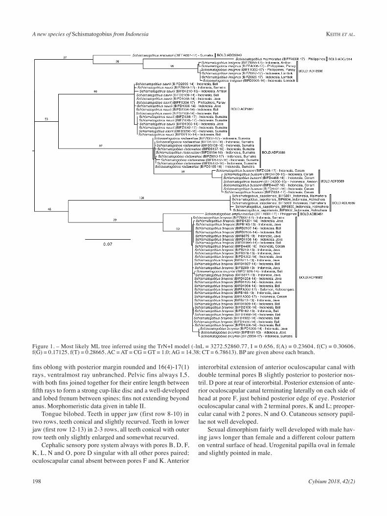
gene. the most likely substitution model selected by jmod-eltest was trn + i. the mL tree (Fig. 1) allowed delimit-ing nine species, each corresponding to a distinct mitochon-
drial lineage as evidenced by the reSL algorithm (tab. i). (boLD:aCP9881, boLD:aDF3589, boLD:aDF3588, boLD:aCP9882, boLD:aDF3590, boLD:aDG5049, boLD:aDG7314, boLD:aDb0451, boLD: aDL4589 ).
MorphomeristicsSpecimen examination led to the discovery of a new spe-
cies to science and its description is given below.
Schismatogobius sapoliensis, n. sp. (Figs 1-3; tabs i-ii)
Material examinedFive specimens from halmahera with a size range of
16.2-18 mm SL.Holotype. – mZb 24586, male (17.9 mm SL); Samuda,
Sapoli, halmahera, indonesia, 23 march 2017, hubert et al. coll.; biF 6802.
Paratypes. – mZb 24587, 2 females (16.2-17.6 mm SL); same data as for holotype; biF 6803 & 6805. mnhn 2016-0626, 1 female (18 mm SL); same data as for holotype; biF 6801. mnhn 2016-0627, 1 male (17.8 mm SL); same data as for holotype; biF 6804.
diagnosisUsually 16 pectoral rays; pectoral fins with a large trans-
verse medium black band. First dorsal fin membrane pos-terior to spine 6 not connected to base of spine of second dorsal fin. Ventral surface of head in male blackish or grey-ish with a white mentum. Frenum whitish with a blackish border. Ventral surface of head in female blackish or grey-ish, except the isthmus, which is whitish, and with a white mentum. Frenum whitish, sometimes with a distal black dot. a single mitochondrial lineage was observed for this species (boLD: aDL4589).
descriptiona small sized Schismatogobius (average adult size < 20
mm SL). body naked, slender, almost circular in cross-sec-tion. head rounded, snout rather pointed. mouth oblique, lower lip more prominent. Jaw length in male much greater than in female; jaw length 47.4-52.8% of HL in males and 32-36.7% of HL in females. Lower jaw reaching vertical of 1/3 to 1/2 of the eye in female and exceeding a vertical of posterior margin of eye in male. eyes high on head, close together with interorbital width about equal to 1/3 eye diam-eter. anterior nostril short and tube-like.
Dorsal fins VI-I,8-9, membrane in first dorsal fin posteri-or to spine 6 not connected to base of spine of second dorsal fin. D1 with all spines about equal in length. Anal fin I,8-9, origin directly opposite to second dorsal fin origin. Caudal fin with 10-11 branched rays, posterior margin straight. Pectoral
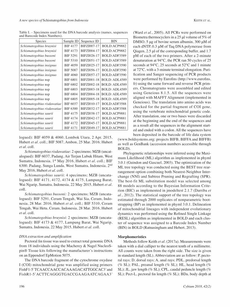
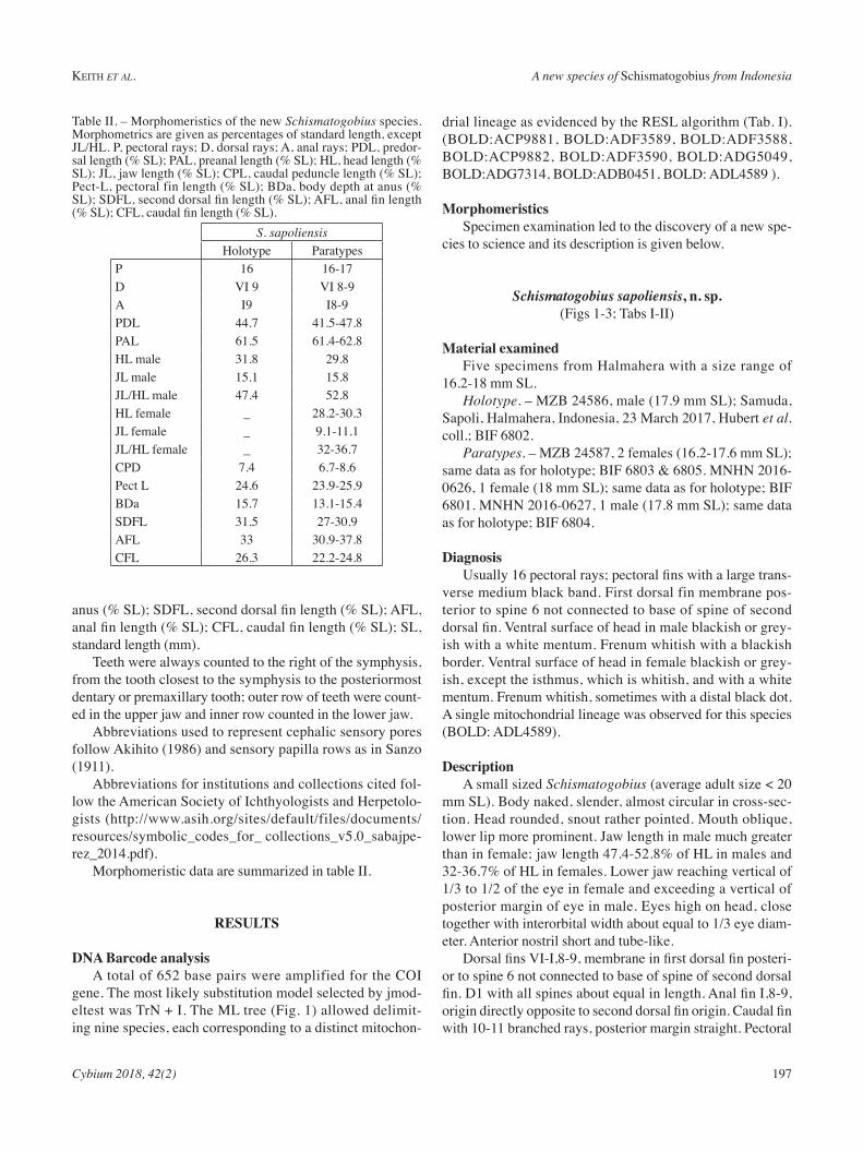
table ii. – morphomeristics of the new Schismatogobius species. morphometrics are given as percentages of standard length, except JL/HL. P, pectoral rays; D, dorsal rays; A, anal rays; PDL, predor-sal length (% SL); PAL, preanal length (% SL); HL, head length (% SL); JL, jaw length (% SL); CPL, caudal peduncle length (% SL); Pect-L, pectoral fin length (% SL); BDa, body depth at anus (% SL); SDFL, second dorsal fin length (% SL); AFL, anal fin length (% SL); CFL, caudal fin length (% SL).
S. sapoliensis holotype ParatypesP 16 16-17D VI 9 VI 8-9a i9 i8-9PDL 44.7 41.5-47.8PaL 61.5 61.4-62.8hL male 31.8 29.8JL male 15.1 15.8JL/HL male 47.4 52.8hL female _ 28.2-30.3JL female _ 9.1-11.1JL/HL female _ 32-36.7CPD 7.4 6.7-8.6Pect L 24.6 23.9-25.9bDa 15.7 13.1-15.4SDFL 31.5 27-30.9aFL 33 30.9-37.8CFL 26.3 22.2-24.8
A new species of Schismatogobius from Indonesia Keith et al.
198 Cybium 2018, 42(2)
fins oblong with posterior margin rounded and 16(4)-17(1) rays, ventralmost ray unbranched. Pelvic fins always i,5, with both fins joined together for their entire length between fifth rays to form a strong cup-like disc and a well-developed and lobed frenum between spines; fins not extending beyond anus. morphomeristic data given in table ii.
tongue bilobed. teeth in upper jaw (first row 8-10) in two rows, teeth conical and slightly recurved. teeth in lower jaw (first row 12-13) in 2-3 rows, all teeth conical with outer row teeth only slightly enlarged and somewhat recurved.
Cephalic sensory pore system always with pores b, D, F, K, L, n and o, pore D singular with all other pores paired; oculoscapular canal absent between pores F and K. anterior
interorbital extension of anterior oculoscapular canal with double terminal pores b slightly posterior to posterior nos-tril. D pore at rear of interorbital. Posterior extension of ante-rior oculoscapular canal terminating laterally on each side of head at pore F, just behind posterior edge of eye. Posterior oculoscapular canal with 2 terminal pores, K and L; preoper-cular canal with 2 pores, n and o. Cutaneous sensory papil-lae not well developed.
Sexual dimorphism fairly well developed with male hav-ing jaws longer than female and a different colour pattern on ventral surface of head. urogenital papilla oval in female and slightly pointed in male.
Figure 1. – most likely mL tree inferred using the trn+i model (-lnL = 3272.52860.77, i = 0.656, f(a) = 0.23604, f(C) = 0.30606, f(G) = 0.17125, f(t) = 0.28665, aC = at = CG = Gt = 1.0; aG = 14.38; Ct = 6.78613). bP are given above each branch.
Keith et al. A new species of Schismatogobius from Indonesia
Cybium 2018, 42(2) 199
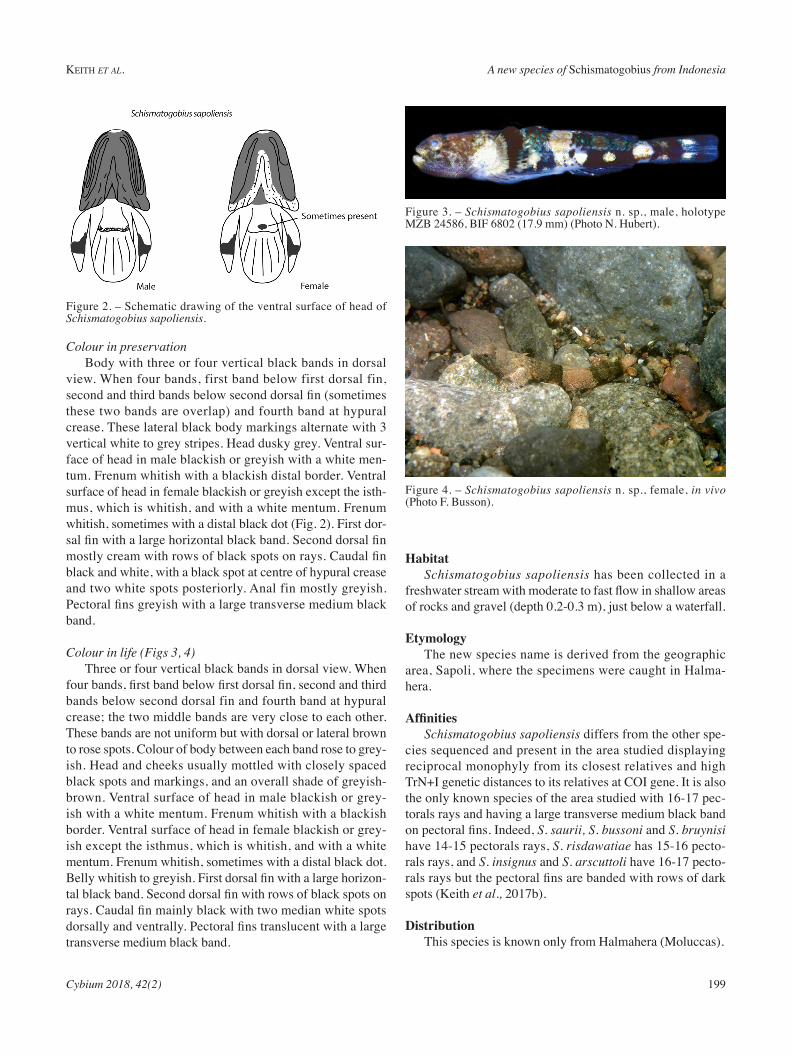
Colour in preservationbody with three or four vertical black bands in dorsal
view. When four bands, first band below first dorsal fin, second and third bands below second dorsal fin (sometimes these two bands are overlap) and fourth band at hypural crease. these lateral black body markings alternate with 3 vertical white to grey stripes. Head dusky grey. Ventral sur-face of head in male blackish or greyish with a white men-tum. Frenum whitish with a blackish distal border. Ventral surface of head in female blackish or greyish except the isth-mus, which is whitish, and with a white mentum. Frenum whitish, sometimes with a distal black dot (Fig. 2). First dor-sal fin with a large horizontal black band. Second dorsal fin mostly cream with rows of black spots on rays. Caudal fin black and white, with a black spot at centre of hypural crease and two white spots posteriorly. anal fin mostly greyish. Pectoral fins greyish with a large transverse medium black band.
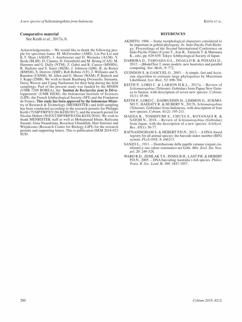
Colour in life (Figs 3, 4)three or four vertical black bands in dorsal view. When
four bands, first band below first dorsal fin, second and third bands below second dorsal fin and fourth band at hypural crease; the two middle bands are very close to each other. these bands are not uniform but with dorsal or lateral brown to rose spots. Colour of body between each band rose to grey-ish. head and cheeks usually mottled with closely spaced black spots and markings, and an overall shade of greyish-brown. Ventral surface of head in male blackish or grey-ish with a white mentum. Frenum whitish with a blackish border. Ventral surface of head in female blackish or grey-ish except the isthmus, which is whitish, and with a white mentum. Frenum whitish, sometimes with a distal black dot. Belly whitish to greyish. First dorsal fin with a large horizon-tal black band. Second dorsal fin with rows of black spots on rays. Caudal fin mainly black with two median white spots dorsally and ventrally. Pectoral fins translucent with a large transverse medium black band.
HabitatSchismatogobius sapoliensis has been collected in a
freshwater stream with moderate to fast flow in shallow areas of rocks and gravel (depth 0.2-0.3 m), just below a waterfall.
etymologythe new species name is derived from the geographic
area, Sapoli, where the specimens were caught in halma-hera.
AffinitiesSchismatogobius sapoliensis differs from the other spe-
cies sequenced and present in the area studied displaying reciprocal monophyly from its closest relatives and high trn+i genetic distances to its relatives at Coi gene. it is also the only known species of the area studied with 16-17 pec-torals rays and having a large transverse medium black band on pectoral fins. indeed, S. saurii, S. bussoni and S. bruynisi have 14-15 pectorals rays, S. risdawatiae has 15-16 pecto-rals rays, and S. insignus and S. arscuttoli have 16-17 pecto-rals rays but the pectoral fins are banded with rows of dark spots (Keith et al., 2017b).
distributionthis species is known only from halmahera (moluccas).
Figure 2. – Schematic drawing of the ventral surface of head of Schismatogobius sapoliensis.
Figure 3. – Schismatogobius sapoliensis n. sp., male, holotype mZb 24586, biF 6802 (17.9 mm) (Photo n. hubert).
Figure 4. – Schismatogobius sapoliensis n. sp., female, in vivo (Photo F. busson).
A new species of Schismatogobius from Indonesia Keith et al.
200 Cybium 2018, 42(2)
Comparative material See Keith et al., 2017a, b.
acknowledgements. – We would like to thank the following peo-ple for specimen loans: m. mcGrouther (amS), Lin Pai-Lei and K.t. Shao (aSiZP), J. amrbruster and D. Werneke (aum), Y. Ikeda (BLIH), D. Catania, D. Greenfield and M. Hoang (CAS), M. hammer and G. Dally (ntm), Z. Gabsi and r. Causse (mnhn), r. hadiaty and S. Sauri (mZb), J. Johnson (Qm), r. de ruiter (rmnh), S. Dorrow (SmF), rob robins (uF), J. Williams and S. raredon (uSnm), m. allen and G. moore (Wam), P. bartsch and J. Kapp (Zmb). We wish to thank bambang Dwisusilo, Sumanta, Daisy Wowor and Ujang Nurhaman for their help during the field samplings. Part of the present study was funded by the mnhn (umr 7208 borea), the ‘institut de recherche pour le Déve-institut de recherche pour le Déve-loppement’ (umr iSem), the indonesian institute of Sciences (LiPi), the French ichthyological Society (SFi) and the Fondation de France. this study has been approved by the indonesian minis-. this study has been approved by the indonesian minis-try of Research & Technology (MENRISTEK) and field sampling has been conducted according to the research permits for Philippe Keith (75/SIP/FRP/E5/ Dit.KI/III/2017), and the research permit for Nicolas Hubert (50/EXT/SIP/FRP/E5/Dit.KI/IX/2016). We wish to thank menriSteK staff as well as mohammad irham, ruliyana Susanti, Gina naandriana, rosichon ubaidillah, hari Sutrisno and Witjaksono (research Center for biology-LiPi) for the research permits and supporting letters. this is publication iSem 2018-023 SuD.
referenCes
aKihito, 1986. – Some morphological characters considered to be important in gobiid phylogeny. In: Indo-Pacific Fish Biolo-gy: Proceedings of the Second international Conference on Indo-Pacific Fishes (Uyeno T., Arai R., Taniuchi T. & Matsuura K., eds), pp. 629-639. tokyo: ichthyological Society of Japan.
Darriba D., taboaDa G.L., DoaLLo r. & PoSaDa D., 2012. – jmodeltest 2: more models, new heuristics and parallel computing. Nat. Meth., 9: 772.
GuinDon S. & GaSCueL o., 2003. – a simple, fast and accu-rate algorithm to estimate large phylogenies by maximum Likelihood. Syst. Biol., 52: 696-704.
Keith P., LorD C. & LarSon h.K.L., 2017a. – review of Schismatogobius (teleostei: Gobiidae) from Papua new Guin-ea to Samoa, with description of seven new species. Cybium, 41(1): 45-66.
Keith P., LorD C., DahruDDin h., Limmon G., SuKmo-no t., haDiatY r. & hubert n., 2017b. Schismatogobius (teleostei: Gobiidae) from indonesia, with description of four new species, Cybium, 41(2): 195-211.
maeDa K., toShiFumi S., ChuYa S., KoYanaGi r. & Satoh n., 2018. – review of Schismatogobius (Gobiidae) from Japan, with the description of a new species. Ichthyol. Res., 65(1): 56-77.
ratnaSinGham S. & hebert P.D.n., 2013. – a Dna-based registry for all animal species: the barcode index number (bin) system. PLoS ONE, 8: e66213.
SanZo L., 1911. – Distribuzione delle papille cutanee (organi cia-tiformi) e suo valore sistematico nei Gobi. Mitt. Zool. Stn. Nea-pel, 20: 249-328.
WarD r.D., ZemLaK t.S., inneS b.h., LaSt P.r. & hebert P.D.N., 2005. – DNA barcoding Australia’s fish species. Philos. Trans. R. Soc. Lond. b, 360: 1847-1857.