
Analysis and engineering of biomolecules and ...
197
Analysis and engineering of biomolecules and microorganisms: from genome-scale study of pathogens to programming of DNA and cells Vom Promotionsausschuss der Technischen Universität Hamburg-Harburg zur Erlangung des akademischen Grades Doktor der Naturwissenschaften (Dr. rer. nat.) genehmigte Dissertation von Lifu Song aus Shandong, China 2018
Transcript of Analysis and engineering of biomolecules and ...
Analysis and engineering of biomolecules andmicroorganisms: from genome-scale study ofpathogens to programming of DNA and cells
Vom Promotionsausschuss der
Technischen Universität Hamburg-Harburg
zur Erlangung des akademischen Grades
Doktor der Naturwissenschaften (Dr. rer. nat.)
genehmigte Dissertation
vonLifu Song
ausShandong, China
2018

Dissertation Committee
Chairman: Prof. Dr.-Ing. habil. Dr. h.c. Stefan HeinrichSupervisor & Examiner: Prof. Dr. rer. nat. habil. An-Ping ZengExaminer: Prof. Dr. rer. nat. habil. Christoph WittmannExamination date: 25-May-2018
Administrator
Typewriter
DOI: 10.15480/882.1668
Administrator
Placed Image

In memory of my mother


Acknowledgements
There are many people without whom this thesis would not have been possible. First, I wouldlike to thank my supervisor, Prof. An-Ping Zeng, for providing me this opportunity to pursuemy research interests. I would also like to thank Dr. Wei Wang, who basically supervisedthe first part of this thesis. I really appreciate her hard efforts on improving my manuscriptssentence by sentence. Thanks to both Prof. Zeng and Dr. Wei Wang for many pieces ofadvice about how to write scientific papers.
Next, I would like to thank my dissertation committee members. A big thank to Prof.Heinrich for agreeing to be the chairman and waiting a long time for my examination.I appreciate Prof. Wittmann for agreeing to review my thesis, the huge efforts to reachHamburg to attend my examination and the kindness during the examination.
Then, many thanks to Dr. Sugima Rappert and Dr. Wael Sabra who have always beenkind and helpful with all kinds of issues in the lab. I thank Prof. Ralf Pörtner for beingalways kindness and the help with contract issues. Thanks to Ms. Cornelia Hoffmann for herhelp with kinds of document stuff. Many thanks to Mr. Ralf Grajetzki and Mr. Olaf Schmidtwho helped me a lot in setting up the PC and servers. I thank Dr. Uwe Jandt for his insightfuldiscussions and helps with a conference presentation. Thanks to Yaeseong Hong for his helpwith construction of some plasmids.
After that, a big and special thank to Dr. Ke Wang. Although her major is quite differentwith mine, I do benefit a lot from the discussions with her. Furthermore, her kind encouragesmade me came through the darkest days of my life. I appreciate Dr. Chengwei Ma, as well ashis wife - Ying Liu, for their hosting during the time waiting for the examination, insightfuldiscussions, and many other bits of help. Thanks to Dr. Ying Dong for the help regardingthe examination procedure and the words comforting me. I would also like to thank all mycolleagues and friends, Anibal Mora, Anna Gorte, Birgit Koch, Christiane Goepfert, ChristinGroeger, Feng Geng, Jan Bomnüter, Jan Sens, Jin Guo, Libang Zhou, Lin Chen, RebekkaSchmitz, Sibel Ilhan, Tyll Utesch, Yujun Zhang and Minliang Chen, for the help and allkinds of enjoyable discussions, making years of living in a foreign country an unforgettableexperience.

vi
Additionally, I would like to thank Bundesministerium für Bildung und Forschung(BMBF) for the financial support.
Last but not least, I would like to thank my families. Thank my parents for their support.Thank my wife for her love, patience, support, and understanding throughout my Ph.D.studies. Thank my little daughter who has no idea how wonderful my life becomes with herpresence.
Abstract
This thesis is consisted of three major but different parts with the general aims of systemslevel evaluation and engineering of biomolecules and biological systems. In the first part ofthis thesis, comparative genomic studies of mutans streptococci strains, which are involved inthe development of dental caries, were performed for better understanding their pathogenicityat the level of systems biology. A mosaic-like structure of genome arrangement was revealedby genome alignment analysis. Genes related to pathogenicity were found to have highvariations among the strains, whereas genes for oxidative stress resistance are well conserved,indicating the importance of this trait in the dental biofilm community. Genome-scalemetabolic network analysis revealed significant differences in 42 pathways. A strikingdissimilarity is the unique presence of two lactate oxidases in S. sobrinus DSM 20742,probably indicating an unusual capability of this strain in producing H2O2 and expanding itsecological niche. In addition, lactate oxidases may form a unique energy-producing pathwaywith other enzymes in S. sobrinus DSM 20742 that can remedy its deficiency in citrateutilization pathway. An "open" pan-genome was inferred by pan-genome analysis using67 S. mutans genomes currently available including the strains sequenced in this study. Anonline regulation database for S. mutans, named StrepReg, was constructed by integratinga transcription factor-based gene regulatory network, which was derived from time-seriestranscriptome analysis, with STRING protein-protein interaction information and KEGGpathway information (http://biosystem.bt1.tu-harburg.de:1555/homes/).
Although systems biology is a powerful tool in understanding the system level behaviorsof biological systems, the establishment of predictive, multiscale models in systems biologyis still a challenge due to the complexity of biological systems. For the same reason,mathematical models often fail in applications under physiological conditions, such as foridentification of targets in metabolic engineering for the development of highly productionstrains. In the second part of this thesis, a novel multiple input-output (I/O) system wastherefore proposed and verified, which allows the identification of limiting bioreactionsor key enzymes in metabolic pathways and even the optimization of biomolecules in vivo.The basic idea is to design a multiple I/O system which can introduce various geneticmanipulations (perturbations) into the cells and record the specific intracellular signal changes
viii
correspondingly. This was achieved by engineering the interactions of phage with E. colicells. Specifically, a multiple I/O system was implemented using M13 phage derivativeswhich can introduce various perturbations into E. coli cells after infection, such as up- ordown-regulation of specific gene expressions. Using a rationally designed biological circuit,the intracellular signal changes after introduction of the perturbations by the phage infectionwere linked to the phage reproduction process. This means, signal changes caused by specificperturbations are linked to the specific populations of phages introducing the correspondingperturbations. In this way, the various signals are ‘recorded’ in forms of correspondingpopulations of phage derivatives. The usefulness of the multiple I/O system was demonstratedwith three applications, i.e. identification of beneficial genetic manipulations, parallelevaluation of various designs of enzymes, and parallel screening of key enzymes for L-lysinebiosynthesis in E. coli. Various gene operations related or not related to L-lysine biosynthesisin E. coli were used as inputs and the intracellular lysine concentration changes were used totrigger output signals. Correct predictions of beneficial genetic manipulations for enhancedlysine production in E. coli were achieved. New and effective variants of a key enzymeaspartate kinase III (AK-III), which is strictly inhibited by L-lysine, were obtained andevaluated in parallel. Importantly, the I/O system shows a ultra-sensitivity in capturing signalchanges caused by the certain perturbations introduced. The approach developed in thiswork opens up new possibilities in systems metabolic engineering and synthetic biology ofindustrial microorganisms for practical applications.
In the third part of this thesis, a novel self-error-detecting, three-base block encodingscheme (SED3B), which takes full advantage of the inherent redundancy feature of DNAsynthesis for error correction, was proposed for reliable information encoding in DNA ofliving cells. In addition to the high error tolerance, SED3B encoded sequences were shownto be orthogonal to natural DNA sequences, indicating for the first time a low biologicalrelevance of the encoded sequences. Features such as effective error tolerance and lowbiological relevance make SED3B an appealing solution for orthogonal information encodingin living cells with low or no affections to their biological functions, e.g. as a commentlanguage in programming cells in vivo and for biological barcode encoding. Based onerror-prone PCR experiments it was estimated that more than 12,000 years of continuousreplication would be required to make the SED3B encoded information in E. coli cellsbecome unrecoverable. To facilitate the usage of SED3B as a comment and barcode encodingsystem in synthetic biology, an online encoding-decoding system was implemented andreleased at http://biosystem.bt1.tu-harburg.de/sed3b. In principle, SED3B is also applicablefor in vitro large data storage in synthesized DNA. Although further investigation is required,preliminary analysis shows that SED3B has a great potential for increasing the storage density

ix
to over several exabytes (EBs) per gram DNA which is theoretically much higher than thatof methods reported in literature so far.

ZUSAMMENFASSUNG
Diese Doktorarbeit besteht aus drei Hauptteilen mit dem Generalziel, Biomoleküle undbiologische Systeme auf Systemebene zu analysieren bzw. zu programmieren. Im erstenTeil dieser Arbeit wurden vergleichende genomische Untersuchungen von Mutans - Strep-tokokken - Stämmen, die an der Entstehung von Karies beteiligt sind, durchgeführt, um derenPathogenität auf systembiologischer Ebene besser zu verstehen. Genom-Alignment ergabeine mosaikartige Struktur der Genomanordnung. Gene, die mit der Pathogenität in Zusam-menhang stehen, weisen hohe Variationen unter den Stämmen auf, wohingegen Gene für dieResistenz gegen oxidativen Stress gut konserviert sind, was die Bedeutung dieses Merkmalsin der dentalen Biofilm-Gemeinschaft anzeigt. Die Analyse genomweiter metabolischerNetzwerke zeigte signifikante Unterschiede in 42 Signalwegen. Eine bemerkenswerte Beson-derheit ist die einzigartige Anwesenheit von zwei Lactatoxidasen in S. sobrinus DSM 20742,was wahrscheinlich auf eine ungewöhnliche Fähigkeit dieses Stamms hinweist, H2O2 zu pro-duzieren und seine ökologische Nische zu erweitern. Zusätzlich können Lactatoxidasen eineneinzigartigen energetischen Weg mit anderen Enzymen in S. sobrinus DSM 20742 bilden, derseinen Mangel im Citratverwertungsweg beheben kann. Unter Verwendung von derzeit ver-fügbaren 67 S. mutans-Genomen, einschließlich der in dieser Studie sequenzierten Stämme,wurde die theoretische Kerngenomgröße von S. mutans geschätzt und eine Modellierungvon S. mutans pan-genom durch Anwendung verschiedener Fitting-Modelle durchgeführt.Ein "offenes" Pan-Genom wurde gezeigt. Eine Online-Regulierungsdatenbank für Strep-tococcus, genannt StrepReg, wurde durch Integration eines Transkriptionsfaktor-basiertenGenregulationsnetzwerkes, das aus einer zeitreihen Transkriptomanalyse in Zusammenarbeitmit Projektpartnern abgeleitet wurde (http://biosystem.bt1.tu-harburg.de:1555/homes/).
Obwohl die Systembiologie ein sehr nützliches Werkzeug ist, um das Systemverhaltenvon biologischen Systemen zu verstehen, ist die Etablierung von prädiktiven Multiskalen-modellen aufgrund der Komplexität biologischer Systeme immer noch eine große Heraus-forderung. Aus dem gleichen Grund scheitern mathematische Modelle oft für Anwendungenunter physiologische Bedingungen, wie z.B. bei der Identifizierung von Targets in MetabolicEngineering für die Entwickelung von Hochleistungsproduktionsstämmen. Zur Lösungder Probleme wurde im zweiten Teil dieser Arbeit ein neuartiges Mehrfach Input-Output
xii
(I/O) System vorgeschlagen und verifiziert, das verschiedene genetische Manipulationenin die Zellen einbringen und die entsprechenden intrazellulären Signaländerungen aufze-ichnen kann, mit dem Ziel, Schlüsselreaktionen bzw. Enzyme in Stoffwechselwegen inE. coli zu identifizieren und Biomoleküle zu optimieren. Die Grundidee dabei war, dieInteraktionen von Phagen mit E. coli-Zellen zu gestalten und zu nutzen. Konkret wurde einMehrfach-I/O-System unter Verwendung verschiedener M13-Phagenderivate implementiert,die verschiedene genetische Modifikationen (Störungen) in E. coli-Zellen nach einer Phagen-infektion einführen können, wie etwa eine Aufwärts- oder Abwärtsregulierung spezifischerGenexpressionen. Unter Verwendung eines rational entworfenen biologischen Schaltkreiseswurden die intrazellulären Signalveränderungen nach der Einführung von Störungen durchPhageninfektion mit dem Phagenreproduktionsprozess verknüpft. Dies bedeutet, dass Sig-naländerungen, die durch spezifische Störungen verursacht werden, mit den spezifischenPhagenpopulationen verbunden sind, die die entsprechenden Störungen einführen. Mit an-deren Worten werden die verschiedenen Signale in Formen von entsprechenden Populationenvon Phagenderivaten "aufgezeichnet". Die Nützlichkeit des Mehrfach-I/O-Systems wurdein drei Anwendungen gezeigt, d.h. Identifizierung von vorteilhaften genetischen Manip-ulationen, paralleler Bewertung verschiedener Designs von Biomolekülen und parallelemScreening von Schlüsselenzymen für die L-Lysin-Biosynthese in E. coli. VerschiedeneGenoperationen, die mit der L-Lysinbiosynthese in E. coli verwandt waren oder nicht, wur-den als Inputs verwendet und die intrazellulären Lysinkonzentrationsänderungen wurdenverwendet, um Ausgangssignale auszulösen. Korrekte Vorhersagen von vorteilhaften genetis-chen Manipulationen für eine erhöhte Lysinproduktion in E. coli wurden erzielt. Neue undeffektive Varianten eines Schlüsselenzyms Aspartatkinase III (AK-III), das durch L-Lysinstreng gehemmt wird, wurden parallel erhalten und ausgewertet. Es ist anzumerken, dass dasI/O-System eine besonders hohe Empfindlichkeit bei der Erfassung von Signaländerungenaufweist, die durch die eingeführten bestimmten Störungen verursacht werden. Der in dieserArbeit entwickelte Ansatz eröffnet neue Möglichkeiten in Systems Metabolic Engineeringund synthetischer Biologie industrieller Mikroorganismen für praktische Anwendungen.
Im dritten Teil dieser Arbeit wurde ein neuartiges selbstfehlererkennendes Drei-Basen-Block-Codierungsschema (SED3B) für eine zuverlässige Informationscodierung in DNA,insbesondere für Anwendungen in lebenden Zellen vorgeschlagen und verifiziert, das dieinhärente Redundanz der DNA-Synthese zur Fehlerkorrektur in der DNA-Datenspeicherungvoll ausnutzt. Zusätzlich zu der hohen Fehlertoleranz wurde gezeigt, dass SED3B-codierteSequenzen sich von den natürlich gebildeten DNA-Sequenzen grundsetzlich unterscheiden,was zum ersten Mal eine geringe biologische Relevanz der zu diesem Zweck codiertenSequenzen anzeigt. Merkmale, wie die effektive Fehlertoleranz und die geringe biolo-
xiii
gische Relevanz, machen SED3B zu einer ansprechenden Lösung für die orthogonale In-formationscodierung in lebenden Zellen mit geringen bzw. keinen Beeinträchtigungenihrer biologischen Funktionen, z. als Kommentarsprache beim Programmieren von Zellenin vivo und für ein biologisches barcoding. Basierend auf einem fehleranfälligen PCR-Experiment wurde geschätzt, dass mehr als 12.000 Jahre kontinuierlicher Replikation er-forderlich wären, um die SED3B-codierte Information in E. coli-Zellen zu verlieren. Umdie Verwendung von SED3B als Kommentar- und Barcode-Kodierungssystem in der syn-thetischen Biologie zu erleichtern, wurde ein Online-Kodierungs-Dekodierungssystem im-plementiert und unter http://biosystem.bt1.tu-harburg.de/sed3b veröffentlicht. Im Prinzip istSED3B auch für eine in vitro große Datenspeicherung in synthetisierter DNA anwendbar.Obwohl weitere Untersuchungen erforderlich sind, zeigen erste Ergebnisse, dass SED3Bein gutes Potenzial zur Erhöhung der Speicherdichte auf mehrere extaabytes (EBs) proGramm DNA hat, was theoretisch viel höher ist als bei den bekannten Methoden für digitaleDNA-Informationskodierung.

Table of contents
List of figures xix
List of tables xxi
Nomenclature xxiii
1 Introduction and objectives 11.1 Genome-scale comparative studies of mutants streptococci . . . . . . . . . 11.2 A multiple input-output system for systems metabolic engineering in E. coli
cells . . . . . . . . . . . . . . . . . . . . . . . . . . . . . . . . . . . . . . 21.3 Development of an orthogonal information encoding scheme for reliable
information encoding in DNA of living cells . . . . . . . . . . . . . . . . . 4
2 Materials and methods 72.1 Methods for systems biology analysis . . . . . . . . . . . . . . . . . . . . 7
2.1.1 Genome sequences and strains . . . . . . . . . . . . . . . . . . . . 72.1.2 Genome sequencing, assembly and annotation . . . . . . . . . . . 82.1.3 Genome alignment . . . . . . . . . . . . . . . . . . . . . . . . . . 92.1.4 Pan-genome and core-genome analysis . . . . . . . . . . . . . . . 92.1.5 Gene content-based comparative analysis of 10 mutans streptococci
strains . . . . . . . . . . . . . . . . . . . . . . . . . . . . . . . . . 102.1.6 Identification of putative two-component signal transduction systems 102.1.7 Genome-scale metabolic networks construction . . . . . . . . . . . 112.1.8 PCR verification of unique genes in the comparative genomics studies 122.1.9 Construction of lactate oxidase encoding gene knockout mutants and
transformation of S. sobrinus DSM 20742 . . . . . . . . . . . . . . 132.2 Methods for multiple input-output system . . . . . . . . . . . . . . . . . . 14
2.2.1 Chemicals . . . . . . . . . . . . . . . . . . . . . . . . . . . . . . . 142.2.2 Bacterial strains . . . . . . . . . . . . . . . . . . . . . . . . . . . . 14
xvi Table of contents
2.2.3 Phagemids, plasmids and primers . . . . . . . . . . . . . . . . . . 142.2.4 Media . . . . . . . . . . . . . . . . . . . . . . . . . . . . . . . . . 152.2.5 Strain conservation . . . . . . . . . . . . . . . . . . . . . . . . . . 162.2.6 Molecular cloning . . . . . . . . . . . . . . . . . . . . . . . . . . 172.2.7 Preparation of infective engineered phages . . . . . . . . . . . . . 182.2.8 Screening based on cell-phage interactions . . . . . . . . . . . . . 182.2.9 Enzyme characterization . . . . . . . . . . . . . . . . . . . . . . . 19
2.3 Methods for orthogonal information encoding in living cells . . . . . . . . 202.3.1 Detailed steps for encoding binary data into DNA string . . . . . . 202.3.2 Decoding error-containing DNA strings into binary data . . . . . . 202.3.3 Implementation of the online encoding-decoding system for SED3B 212.3.4 Analysis of error tolerance by in silicon simulation . . . . . . . . . 212.3.5 In vivo verification of the error tolerance by error-prone PCR . . . . 21
3 Genome-scale comparative studies of mutans streptococci 253.1 Introduction . . . . . . . . . . . . . . . . . . . . . . . . . . . . . . . . . . 253.2 Genome sequencing, assembly and annotation of eight mutans streptococci
strains . . . . . . . . . . . . . . . . . . . . . . . . . . . . . . . . . . . . . 263.3 Genome rearrangement of S. mutans genomes . . . . . . . . . . . . . . . . 303.4 Core and pan-genome analysis of S. mutans species . . . . . . . . . . . . . 31
3.4.1 Core-genome . . . . . . . . . . . . . . . . . . . . . . . . . . . . . 323.4.2 Pan-genome . . . . . . . . . . . . . . . . . . . . . . . . . . . . . . 33
3.5 Gene content-based comparative analysis of mutans streptococci strains . . 343.5.1 Distribution of two-component signal transduction systems . . . . . 373.5.2 High diversities of the competence development regulation module 443.5.3 Distribution of bacteriocin- and antibiotic resistance-related proteins 493.5.4 Oxidative stress defense systems in mutans streptococci . . . . . . 57
3.6 Metabolic network construction and analysis . . . . . . . . . . . . . . . . . 613.6.1 Genome-scale metabolic network reconstruction . . . . . . . . . . 613.6.2 Variability and specificity in metabolic pathways and network . . . 62
3.7 Construction of StrepReg - a regulation database of S. mutans . . . . . . . . 643.8 Conclusion . . . . . . . . . . . . . . . . . . . . . . . . . . . . . . . . . . 65
4 Development of a multiple IO system for biological engineering in E. coli 714.1 Introduction . . . . . . . . . . . . . . . . . . . . . . . . . . . . . . . . . . 714.2 Principles of a multiple input-output system which can interact with E. coli
cells . . . . . . . . . . . . . . . . . . . . . . . . . . . . . . . . . . . . . . 72
Table of contents xvii
4.3 Proof of concept studies . . . . . . . . . . . . . . . . . . . . . . . . . . . . 754.3.1 Identification of beneficial genetic manipulations . . . . . . . . . . 764.3.2 Evaluation of designs . . . . . . . . . . . . . . . . . . . . . . . . . 774.3.3 Parallel and sensitive screening of biomolecules . . . . . . . . . . . 78
4.4 Conclusion and Perspective . . . . . . . . . . . . . . . . . . . . . . . . . . 87
5 Orthogonal information encoding in living cells 895.1 Introduction . . . . . . . . . . . . . . . . . . . . . . . . . . . . . . . . . . 895.2 Theoretical and technological backgrounds . . . . . . . . . . . . . . . . . 91
5.2.1 The method of Church et al. . . . . . . . . . . . . . . . . . . . . . 925.2.2 The method of Goldman et al. . . . . . . . . . . . . . . . . . . . . 925.2.3 The method of Grass et al. . . . . . . . . . . . . . . . . . . . . . . 92
5.3 Principles of a self-error-detecting, three-base block encoding scheme (SED3B) 955.4 High error tolerance revealed by in silicon simulations . . . . . . . . . . . 965.5 SED3B encoded DNA sequences show low biological relevance . . . . . . 985.6 SED3B encoded DNA sequences show simple secondary structure . . . . . 1005.7 Reliable orthogonal information encoding in living cells using SED3B . . . 1025.8 In vitro data storage using SED3B . . . . . . . . . . . . . . . . . . . . . . 1035.9 Development of an online encoding-decoding system . . . . . . . . . . . . 1065.10 Conclusion . . . . . . . . . . . . . . . . . . . . . . . . . . . . . . . . . . 106
6 Summary and outlook 109
References 113
Appendix A Source codes of utilized perl scripts 139A.1 panGenomeAnalysis.pl . . . . . . . . . . . . . . . . . . . . . . . . . . . . 139A.2 shared.pl . . . . . . . . . . . . . . . . . . . . . . . . . . . . . . . . . . . . 150A.3 bin2DNA.pl . . . . . . . . . . . . . . . . . . . . . . . . . . . . . . . . . . 152A.4 Consensus.pl . . . . . . . . . . . . . . . . . . . . . . . . . . . . . . . . . 154A.5 DNA2bin.pl . . . . . . . . . . . . . . . . . . . . . . . . . . . . . . . . . . 155A.6 kmerAnalysis.pl . . . . . . . . . . . . . . . . . . . . . . . . . . . . . . . . 157A.7 biologyRelevanceAnalysis.pl . . . . . . . . . . . . . . . . . . . . . . . . . 160A.8 bin2DNACRCIndex.pl . . . . . . . . . . . . . . . . . . . . . . . . . . . . 162
Appendix B Supplement Information 167B.1 Sequences of mutacins used for the identification of putative mutacins in 10
mutans streptococci strains. . . . . . . . . . . . . . . . . . . . . . . . . . . 167

List of figures
1.1 Design–Build–Test Cycle for Biomolecular and Biosystems Engineering . . 3
2.1 Detailed steps of decoding error-containing DNA strings into error free bitstring. . . . . . . . . . . . . . . . . . . . . . . . . . . . . . . . . . . . . . 22
2.2 The logo of our institute used as input for error tolerance simulation . . . . 232.3 Illustration of construction process of plasmids carrying the encoded 78bp
DNA string with variant errors introduced by error-prone PCR . . . . . . . 23
3.1 Phylogenetic analysis of 10 mutans streptococci strains compared in thisstudy and their phylogenetic relationship to other Streptococcus species. . . 28
3.2 Comparison of local collinear blocks (LCBs) of chromosomal sequences ofthe eight S. mutans strains. . . . . . . . . . . . . . . . . . . . . . . . . . . 31
3.3 Core and pan-genome model of 67 S. mutans genomes. . . . . . . . . . . . 353.4 Alignment of ComC and ComS amino acid sequences. . . . . . . . . . . . 483.5 Cluster structure of the mutacin-K8 production system across six S. mutans
strains. . . . . . . . . . . . . . . . . . . . . . . . . . . . . . . . . . . . . . 503.6 Example of visualized genome-scale metabolic networks constructed based
on genome annotations and KEGG pathway . . . . . . . . . . . . . . . . . 673.7 Glycolysis/Gluconeogenesis and TCA cycle pathway in mutans streptococci 683.8 Screenshots of StrepReg database . . . . . . . . . . . . . . . . . . . . . . 69
4.1 Sample plasmid maps of inputting phages carrying out overexpression/re-pression operation on specific genes . . . . . . . . . . . . . . . . . . . . . 73
4.2 Principle of phage based multiple IO system . . . . . . . . . . . . . . . . . 744.3 Proof of concept application studies of the IO system . . . . . . . . . . . . 754.4 Illustration of the output device using the concentration of intracellular lysine
as an output signal . . . . . . . . . . . . . . . . . . . . . . . . . . . . . . 764.5 Inhibition profiles of wild-type and mutants of AK-III by lysine . . . . . . 784.6 Work flow of cell robot based screening by using the IO system . . . . . . . 82
xx List of figures
4.7 3D structure illustration of the de-allosteric regulation mechanism of R300Cmutein . . . . . . . . . . . . . . . . . . . . . . . . . . . . . . . . . . . . . 85
4.8 Flow Cytometry assays of cell populations harboring wild-type AK-III andAK-III mutants of R300C and V339A . . . . . . . . . . . . . . . . . . . . 88
5.1 GF(47) to DNA codon wheel for mapping every element of GF(47) to threenucleotides . . . . . . . . . . . . . . . . . . . . . . . . . . . . . . . . . . 93
5.2 Illustration of encoding binary data into DNA string using the SED3B en-coding scheme. . . . . . . . . . . . . . . . . . . . . . . . . . . . . . . . . 96
5.3 Error detection and repression by using the SED3B encoding scheme. . . . 975.4 Error correction capabilities by multiple DNA sequences encoded by SED3B
encoding scheme. . . . . . . . . . . . . . . . . . . . . . . . . . . . . . . . 985.5 Simulation of required sequence numbers for reliable information recovery
by DNA fragments with variant rates of errors. . . . . . . . . . . . . . . . 995.6 Comparative analysis of SED3B encoded sequences with a natural DNA
sequences. . . . . . . . . . . . . . . . . . . . . . . . . . . . . . . . . . . . 1005.7 The number of complementary matched k-mers is reduced remarkably by
using the SED3B scheme. . . . . . . . . . . . . . . . . . . . . . . . . . . . 1015.8 Correct information can be retrieved using 14 sequences with high rates of
errors introduced by error-prone PCR. . . . . . . . . . . . . . . . . . . . . 1025.9 Schematic presentation of in vitro information storage in DNA . . . . . . . 1045.10 Strategy of in vitro digital information encoding in DNA using SED3B . . . 1055.11 Screenshots of the online encoding-decoding system . . . . . . . . . . . . 107
List of tables
2.1 Eight newly sequenced and two previously sequenced mutans streptococcistrains included in the analysis . . . . . . . . . . . . . . . . . . . . . . . . 8
2.2 E. coli strains used in the present work . . . . . . . . . . . . . . . . . . . . 142.3 Plasmids used in present work . . . . . . . . . . . . . . . . . . . . . . . . 162.4 Primers used in present work . . . . . . . . . . . . . . . . . . . . . . . . . 17
3.1 Genome assembly and annotation of eight S. mutans strains sequenced inthis study in comparison with previously sequenced S. mutans strains UA159and NN2025 . . . . . . . . . . . . . . . . . . . . . . . . . . . . . . . . . . 29
3.2 Unique protein coding sequences (CDSs) between the different strains re-vealed by ortholog analysis . . . . . . . . . . . . . . . . . . . . . . . . . . 36
3.3 Identification and classification of putative two component systems in theeightmutans streptococci strains sequenced in this study . . . . . . . . . . . 38
3.4 Ortholog analysis and classifications of the putative TCS proteins . . . . . . 403.5 Distribution of competence development-related systems in the 10 mutans
streptococci strains . . . . . . . . . . . . . . . . . . . . . . . . . . . . . . 463.6 Distribution of mutacins and mutacin immunity proteins in the 10 mutans
streptococci strains . . . . . . . . . . . . . . . . . . . . . . . . . . . . . . 533.7 Distribution of antibiotic resistance-related proteins . . . . . . . . . . . . . 563.8 Distribution of oxidative stress resistance systems . . . . . . . . . . . . . . 603.9 Compositions of the established metabolic networks of the 10 mutans strep-
tococci strains . . . . . . . . . . . . . . . . . . . . . . . . . . . . . . . . . 61
5.1 Base-3 to DNA encoding ensuring no repeated nucleotides in the Goldman’smethod . . . . . . . . . . . . . . . . . . . . . . . . . . . . . . . . . . . . 93
5.2 Comparison of capabilities of current available encoding schemes for digitalinformation storage in DNA . . . . . . . . . . . . . . . . . . . . . . . . . 94

Nomenclature
Roman Symbols
Ω Theoritical core-genome size
Acronyms / Abbreviations
γ-GCS-GS γ-Glutamylcysteine synthetase-glutathione synthetase
dpr Dps-like Peroxide Resistance gene
k-mer All the possible substrings of length k that are contained in a string
lysC Gene encoding aspartokinase
AK-III Aspartokinase III
ATP Adenosine triphosphate
BCP Bacterioferritin comigratory protein
Cas9 CRISPR associated protein 9
CDSs Protein Coding Genes
CMKM Complementarily matched k-mer pairs
CoA Coenzyme A
COGs Clusters of Orthologous Groups of proteins
CRC Cyclical Redundancy Check
CRISPR Clustered regularly interspaced short palindromic repeats
CSP Competence stimulating peptide
xxiv Nomenclature
DNA Deoxyribonucleic Acid
EB Exabyte
eYFP Enhanced Yellow Fluorescent Protein
FACS Fluorescence-activated cell sorting
G3P Phage minor coat gene 3 protein
GF(47) Galois Field of size 47
GS Glutathione synthetase
GSH L-γ-Glutamyl-L-cysteinylglycine
GSSG Oxidized Glutathione
HGT Horizontal gene transfer
HK Histidine Kinase
HMM Hidden Markov Model
HO• Hydroxyl radical
HTS High Throughput Screening
IO Input-Output
IPTG Isopropyl-β -D-Thiogalactoside
LCBs Locally collinear blocks
LTA Lipoteichoic acid
ML Maximum Likelihood
multi-MUMs Multiple Maximal Unique Matches
NAD Nicotinamide adenine dinucleotide
NAD+ Oxidized form of Nicotinamide adenine dinucleotide
NADH Reduced form of Nicotinamide adenine dinucleotide
NADP+ Oxidized form of Nicotinamide Adenine Dinucleotide Phosphate
Nomenclature xxv
NADPH Reduced form of Nicotinamide Adenine Dinucleotide Phosphate
PB Petabyte
PCR Polymerase Chain Reaction
PEP Phosphoenolpyruvate
PT Petabytes
PTS Phosphotransferase system
RBS Ribosomal binding site
ROS Reactive oxygen species
RR Response Regulators
rRNA Ribosomal ribonucleic acid
RS Reed–Solomon
SED3B Self-error-detecting, three-base block encoding scheme
SOD Superoxide dismutases
TCA Tricarboxylic Acid
TCS Two-component signal transduction system
TF Transcription factor
TG Target gene
TM Transmembrane helix
tRNA Transfer Ribonucleic Acid
V-ATPases V-type ATPases

Chapter 1
Introduction and objectives
This thesis is based on work done during my stay at the Hamburg University of Technologyas a scientific coworker. It is consisted of three major but different parts with the generalaims of systems level evaluation and engineering of biomolecules and biological systems.In the following, the background and objectives of each part are briefly introduced. Moredetailed introduction and background information are presented in the corresponding chapterfor each part.
1.1 Genome-scale comparative studies of mutants strepto-cocci
The oral microbiome is a dynamic environment inhabited by both commensals and pathogens.Among them, mutans group streptococci are considered as significant contributors to thedevelopment of dental caries [1]. This is attributed to their ability to form biofilms which aregenerally difficult or impossible to eradicate by antibiotic therapy because biofilm cells areresistant to antibiotics [2]. Systems biology is a holistic approach to decipher the complexityof biological systems. It is based on the understanding that live biological networks thatform the whole of living organisms are more than the sum of their parts [3–5]. Systemsbiology studies try to design predictive, multi-scale models to discover new biomarkersfor disease, drug targets, to understand pathogenicity mechamisms and to develop highperformance producers in industrial biotechnology. It has been responsible for some ofthe most important developments in the science of biology [6–15]. In the first part of thisthesis, systems biology efforts were made to understand the pathogenicity of ten mutansstreptococci strains. Due to the high diversity of genetic content of different isolates, genomecontents of single or just few isolates cannot represent specific species or group of strains.
2 Introduction and objectives
Among all the species of mutans group streptococci, only the genomes of two strains of S.mutans were sequenced previously. In the frame of a collaboration systems biology project,six S. mutans strains, one S. ratti strain and one S. sobrinus strain were submitted for genomesequencing. Genome annotation, genome level comparative analysis and metabolic networkanalysis were performed in this work to reveal strain-specific features and potential drugtargets. An online transcriptional regulatory network database of S. mutans, named StrepReg,was constructed by integrating time-resolved transcriptomic data from the project partners(http://biosystem.bt1.tu-harburg.de:1555/homes/). All the information and tools should behelpful for understanding the evolution and pathology of these oral pathologies.
1.2 A multiple input-output system for systems metabolicengineering in E. coli cells
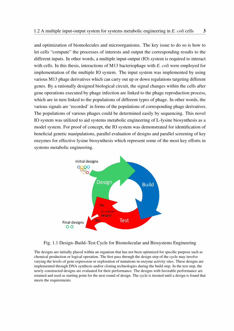
Systems biology is a fast developing discipline making significant contributions to otherdisciplines. Systems biology strategy has been applied to metabolic engineering, enablinga new state-of-art technology termed ’systems metabolic engineering’ [16–21]. The keychallenges of metabolic engineering have been the time-, cost- and labor-intensive processesof strain development owing to the difficulties in understanding the complex interactionsamong the metabolic, gene regulatory and signaling networks inside the cells, which are col-lectively represented as overall system performance under industrial fermentation conditions.To avoid laborious try-and-error manner experiments, systems biology studies have focusedon building genome-scale models of cellular functions to make predictions. However, dueto the complexity of cellular functions and the technical/biological variations in omics data,establishment of predictive, multiscale models is still quite challenging. Indeed the complex-ity issue not only occurs on whole cell-level, it was observed even on single gene level. Inconsequence, effective engineering of biological parts or systems, regardless the scale of thetarget system, requires extensive studies and efforts in the form of design–build–test cycles asshown in Figure 1.1, in which many designs are evaluated and the process is iterated in orderto improve the performance. The rate of improvements is directly related to the throughputand rounds of the design cycles.
Although cells are composed of molecules and their viability relies on extracting andusing energy to maintain them, they are not ‘just’ matter and energy. Information processing,also called “cellular computing”, is essential for cellular function. Previous studies provedthat the computational abilities of biological system could be utilized in rational ways. Here,the computation abilities of cells were proposed to be utilized for systems-level prediction
1.2 A multiple input-output system for systems metabolic engineering in E. coli cells 3
and optimization of biomolecules and microorganisms. The key issue to do so is how tolet cells “compute” the processes of interests and output the corresponding results to thedifferent inputs. In other words, a multiple input-output (IO) system is required to interactwith cells. In this thesis, interactions of M13 bacteriophage with E. coli were employed forimplementation of the multiple IO system. The input system was implemented by usingvarious M13 phage derivatives which can carry out up or down regulations targeting differentgenes. By a rationally designed biological circuit, the signal changes within the cells aftergene operations executed by phage infection are linked to the phage reproduction process,which are in turn linked to the populations of different types of phage. In other words, thevarious signals are ‘recorded’ in forms of the populations of corresponding phage derivatives.The populations of various phages could be determined easily by sequencing. This novelIO system was utilized to aid systems metabolic engineering of L-lysine biosynthesis as amodel system. For proof of concept, the IO system was demonstrated for identification ofbeneficial genetic manipulations, parallel evaluation of designs and parallel screening of keyenzymes for effective lysine biosynthesis which represent some of the most key efforts insystems metabolic engineering.
Fig. 1.1 Design–Build–Test Cycle for Biomolecular and Biosystems Engineering
The designs are initially placed within an organism that has not been optimized for specific purpose such aschemical production or logical operation. The first pass through the design step of the cycle may involvevarying the levels of gene expression or exploration of mutations in enzyme activity sites. These designs areimplemented through DNA synthesis and/or cloning technologies during the build step. In the test step, thenewly constructed designs are evaluated for their performance. The designs with favorable performance areretained and used as starting point for the next round of design. The cycle is iterated until a design is found thatmeets the requirements.
4 Introduction and objectives
1.3 Development of an orthogonal information encodingscheme for reliable information encoding in DNA ofliving cells
We live in the age of information explosion which imposes a big challenge to data storagetechnologies [22]. The presently used storage media such as magnetic tape or hard diskdrivers have a decisive shortcoming of limited lifetime and density, e.g. around 50 yearsfor hard disk drivers. The recent studies of Church et al. and Goldman et al. opened upa new and exciting possibility of storing digital information in synthetic DNA [23, 24].Goldman et al. achieved an information density of 2.2 petabytes (PT)/gram DNA whichis far above the current commercial technologies. Besides the advantage of high density,information storage in DNA has additional attractive features such as ultra-long lifetimeand low maintenance requirements [23, 24]. However, unlike other planner storage media,relatively high rate of errors could be introduced to stored digital data by complex “writing”and “reading” processes of information storage in DNA, especially if fast and cheap synthesisand sequencing technologies are applied [25, 26]. The error rate can be even higher if theencoded DNA contains sequences with extreme GC contents, long homopolymers or complexsecondary structures which are hard to be synthesized or sequenced [23, 24].
Previous studies dealt with information encoding in DNA outside living cells. It is alsoof interest to know if DNA data storage or information encoding in living cells are feasibleand reliable. This should enable applications such as for biological barcodes of engineeredbiological parts (Biobricks) and as comment “language” in “programming biology” in theemerging area of synthetic biology [27]. Theoretically, the encoding schemes designed for invitro data storage in DNA are also applicable for in vivo applications. However, to the bestof our knowledge, no reported work has addressed the issue of increasing errors introducedby DNA replication. This issue is crucial for in vivo applications since DNA replicationhappens constantly under in vivo conditions. Furthermore, the artificial DNA fragmentscould interfere with the native and natural ones (being so-called biologically relevant). Thisis another issue which has not been studied so far. For in vivo applications, such as biologicalbarcodes or comments encoding in living cells, the encoded DNA sequences should not sharethe same sequence space as the natural ones to avoid interference with cellular functions. Inother words, they should be orthogonal to exclude biological relevance. One unique featureof information storage in DNA is that there are always many copies of DNA moleculessynthesized while data writting by DNA synthesis. In other words, a high data redundancyis inherently generated during this process. In this study, we sought to design an encoding

1.3 Development of an orthogonal information encoding scheme for reliable informationencoding in DNA of living cells 5
scheme by taking advantage of the inherent redundancy feature for effective error correctionwith additional consideration of the biological relevance, homopolymers and extrem GCcontent issues.

Chapter 2
Materials and methods
2.1 Methods for systems biology analysis
2.1.1 Genome sequences and strains
Serotype c strain S. mutans 5DC8 was isolated from root caries by David Beighton (London,UK); serotype c strain S. mutans AC4446 was isolated from a proven case of infectiveendocarditis in Dillingen (Germany), serotype c strain S. mutans KK21 was isolated fromenamel caries of an adult by Susanne Kneist (Jena, Germany), serotype c strain S. mutansKK23 was isolated from enamel caries of a child by Susanne Kneist (Jena, Germany),Serotype c strain S. mutans ATCC 25175 was isolated from carious dentine, serotype f strainS. mutans NCTC 11060 was isolated in Denmark from a patient’s blood, serotype b strain S.ratti DSM 20564(=ATCC 19645) was isolated from caries lesion in rat, and finally, serotypenon-d & non-g strain S. sobrinus DSM 20742 (= ATCC 33478) was isolated from humandental plaque. Serotype c is over-represented because 70-80% of all S. mutans isolates are ofthis serotype. However, non-c serotypes seem to be associated with cardiovascular diseasesand this is represented in our study by the serotype f strain. Besides S. mutans, S. sobrinus isconsidered as a relevant cariogenic species in human. The genome sequences of S. mutansUA159 and S. mutans NN2025 were sequenced previously and obtained from NCBI genomedatabase (http://www.ncbi.nlm.nih.gov/genome/). They were used in this study as referencegenomes for the genome analysis. All used strains are listed in Table 2.1.
Some parts of the "Materials and Methods" presented here have been taken or modified from publications(Song et al. 2012, Song et al. 2013; Song et al. 2017a; Song et al. 2017b) with me as the first author. Theexperiments for Section 2.1 were carried out by project partner(s) as specified in the corresponding publications.
8 Materials and methods
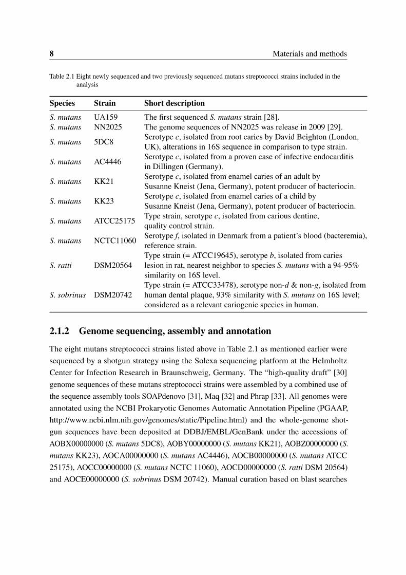
Table 2.1 Eight newly sequenced and two previously sequenced mutans streptococci strains included in theanalysis
Species Strain Short description
S. mutans UA159 The first sequenced S. mutans strain [28].S. mutans NN2025 The genome sequences of NN2025 was release in 2009 [29].
S. mutans 5DC8Serotype c, isolated from root caries by David Beighton (London,UK), alterations in 16S sequence in comparison to type strain.
S. mutans AC4446Serotype c, isolated from a proven case of infective endocarditisin Dillingen (Germany).
S. mutans KK21Serotype c, isolated from enamel caries of an adult bySusanne Kneist (Jena, Germany), potent producer of bacteriocin.
S. mutans KK23Serotype c, isolated from enamel caries of a child bySusanne Kneist (Jena, Germany), potent producer of bacteriocin.
S. mutans ATCC25175Type strain, serotype c, isolated from carious dentine,quality control strain.
S. mutans NCTC11060Serotype f, isolated in Denmark from a patient’s blood (bacteremia),reference strain.
S. ratti DSM20564Type strain (= ATCC19645), serotype b, isolated from carieslesion in rat, nearest neighbor to species S. mutans with a 94-95%similarity on 16S level.
S. sobrinus DSM20742Type strain (= ATCC33478), serotype non-d & non-g, isolated fromhuman dental plaque, 93% similarity with S. mutans on 16S level;considered as a relevant cariogenic species in human.
2.1.2 Genome sequencing, assembly and annotation
The eight mutans streptococci strains listed above in Table 2.1 as mentioned earlier weresequenced by a shotgun strategy using the Solexa sequencing platform at the HelmholtzCenter for Infection Research in Braunschweig, Germany. The “high-quality draft” [30]genome sequences of these mutans streptococci strains were assembled by a combined use ofthe sequence assembly tools SOAPdenovo [31], Maq [32] and Phrap [33]. All genomes wereannotated using the NCBI Prokaryotic Genomes Automatic Annotation Pipeline (PGAAP,http://www.ncbi.nlm.nih.gov/genomes/static/Pipeline.html) and the whole-genome shot-gun sequences have been deposited at DDBJ/EMBL/GenBank under the accessions ofAOBX00000000 (S. mutans 5DC8), AOBY00000000 (S. mutans KK21), AOBZ00000000 (S.mutans KK23), AOCA00000000 (S. mutans AC4446), AOCB00000000 (S. mutans ATCC25175), AOCC00000000 (S. mutans NCTC 11060), AOCD00000000 (S. ratti DSM 20564)and AOCE00000000 (S. sobrinus DSM 20742). Manual curation based on blast searches
2.1 Methods for systems biology analysis 9
using known coding nucleotide sequences were performed to complement some missingcoding genes.
2.1.3 Genome alignment
Multiple genome alignments have been computed using the progressive Mauve algorithm ofthe Mauve software [34] with default options.
2.1.4 Pan-genome and core-genome analysis
In addition to the six S. mutans draft genomes of this study and the previously releasedcomplete genomes of S. mutans UA159 and NN2025, 59 S. mutans genomes (2 completedand 57 drafts) available in NCBI till April 2013 were also included in the core- and pan-genome analysis of S. mutans. The accessions of the 59 genomes are as follows:
AGWE00000000, AHRB00000000, AHRC00000000, AHRD00000000, AHRE00000000,AHRF00000000, AHRG00000000, AHRH00000000, AHRI00000000, AHRJ00000000,AHRK00000000, AHRL00000000, AHRM00000000, AHRN00000000, AHRO00000000,AHRP00000000, AHRQ00000000, AHRR00000000, AHRS00000000, AHRT00000000,AHRU00000000, AHRV00000000, AHRW00000000, AHRX00000000, AHRY00000000,AHRZ00000000, AHSA00000000, AHSB00000000, AHSC00000000, AHSD00000000,AHSE00000000, AHSF00000000, AHSG00000000, AHSH00000000, AHSI00000000,AHSJ00000000, AHSK00000000, AHSL00000000, AHSM00000000, AHSN00000000,AHSO00000000, AHSP00000000, AHSQ00000000, AHSR00000000, AHSS00000000,AHST00000000, AHSU00000000, AHSV00000000, AHSW00000000, AHSX00000000,AHSY00000000, AHSZ00000000, AHTA00000000, AHTB00000000, AHTC00000000,AHTD00000000, AHTE00000000, CP003686, AP012336.
Data pre-processing for the core and pan-genome analysis were performed using a self-implemented perl script (the source codes are gaven in Appendix A), which is similar asdescribed previously by Tettelin et al. [35]. Briefly, an iterative procedure was carriedout to estimate total genes/core genes to be discovered per additional genome sequenced.The number of total genes/core genes provided by each added new genome depends onthe selection of previously added genomes. All possible combinations of genomes from 1to M (the maximal number of available genomes) were calculated. In the case more than1000 combinations were possible, only 1000 random combinations were used. In order totake into consideration of core genes that are possibly missed during genome sequencingand assembly, for the calculation of core-genome size, an additional correction step wasintroduced, in which any one gene that is only absent in one of the 63 draft genomes was
10 Materials and methods
still regarded as core gene. During the fitting step of the core genome model, the inputtedgenome numbers were used as fitting weight for corresponding data point.
The pre-data processing was performed using recently released pipeline PGAP [36].The pan-genome size was calculated using a “Power law model” proposed by Tettelinpreviously [37, 35]. The core-genome model Fc(n) = kcexp[−n/τc]+Ω ( kc,τc, and Ω arefree parameters and Ω means the theoretical core-genome size) proposed by Tettelin et al.was also applied in this study [35].
2.1.5 Gene content-based comparative analysis of 10 mutans strepto-cocci strains
In this work, if not otherwise specified, the uniqueness of genes is defined according tothe ortholog groups constructed by using the OrthoMCL program [38]. If the ortholog ofa gene from organism A is absent in “organism B”, this gene was defined to be unique orspecific to organism A in comparison to organism B. However, it does not imply that thereis no homolog of this gene in organism B. In some cases, this gene is just an additionalcopy (namely paralog) of another gene whose alleles/orthologs are found in both organisms.Certainly, it does further not imply that this gene is present only in organism A. For example,the ortholog of this gene may be found in organism C from the relationship table or in otherstrains or species not compared in this work.
2.1.6 Identification of putative two-component signal transduction sys-tems
The identification of histidine kinases (HKs) and response regulators (RRs) of putativetwo-component systems (TCSs) of the eight mutans streptococci strains (shown in Table 2.1)was carried out based on computational domain analysis of the predicted protein sequences.Two previously sequenced S. mutans strains, the S. mutans UA159 and S. mutans NN2025,were used as reference strains for comparison. To this end, the same identification procedurewas carried out on the genomes of S. mutans NN2025 and UA159 to ensure that the samesearch criteria were applied for all the strains included in this study so that a reasonablecomparison can be achieved. The genome sequences of the two reference strains wereobtained from the genome database at the National Center for Biotechnology Information(http://www.ncbi.nlm.nih.gov/sites/genome). Approaches for identifying HKs and RRs weresimilar to those described previously [39] with slight modifications. Briefly, putative HKand RR proteins were identified by Hidden Markov Model (HMM) searches using the
2.1 Methods for systems biology analysis 11
related HMM profiles available in the Pfam database (http://pfam.sanger.ac.uk/) as templates[40]. The sequence homology search software HMMER3 (http://hmmer.org/) [41] wasused for scanning the predicted protein sequences with the HMM profiles. All the HKrelated HMM profiles with the accession numbers PF00512, PF07568, PF07730, PF07536,PF06580, PF01627, PF02895, PF05384, PF10090 were used for identifying putative HKs.The HMM profile PF00072 which targets the receiver (REC) domain of RR proteins wasused to recognize putative RRs. For the identification of HKs, the homology search wasperformed without setting E-value/score cutoffs to avoid missing any putative HKs withlow scores. However, all the identified putative HKs were manually validated by judgingwhether at least one of the following two criteria was satisfied: (a) the presence of a cognateputative RR in the same operon as the putative HK in question; (b) the presence of both theHisKA-like and HATPase_c domains so that any HATPase_c domain possessing non-HKproteins could be excluded. For the identification of putative RRs, the E-value cut-off wasset at 1e-6. Paired HK and RR present in the same operon comprise a TCS cluster. HybridHKs, if any, could be determined by the presence of a complete HK transmitter domain anda REC domain in a single protein. If no corresponding cognate RRs or HKs can be found inthe same operon, HKs and RRs are defined as orphan HKs or RRs. The operon informationused in this study was predicted by Pathway Tools [42].
2.1.7 Genome-scale metabolic networks construction
The bipartite metabolic networks were constructed based on the connection matrix of up-dated KEGG reactions database according to Stelzer and Zeng [43, 44] with the additionof the newly identified reactions catalyzed by lactate oxidase (Lactate + O2 => Pyruvate +H2O2) with provisional R numbers of R10001 (C00186 + C00007 => C00022 + C00027)and R10002 (C00256 + C00007 => C00022 + C00027). Compared to reaction graph ormetabolite graph, wherein either reactions or metabolites (called "node") are shown in aninterconnected way, the bipartite network is more understandable because both the reactionsand metabolites are visualized at the same time. Seventy-six non-enzymatic automatic reac-tions were also considered for the network construction. The construction of sub-networkswas based on KEGG pathway classification (http://www.genome.jp/kegg/pathway.html) withslight modification by adding lactate oxidase to the glycolysis/gluconeogenesis pathway(MAP00010) and the pyruvate metabolism pathway (MAP00620). The software Cytoscape[45] was used for the visualization and comparative analysis of the genome-scale metabolicnetworks.
12 Materials and methods
2.1.8 PCR verification of unique genes in the comparative genomicsstudies
To verify the unique presence of the lactate oxidase (consecutive) coding genes D823_06595and D823_06598, respectively, in S. sobrinus DSM 20742 and to exclude the possibilityof contamination with e. g. human DNA during the process of genome sequencing, PCRamplifications (using one primer pair covering both genes) with isolated DNAs from S.sobrinus DSM 20742 and a second S. sobrinus strain (AC153), as well as from S. mutansUA159 and S. ratti DSM 20564 (the latter two strains as negative controls) were performed.The primers used were: 5’- GAGCAGGATAATTGACAGTC -3’ (forward primer) and 5’-ACTCAGTGACGAATCAGTT -3’ (reverse primer), which were designed by using PrimerPremier and Vector NTI 9.0 (InforMax), respectively. Conditions for this conventional PCRwere: 94°C, 2 min; followed by 32 cycles of 94°C for 30s; annealing temperature 48°C for30s; and 72°C for 90s; final extension at 72°C for 5 min; length of amplicon 1,175 bp.
To verify the unique presence of TCS-15 in S. mutans NCTC11060, PCR amplificationwith original DNA from this strain using two different forward primers was performed (S.mutans UA159 as negative control). The primers used were: 5’-TTGCTTGCTGTTGTTGTG-3’ (forward primer), 5’- GGCTACCATTTAGTAGAAAAGAGG -3’ (alternative forwardprimer) and 5’-TGTTACCATCTTCGGAAGG-3’ (reverse primer), which were designedby using Primer Premier 6 and Vector NTI 9.0 (InforMax) respectively. Conditions for thisconventional PCR were: 94°C, 2 min; followed by 32 cycles of 94°C for 30s; annealingtemperature 49°C for 30s; and 72°C for 90s; final extension at 72°C for 5 min; length ofamplicons: 1,624 bp and 504 bp, respectively.
To verify the unique presence of TCS-18 and the unique absence of TCS-13 in S. rattiDSM 20564, as well as the unique absence of TCS-9 and TCS-3 in S. sobrinus DSM 20742,PCR amplifications using original DNAs from S. ratti DSM20564, and S. sobrinus DSM20742 was performed (S. mutans UA159 as negative control). The primers used, the anneal-ing temperatures and the lengths of amplicons were as follows (all other parameters were keptthe same as mentioned above): TCS-18 F 5’-CACTGTTCCTCCTGTATCC 3’, TCS-18 R 5’-ATGCTGGCTATGATGTTGT-3’(Tm=50°C, length: 1,899bp covering HK and RR); TCS-13F 5’ RAKTTYATGCCYCTMACYTTYCAG 3’, TCS-13 R 5’ GATTCRWWRGCMGCCTC3’ (Tm = 49°C, length: 1,600 bp covering HK and RR); TCS-9 HK-F 5’ ATACAGTCAATAT-GCYAAGC 3’, TCS-9 HK-R 5’ GRATAACACGGAAAA 3’ (Tm = 45 C, length: 1,055 bp);
All primers in section 2.1.8 were designed by the author. The experiments in section 2.1.8 and 2.1.9 wereperformed by a project partner (Anke Brock, [email protected], Division of Oral Microbiology andImmunology, Department of Operative and Preventive Dentistry Periodontology, RWTH Aachen University,Aachen, Germany).
2.1 Methods for systems biology analysis 13
TCS-9 RR-F 5’ TGCTGARGACCAAGA 3’, TCS-9 RR-R 5’ TTAGCTGCAATTTCTT 3’(Tm = 50°C, length: 522 bp); TCS-3 HK-F 5’ CAYGAYYTIMGIAAYCC 3’, TCS-3 HK-R5’ GTDATIACIGTICCC 3’ (Tm = 40°C, length: 505 bp).
2.1.9 Construction of lactate oxidase encoding gene knockout mutantsand transformation of S. sobrinus DSM 20742
To clarify the functionality of the two lactate oxidases, namely D823_06598 (Llod) andD823_06595 (lod), PCR ligation mutagenesis according to the method described by Lau etal. [46] was used to separately replace the two genes encoding the two enzymes by an ery-thromycin resistance cassette via double homologous recombination. Primers P1Llod (TTAC-CGTTATCCGCGAATTAT) and P2Llod (GGCGCGCCAACCACCCAAGGTTGAATC),P1lod (GGCTGGTTTCCTCCATGATA) and P2lod (GGCGCGCCCCAAAACCACCTTGA-GGAAT) were used to amplify the 5’flanking regions of both genes, respectively, introducingan AscI restriction site. To amplify the 3’flanking regions of both genes, the primers P3Llod(GGCCGGCCGGGAGCTCAAGGTGTTCAAA) and P4Llod (CAAATTGTTCAAAGCGG-GAAC), P3lod (GGCCGGCCGGCAGCAGCCGGTAGTATT) and P4lod (GGGTGCCAACT-TATGTCACGA) were used, respectively, thereby introducing restriction site for FseI. Theerythromycin resistance cassette was amplified from previously constructed gene deletionmutant [47] using primers ErmFor (GGCGCGCCCCGGGCCCAAAATTTGTTTGAT) andErmRev (GGCCGGCCAGTCGGCAGCGACTCATAGAAT), containing the restriction sitefor AscI and FseI, respectively. After digestion with the appropriate restriction enzymes,following purification, the three amplicons were ligated together and used for transformation.
For transformation, two natural transformation methods were first used to assay andoptimize the natural transformation of the S. sobrinus cells. The first step was the preparationof pre-competent cells of S. sobrinus applying the methods according to Lefrancois et al.[48] and Ween et al. [49]. Afterwards 200 ng of the constructs prepared for mutagenesiswere used for the transformation. The plasmids like pDL278 (Spr, pAT18 Emr, and suicidevector pFW5 Spr in both circular and linearized form were used as a positive control. Anothertransformation protocol according to Li et al. [50] applying pheromone CSP of S. mutanswas additionally used to introduce genetic constructs and plasmids into S. sobrinus cells.In this approach two various concentrations of CSP were used: 0.2 and 1µM, respectively.Transformation of S. mutans was used as a parallel control. All these experiments werecarried out at least three times.
All experiments in section 2.1.9 were performed by a project partner Anke Brock ([email protected])
14 Materials and methods
Later, electroporation experiment was carried out according to the procedure describedby LeBlanc et al. [51]. Various pHs of electroporation mix (EPM) [52] as well as variouspulsing conditions were tested. The electroporation was carried out by adding to the chilledelectrocompetent cells 200 ng of constructs prepared for mutagenesis or plasmids. Otherprotocol for electroporation according to [53] was also tested.
2.2 Methods for multiple input-output system
2.2.1 Chemicals
Chemicals of analytical grade were purchased from Sigma-Aldrich Chemie GmbH (München,Germany). Other chemicals were purchased from Carl Roth GmbH (Karlsruhe, Germany).Enzymes and other reagents for molecular biology were obtained from Fermentas (St. Leon-Roth, Germany). Kits for site-directed mutagenesis were obtained from Agilent Technologies(Karlsruhe, Germany).
2.2.2 Bacterial strains
E. coli DH5α and TOP10 were used as hosts for normal vectors construction. E. coliBL21(DE3) was used for high level protein expression. E. coli XL1-Blue (Agilent Technolo-gies) was used for M13 phage infection. The genotypes of E. coli strains are listed in Table2.2.
Table 2.2 E. coli strains used in the present work
Strain Genotype Description
Top10 F- mcrA ∆(mrr-hsdRMS-mcrBC) ϕ80lacZ∆M15 ∆lacX74 nupGrecA1 araD139 ∆(ara-leu)7697 galE15 galK16 rpsL(StrR)endA1 λ -
Host for normal DNAcloning and transforma-tion
DH5α recA1 endA1 gyrA96 thi-1 hsdR17 supE44 relA1 lac [F´ proABlacIqZ∆M15 Tn10 (TetR)]
Host for VCSM13 phageamplification
XL1-Blue recA1 endA1 gyrA96 thi-1 hsdR17 supE44 relA1 lac [F´ proABlacIqZ∆M15 Tn10 (TetR)]
Host for VCSM13 phageamplification
BL21 (DE3) B F– ompT gal dcm lon hsdSB(rB-mB
–) λ (DE3 [lacI lacUV5-T7p07 ind1 sam7 nin5]) [malB+]K-12(λ S)
Host for protein overex-pression
2.2.3 Phagemids, plasmids and primers
The M13 phage (VCSM13) was purchased from Agilent Technology (5301 Stevens CreekBlvd. Santa Clara, CA 95051, USA). The wild lysC gene encoding AK-III was amplified by
2.2 Methods for multiple input-output system 15
PCR from the genomic DNA of E. coli K12 MG1655. For over-expression and purificationof the wild-type AK-III and relevant muteins, the wild-type lysC gene was cloned to pET-22b(+) (Novagen, Darmstadt, Germany) with the introduction of an additional His-tag atthe C-terminal to generate the plasmid pET22-lysC. Site-mutagenesis was performed onpET22-lysC to generate over-expression plasmids for AK-III muteins. The lysC gene wasalso cloned to VCSM13 by replacing the original gene III to generate a phagemid M13-lysC. Similarly, site-mutagenesis was also performed on M13-lysC to generate phagemidderivations carrying different AK-III muteins.
For construction of plasmid AP-Lys-B, i.e. the device harnessed by the host cells tocontrol the phage packaging process based on intracellular lysince concentration, we ultilizeda lysine inducible promoter from Corynebacterium glutamicum ATCC13032 as a lysinesensor. The lysine inducible promoter, gene III from M13 phage and a GFP-encoding genewere cloned into the plasmid pZE21MCS to obtain AP-Lys-B. The transcriptional levelsof gene III and GFP encoding gene are controlled by the lysine inducible promoter. Theantibiotic resistance type of AP-Lys-B was changed to ampicillin resistance by replacing thekanamycin resistance gene with an ampicillin resistance gene.
The plasmids used in this study are listed in Table 2.3.
2.2.4 Media
Complex medium
LB mediumThe LB (Luria-Bertani) medium was routinely used for the cultivations of E. coli strains.
One liter LB liquid medium contained: 10 g tryptone, 5 g yeast extract and 10 g NaCl. LBsolid plate was prepared by addition of 15 g/L agar. The pH was adjusted to 7.0 by 5M NaOH.Sterilization was performed at 121°C for 20 min. When necessary, appropriate antibioticswere added to the medium before usage. For E. coli strains, the working concentration ofampicillin and kanamycin was 100µg/mL or 50µg/ml, respectively.
SOC mediumThe SOC (Super Optimal broth with Catabolite repression) medium is a nutrient-rich
medium used for the regeneration of E. coli strains after heat shock transformation. Forpreparation, 20 g tryptone, 5 g yeast extract. 0.5 g NaCl and 0.186 g KCl were dissolvedin 975 mL water and autoclaved at 121°C for 20 min. Subsequently, 20 mL filter-sterilizedglucose (1M, 0.22 um Ultrafree-MC, Millipore) and 5 mL filter-sterilized MgCl2 (2M, 0.22um Ultrafree-MC, Millipore) were added into the cooling medium.
2XYT medium
16 Materials and methods
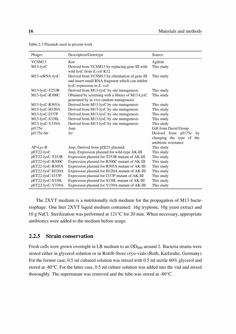
Table 2.3 Plasmids used in present work
Phages Description/Genotype Source
VCSM13 Kan AgilentM13-lysC Derived from VCSM13 by replacing gene III with
wild lysC from E.coli K12This study
M13-srRNA-lysC Derived from VCSM13 by elimination of gene IIIand insert small RNA fragment which can inhibitlysC expression in E. coli
This study
M13-lysC-T253R Derived from M13-lysC by site mutagenesis This studyM13-lysC-R300C Obtained by screening with a library of M13-LysC
generated by in vivo random mutagenesisThis study
M13-lysC-R305A Derived from M13-lysC by site mutagenesis This studyM13-lysC-H320A Derived from M13-lysC by site mutagenesis This studyM13-lysC-I337P Derived from M13-lysC by site mutagenesis This studyM13-lysC-S338L Derived from M13-lysC by site mutagenesis This studyM13-lysC-V339A Derived from M13-lysC by site mutagenesis This studypJ175e Amp Gift from David GrouppJ175e-Str Str Derived from pJ175e by
changing the type of theantibiotic resistance
AP-Lys-B Amp, Derived from pZE21 plasmid; This studypET22-lysC Amp, Expression plasmid for wild-type AK-III This studypET22-lysC-T253R Expression plasmid for T253R mutant of AK-III This studypET22-lysC-R300C Expression plasmid for R300C mutant of AK-III This studypET22-lysC-R305A Expression plasmid for R305A mutant of AK-III This studypET22-lysC-H320A Expression plasmid for H320A mutant of AK-III This studypET22-lysC-I337P Expression plasmid for I337P mutant of AK-III This studypET22-lysC-S338L Expression plasmid for S338L mutant of AK-III This studypET22-lysC-V339A Expression plasmid for V339A mutant of AK-III This study
The 2XYT medium is a nutritionally rich medium for the propagation of M13 bacte-riophage. One liter 2XYT liquid medium contained: 16g tryptone, 10g yeast extract and10 g NaCl. Sterilization was performed at 121°C for 20 min. When necessary, appropriateantibiotics were added to the medium before usage.
2.2.5 Strain conservation
Fresh cells were grown overnight in LB medium to an OD600 around 2. Bacteria strains werestored either in glycerol solution or in Roti®-Store cryo-vials (Roth, Karlsruhe, Germany).For the former case, 0.5 ml cultured solution was mixed with 0.5 ml sterile 60% glycerol andstored at -80°C. For the latter case, 0.5 ml culture solution was added into the vial and mixedthoroughly. The supernatant was removed and the tube was stored at -80°C.
2.2 Methods for multiple input-output system 17
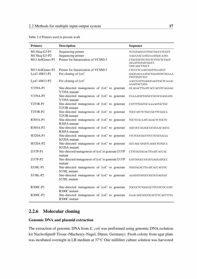
Table 2.4 Primers used in present work
Primers Description Sequence
M13Seq-G3-P1 Sequencing primer TCTGTAGCCGTTGCTACCCTCGTTM13Seq-G3-P2 Sequencing primer AAGAAACAATGAAATAGCAATAM13-ln4Genes-P1 Primer for linearization of VCSM13 CTAGTATTTCTCCTCTTTCTCTAGT
ATAATTGTATCGGTT-TATCAGCTTGCT
M13-ln4Genes-P2 Primer for linearization of VCSM13 CTCCCTCAATCGGTTGAATGTLysC-4M13-P1 For cloning of lysC GAGGAGAAATACTAGATGTCTGAAA
TTGTTGTCTCCLysC-4M13-P2 For cloning of lysC AACCGATTGAGGGAGTTACTCAAAC
AAATTACTATGV339A-P1 Site-directed mutagenesis of lysC to generate
V339A mutantGCAGACTTAATCACCACGTCAGAAG
V339A-P2 Site-directed mutagenesis of lysC to generateV339A mutant
CGAAATATTATGCCGCGCGAGGATG
T253R-P1 Site-directed mutagenesis of lysC to generateT253R mutant
CGTTTTGGTGCAAAAGTACTGC
T253R-P2 Site-directed mutagenesis of lysC to generateT253R mutant
TGCCATCTCTGCCGCTTCGGCA
R305A-P1 Site-directed mutagenesis of lysC to generateR305A mutant
TGCTCGCAATCAGACTCTGCTC
R305A-P2 Site-directed mutagenesis of lysC to generateR305A mutant
AGCGCCAGAGCGCGGAACAGCG
H320A-P1 Site-directed mutagenesis of lysC to generateH320A mutant
TTCTCGCGGTTTCCTCGCGGAA
H320A-P2 Site-directed mutagenesis of lysC to generateH320A mutant
GCCAGCATATTCAGGCTGTGCA
I337P-P1 Site-directed mutagenesis of lysC to generate I337Pmutant
CTTCGGTAGACTTAATCACCAC
I337P-P2 Site-directed mutagenesis of lysC to generate I337Pmutant
GATTATGCCGCGCGAGGATGCC
S338L-P1 Site-directed mutagenesis of lysC to generateS338L mutant
TGGTAGACTTAATCACCACGTC
S338L-P2 Site-directed mutagenesis of lysC to generateS338L mutant
AAATATTATGCCGCGCGAGGAT
R300C-P1 Site-directed mutagenesis of lysC to generateR300C mutant
TGCGCTCTGGCGCTTCGTCGCAATC
R300C-P2 Site-directed mutagenesis of lysC to generateR300C mutant
GAACAGCGGCGGATTTTCAGTTTTA
2.2.6 Molecular cloning
Genomic DNA and plasmid extraction
The extraction of genomic DNA from E. coli was performed using genomic DNA isolationkit NucleoSpin® Tissue (Macherey-Nagel, Düren, Germany). Fresh colony from agar platewas incubated overnight in LB medium at 37°C One milliliter culture solution was harvested
18 Materials and methods
and the cell lysis was achieved by incubation of the sample in a proteinase K/SDS solution.Cell harvest and DNA purification were performed according to the manual of NucleoSpin®Tissue. Plasmid extraction was carried out by following the standard protocol of NucleoSpin®Plasmid kit (Macherey-Nagel, Düren, Germany).
Mutagenesis
Site-mutagenesis was performed using a protocol similar to the NEB Q5® Site-DirectedMutagenesis Kit. Briefly, none overlap primers were designed and synthesized which containthe desired mutations. Then PCR amplification was performed with the designed primersusing the original plasmid as templates to generate linear plasmids. Template DNA waseliminated by enzymatic digestion with DpnI. Finally, phosphorylation and ligation usingT4 Polynucleotide Kinase and T4 Ligase were carried out to obtain circular DNA beforetransformation.
Random in vivo mutagenesis was enabled by using the plasmid pJ184-Str harboringgenes which can increase intracellular DNA replication error rates. The plasmid pJ184-Strwas derived from pJ184 by replacing the chloramphenicol acetyltransferase encoding genewith a streptomycin resistance gene. The pJ184 plasmid which has been described previouslywas obtained from David R. Liu’s group of Harvard Medical School [54].
2.2.7 Preparation of infective engineered phages
Since the engineered phages lack gene III, the helper plasmid pJ175e was harnessed bythe host cells to supply gene III products intracellularly to obtain infective phages. Theplasmid pJ175e was obtained from David R. Liu’s group. Specifically, engineered phageswere co-transformed with pJ175e into XL1-Blue cells. Overnight cultures were depositedfor centrifuge and the supernatant containing the packaged infective phages was collected.
2.2.8 Screening based on cell-phage interactions
XL1-Blue/AP-Lys-B cells were incubated in LB medium to an OD600 value around 1.0.Roughly 200ul XL1-Blue/AP-Lys-B cells were mixed with 2ul proper diluted phages (Cellsto phage number ratio above 10:1 to make sure that all phages could be captured and evaluatedby host cells. Different types of phages in a total number of roughly 10,000 were used asinputs in the present study). The mixture was incubated at 37°C for 15 minutes withoutshaking to allow the phages to attach to the cells, following by incubation at 37°C withshaking for 1 to 2 hours. Inactivate the host cells at 65°C for 15min. The cell debris were
2.2 Methods for multiple input-output system 19
spinned down and the supernatant containing the “scored” phages was transferred to a freshtube. A proper amount of “scored” phages were mixed with fresh XL1-Blue/AP-Lys-B cellsand incubated at 37°C for 15 minutes without shaking to allow the host robots to absorb thehighly “scored” phages. A proper amount of the culture was then sprayed on LB agar plateswith kanamycin (50 mg/ml) for selection.
2.2.9 Enzyme characterization
Enzyme overexpression in E. coli
Enzyme overexpression was achieved with pET-22b(+) in E. coli BL21(DE3) cells. Therecombinant cells bearing the expression vectors were firstly grown in 100 mL LB mediumsupplemented with appropriate antibiotics (80 µg/ml ampicillin for pET-22b(+) derivates at37°C. When the OD600 of the culture reached 0.6, protein expression was induced by theaddition of isopropyl-β -D-thiogalactopyranoside (IPTG) in a final concentration of 0.1 mM,and the culture was continued for an additional 12 to 14 h at 30°C. Cells were harvested bycentrifuge (10min, 5000rpm, 4°C), washed twice with 20mM Tris-HCl buffer (pH 7.5) andresuspended with 5mL lysis buffer (20 mM Tris-HCl (pH7.5), 150 mM NaCl and 500 mM(NH4)2SO4). Cell suspensions were directly submitted for enzyme purification steps withinthe same day.
Enzyme purification
The supernatant was obtained by centrifugation at 4°C for 1 hour at 13,000 rpm. Targetedproteins with His-tag at C-terminal (pET-22b (+) derivates) were purified by His SpinTrapTMcolumns (GE Healthcare Bio-Sciences, Piscataway, USA). The protocols from the kits werefollowed during the purification (twice washing with washing buffer (20mM KH2PO4,500mM NaCl, 20mM Imidazole, pH 7.4) and elution with elution buffer (20mM KH2PO4,500mM NaCl, 500mM Imidazole, pH 7.4)). After the step of enzyme purification, PDMiniTrap G-10 columns (GE Healthcare Bio-Sciences, Piscataway, USA) were used forbuffer change (20 mM Tris-HCl (pH7.5), 150 mM NaCl and 500 mM (NH4)2SO4). Proteincontent was determined at 595nm by Bradford method (Bradford, 1976) with a reagentsolution from Biorad (Biorad, Hercules, USA) and BSA (Bovine Serum Albumin) standardprotein.
20 Materials and methods
Enzyme assay
The parameters of enzyme kinetics were determined by varying the concentrations of sub-strates. To test the influence of allosteric inhibition, effectors with varied concentrationswere additionally added into the standard reaction. The relative activities were calculated bynormalizing the specific activities of enzymes under the standard conditions.
The enzyme activity of aspartokinase was detected by using the hydroxamate method[55]. The quantity of aspartate hydroxamate formed in the presence of hydroxylamine wasmeasured at 540 nm. The standard assay reaction mixture in 1 ml contained 200 mM Tris-HCl (pH 7.5), 10 mM MgSO4·6H2O, 10 mM aspartate, 10 mM ATP, 160 mM NH2OH·HCl(neutralized with KOH), and appropriate amounts of enzyme. After incubation at 30°C for30 min, the reaction was stopped by mixing with 1 ml of a 5% (wt/vol) FeCl3 solution, andthe absorbance at 540 nm was monitored.
2.3 Methods for orthogonal information encoding in livingcells
2.3.1 Detailed steps for encoding binary data into DNA string
To detail the steps of encoding arbitrary digital data into DNA string, an arbitrary computerfile is represented as a string (S1) of bits (often interpreted as a number between 0 and 1).The detailed steps are illustrated in Figure 5.2 and explained as follows: 1) Bit string S1is converted to DNA string S2 of characters in A, C, G, T four bits by four bits using thescheme shown in Figure 1 (shown in rows of “Data encoding bases”). 2) One error detectingbase was inserted per two bases based on assign rule I shown in Figure 5.2 to generate DNAstring of S3. 3) Check presentation of “TTT” three bases by three bases and adapt the errordetecting base of blocks that next to “TTT” to a new base based on rule II to generate finalDNA string of S4.
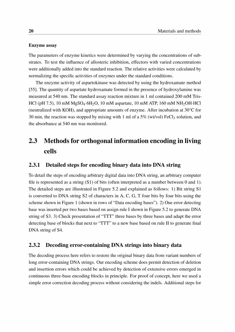
2.3.2 Decoding error-containing DNA strings into binary data
The decoding process here refers to restore the original binary data from variant numbers oflong error-containing DNA strings. Our encoding scheme does permit detection of deletionand insertion errors which could be achieved by detection of extensive errors emerged incontinuous three-base encoding blocks in principle. For proof of concept, here we used asimple error correction decoding process without considering the indels. Additional steps for
2.3 Methods for orthogonal information encoding in living cells 21
indels treatments would enhance the error correction efficiency. The details of the decodingprocess were illustrated in Figure 2.1 and described as follows:
1. Generate consensus DNA string block by block as follows:a) Read a three-base block from all DNA fragments and remove all the three-base blocks
with errors detected (The rule of error detecting base was initialized with rule I); b) Makeconsensus block by taking the block that with largest occurrence frequency; c) Switch theerror detecting rule to rule II if the consensus block is ‘TTT’, otherwise, switch to rule I; d)Go to next blocks and repeat a), b), c) steps until the complete consensus DNA string wasgenerated;
2. Transfer the consensus DNA string into bit string based on the scheme shown in Figure5.2.
2.3.3 Implementation of the online encoding-decoding system for SED3B
The online system is implemented by using CakePHP (https://cakephp.org/) web developmentframework. Two different applications are provided: comment encoding-decoding and biolog-ical barcode encoding-decoding. The system is available under the link: http://biosystem.bt1.tu-harburg.de/sed3b/.
2.3.4 Analysis of error tolerance by in silicon simulation
The 35,292 bps DNA string encoding the logo (Figure 2.2) of our institute is used as input forerror tolerance simulation. The specific rate of random errors was introduced base by baseby giving a specific error probability. The rates of A<->T and G<->C transition errors weredoubled to that of A/T<->G/C transition errors to mimic the natural DNA replication process.Variant numbers of DNA sequences with random errors were then used for decoding to testthe error tolerance.
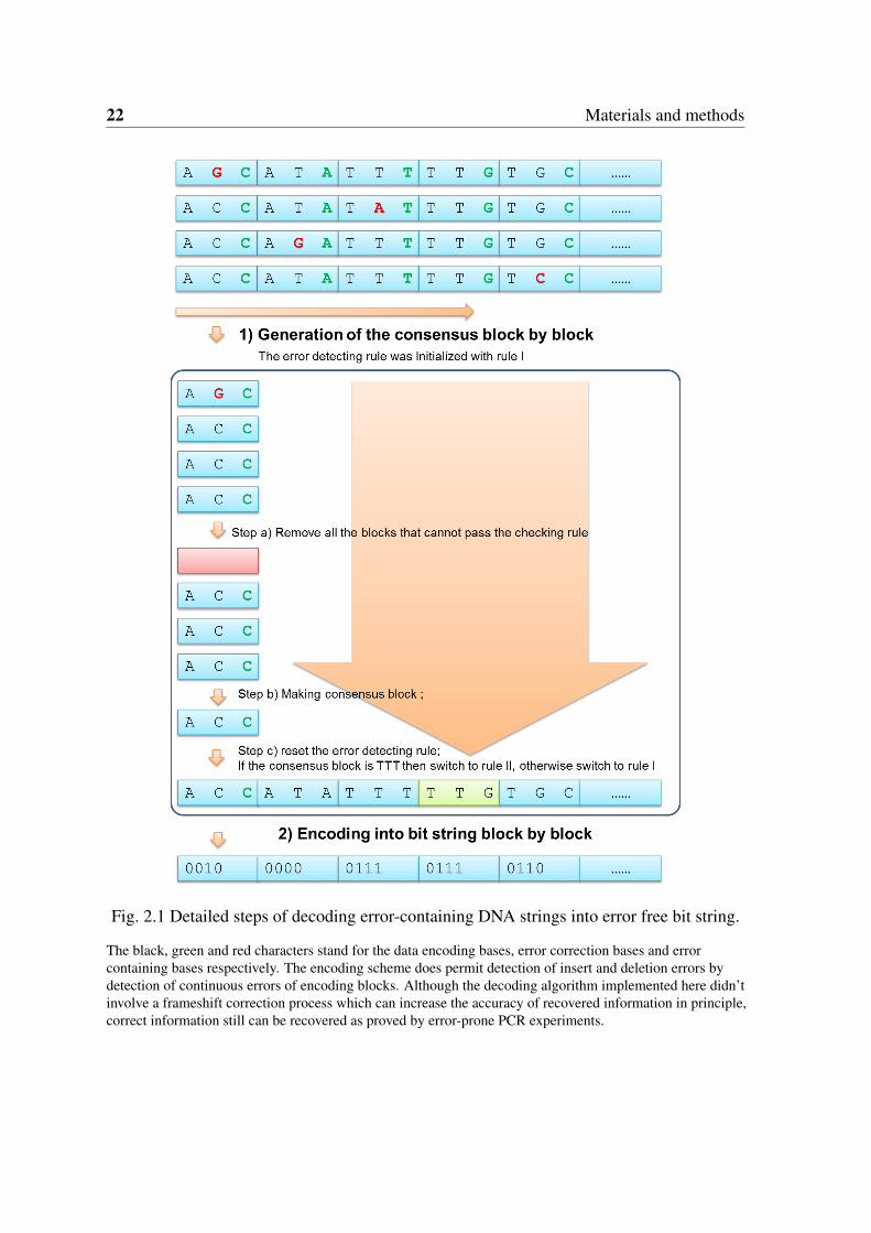
2.3.5 In vivo verification of the error tolerance by error-prone PCR
To test the error tolerance capability of the SED3B encoding scheme in practical, we en-coded text of “Hello, World!” into 78bp DNA string. A 168bp DNA fragment includ-ing the 78bp DNA string encoding "Hello, World!" was constructed using two primersof 5’- TCTAAGAAACCATTATTATCATGACATTAACCTATAAAAATAGGCGTATCAC-GAGGCCCTTTCGTCTTTAAGGATGCTCGTGCCCATGCCCATGCCGTAC -3’ and 5’-GGCTCGAGCTCGAGACTAGCACCTGGTTTAGCATGGGCAAGTAAAACGGCACAAA-AATATGGTTGGGGTACGGCATGGGCATGGGCACGAGCATCCTTAA -3’. We then used
22 Materials and methods
Fig. 2.1 Detailed steps of decoding error-containing DNA strings into error free bit string.
The black, green and red characters stand for the data encoding bases, error correction bases and errorcontaining bases respectively. The encoding scheme does permit detection of insert and deletion errors bydetection of continuous errors of encoding blocks. Although the decoding algorithm implemented here didn’tinvolve a frameshift correction process which can increase the accuracy of recovered information in principle,correct information still can be recovered as proved by error-prone PCR experiments.
2.3 Methods for orthogonal information encoding in living cells 23
Fig. 2.2 The logo of our institute used as input for error tolerance simulation
error-prone PCR to introduce random errors into the 168bp DNA fragments. Error-pronePCR was performed using JBS dNTP-Mutagenesis Kit using the recommended protocol with30 thermal cycles to introduce errors into the encoded DNA string. The amplified fragmentsby error-prone PCR were ligated with linearized pZE21-MCS plasmid using In-Fusion®HD Cloning Kit from Clontech© Laboratories. The ligation products were transformedinto stellar E. coli stellar competent Cells. The plasmid map and encoded information arepresented in Figure 2.3.
Fig. 2.3 Illustration of construction process of plasmids carrying the encoded 78bp DNAstring with variant errors introduced by error-prone PCR
The plasmid abstractions of individual colonies were deposited for sequencing. Originalinformation was recovered using the error rich DNA fragments. Primers used for error-proneamplification of the 168bp insert DNA fragment are 5’-TCTAAGAAACCATTATTATCAT-

24 Materials and methods
3’ and 5’-GGCTCGAGCTCGAGACTAGCA-3’. The primers used for linearization ofthe plasmid are 5’- TAATGGTTTCTTAGACGTCGGAATTGCCAGCTGGG -3’ and 5’-TCTCGAGCTCGAGCCAGGCATCAAATAAAACGA AAGG-3’. The primer used forsequencing is 5’-GCGAAACGATCCTCATCCTGTCT-3’.
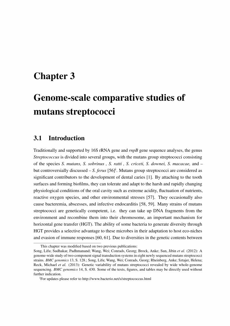
Chapter 3
Genome-scale comparative studies ofmutans streptococci
3.1 Introduction
Traditionally and supported by 16S rRNA gene and rnpB gene sequence analyses, the genusStreptococcus is divided into several groups, with the mutans group streptococci consistingof the species S. mutans, S. sobrinus , S. ratti , S. criceti, S. downei, S. macacae, and –but controversially discussed – S. ferus [56]i. Mutans group streptococci are considered assignificant contributors to the development of dental caries [1]. By attaching to the toothsurfaces and forming biofilms, they can tolerate and adapt to the harsh and rapidly changingphysiological conditions of the oral cavity such as extreme acidity, fluctuation of nutrients,reactive oxygen species, and other environmental stresses [57]. They occasionally alsocause bacteremia, abscesses, and infective endocarditis [58, 59]. Many strains of mutansstreptococci are genetically competent, i.e. they can take up DNA fragments from theenvironment and recombine them into their chromosome, an important mechanism forhorizontal gene transfer (HGT). The ability of some bacteria to generate diversity throughHGT provides a selective advantage to these microbes in their adaptation to host eco-nichesand evasion of immune responses [60, 61]. Due to diversities in the genetic contents between
This chapter was modified based on two previous publications:Song, Lifu; Sudhakar, Padhmanand; Wang, Wei; Conrads, Georg; Brock, Anke; Sun, Jibin et al. (2012): Agenome-wide study of two-component signal transduction systems in eight newly sequenced mutans streptococcistrains. BMC genomics 13, S. 128.; Song, Lifu; Wang, Wei; Conrads, Georg; Rheinberg, Anke; Sztajer, Helena;Reck, Michael et al. (2013): Genetic variability of mutans streptococci revealed by wide whole-genomesequencing. BMC genomics 14, S. 430. Some of the texts, figures, and tables may be directly used withoutfurther indication.
iFor updates please refer to http://www.bacterio.net/s/streptococcus.html
26 Genome-scale comparative studies of mutans streptococci
different isolates, the genome content of a single isolate doesnot necessarily represent thegenomic potential of a certain species. With the rapid development of DNA sequencingtechnologies, the steadily increasing genome data enable us to dig the evolutionary andgenetic information of a species from a pan-genome perspective. In 2002, the release ofthe genome sequence of S.mutans UA159, the first genome sequence of mutans groupstreptococci, has greatly helped in understanding the robustness and complexity of S. mutansas an oral and odontogenic (e.g. infective endocarditis and abscesses) pathogen [28]. Later,after the genome sequence of S. mutans NN2025 became available, a comparative genomicanalysis of S. mutans NN2025 and UA159 has provided insights into chromosomal shufflingand species-specific contents [29]. Recently, Cornejo et al. have studied the evolutionary andpopulation genomics of S. mutans based on 57 S. mutans draft genomes and revealed a highHGT rate of S. mutans [62].
In this study, the whole genome of eight mutans streptococci strains, including six S.mutans strains (5DC8, KK21, KK23, AC4446, ATCC25175 and NCTC11060), one S. rattistrain (DSM20564) and one S. sobrinus strain (DSM20742) were sequenced. A pan-genomicmodel of mutans streptococci was constructed and analyzed. Cross-comparison of the genomecontents of the eight mutans streptococci strains and the previously genome sequenced strainsof S. mutans UA159 and NN2025 were carried out focusing on the genomic componentsthat are highly related to pathogenicity. Further, by constructing and comparative analysisof genome-level metabolic networks, the diversities in sub-pathways among these strainswere systematically investigated. The results are helpful for understanding the evolutionand pathogenicity of these oral pathogens, which in turn will be helpful for the clinicalmanagement of diseases caused by these pathogens and for the development of diagnosticsand new molecular epidemiological methods.
3.2 Genome sequencing, assembly and annotation of eightmutans streptococci strains
Shot-gun sequencing of the six S. mutans isolates (5DC8, KK21, KK23, AC4446, ATCC25175,and NCTC11060) as well as S. ratti DSM 20564 and S. sobrinus DSM 20742 were carriedout on a Solexa sequencing platform. An overview of the genome assemblies and annotationsof the six S. mutans isolates, S. ratti DSM 20564 and S. sobrinus DSM 20742 is summarizedin Table 3.1 in comparison with two previously sequenced S. mutans strains, namely UA159and S. mutans NN2025. The average GC contents are in the range of low GC organisms [28].The genome sizes are very close to each other, with the largest one from S. sobrinus DSM
3.2 Genome sequencing, assembly and annotation of eight mutans streptococci strains 27
20742 and the smallest one from S. mutans KK23 showing merely 5.7% differences. Thetotal numbers of protein-coding sequences per genome are also similar among all the strainscompared.
As would be expected, the overall genomic features of the eight S. mutans strains (5DC8,KK21, KK23, AC4446, ATCC25175, NCTC11060, UA159 and NN2025) are more similarto each other than to S. ratti DSM20564 and S. sobrinus DSM20742. This is consistent withthe results of the phylogenetic analysis, as visualized by the phylogenetic tree constructedbased on 16S rRNA gene sequences and core-genome single-nucleotide polymorphisms(SNPs) shown in Figure 3.1.
28 Genome-scale comparative studies of mutans streptococci
Fig. 3.1 Phylogenetic analysis of 10 mutans streptococci strains compared in this study andtheir phylogenetic relationship to other Streptococcus species.
a) 16S rRNA phylogenetic tree of Streptococcus species with genomes known as this study was performed(Since the 16S rRNA sequences were almost identical between the different S. mutans strains, only UA159 isshown as representative). b) Phylogenetic tree of the mutans streptococci compared in this study constructedwith the core-genome SNPs obtained by PGAP pipeline [36]. All phylogenetic trees were constructed usingClustalX [63] and Phylip [64] by applying the maximum likelihood (ML) method with bootstrap value set to100. Values beside branches depict ML bootstrap support values. The scale bars in the unit of “substitution/site”are shown below the trees.
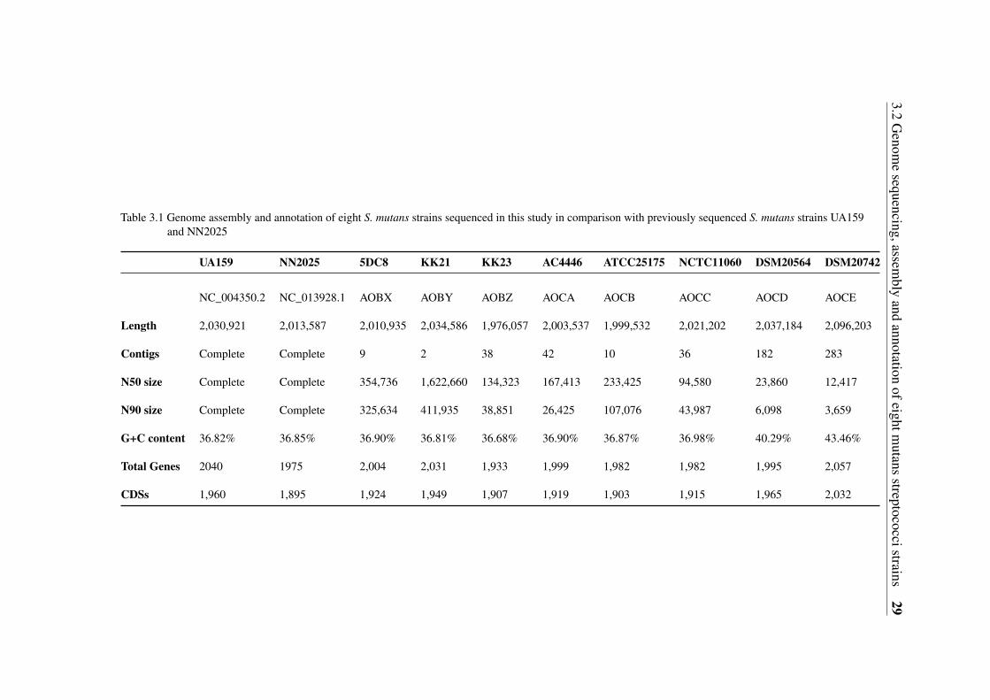
3.2G
enome
sequencing,assembly
andannotation
ofeightmutans
streptococcistrains29
Table 3.1 Genome assembly and annotation of eight S. mutans strains sequenced in this study in comparison with previously sequenced S. mutans strains UA159and NN2025
UA159 NN2025 5DC8 KK21 KK23 AC4446 ATCC25175 NCTC11060 DSM20564 DSM20742
NC_004350.2 NC_013928.1 AOBX AOBY AOBZ AOCA AOCB AOCC AOCD AOCE
Length 2,030,921 2,013,587 2,010,935 2,034,586 1,976,057 2,003,537 1,999,532 2,021,202 2,037,184 2,096,203
Contigs Complete Complete 9 2 38 42 10 36 182 283
N50 size Complete Complete 354,736 1,622,660 134,323 167,413 233,425 94,580 23,860 12,417
N90 size Complete Complete 325,634 411,935 38,851 26,425 107,076 43,987 6,098 3,659
G+C content 36.82% 36.85% 36.90% 36.81% 36.68% 36.90% 36.87% 36.98% 40.29% 43.46%
Total Genes 2040 1975 2,004 2,031 1,933 1,999 1,982 1,982 1,995 2,057
CDSs 1,960 1,895 1,924 1,949 1,907 1,919 1,903 1,915 1,965 2,032
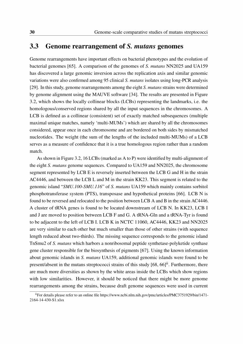
30 Genome-scale comparative studies of mutans streptococci
3.3 Genome rearrangement of S. mutans genomes
Genome rearrangements have important effects on bacterial phenotypes and the evolution ofbacterial genomes [65]. A comparison of the genomes of S. mutans NN2025 and UA159has discovered a large genomic inversion across the replication axis and similar genomicvariations were also confirmed among 95 clinical S. mutans isolates using long-PCR analysis[29]. In this study, genome rearrangements among the eight S. mutans strains were determinedby genome alignment using the MAUVE software [34]. The results are presented in Figure3.2, which shows the locally collinear blocks (LCBs) representing the landmarks, i.e. thehomologous/conserved regions shared by all the input sequences in the chromosomes. ALCB is defined as a collinear (consistent) set of exactly matched subsequences (multiplemaximal unique matches, namely ‘multi-MUMs’) which are shared by all the chromosomesconsidered, appear once in each chromosome and are bordered on both sides by mismatchednucleotides. The weight (the sum of the lengths of the included multi-MUMs) of a LCBserves as a measure of confidence that it is a true homologous region rather than a randommatch.
As shown in Figure 3.2, 16 LCBs (marked as A to P) were identified by multi-alignment ofthe eight S. mutans genome sequences. Compared to UA159 and NN2025, the chromosomesegment represented by LCB E is reversely inserted between the LCB G and H in the strainAC4446, and between the LCB L and M in the strain KK23. This segment is related to thegenomic island “SMU.100-SMU.116” of S. mutans UA159 which mainly contains sorbitolphosphotransferase system (PTS), transposase and hypothetical proteins [66]. LCB N isfound to be reversed and relocated to the position between LCB A and B in the strain AC4446.A cluster of tRNA genes is found to be located downstream of LCB N. In KK23, LCB Iand J are moved to position between LCB F and G. A tRNA-Gln and a tRNA-Tyr is foundto be adjacent to the left of LCB I. LCB K in NCTC 11060, AC4446, KK23 and NN2025are very similar to each other but much smaller than those of other strains (with sequencelength reduced about two-thirds). The missing sequence corresponds to the genomic islandTnSmu2 of S. mutans which harbors a nonribosomal peptide synthetase-polyketide synthasegene cluster responsible for the biosynthesis of pigments [67]. Using the known informationabout genomic islands in S. mutans UA159, additional genomic islands were found to bepresent/absent in the mutans streptococci strains of this study [68, 66]ii. Furthermore, thereare much more diversities as shown by the white areas inside the LCBs which show regionswith low similarities. However, it should be noticed that there might be more genomerearrangements among the strains, because draft genome sequences were used in current
iiFor details please refer to an online file https://www.ncbi.nlm.nih.gov/pmc/articles/PMC3751929/bin/1471-2164-14-430-S1.xlsx
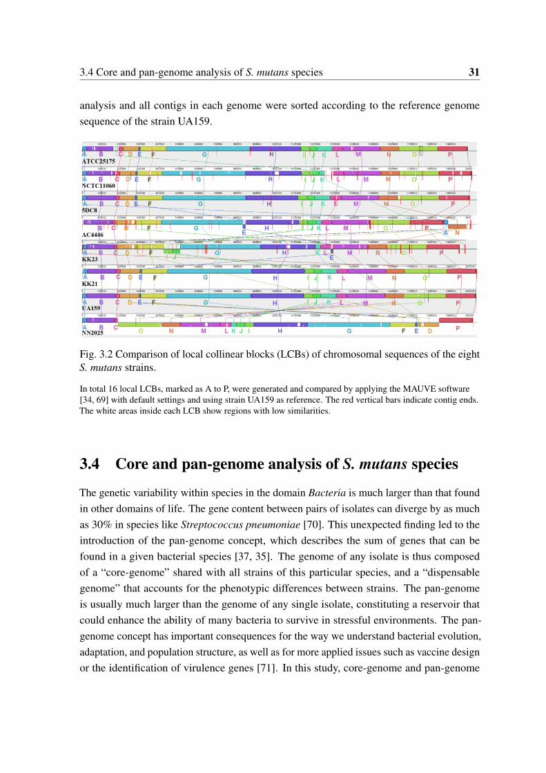
3.4 Core and pan-genome analysis of S. mutans species 31
analysis and all contigs in each genome were sorted according to the reference genomesequence of the strain UA159.
Fig. 3.2 Comparison of local collinear blocks (LCBs) of chromosomal sequences of the eightS. mutans strains.
In total 16 local LCBs, marked as A to P, were generated and compared by applying the MAUVE software[34, 69] with default settings and using strain UA159 as reference. The red vertical bars indicate contig ends.The white areas inside each LCB show regions with low similarities.
3.4 Core and pan-genome analysis of S. mutans species
The genetic variability within species in the domain Bacteria is much larger than that foundin other domains of life. The gene content between pairs of isolates can diverge by as muchas 30% in species like Streptococcus pneumoniae [70]. This unexpected finding led to theintroduction of the pan-genome concept, which describes the sum of genes that can befound in a given bacterial species [37, 35]. The genome of any isolate is thus composedof a “core-genome” shared with all strains of this particular species, and a “dispensablegenome” that accounts for the phenotypic differences between strains. The pan-genomeis usually much larger than the genome of any single isolate, constituting a reservoir thatcould enhance the ability of many bacteria to survive in stressful environments. The pan-genome concept has important consequences for the way we understand bacterial evolution,adaptation, and population structure, as well as for more applied issues such as vaccine designor the identification of virulence genes [71]. In this study, core-genome and pan-genome
32 Genome-scale comparative studies of mutans streptococci
analyses of 67 S. mutans strains was performed, including the eight mutans streptococcistrains sequenced in this study and 59 S. mutans strains whose genomes were available inNCBI till April 2013.
3.4.1 Core-genome
The core-genome size of the 67 S. mutans strains was calculated to be 1,373. For detailedinformation of the core genes please refer to an online fileiii. To estimate the theoreticalcore-genome size achievable with an infinite number of S. mutans genomes, core-genomesize medians corresponding to different genome numbers as shown in Figure 3.3a by thered rectangles were first calculated by random sampling 1,000 genome combinations of ngenomes out of the 67 S. mutans genomes. Then, the exponential regression core-genomemodel Fc(n) = kcexp[−n/τc]+Ω proposed previously by Tettelin et al. [37, 35] was appliedto fit the median data points of the core-genome sizes, where n represents the number ofgenomes, and Ω stands for the theoretical core-genome size. To take into considerationthe different deviations of the core-genome size medians, as clearly indicated by the blueerror bars in Figure 3.3a, the fitting process was modified by introducing the genomenumber as weight to the corresponding data point. The fitting parameters thus obtainedare as follows: r2 = 0.97403,kc = 325.74718±10.00912,Ω = 1,369.41225±1.986,τc =
15.90248± 0.66807. Using this fitting result to describe the core-genome of S. mutans,the theoretical core-genome size (Ω) was estimated to be around 1,370 genes, which iscomparable to the core-genome size (1,373) calculated using 67 genomes. Comparedwith other streptococcus species, the core-genome of S. mutans is at the same level to thecore-genome of S. pyogenes (1,400 genes determined using 11 strains), less than that ofS. pneumoniae (1,647 genes determined using 47 strains) and S. agalactiae (1,800 genesdetermined using eight strains) [37, 72, 73]. However, it should be cautious with suchcomparison. In the study of Cornejo et al. [62], the core genome size of S. mutans wasdetermined as 1,490 by using 57 S. mutans genomes which is obviously different to the coregenome size of S. mutans estimated in this study, although the 57 S. mutans genomes used byCornejo et al. were also included in our study. The discrepancy can be caused by differentreasons, such as difference in the correction step for core gene determination and, very likely,different methods and parameter settings used for determining orthologs. Apparently, a morestringent process was used in this study to determine orthologs which led to smaller coregenome size of S. mutans estimated.
iiihttps://www.ncbi.nlm.nih.gov/pmc/articles/PMC3751929/bin/1471-2164-14-430-S2.xlsx
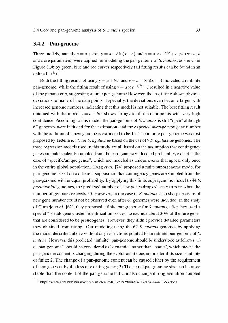
3.4 Core and pan-genome analysis of S. mutans species 33
3.4.2 Pan-genome
Three models, namely y = a+ bxc, y = a− b ln(x+ c) and y = a× e−x/b + c (where a, band c are parameters) were applied for modeling the pan-genome of S. mutans, as shown inFigure 3.3b by green, blue and red curves respectively (all fitting results can be found in anonline file iv).
Both the fitting results of using y = a+bxc and y = a−b ln(x+ c) indicated an infinitepan-genome, while the fitting result of using y = a× e−x/b + c resulted in a negative valueof the parameter a, suggesting a finite pan-genome However, the last fitting shows obviousdeviations to many of the data points. Especially, the deviations even become larger withincreased genome numbers, indicating that this model is not suitable. The best fitting resultobtained with the model y = a+ bxc shows fittings to all the data points with very highconfidence. According to this model, the pan-genome of S. mutans is still “open” although67 genomes were included for the estimation, and the expected average new gene numberwith the addition of a new genome is estimated to be 15. The infinite pan-genome was firstproposed by Tettelin et al. for S. agalactiae based on the use of 9 S. agalactiae genomes. Thethree regression models used in this study are all based on the assumption that contingencygenes are independently sampled from the pan-genome with equal probability, except in thecase of “specific/unique genes”, which are modeled as unique events that appear only oncein the entire global population. Hogg et al. [74] proposed a finite supragenome model forpan-genome based on a different supposition that contingency genes are sampled from thepan-genome with unequal probability. By applying this finite supragenome model to 44 S.pneumoniae genomes, the predicted number of new genes drops sharply to zero when thenumber of genomes exceeds 50. However, in the case of S. mutans such sharp decrease ofnew gene number could not be observed even after 67 genomes were included. In the studyof Cornejo et al. [62], they proposed a finite pan-genome for S. mutans, after they used aspecial “pseudogene cluster” identification process to exclude about 30% of the rare genesthat are considered to be pseudogenes. However, they didn’t provide detailed parametersthey obtained from fitting. Our modeling using the 67 S. mutans genomes by applyingthe model described above without any restrictions pointed to an infinite pan-genome of S.mutans. However, this predicted “infinite” pan-genome should be understood as follows: 1)a “pan-genome” should be considered as “dynamic” rather than “static”, which means thepan-genome content is changing during the evolution, it does not matter if its size is infiniteor finite; 2) The change of a pan-genome content can be caused either by the acquirementof new genes or by the loss of existing genes; 3) The actual pan-genome size can be morestable than the content of the pan-genome but can also change during evolution coupled
ivhttps://www.ncbi.nlm.nih.gov/pmc/articles/PMC3751929/bin/1471-2164-14-430-S3.docx
34 Genome-scale comparative studies of mutans streptococci
with the change of the environment. Thus, without considering “gene loss events”, it’squite understandable to have a “growing” or “infinite” pan-genome as gene acquirementoccurs no matter how slow it might be. Interestingly, Cornejo et al. found a high rate ofHGT in S. mutans, where many genes were acquired from related streptococci and bacterialstrains predominantly residing not only in the oral cavity, but also in the respiratory tract, thedigestive tract, genitalia, in insect pathogens and in the environment in general [62]. Suchhigh rate of HGT might also lead to a continuously growing (infinite) pan-genome.
3.5 Gene content-based comparative analysis of mutans strep-tococci strains
The annotated protein sequences of the ten mutans streptococci genomes studied were cross-compared based on alleles/ortholog groups established by the program OrthoMCL [38]. Intotal, 2,211 putative alleles/ortholog groups were establishedv. A pair-wise comparison ofthe protein coding sequences between each two strains is shown in Table 3.2. It is clearto see that remarkable differences in protein coding sequences (CDSs) exist between thestrains, even inside the same species of S. mutans . In the following sections, systems that arehighly related to stress resistance and pathogenicity are presented and discussed. As all thefollowing results are based on putative alleles/ortholog groups established by OrthoMCL, ifnot otherwise specified, the word “putative allele(s)/ortholog(s)” is omitted in the followingtext.
vhttps://www.ncbi.nlm.nih.gov/pmc/articles/PMC3751929/bin/1471-2164-14-430-S4.xlsx
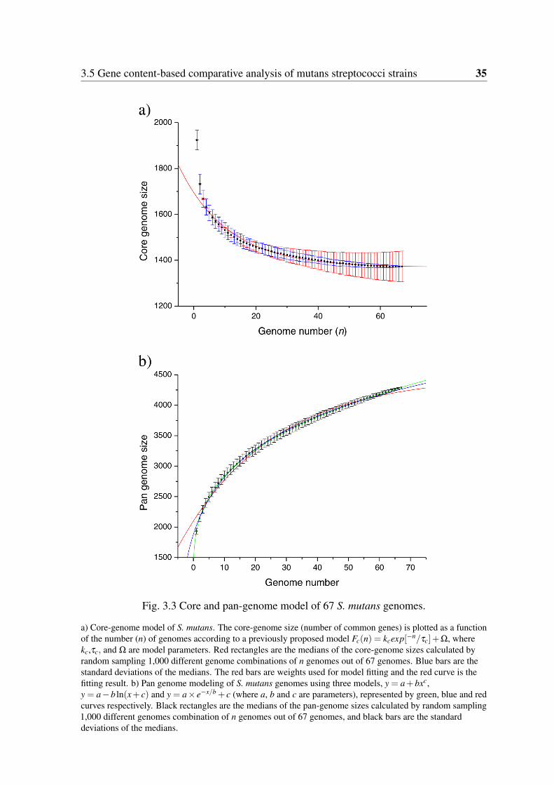
3.5 Gene content-based comparative analysis of mutans streptococci strains 35
Fig. 3.3 Core and pan-genome model of 67 S. mutans genomes.
a) Core-genome model of S. mutans. The core-genome size (number of common genes) is plotted as a functionof the number (n) of genomes according to a previously proposed model Fc(n) = kcexp[−n/τc]+Ω, wherekc,τc, and Ω are model parameters. Red rectangles are the medians of the core-genome sizes calculated byrandom sampling 1,000 different genome combinations of n genomes out of 67 genomes. Blue bars are thestandard deviations of the medians. The red bars are weights used for model fitting and the red curve is thefitting result. b) Pan genome modeling of S. mutans genomes using three models, y = a+bxc,y = a−b ln(x+ c) and y = a× e−x/b + c (where a, b and c are parameters), represented by green, blue and redcurves respectively. Black rectangles are the medians of the pan-genome sizes calculated by random sampling1,000 different genomes combination of n genomes out of 67 genomes, and black bars are the standarddeviations of the medians.
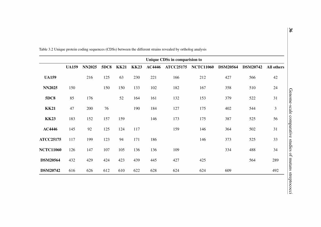
36G
enome-scale
comparative
studiesofm
utansstreptococci
Table 3.2 Unique protein coding sequences (CDSs) between the different strains revealed by ortholog analysis
Unique CDSs in comparision to
UA159 NN2025 5DC8 KK21 KK23 AC4446 ATCC25175 NCTC11060 DSM20564 DSM20742 All others
UA159 216 125 63 230 221 166 212 427 566 42
NN2025 150 150 150 133 102 182 167 358 510 24
5DC8 85 176 52 164 161 132 153 379 522 31
KK21 47 200 76 190 184 127 175 402 544 3
KK23 183 152 157 159 146 173 175 387 525 56
AC4446 145 92 125 124 117 159 146 364 502 31
ATCC25175 117 199 123 94 171 186 146 373 525 33
NCTC11060 126 147 107 105 136 136 109 334 488 34
DSM20564 432 429 424 423 439 445 427 425 564 289
DSM20742 616 626 612 610 622 628 624 624 609 492
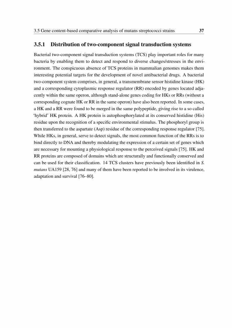
3.5 Gene content-based comparative analysis of mutans streptococci strains 37
3.5.1 Distribution of two-component signal transduction systems
Bacterial two-component signal transduction systems (TCS) play important roles for manybacteria by enabling them to detect and respond to diverse changes/stresses in the envi-ronment. The conspicuous absence of TCS proteins in mammalian genomes makes theminteresting potential targets for the development of novel antibacterial drugs. A bacterialtwo-component system comprises, in general, a transmembrane sensor histidine kinase (HK)and a corresponding cytoplasmic response regulator (RR) encoded by genes located adja-cently within the same operon, although stand-alone genes coding for HKs or RRs (without acorresponding cognate HK or RR in the same operon) have also been reported. In some cases,a HK and a RR were found to be merged in the same polypeptide, giving rise to a so called‘hybrid’ HK protein. A HK protein is autophosphorylated at its conserved histidine (His)residue upon the recognition of a specific environmental stimulus. The phosphoryl group isthen transferred to the aspartate (Asp) residue of the corresponding response regulator [75].While HKs, in general, serve to detect signals, the most common function of the RRs is tobind directly to DNA and thereby modulating the expression of a certain set of genes whichare necessary for mounting a physiological response to the perceived signals [75]. HK andRR proteins are composed of domains which are structurally and functionally conserved andcan be used for their classification. 14 TCS clusters have previously been identified in S.mutans UA159 [28, 76] and many of them have been reported to be involved in its virulence,adaptation and survival [76–80].
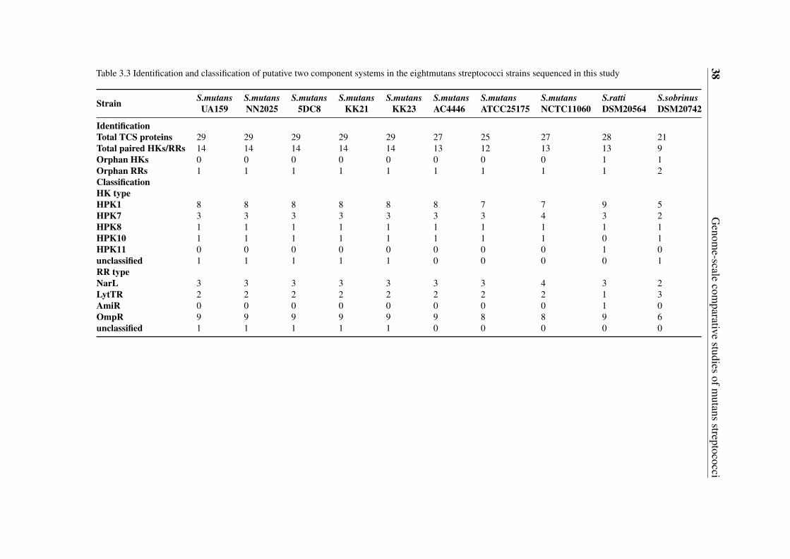
38G
enome-scale
comparative
studiesofm
utansstreptococci
Table 3.3 Identification and classification of putative two component systems in the eightmutans streptococci strains sequenced in this study
Strain S.mutansUA159
S.mutansNN2025
S.mutans5DC8
S.mutansKK21
S.mutansKK23
S.mutansAC4446
S.mutansATCC25175
S.mutansNCTC11060
S.rattiDSM20564
S.sobrinusDSM20742
IdentificationTotal TCS proteins 29 29 29 29 29 27 25 27 28 21Total paired HKs/RRs 14 14 14 14 14 13 12 13 13 9Orphan HKs 0 0 0 0 0 0 0 0 1 1Orphan RRs 1 1 1 1 1 1 1 1 1 2ClassificationHK typeHPK1 8 8 8 8 8 8 7 7 9 5HPK7 3 3 3 3 3 3 3 4 3 2HPK8 1 1 1 1 1 1 1 1 1 1HPK10 1 1 1 1 1 1 1 1 0 1HPK11 0 0 0 0 0 0 0 0 1 0unclassified 1 1 1 1 1 0 0 0 0 1RR typeNarL 3 3 3 3 3 3 3 4 3 2LytTR 2 2 2 2 2 2 2 2 1 3AmiR 0 0 0 0 0 0 0 0 1 0OmpR 9 9 9 9 9 9 8 8 9 6unclassified 1 1 1 1 1 0 0 0 0 0
3.5 Gene content-based comparative analysis of mutans streptococci strains 39
By combining the HMM profiling results and the information on putative operon organi-zation, repertoires of potential TCS proteins (HKs and RRs) in the eight mutans streptococcistrains sequenced in this study were obtained, as shown in Table 3.3 in comparison to S.mutans NN2025 and UA159. The total numbers of TCS proteins identified are comparableamong the 10 mutans streptococci strains.
By analyzing the putative operon organizations of the identified TCS proteins, 98.5%of the total putative HKs and 92.2% of the total putative RR were found to be constitutingHK-RR pairs. Ortholog analysis of the paired or non-paired TCS proteins among the 10mutans streptococci strains revealed a total of 18 different TCS clusters, 2 orphan HKs and2 orphan RRs (Table 3.4). The numbering of the TCS clusters was based on an existingnumbering system used by Levesque [81], and extended to the new TCS clusters identifiedin this study. Co-evolution of TCS proteins could be clearly observed. This means HKsand RRs, which belong to a particular TCS cluster, are usually co-present or co-absent in aspecific strain.
In addition, putative alleles/orthologs of the corresponding HKs and RRs were foundto be highly conserved (similarity ≥ 95%) among the S. mutans strains. The conservationwas clearly lower across the species. Furthermore, it is obvious that, in most cases, putativeHK alleles/orthologs within one TCS cluster exhibited a higher degree of diversity than thecorresponding putative RR alleles/orthologs across the species. This could be attributed tothe high variability of sensing/input domains harbored by the individual HKs, as will bediscussed later in the classification of HKs and RRs.
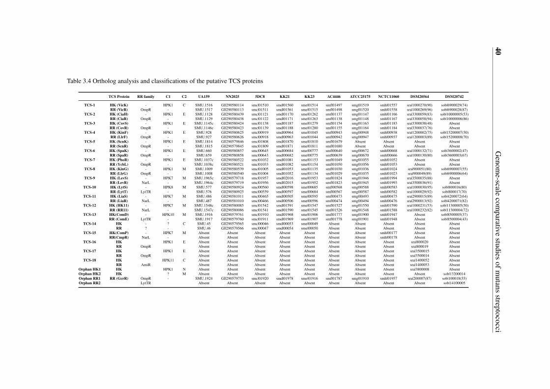
40G
enome-scale
comparative
studiesofm
utansstreptococci
Table 3.4 Ortholog analysis and classifications of the putative TCS proteins
TCS Protein RR family C1 C2 UA159 NN2025 5DC8 KK21 KK23 AC4446 ATCC25175 NCTC11060 DSM20564 DSM20742
TCS-1 HK (VicK) - HPK1 C SMU.1516 GI|290580114 smc|01510 smd|01560 sme|01514 smf|01497 smg|01519 smh|01557 sra|1000270(90) sob|6900029(74)RR (VicR) OmpR - SMU.1517 GI|290580113 smc|01511 smd|01561 sme|01515 smf|01498 smg|01520 smh|01558 sra|1000269(96) sob|6900028(87)
TCS-2 HK (CiaH) - HPK1 E SMU.1128 GI|290580439 smc|01121 smd|01170 sme|01262 smf|01137 smg|01147 smh|01166 sra|3300059(83) sob|10000005(53)RR (CiaR) OmpR - SMU.1129 GI|290580438 smc|01122 smd|01171 sme|01263 smf|01138 smg|01148 smh|01167 sra|3300058(94) sob|10000006(86)
TCS-3 HK (CovS) - HPK1 E SMU.1145c GI|290580424 smc|01138 smd|01187 sme|01279 smf|01154 smg|01163 smh|01183 sra|3300038(48) AbsentRR (CovR) OmpR - SMU.1146c GI|290580423 smc|01139 smd|01188 sme|01280 smf|01155 smg|01164 smh|01184 sra|3300037(76) Absent
TCS-4 HK (KinF) - HPK1 E SMU.928 GI|290580625 smc|00919 smd|00964 sme|01045 smf|00943 smg|00948 smh|00938 sra|1200002(75) sob|15200007(50)RR (LlrF) OmpR - SMU.927 GI|290580626 smc|00918 smd|00963 sme|01044 smf|00942 smg|00947 smh|00937 sra|1200003(89) sob|15200008(70)
TCS-5 HK (ScnK) - HPK1 E SMU.1814 GI|290579846 smc|01808 smd|01870 sme|01810 smf|01679 Absent Absent Absent AbsentRR (ScnR) OmpR - SMU.1815 GI|290579845 smc|01809 smd|01871 sme|01811 smf|01680 Absent Absent Absent Absent
TCS-6 HK (SpaK) - HPK1 E SMU.660 GI|290580857 smc|00645 smd|00684 sme|00777 smf|00640 smg|00672 smh|00668 sra|1000132(71) sob|3600002(47)RR (SpaR) OmpR - SMU.659 GI|290580858 smc|00643 smd|00682 sme|00775 smf|00639 smg|00670 smh|00666 sra|1000130(80) sob|3600003(67)
TCS-7 HK (PhoR) - HPK1 E SMU.1037c GI|290580522 smc|01032 smd|01081 sme|01153 smf|01049 smg|01055 smh|01052 Absent AbsentRR (YcbL) OmpR - SMU.1038c GI|290580521 smc|01033 smd|01082 sme|01154 smf|01050 smg|01056 smh|01053 Absent Absent
TCS-8 HK (KinG) - HPK1 M SMU.1009 GI|290580539 smc|01005 smd|01053 sme|01135 smf|01030 smg|01036 smh|01024 sra|900051(80) sob|6900007(55)RR (LlrG) OmpR - SMU.1008 GI|290580540 smc|01004 smd|01052 sme|01134 smf|01029 smg|01035 smh|01023 sra|900049(89) sob|6900006(64)
TCS-9 HK (LevS) - HPK7 M SMU.1965c GI|290579718 smc|01957 smd|02016 sme|01953 smf|01824 smg|01946 smh|01994 sra|3500035(88) AbsentRR (LevR) NarL - SMU.1964c GI|290579719 smc|01956 smd|02015 sme|01952 smf|01823 smg|01945 smh|01993 sra|3500036(91) Absent
TCS-10 HK (LytS) - HPK8 M SMU.577 GI|290580924 smc|00560 smd|00598 sme|00685 smf|00568 smg|00588 smh|00583 sra|1000030(95) sob|800016(80)RR (LytT) LytTR - SMU.576 GI|290580925 smc|00559 smd|00597 sme|00684 smf|00567 smg|00587 smh|00582 sra|1000029(92) sob|800017(70)
TCS-11 HK (LiaS) - HPK7 M SMU.486 GI|290581011 smc|00465 smd|00505 sme|00595 smf|00473 smg|00493 smh|00475 sra|2900015(89) sob|4200072(64)RR (LiaR) NarL - SMU.487 GI|290581010 smc|00466 smd|00506 sme|00596 smf|00474 smg|00494 smh|00476 sra|2900013(92) sob|4200071(82)
TCS-12 HK (HK11) - HPK7 M SMU.1548c GI|290580085 smc|01542 smd|01591 sme|01547 smf|01527 smg|01550 smh|01590 sra|1000231(53) sob|11300005(50)RR (RR11) NarL - SMU.1547c GI|290580086 smc|01541 smd|01590 sme|01545 smf|01526 smg|01548 smh|01588 sra|1000232(82) sob|11300004(72)
TCS-13 HK(ComD) - HPK10 M SMU.1916 GI|290579761 smc|01910 smd|01968 sme|01906 smf|01777 smg|01900 smh|01947 Absent sob|8500005(37)RR (ComE) LytTR - SMU.1917 GI|290579760 smc|01911 smd|01969 sme|01907 smf|01778 smg|01901 smh|01948 Absent sob|8500004(43)
TCS-14 HK - ? C SMU.45 GI|290579565 smc|00046 smd|00053 sme|00049 Absent Absent Absent Absent AbsentRR ? - SMU.46 GI|290579566 smc|00047 smd|00054 sme|00050 Absent Absent Absent Absent Absent
TCS-15 HK(ComP) - HPK7 M Absent Absent Absent Absent Absent Absent Absent smh|00177 Absent AbsentRR(CmpR) NarL - Absent Absent Absent Absent Absent Absent Absent smh|00178 Absent Absent
TCS-16 HK HPK1 E Absent Absent Absent Absent Absent Absent Absent Absent sra|800020 AbsentRR OmpR Absent Absent Absent Absent Absent Absent Absent Absent sra|800019 Absent
TCS-17 HK HPK1 E Absent Absent Absent Absent Absent Absent Absent Absent sra|3500015 AbsentRR OmpR Absent Absent Absent Absent Absent Absent Absent Absent sra|3500014 Absent
TCS-18 HK HPK11 C Absent Absent Absent Absent Absent Absent Absent Absent sra|1400052 AbsentRR AmiR Absent Absent Absent Absent Absent Absent Absent Absent sra|1400053 Absent
Orphan HK1 HK HPK1 N Absent Absent Absent Absent Absent Absent Absent Absent sra|3800008 AbsentOrphan HK2 HK ? M Absent Absent Absent Absent Absent Absent Absent Absent Absent sob|13200014Orphan RR1 RR (GcrR) OmpR - SMU.1924 GI|290579753 smc|01920 smd|01978 sme|01916 smf|01787 smg|01910 smh|01957 sra|200007(87) sob|100018(55)Orphan RR2 LytTR Absent Absent Absent Absent Absent Absent Absent Absent Absent sob|14100005
3.5 Gene content-based comparative analysis of mutans streptococci strains 41
TCS proteins common to all the 10 mutans streptococci strains
Proteins of the TCS clusters 1, 2, 4, 6, 8, 10, 11, 12 and the orphan RR1 are common toall the 10 mutans streptococci strains compared here, indicating probably the functionalimportance of these TCS clusters for the adaptation and survival of these mutans streptococci.For instance, Orphan RR1 is highly conserved across the 10 mutans streptococci strains. InS. mutans UA159, this orphan RR is encoded by gcrR (SMU.1924c) and has been foundto play a vital role in sucrose-dependent adherence and cariogenesis [82]. Therefore, it isconceivable that conservation of this gene across the mutans streptococci strains is essentialfor their primary pathogenicity.
TCS proteins uniquely present/absent in one or several strains
The TCS-3 (CovSR) cluster was predicted to be absent in S. sobrinus DSM20742. CovSR isinvolved in the acid tolerance response of S. mutans UA159 [81], and has also been reportedto be involved in counteracting oxidative stress and reducing susceptibility to phagocytickilling [83]. TCS-9 (LevRS), which affects the acid tolerance response as well [81], was alsoabsent in S. sobrinus DSM20742. The absence of the covS and levS genes was experimentallysupported by the PCR results. In S. mutans UA159, the levRS gene cluster is flanked bylevQ and levT, which code for two carbohydrate-binding proteins. These four genes togetherconstitute a four-component signal transduction system levQRST controlling the transcriptionof the fructan hydrolase gene (fruA) and a four-gene cluster levDEFG, which encode afructose/mannose sugar:phosphotransferase system located immediately downstream oflevQRST [84]. S. sobrinus was also found to lack the levQ, levT and levDEFG genes. Takingtogether, these findings indicate dramatic differences in the regulation of fructan catabolismand the acid tolerance response of S. sobrinus DSM20742 in comparison to the S. mutansstrains.
It should be pointed out that the TCS nomenclature is unfortunately inconsistent amongthe published articles on TCSs of S. mutans strains. Many publications [85–89, 82] on CovRactually address the orphan response regulator RR1 which is known as GcrR (SMU.1924) inS. mutans UA159 [82]. On the other hand, CovSR of S. mutans has also been confusinglynamed as ScnKR [81, 83]. In this study, the nomenclature of TCS genes/proteins was basedprimarily on the Oralgen Pathogen Sequence Database (http://www.oralgen.lanl.gov). Incases where several names were given for a gene in Oralgen, gene names used by Biswas etal. [76] were preferably used.
TCS-5 (ScnKR) could be neither found in the two S. mutans strains ATCC25175 andNCTC11060 nor in S. ratti DSM20564 and S. sobrinus DSM20742. The asymmetric
42 Genome-scale comparative studies of mutans streptococci
distribution of TCS-5 was also observed in a previous study, in which TCS-5 was foundto be present only in two of the 10 S. mutans strains compared [76]. In S. mutans UA159,an insertion mutant of scnK gene displayed no significant difference to the wild type withrespect to growth under various stress conditions [76]. In Streptococcus pyogenes, scnKRwas found to be essential for the production of a bacteriocin (SAFF22) [90]. By a closerlook at the genes in the neighborhood of scnKR in the 10 strains studied, it was found that S.mutans KK23 and S. mutans NN2025 carried two and three genes, respectively, which encodeputative bacteriocin precursor peptides sharing more than 60% similarity with SAFF22. Inaddition, a putative bacteriocin biosynthesis protein coding gene was also found downstreamof the scnKR operon in the two S. mutans strains. It was therefore inferred that TCS-5(ScnKR) might be involved in the regulation of mutacin production at least in S. mutansKK23 and S. mutans NN2025.
TCS-7 (PhoR/YcbL) was only shared by the eight S. mutans strains. In S. mutans UA159,an insertion mutant of the gene encoding PhoR displayed no significant difference to thewild type with respect to growth under various stress conditions [76]. The clear function ofTCS-7 is still unknown.
As mentioned before in the identification and classification of TCS proteins, TCS-13(ComDE) was absent in S. ratti DSM20564. This finding was also supported by the PCRexperiment. In S. mutans, ComDE is the most extensively studied two-component signalingsystem involved in quorum sensing and competence development. Mediated by the so-calledcompetence stimulating peptide (CSP), it is involved in multiple stress responses and hasbeen implicated in competence development, bacteriocin production, virulence, biofilmformation, and cariogenicity [79, 91–95]. Further analysis showed that the ortholog of thecomC gene, whose product is the precursor of the signal peptide CSP sensed by ComDEin S. mutans, was absent in S. ratti. Putative orthologs of comD and comE were found inS. sobrinus DSM20742. However, with a similarity of merely 37% and 43%, respectively,to the comD and comE of S. mutans UA159, it’s highly possible that the actual function ofTCS-13 in S. sobrinus DSM20742 might be quite different from that known for S. mutans. Itis also worthy to mention that no comC-like gene was found in S. sobrinus DSM20742.
Putative alleles of the HK and RR proteins of TCS-14 are present in five of the S. mutansstrains, namely 5DC8, KK23, KK21, UA159 and NN2025. This cluster was first identifiedin S. mutans UA159 by Biswas et al. and the corresponding HK and RR are encoded bySMU.45 and SMU.46, respectively [76]. They also found that TCS-14 was present only intwo of the 13 S. mutans strains compared in their study. HKs of this TCS cluster contain onlyone recognizable HATPase_c domain. In addition, no known output domain was identified inthe cognate RRs. Thus, neither HKs nor RRs of TCS-14 could be classified into any known

3.5 Gene content-based comparative analysis of mutans streptococci strains 43
HK and RR families. In addition, by the multi-sequence alignment of the putative TCS-14HK alleles, the open reading frame predictions carried out in this study revealed that whileSMU.45 and its upstream gene SMU.44 clearly constitute two separate genes in S. mutansUA159, they are merged to constitute the parts of a single gene coding for the HKs in the S.mutans strains 5 DC8, KK23 and NN2025. The corresponding HK of KK21 was also splitinto two proteins by the lacking of a single glutamine (Q) residue.
TCS-15 was found exclusively in the genome of the serotype f blood isolate S. mutansNCTC11060. Genes of a TCS located on the genomic island TnSmu2 of S. mutans UA140,as recently reported by the research group Qi [67], could be possible alleles of TCS-15 genes.This was based on the fact that the predicted HKs and RRs of both TCS 15 in this study andthe TCS found by Qi’s group have the HK (YP_002747386.1) and RR (YP_002124238.1)of Streptococcus equi, respectively, as the best matched homologs. The HK (smh|00177) ofTCS-15 is also the only histidine kinase found in this study that contains a PDZ domain. PDZdomain was first reported to be present in animals. In 1997, Ponting et al. claimed that PDZdomains exist also in diverse signaling proteins of bacteria, yeasts and plants. Experimentalevidence was first provided by Liao et al. through the determination of the first crystalstructure of a bacterial PDZ domain [96]. The most remarkable feature of PDZ domains istheir ability to specifically recognize and bind to short C-terminal peptide motifs. This allowsthem especially to bind membrane proteins such as ion channels, which have very smallfree C-termini. To exclude the possibility of contamination with e. g. human DNA duringthe process of genome sequencing, the existence of the gene coding for this unusual PDZdomain-containing HK protein in the DNA of the NCTC11060 strain was experimentallyverified by using two different forward primers in the PCR experiment.
The PDZ domain of smh|00177 is flanked by one transmembrane helix (TM) at its N-terminal side and 9 TMs at its C-terminal side, which is characteristic of a ComP-like HK.ComP-like HKs are a group of sensors of another peptide-dependent quorum sensing systemrelated to cell density-responsive regulation other than ComDE in gram-positive bacteria. InB. subtilis, ComP is the sensor histidine kinase of the four-component comQXPA quorumsensing system, where ComA stands for the corresponding response regulator, ComX is thepheromone precursor and ComQ the protein required for the proteolytic cleavage and modifi-cation of the pheromone precursor molecule [97] . The most similar homolog of smh|00177found from a BLASTp search in the NCBI database (http://blast.ncbi.nlm.nih.gov/) witha sequence identity of around 65% is the histidine kinase (YP_003353659.1) of a TCSfrom Lactococcus lactis subsp. lactis KF147. Furthermore, the cognate response regulatorof TCS-15 showed a sequence identity of around 78% with the corresponding responseregulator (YP_003353660.1) from the same L. lactis subsp. lactis strain. In this study, the RR
44 Genome-scale comparative studies of mutans streptococci
(smh|00177) of TCS-15 was termed as CmpR. It should be mentioned that neither L. lactissubsp. lactis KF147 nor S. mutans NCTC11060 possesses homologs of the B. subtilis ComXand ComQ. Thus, the signal peptide sensed by ComP/CmpR in S. mutans NCTC11060remains unknown.
TCS-16, 17 and 18 are uniquely present in S. ratti DSM20564. According to BLASTPsearches against the NCBI database, the top matches to the TCS-16 HK protein (sra|800020)or the RR protein (sra|800019) are all from Streptococcus species such as Streptococcusinfantarius and Streptococcus agalactiae. In addition, the HK and RR homologs in thedifferent Streptococcus species are all encoded by two adjacent genes and annotated in someS. agalactiae strains as sensor histidine kinase DltS and DNA-binding response regulator DltR.The DltSR has been reported to be involved in the regulation of D-alanyl-lipoteichoic acidbiosynthesis in S. agalactiae [98]. Lipoteichoic acid (LTA) is a major cell wall constituent ofGram-positive bacteria which is phosphoglycerol substituted with a D-Ala ester or a glycosylresidue and anchored in the membrane by its glycolipid moiety. D-alanylation of lipoteichoicacid has been proven to contribute to the virulence of Streptococcus suis [99], as well as tothe biofilm formation and resistance to antimicrobial peptides in enterococci [100]. Thus,the TCS-16 cluster might also be an important virulence factor in S. ratti.
The TCS-17 is composed of a HAMP-containing HK and an OmpR-type RR. The besthomologs of the HK protein (sra|3500015) and the RR protein (sra|3500014) are from S.agalactiae strains. But the functions of these homologous proteins remain unknown.
TCS-18 is the only cluster that comprises a HPK 11 family HK and an AmiR familyRR, which possesses a novel RNA-binding type output domain. The top ten best hits of theBLASTp search in the NCBI database showed that the closest homologs for the HK and RRof TCS-18 are all from Listeria species. Since the HK of TCS-18 possesses a PAS domainthat is commonly involved in sensing intracellular signals such as redox potential, similarto the sensing mechanism described for the HK (VicK) of TCS-1, the function of TCS-18might also be related to the sensing and response to signal(s) originated in the cytoplasm.The unique presence of TCS-18 in S. ratti DSM20564 was also confirmed by the PCRexperiment.
3.5.2 High diversities of the competence development regulation mod-ule
In the previous section, the two-component signal transduction systems (TCS) in the 10mutans streptococci strains were systematically discussed. ComDE, one of the TCS isdirectly related to competence development. Competence development is a complex process
3.5 Gene content-based comparative analysis of mutans streptococci strains 45
involving sophisticated regulatory networks that trigger the capacity of bacterial cells to takeup exogenous DNA from the environment. This phenomenon is frequently encountered inbacteria of the oral cavity, e.g., S. mutans [101]. In S. mutans , ComX, an alternative sigmafactor, drives the transcription of the so called ‘late-competence genes’ required for genetictransformation. ComX activity is modulated by the inputs from two types of signal pathways,namely the competence-stimulating peptide (CSP) dependent competence regulation systemand CSP-independent competence regulation system. ComX and the ‘late-competence genes’regulated by ComX as labeled in boldface in Table 3.5, are highly conserved even betweenthe species, indicating that all mutans streptococci studied here might have the ability oftransforming to genetic competence state. On the other hand, the upstream signal pathwaysregulating the activity of ComX show high variety as discussed in details below.

46G
enome-scale
comparative
studiesofm
utansstreptococci
Table 3.5 Distribution of competence development-related systems in the 10 mutans streptococci strains
Name UA159 NN2025 5DC8 KK21 KK23 AC4446 ATCC 25175 NCTC 11060 DSM 20564 DSM 20742
ComA SMU.286 GI|290581206 D816_01150 D817_01300 D818_01134 D819_01163 D820_01336 D821_01208 D822_01584 D823_05343SMU.1881c GI|290579788 D816_08453 D817_08643 - D819_07724 D821_08449 D822_08325 D823_01400
ComB SMU.287 GI|290581205 D816_01155 D817_01305 D818_01139 D819_01168 D820_01341 D821_01213 D822_01589 D823_05923ComC SMU.1915 GI|290579762 D816_08588 D817_08778 D818_08368 D819_07839 D820_08520 D821_08549 - -SepM SMU.518 GI|290580977 D816_02205 D817_02448 D818_02735 D819_02254 D820_02420 D821_02274 D822_04126 D823_08607ComD SMU.1916 GI|290579761 D816_08593 D817_08783 D818_08373 D819_07844 D820_08525 D821_08554 D823_05333ComE SMU.1917 GI|290579760 D816_08598 D817_08788 D818_08378 D819_07849 D820_08530 D821_08559 D823_05328a
HtrA SMU.2164 GI|290581420 D816_09733 D817_00015b D818_00020 D819_09056 D820_09650 D821_09748 D822_05851 D823_03191HdrM SMU.1855 GI|290579809 D816_08353 D817_08543 D818_08143 D819_07614 D820_08345 D821_08319 D822_08240 D823_08222HdrR SMU.1854 GI|290579810 D816_08348 D817_08538 D818_08138 D819_07609 D820_08340 D821_08314 - -BrsM SMU.2081 GI|290581347 D816_09358 D817_09538 D818_09198 D819_08671 D820_09275 D821_09348 - -BrsR SMU.2080 GI|290581346 D816_09353 D817_09533 D818_09193 D819_08666 D820_09270 D821_09343 D822_05085 -OppD SMU.258 GI|290581226 D816_01025 D817_01175 D818_01039 D819_01063 D820_01211 D821_01051 D822_05611 D823_04322ComS NC_004350.2 NC_013928.1 D816_00277 D817_00297 D818_00297 D819_00203 D820_00247 D821_00253 D822_01077 -ComR SMU.61 GI|290579576 D816_00275 D817_00295 D818_00294 D819_00200 D820_00245 D821_00250 D822_01080ComX SMU.1997 GI|290579687 D816_08973 D817_09163 D818_08748 D819_08219 D820_08900 D821_08929 D822_07328 D823_08887ComEA SMU.625 GI|290580890 D816_02675 D817_02923 D818_03217 D819_02694 D820_02880 D821_02784 D822_02674 D823_08107ComEC SMU.626 GI|290580889 D816_02680 D817_02928 D818_03222 D819_02699 D820_02885 D821_02789 D822_02679 D823_08117CoiA SMU.644 GI|290580870 D816_02775 D817_03018 D818_03322 D819_02786 D820_02970 D821_02879 D822_02739 D823_01025EndA SMU.1523 GI|290580108 D816_06842 D817_07008 D818_06659 D819_06647 D820_06860 D821_06857 D822_03254 D823_09687ComG SMU.1981c GI|290579702 D816_08898 D817_09088 D818_08673 D819_08144 D820_08825 D821_08854 D822_07418 D823_01170ComYD SMU.1983 GI|290579700 D816_08908 D817_09098 D818_08683 D819_08154 D820_08835 D821_08864 D822_07408 D823_01160ComYC SMU.1984 GI|290579699 D816_08913 D817_09103 D818_08688 D819_08159 D820_08840 D821_08869 D822_07403 D823_01155
SMU.2075c GI|290581342 D816_09328 D817_09508 D818_09168 D819_08641 D820_09245 D821_09318 D822_05110 D823_03558CinA SMU.2086 GI|290581351 D816_09383 D817_09563 D818_09218 D819_08691 D820_09295 D821_09368 D822_05060 D823_03593ComYB SMU.1985 GI|290579698 D816_08918 D817_09108 D818_08693 D819_08164 D820_08845 D821_08874 D822_07398 D823_01150ComYA SMU.1987 GI|290579697 D816_08923 D817_09113 D818_08698 D819_08169 D820_08850 D821_08879 D822_07393 D823_01145ComFC SMU.499 GI|290580999 D816_02100 D817_02348 D818_02650 D819_02154 D820_02290 D821_02159 D822_06218 D823_02981ComFA SMU.498 GI|290581000 D816_02095 D817_02343 D818_02645 D819_02149 D820_02285 D821_02154 D822_06223 D823_02986CinA SMU.2086 290581351 D816_09383 D817_09563 D818_09218 D819_08691 D820_09295 D821_09368 D822_05060 D823_03593aAdditional ComE like protein identified D823_7992 bAdditional HtrA like protein identified D817_9913. The rows related to highly conserved ‘late-competencegenes’ were shown in boldface. The missing of ComDE in S. ratti DSM 20564 has been discussed in previous section about two compenent systems
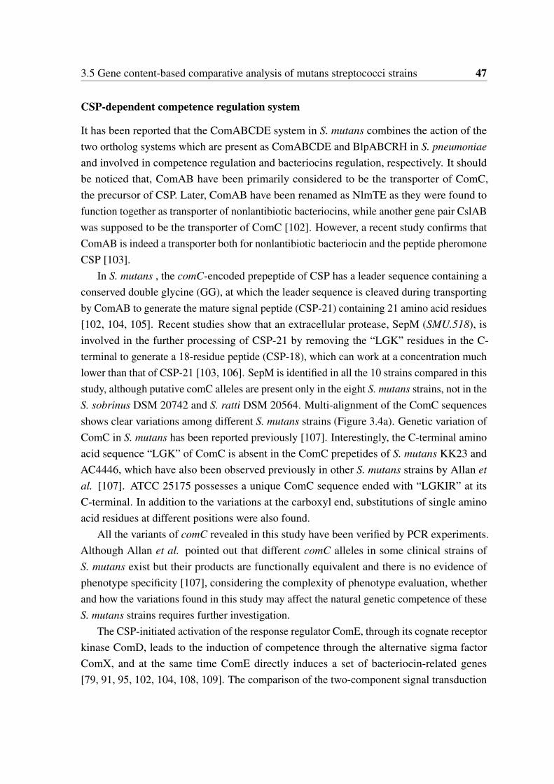
3.5 Gene content-based comparative analysis of mutans streptococci strains 47
CSP-dependent competence regulation system
It has been reported that the ComABCDE system in S. mutans combines the action of thetwo ortholog systems which are present as ComABCDE and BlpABCRH in S. pneumoniaeand involved in competence regulation and bacteriocins regulation, respectively. It shouldbe noticed that, ComAB have been primarily considered to be the transporter of ComC,the precursor of CSP. Later, ComAB have been renamed as NlmTE as they were found tofunction together as transporter of nonlantibiotic bacteriocins, while another gene pair CslABwas supposed to be the transporter of ComC [102]. However, a recent study confirms thatComAB is indeed a transporter both for nonlantibiotic bacteriocin and the peptide pheromoneCSP [103].
In S. mutans , the comC-encoded prepeptide of CSP has a leader sequence containing aconserved double glycine (GG), at which the leader sequence is cleaved during transportingby ComAB to generate the mature signal peptide (CSP-21) containing 21 amino acid residues[102, 104, 105]. Recent studies show that an extracellular protease, SepM (SMU.518), isinvolved in the further processing of CSP-21 by removing the “LGK” residues in the C-terminal to generate a 18-residue peptide (CSP-18), which can work at a concentration muchlower than that of CSP-21 [103, 106]. SepM is identified in all the 10 strains compared in thisstudy, although putative comC alleles are present only in the eight S. mutans strains, not in theS. sobrinus DSM 20742 and S. ratti DSM 20564. Multi-alignment of the ComC sequencesshows clear variations among different S. mutans strains (Figure 3.4a). Genetic variation ofComC in S. mutans has been reported previously [107]. Interestingly, the C-terminal aminoacid sequence “LGK” of ComC is absent in the ComC prepetides of S. mutans KK23 andAC4446, which have also been observed previously in other S. mutans strains by Allan etal. [107]. ATCC 25175 possesses a unique ComC sequence ended with “LGKIR” at itsC-terminal. In addition to the variations at the carboxyl end, substitutions of single aminoacid residues at different positions were also found.
All the variants of comC revealed in this study have been verified by PCR experiments.Although Allan et al. pointed out that different comC alleles in some clinical strains ofS. mutans exist but their products are functionally equivalent and there is no evidence ofphenotype specificity [107], considering the complexity of phenotype evaluation, whetherand how the variations found in this study may affect the natural genetic competence of theseS. mutans strains requires further investigation.
The CSP-initiated activation of the response regulator ComE, through its cognate receptorkinase ComD, leads to the induction of competence through the alternative sigma factorComX, and at the same time ComE directly induces a set of bacteriocin-related genes[79, 91, 95, 102, 104, 108, 109]. The comparison of the two-component signal transduction
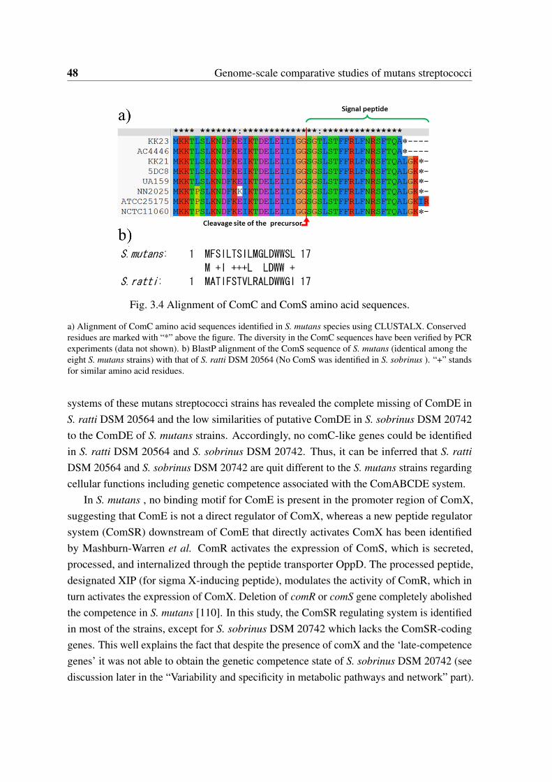
48 Genome-scale comparative studies of mutans streptococci
Fig. 3.4 Alignment of ComC and ComS amino acid sequences.
a) Alignment of ComC amino acid sequences identified in S. mutans species using CLUSTALX. Conservedresidues are marked with “*” above the figure. The diversity in the ComC sequences have been verified by PCRexperiments (data not shown). b) BlastP alignment of the ComS sequence of S. mutans (identical among theeight S. mutans strains) with that of S. ratti DSM 20564 (No ComS was identified in S. sobrinus ). “+” standsfor similar amino acid residues.
systems of these mutans streptococci strains has revealed the complete missing of ComDE inS. ratti DSM 20564 and the low similarities of putative ComDE in S. sobrinus DSM 20742to the ComDE of S. mutans strains. Accordingly, no comC-like genes could be identifiedin S. ratti DSM 20564 and S. sobrinus DSM 20742. Thus, it can be inferred that S. rattiDSM 20564 and S. sobrinus DSM 20742 are quit different to the S. mutans strains regardingcellular functions including genetic competence associated with the ComABCDE system.
In S. mutans , no binding motif for ComE is present in the promoter region of ComX,suggesting that ComE is not a direct regulator of ComX, whereas a new peptide regulatorsystem (ComSR) downstream of ComE that directly activates ComX has been identifiedby Mashburn-Warren et al. ComR activates the expression of ComS, which is secreted,processed, and internalized through the peptide transporter OppD. The processed peptide,designated XIP (for sigma X-inducing peptide), modulates the activity of ComR, which inturn activates the expression of ComX. Deletion of comR or comS gene completely abolishedthe competence in S. mutans [110]. In this study, the ComSR regulating system is identifiedin most of the strains, except for S. sobrinus DSM 20742 which lacks the ComSR-codinggenes. This well explains the fact that despite the presence of comX and the ‘late-competencegenes’ it was not able to obtain the genetic competence state of S. sobrinus DSM 20742 (seediscussion later in the “Variability and specificity in metabolic pathways and network” part).
3.5 Gene content-based comparative analysis of mutans streptococci strains 49
It is also worth to mention that the putative ComS ortholog found in S. ratti DSM 20564 isquite different to those of S. mutans strains, as shown in Figure 3.4b.
CSP-independent competence regulation system
It has been reported that a basal level of competence remains (referred as CSP-independentcompetence) after the deletion of comE from S. mutans , suggesting that the CSP-dependentregulation system is only one of the signaling pathways involved in ComX activation [91].Indeed, under conditions of biofilm growth the HdrMR system, a novel two-gene regulatorysystem, has been shown to contribute to competence development through the activation ofComX by a yet unknown signal [111]. Moreover, microarray analysis revealed that bothregulators, ComE and HdrR, activate a large set of overlapping genes [111, 112]. Recently,Xie et al. identified in S. mutans another regulatory system, designated BsrRM, that primarilyregulates bacteriocin-related genes but also affects the HdrMR system and thus indirectlycontributes to competence development [113]. In this study, HdrR, the response regulatorof the HdrMR system, is found neither present in S. ratti DSM 20564 nor in S. sobrinusDSM 20742. Furthermore, the response regulator BrsR of the BsrRM system is also absentin S. ratti DSM 20564, and S. sobrinus DSM 20742 lacks the complete BsrRM system.However, a competence damage-inducible protein CinA, which is regulated via ComX andhas been proven to be related to DNA damage, genetic transformation and cell survival [114],is present in all strains.
Taking together, both the CSP-dependent and CSP-independent competence regulationsystems in S. ratti DSM 20564 and especially in S. sobrinus DSM 20742 are very differentto those of the S. mutans strains.
3.5.3 Distribution of bacteriocin- and antibiotic resistance-related pro-teins
Bacteriocin-related proteins
Bacteriocins are proteinaceous toxins produced by bacteria to kill or inhibit the growth ofsimilar or closely related bacterial strain(s). Bacteriocins produced by mutans streptococciwere named “mutacins”. As dental plaque, the dominating niche of mutans streptococci, is amultispecies biofilm community that harbors many microorganism species, mutans groupstrains have developed a variety of mutacins to inhibit the growth of competitors, such as mitisgroup streptococci [103, 115, 116]. In this study, information about known mutacins as wellas mutacin-immunity proteins was collected from the NCBI (http://www.ncbi.nlm.nih.gov)
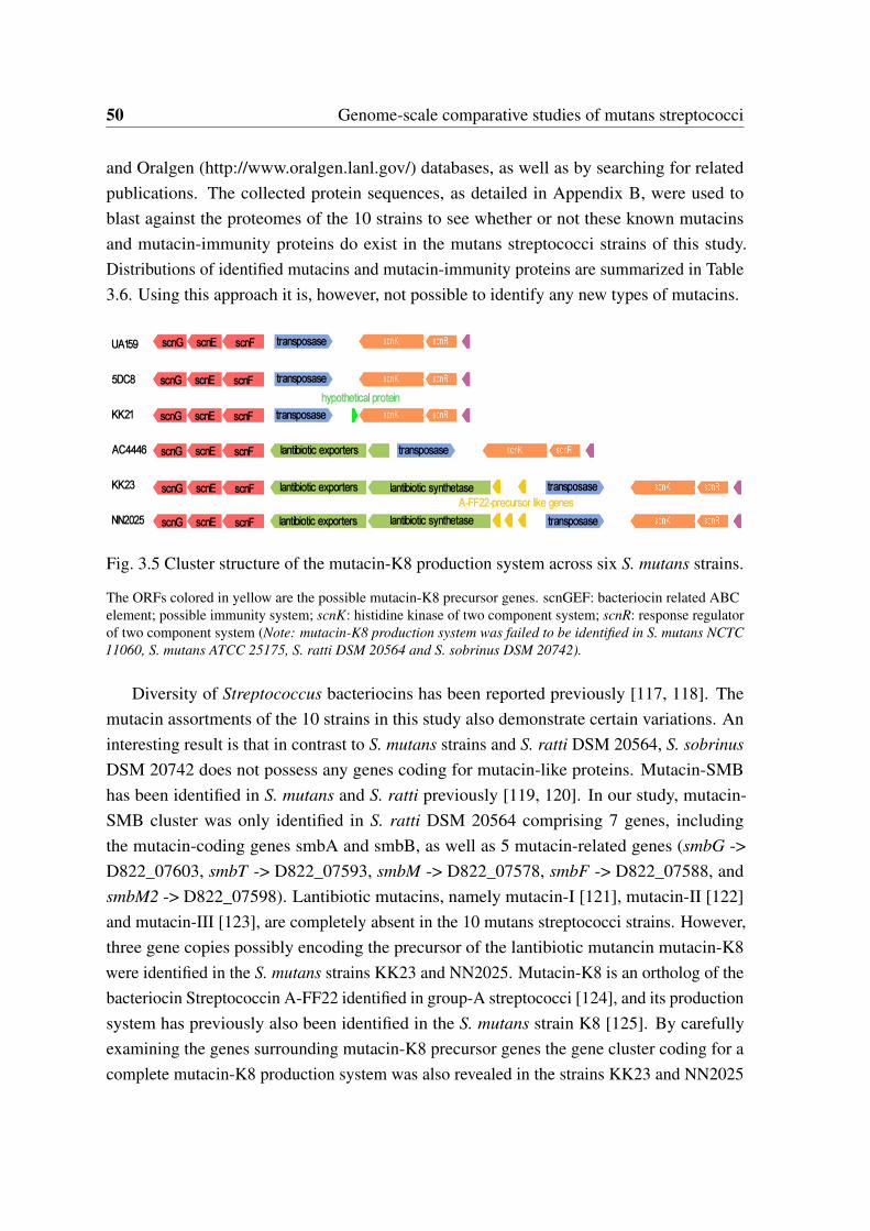
50 Genome-scale comparative studies of mutans streptococci
and Oralgen (http://www.oralgen.lanl.gov/) databases, as well as by searching for relatedpublications. The collected protein sequences, as detailed in Appendix B, were used toblast against the proteomes of the 10 strains to see whether or not these known mutacinsand mutacin-immunity proteins do exist in the mutans streptococci strains of this study.Distributions of identified mutacins and mutacin-immunity proteins are summarized in Table3.6. Using this approach it is, however, not possible to identify any new types of mutacins.
Fig. 3.5 Cluster structure of the mutacin-K8 production system across six S. mutans strains.
The ORFs colored in yellow are the possible mutacin-K8 precursor genes. scnGEF: bacteriocin related ABCelement; possible immunity system; scnK: histidine kinase of two component system; scnR: response regulatorof two component system (Note: mutacin-K8 production system was failed to be identified in S. mutans NCTC11060, S. mutans ATCC 25175, S. ratti DSM 20564 and S. sobrinus DSM 20742).
Diversity of Streptococcus bacteriocins has been reported previously [117, 118]. Themutacin assortments of the 10 strains in this study also demonstrate certain variations. Aninteresting result is that in contrast to S. mutans strains and S. ratti DSM 20564, S. sobrinusDSM 20742 does not possess any genes coding for mutacin-like proteins. Mutacin-SMBhas been identified in S. mutans and S. ratti previously [119, 120]. In our study, mutacin-SMB cluster was only identified in S. ratti DSM 20564 comprising 7 genes, includingthe mutacin-coding genes smbA and smbB, as well as 5 mutacin-related genes (smbG ->D822_07603, smbT -> D822_07593, smbM -> D822_07578, smbF -> D822_07588, andsmbM2 -> D822_07598). Lantibiotic mutacins, namely mutacin-I [121], mutacin-II [122]and mutacin-III [123], are completely absent in the 10 mutans streptococci strains. However,three gene copies possibly encoding the precursor of the lantibiotic mutancin mutacin-K8were identified in the S. mutans strains KK23 and NN2025. Mutacin-K8 is an ortholog of thebacteriocin Streptococcin A-FF22 identified in group-A streptococci [124], and its productionsystem has previously also been identified in the S. mutans strain K8 [125]. By carefullyexamining the genes surrounding mutacin-K8 precursor genes the gene cluster coding for acomplete mutacin-K8 production system was also revealed in the strains KK23 and NN2025
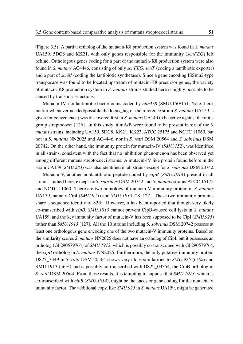
3.5 Gene content-based comparative analysis of mutans streptococci strains 51
(Figure 3.5). A partial ortholog of the mutacin-K8 production system was found in S. mutansUA159, 5DC8 and KK21, with only genes responsible for the immunity (scnFEG) leftbehind. Orthologous genes coding for a part of the mutacin-K8 production system were alsofound in S. mutans AC4446, consisting of only scnFEG, scnT (coding a lantibiotic exporter)and a part of scnM (coding the lantibiotic synthetase). Since a gene encoding ISSmu2-typetransposase was found to be located upstream of mutacin-K8 precursor genes, the varietyof mutacin-K8 production system in S. mutans strains studied here is highly possible to becaused by transposase actions.
Mutacin-IV, nonlantibiotic bacteriocins coded by nlmA/B (SMU.150/151, Note: here-inafter whenever needed/possible the locus_tag of the reference strain S. mutans UA159 isgiven for convenience) was discovered first in S. mutans UA140 to be active against the mitisgroup streptococci [126]. In this study, nlmA/B were found to be present in six of the S.mutans strains, including UA159, 5DC8, KK21, KK23, ATCC 25175 and NCTC 11060, butnot in S. mutans NN2025 and AC4446, nor in S. ratti DSM 20564 and S. sobrinus DSM20742. On the other hand, the immunity protein for mutacin-IV (SMU.152), was identifiedin all strains, consistent with the fact that no inhibition phenomenon has been observed yetamong different mutans streptococci strains. A mutacin-IV like protein found before in thestrain UA159 (SMU.283) was also identified in all strains except for S. sobrinus DSM 20742.
Mutacin-V, another nonlantibiotic peptide coded by cipB (SMU.1914) present in allstrains studied here, except forS. sobrinus DSM 20742 and S. mutans strains ATCC 15175and NCTC 11060. There are two homologs of mutacin-V immunity protein in S. mutansUA159, namely CipI (SMU.925) and SMU.1913 [28, 127]. These two immunity proteinsshare a sequence identity of 82%. However, it has been reported that though very likelyco-transcribed with cipB, SMU.1913 cannot prevent CipB-caused cell lysis in S. mutansUA159, and the key immunity factor of mutacin-V has been supposed to be CipI (SMU.925)rather than SMU.1913 [127]. All the 10 strains including S. sobrinus DSM 20742 possess atleast one orthologous gene encoding one of the two mutacin-V immunity proteins. Based onthe similarity scores S. mutans NN2025 does not have an ortholog of CipI, but it possesses anortholog (GI|290579764) of SMU.1913, which is possibly co-transcribed with GI|290579764,the cipB ortholog in S. mutans NN2025. Furthermore, the only putative immunity proteinD822_3349 in S. ratti DSM 20564 shows very close similarities to SMU.925 (61%) andSMU.1913 (56%) and is possibly co-transcribed with D822_03354, the CipB ortholog inS. ratti DSM 20564. From these results, it is tempting to suppose that SMU.1913, which isco-transcribed with cipB (SMU.1914), might be the ancestor gene coding for the mutacin-Vimmunity factor. The additional copy, like SMU.925 in S. mutans UA159, might be generated
52 Genome-scale comparative studies of mutans streptococci
by duplication action and evolved as the dominant immunity factor in some of the mutansstreptococci strains.
Furthermore, a possible nonlantibiotic bacteriocin peptide (SMU.423) is found to bepresent in all strains except for S. ratti DSM 20564. Putative ComAB, which has beenproved to be the transporter complex of mutacin IV in S. mutans [102], were identified inall strains, supporting the suggestion that ComAB might function as a common transporterfor multi-type nonlantibiotic bacteriocins rather than merely for mutacin IV. Moreover, anadditional paralog of ComA is present in most of the strains except for S. mutans KK23 andS. mutans ATCC 25175.
To summarize, a differed distribution of mutacin/bacteriocin encoding genes accompaniedwith a high conservation of genes coding for mutacin-immunity proteins were revealed forthe 10 mutans streptococci strains/species. The conservation of mutacin immunity proteinsapparently plays an important role in the survival of mutans streptococci strains under abacteriocin-rich environment.
3.5G
enecontent-based
comparative
analysisofm
utansstreptococcistrains
53
Table 3.6 Distribution of mutacins and mutacin immunity proteins in the 10 mutans streptococci strains
UA159 NN2025 5DC8 KK21 KK23 AC4446 ATCC NCTC DSM DSM25175 11060 20564 20742
Mutacin-SMB - - - - - - - - D822_07608 -(lantibiotic mutacin) D822_07613Mutacin-I - - - - - - - - - -(lantibiotic mutacin)Mutacin-II - - - - - - - - - -(lantibiotic mutacin)Mutacin-III - - - - - - - - - -(lantibiotic mutacin)Mutacin-K8 - GI|290579849 - - D818_07928 - - - - -(lantibiotic mutacin) GI|290579848 D818_07933
GI|290579850 D818_07938Mutacin-IV (NlmA) SMU.150 - D816_00655 D817_00675 D818_00659 - D820_00642 D821_00661 - -Mutacin-IV (NlmB) SMU.151 - D816_00660 D817_00680 D818_00664 - D820_00647 D821_00666 - -Mutacin-IV like SMU.283 GI|290581209 D816_01135 D817_01285 D818_01099 D819_01148 D820_01321 D821_01193 D822_03404 -Mutacin-IV* SMU.152 GI|290580110 D816_06832 D817_06998 D818_06649 D819_06637 D820_06850 D821_06847 D822_03264 D823_04636Mutacin-V (CipB) SMU.1914c GI|290579763 D816_08583 D817_08773 D818_08363 D819_07834 - - D822_03354 -CipI (Mutacin-V) * SMU.925 D816_04020 D817_04283 D818_04522 D819_04119 D820_04232 D821_04089 D822_03349Homolog of CipI SMU.1913c GI|290579764 D816_08578 D817_08768 D818_08358 D819_07829 D823_03992
SMU.423 GI|290581063 D816_01775 D817_01930 D818_01847 D819_01823 D820_01975 D821_01862 D823_05348NlmT/ComA SMU.286 GI|290581206 D816_01150 D817_01300 D818_01134 D819_01163 D820_01336 D821_01208 D822_01584 D823_05343
SMU.1881c GI|290579788 D816_08453 D817_08643 - D819_07724 - D821_08449 D822_08325 D823_01400NlmE/ComB SMU.287 GI|290581205 D816_01155 D817_01305 D818_01139 D819_01168 D820_01341 D821_01213 D822_01589 D823_05923
Note: as a multi-function exporter, the entries of NlmTE(ComAB) have been shown in Table 3.5 and here again. * stands for a immunity protein
54 Genome-scale comparative studies of mutans streptococci
Antibiotic resistance-related proteins
Bacteria and other microorganisms that cause infections are remarkably resilient and candevelop ways to survive drugs meant to kill or weaken them. Antibiotic resistance can be aresult of horizontal gene transfer [128], and also of unlinked point mutations in the pathogengenome at a rate of about 1 in 108 per chromosomal replication [129]. The antibiotic actionagainst the pathogen can be seen as an environmental selective pressure and bacteria whichhave developed mutations allowing them to survive will live on to reproduce. They will thenpass this trait to their offsprings, which will result in the evolution of fully resistant colonies.In this study, putative antibiotic resistance-related genes were identified in the genomes ofthe ten mutans streptococci strains/species and are listed in Table 3.7.
The S. mutans species is known to be intrinsically resistant to bacitracins produced byBacillus subtilis. This was also confirmed in this study by testing all the 10 strains with abacitracin-E-test (data not shown). All strains including S. ratti DSM 20564 and S. sobrinusDSM 20742 showed a minimum inhibitory concentration between 128 and 256 µg/l. In fact,bacitracins have been used to isolate mutans-streptococci from highly heterogeneous oralmicroflora. It has been reported that bceABRS (also named as mbrABCD) system, encoding atwo component signal transduction system and an ABC-transporter, is required for bacitracinresistance in S. mutans [130, 131]. As expected, ortholog of bceABRS system was found to bepresent in all strains of this study. Furthermore, an ortholog of a putative bacitracin resistantprotein UppP (SMU.244, undecaprenyl-diphosphatase) is also present in all strains. It hasbeen proved that overexpression of UppP in Escherichia coli and Bacillus subtilis results inbacitracin resistance [132, 133]. However, the function of UppP in bacitracin resistance inmutans streptococci has not yet been investigated. Based on its conservation in all strainsstudied here, it is reasonable to suppose that UppP might play an important role in bacitracinresistance of mutans streptococci species as well.
Two penicillin-binding proteins (SMU.75 and SMU.455) were identified in all the tenstrains, indicating that they are potentially all susceptible to penicillin. Phenotypically allstrains were tested to be susceptible to penicillin (data not shown). On the other hand, all thestrains possess orthologs of SMU.368c, SMU.400, SMU.1444c and SMU.1515, which arehomologs to beta-lactamases (EC 3.5.2.6), as well as orthologs of two so called beta-lactamresistance factors (SMU.716, SMU.717). Thus, all the strains are potentially capable ofresistance against beta-lactam antibiotics. Orthologs of macrolide-efflux transporter proteins,as coded by GI|290581182 and GI|290581181 in S. mutans NN2025, were found to bealso present in S. mutans 5DC8 and S. mutans KK21. A vancomycin b-type resistance-associated protein (D822_01634) is uniquely present in S. ratti DSM 20564, althougha phenotypic testing showed as expected that S. ratti DSM 20564 was still susceptible

3.5 Gene content-based comparative analysis of mutans streptococci strains 55
to vancomycin. Furthermore, several putative multidrug resistance-associated proteins(SMU.745, SMU.1611c and SMU.905 except for SMU.1286c) were found to be present in allstrains.
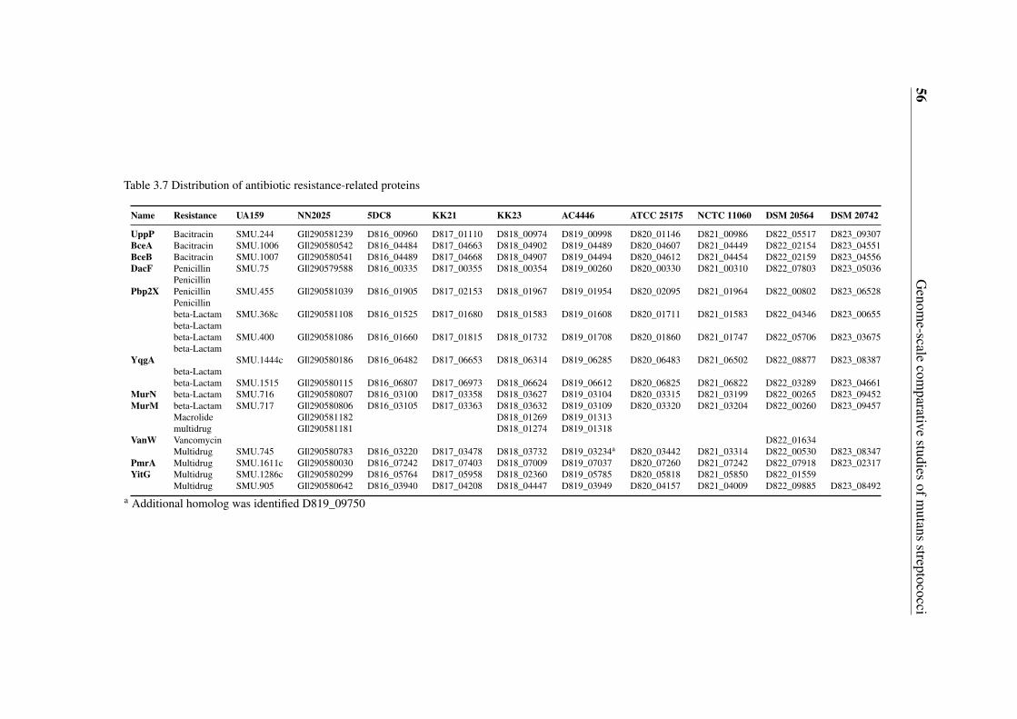
56G
enome-scale
comparative
studiesofm
utansstreptococci
Table 3.7 Distribution of antibiotic resistance-related proteins
Name Resistance UA159 NN2025 5DC8 KK21 KK23 AC4446 ATCC 25175 NCTC 11060 DSM 20564 DSM 20742
UppP Bacitracin SMU.244 GI|290581239 D816_00960 D817_01110 D818_00974 D819_00998 D820_01146 D821_00986 D822_05517 D823_09307BceA Bacitracin SMU.1006 GI|290580542 D816_04484 D817_04663 D818_04902 D819_04489 D820_04607 D821_04449 D822_02154 D823_04551BceB Bacitracin SMU.1007 GI|290580541 D816_04489 D817_04668 D818_04907 D819_04494 D820_04612 D821_04454 D822_02159 D823_04556DacF Penicillin SMU.75 GI|290579588 D816_00335 D817_00355 D818_00354 D819_00260 D820_00330 D821_00310 D822_07803 D823_05036
PenicillinPbp2X Penicillin SMU.455 GI|290581039 D816_01905 D817_02153 D818_01967 D819_01954 D820_02095 D821_01964 D822_00802 D823_06528
Penicillinbeta-Lactam SMU.368c GI|290581108 D816_01525 D817_01680 D818_01583 D819_01608 D820_01711 D821_01583 D822_04346 D823_00655beta-Lactambeta-Lactam SMU.400 GI|290581086 D816_01660 D817_01815 D818_01732 D819_01708 D820_01860 D821_01747 D822_05706 D823_03675beta-Lactam
YqgA SMU.1444c GI|290580186 D816_06482 D817_06653 D818_06314 D819_06285 D820_06483 D821_06502 D822_08877 D823_08387beta-Lactambeta-Lactam SMU.1515 GI|290580115 D816_06807 D817_06973 D818_06624 D819_06612 D820_06825 D821_06822 D822_03289 D823_04661
MurN beta-Lactam SMU.716 GI|290580807 D816_03100 D817_03358 D818_03627 D819_03104 D820_03315 D821_03199 D822_00265 D823_09452MurM beta-Lactam SMU.717 GI|290580806 D816_03105 D817_03363 D818_03632 D819_03109 D820_03320 D821_03204 D822_00260 D823_09457
Macrolide GI|290581182 D818_01269 D819_01313multidrug GI|290581181 D818_01274 D819_01318
VanW Vancomycin D822_01634Multidrug SMU.745 GI|290580783 D816_03220 D817_03478 D818_03732 D819_03234a D820_03442 D821_03314 D822_00530 D823_08347
PmrA Multidrug SMU.1611c GI|290580030 D816_07242 D817_07403 D818_07009 D819_07037 D820_07260 D821_07242 D822_07918 D823_02317YitG Multidrug SMU.1286c GI|290580299 D816_05764 D817_05958 D818_02360 D819_05785 D820_05818 D821_05850 D822_01559
Multidrug SMU.905 GI|290580642 D816_03940 D817_04208 D818_04447 D819_03949 D820_04157 D821_04009 D822_09885 D823_08492a Additional homolog was identified D819_09750
3.5 Gene content-based comparative analysis of mutans streptococci strains 57
3.5.4 Oxidative stress defense systems in mutans streptococci
For protection against reactive oxygen species (such as O2-, H2O2, HO·) or adaptation to
oxidative stresses aerobes and facultative anaerobes have evolved efficient defense systems,comprising an array of antioxidant enzymes such as catalase, superoxide dismutase (SOD),Dps-like peroxide resistance protein, alkylhydroperoxide reductase (AhpCF), glutathionereductase, and thiol reductase, which have been identified in many bacterial species.
Although the first genome sequence of S. mutans UA159 has already been publishedin 2002, the oxidative stress defense systems in the group of mutans streptococci havenot yet been systematically discussed. By searching for known antioxidant systems in thegenomes of the sequenced mutans streptococci strains of this study, an overview of putativeoxidative defense systems in these mutans streptococci strains/species was obtained, whichare composed of superoxide dismutase (SOD), AhpF/AhpC system, Dpr, thioredoxin systemand glutaredoxin system, as shown in Table 3.8.
SOD, which catalyzes the dismutation of superoxide into oxygen and hydrogen peroxide,is an important antioxidant defense in nearly all cells exposed to oxygen [134]. SOD wasfound in all strains of this study. Catalase, which catalyzes the decomposition of hydrogenperoxide, was not found in any of the mutans streptococci strains of this study. It is knownthat although most streptococci can grow in the presence of air, they do not possess acatalase, implying that hydrogen peroxide defense mechanism, by which lactic acid bacteriaestablished their growth in air, are very different to those of aerobes. It has been reportedthat both the bi-component peroxidase system AhpF/AhpC and Dps-like peroxide resistanceprotein confer tolerance to oxidative stress in S. mutans [135].
The AhpF/AhpC system catalyzes the NADH-dependent reduction of organic hydroper-oxides and/or H2O2 to their respective alcohol and/or H2O. Both AhpF and AhpC are presentin all S. mutans strains of this study and in S. ratti DSM 20564, but are absent in S. sobrinusDSM 20742. The natural missing of AhpF and AhpC in S. sobrinus indicates that Ah-pF/AhpC system is not an essential peroxide tolerance system for some mutans streptococcispecies. While studying a ahpF and ahpC double deletion mutant of S. mutans , Higuchi etal. [136] found that the mutant still showed the same level of peroxide tolerance as did thewild-type strain that led them to the finding of the dpr gene, which encodes a ferritin-likeiron-binding protein involved in oxygen tolerance by limiting the nonenzymatic hydroxylradical synthesis via iron-catalyzed ‘Fenton reaction’ in S. mutans . Their further studies onthe biological function of dpr found that dpr gene from S. mutans chromosome was capableof complementing an alkyl hydroperoxide reductase-deficient mutant of E. coli, as well ascomplementing the defect in peroxidase activity caused by the deletion of ahpF/ahpC in S.mutans , indicating that dpr plays an indispensable role in oxygen tolerance of S. mutans
58 Genome-scale comparative studies of mutans streptococci
[135, 137]. Dpr homologs were found in all strains of this study as expected by the supposedessential function of dpr gene in oxygen tolerance.
Thioredoxins are a class of small redox mediator proteins known to be present in allorganisms. They are involved in many important biological processes, including redoxsignaling. Thioredoxins are kept in the reduced state by the flavor enzyme thioredoxinreductase in a NADPH-dependent reaction [138]. They act as electron donors to manyproteins including thiol peroxidases [139]. Thioredoxin, thioredoxin reductase and thiolperoxidase, the components of thioredoxin system, were identified in all the strains of thisstudy. Two putative thioredoxin reductases (SMU.463 and SMU.869) were found in allstrains/species. It has been reported that in some species thioredoxin reductases have beenevolved to be activated by both NADPH and NADH [140]. Since SMU.463 and SMU.869shares less than 20% similarities, it is reasonable to speculate that SMU.463 and SMU.869might have been evolved to have different preferences to NADPH and NADH. If it holdstrue, this could be advantageous for these mutans streptococci, as the extra amount of NADHproduced from glycolysis/gluconeogenesis pathway under anaerobic conditions could bedirectly used for oxidative stress resistance. Thioredoxin (SMU.1869) and two thioredoxinfamily proteins (SMU.1971c and SMU.1169c) were found to be present in nearly all strains,except for S. sobrinus DSM 20742, which lacks any ortholog of SMU1169c. An ortholog ofa thiol peroxidase-coding gene (tpx) was identified in all strains.
Glutaredoxins share many functions of thioredoxins but are reduced by glutathione (L-γ-glutamyl-L-cysteinylglycine, GSH) rather than by a specific reductase. This means thatglutaredoxins are oxidized by their corresponding substrates, and reduced non-enzymaticallyby GSH [141]. Oxidized glutathione (GSSG) is then regenerated by glutathione reductase. To-gether, these components comprise the glutathione system [142]. GSH is a well-characterizedantioxidant in eukaryotes and Gram-negative bacteria, where it is synthesized by the se-quential action of two enzymes, γ-glutamylcysteine synthetase (γ-GCS) and glutathionesynthetase (GS). Among Gram-positive bacteria only a few species contain GSH. It hasbeen reported that streptococci lack the moderate-to-high levels of intracellular glutathionenormally found in Gram-negative bacteria [143]. Using Streptococcus agalactiae as a model,it has been discovered that in GSH-containing Gram-positive bacteria GSH synthesis iscatalyzed by one bifunctional protein, γ-glutamylcysteine synthetase-glutathione synthetase(γ-GCS-GS), encoded by one gene, gshAB. Homologs of γ-GCS-GS have been identified inthe genomes of 19 mostly studied Gram-positive bacteria, including S. mutans [144]. Allcomponents of the glutathione system were identified in all the 10 strains of this study. Sev-eral S. mutans strains, namely UA159, 5DC8, KK21, KK23, ATCC 25175, and NCTC 11060,as well as S. ratti DSM 20564, possess two glutathione reductase orthologs (SMU.140 and

3.5 Gene content-based comparative analysis of mutans streptococci strains 59
SMU.838). This could possibly convey these strains certain advantages in the re-generationof GSH from GSSG, which in turn would be helpful for oxidative resistance.
In addition, 3’-phosphoadenosine-5’-phosphate phosphatase activity has recently beenreported to be required for superoxide stress tolerance in S. mutans [145]. Putative 3’-phosphoadenosine-5’-phosphate phosphatase coding genes were identified in all strains ofthis study as well (Table 3.8).
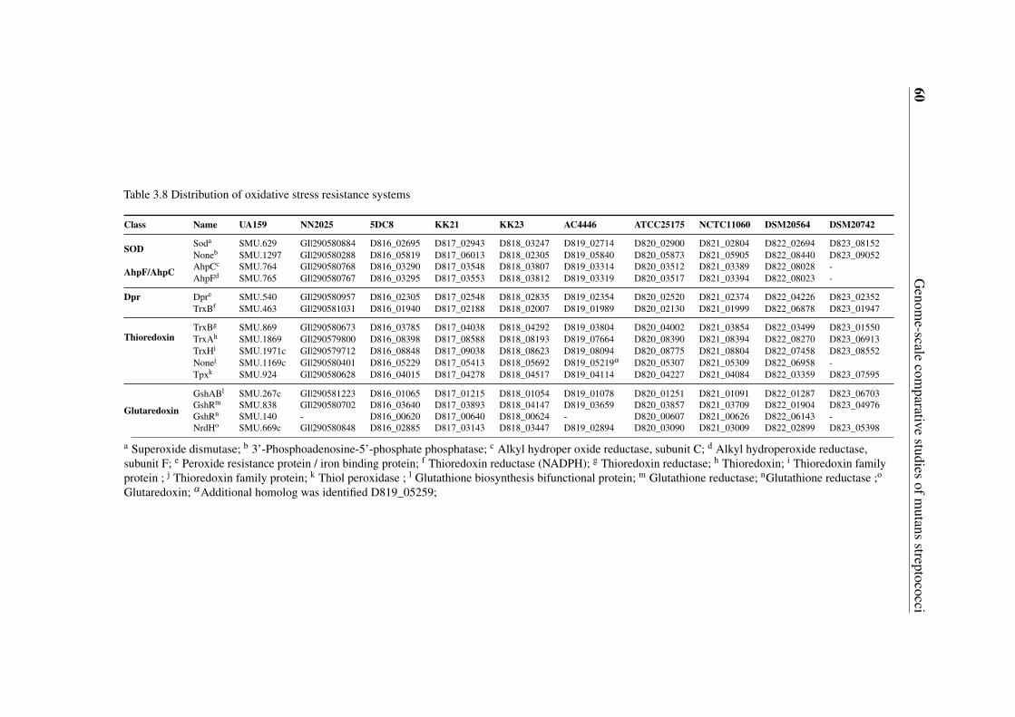
60G
enome-scale
comparative
studiesofm
utansstreptococci
Table 3.8 Distribution of oxidative stress resistance systems
Class Name UA159 NN2025 5DC8 KK21 KK23 AC4446 ATCC25175 NCTC11060 DSM20564 DSM20742
SOD Soda SMU.629 GI|290580884 D816_02695 D817_02943 D818_03247 D819_02714 D820_02900 D821_02804 D822_02694 D823_08152Noneb SMU.1297 GI|290580288 D816_05819 D817_06013 D818_02305 D819_05840 D820_05873 D821_05905 D822_08440 D823_09052
AhpF/AhpC AhpCc SMU.764 GI|290580768 D816_03290 D817_03548 D818_03807 D819_03314 D820_03512 D821_03389 D822_08028 -AhpFd SMU.765 GI|290580767 D816_03295 D817_03553 D818_03812 D819_03319 D820_03517 D821_03394 D822_08023 -
Dpr Dpre SMU.540 GI|290580957 D816_02305 D817_02548 D818_02835 D819_02354 D820_02520 D821_02374 D822_04226 D823_02352
Thioredoxin
TrxBf SMU.463 GI|290581031 D816_01940 D817_02188 D818_02007 D819_01989 D820_02130 D821_01999 D822_06878 D823_01947
TrxBg SMU.869 GI|290580673 D816_03785 D817_04038 D818_04292 D819_03804 D820_04002 D821_03854 D822_03499 D823_01550TrxAh SMU.1869 GI|290579800 D816_08398 D817_08588 D818_08193 D819_07664 D820_08390 D821_08394 D822_08270 D823_06913TrxHi SMU.1971c GI|290579712 D816_08848 D817_09038 D818_08623 D819_08094 D820_08775 D821_08804 D822_07458 D823_08552Nonej SMU.1169c GI|290580401 D816_05229 D817_05413 D818_05692 D819_05219α D820_05307 D821_05309 D822_06958 -Tpxk SMU.924 GI|290580628 D816_04015 D817_04278 D818_04517 D819_04114 D820_04227 D821_04084 D822_03359 D823_07595
Glutaredoxin
GshABl SMU.267c GI|290581223 D816_01065 D817_01215 D818_01054 D819_01078 D820_01251 D821_01091 D822_01287 D823_06703GshRm SMU.838 GI|290580702 D816_03640 D817_03893 D818_04147 D819_03659 D820_03857 D821_03709 D822_01904 D823_04976GshRn SMU.140 - D816_00620 D817_00640 D818_00624 - D820_00607 D821_00626 D822_06143 -NrdHo SMU.669c GI|290580848 D816_02885 D817_03143 D818_03447 D819_02894 D820_03090 D821_03009 D822_02899 D823_05398
a Superoxide dismutase; b 3’-Phosphoadenosine-5’-phosphate phosphatase; c Alkyl hydroper oxide reductase, subunit C; d Alkyl hydroperoxide reductase,subunit F; e Peroxide resistance protein / iron binding protein; f Thioredoxin reductase (NADPH); g Thioredoxin reductase; h Thioredoxin; i Thioredoxin familyprotein ; j Thioredoxin family protein; k Thiol peroxidase ; l Glutathione biosynthesis bifunctional protein; m Glutathione reductase; nGlutathione reductase ;o
Glutaredoxin; α Additional homolog was identified D819_05259;
3.6 Metabolic network construction and analysis 61
3.6 Metabolic network construction and analysis
3.6.1 Genome-scale metabolic network reconstruction
In order to systematically reveal the metabolic variability of the mutans streptococci in thisstudy, the genome-scale metabolic networks of all the strains sequenced were reconstructedand analyzed according to the method proposed by Ma and Zeng [43, 44]. All annotatedprotein sequences having EC numbers were considered for the network reconstruction. Fromthe functional annotation discussed in chapter 3.2, total EC numbers identified in the 10strains are very close to each other. A summary of the total numbers of reactions andmetabolites in each of the reconstructed metabolic networks is shown in Table 3.9, and all theconstructed metabolic networks can be found in an online filevi in *.cys format which can beopened with Cytoscape [45], a software for visualization and analysis of biological networks.The sizes of the constructed metabolic networks of the eight S. mutans strains are very closeto each other, with UA159, NN2025, AC4446, 5DC8 and KK21 having almost exactly thesame size, and the networks of KK23, ATCC 25175 and NCTC 11060 being merely about2% larger. While the size of the metabolic network of S. ratti DSM 20564 is comparableto those of the S. mutans strains, the metabolic network of S. sobrinus with 833 reactionsand 853 metabolites is the smallest one, which have 62 less reactions and 60 less metabolitescompared to the largest one of S. mutans NCTC 11060 (895 reactions and 913 metabolites).
Table 3.9 Compositions of the established metabolic networks of the 10 mutans streptococci strains
Strain EC Numbers Reactions Metabolites
S. mutans UA159 454 875 893S. mutans NN2025 450 874 892S. mutans 5DC8 453 875 893S. mutans KK21 453 875 893S. mutans KK23 452 893 911S. mutans AC4446 449 874 893S. mutans ATCC 25175 453 891 911S. mutans NCTC 11060 456 895 913S. ratti DSM20564 435 888 893S. sobrinus DSM20742 434 833 853
vihttps://www.ncbi.nlm.nih.gov/pmc/articles/PMC3751929/bin/1471-2164-14-430-S6.cys
62 Genome-scale comparative studies of mutans streptococci
3.6.2 Variability and specificity in metabolic pathways and network
Despite the comparable network sizes, however, all the strains possess or lack certainreactions/metabolites, as revealed by detailed comparative analyses. Using the metabolicnetwork of S. mutans UA159 as reference, the presence and absence of reactions in eachof the strains/species compared were discovered and mapped into sub-pathways based onthe KEGG pathway classification (http://www.genome.jp/kegg/pathway.html). As the result,among the 416 sub-pathways defined in the KEGG pathway database certain variationsbetween the strains/species were found in 46 sub-pathways(Please refer to an online file fordetailsvii).
A key feature of the oral environment is that the nutrients available to the oral bacteriaare always fluctuating between abundance and famine associated with human diet. Thus, theability to quickly acquire and metabolize carbohydrates to produce energy and precursorsfor biosynthesis is essential for the survival of all oral bacteria. Due to their key roles incarbohydrates metabolism and energy production, glycolysis/gluconeogenesis, TCA cycleand pyruvate metabolism pathways are generally considered to be highly conserved amongthese oral bacteria. Although mutans streptococci strains/species are closely related speciesas revealed by phylogenetic tree analysis in this study (Figure 3.1), differences in thesecentral carbon metabolic pathways were found as shown in Figure 3.6.
Facultative anaerobes such as lactic acid bacteria including Streptococcus lack cy-tochrome oxidases required for energy-linked oxygen metabolism. Energy (in the formof ATP) required for survival and growth are generated by substrate level phosphorylationin the glycolysis pathway [136]. L-lactate oxidase (D823_06598) with a similarity of 73%to YP_003064450.1 (accession number) of Lactobacillus plantarum JDM1 and lactate ox-idase (D823_06595) with a similarity of 65% to ZP_09448656.1 (accession number) ofLactobacillus mali KCTC 3596, were found to be uniquely present in S. sobrinus DSM20742. These two enzymes catalyze the reaction of L-Lactate + O2 => Pyruvate + H2O2
and/or D-Lactate + O2= > Pyruvate + H2O2. It has been reported that in S. pneumoniaconcerted action of lactate oxidase and pyruvate oxidase forms a novel energy-generationpathway by converting lactate acid to acetic acid under aerobic growth conditions [146].Because no pyruvate oxidase could be identified in S. sobrinus DSM 20742, the function ofthe lactate oxidases in S. sobrinus DSM 20742 should be different to that of S. pneumoniae.By a close examination, it is reasonable to hypothesize that lactate oxidase, together withpyruvate dehydrogenase, phosphate acetyl transferase and acetate kinase, could form a novelenergy production pathway to convert lactate acid to acetate and simultaneously produce oneadditional ATP, as depicted in Figure 3.7. By doing so, the lactate oxidases of S. sobrinus
viihttps://www.ncbi.nlm.nih.gov/pmc/articles/PMC3751929/bin/1471-2164-14-430-S7.docx
3.6 Metabolic network construction and analysis 63
DSM 20742 could also play a role in consuming lactate to regulate pH, which would be anadvantage for S. sobrinus DSM 20742 in resistance to acid stress. In addition, this pathwaycould replenish acetyl-CoA, an important intermediate for the biosynthesis of fatty acids andamino acids. This is for the first time that such an energy production pathway is proposed inStreptococcus species. Furthermore, lactate oxidase and lactate dehydrogenase could forma local NAD+ regeneration system, which would be certainly advantageous to S. sobrinusDSM 20742 under aerobic growth conditions. Moreover, it is known that mutans groupstreptococci and the mitis group streptococci are competitors, with S. mutans producingmutacins to kill the mitis group streptococci and the mitis group streptococci in turn produceH2O2 to kill mutans group streptococci [67, 147]. Favored by possessing the lactate oxidases,S. sobrinus DSM 20742 has the potential ability of producing H2O2 to kill not only competi-tors (oxygen sensitive S. mutans, oral anaerobes) but also macrophages [148], and therebydefend its ecological niche. The unique presence of lactate oxidases in S. sobrinus DSM20742 was verified by PCR experiments (Please refer to an online file for details viii). Later,another S. sobrinus strain AC153 was also found to harbor homologous genes of lactateoxidase, suggesting that lactate oxidase may be conserved and play an important role in S.sobrinus. In the effort to clarify the functionality of lactate oxidase it was tried to knockout the two genes encoding the two enzymes by PCR ligation mutagenesis according to themethod of Lau PC et al. (2002). Although different transformation methods (two naturaltransformation methods and two electroporation methods) were applied, but it was failed toobtain the desired recombinants. Then, to find out if S. sobrinus DSM 20742 is in the leastable to enter genetic competence state, transforming S. sobrinus with plasmids which arereplicative in other Streptococcus spp. like pDL278 (Spr, pAT18 Emr, with suicide vectorpFW5 Spr) in both circular and linearized forms were tested but no transformants could beobtained. Therefore, it is clear that the genetic competence behavior of S. sobrinus DSM20742 is very different to that of S. mutans, attributing very likely to the lacking of the genescomSR and comC.
In contrast to the unique harboring of lactate oxidases in S. sobrinus DSM 20742, citratelyase (EC 4.1.3.6), which catalyzes the cleavage of citrate into oxaloacetate and acetate,and oxaloacetate decarboxylase (EC 4.1.1.3), catalyzing the irreversible decarboxylationof oxaloacetate to pyruvate and CO2, are not present in S. sobrinus DSM 20742, as shownin Figure 3.7 by the blue dotted lines. It has been reported that citrate lyase functions asa key enzyme in initiating the anaerobic utilization of citrate by a number of bacteria, andthe further catabolism of oxaloacetate formed takes place either by decarboxylation or byreduction. In some organisms, oxaloacetate is decarboxylated to pyruvate by oxaloacetate
viiihttps://www.ncbi.nlm.nih.gov/pmc/articles/PMC3751929/bin/1471-2164-14-430-S8.docx
64 Genome-scale comparative studies of mutans streptococci
decarboxylase, which is also induced in the presence of citrate. The two enzymatic reactions,which occur sequentially, constitute the ‘citrate fermentation pathway’ [149]. The absence ofcitrate lyase and oxaloacetate decarboxylase implies that S. sobrinus DSM 20742 might lacksthe ability in anaerobic utilization of citrate as a substrate. However, the disadvantages of S.sobrinus DSM 20742 in citrate utilization could be offset by the novel energy productionpathway from lactate to acetate proposed above.
A putative pyruvate-phosphate dikinase (EC 2.7.9.1), which catalyzes the interconversionbetween PEP and pyruvate, was found to be uniquely present in S. ratti DSM 20564. Pyruvate-phosphate dikinase has been found in propionic acid bacteria [150] . The large difference inthe standard free energy of hydrolysis for ATP to AMP and pyrophosphate (-7.6 kcal/mole)and for PEP to pyruvate (-13.6 kcal/mole) at pH 7.0 indicates that the equilibrium for thereaction it catalyzes would strongly favor pyruvate formation. But studies in Acetobacterxylinum clearly indicate that the function of this enzyme under physiological conditionsfavors the process of gluconeogenesis [151]. Metabolite interconversion at the PEP-pyruvate-oxaloacetate node involves a structurally entangled set of reactions that interconnect the majorpathways of carbon metabolism and thus, is responsible for the distribution of the carbonflux among catabolism, anabolism and energy supply of the cell [152]. Under glycolyticconditions oxaloacetate is generated by carboxylation of PEP and/or pyruvate catalyzed byPEP carboxylase (PEPCx) and/or pyruvate carboxylase (PCx). In this study PCx was notfound in any of the mutans streptococci strains.
All the 10 strains of this study possess similarly an incomplete TCA cycle and the primaryrole of the existing TCA enzymes is most likely the synthesis of amino acid precursors ashas been reported previously [28, 153].
3.7 Construction of StrepReg - a regulation database of S.mutans
Detailed knowledge of all interactions between proteins/genes in a given cell would repre-sent an important milestone towards a comprehensive description of cellular mechanismsand functions. To enable a global view of all interactions in S. mutans, three sources offunctional information of genes/proteins were integrated into one database named StrepReg:1) the regulatory network constructed based on assigning predicted relationships betweentranscription factors (TFs) and target genes (TGs). This regulatory network is a “static” con-nection network consisting 1,785 TF-TG relationships corresponding to 32 regulons [154].
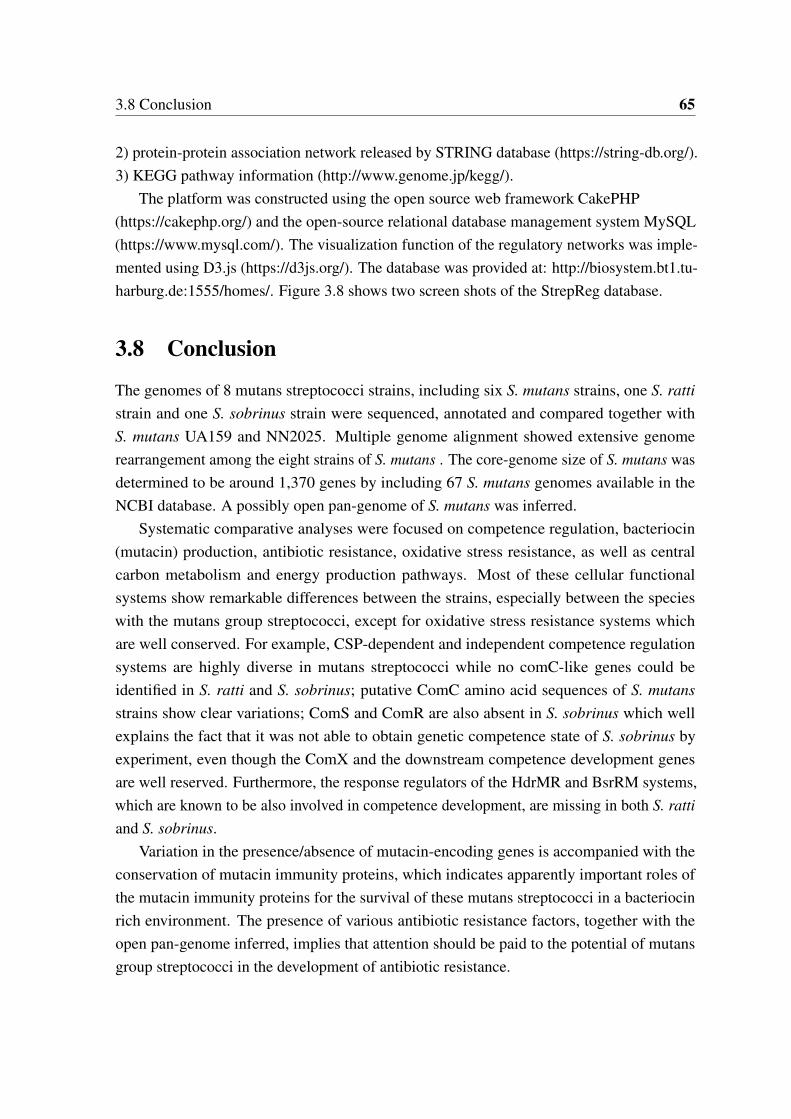
3.8 Conclusion 65
2) protein-protein association network released by STRING database (https://string-db.org/).3) KEGG pathway information (http://www.genome.jp/kegg/).
The platform was constructed using the open source web framework CakePHP(https://cakephp.org/) and the open-source relational database management system MySQL(https://www.mysql.com/). The visualization function of the regulatory networks was imple-mented using D3.js (https://d3js.org/). The database was provided at: http://biosystem.bt1.tu-harburg.de:1555/homes/. Figure 3.8 shows two screen shots of the StrepReg database.
3.8 Conclusion
The genomes of 8 mutans streptococci strains, including six S. mutans strains, one S. rattistrain and one S. sobrinus strain were sequenced, annotated and compared together withS. mutans UA159 and NN2025. Multiple genome alignment showed extensive genomerearrangement among the eight strains of S. mutans . The core-genome size of S. mutans wasdetermined to be around 1,370 genes by including 67 S. mutans genomes available in theNCBI database. A possibly open pan-genome of S. mutans was inferred.
Systematic comparative analyses were focused on competence regulation, bacteriocin(mutacin) production, antibiotic resistance, oxidative stress resistance, as well as centralcarbon metabolism and energy production pathways. Most of these cellular functionalsystems show remarkable differences between the strains, especially between the specieswith the mutans group streptococci, except for oxidative stress resistance systems whichare well conserved. For example, CSP-dependent and independent competence regulationsystems are highly diverse in mutans streptococci while no comC-like genes could beidentified in S. ratti and S. sobrinus; putative ComC amino acid sequences of S. mutansstrains show clear variations; ComS and ComR are also absent in S. sobrinus which wellexplains the fact that it was not able to obtain genetic competence state of S. sobrinus byexperiment, even though the ComX and the downstream competence development genesare well reserved. Furthermore, the response regulators of the HdrMR and BsrRM systems,which are known to be also involved in competence development, are missing in both S. rattiand S. sobrinus.
Variation in the presence/absence of mutacin-encoding genes is accompanied with theconservation of mutacin immunity proteins, which indicates apparently important roles ofthe mutacin immunity proteins for the survival of these mutans streptococci in a bacteriocinrich environment. The presence of various antibiotic resistance factors, together with theopen pan-genome inferred, implies that attention should be paid to the potential of mutansgroup streptococci in the development of antibiotic resistance.
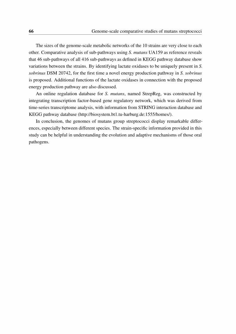
66 Genome-scale comparative studies of mutans streptococci
The sizes of the genome-scale metabolic networks of the 10 strains are very close to eachother. Comparative analysis of sub-pathways using S. mutans UA159 as reference revealsthat 46 sub-pathways of all 416 sub-pathways as defined in KEGG pathway database showvariations between the strains. By identifying lactate oxidases to be uniquely present in S.sobrinus DSM 20742, for the first time a novel energy production pathway in S. sobrinusis proposed. Additional functions of the lactate oxidases in connection with the proposedenergy production pathway are also discussed.
An online regulation database for S. mutans, named StrepReg, was constructed byintegrating transcription factor-based gene regulatory network, which was derived fromtime-series transcriptome analysis, with information from STRING interaction database andKEGG pathway database (http://biosystem.bt1.tu-harburg.de:1555/homes/).
In conclusion, the genomes of mutans group streptococci display remarkable differ-ences, especially between different species. The strain-specific information provided in thisstudy can be helpful in understanding the evolution and adaptive mechanisms of those oralpathogens.
3.8 Conclusion 67
Fig. 3.6 Example of visualized genome-scale metabolic networks constructed based ongenome annotations and KEGG pathway
The blue rectangle nodes represent the reactions and the circle green nodes represent the metabolites.
68 Genome-scale comparative studies of mutans streptococci
Fig. 3.7 Glycolysis/Gluconeogenesis and TCA cycle pathway in mutans streptococci
The rectangle nodes represent the metabolites. The yellow lines represent enzymes and the blue line representenzymes with diversities across mutans streptococci strains studied here. The yellow line with cross means thisenzyme is not present in all strains. Dotted blue line means this enzyme is absent in S. sobrinus DSM20742and solid blue line means this enzyme is uniquely present in S. sobrinus DSM20742. Malate dehydrogenaserepresented in green line was absent in S. mutans NN2025 and S. mutans AC4446.
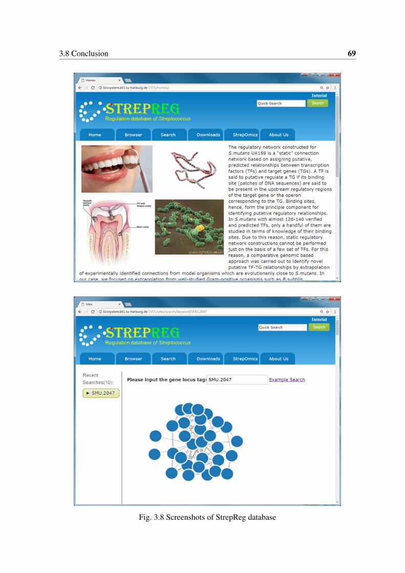
3.8 Conclusion 69
Fig. 3.8 Screenshots of StrepReg database

Chapter 4
Development of a multiple IO system forbiological engineering in E. coli
4.1 Introduction
Although cells are composed of molecules and their viability relies on extracting and usingenergy to maintain them, they are not ‘just’ matter and energy [155]. Cells can respondto their environment, make decisions, build structures, and coordinate tasks based on com-putational operations performed by networks of regulatory proteins that integrate signalsand control the timing of gene expression [155]. It has been shown that cells can be pro-grammed using synthetic genetic circuits composed of regulators organized to generatedesired operations [155–159]. Stimulated by the great potential of engineering biologicalsystems to achieve novel tasks, an emerging discipline termed synthetic biology is drawingmore and more attentions [160–184]. It focuses on designing and building novel biologicalfunctions and systems by combining science and engineering principles, including the designand construction of new biological parts, devices, and systems, as well as the re-design ofexisting, natural biological systems for useful purposes. In general, the overall process ofbiological engineering is similar to programming in computer science. However, unlikeprogramming on a computer, "programming" a biological system is much more time- andlabor-intensive. One reason is that changing the "biological codes" is much more difficultthan changing digital codes on a computer. It always takes days or even weeks to enablethe editing of the "genetic codes". Recently, this process has been greatly simplified by therecently emerged CRISPR/Cas9 based genetic editing tools [185–188]. Another more crucial
This chapter was a modified and extended version of a recent publication: Song, Lifu; Zeng, An-Ping(2017): Engineering ’cell robots’ for parallel and highly sensitive screening of biomolecules under in vivoconditions. Scientific Reports 7 (1), p. 15145.
72 Development of a multiple IO system for biological engineering in E. coli
reason is the inherent complexity and uncertainty of the genotype-phenotype relationshipsof biological systems. Despite the complicate interactions among the metabolic, gene regu-latory and signaling networks at the cellular level, it is not possible to precisely predict theconsequences of even a single base change at the single gene level. Hence, the biologicalengineering process is held by the time- and labor- intensive design–build–test cycles asshown in Figure 1.1, in which many designs have to be evaluated and iterated on in order toimprove the performance of target system. The rate of improvements is directly related tothe throughput and rounds of the design cycles, with higher throughputs and more roundsresulting in reduced development period. Although recent advances have enabled the designand construction of billions of genetic variants per day, but evaluation capacity is still limitedto thousands of variants per day.
Inspired by the debugging system in computer science, a versatile diagnosis system wasproposed to reduce the development burden for biological engineering. This was achieved bya novel multiple input-output (IO) system which can interact with the cells and output multiplesignals corresponding to various perturbations (inputs). Despite impressive progress insystems metabolic engineering and synthetic biology, there are still unsolved major problemsin their practical applications for developing effective microorganisms for biosynthesis,such as identification of relevant targets for pathway engineering, designed elements ordevices from synthetic biology often not working well inside cells under industrially relevantconditions. For proof of concept, the IO system used for target identification, evaluation ofdesigns, evolution and selection of key enzymes for bioproduction.
4.2 Principles of a multiple input-output system which caninteract with E. coli cells
The inputs here refer to operations/perturbations that can alter intracellular conditions.The concentrations of chemicals, e.g. IPTG, arabinose, have been widely used as inputsin previous studies. However, the availability of well-characterized chemicals is limitedand intracellular genetic parts have to be built in order to sense the desired signal andchange intracellular gene expression pattern accordingly. Here various DNA fragments wereproposed to be utilized as inputs directly. The DNA fragments with different functions canbe easily designed and they are different from each other inherently in their sequences. Thepossible inputs are unlimited in principle. The challenge is how to introduce these DNAfragments into the cells.
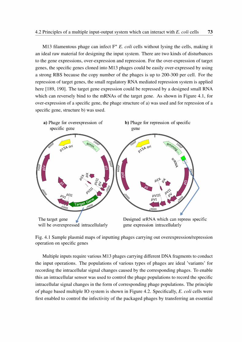
4.2 Principles of a multiple input-output system which can interact with E. coli cells 73
M13 filamentous phage can infect F+ E. coli cells without lysing the cells, making itan ideal raw material for designing the input system. There are two kinds of disturbancesto the gene expressions, over-expression and repression. For the over-expression of targetgenes, the specific genes cloned into M13 phages could be easily over-expressed by usinga strong RBS because the copy number of the phages is up to 200-300 per cell. For therepression of target genes, the small regulatory RNA mediated repression system is appliedhere [189, 190]. The target gene expression could be repressed by a designed small RNAwhich can reversely bind to the mRNAs of the target gene. As shown in Figure 4.1, forover-expression of a specific gene, the phage structure of a) was used and for repression of aspecific gene, structure b) was used.
Fig. 4.1 Sample plasmid maps of inputting phages carrying out overexpression/repressionoperation on specific genes
Multiple inputs require various M13 phages carrying different DNA fragments to conductthe input operations. The populations of various types of phages are ideal ’variants’ forrecording the intracellular signal changes caused by the corresponding phages. To enablethis an intracellular sensor was used to control the phage populations to record the specificintracellular signal changes in the form of corresponding phage populations. The principleof phage based multiple IO system is shown in Figure 4.2. Specifically, E. coli cells werefirst enabled to control the infectivity of the packaged phages by transferring an essential
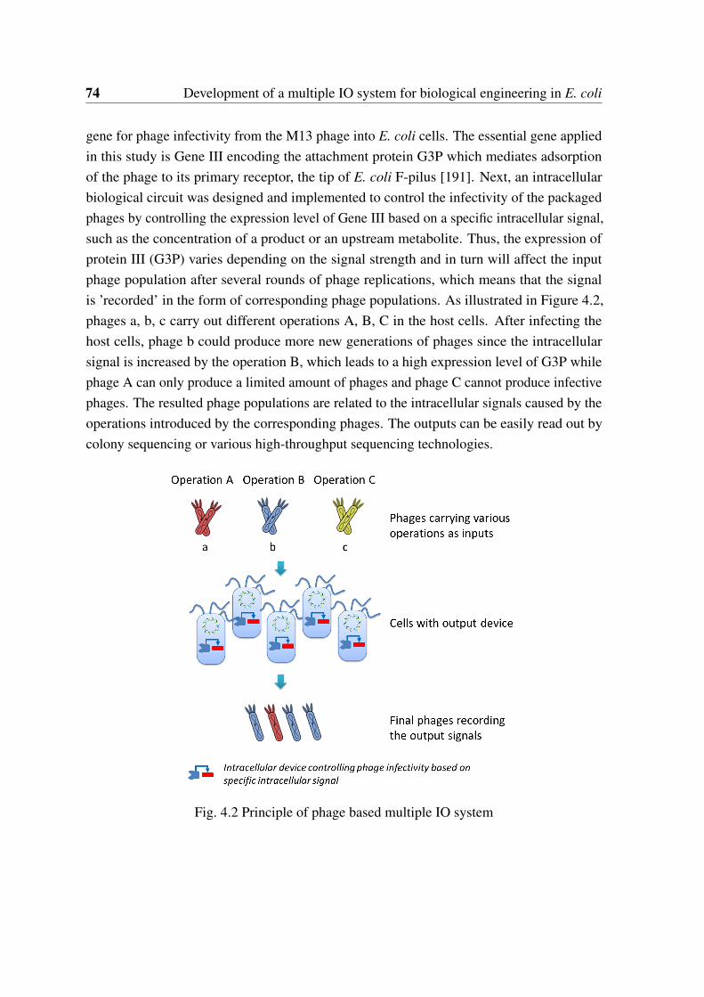
74 Development of a multiple IO system for biological engineering in E. coli
gene for phage infectivity from the M13 phage into E. coli cells. The essential gene appliedin this study is Gene III encoding the attachment protein G3P which mediates adsorptionof the phage to its primary receptor, the tip of E. coli F-pilus [191]. Next, an intracellularbiological circuit was designed and implemented to control the infectivity of the packagedphages by controlling the expression level of Gene III based on a specific intracellular signal,such as the concentration of a product or an upstream metabolite. Thus, the expression ofprotein III (G3P) varies depending on the signal strength and in turn will affect the inputphage population after several rounds of phage replications, which means that the signalis ’recorded’ in the form of corresponding phage populations. As illustrated in Figure 4.2,phages a, b, c carry out different operations A, B, C in the host cells. After infecting thehost cells, phage b could produce more new generations of phages since the intracellularsignal is increased by the operation B, which leads to a high expression level of G3P whilephage A can only produce a limited amount of phages and phage C cannot produce infectivephages. The resulted phage populations are related to the intracellular signals caused by theoperations introduced by the corresponding phages. The outputs can be easily read out bycolony sequencing or various high-throughput sequencing technologies.
Fig. 4.2 Principle of phage based multiple IO system
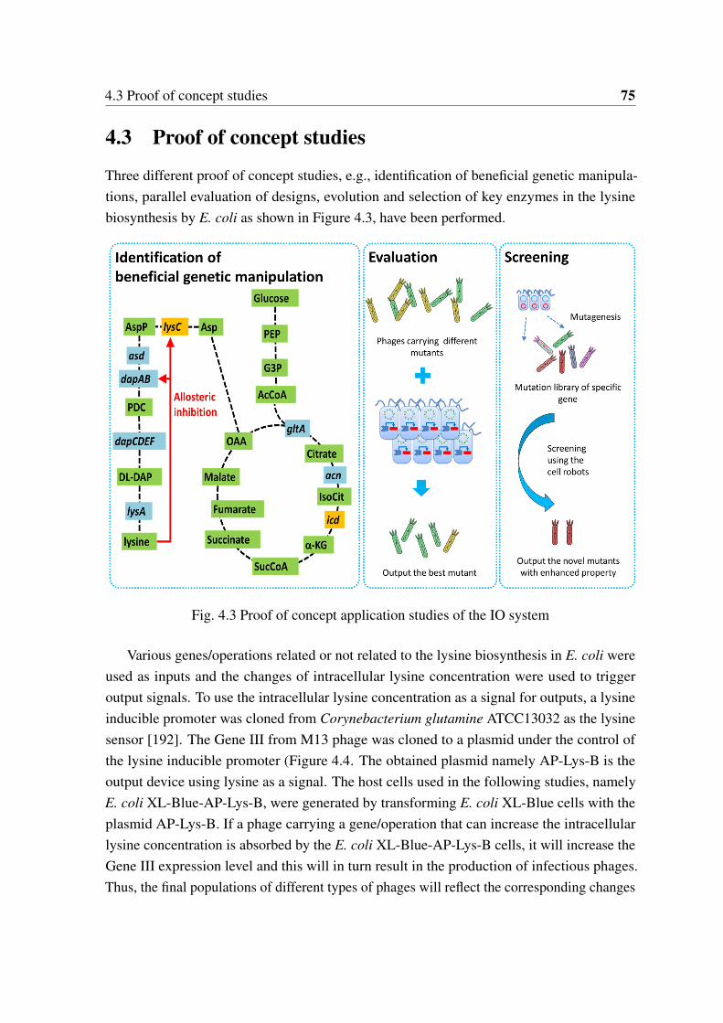
4.3 Proof of concept studies 75
4.3 Proof of concept studies
Three different proof of concept studies, e.g., identification of beneficial genetic manipula-tions, parallel evaluation of designs, evolution and selection of key enzymes in the lysinebiosynthesis by E. coli as shown in Figure 4.3, have been performed.
Fig. 4.3 Proof of concept application studies of the IO system
Various genes/operations related or not related to the lysine biosynthesis in E. coli wereused as inputs and the changes of intracellular lysine concentration were used to triggeroutput signals. To use the intracellular lysine concentration as a signal for outputs, a lysineinducible promoter was cloned from Corynebacterium glutamine ATCC13032 as the lysinesensor [192]. The Gene III from M13 phage was cloned to a plasmid under the control ofthe lysine inducible promoter (Figure 4.4. The obtained plasmid namely AP-Lys-B is theoutput device using lysine as a signal. The host cells used in the following studies, namelyE. coli XL-Blue-AP-Lys-B, were generated by transforming E. coli XL-Blue cells with theplasmid AP-Lys-B. If a phage carrying a gene/operation that can increase the intracellularlysine concentration is absorbed by the E. coli XL-Blue-AP-Lys-B cells, it will increase theGene III expression level and this will in turn result in the production of infectious phages.Thus, the final populations of different types of phages will reflect the corresponding changes
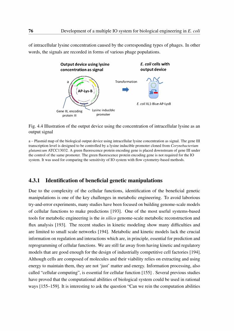
76 Development of a multiple IO system for biological engineering in E. coli
of intracellular lysine concentration caused by the corresponding types of phages. In otherwords, the signals are recorded in forms of various phage populations.
Fig. 4.4 Illustration of the output device using the concentration of intracellular lysine as anoutput signal
a – Plasmid map of the biological output device using intracellular lysine concentration as signal. The gene IIItranscription level is designed to be controlled by a lysine inducible promoter cloned from Corynebacteriumglutamicum ATCC13032. A green fluorescence protein encoding gene is placed downstream of gene III underthe control of the same promoter. The green fluorescence protein encoding gene is not required for the IOsystem. It was used for comparing the sensitivity of IO system with flow cytometry-based methods.
4.3.1 Identification of beneficial genetic manipulations
Due to the complexity of the cellular functions, identification of the beneficial geneticmanipulations is one of the key challenges in metabolic engineering. To avoid laborioustry-and-error experiments, many studies have been focused on building genome-scale modelsof cellular functions to make predictions [193]. One of the most useful systems-basedtools for metabolic engineering is the in silico genome-scale metabolic reconstruction andflux analysis [193]. The recent studies in kinetic modeling show many difficulties andare limited to small scale networks [194]. Metabolic and kinetic models lack the crucialinformation on regulation and interactions which are, in principle, essential for prediction andreprogramming of cellular functions. We are still far away from having kinetic and regulatorymodels that are good enough for the design of industrially competitive cell factories [194].Although cells are composed of molecules and their viability relies on extracting and usingenergy to maintain them, they are not ‘just’ matter and energy. Information processing, alsocalled “cellular computing”, is essential for cellular function [155] . Several previous studieshave proved that the computational abilities of biological system could be used in rationalways [155–159]. It is interesting to ask the question “Can we rein the computation abilities
4.3 Proof of concept studies 77
of cells for systems-level prediction and optimization of microorganisms?” If so, we thendo not need to build laborious mathematic models of the whole cell since the perfect model,the cells, is already there. The key issue is how to let the cells “compute” the processes weare interested in and output the results corresponding to the different inputs? In the currentstudy, efforts on utilizing the computation abilities of the cells to make "predictions" by thephage-based multiple IO systems have been made.
For a proof of the principle, the efforts were focused on lysC gene encoding aspartatekinase III (AK-III) which catalyzes the phosphorylation of aspartate and controls the biosyn-thesis of several industrially important amino acids such as lysine, threonine, and methioninein E. coli [195]. For the construction of the inputting phage to over-express lysC, the wild-type lysC gene was cloned from E. coli MG1655 to VCSM13 phage replacing the Gene III.The obtained plasmid is named as M13-lysC. For the construction of M13-srRNA-lysC whichcan repress lysC expression, a srRNA fragment which targeting lysC was synthesized byInvitrogen and cloned into the intergenic region (upstream gene II) of M13-rmGIII. Roughlythe same amount of two phages were put together with E. coli XL1 blue F+ host cells carryingthe designed output system. After 4 hours of co-cultivation, the different phage populationswere determined by colony PCR and sequencing. Only a large amount of M13-lysC phageswas detected. According to the principle of the output system, higher phage populationindicates a higher intracellular lysine concentration caused by the corresponding operation.Thus, the correct ‘prediction’, e.g. up-regulation of lysC expression can enhance the lysineproductivity, is obtained.
4.3.2 Evaluation of designs
As mentioned above, due to the inherent complexity of biological systems, biologicalengineering efforts always have to evaluate many designs/variants to obtain optimizedbiological parts/devices with desired properties for pathway engineering or biological circuitsas shown in Figure 1.1. Here some efforts on utilizing the proposed IO system for parallelevaluation of multiple designs/variants were made. For proof of concept, the evaluation ofdifferent mutants of lysC gene was investigated.
Several variations of the plasmid were constructed by introducing site mutations to thelysC gene of M13-lysC individually. The included mutations are T253R, R305A, H320A,I337P, S338L and V339A, which have been previously proved to be resistant to allostericinhibition by lysine [196] at various levels. Another mutant R300C obtained from thescreening studies was also included as shown in Figure 4.5. Roughly equal amounts ofdifferent phages were mixed and screened using the designed host cells. In the first run ofscreening, 12 colonies were submitted for sequencing which gave the following score of
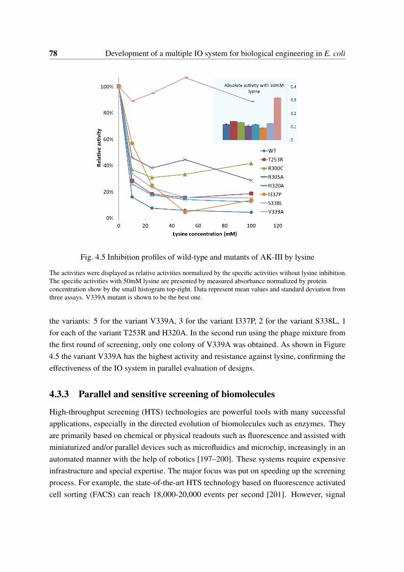
78 Development of a multiple IO system for biological engineering in E. coli
Fig. 4.5 Inhibition profiles of wild-type and mutants of AK-III by lysine
The activities were displayed as relative activities normalized by the specific activities without lysine inhibition.The specific activities with 50mM lysine are presented by measured absorbance normalized by proteinconcentration show by the small histogram top-right. Data represent mean values and standard deviation fromthree assays. V339A mutant is shown to be the best one.
the variants: 5 for the variant V339A, 3 for the variant I337P, 2 for the variant S338L, 1for each of the variant T253R and H320A. In the second run using the phage mixture fromthe first round of screening, only one colony of V339A was obtained. As shown in Figure4.5 the variant V339A has the highest activity and resistance against lysine, confirming theeffectiveness of the IO system in parallel evaluation of designs.
4.3.3 Parallel and sensitive screening of biomolecules
High-throughput screening (HTS) technologies are powerful tools with many successfulapplications, especially in the directed evolution of biomolecules such as enzymes. Theyare primarily based on chemical or physical readouts such as fluorescence and assisted withminiaturized and/or parallel devices such as microfluidics and microchip, increasingly in anautomated manner with the help of robotics [197–200]. These systems require expensiveinfrastructure and special expertise. The major focus was put on speeding up the screeningprocess. For example, the state-of-the-art HTS technology based on fluorescence activatedcell sorting (FACS) can reach 18,000-20,000 events per second [201]. However, signal
4.3 Proof of concept studies 79
detection with fast moving cells is challenging resulting with noisy signals as shown inprevious studies [202–205]. Furthermore, single cell variations are another source of signalnoise which cannot be avoided by FACS based methods [206]. These represent some ofthe shortcomings of presently used HTS technologies when the molecules to be evolvedand optimized are to be used for regulation and improvement of metabolic pathways in thecontext of metabolic engineering or for creation of new synthetic pathways and regulationtools.
Similar to the electric robots, microbial cells can be considered as a kind of “biologicalrobots” that can sense the information of fast changing environment, compute and make deci-sions for survival. Cells are highly programmable as proved by recent developments in thefield of synthetic biology. Programming cells to perform specific tasks have been successfullyachieved in many cases. For example, cells have been programmed to produce pharmaceuti-cals, fuels, amino acids, fine and bulk chemicals and even metal nanoparticles [207–214].Cells also have been programmed to sense toxic compounds in environments [215], to recordthe environment signal in human gut [216] and to eradicate human pathogen[217]. Althoughthe capability of a single cell is limited, cells can reproduce themselves exponentially andwork simultaneously to solve complicated tasks or accomplish sophisticated tasks in princi-ple. However, these capabilities of cells have not yet been well exploited, especially for thepurpose of HTS.
Recently, concentrations of intracellular molecules have been used as a signal for over-expression of fluorescence for screening purposes in the context of strain improvement [203].For example, Binder et al. successfully used the intracellular concentration of lysine, a naturallysine-responsible transcriptional activator LysG and fused expression of a yellow fluorescentprotein (eYFP) to screen high lysine producer from Corynebacterium glutamicum [203].Later, by using the same sensor for in vivo detection of the desired end-product in singlecells, they established a screening method with FACS to screen enzymes without allostericinhibition. However, due to the complexity of the metabolic pathways, one enhanced enzymeusually has limited effects on productivity of the end-products. Genetic modifications arerequired to enhance the signals of the end-product in their studies [204].
Esvelt et al. (2011) presented an interesting phage-assisted method for continuousevolution of a specific gene-coded biomolecule that is linked to the infectivity of the phagemediated by the expression of a specific protein in host cells [218]. Specifically, M13filamentous bacteriophage carrying the molecule of interest was used to infect E. coli cellsin a lagoon with continuous inflow and outflow of the host cells, where the evolving geneis transferred from host cell to host cell in a manner that is dependent on the activity ofthe molecule of interest. The method was demonstrated with the evolution of a T7 RNA
80 Development of a multiple IO system for biological engineering in E. coli
polymerase with new binding properties. It was later on used to successfully evolve proteaseswith significantly increased drug resistance to protease inhibitor [219, 220, 220].
The cells can be considered as a kind of “biological robots”. Compared to physical robotsthe biological robots have the decisive advantage of fast replication, resulting in a large poolfor simultaneously screening under in vivo conditions. Thus, the screening throughput canbe expanded simply by using a larger population of cells, indicating a massively parallelscreening manner potentially far beyond the current HTS technologies. It is also worth tomention that the cost for such an approach is almost zero compared to methods based onexpensive FACS or microcapillary arrays, making it applicable in almost all biological labs.
Here, the novel IO system designed in this part of study was proposed to programm cellsas "screening robots" for parallel and highly sensitive screening of biomolecules for metabolicpathway optimization under in vivo conditions. The basic idea is to input may phages carryingvarious mutants and outputting the phages carrying mutants with desired properties. Theconcept was demonstrated by screening mutants of a protein with reduced allosteric inhibition.Allosteric regulation is one of the fundamental mechanisms that control almost all cellularmetabolisms and gene regulation [221]. Deregulation of allosteric inhibition is essentialin designing and optimizing metabolic pathways for the production of target metabolitessuch as amino acids [196]. AK-III is allosterically inhibited by L-lysine strictly. AK-IIIwas chosen in this work as a model enzyme because of our extensive previous work on therational design of this enzyme[196, 222]. The new approach is shown to be more sensitivethan the widely used flow cytometry method by novel way of signal capturing.
Principle and work-flow of cell robot based screening utilizing the IO system
The workflow of programming cells as robots for the screening of molecule of interest (target)is shown in Figure 4.6. Briefly, instead of placing the screening targets inside of the host cellsas in most of the traditional screening methods, the targets to be screened were placed onM13 phages. The host cells were then engineered so that they can screen for phages carryingthe targets with desired properties. Specifically, E. coli XL1-Blue cells were used as the hostcells for this purpose. To enable the host cells to control the infectivity of packaged phages,an essential gene for phage infectivity was transfered from the M13 phage to the host cells.The essential gene applied in this study is gene III encoding the attachment protein pIII whichmediates adsorption of the phage to its primary receptor, the tip of E. coli F-pilus[191]. Anintracellular biological circuit was implemented to control the infectivity of packaged phagesby controlling the expression level of gene III based on a specific intracellular signal that isrelated to the performance of the biomolecules to be screened, such as the concentration ofan end product or an intermediate metabolite of a metabolic pathway. The targets are then

4.3 Proof of concept studies 81
cloned into VCSM13 by replacing the original gene III. A helper plasmid pJ175-Str whichcan supply the gene III product is used for preparing infective phage library at the first step ofscreening (see below). Elimination of gene III does not affect the phage secretion. However,the infectivity of the produced phages is very low. Thus, to enable an effective screening, atwo-step strategy was utilized as illustrated in Figure 4.6.
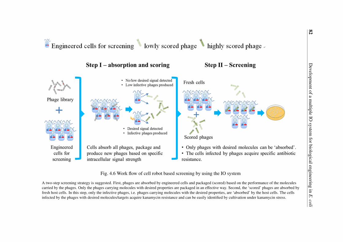
82D
evelopmentofa
multiple
IOsystem
forbiologicalengineeringin
E.coli
Fig. 4.6 Work flow of cell robot based screening by using the IO system
A two-step screening strategy is suggested. First, phages are absorbed by engineered cells and packaged (scored) based on the performance of the moleculescarried by the phages. Only the phages carrying molecules with desired properties are packaged in an effective way. Second, the ‘scored’ phages are absorbed byfresh host cells. In this step, only the infective phages, i.e. phages carrying molecules with the desired properties, are ‘absorbed’ by the host cells. The cellsinfected by the phages with desired molecules/targets acquire kanamycin resistance and can be easily identified by cultivation under kanamycin stress.
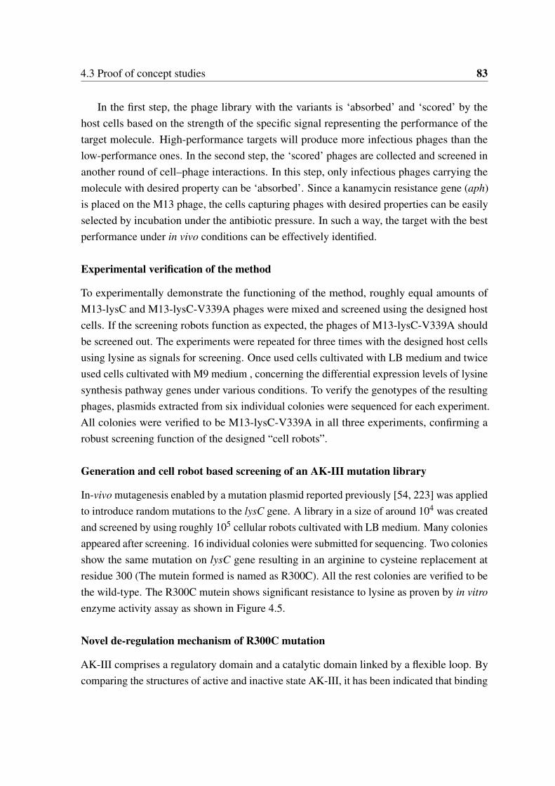
4.3 Proof of concept studies 83
In the first step, the phage library with the variants is ‘absorbed’ and ‘scored’ by thehost cells based on the strength of the specific signal representing the performance of thetarget molecule. High-performance targets will produce more infectious phages than thelow-performance ones. In the second step, the ‘scored’ phages are collected and screened inanother round of cell–phage interactions. In this step, only infectious phages carrying themolecule with desired property can be ‘absorbed’. Since a kanamycin resistance gene (aph)is placed on the M13 phage, the cells capturing phages with desired properties can be easilyselected by incubation under the antibiotic pressure. In such a way, the target with the bestperformance under in vivo conditions can be effectively identified.
Experimental verification of the method
To experimentally demonstrate the functioning of the method, roughly equal amounts ofM13-lysC and M13-lysC-V339A phages were mixed and screened using the designed hostcells. If the screening robots function as expected, the phages of M13-lysC-V339A shouldbe screened out. The experiments were repeated for three times with the designed host cellsusing lysine as signals for screening. Once used cells cultivated with LB medium and twiceused cells cultivated with M9 medium , concerning the differential expression levels of lysinesynthesis pathway genes under various conditions. To verify the genotypes of the resultingphages, plasmids extracted from six individual colonies were sequenced for each experiment.All colonies were verified to be M13-lysC-V339A in all three experiments, confirming arobust screening function of the designed “cell robots”.
Generation and cell robot based screening of an AK-III mutation library
In-vivo mutagenesis enabled by a mutation plasmid reported previously [54, 223] was appliedto introduce random mutations to the lysC gene. A library in a size of around 104 was createdand screened by using roughly 105 cellular robots cultivated with LB medium. Many coloniesappeared after screening. 16 individual colonies were submitted for sequencing. Two coloniesshow the same mutation on lysC gene resulting in an arginine to cysteine replacement atresidue 300 (The mutein formed is named as R300C). All the rest colonies are verified to bethe wild-type. The R300C mutein shows significant resistance to lysine as proven by in vitroenzyme activity assay as shown in Figure 4.5.
Novel de-regulation mechanism of R300C mutation
AK-III comprises a regulatory domain and a catalytic domain linked by a flexible loop. Bycomparing the structures of active and inactive state AK-III, it has been indicated that binding

84 Development of a multiple IO system for biological engineering in E. coli
of lysine to the regulatory domain of the active state AK-III triggers a series of changes thatrelease a “latch”, the loop (residues 355-365) colored in yellow color as shown in Figure4.7, from the catalytic domain, which in turn undergoes large rotational rearrangements,promoting tetramer formation and completion of the transition to the inactive state [224].Rearrangement of the catalytic domain blocks the ATP-binding site, which is the structuralbasis for allosteric inhibition of AK-III by lysine [224]. All previous studies of removingthe allosteric inhibition focused on mutagenesis of the regulatory domains, especially onmutagenesis of the binding sites [225, 55, 224, 226]. Surprisingly, the R300C mutationdiscovered here located in the flexible loop A (residues 291-300) which link the regulatoryand catalytic domains. Besides as a linker of the regulatory domain and the catalytic domain,this flexible loop has not been reported to be related to the allosteric transition process so far.After comparing the structures of the active and inactive states of the wild type AK-III, it wasfound that the residue 300 arginine forms a hydrogen bond with the residue 293 threonine inthe inactive state of AK-III after binding of lysine. The appeal between these two residuesforms a ‘dragging’ force which can deform the flexible loop and form a hairpin structure afterthe binding of lysine as shown in Figure 4.7a-b. The deformation of the flexible loop mightplay key roles in the initial steps of the rotational rearrangement of active state AK-III to theinactive state AK-III. The replacement of arginine 300 by cysteine will abolish this ‘dragging’force and block the formation of the hydrogen bond. Thus, by blocking the transition fromactive state to inactive state and unstable the inactive state, the R300C show significantresistance to lysine inhibition.
Screening with a higher sensitivity than fluorescence-based method
To compare the sensitivity of the current screening system with methods based on fluores-cence and flow cytometry, the state-of-the-art screening technology, we transformed E. coliXL1-Blue/AP-Lys-B cells with M13-lysC, M13-lysC-V339A, and M13-lysC-R300C indi-vidually to obtain cells of XL1-Blue/AP-Lys-B/M13-lysC-WT, XL1-Blue/AP-Lys-B/M13-lysC-V339A, and XL1-Blue/AP-Lys-B/M13-lysC-R300C. A GFP-encoding gene was placedunder the control of the same lysine sensor in E. coli XL1-Blue/AP-Lys-B. Over-nightcultivated cells of XL1-Blue/AP-Lys-B, XL1-Blue/AP-Lys-B/M13-lysC-WT, XL1-Blue/AP-Lys-B/M13-lysC-V339A and XL1-Blue/AP-Lys-B/M13-lysC-R300C were harvested andwashed twice with 50mM, ice-cooled PBS buffer. The fluorescence activities of the fourdifferent cell populations were measured using flow cytometry. As shown in 4.8 a and b, al-though slight differences could be observed, it is not possible to set up a gain setting to selectthe mutants. In other words, the different cell populations cannot be distinguished by theflow cytometry method. On the other hand, our method based on cell-phage interaction can
4.3 Proof of concept studies 85
Fig. 4.7 3D structure illustration of the de-allosteric regulation mechanism of R300C mutein
a - active state of wild AK-III without lysine binding. b - inactive state of wild AK-III with lysine bound to theregulatory domain. The visualizations were generated using the UCSF Chimera software [227].
successfully screen out V339A as the best mutant of AK-III, confirming a higher sensitivityof the cell robot based screening method.
Biological systems are complex and highly adaptive, meaning that the cells always tryto reduce the perturbations introduced. Introducing molecular variants into cells can beregarded as perturbations to the cells. As shown in 4.8c, after the introduction of molecularvariants, the cells may undergo three stages of signal change: enhancement, attenuation andstabilization. The signal enhancement stage is the direct consequence of the perturbationsinduced by the introduced molecular variants. The signal attenuation stage is caused bythe adaptive response of cells to the perturbation. Finally, the signal reaches a stable statewhich might be slightly different from the state before the perturbation. The time interval forthese changes may be relatively short. The curves in 4.8c are theoretical response patternsof cells to the disturbance by over-expression of the different AK-III variants respectively.For the FACS-based method, the cells to be measured may have already reached the stablestage where the signal strength may not be significantly different in the cell populations withdifferent variants. However, our method captures signals during the whole response andadaptation processes which correspond to the area below the curves and can be thereforemore sensitive.
Furthermore, the “cell robots” based screening works in principle like an autocatalyticprocess of signal amplification: the target molecule with desired performance will increasethe intracellular concentration of the signal molecule (lysine in this case) in the cell, the
86 Development of a multiple IO system for biological engineering in E. coli
increased concentration of the signal molecule will amplify the population of phage carryingthe target molecule. The amplified phages can infect other cells to further enhance the signal.In such a way the screening process is highly effective and sensitive compared to the otherpresently used methods, such as those based on single cells using fluorescence as the readoutsignal [204].
Summary of cell robot based screening
For conclusion, it was demonstrated that the biological ‘robots’, i.e. the cells, can beengineered to perform screening tasks in protein engineering. By capturing the signals duringthe whole response and adaptation processes, which cannot be achieved by screening basedon electric machines, the cell-phage based screening system has an inherent higher sensitivity.The current proof of concept study shows that cell-phage interaction system does not requireany genetic modifications of the host cells to enhance the signal for screening. In a recentsimilar work which used FACS as the screening method, genetic modifications are requiredto enhance the signal [204]. Furthermore, FACS-based screening often suffers signal noisecaused by single cell variations and signal detection under conditions of fast moving cells[206, 204, 203]. By equally accessing all cells, the cell-phage interaction system can avoidthe problem of single cell variation in principle. Cells as biological ‘robots’ have a uniquefeature of reproducing themselves to generate a vast population exponentially and cheaply.Thus, the screening throughput can be simply expanded by using a larger population of cellswith minimal additional costs, indicating a massively parallel screening manner beyond thecurrent electric machines. As proved by the power of parallel computing in computationalscience [228], parallelization is a great solution for speeding up the process of paralleltasks. The sensitivity and throughput are key factors determining the success of a screeningexperiment. The cell-phage screening system shows clear advantages in both sensitivity andthroughput. Furthermore, the cost of cell robots is almost zero compared to that of expensiveelectric machines/robots. It should be mentioned that, while electric machines can utilizevarious types of signals for screening, screening based on the cell robots uses a “biologicalsignal and sensor”, which might represent a limitation in some cases. However, many naturalor purposefully designed biological elements or sensors such as promoters and riboswitchescan be used for this purpose [229, 230] and the signal molecules can be intermediates ofmetabolic pathways.
4.4 Conclusion and Perspective 87
4.4 Conclusion and Perspective
Engineering biological system is complex and kind of unpredictable. In this study, a multipleIO system was proposed to simplify the development process of biological engineering.The IO system was implemented based on M13 phages. For proof of concept, the methodwas demonstrated for target identification, evaluation of designs, evolution and selection ofkey enzymes for the lysine biosynthesis in E. coli. Correct prediction of beneficial geneticmanipulation for enhanced lysine production in E. coli was achieved. New and effectivevariants of AK-III which is strictly inhibited by L-lysine, were obtained. Importantly, thecellular IO system showed an ultra-sensitivity in capturing the signal changes caused by theperturbations introduced. The author believes that the approach developed in this work opensup new possibilities for systems metabolic engineering and synthetic biology of industrialmicroorganisms in practical applications.
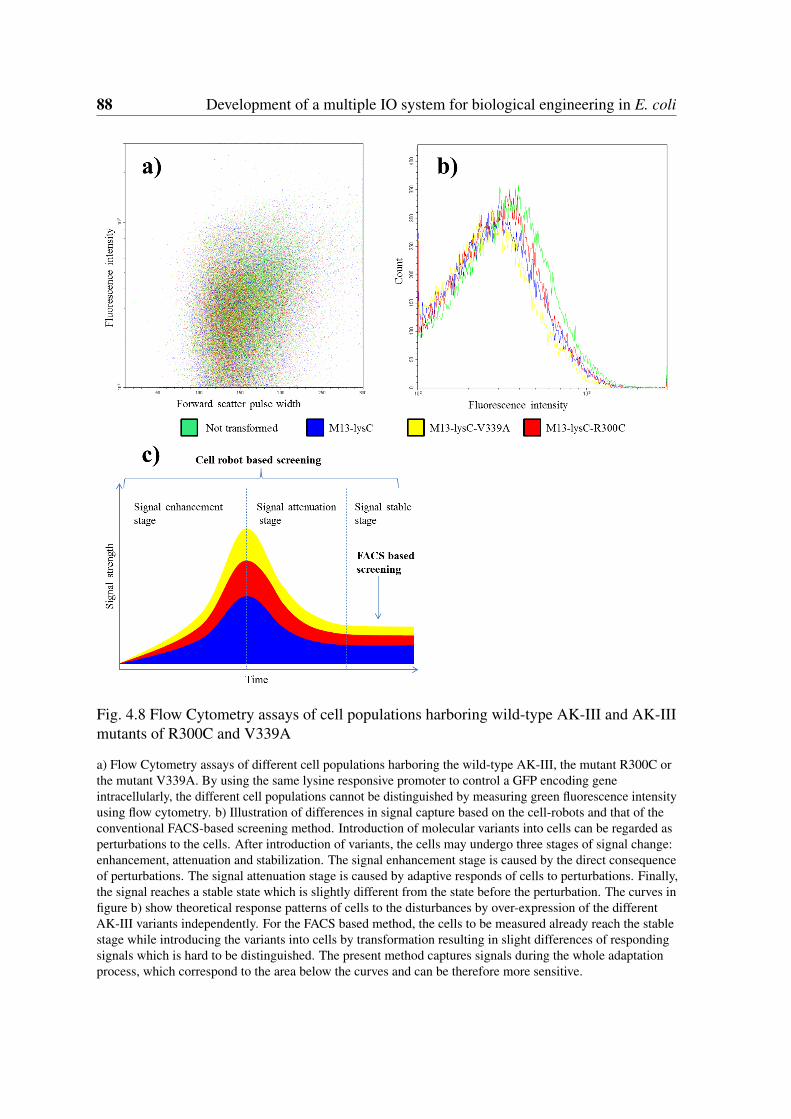
88 Development of a multiple IO system for biological engineering in E. coli
Fig. 4.8 Flow Cytometry assays of cell populations harboring wild-type AK-III and AK-IIImutants of R300C and V339A
a) Flow Cytometry assays of different cell populations harboring the wild-type AK-III, the mutant R300C orthe mutant V339A. By using the same lysine responsive promoter to control a GFP encoding geneintracellularly, the different cell populations cannot be distinguished by measuring green fluorescence intensityusing flow cytometry. b) Illustration of differences in signal capture based on the cell-robots and that of theconventional FACS-based screening method. Introduction of molecular variants into cells can be regarded asperturbations to the cells. After introduction of variants, the cells may undergo three stages of signal change:enhancement, attenuation and stabilization. The signal enhancement stage is caused by the direct consequenceof perturbations. The signal attenuation stage is caused by adaptive responds of cells to perturbations. Finally,the signal reaches a stable state which is slightly different from the state before the perturbation. The curves infigure b) show theoretical response patterns of cells to the disturbances by over-expression of the differentAK-III variants independently. For the FACS based method, the cells to be measured already reach the stablestage while introducing the variants into cells by transformation resulting in slight differences of respondingsignals which is hard to be distinguished. The present method captures signals during the whole adaptationprocess, which correspond to the area below the curves and can be therefore more sensitive.
Chapter 5
Orthogonal information encoding inliving cells
5.1 Introduction
Deoxyribonucleic acid (DNA) is the natural information carrier utilized in all living organismson earth [231]. The first report about artificial information in DNA was published in 1996by Davis et al. They encoded a binary graphic data into a synthetic DNA molecule usinga simple bit-mapping mechanism. Later in 1999, Clelland et al. proposed a method forhiding messages in DNA microdots for data encryption purpose [232]. Bancroft et al. firstproposed used DNA as a long-term information storage media [233]. In 2007, Yachie et al.proposed an alignment-based approach for durable data storage into living organisms [234].In 2009, Gustafsson et al. encoded a poem into DNA [235]. Ailenberg et al. proposed animproved Huffman coding method for archiving text, images, and music characters in DNA[236]. In 2010, Gibson et al. wrote a watermark message into a chemically synthesizedgenome[237]. In addition to these applications, artificial information encoding in DNAhas more attractive potential applications such as barcoding and comments encoding forprogramming cells in synthetic biology [27], and even for large and long-term data storage[232, 27, 22, 238–241]. Information stored in DNA can be distributed in a three dimensionalspace while the traditional planner media can only store information on a two dimensionalsurface. The extra dimension remarkably enhances the information density as recentlydemonstrated by Church et al., Goldman et al. and Erlich et al. [23, 24, 242]. Furthermore,unlike the presently used storage media such as magnetic tape or hard drives which have a
This chapter was modified based on a recent publication: Song, Lifu; Zeng, An-Ping (2017): Orthogonalinformation encoding in living cells with high error-tolerance, safety, and fidelity. ACS Synthetic Biology 7 (3),pp. 866–874.
90 Orthogonal information encoding in living cells
decisive shortcoming of a limited lifetime, e.g. around 50 years for hard drivers, informationstorage in DNA can have a lifetime of many thousands of years and is of low maintenancecosts [23, 24, 243, 244, 233]. By using silica glass spheres for DNA encapsulation, Grasset al. predicted an error-free retrieval of information from the DNA after more than 1million years if stored at -18°C and 2000 years at 10°C [245]. However, relatively high errorrates could be introduced in the information “writing”, “reading” and “copying” processes(i.e. DNA synthesis, sequencing and polymerase chain reaction (PCR)) [25, 26]. Theerror rate is even higher if the encoded DNA sequences contain extreme GC contents, longhomopolymers or complex secondary structures which are hard to be synthesized, sequencedand amplified. Errors were detected in early studies lacking an effective error correctionmechanism [23, 24]. In the later studies of Yim et al. and Grass et al., error correction codessuch as “Reed–Solomon” or “low-density parity-check” were introduced and informationcould be retrieved successfully [245, 246]. Yazdi et al. implemented a DNA-based storagearchitecture that enables random access to data blocks and rewriting of information storedat arbitrary locations within the blocks [247]. Later on, additional error correction codeswere introduced and the scale of encoded data volume increased remarkably [244, 248, 249].Interestingly, a recent study has successfully retrieved information stored in synthesized longDNA fragments using a portable sequencer - MinION [25, 250, 251]. More recently, byintroducing Fountain codes, Erlich et al. established a robust and efficient storage strategycalled DNA Fountain with a data density of 215 petabytes per gram of DNA [242].
All the studies mentioned above dealt with information encoding in DNA outside livingcells. It is also of great interest to know if DNA data storage or information encoding in livingcells is feasible and reliable. This should enable applications such as biological barcodes ofengineered biological parts (Biobricks) and comment “language” in “programming biology”in the emerging area of synthetic biology [27]. Theoretically, the encoding schemes designedfor in vitro data storage in DNA are also applicable for in vivo applications. However, tothe best of our knowledge, no reported work has addressed the issue of error enrichmentintroduced by DNA replication which is crucial for in vivo applications since DNA replicationhappens constantly under in vivo conditions. In the study of Erlich et al., they retrieved theoriginal data from a deep copy of the data by PCR amplifying the oligo pool in a total ofnine successive reactions [242]. Theoretically, the successive PCR reactions could generate218×1012 copies of the original data, showing a great advantage of the exponential “copying”process of DNA storage by PCR to generate vast data copies quickly and cheaply. However,the number of rounds of the “copying” process is only demonstrated within limited cycleswhich are insufficient for in vivo applications [242]. Furthermore, the artificial DNA frag-ments could interfere with the native and natural ones (being so-called biologically relevant).
5.2 Theoretical and technological backgrounds 91
This is another issue which has not been studied so far. For in vivo applications, such asbiological barcodes or comments encoding in living cells, the encoded DNA sequencesshould not share the same sequence space as the natural ones to avoid interference withcellular functions. In other words, they should be orthogonal to exclude biological relevance.
One unique feature of information storage in DNA is that there are always many copiesof DNA molecules synthesized which represent the same data. In other words, there is a highinherent data redundancy. In this study, using a novel way of adding error detection codesblock by block, an efficient self-error-detecting, three-base block encoding scheme (SED3B)which can take full advantage of the inherent redundancy feature for error correction wasestablished. SED3B can effectively repress error enrichment emerging from DNA replicationas proved by in silicon and experimental verifications. With merely 30 sequences for errorcorrection, the SED3B scheme can tolerate a high error rate of 19.1%. Errors in a rate of40% still can be corrected with 180 DNA sequences as proved by in silicon simulations.Over 12,100 years of continuous replication are estimated to be required to make the SED3Bencoded information in growing E. coli cells unrecoverable as proved by in vivo, error-pronePCR experiments. In addition to limited extreme GC contents, homopolymers, and simplesecondary structure, SED3B encoded sequences also show very low biological relevanceas proved by comparative studies with naturally formed sequences. Features of high errortolerance and low biological relevance make SED3B promising for orthogonal informationencoding in living cells, e.g. as comment language in programming cell or for biologicalbarcode encoding. To facilitate the usage of SED3B as a univeral information encodingscheme in living cells, an online encoding-decoding system with cases of comment andbarcode encoding is implemented and released in http://biosystem.bt1.tu-harburg.de/sed3b/.
5.2 Theoretical and technological backgrounds
The focus of this part of work is to design an encoding scheme for reliable digital dataencoding in DNA with regarding to the unique features of DNA as data storage media.There are many methods available for data encoding in DNA. In the previous section, theavailable methods for data encoding in DNA have been briefly introduced. In this section,four representative state-of-the-art methods released in recent years are detailed, their meritsand limitations or disadvantages are mentioned. Other efforts on associative memory andDNA computing were not included because they were designed for different purposes forDNA information storage needs. In a recent study, Erlich et al. reported a storage strategy,called DNA Fountain. They proved that the 2.14 × 106 bytes encoded data could be retrieved
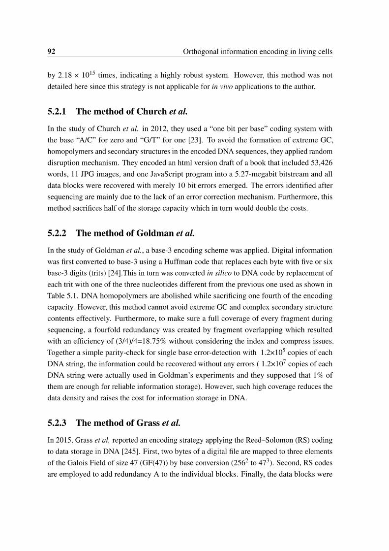
92 Orthogonal information encoding in living cells
by 2.18 × 10¹⁵ times, indicating a highly robust system. However, this method was notdetailed here since this strategy is not applicable for in vivo applications to the author.
5.2.1 The method of Church et al.
In the study of Church et al. in 2012, they used a “one bit per base” coding system withthe base “A/C” for zero and “G/T” for one [23]. To avoid the formation of extreme GC,homopolymers and secondary structures in the encoded DNA sequences, they applied randomdisruption mechanism. They encoded an html version draft of a book that included 53,426words, 11 JPG images, and one JavaScript program into a 5.27-megabit bitstream and alldata blocks were recovered with merely 10 bit errors emerged. The errors identified aftersequencing are mainly due to the lack of an error correction mechanism. Furthermore, thismethod sacrifices half of the storage capacity which in turn would double the costs.
5.2.2 The method of Goldman et al.
In the study of Goldman et al., a base-3 encoding scheme was applied. Digital informationwas first converted to base-3 using a Huffman code that replaces each byte with five or sixbase-3 digits (trits) [24].This in turn was converted in silico to DNA code by replacement ofeach trit with one of the three nucleotides different from the previous one used as shown inTable 5.1. DNA homopolymers are abolished while sacrificing one fourth of the encodingcapacity. However, this method cannot avoid extreme GC and complex secondary structurecontents effectively. Furthermore, to make sure a full coverage of every fragment duringsequencing, a fourfold redundancy was created by fragment overlapping which resultedwith an efficiency of (3/4)/4=18.75% without considering the index and compress issues.Together a simple parity-check for single base error-detection with 1.2×10⁵ copies of eachDNA string, the information could be recovered without any errors ( 1.2×10⁷ copies of eachDNA string were actually used in Goldman’s experiments and they supposed that 1% ofthem are enough for reliable information storage). However, such high coverage reduces thedata density and raises the cost for information storage in DNA.
5.2.3 The method of Grass et al.
In 2015, Grass et al. reported an encoding strategy applying the Reed–Solomon (RS) codingto data storage in DNA [245]. First, two bytes of a digital file are mapped to three elementsof the Galois Field of size 47 (GF(47)) by base conversion (2562 to 473). Second, RS codesare employed to add redundancy A to the individual blocks. Finally, the data blocks were
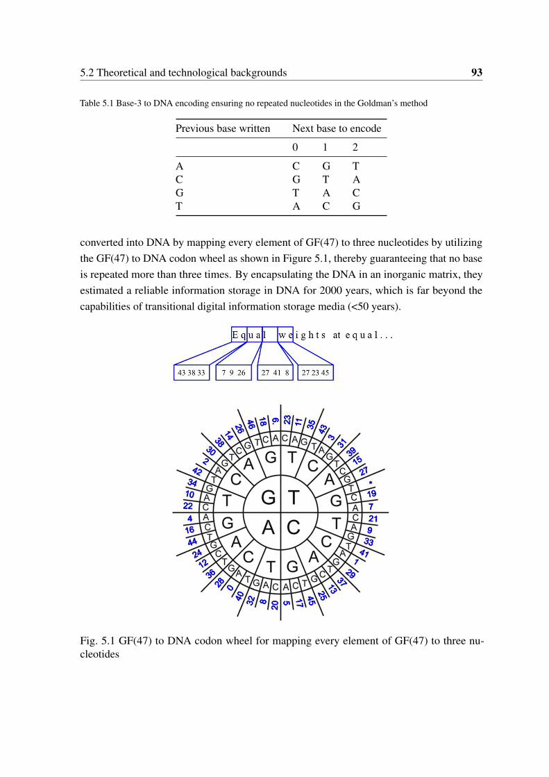
5.2 Theoretical and technological backgrounds 93
Table 5.1 Base-3 to DNA encoding ensuring no repeated nucleotides in the Goldman’s method
Previous base written Next base to encode
0 1 2
A C G TC G T AG T A CT A C G
converted into DNA by mapping every element of GF(47) to three nucleotides by utilizingthe GF(47) to DNA codon wheel as shown in Figure 5.1, thereby guaranteeing that no baseis repeated more than three times. By encapsulating the DNA in an inorganic matrix, theyestimated a reliable information storage in DNA for 2000 years, which is far beyond thecapabilities of transitional digital information storage media (<50 years).
Fig. 5.1 GF(47) to DNA codon wheel for mapping every element of GF(47) to three nu-cleotides
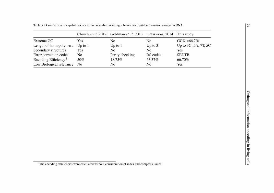
94O
rthogonalinformation
encodingin
livingcells
Table 5.2 Comparison of capabilities of current available encoding schemes for digital information storage in DNA
Church et al. 2012 Goldman et al. 2013 Grass et al. 2014 This study
Extreme GC Yes No No GC% <66.7%Length of homopolymers Up to 1 Up to 1 Up to 3 Up to 3G, 5A, 7T, 5CSecondary structures Yes No No YesError correction codes No Parity checking RS codes SEDTBEncoding Efficiency i 50% 18.75% 63.37% 66.70%Low Biological relevance No No No Yes
iThe encoding efficiencies were calculated without consideration of index and compress issues.
5.3 Principles of a self-error-detecting, three-base block encoding scheme (SED3B) 95
5.3 Principles of a self-error-detecting, three-base block en-coding scheme (SED3B)
To fully utilize the redundancy feature of DNA molecules for error correction, a novel self-error-detecting, three-base block (SED3B) encoding scheme was proposed for effective andflexible error correction. In details, binary bits are first transformed into data encoding DNAbases four bits by four bits using the scheme shown in Figure 5.2. Then one error checkingbase was inserted per two data encoding bases to form a three-base block encoding manner.The third base is designed to detect whether there are errors emerged in the two encodingbases. A simple way of error checking by the third base is the checksum principle [252].However, the checksum method has no optimization option for homopolymers and extremeGC contents. Instead, a novel strategy was utilized to enable error checking by the thirdbase. At first, all possible 16 two-base combinations were divided into four groups basedon the principle that all the four two-base combinations in the same group do not share anyidentical base in neither the first nor the second base, and then every group was assignedwith an error detecting base as shown in Figure 5.2. Thus, the data encoding two-base andthe error detecting base won’t match to each other anymore if error emerges in any of thethree bases. In other words, a single base error on any of the three bases can be detected.To avoid extreme GC, long homopolymers and complex secondary structures generated inthe encoding DNA strings, three additional principles are followed while assigning errorchecking bases to the four groups of two-base combinations: 1) no more than 3 G/C presentin the three base block to avoid extreme GC contents; 2) no identical bases present in all thethree bases to avoid long homopolymers; 3) no complementarily matched three base blockspresent. However, principals 1) and 2) cannot be satisfied simultaneously. To address thisissue, two different rules were introduced for assigning error detecting bases as shown inFigure 5.2, rows of “error detecting base rules”. Rule I satisfies the principle that there areno more than 2 G/C present in all the three bases while a “TTT” homopolymer does presentthere. Rule II abolishes any three base homopolymer while enabling G/C presents in all threebases. During the encoding process, Rule I is used in general and only if “TTT” is present,the rule for assigning the error detecting bases is switched to Rule II temporarily and thenswitched back to Rule I after having encoded once. Thus, continuous “T” homopolymerscan be avoided as the error detecting base for “TT” is switched to “G”, not “T” in Rule IIand extreme GC content can also be avoided as three G/C combinations in Rule II are onlypresent if the previous encoding three-base block is “TTT”. Finally, no more than sevencontinuous “T”, five continuous “A/C” and three continuous “G” are possible to exist in theencoded DNA strings which have been proved to be acceptable by current DNA synthesis
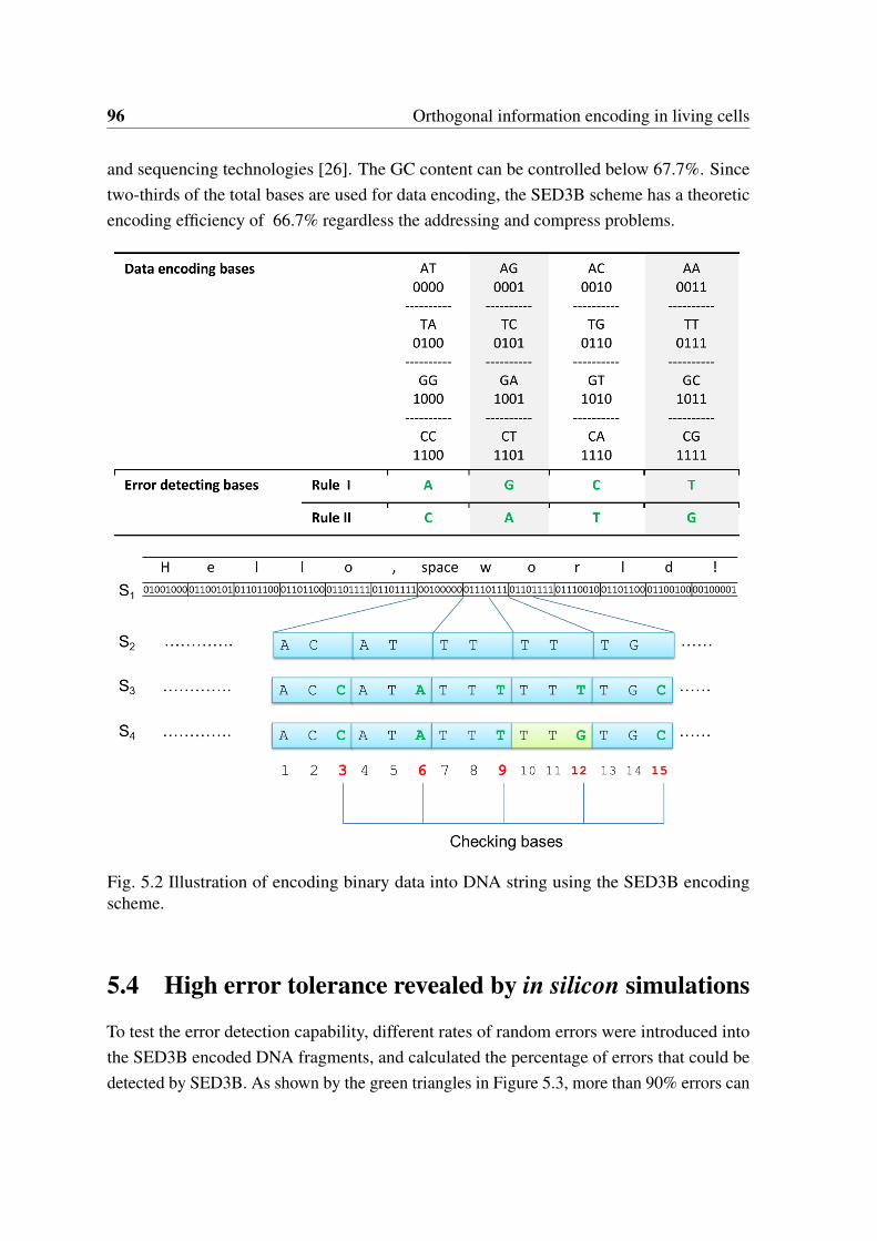
96 Orthogonal information encoding in living cells
and sequencing technologies [26]. The GC content can be controlled below 67.7%. Sincetwo-thirds of the total bases are used for data encoding, the SED3B scheme has a theoreticencoding efficiency of 66.7% regardless the addressing and compress problems.
Fig. 5.2 Illustration of encoding binary data into DNA string using the SED3B encodingscheme.
5.4 High error tolerance revealed by in silicon simulations
To test the error detection capability, different rates of random errors were introduced intothe SED3B encoded DNA fragments, and calculated the percentage of errors that could bedetected by SED3B. As shown by the green triangles in Figure 5.3, more than 90% errors can
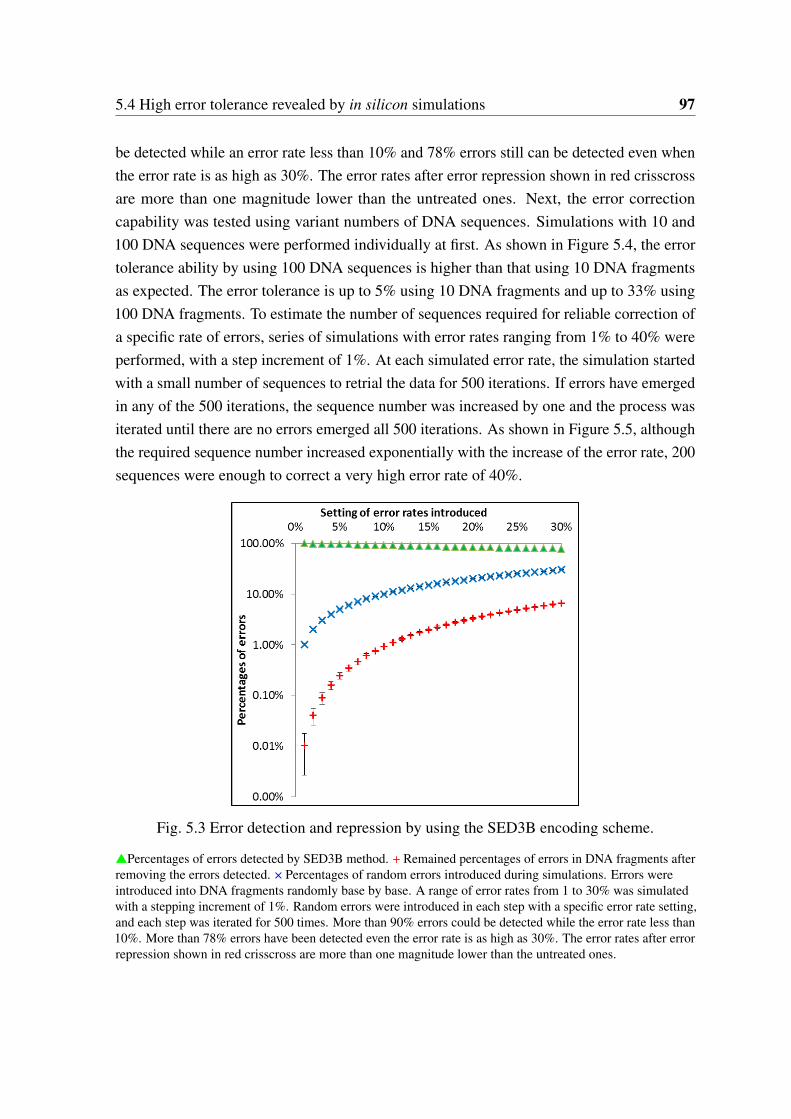
5.4 High error tolerance revealed by in silicon simulations 97
be detected while an error rate less than 10% and 78% errors still can be detected even whenthe error rate is as high as 30%. The error rates after error repression shown in red crisscrossare more than one magnitude lower than the untreated ones. Next, the error correctioncapability was tested using variant numbers of DNA sequences. Simulations with 10 and100 DNA sequences were performed individually at first. As shown in Figure 5.4, the errortolerance ability by using 100 DNA sequences is higher than that using 10 DNA fragmentsas expected. The error tolerance is up to 5% using 10 DNA fragments and up to 33% using100 DNA fragments. To estimate the number of sequences required for reliable correction ofa specific rate of errors, series of simulations with error rates ranging from 1% to 40% wereperformed, with a step increment of 1%. At each simulated error rate, the simulation startedwith a small number of sequences to retrial the data for 500 iterations. If errors have emergedin any of the 500 iterations, the sequence number was increased by one and the process wasiterated until there are no errors emerged all 500 iterations. As shown in Figure 5.5, althoughthe required sequence number increased exponentially with the increase of the error rate, 200sequences were enough to correct a very high error rate of 40%.
Fig. 5.3 Error detection and repression by using the SED3B encoding scheme.
Percentages of errors detected by SED3B method. + Remained percentages of errors in DNA fragments afterremoving the errors detected. × Percentages of random errors introduced during simulations. Errors wereintroduced into DNA fragments randomly base by base. A range of error rates from 1 to 30% was simulatedwith a stepping increment of 1%. Random errors were introduced in each step with a specific error rate setting,and each step was iterated for 500 times. More than 90% errors could be detected while the error rate less than10%. More than 78% errors have been detected even the error rate is as high as 30%. The error rates after errorrepression shown in red crisscross are more than one magnitude lower than the untreated ones.
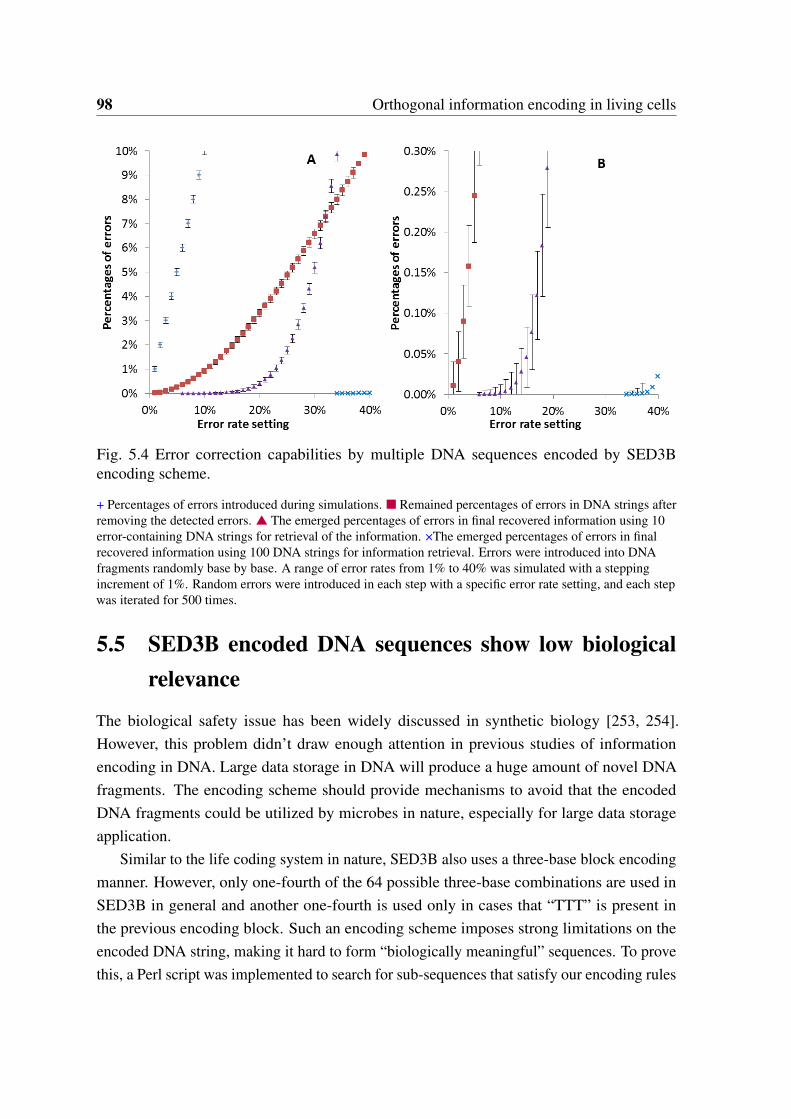
98 Orthogonal information encoding in living cells
Fig. 5.4 Error correction capabilities by multiple DNA sequences encoded by SED3Bencoding scheme.
+ Percentages of errors introduced during simulations. Remained percentages of errors in DNA strings afterremoving the detected errors. The emerged percentages of errors in final recovered information using 10error-containing DNA strings for retrieval of the information. ×The emerged percentages of errors in finalrecovered information using 100 DNA strings for information retrieval. Errors were introduced into DNAfragments randomly base by base. A range of error rates from 1% to 40% was simulated with a steppingincrement of 1%. Random errors were introduced in each step with a specific error rate setting, and each stepwas iterated for 500 times.
5.5 SED3B encoded DNA sequences show low biologicalrelevance
The biological safety issue has been widely discussed in synthetic biology [253, 254].However, this problem didn’t draw enough attention in previous studies of informationencoding in DNA. Large data storage in DNA will produce a huge amount of novel DNAfragments. The encoding scheme should provide mechanisms to avoid that the encodedDNA fragments could be utilized by microbes in nature, especially for large data storageapplication.
Similar to the life coding system in nature, SED3B also uses a three-base block encodingmanner. However, only one-fourth of the 64 possible three-base combinations are used inSED3B in general and another one-fourth is used only in cases that “TTT” is present inthe previous encoding block. Such an encoding scheme imposes strong limitations on theencoded DNA string, making it hard to form “biologically meaningful” sequences. To provethis, a Perl script was implemented to search for sub-sequences that satisfy our encoding rules
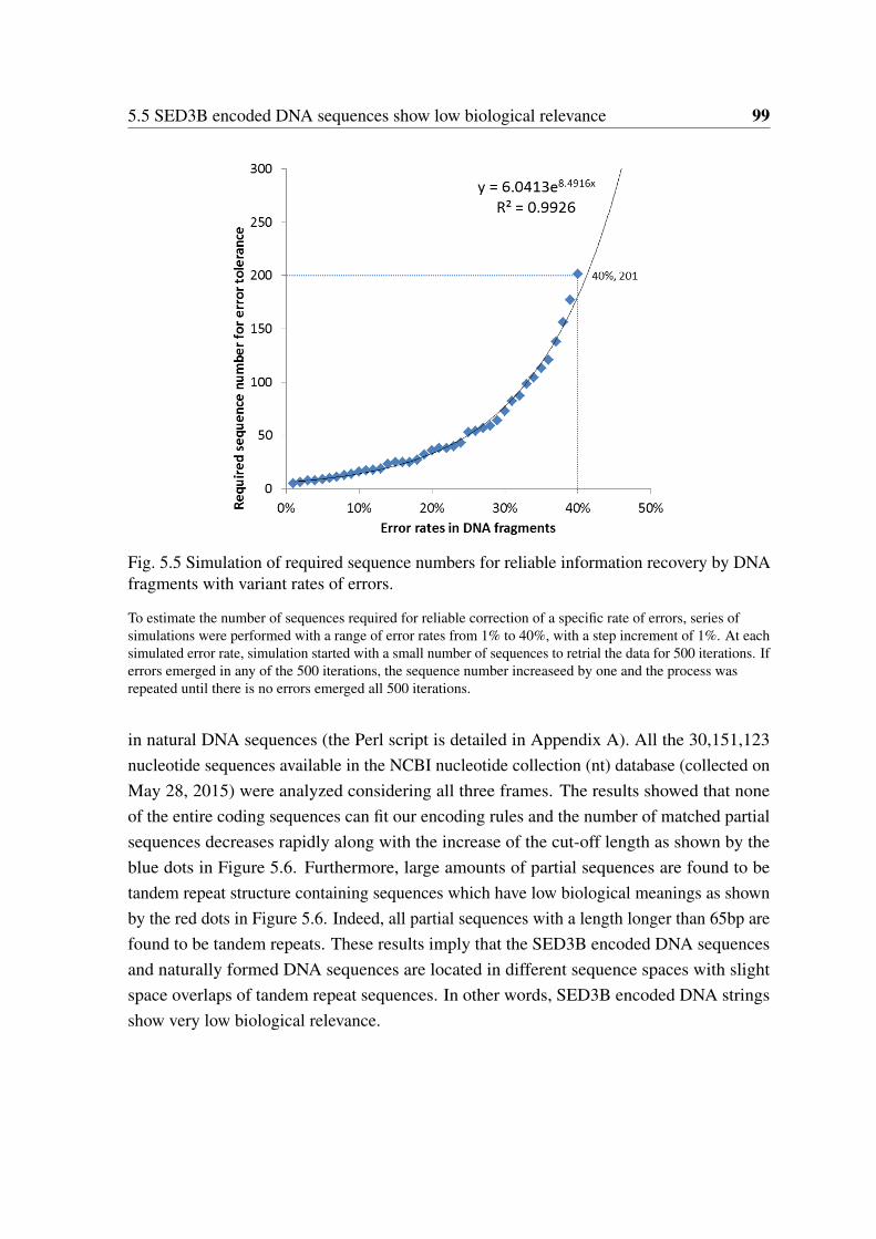
5.5 SED3B encoded DNA sequences show low biological relevance 99
Fig. 5.5 Simulation of required sequence numbers for reliable information recovery by DNAfragments with variant rates of errors.
To estimate the number of sequences required for reliable correction of a specific rate of errors, series ofsimulations were performed with a range of error rates from 1% to 40%, with a step increment of 1%. At eachsimulated error rate, simulation started with a small number of sequences to retrial the data for 500 iterations. Iferrors emerged in any of the 500 iterations, the sequence number increaseed by one and the process wasrepeated until there is no errors emerged all 500 iterations.
in natural DNA sequences (the Perl script is detailed in Appendix A). All the 30,151,123nucleotide sequences available in the NCBI nucleotide collection (nt) database (collected onMay 28, 2015) were analyzed considering all three frames. The results showed that noneof the entire coding sequences can fit our encoding rules and the number of matched partialsequences decreases rapidly along with the increase of the cut-off length as shown by theblue dots in Figure 5.6. Furthermore, large amounts of partial sequences are found to betandem repeat structure containing sequences which have low biological meanings as shownby the red dots in Figure 5.6. Indeed, all partial sequences with a length longer than 65bp arefound to be tandem repeats. These results imply that the SED3B encoded DNA sequencesand naturally formed DNA sequences are located in different sequence spaces with slightspace overlaps of tandem repeat sequences. In other words, SED3B encoded DNA stringsshow very low biological relevance.
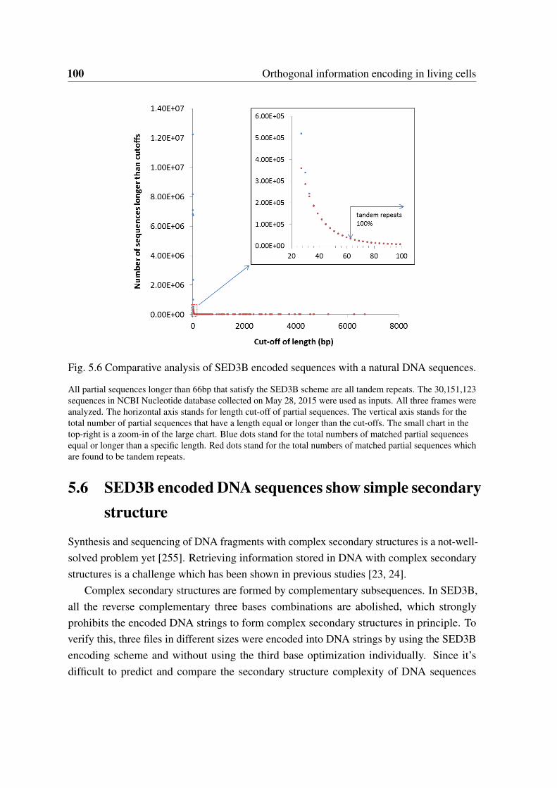
100 Orthogonal information encoding in living cells
Fig. 5.6 Comparative analysis of SED3B encoded sequences with a natural DNA sequences.
All partial sequences longer than 66bp that satisfy the SED3B scheme are all tandem repeats. The 30,151,123sequences in NCBI Nucleotide database collected on May 28, 2015 were used as inputs. All three frames wereanalyzed. The horizontal axis stands for length cut-off of partial sequences. The vertical axis stands for thetotal number of partial sequences that have a length equal or longer than the cut-offs. The small chart in thetop-right is a zoom-in of the large chart. Blue dots stand for the total numbers of matched partial sequencesequal or longer than a specific length. Red dots stand for the total numbers of matched partial sequences whichare found to be tandem repeats.
5.6 SED3B encoded DNA sequences show simple secondarystructure
Synthesis and sequencing of DNA fragments with complex secondary structures is a not-well-solved problem yet [255]. Retrieving information stored in DNA with complex secondarystructures is a challenge which has been shown in previous studies [23, 24].
Complex secondary structures are formed by complementary subsequences. In SED3B,all the reverse complementary three bases combinations are abolished, which stronglyprohibits the encoded DNA strings to form complex secondary structures in principle. Toverify this, three files in different sizes were encoded into DNA strings by using the SED3Bencoding scheme and without using the third base optimization individually. Since it’sdifficult to predict and compare the secondary structure complexity of DNA sequences
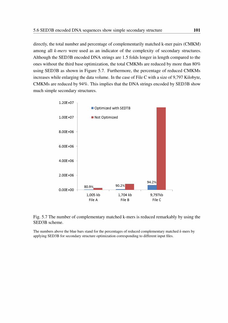
5.6 SED3B encoded DNA sequences show simple secondary structure 101
directly, the total number and percentage of complementarily matched k-mer pairs (CMKM)among all k-mers were used as an indicator of the complexity of secondary structures.Although the SED3B encoded DNA strings are 1.5 folds longer in length compared to theones without the third base optimization, the total CMKMs are reduced by more than 80%using SED3B as shown in Figure 5.7. Furthermore, the percentage of reduced CMKMsincreases while enlarging the data volume. In the case of File C with a size of 9,797 Kilobyte,CMKMs are reduced by 94%. This implies that the DNA strings encoded by SED3B showmuch simple secondary structures.
Fig. 5.7 The number of complementary matched k-mers is reduced remarkably by using theSED3B scheme.
The numbers above the blue bars stand for the percentages of reduced complementary matched k-mers byapplying SED3B for secondary structure optimization corresponding to different input files.
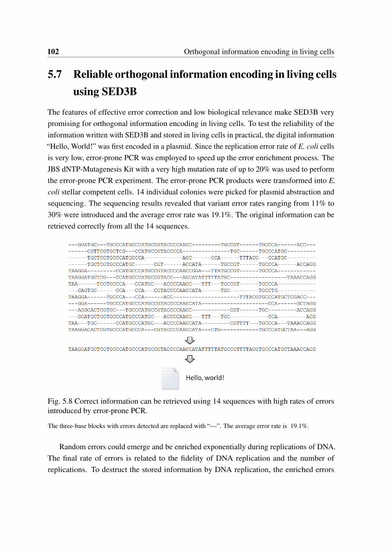
102 Orthogonal information encoding in living cells
5.7 Reliable orthogonal information encoding in living cellsusing SED3B
The features of effective error correction and low biological relevance make SED3B verypromising for orthogonal information encoding in living cells. To test the reliability of theinformation written with SED3B and stored in living cells in practical, the digital information“Hello, World!” was first encoded in a plasmid. Since the replication error rate of E. coli cellsis very low, error-prone PCR was employed to speed up the error enrichment process. TheJBS dNTP-Mutagenesis Kit with a very high mutation rate of up to 20% was used to performthe error-prone PCR experiment. The error-prone PCR products were transformed into E.coli stellar competent cells. 14 individual colonies were picked for plasmid abstraction andsequencing. The sequencing results revealed that variant error rates ranging from 11% to30% were introduced and the average error rate was 19.1%. The original information can beretrieved correctly from all the 14 sequences.
Fig. 5.8 Correct information can be retrieved using 14 sequences with high rates of errorsintroduced by error-prone PCR.
The three-base blocks with errors detected are replaced with “—”. The average error rate is 19.1%.
Random errors could emerge and be enriched exponentially during replications of DNA.The final rate of errors is related to the fidelity of DNA replication and the number ofreplications. To destruct the stored information by DNA replication, the enriched errors
5.8 In vitro data storage using SED3B 103
should be higher than the error rate that can be tolerated by the encoding scheme. Thus, weget the following unequal constraint:
E = 1− (1−P)n > Er Eq. 5.1
where E denotes the final rate of errors after n times of replication with a replicationerror probability of P per base and Er is the rate of errors that can be tolerated. It has beenreported that the DNA replication error rate of E. coli cells is as low as 10-9 to 10-11 per basepair [256]. Here, the highest error rate, i.e. 10-9, was used to make a confident estimation.Although the simulation results show that SED3B can tolerance as high as 40% rate of errors,an error rate of 19.1% which has been proved in the error-prone PCR experiment in practicalwas used for calculation. Using these numbers, we obtain:
n > log(1−Er)÷ log(1−P) = log(1−0.191)÷ log(1−10−9)≈ 2.12E8
The doubling time of E. coli is around 0.5 to 1 hour. We use a doubling time of 0.5 hourfor the following calculation. Thus, the minimal replication time T min required to destructthe information is obtained from the following equation:
Tmin = n×Td = 2.12E8×0.5hours ≈ 12,100 years
Thus, it would take more than 12,000 years’ of replication time to make the informationdistorted, indicating a reliable information encoding in living cells.
To fascinate the utilization of SED3B as an information encoding system in living cells,an online encoding-decoding system for comment and barcode encoding-decoding has beenimplemented and released in http://biosystem.bt1.tu-harburg.de/sed3b/.
5.8 In vitro data storage using SED3B
A typical process of in vitro data storage in DNA is shown in Figure 5.9. For the informationwriting process, the digital information represented by a string of zero and one is first encodedinto a DNA string. Due to the limitation in DNA synthesis, especially in high throughputDNA synthesis, the encoded DNA string should be fragmented and indexed in this step. Thesecond step is to generate the realistic DNA fragments by high throughput DNA synthesis.
SED3B is also applicable for in vitro data storage in DNA in principle. Indeed, SED3Bhas some advantages in large data storage theoretically. We notice that by using merely fiveDNA sequences SED3B can correct an error rate of 5%. It has been reported that the errorrate of high throughput DNA synthesis technology is around 0.5% currently [26]. Thus, five
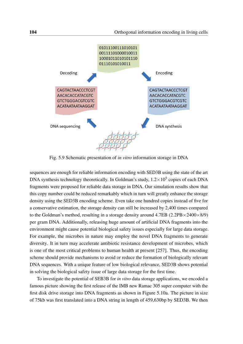
104 Orthogonal information encoding in living cells
Fig. 5.9 Schematic presentation of in vitro information storage in DNA
sequences are enough for reliable information encoding with SED3B using the state of the artDNA synthesis technology theoretically. In Goldman’s study, 1.2×10⁵ copies of each DNAfragments were proposed for reliable data storage in DNA. Our simulation results show thatthis copy number could be reduced remarkably which in turn will greatly enhance the storagedensity using the SED3B encoding scheme. Even take one hundred copies instead of five fora conservative estimation, the storage density can still be increased by 2,400 times comparedto the Goldman’s method, resulting in a storage density around 4.7EB (2.2PB×2400×8/9)per gram DNA. Additionally, releasing huge amount of artificial DNA fragments into theenvironment might cause potential biological safety issues especially for large data storage.For example, the microbes in nature may employ the novel DNA fragments to generatediversity. It in turn may accelerate antibiotic resistance development of microbes, whichis one of the most critical problems to human health at present [257]. Thus, the encodingscheme should provide mechanisms to avoid or reduce the formation of biologically relevantDNA sequences. With a unique feature of low biological relevance, SED3B shows potentialin solving the biological safety issue of large data storage for the first time.
To investigate the potential of SEB3B for in vitro data storage applications, we encoded afamous picture showing the first release of the IMB new Ramac 305 super computer with thefirst disk drive storage into DNA fragments as shown in Figure 5.10a. The picture in sizeof 75kb was first translated into a DNA string in length of 459,630bp by SED3B. We then
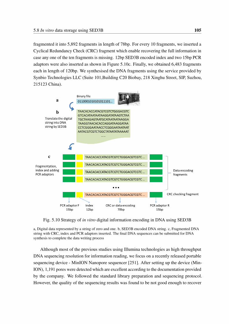
5.8 In vitro data storage using SED3B 105
fragmented it into 5,892 fragments in length of 78bp. For every 10 fragments, we inserted aCyclical Redundancy Check (CRC) fragment which enable recovering the full information incase any one of the ten fragments is missing. 12bp SED3B encoded index and two 15bp PCRadaptors were also inserted as shown in Figure 5.10c. Finally, we obtained 6,483 fragmentseach in length of 120bp. We synthesised the DNA fragments using the service provided bySynbio Technologies LLC (Suite 101,Building C20 Biobay, 218 Xinghu Street, SIP, Suzhou,215123 China).
Fig. 5.10 Strategy of in vitro digital information encoding in DNA using SED3B
a, Digital data represented by a string of zero and one. b, SED3B encoded DNA string. c, Fragmented DNAstring with CRC, index and PCR adaptors inserted. The final DNA sequences can be submitted for DNAsynthesis to complete the data writing process
Although most of the previous studies using Illumina technologies as high throughputDNA sequencing resolution for information reading, we focus on a recently released portablesequencing device - MinION Nanopore sequencer [251]. After setting up the device (Min-ION), 1,191 pores were detected which are excellent according to the documentation providedby the company. We followed the standard library preparation and sequencing protocol.However, the quality of the sequencing results was found to be not good enough to recover
106 Orthogonal information encoding in living cells
the information (data not shown). A possible reason is the nanopore sequencing protocol isdesigned for sequencing of long fragments (>500bp). In our study, 120bp fragments wereused which may lead to a low quality of the sequencing results. In a more recent study byYazdi et al., 1kb DNA fragments were used to encode data and the data can be read correctlyusing MinION [250]. Since high throughput DNA synthesis is limited to short DNA oligoscurrently, our results indicate that further efforts are required to apply nanopore sequencingtechnology to fetch the data encoded in short DNA oligos.
5.9 Development of an online encoding-decoding system
To facilitate the utilization of SED3B as an information encoding system in living cells, anonline encoding-decoding system for comment and barcode encoding-decoding has beenimplemented and released (http://biosystem.bt1.tu-harburg.de/sed3b/) as shown in Figure5.11.
5.10 Conclusion
Reliable information storage in vivo in error rich DNA molecules is still represents a challengesince more and more errors could be introduced and enriched exponentially by rounds ofreplications. In this study, we presented a novel encoding scheme named SED3B, whichcan take full advantage of the inherent redundancy of DNA molecules for error correction.By using a small number of DNA molecules for error correction, SED3B can effectivelycorrect the exponentially enriching errors during DNA replications as proved by in siliconsimulation and error-prone PCR experiments for the first time. Based on error-prone PCRinvivo experiments with E. coli cells, more than 12,000 years of continuous replications areestimated to be required to make the SED3B encoded information unrecoverable in growingE. coli cells. Furthermore, for the first time we showed that SED3B encoded DNA sequenceshave little biological relevance to known natural DNA sequences, indicating its excellentorthogonality. Synthetic biologists are trying to design biological devices and algorithms toprogramm cells for various functions. Similar to the situation in programming of computersand machines, we need to write information such as comments or barcodes in the syntheticmolecular programs. In these cases, SED3B is well suitable for reliable information encodingwith no or low affections to the biological functions.
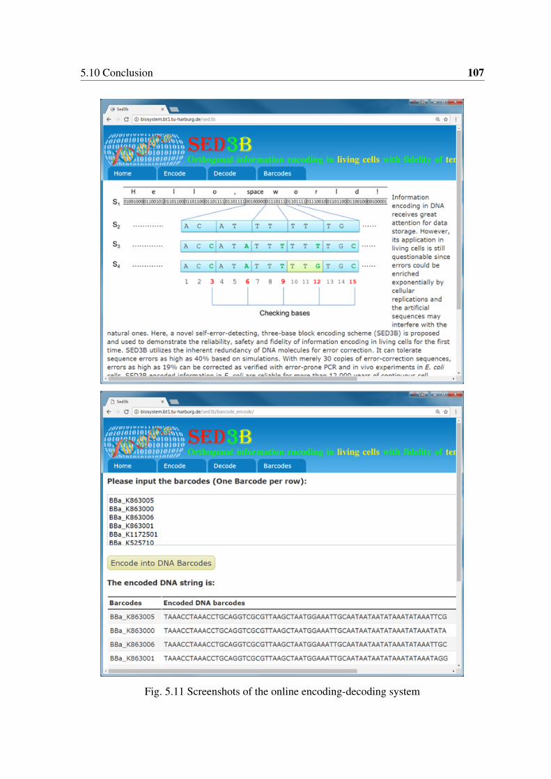
5.10 Conclusion 107
Fig. 5.11 Screenshots of the online encoding-decoding system

Chapter 6
Summary and outlook
Systems biology studies have been performed on ten strains of mutans streptococci for thesake of identification of the targets of carolacton and better understanding of the pathogenicityof mutants streptococci strains. Eight S. mutans strains, one S. ratti strains and one S. sobrinusstrain were genome sequenced. Mathematic modeling of the S.mutans pan-genome displayeda possible opened pan-genome with high lateral gene transfer rate than other establishedpan-genome models of Streptococcus species. Distribution of factors which are thoughtto be highly related to pathology was systematically studied. Most of the studied systemsshow high diversities across different species except oxidative resistance system. The wholegenome level metabolic networks were constructed and cross-compared. Diversities incentral metabolic pathways were observed and a novel energy production pathway uniquelypresented in S. sobrinus DSM20742 was revealed for the first time with the involvement oftwo novel lactate oxidases. Possible opened pan-genome, high lateral transferred genes rate,high diversities in resistance related systems and metabolic pathways – the pathogenicity ofmutans streptococci should be re-evaluated. The strain-specific information provided in thisstudy is helpful to understand the evolution and adaptive mechanisms of mutans streptococciand in turn will be very helpful for researchers to better understand those oral pathogens. Anonline regulation database for S. mutans, named StrepReg, was constructed by integratingtime-resolved transcription factor based regulation network, STRING interaction databaseand KEGG pathway database (http://biosystem.bt1.tu-harburg.de:1555/homes/).
Due to the inherent complexity of the biological systems, biological engineering isunpredictable and cannot be performed in a rational way like other engineering disciplinessuch as electronic engineering. The biological engineering process have to go throughmany design-build-test cycles, within which many designs/variations have to be evaluated togenerate some targets with desired properties. In other words, the biological engineering issuffered from the time- and labor- intensive ’debugging’ process. Inspired by the debugging
110 Summary and outlook
process of programming on a computer, a debugging system is proposed to speed up thedebugging process of biological engineering. To this end, we implemented a multiple IOsystem based on M13 phages which can be used as a debugging system for biologicalengineering in E. coli. Our proof of concept studies show that the current IO system canbe utilized for applications of parallel evaluation of designs, beneficial genetic operationprediction and screening. The IO system shows higher sensitivity compared to the state ofthe art method of flow cytometry. Although further experiments are required, the IO systemdoes show great potential in scaling up the input-output numbers. Scaling up the inputs togenome-level and combining the electric machine based robots may open up a new wayto study the complex interactions of various intracellular components. Consequently, themassive information obtained could be helpful for mathematic modeling studies in systemsbiology.
In addition, a novel encoding scheme SED3B which can take full advantage of theinherent redundancy of DNA molecules for error correction was implemented. SED3B caneffectively correct the exponentially enriching errors during DNA replications by using asmall population of DNA molecules as proved by in silicon simulation and error-prone PCRexperiments. Based on error-prone PCR in vivo experiments with E. coli cells, more than12,000 years of continuous replications are estimated to be required to make the SED3Bencoded information unrecoverable in growing E. coli cells. Furthermore, for the first timewe showed that SED3B encoded DNA sequences have little biological relevance to knownnatural DNA sequences. Although further investigations are definitely required, the SED3Bencoding scheme is also applicable for in vitro data storage in DNA in principle. Indeed,SED3B does show advantages in large data storage theoretically. We noticed that by usingmerely five DNA sequences SED3B can correct an error rate of 5%. It has been reported thatthe error rate of high throughput DNA synthesis technology is around 0.5% currently [26].Thus, five sequences are enough for reliable information encoding by the state of the art DNAsynthesis technology using SED3B theoretically. In Goldman’s study, 1.2×10⁵ copies ofeach DNA fragments were proposed for reliable data storage in DNA. Our results show thatthis copy number could be reduced remarkably which in turn will greatly enhance the storagedensity. Even take one hundred instead of five for reliable estimation, the storage density stillcan be increased by 2,400 times compared to the Goldman’s method, resulting in a storagedensity around 4.7EB (2.2PB×2400×8/9) per gram DNA considering the encoding efficiency.Additionally, releasing huge amounts of artificial DNA fragments into the environment mightcause potential biological safety issues especially for large data storage. For example, themicrobes in nature may employ the novel DNA fragments to generate diversity. It in turn mayaccelerate antibiotic resistance development of microbes, which is one of the most critical

111
problems to human health at present [257]. Thus, the encoding scheme should providemechanisms to avoid or reduce the formation of biologically relevant DNA sequences. Witha unique feature of low biological relevance, SED3B shows potential in large data storageconcerning about the biological safety issue for the first time.

References
[1] W. J. Loesche, “Role of Streptococcus mutans in human dental decay,” MicrobiologicalReviews, vol. 50, no. 4, pp. 353–380, 1986.
[2] P. Stoodley, K. Sauer, D. G. Davies, and J. W. Costerton, “Biofilms as complexdifferentiated communities,” Annual Review of Microbiology, vol. 56, pp. 187–209,2002.
[3] F. J. Bruggeman and H. V. Westerhoff, “The nature of systems biology,” Trends inMicrobiology, vol. 15, no. 1, pp. 45–50, 2007.
[4] C. J. Cain, D. A. Conte, M. E. Garcia-Ojeda, L. G. Daglio, L. Johnson, E. H. Lau, J. O.Manilay, J. B. Phillips, N. S. Rogers, N. S. Stolberg, H. F. Swift, and M. N. Dawson,“Integrative biology: What systems biology is (not, yet),” Science, vol. 320, no. 5879,pp. 1013a–1014a, 2008.
[5] H.-Y. Chuang, M. Hofree, and T. Ideker, “A decade of systems biology,” AnnualReview of Cell and Developmental Biology, vol. 26, pp. 721–744, 2010.
[6] A. Chiappino-Pepe, V. Pandey, M. Ataman, and V. Hatzimanikatis, “Integration ofmetabolic, regulatory and signaling networks towards analysis of perturbation anddynamic responses,” Current Opinion in Systems Biology, vol. 2, pp. 58–65, 2017.
[7] K.-H. Cho, S. Lee, D. Kim, D. Shin, J. I. Joo, and S.-M. Park, “Cancer reversion, arenewed challenge in systems biology,” Current Opinion in Systems Biology, vol. 2,pp. 48–57, 2017.
[8] A. Dominguez, E. Munoz, M. C. Lopez, M. Cordero, J. P. Martinez, and M. Vinas,“Transcriptomics as a tool to discover new antibacterial targets,” Biotechnology Letters,2017.
[9] S. Durmus, T. Cakir, A. Ozgur, and R. Guthke, “A review on computational systemsbiology of pathogen-host interactions,” Frontiers in Microbiology, vol. 6, p. 235, 2015.
114 References
[10] J. Geng and J. Nielsen, “In silico analysis of human metabolism: Reconstruction, con-textualization and application of genome-scale models,” Current Opinion in SystemsBiology, vol. 2, pp. 28–37, 2017.
[11] W. J. Kim, H. U. Kim, and S. Y. Lee, “Current state and applications of microbialgenome-scale metabolic models,” Current Opinion in Systems Biology, vol. 2, pp. 9–17, 2017.
[12] S. Magi, K. Iwamoto, and M. Okada-Hatakeyama, “Current status of mathematicalmodeling of cancer – from the viewpoint of cancer hallmarks,” Current Opinion inSystems Biology, vol. 2, pp. 38–47, 2017.
[13] J. Nielsen, “Systems biology of metabolism: A driver for developing personalized andprecision medicine,” Cell Metabolism, vol. 25, no. 3, pp. 572–579, 2017.
[14] E. J. Strobel, K. E. Watters, D. Loughrey, and J. B. Lucks, “Rna systems biology:uniting functional discoveries and structural tools to understand global roles of rnas,”Current Opinion in Biotechnology, vol. 39, pp. 182–191, 2016.
[15] L. S. Yilmaz and A. J. Walhout, “Metabolic network modeling with model organisms,”Current Opinion in Chemical Biology, vol. 36, pp. 32–39, 2017.
[16] J. W. Lee, T. Y. Kim, Y.-S. Jang, S. Choi, and S. Y. Lee, “Systems metabolic engineer-ing for chemicals and materials,” Trends in Biotechnology, vol. 29, no. 8, pp. 370–378,2011.
[17] S. Y. Lee and H. U. Kim, “Systems strategies for developing industrial microbialstrains,” Nature Biotechnology, vol. 33, no. 10, pp. 1061–1072, 2015.
[18] M. Gustavsson and S. Y. Lee, “Prospects of microbial cell factories developed throughsystems metabolic engineering,” Microbial Biotechnology, vol. 9, no. 5, pp. 610–617,2016.
[19] A. S. L. Hansen, R. M. Lennen, N. Sonnenschein, and M. J. Herrgard, “Systemsbiology solutions for biochemical production challenges,” Current Opinion in Biotech-nology, vol. 45, pp. 85–91, 2017.
[20] H. M. Purdy and J. L. Reed, “Evaluating the capabilities of microbial chemical pro-duction using genome-scale metabolic models,” Current Opinion in Systems Biology,vol. 2, pp. 90–96, 2017.
References 115
[21] Y. Vervoort, A. G. Linares, M. Roncoroni, C. Liu, J. Steensels, and K. J. Verstrepen,“High-throughput system-wide engineering and screening for microbial biotechnology,”Current Opinion in Biotechnology, vol. 46, pp. 120–125, 2017.
[22] V. Zhirnov, R. M. Zadegan, G. S. Sandhu, G. M. Church, and W. L. Hughes, “Nucleicacid memory,” Nature Materials, vol. 15, no. 4, pp. 366–370, 2016.
[23] G. M. Church, Y. Gao, and S. Kosuri, “Next-generation digital information storage indna,” Science (New York, N.Y.), vol. 337, no. 6102, p. 1628, 2012.
[24] N. Goldman, P. Bertone, S. Chen, C. Dessimoz, E. M. LeProust, B. Sipos, andE. Birney, “Towards practical, high-capacity, low-maintenance information storage insynthesized dna,” Nature, vol. 494, no. 7435, pp. 77–80, 2013.
[25] M. Jain, I. T. Fiddes, K. H. Miga, H. E. Olsen, B. Paten, and M. Akeson, “Improveddata analysis for the minion nanopore sequencer,” Nature Methods, vol. 12, no. 4,pp. 351–356, 2015.
[26] S. Kosuri and G. M. Church, “Large-scale de novo dna synthesis: technologies andapplications,” Nature Methods, vol. 11, no. 5, pp. 499–507, 2014.
[27] “http://syntheticbiology.org/vectors/barcode.html.”
[28] D. Ajdic, W. M. McShan, R. E. McLaughlin, G. Savic, J. Chang, M. B. Carson,C. Primeaux, R. Tian, S. Kenton, H. Jia, S. Lin, Y. Qian, S. Li, H. Zhu, F. Najar,H. Lai, J. White, B. A. Roe, and J. J. Ferretti, “Genome sequence of Streptococcusmutans ua159, a cariogenic dental pathogen,” Proceedings of the National Academyof Sciences of the United States of America, vol. 99, no. 22, pp. 14434–14439, 2002.
[29] F. Maruyama, M. Kobata, K. Kurokawa, K. Nishida, A. Sakurai, K. Nakano, R. No-mura, S. Kawabata, T. Ooshima, K. Nakai, M. Hattori, S. Hamada, and I. Nakagawa,“Comparative genomic analyses of Streptococcus mutans provide insights into chro-mosomal shuffling and species-specific content,” BMC Genomics, vol. 10, p. 358,2009.
[30] P. S. G. Chain, D. V. Grafham, R. S. Fulton, M. G. Fitzgerald, J. Hostetler, D. Muzny,J. Ali, B. Birren, D. C. Bruce, C. Buhay, J. R. Cole, Y. Ding, S. Dugan, D. Field, G. M.Garrity, R. Gibbs, T. Graves, C. S. Han, S. H. Harrison, S. Highlander, P. Hugenholtz,H. M. Khouri, C. D. Kodira, E. Kolker, N. C. Kyrpides, D. Lang, A. Lapidus, S. A.Malfatti, V. Markowitz, T. Metha, K. E. Nelson, J. Parkhill, S. Pitluck, X. Qin, T. D.
116 References
Read, J. Schmutz, S. Sozhamannan, P. Sterk, R. L. Strausberg, G. Sutton, N. R.Thomson, J. M. Tiedje, G. Weinstock, A. Wollam, and J. C. Detter, “Genomics.genome project standards in a new era of sequencing,” Science (New York, N.Y.),vol. 326, no. 5950, pp. 236–237, 2009.
[31] R. Li, C. Yu, Y. Li, T.-W. Lam, S.-M. Yiu, K. Kristiansen, and J. Wang, “Soap2: animproved ultrafast tool for short read alignment,” Bioinformatics (Oxford, England),vol. 25, no. 15, pp. 1966–1967, 2009.
[32] H. Li and R. Durbin, “Fast and accurate short read alignment with burrows-wheelertransform,” Bioinformatics (Oxford, England), vol. 25, no. 14, pp. 1754–1760, 2009.
[33] M. de La Bastide and W. R. McCombie, “Assembling genomic dna sequences withphrap,” Current Protocols in Bioinformatics, vol. Chapter 11, p. Unit11.4, 2007.
[34] A. C. E. Darling, B. Mau, F. R. Blattner, and N. T. Perna, “Mauve: multiple alignmentof conserved genomic sequence with rearrangements,” Genome Research, vol. 14,no. 7, pp. 1394–1403, 2004.
[35] H. Tettelin, D. Riley, C. Cattuto, and D. Medini, “Comparative genomics: the bacterialpan-genome,” Current Opinion in Microbiology, vol. 11, no. 5, pp. 472–477, 2008.
[36] Y. Zhao, J. Wu, J. Yang, S. Sun, J. Xiao, and J. Yu, “Pgap: pan-genomes analysispipeline,” Bioinformatics (Oxford, England), vol. 28, no. 3, pp. 416–418, 2012.
[37] H. Tettelin, V. Masignani, M. J. Cieslewicz, C. Donati, D. Medini, N. L. Ward, S. V.Angiuoli, J. Crabtree, A. L. Jones, A. S. Durkin, R. T. Deboy, T. M. Davidsen, M. Mora,M. Scarselli, I. Margarit y Ros, J. D. Peterson, C. R. Hauser, J. P. Sundaram, W. C.Nelson, R. Madupu, L. M. Brinkac, R. J. Dodson, M. J. Rosovitz, S. A. Sullivan,S. C. Daugherty, D. H. Haft, J. Selengut, M. L. Gwinn, L. Zhou, N. Zafar, H. Khouri,D. Radune, G. Dimitrov, K. Watkins, K. J. B. O’Connor, S. Smith, T. R. Utterback,O. White, C. E. Rubens, G. Grandi, L. C. Madoff, D. L. Kasper, J. L. Telford, M. R.Wessels, R. Rappuoli, and C. M. Fraser, “Genome analysis of multiple pathogenicisolates of Streptococcus agalactiae: implications for the microbial "pan-genome",”Proceedings of the National Academy of Sciences of the United States of America,vol. 102, no. 39, pp. 13950–13955, 2005.
[38] L. Li, C. J. Stoeckert, JR, and D. S. Roos, “Orthomcl: identification of ortholog groupsfor eukaryotic genomes,” Genome Research, vol. 13, no. 9, pp. 2178–2189, 2003.
References 117
[39] J. L. Lavin, K. Kiil, O. Resano, D. W. Ussery, and J. A. Oguiza, “Comparativegenomic analysis of two-component regulatory proteins in Pseudomonas syringae,”BMC Genomics, vol. 8, p. 397, 2007.
[40] R. D. Finn, J. Mistry, J. Tate, P. Coggill, A. Heger, J. E. Pollington, O. L. Gavin,P. Gunasekaran, G. Ceric, K. Forslund, L. Holm, E. L. L. Sonnhammer, S. R. Eddy,and A. Bateman, “The pfam protein families database,” Nucleic Acids Research,vol. 38, no. Database issue, pp. D211–22, 2010.
[41] S. R. Eddy, “Profile hidden markov models,” Bioinformatics (Oxford, England),vol. 14, no. 9, pp. 755–763, 1998.
[42] P. D. Karp, S. M. Paley, M. Krummenacker, M. Latendresse, J. M. Dale, T. J. Lee,P. Kaipa, F. Gilham, A. Spaulding, L. Popescu, T. Altman, I. Paulsen, I. M. Keseler,and R. Caspi, “Pathway tools version 13.0: integrated software for pathway/genomeinformatics and systems biology,” Briefings in Bioinformatics, vol. 11, no. 1, pp. 40–79,2010.
[43] H. Ma and A.-P. Zeng, “Reconstruction of metabolic networks from genome dataand analysis of their global structure for various organisms,” Bioinformatics (Oxford,England), vol. 19, no. 2, pp. 270–277, 2003.
[44] M. Stelzer, J. Sun, T. Kamphans, S. P. Fekete, and A.-P. Zeng, “An extended bioreac-tion database that significantly improves reconstruction and analysis of genome-scalemetabolic networks,” Integrative Biology : quantitative biosciences from nano tomacro, vol. 3, no. 11, pp. 1071–1086, 2011.
[45] M. Kohl, S. Wiese, and B. Warscheid, “Cytoscape: software for visualization and anal-ysis of biological networks,” Methods in Molecular Biology (Clifton, N.J.), vol. 696,pp. 291–303, 2011.
[46] P. C. Y. Lau, C. K. Sung, J. H. Lee, D. A. Morrison, and D. G. Cvitkovitch, “Pcr ligationmutagenesis in transformable streptococci: application and efficiency,” Journal ofmicrobiological methods, vol. 49, no. 2, pp. 193–205, 2002.
[47] M. Reck, K. Rutz, B. Kunze, J. Tomasch, S. K. Surapaneni, S. Schulz, and I. Wagner-Döbler, “The biofilm inhibitor carolacton disturbs membrane integrity and cell divisionof streptococcus mutans through the serine/threonine protein kinase pknb,” Journal ofbacteriology, vol. 193, no. 20, pp. 5692–5706, 2011.
118 References
[48] J. Lefrançois, M. M. Samrakandi, and A. M. Sicard, “Electrotransformation andnatural transformation of streptococcus pneumoniae: requirement of dna processingfor recombination,” Microbiology (Reading, England), vol. 144 ( Pt 11), pp. 3061–3068, 1998.
[49] O. Ween, S. Teigen, P. Gaustad, M. Kilian, and L. S. Havarstein, “Competence withouta competence pheromone in a natural isolate of streptococcus infantis,” Journal ofbacteriology, vol. 184, no. 13, pp. 3426–3432, 2002.
[50] Y. H. Li, P. C. Lau, J. H. Lee, R. P. Ellen, and D. G. Cvitkovitch, “Natural genetictransformation of streptococcus mutans growing in biofilms,” Journal of bacteriology,vol. 183, no. 3, pp. 897–908, 2001.
[51] D. J. LeBlanc, Y.-Y. Chen, N. D. Buckley, and L. N. Lee, “Genetic transfer methods forstreptococcus sobrinus and other oral streptococci,” Methods in Cell Science, vol. 20,no. 1/4, pp. 85–93, 1998.
[52] M. G. Caparon and J. R. Scott, “Genetic manipulation of pathogenic streptococci,”Methods in enzymology, vol. 204, pp. 556–586, 1991.
[53] R. E. McLaughlin and J. J. Ferretti, “Electrotransformation of streptococci,” Methodsin molecular biology (Clifton, N.J.), vol. 47, pp. 185–193, 1995.
[54] A. H. Badran and D. R. Liu, “Development of potent in vivo mutagenesis plasmidswith broad mutational spectra,” Nature Communications, vol. 6, p. 8425, 2015.
[55] S. BLACK and N. G. WRIGHT, “beta-aspartokinase and beta-aspartyl phosphate,”The Journal of Biological Chemistry, vol. 213, no. 1, pp. 27–38, 1955.
[56] J. Täpp, M. Thollesson, and B. Herrmann, “Phylogenetic relationships and genotypingof the genus streptococcus by sequence determination of the rnase p rna gene, rnpb,”International journal of systematic and evolutionary microbiology, vol. 53, no. Pt 6,pp. 1861–1871, 2003.
[57] J. A. Lemos and R. A. Burne, “A model of efficiency: stress tolerance by Streptococcusmutans,” Microbiology (Reading, England), vol. 154, no. Pt 11, pp. 3247–3255, 2008.
[58] K. Nakano, R. Nomura, M. Matsumoto, and T. Ooshima, “Roles of oral bacteria incardiovascular diseases–from molecular mechanisms to clinical cases: Cell-surfacestructures of novel serotype k Streptococcus mutans strains and their correlation tovirulence,” Journal of Pharmacological Sciences, vol. 113, no. 2, pp. 120–125, 2010.
References 119
[59] R. Nomura, K. Nakano, N. Taniguchi, J. Lapirattanakul, H. Nemoto, L. Gronroos,S. Alaluusua, and T. Ooshima, “Molecular and clinical analyses of the gene encodingthe collagen-binding adhesin of Streptococcus mutans,” Journal of Medical Microbiol-ogy, vol. 58, no. Pt 4, pp. 469–475, 2009.
[60] R. J. Redfield, W. A. Findlay, J. Bosse, J. S. Kroll, A. D. S. Cameron, and J. H. Nash,“Evolution of competence and dna uptake specificity in the Pasteurellaceae,” BMCEvolutionary Biology, vol. 6, p. 82, 2006.
[61] G. D. Ehrlich, F. Z. Hu, K. Shen, P. Stoodley, and J. C. Post, “Bacterial plurality as ageneral mechanism driving persistence in chronic infections,” Clinical Orthopaedicsand Related Research, no. 437, pp. 20–24, 2005.
[62] O. E. Cornejo, T. Lefébure, P. D. P. Bitar, P. Lang, V. P. Richards, K. Eilertson, T. Do,D. Beighton, L. Zeng, S.-J. Ahn, R. A. Burne, A. Siepel, C. D. Bustamante, andM. J. Stanhope, “Evolutionary and population genomics of the cavity causing bacteriastreptococcus mutans,” Molecular biology and evolution, vol. 30, no. 4, pp. 881–893,2013.
[63] M. A. Larkin, G. Blackshields, N. P. Brown, R. Chenna, P. A. McGettigan,H. McWilliam, F. Valentin, I. M. Wallace, A. Wilm, R. Lopez, J. D. Thompson,T. J. Gibson, and D. G. Higgins, “Clustal w and clustal x version 2.0,” Bioinformatics(Oxford, England), vol. 23, no. 21, pp. 2947–2948, 2007.
[64] J. D. Retief, “Phylogenetic analysis using phylip,” Methods in molecular biology(Clifton, N.J.), vol. 132, pp. 243–258, 2000.
[65] A. E. Darling, I. Miklós, and M. A. Ragan, “Dynamics of genome rearrangement inbacterial populations,” PLoS genetics, vol. 4, no. 7, p. e1000128, 2008.
[66] J. C. Waterhouse, D. C. Swan, and R. R. B. Russell, “Comparative genome hybridiza-tion of streptococcus mutans strains,” Oral microbiology and immunology, vol. 22,no. 2, pp. 103–110, 2007.
[67] C. Wu, R. Cichewicz, Y. Li, J. Liu, B. Roe, J. Ferretti, J. Merritt, and F. Qi, “Genomicisland tnsmu2 of Streptococcus mutans harbors a nonribosomal peptide synthetase-polyketide synthase gene cluster responsible for the biosynthesis of pigments involvedin oxygen and h2o2 tolerance,” Applied and Environmental Microbiology, vol. 76,no. 17, pp. 5815–5826, 2010.
120 References
[68] J. C. Waterhouse and R. R. B. Russell, “Dispensable genes and foreign dna in strepto-coccus mutans,” Microbiology (Reading, England), vol. 152, no. Pt 6, pp. 1777–1788,2006.
[69] A. E. Darling, B. Mau, and N. T. Perna, “progressivemauve: multiple genome align-ment with gene gain, loss and rearrangement,” PloS one, vol. 5, no. 6, p. e11147,2010.
[70] A. Muzzi and C. Donati, “Population genetics and evolution of the pan-genome ofstreptococcus pneumoniae,” International journal of medical microbiology : IJMM,vol. 301, no. 8, pp. 619–622, 2011.
[71] A. Mira, A. B. Martín-Cuadrado, G. D’Auria, and F. Rodríguez-Valera, “The bacterialpan-genome:a new paradigm in microbiology,” International microbiology : theofficial journal of the Spanish Society for Microbiology, vol. 13, no. 2, pp. 45–57,2010.
[72] C. Donati, N. L. Hiller, H. Tettelin, A. Muzzi, N. J. Croucher, S. V. Angiuoli, M. Og-gioni, J. C. Dunning Hotopp, F. Z. Hu, D. R. Riley, A. Covacci, T. J. Mitchell, S. D.Bentley, M. Kilian, G. D. Ehrlich, R. Rappuoli, E. R. Moxon, and V. Masignani,“Structure and dynamics of the pan-genome of Streptococcus pneumoniae and closelyrelated species,” Genome Biology, vol. 11, no. 10, p. R107, 2010.
[73] T. Lefebure and M. J. Stanhope, “Evolution of the core and pan-genome of Streptococ-cus: positive selection, recombination, and genome composition,” Genome Biology,vol. 8, no. 5, p. R71, 2007.
[74] J. S. Hogg, F. Z. Hu, B. Janto, R. Boissy, J. Hayes, R. Keefe, J. C. Post, and G. D.Ehrlich, “Characterization and modeling of the haemophilus influenzae core andsupragenomes based on the complete genomic sequences of rd and 12 clinical nonty-peable strains,” Genome Biology, vol. 8, no. 6, p. R103, 2007.
[75] A. M. Stock, V. L. Robinson, and P. N. Goudreau, “Two-component signal transduc-tion,” Annual Review of Biochemistry, vol. 69, pp. 183–215, 2000.
[76] I. Biswas, L. Drake, D. Erkina, and S. Biswas, “Involvement of sensor kinases in thestress tolerance response of Streptococcus mutans,” Journal of Bacteriology, vol. 190,no. 1, pp. 68–77, 2008.

References 121
[77] Y. Liu and R. A. Burne, “Multiple two-component systems of Streptococcus mu-tans regulate agmatine deiminase gene expression and stress tolerance,” Journal ofBacteriology, vol. 191, no. 23, pp. 7363–7366, 2009.
[78] S. F. Lee, G. D. Delaney, and M. Elkhateeb, “A two-component covrs regulatory sys-tem regulates expression of fructosyltransferase and a novel extracellular carbohydratein Streptococcus mutans,” Infection and Immunity, vol. 72, no. 7, pp. 3968–3973,2004.
[79] van der Ploeg, Jan R, “Regulation of bacteriocin production in Streptococcus mutansby the quorum-sensing system required for development of genetic competence,”Journal of Bacteriology, vol. 187, no. 12, pp. 3980–3989, 2005.
[80] M. D. Senadheera, B. Guggenheim, G. A. Spatafora, Y.-C. C. Huang, J. Choi, D. C. I.Hung, J. S. Treglown, S. D. Goodman, R. P. Ellen, and D. G. Cvitkovitch, “A vicrksignal transduction system in Streptococcus mutans affects gtfbcd, gbpb, and ftfexpression, biofilm formation, and genetic competence development,” Journal ofBacteriology, vol. 187, no. 12, pp. 4064–4076, 2005.
[81] C. M. Levesque, R. W. Mair, J. A. Perry, P. C. Y. Lau, Y.-H. Li, and D. G. Cvitkovitch,“Systemic inactivation and phenotypic characterization of two-component systems inexpression of Streptococcus mutans virulence properties,” Letters in Applied Microbi-ology, vol. 45, no. 4, pp. 398–404, 2007.
[82] V. Idone, S. Brendtro, R. Gillespie, S. Kocaj, E. Peterson, M. Rendi, W. Warren,S. Michalek, K. Krastel, D. Cvitkovitch, and G. Spatafora, “Effect of an orphanresponse regulator on Streptococcus mutans sucrose-dependent adherence and cario-genesis,” Infection and Immunity, vol. 71, no. 8, pp. 4351–4360, 2003.
[83] P.-M. Chen, H.-C. Chen, C.-T. Ho, C.-J. Jung, H.-T. Lien, J.-Y. Chen, and J.-S. Chia,“The two-component system scnrk of Streptococcus mutans affects hydrogen peroxideresistance and murine macrophage killing,” Microbes and Infection, vol. 10, no. 3,pp. 293–301, 2008.
[84] L. Zeng, S. Das, and R. A. Burne, “Genetic analysis of the functions and interactionsof components of the levqrst signal transduction complex of Streptococcus mutans,”PloS One, vol. 6, no. 2, p. e17335, 2011.
[85] P. Chong, L. Drake, and I. Biswas, “Modulation of covr expression in Streptococcusmutans ua159,” Journal of Bacteriology, vol. 190, no. 13, pp. 4478–4488, 2008.
122 References
[86] P. Chong, P. Chattoraj, and I. Biswas, “Activation of the smu.1882 transcription bycovr in Streptococcus mutans,” PloS One, vol. 5, no. 11, p. e15528, 2010.
[87] A. Dmitriev, S. S. Mohapatra, P. Chong, M. Neely, S. Biswas, and I. Biswas, “Covr-controlled global regulation of gene expression in Streptococcus mutans,” PloS One,vol. 6, no. 5, p. e20127, 2011.
[88] I. Biswas, L. Drake, and S. Biswas, “Regulation of gbpc expression in Streptococcusmutans,” Journal of Bacteriology, vol. 189, no. 18, pp. 6521–6531, 2007.
[89] I. Biswas and J. R. Scott, “Identification of roca, a positive regulator of covr expressionin the group a Streptococcus,” Journal of Bacteriology, vol. 185, no. 10, pp. 3081–3090, 2003.
[90] R. E. McLaughlin, J. J. Ferretti, and W. L. Hynes, “Nucleotide sequence of thestreptococcin a-ff22 lantibiotic regulon: model for production of the lantibiotic sa-ff22by strains of Streptococcus pyogenes,” FEMS Microbiology Letters, vol. 175, no. 2,pp. 171–177, 1999.
[91] S.-J. Ahn, Z. T. Wen, and R. A. Burne, “Multilevel control of competence developmentand stress tolerance in Streptococcus mutans UA159,” Infection and Immunity, vol. 74,no. 3, pp. 1631–1642, 2006.
[92] J. A. Perry, C. M. Levesque, P. Suntharaligam, R. W. Mair, M. Bu, R. T. Cline, S. N.Peterson, and D. G. Cvitkovitch, “Involvement of Streptococcus mutans regulator rr11in oxidative stress response during biofilm growth and in the development of geneticcompetence,” Letters in Applied Microbiology, vol. 47, no. 5, pp. 439–444, 2008.
[93] Y.-H. Li, N. Tang, M. B. Aspiras, P. C. Y. Lau, J. H. Lee, R. P. Ellen, and D. G.Cvitkovitch, “A quorum-sensing signaling system essential for genetic competencein Streptococcus mutans is involved in biofilm formation,” Journal of Bacteriology,vol. 184, no. 10, pp. 2699–2708, 2002.
[94] Y.-H. Li, X.-L. Tian, G. Layton, C. Norgaard, and G. Sisson, “Additive attenuation ofvirulence and cariogenic potential of Streptococcus mutans by simultaneous inacti-vation of the comcde quorum-sensing system and hk/rr11 two-component regulatorysystem,” Microbiology (Reading, England), vol. 154, no. Pt 11, pp. 3256–3265, 2008.
[95] J. Kreth, D. C. I. Hung, J. Merritt, J. Perry, L. Zhu, S. D. Goodman, D. G. Cvitkovitch,W. Shi, and F. Qi, “The response regulator come in Streptococcus mutans functions
References 123
both as a transcription activator of mutacin production and repressor of csp biosynthe-sis,” Microbiology (Reading, England), vol. 153, no. Pt 6, pp. 1799–1807, 2007.
[96] D. I. Liao, J. Qian, D. A. Chisholm, D. B. Jordan, and B. A. Diner, “Crystal structuresof the photosystem ii d1 c-terminal processing protease,” Nature structural biology,vol. 7, no. 9, pp. 749–753, 2000.
[97] M. Ansaldi and D. Dubnau, “Diversifying selection at the bacillus quorum-sensinglocus and determinants of modification specificity during synthesis of the comxpheromone,” Journal of Bacteriology, vol. 186, no. 1, pp. 15–21, 2004.
[98] C. Poyart, M. C. Lamy, C. Boumaila, F. Fiedler, and P. Trieu-Cuot, “Regulationof d-alanyl-lipoteichoic acid biosynthesis in Streptococcus agalactiae involves anovel two-component regulatory system,” Journal of Bacteriology, vol. 183, no. 21,pp. 6324–6334, 2001.
[99] N. Fittipaldi, T. Sekizaki, D. Takamatsu, J. Harel, Dominguez-Punaro, Maria de laCruz, S. von Aulock, C. Draing, C. Marois, M. Kobisch, and M. Gottschalk, “D-alanylation of lipoteichoic acid contributes to the virulence of Streptococcus suis,”Infection and Immunity, vol. 76, no. 8, pp. 3587–3594, 2008.
[100] F. Fabretti, C. Theilacker, L. Baldassarri, Z. Kaczynski, A. Kropec, O. Holst, andJ. Huebner, “Alanine esters of enterococcal lipoteichoic acid play a role in biofilmformation and resistance to antimicrobial peptides,” Infection and Immunity, vol. 74,no. 7, pp. 4164–4171, 2006.
[101] J. M. Tanzer, J. Livingston, and A. M. Thompson, “The microbiology of primarydental caries in humans,” Journal of Dental Education, vol. 65, no. 10, pp. 1028–1037,2001.
[102] J. D. F. Hale, N. C. K. Heng, R. W. Jack, and J. R. Tagg, “Identification of nlmte, thelocus encoding the abc transport system required for export of nonlantibiotic mutacinsin Streptococcus mutans,” Journal of Bacteriology, vol. 187, no. 14, pp. 5036–5039,2005.
[103] M. S. Hossain and I. Biswas, “Mutacins from Streptococcus mutans ua159 are activeagainst multiple streptococcal species,” Applied and Environmental Microbiology,vol. 77, no. 7, pp. 2428–2434, 2011.
[104] Y. Liu and H. Lu, “Microfluidics in systems biology-hype or truly useful?,” CurrentOpinion in Biotechnology, vol. 39, pp. 215–220, 2016.
124 References
[105] F. C. Petersen and A. A. Scheie, “Genetic transformation in streptococcus mutansrequires a peptide secretion-like apparatus,” Oral Microbiology and Immunology,vol. 15, no. 5, pp. 329–334, 2000.
[106] F. C. Petersen, G. Fimland, and A. A. Scheie, “Purification and functional studies of apotent modified quorum-sensing peptide and a two-peptide bacteriocin in streptococ-cus mutans,” Molecular microbiology, vol. 61, no. 5, pp. 1322–1334, 2006.
[107] E. Allan, H. A. Hussain, K. R. Crawford, S. Miah, Z. K. Ascott, M. H. Khwaja, andA. H. F. Hosie, “Genetic variation in comc, the gene encoding competence-stimulatingpeptide (csp) in Streptococcus mutans,” FEMS Microbiology Letters, vol. 268, no. 1,pp. 47–51, 2007.
[108] J. Kreth, J. Merritt, W. Shi, and F. Qi, “Co-ordinated bacteriocin production andcompetence development: a possible mechanism for taking up dna from neighbouringspecies,” Molecular Microbiology, vol. 57, no. 2, pp. 392–404, 2005.
[109] J. Kreth, J. Merritt, L. Zhu, W. Shi, and F. Qi, “Cell density- and come-dependentexpression of a group of mutacin and mutacin-like genes in Streptococcus mutans,”FEMS Microbiology Letters, vol. 265, no. 1, pp. 11–17, 2006.
[110] L. Mashburn-Warren, D. A. Morrison, and M. J. Federle, “A novel double-tryptophanpeptide pheromone controls competence in Streptococcus spp. via an rgg regulator,”Molecular Microbiology, vol. 78, no. 3, pp. 589–606, 2010.
[111] T. Okinaga, G. Niu, Z. Xie, F. Qi, and J. Merritt, “The hdrrm operon of Streptococcusmutans encodes a novel regulatory system for coordinated competence developmentand bacteriocin production,” Journal of Bacteriology, vol. 192, no. 7, pp. 1844–1852,2010.
[112] T. Okinaga, Z. Xie, G. Niu, F. Qi, and J. Merritt, “Examination of the hdrrm regulonyields insight into the competence system of Streptococcus mutans,” Molecular OralMicrobiology, vol. 25, no. 3, pp. 165–177, 2010.
[113] Z. Xie, T. Okinaga, G. Niu, F. Qi, and J. Merritt, “Identification of a novel bacteriocinregulatory system in Streptococcus mutans,” Molecular Microbiology, vol. 78, no. 6,pp. 1431–1447, 2010.
[114] R. W. Mair, D. B. Senadheera, and D. G. Cvitkovitch, “Cina is regulated via comx tomodulate genetic transformation and cell viability in streptococcus mutans,” FEMSmicrobiology letters, vol. 331, no. 1, pp. 44–52, 2012.
References 125
[115] S. Alaluusua, T. Takei, T. Ooshima, and S. Hamada, “Mutacin activity of strainsisolated from children with varying levels of mutants streptococci and caries,” Archivesof Oral Biology, vol. 36, no. 4, pp. 251–255, 1991.
[116] T. Baba and O. Schneewind, “Instruments of microbial warfare: bacteriocin synthesis,toxicity and immunity,” Trends in Microbiology, vol. 6, no. 2, pp. 66–71, 1998.
[117] I. F. Nes, D. B. Diep, and H. Holo, “Bacteriocin diversity in Streptococcus andEnterococcus,” Journal of Bacteriology, vol. 189, no. 4, pp. 1189–1198, 2007.
[118] S. Bekal-Si Ali, Y. Hurtubise, M. C. Lavoie, and G. LaPointe, “Diversity of Strep-tococcus mutans bacteriocins as confirmed by dna analysis using specific molecularprobes,” Gene, vol. 283, no. 1-2, pp. 125–131, 2002.
[119] H. Yonezawa and H. K. Kuramitsu, “Genetic analysis of a unique bacteriocin, smb,produced by Streptococcus mutans gs5,” Antimicrobial Agents and Chemotherapy,vol. 49, no. 2, pp. 541–548, 2005.
[120] O. Hyink, M. Balakrishnan, and J. R. Tagg, “Streptococcus rattus strain bht producesboth a class i two-component lantibiotic and a class ii bacteriocin,” FEMS microbiologyletters, vol. 252, no. 2, pp. 235–241, 2005.
[121] T. Nguyen, Z. Zhang, I.-H. Huang, C. Wu, J. Merritt, W. Shi, and F. Qi, “Genesinvolved in the repression of mutacin i production in Streptococcus mutans,” Microbi-ology (Reading, England), vol. 155, no. Pt 2, pp. 551–556, 2009.
[122] P. Chen, F. Qi, J. Novak, and P. W. Caufield, “The specific genes for lantibiotic mutacinii biosynthesis in Streptococcus mutans t8 are clustered and can be transferred en bloc,”Applied and Environmental Microbiology, vol. 65, no. 3, pp. 1356–1360, 1999.
[123] F. Qi, P. Chen, and P. W. Caufield, “Purification of mutacin iii from group iii Strepto-coccus mutans ua787 and genetic analyses of mutacin iii biosynthesis genes,” Appliedand Environmental Microbiology, vol. 65, no. 9, pp. 3880–3887, 1999.
[124] D. W. Johnson, J. R. Tagg, and L. W. Wannamaker, “Production of a bacteriocine-likesubstance by group-a streptococci of m-type 4 and t-pattern 4,” Journal of MedicalMicrobiology, vol. 12, no. 4, pp. 413–427, 1979.
[125] C. L. Robson, P. A. Wescombe, N. A. Klesse, and J. R. Tagg, “Isolation and par-tial characterization of the Streptococcus mutans type aii lantibiotic mutacin k8,”Microbiology (Reading, England), vol. 153, no. Pt 5, pp. 1631–1641, 2007.
126 References
[126] F. Qi, P. Chen, and P. W. Caufield, “The group i strain of Streptococcus mutans, ua140,produces both the lantibiotic mutacin i and a nonlantibiotic bacteriocin, mutacin iv,”Applied and Environmental Microbiology, vol. 67, no. 1, pp. 15–21, 2001.
[127] J. A. Perry, M. B. Jones, S. N. Peterson, D. G. Cvitkovitch, and C. M. Levesque,“Peptide alarmone signalling triggers an auto-active bacteriocin necessary for geneticcompetence,” Molecular Microbiology, vol. 72, no. 4, pp. 905–917, 2009.
[128] A. K. Chatterjee and M. P. Starr, “Transfer among erwinia spp. and other enterobacteriaof antibiotic resistance carried on r factors,” Journal of Bacteriology, vol. 112, no. 1,pp. 576–584, 1972.
[129] H. Yano, A. Kuga, R. Okamoto, H. Kitasato, T. Kobayashi, and M. Inoue, “Plasmid-encoded metallo-beta-lactamase (imp-6) conferring resistance to carbapenems, espe-cially meropenem,” Antimicrobial agents and chemotherapy, vol. 45, no. 5, pp. 1343–1348, 2001.
[130] J. Ouyang, X.-L. Tian, J. Versey, A. Wishart, and Y.-H. Li, “The bceabrs four-component system regulates the bacitracin-induced cell envelope stress responsein streptococcus mutans,” Antimicrobial agents and chemotherapy, vol. 54, no. 9,pp. 3895–3906, 2010.
[131] H. Tsuda, Y. Yamashita, Y. Shibata, Y. Nakano, and T. Koga, “Genes involved in baci-tracin resistance in streptococcus mutans,” Antimicrobial agents and chemotherapy,vol. 46, no. 12, pp. 3756–3764, 2002.
[132] M. El Ghachi, A. Bouhss, D. Blanot, and D. Mengin-Lecreulx, “The baca gene ofescherichia coli encodes an undecaprenyl pyrophosphate phosphatase activity,” TheJournal of biological chemistry, vol. 279, no. 29, pp. 30106–30113, 2004.
[133] R. Bernard, M. El Ghachi, D. Mengin-Lecreulx, M. Chippaux, and F. Denizot, “Bcrcfrom bacillus subtilis acts as an undecaprenyl pyrophosphate phosphatase in bacitracinresistance,” The Journal of biological chemistry, vol. 280, no. 32, pp. 28852–28857,2005.
[134] J. M. McCord and I. Fridovich, “Superoxide dismutase: The first twenty years (1968–1988),” Free Radical Biology and Medicine, vol. 5, no. 5-6, pp. 363–369, 1988.
[135] Y. Yamamoto, M. Higuchi, L. B. Poole, and Y. Kamio, “Role of the dpr product inoxygen tolerance in Streptococcus mutans,” Journal of Bacteriology, vol. 182, no. 13,pp. 3740–3747, 2000.
References 127
[136] M. Higuchi, Y. Yamamoto, and Y. Kamio, “Molecular biology of oxygen tolerance inlactic acid bacteria: Functions of nadh oxidases and dpr in oxidative stress,” Journalof Bioscience and Bioengineering, vol. 90, no. 5, pp. 484–493, 2000.
[137] Y. Yamamoto, L. B. Poole, R. R. Hantgan, and Y. Kamio, “An iron-binding protein,dpr, from Streptococcus mutans prevents iron-dependent hydroxyl radical formationin vitro,” Journal of Bacteriology, vol. 184, no. 11, pp. 2931–2939, 2002.
[138] D. Mustacich and G. Powis, “Thioredoxin reductase,” The Biochemical journal,vol. 346 Pt 1, pp. 1–8, 2000.
[139] E. S. Arner and A. Holmgren, “Physiological functions of thioredoxin and thioredoxinreductase,” European Journal of Biochemistry, vol. 267, no. 20, pp. 6102–6109, 2000.
[140] H.-J. Seo and Y. N. Lee, “Characterization of deinococcus radiophilus thioredoxinreductase active with both nadh and nadph,” Journal of microbiology (Seoul, Korea),vol. 48, no. 5, pp. 637–643, 2010.
[141] A. P. Fernandes and A. Holmgren, “Glutaredoxins: glutathione-dependent redoxenzymes with functions far beyond a simple thioredoxin backup system,” Antioxidants& Redox Signaling, vol. 6, no. 1, pp. 63–74, 2004.
[142] A. Holmgren, “Thioredoxin and glutaredoxin systems,” The Journal of BiologicalChemistry, vol. 264, no. 24, pp. 13963–13966, 1989.
[143] R. C. Fahey, W. C. Brown, W. B. Adams, and M. B. Worsham, “Occurrence ofglutathione in bacteria,” Journal of Bacteriology, vol. 133, no. 3, pp. 1126–1129,1978.
[144] B. E. Janowiak and O. W. Griffith, “Glutathione synthesis in streptococcus agalac-tiae. one protein accounts for gamma-glutamylcysteine synthetase and glutathionesynthetase activities,” The Journal of Biological Chemistry, vol. 280, no. 12, pp. 11829–11839, 2005.
[145] J. Zhang and I. Biswas, “3’-phosphoadenosine-5’-phosphate phosphatase activity isrequired for superoxide stress tolerance in streptococcus mutans,” Journal of bacteri-ology, vol. 191, no. 13, pp. 4330–4340, 2009.
[146] H. Taniai, K.-i. Iida, M. Seki, M. Saito, S. Shiota, H. Nakayama, and S.-i. Yoshida,“Concerted action of lactate oxidase and pyruvate oxidase in aerobic growth of Strep-tococcus pneumoniae: role of lactate as an energy source,” Journal of Bacteriology,vol. 190, no. 10, pp. 3572–3579, 2008.
128 References
[147] J. Kreth, J. Merritt, W. Shi, and F. Qi, “Competition and coexistence between Strepto-coccus mutans and Streptococcus sanguinis in the dental biofilm,” Journal of Bacteri-ology, vol. 187, no. 21, pp. 7193–7203, 2005.
[148] N. Okahashi, M. Nakata, T. Sumitomo, Y. Terao, and S. Kawabata, “Hydrogenperoxide produced by oral streptococci induces macrophage cell death,” PloS one,vol. 8, no. 5, p. e62563, 2013.
[149] S. Subramanian and C. Sivaraman, “Bacterial citrate lyase,” Journal of Biosciences,vol. 6, no. 4, pp. 379–401, 1984.
[150] H. J. Evans and H. G. Wood, “The mechanism of the pyruvate, phosphate dikinasereaction,” Proceedings of the National Academy of Sciences, vol. 61, no. 4, pp. 1448–1453, 1968.
[151] M. Benziman, N. Eisen, and A. Palgi, “Properties and physiological role of thepep-synthase of a. xylinum,” FEBS letters, vol. 3, no. 2, pp. 156–159, 1969.
[152] U. Sauer and B. J. Eikmanns, “The pep-pyruvate-oxaloacetate node as the switch pointfor carbon flux distribution in bacteria,” FEMS microbiology reviews, vol. 29, no. 4,pp. 765–794, 2005.
[153] D. G. Cvitkovitch, J. A. Gutierrez, and A. S. Bleiweis, “Role of the citrate pathway inglutamate biosynthesis by Streptococcus mutans,” Journal of Bacteriology, vol. 179,no. 3, pp. 650–655, 1997.
[154] P. Sudhakar, M. Reck, W. Wang, F. Q. He, I. Wagner-Döbler, I. W. Dobler, and A.-P.Zeng, “Construction and verification of the transcriptional regulatory response networkof Streptococcus mutans upon treatment with the biofilm inhibitor carolacton,” BMCGenomics, vol. 15, p. 362, 2014.
[155] J. Macia, F. Posas, and R. V. Sole, “Distributed computation: the new wave of syntheticbiology devices,” Trends in Biotechnology, vol. 30, no. 6, pp. 342–349, 2012.
[156] Y. Benenson, “Biocomputers: from test tubes to live cells,” Molecular bioSystems,vol. 5, no. 7, pp. 675–685, 2009.
[157] S. Regot, J. Macia, N. Conde, K. Furukawa, J. Kjellen, T. Peeters, S. Hohmann,E. de Nadal, F. Posas, and R. Sole, “Distributed biological computation with multicel-lular engineered networks,” Nature, vol. 469, no. 7329, pp. 207–211, 2011.
References 129
[158] J. Bonnet, P. Yin, M. E. Ortiz, P. Subsoontorn, and D. Endy, “Amplifying genetic logicgates,” Science (New York, N.Y.), vol. 340, no. 6132, pp. 599–603, 2013.
[159] J. Macia and R. Sole, “How to make a synthetic multicellular computer,” PloS One,vol. 9, no. 2, p. e81248, 2014.
[160] N. V. Hud and D. G. Lynn, “From life’s origins to a synthetic biology,” CurrentOpinion in Chemical Biology, vol. 8, no. 6, pp. 627–628, 2004.
[161] S. A. Benner and A. M. Sismour, “Synthetic biology,” Nature reviews. Genetics, vol. 6,no. 7, pp. 533–543, 2005.
[162] J. D. Keasling, “Synthetic biology for synthetic chemistry,” ACS Chemical Biology,vol. 3, no. 1, pp. 64–76, 2008.
[163] P. E. M. Purnick and R. Weiss, “The second wave of synthetic biology: from modulesto systems,” Nature reviews. Molecular Cell Biology, vol. 10, no. 6, pp. 410–422,2009.
[164] A. S. Khalil and J. J. Collins, “Synthetic biology: applications come of age,” Naturereviews. Genetics, vol. 11, no. 5, pp. 367–379, 2010.
[165] J. Liang, Y. Luo, and H. Zhao, “Synthetic biology: putting synthesis into biology,”Wiley Interdisciplinary Reviews. Systems Biology and Medicine, vol. 3, no. 1, pp. 7–20,2011.
[166] N. Nandagopal and M. B. Elowitz, “Synthetic biology: integrated gene circuits,”Science (New York, N.Y.), vol. 333, no. 6047, pp. 1244–1248, 2011.
[167] A. A. Cheng and T. K. Lu, “Synthetic biology: an emerging engineering discipline,”Annual Review of Biomedical Engineering, vol. 14, pp. 155–178, 2012.
[168] R. Kitney and P. Freemont, “Synthetic biology - the state of play,” FEBS Letters,vol. 586, no. 15, pp. 2029–2036, 2012.
[169] S. A. Lynch and R. T. Gill, “Synthetic biology: new strategies for directing design,”Metabolic Engineering, vol. 14, no. 3, pp. 205–211, 2012.
[170] K. A. Riccione, R. P. Smith, A. J. Lee, and L. You, “A synthetic biology approach tounderstanding cellular information processing,” ACS Synthetic Biology, vol. 1, no. 9,pp. 389–402, 2012.
130 References
[171] G. Stephanopoulos, “Synthetic biology and metabolic engineering,” ACS SyntheticBiology, vol. 1, no. 11, pp. 514–525, 2012.
[172] Y.-H. Wang, K. Y. Wei, and C. D. Smolke, “Synthetic biology: advancing the design ofdiverse genetic systems,” Annual Review of Chemical and Biomolecular Engineering,vol. 4, pp. 69–102, 2013.
[173] B. Zakeri and T. K. Lu, “Synthetic biology of antimicrobial discovery,” ACS SyntheticBiology, vol. 2, no. 7, pp. 358–372, 2013.
[174] D. E. Cameron, C. J. Bashor, and J. J. Collins, “A brief history of synthetic biology,”Nature reviews. Microbiology, vol. 12, no. 5, pp. 381–390, 2014.
[175] G. M. Church, M. B. Elowitz, C. D. Smolke, C. A. Voigt, and R. Weiss, “Realizingthe potential of synthetic biology,” Nature reviews. Molecular Cell Biology, vol. 15,no. 4, pp. 289–294, 2014.
[176] J. C. Way, J. J. Collins, J. D. Keasling, and P. A. Silver, “Integrating biologicalredesign: where synthetic biology came from and where it needs to go,” Cell, vol. 157,no. 1, pp. 151–161, 2014.
[177] R. Chao, Y. Yuan, and H. Zhao, “Building biological foundries for next-generationsynthetic biology,” Science China. Life sciences, vol. 58, no. 7, pp. 658–665, 2015.
[178] K. A. Markham and H. S. Alper, “Synthetic biology for specialty chemicals,” AnnualReview of Chemical and Biomolecular Engineering, vol. 6, pp. 35–52, 2015.
[179] S. Slomovic, K. Pardee, and J. J. Collins, “Synthetic biology devices for in vitro andin vivo diagnostics,” Proceedings of the National Academy of Sciences of the UnitedStates of America, vol. 112, no. 47, pp. 14429–14435, 2015.
[180] S. Auslander, D. Auslander, and M. Fussenegger, “Synthetic biology-the synthesis ofbiology,” Angewandte Chemie (International ed. in English), 2016.
[181] M. C. Bassalo, R. Liu, and R. T. Gill, “Directed evolution and synthetic biologyapplications to microbial systems,” Current Opinion in Biotechnology, vol. 39, pp. 126–133, 2016.
[182] R. Breitling and E. Takano, “Synthetic biology of natural products,” Cold SpringHarbor Perspectives in Biology, vol. 8, no. 10, 2016.
References 131
[183] M. J. Smanski, H. Zhou, J. Claesen, B. Shen, M. A. Fischbach, and C. A. Voigt,“Synthetic biology to access and expand nature’s chemical diversity,” Nature reviews.Microbiology, vol. 14, no. 3, pp. 135–149, 2016.
[184] A. A. K. Nielsen, B. S. Der, J. Shin, P. Vaidyanathan, V. Paralanov, E. A. Strychalski,D. Ross, D. Densmore, and C. A. Voigt, “Genetic circuit design automation,” Science(New York, N.Y.), vol. 352, no. 6281, p. aac7341, 2016.
[185] P. Mali, K. M. Esvelt, and G. M. Church, “Cas9 as a versatile tool for engineeringbiology,” Nature Methods, vol. 10, no. 10, pp. 957–963, 2013.
[186] J. A. Doudna and E. Charpentier, “Genome editing. the new frontier of genomeengineering with crispr-cas9,” Science (New York, N.Y.), vol. 346, no. 6213, p. 1258096,2014.
[187] P. D. Hsu, E. S. Lander, and F. Zhang, “Development and applications of crispr-cas9for genome engineering,” Cell, vol. 157, no. 6, pp. 1262–1278, 2014.
[188] J. D. Sander and J. K. Joung, “Crispr-cas systems for editing, regulating and targetinggenomes,” Nature Biotechnology, vol. 32, no. 4, pp. 347–355, 2014.
[189] D. Na, S. M. Yoo, H. Chung, H. Park, J. H. Park, and S. Y. Lee, “Metabolic engineeringof Escherichia coli using synthetic small regulatory rnas,” Nature Biotechnology,vol. 31, no. 2, pp. 170–174, 2013.
[190] S. M. Yoo, D. Na, and S. Y. Lee, “Design and use of synthetic regulatory smallrnas to control gene expression in Escherichia coli,” Nature Protocols, vol. 8, no. 9,pp. 1694–1707, 2013.
[191] V. Gailus, U. Ramsperger, C. Johner, H. Kramer, and I. Rasched, “The role of theadsorption complex in the termination of filamentous phage assembly,” Research inMicrobiology, vol. 145, no. 9, pp. 699–709, 1994.
[192] Y. H. Park, H. M. Koo, J. O. Moon, S. J. Kim, H. J. Kim, and J. K. Lee, “Novell-lysine-inducible promoter,” 2006.
[193] J. Blazeck and H. Alper, “Systems metabolic engineering: genome-scale models andbeyond,” Biotechnology Journal, vol. 5, no. 7, pp. 647–659, 2010.
[194] J. Almquist, M. Cvijovic, V. Hatzimanikatis, J. Nielsen, and M. Jirstrand, “Kineticmodels in industrial biotechnology - improving cell factory performance,” MetabolicEngineering, vol. 24, pp. 38–60, 2014.
132 References
[195] J. Theze, D. Margarita, G. N. Cohen, F. Borne, and J. C. Patte, “Mapping of thestructural genes of the three aspartokinases and of the two homoserine dehydrogenasesof Escherichia coli k-12,” Journal of Bacteriology, vol. 117, no. 1, pp. 133–143, 1974.
[196] Z. Chen, W. Meyer, S. Rappert, J. Sun, and A.-P. Zeng, “Coevolutionary analysisenabled rational deregulation of allosteric enzyme inhibition in Corynebacteriumglutamicum for lysine production,” Applied and Environmental Microbiology, vol. 77,no. 13, pp. 4352–4360, 2011.
[197] U. T. Bornscheuer, “Protein engineering: Beating the odds,” Nature Chemical Biology,vol. 12, no. 2, pp. 54–55, 2016.
[198] B. Chen, S. Lim, A. Kannan, S. C. Alford, F. Sunden, D. Herschlag, I. K. Dimov, T. M.Baer, and J. R. Cochran, “High-throughput analysis and protein engineering usingmicrocapillary arrays,” Nature Chemical Biology, vol. 12, no. 2, pp. 76–81, 2016.
[199] P.-Y. Colin, B. Kintses, F. Gielen, C. M. Miton, G. Fischer, M. F. Mohamed, M. Hyvo-nen, D. P. Morgavi, D. B. Janssen, and F. Hollfelder, “Ultrahigh-throughput discoveryof promiscuous enzymes by picodroplet functional metagenomics,” Nature Communi-cations, vol. 6, p. 10008, 2015.
[200] P.-Y. Colin, A. Zinchenko, and F. Hollfelder, “Enzyme engineering in biomimeticcompartments,” Current Opinion in Structural Biology, vol. 33, pp. 42–51, 2015.
[201] N. Aghaeepour, G. Finak, H. Hoos, T. R. Mosmann, R. Brinkman, R. Gottardo, andR. H. Scheuermann, “Critical assessment of automated flow cytometry data analysistechniques,” Nature Methods, vol. 10, no. 3, pp. 228–238, 2013.
[202] L. W. Arnold and J. Lannigan, “Practical issues in high-speed cell sorting,” CurrentProtocols in Cytometry, vol. Chapter 1, pp. Unit 1.24.1–30, 2010.
[203] S. Binder, G. Schendzielorz, N. Stabler, K. Krumbach, K. Hoffmann, M. Bott, andL. Eggeling, “A high-throughput approach to identify genomic variants of bacterialmetabolite producers at the single-cell level,” Genome Biology, vol. 13, no. 5, p. R40,2012.
[204] G. Schendzielorz, M. Dippong, A. Grunberger, D. Kohlheyer, A. Yoshida, S. Binder,C. Nishiyama, M. Nishiyama, M. Bott, and L. Eggeling, “Taking control over control:use of product sensing in single cells to remove flux control at key enzymes inbiosynthesis pathways,” ACS Synthetic Biology, vol. 3, no. 1, pp. 21–29, 2014.
References 133
[205] Y. Wang, Q. Li, P. Zheng, Y. Guo, L. Wang, T. Zhang, J. Sun, and Y. Ma, “Evolvingthe l-lysine high-producing strain of escherichia coli using a newly developed high-throughput screening method,” Journal of Industrial Microbiology & Biotechnology,2016.
[206] J. W. Young, J. C. W. Locke, A. Altinok, N. Rosenfeld, T. Bacarian, P. S. Swain,E. Mjolsness, and M. B. Elowitz, “Measuring single-cell gene expression dynamics inbacteria using fluorescence time-lapse microscopy,” Nature Protocols, vol. 7, no. 1,pp. 80–88, 2011.
[207] D.-K. Ro, E. M. Paradise, M. Ouellet, K. J. Fisher, K. L. Newman, J. M. Ndungu, K. A.Ho, R. A. Eachus, T. S. Ham, J. Kirby, M. C. Y. Chang, S. T. Withers, Y. Shiba, R. Sar-pong, and J. D. Keasling, “Production of the antimalarial drug precursor artemisinicacid in engineered yeast,” Nature, vol. 440, no. 7086, pp. 940–943, 2006.
[208] P. P. Peralta-Yahya, F. Zhang, S. B. del Cardayre, and J. D. Keasling, “Microbialengineering for the production of advanced biofuels,” Nature, vol. 488, no. 7411,pp. 320–328, 2012.
[209] J. H. Ahn, Y.-S. Jang, and S. Y. Lee, “Production of succinic acid by metabolicallyengineered microorganisms,” Current Opinion in Biotechnology, vol. 42, pp. 54–66,2016.
[210] S. Y. Choi, S. J. Park, W. J. Kim, J. E. Yang, H. Lee, J. Shin, and S. Y. Lee, “One-step fermentative production of poly(lactate-co-glycolate) from carbohydrates inEscherichia coli,” Nature Biotechnology, vol. 34, no. 4, pp. 435–440, 2016.
[211] T. J. Park, K. G. Lee, and S. Y. Lee, “Advances in microbial biosynthesis of metalnanoparticles,” Applied Microbiology and Biotechnology, vol. 100, no. 2, pp. 521–534,2016.
[212] S. H. Park, H. U. Kim, T. Y. Kim, J. S. Park, S.-S. Kim, and S. Y. Lee, “Metabolicengineering of Corynebacterium glutamicum for l-arginine production,” Nature Com-munications, vol. 5, p. 4618, 2014.
[213] J. C. Liao, L. Mi, S. Pontrelli, and S. Luo, “Fuelling the future: microbial engineeringfor the production of sustainable biofuels,” Nature reviews. Microbiology, vol. 14,no. 5, pp. 288–304, 2016.
[214] R. R. Bommareddy, Z. Chen, S. Rappert, and A.-P. Zeng, “A de novo nadph generationpathway for improving lysine production of Corynebacterium glutamicum by rational
134 References
design of the coenzyme specificity of glyceraldehyde 3-phosphate dehydrogenase,”Metabolic Engineering, vol. 25, pp. 30–37, 2014.
[215] J. H. Lee, R. J. Mitchell, B. C. Kim, D. C. Cullen, and M. B. Gu, “A cell arraybiosensor for environmental toxicity analysis,” Biosensors & Bioelectronics, vol. 21,no. 3, pp. 500–507, 2005.
[216] J. W. Kotula, S. J. Kerns, L. A. Shaket, L. Siraj, J. J. Collins, J. C. Way, and P. A. Silver,“Programmable bacteria detect and record an environmental signal in the mammaliangut,” Proceedings of the National Academy of Sciences of the United States of America,vol. 111, no. 13, pp. 4838–4843, 2014.
[217] N. Saeidi, C. K. Wong, T.-M. Lo, H. X. Nguyen, H. Ling, S. S. J. Leong, C. L.Poh, and M. W. Chang, “Engineering microbes to sense and eradicate Pseudomonasaeruginosa, a human pathogen,” Molecular Systems Biology, vol. 7, p. 521, 2011.
[218] K. M. Esvelt, J. C. Carlson, and D. R. Liu, “A system for the continuous directedevolution of biomolecules,” Nature, vol. 472, no. 7344, pp. 499–503, 2011.
[219] B. C. Dickinson, M. S. Packer, A. H. Badran, and D. R. Liu, “A system for thecontinuous directed evolution of proteases rapidly reveals drug-resistance mutations,”Nature Communications, vol. 5, p. 5352, 2014.
[220] J. C. Carlson, A. H. Badran, D. A. Guggiana-Nilo, and D. R. Liu, “Negative selectionand stringency modulation in phage-assisted continuous evolution,” Nature ChemicalBiology, vol. 10, no. 3, pp. 216–222, 2014.
[221] C.-J. Tsai, A. Del Sol, and R. Nussinov, “Protein allostery, signal transmission anddynamics: a classification scheme of allosteric mechanisms,” Molecular bioSystems,vol. 5, no. 3, pp. 207–216, 2009.
[222] C.-W. Ma, Z.-L. Xiu, and A.-P. Zeng, “A new concept to reveal protein dynamicsbased on energy dissipation,” PloS One, vol. 6, no. 10, p. e26453, 2011.
[223] A. H. Badran and D. R. Liu, “In vivo continuous directed evolution,” Current Opinionin Chemical Biology, vol. 24, pp. 1–10, 2015.
[224] M. Kotaka, J. Ren, M. Lockyer, A. R. Hawkins, and D. K. Stammers, “Structures of r-and t-state escherichia coli aspartokinase iii. mechanisms of the allosteric transitionand inhibition by lysine,” The Journal of Biological Chemistry, vol. 281, no. 42,pp. 31544–31552, 2006.
References 135
[225] Z. Chen, S. Rappert, J. Sun, and A.-P. Zeng, “Integrating molecular dynamics andco-evolutionary analysis for reliable target prediction and deregulation of the allostericinhibition of aspartokinase for amino acid production,” Journal of Biotechnology,vol. 154, no. 4, pp. 248–254, 2011.
[226] E. R. STADTMAN, G. N. Cohen, and G. LEBRAS, “Feedback inhibition and repres-sion of aspartokinase activity in Escherichia coli,” Annals of the New York Academyof Sciences, vol. 94, pp. 952–959, 1961.
[227] E. F. Pettersen, T. D. Goddard, C. C. Huang, G. S. Couch, D. M. Greenblatt, E. C.Meng, and T. E. Ferrin, “Ucsf chimera–a visualization system for exploratory researchand analysis,” Journal of Computational Chemistry, vol. 25, no. 13, pp. 1605–1612,2004.
[228] J. D. Owens, M. Houston, D. Luebke, S. Green, J. E. Stone, and J. C. Phillips, “Gpucomputing,” Proceedings of the IEEE, vol. 96, no. 5, pp. 879–899, 2008.
[229] M. Deaner and H. S. Alper, “Promoter and terminator discovery and engineering,”Advances in Biochemical Engineering/Biotechnology, 2016.
[230] C.-W. Ma, L.-B. Zhou, and A.-P. Zeng, “Engineering biomolecular switches fordynamic metabolic control,” Advances in Biochemical Engineering/Biotechnology,2016.
[231] J. D. WATSON and F. H. C. CRICK, “Molecular structure of nucleic acids: A structurefor deoxyribose nucleic acid,” Nature, vol. 171, no. 4356, pp. 737–738, 1953.
[232] C. T. Clelland, V. Risca, and C. Bancroft, “Hiding messages in dna microdots,” Nature,vol. 399, no. 6736, pp. 533–534, 1999.
[233] C. Bancroft, T. Bowler, B. Bloom, and C. T. Clelland, “Long-term storage of in-formation in dna,” Science (New York, N.Y.), vol. 293, no. 5536, pp. 1763–1765,2001.
[234] N. Yachie, K. Sekiyama, J. Sugahara, Y. Ohashi, and M. Tomita, “Alignment-basedapproach for durable data storage into living organisms,” Biotechnology Progress,vol. 23, no. 2, pp. 501–505, 2007.
[235] C. Gustafsson, “For anyone who ever said there’s no such thing as a poetic gene,”Nature, vol. 458, no. 7239, p. 703, 2009.
136 References
[236] M. Ailenberg and O. Rotstein, “An improved huffman coding method for archivingtext, images, and music characters in dna,” BioTechniques, vol. 47, no. 3, pp. 747–754,2009.
[237] D. G. Gibson, J. I. Glass, C. Lartigue, V. N. Noskov, R.-Y. Chuang, M. A. Algire,G. A. Benders, M. G. Montague, L. Ma, M. M. Moodie, C. Merryman, S. Vashee,R. Krishnakumar, N. Assad-Garcia, C. Andrews-Pfannkoch, E. A. Denisova, L. Young,Z.-Q. Qi, T. H. Segall-Shapiro, C. H. Calvey, P. P. Parmar, C. A. Hutchison, H. O.Smith, and J. C. Venter, “Creation of a bacterial cell controlled by a chemicallysynthesized genome,” Science (New York, N.Y.), vol. 329, no. 5987, pp. 52–56, 2010.
[238] A. Baccaro, A.-L. Steck, and A. Marx, “Barcoded nucleotides,” Angewandte Chemie(International ed. in English), vol. 51, no. 1, pp. 254–257, 2012.
[239] N. Yachie, Y. Ohashi, and M. Tomita, “Stabilizing synthetic data in the dna of livingorganisms,” Systems and Synthetic Biology, vol. 2, no. 1-2, pp. 19–25, 2008.
[240] S.-H. Jiao and R. Goutte, “Hiding data in dna of living organisms,” Natural Science,vol. 01, no. 03, pp. 181–184, 2009.
[241] A. Akhmetov, A. Ellington, and E. Marcotte, A highly parallel strategy for storage ofdigital information in living cells. Cold Spring Harbor Labs Journals, 2016.
[242] Y. Erlich and D. Zielinski, “Dna fountain enables a robust and efficient storagearchitecture,” Science (New York, N.Y.), vol. 355, no. 6328, pp. 950–954, 2017.
[243] Q. Fu, H. Li, P. Moorjani, F. Jay, S. M. Slepchenko, A. A. Bondarev, P. L. F. Johnson,A. Aximu-Petri, K. Prufer, C. de Filippo, M. Meyer, N. Zwyns, D. C. Salazar-Garcia,Y. V. Kuzmin, S. G. Keates, P. A. Kosintsev, D. I. Razhev, M. P. Richards, N. V.Peristov, M. Lachmann, K. Douka, T. F. G. Higham, M. Slatkin, J.-J. Hublin, D. Reich,J. Kelso, T. B. Viola, and S. Paabo, “Genome sequence of a 45,000-year-old modernhuman from western siberia,” Nature, vol. 514, no. 7523, pp. 445–449, 2014.
[244] A. Extance, “How dna could store all the world’s data,” Nature, vol. 537, no. 7618,pp. 22–24, 2016.
[245] R. N. Grass, R. Heckel, M. Puddu, D. Paunescu, and W. J. Stark, “Robust chemi-cal preservation of digital information on dna in silica with error-correcting codes,”Angewandte Chemie (International ed. in English), vol. 54, no. 8, pp. 2552–2555,2015.
References 137
[246] A. K.-Y. Yim, A. C.-S. Yu, J.-W. Li, A. I.-C. Wong, J. F. C. Loo, K. M. Chan, S. K.Kong, K. Y. Yip, and T.-F. Chan, “The essential component in dna-based informationstorage system: Robust error-tolerating module,” Frontiers in Bioengineering andBiotechnology, vol. 2, p. 49, 2014.
[247] Yazdi, S M Hossein Tabatabaei, Y. Yuan, J. Ma, H. Zhao, and O. Milenkovic, “Arewritable, random-access dna-based storage system,” Scientific Reports, vol. 5,p. 14138, 2015.
[248] M. Blawat, K. Gaedke, I. Hütter, X.-M. Chen, B. Turczyk, S. Inverso, B. W. Pruitt, andG. M. Church, “Forward error correction for dna data storage,” Procedia ComputerScience, vol. 80, pp. 1011–1022, 2016.
[249] J. Bornholt, R. Lopez, D. M. Carmean, L. Ceze, G. Seelig, and K. Strauss, “A dna-based archival storage system,” in the Twenty-First International Conference (T. Conteand Y. Zhou, eds.), pp. 637–649, 2016.
[250] S. M. H. Tabatabaei Yazdi, R. Gabrys, and O. Milenkovic, Portable and Error-FreeDNA-Based Data Storage. Cold Spring Harbor Labs Journals, 2016.
[251] “https://www.nanoporetech.com/.”
[252] B. Wei and T. Chen, Verifying Data Migration Correctness: The Checksum Principle.RTI Press, 2014.
[253] D. J. Mandell, M. J. Lajoie, M. T. Mee, R. Takeuchi, G. Kuznetsov, J. E. Norville, C. J.Gregg, B. L. Stoddard, and G. M. Church, “Biocontainment of genetically modifiedorganisms by synthetic protein design,” Nature, vol. 518, no. 7537, pp. 55–60, 2015.
[254] A. J. Rovner, A. D. Haimovich, S. R. Katz, Z. Li, M. W. Grome, B. M. Gassaway,M. Amiram, J. R. Patel, R. R. Gallagher, J. Rinehart, and F. J. Isaacs, “Recodedorganisms engineered to depend on synthetic amino acids,” Nature, vol. 518, no. 7537,pp. 89–93, 2015.
[255] S. Goodwin, J. D. McPherson, and W. R. McCombie, “Coming of age: ten years ofnext-generation sequencing technologies,” Nature reviews. Genetics, vol. 17, no. 6,pp. 333–351, 2016.
[256] I. J. Fijalkowska, R. M. Schaaper, and P. Jonczyk, “Dna replication fidelity in Es-cherichia coli: a multi-dna polymerase affair,” FEMS Microbiology Reviews, vol. 36,no. 6, pp. 1105–1121, 2012.

138 References
[257] J. M. A. Blair, M. A. Webber, A. J. Baylay, D. O. Ogbolu, and L. J. V. Piddock,“Molecular mechanisms of antibiotic resistance,” Nature reviews. Microbiology, vol. 13,no. 1, pp. 42–51, 2015.
Appendix A
Source codes of utilized perl scripts
A.1 panGenomeAnalysis.pl
#!/ u s r / b i n / p e r l
=head1 Name
panGenomeAnalys i s . p l
=head1 D e s c r i p t i o n
run pan− and core−genome a n a l y s i s from o r t h o l o g groups output f i l e .
=head1 Ve r s i o n
Author : L i f u Song ( l i f u . song@tuhh . de )
Ve r s i o n : 1 . 1
=head1 Usage
p e r l panGenomeAnalys i s . p l [ o p t i o n s ] i n p u t_ f i l e
−run run co r e /pan ( runcp ) or run new genes ( d e f a u l t )
−gnumer S p e c i f i c the t o t a l genome number o f i n pu t f i l e
−clunum S p e c i f i c the t o t a l c l u s t e r number o f the i n pu t f i l e
−samp les How many random samples w i l l be outputed .
=head1 Exmple
140 Source codes of utilized perl scripts
1 . Run co r e and pan a n a l y s i s :
p e r l panGenomeAnalys i s . p l −run runcp −gnumber 67 −clunum 3000
−samp les 1000 i npu t > output
2 . Run new genes a n a l y s i s :
p e r l panGenomeAnalys i s . p l −run newgene −gnumber 67 −clunum 3000
−samp les 1000 i npu t > output
=cut
use s t r i c t ;
use Getopt : : Long ;
use Data : : Dumper ;
use Math : : Comb ina to r i c s ;
use L i s t : : U t i l qw/sum/ ;
my $run ;
my $g_number ;
my $clu_num ;
my $ t o t a l_g en e s_ f i l e ;
my $samples ;
my $he l p ;
GetOpt ions (
" run : s "=>\$run ,
"gnumber : i "=>\$g_number ,
" clunum : i "=>\$clu_num ,
" t o t a l g e n e s : s "=>\$ to t a l_gen e s_ f i l e ,
" samp les : i "=>\$samples ,
" h e l p "=>\$he l p
) ;
d i e ` pod2tex t $0 ` i f (@ARGV==0 | | $he l p ) ;
my $ i n=s h i f t ;
my @data ;
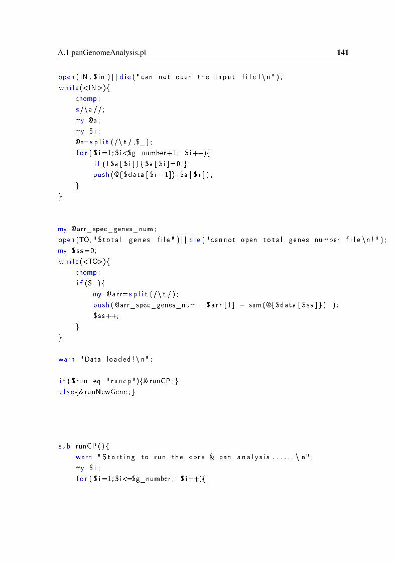
A.1 panGenomeAnalysis.pl 141
open ( IN , $ i n ) | | d i e ( " can not open the i n pu t f i l e ! \ n" ) ;
wh i l e (<IN>)
chomp ;
s /\a // ;
my @a ;
my $ i ;
@a=s p l i t (/\ t / ,$_ ) ;
f o r ( $ i =1; $ i<$g_number+1; $ i++)
i f ( ! $a [ $ i ] ) $a [ $ i ]=0;
push (@ $data [ $ i −1] , $a [ $ i ] ) ;
my @arr_spec_genes_num ;
open (TO, " $ t o t a l_g en e s_ f i l e " ) | | d i e ( " cannot open t o t a l genes number f i l e \n ! " ) ;
my $s s =0;
wh i l e (<TO>)
chomp ;
i f ($_)
my @arr=s p l i t (/\ t / ) ;
push ( @arr_spec_genes_num , $ a r r [ 1 ] − sum(@ $data [ $ s s ] ) ) ;
$ s s++;
warn "Data l oaded !\ n" ;
i f ( $run eq " runcp ")&runCP ;
e l s e &runNewGene ;
sub runCP ()
warn " S t a r t i n g to run the co r e & pan a n a l y s i s . . . . . . \ n" ;
my $ i ;
f o r ( $ i =1; $ i<=$g_number ; $ i++)
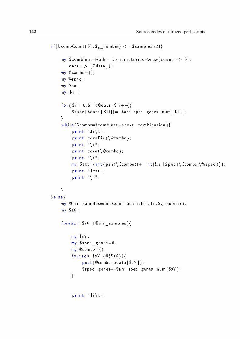
142 Source codes of utilized perl scripts
i f (&combCount ( $ i , $g_number ) <= $samples *2)
my $combinat=Math : : Comb ina to r i c s−>new( count => $i ,
data => [ @data ] ) ;
my @combo=() ;
my %spec ;
my $sx ;
my $ i i ;
f o r ( $ i i =0; $ i i <@data ; $ i i ++)
$spec $data [ $ i i ]= $arr_spec_genes_num [ $ i i ] ;
wh i l e (@combo=$combinat−>next_combinat ion )
p r i n t " $ i \ t " ;
p r i n t c o r e F i x (\@combo ) ;
p r i n t "\ t " ;
p r i n t co r e (\@combo ) ;
p r i n t "\ t " ;
my $ t t t =( i n t ( pan (\@combo))+ i n t (& a l l S p e c (\@combo,\% spec ) ) ) ;
p r i n t " $ t t t " ;
p r i n t "\n" ;
e l s e
my @arr_samples=randConm ( $samples , $ i , $g_number ) ;
my $sX ;
f o r e a c h $sX ( @arr_samples )
my $sY ;
my $spec_genes=0;
my @combo=() ;
f o r e a c h $sY (@$sX )
push (@combo , $data [ $sY ] ) ;
$spec_genes+=$arr_spec_genes_num [ $sY ] ;
p r i n t " $ i \ t " ;
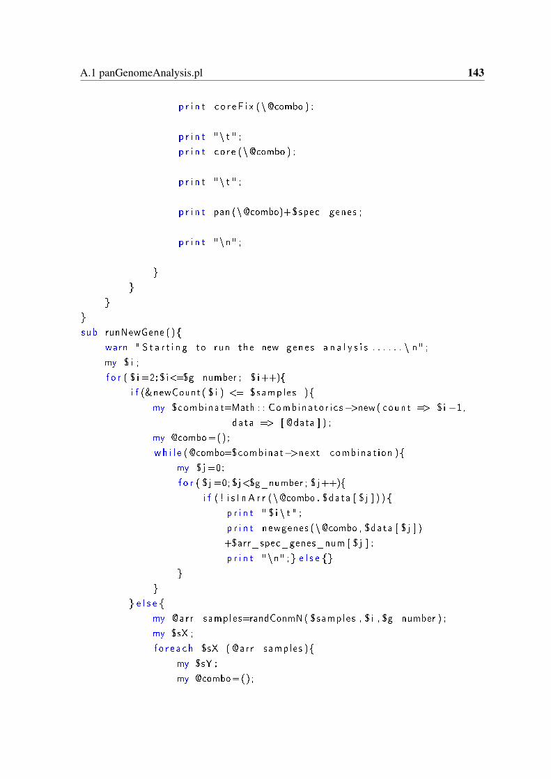
A.1 panGenomeAnalysis.pl 143
p r i n t c o r e F i x (\@combo ) ;
p r i n t "\ t " ;
p r i n t co r e (\@combo ) ;
p r i n t "\ t " ;
p r i n t pan (\@combo)+$spec_genes ;
p r i n t "\n" ;
sub runNewGene ( )
warn " S t a r t i n g to run the new genes a n a l y s i s . . . . . . \ n" ;
my $ i ;
f o r ( $ i =2; $ i<=$g_number ; $ i++)
i f (&newCount ( $ i ) <= $samples )
my $combinat=Math : : Comb ina to r i c s−>new( count => $i −1,
data => [ @data ] ) ;
my @combo=() ;
wh i l e (@combo=$combinat−>next_combinat ion )
my $ j =0;
f o r ( $ j =0; $ j<$g_number ; $ j++)
i f ( ! i s I n A r r (\@combo , $data [ $ j ] ) )
p r i n t " $ i \ t " ;
p r i n t newgenes (\@combo , $data [ $ j ] )
+$arr_spec_genes_num [ $ j ] ;
p r i n t "\n" ; e l s e
e l s e
my @arr_samples=randConmN( $samples , $ i , $g_number ) ;
my $sX ;
f o r e a c h $sX ( @arr_samples )
my $sY ;
my @combo=() ;
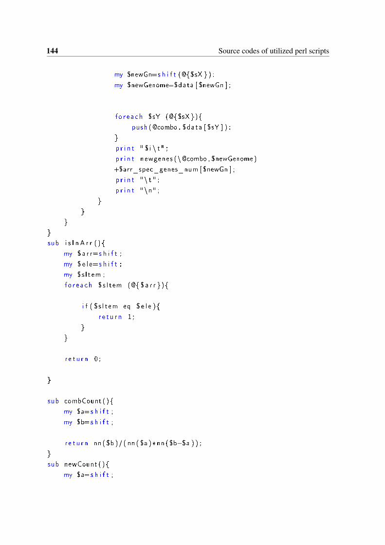
144 Source codes of utilized perl scripts
my $newGn=s h i f t (@$sX ) ;
my $newGenome=$data [ $newGn ] ;
f o r e a c h $sY (@$sX )
push (@combo , $data [ $sY ] ) ;
p r i n t " $ i \ t " ;
p r i n t newgenes (\@combo , $newGenome )
+$arr_spec_genes_num [ $newGn ] ;
p r i n t "\ t " ;
p r i n t "\n" ;
sub i s I n A r r ( )
my $a r r=s h i f t ;
my $ e l e=s h i f t ;
my $s I t em ;
f o r e a c h $s I t em (@ $a r r )
i f ( $s I t em eq $ e l e )
r e t u r n 1 ;
r e t u r n 0 ;
sub combCount ( )
my $a=s h i f t ;
my $b=s h i f t ;
r e t u r n nn ( $b )/ ( nn ( $a )* nn ( $b−$a ) ) ;
sub newCount ( )
my $a=s h i f t ;
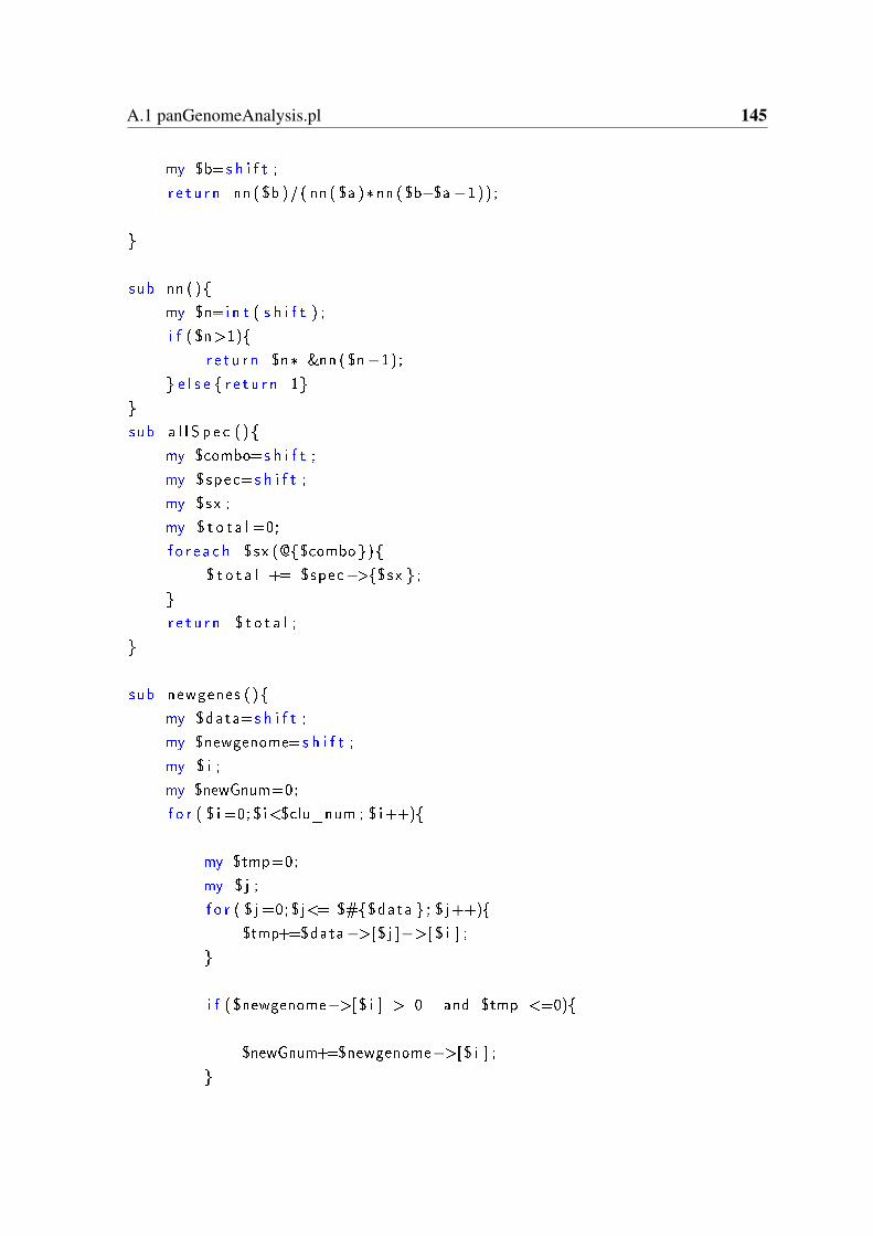
A.1 panGenomeAnalysis.pl 145
my $b=s h i f t ;
r e t u r n nn ( $b )/ ( nn ( $a )* nn ( $b−$a−1)) ;
sub nn ( )
my $n=i n t ( s h i f t ) ;
i f ( $n>1)
r e t u r n $n* &nn ( $n−1);
e l s e r e t u r n 1
sub a l l S p e c ( )
my $combo=s h i f t ;
my $spec=s h i f t ;
my $sx ;
my $ t o t a l =0;
f o r e a c h $sx (@$combo)
$ t o t a l += $spec−>$sx ;
r e t u r n $ t o t a l ;
sub newgenes ( )
my $data=s h i f t ;
my $newgenome=s h i f t ;
my $ i ;
my $newGnum=0;
f o r ( $ i =0; $ i<$clu_num ; $ i++)
my $tmp=0;
my $ j ;
f o r ( $ j =0; $ j<= $#$data ; $ j++)
$tmp+=$data−>[$ j ]−>[ $ i ] ;
i f ( $newgenome−>[$ i ] > 0 and $tmp <=0)
$newGnum+=$newgenome−>[$ i ] ;
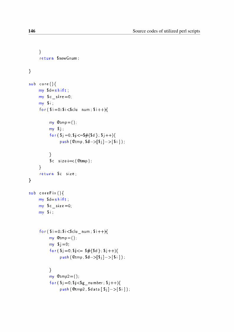
146 Source codes of utilized perl scripts
r e t u r n $newGnum ;
sub co r e ( )
my $d=s h i f t ;
my $c_s i ze =0;
my $ i ;
f o r ( $ i =0; $ i<$clu_num ; $ i++)
my @tmp=() ;
my $ j ;
f o r ( $ j =0; $ j<=$#$d ; $ j++)
push (@tmp , $d−>[$ j ]−>[ $ i ] ) ;
$c_s i ze+=c (@tmp ) ;
r e t u r n $c_s i ze ;
sub c o r e F i x ( )
my $d=s h i f t ;
my $c_s i ze =0;
my $ i ;
f o r ( $ i =0; $ i<$clu_num ; $ i++)
my @tmp=() ;
my $ j =0;
f o r ( $ j =0; $ j<= $#$d ; $ j++)
push (@tmp , $d−>[$ j ]−>[ $ i ] ) ;
my @tmp2=() ;
f o r ( $ j =0; $ j<$g_number ; $ j++)
push (@tmp2 , $data [ $ j ]−>[ $ i ] ) ;
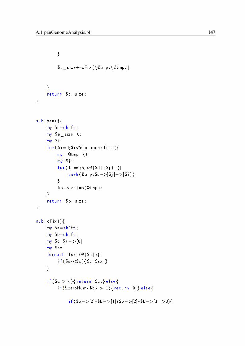
A.1 panGenomeAnalysis.pl 147
$c_s i ze+=cF i x (\@tmp , \@tmp2 ) ;
r e t u r n $c_s i ze ;
sub pan ( )
my $d=s h i f t ;
my $p_s ize=0;
my $ i ;
f o r ( $ i =0; $ i<$clu_num ; $ i++)
my @tmp=() ;
my $ j ;
f o r ( $ j =0; $ j<@$d ; $ j++)
push (@tmp , $d−>[$ j ]−>[ $ i ] ) ;
$p_s ize+=p (@tmp ) ;
r e t u r n $p_s ize ;
sub cF i x ( )
my $a=s h i f t ;
my $b=s h i f t ;
my $c=$a−>[0];
my $sx ;
f o r e a c h $sx (@$a )
i f ( $sx<$c ) $c=$sx ;
i f ( $c > 0) r e t u r n $c ; e l s e
i f (&zeroNum ( $b ) > 1) r e t u r n 0 ; e l s e
i f ( $b−>[0]*$b−>[1]*$b−>[2]*$b−>[3] >0)
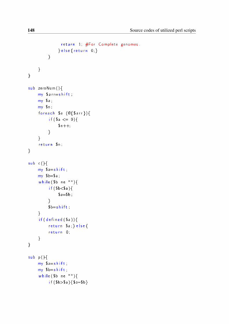
148 Source codes of utilized perl scripts
r e t u r n 1 ; #For Complete genomes .
e l s e r e t u r n 0 ;
sub zeroNum ()
my $a r r=s h i f t ;
my $a ;
my $n ;
f o r e a c h $a (@ $a r r )
i f ( $a <= 0)
$n++;
r e t u r n $n ;
sub c ( )
my $a=s h i f t ;
my $b=$a ;
wh i l e ( $b ne "" )
i f ( $b<$a )
$a=$b ;
$b=s h i f t ;
i f ( d e f i n e d ( $a ) )
r e t u r n $a ; e l s e
r e t u r n 0 ;
sub p ( )
my $a=s h i f t ;
my $b=s h i f t ;
wh i l e ( $b ne "" )
i f ( $b>$a ) $a=$b
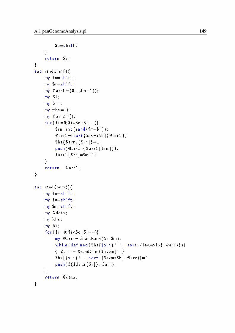
A.1 panGenomeAnalysis.pl 149
$b=s h i f t ;
r e t u r n $a ;
sub randCnm ()
my $n=s h i f t ;
my $m=s h i f t ;
my @arr1 =(0 . . ($m−1)) ;
my $ i ;
my $rn ;
my %hs =() ;
my @arr2 =() ;
f o r ( $ i =0; $ i<$n ; $ i++)
$rn=i n t ( rand ($m−$ i ) ) ;
@arr1=( s o r t $a<=>$b ( @arr1 ) ) ;
$hs $a r r 1 [ $rn ]=1;
push ( @arr2 , ( $ a r r 1 [ $rn ] ) ) ;
$ a r r 1 [ $rn ]=$m+1;
r e t u r n @arr2 ;
sub randConm ()
my $o=s h i f t ;
my $n=s h i f t ;
my $m=s h i f t ;
my @data ;
my %hs ;
my $ i ;
f o r ( $ i =0; $ i<$o ; $ i++)
my @arr = &randCnm($n ,$m) ;
wh i l e ( d e f i n e d ( $hs j o i n ( " " , s o r t $a<=>$b @arr ) ) )
@arr = &randCnm($n ,$m) ;
$hs j o i n ( " " , s o r t $a<=>$b @arr )=1;
push (@ $data [ $ i ] , @ar r ) ;
r e t u r n @data ;

150 Source codes of utilized perl scripts
sub randConmN ()
my $o=s h i f t ;
my $n=s h i f t ;
my $m=s h i f t ;
my @data ;
my %hs ;
my $ i ;
f o r ( $ i =0; $ i<$o ; $ i++)
my @arr = &randCnm($n ,$m) ;
wh i l e ( d e f i n e d ( $hs j o i n ( " " , ( $ a r r [ 0 ] , s o r t $a<=>$b @arr ) ) ) )
@arr = &randCnm($n ,$m) ;
$hs j o i n ( " " , ( $ a r r [ 0 ] , s o r t $a<=>$b @arr ))=1;
push (@ $data [ $ i ] , @ar r ) ;
r e t u r n @data ;
A.2 shared.pl
Script function: a shared script which is required by some of the following scripts.
#!/ u s r / b i n / p e r l
my %codonLmA ;
#//Group A
$codonLmA"AT" ="A" ; $codonLmA"TA" ="A" ;
$codonLmA"GG" ="A" ; $codonLmA"CC" ="A" ;
#Group G
$codonLmA"AG" ="G" ; $codonLmA"TC" ="G" ;
$codonLmA"GA" ="G" ; $codonLmA"CT" ="G" ;
#Group T
$codonLmA"AC" ="C" ; $codonLmA"TG" ="C" ;
$codonLmA"GT" ="C" ; $codonLmA"CA" ="C" ;
#Group C
$codonLmA"AA" ="T" ; $codonLmA"TT" ="T" ;
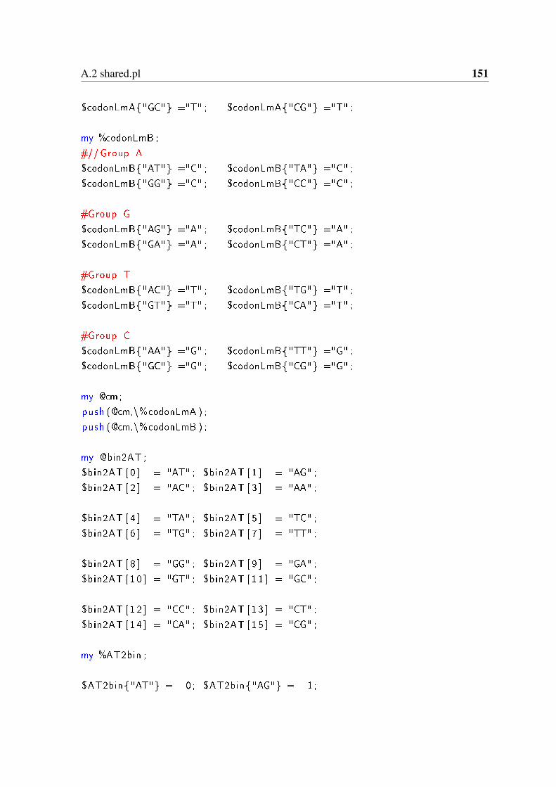
A.2 shared.pl 151
$codonLmA"GC" ="T" ; $codonLmA"CG" ="T" ;
my %codonLmB ;
#//Group A
$codonLmB"AT" ="C" ; $codonLmB"TA" ="C" ;
$codonLmB"GG" ="C" ; $codonLmB"CC" ="C" ;
#Group G
$codonLmB"AG" ="A" ; $codonLmB"TC" ="A" ;
$codonLmB"GA" ="A" ; $codonLmB"CT" ="A" ;
#Group T
$codonLmB"AC" ="T" ; $codonLmB"TG" ="T" ;
$codonLmB"GT" ="T" ; $codonLmB"CA" ="T" ;
#Group C
$codonLmB"AA" ="G" ; $codonLmB"TT" ="G" ;
$codonLmB"GC" ="G" ; $codonLmB"CG" ="G" ;
my @cm;
push (@cm,\%codonLmA ) ;
push (@cm,\%codonLmB ) ;
my @bin2AT ;
$bin2AT [ 0 ] = "AT" ; $bin2AT [ 1 ] = "AG" ;
$bin2AT [ 2 ] = "AC" ; $bin2AT [ 3 ] = "AA" ;
$bin2AT [ 4 ] = "TA" ; $bin2AT [ 5 ] = "TC" ;
$bin2AT [ 6 ] = "TG" ; $bin2AT [ 7 ] = "TT" ;
$bin2AT [ 8 ] = "GG" ; $bin2AT [ 9 ] = "GA" ;
$bin2AT [ 1 0 ] = "GT" ; $bin2AT [ 1 1 ] = "GC" ;
$bin2AT [ 1 2 ] = "CC" ; $bin2AT [ 1 3 ] = "CT" ;
$bin2AT [ 1 4 ] = "CA" ; $bin2AT [ 1 5 ] = "CG" ;
my %AT2bin ;
$AT2bin"AT" = 0 ; $AT2bin"AG" = 1 ;
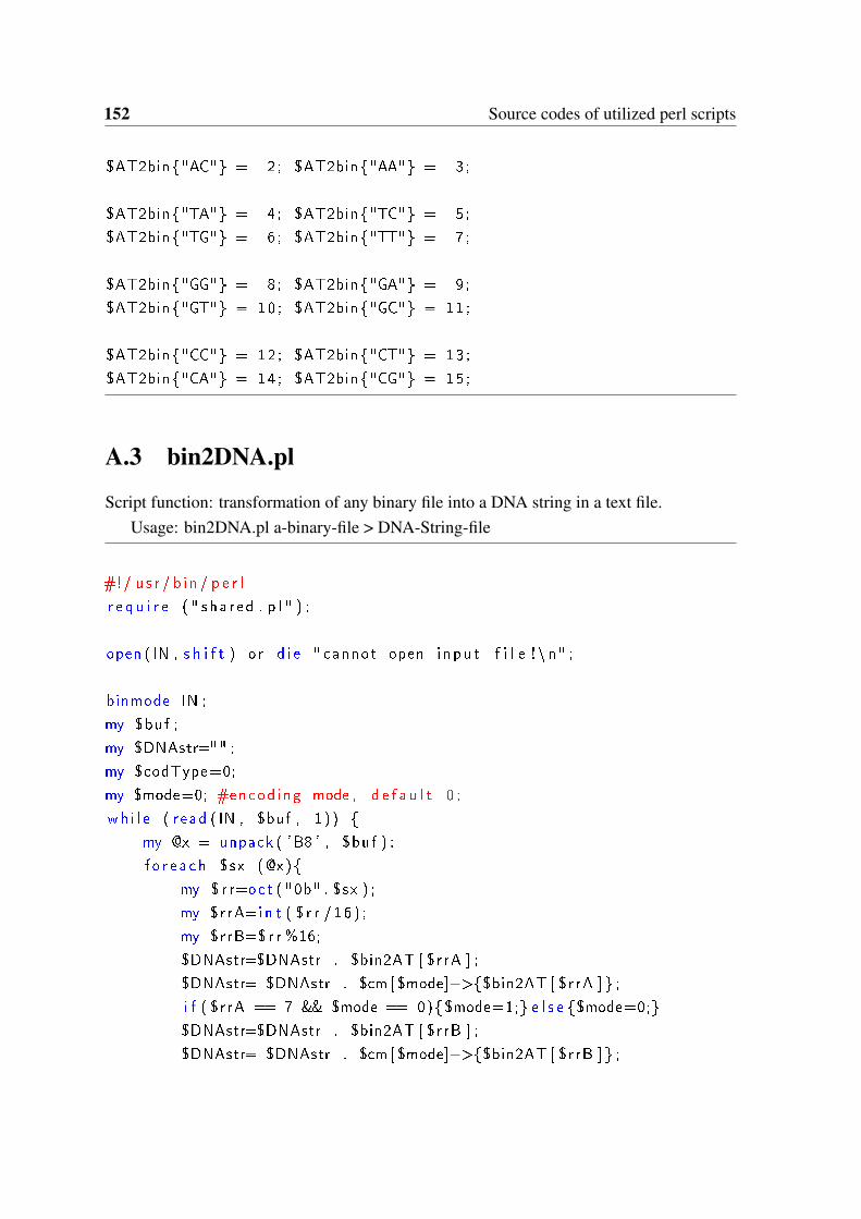
152 Source codes of utilized perl scripts
$AT2bin"AC" = 2 ; $AT2bin"AA" = 3 ;
$AT2bin"TA" = 4 ; $AT2bin"TC" = 5 ;
$AT2bin"TG" = 6 ; $AT2bin"TT" = 7 ;
$AT2bin"GG" = 8 ; $AT2bin"GA" = 9 ;
$AT2bin"GT" = 10 ; $AT2bin"GC" = 11 ;
$AT2bin"CC" = 12 ; $AT2bin"CT" = 13 ;
$AT2bin"CA" = 14 ; $AT2bin"CG" = 15 ;
A.3 bin2DNA.pl
Script function: transformation of any binary file into a DNA string in a text file.Usage: bin2DNA.pl a-binary-file > DNA-String-file
#!/ u s r / b i n / p e r l
r e q u i r e ( " sha r ed . p l " ) ;
open ( IN , s h i f t ) o r d i e " cannot open i n pu t f i l e ! \ n" ;
binmode IN ;
my $buf ;
my $DNAstr="" ;
my $codType=0;
my $mode=0; #encod ing mode , d e f a u l t 0 ;
wh i l e ( r ead ( IN , $buf , 1 ) )
my @x = unpack ( 'B8 ' , $buf ) ;
f o r e a c h $sx (@x)
my $ r r=oct ( "0b" . $sx ) ;
my $rrA=i n t ( $ r r /16 ) ;
my $rrB=$ r r %16;
$DNAstr=$DNAstr . $bin2AT [ $rrA ] ;
$DNAstr= $DNAstr . $cm [ $mode]−>$bin2AT [ $rrA ] ;
i f ( $r rA == 7 && $mode == 0)$mode=1; e l s e $mode=0;
$DNAstr=$DNAstr . $bin2AT [ $rrB ] ;
$DNAstr= $DNAstr . $cm [ $mode]−>$bin2AT [ $rrB ] ;
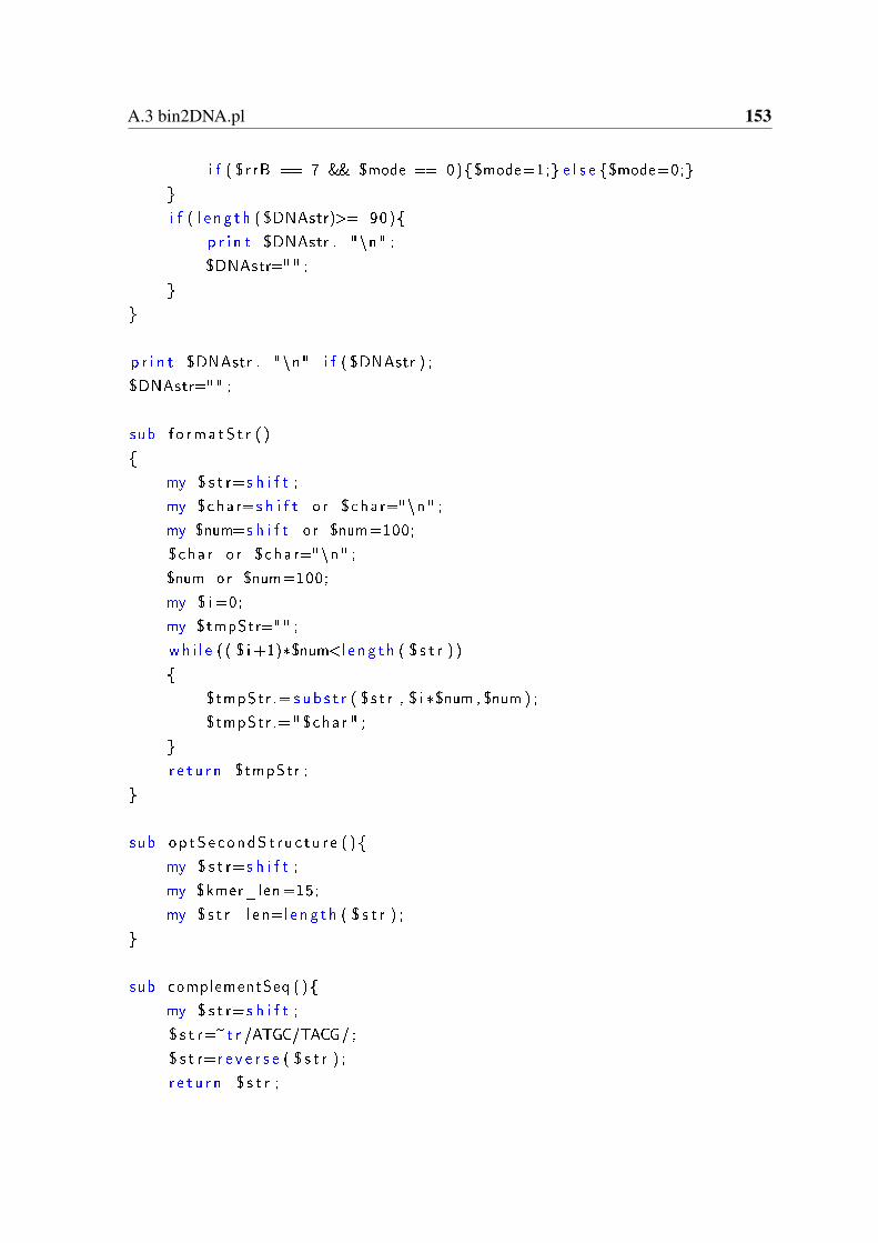
A.3 bin2DNA.pl 153
i f ( $r rB == 7 && $mode == 0)$mode=1; e l s e $mode=0;
i f ( l e n g t h ( $DNAstr)>= 90)
p r i n t $DNAstr . "\n" ;
$DNAstr="" ;
p r i n t $DNAstr . "\n" i f ( $DNAstr ) ;
$DNAstr="" ;
sub f o rma tS t r ( )
my $ s t r=s h i f t ;
my $char=s h i f t o r $char="\n" ;
my $num=s h i f t o r $num=100;
$char o r $char="\n" ;
$num or $num=100;
my $ i =0;
my $tmpStr="" ;
wh i l e ( ( $ i +1)*$num<l e ng t h ( $ s t r ) )
$tmpStr .= s u b s t r ( $ s t r , $ i *$num , $num ) ;
$tmpStr .=" $char " ;
r e t u r n $tmpStr ;
sub op tSecondS t ru c tu r e ( )
my $ s t r=s h i f t ;
my $kmer_len=15;
my $ s t r_ l en=l e n g t h ( $ s t r ) ;
sub complementSeq ( )
my $ s t r=s h i f t ;
$ s t r=~t r /ATGC/TACG/ ;
$ s t r=r e v e r s e ( $ s t r ) ;
r e t u r n $ s t r ;
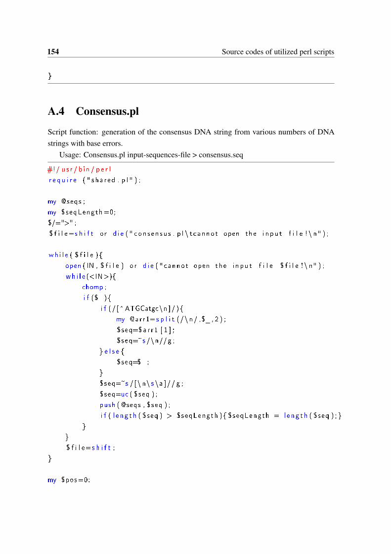
154 Source codes of utilized perl scripts
A.4 Consensus.pl
Script function: generation of the consensus DNA string from various numbers of DNAstrings with base errors.
Usage: Consensus.pl input-sequences-file > consensus.seq
#!/ u s r / b i n / p e r l
r e q u i r e ( " sha r ed . p l " ) ;
my @seqs ;
my $seqLength=0;
$/=">" ;
$ f i l e=s h i f t o r d i e ( " con sen su s . p l \ t cannot open the i n pu t f i l e ! \ n" ) ;
wh i l e ( $ f i l e )
open ( IN , $ f i l e ) o r d i e ( " cannot open the i n pu t f i l e $ f i l e ! \ n" ) ;
wh i l e (<IN>)
chomp ;
i f ($_)
i f (/ [^ATGCatgc\n ] / )
my @arr1=s p l i t (/\n / ,$_ , 2 ) ;
$seq=$a r r 1 [ 1 ] ;
$seq=~s /\n//g ;
e l s e
$seq=$_;
$seq=~s / [\ n\ s \a ]// g ;
$seq=uc ( $seq ) ;
push ( @seqs , $seq ) ;
i f ( l e n g t h ( $seq ) > $seqLength ) $seqLength = l e n g t h ( $seq ) ;
$ f i l e=s h i f t ;
my $pos=0;

A.5 DNA2bin.pl 155
my $consensu s="" ;
my $encodeRu le=0;
wh i l e ( $pos<$seqLength )
my %b l o c k s =() ;
f o r e a c h $s I t em ( @seqs )
my $aBlock=s u b s t r ( $s I tem , $pos , 3 ) ;
i f ( cMatch ( $aBlock , $encodeRu le ) ) $b l o c k s $aBlock++;
my $ fB lock = cBlock (\%b l o c k s ) ;
$consensu s = $consensu s . $ fB l o ck ;
i f ( $ fB l o ck eq "TTT" ) $encodeRu le =1 e l s e $encodeRu le =0;
$pos=$pos+3;
p r i n t ">consen su s \ n$consensus \n" ;
sub cBase
my $s t a=s h i f t ;
my $base="−" ;f o r e a c h $ s s ( key s %$s t a )
$sta−>$s s ++;
i f ( $ s s ne "N" )
i f ( $sta−>$s s > $sta−>$base )$base = $s s ;
r e t u r n $base ;
A.5 DNA2bin.pl
Script function: transformation of a DNA consensus string to a binary file.Usage: DNA2bin.pl consensus.seq binary-file
open ( IN , s h i f t ) o r d i e " cannot open i n pu t f i l e ! \ n" ;
156 Source codes of utilized perl scripts
open (OUT, ">" . s h i f t ) | | d i e ( " cannot open output !\ n" ) ;
binmode OUT;
wh i l e (<IN>)
chomp ;
i f ($_)
my $DNAstr=$_;
$DNAstr=~s / [\ n\ s \a ]// g ;
my $pos=0;
wh i l e ( $pos<l e n g t h ( $DNAstr ) )
my $twoBases=s u b s t r ( $DNAstr , $pos , 6 ) ;
my $twoBases1=s u b s t r ( $DNAstr , $pos , 2 ) ;
my $twoBases2=s u b s t r ( $DNAstr , $pos +3 ,2) ;
my $octNum1=$AT2bin $twoBases1 ;
my $octNum2=$AT2bin $twoBases2 ;
my $octNum=$octNum1*16+$octNum2 ;
my $binNum=s p r i n t f ( "%b" , $octNum ) ;
my $binNum= "0" x (8− l e n g t h ( $binNum ) ) . $binNum ;
my $binNumPack=pack ( "C" , $octNum ) ;
my $binNumUPack=unpack ( 'C ' , $binNumPack ) ;
s y s w r i t e (OUT, $binNumPack , 1 ) ;
$pos+=6;
sub dec2b in ( )#10 to2
my $dec = s h i f t ;
my $b in = unpack ( "b4" , pack ( " i " , $dec ) ) ;
r e t u r n $b in ;
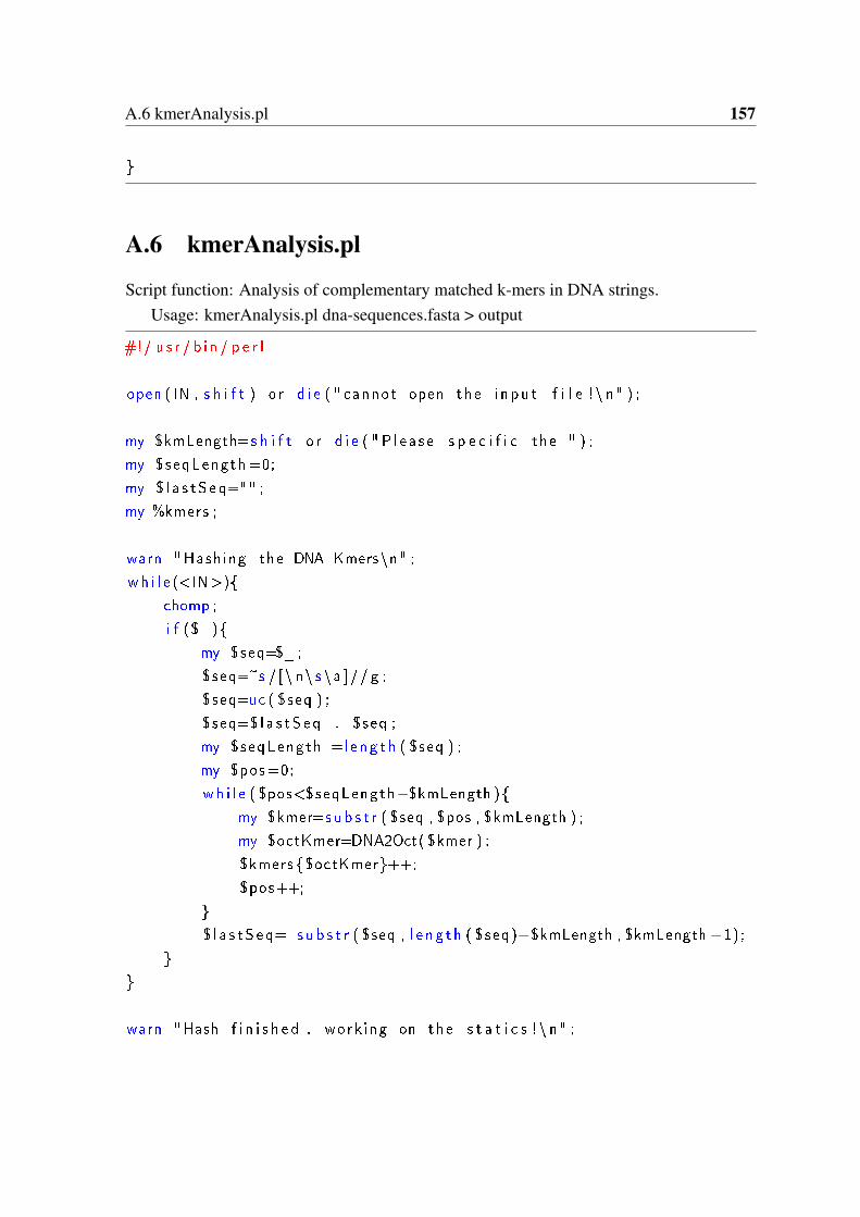
A.6 kmerAnalysis.pl 157
A.6 kmerAnalysis.pl
Script function: Analysis of complementary matched k-mers in DNA strings.Usage: kmerAnalysis.pl dna-sequences.fasta > output
#!/ u s r / b i n / p e r l
open ( IN , s h i f t ) o r d i e ( " cannot open the i n pu t f i l e ! \ n" ) ;
my $kmLength=s h i f t o r d i e ( " P l e a s e s p e c i f i c the " ) ;
my $seqLength=0;
my $ l a s t S e q="" ;
my %kmers ;
warn "Hash ing the DNA Kmers\n" ;
wh i l e (<IN>)
chomp ;
i f ($_)
my $seq=$_;
$seq=~s / [\ n\ s \a ]// g ;
$seq=uc ( $seq ) ;
$seq=$ l a s t S e q . $seq ;
my $seqLength =l e n g t h ( $seq ) ;
my $pos=0;
wh i l e ( $pos<$seqLength−$kmLength )
my $kmer=s u b s t r ( $seq , $pos , $kmLength ) ;
my $octKmer=DNA2Oct( $kmer ) ;
$kmers $octKmer++;
$pos++;
$ l a s t S e q= s u b s t r ( $seq , l e n g t h ( $seq)−$kmLength , $kmLength−1);
warn "Hash f i n i s h e d , work ing on the s t a t i c s ! \ n" ;
158 Source codes of utilized perl scripts
my $km ;
f o r e a c h $km ( keys %kmers )
i f ( e x i s t s ( $kmers $km))
my $cpSeqNum=complementNum ($km ) ;
i f ( e x i s t s ( $kmers $cpSeqNum))
p r i n t oct2DNA($km) . "\ t " ;
p r i n t $kmers $km ;
p r i n t "\ t " . oct2DNA($cpSeqNum) . "\ t " ;
p r i n t $kmers $cpSeqNum ;
p r i n t "\n" ;
d e l e t e ( $kmers $cpSeqNum ) ;
d e l e t e ( $kmers $km ) ;
sub complementSeq
my $ s t r=s h i f t ;
$ s t r=~t r /ATGC/TACG/ ;
$ s t r=r e v e r s e ( $ s t r ) ;
r e t u r n $ s t r ;
sub DNA2Oct
my $DNAstr=s h i f t ;
my %AT2bin ;
$AT2bin"AT" = 0 ; $AT2bin"AG" = 1 ;
$AT2bin"AC" = 2 ; $AT2bin"AA" = 3 ;
$AT2bin"TA" = 4 ; $AT2bin"TC" = 5 ;
$AT2bin"TG" = 6 ; $AT2bin"TT" = 7 ;
$AT2bin"GG" = 8 ; $AT2bin"GA" = 9 ;
$AT2bin"GT" = 10 ; $AT2bin"GC" = 11 ;
$AT2bin"CC" = 12 ; $AT2bin"CT" = 13 ;
$AT2bin"CA" = 14 ; $AT2bin"CG" = 15 ;
A.6 kmerAnalysis.pl 159
my $pos=0;
my $fOct=0;
wh i l e ( $pos<l e n g t h ( $DNAstr )−1)
my $twoBases=s u b s t r ( $DNAstr , $pos , 2 ) ;
my $octNum=$AT2bin $twoBases ;
$fOct=$fOct*16+$octNum ;
$pos+=2;
r e t u r n $fOct ;
sub complementNum
my $num=s h i f t ;
my $seq=oct2DNA($num ) ;
my $cSeq=complementSeq ( $seq ) ;
r e t u r n DNA2Oct( $cSeq ) ;
sub oct2DNA
my $num=s h i f t ;
my $DNA="" ;
my @bin2AT ;
$bin2AT [ 0 ] = "AT" ; $bin2AT [ 1 ] = "AG" ;
$bin2AT [ 2 ] = "AC" ; $bin2AT [ 3 ] = "AA" ;
$bin2AT [ 4 ] = "TA" ; $bin2AT [ 5 ] = "TC" ;
$bin2AT [ 6 ] = "TG" ; $bin2AT [ 7 ] = "TT" ;
$bin2AT [ 8 ] = "GG" ; $bin2AT [ 9 ] = "GA" ;
$bin2AT [ 1 0 ] = "GT" ; $bin2AT [ 1 1 ] = "GC" ;
$bin2AT [ 1 2 ] = "CC" ; $bin2AT [ 1 3 ] = "CT" ;
$bin2AT [ 1 4 ] = "CA" ; $bin2AT [ 1 5 ] = "CG" ;
wh i l e ($num > 15)
$a=$num%16;
160 Source codes of utilized perl scripts
$num=i n t ($num/16 ) ;
$DNA= $bin2AT [ $a ] . $DNA ;
$DNA= $bin2AT [ $num ] . $DNA ;
$DNA= ("AT" x i n t ( ( $kmLength − l e n g t h ($DNA) )/2 ) ) . $DNA;
r e t u r n $DNA;
A.7 biologyRelevanceAnalysis.pl
Script function: Searching and analysis of sub-sequences that matching SED3B encodingrules in natural generated DNA sequences. The results are used to evaluate the biologicalrelevance of SED3B encoded sequences.
Usage: biologyRelevanceAnalysis.pl dna-sequences.fasta > output
#!/ u s r / b i n / p e r l
r e q u i r e ( " sha r ed . p l " ) ;
open ( IN , s h i f t ) | | d i e ( " not opened !\ n" ) ;
$/="\n>" ;
$mSeqLength = s h i f t ;
wh i l e (<IN>)
chomp ;
my @arr=s p l i t (/\n / ,$_ , 2 ) ;
my @arr2=s p l i t (/\ s+/, $ a r r [ 0 ] , 2 ) ;
my $seq=$a r r [ 1 ] ;
$seq=~s / [\ n\ s \a ]// g ;
$ s eq f 1=uc ( $seq ) ;
$ s eq f 2=s u b s t r ( $seq f1 , 1 , l e n g t h ( $ s eq f 1 )−1);
$ s eq f 3=s u b s t r ( $seq f1 , 2 , l e n g t h ( $ s eq f 1 )−2);
bSafeSeq ( $seq f1 , " $a r r 2 [ 0 ] _Frame1_" ) ;
bSafeSeq ( $seq f2 , " $a r r 2 [ 0 ] _Frame2_" ) ;
bSafeSeq ( $seq f3 , " $a r r 2 [ 0 ] _Frame3_" ) ;
A.7 biologyRelevanceAnalysis.pl 161
sub cMatch
my $codon=s h i f t ;
my $mode=s h i f t ;
i f ( $mode == 0)
i f ( $codonLmA s u b s t r ( $codon , 0 , 2 ) eq s u b s t r ( $codon , 2 , 1 ) )
r e t u r n 1 ; e l s e r e t u r n 0 ;
i f ( $mode == 1)
i f ( $codonLmB s u b s t r ( $codon , 0 , 2 ) eq s u b s t r ( $codon , 2 , 1 ) )
r e t u r n 1 ; e l s e r e t u r n 0 ;
sub bSafeSeq
my $seq=uc ( s h i f t ) ;
my $seqName=s h i f t ;
my $pos=0;
my $sa f eSeq ;
my $seqNum=0;
my $encodeMode=0;
my $mSeqLength=21;
wh i l e ( $pos<l e n g t h ( $seq ) )
my $th r eeBase=s u b s t r ( $seq , $pos , 3 ) ;
i f ( l e n g t h ( $ th r eeBase ) == 3)
i f ( cMatch ( $threeBase , $encodeMode ) )
$ sa f eSeq = $sa f eSeq . $ th r eeBase ;
i f ( $encodeMode == 0 and ( $ th r eeBase eq "TTT" ) )
$encodeMode=1; e l s e $encodeMode=0;
e l s e
i f ( l e n g t h ( $ sa f eSeq)>=$mSeqLength )
my $ l e n=l e n g t h ( $ sa f eSeq ) ;
p r i n t ">$seqName$seqNum $ l en \ n$sa f eSeq \n" ;
$ sa f eSeq ="" ;
$seqNum++;
$encodeMode=0;
$ sa f eSeq ="" ;
162 Source codes of utilized perl scripts
$pos+=3;
my $ l e n=l e n g t h ( $ sa f eSeq ) ;
p r i n t ">$seqName$seqNum $ l en \ n$sa f eSeq \n" ;
$ sa f eSeq ="" ;
A.8 bin2DNACRCIndex.pl
Script function: encode a digital file into indexed DNA sequences with CRC checking.Usage: bin2DNACRCIndex.pl input-digital-file [start of index] > output.dna
#!/ u s r / b i n / p e r l
r e q u i r e ( " sha r ed . p l " ) ;
open ( IN , s h i f t ) o r d i e " cannot open i n pu t f i l e ! \ n" ;
binmode IN ;
my $buf ;
my $DNAstr="" ;
my $codType=0;
my $mode=0; #encod ing mode , d e f a u l t 0 ;
my $rowLenth=78; #fragment l e n g t h ;
my $pcrF="TGCCTCTTTATCTGT" ; #"CAGTACTAACCCTCG" ;
my $pcrR="CATTTCCGATACACC" ; #"CCGCAAGAGTGTCGA" ;
my $ i n d e x S t a r t=s h i f t o r $ i n d e x S t a r t =0;
my $ c r c S i z e =2;
my $ l c r S i z e =10;
my @crc =() ;
wh i l e ( r ead ( IN , $buf , 1 ) )
my @x = unpack ( 'B8 ' , $buf ) ;
f o r e a c h $sx (@x)
my $ r r=oct ( "0b" . $sx ) ;
my $rrA=i n t ( $ r r /16 ) ;
my $rrB=$ r r %16;
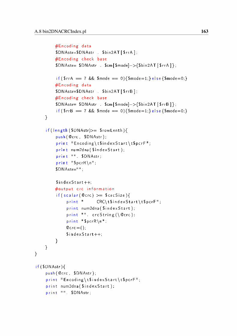
A.8 bin2DNACRCIndex.pl 163
#Encoding data
$DNAstr=$DNAstr . $bin2AT [ $rrA ] ;
#Encoding check base
$DNAstr= $DNAstr . $cm [ $mode]−>$bin2AT [ $rrA ] ;
i f ( $r rA == 7 && $mode == 0)$mode=1; e l s e $mode=0;
#Encoding data
$DNAstr=$DNAstr . $bin2AT [ $rrB ] ;
#Encoding check base
$DNAstr= $DNAstr . $cm [ $mode]−>$bin2AT [ $rrB ] ;
i f ( $r rB == 7 && $mode == 0)$mode=1; e l s e $mode=0;
i f ( l e n g t h ( $DNAstr)>= $rowLenth )
push ( @crc , $DNAstr ) ;
p r i n t " Encoding \ t $ i n d e x S t a r t \ t$pc rF " ;
p r i n t num2dna ( $ i n d e x S t a r t ) ;
p r i n t "" . $DNAstr ;
p r i n t " $pcrR\n" ;
$DNAstr="" ;
$ i n d e x S t a r t++;
#output c r c i n f o rma t i o n
i f ( s c a l a r ( @crc ) >= $ c r c S i z e )
p r i n t " CRC\ t $ i n d e x S t a r t \ t$pc rF " ;
p r i n t num2dna ( $ i n d e x S t a r t ) ;
p r i n t "" . c r c S t r i n g (\ @crc ) ;
p r i n t " $pcrR\n" ;
@crc =() ;
$ i n d e x S t a r t++;
i f ( $DNAstr )
push ( @crc , $DNAstr ) ;
p r i n t " Encoding \ t $ i n d e x S t a r t \ t$pc rF " ;
p r i n t num2dna ( $ i n d e x S t a r t ) ;
p r i n t "" . $DNAstr ;
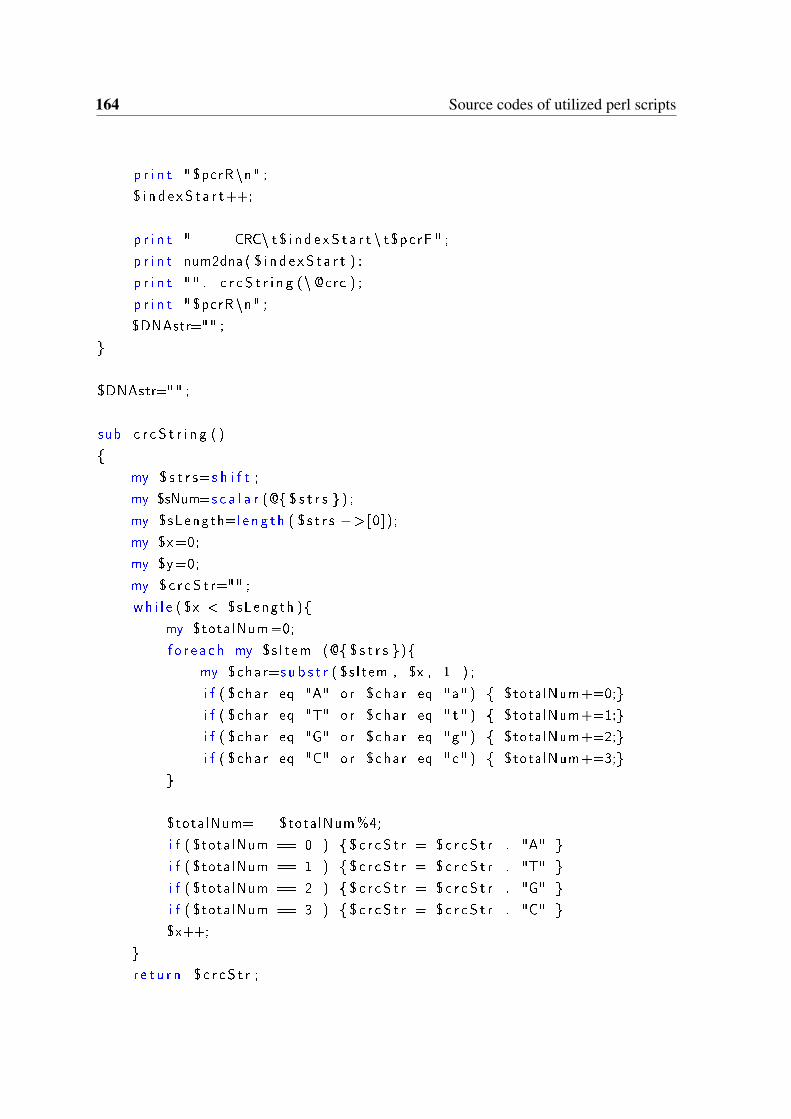
164 Source codes of utilized perl scripts
p r i n t " $pcrR\n" ;
$ i n d e x S t a r t++;
p r i n t " CRC\ t $ i n d e x S t a r t \ t$pc rF " ;
p r i n t num2dna ( $ i n d e x S t a r t ) ;
p r i n t "" . c r c S t r i n g (\ @crc ) ;
p r i n t " $pcrR\n" ;
$DNAstr="" ;
$DNAstr="" ;
sub c r c S t r i n g ( )
my $ s t r s=s h i f t ;
my $sNum=s c a l a r (@ $ s t r s ) ;
my $sLength=l e n g t h ( $ s t r s −>[0]);
my $x=0;
my $y=0;
my $ c r c S t r="" ;
wh i l e ( $x < $sLength )
my $totalNum=0;
f o r e a c h my $s I tem (@ $ s t r s )
my $char=s u b s t r ( $s I tem , $x , 1 ) ;
i f ( $char eq "A" or $char eq "a" ) $totalNum+=0;
i f ( $char eq "T" or $char eq " t " ) $totalNum+=1;
i f ( $char eq "G" or $char eq "g" ) $totalNum+=2;
i f ( $char eq "C" or $char eq "c" ) $totalNum+=3;
$totalNum= $totalNum%4;
i f ( $totalNum == 0 ) $ c r c S t r = $ c r c S t r . "A"
i f ( $totalNum == 1 ) $ c r c S t r = $ c r c S t r . "T"
i f ( $totalNum == 2 ) $ c r c S t r = $ c r c S t r . "G"
i f ( $totalNum == 3 ) $ c r c S t r = $ c r c S t r . "C"
$x++;
r e t u r n $ c r c S t r ;
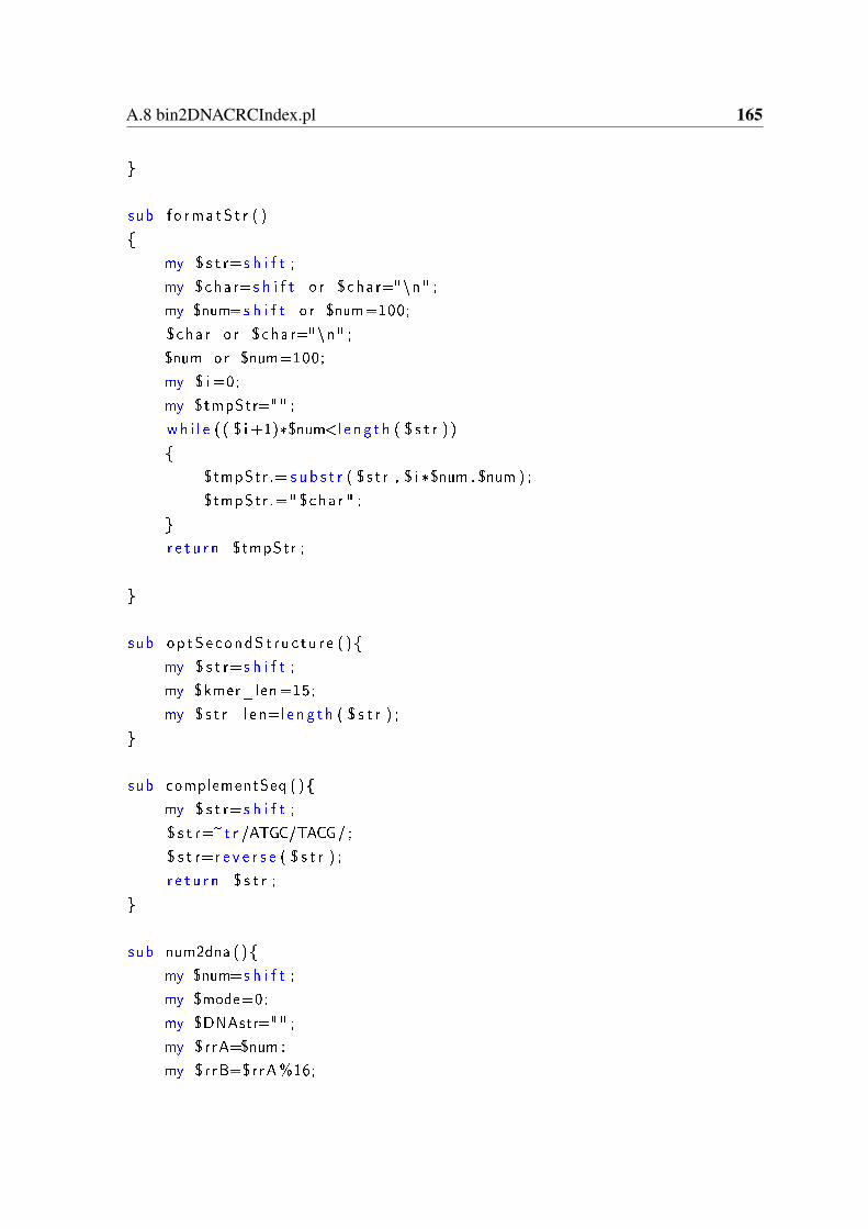
A.8 bin2DNACRCIndex.pl 165
sub f o rma tS t r ( )
my $ s t r=s h i f t ;
my $char=s h i f t o r $char="\n" ;
my $num=s h i f t o r $num=100;
$char o r $char="\n" ;
$num or $num=100;
my $ i =0;
my $tmpStr="" ;
wh i l e ( ( $ i +1)*$num<l e ng t h ( $ s t r ) )
$tmpStr .= s u b s t r ( $ s t r , $ i *$num , $num ) ;
$tmpStr .=" $char " ;
r e t u r n $tmpStr ;
sub op tSecondS t ru c tu r e ( )
my $ s t r=s h i f t ;
my $kmer_len=15;
my $ s t r_ l en=l e n g t h ( $ s t r ) ;
sub complementSeq ( )
my $ s t r=s h i f t ;
$ s t r=~t r /ATGC/TACG/ ;
$ s t r=r e v e r s e ( $ s t r ) ;
r e t u r n $ s t r ;
sub num2dna ( )
my $num=s h i f t ;
my $mode=0;
my $DNAstr="" ;
my $rrA=$num ;
my $rrB=$rrA%16;
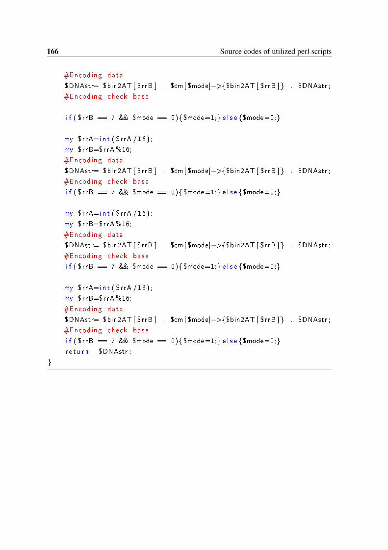
166 Source codes of utilized perl scripts
#Encoding data
$DNAstr= $bin2AT [ $rrB ] . $cm [ $mode]−>$bin2AT [ $rrB ] . $DNAstr ;
#Encoding check base
i f ( $r rB == 7 && $mode == 0)$mode=1; e l s e $mode=0;
my $rrA=i n t ( $r rA /16 ) ;
my $rrB=$rrA%16;
#Encoding data
$DNAstr= $bin2AT [ $rrB ] . $cm [ $mode]−>$bin2AT [ $rrB ] . $DNAstr ;
#Encoding check base
i f ( $r rB == 7 && $mode == 0)$mode=1; e l s e $mode=0;
my $rrA=i n t ( $r rA /16 ) ;
my $rrB=$rrA%16;
#Encoding data
$DNAstr= $bin2AT [ $rrB ] . $cm [ $mode]−>$bin2AT [ $rrB ] . $DNAstr ;
#Encoding check base
i f ( $r rB == 7 && $mode == 0)$mode=1; e l s e $mode=0;
my $rrA=i n t ( $r rA /16 ) ;
my $rrB=$rrA%16;
#Encoding data
$DNAstr= $bin2AT [ $rrB ] . $cm [ $mode]−>$bin2AT [ $rrB ] . $DNAstr ;
#Encoding check base
i f ( $r rB == 7 && $mode == 0)$mode=1; e l s e $mode=0;
r e t u r n $DNAstr ;
Appendix B
Supplement Information
B.1 Sequences of mutacins used for the identification of pu-tative mutacins in 10 mutans streptococci strains.
Sequences of known mutacins as well as mutacin-immunity proteins collected from theNCBI (http://www.ncbi.nlm.nih.gov) and Oralgen (http://www.oralgen.lanl.gov/) databases,as well as by searching for related publications.
>SmbAMKSNLLKINNVTEMEKNMVTLIKDEDMLAGGSTPACAIGVVGITVAVTGISTACTSRCINK>SmbBMKEIQKAGLQEELSILMDDANNLEQLTAGIGTTVVNSTFSIVLGNKGYICTVTVECMRNCSK>Mutacin-I (Isolated from strain UA140 AND CH43)MSNTQLLEVLGTETFDVQEDLFAFDTTDTTIVASNDDPDTRFSSLSLCSLGCTGVKNPSFNSYCC>Mutacin-II AAC38144.1MNKLNSNAVVSLNEVSDSELDTILGGNRWWQGVVPTVSYECRMN>Mut-III (Isolated from strain UA787 1140)MSNTQLLEVLGTETFDVQEDLFAFDTTDTTIVASNDDPDTRFKSWSLCTPGCARTGSFNSYCC>Mutacin-IV SMU.150 nlmA non-lantibiotic mutacin IV AMDTQAFEQFDVMDSQTLSTVEGGKVSGGEAVAAIGICATASAAIGGLAGATLVTPYCVGTWGLIRSH>Mutacin-IV SMU.151 nlmB non-lantibiotic mutacin IV B
168 Supplement Information
MEWRINTMELNVNNYKSLTNDELSEVFGGDKQAADTFLSAVGGAASGFTYCASNGVWHPYILAGCAGVGAVGSVVFPH>Mutacin-V SMU.1914c CipBMNTQAFEQFNVMDNEALSAVEGGGRGWNCAAGIALGAGQGYMATAGGTAFLGPYAIGTGAFGAIAGGIGGALNSCG>SMU.423 possible bacteriocin Kreth et al., 2005, statethat this mutacin-like gene is regulated by the competencesystem.MNTQAFEQFNVMDNEALSTVEGGGMIRCALGTAGSAGLGFVGGMGAGTVTLPVVGTVSGAALGGWSGAAVGAATF>Mutacin-AII (S. pyo FF22, homolog found in S.mutans)MEKNNEVINSIQEVSLEELDQIIGAGKNGVFKTISHECHLNTWA
Lebenslauf
Name Song
Vorname Lifu
Geburtsdatum 12.Feb.1982
Geburtsort, -land Shandong, China
08.1989 - 07.1994 Grundschule in Weifang, Shandong/China
08.1994 - 07.1997 Mittelschule in Weifang, Shandong/China
08.1997 - 07.2000 Oberschule in Weifang, Shandong/China
08.2000 - 07.2004 Studium Pharmaceutical Engineering
an der Shandong University, Shandong/China
Abschluss: Bachelor
08.2004 - 07.2007 Studium Fermentation Engineering
an der Shandong University, Shandong/China
Abschluss: Master of Engineering
08.2007 - 08.2008 Mitarbeiter in der Arbeitung Bioinformatik
am Beijing Genomics Institute (BGI), Beijing/China
09.2008 - 05.2010 Wissenschaftlicher Mitarbeiter
am Tianjin Institute of Industrial Biotechnology, Chinese
Academy of Sciences, Tianjin/China
06.2010 - 07.2011 Wissenschaftlicher Mitarbeiter
Im Institut für Bioprozess– und Biosystemtechnik,
Technische Universität Hamburg–Harburg, Hamburg,
Deutschland
08.2011 - 09.2012 Doktorand
an der Tianjin University, Tianjin/China
10.2012 - 12.2016 Promotion
Institut für Bioprozess– und Biosystemtechnik,
Technische Universität Hamburg–Harburg, Hamburg,
Deutschland
01.2017 – jetzt Zusammenschreiben der Doktorarbeit und Arbeitssuchend
Publications
Song, Lifu; Zeng, An-Ping (2017): Engineering "cell robots" for parallel and highly
sensitive screening of biomolecules under in vivo conditions. In: Scientific reports 7
(1), p. 15145. DOI: 10.1038/s41598-017-15621-0.
Song, Lifu; Zeng, An-Ping (2017): Orthogonal information encoding in living cells
with high error-tolerance, safety, and fidelity. In: ACS synthetic biology 7 (3), pp.
866–874. DOI: 10.1021/acssynbio.7b00382.
Song, Lifu; Sudhakar, Padhmanand; Wang, Wei; Conrads, Georg; Brock, Anke; Sun,
Jibin et al. (2012): A genome-wide study of two-component signal transduction
systems in eight newly sequenced mutans streptococci strains. In: BMC Genomics
13, S. 128. DOI: 10.1186/1471-2164-13-128.
Song, Lifu; Wang, Wei; Conrads, Georg; Rheinberg, Anke; Sztajer, Helena; Reck,
Michael et al. (2013): Genetic variability of mutans streptococci revealed by wide
whole-genome sequencing. In: BMC Genomics 14, S. 430. DOI: 10.1186/1471-
2164-14-430.
Conrads, Georg; Soet, Johannes J. de; Song, Lifu; Henne, Karsten; Sztajer, Helena;
Wagner- Dobler, Irene; Zeng, An-Ping (2014): Comparing the cariogenic species
Streptococcus sobrinus and S. mutans on whole genome level. In: Journal of Oral
Microbiology 6, S. 26189. DOI: 10.3402/jom.v6.26189. Walkhoff prize of
the ’Deutsche Gesellschaft für Zahnerhaltung’ (DGZ).
Rappert, Sugima; Song, Lifu; Sabra, Wael; Wang, Wei; Zeng, An-Ping (2013):
Draft Genome Sequence of Type Strain Clostridium pasteurianum DSM 525
(ATCC 6013), a Promising Producer of Chemicals and Fuels. In: Genome
Announcements 1 (1). DOI: 10.1128/genomeA.00232-12.
Poster & Presentations
Song, Lifu and Zeng, An-Ping: Rein the computational abilities of cells to make
predictions by letting them ‘listen’ and ‘talk’ to us, Scale-up and scale-down of
bioprocesses, 12.May.2015, Ramada Hotel Hamburg-Bergedorf (Best Poster
Award 2015)
Song, Lifu and Zeng, An-Ping: A Novel, Ultra-Sensitive Multiple Input-Output
System for Target Identification in Systems Metabolic Engineering of E. coli,
Metabolic Engineering 11, 28 June 2016, Japan (Selected as Rapid Fire Poster)
Song, Lifu and Zeng, An-Ping: Digital information storage in DNA (In Chinese),
The 8th China Summit Forum on Industrial Biotechnology Development, 11-12
Dec 2015, Tianjin China
Patent applications
Song, Lifu and Zeng, An-Ping (2017): Methods for encoding and decoding a
binary string and system, PCT/EP2016/078122


![Advanced NMR Analysis of Polymers and Biomolecules · a feature that transformed NMR into one of the cornerstones of modern chemical and pharmaceutical research” [Fry1]. Over the](https://static.fdokument.com/doc/165x107/5ed92c0a6714ca7f476947bb/advanced-nmr-analysis-of-polymers-and-biomolecules-a-feature-that-transformed-nmr.jpg)

![Functioning and Process Engineering Design - Silos[1]](https://static.fdokument.com/doc/165x107/542ed777219acdf5478b5156/functioning-and-process-engineering-design-silos1.jpg)