Analysis, Modeling and Simulation of Tomographic Image ... · Westhoff, D. et al.: 3D...
13
134 © Carl Hanser Verlag, München Pract. Metallogr. 55 (2018) 3 Westhoff, D. et al.: 3D Microstructure of Li-Ion Batteries / 3D Mikrostruktur von Li-Ionen Batterien Eingegangen: 21. Dezember 2017 Angenommen: 05. Januar 2018 Translation: M. Lackas Kurzfassung In diesem Artikel werden stochastische 3D Strukturmodelle für Elektroden von Lithium- Ionen-Batterien beschrieben, die zur modell- basierten Optimierung der Elektrodenmor- phologie verwendet werden können. Zunächst wird ein Einzelpartikel-Modell präsentiert, mit dessen Hilfe sich einzelne Partikel aus 3D Tomographiedaten parametrisch beschreiben lassen. Durch Anpassung parametrischer Ver- teilungen lassen sich somit auch (statistisch ähnliche) Partikel simulieren. Im Folgenden werden verschiedene Ansätze zur Anordnung Received: December 21, 2017 Accepted: January 05, 2018 Abstract This article describes stochastic 3D struc- ture models for electrodes of lithium-ion batteries, which can be used for model- based optimization of the electrode mor- phology. First, a single particle model is presented which can be used to parametri- cally describe individual particles from 3D tomographic image data. By fitting para- metrical distributions it is therefore also possible to simulate (statistically similar) particles. Hereafter, different approaches for the arrangement of individual particles Authors: D. Westhoff, K. Kuchler, J. Feinauer, L. Petrich, V. Schmidt Analysis, Modeling and Simulation of Tomographic Image Data for the 3D Microstructure of Electrode Material in Lithium-Ion Batteries Analyse, Modellierung und Simulation von tomographischen Bilddaten für die 3D Mikrostruktur von Elektrodenmaterial in Lithium-Ionen-Batterien Daniel Westhoff, Klaus Kuchler, Julian Feinauer, Lukas Petrich, Volker Schmidt Universität Ulm, Institut für Stochastik, Helmholtzstraße 18, 89069 Ulm; e-mail: [email protected] Practical Metallography downloaded from www.hanser-elibrary.com by Hanser - Library on April 3, 2018 For personal use only.
Transcript of Analysis, Modeling and Simulation of Tomographic Image ... · Westhoff, D. et al.: 3D...
134 © Carl Hanser Verlag, München Pract. Metallogr. 55 (2018) 3
Westhoff, D. et al.: 3D Microstructure of Li-Ion Batteries / 3D Mikrostruktur von Li-Ionen Batterien
Eingegangen: 21. Dezember 2017Angenommen: 05. Januar 2018
Translation: M. Lackas
KurzfassungIn diesem Artikel werden stochastische 3D Strukturmodelle für Elektroden von Lithium-Ionen-Batterien beschrieben, die zur modell-basierten Optimierung der Elektrodenmor-phologie verwendet werden können. Zunächst wird ein Einzelpartikel-Modell präsentiert, mit dessen Hilfe sich einzelne Partikel aus 3D Tomographiedaten parametrisch beschreiben lassen. Durch Anpassung parametrischer Ver-teilungen lassen sich somit auch (statistisch ähnliche) Partikel simulieren. Im Folgenden werden verschiedene Ansätze zur Anordnung
Received: December 21, 2017Accepted: January 05, 2018
AbstractThis article describes stochastic 3D struc-ture models for electrodes of lithium-ion batteries, which can be used for model-based optimization of the electrode mor-phology. First, a single particle model is presented which can be used to parametri-cally describe individual particles from 3D tomographic image data. By fitting para-metrical distributions it is therefore also possible to simulate (statistically similar) particles. Hereafter, different approaches for the arrangement of individual particles
Authors:
D. Westhoff, K. Kuchler, J. Feinauer, L. Petrich, V. Schmidt
Analysis, Modeling and Simulation of Tomographic Image Data for the 3D Microstructure of Electrode Material in Lithium-Ion Batteries
Analyse, Modellierung und Simulation von tomographischen Bilddaten für die 3D Mikrostruktur von Elektrodenmaterial in Lithium-Ionen-Batterien
Daniel Westhoff, Klaus Kuchler, Julian Feinauer, Lukas Petrich, Volker Schmidt Universität Ulm, Institut für Stochastik, Helmholtzstraße 18, 89069 Ulm; e-mail: [email protected]
Prac
tical
Met
allo
grap
hy d
ownl
oade
d fr
om w
ww
.han
ser-
elib
rary
.com
by
Han
ser
- L
ibra
ry o
n A
pril
3, 2
018
For
pers
onal
use
onl
y.
Westhoff, D. et al.: 3D Microstructure of Li-Ion Batteries / 3D Mikrostruktur von Li-Ionen Batterien
Pract. Metallogr. 55 (2018) 3 135
der Einzelpartikel im Beobachtungsfenster präsentiert, so dass systembasierte Eigen-schaften verschiedener Elektrodentypen nähe-rungsweise nachgebildet werden können (z. B. die Konnektivität des Partikelsystems sowie die Struktur des Porenraumes). Schließlich werden Algorithmen zur automatischen Erkennung von Partikel-Rissen in Tomographiedaten vor-gestellt, mit deren Hilfe solche Alterungseffekte in die Modelle integriert werden können.
1. EinleitungLithium-Ionen-Batterien erlangen eine stetig wachsende Bedeutung in verschiedenen Bereichen des täglichen Lebens. Hieraus er-wächst die Notwendigkeit, die derzeit vor-liegenden Batterie-Materialien weiter zu verbessern. Dabei müssen insbesondere Zu-sammenhänge zwischen der 3D Mikrostruktur und den makroskopischen Eigenschaften der Materialien aufgeklärt und quantifiziert werden [1]. Reale Laborexperimente sind jedoch sehr aufwändig hinsichtlich Zeit und Kosten. Des-halb ist es sinnvoll, stochastische Mikrostruk-turmodelle zu entwickeln, die zunächst mittels tomographischer Bilddaten kalibriert werden und die dann dafür verwendet werden können, mit modellbasierter Computersimulation ein breites Spektrum von virtuellen Mikrostrukturen in 3D zu generieren sowie deren makroskopi-sche (Nutzungs-) Eigenschaften zu testen. Auf diese Weise können 3D Mikrostrukturen mit optimierten Eigenschaften bestimmt bzw. Strukturierungsempfehlungen erarbeitet wer-den, um die Entwicklung neuer, verbesserter Materialien möglichst effizient zu gestalten.
Die Entwicklung von elektrochemischen Simu-lationsmodellen begann mit den bahnbrechen-den Arbeiten der Gruppe von J. Newman [2], wobei jedoch die Details der Zell-Geometrie weitgehend ausgeblendet werden und lediglich stark aggregierte (gemittelte) Kenngrößen der 3D Mikrostruktur, wie die Porosität von Elektro-denmaterialien bzw. die mittlere Partikelgröße, berücksichtigt werden. In diesem Beitrag wird ein Überblick über die Ergebnisse gegeben, die
in the observation window are presented, so that system-based properties of differ-ent electrode types can be approximately matched (e. g. the connectivity of the par-ticle system as well as the structure of the pore space). Finally, algorithms for the automatic recognition of particle cracks in tomographic image data are presented which can be used to integrate ageing ef-fects into the models.
1. IntroductionLithium-ion batteries are becoming more and more important in different areas of everyday life. This leads to the necessity to further improve current battery materi-als. To this end, particularly connections between 3D microstructure and macro-scopic properties of the materials must be clarified and quantified [1]. Real laboratory experiments, however, are very costly in terms of time and money. Therefore, it is reasonable to develop stochastic micro-structure models which are first calibrated via tomographic image data and can then be used to generate a wide range of vir-tual microstructures in 3D through model based computer simulation and test their macroscopic (usage) properties. This way, it is possible to determine 3D microstruc-tures with optimized properties or work out structuring recommendations to make the development of new, improved materials as efficient as possible.
The development of electrochemical sim-ulation models started with the ground-breaking work of the group of J. Newman [2] while, however, the details of cell ge-ometry are largely ignored and only highly aggregated (averaged) parameters of the 3D microstructure like porosity of electrode material or the average particle size are con-sidered. This paper provides an overview of the results we have achieved at Ulm Univer-
Prac
tical
Met
allo
grap
hy d
ownl
oade
d fr
om w
ww
.han
ser-
elib
rary
.com
by
Han
ser
- L
ibra
ry o
n A
pril
3, 2
018
For
pers
onal
use
onl
y.
Westhoff, D. et al.: 3D Microstructure of Li-Ion Batteries / 3D Mikrostruktur von Li-Ionen Batterien
136 Pract. Metallogr. 55 (2018) 3
wir in den letzten Jahren an der Universität Ulm mit der Methodik des virtuellen Materialtestens auf der Basis stochastischer Mikrostrukturmo-delle erzielt haben. Dabei wird insbesondere gezeigt, welche Vorteile die Verknüpfung von hochaufgelöster tomographischer Bildgebung mit stochastischer 3D Strukturmodellierung und numerischer Modellierung bzw. Simulation von Materialeigenschaften besitzt, vgl. auch [3]. Wir demonstrieren dies am Beispiel der stochas-tischen Modellierung von Aktivpartikel-Syste-men der Anoden von Energiezellen [4,5] bzw. Leistungszellen [6] in Lithium-Ionen-Batterien. Darüber hinaus zeigen wir, dass unser Ansatz auch zur stochastischen Modellierung der 3D Mikrostruktur von Aktivpartikel-Systemen in Kathoden-Materialien geeignet ist [7], obwohl sich die Formen der Metalloxid-Partikel in Ka-thoden deutlich von den Formen der Graphit-Partikel in Anoden unterscheiden. Außerdem diskutieren wir Phänomene der strukturellen Materialalterung in Batterieelektroden, die z. B. durch Partikelbruch bzw. Rissbildungen her-vorgerufen werden [8 – 10].
2. Darstellung von Partikeln durch sphärische harmonische Funktionen
Zur Vorbereitung der stochastischen 3D Model-lierung von Aktiv-Partikelsystemen wurde eine Methodik entwickelt, um die einzelnen Partikel jeweils mittels sphärischer harmonischer Funk-tionen zu beschreiben [11]. Dabei wird voraus-gesetzt, dass sich die Partikel durch sogenann-te sternförmige Mengen darstellen lassen, d. h., jedes Einzelpartikel wird durch eine Radius-Funktion dargestellt, die auf der dreidimensio-nalen Einheitssphäre definiert ist und die rich-tungsabhängigen Abstände vom Schwerpunkt zum Rand des Partikels beschreibt. Auf diese Weise können die Partikel, die in den tomo-graphischen Bilddaten zunächst voxelbasiert gegeben sind, durch gitterfreie Vektordaten dargestellt werden. Dies bietet einen wesentli-chen Effizienzgewinn, weil z. B. die spezifische Oberfläche sowie weitere Strukturkenngrößen des Partikelsystems als auch der Einzelpartikel analytisch berechnet werden können. Theo-
sity in recent years with the method of virtual material testing based on stochastic micro-structure models. In particular, the benefits of connecting high resolution tomographic imaging with stochastic 3D structure mode-ling and numeric modeling or the simulation of material properties is shown, see also [3]. This will be demonstrated by the example of stochastic modeling of active particle sys-tems of the anodes of energy cells [4,5] or power cells, respectively, [6] in lithium-ion batteries. Furthermore, it will be shown that our approach is also suitable for stochastic modeling of the 3D microstructure of active particle systems in cathode materials [7], al-though the shapes of metal oxide particles in cathodes differ considerably from those of graphite particles in anodes. Besides, phenomena of structural material ageing in battery electrodes which are, for example, caused by particle breakage or crack for-mations, are discussed.
2. Representation of Particles by Spherical Harmonic Functions
In preparation for the stochastic 3D mod-eling of active particle systems, a method was developed to describe the individual particles via spherical harmonic functions [11]. It is assumed that the particles can be represented by so called star-shaped quan-tities, i. e. each individual particle is repre-sented by a radius function which is defined on the three-dimensional unit sphere and describes the direction dependent distanc-es from the barycenter to the boundary of the particle. This way, the voxel based par-ticles which first exist in the tomographic image data, can be represented by grid-free vector data. This results in a significant ef-ficiency gain, since the specific surface and further structural parameters of the particle systems as well as the individual particles can be analytically calculated. Theoretically, the representation of the particles by spheri-
Prac
tical
Met
allo
grap
hy d
ownl
oade
d fr
om w
ww
.han
ser-
elib
rary
.com
by
Han
ser
- L
ibra
ry o
n A
pril
3, 2
018
For
pers
onal
use
onl
y.
Westhoff, D. et al.: 3D Microstructure of Li-Ion Batteries / 3D Mikrostruktur von Li-Ionen Batterien
Pract. Metallogr. 55 (2018) 3 137
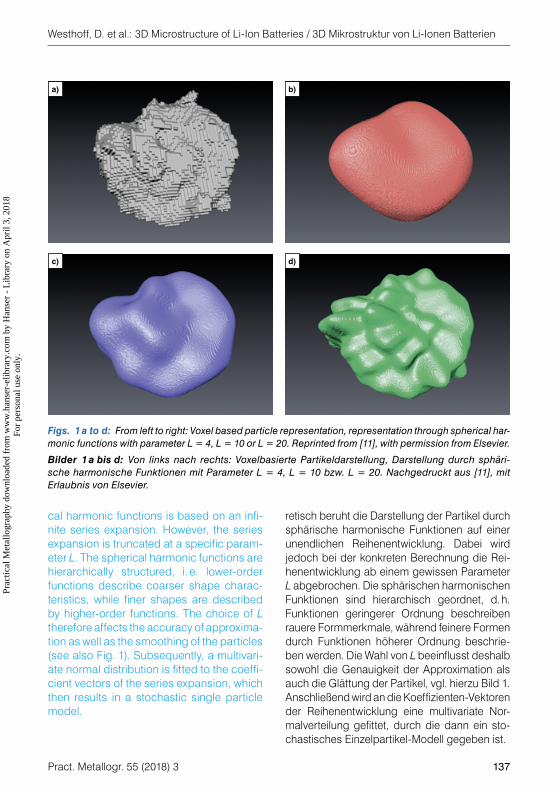
retisch beruht die Darstellung der Partikel durch sphärische harmonische Funktionen auf einer unendlichen Reihenentwicklung. Dabei wird jedoch bei der konkreten Berechnung die Rei-henentwicklung ab einem gewissen Parameter L abgebrochen. Die sphärischen harmonischen Funktionen sind hierarchisch geordnet, d. h. Funktionen geringerer Ordnung beschreiben rauere Formmerkmale, während feinere Formen durch Funktionen höherer Ordnung beschrie-ben werden. Die Wahl von L beeinflusst deshalb sowohl die Genauigkeit der Approximation als auch die Glättung der Partikel, vgl. hierzu Bild 1. Anschließend wird an die Koeffizienten-Vektoren der Reihenentwicklung eine multivariate Nor-malverteilung gefittet, durch die dann ein sto-chastisches Einzelpartikel-Modell gegeben ist.
cal harmonic functions is based on an infi-nite series expansion. However, the series expansion is truncated at a specific param-eter L. The spherical harmonic functions are hierarchically structured, i. e. lower-order functions describe coarser shape charac-teristics, while finer shapes are described by higher-order functions. The choice of L therefore affects the accuracy of approxima-tion as well as the smoothing of the particles (see also Fig. 1). Subsequently, a multivari-ate normal distribution is fitted to the coeffi-cient vectors of the series expansion, which then results in a stochastic single particle model.
Figs. 1 a to d: From left to right: Voxel based particle representation, representation through spherical har-monic functions with parameter L = 4, L = 10 or L = 20. Reprinted from [11], with permission from Elsevier.
Bilder 1 a bis d: Von links nach rechts: Voxelbasierte Partikeldarstellung, Darstellung durch sphäri-sche harmonische Funktionen mit Parameter L = 4, L = 10 bzw. L = 20. Nachgedruckt aus [11], mit Erlaubnis von Elsevier.
b)a)
c) d)
Prac
tical
Met
allo
grap
hy d
ownl
oade
d fr
om w
ww
.han
ser-
elib
rary
.com
by
Han
ser
- L
ibra
ry o
n A
pril
3, 2
018
For
pers
onal
use
onl
y.
Westhoff, D. et al.: 3D Microstructure of Li-Ion Batteries / 3D Mikrostruktur von Li-Ionen Batterien
138 Pract. Metallogr. 55 (2018) 3
3. Stochastisches 3D Strukturmodell für Aktivpartikel-Systeme
3.1 Graphitpartikel in Anoden von Energie-zellen
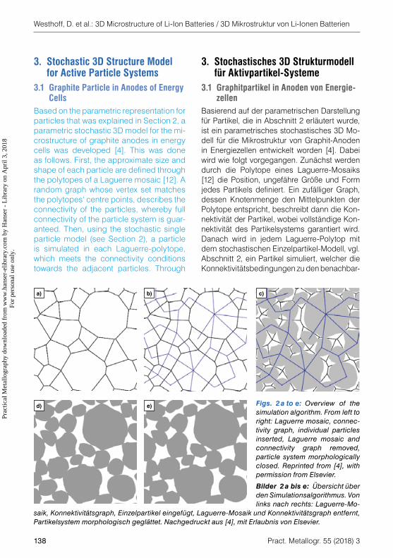
Basierend auf der parametrischen Darstellung für Partikel, die in Abschnitt 2 erläutert wurde, ist ein parametrisches stochastisches 3D Mo-dell für die Mikrostruktur von Graphit-Anoden in Energiezellen entwickelt worden [4]. Dabei wird wie folgt vorgegangen. Zunächst werden durch die Polytope eines Laguerre-Mosaiks [12] die Position, ungefähre Größe und Form jedes Partikels definiert. Ein zufälliger Graph, dessen Knotenmenge den Mittelpunkten der Polytope entspricht, beschreibt dann die Kon-nektivität der Partikel, wobei vollständige Kon-nektivität des Partikelsystems garantiert wird. Danach wird in jedem Laguerre-Polytop mit dem stochastischen Einzelpartikel-Modell, vgl. Abschnitt 2, ein Partikel simuliert, welcher die Konnektivitätsbedingungen zu den benachbar-
3. Stochastic 3D Structure Model for Active Particle Systems
3.1 Graphite Particle in Anodes of Energy Cells
Based on the parametric representation for particles that was explained in Section 2, a parametric stochastic 3D model for the mi-crostructure of graphite anodes in energy cells was developed [4]. This was done as follows. First, the approximate size and shape of each particle are defined through the polytopes of a Laguerre mosaic [12]. A random graph whose vertex set matches the polytopes‘ centre points, describes the connectivity of the particles, whereby full connectivity of the particle system is guar-anteed. Then, using the stochastic single particle model (see Section 2), a particle is simulated in each Laguerre-polytope, which meets the connectivity conditions towards the adjacent particles. Through
Figs. 2 a to e: Overview of the simulation algorithm. From left to right: Laguerre mosaic, connec-tivity graph, individual particles inserted, Laguerre mosaic and connectivity graph removed, particle system morphologically closed. Reprinted from [4], with permission from Elsevier.
Bilder 2 a bis e: Übersicht über den Simulationsalgorithmus. Von links nach rechts: Laguerre-Mo-
saik, Konnektivitätsgraph, Einzelpartikel eingefügt, Laguerre-Mosaik und Konnektivitätsgraph entfernt, Partikelsystem morphologisch geglättet. Nachgedruckt aus [4], mit Erlaubnis von Elsevier.
b)a) c)
d) e)
Prac
tical
Met
allo
grap
hy d
ownl
oade
d fr
om w
ww
.han
ser-
elib
rary
.com
by
Han
ser
- L
ibra
ry o
n A
pril
3, 2
018
For
pers
onal
use
onl
y.
Westhoff, D. et al.: 3D Microstructure of Li-Ion Batteries / 3D Mikrostruktur von Li-Ionen Batterien
Pract. Metallogr. 55 (2018) 3 139
ten Partikeln erfüllt. Mit einer anschließenden morphologischen Glättung wird auch Binder und Leitruß in die Modellierung einbezogen. Eine Übersicht über die einzelnen Schritte des Simulations-algorithmus ist in Bild 2 gegeben.
3.2 Kalibrierung und Validierung des Struk-turmodells
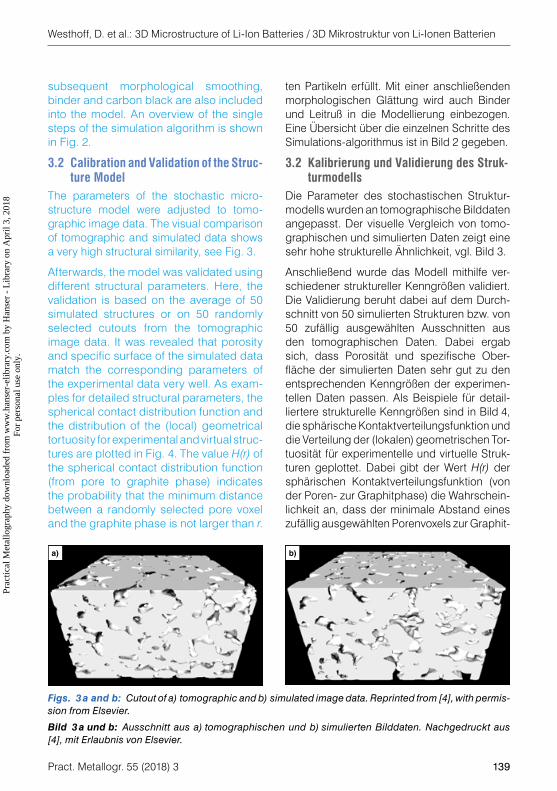
Die Parameter des stochastischen Struktur-modells wurden an tomographische Bilddaten angepasst. Der visuelle Vergleich von tomo-graphischen und simulierten Daten zeigt eine sehr hohe strukturelle Ähnlichkeit, vgl. Bild 3.
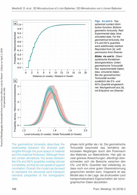
Anschließend wurde das Modell mithilfe ver-schiedener struktureller Kenngrößen validiert. Die Validierung beruht dabei auf dem Durch-schnitt von 50 simulierten Strukturen bzw. von 50 zufällig ausgewählten Ausschnitten aus den tomographischen Daten. Dabei ergab sich, dass Porosität und spezifische Ober-fläche der simulierten Daten sehr gut zu den entsprechenden Kenngrößen der experimen-tellen Daten passen. Als Beispiele für detail-liertere strukturelle Kenngrößen sind in Bild 4, die sphärische Kontaktverteilungsfunktion und die Verteilung der (lokalen) geometrischen Tor-tuosität für experimentelle und virtuelle Struk-turen geplottet. Dabei gibt der Wert H(r) der sphärischen Kontaktverteilungsfunktion (von der Poren- zur Graphitphase) die Wahrschein-lichkeit an, dass der minimale Abstand eines zufällig ausgewählten Porenvoxels zur Graphit-
subsequent morphological smoothing, binder and carbon black are also included into the model. An overview of the single steps of the simulation algorithm is shown in Fig. 2.
3.2 Calibration and Validation of the Struc-ture Model
The parameters of the stochastic micro-structure model were adjusted to tomo-graphic image data. The visual comparison of tomographic and simulated data shows a very high structural similarity, see Fig. 3.
Afterwards, the model was validated using different structural parameters. Here, the validation is based on the average of 50 simulated structures or on 50 randomly selected cutouts from the tomographic image data. It was revealed that porosity and specific surface of the simulated data match the corresponding parameters of the experimental data very well. As exam-ples for detailed structural parameters, the spherical contact distribution function and the distribution of the (local) geometrical tortuosity for experimental and virtual struc-tures are plotted in Fig. 4. The value H(r) of the spherical contact distribution function (from pore to graphite phase) indicates the probability that the minimum distance between a randomly selected pore voxel and the graphite phase is not larger than r.
Figs. 3 a and b: Cutout of a) tomographic and b) simulated image data. Reprinted from [4], with permis-sion from Elsevier.
Bild 3 a und b: Ausschnitt aus a) tomographischen und b) simulierten Bilddaten. Nachgedruckt aus [4], mit Erlaubnis von Elsevier.
b)a)
Prac
tical
Met
allo
grap
hy d
ownl
oade
d fr
om w
ww
.han
ser-
elib
rary
.com
by
Han
ser
- L
ibra
ry o
n A
pril
3, 2
018
For
pers
onal
use
onl
y.
Westhoff, D. et al.: 3D Microstructure of Li-Ion Batteries / 3D Mikrostruktur von Li-Ionen Batterien
140 Pract. Metallogr. 55 (2018) 3
phase nicht größer als r ist. Die geometrische Tortuosität beschreibt das Verhältnis der kürzesten Weglängen durch den Porenraum des Materials zur Materialdicke. Hier gibt es zwar gewisse Abweichungen, allerdings über-schneiden sich die Bereiche zwischen den 5 %- und 95 %- Quantilen fast vollständig, so dass noch von einer guten Übereinstimmung gesprochen werden kann. Insgesamt ist das Modell also in der Lage, die strukturellen (und transportrelevanten) Eigenschaften der tomo-graphischen Daten abzubilden.
The geometrical tortuosity describes the relationship between the shortest path lengths through the pore space of material and the material thickness. Although there are certain deviations, the areas between the 5 % and 95 % quantiles overlap almost completely, so that we can speak of a good correlation. Overall, the model can be used to represent the structural (and transport relevant) properties of the tomographic data.
Figs. 4 a and b: Top: spherical contact distri-bution function; Bottom: geometric tortuosity. Red: Experimental data; blue: simulated data. For the geometrical tortuosity, the 5 % and 95 % quantiles were additionally marked. Reprinted from [4], with permission from Elsevier.
Bilder 4 a und b: Oben: sphärische Kontaktver-teilungsfunktion; Unten: geometrische Tortuosität. Rot: experimentelle Daten; blau: simulierte Daten. Bei der geometrischen Tortuosität wurden zusätzlich die 5 %- und 95 %-Quantile eingezeich-net. Nachgedruckt aus [4], mit Erlaubnis von Elsevier.
Pro
babi
lity
/ Wah
rsch
einl
ichk
eit
Distance (in voxels) / Abstand in Voxeln
Pro
babi
lity
dens
ity /
Wah
rsch
einl
ichk
eits
dich
te
Local turtuosity (in voxels) / lokale Tortuosität (in Voxeln)
b)
a)
Prac
tical
Met
allo
grap
hy d
ownl
oade
d fr
om w
ww
.han
ser-
elib
rary
.com
by
Han
ser
- L
ibra
ry o
n A
pril
3, 2
018
For
pers
onal
use
onl
y.
Westhoff, D. et al.: 3D Microstructure of Li-Ion Batteries / 3D Mikrostruktur von Li-Ionen Batterien
Pract. Metallogr. 55 (2018) 3 141
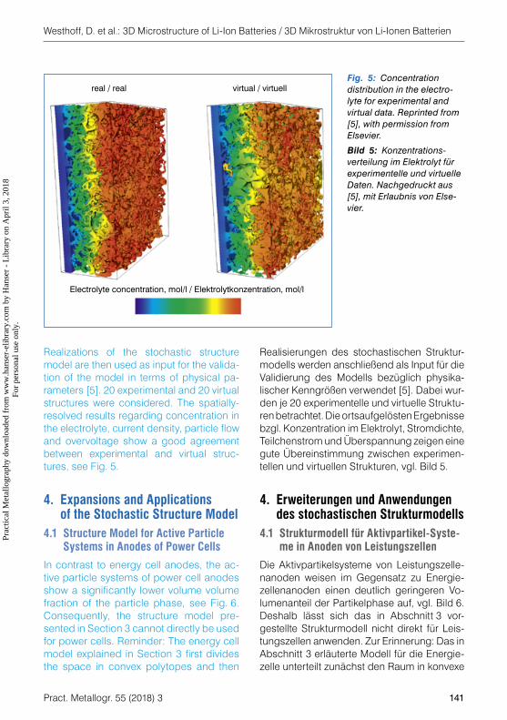
Realisierungen des stochastischen Struktur-modells werden anschließend als Input für die Validierung des Modells bezüglich physika-lischer Kenngrößen verwendet [5]. Dabei wur-den je 20 experimentelle und virtuelle Struktu-ren betrachtet. Die ortsaufgelösten Ergebnisse bzgl. Konzentration im Elektrolyt, Stromdichte, Teilchenstrom und Überspannung zeigen eine gute Übereinstimmung zwischen experimen-tellen und virtuellen Strukturen, vgl. Bild 5.
4. Erweiterungen und Anwendungen des stochastischen Strukturmodells
4.1 Strukturmodell für Aktivpartikel-Syste-me in Anoden von Leistungszellen
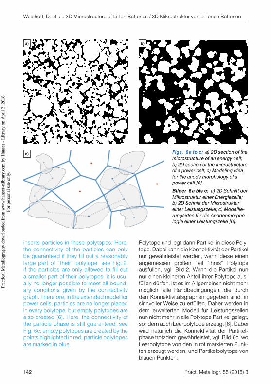
Die Aktivpartikelsysteme von Leistungszelle-nanoden weisen im Gegensatz zu Energie-zellenanoden einen deutlich geringeren Vo-lumenanteil der Partikelphase auf, vgl. Bild 6. Deshalb lässt sich das in Abschnitt 3 vor-gestellte Strukturmodell nicht direkt für Leis-tungszellen anwenden. Zur Erinnerung: Das in Abschnitt 3 erläuterte Modell für die Energie-zelle unterteilt zunächst den Raum in konvexe
Realizations of the stochastic structure model are then used as input for the valida-tion of the model in terms of physical pa-rameters [5]. 20 experimental and 20 virtual structures were considered. The spatially-resolved results regarding concentration in the electrolyte, current density, particle flow and overvoltage show a good agreement between experimental and virtual struc-tures, see Fig. 5.
4. Expansions and Applications of the Stochastic Structure Model
4.1 Structure Model for Active Particle Systems in Anodes of Power Cells
In contrast to energy cell anodes, the ac-tive particle systems of power cell anodes show a significantly lower volume volume fraction of the particle phase, see Fig. 6. Consequently, the structure model pre-sented in Section 3 cannot directly be used for power cells. Reminder: The energy cell model explained in Section 3 first divides the space in convex polytopes and then
Fig. 5: Concentration distribution in the electro-lyte for experimental and virtual data. Reprinted from [5], with permission from Elsevier.
Bild 5: Konzentrations-verteilung im Elektrolyt für experimentelle und virtuelle Daten. Nachgedruckt aus [5], mit Erlaubnis von Else-vier.
Electrolyte concentration, mol/l / Elektrolytkonzentration, mol/l
real / real virtual / virtuell
Prac
tical
Met
allo
grap
hy d
ownl
oade
d fr
om w
ww
.han
ser-
elib
rary
.com
by
Han
ser
- L
ibra
ry o
n A
pril
3, 2
018
For
pers
onal
use
onl
y.
Westhoff, D. et al.: 3D Microstructure of Li-Ion Batteries / 3D Mikrostruktur von Li-Ionen Batterien
142 Pract. Metallogr. 55 (2018) 3
Polytope und legt dann Partikel in diese Poly-tope. Dabei kann die Konnektivität der Partikel nur gewährleistet werden, wenn diese einen angemessen großen Teil “ihres” Polytops ausfüllen, vgl. Bild 2. Wenn die Partikel nun nur einen kleineren Anteil ihrer Polytope aus-füllen dürfen, ist es im Allgemeinen nicht mehr möglich, alle Randbedingungen, die durch den Konnektivitätsgraphen gegeben sind, in sinnvoller Weise zu erfüllen. Daher werden in dem erweiterten Modell für Leistungszellen nun nicht mehr in alle Polytope Partikel gelegt, sondern auch Leerpolytope erzeugt [6]. Dabei wird natürlich die Konnektivität der Partikel-phase trotzdem gewährleistet, vgl. Bild 6c, wo Leerpolytope von den in rot markierten Punk-ten erzeugt werden, und Partikelpolytope von blauen Punkten.
inserts particles in these polytopes. Here, the connectivity of the particles can only be guaranteed if they fill out a reasonably large part of “their” polytope, see Fig. 2. If the particles are only allowed to fill out a smaller part of their polytopes, it is usu-ally no longer possible to meet all bound-ary conditions given by the connectivity graph. Therefore, in the extended model for power cells, particles are no longer placed in every polytope, but empty polytopes are also created [6]. Here, the connectivity of the particle phase is still guaranteed, see Fig. 6c, empty polytopes are created by the points highlighted in red, particle polytopes are marked in blue.
b)a)
c) Figs. 6 a to c: a) 2D section of the microstructure of an energy cell; b) 2D section of the microstructure of a power cell; c) Modeling idea for the anode morphology of a power cell [6].
Bilder 6 a bis c: a) 2D Schnitt der Mikrostruktur einer Energiezelle; b) 2D Schnitt der Mikrostruktur einer Leistungszelle; c) Modellie-rungsidee für die Anodenmorpho-logie einer Leistungszelle [6].
Prac
tical
Met
allo
grap
hy d
ownl
oade
d fr
om w
ww
.han
ser-
elib
rary
.com
by
Han
ser
- L
ibra
ry o
n A
pril
3, 2
018
For
pers
onal
use
onl
y.
Westhoff, D. et al.: 3D Microstructure of Li-Ion Batteries / 3D Mikrostruktur von Li-Ionen Batterien
Pract. Metallogr. 55 (2018) 3 143
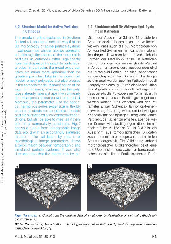
4.2 Strukturmodell für Aktivpartikel-Syste-me in Kathoden
Die in den Abschnitten 3.1 und 4.1 erläuterten Anodenmodelle lassen sich so weiterent-wickeln, dass auch die 3D Morphologie von Aktivpartikel-Systemen in Kathodenmateria-lien dargestellt werden kann, obwohl sich die Formen der Metalloxid-Partikel in Kathoden deutlich von den Formen der Graphit-Partikel in Anoden unterscheiden. Insbesondere sind die Metalloxid-Partikel deutlich sphärischer als die Graphitpartikel. So wie im Leistungs-zellenmodell werden auch im Kathodenmodell Leerpolytope erzeugt. Durch eine Modifikation des Algorithmus wird jedoch sichergestellt, dass bereits die Polytope eine Form haben, in die nahezu sphärische Partikel gut eingebettet werden können. Des Weiteren wird der Pa-rameter L der Spherical-Harmonics-Reihen-entwicklung flexibel gewählt, um bei wenigen Konnektivitätsbedingungen möglichst glatte Partikel-Oberflächen zu erhalten, aber bei vie-len Konnektivitätsbedingungen diese immer noch erfüllen zu können [7]. In Bild 7 ist ein Ausschnitt aus tomographischen Bilddaten zusammen mit einer entsprechend simulierten Struktur dargestellt. Die Validierung anhand morphologischer Bildkenngrößen zeigt eine gute Übereinstimmung zwischen tomographi-schen und simulierten Partikelsystemen. Darü-
4.2 Structure Model for Active Particles in Cathodes
The anode models explained in Sections 3.1 and 4.1, can be refined in a way that the 3D morphology of active particle systems in cathode materials can also be represent-ed, although the shapes of the metal oxide particles in cathodes differ significantly from the shapes of the graphite particles in anodes. In particular, the metal oxide par-ticles are much more spherical than the graphite particles. Like in the power cell model, empty polytopes are also created in the cathode model. A modification of the algorithm ensures, however, that the poly-topes already have a shape in which nearly spherical particles can be well embedded. Moreover, the parameter L of the spheri-cal harmonics series expansion is flexibly chosen to obtain the smoothest possible particle surfaces for a few connectivity con-ditions, but still be able to meet all if there are many connectivity conditions. Fig. 7 shows a cutout from tomographic image data along with an accordingly simulated structure. The validation by means of morphological image parameters shows a good match between tomographic and simulated particle systems. It was also demonstrated that the model can be ad-
Figs. 7 a and b: a) Cutout from the original data of a cathode; b) Realization of a virtual cathode mi-crostructure [7].
Bilder 7 a und b: a) Ausschnitt aus den Originaldaten einer Kathode; b) Realisierung einer virtuellen Kathodenmikrostruktur [7].
b)a)
Prac
tical
Met
allo
grap
hy d
ownl
oade
d fr
om w
ww
.han
ser-
elib
rary
.com
by
Han
ser
- L
ibra
ry o
n A
pril
3, 2
018
For
pers
onal
use
onl
y.
Westhoff, D. et al.: 3D Microstructure of Li-Ion Batteries / 3D Mikrostruktur von Li-Ionen Batterien
144 Pract. Metallogr. 55 (2018) 3
ber hinaus wurde gezeigt, dass sich das Modell durch eine geeignete Wahl der Parameter an Kathodenstrukturen sowohl für neuwertige als auch gealterte Kathoden anpassen lässt [7].
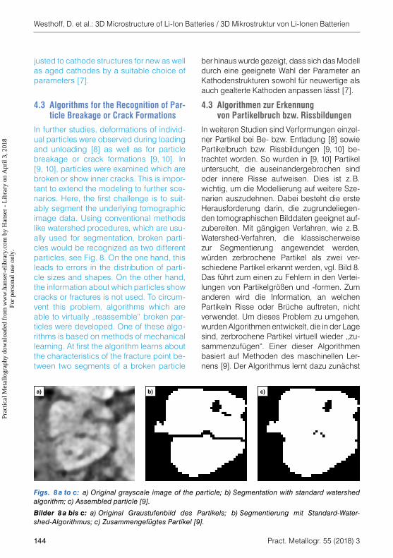
4.3 Algorithmen zur Erkennung von Partikelbruch bzw. Rissbildungen
In weiteren Studien sind Verformungen einzel-ner Partikel bei Be- bzw. Entladung [8] sowie Partikelbruch bzw. Rissbildungen [9, 10] be-trachtet worden. So wurden in [9, 10] Partikel untersucht, die auseinandergebrochen sind oder innere Risse aufweisen. Dies ist z. B. wichtig, um die Modellierung auf weitere Sze-narien auszudehnen. Dabei besteht die erste Herausforderung darin, die zugrundeliegen-den tomographischen Bilddaten geeignet auf-zubereiten. Mit gängigen Verfahren, wie z. B. Watershed-Verfahren, die klassischerweise zur Segmentierung angewendet werden, würden zerbrochene Partikel als zwei ver-schiedene Partikel erkannt werden, vgl. Bild 8. Das führt zum einen zu Fehlern in den Vertei-lungen von Partikelgrößen und -formen. Zum anderen wird die Information, an welchen Partikeln Risse oder Brüche auftreten, nicht verwendet. Um dieses Problem zu umgehen, wurden Algorithmen entwickelt, die in der Lage sind, zerbrochene Partikel virtuell wieder „zu-sammenzufügen“. Einer dieser Algorithmen basiert auf Methoden des maschinellen Ler-nens [9]. Der Algorithmus lernt dazu zunächst
justed to cathode structures for new as well as aged cathodes by a suitable choice of parameters [7].
4.3 Algorithms for the Recognition of Par-ticle Breakage or Crack Formations
In further studies, deformations of individ-ual particles were observed during loading and unloading [8] as well as for particle breakage or crack formations [9, 10]. In [9, 10], particles were examined which are broken or show inner cracks. This is impor-tant to extend the modeling to further sce-narios. Here, the first challenge is to suit-ably segment the underlying tomographic image data. Using conventional methods like watershed procedures, which are usu-ally used for segmentation, broken parti-cles would be recognized as two different particles, see Fig. 8. On the one hand, this leads to errors in the distribution of parti-cle sizes and shapes. On the other hand, the information about which particles show cracks or fractures is not used. To circum-vent this problem, algorithms which are able to virtually „reassemble“ broken par-ticles were developed. One of these algo-rithms is based on methods of mechanical learning. At first the algorithm learns about the characteristics of the fracture point be-tween two segments of a broken particle
Figs. 8 a to c: a) Original grayscale image of the particle; b) Segmentation with standard watershed algorithm; c) Assembled particle [9].
Bilder 8 a bis c: a) Original Graustufenbild des Partikels; b) Segmentierung mit Standard- Water-shed-Algorithmus; c) Zusammengefügtes Partikel [9].
b)a) c)
Prac
tical
Met
allo
grap
hy d
ownl
oade
d fr
om w
ww
.han
ser-
elib
rary
.com
by
Han
ser
- L
ibra
ry o
n A
pril
3, 2
018
For
pers
onal
use
onl
y.
Westhoff, D. et al.: 3D Microstructure of Li-Ion Batteries / 3D Mikrostruktur von Li-Ionen Batterien
Pract. Metallogr. 55 (2018) 3 145
auf von Hand segmentierten Daten, welche Eigenschaften die Bruchstelle zwischen zwei Fragmenten eines gebrochenen Partikels hat. Solche Bruchstellen können dann automatisch in ähnlichen Datensätzen detektiert werden. Zur Validierung der Ergebnisse wurden mit dem in Abschnitt 3.1 präsentierten stochasti-schen Strukturmodell Bilddaten generiert und zusätzlich Partikel virtuell zerbrochen. Mit Hilfe dieser Daten konnte dann der Erfolg des Al-gorithmus quantifiziert werden. Als Alternative zu diesem Ansatz wurde in [10] ein parame-trischer Segmentieralgorithmus entwickelt, der auch Risse in Partikeln, die nicht zum vollstän-digen Bruch in zwei Teile führen, detektiert, und keine von Hand segmentierten Bilddaten zum Training des Algorithmus benötigt. Auch dieser Ansatz wurde durch Untersuchung simulierter Strukturen validiert sowie die Anwendbarkeit für verschiedene reale Strukturen gezeigt.
Mithilfe dieser Algorithmen kann eine Segmen-tierung von stark gealterten Elektroden ermög-licht werden, die auch die Risse und Brüche von Partikeln berücksichtigt und die Grundlage für eine weiterführende Modellierung darstellt.
DanksagungEine frühere Fassung dieser Arbeit wurde in „Fortschritte in der Metallographie. Sonder-band zur 51. Metallographie-Tagung in Aalen, T. Bernthaler, G. Schneider (Hrsg.), Aalen, 13.–15.09.2017, 189 – 194.“ veröffentlicht.
by using manually segmented data. Such fracture points can then be automatically detected in similar sets of data. To validate the results, the stochastic model presented in Section 3.1 was used to generate image data and virtually break particles. This data can be used to quantify the success of the algorithm. As an alternative to this approach, a parametric segmentation al-gorithm was developed in [10], which can also detect cracks that did not lead to the complete fracture of the particle into two pieces and which does not require manu-ally segmented image data to train the al-gorithm. This approach was also validated by the examination of simulated structures and the applicability for different real struc-tures was shown.
These algorithms can be used to enable a segmentation of heavily aged electrodes, which also considers cracks and fractures of particles and serves as the basis for fur-ther modeling.
AcknowledgementsAn earlier version of this work has been pub-lished in “Fortschritte in der Metallographie. Sonderband zur 51. Metallographie-Tagung in Aalen, T. Bernthaler, G. Schneider (Hrsg.), Aalen, 13.–15.09.2017, 189 – 194.”
References / Literatur [1] Schmidt, D.; Kleinbach, M.; Kamlah, M.; Knob-
lauch, V., in: Fortschritte in der Metallographie. Sonderband der Praktischen Metallographie zur 51. Metallographie-Tagung in Aalen, T. Bernthaler, G. Schneider (Hrsg.), Aalen, 13.–15.09.2017, 61 – 66
[2] Newman, J.; Thomas, K.; Hafezi, H.; Wheeler, D.: J. Power Sources 119 (2003), 838 – 843
DOI: 10.1016/S0378 – 7753(03)00282 – 9 [3] Feinauer, J., Hein, S.; Rave, S.; Schmidt, S.;
Westhoff, D.; Zausch, J.; Iliev, O.; Latz, A.;
Ohlberger, M.; Schmidt, V.: MULTIBAT: Unified workflow for fast electrochemical 3D simulations of lithium-ion cells combining virtual stochastic microstructures, electrochemical degradation models and model order reduction. J. Comput. Sci. (under revision)
[4] Feinauer, J.; Brereton, T.; Spettl, A.; Weber, M.; Manke, I.; Schmidt, V.: Comput. Mater. Sci. 109 (2015), 137 – 146
DOI: 10.1016/j.commatsci.2015.06.025
Prac
tical
Met
allo
grap
hy d
ownl
oade
d fr
om w
ww
.han
ser-
elib
rary
.com
by
Han
ser
- L
ibra
ry o
n A
pril
3, 2
018
For
pers
onal
use
onl
y.
Westhoff, D. et al.: 3D Microstructure of Li-Ion Batteries / 3D Mikrostruktur von Li-Ionen Batterien
146 Pract. Metallogr. 55 (2018) 3
[10] Westhoff, D; Finegan, D. P.; Shearing, P. R.; Schmidt, V.: Algorithmic structural segmenta-tion of defective particle systems: A lithium-ion battery study. Journal of Microscopy (im Druck),
DOI: 10.1111/jmi.12653 [11] Feinauer, J.; Spettl, A.; Manke, I.; Strege, S.;
Kwade, A.; Pott, A.; Schmidt, V.: Mater. Charact. 106 (2015) 123 – 133.
DOI: 10.1016/j.matchar.2015.05.023 [12] Chiu, S. N.; Stoyan, D.; Kendall, W. S.; Mecke, J.:
Stochastic Geometry and its Applications, 3 Au-flage, J. Wiley & Sons, Chichester, 2013.
DOI: 10.1002/9781118658222
[5] Hein, S.; Feinauer, J.; Westhoff, D.; Manke, I.; Schmidt, V.; Latz A.: J. Power Sources 336 (2016), 161 – 171
DOI: 10.1016/j.jpowsour.2016.10.057 [6] Westhoff, D.; Feinauer, F.; Kuchler, K.; Mitsch, T.;
Manke, I.; Hein, S.; Latz, A.; Schmidt, V.: Com-put. Mat. Sci. 126 (2017) 453 – 467
DOI: 10.1016/j.commatsci.2016.09.006 [7] Kuchler, K.; Westhoff, D.; Feinauer, J.; Mitsch, T.;
Manke, I.; Schmidt V.: Stochastic model of the 3D microstructure of Li-ion battery cathodes under various cyclical aging scenarios, Model. Simul. Mater. Sci. Eng. (in print)
[8] Pietsch, P.; Westhoff, D.; Feinauer, J.; Eller, J.; Marone, F.; Stampanoni, M.F.M.; Schmidt, V.; Wood, V.: Nat. Commun. 7 (2016) 12909
DOI: 10.1038/ncomms12909 [9] Petrich, L.; Westhoff, D.; Feinauer, J.; Fin-
egan, D. P.; Daemi, S. R.; Shearing, P. R.; Schmidt, V.: Comput. Mat. Sci. 136 (2017) 297 – 305
DOI: 10.1016/j.commatsci.2017.05.012
BibliographyDOI 10.3139/147.110501Pract. Metallogr. 55 (2018) 3; page 134 – 146© Carl Hanser Verlag GmbH & Co. KGISSN 0032 – 678X
Daniel Westhoff
is a PhD student at the Institute of Stochastics at Ulm University under the supervision of Prof. Dr. Volker Schmidt. In his research he deals with structural seg-mentation, statistical analysis and stochas-tic modeling of 3D im-
age data with a focus on the microstructure of functional materials.
Volker Schmidt
is Professor at the In-stitute of Stochastics at Ulm University. He re-ceived his PhD from the Technical University of Freiberg. His research interests include sto-chastic modeling and simulation of 3D micro-structures, using tools of stochastic geometry and spatial statistics.Pr
actic
al M
etal
logr
aphy
dow
nloa
ded
from
ww
w.h
anse
r-el
ibra
ry.c
om b
y H
anse
r -
Lib
rary
on
Apr
il 3,
201
8Fo
r pe
rson
al u
se o
nly.