Bone Marrow Mesenchymal Stem Cell Niches and Regenerative ... · Bone Marrow Mesenchymal Stem Cell...
142
1 Bone Marrow Mesenchymal Stem Cell Niches and Regenerative Medicine Inauguraldissertation zur Erlangung der Würde eines Doktors der Philosophie vorgelegt der Philosophisch-Naturwissenschaftlichen Fakultät der Universität Basel von Nunzia Di Maggio aus Italien Basel (Schweiz), 2010
Transcript of Bone Marrow Mesenchymal Stem Cell Niches and Regenerative ... · Bone Marrow Mesenchymal Stem Cell...
1
Bone Marrow Mesenchymal Stem Cell Niches and Regenerative Medicine
Inauguraldissertation
zur Erlangung der Würde eines Doktors der Philosophie
vorgelegt der Philosophisch-Naturwissenschaftlichen Fakultät
der Universität Basel
von
Nunzia Di Maggio
aus Italien
Basel (Schweiz), 2010

2
Genehmigt von der Philosophisch-Naturwissenschaftlichen Fakultät auf Antrag von Prof. Dr. Ivan Martin (Supervisor) Prof. Dr. Antonius Rolink (Referee) Prof. Dr. Aleksandra Wodnar-Filipowicz (Co-referee) Basel, den 26/05/2009
Prof. Dr. Eberhard Parlow Dekan

3
Table of Contents
Summary 5
1. Bone and Bone Marrow: Structure and Function 9
1.1 Bone tissue: biology, structure and function 10
1.2 Bone formation: development, healing and repair 14
1.3 The bone structure 17
References 23
2. Bone Marrow Mesenchymal Stem Cells 25
2.1 History 26
2.2 In vitro proliferation and differentiation potential 28
2.3 Assessment of self-renewal 32
2.4 Phenotypical characterization 34
2.5 Clinical applications 35
References 39
3. The Bone Marrow Stem Cell Niche 43
3.1 The hematopoietic stem cell 44
3.2 Stem cell niche function 46
3.3 The bone marrow stem cell niche 49
3.4 Molecular cross-talk in the endosteal niche 52
3.5 Perspectives 54
References 57
4. FGF-2/FGFR2c signaling generates a niche-progenitor system in vitro by selecting
and maintaining a self-renewing, highly potent, non-adherent mesenchymal
progenitor population 61
4.1 Introduction 62
4.2 Materials and Methods 64
4.3 Results 69
4.4 Discussion 86
4
References 90
5. Adipose tissues stromal vascular fraction (SVF) cultures contain a population of
non-adherent mesenchymal progenitor (NAMP) which are FGF2-dependent and
require an in vitro niche for their maintenance 95
5.1 Introduction 94
5.2 Materials and Methods 95
5.3 Results 97
5.4 Summary and Conclusions 107
References 108
6. A three-dimensional bioreactor-based culture system to engineer the bone marrow
niche 109
6.1 Introduction 110
6.1.1 Stem Cell Niches 110
6.1.2 Standard HSC culture in vitro 112
6.1.3 Alternative systems for HSC in vitro culture 114
6.2 A 3D-perfusion bioreactor system and engineering of 3D- stromal tissue 117
6.3 A 3D-perfusion bioreactor based system for HSC culture: a possible experimental
design 119
6.4 Modulating variables 119
6.4.1 Culture conditions 123
6.4.2 Scaffold composition 125
6.4.3 Co-culture of different cell types 128
6.5 Future perspectives 130
References 134
7. Future Perspectives 139
References 142
5
Summary
Mesenchymal progenitors are a powerful tool in regenerative medicine, but suffer from a
rapid loss of differentiation potential during in vitro expansion (1). The recent discovery that
well-characterized stem cells, like HSC, maintain their stemness during self-renewal through
the interaction with specialized microenvironments, called stem cell niches, prompted us to
investigate the existence of a niche compartment for also mesenchymal progenitors.
In Chapter 4 of this thesis we described the establishment of a niche/progenitor system
in vitro for bone marrow mesenchymal stem cells (MSC). We asked whether the non-
adherent fraction of human bone marrow cultures contained early progenitors which can
constitute a reservoir for the mesenchymal compartment and whether the adherent cells,
instead, could provide a niche function for the maintenance and regulation of these
progenitors.
Replating the non-adherent fraction in a new dish at the first medium change, we found
that a population of bone marrow non-adherent mesenchymal progenitors (BM-NAMP) was
present and their number was 20.4±3.6% of the initial CFU-f. The replating in the same dish,
instead, did not increase the number of colonies (100±25.1% vs 99.0±23.5%, p=n.s.) and no
change in the average diameter size was observed (5.5±0.5 mm vs 5.8±0.6 mm, p=n.s.),
indicating that BM-NAMP were stably non-adherent. However, further investigation showed
that, when serially replated in new dishes, BM-NAMP were able to steadily increase in
number, self-renewing as non-adherent progenitors while generating at the same time
adherent colonies. The diameter size evaluation showed that BM-NAMP could produce
colonies with 2-fold larger diameter (Plate1=10.73±1.21 mm vs Plate0=5.54±0.45 mm, p<
0.01), indicating a significantly higher proliferation capacity. However, the colonies produced
in the following replating steps were progressively smaller, indicating a gradual loss of BM-
NAMP proliferation potential (Plate2= 8.8±0.7 mm, Plate3= 7.5±0.3 mm, Plate4= 6.4±0.3

6
mm). Together with increased proliferation, first-replated BM-NAMP progeny cells
displayed a higher differentiation potential compared to standard CFU-f both in vitro and in
vivo. In vitro assays revealed that they could differentiate towards the adipogenic,
chondrogenic and osteogenic lineage and, when implanted in vivo, they produced 3-fold
higher amounts of bone tissue. Taken together, these data indicate together that BM-NAMP
show features of earlier progenitor features and suggest a biological difference between BM-
NAMP and the initially adhrering CFU-f.
Serial replating experiments performed with serum alone showed that BM-NAMP
critically required FGF-2 for their initial selection and maintenance in culture. Furthermore,
FGF-2 removal, at different time points during serial replating, always caused the
disappearance of BM-NAMP. This observation is compatible with the data that described
FGF-2 role in the selection of a subset of earlier pluripotent mesenchymal precursors within
initial CFU-f (3). Interestingly, blocking receptor experiments showed that the maintenance
of BM-NAMP in culture was mediated through FGFR2c signaling, which has been shown to
be involved in vivo in the balance between proliferation and differentiation of skeletal
progenitors (4).
When replated in a new dish, BM-NAMP regenerated themselves as non-adherent
progenitors, but at the same time they always gave rise to adherent colonies. We, therefore,
hypothesize that BM-NAMP were in close interaction with the adherent cells, and that these
provide a niche function for them. BM-NAMP were not able to survive when replated either
on agarose-coated dishes or on human fibroblasts. This suggests that BM-NAMP required
specific signals from the adherent progeny and that this fraction constitutes a unique
environment for BM-NAMP survival and self-renewal. In fact, when kept in contact with
initial CFU-f progeny for 14 days instead of being serially replated, BM-NAMP were able to
produce 3-fold more colonies (260.8±15.8% vs 61.0±8.0%). Furthermore, the colony
7
diameter analysis showed that, unlike the serial replating which caused a gradual loss of BM-
NAMP proliferative activity, the continuous culture in the primary plate could preserve BM-
NAMP proliferation potential (9.2±0.5 mm and 9.8±0.4 mm at the start of culture
respectively vs 6.8±0.3 mm after serial replating, p<0.05). Furthermore, if kept in the original
plate, BM-NAMP could generate a progeny that also displayed a higher differentiation
capacity. Taken together, these results suggest together that CFU-f progeny provides a niche
function for BM-NAMP. When an adherent fraction was not present, due to serial replating in
empty dishes, BM-NAMP could regenerate their niche but at the cost of gradually losing
their proliferative and differentiation potential.
In Chapter 5 we sought at investigating the presence of a class of non-adherent
progenitors in human adipose tissue stromal vascular fraction (SVF), which constitute an
abundant source of mesenchymal progenitors, to determine whether the NAMP compartment
was specific to bone marrow or they could constitute a reservoir also in other tissues.
NAMP were present in adipose tissue SVF cultures (AT-NAMP) with a similar
frequency as observed in the bone marrow (17.7±9.1% vs 20.4±3.6% respectively) and the
replating of the non-adherent fraction in the same dish revealed that they were stably non-
adherent. The main difference compared to BM-NAMP was the inability of AT-NAMP to
self-renew as non-adherent progenitors upon serial replating, since only few colonies were
present in the last replating step. However, these colonies had a significantly increased
diameter. This suggests that, when serially replated, AT-NAMP do not undergo proliferation
but rather a selection for the very rare progenitors with the highest proliferation ability.
Similarly to BM-NAMP, when kept in contact with the initially adhering CFU-f, AT-NAMP
could proliferate without loss of their proliferation capacity. This suggests that, as for bone
marrow cells, adherent CFU-f provide a niche function for the non-adherent progenitors,
regulating the maintenance of their early-progenitor properties.

8
In conclusion, these data show that, although displaying important tissue-specific biological
differences, NAMP are present in the mesenchymal progenitor compartment of different
tissues and they represent a reservoir of earlier progenitors compared to standard CFU-f.

9
Chapter 1:
Bone and bone marrow: structure and function
10
1.1 Bone tissue: biology, structure, and function
Bone is a dynamic, highly vascular, and mineralized connective tissue, characterized by
its hardness, resilience, growth mechanisms, and its capability to remodel itself throughout
the life-time of an individual.
Bone performs several key functions within the body: it not only provides structural
support and protection to bodily organs, but is also responsible for maintaining mineral
homeostasis, and is the primary site for the synthesis of blood cells. Furthermore, it is capable
of maintaining an optimal shape and structure throughout life, via a continuous process of
renewal and remodelling, through which it’s able to respond to changes in its mechanical
environment, in order to meet different loading demands, thus maintaining an optimal
balance between form and function (1).
Simply, bone is a dense multi-phase composite, made up of cells embedded in a very
well-organized matrix, which is composed of both organic and inorganic elements; however,
both structure and proportion of its components widely differ with age, site and history,
resulting in many different classifications of bone that exhibit various mechanical and
functional characteristics.
Histologically, mature bone is classified in two different types of tissue, one of which is
relatively dense, known as cortical bone, while the other consists of a network of struts or
trabeculae surrounding interconnected spaces, known as trabecular or cancellous bone
(Figure 1). Bone surfaces consist of cortical bone, and the thickness of this protective layer
increases in mechanically demanding regions, such as the shafts of long bones, while
cancellous bone is found in the interior of bones, such as within the femoral head, and
vertebra.
Bone as an organ is composed of three main elements: (i) bone matrix, providing
mechanical strength and acting as the body’s mineral store, (ii) bone cells, responsible for
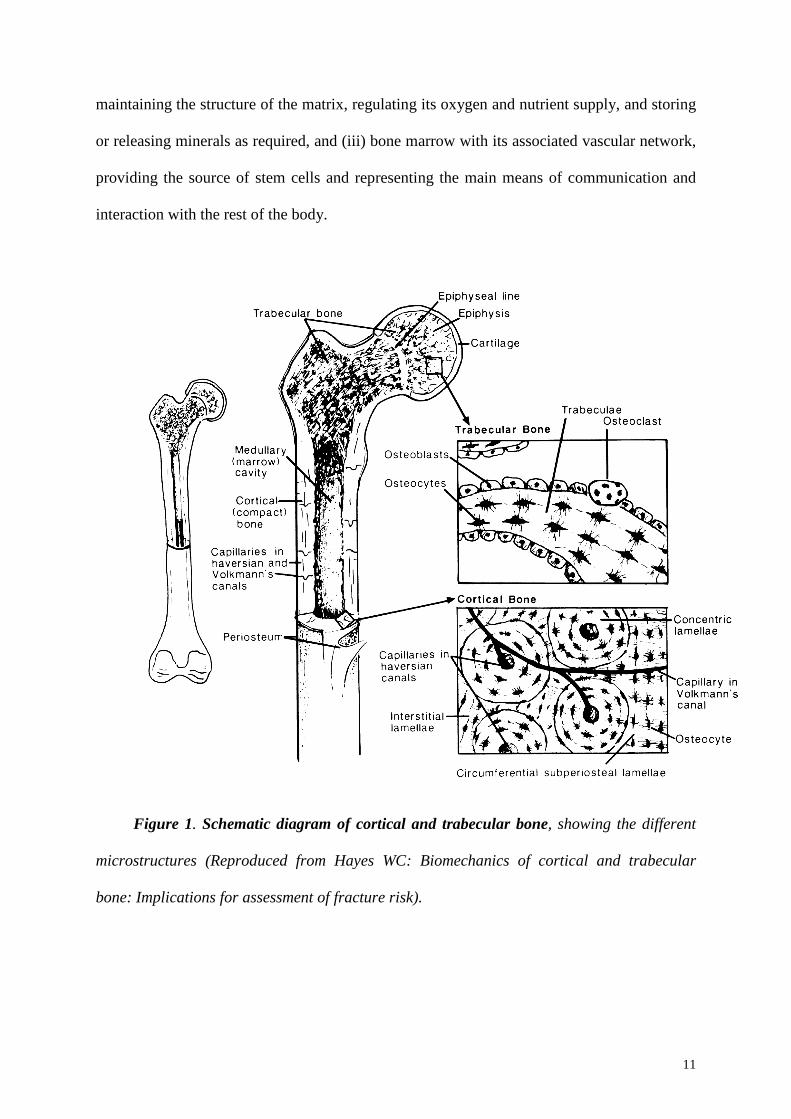
11
maintaining the structure of the matrix, regulating its oxygen and nutrient supply, and storing
or releasing minerals as required, and (iii) bone marrow with its associated vascular network,
providing the source of stem cells and representing the main means of communication and
interaction with the rest of the body.
Figure 1. Schematic diagram of cortical and trabecular bone, showing the different
microstructures (Reproduced from Hayes WC: Biomechanics of cortical and trabecular
bone: Implications for assessment of fracture risk).
12
Bone extracellular matrix has two main components: the organic collagen fibres and the
inorganic bone mineral crystals. Together they make up approximately 95% of the dry weight
of bone, the remainder being composed of other organic molecules, collectively known as the
non-collagenous proteins.
Collagen accounts for 70-90% of the non-mineralized components of the bone matrix;
it consists of carefully arranged arrays of tropocollagen molecules, which are long rigid
molecules composed of three left-handed helices of peptides, known as α-chains, which are
bound together in a right-handed triple helix. Bone contains mostly type-I collagen, which is
composed of tropocollagen molecules containing two identical and one dissimilar α-chains
(α1(I)2 α2).
The main inorganic phase within the bone matrix is usually incorrectly referred to as
hydroxyapatite (HA), a hydrated calcium phosphate ceramic, with a similar crystallographic
structure to natural bone mineral, which has a chemical formula of Ca10(PO4)6(OH)2;
however, bone-apatite is characterized by calcium, phosphate and hydroxyl deficiency,
internal crystal disorder, and ionic substitutions, thus resulting in the presence of significant
levels of additional trace elements within bone mineral: it is not a direct analogue of HA, but
more closely a carbonate-substituted apatite. All these factors contribute to an apatite that is
insoluble enough for stability, yet sufficiently reactive to allow the in vivo crystallites to be
constantly resorbed and reformed as required by the body.
The most important non-collagenous organic constituents of bone matrix are four
proteins: osteocalcin (OC), bone sialoprotein (BSP), osteopontin (OP) and osteonectin (ON).
They are produced by bone cells and their relative composition within the bone matrix
appears to be self-regulating through a feedback effect on their expression by osteoblasts.
They all appear to be multi-functional, and are all involved in regulating bone mineralization
and remodelling.

13
Bone matrix also contains a great number of growth factors, including fibroblast growth
factors (FGFs), insuline-like growth factors (IGFs), plateled-derived growth factors (PDGF),
transforming growth factor-beta (TGFβ) superfamily, and bone morphogenic proteins
(BMPs): they play several critical roles in regulating cell proliferation and differentiation,
inducing the complete sequence of endochondral bone formation, when cartilage forms first
and is subsequently replaced by bone.
The major types of bone cells are osteoblasts, osteocytes and osteoclasts, respectively
responsible for production, maintenance, and resorption of bone; they are highly specialized
differentiated cells, and they generally don’t proliferate. Less differentiated cells of the same
lineage are required for the control of bone cell populations, and, as demands are made on or
by the bone, these cells proliferate and differentiate as required: such cells are generally
known as stem cells, and in the case of bone formation are often referred to as osteogenic
cells.
The osteogenic bone-forming cells originate from the mesenchymal bone marrow
stromal cell line and exist in the endosteum and periosteum (2). Biochemical signalling
molecules stimulated during remodelling and fracture healing, result in a local increase of this
cell population. However, the local environment also determines the route of differentiation
undertaken by osteogenic cells, resulting in the evolution of either osteoblasts or
chondroblasts: if the environment surrounding a differentiating osteogenic cell has a high
vascular content, as in healthy bone, the cell will differentiate into an osteoblast which will
produce bone; once the osteoblast has been surrounded by bone, it differentiates into an
osteocyte, and becomes involved in the nutrition and maintenance of the local bone. In
contrast, if the environment surrounding a differentiating osteogenic cell has little or no
vascular content, as in a recent fracture site, the cell will differentiate into a chondroblast and
cartilage will be produced; once the chondroblast is surrounded by cartilage, it then

14
differentiates into a chondrocyte, which maintains the surrounding collagenous matrix until
it’s replaced by bone during endochondral ossification.
In contrast osteoclasts are derived from monocytes, thus they originate from the
haemopoietic stem cell lineage: under the influence of specific signalling proteins or
cytokines, mononuclear monocytes migrate to the resorption site and fuse with either other
monocytes or a multi-nucleated macrophage, before differentiating into the specialized
osteoclast, an aggressive cell responsible for bone resorption (3).
1.2 Bone formation: development, healing, and repair
Bone is unique among all the vertebrate tissues in its ability to heal via formation of
new bone: most of the other tissues, such as heart, muscle and brain heal by replacement with
connective tissue rather than original tissue. Furthermore, in a mature animal, the molecular
and cellular patterns of bone repair after injury are similar to bone formation in an embryo,
suggesting analogous mechanisms for the control of bone formation in adult and embryonic
skeletons (4). In an embryo, a condensation of primitive mesenchymal cells can transform
into bone via either intramembranous or endochondral ossification: intramembranous
ossification occurs when the mesenchymal cells are transformed into osteoprogenitor cells
and then directly into osteoblasts, resulting in the direct formation of bone; endochondral
ossification occurs via a two-step process where mesenchymal cells transform into
chondroblasts which lay down a collagenous template, subsequently ossified by invading
osteoblasts. The final mature bone formed by both processes is virtually indistinguishable,
and the mechanisms dictating which route is taken are poorly understood.
Fractured bone heals through endochondral ossification: a haematoma is formed,
resulting from injury to the periosteum and local soft tissue; as a consequence of this
disruption in the blood supply, osteocytes nearest to the fracture die, resulting in local
15
necrosis of the bone around the fracture; simultaneously, there is a demand for the repair of
the bone, the stabilization of the damaged area and the removal of the dead tissue; in response
to this, macrophages and fibroblasts are recruited to the site to remove tissue debris, and to
express extracellular matrix, respectively. In response to growth factors and cytokines
released by these inflammatory cells, mesenchymal stem cells recruited from the bone
marrow and periosteum, proliferate and differentiate into osteoprogenitor cells. This leads to
an apparent thickening of the periosteum and the production of collars of external fracture
callus around the fracture site. Those osteoprogenitor cells that lie close to undamaged bone,
differentiate into bone osteoblasts and form an osteoid, which is rapidly calcified into bone,
while those farther away become chondroblasts and form cartilage; concurrent angiogenesis
is induced, and, as soon as cartilage has formed and the fracture site stabilized, it is replaced
by cancellous bone via endochondral ossification, in which osteoclasts and osteoprogenitor
cells invade the cartilaginous callus preceded by capillary formation. The uncalcified material
is then resorbed, and new bone is deposited on the remaining spinicules of calcified cartilage.
Woven bone is finally remodelled into lamellar bone, bone marrow is restored within
cancellous regions, and successive layers of bone gradually fill the spaces between trabeculae
of cortical bone. Load-bearing capabilities and a new vascular network are thus restored.
Although the vast majority of bone defects spontaneously heal with minimal treatment,
among the 6 millions fractures occurring every year in the United States, 5-10% require
further treatment for compromised healing because of either interposition of soft tissue,
improper fracture fixation, loss of bone, metabolic diseases, impairment of blood supply or
infection. Furthermore, in certain clinical settings, large pieces of bone must be resected to
treat benign and malignant tumours, osteomyelitis, as well as bone deficiences, and abnormal
loss in the maxillo-facial area; in addition, bone is typically subject to progressive
degeneration as a result of age and disease (i.e. osteoporosis).
16
Considering all these challenging situations, bone function can often be restored only
by surgical reconstruction: bone grafting, the procedure of replacing missing bone with
material from either the patient’s own body (autografting) or that of a donor (allografting) is
used in the surgical procedures since many years. Autologous bone harvested from donor
sites such as the iliac crest, is the preferred treatment (5): grafts of this kind are
osteoconductive (they provide a scaffold on which bone cells can proliferate), osteoinductive
(they induce proliferation of undifferentiated cells and their differentiation into osteoblasts),
and osteogenic (they provide a reservoir of skeletal stem and progenitor cells that can form
new bone); however, the amount of bone that can be safely harvested is limited, while the
additional surgical procedure may be complicated by donor-site pain and morbidity. Modern
allografting using material stored within bone banks overcomes these difficulties; however,
the demand exceeds the supply, there is no assurance of freedom from disease, and healing
can be inconsistent (6).
As an alternative to these two types of bone grafts, a wide variety of synthetic
substrates have been developed and are actually in clinical use, with mixed success and
surgical acceptance: such materials in fact are generally biocompatible and osteoconductive,
thus supporting adhesion, proliferation, and differentiation of osteogenic cells from
surrounding tissues, and ultimately leading to bone formation; however, these materials are
not osteoinductive, providing only the scaffold which has to be invaded by bone-forming
bioactive cells (7), (8): reasoning that they typically give good results only when implanted in
small defects, where interactions between material’s surface and local cells and proteins are
sufficient to repair the bone defect. In addition, metals, although providing immediate
mechanical support at the site of the defect, exhibit poor overall integration with the tissue at
the implantation site, and can fail because of infection or fatigue loading; on the other hand,
17
ceramics have very low tensile strength and are brittle, thus they cannot be used in locations
of significant torsion, bending, or shear stress (9).
Thus it’s clearly seen that repair of bone defects is actually still a big challenge for the
orthopaedic, reconstructive, and maxillo-facial surgeons: it’s in this scenario that a promising
field of science called Tissue Engineering is emerging since the last few years.
1.3 The bone marrow structure
The marrow, one of the largest organs in the human body, is the principal site for blood
cell formation. Until the late 19th century hematopoiesis was thought to be the prerogative of
the limph nodes or the liver and the spleen. In 1868, Neuman and Bizzozzero independently
observed nucleated blood cells in material squeezed from the ribs of human cadavers and
proposed that bone marrow was the major source of blood cells.
The bone marrow is found within the central cavities of axial and long bones. It
consists of hematopoietic tissue islands and adipose cells surrounded by vascular sinuses
interspersed within a meshwork of trabecular bone. The inner surface of the bone cavities and
the outer surface of the cancellous bone trabeculae within the cavities are covered by an
endosteal lining consisting of a single layer of flat “bone-lining cells” supported by a thin
layer of reticular connective tissue; osteoblasts and osteoclasts are also found within the
endosteal lining.
Macroscopically, the bone marrow is composed by red marrow (hematopoietic) and
yellow marrow (adipose), whose proportions vary with age in agreement with the Neumann’s
law, according to which, at birth, all the bones contain red marrow, whereas, as age increases,
the extension of hematopoietic marrow contracts towards the axial skeleton and the
peripheral bones contain only yellow marrow.
Microscopically, the bone marrow structure follows the organization of the vasculature.
18
Bone marrow has an extensive blood supply: in long bones, one or more feeding canals
(containing one artery and 1or 2 veins) pass through the cortical bone entering the marrow
cavity obliquely. In flat bones, the marrow is served by numerous blood vessels of various
sizes entering the marrow via large and small canals.
In a long bone, the feeding artery enters the marrow cavity and runs parallel to the
longitudinal axis in the central part. Its branches run perpendicularly the bone cortex, forming
specialized vascular structures – the bone marrow sinuses – composed by endothelial cells
only, which function as entry site for the mature hematopoietic elements ready for the
circulation. The sinuses coalesce into venules, which form then the central vein, running side
by side with the artery and the two exit the marrow cavity together. Blood flow, therefore,
takes place in a radial direction from the center to the cortex and viceversa. The space
between the vessels is occupied by the hematopoietic cords, in which the maturation of the
different blood elements takes place. The hamatopoietic activity is highest close to the
sinuses in the periphery of the marrow cavity in proximity of the endosteal surface, whereas
adipocytes are most in the central part. A similar structure, with the hematopoietic islands
located close to the endosteal surface of the trabeculae, is present, even though less defined,
also in the spongy bones, where the majority of adult hematopoiesis takes place.
19
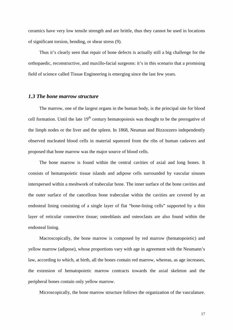
Figure 2. Bone Marrow Structure B= bone; Art= central artery; CV= central vein; S=
marrow sinus; C= hematopoietic cord; E= erithropoiesis; G= granulopoiesis; M=
megakariocytes (reproduced from (10)).
Within the hematopoietic cords, it is possible to recognize two dinstinct cellular
compartments, which have different ontological origins and functions: the stromal
compartment and the hematopoietic compartment. Maturation of the different blood lineages
takes place in distinct compartments: 1) erythropoiesis takes place in the erythroblastic
islands located around a central macrophage, which surrounds the maturing hematopoieitc
elements with thin cytoplasmatic projections, for at least two thirds of their surface; 2)
megakariopoiesis takes place under the sinus endothelium, where small cytoplasmatic
processes anchor the megakariocyte to the sinusoidal wall; 3) granulopoiesis, instead, takes
place in foci always associated with a reticular cell.

20
The stromal compartment, instead, forms the complex three-dimensional structure of
the hematopoietic cords. Two cell types, the macrophages and the reticular cells, play an
important role for the stroma structure.
Macrophages are located in proximity of the sinuses and in the center of the
erythroblastic islands. They are also responsible for the generation of the osteoclastic
compartment.
The reticular cells can be visualized by silver staining of the reticular fibers to which
they are associated in the extra-cellular matrix. A subpopulation of reticular cells, defined as
adventitial reticular cells, is located close to the sinuses, forming an adventitial layer on the
wall of the vessel, similar to pericytes. These cells send thin cytoplasmatic processes from in
the hematopoietic cords, where they enter in contact with processes of other reticular cells,
forming in this way, a three-dimensional scaffold for the hematopoietic compartment. The
non-adventitial reticular cells are often located in the center of the granulopoiesis islands,
where they also have a regulatory function.
The bone-lining cells are a population of flat cells that covers the bone endosteal
surface. Reticular cells, pre-osteoblasts, osteoblasts and osteoclast can be found in the same
location. These cells also include the mesenchymal progenitors cells or mesenchymal stem
cells (MSC), whose anatomical location remains still controversial. All these cell types,
except those of the osteoclastic lineage, share a common cytochemical characteristic that sets
them apart from all other bone marrow cells: the expression of alkaline phosphatase (ALP),
which is considered a marker for the osteoblastic lineage. At a morphological level, some
electron microscopy studies revealed that all the cells that make up the stromal structure, are
functionally connected through gap-junctions (11) (Figure 3).
21
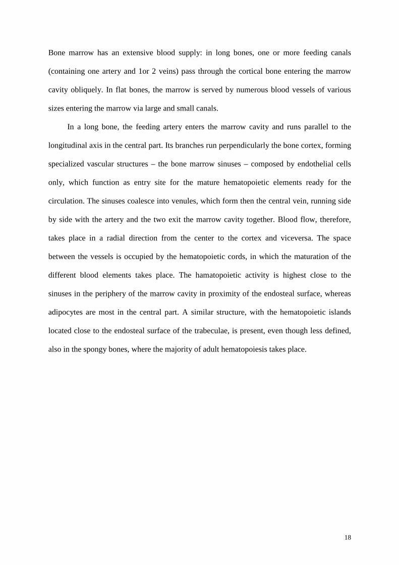
Figure 3. Schematic representation of the connections between the different cell types of the
stromal structure in the bone marrow. The arrows indicate the gap-junctions (Reproduced
from (11)).
A gap-junction connects anatomically and functionally two cells, allowing the direct
exchange of molecules up to 2 KDa of weight through the cytoplasm. Calcium ions can pass
through, but also growth factors and cytokines involved in bone remodeling and in the
regulation of hematopoiesis. Therefore, the relationship between reticular cells, stromal
progenitors and osteoblasts comes from anatomical evidence, from the common expression of
the alkaline phosphatase, from the absence of endothelial or macrophage markers and from
the common synthesis of collagen type I and II.
22
The stroma, with its complex structure and the different cell elements, therefore
represents the support for the hematopoietic compartment. Among the cells that compose the
stromal tissue, the mesenchymal progenitors or mesenchymal stem cells (MSC) received, in
the past years, plenty of attention from the scientific community for their ability to
differentiate into the different mesenchymal lineages.
In the next chapter of this thesis (Chapter 2), the basic aspects of MSC biology will be
discussed, focusing on the issues that still remain controversial and on MSC potential
application for clinical purposes.
In Chapter 3, instead, we will described an important aspect of the bone marrow
function, illustrating the data currently available about the specialized microenvironments,
called stem cell niches, responsible for the regulation of the stem cell function.
23
References
1. Wolff, J. 1870. Über die innere Architektur der Knochen und ihre Bedeutung für die Fragen vom Knochenwachstum. Virchows Arch Path Anat Physiol 50:389-450.
2. Owen, M. 1978. Histogenesis of bone cells. Calcif Tissue Res 25:205-207.
3. Ross, F.P. 2003. Cytokine regulation of osteoclast formation and function. J Musculoskelet Neuronal Interact 3:282-286; discussion 292-284.
4. Rosen, V., and Thies, R.S. 1992. The BMP proteins in bone formation and repair. Trends Genet 8:97-102.
5. Einhorn, T.A. 1995. Enhancement of fracture-healing. J Bone Joint Surg Am 77:940-956.
6. Togawa, D., Bauer, T.W., Lieberman, I.H., and Sakai, H. 2004. Lumbar intervertebral body fusion cages: histological evaluation of clinically failed cages retrieved from humans. J Bone Joint Surg Am 86-A:70-79.
7. Shors, E.C. 1999. Coralline bone graft substitutes. Orthop Clin North Am 30:599-613.
8. Damien, C.J., and Parsons, J.R. 1991. Bone graft and bone graft substitutes: a review of current technology and applications. J Appl Biomater 2:187-208.
9. Yaszemski, M.J., Oldham, J.B., Lu, I., and Currier, B.L. 1994. Bone Engineering. Toronto: Em Squared. 541 pp.
10. Tavassoli, M., and Yoffey, J.M. 1983. Bone Marrow: Strcture and Function. New York: Alan Liss.
11. Yamazaki, K., and Eyden, B.P. 1995. A study of intercellular relationships between trabecular bone and marrow stromal cells in the murine femoral metaphysis. Anat Embryol (Berl) 192:9-20.

24

25
Chapter 2:
Bone Marrow Mesenchymal Stem Cells

26
2.1 History
The bone marrow is a highly cellularized and richly vascularized tissue contained in the
cavity of long bones and in the intra-trabecular spaces of spongius bones. Being the major
site of adult hematopoiesis, the main function of bone marrow is to provide a specialized
environment to protect hematopoietic stem cells, assuring their maintenance and, therefore, a
continuous production of all types of mature blood cells.
Besides containing hematopoietic precursors, the bone marrow contains different non-
hematopoietic cells including reticular, fat and endothelial cells, fibroblasts, osteoblasts and
mesenchymal progenitors. All together, these cells constitute the bone marrow stroma, which
support hematopoiesis, providing structural and humoral signals which regulate the stem cell
function. Among the fully differentiated cells, a key component of the bone marrow stroma is
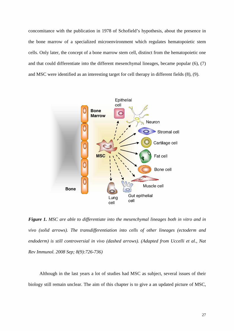
represented by the mesenchymal progenitor cells, which are able to differentiate into the
mesenchymal lineages both in vitro and after in vivo transfer ((1), Figure 1). The first
evidence that it was feasible to ectopically generate bone and bone marrow, after bone
marrow transplantation, dates back to the 19th century (2). In 1968, the work of Tavassoli and
Crosby clearly established the capacity of bone marrow to form bone ectopically, even
though the cells holding the osteogenic potential were not identified. Only the studies of
Friedenstein (3) demonstrated that the ectopic bone formation was due to the presence in the
bone marrow of a specific rare cell population. These cells were characterized by a
fibroblastic morphology, suggesting a stromal-compartment origin, and by their capacity to
rapidly adhere to plastic. Furthermore, when plated at low density, they were able to form
discrete colonies originated by single cells, the CFU-f, the colony-forming unit fibroblastic
(3). Friedenstein and Owen named them osteogenic stem cells (4) or bone marrow stromal
stem cells (5) and in 1991 Caplan defined them as mesenchymal stem cells (MSC). In the
beginning, these discoveries about MSC aroused the interest of the hematologists, also in
27
concomitance with the publication in 1978 of Schofield’s hypothesis, about the presence in
the bone marrow of a specialized microenvironment which regulates hematopoietic stem
cells. Only later, the concept of a bone marrow stem cell, distinct from the hematopoietic one
and that could differentiate into the different mesenchymal lineages, became popular (6), (7)
and MSC were identified as an interesting target for cell therapy in different fields (8), (9).
Figure 1. MSC are able to differentiate into the mesenchymal lineages both in vitro and in
vivo (solid arrows). The transdifferentiation into cells of other lineages (ectoderm and
endoderm) is still controversial in vivo (dashed arrows). (Adapted from Uccelli et al., Nat
Rev Immunol. 2008 Sep; 8(9):726-736)
Although in the last years a lot of studies had MSC as subject, several issues of their
biology still remain unclear. The aim of this chapter is to give a an updated picture of MSC,
28
describing their main characteristics, but also underlining the still controversial aspects of
their biology and illustrating their potential clinical applications.
2.2 In vitro proliferation and differentiation potential
The main attractiveness of MSC is based on the ease with which they can be isolated
from bone marrow and on their ability to proliferate readily in vitro. However, since their
identification, MSC have been classified as a heterogeneous population, in terms of
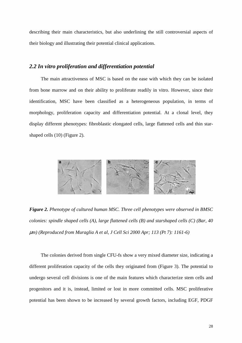
morphology, proliferation capacity and differentiation potential. At a clonal level, they
display different phenotypes: fibroblastic elongated cells, large flattened cells and thin star-
shaped cells (10) (Figure 2).
Figure 2. Phenotype of cultured human MSC. Three cell phenotypes were observed in BMSC
colonies: spindle shaped cells (A), large flattened cells (B) and starshaped cells (C) (Bar, 40
µm) (Reproduced from Muraglia A et al, J Cell Sci 2000 Apr; 113 (Pt 7): 1161-6)
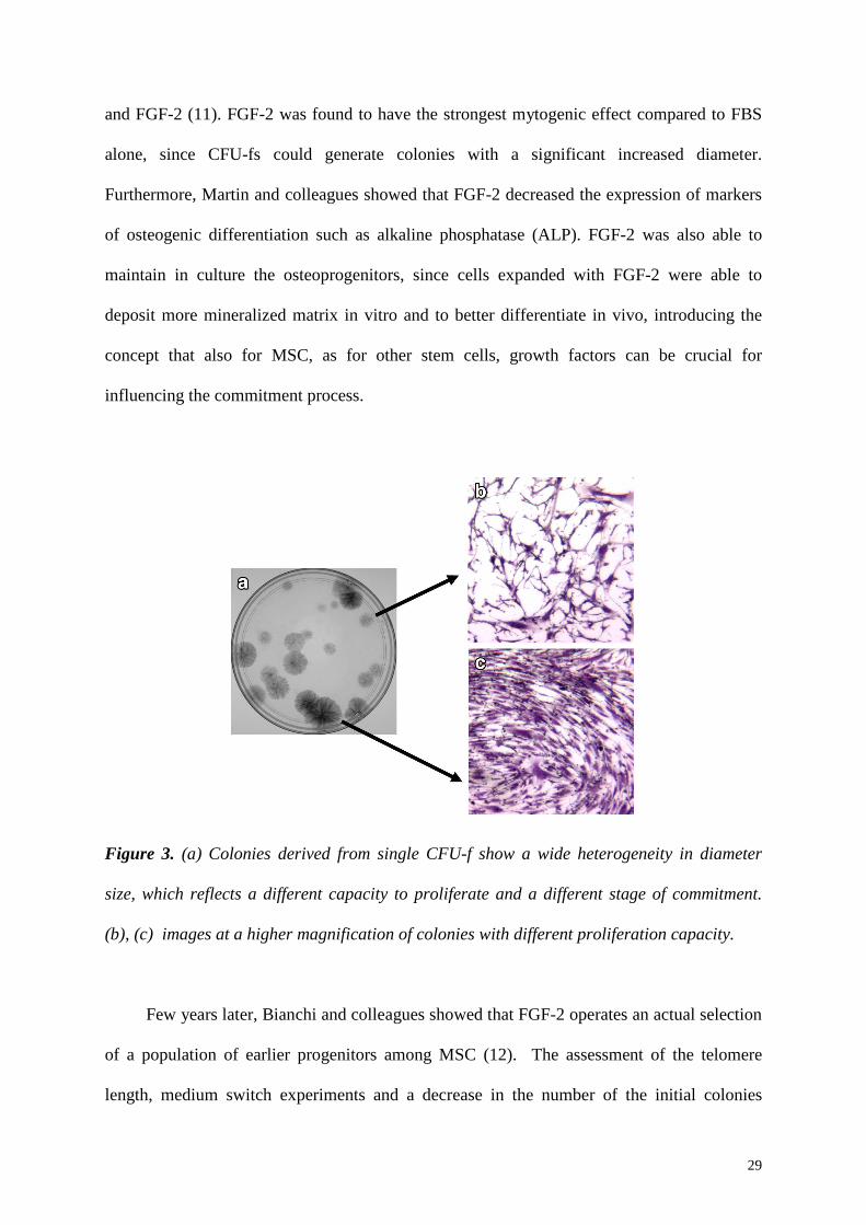
The colonies derived from single CFU-fs show a very mixed diameter size, indicating a
different proliferation capacity of the cells they originated from (Figure 3). The potential to
undergo several cell divisions is one of the main features which characterize stem cells and
progenitors and it is, instead, limited or lost in more committed cells. MSC proliferative
potential has been shown to be increased by several growth factors, including EGF, PDGF
29
and FGF-2 (11). FGF-2 was found to have the strongest mytogenic effect compared to FBS
alone, since CFU-fs could generate colonies with a significant increased diameter.
Furthermore, Martin and colleagues showed that FGF-2 decreased the expression of markers
of osteogenic differentiation such as alkaline phosphatase (ALP). FGF-2 was also able to
maintain in culture the osteoprogenitors, since cells expanded with FGF-2 were able to
deposit more mineralized matrix in vitro and to better differentiate in vivo, introducing the
concept that also for MSC, as for other stem cells, growth factors can be crucial for
influencing the commitment process.
Figure 3. (a) Colonies derived from single CFU-f show a wide heterogeneity in diameter
size, which reflects a different capacity to proliferate and a different stage of commitment.
(b), (c) images at a higher magnification of colonies with different proliferation capacity.
Few years later, Bianchi and colleagues showed that FGF-2 operates an actual selection
of a population of earlier progenitors among MSC (12). The assessment of the telomere
length, medium switch experiments and a decrease in the number of the initial colonies

30
indicated that FGF-2 could select for the survival of cells with an increased proliferation and
differentiation potential. In fact, when FGF-2 was added to the culture, MSC life span could
increase up to 70 doublings, compared with about 50 without it, and the chondrogenic
differentiation potential could be retained up to 50 doublings. These data confirmed how
FGF-2 could be a relevant component of MSC ex-vivo culture to exploit their full potential
for clinical applications.
However, FGF-2 wasn’t able to maintain MSC proliferative activity in vitro, since the
telomere length was decreasing with the expansion (12). Furthermore, even when cultured in
presence of FGF-2, MSC lost the ability to generate colonies and colonies with a decreased
diameter were formed at increasing population doublings, suggesting a commitment of the
population. Other data supported these findings, confirming that in vitro expansion could
cause a gradual loss of MSC early progenitor characteristics (13). MSC dramatically slowed
their proliferation rate already by the second passage and this was also associated to a change
in the morphology, from the spindle shaped to the flattened one. A parallel loss in the multi-
lineage in vitro differentiation potential was also observed with the increasing proliferation,
both for the bulk population and for clones derived from single CFU-f. Moreover, when
implanted in vivo after the first passage, cultured MSC displayed a poor osteogenic potential
compared to fresh bone marrow, indicating that already the primary expansion could affect
MSC commitment and differentiation.
Another aspect, that rise from these findings and that should be taken in consideration,
is the heterogeneity of the starting population. Regarding the differentiation potential, in fact,
not every CFU-f is multipotent when transplanted in vivo (14), (15). As mentioned before,
FGF-2 is capable of selecting a population of earlier progenitors among the whole pool of
initial CFU-f, indicating the presence of different MSC subpopulations characterized by a
mixed stage of commitment. Since studies on the bulk population could not bring much
31
information about this aspect, an extensive study on clones was conducted in the past (10).
The authors showed that a significant percentage (17%) of the analyzed clones were able to
differentiate in vitro into the three common mesenchymal lineages, ostegenic, chondrogenic
and adipogenic. Some clones were bipotent and a small percentage could only differentiate
into the osteogenic lineage. Interestingly, adding FGF-2 to the culture, the percentage of
tripotent clones increased from 17 to 34%, confirming a selection for earlier progenitors.
Furthermore, with increasing expansion, clones displayed a loss of the multi-lineage
differentiation potential, which was not randomly determined. In fact, the adipogenic
potential was the first to be lost; the osteogenic and the chondrogenic potentials, instead,
diverged only very late with the loss of the chondrogenic ability and the preservation of the
osteogenic lineage pathway, suggesting the presence of a hierarchy in the MSC
differentiation pathway. These results described a new aspect of MSC biology and
contributed to support the relevance of FGF-2 for MSC culture, underlining how growth-
factors could affect in vitro expansion of early progenitors.
In addition to this, other factors seem to be important in regulating MSC fate decision.
Engler and colleagues showed that MSC are sensitive to the matrix elasticity and that the
stiffness of the substrate they grow on can influence their commitment towards a specific
lineage (16). These data, therefore, suggest that several elements can influence MSC
behavior. Further studies are necessary to clarify how the different regulations are linked and
combined to determine MSC fate in vivo. Moreover, the factors involved in the regulation of
the hierarchical relationship between different classes of MSC and of the maintenance of
their multipotent capacity still need to be investigated.
Ex-vivo expansion is of crucial importance for a potential application of MSC for
therapeutic purposes. However, the application of these cells has to deal with the problem of
a heterogeneous starting population and with the difficulty of obtaining a number of cells
32
which is clinically relevant and, at the same time, preserving MSC early progenitor features.
Ex-vivo MSC expansion should, therefore, aim to optimize culture procedures and conditions
for selecting and maintaining early progenitor populations.
2.3 Assessment of self-renewal
The main features which characterize a stem cell are the self-renewal, defined as the
capability to generate at least one daughter cell which retain the stem cell fate and the
differentiation potential, which is the ability to give rise to a defined set of mature
differentiated progeny. When MSC were characterized by Friedenstein for the first time, they
were classified as multipotent, for their ability to differentiate into the different mesenchymal
lineages, and, therefore, thought to hold one of the property found in stem cells. Even though
the term mesenchymal stem cells is broadly used, the assessment of the other indispensable
requirement to be defined as stem, the self-renewal, still remain controversial. In several
studies the self-renewal is associated to the growth in culture or to the retention of
multipotency after in vitro expansion. However, none of these studies proved the self-renewal
at a single cell level and, moreover, it was not assessed as the reconstitution of both a
differentiated and a stem cell compartment in vivo. The only stem cell for which the self-
renewal has been soundly proven is the hematopoietic stem cell, founded on the capability to
serially regenerate the whole pool of mature hematopoietic cells in lethally irradiated
recipients (17), (18). For several years there was a lack of experimental evidences on MSC
self-renewal and studies on the bulk population actually demonstrated a loss of proliferation
and differentiation potential with increasing ex-vivo expansion (13), (10), suggesting a
progressive commitment without self-renewal.
It has been recently shown that a subpopulation of human bone marrow stromal cells
are able to differentiate into osteoblasts and hematopoiesis supporting stroma and to
33
maintain, at the same time, the characteristics of clonogenic mesenchymal progenitors (19).
A fundamental characteristic of this population was the expression of the melanoma-
associated adhesion protein, MCAM/CD146, which was homogeneously present when only
freshly isolated bone marrow nucleated cells were grown in clonogenic conditions. In the
bone marrow, CD146+ cells correspond to the adventitial reticular population which is
located in proximity of the sinusoids and which extends processes to contact the
hematopoietic cells. When transplanted subcutaneously in nude mice, expanded CD146+
MSC could form bone and the presence of bone marrow indicated that they could also bring
in vivo the hematopoietic microenvironment. Furthermore, cells were harvested from the
heterotopic ossicles after 8 weeks and expanded; a significant number of clonogenic CD146+
cells could be obtained, indicating that this population, when transplanted in vivo, could
function as self-renewing mesenchymal progenitors. The experiments were performed both
pulling together different clones and implanting the progeny of a single CFU-f,
demonstrating, therefore, that the results were also valid at a single CFU-f level.
Interestingly, when FGF-2 was added to the culture, the expression of CD146 decreased and
no bone marrow formation and no human CD146+ adventitial cells were found in vivo. Only
the osteogenic potential was maintained, suggesting that FGF-2 selects a different population
of progenitors with a distinct function.
These observations indicated for the first time a possible the role of specific
mesenchymal progenitor population in the bone marrow niche and supported the evidence
that the self-renewal is a property also shared by bone marrow MSC. However this is just the
tip of the iceberg of a completely new aspect of MSC biology. Other questions, in fact, needs
to be answered, such as, for example, which is the frequency of these cells in vivo and
whether the same property belongs to MSC derived from other tissues.
34
2.4 Phenotypic characterization
A highly controversial aspect of MSC characterization regards their phenotypic
properties. The rarity of this population in the bone marrow represents a challenging barrier
for the identification of specific antibodies for their isolation and enrichment. Furthermore,
most of the markers identified up to now are not specific for MSC since they are also
expressed by cultures of fibroblastic cells from various tissues and they are widely modulated
during in vitro culture. Little is still known about MSC characteristics in vivo and about a
possible differential marker expression among the different classes of MSC. The majority of
the information collected so far is based on the analysis of expanded MSC. This contributed
to generate confusion about their phenotype and the wrong assumption that any marker
expressed on cultured-expanded MSC was also likely to be present in vivo.
Both non-expanded and expanded MSC have been shown to be negative for any
hematopoietic or vascular endothelial marker, such as CD45, CD14 and CD34. One of the
first antibodies that was identified, instead, to enrich for CFU-f in fresh human bone marrow
is STRO-1 (20). STRO-1 is not expressed on hematopoietic cells and its selection results in a
10- to 20-fold enrichment of CFU-f, compared to unseparated bone marrow. Analysis of the
expression of a wide range of cell surface molecules on CFU-f demonstrated that other
markers, including CD105, CD49a, CD73 and CD90 (21), (22), (23) could be used for CFU-f
enrichment. These markers are also expressed on expanded MSC and, to date, they are
commonly used to characterize culture-derived MSC.
Recently, Sacchetti and colleagues showed that MCAM/CD146, which marks in human
bone marrow the adventitial reticular cells, can be used to isolate a population of in vivo self-
renewing osteogenic progenitors (19). After in vivo transfer, they could form bone and they
could also be localized in the same adventitial position. This indicated the importance of
identifying, in addition to markers of un-expanded and cultured MSC, markers for in situ
35
localization, to find in situ counterparts of CFU-f and to follow the fate of the implanted cells
in vivo, especially when aiming to find evidences for self-renewal.
2.5 Clinical applications
After the discovery of their biological property of differentiating into the common
mesenchymal lineages, bone marrow MSC were thought of being responsible for the normal
turnover and maintenance of adult mesenchymal tissues (24). This led to the identification of
MSC as an attractive cell source for therapeutic applications in different fields of regenerative
medicine.
The most obvious application was to apply MSC for the regeneration of mesenchymal
tissues, such as bone and cartilage. In fact, first among the others, tissue engineers tried to
exploit MSC properties for the repair of bone defects and for the treatment of various bone
disorders. Up to date, autologous bone graft still represents the first choice for site-specific
bone defect repair, although it is associated with several complications such as, donor site
morbidity, infection or loss of graft function (25). The combination of a biomaterial and ex-
vivo expanded MSC is thought to represent a valid alternative to functionally replace host
bone tissue. In the past years, novel approaches have been developed to improve the
performance of the tissue engineered constructs: biomimetic material properties, including
surface roughness and porosity, have been investigated to enhance MSC adherence,
proliferation and differentiation (26), (27), (28). The presence of MSC promoted bone
formation in vivo and MSC were able to accelerate bone repair in femoral and cranial
critical-size defects and spinal fusion both in large and small animal models (29), (30), (31).
Based on these preclinical studies, clinical trials for bone repair have been conducted (32),
(33), (34) with promising outcomes. As mentioned before, expanded MSC have been also
used as a potential treatment for cartilaginous injuries in humans, showing a contribution also
36
in cartilage repair (35), (36). This indicated that bone marrow MSC represent a valid cell
source for clinical tissue engineering applications, even though further investigation of their
biology and optimization of culture conditions are still needed to fully exploit their potential.
Another clinical application that appeared very promising was the use of MSC as an
innovative treatment for genetic diseases. The first application was in patients with
osteogenesis imperfecta (OI), which defines a heterogeneous group of genetic disorders,
characterized by bone fragility and skeletal deformities as osteoporosis. Clinical trials were
started infusing ex-vivo expanded MSC in children affected by OI (9),(37),(38). The results
indicated that MSC were able to engraft into skeletal sites and cause the enhancement of
patient growth. However, the authors also showed that the total bone mineral content did not
significantly increase and they suggested that prolonged in vitro expansion, affecting MSC
osteogenic capacity, could compromise the outcome of the study.
This observation, together with the data about the loss of differentiation potential
described in the previous section, underlines that a limited in vitro expansion is of
fundamental importance for a clinical application in regenerative medicine fields. The low
frequency of MSC in the bone marrow and the rapid loss of their progenitor-like
characteristics during exapnsion, therefore, represents one of the main limitations for the
establishment of MSC-based therapies. Strategies, as FGF-2 supplementation (12) and
growth on extracellular matrices (39), (40), should be considered in order to retain MSC
progenitor features during ex-vivo expansion. In addition, different culture strategies,
alternative to the standard 2D-expansion on plastic, have been explored in the past, in order
to recreate a more physiological environment to preserve the MSC characteristics. A very
promising approach is represented by bioreactors, which showed to promote cell viability,
shear-stress stimulation and a better maintenance of MSC differentiation potential during
expansion (41), (42), (43), (44). The results of these studies indicated the relevance of
37
bioreactors as an important tool for MSC ex-vivo expansion in order to optimize culture
conditions and, therefore, to enhance MSC in vivo performance. Furthermore, in Chapter 4 of
this thesis we will describe the use of a bioreactor system to reconstruct in vitro the bone
marrow stem cell niche, underlining the relevance of such a system, not only for MSC in
vitro expansion, but also for the development of a physiological bone marrow stroma.
Although the pioneer clinical studies on MSC have focused on their ability to repair
damaged tissues, new findings about their biology, such as their immunomodulatory
properties and their possible role in the bone marrow niche, open interesting perspectives.
Several studies showed the ability of MSC to modulate immune responses both in vitro and
in vivo, by nonspecifically targeting cells of the immune system. This characteristic seems to
be due to the ability to block immunocompetent cells through the inhibition of cell division,
preventing their activation and maintaining them in a quiescent state (45). The clinical
efficacy of MSC would therefore depend on their ability to modify the environment of
injured tissues, releasing anti-inflammatory molecules and trophic factors, which promote
tissue regeneration. So, the therapeutic potential of MSC could be traced to their
physiological activity. The current data about MSC anti-inflammatory and
immunosuppressive features support their potential application for immune-mediated
diseases, offering alternative therapeutic strategies. The effect of infused MSC has been
successfully shown in acute graft-versus-host diseases (46), and, recently, it is being tested
for the treatment of Crohn’s disease, investigating MSC contribution to the regeneration of
gastro-intestinal epithelial cells (47).
On the other hand, the recent findings about the involvement of MSC in the bone
marrow stem cell niche opened new lines of research. This could lead to the identification of
new molecular targets for the treatment of diseases in which stem cell function is
disregulated, either through degeneration, such as aplastic anemias, or excessive expansion
38
and lack of differentiation, such as hematological neoplasias. A deeper knowledge in the
niche biology is therefore needed in order to identify all the regulatory players and the
mechanisms by which MSC might maintain and regulate hematopoietic stem cells.
In the next chapter, the main characteristics of the bone marrow stem cell niche will be
described, focusing on the niche function and on the data currently available about the
regulatory factors which contribute to hematopoietic stem cells maintenance.
39
References
1. Friedenstein, A.J., Chailakhyan, R.K., Latsinik, N.V., Panasyuk, A.F., and Keiliss-Borok, I.V. 1974. Stromal cells responsible for transferring the microenvironment of the hemopoietic tissues. Cloning in vitro and retransplantation in vivo. Transplantation 17:331-340.
2. Goujon, E.J. 1869. J Anat Physiol 11:399.
3. Friedenstein, A.Y., and Lalykina, K.S. 1970. Lymphoid cell populations are competent systems for induced osteogenesis. Calcif Tissue Res:Suppl:105-106.
4. Friedenstein, A.J., Chailakhyan, R.K., and Gerasimov, U.V. 1987. Bone marrow osteogenic stem cells: in vitro cultivation and transplantation in diffusion chambers. Cell Tissue Kinet 20:263-272.
5. Owen, M., and Friedenstein, A.J. 1988. Stromal stem cells: marrow-derived osteogenic precursors. Ciba Found Symp 136:42-60.
6. Caplan, A.I. 1991. Mesenchymal stem cells. J Orthop Res 9:641-650.
7. Pittenger, M.F., Mackay, A.M., Beck, S.C., Jaiswal, R.K., Douglas, R., Mosca, J.D., Moorman, M.A., Simonetti, D.W., Craig, S., and Marshak, D.R. 1999. Multilineage potential of adult human mesenchymal stem cells. Science 284:143-147.
8. Koc, O.N., Gerson, S.L., Phillips, G.L., Cooper, B.W., Kutteh, L., Van Zant, G., Reece, D.E., Fox, R.M., Schupp, J.E., Tainer, N., et al. 1998. Autologous CD34+ cell transplantation for patients with advanced lymphoma: effects of overnight storage on peripheral blood progenitor cell enrichment and engraftment. Bone Marrow Transplant 21:337-343.
9. Horwitz, E.M., Prockop, D.J., Fitzpatrick, L.A., Koo, W.W., Gordon, P.L., Neel, M., Sussman, M., Orchard, P., Marx, J.C., Pyeritz, R.E., et al. 1999. Transplantability and therapeutic effects of bone marrow-derived mesenchymal cells in children with osteogenesis imperfecta. Nat Med 5:309-313.
10. Muraglia, A., Cancedda, R., and Quarto, R. 2000. Clonal mesenchymal progenitors from human bone marrow differentiate in vitro according to a hierarchical model. J Cell Sci 113 (Pt 7):1161-1166.
11. Martin, I., Muraglia, A., Campanile, G., Cancedda, R., and Quarto, R. 1997. Fibroblast growth factor-2 supports ex vivo expansion and maintenance of osteogenic precursors from human bone marrow. Endocrinology 138:4456-4462.
12. Bianchi, G., Banfi, A., Mastrogiacomo, M., Notaro, R., Luzzatto, L., Cancedda, R., and Quarto, R. 2003. Ex vivo enrichment of mesenchymal cell progenitors by fibroblast growth factor 2. Exp Cell Res 287:98-105.
13. Banfi, A., Muraglia, A., Dozin, B., Mastrogiacomo, M., Cancedda, R., and Quarto, R. 2000. Proliferation kinetics and differentiation potential of ex vivo expanded human bone marrow stromal cells: Implications for their use in cell therapy. Exp Hematol 28:707-715.
14. Bianco, P., and Robey, P.G. 2001. Stem cells in tissue engineering. Nature 414:118-121.
40
15. Gronthos, S., Zannettino, A.C., Hay, S.J., Shi, S., Graves, S.E., Kortesidis, A., and Simmons, P.J. 2003. Molecular and cellular characterisation of highly purified stromal stem cells derived from human bone marrow. J Cell Sci 116:1827-1835.
16. Engler, A.J., Sen, S., Sweeney, H.L., and Discher, D.E. 2006. Matrix elasticity directs stem cell lineage specification. Cell 126:677-689.
17. Osawa, M., Hanada, K., Hamada, H., and Nakauchi, H. 1996. Long-term lymphohematopoietic reconstitution by a single CD34-low/negative hematopoietic stem cell. Science 273:242-245.
18. Weissman, I.L. 2000. Stem cells: units of development, units of regeneration, and units in evolution. Cell 100:157-168.
19. Sacchetti, B., Funari, A., Michienzi, S., Di Cesare, S., Piersanti, S., Saggio, I., Tagliafico, E., Ferrari, S., Robey, P.G., Riminucci, M., et al. 2007. Self-renewing osteoprogenitors in bone marrow sinusoids can organize a hematopoietic microenvironment. Cell 131:324-336.
20. Simmons, P.J., and Torok-Storb, B. 1991. Identification of stromal cell precursors in human bone marrow by a novel monoclonal antibody, STRO-1. Blood 78:55-62.
21. Majumdar, M.K., Banks, V., Peluso, D.P., and Morris, E.A. 2000. Isolation, characterization, and chondrogenic potential of human bone marrow-derived multipotential stromal cells. J Cell Physiol 185:98-106.
22. Barry, F.P., Boynton, R.E., Haynesworth, S., Murphy, J.M., and Zaia, J. 1999. The monoclonal antibody SH-2, raised against human mesenchymal stem cells, recognizes an epitope on endoglin (CD105). Biochem Biophys Res Commun 265:134-139.
23. Barry, F., Boynton, R., Murphy, M., Haynesworth, S., and Zaia, J. 2001. The SH-3 and SH-4 antibodies recognize distinct epitopes on CD73 from human mesenchymal stem cells. Biochem Biophys Res Commun 289:519-524.
24. Caplan, A.I. 2005. Review: mesenchymal stem cells: cell-based reconstructive therapy in orthopedics. Tissue Eng 11:1198-1211.
25. Hollinger, J., and Wong, M.E. 1996. The integrated processes of hard tissue regeneration with special emphasis on fracture healing. Oral Surg Oral Med Oral Pathol Oral Radiol Endod 82:594-606.
26. Wilson, C.E., de Bruijn, J.D., van Blitterswijk, C.A., Verbout, A.J., and Dhert, W.J. 2004. Design and fabrication of standardized hydroxyapatite scaffolds with a defined macro-architecture by rapid prototyping for bone-tissue-engineering research. J Biomed Mater Res A 68:123-132.
27. Mante, M., Daniels, B., Golden, E., Diefenderfer, D., Reilly, G., and Leboy, P.S. 2003. Attachment of human marrow stromal cells to titanium surfaces. J Oral Implantol 29:66-72.
28. Behravesh, E., and Mikos, A.G. 2003. Three-dimensional culture of differentiating marrow stromal osteoblasts in biomimetic poly(propylene fumarate-co-ethylene glycol)-based macroporous hydrogels. J Biomed Mater Res A 66:698-706.
29. Petite, H., Viateau, V., Bensaid, W., Meunier, A., de Pollak, C., Bourguignon, M., Oudina, K., Sedel, L., and Guillemin, G. 2000. Tissue-engineered bone regeneration. Nat Biotechnol 18:959-963.
41
30. Kon, E., Muraglia, A., Corsi, A., Bianco, P., Marcacci, M., Martin, I., Boyde, A., Ruspantini, I., Chistolini, P., Rocca, M., et al. 2000. Autologous bone marrow stromal cells loaded onto porous hydroxyapatite ceramic accelerate bone repair in critical-size defects of sheep long bones. J Biomed Mater Res 49:328-337.
31. Shang, Q., Wang, Z., Liu, W., Shi, Y., Cui, L., and Cao, Y. 2001. Tissue-engineered bone repair of sheep cranial defects with autologous bone marrow stromal cells. J Craniofac Surg 12:586-593; discussion 594-585.
32. Faundez, A.A., Taylor, S., and Kaelin, A.J. 2006. Instrumented fusion of thoracolumbar fracture with type I mineralized collagen matrix combined with autogenous bone marrow as a bone graft substitute: a four-case report. Eur Spine J 15 Suppl 5:630-635.
33. Gimbel, J.A., Van Kleunen, J.P., Lake, S.P., Williams, G.R., and Soslowsky, L.J. 2007. The role of repair tension on tendon to bone healing in an animal model of chronic rotator cuff tears. J Biomech 40:561-568.
34. Velardi, F., Amante, P.R., Caniglia, M., De Rossi, G., Gaglini, P., Isacchi, G., Palma, P., Procaccini, E., and Zinno, F. 2006. Osteogenesis induced by autologous bone marrow cells transplant in the pediatric skull. Childs Nerv Syst 22:1158-1166.
35. Wakitani, S., Mitsuoka, T., Nakamura, N., Toritsuka, Y., Nakamura, Y., and Horibe, S. 2004. Autologous bone marrow stromal cell transplantation for repair of full-thickness articular cartilage defects in human patellae: two case reports. Cell Transplant 13:595-600.
36. Kuroda, R., Ishida, K., Matsumoto, T., Akisue, T., Fujioka, H., Mizuno, K., Ohgushi, H., Wakitani, S., and Kurosaka, M. 2007. Treatment of a full-thickness articular cartilage defect in the femoral condyle of an athlete with autologous bone-marrow stromal cells. Osteoarthritis Cartilage 15:226-231.
37. Horwitz, E.M. 2001. Marrow mesenchymal cell transplantation for genetic disorders of bone. Cytotherapy 3:399-401.
38. Horwitz, E.M., Hofmann, T.J., Garlits, J.E., Campioni, D., and Dominici, M. 2002. On the development of cell therapy for genetic disorders. Cytotherapy 4:511-512.
39. Mauney, J.R., Kaplan, D.L., and Volloch, V. 2004. Matrix-mediated retention of osteogenic differentiation potential by human adult bone marrow stromal cells during ex vivo expansion. Biomaterials 25:3233-3243.
40. Matsubara, T., Tsutsumi, S., Pan, H., Hiraoka, H., Oda, R., Nishimura, M., Kawaguchi, H., Nakamura, K., and Kato, Y. 2004. A new technique to expand human mesenchymal stem cells using basement membrane extracellular matrix. Biochem Biophys Res Commun 313:503-508.
41. Qiu, Q., Ducheyne, P., Gao, H., and Ayyaswamy, P. 1998. Formation and differentiation of three-dimensional rat marrow stromal cell culture on microcarriers in a rotating-wall vessel. Tissue Eng 4:19-34.
42. Sikavitsas, V.I., Bancroft, G.N., Holtorf, H.L., Jansen, J.A., and Mikos, A.G. 2003. Mineralized matrix deposition by marrow stromal osteoblasts in 3D perfusion culture increases with increasing fluid shear forces. Proc Natl Acad Sci U S A 100:14683-14688.
42
43. Bancroft, G.N., Sikavitsas, V.I., and Mikos, A.G. 2003. Design of a flow perfusion bioreactor system for bone tissue-engineering applications. Tissue Eng 9:549-554.
44. Braccini, A., Wendt, D., Jaquiery, C., Jakob, M., Heberer, M., Kenins, L., Wodnar-Filipowicz, A., Quarto, R., and Martin, I. 2005. Three-dimensional perfusion culture of human bone marrow cells and generation of osteoinductive grafts. Stem Cells 23:1066-1072.
45. Uccelli, A., Moretta, L., and Pistoia, V. 2008. Mesenchymal stem cells in health and disease. Nat Rev Immunol 8:726-736.
46. Le Blanc, K., Rasmusson, I., Sundberg, B., Gotherstrom, C., Hassan, M., Uzunel, M., and Ringden, O. 2004. Treatment of severe acute graft-versus-host disease with third party haploidentical mesenchymal stem cells. Lancet 363:1439-1441.
47. Okamoto, R., Yajima, T., Yamazaki, M., Kanai, T., Mukai, M., Okamoto, S., Ikeda, Y., Hibi, T., Inazawa, J., and Watanabe, M. 2002. Damaged epithelia regenerated by bone marrow-derived cells in the human gastrointestinal tract. Nat Med 8:1011-1017.

43
Chapter 3:
The Bone Marrow Stem Cell Niche

44
3.1 The hematopoietic stem cell
Stem cells are self-renewing, multipotent progenitors that are responsible for the
growth, maintenance and repair of several tissues. They are present in different compartments
of the body, including the skin, the intestinal epithelium and the hematopoietic system. The
best characterized stem cell is the hematopoietic stem cell (HSC). In the past few years a lot
of progress has been done to advance the knowledge about HSC and, recently, the
development of new tools for labeling HSC in situ also allowed the characterization of the
microenvironment which regulates HSC, the bone marrow stem cell niche.
The most common assay to assess the stemness in HSC is the ability to reconstitute all
the blood-cell lineages in lethally irradiated recipients. Furthermore, the fact that this property
can be maintained upon serial transplantation in mice shows the capability of these cells to
undergo self-renewal, i.e. to give rise to a differentiated progeny and preserving, at the same
time, the stem cell properties. The optimization of these assays also helped to identify cell
surface molecules for the characterization, and consequently for the isolation, of HSC.
Murine HSC do not express any of the markers present on fully differentiated hematopoietic
cells, but they express high levels of stem-cell antigen 1 (SCA 1) and c-KIT and, according to
this, they are named ad LKS cells. Moreover, long-term repopulating HSC have been shown
to be negative for CD34 and positive for CD150 (1), (2). However, the studies in which the
presence of these markers was correlated to the repopulating function showed that only 1 in
100 LKS is able to save from irradiation (3). Recently, some studies have started to define
HSC gene-profiling, moving the first steps towards the clarification of the mechanisms which
regulate HSC function (4), (5).
The regulation of self- renewal and differentiation represents probably the most
fascinating aspect of HSC biology. Self-renewing divisions in vivo contribute to maintain the
size of the stem cell pool. This can be achieved preserving the balance between symmetrical

45
and asymmetrical divisions, by which a single stem cell gives rise to two different daughter
cells, one maintaining the stem cell features and the other becoming fully differentiated. Two
possible different mechanisms have been described to achieve the asymmetry: the divisional
asymmetry, which occurs before the cell division, and the environmental asymmetry, when it
is determined after the cell division (Figure 1).
In the divisional asymmetry (Figure 1a), only one of the daughter cells receives, during
mitosis, the determinants for initiating the commitment process. This mechanism has not
been shown in any vertebrate stem-cell type in vivo, but several in vitro studies have shown
that HSC may undergo asymmetrical divisions (6), (7). Further investigation is, therefore,
needed to confirm whether this reflects the in vivo conditions.
The environmental asymmetry (Figure 1b), instead, is determined through extrinsic
signals provided by the surrounding microenvironment. After a symmetrical division, one
daughter cell will remain in contact with the niche, preserving the stem cell fate; the other
one, instead, will be exposed to an external environment and will receive signals starting
differentiation (8), (9). Even though there is a lack of experimental evidences that this
mechanism can occur in vertebrates in vivo, recent in vitro studies indicated that some
molecular pathways for divisional asymmetry are conserved between invertebrates and
vertebrates. Therefore, this raises the possibility that HSC can undergo both divisional and
environmental asymmetry in vivo in order to drive daughter cells to different fates.
The environmental asymmetry introduces the importance of a specialized
microenvironment, the niche, in providing signals to promote self-renewal and avoid
differentiation.
46
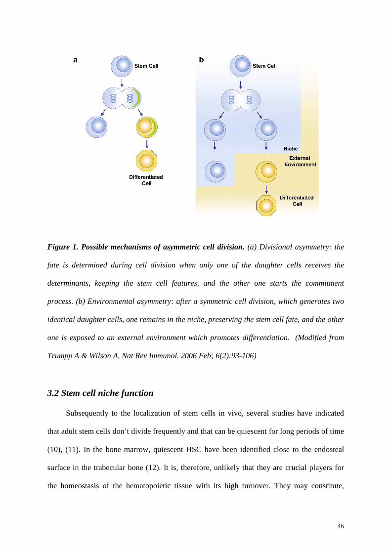
Figure 1. Possible mechanisms of asymmetric cell division. (a) Divisional asymmetry: the
fate is determined during cell division when only one of the daughter cells receives the
determinants, keeping the stem cell features, and the other one starts the commitment
process. (b) Environmental asymmetry: after a symmetric cell division, which generates two
identical daughter cells, one remains in the niche, preserving the stem cell fate, and the other
one is exposed to an external environment which promotes differentiation. (Modified from
Trumpp A & Wilson A, Nat Rev Immunol. 2006 Feb; 6(2):93-106)
3.2 Stem cell niche function
Subsequently to the localization of stem cells in vivo, several studies have indicated
that adult stem cells don’t divide frequently and that can be quiescent for long periods of time
(10), (11). In the bone marrow, quiescent HSC have been identified close to the endosteal
surface in the trabecular bone (12). It is, therefore, unlikely that they are crucial players for
the homeostasis of the hematopoietic tissue with its high turnover. They may constitute,
47
instead, a reserve pool which is stored in the “quiescent niche” and which can be mobilized in
case of tissue injury (Figure 2a). It has been shown, in fact, that, when the hematopoietic
system is damaged, HSC are mobilized from the bone marow, enter the circulation and start
to divide in order to restore hematopoieis. They can then home back to the bone marrow
niches and be quiescent again (13), (14).
However, quiescence is not a characteristic of all stem cells. Embryonic stem cells have
to undergo several divisions but they preserve the stem cell fate and fetal-liver HSC can
reconstitute hematopoieisis even being highly proliferative (15), (16). This could be
associated to the difference between “fetal” and “adult” stem cells. However, in tissues with
high regeneration rate, such as the hematopoietic system, stem cells have to divide also
during homeostasis, to produce progenitors that can, then, generate the differentiated progeny
during regular turnover. This indicates that another type of niche, the self-renewing niche
(Figure 2b), might exist and its function would be to guarantee that one of the daughter cells
maintain the stem cell features and the other one initiates the differentiation process. In this
case the niche structure would be more complex, but a self-renewing niche would be
fundamental to maintain standard tissue homeostasis. A possible structure of this type of
niche would require that quiescent stem cells are anchored in a protected part of the niche,
while self-renewing stem cells would be located in proximity of the external environment
from which they can receive signals that could stimulate cell division and/or differentiation.
Up to date, whether a single niche can provide all the stem-cell-niche-functions or
multiple niches are required in vivo remains still unknown. However, the two possible model
proposed above are both compatible with the normal homeostatic conditions; further
investigation conducted in vivo will clarify which of the two reflects the bone marrow
condition.
48
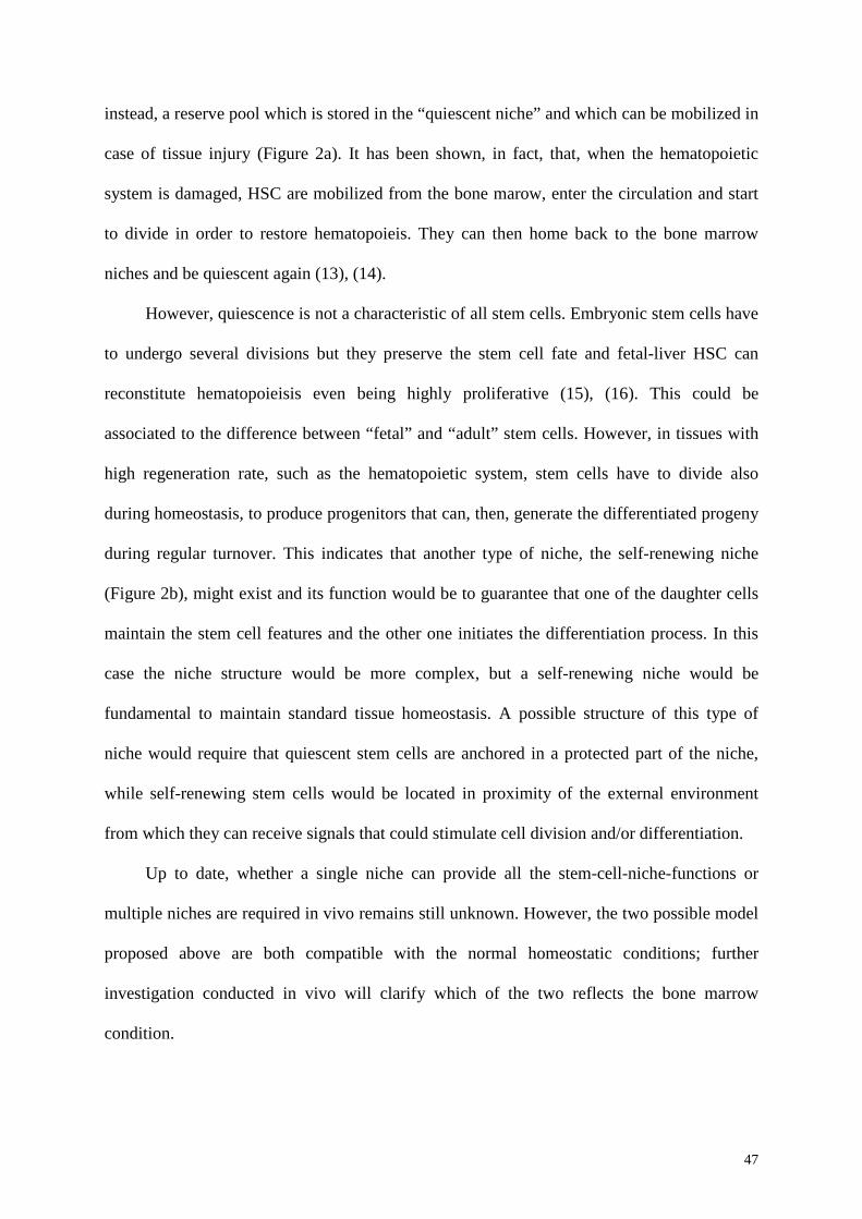
Figure 2. Different types of niche. (a) Quiescent storage niche: it contains resting stem cells
and it produces signals that repress differentiation and/or cell division. Stem cells can be
mobilized from this site in case of need and they might return for storage and self-renewal.
(b) Self-renewing niche: quiescent stem cells would be located in the centre of the niche,
while self-renewing stem cells would be present at the interface between the niche and the
external environment. At this interface, stem cells might be exposed to proliferative signals,
undergoing divisional or environmental asymmetry. This would allow both self-renewal,
since stem cells will remain attached to the niche, and initiation of differentiation, leading to
different stem cell fates (A and B). (Modified from Trumpp A & Wilson A, Nat Rev Immunol.
2006 Feb; 6(2): 93-106)
49
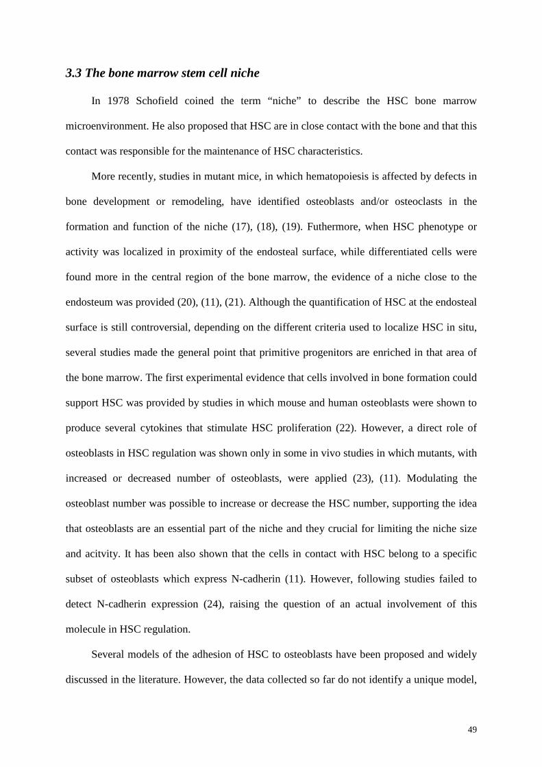
3.3 The bone marrow stem cell niche
In 1978 Schofield coined the term “niche” to describe the HSC bone marrow
microenvironment. He also proposed that HSC are in close contact with the bone and that this
contact was responsible for the maintenance of HSC characteristics.
More recently, studies in mutant mice, in which hematopoiesis is affected by defects in
bone development or remodeling, have identified osteoblasts and/or osteoclasts in the
formation and function of the niche (17), (18), (19). Futhermore, when HSC phenotype or
activity was localized in proximity of the endosteal surface, while differentiated cells were
found more in the central region of the bone marrow, the evidence of a niche close to the
endosteum was provided (20), (11), (21). Although the quantification of HSC at the endosteal
surface is still controversial, depending on the different criteria used to localize HSC in situ,
several studies made the general point that primitive progenitors are enriched in that area of
the bone marrow. The first experimental evidence that cells involved in bone formation could
support HSC was provided by studies in which mouse and human osteoblasts were shown to
produce several cytokines that stimulate HSC proliferation (22). However, a direct role of
osteoblasts in HSC regulation was shown only in some in vivo studies in which mutants, with
increased or decreased number of osteoblasts, were applied (23), (11). Modulating the
osteoblast number was possible to increase or decrease the HSC number, supporting the idea
that osteoblasts are an essential part of the niche and they crucial for limiting the niche size
and acitvity. It has been also shown that the cells in contact with HSC belong to a specific
subset of osteoblasts which express N-cadherin (11). However, following studies failed to
detect N-cadherin expression (24), raising the question of an actual involvement of this
molecule in HSC regulation.
Several models of the adhesion of HSC to osteoblasts have been proposed and widely
discussed in the literature. However, the data collected so far do not identify a unique model,
50
remaining compatible with different ways by which endosteal cells can support HSC
maintenance (Figure 3). Furthermore, other cell types present in the bone marrow can secrete
molecules, such as angiopoietin, thrombopoietin and CXCL12, questioning whether they also
contribute to niche formation, activity or architecture.
Figure 3. Possible mechanisms by which niche specialized cells can regulate HSC
function. (a) A possible model would imply a direct contact between HSC and niche
specialized cells. (b) Another possibility is that cells at the endosteum secrete regulatory
factors which may regulate HSC. According to this model HSC can be also located in
proximity to the endosteal surface, excluding a direct contact with the niche regulatory cells.
(c) A third possibility is that endosteal cells promote HSC maintenance by modulating the
function of intermediate cells, which are fundamental in the formation of other niche types
away form the endosteum (as the vascular niche).
51
The localization of a significant amount of CD150+ HSC in proximity of the sinusoidal
walls in the bone marrow raised the hypothesis that another specialized microenvironment
could affect HSC regulation (20). It has been shown that there is a close developmental
relationship between the hematopoietic and the endothelial lineages (25), (26) and HSC seem
to originate from a perivascular progenitor during embryonic development (27), (28).
Moreover, the presence of HSC in the yolk sac, aorta-gonad-mesonephros region and
vitelline arteries suggests they reside and undergo hematopoiesis in association with blood
vessels. The endothelial cells harvested from these regions have been shown to support the
expansion of adult HSC LKS cells in vitro (29), (30). In contrast, endothelial cells isolated
from adult organs don’t display a HSC supportive activity in vitro (31). These observations
indicate that the endothelial cells of bone marrow sinusoids hold distinct functional
characteristics from endothelial cells present in other tissues; in fact, they have been shown to
produce factors important for mobilization, homing and engrafment for HSC, such as
CXCL12, VCAM1 and E-selectin (32), (33). Some evidences about the existence of a
vascular niche were already present, since in case of injury HSC can detach from the
endosteum and migrate to the vascular region of the bone marrow from where they restore
hematopoiesis (34), (35). Moreover, although only some more recent findings confirmed the
presence of CD150+ HSC close to the sinusoids, the possibility that a vascular niche could
exist during homeostasis can easily find an explanation. In fact, label-retaining HSC have
been localized in the endosteal niche, showing that the most dormant HSC reside there and
that this type of niche serve as a quiescent niche, or a self-renewing niche containing both
quiescent and self-renewing HSC. The proliferating HSC, instead, are close to the vasculature
(20) and therefore it is possible that the vascular niche contains the self-renewing HSC rather
than dormant HSC. Furthermore, this position would allow HSC to constantly sense the
52
concentration of blood factors that reflects the status of the hematopoietic system and to be
rapidly activated in case of need; more HSC can be then mobilized from the endosteal niche
if necessary. However, it has been shown that when osteoblasts are deleted hematopoiesis
occurs extramedullary (36), indicating that the vascular niche is not sufficient to support
long-term hematopoiesis and it might represent a secondary niche in the bone marrow.
Although the role of all the niche cells has not been clarified and the mechanisms by
which they regulate HSC fate need to be further investigated, strong experimental evidences
support the hypothesis that the endosteal and the vascular niche act together to maintain
hematopoietic homeostasis or restore it after damage.
3.4 Molecular crosstalk in the endosteal niche
Some recent studies have identified few molecules which are expressed both by
osteoblasts and HSC and which are thought to be important for mediating the signals
exchanged in the niche (22). However, some other studies have focused on the identification
of factors directly in situ showing their involvement in the modulation of HSC function.
Notch signaling has been shown to be involved in stem cell fate decision in several
tissues. The data about its expression in the bone marrow (37) led to the hypothesis that it
could also had a role in HSC regulation. Moreover, a confirmation came from some in vitro
studies in which the authors showed the importance of Notch signaling for HSC maintenance
(38). In addition, in mutants with an increased number of osteoblasts, Notch ligand Jagged-1
is upregulated, suggesting that Notch signaling could be responsible for the consequent
augmentation in HSC number (23). In contrast, some other studies showed that a deletion of
both Notch-1 and Jagged-1 from bone marrow does not have any effect on hematopoiesis in
vivo (39). This suggests that Notch is not a crucial requirement of the niche, but it is maybe
53
important only in some circumstances, and that the regulation of HSC maintenance involves
the complex combination of different factors.
Components also involved in HSC regulation are some proteins contained in the
extracellular matrix. It has been shown that osteoblasts can modulate the number of HSC
secreting osteopontin (OPN), an acidic glycoprotein of the bone matrix (40). In mice in which
a deletion of OPN was induced, the number of HSC significantly increased and the same
increased was observed transplanting wild type HSC, indicating that OPN production by the
osteoblasts has a negative regulatory effect on HSC (41). Some in vitro experiments showed
that OPN induces apoptosis in HSC and that in the niche plays a role in maintaining HSC
quiescence (41), (42).
Stem cell factor (SCF) binds and activate c-KIT which is highly expressed by label
retaining HSC. It has been previously shown that mutations which affect the membrane
bound SCF have an influence on migration and differentiation of primordial germ cells,
neural-crest derived melanoblasts and hematopoietic cells (43). Furthermore, osteoblasts in
the bone marrow produce a significant amount of membrane-bound SCF, which has the
capacity to enhance the adhesion of HSC to stromal cells (44), suggesting that osteoblasts can
influence the adhesive characteristics of the endosteal niche by modulating the functional
state of specific integrins. In vivo, the hampered production of SCF causes a reduced HSC
activity (17), (45), indicating that SCF is an essential component of the endosteal niche.
Moreover, SCF is also important for osteoblasts activity in vivo (46). It would be therefore
interesting to investigate whether it mainly explicates its effect directly, acting on HSC, or
indirectly, affecting niche specialized osteoblasts.
Other factors have been described to affect HSC regulation. TIE2, which is expressed
by HSC, is activated by angiopoietin-1 (Ang1) and the activation of this pathway contributes
to maintain HSC quiescence both in vitro and in vivo (47), (12). MYC is expressed, instead,
54
during initiation of HSC differentiation (48) and MYC overexpressing HSC are lost because
of differentiation mainly for their inability to be retained in the niche. Although it has been
shown that WNT signaling pathway is involved in HSC self-renewing divisions in culture, its
role remains still controversial and it has to be clarified whether it is fundamental for HSC
function.
These observations together contribute to describe the endosteal niche as a complex
interactive structure that combines positive and negative signals for the regulation of stem
cell activities. Open question still remain and significant advance in the knowledge is still
needed to better understand the mechanisms of these microenvironments, in order to
modulate endogenous stem cell function.
3.5 Perspectives
In the past few years, the theory proposed in the 1970s about the presence in the bone
marrow of a specific stem-cell microenvironment, has received the right attention. A great
amount of data, trying to identify interactions both at the molecular and cellular levels, has
been collected. Nevertheless, we are still far to completely elucidate all the mechanisms
present in the niche and, moreover, as the characterization of the niche will proceed, it is
likely that new paradigms will be uncover. Further investigation is still needed to clarify
whether different niche types exist in the bone marrow, how many HSC are contained in each
niche and the precise role of every niche during the normal turnover or in case of injury. In
fact, it will be interesting to distinguish between mechanisms that maintain HSC under
homeostasis or that are activated under stress conditions, since some experimental evidences
suggest that some factors, such as Notch and WNT for example, are crucial only in some
circumstances. In addition, even though osteoblasts have been identified as key players for
the niche function, very little is known about their characteristics, especially about their

55
differentiation stage. It will be important to establish if the niche-specialized osteoblasts
correspond to the osteoblasts which then differentiate in ostocytes or whether they undergo a
different differentiation pathway. Recent studies have revealed that other cell types like
endothelial cell, osteoclasts and mesenchymal progenitors are important in the establishment
of the niche function, opening new areas of research. This contributes to underline that the
eventual reconstruction in vitro of active niches will probably require more than a feeder
layer and a mix of different cytokines. In fact, it will need the reconstitution of a three-
dimensional structure, which can include the different cellular players and the deposition of a
physiological extracellular matrix. The combination of cell-and-molecular biology and tissue
engineering can lead to a significant advance in the field, which will facilitate the
development of new stem-cell-based therapies.
In this regard, in Chapter 6 will illustrate the potential of a bioreactor system as a tool
to reconstruct in vitro the interactions present in the bone marrow niche. We will focus on the
plasticity of the system, underlining the advantages, such as the three-dimensional
environment, and the modulating variables, like the scaffold composition and the possibility
of multiple cell types co-culture. We will propose an innovative application of a perfusion
bioreactor-based system, not only for HSC expansion, as bioreactors were meant so far, but
also as a valid system to reproduce the bone marrow physiological microenvironment. The
final aim will be to use such a device to recreate a stable stem-cell-niche unit, in which the
physiological interactions are respected, promoting HSC maintenance and the formation of a
functional supportive tissue composed by stromal cells, which preserve their progenitor-like
features. This will, therefore, have important implications not only for stem cell biology but
also for the tissue-engineering field, representing a physiological environment for MSC and
moving the first steps to overcome limitations linked to standard MSC culture.
56
Furthermore, beyond clarifying mechanisms of HSC biology that have been already
identified in two-dimensional culture conditions, this three-dimensional system can be
applied to investigate basic aspects of MSC biology, like the regulation of the hierarchical
relationship between different classes of MSC and of the maintenance of their pluripotent
capacity, which remain still widely unknown. Although a three-dimensional setup would
have constituted a more physiological environment, we started answering these questions
using the standard two-dimensional system and tested the hypothesis that, as HSC in the bone
marrow, also MSC require their own niche to preserve their progenitor-like features. In the
following chapter (Chapter 4), we will describe the establishment of a MSC niche system in
vitro by the maintenance of a self-renewing, highly potent, non-adherent mesenchymal
progenitor population.
57
References
1. Yang, L., Bryder, D., Adolfsson, J., Nygren, J., Mansson, R., Sigvardsson, M., and Jacobsen, S.E. 2005. Identification of Lin(-)Sca1(+)kit(+)CD34(+)Flt3- short-term hematopoietic stem cells capable of rapidly reconstituting and rescuing myeloablated transplant recipients. Blood 105:2717-2723.
2. Adolfsson, J., Mansson, R., Buza-Vidas, N., Hultquist, A., Liuba, K., Jensen, C.T., Bryder, D., Yang, L., Borge, O.J., Thoren, L.A., et al. 2005. Identification of Flt3+ lympho-myeloid stem cells lacking erythro-megakaryocytic potential a revised road map for adult blood lineage commitment. Cell 121:295-306.
3. Jordan, C.T., and Lemischka, I.R. 1990. Clonal and systemic analysis of long-term hematopoiesis in the mouse. Genes Dev 4:220-232.
4. Ivanova, N.B., Dimos, J.T., Schaniel, C., Hackney, J.A., Moore, K.A., and Lemischka, I.R. 2002. A stem cell molecular signature. Science 298:601-604.
5. Akashi, K., He, X., Chen, J., Iwasaki, H., Niu, C., Steenhard, B., Zhang, J., Haug, J., and Li, L. 2003. Transcriptional accessibility for genes of multiple tissues and hematopoietic lineages is hierarchically controlled during early hematopoiesis. Blood 101:383-389.
6. Takano, H., Ema, H., Sudo, K., and Nakauchi, H. 2004. Asymmetric division and lineage commitment at the level of hematopoietic stem cells: inference from differentiation in daughter cell and granddaughter cell pairs. J Exp Med 199:295-302.
7. Ho, A.D. 2005. Kinetics and symmetry of divisions of hematopoietic stem cells. Exp Hematol 33:1-8.
8. Ohlstein, B., Kai, T., Decotto, E., and Spradling, A. 2004. The stem cell niche: theme and variations. Curr Opin Cell Biol 16:693-699.
9. Spradling, A., Drummond-Barbosa, D., and Kai, T. 2001. Stem cells find their niche. Nature 414:98-104.
10. Tumbar, T., Guasch, G., Greco, V., Blanpain, C., Lowry, W.E., Rendl, M., and Fuchs, E. 2004. Defining the epithelial stem cell niche in skin. Science 303:359-363.
11. Zhang, J., Niu, C., Ye, L., Huang, H., He, X., Tong, W.G., Ross, J., Haug, J., Johnson, T., Feng, J.Q., et al. 2003. Identification of the haematopoietic stem cell niche and control of the niche size. Nature 425:836-841.
12. Arai, F., Hirao, A., Ohmura, M., Sato, H., Matsuoka, S., Takubo, K., Ito, K., Koh, G.Y., and Suda, T. 2004. Tie2/angiopoietin-1 signaling regulates hematopoietic stem cell quiescence in the bone marrow niche. Cell 118:149-161.
13. Randall, T.D., and Weissman, I.L. 1997. Phenotypic and functional changes induced at the clonal level in hematopoietic stem cells after 5-fluorouracil treatment. Blood 89:3596-3606.
14. Venezia, T.A., Merchant, A.A., Ramos, C.A., Whitehouse, N.L., Young, A.S., Shaw, C.A., and Goodell, M.A. 2004. Molecular signatures of proliferation and quiescence in hematopoietic stem cells. PLoS Biol 2:e301.

58
15. Uchida, N., He, D., Friera, A.M., Reitsma, M., Sasaki, D., Chen, B., and Tsukamoto, A. 1997. The unexpected G0/G1 cell cycle status of mobilized hematopoietic stem cells from peripheral blood. Blood 89:465-472.
16. Chambers, I., and Smith, A. 2004. Self-renewal of teratocarcinoma and embryonic stem cells. Oncogene 23:7150-7160.
17. Barker, J.E. 1994. Sl/Sld hematopoietic progenitors are deficient in situ. Exp Hematol 22:174-177.
18. Deguchi, K., Yagi, H., Inada, M., Yoshizaki, K., Kishimoto, T., and Komori, T. 1999. Excessive extramedullary hematopoiesis in Cbfa1-deficient mice with a congenital lack of bone marrow. Biochem Biophys Res Commun 255:352-359.
19. Visnjic, D., Kalajzic, I., Gronowicz, G., Aguila, H.L., Clark, S.H., Lichtler, A.C., and Rowe, D.W. 2001. Conditional ablation of the osteoblast lineage in Col2.3deltatk transgenic mice. J Bone Miner Res 16:2222-2231.
20. Kiel, M.J., Yilmaz, O.H., Iwashita, T., Terhorst, C., and Morrison, S.J. 2005. SLAM family receptors distinguish hematopoietic stem and progenitor cells and reveal endothelial niches for stem cells. Cell 121:1109-1121.
21. Nilsson, S.K., Johnston, H.M., and Coverdale, J.A. 2001. Spatial localization of transplanted hemopoietic stem cells: inferences for the localization of stem cell niches. Blood 97:2293-2299.
22. Taichman, R.S. 2005. Blood and bone: two tissues whose fates are intertwined to create the hematopoietic stem-cell niche. Blood 105:2631-2639.
23. Calvi, L.M., Adams, G.B., Weibrecht, K.W., Weber, J.M., Olson, D.P., Knight, M.C., Martin, R.P., Schipani, E., Divieti, P., Bringhurst, F.R., et al. 2003. Osteoblastic cells regulate the haematopoietic stem cell niche. Nature 425:841-846.
24. Kiel, M.J., Radice, G.L., and Morrison, S.J. 2007. Lack of evidence that hematopoietic stem cells depend on N-cadherin-mediated adhesion to osteoblasts for their maintenance. Cell Stem Cell 1:204-217.
25. Huber, T.L., Kouskoff, V., Fehling, H.J., Palis, J., and Keller, G. 2004. Haemangioblast commitment is initiated in the primitive streak of the mouse embryo. Nature 432:625-630.
26. Kennedy, M., Firpo, M., Choi, K., Wall, C., Robertson, S., Kabrun, N., and Keller, G. 1997. A common precursor for primitive erythropoiesis and definitive haematopoiesis. Nature 386:488-493.
27. Mikkola, H.K., and Orkin, S.H. 2006. The journey of developing hematopoietic stem cells. Development 133:3733-3744.
28. Sanchez, M.J., Holmes, A., Miles, C., and Dzierzak, E. 1996. Characterization of the first definitive hematopoietic stem cells in the AGM and liver of the mouse embryo. Immunity 5:513-525.
29. Li, W., Johnson, S.A., Shelley, W.C., Ferkowicz, M., Morrison, P., Li, Y., and Yoder, M.C. 2003. Primary endothelial cells isolated from the yolk sac and para-aortic splanchnopleura support the expansion of adult marrow stem cells in vitro. Blood 102:4345-4353.
30. Ohneda, O., Fennie, C., Zheng, Z., Donahue, C., La, H., Villacorta, R., Cairns, B., and Lasky, L.A. 1998. Hematopoietic stem cell maintenance and differentiation are
59
supported by embryonic aorta-gonad-mesonephros region-derived endothelium. Blood 92:908-919.
31. Li, W., Johnson, S.A., Shelley, W.C., and Yoder, M.C. 2004. Hematopoietic stem cell repopulating ability can be maintained in vitro by some primary endothelial cells. Exp Hematol 32:1226-1237.
32. Avecilla, S.T., Hattori, K., Heissig, B., Tejada, R., Liao, F., Shido, K., Jin, D.K., Dias, S., Zhang, F., Hartman, T.E., et al. 2004. Chemokine-mediated interaction of hematopoietic progenitors with the bone marrow vascular niche is required for thrombopoiesis. Nat Med 10:64-71.
33. Rafii, S., Mohle, R., Shapiro, F., Frey, B.M., and Moore, M.A. 1997. Regulation of hematopoiesis by microvascular endothelium. Leuk Lymphoma 27:375-386.
34. Kopp, H.G., Avecilla, S.T., Hooper, A.T., and Rafii, S. 2005. The bone marrow vascular niche: home of HSC differentiation and mobilization. Physiology (Bethesda) 20:349-356.
35. Heissig, B., Hattori, K., Dias, S., Friedrich, M., Ferris, B., Hackett, N.R., Crystal, R.G., Besmer, P., Lyden, D., Moore, M.A., et al. 2002. Recruitment of stem and progenitor cells from the bone marrow niche requires MMP-9 mediated release of kit-ligand. Cell 109:625-637.
36. Visnjic, D., Kalajzic, Z., Rowe, D.W., Katavic, V., Lorenzo, J., and Aguila, H.L. 2004. Hematopoiesis is severely altered in mice with an induced osteoblast deficiency. Blood 103:3258-3264.
37. Radtke, F., Wilson, A., Mancini, S.J., and MacDonald, H.R. 2004. Notch regulation of lymphocyte development and function. Nat Immunol 5:247-253.
38. Duncan, A.W., Rattis, F.M., DiMascio, L.N., Congdon, K.L., Pazianos, G., Zhao, C., Yoon, K., Cook, J.M., Willert, K., Gaiano, N., et al. 2005. Integration of Notch and Wnt signaling in hematopoietic stem cell maintenance. Nat Immunol 6:314-322.
39. Mancini, S.J., Mantei, N., Dumortier, A., Suter, U., MacDonald, H.R., and Radtke, F. 2005. Jagged1-dependent Notch signaling is dispensable for hematopoietic stem cell self-renewal and differentiation. Blood 105:2340-2342.
40. Denhardt, D.T., and Guo, X. 1993. Osteopontin: a protein with diverse functions. Faseb J 7:1475-1482.
41. Stier, S., Ko, Y., Forkert, R., Lutz, C., Neuhaus, T., Grunewald, E., Cheng, T., Dombkowski, D., Calvi, L.M., Rittling, S.R., et al. 2005. Osteopontin is a hematopoietic stem cell niche component that negatively regulates stem cell pool size. J Exp Med 201:1781-1791.
42. Nilsson, S.K., Haylock, D.N., Johnston, H.M., Occhiodoro, T., Brown, T.J., and Simmons, P.J. 2003. Hyaluronan is synthesized by primitive hemopoietic cells, participates in their lodgment at the endosteum following transplantation, and is involved in the regulation of their proliferation and differentiation in vitro. Blood 101:856-862.
43. Lyman, S.D., and Jacobsen, S.E. 1998. c-kit ligand and Flt3 ligand: stem/progenitor cell factors with overlapping yet distinct activities. Blood 91:1101-1134.
44. Kinashi, T., and Springer, T.A. 1994. Adhesion molecules in hematopoietic cells. Blood Cells 20:25-44.
60
45. Barker, J.E. 1997. Early transplantation to a normal microenvironment prevents the development of Steel hematopoietic stem cell defects. Exp Hematol 25:542-547.
46. Lotinun, S., Evans, G.L., Turner, R.T., and Oursler, M.J. 2005. Deletion of membrane-bound steel factor results in osteopenia in mice. J Bone Miner Res 20:644-652.
47. Cheng, T., Rodrigues, N., Shen, H., Yang, Y., Dombkowski, D., Sykes, M., and Scadden, D.T. 2000. Hematopoietic stem cell quiescence maintained by p21cip1/waf1. Science 287:1804-1808.
48. Wu, S., Cetinkaya, C., Munoz-Alonso, M.J., von der Lehr, N., Bahram, F., Beuger, V., Eilers, M., Leon, J., and Larsson, L.G. 2003. Myc represses differentiation-induced p21CIP1 expression via Miz-1-dependent interaction with the p21 core promoter. Oncogene 22:351-360.
61
Chapter 4
FGF-2/FGFR2c signaling generates a niche-progenitor system in
vitro by selecting and maintaining a self-renewing, highly potent,
non-adherent mesenchymal progenitor population
62
4.1 Introduction
The bone marrow stromal system is defined as the connective tissue elements providing
structural and functional support for hematopoiesis, as well as containing a population of
multipotent mesenchymal progenitors. Recent studies have revealed that specific
microenvironments, often referred to as “stem cell niches” regulate stem cell function by
providing architectural support, together with humoral and cell-contact dependent signals (1).
It has been also shown that stem cell niches are as diverse as the stem cells they sustain and
that they can be specialized to support different physiological functions. In the endosteal
niche, hematopoietic stem cells (HSC) have been shown to be in contact with specialized
niche osteoblasts (2) and the mutual signal exchange is responsible for the regulation of self-
renewal and inhibition of differentiation (3, 4). Currently it is postulated that HSC
maintenance is achieved by environmental or divisional asymmetry (5), by which one of the
daughter cells can preserve the stem cell fate and the other one initiates the commitment
process. The main function of the niche is, therefore, to guarantee the normal tissue
homeostasis and to provide the environment for a stem cell reservoir that can be activated in
case of stress or injury. Although osteoblasts have been identified as an essential component
of the endosteal niche (2), it has recently been shown that other cell types, like endothelial
cells, contribute to the formation of HSC niches (6, 7). Moreover, a recent study showed that
human CD146+ clonogenic osteoprogenitors, that reside perivascularly in the bone marrow,
are able to regenerate the hematopoietic microenvironment in vivo after transplantation (8),
suggesting a role of mesenchymal progenitors, in addition to differentiated osteoblasts, in the
bone marrow niche. Human mesenchymal progenitors show differences in terms of
proliferation capacity (9) and differentiation potential in vitro at a clonal level (10), indicating
a wide heterogeneity in their stage of commitment. Although the presence of different classes
of mesenchymal progenitors was already described by Friedstein in the 1980s (11), the
63
hierarchical relationship between them and the regulation of the maintenance of their
pluripotent capacity are still largely unknown (10). Even though significant progress has
lately been made to identify HSC niches, it still remains unclear if also mesenchymal cells
require their own niche to maintain their progenitor-like characteristics in vivo.
Recently, Wan and coworkers (12) reported the presence of multipotent mesenchymal
cells in the non-adherent fraction of freshly plated human bone marrow cultures, which gave
rise to adherent colonies if replated on new dishes. Furthermore, it has been shown that a
population of non-adherent mesenchymal progenitors can be cultured in suspension and that
the maintenance of the non-adherent status is associated with the preservation of an
uncommitted phenotype (13).
In analogy to the relationship between the HSC and the stromal niche, we hypothesized
that non-adherent stromal cells constitute a reservoir of earlier progenitors/stem cells for the
mesenchymal compartment and that the adherent fraction provides the niche function for the
maintenance and regulation of these progenitors.
In this chapter, we investigated the self-renewal and differentiation capacity of bone
marrow non-adherent mesenchymal progenitors (NAMP) in vitro in different experimental
settings. We show that the NAMP are a class of multipotent cells, which can generate an
adherent progeny while self-renewing as non-adherent progenitors. NAMP progeny showed
an increased proliferation capacity and greater differentiation potential than that of the
initially adherent CFU-f. The selection, the maintenance and the biological function of
NAMP is exquisitely dependent on FGFR2c signaling and it requires the adherent fraction to
provide signals which prevent adherence and colony formation, while supporting self-
renewal, therefore providing niche function for the NAMP.
64
4.2 Matherials and Methods
Cell culture
Bone marrow aspirates were obtained from a total of 24 healthy donors (27-64 years
old) during routine orthopaedic surgical procedures, in accordance with the local ethical
committee (University Hospital Basel) and after informed consent. Bone marrow
mononuclear cells (BM-MNC) were counted after staining with Crystal Violet 0.01%
(Sigma) in phosphate-buffered saline, pH 7.2 (PBS). To determine colony forming efficiency
(CFE) and for serial replating experiments, cells were plated at clonal density (4.5 x 10³
cells/cm2) and cultured in alpha-MEM (GIBCO) with 10% fetal bovine serum (FBS). When
indicated, medium was further supplemented with FGF-2 (5 ng/ml, R&D System) or PDGF-
BB (10 ng/ml R&D System). After 2 weeks cells were then washed with PBS, fixed with
3.7% formaldehyde in PBS, stained with Crystal Violet (Sigma) for 10 min, washed with tap
water and the colonies were counted. All determinations were performed in triplicate and
CFU-f frequency in the fresh marrow sample was used to calculate the population doublings
of first-confluence cultures.
For progeny expansion, freshly isolated BM-MNC were plated at a density of 1 x 10⁵
cells/cm2 (Flask0, CFU-f progeny cells) and after 3 days the non-adherent fraction was
transferred into a new flask (Flask1, NAMP progeny cells). After 2 weeks of expansion in
presence of FGF-2, the cells were detached with 0.05% trypsin/0.01% EDTA (Gibco) and
counted for the population doubling determination. Aliquots of the pooled cells were used for
flow cytometry analysis, in vitro and in vivo differentiation assays, or replated for growth
curve determination.
In vitro adipogenic differentiation
Adipogenic differentiation was induced in 2D cultures as previously described (14).
65
Briefly, CFU-f and NAMP progeny cells were seeded in 6-well plates at a density of 5 x 10³
cells/cm2 and cultured in alpha-MEM with 10% FBS until they reached confluency. Medium
was then supplemented with 10 µg/ml insulin, 10-5 M dexamethasone, 100 µM indomethacin
and 500 µM 3-isobutyl-1-methyl xanthine (adipogenic induction medium) for 72 h and
subsequently with 10 µg/ml insulin (adipogenic maintenance medium) for 24 h. This 96-
hours cycle was repeated for four times and cells were then cultured for an additional week in
adipogenic maintenance medium.
Readout: Cell layers were harvested for mRNA extraction and PPAR-gamma
expression assessment (see RT-PCR section) or fixed in 60% isopropanol for 2 minutes and
incubated with oil red (Sigma) 0.1% in 60% isopropanol for 20 minutes. To determine the
number of adipocytes, oil-red positive cells were counted in at least 10 different fields.
In vitro chondrogenic differentiation
Chondrogenic differentiation was induced in pellet cultures using a defined serum-free
medium, as previously described (15). Briefly, CFU-f and NAMP progeny cells were
resuspended in alpha-MEM supplemented with ITS+1 (10 µg/ml insulin, 5.5 µg/ml
transferrin, 5 ng/ml selenium, 0.5 mg/ml bovine serum albumin, 4.7 µg/ml linoleic acid
(Sigma, St.Louis, MO), 0.1 mM ascorbic acid 2-phosphate (Sigma, St.Louis, MO), 1.25
mg/ml human serum albumin, 10-7 dexamethasone (Sigma, St.Louis, MO) and 10 ng/ml
TGFβ1 (R&D system, Minneapolis, MN). Aliquots of 5 x 10⁵ cells/0.5 ml were centrifuged
at 1,200 rpm for 5 minutes in 1.5 ml polypropylene conical tubes (Sarstedt, Numbrecht,
Germany) to form spherical pellets. Pelletes were cultured in a humidified incubator at 37 °C
with 5% CO2 for 3 weeks and the medium was changed twice per week.
Readouts: For histological analysis, cell pellets cultured in chondrogenic medium were
fixed in 4% formalin for 24 h at 4 °C, dehydrated in a ethanol series, embedded in paraffin
66
and 7-µm thick-sections were stained with Safranin-O to determine the presence of sulfated
glycosamminoglycans (GAG). For biochemical analysis, Cell pellets cultured in
chondrogenic medium were digested in 1 m of proteinase-K solution (1 mg/ml proteinase-K
in 50 mM Tris, 1 mM EDTA, 1mM iodocetamide and 10 µg/ml pepstatin-A) for 15 h at 56
°C. GAG content was measured spectrophotometrically after reaction with
dymethilmethylene blue (16). Known concentrations of chondroitin sulfate were used as a
standard. The amount of DNA was measured using a CyQuant cell proliferation assay kit
(Molecular Probes, Eugene, OR) and used to normalize the GAG content.
In vitro osteogenic differentiation
Osteogenic differentiation was induced in 2D cultures as previously described (17).
Briefly, CFU-f and NAMP progeny cells were seeded in 6-well plates at a density of 5 x 10³
cells/cm2 in alpha-MEM supplemented with 10% FBS, 10 mM β-glycerophosphate (Sigma),
10 nM dexamethasone (Sigma) and 0.1 mM L-ascorbic acid-2-phosphate (Sigma) and
cultured for 3 weeks, with medium changed twice per week.
Readout: Cells layers cultured in osteogenic medium were were harvested for mRNA
extraction and BSP expression assessment (see RT-PCR section) or washed twice with PBS,
fixed for 10 minutes in 4% formalin and washed twice with water. Fixed cells were then
incubated for 10 minutes with alizarin red 2% in distilled water and extensively washed with
water.
Real-time PCR
Total RNA was extracted from cells using TRIzol (Invitrogen, Carlsbad, CA), treated with
DNAse and retrotranscribed into cDNA, as previously described (18). Real-time PCR was
performed with the ABIPrism 77000 Sequence Detection System (Perkin Elmer/Applied

67
Biosystem, Rotkreuz, Switzerland) and expression levels of the genes of interest bone
sialoprotein (BSP) and PPAR gamma were normalized to the 18S rRNA. Primers and probe
sets and PCR conditions for BSP and 18S were used as previously described (18). PPAR-
gamma primers and probe (Hs00234592_m1) were provided by Applied Biosystem.
Assessment of bone formation in vivo
In vivo ectopic bone formation was assayed as previously described (19). Briefly, on
reaching their first confluence, 1 x 10⁶ of CFU-f and NAMP progeny cells were resuspended
in 30 µl of Fibrinogen (20 mg/ml Baxter, Austria), quickly mixed with 30 µl of Thrombin (6
IU/ml Baxter, Austria) and loaded onto 35 µg of bovine bone-derived granules (Bio-Oss,
Geistlich Switzerland). The constructs were transferred for 15 minutes in a humidified
incubator at 37 °C with 5% CO2 to allow fibrin polimerization and implanted subcutaneously
in CD-1 nu/nu nude mice (Charles River, Germany). After 8 weeks, the constructs were
harvested, fixed overnight in 4% formalin, completely decalcified with Osteodec (Bio-
Optica, Milan, Itlay) for 2 hours at 37 °C, paraffin embedded and 7 µm-thick-sections were
obtained from different levels. Sections were stained with hematoxilin and eosin (H&E) and
observed microscopically to detect the formation of bone tissue for qualitative analysis and
assessed by computerized bone histomorphometry as previously described (20) for bone
tissue quantification. Briefly, fluorescent images of sections at different depth of each
construct were taken and used to measure the area covered with bone tissue by computerized
image analysis (Scion Image, Scion Corp., Frederick, MD).
Flow cytometry analysis
CFU-f and NAMP progeny cells (3-5 x 10⁵ cells) were resuspended into 200 µl of 0.5% BSA
in PBS (FACS Buffer) with fluorochrome-conjugated antibodies against the indicated protein
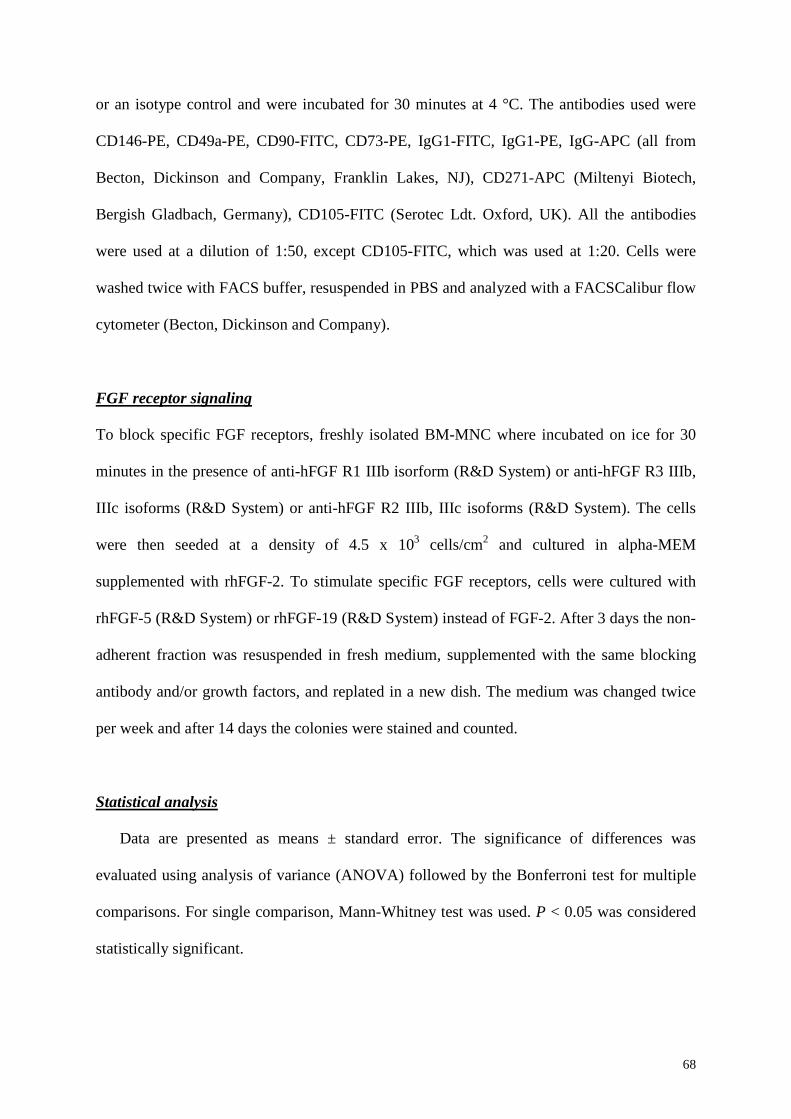
68
or an isotype control and were incubated for 30 minutes at 4 °C. The antibodies used were
CD146-PE, CD49a-PE, CD90-FITC, CD73-PE, IgG1-FITC, IgG1-PE, IgG-APC (all from
Becton, Dickinson and Company, Franklin Lakes, NJ), CD271-APC (Miltenyi Biotech,
Bergish Gladbach, Germany), CD105-FITC (Serotec Ldt. Oxford, UK). All the antibodies
were used at a dilution of 1:50, except CD105-FITC, which was used at 1:20. Cells were
washed twice with FACS buffer, resuspended in PBS and analyzed with a FACSCalibur flow
cytometer (Becton, Dickinson and Company).
FGF receptor signaling
To block specific FGF receptors, freshly isolated BM-MNC where incubated on ice for 30
minutes in the presence of anti-hFGF R1 IIIb isorform (R&D System) or anti-hFGF R3 IIIb,
IIIc isoforms (R&D System) or anti-hFGF R2 IIIb, IIIc isoforms (R&D System). The cells
were then seeded at a density of 4.5 x 10³ cells/cm2 and cultured in alpha-MEM
supplemented with rhFGF-2. To stimulate specific FGF receptors, cells were cultured with
rhFGF-5 (R&D System) or rhFGF-19 (R&D System) instead of FGF-2. After 3 days the non-
adherent fraction was resuspended in fresh medium, supplemented with the same blocking
antibody and/or growth factors, and replated in a new dish. The medium was changed twice
per week and after 14 days the colonies were stained and counted.
Statistical analysis
Data are presented as means ± standard error. The significance of differences was
evaluated using analysis of variance (ANOVA) followed by the Bonferroni test for multiple
comparisons. For single comparison, Mann-Whitney test was used. P < 0.05 was considered
statistically significant.
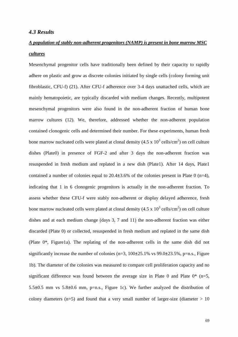
69
4.3 Results A population of stably non-adherent progenitors (NAMP) is present in bone marrow MSC
cultures
Mesenchymal progenitor cells have traditionally been defined by their capacity to rapidly
adhere on plastic and grow as discrete colonies initiated by single cells (colony forming unit
fibroblastic, CFU-f) (21). After CFU-f adherence over 3-4 days unattached cells, which are
mainly hematopoietic, are typically discarded with medium changes. Recently, multipotent
mesenchymal progenitors were also found in the non-adherent fraction of human bone
marrow cultures (12). We, therefore, addressed whether the non-adherent population
contained clonogenic cells and determined their number. For these experiments, human fresh
bone marrow nucleated cells were plated at clonal density (4.5 x 10³ cells/cm2) on cell culture
dishes (Plate0) in presence of FGF-2 and after 3 days the non-adherent fraction was
resuspended in fresh medium and replated in a new dish (Plate1). After 14 days, Plate1
contained a number of colonies equal to 20.4±3.6% of the colonies present in Plate 0 (n=4),
indicating that 1 in 6 clonogenic progenitors is actually in the non-adherent fraction. To
assess whether these CFU-f were stably non-adherent or display delayed adherence, fresh
bone marrow nucleated cells were plated at clonal density (4.5 x 10³ cells/cm2) on cell culture
dishes and at each medium change (days 3, 7 and 11) the non-adherent fraction was either
discarded (Plate 0) or collected, resuspended in fresh medium and replated in the same dish
(Plate 0*, Figure1a). The replating of the non-adherent cells in the same dish did not
significantly increase the number of colonies (n=3, 100±25.1% vs 99.0±23.5%, p=n.s., Figure
1b). The diameter of the colonies was measured to compare cell proliferation capacity and no
significant difference was found between the average size in Plate 0 and Plate 0* (n=5,
5.5±0.5 mm vs 5.8±0.6 mm, p=n.s., Figure 1c). We further analyzed the distribution of
colony diameters (n=5) and found that a very small number of larger-size (diameter > 10
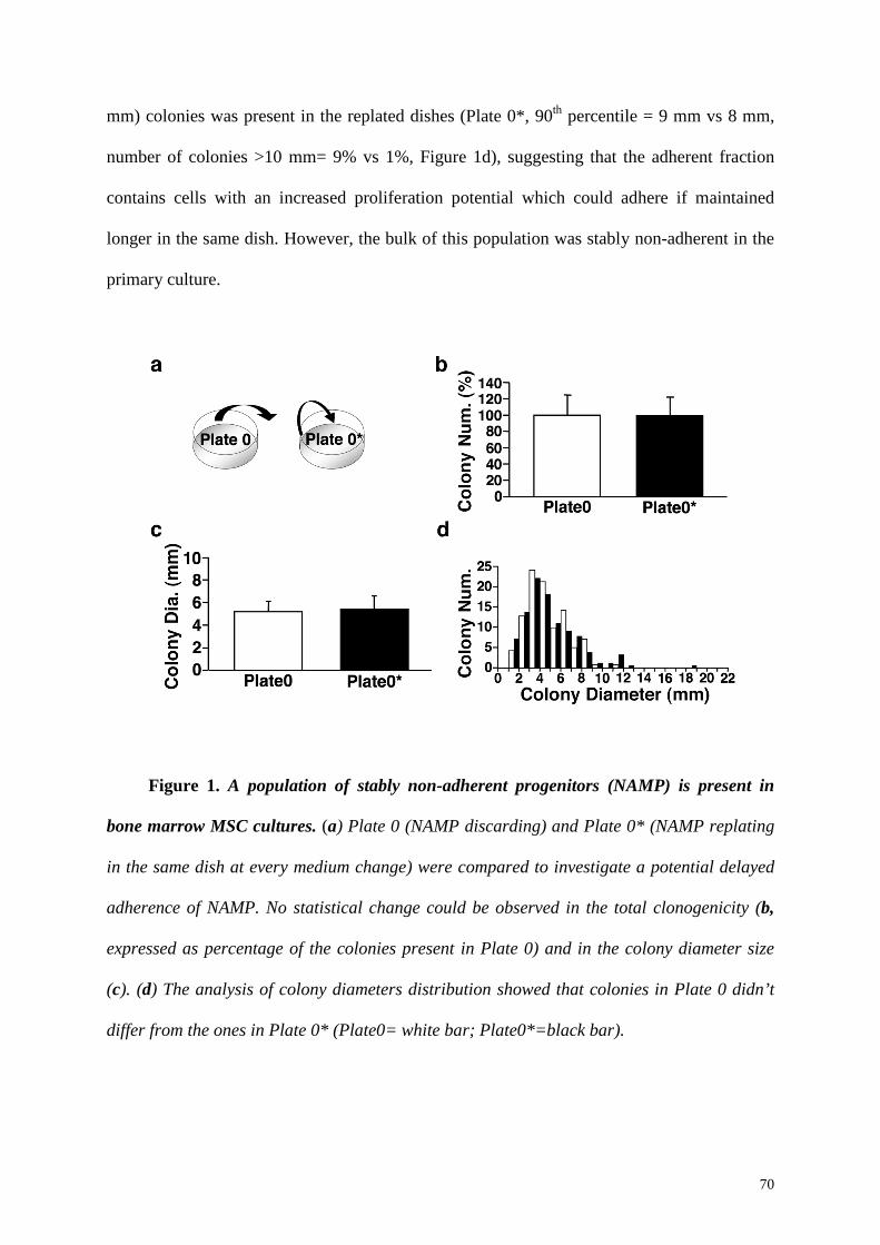
70
mm) colonies was present in the replated dishes (Plate 0*, 90th percentile = 9 mm vs 8 mm,
number of colonies >10 mm= 9% vs 1%, Figure 1d), suggesting that the adherent fraction
contains cells with an increased proliferation potential which could adhere if maintained
longer in the same dish. However, the bulk of this population was stably non-adherent in the
primary culture.
Figure 1. A population of stably non-adherent progenitors (NAMP) is present in
bone marrow MSC cultures. (a) Plate 0 (NAMP discarding) and Plate 0* (NAMP replating
in the same dish at every medium change) were compared to investigate a potential delayed
adherence of NAMP. No statistical change could be observed in the total clonogenicity (b,
expressed as percentage of the colonies present in Plate 0) and in the colony diameter size
(c). (d) The analysis of colony diameters distribution showed that colonies in Plate 0 didn’t
differ from the ones in Plate 0* (Plate0= white bar; Plate0*=black bar).
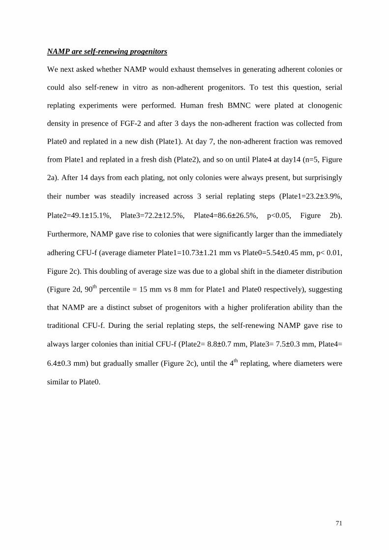
71
NAMP are self-renewing progenitors
We next asked whether NAMP would exhaust themselves in generating adherent colonies or
could also self-renew in vitro as non-adherent progenitors. To test this question, serial
replating experiments were performed. Human fresh BMNC were plated at clonogenic
density in presence of FGF-2 and after 3 days the non-adherent fraction was collected from
Plate0 and replated in a new dish (Plate1). At day 7, the non-adherent fraction was removed
from Plate1 and replated in a fresh dish (Plate2), and so on until Plate4 at day14 (n=5, Figure
2a). After 14 days from each plating, not only colonies were always present, but surprisingly
their number was steadily increased across 3 serial replating steps (Plate1=23.2±3.9%,
Plate2=49.1±15.1%, Plate3=72.2±12.5%, Plate4=86.6±26.5%, p<0.05, Figure 2b).
Furthermore, NAMP gave rise to colonies that were significantly larger than the immediately
adhering CFU-f (average diameter Plate1=10.73±1.21 mm vs Plate0=5.54±0.45 mm, p< 0.01,
Figure 2c). This doubling of average size was due to a global shift in the diameter distribution
(Figure 2d, 90th percentile = 15 mm vs 8 mm for Plate1 and Plate0 respectively), suggesting
that NAMP are a distinct subset of progenitors with a higher proliferation ability than the
traditional CFU-f. During the serial replating steps, the self-renewing NAMP gave rise to
always larger colonies than initial CFU-f (Plate2= 8.8±0.7 mm, Plate3= 7.5±0.3 mm, Plate4=
6.4±0.3 mm) but gradually smaller (Figure 2c), until the 4th replating, where diameters were
similar to Plate0.
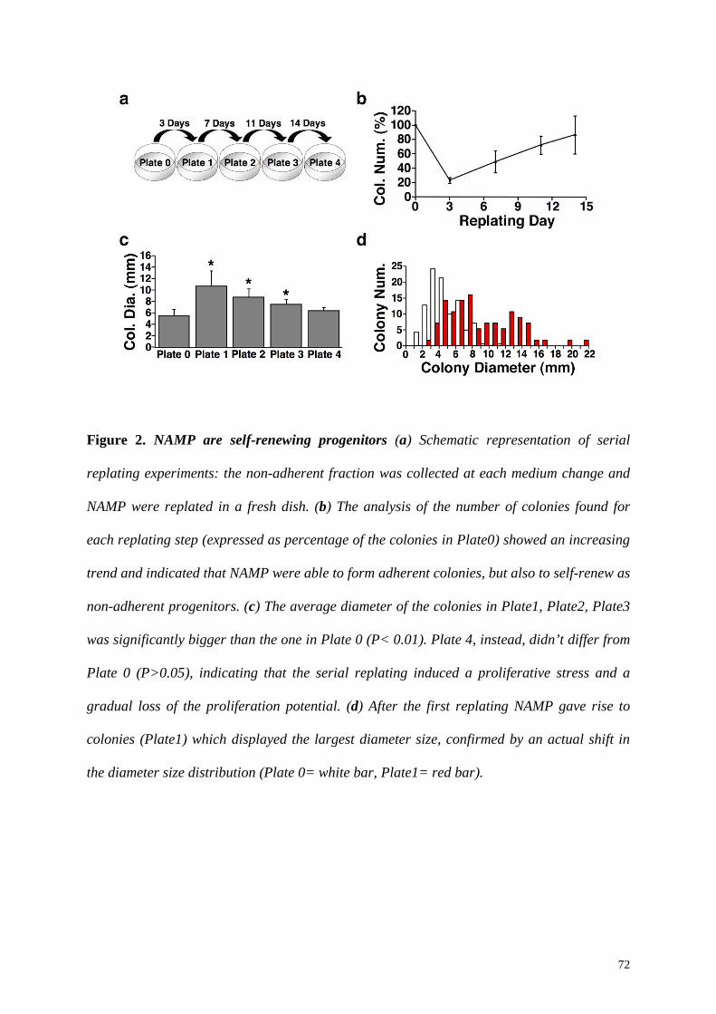
72
Figure 2. NAMP are self-renewing progenitors (a) Schematic representation of serial
replating experiments: the non-adherent fraction was collected at each medium change and
NAMP were replated in a fresh dish. (b) The analysis of the number of colonies found for
each replating step (expressed as percentage of the colonies in Plate0) showed an increasing
trend and indicated that NAMP were able to form adherent colonies, but also to self-renew as
non-adherent progenitors. (c) The average diameter of the colonies in Plate1, Plate2, Plate3
was significantly bigger than the one in Plate 0 (P< 0.01). Plate 4, instead, didn’t differ from
Plate 0 (P>0.05), indicating that the serial replating induced a proliferative stress and a
gradual loss of the proliferation potential. (d) After the first replating NAMP gave rise to
colonies (Plate1) which displayed the largest diameter size, confirmed by an actual shift in
the diameter size distribution (Plate 0= white bar, Plate1= red bar).
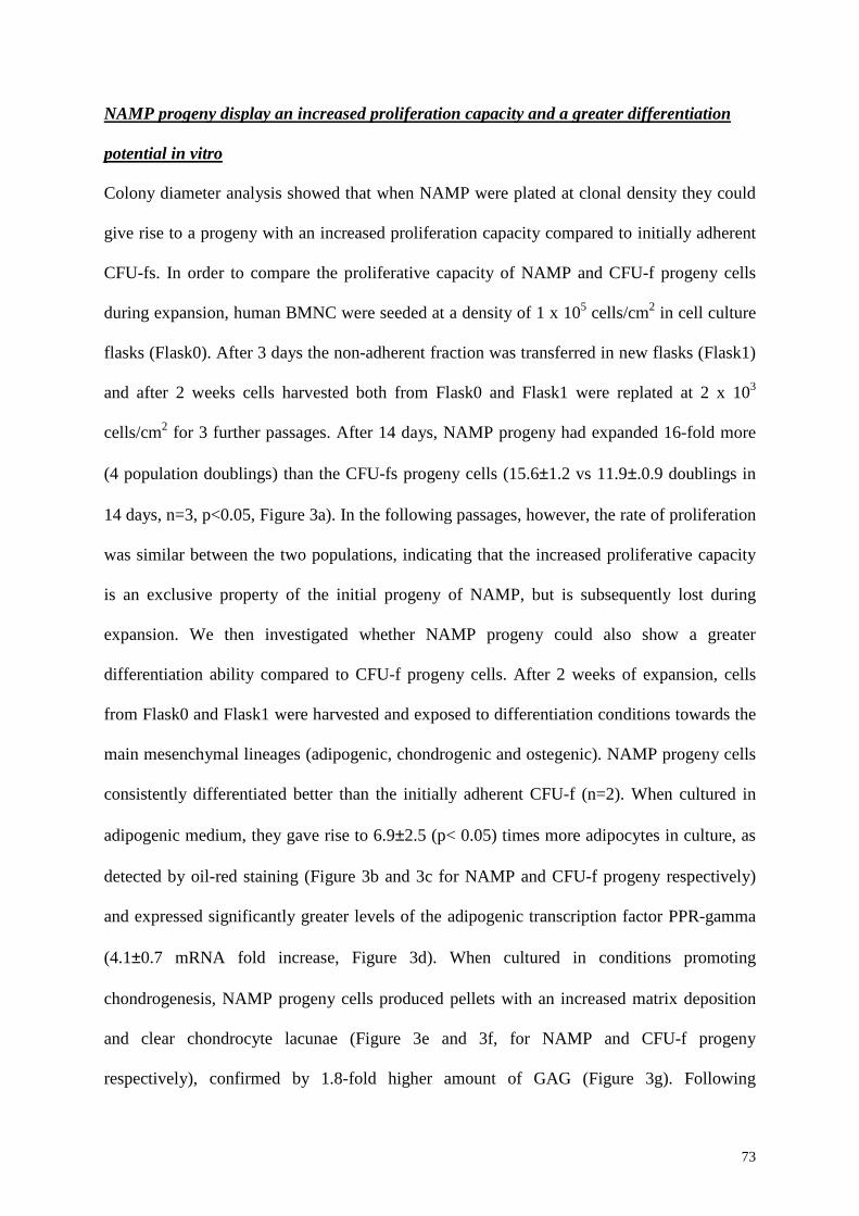
73
NAMP progeny display an increased proliferation capacity and a greater differentiation
potential in vitro
Colony diameter analysis showed that when NAMP were plated at clonal density they could
give rise to a progeny with an increased proliferation capacity compared to initially adherent
CFU-fs. In order to compare the proliferative capacity of NAMP and CFU-f progeny cells
during expansion, human BMNC were seeded at a density of 1 x 10⁵ cells/cm2 in cell culture
flasks (Flask0). After 3 days the non-adherent fraction was transferred in new flasks (Flask1)
and after 2 weeks cells harvested both from Flask0 and Flask1 were replated at 2 x 10³
cells/cm2 for 3 further passages. After 14 days, NAMP progeny had expanded 16-fold more
(4 population doublings) than the CFU-fs progeny cells (15.6±1.2 vs 11.9±.0.9 doublings in
14 days, n=3, p<0.05, Figure 3a). In the following passages, however, the rate of proliferation
was similar between the two populations, indicating that the increased proliferative capacity
is an exclusive property of the initial progeny of NAMP, but is subsequently lost during
expansion. We then investigated whether NAMP progeny could also show a greater
differentiation ability compared to CFU-f progeny cells. After 2 weeks of expansion, cells
from Flask0 and Flask1 were harvested and exposed to differentiation conditions towards the
main mesenchymal lineages (adipogenic, chondrogenic and ostegenic). NAMP progeny cells
consistently differentiated better than the initially adherent CFU-f (n=2). When cultured in
adipogenic medium, they gave rise to 6.9±2.5 (p< 0.05) times more adipocytes in culture, as
detected by oil-red staining (Figure 3b and 3c for NAMP and CFU-f progeny respectively)
and expressed significantly greater levels of the adipogenic transcription factor PPR-gamma
(4.1±0.7 mRNA fold increase, Figure 3d). When cultured in conditions promoting
chondrogenesis, NAMP progeny cells produced pellets with an increased matrix deposition
and clear chondrocyte lacunae (Figure 3e and 3f, for NAMP and CFU-f progeny
respectively), confirmed by 1.8-fold higher amount of GAG (Figure 3g). Following
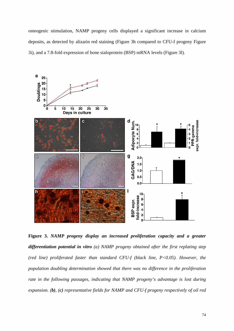
74
osteogenic stimulation, NAMP progeny cells displayed a significant increase in calcium
deposits, as detected by alizarin red staining (Figure 3h compared to CFU-f progeny Figure
3i), and a 7.8-fold expression of bone sialoprotein (BSP) mRNA levels (Figure 3l).
Figure 3. NAMP progeny display an increased proliferation capacity and a greater
differentiation potential in vitro (a) NAMP progeny obtained after the first replating step
(red line) proliferated faster than standard CFU-f (black line, P<0.05). However, the
population doubling determination showed that there was no difference in the proliferation
rate in the following passages, indicating that NAMP progeny’s advantage is lost during
expansion. (b), (c) representative fields for NAMP and CFU-f progeny respectively of oil red

75
staining after adipogenic differentiation (size bar= 200 µm); NAMP progeny cells showed a
greater differentiation potential which was quantified by the number of adipocytes per field
and by PPAR-gamma expression (d, black bar= NAMP progeny, white bar= CFU-f
progeny); (e), (f) Safranin-O staining of cell pellets obtained from NAMP and CFU-f progeny
cells respectively (size bar= 100 µm). Increased chondrogenic potential of NAMP progeny
cells was confirmed by glycosamminoglycan deposition quantification (g, black bar= NAMP
progeny, white bar= CFU-f progeny); (h), (i) alizarin red staining for NAMP and CFU-f
progeny cells respectively and determination of BSP expression (l, black bar= NAMP
progeny, white bar= CFU-f progeny).
NAMP progeny cells have greatly increased osteogenic potential in vivo
In vitro differentiation assays are a high-throughput surrogate of actual differentiation
capacity which can be rigorously determined only by in vivo functional assays. Therefore, we
investigated whether NAMP progeny cells display an increased osteogenic potential in vivo
compared to CFU-f progeny cells. Both progenies were obtained and expanded as described
for the assessment of the in vitro differentiation capacity. As it is shown in Figure 4, a
qualitative analysis revealed that after 8 weeks both CFU-f and NAMP progeny cells could
generate bone in vivo (Figure 4a and 4c respectively). The matrix was characterized by an
intense eosin staining and by the formation of osteocyte lacunae. Fluorescent images of the
histological sections showed that collagen fibers were compact and deposited orderly in
parallel orientation (Figure 4b and 4d for CFU-f progeny cells and NAMP progeny cells
respectively), confirming the formation of a dense bone-like matrix. The histological analysis
also showed an increased amount of bone tissue in the constructs loaded with NAMP progeny
cells compared to the ones containing CFU-f progeny cells (Figure 4c-4d and 4a-4b
respectively). This observation was confirmed by a quantitative analysis, which revealed that
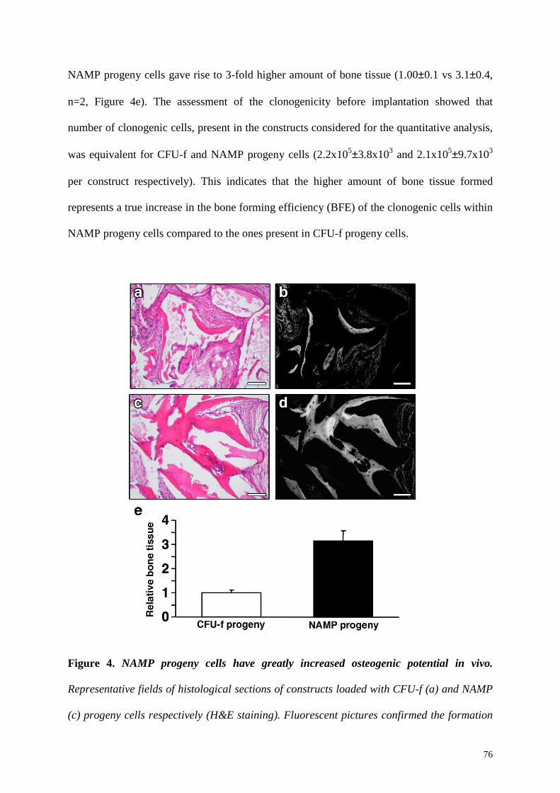
76
NAMP progeny cells gave rise to 3-fold higher amount of bone tissue (1.00±0.1 vs 3.1±0.4,
n=2, Figure 4e). The assessment of the clonogenicity before implantation showed that
number of clonogenic cells, present in the constructs considered for the quantitative analysis,
was equivalent for CFU-f and NAMP progeny cells (2.2x10⁵±3.8x10³ and 2.1x10⁵±9.7x10³
per construct respectively). This indicates that the higher amount of bone tissue formed
represents a true increase in the bone forming efficiency (BFE) of the clonogenic cells within
NAMP progeny cells compared to the ones present in CFU-f progeny cells.
Figure 4. NAMP progeny cells have greatly increased osteogenic potential in vivo.
Representative fields of histological sections of constructs loaded with CFU-f (a) and NAMP
(c) progeny cells respectively (H&E staining). Fluorescent pictures confirmed the formation
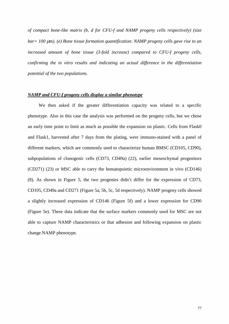
77
of compact bone-like matrix (b, d for CFU-f and NAMP progeny cells respectively) (size
bar= 100 µm). (e) Bone tissue formation quantification: NAMP progeny cells gave rise to an
increased amount of bone tissue (3-fold increase) compared to CFU-f progeny cells,
confirming the in vitro results and indicating an actual difference in the differentiation
potential of the two populations.
NAMP and CFU-f progeny cells display a similar phenotype
We then asked if the greater differentiation capacity was related to a specific
phenotype. Also in this case the analysis was performed on the progeny cells, but we chose
an early time point to limit as much as possible the expansion on plastic. Cells from Flask0
and Flask1, harvested after 7 days from the plating, were immuno-stained with a panel of
different markers, which are commonly used to characterize human BMSC (CD105, CD90),
subpopulations of clonogenic cells (CD73, CD49a) (22), earlier mesenchymal progenitors
(CD271) (23) or MSC able to carry the hematopoietic microenvironment in vivo (CD146)
(8). As shown in Figure 5, the two progenies didn’t differ for the expression of CD73,
CD105, CD49a and CD271 (Figure 5a, 5b, 5c, 5d respectively). NAMP progeny cells showed
a slightly increased expression of CD146 (Figure 5f) and a lower expression for CD90
(Figure 5e). These data indicate that the surface markers commonly used for MSC are not
able to capture NAMP characteristics or that adhesion and following expansion on plastic
change NAMP phenotype.
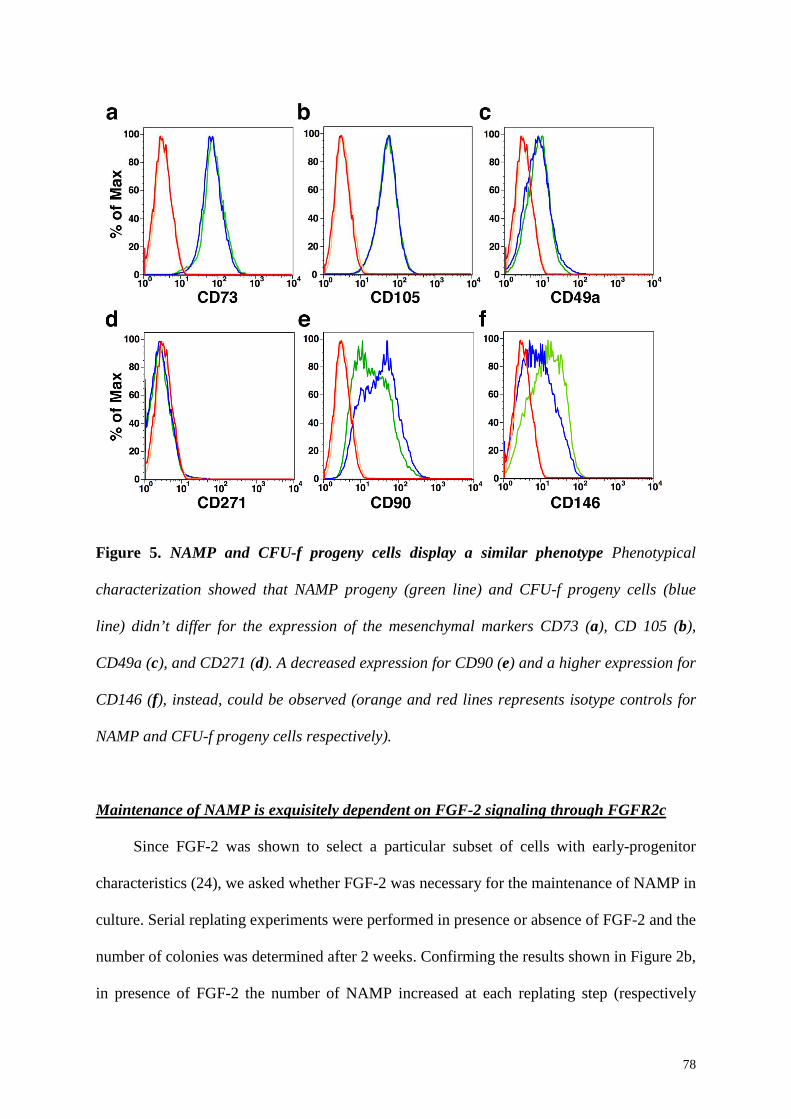
78
Figure 5. NAMP and CFU-f progeny cells display a similar phenotype Phenotypical
characterization showed that NAMP progeny (green line) and CFU-f progeny cells (blue
line) didn’t differ for the expression of the mesenchymal markers CD73 (a), CD 105 (b),
CD49a (c), and CD271 (d). A decreased expression for CD90 (e) and a higher expression for
CD146 (f), instead, could be observed (orange and red lines represents isotype controls for
NAMP and CFU-f progeny cells respectively).
Maintenance of NAMP is exquisitely dependent on FGF-2 signaling through FGFR2c
Since FGF-2 was shown to select a particular subset of cells with early-progenitor
characteristics (24), we asked whether FGF-2 was necessary for the maintenance of NAMP in
culture. Serial replating experiments were performed in presence or absence of FGF-2 and the
number of colonies was determined after 2 weeks. Confirming the results shown in Figure 2b,
in presence of FGF-2 the number of NAMP increased at each replating step (respectively
79
Plate1= 21.2%±3.1, Plate2= 47.4%±10.7, Plate3= 85.4%±6.8, Plate4=123.5%±1.0 of the
number of initially adherent CFU-f, n=3) (Figure 6a, black line). However, removal of FGF-2
completely abolished the presence of NAMP (Figure 6a, blue line). In fact, only 2.2%±1.0 of
the initial number of colonies was present in Plate1 and no colonies could be observed in the
following replating steps.
Because proliferating cells undergo a stage of rounding and semi-detachment from
plastic during the M phase of cycle, we investigated whether NAMP were a specific non-
adherent population or simply a fraction of the actively proliferating adherent cells. We
therefore performed serial replating experiments replacing FGF-2 with PDGF-BB, which also
efficiently stimulates mesenchymal cell proliferation in culture. As shown in Figure 6a (red
line), the number of colonies present in all replating steps with PDGF-BB was not different
from the condition with serum alone (Plate1 5.6±0.5% vs 2.9±2.3% of the colonies in Plate 0,
respectively, n=3, p=n.s.), completely disappearing by the second replating. These results
strongly indicate that NAMP are a population of non-adhering progenitors distinct from the
proliferating fraction of adherent cells. Furthermore, their presence in vitro critically and
specifically depends on FGF-2 signaling.
We next asked whether FGF-2 was continuously necessary for the maintenance of
NAMP function after initial selection, and conversely whether NAMP could be induced by
FGF-2 from the initially adherent fraction at a later stage. Therefore, we performed a
medium-switch experiment (n=3). Fresh human BMNC were serially replated as before.
However, in some conditions FGF-2 was removed after the first (3 days) or the second
replating (7 days). Similarly, they were also cultured without FGF-2 throughout the serial
replating, or FGF-2 was added to the medium after the first or second replating steps (3 and 7
days, respectively). When FGF-2 was removed after the first 3 days of culture (Figure 6b,
dash-dotted line), the number of colonies in the following replating steps dropped to zero like
80
in the no-FGF condition (Figure 6b, blue line). When FGF-2 was removed after the second
replating step (7 days) (Fig. 6b, dashed line), instead, the number of NAMP derived colonies
stopped increasing in the following replating steps and actually decreased slightly, but not
disappeared. No colonies were ever observed when FGF-2 was added after the first 3 or 7
days of culture, suggesting that FGF-2 is critical for NAMP survival and selection in the
initial stage of culture.
Since selection and maintenance of NAMP in vitro was critically dependent on FGF-2
signaling, we therefore investigated which receptor mediates this specific function of FGF-2,
which is known to be quite pleiotropic and bind 5 different receptors (FGFR1b, FGFR1c,
FGFR2c, FGFR3c and FGFR4) (25). To assess the role of FGFR1b, FGFR2c and FGFR3c,
monoclonal blocking antibodies were used. Instead, since blocking antibodies for FGFR1c
and FGFR4 were not commercially available, we used FGF-5 and FGF-19, which
respectively bind to FGFR1c/2c and FGFR4, to see if the stimulation of these specific
receptors could maintains NAMP in culture. BMNC were plated at clonogenic density (n=3)
and only the first replating step was performed (3 days replating). The medium was
supplemented either with the blocking antibody plus FGF-2 or with FGF-5 or FGF-19 and
after 14 day the colonies were stained. As it is shown in Figure 6c, in the presence of FGF-
19 the number of NAMP derived colonies in Plate1 was similar to the one with serum alone
(Figure 6c, 20.1±5.9% vs 25.0±3.9% respectively, values are expressed as percentage of the
colonies in the FGF-2 condition), suggesting that FGFR4 is not involved in maintenance of
NAMP in culture. Blocking antibodies for FGR1b and FGFR3 did not significantly affect
colonies in Plate1 compared to the condition with FGF-2 only (Figure 6c, 91.2±11.6% and
87.9±11.9%, respectively, p=ns). However, when the FGFR2 blocking antibody was added to
the culture, the number of colonies decreased significantly (Figure 6c, 43.3±8.4%, p<0.05).
Furthermore, by stimulating FGFR1c and FGFR2c with FGF-5 it was possible to partially
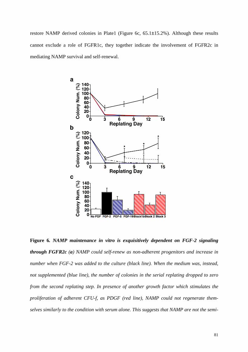
81
restore NAMP derived colonies in Plate1 (Figure 6c, 65.1±15.2%). Although these results
cannot exclude a role of FGFR1c, they together indicate the involvement of FGFR2c in
mediating NAMP survival and self-renewal.
Figure 6. NAMP maintenance in vitro is exquisitively dependent on FGF-2 signaling
through FGFR2c (a) NAMP could self-renew as non-adherent progenitors and increase in
number when FGF-2 was added to the culture (black line). When the medium was, instead,
not supplemented (blue line), the number of colonies in the serial replating dropped to zero
from the second replating step. In presence of another growth factor which stimulates the
proliferation of adherent CFU-f, as PDGF (red line), NAMP could not regenerate them-
selves similarly to the condition with serum alone. This suggests that NAMP are not the semi-

82
adherent proliferating CFU-f and indicates FGF-2 specificity for NAMP maintenance. (b)
The removal of FGF-2 after 3 days (dash-dotted line) negatively affected NAMP self-
renewal. In fact, the number of colonies in the following replating steps was the same of the
condition with serum alone (blue line). When FGF-2 was removed after 7 days (dashed line),
the FGF-2 condition trend (black line) was abolished since there was no increase in the
number of NAMP derived colonies, indicating that FGF-2 is necessary for NAMP self-
renewal in culture. (c) In presence of FGF-19, which stimulates FGFR4, the number of
colonies in Plate1 was similar to the condition with serum alone (white bar), indicating that
FGFR4 doesn’t mediate NAMP self-renewal. When FGF-5, which stimulates FGFR1c and
FGFR2c, was added to the culture NAMP gave rise to a higher number of colonies,
suggesting the involvement of at least one of the two receptors. Blocking FGFR1b and
FGFR3, no reduction in colony number could be observed. In presence, instead, of FGFR2
blocking antibody, the colony number significantly decreased, suggesting a role of FGFR2c
in NAMP maintenance. (Colony number represents colonies in Plate1 and is expressed as
percentage of the colonies in FGF-2 condition).
The adherent cells provide the niche for NAMP
We showed that the non-adherent fraction of bone marrow cultures contains a
population of stably non-adherent mesenchymal progenitors, which can produce adherent
colonies while regenerating them-selves upon serial replating. Furthermore, the results of the
serial replating experiments suggest that NAMP need the adherent fraction for their
maintenance. We now hypothesized that the adherent cells constitute a niche that provide
specific signals to preserve NAMP characteristics. To test our hypothesis we investigated the
niche function of the adherent fraction by complementary loss of function and gain of
function approaches.

83
For the loss of function approach, we cultured first-replating derived NAMP in agarose-
coated dishes, thus preventing the formation of an adherent cell layer. After 4 or 7 days (day7
and day11 respectively) cells were transferred to plastic cell culture dishes for CFU-f assay.
The total absence of colonies indicated that NAMP were not able to survive in the absence of
an adherent progeny. We next asked if the CFU-f or NAMP adherent progeny cells constitute
a unique environment for NAMP or if also other cell types could provide the same signals.
Therefore, first-replating derived NAMP were replated on an irradiated feeder layer of human
skin fibroblasts. After 4 or 7 days (day7 or day11 respectively) the cells were transferred to
plastic on cell culture dishes. The number of colonies generated after both 4 and 7 days on
skin fibroblasts was negligible, suggesting that this cell type could not provide the required
signals for NAMP survival.
Taking a gain of function approach we hypothesized that, if CFU-f progeny cells
provided the niche function for NAMP, keeping them in contact with the adherent cells
would avoid the gradual loss of their early progenitors characteristics, which is observed
during serial replates in empty dishes. The non-adherent fraction was maintained in Plate0 for
7 or 14 days before replating to a new culture dish. After two weeks the number of colonies
was compared with those obtained in the serial replating to establish if expanding the putative
niche, we could also expand the non-adherent fraction. Colony diameter was measured,
instead, to assess whether the contact with the niche could preserve NAMP high proliferative
potential (n=2). As shown in Figure 7a, when NAMP were kept on the originally adherent
cells for 14 days (Plate4*), they generated greater than 3-fold more colonies compared to
when they underwent a serial replating for the same time (Plate4) (Figure 7a, 260.8±15.8% vs
61.0±8.0%); if NAMP were not replated to a new dish, however, no extra colonies grew in
the initial plate after 14 days, confirming the results in Figure 1b. Colonies generated after 14
days of continuous culture in the primary plate displayed a significantly ad consistently larger
84
diameter compared to those generated by serially replated cells at the same time point
(9.8±0.4 mm vs 6.8±0.3 mm, p<0.05, Fig 7b). Furthermore, colonies in Plate4* were similar
in diameter to the ones in Plate1 (diameter= 9.8±0.4 mm 9.2±0.5 mm respectively, p=ns)
indicating that NAMP compartment can expand on the adherent fraction without losing the
proliferative potential. In parallel, we compared the differentiation capacity of NAMP kept on
the originally adherent cells for 14 days (Plate4*) to the ones that were serially replated for
the same time (Plate4). Progeny of both populations was generated replating the non-adherent
fraction in new flasks and after 1 week of expansion cells were exposed differentiation
conditions to assess differentiation capacity in vitro. When NAMP were kept in contact with
their niche, they generated a progeny which displayed an increased differentiation potential
compared to the one derived from serially replated NAMP. In fact, they gave raise to a higher
number of adipocytes (Figure 7c and 7d for Plate4 and Plate4*-progeny respectively) and
they could better differentiate towards the osteogenic lineage (Figure 7e and 7f for Plate4 and
Plate4*-progeny respectively).
These data collectively suggest a niche function for the adherent cells. When this function
was absent due to serial replating in empty dishes, NAMP could regenerate the niche but at
the cost of gradually losing their proliferative and differentiation potential (Figure 2c).

85
Figure 7. The adherent cells provide the niche for NAMP (a) when NAMP were kept in
contact with CFU-f progeny (Plate4*), they produced 3-fold more colonies compared to
when they were serially replated for the same time (Plate4); (b) As shown from the colony
diameter size evaluation, Plate4*-progeny preserved the proliferation capacity compared
cells generated from serially replated NAMP; (c), (d) representative fields for Plate-4 and
Plate-4* progeny respectively of oil red staining after adipogenic differentiation; (e), (f)
alizarin red staining for Plate4 and Plate4*- progeny cells respectively. Red arrows indicate
calcium deposits.
86
4.4 Discussion
The data presented in this study indicate that a population of stably non-adherent
mesenchymal progenitors (NAMP) is present in human bone marrow cultures. NAMP
display the following distinct biological features compared to standard CFU-f: 1) an
increased proliferation and differentiation capacity of their immediate progeny in vitro; 2) a
greater efficiency in generating bone tissue in vivo; and 3) the ability to give rise to adherent
colonies while self-renewing as non-adherent progenitors in vitro. Both their maintenance
and self-renewal are strictly dependent on FGF-2 signaling and on the presence of adherent
CFU-f progeny cells, suggesting the establishment of a niche-progenitor system in vitro.
Bone marrow-derived mesenchymal progenitors are a widely heterogeneous population in
terms of both proliferation and differentation potential (26). A model about lineage hierarchy
in vivo has been proposed (27) and in vitro studies at the clonal level have revealed the
presence of different classes of progenitors within the CFU-f population in a specific
hierarchical relationship, with progressive loss of multi-differentiation potential towards
adipogenesis and chondrogenesis and commitment to the osteogenic lineage (10). Our data
identify a population of mesenchymal progenitors which appears to represent a more
primitive entity than classic CFU-f within the hierarchy of mesenchymal progenitors.
Features currently accepted as defining a stem/progenitor cell are 1) the high proliferation
capacity, 2) the multilineage differentiation potential and 3) the self-renewing ability (28).
When serially replated, NAMP gave rise to colonies with a significantly increased diameter,
indicatating a higher proliferation activity at clonal density. When plated at high density,
however, NAMP progeny showed a faster rate of proliferation only until the first confluence.
These data indicate that the adherent progeny does not maintain the properties of the NAMP
from which it derives, which rather reside in the self-renewed non-adherent fraction. The
differentiation potential has been described to be progressively lost during expansion pointing
87
to a commitment of the population (29). Even though at the first confluence NAMP progeny
cells had expanded 16-fold more (4 population doublings) than CFU-f progeny cells, it still
displayed a greater differentiation potential. Furthermore, NAMP are able to regenerate them-
selves and at the same time to produce adherent colonies, but they cannot be induced from
the initially adherent fraction at a later stage. These data together suggest that NAMP are a
distinct population hierarchically upstream of initially adherent CFU-f which displays earlier
progenitor features and which is biologically different from classically-defined CFU-f.
Serial replating experiments performed in the absence of FGF-2 revealed that, unlike
initially adherent CFU-f, NAMP require FGF-2 both for their selection and maintenance in
culture. FGF-2 has been shown to crucially control the selection of a subset of initial CFU-f
enriched in pluripotent mesenchymal precursors (24). Interestingly, removal of FGF-2, at
different time points during serial replating, always caused the disappearance of NAMP. This
is in contrast to the effects on CFU-f, where FGF-2 induces, instead, an immediate and
permanent selection within the clonogenic progenitors, suggesting a different biological
response to FGF-2. Furthermore, blocking receptor experiments showed that FGF-2 mediates
the maintenance of NAMP in culture through FGFR2c signaling. The FGFR2 gene encodes
two splice variants, FGFR2b which in mice seems to have a unique role in skin development
and FGFR2c, which is, instead, preferentially expressed during osteogenesis (30). In
FGFR2c-/- mice the balance between proliferation and differentiation of skeletal progenitors is
shifted towards differentiation, leading to premature loss of growth and defects in the skull
and in long bones (31). Conversely, in gain of function experiments, which were used as a
model for human Crouzon and Pfeiffer syndromes, mutants showed a significant increase in
the number of osteoprogenitors cells (32). These data collectively suggest that NAMP have a
role as reservoir of early mesenchymal progenitors and that the activation of FGFR2c
signaling is fundamental for their function in vivo.
88
A fundamental property that characterize stem cells is the self-renewal, which is defined
as the cycles of division that generate at least one daughter equivalent to the mother cell with
latent capacity for differentiation (33). Demonstration of self-renewal in vivo implicates both
the generation of a differentiated progeny and the reconstitution of a stem cell compartment.
In the bone marrow, the contact with the specialized environment of the niche regulates
hematopoietic stem cells survival and self-renewal (5). Our data show that NAMP are able to
maintain them-selves and proliferate in suspension, but that they require the adherent
mesenchymal fraction for their maintenance. The generation of adherent daughter cells can
be achieved by two different mechanisms, 1) adhesion of a non-adherent progenitor and
proliferation as fibroblastic colony or 2) a division, in which only one of the daughter cells
lose the non-adherent feature and the other maintains instead NAMP features. The first
mechanism would implicate that NAMP are able to undergo division in suspension to
maintain them-selves in the non-adherent status. Even thought further experiments are
required to address this point, our data show that, when serially replated, NAMP are subject
to a proliferative stress due to the generation of the adherent fraction. In fact, the generation
of adherent daughter cells happens with slow gradual loss of potential, as after 4 steps of
serial replating, NAMP proliferative potential is similar to that of initially adherent CFU-f.
However, the self-renewal capacity is maintained because they are still able to generate non-
adherent clonogenic progenitors in constantly increasing numbers. A similar mechanism has
been described for hematopoietic stem cells, which, even maintaining their stem cell
properties, lose telomere length, and therefore proliferative activity, when they are subjected
to the proliferative stress of bone marrow repopulation in bone marrow transplantation
recipients (34, 35). In our experimental set up NAMP self-renewal is not assessed at a single
cell level in vivo. However, our data show that in vitro NAMP are able to maintain them-
selves and to give rise to a progeny, which displays different biological features.
89
Furthermore, the loss of potential, taking place with the serial generation of the adherent
progeny, does not occur maintaining the culture of NAMP on the initial adherent fraction. In
this experimental condition, in fact, NAMP undergo greater expansion compared to when
they are serially replated (12-fold vs 4-fold in 14 days, Figure 6a) without any loss of
proliferation and differentiation potential. This set of data, therefore, suggests that the
adherent CFU-f provide a niche function for NAMP promoting their expansion and self-
renewal as early progenitors in vitro.
Even though mesenchymal progenitor cells are the subject of extensive interest in
regenerative medicine, the knowledge about their regulation in vivo, including the need of a
specific microenvironment for their maintenance, is severely insufficient. Sacchetti et al. (8)
identified for the first time the presence of self-renewing stromal cells, which can be selected
ex vivo only in the absence of FGF-2. This population is characterized by CD146 expression
and is able to generate osteoblasts, adventitial reticular cells and the hemopoiesis-supporting
niche in vivo, while maintaining them-selves as clonogenic stromal progenitors. Our data
suggest a complementary model, showing that FGF-2, instead, selects a different population
of progenitors from the common bone marrow pool. NAMP are self-renewing in vitro, can
differentiate towards the classic mesenchymal lineages (adipogenic, chondrogenic and
osteogenic in vivo) and generate a niche supporting their own self-renewal, but lose the
ability to support hemopoiesis in vivo. In fact, although we could observe a moderate shift of
CD146 expression in NAMP progeny, they could not support bone marrow formation in
vivo. It would be interesting to investigate the role of FGF-2 in selecting different
populations in vivo, which perform different functions in the complex bone marrow
environment.
In conlcusion, our data identify a niche/mesenchymal progenitor organization in vitro,
suggesting the novel concept that the progeny of self-renewing mesenchymal progenitors can
90
provide the regulatory microenviroment from the same progenitor it derives from.
References
1. Scadden, D.T. 2006. The stem-cell niche as an entity of action. Nature 441:1075-1079.
2. Zhang, J., Niu, C., Ye, L., Huang, H., He, X., Tong, W.G., Ross, J., Haug, J., Johnson, T., Feng, J.Q., et al. 2003. Identification of the haematopoietic stem cell niche and control of the niche size. Nature 425:836-841.
3. Arai, F., Hirao, A., Ohmura, M., Sato, H., Matsuoka, S., Takubo, K., Ito, K., Koh, G.Y., and Suda, T. 2004. Tie2/angiopoietin-1 signaling regulates hematopoietic stem cell quiescence in the bone marrow niche. Cell 118:149-161.
4. Calvi, L.M., Adams, G.B., Weibrecht, K.W., Weber, J.M., Olson, D.P., Knight, M.C., Martin, R.P., Schipani, E., Divieti, P., Bringhurst, F.R., et al. 2003. Osteoblastic cells regulate the haematopoietic stem cell niche. Nature 425:841-846.
5. Wilson, A., and Trumpp, A. 2006. Bone-marrow haematopoietic-stem-cell niches. Nat Rev Immunol 6:93-106.
6. Jones, D.L., and Wagers, A.J. 2008. No place like home: anatomy and function of the stem cell niche. Nat Rev Mol Cell Biol 9:11-21.
7. Kiel, M.J., and Morrison, S.J. 2008. Uncertainty in the niches that maintain haematopoietic stem cells. Nat Rev Immunol 8:290-301.
8. Sacchetti, B., Funari, A., Michienzi, S., Di Cesare, S., Piersanti, S., Saggio, I., Tagliafico, E., Ferrari, S., Robey, P.G., Riminucci, M., et al. 2007. Self-renewing osteoprogenitors in bone marrow sinusoids can organize a hematopoietic microenvironment. Cell 131:324-336.
9. Bianco, P., Riminucci, M., Gronthos, S., and Robey, P.G. 2001. Bone marrow stromal stem cells: nature, biology, and potential applications. Stem Cells 19:180-192.
10. Muraglia, A., Cancedda, R., and Quarto, R. 2000. Clonal mesenchymal progenitors from human bone marrow differentiate in vitro according to a hierarchical model. J Cell Sci 113 ( Pt 7):1161-1166.
11. Owen, M., and Friedenstein, A.J. 1988. Stromal stem cells: marrow-derived osteogenic precursors. Ciba Found Symp 136:42-60.
12. Wan, C., He, Q., McCaigue, M., Marsh, D., and Li, G. 2006. Nonadherent cell population of human marrow culture is a complementary source of mesenchymal stem cells (MSCs). J Orthop Res 24:21-28.
13. Baksh, D., Zandstra, P.W., and Davies, J.E. 2007. A non-contact suspension culture approach to the culture of osteogenic cells derived from a CD49elow subpopulation of human bone marrow-derived cells. Biotechnol Bioeng 98:1195-1208.
14. Barbero, A., Ploegert, S., Heberer, M., and Martin, I. 2003. Plasticity of clonal populations of dedifferentiated adult human articular chondrocytes. Arthritis Rheum 48:1315-1325.
91
15. Jakob, M., Demarteau, O., Schafer, D., Hintermann, B., Dick, W., Heberer, M., and Martin, I. 2001. Specific growth factors during the expansion and redifferentiation of adult human articular chondrocytes enhance chondrogenesis and cartilaginous tissue formation in vitro. J Cell Biochem 81:368-377.
16. Farndale, R.W., Buttle, D.J., and Barrett, A.J. 1986. Improved quantitation and discrimination of sulphated glycosaminoglycans by use of dimethylmethylene blue. Biochim Biophys Acta 883:173-177.
17. Jaiswal, N., Haynesworth, S.E., Caplan, A.I., and Bruder, S.P. 1997. Osteogenic differentiation of purified, culture-expanded human mesenchymal stem cells in vitro. J Cell Biochem 64:295-312.
18. Frank, O., Heim, M., Jakob, M., Barbero, A., Schafer, D., Bendik, I., Dick, W., Heberer, M., and Martin, I. 2002. Real-time quantitative RT-PCR analysis of human bone marrow stromal cells during osteogenic differentiation in vitro. J Cell Biochem 85:737-746.
19. Jaquiery, C., Schaeren, S., Farhadi, J., Mainil-Varlet, P., Kunz, C., Zeilhofer, H.F., Heberer, M., and Martin, I. 2005. In vitro osteogenic differentiation and in vivo bone-forming capacity of human isogenic jaw periosteal cells and bone marrow stromal cells. Ann Surg 242:859-867, discussion 867-858.
20. Martin, I., Mastrogiacomo, M., De Leo, G., Muraglia, A., Beltrame, F., Cancedda, R., and Quarto, R. 2002. Fluorescence microscopy imaging of bone for automated histomorphometry. Tissue Eng 8:847-852.
21. Friedenstein, A.J., Chailakhyan, R.K., and Gerasimov, U.V. 1987. Bone marrow osteogenic stem cells: in vitro cultivation and transplantation in diffusion chambers. Cell Tissue Kinet 20:263-272.
22. Boiret, N., Rapatel, C., Veyrat-Masson, R., Guillouard, L., Guerin, J.J., Pigeon, P., Descamps, S., Boisgard, S., and Berger, M.G. 2005. Characterization of nonexpanded mesenchymal progenitor cells from normal adult human bone marrow. Exp Hematol 33:219-225.
23. Quirici, N., Soligo, D., Bossolasco, P., Servida, F., Lumini, C., and Deliliers, G.L. 2002. Isolation of bone marrow mesenchymal stem cells by anti-nerve growth factor receptor antibodies. Exp Hematol 30:783-791.
24. Bianchi, G., Banfi, A., Mastrogiacomo, M., Notaro, R., Luzzatto, L., Cancedda, R., and Quarto, R. 2003. Ex vivo enrichment of mesenchymal cell progenitors by fibroblast growth factor 2. Exp Cell Res 287:98-105.
25. Eswarakumar, V.P., Lax, I., and Schlessinger, J. 2005. Cellular signaling by fibroblast growth factor receptors. Cytokine Growth Factor Rev 16:139-149.
26. Bianco, P., Robey, P.G., and Simmons, P.J. 2008. Mesenchymal stem cells: revisiting history, concepts, and assays. Cell Stem Cell 2:313-319.
27. Bianco, P., and Cossu, G. 1999. Uno, nessuno e centomila: searching for the identity of mesodermal progenitors. Exp Cell Res 251:257-263.
28. Reya, T., Morrison, S.J., Clarke, M.F., and Weissman, I.L. 2001. Stem cells, cancer, and cancer stem cells. Nature 414:105-111.
29. Banfi, A., Muraglia, A., Dozin, B., Mastrogiacomo, M., Cancedda, R., and Quarto, R. 2000. Proliferation kinetics and differentiation potential of ex vivo expanded human
92
bone marrow stromal cells: Implications for their use in cell therapy. Exp Hematol 28:707-715.
30. Orr-Urtreger, A., Bedford, M.T., Burakova, T., Arman, E., Zimmer, Y., Yayon, A., Givol, D., and Lonai, P. 1993. Developmental localization of the splicing alternatives of fibroblast growth factor receptor-2 (FGFR2). Dev Biol 158:475-486.
31. Eswarakumar, V.P., Monsonego-Ornan, E., Pines, M., Antonopoulou, I., Morriss-Kay, G.M., and Lonai, P. 2002. The IIIc alternative of Fgfr2 is a positive regulator of bone formation. Development 129:3783-3793.
32. Eswarakumar, V.P., Horowitz, M.C., Locklin, R., Morriss-Kay, G.M., and Lonai, P. 2004. A gain-of-function mutation of Fgfr2c demonstrates the roles of this receptor variant in osteogenesis. Proc Natl Acad Sci U S A 101:12555-12560.
33. Smith, A. 2006. A glossary for stem cell biology. Nature 441:1060.
34. Karadimitris, A., Araten, D.J., Luzzatto, L., and Notaro, R. 2003. Severe telomere shortening in patients with paroxysmal nocturnal hemoglobinuria affects both GPI- and GPI+ hematopoiesis. Blood 102:514-516.
35. Notaro, R., Cimmino, A., Tabarini, D., Rotoli, B., and Luzzatto, L. 1997. In vivo telomere dynamics of human hematopoietic stem cells. Proc Natl Acad Sci U S A 94:13782-13785.
93
Chapter 5
Adipose tissues stromal vascular fraction (SVF) cultures contain a
population of non-adherent mesenchymal progenitor (NAMP)
which are FGF2-dependent and require an in vitro niche for their
maintenance
94
5.1 Introduction
Although mesenchymal progenitors cells were originally described in the bone marrow (1),
they have been also found in the stromal vascular fraction (SVF) of white adipose tissue and
more recently in a variety of other tissues, such as placenta, synovium and dental pulp (2).
This led to the current hypothesis that multipotent mesenchymal progenitors are present in all
tissues of the body, possibly in a perivascular position, representing a common reservoir of
regenerative cells.
Our findings described in Chapter 4 showed that non-adherent progenitors of bone
marrow stroma (BM-NAMP) represent the most primitive compartment of MSC, are capable
of establishing their own niche in vitro and, in these conditions, can self-renew without loss
of proliferation and differentiation capacity. Based on these results, we sought to determine
whether the NAMP class of progenitors was specific to bone marrow stroma or could
represent a common feature of other mesenchymal compartments, like for example the SVF
of adipose tissue.
95
5.2 Material and Methods
Cell isolation
Subcutaneous adipose tissue in the form of lipoaspirates was obtained from 12 healthy donors
(21-69 years old) during routine lipoaspirations, after informed consent from the patient and
following protocol approval by the local ethical committee. The tissue was digested in
0.075% collagenase type 2 (Worthington, Lakewood, NY) for 45 minutes at 37 °C on an
orbital shaker. The suspension was thereafter centrifuged at 300g for 10 minutes, and the
resulting SVF pellet was washed once with phosphate-buffered saline (PBS), resuspended in
alpha-minimal essential medium (MEM) (Gibco, Grand Island, NY), and finally filtered
through a 100 µm strainer (BD Falcon; BD Biosciences, San Diego). Nucleated cells were
counted after staining with Crystal Violet 0.01% (Sigma) in phosphate-buffered saline, pH
7.2 (PBS). For FACS analysis, freshly isolated cells were resuspended into 200 µl of 0.5%
BSA in PBS (FACS Buffer) with fluorochrome-conjugated antibodies against the indicated
protein or an isotype control and were incubated for 30 minutes at 4 °C. The antibodies used
were CD146-PE, CD90-FITC, CD73-PE, CD14-PE, CD45-PE, CD31-APC, CD34-APC,
SSEA4-FITC, IgG1-FITC, IgG1-PE, IgG-APC (all from Becton, Dickinson and Company,
Franklin Lakes, NJ), CD105-FITC (Serotec Ldt. Oxford, UK). All the antibodies were used at
a dilution of 1:50, except CD105-FITC, which was used at 1:20. Cells were washed twice
with FACS buffer, resuspended in PBS and analyzed with a FACSCalibur flow cytometer
(Becton, Dickinson and Company).
Cell culture
To determine colony forming efficiency (CFE) and for serial replating experiments, cells
were plated at clonal density (9 cells/cm2) and cultured in alpha-MEM (GIBCO) with 10%
fetal bovine serum (FBS). When indicated, medium was further supplemented with FGF-2 (5
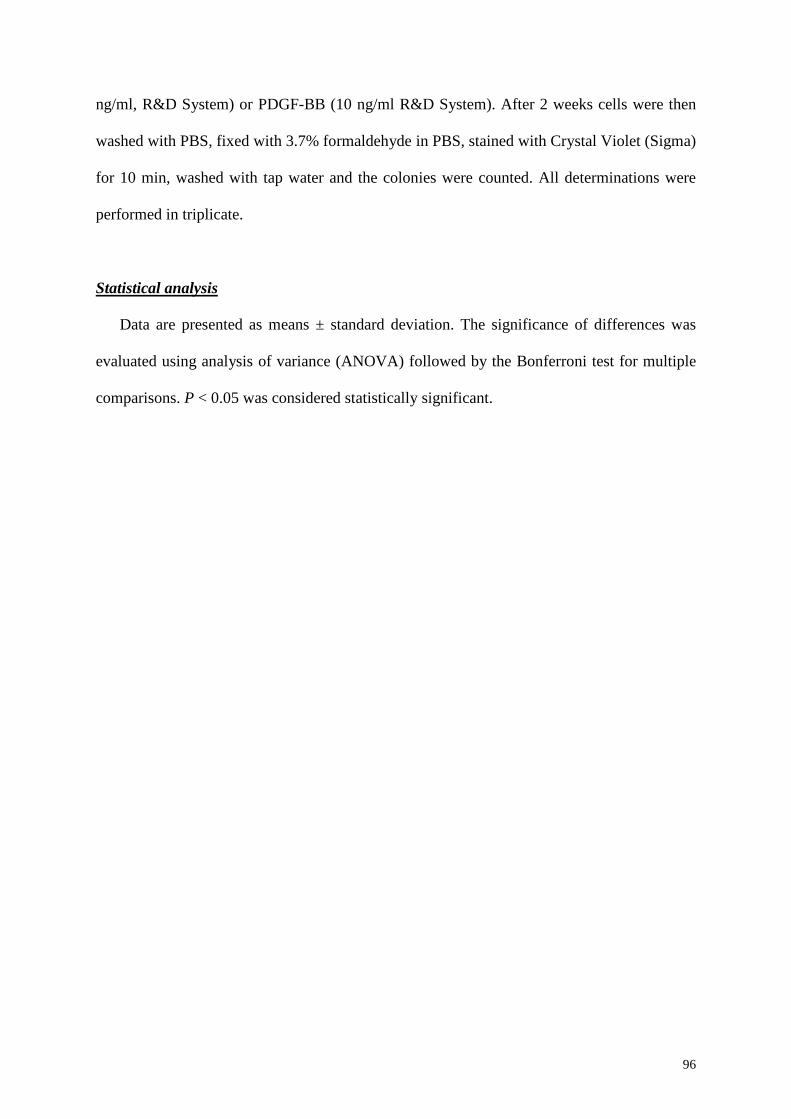
96
ng/ml, R&D System) or PDGF-BB (10 ng/ml R&D System). After 2 weeks cells were then
washed with PBS, fixed with 3.7% formaldehyde in PBS, stained with Crystal Violet (Sigma)
for 10 min, washed with tap water and the colonies were counted. All determinations were
performed in triplicate.
Statistical analysis
Data are presented as means ± standard deviation. The significance of differences was
evaluated using analysis of variance (ANOVA) followed by the Bonferroni test for multiple
comparisons. P < 0.05 was considered statistically significant.
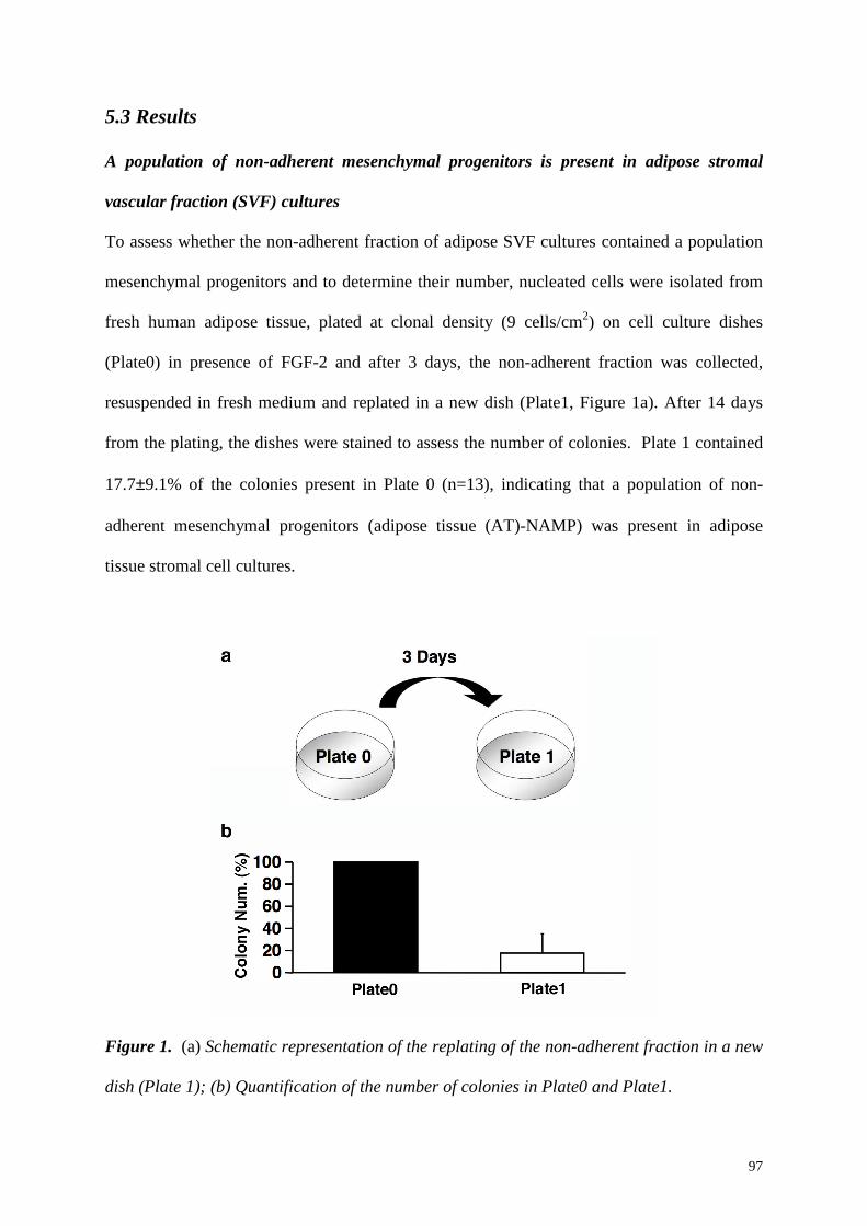
97
5.3 Results
A population of non-adherent mesenchymal progenitors is present in adipose stromal
vascular fraction (SVF) cultures
To assess whether the non-adherent fraction of adipose SVF cultures contained a population
mesenchymal progenitors and to determine their number, nucleated cells were isolated from
fresh human adipose tissue, plated at clonal density (9 cells/cm2) on cell culture dishes
(Plate0) in presence of FGF-2 and after 3 days, the non-adherent fraction was collected,
resuspended in fresh medium and replated in a new dish (Plate1, Figure 1a). After 14 days
from the plating, the dishes were stained to assess the number of colonies. Plate 1 contained
17.7±9.1% of the colonies present in Plate 0 (n=13), indicating that a population of non-
adherent mesenchymal progenitors (adipose tissue (AT)-NAMP) was present in adipose
tissue stromal cell cultures.
Figure 1. (a) Schematic representation of the replating of the non-adherent fraction in a new
dish (Plate 1); (b) Quantification of the number of colonies in Plate0 and Plate1.
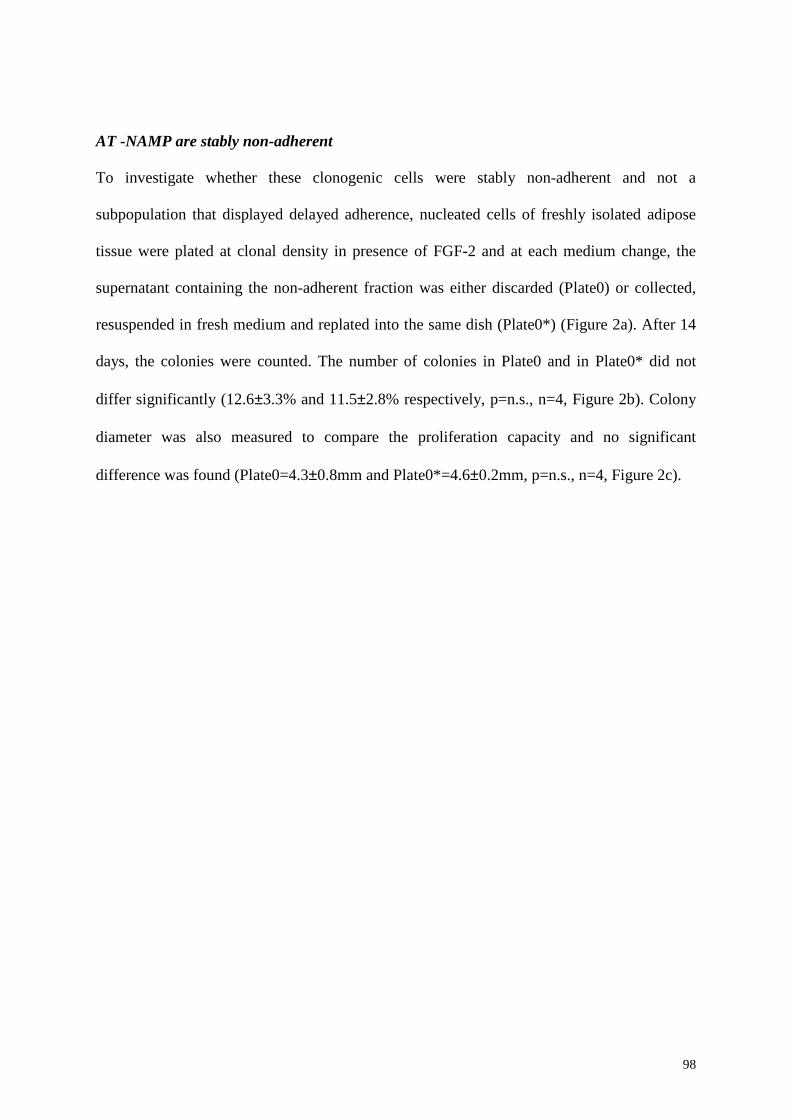
98
AT -NAMP are stably non-adherent
To investigate whether these clonogenic cells were stably non-adherent and not a
subpopulation that displayed delayed adherence, nucleated cells of freshly isolated adipose
tissue were plated at clonal density in presence of FGF-2 and at each medium change, the
supernatant containing the non-adherent fraction was either discarded (Plate0) or collected,
resuspended in fresh medium and replated into the same dish (Plate0*) (Figure 2a). After 14
days, the colonies were counted. The number of colonies in Plate0 and in Plate0* did not
differ significantly (12.6±3.3% and 11.5±2.8% respectively, p=n.s., n=4, Figure 2b). Colony
diameter was also measured to compare the proliferation capacity and no significant
difference was found (Plate0=4.3±0.8mm and Plate0*=4.6±0.2mm, p=n.s., n=4, Figure 2c).
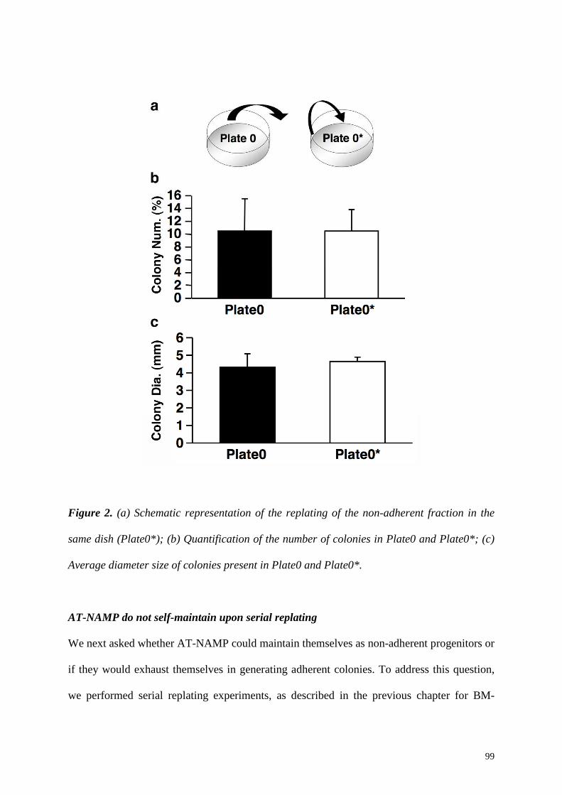
99
Figure 2. (a) Schematic representation of the replating of the non-adherent fraction in the
same dish (Plate0*); (b) Quantification of the number of colonies in Plate0 and Plate0*; (c)
Average diameter size of colonies present in Plate0 and Plate0*.
AT-NAMP do not self-maintain upon serial replating
We next asked whether AT-NAMP could maintain themselves as non-adherent progenitors or
if they would exhaust themselves in generating adherent colonies. To address this question,
we performed serial replating experiments, as described in the previous chapter for BM-
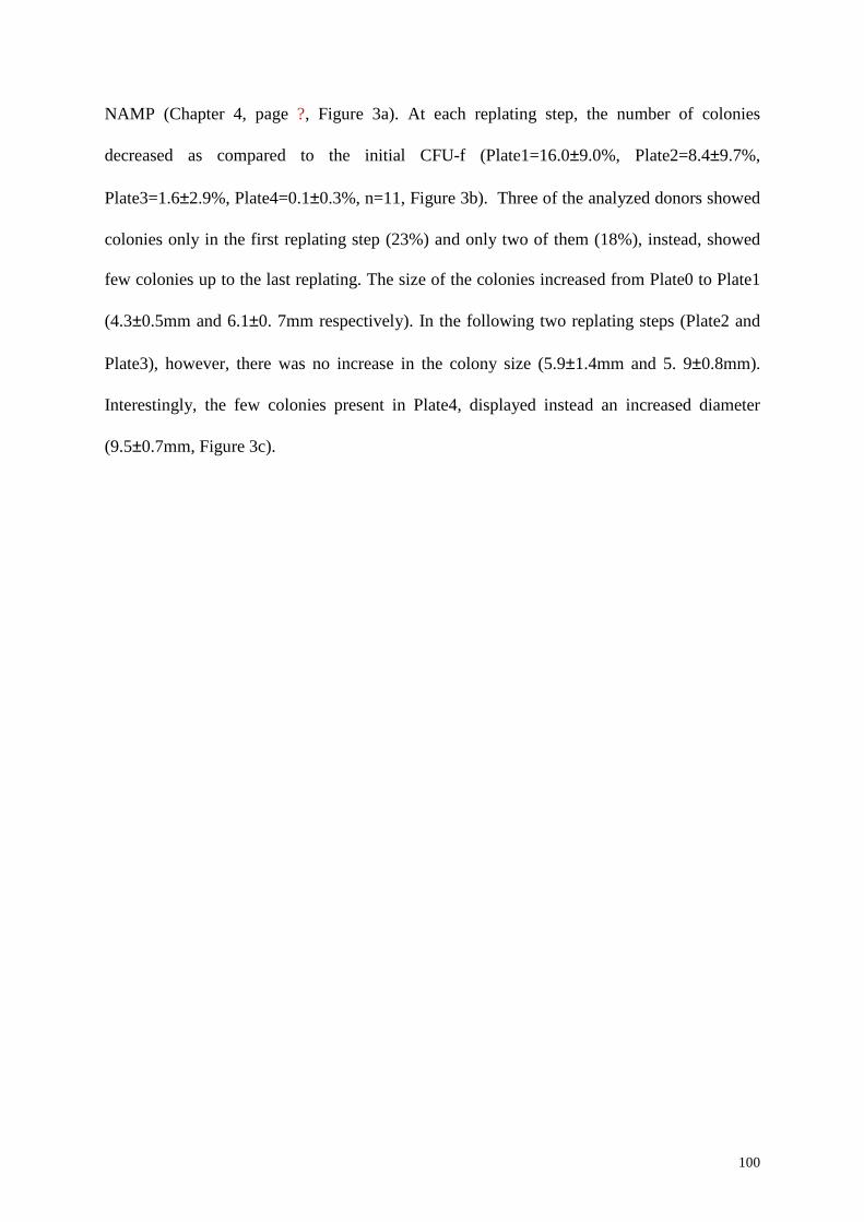
100
NAMP (Chapter 4, page ?, Figure 3a). At each replating step, the number of colonies
decreased as compared to the initial CFU-f (Plate1=16.0±9.0%, Plate2=8.4±9.7%,
Plate3=1.6±2.9%, Plate4=0.1±0.3%, n=11, Figure 3b). Three of the analyzed donors showed
colonies only in the first replating step (23%) and only two of them (18%), instead, showed
few colonies up to the last replating. The size of the colonies increased from Plate0 to Plate1
(4.3±0.5mm and 6.1±0. 7mm respectively). In the following two replating steps (Plate2 and
Plate3), however, there was no increase in the colony size (5.9±1.4mm and 5. 9±0.8mm).
Interestingly, the few colonies present in Plate4, displayed instead an increased diameter
(9.5±0.7mm, Figure 3c).
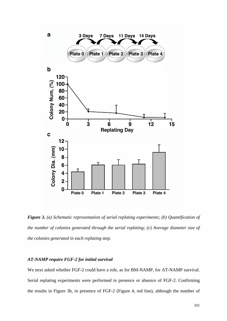
101
Figure 3. (a) Schematic representation of serial replating experiments; (b) Quantification of
the number of colonies generated through the serial replating; (c) Average diameter size of
the colonies generated in each replating step.
AT-NAMP require FGF-2 for initial survival
We next asked whether FGF-2 could have a role, as for BM-NAMP, for AT-NAMP survival.
Serial replating experiments were performed in presence or absence of FGF-2. Confirming
the results in Figure 3b, in presence of FGF-2 (Figure 4, red line), although the number of
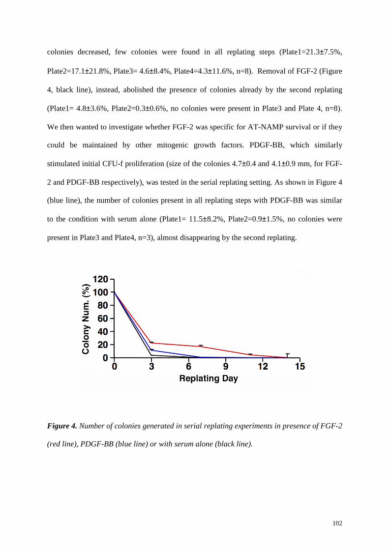
102
colonies decreased, few colonies were found in all replating steps (Plate1=21.3±7.5%,
Plate2=17.1±21.8%, Plate3= 4.6±8.4%, Plate4=4.3±11.6%, n=8). Removal of FGF-2 (Figure
4, black line), instead, abolished the presence of colonies already by the second replating
(Plate1= 4.8±3.6%, Plate2=0.3±0.6%, no colonies were present in Plate3 and Plate 4, n=8).
We then wanted to investigate whether FGF-2 was specific for AT-NAMP survival or if they
could be maintained by other mitogenic growth factors. PDGF-BB, which similarly
stimulated initial CFU-f proliferation (size of the colonies 4.7±0.4 and 4.1±0.9 mm, for FGF-
2 and PDGF-BB respectively), was tested in the serial replating setting. As shown in Figure 4
(blue line), the number of colonies present in all replating steps with PDGF-BB was similar
to the condition with serum alone (Plate1= 11.5±8.2%, Plate2=0.9±1.5%, no colonies were
present in Plate3 and Plate4, n=3), almost disappearing by the second replating.
Figure 4. Number of colonies generated in serial replating experiments in presence of FGF-2
(red line), PDGF-BB (blue line) or with serum alone (black line).
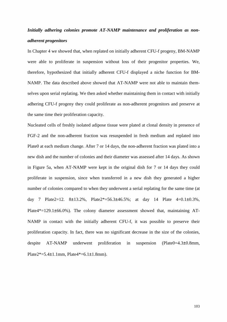
103
Initially adhering colonies promote AT-NAMP maintenance and proliferation as non-
adherent progenitors
In Chapter 4 we showed that, when replated on initially adherent CFU-f progeny, BM-NAMP
were able to proliferate in suspension without loss of their progenitor properties. We,
therefore, hypothesized that initially adherent CFU-f displayed a niche function for BM-
NAMP. The data described above showed that AT-NAMP were not able to maintain them-
selves upon serial replating. We then asked whether maintaining them in contact with initially
adhering CFU-f progeny they could proliferate as non-adherent progenitors and preserve at
the same time their proliferation capacity.
Nucleated cells of freshly isolated adipose tissue were plated at clonal density in presence of
FGF-2 and the non-adherent fraction was resuspended in fresh medium and replated into
Plate0 at each medium change. After 7 or 14 days, the non-adherent fraction was plated into a
new dish and the number of colonies and their diameter was assessed after 14 days. As shown
in Figure 5a, when AT-NAMP were kept in the original dish for 7 or 14 days they could
proliferate in suspension, since when transferred in a new dish they generated a higher
number of colonies compared to when they underwent a serial replating for the same time (at
day 7 Plate2=12. 8±13.2%, Plate2*=56.3±46.5%; at day 14 Plate 4=0.1±0.3%,
Plate4*=129.1±66.0%). The colony diameter assessment showed that, maintaining AT-
NAMP in contact with the initially adherent CFU-f, it was possible to preserve their
proliferation capacity. In fact, there was no significant decrease in the size of the colonies,
despite AT-NAMP underwent proliferation in suspension (Plate0=4.3±0.8mm,
Plate2*=5.4±1.1mm, Plate4*=6.1±1.8mm).
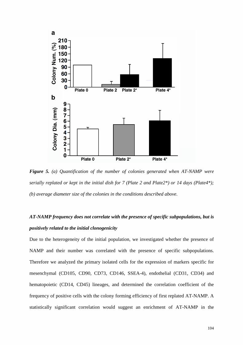
104
Figure 5. (a) Quantification of the number of colonies generated when AT-NAMP were
serially replated or kept in the initial dish for 7 (Plate 2 and Plate2*) or 14 days (Plate4*);
(b) average diameter size of the colonies in the conditions described above.
AT-NAMP frequency does not correlate with the presence of specific subpopulations, but is
positively related to the initial clonogenicity
Due to the heterogeneity of the initial population, we investigated whether the presence of
NAMP and their number was correlated with the presence of specific subpopulations.
Therefore we analyzed the primary isolated cells for the expression of markers specific for
mesenchymal (CD105, CD90, CD73, CD146, SSEA-4), endothelial (CD31, CD34) and
hematopoietic (CD14, CD45) lineages, and determined the correlation coefficient of the
frequency of positive cells with the colony forming efficiency of first replated AT-NAMP. A
statistically significant correlation would suggest an enrichment of AT-NAMP in the
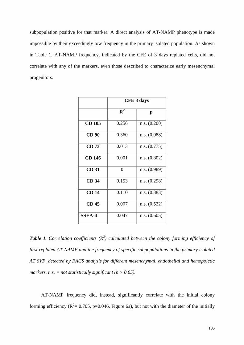
105
subpopulation positive for that marker. A direct analysis of AT-NAMP phenotype is made
impossible by their exceedingly low frequency in the primary isolated population. As shown
in Table 1, AT-NAMP frequency, indicated by the CFE of 3 days replated cells, did not
correlate with any of the markers, even those described to characterize early mesenchymal
progenitors.
CFE 3 days
R2 p
CD 105 0.256 n.s. (0.200)
CD 90 0.360 n.s. (0.088)
CD 73 0.013 n.s. (0.775)
CD 146 0.001 n.s. (0.802)
CD 31 0 n.s. (0.989)
CD 34 0.153 n.s. (0.298)
CD 14 0.110 n.s. (0.383)
CD 45 0.007 n.s. (0.522)
SSEA-4 0.047 n.s. (0.605)
Table 1. Correlation coefficients (R2) calculated between the colony forming efficiency of
first replated AT-NAMP and the frequency of specific subpopulations in the primary isolated
AT SVF, detected by FACS analysis for different mesenchymal, endothelial and hemopoietic
markers. n.s. = not statistically significant (p > 0.05).
AT-NAMP frequency did, instead, significantly correlate with the initial colony
forming efficiency (R2= 0.705, p=0.046, Figure 6a), but not with the diameter of the initially

106
adherent colonies (R2= 0.009, p=0.816, Figure 6b). This indicates that AT-NAMP are a fixed
fraction in the initial CFU-f population and that their frequency is independent from the
proliferation rate of initially adherent CFU-f.
Figure 6. Correlation plots between the CFE of three-days replated AT-NAMP and CFE (a),
or the colony size (b) of initially adhering CFU-f. Colony Dia.= colony diameter.
107
5.4 Summary and Conclusions
The results presented above show that a population of non-adherent mesenchymal
progenitors is also present in human adipose tissue SVF cultures.
Similarly to BM-NAMP, AT-NAMP did not simply display delayed adherence, but
were stably non-adherent, since the replating of the non-adherent fraction in the original dish
could not increase the number of colonies. Furthermore, no change in the colony size was
observed, indicating that the maintenance of the non-adherent fraction in the same dish did
not enrich the adherent cells with a highly proliferating subpopulation.
Upon serial replating, however, AT-NAMP could not maintain themselves. In fact,
unlike the experiments performed with BM-NAMP, the number of colonies steadily
decreased in each replating step, indicating that AT-NAMP were not able to maintain them-
selves as non-adherent progenitors, while at the same time forming adherent colonies.
Interestingly, the very few colonies found in the last replating step had a significantly
increased diameter. This suggests that, in these conditions, AT-NAMP may undergo a
selection for the very rare progenitors with the highest proliferation ability, but without an
actual amplification.
In Chapter 4 we have shown that BM-NAMP required FGF-2 for their initial selection
and their self-renewal as non-adherent progenitors. The experiments performed with the SVF
of adipose tissue indicated that FGF-2 plays an important role also for AT-NAMP survival,
without however supporting their self-renewal as non-adherent progenitors when serially
replated. As for BM-NAMP, PDGF-BB, although inducing a similar rate of proliferation of
the initially adhering CFU-f, was not able to sustain AT-NAMP survival at a level
comparable to FGF-2. This indicates that AT-NAMP frequency is not a function of the
proliferative status of the initially adhering CFU-f, excluding as well that they are a fraction
of the actively proliferating adherent cells, similarly to BM-NAMP.
108
Since serial replating experiments showed that AT-NAMP were not able to regenerate
as non-adherent progenitors when plated in an empty dish, we asked whether at least the
initially adhering CFU-f could provide a niche function to AT-NAMP, as was found for BM-
NAMP. The results indicated that AT-NAMP, when kept in contact with the initially forming
niche, could expand as non-adherent progenitors. The analysis of the colony diameters
showed that, despite this expansion, AT-NAMP fully preserved their proliferation capacity,
suggesting they were able to undergo self-renewing divisions in these conditions, and,
therefore, a niche function for the initially adhering CFU-f progeny.
AT-NAMP frequency did not correlate with the frequency of any specific
subpopulation in the freshly isolated SVF cells, but it was only positively related to the initial
CFE, indicating that they represent a fixed fraction of the initially adhering CFU-f.
Interestingly, AT-NAMP frequency did not show a correlation with the colony size of the
initial CFU-f, confirming that they do not represent a fraction of the most actively
proliferating adherent cells.
In conclusion, the presence of NAMP appears to be a common property of the
mesenchymal progenitor compartment of different tissues and not specific to bone marrow
stroma alone. However, the data showed in this chapter suggest some important tissue-
specific biological differences, particularly in their ability to regenerate their own niche and
therefore to self-renew upon serial replating.
References
1. Friedenstein, A.J., Chailakhyan, R.K., Latsinik, N.V., Panasyuk, A.F., and Keiliss-Borok, I.V. 1974. Stromal cells responsible for transferring the microenvironment of the hemopoietic tissues. Cloning in vitro and retransplantation in vivo. Transplantation 17:331-340.
2. Bianco, P., Robey, P.G., and Simmons, P.J. 2008. Mesenchymal stem cells: revisiting history, concepts, and assays. Cell Stem Cell 2:313-319.
109
Chapter 6:
A three-dimensional bioreactor-based culture system to engineer the
bone marrow niche
110
6.1 Introduction
6.1.1 Stem cell niches
Adult stem cells are responsible for the maintenance and turnover of several tissues.
The main features that characterize stem cells are the self-renewal, by which they are able to
maintain the size of the stem cell pool, and the potential to give rise to all the differentiated
cell types of one tissue. Adult stem cells can be extremely long-lived and display very low
proliferation rates under homeostatic conditions, constituting a reservoir that can be triggered
in case of stress or damage. Typically, stem cells reside in a specialized microenvironment,
the stem cell niche, which, apart from physically anchoring stem cells, also produces signals
necessary to regulate stem cell quiescence, self-renewal, differentiation and stress response.
Stem cells niches have been identified in many tissues including the brain (1), the skin (2),
the intestine (3) and the hematopoietic system.
Recently, a lot of progress has been done in localizing these microenvironments in vivo
in different tissues. Neural stem cells (NSC) have been localized close to endothelial cells,
rising the possibility that through the vasculature NSC can receive signals for quiescence or
activation (4). In the skin, epidermal stem cells are located below the epidermis and they can
be responsible for the turnover of the hair follicle, the sebaceous glands and the intrafollicular
epidermis (5), (6). In the intestine, instead, a novel population of stem cells, which is located
at the crypt base, has been recently identified (3). It has been shown that a population of cells
holding stem cell features, is present also in tendons and that it resides in a unique niche
composed by extracellualr matrix (7). These findings indicate that different cellular
components and extracellular substrates can play a crucial role in regulating the stem cell
function and that stem cell biology is moving the first steps towards identifying these
components and understanding the mechanisms of the interactions present in the niche.
111
Although stem cells of other tissues have been identified, the best-characterized adult
stem cell remains the hematopoietic stem cell (HSC). It has the clonal capacity to provide
life-long reconstitution of all hematopoietic lineages after transplantation into lethally
irradiated mice. In the bone marrow, HSC are found in the trabecular areas of the bones near
bone surfaces (endosteal niche) or associated with the sinusoidal endothelium (vascular
niche). The bone marrow niche has been the subject of several studies so far, and this allowed
the identification of its main components. Among the cellular players, the osteoblast, was
shown to be of great importance, since the modulation of its numbers led either to a decline
or to an increase in the numbers of HSCs in the bone marrow, showing its relevance for
controlling the niche size (8), (9), (10). However, other cell types, like osteoclasts,
endothelial cells and mesenchymal progenitors (reviewed in (11)) seem to be involved in the
regulation of HSC fate.
Also molecules produced by bone marrow stromal cells can influence stem cell
behavior, including membrane bound SCF (12), (13), osteopontin (14), (15) angiopoietin (8),
TPO (16) and SDF-1 (17). Furthermore, several adhesion molecules, such as VCAM-1 (18)
and N-cadherin (8), present on osteoblasts, have also been indicated in HSC support.
In addition, other physical factors are very important for HSC regulation. Results from
calcium sensing receptor knock-out mice have shown that a low Ca2+ environment is favored
by HSCs and the inability to sense it lead to a decline of HSC numbers and increased
mobilization to the periphery (19). Moreover, HSCs are preferentially found in the regions of
the bone containing least oxygen, suggesting that the niche for quiescent HSCs is hypoxic
(20), (21).
These observations indicate that the bone marrow niche displays a complex structure, in
which interactions are crucial to maintain HSC function. Up to date, a lot of studies aimed to
recreate the in vivo conditions, mainly for HSC expansion for clinical purposes, without,
112
however, succeeding in maintaining HSC in culture. In fact, standard 2D HSC culture applys
a stromal feeder layer as surrogate of the niche, being, however, far from reproducing the in
vivo conditions. In the next sections, we will illustrate the state of the art regarding HSC in
vitro culture, underlining the issues which remain still controversial. We will also describe
alternative culture methods which have been developed in past to overcome the limitations of
standard 2D cultures, highlighting the still existing need of new 3D culture systems to
reproduce the complex niche organization.
6.1.2 Standard HSC culture in vitro
Hematopoietic stem cells are the basis of bone marrow transplantation and are
attractive target cells for hematopoietic gene therapy. However, these important clinical
applications have been severely hampered by the limiting numbers of HSC available for
transplantation, which are isolated for example from mobilized blood or umbilical cord
blood. Therefore, the ex vivo expansion of functional HSC is crucial. Furthermore, a
functioning ex vivo human hematopoietic system could serve as an analytical model to study
the basic biology of hematopoiesis and HSC niche interactions. Such a test system would also
provide an alternative to animal studies, with the added benefit of generating data from
human cells.
To date there is quite a large body of studies on this subject. However, although many
different media compositions and culture conditions have been tested, expansion of
functional repopulating HSC in sufficient numbers for transplantation has remained
inefficient. One major drawback in comparing the numerous studies is the evaluation of stem
cell activity. The gold standard for HSC function is their ability to engraft in a host and to
maintain hematopoiesis over a long period of time (> 6 months) or during serial transplants.
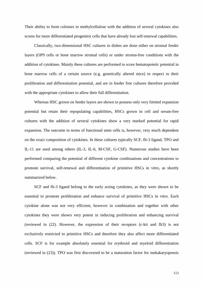
113
Their ability to form colonies in methylcellulose with the addition of several cytokines also
scores for more differentiated progenitor cells that have already lost self-renewal capabilities.
Classically, two-dimensional HSC cultures in dishes are done either on stromal feeder
layers (OP9 cells or bone marrow stromal cells) or under stroma-free conditions with the
addition of cytokines. Mainly these cultures are performed to score hematopoietic potential in
bone marrow cells of a certain source (e.g. genetically altered mice) in respect to their
proliferation and differentiation potential, and are in feeder free cultures therefore provided
with the appropriate cytokines to allow their full differentiation.
Whereas HSC grown on feeder layers are shown to possess only very limited expansion
potential but retain their repopulating capabilities, HSCs grown in cell and serum-free
cultures with the addition of several cytokines show a very marked potential for rapid
expansion. The outcome in terms of functional stem cells is, however, very much dependent
on the exact composition of cytokines. In these cultures typically SCF, flt-3 ligand, TPO and
IL-11 are used among others (IL-3, IL-6, M-CSF, G-CSF). Numerous studies have been
performed comparing the potential of different cytokine combinations and concentrations to
promote survival, self-renewal and differentiation of primitive HSCs in vitro, as shortly
summarized below.
SCF and flt-3 ligand belong to the early acting cytokines, as they were shown to be
essential to promote proliferation and enhance survival of primitive HSCs in vitro. Each
cytokine alone was not very efficient, however in combination and together with other
cytokines they were shown very potent in inducing proliferation and enhancing survival
(reviewed in (22). However, the expression of their receptors (c-kit and flt3) is not
exclusively restricted to primitive HSCs and therefore they also affect more differentiated
cells. SCF is for example absolutely essential for erythroid and myeloid differentiation
(reviewed in (23)). TPO was first discovered to be a maturation factor for mekakaryopoesis
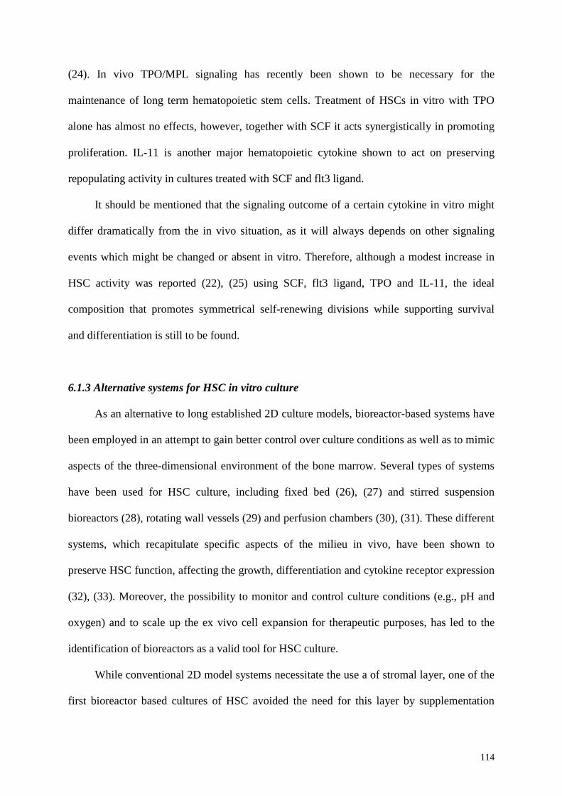
114
(24). In vivo TPO/MPL signaling has recently been shown to be necessary for the
maintenance of long term hematopoietic stem cells. Treatment of HSCs in vitro with TPO
alone has almost no effects, however, together with SCF it acts synergistically in promoting
proliferation. IL-11 is another major hematopoietic cytokine shown to act on preserving
repopulating activity in cultures treated with SCF and flt3 ligand.
It should be mentioned that the signaling outcome of a certain cytokine in vitro might
differ dramatically from the in vivo situation, as it will always depends on other signaling
events which might be changed or absent in vitro. Therefore, although a modest increase in
HSC activity was reported (22), (25) using SCF, flt3 ligand, TPO and IL-11, the ideal
composition that promotes symmetrical self-renewing divisions while supporting survival
and differentiation is still to be found.
6.1.3 Alternative systems for HSC in vitro culture
As an alternative to long established 2D culture models, bioreactor-based systems have
been employed in an attempt to gain better control over culture conditions as well as to mimic
aspects of the three-dimensional environment of the bone marrow. Several types of systems
have been used for HSC culture, including fixed bed (26), (27) and stirred suspension
bioreactors (28), rotating wall vessels (29) and perfusion chambers (30), (31). These different
systems, which recapitulate specific aspects of the milieu in vivo, have been shown to
preserve HSC function, affecting the growth, differentiation and cytokine receptor expression
(32), (33). Moreover, the possibility to monitor and control culture conditions (e.g., pH and
oxygen) and to scale up the ex vivo cell expansion for therapeutic purposes, has led to the
identification of bioreactors as a valid tool for HSC culture.
While conventional 2D model systems necessitate the use a of stromal layer, one of the
first bioreactor based cultures of HSC avoided the need for this layer by supplementation
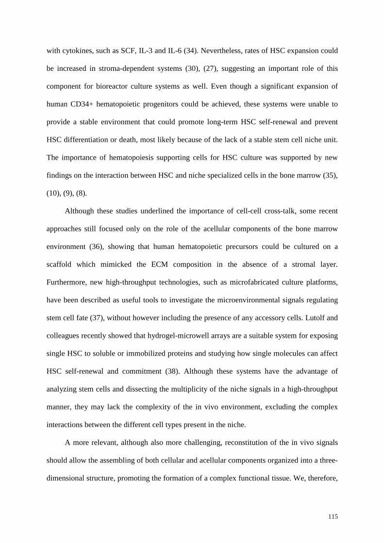
115
with cytokines, such as SCF, IL-3 and IL-6 (34). Nevertheless, rates of HSC expansion could
be increased in stroma-dependent systems (30), (27), suggesting an important role of this
component for bioreactor culture systems as well. Even though a significant expansion of
human CD34+ hematopoietic progenitors could be achieved, these systems were unable to
provide a stable environment that could promote long-term HSC self-renewal and prevent
HSC differentiation or death, most likely because of the lack of a stable stem cell niche unit.
The importance of hematopoiesis supporting cells for HSC culture was supported by new
findings on the interaction between HSC and niche specialized cells in the bone marrow (35),
(10), (9), (8).
Although these studies underlined the importance of cell-cell cross-talk, some recent
approaches still focused only on the role of the acellular components of the bone marrow
environment (36), showing that human hematopoietic precursors could be cultured on a
scaffold which mimicked the ECM composition in the absence of a stromal layer.
Furthermore, new high-throughput technologies, such as microfabricated culture platforms,
have been described as useful tools to investigate the microenvironmental signals regulating
stem cell fate (37), without however including the presence of any accessory cells. Lutolf and
colleagues recently showed that hydrogel-microwell arrays are a suitable system for exposing
single HSC to soluble or immobilized proteins and studying how single molecules can affect
HSC self-renewal and commitment (38). Although these systems have the advantage of
analyzing stem cells and dissecting the multiplicity of the niche signals in a high-throughput
manner, they may lack the complexity of the in vivo environment, excluding the complex
interactions between the different cell types present in the niche.
A more relevant, although also more challenging, reconstitution of the in vivo signals
should allow the assembling of both cellular and acellular components organized into a three-
dimensional structure, promoting the formation of a complex functional tissue. We, therefore,

116
propose a three-dimensional culture system composed by a 3D scaffold and a perfusion
bioreactor system as a potential tool to reconstruct in vitro the bone marrow niche
environment. Unlike the approaches used in the past, we aim not only to the expand HSC ex-
vivo, but to establish a stable and more complete stem cell niche unit, composed by cellular
and acellular compartments. The system has the advantage of providing a more physiological
three-dimensional structure combined with a biomaterial, which can modulate both HSC
function and influence niche specialized cells differentiation.
In the next sections we will highlight the advantages of this system to modulate
different variables for HSC culture and we will give a perspective about the implications of
the development of this system both for scientific and clinical applications.
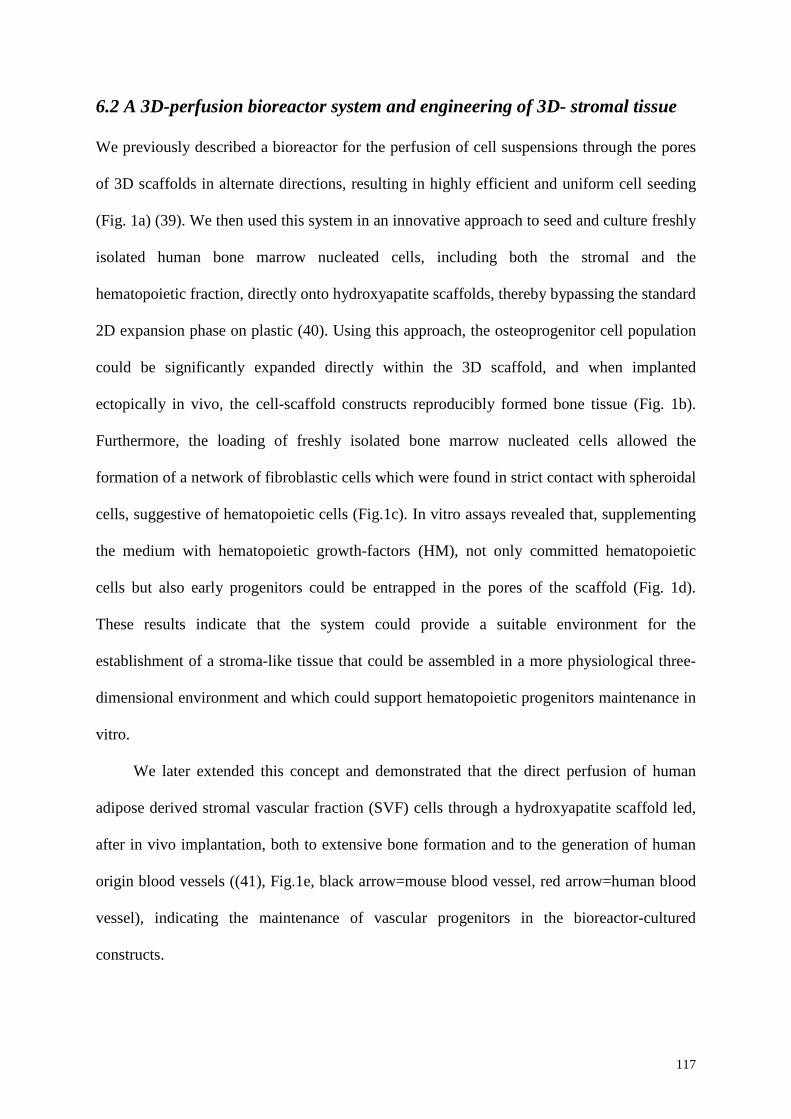
117
6.2 A 3D-perfusion bioreactor system and engineering of 3D- stromal tissue
We previously described a bioreactor for the perfusion of cell suspensions through the pores
of 3D scaffolds in alternate directions, resulting in highly efficient and uniform cell seeding
(Fig. 1a) (39). We then used this system in an innovative approach to seed and culture freshly
isolated human bone marrow nucleated cells, including both the stromal and the
hematopoietic fraction, directly onto hydroxyapatite scaffolds, thereby bypassing the standard
2D expansion phase on plastic (40). Using this approach, the osteoprogenitor cell population
could be significantly expanded directly within the 3D scaffold, and when implanted
ectopically in vivo, the cell-scaffold constructs reproducibly formed bone tissue (Fig. 1b).
Furthermore, the loading of freshly isolated bone marrow nucleated cells allowed the
formation of a network of fibroblastic cells which were found in strict contact with spheroidal
cells, suggestive of hematopoietic cells (Fig.1c). In vitro assays revealed that, supplementing
the medium with hematopoietic growth-factors (HM), not only committed hematopoietic
cells but also early progenitors could be entrapped in the pores of the scaffold (Fig. 1d).
These results indicate that the system could provide a suitable environment for the
establishment of a stroma-like tissue that could be assembled in a more physiological three-
dimensional environment and which could support hematopoietic progenitors maintenance in
vitro.
We later extended this concept and demonstrated that the direct perfusion of human
adipose derived stromal vascular fraction (SVF) cells through a hydroxyapatite scaffold led,
after in vivo implantation, both to extensive bone formation and to the generation of human
origin blood vessels ((41), Fig.1e, black arrow=mouse blood vessel, red arrow=human blood
vessel), indicating the maintenance of vascular progenitors in the bioreactor-cultured
constructs.
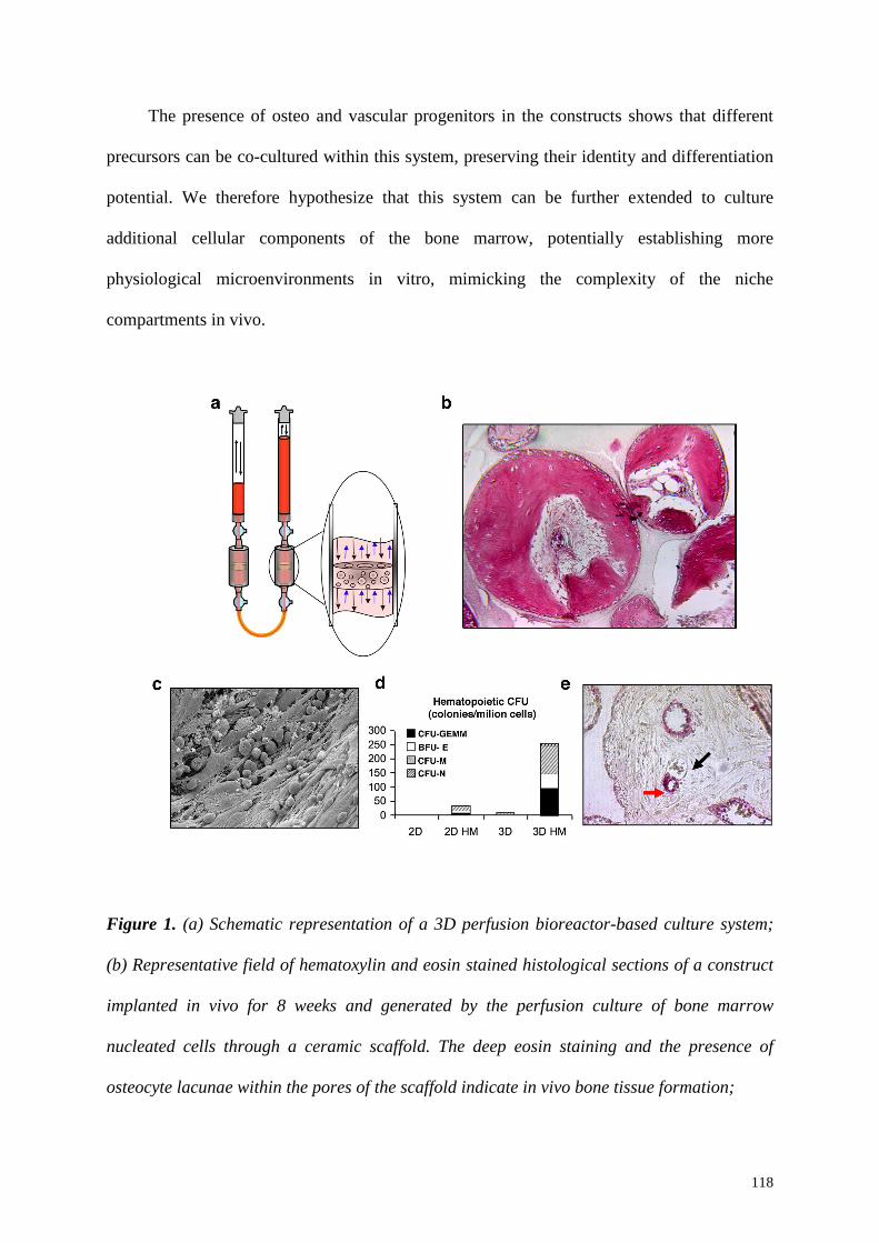
118
The presence of osteo and vascular progenitors in the constructs shows that different
precursors can be co-cultured within this system, preserving their identity and differentiation
potential. We therefore hypothesize that this system can be further extended to culture
additional cellular components of the bone marrow, potentially establishing more
physiological microenvironments in vitro, mimicking the complexity of the niche
compartments in vivo.
Figure 1. (a) Schematic representation of a 3D perfusion bioreactor-based culture system;
(b) Representative field of hematoxylin and eosin stained histological sections of a construct
implanted in vivo for 8 weeks and generated by the perfusion culture of bone marrow
nucleated cells through a ceramic scaffold. The deep eosin staining and the presence of
osteocyte lacunae within the pores of the scaffold indicate in vivo bone tissue formation;
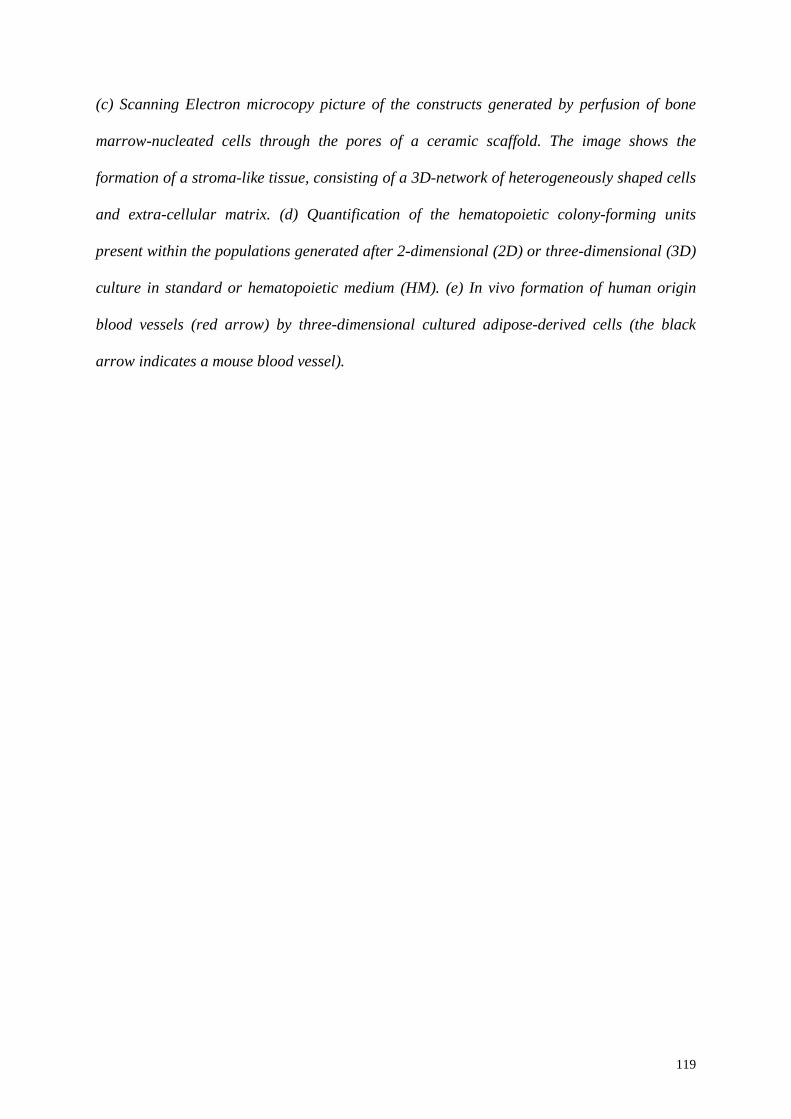
119
(c) Scanning Electron microcopy picture of the constructs generated by perfusion of bone
marrow-nucleated cells through the pores of a ceramic scaffold. The image shows the
formation of a stroma-like tissue, consisting of a 3D-network of heterogeneously shaped cells
and extra-cellular matrix. (d) Quantification of the hematopoietic colony-forming units
present within the populations generated after 2-dimensional (2D) or three-dimensional (3D)
culture in standard or hematopoietic medium (HM). (e) In vivo formation of human origin
blood vessels (red arrow) by three-dimensional cultured adipose-derived cells (the black
arrow indicates a mouse blood vessel).

120
6.3 A 3D-perfusion bioreactor based system for HSC culture: a possible
experimental design
The application of a three-dimensional bioreactor-based system to reconstruct the niche
environment represents a fascinating challenge and it would require a careful experimental
design, which takes in consideration the heterogeneous culture requirements of the different
cellular and extra-cellular components of the bone marrow organization. In this section we
would like to propose a possible basic experimental design for applying this innovative
culture system to reproduce the niche interactions ex-vivo.
It would be important to first establish a stromal environment, in order to provide pre-
formed niches to promote HSC homing and avoid differentiation. This would imply the
division of the experiment in two distinct phases. In Phase-1, bone marrow nucleated cells
could be harvested from transgenic mice, for example from Bcl-2 mice in order to minimize
cell death, loaded onto a 3D scaffold and cultured under perfusion for 4 weeks, to allow
stromal tissue formation (Figure 2a, red arrows indicate the presence of a stromal layer on the
scaffold surface). In Phase-2, LSK cells could be sorted from the bone marrow pool of GFP
mice and loaded onto the pre-established stroma. The positivity for GFP would facilitate their
tracking and characterization. After 2 weeks, cells could be harvested from the constructs and
the hematopoietic GFP+ cells could be analyzed for the presence of stem cell markers, like
SCA-1 and c-KIT, to assess the maintenance of a stem phenotype (Figure 2b). In combination
with the analysis of surface markers, further assays, in vitro and in vivo, would clearly be
required to assess the maintenance of the stem cell function. Moreover,
immunohistochemical characterization of engineered tissue cross-sections could allow to
investigate specific cell-cell and cell-matrix interactions in the stromal-hematopoietic co-
culture system, as a starting point to formulate and test hypotheses related to bone marrow
stem cell niches.
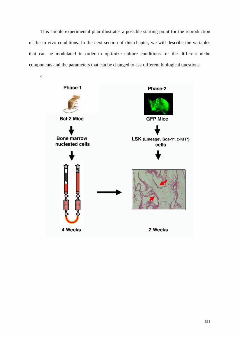
121
This simple experimental plan illustrates a possible starting point for the reproduction
of the in vivo conditions. In the next section of this chapter, we will describe the variables
that can be modulated in order to optimize culture conditions for the different niche
components and the parameters that can be changed to ask different biological questions.
a
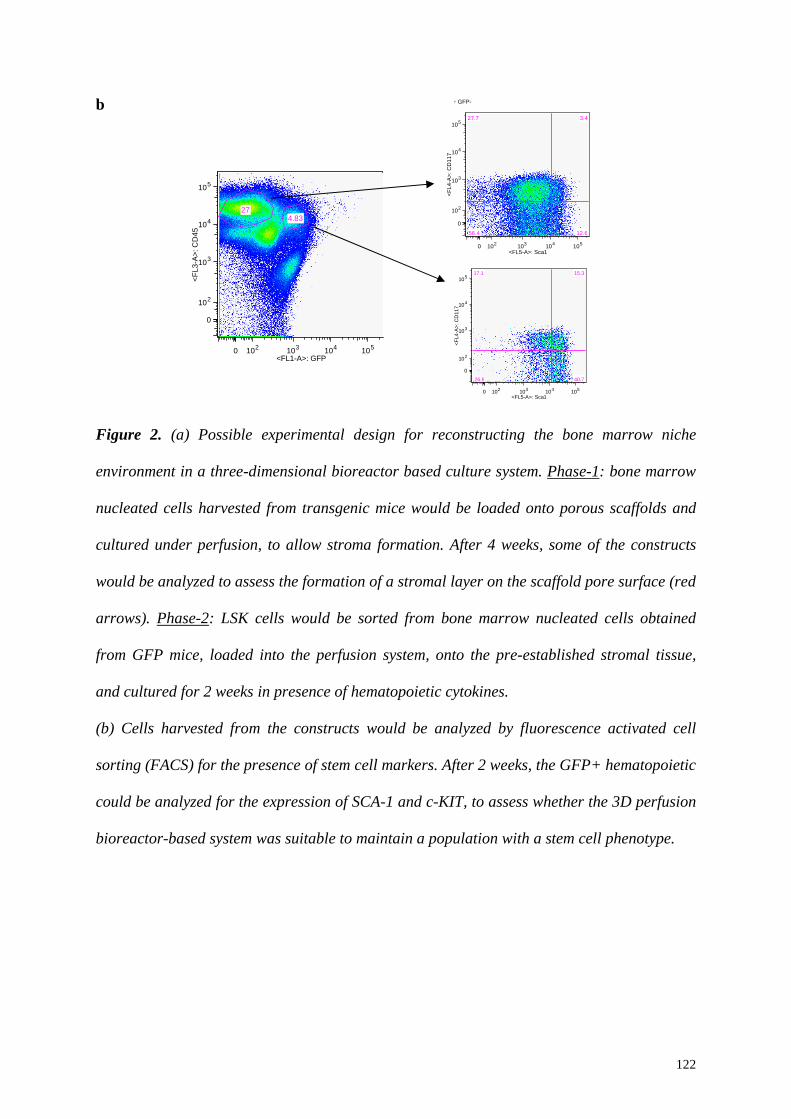
122
b
Figure 2. (a) Possible experimental design for reconstructing the bone marrow niche
environment in a three-dimensional bioreactor based culture system. Phase-1: bone marrow
nucleated cells harvested from transgenic mice would be loaded onto porous scaffolds and
cultured under perfusion, to allow stroma formation. After 4 weeks, some of the constructs
would be analyzed to assess the formation of a stromal layer on the scaffold pore surface (red
arrows). Phase-2: LSK cells would be sorted from bone marrow nucleated cells obtained
from GFP mice, loaded into the perfusion system, onto the pre-established stromal tissue,
and cultured for 2 weeks in presence of hematopoietic cytokines.
(b) Cells harvested from the constructs would be analyzed by fluorescence activated cell
sorting (FACS) for the presence of stem cell markers. After 2 weeks, the GFP+ hematopoietic
could be analyzed for the expression of SCA-1 and c-KIT, to assess whether the 3D perfusion
bioreactor-based system was suitable to maintain a population with a stem cell phenotype.
0 102 103 104 105
0
102
103
104
105
Élin low
<FL1-A>: GFP
<F
L3-A
>: C
D45
4.8327
0 102 103 104 105
0
102
103
104
105
ÉCD45+ GFP-
<FL5-A>: Sca1
<F
L4-A
>: C
D11
7
27.7 3.4
12.656.4
0 102 103 104 105
0
102
103
104
105
ÉCD45+ GFP+
<FL5-A>: Sca1
<FL4
-A>:
CD
117
17.1 15.3
40.726.8
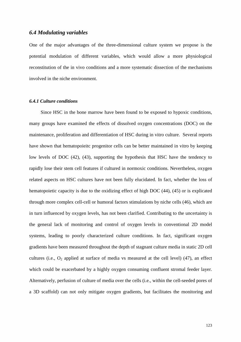
123
6.4 Modulating variables
One of the major advantages of the three-dimensional culture system we propose is the
potential modulation of different variables, which would allow a more physiological
reconstitution of the in vivo conditions and a more systematic dissection of the mechanisms
involved in the niche environment.
6.4.1 Culture conditions
Since HSC in the bone marrow have been found to be exposed to hypoxic conditions,
many groups have examined the effects of dissolved oxygen concentrations (DOC) on the
maintenance, proliferation and differentiation of HSC during in vitro culture. Several reports
have shown that hematopoieitc progenitor cells can be better maintained in vitro by keeping
low levels of DOC (42), (43), supporting the hypothesis that HSC have the tendency to
rapidly lose their stem cell features if cultured in normoxic conditions. Nevertheless, oxygen
related aspects on HSC cultures have not been fully elucidated. In fact, whether the loss of
hematopoietic capacity is due to the oxidizing effect of high DOC (44), (45) or is explicated
through more complex cell-cell or humoral factors stimulations by niche cells (46), which are
in turn influenced by oxygen levels, has not been clarified. Contributing to the uncertainty is
the general lack of monitoring and control of oxygen levels in conventional 2D model
systems, leading to poorly characterized culture conditions. In fact, significant oxygen
gradients have been measured throughout the depth of stagnant culture media in static 2D cell
cultures (i.e., O2 applied at surface of media vs measured at the cell level) (47), an effect
which could be exacerbated by a highly oxygen consuming confluent stromal feeder layer.
Alternatively, perfusion of culture of media over the cells (i.e., within the cell-seeded pores of
a 3D scaffold) can not only mitigate oxygen gradients, but facilitates the monitoring and
124
control of oxygen levels in the culture via in-line sensors, and thus, the establishment of a
well-defined and controlled model system.
Similarly to oxygen concentration, pH may affect HSC proliferation and differentiation
in vitro (48). Even if also in this case the molecular mechanism of pH influence on HSC has
not been totally elucidated yet, several observations support the idea that pH should be
maintained in physiological ranges to assure HSC maintenance in vitro. To deepen the study
on pH effects, and to monitor pH changes over the whole period of culture, we propose an
approach equivalent to the one proposed for oxygen measurements, based on non-invasive
sensors able to continuously measure the pH during the culture. Ideally, the sensors could be
interfaced with the incubator in order to change the CO2 percentage to keep constant the pH
at a set value; similarly, a sensor based oxygen control can be implemented to modify the
DOC based on the experimental setup. This will therefore allow to monitor and modulate
these variables during the culture, adjusting them constantly.
Our bioreactor system has, therefore, the potential to overcome the limitations of
traditional cell cultures in terms of monitoring physical parameters like oxygen and pH,
allowing the establishment of a more controlled culture setting and contributing in recreating
a more physiological environment.
Shear stress has been described as another factor which can influence HSC
maintenance in vitro. The lack of suitable instruments to directly measure local shear stress
for HSC cultures has so far avoided the clear definition of optimal, or at least well-tolerated,
shear stress forces. In bioreactor systems, it has been described that high stirring speeds can
negatively affect HSC maintenance and proliferation, indicating that HSC are relatively
sensitive to shear stress. However, the impact of shear stress on MSC has been more
extensively investigated leading to the conclusion that controlled shear stress can promote
MSC proliferation and differentiation (49), (50), (51). Combining the effects of shear stress

125
on MSC and HSC when co-cultured represents a fascinating challenge that will require
specific culture systems with a defined geometry, a scaffold with definite properties, and
possibly computerized fluid dynamic simulations (CFD) to more easily predict local stress
values.
According to earlier works on in vitro HSC co-cultured with stromal cells, the feeding
rate and the medium exchange show to play a role in the maintenance and proliferation
potential of hematopoietic progenitors (52). Apparently, the beneficial effect derives by the
active removal of cell produced negative regulators (53), but other reasons like the addition
of a limiting nutrient, a soluble component or grow factors, and the removal of detrimental
waste products cannot be excluded. As a confirmation of these hypothesises, frequent
medium exchange represented a great advantage for hematopoietic precursors culture both in
static (54), (55) and dynamic conditions (30). In this regard, the high degree of flexibility of
our perfusion bioreactor for different medium volumes and for the configuration of the
components gives the system an increased valuable advantage over traditional supports for
static cell culture in terms of adaptability to different exchange schedules.
6.4.2 Scaffold composition
Another feature that makes attractive this system we propose is the presence of a three-
dimensional scaffold, whose composition can be varied in order to investigate the influence
of the material in this experimental setting.
In the past years, three-dimensional scaffolds have been mainly applied for tissue
engineering purposes, but recently, they start as well to receive attention for answering basic
questions related to stem cell biology.

126
Beside biocompatible, scaffolds are usually designed to be porous in order to increase
the available surface, guarantee proper adhesion sites, to favour the maintenance of three-
dimensional shape of the cells and facilitate cell-cell spatial relations.
As reviewed by Panoskaltsis in (56), tissue-engineering biomaterials can be grouped
based on their biological or synthetic derivation. Some of them, both natural and synthetic,
have been tested for HSC culture: macroporous collagen carriers (57), cellulose porous
microspheres (58), porous polyvinyl formal (PVF), nonwoven polyethylene terephthalate
(PET) (59), porous biomatrix (Cellfoam ™), porous gelatin microspheres (CultiSpher G),
nylon filtration screen (60) and polyester nonwoven fabric porous disc carriers (Fibra-cel)
(58).
Moreover, Rosenzweig (61), proposed a model in which hematopoietic stem cell
maintenance was achieved in a tantalum coated porous biomaterial (TCPB) without the use
of cytokines. Although the molecular mechanism was not disclosed in the paper, it opened
many enquiries on the different biological effects of 3-dimensional materials, supporting the
hypothesis that mimicking the 3-dimensional microenvironment of the bone marrow can play
a crucial role in supporting progenitor viability and pluripotency.
In an attempt to evaluate the influence of physical properties of the scaffold on
hematopoietic differentiation of Embryonic Stem Cells (ESC), Taqvi (62) tested a poly(L-
lactic acid) (PLLA) porous scaffolds. The scaffold was manufactured with different polymer
concentrations and pore sizes. The results provided some evidences that a higher seeding cell
density, small pore size (<150 µm), increased mechanical stiffness and coculture with stromal
cells, provide increased ESC differentiation into the hematopoietic lineage.
Beside focusing on the modulation of the pore size and material composition, other
approaches have been tested. The functionalization of the scaffold with molecules which can
affect cell behaviour and which can function as active biochemical stimuli, recently received
127
attention. Jiang and colleagues (63) have demonstrated the efficacy of functionalized PET to
expand umbilical cord blood CD34+ cells. In this study, the connecting segment-1 (CS-1)
and RGD motifs were covalently bound to PET and the resulting material was compared to
traditional polystyrene surfaces. The peptides mimicked the fibronectin domains of the ECM
and the results showed that peptide-functionalised surfaces were more suitable to sustain cell
expansion compared to non-functionalized polystyrene. This suggested the importance of the
reconstruction of the ECM to elucidate which adhesion molecules or insoluble factors have a
crucial role in maintaining HSC characteristics.
It would be important to combine these observations with the data on the influence of
the different scaffold compositions on the cells, like osteoblasts and endothelial cells, which
are supposed to have a regulatory role in the bone marrow niche environment. Furthermore,
the possibility of modifying and functionalizing the materials currently used in tissue
engineering (like ceramics, natural and synthetic polymers, composites, decellularized bone
grafts, various coatings on solid or semi-solid substrates) should be driven by the new
findings about the molecules which influence HSC function in vivo. Moreover, it should be
taken in consideration that scaffold materials can exert their influence on HSC in two distinct
manners: by direct contact or indirectly, affecting the differentiation state or the proliferation
of other cell types which have a supportive function. This would lead to a paradigm shift, to
create a brand new class of biomaterials, customizing, according to the needs, the
environment for HSC culture.
These observations indicate the importance of testing a three-dimensional setting with
different material compositions to clarify the basic mechanisms which regulate HSC function
in the niche. Our system provides the unique possibility to control culture variables, as
oxygen and pH, combined with the perfusion through a three-dimensional scaffold, holding a
great advantage compared to standard HSC culture systems.
128
6.4.3 Co-culture of different cell types
As mentioned before, both osteo- and endothelial progenitors could be maintained in
our perfusion-based culture system, indicating that this system offers proper conditions for
different progenitors co-culture (41). Recently, several studies have shown that multiple
niche compartments are present in vivo and that more than one cell type is involved in
regulating HSC function in vivo. Beside osteoblasts (10), (9) and endothelial cellsm (64), also
osteoclasts (65), perivascular cell, like reticular cells and megakaryocytes (66), and
mesenchymal progenitors (67) have been recently described as supportive cells for
hematopoiesis in vivo. In the past years, 2D co-cultures of osteblasts, osteoclast and/or
endothelial cells were performed to study the interactions between these cell types, in order to
clarify the mechanisms involved in bone regeneration and remodeling. The co-culture of
different cell types for reconstructing the complex environment and reproducing the
physiological cross-talk of the niche, instead, still represents a new perspective in stem cell
biology. Compared to standard 2D culture systems, the advantage of a 3D-perfusion
bioreactor system is represented by the three-dimensional structure, which can allow a three-
dimensional assembling of the different cellular players and matrix deposition, and by the
perfusion, which provides a proper diffusion of the released growth factors. The aim would
be, therefore, to establish the culture requirements for the growth of the different niche
cellular components, in order to promote a correct assembly and distribution of the different
niche compartments within the three-dimensional architecture of the scaffold. To achieve this
purpose, not only the most obvious of the variables, i.e. the medium composition, but also
others, like the ones listed in this section, should be considered. It would be important, in
fact, to choose a cell source according to the cell composition and also the proportion
between the different cell types, progenitors and mature cells, since this can be of
fundamental importance for recreating a well-balanced niche environment. Furthermore, cell
129
composition can be modulated modifying genetically a defined cellular compartment, in
order to produce growth factors for the maintenance of other specialized niche cells, offering
a wide spectrum of conditions to test. In addition, the design of the biomaterial could include
a mixed composition, which can promote in distinct areas of the construct the development of
different niche compartments.
An intriguing consequence of a complete reconstruction of the bone marrow niche
environment would be the establishment of a self-maintaining system, which doesn’t depend
on external supply of cytokines for HSC preservation. This would overcome the issues
correlated with the use of recombinant cytokines in the perspective of a clinical application,
coupled with the advantages of reducing the costs of expensive medium components.
Among the several positive aspects of this bioreactor system, a possible drawback
could seem to be represented by the complexity related to the high number of variables
implied. However, the system we propose offers the possibility to systematically test different
conditions in parallel. In would be feasible in this way to modulate the level of complexity,
starting to analyze one variable at the time, with the possibility to introduce in the following
steps other variables and, in the end, reconstruct the in vivo conditions.
130
6.5 Future perspectives
In the previous sections of this chapter we highlighted the potential of a three-
dimensional bioreactor-based system for the ex-vivo reconstruction of the complex bone
marrow environment. This system offers the advantage of combining a highly controllable
culture system and a three-dimensional scaffold, which could provide a suitable environment
to promote the correct assembly of the niche cellular components and a physiological extra-
cellular matrix deposition. This would lead to the establishment of engineered stem cell niche
units, offering, on one hand, a useful system to overcome limitations linked to currently
available stem cell expansion for stem-cell based therapies, but also, on the other, a relevant
tool to investigate the interactions between HSC and their regulatory environment.
Bone marrow transplantation is the only treatment by which many cases of
hematopoietic malignancies can be cured. However, the amount of HSC that can be
transplanted is limited by the amount that can be harvested, since their in vitro expansion is
not currently possible without loss of their stemness. In this regard, an application of the
system we propose for ex-vivo expansion of HSC, especially if derived from umbilical cord
blood, generally associated with high quality but limited quantity, would be extremely useful.
In fact, a three-dimensional reconstitution of all the components which form the niche in vivo
would offer a greater possibility, compared to standard feeder layers, to establish the
physiological signaling cross-talk between HSC and its niches, leading to the maintenance of
their stem cell function. The final goal would be, however, not only to engineer quiescent
niches, but also microenvironments which support self-renewing divisions in order to allow
HSC expansion. Furthermore, the regulation of HSC by a stable stem cell niche unit would
not require the external supplementation with cytokines, since the correct signals and growth
factors for HSC maintenance and expansion will be provided by the surrounding
environment. This will have direct implications for all situations in which HSC number is not
131
sufficient for transplantation, as well as for the reduction of the time required for T-cell
reconstitution after bone marrow transplantation.
As indicated above, another field of application of the proposed culture system will be
to offer a modular platform to clarify the mechanisms by which the niche regulates HSC.
Besides investigating normal homeostatic conditions, it would be important to study the niche
role in pathological conditions. In fact, recent findings raised the possibility that, as for HSC,
also the survival of malignant cells is supported by the niche environment. Acute myeloid
leukemia (AML) has a hierarchical organization similar to that of normal hematopoiesis,
where a small subpopulation of cancer cells, called leukemia stem cells (LSC), holds the
ability to initiate the disease. LSC are characterized by a more mature phenotype compared to
HSC (68), but they acquired limitless self-renewal through oncogenic transformation (69).
Some studies have shown that LSC receive signals from the niche that support self-renewal,
exploiting the mechanisms involved in the long-term maintenance of HSC them-selves.
Furthermore, it has been shown that the microenvironment is important for LSC engraftment,
chemotherapy resistance and cell cycle regulation (70). Treatments currently available for
AML only result in a cure in less than 50% of the cases (71). The recent findings regarding
the interaction between LSC and the niche open the possibility of identifying new targets for
AML therapy. These approaches should be directed to selectively stop LSC growth by
interfering and disrupting their interactions with the niche environment and, at the same time,
they should preserve normal hematopoiesis to ensure HSC regeneration. In vivo studies
should be combined with in vitro screening in order to investigate the mechanisms involved
in a controlled setting, such as the three-dimensional culture system we propose. Compared
to in vivo studies in mice, it has the potential advantage of combining cells harvested from
different strains of transgenic mice with human cells. It would, therefore, represent a
132
powerful system for dissecting the mechanisms related to LSC maintenance and a useful
platform for drug screening and validation.
HSC are the so far best-characterized stem cells, providing also a framework for the
study of other stem cells, and significant progress has been done in the past few years in
advancing our knowledge about the bone marrow stem cell niche. Recently, regulatory
environments have been identified also for stem cells of other tissues, highlighting the
increasing need for culture systems which better reproduce the complexity of the niche. In
many tissues, like the intestinal epithelium and the hair follicle, stem cells occupy fixed
positions within the niche, suggesting that the spatial organization can play an important role
in the explication of niche function. In the thymic niche, T cell progenitors undergo
expansion and progress through programmed stages of T cell development, critically
dependent upon interactions with specific thymic stromal compartments. Thymocytes, on the
other hand, contribute to the development and maintenance of the thymic stroma in a bi-
directional cross-talk (72). Furthermore, it has been shown that in this setting, the there-
dimensional environment is of essential importance to enable migration of T cell precursors
through the stroma (73). The combination of the culture system we propose and an
appropriate scaffold which sustains the development of a specialized thymic stroma, would
constitute an attractive model for creating an ex-vivo thymus. This would help clarify, for
example, the mechanisms involved in the disruption of the architectural structure of the
thymic niche caused by aging process.
In Chapter 3 of this thesis, we described the in vitro establishment of a niche/progenitor
system for the mesenchymal compartment. As mentioned before for other niche systems, a
deeper mechanistic insight into the regulation of the maintenance of MSC earlier progenitors
will also require the reconstruction of a three-dimensional bone marrow organization. This
would significantly contribute to clarify several aspects of MSC biology, offering in addition
133
the possibility to investigate how the co-culture with other niche cells and the scaffold
composition can modulate their properties. Furthermore, another application of this system
would be to evaluate the potential influence of MSC on cancer stem cells. Recent evidence
suggests, in fact, that MSC can migrate towards primary and metastatic sites of several solid
tumors and play a role in tumor progression. A major limitation in the analysis of stroma-
tumor interaction is represented by the lack of reliable models. A three-dimensional culture
system combined to a porous scaffold would span the gap between 2D cultures, which don’t
reproduce the complexity of the in vivo environment, and animal models, which, although
more physiological, do not allow to address the role of individual cell populations present at
the tumor site. Furthermore, the establishment of a physiological stroma will offer a relevant
model for identifying targets for the treatment of bone-related disorders and for testing the
effect of new therapeutic molecules on bone remodeling. It will also have direct implications
in the tissue-engineering field allowing the maintenance of a physiological pool of functional
progenitors, like osteo and endothelial progenitors, necessary for the successful generation of
vascularized bone grafts.
These observations point out the potential and the versatility of this innovative culture
system, in reproducing the complex interactions of the niche microenvironment. This can
have a significant impact on regenerative medicine applications, facilitating the development
of stem-cell-based therapies.
134
References
1. Doetsch, F., Caille, I., Lim, D.A., Garcia-Verdugo, J.M., and Alvarez-Buylla, A. 1999. Subventricular zone astrocytes are neural stem cells in the adult mammalian brain. Cell 97:703-716.
2. Tumbar, T., Guasch, G., Greco, V., Blanpain, C., Lowry, W.E., Rendl, M., and Fuchs, E. 2004. Defining the epithelial stem cell niche in skin. Science 303:359-363.
3. Barker, N., van Es, J.H., Kuipers, J., Kujala, P., van den Born, M., Cozijnsen, M., Haegebarth, A., Korving, J., Begthel, H., Peters, P.J., et al. 2007. Identification of stem cells in small intestine and colon by marker gene Lgr5. Nature 449:1003-1007.
4. Palmer, T.D., Willhoite, A.R., and Gage, F.H. 2000. Vascular niche for adult hippocampal neurogenesis. J Comp Neurol 425:479-494.
5. Ito, M., Liu, Y., Yang, Z., Nguyen, J., Liang, F., Morris, R.J., and Cotsarelis, G. 2005. Stem cells in the hair follicle bulge contribute to wound repair but not to homeostasis of the epidermis. Nat Med 11:1351-1354.
6. Levy, V., Lindon, C., Harfe, B.D., and Morgan, B.A. 2005. Distinct stem cell populations regenerate the follicle and interfollicular epidermis. Dev Cell 9:855-861.
7. Bi, Y., Ehirchiou, D., Kilts, T.M., Inkson, C.A., Embree, M.C., Sonoyama, W., Li, L., Leet, A.I., Seo, B.M., Zhang, L., et al. 2007. Identification of tendon stem/progenitor cells and the role of the extracellular matrix in their niche. Nat Med 13:1219-1227.
8. Arai, F., Hirao, A., Ohmura, M., Sato, H., Matsuoka, S., Takubo, K., Ito, K., Koh, G.Y., and Suda, T. 2004. Tie2/angiopoietin-1 signaling regulates hematopoietic stem cell quiescence in the bone marrow niche. Cell 118:149-161.
9. Calvi, L.M., Adams, G.B., Weibrecht, K.W., Weber, J.M., Olson, D.P., Knight, M.C., Martin, R.P., Schipani, E., Divieti, P., Bringhurst, F.R., et al. 2003. Osteoblastic cells regulate the haematopoietic stem cell niche. Nature 425:841-846.
10. Zhang, J., Niu, C., Ye, L., Huang, H., He, X., Tong, W.G., Ross, J., Haug, J., Johnson, T., Feng, J.Q., et al. 2003. Identification of the haematopoietic stem cell niche and control of the niche size. Nature 425:836-841.
11. Jones, D.L., and Wagers, A.J. 2008. No place like home: anatomy and function of the stem cell niche. Nat Rev Mol Cell Biol 9:11-21.
12. Barker, J.E. 1994. Sl/Sld hematopoietic progenitors are deficient in situ. Exp Hematol 22:174-177.
13. Barker, J.E. 1997. Early transplantation to a normal microenvironment prevents the development of Steel hematopoietic stem cell defects. Exp Hematol 25:542-547.
14. Nilsson, S.K., Johnston, H.M., Whitty, G.A., Williams, B., Webb, R.J., Denhardt, D.T., Bertoncello, I., Bendall, L.J., Simmons, P.J., and Haylock, D.N. 2005. Osteopontin, a key component of the hematopoietic stem cell niche and regulator of primitive hematopoietic progenitor cells. Blood 106:1232-1239.
15. Stier, S., Ko, Y., Forkert, R., Lutz, C., Neuhaus, T., Grunewald, E., Cheng, T., Dombkowski, D., Calvi, L.M., Rittling, S.R., et al. 2005. Osteopontin is a hematopoietic stem cell niche component that negatively regulates stem cell pool size. J Exp Med 201:1781-1791.
135
16. Yoshihara, H., Arai, F., Hosokawa, K., Hagiwara, T., Takubo, K., Nakamura, Y., Gomei, Y., Iwasaki, H., Matsuoka, S., Miyamoto, K., et al. 2007. Thrombopoietin/MPL signaling regulates hematopoietic stem cell quiescence and interaction with the osteoblastic niche. Cell Stem Cell 1:685-697.
17. Wright, N., Hidalgo, A., Rodriguez-Frade, J.M., Soriano, S.F., Mellado, M., Parmo-Cabanas, M., Briskin, M.J., and Teixido, J. 2002. The chemokine stromal cell-derived factor-1 alpha modulates alpha 4 beta 7 integrin-mediated lymphocyte adhesion to mucosal addressin cell adhesion molecule-1 and fibronectin. J Immunol 168:5268-5277.
18. Jung, Y., Wang, J., Havens, A., Sun, Y., Jin, T., and Taichman, R.S. 2005. Cell-to-cell contact is critical for the survival of hematopoietic progenitor cells on osteoblasts. Cytokine 32:155-162.
19. Adams, G.B., Chabner, K.T., Alley, I.R., Olson, D.P., Szczepiorkowski, Z.M., Poznansky, M.C., Kos, C.H., Pollak, M.R., Brown, E.M., and Scadden, D.T. 2006. Stem cell engraftment at the endosteal niche is specified by the calcium-sensing receptor. Nature 439:599-603.
20. Jang, Y.Y., and Sharkis, S.J. 2007. A low level of reactive oxygen species selects for primitive hematopoietic stem cells that may reside in the low-oxygenic niche. Blood 110:3056-3063.
21. Parmar, K., Mauch, P., Vergilio, J.A., Sackstein, R., and Down, J.D. 2007. Distribution of hematopoietic stem cells in the bone marrow according to regional hypoxia. Proc Natl Acad Sci U S A 104:5431-5436.
22. Lyman, S.D., and Jacobsen, S.E. 1998. c-kit ligand and Flt3 ligand: stem/progenitor cell factors with overlapping yet distinct activities. Blood 91:1101-1134.
23. Broudy, V.C. 1997. Stem cell factor and hematopoiesis. Blood 90:1345-1364.
24. Kaushansky, K., Broudy, V.C., Lin, N., Jorgensen, M.J., McCarty, J., Fox, N., Zucker-Franklin, D., and Lofton-Day, C. 1995. Thrombopoietin, the Mp1 ligand, is essential for full megakaryocyte development. Proc Natl Acad Sci U S A 92:3234-3238.
25. Sauvageau, G., Iscove, N.N., and Humphries, R.K. 2004. In vitro and in vivo expansion of hematopoietic stem cells. Oncogene 23:7223-7232.
26. Meissner, P., Schroder, B., Herfurth, C., and Biselli, M. 1999. Development of a fixed bed bioreactor for the expansion of human hematopoietic progenitor cells. Cytotechnology 30:227-234.
27. Noll, T., Jelinek, N., Schmid, S., Biselli, M., and Wandrey, C. 2002. Cultivation of hematopoietic stem and progenitor cells: biochemical engineering aspects. Adv Biochem Eng Biotechnol 74:111-128.
28. Zandstra, P.W., Jervis, E., Haynes, C.A., Kilburn, D.G., Eaves, C.J., and Piret, J.M. 1999. Concentration-dependent internalization of a cytokine/cytokine receptor complex in human hematopoietic cells. Biotechnol Bioeng 63:493-501.
29. Liu, Y., Liu, T., Fan, X., Ma, X., and Cui, Z. 2006. Ex vivo expansion of hematopoietic stem cells derived from umbilical cord blood in rotating wall vessel. J Biotechnol 124:592-601.

136
30. Koller, M.R., Emerson, S.G., and Palsson, B.O. 1993. Large-scale expansion of human stem and progenitor cells from bone marrow mononuclear cells in continuous perfusion cultures. Blood 82:378-384.
31. Koller, M.R., Manchel, I., Maher, R.J., Goltry, K.L., Armstrong, R.D., and Smith, A.K. 1998. Clinical-scale human umbilical cord blood cell expansion in a novel automated perfusion culture system. Bone Marrow Transplant 21:653-663.
32. Nielsen, L.K. 1999. Bioreactors for hematopoietic cell culture. Annu Rev Biomed Eng 1:129-152.
33. McDowell, C.L., and Papoutsakis, E.T. 1998. Increased agitation intensity increases CD13 receptor surface content and mRNA levels, and alters the metabolism of HL60 cells cultured in stirred tank bioreactors. Biotechnol Bioeng 60:239-250.
34. Haylock, D.N., To, L.B., Dowse, T.L., Juttner, C.A., and Simmons, P.J. 1992. Ex vivo expansion and maturation of peripheral blood CD34+ cells into the myeloid lineage. Blood 80:1405-1412.
35. Scadden, D.T. 2006. The stem-cell niche as an entity of action. Nature 441:1075-1079.
36. Cho, C.H., Eliason, J.F., and Matthew, H.W. 2008. Application of porous glycosaminoglycan-based scaffolds for expansion of human cord blood stem cells in perfusion culture. J Biomed Mater Res A 86:98-107.
37. Underhill, G.H., and Bhatia, S.N. 2007. High-throughput analysis of signals regulating stem cell fate and function. Curr Opin Chem Biol 11:357-366.
38. Lutolf, M.P., Doyonnas, R., Havenstrite, K., Koleckar, K., and Blau, H.M. 2009. Perturbation of sinlge hematopoietic stem cell fates in artificial niches. Integr Biol 1:59.
39. Wendt, D., Marsano, A., Jakob, M., Heberer, M., and Martin, I. 2003. Oscillating perfusion of cell suspensions through three-dimensional scaffolds enhances cell seeding efficiency and uniformity. Biotechnol Bioeng 84:205-214.
40. Braccini, A., Wendt, D., Jaquiery, C., Jakob, M., Heberer, M., Kenins, L., Wodnar-Filipowicz, A., Quarto, R., and Martin, I. 2005. Three-dimensional perfusion culture of human bone marrow cells and generation of osteoinductive grafts. Stem Cells 23:1066-1072.
41. Scherberich, A., Galli, R., Jaquiery, C., Farhadi, J., and Martin, I. 2007. Three-dimensional perfusion culture of human adipose tissue-derived endothelial and osteoblastic progenitors generates osteogenic constructs with intrinsic vascularization capacity. Stem Cells 25:1823-1829.
42. Danet, G.H., Pan, Y., Luongo, J.L., Bonnet, D.A., and Simon, M.C. 2003. Expansion of human SCID-repopulating cells under hypoxic conditions. J Clin Invest 112:126-135.
43. Ivanovic, Z., Dello Sbarba, P., Trimoreau, F., Faucher, J.L., and Praloran, V. 2000. Primitive human HPCs are better maintained and expanded in vitro at 1 percent oxygen than at 20 percent. Transfusion 40:1482-1488.
44. Broxmeyer, H.E., Cooper, S., and Gabig, T. 1989. The effects of oxidizing species derived from molecular oxygen on the proliferation in vitro of human granulocyte-macrophage progenitor cells. Ann N Y Acad Sci 554:177-184.
137
45. Meagher, R.C., Salvado, A.J., and Wright, D.G. 1988. An analysis of the multilineage production of human hematopoietic progenitors in long-term bone marrow culture: evidence that reactive oxygen intermediates derived from mature phagocytic cells have a role in limiting progenitor cell self-renewal. Blood 72:273-281.
46. LaIuppa, J.A., Papoutsakis, E.T., and Miller, W.M. 1998. Oxygen tension alters the effects of cytokines on the megakaryocyte, erythrocyte, and granulocyte lineages. Exp Hematol 26:835-843.
47. Pettersen, E.O., Larsen, L.H., Ramsing, N.B., and Ebbesen, P. 2005. Pericellular oxygen depletion during ordinary tissue culturing, measured with oxygen microsensors. Cell Prolif 38:257-267.
48. McAdams, T.A., Miller, W.M., and Papoutsakis, E.T. 1997. Variations in culture pH affect the cloning efficiency and differentiation of progenitor cells in ex vivo haemopoiesis. Br J Haematol 97:889-895.
49. Kapur, S., Baylink, D.J., and Lau, K.H. 2003. Fluid flow shear stress stimulates human osteoblast proliferation and differentiation through multiple interacting and competing signal transduction pathways. Bone 32:241-251.
50. Bancroft, G.N., Sikavitsas, V.I., van den Dolder, J., Sheffield, T.L., Ambrose, C.G., Jansen, J.A., and Mikos, A.G. 2002. Fluid flow increases mineralized matrix deposition in 3D perfusion culture of marrow stromal osteoblasts in a dose-dependent manner. Proc Natl Acad Sci U S A 99:12600-12605.
51. Stolberg, S., and McCloskey, K.E. 2009. Can shear stress direct stem cell fate? Biotechnol Prog 25:10-19.
52. Dexter, T.M., Spooncer, E., Simmons, P., and Allen, T.D. 1984. Long-term marrow culture: an overview of techniques and experience. Kroc Found Ser 18:57-96.
53. Madlambayan, G.J., Rogers, I., Purpura, K.A., Ito, C., Yu, M., Kirouac, D., Casper, R.F., and Zandstra, P.W. 2006. Clinically relevant expansion of hematopoietic stem cells with conserved function in a single-use, closed-system bioprocess. Biol Blood Marrow Transplant 12:1020-1030.
54. Simmons, P.J., and Haylock, D.N. 1995. Use of hematopoietic growth factors for in vitro expansion of precursor cell populations. Curr Opin Hematol 2:189-195.
55. Koller, M.R., Palsson, M.A., Manchel, I., and Palsson, B.O. 1995. Long-term culture-initiating cell expansion is dependent on frequent medium exchange combined with stromal and other accessory cell effects. Blood 86:1784-1793.
56. Panoskaltsis, N., Mantalaris, A., and Wu, J.H. 2005. Engineering a mimicry of bone marrow tissue ex vivo. J Biosci Bioeng 100:28-35.
57. Wang, C., Kasuga, T., and Nogami, M. 2005. Macroporous calcium phosphate glass-ceramic prepared by two-step pressing technique and using sucrose as a pore former. J Mater Sci Mater Med 16:739-744.
58. Tomimori, Y., Takagi, M., and Yoshida, T. 2000. The construction of an in vitro three-dimensional hematopoietic microenvironment for mouse bone marrow cells employing porous carriers. Cytotechnology 34:121-130.
59. Li, Y., Ma, T., Yang, S.T., and Kniss, D.A. 2001. Thermal compression and characterization of three-dimensional nonwoven PET matrices as tissue engineering scaffolds. Biomaterials 22:609-618.
138
60. Naughton, B.A., Tjota, A., Sibanda, B., and Naughton, G.K. 1991. Hematopoiesis on suspended nylon screen-stromal cell microenvironments. J Biomech Eng 113:171-177.
61. Rosenzweig, M., Pykett, M., Marks, D.F., and Johnson, R.P. 1997. Enhanced maintenance and retroviral transduction of primitive hematopoietic progenitor cells using a novel three-dimensional culture system. Gene Ther 4:928-936.
62. Taqvi, S., and Roy, K. 2006. Influence of scaffold physical properties and stromal cell coculture on hematopoietic differentiation of mouse embryonic stem cells. Biomaterials 27:6024-6031.
63. Jiang, X.S., Chai, C., Zhang, Y., Zhuo, R.X., Mao, H.Q., and Leong, K.W. 2006. Surface-immobilization of adhesion peptides on substrate for ex vivo expansion of cryopreserved umbilical cord blood CD34+ cells. Biomaterials 27:2723-2732.
64. Kiel, M.J., Yilmaz, O.H., Iwashita, T., Terhorst, C., and Morrison, S.J. 2005. SLAM family receptors distinguish hematopoietic stem and progenitor cells and reveal endothelial niches for stem cells. Cell 121:1109-1121.
65. Kollet, O., Dar, A., Shivtiel, S., Kalinkovich, A., Lapid, K., Sztainberg, Y., Tesio, M., Samstein, R.M., Goichberg, P., Spiegel, A., et al. 2006. Osteoclasts degrade endosteal components and promote mobilization of hematopoietic progenitor cells. Nat Med 12:657-664.
66. Sugiyama, T., Kohara, H., Noda, M., and Nagasawa, T. 2006. Maintenance of the hematopoietic stem cell pool by CXCL12-CXCR4 chemokine signaling in bone marrow stromal cell niches. Immunity 25:977-988.
67. Sacchetti, B., Funari, A., Michienzi, S., Di Cesare, S., Piersanti, S., Saggio, I., Tagliafico, E., Ferrari, S., Robey, P.G., Riminucci, M., et al. 2007. Self-renewing osteoprogenitors in bone marrow sinusoids can organize a hematopoietic microenvironment. Cell 131:324-336.
68. Somervaille, T.C., and Cleary, M.L. 2006. Identification and characterization of leukemia stem cells in murine MLL-AF9 acute myeloid leukemia. Cancer Cell 10:257-268.
69. Krivtsov, A.V., Twomey, D., Feng, Z., Stubbs, M.C., Wang, Y., Faber, J., Levine, J.E., Wang, J., Hahn, W.C., Gilliland, D.G., et al. 2006. Transformation from committed progenitor to leukaemia stem cell initiated by MLL-AF9. Nature 442:818-822.
70. Ishikawa, F., Yoshida, S., Saito, Y., Hijikata, A., Kitamura, H., Tanaka, S., Nakamura, R., Tanaka, T., Tomiyama, H., Saito, N., et al. 2007. Chemotherapy-resistant human AML stem cells home to and engraft within the bone-marrow endosteal region. Nat Biotechnol 25:1315-1321.
71. Harrison, T.R., and Fauci, A.S. 1998. Harrison's principles of internal medicine. New York: McGraw-Hill Health Profession Division. 1 v. (various pagings) pp.
72. van Ewijk, W., Shores, E.W., and Singer, A. 1994. Crosstalk in the mouse thymus. Immunol Today 15:214-217.
73. van Ewijk, W., Wang, B., Hollander, G., Kawamoto, H., Spanopoulou, E., Itoi, M., Amagai, T., Jiang, Y.F., Germeraad, W.T., Chen, W.F., et al. 1999. Thymic microenvironments, 3-D versus 2-D? Semin Immunol 11:57-64.
139
Future Perspectives
The identification of a subpopulation of early progenitors in the mesenchymal compartment
offers direct implications for tissue engineering applications. In fact, NAMP progeny cells
can be used as an additional powerful source of mesenchymal progenitors. NAMP increased
proliferation and differentiation potential could help overcome the limitations of expanded
MSC. Since they can expand on their niche without losing differentiation potential, they can
greatly improve the the quality of engineered grafts.
However, the existence of a niche/progenitor system for the mesenchymal compartment
also opens interesting perspectives for the elucidation of different aspects of the biology of
MSC. Although the data described in this thesis provided the first evidence of the presence of
a specialized regulatory environment for MSC, further investigation is needed to identify the
pathways involved in the maintenance of the early-progenitor features and to clarify how this
niche mediates its function in vivo. In our experimental setting, self-renewal was defined as
the ability to undergo expansion without loss of proliferation and differentiation potential.
However, the self-renewal was not assessed in vivo, as the reconstitution of a stem cell
compartment. The next steps should clarify whether, when implanted in vivo, NAMP can
produce bone tissue and, at the same time, maintain themselves as an actual reservoir for the
mesenchymal compartment. Ideally, this would employ serial transplantation experiments, in
order to determine whether the bone tissue formed in vivo by NAMP contains a self-renewed
NAMP compartment that can be re-isolated and re-implanted.
A parallel direction would be to investigate the mechanisms involved in the regulation
of NAMP maintenance. This would require the identification of the signaling pathways
involved. Initial targets would be chosen by analogy with other well-characterized stem cell
systems, i.e. Notch and Wnt pathways, which have been shown to control self-renewal,
asymmetric divisions and cell fate decisions in hematopoietic and neural stem cells (5, 6).
140
This would offer a mechanistic insight into NAMP biology, but it would also have practical
implications. In fact, the clarification of the mechanisms involved in NAMP maintenance
would offer the opportunity to identify novel molecular targets to stimulate the endogenous
mesenchymal stem cell compartment, leading to pharmacogic treatments for bone
degenerative diseases, like osteoporosis.
One of the major limitations of standard in vitro systems applied to the study of the
mechanisms regulating the niche is their marked difference from in vivo conditions. In fact,
the reconstruction of active niches in vitro would probably require more than a feeder layer
and a mix of different cytokines. In Chapter 6 of this thesis, we illustrated the potential of a
three-dimensional bioreactor-based culture system for the ex-vivo reconstruction of the
complex bone marrow environment. Compared to standard 2-dimensional cultures, this
system offers the advantage of combining a highly controllable culture system and a three-
dimensional scaffold, which could provide a suitable environment to promote the correct
assembly of the niche cellular components and a physiological extra-cellular matrix
deposition. The development of such a system would offer a unique tool to investigate the
interactions between hematopoietic stem cells and their regulatory environment.
It was previously reported that the culture of bone marrow nucleated cells in the system
we described led to the formation of highly osteoinducitve grafts (7). The data described in
Chapter 4 regarding the existence of highly potent non-adherent progenitors within the MSC
population can offer a plausible model explaining the increased bone formation. In fact, we
hypothesize that NAMP can be entrapped in the three-dimensional network formed within the
pores of the scaffold, instead of being discarded in the early phases of the 2D culture. This
would promote the establishment of a progenitor/niche system, allowing NAMP to undergo
self-renewing expansion before committing to the osteogenic lineage. These observations,

141
therefore, highlight the importance of a three-dimensional culture system also for the
investigation of the mechanisms involved in NAMP maintenance.
The establishment of a physiological stroma will have two main implications: 1) it will
offer a relevant model to clarify several aspects of MSC biology; 2) it will be a useful
platform to identify targets for the treatment of bone-related disorders and to test the effect of
new therapeutic molecules on bone remodeling.
142
References
1. Banfi, A., Muraglia, A., Dozin, B., Mastrogiacomo, M., Cancedda, R., and Quarto, R. 2000. Proliferation kinetics and differentiation potential of ex vivo expanded human bone marrow stromal cells: Implications for their use in cell therapy. Exp Hematol 28:707-715.
2. Muraglia, A., Cancedda, R., and Quarto, R. 2000. Clonal mesenchymal progenitors from human bone marrow differentiate in vitro according to a hierarchical model. J Cell Sci 113 ( Pt 7):1161-1166.
3. Bianchi, G., Banfi, A., Mastrogiacomo, M., Notaro, R., Luzzatto, L., Cancedda, R., and Quarto, R. 2003. Ex vivo enrichment of mesenchymal cell progenitors by fibroblast growth factor 2. Exp Cell Res 287:98-105.
4. Eswarakumar, V.P., Horowitz, M.C., Locklin, R., Morriss-Kay, G.M., and Lonai, P. 2004. A gain-of-function mutation of Fgfr2c demonstrates the roles of this receptor variant in osteogenesis. Proc Natl Acad Sci U S A 101:12555-12560.
5. Artavanis-Tsakonas, S., Rand, M.D., and Lake, R.J. 1999. Notch signaling: cell fate control and signal integration in development. Science 284:770-776.
6. Reya, T., and Clevers, H. 2005. Wnt signalling in stem cells and cancer. Nature 434:843-850.
7. Braccini, A., Wendt, D., Jaquiery, C., Jakob, M., Heberer, M., Kenins, L., Wodnar-Filipowicz, A., Quarto, R., and Martin, I. 2005. Three-dimensional perfusion culture of human bone marrow cells and generation of osteoinductive grafts. Stem Cells 23:1066-1072.