Can intraoperative clinical testing predict the effects of ... · 6 Deutsche Zusammenfassung...
45
AUS DEM LEHRSTUHL FÜR NEUROLOGIE PROF. DR. MED. ULRICH BOGDAHN DER FAKULTÄT FÜR MEDIZIN DER UNIVERSITÄT REGENSBURG Can intraoperative clinical testing predict the effects of the permanent DBS electrode in the subthalamic nucleus? Inaugural – Dissertation zur Erlangung des Doktorgrades der Medizin der Fakultät für Medizin der Universität Regensburg vorgelegt von Josefine Andrea Blume 2016
Transcript of Can intraoperative clinical testing predict the effects of ... · 6 Deutsche Zusammenfassung...
AUS DEM LEHRSTUHL
FÜR NEUROLOGIE
PROF. DR. MED. ULRICH BOGDAHN
DER FAKULTÄT FÜR MEDIZIN
DER UNIVERSITÄT REGENSBURG
Can intraoperative clinical testing predict the effects of the permanent DBS
electrode in the subthalamic nucleus?
Inaugural – Dissertation
zur Erlangung des Doktorgrades
der Medizin
der
Fakultät für Medizin
der Universität Regensburg
vorgelegt von
Josefine Andrea Blume
2016

Dekan: Prof. Dr. Dr. Torsten E. Reichert
1. Berichterstatter: Prof. Dr. med. Ulrich Bogdahn
2. Berichterstatter: Prof. Dr. med. Jürgen Schlaier
Tag der mündlichen Prüfung: 20.02.2017
Inhaltsverzeichnis
Deutsche Zusammenfassung …………………………………………. 6
Englische Kurzzusammenfassung (Abstract)………………………… 11
Einleitung (Introduction)………………………………………………… 13
Fragestellung (Objectives) …………………………………………….. 14
Methoden (Methods)……………………………………………………. 16
Ergebnisse (Results)……………………………………………………. 18
Diskussion (Discussion)………………………………………………… 20
Literaturverzeichnis……………………………………………………… 25
Tabellen…………………………………………………………………... 31
Abbildungen …………………………………………………………….. 33
Lebenslauf……………………………………………………………….. 42
Eidesstattliche Erklärung………………………………………………. 45
6
Deutsche Zusammenfassung
Hintergrund und Fragestellung
Das idiopathische Parkinson-Syndrom ist die häufigste neurodegenerative
Bewegungsstörung. Es handelt sich um eine neurodegenerative Erkrankung, deren
motorische Kardinalsymptome Rigor, Tremor, Akinese und posturale Instabilität auf den
Untergang dopaminerger Zellen in der Substantia nigra zurückzuführen sind2 6. Bis
heute existieren ausschließlich symptomatische Therapieoptionen14, aber keine
Krankheits-modifzierende oder heilenden Therapie. Im Verlauf der Erkrankung kommt
es unter der konventionellen oralen Behandlung mit Levo-Dopa oder Dopa-Agonisten
zu Wirkfluktuationen mit Dyskinesien und Off-Phänomen13. Für die fortgeschrittene
Erkrankung sind deshalb kontinuierliche Therapien wie die Tiefe Hirnstimulation (THS)
entwickelt worden. Die THS im Nucleus subthalamicus (STN) ist seit 2001 in zur
Behandlung des fortgeschrittenen idiopathischen Parkinson-Syndroms zugelassen und
etabliert17.
Die Implantation der THS erfolgt meist während eines stereotaktischen Wach-Eingriffs.
Der Patient ist wach und ohne dopaminerge Medikation, um Wirkung und
Nebenwirkungen einer intraoperativen Teststimulation anhand der klinischen
Beurteilung sofort feststellen zu können. Erst nach dieser Teststimulation mittels
Testelektroden, die über die bis zu fünf stereotaktischen Trajekte eingebracht werden,
7
erfolgt die Entscheidung für eines dieser Trajekte sowie die Festlegung der
Implantationstiefe der permanenten THS-Elektrode.
In dieser Arbeit wurde untersucht, ob der Schwellenwert der Wirkung sowie der
Nebenwirkungen der permanenten Elektrode und somit das therapeutische Fenster
durch die intraoperative Teststimulation vorhersagbar sind.
Methoden
Die Untersuchungsprotokolle von 59 Patienten mit idiopathischem Parkinson-Syndrom
wurden analysiert, die zwischen 2004 und 2015 eine bilaterale THS im STN erhalten
haben.
Sowohl während der Implantation und auch postoperativ zur Programmierung der
permanenten Stimulation erfolgte zur Austestung des therapeutischen Fensters eine
schrittweise Steigerung der Stimulationsspannung bis zum Auftreten von
Nebenwirkungen. Der Schwellenwert für eine zufriedenstellende Wirkung, entsprechend
einer Reduktion von Tremor, Rigor oder Akinesie um mindestens 50% des
Ausgangswertes, sowie der Schwellenwert bis zum Auftreten von Stimulation-bedingten
Nebenwirkungen, wie Kapseleffekten, Parästhesien, Okulomotorikstörungen, Dysarthrie
oder autonomen Symptomen, wurden für die intra- und postoperative Testung
standardisiert erfasst. Da die endgültige Lage der permanenten Elektrode in Relation
zum stereotaktischen Zielpunkt bekannt war, konnte den vier Polen der permanenten
Elektrode jeweils eine intraoperative Stimulationstiefe eindeutig zugeordnet werden.
Somit war es möglich, die Schwellenwerte für intra- und postoperative Stimulation direkt
8
zu vergleichen. Außerdem wurde für jeden Patienten individuell die Kategorie der
Nebenwirkung beider Stimulationen verglichen.
Ergebnisse
Die postoperative Stimulation mit der permanenten Elektrode verursachte
Nebenwirkungen bereits bei einem signifikant niedrigeren Schwellenwert als die
intraoperative Teststimulation. Andererseits wurde ein zufriedenstellender
therapeutischer Effekt mit der permanenten Elektrode erst bei einem signifikant höheren
Schwellenwert erreicht. Die Nebenwirkungskategorie des individuellen Patienten zeigte
sehr häufig Abweichungen zwischen beiden Stimulationen, insgesamt waren nur 33.5%
der intraoperativen Nebenwirkungen in der gleichen Kategorie durch die permanente
Stimulation reproduzierbar.
Diskussion und Schlussfolgerungen
Bei der Implantation der THS hängt die Entscheidung für ein Trajekt und die
Implantationstiefe unter anderem von der intraoperativen Teststimulation ab34. Der
optimale Zielpunkt hat hierbei einen möglichst geringen Schwellenwert für eine
ausreichende Wirkung bei möglichst hohem Schwellenwert für Nebenwirkungen, was
die Optionen bei der späteren Programmierung der Tiefen Hirnstimulation erhöhen soll.
Allerdings scheinen die therapeutischen Effekte und Nebenwirkungen nicht sicher durch
eine intraoperative Teststimulation vorhersagbar zu sein. Dies betrifft sowohl ihren
Schwellenwert, als auch die Art der Nebenwirkung. Die intraoperative Teststimulation

9
führt eher dazu, dass das therapeutische Fenster der permanenten Stimulation
überschätzt wird. Dies sollte bei der Beurteilung während der Implantation bedacht
werden.
Ursächlich ist am ehesten eine unterschiedliche Ausbreitung des elektrischen Feldes
beider Elektroden im STN und in die umliegenden Strukturen. Hierfür wiederum
kommen verschiedene Faktoren in Betracht. Zunächst sind minimale Abweichungen
vom Zielpunkt beim Tausch der Testelektrode mit der permanenten Elektrode nicht
auszuschließen. Die beiden Elektroden unterscheiden sich außerdem deutlich, nicht nur
in ihrem Durchmesser, sondern auch in ihrem Aufbau und ihrer Geometrie. Das von
ihnen erzeugte elektrische Feld zeigt deshalb deutliche Differenzen, was bereits durch
verschiedene Studien belegt ist38 42.
Bisher existieren keine prospektiven Analysen, ob die intraoperative Teststimulation das
Outcome der Tiefen Hirnstimulation verbessert. Die intraoperative Teststimulation
erfordert eine Wach-Kraniotomie mit Zeit-intensiver klinischer Untersuchung, während
der der Patient außerdem ohne dopaminerge Medikation und damit oft in einem
schlechten klinischen Zustand ist. Da wir in dieser Untersuchung zeigen konnten, dass
das therapeutische Fenster durch die intraoperative Teststimulation nicht sicher
vorhergesagt werden kann, stellt unser Ergebnis den bisher angenommenen Nutzen
der Wach-Kraniotomie in Frage. Aktuell zeigen verschiedene Studien, dass durch die
erheblichen Verbesserungen der neuroradiologischen Methoden die Platzierung der
Elektrode mittels intraoperativem MRT unter Allgemeinanästhesie gute Ergebnisse
erzielen kann31 51.

10
Zusammengefasst stellen unsere Ergebnisse daher den Nutzen der intraoperativen
Teststimulation für das Outcome der Tiefen Hirnstimulation in Frage.
11
Abstract
Background and Objectives:
Intraoperative test stimulation is established to optimize target localization in STN DBS,
but requires a time-consuming awake surgery in off-medication state. The aim of this
study was to evaluate whether stimulation-induced effects of the permanent electrode
are predictable by intraoperative test stimulation.
Methods:
59 PD-patients receiving bilateral STN-DBS were clinically examined with stepwise
increasing monopolar stimulation during surgery and DBS programming at matched
stimulation depths. Thresholds of therapeutic effects on rigidity, tremor, akinesia as well
as threshold and categories of side effects as dysarthria, paraesthesia, oculomotor
dysfunction, autonomic and capsular effects were obtained from standardized
examination protocols retrospectively.
Results:
The central trajectory was chosen in 48.3% for implantation of the final electrode.
Postoperative stimulation via the permanent electrode caused any category of side
effect at a significantly lower threshold than predicted during intraoperative test
stimulation (p<0,001); whereas sufficient therapeutic effects were achieved at
significantly higher thresholds. The category of side effects differed frequently in
12
individual patients, only 33.5% of intraoperative side effects were reproducible in their
category with permanent stimulation.
Conclusions:
Stimulation-induced therapeutic and side effects do not seem to be reliably predictable
by intraoperative test stimulation concerning their thresholds and even their categories.
Hence intraoperative testing may lead to an overestimation of the therapeutic window.
13
Introduction
Parkinson's Disease
Idiopathic Parkinson's Disease (PD) is the most common neurodegenerative movement
disorder and one of the most prevalent neurological disease with an incidence of 10 to
20 of 100,0001. The average age of onset is 60 years. Although PD is considered to be
a disease of the elderly, it can affect people at all ages. PD is a neurodegenerative
disease that leads to a dopaminergic denervation in the basal ganglia of the brain as a
result of a degeneration of the dopamine-producing cells in the substantia nigra2. The
degeneration is accompanied by deposition of alpha-synuclein and Lewy bodies3.
These pathological changes start in the lower brainstem and olfactory bulb and proceed
to predictable sites of the brain which is known as Braak stages4.
The clinical syndrome of motor parkinsonism was first described by James Parkinson in
18175. The cardinal symptoms are manifestations of a hypokinetic, extrapyramidal-
motor dysfunction and include akinesia, rigidity, rest tremor and postural instability6.
Today PD is considered to affect not only the basal ganglia, but the peripheral and
central nervous system as a whole. The motor impairment is generally accompanied or
even preceded by multiple non-motor symptoms, e.g. hyposmia7, sleep disorders8,
depression, psychosis and apathy9, autonomic dysfunction10, as well as cognitive
decline11.
Deep Brain Stimulation
There is no approved disease-modifying treatment or even cure for PD until today; all
available therapeutic options are solely symptomatic. The immediate reduction of
14
symptoms after intake of dopamimetics supports the diagnosis of PD12. During the
course of the disease, this conservative oral medication with dopamimetics13 becomes
less effective because of motor complications with fluctuations, off-dystonia and
dyskinesias14. A continuous treatment is often required in this state of the disease. Deep
brain stimulation (DBS) has been available since the early 1990's15. Today DBS of the
STN or the globus pallidus internus (GPI) is an established therapy in advanced PD16.
Although the exact mode of action is yet not fully understood it has a highly satisfying
outcome17. The overall motor function, measured by the Unified Parkinson's Disease
Rating Scale Part III (UPDRS III), is improved by 50% at least compared to an off-
medication state. The improvement maintains in the long-term, as sufficient data five18,
eight19 and even ten years20 after DBS implantation show. The dopaminergic
medication can be reduced to 50% after STN DBS21 which also diminishes disabling
dyskinesias and medication side effects. The fluctuations of motor symptoms decrease
because the continuous stimulation leads to a stabilization of basal ganglia loop activity.
Furthermore the quality of life of PD patients improves under DBS treatment22.
Objectives
Bilateral DBS of the STN is a well-established symptomatic therapy option in advanced
PD17.
The potential stimulation parameters are limited by stimulation-induced side effects
emerging from current spread to surrounding structures. Therefore an optimal
placement of the permanent DBS electrode in the anterior dorsolateral motor part of the
15
STN is required23. Intraoperative microelectrode recording (MER) and clinical test
stimulation are routinely performed to optimize target localization and thereby improve
the outcome24 although higher complication rates have been reported, e.g. bleedings25
26 and specific deterioration in neuropsychological functions27. The discussion about
usefulness of MER is lively led since DBS started to be applied28 29 30 31. New
approaches using image-guided targeting have been suggested as an alternative to the
current standard procedure32. On the other hand, MER was found to provide additional
information to preoperative magnetic resonance (MRI) images for the localization of the
STN33 and thereby may lead to a better final stimulation site than anatomical targeting
alone34.
Intraoperative test stimulation requires an awake surgery in off-medication state during
which therapeutic effects as well as stimulation-induced side effects are examined
immediately. Thereby the therapeutic window for permanent stimulation shall be
predicted. Nevertheless in clinical practice, the effects of intra- and postoperative
examinations differ considerably in some patients. Therefore the aim of this study was
to systematically analyze whether the effects of permanent stimulation can reliably be
predicted by intraoperative test stimulation.
16
Methods
Patient Selection
Data of consecutive 59 PD patients (table 1) were analyzed retrospectively after
bilateral STN DBS between 2004 and 2015. All patients fulfilled the inclusion criteria for
DBS having a functional disability in daily life due to severe motor fluctuations or
disabling tremor with a significant improvement of UPDRS III score in a standardized L-
dopa challenge. Exclusion criteria were age < 75 years, active psychiatric diseases,
dementia or severe cerebral microangiopathy or atrophy. For preoperative target
planning, magnetic resonance imaging (MRI) under general anesthesia was fused to
stereotactic computed tomography (CT).
DBS Implantation Procedure
All patients received awake stereotactic surgery in off-medication state under local
anaesthesia for burr hole trepanations without the use of sedatives.35 During the
stereotactic procedure, a “ben-gun”-array of five trajectories, central, lateral, medial,
anterior and posterior, each 2 mm apart was inserted, containing FHC 22675L (FHC,
Bowdoin, ME, USA) electrodes for MER and test stimulation.
Test Stimulation
One to three trajectories, with identified STN-signaling in MER were chosen for
intraoperative clinical testing. Test stimulation was performed at one or two depths
along the chosen trajectory inside the area with the characteristic STN signals.
17
Stepwise increasing currents were applied between 1 mA and 6 mA or until
reproducable side effects appeared. The remaining stimulation parameters were kept
constant with a pulse width of 60 µs and a frequency of 130 Hz. The improvement of
rigidity, tremor and bradykinesia was documented on a standardized examination
protocol in four quantitative graduations, each representing an improvement of 25%
from base line. Side effects were documented by verbal descriptions of type and
strength. Typical side effects were retrospectively divided into five categories: capsule
effects, paresthesia, dysarthria, oculomotor dysfunction and autonomic dysregulation.
Insertion of the Permanent Electrode
After identifying the optimal localization, the test electrode was replaced with the
quadrupolar permanent electrode (3389, Medtronic, Minneapolis, MN, USA in 54
patients; 6147, St. Jude Medical, St. Paul, MN, USA in 5 patients).
Every test stimulation depth of the chosen trajectory was matched to the corresponding
contact of the permanent electrode retrospectively. For every stimulation point of the
test electrode the distance from the preoperatively determined target was registered in
0.5 mm intervals on the examination protocol. After MER and test stimulation, the final
target point was defined and marked. Then, the implantation depth of the permanent
electrode was determined with the second most distal contact placed on the
intraoperatively defined final target. The segmentation of the permanent electrode with
four contacts, each 1.5 mm in length, and with two adjacent contacts separated by 0.5
mm, was mapped in the protocol, too. Thereby the intraoperative stimulation depth
18
could be allocated to the corresponding contact of the permanent electrode with a 0.5
mm accuracy.
DBS Programming
Postoperative DBS programming was performed by the same examiner as during the
intraoperative test stimulation between five and 13 days after surgery. Stepwise
increasing currents were applied between 1 V and 6 V or until reproducible side effects
appeared. The thresholds for therapeutic effects, defined as an improvement of at least
50% from baseline in rigidity, tremor or bradykinesia, as well as the thresholds for side
effects were acquired for both stimulation settings.
Results
Thresholds for side effects were higher with intraoperative test stimulation than with
permanent stimulation (p<0.001) as shown in the Kaplan-Meier estimator (Figure 1).
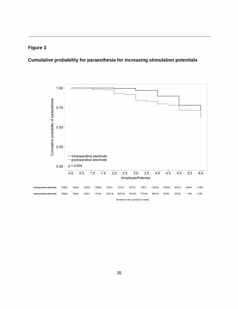
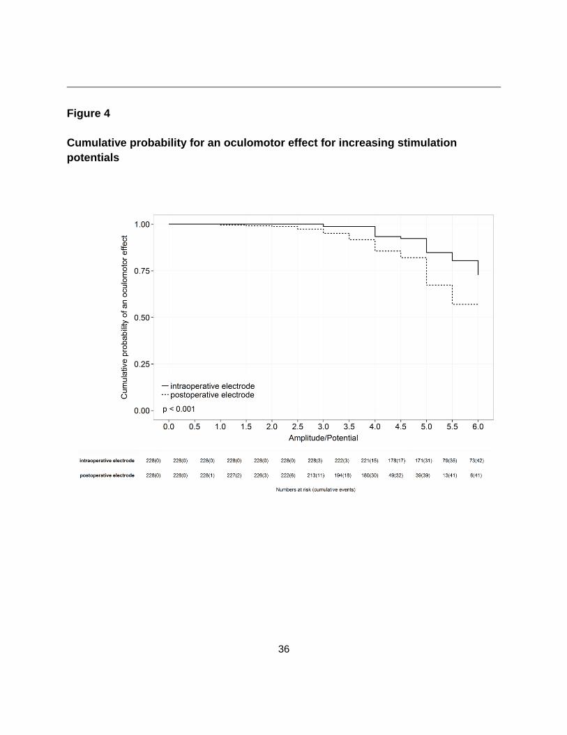
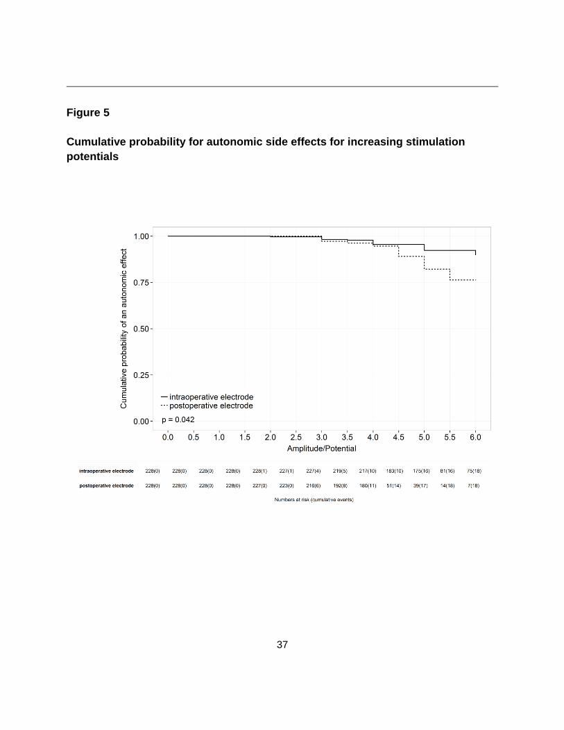
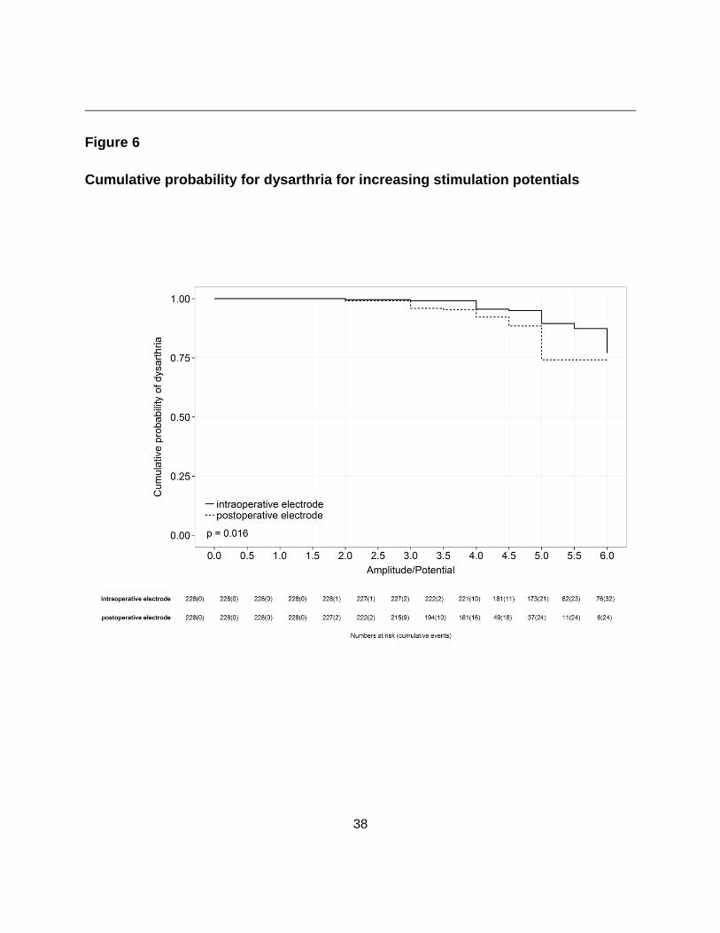
This was replicable for every single category of side effect, and the differences were
statistically significant except for capsule effects (p=0.184).
Thresholds of a suitable therapeutic effect were detectable in 84.8% of stimulation sites
(178 / 211) for intraoperative test stimulation and still in 83.4% (176 / 211) for
permanent stimulation. A strong microlesion effect obscured the delineation of
thresholds in 15.2% and 16.6% respectively. Therapeutic effects of intraoperative test
stimulation were achieved at significantly lower current amplitudes than in postoperative
DBS programming (p<0.001) (Figure 2).
19
Thus the intraoperative test stimulation resulted in a wider therapeutic window in total
with lower amplitudes for therapeutic effects and higher thresholds for side effects.
Postoperative DBS programming attained a satisfactory therapeutic window for
monopolar stimulation in 87.9% of the STNs, which remained constant one year after
surgery (87.0%). The remaining STNs were stimulated in a bipolar stimulation mode
because of unbearable side effects appearing at low stimulation amplitudes with
monopolar stimulation. The average stimulation voltage of the monopolarly stimulated
STNs was 2.77 V (± 0.86) after one year.
The total count of side effects was lower for the permanent stimulation than for the
intraoperative stimulations (237 vs. 248, Table 2). The most likely reason is that in
postoperative programming the testing stopped at 4 V in a portion of patients to shorten
the procedure even if no side effects appeared until that voltage. Therefore we
additionally calculated the threshold for any side effect measured until 4 V,
independently of the clinical findings with higher intensities. The threshold for side
effects with the permanent electrode was significantly lower than with the intraoperative
electrodes (mean 3.51 V vs. 3.89 mA, mean difference: 0.38 (95%-CI 0.26, 0.49), p
<0.001.
The category of side effects differed frequently between intra- and postoperative
stimulation. Overall only 33.5% of the side effects caused by the permanent electrode
coincided in their category with the intraoperative test stimulation (Table 2). Capsule
effects were most consistently predicted in 63.6% by intraoperative testing, vice versa
20
only 32.9% of intraoperative capsule effects recurred during permanent stimulation. The
coincidence rates were even lower for the other categories of side effects.
In total 48.3% (57/118) of electrodes were implanted to the central trajectory.
Discussion
Approximately half of the permanent electrodes were implanted along the central
trajectory which is in accordance with external data36,37 and emphasizes the influence of
MER and test stimulation on final electrode location. In many centers the decision for
one of the trajectories relies on the results of MER and intraoperative clinical testing. It
is assumed that a wider therapeutic window offers more possibilities for postoperative
stimulation parameter adjustments and thereby potentially leads to a better long-term
outcome. The goal of intraoperative test stimulation is to choose the optimal stimulation
site defined by the best therapeutic effects in combination with a high threshold for side
effects. However, our data suggest that intraoperative test stimulation overestimates the
potential therapeutic window. We found significantly higher thresholds for stimulation-
induced side effects during intraoperative stimulation while a therapeutic effect was
obtained at lower thresholds. The latter could partly be explained by a strong,
intraoperative microlesion effect in some patients.
Yet the category of side effects was not reliably predictable which implicates that the
electrodes stimulate different structures along the borders of STN. There is a variety of
possible reasons for this distinct spreading of the electric field in the STN and
surrounding tissues that causes a mismatch of the volume of tissue activated (VTA).
21
Minimal dislocations from the chosen target point during the insertion of the permanent
electrode may lead to a stimulation site deviating from the assumed stimulation site in
this study. Even smallest variations in the electrode location may lead to stimulations of
completely different fibers38. In our setting the accuracy of the placement of the final
electrode is controlled intraoperatively by non-stereotactic X-ray. An improvement of
these placement controls during the procedure could be helpful to detect the potential
dislocations in all three dimensions with the opportunity for immediate correction39.
An earlier study found the optimal postoperative stimulation site within and around the
STN more lateral and more superior compared to the preoperative MRI- based target
and compared to the intraoperatively found site after intraoperative MER and test
stimulation 13.
Brain shift during the procedure40 might be another possible cause for the mismatch
between intra- and postoperative stimulation sites. Even the regression of this brain
shift, e.g. due to the restoration of the lost CSF during the procedure could also
contribute to minimal changes of the lead position postoperatively.
As shown in experimental settings, the electric fields around the used types of
electrodes differ due to their different dimensions41. The electrode design has a relevant
impact on the stimulation effect: Changes of the diameter-height-ratio of the active
contact alter the shape of VTA even if the surface area is left unchanged42. The 3389
22
Medtronic electrode has four contacts, each 1,5 mm in length with a diameter of 1.27
mm creating a surface area of 5.98 mm². The test electrode (FHC 22675L) offers a
twofold function for MER and stimulation. Therefore its geometry is more complex with a
retractable 10 mm recording tip (“micro”) in a steel tube with an outside diameter of 0.46
mm, whose distal portion functions as cathode for stimulation (“macro-tip”). The
remaining shaft (“cannula”) is insulated from the “macro-tip” and is usually used as
anode during the intraoperative test stimulation. The “macro” contact is 1 mm in length
resulting in a surface area of approximately 1.78 mm², barely one third of the permanent
electrode’s. In a study using c-fos immunohistochemistry in the rat hippocampus, a
macroelectrode, 150 µm in diameter, was found to have twice the radius of activation, to
activate 5.8 times more neurons and to displace 20 times more tissue than a
microelectrode with a diameter of 33 µm, resulting in a diameter ratio of 0.2243.
Transferring these results to the electrodes used in this study with a diameter ratio of
0.36, an analogous impact of the electrode dimensions on the VTA is easily
conceivable.
Furthermore the stimulation between the “macro-tip” and the “cannula” in a bipolar
mode creates an asymmetric electric field20. Theoretical comparisons of test- and DBS-
electrode therefore utilized a bipolar model for permanent stimulation, too. But in clinical
practice monopolar stimulation is most commonly applied with only one of four contacts
functioning as cathode and the implantable pulse generator (IPG) case as anodic
return. Here the electric field spreads more radially outwards from the active contact
which results in wider tissue activation than in bipolar mode44. Furthermore it is
23
necessary to place the electric center of the permanent electrode coincident with the
electric center of the test electrode to reproduce the intraoperative effects with the
chronic stimulation16 which is barely feasible or verifiable in clinical practice due to the
different compositions outlined above.
In total, there are various mismatches in the design of the two electrodes that possibly
lead to relevant discrepancies in VTA and stimulation effects.
While intraoperative test stimulation was performed in constant-current mode,
postoperative DBS programming utilized constant-voltage mode. There is some
evidence that monopolar current-controlled and voltage-controlled stimulation generate
nearly identical VTA shapes in theoretical DBS models45. Also in clinical practice, the
current of intraoperative testing is usually considered to be equal to the voltage of
permanent electrode stimulation assuming fixed impedances around 1000 Ω. But
clinical measurements revealed that the impedance fluctuates in a range from 500 to
1500 Ω46. In vitro experiments showed that the impedance is mostly influenced by the
conductivity and thickness of the encapsulation layer around the electrode47. In an in-
vitro model with typical DBS settings, the VTA was inversely correlated to the
impedance with a reduction up to 52% of VTA volume just by increasing the impedance
within the typical range48. Perioperative edema, a haemosiderine layer or later gliotic
scar formation determine the electrode´s encapsulation. This changes the conductivity
of the surrounding tissue and thereby contributes to different VTA intra- and
postoperatively. An edema decreases the impedance, while a glial transformation has
24
the opposite effect18. Although the impedance of the “macro” contact of the test
electrode cannot be measured during the surgery, we suggest a notable influence of the
impedance changing over time on different stimulation effects.
Although there is evidence for the usefulness of MER in target localization and outcome
improvement11, similar clinical effects of intraoperative clinical testing have yet not been
shown convincingly. As we were not able to predict the therapeutic window accurately,
our results may stimulate the discussion about the necessity of awake surgery. Recent
studies provided increasing evidence for good clinical results after electrode placement
using intraoperative MRI49 50 51 or CT52 which could potentially shorten the procedure
and relieve the patient from the strain of awake surgery.
In conclusion, whether test stimulation and intraoperative clinical examination improve
the long-term outcome of STN DBS is called into question by our data and should be
evaluated in a prospective study design. Therefore clinicians involved in DBS surgery
and programming should be aware that the therapeutic effect and side effects of
permanent stimulation may not be reliably predicted by intraoperative test stimulation.
25
Literaturverzeichnis 1 Van den Eeeden S, Tanner C, Bernstein A et al.: Incidence of Parkinson's Disease: variation by age,
gender, race / ethnity; Am J Epidemiol 2003, 157: 1015-1022
2 Hirsch EC, Jenner P, Przedborski S.: Pathogenesis of Parkinson's disease. Mov Disord. 2013
Jan;28(1):24-30
3 Irizarry MC, Growdon W, Gomez-Isla T, Newell K, George JM, Clayton DF, Hyman BT. Nigral and
cortical Lewy bodies and dystrophic nigral neurites in Parkinson's disease and cortical Lewy body disease
contain alpha-synuclein immunoreactivity. J Neuropathol Exp Neurol. 1998 Apr;57(4):334-7
4 Braak H, Del Tredici K, Rüb U, de Vos RA, Jansen Steur EN, Braak E.: Staging of brain pathology
related to sporadic Parkinson's disease. Neurobiol Aging. 2003 Mar-Apr;24(2):197-211
5 Parkinson J. An Essay on the Shaking Palsy. London: Sherwood, Neely and Jones; 1817. 6 Magrinelli F, Picelli A, Tocco P, Federico A, Roncari L, Smania N, Zanette G, Tamburin S.
Pathophysiology of Motor Dysfunction in Parkinson's Disease as the Rationale for Drug Treatment and
Rehabilitation. Parkinsons Dis. 2016;2016:9832839
7 Ponsen MM, Stoffers D, Booij J, van Eck-Smit BL, Wolters ECh, Berendse HW. Idiopathic hyposmia as
a preclinical sign of Parkinson's disease. Ann Neurol. 2004 Aug;56(2):173-81
8 Yu SY, Sun L, Liu Z, Huang XY, Zuo LJ, Cao CJ, Zhang W, Wang XM. Sleep disorders in Parkinson's
disease: clinical features, iron metabolism and related mechanism. PLoS One. 2013 Dec
23;8(12):e82924
9 Aarsland D, Kramberger MG. Neuropsychiatric Symptoms in Parkinson's Disease. J Parkinsons Dis.
2015;5(3):659-67
10 Kim JB, Kim BJ, Koh SB, Park KW.Autonomic dysfunction according to disease progression
in Parkinson's disease. Parkinsonism Relat Disord. 2014 Mar;20(3):303-7
26
11 Aarsland D. Cognitive impairment in Parkinson's disease and dementia with Lewy bodies.
Parkinsonism Relat Disord. 2016 Jan;22 Suppl 1:S144-8
12 Uitti RJ, Ahlskog JE, Maraganore DM, Muenter MD, Atkinson EJ, Cha RH, O'Brien PC.Levodopa
therapy and survival in idiopathic Parkinson's disease: Olmsted County project. Neurology. 1993
Oct;43(10):1918-26.
13 Rascol O, Brooks DJ, Korczyn AD, De Deyn PP, Clarke CE, Lang AE. A five-year study of the
incidence of dyskinesia in patients with early Parkinson's disease who were treated with ropinirole or
levodopa. 056 Study Group. N Engl J Med. 2000 May 18. 342(20):1484
14 Fahn S. The medical treatment of Parkinson disease from James Parkinson to George Cotzias. Mov
Disord. 2015 Jan;30(1):4-18
15 Pollak P, Benabid AL, Gross C, Gao DM, Laurent A, Benazzouz A, Hoffmann D, Gentil M, Perret J.
Effects of the stimulation of the subthalamic nucleus in Parkinson disease. Rev Neurol
(Paris). 1993;149(3):175-6.
16 Odekerken VJ, van Laar T, Staal MJ, Mosch A, Hoffmann CF, Nijssen PC, Beute GN, van Vugt JP,
Lenders MW, Contarino MF, Mink MS, Bour LJ, van den Munckhof P, Schmand BA, de Haan RJ,
Schuurman PR, de Bie RM. Subthalamic nucleus versus globus pallidus bilateral deep brain stimulation
for advancedParkinson's disease (NSTAPS study): a randomised controlled trial. Lancet Neurol. 2013
Jan;12(1):37-44.
17 Deuschl G, Schade-Brittinger C, Krack P, Volkmann J, Schäfer H, Bötzel K, Daniels C, Deutschländer
A, Dillmann U, Eisner W, Gruber D, Hamel W, Herzog J, Hilker R, Klebe S, Kloss M, Koy J, Krause M,
Kupsch A, Lorenz D, Lorenzl S, Mehdorn HM, Moringlane JR, Oertel W, Pinsker MO, Reichmann H,
Reuss A, Schneider GH, Schnitzler A, Steude U, Sturm V, Timmermann L, Tronnier V, Trottenberg T,
Wojtecki L, Wolf E, Poewe W, Voges J; German Parkinson Study Group, Neurostimulation Section. A
randomized trial of deep-brain stimulation for Parkinson's disease. N Engl J Med. 2006 Aug
31;355(9):896-908. Erratum in: N Engl J Med. 2006 Sep 21;355(12):1289.
27
18 Jiang LL, Liu JL, Fu XL, Xian WB, Gu J, Liu YM, Ye J, Chen J, Qian H, Xu SH, Pei Z, Chen L. Long-
term Efficacy of Subthalamic Nucleus Deep Brain Stimulation in Parkinson's Disease: A 5-year Follow-up
Study in China. Chin Med J (Engl). 2015 Sep 20;128(18):2433-8
19 Fasano A, Romito LM, Daniele A, Piano C, Zinno M, Bentivoglio AR, Albanese A. Motor and cognitive
outcome in patients with Parkinson's disease 8 years after subthalamicimplants. Brain. 2010
Sep;133(9):2664-76
20 Bang Henriksen M, Johnsen EL, Sunde N, Vase A, Gjelstrup MC, Østergaard K. Surviving 10 years
with deep brain stimulation for Parkinson's disease--a follow-up of 79 patients. Eur J Neurol. 2016
Jan;23(1):53-61.
21 Kleiner-Fisman G1, Herzog J, Fisman DN, Tamma F, Lyons KE, Pahwa R, Lang AE, Deuschl
G. Subthalamic nucleus deep brain stimulation: summary and meta-analysis of outcomes. Mov
Disord. 2006 Jun;21 Suppl 14:S290-304.
22 Ferrara J, Diamond A, Hunter C, Davidson A, Almaguer M, Jankovic J. Impact of STN-DBS on life and
health satisfaction in patients with Parkinson's disease. J Neurol Neurosurg Psychiatry. 2010
Mar;81(3):315-9.
23 Temel Y, Blokland A, Steinbusch HW, Visser-Vandewalle V: The functional role of the subthalamic
nucleus in cognitive and limbic circuits; Prog Neurobiol. 2005 Aug;76(6):393-413.
24 Mann JM, Foote KD, Garvan CW, Fernandez HH, Jacobson CE 4th, Rodriguez RL, Haq IU, Siddiqui
MS, Malaty IA, Morishita T, Hass CJ, Okun MS: Brain penetration effects of microelectrodes and DBS
leads in STN or GPi; J Neurol Neurosurg Psychiatry. 2009 Jul;80(7):794-7
25 Ben-Haim S, Asaad WF, Gale JT, Eskandar EN: Risk factors for hemorrhage during microelectrode-
guided deep brain stimulation and the introduction of an improved microelectrode design; Neurosurgery.
2009 Apr;64(4):754-62l
26 Xiaowu H, Xiufeng J, Xiaoping Z, Bin H, Laixing W, Yiqun C, Jinchuan L, Aiguo J, Jianmin L: Risks of
intracranial hemorrhage in patients with Parkinson's disease receiving deep brain stimulation and
ablation; Parkinsonism Relat Disord. 2010 Feb;16(2):96-100
28
27 Temel Y, Wilbrink P, Duits A, Boon P, Tromp S, Ackermans L, van Kranen-Mastenbroek V, Weber W,
Visser-Vandewalle V: Single electrode and multiple electrode guided electrical stimulation of the
subthalamic nucleus in advanced Parkinson's disease.; Neurosurgery. 2007 Nov;61(5 Suppl 2):346-55
28 Pollak P, Benabid AL, Gross C, Gao DM, Laurent A, Benazzouz A, Hoffmann D, Gentil M, Perret J:
Effects of the stimulation of the subthalamic nucleus in Parkinson disease; Rev Neurol (Paris).
1993;149(3):175-6
29 Hariz M, Blomstedt P, Limousin P: The myth of microelectrode recording in ensuring a precise location
of the DBS electrode within the sensorimotor part of the subthalamic nucleus. Mov Disord. 2004
Jul;19(7):863-4
30 Bour LJ, Contarino MF, Foncke EM, de Bie RM, van den Munckhof P, Speelman JD, Schuurman PR:
Long-term experience with intraoperative microrecording during DBS neurosurgery in STN and GPi; Acta
Neurochir (Wien). 2010 Dec;152(12):2069-77
31 Kocabicak E, Alptekin O, Ackermans L, Kubben P, Kuijf M, Kurt E, Esselink R, Temel Y: Is there still
need for microelectrode recording now the subthalamic nucleus can be well visualized with high field and
ultrahigh MR imaging?; Front Integr Neurosci. 2015 Aug 11;9:46
32 Foltynie T, Zrinzo L, Martinez-Torres I, Tripoliti E, Petersen E, Holl E, Aviles-Olmos I, Jahanshahi M,
Hariz M, Limousin P: MRI-guided STN DBS in Parkinson's disease without microelectrode recording:
efficacy and safety; Neurol Neurosurg Psychiatry. 2011 Apr;82(4):358-63
33 Schlaier J, Habermeyer C, Warnat J, Lange M, Janzen A, Hochreiter A, Proescholdt M. Brawanski A,
Fellner C: Discrepancies between the MRI- and the electrophysiologically defined subthalamic nucleus;
Acta Neurochir (2011) 153:2307–2318
34 Schlaier JR, Habermeyer C, Janzen A, Fellner C, Hochreiter A, Proescholdt M, Brawanski A, Lange M:
The influence of intraoperative microelectrode recordings and clinical testing on the location of final
stimulation sites in deep brain stimulation for Parkinson's disease; Acta Neurochir (Wien). 2013
Feb;155(2):357-66
http://www.ncbi.nlm.nih.gov/pubmed/?term=Ackermans%20L%5BAuthor%5D&cauthor=true&cauthor_uid=18091250
http://www.ncbi.nlm.nih.gov/pubmed/?term=Blomstedt%20P%5BAuthor%5D&cauthor=true&cauthor_uid=15254958
http://www.ncbi.nlm.nih.gov/pubmed/?term=de%20Bie%20RM%5BAuthor%5D&cauthor=true&cauthor_uid=20949292
http://www.ncbi.nlm.nih.gov/pubmed/?term=Speelman%20JD%5BAuthor%5D&cauthor=true&cauthor_uid=20949292
http://www.ncbi.nlm.nih.gov/pubmed/?term=Kocabicak%20E%5BAuthor%5D&cauthor=true&cauthor_uid=26321929
http://www.ncbi.nlm.nih.gov/pubmed/?term=Ackermans%20L%5BAuthor%5D&cauthor=true&cauthor_uid=26321929
http://www.ncbi.nlm.nih.gov/pubmed/?term=Tripoliti%20E%5BAuthor%5D&cauthor=true&cauthor_uid=20571041
http://www.ncbi.nlm.nih.gov/pubmed/?term=Schlaier%20JR%5BAuthor%5D&cauthor=true&cauthor_uid=23275071
29
35 Lange M, Zech N, Seemann M, Janzen A, Halbing D, Zeman F, Doenitz C, Rothenfusser E, Hansen E,
Brawanski A, Schlaier J.: Anesthesiologic regimen and intraoperative delirium in deep brain stimulation
surgery for Parkinson's disease. J Neurol Sci. 2015 Aug 15;355(1-2):168-73.
36 Reck C, Maarouf M, Wojtecki L, Groiss SJ, Florin E, Sturm V, Fink GR, Schnitzler A, Timmermann L:
Clinical outcome of subthalamic stimulation in Parkinson's disease is improved by intraoperative multiple
trajectories microelectrode recording; J Neurol Surg A Cent Eur Neurosurg. 2012 Nov;73(6):377-86
37 Amirnovin R, Williams ZM, Cosgrove GR, Eskandar EN: Experience with microelectrode guided
subthalamic nucleus deep brain stimulation; Neurosurgery. 2006 Feb;58(1 Suppl)
38 Paffi A, Apollonio F, Puxeddu MG, Parazzini M, d'Inzeo G, Ravazzani P, Liberti M: A numerical study to
compare stimulations by intraoperative microelectrodes and chronic macroelectrodes in the DBS
technique, Biomed Res Int. 2013;2013:262739
39 Cui Z, Pan L, Song H, Xu X, Xu B, Yu X, Ling Z: Intraoperative MRI for optimizing electrode placement
for deep brain stimulation of the subthalamic nucleus in Parkinson disease. J Neurosurg. 2015 Aug 14:1-
8
40 Ivan ME, Yarlagadda J, Saxena AP, Martin AJ, Starr PA, Sootsman WK, Larson PS: Brain shift during
bur hole-based procedures using interventional MRI; J Neurosurg. 2014 Jul;121(1):149-60
41 Paffi A, Camera F, Apollonio F, d'Inzeo G, Liberti M: Numerical characterization of intraoperative and
chronic electrodes in deep brain stimulation; Front Comput Neurosci. 2015 Feb 19;9:2
42 Butson CR and McIntyre CC: Role of electrode design on the volume of tissue activated during deep
brain stimulation; J Neural Eng. 2006 Mar; 3(1): 1–8
43 Desai S, Gutekunst C, Potter S, Gross R: Deep brain stimulation macroelectrodes compared to
multiple microelectrodes in rat hippocampus, Front Neuroeng. 2014; 7: 16.
44 Yousif N, Liu X: Modeling the current distribution across the depth electrode-brain interface in deep
brain stimulation; Expert Rev Med Devices. 2007 Sep;4(5):623-31
http://www.ncbi.nlm.nih.gov/pubmed/?term=Apollonio%20F%5BAuthor%5D&cauthor=true&cauthor_uid=24222899
http://www.ncbi.nlm.nih.gov/pubmed/?term=Parazzini%20M%5BAuthor%5D&cauthor=true&cauthor_uid=24222899
http://www.ncbi.nlm.nih.gov/pubmed/?term=d%27Inzeo%20G%5BAuthor%5D&cauthor=true&cauthor_uid=24222899
http://www.ncbi.nlm.nih.gov/pubmed/?term=Ravazzani%20P%5BAuthor%5D&cauthor=true&cauthor_uid=24222899
http://www.ncbi.nlm.nih.gov/pubmed/?term=Sootsman%20WK%5BAuthor%5D&cauthor=true&cauthor_uid=24785326
http://www.ncbi.nlm.nih.gov/pubmed/?term=Apollonio%20F%5BAuthor%5D&cauthor=true&cauthor_uid=25745397
30
45 Butson CR, McIntyre CC: Current steering to control the volume of tissue activated during deep brain
stimulation; Brain Stimul. 2008 Jan;1(1):7-15.
46 Volkmann J, Herzog J, Kopper F, Deuschl G: Introduction to the programming of deep brain
stimulators; Mov Disord. 2002;17 Suppl 3:S181-7
47 Kent AR, Grill WM: Analysis of deep brain stimulation electrode characteristics for neural recording; J
Neural Eng. 2014 Aug;11(4):046010.
48 Butson CR1, Maks CB, McIntyre CC: Sources and effects of electrode impedance during deep brain
stimulation, Clin Neurophysiol. 2006 Feb;117(2):447-54. Epub 2005 Dec 22.
49 Starr PA1, Martin AJ, Ostrem JL, Talke P, Levesque N, Larson PS: Subthalamic nucleus deep brain
stimulator placement using high-field interventional magnetic resonance imaging and a skull-mounted
aiming device: technique and application accuracy; J Neurosurg. 2010 Mar;112(3)
50 Aviles-Olmos I, Kefalopoulou Z, Tripoliti E, et al. Long-term outcome of subthalamic nucleus deep brain
stimulation for Parkinson's disease using an MRI-guided and MRI-verified approach. J Neurol Neurosurg
Psychiatry 2014;85:1419-1425
51 Chabardes S, Isnard S, Castrioto A, Oddoux M, Fraix V, Carlucci L, Payen JF, Krainik A, Krack P,
Larson P, Le Bas JF: Surgical implantation of STN-DBS leads using intraoperative MRI guidance:
technique, accuracy, and clinical benefit at 1-year follow-up; Acta Neurochir (Wien). 2015 Apr;157(4):729-
37
52 Mirzadeh Z, Chapple K, Lambert M, Evidente VG, Mahant P, Ospina MC, Samanta J, Moguel-Cobos
G, Salins N, Lieberman A, Tröster AI, Dhall R, Ponce FA: Parkinson's disease outcomes after
intraoperative CT-guided "asleep" deep brain stimulation in the globus pallidus internus. J Neurosurg.
2016 Apr;124(4):902-7
http://www.ncbi.nlm.nih.gov/pubmed/?term=McIntyre%20CC%5BAuthor%5D&cauthor=true&cauthor_uid=19142235
http://www.ncbi.nlm.nih.gov/pubmed/?term=McIntyre%20CC%5BAuthor%5D&cauthor=true&cauthor_uid=16376143
http://www.ncbi.nlm.nih.gov/pubmed/?term=Chabardes%20S%5BAuthor%5D&cauthor=true&cauthor_uid=25788414
http://www.ncbi.nlm.nih.gov/pubmed/?term=Castrioto%20A%5BAuthor%5D&cauthor=true&cauthor_uid=25788414
31
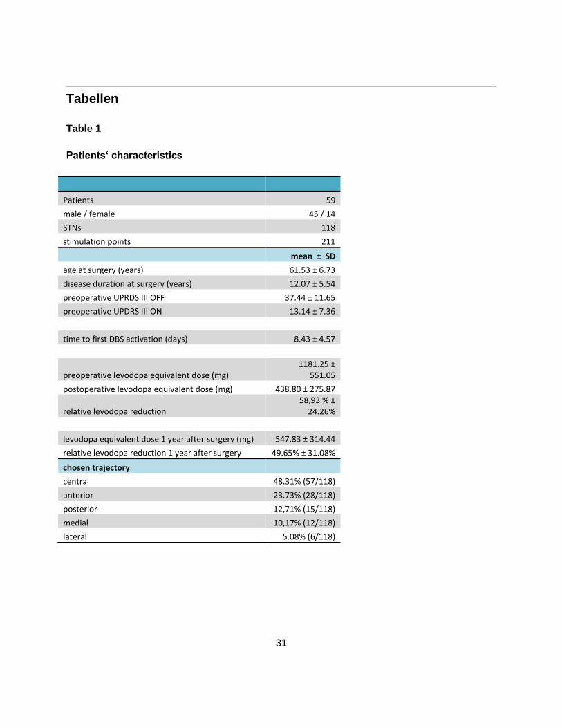
Tabellen
Table 1
Patients‘ characteristics
Patients 59
male / female 45 / 14
STNs 118
stimulation points 211
mean ± SD
age at surgery (years) 61.53 ± 6.73
disease duration at surgery (years) 12.07 ± 5.54
preoperative UPRDS III OFF 37.44 ± 11.65
preoperative UPDRS III ON 13.14 ± 7.36
time to first DBS activation (days) 8.43 ± 4.57
preoperative levodopa equivalent dose (mg) 1181.25 ±
551.05
postoperative levodopa equivalent dose (mg) 438.80 ± 275.87
relative levodopa reduction 58,93 % ±
24.26%
levodopa equivalent dose 1 year after surgery (mg) 547.83 ± 314.44
relative levodopa reduction 1 year after surgery 49.65% ± 31.08%
chosen trajectory
central 48.31% (57/118)
anterior 23.73% (28/118)
posterior 12,71% (15/118)
medial 10,17% (12/118)
lateral 5.08% (6/118)
32
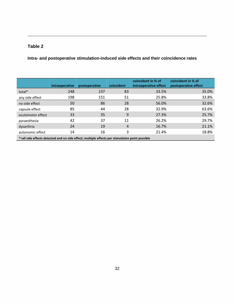
Table 2
Intra- and postoperative stimulation-induced side effects and their coincidence rates
intraoperative postoperative coincident coincident in % of intraoperative effect
coincident in % of postoperative effect
total* 248 237 83 33.5% 35.0%
any side effect 198 151 51 25.8% 33.8%
no side effect 50 86 28 56.0% 32.6%
capsule effect 85 44 28 32.9% 63.6%
oculomotor effect 33 35 9 27.3% 25.7%
paraesthesia 42 37 11 26.2% 29.7%
dysarthria 24 19 4 16.7% 21.1%
autonomic effect 14 16 3 21.4% 18.8%
*=all side effects detected and no side effect; multiple effects per stimulation point possible
33
Abbildungen
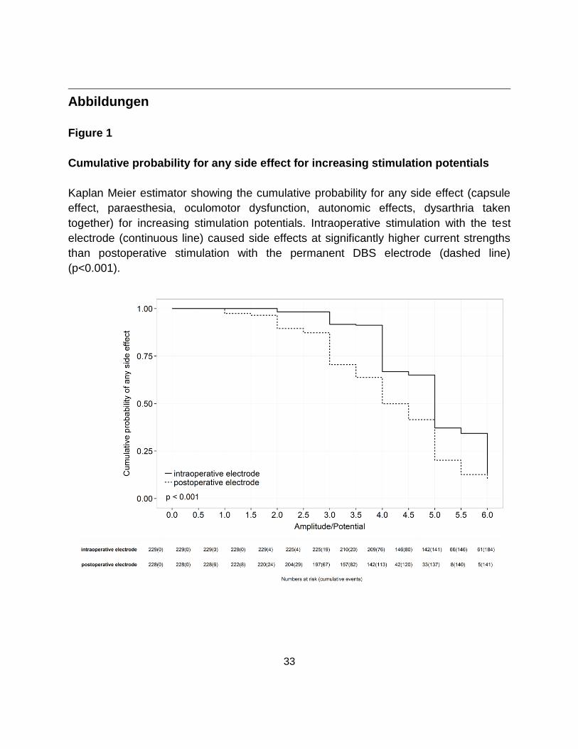
Figure 1
Cumulative probability for any side effect for increasing stimulation potentials
Kaplan Meier estimator showing the cumulative probability for any side effect (capsule
effect, paraesthesia, oculomotor dysfunction, autonomic effects, dysarthria taken
together) for increasing stimulation potentials. Intraoperative stimulation with the test
electrode (continuous line) caused side effects at significantly higher current strengths
than postoperative stimulation with the permanent DBS electrode (dashed line)
(p<0.001).
34
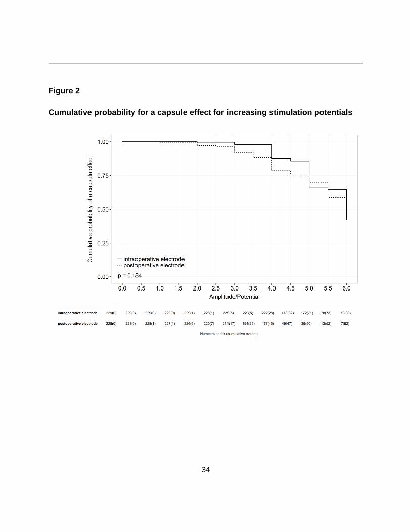
Figure 2
Cumulative probability for a capsule effect for increasing stimulation potentials
35
Figure 3
Cumulative probability for paraesthesia for increasing stimulation potentials
36
Figure 4
Cumulative probability for an oculomotor effect for increasing stimulation
potentials
37
Figure 5
Cumulative probability for autonomic side effects for increasing stimulation
potentials
38
Figure 6
Cumulative probability for dysarthria for increasing stimulation potentials
39
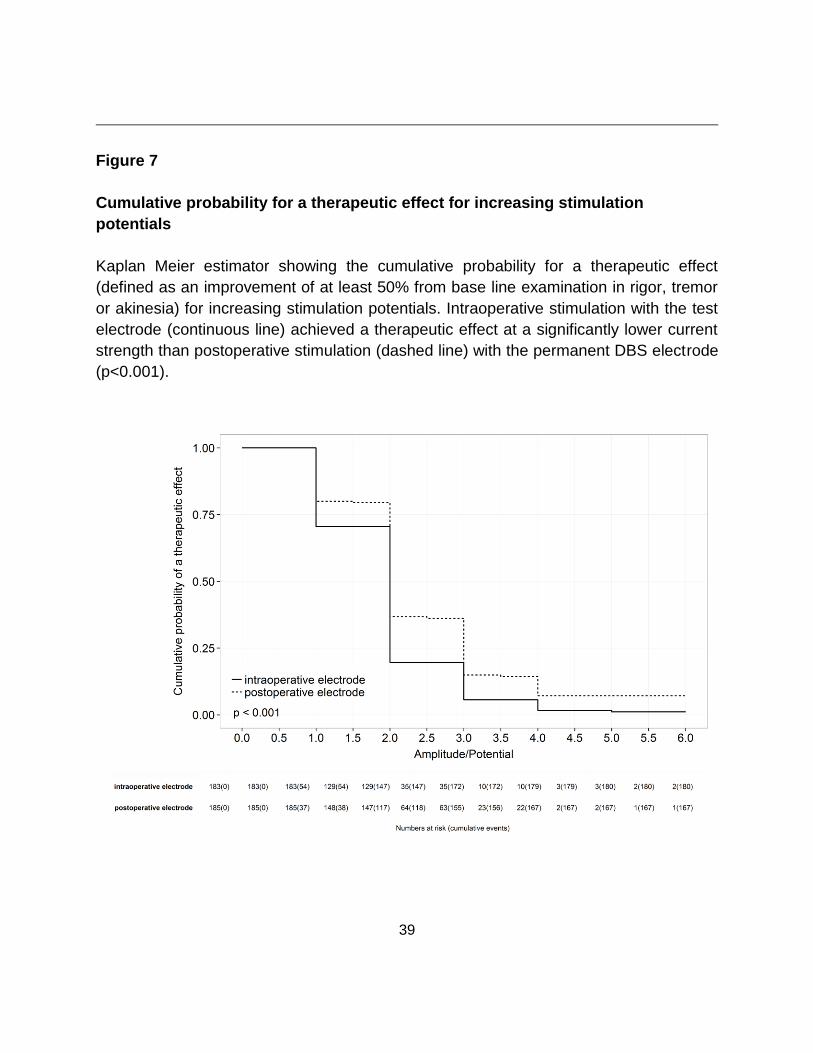
Figure 7
Cumulative probability for a therapeutic effect for increasing stimulation
potentials
Kaplan Meier estimator showing the cumulative probability for a therapeutic effect
(defined as an improvement of at least 50% from base line examination in rigor, tremor
or akinesia) for increasing stimulation potentials. Intraoperative stimulation with the test
electrode (continuous line) achieved a therapeutic effect at a significantly lower current
strength than postoperative stimulation (dashed line) with the permanent DBS electrode
(p<0.001).
40
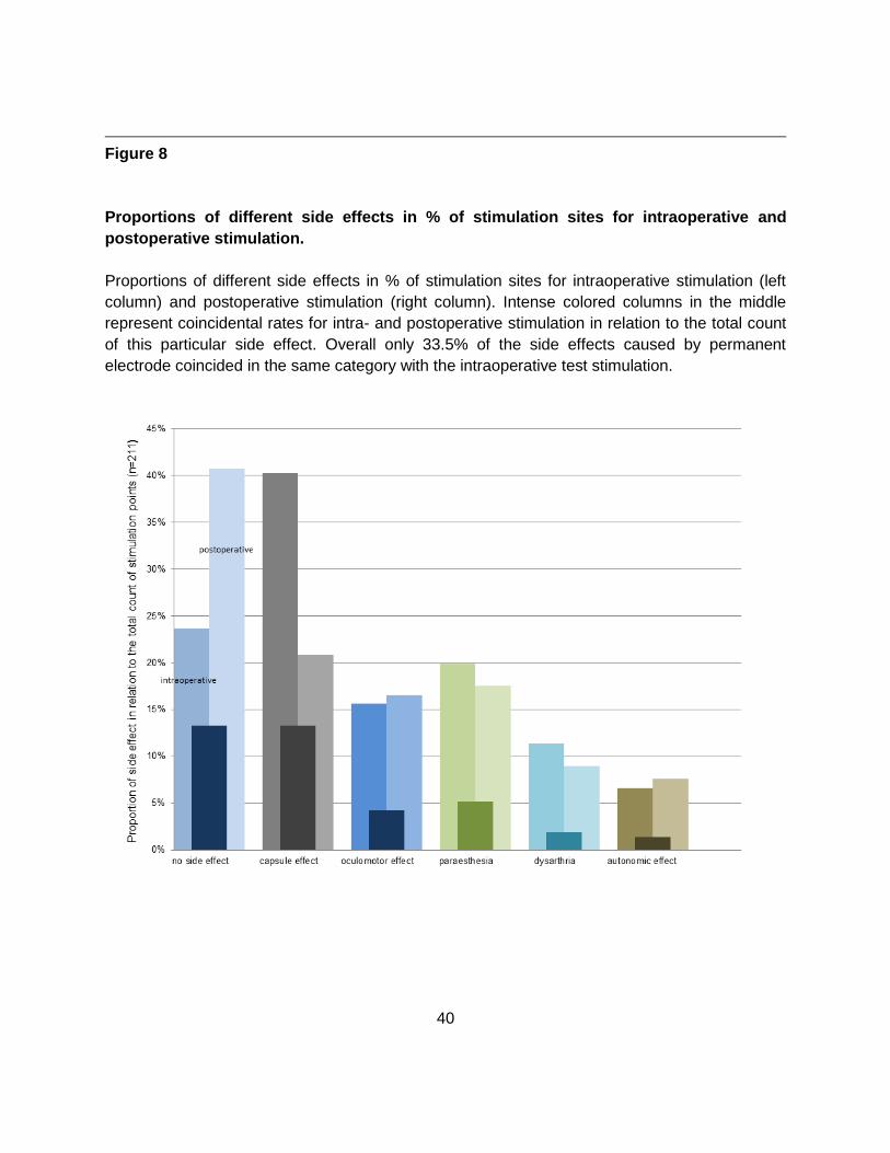
Figure 8
Proportions of different side effects in % of stimulation sites for intraoperative and
postoperative stimulation.
Proportions of different side effects in % of stimulation sites for intraoperative stimulation (left
column) and postoperative stimulation (right column). Intense colored columns in the middle
represent coincidental rates for intra- and postoperative stimulation in relation to the total count
of this particular side effect. Overall only 33.5% of the side effects caused by permanent
electrode coincided in the same category with the intraoperative test stimulation.

41
42
Lebenslauf
Josefine Blume
Persönliche Informationen
Name: Josefine Andrea Blume
Adresse: Kumpfmühler Straße 37, 93051 Regensburg, Germany
Telefon: +49-151-65182400
E-mail: [email protected]
Geburtstag und -ort: 06.06.1988 in Staßfurt
Staatsangehörigkeit: Deutsch
Sprachen
Deutsch Muttersprache
Englisch fließend
Französisch gute Kenntnisse
Latein großes Latinum
Akademische Ausbildung
2000-2007 Gymnasium Egeln, Abiturnote 1,0
10/2007 – 11/2013 Studium der Humanmedizin, Otto-von-Guericke-Universität
Magdeburg (OvGU),
2. Ärztliche Prüfung 10/2013, Note 1,5
Approbation 11/2013
Beruflicher Werdegang
ab 01 / 2014 Klinik für Neurologie, Uniklinik Regensburg
01-12/2014 Allgemein-neurologische Station
43
01-06/2015 Ultraschall der hirnversorgenden Arterien
07-12/2015 Notaufnahme
01-09/2016 Intensivstation
06/2014- 09/2016 Ambulanz für Bewegungsstörungen und Tiefe Hirnstimulation
06/2014 – 09/2016 Ambulanz für Botulinumtoxin
10/2014 – 09/2016 Lehrtätigkeit: Neurologie für Logopäden, Staatliche Logopädieschule
Regensburg
Wissenschaftlicher Werdegang
Dissertation Universität Regensburg, Fakultät für Medizin:
“Can intraoperative clinical examination really predict the effects of Deep Brain Stimulation in
the Subthalamic Nucleus?“
01/2015 – 08/2016 Datensammlung
02/2015 GCP Training (ICH-GCP/AMG)
Publikationen
Reviews und Case Reports
Benefit of ELISpot in Early Diagnosis of Tuberculous Meningoencephalitis: Case Report and Review
Josefine Blume, Josef Köstler, Robert Weissert in eNeurologicalSci 2015
Suspected Postnatal Depression revealed as Hereditary Diffuse Leukoencephalopathy with Spheroids
Josefine Blume, Robert Weissert (eingereicht in Movement Disorders Clinical Practice, 08/2016)
44
Wissenschaftliche Artikel
Can intraoperative clinical testing predict the effects of the permanent DBS electrode in the subthalamic nucleus?
J Blume, J Schlaier, E Rothenfußer, J Anthofer, F Zeman, A Brawanski, U Bogdahn, M Lange
(eingereicht in Stereotactic and Functional Neurosurgery, 07/2016)
Poster
1st Congress of the European Academy of Neurology (Berlin, 2015)
Safety and Feasibility of G-CSF compassionate use in ALS patients
A Khomenko, D Baldaranov, HP Müller, S Johannesen, I Kobor, J Blume, TH Bruun, J Grassinger, T Grimm, T
Kammermaier, V Haringer,A Ludoph, G Schuierer, R Laage, A Schneider, J Kassubek, W Schulte-Mattler, U
Bogdahn
1st Congress of the European Academy of Neurology (Berlin, 2015)
Biomarker Discoveryin G-CSF mediated clinical ALS stabilization
S Johannesen, A Khomenko, D Baldaranov, A Khomenko , I Kobor, J Blume, TH Bruun, J Grassinger, T Grimm, T
Kammermaier, V Haringer, A Ludoph, G Schuierer, R Laage, A Schneider, W Schulte-Mattler, U Bogdahn
20th International Congress of Parkinson’s Disease and Movement Disorders (Berlin, 2016)
Can intraoperative clinical examination really predict the effects of STN DBS?
J Blume, J Schlaier, E Rothenfußer, J Anthofer, F Zeman, A Brawanski, U Bogdahn, M Lange
Vorträge
IAPRD 2015 – XXI World Congress on Parkinson’s and Related Disorders (Milan, 2015) and
67. Jahrestagung der Deutschen Gesellschaft für Neurochirurgie (DGNC) (Frankfurt, 2016)
Can intraoperative clinical examination really predict the effects of STN DBS?
J Blume, J Schlaier, E Rothenfußer, J Anthofer, F Zeman, A Brawanski, U Bogdahn, M Lange
45
Eidesstattliche Erklärung
Ich erkläre hiermit, dass ich die vorliegende Arbeit ohne unzulässige Hilfe Dritter und ohne Benutzung anderer als der angegebenen Hilfsmittel angefertigt habe. Die aus anderen Quellen direkt oder indirekt übernommenen Daten und Konzepte sind unter Angabe der Quelle gekennzeichnet. Insbesondere habe ich nicht die entgeltliche Hilfe von Vermittlungs- bzw. Beratungsdiensten (Promotionsberater oder andere Personen) in Anspruch genommen. Niemand hat von mir unmittelbar oder mittelbar geldwerte Leistungen für Arbeit erhalten, die im Zusammenhang mit dem Inhalt der vorgelegten Dissertation stehen. Die Arbeit wurde bisher weder im In- noch im Ausland in gleicher oder ähnlicher Form einer anderen Prüfungsbehörde vorgelegt.
Regensburg, 20.02.2017 Josefine Blume