Chemical and biological analysis of essential oils pulp ...
9
~ 1808 ~ Journal of Pharmacognosy and Phytochemistry 2019; 8(4): 1808-1816 E-ISSN: 2278-4136 P-ISSN: 2349-8234 JPP 2019; 8(4): 1808-1816 Received: 01-05-2019 Accepted: 05-06-2019 Amel M Kamal Department of Pharmacognosy, Faculty of Pharmacy, Helwan University, Cairo, Egypt Mona E El-Tantawy National Organization for Drug Control and Research, Giza, Egypt Eman G Haggag Department of Pharmacognosy, Faculty of Pharmacy, Helwan University, Cairo, Egypt Marwa H Shukr National Organization for Drug Control and Research, Giza, Egypt Amany M Gad El-Garhy National Organization for Drug Control and Research, Giza, Egypt Rasha M Lithy National Organization for Drug Control and Research, Giza, Egypt Correspondence Amel M Kamal Department of Pharmacognosy, Faculty of Pharmacy, Helwan University, Cairo, Egypt Chemical and biological analysis of essential oils and pectins of banana, cantaloupe peels, guava pulp and formulation of banana pectin gel Amel M Kamal , Mona E El-Tantawy, Eman G Haggag, Marwa H Shukr, Amany M Gad El-Garhy and Rasha M Lithy Abstract The peels of both Musa paradisicae var. sapientum (banana), Cucumis melo L. (Cantaloupe) and pulp of Psidium guajava L. (guava) were studied for their essential oils, pectins, antimicrobial and anticancer activities. All tested samples exerted marked activities against Gram +ve, Gram -ve bacteria, fungi, dermatophytes, prostate (PC-3), breast (MCF-7) and colon (HCT-116) carcinoma cell lines. Banana essential oil showed the best activity against all tested micro-organisms, breast carcinoma cell line whereas guava pulp oil exerted the best activity against prostate and colon carcinoma cell line. Banana pectin gels were prepared using Carbopol 934, hydroxyl ethyl cellulose (HEC) and sodium carboxy methyl cellulose (Na CMC) as gelling agents. They were evaluated for their physical properties, anti- microbial and wound healing activities. F6 formulation containing 0.5% carbopol 934 was selected, it has been significantly shown to enhance wound healing compared to panthenol. The stimulation of healing may be due to the immune-modulatory activity of pectin. Keywords: Banana, cantaloupe, guava, antimicrobial, anticancer gel formulation 1. Introduction Musaceae, banana family, consists of Zingiberales having spirally arranged leaves, separate male and female flowers and pulpy fruits comprising 6 genera with 45 species (Kumar 2002) [28] while Cucurbitaceae, cucumber family or vine crop family, contains herbaceous annuals or perennials with storage roots and mostly moist vines, rarely grow as trees shrubs or bushes (Kumar 2002) [28] comprising 100 genera and 850 species (Deyo and Malley, 2008) [12] and Myrtaceae, cucumber family, consists of moderate-sized, small trees or shrubs comprising 80 genera and 3000 species (Kumar 2002) [28] . Musa paradisicae var. sapientum (Banana), Cucumis melo L. (Cantaloupe) and Psidium guajava L. (guava) are food crops cultivated in tropical and subtropical regions (Kumar 2002) [28] . They are rich in biologically active phytoconstituents like essential oils, sterols/triterpenes, carotenoids, pectins and flavonoids (Abou-ziad 1998; Mittal et al., 2010) [1, 30] . Several parts of the plants as leaves, fruits, peels have been investigated for their essential oil components, as limonene, citral, 1,8-cineole were identified in Cucumis melo fruit while 6-nonenyl acetate, cinnamyl acetate, nonenol in its peel (Howat and Senter, 1987; Beaulieu and Grimm, 2001; Nattaporn and Pranee, 2011) [21, 6, 32] . Limonene, octanol, α-copaene, α-humulene, β-bisabolene for example have been identified in Psidium guajava fruit (Jordan et al., 2003; Soares et al., 2007) [25, 37] , caryophyllene, selinene, viridiflorol, limonene and 1, 8-cineole in its leaves (Karawya 1999; Soliman et al., 2016) [26, 38] . Several biological activities have been previously reported on different parts of the plants as antioxidant activity of M. paradisicae, C. melo peels, P. guajava fruit and its pulp (Escrig et al., 2001; Someya et al., 2002; Hassimetto et al., 2009; Ismail et al., 2010; Calderon et al., 2011; Agarwal et al., 2012) [16, 39, 19, 23, 11, 4] , prostate anticancer and antimicrobial activities of M. paradisicae peels and essential oils of P. guajava leaves, respectively (Karawya 1999; Akamine et al., 2009) [26, 3] . The biological importance and the few phytochemical studies reported on these species growing in Egypt, encouraged the authors to undertake this study. 2. Materials and Methods 2.1. Plant materials Samples of ripe fruits of banana (Musa paradisiaca L. var. sapientum Kuntze), cantaloupe (Cucumis melo L.) and guava (Psidium guajava L.) were collected from trees in gardens, Horticultural Research Station, El-Qanater, Qalyoubia, Egypt during November/December 2012/2013 for banana and guava and March/April 2014/2015 for cantaloupe. The identity of
Transcript of Chemical and biological analysis of essential oils pulp ...

~ 1808 ~
Journal of Pharmacognosy and Phytochemistry 2019; 8(4): 1808-1816
E-ISSN: 2278-4136
P-ISSN: 2349-8234
JPP 2019; 8(4): 1808-1816
Received: 01-05-2019
Accepted: 05-06-2019
Amel M Kamal
Department of Pharmacognosy,
Faculty of Pharmacy, Helwan
University, Cairo, Egypt
Mona E El-Tantawy
National Organization for Drug
Control and Research, Giza,
Egypt
Eman G Haggag
Department of Pharmacognosy,
Faculty of Pharmacy, Helwan
University, Cairo, Egypt
Marwa H Shukr
National Organization for Drug
Control and Research, Giza,
Egypt
Amany M Gad El-Garhy
National Organization for Drug
Control and Research, Giza,
Egypt
Rasha M Lithy
National Organization for Drug
Control and Research, Giza,
Egypt
Correspondence
Amel M Kamal
Department of Pharmacognosy,
Faculty of Pharmacy, Helwan
University, Cairo, Egypt
Chemical and biological analysis of essential oils
and pectins of banana, cantaloupe peels, guava
pulp and formulation of banana pectin gel
Amel M Kamal, Mona E El-Tantawy, Eman G Haggag, Marwa H Shukr,
Amany M Gad El-Garhy and Rasha M Lithy
Abstract
The peels of both Musa paradisicae var. sapientum (banana), Cucumis melo L. (Cantaloupe) and pulp of
Psidium guajava L. (guava) were studied for their essential oils, pectins, antimicrobial and anticancer
activities. All tested samples exerted marked activities against Gram +ve, Gram -ve bacteria, fungi,
dermatophytes, prostate (PC-3), breast (MCF-7) and colon (HCT-116) carcinoma cell lines. Banana
essential oil showed the best activity against all tested micro-organisms, breast carcinoma cell line
whereas guava pulp oil exerted the best activity against prostate and colon carcinoma cell line. Banana
pectin gels were prepared using Carbopol 934, hydroxyl ethyl cellulose (HEC) and sodium carboxy
methyl cellulose (Na CMC) as gelling agents. They were evaluated for their physical properties, anti-
microbial and wound healing activities. F6 formulation containing 0.5% carbopol 934 was selected, it has
been significantly shown to enhance wound healing compared to panthenol. The stimulation of healing
may be due to the immune-modulatory activity of pectin.
Keywords: Banana, cantaloupe, guava, antimicrobial, anticancer gel formulation
1. Introduction
Musaceae, banana family, consists of Zingiberales having spirally arranged leaves, separate
male and female flowers and pulpy fruits comprising 6 genera with 45 species (Kumar 2002) [28] while Cucurbitaceae, cucumber family or vine crop family, contains herbaceous annuals or
perennials with storage roots and mostly moist vines, rarely grow as trees shrubs or bushes
(Kumar 2002) [28] comprising 100 genera and 850 species (Deyo and Malley, 2008) [12] and
Myrtaceae, cucumber family, consists of moderate-sized, small trees or shrubs comprising 80
genera and 3000 species (Kumar 2002) [28]. Musa paradisicae var. sapientum (Banana),
Cucumis melo L. (Cantaloupe) and Psidium guajava L. (guava) are food crops cultivated in
tropical and subtropical regions (Kumar 2002) [28]. They are rich in biologically active
phytoconstituents like essential oils, sterols/triterpenes, carotenoids, pectins and flavonoids
(Abou-ziad 1998; Mittal et al., 2010) [1, 30]. Several parts of the plants as leaves, fruits, peels
have been investigated for their essential oil components, as limonene, citral, 1,8-cineole were
identified in Cucumis melo fruit while 6-nonenyl acetate, cinnamyl acetate, nonenol in its peel
(Howat and Senter, 1987; Beaulieu and Grimm, 2001; Nattaporn and Pranee, 2011) [21, 6, 32].
Limonene, octanol, α-copaene, α-humulene, β-bisabolene for example have been identified in
Psidium guajava fruit (Jordan et al., 2003; Soares et al., 2007) [25, 37], caryophyllene, selinene,
viridiflorol, limonene and 1, 8-cineole in its leaves (Karawya 1999; Soliman et al., 2016) [26,
38]. Several biological activities have been previously reported on different parts of the plants
as antioxidant activity of M. paradisicae, C. melo peels, P. guajava fruit and its pulp (Escrig et
al., 2001; Someya et al., 2002; Hassimetto et al., 2009; Ismail et al., 2010; Calderon et al.,
2011; Agarwal et al., 2012) [16, 39, 19, 23, 11, 4], prostate anticancer and antimicrobial activities of
M. paradisicae peels and essential oils of P. guajava leaves, respectively (Karawya 1999;
Akamine et al., 2009) [26, 3]. The biological importance and the few phytochemical studies
reported on these species growing in Egypt, encouraged the authors to undertake this study.
2. Materials and Methods
2.1. Plant materials
Samples of ripe fruits of banana (Musa paradisiaca L. var. sapientum Kuntze), cantaloupe
(Cucumis melo L.) and guava (Psidium guajava L.) were collected from trees in gardens,
Horticultural Research Station, El-Qanater, Qalyoubia, Egypt during November/December
2012/2013 for banana and guava and March/April 2014/2015 for cantaloupe. The identity of
~ 1809 ~
Journal of Pharmacognosy and Phytochemistry the fruits was confirmed by Dr. Abdel Halim Abdel Mogali,
Agricultural Museum, Giza, Egypt. The peels of banana and
cantaloupe and pulp of guava were then air-dried in the shade,
reduced to powder and kept in tightly-closed containers.
Voucher specimen (Reg. no. 55a, b, c, respectively) were kept
in the herbarium of Pharmacognosy Department, Faculty of
Pharmacy, Helwan University, Cairo, Egypt.
2.2 Experimental animals
Adult male rats, of 300 g body weight were obtained from the
animal house of National Organization for Drug Control and
Research Institute Giza, Egypt. The adopted protocol and
implemented experiments were approved by the local Animal
Ethics Committee of faculty of Pharmacy, Helwan University,
Egypt, and were carried out in accordance to the international
Guide for the Care and Use of Laboratory Animals.
2.3 Instruments
Aglient 6890 gas chromatograph equipped with an Aglient
mass spectrometric detector with a direct capillary interface
and fused silica capillary column HP-5MS (30 m X 0.32 mm
X 0.25 μm film thickness) (Agricultural Pesticide Lab, Giza,
Egypt), was used for GLC analysis of essential oils, HPLC
chromatograph (Win Chrome ver. 1.3 equipped with column
KROMACIL C18) (Regional Center for Mycology and
Biotechnology, Al-Azhar university, Cairo, Egypt), was used
for sugars analysis of pectins, Clavenger apparatus for hydro-
distillation of essential oils and Refractometer for
identification of refractive indices of essential oils (National
Organization for Drug Control and Research, Giza, Egypt).
ELISA reader (SunRise, TECAN, Inc, USA) was used for
determination of number of viable cells in the cytotoxic
activity.
2.4 Chemicals
Authentic sugars: glucose, galactose, rhamnose, xylose and
galactouronic acid used in HPLC analysis of pectins were
obtained from Regional Center for Mycology and
Biotechnology, Al-Azhar university, Cairo, Egypt. All other
chemicals, solvents and reagents used in chromatography
were of analytical grade.
2.5 Micro-organisms
Staphylococcus aureus (RCMB000105) and Bacillus subtilis
(RCMB010069); Gram +ve bacteria, Escherichia coli
(RCMB010054) and Pseudomonas aeruginosa
(RCMB010048); Gram -ve bacteria, Aspergillus fumigatus
(RCMB02569), Candida albicans (RCMB05038) and
Geotricum candidum (RCMB05098); fungi, Trichophyton
mentagrophytes (RCMB0927); as dermatophyte were
obtained from Regional Center for Mycology and
Biotechnology (RCMB), Cairo, Egypt.
2.6 Cell lines for study of cytotoxicity
Human breast carcinoma cells (MCF-7), Prostate carcinoma
cells (PC-3) and Colon carcinoma cells (HCT-116) were
obtained from the American Type Culture Collection
(ATCC), Rockville, MD. The cells were grown on RPMI-
1640 medium supplemented with 10% inactivated fetal calf
serum and 50 μg/ml gentamicin. The cells were maintained at
37oC in a humidified atmosphere with 5% CO2 and
subcultured two to three times a week.
2.7 Investigation of essential oils
The ripe fresh peels of both banana and cantaloupe and the
ripe fresh pulp of guava were subjected to hydrodistillation
for 3 hours, using a Clavenger-type apparatus. The oils were
then collected, dried over anhydrous sodium sulphate
followed by GLC/MS analysis (Egyptian Pharmacopeia 1984) [14] (Table 1). Identification of the oils constituents was
achieved by Wiley and Nist mass spectral data in addition to
the published data in Adams, 2009 [2]. The estimation of each
peak was done using a computing integrator adopting the
internal normalization procedure.
The collective percentages of different classes of volatile
components in essential oils were calculated (Table 2) and the
reported biological activities of major volatile compounds
identified in banana, cantaloupe and guava essential oils were
summarized in table 3.
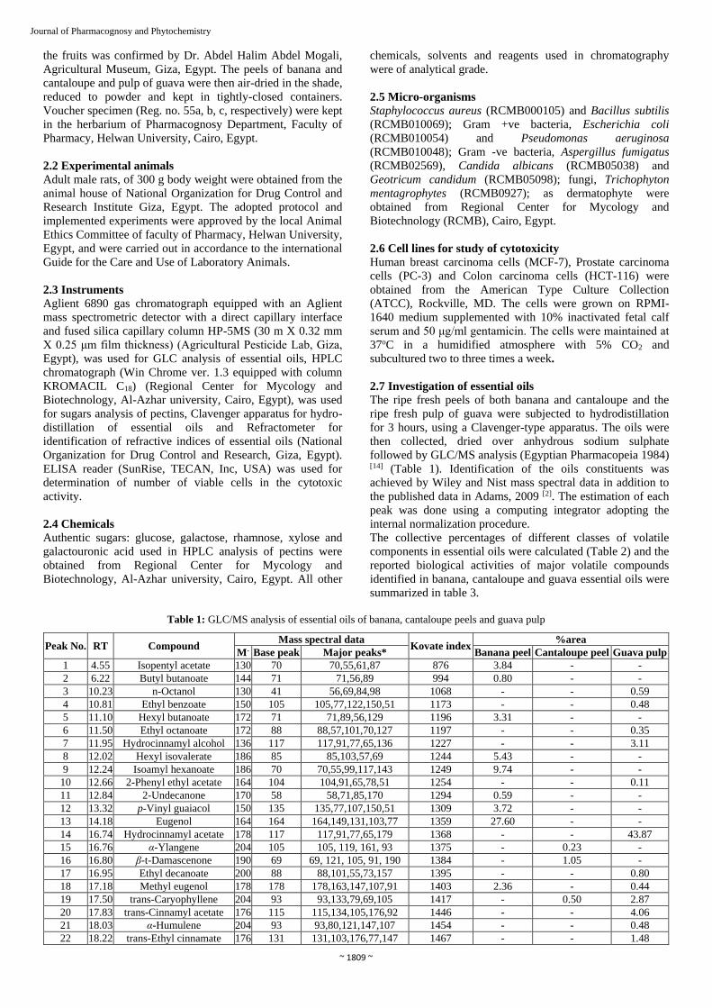
Table 1: GLC/MS analysis of essential oils of banana, cantaloupe peels and guava pulp
Peak No. RT Compound Mass spectral data
Kovate index %area
M- Base peak Major peaks* Banana peel Cantaloupe peel Guava pulp
1 4.55 Isopentyl acetate 130 70 70,55,61,87 876 3.84 - -
2 6.22 Butyl butanoate 144 71 71,56,89 994 0.80 - -
3 10.23 n-Octanol 130 41 56,69,84,98 1068 - - 0.59
4 10.81 Ethyl benzoate 150 105 105,77,122,150,51 1173 - - 0.48
5 11.10 Hexyl butanoate 172 71 71,89,56,129 1196 3.31 - -
6 11.50 Ethyl octanoate 172 88 88,57,101,70,127 1197 - - 0.35
7 11.95 Hydrocinnamyl alcohol 136 117 117,91,77,65,136 1227 - - 3.11
8 12.02 Hexyl isovalerate 186 85 85,103,57,69 1244 5.43 - -
9 12.24 Isoamyl hexanoate 186 70 70,55,99,117,143 1249 9.74 - -
10 12.66 2-Phenyl ethyl acetate 164 104 104,91,65,78,51 1254 - - 0.11
11 12.84 2-Undecanone 170 58 58,71,85,170 1294 0.59 - -
12 13.32 p-Vinyl guaiacol 150 135 135,77,107,150,51 1309 3.72 - -
13 14.18 Eugenol 164 164 164,149,131,103,77 1359 27.60 - -
14 16.74 Hydrocinnamyl acetate 178 117 117,91,77,65,179 1368 - - 43.87
15 16.76 α-Ylangene 204 105 105, 119, 161, 93 1375 - 0.23 -
16 16.80 β-t-Damascenone 190 69 69, 121, 105, 91, 190 1384 - 1.05 -
17 16.95 Ethyl decanoate 200 88 88,101,55,73,157 1395 - - 0.80
18 17.18 Methyl eugenol 178 178 178,163,147,107,91 1403 2.36 - 0.44
19 17.50 trans-Caryophyllene 204 93 93,133,79,69,105 1417 - 0.50 2.87
20 17.83 trans-Cinnamyl acetate 176 115 115,134,105,176,92 1446 - - 4.06
21 18.03 α-Humulene 204 93 93,80,121,147,107 1454 - - 0.48
22 18.22 trans-Ethyl cinnamate 176 131 131,103,176,77,147 1467 - - 1.48

~ 1810 ~
Journal of Pharmacognosy and Phytochemistry 23 18.44 α-Curcumene 202 119 119, 132, 105, 202, 145 1480 - 0.42 -
24 18.70 α-Muurolene 204 105 105, 161, 204, 119, 91 1500 - 0.36 -
25 18.91 Butylated hydroxy toluene 220 205 205,220,57,145,86 1515 10.63 26.14 18.51
26 19.04 δ-Cadinene 204 161 161, 204, 119, 105, 134 1523 - 0.19 -
27 19.09 cis-Calamenene 202 159 159,202,128,144,115 1529 - - 1.26
28 19.36 E-Nerolidol 222 69 69,93,107,81,161 1563 - - 3.37
29 19.69 trans-Isoelemicin 208 208 208,193,177,165,69 1570 8.06 - -
30 20.07 Globulol 222 109 109,69,161,81,189 1590 - - 7.00
31 20.14 Hexadecane 226 57 57, 71, 85, 99, 226 1600 - 1.45 -
32 20.33 Ledol 222 122 122,69,109,81,161 1602 - - 1.41
33 20.43 Tetradecanal 212 57 57,82,69,96,110 1612 0.65 - -
34 20.80 α-Cadinol 222 95 95,121,204,161,105 1624 - 12.50 0.57
35 20.82 α-Muurolol 222 161 161, 43, 119, 204, 105 1646 - 4.95 -
36 20.89 Heptadecane 240 57 57, 71, 85, 99, 240 1700 - 1.12 -
37 20.93 E-Isoamyl cinnamate 218 131 131,103,77,147,70 1741 0.35 - -
38 21.04 2E,6E-Farnesol 222 69 69,81,93,107,136 1743 - - 0.61
39 21.86 Ethyl myristate 256 88 88,101,55,157,213 1795 - - 0.39
40 22.52 n-Octadecane 254 57 57, 71, 85, 99, 254 1800 - 0.46 -
41 22.76 Isoamyl dodecanoate 270 70 70,55,183,201 1845 1.18 - -
42 22.77 n-Nonadecane 268 57 57, 71, 85, 99, 268 1900 - 2.41 -
43 22.78 Methyl palmitate 270 74 74,87,143,227,270 1921 0.32 - 0.30
44 23.99 Methyl palmitoleate 268 55 55, 69, 83, 96, 236 1934 - 1.79 -
45 24.15 Ethyl palmitate 284 88 88,101,73,157,284 1993 0.68 - 0.76
46 24.44 Methyl linoleate 294 67 67,81,95,109,294 2085 - 4.73 0.24
47 24.48 Ethyl oleate 310 88 88, 101, 55, 70, 157 2196 - 7.36 -
48 25.12 n-Heneicosane 296 57 57,71,85,99,296 2100 0.30 10.96 -
49 26.48 n-Docosane 310 57 57,71,85,99,310 2200 0.38 3.99 -
50 26.58 Tricosane 324 57 57, 71, 85, 99, 324 2300 - 0.48 -
51 27.23 Tetracosane 338 57 57,71,85,99,338 2400 0.89 3.01 0.19
52 28.23 Pentacosane 352 57 57,71,85,99,352 2500 0.94 5.65 0.26
53 29.30 Hexacosane 366 57 57,71,85,99,366 2600 1.33 - 0.28
54 29.42 Heptacosane 380 57 57, 71, 85, 99, 380 2700 - 3.46 -
55 31.03 Octacosane 394 57 57,71,85,99,394 2800 3.01 - -
56 31.90 Nonacosane 408 57 57,71,85,99,408 2900 3.34 - -
57 32.71 Triacontane 422 57 57,71,85,99,422 3000 2.51 - -
58 34.43 Dotriacontane 450 57 57,71,85,97,355 3200 0.74 - -
RT: Retention time, M-: Molecular ion peak
Table 2: Collective percentages of different classes of volatile components in essential oils of banana peel, cantaloupe peel and guava pulp
Constituents Banana peel Cantaloupe peel Guava pulp
Hydrocarbons 13.44% 34.71% 5.34%
- Aliphatic 13.44% 33% 0.73%
- Terpenoidal -- 1.71% 4.61%
Oxygenated compounds 79.26% 58.53% 88.45%
- Ethers 8.06% -- --
- Esters 25.65% 13.89% 52.84%
- Ketones 0.59% 1.05% --
- Aldehydes 0.65% -- --
- Alcohols
Aliphatic -- -- 3.70%
Sesquiterpene -- 17.45% 12.96%
- Other oxygenated compounds 44.31% 26.14% 18.95%
Total 92.70% 93.24% 93.79%
Table 3: Reported biological activity of major volatile compounds identified in banana, cantaloupe and guava essential oils
Serial
No Compound %Area Biological activity Reference
1 Eugenol 27.6 (banana)
Antiseptic and anti-inflammatory allowing its use
by dentists, antibacterial, antifungal Bennis et al., 2004 [7]
protects against cardiovascular diseases by
inhibiting the aggregation of platelets
Broadhurst and Duke,
1997 [10]
2 Hydrocinnamyl acetate 43.87 (guava) Antibacterial, Antifungal Aziz et al., 2013 [5]
3 Butylated hydroxyl
toluene
10.63 (banana)
26.14 (cantaloupe) 18.51 (guava)
Antioxidant Yehye et al., 2015 [41]
Antiviral, antimutagenic and anticarcinogenic Ohno et al., 1984 [33]
4 α-Cadinol 12.5 (cantaloupe) Antifungal Ho et al., 2011[20]
5 Heneicosane 10.96 (cantaloupe) Antimicrobial, antitumor Hsouna and Trigui,
2011[22]

~ 1811 ~
Journal of Pharmacognosy and Phytochemistry 2.8 Investigation of pectins
Powdered peels of both banana and cantaloupe and guava
pulp, were separately extracted with hot water (70o-90oC) for
30 min at a sample-to-water ratio of 1: 20 by magnetic
stirring. At the end of extraction period, extract water
temperature had reached ambient. The extract was filtered
through cotton piece and the filtrate was reduced in volume
by rotary evaporation. Pectin was precipitated from the
extract by ethanol addition in a ratio of 1: 4 (v/v) (extract :
ethanol), allowing to stand for 24 h at 5oC, the precipitate was
removed by centrifugation (10,000 RPM for 20 min), dried in
a desiccator over anhydrous calcium chloride then turned to
powder (Pazur 1986) [34] and assayed for their degree of
esterification, percentage of galacturonic acid and methoxy
groups (Bhatty 1993) [8] (Table 4).
2.9 Hydrolysis of pectins
The dried pectins (20 mg) were separately dissolved in 10 ml
of 50% ethanol, 10 ml of 2N hydrochloric acid and refluxed
on a water bath for 30 min. The aqueous acidic solution was
neutralized by sodium carbonate solution then evaporated to
dryness by rotary evaporator and the residue was dissolved in
50% ethanol/water and reserved for examination for sugars
composition by HPLC (United States Pharmacopeia 2011) [40].
2.10 Antimicrobial study
Antimicrobial study was carried out using the agar disc
diffusion method (Scott 1989) [36]. Samples were individually
tested against a panel of Gram-positive and Gram-negative
bacterial pathogens, yeast and fungi. Pathological tested
bacteria, yeast (1 x 10⁶ CFU/ml) and fungi (1 x 10⁴ spore/ml)
were spread on nutrient agar (NA), Sab. dextrose agar (SDA)
and malt extract agar (MA), respectively. After the media had
cooled and solidified, wells (6 mm in diameter) were made in
the solidified agar and loaded with 100 μl of samples. The
inoculated plates were then incubated for 24 h at 37oC for
bacteria and yeast, 48 h at 28oC for fungi. Ampicillin,
gentamycin and amphotericin B were used as standards for
Gram-positive bacteria, Gram-negative bacteria and fungi,
respectively, as positive controls and DMSO without the
extracts was used as a negative control. After incubation,
antimicrobial activity was evaluated by measuring the zone of
inhibition against the test organisms and compared with that
of the standard. Antimicrobial activity was expressed as
inhibition diameter zones in millimeters (mm). The
experiment were performed in triplicate and the data was
expressed as mean ± SD. Minimum inhibitory concentrations
(MIC) was determined using the broth micro-dilution method
using 96-well micro-plates (Saini et al., 2005; Bhuiyan et al.,
2011) [35, 9] (Table 5).
2.11 Cytotoxic study Potential cytotoxicity was evaluated using viability assay
(Mosmann 1983; Gangadevi and Muthumary, 2007) [31, 17].
The cytotoxic activity was evaluated on PC-3 (prostate
carcinoma cell line), MCF-7 (breast carcinoma cell line) and
HCT-116 (colon carcinoma cell line). Tumor cells were
grown as monolayers in growth RPMI-1640 medium
supplemented with 10% inactivated fetal calf serum and 50
μg/ml gentamicin. The monolayers of 10,000 cells adhered at
the bottom of the wells in a 96-well micro titer plate
incubated for 24 h at 37oC in a humidified incubator with 5%
CO2. The monolayers were then washed with sterile
phosphate buffered saline (0.01 M pH 7.2) and
simultaneously the cells were treated with 100μl from
different dilutions of tested sample in fresh maintenance
medium and incubated at 37oC. A control of untreated cells
was made in the absence of tested sample. A positive control
containing Doxorubicin drug was also tested as reference drug
for comparison. Six wells were used for each concentration of
the test sample. Every 24 h the observation was made under
the inverted microscope. The number of the surviving cells
was determined by staining the cells with crystal violet
followed by cell lysing using 33% glacial acetic acid and the
absorbance at 590 nm was read using ELISA reader after well
mixing. The absorbance values from untreated cells were
considered as 100% proliferation. The number of viable cells
was determined using ELISA reader as previously mentioned
and the percentage of viability was calculated. The 50%
inhibitory concentration (IC50) (the concentration required to
cause toxic effects in 50% of intact cells) was estimated from
graphic plots (Table 6).
Table 4: Analysis of pectins content in banana, cantaloupe and guava
Parameter % Pectin DE % GA % MG
Banana peel 3.5 95.2 81.52 12.40
Cantaloupe peel 5.75 92.3 88.32 13.00
Guava pulp 4.6 87.5 77.64 10.85
DE: Degree of esterification, GA: galacturonic Acid, MG: Methoxy groups
Table 5: MIC values (μg/ml) for oils and pectins of banana, cantaloupe and guava
Tested microorganism Banana peel Cantaloupe peel Guava pulp
Standard Oil Pectin Oil Pectin Oil Pectin
G+ve bacteria Ampicillin
S. aureus 15.63 15.63 31.25 31.25 31.25 250 3.9
B. subtilis 3.9 15.63 3.9 15.63 7.81 125 1.95
G-ve bacteria Gentamicin
E. coli 3.9 7.81 15.36 7.81 31.25 125 31.25
P. aeruginosa 3.9 15.63 62.5 31.25 125 250 125
Fungi Amp.B
A. fumigatus 15.63 31.25 31.25 62.5 15.63 500 3.9
C. albicans 15.63 125 62.5 250 125 1000 3.9
G. candidum 3.9 125 7.81 125 15.63 500 1.95
Dermatophyte Amp.B
T. mentagrophytes 500 500 1000 500 1000 NA 31.25

~ 1812 ~
Journal of Pharmacognosy and Phytochemistry G: Gram reaction, S. aureus: Staphylococcus aureus, B. subtilis: Bacillus subtilis, E. coli: Escherichia coli, P. aeruginosa: Pseudomonas
aeruginosa, A. fumigatus: Aspergillus fumigatus, C. albicans: Candida albicans, G. candidum: Geotricum candidum, T. mentagrophytes:
Trichophyton mentagrophytes, Amp. B: Amphotericin B, NA: no activity.
Table 6: Potential cytotoxicity expressed as IC50 for oils and pectins of banana, cantaloupe and guava
Cell Line Plant IC50
Essential oil Pectin Standard (Doxorubicin)
Prostate tumor cell line (PC-3)
Banana 9.49 44.3
0.71 Cantaloupe 21.5 36.4
Guava 5.1 43.2
Breast tumor cell line (MCF-7)
Banana 5.49 >50
0.44 Cantaloupe 11.6 44.1
Guava 5.89 >50
Colon tumor cell line (HCT-116)
Banana 11.7 >50
0.46 Cantaloupe 35.6 48.1
Guava 8.67 >50
2.12 Preparation of banana pectin gels
Different gels were prepared using Carbopol 934 (0.25 and
0.5%), HEC (3 and 5%) and Na CMC (3 and 5%) as gelling
agents (Table 7). The polymer was dispersed into a beaker
containing the calculated amount of purified water and stirred
using a magnetic stirrer until no lumps were observed.
Stirring speed was reduced to break the foam and propylene
glycol (10%) was added followed by pectin. For gels
containing Na CMC, the polymer was dispersed in the
purified water and kept in a refrigerator overnight. In
formulae containing carbopol 934 adequate amount of
triethanolamine was added to neutralize the free carboxylic
acid groups of carbopol 934 to pH 6.5 - 7 ± 0.2 (Lucero et al.,
1994) [29].
2.13 Evaluation of the prepared gels
The prepared formulations were examined for their physical
characteristics namely; color, clarity, homogeneity and phase
separation, spreadability, rheological properties, anti-
microbial and wound healing activities (Tables 8, 9, 10).
Test for spreadability
Spreadability was determined using an adapted in-house
apparatus (Jain et al., 2007) [24]. It consists of a wooden block
provided by a pulley at one end. The spreadability was
measured on the basis of “slip” and “drag” characteristics of
the gels. A ground glass slide was fixed on this block, excess
of gel (about 2 g) under study was placed on it. The gel was
sandwiched between this slide and another glass slide having
the dimension of fixed ground slide and provided with a hook.
A weight of 100 g was placed on the top of the two slides for
5 min to expel air and to provide a uniform film of the gel
between the slides. Excess of the gel was scrapped off from
the edges. The top plate was then subjected to a pull of 20 g
weight with the help of a string attached to the hook and the
time (in seconds) required by the top slide to cover a distance
of 7.5 cm was noted. Spreadability (S) was calculated from
equation S = M.L/t where M is the weight (g) tied to the
upper glass slide, L is the length (cm) moved on the glass
slide, and t is time (sec) (Table 8).
Assessment of rheological properties
The rheological properties were evaluated using a rotational
Brookfield viscometer of cone and plate structure (Ekong et
al., 2001) [15]. About 0.5 g of the tested formula was applied
to the plate and left until the temperature of the cone reached
(25oC±1oC). Measures were taken over a large range of
shearing rates (from 0.75 to 1875 sec -1) corresponding to 0.1
to 250 rpm. The viscosities and degree of pseudoplasticity
(Farrow's constant) were determined. To study the flow
behavior of different gel bases, apply Farrow's equation (Dolz
et al., 1988) [13] Log G = N Lof F – Log ղ where G is shear
rate (sec-1), F: is shear stress (dyne/cm2), ƞ is viscosity (c.p.),
N: is Farrow's constant.
Evaluation of wound healing process This activity was evaluated using excision wound method
(Khan et al., 2014) [27]. Rats were anaesthetized with (300 mg/
kg body weight) of chloral hydrate via intraperitoneal
injection. The dorsal surface of rat was shaved, cleaned with
70% ethanol. Excision wounds were made by cutting out a
predetermined dorsal area (approximately 22 mm diameter) of
skin from the shaved area using toothed forceps and pointed
scissors. The entire wound was left open. Adult male rats
were randomly allocated into 3 groups. Each group consisted
of 6 rats. The tested formula 6 that showed the best
antimicrobial activity (Table 9) was topically applied after
excision of the skin and continued daily for successive 21
days. One ml of formulated gel was applied topically to each
animal once a day. All groups of animals were treated in the
similar manner. The animals were treated according to the
following scheme:
Group1: Rats having wounds were kept untreated, served as
control group.
Group 2: Rats having wounds treated topically with gel
formula 6 of banana pectin (1 mg/ml) applied after excision of
the skin. Group 3: Rats having wounds treated topically with panthenol
5% emulgel applied after excision of the skin, served as
reference.
The rate of wound contraction was measured as percentage
reduction of wound size at every other day from each rat
wound until wound closure. Progressive decrease in the
wound size was monitored periodically using transparency
paper and a marker. The wound area was assessed graphically
to monitor the percentage of wound closure which indicates
the formation of new epithelial tissue to cover the wound.
Wound contraction was expressed as reduction in percentage
of the original wound size (Table 10).

~ 1813 ~
Journal of Pharmacognosy and Phytochemistry Table 7: Composition of pectin gels formulated with different polymers
Formula No. C934 (%) PG (%) Pectin (%) HEC (%) Na CMC (%) TEA
1 - 10 0.1 3 - -
2 - 10 0.1 5 - -
3 - 10 0.1 - 3 -
4 - 10 0.1 - 5 -
5 0.25 10 0.1 - - q.s
6 0.5 10 0.1 - - q.s
N.B: Water was added to make 100 g of each gel formula.
HEC: hydroxyl ethyl cellulose, Na CMC: sodium carboxy methyl cellulose,
PG: propylene glycol, TEA: Triethanolamine
Table 8: Physical evaluation of the prepared gels
Formula code Appearance pH ± SD* Spreadability
(Mean ± SD, n=3)
η at Minimum
Shear Rate (cp)
η at Maximum
Shear Rate (cp)
Farrow's
Constant
F1 Pale brown clear homogenous gel 5.31 ± 0.012 13.6 ±0.5 1743 636 1.75
F2 Pale brown clear homogenous gel 5.03 ± 0.014 11.4 ±0.4 2975 835 2.15
F3 Pale brown clear homogenous gel 6.78 ± 0.026 16 ±0.35 1967 738 1.52
F4 Pale brown clear homogenous gel 6.75 ± 0.019 14.1 ±0.52 2712 1025 1.8
F5 Pale brown clear homogenous gel 5.70 ± 0.031 14.6±0.42 11145 196.4 2.52
F6 Pale brown clear homogenous gel 6.00 ± 0.028 12.4±0.39 20844 263 2.91
Table 9: Antimicrobial activity of the prepared formulations
Formula
Tested microorganism
Staphylococcus aureus Zone of
inhibition (mm) (mean ± SD, n = 3)
Trichophyton mentagrophytes Zone of
inhibition (mm) (mean ± SD, n = 3)
F1 15.2 ± 0.63 13.3 ± 1.2
F2 16.8 ± 0.58 15.3 ± 0.58
F3 15.2 ± 0.72 14.3 ± 1.5
F4 16.4 ± 1.5 15.2 ± 0.63
F5 16.2 ± 0.58 14.2 ± 0.72
F6 17.2 ± 0.72 16.1 ± 1.2
Ampicillin 26.2 ± 1.5 -
Amphotericin B - 20.3 ± 1.2
3. Results and Discussion
Regarding to essential oils, the yield was 0.106%, 0.025% and
0.017% with refractive indices 1.401, 1.523 and 1.477 for
banana peel, cantaloupe peel and guava pulp, respectively.
The hydrocarbon fractions constituted 13.44%, 37.71% and
5.34% and the oxygenated fractions constituted 79.26%,
58.53% and 88.45% of banana peel, cantaloupe peel and
guava pulp, respectively with eugenol (27.6%), butylated
hydroxy toluene (26.14%) and hydrocinnamyl acetate
(43.87%) as the most abundant constituents of banana,
cantaloupe and guava, respectively.
Pectins yield were 3.5%, 5.75% and 4.6% for banana peel,
cantaloupe peel and guava pulp, respectively. Cantaloupe peel
had the highest percentage of galacturonic acid (88.32%).
Besides galacturonic acid, both banana peel and guava pulp
contained L-rhamnose, glucose and galactose in their pectin
composition while the pectin of cantaloupe peel consisted of
xylose and glucose.
Our data revealed that essential oils and pectins of all studied
parts exerted marked effects against Gram +ve, Gram -ve
bacteria, fungi and dermatophytes. Only guava pulp had no
antidermatophyte activity. Banana oil showed the best activity
against all tested microorganisms. Banana oil and pectin
showed the same MIC against Staphylococcus aureus (15.63
μg/ml) and T. mentagrophytes (500 μg/ml). Both oils of
banana (3.9 μg/ml) and cantaloupe (15.36, 62.5 μg/ml) and
both pectins of banana (7.81, 15.63 μg/ml) and cantaloupe
(7.81, 31.25 μg/ml) showed anti-Gram -ve bacterial activity
better than standard gentamicin (31.25, 125 μg/ml). Pectins of
both banana and cantaloupe showed the same MIC against E.
coli (7.81 μg/ml) followed by cantaloupe oil (15.36 μg/ml).
All tested samples showed an activity against prostate (PC-3),
breast (MCF-7) and colon (HCT-116) carcinoma cell lines.
Essential oil of guava pulp exerted the best activity against
prostate carcinoma cell line PC-3 (IC50= 5.1μg/ml) and colon
carcinoma cell line HCT-116 (IC50= 8.67μg/ml) although
essential oil of banana oil exerted the best activity against
breast carcinoma cell line MCF-7 (IC50= 5.49 μg/ml).
The study of essential oils, pectins composition, antimicrobial
and cytotoxic activities are reported for the first time for the
investigated waste parts of these three species, however in
previous reports other parts of the plants have been studied. P.
guajava fruit extract showed weak antiproliferative effect
aganist breast (MCF-7) carcinoma cell line (Garcia-Solis et
al., 2009) [18], the methanolic extract of banana peel showed
an inhibition of cell growth of prostate cancer cells which was
due to an antiandrogen effect (Akamine et al., 2009) [3] and
wasn’t as a result of cell cytotoxic effect as in case of our
study. From these three species cultivated in Egypt only P.
guajava leaves were studied for its oil composition that
revealed that hydrocarbons composition were higher than
oxygenated constituents (61.92%, 33.93%, respectively),
(Soliman et al., 2016) [38], in contrast to our result for guava
pulp oil that revealed the oxygenated compounds (88.45%)
were prominent than hydrocarbons(5.34%).
The antibacterial and antifungal activities of banana,
cantaloupe, guava oils may be due to the high percentage
constituents of eugenol, α-cadinol, hydrocinnamyl acetate,
respectively (Bennis et al., 2004; Ho et al., 2011; Aziz et al.,
2013) [7,20,5]. The cytotoxic activities of the three oils may be
also due to the high concentration of butylated hydroxyl
~ 1814 ~
Journal of Pharmacognosy and Phytochemistry toluene which was in accordance with previous data (Ohno et
al., 1984) [33].
The prepared banana pectin gels were clear, homogenous and
pale brown in color. F6 formulation containing 0.5% carbopol
934 showed the best antimicrobial activity and it has been
significantly shown to enhance wound healing compared to
panthenol. The stimulation of healing may be due to the
immune-modulatory activity of pectin.
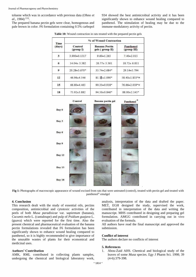
Table 10: Wound contraction in rats treated with the prepared pectin gels
Fig 1: Photographs of macroscopic appearance of wound excised from rats that were untreated (control), treated with pectin gel and treated with
panthenol® emulgel
4. Conclusion
This research dealt with the study of essential oils, pectins
composition, antimicrobial and cytotoxic activities of the
peels of both Musa paradisicae var. sapientum (banana),
Cucumis melo L. (cantaloupe) and pulp of Psidium guajava L.
(guava) which were reported for the first time. Also the
present chemical and pharmaceutical evaluation of the banana
pectin formulations revealed that F6 formulation has been
significantly shown to enhance wound healing compared to
panthenol, so it is highly recommended to give importance of
the unusable wastes of plants for their economical and
medicinal uses.
Authors’ Contribution
AMK, RML contributed in collecting plants samples,
undergoing the chemical and biological laboratory work,
analysis, interpretation of the data and drafted the paper.
MET, EGH designed the study, supervised the work,
contributed in interpretation of the data and writing the
manuscript. MHS contributed in designing and preparing gel
formulation. AMGG contributed in carrying out in vivo
evaluation of the formula.
All authors have read the final manuscript and approved the
submission.
Conflict of interest
The authors declare no conflicts of interest
5. References
1. Abou-Zaid AHS. Chemical and biological study of the
leaves of some Musa species. Egy J Pharm Sci. 1998; 39
(4-6):379-398.

~ 1815 ~
Journal of Pharmacognosy and Phytochemistry 2. Adams RP. Identification of essential oil components by
Gas Chromatography/Mass Spectrometry, 4th Ed. Allured
Business Media, Gundersen Drive, Carol Stream, Illinois,
USA, 2009.
3. Akamine K, Koyama T, Yazawa K. Banana peel extract
suppressed prostate gland enlargement in testosterone-
treated mice. Biosci Biotechnol Biochem. 2009;
73(9):1911-1914.
4. Agrawal M, Kumar A, Gupta R, Upadhyaya S.
Extraction of polyphenol, flavonoid from Emblica
officinalis, Citrus limon, Cucumis sativus and evaluation
of their antioxidant activity. Orient J Chem. 2012;
28(2):993-998.
5. Aziz AN, Ibrahim H, Rosmy SD, Mohtar M, Vejayan J,
Awang K. Antimicrobial compounds from Alpinia
conchigera. J Ethnopharmcol. 2013; 145(3):798-802.
6. Beaulieu JC, Grimm CC. Identification of volatile
compounds in cantaloupe at various developmental
stages using solid phase micro extraction. J Agr Food
Chem. 2001; 49:1345-1352.
7. Bennis S, Chami N, Rhayour K, Tantaoui-Elaraki A,
Remmal A. Eugenol induces damage of bacterial and
fungal envelope. Moroccan J Biol. 2004; 1:33-39.
8. Bhatty RS. Further compositional analyses of flax
mucilage, trypsin inhibitors and hydrocyanic acid. J
Amer Oil Chem. 1993; 70(9):899-904.
9. Bhuiyan MMH, Hossain MI, Mahmud MM, Mohammed
A. Microwave-assisted efficient synthesis of chalcones as
probes for antimicrobial activities. J Chemistry. 2011;
1:21-28.
10. Broadhurst CL, Duke JA. Oil of cloves: the benefits of
eugenol, Ph.D. 1997.
11. Calderón JC, Jaimes LC, Hernánddez EG, Villanova BG.
Antioxidant capacity, phenolic content and vitamin C in
pulp, peel and seed from 24 exotic fruits from Colombia.
Food Res Int. 2011; 44(7):2047-2053.
12. Deyo A, Malley B. Cucurbitaceae, 2008, 1-5p.
13. Dolz M, Gonzaler F, Belda R, Herraez JV. Thixotropic
behavior of microcrystalline cellulose – sodium
carboxymethyl cellulose gel. J Pharm Sci. 1988; 77:799.
14. Egyptian Pharmacopeia. General Organization for
Governmental printing Office, Cairo, Egypt, 1984, 31-
33p.
15. Ekong AE, Melbouci M, Lusvardi K, Eraze-Majewicz
PE. In "Handbook of Cosmetic Science and
Technology"; Barel AO, Paye M and Maibach HI, eds.,
Marcel Dekker, Inc. New York and Basel, 2001, 384-
385p.
16. Escrig AJ, Rincon M, Pulido R, Calixto FS. Guava fruit
as a new source of antioxidant dietary fiber. J Agr Food
Chem. 2001; 49(11):5489-5493.
17. Gangadevi V, Muthumary J. Preliminary studies on
cytotoxic effect of fungal taxol on cancer cell lines.
African J Bio technol. 2007; 6:1382-1386.
18. Garcia-Solis P, Yahia EM, Morales-Tlalpan V, Diaz-
Mumoz M. Screening of antiproliferative effect of
aqueous extracts of plant foods consumed in Mexico on
the breast cancer cell line MCF-7. Int J Food Sciences
Nutri. 2009; 60:32-46.
19. Hassimotto NMAH, Genovese MI, Lajolo FM.
Antioxidant capacity of Brazilian fruits, vegetables and
chemically-frozen fruit pulps. J Food Comp Anal. 2009;
22(5):394-396.
20. Ho CH, Piotrowski J, Dixon SJ, Baryshnikova A,
Costanzo M, Boone C. Combining functional genomics
and chemical biology to identify targets of bioactive
compounds. Curr Opin Chem Boil. 2011; 15(1):66-78.
21. Howat RJ, Senter SD. Identification of additional volatile
compounds from cantaloupe. J Food Sci. 1987;
52(4):1097-1098.
22. Hsouna AB, Trigui M. Chemical composition,
cytotoxicity effect and antimicrobial activity of
Ceratonia siliqua essential oil preservative effects against
Listeria inoculated in minced beef meat. Int J Food
Microbiol. 2011; 148(1):66-72.
23. Ismail HI, Chan KW, Mariod AA, Ismail M. Phenolic
content and antioxidant activity of cantaloupe methanolic
extracts Food Chem. 2010; 119(2):643-647
24. Jain S, Padsalg BD, Patel AK, Mokale V. Formulation,
development and evaluation of Fluconazole gel in various
polymer bases. Asian J Pharm. 2007; 1:63-68.
25. Jordán MJ, Margaria CA, Shaw PE, Goodner KL.
Volatile components and aroma active compounds in
aqueous essence and fresh pink guava fruit puree by GC-
MS and multidimensional GC/GC-O. J Agr food Chem.
2003; 51(5):1421-1426.
26. Karawya MS. Essential oil of Egyptian guava leaves. Egy
J Pharm Sci. 1999; 40(2):209-217.
27. Khan AA, Kumar V, Singh BK, Singh R. Evaluation of
wound healing property of Terminalia catappa on
excision wound models in Wistar rats, Drug Res. 2014;
64(5):225-228.
28. Kumar S. Text book of plant taxonomy, 1st Ed. Campus
Books International, Delhi, India, 2002, 128-131, 162-
166 and 302-306.
29. Lucero MJ, Vigo J, Leon MJ. A study of shear and
compression deformations on hydrogels of tretinoin. Int J
Pharm, 1994, 106, 125.
30. Mittal P, Gupta V, Kaur G, Garg AK, Singh A.
Phytochemistry and pharmacological activities of
Psidium guajava: A review. Int J Pharm Sci Res. 2010;
1(9):9-19.
31. Mosmann T. Rapid colorimetric assay for cellular growth
and survival: application to proliferation and cytotoxicity
assays. J Immunol Methods. 1983; 65:55-63.
32. Nattaporn W, Pranee A. Effect of pectinase on volatile
and functional bioactive compounds in the flesh and
placenta of sunlady cantaloupe. Int Food Res J. 2011;
18:819-827.
33. Ohno Y, Takuma T, Asahi K, Isono K. Differentiation
induction of murine erythroleukemia cells by butylated
hydroxyl toluene. FEBS (Federation of European
biochemical societies). 1984; 165(2):277-279.
34. Pazur JH. Neutral polysaccharides. Chaplin MF and
Kennedy JF, Eds. Carbohydrate Analysis: a Practical
Approach. 1986, 55-96p. Oxford, IRL Press.
35. Saini RK, Choudhary AS, Joshi YC, Joshi P. Solvent free
synthesis of chalcones and their antibacterial activities. E
J Chemistry. 2005; 2(4):224-227.
36. Scott AC. Laboratory control of antimicrobial therapy.
In: Collee, J.G. et al., eds. Practical Medical
Microbiology, 13th Ed. Edinburgh: Churchill Livingstone,
1989, 161-181.
37. Soares FD, Pereira T, Marques MOM, Monteiro AR.
Volatile and non volatile chemical composition of the
white guava fruit at different stages of maturity. Food
Chem. 2007; 100(1):15-21.
38. Soliman FM, Fathy MM, Salama MM, Saber FR.
Comparative study of the volatile oil content and
antimicrobial activity of Psiduim guajava L. and Psiduim

~ 1816 ~
Journal of Pharmacognosy and Phytochemistry cattleianum Sabine leaves. Bull Fac Pharm Cairo Univ.
2016; 54(2):219-225.
39. Someya S, Yoshiki Y, Okubo K. Antioxidant compounds
from bananas (Musa cavendishii). Food Chem. 2002;
79(3):351-354.
40. United States Pharmacopeia. Twinbrook Parkway,
Rockville, M.D., 2011, 3831p.
41. Yehye AW, Abdul Rahman N, Yaeghoobi M.
Understanding the chemistry behind the antioxidant
activities of BHT: A review. Eur J Med Chem. 2015;
101:295-312.

![Analytische Chemie - uni-muenster.de · Sulfur contents in crude oils range from 0.05 to 13.95 %wt [3], typical amount of sulfur for economically interesting oils vary from 0.1 to](https://static.fdokument.com/doc/165x107/5d4df46188c993cf7a8b9bb4/analytische-chemie-uni-sulfur-contents-in-crude-oils-range-from-005-to-1395.jpg)