Crystal structure determination of hyaluronidase, a major...
101
Crystal structure determination of hyaluronidase, a major bee venom allergen, in complex with an IgG Fab fragment and purification and biophysical characterization of bovine testes hyaluronidase Inauguraldissertation zur Erlangung der Würde eines Doktors der Philosophie vorgelegt der Philosophisch-Naturwissenchaftlichen Fakultät der Universität Basel von Sivaraman Padavattan aus Indien Basel, 2006
Transcript of Crystal structure determination of hyaluronidase, a major...
Crystal structure determination of hyaluronidase, a major bee venom
allergen, in complex with an IgG Fab fragment and purification and
biophysical characterization of bovine testes hyaluronidase
Inauguraldissertation
zur
Erlangung der Würde eines Doktors der Philosophie
vorgelegt der
Philosophisch-Naturwissenchaftlichen Fakultät
der Universität Basel
von
Sivaraman Padavattan
aus
Indien
Basel, 2006


Genehmigt von der Philosophisch-Naturwissenchaftlichen Fakultät
auf Antrag von
Prof. Dr. Tilman Schirmer
Dr. Zora Markovic-Housley
Prof. Dr. Andreas Engel
Basel, den 4.7.2006
Prof. Dr. Hans-Jakob Wirz
Dekan
Ⅰ


Declaration
I declare that I wrote this thesis, Crystal structure determination of hyaluronidase, a
major bee venom allergen, in complex with an IgG Fab fragment and purification
and biophysical characterization of bovine testes hyaluronidase, with the help indi-
cated and only handed it into the Faculty of Science of the University of Basel and to
no other faculty and no other university.
Ⅱ

Acknowledgments
I would like to thank my supervisor, Dr. Zora Markovic-Housely for her guidance, constant
encouragement, valuable suggestion and support throughout my thesis. My sincere thanks to
Prof Tilman Schirmer, for his excellent supervision through constructive criticism. I was able to
receive immediate feedback from him during entire course of my PhD studies. I am grateful to
Prof Andreas Engel for accepting to be an examiner and Prof Thomas Kiefhaber for moderating
the viva voce.
Thank to Dr Jun-ichi Saito (Guru), the best friend I got in Basel, for his support both scientifi-
cally and personally which will remain in my memories forever. It was great pleasure to thank
my group members Dr. Dinesh, Dr. Carmen Chan, Dr. Caroline Peneff, Dr. Arnaude Basle, Paul
Wassmann, Christophe Wirth for their support and valuable discussion. Special thanks to Di-
etrich Samoray for his technical help and for translating German letters.
I wish to thank Dr. Joseph, for his support and invaluable technical help during my PhD thesis.
Thanks to Dr. Paul Jenoe and Ariel Lustig for mass fingerprinting and analytical ultracentrifu-
gation analyses.
Thanks to Mrs. Ute Gruetter for the administrative work and Roland Buerki and Margrit Jenny
for the computer installation.
Many thanks to Senthil, Dinesh, Mathi, Ravi and Rajesh for supporting me all throughout my
thesis writing.
Special thanks to Senthil (Seni), Dinesh, Kavitha, naughty Charen. We were like family and
they made my Basel stay a memorable one. Thanks to other Basel friends Balasubramanian,
Balamurugan, Arundhathi, Satheesh, Rathi, Murali and Rajeshwaren they all made me to feel at
home.
My special regards to Stephanie Goulet, a Canadian girl, my neighbor in student home, for her
support and help during my stay in Basel. Thanks to Lausanne friends Kannu, Vidya, Gnana,
Renuga for their constant support during my PhD studies.
Ⅲ

My humble thanks to Prof H.S. Savithri and Prof M.R.N Murthy who are my ‘role models’ in
the scientific profession. They gave me the motivation, encouragement and support for find my
PhD position.
Thanks to Isai, who trained me in protein purification and crystallization during my Master’s
study.
My special thanks to my parents, Ragupathy uncle, Kumarasamy teacher, they gave me the mo-
tivation, encouragement throughout my studies and they well deserve the credit of my work.
...........to my parents, Ragupathy uncle and
Kumarasamy teacher
Ⅳ

Abbreviation
Ab Antibody
Ag Antigen
APC Antigen presenting cells
AU Analytical ultracentrifugation
CDR Complementary determining region
CHES 2-(N-cyclohexylamino) ethanesulfonic acid
Da Dalton
DC Dendritic cell
DTT Dithiothreitol
EDTA Ethylenediaminetetraacetic acid
Fab Fragment antigen binding
Fc Fragment crystallizable
FWR Frame work region
GPI Glycosyl phosphatidylinositol
HA Hyaluronic acid or hyaluronan
Hya Hyaluronidase
Hyal Hyaluronidase (mammals)
His-tag Hexahistidine tag
IgG Immunoglobulin G
IL Interleukin
kDa Kilo dalton
LS-MS Liquid chromatography-Mass spectrometry
mAb Monoclonal antibody
MALDI-TOF Matrix assisted laser desorption ionization-Time of flight
min Minute
MW Molecular weight
OD Optical density
PEG Polyethylene glycol
PDB Protein data bank
PH2060 60-kDa PH-20 protein purified from commercial bovine testicular ex-
tract (Sigma)
Ⅳ

PH2069 69-kDa PH-20 protein purified from commercial bovine testicular ex-
tract (Sigma)
PH2080 80-kDa PH-20 protein purified from bovine testes
PI-PLC Phosphatidylinositol-specific phospholipase C
PMSF Phenylmethylsulphonyl fluoride
Rfactor Crystallographic residual for working set of reflections
Rfree Crystallographic residual for test set of reflections
RT Room temperature
rms Root-mean-squared
rpm Rotation per minute
SDS-PAGE Sodium dodecylsulphate-polyacrylamide gel eclectrophoresis
SPAM1 Sperm adhesion molecule1
TABS N-tris[Hydroxymethyl]methyl-4-aminobutanesulfonic acid
TCR T-cell receptor
Th2 T helper cell 2
Ⅳ


Table of contents
Declaration ⅡAcknowledgments ⅢAbbreviation Ⅴ
1.0 Hyaluronidases: background and significance 1
Bibliography 9
2.0 Identification of a B-cell epitope of hyaluronidase, a major bee
venom allergen, from its crystal structure in complex with a spe-
cific Fab (S.Padavattan et al., JMB, v. 368, p. 742-52) 13
2.1 Supplementary information (Hya/Fab complex) 25
2.1.1 Hya/Fab complex formation, crystallization,
diffraction and structure solution 26 2.1.1.1 IgG purification 26
2.1.1.2 IgG digestion 27
2.1.1.3 Purification of Fab using cation exchange chromatogra-
phy (Mono-S) 28
2.1.1.4 Complex formation and purification using gelfiltration
column (Superdex S-75 16/60) 28
2.1.1.5 Crystallization and diffraction 31
2.1.1.6 Structure solution: Search for right Fab model for mo-
lecular replacement (MR) 34
2.1.1.7 Crystal packing of Hya/Fab -21E11 complex 37
2.1.2 Mechanism of type 1 hypersensitivity reaction 38
Ⅶ


2.1.3 Specific immunotherapy 40
Bibliography 42
3.0 Purification and biophysical characterization of bovine
testes hyaluronidase 43
3.1 Abstract 44
3.2 PH-20/Sperm adhesion molecule (SPAM1) 45
3.3 Methods and material 50
3.3.1 Substrate gel assay 50
3.3.2 Protein Electrophoresis and Immunoblotting 50
3.3.3 Cloning and expression of human PH-20 51
3.3.4 Protein purification from crude extract ( type Ⅳ-S: From
bovine testes, Sigma) 51
3.3.5 Protein purification from bovine testes 52
3.3.6 Analytical ultracentrifugation 53
3.3.7 Mass spectral analysis 53
3.3.8 Mass fingerprinting 54
3.3.9 Crystallization attempts 54
3.3 Results 55
3.3.1 Expression of recombinant human PH-20 protein 55
3.3.2 Purification of PH-20 protein from Sigma crude extract 55
3.3.3 Purification of PH-20 from bovine testes 63
3.3.4 Endoproteolytic cleavage of PH-20 protein 66
Ⅷ


3.3.5 Mass spectrometry 67
3.3.6 PH-20 aggregation 68
3.4 Discussion 71
3.4.1 PH-20 purification 71
3.4.2 Endoproteolytic cleavage of PH-20 71
3.4.3 PH-20 crystallization and aggregation 72
3.5 Conclusion 74
Bibliography 75
Appendix 78
curriculum vitae 78
Ⅸ


1.0 Hyaluronidases: background and significance
1.0 Hyaluronidases: background and significance
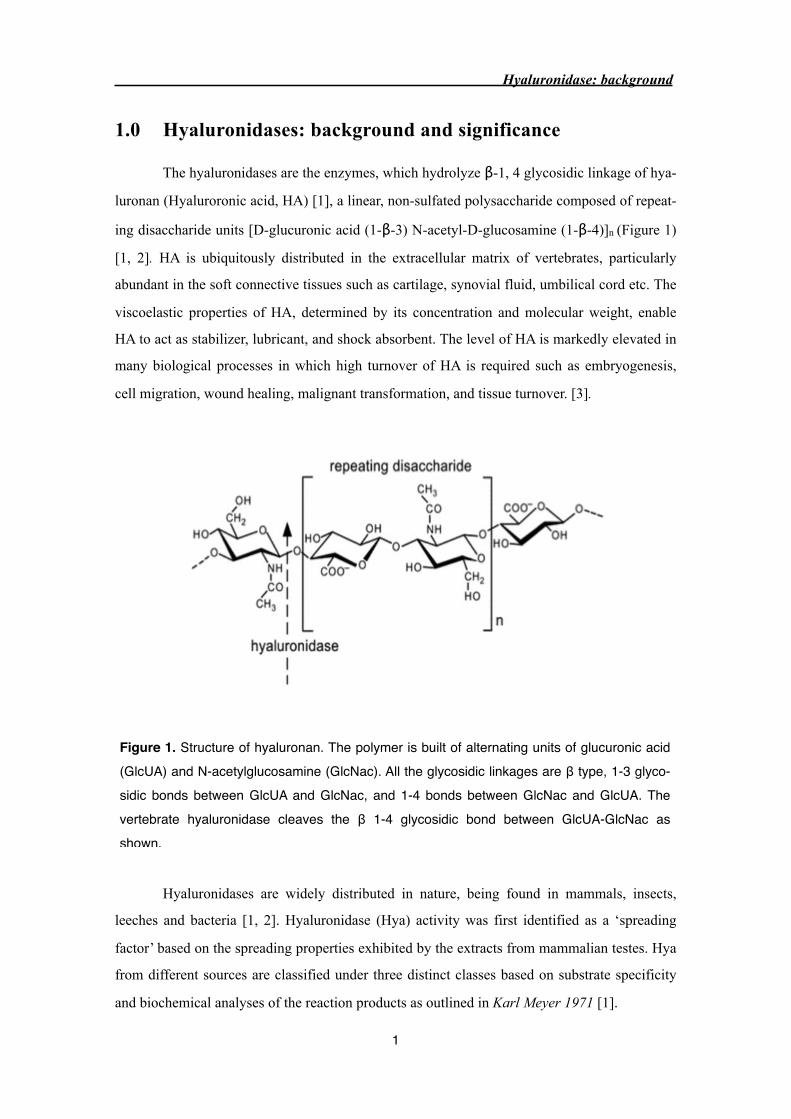
The hyaluronidases are the enzymes, which hydrolyze β-1, 4 glycosidic linkage of hya-
luronan (Hyaluroronic acid, HA) [1], a linear, non-sulfated polysaccharide composed of repeat-
ing disaccharide units [D-glucuronic acid (1-β-3) N-acetyl-D-glucosamine (1-β-4)]n (Figure 1)
[1, 2]. HA is ubiquitously distributed in the extracellular matrix of vertebrates, particularly
abundant in the soft connective tissues such as cartilage, synovial fluid, umbilical cord etc. The
viscoelastic properties of HA, determined by its concentration and molecular weight, enable
HA to act as stabilizer, lubricant, and shock absorbent. The level of HA is markedly elevated in
many biological processes in which high turnover of HA is required such as embryogenesis,
cell migration, wound healing, malignant transformation, and tissue turnover. [3].
Figure 1. Structure of hyaluronan. The polymer is built of alternating units of glucuronic acid (GlcUA) and N-acetylglucosamine (GlcNac). All the glycosidic linkages are β type, 1-3 glyco-sidic bonds between GlcUA and GlcNac, and 1-4 bonds between GlcNac and GlcUA. The vertebrate hyaluronidase cleaves the β 1-4 glycosidic bond between GlcUA-GlcNac as shown.
Hyaluronidases are widely distributed in nature, being found in mammals, insects,
leeches and bacteria [1, 2]. Hyaluronidase (Hya) activity was first identified as a ‘spreading
factor’ based on the spreading properties exhibited by the extracts from mammalian testes. Hya
from different sources are classified under three distinct classes based on substrate specificity
and biochemical analyses of the reaction products as outlined in Karl Meyer 1971 [1].
Hyaluronidase: background
1

‣ Group 1 is represented by the mammalian hyaluronidases (E.C. 3.2.1.35) and the enzymes
from venom of insects, snakes, scorpions which hydrolyze β-1, 4 glycosidic bond between
GlcNac and GlcUA, generating tetra- and hexasaccharides as the predominant end products.
They also digest chondroitin sulfate and to small extent dermatan sulfate. Bee venom hyalu-
ronidase and PH-20 protein from mammalian testis are best characterized enzymes.
‣ Group 2 is represented by leeches enzymes (E.C. 3.2.1.36) which act as endo-β-
glucuronidases. These enzymes generate tetra- and hexasaccharide end products.
‣ Group 3 comprises bacterial HA-lyases (E.C. 4.2.99.1) that act as endo-N-acetyl-
hexosaminidases by β-elimination, yielding predominantly disaccharides as end products.
Based on the sequence homology, hyaluronidases from insect (honeybees and vespids)
venom and mammalian hyaluronidases (EC 3.2.1.35) found in tissues (testis and plasma),
snakes and scorpion enzymes have been classified into the glycosidase family 56 [4, 5]. Hya
from honey bee venom shares greater than 50% sequence identity with other hymenopterans
Hya [6, 7] and 30% sequence identity with the sperm PH-20, involved in fertilization, and the
human lysosomal enzymes Hyal-1 and Hyal-2 which regulate HA turnover [8, 9]. The crystal
structure of bee venom hyaluronidase, the first representative structure of glycosidase family
56, has been determined at 1.6 Å resolution. The overall fold resembling closely a classical (α/
β)8 TIM barrel with the exception that seven β-strands form the barrel [9]. The structure of Hya
in complex with HA-tetramer (2.65 Å) enabled the elucidation of an acid-base catalytic mecha-
nism in which Glu113 acts as the proton donor whereas the N-acetyl group of the substrate is
the nucleophile. This unusual substrate-assisted acid-base catalytic mechanism is most proba-
bly also used by homologous mammalian enzymes since the active site residues and the resi-
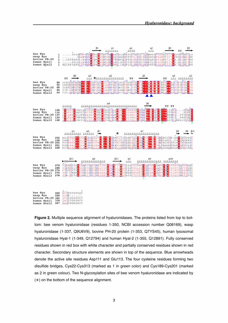
dues in the substrate binding cleft are highly conserved. In addition, four cysteine residues
forming two disulfide bridges are highly conserved (Figure 2). In comparison to bee venom
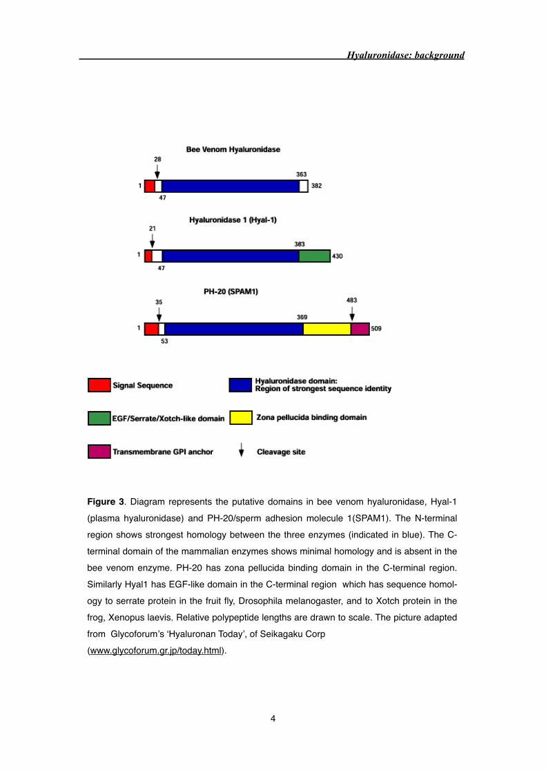
hyaluronidase (350 residues), vertebrate hyaluronidases have an additional C-terminal domain
EGF-like domain in Hyal-1 and a putative cell adhesion domain in PH-20. In addition, C-
terminus of PH-20 has a transmembrane glycosyl phosphatidylinositol (GPI) lipid anchor [10,
11] (Figure 3).
Hyaluronidase: background
2
bee Hyawasp Hyabovine PH-20human Hyal1human Hyal2
bee Hyawasp Hyabovine PH-20human Hyal1human Hyal2
bee Hyawasp Hyabovine PH-20human Hyal1human Hyal2
bee Hyawasp Hyabovine PH-20human Hyal1human Hyal2
bee Hyawasp Hyabovine PH-20human Hyal1human Hyal2
bee Hyawasp Hyabovine PH-20human Hyal1human Hyal2
✭
✭
Hyaluronidase: background
3
Figure 2. Multiple sequence alignment of hyaluronidases. The proteins listed from top to bot-tom: bee venom hyaluronidase (residues 1-350, NCBI accession number Q08169), wasp hyaluronidase (1-337, Q9U6V9), bovine PH-20 protein (1-353, Q7YS45), human lysosomal hyaluronidase Hyal-1 (1-349, Q12794) and human Hyal-2 (1-355, Q12891). Fully conserved residues shown in red box with white character and partially conserved residues shown in red character. Secondary structure elements are shown in top of the sequence. Blue arrowheads denote the active site residues Asp111 and Glu113. The four cysteine residues forming two disulfide bridges, Cys22-Cys313 (marked as 1 in green color) and Cys189-Cys201 (marked as 2 in green colour). Two N-glycosylation sites of bee venom hyaluronidase are indicated by (✭) on the bottom of the sequence alignment.
Hyaluronidase: background
4
Figure 3. Diagram represents the putative domains in bee venom hyaluronidase, Hyal-1 (plasma hyaluronidase) and PH-20/sperm adhesion molecule 1(SPAM1). The N-terminal region shows strongest homology between the three enzymes (indicated in blue). The C-terminal domain of the mammalian enzymes shows minimal homology and is absent in the bee venom enzyme. PH-20 has zona pellucida binding domain in the C-terminal region. Similarly Hyal1 has EGF-like domain in the C-terminal region which has sequence homol-ogy to serrate protein in the fruit fly, Drosophila melanogaster, and to Xotch protein in the frog, Xenopus laevis. Relative polypeptide lengths are drawn to scale. The picture adapted from Glycoforum’s ‘Hyaluronan Today’, of Seikagaku Corp (www.glycoforum.gr.jp/today.html).
Bee venom hyaluronidase: It is one of the major allergens present in bee venom which spe-
cifically degrades HA in the extracellular matrix of skin thereby facilitating penetration of
venom constituents into the body [12]. Native hyaluronidase, isolated from honeybee venom, is
a single chain secreted protein composed of 350 amino acids, which is derived from a precursor
composed of a signal peptide and a short prosegment. Recombinant His-tagged Hya expressed
in eukaryotic (Baculovirus) expression system, has enzymatic activity and IgE-binding capacity
similar to native Hya [13]. Hya contains four potential N-glycosylation motifs (Asn-X-Thr,
where X is any amino acid). The crystal structure of the recombinant (Baculovirus) bee venom
hyaluronidase shows that the Asn residues at 83 and 231 have weak density extending from
these residues, which is indicative of glycosylation. In contrast, Asn191 is buried in the protein
interior so that it can be ruled out as glycosylation site and Asn4 in the N-terminus is not de-
fined by electron density. Hya contains two disulfide bridges: Cys189-Cys201 which stabilizes
the base of a long loop at the C-terminal end, whereas the Cys22-Cys313 bridge joins the sec-
ondary structure elements, a 310 helix near the N-terminus and α-helix near the C-terminus.
The prominent feature of Hya structure is the large gap between stands 1 and 2 (7-8Å) and a
Hyaluronidase: background
5
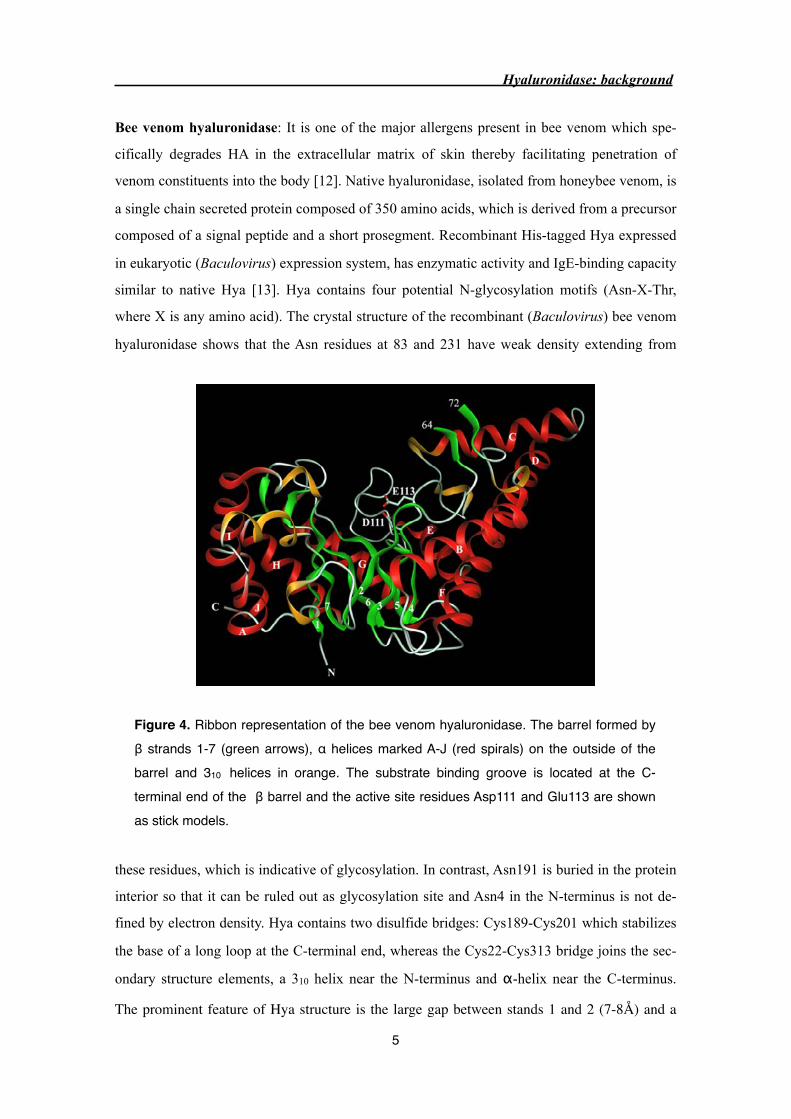
Figure 4. Ribbon representation of the bee venom hyaluronidase. The barrel formed by β strands 1-7 (green arrows), α helices marked A-J (red spirals) on the outside of the barrel and 310 helices in orange. The substrate binding groove is located at the C-terminal end of the β barrel and the active site residues Asp111 and Glu113 are shown as stick models.

long groove formed by the loops at the C-terminal end of β-barrel, suited for substrate binding.
Another unusual feature is the presence of two α-helices (helix C and D) resembling a ‘handle’
like structure which extend away from globular domain (Figure 4) [9].
Mammalian hyaluronidases: Mammalian hyaluronidases are presumably responsible for hya-
luronan (HA) turnover in diverse tissue but the details of HA catabolism remain obscure.
Mammalian genome consists of six hyaluronidase genes, clustered into two groups and shares
40% sequence identity to each other. In the human, three genes (HYAL1, HYAL2 and HYAL3)
are found tightly clustered on chromosome 3p21.3 and likewise, another three genes HYAL4,
HYALP1 (a pseudogene), and PH-20/SPAM1 are clustered on chromosome 7q31.3 (Figure 5)
[11]. The extensive homology between the different hyaluronidase genes suggests ancient gene
duplication. Correspondingly, mouse genome also contains six hyaluronidase genes, except
HYALP1 ortholog does not contain any mutation, and may encode for an active enzyme.
Hyaluronidase: background
6
Figure 5. Depicts the chromosomal orientation of the six hyaluronidase genes at their two respective chromosomal sites, and tabulation of their gene products. The relative gene order has been established for the chromosome 7, but their orientation in rela-tion to the centromere and telomere has not yet been determined. This figure is not drawn to scale. Picture was adapted from Glycoforum’s ‘Hyaluronan Today’, of Seikagaku Corp (www.glycoforum.gr.jp/today.html).
PH-20, Hyal-1 and Hyal-2 are well characterized mammalian hyaluronidase. PH-20 is
expressed only in the testis and plays important role in mammalian fertilization (more details
under PH-20/Sperm adhesion molecule 1). Hyal-1 and Hyal-2 are expressed in the somatic tis-
sues such as liver, kidney, spleen etc., and the expression of these enzymes are extremely low
and difficult to purify. Recent development in Hya detection procedure (substrate gel assay)
[14] has facilitated the purification of 57 kDa molecular weight Hyal-1 to homogeneity from
human plasma [15]. In contrast to plasma, urine contains two hyaluronidases with molecular
weight of 57 kDa and 45 kDa. Microsequencing confirmed that both urinary isozymes have N-
terminal identical to plasma hyaluronidase. Lower molecular weight isozyme contains a second
N-terminal sequence, which was derived from the C-terminal end of the protein. This suggests
that 45 kDa isozyme, resulting from endoproteolytic cleavage of the 57 kDa isoform, consists
of two polypeptides linked by disulfide bond [16]. In vertebrates, the turnover of HA is con-
trolled by hyaluronidase, which is an endoglycosidase that acts jointly with two lysosomal exo-
glycosidases, β-glucuronidase and β-N-acetyl glucosaminidase.
Hyal-3 is widely expressed enzyme but no activity was identified by present hyaluroni-
dase assay [17]. In contrast to other hyaluronidases, Hyal-4 appears to have specificity for
chondroitin and chondroitin sulfate, with no activity against hyaluronan [4]. In addition, Hyal-
2, Hyal-4 and PH-20 are GPI-anchored on the plasma membrane [11].
Biological and medical significance of hyaluronidases:
★ The PH-20 protein plays a major role in mammalian fertilization. The male and female
guinea pig immunized with PH-20 showed 100% contraception and the effect was long
lasting and reversible [18]. This suggest PH-20 can be effectively used as a contraceptive
vaccine.
★ The level of HA surrounding tumor cells often correlates with tumor aggressiveness and
overproduction of HA enhance anchorage-independent cell growth [19-21]. Loss of hyalu-
ronidase activity, permitting accumulation of HA, which might be one of the several steps
required by cells during carcinogenesis. In addition, hyaluronidase expression was seen in
various metastatic cell lines, which suggest tumor cells use hyaluronidase as "molecular
Hyaluronidase: background
7

destroyer" to depolymerize hyaluronic acid in surrounding tissues and facilitate tumor in-
vasion. Hyaluronidase on tumor cells may provide a target for anti-neoplastic drugs [22].
★ Testicular enzyme is used as a ‘spreading factor’, to improve better penetration of chemo-
therapeutic drug into tumors. Drawback of this approach is hyaluronidase developed symp-
toms of immediate type 1 allergic reaction [23].
Hyaluronidase: background
8

Bibliography
1 Meyer, K. (1971). Hyaluronidases. In The Enzymes, 3rd edition, volume V, P.D. Boyer, ed.
(New York: Academic press), pp. 307-320.
2. Kreil, G. (1995). Hyaluronidases--a group of neglected enzymes. Protein Sci 4, 1666-
1669.
3. Laurent, T.C., and Fraser, J.R. (1992). Hyaluronan. Faseb J 6, 2397-2404.
4. Henrissat, B. (1991). A classification of glycosyl hydrolases based on amino acid sequence
similarities. Biochem J 280 ( Pt 2), 309-316.
5. Henrissat, B., and Bairoch, A. (1996). Updating the sequence-based classification of gly-
cosyl hydrolases. Biochem J 316 ( Pt 2), 695-696.
6. Kolarich, D., Leonard, R., Hemmer, W., and Altmann, F. (2005). The N-glycans of yellow
jacket venom hyaluronidases and the protein sequence of its major isoform in Vespula
vulgaris. Febs J 272, 5182-5190.
7. Lu, G., Kochoumian, L., and King, T.P. (1995). Sequence identity and antigenic cross-
reactivity of white face hornet venom allergen, also a hyaluronidase, with other proteins. J
Biol Chem 270, 4457-4465.
8. Gmachl, M., and Kreil, G. (1993). Bee venom hyaluronidase is homologous to a mem-
brane protein of mammalian sperm. Proc Natl Acad Sci U S A 90, 3569-3573.
9. Markovic-Housley, Z., Miglierini, G., Soldatova, L., Rizkallah, P.J., Muller, U., and
Schirmer, T. (2000). Crystal structure of hyaluronidase, a major allergen of bee venom.
Structure 8, 1025-1035.
10. Cherr, G.N., Meyers, S.A., Yudin, A.I., VandeVoort, C.A., Myles, D.G., Primakoff, P., and
Overstreet, J.W. (1996). The PH-20 protein in cynomolgus macaque spermatozoa: identi-
fication of two different forms exhibiting hyaluronidase activity. Dev Biol 175, 142-153.
11. Csoka, A.B., Frost, G.I., and Stern, R. (2001). The six hyaluronidase-like genes in the hu-
man and mouse genomes. Matrix Biol 20, 499-508.
12. King, T.P., and Spangfort, M.D. (2000). Structure and biology of stinging insect venom
allergens. Int Arch Allergy Immunol 123, 99-106.
13. Soldatova, L.N., Crameri, R., Gmachl, M., Kemeny, D.M., Schmidt, M., Weber, M., and
Mueller, U.R. (1998). Superior biologic activity of the recombinant bee venom allergen
hyaluronidase expressed in baculovirus-infected insect cells as compared with Escherichia
coli. J Allergy Clin Immunol 101, 691-698.
14. Guntenhoner, M.W., Pogrel, M.A., and Stern, R. (1992). A substrate-gel assay for hyalu-
ronidase activity. Matrix 12, 388-396.
15. Frost, G.I., Csoka, A.B., Wong, T., and Stern, R. (1997). Purification, cloning, and expres-
sion of human plasma hyaluronidase. Biochem Biophys Res Commun 236, 10-15.
Hyaluronidase: background
9
16. Csoka, A.B., Frost, G.I., Wong, T., and Stern, R. (1997). Purification and microsequencing
of hyaluronidase isozymes from human urine. FEBS Lett 417, 307-310.
17. Stern, R. (2003). Devising a pathway for hyaluronan catabolism: are we there yet? Glyco-
biology 13, 105R-115R.
18. Primakoff, P., Lathrop, W., Woolman, L., Cowan, A., and Myles, D. (1988). Fully effective
contraception in male and female guinea pigs immunized with the sperm protein PH-20.
Nature 335, 543-546.
19. Kosaki, R., Watanabe, K., and Yamaguchi, Y. (1999). Overproduction of hyaluronan by
expression of the hyaluronan synthase Has2 enhances anchorage-independent growth and
tumorigenicity. Cancer Res 59, 1141-1145.
20. Liu, N., Gao, F., Han, Z., Xu, X., Underhill, C.B., and Zhang, L. (2001). Hyaluronan syn-
thase 3 overexpression promotes the growth of TSU prostate cancer cells. Cancer Res 61,
5207-5214.
21. Zhang, L., Underhill, C.B., and Chen, L. (1995). Hyaluronan on the surface of tumor cells
is correlated with metastatic behavior. Cancer Res 55, 428-433.
22. Liu, D., Pearlman, E., Diaconu, E., Guo, K., Mori, H., Haqqi, T., Markowitz, S., Willson,
J., and Sy, M.S. (1996). Expression of hyaluronidase by tumor cells induces angiogenesis
in vivo. Proc Natl Acad Sci U S A 93, 7832-7837.
23. Szepfalusi, Z., Nentwich, I., Dobner, M., Pillwein, K., and Urbanek, R. (1997). IgE-
mediated allergic reaction to hyaluronidase in paediatric oncological patients. Eur J Pediatr
156, 199-203.
Hyaluronidase: background
10
Hyaluronidase: background
11
The thesis is divided in two parts:
In the first part, I discuss the crystal structure determination of bee venom hyaluronidase in
complex with a Fab fragment of an anti-Hya monoclonal IgG antibody (clone 21E11) which is
able to compete with human IgE, from the bee venom allergic patient serum pool into Hya
binding. Hya is one of the major allergen present in honeybee venom that may induce life
threatening allergic reaction in human. Since the allergen-antibody interactions are crucial for
triggering of allergic responses, the structures of the antigen/antibody complexes are essential
for the identification of Hya epitopes recognized by IgE antibodies. The knowledge of B cell
epitope may facilitate the design of new and safer vaccines for allergen immunotherapy in the
form of mutated allergens with abolished or reduced IgE binding potency (hypoallergen).
In the second part, I discuss the PH-20 protein, a mammalian hyaluronidase which plays impor-
tant role in fertilization. Though mammalian hyaluronidases play important role in many bio-
logical processes, the study of these enzymes is greatly neglected. In the present study, we were
able to purify PH-20 proteins from commercially available testicular extract (Sigma) and from
the homogenized bovine testes to highest purity. The aggregation of purified PH-20 was suc-
cessfully eliminated by the addition of zwitterionic compound, NDSB (non detergent sulfobe-
taine). Attempt to crystallize the PH-20 protein were not successful but crystallization in the
presence of NDSB still remains to be tested.
Hyaluronidase: background
12

2.0 Identification of a B-cell epitope of hyaluronidase, a major
bee venom allergen, from its crystal structure in complex with a
specific Fab (S.Padavattan et al., JMB, v. 368, p. 742-52)
Hya/Fab complex structure
13

doi:10.1016/j.jmb.2007.02.036 J. Mol. Biol. (2007) 368, 742–752
Identification of a B-cell Epitope of Hyaluronidase, aMajor Bee Venom Allergen, from its Crystal Structurein Complex with a Specific Fab
Sivaraman Padavattan1, Tilman Schirmer1, Margit Schmidt2
Cezmi Akdis3, Rudolf Valenta4, Irene Mittermann4, Lyudmila Soldatova5
Jay Slater6, Ulrich Mueller7 and Zora Markovic-Housley1⁎
1Division of Structural Biology,Biozentrum, University of Basel,CH-4056 Basel, Switzerland2Department of Biology,East Caroline University,Greenville, NC 27858, USA3Department of CellularAllergology/Immunology,Swiss Institute of Allergy andAsthma Research, CH-7270,Davos, Switzerland4Christian Doppler Laboratoryof Allergy Research, Division ofImmunopathology, Departmentof Pathophysiology, MedicalUniversity of Vienna,A-1090 Vienna, Austria5OPS/ONDQA, Center forDrug Evaluation andResearch, US Food and DrugAdministration, Silver Spring,MD 20993, USA6Center for Biologics Evaluationand Research, US Food andDrug Administration,Bethesda, MD 20892, USA7SpitalBern, CH-3001 Bern,SwitzerlandAbbreviations used: Hyal (or rApfragment of IgG which binds to antmonoclonal antibody; MR, moleculaPEG, polyethylene glycol; SIT, speciE-mail address of the correspondi
0022-2836/$ - see front matter © 2007 E
The major allergens of honeybee venom, hyaluronidase (Hyal) andphospholipase A2, can induce life-threatening IgE-mediated allergicreactions in humans. Although conventional immunotherapy is effective,up to 40% of patients develop allergic side effects including anaphylaxis andthus, there is a need for an improved immunotherapy. A murinemonoclonal anti-Hyal IgG1 antibody (mAb 21E11), that competed forHyal binding with IgEs from sera of bee venom allergic patients, was raised.The fragment of these IgG antibodies which bind to antigen (Fab) wasproduced and complexed (1:1) with Hyal. The crystal structure determina-tion of Hyal/Fab 21E11 complex (2.6 Å) enabled the identification of theHyal–IgG interface which provides indirect information on the Hyal–IgEinteraction (B-cell epitope). The epitope is composed of a linear array of nineresidues (Arg138, His141–Arg148) located at the tip of a helix–turn–helixmotive which protrudes away from the globular core and fits tightly intothe deep surface pocket formed by the residues from the six complemen-tarity determining regions (CDRs) of the Fab. The epitope is continuous andyet its conformation appears to be essential for Ab recognition, since thesynthetic 15-mer peptide comprising the entire epitope (Arg138–Glu152) isneither recognized by mAb 21E11 nor by human IgEs. The structure of thecomplex provides the basis for the rational design of Hyal derivatives withreduced allergenic activity, which could be used in the development of saferallergen-specific immunotherapy.
© 2007 Elsevier Ltd. All rights reserved.
Keywords: hyaluronidase; bee venom allergen; B-cell epitope; hyaluroni-dase/Fab complex; X-ray structure
*Corresponding authori m 2), hyaluronidase; Ab, antibody; Ag, antigen; IgE, immunoglobulin E; Fab,igen; CDR, complementarity determining region; FWR, frame work region; mAb,r replacement; PBS, phosphate-buffered saline; PBST, PBS/0.05% v/v Tween 20;fic immunotherapy; SLS, Swiss Light Source.ng author: [email protected]
lsevier Ltd. All rights reserved.

743X-ray Structure of Bee Venom Hyaluronidase/Fab Complex
Introduction
Venoms of bees, wasps and fire ants can causesevere IgE-mediated allergic reactions (type Ihypersensitivity) including anaphylaxis.1–3 It hasbeen estimated that up to 3% of the generalpopulation have a history of systemic anaphylacticreactions to insect stings.4 Venoms from bee andwasp are different, each containing distinct majorallergens: phospholipase A2 and melittin occuronly in bee venom, and antigen 5 only in waspvenom, but both venoms contain hyaluronidases(Hyal).3 Clinical studies demonstrated that Hyaland phospholipase A2 are the two major allergenspresent in bee venom: 71% of patients had specificserum IgE against recombinant Hyal and 78%against recombinant phospholipase A2.5 Suscep-tible individuals respond to bee venom exposureby producing IgE antibodies which bind to thehigh affinity Fc receptors on basophils in circula-tion or mast cells in tissues. Following re-exposure, multivalent allergens cross-link Fc-recep-tor-bound IgE antibodies on the surface of mastcells. This leads to a series of signaling events,which result in mast cell degranulation and therelease of histamine, leukotriene and other med-iators which are responsible for a variety ofallergic symptoms.Bee venom Hyal is a hydrolytic enzyme that
specifically cleaves the hyaluronic acid, a large,linear polymer (hyaluronan) consisting of simplerepeats of disaccharide composed of the β-1,4 linkedD-glucuronic acid and N-acetyl-glucosamine. Hya-luronan and chondroitin sulfates are the mostabundant glycosaminoglycan of vertebrate extra-cellular matrix, and the relative abundance of theseglycoproteins varies with the origin of the connec-tive tissue. Cleavage of hyaluronan by Hyal facil-itates the penetration of venom constituents into thebody.6 Hyaluronan is found in almost all tissues andbody fluids and is particularly abundant in theintercellular matrix of skin and the connectivetissues of cartilage, synovial fluid and the vitreoushumor of the eye. Under physiological conditions,hyaluronan is a large (105 to 107 kDa), charged andextended polysaccharide which is exceptionallyhydrophilic. The ability to bind large amounts ofwater confers special viscoelastic properties tohyaluronan solutions which are the basis of itsstructural role as a stabilizer, joint lubricant andshock absorber.7,8 Hyaluronan exists in a number ofphysiological states, associated with themselves,with the extracellular matrix, with the cell surfacereceptors CD44 and RHAMM9 present on thehyaluronan-binding proteins (hyaladherins)10–12
forming massive multimolecular aggregates withproteoglycans, such as aggrecan.Bee venom Hyal is composed of 350 amino acids
and has four potentialN-glycosylation sites and twodisulfide bridges. In fact, only two of the fourpostulated sites are N-glycosylated, Asn115 andAsn263.13–15 Recombinant Hyal has been expressedin prokaryotic (Escherichia coli) and eukaryotic
(Baculovirus) hosts. Only the Baculovirus-expressedenzyme has enzymatic activity and IgE-bindingcapacity similar to native Hyal.16 The crystalstructure of the recombinant (Baculovirus) beevenom Hyal13 is the first structural representativeof the Hyals belonging to the glycosidase family56.17,18 The overall fold of Hyal resembles a classical(β/α)8 barrel except that the barrel is formed byseven β strands and is open between β strands 1 and2. The structure of Hyal in complex with hyaluronicacid-tetrameres enabled to propose an acid–basecatalytic mechanism in which Glu113 acts as theproton donor and the N-acetyl group of thesubstrate as the nucleophile.13,19 Recently, thestructure of homologous wasp Hyal has been solvedshowing a fold identical with that of bee venomHyal.20
Specific immunotherapy (SIT) is the only causaltreatment for type I allergies; however, a drawbackis the risk of IgE-mediated anaphylactic side effects,which occur in 20%–40% of patients21 subjected toSIT based on natural extracts. There is evidence thatsuccessful immunotherapy operates on the level of Thelper cells, leading to marked changes in cytokinesecretion patterns: TH2 cells secreting IL-4, IL-5 andIL-13 diminish strongly and are replaced by IL-10secreting CD4+CD25+ regulatory cells (TReg).
22,23
Furthermore, it has been shown that immunother-apy induces allergen-specific IgG antibodies whichinhibit the binding of allergic patients IgE to theallergen.24,25 One way to lower the risk of anaphy-laxis during SIT is to use allergens modified in a waywhich abolishes or reduces its IgE-binding potency(hypoallergen) but retains the allergen T-cell epi-topes (linear epitopes) that will induce T-celltolerance.23 Other possibilities are to prepare geneti-cally modified allergens or allergen-derived pep-tides with reduced allergenic activity which induceprotective allergen-specific IgG antibodies compet-ing with IgE.26
Since allergen–antibody (Ab) interactions arecrucial for the triggering of allergic responses, apromising approach to inhibit this process wouldbe to prevent allergen binding to IgE. Determiningthe structure of antigen (Ag)/Ab complexes is oneway to identify the Hyal epitopes which arerecognized by IgE antibodies. The knowledge ofthe B-cell epitope of the allergen is expected tofacilitate the design of a safer SIT in the form ofmutated allergens with reduced IgE-bindingpotency which would be able to bypass the riskof anaphylaxis.In the present study, a monoclonal murine IgG1
antibody (mAb, clone 21E11) raised against purifiedrecombinant Hyal and able to inhibit IgE binding toHyal up to 60% in some patients, was chosen to mapthe B-cell epitope. This is the second study of anallergen–Ab complex, after the Bet v 1–Fab (frag-ment of IgG which binds to antigen (Fab)) complexstructure, although many Ag–Fab complex struc-tures have been reported.27–30 It is anticipated thatthe knowledge of the Hyal epitope may be used togenetically modify the allergenic protein in order to

Table 1. Reactivity of mouse mAbs specific for Api m 2
Absorbance21E11
Absorbance22H7
Absorbance24F2
Peptide (R138–E152) 0.03 0.03 0.03Api m 2 0.90 1.11 0.67HSA 0.03 0.03 0.03
Absorbance values corresponding to the levels of mousemonoclonal IgG1 antibodies (mAbs: 21E11, 22H7, 24F2) specificfor Api m 2 and the Api m 2-derived peptide are displayed.Human serum albumin (HSA) was used as negative control.
744 X-ray Structure of Bee Venom Hyaluronidase/Fab Complex
reduce its IgE-binding potency and to employ it forsafer immunotherapy.
Results
Inhibition of IgE binding to Hyal by anti-Hyalmonoclonal IgG1 Abs
Hyal (rApi m 2) is recognized by serum IgE frombee venom allergic patients and also by threedifferent mouse monoclonal IgG1 antibodies (clones21E11, 22H7 and 24F2). In contrast, the Hyal derived15-mer peptide (Arg138–Glu152) containing nineepitope residues elucidated from the Hyal/Fabcomplex structure, is not recognized by the mAbs(Table 1). Also sera of bee venom allergic patients donot bind to the isolated peptide (data not shown).The effect of the three mouse mAbs on binding ofserum IgE from 10 bee venom allergic patients wastested by an ELISA-inhibition assay (Table 2). Amean inhibition of IgEs binding to Hyal by each ofthe three mAbs ranged between 18% and 20%.However, this value was as high as 57% for certainpatients' sera.
Structure determination of the Hyal/Fab complex
The crystal structure of the complex formedbetween Hyal and the Fab fragment of the mousemAb 21E11 has been determined at 2.6 Å resolution
Table 2. Inhibition of IgE binding of bee venom positive sera
IndividualAbsorbancemAb control
Absorbance21E11
%inhibitiona
Absorbance22H7 in
I 2.57 1.98 23.02 2.28II 1.47 1.15 21.77 0.99III 1.39 1.67 – 1.43IV 1.18 0.87 26.36 1.01V 0.61 0.58 5.06 0.58VI 0.54 0.23 56.83 0.25VII 0.32 0.30 6.48 0.25VIII 0.39 0.35 9.51 0.34IX 0.32 0.31 2.79 0.28X 0.22 0.15 32.72 0.16Mean 18.45
a rApi m 2 bound to ELISA plate was pre-incubated with three(mAbs:21E11, 22H7, 24F2), a mixture of all three mAbs and, for controplates were then incubated with sera of 10 bee venom allergic patieninhibition of IgE binding is shown.
by molecular replacement (MR) (Table 3). MR wasperformed in three consecutive steps using thestructures of apo Hyal (1FCQ), Fab variable domain(1A7Q) and constant domain (15C8) as the searchmodels. This yielded the orientation and locationof the three constituents in the asymmetric unit. Thefinal model comprises residues Glu10–Ser333 ofHyal, Asp1–Ile205 of the light chain and Gln1–Val180 of the heavy chain of Fab as well as 63 watermolecules. The Fab residues are numbered as de-scribed by standard Kabat convention with lightand heavy chain identifiers L and H, respectively.31,32In Hyal, the electron density is missing for 9 N-terminal and 17 C-terminal residues (plus the His6tail) and loop residues Asp66–Asn70. Likewise, inFab density is missing for residues L119–L131,L150–L157, L168–L169, L187–L192 and L206 to theC-terminal end of the light chain, and for residuesH122–H137, H157–H162 andH181 to the C-terminalend of the heavy chain.Most of these residues belongto the loops of the constant domain and are far awayfrom the Hyal/Fab interface. After conventionalrefinement, the final model has an R-factor of 20.9%(Rfree, 24.7%) at 2.6 Å resolution (Table 3). Theresidues are well ordered and exhibit good stereo-chemistry (Table 3). The electron density is of goodquality; an example of the final 2Fo–Fc map is shownin Figure 1.
Overall fold of the Hyal/Fab complex
The structure of the Hyal/Fab complex is shownas a ribbon presentation in Figure 2(a). The Hyal foldis a (β/α)7 barrel open between strands 1 and 2 andsurrounded by 10 α helices.13 The overall Agconformation is not changed significantly uponcomplex formation. Superposition of the apo Hyalmodels 1FCQ (1.6 Å resolution) and 1FCU (2.1 Åresolution)13 with Hyal of the Hyal/Fab complexgives r.m.s.d. of 0.76 and 0.61 Å (for 312 and 319 Cαatoms), respectively, which is comparable with thedifferences observed between the two unligandedHyal models (r.m.s.d. of 0.68 Å for 314 Cα atoms). Inprotein Ag/Ab complexes, conformational adjust-
to purified recombinant Api m 2 by mAbs
%hibition
Absorbance24F2
%inhibition
Absorbance21E11/22H7/24F2
%inhibition
11.14 1.97 23.26 2.20 14.1432.56 1.00 31.95 0.98 32.76– 1.21 12.83 1.17 15.28
14.54 1.23 – 0.94 20.414.73 0.59 4.08 0.43 29.2054.79 0.27 49.82 0.22 60.1521.61 0.27 17.91 0.21 36.4213.88 0.34 13.11 0.27 31.3611.80 0.28 13.98 0.25 23.9126.73 0.16 28.57 0.16 26.7319.18 19.55 29.04
individual Api m 2-specific mouse monoclonal IgG1 antibodiesl purposes, with a mouse mAb against Bet v 1 (mAb control). Thets. Api m 2-specific IgE levels were measured. The percentage of

†http://bioinfo.ernet.in/cep.htm
Table 3. Crystallographic data for Hyal/Fab complex
Data collection
Space group C2Unit cell dimensions
a, b, c (Å) 151.6, 70.1, 138.9α, β, γ (°) 90.0, 123.7, 90.0
X-ray source SLS-PXDetector type MAR CCDWavelength (Å) 0.978Resolution range (Å) 63–2.6 (2.74–2.6)a
No. of total observation 101,732 (14,135)No. of unique observation 32,047 (4437)Completeness (%) 85.4 (81.8)Multiplicity 3.2 (3.2)I/σ(I) 13.9 (3.6)Rsym
b (%) 8.4 (30)
Refinement
Rfactor/Rfreec (%) 20.9/24.7
Protein atoms 11642Water molecules 63Average B-factor (Å2)
Hyal 37.5Fab variable domain 38.0Fab constant domain 38.2Solvent 33.8
r.m.s.d. from ideal valuesBond lengths (Å) 0.009Bond angles (°) 1.136
r.m.s. ΔB of bonded atoms (Å2)Main chain 1.020Side-chain 2.257
Ramachandran plotMost favored region (%) 88.4Additionally allowed region (%) 10.3Generously allowed region (%) 0.5Disallowed region (%) 0.7a Number in parentheses are statistics for the data in the
highest resolution shell.b Rsym=ΣΣ|I(h)i− ⟨I(h)⟩|/ΣΣ I(h)i, observed intensity in the
ith data set and ⟨I(h)⟩, mean intensity of reflection h over allmeasurements of I(h).
c Rfactor is the conventional R-factor and Rfree is the R-factorcalculated with 5% of the data that were not used in refinement.
745X-ray Structure of Bee Venom Hyaluronidase/Fab Complex
ment in flexible loops and side-chains of the inter-acting residues upon complex formation is oftenobserved.29 In Hyal, the largest deviations in the Cαposition occur in loop residues His141–Asp145,which are part of the epitope (r.m.s.d.=2.6 and2.1 Å for 1FCQ and 1FCU, respectively), indicatinga considerable conformational change of the epi-tope loop upon Fab binding (Figure 2(b)). However,also the conformation of residues Thr193–Pro197from the loop which is far from the Ab binding site(r.m.s.d.=2.3 Å) is significantly changed. In theabsence of the structure of the free Ab, the extent ofpossible conformational changes of the residues be-longing to the variable domain cannot be evaluated.
The Hyal/Fab interface
The mAb 21E11 recognizes a continuous epitopelocated at the tip of the helix C–turn–helix Dmotif ofHyal, which resembles a handle that protrudesaway from the globular core of the molecule (Figure2(a)). The total contact surface area of the complex
interface was estimated to be 1274 Å2 (probe radius1.7 Å, program MS). The epitope is mostly contin-uous and composed of eight consecutive residuesHis141–Arg148 (HPFWDDQR) plus Arg138. Hyalstructure was submitted to CEP-server for theprediction of conformational epitopes†.33 In addi-tion to the predicted conformational epitopes, theserver predicted a sequential epitope at positionArg138–Gln151. This prediction is in good agree-ment with the results from our crystal structure,which showed that 9 of 14 residues predicted by theserver are indeed involved in Fab binding. All nineresidues belong to the C- and N-terminal ends ofhelices C and D and the joining turn (Figure 2(c)).Epitope residues fit tightly into the deep pocketformed by the side-chains from all six complemen-tarity determining regions (CDRs) and the lightframe work region 2 (FWR L2) of the Fab (Figures2(c) and 3). The interface is formed by 9 Hyal and 17Fab residues. The total number of polar Hyal/Fabinteractions comprises 4 salt bridges and 9 hydrogenbonds while 11 and 39 van der Waals contacts havebeen found for the cut off values of 3.7 Å and 4.0 Å,respectively (Table 4). Of the nine epitope residues incontact with Fab, two are polar, four are chargedand three are hydrophobic. The polar and chargedresidues are located predominantly at the peripheryof the Hyal binding surface, the two hydrophobicresidues (Pro142, Phe143) from the turn are buriedat the bottom of Hyal/Fab cavity formed by thehydrophobic residues of Fab, whereas Trp144 isonly 79% buried (Figures 2(c) and 3). The size of theFab binding pocket is approximately 14 Å wide and15 Å deep. The salt bridges are formed betweenAsp145 and Arg148 from helix D of Hyal and Arg58and Asp54 from CDR-H2 of Fab, respectively(Figure 3).Water molecules are often found in the interfaces
of Ag–Ab complexes34–36 where they are required toimprove the fit between the proteins and toneutralize unpaired hydrogen-bonding groups. Inthe Hyal/Fab complex, three water molecules (21O,22O and 23O) are completely buried between thetwo surfaces, filling the cavity and enhancingsurface complementarity (Table 5). Three additionalwater molecules (5O, 20O and 45O) are present atthe periphery of the interface where they enableHyal–water–water–Fab hydrogen bonding interac-tion. The shape correlation statistic for Hyal/Fabcomplex is 0.73 which is slightly higher than theobserved mean shape correlation statistic value of0.64–0.65 for Ag–Ab interface,37 indicating goodshape complementarity. Thus, the Hyal/Fab com-plex structure also confirms the importance ofbound water molecules in mediating protein/protein interactions.The Ab combining site is a pronounced cavity
formed by the side-chains of the Fab residuesinteracting with Hyal: six residues from the lightchain CDRs (L1, L2 and L3), nine residues from the

Figure 1. Close-up stereo view of the final SigmaA-weighted 2Fo–Fc electron density map60 contoured at 1.0σ. Shownis part of the Hyal/Fab interface with Hyal and Fab heavy chain colored magenta and yellow, respectively. All pictureswere produced using program DINO [http://www.dino3d.org/].
746 X-ray Structure of Bee Venom Hyaluronidase/Fab Complex
heavy chain CDRs (H1, H2 and H3) and tworesidues from the FWR L2. Similar to other Ab–Agcomplexes,27 the residues from the heavy chain,particularly CDR-H2, make extensive contact withHyal. In the Hyal/Fab complex, 6 out of 17 Fabresidues interacting with Hyal are aromatic, inagreement with a notion that aromatic residues,particularly tyrosines, form most of the contactswith Ag.27,38 Residues Tyr32 (L1), Tyr52 (H2) andTyr95 (H3) are almost completely buried (>90%)within the interface, whereas Tyr49 (FWR L2), Tyr92(L3) and Trp53 (H2) are partially buried (38–48%).Apart from CDRs, residues Leu46 and Tyr49 fromFWR L2 are also involved in apolar interactions withHyal. The shape of the Ab binding cavity is similarto those observed with haptens which bind ingrooves and pockets39 and usually do not causelarge conformational changes in Fab. The interactionbetween residues from CDR-H3 and Hyal ismediated by water and hydrophobic interactions.
Discussion
We have identified the first B-cell epitope of Hyal,a major allergen of bee venom, from the crystalstructure of Hyal in complex with the specific Fabfragment of a monoclonal murine anti-Hyal IgG1Ab. The Hyal epitope is composed of mostlycontinuous array of nine residues folded as ahelix–turn–helix motive, which protrudes awayfrom the globular protein core and fits tightly intothe deep pocket formed by the six CDRs of the Fab.A local conformational change of the turn joiningtwo helices is observed upon binding. Although theepitope is continuous, its conformation is importantfor Ab recognition, since a synthetic peptide(Arg138–Glu152), comprising the entire epitope, is
neither recognized by mAb-IgG 21E11 nor byhuman IgE from the sera of bee venom allergicpatients. This is the second structural study of anallergen–Ab complex after the major birch pollenallergen Bet v 1 in complex with the Fab fragmentsof a monoclonal murine IgG Ab.40 The latterstructure revealed a conformational epitope com-posed of the 16 residues belonging to a rather flatallergen–Ab molecular interface.The IgE antibodies from allergic patients are
polyclonal and bind to several different epitopes ofan allergen. We have shown that mAb 21E11exhibits a varying degree of inhibitory effect on thebinding of human IgEs to Hyal, ranging from 3% to57% depending on the patient. This broad range ofinhibition efficacy probably reflects the different IgErepertoire of each patient. The moderate inhibitionobserved with most of the sera is probably due tothe presence of a variety of IgE antibodies whichrecognize different epitopes. Thus, only a fraction ofIgE antibodies will be affected by the presence ofmAb 21E11 bound to Hyal. In the case thatinhibition is caused by direct competition of IgGand IgE for the same epitope, the Hyal–IgGinteraction surface provides indirect informationon the interaction of Hyal with IgE. However,allergen-specific IgG antibodies may inhibit thebinding of IgE by several alternative mechanisms,such as (i) partial overlap of the IgG and IgEepitopes, (ii) neighboring IgG and IgE epitopeswhich cannot be occupied simultaneously due tosteric hindrance and (iii) perturbation of IgEepitopes by the conformational changes inducedby IgG binding. The moderate inhibition (20–30%) isconsistent with the overlapping of IgG and IgEepitopes, as well as with mechanisms (i) or (ii).However, an inhibition of 57% would be consistenteither with the inhibition of a dominant IgE epitope

Figure 2. Hyal/Fab complex. (a) Ribbon representation of the Hyal/Fab complex structure. Same color code as inFigure 1; Hyal is shown in magenta and Fab heavy and light chains are colored yellow and green, respectively. For clarity,the residues of the Fab constant and variable domain not defined by electron density are also included and are coloredred. The polypeptide chain termini are labeled with N and C. (b) Conformational changes induced in the Hyal epitopeupon Hyal/Fab complex formation. Hyal, colored in magenta, is superposed on two models of unliganded Hyal: 1FCQ(light reddish brown) and 1FCU (dark reddish brown). The largest Cα atom shifts (more than 2 Å) are seen for Phe143. (c)Close-up view of the Hyal/Fab interface. The Hyal epitope is shown as a full atom representation colored magenta andthe molecular surface of the Fab colored yellow and green for the heavy and light chains, respectively. For clarity, residueArg138 of Hyal that is also involved in the interaction is not shown. For more details, see Table 4.
747X-ray Structure of Bee Venom Hyaluronidase/Fab Complex
or with a large conformational change, which wouldaffect a significant part of the molecule and thuscause the simultaneous perturbation of several IgEepitopes. However, the last proposed mechanism isless likely because only a local conformationalchange is observed.The Hyal/Fab complex shares a number of
general features with other protein–Ab complexeswhich possess a discontinuous epitope, e.g., all sixCDRs interact with the Ag,35,41 with the interactionswith the heavy chain being most prominent;27 thecontacting surfaces are highly complementary andhave areas in the range of 600Å2–900 Å2; the stabilityof a complex is provided by van der Waalsinteractions, hydrogen bonds and, to a lesser extend,by salt bridges. Epitopes in most of the protein/antibodies complexes are discontinuous and com-prise 14–20 residues.35,41,42 In contrast, the Hyalepitope is continuous and composed of only 9 resi-
dues, thus resembling to an Ab–peptide complex.43
However, many conformational epitopes contain alinear stretch of residues which account for most ofthe interactions with specific antibodies. For exam-ple, residues Ile42–Thr52 of the conformationalepitope of Bet v 1/Fab complex account for 80% ofall Ag–Ab interactions40 while residues 101–108 ofconformational epitope of extracellular domain ofmyelin oligodentrocyte glucoprotein account for65% of the total interaction surface of myelin oligo-dentrocyte glucoprotein-extracellular domain.44
The Hyal/Fab complex contains four salt bridges,between Asp145 and Arg148 of Hyal and Arg58and Asp54 of Fab CDR-H2, respectively. Similarly,in the lysozyme (likewise Hyal also an β(1,4)glycosidase) in complex with Fab fragment ofspecific Ab HyHEL-5, Arg45 and Arg68 of lyso-zyme and Glu-H35 and Glu-H50 of Fab fragmentform three salt bridges.42 Both Hyal and lysozyme
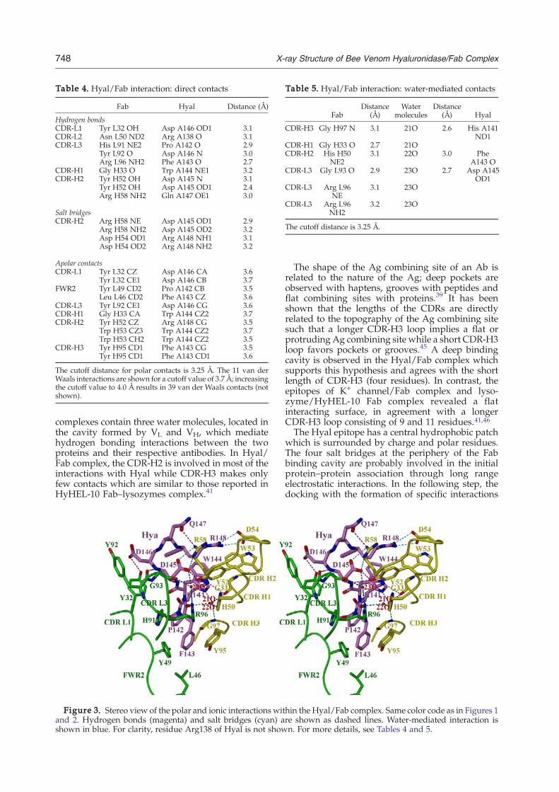
Table 4. Hyal/Fab interaction: direct contacts
Fab Hyal Distance (Å)
Hydrogen bondsCDR-L1 Tyr L32 OH Asp A146 OD1 3.1CDR-L2 Asn L50 ND2 Arg A138 O 3.1CDR-L3 His L91 NE2 Pro A142 O 2.9
Tyr L92 O Asp A146 N 3.0Arg L96 NH2 Phe A143 O 2.7
CDR-H1 Gly H33 O Trp A144 NE1 3.2CDR-H2 Tyr H52 OH Asp A145 N 3.1
Tyr H52 OH Asp A145 OD1 2.4Arg H58 NH2 Gln A147 OE1 3.0
Salt bridgesCDR-H2 Arg H58 NE Asp A145 OD1 2.9
Arg H58 NH2 Asp A145 OD2 3.2Asp H54 OD1 Arg A148 NH1 3.1Asp H54 OD2 Arg A148 NH2 3.2
Apolar contactsCDR-L1 Tyr L32 CZ Asp A146 CA 3.6
Tyr L32 CE1 Asp A146 CB 3.7FWR2 Tyr L49 CD2 Pro A142 CB 3.5
Leu L46 CD2 Phe A143 CZ 3.6CDR-L3 Tyr L92 CE1 Asp A146 CG 3.6CDR-H1 Gly H33 CA Trp A144 CZ2 3.7CDR-H2 Tyr H52 CZ Arg A148 CG 3.5
Trp H53 CZ3 Trp A144 CZ2 3.7Trp H53 CH2 Trp A144 CZ2 3.5
CDR-H3 Tyr H95 CD1 Phe A143 CG 3.5Tyr H95 CD1 Phe A143 CD1 3.6
The cutoff distance for polar contacts is 3.25 Å. The 11 van derWaals interactions are shown for a cutoff value of 3.7 Å; increasingthe cutoff value to 4.0 Å results in 39 van der Waals contacts (notshown).
Table 5. Hyal/Fab interaction: water-mediated contacts
FabDistance
(Å)Water
moleculesDistance
(Å) Hyal
CDR-H3 Gly H97 N 3.1 21O 2.6 His A141ND1
CDR-H1 Gly H33 O 2.7 21OCDR-H2 His H50
NE23.1 22O 3.0 Phe
A143 OCDR-L3 Gly L93 O 2.9 23O 2.7 Asp A145
OD1CDR-L3 Arg L96
NE3.1 23O
CDR-L3 Arg L96NH2
3.2 23O
The cutoff distance is 3.25 Å.
748 X-ray Structure of Bee Venom Hyaluronidase/Fab Complex
complexes contain three water molecules, located inthe cavity formed by VL and VH, which mediatehydrogen bonding interactions between the twoproteins and their respective antibodies. In Hyal/Fab complex, the CDR-H2 is involved in most of theinteractions with Hyal while CDR-H3 makes onlyfew contacts which are similar to those reported inHyHEL-10 Fab–lysozymes complex.41
Figure 3. Stereo view of the polar and ionic interactions witand 2. Hydrogen bonds (magenta) and salt bridges (cyan) ashown in blue. For clarity, residue Arg138 of Hyal is not show
The shape of the Ag combining site of an Ab isrelated to the nature of the Ag; deep pockets areobserved with haptens, grooves with peptides andflat combining sites with proteins.39 It has beenshown that the lengths of the CDRs are directlyrelated to the topography of the Ag combining sitesuch that a longer CDR-H3 loop implies a flat orprotruding Ag combining site while a short CDR-H3loop favors pockets or grooves.45 A deep bindingcavity is observed in the Hyal/Fab complex whichsupports this hypothesis and agrees with the shortlength of CDR-H3 (four residues). In contrast, theepitopes of K+ channel/Fab complex and lyso-zyme/HyHEL-10 Fab complex revealed a flatinteracting surface, in agreement with a longerCDR-H3 loop consisting of 9 and 11 residues.41,46The Hyal epitope has a central hydrophobic patch
which is surrounded by charge and polar residues.The four salt bridges at the periphery of the Fabbinding cavity are probably involved in the initialprotein–protein association through long rangeelectrostatic interactions. In the following step, thedocking with the formation of specific interactions
hin the Hyal/Fab complex. Same color code as in Figures 1re shown as dashed lines. Water-mediated interaction isn. For more details, see Tables 4 and 5.

‡www.genscript.com
749X-ray Structure of Bee Venom Hyaluronidase/Fab Complex
takes place. The interaction between the Hyal/Fabsurface shows a good shape complementarity whichis enhanced by the presence of three water mole-cules in the interface. Additional water moleculesare also observed at the periphery of the bindingcavity and are involved in hydrogen bonds whichare bridging Hyal with Fab. Similarly, Fv D1.3-HELand HyHEL-5–lysozyme complexes contain fourand seven water molecules which are located inthe cavity and mediate the hydrogen bondinginteraction.34,35
In more general terms, the structure of each newAg–Ab complex increases our knowledge and, hence,the probability of identifying any features commonfor Ag–Ab recognition, which would ultimately helpto reveal the characteristics of the allergen and theunderlying mechanism, which makes an Ag anallergen. Attempts to predict features responsible forallergenicity, either by the search for commonsequence motifs47,48 or a common motive on themolecular surface,49 have recently been reported. Thelatter revealed a high occurrence of solvent exposedhydrophobic patches, which is in agreement with anotion that the immune system has evolved torecognize the hydrophobic portion of an immuno-genic protein.50 Supporting this concept, the Hyalepitope contains a surface-exposed hydrophobicpatch formed by loop residues Pro142–Phe143–Trp144 which, upon complex formation, becomedeeply buried in the interface. Similarly, the crystal-lographically determined epitope of a major birchpollen allergen Bet v 1 showed that the central part ofthe conformational epitope consists of a linear stretchof mostly apolar residues (P-loop region: Gly-Asn-Gly-Gly-Pro-Gly-Thr) surrounded on both sides bycharged and polar residues.40
The Hyals from venoms of honey bee, Api m 2,and wasp, Ves v 2, share 55% sequence identity andhave identical fold while the sequence variability ismostly confined to the surface area.20 Of the nineepitope residues of Hyal (Api m 2) only four areconserved in Ves v 2. The central epitope residuePhe143, which fits tightly in the hydrophobic pocketformed by CDR residues of Fab, has been replacedby a polar Thr residue in Ves v 2. Moreover, thecharge distribution among the non-conserved sur-face exposed residues is significantly different andhence, it is unlikely that the identified epitope isresponsible for the in vitro observed IgE-mediatedcross-reactivity between Api m 2 and Ves v 2. Sinceboth enzymes are glycosylated, it is possible that thecross-reactivity can be mediated by carbohydratemoiety, as suggested by Skov et al.20
Type I allergy is an IgE-mediated hypersensitivitydisease affecting up to 25% of the population of allages, from infants to the elderly. There is a definiteneed for an effective treatment of this disease,particularly for bee venom where the allergicreaction to stings can occur suddenly with anoccasionally fatal outcome. The antigenicity ofHyal is fully determined by the structure of itsepitopes, the areas of the protein surface that arerecognized by specific antibodies. The knowledge
of the Hyal epitope allows a structure-basedrational modification of the epitope surface aimedat producing allergen variants with low IgE-binding activity but able to induce protectiveallergen-specific IgG antibodies which competewith IgE (hypoallergens).51 The hypoallergens areexpected to have a significant impact on the devel-opment of allergy vaccines with the increased safetyand efficacy for allergen-specific immunotherapy.
Experimental Procedures
Protein expression and purification
Recombinant Hyal was produced as a secreted proteinby Baculovirus-infected insect cells (High Five) andpurified by Ni2+-chelate chromatography.13 The mono-clonal hybridoma antibodies, derived from mice immu-nized with recombinant Hyal, were purified from thesupernatants of hybridoma cells rich in specific IgG1antibodies. To avoid any contamination with bovine IgGsand serum albumin, a serum- and protein-free culturemedia (TurboDoma) were used for the supernatantproduction. The IgG1 antibodies purified by Protein GSepharose affinity chromatography were almost 100%pure, as judged by SDS-PAGE and Coommassie Bluestaining (150 kDa under nonreducing conditions). Thepurified IgG1 antibodies exhibited a high affinity to Hyaland were able to compete with the binding of sera IgEsfrom bee venom allergic patients.Amino acid sequence of the complete variable light and
heavy chain of mAb 21E11 (murine IgG1, kappa) wasdeduced from the cDNA sequence obtained by using RT-PCR and the SMART race system (Clontech Laboratories).The PCR products were cloned into TOPO vector (vectorwith covalently bound topoisomerase I; Invitrogen) andsequenced using M13 forward and reverse primers.
Synthetic peptide
The 15-mer peptide derived from the Hyal/Fab com-plex (138-RREHPFWDDQRVEQE-152) and covering theentire epitope sequence, was synthesized by GenscriptCorporation (Piscatway, NJ, USA)‡. The peptide waspurified by HPLC and verified by matrix-assisted laserdesorption/ionization time-of-flight mass spectroscopy.The peptide was >80% pure and was fully soluble inaqueous solutions.
ELISA
For ELISA experiments, the ELISA plates (Greiner,Kremsmuenster, Austria) were coated overnight, at 4 °C,with rApi m 2 (5 μg/ml dissolved in PBS) or the Api m 2derived peptide 138-RREHPFWDDQRVEQE-152 (5 μg/mldissolved in PBS), and for control purposes with humanserum albumin (5 μg/ml in PBS) or a control peptide. Theplates were washed between each incubation step withPBS containing 0.05% v/v Tween 20 (PBST). Non-specific-binding sites were blocked with PBS/1% w/v bovineserum albumin for 2 h at room temperature. Coated plates

750 X-ray Structure of Bee Venom Hyaluronidase/Fab Complex
were incubated with sera from bee venom allergic patients(dilution 1:5) or the mouse mAb (dilution 1:1000) in PBS,0.5% w/v bovine serum albumin, 0.05% v/v Tween 20;both were added in duplicate and incubated overnight at4 °C. Bound human IgE was detected as described.52
Bound mouse mAbs were detected with a monoclonal ratanti-mouse IgG1 Ab (BD Pharmingen, San Diego, CA)followed by the addition of horseradish peroxidase-labeled goat anti-rat IgG antibodies (AmershamBioscience, Uppsala, Sweden) and visualized by theaddition of ABTS solution (60 mM citric acid, 77 mMNa2HPO4×2H2O, 1.7 mM ABTS, 3 mM H2O2) as adeveloper.
ELISA inhibition
ELISA plates were coated with 5 μg/ml rApi m 2overnight at 4 °C. The plates were washed with PBST andafter blocking with PBST plus 1% w/v bovine serumalbumin, incubated overnight with three mouse mono-clonal IgG1 antibodies against Api m 2 (21E11, 22H7,24F2) or for control purposes, with a mouse mAb againstBet v 1 (diluted 1:100). After washing, the plates wereincubated with 1:5 diluted sera from bee venom allergicpatients overnight at 4 °C and bound human IgEantibodies were detected as described.52
Production of Fab fragments by papaindigestion of IgG
Purified Ab (clone 21E11, 3–6 mg/ml) was dialyzedagainst 50 mM sodium acetate buffer (pH 5.5) containing2 mM ethylene diamine tetra-acetic acid. Papain wasadded (1/100 w/w ratio) in the presence of 10 mM freshlyprepared cysteine and digestion was carried out at 37 °Cfor 6 h. The reaction was stopped by the addition of thespecific papain inhibitor E-64 (N-[N-(L-3-trans-carboxir-ane-2-carbonyl)-leucyl]-agmatine) (Roche) in large excess.The digestion mixture was dialyzed against 50 mMsodium acetate buffer (pH 5.5), loaded on a Mono Scation exchange column (Amersham Biosciences) andeluted with a shallow salt gradient 0–150 mM NaCl. TheFab was eluted at 70–80 mM NaCl and its purity wasconfirmed by SDS-PAGE under both reducing andnonreducing conditions. The Fab yield was about 30%of the starting IgG1 amount. The purified Fab fragmentswere mixed with Hyal in 1:1.2 molar ratio and incubatedat 23 °C for 60 min. The Hyal/Fab complex was sepa-rated from excess Hyal by using a Superdex S-75 16/60gel filtration column (Amersham BioSciences). Theprotein concentration of the Hyal/Fab complex wascalculated from the absorbance measured at 280 nm,assuming an extinction coefficient of 0.7 mg/ml−1 cm−1.
Crystallization and data collection
For the crystallization experiments, the purified Hyal/Fab complex was dialyzed against 5 mM sodium acetatebuffer (pH 5.5) and concentrated to 11 mg ml−1. Theclusters of thin plate-like crystals were grown by thehanging-drop vapor diffusion method within 4–5 daysunder the following conditions: the equal volumes (1.5 μl)of the Hyal/Fab complex and precipitant 10% (w/v)polyethylene glycol (PEG) 8000, 0.1 M Ches (2-(N-cyclohexylamino)ethanesulfonic acid) (pH 9.5), 0.2 MNaCl (condition 29, Wizard screen kit I) were mixed andequilibrated over the latter solution at 20 °C. The low
crystal reproducibility was overcome by applying amicroseeding technique while the substitution of PEG8000 (10%) by PEG 6000 (11%) resulted in a larger andchunkier, but still clustered plate-like crystals. Prior todata collection, single plates were separated, soakedbriefly in the cryoprotective solution (precipitant solutionplus 20% PEG 400) and the diffraction data were collectedto 2.6 Å resolution at Swiss Light Source (SLS), using aMAR CCD detector (λ=0.9762 Å). All measurements wereperformed at 100 K. The images were indexed andintegrated in a monoclinic space group C2 using theprogram MOSFLM.53 There is one monomer of Hyal/Fabcomplex per asymmetric unit, resulting in a solventcontent of 69% (Vm=3.95 Å3/Da).54
Structure determination and analysis
The structure of the Hyal/Fab complex was determinedby the method of MR using the program PHASER.55 MRwas done in three steps. First, position and orientation ofHyal were obtained using the monoclinic structure ofHyal, 1FCQ13 as a search model. Second, the MR wasperformed separately with variable, V, and constant, C,domains of various Fab structures, in order to avoid dif-ficulties related to the variable relative orientations of theVwith respect to the C domains of the search models. Outof 30 different models, the V domain of 1A7Q gave a goodstatistic and its position was fixed. Third, out of 18 Cdomain models tested byMR, the PDB code 15C8 gave thebest statistics and its position was fixed. Rigid bodyrefinement with five domains (Hyal, VL, CL, VH and CH)with REFMAC56 gave an initial R/Rfree of 36.0/39.1.Manual adjustment of the model and replacement of themodel amino acid sequences with that of 21E11 Fab wereperformed with program O.57 This was followed by re-strained maximum-likelihood refinement with REFMACand addition of water molecules by the program ARP,resulting in the convergence ofR/Rfree value to 20.9/24.7%at 2.6 Å resolution.56 The stereochemistry of the refinedstructure was validated with program PROCHECK,58
which showed that only 0.7% residues are in disallowedregions of a Ramachandran plot (Table 3).The buried surface area was calculated with program
MS59 using a probe radius of 1.7 Å. Interactions within theHyal–Fab interface were assigned with the programCONTACT.56
Protein Data Bank accession codes
Coordinates and structure factors have been depositedin the Protein Data Bank with accession codes 2j88 andr2j88sf, respectively.
Acknowledgements
We thank Dr Caroline Peneff for careful reading ofthe manuscript and valuable discussions. We thankthe staff of the synchrotron beam line PX at SLS inVilligen, Switzerland. This project was supported bySwiss National Foundation grant No. 31-67968/02to Z.M-H and in part by grant F1815 of the AustrianResearch Foundation and a research grant fromPhadia, Uppsala, Sweden.

751X-ray Structure of Bee Venom Hyaluronidase/Fab Complex
References1. King, T. P. & Spangfort, M. D. (2000). Structure and
biology of stinging insect venom allergens. Int. Arch.Allergy Immunol. 123, 99–106.
2. Müller, U. R. (2002). Recombinant Hymenopteravenom allergens. Allergy, 57, 570–576.
3. Hoffman, D. R. (2006). Hymenoptera venom allergens.Clin. Rev. Allergy Immunol. 30, 109–128.
4. Roers, A. & Hunzelmann, N. (2005). Immunotherapyof hypersensitivity to hymenoptera venom. ExpertOpin. Biol. Ther. 5, 1349–1358.
5. Müller, U. R., Crameri, R. & Soldatova, L. (1999).Diagnostik mit rekombinanten/synthetischen Bienen-giftallergenen. Allergologie, 22, 551–552.
6. Habermann, E. (1972). Bee and wasp venoms. Science,177, 314–322.
7. Laurent, T. C. (1989). The biology of hyaluronan. CibaFoundation Symp, vol. 143, pp. John Wiley & Sons,New York.
8. Laurent, T. C. & Fraser, J. R. E. (1992). Hyaluronan.FASEB J. 6, 2397–2404.
9. Turley, E. A., Noble, P. W. & Bourguignon, L. Y. W.(2002). Signaling properties of hyaluronan receptors.J. Biol. Chem. 277, 4589–4592.
10. Sherman, L., Sleeman, J., Herrlich, P. & Ponta, H.(1994). Hyaluronate receptors: key players in growth,differentiation, migration and tumor progression.Curr. Opin. Cell Biol. 6, 726–733.
11. Knudson, W., Chow, G. & Knudson, C. B. (2002).CD44-mediated uptake and degradation of hyalur-onan. Matrix Biol. 21, 15–23.
12. Hardingham, T. E. & Muir, H. (1972). The specificinteraction of hyaluronic acid with cartillage proteo-glycans. Biochim. Biophys. Acta, 279, 401–405.
13. Marković-Housley, Z., Miglierini, G., Soldatova, L.,Rizkallah, P. J., Müller, U. & Schirmer, T. (2000).Crystal structure of hyaluronidase, a major allergen ofbee venom. Structure, 8, 1025–1035.
14. Kolarich, D., Leonard, R., Hemmer, W. & Altmann, F.(2005). The N-glycans of yellow jacket venom hyalur-onidases and the protein sequence of its major isoformin Vespula vulgaris. FEBS J. 272, 5182–5190.
15. Soldatova, L.N., Tsai, C., Dobrovolskaia, E., Marko-vic-Housley, Z. & Slater, J. (2006). Characterization ofthe N-glycans of recombinant bee venom hyaluroni-dase (Api m 2) expressed in insect cells.Asthma AllergyProc., in press.
16. Soldatova, L. N., Crameri, R., Gmachl, M., Kemeny,D., Schmidt, M., Weber, M. & Müller, U. R. (1998).Superior biologic activity of the recombinant beevenom allergen hyaluronidase expressed in Baculo-virus-infected insect cells as compared with Escherichiacoli. J. Allergy Clin. Immunol. 101, 691–698.
17. Henrissat, B. (1991). A classification of glycosylhydrolases based on amino acid sequence similarities.Biochem. J. 280, 309–316.
18. Henrissat, B. & Bairoch, A. (1996). Updating thesequence-based classification of glycohydrolases. Bio-chem. J. 316, 695–696.
19. Marković-Housley, Z. & Schirmer, T. (2002). Structuralevidence for substrate assisted catalytic mechanism ofbee venom hyaluronidase, a major allergen of beevenom. pp. 19–27, The Royal Society of Chemistry,Cambridge, UK.
20. Skov, L. K., Seppl, U., Coen, J. J. F., Crickmore, N.,King, T. P., Monsalve, R. et al. (2006). Structure ofrecombinant Ves v 2 at 2.0 Angstrom resolution:structural analysis of an allergenic hyaluronidase
fromwasp venom. Acta Crystallogr., D Biol. Crystallogr.62, 595–604.
21. Müller, U., Helbling, A. & Berchtold, E. (1992).Immunotherapy with honeybee venom and yellowjacket venom is different regarding efficacy and safety.J. Allergy Clin. Immunol. 89, 529–535.
22. Akdis, M., Verhagen, J., Taylor, A., Karamloo, F.,Karagiannidis, C., Crameri, R. et al. (2004). Immuneresponses in healthy and allergic individuals arecharacterized by a fine balance between allergen-specific T regulatory 1 and T helper 2 cells. J. Exp. Med.199, 1567–1575.
23. Jutel, M., Akdis, M., Blaser, K. & Akdis, C. A. (2006).Mechanisms of allergen specific immunotherapy-T-cell tolerance and more. Allergy, 61, 796–807.
24. Valenta, R., Ball, T., Focke, M., Linhart, B., Mothes, N.,Niederberger, V. et al. (2004). Immunotherapy ofallergic disease. Adv. Immunol. 82, 105–153.
25. Larche, M., Akdis, C. A. & Valenta, R. (2006).Immunological mechanisms of allergen-specificimmunotherapy. Nat. Rev., Immunol. 6, 761–771.
26. Linhart, B. & Valenta, R. (2005). Molecular design ofallergy vaccines. Curr. Opin. Immunol. 17, 646–655.
27. Davies, D. R., Padlan, E. A. & Sheriff, S. (1990).Antibody–antigen complexes. Annu. Rev. Biochem. 59,439–473.
28. Poljak, R. J. (1991). Structure of antibodies andtheir complexes with antigens. Mol. Immunol. 28,1341–1345.
29. Davies, D. R. & Cohen, G. H. (1996). Interactions ofprotein antigens with antibodies. Proc. Natl Acad. Sci.USA, 93, 7–12.
30. Braden, B. C. & Poljak, R. J. (1995). Structural featuresof the reactions between antibodies and proteinantigens. FASEB J. 9, 9–16.
31. Kabat, E.A., Wu, T.T., Erry, H.M., Gottesman, K.S. &Foeller, C. (1991). Sequences of proteins of immuno-logical interest. Technical report. U.S. Department ofHeath and Human Services.
32. Martin, A. C. (1996). Accessing the Kabat antibodysequence database by computer. Proteins: Struct.Funct. Genet. 25, 130–133.
33. Kulkarni-Kale, U., Bhosle, S. & Kolaskar, A. S. (2005).CEP: a conformational epitope prediction server.Nucleic Acids Res. 33, W168–W171; (Web Server issue).
34. Bhat, T. N., Bentley, G. A., Boulot, G., Greene, M.,Tello, D., Dall'Acqua, W. et al. (1994). Bound watermolecules and conformational stabilization helpmediate an antigen–antibody association. Proc. NatlAcad. Sci. USA, 91, 1089–1093.
35. Cohen, G. H., Silverton, E. W., Padlan, E. A., Dyda, F.,Wibbenmeyer, J. A., Willson, R. C. & Davies, D. R.(2005). Water molecules in the antibody-antigeninterface of the structure of the Fab HyHEL-5-lysozyme complex at 1.7 A resolution: comparisonwith results from isothermal titration calorimetry.Acta Crystallogr., D Biol. Crystallogr. 61, 628–633.
36. Ysern, X., Fields, B. A., Bhat, T. N., Goldbaum, F. A.,Dall'Acqua, W., Schwarz, F. P. et al. (1994). Solventrearrangement in an antigen–antibody interface intro-duced by site-directed mutagenesis of the antibodycombining site. J. Mol. Biol. 238, 496–500.
37. Lawrence, M. C. & Colman, P. M. (1993). Shapecomplementarity at protein/protein interfaces. J. Mol.Biol. 234, 946–950.
38. Padlan, E. A. (1990). On the nature of antibodycombining sites: unusual structural features that mayconfer on these sites an enhanced capacity for bindingligands. Proteins: Struct. Funct. Genet. 7, 112–124.

752 X-ray Structure of Bee Venom Hyaluronidase/Fab Complex
39. MacCallum, R. M., Martin, A. C. & Thornton, J. M.(1996). Antibody–antigen interactions: contact analy-sis and binding site topography. J. Mol. Biol. 262,732–745.
40. Mirza, O., Henriksen, A., Ipsen, H., Larsen, J.,Wissenbach, M., Spangfort, M. D. & Gajhede, M.(2000). Dominant epitopes and allergic cross-reactiv-ity: complex formation between a Fab fragment of amonoclonal murine IgG antibody and the majorallergen from birch pollen Bet v 1. J. Immunol. 165,331–338.
41. Padlan, E. A., Silverton, E.W., Sheriff, S., Cohen, G. H.,Smith-Gill, S. J. & Davies, D. (1989). Structure of anantibody–antigen complex: crystal structure of theHyHEL-10 Fab–lysozyme complex. Proc. Natl Acad.Sci. USA, 86, 5938–5942.
42. Sheriff, S., Silverton, E.W., Padlan, E. A., Cohen, G. H.,Smith-Gill, S. J., Finzel, B. C. & Davies, D. R. (1987).Three-dimensional structure of an antibody–antigencomplex. Proc. Natl Acad. Sci. USA, 84, 8075–8079.
43. Schulze-Gahmen, U., Rini, J. & Wilson, I. (1993).Detailed analysis of the free and bound conformationsof an antibody. X-ray structures of Fab 17/9 and threedifferent Fab–peptide complexes. J. Mol. Biol. 234,1098–1118.
44. Breithaupt, C., Schubart, A., Zander, H., Skerra, A.,Huber, R., Linington, C. & Jacob, U. (2003). Structuralinsights into the antigenicity of myelin oligodendro-cyte glycoprotein. Proc. Natl Acad. Sci. USA, 100,9446–9451.
45. Collis, A. V. J., Brouwer, A. P. &Martin, A. C. R. (2003).Analysis of the antigen combining site: correlationsbetween length and sequence composition of thehypervariable loops and the nature of the antigen.J. Mol. Biol. 325, 337–354.
46. Jiang, Y., Lee, A., Chen, J., Ruta, V., Cadene, M., Chait,B. T. & MacKinnon, R. (2003). X-ray structure of avoltage-dependent K+ channel. Nature, 423, 33–41.
47. Gendel, S. M. (2002). Sequence analysis for asses-sing potential allergenicity. Ann. N.Y. Acad. Sci. 964,87–98.
48. Li, K.-B., Issac, P. & Krishnan, A. (2004). Predictingallergenic proteins using wavelet transform. Bioinfor-matics, 20, 2572–2578.
49. Furmonaviciene, R., Sutton, B. J., Glaser, F., Laughton,
C. A., Jones, N., Sewell, H. F. & Shakib, F. (2005). Anattempt to define allergen-specific molecular surfacefeatures: a bioinformatic approach. Bioinformatics, 21,4201–4204.
50. Seong, S.-Y. & Matzinger, P. (2004). Hydrophobicity:an ancient damage-associated molecular pattern thatinitiates innate immune responses. Nat. Rev., Immunol.4, 469–478.
51. Ferreira, F., Ebner, C., Kramer, B., Casari, G., Briza, P.,Kungl, A. J. et al. (1998). Modulation of IgE reactivityof allergens by site-directed mutagenesis: potentialuse of hypoallergenic variants for immunotherapy.FASEB J. 12, 231–242.
52. Denepoux, S., Eibensteiner, P. B., Steinberger, P.,Vrtala, S., Visco, V., Weyer, A. et al. (2000). Molecularcharacterization of human IgG monoclonal antibodiesspecific for the major birch pollen allergen Bet v 1.Anti-allergen IgG can enhance the anaphylacticreaction. FEBS Letters, 465, 39–46.
53. Leslie, A.G.W. (1992). In joint CCP4 and ESF-EACBMnewsletter on protein crystallography no. 26. Techni-cal report. SERC Daresbury Laboratory, Warrington,UK.
54. Matthews, B. W. (1968). Solvent content of proteincrystals. J. Mol. Biol. 33, 491–497.
55. Storoni, L. C., McCoy, A. J. & Read, R. J. (2004).Likelihood-enhanced fast rotation functions. ActaCrystallogr., D Biol. Crystallogr. 60, 432–438.
56. CCP4 (1994). Collaborative computational project,number 4.Acta Crystallogr., D Biol. Crystallogr. 50,760–763; (The CCP4 suite: programs for proteincrystallography).
57. Jones, T. A., Zou, J., Cowan, S. & Kjeldgaard, M.(1991). Improved methods for building proteinmodels in electron density maps and the location oferrors in these models.Acta Crystallogr., A, 47, 110–119.
58. Laskowski, R. A., MacArthur, M. W., Moss, D. S.& Thornton, J. M. (1993). Procheck: a program tocheck the stereochemical quality of protein structures.J. Appl. Cryst. 26, 283–291.
59. Connolly, M. L. (1993). The molecular surface pack-age. J. Mol. Graph. 11, 139–141.
60. Read, R. J. (1986). SIGMAA-improved Fourier coeffi-cients for maps using phases from partial structureswith errors. Acta Crystallogr., A, 42, 140–149.
Edited by I. Wilson
(Received 9 December 2006; received in revised form 22 January 2007; accepted 8 February 2007)Available online 22 February 2007
2.1 Supplementary information (Hya/Fab complex)
Hya/Fab-supplementary information
25

2.1.1 Hya/Fab complex formation, crystallization and diffraction
2.1.1.1 IgG purification
The supernatant containing IgG was dialyzed (Spectra/Pro, membrane cutoff 12-14
kDa) against 20 mM sodium phosphate buffer pH 7.0 and centrifuged at 13000 rpm (Sorval
RC-5B, SLA-1500 rotor) for 30 min. The resultant supernatant was applied on a Protein-G
affinity column (Amersham Pharmacia) that specifically binds the Fc region of the IgG
molecule. The bound IgG was eluted in 1 ml fractions with 100mM glycine buffer pH 2.7
(Figure 1) and immediately neutralized with 200 μM of 1M Tris buffer pH 9.0.
97.4
66.2
45.0
31.0
Heavy chain
Light chain
M SM 1 2 3 4 5 6
Figure 1. IgG purification. SDS-polyacrylamide gel (12%, reducing condition) analysis of the purified IgG eluted from Protein-G column. M stands for molecular weight markers (kDa), SM for starting material and fraction numbers are indicated at the bottom of the gel.
Hya/Fab-supplementary information
26

2.1.1.2 IgG digestion
The purified IgG was dialyzed against 100 mM sodium acetate buffer pH 5.5
containing 2 mM EDTA and was subsequently concentrated to 3-6 mg/ml using a centricon (30
kDa molecular-mass cutoff; Amicon). To the concentrated IgG, freshly prepared cysteine was
added to the final concentration of 10 mM and followed by the addition of papain at the ratio of
1/100 (W/W) with respect to IgG concentration. The papain is an cysteine endopeptidase of
23000 Da molecular weight and was used for limited hydrolysis of native immunoglobulin (Ig).
Digestion was carried out by incubating sample at 37 ˚C for 6 hours (Figure 2). The IgG
digestion was stopped by addition of 10 fold excess of E64 inhibitor (N-[N-(L-3-trans-
carboxyoxirane-2-carbonyl)-L-leucyl]-agmatine, Roche; Molecular weight 357.4 Da) relative
to the papain concentration. The E64 stock has been prepared by dissolving the compound in
1:1 ethanol/water mixture to the final concentration of 250 μM.
M SM 1h 2h 3h 4h 5h 6h 7h 8h 10h
Undigested heavy chain
Fc
Fab
97.4
66.2
45.0
31.0
21.5
Figure 2. IgG test digestion. Digestion was carried out with papain in the presence of 10 mM cysteine. SDS-polyacrylamide (12%, reducing condition) analysis of the digested sample collected at various time point as indicated at the bottom of the gel. M stands for molecular weight markers (kDa), SM for starting material.
Hya/Fab-supplementary information
27

2.1.1.3 Purification of Fab using cation exchange chromatography (Mono-S)
The digested sample was dialyzed against 50 mM sodium acetate buffer pH 5.5 and
loaded on a cation exchanger (mono-S) column (Pharmacia) using acta purifier (Amersham
Pharmacia). The long shallow salt gradient was carried out between 20 -120 mM NaCl in order
to separate Fab isoforms from (Fab)2 (Figure 3).
2.1.1.4 Complex formation and purification using gel filtration column (Superdex
S-75 16/60)
The Fab, eluted as a major peak at 40 mM NaCl was pooled, mixed with hyaluronidase
in 1:1.2 molar ratio and incubated at room temperature for 1 hour. The Hya/Fab mixture was
concentrated to 4-6 mg/ml using centricon (Amicon) and loaded on a Hiload Superdex S-75
(Amersham Pharmacia) gel filtration column in order to separate Hya/Fab complex from excess
Hya (Figure 4).
Hya/Fab-supplementary information
28

Fab
Fc
(Fab)2
M SM B7 B6 B5 B4 B3 B1 C2 D11 D9 D7 D5 D3
97.4
66.245.031.0
21.5
14.4
Figure 3. Purification of Fab using cation exchange chromatography (Mono-S column). A) Chromatogram of Fab elution profile. B) SDS-polyacrylamide (12%, non-reducing condition) analysis of proteins separated on Mono-S column. M stands for molecular weight markers (kDa) and SM for starting material. Fraction numbers are indicated at the bottom of the gel.
A
B
Hya/Fab-supplementary information
29

Figure 4. Purification of Hya-Fab complex using Hiload 26/60 Superdex 75 16/60 gel filtration chromatography. A) Chromatogram show Hya/Fab complex separated from excess Hya. B) SDS-polyacrylamide (12%, reducing condition) analysis of the first peak shows complex whilst the second peak shows an excess of Hya. M stands for molecular weight markers (kDa), SM for starting material and fraction numbers are indicated at the bottom of the gel.
97.4
66.2
45.0
31.0
21.5
Hya
Fab
M Fab Hya SM D11 D9 D7 D6 D5 E2 E3 E4 E5 E6
A
B
Hya/Fab-supplementary information
30
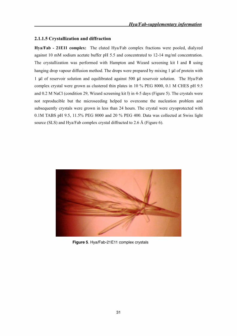
2.1.1.5 Crystallization and diffraction
Hya/Fab - 21E11 complex: The eluted Hya/Fab complex fractions were pooled, dialyzed
against 10 mM sodium acetate buffer pH 5.5 and concentrated to 12-14 mg/ml concentration.
The crystallization was performed with Hampton and Wizard screening kit Ⅰ and Ⅱ using
hanging drop vapour diffusion method. The drops were prepared by mixing 1 μl of protein with
1 μl of reservoir solution and equilibrated against 500 μl reservoir solution. The Hya/Fab
complex crystal were grown as clustered thin plates in 10 % PEG 8000, 0.1 M CHES pH 9.5
and 0.2 M NaCl (condition 29, Wizard screening kit Ⅰ) in 4-5 days (Figure 5). The crystals were
not reproducible but the microseeding helped to overcome the nucleation problem and
subsequently crystals were grown in less than 24 hours. The crystal were cryoprotected with
0.1M TABS pH 9.5, 11.5% PEG 8000 and 20 % PEG 400. Data was collected at Swiss light
source (SLS) and Hya/Fab complex crystal diffracted to 2.6 Å (Figure 6).
Figure 5. Hya/Fab-21E11 complex crystals
Hya/Fab-supplementary information
31
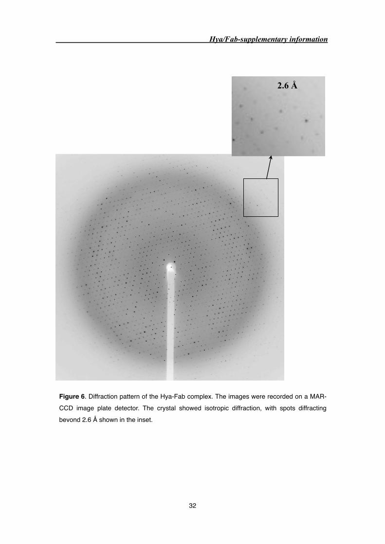
Figure 6. Diffraction pattern of the Hya-Fab complex. The images were recorded on a MAR-CCD image plate detector. The crystal showed isotropic diffraction, with spots diffracting beyond 2.6 Å shown in the inset.
2.6 Å
Hya/Fab-supplementary information
32

Hya/Fab-22H7 complex: The purified Hya/Fab complex was dialyzed against 10 mM sodium
acetate buffer pH 5.4 and concentrated to 12 - 14 mg/ml. The crystallization was performed
with commercial screens, that is Hampton and Wizard screening kit I and II using hanging drop
vapour diffusion method. The drops were prepared by mixing 1 μl of protein with 1 μl
reservoir solution and equilibrated against 500 μl of reservoir solution. The Hya/Fab
(22H7.11.12) complex was crystallized in the presence of 9.5% PEG 8000, 0.1 M TABS pH 9.5
and 0.2 M NaCl similar to 21E11.1.6 clone. The microseeding was applied to overcome the
nucleation problem. The crystals were grown as thin plates and reached maximum size in 2-3
days (Figure 7).
Figure 7. Hya/Fab-22H7 complex crystals
Hya/Fab-supplementary information
33
2.1.1.6 Structure solution: Search for useful Fab model for molecular replacement
(MR)
The structure of Hya/Fab complex was determined by molecular replacement (MR)
using the program PHASER [1]. The orientation and position of the Hya/Fab complex were
determined by MR in three consecutive steps. First, Hya was located using PDB code: 1FCQ as
a search model. On fixing the Hya position, PHASER gave a Z-score of 13. This was followed
by rigid body refinement which gave Rfactor/Rfree of 41.8/42.5%.
The PDB database consists of more than 500 Fab structures. MR with intact Fab model
was not expected to work due to a rather various relative variable (VL and VH) and constant
domains (CL and CH) arrangements. Therefore, the following strategy was applied to locate the
correct Fab model from the database. Firstly, mass mapping was carried out with light and
heavy chains separately and the sequence of top hits was used for Blast (NCBI BLAST). The
Blast with the heavy chain sequence gave the Fab (1S5I and 1ORS) and these Fabs were
divided into variable (VL and VH) and constant domains (CL and CH) whilst the flexible linker
region were removed. The MR was carried out in two steps; the position of the variable domain
(VL and VH) was fixed followed by fixing the constant domain (CL and CH). The MR with
variable domain of 1S5I and 1ORS gave good Z score of 18 and 20 and the R factor and Rfree of
39.4/ 42.8% and 39.3/42.0% respectively. The structural neighbor to 1ORS heavy chain was
searched using NCBI vast program and the results were sorted based on high redundant and
root-mean square deviation (rmsd). For variable domain, four highest hit (marked in pink) gave
good statistics, out of which, 1A7Q position was fixed and it gave Z score of 23 and Rfactor and
Rfree of 37.8/41.6%. In total, 30 different variable domain were used for MR. The variable
domain consists of complementary determining regions (CDR) which is involved in antigen
binding. The sequence and structure of CDR’s varies from one antibody to another. The
position of CDR’s in 1A7Q were determined by Immunoglobulin (Ig) Blast (NCBI Blast) and
the truncation were made in CDR sequence i.e the occupancy of the corresponding amino acid
was set to zero, in order to exclude them from the refinement using REFMAC [2]. The
truncation in CDR’s of 1A7Q, improved the statistics to small extent resulting in the Z score of
24 and Rfactor/Rfree of 37.7/40.7%.
Search for constant domain models (CL and CH) was carried out randomly. Out of 18
constant domain models, 15C8 gave a Z score 20. After five rigid body refinement (Hya, VL,
CL, VH and CH) the Rfactor/Rfree was 36.0/39.1%. The density for the terminal region of
constant domain was not well defined. Thus, Hya, variable and constant domains had been
Hya/Fab-supplementary information
34

found representing the full Hya/Fab complex structure. Manual adjustment of the model was
performed with program O [3] and was followed by full atom refinement that gave an Rfactor/
Rfree of 26.9/31.2%. Replacement of the model amino acid sequences with that of 21E11 Fab
was performed with program O and followed by refinement with REFMAC converged Rfactor/
Rfree to 21.0/24.7% at 2.6 Å.
Hya/Fab-supplementary information
35

Mol
ecul
ar r
epla
cem
ent
(MR
) ca
rrie
d ou
t as
thr
ee s
tep
proc
edur
e -
Pha
ser
Mas
s m
appi
ng
Hea
vy c
hain
Lig
ht c
hain
NC
BI
prot
ein-
prot
ein
BL
AST
; ch
osen
dat
abas
e: P
DB
Sequ
ence
of
the
top
hit m
odel
Sear
ch f
or s
truc
tura
l nei
ghbo
urs
to 1
OR
S
(NC
BI
Vas
t)
Sear
ch w
ith
heav
y ch
ain
sequ
ence
gav
e be
st F
ab m
odel
Ref
mac
(Fa
b se
quen
ce)
21.0
24.6
hyaf
ab-u
niqu
e.m
tz
Step
1: M
R f
or th
e H
ya (
PDB
cod
e: 1
FCQ
)
No
of s
earc
h m
odel
Mod
elZ
Sco
reR
fac
tor
R f
ree
1H
ya (
PDB
cod
e: 1
FCQ
)13
.11
41.8
41.8
42.2
42.5
Step
2: s
earc
h fo
r th
e va
riab
le d
omai
n m
odel
Fixe
d po
sitio
n of
Hya
and
var
iabl
e do
mai
n
Step
3: R
ando
m s
earc
h fo
r th
e co
nsta
nt d
omai
n m
odel
18C
onst
ant d
omai
n (P
DB
co
de: 1
5C8)
20.1
637
.136
.039
.839
.1
26V
aria
ble
dom
ain
(PD
B
code
: 1A
7Q)
22.9
638
.937
.841
.441
.6
Tru
ncat
ion
in C
DR
’s24
.23
38.7
37.7
40.4
40.7
Ref
mac
26.9
31
.2
Step
4: F
ull a
tom
ref
inem
ent
Step
5: R
epla
cing
mod
el s
eque
nce
with
21E
11.6
.1
sequ
ence
Fin
al H
ya/F
ab c
ompl
ex s
truc
ture
1A7Q
was
a b
est
vari
able
dom
ain
mod
el
Fixe
d th
e po
sitio
ned
Hya
, var
iabl
e an
d co
nsta
nt d
omai
n =
Hya
/Fab
com
plex
Fixe
d po
sitio
n of
Hya
Hya/Fab-supplementary information
36

2.1.1.7 Hya/Fab-21E11 crystal packing
Hya/Fab complex crystallized in C2 (monoclinic) space group, which contains two fold
rotation along b-axis and c-centering on ab plane. The C2 space group consists of four
asymmetric units. In case of Hya/Fab complex, each asymmetric unit contains one Hya/Fab
complex. The crystal contacts are mediated by Hya-Hya and Hya-VH contacts.
Figure 8. Schematic representation of a search for the Hya/Fab model by molecular replacement (MR). The number of search models, model with PDB code, Rfactor and Rfree values, used at various stages of MR are indicated in box. The method used for finding the useful variable domain model is showed on the left.
Figure 9. Crystal packing of Hya/Fab-21E11 complex. Magenta represents Hya, yellow heavy chain and green light chain). The picture was produced using the program DINO (A.Philippsen, http://www.biozentrum.unibas.ch/~xray/dino).
Hya/Fab-supplementary information
37

2.1.2 Mechanism of Type 1 hypersensitivity reaction.
Allergy caused by insect venom is an example of immediate type 1 hypersensitivity reaction. In
genetically susceptible individual, some environmental protein antigen, commonly called
allergen, can stimulate distinct immune response. They preferentially use particular antigen
presenting cells (APC) such as dentritic cells and B cells and stimulate CD4+ T cells to
differentiate into Th2 effector T cells. The activated Th2-type cells produce cytokines - such as
interleukin-4 (IL-4) and IL-13 - which promote switching of B cells to IgE producing plasma
cells, whilst IL-5 stimulates eosinophilic survival and activation (Figure 10). In addition to
cytokines, various other factors influence the differentiation of CD4+ T cells into the Th2
phenotype, such as the genetic background of the individual, the site of antigen contact,
allergen dose and conformation, the nature and number of antigen presenting cells, as well as
inflammatory mediator (e.g. histamine) [4]. In addition, immune cells recognize allergen
epitopes differently: T-cells recognize continuous amino acid sequences (i.e. continuous
epitopes), whereas B cells recognize allergens in there intact three dimensional structure (i.e.
conformational epitope). The secreted IgE circulates throughout the body and binds to high-
affinity Fc receptors FcεRI on the surfaces of circulating basophils and on mast cells in
various tissues. This primary response to allergen is known as sensitization.
On second re-exposure, multivalent antigen binds to the cell-associated IgE and cross-
links two or more IgE molecules bound to the Fc receptors. This leads to a series of signaling
events in the mast cells and basophils, which result in the release of preformed mediators, such
as histamine and leukotrienes, stored in the cytoplasmic granules. The clinical and pathologic
manifestations are due to the actions of the released mediators. Some of these mediators induce
an immediate symptoms of disease, such as rhinitis, conjunctivitis, vascular and smooth muscle
reaction and asthma, and others stimulates the influx of leucocytes resulting in the late phase
reaction [4-6].
Hya/Fab-supplementary information
38
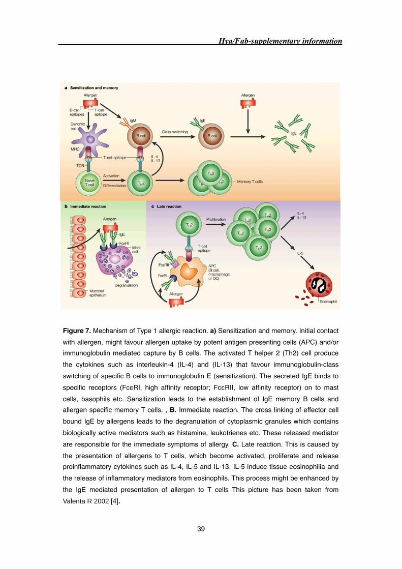
Hya/Fab-supplementary information
39
Figure 7. Mechanism of Type 1 allergic reaction. a) Sensitization and memory. Initial contact with allergen, might favour allergen uptake by potent antigen presenting cells (APC) and/or immunoglobulin mediated capture by B cells. The activated T helper 2 (Th2) cell produce the cytokines such as interleukin-4 (IL-4) and (IL-13) that favour immunoglobulin-class switching of specific B cells to immunoglobulin E (sensitization). The secreted IgE binds to specific receptors (FcεRI, high affinity receptor; FcεRII, low affinity receptor) on to mast cells, basophils etc. Sensitization leads to the establishment of IgE memory B cells and allergen specific memory T cells. , B. Immediate reaction. The cross linking of effector cell bound IgE by allergens leads to the degranulation of cytoplasmic granules which contains biologically active mediators such as histamine, leukotrienes etc. These released mediator are responsible for the immediate symptoms of allergy. C. Late reaction. This is caused by the presentation of allergens to T cells, which become activated, proliferate and release proinflammatory cytokines such as IL-4, IL-5 and IL-13. IL-5 induce tissue eosinophilia and the release of inflammatory mediators from eosinophils. This process might be enhanced by the IgE mediated presentation of allergen to T cells This picture has been taken from Valenta R 2002 [4].

2.1.3 Specific immunotherapy (SIT)
Allergic disease affect more than 30% of the population in industrialized countries,
with an increasing prevalence during the last decades. Currently, SIT is performed with natural
allergen extracts that are not designed for an individual patient’s allergen profile. Besides, they
contain various allergenic, as well as non-allergenic components and might even be
contaminated with allergen from other sources. In general, successful SIT requires high dose of
allergen extract and in most cases, required dose could not be achieved due to the development
of severe systemic side effects including anaphylactic reaction [4].
Rapid development in the field of recombinant allergens revealed the molecular nature
of many important allergens and facilitated the characterization of their immunological and
structural features. This knowledge allowed modifying allergen to contain relevant T-cell
epitopes that induce T-cell tolerance and lack antibody-binding sites (hypoallergen) that would
otherwise facilitate IgE-mediated allergic responses. The hypoallergenic proteins have
diminished or no IgE binding activities thus enabling the administration of high allergen doses
with a reduced risks of systemic side effects. Such hypoallergenic allergen derivatives have
been generated by modifying the allergen encoding cDNA (e.g. mutation, deletion,
oligomerization and production of hybrids) [7].
Most direct method to map IgE binding epitopes is the structure determination of
allergen in complex with Fab fragment of IgG antibodies that compete with patient sera IgE
antibodies in binding to Hya. The hypoallergenic allergens were generated by destroying the
epitope either by introducing mutation or deletion of epitope residues. In case of birch pollen
allergen Bet v1, single mutation in the mapped epitope showed 50% reduction in the binding of
human polyclonal IgE [8]. The chimeric protein major bee venom allergens (Api 1/2/3),
induced 100 to 1000 times less type-1 skin test reactivity in allergic patients. Moreover,
treatment in mice with chimeric protein led to significant reduction of specific IgE
development towards native allergen, representing a protective vaccine effect in vivo [9]. The
clinical trial with peptides containing only T-cell epitopes (peptide immunotherapy, PIT) to
major cat allergen, Fel d 1 [10] and bee venom major allergen, phospholipase A2 [11] showed
beneficial modulation of immune response against whole allergen.
Though numerous clinical studies documented the efficacy of immunotherapy, but the
underlying immunological mechanisms remains a matter of controversy. Most likely, several
different mechanisms were involved in the observed long term effects of immunotherapy.
Hya/Fab-supplementary information
40
Moreover, the importance of blocking antibodies has been questioned because it was not
always associated with clinically successful immunotherapy [4].
Hya/Fab-supplementary information
41

Bibliography
1. Storoni, L.C., McCoy, A.J., and Read, R.J. (2004). Likelihood-enhanced fast rotation
functions. Acta Crystallogr D Biol Crystallogr 60, 432-438.
2. (1994). The CCP4 suite: programs for protein crystallography. Acta Crystallogr D Biol
Crystallogr 50, 760-763.
3. Jones, T.A., Zou, J.Y., Cowan, S.W., and Kjeldgaard (1991). Improved methods for
building protein models in electron density maps and the location of errors in these models.
Acta Crystallogr A 47 ( Pt 2), 110-119.
4. Valenta, R. (2002). The future of antigen-specific immunotherapy of allergy. Nat Rev
Immunol 2, 446-453.
5. Akdis, C.A., and Blaser, K. (2001). Bypassing IgE and targeting T cells for specific
immunotherapy of allergy. Trends Immunol 22, 175-178.
6. Saini, S.S., and MacGlashan, D. (2002). How IgE upregulates the allergic response. Curr
Opin Immunol 14, 694-697.
7. Ferreira, F., Ebner, C., Kramer, B., Casari, G., Briza, P., Kungl, A.J., Grimm, R., Jahn-
Schmid, B., Breiteneder, H., Kraft, D., Breitenbach, M., Rheinberger, H.J., and Scheiner, O.
(1998). Modulation of IgE reactivity of allergens by site-directed mutagenesis: potential
use of hypoallergenic variants for immunotherapy. Faseb J 12, 231-242.
8. Spangfort, M.D., Mirza, O., Ipsen, H., Van Neerven, R.J., Gajhede, M., and Larsen, J.N.
(2003). Dominating IgE-binding epitope of Bet v 1, the major allergen of birch pollen,
characterized by X-ray crystallography and site-directed mutagenesis. J Immunol 171,
3084-3090.
9. Karamloo, F., Schmid-Grendelmeier, P., Kussebi, F., Akdis, M., Salagianni, M., von Beust,
B.R., Reimers, A., Zumkehr, J., Soldatova, L., Housley-Markovic, Z., Muller, U., Kundig,
T., Kemeny, D.M., Spangfort, M.D., Blaser, K., and Akdis, C.A. (2005). Prevention of
allergy by a recombinant multi-allergen vaccine with reduced IgE binding and preserved T
cell epitopes. Eur J Immunol 35, 3268-3276.
10. Pene, J., Desroches, A., Paradis, L., Lebel, B., Farce, M., Nicodemus, C.F., Yssel, H., and
Bousquet, J. (1998). Immunotherapy with Fel d 1 peptides decreases IL-4 release by
peripheral blood T cells of patients allergic to cats. J Allergy Clin Immunol 102, 571-578.
11. Muller, U., Akdis, C.A., Fricker, M., Akdis, M., Blesken, T., Bettens, F., and Blaser, K.
(1998). Successful immunotherapy with T-cell epitope peptides of bee venom
phospholipase A2 induces specific T-cell anergy in patients allergic to bee venom. J Allergy
Clin Immunol 101, 747-754.
Hya/Fab-supplementary information
42

3.0 Purification and biophysical characterization of bo-
vine testes hyaluronidase (PH-20)
PH-20 characterization
43
3.1 Abstract
PH-20 is well characterized mammalian hyaluronidase, present on the sperm plasma
membrane and inner acrosomal membrane via GPI-anchor and plays important role in fertiliza-
tion. It is a bifunctional protein: N-terminal domain carrying hyaluronidase activity involved in
cumulus penetration, while the C-terminal is presumably involved in binding of acrosome re-
acted (AR) sperm to the zona pellucida. In the present study, we were able to purify PH-20 pro-
teins from commercial available bovine testicular extract (Sigma) and from fresh bovine testes
to highest purity. PH-2060 purified from commercial source is probably endoproteolytic cleav-
age product whereas PH-2069 (Sigma) and PH-2080 (bovine testes) are intact protein. The puri-
fied PH-20 proteins forms heterogeneous aggregate, most likely by hydrophobic interaction,
which was successfully overcomed by addition of zwitterionic compound, NDSB (non deter-
gent sulfobetaine).
PH-20 characterization
44
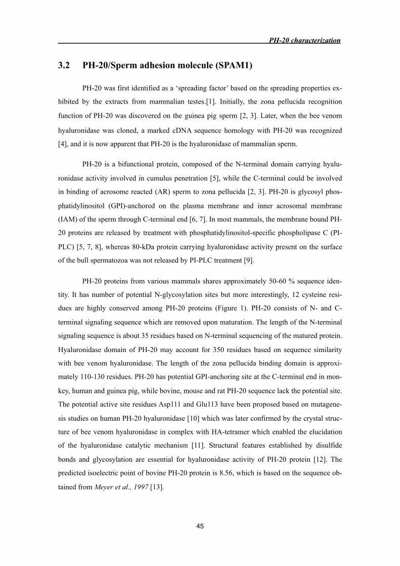
3.2 PH-20/Sperm adhesion molecule (SPAM1)
PH-20 was first identified as a ‘spreading factor’ based on the spreading properties ex-
hibited by the extracts from mammalian testes.[1]. Initially, the zona pellucida recognition
function of PH-20 was discovered on the guinea pig sperm [2, 3]. Later, when the bee venom
hyaluronidase was cloned, a marked cDNA sequence homology with PH-20 was recognized
[4], and it is now apparent that PH-20 is the hyaluronidase of mammalian sperm.
PH-20 is a bifunctional protein, composed of the N-terminal domain carrying hyalu-
ronidase activity involved in cumulus penetration [5], while the C-terminal could be involved
in binding of acrosome reacted (AR) sperm to zona pellucida [2, 3]. PH-20 is glycosyl phos-
phatidylinositol (GPI)-anchored on the plasma membrane and inner acrosomal membrane
(IAM) of the sperm through C-terminal end [6, 7]. In most mammals, the membrane bound PH-
20 proteins are released by treatment with phosphatidylinositol-specific phospholipase C (PI-
PLC) [5, 7, 8], whereas 80-kDa protein carrying hyaluronidase activity present on the surface
of the bull spermatozoa was not released by PI-PLC treatment [9].
PH-20 proteins from various mammals shares approximately 50-60 % sequence iden-
tity. It has number of potential N-glycosylation sites but more interestingly, 12 cysteine resi-
dues are highly conserved among PH-20 proteins (Figure 1). PH-20 consists of N- and C-
terminal signaling sequence which are removed upon maturation. The length of the N-terminal
signaling sequence is about 35 residues based on N-terminal sequencing of the matured protein.
Hyaluronidase domain of PH-20 may account for 350 residues based on sequence similarity
with bee venom hyaluronidase. The length of the zona pellucida binding domain is approxi-
mately 110-130 residues. PH-20 has potential GPI-anchoring site at the C-terminal end in mon-
key, human and guinea pig, while bovine, mouse and rat PH-20 sequence lack the potential site.
The potential active site residues Asp111 and Glu113 have been proposed based on mutagene-
sis studies on human PH-20 hyaluronidase [10] which was later confirmed by the crystal struc-
ture of bee venom hyaluronidase in complex with HA-tetramer which enabled the elucidation
of the hyaluronidase catalytic mechanism [11]. Structural features established by disulfide
bonds and glycosylation are essential for hyaluronidase activity of PH-20 protein [12]. The
predicted isoelectric point of bovine PH-20 protein is 8.56, which is based on the sequence ob-
tained from Meyer et al., 1997 [13].
PH-20 characterization
45

✭
✭
✭✭
✭
✭
mayer et albovine PH-20human PH-20monkey PH-20mouse PH-20Rat PH-20guinea PH-20
mayer et albovine PH-20human PH-20monkey PH-20mouse PH-20Rat PH-20guinea PH-20
mayer et albovine PH-20human PH-20monkey PH-20mouse PH-20Rat PH-20guinea PH-20
mayer et albovine PH-20human PH-20monkey PH-20mouse PH-20Rat PH-20guinea PH-20
mayer et albovine PH-20human PH-20monkey PH-20mouse PH-20Rat PH-20guinea PH-20
mayer et albovine PH-20human PH-20monkey PH-20mouse PH-20Rat PH-20guinea PH-20
mayer et albovine PH-20human PH-20monkey PH-20mouse PH-20Rat PH-20guinea PH-20
mayer et albovine PH-20human PH-20monkey PH-20mouse PH-20Rat PH-20guinea PH-20
PH-20 characterization
46
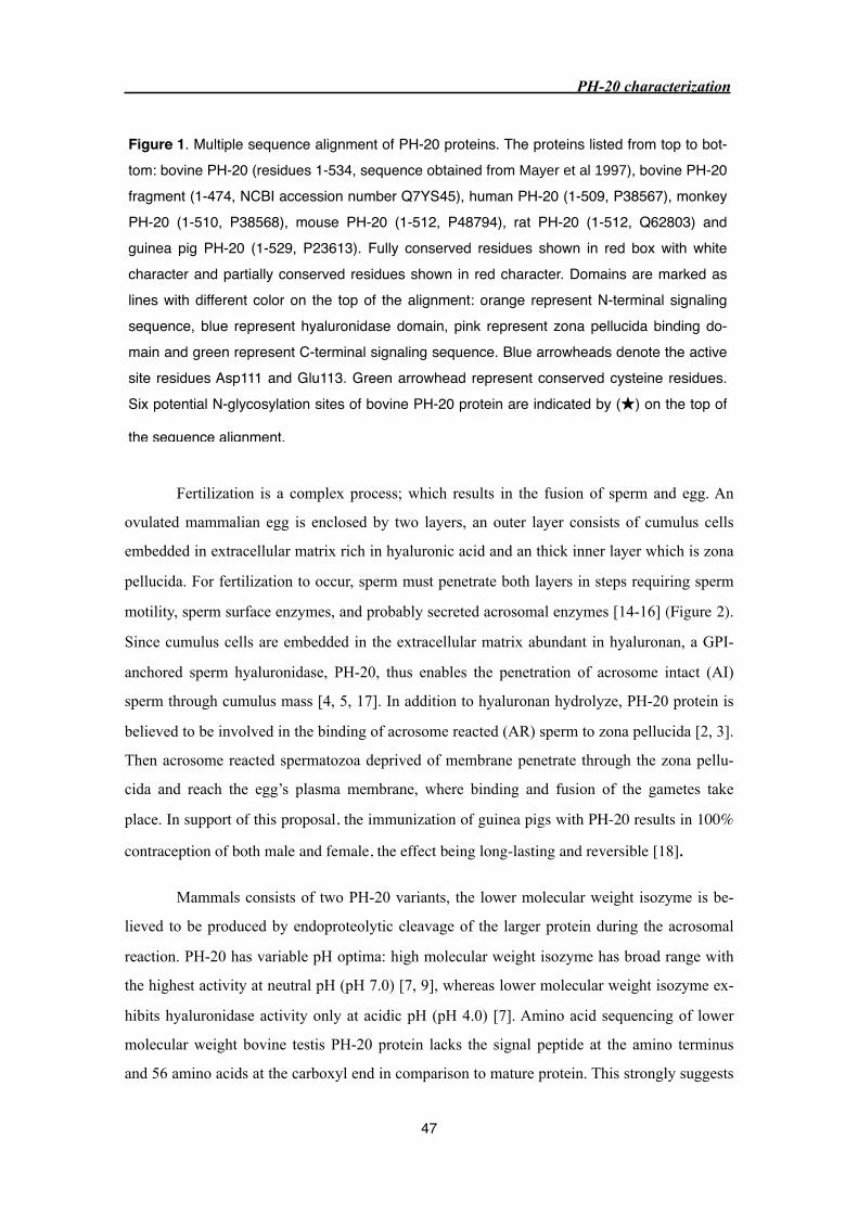
Fertilization is a complex process; which results in the fusion of sperm and egg. An
ovulated mammalian egg is enclosed by two layers, an outer layer consists of cumulus cells
embedded in extracellular matrix rich in hyaluronic acid and an thick inner layer which is zona
pellucida. For fertilization to occur, sperm must penetrate both layers in steps requiring sperm
motility, sperm surface enzymes, and probably secreted acrosomal enzymes [14-16] (Figure 2).
Since cumulus cells are embedded in the extracellular matrix abundant in hyaluronan, a GPI-
anchored sperm hyaluronidase, PH-20, thus enables the penetration of acrosome intact (AI)
sperm through cumulus mass [4, 5, 17]. In addition to hyaluronan hydrolyze, PH-20 protein is
believed to be involved in the binding of acrosome reacted (AR) sperm to zona pellucida [2, 3].
Then acrosome reacted spermatozoa deprived of membrane penetrate through the zona pellu-
cida and reach the egg’s plasma membrane, where binding and fusion of the gametes take
place. In support of this proposal, the immunization of guinea pigs with PH-20 results in 100%
contraception of both male and female, the effect being long-lasting and reversible [18].
Mammals consists of two PH-20 variants, the lower molecular weight isozyme is be-
lieved to be produced by endoproteolytic cleavage of the larger protein during the acrosomal
reaction. PH-20 has variable pH optima: high molecular weight isozyme has broad range with
the highest activity at neutral pH (pH 7.0) [7, 9], whereas lower molecular weight isozyme ex-
hibits hyaluronidase activity only at acidic pH (pH 4.0) [7]. Amino acid sequencing of lower
molecular weight bovine testis PH-20 protein lacks the signal peptide at the amino terminus
and 56 amino acids at the carboxyl end in comparison to mature protein. This strongly suggests
PH-20 characterization
47
Figure 1. Multiple sequence alignment of PH-20 proteins. The proteins listed from top to bot-tom: bovine PH-20 (residues 1-534, sequence obtained from Mayer et al 1997), bovine PH-20 fragment (1-474, NCBI accession number Q7YS45), human PH-20 (1-509, P38567), monkey PH-20 (1-510, P38568), mouse PH-20 (1-512, P48794), rat PH-20 (1-512, Q62803) and guinea pig PH-20 (1-529, P23613). Fully conserved residues shown in red box with white character and partially conserved residues shown in red character. Domains are marked as lines with different color on the top of the alignment: orange represent N-terminal signaling sequence, blue represent hyaluronidase domain, pink represent zona pellucida binding do-main and green represent C-terminal signaling sequence. Blue arrowheads denote the active site residues Asp111 and Glu113. Green arrowhead represent conserved cysteine residues. Six potential N-glycosylation sites of bovine PH-20 protein are indicated by (★) on the top of
the sequence alignment.
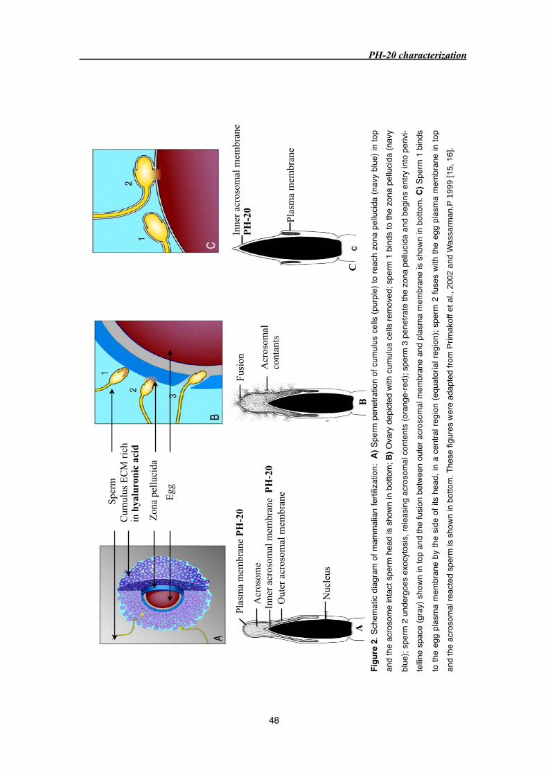
Sper
mC
umul
us E
CM
ric
hin
hya
luro
nic
acid
Zon
a pe
lluci
da
EggTex
Tex Tex
Plas
ma
mem
bran
e P
H-2
0
Acr
osom
eIn
ner
acro
som
al m
embr
ane
PH
-20
Nuc
leus
Out
er a
cros
omal
mem
bran
e
Tex
Fusi
on Acr
osom
alco
ntan
ts
A
Inne
r ac
roso
mal
mem
bran
eP
H-2
0
Plas
ma
mem
bran
e
CB
Figu
re 2
. Sch
emat
ic di
agra
m o
f mam
mal
ian
ferti
lizat
ion:
A) S
perm
pen
etra
tion
of cu
mul
us c
ells
(pur
ple)
to re
ach
zona
pel
lucid
a (n
avy
blue
) in
top
and
the
acro
som
e in
tact
spe
rm h
ead
is sh
own
in b
otto
m; B
) Ova
ry d
epict
ed w
ith c
umul
us ce
lls re
mov
ed; s
perm
1 b
inds
to th
e zo
na p
ellu
cida
(nav
y
blue
); sp
erm
2 u
nder
goes
exo
cyto
sis, r
elea
sing
acro
som
al c
onte
nts
(ora
nge-
red)
; spe
rm 3
pen
etra
te th
e zo
na p
ellu
cida
and
begi
ns e
ntry
into
per
ivi-
tellin
e sp
ace
(gra
y) s
hown
in to
p an
d th
e fu
sion
betw
een
oute
r acr
osom
al m
embr
ane
and
plas
ma
mem
bran
e is
sho
wn in
bot
tom
. C) S
perm
1 b
inds
to th
e eg
g pl
asm
a m
embr
ane
by th
e sid
e of
its
head
, in
a ce
ntra
l reg
ion
(equ
ator
ial r
egio
n); s
perm
2 fu
ses
with
the
egg
plas
ma
mem
bran
e in
top
and
the
acro
som
al re
acte
d sp
erm
is s
hown
in b
otto
m. T
hese
figu
res
were
ada
pted
from
Prim
akof
f et a
l., 2
002
and
Was
sarm
an.P
199
9 [1
5, 1
6].
PH-20 characterization
48

that the lower molecular weight form is generated by proteolysis, probable by the action of ac-
rosomal enzymes [13].
In addition to fertilization, mammalian hyaluronidases plays important role in many
biological process but the study of these enzymes are greatly neglected. In the present study, we
purified PH-20 protein from bovine testicular extract (Sigma) and bovine testes to homogeneity
and characterized them.
PH-20 characterization
49

3.3 Material and methods
3.3.1 Substrate gel assay
Hyaluronic acid substrate gel electrophoresis was performed according to the protocol
described by Guntenhoener et al., 1992 [19]. The protein was subjected to electrophoresis un-
der non-reducing condition on 12% (w/v) SDS-PAGE gel copolymerized with 0.17 mg/ml of
hyaluronic acid. The gel was incubated in the TBS buffer (20 mM Tris-HCl, pH 7.5 150 mM
NaCl) containing 3% (v/v) TritonX-100 for 3 hours at room temperature on a rocking platform
to remove SDS and then in 100 mM sodium acetate buffer pH 4.0, for 14 to 16 hours at 37˚C.
To visualize digestion of hyaluronic acid, gels were stained with 0.5% (w/v) alcian blue in 3%
(v/v) acetic acid for 2 hours at room temperature and destained in 7% (v/v) acetic acid. The
presence of activity in the gel was revealed by a lack of coloration of a digested area against the
blue background characteristic for undigested hyaluronic acid.
3.3.2 Protein Electrophoresis and Immunoblotting
Protein samples were solubilized in sample buffer (62.5 mM Tris-HCl pH 6.8, 10%
glycerol, 2% (w/v) SDS, 0.01% (w/v) bromophenol blue with or without 100 mM DTT) and
heated at 99˚C for 3 min and separated on 12% SDS-PAGE [20].
The immunoblotting was done according to standard protocol (Molecular cloning,
Sambrook et al., 2nd edition). After electrophoresis, the protein was transfered onto a nitrocel-
lulose membrane (BA85, Schleicher & Schuell, Dassel, Germany) using Bio-Rad PROTEAN
2 Electrophoresis / Mini Trans-Blot Module. The transfer was carried out in the presence of
blotting buffer (20 mM Tris buffer pH 8.3, 150 mM glycine and 20% methanol) at 100V, 4˚C
for 1 hour. The transferred membrane was blocked with TTBS buffer (20 mM Tris pH 7.4, 150
mM NaCl, 0.1% (v/v) Tween 20) containing 5% (w/v) dry skim milk to prevent non-specific
binding. The membrane was incubated with TTBS buffer (with 5% (w/v) dry skim milk) con-
taining primary antibody, ram PH-20 polyclonal antibody (Babraham bioscience technologies)
at dilution 1:2500 for 1 hour in room temperature. Subsequently, the membrane was washed
with TTBS for three times at the interval of 10 minutes and then incubated with TTBS (with
5% (w/v) dry skim milk) containing secondary antibody, anti-mouse IgG antibody-peroxidase
conjugate (Sigma) at 1:5000 dilution for 1 hour at room temperature. Again, the membrane
was extensively washed in TTBS as described before and incubated in ECL Western blotting
detection reagents according to manufacturers protocol (Amersham Biosciences). The blot was
PH-20 characterization
50
subsequently exposed to KodakX-OMAT XAR-5 radiography film for 15 sec to 15 min until
the positive bands were visualized by enhanced chemiluminescence.
3.3.3 Cloning and expression of human PH-20
Cloning and expression of PH-20 fragment (1-332) containing a C-terminal 6xHis tag
was carried out by Dr. Margit Schmidt, Department of Biology, East Carolina University. Hu-
man PH-20 gene comprising amino acids Leu1 to Ile332 was ligated into the Pichia expression
vector pPICZαA (Invitrogen) and transformed into TOP 10 E. coli cells (Invitrogen). The
plasmid was purified and transformed into Pichia pastoris strains X33 and KMH71H using
10μg plasmid. From each strain ten single colonies were isolated, cultured in complex liquid
media and expression was induced by the addition of methanol. The harvested culture super-
natant and the cell extract from positive clones were analyzed for expression in our laboratory
by SDS gel electrophoresis, anti-His antibodies and substrate gel assay.
3.3.4 Protein purification from crude extract (type Ⅳ-S: From bovine tes-
tes, Sigma)
The PH-20 was purified according to the modified procedure described elsewhere [9,
13, 21, 22]. Lyophilized crude extract (type Ⅳ-S: From bovine testes, Sigma) was dissolved in
20 mM sodium phosphate buffer pH 7.0 and purification was carried out as a six step proce-
dure. As a first step the ammonium sulfate was added slowly to reach 45% saturation by con-
stant stirring using magnetic bead (Heidolph MR 3001) for 4 to 6 hours at 4˚C. This was fol-
lowed by centrifugation at 13000 rpm for 30 min at 4˚C (Sorval RC-5B) and the precipitant
was removed. To the supernatant ammonium sulfate was added to reach 70% saturation by con-
stant stirring for 4 to 6 hours followed by centrifugation of mixture at 13000 rpm for 30 min at
4˚C. The precipitate obtained between 45-70% saturated ammonium sulfate was rich in PH-20
(monitored by Western blot analysis and substrate gel as described earlier), dissolved in 20 mM
sodium phosphate pH 6.0 and dialyzed (Spectra/Pro, membrane cutoff 12 - 14 kDa) against the
same buffer. The insoluble precipitate formed during the dialysis was removed by centrifuga-
tion at 13000 rpm for 30 min and resultant supernatant was loaded on a Resource Q anion ex-
changer column (Amersham Pharmacia) using acta purifier (Amersham Pharmacia). PH-20
elutes in flow through (monitored by substrate gel) whereas many contaminant proteins bound
to Resource Q resin were eluted with 20 mM sodium phosphate pH 6.0 containing 1 M NaCl.
The flowthrough was dialyzed against 20 mM sodium phosphate pH 7.0 and applied onto
PH-20 characterization
51

Protein-G affinity column (Amersham Pharmacia) to separate immunoglobulins. The PH-20
found in flowthrough was concentrated using centricon (Amicon Ultra, membrane cutoff 30000
kDa) and applied on a Mono-S cation exchanger column (Pharmacia). The PH-20 was bound to
the Mono-S column and eluted with 20 mM sodium phosphate buffer pH 7.0 containing 1 M
NaCl as step gradient. The fractions containing PH-20 (identified by substrate gel and mass
mapping), were concentrated and applied to Hiload 26/60 Superdex G-75 (Amersham Pharma-
cia) gel filtration column. Protein concentration of purified PH-20 was determined by absorp-
tion spectroscopy (Hewlett Packard 8453, HP845X UV-Visible system, Switzerland) at 280 nm
using extinction coefficient of 89435 (lower species) and 90430 (higher species) M-1 cm-1 cal-
culated from the amino acid sequence obtained from Mayer et al., 1997. The extinction coeffi-
cient was calculated using ProtParam (http://www.expasy.org/tools/protparam.html) from Ex-
pasy server.
3.3.5 Protein purification from bovine testes
Fresh bovine testes were obtained from regional slaughterhouse and stored at -80˚C.
When needed, frozen testes were thawed overnight at 4˚C. Outerlayer of the testis was re-
moved, the inner layer cut into small pieces and homogenized using buffer (20 mM sodium
phosphate pH 7.0) containing protease inhibitor (1 mM EDTA, 10 μM Leupeptin, 1 mM
PMSF) at 4˚C. Homogenized sample was centrifuged at 8000 rpm for 30 min at 4˚C and pellet
was removed. To the supernatant ammonium sulfate was added slowly to reach 40 % saturation
by constant stirring for 4 to 6 hours in the cold room. The sample was then centrifuged at 8000
rpm for 30 min and pellet was removed. To the resultant supernatant ammonium sulfate was
added slowly to reach 70 % saturation by constant stirring for 4 to 6 hours followed by cen-
trifugation at 8000 rpm for 30 min. The pellet obtained from 40 - 70 % saturation was dissolved
and dialyzed against 20 mM sodium phosphate buffer pH 6.0.
To handle large volume in each step of purification, ion exchange column was packed
manually with Q sepharoseTM fast flow (Amersham Biosciences) and SP sepharoseTM fast flow
(Amersham Bioscience) beads separately. Dialyzed sample was applied on the anion exchanger
(Sepharose-Q), PH-20 elutes in flowthrough whereas most contaminant were bound to the col-
umn. The collected flowthrough was dialyzed against 20 mM sodium phosphate pH 7.0 and
applied on a Protein-G column, which bound the immunoglobulins whereas PH-20 eluted in
flowthrough. The collected supernatant was applied on a cation exchanger (Sephrose-S) and
bound proteins where batch eluted with 20 mM sodium phosphate pH 7.0 containing 30, 120,
PH-20 characterization
52

200, 1000 mM NaCl. The fractions containing PH-20 (monitored by substrate gel and mass
mapping) were pooled and applied onto Hiload 26/60 Superdex G-75 (Amersham Pharmacia)
gel filtration column. The concentration of the higher species of PH-20 protein was determined
by absorption spectroscopy (Hewlett Packard 8453, HP845X UV-Visible system, Switzerland)
at 280 nm using extinction coefficient of 90430 M-1 cm-1 calculated from the amino acid se-
quence.
3.3.6 Analytical ultracentrifugation
The sedimentation equilibrium (SE) and sedimentation velocity (SV) were performed
on a Beckman optima XL-A analytical ultracentrifuge (Beckman Instruments) equipped with 4
and 12 mm Epon double sector cells. The experiments were carried out in the presence and ab-
sence of NaCl and with the presence of dimethylammonium sulfonate (Zwitterionic com-
pound). Sedimentation velocity runs were carried out at two different speeds 44000 and 52000
rpm at 20˚C and the sedimentation coefficients were corrected to standard condition (water, 20
˚C). Sedimentation equilibrium measurements were carried out at 20 ˚C at rotor speeds from
5000 to 7000 rpm using 12-mm double sector cells filled with 0.1 ml solution. Molecular
masses were evaluated in sedimentation equilibrium experiments from ln A versus r2 plots,
where A represents the absorbance and r is the distance from the rotor center. A partial specific
volume of 0.73 cm3/g was assumed in all calculations. The molecular masses were calculated
using the Segal computer program (www.biozentrum.unibas.ch/auc). All the analytical ultracen-
trifugation experiments were performed by Ariel Lustig, Biozentrum, University of Basel.
3.3.7 Mass spectral analysis
The mass spectrometry experiments were performed by the group of Dr. Paul Jenoe,
Biozentrum, University of Basel as described [23, 24]. Liquid chromatography (LC) / Mass
spectrometric (MS) analysis of the protein and its digest was carried out on 100 μm i.d.(inner
diameter) capillary column packed with C18 Monitor-Spherical silica (5-μm particle size, Col-
umn Engineering Inc., Ontario, CA). The bound peptide were eluted with 0.05% trifluoroacetic
acid to 60% acetonitrile containing 0.05% trifluoroacetic acid at the flow rate of 1-2 μl/min.
The outlet of the column was directed to a microspray needle, which was pulled from 100 μm
i.d.(inner diameter) × 280 μm o.d. (outer diameter) fused silica capillaries (LC Packings) on a
model P-2000 quartz micropipette puller (Sutter Instruments Co.). The needle was placed into
an XYZ micropossitioner, and the voltage was applied directly to the sample stream through
the capillary union. Spray voltages were usually between 1100 and 1400 V. Mass determina-
PH-20 characterization
53
tions were carried out on a TSQ7000 triple quadrupole mass spectrometer (Finnigan, San José,
CA). All measurements were carried out in the positive ion mode. Precursor ion scanning was
between 200 and 2000 Da for 3 s at unit resolution. For operation in the MS/MS mode, the
resolution of Q1 was set to transmit a mass window of 4 Da and the resolution of Q3 was ad-
justed to 1.5 Da. Scanning was between 50 and 2250 Da in 3.5 s. Argon was used as a collision
gas at a pressure of 3.5 mTorr.
3.3.8 Mass fingerprinting
The protein bands corresponding to PH-20 were cutout of the SDS/PAGE gel and incu-
bated with trypsin (sequencing grade, Promega) at 37˚C for 2 h. Trypsined fragments were de-
salted on C18 ZipTips (Millpore) and eluted with 1.5 μl 80% acetonitrile, 0.1% TFA, contain-
ing 1μl/ml α-cyano-4-hydroxycinnamic acid (Aldrich). About 300 nl of the elute was deposited
onto anchor spots of a Scout 400 μm/36 sample support (Bruker Daltonik, Germany) and the
droplet was left to dry at room temperature. Mass spectra were recorded on a Bruker Scout 26
Reflex Ⅲ instrument (Bruker Daltonik). The instrument was calibrated with angiotensin Ⅱ, sub-
stance P, bombesin, and adrenocorticotropic hormone. Peptides were analyzed in reflector
mode using delayed ion extraction with a total acceleration voltage of 23 kV. The molecular
weights of the each ejected peptide fragment were measured and searched in the sequence li-
brary using the programme MASCOT.
3.3.9 Crystallization attempts
Two different molecular weight species purified from Sigma crude extract were dia-
lyzed against 5mM sodium acetate pH 4.0, concentrated using centricon to 4.3 mg/ml (lower
species) and 8.3 mg/ml (higher species), respectively. PH-20 purified from bovine testes
(higher species) was dialyzed against 20 mM HEPES pH 7.0 and concentrated to 10 mg/ml.
Crystallization was performed with concentrated protein solutions using Hampton screen kits Ⅰ
and Ⅱ by hanging drop vapour diffusion method. The drops were prepared by mixing 1 μl of
protein with 1 μl of reservoir solution and equilibrated against 500 μl of reservoir solution.
PH-20 characterization
54
3.3 Results
3.3.1 Expression of recombinant human PH-20 protein
The attempts to clone and express the N-terminal hyaluronidase domain of PH-20
(residue Leu1-Ile312) in Pichia pastoris were not successful. Unfortunately, PH-20 protein
expression was neither detected in the supernatant nor in the cell extract. Cloning and
expression of the same PH-20 fragment in Baculovirus was also not successful (data not
shown). All these experiments were performed by Dr. M. Schmidt.
3.3.2 Purification of PH-20 protein from Sigma crude extract
Since the human PH-20 protein could not be expressed successfully in Baculovirus and
Pichia pastoris expression systems, an attempt was made to purify PH-20 from commercial
bovine testicular crude extract (type Ⅳ-S, Sigma).
The PH-20 purification comprised of six steps (see PH-20 purification scheme, Figure
3). In each step of purification, PH-20 protein was monitored by one or more of the following
methods: 1) substrate gel electrophoresis assay, 2) Western blot analysis using a polyclonal ram
anti PH-20 antibody, and 3) MALDI mass mapping of tryptic fragments.
Lyophilized crude extract (Sigma) was dissolved and fractionated by ammonium
sulfate precipitation. The resultant fractions were analyzed by SDS/PAGE and Western blot
(Figure 4). The hyaluronidase rich pellet obtained with 45-70% saturated ammonium sulfate
precipitation (Figure 4B).
The PH-20 rich precipitant was dissolved and dialyzed against 20 mM sodium
phosphate buffer pH 6.0. The sample was loaded on an anion exchanger (Resource Q) where
PH-20 eluted in the flowthrough while some major contaminants remained bound to the
column (Figure 5B). The substrate gel of the flowthrough sample showed strong hyaluronidase
activity corresponding to two different molecular weight proteins, whereas the elution sample
showed less activity (Figure 5C).
The flowthrough from Resource Q column, in addition to PH-20 contained
immunoglobulin as one major contaminant as determined by MALDI mass mapping. The
sample was dialyzed against 20 mM sodium phosphate pH 7.0 and applied onto a Protein-G
affinity column. PH-20 eluted in flowthrough as confirmed by the substrate gel and the
immunoglobulin remained bound to the column (Figure 6).
PH-20 characterization
55
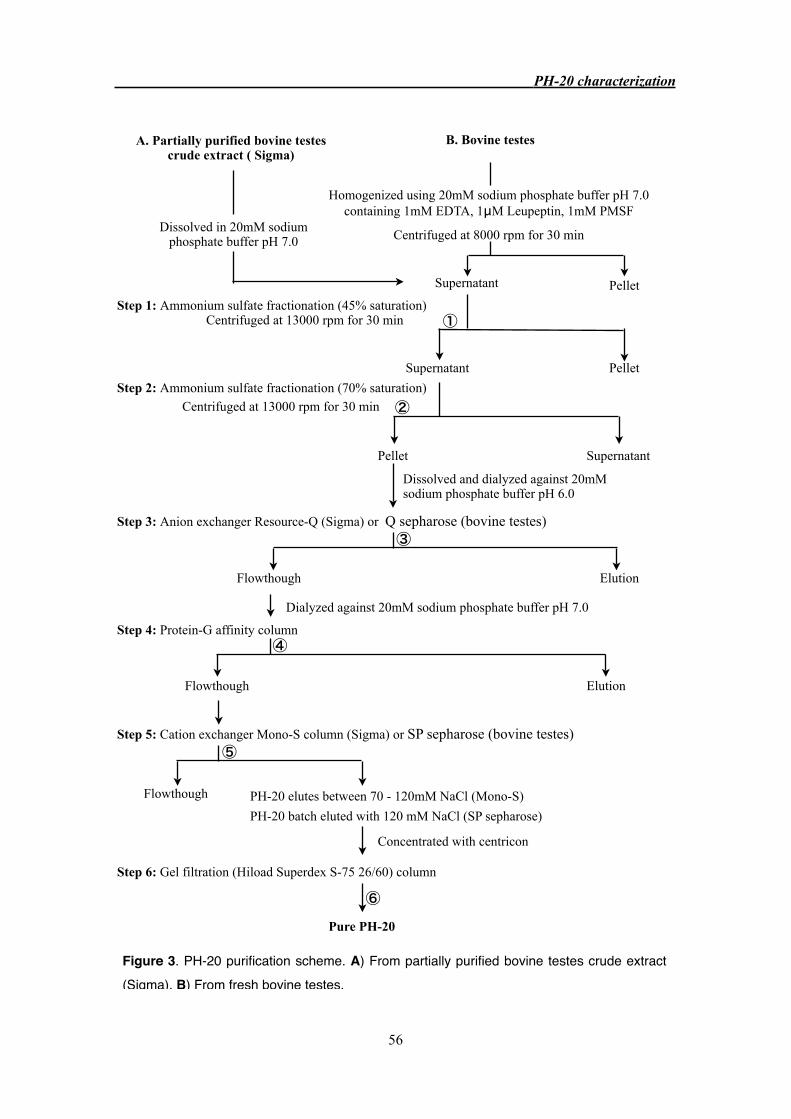
A. Partially purified bovine testes crude extract ( Sigma)
B. Bovine testes
Homogenized using 20mM sodium phosphate buffer pH 7.0 containing 1mM EDTA, 1μM Leupeptin, 1mM PMSF
Centrifuged at 8000 rpm for 30 min
PelletSupernatant
Step 1: Ammonium sulfate fractionation (45% saturation)Centrifuged at 13000 rpm for 30 min
Centrifuged at 13000 rpm for 30 min
Pellet Supernatant
Dissolved and dialyzed against 20mM sodium phosphate buffer pH 6.0
Step 3: Anion exchanger Resource-Q (Sigma) or Q sepharose (bovine testes)
ElutionFlowthough
Dialyzed against 20mM sodium phosphate buffer pH 7.0
Step 4: Protein-G affinity column
Flowthough Elution
Step 5: Cation exchanger Mono-S column (Sigma) or SP sepharose (bovine testes)
Flowthough PH-20 elutes between 70 - 120mM NaCl (Mono-S)
PH-20 batch eluted with 120 mM NaCl (SP sepharose)
Concentrated with centricon
Step 6: Gel filtration (Hiload Superdex S-75 26/60) column
Pure PH-20
Dissolved in 20mM sodium phosphate buffer pH 7.0
Step 2: Ammonium sulfate fractionation (70% saturation)
PelletSupernatant
①
②
③
④
⑤
⑥
Figure 3. PH-20 purification scheme. A) From partially purified bovine testes crude extract (Sigma). B) From fresh bovine testes.
PH-20 characterization
56

The flowthrough from the Protein-G column was applied onto a cation exchanger
(Mono-S) column. It was found that PH-20 bound to the column and eluted between 70-120
mM NaCl (Figure 7). At this stage of the purification, the majority of the contaminant proteins
were removed and PH-20 migrated in SDS-PAGE as two different molecular weight proteins
with the molecular weight of approximately 60 kDa (PH-2060) and 69 kDa (PH-2069) (Figure 7
B). The MALDI mass mapping of two different molecular weight protein bands, cutout from
the lane D3 matched to bovine testicular hyaluronidase (AAP55713) (shown in figure 12).
The fractions containing PH-20 were pooled, concentrated and subjected to gel
filtration chromatography (Hiload Superdex S-75 26/60). Two different molecular weight
PH-20 proteins were partially resolved and several lower molecular weight contaminants were
separated by this purification procedure (Figure 8 A). The yield of PH-2069 and PH-2060 protein
was approximately 3.0 mg/1g and 2.5 mg/1g of crude extract, respectively. Two purified PH-20
variants were dialyzed and concentrated to 4.3 mg/ml (PH-2060) and 8.3 mg/ml (PH-2069)
respectively. Crystallization trials were performed with Hampton screening kits Ⅰ and Ⅱ. Despite
the high purity and extensive screening neither of the two proteins could be crystallized.
PH-20 characterization
57
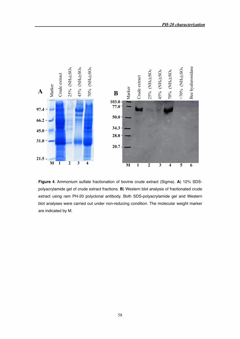
Mar
ker
Cru
de e
xtra
ct
Bee
hya
luro
nida
se
103.077.0
50.0
34.3
28.8
20.7
B
M 1 2 3 4 5 6 M 1 2 3 4
A
97.4
66.2
45.0
31.0
21.5
Mar
ker
Cru
de e
xtra
ct
25%
(N
H4)
2SO
4
45%
(N
H4)
2SO
4
70%
(N
H4)
2SO
4
45%
(N
H4)
2SO
4
25%
(N
H4)
2SO
4
70%
(N
H4)
2SO
4
>70%
(N
H4)
2SO
4
PH-20 characterization
58
Figure 4. Ammonium sulfate fractionation of bovine crude extract (Sigma). A) 12% SDS-polyacrylamide gel of crude extract fractions. B) Western blot analysis of fractionated crude extract using ram PH-20 polyclonal antibody. Both SDS-polyacrylamide gel and Western blot analyses were carried out under non-reducing condition. The molecular weight marker are indicated by M.

97.4
66.2
45.0
31.0
21.5
14.4
M SM 1 2
103.077.0
50.0
34.3
28.8
M 1 2
Flowthrough (PH-20)
Elution (impurities)
A
B C
PH-20 characterization
59
Figure 5. Purification of PH-20 using anion exchange chromatography (Resource-Q column). A) Chromatogram of PH-20 elution profile with NaCl gradient. B) SDS-polyacrylamide (12%, non-reducing condition) analysis of the peak fractions. C) Hyaluronidase acid substrate SDS-PAGE (12%, non-reducing condition) at pH 4.0. Lane1: flowthrough; lane 2: elution fraction. M stands for molecular weight markers (kDa) and SM for starting material.

97.4
66.2
45.0
31.0
21.5
A
103.077.0
50.0
34.3
28.8
M 1 2M SM 1 2 3 4
Flowthrough (PH-20)
CB
Elution (immunoglobulin)
PH-20 characterization
60
Figure 6. Separation of immunoglobulin contaminant by Protein-G column. A) Chromatogram of PH-20 elution profile on Protein-G column. B) SDS-polyacrylamide (12%, non-reducing condition) analysis of peak fractions, lane 1 - 2: flowthrough fraction; lane 3 - 4: elution fraction using 100 mM glycine pH 3.0. C) Hyaluronic acid substrate SDS-PAGE (12%, non-reducing condition) at pH 4.0, lane1: flowthrough; lane2: elution fraction. M strands for molecular weight markers (kDa) and SM for starting material.

69 kDa
60 kDa
97.4
66.2
45.0
31.0
21.5
14.4M SM C11 C13 D9 D7 D5 D3 D1
69 kDa60 kDa
Flowthrough
Eluted PH-20
B C
PH-20 characterization
61
Figure 7. Purification of PH-20 protein using cation exchange chromatography (Mono-S column) A) Chromatogram of PH-20 elution profile with NaCl gradient. B) SDS-polyacrylamide (12%, non-reducing condition) analysis of proteins separated on Mono-S column. C) Hyaluronic acid substrate SDS-PAGE (12%, non-reducing condition) at pH 4.0. M stands for molecular weight markers (kDa) and SM for starting material. Fraction numbers (A1 to B11) are indicated at the bottom of the gel.
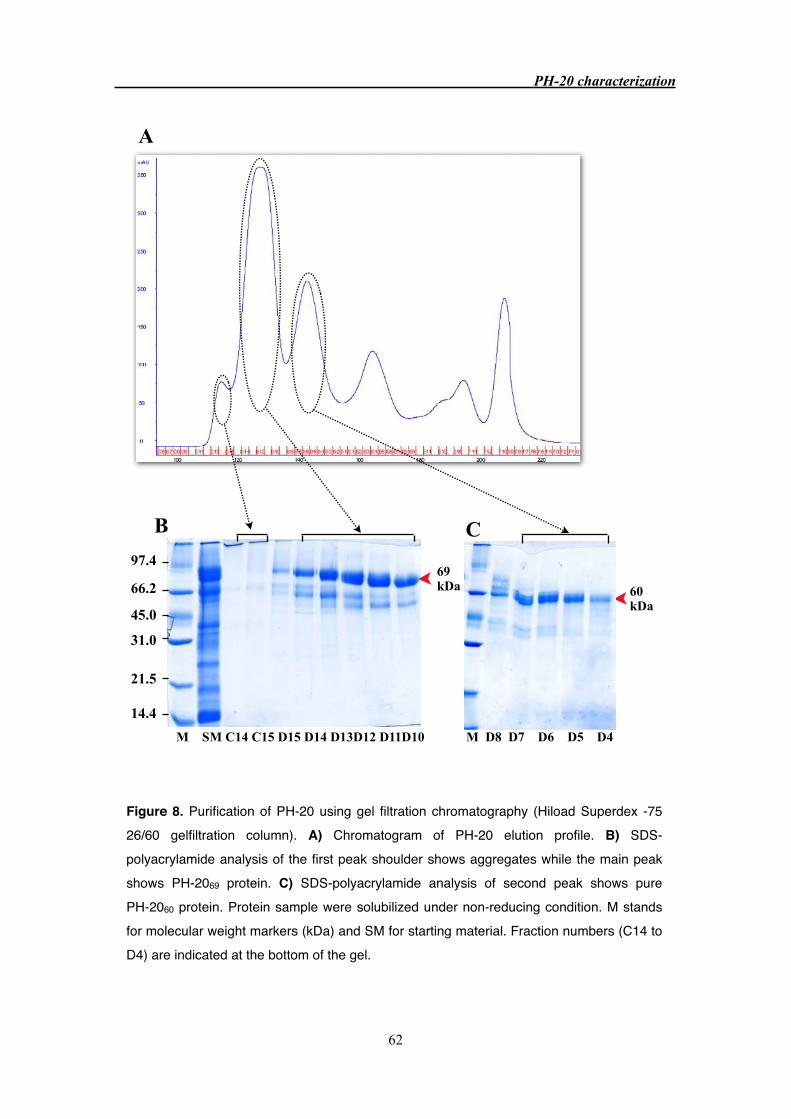
97.4
66.2
45.0
31.0
21.5
14.4
M SM C14 C15 D15 D14 D13D12 D11D10 M D8 D7 D6 D5 D4
69 kDa 60
kDa
A
B C
PH-20 characterization
62
Figure 8. Purification of PH-20 using gel filtration chromatography (Hiload Superdex -75 26/60 gelfiltration column). A) Chromatogram of PH-20 elution profile. B) SDS-polyacrylamide analysis of the first peak shoulder shows aggregates while the main peak shows PH-2069 protein. C) SDS-polyacrylamide analysis of second peak shows pure PH-2060 protein. Protein sample were solubilized under non-reducing condition. M stands for molecular weight markers (kDa) and SM for starting material. Fraction numbers (C14 to D4) are indicated at the bottom of the gel.

3.3.3 Purification of PH-20 from bovine testes
Purification of PH-20 protein from commercial crude extract of bovine testes has been
discontinued due to the high cost of the extract and very low yield of pure protein. Instead,
purification of PH-20 protein from bovine testes supplied by a local slaughterhouse was
pursued.
The frozen bovine testes were thawed and homogenized using buffer containing
protease inhibitors. The homogenized sample was centrifuged and the resultant supernatant and
pellet were checked for hyaluronidase activity (Figure 9). Substrate gel showed maximum
activity for the supernatant whereas the pellet showed negligible activity. The purification
procedure was similar to that used for the purification of Sigma crude extract (Figure 3. PH-20
purification scheme), except for the ion exchange columns which were packed manually in
order to handle the large volumes.
A protein with an apparent molecular weight of about 80 kDa (PH-2080) with
hyaluronidase activity was purified from fresh bovine testes (Figure 10 B) and identified as
bovine PH-20 protein by MALDI mass mapping (Figure 12). The yield was approximately
1-1.5 mg/1 kg of fresh bovine testes and could not be improved despite numerous trials. In
contrast to commercial crude extract, the relative amount of PH-2066 protein obtained from
fresh bovine testes was negligible and was not purified to homogeneity (Figure 10 C). Purified
PH-2080 protein was dialyzed and concentrated to 10 mg/ml and crystallization trials were
performed with Hampton screening kits Ⅰ and Ⅱ. Crystallization was not successful and
precipitation was seen in most of the conditions tested which suggested protein aggregation.
PH-20 characterization
63

97.4
66.2
45.0
31.0
21.5
103.077.0
50.0
34.3
28.8
M P S M P S
A B
Figure 9. Homogenization of bovine testes. A) SDS-polyacrylamide (12% gel) analysis. B) Hyaluronic acid substrate SDS-PAGE (12% gel) at pH 4.0. Lane P and S stands for pellet and supernatant collected after centrifugation at 8000 rpm for 30 minutes, respectively. Protein sample were solubilized under non-reducing condition. M stands for molecular weight markers (kDa).
PH-20 characterization
64

97.4
66.2
45.0
31.0
21.5
14.4
M SM C7 C8 C9 C10 C11 C12 M C14 C15 D15 D14 D13 D12
B C
80 kDa 66
kDa
A
PH-20 characterization
65
Figure 10. Purification of PH-20 using gel filtration chromatography (Hiload Superdex-75 26/60 column) A) Chromatogram of PH-20 elution profile. B) SDS-polyacrylamide analysis of the first peak shoulder shows aggregates while the main peak shows PH-2080 protein. C) SDS-polyacrylamide analysis of second peak shows PH-2066 protein with contaminant. Protein sample were solubilized under non-reducing condition. Molecular weights marker (M; kDa), starting material (SM) and fraction numbers (C7 to D12) are indicated at the bottom of the gel.

3.2.4 Endoproteolytic cleavage of PH-20 protein
Purified PH-20 proteins from both commercial source (PH-2060 and PH-2069) and from
fresh bovine testes (PH-2080) were analyzed for endoproteolytic cleavage. For this the proteins
were reduced by DTT and analyzed on SDS gels (Figure. 11). The PH-2060 protein (Sigma) run
as two bands with apparent molecular weight of 35 and 30 kDa (Figure. 11 B, Lane 1). In
contrast, PH-2069 protein from commercial source and PH-2080 protein from fresh bovine testes
run as single band under reducing condition. This suggests that PH-2060 protein is
endoproteolytically cleaved and a disulfide bridge holds the two polypeptides.
97.4
66.2
45.0
31.0
21.5
14.4
97.466.2
45.0
31.0
21.5
14.4
M P60 P69 P80
A B
M P60 P69 P80
PH-20 characterization
66
Figure 11. Purified PH-20 from two different source (Sigma crude extract and bovine testes). (A) non-reducing condition (B). reducing condition (12% SDS-polyacrylamide gel). Lane 1 and lane 2 corresponds to PH-2060 and PH-2069 proteins purified from Sigma crude extract. Lane 3: PH-2080 purified from bovine testes. Molecular weights marker (M) are indicated at the bottom of the gel. Arrows indicate reduced PH-2060 protein fragments

3.3.5 Mass spectrometry
Mascot search results of tryptic fragment of the PH-2060 and PH-2069 proteins purified
from commercial crude extract (Sigma) and PH-2080 from fresh bovine testes matched to the
hyaluronidase domain of bovine PH-20 sequence (NCBI accession number: AAP55713)
(Figure 12). This strongly suggests that these proteins have identical N-termini. LC-MS method
was used to determine the molecular weight of purified PH-20 proteins. The PH-20 proteins
purified from commercial crude extract (Sigma) have a molecular weight of 60334 Da and
68780 Da, respectively. Whereas PH-20 protein purified from fresh bovine testes has a MW of
80350 Da (Figure 13). The molecular weight estimates are based on broad peaks and therefore
is not accurate. This may be due to posttranslational modification and may account for
approximately 5% error.
PH-20 characterization
67
Figure 12. Mascot Search Results for PH-2060 protein (commercial source). Tryptic fragments matched to the N-terminal region of AAP55713 sequence (deposited in the NCBI) and are shown in bold (top) and peptides were sorted based on increasing mass (bottom).

3.3.6 PH-20 aggregation
To test for homogeneity, sedimentation equilibrium (SE) and sedimentation velocity
(SV) experiments were performed with PH-2080 protein purified from fresh bovine testes in the
presence and absence of 100 mM NaCl. The result revealed extensive protein aggregation in
the presence of 100 mM NaCl whereas aggregation was less extensive in the absence of NaCl.
The PH-2060 protein purified from commercial testicular extract gave similar results (Figure
14C). Changing the pH from 7.0 to 5.4 had no effect on the heterogeneity of the protein
solution (data not shown). Many additives were tested with the aim to eliminate the protein
aggregation. Success was achieved with non detergent sulfobetains (NDSB), which are
zwitterionic, powerful non denaturing protein solubilizing agents used in protein extraction,
solubilization and crystallization [25, 26]. On addition of dimethylethylammonium propane
sulfonate (NDSB-195), at a final concentration of 250 mM, aggregation was abolished for
PH-2080 (Figure 14E) and PH-2060 (data not shown). Whereas in the presence of 100 mM
NDSB-195, PH-2080 showed diminished, but not abolished heterogeneity (data not shown).
PH-20 characterization
68
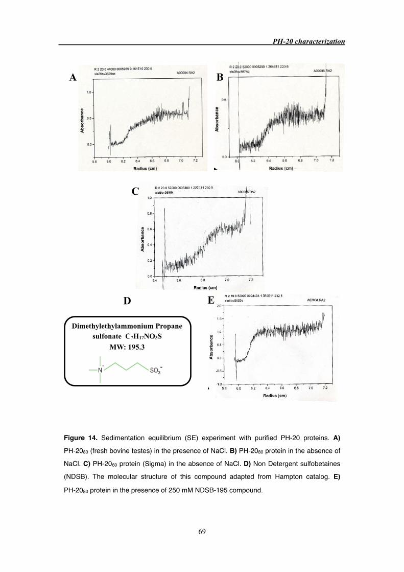
C
-
D
Dimethylethylammonium Propane sulfonate C7H17NO3S
MW: 195.3
E
BA
PH-20 characterization
69
Figure 14. Sedimentation equilibrium (SE) experiment with purified PH-20 proteins. A)
PH-2080 (fresh bovine testes) in the presence of NaCl. B) PH-2080 protein in the absence of
NaCl. C) PH-2060 protein (Sigma) in the absence of NaCl. D) Non Detergent sulfobetaines
(NDSB). The molecular structure of this compound adapted from Hampton catalog. E)
PH-2080 protein in the presence of 250 mM NDSB-195 compound.
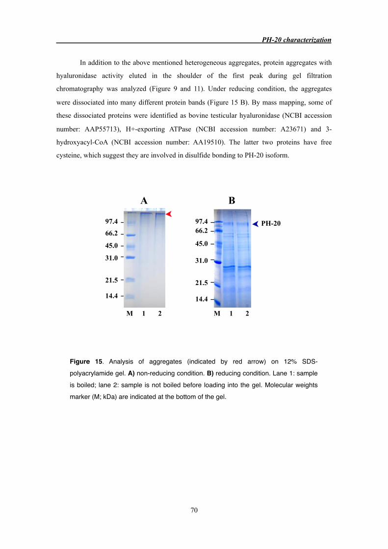
In addition to the above mentioned heterogeneous aggregates, protein aggregates with
hyaluronidase activity eluted in the shoulder of the first peak during gel filtration
chromatography was analyzed (Figure 9 and 11). Under reducing condition, the aggregates
were dissociated into many different protein bands (Figure 15 B). By mass mapping, some of
these dissociated proteins were identified as bovine testicular hyaluronidase (NCBI accession
number: AAP55713), H+-exporting ATPase (NCBI accession number: A23671) and 3-
hydroxyacyl-CoA (NCBI accession number: AA19510). The latter two proteins have free
cysteine, which suggest they are involved in disulfide bonding to PH-20 isoform.
M 1 2
97.4
66.2
45.0
31.0
21.5
14.4
97.466.2
45.0
31.0
21.5
14.4
M 1 2
A B
PH-20
PH-20 characterization
70
Figure 15. Analysis of aggregates (indicated by red arrow) on 12% SDS-polyacrylamide gel. A) non-reducing condition. B) reducing condition. Lane 1: sample is boiled; lane 2: sample is not boiled before loading into the gel. Molecular weights marker (M; kDa) are indicated at the bottom of the gel.
3.4 Discussion
3.4.1 PH-20 purification
We were able to purify two PH-20 variants with hyaluronidase activity from the
commercially available bovine testicular extract (Sigma). Though, PH-20 protein purification
from commercial source (Sigma) has been previously reported [13, 21], we isolated them to
highest purity using modified protocol. Purified PH-20 variants have molecular weight of 60
kDa (PH-2060) and 69 kDa (PH-2069) respectively, as determined by LS/MS analysis and these
values are comparable to those reported [13]. Due to high cost and low yield of PH-20 proteins
from commercial source, purification from bovine testes supplied by a local slaughterhouse
was pursued.
Although the 80 kDa band of bovine PH-20 was reported earlier [9], we were the first
to purify the 80 kDa protein (PH-2080) with hyaluronidase activity from fresh bovine testes to
highest purity. Homogenization of fresh bovine testes resulted in an effective release of
membrane bound PH-20 protein into the supernatant, which suggest PH-20 protein is freed
either by action of released acrosomal enzymes or by mechanically affecting the stability of the
plasma membrane. The yield of PH-2080 protein was very low which might be due to the many
purification steps employed. Unlike the commercial source (Sigma), the PH-2065 fragment was
presented only in negligible amount.
The MALDI mass mapping of the protein bands correspond to PH-20 variants from
Sigma crude extract (PH-2060 and PH-2069) and from fresh bovine testes (PH-2080 and
PH-2065), showed peptides matched to that of N-terminal region of bovine testicular
hyaluronidase PH-20 protein (NCBI accession number: AAP55713). It remains to be shown
whether the glycosylation and / or proteolytic cleavage can account for the difference in
observed molecular weights of PH-20 proteins isolated from commercial source and fresh
bovine testes.
3.4.2 Endoproteolytic cleavage of PH-20
In guinea pig and macaque sperm, lower molecular weight PH-20 form is transformed
into two lower weight fragments under reducing condition, indicative of endoproteolytic
cleavage of the protein into two disulfide-linked fragments [3, 7]. Our data suggested that a
similar alteration on bovine PH-20 protein, under reducing PH-2060 transformed into lower
molecular weight fragment of 35 kDa and 30 kDa, respectively, whereas PH-2069 and PH-2080
PH-20 characterization
71

protein are intact (Figure 17). Apart from PH-20 proteins, similarly modification occurs in
Hyal-1 protein [27]. It was reported that bovine PH-2060 comprises of 443 amino acids, in
comparison to the PH-20 polypeptide deduced from cloned cDNA from the same source, it
lacks the signaling peptide (35 residues) and 56 residues from carboxyl end [13]. This strongly
suggests that PH-2060 is probably proteolytically processed product of the larger isozyme
PH-2069. Structural features established by disulfide bridges, as well as glycosylation are
essential for the hyaluronidase activity of macaque sperm PH-20 protein [12]. Similarly, the
reduction of purified bovine PH-20 results in loss of hyaluronidase activity (data not shown).
3.4.3 PH-20 crystallization and aggregation
Based on amino acid sequence obtained from Mayar et al.,1997 [13], the calculated
molecular weight of two PH-20 variants are 56208.9 kDa and 50240.2 kDa respectively, much
lower than estimated molecular weight by LS/MS analyses. The difference account for
approximately 18 % for PH-20 purified from Sigma crude extract and 29 % for PH-20 from
bovine testes, which suggest the purified PH-20 proteins are heavily glycosylated. This could
be a one major reason for unsuccessful crystallization trials of purified PH-20 proteins.
Moreover, crystallization of PH-2080 protein showed precipitation in most of the conditions
screened, which is indicative of protein aggregation.
Analytical ultracentrifugation studies showed that purified PH-20 is extensively
aggregated both in the presence or absence of NaCl. The problem was successfully overcomed
by the addition of dimethylethylammonium propane sulfonate (NDSB-195), at the final
concentration of 250 mM. It was hypothesized that the short hydrophobic group on
sulfobetaines interacts with the hydrophobic regions of the protein and prevents their
aggregation [25, 26]. This strongly suggests that purified PH-20 aggregates in solution by
hydrophobic interactions, which are weakened by addition of NDSB. In addition to
hydrophobic interaction, some PH-20 isoform, forms aggregation involving disulfide bridge in
agreement with previous report [12].
PH-20 characterization
72
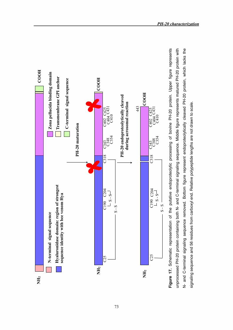
Figu
re 1
7. S
chem
atic
repr
esen
tatio
n of
the
put
ative
end
opro
teol
ytic
proc
essin
g of
bov
ine
PH-2
0 pr
otei
n. U
pper
fig
ure
repr
esen
ts
unpr
oces
sed
PH-2
0 pr
otei
n co
ntai
ning
bot
h N-
and
C-te
rmin
al s
igna
ling
sequ
ence
. Mid
dle
figur
e re
pres
ents
mat
ured
PH-
20 p
rote
in wi
th
N- a
nd C
-term
inal
sig
nalin
g se
quen
ce r
emov
ed.
Botto
m f
igur
e re
pres
ent
endo
prot
eolyt
ically
cle
aved
PH-
20 p
rote
in,
which
lac
ks t
he
signa
ling
sequ
ence
and
56
resid
ues
from
car
boxy
l end
. Rel
ative
pol
ypep
tide
leng
ths
are
not d
rawn
to s
cale
.
S -
S
NH
2C
OO
H
N-t
erm
inal
sig
nal s
eque
nce
Hya
luro
nida
se d
omai
n: r
egio
n of
str
onge
st
sequ
ence
iden
tity
wit
h be
e ve
nom
Hya
Zon
a pe
lluci
da b
indi
ng d
omai
n
Tra
nsm
embr
ane
GP
I an
chor
C-t
erm
inal
sig
nal s
eque
nce
NH
2C
OO
H
C25
C19
0C
318
C20
4
S -
SS
- S
NH
2C
OO
H
C25
C19
0C
318
C20
4S
- S
443
PH
-20
endo
prot
eoly
tica
lly c
leav
ed
duri
ng a
cros
omal
rea
ctio
n
PH
-20
mat
urat
ion
C
343
C34
8C
354
C40
2C
404
C41
0C42
5C
431
C34
3C
348
C35
4
C40
2C
404
C41
0C42
5C
431
PH-20 characterization
73
3.5 Conclusion
Though cloning and expression of the hyaluronidase domain of human PH-20 failed
(M.Schmidt, unpublished data), we were successful in purifying PH-20 proteins from both
commercial source (Sigma) and bovine testes to the highest purity except for the lower
molecular weight PH-20 (PH-2065) from latter source. The MALDI mass mapping of PH-20
variants from both sources matched to the N-terminal part of bovine testicular hyaluronidase
(NCBI accession number: AAP55713). PH-2060 from the commercial source (Sigma) was
endoproteolytically cleaved and linked by disulfide bridge, while PH-2069 and PH-2080 were
intact. Reduction of disulfide bridges resulted in inactive enzyme (not shown), suggesting that
structural integrity established by disulfide linkage are essential for enzymatic activity. The
aggregation of purified PH-20 protein was successfully overcome by addition of zwitterionic
compound, 250 mM of NDSB-195 (non detergent sulfobetaine; MW 195), which is indicative
of hydrophobic interaction. In addition to hydrophobic interaction, some PH-20 isoforms form
aggregation with other protein by disulfide linkage. Crystallization of PH-20 protein was not
achieved but crystallization in the presence of NDSB still remains to be tested. In addition,
cloning and expression of various length of hyaluronidase domain should be explored.
PH-20 characterization
74
Bibliography
1. Meyer, K. (1971). Hyaluronidases. The enzymes, 3rd ed, vol V, pp 307-320.
2. Primakoff, P., Hyatt, H., and Myles, D.G. (1985). A role for the migrating sperm
surface antigen PH-20 in guinea pig sperm binding to the egg zona pellucida. J Cell
Biol 101, 2239-2244.
3. Hunnicutt, G.R., Primakoff, P., and Myles, D.G. (1996). Sperm surface protein PH-20
is bifunctional: one activity is a hyaluronidase and a second, distinct activity is required
in secondary sperm-zona binding. Biol Reprod 55, 80-86.
4. Gmachl, M., and Kreil, G. (1993). Bee venom hyaluronidase is homologous to a
membrane protein of mammalian sperm. Proc Natl Acad Sci U S A 90, 3569-3573.
5. Lin, Y., Mahan, K., Lathrop, W.F., Myles, D.G., and Primakoff, P. (1994). A
hyaluronidase activity of the sperm plasma membrane protein PH-20 enables sperm to
penetrate the cumulus cell layer surrounding the egg. J Cell Biol 125, 1157-1163.
6. Overstreet, J.W., Lin, Y., Yudin, A.I., Meyers, S.A., Primakoff, P., Myles, D.G., Katz,
D.F., and Vandevoort, C.A. (1995). Location of the PH-20 protein on acrosome-intact
and acrosome-reacted spermatozoa of cynomolgus macaques. Biol Reprod 52,
105-114.
7. Cherr, G.N., Meyers, S.A., Yudin, A.I., VandeVoort, C.A., Myles, D.G., Primakoff, P.,
and Overstreet, J.W. (1996). The PH-20 protein in cynomolgus macaque spermatozoa:
identification of two different forms exhibiting hyaluronidase activity. Dev Biol 175,
142-153.
8. Thaler, C.D., and Cardullo, R.A. (1995). Biochemical characterization of a
glycosylphosphatidylinositol-linked hyaluronidase on mouse sperm. Biochemistry 34,
7788-7795.
9. Lalancette, C., Dorval, V., Leblanc, V., and Leclerc, P. (2001). Characterization of an
80-kilodalton bull sperm protein identified as PH-20. Biol Reprod 65, 628-636.
10. Arming, S., Strobl, B., Wechselberger, C., and Kreil, G. (1997). In vitro mutagenesis of
PH-20 hyaluronidase from human sperm. Eur J Biochem 247, 810-814.
11. Markovic-Housley, Z., Miglierini, G., Soldatova, L., Rizkallah, P.J., Muller, U., and
Schirmer, T. (2000). Crystal structure of hyaluronidase, a major allergen of bee venom.
Structure 8, 1025-1035.
12. Li, M.W., Yudin, A.I., Robertson, K.R., Cherr, G.N., and Overstreet, J.W. (2002).
Importance of glycosylation and disulfide bonds in hyaluronidase activity of macaque
sperm surface PH-20. J Androl 23, 211-219.
13. Meyer, M.F., Kreil, G., and Aschauer, H. (1997). The soluble hyaluronidase from bull
testes is a fragment of the membrane-bound PH-20 enzyme. FEBS Lett 413, 385-388.
PH-20 characterization
75

14. Cherr, G.N., Yudin, A.I., and Overstreet, J.W. (2001). The dual functions of GPI-
anchored PH-20: hyaluronidase and intracellular signaling. Matrix Biol 20, 515-525.
15. Primakoff, P., and Myles, D.G. (2002). Penetration, adhesion, and fusion in mammalian
sperm-egg interaction. Science 296, 2183-2185.
16. Wassarman, P.M. (1999). Mammalian fertilization: molecular aspects of gamete
adhesion, exocytosis, and fusion. Cell 96, 175-183.
17. Kim, E., Baba, D., Kimura, M., Yamashita, M., Kashiwabara, S., and Baba, T. (2005).
Identification of a hyaluronidase, Hyal5, involved in penetration of mouse sperm
through cumulus mass. Proc Natl Acad Sci U S A 102, 18028-18033.
18. Primakoff, P., Lathrop, W., Woolman, L., Cowan, A., and Myles, D. (1988). Fully
effective contraception in male and female guinea pigs immunized with the sperm
protein PH-20. Nature 335, 543-546.
19. Guntenhoner, M.W., Pogrel, M.A., and Stern, R. (1992). A substrate-gel assay for
hyaluronidase activity. Matrix 12, 388-396.
20. Laemmli, U.K. (1970). Cleavage of structural proteins during the assembly of the head
of bacteriophage T4. Nature 227, 680-685.
21. Borders, C.L., Jr., and Raftery, M.A. (1968). Purification and partial characterization of
testicular hyaluronidase. J Biol Chem 243, 3756-3762.
22. Yang, C.H., and Srivastava, P.N. (1975). Purification and properties of hyaluronidase
from bull sperm. J Biol Chem 250, 79-83.
23. Radimerski, T., Mini, T., Schneider, U., Wettenhall, R.E., Thomas, G., and Jeno, P.
(2000). Identification of insulin-induced sites of ribosomal protein S6 phosphorylation
in Drosophila melanogaster. Biochemistry 39, 5766-5774.
24. Bonenfant, D., Schmelzle, T., Jacinto, E., Crespo, J.L., Mini, T., Hall, M.N., and Jenoe,
P. (2003). Quantitation of changes in protein phosphorylation: a simple method based
on stable isotope labeling and mass spectrometry. Proc Natl Acad Sci U S A 100,
880-885.
25. Blisnick, T., Morales-Betoulle, M.E., Vuillard, L., Rabilloud, T., and Braun Breton, C.
(1998). Non-detergent sulphobetaines enhance the recovery of membrane and/or
cytoskeleton-associated proteins and active proteases from erythrocytes infected by
Plasmodium falciparum. Eur J Biochem 252, 537-541.
26. Benetti, P.H., Kim, S.I., Chaillot, D., Canonge, M., Chardot, T., and Meunier, J.C.
(1998). Expression and characterization of the recombinant catalytic subunit of casein
kinase II from the yeast Yarrowia lipolytica in Escherichia coli. Protein Expr Purif 13,
283-290.
27. Csoka, A.B., Frost, G.I., Wong, T., and Stern, R. (1997). Purification and
microsequencing of hyaluronidase isozymes from human urine. FEBS Lett 417,
307-310.
PH-20 characterization
76

77

Appendix
Curriculum Vitae
Sivaraman Padavattan
ADDRESS Division of Structural Biology, Biozentrum, University of
Basel, Klingelbergstrasse 50-70, CH-4056, Switzerland
TELEPHONE +41 61 267 2092
EMAIL [email protected]
NATIONALITY Indian
EDUCATION
2003 - Present PhD in Biophysics, Prof. Tilman Schirmer’s Group,
Biozentrum, University of Basel, Switzerland
“Crystal structure determination of hyaluronidase, a
major bee venom allergen in complex with an IgG Fab
fragment and the purification and biophysical
characterization of bovine testes hyaluronidase”
1999 - 2001 MSc Microbiology, Bharathidasan University, Trichy, INDIA
1995 -1998 BSc Microbiology, Madras University, Chennai, INDIA
PUBLICATION Padavattan, S., T. Schirmer, M. Schmidt, C. Akdis, R.
Valenta, I. Mittermann, L. Soldatova, J. Slater, U. Mueller,
and Z. Markovic-Housley, 2007b, Identification of a B-cell
Epitope of Hyaluronidase, a Major Bee Venom Allergen,
from its Crystal Structure in Complex with a Specific Fab: J
Mol Biol, v. 368, p. 742-52.
78

RELEVANT EXPERIENCE
2005 Oral presentation in the ‘19th Regio meeting on
Biocrystallography’, Les Feignes, La Bresse, France.
Presented poster in the ‘Murnau conference on Structural
Biology of Molecular recognition’, Murnau, Germany.
Presented poster in the ‘Biozentrum conference’, University
of Basel, Switzerland.
2004 Participated in the ‘EMBO Conference on Structures in
Biology’, EMBL, Heidelberg, Germany
Participated in the ‘18th Regional Meeting on the X-ray
Crystallography of Biomacromolecules’, Switzerland.
2003 Participated in the ‘17th Regional Meeting on the X-ray
Crystallography of Biomacromolecules’, Germany.
2001 Participated in the ‘International symposium on
Crystallography and Bioinformatics in Structural biology’,
Indian Institute of Science, India.
Participated in the ‘Asian crystallography association meeting’, Indian Institute of Science, India.
LANGUAGES English and Tamil
REFERENCE
Dr. Zora Markovic-Housley, Division of Structural Biology, Biozentrum, University
of Basel, Klingelbergstrasse 50-70, CH-4056 Basel, Switzerland.
Phone: +41 61 267 2088
Email: [email protected]
Prof. Tilman Schirmer, Division of Structural Biology, Biozentrum, University of
Basel, Klingelbergstrasse 50-70, CH-4056 Basel, Switzerland.
Phone: +41 61 267 2089
Email: [email protected]
79