Damage Control Resuscitation: Systematic Review
121
Aus der Abteilung für Hand-, Replantations- und Mikrochirurgie des Unfallkrankenhauses Berlin (Marzahn) DISSERTATION Damage Control Resuscitation: Systematic Review zur Erlangung des akademischen Grades Doctor medicinae (Dr. med.) vorgelegt der Medizinischen Fakultät Charité – Universitätsmedizin Berlin von Jan Jansen aus Bremen
Transcript of Damage Control Resuscitation: Systematic Review

Aus der Abteilung für Hand-, Replantations- und Mikrochirurgie des Unfallkrankenhauses Berlin (Marzahn)
DISSERTATION
Damage Control Resuscitation: Systematic Review
zur Erlangung des akademischen Grades Doctor medicinae (Dr. med.)
vorgelegt der Medizinischen Fakultät Charité – Universitätsmedizin Berlin
von
Jan Jansen
aus Bremen

Gutachter: 1. Priv.-Doz. Dr. med. A. Eisenschenk
2. Prof. Dr. med. U. Weber
3. Priv.-Doz. Dr. med. habil. R. A. Laun
Datum der Promotion: 19. März 2010

CONTENTS
1. Introduction....................................................................................................................... 1
1.1 The evolution of the damage control resuscitation concept ....................................... 1
1.2 Objectives ................................................................................................................ 2
1.3 Previous reviews and guidelines............................................................................... 3
1.4 The rationale for systematic review .......................................................................... 3
1.5 Terminology ............................................................................................................ 4
1.6 Target users.............................................................................................................. 4
1.7 Target patients and setting........................................................................................ 4
2. Methods.............................................................................................................................. 5
2.1 Overview of methodology........................................................................................ 5
2.2 Key questions........................................................................................................... 5
2.3 Outcome measures ................................................................................................... 6
2.4 Identification of evidence ......................................................................................... 6 2.4.1 Search strategies................................................................................................................................ 6 2.4.2 Sifting of search output .................................................................................................................... 7
2.5 Appraisal of evidence............................................................................................... 8 2.5.1 MERGE checklists............................................................................................................................ 8 2.5.2 AGREE instrument ......................................................................................................................... 11 2.5.3 Minimising bias .............................................................................................................................. 11
2.6 Forming Evidence Statements ................................................................................ 12 2.6.1 Considered judgement .................................................................................................................... 12 2.6.2 Assigning levels of evidence.......................................................................................................... 12
2.7 Format of this dissertation ...................................................................................... 13 2.7.1 Outline structure.............................................................................................................................. 13 2.7.2 Presentation ..................................................................................................................................... 14

3. Systematic Review ........................................................................................................... 15
3.1 Definition............................................................................................................... 15
3.2 Haemostatic resuscitation ....................................................................................... 15 3.2.1 Aetiology of traumatic coagulopathy ............................................................................................ 15 3.2.2 Fresh frozen plasma........................................................................................................................ 20 3.2.3 Platelets............................................................................................................................................ 27 3.2.4 Recombinant factor VIIa ................................................................................................................ 31 3.2.5 Cryoprecipitate................................................................................................................................ 38 3.2.6 Tranexamic acid.............................................................................................................................. 42
3.3 Permissive hypotension.......................................................................................... 47
3.4 Acidaemia management ......................................................................................... 53
3.5 Hypothermia management...................................................................................... 56
3.6 Damage control surgery ......................................................................................... 61
3.7 Indications ............................................................................................................. 69
4. Discussion....................................................................................................................... 723
4.1 Summary of evidence............................................................................................. 73 4.1.1 Fresh frozen plasma........................................................................................................................ 73 4.1.2 Platelets............................................................................................................................................ 74 4.1.3 Recombinant factor VIIa ................................................................................................................ 75 4.1.4 Cryoprecipitate................................................................................................................................ 76 4.1.5 Tranexamic acid.............................................................................................................................. 76 4.1.6 Permissive hypotension .................................................................................................................. 77 4.1.7 Tris-hydroxymethyl aminomethane (THAM) .............................................................................. 78 4.1.8 Hypothermia.................................................................................................................................... 78 4.1.9 Damage control surgery ................................................................................................................. 79 4.1.10 Indications for initiating damage control resuscitation ................................................................ 80
4.2 Validity, limitations and applicability..................................................................... 81 4.2.1 Validity ............................................................................................................................................ 81 4.2.2 Limitations ...................................................................................................................................... 81 4.2.3 Applicability.................................................................................................................................... 82
4.3 Conclusion ............................................................................................................. 82

5. Abstract (English)............................................................................................................ 84
6. Zusammenfassung (Deutsch) .......................................................................................... 86
7. References ........................................................................................................................ 89
8. Declaration/Erklärung .................................................................................................. 102
9. Curriculum Vitae/Lebenslauf ....................................................................................... 103
10. Publications/Publikationsliste ...................................................................................... 108
11. Dedication ..................................................................................................................... 110

1.
Introduction

1
1.1 THE EVOLUTION OF THE DAMAGE CONTROL RESUSCITATION CONCEPT
Military conflicts often drive innovations in health care. The first and second world war and the
Vietnam war saw significant advances in the care of the sick and injured, which were
subsequently translated into civilian practice. The recent conflicts in Iraq and Afghanistan are no
exception.[1][2] The intensity of warfare and types of munitions used has led to sustained
numbers of casualties with high trauma burdens, and provided the stimulus for the development
of new paradigms of care.[1] Damage control resuscitation is the synthesis of this collective
experience.[3]
Exsanguination is the second commonest cause of death following trauma,[4] and unlike central
nervous system injury, often preventable. Conventional resuscitation algorithms based on the
sequential use of crystalloids and colloids, followed by packed red blood cells and then plasma
or platelet transfusions, were based on the belief that coagulopathy developed over the course of
several hours.[5] As a consequence, resuscitation was focused on the restoration of cardiac
output and end-organ perfusion, with volume expansion; and oxygen delivery, with transfusion
of packed red blood cells.[6] The management of coagulopathy and hypothermia, even in the
context of damage control surgery, was deferred until measurable abnormalities were present.
This approach has been in widespread use since the 1980s and is codified in the Advanced
Trauma Life Support programme.[5]
Damage control resuscitation, in contrast, is a management strategy which addresses the entire
lethal triad of coagulopathy, acidosis, and hypothermia immediately upon admission, rather than
sequentially.[3][7] It is a development and refinement of the damage control surgery concept,
based on a better understanding of the pathophysiology of major trauma. Although pioneered by
military surgeons, damage control resuscitation is not only applicable to injuries sustained in
war. The impressive improvements in outcome witnessed in the military setting [8][9][10] have
been translated into civilian practice, and followed by the rapid acceptance of damage control
resuscitation by trauma surgeons worldwide.
The damage control surgery concept was founded on the realisation that – provided surgically
correctable haemostasis had been achieved – trauma patients died of the metabolic consequences
of injury. The discovery of the mutually perpetuating “lethal triad” or “bloody vicious circle” of
coagulopathy, metabolic acidosis, and hypothermia led to the introduction of a surgical strategy
which sacrificed the completeness of the immediate repair in order to address the combined
2
physiological impact of injury and operation, and avoid progression to metabolic
unsalvageability.[10][12] The notion that resuscitation could not take place at the same time as
surgery resulted in the rigid stratification of damage control surgery into phases of management
– surgery followed by resuscitation.[10] A new understanding of the aetiology of acute traumatic
coagulopathy, and its impact on survival, led to the re-evaluation of this concept.
The coagulopathy of trauma was classically viewed as a byproduct of resuscitation, attributed to
consumption, dilution and dysfunction (due to acidosis and hypothermia) of procoagulant serine
proteases. It is now recognised that injury-related coagulopathy is often present prior to
admission, and before any fluid or blood product administration, and thus cannot be caused by
dilution alone, or even to a significant extent. This early coagulopathy appears to be related to
hypoperfusion, is distinct from disseminated intravascular coagulopathy, and has been termed
Acute Coagulopathy of Trauma-Shock (ACoTS).[13][14] Recognition of the pivotal role of
ACoTS in determining outcome has led to the adoption of so-called haemostatic resuscitation
strategies, which involve the administration of fresh frozen plasma and platelets in predefined
ratios with packed red blood cells, effectively reconstituting whole blood, with the aim of
normalising all three aspects of the lethal triad – ideally before the patient leaves the operating
theatre.[3][7] Damage control resuscitation is surgery and resuscitation, performed concurrently,
with close cooperation between anaesthetist and surgeon.[3][7]
The components of damage control resuscitation remain vaguely defined, but in combination
appear to improve the survival of the most severely injured patients. The rapid introduction and
evolution of the components of damage control resuscitation, without formal evaluation in
interventional studies, is explained by the needs and demands of the operational military setting
which spawned its development. There is thus an urgent need for a review and appraisal of the
evidence supporting damage control resuscitation.
1.2 OBJECTIVES
The objectives of this dissertation are
• To define damage control resuscitation
• To conduct a systematic review of the evidence for damage control resuscitation
3
1.3 PREVIOUS REVIEWS AND GUIDELINES
There is a rapidly expanding body of literature on damage control resuscitation. Almost every
edition of the Journal of Trauma seems to carry an article on the subject, and there have been
several key publications, such as Holcomb’s editorials “Damage control resuscitation” and
“Damage control resuscitation: directly addressing the coagulopathy of trauma”, and an ever-
increasing number of non-systematic reviews. [3][7][14][15][16][17] To date, however, there
have been no systematic reviews of the damage control resuscitation strategy, and although there
are many existing clinical guidelines on the management of trauma and major haemorrhage, the
vast majority are not evidence-based either, and none specifically address the recent
developments which constitute damage control resuscitation.[18][19]
1.4 THE RATIONALE FOR SYSTEMATIC REVIEW
A systematic review identifies, evaluates and assimilates evidence on the effectiveness of
interventions, with the aim of assessing the consistency and generalisability of research
findings.[20] Reviews based on unsystematic literature surveys or expert opinion are liable to
bias.[21][22]
Systematic reviews form the basis for meta-analyses and clinical guidelines. Modern clinical
guidelines must be explicitly linked to supporting evidence and therefore rely heavily on a
thorough and unbiased review of the literature.[22][24][25] Meta-analysis is an extension of
systematic review, mathematically re-analysing data from primary studies, and thus depends on
the appropriate identification and selection of primary research. Organisations such as the
Cochrane Collaboration, the Scottish Intercollegiate Guidelines Network (SIGN), and the
National Institute for Health and Clinical Excellence (NICE) have contributed a great deal to
advancing systematic review methodology.
The essential criteria of a systematic review are an explicit search strategy, selection of literature
according to defined inclusion and exclusion criteria, and evaluation against consistent
standards.[22]

4
1.5 TERMINOLOGY
For the purpose of this work, “trauma”, “trauma surgery” and “trauma surgeon” are defined as
relating to injuries sustained to the torso, neck, and vasculature of the limbs, not the management
of isolated musculoskeletal injuries.
1.6 TARGET USERS
This dissertation is not intended to be a textbook or manual of trauma surgery. It is assumed that
the reader is familiar with the principles of resuscitation and trauma surgical techniques.
1.7 TARGET PATIENTS AND SETTING
This review relates to patients with haemorrhagic shock due to trauma, managed in the setting of
a European or North American centre, by general surgeons and anaesthetists with an interest and
experience in trauma care. Hospitals should be large enough to be able to provide on-site blood
transfusion services and intensive care facilities.
2.
Methods

5
2.1 OVERVIEW OF METHODOLOGY
Systematic reviews aim to minimise bias by using explicit methods to identify and collate all
existing evidence in order to address a specific research question.[26] The processes of
identification and appraisal of evidence must be methodical and reproducible. The methodology
used in this dissertation is based on a synthesis of techniques employed by the Cochrane
Collaboration, the Scottish Intercollegiate Guidelines Network, and the National Institute for
Health and Clinical Excellence, but also incorporates aspects of the MERGE (Method for
Evaluating Research and Guideline Evidence), and AGREE (Appraisal of Guidelines for
Research & Evaluation) initiatives. [22][26][27]
The development of a systematic review can be broken down into several steps, which include
the setting of specific research questions, the identification and appraisal of evidence, and the
formation of evidence statements.
2.2 KEY QUESTIONS
The first step in the writing of a systematic review is to divide the subject area into a number of
key questions.[26] The selection of a set of clear and focused queries with specified and
clinically relevant outcomes – such as survival, rather than surrogate measures, eg. change in
blood pressure – is fundamental to the success of the review.[26] The questions chosen for this
review, grouped by subject area, are:
Haemostatic resuscitation
• Is the early and aggressive use of fresh frozen plasma in predefined ratios with packed red
blood cells associated with increased survival of trauma patients?
• Is the early and aggressive use of platelets in predefined ratios with packed red blood cells
associated with increased survival of trauma patients?
• Does factor VIIa improve survival in trauma patients with severe bleeding?
• Does factor VIIa reduce transfusion requirements in trauma patients?
• Does the use of cryoprecipitate improve survival in trauma patients?
• Does tranexamic acid reduce transfusion requirements and/or mortality in trauma patients?

6
Permissive hypotension
• Does a strategy of withholding or limiting fluid resuscitation prior to surgical control of
haemorrhage improve survival?
Acidaemia management
• Does the administration of tris-hydroxymethyl aminomethane (THAM) improve survival in
trauma patients?
Hypothermia mitigation
• Do aggressive attempts at hypothermia mitigation improve outcome in trauma patients?
• What is the most effective method of preventing and treating hypothermia in trauma patients?
Damage control surgery
• Does the use of damage control surgical techniques improve survival in trauma patients with
severe bleeding?
Indications
• What are the indications for initiating damage control resuscitation?
2.3 OUTCOME MEASURES
The outcome measure chosen to answer the majority of the key questions in this review was
survival (or its reciprocal, mortality). Mortality is always clinically significant, and relatively
easy to measure, although it is accepted that studies attempting to show differences in mortality
require large numbers of patients, and may be the subject of type II errors.
2.4 IDENTIFICATION OF EVIDENCE
2.4.1 Search strategies
The literature search was designed to focus on the best available evidence, addressing each key
question in turn. In order to maximise coverage and minimise bias, searches were conducted
across the medline and embase medical literature databases, and the Cochrane library. Where
appropriate, search filters were used, but in general, search strategies were designed to maximise
sensitivity, while accepting low precision, as recommended by the Cochrane Collaboration.[26]
In order to capture the breadth of the subject, separate searches for primary studies, secondary
research, and existing guidelines were conducted, but secondary literature and existing
guidelines were only considered for inclusion when based on systematic methodology. Searches

7
were limited to articles in English and German, and published after 1980 (except in the case of
damage control surgery, as several key papers on this subject were published in the late 1970s).
Animal studies were not considered. Although there is a substantial body of literature on animal
models of resuscitation and treatment, these are contentious, and the applicability of animal
studies to human physiology is questionable.[28]
2.4.2 Sifting of search output
The medline and embase database search output was then assessed for eligibility. Citation lists
were initially sifted for irrelevant material, and titles that were not relevant to the key question
eliminated. The abstracts of the remaining papers were then examined using inclusion and
exclusion criteria, and studies with inappropriate designs excluded. The use of such criteria helps
to minimise bias.[26] Articles were acquired on completion of the sifting process. However, in
acknowledgement of the limitations of databases and search strategies, computerised searches
were supplemented by manual cross-referencing. The strategy is summarised diagrammatically
in fig 1.
Fig 1. Identification of evidence

8
2.5 APPRAISAL OF EVIDENCE
Following selection of articles as potential sources of evidence, the methodological validity of
each study was assessed. The results of this assessment determine the level of evidence allocated
to the study.[22]
2.5.1 MERGE checklists
Methodological assessment must be based on aspects of study design which have been shown to
influence the validity of the results reported and conclusions drawn, and varies between different
study types.[22] Primary research and systematic reviews were appraised using the MERGE
(Method for Evaluating Research and Guideline Evidence) criteria, which have been the subject
of wide consultation and evaluation.[22][23] These criteria are endorsed by the Scottish
Intercollegiate Guidelines Network and National Institute of Clinical Excellence for the purpose
of evaluating supporting evidence for guideline development.[22][29] MERGE checklists are
available for all principal study designs (systematic reviews and meta-analyses, randomised
trials, cohort studies, case-control studies, and studies of diagnostic accuracy), and consist of
three sections, providing a focused description of the results, an assessment of internal validity,
and an overall assessment of the methodological quality of the study, indicated by a rating of
“++”, “+”, or “-“. A “++” rating indicates that all or most of the assessed criteria have been
fulfilled. Unfulfilled criteria are thought very unlikely to alter the conclusions of the study. A “+”
rating indicates that some of the assessed criteria have been fulfilled. Unfulfilled criteria are
thought unlikely to alter the conclusions of the study. A “-” rating indicates that few or no criteria
were fulfilled, and that the conclusions of the study are likely or very likely to alter.[22] The
methodology checklist proformas for randomised controlled trials and cohort studies are shown
in figs 2 and 3. Due to constraints of space, the completed methodology checklists are not
included with this dissertation, but their conclusions are reproduced in the evidence tables (see
below).

9
Section 1: Internal Validity 1.1 The study addresses an
appropriate and clearly focused question.
Well addressed Adequately addressed Poorly addressed
Not addressed Not reported Not applicable
1.2 The assignment of subjects to treatment groups is randomised.
Well addressed Adequately addressed Poorly addressed
Not addressed Not reported Not applicable
1.3 An adequate concealment method is used.
Well addressed Adequately addressed Poorly addressed
Not addressed Not reported Not applicable
1.4 Subjects and investigators are kept “blind” about treatment allocation.
Well addressed Adequately addressed Poorly addressed
Not addressed Not reported Not applicable
1.5 The treatment and control groups are similar at the start of the trial.
Well addressed Adequately addressed Poorly addressed
Not addressed Not reported Not applicable
1.6 The only difference between groups is the treatment under investigation.
Well addressed Adequately addressed Poorly addressed
Not addressed Not reported Not applicable
1.7 All relevant outcomes are measured in a standard, valid, and reliable way.
Well addressed Adequately addressed Poorly addressed
Not addressed Not reported Not applicable
1.8 What percentage of individuals or clusters recruited into each arm of the study dropped out before the study was completed?
1.9 All the subjects are analysed in the groups to which they are randomly allocated (intention-to-treat-analysis)
Well addressed Adequately addressed Poorly addressed
Not addressed Not reported Not applicable
1.10 Where the study is carried out at more than one site, results are comparable for all sites.
Well addressed Adequately addressed Poorly addressed
Not addressed Not reported Not applicable
Section 2: Overall Assessment 2.1 How well was the study
done to minimise the risk of bias or confounding, and to establish a causal relationship between exposure and effect?
2.2 Taking into account clinical considerations, your evaluation of the methodology used, and the statistical power of the study, are you certain that the overall effect is due to the exposure being investigated?
2.3 Are the results of this study directly applicable to the patient group targeted in this guideline?
Section 3: Description 3.1
How many patients are included in this study?
3.2 What are the main characteristics of the patient population?
3.3
What intervention is being investigated in this study?
3.4
What comparisons are being made in the study?
3.5
For how long are the patients being followed up in the study?
3.6
What outcome measure(s) are used in the study?
3.7 What size of effect is identified in the study?
3.8
How was this study funded?
3.9
Does this study help to answer your key question?
Fig. 3 MERGE criteria and checklist for randomised controlled trials

10
Section 1: Internal Validity 1.1 The study addresses an
appropriate and clearly focused question.
Well addressed Adequately addressed Poorly addressed
Not addressed Not reported Not applicable
Selection of subjects 1.2 The two groups being
studied are selected from source populations that are comparable in all respects other than the factor under investigation.
Well addressed Adequately addressed Poorly addressed
Not addressed Not reported Not applicable
1.3 The study indicates how many of the people asked to take part did so, in each of the groups being studied.
Well addressed Adequately addressed Poorly addressed
Not addressed Not reported Not applicable
1.4 The likelihood that some eligible subjects might have the outcome at the time of enrolment is assessed and taken into account in the analysis.
Well addressed Adequately addressed Poorly addressed
Not addressed Not reported Not applicable
1.5 What percentage of individuals or clusters recruited into each arm of the study dropped out before the study was completed.
1.6 Comparison is made between full participants and those lost to follow-up, by exposure status.
Well addressed Adequately addressed Poorly addressed
Not addressed Not reported Not applicable
Assessment 1.7 The outcomes are clearly
defined.
Well addressed Adequately addressed Poorly addressed
Not addressed Not reported Not applicable
1.8 The assessment of outcome is made blind to exposure status.
Well addressed Adequately addressed Poorly addressed
Not addressed Not reported Not applicable
1.9 Where blinding was not possible, there is some recognition that knowledge of exposure status could have influenced the assessment of outcome.
Well addressed Adequately addressed Poorly addressed
Not addressed Not reported Not applicable
1.10 The measure of assessment of exposure is realiable.
Well addressed Adequately addressed Poorly addressed
Not addressed Not reported Not applicable
1.11 Evidence from other sources is used to demonstrate that the method of outcome assessment is valid and reliable.
Well addressed Adequately addressed Poorly addressed
Not addressed Not reported Not applicable
1.12 Exposure level of prognostic factor is assessed more than once.
Well addressed Adequately addressed Poorly addressed
Not addressed Not reported Not applicable
Confounding 1.13 The main potential
confounders are identified and taken into account in the design and analysis.
Well addressed Adequately addressed Poorly addressed
Not addressed Not reported Not applicable
Statistical analysis 1.14 Confidence intervals are
provided.
Well addressed Adequately addressed Poorly addressed
Not addressed Not reported Not applicable
Section 2: Overall Assessment 2.1 How well was the study
done to minimise the risk of bias or confounding, and to establish a causal relationship between exposure and effect?
2.2 Taking into account clinical considerations, your evaluation of the methodology used, and the statistical power of the study, are you certain that the overall effect is due to the exposure being investigated?
2.3 Are the results of this study directly applicable to the patient group targeted in this guideline?
Section 3: Description 3.1
How many patients are included in this study?
3.2 What are the main characteristics of the patient population?
3.3
What environmental or prognostic factor is being investigated in this study?
3.4
What comparisons are being made in the study?
3.5
For how long are the patients being followed up in the study?
3.6
What outcome measure(s) are used in the study?
3.7 What size of effect is identified in the study?
3.8
How was this study funded?
3.9
Does this study help to answer your key question?
Fig.4 MERGE criteria and checklist for cohort studies

11
2.5.2 AGREE instrument
Existing guidelines were also considered for inclusion in the evidence base, following
methodological evaluation using the AGREE (Appraisal of Guidelines, Research and Evaluation
for Europe) instrument for the assessment of clinical practice guidelines.[27] The AGREE
instrument provides an assessment of the predicted validity of a guideline, ie. the likelihood that
it will achieve its intended outcome. AGREE consists of 23 items organised in six domains.
Each domain is intended to capture a separate dimension of guideline quality. “Scope and
purpose” is concerned with the overall aim of the guideline, the specific clinical questions and
the target patient population. “Stakeholder involvement” focuses on the extent to which the
guideline represents the views of its intended users. “Rigour of development” relates to the
process used to gather and synthesise the evidence, the methods to formulate the
recommendations and to update them. “Clarity and presentation” deals with the language and
format of the guideline. “Applicability” pertains to the likely organisational, behavioural and
cost implications of applying the guideline. “Editorial independence” is concerned with the
independence of the recommendations and acknowledgement of possible conflict of interest
from the guideline development group. Each item is scored, by each appraiser, on a scale ranging
from 4 (“strongly agree”) to 1 (“strongly disagree”), with the midpoints 2 (“disagree”) and 3
(“agree”). The number of appraisers is flexible. The standardised composite percentage score for
each domain is calculated using the formula 100x (obtained score – minimum possible
score)/(maximum possible score – minimum possible score). The six domain scores are
independent and cannot be aggregated into a single quality score. “Overall assessment” is a
recommendation as to whether the guideline in question should be used in practice, and is graded
as “++” (strongly recommended), “+” (recommended with provisos), or “-“ (not
recommended).[27]
2.5.3 Minimising bias
Although predefined inclusion and exclusion criteria and the use of tools such as the MERGE
checklists and the AGREE instrument objectify the assessment process, an inevitable degree of
subjective judgement remains. This is usually minimised through dual or multiple assessment
and consensus between appraisers. Such a multi-author process would not be appropriate for a
dissertation and has therefore been omitted.

12
2.6 FORMING EVIDENCE STATEMENTS
2.6.1 Considered judgement
The results of the assessments of individual studies were compiled in evidence tables, which
summarise the findings and quality of articles relating to each key question. The level of the
evidence is determined by an objective assessment of the design and quality of each individual
study and a more subjective judgement on the consistency, clinical relevance and external
validity of the whole body of evidence.[22] It is rare for the evidence to show clearly and
unambiguously what course of action should be recommended for any given problem. In order to
address this problem, the Scottish Intercollegiate Guidelines Network have introduced the
concept of “considered judgement”.[22] Considered judgement is a review of the total body of
evidence covered by the evidence tables, consisting of an appraisal of the quantity, quality, and
consistency of evidence; the external validity (generalisability) of studies, and the applicability
to the target population. The process culminates in the formulation of a summary, known as an
evidence statement, and the assignment of a level of evidence. Evidence statements are based
entirely on the evidence presented, and do not take into account material which has not been
covered as part of the systematic review.
Fig 4. Forming evidence statements
2.6.2 Assigning levels of evidence
Assigning an evidence level to a statement quantifies the strength of the supporting evidence.
There are several systems in use. The US Agency for Health Care Policy and Research (AHCPR,
now the US Agency for Health Research and Quality, AHRQ) system is one of the oldest, and

13
was widely used for many years, but has limitations. An alternative system proposed by the
Scottish Intercollegiate Guidelines Network in 2000 was developed specifically for the purpose
of linking evidence to practice recommendations in guidelines, but separates levels of supporting
evidence from the grade of recommendations.[30] The SIGN system emphasises consideration
of the body of evidence as a whole. It is more flexible than the AHCPR/AHRQ system, because
it allows more weight to be given to good quality observational studies, where RCTs are not
available for ethical or practical reasons, as is often the case in trauma care. The SIGN system is
sometimes still difficult to apply in practice, but is an improvement on the AHCPR/AHRQ
system, and therefore used throughout this dissertation. It is summarised in table 1.
Table 1: SIGN system for assigning levels of evidence [30]
Level of evidence Type of evidence
1++
High quality meta-analyses, systematic reviews of RCTs, or RCTs with a very low risk of bias
1+
Well-conducted meta-analyses, systematic reviews of RCTs, or RCTs with a low risk of bias
1-
Meta-analyses, systematic reviews of RCTs, or RCTs with a high risk of bias
2++ High-quality systematic reviews of case-control or cohort studies
High-quality case control or cohort studies with a very low risk of confounding, bias or chance and a high probability that the relationship is causal
2+
Well-conducted case-control or cohort studies with a low risk of confounding, bias or chance and a moderated probability that the relationship is causal
2-
Case-control or cohort studies with a high risk of confounding, bias, or chance and a significant risk that the relationship is not causal
3
Non-analytic studies (for example, case reports, case series)
4
Expert opinion, formal consensus
2.7 FORMAT OF THIS DISSERTATION
2.7.1 Outline structure
This dissertation constitutes secondary research, evaluating and analysing existing work, and the
format has been adapted accordingly. The “introduction” and “methodology” sections have
already been covered and are broadly similar to a dissertation reporting experimental work.
Given the non-experimental nature of this work, a “hypothesis” has been omitted and substituted

14
with “objectives” (section 1.2). The “systematic review” section effectively represents the
“results” section of a conventional thesis. It is followed by a “discussion” which summarises and
contextualises the findings derived in the “results” section, identifies areas of practice for which
evidence is lacking, and examines the validity of the review and applicability of the findings.
The “conclusions” section (4.3) is self-explanatory and equivalent to a conventional dissertation.
2.7.2 Presentation
The aim of the systematic review chapter is to answer the key questions. For clarity, the chapter
is broken down into six parts: Haemostatic resuscitation, hypotensive resuscitation, acidaemia
management, hypothermia management, damage control surgery and indications. Some of these
parts contain more than one subsection, such as “fresh frozen plasma” under “haemostatic
resuscitation”.
The subsections commence with a specific, non-systematically developed introduction, followed
by the key question, and the outcome measure(s) chosen. The next paragraph contains a
description of the search strategy and output for relevant primary research, and a summary of the
evidence (as an evidence table), derived from MERGE checklists (which, for reasons of space,
are not included with this dissertation, see above). Evidence tables differ slightly for
interventional/observational studies, secondary research, and existing guidelines. Each table
contains the bibliographic reference to the publication, the summary rating (++, +, or -), and
several columns describing the results. This is followed by an additional row summarising any
particular issues of the study, in longhand. A similar format is employed for secondary research
and existing guidelines. The selection of the literature is also summarised in a flow diagram at
the end of each section.
This combined body of evidence is then appraised in the “considered judgement” paragraph.
Each subsection concludes with one or more “evidence statements”, which summarise the
available evidence. The level of evidence assigned is given in the right margin.
3.
Systematic Review

15
3.1 DEFINITION
Damage control resuscitation is a composite, multimodal, multidisciplinary strategy for the
management of the exsanguinating trauma patient, consisting of haemostatic resuscitation,
permissive hypotension, acidaemia management, hypothermia management, and damage control
surgery.[3][7][15][16][31]
3.2 HAEMOSTATIC RESUSCITATION
Haemostatic resuscitation is the early use of blood components in predefined ratios, and the
adjunctive use of therapies such as recombinant factor VIIa and antifibrinolytics, to avert the
consequences of traumatic coagulopathy. This section outlines the aetiology and diagnosis of
traumatic coagulopathy, and systematically reviews the evidence for these management
strategies.
3.2.1 Aetiology of traumatic coagulopathy
Prevalence
Traumatic coagulopathy was traditionally regarded as a consequence of resuscitation, occurring
some hours after injury. Recent studies have shown that this is not the case, and that
coagulopathy may be present as early as on admission to hospital, and is therefore not the result
of fluid administration alone. Brohi et al conducted a retrospective review of 1,088 trauma
patients (median ISS 20, 57.7% ISS>15) over a five-year period. 24.4% of patients were
coagulopathic (defined as a prothrombin time or activated partial thromboplastin time 1.5x
greater than normal) on admission. This finding was associated with a four-fold increase in
mortality (46% vs 11%, p<0.001).[32] In a similar review comprising 7638 patients, MacLeod et
al also showed that abnormal prothrombin time (>14s) and partial thromboplastin time (>34 s)
on admission were independent predictors of mortality (median ISS of 9, odds ratio for death
3.6, 95% confidence interval 3.15-4.08, p<0.0001), and an analysis of data from the German
Trauma Registry revealed a prevalence of 34.2% (based on a prothrombin time test of
<70%).[33][34] Although these studies comprised different groups of patients, with different
patterns and severity of injury, and utilised different definitions of coagulopathy, they all showed
that coagulopathy is present, on admission, in a substantial proportion of trauma patients, and is
associated with decreased odds of survival.

16
Novel concepts
Traumatic coagulopathy is a complex, dynamic, multifactorial process, involving all components
of the haemostatic system, and the simplistic traditional explanations of traumatic coagulopathy
which pervade the literature are no longer sufficient to characterise the condition or to base
treatment decisions on. [14] The regulation of fibrin generation, platelets, and endothelium all
play a role, together with inhibition of stable clot formation by anticoagulant and fibrinolytic
processes.[14] Which of these mechanisms predominates depends on the nature and severity of
the injury, the effects of therapy, and the chronicity of wounding and treatment.[14] The
Educational Initiative on Critical Bleeding in Trauma (EICBT), an independent international
think-tank, describes six key initiators of coagulopathy in trauma patients: Tissue trauma, shock,
haemodilution, hypothermia, acidaemia, inflammation. [14] While this model provides a useful
framework, it is more helpful to divide these six mechanisms into two initiators (tissue injury
and shock), and four propagators (haemodilution, hypothermia, acidosis, and inflammation).
Initiators
Tissue injury initiates both coagulation and fibrinolysis, but in isolation is rarely responsible for
clinically overt coagulopathy.[14] Endothelial damage leads to exposure of subendothelial type
III collagen and tissue factor, which bind von Willebrand factor, platelets, and activated factor
VII.[35] The tissue factor/factor VIIa complex then activates serine proteases, ultimately
resulting in thrombin and fibrin formation.[36] Hyperfibrinolysis is a consequence of both tissue
injury and shock.[13] The presence of thrombin increases the expression of tissue plasminogen
activator (tPA) by endothelium, and endothelial injury – physical or ischaemic – releases tPA,
promoting fibrinolysis.[37][38][39][40] The effects are exacerbated by the inhibition of
plasminogen activator inhibitor-1 (PAI-1).[41] The purpose of hyperfibrinolysis in trauma is
presumably to limit clot propagation to the site of vascular injury.[14] With widespread trauma
and endothelial activation, however, such localisation may be lost.[14] Recognition of the
contribution of hyperfibrinolysis to the clinical syndrome of traumatic coagulopathy is
important, as it opens up new therapeutic possibilities: Antifibrinolytic drugs, such as tranexamic
acid, have been used successfully in elective surgery for some time, and may prove to be a useful
adjunct in traumatic haemorrhage.
Although coagulopathy and fibrinolysis are initiated by tissue injury, the main driver of
traumatic coagulopathy appears to be shock, or more accurately, systemic hypoperfusion. An
elegant recent study showed that patients without shock are rarely coagulopathic, even after
major mechanical trauma (as measured by ISS). [41] In contrast, there is a dose-dependent
17
relationship between the severity of shock/ tissue hypoperfusion – as measured by base excess –
and the degree of admission coagulopathy, as measured by prothrombin time (PT) and activated
partial thromboplastin time (APTT).[41][42] All of these derangements were determined before
fluid resuscitation, and are therefore not attributable to haemodilution. The pathophysiology of
this process is distinct from that of disseminated intravascular coagulation (DIC), leading the
Educational Initiative on Critical Bleeding in Trauma to coin the term “Acute Coagulopathy of
Trauma-Shock” (AcoTS).
Despite these exciting new realisations and novel terminology, many aspects of the underlying
mechanisms remain unclear. While acidaemia is well known to interfere with protease function,
clinical coagulopathy is evident at milder degrees of acidaemia than have been identified as
causing significant loss of protease activity.[14] It is conceivable that hypoperfusion results in
widespread endothelial disruption or activation, which in turn causes dysregulation and
activation of coagulation and fibrinolysis.[14] Brohi et al have implicated activated protein C
(aPC) in this process, but this was inferred by association rather than direct measurement of aPC
levels.[41] Formation of anticoagulant thrombin, through complexation with thrombomodulin,
would also result in hyperfibrinolysis, either due to aPC consumption of PAI-1, or reduced
activation of thrombin-activatable fibrinolysis inhibitor.[43][44][45] More work is required
before these mechanisms become fully elucidated, however, there is little doubt that, in
combination, tissue trauma and systemic hypoperfusion are the prime initiators of traumatic
coagulopathy in the immediate postinjury phase.
Propagators
The initial coagulopathy may then be propagated and exacerbated by the physical and
physiological impact of haemodilution, acidosis, hypothermia, and inflammation.[14] Reduced
intravascular hydrostatic pressure results in shifts of fluid devoid of coagulation factors from the
extracellular and interstitial spaces into the intravascular compartment.[14] This effect is
compounded by volume expansion with synthetic fluids. The effects of crystalloid administration
on coagulation have been demonstrated in mathematical models, in vitro, and in volunteer
studies.[46][47][48][49] Colloids, in addition to their disproportionately greater dilutional
effects, may in addition interfere directly with clot formation and stability.[14] Packed red blood
cell administration also results in dilution of clotting factors. [46][50][51]
Traumatic coagulopathy is exacerbated further by hypothermia, which inhibits protease activity
and platelet function, although the latter appears to predominate.[52] Acidosis, the consequence
18
of anaerobic metabolism precipitated by hypoperfusion as well as iatrogenic chloride
administration, also impairs protease function.
Implications for clinical practice
Although incomplete, an emerging understanding of the mechanisms underlying the Acute
Coagulopathy of Trauma Shock forms the basis of haemostatic resuscitation. Historically, whole
blood was the preferred therapy for patients with exsanguinating trauma. In the late 1980s,
concerns about resource utilisation and infectious disease transmission led to a switch to
component therapy, which aims to correct measured deficiencies. This approach of replacing
specific haemotological deficits, extrapolated from elective surgical practice, extended into
guidelines for patients requiring massive transfusion after injury, although proof of the efficacy
of this change in practice was lacking.[46][48][53][54][55] Many transfusion guidelines
continue to recommend against the administration of clotting factors until the prothrombin or
activated partial thromboplastin time is greater than 1.5x normal, [19][55][56][57][58] and
perpetuation of this type of “expert opinion” is in part responsible for the common finding of
refractory coagulopathy in trauma patients.[46]
Increasing experience with large numbers of severely injured patients has led to a greater
appreciation of the importance of the early coagulopathy of trauma. Several recent studies have
shown that a more proactive strategy, administering packed red cells, fresh frozen plasma and
platelets in similar ratios to those found in whole blood, may be associated with increased
survival.[59][60][61][62][63]
Diagnosis
Although the diagnosis of traumatic coagulopathy is, at first sight, straightforward, this is not the
case. Prothrombin time and partial thromboplastin time measurement is readily available and
widely used. Indeed, all three of the recently published large retrospective studies of traumatic
coagulopathy mentioned above relied on these tests for the diagnosis of
coagulopathy.[32][33][34] However, these surveys were designed to establish the prevalence and
clinical significance of traumatic coagulopathy, rather than the diagnostic accuracy of
prothrombin time and partial thromboplastin time. Although these studies show that an abnormal
prothrombin and partial thromboplastin time is associated with adverse outcome, they do not
validate the tests: Patients who had normal prothrombin and partial thromboplastin times could
still have been clinically coagulopathic.
The limitations of prothrombin time and partial thromboplastin time measurements are well
recognised. These assays only measure the functioning of isolated aspects, rather than the global

19
performance, of the coagulation system.[16][64] In particular, the reactions are conducted on
platelet-poor plasma and thus do not evaluate the cellular interactions of coagulation.[16][64]
Prothrombin time and partial thromboplastin time measurements are furthermore conducted at
37oC, at supraphysiological calcium concentrations, and therefore do not reflect the in vivo
effects of hypothermia or hypocalcaemia.[64][65] In addition to limited validity, prothrombin
time and partial thromboplastin time measurements are time-consuming, both intrinsically, and
because they are usually performed in a central laboratory rather than at the bedside,
necessitating the transport of specimens.[51] A further drawback of partial thromboplastin time
and prothrombin time measurement is their inability to identify a hypercoagulable state.
Many authorities therefore now agree that the initial diagnosis of traumatic coagulopathy should
not rest on the demonstration of abnormal in vitro coagulation parameters, [7][16][51][64][65]
[66][67] although these tests may have a role in monitoring the response to treatment.
Thromboelastography (TEG®) measures shear elastic modulus during clot formation and
subsequent fibrinolysis. In contrast to prothrombin time and partial thromboplastin time,
thromboelastography provides a global functional profile of whole blood coagulation, providing
information on the initiation of coagulation, propagation kinetics, fibrin-platelet interaction, clot
firmness and fibrinolysis.[68][69][70] It can also be performed at the temperature of the patient,
reflecting the effect of hypothermia on clotting.
Thromboelastography provides a graphic output, from which a variety of parameters can be
derived, and has been shown to be a more sensitive measure of coagulation disorders than
standard tests of coagulation.[71] The ability to diagnose and characterise hyperfibrinolysis, now
recognised to play a major role in traumatic coagulopathy, has led to renewed interest in this
technique. There is a large volume of literature relating to the use of TEG® in orthotopic liver
transplant and cardiac surgery,[17] but despite its advantages, thromboelastography has not
become the standard of care. This is largely related to the cost and delicate nature of the
equipment, which requires considerable training and maintenance. A new device, the rotation
thromboelastogram analyzer (ROTEM®, Pentapharm, Munich, Germany), appears to have
overcome some of the limitations of classic thromboelastography, and is also faster.[70] A basic
ROTEM® analysis takes approximately 15 mins, although the characterisation of fibrinolysis is
more time consuming.[70] Several small, recent studies have confirmed the utility of ROTEM®
in trauma management.[68][69][70][72] Further studies are needed, but thromboelastography
has the potential to provide a better and faster characterisation of traumatic coagulopathy than

20
other tests currently in use, and its ability to differentiate hyperfibrinolysis from factor and
platelet deficiency is of particular interest.
However, until ROTEM® becomes more widely available, and experience accumulates, the
diagnosis of traumatic coagulopathy, and the initiation of appropriate management, must be
made on clinical grounds.
3.2.2 Fresh frozen plasma
The publication of Brohi’s and MacLeod’s observational studies coincided with the beginning of
the Iraq war,[32][33] and led American military surgeons to experiment with the use fresh frozen
plasma as a primary resuscitation fluid. The unprecedented severity of injuries inflicted by
modern munitions and improvised explosive devices resulted in a high incidence of traumatic
coagulopathy and exsanguination from microvascular bleeding despite surgical control of
haemorrhage. Severely injured patients predicted to require massive transfusion were empirically
resuscitated with fresh frozen plasma and packed red blood cells in a 1:1 ratio on arrival at the
combat support hospital.[73] Anecdotal success of decreased coagulopathic bleeding prompted a
formal, retrospective evaluation of the strategy, which confirmed a survival benefit, and led to a
dramatic change in military resuscitation and transfusion strategies.[59] Both US and British
military guidelines now recommend resuscitation of severely injured personnel with equal
numbers of units of red cells and plasma.[15][16][66][73] Several subsequent studies appear to
confirm that these developments may be extrapolated to civilian settings.[6][60][61][63][74] The
aggressive use of fresh frozen plasma remains contentious, however. While many trauma
surgeons regard the available evidence as proof of effectiveness,[3][7][15][16] many
haematologists disagree.[75][76] Concern has also been raised regarding the potential
complications of therapy with large amounts of blood products, including the risks of major
transfusion reactions, blood borne virus transmission, and transfusion-related acute lung
injury.[74][76][77][78]
Key question
This section aims to answer the question “Is the early and aggressive use of fresh frozen plasma
in predefined ratios with packed red blood cells associated with increased survival of trauma
patients?”
Outcome measure
Survival/mortality.

21
Primary studies
Database: Ovid MEDLINE® <1950 to November Week 3 2008> 1 trauma.mp. or exp *”Wounds and Injuries”/ (505433) 2 exp *Mental Disorders/ (605325) 3 1 not 2 (491574) 4 Plasma/ (11432) 5 3 and 4 (258) 6 limit 5 to (humans and yr=”1980 – 2009”) (131) Database: EMBASE <1980 to 2009 Week 05> 1 trauma.mp. or exp *”Wounds and Injuries”/ (405735) 2 exp *Mental Disorders/ (496419) 3 1 not 2 (387006) 4 Plasma/ (31849) 5 3 and 4 (475) 6 limit 5 to (human and (english or german) and yr=”1980 – 2009”) (259) Inclusion criteria for abstract selection 1 Interventional or observational studies Exclusion criteria for abstract selection 1 Case reports 2 Case series without comparison groups
Systematic medline and embase searches returned 390 citations. 301 titles were deemed
irrelevant and excluded. Of the remaining 89 abstracts, seven met the inclusion criteria. (Fig. 5)
These studies are summarised in evidence table 1.
Evidence Table 1: Primary studies
Bibliographic Citation
Study Type
Evi-dence Level
Number of Patients
Patient Charac-teristics
Intervention Comparison Length of Follow-Up
Outcome Measure
Effect Size
Borgman MA, et al (2007)
[59]
NCCS + 246 Military setting
94% penetrating ISS>=18
Massive transfusion (>= 10 units PRBC in 24h)
FFP:PRBC ratio
Low ratio (median 1:8) (n=31)
Medium ratio (median 1:2.5) (n=53)
High ratio (median 1:1.4) (n=162)
To discharge Mortality to discharge
Low ratio group 65%
Medium ratio group 34% High ratio group 19% (p<0.001)
Low ratio (median 1:8) (n=20)
Medium ratio (median 1:2.5) (n=18)
High ratio (median 1:1.4) (n=31)
To discharge Death from haemorrhage
Low ratio group 92%
Medium ratio group 78% High ratio group 37% (p<0.05)
Original and seminal study of effect of FFP:PRBC ratios. Slightly marred by vague primary outcome measure (mortality to discharge).
Analysed as non-concurrent cohort study (NCCS).

22
Bibliographic Citation
Study Type
Evi-dence Level
Number of Patients
Patient Charac-teristics
Intervention Comparison Length of Follow-Up
Outcome Measure
Effect Size
Sperry JL, et al (2008)
[74]
NCCS - 415 x7 civilian level I trauma centers 0% penetrating Hypotension (<90mmHg)
Base deficit >6 meq/l
Massive transfusion (>=8 U in 12 h)
FFP:PRBC ratio
Low ratio group (<1:1.5) (n=313)
High ratio group (>1:1.5) (n=102)
To discharge 24h mortality Low ratio group 13%
High ratio group 4%
(p=0.012)
Low ratio group (<1:1.5) (n=313)
High ratio group (>1:1.5) (n=102)
Crude mortality
Low ratio group 35% High ratio group 28% (p=0.202)
Low ratio group (<1:1.5) (n=313)
High ratio group (>1:1.5) (n=102)
PRBC transfusion requirement at 24h (mean)
Low ratio group 22U High ratio group 16U (p=0.001)
Data obtained from another ongoing observational study. Large numbers, but statistical analysis opaque. Analysed as non-concurrent cohort study (NCCS).
Maegele M, et al (2008)
[63]
NCCS + 713 Civilian setting
ISS>16 Massive transfusion (>10 U PRBC prior to ICU admission)
7.6% penetrating
FFP:PRBC ratio
Low ratio group (<1:0.9) (n=484) Medium ratio group (1:1) (n=114) High ratio group (>1:1.1) (n=115)
To discharge 6h mortality Low ratio group 24.6%
Medium ratio group 9.6%
High ratio group 3.5% (p<0.0001)
Low ratio group (<1:0.9) (n=484) Medium ratio group (1:1) (n=114)
High ratio group (>1:1.1) (n=115)
24h mortality Low ratio group 32.6%
Medium ratio group 16.7%
High ratio group 11.3%
(p<0.0001)
Low ratio group (<1:0.9) (n=484)
Medium ratio group (1:1) (n=114)
High ratio group (>1:1.1) (n=115)
30d mortality Low ratio group 45.5% Medium ratio group 35.1%
High ratio group 24.3%
(p<0.001)
Well conducted retrospective analysis. Original paper uses PRBC:FFP (rather than FFP:PRBC) ratio, therefore converted. Analysed as non-concurrent cohort study (NCCS).

23
Bibliographic Citation
Study Type
Evi-dence Level
Number of Patients
Patient Charac-teristics
Intervention Comparison Length of Follow-Up
Outcome Measure
Effect Size
Duchesne JC, et al (2008)
[60]
NCCS
2x2
+ 250 Civilian level I trauma center
Average ISS 21
<= 10 units PRBC
58% penetrating
FFP:PRBC ratio
Low ratio group (<1:2)
High ratio group (>1:2)
Not stated 24h Mortality Low ratio group 21.2%
High ratio group 11.8%
(P=0.06)
135 Civilian level I trauma center
Average ISS 27
> 10 units PRBC 72% penetrating
FFP:PRBC ratio
Low ratio group (<1:2)
High ratio group (>1:2)
Not stated 24h Mortality Low ratio group 87.5%
High ratio group 26%
RR 18.88 (95% CI 6.32-56.36, p=0.001)
2x2 factorial design incorporating analysis of patients transfused less than 10U packed red blood cells. Analysed as non-concurrent cohort study (NCCS).
Gunter OL, et al (2008)
[61]
NCCS + 259 Civilian level I trauma center
42% penetrating
Median ISS 25 Massive transfusion (>=10 U in 24 h)
FFP:PRBC ratio
Low ratio group (n=195)
High ratio group (FFP:PRBC>=2:3) (n=64)
30 d 30 d Mortality Low ratio group 62%
High ratio group 41%
(p=0.008)
Well-conducted retrospective study. Analysed as non-concurrent cohort study (NCCS).
Scalea TM, et al (2008)
[79]
NCCS - 365 12% penetrating
mean ISS 29
Mean U PRBC 7.7
FFP:PRBC ratio
Low ratio group (<1:1) (n=199) High ration (1:1) (n=51)
To discharge 24 h mortality OR 0.57 (95% CI 0.19-1.66) (p=0.34)
Retrospective study. Methodology and statistical analysis unclear. Only 81 patients actually received massive transfusion. Analysed as non-concurrent cohort study (NCCS).
Holcomb et al (2008)
[6]
NCCS
2x2
+ 466 16 US level I trauma centers Massive transfusion (>=10 units PRBC in 24 hrs)
35% penetrating
76% male Mean ISS=32
Mean age=39
FFP:PRBC ratio
Low ratio group (<1:2) (n=214) High ratio group (>1:2) (n=252)
30d 30d mortality Low ratio group 59.6%
High ratio group 40.4%
(p<0.01)
Largest and best-designed study to date. Also comprised 2x2 factorial Kaplan-Meier survival analysis incorporating effects of platelet administration. Showed statistically significant differences in mortality at 6h, 24h and 30d. (Study used survival, not mortality, as outcome measures, therefore converted.) Analysed as non-concurrent cohort study (NCCS).
Secondary research
Database: MEDLINE (number of citations in brackets) 1 trauma.mp. or exp *"Wounds and Injuries"/ (505433) 2 exp *Mental Disorders/ (605325) 3 1 not 2 (491574) 4 Plasma/ (11432) 5 3 and 4 (258) 6 limit 5 to (humans and yr="1980 - 2009") (131) 7 meta-analysis/ (20263)
24
8 exp review literature/ (1446234) 9 (meta-analy$ or meta analy$ or metaanaly$).tw. (23776) 10 meta analysis.pt. (20263) 11 review academic.pt. (0) 12 review literature.pt. (0) 13 letter.pt. (654713) 14 review of reported cases.pt. (0) 16 review multicase.pt. (0) 17 7 or 8 or 9 or 10 or 11 or 12 (1464813) 18 13 or 14 or 15 or 16 (908067) 19 17 not 1 (1452213) 20 animal/ (4410095) 21 human/ (10826325) 22 20 and 21 (1098839) 23 20 not 22 (3311256) 24 19 not 23 (1340459) 25 4 and 24 (16) Database: EMBASE <1980 to 2009 Week 07> 1 trauma.mp. or exp *"Wounds and Injuries"/ (406671) 2 exp *Mental Disorders/ (497727) 3 1 not 2 (387898) 4 Plasma/ (29661) 5 3 and 4 (407) 6 from 5 keep (13) Inclusion criteria for abstract selection 1 Meta-analysis 2 Systematic review Exclusion criteria for abstract selection 1 Non-systematic review
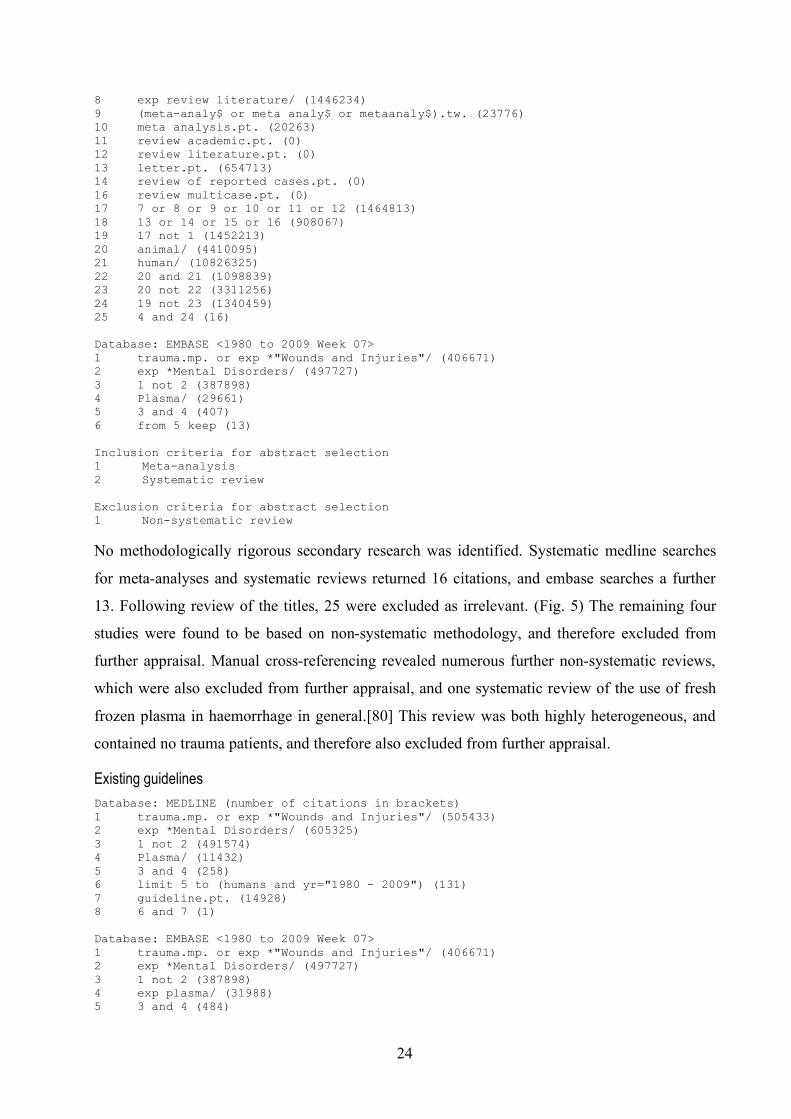
No methodologically rigorous secondary research was identified. Systematic medline searches
for meta-analyses and systematic reviews returned 16 citations, and embase searches a further
13. Following review of the titles, 25 were excluded as irrelevant. (Fig. 5) The remaining four
studies were found to be based on non-systematic methodology, and therefore excluded from
further appraisal. Manual cross-referencing revealed numerous further non-systematic reviews,
which were also excluded from further appraisal, and one systematic review of the use of fresh
frozen plasma in haemorrhage in general.[80] This review was both highly heterogeneous, and
contained no trauma patients, and therefore also excluded from further appraisal.
Existing guidelines
Database: MEDLINE (number of citations in brackets) 1 trauma.mp. or exp *"Wounds and Injuries"/ (505433) 2 exp *Mental Disorders/ (605325) 3 1 not 2 (491574) 4 Plasma/ (11432) 5 3 and 4 (258) 6 limit 5 to (humans and yr="1980 - 2009") (131) 7 guideline.pt. (14928) 8 6 and 7 (1) Database: EMBASE <1980 to 2009 Week 07> 1 trauma.mp. or exp *"Wounds and Injuries"/ (406671) 2 exp *Mental Disorders/ (497727) 3 1 not 2 (387898) 4 exp plasma/ (31988) 5 3 and 4 (484)
25
6 limit 5 to human (264) 7 Practice Guideline/ (102173) 8 6 and 7 (1) Inclusion criteria for abstract selection 1 Systematically developed guideline Exclusion criteria for abstract selection 1 Non-systematically developed guidelines 2 Quasi-editorial guidelines
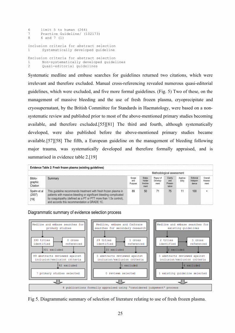
Systematic medline and embase searches for guidelines returned two citations, which were
irrelevant and therefore excluded. Manual cross-referencing revealed numerous quasi-editorial
guidelines, which were excluded, and five more formal guidelines. (Fig. 5) Two of these, on the
management of massive bleeding and the use of fresh frozen plasma, cryoprecipitate and
cryosupernatant, by the British Committee for Standards in Haematology, were based on a non-
systematic review and published prior to most of the above-mentioned primary studies becoming
available, and therefore excluded.[55][81] The third and fourth, although systematically
developed, were also published before the above-mentioned primary studies became
available.[57][58] The fifth, a European guideline on the management of bleeding following
major trauma, was systematically developed and therefore formally appraised, and is
summarised in evidence table 2.[19]
Evidence Table 2: Fresh frozen plasma (existing guidelines)
Methodological assessment
Biblio-graphic Citation
Summary Scope and
Purpose
Stake-holder
Involve-ment
Rigour of Develop-
ment
Clarity and
Presen-tation
Applica-bility
Editorial Indepen-
dence
Overall Assess-
ment
Spahn et al (2007)
[19]
This guideline recommends treatment with fresh frozen plasma in patients with massive bleeding or significant bleeding complicated by coagulopathy (defined as a PT or PTT more than 1.5x control), and accords this recommendation a GRADE 1C.
89 50 71 75 11 100 +
Diagrammatic summary of evidence selection process
Fig 5. Diagrammatic summary of selection of literature relating to use of fresh frozen plasma.

26
Appraisal
Volume of evidence: The volume of evidence is moderate. All identified primary studies are
retrospective database or registry analyses, which have for the purpose of this review been
analysed as non-concurrent cohort studies. There is heterogeneity with regards to the definition
of mortality (crude, 6 hours, 24 hours, or 30 days). There is no relevant secondary research, and
only one systematically developed guideline, which is, however, of reasonable methodological
quality. Applicability: Of the primary studies, one was conducted in the military setting,
comprising almost exclusively penetrating injuries.[59] Of the remaining six studies, five were
conducted in North American level I trauma centers, and one was an analysis of German trauma
registry data.[6][60][61][63][74][79] Apart from Sperry’s study, which contained only blunt
injuries, all of the American studies contained a significant proportion of penetrating injuries,
whereas the German study comprised mostly blunt trauma.[63][74] However, the
pathophysiological mechanisms underlying acute traumatic coagulopathy in severely injured
patients requiring massive transfusions are likely to be similar irrespective of the mechanism of
injury, and these studies are therefore likely to be applicable. Only Duchesne et al investigated
the effect of high ratios of FFP:PRBC in patients who received less than 10 units of packed red
blood cells in the first 24 hours, and found no difference in outcome.[60] The existing guideline
is aimed at general trauma patients with major haemorrhage in the setting of a European hospital,
and is therefore applicable.[19] Consistency: Six of the seven studies showed a beneficial effect
of high FFP:PRBC ratio on mortality (allowing for varying definitions, see above). One study
showed no effect but was marred by poor methodology.[79] The existing guideline endorses
treatment with FFP based on clinical grounds (as well as haematological abnormalities).
Evidence statements
In patients with traumatic haemorrhage predicted to require massive transfusion
(defined as more than 8-10 units of packed red blood cells in the first 24 hours after
injury), a high ratio of fresh frozen plasma to packed red blood cells is associated with
decreased mortality.
2+
A fresh frozen plasma to packed red blood cell ratio of approximately 1:1 units
appears to be optimal, although this evidence is extrapolated from studies which
retrospectively stratified intervention groups for survival analysis, rather than dose-
finding studies.
2+

27
Future research
There is an urgent need for a clinical trial of management with predetermined ratios of fresh
frozen plasma to packed red blood cells versus conventional resuscitation strategies. In addition
to proving efficacy, such a trial should be designed to answer what the optimal component ratio
is, which patients benefit the most, how to select them, and what the risks and complications are.
(These issues are discussed in more detail in section 4.1.1.)
3.2.3 Platelets
Success with the aggressive use of fresh frozen plasma in haemorrhagic shock led to a re-
examination of the use of other blood products in early resuscitation. Platelets have long been
recognised to play a pivotal role both in clot formation, and the regulation of the coagulation
system. The administration of platelets in roughly physiological proportions compared with fresh
frozen plasma and packed red blood cells was the logical next step. This strategy – known as
1:1:1 – effectively aims to reconstitute whole blood, and is thus conceptually attractive.
The term “1:1:1” refers to units of fresh frozen plasma, units of packed red blood cells, and
individual donor units of platelets. Individual donor units of platelets are rarely used nowadays,
platelets instead being issued as “pools” of 4-6 individual donor units. “1:1:1” in most European
countries, where platelets are only provided in pools, therefore equates to “5 units of FFP : 5
units of PRBC : 1 pool of platelets”.
Key question
This section aims to answer the question “Is the early and aggressive use of platelets in
predefined ratios compared with packed red blood cells associated with increased survival of
trauma patients?”
Outcome measure
Survival/mortality
Primary studies
Database: Ovid MEDLINE(R) <1950 to November Week 3 2008> 1 trauma.mp. or exp *"Wounds and Injuries"/ (505433) 2 exp *Mental Disorders/ (605325) 3 1 not 2 (491574) 4 exp Blood Platelets/ (58032) 5 3 and 4 (619) 6 limit 5 to (humans and yr="1980 - 2009") (182) Database: EMBASE <1980 to 2009 Week 05> 1 trauma.mp. or exp *"Wounds and Injuries"/ (405735) 2 exp *Mental Disorders/ (496419) 3 1 not 2 (387006) 4 exp thrombocyte/ (28484)
28
5 3 and 4 (579) 6 limit 5 to (humans and yr="1980 - 2009") (290) Inclusion criteria for abstract selection 1 Interventional or observational studies Exclusion criteria for abstract selection 1 Case reports 2 Case series without comparison groups
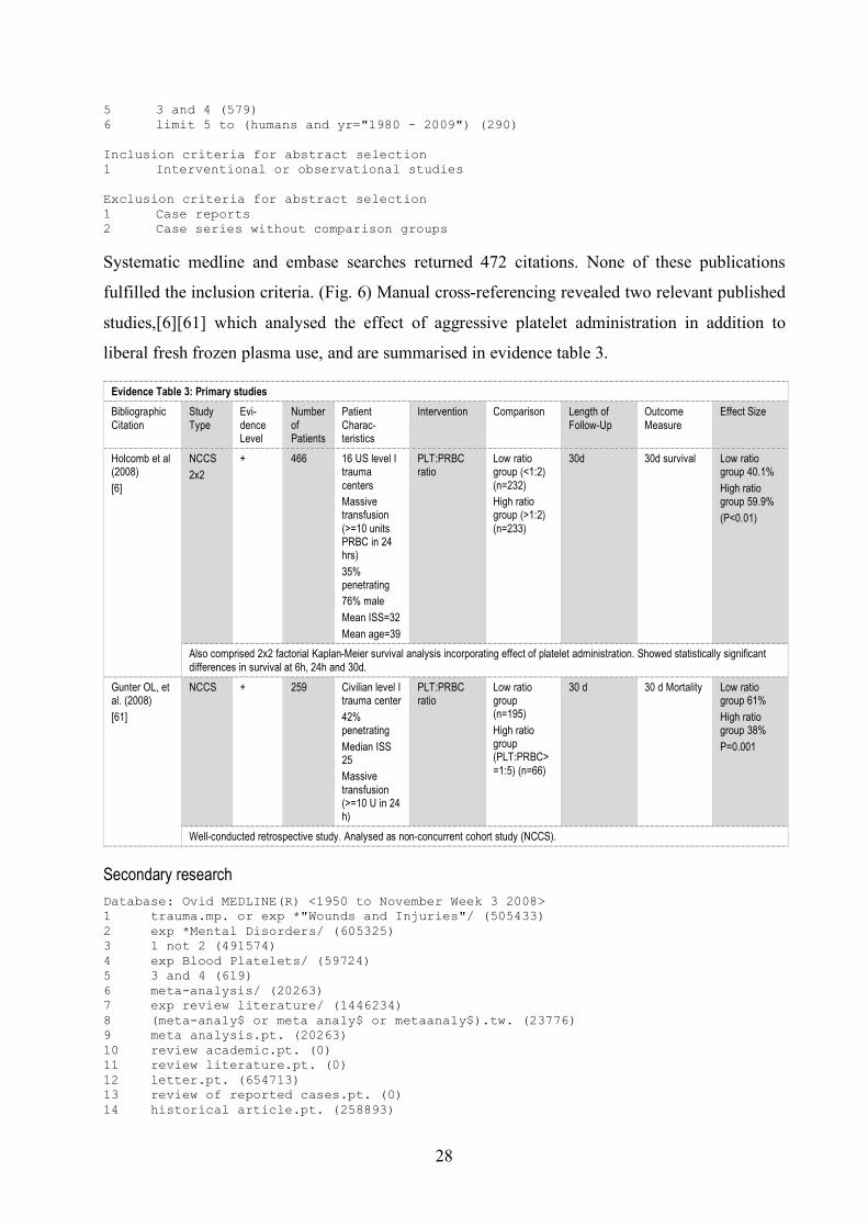
Systematic medline and embase searches returned 472 citations. None of these publications
fulfilled the inclusion criteria. (Fig. 6) Manual cross-referencing revealed two relevant published
studies,[6][61] which analysed the effect of aggressive platelet administration in addition to
liberal fresh frozen plasma use, and are summarised in evidence table 3.
Evidence Table 3: Primary studies
Bibliographic Citation
Study Type
Evi-dence Level
Number of Patients
Patient Charac-teristics
Intervention Comparison Length of Follow-Up
Outcome Measure
Effect Size
Holcomb et al (2008)
[6]
NCCS
2x2
+ 466 16 US level I trauma centers Massive transfusion (>=10 units PRBC in 24 hrs)
35% penetrating 76% male
Mean ISS=32
Mean age=39
PLT:PRBC ratio
Low ratio group (<1:2) (n=232) High ratio group (>1:2) (n=233)
30d 30d survival Low ratio group 40.1%
High ratio group 59.9%
(P<0.01)
Also comprised 2x2 factorial Kaplan-Meier survival analysis incorporating effect of platelet administration. Showed statistically significant differences in survival at 6h, 24h and 30d.
Gunter OL, et al. (2008)
[61]
NCCS + 259 Civilian level I trauma center
42% penetrating
Median ISS 25 Massive transfusion (>=10 U in 24 h)
PLT:PRBC ratio
Low ratio group (n=195)
High ratio group (PLT:PRBC>=1:5) (n=66)
30 d 30 d Mortality Low ratio group 61%
High ratio group 38%
P=0.001
Well-conducted retrospective study. Analysed as non-concurrent cohort study (NCCS).
Secondary research
Database: Ovid MEDLINE(R) <1950 to November Week 3 2008> 1 trauma.mp. or exp *"Wounds and Injuries"/ (505433) 2 exp *Mental Disorders/ (605325) 3 1 not 2 (491574) 4 exp Blood Platelets/ (59724) 5 3 and 4 (619) 6 meta-analysis/ (20263) 7 exp review literature/ (1446234) 8 (meta-analy$ or meta analy$ or metaanaly$).tw. (23776) 9 meta analysis.pt. (20263) 10 review academic.pt. (0) 11 review literature.pt. (0) 12 letter.pt. (654713) 13 review of reported cases.pt. (0) 14 historical article.pt. (258893)

29
15 review multicase.pt. (0) 16 6 or 7 or 8 or 9 or 10 or 11 (1464813) 17 12 or 13 or 14 or 15 (908067) 18 16 not 17 (1452213) 19 animal/ (4410095) 20 human/ (10826325) 21 19 and 20 (1098839) 22 19 not 21 (3311256) 23 18 not 22 (1340459) 24 5 and 23 (64) Database: EMBASE <1980 to 2009 Week 07> 1 trauma.mp. or exp *"Wounds and Injuries"/ (406671) 2 exp *Mental Disorders/ (497727) 3 1 not 2 (387898) 4 Thrombocyte/ (26432) 5 3 and 4 (566) 6 limit 5 to (human and english) (251) 7 limit 5 to (human and german) (13) 8 6 or 7 (264) 9 from 8 keep (79) Inclusion criteria for abstract selection 1 Systematic reviews 2 Meta-analyses Exclusion criteria for abstract selection 1 Non-systematic reviews 2 Quasi-editorial guidelines
No methodologically rigorous secondary research was identified. Systematic medline searches
for meta-analyses and systematic reviews returned 64 citations, and embase searches a further
79. Following review of the abstracts, 141 were excluded as irrelevant. The remaining two were
found to be based on non-systematic methodology, and therefore excluded from further
appraisal.[51][73] (Fig. 6) Manual cross-referencing revealed numerous non-systematic reviews,
which were excluded from further analysis.
Existing guidelines
Database: Ovid MEDLINE(R) <1950 to November Week 3 2008> 1 trauma.mp. or exp *"Wounds and Injuries"/ (505433) 2 exp *Mental Disorders/ (605325) 3 1 not 2 (491574) 4 exp Blood Platelets/ (59724) 5 3 and 4 (619) 6 guideline.pt. (14928) 7 5 and 6 (0) Database: EMBASE <1980 to 2009 Week 01> 1 trauma.mp. or exp *"Wounds and Injuries"/ (404150) 2 exp *Mental Disorders/ (494289) 3 1 not 2 (385487) 4 exp Thrombocyte/ (28489) 5 guideline.mp. (105785) 6 4 and 5 (85) 7 limit 6 to (human and (english or german) and yr="1980 - 2009") (68) 8 from 7 keep (0)
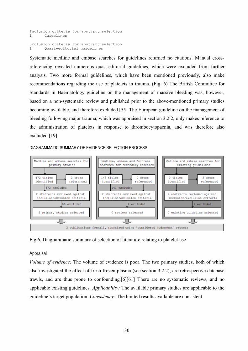
30
Inclusion criteria for abstract selection 1 Guidelines Exclusion criteria for abstract selection 1 Quasi-editorial guidelines
Systematic medline and embase searches for guidelines returned no citations. Manual cross-
referencing revealed numerous quasi-editorial guidelines, which were excluded from further
analysis. Two more formal guidelines, which have been mentioned previously, also make
recommendations regarding the use of platelets in trauma. (Fig. 6) The British Committee for
Standards in Haematology guideline on the management of massive bleeding was, however,
based on a non-systematic review and published prior to the above-mentioned primary studies
becoming available, and therefore excluded.[55] The European guideline on the management of
bleeding following major trauma, which was appraised in section 3.2.2, only makes reference to
the administration of platelets in response to thrombocytopaenia, and was therefore also
excluded.[19]
DIAGRAMMATIC SUMMARY OF EVIDENCE SELECTION PROCESS
Fig 6. Diagrammatic summary of selection of literature relating to platelet use
Appraisal
Volume of evidence: The volume of evidence is poor. The two primary studies, both of which
also investigated the effect of fresh frozen plasma (see section 3.2.2), are retrospective database
trawls, and are thus prone to confounding.[6][61] There are no systematic reviews, and no
applicable existing guidelines. Applicability: The available primary studies are applicable to the
guideline’s target population. Consistency: The limited results available are consistent.

31
Evidence statements
In patients with traumatic haemorrhage predicted to require massive transfusion
(defined as more than 8-10 units of packed red blood cells in the first 24 hours after
injury), a high ratio of units of platelets to packed red blood cells is associated with
decreased mortality. A ratio of at least 1 (pooled) unit of platelets to 5 units of packed
red blood cells appears to be optimal, although this evidence is extrapolated from
studies which retrospectively stratified intervention groups for survival analysis.
2-
Future research
Although consistent, the current volume of evidence supporting the liberal use of platelets is
poor. In addition to the already mentioned need for a trial of fresh frozen plasma to packed red
blood cell ratios, there is also a need for a trial of different platelet to packed red blood cell
ratios, and administration regimes. These studies could be combined.
3.2.4 Recombinant factor VIIa
Recognition of the importance of traumatic coagulopathy has also prompted a search for
pharmacological adjuncts to treatment. Recombinant activated factor VIIa (RFVIIa; eptacog ∝-
activated; Novoseven®, Novo Nordisk®, Crawley, Surrey, UK) was introduced into clinical
practice in the 1980s for the treatment of haemophiliacs with inhibitory antibodies to factor VIII
and IX, but has since been used widely for the treatment of patients with acquired
coagulopathy.[82]
Factor VII is an important initial component of the coagulation cascade, and acts via two linked
pathways to produce thrombin.[82] The first involves the binding of factor VIIa to
subendothelial tissue factor exposed by vessel injury. This reaction in turn activates factor X,
resulting in the generation of a small amount of thrombin (factor IIa). Thrombin then activates
platelets and factors V and VIII. Activated platelets also bind circulating factor VIIa – the second
pathway – resulting in further factor Xa generation, as well as activation of factor IX. IXa (with
its cofactor VIIIa) yields additional Xa. The complex of Xa and its cofactor Va then converts
prothrombin into thrombin in amounts that are sufficient to induce the conversion of fibrinogen
to fibrin (“thrombin burst”). [83]
Recombinant factor VIIa is manufactured using recombinant DNA technology. The amino acid
sequence is identical to the human plasma protein, but there are minor differences in post-
translational changes.[82] Recombinant factor VIIa has a half-life of 2-3 hours, less in bleeding
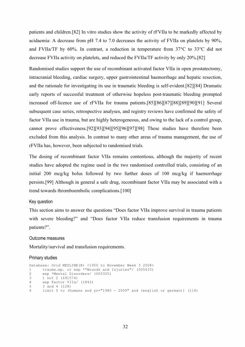
32
patients and children.[82] In vitro studies show the activity of rFVIIa to be markedly affected by
acidaemia: A decrease from pH 7.4 to 7.0 decreases the activity of FVIIa on platelets by 90%,
and FVIIa/TF by 60%. In contrast, a reduction in temperature from 37°C to 33°C did not
decrease FVIIa activity on platelets, and reduced the FVIIa/TF activity by only 20%.[82]
Randomised studies support the use of recombinant activated factor VIIa in open prostatectomy,
intracranial bleeding, cardiac surgery, upper gastrointestinal haemorrhage and hepatic resection,
and the rationale for investigating its use in traumatic bleeding is self-evident.[82][84] Dramatic
early reports of successful treatment of otherwise hopeless post-traumatic bleeding prompted
increased off-licence use of rFVIIa for trauma patients.[85][86][87][88][89][90][91] Several
subsequent case series, retrospective analyses, and registry reviews have confirmed the safety of
factor VIIa use in trauma, but are highly heterogeneous, and owing to the lack of a control group,
cannot prove effectiveness.[92][93][94][95][96][97][98] These studies have therefore been
excluded from this analysis. In contrast to many other areas of trauma management, the use of
rFVIIa has, however, been subjected to randomised trials.
The dosing of recombinant factor VIIa remains contentious, although the majority of recent
studies have adopted the regime used in the two randomised controlled trials, consisting of an
initial 200 mcg/kg bolus followed by two further doses of 100 mcg/kg if haemorrhage
persists.[99] Although in general a safe drug, recombinant factor VIIa may be associated with a
trend towards thromboembolic complications.[100]
Key question
This section aims to answer the questions “Does factor VIIa improve survival in trauma patients
with severe bleeding?” and “Does factor VIIa reduce transfusion requirements in trauma
patients?”.
Outcome measures
Mortality/survival and transfusion requirements.
Primary studies
Database: Ovid MEDLINE(R) <1950 to November Week 3 2008> 1 trauma.mp. or exp *"Wounds and Injuries"/ (505433) 2 exp *Mental Disorders/ (605325) 3 1 not 2 (491574) 4 exp Factor VIIa/ (1843) 5 3 and 4 (128) 6 limit 5 to (humans and yr="1980 - 2009" and (english or german)) (114)

33
Database: EMBASE <1980 to 2009 Week 01> 1 trauma.mp. or exp *"Wounds and Injuries"/ (404150) 2 exp *Mental Disorders/ (494289) 3 1 not 2 (385487) 4 exp Blood Clotting Factor 7a/ (1480) 5 3 and 4 (66) 6 limit 5 to (human and yr="1980 - 2009") (52) Inclusion criteria for abstract selection 1 Interventional studies 2 Observational studies Exclusion criteria for abstract selection 1 Case reports 2 Case series without comparison groups
Systematic searches across medline and embase returned 166 citations. Following review of the
abstracts, 162 were excluded as irrelevant or not meeting the above inclusion criteria, leaving
four studies which were appraised in full. One was subsequently excluded as the authors had
failed to fulfill the methodology described in the abstract.[92] One of the remaining three studies
consisted of two parallel trials, which have been analysed as such. (Fig. 7) These studies are
summaries in evidence table 4.
Evidence table 4: Recombinant factor VIIa (primary studies)
Bibliographic Citation
Study Type
Evi-dence Level
Number of Patients
Patient Charac-teristics
Intervention Comparison Length of Follow-Up
Outcome Measure
Effect Size
Spinella PC et al (2008) [101]
NCCS + 124 Military
ISS>15
Massive transfusion >=10U/24h
92% penetrating
RFVIIa RFVIIa (n=49) vs no RFVIIa (n=75)
30d 24h mortality RFVIIa- (26/75) 35% RFVIIa+ (7/49) 14%
(p=0.01)
RFVIIa (n=49) vs no RFVIIa (n=75)
30d 30d mortality RFVIIa- (38/75) 51%
RFVIIa+ (15/49) 31%
(p=0.03)
RFVIIa (n=14) vs no RFVIIa (n=37)
30d Death from haemorrhage
RFVIIa- (29/37) 78%
RFVIIa+ (8/14) 57%
(p=0.12)
Retrospective study with significant potential confounders. Virtually all military ballistic injuries treated in Combat Support Hospital, hence not directly applicable to civilian setting.

34
Bibliographic Citation
Study Type
Evi-dence Level
Number of Patients
Patient Charac-teristics
Intervention Comparison Length of Follow-Up
Outcome Measure
Effect Size
Boffard KD et al (2005)
[99]
RCT + 143 Civilian
Multicentre ISS>15
Massive transfusion >=6 in 4h
Blunt trauma
RFVIIa Placebo (n=74) vs RFVIIa (n=69)
30d Decrease in 48h transfusion requirement
2.6U (90% CI 0.7-4.6) (p=0.02)
48h mortality Placebo 18%
VIIa 19% (p=1.00)
30d mortality Placebo 30%
VIIa 25% (p=0.58)
Complex, parallel studies with some methodological issues, but only RCTs to date. Powered to detect difference in transfusion requirements, not mortality. Timing of administration of VIIa (after transfusion of 8U PRBC) may have been too late. Other treatment (surgical and non-surgical) not standardised. High proportion of patients lost to follow-up. Statistical analysis opaque.
Boffard KD et al (2005)
[99]
RCT + 134 Civilian Multicentre
ISS>15
Massive transfusion >=6 in 4h
Penetrating trauma
RFVIIa Placebo (n=64) vs RFVIIa (n=70)
30d Decrease in 48h transfusion requirement
1.0U (90% CI 0.0-4.6) (p=0.1)
48h mortality Placebo 16%
VIIa 17% (p=1.00)
30d mortality Placebo 28%
VIIa vs 24%
(p=0.69)
Complex, parallel studies with some methodological issues, but only RCTs to date. Powered to detect difference in transfusion requirements, not mortality. Timing of administration of VIIa (after transfusion of 8U PRBC) may have been too late. Other treatment (surgical and non-surgical) not standardised. High proportion of patients lost to follow-up. Statistical analysis opaque.
Rizoli S et al (2006) [102]
NCCS - 240 Civilian
Multicentre
Mean ISS 30 (coagulo-pathic group), 24 (non-coagulopathic group) Mean 8.4U PRBC before intervention
RFVIIa Coagulo-pathic patients (n=136) vs non-coagulopathic patients (n=104)
30d 48h mortality (although only stated in baseline charac-teristics)
Coag gp 15%
Non-coag gp 19%
(p=0.44)
30d mortality (although only stated in baseline charac-teristics)
Coag gp 24%
Non-coag gp 28% (p=0.44)
48h PRBC requirement
Decreased by 2.6 units (p=0.02)
Poor quality post-hoc subgroup analysis of data from Boffard randomised controlled trial (see above), comparing coagulopathic with non-coagulopathic patients (retrospectively defined by blood products administered). Main problem relates to identification of coagulopathic and non-coagulopathic patients on basis of blood products administered. Mortality only described in baseline characteristics table, rather than as outcome measure. Actual outcome measures are surrogate markers.

35
Secondary research
Database: Ovid MEDLINE(R) <1950 to November Week 3 2008> 1 trauma.mp. or exp *"Wounds and Injuries"/ (505433) 2 meta-analysis/ (20263) 3 exp review literature/ (1446234) 4 (meta-analy$ or meta analy$ or metaanaly$).tw. (23776) 5 meta analysis.pt. (20263) 6 review academic.pt. (0) 7 review literature.pt. (0) 8 letter.pt. (654713) 9 review of reported cases.pt. (0) 10 historical article.pt. (258893) 11 review multicase.pt. (0) 12 2 or 3 or 4 or 5 or 6 or 7 (1464813) 13 8 or 9 or 10 or 11 (908067) 14 12 not 13 (1452213) 15 animal/ (4410095) 16 human/ (10826325) 17 15 and 16 (1098839) 18 15 not 17 (3311256) 19 14 not 18 (1340459) 20 exp *Mental Disorders/ (605325) 21 1 not 20 (491574) 22 exp Factor VIIa/ (1843) 23 21 and 22 (128) 24 (brain or intracerebral or extradural or subdural).m_titl. (208677) 25 23 not 24 (116) 26 limit 25 to (humans and yr="1980 - 2009" and (english or german)) (102) 27 26 and 19 (27) Database: EMBASE <1980 to 2009 Week 07> 1 trauma.mp. or exp *"Wounds and Injuries"/ (406671) 2 exp *Mental Disorders/ (497727) 3 1 not 2 (387898) 4 Blood Clotting Factor 7a/ (1492) 5 "Review"/ (931130) 6 3 and 4 (68) 7 5 and 6 (17) 8 from 7 keep 1-10 (10) Inclusion criteria for abstract selection 1 Systematic reviews 2 Meta-analyses Exclusion criteria for abstract selection 1 Non-systematic reviews
Systematic searches for meta-analyses and systematic reviews using the NHS Centre for
Reviews and Dissemination’s filter, which is only available for medline, returned 27 citations,
and embase searches a further 10. Following review of the abstracts, all were found to be based
on non-systematic methodology, and therefore excluded from further appraisal. A search of the
Cochrane Database revealed one systematic review, appraised in evidence table 5. Manual cross-
referencing revealed numerous further non-systematic reviews, which were excluded from
further analysis. (Fig. 7)

36
Evidence Table 5: Recombinant factor VIIa (secondary research)
Bibliographic Citation
Study Type
Evi-dence Level
Number of Patients
Primary studies included
Intervention Comparison Outcome Measure
Effect Size
Stanworth et al (2007)
[100]
CSR + 1214 7 RCTs (only 2 pertaining to trauma) examining therapeutic use of rFVIIa in a variety of settings (incl. non-trauma)
RFVIIa RFVIIa vs placebo
Mortality (not further defined)
RR 0.82 (95% CI 0.64 to 1.04)
4 RCTs (but only 2 pertaining to trauma) examining therapeutic use of rFVIIa in a variety of settings (incl. non-trauma)
RFVIIa RFVIIa vs placebo
Red cell transfusion requirement
Weighted Mean Difference (WMD) 56mL (95% CI -148 to 260)
This Cochrane Review was heterogeneous and has only limited applicability. Of 13 included placebo-controlled trials, six related to the prophylactic use of rFVIIa. Seven trials examined the effect of rFVIIa in a therapeutic role, but only two pertained to trauma (Boffard et al). Pooled outcomes did not show evidence of an advantage of rFVIIa over placebo, although there was a trend in favour of rFVIIa treatment for mortality.
Existing guidelines
Database: Ovid MEDLINE(R) <1950 to November Week 3 2008> 1 guideline.pt. (14928) 2 trauma.mp. or exp *"Wounds and Injuries"/ (505433) 3 exp *Mental Disorders/ (605325) 4 2 not 3 (491574) 5 exp Factor VIIa/ (1843) 6 4 and 5 (128) 7 (brain or intracerebral or extradural or subdural).m_titl. (208677) 8 6 not 7 (116) 9 limit 8 to (humans and yr="1980 - 2009" and (english or german)) (102) 10 8 and 1 (0) Database: EMBASE <1980 to 2009 Week 02> 1 trauma.mp. or exp *"Wounds and Injuries"/ (404584) 2 exp *Mental Disorders/ (494775) 3 1 not 2 (385908) 4 exp Blood Clotting Factor 7a/ (1482) 5 3 and 4 (66) 6 (guideline or guidance).mp. [mp=title, abstract, subject headings, heading
word, drug trade name, original title, device manufacturer, drug manufacturer name] (131903)
7 5 and 6 (0) Inclusion criteria for abstract selection 1 Guidelines Exclusion criteria for abstract selection 1 Quasi-editorial guidelines
Systematic medline and embase searches for guidelines returned one citation.[104] Manual
cross-referencing identified three further guidelines.[19][55][103] (Fig. 7) Two of these were
published prior to the reporting of the above-mentioned randomised trials, and therefore
regarded as superseded and excluded.[55][103]
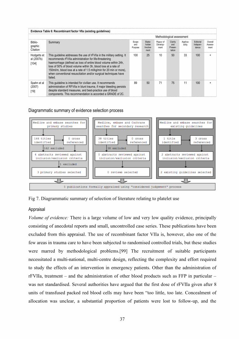
37
Evidence Table 6: Recombinant factor VIIa (existing guidelines)
Methodological assessment
Biblio-graphic Citation
Summary Scope and
Purpose
Stake-holder
Involve-ment
Rigour of Develop-
ment
Clarity and
Presen-tation
Applica-bility
Editorial Indepen-
dence
Overall Assess-
ment
Hodgetts et al (2007b)
[104]
This guideline addresses the use of rFVIIa in the military setting. It recommends rFVIIa administration for life-threatening haemorrhage (defined as loss of entire blood volume within 24h, loss of 50% of blood volume within 3h, blood loss at a rate of 150ml/m, blood loss at a rate of 1.5 ml/kg/min for 20 min or more), when conventional resuscitation and/or surgical techniques have failed.
100 25 10 50 33 100 +
Spahn et al (2007)
[19]
This guideline is intended for civilian use. It recommends administration of RFVIIa in blunt trauma, if major bleeding persists despite standard measures, and best-practice use of blood components. This recommendation is accorded a GRADE of 2C.
89 50 71 75 11 100 +
Diagrammatic summary of evidence selection process
Fig 7. Diagrammatic summary of selection of literature relating to platelet use
Appraisal
Volume of evidence: There is a large volume of low and very low quality evidence, principally
consisting of anecdotal reports and small, uncontrolled case series. These publications have been
excluded from this appraisal. The use of recombinant factor VIIa is, however, also one of the
few areas in trauma care to have been subjected to randomised controlled trials, but these studies
were marred by methodological problems.[99] The recruitment of suitable participants
necessitated a multi-national, multi-centre design, reflecting the complexity and effort required
to study the effects of an intervention in emergency patients. Other than the administration of
rFVIIa, treatment – and the administration of other blood products such as FFP in particular –
was not standardised. Several authorities have argued that the first dose of rFVIIa given after 8
units of transfused packed red blood cells may have been “too little, too late. Concealment of
allocation was unclear, a substantial proportion of patients were lost to follow-up, and the

38
statistical analysis was opaque. Despite these limitations, these trials are important. Although
there was no difference in mortality (which the trials were never powered to detect), factor VIIa
reduced blood transfusion requirements in patients with blunt trauma. Other than the two parallel
trials conducted by Boffard et al, there is only one small non-concurrent cohort study of the
effect of rFVIIa, in military patients. Existing systematic reviews and guidelines reflect the
paucity of evidence and rely heavily on the results of the paired trials. Applicability: Other than
the small retrospective study of rFVIIa in military patients, the studies, reviews and guidelines
included are applicable to this guideline’s target population. However, all of these studies were
conducted before the aggressive use of fresh frozen plasma in trauma resuscitation became
commonplace. The place of rFVIIa in this setting is thus difficult to ascertain. Consistency: The
volume of evidence is too small to comment on consistency.
Evidence statements
Recombinant factor VIIa reduces transfusion needs in blunt trauma patients requiring
massive transfusion (defined as more than 8 units of packed red blood cells). The
effect of recombinant factor VIIa on mortality/survival in this setting is not known.
1-
Recombinant factor VIIa may also reduce transfusion needs in penetrating trauma
patients requiring massive transfusion (defined as more than 8 units of packed red
blood cells), but the evidence in this setting is less clear. As in blunt trauma, the effect
of recombinant factor VIIa on mortality/survival in this setting is also not known.
1-
Future Research
All studies of rFVIIa, including the two trials on which these recommendations are based, were
conducted prior to the acceptance of aggressive fresh frozen plasma therapy as the standard of
care, and it is conceivable that the earlier use of such blood products will lead to a decreased
need for rFVIIa. Given the substantial cost of this intervention, further studies are needed to
define the place of recombinant factor VIIa in haemostatic resuscitation.
3.2.5 Cryoprecipitate
Fibrinogen deficiency develops earlier than any other clotting factor deficiency following major
haemorrhage, and the use of cryoprecipitate, which contains factor VIII, factor XIII, von
Willebrand factor (vWF) and fibrinogen, is therefore conceptually attractive.[66][105][106][107]
In addition to replacing deficient fibrinogen, the vWF contained in cryoprecipitate may enhance
platelet aggregation and adhesion.[51] Although current transfusion strategies with high ratios of

39
FFP and platelets seem very successful, several authorities have suggested that the role of
cryoprecipitate in coagulopathic trauma patients should be re-evaluated.[51][66] The use of
cryoprecipitate is tempered by concerns about patient exposure to large numbers of donors, and
associated risks of blood borne virus transmission.
Key question
This section aims to answer the question “Does the use of cryoprecipitate improve survival in
trauma patients?”
Outcome measures
Mortality/survival
Primary Studies
Database: Ovid MEDLINE(R) <1950 to February Week 2 2009> 1 trauma.mp. or exp *"Wounds and Injuries"/ (488200) 2 exp *Mental Disorders/ (582078) 3 1 not 2 (474751) 4 cryoprecipitate.mp. (1401) 5 3 and 4 (66) 6 limit 5 to (humans and yr="1980 - 2009" and (english or german)) (49) 7 from 6 keep 2-6 (5) Database: EMBASE <1980 to 2009 Week 07> 1 trauma.mp. or exp *"Wounds and Injuries"/ (406671) 2 exp *Mental Disorders/ (497727) 3 1 not 2 (387898) 4 *Cryoprecipitate/ (237) 5 3 and 4 (7) 6 from 5 keep (0) Inclusion criteria for abstract selection 1 Interventional studies 2 Observational studies Exclusion criteria for abstract selection 1 Case reports 2 Case series without comparison groups
Systematic medline and embase searches returned 56 citations, of which 51 were excluded as
irrelevant following title review. Of the remaining 5 abstracts, only one was found to meet the
inclusion criteria.[108] (Fig. 8)
Evidence Table 7: Cryoprecipitate (primary studies)
Bibliographic Citation
Study Type
Evi-dence Level
Number of Patients
Patient Charac-teristics
Intervention Comparison Length of Follow-Up
Outcome Measure
Effect Size
Stinger et al (2008)
[108]
NCCS - 252 Military setting
Mean ISS 21
Fibrinogen administration
Low fib: PRBC ratio (<0.2g/U) (n=52) High fib: PRBC ratio (>0.2g/U) (n=200)
To discharge Survival Low gp 52% High gp 24%
(p<0.001)
OR of death 0.37 (95%CI 0.171-0.812, p=0.013)
Database study of calculated amount of fibrinogen administered (in FFP, platelets, whole blood, PRBCs, or as cryoprecipitate). Retrospective design and assumptions regarding fibrinogen content of the products used make the validity of the conclusions questionable.

40
Secondary research
Database: Ovid MEDLINE(R) <1950 to February Week 2 2009> 1 trauma.mp. or exp *"Wounds and Injuries"/ (488200) 2 exp *Mental Disorders/ (582078) 3 1 not 2 (474751) 4 cryoprecipitate.mp. (1401) 5 3 and 4 (66) 6 limit 5 to (humans and yr="1980 - 2009" and (english or german)) (49) 7 meta-analysis/ (19931) 8 exp review literature/ (1416721) 9 (meta-analy$ or meta analy$ or metaanaly$).tw. (23482) 10 meta analysis.pt. (19931) 11 review academic.pt. (0) 12 review literature.pt. (0) 13 letter.pt. (638576) 14 review of reported cases.pt. (0) 15 historical article.pt. (251498) 16 review multicase.pt. (0) 17 7 or 8 or 9 or 10 or 11 or 12 (1434959) 18 13 or 14 or 15 or 16 (884681) 19 17 not 18 (1422622) 20 animal/ (4310239) 21 human/ (10508643) 22 20 and 21 (1074690) 23 20 not 22 (3235549) 24 19 not 23 (1312678) 25 6 and 24 (9) 26 from 25 keep 1-5 (5) Database: EMBASE <1980 to 2009 Week 07> 1 trauma.mp. or exp *"Wounds and Injuries"/ (406671) 2 exp *Mental Disorders/ (497727) 3 1 not 2 (387898) 4 *Cryoprecipitate/ (237) 5 3 and 4 (7) 6 from 5 keep (0) Inclusion criteria for abstract selection 1 Systematic reviews 2 Meta-analyses Exclusion criteria for abstract selection 1 Non-systematic reviews
No methodologically rigorous secondary research was identified. Systematic medline, embase,
and Cochrane library searches returned 16 citations. 11 were excluded as irrelevant on title
review. Of the remaining 5 review articles, none were based on systematic methodology, and
therefore excluded from further appraisal. (Fig.8)
Existing guidelines
Database: Ovid MEDLINE(R) <1950 to February Week 2 2009> 1 trauma.mp. or exp *"Wounds and Injuries"/ (488200) 2 exp *Mental Disorders/ (582078) 3 1 not 2 (474751) 4 cryoprecipitate.mp. (1401) 5 3 and 4 (66) 6 limit 5 to (humans and yr="1980 - 2009" and (english or german)) (49) 7 guideline.pt. (0) Database: EMBASE <1980 to 2009 Week 07> 1 trauma.mp. or exp *"Wounds and Injuries"/ (406671) 2 exp *Mental Disorders/ (497727)

41
3 1 not 2 (387898) 4 *Cryoprecipitate/ (237) 5 3 and 4 (7) 6 from 5 keep (0) Inclusion criteria for abstract selection 1 Guidelines Exclusion criteria for abstract selection 1 Quasi-editorial guidelines
Systematic medline and embase searches for existing guidelines returned no citations. Manual
cross-referencing, however, revealed four relevant articles.[19][55][56][81] (Fig.8)
Evidence Table 8: Cryoprecipitate (existing guidelines)
Methodological assessment
Biblio-graphic Citation
Summary Scope and
Purpose
Stake-holder
Involve-ment
Rigour of Develop-
ment
Clarity and
Presen-tation
Applica-bility
Editorial Indepen-
dence
Overall Assess-
ment
Spahn et al (2007)
[19]
This guideline recommends “treatment with fibrinogen concentrate or cryoprecipitate if significant bleeding is accompanied by a plasma fibrinogen level of less than 1.0 g/l.” It accords this recommendation a GRADE 1C.
89 50 71 75 11 100 +
Stainsby et al (2006)
[55]
This guideline, which is not specifically aimed at trauma patients, recommends that cryoprecipitate therapy should be considered if fibrinogen levels remain low (<1.0g/l) despite fresh frozen plasma administration. This recommendation is based on a guideline issued by the College of American Pathologists (Lundberg 1994).
78 25 38 50 44 100 +
Lundberg (1994)
[56]
This guideline, not specifically aimed at trauma patients, recommends cryoprecipitate for the correction of fibrinogen deficits refractory to therapy with fresh frozen plasma (<1.0g/l). This document is included for completeness, as it is still often quoted. The methodology used to derive the recommendations is unclear.
33 25 9 25 0 10 -
O’Shaughnessy et al (2004)
[81]
This guideline states that cryoprecipitate should be given so that a fibrinogen concentration of at least 1.0 g/l is obtained.
67 25 14 25 11 67 -
Diagrammatic summary of evidence selection process
Fig 8. Diagrammatic summary of selection of literature relating to use of cryoprecipitate
42
Appraisal
Volume of evidence: The volume of evidence for the use of cryoprecipitate in traumatic
haemorrhage is poor. Only one poor quality primary study was identified.[108] This study
retrospectively compared outcome with the calculated amount of fibrinogen administered, in
various forms, and is therefore liable to significant confounding.[108] Current guidelines, many
of which are not specifically aimed at trauma patients, are based on observational and
extrapolated data. The European guideline for the management of bleeding following major
trauma is based on satisfactory methodology, but the British committee for standards in
haematology guideline merely reiterates guidance published by College of American
Pathologists in 1994 and is therefore outdated.[19][55][56] Applicability: The only primary
study was conducted in military patients, and may thus not be relevant to civilian practice.
Consistency: As there is only one study, consistency could not be assessed.
Evidence statement
The use of cryoprecipitate in exsanguinating haemorrhage is founded on reasonable
scientific principles, but its use in trauma patients is largely based on studies of poor
methodology, and extrapolated data. Cryoprecipitate should be considered when
hypofibrinogenaemia has been confirmed, or when traumatic coagulopathy, whether
diagnosed clinically or by assay, is not responding to other methods of haemostatic
resuscitation.
3
Future research
There is a need to better define the role of cryoprecipitate, and other single-factor concentrates
(such as fibrinogen) and complexes (such as prothrombin complex concentrate), in the
management of traumatic coagulopathy.
3.2.6 Tranexamic acid
Antifibrinolytics such as tranexamic acid, aprotinin, and aminocaproic acid have been shown to
reduce blood loss after elective – particularly cardiac and liver transplantation – surgery.[109]
[110][111][112] Recognition of the contribution of hyperfibrinolysis to the development of the
acute coagulopathy of trauma shock has led to renewed interest in their use in trauma
patients.[113] Tranexamic acid and aminocaproic acid are lysine analogues which stabilise clot
by competitively inhibiting lysine binding sites of the plasminogen molecule, preventing
complexation with t-PA and fibrin, and inhibiting fibrinolysis.[112] Aprotinin is a
broadspectrum serine protease inhibitor. Aminocaproic acid and aprotinin are not available in the

43
UK, the licence for the latter having been withdrawn following reports of increased
cardiovascular and renal complications following coronary artery bypass grafting.[114]
Tranexamic acid is therefore the only readily available antifibrinolytic agent.
Key question
This section aims to answer the question “Does tranexamic acid reduce transfusion requirements
and/or mortality in trauma patients?”
Outcome measures
Blood loss, mortality/survival.
Primary Studies
Database: Ovid MEDLINE(R) <1950 to January Week 3 2009> 1 trauma.mp. or exp *"Wounds and Injuries"/ (486169) 2 exp *Mental Disorders/ (580167) 3 1 not 2 (472776) 4 Tranexamic Acid/ (1277) 5 3 and 4 (52) 6 limit 5 to yr="1980 - 2009" (42) 7 limit 6 to (english or german) (36) 8 from 7 keep 0 Database: EMBASE <1980 to 2009 Week 05> 1 trauma.mp. or exp *"Wounds and Injuries"/ (405735) 2 exp *Mental Disorders/ (496419) 3 1 not 2 (387006) 4 Tranexamic Acid/ (3385) 5 3 and 4 (212) 6 limit 5 to (human and (english or german) and yr="1980 - 2009") (151) 7 from 6 keep 0 Inclusion criteria for abstract selection 1 Interventional studies 2 Observational studies Exclusion criteria for abstract selection 1 Case reports 2 Case series without comparison groups
Systematic searches of the medline and embase databases returned no completed primary studies
of tranexamic acid in trauma patients. (Fig. 9) The two trials included in the Cochrane review on
antifibrinolytic drugs for acute traumatic injury both trialled aprotinin rather than tranexamic
acid.[115] In addition, these studies were small, outdated, and of questionable methodology.
Secondary research
Database: Ovid MEDLINE(R) <1950 to January Week 3 2009> 1 trauma.mp. or exp *"Wounds and Injuries"/ (486169) 2 exp *Mental Disorders/ (580167) 3 1 not 2 (472776) 4 Tranexamic Acid/ (1277) 5 3 and 4 (52) 6 limit 5 to yr="1980 - 2009" (42) 7 limit 6 to (english or german) (36) 8 meta-analysis/ (19624) 9 exp review literature/ (1406546)

44
10 (meta-analy$ or meta analy$ or metaanaly$).tw. (23115) 11 meta analysis.pt. (19624) 12 review academic.pt. (0) 13 review literature.pt. (0) 14 letter.pt. (634977) 15 review of reported cases.pt. (0) 16 historical article.pt. (250218) 17 review multicase.pt. (0) 18 8 or 9 or 10 or 11 or 12 or 13 (1424563) 19 14 or 15 or 16 or 17 (879842) 20 18 not 19 (1412320) 21 animal/ (4288697) 22 human/ (10444338) 23 21 and 22 (1066842) 24 21 not 23 (3221855) 25 20 not 24 (1302945) 26 7 and 25 (4) Database: EMBASE <1980 to 2009 Week 05> 1 trauma.mp. or exp *"Wounds and Injuries"/ (405735) 2 exp *Mental Disorders/ (496419) 3 1 not 2 (387006) 4 Tranexamic Acid/ (3385) 5 3 and 4 (212) 6 limit 5 to (human and (english or german) and yr="1980 - 2009") (151) 7 from 6 keep (14) Inclusion criteria for abstract selection 1 Systematic reviews 2 Meta-analyses Exclusion criteria for abstract selection 1 Non-systematic reviews
Systematic medline, embase, and Cochrane library searches returned 16 citations. Apart from
one, summarised in evidence table 9, none were developed systematically and therefore
excluded. (Fig. 9)
Evidence Table 9: Tranexamic acid (secondary research)
Bibliographic Citation
Study Type
Evi-dence Level
Number of Patients
Primary studies included
Intervention Comparison Outcome Measure
Effect Size
Coats et al (2004) [115]
CSR - 97 2 RCTs Aprotinin Aprotinin vs placebo
Proportion undergoing surgical intervention
Not evaluable
Volume of blood transfused
Not evaluable
Mortality Not evaluable
This Cochrane Review only included two trials, from 1979 and 1982, comprising a total of only 97 patients. Both studies were of questionable methodology. Allocation and concealment were unclear, and the results of one were reported in four papers. The review concluded that there was “insufficient evidence from randomised controlled trials of antifibrinolytic agents in trauma to either support or refute a clinically important treatment effect.” Both the title and conclusions of this Cochrane review are misleading as both included trials investigated the effect of aprotinin, rather than other, or combinations of, antibrinolytics.
Existing guidelines
Database: Ovid MEDLINE(R) <1950 to January Week 3 2009> 1 trauma.mp. or exp *"Wounds and Injuries"/ (486169) 2 exp *Mental Disorders/ (580167) 3 1 not 2 (472776) 4 Tranexamic Acid/ (1277) 5 3 and 4 (52) 6 limit 5 to yr="1980 - 2009" (42)

45
7 limit 6 to (english or german) (36) 8 guideline.pt. (14928) 9 7 and 8 (0) Database: EMBASE <1980 to 2009 Week 05> 1 trauma.mp. or exp *"Wounds and Injuries"/ (405735) 2 exp *Mental Disorders/ (496419) 3 1 not 2 (387006) 4 Tranexamic Acid/ (3385) 5 3 and 4 (212) 6 limit 5 to (human and (english or german) and yr="1980 - 2009") (151) 7 Practice Guideline/ (101734) 8 6 and 7 (5) Inclusion criteria for abstract selection 1 Guidelines Exclusion criteria for abstract selection 1 Quasi-editorial guidelines
Systematic medline and embase searches for existing guidelines returned five citations. Three
did not meet the inclusion criteria and were excluded. (Fig. 9) One other guideline was identified
through manual cross-referencing, yielding a total of three relevant existing guidelines,
summarised in evidence table 10.[19][55][116]
Evidence Table 10: Tranexamic acid (existing guidelines)
Methodological assessment
Biblio-graphic Citation
Summary Scope and
Purpose
Stake-holder
Involve-ment
Rigour of Develop-
ment
Clarity and
Presen-tation
Applica-bility
Editorial Indepen-
dence
Overall Assess-
ment
Spahn et al (2007)
[19]
This guideline extrapolates the evidence from studies of antifibrinolytic agents in elective surgery to trauma patients, and concludes that “antifibrinolytic agents (tranexamic acid, aminocaproic acid, or aprotinin) be considered in the treatment of the bleeding trauma patient. Antifibrinolytic therapy should be stopped once bleeding has been adequately controlled.” It accords this recommendation a GRADE 2C.
89 50 71 75 11 100 +
Stainsby et al (2006) [55]
This guideline, based on previous reviews, concludes that there is insufficient evidence from randomised controlled trials of antibrinolytic agents in trauma to either support or refute a clinically important treatment effect. It does not make recommendations regarding their use.
78 25 38 50 44 100 +
Gaarder et al (2008) [116]
This guideline, based on previous reviews, concludes that there is no evidence for the routine use of antifibrinolytics in trauma. It recommends, however, that such therapy should be considered in cases of prolonged bleeding (>24h) and bleeding caused by hyperfibrinolysis, as identified by thromboelastography. It acknowledges that the diagnosis of hyperfibrinolysis in the absence of TEG is difficult. The methodology used to appraise the evidence base for this guideline is unclear.
89 50 33 50 44 100 +

46
Diagrammatic summary of evidence selection process
Fig 9. Diagrammatic summary of selection of literature relating to tranexamic acid
Appraisal
Volume of evidence: The volume of evidence for the use of any antifibrinolytics in trauma
patients is poor. Other than two very small, outdated, methodologically questionable trials which
are largely of historical interest, there are no primary studies to support such therapy. The
paucity of primary studies is reflected, accurately, in the secondary literature. Applicability:
There is, however, a very substantial body of high quality evidence, including several meta-
analyses, to support the use of various antifibrinolytics in elective surgery, particularly cardiac,
and liver transplantation. It is reasonable to extrapolate the benefits of antifibrinolytic agents to
the trauma setting, but this assumption is not backed by any published data to suggest that the
haemostatic response to trauma is similar to elective surgery.[19] The risk of precipitating
thrombosis is a concern, but Henry et al’s Cochrane review of antifibrinolytic use in the elective
setting, which includes more than 8000 patients, demonstrated no increased risk of either arterial
or venous thrombotic events.[110] Consistency: Given the lack of evidence, consistency in the
trauma setting cannot be assessed. The effect of antifibrinolytics in the elective setting are,
however, consistent.
Evidence statement
There is insufficient evidence from randomised trials of antifibrinolytic agents in trauma patients
to either support or refute a clinically important treatment effect. However, given the proven
efficacy and effectiveness of antifibrinolytics in reducing blood loss in elective surgery, and the
lack of serious side effects, many authorities recommend the use of tranexamic acid in
haemorrhaging trauma patients. This practice, although reasonable, amounts to “expert opinion”
only, and no formal evidence statement has therefore been made.
47
Future research
The efficacy of tranexamic acid in the trauma setting is the subject of the ongoing, multinational
CRASH (Clinical Randomisation of an Antifibrinolytic in Significant Haemorrhage) 2 study, in
which 20,000 trauma patients are being randomly assigned to 1g of tranexamic acid for a period
of 10 minutes followed by 1g infused for a period of 8 hours.[113][117] This trial should help to
answer many of the questions surrounding tranexamic acid use in trauma, and allow stronger
recommendations to be made.
3.3 PERMISSIVE HYPOTENSION
Haemorrhagic shock is failure of oxygen delivery, as a consequence of acute blood loss. Cellular
ischaemia leads to organ dysfunction, and ultimately irreversible organ failure. The prevention
and reversal of these sequelae through the rapid restoration of normal circulatory function – by
replacing lost volume with a combination of synthetic fluids and blood products – was long
regarded as pivotal, and remains enshrined in Advanced Trauma Life Support (ATLS)
practice.[5][118][119] The recognition that fluid resuscitation may interfere with normal
haemostatic mechanisms, ultimately exacerbating blood loss, however, led to a re-examination
of this approach. [118][119] Permissive hypotension (also known as hypotensive resuscitation or
balanced resuscitation) is a strategy of deferring or restricting fluid administration until control
of haemorrhage has been achieved, while accepting a limited period of suboptimal end-organ
perfusion. [119][120]
Key question
This section aims to answer the question “Does a strategy of withholding or limiting fluid
resuscitation prior to surgical control of haemorrhage improve survival?”
Outcome measure
Mortality/survival.
Primary studies
Database: Ovid MEDLINE(R) <1950 to January Week 4 2009> 1 hypotensive resuscitation.mp. (46) 2 permissive hypotension.mp. or Hypotension, Controlled/ (1898) 3 balanced resuscitation.mp. (1) 4 trauma.mp. or exp *"Wounds and Injuries"/ (486815) 5 exp *Mental Disorders/ (580862) 6 4 not 5 (473395) 7 1 or 2 or 3 (1939) 8 6 and 7 (68) Database: EMBASE <1980 to 2009 Week 05> 1 trauma.mp. or exp *"Wounds and Injuries"/ (405735)

48
2 exp *Mental Disorders/ (496419) 3 1 not 2 (387006) 4 permissive hypotension.mp. (18) 5 hypotensive resuscitation.mp. (44) 6 balanced resuscitation.mp. (0) 7 4 or 5 (57) 8 limit 7 to (human and (english or german) and yr="1980 - 2009") (29) Inclusion criteria for abstract selection 1 Interventional studies 2 Observational studies Exclusion criteria for abstract selection 1 Case reports 2 Case series without comparison groups
Systematic medline and embase searches returned 96 citations. 94 did not meet the inclusion
criteria, and were therefore excluded. Manual cross-referencing revealed one further study. (Fig.
10) All three included studies are randomised controlled trials, summarised in table 11.
Evidence table 11: Permissive hypotension (primary studies)
Bibliographic Citation
Study Type
Evi-dence Level
Number of Patients
Patient Charac-teristics
Intervention Comparison Length of Follow-Up
Outcome Measure
Effect Size
Bickel et al (1994) [121]
CT + 598 Penetrating torso trauma Hypotension (SAP <90 mmHg)
Withholding of fluid resuscitation
Early (pre-hospital or emergency room) (n=309) vs delayed (intra-operative) fluid resuscitation (n=289)
Until discharge
Mortality Early 38%
Delayed 30%
RR for death with early fluid 1.25 (95% CI 1.00-1.58)*
First large-scale study of hypotensive resuscitation concept. Suggestive of iv fluids being harmful in trauma patients with penetrating injury; some evidence of harm, no evidence of benefit. Although the study enrolled large numbers of patients, there was no concealment of allocation, and the trial therefore not truly randomised. Patients were also mostly young (median 36 years), and from a small geographical area, resulting in very short pre-hospital times. Extrapolation of the findings to other settings or mechanisms of injury may therefore not be appropriate. Despite these caveats, this study remains one of the most seminal studies of trauma management.
Turner et al (2000)
[122]
RCT - 1309 Unselected (mostly blunt) adult trauma patients
Withholding of fluid resuscitation (unless transfer to hospital > 1h)
Conventional fluid resuscitation (as per ambulance service protocol) (n=699) vs delayed (no pre-hospital) fluid resuscitation (n=610)
Not stated Mortality Early (10.4%)
Delayed/no (9.8%)
RR for death with early fluid 1.06 (95%CI 0.77-1.47)*
UK-based, cluster-randomised trial marred by vague inclusion criteria, resulting in the enrolment of a large number of patients with minor injuries. Numerous protocol violations, rendering the conclusions essentially meaningless: Only 31% in the conventional treatment group actually received intravenous fluids, and only 80% in the delayed treatment group actually had fluids withheld. If fluids given pre-theatre are included, the percentages are 49% vs 42%.
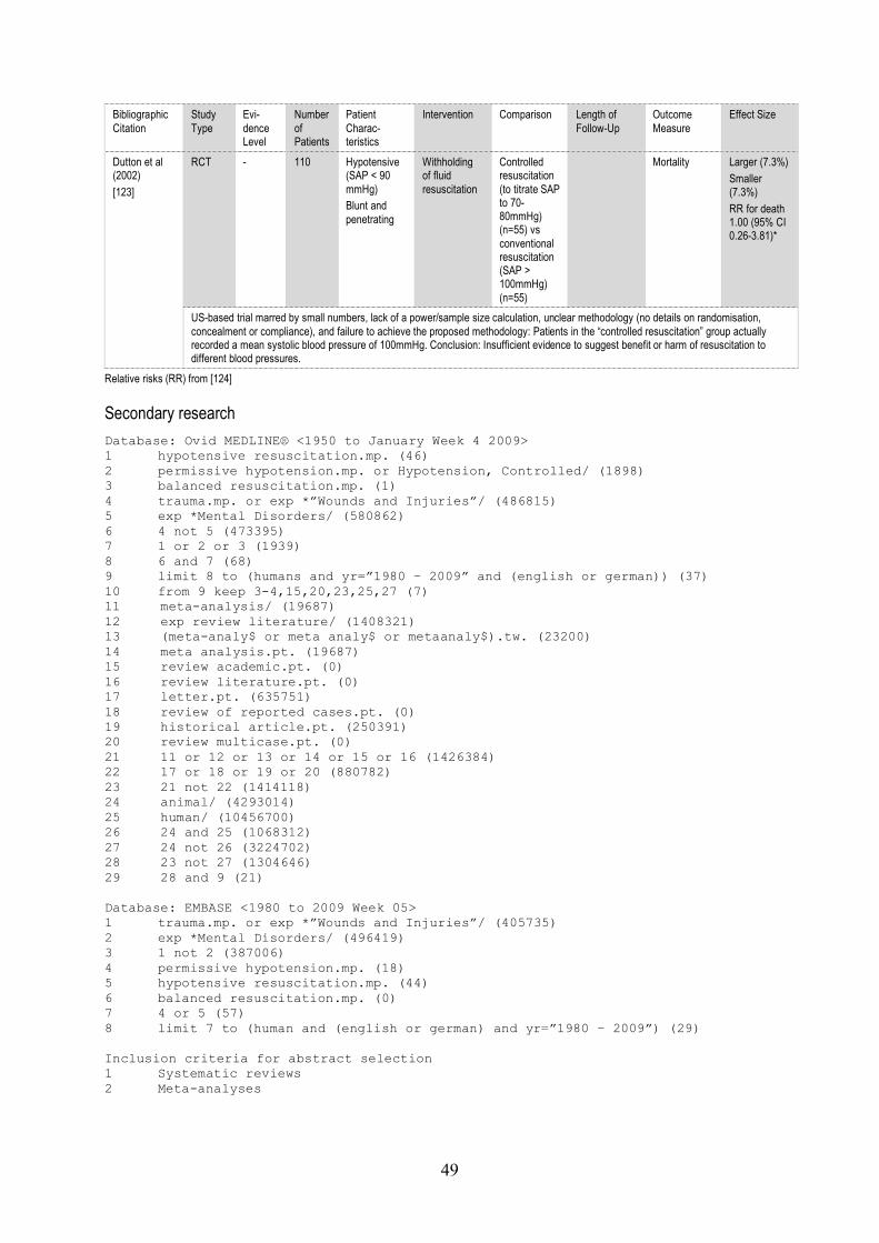
49
Bibliographic Citation
Study Type
Evi-dence Level
Number of Patients
Patient Charac-teristics
Intervention Comparison Length of Follow-Up
Outcome Measure
Effect Size
Dutton et al (2002)
[123]
RCT - 110 Hypotensive (SAP < 90 mmHg) Blunt and penetrating
Withholding of fluid resuscitation
Controlled resuscitation (to titrate SAP to 70-80mmHg) (n=55) vs conventional resuscitation (SAP > 100mmHg) (n=55)
Mortality Larger (7.3%)
Smaller (7.3%)
RR for death 1.00 (95% CI 0.26-3.81)*
US-based trial marred by small numbers, lack of a power/sample size calculation, unclear methodology (no details on randomisation, concealment or compliance), and failure to achieve the proposed methodology: Patients in the “controlled resuscitation” group actually recorded a mean systolic blood pressure of 100mmHg. Conclusion: Insufficient evidence to suggest benefit or harm of resuscitation to different blood pressures.
Relative risks (RR) from [124]
Secondary research
Database: Ovid MEDLINE® <1950 to January Week 4 2009> 1 hypotensive resuscitation.mp. (46) 2 permissive hypotension.mp. or Hypotension, Controlled/ (1898) 3 balanced resuscitation.mp. (1) 4 trauma.mp. or exp *”Wounds and Injuries”/ (486815) 5 exp *Mental Disorders/ (580862) 6 4 not 5 (473395) 7 1 or 2 or 3 (1939) 8 6 and 7 (68) 9 limit 8 to (humans and yr=”1980 – 2009” and (english or german)) (37) 10 from 9 keep 3-4,15,20,23,25,27 (7) 11 meta-analysis/ (19687) 12 exp review literature/ (1408321) 13 (meta-analy$ or meta analy$ or metaanaly$).tw. (23200) 14 meta analysis.pt. (19687) 15 review academic.pt. (0) 16 review literature.pt. (0) 17 letter.pt. (635751) 18 review of reported cases.pt. (0) 19 historical article.pt. (250391) 20 review multicase.pt. (0) 21 11 or 12 or 13 or 14 or 15 or 16 (1426384) 22 17 or 18 or 19 or 20 (880782) 23 21 not 22 (1414118) 24 animal/ (4293014) 25 human/ (10456700) 26 24 and 25 (1068312) 27 24 not 26 (3224702) 28 23 not 27 (1304646) 29 28 and 9 (21) Database: EMBASE <1980 to 2009 Week 05> 1 trauma.mp. or exp *”Wounds and Injuries”/ (405735) 2 exp *Mental Disorders/ (496419) 3 1 not 2 (387006) 4 permissive hypotension.mp. (18) 5 hypotensive resuscitation.mp. (44) 6 balanced resuscitation.mp. (0) 7 4 or 5 (57) 8 limit 7 to (human and (english or german) and yr=”1980 – 2009”) (29) Inclusion criteria for abstract selection 1 Systematic reviews 2 Meta-analyses

50
Exclusion criteria for abstract selection 1 Non-systematic reviews
Systematic medline and embase searches returned 50 citations. Following review of the titles and
abstracts, 49 were excluded as either irrelevant, or not meeting the inclusion criteria. The vast
majority of excluded studies were non-systematic reviews or editorials. A search of the Cochrane
library returned one further citation. (Fig. 10) Both reviews are summarised in evidence table 12.
Evidence Table 12: Permissive hypotension (secondary research)
Bibliographic Citation
Study Type
Evi-dence Level
Number of Patients
Primary studies included
Intervention Comparison Outcome Measure
Effect Size
Dretzke et al (2006) [125][126]
SR + 2053 4 primary RCTs, including the three studies listed above
Withholding or limitation of fluid resuscitation
Withholding, limited or targeted resuscitation vs conventional resuscitation
Mortality Not calculable
High quality systematic review commissioned by the NHS Research & Development Health Technology Assessment programme, incorporating the three primary studies identified above, and one additional, small, early trial. This parallel randomised controlled trial of 36 patients compared a rapid infusor system with conventional practice. [127] The study was of poor methodological quality. Overall conclusion: There is no evidence to suggest that pre-hospital iv fluid resuscitation prior to control of bleeding is beneficial. There is limited evidence (from one study only) to suggest that it can be harmful, and that patients with penetrating injuries in particular may have better outcomes when fluids are withheld.
Kwan et al (2003)
[124]
CSR + 1957 3 primary RCTs of patients with acute blood loss, including 1 not relating to trauma
Fluid administration
Early vs delayed fluid administration
Mortality No meta-analysis performed (due to heterogeneity of primary studies)
Cochrane systematic review, conducted in two parts. This first part of the review compared early with delayed fluid administration, and included 3 primary RCTs. Of these, only 2 relate to trauma (both included in the primary studies analysed above). [121][122]The remaining study was of patients with gastrointestinal haemorrhage. Conclusion: There is no evidence for or against the withholding of intravenous fluid administration in uncontrolled haemorrhage.
CSR - 171 3 primary RCTs of patients with acute blood loss, including 1 not relating to trauma
Fluid administration
Larger vs smaller volume fluid administration
Mortality No meta-analysis performed (due to heterogeneity of primary studies)
Cochrane systematic review, conducted in two parts. This second part of the review compared larger with smaller volume fluid administration, and included 3 primary RCTs. All 3 trials were of trauma patients. One is included in the primary studies analysed above.[123] The remaining two are small, early studies, of questionable methodological quality. [127] Conclusion: There is no evidence for or against the administration of larger or smaller volumes of intravenous fluid in uncontrolled haemorrhage.
Existing guidelines
Database: Ovid MEDLINE(R) <1950 to January Week 4 2009> 1 hypotensive resuscitation.mp. (46) 2 permissive hypotension.mp. or Hypotension, Controlled/ (1898) 3 balanced resuscitation.mp. (1) 4 trauma.mp. or exp *"Wounds and Injuries"/ (486815) 5 exp *Mental Disorders/ (580862) 6 4 not 5 (473395) 7 1 or 2 or 3 (1939) 8 6 and 7 (68) 9 guideline.pt. (14387) 10 8 and 9 (0) Database: EMBASE <1980 to 2009 Week 05> 1 trauma.mp. or exp *"Wounds and Injuries"/ (405735) 2 exp *Mental Disorders/ (496419) 3 1 not 2 (387006)

51
4 permissive hypotension.mp. (18) 5 hypotensive resuscitation.mp. (44) 6 balanced resuscitation.mp. (0) 7 4 or 5 (57) 8 limit 7 to (human and (english or german) and yr="1980 - 2009") (29) 9 Practice Guideline/ (101983) 10 8 and 9 (2) Inclusion criteria for abstract selection 1 Guidelines Exclusion criteria for abstract selection 1 Quasi-editorial guidelines
Systematic medline and embase searches returned two citations, both of which were irrelevant
and therefore exluded. (Fig. 10) Manual cross-referencing revealed a further two publications
which met the inclusion criterion, and one paper which reports on the changes to the forthcoming
eighth edition of the Advanced Trauma Life Support programme, although the revised manual is
not yet available (evidence table 13).[19][120][128]
Evidence Table 13: Hypotensive resuscitation (existing guidelines)
Methodological assessment
Biblio-graphic Citation
Summary Scope and
Purpose
Stake-holder
Involve-ment
Rigour of Develop-
ment
Clarity and
Presen-tation
Applica-bility
Editorial Indepen-
dence
Overall Assess-
ment
Kortbeek et al (2008)
[120]
This article summarises the changes to the forthcoming (8th) Advanced Trauma Life Support (ATLS) guidelines. The new ATLS guidelines will place greater emphasis on the need to balance the risks of precipitating further bleeding against the adequacy of organ perfusion, by accepting a lower than normal blood pressure.
N/A* N/A* N/A* N/A* N/A* N/A* +
National Institute of Clinical Excellence (2008)
[128]
This guideline recommends that, during pre-hospital management, “intravenous fluids should not be administered if a radial pulse can be felt (or, for penetrating torso injuries, if a central pulse can be felt).” It further recommends that “in the absence of a radial pulse (or a central pulse for penetrating torso injuries) in adults and older children […] intravenous fluid should be administered in boluses of no more than 250 ml. The patient should then be reassessed, and the process repeated until a radial pulse (or central pulse for penetrating torso injuries) is palpable.”
100 100 100 100 50 100 +
Spahn et al (2007)
[19]
This guideline recommends “a target systolic blood pressure of 80 to 100 mmHg, until major bleeding has been stopped […] in [patients] without brain injury” and accords this recommendation a GRADE 2C.
89 50 71 75 11 100 +
N/A* = Not applicable. This publication related to a forthcoming guideline. Methodology therefore cannot be assessed.

52
Diagrammatic summary of evidence selection process
Fig 10. Diagrammatic summary of selection of literature relating to permissive hypotension.
Appraisal
Volume of evidence: The volume of evidence is moderate. There are three well-known major
primary studies, all of which have significant methodological flaws.[121][122][123]
Interestingly, the first of these trials, which showed a statistically significant difference between
groups, was arguably the least flawed. [121] There are two systematic reviews, including one
Cochrane systematic review.[124][125][126] The latter is marred by heterogeneity, as it included
several trials pertaining to non-trauma patients (such as patients with gastrointestinal
haemorrhage). Applicability: The Bickel trial only included patients with penetrating torso
injuries, in the setting of a well-developed trauma system with very short pre-hospital times.
[121] The findings are therefore not directly applicable to a typical European mixed
(blunt/penetrating) trauma population, but may still be relevant to the management of subgroups
of patients. The other two primary studies, by Turner and Dutton, are more applicable in terms of
settings and inclusion criteria. [122][123] Consistency: The results of the primary studies are
inconsistent. Only the Bickel trial showed a difference in mortality, but may not be applicable to
all patients and settings (see above). [121] The Turner and Dutton trials, bearing in mind
methological issues, were negative. [122][123]

53
Evidence statements
There is no new evidence to suggest that pre-hospital intravenous fluid resuscitation
prior to control of bleeding is beneficial. There is, however, limited evidence to
suggest that fluid resuscitation may be harmful, and that patients with penetrating
injuries in particular may have better outcomes when fluids are withheld until surgical
control of haemorrhage has been obtained.
1-
Future research
There is a need for a better quality trial of permissive hypotension in a clearly defined
population, and to define the time period during which suboptimal end-organ perfusion is
tolerated and reversible.
3.4 ACIDAEMIA MANAGEMENT
Acidaemia impairs all essential components of the coagulation process: At a pH of less than 7.4,
platelets lose their pseudopodia and change shape, becoming more spherical.[65] Coagulation
factor function is also impaired, through the induction of conformational changes, although
different factors are affected by acidaemia in different ways. [65][129][130] Calcium binding
sites have a pH-dependent affinity, further compounding the problem. [65] Restoration of normal
acid-base status is therefore important.
The metabolic acidosis associated with haemorrhagic shock is the result of hypoperfusion.
Correction requires the restoration of organ perfusion, but recognition of the need to defer
volume replacement until control of haemorrhage has been obtained has led to a search for
adjunctive pharmacological treatments to offset the pathophysiological consequences of
acidaemia on other organ systems, and the coagulation system in particular. [129] The traditional
treatment for severe lactic acidosis in critical illness is sodium bicarbonate, but there is little
rationale for its use and no evidence of effectiveness.[131] Bicarbonate administration produces
carbon dioxide, which requires large increases in minute volume to clear. Bicarbonate also
decreases ionised calcium levels by approximately 10%, with consequent effects on coagulation
and cardiac and vascular contractility.[131]
Ttris-hydroxymethyl aminomethane (THAM) is a biologically inert amino alcohol of low
toxicity, which buffers carbon dioxide and acids in vitro and in vivo.[132] Its pK at 37°C is 7.8,
making it a more effective buffer than bicarbonate.[132] In vivo, THAM supplements the
buffering capacity of the blood bicarbonate system, accepting a proton, generating bicarbonate,

54
and decreasing the partial pressure of carbon dioxide in arterial blood.[132] It rapidly restores
pH in acidaemia caused by carbon dioxide retention or metabolic acid accumulation. Unlike
bicarbonate, which requires an open system for carbon dioxide elimination in order to exert its
buffering effect, protonated THAM is excreted renally, avoiding issues with minute volume
ventilation.[132] THAM has been used in the treatment of hypercapnoeic respiratory failure,
diabetic and renal acidosis, salicylate and barbiturate intoxication, and raised intracranial
pressure following cerebral trauma. It is also used in cardioplegic solutions and liver
transplantation.[132] The administration of THAM in trauma patients with hypoperfusion-
induced acidaemia is thus conceptually attractive, and has been the subject of several recent key
publications.[3]
Key question
This section aims to answer the question “Does the administration of tris-hydroxymethyl
aminomethane improve survival in trauma patients?”
Outcome measure
Mortality/survival
Primary studies
Database: Ovid MEDLINE(R) <1950 to January Week 4 2009> 1 trauma.mp. or exp *"Wounds and Injuries"/ (486815) 2 exp *Mental Disorders/ (580862) 3 1 not 2 (473395) 4 tris-hydroxymethyl aminomethane.mp. (3435) 5 Trometamol.mp. (210) 6 THAM.mp. (399) 7 4 or 5 or 6 (3723) 8 3 and 7 (71) 9 limit 8 to (humans and yr="1980 - 2009" and (english or german)) (29) 10 from 9 keep (0) Database: EMBASE <1980 to 2009 Week 06> 1 tris-hydroxymethyl aminomethane.mp. or Trometamol/ (1934) 2 THAM.mp. (187) 3 1 or 2 (2030) 4 trauma.mp. or exp *"Wounds and Injuries"/ (406258) 5 exp *Mental Disorders/ (497136) 6 4 not 5 (387500) 7 3 and 6 (69) 8 limit 7 to human (39) 9 from 9 keep (0) Inclusion criteria for abstract selection 1 Interventional studies 2 Observational studies Exclusion criteria for abstract selection 1 Case reports 2 Case series without comparison groups

55
Systematic medline and embase searches returned no primary studies of THAM in the setting of
non-cerebral trauma, although there are numerous publications relating to the use of THAM for
neuroprotection following traumatic brain injury.
Secondary research
Database: Ovid MEDLINE(R) <1950 to January Week 4 2009> 1 trauma.mp. or exp *"Wounds and Injuries"/ (486815) 2 exp *Mental Disorders/ (580862) 3 1 not 2 (473395) 4 tris-hydroxymethyl aminomethane.mp. (3435) 5 Trometamol.mp. (210) 6 THAM.mp. (399) 7 4 or 5 or 6 (3723) 8 3 and 7 (71) 9 limit 8 to (humans and yr="1980 - 2009" and (english or german)) (29) 10 from 9 keep (0) Database: EMBASE <1980 to 2009 Week 06> 1 tris-hydroxymethyl aminomethane.mp. or Trometamol/ (1934) 2 THAM.mp. (187) 3 1 or 2 (2030) 4 trauma.mp. or exp *"Wounds and Injuries"/ (406258) 5 exp *Mental Disorders/ (497136) 6 4 not 5 (387500) 7 3 and 6 (69) 8 limit 7 to human (39) 9 from 8 keep (1) Inclusion criteria for abstract selection 1 Systematic reviews 2 Meta-analyses Exclusion criteria for abstract selection 1 Non-systematic reviews
Systematic medline, embase and Cochrane library searches returned only one review article (and
guideline) of the treatment of acidaemia with THAM, which was developed non-systematically,
and makes no recommendations regarding the use of THAM in non-cerebral trauma.[132]
Existing guidelines
Database: Ovid MEDLINE(R) <1950 to January Week 4 2009> 1 trauma.mp. or exp *"Wounds and Injuries"/ (486815) 2 exp *Mental Disorders/ (580862) 3 1 not 2 (473395) 4 tris-hydroxymethyl aminomethane.mp. (3435) 5 Trometamol.mp. (210) 6 THAM.mp. (399) 7 4 or 5 or 6 (3723) 8 3 and 7 (71) 9 limit 8 to (humans and yr="1980 - 2009" and (english or german)) (29) 10 from 9 keep 0 (0) Database: EMBASE <1980 to 2009 Week 06> 1 tris-hydroxymethyl aminomethane.mp. or Trometamol/ (1934) 2 THAM.mp. (187) 3 1 or 2 (2030) 4 trauma.mp. or exp *"Wounds and Injuries"/ (406258) 5 exp *Mental Disorders/ (497136) 6 4 not 5 (387500) 7 3 and 6 (69) 8 limit 7 to human (39)

56
9 from 8 keep 1-39 (39) 10 Practice Guideline/ (101983) 11 9 and 10 (1) 12 from 11 keep 1 (1) Inclusion criteria for abstract selection 1 Guidelines Exclusion criteria for abstract selection 1 Quasi-editorial guidelines
Systematic medline, embase and Cochrane library searches returned only one guideline of the
treatment of acidaemia with THAM, which was developed non-systematically, and makes no
recommendations regarding the use of THAM in non-cerebral trauma.[132]
Evidence statement
There is no evidence to support the use of tris-hydroxymethyl aminomethane in trauma patients.
Future research
There is a need for a trial of THAM in acidaemic trauma patients.
3.5 HYPOTHERMIA MANAGEMENT
Environmental exposure combined with limited endogenous heat production, as a consequence
of hypoperfusion-induced anaerobic metabolism, leads to hypothermia, which may be
compounded by injudicious administration of cold resuscitation fluids and blood. The
detrimental effects of hypothermia on protease and platelet function and metabolism are well
recognised.[46][65] However, hypothermia probably has disproportionately greater effects on
platelet function than on serine proteases.[3][46][65]
Two large, well-conducted retrospective studies, which controlled for injury severity, have
shown hypothermia to be an independent predictor of mortality after major trauma. Wang et al,
in a study of 38520 trauma patients, of which 1921 (5%) had a core temperature of <35°C on
admission, showed that hypothermia was independently associated with increased odds of death
(OR 3.03, 95%CI 2.62-3.51).[133] Martin et al studied the outcome of 11828 trauma patients
with an admission temperature of <35°C, and found the odds of death to be 1.54 (95%CI 1.40-
1.71).[134] Recognition of the association between hypothermia, coagulopathy, and adverse
outcome has led to the widespread acceptance of aggressive hypothermia mitigation in trauma
patients. Although hypothermia mitigation is also important in non-trauma surgery, the special
demands of trauma management, and in particular the need to prepare large areas of the body for
immediate extension of surgical access, limit the use of conventional warming devices, and
poses special problems.

57
Key question
This section aims to answer the questions “Do aggressive attempts at hypothermia mitigation
improve outcome in trauma patients?” and “What is the most effective method of preventing and
treating hypothermia in trauma patients?”
Outcome measure
Mortality/survival; change in temperature
Primary studies
Database: Ovid MEDLINE(R) <1950 to January Week 4 2009> 1 trauma.mp. or exp *"Wounds and Injuries"/ (487169) 2 exp *Mental Disorders/ (580839) 3 1 not 2 (473749) 4 *Hypothermia/ (4795) 5 3 and 4 (438) 6 limit 5 to (humans and yr="1980 - 2009" and (english or german)) (295) 7 randomized controlled trial.pt. (262320) 8 controlled clinical trial.pt. (78055) 9 randomized controlled trials/ (57544) 10 random allocation/ (62690) 11 double-blind method/ (98628) 12 single-blind method/ (12432) 13 7 or 8 or 9 or 10 or 11 or 12 (443309) 14 animal/ (4302436) 15 human/ (10485618) 16 14 and 15 (1071730) 17 14 not 16 (3230706) 18 13 not 17 (413856) 19 6 and 18 (8) Database: EMBASE <1980 to 2009 Week 06> 1 trauma.mp. or exp *"Wounds and Injuries"/ (406258) 2 exp *Mental Disorders/ (497136) 3 1 not 2 (387500) 4 *Hypothermia/et, dm, co, di, th, pc, su [Etiology, Disease Management,
Complication, Diagnosis, Therapy, Prevention, Surgery] (910) 5 3 and 4 (893) 6 limit 5 to (human and (english or german)) (621) 7 trauma.m_titl. (25350) 8 6 and 7 (21) 9 from 8 keep (6) Inclusion criteria for abstract selection 1 Interventional studies Exclusion criteria for abstract selection 1 Case reports 2 Case series without comparison groups
Systematic medline and embase searches returned 29 citations. Following review of the titles, 15
were excluded as irrelevant. (Fig. 11) Of the remaining 14 publications, 3 met the inclusion
criteria (table 14).

58
Evidence table 14: Hypothermia mitigation (primary studies)
Bibliographic Citation
Study Type
Evi-dence Level
Number of Patients
Patient Charac-teristics
Intervention Comparison Length of Follow-Up
Outcome Measure
Effect Size
Cohen et al (2002) [135]
RCT + 298 Normo-thermic (>35°C) trauma patients admitted to emergency department
ISS<15
Heat loss prevention
Reflective blanket over warmed cotton blanket vs forced-warm-air inflatable blanket vs 3 cotton warmed blankets
To discharge from emergency department
Temperature Warmed blankets: pre 36.8°C vs post 37.3°C
Reflective: pre 36.9°C vs post 37.6°C
Forced-air: pre 36.8°C vs post 37.3°C
(p>0.05)
Trial of three different types of heat loss prevention strategies in normothermic, non-severely injured trauma patients. No difference observed. May not be applicable to patients who are hypothermic on admission.
Kober et al (2001)
[136]
RCT + 100 Hypothermic (mean 35.4/ 35.3°C) “Minor trauma” During transfer to hospital
Electric warming blanket
Electric warming blanket switched on vs switched off
To arrival at hospital
Temperature Blanket off: Change -0.4 °C (95%CI -0.3 to -0.5)
Blanket on: Change 0.8 °C (95%CI 0.7-0.9)
Small trial conducted by paramedic-manned ambulances, accounting for limitation to patients with “minor” (not otherwise specified) trauma. Electric blankets raised temperature more than passive warming.
Gentilello et al (1997)
[137]
RCT 57 Hypothermic (<=35.5°C)
“Critically injured”
ISS 32 (SR) vs 31 (CAVR) With PAFC in situ
Rewarming Continuous arteriovenous (femoro-femoral) rewarming (CAVR) vs Standard rewarming (SR)
To discharge Mortality Not clearly stated (see below)
Randomised trial which set out to evaluate whether hypothermia during resuscitation is protective or harmful by comparing hypothermic patients who had been treated with conventional rewarming methods, or rapid rewarming by arteriovenous extracorporeal warming. Statistical methods and presentation of findings is confusing, but appear to show an excess early mortality in patients treated with standard rewarming. However, late deaths reduced the overall survival to discharge advantage in patients who underwent CAVR, which was attributed to survival from injuries which patients who underwent SR did not survive. The author’s concluded that hypothermia increases the risk of early death after injury.
Secondary research
Database: Ovid MEDLINE(R) <1950 to January Week 4 2009> 1 trauma.mp. or exp *"Wounds and Injuries"/ (487169) 2 exp *Mental Disorders/ (580839) 3 1 not 2 (473749) 4 *Hypothermia/ (4795) 5 3 and 4 (438) 6 limit 5 to (humans and yr="1980 - 2009" and (english or german)) (295) 7 meta-analysis/ (19805) 8 exp review literature/ (1412791) 9 (meta-analy$ or meta analy$ or metaanaly$).tw. (23318) 10 meta analysis.pt. (19805) 11 review academic.pt. (0) 12 review literature.pt. (0) 13 letter.pt. (637017) 14 review of reported cases.pt. (0) 15 historical article.pt. (251109) 16 review multicase.pt. (0) 17 7 or 8 or 9 or 10 or 11 or 12 (1430930) 18 13 or 14 or 15 or 16 (882749)

59
19 17 not 18 (1418630) 20 animal/ (4302436) 21 human/ (10485618) 22 20 and 21 (1071730) 23 20 not 22 (3230706) 24 19 not 23 (1308903) 25 6 and 24 (69) 27 from 25 keep (15) Database: EMBASE <1980 to 2009 Week 06> 1 trauma.mp. or exp *"Wounds and Injuries"/ (406258) 2 exp *Mental Disorders/ (497136) 3 1 not 2 (387500) 4 *Hypothermia/et, dm, co, di, th, pc, su [Etiology, Disease Management,
Complication, Diagnosis, Therapy, Prevention, Surgery] (910) 5 3 and 4 (893) 6 limit 5 to (human and (english or german)) (621) 7 trauma.m_titl. (25350) 8 6 and 7 (21) 9 from 8 keep (5) Inclusion criteria for abstract selection 1 Systematic reviews 2 Meta-analyses Exclusion criteria for abstract selection 1 Non-systematic reviews
No methodologically rigorous secondary research was identified. Systematic medline and
embase searches returned 90 citations. 70 of these were deemed irrelevant on the basis of title
review and excluded. None of the remaining 20 publications met the inclusion criteria. A search
of the Cochrane library similarly revealed no systematic reviews of interventions to mitigate
against hypothermia in trauma patients. (Fig. 11)
Existing guidelines
Database: Ovid MEDLINE(R) <1950 to January Week 4 2009> 1 trauma.mp. or exp *"Wounds and Injuries"/ (487169) 2 exp *Mental Disorders/ (580839) 3 1 not 2 (473749) 4 *Hypothermia/ (4795) 5 3 and 4 (438) 6 limit 5 to (humans and yr="1980 - 2009" and (english or german)) (295) 7 guideline.pt. (14427) 8 6 and 7 (1) 9 from 8 keep 1 (1) Database: EMBASE <1980 to 2009 Week 06> 1 trauma.mp. or exp *"Wounds and Injuries"/ (406258) 2 exp *Mental Disorders/ (497136) 3 1 not 2 (387500) 4 *Hypothermia/et, dm, co, di, th, pc, su [Etiology, Disease Management,
Complication, Diagnosis, Therapy, Prevention, Surgery] (910) 5 3 and 4 (893) 6 limit 5 to (human and (english or german)) (621) 7 *Practice Guideline/ (8140) 8 6 and 7 (1) 9 from 8 keep 1 (1) Inclusion criteria for abstract selection 1 Guidelines Exclusion criteria for abstract selection

60
1 Quasi-editorial guidelines
Systematic medline and embase searches returned no relevant citations. Manual cross-
referencing revealed two guidelines, one of which has been mentioned previously.[19][138] (Fig.
11)
Evidence Table 15: Hypothermia mitigation (existing guidelines)
Methodological assessment
Biblio-graphic Citation
Summary Scope and
Purpose
Stake-holder
Involve-ment
Rigour of Develop-
ment
Clarity and
Presen-tation
Applica-bility
Editorial Indepen-
dence
Overall Assess-
ment
Spahn et al (2007)
[19]
This guideline recommends that “early application of measures to reduce heat loss and warm the hypothermic patient in order to achieve and maintain normothermia” should be employed, and accords this recommendation a GRADE 1C.
89 50 71 75 11 100 +
National Institute of Clinical Excellence
[138]
This guideline, which is not limited to trauma patients, but is aimed at all surgical patients at risk of inadvertent hypothermia, recommends that forced air warming should be started preoperatively if the patient’s temperature is <36°C, and should be maintained throughout the intraoperative phase.
100 100 100 100 33 100 +
Diagrammatic summary of evidence selection process
Fig 11. Diagrammatic summary of selection of literature relating to hypothermia mitigation
Appraisal
Volume of evidence: Although there is limited evidence from trials, there is a high volume of
evidence from observational studies. Applicability: The publications cited above are broadly
applicable to the target population of this guideline, although few of the comparisons of different
devices were specifically conducted in the trauma setting. The mechanisms underlying the
development of hypothermia may differ between trauma and non-trauma patients, and because
surgical intervention in trauma patients often requires wide access. Consistency: The evidence
showing the detrimental effects of hypothermia is consistent. The evidence supporting the use of
any one device, or combination of devices, is inconsistent. Although there is no direct evidence

61
to support their use, electrical warming mattresses are the most practical devices. These devices
are effective and do not compromise access.
Evidence statements
Hypothermia in trauma patients is associated with increased mortality. Reversal of
hypothermia is associated with improved survival. Hypothermia in trauma patients
should therefore be prevented whenever possible, and aggressively treated when
present.
2+
There is no evidence to support the use of extracorporeal warming techniques, such as
cardiopulmonary or passive arteriovenous bypass, as sometimes used for the treatment
of severe hypothermia due to prolonged exposure or drowning, for the treatment of
trauma-associated hypothermia. These systems are cumbersome to set up and use, are
associated with risks such as thrombosis and haemorrhage due to decannulation, and
may be contraindicated if anticoagulation is required. The use of cavity rewarming
techniques such as peritoneal or pleural lavage is also impractical, and there is no
evidence of its utility in trauma.
2-
3.6 DAMAGE CONTROL SURGERY
Damage control is a surgical strategy which sacrifices the completeness of the immediate repair
in order to address the combined physiological impact of injury and operation.[12] It was born
out of the realisation that the physiological derangements caused by haemorrhagic shock – the
lethal triad of acidosis, coagulopathy, and hypothermia – were the prime determinants of
outcome, and exacerbated by prolonged surgery.[12] The aim of treatment was shifted from
restoring anatomical integrity to limiting the duration of the operation, thus facilitating
aggressive post-operative resuscitation in the intensive care unit.[12] If the patient recovered,
anatomical integrity was restored at “relook” laparotomy some hours or days later.
Pringle first enunciated the principles of compression and hepatic packing for control of portal
venous haemorrhage in the early part of the twentieth century, but these techniques fell from
favour during the second world war and Vietnam war, where definitive primary surgery became
the preferred treatment.[139][140] The technique started to re-emerge in the late 1970s. In 1976,
Lucas et al reported three patients with severe liver injuries, who survived with perihepatic
packing.[141] In 1978, Calne et al described another four cases in whom exsanguinating liver

62
haemorrhage was temporarily controlled with gauze packing, enabling safe transfer and
definitive management at a more appropriate institution.[142] All four patients survived. In
1981, Feliciano et al reported a 90% survival in 10 patients with exsanguinating intra-abdominal
haemorrhage managed with packing, and in 1983, Stone et al were the first to describe
temporising manoeuvres for hollow viscera and the urinary tract, and to divide the damage
control sequence into phases.[143][144] The concept continued to evolve, and by the end of the
decade, more than 1,000 cases had been described in the literature, many of them included in
two major reviews published in 1997 and 2000.[10][140][141][142][143][144][145][146][147]
[148][149][150][151][152][153][154][155][156][157][158][159][160][161][162][163][164]
Damage control surgery was originally stratified into three distinct phases.[12][144] The first is
abbreviated resuscitative surgery for rapid control of haemorrhage and contamination.[12][163]
The essence of this phase is speed, and traditional definitive primary repairs are deferred in
favour of rapid measures to control haemorrhage, restore blood flow where needed, and control
or contain contamination.[12][163] Intra-abdominal packing and temporary abdominal closure
complete this critical first phase.[12][163] The second phase constitutes aggressive resuscitation
in the intensive care unit, consisting of rewarming, correction of coagulopathy, and optimisation
of haemodynamic status and reversal of acidosis.[12][163] When normal physiology has been
restored, re-exploration is undertaken for definitive management of injuries and abdominal
closure (phase 3).[12][163] In 2001, Johnson and Schwab introduced a fourth component to the
damage control sequence, “damage control ground 0”.[12][165] This represents the earliest
phase of the damage control process, which occurs in the prehospital setting, and continues into
the trauma bay.[166] The emphasis is on injury-pattern recognition for potential damage control
beneficiaries.
The initial damage control laparotomy (phase 1) consists of three key manouevres: Obtaining
control of haemorrhage, obtaining control of contamination, and temporary abdominal closure.
Control of haemorrhage is obtained by initial digital compression, followed – as appropriate – by
application of vascular clamps or placement of packs. Distinction should be made between
initial, resuscitative; and subsequent, therapeutic packing.[163] Resuscitative “four-quadrant”
packing, maintained for a few minutes, is used as an initial short-term measure to control or
minimise further blood loss while attending to other, higher priority injuries, or allowing the
anaesthetist time to “catch up”.[163] Therapeutic packing, in contrast, provides tamponade of
bleeding when it is surgically unmanageable or coagulopathy has developed.[163] It is used to
enable a longer period of resuscitation, or occasionally to access other means of definitive

63
vascular control, such as angioembolisation.[163] The principles of packing are to exert
sufficient pressure to stop bleeding, while attempting to restore normal anatomy (eg. by
reapproximating liver parenchyma, rather than separating it), and without precipitating
compartment syndrome or impeding caval return. Packing is an adjunct, and should not be
regarded as a substitute for obtaining control of vessels which can be ligated, repaired, or
shunted.[163]
Contamination is controlled by suturing, stapling (using the linear cutter-stapler family of
devices), or tying off hollow viscus injuries, with or without resection.[12]
Temporary abdominal closure reduces the risk of abdominal compartment syndrome, allows the
monitoring of ongoing blood losses, and facilitates re-entering the abdomen for removal of packs
and definitive repairs.[12] The “Bogota Bag”, consisting of an opened-up bag of intravenous
fluid sutured to the skin edges, is the traditional method of coverage, but has fallen out of favour
because exudate and blood are not drained. Fluid runs from underneath the dressing and collects
underneath the patient, making nursing care very difficult. Topical negative suction dressings,
such as the improvised “OpSite Sandwich”,[167] or commercially available alternatives, are
much cleaner and easier to look after.
Although there are many other techniques which are occasionally necessary – for example
balloon tamponade of a penetrating liver injury – the methods described above are those most
frequently used in damage control surgery, and are therefore appraised and evaluated in detail in
the following sections.
The evaluation of damage control surgery is complicated by the heterogeneity of the target group
and the intervention. There is no clear definition of what constitutes a damage control operation,
or when it is indicated. Available evidence can be divided into those studies which assess the
global effectiveness of the strategy, irrespective of differences in injury type and burden, and
surgical and non-surgical management, and those which evaluate specific aspects, such as
perihepatic packing. For the purpose of this review, these studies have been analysed together.
The evaluation of damage control surgery within the context of haemostatic resuscitation is
further complicated by the fact that the vast majority of studies were performed prior to the
introduction of damage control resuscitation principles.
Key question
This section aims to answer the question “Does the use of damage control surgical techniques
improve survival in trauma patients with severe bleeding?”

64
Outcome measure
Mortality/survival.
Primary studies
Database: Ovid MEDLINE(R) <1950 to November Week 3 2008> 1 trauma.mp. or exp *"Wounds and Injuries"/ (505433) 2 exp *Mental Disorders/ (605325) 3 1 not 2 (491574) 4 damage control.mp. [mp=title, original title, abstract, name of substance word,
subject heading word] (532) 5 ((staged or abbreviated) and laparotomy).mp. [mp=title, original title,
abstract, name of substance word, subject heading word] (382) 6 laparostomy.mp. [mp=title, original title, abstract, name of substance word,
subject heading word] (161) 7 4 or 5 or 6 (1030) 8 3 and 7 (440) 9 from 8 keep (97) 10 packing.mp. (13666) 11 exp Liver/ (332191) 12 3 and 11 (5139) 13 10 and 12 (147) 14 limit 13 to (english or german) (119) 15 from 13 keep (8) Database: EMBASE <1980 to 2009 Week 05> 1 trauma.mp. or exp *"Wounds and Injuries"/ (405735) 2 exp *Mental Disorders/ (496419) 3 1 not 2 (387006) 4 damage control.mp. (462) 5 (((staged or abbreviated) and laparotomy) or laparostomy).mp. [mp=title,
abstract, subject headings, heading word, drug trade name, original title, device manufacturer, drug manufacturer name] (434)
6 3 and 4 and 5 (26) 7 from 6 keep 1-26 (26) 8 packing.mp. [mp=title, abstract, subject headings, heading word, drug trade
name, original title, device manufacturer, drug manufacturer name] (11953) 9 exp Liver/ (207896) 10 3 and 8 and 9 (33) 11 limit 10 to human (24) 12 from 11 keep 6,16 (2) Inclusion criteria for abstract selection 1 Interventional studies 2 Observational studies Exclusion criteria for abstract selection 1 Case reports 2 Case series without comparison groups
Systematic searches across medline and embase returned 609 citations. As some of the early
studies on damage control surgery were published before 1980, no limitation was placed on
publication date in the medline searches. Following review of the titles, 476 were excluded as
irrelevant. On review of the remaining 133 abstracts, and the abstracts of a further 33 manually
cross-referenced papers, only four were deemed to meet the inclusion criteria. (Fig. 12) The vast
majority of the excluded studies, including many seminal and often-quoted papers on the subject,
consist of case series without concurrent or even historical controls. [142][143][146][148]

65
[149][150][151][153][154][155][157][159][160][168] The remaining four papers are appraised
in evidence table 16.
Evidence table 16: Damage control surgery (primary studies)
Bibliographic Citation
Study Type
Evi-dence Level
Number of Patients
Patient Charac-teristics
Intervention Comparison Length of Follow-Up
Outcome Measure
Effect Size
Stone HH et al (1983)
[144]
NCCS + 31 Coagulopathy (not otherwise specified)
Damage control surgery (gastrointestinal ligation, ureteric ligation, packing)
Conventional laparotomy (CL) (n=14)
Damage control surgery (DCS) (n=17)
To discharge Survival CL 7%
DCS 65%
Seminal early paper comparing two groups of consecutive patients undergoing definitive primary repair and damage control surgery, encompassing packing, gastrointestinal resection without anastomosis, and repair of major vascular injuries. Numbers of patients in each group small. Possibility of confounders due to design and age of paper.
Ivatury RR et al (1986)
[147]
NCCS + 345 Hepatic injury Perihepatic packing
Conventional treatment (CT) (1977-1980) (n=177)
Perihepatic packing (PP) (1981-1985) (n=168)
To discharge?
Mortality from haemorrhage
CT 19.2%
PP 19.4%
Larger study comparing periphepatic packing with historical, conventionally managed cohort. No difference in mortality from haemorrhage. Possibility of confounders due to design and age of paper.
Rotondo MF et al (1993) [10]
NCCS + 46 Penetrating abdominal trauma
>10 U transfusion
Damage control (DCS)
Damage control (DCS) (n=24) vs definitive primary repair (DPR) (n=22)
To discharge Survival DCR 55% vs DPR 58%
Small study of patients with major penetrating abdominal injuries managed with either damage control surgery (using a variety of techniques) or conventional definitive primary repair. Allocation unclear. No difference in survival. Possibility of confounders due to design and age of paper.
+ 22 Penetrating abdominal trauma >10 U transfusion Major vascular injury >1 Visceral injury
Damage control (DCS)
Damage control (DCS) (n=13) vs definitive primary repair (DPR) (n=9)
To discharge Survival DCR 77% vs DPR 11% (p<0.02)
Subgroup analysis of above study, of severely injured patients, defined as having sustained a major vascular injuries and two or more visceral injuries. Statistically significant difference in survival. Numbers of patients were small, and the study design predisposes to confounding.
Johnson JW et al (2001)
[165]
NCCS + 45 Penetrating abdominal trauma, majority gunshot wounds
Damage control surgery
Damage control surgery 1997-2000 (DCS-C) (n=21)
Damage control surgery historical (DCS-H) (n=24)
To discharge Survival DCS-C 90%
DCS-H 58% (p=0.02)
Only recent comparative study, and only study comparing contemporary and historical cohorts, both managed with damage control surgical techniques. Groups were small but broadly comparable in terms of injury severity score (ISS), penetrating abdominal trauma index (PATI), and physiological manifestations of injury (revised trauma score, RTS). The historical group were more hypothermic on commencement of laparotomy than the current group. The current group were less coagulopathic and hypothermic on admission to ICU, despite having received less blood products. This may have translated into improved survival, and represents an important confounder.
66
Secondary research
Database: Ovid MEDLINE(R) <1950 to January Week 3 2009> 1 trauma.mp. or exp *"Wounds and Injuries"/ (486169) 2 exp *Mental Disorders/ (580167) 3 1 not 2 (472776) 4 damage control.mp. [mp=title, original title, abstract, name of substance word,
subject heading word] (534) 5 ((staged or abbreviated) and laparotomy).mp. [mp=title, original title,
abstract, name of substance word, subject heading word] (380) 6 laparostomy.mp. [mp=title, original title, abstract, name of substance word,
subject heading word] (161) 7 packing.mp. [mp=title, original title, abstract, name of substance word,
subject heading word] (13671) 8 4 or 5 or 6 or 7 (14632) 9 3 and 8 (983) 10 meta-analysis/ (19624) 11 exp review literature/ (1406546) 12 (meta-analy$ or meta analy$ or metaanaly$).tw. (23115) 13 meta analysis.pt. (19624) 14 review academic.pt. (0) 15 review literature.pt. (0) 16 letter.pt. (634977) 17 review of reported cases.pt. (0) 18 historical article.pt. (250218) 19 review multicase.pt. (0) 20 10 or 11 or 12 or 13 or 14 or 15 (1424563) 21 16 or 17 or 18 or 19 (879842) 22 20 not 21 (1412320) 23 animal/ (4288697) 24 human/ (10444338) 25 23 and 24 (1066842) 26 23 not 25 (3221855) 27 22 not 26 (1302945) 28 guideline.pt. (14382) 29 9 and 27 (181) 30 limit 29 to (humans and yr="1980 - 2009" and (english or german)) (144) 31 from 30 keep 4-6,10,15,18,22,34-35,39,42-43,46,58,72-
73,81,84,87,89,94,98,100,108,117,126-128,132,134 (30) Database: EMBASE <1980 to 2009 Week 07> 1 trauma.mp. or exp *"Wounds and Injuries"/ (406671) 2 exp *Mental Disorders/ (497727) 3 1 not 2 (387898) 4 damage control.mp. (467) 5 (((staged or abbreviated) and laparotomy) or laparostomy).mp. [mp=title,
abstract, subject headings, heading word, drug trade name, original title, device manufacturer, drug manufacturer name] (436)
6 packing.mp. (11998) 7 4 and (5 or 6) (77) 8 "Review"/ (931130) 9 7 and 8 (23) Inclusion criteria for abstract selection 1 Systematic reviews 2 Meta-analyses Exclusion criteria for abstract selection 1 Non-systematic reviews
Systematic medline searches for meta-analyses and systematic reviews using the NHS Centre for
Reviews and Dissemination’s filter returned 144 citations, and embase searches a further 23. 134
of these were excluded as irrelevant on the basis of a review of titles, leaving 33 abstracts. (Fig.

67
12) The majority were found to be non-systematic reviews and excluded, leaving two articles for
inclusion in the formal appraisal,[169][170] of which one could not be located.[170] These
studies are appraised in evidence table 17. A search of the Cochrane Library revealed a protocol
for a systematic review, but no completed works.[171] Manual cross-referencing did not identify
any additional systematic reviews or meta-analyses. Two frequently quoted reviews were not
included in this analysis.[163][164] Although these reviews contain large numbers of patients,
almost all of the primary studies were simple case series, rather than comparative studies.
Evidence Table 17: Damage control surgery (secondary research)
Bibliographic Citation
Study Type
Evi-dence Level
Number of Patients
Primary studies included
Intervention Comparison Outcome Measure
Effect Size
Matthes et al (2006)
[169]
SR + 77 2 small comparative studies (with historical control groups)
Damage control surgery
Damage control vs primary definitive surgery
Mortality (not further defined)
61% RR (95%CI 41-81%)
Systematic review of several strategies in abdominal trauma management. The use of damage control surgery constituted only a small part of the analysis. The two studies included are both mentioned above.[11][144] The calculation of effect size and confidence interval is unclear. This review also mentioned several of the case series mentioned above and summarised in Shapiro and Rotondo’s reviews.[163][164]
Existing guidelines
Database: Ovid MEDLINE(R) <1950 to January Week 3 2009> 1 trauma.mp. or exp *"Wounds and Injuries"/ (486169) 2 exp *Mental Disorders/ (580167) 3 1 not 2 (472776) 4 damage control.mp. [mp=title, original title, abstract, name of substance word,
subject heading word] (534) 5 ((staged or abbreviated) and laparotomy).mp. [mp=title, original title,
abstract, name of substance word, subject heading word] (380) 6 laparostomy.mp. [mp=title, original title, abstract, name of substance word,
subject heading word] (161) 7 packing.mp. [mp=title, original title, abstract, name of substance word,
subject heading word] (13671) 8 4 or 5 or 6 or 7 (14632) 9 3 and 8 (983) 10 guideline.pt. (14382) 11 9 and 10 (181) 12 limit 11 to (humans and yr="1980 - 2009" and (english or german)) (144) 13 from 12 keep 4-6,10,15,18,22,34-35,39,42-43,46,58,72-
73,81,84,87,89,94,98,100,108,117,126-128,132,134 (30) Database: EMBASE <1980 to 2009 Week 05> 1 trauma.mp. or exp *"Wounds and Injuries"/ (405735) 2 exp *Mental Disorders/ (496419) 3 1 not 2 (387006) 4 Practice Guideline/ (101734) 5 3 and 4 (3542) 6 damage control.mp. (462) 7 5 and 6 (12) 8 from 7 keep 4 (1) Inclusion criteria for abstract selection 1 Systematically developed guidelines Exclusion criteria for abstract selection 1 Quasi-editorial guidelines

68
Systematic searches for guidelines across medline and embase returned 156 citations. 126 were
excluded as irrelevant, leaving 32 abstracts for review. (Fig. 12) Of these, only one, mentioned
previously, was found to have been developed systematically, and hence formally appraised
(evidence table 18).[20]
Evidence Table 18: Damage control surgery (existing guidelines)
Methodological assessment
Biblio-graphic Citation
Summary Scope and
Purpose
Stake-holder
Involve-ment
Rigour of Develop-
ment
Clarity and
Presen-tation
Applica-bility
Editorial Indepen-
dence
Overall Assess-
ment
Spahn et al (2007)
[20]
This guideline recommends that “damage control surgery be employed in the severely injured patient presenting with deep haemorrhagic shock, signs of ongoing bleeding, and coagulopathy. Additional factors that should trigger a damage control approach are hypothermia, acidosis, inaccesible major anatomic injury, a need for time-consuming procedures, or concomitant major injury outside the abdomen.” The authors accorde this recommendation a GRADE 1C.
89 50 71 75 11 100 +
Diagrammatic summary of evidence selection process
Fig 12. Diagrammatic summary of selection of literature relating to damage control surgery.
Appraisal
Volume of evidence: The volume of evidence for damage control surgery is poor. Despite a very
large number of published primary studies and reviews, only very few are of acceptable quality.
There are only three primary studies, incorporating only a few hundred patients, which compare
damage control surgery with conventional definitive primary surgery. The vast majority of
published primary studies are case series, of between three and several hundred patients, without
comparison groups. Given the heterogeneity of injuries and severity, as well as surgical and non-
surgical treatment, such studies are essentially meaningless. The only systematic review
accurately reflects the quality of this evidence base. The large number of non-systematically
developed review articles largely replicate the conclusions of previews reviews. Applicability:

69
None of the studies reviewed were performed within the context of haemostatic resuscitation, or
aggressive hypothermia mitigation. The earliest of the better quality studies was published in
1983, the latest in 1993. It is reasonable to assume that many aspects of medical care,
particularly in the fields of perioperative care and resuscitation, have changed dramatically since
then. This is born out by Johnson et al’s study.[165] Consistency: The largest of the three
primary studies did not show a difference in mortality, and one of the two positive studies only
showed a difference on subgroup analysis. Clinical impact: The clinical impact of damage
control surgery, in terms of potential morbidity, mortality, and cost, is substantial. Nevertheless,
its perceived benefits – in appropriately selected patients – may outweigh these considerations.
Evidence statement
Patients with severe intra-abdominal injuries – in particular those with profound
haemorrhagic shock, an existing or developing coagulopathy, a need for time-
consuming procedures, or concomitant major injury outside the abdomen – benefit
from the use of damage control surgical techniques, which may include curtailment of
the procedure by ligation of hollow viscus injuries or resection without anastomosis,
ligation or shunting of vascular injuries, packing, and temporary abdominal closure.
2-
Future Research
There is an urgent need to better define the benefits of damage control surgery in the context of
damage control resuscitation. Given the perceived benefits of the technique, withholding such
therapy as part of a study may well be unethical. Although subject to a greater risk of
confounding, historical or non-concurrent cohort studies represent the best compromise.
3.7 INDICATIONS
Damage control resuscitation consumes considerable resources in terms of blood products,
theatre time, and critical care facilities. It is also associated with potential morbidity when
applied injudiciously. Not every injured patient requires damage control resuscitation – in fact,
only a small minority do, particularly in the civilian setting. When appropriate, however, damage
control resuscitation must be applied expeditiously. Specific and practical indications for the
initiation of damage control resuscitation would aid the appropriate and timely application of this
strategy, and prevent the waste of precious resources in futile cases.
70
Key question
This section aims to answer the question “What are the indications for initiating damage control
resuscitation?”
Outcome measures
Any parameters, clinical or laboratory, alone or in combination, which predict the need for
damage control resuscitation.
Primary studies
Database: Ovid MEDLINE(R) <1950 to February Week 1 2009> 1 damage control resuscitation.mp. (17) 2 from 1 keep 3-4,8 (3) Database: EMBASE <1980 to 2009 Week 07> 1 damage control resuscitation.mp. (10) 2 from 1 keep (0) Inclusion criteria 1 Studies of diagnostic accuracy 2 Observational studies 3 Interventional studies Exclusion criteria 1 Case reports 2 Case series
Systematic medline and embase searches using the term “damage control resuscitation” returned
only 27 citations in total, which were therefore handsearched. 24 titles were excluded as
irrelevant. Of the remaining three studies, one was a retrospective study of patients managed
within the context of a damage control resuscitation type trauma exsanguination protocol, but did
not take into account other components of the strategy, and did not specify indications.[61] The
second was a study of the damage control resuscitation concept as a whole, as applied to military
casualties having sustained peripheral vascular injuries, but only lists the indications as “life-
threatening haemorrhage”.[8] The third was a retrospective study which evaluates two cohorts of
military patients with vascular injuries, one managed with DCR, and one without.[9] It again
does not specify indications beyond “life-threatening haemorrhage”. These studies were
therefore disregarded.
Secondary research
Database: Ovid MEDLINE(R) <1950 to February Week 1 2009> 1 damage control resuscitation.mp. (17) 2 from 1 keep 6,10-15 (7) Database: EMBASE <1980 to 2009 Week 07> 1 damage control resuscitation.mp. (10) 2 from 1 keep (5) Inclusion criteria for abstract selection 1 Meta-analysis 2 Systematic review

71
Exclusion criteria for abstract selection 1 Non-systematic review
Of the 27 citations identified by the medline and embase searches noted, and an additional
Cochrane library search, which returned no citations, 12 articles were reviewed. Although all
relevant, none were based on a systematic review of the primary literature, and therefore
excluded.
Existing guidelines
Database: Ovid MEDLINE(R) <1950 to February Week 1 2009> 1 damage control resuscitation.mp. (17) 2 from 1 keep (0) Database: EMBASE <1980 to 2009 Week 07> 1 damage control resuscitation.mp. (10) 2 from 1 keep (0) Inclusion criteria for abstract selection 1 Systematically developed guideline Exclusion criteria for abstract selection 1 Non-systematically developed guidelines 2 Quasi-editorial guidelines
Medline and embase searches, as well as manual cross-referencing, returned no citations
referring to existing guidelines.
Appraisal
There is insufficient evidence for a formal appraisal. This review aims to base its
recommendations on methodologically rigorous research. Adherence to this principle has failed
to reveal any studies which meet the inclusion criteria. This does not imply that there are no
indications, or indeed defined indications, for the application of damage control resuscitation,
only that these indications are not evidence-based. There is a considerable body of literature on
the subject, and most authors agree that trauma patients with exsanguinating haemorrhage, those
with an existing coagulopathy, and those at high risk of coagulopathy, benefit from damage
control resuscitation.[3][7][15][16][31] The indications are therefore clinical, derived from data
and experience extrapolated from multiple sources, amounting to expert opinion only. However,
in the correct setting, applied by a suitably experienced practitioner, they may be clinically
relevant and useful. Advances in the rapid diagnosis of traumatic coagulopathy will hopefully
permit a more evidence-based approach to decisionmaking in the future.
Evidence statement
There is insufficient evidence for an evidence statement.

4.
Discussion
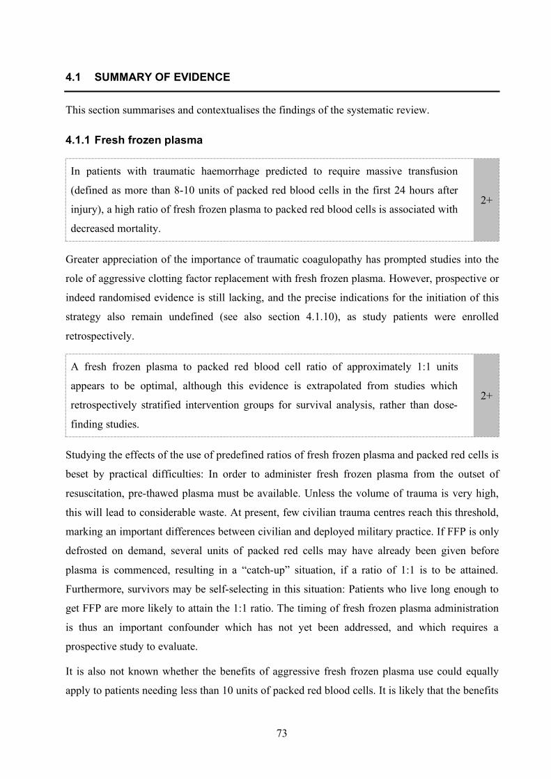
73
4.1 SUMMARY OF EVIDENCE
This section summarises and contextualises the findings of the systematic review.
4.1.1 Fresh frozen plasma
In patients with traumatic haemorrhage predicted to require massive transfusion
(defined as more than 8-10 units of packed red blood cells in the first 24 hours after
injury), a high ratio of fresh frozen plasma to packed red blood cells is associated with
decreased mortality.
2+
Greater appreciation of the importance of traumatic coagulopathy has prompted studies into the
role of aggressive clotting factor replacement with fresh frozen plasma. However, prospective or
indeed randomised evidence is still lacking, and the precise indications for the initiation of this
strategy also remain undefined (see also section 4.1.10), as study patients were enrolled
retrospectively.
A fresh frozen plasma to packed red blood cell ratio of approximately 1:1 units
appears to be optimal, although this evidence is extrapolated from studies which
retrospectively stratified intervention groups for survival analysis, rather than dose-
finding studies.
2+
Studying the effects of the use of predefined ratios of fresh frozen plasma and packed red cells is
beset by practical difficulties: In order to administer fresh frozen plasma from the outset of
resuscitation, pre-thawed plasma must be available. Unless the volume of trauma is very high,
this will lead to considerable waste. At present, few civilian trauma centres reach this threshold,
marking an important differences between civilian and deployed military practice. If FFP is only
defrosted on demand, several units of packed red cells may have already been given before
plasma is commenced, resulting in a “catch-up” situation, if a ratio of 1:1 is to be attained.
Furthermore, survivors may be self-selecting in this situation: Patients who live long enough to
get FFP are more likely to attain the 1:1 ratio. The timing of fresh frozen plasma administration
is thus an important confounder which has not yet been addressed, and which requires a
prospective study to evaluate.
It is also not known whether the benefits of aggressive fresh frozen plasma use could equally
apply to patients needing less than 10 units of packed red blood cells. It is likely that the benefits

74
of such a strategy, if they are indeed real, are less pronounced in patients requiring smaller
volume transfusions. None of the studies conducted so far have adequately delineated the side-
effect profile of aggressive fresh frozen plasma use, and there is ongoing concern regarding the
side effects of this strategy, and in particular whether it might result in a higher incidence of
ARDS.
The diagnosis of traumatic coagulopathy is clinical. Clotting assays such as prothrombin time
and partial thromboplastin time take time to process, and lack validity in the trauma setting. In
the exsanguinating trauma patient, haemostatic resuscitation should be initiated without awaiting
the results of such assays. There is, however, also a theoretical concern regarding the
administration of large amounts of plasma to patients who are not coagulopathic, or who may
even be hypercoagulable, in which cases thrombotic complications may ensue. This has not been
shown to be the case until now, but has also not been adequately investigated. The more
widespread use of thromboelastography, or rotational thromboelastometry – particularly as a
point-of-care test – might allow more rapid and more precise identification of coagulation
defects.
In summary, although there is reasonable evidence that a strategy of transfusing fresh frozen
plasma and packed cells in a 1:1 ratio is associated with improved outcome in coagulopathic
trauma patients, better quality evidence is needed to confirm the benefits and define the place of
this strategy.
4.1.2 Platelets
In patients with traumatic haemorrhage predicted to require massive transfusion
(defined as more than 8-10 units of packed red blood cells in the first 24 hours after
injury), a high ratio of units of platelets to packed red blood cells is associated with
decreased mortality. A ratio of at least 1 (pooled) unit of platelets to 5 units of packed
red blood cells appears to be optimal, although this evidence is extrapolated from
studies which retrospectively stratified intervention groups for survival analysis.
2-
The evidence base for the liberal use of platelets is even more limited than for fresh frozen
plasma. Furthermore, as with fresh frozen plasma, the precise indications for the initiation of this
strategy remain undefined (see also section 4.1.10), and it is not known whether the benefits of
aggressive platelet use could also apply to patients needing less than 10 units of packed red
blood cells. The same concerns regarding the side effects of this transfusion strategy also apply.
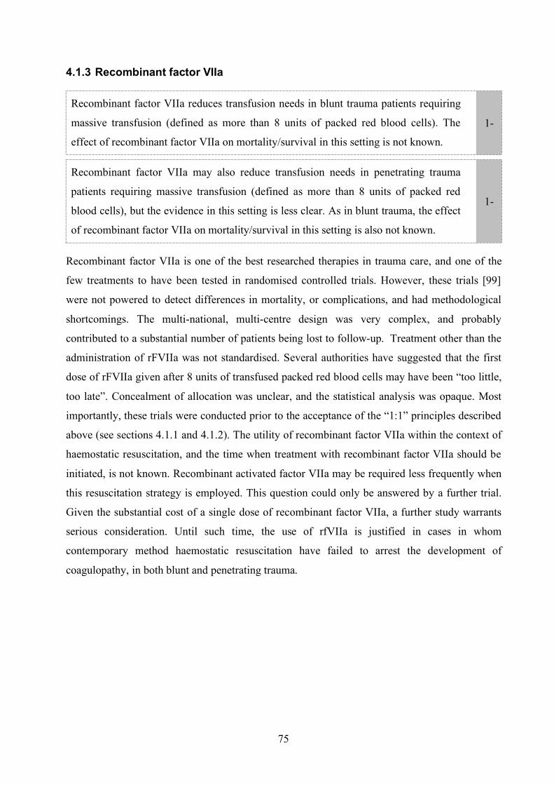
75
4.1.3 Recombinant factor VIIa
Recombinant factor VIIa reduces transfusion needs in blunt trauma patients requiring
massive transfusion (defined as more than 8 units of packed red blood cells). The
effect of recombinant factor VIIa on mortality/survival in this setting is not known.
1-
Recombinant factor VIIa may also reduce transfusion needs in penetrating trauma
patients requiring massive transfusion (defined as more than 8 units of packed red
blood cells), but the evidence in this setting is less clear. As in blunt trauma, the effect
of recombinant factor VIIa on mortality/survival in this setting is also not known.
1-
Recombinant factor VIIa is one of the best researched therapies in trauma care, and one of the
few treatments to have been tested in randomised controlled trials. However, these trials [99]
were not powered to detect differences in mortality, or complications, and had methodological
shortcomings. The multi-national, multi-centre design was very complex, and probably
contributed to a substantial number of patients being lost to follow-up. Treatment other than the
administration of rFVIIa was not standardised. Several authorities have suggested that the first
dose of rFVIIa given after 8 units of transfused packed red blood cells may have been “too little,
too late”. Concealment of allocation was unclear, and the statistical analysis was opaque. Most
importantly, these trials were conducted prior to the acceptance of the “1:1” principles described
above (see sections 4.1.1 and 4.1.2). The utility of recombinant factor VIIa within the context of
haemostatic resuscitation, and the time when treatment with recombinant factor VIIa should be
initiated, is not known. Recombinant activated factor VIIa may be required less frequently when
this resuscitation strategy is employed. This question could only be answered by a further trial.
Given the substantial cost of a single dose of recombinant factor VIIa, a further study warrants
serious consideration. Until such time, the use of rfVIIa is justified in cases in whom
contemporary method haemostatic resuscitation have failed to arrest the development of
coagulopathy, in both blunt and penetrating trauma.
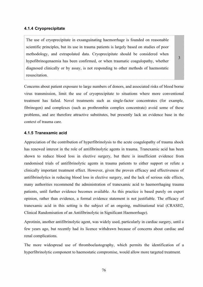
76
4.1.4 Cryoprecipitate
The use of cryoprecipitate in exsanguinating haemorrhage is founded on reasonable
scientific principles, but its use in trauma patients is largely based on studies of poor
methodology, and extrapolated data. Cryoprecipitate should be considered when
hypofibrinogenaemia has been confirmed, or when traumatic coagulopathy, whether
diagnosed clinically or by assay, is not responding to other methods of haemostatic
resuscitation.
3
Concerns about patient exposure to large numbers of donors, and associated risks of blood borne
virus transmission, limit the use of cryoprecipitate to situations where more conventional
treatment has failed. Novel treatments such as single-factor concentrates (for example,
fibrinogen) and complexes (such as prothrombin complex concentrate) avoid some of these
problems, and are therefore attractive substitutes, but presently lack an evidence base in the
context of trauma care.
4.1.5 Tranexamic acid
Appreciation of the contribution of hyperfibrinolysis to the acute coagulopathy of trauma shock
has renewed interest in the role of antifibrinolytic agents in trauma. Tranexamic acid has been
shown to reduce blood loss in elective surgery, but there is insufficient evidence from
randomised trials of antifibrinolytic agents in trauma patients to either support or refute a
clinically important treatment effect. However, given the proven efficacy and effectiveness of
antifibrinolytics in reducing blood loss in elective surgery, and the lack of serious side effects,
many authorities recommend the administration of tranexamic acid to haemorrhaging trauma
patients, until further evidence becomes available. As this practice is based purely on expert
opinion, rather than evidence, a formal evidence statement is not justifiable. The efficacy of
tranexamic acid in this setting is the subject of an ongoing, multinational trial (CRASH2,
Clinical Randomisation of an Antifibrinolytic in Significant Haemorrhage).
Aprotinin, another antifibrinolytic agent, was widely used, particularly in cardiac surgery, until a
few years ago, but recently had its licence withdrawn because of concerns about cardiac and
renal complications.
The more widespread use of thromboelastography, which permits the identification of a
hyperfibrinolytic component to haemostatic compromise, would allow more targeted treatment.

77
4.1.6 Permissive hypotension
Permissive hypotension is not a new concept, but has gained a new “lease of life” as part of
damage control resuscitation, particularly in the military setting. The three trials of permissive
hypotension conducted over the last 15 years all have significant methodological limitations.
Bickell et al’s study was the only one to show a difference in mortality, but was not randomised,
and conducted in very specific circumstances: The geographical area from which patients were
recruited was small, all had penetrating torso trauma, and all were young.[121] Turner et al’s
cluster-randomised trial suffered from the inclusion of large numbers of patients with minor
injuries and numerous protocol violations.[122] Dutton et al’s study failed to achieve the
proposed methodology and was underpowered.[123]
There is no new evidence to suggest that pre-hospital intravenous fluid resuscitation
prior to control of bleeding is beneficial. There is, however, limited evidence to
suggest that fluid resuscitation may be harmful, and that patients with penetrating
injuries in particular may have better outcomes when fluids are withheld until surgical
control of haemorrhage has been obtained.
1-
Few would argue against replacing lost intravascular volume in patients with controlled or self-
limiting haemorrhage. In contrast, in patients with uncontrolled haemorrhage, particularly in the
context of penetrating torso trauma, a strategy of permissive hypotension, together with rapid
control of haemorrhage, may be more appropriate. Distinguishing between these two groups can
be difficult, and take time.
It is probable that there is a critical period during which suboptimal end-organ perfusion is
tolerated, but beyond which irreversible anatomical and physiological changes manifest. The
duration of this period remains to be defined.
It is conceivable that permissive hypotension is more applicable to the management of
penetrating injuries than blunt trauma. The management of polytrauma patients with head
injuries requires special mention. The importance of maintaining cerebral perfusion pressure is
well recognised, and permissive hypotension may be contraindicated in this setting. Conversely,
in an exsanguinating patient with a coexisting head injury, control of haemorrhage is arguably
still the higher priority, and may be the best way of restoring cerebral perfusion.
Despite a lack of evidence, clinical practice has evolved to a much more judicious administration
of intravenous fluids. In recognition of the unique challenges posed by combat casualty

78
resuscitation, permissive hypotension has been firmly incorporated into military medical
doctrine, and is now also being endorsed by several civilian organisations. [128][172]
[173][174][175] The forthcoming eighth edition of the Advanced Trauma Life Support
programme also departs from previous guidance by emphasising the need to balance the risks of
precipitating further bleeding against the adequacy of organ perfusion, and accepting a lower
than normal blood pressure until surgical control of haemorrhage has been obtained.
There is an urgent need for more studies of fluid resuscitation in clearly defined populations,
including those with head injuries, and studies to define the time period during which suboptimal
end-organ perfusion is tolerated and reversible.
4.1.7 Tris-hydroxymethyl aminomethane (THAM)
THAM is a buffering agent which has been used in the treatment of respiratory failure, head
injury management, diabetic ketoacidosis and poisoning. Unlike bicarbonate, which relies on the
respiratory system for the elimination of carbon dioxide, protonated THAM is excreted renally.
Acidaemia in trauma is almost invariably the consequence of hypoperfusion, and should ideally
be corrected by restoring oxygen delivery, but offsetting its effects on coagulation and
myocardial contractility by pharmacological modulation is attractive. Several authors have
alluded to the potential usefulness of THAM in trauma patients, but there is no direct evidence to
support its use in trauma patients. Studies are needed to confirm efficacy and define indications
for treatment with THAM in trauma patients.
4.1.8 Hypothermia
Hypothermia in trauma patients is associated with increased mortality. Reversal of
hypothermia is associated with improved survival. Hypothermia in trauma patients
should therefore be prevented whenever possible, and aggressively treated when
present.
2+
There is very little evidence to support the use of any one device, or combination of devices,
over another. Although there are numerous comparisons, very few were conducted in the trauma
setting. The mechanisms underlying the development of hypothermia may differ between trauma
and non-trauma patients, and because surgical intervention in trauma patients often requires wide
access. One study has shown that prewarmed blankets are as efficacious, but less cumbersome,
than forced air devices in preventing hypothermia in trauma patients admitted with
normothermia. This study was confined to patients with minor or moderate trauma, and its

79
findings are thus not applicable to other populations, or the treatment of hypothermia. Forced air
devices are effective but impractical in the trauma setting, because they limit access. They are,
however, useful in the pre-operative and, particularly, post-operative phases of care. Although
there is no direct evidence to support their use, electrical warming mattresses are the most
practical devices. These devices are effective and do not compromise access.
There is no evidence to support the use of extracorporeal warming techniques, such as
cardiopulmonary or passive arteriovenous bypass, as sometimes used for the treatment
of severe hypothermia due to prolonged exposure or drowning, for the treatment of
trauma-associated hypothermia. These systems are cumbersome to set up and use, are
associated with risks such as thrombosis and haemorrhage due to decannulation, and
may be contraindicated if anticoagulation is required. The use of cavity rewarming
techniques such as peritoneal or pleural lavage is also impractical, and there is no
evidence of its utility in trauma.
2-
4.1.9 Damage control surgery
Damage control surgery is regarded as one of the key advances in trauma management of the last
20 years. It has become both a cornerstone and hallmark of trauma surgery, and it is thus
surprising how little evidence there is to support the concept. This is probably the consequence
of both less exacting proofs of efficacy required when the technique was introduced, and the
usual difficulties encountered when trying to conduct studies on severely injured patients.
Lack of evidence for a treatment does not equate to ineffectiveness. There are many treatments,
particularly surgical, which have never been subjected to formal evaluation. Indeed, attempting
to prove the efficacy of damage control surgery (and resuscitation) by means of comparative
studies might expose patients to unacceptable risks, and be considered unethical. The scientific
rationale for damage control surgery is compelling, and there is a large amount of indirect and
circumstantial evidence to support its use. Few trauma surgeons would argue against employing
damage control surgical techniques in the correct setting.

80
Patients with severe intra-abdominal injuries – in particular those with profound
haemorrhagic shock, an existing or developing coagulopathy, a need for time-
consuming procedures, or concomitant major injury outside the abdomen – benefit
from the use of damage control surgical techniques, which may include curtailment of
the procedure by ligation of hollow viscus injuries or resection without anastomosis,
ligation or shunting of vascular injuries, packing, and temporary abdominal closure.
2-
Damage control surgery carries with it significant risks in terms of morbidity – such as sepsis
related to abdominal packing, inability to secondarily close the abdomen or reconstruct the
abdominal wall, enterocutaneous fistulation, to name a few – and even mortality. It is likely that
its benefits are most evident in patients with severe injuries. In this setting, the perceived benefits
of the strategy may well be amplified by the use of other damage control resuscitation strategies,
such as haemostatic resuscitation and temperature maintenance. Damage control surgery should
therefore not be employed outwith the context of damage control resuscitation.
4.1.10 Indications for initiating damage control resuscitation
The indications for damage control resuscitation remain poorly defined. This is problematic both
in terms of appropriate selection of patients, and the reporting and comparison of results of
treatment. There is a substantial body of literature on transfusion practice, and massive
transfusion and trauma exsanguination protocols, but studies are either not evidence-based,
predate recent clinical developments in coagulopathy management, are restricted to
haematological aspects of management, or not specifically aimed at trauma patients. Some of the
recent studies of predefined plasma to packed cell ratios used the inclusion criterion “massive
transfusion” (defined as more than 8 or 10 units of packed cells transfused within the first 24
hours after injury), but this was applied retrospectively, and is obviously of no use to the
clinician faced with a trauma patient who has just arrived in the emergency department. Equally,
there are many articles regarding parameters or combinations of parameters – such as
temperature, base excess, or prothrombin time – which are said to indicate the need for damage
control rather than definitive primary surgery, but these were again never formally tested, and
also predate the introduction of haemostatic resuscitation.
In summary, the indications for damage control resuscitation remain imprecise. Several recent
publications, many written by experts in the field, gloss over this important aspect, referring to
“patients with exsanguinating haemorrhage” or “patients at risk of coagulopathy”.[3][7] This is
unsatisfactory both in terms of resource utilisation and potential morbidity: Many less severely
81
injured trauma patients are hyper- rather than hypocoagulable, and aggressive administration of
fresh frozen plasma could conceivably be harmful in this setting. However, until more objective
parameters for diagnosis and selection are developed, pattern recognition by an experienced
physician remains the only “test” available.
4.2 VALIDITY, LIMITATIONS AND APPLICABILITY
The objectives of this dissertation (section 1.2) was “to conduct a systematic review of the
evidence for damage control resuscitation”. Has this objectives been achieved?
4.2.1 Validity
Section 1.4 defined a systematic review as an efficient scientific technique to identify and
summarise evidence on the effectiveness of interventions, and identified its characteristics as an
explicit search strategy, selection of literature according to defined inclusion and exclusion
criteria, and evaluation against consistent standards.[20][22]
The search strategies employed in this dissertation are explicit, and in all likelihood sufficiently
inclusive to identify the vast majority of publications relating to each of the key questions,
although this latter fact is impossible to prove. Articles were furthermore selected according to
defined inclusion and exclusion criteria. The trauma literature is unfortunately replete with
methodologically inferior studies, and the selection process was specifically designed to
eliminate such work. In particular, primary studies such as case reports and case series without
control groups were excluded. Non-systematic reviews and quasi-editorial guidelines,
representing a very substantial proportion of the literature, were treated as “expert opinion” and
also disregarded. This highly selective approach is not without problems, as there are some
treatments which have a limited or even no evidence base. In contrast, some of the more recent
innovations, in particular haemostatic resuscitation, are characterised by a much better scientific
foundation.
The articles selected for incorporation into the systematic review were furthermore evaluated
against consistent and accepted standards. This work therefore meets all the criteria of a
systematic review.
4.2.2 Limitations
The principal limitation of this work in terms of its validity is that it was produced by a single
author. Both selection bias, during the identification of potentially relevant publication, and

82
appraisal bias, during the analysis of selected evidence, is greatly reduced by multiple assessors
reaching a consensus. Although such a process increases validity, it is not appropriate – or
indeed practical – for a doctoral thesis, and was therefore omitted.
4.2.3 Applicability
Despite the limitations listed in section 4.2.2, this dissertation has succeeded in systematically
and comprehensively reviewing the available evidence for the components of the damage control
resuscitation strategy. Although some of the included studies were conducted in the military
setting, the conclusions are broadly applicable to civilian trauma management in the context of a
European or North American trauma centre.
4.3 CONCLUSION
The management of major trauma has undergone dramatic changes, which have been drawn
together into the damage control resuscitation concept. Haemostatic resuscitation, hypothermia
management, hypotensive resuscitation, and damage control surgery have been amalgamated
into a coherent management strategy, which has revolutionised the care of the injured.
Those with more than just a passing interest in the history of trauma will recognise that many of
the component strategies of damage control resuscitation are not new, but merely developments
of existing concepts. Irrespective of origin or originality, contemporary medical practice
demands that new therapies are supported by scientific evidence. Trauma care has long suffered
from a lack of such evidence. This is a direct result of the difficulty of conducting interventional
studies on patients with life-threatening injuries: Except in the very largest centres, the numbers
of patients admitted with major trauma are relatively small, and trauma is a heterogeneous
disease: Injury patterns and severity vary, and are difficult to control for. Resuscitation, surgical
treatment, and postoperative care are difficult to standardise. The selection of outcome measures
is also problematic: Mortality is easily assessed, and always clinically significant, but may not be
appropriate, or require very large studies to reach statistical significance. Lastly, patient consent,
an essential safeguard of ethical research practice, is often difficult or even impossible to obtain
from patients requiring emergency treatment.
Although these difficulties are real, they should not detract from the need to demonstrate that
novel treatments are effective. Proving the effectiveness of a strategy such as damage control
resuscitation is difficult, and should ideally take place at several levels, confirming both the
effectiveness of the constituent components, and the strategy as a whole.

83
This dissertation has distilled the evidence, and in some cases, lack of evidence, for the
components of damage control resuscitation. It cannot, and never set out to, prove the
effectiveness of damage control resuscitation as a whole. Indeed, the complex issues –
methodological and ethical – that a study attempting to demonstrate its efficacy would throw up
make it highly unlikely that such an investigation will ever take place.
Fortunately, there is indirect evidence that damage control resuscitation is effective. Given its
origins, it is fitting that this evidence should come from military practice, which has been
meticulously evaluated for many years. The lethality of war wounds sustained by US servicemen
decreased from 30% in the second World War (1939-1945), to 25% during the Korean War
(1950-1953), but then remained at 24% during the Vietnam War (1961-1973), and even the first
Gulf War (1990-1991), some thirty years later. Since 2001, however, despite increases in the
wounding potential of modern weapon systems, less than 10% of US servicemen wounded in
Iraq and Afghanistan have died.[1][2][10] Although the precise role of medical care, and damage
control resuscitation in particular, is impossible to ascertain, it has almost certainly contributed
to this remarkable reduction in combat-related mortality.[1] The successful development and
implementation of the damage control resuscitation concept by the military – in an operational
setting – is a remarkable achievement. The lessons learnt are now redefining the care of the most
severely injured patients in civilian practice.

5.
Abstract
(English)

84
Background: Damage control resuscitation is a novel strategy for the management of the
exsanguinating trauma patient. It is a development of the damage control surgery concept, with
emphasis on the integration of resuscitation and surgery, and the aggressive management of
traumatic coagulopathy. Although pioneered by military surgeons, many aspects of damage
control resuscitation are equally applicable to the management of civilian trauma, but awareness
outside specialist circles is limited. This is at least in part related to the lack of a precise
definition, and the rapid and continuing evolution of the strategy.
Objectives: 1. To define the components of damage control resuscitation. 2. To conduct a
systematic review of the evidence for damage control resuscitation.
Methods: Systematic review of literature.
Results: Damage control resuscitation encompasses haemostatic resuscitation, hypotensive
resuscitation, hypothermia and acidaemia management and damage control surgery. Systematic
review produced the following conclusions: In patients with traumatic haemorrhage predicted to
require massive transfusion, a high ratio of fresh frozen plasma to packed red blood cells is
associated with decreased mortality. (Evidence level: 2+) A ratio of approximately 1:1 units
appears to be optimal, although this evidence is extrapolated from studies which retrospectively
stratified intervention groups for survival analysis, rather than dose-finding studies. (2+) In
patients with traumatic haemorrhage predicted to require massive transfusion, a high ratio of
units of platelets to packed red blood cells is also associated with decreased mortality. A ratio of
at least 1 (pooled) unit of platelets to 5 units of packed red blood cells appears to be optimal,
although this evidence is again extrapolated. (2-) Recombinant factor VIIa reduces transfusion
needs in blunt trauma patients requiring massive transfusion. The effect of recombinant factor
VIIa on mortality in this setting is not known. (1-) Recombinant factor VIIa may also reduce
transfusion needs in penetrating trauma patients requiring massive transfusion, but the evidence
is less clear. As in blunt trauma, the effect on mortality in this setting is also not known. (1-)
The use of cryoprecipitate in exsanguinating haemorrhage is founded on reasonable scientific
principles, but its use in trauma patients is largely based on studies of poor methodology, and
extrapolated data. Cryoprecipitate should be considered when hypofibrinogenaemia has been
confirmed, or when traumatic coagulopathy is not responding to other methods of haemostatic
resuscitation. (3) Although conceptually attractive, associated with few complications, and
frequently recommended, there is no evidence for the use of antifibrinolytics in the trauma
setting. There is no new evidence to suggest that pre-operative intravenous fluid resuscitation
prior to control of bleeding is beneficial. There is, however, limited evidence to suggest that fluid

85
resuscitation may be harmful, and that patients with penetrating injuries in particular may have
better outcomes when fluids are withheld until surgical control of haemorrhage has been
obtained. (1-) There is no evidence to support the use of tris-hydroxymethyl aminomethane
(THAM) in trauma patients, although in theory appealing. Hypothermia in trauma patients is
associated with increased mortality, and should be prevented whenever possible, and
aggressively treated when present. (2+) There is no evidence to support the use of extracorporeal
warming techniques. (2-) Patients with severe intra-abdominal injuries – in particular those with
profound haemorrhagic shock, an existing or developing coagulopathy, a need for time-
consuming procedures, or concomitant major injury outside the abdomen – benefit from the use
of damage control surgical techniques, which may include curtailment of the procedure by
ligation of hollow viscus injuries or resection without anastomosis, ligation or shunting of
vascular injuries, packing, and temporary abdominal closure. (2-) The indications for damage
control resuscitation remain poorly defined. This applies both in general, and for specific
components, such as when resuscitation with predefined blood product ratios should be initiated,
or at what stage recombinant factor VIIa should be utilised. Existing criteria are largely based on
expert opinion.
Discussion: Damage control resuscitation is a composite, multidisciplinary strategy which
integrates haemostatic resuscitation, hypotensive resuscitation, hypothermia management, and
damage control surgery. Hypotensive resuscitation and damage control surgery are not new
concepts, but have been incorporated into an effective management paradigm. Greater
appreciation of the importance of traumatic coagulopathy, and its manipulation through
haemostatic resuscitation, has led to an explosion of interest in clotting factor and platelet
replacement. The resulting resuscitation strategies with predefined ratios of blood products, and
avoidance of synthetic fluids, together with the modulation of fibrinolysis, are an exciting
development. Although much work remains to be done, these novel treatments, in combination
with more established techniques, are revolutionising major trauma resuscitation. This
systematic review should assist clinicians in applying damage control resuscitation principles,
and maximising their patients’ survival.

6.
Zusammenfassung
(Deutsch)
86
Hintergrund: Damage Control Resuscitation (DCR) ist eine innovative Strategie zur
Behandlung von Traumapatienten mit hohem Blutverlust. Sie ist eine Weiterentwicklung des
chirurgischen Damage Control-Konzepts mit dem Schwerpunkt einer Kombination aus
Reanimations- und chirurgischer Therapie sowie der konsequenten Behandlung der
traumatischen Koagulopathie. Die DCR wurde ursprünglich von Militärchirurgen eingeführt,
große Teile sind jedoch auch in der Therapie ziviler Verletzter anwendbar. Dass die Kenntnis
hiervon bisher auf Spezialisten begrenzt war, liegt zum einem am Fehlen einer präzisen
Definition, zum andern an der rapiden Weiterentwicklung der Behandlungsstrategien.
Ziele: Es gilt erstens, die einzelnen Komponenten der Damage Control Resuscitation zu
definieren, zweitens, eine systematische Sichtung der Evidenzbasierung der DCR
durchzuführen.
Methode: Systematische Literaturrecherche unter Anwendung von gängigen
Evaluierungsmethoden.
Ergebnisse: Damage Control Resuscitation ist die (lebensrettende) Behandlung von
Gerinnungsstörung, Hypothermie, Acidose und die kontrollierte Toleranz einer verminderten
Perfusion, in Verbindung mit Damage Control-Chirurgie. Die systematische Analyse ergab
folgende Resultate: die Mortalität von Patienten mit traumatischen Blutungen, die massive
Transfusionen benötigen, wird durch ein 1:1 Verhältnis von Gefrorenem Frischplasma zu
Erythrocytenkonzentraten deutlich gesenkt. (Evidenzgrad 2+). Diese Evidenz ist allerdings aus
retrospektiven Studien extrapoliert, die sich weniger mit der Dosierung als vielmehr mit der
Festlegung von Interventionsgruppen zur Beurteilung der Überlebensrate befassten.(2+) Auch
ein hoher Anteil von Thrombocyten – im Verhältnis zu Erythrocyten – Konzentraten lässt eine
verminderte Mortalität bei polytransfundierten Patienten erwarten. Eine Relation von wenigstens
einem Pool- Thrombocyten-Konzentrat zu fünf Erythrocyten-Konzentraten scheint optimal,
wobei auch diese Schlussfolgerung extrapoliert ist. (2-) Recombinanter Faktor VIIa vermindert
die Anzahl der erforderlichen Transfusionen bei stumpfem Trauma, jedoch ist ein Effekt von
Faktor VIIa auf die Mortalität nicht bekannt. (1-) Faktor VIIa reduziert möglicherweise auch die
notwendige Transfusionsmenge bei Patienten mit penetrierenden Verletzungen. Dieser
Zusammenhang ist indes nicht klar erwiesen, auch der Effekt auf die Mortalität ist – wie beim
stumpfen Trauma – nicht bekannt. (1-)
Der Gebrauch von Kryopraezipitaten bei Patienten mit nicht-traumatischen lebensbedrohlichen
Blutungen gründet sich auf fundierte wissenschaftliche Prinzipien; bei Traumapatienten basiert
er aber auf Studien, deren Methode und Daten fragwürdig sind. Der Gebrauch von

87
Kryopraezipitaten sollte erwogen werden, wenn eine Hypofibrinogenaemie nachgewiesen ist
oder wenn die traumatische Koagulopathie auf andere Therapieversuche nicht reagiert. (3)
Obwohl häufig empfohlen, vom Ansatz her attraktiv und mit geringen Komplikationen belastet,
ist der Nutzen einer Antifibrinolytikabehandlung in der Traumaversorgung nicht evident. Auch
präoperative Flüssigkeitsinfusionen – ehe die Blutung beherrscht werden kann – erscheinen
fragwürdig. Es gibt Untersuchungen darüber, dass Flüssigkeitszufuhr schädlich sein kann und
dass bei Patienten mit penetrierenden Verletzungen zunächst eine chirurgische Kontrolle der
Blutung erreicht werden sollte. (1-) Keine Evidenzbasis findet sich für die Anwendung von
Trishydroxymethylaminomethan (THAM) bei Traumapatienten, obwohl der theoretische Ansatz
für eine solche reizvoll ist. Hypothermie ist bei Traumapatienten mit erhöhter Mortalität
verbunden, muss vermieden oder – wenn eingetreten – aktiv behandelt werden. (2+) Für die
Anwendung extrakorporaler Aufwärmtechniken gibt es keine Rechtfertigung. (2-) Patienten mit
schweren intraabdominalen Verletzungen, insbesondere solche mit haemorrhagischem Schock,
einer vorhandenen oder sich entwickelnden Koagulopathie, folgenden langwierigen Operationen
oder mit zusätzlichen grösseren Verletzungen außerhalb des Abdomens profitieren von der
Anwendung chirurgischer Damage Control-Techniken. Diese können in einer zeitlichen
Begrenzung der Operation bestehen, zum Beispiel durch Verschluss von Hohlorganen oder
Resektion ohne Anastomose, in Ligatur oder Überbrückung (shunting) von Gefässverletzungen,
in „packing“ und provisorischem Abdominalverschluss. (2-) Die Indikationen für Damage
Control Resuscitation sind weiterhin unzureichend definiert. Dies gilt sowohl in allgemeiner
Hinsicht als auch im Hinblick auf spezifische Fragestellungen: wann etwa soll eine
lebensrettende Behandlung mit festgelegten Mengen von Blutersatzprodukten begonnen werden
oder in welchem Stadium Faktor VIIa zum Einsatz kommen. Vorhandene Kriterien basieren fast
ausschließlich auf persönlichen Erfahrungen.
Diskussion: Damage Control Resuscitation ist eine komplexe, multidisziplinäre Strategie, die
lebensrettende Haemostase-, Hypothermiebehandlung, Hypotensions-Toleranz sowie Damage
Control Chirurgie beinhaltet. Toleranz einer verminderten Perfusion und DC-Chirurgie sind
keine Neuerungen an sich, sie werden jedoch in ein effektives Therapieschema eingebettet.
Zunehmende Berücksichtigung der traumatischen Koagulopathie und ihre Behandlung haben zu
einer Ausweitung des Interesses an Gerinnungsfaktoren und Thrombocytenersatz geführt. Die
resultierenden lebenserhaltenden Maßnahmen mit vorgegebenen Mengen von
Blutersatzprodukten, bei Vermeidung synthetischer Flüssigkeiten, sind in Verbindung mit der
vorsichtigen Beeinflussung der Fibrinolyse eine interessante Entwicklung. Obwohl die

88
Forschungsbasis noch erweitert werden muss, bereichern diese neuen Behandlungsmethoden in
Verbindung mit bewährten Techniken die Lebensrettungsmaßnahmen bei Schwerverletzten
schon heute. Der vorliegende systematische Überblick sollte es Klinikern ermöglichen, DCR-
Prinzipien anzuwenden und die Überlebenschancen ihrer Patienten zu vergrößern.

7.
References

89
1. Schreiber MA, Tieu B. Hemostasis in Operation Iraqi Freedom III. Surgery 2007;142:S61-
S66
2. Gawande A. Casualties of war – military care for the wounded from Iraq and Afghanistan.
NEJM 2004;351:2471-2475
3. Holcomb JB, Jenkins D, Rhee P, et al. Damage control resuscitation: directly addressing
the early coagulopathy of trauma. J Trauma 2007a;62:307-310
4. Sauaia A, Moore FA, Moore EE, et al. Epidemiology of trauma deaths: a reassessment. J
Trauma 1995;38(2):185-193
5. American College of Surgeons. Advanced Trauma Life Support for Doctors. Student
Course Manual. 7th Edition. Chicago: 2004
6. Holcomb JB, Wade CE, Michalek JE, et al. Increased plasma and platelet to red blood cell
ratios improves outcome in 466 massively transfused civilian trauma patients. Ann Surg
2008;248(3):447-458
7. Holcomb JB. Damage control resuscitation. J Trauma. 2007b;62:S36 –S37
8. Fox CJ, Gillespie DL, Cox ED, et al. Damage control resuscitation for vascular surgery in a
combat support hospital. J Trauma 2008a;65:1-9
9. Fox CJ, Gillespie DL, Cox ED, et al. The effectiveness of a damage control resuscitation
strategy for vascular injury in a combat support hospital: results of a case control study. J
Trauma 2008b;64:S99-S107
10. http://www.defenselink.mil/news/casualty.pdf (accessed at 11.03.2009)
11. Rotondo M, Schwab CW, McGonigal M, et al. “Damage control”: an approach for
improved survival in exsanguinating penetrating abdominal injury. J Trauma 1993;35:375
12. Loveland J, Boffard KD. Damage control in the abdomen and beyond. Br J Surg
2004;91:1095:1101
13. Brohi K, Cohen MJ, Gunter MT, et al. Acute coagulopathy of trauma: hypoperfusion
induces anticoagulation and hyperfibrinolysis. J Trauma 2008;64:1211-1217
14. Hess JR, Brohi K, Dutton RP, et al. The coagulopathy of trauma: a review of mechanisms.
J Trauma 2008a;65(4):748-754
15. Beekley AC. Damage control resuscitation: a sensible approach to the exsanguinating
surgical patient. Crit Care Med 2008;36(7)(Supp):S267-S274
16. Hodgetts TJ, Mahoney PF, Kirkman E. Damage Control Resuscitation. JRAMC 2007a;
153(4): 299-300
90
17. Tieu BH, Holcomb JB, Schreiber MA. Coagulopathy: its pathophysiology and treatment in
the injured patient. World J Surg 2007;31:1055-1064
18. Gaarder C, Naess PA, Frischknecht Christensen E, et al. Scandinavian guidelines – “The
massively bleeding patient”. Scandinavian Journal of Surgery 2008;97:15-36
19. Spahn DR, Cerny V, Coats TJ, et al. Management of bleeding following major trauma: a
European guideline. Crit Care 2007;11:R17
20. Woolf S. Practice guidelines, a new reality in medicine. II: methods of developing
guidelines. Arch Intern Med 1992;152:946-52
21. Mulrow C. Rationale for systematic reviews. BMJ 1994;309:597-9
22. Scottish Intercollegiate Guidelines Network. SIGN guidelines development manual.
Edinburgh: SIGN, 2007
23. Liddle J, Williamson M, Irwog L. Method for evaluating research and guideline evidence.
Sydney: New South Wales Department of Health; 1996
24. Field M, Lohr K. Institute of Medicine Committee to Advice the Public Health Service on
Clinical Practice Guidelines. Clinical practice guidelines: directions for a new program.
Washington (DC): National Academy Press; 1990
25. Hayward RSA, Wilson MC, Tunis SR, Bass EB, Guyatt G, for the Evidence-Based
Medicine Working Group. Users’ guides to the Medical Literature. VIII. How to Use
Clinical Practice Guidelines. A. Are the Recommendations Valid? JAMA, 1995;274, 570-
574
26. Higgins JPT, Green S (editors). Cochrane Handbook for Systematic Reviews of
Interventions Version 5.0.1 [updated September 2008]. The Cochrane Collaboration, 2008.
27. The AGREE Collaboration. The Appraisal of Guidelines for Research & Evaluation
(AGREE) instrument. Available from http://wwwagreecollaboration.org (accessed 4
December 2008).
28. Mapstone J, Roberts I, Evans P. Fluid resuscitation strategies: a systematic review of
animal trials. J Trauma 2003;55(3):571-589
29. National Institute for Health and Clinical Excellence. The guidelines manual. London:
2007.
30. Scottish Intercollegiate Guidelines Network. Methodology Review Group. Report on the
review of the method of grading guideline recommendations. Edinburgh: SIGN, 1999.
31. Zalstein S, Pearce A, Scott DM, Rosenfeld JV. Damage control resuscitation: a paradigm
shift in the management of haemorrhagic shock. Emerg Med Austral 2008;20:291-293

91
32. Brohi K, Singh J, Heron M, Coats T. Acute traumatic coagulopathy. J Trauma
2003;54:1127-1130
33. MacLeod JB, Lynn M, McKenney MG, et al. Early coagulopathy predicts mortality in
trauma. J Trauma 2003;55:39-44
34. Maegele M, Lefering R, Yucel N, et al. Early coagulopathy in multiple injury: an analysis
from the German Trauma Registry on 8724 patients. Injury 2007;38:298-304
35. Mann KG. Biochemistry and physiology of blood coagulation. Thromb Haemost
1999;82:165-174
36. Roberts HR, Hoffman M, Monroe DM. A cell-based model of thrombin generation. Semin
Thromb Hemost 2006;32(Supp 1):32-38
37. Di Cera E. Thrombin interactions. Chest 2003;124:11S-17S
38. Sinisalo J, Syrjala M, Mattila KJ, Kerman T, Nieminen MS. Endothelial release of tissue-
type plasminogen activator and ischemia-induced vasodilatation are linked in patients with
coronary heart disease. Blood Coagul Fibrinolysis 1999;10:181-187
39. Osterlund B, Andersson B, Haggmark S, et al. Myocardial ischemia induces coronary t-PA
release in the pig. Acta Anaesthesiol Scand 2002;46:271-278
40. Hrafnkelsdottir T, Erlinge D, Jern S. Extracellular nucleotides ATP and UTP induce a
marked acute release of tissue-type plasminogen activator in vivo in man. Thromb Haemost
2001;85:875-881
41. Brohi K, Cohen MJ, Gunter MT, et al. Acute traumatic coagulopathy: initiated by
hypoperfusion: modulated through the protein C pathway? Ann Surg 2007;245:812-818
42. Niles S, McLaughlin DF, Perkins JG, et al. Increased mortality associated with the early
coagulopathy of trauma in combat casualties. J Trauma 2008;64:1459-1465
43. Rezaie AR. Vitronectin functions as a cofactor for rapid inhibition of activated protein C by
plasminogen activator inhibitor-1. Implications for the mechanism of profibrinolytic action
of activated protein C. J Biol Chem 2001;276:15567-15570
44. Bajzar L, Jain N, Wang P, Walker JB. Thrombin activatable fibrinolysis inhibitor: not just
an inhibitor of fibrinolysis. Crit Care Med 2004;32:S320-S324
45. Bajzar L, Nesheim ME, Tracy PB. The profibrinolytic effect of activated protein C in clots
formed from plasma is TAFI dependent. Blood 1996;88:2093-2100
46. Ho AMH, Karmakar MK, Dion PW. Are we giving enough coagulation factors during
major trauma resuscitation? Am J Surg 2005;190:479-484

92
47. Hirshberg A, Dugas M, Banez EI, Scott BG, Wall MJ, Mattox KL. Minimizing dilutional
coagulopathy in exsanguinating hemorrhage: a computer simulation. J Trauma
2003;54:454-463
48. Coates JB. Blood Program in World War II. Washington, DC: Office of the Surgeon
General, Dept. of the Army; United States Army Medical Service; 1964.
49. Brazil EV, Coats TJ. Sonoclot coagulation analysis of in vitro haemodilution with
resuscitation solutions. J R Soc Med 2000;93:507-510
50. Armand R, Hess JR. Treating coagulopathy in trauma patients. Transfus Med Rev
2003;17:223-231
51. Ketchum L, Hess JR, Hiippala S. Indications for early fresh frozen plasma, cryoprecipitate,
and platelet transfusion in trauma. J Trauma 2006;60(6):S51-S58
52. Reed RL, Bracey AW, Hudson JD, Miller TA, Fischer RP. Hypothermia and blood
coagulation: dissociation between enzyme activity and clotting factor levels. Circ Shock
1990;32:141-152
53. Pfeffermann R, Rozin RR, Durst AL, Marin G. Modern war surgery: operations in an
evacuation hospital during the October 1973 Arab-Israeli war. J Trauma. 1976;16:694 –
703.
54. Repine TB, Perkins JG, Kauvar DS, Blackborne L. The use of fresh whole blood in
massive transfusion. J Trauma. 2006; 60(Suppl):S59 –S69.
55. Stainsby D, MacLenna S, Thomas D, Isaac J, Hamilton PJ. British Committee for
Standards in Haematology. Guidelines on the management of massive blood loss. Br J
Haematol 2006;135:634-641
56. Lundberg GD. Practice Parameters for the use of fresh-frozen plasma, cryoprecipitate, and
platelets. JAMA 1994;271(10):777-781
57. American Society of Anesthesiologists. Practice Guidelines for blood component therapy: a
report by the American Society of Anesthesiologists Task Force on blood component
therapy. Anesthesiology 1996;84(3):732-747
58. American Society of Anesthesiologists. Practice Guidelines for perioperative blood
transfusion and adjuvant therapies. An updated report by the American Society of
Anesthesiologists Task Force on perioperative blood transfusion and adjuvant therapies.
Anesthesiology 2006;105:190-208
59. Borgman MA, Spinella PC, Perkins JG, et al. The ratio of blood products transfused affects
mortality in patients receiving massive transfusions at a combat support hospital. J Trauma
2007;63(4):805-813
93
60. Duchesne JC, Hunt JP, Wahl G, et al. Review of current blood transfusions strategies in a
mature level I trauma center: were we wrong for the last 60 years? J Trauma 2008;65:272-
278
61. Gunter OL, Au BK, Isbell JM, Mowery NT, Young PP, Cotton BA. Optimizing outcomes
in damage control resuscitation: identifying blood product rations associated with improved
survival. J Trauma 2008;65:527-534
62. Kashuk JL, Moore EE, Johnson JL, et al. Postinjury life threatening coagulopathy: is 1:1
fresh frozen plasma:packed red blood cells the answer? J Trauma 2008;65:261-271
63. Maegele M, Lefering R, Paffrath T, Tjardes T, Simanski C, Bouillon B. Red blood cell to
plasma ratios transfused during massive transfusion are associated with mortality in sever
multiply injury: a retrospective analysis from the trauma registry of the Deutsche
Gesellschaft fur Unfallchirurgie. Vox Sanguis 2008;95:112-119
64. Schreiber MA. Coagulopathy in the trauma patient. Curr Opin Crit Care 2005;11:590-597
65. Lier H, Krep H, Schroeder S, Stuber F. Preconditions of hemostasis in trauma: a review.
The influenc of acidosis, hypocalcaemia, anemia, and hypothermia on functional
hemostasis in trauma. J Trauma 2008;65:951-960
66. Kirkman E, Watts S, Hodgetts T, Mahoney P, Rawlinson S, Midwinter M. A proactive
approach to the coagulopathy of trauma: the rationale and guidelines for treatment. JR
Army Med Corps 2008;153(4):203-306
67. Hoyt DB, Dutton RP, Hauser CJ, et al. Management of coagulopathy in the patients with
multiple injuries: results from an international survey of clinical practice. J Trauma
2008;65:755-765
68. Kaufmann CR, Dwyer KM, Crews JD, Dols SJ, Trask AL. Usefulness of
thromboelastography is assessment of trauma patient coagulation. J Trauma
1997;42(2):716-722
69. Levrat A, Gros A, Rugeri L, et al. Evaluation of rotation thromboelastography for the
diagnosis of hyperfibrinolysis in trauma patients Br J Anaesth 2008;100(6):792-798
70. Rugeri L, Levrat A, David S, et al. Diagnosis of early coagulation abnormalities in trauma
patients by rotation thromboelastography. J Thromb Haemost 2006;5:289-295
71. Zuckerman L, Cohen E, Vagher JP, et al. Comparison of thromboelastography with
common coagulation tests. Thromb Haemost 1981;46:752-756
72. Plotkin AJ, Wade CE, Jenkins DH, et al. A reduction in clot formation rate and strength
assessed by thromboelastography is indicative of transfusion requirements in patients with
penetrating injuries. J Trauma 2008;64:S64-S68

94
73. Hess JR, Holcomb JB. Transfusion practice in military trauma. Transfusion Medicine
2008b;18:143-150
74. Sperry JL, Ochoa JB, Gunn SR, et al. An FFP:PRBC transfusion ratio >=1:1.5 is associated
with a lower risk of mortality after massive transfusion. J Trauma 2008;65(5):986-993
75. Stanworth SJ, Hyde CJ, Murphy MF. Evidence for indicatons of fresh frozen plasma.
Transfusion Clinique et Biologique 2007;14:551-556
76. Stanworth SJ, Walsh TS. Fresh frozen plasma: friend of faux pax in critical illness? Crit
Care Med 2005;33(11):2714-2716
77. MacLennan S, Williamson LM. Risks of fresh frozen plasma and platelets. J Trauma
2006;60(6)(Supp):S46-S50
78. Norda R, Tynell E, Akerblom O. Cumulative risks of early fresh frozen plasma,
cryoprecipitate and platelet transfusion in Europe. J Trauma 2006;60(6)(Supp):S41-S45
79. Scalea TM, Bochicchio KM, Lumpkins K, et al. Early aggressive use of fresh frozen
plasma does not improve outcome in critically injured trauma patients. Ann Surg 2008;
248(4): 578-584
80. Stanworth SJ, Brunskill SJ, Hyde CJ, McClelland DBL, Murphy MF. Is fresh frozen
plasma clinically effective? A systematic review of randomised controlled trials. Br J
Haematology 2004;126:139-152
81. O’Shaughnessy DF, Atterbury C, Bolton Maggs P, et al. Guidelines for the use of fresh-
frozen plasma, cryoprecipitate and cryosupernatant. Br J Haematol 2004;126:11-28
82. Mittal S, Watson HG. A critical appraisal of the use of recombinant factor VIIa in acquired
bleeding conditions. Br J Haematol 2006;133:355-363
83. Patanwala AE. Factor VIIa (recombinant) for acute traumatic haemorrhage. Am J Health-
Syst Pharm 2008;65:1616-1623
84. Scarpelini S, Rizoli S. Recombinant factor VIIa and the surgical patient. Curr Opin Crit
Care 2006;12:351-356
85. Kenet G, Walden R, Eldad A, Martinowitz U. Treatment of traumatic bleeding with
recombinant factor VIIa. Lancet. 1999; 354:1879.
86. Dutton RP, Hess JR, Scalea TM. Recombinant factor VIIa for control of hemorrhage: early
experience in critically ill trauma patients. J Clin Anesth. 2003;15:184 –188.
87. Martinowitz U, Kenet G, Segal E, et al. Recombinant activated factor VII for adjunctive
hemorrhage control in trauma. J Trauma. 2001;51:431– 438.

95
88. O’Neill PA, Bluth M, Gloster ES, et al. Successful use of recombinant activated factor VII
for trauma-associated hemorrhage in a patient without preexisting coagulopathy. J Trauma.
2002;52:400–405.
89. Vlot AJ, Ton E, Mackaay AJC, Kramer MHH, Gaillard CAJM.Treatment of a severely
bleeding patient without preexisting coagulopathy with activated recombinant factor VII.
Am J Med. 2001;108:421– 422.
90. O’Connell NM, Perry DJ, Hodgson AJ, O’Shaughnessy DF, Laffan MA, Smith OP.
Recombinant FVIIa in the management of uncontrolled haemorrhage. Transfusion 2003;
43: 1711-1716
91. Clark AD, Gordon WC, Walker ID, Tait RC. “Last-ditch” use of recombinant factor VIIa
in patients with massive hemorrhage is ineffective. Vox Sanguinis. 2004;86:120 –124.
92. Dutton RP, McCunn M, Hyder M, et al. Factor VIIa for correction of traumatic
coagulopathy. J Trauma. 2004; 57:709-18.
93. Felfernig M, for the European rFVIIa Trauma Study Group. Clinical experience with
recombinant activated factor VII in a series of 45 trauma patients. J R Army Med Corps.
2007; 153:32-9.
94. Grounds RM, Seebach C, Knothe C, et al. Use of recombinant activated factor VII
(NovoSeven) in trauma and surgery: analysis of outcomes reported to an international
registry. J Intensive Care Med. 2006; 21:27-39.
95. Harrison TD, Laskosky J, Jazaeri O et al. “Low-dose” recombinant activated factor VII
results in less blood and blood product use in traumatic hemorrhage. J Trauma. 2005;
59:150-4.
96. Martinowitz U, Michaelson M. Guidelines for the use of recombinant activated factor VII
(rFVIIa) in uncontrolled bleeding: a report by the Israeli Multidisciplinary rFVIIa Task
Force. J Thromb Haemost. 2005; 3:640-8.
97. Cameron P, Phillips L, Balogh Z, et al. The use of recombinant activated factor VII in
trauma patients: experience from the Australian and New Zealand haemostasis registry.
2007;38(9):1030-8
98. MacLaren R, Weber LA, Brake H, Gardner MA, Tanzi M. A multicenter assessment of
recombinant factor VIIa off-label usage: clinical experiences and associated outcomes.
Transfusion 2005;45:1434-1442
99. Boffard KD, Riou B, Warren B, et al. Recombinant factor VIIa as adjunctive therapy for
bleeding control in severely injured trauma patients: two parallel randomized, placebo-
controlled, double-blind clinical trials. J Trauma 2005;59:8-15

96
100. Stanworth SJ, Birchall J, Doree CJ, Hyde C. Recombinant factor VIIa for the prevention
and treatment of bleeding in patients without haemophilia. Cochrane Database of
Systematic Reviews 2007;Issue 2. Art.No.:CD005011
101. Spinella PC, Perkins JG, McLaughlin DF, et al. The effect of recombinant activated favtor
VII on mortality in combat-related casualties with severe trauma and massive transfusion. J
Trauma 2008;64:286-294
102. Rizoli SB, Boffard KD, Riou B, et al. Recombinant activated factor VII as an adjunctive
therapy for bleeding control in severe trauma patients with coagulopathy: subgroup analysis
from two randomized trials. Critical Care 2006;10:R178
103. Martinowitz U, Kenet G, Lubetski A et al. Possible role of recombinant activated factor VII
(rFVIIa) in the control of hemorrhage associated with massive trauma. Can J Anaesth 2002;
49:S15-20
104. Hodgetts TJ, Kirkman E, Mahoney PF, Russell R, Thomas R, Midwinter M. UK Defence
Medical Services guidance for the use of recombinant factor VIIa (RFVIIa) in the deployed
military setting. JR Army Med Corps 2007b;153(4):307-309
105. Hiippala ST, Myllyla GJ, Vahtera EM. Hemostatic Factors and Replacement of Major
Blood-Loss with Plasma-Poor Red-Cell Concentrates. Anesthesia and Analgesia 1995;
81(2): 360-365
106. Mcloughlin TM, Fontana TL, Alving B. Profound normovolemic hemodilution: hemostatic
effects in patients and in a porcine model. Anesthesia and Analgesia 1996;83:459-465
107. Singbartl K, Innerhofer P, Radvan J. Hemostasis and hemodilution: a quantitative
mathematical guide for clinical practice. Anesthesia andAnalgesia 2003;96:929-935
108. Stinger HK, Spinella PC, Perkins JG, et al. The ratio of fibrinogen to red cells transfused
affects survival in casualties receiving massive transfusions at an army combat support
hospital. J Trauma 2008;64:S79-S85
109. Chiu J, Ketchum LH, Reid TJ. Transfusion-sparing hemostatic agents. Curr Opin Hematol
2002;9:544-550
110. Henry DA, Moxey AJ, Carless PA, et al. Anti-fibrinolytic use for minimising perioperative
allogeneic blood transfusion. Cochrane Database Syst Rev 2001:CD001886.
111. Levi M, Cromheecke ME, de Jonge E, et al. Pharmacological strategies to decrease
excessive blood loss in cardiac surgery: a meta-analysis of clinically relevant endpoints.
Lancet 1999, 354:1940-1947.
112. Ozier Y. Pharmacological approaches to reducing blood loss and transfusions in the
surgical patient. Canadian Journal of Anesthesia 2006;53(6):S21-S29

97
113. Roberts I. The CRASH-2 trial of an antifibrinolytic agent in traumatic haemorrhage: an
international collaboration. Indian Journal of Medical Research 2007;125:1
114. Mangano DT, Miao Y, Vuylsteke, et al. Mortality associated with aprotinin during 5 years
following coronary artery bypass graft surgery. JAMA 2007;297(5):471-479
115. Coats T, Roberts IG, Shakur H. Antifibrinolytic drugs for acute traumatic injury. Cochrane
Database of Systematic Reviews 2004;4. Art No.: CD004896
116. Gaarder C, Naess PA, Frischknecht Christensen E, et al. Scandinavian guidelines – “The
massively bleeding patient”. Scandinavian Journal of Surgery 2008;97:15-36
117. http://www.CRASH2.LSHTM.ac.uk (accessed on 14.12.2008)
118. Revell M, Greaves I, Porter K. Endpoints for fluid resuscitation in hemorrhagic shock. J
Trauma 2003;54:S63-S67
119. Stern SA. Low-volume fluid resuscitation for presumed hemorrhagic shock: helpful or
harmful? Curr Opin Crit Care 2001;7:422-430
120. Kortbeek JB, Al Turki SA, Ali J, et al. Advanced Trauma Life Support, 8th Edition, The
Evidence for Change. J Trauma 2008;64:1638-1650
121. Bickell WH, Wall MJ Jr, Pepe PE, Martin RR, Ginger VF, Allen MK, et al. Immediate
versus delayed fluid resuscitation for hypotensive patients with penetrating torso injuries. N
Engl J Med 1994;331(17):1105-1109
122. Turner J, Nicholl J, Webber L, Cox H, Dixon S, Yates D. A randomised controlled trial or
prehospital intravenous fluid therapy in serious trauma. Health Technology Assessment
2000;4(31)
123. Dutton RP, Mackenzie CF, Scalea TM. Hypotensive resuscitation during active
haemorrhage: Impact on in-hospital mortality. J Trauma 2002;52(6):1141-1146
124. Kwan I, Bunn F, Roberts I, for the WHO Pre-Hospital Trauma Care Steering Committee.
Timing and volume of fluid administration for patients with bleeding following trauma.
Cochrane Database of Systematic Reviews 2001;1:CD002245
125. Dretzke J, Burls A, Bayliss S, Sandercock J. The clinical effectiveness of pre-hospital
intravenous fluid replacement in trauma patients: a systematic review. Trauma 2006;8:131-
141
126. Dretzke J, Sandercock J, Bayliss S, Burls A. The clinical effectiveness and cost-
effectiveness of pre-hospital intravenous fluids in trauma patients. Birmingham: West
Midlands Health Technology Assessment Collaboration, University of Birmingham, 2003

98
127. Dunham CM, Belzberg H, Lyles R, Weireter L, Skurdal D, Sullivan G, Esposito T, Namini
M. The rapid infusion system: a superior method for the resuscitation of hypovolemic
patients. Resuscitation 1991;21(2-3):207-27
128. National Institute for Health and Clinical Excellence. Technology Appraisal Guidance 74.
Pre-hospital initiation of fluid replacement therapy in trauma. London: 2004
129. Engstrom M, Schott U, Nordstrom CH, Romner B, Reinstrup P. Increased lactate levels
impair the coagulation system – a potential contributing factor to progressive hemorrhage
after traumatic brain injury. J Neurosurg Anesthesiol 2006;18:200-204
130. Engstrom M, Schott U, Romner B, Reinstrup P. Acidosis impairs the coagulation: a
thromboelastographic study. J Trauma 2006;61:624-628
131. Boyd JH, Walley KR. Is there a role for sodium bicarbonate in treating lactic acidosis from
shock? Curr Opin Crit Care 2008;14:379-383
132. Nahas GG, Sutin KM, Fermon C, et al. Guidelines for the treatment of acidaemia with
THAM. Drugs 1998;55(2):191-224
133. Wang HE, Callaway CW, Peitzman AB, Tisherman SA. Admission hypothermia and
outcome after major trauma. Crit Care Med 2005;33:1296-1301
134. Martin RS, Kilgo PD, Miller PR, Hoth JJ, Meredith JW, Chang MC. Injury-associated
hypothermia: an analysis of the 2004 national trauma data bank. Shock 2005;24(2):114-118
135. Cohen S, Hayes JS, Tordella T, Puente I. Thermal efficiency of prewarmed cotton,
reflective, and forced-warm-air inflatable blankets in trauma patients. Int J Trauma Nurs
2002;8:4-8
136. Kober A, Scheck T, Fulesdi B, et al. Effectiveness of resistive heating compared with
passive warming in treating hypothermia associated with minor trauma: a randomised trial.
Mayo Clin Proc 2001;76:369-375
137. Gentilello LM, Jurkovich GJ, Stark MS, Hassantash SA, O’Keefe GE. Is hypothermia in
the victim of major trauma protective or harmful? A randomized, prospective study. Ann
Surg 1997;226(4):439-449
138. National Insitute for Health and Clinical Excellence. The management of inadvertent
perioperative hypothermia in adults. Clinical practice guideline. London: 2008
139. Pringle JH. Notes on the arrest of hepatic hemorrhage due to trauma. Ann Surg 1908; 48:
541–549.
140. Sharp K, Locicero R. Abdominal packing for surgically uncontrollable haemorrhage. Ann
Surg 1992;215:467

99
141. Lucas C, Ledgerwood A. Prospective evaluation of hemostatic techniques for liver injuries.
J Trauma 1976;16:442
142. Calne R, McMaster P, Pentlow B. The treatment of major liver trauma by primary packing
with transfer of the patient for definitive treatment. Br J Surg. 1978;66:338–339.
143. Feliciano DV, Mattox KL, Jordan GL. Intra-abdominal packing for control of hepatic
haemorrhage: a reappraisal. J Trauma 1981; 21: 285-290
144. Stone H, Strom P, Mullins R. Management of the major coagulopathy with onset during
laparotomy. Ann Surg. 1983; 197:532–535.
145. Carmona R, Peck D, Lim R. The role of packing and planned reoperation in severe hepatic
trauma. J Trauma 1984;24:779
146. Baracco-Gandolfo V, Vidarte O, Baracco-Miller V, et al. Prolonged closed liver packing in
severe hepatic trauma: experience with 36 patients. J Trauma 1986;26:754
147. Ivatury R, Nallathambi M, Gunduz Y et al. Liver packing for uncontrolled haemorrhage: a
reappraisal. J Trauma 1986;26:744
148. Feliciano DV, Mattox KL, Burch JM, Bitondo CG, Jordan GL. Packing for control of
hepatic haemorrhage. J Trauma 1986; 26: 738-743
149. Cogbill T, Moore EE, Jurkovich G et al. Severe hepatic trauma: a multicenter experience
with 1,335 liver injuries. J Trauma 1988;28:1433
150. Saifi J, Fortune J, Graca L et al. Benefits of intraabdominal pack placement for the
management of nonchemical haemorrhage. Arch Surg 1990;125:119
151. Beal S. Fatal hepatic haemorrhage. An unresolved problem in the management of complex
liver injuries. J Trauma 1990;30:163
152. Aprahamian C, Wittman D, Bergstein J et al. Temporary abdominal closure for planned
relaparotomy (etappenlavage) in trauma. J Trauma 1990;30:719
153. Cue J, Cryer H, Miller F et al. Packing and planned reexploration for hepatic and
retroperitoneal haemorrhage: critical refinements of a useful technique. J Trauma
1990;30:1007
154. Krige J, Bornman P, Terblance J. Therapeutic perihepatic packing in complex liver trauma.
Br J Surg 1992;79:43
155. Burch JM, Ortiz VB, Richardson RJ, Martin RR, Mattox KL, Jordan GL. Abbreviated
laparotomy and planned re-operation for critically injured patients. Ann Surg 1992; 215:
476-484
156. Shen G, Rappaport W. Control of non-hepatic intra-abdominal haemorrhage with
temporary packing. Surg Gynaecol Obstet 1992;174:411
100
157. Talbert S, Trooskin S, Scalea T, et al. Packing and reexploration for patients with
nonhepatic injuries. J Trauma 1992;33:121
158. Carillo C, Fogler C, Shaftan G. Delayed gastrointestinal reconstruction following massive
abdominal trauma. J Trauma 1993;34:233
159. Hirshberg A, Wall MJ, Mattox KL. Planned reoperation for trauma: a two-year experience
with 124 consecutive patients. J Trauma 1994; 37: 365-369
160. Garrison J, Richardson D, Hilakos A et al. Predicting the need to pack early for severe
intra-abdominal haemorrhage. J Trauma 1996; 40:923
161. Ivatury RR, Diebel L, Porter JM et al. Intra-abdominal hypertension and the abdominal
compartment syndrome. Surg Clin N Am 1997;77:783-800
162. Carillo EH, Bergamini TM, Miller FB, et al. Abdominal vascular injuries. J Trauma
1997;43:164-171
163. Shapiro MB, Jenkins DH, Schwab CW, Rotondo MF. Damage control: collective review
2000;49:969-978
164. Rotondo MF, Zonies DH. The damage control sequence and underlying logic. Surg Clin
North Am 1997;77:761–777
165. Johnson JW, Gracias VH, Schwab CW et al. Evolution in damage control for
exsanguinating penetrating abdominal injury. J Trauma 2001;51(2):261-271
166. Braslow B, Brooks AJ, Schwab CW. Damage control. In: Mahoney PF, Ryan JM, Brooks
AJ, Schwab CW. Ballistic Trauma. Second edition. Springer. London. 2005.
167. Wilde JM, Loudon MA. Modified opsite® sandwich for temporary abdominal closure: a
non-traumatic experience. Ann R Coll Surg Engl 2007;89(1): 57–61
168. Svoboda JA, Peter ET, Dang CV, Parks SN, Ellyson JH. Severe liver trauma in the face of
coagulopathy: a case for temporary packing and early reexploration. Am J Surg 1982; 144:
717-721
169. Matthes G, Bauwens K, Ekkernkamp A, Stengel D. Operative Therapie abdomineller
Verletzungen. Unfallchirurg 2006;109(6):437-446
170. Bashir MM, Abu-Zidan FM. Damage control surgery for abdominal trauma. Eur J Surg,
Acta Chirurgica, Supp 2003;(588):8-13
171. Cirocchi R, Abraha I, Montedori A, Sciannomeo F. Damage control surgery for abdominal
trauma. Cochrane Database of Systematic Reviews 2008; Issue 4. Art No.:CD007438
172. Holcomb JB. Fluid resuscitation in modern combat casualty care: lessons learned from
Somalia. J Trauma 2003;54:S46-S51
173. Krausz MM. Fluid resuscitation strategies in the Israeli Army. J Trauma 2003;54:S39-S42

101
174. Champion HR. Combat fluid resuscitation: introduction and overview of conferences. J
Trauma 2003;54:S7-S12
175. Rhee P, Koustova E, Alam HB. Searching for the optimal resuscitation method:
recommendations for the initial fluid resuscitation of combat casualties. J Trauma
2003;54:S52-S62

8.
Declaration/Erklärung

102
“Ich, Jan Olaf Jansen, erkläre, dass ich die vorgelegte Dissertation mit dem Thema: ‘Damage
Control Resuscitation: Systematic Review’ selbst verfasst und keine anderen als die
angegebenen Quellen und Hilfsmittel benutzt, ohne die (unzulässige) Hilfe Dritter verfasst und
auch in Teilen keine Kopien anderer Arbeiten dargestellt habe.“
09.05.2009 J. Jansen

9.
Curriculum Vitae/Lebenslauf

103
Mein Lebenslauf wird aus datenschutzrechtlichen Gründen in der elektronischen Version meiner
Arbeit nicht veröffentlicht.

10.
Publications/Publikationsliste

108
BOOK CHAPTERS/ BUCHKAPITEL
Jansen JO, Loudon MA. Investigation and management of blunt abdominal trauma. In: Johnson C, Taylor I. Recent Advances in Surgery 32. London. 2009.
PAPERS/ ARTIKEL
Andrews JMS, Dickson EJ, Loudon MA. Jansen JO. Protocol-driven trauma resuscitation: survey of UK practice. (in press; accepted by Emergency Medicine Journal)
Jansen JO, Yule SR, Loudon MA. Investigation of blunt abdominal trauma. British Medical Journal 2008;336:938-942
Jansen JO, Loudon MA. Damage control surgery in a non-trauma setting British Journal of Surgery 2007;94:789-790
Jansen JO, Driver CP, Duncan JL. Unusual hindgut malrotation and associated mesenteric vascular abnormality presenting as large bowel obstruction in an adult Surgeon 2007;5(2):109-110
Jansen JO, McKinley AJ. New treatment lowers parastomal herniation risk. surgeons’ news 2006;5(3):15
Jansen JO, Logie JRC. Diagnostic peritoneal lavage – an obituary British Journal of Surgery 2005;92(5):517-518
Jansen JO, Gardiner DS, MacDonald A. Small bowel obstruction secondary to traction from a large stromal tumour Scottish Medical Journal 2004;49:160-161
King PM, Suttie S, Jansen JO, Watson AJM. Perforation of the terminal ileum: a possible complication of nicorandil therapy Surgeon 2004;2(1):56-57
O’Kelly TJ, Jansen JO. Non-restorative surgery for rectal cancer: indications in 2003 Surgeon 2003;1(6):342-346
Jansen JO, Grant IC. Communication with GPs after A&E attendance: computer-generated letters are often deficient Emergency Medicine Journal 2003;20(3):256-257
Jansen JO, O’Kelly TJ, Krukowski ZH, Keenan RA. Right hemicolectomy: mechanical bowel preparation is not required Journal of the Royal College of Surgeons of Edinburgh 2002; 47(3): 557-560

109
PUBLISHED ABSTRACTS/ VERÖFFENTLICHTE ZUSAMMENFASSUNGEN
Duthie G, Jansen JO, Duncan JL. Increases in general surgical workload as a result of changing demographics: can we cope? British Journal of Surgery 2006;93(S1):39
Jansen JO, King PM, Krukowski ZH. Emergency abdominal surgery: still a hazardous undertaking? British Journal of Surgery 2005;92(S1):97
Jansen JO, Daliya P, O’Kelly TJ. Defunctioning ileostomy after low anterior resection for rectal cancer: Is it worth it? Colorectal Disease 2004;6(s2):48
Jansen JO, Simpson J, Watson AGM, O’Kelly TJ. Anastomotic dehiscence after anterior resection: defining acceptable limits. Colorectal Disease 2003;5(Suppl 1):24
Jansen JO, MacKay G, Weir K, Simpson M, Donnelly L, Gardner DS, MacDonald A. Time from biopsy to operation and tumour stage affect lymph node retrieval rate in rectal cancer surgery. Colorectal Disease 2003;5(Suppl 1):48
SCIENTIFIC LETTERS/ WISSENSCHAFTLICHE BRIEFE
Jansen JO, Yule SR, Loudon MA. Trauma care: aspiration and reality. eBMJ 2008; http://www.bmj.com/cgi/eletters/336/7655/1205#196880
Jansen JO, Dickson EJ. Investigation of suspected diaphragmatic injury in penetrating thoracoabdominal trauma. eBMJ 2007; http://www.bmj.com/cgi/eletters/334/7587/257#157333

11.
Dedication

I would like to thank Priv.-Doz. Dr. med. Andreas Eisenschenk for his supervision, my parents
for their support, and – most importantly – my wife and son for their understanding.