Decatropis bicolor (Zucc.) Radlk essential oil induces ...
11
RESEARCH ARTICLE Open Access Decatropis bicolor (Zucc.) Radlk essential oil induces apoptosis of the MDA-MB-231 breast cancer cell line C. C. Estanislao Gómez 1 , A. Aquino Carreño 1,2 , D. G. Pérez Ishiwara 1,3 , E. San Martín Martínez 4 , J. Morales López 1 , N. Pérez Hernández 1 and M. C. Gómez García 1* Abstract Background: Decatropis bicolor (Zucc.)Radlk is a plant that has been traditionally used for the treatment of breast cancer in some communities of Mexico. So, the aim of this study was to determine the cytotoxic and apoptotic effect of the essential oil of Decatropis bicolor against breast cancer cell line, MDA-MB-231. Methods: The essential oil obtained from hydrodestillation of leaves of Decatropis bicolor was studied for its biological activity against breast cancer cells MDA-MB-231 by MTT assay, Hematoxylin-eosin stain, Annexin V-FITC, TUNEL and western blot assays and for its chemical composition by GC-MS. Results: The results showed a relevant cytotoxic effect of the essential oil towards MDA-MB-231 cells in a dose- and time- dependent manner, with an IC 50 of 53.81 ± 1.691 μg/ml but not in the epithelial mammary cell line MCF10A (207.51 ± 3.26 μg/ml). Morphological examination displayed apoptotic characteristics in the treated cells like cell size reduction, membrane blebbing and apoptotic bodies. In addition, the apoptotic rate significantly increased as well as DNA fragmentation and western blot analysis revealed that the essential oil induced apoptosis in the MDA-MB-231 cells via intrinsic pathways due to the activation of Bax, caspases 9 and 3. Phytochemical analysis of the Decatropis bicolor essential oil showed the presence of twenty-three compounds. Major components of the oil were 1,5-cyclooctadiene,3-(methyl-2)propenyl (18.38 %), β-terpineol (8.16 %) and 1-(3-methyl-cyclopent-2-enyl)-cyclohexene (6.12 %). Conclusions: This study suggests that essential oil of Decatropis bicolor has a potential cytotoxic and antitumoral effect against breast cancer cells, with the presence of potential bioactive compounds. Our results contribute to the validation of the anticancer activity of the plant in Mexican traditional medicine. Keywords: Essential oil, Breast cancer, Cytotoxic Background Cancer is a leading cause of mortality worldwide, and according to the American Cancer Society, deaths from cancer constitute 2–3 % of the annual deaths recorded worldwide. An estimated 14.1 million new cancer cases occurred in 2012, and breast cancer was the most common cancer diagnosed in women, repre- senting 25.2 % of all new cases in women [1]. Chemotherapy is a major treatment modality that is used for the control of breast cancer; however, the drugs used exhibit severe toxicity to normal tissues, causing serious side effects [2]. Therefore, many cancer patients seek alternative and/or complementary methods of treatment, such as medicinal plants [3], and there is an urgent need for novel treatment options with improved properties. Plants are a potential source for drug discovery and the development of cancer chemoprevention. In fact, at least 60 % of the currently used anticancer agents, such as paclitaxel or the vinca alkaloids, vincristine and vinblastine, are derived from natural sources [4]. Several * Correspondence: [email protected]; [email protected] 1 Programa de Doctorado en Biotecnología, Escuela Nacional de Medicina y Homeopatía, Instituto Politécnico Nacional, Guillermo Massieu Helguera No. 239, Fracc. La Escalera, Ticomán, Delg. Gustavo A. Madero, Mexico C.P. 07320, D. F., Mexico Full list of author information is available at the end of the article © 2016 The Author(s). Open Access This article is distributed under the terms of the Creative Commons Attribution 4.0 International License (http://creativecommons.org/licenses/by/4.0/), which permits unrestricted use, distribution, and reproduction in any medium, provided you give appropriate credit to the original author(s) and the source, provide a link to the Creative Commons license, and indicate if changes were made. The Creative Commons Public Domain Dedication waiver (http://creativecommons.org/publicdomain/zero/1.0/) applies to the data made available in this article, unless otherwise stated. Estanislao Gómez et al. BMC Complementary and Alternative Medicine (2016) 16:266 DOI 10.1186/s12906-016-1136-7
Transcript of Decatropis bicolor (Zucc.) Radlk essential oil induces ...
RESEARCH ARTICLE Open Access
Decatropis bicolor (Zucc.) Radlk essential oilinduces apoptosis of the MDA-MB-231breast cancer cell lineC. C. Estanislao Gómez1, A. Aquino Carreño1,2, D. G. Pérez Ishiwara1,3, E. San Martín Martínez4, J. Morales López1,N. Pérez Hernández1 and M. C. Gómez García1*
Abstract
Background: Decatropis bicolor (Zucc.)Radlk is a plant that has been traditionally used for the treatment of breastcancer in some communities of Mexico. So, the aim of this study was to determine the cytotoxic and apoptoticeffect of the essential oil of Decatropis bicolor against breast cancer cell line, MDA-MB-231.
Methods: The essential oil obtained from hydrodestillation of leaves of Decatropis bicolor was studied for its biologicalactivity against breast cancer cells MDA-MB-231 by MTT assay, Hematoxylin-eosin stain, Annexin V-FITC, TUNEL andwestern blot assays and for its chemical composition by GC-MS.
Results: The results showed a relevant cytotoxic effect of the essential oil towards MDA-MB-231 cells in a dose- andtime- dependent manner, with an IC50 of 53.81 ± 1.691 μg/ml but not in the epithelial mammary cell line MCF10A(207.51 ± 3.26 μg/ml). Morphological examination displayed apoptotic characteristics in the treated cells like cell sizereduction, membrane blebbing and apoptotic bodies. In addition, the apoptotic rate significantly increased as wellas DNA fragmentation and western blot analysis revealed that the essential oil induced apoptosis in the MDA-MB-231cells via intrinsic pathways due to the activation of Bax, caspases 9 and 3. Phytochemical analysis of the Decatropisbicolor essential oil showed the presence of twenty-three compounds. Major components of the oil were1,5-cyclooctadiene,3-(methyl-2)propenyl (18.38 %), β-terpineol (8.16 %) and 1-(3-methyl-cyclopent-2-enyl)-cyclohexene(6.12 %).
Conclusions: This study suggests that essential oil of Decatropis bicolor has a potential cytotoxic and antitumoral effectagainst breast cancer cells, with the presence of potential bioactive compounds. Our results contribute to the validationof the anticancer activity of the plant in Mexican traditional medicine.
Keywords: Essential oil, Breast cancer, Cytotoxic
BackgroundCancer is a leading cause of mortality worldwide, andaccording to the American Cancer Society, deathsfrom cancer constitute 2–3 % of the annual deathsrecorded worldwide. An estimated 14.1 million newcancer cases occurred in 2012, and breast cancer wasthe most common cancer diagnosed in women, repre-senting 25.2 % of all new cases in women [1].
Chemotherapy is a major treatment modality that isused for the control of breast cancer; however, thedrugs used exhibit severe toxicity to normal tissues,causing serious side effects [2]. Therefore, manycancer patients seek alternative and/or complementarymethods of treatment, such as medicinal plants [3],and there is an urgent need for novel treatmentoptions with improved properties.Plants are a potential source for drug discovery and
the development of cancer chemoprevention. In fact, atleast 60 % of the currently used anticancer agents, suchas paclitaxel or the vinca alkaloids, vincristine andvinblastine, are derived from natural sources [4]. Several
* Correspondence: [email protected]; [email protected] de Doctorado en Biotecnología, Escuela Nacional de Medicina yHomeopatía, Instituto Politécnico Nacional, Guillermo Massieu Helguera No.239, Fracc. La Escalera, Ticomán, Delg. Gustavo A. Madero, Mexico C.P. 07320,D. F., MexicoFull list of author information is available at the end of the article
© 2016 The Author(s). Open Access This article is distributed under the terms of the Creative Commons Attribution 4.0International License (http://creativecommons.org/licenses/by/4.0/), which permits unrestricted use, distribution, andreproduction in any medium, provided you give appropriate credit to the original author(s) and the source, provide a link tothe Creative Commons license, and indicate if changes were made. The Creative Commons Public Domain Dedication waiver(http://creativecommons.org/publicdomain/zero/1.0/) applies to the data made available in this article, unless otherwise stated.
Estanislao Gómez et al. BMC Complementary and Alternative Medicine (2016) 16:266 DOI 10.1186/s12906-016-1136-7
reports indicate that the anticancer activity of medicinalplants is due to the presence of different metabolites.Medicinal plants possess no toxicity compared to modern(allopathic) drugs [4, 5]. The study of new natural prod-ucts with anticancer activity is important to synthesizenew chemical derivatives based on bioactivity and themechanism of action [5]. Additionally, the essential oilsobtained from natural sources have become increasinglypopular as naturally occurring bioactive agents [6].Essential oils (EOs) are volatile complex compoundscharacterized by a strong odor that are formed by aromaticplants as secondary metabolites. They have been widelyused for bactericidal, insecticidal, fungicidal, antioxidant,anticancer, cardiovascular, cosmetic and food applications[7, 8]. EOs obtained from medicinal plants have biologicaleffects via the induction of apoptosis in various cancer celllines and are promising for the development of novel anti-cancer agents.Rutaceae is a plant family which comprises about 161
genera and 1813 species distributed worldwide [9]. Someplants of this family have been widely used in traditionalmedicine and in the cosmetic, food and pharmaceuticalindustries, where both, the extracts and the EOs have beenproven [10–12]. In particularly, Mexico has a traditionalof using medicinal plants from Rutaceae family such asDecatropis bicolor (Zucc.)Radlk. This plant is commonlyknown as arantho, is a 2–3-m tall shrub with small whiteflowers that is distributed from Mexico to Centroamerica.Several studies demonstrated antifungal [13] and anti-inflammatory activities of different extracts of this plant[14]. The aerial parts of D. bicolor are traditionally usedfor ailments, such as backache, headache, flu, some injur-ies, and cancer. In communities such as El Cardonal, inHidalgo State, Mexico, the leaves of D. bicolor are used toprepare infusions with approximately 5 g of aerial partsper 1 lt of water, boiled for 15 min and drunk as dailywater for the treatment of breast cancer [15–18]; there-fore, evaluating the effects of the extracts and the EO ofthis plant is important to determine its antitumoral activ-ity. Moreover, the plant is used to treat certain inflamma-tory and oxidative diseases and may have anticancereffects because there is a relationship between the produc-tion of reactive oxygen species and the origin of oxidationand inflammation that can lead to cancer.The objective of the present study was to assess the
cytotoxic activity of different plant extracts and the EO ofarantho in the metastatic breast cancer cell line, MDA-MB-231, to determine their specific anticancer activitiesusing various assays.
MethodsPlant material and extractionThe plant was collected in El Cardonal, Hidalgo State,Mexico on April, 2013. Taxonomic identification of the
plant was performed by a botanist at the herbarium Izta ofthe FES-Iztacala, UNAM (Universidad Nacional Autonomade Mexico), and a voucher specimen (1917) was depositedin the herbarium.To prepare the different extracts, maceration technique
was used, the leaves were washed and dried at roomtemperature and then ground into a powder. Four differentsolvents, water, ethanol, acetone, and hexane, were used.For each extraction, 10 g of the plant was dissolved in100 mL of the different solvents and left it to macerate inthe dark for 24 h. Then, each extract was filtered and eitherlyophilized (water) or vacuum-evaporated (ethanol, acet-one, hexane). For EO isolation, fresh leaves (1 kg) werechopped and hydrodistilled separately for 4 h using a lowpressure and low temperature method reported previously[19]. Leaves were ground with water in a blender, depositedinto a flask and then brought to a boil. The vapors werecondensed on a cold surface using a condenser. The EOwas separated based on the difference in density andimmiscibility, which was then collected and stored at 4 ° Cuntil use. Each extract and the EO were dissolved in 0.1 %dimethylsulfoxide (DMSO) and then diluted with DMEMto the desired final concentration.
EO analysis by the gas chromatography–mass spectrometry(GC-MS) methodEO was diluted in dichloromethane at a ratio of 2:48. Avolume of 1 μL was manually injected in the split modeinto a GC–MS (Perkin Elmer, Turbo Mass Autosystem XL,(Norwalk, CT)) equipped with an HP-FFAP capillary col-umn 19091 F-413 (30 m*0.32 id*0.25 μm film thickness).The injection port was at 180 °C, and the oven temperaturewas set at 50 °C, increased to 130 °C at a rate of 6 °C min−1
and maintained for 3 min. A second set was used at 200 °C,with an increase of 8 °C min−1 over 8 min. The carrier gaswas high-purity helium at 8 psi. The selective mass detectorwas a quadrupole Perkin Elmer TurboMass with an elec-tronic impact ionization system at 70 eV and at 215 °C. Theresults are presented as the relative area % of the total-ionchromatogram, and the percentage composition was calcu-lated by the normalization method of peak areas from theGC as the average value of three injections of oil withoutcorrection factors. The compounds were identified by com-paring the spectra of the compounds from the EO with thespectral database (NIST MS Search 1.7). The linear reten-tion indices (RI) of the volatile compounds were calculatedwith n-alkanes series (C10-C26).
Cell cultureThe breast adenocarcinoma MDA-MB-231 and the epithe-lial mammary cell line MCF10-A were purchased fromAmerican Type Culture Collection (ATCC, Rockville).MDA-MB-231 cell line was maintained in DMEM (Dulbec-co’s Modified Eagle Medium, Gibco) supplemented with
Estanislao Gómez et al. BMC Complementary and Alternative Medicine (2016) 16:266 Page 2 of 11
5 % fetal bovine serum (Invitrogen). MCF10-A cell line wasmaintained in DMEM/F12 medium supplemented with10 % serum fetal bovine, hydrocortisone (1 mg/mL), EGF(100 mg/mL) and insulin (100 mg/mL). The cells weregrown at 37 °C in a humidified atmosphere of 5 % CO2.
MTT viability assayCell viability was evaluated by a MTT (3-(4, 5- dimethylthiazol-2-yl)-2, 5-diphenyl tetrazolium bromide) assay usinga modified method of Mosmann [20]. Briefly, 7000 cellswere seeded in 96 microplates and incubated for 24 h at37 °C in 5 % CO2. Next, the medium was removed andreplaced with fresh medium with or without treatment.The cells were treated using different concentrations ofeach extract (50–400 μg/mL) and the EO (20–100 μg/mL)and were incubated for 24, 48 and 72 h. Paclitaxel (0.25 μg/mL) was used as the positive control, and cells withmedium alone and 0.1 % DMSO were used as negativecontrols. After incubation, 20 μl of MTT solution (5 mg/mL) was added to each well and incubated for 3 h. Then,the medium containing MTT was replaced with 100 μl ofDMSO and the absorbance was measured at 570 nm withan ELISA reader (Labsystem Multiskan Ms). Each experi-ment was performed in triplicate and repeated three times.Cell viability was expressed as the percentage of controlcells. The IC50 (50 % inhibitory concentration) was calcu-lated using GraphPad Prism 5.0 software.
Cell morphology analysisEO exhibited the greatest cytotoxic effect on MDA-MB-231 breast cancer cells; therefore, the following assayswere performed to analyze the apoptotic effect of the EOon this cell line. To analyze the morphological changes inthe cells caused by EO, the cells were stained withhematoxylin and eosin (H&E) technique. MDA-MB-231cells (1x10⁴) were seeded on a 6-well plate for 24 h. Afterincubation and attachment, the cells were treated with EOusing the IC50 concentration (53.81 ± 1.691 μg/mL) for 3,6, 12 and 24 h. Then, the cells were washed with PBSpH 7.4 and fixed in a 4 % paraformaldehyde solution for30 min. The cells were washed again and stained withH&E methods and observed under a light microscope(Nikon Eclipse TE300).
Annexin V-FITC binding assayApoptosis was determined using the Annexin V-FITCApoptosis Detection kit (Biovision) according to themanufacturer’s instructions. Briefly, MDA-MB-231 cellswere cultured at a density of 5x10⁵ and allowed to attachovernight, followed by treatment with the EO (IC50 value)or the controls for 0.5, 1.5, 3, 6, 12 and 24 h. The cells werethen harvested by trypsinization, centrifuged and washedwith PBS and incubated in binding buffer with 5 μl of
annexin-V and 5 μl of propidium iodide for 5 min in thedark at room temperature.Analysis was performed by flow cytometry (FACScan,
Becton Dickinson Cytometer). A minimum of 10,000 eventswas collected for analysis.
Terminal deoxynucleotidyl transferase-mediated dUTPnick end labeling (TUNEL)DNA fragmentation, a characteristic of apoptosis, wasassessed using an In Situ Cell Death Detection Kit AP(Roche) following the manufacturer’s instructions. Briefly,breast cancer cells (5x10⁴) were cultured on 6-well platesover coverslips and incubated for 24 h. The cells werethen treated with EO (IC50 value) for different times (1.5,3, 6, 12 and 24 h). Also cells were treated with mediaalone, DNAse (3000 U/mL) or paclitaxel (0.25 μg/mL) for24 h. Next, the cells were washed with PBS and fixed inparaformaldehyde solution. The cells were incubated in apermeabilization solution (0.1 % Triton X-100 in 0.1 %sodium citrate) for 1 h at 4 ° C and then, the cells weretreated with solution A and B for 1 h in the dark at 37 ° C.Analysis was performed by fluorescence microscopy(Nikon diaphot 200) with a laser scanning confocal im-aging system (MCRR 1024).
Western blotting analysisThe analysis of protein expression involved in the twomain pathways of apoptosis was performed by westernblot. MDA-MB-231 breast cancer cells were seeded (5 x10⁵) for 24 h, and the cells were then treated with EO(IC50 value) or controls for 0.5, 1.5, 3, 6, 12 and 24 h.The cells were harvested by trypsinization and washedwith PBS, and the pellet was resuspended in 20 μl of aprotease inhibitor cocktail solution and 100 μl of coldlysis buffer (50 mM Tris–HCl, 150 mM NaCl, 0.1 %SDS, 1 % NP-40, 1 mmol phenylmethylsulfonyl fluoride,100 μM leupeptin, and 2 μg/L aprotinin). The protein ly-sates were centrifuged at 10, 000 rpm for 10 min at 4 °C.The protein concentration was determined by Bradfordassay. Protein extracts were reconstituted in sample bufferand the mixture was boiled for 5 min. Equal amounts(30 μg) of the denatured proteins were loaded into eachlane and separated on a 15 % SDS polyacrylamide gel,followed by the transfer of the proteins to a 0.45-μm nitro-cellulose membranes for 2 h. The membranes wereblocked with 5 % non-fat dry milk and then incubatedwith primary rabbit polyclonal antibodies to procaspase-3(1:200, Santa Cruz, CA), procaspase-8 (1:200, Santa Cruz,CA), procaspase-9 (1:200, Santa Cruz, CA), Bax (1:200,Santa Cruz, CA), Bcl-2 (1:200, Santa Cruz, CA), PCNA(1:100, Santa Cruz, CA), or β-actin (1:1000, Santa Cruz,CA) as a positive control for 2 h at 4 ° C. They were thenincubated with horseradish peroxidase-conjugated goatanti-rabbit or anti-mouse secondary antibody (1:2000) for
Estanislao Gómez et al. BMC Complementary and Alternative Medicine (2016) 16:266 Page 3 of 11
2 h before being visualized with diaminobenzidine solu-tion. All experiments were performed in triplicate.
Statistical analysisAll data are expressed as means ± S.E. One-way ANOVAfollowed by Tukey test was used to compare all groups toeach other. For all tests, p < 0.05 was considered significant.
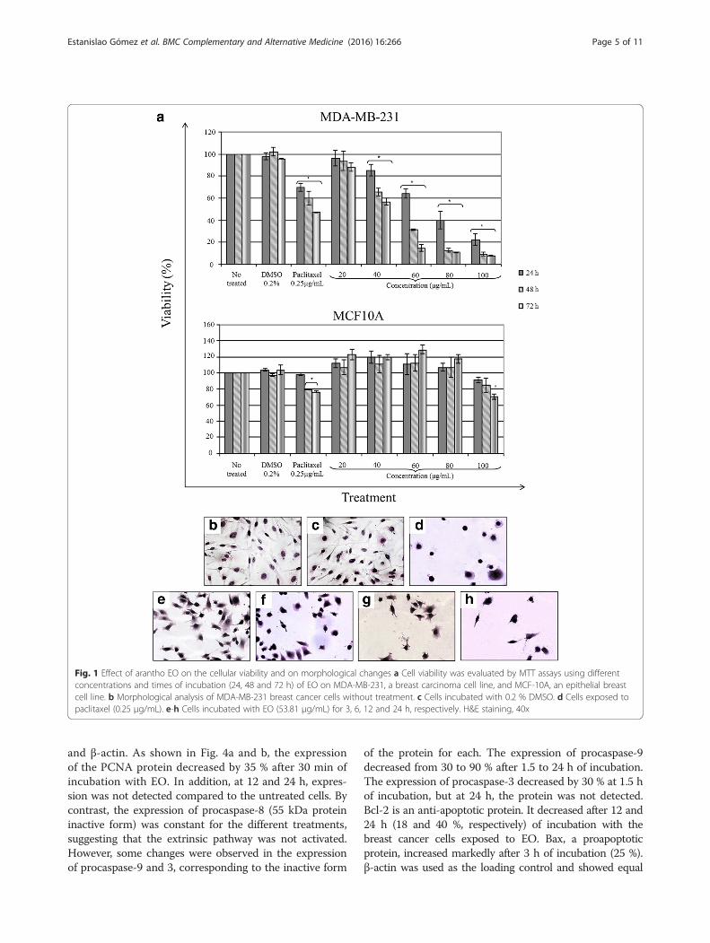
ResultsEffect of the different extracts and EO of arantho on viabilityThe cytotoxic activities of the extracts and EO from ara-ntho were analyzed by MTT assays performed on theMDA-MB-231 breast cancer cell line. The aqueous extractdid not show any cytotoxic activity at any concentrationor treatment period against these cancer cells. However,the ethanolic, acetonic and hexanic extracts showed acytotoxic effect in a time- and concentration-dependentmanner, with IC50 values of 128.20 ± 2.035, 203.2 ± 2.3and 450.7 ± 2.657 μg/mL, respectively (Additional file 1).As shown in Fig. 1a, a better cytotoxic effect was observedwhen the MDA-MB-231 cells were exposed to the EO.After 24 h of incubation, the cell viability decreased 15,36, 60 and 77 % with 40, 60, 80 and 100 μg/mL, respect-ively. In addition, after 48 h of treatment, the cell viabilitydecreased 35, 69, 88 and 91 % using doses of 40, 60, 80and 100 μg/mL, respectively. A greater cytotoxic effectwas observed after 72 h of incubation, with a reduction ofcell viability of more than 85 % for 60, 80 and 100 μg/mL.The cytotoxic effect of the EO was dose- and time-dependent, with an IC50 of 53.81 ± 1.691 μg/mL. Paclitaxel(0.25 μg/mL) was used as a positive control and alsocaused a decrease in viability of 30, 40 and 53 % at 24, 48and 72 h, respectively, whereas MDA-MB-231 cells with-out treatment maintained a viability of 100 %. These re-sults demonstrate an important and significant cytotoxiceffect of the arantho EO against MDA-MB-231 cells.To analyze the specificity of the EO against breast can-
cer cells, an epithelial breast cell line, MCF-10A, wasused. The results showed that MCF-10A cells treatedwith 20 to 80 μg/mL EO maintained cell viability similarto the negative controls (100 %) during all treatments.Higher doses (100 μg/mL) given to the normal epithelialcells for 72 h decreased the cell viability 25 % (Fig. 1a).The IC50 value for normal epithelial cells was 207.51 ±3.26 μg/mL, representing a 2.84-fold higher concentra-tion compared to the IC50 concentration for breast can-cer cells. These results confirmed that EO was selectiveagainst breast cancer cells.
Morphological changes on MDA-MB-231 cells exposed toarantho EOTo determine whether the reduced cell viability of thecancer cells resulted from cell death induction, MDA-MB-231 cells were incubated with the EO using the IC50 value
for different times and were stained with H&E. Non-treatedcells possessed an elongated shape; a centrally locatednucleus, forming a confluent colony; and were completelyattached to the plate (Fig. 1b, c). By contrast, cells exposedto EO showed relevant morphological changes after 3 h ofincubation, including cell size reduction, membrane bleb-bing, cell shrinkage and loss of colony formation (Fig. 1e).These changes increased with time, and after 6, 12 and24 h of incubation, the cells appeared to be rounded andapoptotic bodies were present (Fig. 1f-h). Similar character-istics were observed in breast cancer cells exposed to pacli-taxel (Fig. 1d). These morphological characteristics suggestthe induction of apoptosis by arantho EO.
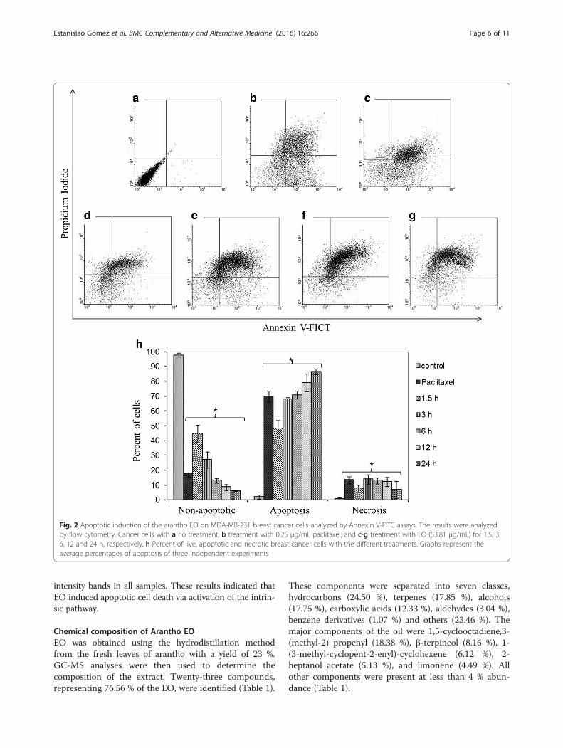
Apoptotic induction by the arantho EOTo determine and quantify cell death, MDA-MB-231 cellswere incubated with EO (53.81 μg/mL) and analyzed byAnnexin V-FITC assays. Control cells without treatmentshowed 98 % non-apoptotic live cells (Fig. 2a, h) and a verylow percent (1–2 %) of apoptosis and necrosis. By contrast,cells incubated with 0.25 μg/mL paclitaxel showed only18 % non-apoptotic live cells and displayed 69 and 12 %apoptotic and necrotic cells, respectively (Fig. 2b, h). Cancercells exposed to 53.81 μg/mL of EO showed decreasednon-apoptotic live cells (from 45 to 5 % after 1.5 and 24 h,respectively) and increased apoptotic cells (from 47 to 85 %after 1.5 and 24 h, respectively) in a time-dependent man-ner (Fig. 2c-h). The percent of necrotic cells was similar forthe different treatments. These results indicate that EO in-duces apoptosis rather than necrosis in MDA-MB-231breast cancer cells, and this is consistent with the morpho-logical changes observed by H&E staining.
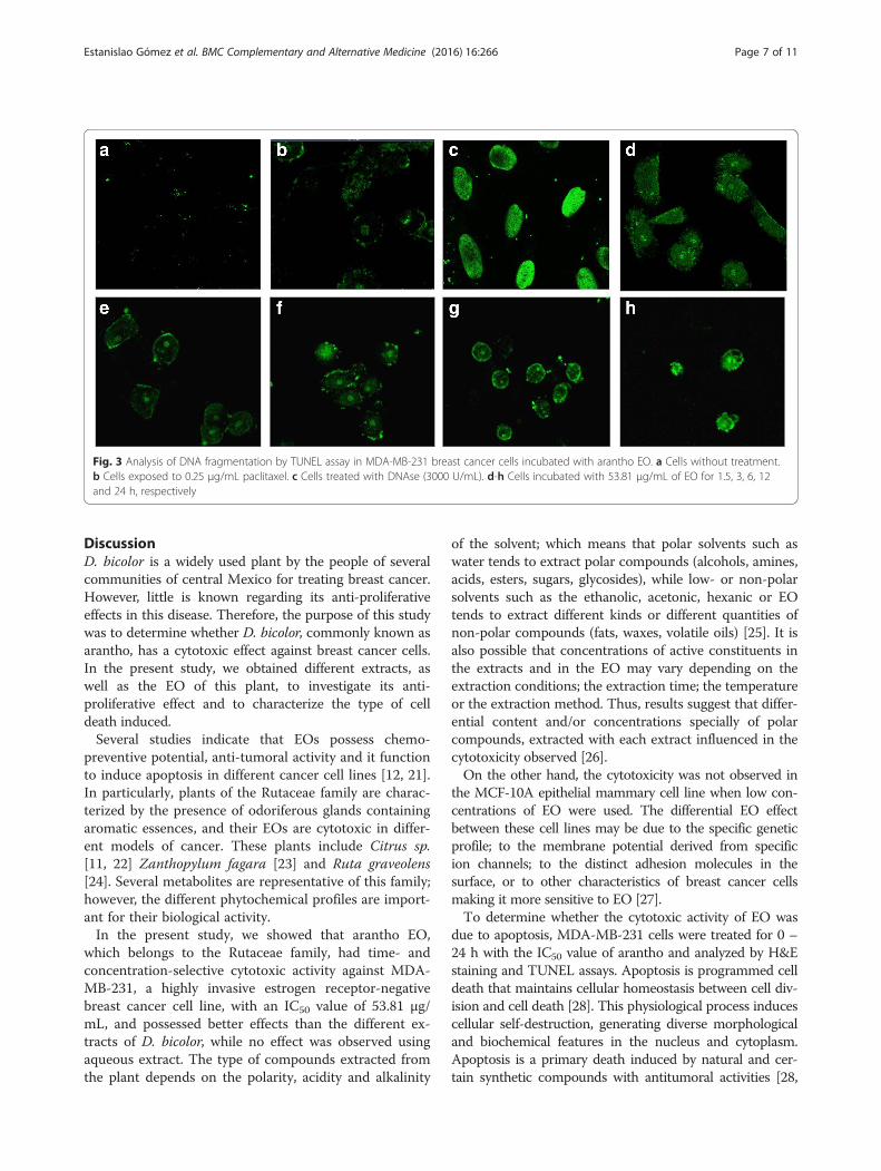
Arantho EO induced DNA fragmentation in MDA-MB-231cellsTo determine the ability of the arantho EO to trigger DNAfragmentation as a feature of apoptosis, TUNEL assayswere performed. Breast cancer cells treated with EO(53.81 μg/mL) showed an increased in DNA fragmentationafter 1.5, 3, 6, 12 and 24 h of incubation (Fig. 3d, e, f, g andh, respectively). As expected, cancer cells exposed to pacli-taxel and DNase were also positively stained in the assay(Fig. 3b and c, respectively). As a control, cancer cells with-out treatment did not exhibit fluorescence staining in thenucleus (Fig. 3a). These findings indicate that arantho EOinduced cell death in MDA-MB-231 cells by apoptosisrather than necrosis.
Apoptotic effect of arantho EO on MDA-MB-231 cells ismediated by activation of caspase cascadesTo analyze the activation of an apoptotic pathway,western blot assays were performed to evaluate theexpression of proteins, such as PCNA (proliferating cellnuclear antigen); procaspases 3, 8 and 9; Bcl-2; Bax;
Estanislao Gómez et al. BMC Complementary and Alternative Medicine (2016) 16:266 Page 4 of 11
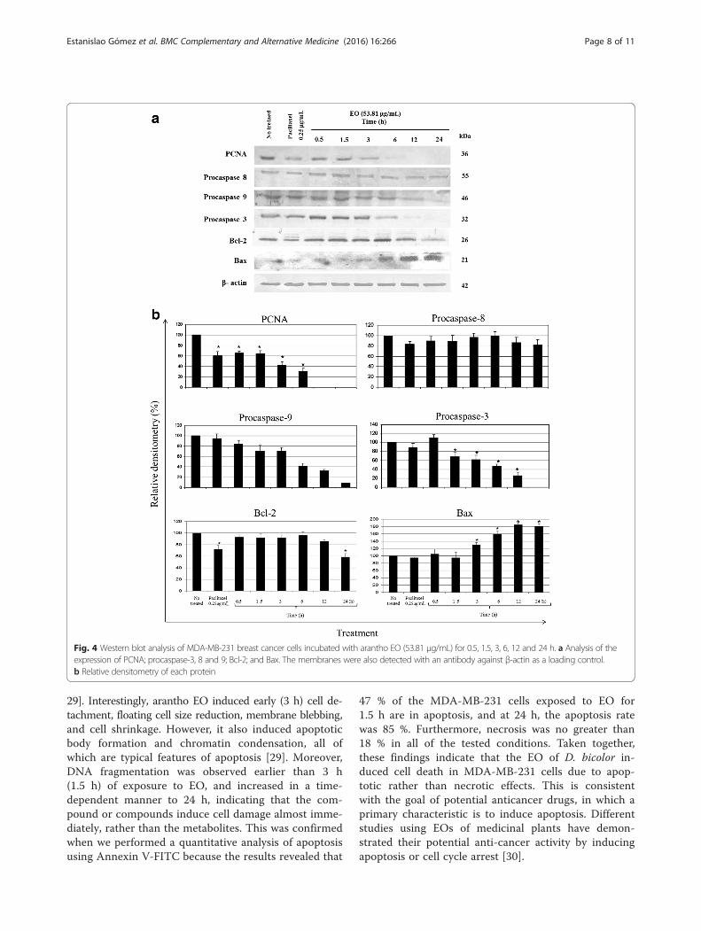
and β-actin. As shown in Fig. 4a and b, the expressionof the PCNA protein decreased by 35 % after 30 min ofincubation with EO. In addition, at 12 and 24 h, expres-sion was not detected compared to the untreated cells. Bycontrast, the expression of procaspase-8 (55 kDa proteininactive form) was constant for the different treatments,suggesting that the extrinsic pathway was not activated.However, some changes were observed in the expressionof procaspase-9 and 3, corresponding to the inactive form
of the protein for each. The expression of procaspase-9decreased from 30 to 90 % after 1.5 to 24 h of incubation.The expression of procaspase-3 decreased by 30 % at 1.5 hof incubation, but at 24 h, the protein was not detected.Bcl-2 is an anti-apoptotic protein. It decreased after 12 and24 h (18 and 40 %, respectively) of incubation with thebreast cancer cells exposed to EO. Bax, a proapoptoticprotein, increased markedly after 3 h of incubation (25 %).β-actin was used as the loading control and showed equal
Fig. 1 Effect of arantho EO on the cellular viability and on morphological changes a Cell viability was evaluated by MTT assays using differentconcentrations and times of incubation (24, 48 and 72 h) of EO on MDA-MB-231, a breast carcinoma cell line, and MCF-10A, an epithelial breastcell line. b Morphological analysis of MDA-MB-231 breast cancer cells without treatment. c Cells incubated with 0.2 % DMSO. d Cells exposed topaclitaxel (0.25 μg/mL). e-h Cells incubated with EO (53.81 μg/mL) for 3, 6, 12 and 24 h, respectively. H&E staining, 40x
Estanislao Gómez et al. BMC Complementary and Alternative Medicine (2016) 16:266 Page 5 of 11
intensity bands in all samples. These results indicated thatEO induced apoptotic cell death via activation of the intrin-sic pathway.
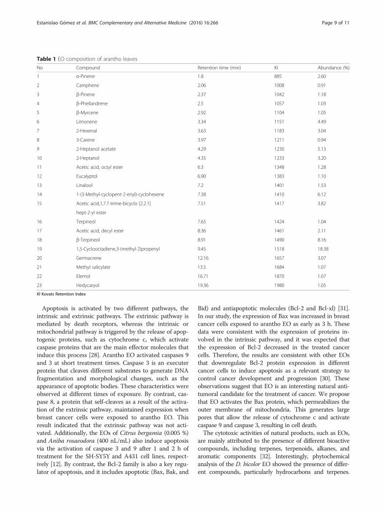
Chemical composition of Arantho EOEO was obtained using the hydrodistillation methodfrom the fresh leaves of arantho with a yield of 23 %.GC-MS analyses were then used to determine thecomposition of the extract. Twenty-three compounds,representing 76.56 % of the EO, were identified (Table 1).
These components were separated into seven classes,hydrocarbons (24.50 %), terpenes (17.85 %), alcohols(17.75 %), carboxylic acids (12.33 %), aldehydes (3.04 %),benzene derivatives (1.07 %) and others (23.46 %). Themajor components of the oil were 1,5-cyclooctadiene,3-(methyl-2) propenyl (18.38 %), β-terpineol (8.16 %), 1-(3-methyl-cyclopent-2-enyl)-cyclohexene (6.12 %), 2-heptanol acetate (5.13 %), and limonene (4.49 %). Allother components were present at less than 4 % abun-dance (Table 1).
Fig. 2 Apoptotic induction of the arantho EO on MDA-MB-231 breast cancer cells analyzed by Annexin V-FITC assays. The results were analyzedby flow cytometry. Cancer cells with a no treatment; b treatment with 0.25 μg/mL paclitaxel; and c-g treatment with EO (53.81 μg/mL) for 1.5, 3,6, 12 and 24 h, respectively. h Percent of live, apoptotic and necrotic breast cancer cells with the different treatments. Graphs represent theaverage percentages of apoptosis of three independent experiments
Estanislao Gómez et al. BMC Complementary and Alternative Medicine (2016) 16:266 Page 6 of 11
DiscussionD. bicolor is a widely used plant by the people of severalcommunities of central Mexico for treating breast cancer.However, little is known regarding its anti-proliferativeeffects in this disease. Therefore, the purpose of this studywas to determine whether D. bicolor, commonly known asarantho, has a cytotoxic effect against breast cancer cells.In the present study, we obtained different extracts, aswell as the EO of this plant, to investigate its anti-proliferative effect and to characterize the type of celldeath induced.Several studies indicate that EOs possess chemo-
preventive potential, anti-tumoral activity and it functionto induce apoptosis in different cancer cell lines [12, 21].In particularly, plants of the Rutaceae family are charac-terized by the presence of odoriferous glands containingaromatic essences, and their EOs are cytotoxic in differ-ent models of cancer. These plants include Citrus sp.[11, 22] Zanthopylum fagara [23] and Ruta graveolens[24]. Several metabolites are representative of this family;however, the different phytochemical profiles are import-ant for their biological activity.In the present study, we showed that arantho EO,
which belongs to the Rutaceae family, had time- andconcentration-selective cytotoxic activity against MDA-MB-231, a highly invasive estrogen receptor-negativebreast cancer cell line, with an IC50 value of 53.81 μg/mL, and possessed better effects than the different ex-tracts of D. bicolor, while no effect was observed usingaqueous extract. The type of compounds extracted fromthe plant depends on the polarity, acidity and alkalinity
of the solvent; which means that polar solvents such aswater tends to extract polar compounds (alcohols, amines,acids, esters, sugars, glycosides), while low- or non-polarsolvents such as the ethanolic, acetonic, hexanic or EOtends to extract different kinds or different quantities ofnon-polar compounds (fats, waxes, volatile oils) [25]. It isalso possible that concentrations of active constituents inthe extracts and in the EO may vary depending on theextraction conditions; the extraction time; the temperatureor the extraction method. Thus, results suggest that differ-ential content and/or concentrations specially of polarcompounds, extracted with each extract influenced in thecytotoxicity observed [26].On the other hand, the cytotoxicity was not observed in
the MCF-10A epithelial mammary cell line when low con-centrations of EO were used. The differential EO effectbetween these cell lines may be due to the specific geneticprofile; to the membrane potential derived from specificion channels; to the distinct adhesion molecules in thesurface, or to other characteristics of breast cancer cellsmaking it more sensitive to EO [27].To determine whether the cytotoxic activity of EO was
due to apoptosis, MDA-MB-231 cells were treated for 0 –24 h with the IC50 value of arantho and analyzed by H&Estaining and TUNEL assays. Apoptosis is programmed celldeath that maintains cellular homeostasis between cell div-ision and cell death [28]. This physiological process inducescellular self-destruction, generating diverse morphologicaland biochemical features in the nucleus and cytoplasm.Apoptosis is a primary death induced by natural and cer-tain synthetic compounds with antitumoral activities [28,
Fig. 3 Analysis of DNA fragmentation by TUNEL assay in MDA-MB-231 breast cancer cells incubated with arantho EO. a Cells without treatment.b Cells exposed to 0.25 μg/mL paclitaxel. c Cells treated with DNAse (3000 U/mL). d-h Cells incubated with 53.81 μg/mL of EO for 1.5, 3, 6, 12and 24 h, respectively
Estanislao Gómez et al. BMC Complementary and Alternative Medicine (2016) 16:266 Page 7 of 11
29]. Interestingly, arantho EO induced early (3 h) cell de-tachment, floating cell size reduction, membrane blebbing,and cell shrinkage. However, it also induced apoptoticbody formation and chromatin condensation, all ofwhich are typical features of apoptosis [29]. Moreover,DNA fragmentation was observed earlier than 3 h(1.5 h) of exposure to EO, and increased in a time-dependent manner to 24 h, indicating that the com-pound or compounds induce cell damage almost imme-diately, rather than the metabolites. This was confirmedwhen we performed a quantitative analysis of apoptosisusing Annexin V-FITC because the results revealed that
47 % of the MDA-MB-231 cells exposed to EO for1.5 h are in apoptosis, and at 24 h, the apoptosis ratewas 85 %. Furthermore, necrosis was no greater than18 % in all of the tested conditions. Taken together,these findings indicate that the EO of D. bicolor in-duced cell death in MDA-MB-231 cells due to apop-totic rather than necrotic effects. This is consistentwith the goal of potential anticancer drugs, in which aprimary characteristic is to induce apoptosis. Differentstudies using EOs of medicinal plants have demon-strated their potential anti-cancer activity by inducingapoptosis or cell cycle arrest [30].
Fig. 4 Western blot analysis of MDA-MB-231 breast cancer cells incubated with arantho EO (53.81 μg/mL) for 0.5, 1.5, 3, 6, 12 and 24 h. a Analysis of theexpression of PCNA; procaspase-3, 8 and 9; Bcl-2; and Bax. The membranes were also detected with an antibody against β-actin as a loading control.b Relative densitometry of each protein
Estanislao Gómez et al. BMC Complementary and Alternative Medicine (2016) 16:266 Page 8 of 11
Apoptosis is activated by two different pathways, theintrinsic and extrinsic pathways. The extrinsic pathway ismediated by death receptors, whereas the intrinsic ormitochondrial pathway is triggered by the release of apop-togenic proteins, such as cytochrome c, which activatecaspase proteins that are the main effector molecules thatinduce this process [28]. Arantho EO activated caspases 9and 3 at short treatment times. Caspase 3 is an executerprotein that cleaves different substrates to generate DNAfragmentation and morphological changes, such as theappearance of apoptotic bodies. These characteristics wereobserved at different times of exposure. By contrast, cas-pase 8, a protein that self-cleaves as a result of the activa-tion of the extrinsic pathway, maintained expression whenbreast cancer cells were exposed to arantho EO. Thisresult indicated that the extrinsic pathway was not acti-vated. Additionally, the EOs of Citrus bergamia (0.005 %)and Aniba rosaeodora (400 nL/mL) also induce apoptosisvia the activation of caspase 3 and 9 after 1 and 2 h oftreatment for the SH-SY5Y and A431 cell lines, respect-ively [12]. By contrast, the Bcl-2 family is also a key regu-lator of apoptosis, and it includes apoptotic (Bax, Bak, and
Bid) and antiapoptotic molecules (Bcl-2 and Bcl-xl) [31].In our study, the expression of Bax was increased in breastcancer cells exposed to arantho EO as early as 3 h. Thesedata were consistent with the expression of proteins in-volved in the intrinsic pathway, and it was expected thatthe expression of Bcl-2 decreased in the treated cancercells. Therefore, the results are consistent with other EOsthat downregulate Bcl-2 protein expression in differentcancer cells to induce apoptosis as a relevant strategy tocontrol cancer development and progression [30]. Theseobservations suggest that EO is an interesting natural anti-tumoral candidate for the treatment of cancer. We proposethat EO activates the Bax protein, which permeabilizes theouter membrane of mitochondria. This generates largepores that allow the release of cytochrome c and activatecaspase 9 and caspase 3, resulting in cell death.The cytotoxic activities of natural products, such as EOs,
are mainly attributed to the presence of different bioactivecompounds, including terpenes, terpenoids, alkanes, andaromatic components [32]. Interestingly, phytochemicalanalysis of the D. bicolor EO showed the presence of differ-ent compounds, particularly hydrocarbons and terpenes.
Table 1 EO composition of arantho leaves
No Compound Retention time (min) KI Abundance (%)
1 α-Pinene 1.8 885 2.60
2 Camphene 2.06 1008 0.91
3 β-Pinene 2.37 1042 1.18
4 β-Phellandrene 2.5 1057 1.03
5 β-Myrcene 2.92 1104 1.05
6 Limonene 3.34 1151 4.49
7 2-Hexenal 3.63 1183 3.04
8 3-Carene 3.97 1211 0.94
9 2-Heptanol acetate 4.29 1230 5.13
10 2-Heptanol 4.35 1233 3.20
11 Acetic acid, octyl ester 6.3 1348 1.28
12 Eucalyptol 6.90 1383 1.10
13 Linalool 7.2 1401 1.53
14 1-(3-Methyl-cyclopent-2-enyl)-cyclohexene 7.38 1410 6.12
15 Acetic acid,1,7.7-trime-bicyclo [2.2.1] 7.51 1417 3.82
hept-2-yl ester
16 Terpineol 7.65 1424 1.04
17 Acetic acid, decyl ester 8.36 1461 2.11
18 β-Terpineol 8.91 1490 8.16
19 1,5-Cyclooctadiene,3-(methyl-2)propenyl 9.45 1518 18.38
20 Germacrene 12.16 1657 3.07
21 Methyl salicylate 13.5 1684 1.07
22 Elemol 16.71 1870 1.07
23 Hedycaryol 19.36 1980 1.05
KI Kovats Retention Index
Estanislao Gómez et al. BMC Complementary and Alternative Medicine (2016) 16:266 Page 9 of 11
This result is consistent with the consensus of the chem-ical composition of EOs, indicating that the three maingroups of compounds are terpenes and terpenoids, aro-matics (phenolic) and aliphatics [6]. However, 1-5-cyclooctadiene,3-(methyl-2)propenyl and 1-(3-methyl-cyclopent-2-enyl-cyclohexene, two hydrocarbons that were majorcompounds identified in D. bicolor EO, may be respon-sible for the cytotoxic activity of this EO towards theMDA-MB-231 breast cancer cells, although nothing isknown of its antitumoral activity. Nevertheless, other con-stituents of EO may contribute to the cytotoxicity. EOsisolated from antitumoral plants of the Rutaceae family ofthe genus Citrus (C. aurantium, C. limon and C. reticu-lata) possess a highly different chemical compositioncompared to D. bicolor, which may be due to the differ-ence in their genetic profile and environmental conditions[10]. Moreover, several of the metabolites identified wereisolated in other studies and demonstrated cytotoxic andantitumoral activities. One of the three major compoundsidentified in D. bicolor EO is α-terpineol, which decreasedboth the viability of the MCF-7 breast cancer cell with anIC50 value of 16.2 μM [33] and the expression of NF-kB[34]. Other important less abundant compounds in D.bicolor EO that have been isolated from other medicinalplants and possess antitumoral effect are limonene, α-pinene and linalool. α-Pinene is a monoterpene that in-duces apoptosis in SK-OV3, HO-8910, Bel-7402 [21] andC32 cancer cells [35]. Linalool had a cytotoxic effect onmelanoma [35], breast cancer cells [36] and hepatocarci-noma cells [37]. Limonene is a monoterpene representativeof the Rutaceae family. It inhibits the growth of differentcell lines, such as DU-145 prostate [38], carcinoma cells,with an IC50 of 2.8 mM. However, limonene, linalool andα-pinene previously tested on MDA-MB-231 breast cancercells were not cytotoxic [39]. These investigations indicatethat several metabolites present in arantho EO are cytotoxicto different cancer cell lines; however, it is difficult to attri-bute the biological activity to the entire EO or to onespecific compound because the cytostatic effect of an entireplant on cancer cells is often better than the effect of theparticular biological active compounds [7]. In addition, dueto the lipophilic nature of EO, volatile compounds appearto accumulate in the cell membrane and increase itspermeability, resulting in the leakage of enzymes andmetabolites that induces cytotoxic and apoptotic effect incancer cells [6]; however, is necessary to identify the activecompound responsible for the cell death of these breastcancer cells.
ConclusionIn conclusion, several assays demonstrated an importantcytotoxic, selective and apoptotic activity of the EO of D.bicolor against the breast cancer cell line MDA-MB-231,Additional in vitro and in vivo studies currently under
study will allow us to determine the sensitivity of othercell lines and the anti-tumoral potential of the essentialoil from arantho (EO).
Additional file
Additional file 1: MTT assays of the different extracts of D. bicolor onMDA-MB-231 breast cancer cell line. The graphics represent the MTTassays of the different extracts (aqueous, ethanolic, acetonic and hexanic)of D. bicolor on breast cancer cells MDA-MB-231. The ethanolic, acetonicand hexanic extracts demonstrated a cytotoxic effect in a doses and timedependent manner, obtaining IC50 values of 128.20 ± 2.035, 203.2 ± 2.3and 450.7 ± 2.657 μg/mL, respectively. However, the aqueous extractdidn’t showed any cytotoxic activity at any concentration or time. Theexperiments were performed at least in triplicate and the values arereported as mean ± SE, *P < 0.05 as compared to control cells (mediumalone). Significance was analyzed using One-way ANOVA followed byTukey test. (PDF 270 kb)
AbbreviationsDMEM, Dulbecco’s Modified Eagle Medium; DMSO, dimethylsulfoxide; EO,essential oil; GC-MS, gas chromatography–mass spectrometry; H&E, hematoxylinand eosin; KI, Kovats Retention Index; MTT, 3-(4, 5- dimethylthiazol-2-yl)-2, 5-diphenyl tetrazolium bromide; PCNA, proliferating cell nuclear antigen; TUNEL, ter-minal deoxynucleotidyl transferase-mediated dUTP nick end labeling.
AcknowledgementsThis work was supported by Consejo Nacional de Ciencia y Tecnología(CONACYT) Grant 114028 (Fondo de Investigación en Salud) and by the InstitutoPolitécnico Nacional Grants SIP 20113658, SIP 20120468 and SIP 20140313. Wethank Dr. María de Jesús Perea and Ing. Alberto Peña of the MicroscopyLaboratory at the Centro de Nanociencias y Micro y Nanotecnología del IPN fortechnical assistance. We also thank Dr. Edith Cortés of the Universidad AutónomaMetropolitana, Iztapalapa.
FundingThis work was supported by Consejo Nacional de Ciencia y Tecnología(CONACYT) Grant 114028 (Fondo de Investigación en Salud) and by theInstituto Politécnico Nacional Grants SIP 20113658, SIP 20120468 and SIP20140313.
Availability of data and materialsAll data and materials are contained and described within the manuscript.
Authors’ contributionsCCEG carried out most of the experiments, contributed to the datainterpretation and wrote the initial draft of the manuscript. AAC contributedto the experimental design and Western blot assays. DGPI contributed to datainterpretation and the final editing of the manuscript., ESMM and JMLcontributed to preparation of essential oil and to its chemical composition byGC-MS. NPH participated in the project design and to the data interpretation.MCGG contributed to the conception and design of the entire study, funding,data interpretation and the final editing of the manuscript. All authors read andapproved the final manuscript.
Competing interestsThe authors declare that they have no competing interests.
Consent for publicationNot applicable.
Ethics approval and consent to participateNot applicable.
Author details1Programa de Doctorado en Biotecnología, Escuela Nacional de Medicina yHomeopatía, Instituto Politécnico Nacional, Guillermo Massieu Helguera No.239, Fracc. La Escalera, Ticomán, Delg. Gustavo A. Madero, Mexico C.P. 07320,D. F., Mexico. 2Departamento de Ciencias Químico Biológicas, Instituto de
Estanislao Gómez et al. BMC Complementary and Alternative Medicine (2016) 16:266 Page 10 of 11
Ciencias Biomédicas, Universidad Autónoma de Ciudad Juárez, AnilloEnvolvente Pronaf y Estocolmo s/n, C.P. 32310 Cd. Juárez, Chih, Mexico.3Centro de Investigación en Biotecnología Aplicada, Instituto PolitécnicoNacional, Tepetitla de Lardizabal, Tlaxcala, Doctorado en Biotecnología, Redde Investigación en Biotecnología IPN, México, Mexico. 4Centro deInvestigación en Ciencia Aplicada y Tecnología Avanzada, Unidad Legaria,Instituto Politécnico Nacional, Calzada Legaria No. 694 Col. Irrigación, Delg.Miguel Hidalgo, Mexico C.P. 11500, D.F., Mexico.
Received: 28 August 2015 Accepted: 18 May 2016
References1. Ferlay J, Soerjomataram I, Ervik M, Dikshit R, Eser S, Mathers C, Rebelo M, Parkin
DM, Forman D, Bray F. Cancer incidence and mortality Worldwide: sources,methods and major patterns in GLOBOCAN 2012. Int J Cancer. 2015;136:E359–86.doi:10.1002/ijc.29210.
2. Pandey G, Madhuri S. Medicinal plants: better remedy for neoplasm. IndianDrugs. 2006;43:869–74.
3. Kaur R, Singh J, Singh G, Kaur H. Anticancer plants: a review. J Nat ProdPlant Resour. 2011;1(4):131–6. http://scholarsresearchlibrary.com/JNPPR-vol1-iss4/JNPPR-2011-1-4-131-136.pdf. Accessed 20 Jan 2015.
4. Agarwal N, Majee C, Chakraborthy GS. Natural herbs as anticancer drugs. IntJ PharmTech Res. 2012;4(3):1142–53. http://www.sphinxsai.com/2012/july_sept12/Pharm/pdfpharm/PT=33%281142-1153%29%20JS%2012.pdf.Accessed 21 Jan 2015.
5. Prema R, Sathish Sekar D, Chandra Sekhar KB. Review on: herbs asanticancer agents. Int J Pharma & Ind Res. 2011;1(2):105–8.
6. Bayala B, Bassole IH, Scifo R, Gnoula C, Morel L, Lobaccaro JM, et al.Anticancer activity of essential oils and their chemical components-a review.Am J Cancer Res. 2014;4(6):591–607.
7. Bakkali F, Averbeck S, Averbeck D, Idaomar M. Biological effects of essentialoils-a review. Food Chem Toxicol. 2008;46:446–75.
8. Edris AE. Pharmaceutical and therapeutic potentials of essential oils and theirindividual volatile constituents: a review. Phytothe Res. 2007;21(4):308–23.
9. Smith N, Mori A, Henderson DW, Stevenson DW, Heald SV. Flowering Plantsof the Neotropics. New Jersey: Princeton University Press; 2004. p. 333–5.
10. Misharina TA, Samusenko AL. Antioxidant properties of essential oils fromlemon, grapefruit, coriander, clove, and their mixtures. Appl BiochemMicrobiol. 2008;45(4):438–42.
11. Odeh F, Rahmo A, Alnory SA, Chaty EM. The cytotoxic effect of essential oilsCitrus aurantium peels on human colorectal carcinoma cell line (Lim1863). JMicrobiol Biotechnol Food Sci. 2012;1(6):1476–87.
12. Berliocchi L, Ciociaro A, Russo R, Cassiano MG, Blandini F, Rotiroti D, et al.Toxic profile of bergamot essential oil on survival and proliferation of SH-SY5Y neuroblastoma cells. Food Chem Toxicol. 2011;49:2780–92.
13. Cárdenas ON, Pérez GS, Zavala SM, Aguirre RJ, Pérez GC. Actividadantifúngica de seis plantas sobre Aspergillus flavus. Revista Mexicana deCiencias Farmacéuticas. 2005;36(03):21–6.
14. García-Argáez AN, Ramírez Apan TO, Parra Delgado H, Velázquez G,Martínez-Vázquez M. Anti-inflammatory activity of coumarins fromDecatropis bicolor on TPA ear mice model. Planta Med. 2000;66(3):279–81.
15. Sánchez GA, Granados SD, Simón NR. Uso medicinal de las plantas por losotomíes del municipio de Nicolás Flores, Hidalgo. México Revista ChapingoSerie Horticultura. 2008;14(3):271–9.
16. Villavicencio NM, Perez EB, Ramírez A. Plantas útiles del Estado de Hidalgo II.Pachuca, Hidalgo: F. Edit. UAEH; 2002. https://books.google.com.mx/books?id=WXZX5JxDtL0C&pg=PA13&lpg=PA13&dq=plantas+utiles+del+estado+de+hidalgo&source=bl&ots=EsP5d-K6sb&sig=JlKNnWKwZfkSnuda2EKiHsJr80U&hl=es&sa=X&ved=0ahUKEwiEqP7o_ITOAhUs_4MKHYTdC_8Q6AEILDAD#v=onepage&q=plantas%20utiles%20del%20estado%20de%20hidalgo&f=false.
17. Pérez EB, Villavicencio NM. Listado de plantas medicinales del Estado deHidalgo. Pachuca: Universidad Autónoma del Estado de Hidalgo; 1995. p. 45.
18. Cortés CJ. Actividad biológica de extractos de plantas usadas para eltratamiento del cáncer e infecciones en Tepatepec, Hidalgo. 2005. http://www.uaeh.edu.mx/docencia/Tesis/icbi/licenciatura/documentos/Actividad%20biologica%20de%200extractos.pdf.
19. Calvo-Gómez O, Morales-López J, López M. Solid-phase microextraction–gaschromatographic–mass spectrometric analysis of garlic oil obtained byhydrodistillation. J Chromatogr A. 2004;1036(1):91–3.
20. Mosmann T. Rapid colorimetric assay for cellular growth and survival: applicationto proliferation and cytotoxicity assay. J Immunol Meth. 1983;65:55–63.
21. Wang W, Li N, Luo M, Zu Y, Efferth T. Antibacterial activity and anticanceractivity of Rosmarinus officinalis L. essential oil compared to that of its maincomponents. Molecules. 2012;17(3):2704–13.
22. Atti-Santos AC, Rossato M, Serafini LA, Cassel E, Moyna P. Extraction ofessential oils from lime (citrus latifolia Tanaka) by hydrodistillation andsupercritical carbon dioxide. Braz Arch Biol Technol. 2005;48(1):155–60.
23. Macías VE, Coy BE, Cuca SL. Preliminary phytochemical analysis andantioxidant, antiinflammatory and antiproliferative activities of the barkethanol extracts of Zanthoxylum fagara (L.) Sarg. (Rutaceae). Rev CubanaPlant Med. 2011;16(1):43–53.
24. Fadlalla K, Watson A, Yehualaeshet T, Turner T, Smuel T. Ruta graveolensextract induces DNA damage pathways and blocks Akt activation to inhibitcancer cell proliferation and survival. Anticancer Res. 2011;31:233–41.
25. Starmans DAJ, Nijhuis HH. Extraction of secondary metabolites from plantmaterial: a review. Trends Food Sci Techol. 1996;7:191–7.
26. Newman DJ, Cragg GM. Natural products as sources of new drugs over thelast 25 years. J Nat Prod. 2007;70(3):461–77.
27. Charafe-Jauffret E, Ginestier C, Monville F, Finetti P, Adélaïde J, Cervera N, etal. Gene expression profiling of breast cell lines identifies potential newbasal markers. Oncogene. 2006;25(15):2273–84.
28. Vermeulen K, Van Bockstaele DR, Berneman ZN. Apoptosis: mechanisms andrelevance in cancer. Ann Hematol. 2005;84:627–39.
29. Koff JL, Ramachandiran S, Bernal-Mizrachi L. A time to kill: targetingapoptosis in cancer. Int J Mol Sci. 2015;16:2942–55.
30. Gautam N, Mantha AK, Mittal S. Essential oils and their constituents asanticancer agents: a mechanistic view. BioMed Res Int. 2014;2014:1–23.http://dx.doi.org/10.1155/2014/154106. Accessed 30 Jan 2015.
31. Marzo I, Naval J. Bcl-2 family members as molecular targets in cancertherapy. Biochem Pharmacol. 2008;76:939–46.
32. Hamid AA, Aiyelaagbe OO, Usman LA. Essential oils: its medicinal andpharmacological uses. Int J Current Res. 2011;3(2):086–98. http://www.journalcra.com/sites/default/files/Download%20406.pdf. Accessed 01 Mar 2015.
33. Bicas JL, Neri-Numa IA, Ruiz AL, De Carvalho JE, Pastore GM. Evaluation ofthe antioxidant and antiproliferative potential of bioflavors. Food ChemToxicol. 2011;49:1610–5.
34. Hassan SB, Gali-Muhtasib H, Göransson H, Larsson R. Alpha terpineol: apotential anticancer agent which acts through suppressing NF-kB signalling.Anticancer Res. 2010;30(6):1911–9. http://ar.iiarjournals.org/content/30/6/1911.long. Accessed 01 Mar 2015.
35. Loizzo MR, Tundis R, Menichini F, Saab AM, Statti GA, Menichini F.Antiproliferative effects of essential oils and their major constituents inhuman renal adenocarcinoma and amelanotic melanoma cells. Cell Prolif.2008;41:1002–12.
36. Ravizza R, Gariboldi MB, Molteni R, Monti E. Linalool, a plant-derivedmonoterpene alcohol, reverses doxorubicin resistance in human breastadenocarcinoma cells. Oncol Rep. 2008;20(3):625–30. http://dx.doi.org/10.3892/or_00000051. Accessed 10 Mar 2015.
37. Usta J, Kreydiyyeh S, Knio K, Barnabe P, Bou-Moughlabay Y, Dagher S.Linalool decreases HepG2 viability by inhibiting mitochondrial complexes Iand II, increasing reactive oxygen species and decreasing ATP and GSHlevels. Chem Biol Interact. 2009;180(1):39–46.
38. Rabi T, Bishayee A. d-Limonene sensitizes docetaxel-induced cytotoxicity inhuman prostate cancer cells: generation of reactive oxygen species andinduction of apoptosis. J Carcinog. 2009;8:9. http://doi.org/10.4103/1477-3163.51368. Accessed 10 Mar 2015.
39. Palazzo MC, Agius BR, Wright BS, Haber WA, Moriarity DM, Setzer WN. Chemicalcompositions and cytotoxic activities of leaf essential oils of four lauraceae treespecies from Monteverde, Costa Rica. Rec Nat Prod. 2009;3(1):32–7.
Estanislao Gómez et al. BMC Complementary and Alternative Medicine (2016) 16:266 Page 11 of 11