Divergent Evolution of 5 S rRNA Genes in Methanococcus
8
This work has been digitalized and published in 2013 by Verlag Zeitschrift für Naturforschung in cooperation with the Max Planck Society for the Advancement of Science under a Creative Commons Attribution 4.0 International License. Dieses Werk wurde im Jahr 2013 vom Verlag Zeitschrift für Naturforschung in Zusammenarbeit mit der Max-Planck-Gesellschaft zur Förderung der Wissenschaften e.V. digitalisiert und unter folgender Lizenz veröffentlicht: Creative Commons Namensnennung 4.0 Lizenz. Divergent Evolution of 5 S rRNA Genes in Methanococcus Günter Wich3, Lionel Siboldb, and August Böcka a Lehrstuhl für Mikrobiologie der Universität München, Maria-Ward-Straße la, D-8000 München 19, Bundesrepublik Deutschland b Unite de Physiologie Cellulaire, Departement de Biochimie et Genetique Moleculaire, Institut Pasteur, Paris, Rue du Dr. Roux 28, F-75724 Paris Cedex 15, France Z. Naturforsch. 42c, 373 — 380 (1987); received November 7, 1986 Dedicated to Professor Helmut Simon on the occasion of his 60th birthday Ribosomal RNA, Evolution, Archaebacteria The organization of genes for 5S rRNA in the methanogenic archaebacterium Methanococcus (M.) voltae and their nucleotide sequences have been determined. M. voltae possesses three 5S rRNA genes, one of them is organized in an rRNA transcriptional unit coding for 16S-23S-5S rRNA. The other two are associated with seven tRN A genes in a putative transcriptional unit composed of 5'-tRNAThr-tRNAPro-tRNATyr-tRNALys — 5S rRNA-tRNAAsp-tRNALys — 5S rRNA-tRNAAsp-3'. Coding regions plus spacers of the tRNALys-5S rRNA-tRNAAsp block of this gene cluster occur twice with identical sequence. The 5S rRNA from this cluster displays considerable sequence divergence to the rRNA operon-linked 5S rRN A gene. Comparison of the M. voltae 5S rRNA sequences with those from M. vannielii revealed that the operon-linked genes on one hand and the tRNA-linked 5S genes on the other share a greater sequence homology than the two types of genes within each of the two organisms. This indicates an independent evolution of the two sets of 5S rRNA genes without selective pressure from other ribosomal components or, alternatively, lateral gene transfer. Introduction Genes for stable R N A species are excellent model systems for the analysis of genome organization and gene expression. Since they are universal cell compo nents and since they have been strongly conserved during evolution their sequence comparison has pro vided a wealth of information on the evolution of macromolecules and of organisms [ 1 ]. One of the peculiar features of the organization of stable R N A genes in archaebacteria concerns the high diversity in which these genes are organized on the chromosome. They may be linked in typical eubacterial-type transcriptional units as in extreme halophiles [2, 3] or as in methanogens like Methano- bacterium [4, 5] or they may be completely unlinked as in Thermoplasma [4], Other organisms like Metha nococcus (M.) possess a “mixed” type of rRNA gene organization. In M. vannielii, for example, there are four 16S-23S-5S rRN A operons [ 6 ] and, in addition, a single “extra” 5S rRNA gene clustered in a tran scriptional unit with seven genes for tRNA [7], The four operon-linked 5S rRNA genes have an identical sequence which differs considerably from that of the “extra” 5S rRNA gene. The products of both gene types are functional since analysis of 5S rRNA extracted from the ribosomes indicated the presence of the products of both genes. The high sequence polymorphism within a single genome could be the result of either a lateral gene transfer event, e.g. of the “extra” 5S rRNA gene, or of the independent evolution of the two types of genes after a gene duplication event. To gain infor mation on this interesting question we have analyzed the organization and the primary structure of the 5S rRNA genes from M. voltae, an organism closely related to M. vannielii. Materials and Methods Strains and Plasmids Methanococcus (M.) voltae strain PS (DSM 1537) was obtained from the German Collection of Micro organisms, Göttingen, F. R .G . Chromosomal DNA fragments from M. voltae were cloned into plasmid pUC9. For subcloning, plasmid vector pUC19 was used [ 8 ]. Recipient E. coli strains in transformation experiments were E. coli 7902, E. coli JM105 and E. coli JM101, respectively. „ . _ A , Recombinant DNA techniques Reprint requests to Dr. A. Bock. Verlagder Zeitschrift für Naturforschung, D-7400Tübingen A method for isolation of high molecular weight 0341-0382/87/0400-0373 $01.30/0 chromosomal DNA from Methanococcus cells has
Transcript of Divergent Evolution of 5 S rRNA Genes in Methanococcus
This work has been digitalized and published in 2013 by Verlag Zeitschrift für Naturforschung in cooperation with the Max Planck Society for the Advancement of Science under a Creative Commons Attribution4.0 International License.
Dieses Werk wurde im Jahr 2013 vom Verlag Zeitschrift für Naturforschungin Zusammenarbeit mit der Max-Planck-Gesellschaft zur Förderung derWissenschaften e.V. digitalisiert und unter folgender Lizenz veröffentlicht:Creative Commons Namensnennung 4.0 Lizenz.
Divergent Evolution of 5 S rRNA Genes in MethanococcusGünter Wich3, Lionel Siboldb, and August Böcka
a Lehrstuhl für Mikrobiologie der Universität München, Maria-Ward-Straße la,D-8000 München 19, Bundesrepublik Deutschland
b Unite de Physiologie Cellulaire, Departement de Biochimie et Genetique Moleculaire,Institut Pasteur, Paris, Rue du Dr. Roux 28, F-75724 Paris Cedex 15, France
Z. Naturforsch. 42c, 373 — 380 (1987); received November 7, 1986
Dedicated to Professor Helmut Simon on the occasion of his 60th birthday
Ribosomal RNA, Evolution, Archaebacteria
The organization of genes for 5S rRNA in the methanogenic archaebacterium Methanococcus (M.) voltae and their nucleotide sequences have been determined. M. voltae possesses three 5S rRNA genes, one of them is organized in an rRNA transcriptional unit coding for 16S-23S-5S rRNA. The other two are associated with seven tRNA genes in a putative transcriptional unit composed of 5'-tRNAThr-tRNAPro-tRNATyr-tRNALys — 5S rRNA-tRNAAsp-tRNALys — 5S rRNA-tRNAAsp-3'. Coding regions plus spacers of the tRNALys-5S rRNA-tRNAAsp block of this gene cluster occur twice with identical sequence. The 5S rRNA from this cluster displays considerable sequence divergence to the rRNA operon-linked 5S rRNA gene. Comparison of the M. voltae 5S rRNA sequences with those from M. vannielii revealed that the operon-linked genes on one hand and the tRNA-linked 5S genes on the other share a greater sequence homology than the two types of genes within each of the two organisms. This indicates an independent evolution of the two sets of 5S rRNA genes without selective pressure from other ribosomal components or, alternatively, lateral gene transfer.
Introduction
Genes for stable RNA species are excellent model
systems for the analysis of genome organization and
gene expression. Since they are universal cell compo
nents and since they have been strongly conserved
during evolution their sequence comparison has pro
vided a wealth of information on the evolution of
macromolecules and of organisms [1 ].
One of the peculiar features of the organization of
stable RNA genes in archaebacteria concerns the
high diversity in which these genes are organized on
the chromosome. They may be linked in typical
eubacterial-type transcriptional units as in extreme
halophiles [2, 3] or as in methanogens like Methano-
bacterium [4, 5] or they may be completely unlinked
as in Thermoplasma [4], Other organisms like Metha
nococcus (M.) possess a “mixed” type of rRNA gene
organization. In M. vannielii, for example, there are
four 16S-23S-5S rRNA operons [6 ] and, in addition,
a single “extra” 5S rRNA gene clustered in a tran
scriptional unit with seven genes for tRNA [7], The
four operon-linked 5S rRNA genes have an identical
sequence which differs considerably from that of the
“extra” 5S rRNA gene. The products of both gene
types are functional since analysis of 5S rRNA
extracted from the ribosomes indicated the presence
of the products of both genes.
The high sequence polymorphism within a single
genome could be the result of either a lateral gene
transfer event, e.g. of the “extra” 5S rRNA gene, or
of the independent evolution of the two types of
genes after a gene duplication event. To gain infor
mation on this interesting question we have analyzed
the organization and the primary structure of the 5S
rRNA genes from M. voltae, an organism closely
related to M. vannielii.
Materials and Methods
Strains and Plasmids
Methanococcus (M.) voltae strain PS (DSM 1537)
was obtained from the German Collection of Micro
organisms, Göttingen, F. R .G . Chromosomal DNA
fragments from M. voltae were cloned into plasmid
pUC9. For subcloning, plasmid vector pUC19 was
used [8 ]. Recipient E. coli strains in transformation
experiments were E. coli 7902, E. coli JM105 and
E. coli JM101, respectively.
„ . _ A , Recombinant DNA techniques Reprint requests to Dr. A. Bock.
Verlagder Zeitschrift für Naturforschung, D-7400Tübingen A method for isolation of high molecular weight
0341-0382/87/0400-0373 $01.30/0 chromosomal DN A from Methanococcus cells has
374 G. Wich et al. ■ Divergent Evolution of 5 S rRNA Genes in Methanococcus
been described [9]. Standard recombinant DNA
techniques like preparation of plasmid DNA, restric-
tion-enzyme analysis, recovery of DNA fragments
from agarose gels and radioactive labelling of DNA
fragments were carried out as given by Maniatis et al.
[1 0 ].Hybridization experiments with DNA and RNA
were performed as described by Southern [11]. The
modification of Johnson et al. [12] was used to lower
unspecific background. DNA-DNA-hybridizations
were carried out at 65 °C; for RNA-DNA-hybridiza-
tions the conditions were 37 °C and presence of 50%
formamide. Detection of recombinant plasmids con
taining 5S rRNA genes was by colony hybridization
[13].
DNA sequence analysis
Sequence analysis was carried out by the chemical
cleavage method [14, 15]. Alternatively, the chain
termination method was used [16], following the
modification of Chen and Seeburg [17] for double
stranded DNA. Both DNA strands were sequenced
throughout.
Purification of RNA
16 S rRNA and 23 S rRNA were prepared from
30 S and 50 S ribosomal subunits of Methanococcus as
outlined by Jarsch et al. [6 ]. 5S rRNA and tRNA
were purified from bulk RNA of Methanococcus by
electrophoresis in urea/polyacrylamid gels [18]. The
separated RNA species were recovered by electro
elution. The 5' ends of RNA were labelled with poly
nucleotide kinase and [y32P]ATP according to
Maniatis et al. [10].
Results and Discussion
Genomic organization of 5 S rRNA genes
in Methanococcus voltae
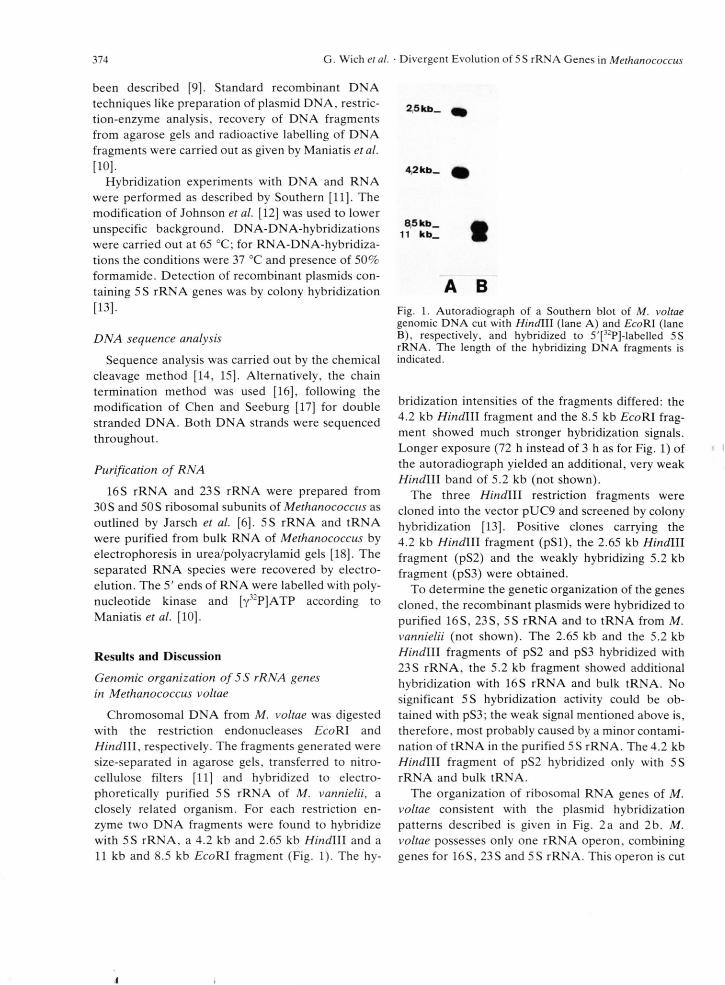
Chromosomal DNA from M. voltae was digested
with the restriction endonucleases £coRI and
Hindlll, respectively. The fragments generated were
size-separated in agarose gels, transferred to nitro
cellulose filters [1 1 ] and hybridized to electro-
phoretically purified 5S rRNA of M. vannielii, a
closely related organism. For each restriction en
zyme two DNA fragments were found to hybridize
with 5S rRNA, a 4.2 kb and 2.65 kb Hindlll and a
11 kb and 8.5 kb EcoRI fragment (Fig. 1). The hy-
4,2 k b _ 4 }
85 k b _
11 k b _
A B
Fig. 1. Autoradiograph of a Southern blot of M. voltae genomic DNA cut with Hindlll (lane A) and EcoRI (lane B), respectively, and hybridized to 5'[32P]-labelled 5S rRNA. The length of the hybridizing DNA fragments is indicated.
bridization intensities of the fragments differed: the
4.2 kb H indlll fragment and the 8.5 kb £coRI frag
ment showed much stronger hybridization signals.
Longer exposure (72 h instead of 3 h as for Fig. 1) of
the autoradiograph yielded an additional, very weak
Hindlll band of 5.2 kb (not shown).
The three Hindlll restriction fragments were
cloned into the vector pUC9 and screened by colony
hybridization [13]. Positive clones carrying the
4.2 kb H indlll fragment (pSl), the 2.65 kb Hindlll
fragment (pS2) and the weakly hybridizing 5.2 kb
fragment (pS3) were obtained.
To determine the genetic organization of the genes
cloned, the recombinant plasmids were hybridized to
purified 16S, 23S, 5S rRNA and to tRNA from M.
vannielii (not shown). The 2.65 kb and the 5.2 kb
Hindlll fragments of pS2 and pS3 hybridized with
23S rRNA, the 5.2 kb fragment showed additional
hybridization with 16S rRNA and bulk tRNA. No
significant 5S hybridization activity could be ob
tained with pS3; the weak signal mentioned above is,
therefore, most probably caused by a minor contami
nation of tRNA in the purified 5S rRNA. The 4.2 kb
Hindlll fragment of pS2 hybridized only with 5S
rRNA and bulk tRNA.
The organization of ribosomal RNA genes of M.
voltae consistent with the plasmid hybridization
patterns described is given in Fig. 2a and 2b. M.
voltae possesses only one rRNA operon, combining
genes for 16S, 23S and 5S rRNA. This operon is cut
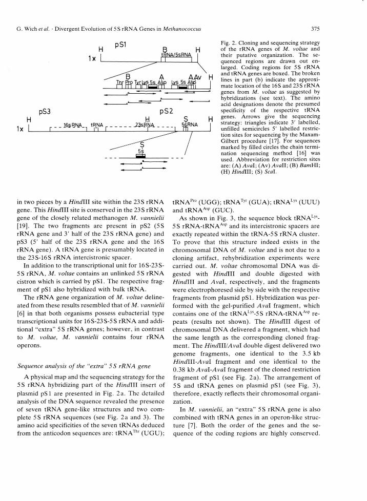
G. Wich et al. ■ Divergent Evolution of 5S rRNA Genes in Methanococcus 375
AAv
pS3 pS2
Fig. 2. Cloning and sequencing strategy of the rRNA genes of M. voltae and their putative organization. The sequenced regions are drawn out enlarged. Coding regions for 5S rRNA and tRNA genes are boxed. The broken lines in part (b) indicate the approximate location of the 16 S and 23 S rRNA genes from M. voltae as suggested by hybridizations (see text). The amino acid designations denote the presumed specificity of the respective tRNA genes. Arrows give the sequencing strategy: triangles indicate 3' labelled, unfilled semicircles 5' labelled restriction sites for sequencing by the Maxam- Gilbert procedure [17]. For sequences marked by filled circles the chain termination sequencing method [16] was used. Abbreviation for restriction sites are: (A) Ava\\ (Av) Avall\ (B) ZtomHI;(H) HindiII; (S) Seal.
in two pieces by a Hindlll site within the 23 S rRNA
gene. This HindlW site is conserved in the 23S rRNA
gene of the closely related methanogen M. vannielii
[19]. The two fragments are present in pS2 (5S
rRNA gene and 3' half of the 23S rRNA gene) and
pS3 (5' half of the 23S rRNA gene and the 16S
rRNA gene). A tRNA gene is presumably located in
the 23S-16S rRNA intercistronic spacer.
In addition to the transcriptional unit for 16S-23S-
5S rRNA, M. voltae contains an unlinked 5S rRNA
cistron which is carried by pSl. The respective frag
ment of pSl also hybridized with bulk tRNA.
The rRNA gene organization of M. voltae deline
ated from these results resembled that of M. vannielii
[6 ] in that both organisms possess eubacterial type
transcriptional units for 16S-23S-5S rRNA and addi
tional “extra” 5S rRNA genes; however, in contrast
to M. voltae, M. vannielii contains four rRNA
operons.
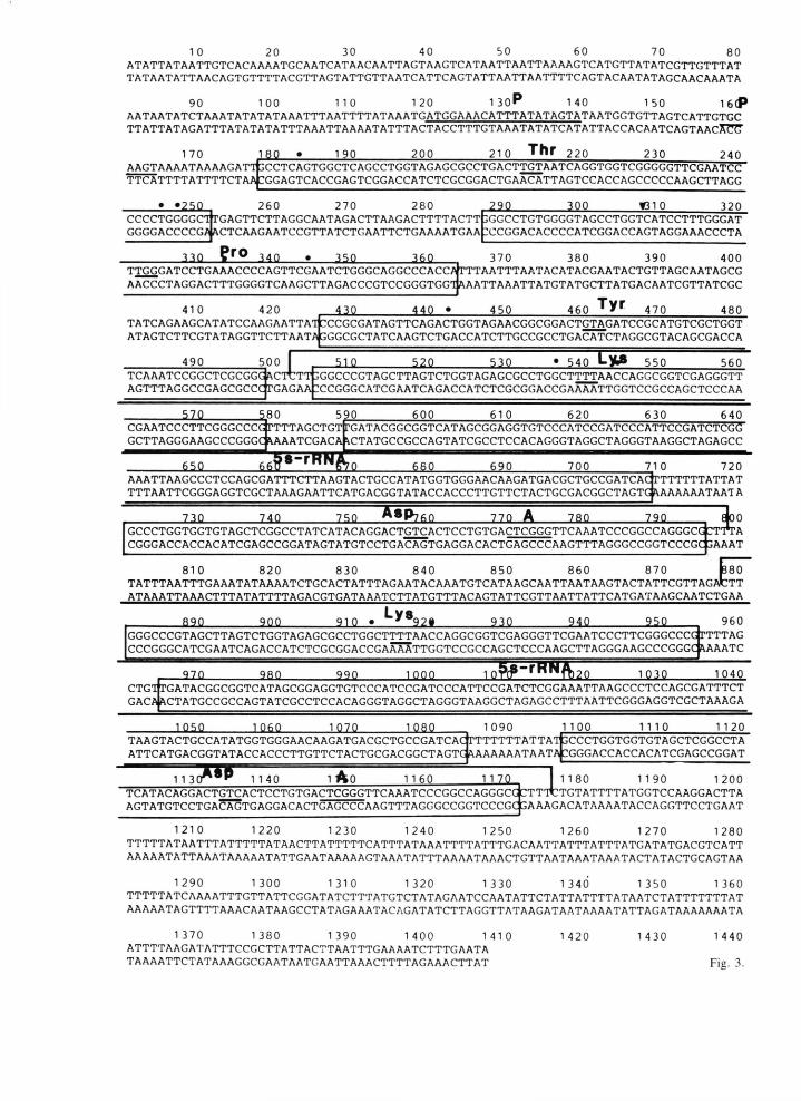
Sequence analysis of the “extra” 5S rRNA gene
A physical map and the sequencing strategy for the
5S rRNA hybridizing part of the Hindlll insert of
plasmid pSl are presented in Fig. 2a. The detailed
analysis of the DNA sequence revealed the presence
of seven tRNA gene-like structures and two com
plete 5S rRNA sequences (see Fig. 2a and 3). The
amino acid specificities of the seven tRNAs deduced
from the anticodon sequences are: tRNAThr (UGU);
tRN APro (UGG); tRNATyr (GUA); tRNALys (UUU)
and tRNAAsp (GUC).
As shown in Fig. 3, the sequence block tRNALys-
5 S rRNA-tRNAAsp and its intercistronic spacers are
exactly repeated within the tRNA-5S rRNA cluster.
To prove that this structure indeed exists in the
chromosomal DNA of M. voltae and is not due to a
cloning artifact, rehybridization experiments were
carried out. M. voltae chromosomal DNA was di
gested with Hindlll and double digested with
H indlll and Aval, respectively, and the fragments
were electrophoresed side by side with the respective
fragments from plasmid pSl. Hybridization was per
formed with the gel-purified Aval fragment, which
contains one of the tRNALys-5S rRNA-tRNAAsp re
peats (results not shown). The Hindlll digest of
chromosomal DNA delivered a fragment, which had
the same length as the corresponding cloned frag
ment. The Hindlll/Aval double digest delivered two
genome fragments, one identical to the 3.5 kb
Hindlll-Aval fragment and one identical to the
0.38 kb Aval-Aval fragment of the cloned restriction
fragment of pSl (see Fig. 2a). The arrangement of
5S and tRNA genes on plasmid pSl (see Fig. 3),
therefore, exactly reflects their chromosomal organi
zation.
In M. vannielii, an “extra” 5S rRNA gene is also
combined with tRNA genes in an operon-like struc
ture [7]. Both the order of the genes and the se
quence of the coding regions are highly conserved.
1 0 20 30 40 50 60 70 80ATATTATAATTGTCACAAAATGCAATCATAACAATTAGTAAGTCATAATTAATTAAAAGTCATGTTATATCGTTGTTTAT TATAATATTAACAGTGTTTTACGTTAGTATTGTTAATCATTCAGTATTAATTAATTTTCAGTACAATATAGCAACAAATA
90 100 1 10 1 20 1 3 o P 1 40 1 50 1 6(PAATAATATCTAAATATATATAAATTTAATTTTATAAATGATGGAAACATTTATATAGTATAATGGTGTTAGTCATTGTGC TTATTATAGATTTATATATATTTAAATTAAAATATTTACTACCTTTGTAAATATATCATATTACCACAATCAGTAACXrC
m 1 90 200 21 0 Thr 220 230 2401 70 ____________________AAGTAAAATAAAAGAT'lfccCTCAGTGGCTCAGCCTGGTAGAGCGCCTGACTTGTAATCAGGTGGTCGGGGGTTCGAATCC TTCATTTTATTTTCTAA:GGAGTCACCGAGTCGGACCATCTCGCGGACTGAACATTAGTCCACCAGCCCCCAAGCTTAGG
CCCCTGGGGCITGAGTTCTTAGGCAATAGACTTAAGACTTTTACTTjGGCCTGTGGGGTAGCCTGGTCATCCTTTGGGAT260 270 280
GGGGACCCCGÄa c t c a a g a a t c c g t t a t c t g a a t t c t g a a a a t g a a
33Q
22SL 300 131 0 320
z c c g g a c a c c c c a t c g g a c c a g t a g g a a a c c c t a
JLIQ- _ÜLQ_TTGGGATCCTGAAACCCCAGTTCGAATCTGGGCAGGCCCACCATTTAATTTAATACATACGAATACTGTTAGCAATAGCG AACCCTAGGACTTTGGGGTCAAGCTTAGACCCGTCCGGGTGGTAAATTAAATTATGTATGCTTATGACAATCGTTATCGC
370 380 390 400
410 420 AA0_ 450 460 Tyr 470 480TATCAGAAGCATATCCAAGAATTATOCCGCGATAGTTCAGACTGGTAGAACGGCGGACTGTAGATCCGCATGTCGCTGGT ATAGTCTTCGTATAGGTTCTTAATAGGGCGCTATCAAGTCTGACCATCTTGCCGCCTGACATCTAGGCGTACAGCGACCA
490 500 f 51 Q JLZiL 540 550 560TCAAATCCGGCTCGCGGGkCTTTTTpGGCCCGTAGCTTAGTCTGGTAGAGCGCCTGGCTTTTAACCAGGCGGTCGAGGGTT AGTTTAGGCCGAGCGCCCTGAGAAÜCCGGGCATCGAATCAGACCATCTCGCGGACCGAAAATTGGTCCGCCAGCTCCCAA
52SL 580 590 600 61 0 620 630 640CGAATCCCTTCGGGCCCGJTTTTAGCTGT rGATACGGCGGTCATAGCGGAGGTGTCCCATCCGATCCCATTCCGATCTCGG GCTTAGGGAAGCCCGGGC\AAATCGACA\CTATGCCGCCAGTATCGCCTCCACAGGGTAGGCTAGGGTAAGGCTAGAGCC
650 660 690 700 71 0 720AAATTAAGCCCTCCAGCGATTTCTTAAGTACTGCCATATGGTGGGAACAAGATGACGCTGCCGATCAClrTTTTTTATTAT TTTAATTCGGGAGGTCGCTAAAGAATTCATGACGGTATACCACCCTTGTTCTACTGCGACGGCTAGTG &AAAAAATAATA
qocrrr;
730 740 750 ML 7 7 Q A 76QGCCCTGGTGGTGTAGCTCGGCCTATCATACAGGACTGTCACTCCTGTGACTCGGGTTCAAATCCCGGCCAGGGCG^TTTA CGGGACCACCACATCGAGCCGGATAGTATGTCCTGACAGTGAGGACACTGAGCCCAAGTTTAGGGCCGGTCCCGCj AAAT
810 820 830 840 850 860 870 1380TATTTAATTTGAAATATAAAATCTGCACTATTTAGAATACAAATGTCATAAGCAATTAATAAGTACTATTCGTTAGACTT ATAAATTAAACTTTATATTTTAGACGTGATAAATCTTATGTTTACAGTATTCGTTAATTATTCATGATAAGCAATCTGAA
9QQ _2_LQ_Ly 3
22M- 2ASL 960’TTTAGlAAATC
GGGCCCGTAGCTTAGTCTGGTAGAGCGCCTGGCTTTTAACCAGGCGGTCGAGGGTTCGAATCCCTTCGGGCCCCCCGGGCATCGAATCAGACCATCTCGCGGACCGAAAATTGGTCCGCCAGCTCCCAAGCTTAGGGAAGCCCGGG
970 2.8 Q 1000 JLÜ XL 1030 1040CTGrGACi
GATACGGCGGTCATAGCGGAGGTGTCCCATCCGATCCCATTCCGATCTCGGAAATTAAGCCCTCCAGCGATTTCTCTATGCCGCCAGTATCGCCTCCACAGGGTAGGCTAGGGTAAGGCTAGAGCCTTTAATTCGGGAGGTCGCTAAAGA
1Q5Q _LM£L 1070 1 080TAAGTACTGCCATATGGTGGGAACAAGATGACGCTGCCGATCACITTTTTTATTAT3CCCTGGTGGTGTAGCTCGGCCTA ATTCATGACGGTATACCACCCTTGTTCTACTGCGACGGCTAGTG PiAAAAAATAATACGGGACCACCACATCGAGCCGGAT
1 1 3T * r
1 090 1 1 00 1 1 1 0 1 1 2 0
1 1 40 1 Ao 1 1 60 1 170 1180 1190 1200TCATACAGGACTGTCACTCCTGTGACTCGGGTTCAAATCCCGGCCAGGGCGtTT'ltTGTATTTTATGGTCCAAGGACTTA AGTATGTCCTGA^fGTGAGGACACTGAGCCCAAGTTTAGGGCCGGTCCCGCSAAAGACATAAAATACCAGGTTCCTGAAT
1210 1220 1230 1240 1250 1260 1270 1280TTTTTATAATTTATTTTTATAACTTATTTTTCATTTATAAATTTTATTTGACAATTATTTATTTATGATATGACGTCATT AAAAATATTAAATAAAAATATTGAATAAAAAGTAAATATTTAAAATAAACTGTTAATAAATAAATACTATACTGCAGTAA
1290 1300 1310 1320 1330 1340 1350 1360TTTTTATCAAAATTTGTTATTCG GATATCTTTATGTCTATAGAATCCAATATTCTATTATTTTATAATCTATTTTTTTAT AAAAATAGTTTTAAACAATAAGCCTATAGAAATACAGATATCTTAGGTTATAAGATAATAAAATATTAGATAAAAAAATA
1370 1380 1390 1400 1410 1420 1430 1440ATTTTAAGATATTTCCGCTTATTACTTAATTTGAAAATCTTTGAATATAAAATTCTATAAAGGCGAATAATGAATTAAACTTTTAGAAACTTAT Fig. 3.

G. Wich et al. ■ Divergent Evolution of 5 S rRN A Genes in Methanococcus 377
The only difference between the gene arrangement
in the tRNA/5S rRNA clusters of M. voltae and M.
vannielii is that just the tRNALys and the tRNAAsp
gene sequence is tandemly repeated in the operon of
M. vannielii [7]. A possible reason for the existence
of the duplicated “extra” 5 S rRNA gene in M. voltae
could reside in the fact that this organism possesses
only one rRNA operon-linked (versus 4 of M. van
nielii) 5S rRNA gene. There is, however, no infor
mation yet on any selective advantage of a surplus of
5S rRNA genes over those for 16S and 23S rRNA.
As mentioned above the coding regions of the 5S
rRNA/tRNA operon in M. voltae and M. vannielii
are highly conserved. The coding regions for
tRNAThr and tRNAPro differ from those of M. van
nielii in three positions, those for tRNALys and
tRNATyr in two and one position, respectively; the
sequences for tRNAAsp are identical. The sequence
differences presumably do not alter the secondary
structure of the respective tRNA molecules since
they either occur in loop regions or — when in stems
— are accompanied by a compensatory change.
It is interesting to note that the tRNAPro gene, as
in M. vannielii [7], seems to encode the 3' terminal
CCA end in the DNA structure. Since the homology
is very low outside of the coding regions this conser
vation clearly indicates that this CCA is indeed tran
scribed into the mature tRNA structure. This is in
contrast to tRNA genes sequenced so far from other
archaebacteria (for review see [2 0 ]).
55 rRNA genes
There is high sequence heterogeneity between the
rRNA operon-linked and the “extra” 5S rRNA
genes of M. vannielii [7, 21]. They differ in no less
than 13 positions. Such a degree of heterogeneity in
rRNA genes of a single organism is unique for pro
caryotes but has been reported for some eukaryotic
systems (for review see [2 2 ]).
To extend this analysis to the two types of 5S
rRNA genes in M. voltae, the operon-linked 5S
rRNA gene of pS2 was subcloned into plasmid vector
pUC18 using a Seal restriction site; the sequence of
the cloned operon-linked 5S rRNA gene was deter
mined.
As shown in Fig. 4a, the 5S rRNA genes of M.
voltae also display considerable sequence poly
morphism. The “extra” 5S rRNA gene diverges from
that of the rRNA operon-linked one in eleven posi
tions (Fig. 4a, 4b). None of these differences results
in an altered secondary structure (Fig. 4 a).
As shown in Fig. 4b and 4c, the sequences of the
operon-linked 5S rRNA genes on one hand and of
the “extra” 5 S rRNA genes on the other are highly
conserved in the two species. Fig. 4c demonstrates
the unusual fact that the two types of 5S rRNA genes
in one organism (the operon-linked 5S rRNA gene
and the “extra” 5S rRNA gene) are less related to
each other than the respective 5S rRNA gene types
in the two organisms.
There are several conclusions which can be drawn
from this observation:
(i) The different relative ratio of operon-linked
and “extra” 5S rRNA gene copies, one operon-link-
ed and two “extra” 5S rRNA genes in M. voltae
versus four operon-linked and one “extra” 5S rRNA
gene in M. vannielii, suggests that the products of
both 5S rRNA gene types, despite of the consider
able differences in primary structure, are functional
ly equivalent.
(ii) All operon-linked 5S rRNA genes of M. van
nielii have an identical sequence [19]. Since the prod
ucts of both types of 5 S rRNA genes are functionally
equivalent the conservation of the operon-linked 5S
genes is not due to biological pressure on the primary
structure but to an rRNA operon specific mecha
nism, e.g. randomization of mutations by recombina
tion events.
(iii) The relationship between the different gene
types suggests that they must have evolved in an
ancestral methanogen before division into the two
species by duplication of an ancestral 5 S rRNA gene
and divergent development, or by horizontal gene
transfer.
(iv) The surprisingly high sequence heterogeneity
of 5S rRNA genes in a single organism restricts the
value of phylogenetic trees based on 5S rRNA, espe-
Fig. 3. Sequence of the tRNA/5S rRNA gene cluster of M. voltae. Regions coding for tRNA and for 5S rRNA genes are boxed. The amino acid specificities of the tRNAs are given above the sequence. Dots indicate positions in the tRNA nucleotide sequences which differ from the sequences of the equivalent genes in M. vannielii [7]. The anticodon triplet of each tRNA is underlined. Two sequence boxes which are homologous to the putative promoter consensus sequence of stable RNA genes from M. vannielii [23] are marked. The Aval restriction sites (A) used for rehybridization experiments (see text) are indicated.

378 G. Wich et al. ■ Divergent Evolution of 5S rRNA Genes in Methanococcus
4aC - G T - A
• G - C \• a — t # Mc. vo l t a e (2 )• t — a •• T — A «
G - C T • GC - G T GG • T A A T
C C G C G G A G G A I I I I I IIG T C G C C T C C
T T A G A CA - T G - C A - T A - T
A* C C C
A T C C G A I I I I I I
T A G G C T T A A C I
G A A T A G
A - T C - G T - A
• a - t «a Mc. v o l t a e ( 1)• G - C »• C — G «• C — G «
G - C
C - G
A - T G - C A - T A — T
ۥ C C C
G C G G A G GI I M I I ICGCCTCC
A T C C G A I I I I I IT A G G C T
T A A C (G A A T A G
4bM. v o l t a e ( 1 )
M . v a n n i e l i i ( 1 )
M . v o l t a e ( 2 )
M . v a n n i e l i i ( 2 )
a) b) c) d)
a ) Me.. voltae(1) - 9 11 20
b) Mc..vannielii(1) 93 - 16 1 3
c ) Me..voltaet 2) 91 86.5 - 11
d) MC..vannielii(2) 83 89 91 -
Fig. 4. Primary and secondary structure of the 5S rRNA gene products from M. voltae: (a) Secondary structure of the “extra” 5S rRNA (1) and the operon-linked 5S rRNA gene transcripts (2). Differences in primary structures are marked by dots, (b) Comparison of the 5S rRNA-types of M. voltae and M. vannielii. Numbers in brackets denote the gene type:(1), “extra” 5S rRNA genes; (2), operon-linked 5S rRNA genes. Bases different from the M. voltae “extra” 5S rRNA gene transcript are indicated. Two regions which are highly conserved in the “extra” 5S rRNAs and the operon-linked 5 S rRNAs, respectively, are boxed, (c) 5S rRNA sequence homologies between M. voltae and M. vannielii. Numbers in the upper right-hand triangle denote the differences in primary structure. Numbers in the lower left-hand triangle are percent sequence homologies.
daily when sequences are compared between organ
isms possessing different copy numbers of these
genes.
Intercistronic spacers
The tRNA genes and the 5S rRNA genes encoded
by pSl are organized in an identical transcriptional
orientation and they are separated by only short
spacers (Fig. 5). Whereas the coding regions are
highly homologous the spacer regions differ both in
length and nucleotide sequence. Where conserved
features are present within the spacers they are con
sidered to possess a biological functions:
(i) Two homology boxes are present in the 5'
flanking regions. These conserved sequence motifs
meet the consensus sequence of the promotor postu
lated for transcription of stable rRNA genes in
Methanococcus [20, 23].

G. Wich et al. ■ Divergent Evolution of 5S rRNA Genes in Methanococcus 379
Fig. 5. Possible secondary structure of the tRNA/5S rRNA operon transcript. The sequence of the tRNALys- 5S rRNA-tRNAAsp gene block which is tan- demly repeated is underlined.
(ii) The secondary structure of the putative 5S
rRNA/tRNA transcript tends to separate the mature
tRNA sequences by stem/loop structures (Fig. 5).
Similar potential recognition sites for tRNA process
ing have been found for many procaryotic systems
(for review see [2 0 ]).
(iii) The tRNA and 5S rRNA gene flanking re
gions in M. voltae and in M. vannielii share a high
A —T content but otherwise very low homology.
Nevertheless, a short conserved sequence motif
seems noteworthy: the coding sequences for
tRNAAsp, which are part of the repeated sequence
blocks in the M. voltae tRNA/5S rRNA cluster and
in the M. vannielii cluster are followed in each of the
four examples by a conserved sequence of four
bases, namely 5 CTTT3. Homology drops down im
mediately after this four-base-motif. An identical se
quence was found surrounding the tRNAAla gene,
which is located within the 16 S—23 S rRNA gene
spacer in M. vannielii and was thought to participate
in processing of this tRNA gene [21].
Acknowledgement
We thank U. Bär for technical assistance and M.
Geier for preparation of the manuscript. This work
was supported by a grant from the Deutsche For
schungsgemeinschaft and the Fonds der Chemischen
Industrie.

380 G. Wich et al. • Divergent Evolution of 5 S rRNA Genes in Methanococcus
[1] M. W. Gray and W. F. Doolittle, Microbiol. Rev. 46, 1 (1982).
[2] J. D. Hofman, R. H. Lau, and W. F. Doolittle, Nucleic Acids Res. 7, 1321 (1979).
[3] J. Hui and P. P. Dennis, J. Biol. Chem. 260, 899(1985).
[4] H. Neumann, A. Gierl, J. Tu, J. Leibrock, D. Staiger, and W. Zillig, Mol. Gen. Genet. 192, 66 (1983).
[5] K. Lechner, G. Wich, and A. Böck, System. Appl. Microbiol. 6, 157 (1985).
[6] M. Jarsch, J. Altenbuchner, and A. Böck, Mol. Gen. Genet. 189, 41 (1983).
[7] G. Wich, M. Jarsch, and A. Böck, Mol. Gen. Genet. 196, 146 (1984).
[8] C. Janisch-Perron, J. Vieira, and J. Messing, Gene 33,103 (1985).
[9] L. Sibold, D. Pariot, L. Bhatnagar, M. Henriquet, and J.-P. Aubert, Mol. Gen. Genet. 200, 40 (1985).
[10] T. Maniatis, E. F. Fritsch, and J. Sambrook, in: Molecular Cloning, Cold Spring Harbor Laboratory, Cold Spring Harbor, New York 1982.
[11] E. M. Southern, J. Mol. Biol. 98, 503 (1975).[12] D. A. Johnson, J. W. Gantsch, J. R. Sportsman, and
J. H. Elder, Gene Anal. Techn. 1, 3 (1984).
13] M. Grunstein and D. S. Hogness, Proc. Natl. Acad. Sei. USA 72, 3961 (1975).
14] A. M. Maxam and W. Gilbert, Methods Enzymol. 65,499 (1980).
15] C. P. Gray, R. Sommer, C. Polke, E. Beck, and H. Schaller, Proc. Natl. Acad. Sei. USA 75, 50 (1978).
16] J. Messing, R. Crea, and P. H. Seeburg, Nucleic Acids Res. 9, 309 (1981).
17] E. J. Chen and P. H. Seeburg, DNA 4, 165 (1985).18] N. Tomioka and M. Sugiura, Mol. Gen. Genet. 193,
427 (1984).19] M. Jarsch and A. Böck, Mol. Gen. Genet. 200, 305
(1985).20] G. Wich, L. Sibold, and A. Böck, System. Appl.
Microbiol. 7, 18 (1986).21] M. Jarsch and A. Böck, Nucleic Acids Res. 11, 7537
(1983).22] V. A. Erdmann and J. Wolters, Nucleic Acids Res. 14,
r l—r59 (1986).23] G. Wich, H. Hummel, M. Jarsch, U. Bär, and A.
Böck, Nucleic Acids Res. 14, 2459 (1986).


![Orientia tsutsugamushi Infektion in der Maus ...ediss.sub.uni-hamburg.de/volltexte/2013/6057/pdf/Dissertation.pdf · Name Rickettsia tsutsugamushi durch [16]. Aufgrund von 16S rRNA-Analysen](https://static.fdokument.com/doc/165x107/5d55779188c993d40b8b5987/orientia-tsutsugamushi-infektion-in-der-maus-edisssubuni-name-rickettsia.jpg)