Functional analysis of Arabidopsis thaliana matrix...
123
I Functional analysis of Arabidopsis thaliana matrix metalloproteinases and MORC in plant immunity Dissertation zur Erlangung des Doktorgrades (Dr. rer. nat.) der Naturwissenschaftlichen Fachbereiche der Justus-Liebig-Universität Gießen durchgeführt am Institut für Phytopathologie und Angewandte Zoologie vorgelegt von M.Sc. Fei Zhang aus China Gießen 2016 1. Gutachter: Prof. Dr. Karl-Heinz Kogel 2. Gutachter: Prof. Dr. Annette Becker
Transcript of Functional analysis of Arabidopsis thaliana matrix...

I
Functional analysis of Arabidopsis thaliana matrix
metalloproteinases and MORC in plant immunity
Dissertation zur Erlangung des Doktorgrades
(Dr. rer. nat.)
der Naturwissenschaftlichen Fachbereiche
der Justus-Liebig-Universität Gießen
durchgeführt am
Institut für Phytopathologie
und Angewandte Zoologie
vorgelegt von
M.Sc. Fei Zhang
aus China
Gießen 2016
1. Gutachter: Prof. Dr. Karl-Heinz Kogel
2. Gutachter: Prof. Dr. Annette Becker

II
Contents
1. Introduction ............................................................................. 1
1.1 Plant immune system .......................................................................... 1
1.1.1 Pattern-triggered immunity (PTI) ................................................ 1
1.1.2 Effector-triggered immunity (ETI) ............................................... 2
1.1.3 Systemic acquired resistance (SAR) .......................................... 4
1.1.4 Nonhost resistance ..................................................................... 6
1.1.5 Reactive oxygen species (ROS) in plant-microbe interaction ..... 7
1.1.6 Callose deposition ...................................................................... 8
1.2 The section of matrix metalloproteinases ............................................ 8
1.2.1 Matrix metalloproteinases (MMPs) in mammals ......................... 8
1.2.1.1 Structure of MMPs in mammals ............................................. 9
1.2.1.2 Function of MMPs in mammals ........................................... 10
1.2.2 MMPs in plants ......................................................................... 15
1.2.2.1 Structure of plant MMPs ....................................................... 15
1.2.2.2 Activation of the plant MMPs activities ............................... 16
1.2.2.3 Function of plant MMPs ........................................................ 16
1.2.2.3.1 Tissue remodeling ....................................................... 16
1.2.2.3.2 Seed germination and development ......................... 17
1.2.2.3.3 Senescence and programmed cell death (PCD) .... 17
1.2.2.3.4 Biotic and abiotic stresses .......................................... 18
1.3 The section of MORC ........................................................................ 19
1.3.1 Morc family ............................................................................... 19
1.3.2 The role of MORC in plant-pathogen interaction ...................... 19
1.3.3 The role of MORC in gene silencing ......................................... 20
1.4 Objectives .......................................................................................... 21
2. Material and methods ........................................................... 24
2.1 Plant growth condition ....................................................................... 24
2.1.1 Matrix metalloproteinases (MMPs) protein assays ................... 24
2.1.2 Microrchidia (MORC) assays .................................................... 25
2.2 Extraction of DNA/RNA ...................................................................... 25
2.2.1 DNA extraction ......................................................................... 25
2.2.2 RNA extraction ................................................................................ 26
2.2.2.1 cDNA synthesis ...................................................................... 26
2.2.2.2 Check the quality of cDNA ................................................... 27
2.3 Use of Arabidopsis knock-out mutants .............................................. 28
2.3.1 Identification of T-DNA mutants (MMPs and MORCs) .............. 28
2.3.1.1 Identification of at1-mmp mutant ......................................... 28
2.3.1.2 Identification of atmorc mutants ........................................... 29
2.3.2 Production and identification of at-mmp triple mutant ............... 30
2.3.2.1 Crossing of Arabidopsis T-DNA mutants ........................... 30

III
2.3.2.2 Identification of triple mutant (at2-mmp/at3-mmp/at5-mmp)
................................................................................................................ 31
2.4 Generation of Arabidopsis over-expression stable transformants ...... 34
2.4.1 Cloning and construction of transformation vectors .................. 34
2.4.2 Floral dip transformation ........................................................... 35
2.4.3 Presence of construct ............................................................... 37
2.4.4 Expression of the construct ...................................................... 37
2.4.4.1 Transcript levels (RNA) ......................................................... 37
2.4.4.2 Protein expression of generated constructs ...................... 38
2.4.4.2.1 Protein extraction ......................................................... 38
2.4.4.2.2 Western blot ................................................................. 38
2.5 Pathogen infection ............................................................................. 39
2.5.1 Botrytis cinerea inoculation ....................................................... 39
2.5.2 Powdery mildew (Golovinomyces orontii) inoculation ............... 40
2.5.3 Pseudomonas syringae pv. tomato DC3000 inoculation .......... 40
2.6 Oxidative burst assay ........................................................................ 41
2.7 Callose deposition assay ................................................................... 41
2.8 Transposon expression assay (qPCR) .............................................. 42
3. Results ................................................................................... 43
3.1 Identification of at-mmp mutants ........................................................ 43
3.1.1 Identification of at1-mmp mutant .............................................. 43
3.1.2 Generation of at2-mmp at3-mmp at5-mmp triple mutant .......... 46
3.2 At-MMPs are required for pattern-triggered immunity ........................ 50
3.2.1 Callose response to MAMP in at-mmp mutants and At-MMP OE
transgenic plants ............................................................................... 50
3.2.2 ROS response to MAMP in at-mmp mutants and At-MMP OE
transgenic plants ............................................................................... 50
3.3 At-MMP is required for basal resistance to necrotrophic and
biotrophic fungal pathogens .............................................................. 53
3.3.1 At-MMP is required for basal resistance to necrotrophic fungal
pathogen Botrytis cinerea .................................................................. 53
3.3.2 At-MMP is required for resistance to biotrophic fungal pathogen
powdery mildew ................................................................................. 57
3.4 Identification of atmorc mutants ......................................................... 58
3.5 Arabidopsis MORCs affect plant resistance against pathogens......... 59
3.6 Production of transgenic plants containing site mutations in MORC1.
................................................................................................................ 61
3.7 Mutations in AtMORC1 reduces plant resistance against the hemi-
biotrophic bacterium P. syringae pv. tomato............................................. 64
3.8 AtMORCs knock-out mutants show enhanced expression of a
transposon and genes related to silencing mechanisms. ........................ 66
3.9 Transgenic plants containing mutations of AtMORC1 mutations show
no or strong derepression of transposons. .............................................. 69

IV
3.10 AtMORCs show effect on the expression of R gene RMG1
(resistance methylated gene 1) ............................................................... 71
4. Discussion ............................................................................. 75
4.1 The section of MMPs ......................................................................... 75
4.1.1 The MMPs family ...................................................................... 75
4.1.2 Reactive oxygen species and the role in plant immunity .......... 76
4.1.3 At-MMPs mediated oxidative burst ........................................... 77
4.1.4 Callose deposition and the role in plant immunity .................... 78
4.1.5 At-MMPs mediate callose deposition ........................................ 79
4.1.6 The role of plant MMPs in immune system ............................... 80
4.2 The section of MORC ........................................................................ 82
4.2.1 The MORC family ..................................................................... 82
4.2.2 Production and identification of AtMORC1 mutations ............... 84
4.2.3 The role of MORC in resistance ............................................... 84
4.2.4 The role of MORC in gene silencing ......................................... 87
4.2.5 The role of MORC to link resistance and gene silencing .......... 89
5. Summary ................................................................................ 91
6. References ............................................................................. 94
7. Supplementary .................................................................... 108
8. Declaration ........................................................................... 115
9. Acknowledgements ............................................................. 116

V
List of Abbreviation
ald1 agd2-like defense response protein 1 APMA 4-aminophenyl mercuric acetate
AzA Azelaic acid AZI1 Azelaic acid induced 1 BAP 6-benzylaminopurine
BTH Benzothiadiazole S-methyl ester cDNA Complementary DNA
CFU Colony forming units Col Collagenase-like protein CRT1 Compromised recognition of TCV
Cs Cytosolic Cys Cysteine array D Aspartic acid
DIR1 Defective in induced resistance 1
dko atmorc1/atmorc2 double knock-out DMSO Dimethyl sulfoxide dpi day post inoculation
E Glutamic acid
E-cadherin Epithelial cadherin
ECM Extracellular matrix
EFR Elongation factor Tu receptor
EF-Tu Elongation factor Tu
ETI Effector-triggered immunity
ETS Effector-triggered susceptibility
Fg Fusarium graminearum
flg22 A 22-amino-acid-long peptide derived from flagellin
FLS2 FLAGELLIN-SENSITIVE 2
Fn Fibronectin repeat Fr Furin-cleavage site G3P Glycerol-3-phosphate
GHKL Gyrase, Hsp90, histidine kinase, MutL
GPI Glycosylphosphatidylinositol h Hour
HPX Hemopexin
HR Hypersensitive response
IL Interleukin
INA 2, 6-dichloroisonicotinic acid
JA Jasmonic acid
K Lysine
LB Left border primer
LP Left primer
LRR Leucine-rich-repeat MAMPs Microbe-associated molecular patterns
MAPK Mitogen activated protein kinase
MBP Myelin basic protein
MeSA Methyl salicylic acid

VI
min Minutes
MMPs Matrix metalloproteinases
MORC Microrchidia
NASC European Arabidopsis stock center
NB-LRR Nucleotide-binding-leucine-rich repeat proteins
NMMP1 Nicotiana benthamiana matrix metalloprotease 1
NO Nitric oxide
PAMPs Pathogen-associated molecular patterns
PCD Programmed cell death
PR genes Pathogenesis-related genes
PRRs Pattern recognition receptors
Pst Pseudomonas syringae pv. tomato
PTI PAMP/pattern-triggered immunity
RdDM RNA-directed DNA methylation
R gene Resistance genes
RMG1 resistance methylated gene 1
ROS Reactive oxygen species
RP Right primer
R protein Resistance protein
RT-PCR Real-time PCR
SA Salicylic acid
SAR Systemic acquired resistance
SDC suppressor of drm2 cmt3
SDS Sodium dodecyl sulfate
SH Thiol group
SMEP1 Soybean metalloendoproteinase 1
SP Signal peptide
TBF1 TL1-binding factor 1
TCV Turnip crinkle virus
TEs Transposable elements
TNF Tumour-necrosis factor
Xcv Xanthomonas campestris pv. vesicatoria
Zn Zinc

1
1. Introduction
1.1 Plant immune system
Plants have to fight with various pathogens in whole life for their survival.
Biotrophic, hemi-biotrophic and necrotrophic pathogens employ different
strategies to infect plants, biotrophic pathogens infect plants and proliferate in
living tissues; necrotrophic pathogens can be axenically cultured and secret
toxins to infect host plants. In order to prevent from pathogens infection,
plants have evolved two layers of innate immunity, which is termed as pattern-
triggered immunity (PTI) and effector-triggered immunity (ETI) (Jones and
Dangl, 2006).
1.1.1 Pattern-triggered immunity (PTI)
The first layer of plant innate immunity is pattern-triggered immunity (PTI).
When plants are stimulated by biotic stresses, PTI is triggered through the
recognition of pathogen-associated or microbe-associated molecular patterns
(PAMPs/MAMPs) by membrane-localized pattern recognition receptors
(PRRs) (Macho and Zipfel, 2015; Rajamuthiah and Mylonakis, 2014; Boller
and Felix, 2009; Jones and Dangl, 2006). PTI in plants is very similar to innate
immunity in animals (Boller and Felix, 2009; Chisholm et al., 2006; Jones and
Dangl, 2006; Smith et al., 2003). For example, in plants, the flagellin of
bacteria is perceived as a MAMP through the leucine-rich-repeat (LRR)
domains of the membrane receptor FLAGELLIN-SENSITIVE 2 (FLS2) (Bohm
et al., 2014; Chinchilla et al., 2006; Gómez-Gómez and Boller, 2000). In
mammals, the Toll-like receptor TLR5 could also perceive bacterial flagellin
through its LRR domain (Hayashi et al., 2001; Smith et al., 2003). Another
well known MAMP is elongation factor Tu (EF-Tu) which is recognized by the
2
kinase EFR receptor (elongation factor Tu receptor) (Zipfel, 2014; Zipfel et al.,
2006). The typical PTI responses include the accumulation of reactive oxygen
species (ROS), activation of MAP kinase cascades, induction of defense
genes expression and occurrence of callose deposition (Bigeard et al., 2015;
O‟Brien et al., 2012; Ahuja et al., 2012; Bednarek, 2012; Torres and Dangl,
2005; Zipfel, 2008; Gómez-Gómez and Boller 2000)
1.1.2 Effector-triggered immunity (ETI)
Effector-triggered immunity (ETI), which was formerly called R-gene-based or
vertical resistance, is the second layer of plant innate immunity (Martin et al.,
2003; Nimchuk et al., 2003; Boller and He, 2009). When confronting with
intruders, efficient PTI could trigger host resistance to circumvent the
pathogen attacks. During the process, successful pathogens evolved
strategies, such as secretion of virulence effectors, to overcome PTI and
achieve compatibility (Abramovitch et al., 2006; Block et al., 2008; Block and
Alfano, 2011; Chisholm et al., 2006; Jones and Dangl, 2006). Nevertheless,
plants would not stop making effort to protect them from infection. They
further evolved effector-triggered immunity, in which the effectors are directly
or indirectly recognized by the resistance (R) proteins encoded by the
resistance genes (R genes) (Dangl et al., 2013; Win et al., 2012; Spoel and
Dong, 2012). Most of the R genes encode nucleotide-binding leucine-rich
repeat proteins (NB-LRR) (Ellis et al., 1999; Wei et al., 1999). The R gene-
mediated plant resistance is only active against specific isolates of a pathogen.
NB-LRR activation involves intra- and intermolecular conformational changes
and inappropriate NB activation seems to be tightly controlled by the
autoinhibition of LRR domains (Takken et al., 2006). Several NB-LRR proteins
indirectly recognize type III effectors, by detecting products of their action on
host targets, consistent with the „guard hypothesis‟ (Dangl and Jones, 2001;

3
Rajamuthiah and Mylonakis, 2014; Hurley et al., 2014), and this is more
frequently compared with direct recognition of effectors. The outcome of ETI
is the increase of plant resistance to invading pathogens. The typical event is
the strong defense reaction called the hypersensitive response (HR), which is
characterized by rapid apoptotic cell death and local necrosis at the infection
site to limit pathogen proliferation and disease symptoms (Strauss et al., 2012;
Boys et al., 2012; Bozkurt et al., 2012; Thirugnanasambandam et al., 2011).
ETI is regarded as a faster and stronger immune response (Cui et al., 2015;
Tao et al., 2003; Truman et al., 2006). More recently, proteomic approaches
have been used to study plant ETI signaling (Hurley et al., 2014; Parker et al.,
2013; Elmore et al., 2012; Dunham et al., 2012; Rodríguez-Herva et al., 2012;
Ntoukakis et al., 2013).
Based on the co-evolution during the interaction between a plant and a
microbe, a four phased „zigzag‟ model was proposed and accepted as a
current concept of the plant immune system (Fig. 1-1).
Figure. 1-1. Zigzag model of the plant immune system (Jones and Dangl,
2006).
The proposed model illustrates the quantitative output of the plant immune
4
system and the evolutionary relationship between PTI and ETI. In phase I,
plant PRRs recognize PAMPs, which activates PTI to prevent pathogen
colonization. In phase 2, successful pathogens suppress PTI using secreted
effectors and results in effector-triggered susceptibility (ETS). In phase 3,
specific recognition of an effector by the cognate plant R proteins results in
ETI, which leads to strong disease resistance. ETI is regarded as a stronger
and amplified version of PTI and often accompanied with an induction of an
HR at the infection site. In phase 4, natural selection drives pathogens to
evade ETI by loss of the read effectors, or by gain of new effectors (in blue)
that suppress ETI. Subsequently, natural selection results in new R proteins
to recognize the newly acquired effectors and triggers ETI again.
1.1.3 Systemic acquired resistance (SAR)
The invasion of pathogens not only triggers the local defense response, but
also induces the generation of specific signals in plants, such as salicylic acid
(SA), methyl salicylic acid (MeSA), azelaic acid (AzA), and glycerol-3-
phosphate (G3P) (Gao et al., 2015; Shah and Zeier, 2013; Kachroo and
Robin, 2013; Gao et al., 2014; Chaturvedi et al., 2012; Chanda et al., 2011;
Jung et al., 2009; Park et al., 2007). The accumulation of these signals leads
to the expression of PR genes (pathogenesis-related genes) in the uninfected
tissue to protect the rest of the plant from subsequent infections (Yan and
Dong, 2014; Durrant and Dong, 2004). This process is termed systemic
acquired resistance (SAR). SAR can also be induced by 2, 6-
dichloroisonicotinic acid (INA) which is synthetic analogs of SA and
benzothiadiazole S-methyl ester (BTH) (Görlach et al., 1996; Durrant and
Dong, 2004). Wang et al (2014) showed that nitric oxide (NO) and reactive
oxygen species (ROS) play an important role in inducing SAR. SAR leads to
long-lasting, broad- spectrum resistance against pathogen infection

5
(Wendehenne et al., 2014; Fu and Dong, 2013). Pajerowska-Mukhtar et al
(2012) suggested that SAR signaling is associated with changes in amino
acid homeostasis induced by ETI. TBF1 (TL1-binding factor 1), which is
required for the growth-to-defense transition upon pathogen challenge, is
derepressed within 30 min of the pathogen infection, suggesting that it might
be one of the earliest responses triggered by SAR (Pajerowska-Mukhtar et al.,
2012). Characterization of the Arabidopsis ald1 (agd2-like defense response
protein 1) mutant also showed that an amino acid–derived defense signal is
generated upstream of SA synthesis (Song et al., 2004). DIR1(defective in
induced resistance 1) was discovered in a genetic screen designed
specifically to identify SAR signals, which encodes a putative lipid-transfer
protein, is probably involved in the synthesis or transport of a lipid molecule,
which is a signal for SAR (Maldonado et al., 2002). AZI1 (azelaic acid induced
1), encoding a predicted secreted protease-inhibitor/seed-storage/lipid-
transfer family protein, which regulates the production or translocation of a
mobile SAR signal together with DIR1 (Jung et al., 2009). Another important
signal for SAR is jasmonic acid (JA). The level of JA increased significantly at
6 h after P. syringae pv. tomato (Pst) DC3000/AvrRpm1 inoculation and
returned to normal level 11 h after Pst infection (Truman et al., 2007). The
SAR is induced by exogenous application of JA. However, SAR is
compromised in JA-insensitive mutant sgt1b/jai4, JA-biosynthesis mutant opr3,
and JA-response mutant jin1 plants (Attaran et al., 2009). Another required
signal for SAR is glycerol-3-Phosphate (G3P), which showed accumulation
within 6 h after pathogen infection (Chanda et al., 2011). It can be produced
through the activity of the G3P dehydrogenase GLY1 (Mandal et al., 2011;
Chanda et al., 2008).

6
1.1.4 Nonhost resistance
Nonhost resistance is defined as a resistance of an entire plant species to all
isolates of a pathogen (Stam et al., 2014; Heath, 2000; Mysore and Ryu, 2004;
Nürnberger and Lipka, 2005). It is a broad-spectrum plant defense. Most of
nonhost resistance is associated with a broad range of mechanisms that are
regulated by multiple genes (Uma et al., 2011; Fan and Doerner, 2012; Ham
et al., 2007; Mysore and Ryu, 2004). A pathogen capable of infecting other
plant species but incapable of invading a nonhost plant is referred as a
nonhost pathogen. Nonhost pathogens which land on the plant surface are
exposed to a wide range of preformed plant defenses (Heath, 2000;
Hückelhoven, 2007). Some nonhost pathogens are able to penetrate into
apoplastic space through stomata or wounds of the plant surface. The
apoplast is therefore the major battleground during the plant-microbe
interactions (Alfano and Collmer, 1996). Nonhost defense responses can be
induced by PTI or ETI (Senthil-Kumar and Mysore, 2013). Physical restriction
and chemical inhibition of pathogens are the important components of
nonhost resistance. The cuticle layer, the epidermis, and the cell wall act as
physical barriers for the entry of pathogens. The callose and lignin deposition
is induced and reinforce the cell wall after nonhost pathogen invasion
(Bestwick et al., 1995, 1997). Plants also produce some antimicrobial
compounds which inhibit host- and nonhost pathogen growth (Che et al., 2011;
Lee et al., 2008). Several secondary metabolites have antimicrobial properties
and involve in restricting the growth of invading pathogens (Fan et al., 2011;
Aires et al., 2009; Filippone et al., 1999). In Arabidopsis thaliana, sulfur- and
nitrogen-containing secondary metabolite compounds glucosinolates play an
important role in plant defense against variety of pathogens (Baskar et al.,
2012; Bednarek, 2012). Induced defense response is another main part of
nonhost resistance of plants against bacterial pathogens (Tao et al., 2003).
During the nonhost resistance, ROS, salicylic acid (SA), and other hormones
7
play an important role as signaling molecules. These defense responses
against nonhost-pathogen growth through producing structural barriers,
inducing biosynthesis of antimicrobial chemicals, and activating several
defense pathways at the molecular level, the growth of nonhost-pathogen
could be inhibited.
1.1.5 Reactive oxygen species (ROS) in plant-microbe interaction
The production of reactive oxygen species (ROS) is one of the earliest plant
defense responses during microbial infection. Previous studies showed that
ROS may be involved in plant defense directly through the antimicrobial
activity or mediating cell wall cross-linking, the induction of defense genes
expression and the induction of cell death (Torres, 2010; Torres and Dangl,
2005; Boller and Felix, 2009; Bolwell, 1999; Lamb and Dixon, 1997; Levine et
al., 1994; Torres et al., 2006; Zurbriggen et al., 2009; Hückelhoven and Kogel,
2003). The production of ROS requires the prior accumulation of Ca2+ (Kadota
et al., 2014; Li et al., 2014b; Grant and Loake, 2000; Grant et al., 2000), and
is dependent on the activity of membrane-localized NADPH oxidases
(respiratory burst oxidase homologs, Rboh) (Ranf et al., 2011; Kobayashi et
al., 2006; Torres et al., 2006). Different lifestyles of pathogens have distinct
responses to ROS generated in the host plants (Heller and Tudzynski, 2011).
For example, biotrophic and hemibiotrophic fungi depend on the prevention of
a strong oxidative burst response and the hypersensitive response to achieve
infection (Molina and Kahmann, 2007; Shetty et al., 2007). Thus, the oxidative
burst accumulation of host plants is an effective strategy to combat biotrophic
pathogens.

8
1.1.6 Callose deposition
Callose is an amorphous, high–molecular weight β-(1, 3)-glucan polymer. It is
contained in cell-wall appositions that are effective barriers induced at the
penetration sites during early stages of pathogenic infection (Piršelová and
Matušíková, 2013; Li et al., 2012). Callose deposition is triggered by MAMPs
(Luna et al., 2011; Brown et al., 1998; Gómez-Gómez et al., 1999a), and is a
hallmark of FLS2-mediated PTI. For example, the MAMP flagellin (Gómez-
Gómez and Boller, 2000), elongation factor EF-Tu (Elf18) (Kunze et al., 2004),
and chitin, a β-(1,4)-linked polymer as well as other molecules from fungal cell
walls can induce callose deposition (Poliakovskiy and Dmitriev, 2011; Iritri and
Faoro, 2009).
1.2 The section of matrix metalloproteinases
1.2.1 Matrix metalloproteinases (MMPs) in mammals
Matrix metalloproteinases (MMPs) are a family of highly conserved
endopeptidases containing zinc ion in the active site, which was first found in
mammals in 1962 (Gross and Lapiere, 1962). The MMP family is widely
distributed throughout all kingdoms of life. There are 23 MMPs found in
humans (Nagase et al., 2006). MMPs are secreted or attached to the cell
surface. In mammals, MMPs play a key role in many important physiological
and pathological processes, such as remodeling of the extracellular matrix
(ECM), regulation of cell migration, proliferation, adhesion and signaling, by
limited proteolytic processing of substrate proteins (Butler and Overall, 2009;
Birkedal-Hansen et al., 1993; Stamenkovic, 2003; Vu and Werb, 2000; Parks
et al., 2004).

9
1.2.1.1 Structure of MMPs in mammals
The structure of mammalian MMP contains a signal peptide, a prodomain, a
catalytic domain, a linker peptide and a hemopexin (Hpx) domain (Nagase et
al., 2006). In the homologues of MMP, the catalytic domain possesses a zinc
binding motif HEXXHXXGXXH, in which the zinc atom is located in the active
site. The propeptide domain maintains the “cysteine switch” sequence
PRCGXPD. All of the MMPs share this common domain structure (Nagase
and Woessner, 1999).
The conserved cysteine residue coordinates with the active zinc ion to inhibit
catalytic activity. When the propeptide domain is removed, the MMPs have
the activity to cleave substrate (Andrea et al., 2007). Most MMP members
also contain a hemopexin domain in the C-terminal by a flexible hinge. The
hemopexin domain encodes a four-bladed β-propeller structure which
mediates protein–protein interactions. This domain also has a function in
modulating substrate specificity, activation of the enzyme, protease
localization, internalization and degradation (Overall, 2002; Parks et al., 2004).
MMPs are subdivided into different groups according to differences in domain
composition (Fig.1–2). One clear division is between MMPs that are secreted
and anchored to the cell surface by an intrinsic motif, including a
transmembrane domain, a glycosylphosphatidylinositol (GPI) anchor or an
amino N-terminal signal anchor (Parks et al., 2004).

10
Figure. 1–2. Domain structure of the mammalian MMP family.
MMPs are subdivided into groups on the basis of differences in domain
composition. C5, type-V-collagen-like domain; Col, collagenase-like protein;
Cs, cytosolic; Cys, cysteine array; Fn, fibronectin repeat; Fr, furin-cleavage
site; Pro, pro-domain; SH, thiol group; SP, signal peptide; Zn, zinc (according
to Parks et al., 2004).
1.2.1.2 Function of MMPs in mammals
MMPs were thought to be responsible for degradation of extracellular matrix
(ECM) molecules in the tissue. All isolated MMPs have been shown to be
capable of degrading various protein components in the ECM (Sternlicht and
Werb, 2001). Consequently, MMPs family plays a role as enzymes
responsible for the turnover, degradation, catabolism and destruction of the
ECM. In addition, some non-ECM molecules are also possible substrates of
MMPs (Nagase et al., 2006). Since MMPs are secreted or anchored to the
cell surface, their potential substrates include all membrane proteins and
proteins in the secretory pathway and extracellular space.
In most cases, MMP-deficient mice mutants showed no or a minor phenotype

11
under unchallenged condition. Nevertheless, the MMP14-deficient mice
mutant showed severe bone deformations (Holmbeck et al., 1999; Zhou et al.,
2000). These indicate that some MMPs might not have a direct role in the
turnover of ECM proteins. After challenge, such as injury, cancer,
inflammation or infection, MMP-deficient mice mutant displayed various
phenotypes, indicating that these enzymes have specific roles in tissue repair,
angiogenesis, host defence, tumour progression and inflammation (Table.1-1).
Taken together, MMPs may have evolved to respond to environmental
pressures (Fig.1-3) (Parks et al., 2004).
Additionally, MMPs are of importance in modulating inflammatory processes.
Many MMPs are increased or mis-regulated in any disease that is
characterized by or associated with inflammation. The role of MMPs as matrix
degrading proteinases justifies their inclusion as important components of the
host response to traumatic, infectious, toxic or autoimmune insults.
Several MMPs were induced at injury sites and crucial for wound closure
(Parks, 1999). For example, the catalytic activity of MMP1 is required for the
repair of skin wounds. Moreover, MMP7 and MMP9 are implicated in wound
repairing (Dunsmore et al., 1998; McGuire et al., 2003; Pilcher et al., 1997).
MMP3-deficient mice mutant showed impaired immunity to intestinal bacterial
infection suggesting that MMP3 is important to resist bacteria (Li et al., 2004).
It is different from other MMPs which are expressed in response to injury or
inflammation, MMP7 is expressed in healthy epithelium indicating its function
in common homeostatic processes, such as resistance to microorganisms
and apoptosis. In mice, MMP7 activates intestinal pro-α-DEFENSINS which is
evidenced by the impaired ability to battle pathogens Escherichia coli and
Salmonella typhimurium in the MMP7-deficient mice mutant (Wilson, et al.,
1999). Furthermore, the induction of MMP7 in mucosal epithelium is highly
sensitive to the presence of virulent bacteria, further suggesting a role of this
MMP in innate immunity (López-Boado, et al., 2000, 2001). Besides, MMP2
and MMP9 also showed early immune response to against Streptococcus

12
pneumoniae infection (Hong et al., 2011).
Table. 1-1. Inflammatory and immune phenotypes of Mmp-null mice
*These phenotypes were reversed following transplantation of wild-type bone
marrow, indicating that the effect observed in knockout mice was caused by
the lack of the matrix metalloproteinase (MMP) in an inflammatory cell or
group of inflammatory cells. E-cadherin, epithelial cadherin; IL, interleukin;
TNF, tumour-necrosis factor (Parks et al., 2004).
13
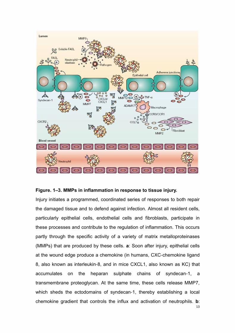
Figure. 1–3. MMPs in inflammation in response to tissue injury.
Injury initiates a programmed, coordinated series of responses to both repair
the damaged tissue and to defend against infection. Almost all resident cells,
particularly epithelial cells, endothelial cells and fibroblasts, participate in
these processes and contribute to the regulation of inflammation. This occurs
partly through the specific activity of a variety of matrix metalloproteinases
(MMPs) that are produced by these cells. a: Soon after injury, epithelial cells
at the wound edge produce a chemokine (in humans, CXC-chemokine ligand
8, also known as interleukin-8, and in mice CXCL1, also known as KC) that
accumulates on the heparan sulphate chains of syndecan-1, a
transmembrane proteoglycan. At the same time, these cells release MMP7,
which sheds the ectodomains of syndecan-1, thereby establishing a local
chemokine gradient that controls the influx and activation of neutrophils. b:
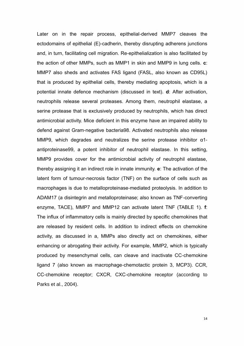
14
Later on in the repair process, epithelial-derived MMP7 cleaves the
ectodomains of epithelial (E)-cadherin, thereby disrupting adherens junctions
and, in turn, facilitating cell migration. Re-epithelialization is also facilitated by
the action of other MMPs, such as MMP1 in skin and MMP9 in lung cells. c:
MMP7 also sheds and activates FAS ligand (FASL, also known as CD95L)
that is produced by epithelial cells, thereby mediating apoptosis, which is a
potential innate defence mechanism (discussed in text). d: After activation,
neutrophils release several proteases. Among them, neutrophil elastase, a
serine protease that is exclusively produced by neutrophils, which has direct
antimicrobial activity. Mice deficient in this enzyme have an impaired ability to
defend against Gram-negative bacteria98. Activated neutrophils also release
MMP9, which degrades and neutralizes the serine protease inhibitor α1-
antiproteinase99, a potent inhibitor of neutrophil elastase. In this setting,
MMP9 provides cover for the antimicrobial activity of neutrophil elastase,
thereby assigning it an indirect role in innate immunity. e: The activation of the
latent form of tumour-necrosis factor (TNF) on the surface of cells such as
macrophages is due to metalloproteinase-mediated proteolysis. In addition to
ADAM17 (a disintegrin and metalloproteinase; also known as TNF-converting
enzyme, TACE), MMP7 and MMP12 can activate latent TNF (TABLE 1). f:
The influx of inflammatory cells is mainly directed by specific chemokines that
are released by resident cells. In addition to indirect effects on chemokine
activity, as discussed in a, MMPs also directly act on chemokines, either
enhancing or abrogating their activity. For example, MMP2, which is typically
produced by mesenchymal cells, can cleave and inactivate CC-chemokine
ligand 7 (also known as macrophage-chemotactic protein 3, MCP3). CCR,
CC-chemokine receptor; CXCR, CXC-chemokine receptor (according to
Parks et al., 2004).

15
1.2.2 MMPs in plants
In comparison to the importance of MMPs in mammals, the function of MMPs
is still less well-studied in plants. So far, there are only a few MMPs being
reported, and MMPs have been isolated from plant species, including
Arabidopsis (Giada et al., 2014; Lenger et al., 2012; Maidment et al., 1999),
tobacco (Kang et al., 2010), soybean (Cho et al., 2009; Ragster and
Chrispeels, 1979), cucumber (Delorme et al., 2000) and Loblolly pine
(Ratnaparkhe et al., 2009). In plant, the MMPs function to degrade the
extracellular matrix (ECM). Moreover, they involved in several physiological
processes during plant growth and development, such as the germination of
seeds (Ratnaparkhe et al., 2009), programmed cell death (PCD) (Delorme et
al., 2000), senescence (Golldack et al., 2002) and expansion of leaf (Graham
et al., 1991). Besides, MMPs were reported to play an important role when
confronting with biotic or abiotic stresses (Schiermeyer et al., 2009; Liu et al.,
2001; Combier et al., 2007; Flinn, 2008).
1.2.2.1 Structure of plant MMPs
In Arabidopsis thaliana, there are five MMPs (Maidment et al., 1999). Similar
to the structure of mammalian MMPs, the plant MMPs possess a signal
peptide, a propeptide domain and a catalytic domain (Fig.1-4). In the
propeptide domain, there exists a conserved cysteine switch sequence
PRCGXXD; while the catalytic domain contains the zinc-binding motif
(HEIGHXLGLXH) followed by the conserved methionine residue of the Met
turn (Rawlings et al., 2010). The most important site determining the
specificity of MMP cleavage site is S1, which is located directly to the right of
the catalytic zinc ion and well suited to accommodate hydrophobic residues,
such as leucine or isoleucine (Giada et al., 2014).

16
Figure. 1– 4. General structure of plant MMPs.
Relevant domains identified are color-coded. Signal peptide, propeptide
domain, cysteine switch, catalytic domain, zinc-binding domain, positions of
putative furin cleavage sites, GPI-anchor modification sites and C-terminal
transmembrane domains (according to Flinn, 2008).
1.2.2.2 Activation of the plant MMPs activities
As mentioned, the activity of mammalian MMPs requires a mechanism to
disrupt the Cys-Zn2+ interaction between the conserved cysteine residue and
the active zinc site (Parks et al., 2004; Sternlicht et al., 2001). Similarly, the
plant MMPs exhibit no activity in original situation; but they require physical
delocalization to achieve the proteolytic activity by cleaving off the cysteine
switch (Flinn, 2008). In Arabidopsis, all five recombinant MMPs were able to
cleave the substrates (Giada et al., 2014), among which the At1-MMP can be
activated by the activator 4-aminophenyl mercuric acetate (APMA) to cleave
the propeptide domain (Maidment et al., 1999).
1.2.2.3 Function of plant MMPs
1.2.2.3.1 Tissue remodeling
One of the most important roles of plant MMPs is to remodel the plant tissues.
The first plant metalloproteinase activity was described in soybean leaves
(Ragster and Chrispeels, 1979). They reported that the protein possesses an
17
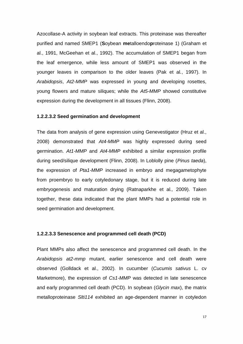
Azocollase-A activity in soybean leaf extracts. This proteinase was thereafter
purified and named SMEP1 (Soybean metalloendoproteinase 1) (Graham et
al., 1991, McGeehan et al., 1992). The accumulation of SMEP1 began from
the leaf emergence, while less amount of SMEP1 was observed in the
younger leaves in comparison to the older leaves (Pak et al., 1997). In
Arabidopsis, At2-MMP was expressed in young and developing rosettes,
young flowers and mature siliques; while the At5-MMP showed constitutive
expression during the development in all tissues (Flinn, 2008).
1.2.2.3.2 Seed germination and development
The data from analysis of gene expression using Genevestigator (Hruz et al.,
2008) demonstrated that At4-MMP was highly expressed during seed
germination. At1-MMP and At4-MMP exhibited a similar expression profile
during seed/silique development (Flinn, 2008). In Loblolly pine (Pinus taeda),
the expression of Pta1-MMP increased in embryo and megagametophyte
from proembryo to early cotyledonary stage, but it is reduced during late
embryogenesis and maturation drying (Ratnaparkhe et al., 2009). Taken
together, these data indicated that the plant MMPs had a potential role in
seed germination and development.
1.2.2.3.3 Senescence and programmed cell death (PCD)
Plant MMPs also affect the senescence and programmed cell death. In the
Arabidopsis at2-mmp mutant, earlier senescence and cell death were
observed (Golldack et al., 2002). In cucumber (Cucumis sativus L. cv
Marketmore), the expression of Cs1-MMP was detected in late senescence
and early programmed cell death (PCD). In soybean (Glycin max), the matrix
metalloproteinase Slti114 exhibited an age-dependent manner in cotyledon

18
(Cho et al., 2009). Taken together, these studies suggest that the plant MMPs
involved in senescence and PCD.
1.2.2.3.4 Biotic and abiotic stresses
Based on the gene expression analysis, the role of plant MMPs in response to
biotic and abiotic stresses have been reported (Schiermeyer et al., 2009; Liu
et al., 2001; Combier et al., 2007; Flinn, 2008). In soybean, GmMMP2 was
induced by wounding and dehydration (Liu et al., 2001). Likewise, the
expression of At2-MMP in Arabidopsis root was induced by NaCl and
stimulated by cadmium treatment in leaves (Golldack et al., 2002). In
Arabidopsis, four enzymes, At1-MMP, At2-MMP, At3-MMP and At5-MMP,
displayed maximal activity at pH between 7.0 and 8.0. In addition, the
proteolytic activity of At-MMPs was affected by temperature. For example, the
activity of At3-MMP and At5-MMP reach a maximum at 35°C; for At1-MMP,
At2-MMP and At4-MMP, 45–55°C is the best temperature for their activity
(Marino et al., 2014). Taken together, these data suggested that the plant
MMPs play a role in the adaptation to abiotic stresses.
On the side of biotic interactions, GmMMP2 transcript levels were increased
in compatible and incompatible interactions of soybean tissues with the
oomycete pathogen Phytophthora sojae, as well as the bacterial pathogen
Pseudomonas syringae pv. glycinea. In accordance with the GmMMP2
activation, a metalloproteinase activity was increased in suspension cells
following the bacterial infection (Liu et al., 2001). In Arabidopsis, At3-MMP
was induced after flg22 treatment in seedlings (Zipfel et al., 2004). In addition,
meta-profile heat map analysis of At-MMPs gene expression in response to
different pathogen stresses indicated that At2-MMP and At3-MMP were up-
regulated (Flinn, 2008). These data suggested that plant MMPs participate in
the response to biotic and abiotic stresses.

19
1.3 The section of MORC
1.3.1 Morc family
The turnip crinkle virus (TCV) is a positive sense RNA virus which belongs to
the carmovirus group. It is able to infect most Arabidopsis ecotypes. The R
protein HRT confers resistance to TCV (Cooley et al., 2000). MORC was
formerly termed CRT1 (compromised recognition of TCV). It was identified
through a genetic screening of mutants carrying HRT that were compromised
for the recognition of TCV (Kang et al., 2008). The MORC family is a
subfamily of microrchidia (MORC) GHKL ATPases (Gyrase, Hsp90, histidine
kinase, MutL) superfamily (Dutta and Inouye, 2000). The first MORC protein
was isolated from mouse, which is required for meiotic nuclear division
(Watson et al., 1998). Thereafter, MORC genes have been identified in
mammals (Pastor et al., 2014) and Caenorhabditis elegans (Moissiard et al.,
2012). Besides, MORC have also been isolated from different plant species,
including Arabidopsis (Kang et al., 2008), barley, tobacco and potato. There
are seven members of MORC identified in Arabidopsis thaliana and five in
barley (Langen et al., 2014). Previous studies demonstrated that MORC is
involved in plant immunity (Kang et al., 2008, 2010, 2012; Langen et al.,
2014).
1.3.2 The role of MORC in plant-pathogen interaction
Sequence analysis showed that the MORC family contains a combination of a
gyrase, histidine kinase, MutL (GHKL) and S5 domains. It is a subfamily of
microrchidia (MORC) GHKL ATPases superfamily. The previous data
indicated that MORC is required for multiple layers of plant immunity. RNAi-
mediated silencing of MORC2 and MORC3, which are the two closest
homologues of MORC1, results in higher susceptibility to TCV infection

20
compared with the wild-type (Kang et al., 2008). The ssi4 mutant contains a
mutation of a TIR-NBS-LRR type R protein which leads to the activation of
defense responses, such as HR, SA accumulation and defense-related gene
expression (Shirano et al., 2002). While morc1 mutant reduced the
spontaneous cell death induced by a constitutively active R protein (ssi4).
Additionally, MORC1 also altered HRT-induced defense responses as well as
ssi4, and MORC1 protein interacted with other NBS-LRR proteins such as
HRT and SSI4 (Kang et al., 2008). The Arabidopsis knockout morc1/ morc2
double mutant, which was produced from the Col-0 background, displayed
higher susceptibility to avirulent bacterial pathogen Pseudomonas syringae pv.
tomato (Pst) and the oomycete Hyaloperonospora arabidopsidis (Kang et al.,
2010; Langen et al., 2014). Arabidopsis MORC1 and MORC2 have been
previously demonstrated to be required for various types of disease
resistance including basal resistance, nonhost resistance SAR and ETI (Kang
et al., 2008; 2010; 2012). In barley, there are five MORCs isolated, all of
which are involved in resistance. Knock-down (KD) of barley MORC2 plant
displayed reduced numbers of fungal colonies compared with control after
powdery mildew fungus Bgh race A6 (BghA6) infection. Moreover, the
overexpression line of barley MORC1 exhibited more Bgh colonies.
Additionally, silencing of barley MORC2 increased the resistance to the root
rot-causing necrotrophic fungus Fusarium graminearum (Fg). Furthermore,
barley MORCs were also involved in ETI (Langen et al., 2014). Taken
together, MORCs are involved in plant resistance. However, Arabidopsis and
barley MORC resulted in opposite effects on plant immunity.
1.3.3 The role of MORC in gene silencing
DNA methylation, DNA repeats and histone methylation are the common
ways to achieve epigenetic gene silencing. Arabidopsis MORC1 and MORC6

21
were involved in gene silencing, which cause derepression of DNA-
methylated genes and transposable elements (TEs) but no losses of DNA or
histone methylation (Moissiard et al., 2012). A modest reduction of DNA
methylation and repressive histone marks at specific RNA-directed DNA
methylation (RdDM) target sites in atmorc6 mutant suggested that AtMORC6
play a role in RdDM pathway (Brabbs et al., 2013; Lorkovic et al., 2012).
Moissiard et al (2014) reported that the AtMORC6 physically interacts with
AtMORC1 and AtMORC2 to enforce gene silencing, moreover, real-time PCR
(RT-PCR) from RNA extracted from atmorc1 and atmorc2 mutants indicated
that SDC was derepressed in atmorc1 mutant but not in atmorc2 mutant, the
expression of two transposons, ATCOPIA28 and ROMANIAT5, showed an
increased derepression in the atmorc1/atmorc2 double mutant compared with
atmorc1 and atmorc2 single mutants, indicating that AtMORC1 and AtMORC2
act redundantly in transposon silencing, suggesting that AtMORC2 acts
redundantly with AtMORC1 to achieve gene silencing. In Caenorhabditis
elegans, knockdown of the single MORC gene is also required for silencing
(Moissiard et al., 2012). Additionally, MORC1-deficient mice showed that
MORC1 is required for transposon repression in the male germline (Pastor et
al., 2014). Taken together, MORC family plays an important role in gene
silencing in both plants and mammals.
1.4 Objectives
Plants are confronted with various biological challenges in their life. In order to
ensure proper development and reproduction, plants have evolved defense
strategies to pathogenic infection. To understand the mechanisms of plant
resistance to pathogens is of importance to sustainable agriculture and food
security.
22
The mammalian MMPs have been reported to function in resistance to
pathogens. However, the function of plant MMPs in disease resistance
remained less well understood. In my study, the model plant Arabidopsis
thaliana was intensively employed to investigate the function of plant MMPs in
plant immunity. For this, a set of Arabidopsis MMP single, double and triple
mutants were produced and employed to analyze how MMPs affect the plant
basal defense responses, such as the production of reactive oxygen species
(ROS) and callose deposition with the treatment of MAMPs. Next, I
investigated the function of MMPs in resistance in way of analyzing the
disease phenotypes in various Arabidopsis MMPs mutants and transgenic
lines during the infection of necrotrophic fungal pathogen Botrytis cinerea and
biotrophic fungal pathogen powdery mildew.
The MORC1 and MORC2 were previously demonstrated to function in plant
immunity. Moreover, the Arabidopsis MORC1 and MORC6 were reported to
involve in gene silencing. However, whether the other homologs of
Arabidopsis MORC have similar functions in resistance and gene silencing
remained elusive. Since the MORC proteins exhibit ATPase activity and
putative endonuclease activity, whether these enzymes activity is necessary
for the function of MORC is unclear. To address these questions, I identified
Arabidopsis MORC single mutants of atmorc1, atmorc2, atmorc5, atmorc6,
atmorc7 and produced set of transgenic Arabidopsis plants containing
mutations in ATPase domain and putative endonuclease domain of AtMORC1.
Thereafter, the function of MORC in plant immunity was investigated through
analyzing the disease phenotype in all AtMORCs mutants and transgenic
plants during the infection of Pseudomonas syringae pv. tomato (Pst).
Furthermore, the role of MORC in gene silencing was investigated in way of
examining the expression profile of a transposon gene (ATCOPIA28) and
silencing-related gene SDC (suppressor of drm2 cmt3). Subsequently, the
expression of the R gene RMG1 (resistance methylated gene 1), which is
regulated by RdDM (RNA-directed DNA methylation), was analyzed in

23
AtMORCs mutants and transgenic plants in order to investigate the function of
MORC in relating the plant resistance and gene silencing.
24
2. Material and methods
2.1 Plant growth condition
To surface sterilize the seeds, the seeds of Arabidopsis thaliana were washed
with ddH2O for 2 min to remove the inflorescence and clean the seeds surface
then centrifuge for 1 min at 4,000 rpm and discard the supernatant. Then add
1 ml 70% ethanol into the tube and shake for 1 min and discard the
supernatant. Afterwards, seeds were surface sterilized with 1 ml 3% NaOCl
(Stock solution is 12%, 1 ml stock+3 ml H2O) for 10 min under shaking. Then
centrifuge for 1 min at 4,000 rpm and discard the supernatant. The seeds
were then rinsed 4-5 times with ddH2O, pipette the seeds with water on sterile
filter paper to dry. Then, put the sterilized seeds on 1/2 MS medium (0.22%
salts, 0.5% agar, 1% sucrose, PH 5.4 with KOH) for germination. In order to
enhance germination, the seeds in the plates were first placed in the dark at
4°C for 2 days. Afterwards, the plates were transferred into the growing
chamber under short-day condition (8 h light/16 h darkness). After 1 week, the
seedlings were transferred to soil (soil: sand = 3:1 (v/v)). To keep the high
humidity, the plants were covered with a plastic pane for 1 week.
2.1.1 Matrix metalloproteinases (MMPs) protein assays
For the Arabidopsis MMPs protein assay, the plants were grown under short-
day condition (8 h light/16 h darkness) and 22°C at day/18°C at night and
60% humidity. Four-week-old plants were used for different pathogen
inoculation, oxidative burst assay and callose deposition response to MAMP
treatment.
25
2.1.2 Microrchidia (MORC) assays
For the study of the MORC genes in plant immunity, the Arabidopsis morc
plants were grown under long-day condition (14 h light/10 h darkness). Four-
week-old plants were used for Pseudomonas syringae pv. tomato (Pst Avr
Rpt2) pathogen inoculation and transposons expression assay.
2.2 Extraction of DNA/RNA
2.2.1 DNA extraction
Genomic DNA was extracted from Arabidopsis leaves using a quick method
for identify T-DNA insertion mutants and transgenic plants. Put one glass
beads in 2 ml eppendorf tube and harvested leaves from each plant. Samples
were frozen in liquid nitrogen and leaf disks were crushed using TissueLyser
II (manufactured by Retsch). Then add 500 μl DNA extraction buffer (200 mM
Tris-HCl pH 7.5, 250 mM NaCl, 25 mM EDTA and 0.5% SDS) to the collection
tubes and vortex vigorously, after that incubate the samples at room
temperature for 5-10 min, add 500 μl chloroform to the same tubes and vortex
vigorously. Then centrifuge the samples at 13,000 rpm for 10 min. Take 500 μl
of the supernatant into a new eppendorf tube and mixed with 500 μl
isopropanol by inverting. The mixture was incubated at room temperature for
2 min, then centrifuged at 13,000 rpm for 10 min and discard the supernatant.
The pellet was kept and washed with 500 μl 70% ethanol by vigorous vortex.
Then centrifuge the samples again at 13,000 rpm for 5 min. Discard the
supernatant and dry the pellet, then dissolved the pellet in 100 μl double
distilled water and incubate at room temperature for 10-20 min and vortex
vigorously. Spin down the DNA shortly and the supernatant can be directly
used for genotyping.
26
2.2.2 RNA extraction
For RNA extraction, 4-week-old Arabidopsis leaves were harvested and
frozen in liquid nitrogen. Leaf samples were crushed to powder with pre-
cooled mortars and pestles in liquid nitrogen. The leaf powder was transferred
into 2 ml eppendorf tubes and 1 ml trizol was added to each tube, then
vortexed for 15 seconds and kept at room temperature for 5 min. After
incubation for 5 min, 200 μl chloroform was added to each tube and vortexed
the samples for 15 seconds, then they were incubate at room temperature for
3 min. After that, the samples were centrifuged at 13,000 rpm for 15 min at
4°C. The supernatant was transferred in a new eppendorf tube (1.5 ml), then
500 μl chloroform was added and vortexed briefly. After that, the samples
were kept at room temperature for 10 min. Then samples were then
centrifuged at 13,000 rpm for 20 min at 4°C. The supernatant was discarded
and 1 ml 75% ethanol (treated with DEPC) was added to the pellet and vortex
for 15 seconds. Then a centrifugation was performed at 4°C and 13,500 rpm
for 5 min. The supernatant was discarded and the pellet was dried under the
clean bench. Then 30 μl H2O (treated with DEPC) was added to dissolve the
pellet. After this, the samples were incubated at 65°C for 10-15 min. The
concentration of RNA was determined by NanoDrop ND-1000
Spectrophotometer (peqLab Biotechnologie GmbH, Erlangen, Germany). The
RNA integrity was verified on denaturing 1.5% agarose-gel containing 5%
formaldehyde in MOPS buffer (20 mM MOPS, 5 mM sodium acetate, 1 mM
EDTA, pH 7.0). The gel was visualized with a UV transilluminator.
2.2.2.1 cDNA synthesis
RT-PCR was performed with Fermentas reagents. RNA extraction was
performed as described above. Two microgram RNA from each sample was
treated with DNase I and RNase Inhibitior. Each sample was added with a
27
mixture containing 2 μl DNase I (1U/μl, Fermantas, Germany), 2 μl 10×DNase
I buffer and 0.5 μl RNase I inhibitor (40 U/μl). DEPC-treated MilliQ H2O was
added to each sample to reach 20 μl final volumes. After 30 min incubation at
37°C, 1 μl EDTA 50mM was added to each sample and incubated for 10 min
at 65°C. Ten μl of RNA was used for cDNA synthesis with 1 μl oligo (dT) 18
primer (100 μM) and 1μl Random hexmaer primer (100 μM). After 5 min
incubation at 70°C, samples were placed on ice. A second mixture containing
4 μl 5×reaction buffer and 0.5 μl RNase Inhibitor (40 U/l) and 2 μl dNTP‟s was
added to each sample. Then transfer the second mixture to the first mixture.
The reactions were incubated in a Professional thermocycler (Aviso GmbH,
Germany) following the program of 25°C for 10 min, 42°C for 60 min and 70°C
for 10 min. Then the PCR product was kept on ice and 80 μl H2O (DEPC
treated) was added to the sample. Store the cDNA samples at -20°C prior to
use.
2.2.2.2 Check the quality of cDNA
After cDNA synthesis, semi-quantitative PCR was performed to check the
quality of cDNA synthesis using primers amplify the Arabidopsis
housekeeping gene Ubiquitin. Then the cDNA were used for quantitative
PCR.
25 μl PCR reaction included the following components:
2.5 µl 10X BD Buffer (DNA Cloning Service, Hamburg, Germany)
2.5 µl 2 mM dNTPs
1.5 µl 25 mM MgCl2 (DNA Cloning Service, Hamburg, Germany)
1 µl Ubi5-4 forward primer
1 µl Ubi5-4 reverse primer
0.2 µl DCS Taq Polymerase (5U/μl; DNA cloning services, Hamburg,
Germany)
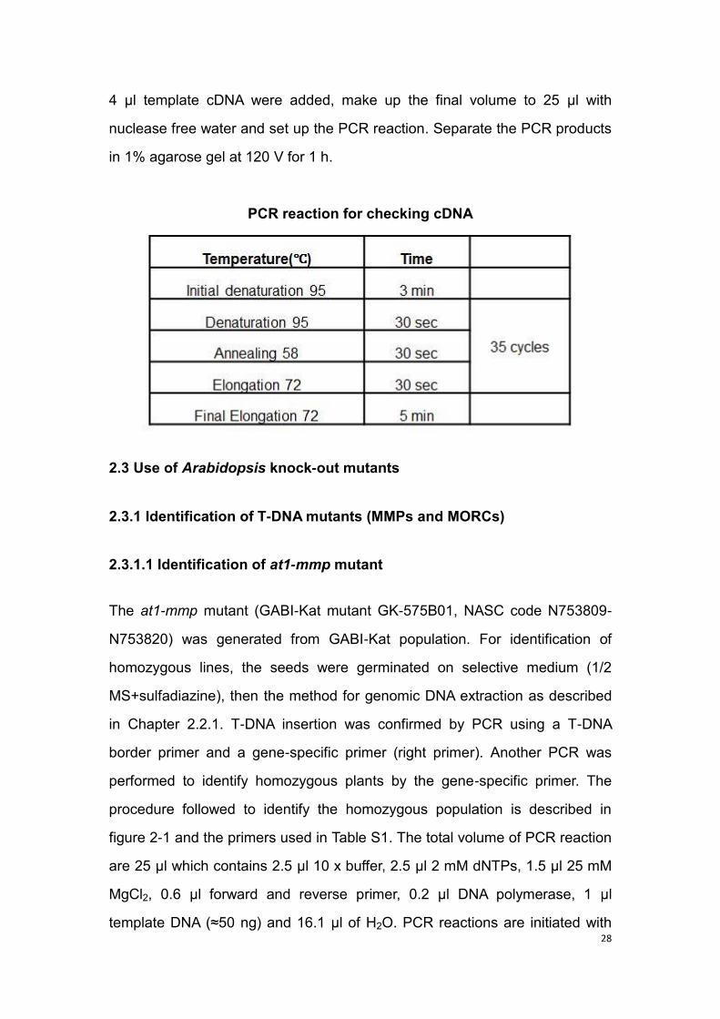
28
4 µl template cDNA were added, make up the final volume to 25 µl with
nuclease free water and set up the PCR reaction. Separate the PCR products
in 1% agarose gel at 120 V for 1 h.
PCR reaction for checking cDNA
2.3 Use of Arabidopsis knock-out mutants
2.3.1 Identification of T-DNA mutants (MMPs and MORCs)
2.3.1.1 Identification of at1-mmp mutant
The at1-mmp mutant (GABI-Kat mutant GK-575B01, NASC code N753809-
N753820) was generated from GABI-Kat population. For identification of
homozygous lines, the seeds were germinated on selective medium (1/2
MS+sulfadiazine), then the method for genomic DNA extraction as described
in Chapter 2.2.1. T-DNA insertion was confirmed by PCR using a T-DNA
border primer and a gene-specific primer (right primer). Another PCR was
performed to identify homozygous plants by the gene-specific primer. The
procedure followed to identify the homozygous population is described in
figure 2-1 and the primers used in Table S1. The total volume of PCR reaction
are 25 μl which contains 2.5 μl 10 x buffer, 2.5 μl 2 mM dNTPs, 1.5 μl 25 mM
MgCl2, 0.6 μl forward and reverse primer, 0.2 μl DNA polymerase, 1 μl
template DNA (≈50 ng) and 16.1 μl of H2O. PCR reactions are initiated with

29
95°C for 3 min and then 35-40 cycles for amplification including 95°C for 30
seconds, 30 seconds for annealing time (temperature is depending on the
gene), and 72°C for 30-90 seconds (depending on the size of the band), a
final extension of 5 min at 72°C.
Figure. 2-1. Identification of the at-mmp1 homozygous mutant.
Two PCR reactions were performed with LP, RP and LB primers. PCR1 was
performed with LP and RP primer of At-MMP1. PCR1 negative plants were
tested by PCR 2 with LB and RP primer of At-MMP1. LP: Left primer. RP:
Right primer. LB: Left border primer.
2.3.1.2 Identification of atmorc mutants
Molecular phylogenetic analysis showed that there are seven homologs in
Arabidopsis genome. We have used five AtMORCs mutants atmorc1, atmorc2,
atmorc5, atmorc6 and atmorc7 to study the potential function of AtMORCs in

30
plant immunity system. T-DNA insertion lines for AtMORCs were ordered from
NASC (European Arabidopsis stock center). These mutant lines were
segregating and thus required identification of homozygous lines. For the
identification, first the seeds were germinated on selection medium (1/2 MS
medium+ sulfadiazine), identified most promising candidate lines according to
the growth condition. The genomic DNA was extracted follow the method
described in Chapter 2.2.1. Finally, the homozygous mutants were identified
by PCR with the gene specific primer and T-DNA border primer, the method is
the same as described in Chapter 2.3.1.1. The primer sequences and product
size for genotyping are described in Table 1-2.
Table. 1-2. Primers for genotyping of atmorc mutants
2.3.2 Production and identification of at-mmp triple mutant
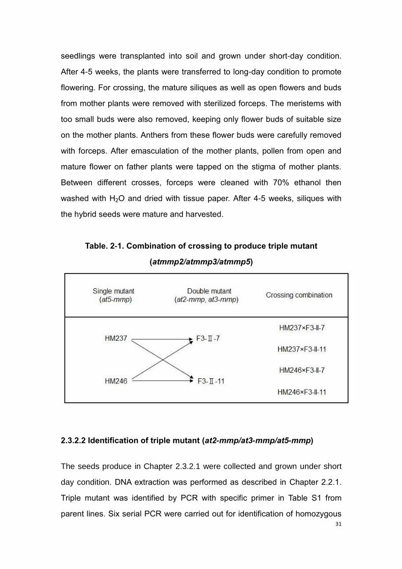
2.3.2.1 Crossing of Arabidopsis T-DNA mutants
Triple mutant was generated from crossing of one single mutant at5-mmp and
double mutant at2-mmp/at3-mmp (Table. 2-1). Seeds of single mutant and
double mutants were grown on 1/2 MS medium for germination and the
31
seedlings were transplanted into soil and grown under short-day condition.
After 4-5 weeks, the plants were transferred to long-day condition to promote
flowering. For crossing, the mature siliques as well as open flowers and buds
from mother plants were removed with sterilized forceps. The meristems with
too small buds were also removed, keeping only flower buds of suitable size
on the mother plants. Anthers from these flower buds were carefully removed
with forceps. After emasculation of the mother plants, pollen from open and
mature flower on father plants were tapped on the stigma of mother plants.
Between different crosses, forceps were cleaned with 70% ethanol then
washed with H2O and dried with tissue paper. After 4-5 weeks, siliques with
the hybrid seeds were mature and harvested.
Table. 2-1. Combination of crossing to produce triple mutant
(atmmp2/atmmp3/atmmp5)
2.3.2.2 Identification of triple mutant (at2-mmp/at3-mmp/at5-mmp)
The seeds produce in Chapter 2.3.2.1 were collected and grown under short
day condition. DNA extraction was performed as described in Chapter 2.2.1.
Triple mutant was identified by PCR with specific primer in Table S1 from
parent lines. Six serial PCR were carried out for identification of homozygous

32
triple mutant described in figure 2-2. LP and RP primers from At2-MMP were
used for the first PCR (PCR1). The samples not showing amplicon after PCR
1 were selected for further test in PCR 2, using LP and RP primer from At3-
MMP. Likewise, the samples with no amplicon after PCR 2 were used for
PCR 3, using LP and RP primer from At5-MMP. The samples which did not
show product were used for PCR4. These samples were tested in PCR4
using the LB and RP primer (from At2-MMP), the samples showing amplicon
in PCR4 were used for further test in PCR5. LB and RP primer (from At3-
MMP) were used in PCR5, the samples showing amplicon were used for
PCR6. The primers LB and RP (from At5-MMP) were used in PCR6 being
positive the samples that show amplicon. The triple mutant should not
produce any amplicon with LP and RP primers in PCR 1, PCR 2 and PCR 3,
but they should show amplicon in PCR 4, PCR 5 and PCR 6.

33
Figure. 2-2. Identification of homozygous at2-mmp/at3-mmp/at5-mmp
triple mutant.
Six PCR reactions were performed with LP, RP and LB primers. PCR1 was

34
performed with LP and RP primer of At2-MMP. PCR1 negative plants were
tested by PCR 2 with LP and RP primer of At3-MMP. Both PCR1 and PCR2
negative plants were tested by PCR 3 with LP and RP primer of At5-MMP.
The plants which are negative for PCR 1, PCR 2 and PCR 3 were used for
PCR 4 with LB and RP primer of At2-MMP, the positive plants were then
verified by PCR 5 with LB and RP primer of At3-MMP. Both PCR4 and PCR5
positive plants were tested by PCR 6 with LB and RP primer of At5-MMP. LP:
Left primer. RP: Right primer. LB: Left border primer.
2.4 Generation of Arabidopsis over-expression stable transformants
2.4.1 Cloning and construction of transformation vectors
To generate the AtMORC1 mutations constructs, the genomic DNA of the
Arabidopsis was used to amplify the full length sequence by PCR. Primer
pairs (Table. S1) were used for amplification of AtMORC1 D111K, AtMORC1
EDE108/115/116KKK, AtMORC1 D366K, AtMORC1 E441K, AtMORC1
E450K, respectively. The numbers denote amino acid position in AtMORC1
structure, D denotes aspartic acid, E denotes glutamic acid and K denotes
lysine. I replaced the aspartic acid/glutamic acid with lysine. The fragment
was first cloned into pET28a vector and sequenced by LGC Genomics (Berlin,
Germany). For cloning, 10 μl of ligation reaction was added to 50 μl
competent cells of E. coli. Then the sample was incubated on ice for 30 min,
heat shock the sample at 42°C for 50 to 60 seconds. The sample was kept on
ice quickly after heat shock, 400 μl LB medium was added and shaked for 1.5
h in 37 °C. The cells were spread on LB-Agar plates with Spectinomycin (50
μg/ml). The plates were incubated at 37°C for one day, and then the colonies
were picked and confirmed by PCR. The positive colony was selected for
Miniprep and Agrobacterium transformation. In order to get enough plasmid,
Minprep extraction of the plasmid was performed using Pure Yield Plasmid

35
Miniprep System (Promega). The transformation was performed following the
instructions from the manufacturer. The construct pET28a:AtMORC1 was
digested with HindIII, and the 1920 bp fragment containing AtMORC1 was
purified in agarose gel. The plasmid p35S-BM was also digested with HindIII
and the 5‟ phosphates were removed with CIAP (Calf intestinal alkaline
phosphatase) to avoid rejoining. The fragment containing AtCRT1 was ligated
into p35S-BM and subcloned into the SfiI sites of the Agrobacterium
transformation vector pLH6000 in sense orientation under control of 35S
promoter. The construct was then transformed into the Agrobacterium strain
GV3101 by electroporation using Gene Pulser Xcell Electroporation system
(Bio-Rad Laboratories, Hercules, CA, USA) following the manufacture
indications. Around 1 mg of plasmid was added to the competent cells
GV3103 (50 μl) and mixed, then they were incubated on ice for 10 min. After
incubation the cells were introduced into a electroporation cuvette and the
electro shock was applied. 600 μl of LB medium was added to the cells in the
cuvette after transformation, and the cells were transferred to an eppendorf
tube and incubated at 28°C for 1.5 h. The cells were then plated on LB
medium containing 25 μg/ml Rifampicin, 50 μg/ml Gentamicin and 50 μg/ml
Spectinomycin. After 2 days of incubation at 28°C, the colonies were picked
and confirmed by PCR. The positive colonies were selected to prepare for the
agro-transformation described in Chapter 2.4.2.d
2.4.2 Floral dip transformation
Agrobacterium-mediated transformation of A. thaliana was carried out by the
floral dip method (Clough and Bent, 1998). The background Arabidopsis
plants atmorc1/atmorc2 mutant were grown in soil under short-day conditions.
There are 8-10 plants per pot. After 3 weeks, the plants were put into long-day
conditions. The first inflorescence shoots were removed to induce the growth

36
of more inflorescence. Plants were used for transformation after about 1
week, when the secondary inflorescence emerged. Three days prior to plant
transformation, a 5 ml liquid pre-culture of Agrobacterium carrying a suitable
binary vector was prepared and incubated at 28°C with vigorous agitation.
The liquid culture consisted of LB medium containing antibiotics (Rifampicin
25 μg/ml, Gentamicin 50 μg/ml). Two days before the infiltration, 200 ml of
YEB medium (1%Bacto-Peptone, 1%Yeast extract, 0.5% NaCl) was
inoculated with 1 ml of the pre-culture and incubate again with vigorous
agitation for additional 48 h at 28°C. Use YEP medium for higher
Agrobacterium density. Stop watering the plants and allow the soil to dry out a
little, so that it will be less prone to falling out of the pots during dipping. After
two days, the agrobacterium was centrifuged at 6000 rpm for 10 min at room
temperature and the cell pellet resuspended in 400 ml of infiltration
medium(1/2 MS salts including vitamins, 5% sucrose, pH 5.7) supplemented
with 0.04% 6-benzylaminopurine (BAP, 10 ul L-1 of a 1 mg mL-1 stock in
dimethyl sulfoxide (DMSO)) and 0.02% Silwet L-77. A glass bell jar connected
via a condensation trap to a Leybold Trivac oil pump (type S8B/AF 4-8) was
used for vacuum infiltration. A glass tray filled with 400 ml of the
Agrobacterium suspension was placed in the jar. The inflorescence shoots
were dipped into the suspension, a pressure of 16 mbar was used for 2 min to
allow the submersion of the inflorescence shoots in the suspension. After 2-3
min treatment the vacuum was immediately released and the infiltration step
was repeated. The 400 ml bacterial suspension was re-used for three pots.
After the infiltration treatment, the plants were covered with a transparent
cover for 2 days. After 2 days, the cover was removed and the plants
transferred to a long-day growth chamber. Mature seeds were collected in
bags after about 3-4 weeks.
37
2.4.3 Presence of construct
Seeds from T0 plants after floral dip were sowing in 1/2 MS containing 30 mg/l
hygromycin and incubated at 4°C refrigerator for 2 days before being
transferred to a short day condition growth chamber. Transformants were
selected by their hygromycin resistance. After 2 weeks, green seedlings with
long roots were transformed to soil. Two weeks later, these plants were tested
by PCR to confirm the presence of construct.
2.4.4 Expression of the construct
2.4.4.1 Transcript levels (RNA)
After cDNA synthesis, the cDNA samples were used for checking the
transcript level by semi-quantitative PCR with AtMORC1 full length primers
(described in Table S1). In order to get clear results, we used the Phusion
High-Fidelity DNA Polymerase, and the PCR reaction as flow:
10 µl 5X Phusion HF Buffer
5 µl 2 mM dNTPs
0.5 µl AtMORC1 forward primer
0.5 µl AtMORC1 reverse primer
0.5 µl Phusion DNA Polymerase
5 µl template cDNA were added, make up the final volume to 50 µl with
nuclease free water and set up the PCR reaction. Separate the PCR products
in 1% agarose gel at 120 V for 1.2 h.

38
PCR reaction for transcript level assay
2.4.4.2 Protein expression of generated constructs
2.4.4.2.1 Protein extraction
For protein extraction, 4-week-old Arabidopsis leaves from AtMORC1
overexpression line with myc-tag were harvested and frozen in liquid nitrogen.
Leaf samples were crushed to powder with pre-cooled mortars and pestles in
liquid nitrogen. The leaf powder was transferred into 2 ml eppendorf tubes
and 200-300 μl Laucus buffer (1 tablet of protease-inhibitor + 10 ml Laucus
buffer) was added to the sample powder and vortexed for 1 min, then
centrifuged at 13,000 rpm at 4°C for 20 min. The supernatant was transferred
into 1.5 ml eppendorf tubes for Bradford analysis. Take 10 μl of supernatant
add 990 μl Bradford-Reagent, using 10 μl of Laucus buffer add 990 μl
Bradford-Reagent for blanking. Then the protein can be used for western blot.
2.4.4.2.2 Western blot
Before western blot, the proteins as described in Chapter 2.4.4.2.1 were
boiled in a water bath for 5 min at 95°C, and then the samples were used for
loading. For western blot, proteins were separated by SDS-polyacrylamide gel
39
electrophoresis (SDS-PAGE) and then transferred onto a PVDF membrane
(Roti®-PVDF, pore size 0.4 um, ROTH, Germany) with semi-dry
electrophoretic transfer cell (Bio-Rad) at 0.3 A for 1 h. The PVDF membrane
was incubated 1 min in methanol afterwards in 1× Towbin buffer (25 mM Tris,
192 mM glycine and 20% [v/v] methanol) for 20 min. After protein transfer, the
PVDF membrane was washed three times with TBS buffer (1.21% Tris, 8.76%
NaCl, adjust pH to 7.9), 5 min per time. Non-specific binding was blocked
using 5% (w/v) milk powder (ROTH, Germany) in TBS buffer at room
temperature for 2 h. After three times washing with TBS buffer, the membrane
was incubated in 5% milk powder contained c-myc antibody (1: 3000)
overnight at 4°C on a shaker. The membrane was washed three times for 5
min/time in TBS buffer and incubated with western blotting detection reagent
luminol enhancer solution and peroxide solution (GE Healthcare) for 1 min at
room temperature. After that, the blot was developed using Amersham
Hyperfilm ECL (GE Healthcare).
2.5 Pathogen infection
2.5.1 Botrytis cinerea inoculation
B. cinerea strain B05.10 was grown on HA agar medium (1% malt extract,
0.4% yeast extract, 0.4% glucose, 1.5% agar, pH 5.5. Plant leaves were
detached from 4-week-old Arabidopsis plants and placed in petri dish
containing 0.5% agar medium. To infect plants, conidia were collected from
14-day-old culture plate, and the spore density was adjusted in 12 g L-1 potato
dextrose broth (PDB, Duchefa Biochemie, Haarlem, The Netherlands) to
5x10⁴ conidiospores ml-1 for pathogen resistance assay. Inoculation was
performed by placing 5 μl of spore suspension in the leaf center. Cover the
petri dish and incubate the leaves at room temperature. Depending on the
40
symptom development rate, three to six days after infection, the leaf samples
were photographed and measured lesion size with ImageJ software.
2.5.2 Powdery mildew (Golovinomyces orontii) inoculation
For G. orontii inoculation, the conidia spores were collected from heavily
infected plants with Tween H2O (1:20,000). The density of spore suspension
was adjusted to 20,000-40,000 conidia ml-1 and sprayed on 4-week-old
healthy plants. Mock treatment was done by spraying Tween H2O (1:20,000).
After inoculation, plants were moved to a growth chamber under short day
condition under 22°C. For quantification of the fungal growth, the infected
plants were harvested when clear symptom is appearing. The fresh weight
was measured and the plants were rinsed with Tween H2O to collect the
conidia spores. The number of conidia per mg of fresh weight was determined
to quantify the fungal growth.
2.5.3 Pseudomonas syringae pv. tomato DC3000 inoculation
Pst Avr Rpt2 are streaked out from a –80°C glycerol stock onto a plate of
King‟s B medium (1% protease peptone, 0.15% anhydrous K2HPO4, 1.5%
glycerol, 1.5% Agar, pH 7.0) containing 50 mg/l Rifampicin and Kanamycin 25
mg/l and grown for 2 or 3 days at 28°C. After three days, the bacterial were
scrapped off with sterile 10 mM MgCl2 using a glass spatula and the optical
density were adjusted to 0.2. The Optical Density (OD) of the bacterial cell
suspension is quantified using a spectrophotometer set at 600 nm. For Pst
DC3000 an OD600=0.2 is approximately 1 x 10⁸ colony-forming units/ml.
Injection of dense bacterial suspensions (~108cfu/ml) of avirulent bacteria is
used to elicit a confluent hypersensitive response. A lower level of inoculum
(OD600=0.0002 of Pst DC3000 is 1 x 10⁵cfu/ml) is used for infiltration. The
41
inoculation was done by pressure infiltration. A leaf is selected and marked so
that it can be identified later. A 1ml needleless syringe containing a bacterial
suspension is used to pressure infiltrate the leaf intracellular spaces. Before
infiltration, spray the estradiol (30 μM), because the Arabidopsis MORC1 and
barley MORC1 overexpression in atmorc1/atmorc2 double mutant are under
control of the estradiol inducible promoter (Zuo et al., 2000). Leaf disks (0.5
cm2) were harvested at 72 h after infiltration and ground in 10 mM MgCl2.
After grinding of the tissue, the samples were thoroughly vortex-mixed and
diluted in a 10 fold serial dilution (101 to 105). Samples were finally plated on
King‟s B medium supplemented with 50 mg/l Rifampicin and Kanamycin 25
mg/l. Plates were incubate for 2 days at room temperature and the colony-
forming units were counted.
2.6 Oxidative burst assay
For ROS detection, 5 mm leaf disks were cut from 4-week-old healthy plants
with sharp puncher and were floated overnight in 200 μl H2O in 96-well plates
in dark to minimize wounding effect. For ROS measurement, the water was
replaced with 200 μl master mix aqueous containing 30 μg/ml luminol (Sigma,
15 mg/ml stock in DMSO) and 20 μg/ml horseradish peroxidase (10 mg/ml in
water, Sigma, P6782) under low-light condition. Elicitor flg22 (100 nM) was
injected by TECAN Infinite®F200 microplate reader (TECAN, Switzerland) to
trigger ROS production. Luminescence was measured for 60 cycles, 60
seconds per cycle.
2.7 Callose deposition assay
For detection of callose deposition, 5-week-old Arabidopsis plant leaves were
infiltrated with 1 µM flg22 for 24 h. The leaves were collected and fixed in

42
ethanol: glacial acetic acid (3:1) for several hours, under shaking. Then the
solution was discarded and put fresh solution to incubate overnight at room
temperature until the leaves were transparent. The next day, the leaves were
re-hydrated in 70% ethanol for 15 min, then in 50% ethanol for 15 min, and
washed several times with water. After several water washes, leaves were
incubated in 150 mM K2HPO4 (PH9.5) solution containing 0.01% aniline blue
for 1 to 2 h in darkness. Callose was observed under the microscope.
2.8 Transposon expression assay (qPCR)
For the measure of transposons expression, the leaves of 3-week-old plants
were harvested 6 h after estradiol spraying (30 μM). Then RNA extraction and
cDNA synthesis were performed as previously described in Chapter 2.2.2.
Quantitative PCR was performed using SYBR Green JumpStart Taq
ReadyMix (Sigma) to amplify the target genes. Primer sequences used for
transposon expression are described in Table 2-2 refer to the publication of
Moissiard et al (2014).
Table. 2-2. Sequences of primers used for transposon expression

43
3. Results
3.1 Identification of at-mmp mutants
3.1.1 Identification of at1-mmp mutant
Molecular phylogenetic analysis indicated that there are five MMP homologs
in the Arabidopsis genome. The four at-mmp mutants at2-mmp, at3-mmp, at4-
mmp and at5-mmp are available in this study except for the at1-mmp mutant.
To investigate the potential function of all At-MMPs in plant immunity, the T-
DNA insertion lines for at1-mmp were ordered from NASC. These mutants
were segregated and thus required the identification of homozygous lines.
The seeds were firstly germinated on selection medium (1/2MS
medium+sulfadiazine). According to the growth condition of the seeds and the
ration of survival / died seedlings (Table.3-1), the most promising candidate
lines of N753809, N753811, N753815, N753818, N753819 and N753820
were selected (Fig. 3-1). These lines were further screened for the
homozygous plants by the PCR as described in Chapter 2.3.3.1, method
which is described in Figure 2-1. Finally, the homozygous mutants were
identified by PCR (Fig. 3-2). In PCR1 (Fig. 3-2 A), the LP and RP primers
were used to amplify the wild-type of at1-mmp, all of the eighteen plants did
not show fragment. In PCR2 (Fig. 3-2 B), the primer LBb1.3 and RP primer
were used to detect the insertion, twelve plants (2, 3, 4, 5, 8, 9, 10, 12, 13, 14,
15, 16) showed amplification. Amplification of a fragment in PCR2, but not in
PCR1 implies that the corresponding line is homozygous, so these twelve
plants are homozygous of at1-mmp, 4 plants (plant 2, 3, 4, 5) from line
N753811, 8 plants (plant 8, 9, 10, 12, 13, 14, 15, 16) from line N753820
(Table. 3-2).
44
Figure. 3-1. The growth of at1-mmp seedlings on selective medium.
The sterilized seeds were germinated on selective medium (1/2 MS
medium+sulfadiazine). After 10 days, the number of growing seedlings was
counted.

45
Table. 3-1. The growth condition of seeds in selection medium
A
B

46
Figure. 3-2. Identification of homozygous T-DNA insertion at1-mmp
mutants.
The seedlings were transferred from selection medium to soil: sand (3:1) pot,
genomic DNA was extracted from each line used for PCR. A: The gene
specific primer LP and RP were used to detect the presence of the wild-type
version, all of the eighteen plants have no amplified PCR products. B: The
border primer LBb1.3 and RP primer were used to detect the T-DNA insertion,
twelve plants with red marker (2, 3, 4, 5, 8, 9, 10, 12, 13, 14, 15, 16) showed
amplification.
Table. 3-2. Homozygous mutant of at1-mmp
3.1.2 Generation of at2-mmp at3-mmp at5-mmp triple mutant
The triple mutant was produced through crossing of at5-mmp single mutant
and at2, 3-mmp double mutant. All the lines of F1 generation are
heterozygous. Thereafter, the F1 hybrids were self-crossed in order to select
the homozygous mutants. The workflow for identification of homozygous triple
mutant is as described in Chapter 2.3.2.2. Homozygous plants have no
amplified PCR products with LP/RP primers but show amplification with
LB/RP primers (Fig. 3-3). In principle, six PCR reactions were used to identify
the homozygous lines in the crossing combination. The at2, 3-mmp double
mutant and at5-mmp single mutant were both used as father parent and
mother parent to produce at2, 3, 5-mmp triple mutant. After the PCR selection
in three generations, the homozygous at2, 3, 5-mmp triple mutant were

47
identified out of 698 plants (Table 3-3). In the first generation, the gene
specific primers LP and RP were used to confirm the lines which are
heterozygous (Fig. 3-3 A and B). In F2 generation, the T-DNA border primer
LBb1.3 and gene specific primer RP were used to screen out that the line
#161 is homozygous for at3-mmp and at5-mmp, but heterozygous for at2-
mmp (Fig. 3-3 C). In the F3 generation of the line #161, the line #698 is
detected to be homozygous for all the mutants of at2-mmp, at3-mmp and at5-
mmp (Fig. 3-3 D).
A
B

48
C
D

49
Figure. 3-3. Identification of homozygous triple mutant at2, 3, 5-mmp.
Genomic DNA was extracted from 3-week-old Arabidopsis plant leaves, used
for PCR to screen out the homozygous mutant. A: The gene specific primer
LP and RP were used to detect the presence of the wild-type version of at2-
mmp, all of the thirteen plants have amplified PCR products. B: The gene
specific primer LP and RP were used to detect the presence of the wild-type
version of at3-mmp, all of the thirteen plants have amplified PCR products as
at1-mmp. C: The LP and RP primers were used to detect the presence of the
wild-type version of at2-mmp, at3-mmp and at5-mmp, and the border primer
LBb1.3 and RP primer were used to detect the T-DNA insertion of at2-mmp,
at3-mmp and at5-mmp, the plant #161 is homozygous for at3-mmp and at5-
mmp, but heterozygous for at2-mmp. D: The same combination of primers
was used to detect the next generation of plant #161. The plant #698 has no
amplification for LP and RP primers of at2-mmp, at3-mmp and at5-mmp;
meanwhile, it has amplification for LBb1.3 and RP primers of at2-mmp, at3-
mmp and at5-mmp. It indicated that the plant #698 is homozygous for all the
mutants of at2-mmp, at3-mmp and at5-mmp. WT: wild-type, T: T-DNA.
Table. 3-3. Homozygous mutant of at2, 3, 5-mmp

50
3.2 At-MMPs are required for pattern-triggered immunity
3.2.1 Callose response to MAMP in at-mmp mutants and At-MMP OE
transgenic plants
To assess whether At-MMPs participate in plant basal defense, I monitored
series of hallmark events of MAMP-triggered immunity in WT, at2, 3, 5-mmp
triple mutant and At2-MMP overexpression line. Callose deposition is known
to be a hallmark of FLS2-mediated PTI (Brown et al. 1998; Gómez-Gómez et
al. 1999a). Therefore, the callose deposition of the plants with the treatment of
flg22 was firstly observed. The results showed a regular callose deposition in
the leaf of wild-type plant; however, in comparison to wild-type, the callose
deposition was significantly reduced in at2, 3, 5-mmp triple mutant. As
expected, the reduction was able to be complemented in the At2-MMP
overexpression line, which exhibited a comparable level of callose deposition
to the wild-type (Fig. 3-4 A).
3.2.2 ROS response to MAMP in at-mmp mutants and At-MMP OE
transgenic plants
Subsequently, I compared the flg22-induced ROS production in wild-type,
single at-mmp mutants, at2, 3-mmp double mutant, at2, 3, 5-mmp triple
mutant and At2-MMP overexpression line. The mutants of at2-mmp, at3-mmp
showed slight less ROS production compared with wild-type. Nevertheless,
at2, 3-mmp double mutant displayed around 30% less ROS accumulation
than wild-type (Fig. 3-4 B). Notably, at2, 3, 5-mmp triple mutant exhibited
significant reduction of ROS production, which was nearly 50% lower than
that in wild-type at 30 min after flg22 treatment (Fig. 3-4 B). On the contrary,
the reduction of ROS generation was complemented in the At2-MMP
overexpression line, which showed even higher level of ROS generation
51
compared with wild-type (Fig. 3-4 C). Moreover, I further investigated the
flg22-induced ROS production in other single mutants of at1-mmp, at4-mmp
and at5-mmp, which showed no, if any, slight lower ROS level compared with
wild-type (data not shown). Taken together, these data indicated that At-MMPs
are required for the MAMP-triggered immunity in plant.
A

52
B
C
Figure. 3-4. Responses of at-mmp mutants and At-MMP OE transgenic
plants to MAMP.
A: Four-week-old plants were treated with 1µM flg22 or mock (water). At 24 h
53
after treatment (hat), the leaves were fixed with ethanol-to-glacial acetic acid
for several h and stained by aniline blue for 1.5 h. The callose deposition was
observed by epifluorescence microscopy. Scale bar = 20 µm. B, C: Oxidative
burst induced by 100nM flg22 was measured in relative light units (RLU) in
the leaves. The leaf disks 5 mm in diameter were cut from 4-week-old plants
wild-type, at2-mmp, at3-mmp, at2, 3-mmp, at2, 3, 5-mmp, EV(empty vector),
overexpression line (line1and line2) of mmp2. Results are mean ± SE (n = 8).
Three independent experiments were performed showing the similar results.
3.3 At-MMP is required for basal resistance to necrotrophic and
biotrophic fungal pathogens
3.3.1 At-MMP is required for basal resistance to necrotrophic fungal
pathogen Botrytis cinerea
Since expression of at2-mmp and at3-mmp is induced by B. cinerea infection
in wild-type plants (Zhao, 2011), this prompted us to study the At-MMPs
function in at-mmp mutants during the B. cinerea infection. For this, the
detached leaves were cut from 4-week-old plants, and symptomatic chlorosis
were taken at 3-4 days post inoculation (d.p.i). The lesion size was measured
with Image J. The results demonstrated that single knockout mutant at2-mmp
and at3-mmp showed bigger lesion size and significantly more susceptible to
B. cinerea than wild-type plants (Fig. 3-5 A, B). To further assess whether At2-
MMP, At3-MMP and At5-MMP have the similar function during B. cinerea
infection, the phenotypic analyses were performed on at2, 3-mmp double
mutant and at2, 3, 5-mmp triple mutant after B. cinerea infection. In
comparison to wild-type, at2, 3-mmp double mutant showed significantly
higher susceptibility. This susceptible symptoms were even slightly more than
those in at2-mmp and at3-mmp mutants (Fig. 3-5 B). Notably, the highest

54
susceptibility was observed in at2, 3, 5-mmp triple mutant, which exhibited
approximatly double level of susceptibitiy compared with wild-type (Fig. 3-5 A,
B). These data implied an additive effect of the mutations in at2-mmp,at3-
mmp and at5-mmp on the plant resistance to B. cinerea.
To confirm the role of MMPs during necrotrophic pathogen B. cinerea
infection, the responses to B. cinerea infection in two indepent overexperssion
lines of At2-MMP (line1 and line2) were subsequently analyzed. Expectedly,
the susceptible symptoms in at-mmp mutants were complemented in both
overexperssion lines of At2-MMP, exhibiting more than 50% lesion size
compared with wild-type and EV (Fig. 3-5 C), which demonstrated more
resistance to B. cinerea than wild-type plants (Fig. 3-5 D). Taken together, the
data suggested that At-MMPs play an important role in resistance to
necrotrophic pathogen B. cinerea.
A
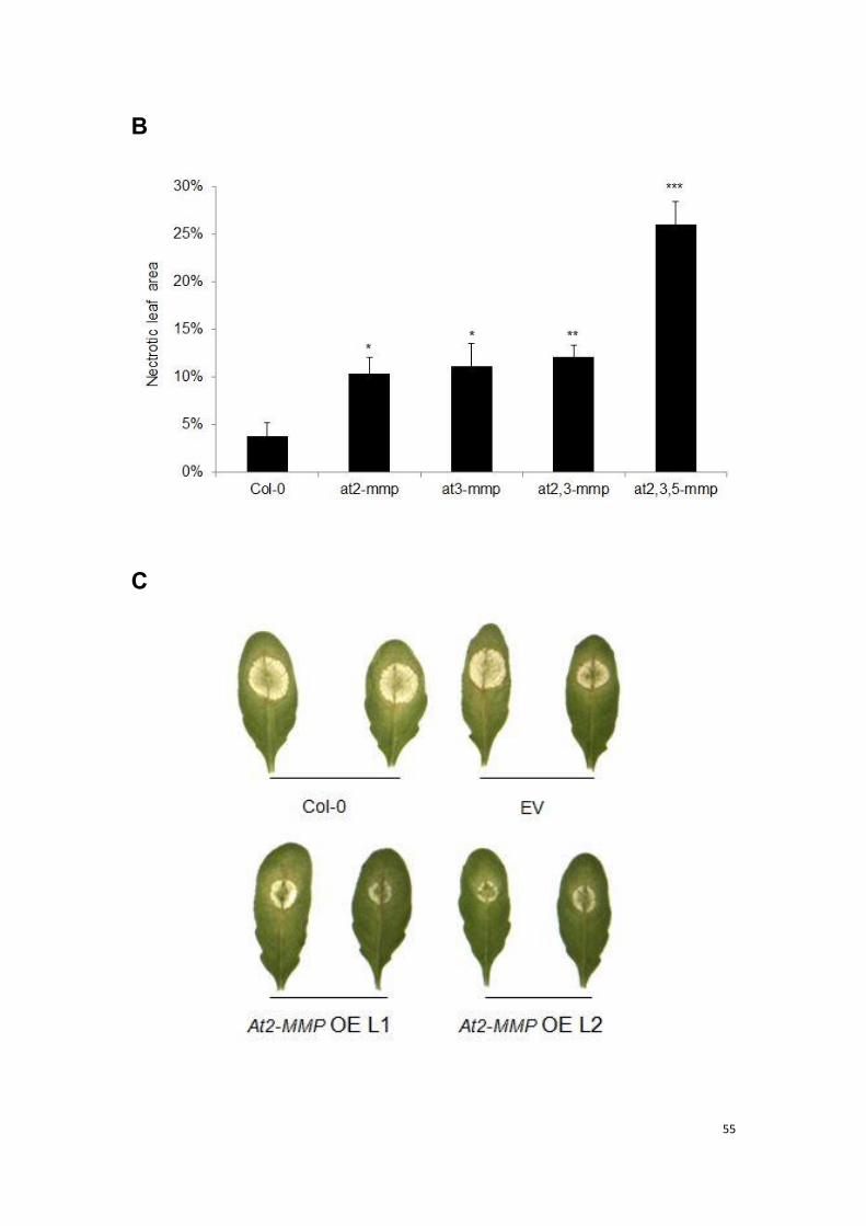
55
B
C
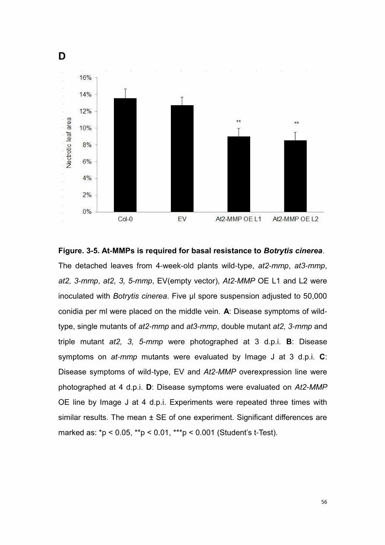
56
D
Figure. 3-5. At-MMPs is required for basal resistance to Botrytis cinerea.
The detached leaves from 4-week-old plants wild-type, at2-mmp, at3-mmp,
at2, 3-mmp, at2, 3, 5-mmp, EV(empty vector), At2-MMP OE L1 and L2 were
inoculated with Botrytis cinerea. Five μl spore suspension adjusted to 50,000
conidia per ml were placed on the middle vein. A: Disease symptoms of wild-
type, single mutants of at2-mmp and at3-mmp, double mutant at2, 3-mmp and
triple mutant at2, 3, 5-mmp were photographed at 3 d.p.i. B: Disease
symptoms on at-mmp mutants were evaluated by Image J at 3 d.p.i. C:
Disease symptoms of wild-type, EV and At2-MMP overexpression line were
photographed at 4 d.p.i. D: Disease symptoms were evaluated on At2-MMP
OE line by Image J at 4 d.p.i. Experiments were repeated three times with
similar results. The mean ± SE of one experiment. Significant differences are
marked as: *p < 0.05, **p < 0.01, ***p < 0.001 (Student‟s t-Test).
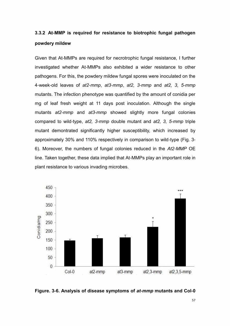
57
3.3.2 At-MMP is required for resistance to biotrophic fungal pathogen
powdery mildew
Given that At-MMPs are required for necrotrophic fungal resistance, I further
investigated whether At-MMPs also exhibited a wider resistance to other
pathogens. For this, the powdery mildew fungal spores were inoculated on the
4-week-old leaves of at2-mmp, at3-mmp, at2, 3-mmp and at2, 3, 5-mmp
mutants. The infection phenotype was quantified by the amount of conidia per
mg of leaf fresh weight at 11 days post inoculation. Although the single
mutants at2-mmp and at3-mmp showed slightly more fungal colonies
compared to wild-type, at2, 3-mmp double mutant and at2, 3, 5-mmp triple
mutant demontrated significantly higher susceptibility, which increased by
approximately 30% and 110% respectively in comparison to wild-type (Fig. 3-
6). Moreover, the numbers of fungal colonies reduced in the At2-MMP OE
line. Taken together, these data implied that At-MMPs play an important role in
plant resistance to various invading microbes.
Figure. 3-6. Analysis of disease symptoms of at-mmp mutants and Col-0

58
plants with the infection of fungal pathogen powdery mildew.
Four-week-old plants were spray-inoculated with 25,000 conidia per ml spore
suspension of the powdery mildew fungus G. orontii. Eleven days after
inoculation, the leaves were detached and the amount of conidia per mg of
leaf fresh weight was determined from at least 10 individually treated plants.
The error bars indicate the standard error. Experiments were repeated three
times with similar results. Significant differences are marked as: *p < 0.05, **p
< 0.01, ***p < 0.001 (Student‟s t-Test).
3.4 Identification of atmorc mutants
Molecular phylogenetic analysis showed that there are seven MORC
homologs in Arabidopsis genome. In my work, five of the identified atmorc
mutants, atmorc1, atmorc2, atmorc5, atmorc6 and atmorc7 were used to
study the potential function of MORC in plant immunity. T-DNA insertion
knock-out mutants of atmorc1, atmorc2, atmorc5, atmorc6 and atmorc7 were
obtained from the NASC. Since these mutants segregated, the identification
of homozygous lines was the first step in the following work. For this, seeds
were germinated on selection medium (1/2 MS medium+sulfadiazine). The
plants resistant to sulfadiazine were screened for homozygosity by PCR
method as described in Chapter 2.3.1.2. After genotyping, the homozygous
lines for each mutant were propagated for further experiments. The numbers
of homozygous plants are described in Table 3-4, the primer sequences for
genotyping are described in Table S1.
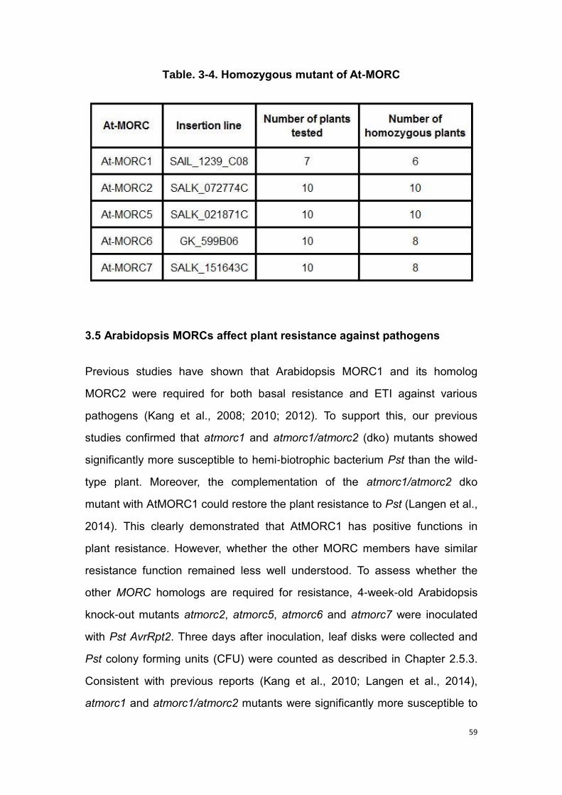
59
Table. 3-4. Homozygous mutant of At-MORC
3.5 Arabidopsis MORCs affect plant resistance against pathogens
Previous studies have shown that Arabidopsis MORC1 and its homolog
MORC2 were required for both basal resistance and ETI against various
pathogens (Kang et al., 2008; 2010; 2012). To support this, our previous
studies confirmed that atmorc1 and atmorc1/atmorc2 (dko) mutants showed
significantly more susceptible to hemi-biotrophic bacterium Pst than the wild-
type plant. Moreover, the complementation of the atmorc1/atmorc2 dko
mutant with AtMORC1 could restore the plant resistance to Pst (Langen et al.,
2014). This clearly demonstrated that AtMORC1 has positive functions in
plant resistance. However, whether the other MORC members have similar
resistance function remained less well understood. To assess whether the
other MORC homologs are required for resistance, 4-week-old Arabidopsis
knock-out mutants atmorc2, atmorc5, atmorc6 and atmorc7 were inoculated
with Pst AvrRpt2. Three days after inoculation, leaf disks were collected and
Pst colony forming units (CFU) were counted as described in Chapter 2.5.3.
Consistent with previous reports (Kang et al., 2010; Langen et al., 2014),
atmorc1 and atmorc1/atmorc2 mutants were significantly more susceptible to

60
Pst AvrRpt2 compared with the wild-type plant, which exhibited respectively
around 50% and 60% higher susceptibility levels in atmorc1 mutant and
atmorc1/atmorc2 dko mutant in comparison to that in wild-type plant (Fig. 3-
7). Interestingly, similar to the disease phenotype displayed in atmorc1 and
atmorc1/atmorc2 mutants, atmorc6 exhibited a strong increase in
susceptibility to Pst AvrRpt2, which showed around 60% more CFUs than that
in wild-type (Fig. 3-7). In contrast, atmorc2, atmorc5 and atmorc7 mutants
showed a similar Pst AvrRpt2 susceptibility level as wild-type (Fig. 3-7).
Figure. 3-7. Effect of AtMORCs in resistance to Pseudomonas syringae
pv. tomato carrying the avirulence gene AvrRpt2.
The leaves from 4-week-old Arabidopsis plants were infiltrated with Pst
AvrRpt2 at 105 c.f.u. ml-1, and the leaves were collected at 0 (black bars) and
3 dai (white bars). The colony forming units (CFU) were counted. Col-0:
Columbia wild-type Arabidopsis; dko: atmorc1/atmorc2 double knock-out
mutant; atmorc1: morc1 single knock-out mutant; atmorc2: morc2 single
knock-out mutant; atmorc5: morc5 single knock-out mutant; atmorc6: morc6
single knock-out mutant; atmorc7: morc7 single knock-out mutant. The error
bars indicate the standard error. Experiments were repeated three times with

61
similar results. Significant differences are marked: *p < 0.05, **p < 0.01, ***p <
0.001 (Student‟s t-Test).
3.6 Production of transgenic plants containing site mutations in MORC1
Arabidopsis MORC1 is involved in multiple layers of disease resistance
against various pathogens (Kang et al., 2012; Langen et al., 2014), and
exhibited activities of ATPase and endonuclease (Kang et al., 2008; 2010;
2012; Lorkovic et al., 2012). In order to further investigate the function of
ATPase activity and putative endonuclease activity in AtMORC1 during plant
defense, AtMORC1 site mutations were generated. These mutations were
targeted in the ATPase site (AtMORC1 D111K and AtMORC1
EDE108/115/116KKK) and a putative endonuclease site (AtMORC1 D366K,
AtMORC1 E441K and AtMORC1 E450K) of AtMORC1. These mutated
versions of AtMORC1 were used to complement into atmorc1/atmorc2 dko
mutant. For this, the transgenic plants were generated from the
atmorc1/atmorc2 dko background through floral dip transformation as
described in Chapter 2.4.2. The positive transgenic lines containing mutations
are described in Table 3-5. The expression level of AtMORC1 was tested by
semi-quantitative RT-PCR. The result indicated that all the transgenic lines
containing the different mutated MORC1 versions were transcribed to mRNA
(Fig. 3-8 A). Next, to confirm whether the mRNA is translated into protein, the
total protein was extracted from transgenic plants which additionally contain
the myc-tag (only constructs: myc: AtMORC1, myc: E441k) and was
forwarded to the immune-blot assay with anti-myc antibody. The data showed
a 70 kDa product, indicating the expression of AtMORC1 protein in the
transgenic lines (Fig. 3-8 B). Furthermore, in order to check whether the myc
tag might affect the function of MORC in resistance, I compared the disease
phenotypes with the infection of Pst AvrRpt2 in Arabidopsis MORC1

62
overexpression line dko-AtMORC1 and the myc-tagged overexpression lines
of AtMORC1 (myc: AtMORC1) and mutated AtMORC1 (myc: E441K). The
data showed that both myc: AtMORC1 and myc: E441K lines remained
certain levels of restoration of resistance to Pst AvrRpt2 in comparison to
atmorc1/atmorc2 dko mutant (Fig. 3-8 C), which indicated that the myc tag
does not affect the MORC function in plant resistance to Pst AvrRpt2.
Thereafter, the restoration of resistance to Pst AvrRpt2 was examined in the
produced transgenic lines containing specific site mutations of MORC1.
Table. 3-5. Positive transgenic lines containing AtMORC1 mutations
A

63
B
C
Figure. 3-8. Expression of AtMORC1 in transgenic plants.
A: Semi-quantitative PCR showing the transcription level of AtMORC1 in the

64
different transgenic plants and control pants by PCR. The cDNA samples
were extracted from the leaves of 4-week-old Arabidopsis plants. B:
Expression of AtMORC1 protein in transgenic plants expressing the construct
myc: E441K or myc: AtMORC1. Control is myc: HvMORC1. For western blot,
the leaves were collected from 4-week-old plants for protein extraction, 10 µg
total protein per sample was separated by 10% SDS-PAGE, and myc-MORC1
protein expression was detected by anti-myc antibody. The staining with
Ponceau S indicates equal loading of all samples (data not show). C: The
phenotype of transgenic plants (myc: E441K and myc: AtMORC1) in
resistance to Pseudomonas syringae pv. tomato carrying the avirulence gene
AvrRpt2. The leaves of 4-week-old plants were infiltrated with Pst AvrRpt2 at
105 c.f.u. ml-1, and collected at 0 (black bars) and 3 dai (white bars). The
colony forming units (CFU) were counted. Col-0: Columbia wild-type
Arabidopsis; dko: atmorc1/atmorc2 double knock-out mutant; dko-AtMORC1:
AtMORC1 overexpression line in dko; D111K and EDE108/115/116KKK are
transgenic lines containing mutations in ATPase domain of AtMORC1; D366K,
E441K and E450K are transgenic lines containing mutations in putative
endonuclease domain of AtMORC1. The numbers denote amino acid position
in AtMORC1 structure: D denotes aspartic acid; E denotes glutamic acid and
K denotes lysine. The aspartic acid/glutamic acid was replaced by lysine. The
error bars indicate the standard error. Experiments were repeated three times
with similar results. Significant differences are marked: *p < 0.05, **p < 0.01,
***p < 0.001 (Student‟s t-Test).
3.7 Mutations in AtMORC1 reduces plant resistance against the hemi-
biotrophic bacterium P. syringae pv. tomato
To assess whether the ATPase domain and/or endonuclease domain of
AtMORC1 might play an important role in plant resistance, the transgenic

65
lines containing mutations of AtMORC1 were employed to examine their
response to Pst. In comparison to the AtMORC1 overexpression line in the
atmorc1/atmorc2 background (dko-AtMORC1), the transgenic lines containing
mutations (D111K and EDE108/115/116KKK) in ATPase domain of AtMORC1
showed significantly more susceptible to Pst AvrRpt2 (Fig. 3-9). Similarly, the
transgenic lines containing mutations (E450K, E366K and E441K) in putative
endonuclease domain of AtMORC1 also exhibited significantly higher
susceptibility to Pst AvrRpt2 in comparison to the dko-AtMORC1 (Fig. 3-9).
Since the transcriptional level of AtMORC1 (Fig. 3-8 A) and the expression of
AtMORC1 protein (Fig. 3-8 B) were confirmed in the above transgenic lines
containing mutations, it is speculated that the alteration of disease
phenotypes are due to the mutations at ATPase domain and/or endonuclease
domain of AtMORC1.
Figure. 3-9. Mutations in ATPase domain and putative endonuclease
domain in AtMORC1 affects resistance against bacterium Pst AvrRpt2.

66
The leaves from 4-week-old plants were infiltrated with Pst AvrRpt2 at 105
c.f.u. ml-1, and the leaves were collected at 0 (black bars) and 3 dai (white
bars). The colony forming units (CFU) were counted. Col-0: Columbia wild-
type Arabidopsis; dko: atmorc1/atmorc2 double knock-out mutant; dko-
AtMORC1: AtMORC1 overexpression line in dko; D111K and
EDE108/115/116KKK are transgenic lines containing mutations in ATPase
domain of AtMORC1; D366K, E441K and E450K are transgenic lines
containing mutations in putative endonuclease domain of AtMORC1. The
numbers denote amino acid position in AtMORC1 structure: D denotes
aspartic acid; E denotes glutamic acid and K denotes lysine. The aspartic
acid/glutamic acid was replaced by lysine. The error bars indicate the
standard error. Experiments were repeated three times with similar results.
Significant differences are marked: *p < 0.05, **p < 0.01, ***p < 0.001
(Student‟s t-Test).
3.8 AtMORCs knock-out mutants show enhanced expression of a
transposon and genes related to silencing mechanisms.
Not only functioning in plant resistance to pathogens, recent research
demonstrated that Arabidopsis MORC1 and MORC6 could also suppress
transposons expression (Moissiard et al., 2012; 2014). To investigate whether
other Arabidopsis MORC homologs are involved in gene silencing, the
expression of a transposon gene (ATCOPIA28) and genes involved in
silencing SDC (suppressor of drm2 cmt3) were measured in atmorc1,
atmorc2, atmorc5, atmorc6 and atmorc7 knock-out mutants as well as
atmorc1/atmorc2 dko mtuant. The total RNA was extracted from 3-week-old
Arabidopsis plant leaves, which were used for cDNA synthesis. Thereafter,
the genes expression level was analyzed by real-time PCR (RT-PCR) using
gene specific primers described in Table 2-2. The results showed that

67
expression of ATCOPIA28 was dramatically induced in atmorc6 and
atmorc1/atmorc2 dko mutants, which displayed more than 200-fold induction
in comparison to wild-type. The atmorc1 mutant also exhibited around 10 fold
induction of ATCOPIA28 (Fig. 3-10 A). In contrast, atmorc2, atmorc5 and
atmorc7 showed little or no induction of ATCOPIA28, which was similar to that
displayed in wild-type (Fig. 3-8 A). SDC encodes an F-box protein and
possesses seven promoter tandem repeats, which show a unique silencing
based on non-CG DNA methylation directed redundantly by histone
methylation and siRNAs. The enhancement of SDC gene expression means
the alteration of the mechanisms regulating its promoter. In the mutants of
atmorc1, atmorc6 and atmorc1/atmorc2 dko, the expression level of SDC was
significantly up-regulated, which displayed respectively around 80-fold, 130-
fold and 110-fold induction in comparison to the level in wild-type (Fig. 3-10
B).
A

68
B
Figure. 3-10. AtMORCs affect the expression of transposon and
silencing-related genes.
The cDNA samples from the leaves of 3-week-old Arabidopsis plants were
used for real-time PCR (RT-PCR). Data represent the Ct thresholds of the
indicated candidate genes relative to the Ct thresholds of the housekeeping
gene actin using the ΔΔCt method. Relative fold increase of one transposon
(A) ATCOPIA28 and one gene involved in silencing (B) SDC (suppressor of
drm2 cmt3) was calculated. Col-0: Columbia wild-type Arabidopsis; dko:
atmorc1/atmorc2 double knock-out mutant; atmorc1: morc1 single knock-out
mutant; atmorc2: morc2 single knock-out mutant; atmorc5: morc5 single
knock-out mutant; atmorc6: morc6 single knock-out mutant; atmorc7: morc7
single knock-out mutant. The error bars indicate the standard error.
Experiments were repeated three times with similar results.

69
3.9 Transgenic plants containing mutations of AtMORC1 mutations show
no or strong derepression of transposons.
To further support the importance of AtMORC1 in gene silencing, the
transposon genes expression was examined in AtMORC1 overexpression line
dko-AtMORC1. Expectedly, the dramatic induction of one transposon marker
gene AtCOPIA28 and gene involved in silencing SDC (suppressor of drm2
cmt3) disappeared in dko-AtMORC1, which showed comparable expression
levels to wild-type plant (Fig. 3-11). Nevertheless, significant inductions of
both AtCOPIA28 and SDC were observed in dko-HvMORC1 line, in which
barley MORC1 was overexpressed in atmorc1/atmorc2 background. The level
of transposons gene expression in dko-HvMORC1 was even similar to that
displayed in atmorc1/atmorc2 dko mutant (Fig. 3-11).
Next, to assess whether the activity of ATPase and endonuclease is
necessary for gene silencing in MORC1, the expression of transposon genes
in the transgenic lines containing mutations of AtMORC1 was further
investigated. Similar to that observed in atmorc1/atmorc2 mutant, the
transgenic lines containing the mutations (D111K; EDE108/115/116KKK) in
ATPase domain of AtMORC1 exhibited significant induction of AtCOPIA28
and SDC expression (Fig. 3-11). Nevertheless, in the transgenic lines
containing the mutations (E366K; E450K; E441K) in the putative
endonuclease domain of AtMORC1, the expressions of AtCOPIA28 and SDC
exhibited comparable level to that in wild-type plant and dko-AtMORC1 line,
although the transgenic lines with the mutations of E366K and E450K showed
slightly induction of both genes (Fig. 3-11).

70
A
B
Figure. 3-11. Effects of AtMORC1 mutations on silencing.
The expression of a transposon and a gene controlled by silencing was
measured in the different lines expressing the mutated AtMORC1 constructs

71
and dko-HvMORC1 (HvMORC1 overexpression in dko). RT-PCR was
performed from RNA extracted from 3-week-old leaf tissue. Data represent
the Ct thresholds of the indicated candidate genes relative to the Ct
thresholds of the housekeeping gene actin using the ΔΔCt method. A:
Relative expression level of a transposon ATCOPIA28, whose repression
depends on silencing mechanism. B: Relative expression level of a gene
controlled by silencing SDC (suppressor of drm2 cmt3). Col-0: Columbia wild-
type Arabidopsis; dko: atmorc1/atmorc2 double knock-out mutant; dko-
AtMORC1: AtMORC1 overexpression line in dko; dko-HvMORC1: HvMORC1
overexpression line in dko; D111K and EDE108/115/116KKK are transgenic
lines containing mutations in ATPase domain of AtMORC1; D366K, E441K
and E450K are transgenic lines containing mutations in putative
endonuclease domain of AtMORC1. The numbers denote amino acid position
in AtMORC1 structure: D denotes aspartic acid; E denotes glutamic acid and
K denotes lysine. The aspartic acid/glutamic acid was replaced by lysine. The
error bars indicate the standard error. Experiments were repeated three times
with similar results.
3.10 AtMORCs show effect on the expression of R gene RMG1
(resistance methylated gene 1)
MORCs are involved in both resistance and gene silencing. To assess
whether MORC might function in plant resistance through gene silencing, I
tested how MORC affects the expression of R gene RMG1 (resistance
methylated gene 1). The RMG1 has transposon element (TE) remnants in its
promoter that attracts the RNA-directed DNA methylation (RdDM) machinery.
In comparison to wild-type, most of the tested morc mutants exhibited no
induction of RMG1, except for the slight expression shown in mutants of
atmorc2, atmorc5 and atmorc1/atmorc2 dko (Fig. 3-12 A). Similar to that
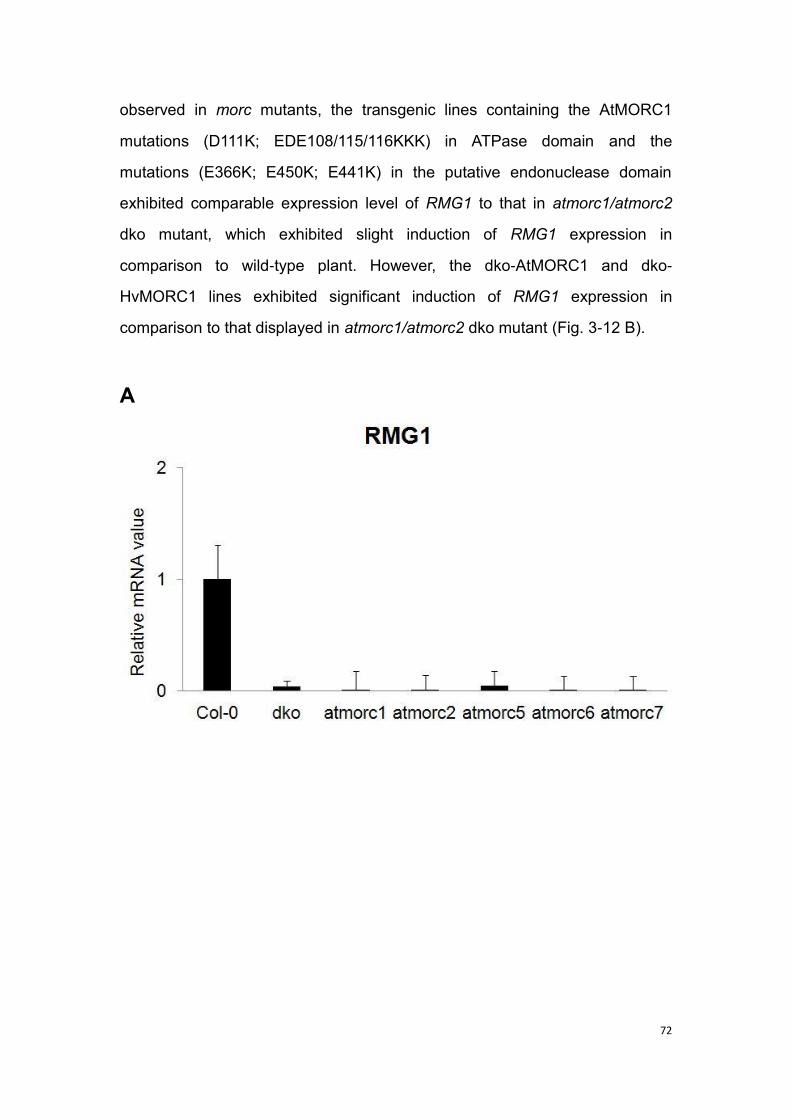
72
observed in morc mutants, the transgenic lines containing the AtMORC1
mutations (D111K; EDE108/115/116KKK) in ATPase domain and the
mutations (E366K; E450K; E441K) in the putative endonuclease domain
exhibited comparable expression level of RMG1 to that in atmorc1/atmorc2
dko mutant, which exhibited slight induction of RMG1 expression in
comparison to wild-type plant. However, the dko-AtMORC1 and dko-
HvMORC1 lines exhibited significant induction of RMG1 expression in
comparison to that displayed in atmorc1/atmorc2 dko mutant (Fig. 3-12 B).
A

73
B
Figure. 3-12. Effects of AtMORC on resistance gene (RMG1) expression.
The expression level of resistance gene RMG1 (resistance methylated gene
1) was analyzed in different AtMORC knock-out mutants and mutation lines.
RT-PCR was performed from cDNA samples extracted from 3-week-old
Arabidopsis leaves. Data represent the Ct thresholds of the indicated
candidate genes relative to the Ct thresholds of the housekeeping gene actin
using the ΔΔCt method. A: Relative expression level of RMG1 was analyzed
in AtMORC knock-out mutants and wild-type plant. Col-0: Columbia wild-type
Arabidopsis; dko: atmorc1/atmorc2 double knock-out mutant; atmorc1: morc1
single knock-out mutant; atmorc2: morc2 single knock-out mutant; atmorc5:
morc5 single knock-out mutant; atmorc6: morc6 single knock-out mutant;
atmorc7: morc7 single knock-out mutant. B: Relative expression level of
RMG1 was analyzed in AtMORC1 mutations line and dko-HvMORC1
(HvMORC1 overexpression in dko). Col-0: Columbia wild-type Arabidopsis;
dko: atmorc1/atmorc2 double knock-out mutant; dko-AtMORC1: AtMORC1

74
overexpression line in dko; dko-HvMORC1: HvMORC1 overexpression line in
dko; D111K and EDE108/115/116KKK are transgenic lines containing
mutations in ATPase domain of AtMORC1; D366K, E441K and E450K are
transgenic lines containing mutations in putative endonuclease domain of
AtMORC1. The numbers denote amino acid position in AtMORC1 structure: D
denotes aspartic acid; E denotes glutamic acid and K denotes lysine. The
aspartic acid/glutamic acid was replaced by lysine. The error bars indicate the
standard error. Experiments were repeated three times with similar results.
75
4. Discussion
4.1 The section of MMPs
4.1.1 The MMPs family
In mammals, MMPs have been reported to play an important role in the
regulation of inflammation and wounding (Parks et al., 2004; Page-McCaw et
al., 2007), in bone modeling and remodeling (Page-McCaw et al., 2007), and
especially in the formation of diseases. In healthy adult tissue, MMP7 is
expressed only in mucosal epithelia; but it is up-regulated by bacterial in
epithelia (Lopez-Boado et al., 2000). Consistently, mouse mmp7 mutant is
much easier to get infected by intestinal bacteria. The precursor of cryptdin is
found in intestinal epithelia from mmp7 mutant mice (Wilson et al., 1999).
Moreover, the MMP7 can mediate wound-induced epithelial migration by
cleaving E-cadherin (McGuire et al., 2003). In wounded mmp7 mutant mice,
epithelial cells do not migrate and E-cadherin cleavage does not occur
(Dunsmore et al., 1998). In addition, MMP3 functions in epidermal wound
healing. The skin wounds of mmp3 mutant mice heal more slowly compared
with control mice. These data indicated that the MMPs involved in innate
immunity and wound healing (Bullard et al., 1999).
Compared with MMPs in mammals, the function of MMPs in plants is less well
known. The first plant MMP was described from soybean SMEP1 (Ragster
and Chrispeels, 1979; Graham et al., 1991). Thereafter, the plant MMPs were
identified in more plant species, including cucumber (Delorme et al., 2000),
Arabidopsis (Maidment et al., 1999), tobacco (Kang et al., 2010) and
Medicago truncatula (Combier et al., 2007). The majority of MMPs are
secreted into the ECM. Through regulating ECM, MMPs might function in
plant development and growth. Several reports have shown that the
expression of MMPs is associated with aging. For instance, the expression of
76
soybean SMEP1 is increased in young leaves, but there is no change in the
next one month (Pak et al., 1997). The other soybean GmMMP2, however,
displayed a higher transcript level in mature leaves compared with the
senescence stage (Liu et al., 2001). The expression of MMPs in not only
associated with aging but also reacting to the outer abiotic and biotic stresses.
It has been reported that the soybean GmMMP2 could accumulate in leaves
with either wounding stress or in response to pathogen invasion (Liu et al.,
2001). In Arabidopsis, the five MMPs exhibited different expression patterns in
different part of plant (Maidment et al., 1999).
Not only participating in plant development and growth, the MMPs also play a
key role in plant-microbe interactions. The cucumber Cs1-MMP presents at
the boundary of senescence and cell death indicating its function in
programmed cell death (PCD) (Delorme et al., 2000). The soybean SMEP1
possibly contributes to the resistance to pathogen challenges (Pak et al.,
1997). Nevertheless, these studies are mainly based on the MMP protease
activity. In my study, I employed genetic, molecular and biochemical analyses
to provide further clear evidences to the function of MMPs in plant disease
resistance.
4.1.2 Reactive oxygen species and the role in plant immunity
The oxidative burst, a rapid production of huge amount of reactive oxygen
species (ROS), is one of the earliest defense events when plants respond to
various environmental threats, including biotic and abiotic stresses. The ROS
mainly occurred in the plant cell surface. In normal situation, the plants
employ certain protective mechanisms to keep a lower level of ROS.
However, when confronting stresses, the balance level of ROS in plants will
be significantly altered by the high amount of ROS production. This reaction
has been known to support the plant resistance to pathogen invasion through

77
strengthening the cell wall, inducing pathogenesis-related (PR) genes and cell
death. Zurbriggen et al (2009) treated the tobacco wild plant with a nonhost
pathogen Xanthomonas campestris pv. vesicatoria (Xcv), and they found that
the accumulation of ROS is higher in chloroplast. In addition, the localized cell
death was also significantly increased in comparison to the plants expressing
the flavodoxin, which is an electron shuttle. When expressed in chloroplasts,
the flavodoxin could prevent ROS formation under abiotic stress. These
results indicated that generation of ROS is important for the cell death
process during nonhost pathogen invasion.
4.1.3 At-MMPs mediated oxidative burst
In order to understand the functional mechanism of At-MMPs in resistance
and whether At-MMPs affect the oxidative burst, I analyzed the ROS
production in at-mmp mutants after the treatment with flg22. As expected, the
single mutants at2-mmp, at3-mmp, double mutant at2, 3-mmp and triple
mutant at2, 3, 5-mmp exhibited a lower level of ROS production compared
with wild-type (Fig. 3-4 B). To further check whether the reduction of ROS
production could be complemented, I further detected the oxidative burst in
At2-MMP overexpression line after the treatment with flg22. The data
demonstrated indeed a higher level of ROS production in At2-MMP
overexpression line (Fig. 3-4 C). The alteration of oxidative burst in single
mutants at2-mmp, at3-mmp, double mutant at2, 3-mmp and triple mutant at2,
3, 5-mmp and At2-MMP OE line is actually consistent with the phenotype
shown in the pathogen test (Fig. 3-5). Thus, it indicated that At-MMPs function
in affecting plant immunity in a way of regulating the MAMP triggered ROS
production.

78
4.1.4 Callose deposition and the role in plant immunity
Callose is a β-(1, 3) glucan polymer, whose structure and chemical
composition are slightly different in various cell wall types. It is an important
part of the cell plate during cell division, vascular bundles and pollen wall
exine. Callose is produced in specific site and developmental stages of plant
cell. Callose synthesis is catalyzed by different subunits of enzyme complex,
which is associated with the plasma membrane (Piršelová and Matušíková,
2013). The callose accumulation is regulated by distinct signaling pathways,
depending on the outside conditions (Luna et al., 2011). For example, the
heat and cold can induce the callose deposition in the sieve plates of the
phloem (Furch et al., 2007; Bilska and Sowinski, 2010). Callose could also
accumulated around cell wall, supporting the cell wall to against different
environment stresses. Under hypoxic conditions, callose deposition can
maintain the mechanical stability of cell wall (Albrecht and Mustroph, 2003). In
roots, callose might help to protect the sink function of organ and store
carbohydrates (Biemelt et al., 1999).
Callose deposition occurs at the site of pathogen attack at the cell wall. These
cell walls are thought to function as a physical barrier to restrict pathogen
invasion (Stone and Clarke, 1992). In tobacco, the β-1, 3-glucanases mutant
induced more callose deposition and showed more resistance to different
viruses (Iglesias and Meins, 2000). The callose deposition is also observed at
the fungi and bacteria penetration sites (Poliakovskiy and Dmitriev, 2011; Yun
et al., 2006). As one of the popular markers, plant early immune response to
pathogen invasion can be quantified by callose deposition. Since the callose
deposition can be triggered by MAMPs, such as Flg22 (Gómez-Gómez and
Boller, 2000), bacterial elongation factor EF-Tu (Elf18) (Kunze et al., 2004)
and chitin, a β-(1, 4)-linked polymer of N-acetylglucosamine (Iritri and Faoro,
2009). MAMP induced callose deposition in plant roots or leaves has become
an important marker response, together with other signaling events, such as

79
the accumulation of reactive oxygen species (ROS) and anion fluxes, to
detect the PTI signaling pathways.
4.1.5 At-MMPs mediate callose deposition
In order to understand whether At-MMPs would affect the callose deposition, I
analyzed the occurrence of callose deposition in at-mmp mutants after the
treatment with flg22. Similar to the ROS production assay, all the mutants
exhibited less callose deposition in comparison to wild-type. To further check
whether the reduction of callose deposition could be complemented in
overexpression line, the callose deposition in At2-MMP overexpression line
after the treatment of flg22 was further observed. Expectedly, the highest
occurrence of callose deposition was detected in At2-MMP OE line.
Combining the ROS production assay, the data implied that at-mmp mutants
showed reduction of both ROS production and callose deposition with the
MAMP treatment; while this reduction could be complemented in
overexpression lines, exhibiting a restored higher level of ROS production and
callose deposition (Fig. 3-4 A, C). These phenotypes are also consistent with
the function of ROS in flg22-induced callose deposition (Zhang et al., 2007;
Galletti et al., 2008; Estrella et al., 2011), indicating the function of At-MMPs in
resistance to pathogens. Indeed, in the pathogen test, at-mmp mutants
exhibited higher susceptibility to necrotrophic fungal pathogen Botrytis cinerea
and biotrophic fungal pathogen powdery mildew. Nevertheless, the At2-MMP
OE lines demonstrated more resistance to Botrytis cinerea compared to wild-
type (Fig. 3-5 D). Taken together, it is speculated that At-MMPs play a positive
role in PTI. More specifically, At2-MMP, At3-MMP and At5-MMP might function
in immune response in way of enhancing the ROS production and callose
deposition.

80
4.1.6 The role of plant MMPs in immune system
The previous studies have indicated that plant MMPs could function in
response to abiotic stresses, such as NaCl treatment, cadmium treatment,
wounding and temperature stress (Liu et al., 2001; Golldack et al., 2002;
Golldack et al., 2002; Cho et al., 2009). Moreover, recent researches further
demonstrated that the plant MMPs are also of importance to plant immunity.
In tobacco, the expression of Nicotiana benthamiana matrix metalloprotease 1
(NMMP1) was up-regulated by Pseudomonas syringae pv. tomato T1(Pst1) or
P. syringae pv. tabaci (Psta) infection. The transient NMMP1 over-expression
in N. benthamiana showed less CFUs compared with the control plant after
Psta inoculation; while, the NMMP1 silenced leaves exhibited significantly
higher number of CFUs than the control leaves (Kang et al., 2010). In
Arabidopsis, the At2-MMP overexpression line appeared to be more resistant
to Botrytis cinerea infection in comparison to wild-type (Zhao, 2011).
According to the phylogenetic analysis, there are five MMPs in A. thaliana
(At1-MMP to At5-MMP), among which At2-MMP, At3-MMP and At5-MMP are
close to each other and appear short genetic distance based on sequence
alignment (Maidment et al., 1999; Zhao, 2011). Marino et al (2014) expressed
and purified the catalytic domains of all five At-MMPs with His-tagged proteins
in Escherichia coli cells to delineate the biochemical function which
demonstrated that all five At-MMPs are proteolytically active enzymes and
each enzyme showed specific protease activity according to the recombinant
At-MMPs by three independent methods: the recombinant At-MMPs were
incubated individually with: (i) MBP and β-casein as typical model protease
substrates, with activity assayed by SDS/PAGE; (ii) quenched fluorescent
peptides using fluorescence generation as readout; and (iii) an A. thaliana leaf
proteome-derived peptide library, followed by MS-based identification of
cleaved peptides. To assess whether these three MMPs (At2-MMP, At3-MMP
and At5-MMP) have the same function in defense response, the double

81
mutant at2, 3-mmp and triple mutant at2, 3, 5-mmp were produced, and their
responses to the infection by different patterns of pathogens were further
analyzed. With the infection by necrotrophic fungus Botrytis cinerea, the
double mutant at2, 3-mmp showed bigger lesion size compared with the
parental single mutant at2-mmp and at3-mmp or the wild-type (Fig. 3-5 A, B),
which is consistent with the former studies (Zhao, 2011). Although the triple
mutant at2, 3, 5-mmp exhibited a similar phenotype in comparison to the
single mutants at2-mmp, at3-mmp and double mutant at2, 3-mmp, yet it
appeared to be more susceptible to B. cinerea than the double mutant at2, 3-
mmp (Fig. 3-5 B). This indicated an additive effect of the mutations in At2-
MMP, At3-MMP and At5-MMP. Furthermore, the phenotype in response to B.
cinerea infection in At2-MMP overexpression line was analyzed. Consistent
with the previous studies, the At2-MMP overexpression line demonstrated
increased resistance to B. cinerea when comparing to the wild-type (Fig. 3-5
C, D). Taken together, the results suggest that At-MMPs play an important role
in plant immune response and function to defend the invasion by necrotrophic
fungus B. cinerea.
Given that At-MMPs were required for necrotrophic fungus resistance. To
assess whether At-MMP are also required for the resistance to biotrophic
fungus, I analyzed the infection phenotype in mutants of at2-mmp, at3-mmp,
at2, 3-mmp and at2, 3, 5-mmp by the biotrophic fungus powdery mildew.
Similarly, the single mutants at2-mmp and at3-mmp showed slightly increased
numbers of fungal colonies compared to wild-type (Fig. 3-6). While the at2, 3-
mmp and at2, 3, 5-mmp mutants appeared to be significantly more
susceptible to the fungal infection, which increased by approximately 30% and
110% respectively in comparison to wild-type and single mutants (Fig. 3-6).
Taken together, the data clearly implied that At2-MMP, At3-MMP and At5-MMP
play positive roles in defense responses to nectrophic and biotrophic fungal
invasion. Furthermore, they might have similar functions in plant immunity.

82
4.2 The section of MORC
4.2.1 The MORC family
MORC (microrchidia) was first described in mouse where it encodes a 108
kDa protein. This protein accumulated specifically in male germ cells (Inoue et
al., 1999). The MORC family widely exists in eukaryotic and prokaryotic
organisms (Iyer et al., 2008). The protein sequences revealed that MORC
contains nuclear localization signals coiled-coil domain and GHL (GyraseB,
Hsp90, MutL) ATPase (Inoue et al., 1999). MutL and its homologs are
essential for cell growth and viability, being involved in mismatch repair, gene
conversion and chromosome segregation (Williamson et al., 1985; Hunter and
Borts, 1997). Hsp90 functions in the conformational activation of eukaryotic
protein kinases and regulating the expression of genetic variation (Dutta and
Inouye, 2000; Rutherford and Lindquist, 1998). In order to test the localization
of MORC protein, Inoue et al (1999) expressed epitope-tagged mouse MORC
in COS7 cells and determined its subcellular localization by multicolor
immunofluorescence. They found that the MORC expressed in the nuclear.
Based on the localization of MORC and the phenotypes of other mouse morc
mutants, it was indicated that MORC might function in the regulation of
transcription, cell division, meiotic chromosome dynamics or DNA repair
(Inoue et al., 1999; Okabe et al., 1998). Moreover, its coiled-coil domain
demonstrates that MORC could interact with other proteins. The GHL
(GyraseB, Hsp90, MutL) ATPase domain in the N-terminus of MORC implies
that the ATP hydrolysis might be required for MORC function (Inoue et al.,
1999).
Interestingly, several recent studies have demonstrated that MORC protein
could also function in plant immunity. Kang et al (2008; 2010; 2012) identified
the MORC family in Arabidopsis. The data implied that AtMORC1 increased
the plant resistance to the necrotrophic fungal pathogen P. syringae pv.

83
tomato (Pst). Moreover, five MORC genes have been identified in the barley
genome (Langen et al., 2014). The morc genes knock-down barley plants and
MORC overexpression barley lines were infected by powdery mildew fungus
Bgh race A6. The results indicated that the morc2 knock-down plants
exhibited less fungal colonies in comparison to control; while the MORC1 OE
plants displayed higher susceptibility. In addition, the morc2 knock-down
plants also showed increased resistance to necrotrophic fungus Fusarium
graminearum (Langen et al., 2014). In my study, the pathogens test in
Arabidopsis morc mutants supported that, similar to its function in barley,
MORC also plays an important role in the immune responses to pathogens in
Arabidopsis. Notably, different from the phenotypes in barley, the Arabidopsis
morc1 mutant seemed more susceptible to Pst in comparison to control (Fig.
3-7). Moreover, the resistance to Pst could be restored in Arabidopsis MORC1
overexpression line dko-AtMORC1 (Fig. 3-10). These data implied that MORC
might have distinguished or alternative mechanisms to function in plant
immunity among different hosts.
In addition, recent reports demonstrated that MORC1 is responsible for
transposon repression in the male germline in mouse (Pastor et al., 2014). In
plant, Arabidopsis MORC1 and MORC6 were also reported to suppress the
expression of transposons (Moissiard et al., 2012). These clearly indicated
that the MORC family could play a role in gene silencing. In my study, I tested
the gene expression profile of a transposon (ATCOPIA28) and genes involved
in silencing SDC (suppressor of drm2 cmt3) and RMG1 (resistance
methylated gene 1) which is regulated by RNA-directed DNA methylation
(RdDM) in Arabidopsis morc mutants and transgenic lines containing
AtMORC1 mutations. The data showed that mutants of atmorc1/atmorc2,
atmorc1 and atmorc6 exhibited most significantly different expression profile
in comparison to Col-0, which was the up-regulation of ATCOPIA28 and SDC
(Fig. 3-10), and the down-regulation of RMG1 (Fig. 3-12 A) in the above
Arabidopsis morc mutants. The results are similar to the previous reports,

84
indicative of the participation of MORC in gene silencing in Arabidopsis.
4.2.2 Production and identification of AtMORC1 mutations
The structure of MORC showed that MORC possesses ATPase domain and
endonuclease domain, which is supported by the ATPase and endonuclease
activity exhibited in AtMORC1 and AtMORC6 (Kang et al., 2012; Lorkovic et
al., 2012). Interestingly, AtMORC1 and AtMORC6 were reported to function as
repressors of transposons and DNA methylated genes (Moissiard et al., 2012;
2014). Additionally, MORC1 also repressed transposons in the male germline
in mouse (Pastor et al., 2014). These indicated that MORC might link to gene
silencing. In order to address the question of whether the activities of ATPase
and endonuclease are necessary for the resistance and gene silencing, I
produced point mutations which are highly conserved in AtMORC1 according
to the database. Furthermore, amino acids in both ATPase domain (D111K;
EDE108/115/116KKK) and putative endonuclease domain (E366K; E441K;
E450K) were mutated, in which the amino acids were replaced from negative
charge to positive charge, resulting in the deactivation of ATPase and
endonuclease. Thereafter, the transgenic plants in Arabidopsis dko
background with/without myc-tag were produced. These stable transgenic
plants were used for phenotypic analysis of disease resistance against P.
syringae pv. tomato (Pst), and the expression analysis of a transposon gene
ATCOPIA28, a silencing-related gene SDC (suppressor of drm2 cmt3) and
RMG1 (resistance methylated gene 1) which is regulated by RNA-directed
DNA methylation.
4.2.3 The role of MORC in resistance
The previous studies have indicated that MORC involved in resistance to

85
pathogen invasion (Kang et al., 2008; 2010; 2012; Langen et al., 2014). For
instance, AtMORC1 overexpression line increased the resistance to
necrotrophic fungus Botrytis cinerea (Langen et al., 2014). Similarly, my
studies demonstrated a higher growth level of hemi-biotrophic bacterium P.
syringae pv. tomato which carries the avirulence gene AvrRpt2 (Pst AvrRpt2)
in Arabidopsis single morc1 and double morc1/morc2 mutants in comparison
to wild-type (Fig. 3-7). However, the resistance to Pst AvrRpt2 could be
restored in the overexpression line of AtMORC1 dko-AtMORC1 (Fig. 3-9).
This pathogen phenotypic analysis was further performed on Arabidopsis
morc2 mutant, and it showed similar disease symptom of Pst AvrRpt2 to wild-
type (Fig. 3-7). Taken together, it is speculated that Arabidopsis MORC1 has
functions in disease resistance. Nevertheless, MORC2 might not directly
affect the resistance against pathogens, indicative of a different function from
MORC1 in plant immunity, although they are the closest homologs out of
seven MORC homologs in Arabidopsis genome, sharing 80.9% amino acid
sequence identity (Kang and Klessig, 2008; Kang et al., 2008, 2010; Langen
et al., 2014; Mossiard et al., 2014). Interestingly, overexpression of barley
MORC1 in atmorc1/atmorc2 background exhibited similar phenotype with the
infection of Pst AvrRpt2 to that in atmorc1/atmorc2 mutant. However, the
barley MORC1 OE line demonstrated more susceptibility to powdery mildew
fungus Bgh race A6 (BghA6) compared to the control (Langen et al., 2014).
Combing the opposite phenotype to pathogens infection in Arabidopsis and
barley morc transgenic plants, it is suggested that MORC could involve in
disease resistance in both Arabidopsis and barley. However, MORC might
employ different mechanisms to affect plant immunity in dependence of host.
Besides, I further tested how atmorc5, atmorc6 and atmorc7 single mutants
respond to the infection of Pst AvrRpt2. Similar to the phenotype on atmorc2
mutant, atmorc5 and atmorc7 mutants exhibited comparable level of
susceptibility to that displayed in wild-type plant (Fig. 3-7), indicative of a less
significant function in resistance. By contrast, atmorc6 mutant showed similar
86
phenotypes to atmorc1 and atmorc1/atmorc2 mutants, which exhibited
significantly higher susceptibility to Pst AvrRpt2 in comparison to the wild-type
plant (Fig. 3-7). Among them, atmorc6 mutant even displayed the highest
susceptible level (Fig. 3-7). These data suggested that the MORC6 might
have similar or comparable significant functions to MORC1 in plant defense
response. Up to date, the functional mechanisms of MORC1 and MORC6 in
plant immunity remained elusive. Therefore, the next challenge is to decipher
in-depth their mode of action.
To further investigate the MORC1 function, I specifically tested the disease
phenotype with the infection of Pst AvrRpt2 in Arabidopsis transgenic lines
(D111K; EDE108/115/116KKK), in which the conserved region of ATPase
domain in AtMORC1 was mutated. The transgenic plants showed more
susceptible than control line (dko-AtMORC1) (Fig. 3-9), which demonstrated
that the mutations in ATPase domain of AtMORC1 might disrupt the
resistance complementation. Similarly, the transgenic lines containing three
AtMORC1 mutations (E366K; E441K; E450K) in the putative endonuclease
domain also exhibited higher susceptibility to Pst AvrRpt2 than control (dko-
AtMORC1) (Fig. 3-9), suggesting that the AtMORC1 mutations in
endonuclease domain might not improve the resistance. Since the MORC1
protein was correctly expressed in the transgenic lines with mutations (Fig. 3-
8 B), it is speculated that the specific mutations induce the plant susceptibility
to Pst AvrRpt2 infection. Considering that these mutated amino acids are
highly conserved in MORC1, they might play an important role in biological
function of MORC1. Alternatively, these mutations might reduce or even
disrupt the biological activity of ATPase or endonuclease, which further lead to
the loss-of-function of MORC1 in plant immunity.
87
4.2.4 The role of MORC in gene silencing
The MORC1 gene was firstly found to be essential for male primordial germ
cell development in mice (Inoue et al., 1999; Watson et al., 1998).
Interestingly, MORC1 could also repress transposons in the male germline of
mice (Pastor et al., 2014). In plant, several reports have shown that
Arabidopsis MORC family involved in transposon repression and gene
silencing (Brabbs et al., 2013; Lorkovic et al., 2012; Moissiard et al., 2012,
2014). For instance, AtMORC1 could interact with AtMORC6, playing a similar
role in gene silencing (Moissiard et al., 2012). Consistently, my studies
demonstrated that the expression levels of one transposon marker gene
ATCOPIA28 and silencing-related gene SDC were dramatically up-regulated
in atmorc1 and atmorc6 mutants (Fig. 3-10). These data further supported the
potential function of AtMORC1 and AtMORC6 in the regulation of transposon
expression and gene silencing. By contrast, the expression of SDC and
AtCOPIA28 was not induced in atmorc2, atmorc5 and atmorc7 mutants
together with wild-type (Fig. 3-10). Notably, a high induction of transposon
marker genes expression was detected in atmorc1/atmorc2 double mutant
(Fig. 3-10), suggesting that AtMORC2 might be redundant for AtMORC1 to
function in gene silencing. But, AtMORC5 and AtMORC7 might have less
significant function in transposons repression and gene silencing.
To further support the importance of AtMORC1, the transposon genes
expression was examined in AtMORC1 overexpression line dko-AtMORC1.
Expectedly, the dramatic induction of transposon gene ATCOPIA28 and
silencing-related gene SDC disappeared in dko-AtMORC1, which showed
comparable expression levels to wild-type. This clearly supported that
AtMORC1 functions in the regulation of transposon expression and gene
silencing. Nevertheless, significant inductions of both AtCOPIA28 and SDC
were observed in dko-HvMORC1 line, in which barley MORC1 was
overexpressed in atmorc1/atmorc2 background. The level of transposons

88
gene expression in dko-HvMORC1 was even similar to that displayed in
atmorc1/atmorc2 dko mutant (Fig. 3-11), indicating that barley MORC1 might
not directly affect the regulation of gene silencing. Consistent to this result,
Langen et al (2014) have demonstrated that barley MORC1 silencing line has
minor effect on the transposons expression. Taken together, it is suggested
that, different from AtMORC1, barley MORC1 might not function in transposon
silencing, indicating that the function of MORC1 might be in dependence of
host.
Next, to assess whether the activity of ATPase and endonuclease is
necessary for gene silencing in MORC1, the expression of transposon genes
in transgenic plants containing AtMORC1 mutations in ATPase domain and
endonuclease domain was further investigated. Similar to that observed in
atmorc1/atmorc2 mutant, the transgenic lines containing the AtMORC1
mutations (D111K; EDE108/115/116KKK) in ATPase domain exhibited
significant induction of AtCOPIA28 and SDC expression (Fig. 3-11). This
indicated the mutation of ATPase domain or at least specific part of the
ATPase domain is crucial for the function of AtMORC1 in transposons
silencing. Alternatively, these mutations might disrupt ATPase activity,
resulting in the effect on transposons expression. Nevertheless, in the
transgenic lines containing the AtMORC1 mutations (E366K; E450K; E441K)
in the putative endonuclease domain, the expressions of AtCOPIA28 and
SDC exhibited comparable level to that in wild-type and dko-AtMORC1 line,
although the transgenic lines with the mutations of E366K and E450K showed
slightly induction of both genes (Fig. 3-11). This suggested that the mutation
of endonuclease domain or at least specific part of the endonuclease domain
have minor effect on MORC1-regulated transposons silencing, although the
endonuclease domain is highly conserved in MORC1. However, this argument
needs to be further proved in future studies.

89
4.2.5 The role of MORC to link resistance and gene silencing
RNA-directed DNA methylation (RdDM) is an important pathway for
transcriptional silencing of transposons in Arabidopsis thaliana. The SET
domain proteins SUVH2 and SUVH9 are involved in the RdDM pathway in the
way of interacting with the chromatin-remodeling complex DDR (DMS3,
DRD1, and RDM1) and binding to methylated DNA at RdDM loci. Interestingly,
AtMORC1 and AtMORC6 were shown to interact with SUVH2 and SUVH9
(Liu et al., 2014). This indicated that the AtMORC1 and AtMORC6 might
indirectly function in RdDM-regulated gene silencing. Notably, RdDM might
function in plant immunity through demethylation (R gene activation) and
methylation (R gene repression). For instance, dampening RdDM could
enhance Arabidopsis resistance to Pst (Pumplin and Voinet, 2013).
RESISTANCE METHYLATED GENE 1 (RMG1) was identified as one of the R
genes in Arabidopsis thaliana which could be negatively regulated by RdDM
(Pumplin and Voinet, 2013). RMG1 has transposon elements in the vicinity of
its promoter that attract the RdDM machinery and the activation of RMG1 is
regulated by active demethylation process. DNA glycosylase REPRESSOR
OF SILENCING 1 (ROS1) is the demethylase in plant vegetative tissues
which activates demethylation and further induces the expression of R genes.
Since the Arabidopsis ros1 mutant exhibited more susceptibility to Pst, and
the induction of RMG1 is compromised in ros1 mutant (Pumplin and Voinet,
2013; Yu et al., 2013), it is speculated that the active demethylation of RMG1
might at least partially induce immune response, although the role of RMG1 in
resistance remained elusive. Similar to that displayed in ros1 mutant, the
expression of RMG1 was significantly down-regulated in all mutants of
atmorc1, atmorc2, atmorc5, atmorc6, atmorc7 and atmorc1/atmorc2 (Fig. 3-
12). This implied that MORC might function in the demethylation and the
further activation of RMG1, which could consequently induce plant immune
response. Moreover, the significant inductions of SDC and ATCOPIA28 in

90
atmorc1 and atmorc6 mutants further supported that AtMORC1 and
AtMORC6 are involved in the regulation of transposon gene silencing (Fig. 3-
10). Interestingly, since atmorc1 and atmorc6 mutants exhibited significantly
higher susceptibility to Pst AvrRpt2 infection (Fig. 3-7); while the resistance
could be restored in dko-AtMORC1 line (Fig. 3-9), it is suggested that
AtMORC1 and AtMORC6 function in plant immune response to pathogens
infection. Combining together, AtMORC1 and AtMORC6 function as key
regulators to mediate both gene silencing and disease resistance. One
hypothesis is that AtMORC1 and AtMORC6 might repress transcriptional gene
silencing through affecting RdDM and further induce specific pathogenesis-
related genes, which could affect plant immune responses to microbial
infection. Nevertheless, the MORCs exhibited different functions in
dependence of host. For instance, the barley MORC1 was demonstrated to
have an opposite function to Arabidopsis MORC1 in resistance to pathogen
attack as well as in transposon silencing (Langen et al., 2014). These
indicated that MORCs might play a role in both repression and activation of R
genes and transposon elements. Furthermore, it is highly possible that
MORCs function in a loci or pathway specific manner.

91
5. Summary
Plant matrix metalloproteinases (MMPs) have some function in development,
but their function in plant immunity remained little understood. The aim of my
study in this part is to analyze the role of Arabidopsis thaliana At-MMPs in
plant immunity. The studies showed that triple mutant at2, 3, 5-mmp exhibited
lower level of ROS production and callose deposition than wild type plants.
Furthermore, the double mutant at2, 3-mmp and triple mutant at2, 3, 5-mmp
exhibited more susceptibility to necrotrophic and biotrophic fungal pathogens
than single mutants at2-mmp and at3-mmp, respectively. At2-MMP
overexpression could restore the resistance to necrotrophic fungal pathogen
and exhibited higher levels of ROS production and callose deposition than WT
plants. These results indicated that At2-MMP plays an important role in plant
disease resistance and might have an additive positive effect with At3-MMP
and At5-MMP on the plant immunity, suggesting that At2-MMP, At3-MMP and
At5-MMP have at least partially overlapping function in plant immunity, which
might be due to the overlapping of enzyme activity.
MORC1 showed some function in plant immunity and gene silencing.
However, whether the other homologs of Arabidopsis MORC have similar
functions in resistance and gene silencing remained unclear. In this part,
atmorc1 and atmorc6 mutants exhibited more susceptibility to Pst AvrRpt2
than WT plants. In contrast, atmorc2, atmorc5 and atmorc7 mutants showed a
similar phenotype to Pst AvrRpt2 as WT. Similar to that displayed in DNA
glycosylase REPRESSOR OF SILENCING 1 (ROS1) mutant, the expression
of R gene RESISTANCE METHYLATED GENE 1 (RMG1), which is negatively
regulated by RdDM, was down-regulated in atmorc mutants. It is speculated
that AtMORC activate demethylation resulting in the induction of R genes.
Meanwhile, the expression of a transposon gene (ATCOPIA28) and silencing-
related gene SDC (suppressor of drm2 cmt3) were significantly up-regulated

92
in atmorc1 and atmorc6 mutants in comparison to WT. However, atmorc2,
atmorc5 and atmorc7 showed little or no induction of transposon gene
ATCOPIA28 and silencing-related gene SDC, which was similar to that
displayed in WT. Taken together, AtMORC1 and AtMORC6 might repress
transcriptional gene silencing through affecting RdDM and further induce
specific pathogenesis-related genes, which could affect plant immune
responses to microbial infection.
Zusammenfassung
Pflanzliche Matrix-Metalloproteinasen (MMPs) besitzen verschiedene
Funktionen in der Entwicklung, aber ihre Funktion in der Pflanzenabwehr ist
bisher kaum verstanden. Das Ziel dieser Arbeit in diesem Abschnitt ist es, die
Rolle von Arabidopsis thaliana At-MMPs in der pflanzlichen Abwehr zu
untersuchen. Es konnte gezeigt werden, dass die Dreifach-Mutante at2,3,5-
mmp niedrigere Werte für die ROS-Produktion und Kallose-Ablagerung als
Wildtyp-Pflanzen aufweist. Des Weiteren besitzt die Doppel-Mutante at2,3-
mmp und Dreifach-Mutante at2,3,5-mmp eine höhere Suszeptibilität
gegenüber nekrotrophen und biotrophen pilzlichen Erregern als die Single-
Mutanten at2-mmp beziehungsweise at3-mmp. Eine Überexpression von At2-
MMP konnte die Resistenz gegenüber nekrotrophen pilzlichen Erregern
wiederherstellen und besaß eine vermehrte ROS-Produktion und
Kalloseablagerungen als Wildtyp-Pflanzen. Diese Ergebnisse deuten an,
dass At2-MMP eine wichtige Rolle in der pflanzlichen Pathogenabwehr spielt
und zusammen mit At3-MMP und At5-MMP einen additiven, positiven Effekt
auf die Pflanzenimmunität hat. Daraus kann geschlossen werden, dass At2-
MMP, At3-MMP und At5-MMP eine zum Teil überlappende Funktion in der
Pflanzenimmunität besitzen, welche wahrscheinlich auf eine ähnliche
Enzymaktivität zurückzuführen ist.
MORC1 besitzt verschiedene Funktionen in der Pflanzenimmunität und

93
„Gene-Silencing“. Ob die anderen Arabidopsis MORC Homologe ähnliche
Funktionen besitzen, ist bisher kaum erforscht. In diesem Versuchsteil zeigten
atmorc1 und atmorc6 eine höhere Anfälligkeit gegen Pst AvrRpt2 als der
Wildtyp. Im Gegensatz hierzu zeigten atmorc2, atmorc5 und atmorc7 einen
ähnlichen Phänotyp nach Infektion mit Pst AvrRpt2 wie der Wildtyp. Ähnlich
wie in der DNA Glykosylase REPRESSOR OF SILENCING 1(ROS1)
dargestellt, war die Expression des R-Gens RESISTANCE METHYLATED
GENE 1 (RMG1), welches negativ durch RdDM reguliert wird, geringer in den
atmorc Mutanten. Es wird spekuliert, dass AtMORC die Demethylierung
aktiviert und somit zu einer Induzierung von R-Genen führt. Außerdem wurde
die Expression des Transposon-Gens ATCOPIA28 und Silencing-verwandten
Gens SDC (suppressor of drm2 cmt3) signifikant hochreguliert in den atmorc1
und atmorc6 Mutanten verglichen mit dem Wildtyp. Im Gegensatz dazu
zeigten atmorc2, atmorc5 und atmorc7 keine oder eine geringe Induzierung
dieser Gene. Zusammengefasst kann behauptet werden, dass AtMORC1 und
AtMORC6 transkriptionelles Gene-Silencing durch eine Beeinträchtigung von
RdDM unterdrückt und im Weiteren Pathogenitäts-zugehörige Gene, die die
Antwort der Pflanzenimmunität nach mikrobieller Infektion beeinträchtigen
können, induziert.

94
6. References
Abramovitch RB, Anderson JC, Martin GB. (2006) Bacterial elicitation and
evasion of plant innate immunity. Nat. Rev. Mol. Cell. Biol. 7: 601-611.
Ahuja I, Kissen R, Bones AM. (2012) Phytoalexins in defense against
pathogens. Trends Plant Sci. 17: 73–90.
Aires A, Mota VR, Saavedra MJ, Monteiro AA, Simões M, Rosa EA,
Bennett RN. (2009) Initial in vitro evaluations of the antibacterial activities of
glucosinolate enzymatic hydrolysis products against plant pathogenic bacteria.
J. Appl. Microbiol. 106: 2096–105.
Alberto P Macho and Cyril Zipfel. (2015) Targeting of plant pattern
recognition receptor triggered immunity by bacterial type-III secretion system
effectors. Current Opinion in Microbiology. 23: 14–22
Alfano JR, Collmer A. (1996) Bacterial pathogens in plants: life up against
the wall. Plant Cell. 8: 1683–98.
Andrea Page-McCaw, Andrew J. Ewald and Zena Werb. (2007) Matrix
metalloproteinases and the regulation of tissue remodeling. Nat. Rev. Mol.
Cell. Biol. March, 8(3): 221–233.
Attaran E, Zeier TE, Griebel T, Zeier J. (2009) Methyl salicylate production
and jasmonate signaling are not essential for systemic acquired resistance in
Arabidopsis. Plant Cell. 21: 954–71.
Baskar V, Gururani M, Yu J, Park S. (2012) Engineering glucosinolates in
plants: current knowledge and potential uses. Appl. Biochem. Biotechnol. 168:
1694–717.
Bednarek P. (2012) Sulfur-containing secondary metabolites from
Arabidopsis thaliana and other Brassicaceae with function in plant immunity.
Chem Bio Chem. 13: 1846–59.
Bednarek, P. (2012). Chemical warfare or modulators of defence responses–
the function of secondary metabolites in plant immunity. Curr. Opin. Plant Biol.
15: 407–414.
Bestwick CS, Bennett MH, Mansfield JW. (1995) Hrp mutant of
Pseudomonas syringae pv phaseolicola induces cell wall alterations but not
95
membrane damage leading to the hypersensitive reaction in lettuce. Plant
Physiol. 108: 503–16.
Bestwick CS, Brown IR, Bennett M, Mansfield JW. (1997) Localization of
hydrogen peroxide accumulation during the hypersensitive reaction of lettuce
cells to Pseudomonas syringae pv phaseolicola. Plant Cell. 9: 209–21.
Bigeard J, Colcombet J and Hirt H. (2015). Signaling Mechanisms in
Pattern-Triggered Immunity (PTI). Mol. Plant. 8, 521–539.
Birkedal-Hansen H, Moore W, Bodden M, Windsor L, Birkedal-Hansen B,
DeCarlo A, Engler JA. (1993) Matrix metalloproteinases: a review. Crit. Rev.
Oral. Biol. Med. 4: 197.
Block, A., Li, G., Fu, Z. Q. and Alfano, J. R. (2008) Phytopathogen type III
effector weaponry and their plant targets. Curr. Opin. Plant Biol. 11: 396-403.
Block, A. and Alfano, J. R. (2011) Plant targets for Pseudomonas syringae
type III effectors: virulence targets or guarded decoys? Curr. Opin. Microbiol.
14: 39-46.
Bohm H, Albert I, Fan L, Reinhard A, Nurnberger T. (2014) Immune
receptor complexes at the plant cell surface. Curr Opin Plant Biol. 20C: 47-54.
Boller, T. and Felix, G. (2009) A renaissance of elicitors: perception of
microbe-associated molecular patterns and danger signals by pattern-
recognition receptors. Annu Rev Plant Biol. 60: 379-406.
Boller, T. and He, S. Y. (2009) Innate immunity in plants: an arms race
between pattern recognition receptors in plants and effectors in microbial
pathogens. Science. 324: 742-744.
Bolwell, G. P. (1999) Role of active oxygen species and NO in plant defence
responses. Curr. Opin. Plant Biol. 2: 287-294.
B. Piršelová • I. Matušíková. (2013) Callose: the plant cell wall
polysaccharide with multiple biological functions. Acta Physiol Plant. 35: 635–
644.
Brabbs TR, He Z, Hogg K, Kamenski A, Li Y, Paszkiewicz KH, Moore KA,
O’Toole P, Graham IA, Jones L. (2013) The stochastic silencing phenotype
of Arabidopsis morc6 mutants reveals a role in efficient RNA-directed DNA
methylation. Plant J. 75(5): 836–846.

96
Brown, I., Trethowan, J., Kerry, M., Mansfield, J and Bolwell, G. P. (1998)
Localization of components of the oxidative cross-linking of glycoproteins and
of callose synthesis in papillae formed during the interaction between non-
pathogenic strains of Xanthomonas campestris and French bean mesophyll
cells. Plant J. 15, 333–343.
Butler, G. S. and Overall, C. M. (2009) Updated biological roles for matrix
metalloproteinases and new “intracellular” substrates revealed by
degradomics. Biochemistry. 48: 10830–10845.
Chanda B, Xia Y, Mandal MK, Yu K, Sekine KT, Gao QM, Selote D, Hu Y,
Stromberg A, Navarre D. (2011) Glycerol-3-phosphate is a critical mobile
inducer of systemic immunity in plants. Nat. Genet. 43: 421–27.
Chaturvedi R, Venables B, Petros RA, Nalam V, Li M, Wang X, Takemoto
LJ, Shah J. (2012) An abietane diterpenoid is a potent activator of systemic
acquired resistance. Plant J. 71: 161–72.
Che Y-Z, Li Y-R, Zou H-S, Zou L-F, Zhang B, Chen G-Y. (2011) A novel
antimicrobial protein for plant protection consisting of a Xanthomonas oryzae
harpin and active domains of cecropin A and melittin. Microb. Biotechnol. 4:
777–93.
Chinchilla, D., Bauer, Z., Regenass, M., Boller, T. and Felix, G. (2006) The
Arabidopsis receptor kinase FLS2 binds flg22 and determines the specificity
of flagellin perception. Plant Cell. 18: 465-476.
Chisholm, S. T., Coaker, G., Day, B. and Staskawicz, B. J. (2006) Host–
microbe interactions: shaping the evolution of the plant immune response.
Cell. 124: 803–814.
C.-W. CHO, E. CHUNG, K. KIM, H.-A. SOH, Y.K. JEONG, S.-W. LEE, Y.-C.
LEE, K.-S. KIM, Y.-S. CHUNG and J.-H. LEE. (2009) Plasma membrane
localization of soybean matrix metalloproteinase differentially induced by
senescence and abiotic stress. Biol. Plantarum. 53: 461-467.
Clough SJ, Bent AF. (1998) Floral dip: a simplified method for Agrobacterium
mediated transformation of Arabidopsis thaliana. Plant J. 16, 735-743.
Combier, J.-P., Vernie, T., de Billy, F., El Yahyaoui, F., Mathis, R. and
Gamas, P. (2007) The MtMMPL1 early nodulin is a novel member of the
matrix metalloendoproteinase family with a role in Medicago truncatula
infection by Sinorhizobium meliloti. Plant Physiol. 144: 703-716.

97
Cooley MB, Pathirana S, Wu HJ, Kachroo P, Klessig DF. (2000) Members
of the Arabidopsis HRT/RPP8 family of resistance genes confer resistance to
both viral and oomycete pathogens. Plant Cell. 12: 663-76.
Dangl, J. L. and Jones, J. D. G. (2006) The plant immune system. Nature
444: 323-329.
Delorme, V. G. R., McCabe, P. F., Kim, D.-J. and Leaver, C. J. (2000) A
matrix metalloproteinase gene is expressed at the boundary of senescence
and programmed cell death in cucumber. Plant Physiol. 123: 917-928.
Dunsmore, S. E., Saarialho-Kere, U. K., Roby, J. D., Wilson, C. L.,
Matrisian, L. M., Welgus, H. G., Parks WC. (1998) Matrilysin expression and
function in airway epithelium. J. Clin. Invest. 102: 1321-31.
Durrant WE, Dong X. (2004) Systemic acquired resistance. Annu. Rev.
Phytopathol. 42: 185–209.
Dutta R, Inouye M. (2000) GHKL, an emergent ATPase/kinase superfamily.
Trends Biochem Sci. 25 (1): 24–28.
Fan J, Crooks C, Creissen G, Hill L, Fairhurst S, Doerner P, Lamb C.
(2011) Pseudomonas sax genes overcome aliphatic isothiocyanate-mediated
non-host resistance in Arabidopsis. Science. 331: 1185–88.
Filippone MP, Diaz Ricci J, Mamaní de Marchese A, Farías RN,
Castagnaro A. (1999) Isolation and purification of a 316 Da preformed
compound from strawberry (Fragaria ananassa) leaves active against plant
pathogens. FEBS Lett. 459: 115–18.
Flinn, B. S. (2008) Plant extracellular matrix metalloproteinases. Funct. Plant
Biol. 35: 1183–1193.
Golldack, D., Popova, O. and Dietz, K. (2002) Mutation of the matrix
metalloproteinase At2-MMP inhibits growth and causes late flowering and
early senescence in Arabidopsis. J. Biol. Chem. 277: 5541 - 5547.
Gómez-Gómez, L. and Boller, T. (2000) FLS2: an LRR receptor-like kinase
involved in the perception of the bacterial elicitor flagellin in Arabidopsis. Mol
Cell 5: 1003-1011.
Gómez-Gómez, L., Felix, G. and Boller, T. (1999a) A single locus
determines sensitivity to bacterial flagellin in Arabidopsis thaliana. Plant J. 18:
277-284.

98
Görlach J, Volrath S, Knauf-Beiter G, Hengy G, Beckhove U, Kogel KH,
Oostendorp M, Staub T, Ward E, Kessmann H, Ryals J. (1996)
Benzothiadiazole, a novel class of inducers of systemic acquired resistance,
activates induced systemic resistance in wheat. The Plant Cell. 8, 629-643.
Graham, J. S., Xiong, J. and Gillikin, J. W. (1991) Purification and
developmental analysis of a metalloendoproteinase from the leaves of Glycine
max. Plant Physiol. 97: 786-792.
Grant M, Brown I, Adams S, Knight M, Ainslie A, Mansfield J. (2000b) The
RPM1 plant disease resistance gene facilitates a rapid and sustained
increase in cytosolic calcium that is necessary for the oxidative burst and
hypersensitive cell death. Plant J. 23: 441–450.
Langen G, von Einem S, Koch A, Imani J, Pai SB, Manohar M, Ehlers K,
Choi HW, Claar M, Schmidt R, Mang HG, Bordiya Y, Kang HG, Klessig DF,
Kogel KH. (2014) The compromised recognition of turnip crinkle virus1
subfamily of microrchidia ATPases regulates disease resistance in barley to
biotrophic and necrotrophic pathogens. Plant Physiol. 164 (2): 866–878.
Gross, J. and Lapiere, C. M. (1962) Collagenolytic activity in amphipian
tissues: a tissue culture assay. Proc. Natl. Acad. Sci. USA. 48: 1014-1022.
Ham JH, KimMG, Lee SY,Mackey D. (2007) Layered basal defenses
underlie non-host resistance of Arabidopsis to Pseudomonas syringae pv.
phaseolicola. Plant J. 51: 604–16.
Hayashi, F., Smith, K. D., Ozinsky, A., Hawn, T. R., Yi, E. C., Goodlett, D.
R., Goodlett D. R., Eng J. K., Akira S., Underhill D. M., Aderem A. (2001)
The innate immune response to bacterial flagellin is mediated by Toll-like
receptor 5. Nature. 410: 1099-1103.
Heath, MC. (2000) Hypersensitive response-related death. Plant Mol Biol. 44:
321-334.
Heller, J. and Tudzynski, P. (2011) Reactive oxygen species in
phytopathogenic fungi: signaling, development, and disease. Annu. Rev.
Phytopathol. 49: 391-413.
Holmbeck, K., Bianco, P., Caterina, J., Yamada, S., Kromer, M.,
Kuznetsov, S. A., et al. (1999) MT1-MMP-deficient mice develop dwarfism,
osteopenia, arthritis, and connective tissue disease due to inadequate
collagen turnover. Cell. 99: 81-92.

99
Hong, J.-S., Greenlee, K. J., Pitchumani, R., Lee, S.-H., Song, L.-z., Shan,
M., et al. (2011) Dual protective mechanisms of matrix metalloproteinases 2
and 9 in immune defense against Streptococcus pneumoniae. J. Immunol.
186: 6427-6436.
Hruz, T., Laule, O., Szabo, G., Wessendorp, F., Bleuler, S., Oertle, L.,
Widmayer, P., Gruissem, W. and Zimmermann, P. (2008) Genevestigator
v3: a reference expression database for the meta-analysis of transcriptomes.
Adv. Bioinf. 420747.
Hückelhoven, R. (2007) Cell wall-associated mechanisms of disease
resistance and susceptibility. Annu Rev Phytopathol. 45: 101-127.
Hückelhoven R, Kogel KH. (2003) Reactive oxygen intermediates in plant-
microbe interactions: Who is who in powdery mildew resistance? Planta. 216:
891–902.
Hunter, N. and Borts, R.H. (1997) Mlh1 is unique among mismatch repair
proteins in its ability to promote crossing-over during meiosis. Genes Dev. 11,
1573–1582.
Inoue N, Hess KD, Moreadith RW, Richardson LL, Handel MA, Watson
ML, Zinn AR. (1999) New gene family defined by MORC, a nuclear protein
required for mouse spermatogenesis. Human Molecular Genetics. 8(7): 1201-
1207
J. J. Grant and G. J. Loake. (2000) Role of Reactive Oxygen Intermediates
and Cognate Redox Signaling in Disease Resistance. Plant Physiol. 124(1):
21–30.
Jones, J. D. G. and Dangl, J. L. (2006) The plant immune system. Nature,
444, 323-329.
Jung HW, Tschaplinski TJ, Wang L, Glazebrook J, Greenberg JT. (2009)
Priming in systemic plant immunity. Science. 324: 89–91.
Kadota, Y., Sklenar, J., Derbyshire, P., Stransfeld, L., Asai, S., Ntoukakis,
V., Jones, J.D., Shirasu, K., Menke, F., Jones, A., et al. (2014). Direct
regulation of the NADPH oxidase RBOHD by the PRR-associated kinase
BIK1 during plant immunity. Mol. Cell 54:4 3–55.
Kang HG, Klessig DF. (2008) The involvement of the Arabidopsis CRT1
ATPase family in disease resistance protein-mediated signaling. Plant Signal
100
Behav. 3(9): 689–690.
Kang HG, Kuhl JC, Kachroo P, Klessig DF. (2008) CRT1, an Arabidopsis
ATPase that interacts with diverse resistance proteins and modulates disease
resistance to turnip crinkle virus. Cell Host Microbe. 3(1): 48–57.
Kang HG, Oh CS, Sato M, Katagiri F, Glazebrook J, Takahashi H,
Kachroo P, Martin GB, Klessig DF. (2010) Endosome-associated CRT1
functions early in resistance genemediated defense signaling in Arabidopsis
and tobacco. Plant Cell. 22(3): 918–936.
Kang HG, Hyong WC, von Einem S, Manosalva P, Ehlers K, Liu PP, Buxa
SV, Moreau M, Mang HG, Kachroo P, Kogel KH, Klessig DF. (2012) CRT1
is a nuclear-translocated MORC endonuclease that participates in multiple
levels of plant immunity. Nat Commun. 3: 1297.
Kang, S., Oh, S.-K., Kim, J.-J., Choi, D. and Baek, K.-H. (2010) NMMP1, a
matrix metalloprotease in Nicotiana benthamiana has a role in protection
against bacterial Infection. J. Plant Pathol. 26: 402-408.
Kobayashi, M., Kawakita, K., Maeshima, M., Doke, N. and Yoshioka, H.
(2006) Subcellular localization of Strboh proteins and NADPH-dependent O2--
generating activity in potato tuber tissues. J. Exp. Bot. 57: 1373.
Kunze, G., Zipfel, C., Robatzek, S., Niehaus, K., Boller, T. and Felix, G.
(2004) The N terminus of bacterial elongation factor Tu elicits innate immunity
in Arabidopsis plants. Plant cell. 16: 3496-3507.
Lakshminarayan M Iyer, Saraswathi Abhiman and L Aravind. (2008) MutL
homologs in restriction-modification systems and the origin of eukaryotic
MORC ATPases. Biology Direct. 17; 3:8.
Lamb, C. and Dixon, R. A. (1997) The oxidative burst in plant disease
resistance. Annu. Rev. Plant Phys. 48: 251-275.
Lee, S.-W., Han, S.-W., Sririyanum, M., Park, C.-J., Seo, Y.-S. and Ronald,
P. C. (2009) A Type I-secreted, sulfated peptide triggers XA21-mediated
innate immunity. Science. 326: 850-853.
Lenger, J., Kaschani, F., Lenz, T., Dalhoff, C., Villamor, J. G., Köster, H.,
et al. (2012) Labeling and enrichment of Arabidopsis thaliana matrix
metalloproteases using an active-site directed, marimastat-based
photoreactive probe. Bioorg. Med. Chem. 20(2): 592-6.

101
Levine, A., Tenhaken, R., Dixon, R. and Lamb, C. (1994) H2O2 from the
oxidative burst orchestrates the plant hypersensitive disease resistance
response. Cell. 79: 583-593.
Li, C. K. F., Pender, S. L. F., Pickard, K. M., Chance, V., Holloway, J. A.,
Huett, A., et al. (2004) Impaired immunity to intestinal bacterial infection in
stromelysin-1 (matrix metalloproteinase-3)-deficient mice. J. Immunol. 173:
5171-5179.
Li, L., Li, M., Yu, L., Zhou, Z., Liang, X., Liu, Z., Cai, G., Gao, L., Zhang, X.,
Wang, Y., et al. (2014b) The FLS2-associated kinase BIK1 directly
phosphorylates the NADPH oxidase RbohD to control plant immunity. Cell
Host Microbe. 15: 329–338.
Liu, Y., Dammann, C. and Bhattacharyya, M. K. (2001) The matrix
metalloproteinase gene GmMMP2 is activated in response to pathogenic
infections in soybean. Plant Physiol. 127: 1788-1797.
Liu ZW, Shao CR, Zhang CJ, Zhou JX, Zhang SW, Li L, Chen S, Huang
HW, Cai T, He XJ. (2014) The SET Domain Proteins SUVH2 and SUVH9 Are
Required for Pol V Occupancy at RNA-Directed DNA Methylation Loci. PLOS
Genetics. 10(1): e1003948.
Li W, Zhao Y, Liu C, Yao G, Wu S, Hou C, Zhang M, Wang D. (2012)
Callose deposition at plasmodesmata is a critical factor in restricting the cell-
to-cell movement of Soybean mosaic virus. Plant Cell Rep. 31: 905–916.
López-Boado Y.S., Wilson C.L., Parks W.C. (2001) Regulation of matrilysin
expression in airway epithelial cells by Pseudomonas aeruginosa flagellin. J.
Biol. Chem. 276: 41417.
Lorkovic ZJ, Naumann U, Matzke AJ, Matzke M. (2012) Involvement of a
GHKL ATPase in RNA-directed DNA methylation in Arabidopsis thaliana. Curr
Biol. 22(10): 933–938.
Luna E, Pastor V, Robert J, Flors V, Mauch-Mani B, Ton J. (2011) Callose
deposition: a multifaceted plant defense response. Mol Plant Microbe Interact.
24: 183–193.
Maidment, J. M., Moore, D., Murphy, G. P., Murphy, G. and Clark, I. M.
(1999) Matrix metalloproteinase homologues from Arabidopsis thaliana. J.
Biol. Chem. 274: 34706-34710.
Maldonado AM, Doerner P, Dixon RA, Lamb CJ, Cameron RK. (2002) A
102
putative lipid transfer protein involved in systemic resistance signalling in
Arabidopsis. Nature. 419: 399–403.
Mandal MK, Chanda B, Xia Y, Yu K, Sekine KT, et al. (2011) Glycerol-3-
phosphate and systemic immunity. Plant Signal. Behav. 6: 1871–74.
Marcello Iritri and Franco Faoro. (2009) Chitosan as a MAMP, searching for
a PRR. Plant Signal Behav. 4(1): 66–68.
Marino G, Huesgen PF, Eckhard U, Overall CM, Schröder WP, Funk C.
(2014) Family-wide characterization of matrix metalloproteinases from
Arabidopsis thaliana reveals their distinct proteolytic activity and cleavage site
specificity. Biochem. J. 457: 335–346
Martin GB, Bogdanove AJ, Sessa G. (2003) Understanding the functions of
plant disease resistance proteins. Annu. Rev. Plant Biol. 54: 23-61.
McGeehan, G., Burkhart, W., Anderegg, R., Becherer, J., Gillikin, J. and
Graham, J. (1992) Sequencing and characterization of the soybean leaf
metalloproteinase – structural and functional similarity to the matrix
metalloproteinase family. Plant Physiol. 99: 1179-1183.
McGuire, J. K., Li, Q. and Parks, W. C. (2003) Matrilysin (matrix
metalloproteinase-7) mediates E-cadherin ectodomain shedding in injured
lung epithelium. Am. J. Pathol. 162: 1831.
Moissiard G, Cokus SJ, Cary J, Feng S, Billi AC, Stroud H, Husmann D,
Zhan Y, Lajoie BR, McCord RP et al. (2012) MORC family ATPases required
for heterochromatin condensation and gene silencing. Science. 336: 1448–
1451.
Moissiard G, Bischof S, Husmann D, Pastor WA, Hale CJ, Yen L, Stroud
H, Papikian A et al. (2014) Transcriptional gene silencing by Arabidopsis
microrchidia homologues involves the formation of heteromers. PNAS.
111(20): 7474–7479.
Molina L. and Kahmann R. (2007) An Ustilago maydis gene involved in
H2O2 detoxification is required for virulence. Plant Cell. 19: 2293.
Mysore KS, Ryu C-M. (2004) Nonhost resistance: how much do we know?
Trends Plant Sci. 9: 97–104.
Nagase, H., Visse, R. and Murphy, G. (2006) Structure and function of
matrix

103
metalloproteinases and TIMPs. Cardiovasc Res. 69: 562-573.
Nagase H and Woessner JF. (1999) Matrix metalloproteinases. J. Biol.
Chem. 274: 21491-21494.
Nathan Pumplin and Olivier Voinnet. (2013) RNA silencing suppression by
plant pathogens: defence, counter-defence and counter-counter-defence.
NATUREREVIEWS MICROBIOLOGY. 11: 745-760.
Nimchuk Z, Eulgem T, Holt BE, Dangl JL. (2003) Recognition and response
in the plant immune system. Annu. Rev. Genet. 37: 579–609.
Nürnberger T, Lipka V. (2005) Non-host resistance in plants: new insights
into an old phenomenon. Mol. Plant Pathol. 6: 335–45.
O’Brien, J.A., Daudi, A., Butt, V.S., and Bolwell, G.P. (2012). Reactive
oxygen species and their role in plant defence and cell wall metabolism.
Planta 236: 765–779.
Okabe, M., Ikawa, M. and Ashkenas, J. (1998) Male infertility and the
genetics of spermatogenesis. Am. J. Hum. Genet. 62, 1274-1281.
Overall, C. M. and Kleifeld, O. (2006a) Towards third generation matrix
metalloproteinase inhibitors for cancer therapy. Br. J. Cancer, 94: 941-946.
Overall, C. M. and Kleifeld, O. (2006b) Validating matrix metalloproteinases
as drug targets and anti-targets for cancer therapy. Nat. Rev. Cancer, 6: 227-
239.
Pajerowska-Mukhtar KM,WangW,TadaY,Oka N, Tucker CL, et al. (2012)
TheHSF-like transcription factor TBF1 is a major molecular switch for plant
growth-to-defense transition. Curr. Biol. 22: 103–12.
Pak, J. H., Liu, C. Y., Huangpu, J. and Graham, J. S. (1997) Construction
and characterization of the soybean leaf metalloproteinase cDNA. FEBS Lett,
404: 283-288.
Parks, W. C. (1999) Matrix metalloproteinases in repair. Wound Repair
Regen. 7: 423-432.
Park SW, Kaimoyo E, Kumar D, Mosher S, Klessig DF. (2007) Methyl
salicylate is a critical mobile signal for plant systemic acquired resistance.
Science. 318: 113–16.
104
Parks, W. C., Wilson, C. L. and López-Boado, Y. S. (2004) Matrix
metalloproteinases as modulators of inflammation and innate immunity. Nat.
Rev. Immunol. 4: 617-629.
Pastor WA, Stroud H, Nee K, Liu W, Pezic D, Manakov S, Lee SA,
Mossiard G et al. (2014) MORC1 represses transposable elements in the
mouse male germline. NATURE COMMUNICATIONS. 12; 5: 5795.
Pilcher, B. K., Dumin, J. A., Sudbeck, B. D., Krane, S. M., Welgus, H. G.
and Parks, W. C. (1997) The activity of collagenase-1 is required for
keratinocyte migration on a type I collagen matrix. J. Cell Biol. 137: 1445.
Poliakovskiy SO, Dmitriev OP. (2011) Study of priming for callose
accumulation in Allium cepa during treatment with biotic inducers. Cytol Genet.
45: 245–248.
Ragster, L. V. and Chrispeels, M. J. (1979) Azocoll-digesting proteinases in
soybean leaves: characteristics and changes during leaf maturation and
senescence. Plant Physiol. 64: 857-862.
Ranf, S., Eschen-Lippold, L., Pecher, P., Lee, J., and Scheel, D. (2011).
Interplay between calcium signalling and early signalling elements during
defence responses to microbe- or damage-associated molecular patterns.
Plant J. 68: 100–113.
Ratnaparkhe, S., Egertsdotter, E. and Flinn, B. (2009) Identification and
characterization of a matrix metalloproteinase (Pta1-MMP) expressed during
Loblolly pine (Pinus taeda) seed development, germination completion, and
early seedling establishment. Planta. 230: 339-354.
Rawlings, N. D., Barrett, A. J. and Bateman, A. (2010) MEROPS: the
peptidase database. Nucleic Acids Res. 38, D227–D233.
Rutherford, S.L. and Lindquist, S. (1998) Hsp90 as a capacitor for
morphological evolution. Nature. 639, 336–342.
Schiermeyer, A., Hartenstein, H., Mandal, M. K., Otte, B., Wahner, V. and
Schillberg, S. (2009) A membrane-bound matrix-metalloproteinase from
Nicotiana tabacum cv. BY-2 is induced by bacterial pathogens. BMC Plant
Biol. 9: 83.
Shirano, Y., Kachroo, P., Shah, J., and Klessig, D.F. (2002) A gain-of-
function mutation in an Arabidopsis Toll Interleukin1 receptor-nucleotide
binding siteleucine-rich repeat type R gene triggers defense responses and

105
results in enhanced disease resistance. Plant Cell. 14, 3149–3162.
Shetty, N. P., Mehrabi, R., Lüken, H., Haldrup, A., Kema, G. H. J., Collinge,
D. B., et al. (2007) Role of hydrogen peroxide during the interaction between
the hemibiotrophic fungal pathogen Septoria tritici and wheat. New Phytol.
174: 637-647.
Smith, K. D., Andersen-Nissen, E., Hayashi, F., Strobe, K., Bergman, M.
A., Barrett, S. L., et al. (2003) Toll-like receptor 5 recognizes a conserved
site on flagellin required for protofilament formation and bacterial motility.
Nature Immunol. 4: 1247-1253.
Song JT, Lu H, McDowell JM, Greenberg JT. (2004) A key role for ALD1 in
activation of local and systemic defenses in Arabidopsis. Plant J. 40: 200–12.
Stamenkovic, I. (2003) Extracellular matrix remodelling: the role of matrix
metalloproteinases. J. Pathol. 200: 448-464.
Sternlicht, M. D. and Werb, Z. (2001) How matrix metalloproteinases
regulate cell behavior Annu. Rev. Cell Dev. Biol. 17: 463-516.
Tao, Y., Xie, Z., Chen, W., Glazebrook, J., Chang, H.-S., Han, B., et al.
(2003) Quantitative nature of Arabidopsis responses during compatible and
incompatible interactions with the bacterial pathogen Pseudomonas syringae.
Plant Cell. 15: 317-330.
Torres, M.A. (2010) ROS in biotic interactions. Physiol. Plant. 138, 414–429.
Torres, M. A. and Dangl, J. L. (2005) Functions of the respiratory burst
oxidase in biotic interactions, abiotic stress and development. Curr. Opin.
Plant Biol. 8: 397-403.
Torres, M. A., Jones, J. D. G. and Dangl, J. L. (2006) Reactive oxygen
species signaling in response to pathogens. Plant Physiol. 141: 373-378.
Truman, W., de Zabala, M. T. and Grant, M. (2006) Type III effectors
orchestrate a complex interplay between transcriptional networks to modify
basal defence responses during pathogenesis and resistance. Plant J. 46: 14-
33.
Truman W, Bennett MH, Kubigsteltig I, Turnbull C, Grant M. (2007)
Arabidopsis systemic immunity uses conserved defense signaling pathways
and is mediated by jasmonates. Proc. Natl. Acad. Sci. USA. 104: 1075–80.
106
Vu TH and Werb Z. (2000) Matrix metalloproteinases: effectors of
development and normal physiology. Genes Dev. 14(17): 2123-33.
Watson M.L, Zinn A.R, Inoue N, Hess K,D, Cobb J, Halaban R, Duchene
C.C, Albright G.M and Moreadith R.W. (1998) Identification of morc
(microrchidia), a mutation that results in arrest of spermatogenesis at an early
meiotic stage in the mouse. Proc.Natl Acad. Sci.USA. 95: 14361-14366.
Williamson MS, Game JC, Fogel S. (1985) Meiotic gene conversion mutants
in Saccharomyces cerevisiae. I. Isolation and characterization of pms1-1 and
pms1-2. Genetics. 110, 609–646.
Wilson, C. L., Ouellette, A. J., Satchell, D. P., Ayabe, T., López-Boado, Y.
S., Stratman, J. L., et al. (1999) Regulation of Intestinal α-defensin activation
by the metalloproteinase matrilysin in innate host defense. Science. 286:
113-117.
Yan, S., and Dong, X. (2014). Perception of the plant immune signal salicylic
acid. Curr. Opin. Plant Biol. 20C: 64–68.
Yolanda S. López-Boado, Carole L. Wilson, Lora V. Hooper, Jeffrey I.
Gordon, Scott J. Hultgren, and William C. Parks. (2000) Bacterial
Exposure Induces and Activates Matrilysin in Mucosal Epithelial Cells.
JCB.148 (6): 1305-1315.
Yu A, Lepère G, Jay F, Wang J, Bapaume L, Wang Y, Abraham A,
Penterman J, Fischer R, Voinnet O, and Navarro L. (2013) Dynamics and
biological relevance of DNA demethylation in Arabidopsis antibacterial
defense. PNAS. 110 (6): 2389-2394.
Zhao Puyan. (2011) Arabidopsis thaliana matrix metalloproteinases (MMPs)
in plant defense against pathogens. https:// d-nb.info/1029992258
Zheng Qing Fu and Xinnian Dong. (2013) Systemic Acquired Resistance:
Turning Local Infection into Global Defense. Annu. Rev. Plant Biol. 64: 839–
63.
Zhou, Z., Apte, S. S., Soininen, R., Cao, R., Baaklini, G. Y., Rauser, R. W.,
et al. (2000) Impaired endochondral ossification and angiogenesis in mice
deficient in membranetype matrix metalloproteinase I. Proc. Natl. Acad. Sci.
USA. 97: 4052.
Zipfel, C., Kunze, G., Chinchilla, D., Caniard, A., Jones, J. D. G., Boller, T.,
et al. (2006) Perception of the bacterial PAMP EF-Tu by the receptor EFR
107
restricts Agrobacterium-mediated transformation. Cell. 125: 749-760.
Zipfel, C. (2008) Pattern-recognition receptors in plant innate immunity. Curr.
Opin. Immunol. 20: 10-16.
Zipfel C. (2014) Plant pattern recognition receptors. Trends Immunol. 35:345-
351.
Zurbriggen, M. D., Carrillo, N., Tognetti, V. B., Melzer, M., Peisker, M.,
Hause, B., et al. (2009) Chloroplast-generated reactive oxygen species play
a major role in localized cell death during the non-host interaction between
tobacco and Xanthomonas campestris pv. vesicatoria. Plant J. 60: 962-973.

108
7. Supplementary
Primer list
Primers Sequence
N455117_F TGTCAATCATATGGAGAGAGTTGG
N455117_R TGCCGATTCCAGAGGACCAT
masprom GTCTTGCGCACTGATTTGAA
term35S-R GCTCAACACATGAGCGAAAC
GABI_416E03_F TTTCCATTGGAATCATTCACC
GABI_416E03_R TTACGTTTCCCTGTCGTGATC
GABI_LB CCCATTTGGACGTGAATGTAGACAC
N115923_F GAACCTTCTACCGAGGAATGG
N115923_R TTTAACCGGTCCTTTACCACC
Spm32 TACGAATAAGAGCGTCCATTTTAGAGTGA
N593137_F CGCCGCAGAATAATTTAACAG
N619909_R CCTAAGAAACTACCATTAATGATGCTC
LBb1.3 ATTTTGCCGATTTCGGAAC
SAIL_LB3 TAGCATCTGAATTTCATAACCAATCTCGATACAC
LBb1 GCGTGGACCGCTTGCTGCAACT
atmorc1-4_LP ATCAAGGAGGCCCCTAAACTT
AtCRT1q_F GGCTGGAGGCGAGGCTGAA
AtCRT1q_R CAGAAGGCAAAGGCGAAGGATTGTA
morc1_RP1 TAGATTTTGACGACGATGATG
morc1_LP1 TTGCAGTTTGGAACCAAAATC
morc1_LP2 TCAGGACGAGAATATTGCGAC
morc1_RP2 ACACCATCAATGCCTTCAGTC
AtMORC1_F GTAATGGCGAAAAATTACACAGTCGC
AtMORC1_R CTTTTACTAAACTTGTTGCATCTCCTTC
Chimera 1 GAATTCCATGGCGAAAAATTACACAG

109
Chimera 2 TCCAGTAATCAGAGGTTATCCT
Chimera 5 GAATTCATGCCAGCGGCAATGGC
Chimera 6 TCCAGTACTCAATGATAATTTGTCTAAGTTTAG
Chimera8 CATATGCTAAACTTGTTGCATCTCCTTCTTC
Sequence information about AtMORC mutations
AtMORC1 D111K fw
GTGACGACGACAACGGAGGAGTAATAGGAATGGTTCCGAGCTTGGCATCTCTAATAGAGAATCAGAAAGTT
TCGATCGCTGATGCTGCGACGGTGGCTCCTCGAGAAACCCTAGAGTGCCGAAGTTTCTGGAAAGCCGGCG
AAAACTTTGTGATCCCTTCAAGTGTTACTCTAACTGCCATAGGTATGGTTGAGCATGCTCGTGTTCATCCAAA
GTTTCTTCACTCGAATGCTACTTCACATAAATGGGCTTTTGGAGCAATCGCCGAGCTACTTAAAAATGCGGTT
GATGAGATACAAAACGGTGCTACCGTTGTCAAGATTGATAAGATCAATATTGTTAAAGATAATACACCTGCCTT
AGTTTTCCAAGATAATGGTGGTGGGATGGATCCTAATGGGATCAGAAAATGCATGAGCTTAGGCTACTCGTC
AAAGAAGTCTAATACGACAATTGGACAATATGGAAATGGTTTCAAGACAAGTACAATGAGACTTGGAGCTG
ATGCCATGGTTTTTAGTCGCTCAACTCGTGGAGGCAAATCTACACATAGCATTGGTCTTCTGTCCTATACATTC
CTTAGGAAAACTGGTCAGGATGATGTGATCGTTCCTATGATTGATTTCGATATATCCAGTGATAGCCCCCAACC
AATTATTTATGGGTCTCCCGGAGATTGGTCTACCAACCTTAACATTCTTCTCAAATGGTCCCCGTTTTCAACAA
TGGTCGAACTTTTGCAGCAGTTCGAGGATATTGGAACACATGGAACAAAAGTAATTATATACAACTTGTGGC
TTAATGATGAAGGAATCTATGAGCTGAGTTTTGACGACGATGATGTGGATATACGGCTCCGAGATGAAAATG
CCCAGGACGGGAAACGGTTGCACGCTAAAACATTGGAAGTAAGATCTCATATTTCGTACCGCTATCGACATT
CTCTAAGGGCTTACATTTCCAT
AtMORC1 D111K rv
TTTACTTTTAATTTCTTCCAGTTCTTTTTCTAAGCTCTTTACCGTCTGTTCTACTTCATTCTCTTTCTTTACATATT
CCTCGCACCTCATGAAGAGTTGTAAGTTCTCTTCGCGTATCTCAGCCGCTAACTTGCCAACGAGGTTGTTTCC
AGTATCGCCAGCTGCAGGTGGTTGTGGGTTGAGCTGCACGGGTTGGAAGTTATTTCTTAAACCTGTGTAATT
TCTCAAATGTGGCGCGGCAACAGCAGCTGTTCGGCTTGAAGTGGCGTTACTAAGATTGATTTCACGTATTATA
GGACCACCATGACTGATTCTATCAGAAGGCAAAGGCGAAGGATTGTATGTATTGACAGTAGGCGGTTGATCA
GGTATTACTGTCCTTTTCGACTTATCTGCAGGGATTTGAGCAGTTTGGTATCCGAAAATGTGACAGTGATTTT
GCCAGTAATCAGAGGTTATCCTTTTCAGCCTCGCCTCCAGCCGCAGAAATAGAGAAGACCTCTCAAAATCTT
GCTTGTCGTGGGCTGGTTCTATGAAGTTTGCCTCAAGAACTCCCATAACACCATTGCCTCTCGTTGACCCTTC
CAGAACGACCTTCCAGAATGGCCGGATGAGACGGTTTTTGTGATAGACATTGAAACCACAAATTGGAAGTT
TAGGGGCCTCCTTGATGAATCCAACTTTAATTCCAGTGGCAGCATAATCCACTGCAGCTGCTTGGGGTTTGTA
CATTATTGTCTCAGGATGTCTGAATTCATCAGCAATGTTGAATTGCGCCACAGAGACTCCCCGAAGAATAATT
TTGAAGTTTTTGAACTTTTTGAGATATAACATGGAAATGTAAGCCCTTAGAGAATGTCGATAGCGGTACGAAA
TATGAGATCTTACTTCCAATGTTTTAGCGTGCAACCGTTTCCCGTCCTGGGCATTTTCATCTCGGAGCCGTATA
TCCACATCATCGTCGTCAAACTCAGCTCATAGATTCCTTCATCATTAAGCCACAAGTTGGAAA

110
AtMORC1 EDE108/115/116KKK fw
TAATAGGAATGGTTCCGAGCTTGGCATCTCTAATAGAGAATCAGAAAGTTTCGATCGCTGATGCTGCGACGG
TGGCTCCTCGAGAAACCCTAGAGTGCCGAAGTTTCTGGAAAGCCGGCGAAAACTTTGTGATCCCTTCAAGT
GTTACTCTAACTGCCATAGGTATGGTTGAGCATGCTCGTGTTCATCCAAAGTTTCTTCACTCGAATGCTACTTC
ACATAAATGGGCTTTTGGAGCAATCGCCAAGCTACTTGACAATGCGGTTAAAAAGATACAAAACGGTGCTAC
CGTTGTCAAGATTGATAAGATCAATATTGTTAAAGATAATACACCTGCCTTAGTTTTCCAAGATAATGGTGGTG
GGATGGATCCTAATGGGATCAGAAAATGCATGAGCTTAGGCTACTCGTCAAAGAAGTCTAATACGACAATTG
GACAATATGGAAATGGTTTCAAGACAAGTACAATGAGACTTGGAGCTGATGCCATGGTTTTTAGTCGCTCAA
CTCGTGGAGGCAAATCTACACAGAGCATTGGTCTTCTGTCCTATACATTCCTTAGGAAAACTGGTCAGGATGA
TGTGATCGTTCCTATGATTGATTTCGATATATCCAGTGATAGCCCCCAACCAATTATTTATGGGTCTCCCGGAGA
TTGGTCTACCAACCTTAACATTCTTCTCAAATGGTCCCCGTTTTCAACAATGGTCGAACTTTTGCAGCAGTTC
GAGGATATTGGAACACATGGAACAAAAGTAATTATATACAACTTGTGGCTTAATGATGAAGGAATCTATGAGC
TGAGTTTTGACGACGATGATGTGGATATACGGCTCCGAGATGAAAATGCCCAGGACGGGAAACGGTTGCAC
GCTAAAACATTGGAAGTAAGATCTCATATTTCGTACCGCTATCGACATTCTCTAAGGGCTTACATTTCCATGTTA
TATCTCAAAAAGTTCAAAAACTTCAAAATTATTCTTCGGGG
AtMORC1 EDE108/115/116KKK rv
TTTACTTTTAATTTCTTCCAGTTCTTTTTCTAAGCTCTTTACCGTCTGTTCTACTTCATTCTCTTTCTTTACATATT
CCTCGCACCTCATGAAGAGTTGTAAGTTCTCTTCGCGTATCTCAGCCGCTAACTTGCCAACGAGGTTGTTTCC
AGTATCGCCAGCTGCAGGTGGTTGTGGGTTGAGCTGCACGGGTTGGAAGTTATTTCTTAAACCTGTGTAATT
TCTCAAATGTGGCGCGGCAACAGCAGCTGTTCGGCTTGAAGTGGCGTTACTAAGATTGATTTCACGTATTATA
GGACCACCATGACTGATTCTATCAGAAGGCAAAGGCGAAGGATTGTATGTATTGACAGTAGGCGGTTGATCA
GGTATTACTGTCCTTTTCGACTTATCTGCAGGGATTTGAGCAGTTTGGTATCCGAAAATGTGACAGTGATTTT
GCCAGTAATCAGAGGTTATCCTTTTCAGCCTCGCCTCCAGCCGCAGAAATAGAGAAGACCTCTCAAAATCTT
GCTTGTCGTGGGCTGGTTCTATGAAGTTTGCCTCAAGAACTCCCATAACACCATTGCCTCTCGTTGACCCTTC
CAGAACGACCTTCCAGAATGGCCGGATGAGACGGTTTTTGTGATAGACATTGAAACCACAAATTGGAAGTT
TAGGGGCCTCCTTGATGAATCCAACTTTAATTCCAGTGGCAGCATAATCCACTGCAGCTGCTTGGGGTTTGTA
CATTATTGTCTCAGGATGTCTGAATTCATCAGCAATGTTGAATTGCGCCACAGAGACTCCCCGAAGAATAATT
TTGAAGTTTTTGAACTTTTTGAGATATAACATGGAAATGTAAGCCCTTAGAGAATGTCGATAGCGGTACGAAA
TATGAGATCTTACTTCCAATGTTTTAGCGTGCAACCGTTTCCCGTCCTGGGCATTTTCATCTCGGAGCCGTATA
TCCACATCATCGTCGTCAAAACTCAGCTCATAGATTCCTTCATCATTAAGCCACAAGTTGGAAATAAATAACTT
TTGGT
AtMORC1 E366K fw
GACGACAACGGAGGAGTAATAGGAATGGTTCCGAGCTTGGCATCTCTAATAGAGAATCAGAAAGTTTCGAT
CGCTGATGCTGCGACGGTGGCTCCTCGAGAAACCCTAGAGTGCCGAAGTTTCTGGAAAGCCGGCGAAAAC
TTTGTGATCCCTTCAAGTGTTACTCTAACTGCCATAGGTATGGTTGAGCATGCTCGTGTTCATCCAAAGTTTCT
TCACTCGAATGCTACTTCACATAAATGGGCTTTTGGAGCAATCGCCGAGCTACTTGACAATGCGGTTGATGA
GATACAAAACGGTGCTACCGTTGTCAAGATTGATAAGATCAATATTGTTAAAGATAATACACCTGCCTTAGTTT
TCCAAGATAATGGTGGTGGGATGGATCCTAATGGGATCAGAAAATGCATGAGCTTAGGCTACTCGTCAAAGA
AGTCTAATACGACAATTGGACAATATGGAAATGGTTTCAAGACAAGTACAATGAGACTTGGAGCTGATGCCA
TGGTTTTTAGTCGCTCAACTCGTGGAGGCAAATCTACACAGAGCATTGGTCTTCTGTCCTATACATTCCTTAG
GAAAACTGGTCAGGATGATGTGATCGTTCCTATGATTGATTTCGATATATCCAGTGATAGCCCCCAACCAATTA
111
TTTATGGGTCTCCCGGAGATTGGTCTACCAACCTTAACATTCTTCTCAAATGGTCCCCGTTTTCAACAATGGTC
GAACTTTTGCAGCAGTTCGAGGATATTGGAACACATGGAACAAAAGTAATTATATACAACTTGTGGCTTAATG
ATGAAGGAATCTATGAGCTGAGTTTTGACGACGATGATGTGGATATACGGCTCCGAGATGAAAATGCCCAGG
ACGGGAAACGGTTGCACGCTAAAACATTGGAAGTAAGATCTCATATTTCGTACCGCTATCGACATTCTCTAAG
GGCTTACATTTCCATGTTATATCTCAAAAAGTTCAAAAACTTCAAAAT
AtMORC1 E366K rv
TTTACTTTTAATTTCTTCCAGTTCTTTTTCTAAGCTCTTTACCGTCTGTTCTACTTCATTCTCTTTCTTTACATATT
CCTCGCACCTCATGAAGAGTTGTAAGTTCTCTTCGCGTATCTCAGCCGCTAACTTGCCAACGAGGTTGTTTCC
AGTATCGCCAGCTGCAGGTGGTTGTGGGTTGAGCTGCACGGGTTGGAAGTTATTTCTTAAACCTGTGTAATT
TCTCAAATGTGGCGCGGCAACAGCAGCTGTTCGGCTTGAAGTGGCGTTACTAAGATTGATTTCACGTATTATA
GGACCACCATGACTGATTCTATCAGAAGGCAAAGGCGAAGGATTGTATGTATTGACAGTAGGCGGTTGATCA
GGTATTACTGTCCTTTTCGACTTATCTGCAGGGATTTGAGCAGTTTGGTATCCGAAAATGTGACAGTGATTTT
GCCAGTAATCAGAGGTTATCCTTTTCAGCCTCGCCTCCAGCCGCAGAAATAGAGAAGACCTCTCAAAATCTT
GCTTGTCGTGGGCTGGTTCTATGAAGTTTGCCTCAAGAACTCCCATAACACCATTGCCTCTCGTTGACCCTTC
CAGAACGACCTTCCAGAATGGCCGGATGAGACGGTTTTTGTGATAGACATTGAAACCACAAATTGGAAGTT
TAGGGGCCTCCTTGATGAATCCAACTTTAATTCCAGTGGCAGCATAATCCACTGCAGCTGCTTGGGGTTTGTA
CATTATTGTCTCAGGATGTCTGAATTTATCAGCAATGTTGAATTGCGCCACAGAGACTCCCCGAAGAATAATT
TTGAAGTTTTTGAACTTTTTGAGATATAACATGGAAATGTAAGCCCTTAGAGAATGTCGATAGCGGTACGAAA
TATGAGATCTTACTTCCAATGTTTTAGCGTGCAACCGTTTCCCGTCCTGGGCATTTTCATCTCGGAGCCGTATA
TCCACATCATCGTCGTCAAAACTCAGCTCATAAATTCCTTCATCATTAAGCCACAAGTTGGTAATAAATTACTTT
TGTTCCATGGGTTCCA
AtMORC1 E441K fw
GTAATAGGAATGGTTCCGAGCTTGGCATCTCTAATAGAGAATCAGAAAGTTTCGATCGCTGATGCTGCGACG
GTGGCTCCTCGAGAAACCCTAGAGTGCCGAAGTTTCTGGAAAGCCGGCGAAAACTTTGTGATCCCTTCAAG
TGTTACTCTAACTGCCATAGGTATGGTTGAGCATGCTCGTGTTCATCCAAAGTTTCTTCACTCGAATGCTACTT
CACATAAATGGGCTTTTGGAGCAATCGCCGAGCTACTTGACAATGCGGTTGATGAGATACAAAACGGTGCTA
CCGTTGTCAAGATTGATAAGATCAATATTGTTAAAGATAATACACCTGCCTTAGTTTTCCAAGATAATGGTGGT
GGGATGGATCCTAATGGGATCAGAAAATGCATGAGCTTAGGCTACTCGTCAAAGAAGTCTAATACGACAATT
GGACAATATGGAAATGGTTTCAAGACAAGTACAATGAGACTTGGAGCTGATGCCATGGTTTTTAGTCGCTCA
ACTCGTGGAGGCAAATCTACACAGAGCATTGGTCTTCTGTCCTATACATTCCTTAGGAAAACTGGTCAGGAT
GATGTGATCGTTCCTATGATTGATTTCGATATATCCAGTGATAGCCCCCAACCAATTATTTATGGGTCTCCCGGA
GATTGGTCTACCAACCTTAACATTCTTCTCAAATGGTCCCCGTTTTCAACAATGGTCGAACTTTTGCAGCAGT
TCGAGGATATTGGAACACATGGAACAAAAGTAATTATATACAACTTGTGGCTTAATGATGAAGGAATCTATGA
GCTGAGTTTTGACGACGATGATGTGGATATACGGCTCCGAGATGAAAATGCCCAGGACGGGAAACGGTTGC
ACGCTAAAACATTGGAAGTA
AtMORC1 E441K rv
AAGCTCTTTACCGTCTGTTCTACTTCATTCTCTTTCTTTACATATTCCTCGCACCTCATGAAGAGTTGTAAGTTC
TCTTCGCGTATCTCAGCCGCTAACTTGCCAACGAGGTTGTTTCCAGTATCGCCAGCTGCAGGTGGTTGTGGG
TTGAGCTGCACGGGTTGGAAGTTATTTCTTAAACCTGTGTAATTTCTCAAATGTGGCGCGGCAACAGCAGCT
GTTCGGCTTGAAGTGGCGTTACTAAGATTGATTTCACGTATTATAGGACCACCATGACTGATTCTATCAGAAG
112
GCAAAGGCGAAGGATTGTATGTATTGACAGTAGGCGGTTGATCAGGTATTACTGTCCTTTTCGACTTATCTGC
AGGGATTTGAGCAGTTTGGTATCCGAAAATGTGACAGTGATTTTGCCAGTAATCAGAGGTTATCCTTTTCAG
CCTCGCCTCCAGCCGCAGAAATAGAGAAGACCTCTCAAAATCTTGCTTGTCGTGGGCTGGTTTTATGAAGTT
TGCCTCAAGAACTCCCATAACACCATTGCCTCTCGTTGACCCTTCCAGAACGACCTTCCAGAATGGCCGGAT
GAGACGGTTTTTGTGATAGACATTGAAACCACAAATTGGAAGTTTAGGGGCCTCCTTGATGAATCCAACTTT
AATTCCAGTGGCAGCATAATCCACTGCAGCTGCTTGGGGTTTGTACATTATTGTCTCAGGATGTCTGAATTCA
TCAGCAATGTTGAATTGCGCCACAGAGACTCCCCGAAGAATAATTTTGAAGTTTTTGAACTTTTTGAGATATA
ACATGGAAATGTAAGCCCTTAGAGAATGTCGATAGCGGTACGAAATATGAGATCTTACTTCCAATGTTTTAGC
GTGCAACCGTTTCCCGTCCTGGGCATTTTCATCTCGGAGCCGTATATCCACATCATCGTCGTCAAAACTCAGC
TCATAGATTCCTTCATCATTAAGCCACAAGTTGGATATAAATTACTTTTTGTTCCATGGTGTTCCAATAATCCTC
GA
AtMORC1 E450K fw
CGGAGGAGTAATAGGAATGGTTCCGAGCTTGGCATCTCTAATAGAGAATCAGAAAGTTTCGATCGCTGATGC
TGCGACGGTGGCTCCTCGAGAAACCCTAGAGTGCCGAAGTTTCTGGAAAGCCGGCGAAAACTTTGTGATCC
CTTCAAGTGTTACTCTAACTGCCATAGGTATGGTTGAGCATGCTCGTGTTCATCCAAAGTTTCTTCACTCGAAT
GCTACTTCACATAAATGGGCTTTTGGAGCAATCGCCGAGCTACTTGACAATGCGGTTGATGAGATACAAAAC
GGTGCTACCGTTGTCAAGATTGATAAGATCAATATTGTTAAAGATAATACACCTGCCTTAGTTTTCCAAGATAA
TGGTGGTGGGATGGATCCTAATGGGATCAGAAAATGCATGAGCTTAGGCTACTCGTCAAAGAAGTCTAATAC
GACAATTGGACAATATGGAAATGGTTTCAAGACAAGTACAATGAGACTTGGAGCTGATGCCATGGTTTTTAG
TCGCTCAACTCGTGGAGGCAAATCTACACAGAGCATTGGTCTTCTGTCCTATACATTCCTTAGGAAAACTGGT
CAGGATGATGTGATCGTTCCTATGATTGATTTCGATATATCCAGTGATAGCCCCCAACCAATTATTTATGGGTCT
CCCGGAGATTGGTCTACCAACCTTAACATTCTTCTCAAATGGTCCCCGTTTTCAACAATGGTCGAACTTTTGC
AGCAGTTCGAGGATATTGGAACACATGGAACAAAAGTAATTATATACAACTTGTGGCTTAATGATGAAGGAA
TCTATGAGCTGAGTTTTGACGACGATGATGTGGATATACGGCTCCGAGATGAAAATGCCCAGGACGGGAAA
CGGTTGCACGCTAAAACATTGGAAGTA
AtMORC1 E450K rv
TTTACTTTTAATTTCTTCCAGTTCTTTTTCTAAGCTCTTTACCGTCTGTTCTACTTCATTCTCTTTCTTTACATATT
CCTCGCACCTCATGAAGAGTTGTAAGTTCTCTTCGCGTATCTCAGCCGCTAACTTGCCAACGAGGTTGTTTCC
AGTATCGCCAGCTGCAGGTGGTTGTGGGTTGAGCTGCACGGGTTGGAAGTTATTTCTTAAACCTGTGTAATT
TCTCAAATGTGGCGCGGCAACAGCAGCTGTTCGGCTTGAAGTGGCGTTACTAAGATTGATTTCACGTATTATA
GGACCACCATGACTGATTCTATCAGAAGGCAAAGGCGAAGGATTGTATGTATTGACAGTAGGCGGTTGATCA
GGTATTACTGTCCTTTTCGACTTATCTGCAGGGATTTGAGCAGTTTGGTATCCGAAAATGTGACAGTGATTTT
GCCAGTAATCAGAGGTTATCCTTTTCAGCCTCGCCTCCAGCCGCAGAAATAGAGAAGACCTCTTAAAATCTT
GCTTGTCGTGGGCTGGTTCTATGAAGTTTGCCTCAAGAACTCCCATAACACCATTGCCTCTCGTTGACCCTTC
CAGAACGACCTTCCAGAATGGCCGGATGAGACGGTTTTTGTGATAGACATTGAAACCACAAATTGGAAGTT
TAGGGGCCTCCTTGATGAATCCAACTTTAATTCCAGTGGCAGCATAATCCACTGCAGCTGCTTGGGGTTTGTA
CATTATTGTCTCAGGATGTCTGAATTCATCAGCAATGTTGAATTGCGCCACAGAGACTCCCCGAAGAATAATT
TTGAAGTTTTTGAACTTTTTGAGATATAACATGGAAATGTAAGCCCTTAGAGAATGTCGATAGCGGTACGAAA
TATGAGATCTTACTTCCAATGTTTTAGCGTGCAACCGTTTCCCGTCCTGGGCATTTTCATCTCGGAACCGTATA
TCCACATCATCGTCGTCAAAACTCAGCTCATAGATTCCTTCATCATTAAGCCACAAGTTGGTTATTAATTACTTT
TGGTCCATG

113
myc::AtMORC1 fw
GTAATAGGAATGGTTCCGAGCTTGGCATCTCTAATAGAGAATCAGAAAGTTTCGATCGCTGATGCTGCGACG
GTGGCTCCTCGAGAAACCCTAGAGTGCCGAAGTTTCTGGAAAGCCGGCGAAAACTTTGTGATCCCTTCAAG
TGTTACTCTAACTGCCATAGGTATGGTTGAGCATGCTCGTGTTCATCCAAAGTTTCTTCACTCGAATGCTACTT
CACATAAATGGGCTTTTGGAGCAATCGCCGAGCTACTTGACAATGCGGTTGATGAGATACAAAACGGTGCTA
CCGTTGTCAAGATTGATAAGATCAATATTGTTAAAGATAATACACCTGCCTTAGTTTTCCAAGATAATGGTGGT
GGGATGGATCCTAATGGGATCAGAAAATGCATGAGCTTAGGCTACTCGTCAAAGAAGTCTAATACGACAATT
GGACAATATGGAAATGGTTTCAAGACAAGTACAATGAGACTTGGAGCTGATGCCATGGTTTTTAGTCGCTCA
ACTCGTGGAGGCAAATCTACACAGAGCATTGGTCTTCTGTCCTATACATTCCTTAGGAAAACTGGTCAGGAT
GATGTGATCGTTCCTATGATTGATTTCGATATATCCAGTGATAGCCCCCAACCAATTATTTATGGGTCTCCCGGA
GATTGGTCTACCAACCTTAACATTCTTCTCAAATGGTCCCCGTTTTCAACAATGGTCGAACTTTTGCAGCAGT
TCGAGGATATTGGAACACATGGAACAAAAGTAATTATATACAACTTGTGGCTTAATGATGAAGGAATCTATGA
GCTGAGTTTTGACGACGATGATGTGGATATACGGCTCCGAGATGAAAATGCCCAGGACGGGAAACGGTTGC
ACGCTAAAACATTGGAAGTAAGATCTCATATTTCGTACCGCTATCGACATTCTCTAAGGGCTTACATTTCCATG
TTAAATCTCAAAAAGTTCAAAAACTTCAA
myc::AtMORC1 rv
TTTACTTTTAATTTCTTCCAGTTCTTTTTCTAAGCTCTTTACCGTCTGTTCTACTTCATTCTCTTTCTTTACATATT
CCTCGCACCTCATGAAGAGTTGTAAGTTCTCTTCGCGTATCTCAGCCGCTAACTTGCCAACGAGGTTGTTTCC
AGTATCGCCAGCTGCAGGTGGTTGTGGGTTGAGCTGCACGGGTTGGAAGTTATTTCTTAAACCTGTGTAATT
TCTCAAATGTGGCGCGGCAACAGCAGCTGTTCGGCTTGAAGTGGCGTTACTAAGATTGATTTCACGTATTATA
GGACCACCATGACTGATTCTATCAGAAGGCAAAGGCGAAGGATTGTATGTATTGACAGTAGGCGGTTGATCA
GGTATTACTGTCCTTTTCGACTTATCTGCAGGGATTTGAGCAGTTTGGTATCCGAAAATGTGACAGTGATTTT
GCCAGTAATCAGAGGTTATCCTTTTCAGCCTCGCCTCCAGCCGCAGAAATAGAGAAGACCTCTCAAAATCTT
GCTTGTCGTGGGCTGGTTCTATGAAGTTTGCCTCAAGAACTCCCATAACACCATTGCCTCTCGTTGACCCTTC
CAGAACGACCTTCCAGAATGGCCGGATGAGACGGTTTTTGTGATAGACATTGAAACCACAAATTGGAAGTT
TAGGGGCCTCCTTGATGAATCCAACTTTAATTCCAGTGGCAGCATAATCCACTGCAGCTGCTTGGGGTTTGTA
CATTATTGTCTCAGGATGTCTGAATTCATCAGCAATGTTGAATTGCGCCACAGAGACTCCCCGAAGAATAATT
TTGAAGTTTTTGAACTTTTTGAGATATAACATGGAAATGTAAGCCCTTAGAGAATGTCGATAGCGGTACGAAA
TATGAGATCTTACTTCCAATGTTTTAGCGTGCAACCGTTTCCCGTCCTGGGCATTTTCATCTCGGAACCGTATA
TCCACATCATCGTCGTCAAAACTCAGCTCATAGATTCCTTCATCATTAAGCCACAAGTTGGATATAATTACTTTT
GTTCCATGGGTTCCA
myc::AtMORC1 E441K fw
GTGACGACGACACGGAGGAGTAATAGGAATGGTTCCGAGCTTGGCATCTCTAATAGAGAATCAGAAAGTTT
CGATCGCTGATGCTGCGACGGTGGCTCCTCGAGAAACCCTAGAGTGCCGAAGTTTCTGGAAAGCCGGCGA
AAACTTTGTGATCCCTTCAAGTGTTACTCTAACTGCCATAGGTATGGTTGAGCATGCTCGTGTTCATCCAAAG
TTTCTTCACTCGAATGCTACTTCACATAAATGGGCTTTTGGAGCAATCGCCGAGCTACTTGACAATGCGGTTG
ATGAGATACAAAACGGTGCTACCGTTGTCAAGATTGATAAGATCAATATTGTTAAAGATAATACACCTGCCTTA
GTTTTCCAAGATAATGGTGGTGGGATGGATCCTAATGGGATCAGAAAATGCATGAGCTTAGGCTACTCGTCA
AAGAAGTCTAATACGACAATTGGACAATATGGAAATGGTTTCAAGACAAGTACAATGAGACTTGGAGCTGAT
GCCATGGTTTTTAGTCGCTCAACTCGTGGAGGCAAATCTACACAGAGCATTGGTCTTCTGTCCTATACATTCC
TTAGGAAAACTGGTCAGGATGATGTGATCGTTCCTATGATTGATTTCGATATATCCAGTGATAGCCCCCAACCA
ATTATTTATGGGTCTCCCGGAGATTGGTCTACCAACCTTAACATTCTTCTCAAATGGTCCCCGTTTTCAACAAT

114
GGTCGAACTTTTGCAGCAGTTCGAGGATATTGGAACACATGGAACAAAAGTAATTATATACAACTTGTGGCT
TAATGATGAAGGAATCTATGAGCTGAGTTTTGACGACGATGATGTGGATATACGGCTCCGAGATGAAAATGC
CCAGGACGGGGAAACGGTTGCACGCTAAAACATTTGGAAGTA
myc::AtMORC1 E441K rv
CATTTACTTTTAATTTCTTCCAGTTCTTTTTCTAAGCTCTTTACCGTCTGTTCTACTTCATTCTCTTTCTTTACATA
TTCCTCGCACCTCATGAAGAGTTGTAAGTTCTCTTCGCGTATCTCAGCCGCTAACTTGCCAACGAGGTTGTTT
CCAGTATCGCCAGCTGCAGGTGGTTGTGGGTTGAGCTGCACGGGTTGGAAGTTATTTCTTAAACCTGTGTAA
TTTCTCAAATGTGGCGCGGCAACAGCAGCTGTTCGGCTTGAAGTGGCGTTACTAAGATTGATTTCACGTATT
ATAGGACCACCATGACTGATTCTATCAGAAGGCAAAGGCGAAGGATTGTATGTATTGACAGTAGGCGGTTGA
TCAGGTATTACTGTCCTTTTCGACTTATCTGCAGGGATTTGAGCAGTTTGGTATCCGAAAATGTGACAGTGAT
TTTGCCAGTAATCAGAGGTTATCCTTTTCAGCCTCGCCTCCAGCCGCAGAAATAGAGAAGACCTCTCAAAAT
CTTGCTTGTCGTGGGCTGGTTTTATGAAGTTTGCCTCAAGAACTCCCATAACACCATTGCCTCTCGTTGACCC
TTCCAGAACGACCTTCCAGAATGGCCGGATGAGACGGTTTTTGTGATAGACATTGAAACCACAAATTGGAA
GTTTAGGGGCCTCCTTGATGAATCCAACTTTAATTCCAGTGGCAGCATAATCCACTGCAGCTGCTTGGGGTTT
GTACATTATTGTCTCAGGATGTCTGAATTCATCAGCAATGTTGAATTGCGCCACAGAGACTCCCCGAAGAATA
ATTTTGAAGTTTTTGAACTTTTTGAGATATAACATGGAAATGTAAGCCCTTAGAGAATGTCGATAGCGGTACG
AAATATGAGATCTTACTTCCAATGTTTTAGCGTGCAACCGTTTCCCGTCCTGGGCATTTTCATCTCGGAACCGT
ATATCCACATCATCGTCGTCAAAACTCAGCTCAAAGATCCCTTCATCATTAAGCCACAAGTTGGATATAATTAC
TTTT

115
8. Declaration
I declare that the dissertation here submitted is entirely my own work, written
without any illegitimate help by any third party and solely with materials as
indicated in the dissertation.
I have indicated in the text where I have used texts from already published
sources, either word for word or in substance, and where I have made
statements based on oral information given to me.
At all times during the investigations carried out by me and described in the
dissertation, I have followed the principles of good scientific practice as
defined in the “Statutes of the Justus Liebig University Giessen for the safe
guarding of Good Scientific Practice”.
Giessen, Jan 2016
Signature:

116
9. Acknowledgements
First of all, I would like to thank China Scholarship Council (CSC) for providing
me the financial support during my studies in the past three years.
I would like to take my deep gratitude to Prof. Dr. Karl-Heinz Kogel, for giving
me the opportunity to work in his institute; for his outstanding supervision,
precious feedback and suggestions during my research work.
I would also like to thank Prof. Dr. Annette Becker for her acceptance as my
second supervisor; for her kind suggestions during the dissertation
preparation.
Especially, I would like to thank Dr. Gregor langen for his immeasurable help,
suggestions and discussion in the first two years. Many thanks to Dr. Aline M.
Koch, Dr. Sabrina von Einem, Dr. Adam Schikora and Dr. Jens Steinbrenner
for their kind help and suggestions. I also want to thank Dr. Jafargholi Imani,
who helped me a lot in the lab. Special thanks to Dr. Javier Veloso for giving
me critical suggestions and comments on the MORC part of my dissertation.
Besides, I owe my deep gratitude to Dr. Xiaoyu Qiang for her excellent and
professional suggestions and endless help. Without her contributions to the
careful and critical reviewing of this manuscript, I would not have got my
dissertation in a limited time.
I would like to thank Martina Claar, Elke Stein, Dagmar Biedenkopf, Christiana
Neumann and Rebekka Fensch for their excellent technical supports. Thanks
Christina Birkenstock for taking care of the plants in greenhouses and
research hall.
I would not forget the kind and efficient assistance of our secretaries Susanne
Habermehl, Helga Fritze and Claudia Pöckentrup-Bauer, which guarantee
that I could best concentrate on my lab work.
I would like to thank all past and present colleagues in IPAZ for their kind help

117
and supports during my study.
I sincerely thank to my friends Liang, Feng, Puyan, Juan, Lihui and Qianhui
for their friendship and help in my life.
Finally, I would like to express my gratitude to my parents, for their
unconditional love, constant encouragement and support to ensure that I have
a good education environment. Thanks to my sister and brother for taking
care of my parents during I study in Germany.
Thank you for everything!