Fundamental Aspects of Gamete Cryobiology - Krause und
17
Offizielles Organ: AGRBM, BRZ, DVR, DGA, DGGEF, DGRM, DIR, EFA, OEGRM, SRBM/DGE Krause & Pachernegg GmbH, Verlag für Medizin und Wirtschaft, A-3003 Gablitz Journal für Reproduktionsmedizin und Endokrinologie – Journal of Reproductive Medicine and Endocrinology – Andrologie • Embryologie & Biologie • Endokrinologie • Ethik & Recht • Genetik Gynäkologie • Kontrazeption • Psychosomatik • Reproduktionsmedizin • Urologie Indexed in EMBASE/Excerpta Medica/Scopus www.kup.at/repromedizin Online-Datenbank mit Autoren- und Stichwortsuche Fundamental Aspects of Gamete Cryobiology Petrunkina AM J. Reproduktionsmed. Endokrinol 2007; 4 (2), 78-91
Transcript of Fundamental Aspects of Gamete Cryobiology - Krause und
Offizielles Organ: AGRBM, BRZ, DVR, DGA, DGGEF, DGRM, DIR, EFA, OEGRM, SRBM/DGE
Krause & Pachernegg GmbH, Verlag für Medizin und Wirtschaft, A-3003 Gablitz
Journal für
Reproduktionsmedizin und Endokrinologie– Journal of Reproductive Medicine and Endocrinology –
Andrologie • Embryologie & Biologie • Endokrinologie • Ethik & Recht • Genetik Gynäkologie • Kontrazeption • Psychosomatik • Reproduktionsmedizin • Urologie
Indexed in EMBASE/Excerpta Medica/Scopus
www.kup.at/repromedizinOnline-Datenbank mit Autoren- und Stichwortsuche
Fundamental Aspects of Gamete Cryobiology
Petrunkina AM
J. Reproduktionsmed. Endokrinol 2007; 4 (2), 78-91
Haftungsausschluss
Die in unseren Webseiten publizierten Informationen richten sich ausschließlich an geprüfte und autorisierte medizinische Berufsgruppen und entbinden nicht von der ärztlichen Sorg-faltspflicht sowie von einer ausführlichen Patientenaufklärung über therapeutische Optionen und deren Wirkungen bzw. Nebenwirkungen. Die entsprechenden Angaben werden von den Autoren mit der größten Sorgfalt recherchiert und zusammengestellt. Die angegebenen Do-sierungen sind im Einzelfall anhand der Fachinformationen zu überprüfen. Weder die Autoren, noch die tragenden Gesellschaften noch der Verlag übernehmen irgendwelche Haftungsan-sprüche.
Bitte beachten Sie auch diese Seiten:
Impressum Disclaimers & Copyright Datenschutzerklärung
Mitteilungen aus der Redaktion
e-Journal-AboBeziehen Sie die elektronischen Ausgaben dieser Zeitschrift hier.
Die Lieferung umfasst 4–5 Ausgaben pro Jahr zzgl. allfälliger Sonderhefte.
Unsere e-Journale stehen als PDF-Datei zur Verfügung und sind auf den meisten der markt-üblichen e-Book-Readern, Tablets sowie auf iPad funktionsfähig.
Bestellung e-Journal-Abo
Haftungsausschluss
Die in unseren Webseiten publizierten Informationen richten sich ausschließlich an geprüfte und autorisierte medizinische Berufsgruppen und entbinden nicht von der ärztlichen Sorg-faltspflicht sowie von einer ausführlichen Patientenaufklärung über therapeutische Optionen und deren Wirkungen bzw. Nebenwirkungen. Die entsprechenden Angaben werden von den Autoren mit der größten Sorgfalt recherchiert und zusammengestellt. Die angegebenen Do-sierungen sind im Einzelfall anhand der Fachinformationen zu überprüfen. Weder die Autoren, noch die tragenden Gesellschaften noch der Verlag übernehmen irgendwelche Haftungs-ansprüche.
Bitte beachten Sie auch diese Seiten:
Impressum Disclaimers & Copyright Datenschutzerklärung
Mitteilungen aus der Redaktion
e-Journal-AboBeziehen Sie die elektronischen Ausgaben dieser Zeitschrift hier.
Die Lieferung umfasst 4–5 Ausgaben pro Jahr zzgl. allfälliger Sonderhefte.
Unsere e-Journale stehen als PDF-Datei zur Verfügung und sind auf den meisten der markt-üblichen e-Book-Readern, Tablets sowie auf iPad funktionsfähig.
Bestellung e-Journal-Abo
Besuchen Sie unsere Rubrik
Medizintechnik-Produkte
InControl 1050 Labotect GmbH
Aspirator 3 Labotect GmbH
Philips Azurion: Innovative Bildgebungslösung
Neues CRT-D Implantat Intica 7 HF-T QP von Biotronik
Artis pheno Siemens Healthcare Diagnostics GmbH
78 J. REPRODUKTIONSMED. ENDOKRINOL. 2/2007
The era of reproductive cryobiology began in 1949with the discovery of glycerol as a potent cryo-
protectant. In that year, Polge and colleagues first re-ported the successful use of glycerol for sperm cryo-preservation [1]. Since this initial fundamental techno-logical step, much progress has been made in the cryo-preservation of reproductive cells and tissues. New tech-nologies of slow freezing, freeze-drying and vitrificationhave been developed, and nowadays not only can alltypes of reproductive germ cells be frozen, but cryo-preservation of ovarian and testicular tissues has alsofound a wide application [2–4]. Considerable advance-ments have thereby been achieved in the biotechnologyand storage of important murine lines, in genome re-source banking for endangered species, in agriculture bygermplasm transfer of genetically superior animals, andin human reproductive medicine [3].
In human medicine, especially, the storage of reproduc-tive cells and tissues in special cryobanks and their use inreproductive medicine and assisted biotechnology for ar-tificial insemination and in vitro fertilization, intracyto-plasmic sperm injection and embryo transfer have be-come an indispensable part of modern therapy: thesehave introduced not only new concepts and opportuni-ties but also new ethical considerations in the life ofindividuals and the entire society [5–7].
However, despite this crusade of reproductive cryobiol-ogy, most attempts to improve cryopreservation systems,even when successful, have been made in empiricalways. Although the use of the protocols developed thusfar for slow freezing and vitrification result in adequatetherapeutic outcomes in human assisted reproduction,further improvement of cryobiological techniques wouldbe desirable, especially in the cryopreservation of oo-cytes and embryos [6, 7]. Even more critical is the situa-tion in many animal systems, where only small improve-ments in basic techniques have been made since theearly 1950s [3, 8]. Whereas in the dairy cattle breedingindustry considerable successes have been achieved with
respect to freezing technology, insemination and embryotransfer, successful applications of these to the breedingof pigs, wild animals or even beef cattle have been ratherrestricted [9, 10]. It has become clear that protocolswhich are optimal for one cell type are not transferable toanother cell type, and even not applicable within this celltype from one species to another. Although differencesbetween species in sperm physiology and biochemistryand in physiology and anatomy of the female genital tractmay be major sources of this wide biological variability[11], a relatively poor understanding of the fundamentalsof the cryobiological processes themselves may also playa large part in limiting progress.
Historically, research in the field of cryobiology has pro-ceeded almost independently along two parallel modes:1) via a biophysical approach which is obviously limitedin its applicability to living cellular systems; 2) via a cel-lular approach which is to a high degree empirical andoften lacks detailed quantitative analysis. The culturalgap between these two has been narrowed somewhat inrecent years as cryobiology has developed as an autono-mous science. However, especially in reproductive biol-ogy and medicine, these two different research ap-proaches continue to run in parallel on their Euclideanpath. A primary example of this latter problem is the evi-dent discrepancy between theoretically predicted opti-mal cooling rates and the empirically obtained coolingrates that result in maximal cell survival [12].
This review will therefore focus on basic aspects of cryo-biology and cryoinjury from the point of view of funda-mental physical principles, and on the phenomena andbiological cell properties which appear to be mainly re-sponsible for the heterogeneous outcomes and poor ratesof improvement in conventional technologies (e. g. slowfreezing) of gamete cryopreservation.
Basic Principles of Cryobiology
Cryobiology as a science describes the effects of sub-zerotemperatures on live organisms. The complexity of cryo-biology is deeply rooted in the complex behavior of liv-ing cellular systems and their responses to massive tem-perature changes. Thus understanding of the effect ofcryobiological processes at the cellular and molecularlevel requires interaction between many fields of biologi-cal and physical science: cell physiology, pathology and
Received: July 3, 2006; accepted after revision: April 3, 2007From the School of Veterinary Medicine of Hannover Foundation,Unit of Reproductive Medicine of Clinics, Clinic for Horses, Hannover,GermanyCorrespondence: Anna Petrunkina, School of Veterinary Medicineof Hannover Foundation, Unit of Reproductive Medicine of Clinics,D-30559 Hannover, Bünteweg 15, Germany;e-mail: [email protected]
Fundamental Aspects of Gamete CryobiologyA. M. Petrunkina
The storage of reproductive cells and tissues in cryobanks and their use in reproductive medicine and biotechnology have become an indispensa-ble part of modern science and clinical service. However, current methods have severe limitations, in particular resulting in sub-optimal mainte-nance of viability after freezing-thawing of gametes. The improvement of gamete cryopreservation systems is therefore of great importance forgenetic resource banking, agriculture and human reproduction. So far, advances have been made largely via empirical rather than analyticalapproaches. When an analytical biophysical approach had been applied, severe discrepancies between theory and practice often have beenrevealed. A basic understanding of the cellular processes which accompany gamete cryopreservation is therefore necessary for successful devel-opment of the relevant technology. In particular, the concepts of biological variability between cell types, species and individuals, as well as ofheterogeneity between cells of the same type must be considered.
This review focuses essentially on the fundamental physical and chemical principles of gamete cryobiology and cryoinjury, as well as on thebiological properties of gametes in different species that are mainly responsible for the heterogeneous outcome and poor rates of improvement intheir cryotechnology. J Reproduktionsmed Endokrinol 2007; 4 (2): 78–91.
Key words: gametes, osmotic properties, plasma membrane, permeability, cryoinjury, cryoprotectant, biological heterogeneity
For personal use only. Not to be reproduced without permission of Krause & Pachernegg GmbH.
79J. REPRODUKTIONSMED. ENDOKRINOL. 2/2007
biochemistry, biophysics and physical chemistry, thermo-dynamics and mathematics.
In a biophysical approach to cryobiology, the behavior ofcells is predicted based on models of mass solute transfer atvarious temperatures. Most calculations used for the estima-tion of optimal cooling protocols are based on the so-called”ideal solution” theory. In this section, the basic terms andconcepts of biophysics relevant to cryobiology will be re-it-erated: solution theory, osmosis, water and solute transport.Because of space limitations, the literature referring to thebasics of biophysics has not been extensively cited, andonly a few crucial final quantitative biophysical equationshave been addressed. The biophysical and thermodynamicprinciples of cryobiology have been summarized and re-viewed many times, and these books or publications maybe consulted for further details [13–19]. An excellent textbook in cryobiology is available online [20]. Further infor-mation about the relevant basics of physical chemistry, cellbiology and biophysics as concerned in this paper are alsoobtainable from books [17–20].
Cellular WaterWater plays a crucial role in the life of the cell: not onlydoes the living state of the cell depend entirely on intrac-ellular water as a vehicle for the intracellular contents(e.g. hydrophilic structures such as proteins, metabolitesand ions), but the extracellular environment also con-sists, in a generic sense, of aqueous solutions. These solu-tions (intracellular and extracellular) can exist in differentstates, and phase changes will occur in them duringfreezing and thawing, containing as they do electrolytes,osmolytes, macromolecules and gases. The concentra-tion and distribution of these solutes in the extracellularand intracellular environments undergo changes duringcryopreservation.
Water Transport and Osmotic PropertiesTo describe water transport in cells, usually two majorassumptions are made: 1) cells are surrounded by a mem-brane which is impermeable for solute but permeable forwater and 2) cells are normally in a state of osmotic equi-librium.
Water can be transported across these semi-permeablemembranes between compartments containing differentconcentrations of solutes. Changes in osmotic pressure(internally as well as externally) will produce changes incellular volume and shape, phenomena which have beenseen in a variety of cell types [21]. According to osmotictheory, as osmotic conditions change, water movementwill take place until there is no osmotic gradient acrossthe plasma membrane and the osmotic pressure of thecytoplasm matches the external osmotic pressure:
ei ππ =
Then the cell volume V at these conditions will fit theequation:
)V(Vπ)V(Vπ b00be −=− (1)
where 0V is the isotonic volume and bV the osmoticallyinactive volume (non-solvent component of the cytoplasm).
This equation in its re-arranged form is widely used incryobiology to determine the osmotic parameters at a
given temperature, and is known as the Boyle Van’t HoffEquation:
bbe0 V)V(V/ππV +−= (2)
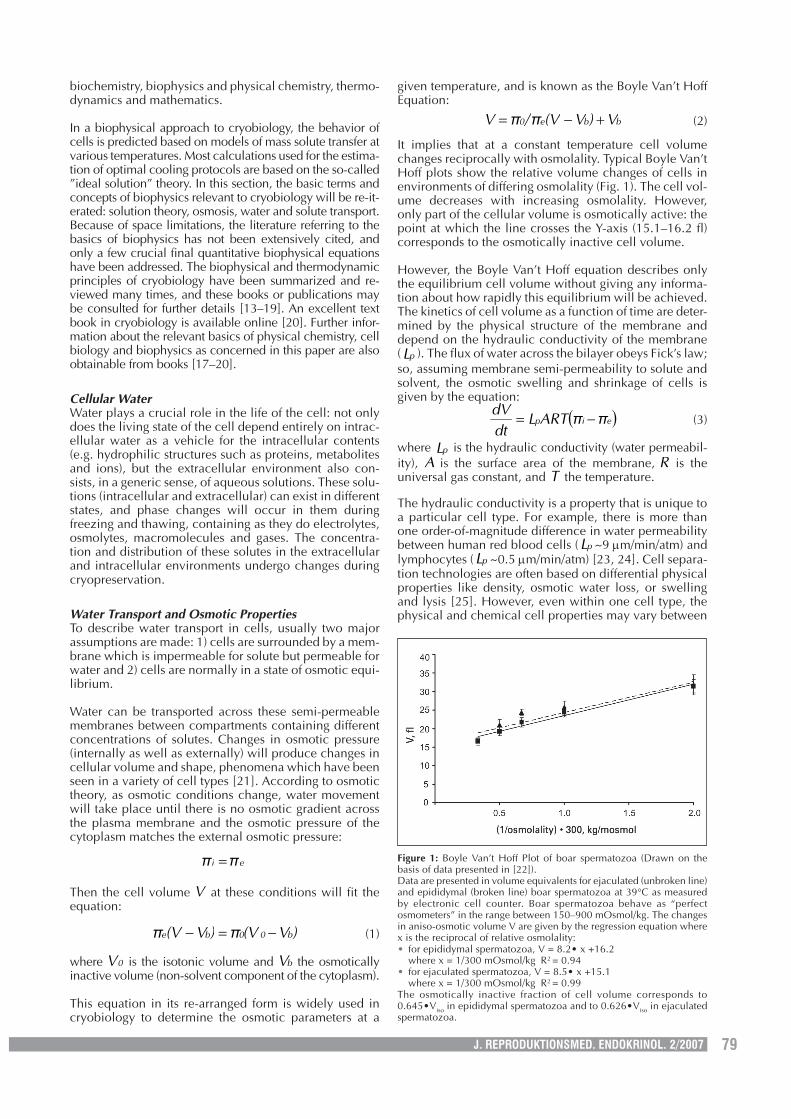
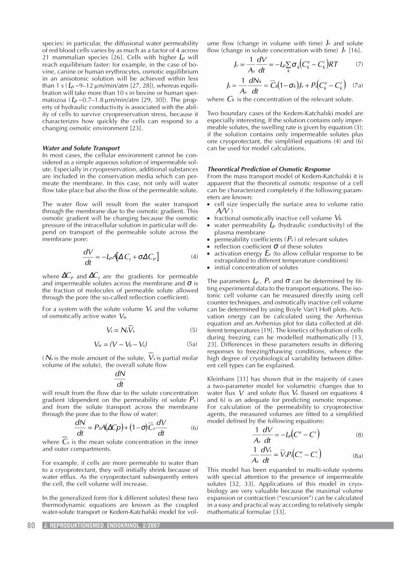
It implies that at a constant temperature cell volumechanges reciprocally with osmolality. Typical Boyle Van’tHoff plots show the relative volume changes of cells inenvironments of differing osmolality (Fig. 1). The cell vol-ume decreases with increasing osmolality. However,only part of the cellular volume is osmotically active: thepoint at which the line crosses the Y-axis (15.1–16.2 fl)corresponds to the osmotically inactive cell volume.
However, the Boyle Van’t Hoff equation describes onlythe equilibrium cell volume without giving any informa-tion about how rapidly this equilibrium will be achieved.The kinetics of cell volume as a function of time are deter-mined by the physical structure of the membrane anddepend on the hydraulic conductivity of the membrane( pL ). The flux of water across the bilayer obeys Fick’s law;so, assuming membrane semi-permeability to solute andsolvent, the osmotic swelling and shrinkage of cells isgiven by the equation:
( )eip ππARTLdtdV
−= (3)
where pL is the hydraulic conductivity (water permeabil-ity), A is the surface area of the membrane, R is theuniversal gas constant, and T the temperature.
The hydraulic conductivity is a property that is unique toa particular cell type. For example, there is more thanone order-of-magnitude difference in water permeabilitybetween human red blood cells ( pL ~9 µm/min/atm) andlymphocytes ( pL ~0.5 µm/min/atm) [23, 24]. Cell separa-tion technologies are often based on differential physicalproperties like density, osmotic water loss, or swellingand lysis [25]. However, even within one cell type, thephysical and chemical cell properties may vary between
Figure 1: Boyle Van’t Hoff Plot of boar spermatozoa (Drawn on thebasis of data presented in [22]).Data are presented in volume equivalents for ejaculated (unbroken line)and epididymal (broken line) boar spermatozoa at 39°C as measuredby electronic cell counter. Boar spermatozoa behave as “perfectosmometers” in the range between 150–900 mOsmol/kg. The changesin aniso-osmotic volume V are given by the regression equation wherex is the reciprocal of relative osmolality:• for epididymal spermatozoa, V = 8.2• x +16.2
where x = 1/300 mOsmol/kg R2 = 0.94• for ejaculated spermatozoa, V = 8.5• x +15.1
where x = 1/300 mOsmol/kg R2 = 0.99The osmotically inactive fraction of cell volume corresponds to0.645•Viso in epididymal spermatozoa and to 0.626•Viso in ejaculatedspermatozoa.
80 J. REPRODUKTIONSMED. ENDOKRINOL. 2/2007
species: in particular, the diffusional water permeabilityof red blood cells varies by as much as a factor of 4 across21 mammalian species [26]. Cells with higher pL willreach equilibrium faster: for example, in the case of bo-vine, canine or human erythrocytes, osmotic equilibriumin an anisotonic solution will be achieved within lessthan 1 s ( pL ~9–12 µm/min/atm [27, 28]), whereas equili-bration will take more than 10 s in bovine or human sper-matozoa ( pL ~0.7–1.8 µm/min/atm [29, 30]). The prop-erty of hydraulic conductivity is associated with the abil-ity of cells to survive cryopreservation stress, because itcharacterizes how quickly the cells can respond to achanging osmotic environment [23].
Water and Solute TransportIn most cases, the cellular environment cannot be con-sidered as a simple aqueous solution of impermeable sol-ute. Especially in cryopreservation, additional substancesare included in the conservation media which can per-meate the membrane. In this case, not only will waterflow take place but also the flow of the permeable solute.
The water flow will result from the water transportthrough the membrane due to the osmotic gradient. Thisosmotic gradient will be changing because the osmoticpressure of the intracellular solution in particular will de-pend on transport of the permeable solute across themembrane pore:
[ ]PIp Cσ∆C∆ALdtdV
+−= (4)
where PC∆ and IC∆ are the gradients for permeableand impermeable solutes across the membrane and σ isthe fraction of molecules of permeable solute allowedthrough the pore (the so-called reflection coefficient).
For a system with the solute volume sV and the volumeof osmotically active water wV
sss VNV = (5)
)VV(VV sbw −−= (5a)
( sN is the mole amount of the solute, sV is partial molarvolume of the solute), the overall solute flow
dtdN
will result from the flow due to the solute concentrationgradient (dependent on the permeability of solute sP )and from the solute transport across the membranethrough the pore due to the flow of water:
( ) ( )dtdV
CσpC∆APdtdN
sS −+= 1 (6)
where sC is the mean solute concentration in the innerand outer compartments.
For example, if cells are more permeable to water thanto a cryoprotectant, they will initially shrink because ofwater efflux. As the cryoprotectant subsequently entersthe cell, the cell volume will increase.
In the generalized form (for k different solutes) these twothermodynamic equations are known as the coupledwater-solute transport or Kedem-Katchalski model for vol-
ume flow (change in volume with time) vJ and soluteflow (change in solute concentration with time) sJ [16].
( )RTCCσLdtdV
AJ
k
ik
ekkp
sv ∑ −−==
1(7)
( ) ( )ik
eksvkk
k
ss CCPJσC
dtdN
AJ −+−== 1
1(7a)
where kC is the concentration of the relevant solute.
Two boundary cases of the Kedem-Katchalski model areespecially interesting. If the solution contains only imper-meable solutes, the swelling rate is given by equation (3);if the solution contains only impermeable solutes plusone cryoprotectant, the simplified equations (4) and (6)can be used for model calculations.
Theoretical Prediction of Osmotic ResponseFrom the mass transport model of Kedem-Katchalski it isapparent that the theoretical osmotic response of a cellcan be characterized completely if the following param-eters are known:• cell size (especially the surface area to volume ratio
A/V )• fractional osmotically inactive cell volume bV• water permeability pL (hydraulic conductivity) of the
plasma membrane• permeability coefficients ( sP ) of relevant solutes• reflection coefficient σ of these solutes• activation energy aE (to allow cellular response to be
extrapolated to different temperature conditions)• initial concentration of solutes
The parameters pL , sP and σ can be determined by fit-ting experimental data to the transport equations. The iso-tonic cell volume can be measured directly using cellcounter techniques, and osmotically inactive cell volumecan be determined by using Boyle Van’t Hoff plots. Acti-vation energy can be calculated using the Arrheniusequation and an Arrhenius plot for data collected at dif-ferent temperatures [19]. The kinetics of hydration of cellsduring freezing can be modelled mathematically [13,23]. Differences in these parameters results in differingresponses to freezing/thawing conditions, whence thehigh degree of cryobiological variability between differ-ent cell types can be explained.
Kleinhans [31] has shown that in the majority of casesa two-parameter model for volumetric changes due towater flux V and solute flux sV (based on equations 4and 6) is an adequate for predicting osmotic response.For calculation of the permeability to cryoprotectiveagents, the measured volumes are fitted to a simplifiedmodel defined by the following equations:
( )iep
sCCL
dtdV
A−−=
1(8)
( )iess
s
sss CCPV
dtdV
A−=
1(8a)
This model has been expanded to multi-solute systemswith special attention to the presence of impermeablesolutes [32, 33]. Applications of this model in cryo-biology are very valuable because the maximal volumeexpansion or contraction (“excursion”) can be calculatedin a easy and practical way according to relatively simplemathematical formulae [33].
81J. REPRODUKTIONSMED. ENDOKRINOL. 2/2007
Cryoinjury and its PreventionThe process of cryopreservation leads to morphologicaland functional damage of cells because it is associatedwith considerable osmotic changes: during freezing,cells become dehydrated, and during thawing, as waterre-equilibration takes place, they are subjected to hypot-onic challenge. The phase transition from water to ice isthe major critical event with respect to cell survival: thereis a critical region of temperature within which the cellsare irreversibly damaged if they remain within it longerthan a few seconds [34]. Therefore, this section will dis-cuss the concepts of ice nucleation and crystal growth,increasing salt concentrations, tolerable osmotic excur-sion of cell volume, post-hypertonic lysis, and mecha-nisms of cryoprotectant action.
Colligative Properties of Extracellular Solutions: Freez-ing Point Depression and OsmolalityThe cellular state and water balance in cells during freez-ing and thawing is determined by the colligative proper-ties of the external aqueous solution, i. e. by the proper-ties which depend on concentrations of solutes ratherthan on behavior of individual chemical compounds. Thefreezing point of a solution is primarily dependent on theconcentration of solute(s) which it contains. The freezingpoint depression is determined by the minimum radius ofan ice crystal that is stable at a given temperature, and isgiven by the mathematical equation
cKT =∆ (9)
where K = 1.86 °C/mol for aqueous solutions and c themolar concentration of the solute.
During freezing, the pure water precipitates out of solu-tion in the form of ice, whence the extracellular salt con-centration increases. In simple systems consisting only oftwo compounds, e. g. NaCl and water, binary phase dia-grams can be used to describe the changes of osmolalityin which the cell must exist. The osmolality of the un-frozen solution at any particular temperature is describedby a simple quadratic function of temperature:
2004920528230 T.T.π −−= (10)
Freezing injury will take place if the cell is exposed to acritical osmolality (i.e. that at which irreversible damageor lysis occurs) [35].
The eutectic temperature also plays an important role incell response to cryopreservation. At the eutectic tem-perature, the system is in thermodynamic balance, atwhich point the unfrozen fraction (solution and solute)becomes solid after spontaneous crystallization (“eutec-tic formation”).
Mechanisms of CryoinjuryThe milestones in understanding mechanisms of cryo-injury were set about a third of century ago as the “Two-factor Hypothesis of Freezing Injury” by Mazur, Leiboand Chu [36] has appeared. In this work, the kinetics ofcellular water loss were investigated, and both too slowand too rapid freezing was found to be associated withlethal cryoinjury.
Rapid freezing will lead to intracellular ice formation be-cause higher cooling rates lead to supercooling of cells
below their nucleation temperature. In this case waterwill be kept in the cell, whence eventually either largelethal, or small less damaging ice crystals will be formedwithin. Rapid freezing may also lead to large gradients inosmotic pressure across the plasma membrane, whichwill become damaged at a critical pressure gradient, andintracellular freezing will also occur as a result of thisdamage [37, 38]. The building of large intracellular icecrystals is associated with cell death. Although moremoderate freezing rates are associated with exposure tosmaller ice crystals, these latter can re-crystallize uponre-warming and become lethal (as demonstrated with re-spect to embryos [39]).
However, if freezing progresses at very slow rates, de-hydration will take place over a longer period of time.During this slow freezing, the osmolality of the externalmedium increases as water-ice is precipitated. The cellswill be exposed to high salt concentrations in the exter-nal medium and dehydrate to a degree of shrinking asso-ciated with serious cellular disruption [36]. Cryoinjurydue to the cell’s sensitivity to high solute concentrationsis referred as to a “solution effect”.
Thus, freezing rates should be slow enough to allow thecells to minimize chemical potential and osmolality gra-dients across the plasma membrane and to dehydratewithout being exposed to lethal salt concentrations. This“equilibrium” freezing leads to tolerable cell shrinkage,to an increase in the intracellular chemical potential, andto depression of the freezing temperature. Due to the de-creased amount of intracellular water and the freezingpoint depression, the potential risk of intracellular ice for-mation is reduced, so that only extracellular ice will beformed. Thus the characteristic shape of the curve plot-ting cell survival against freezing rate is an inverted “U”,with typically a critical cooling rate for maximum sur-vival [36].
The thawing of cells also has to be performed correctlybecause the warming rate influences the survival of cells.Thawing rates comparable with the freezing rates thatinduce intracellular ice formation could lead to re-crys-tallization of small ice crystals resulting in lethal injury.Too slow warming may be lethal depending on cell type,e.g. through over-long exposure to the hypotonic stressthat occurs at thawing. At this stage, possible defects incell volume regulation may be critical. Thus, either slowor rapid warming may be required, depending on bio-physical cell properties and on the cooling rates previ-ously used [23].
Osmotic ToleranceThe shrinkage and swelling of cells is limited by theirphysical capacity to withstand such changes. Damagewill occur beyond certain levels of shrinkage or swelling(the overall range between these levels is known as themaximal cell volume excursion). The optimal equilib-rium freezing rate found for a given cell type must resultin a tolerable volume excursion, i.e. a range of swellingand shrinkage that does not lead to the loss of membraneintegrity and impaired cell function (e.g. motility for sper-matozoa). Tolerable volume excursions are cell type- andspecies-specific.
Cryoinjury by Eutectic FormationEutectic formation, i.e. the process of crystallization ofthe unfrozen fraction of the intracellular or extracellular
82 J. REPRODUKTIONSMED. ENDOKRINOL. 2/2007
medium, has thus far been relatively poorly studied inbiological systems.
Eutectic crystallization is not usually observable withinthe normal freezing range if both inorganic salts and or-ganic substances (e.g. cryoprotectants) are componentsof the system (see [40]). Due to a low concentration ofelectrolyte and the presence of relatively high concentra-tions of cryoprotectant, the impact of eutectic crystalliza-tion on cryoinjury has therefore not been seen as signifi-cant. Hence, despite the theoretical indication that eu-tectic crystallization may have detrimental consequenceson the cell membrane, its effect on cellular systems hasbeen overlooked.
Recently, however, quantitative information on directcell injury due to eutectic formation has been reported[41]. The post-thaw viability of rat prostate tumor cellsdecreased dramatically if eutectic crystallization tookplace. Moreover, cell injury was enhanced by introduc-ing other solutes which had a higher eutectic temperaturethan NaCl. The authors propose two major mechanismsof cell cryoinjury by eutectic formation: (1) mechanicalcell damage to the cell membrane due to extracellulareutectic formation and (2) disruption of cytoplasmicstructures by intracellular eutectic formation.
Mechanisms of Cryoprotectant ActionA more complicated system with three or more com-pounds, e.g. water, sodium chloride and at least onecryoprotective additive, is used in most cryopreservationprotocols. The calculation of equilibrium conditions ismore complex in this case, because the cryoprotectantserves concurrently as a solvent for sodium chloride andas a solute in water. Such a system can be described by aternary phase diagram. Melting and eutectic tempera-tures have been described for the three-compound sys-tems of DMSO-NaCl-H2O and Glycerol-NaCl-H2O [42,43]. These equations are more complex than the analo-gous equation (equation 10) for a binary system but theyallow one to calculate the solution composition at anygiven temperature. In general, lowering the freezingpoint is associated with a higher unfrozen fraction andlower salt concentration in the extracellular space. Thenegative “solution effect” is thereby reduced, and thecells’ exposure to hyper-osmolality diminished. Ternaryphase diagrams illustrate the protective action of cryo-protectants via their effects on the initial freezing tem-perature and the eutectic temperature, and it has beenshown recently experimentally that addition of varioustypes of cryoprotectant leads to the suppression of eutec-tic crystallization, both in a simple NaCl-H2O system andin a more complex phosphate-buffered saline system[41]. One may conclude that one way in which cryopro-tective agents protect cells during freezing and thawing isby lowering the initial freezing point and by diminishinginjury from eutectic crystallization.
Addition of permeable cryoprotectants will also influ-ence the entire system state because trans-membranemovement of both solute and water will take place, thuschanging the transport kinetics, cell dehydration ratesand volume excursion.
Cryoprotective agents that diminish cell damage are usu-ally divided into two general classes related to their abil-ity to move across the cell membrane: permeable and
impermeable. Their mechanisms of action are different.Cryoprotection by permeable agents can be explained bytheir colligative properties: they lower the temperature atwhich cells are exposed to a critical salt concentration[34, 44]. Impermeable agents, on the other hand, arethought to dehydrate cells, allowing them to be cooledrapidly before lethal injury due to solution effects takesplace [44].
However, the effects of cryoprotectants are more com-plex than can be explained from thermodynamics andsolution theory. In high concentrations, they can producelethal osmotic effects during their addition and removal(hyper-osmolality) and they can be toxic [3]. Initial cellu-lar shrinking (due to dehydration) caused by molar con-centrations of cryoprotectant and subsequent swellingduring its removal can both result in damage if the criticalosmotic excursion is exceeded. Whether the effects ofcryoprotective agents are negative or positive is greatlydependent on the kinetics of exposure.
Moreover, it has been shown that the presence of theseagents leads to changes in hydraulic conductivity. Variouscryoprotectant species affect pL in different ways, eitherincreasing or decreasing it (see below), an action whichleads to changes in the osmotic response of cells accord-ing to the mass-transport model. Hence, for optimizationof a cryopreservation system, the optimal selection ofcryoprotectant, both according to its colligative and per-meability properties as well with respect to its effects onother membrane parameters and cell viability, is crucial.
Discrepancies between Theory and Practicein the Cryobiology of Gametes
As outlined above, several quantitative formalisms existwhich allow one to predict the movements of water andsolutes across cellular membranes during the freezingprocess. If the major membrane parameters such ashydraulic conductivity and solute permeability as wellas the geometrical cell characteristics are known, theoptimal equilibrium rates of cell dehydration can becalculated from the mass-transport model equations.
However, several discrepancies have already been foundbetween theoretical approximations and experimentalobservations with respect to maximum cell volume re-sponse to aniso-osmolality. Moreover, empirically deter-mined optimal cooling rates by no means agree withthose calculated according to two-parameter models, es-pecially in the case of mammalian spermatozoa. Waterpermeabilities extrapolated to sub-zero temperaturesbased on data obtained at supra-zero temperatures in theabsence of extracellular ice and cryoprotectants lead todramatic discrepancies between theoretical and experi-mental optimal cooling rates [45, 46]. Indeed, whereasobserved experimental cooling rates lie in the area of 30–50 °C/min [47–49], the mathematical calculations foran pL value of 0.5 µm/min/atm and an activation energyof 3 kcal/mol imply that very high rates (up to 1000 °C/min) are appropriate for the dehydration of mammalianspermatozoa [45]. In fact, such high rates will be lethal:a major decrease in viability has been observed in arange of sperm species after cooling at no more than100–300 °C/min. One way to account for this dis-crepancy between theory and practice is to consider theaccuracy of mathematical approximations. For example,
83J. REPRODUKTIONSMED. ENDOKRINOL. 2/2007
in the two-parameter-model, when the con-centration of impermeable salts is low, theconcentration of permeable solute is high( saltttancryoprotec CC >> ), the Boyle Van’t Hoffapproximation of maximal cell volume excur-sion mV (equation 1) is true, and the quotient
saltcryo/PP approaches infinity (characteristicfor freezing in the presence of any applicablecryoprotectant), the product
)VπV)( π/P(P mmsaltcryo −− 001
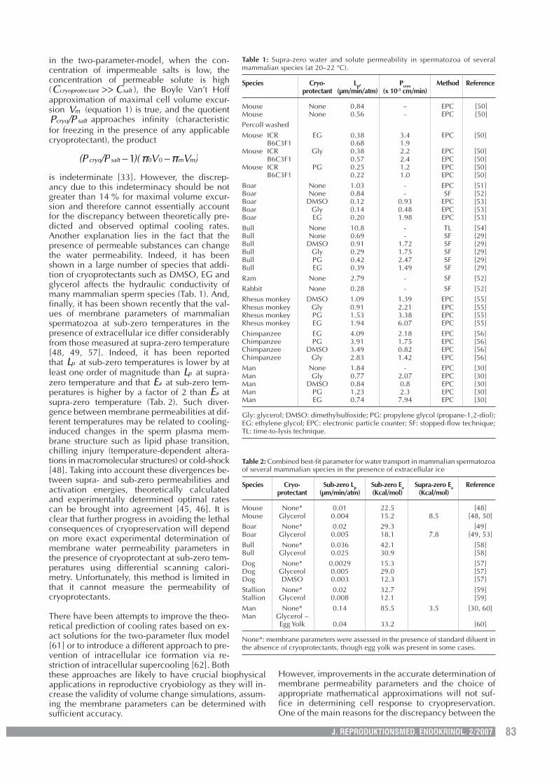
is indeterminate [33]. However, the discrep-ancy due to this indeterminacy should be notgreater than 14 % for maximal volume excur-sion and therefore cannot essentially accountfor the discrepancy between theoretically pre-dicted and observed optimal cooling rates.Another explanation lies in the fact that thepresence of permeable substances can changethe water permeability. Indeed, it has beenshown in a large number of species that addi-tion of cryoprotectants such as DMSO, EG andglycerol affects the hydraulic conductivity ofmany mammalian sperm species (Tab. 1). And,finally, it has been shown recently that the val-ues of membrane parameters of mammalianspermatozoa at sub-zero temperatures in thepresence of extracellular ice differ considerablyfrom those measured at supra-zero temperature[48, 49, 57]. Indeed, it has been reportedthat pL at sub-zero temperatures is lower by atleast one order of magnitude than pL at supra-zero temperature and that aE at sub-zero tem-peratures is higher by a factor of 2 than aE atsupra-zero temperature (Tab. 2). Such diver-gence between membrane permeabilities at dif-ferent temperatures may be related to cooling-induced changes in the sperm plasma mem-brane structure such as lipid phase transition,chilling injury (temperature-dependent altera-tions in macromolecular structures) or cold-shock[48]. Taking into account these divergences be-tween supra- and sub-zero permeabilities andactivation energies, theoretically calculatedand experimentally determined optimal ratescan be brought into agreement [45, 46]. It isclear that further progress in avoiding the lethalconsequences of cryopreservation will dependon more exact experimental determination ofmembrane water permeability parameters inthe presence of cryoprotectant at sub-zero tem-peratures using differential scanning calori-metry. Unfortunately, this method is limited inthat it cannot measure the permeability ofcryoprotectants.
There have been attempts to improve the theo-retical prediction of cooling rates based on ex-act solutions for the two-parameter flux model[61] or to introduce a different approach to pre-vention of intracellular ice formation via re-striction of intracellular supercooling [62]. Boththese approaches are likely to have crucial biophysicalapplications in reproductive cryobiology as they will in-crease the validity of volume change simulations, assum-ing the membrane parameters can be determined withsufficient accuracy.
However, improvements in the accurate determination ofmembrane permeability parameters and the choice ofappropriate mathematical approximations will not suf-fice in determining cell response to cryopreservation.One of the main reasons for the discrepancy between the
Table 2: Combined best-fit parameter for water transport in mammalian spermatozoaof several mammalian species in the presence of extracellular ice
Species Cryo- Sub-zero Lp Sub-zero Ea Supra-zero Ea Referenceprotectant (µm/min/atm) (Kcal/mol) (Kcal/mol)
Mouse None* 0.01 22.5 [48]Mouse Glycerol 0.004 15.2 8.5 [48, 50]Boar None* 0.02 29.3 [49]Boar Glycerol 0.005 18.1 7.8 [49, 53]Bull None* 0.036 42.1 [58]Bull Glycerol 0.025 30.9 [58]Dog None* 0.0029 15.3 [57]Dog Glycerol 0.005 29.0 [57]Dog DMSO 0.003 12.3 [57]Stallion None* 0.02 32.7 [59]Stallion Glycerol 0.008 12.1 [59]Man None* 0.14 85.5 3.5 [30, 60]Man Glycerol –
Egg Yolk 0.04 33.2 [60]
None*: membrane parameters were assessed in the presence of standard diluent inthe absence of cryoprotectants, though egg yolk was present in some cases.
Table 1: Supra-zero water and solute permeability in spermatozoa of severalmammalian species (at 20–22 °C).
Species Cryo- Lp, Pcryo Method Referenceprotectant (µm/min/atm) (x 10-3 cm/min)
Mouse None 0.84 – EPC [50]Mouse None 0.56 – EPC [50]Percoll washedMouse ICR EG 0.38 3.4 EPC [50]
B6C3F1 0.68 1.9Mouse ICR Gly 0.38 2.2 EPC [50]
B6C3F1 0.57 2.4 EPC [50]Mouse ICR PG 0.25 1.2 EPC [50]
B6C3F1 0.22 1.0 EPC [50]Boar None 1.03 - EPC [51]Boar None 0.84 - SF [52]Boar DMSO 0.12 0.93 EPC [53]Boar Gly 0.14 0.48 EPC [53]Boar EG 0.20 1.98 EPC [53]Bull None 10.8 - TL [54]Bull None 0.69 - SF [29]Bull DMSO 0.91 1.72 SF [29]Bull Gly 0.29 1.75 SF [29]Bull PG 0.42 2.47 SF [29]Bull EG 0.39 1.49 SF [29]Ram None 2.79 - SF [52]Rabbit None 0.28 - SF [52]Rhesus monkey DMSO 1.09 1.39 EPC [55]Rhesus monkey Gly 0.91 2.21 EPC [55]Rhesus monkey PG 1.53 3.38 EPC [55]Rhesus monkey EG 1.94 6.07 EPC [55]Chimpanzee EG 4.09 2.18 EPC [56]Chimpanzee PG 3.91 1.75 EPC [56]Chimpanzee DMSO 3.49 0.82 EPC [56]Chimpanzee Gly 2.83 1.42 EPC [56]Man None 1.84 - EPC [30]Man Gly 0.77 2.07 EPC [30]Man DMSO 0.84 0.8 EPC [30]Man PG 1.23 2.3 EPC [30]Man EG 0.74 7.94 EPC [30]
Gly: glycerol; DMSO: dimethylsulfoxide; PG: propylene glycol (propane-1,2-diol);EG: ethylene glycol; EPC: electronic particle counter; SF: stopped-flow technique;TL: time-to-lysis technique.
84 J. REPRODUKTIONSMED. ENDOKRINOL. 2/2007
theoretical and experimental behavior of cells duringthe freezing/thawing process is the biological complexityof the cell. Biological membranes are too complex to jus-tify some of assumptions made from the biophysicalpoint of view. Although it is convenient to consider thecell as a “bag with salt water”, there is a great deal of evi-dence that intracellular water and ions behave in a differ-ent way from those in ideal aqueous solutions. The cellcytoplasm does not behave like water with ions dissolvedin it; there is much molecular interaction between differ-ent cell compartments where different signal transduc-tion mechanisms are involved [17, 18]. Exposure to deeptemperatures can affect this interaction and thereby sig-nalling mechanisms; there is already evidence that somesperm intracellular signalling pathways can be affectedduring cooling and cryopreservation [63–66]. Also, thecell membrane is not a simple semi-permeable barrier.The physical and chemical composition of the membraneis complex and affected by temperature: changes in mo-lecular structure at deeper temperatures will alter soluteand water transport through pores, and changes in mobil-ity of lipids will affect diffusion through the lipid bilayer.In particular, in cryopreserved spermatozoa, lipid diffu-sion in the plasma membrane has been reported to besignificantly reduced relative to fresh spermatozoa [67].These interactions would affect greatly biophysical mod-els based on simplified assumptions. Analogously, theapparently relatively symmetrical external geometry ofthe oocyte cannot completely justify the use of currentbiophysical models to optimize cryopreservation proto-cols on the basis of the mass-transport model [3, 6]. Thebiochemical and physiological complexity of cellularorganization such as cytoskeletal elements and corticalgranulae must be taken into consideration.
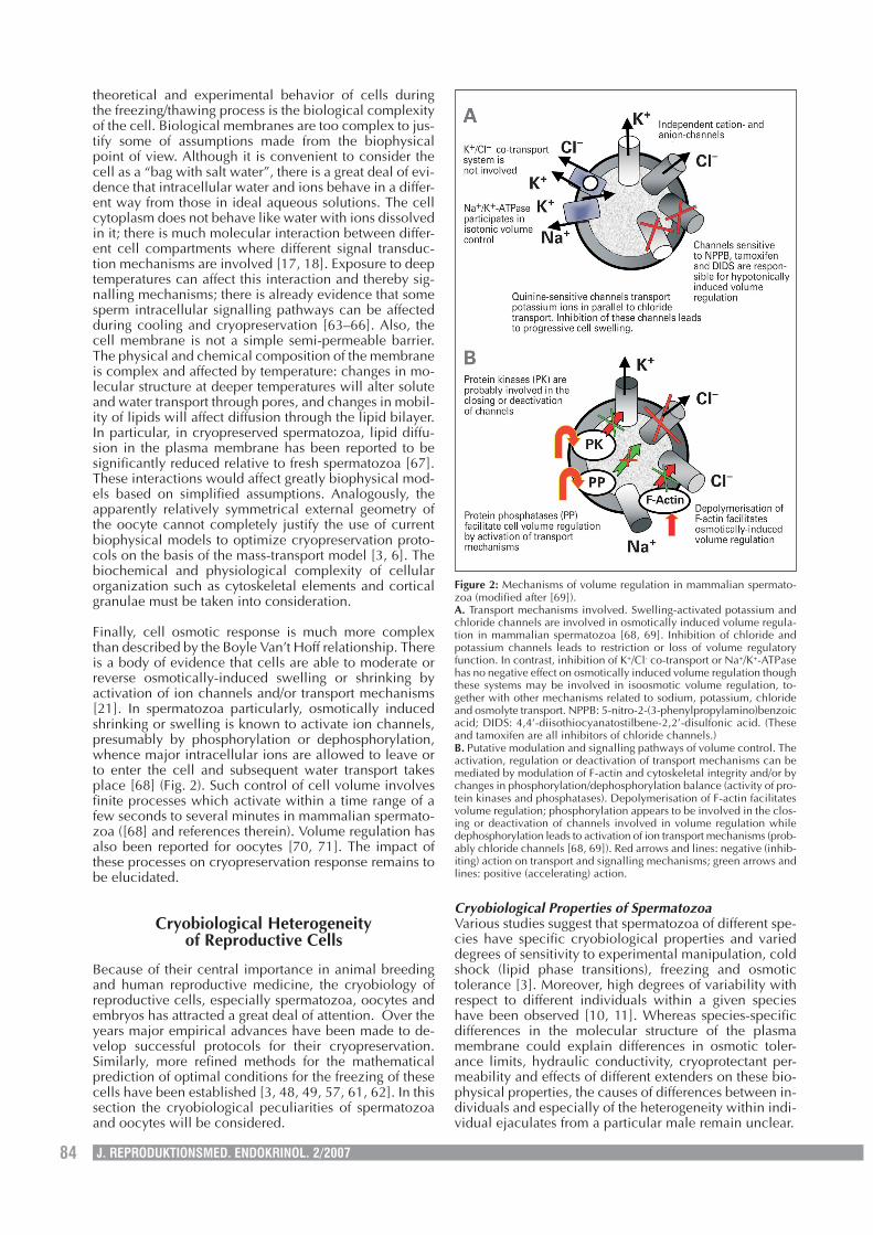
Finally, cell osmotic response is much more complexthan described by the Boyle Van’t Hoff relationship. Thereis a body of evidence that cells are able to moderate orreverse osmotically-induced swelling or shrinking byactivation of ion channels and/or transport mechanisms[21]. In spermatozoa particularly, osmotically inducedshrinking or swelling is known to activate ion channels,presumably by phosphorylation or dephosphorylation,whence major intracellular ions are allowed to leave orto enter the cell and subsequent water transport takesplace [68] (Fig. 2). Such control of cell volume involvesfinite processes which activate within a time range of afew seconds to several minutes in mammalian spermato-zoa ([68] and references therein). Volume regulation hasalso been reported for oocytes [70, 71]. The impact ofthese processes on cryopreservation response remains tobe elucidated.
Cryobiological Heterogeneityof Reproductive Cells
Because of their central importance in animal breedingand human reproductive medicine, the cryobiology ofreproductive cells, especially spermatozoa, oocytes andembryos has attracted a great deal of attention. Over theyears major empirical advances have been made to de-velop successful protocols for their cryopreservation.Similarly, more refined methods for the mathematicalprediction of optimal conditions for the freezing of thesecells have been established [3, 48, 49, 57, 61, 62]. In thissection the cryobiological peculiarities of spermatozoaand oocytes will be considered.
Cryobiological Properties of SpermatozoaVarious studies suggest that spermatozoa of different spe-cies have specific cryobiological properties and varieddegrees of sensitivity to experimental manipulation, coldshock (lipid phase transitions), freezing and osmotictolerance [3]. Moreover, high degrees of variability withrespect to different individuals within a given specieshave been observed [10, 11]. Whereas species-specificdifferences in the molecular structure of the plasmamembrane could explain differences in osmotic toler-ance limits, hydraulic conductivity, cryoprotectant per-meability and effects of different extenders on these bio-physical properties, the causes of differences between in-dividuals and especially of the heterogeneity within indi-vidual ejaculates from a particular male remain unclear.
Figure 2: Mechanisms of volume regulation in mammalian spermato-zoa (modified after [69]).A. Transport mechanisms involved. Swelling-activated potassium andchloride channels are involved in osmotically induced volume regula-tion in mammalian spermatozoa [68, 69]. Inhibition of chloride andpotassium channels leads to restriction or loss of volume regulatoryfunction. In contrast, inhibition of K+/Cl- co-transport or Na+/K+-ATPasehas no negative effect on osmotically induced volume regulation thoughthese systems may be involved in isoosmotic volume regulation, to-gether with other mechanisms related to sodium, potassium, chlorideand osmolyte transport. NPPB: 5-nitro-2-(3-phenylpropylamino)benzoicacid; DIDS: 4,4’-diisothiocyanatostilbene-2,2’-disulfonic acid. (Theseand tamoxifen are all inhibitors of chloride channels.)B. Putative modulation and signalling pathways of volume control. Theactivation, regulation or deactivation of transport mechanisms can bemediated by modulation of F-actin and cytoskeletal integrity and/or bychanges in phosphorylation/dephosphorylation balance (activity of pro-tein kinases and phosphatases). Depolymerisation of F-actin facilitatesvolume regulation; phosphorylation appears to be involved in the clos-ing or deactivation of channels involved in volume regulation whiledephosphorylation leads to activation of ion transport mechanisms (prob-ably chloride channels [68, 69]). Red arrows and lines: negative (inhib-iting) action on transport and signalling mechanisms; green arrows andlines: positive (accelerating) action.
85J. REPRODUKTIONSMED. ENDOKRINOL. 2/2007
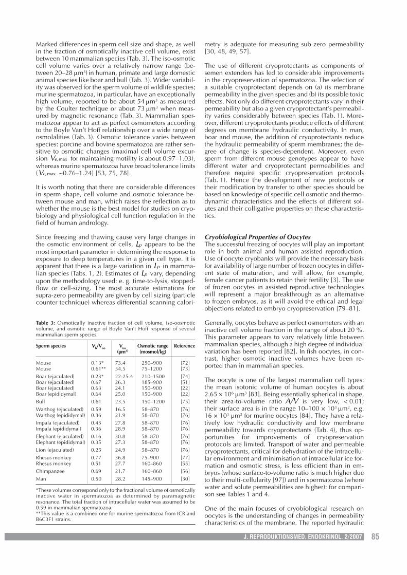
Marked differences in sperm cell size and shape, as wellin the fraction of osmotically inactive cell volume, existbetween 10 mammalian species (Tab. 3). The iso-osmoticcell volume varies over a relatively narrow range (be-tween 20–28 µm3) in human, primate and large domesticanimal species like boar and bull (Tab. 3). Wider variabil-ity was observed for the sperm volume of wildlife species;murine spermatozoa, in particular, have an exceptionallyhigh volume, reported to be about 54 µm3 as measuredby the Coulter technique or about 73 µm3 when meas-ured by magnetic resonance (Tab. 3). Mammalian sper-matozoa appear to act as perfect osmometers accordingto the Boyle Van’t Hoff relationship over a wide range ofosmolalities (Tab. 3). Osmotic tolerance varies betweenspecies: porcine and bovine spermatozoa are rather sen-sitive to osmotic changes (maximal cell volume excur-sion maxe,V for maintaining motility is about 0.97–1.03),whereas murine spermatozoa have broad tolerance limits( maxe,V ~0.76–1.24) [53, 75, 78].
It is worth noting that there are considerable differencesin sperm shape, cell volume and osmotic tolerance be-tween mouse and man, which raises the reflection as towhether the mouse is the best model for studies on cryo-biology and physiological cell function regulation in thefield of human andrology.
Since freezing and thawing cause very large changes inthe osmotic environment of cells, pL appears to be themost important parameter in determining the response toexposure to deep temperatures in a given cell type. It isapparent that there is a large variation in pL in mamma-lian species (Tabs. 1, 2). Estimates of pL vary, dependingupon the methodology used: e. g. time-to-lysis, stopped-flow or cell-sizing. The most accurate estimations forsupra-zero permeability are given by cell sizing (particlecounter technique) whereas differential scanning calori-
metry is adequate for measuring sub-zero permeability[30, 48, 49, 57].
The use of different cryoprotectants as components ofsemen extenders has led to considerable improvementsin the cryopreservation of spermatozoa. The selection ofa suitable cryoprotectant depends on (a) its membranepermeability in the given species and (b) its possible toxiceffects. Not only do different cryoprotectants vary in theirpermeability but also a given cryoprotectant’s permeabil-ity varies considerably between species (Tab. 1). More-over, different cryoprotectants produce effects of differentdegrees on membrane hydraulic conductivity. In man,boar and mouse, the addition of cryoprotectants reducethe hydraulic permeability of sperm membranes; the de-gree of change is species-dependent. Moreover, evensperm from different mouse genotypes appear to havedifferent water and cryoprotectant permeabilities andtherefore require specific cryopreservation protocols(Tab. 1). Hence the development of new protocols ortheir modification by transfer to other species should bebased on knowledge of specific cell osmotic and thermo-dynamic characteristics and the effects of different sol-utes and their colligative properties on these characteris-tics.
Cryobiological Properties of OocytesThe successful freezing of oocytes will play an importantrole in both animal and human assisted reproduction.Use of oocyte cryobanks will provide the necessary basisfor availability of large number of frozen oocytes in differ-ent state of maturation, and will allow, for example,female cancer patients to retain their fertility [3]. The useof frozen oocytes in assisted reproductive technologieswill represent a major breakthrough as an alternativeto frozen embryos, as it will avoid the ethical and legalobjections related to embryo cryopreservation [79–81].
Generally, oocytes behave as perfect osmometers with aninactive cell volume fraction in the range of about 20 %.This parameter appears to vary relatively little betweenmammalian species, although a high degree of individualvariation has been reported [82]. In fish oocytes, in con-trast, higher osmotic inactive volumes have been re-ported than in mammalian species.
The oocyte is one of the largest mammalian cell types:the mean isotonic volume of human oocytes is about2.65 × 106 µm3 [83]. Being essentially spherical in shape,their area-to-volume ratio A/V is very low, < 0.01;their surface area is in the range 10–100 × 103 µm2, e.g.16 × 103 µm2 for murine oocytes [84]. They have a rela-tively low hydraulic conductivity and low membranepermeability towards cryoprotectants (Tab. 4), thus op-portunities for improvements of cryopreservationprotocols are limited. Transport of water and permeablecryoprotectants, critical for dehydration of the intracellu-lar environment and minimisation of intracellular ice for-mation and osmotic stress, is less efficient than in em-bryos (whose surface-to-volume ratio is much higher dueto their multi-cellularity [97]) and in spermatozoa (wherewater and solute permeabilities are higher): for compari-son see Tables 1 and 4.
One of the main focuses of cryobiological research onoocytes is the understanding of changes in permeabilitycharacteristics of the membrane. The reported hydraulic
Table 3: Osmotically inactive fraction of cell volume, iso-oosmoticvolume, and osmotic range of Boyle Van’t Hoff response of severalmammalian sperm species.
Sperm species Vb/Viso Viso Osmotic range Reference(µm3) (mosmol/kg)
Mouse 0.13* 73.4 250–900 [72]Mouse 0.61** 54.5 75–1200 [73]Boar (ejaculated) 0.23* 22-25.4 210–1500 [74]Boar (ejaculated) 0.67 26.3 185–900 [51]Boar (ejaculated) 0.63 24.1 150–900 [22]Boar (epididymal) 0.64 25.0 150–900 [22]Bull 0.61 23.5 150–1200 [75]Warthog (ejaculated) 0.59 16.5 58–870 [76]Warthog (epididymal) 0.36 21.9 58–870 [76]Impala (ejaculated) 0.45 27.8 58–870 [76]Impala (epididymal) 0.36 28.9 58–870 [76]Elephant (ejaculated) 0.16 30.8 58–870 [76]Elephant (epididymal) 0.35 27.3 58–870 [76]Lion (ejaculated) 0.25 24.9 58–870 [76]Rhesus monkey 0.77 36.8 75–900 [77]Rhesus monkey 0.51 27.7 160–860 [55]Chimpanzee 0.69 21.7 160–860 [56]Man 0.50 28.2 145–900 [30]
*These volumes correspond only to the fractional volume of osmoticallyinactive water in spermatozoa as determined by paramagneticresonance. The total fraction of intracellular water was assumed to be0.59 in mammalian spermatozoa.**This value is a combined one for murine spermatozoa from ICR andB6C3F1 strains.
86 J. REPRODUKTIONSMED. ENDOKRINOL. 2/2007
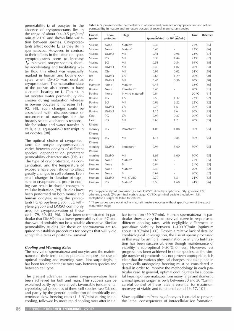
permeability pL of oocytes in theabsence of cryoprotectants lies inthe range of about 0.4–0.5 µm/atm/min at 20 °C and shows little varia-tion between species. Cryoprotec-tants affect oocyte pL as they do inspermatozoa. However, in contrastto their effects in the latter cell type,cryoprotectants seem to increase
pL in several oocyte species, there-by accelerating and facilitating wa-ter flux; this effect was especiallymarked in human and bovine oo-cytes when DMSO was used ascryoprotectant. The maturation stateof the oocyte also seems to havea crucial bearing on pL (Tab. 4). Inrat oocytes water permeability de-creases during maturation whereasin bovine oocytes it increases [91,92, 98]. Such changes could beassociated with disappearance oroccurrence of transcripts for thebroadly selective channels responsi-ble for solute and water transfer incells, e. g. aquaporin-9 transcript inrat oocytes [98].
The optimal choice of cryoprotec-tants for oocyte cryopreservationvaries between oocytes of differentspecies, dependant on protectantpermeability characteristics (Tab. 4).The type of cryoprotectant, its con-centration, and the temperature ofexposure have been shown to affectgreatly changes in cell volume. Evensmall changes in duration of expo-sure to cryoprotectant prior to cool-ing can result in drastic changes incellular hydration [99]. Studies havebeen performed on both mouse andhuman oocytes, using the protec-tants PG (propylene glycol), EG (eth-ylene glycol) and DMSO commonlyused for cryopreservation of thesecells [79, 80, 83, 96]. It has been demonstrated in par-ticular that DMSO has a lower permeability than PG andthus would probably not be a suitable alternative. Similarpermeability studies like those on spermatozoa are re-quired to establish procedures for oocytes that will yieldacceptable rates of post-thaw survival.
Cooling and Warming RatesThe survival of spermatozoa and oocytes and the mainte-nance of their fertilization potential require the use ofoptimal cooling and warming rates. Not surprisingly, ithas been found that these rates vary between species andbetween cell type.
The greatest advances in sperm cryopreservation havebeen achieved for bull and man. This success can beexplained partly by the relatively favourable fundamentalcryobiological properties of these cell species (see Tables),and partly by the general application of empirically de-termined slow freezing rates (1–5 °C/min) during initialcooling, followed by more rapid cooling rates after initial
ice formation (50 °C/min). Human spermatozoa in par-ticular show a very broad survival curve in response todifferent cooling rates, with negligible differences inpost-thaw viability between 1–100 °C/min (optimumabout 10 °C/min) [100]. Despite a relative lack of detailedcryobiological investigation, the use of sperm processedin this way for artificial insemination or in vitro fertiliza-tion has been successful, even though maintenance ofviability is sub-optimal (~50 % or less). However, lessprogress has been achieved in other species, as the sim-ple transfer of protocols has not proven appropriate. It isclear that the various physical changes that take place insperm cells undergoing freezing must be considered indetail in order to improve the methodology in each par-ticular case. In general, optimal cooling rates for success-ful freezing of spermatozoa from many large and domesticanimal species range narrowly between 30 and 50 °C/min;careful control of these rates is essential for maximumrecovery of viable and functional cells [49, 57, 101].
Slow equilibrium freezing of oocytes is crucial to preventthe lethal consequences of intracellular ice formation.
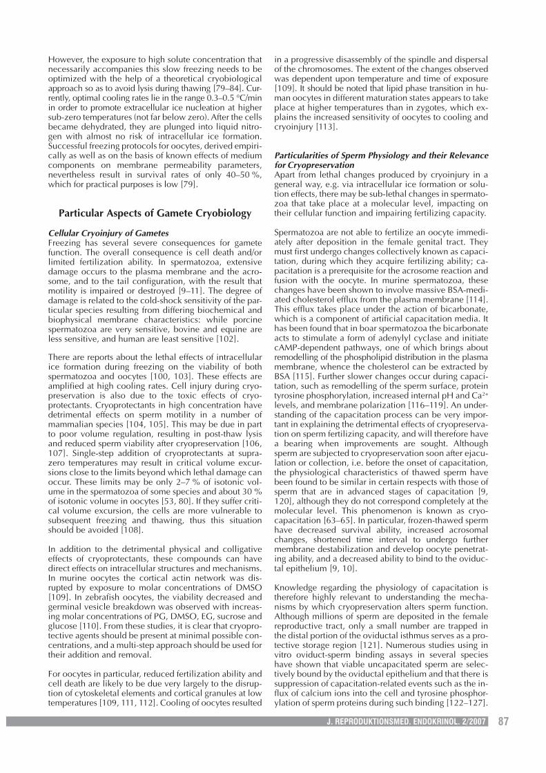
Table 4: Supra-zero water permeability in absence and presence of cryoprotectant and solutepermeability in mature and immature oocytes of several mammalian species
Oocyte Cryo- Stage Lp Pcryo Temp Referncespecies protectant (µm/min/atm) (x 10-3 cm/min)
Murine None Mature* 0.36 - 21°C [85]Murine None Mature* 0.40 - 22°C [86]Murine DMSO MII 0.41 0.96 23°C [87]Murine PG MII 0.36 1.44 23°C [87]Murine EG MII 0.51 0.54 19°C [88]Murine DMSO MII 0.4 1.07 20°C [84]Murine Gly MII 0.98 0.02 24°C [89]Rat DMSO GV 0.68 1.29 20°C [90]Rat DMSO MII 0.45 0.56 20°C [90]Hamster None Mature* 0.45 - 22°C [86]Bovine None Immature* 0.45 - 20°C [91]Bovine None In vitro matured* 0.84 - 20 °C [91]Bovine EG GV 0.5 1.32 22°C [92]Bovine EG MII 0.83 2.22 22°C [92]Bovine DMSO GV 0.73 1.6 20°C [93]Bovine DMSO MII 1.16 2.6 20°C [93]Goat PG GV 0.97 0.87 20°C [94]Goat PG MII 0.60 1.2 20°C [95]Rhesusmonkey EG Immature* 1.08 1.08 30°C [95]Rhesusmonkey EG MII 1.14 0.84 30°C [95]Rhesusmonkey DMSO Immature* 0.96 3.60 30°C [95]Rhesusmonkey DMSO MII 1.08 6.00 30°C [95]Human None Mature* 0.65 - 21°C [85]Human None Ff 0.84 - 21°C [85]Human None Mature* 0.40 - 20°C [82]Human None Ff 0.64 - 20°C [82]Human DMSO MII+GVBD 0.70 1.5 24°C [83]Human PG Mature* 0.53 1.68 24°C [96]
PG: propylene glycol (propane-1,2-diol); DMSO: dimethylsulphoxide; Gly: glycerol; EG:ethylene glycol; GV: germinal vesicle stage; GVBD: germinal vesicle breakdown; MII:metaphase II stage; Ff: failed to fertilize.* These values were obtained in mature/immature oocytes without specification of the exactmaturation stage.
87J. REPRODUKTIONSMED. ENDOKRINOL. 2/2007
However, the exposure to high solute concentration thatnecessarily accompanies this slow freezing needs to beoptimized with the help of a theoretical cryobiologicalapproach so as to avoid lysis during thawing [79–84]. Cur-rently, optimal cooling rates lie in the range 0.3–0.5 °C/minin order to promote extracellular ice nucleation at highersub-zero temperatures (not far below zero). After the cellsbecame dehydrated, they are plunged into liquid nitro-gen with almost no risk of intracellular ice formation.Successful freezing protocols for oocytes, derived empiri-cally as well as on the basis of known effects of mediumcomponents on membrane permeability parameters,nevertheless result in survival rates of only 40–50 %,which for practical purposes is low [79].
Particular Aspects of Gamete Cryobiology
Cellular Cryoinjury of GametesFreezing has several severe consequences for gametefunction. The overall consequence is cell death and/orlimited fertilization ability. In spermatozoa, extensivedamage occurs to the plasma membrane and the acro-some, and to the tail configuration, with the result thatmotility is impaired or destroyed [9–11]. The degree ofdamage is related to the cold-shock sensitivity of the par-ticular species resulting from differing biochemical andbiophysical membrane characteristics: while porcinespermatozoa are very sensitive, bovine and equine areless sensitive, and human are least sensitive [102].
There are reports about the lethal effects of intracellularice formation during freezing on the viability of bothspermatozoa and oocytes [100, 103]. These effects areamplified at high cooling rates. Cell injury during cryo-preservation is also due to the toxic effects of cryo-protectants. Cryoprotectants in high concentration havedetrimental effects on sperm motility in a number ofmammalian species [104, 105]. This may be due in partto poor volume regulation, resulting in post-thaw lysisand reduced sperm viability after cryopreservation [106,107]. Single-step addition of cryoprotectants at supra-zero temperatures may result in critical volume excur-sions close to the limits beyond which lethal damage canoccur. These limits may be only 2–7 % of isotonic vol-ume in the spermatozoa of some species and about 30 %of isotonic volume in oocytes [53, 80]. If they suffer criti-cal volume excursion, the cells are more vulnerable tosubsequent freezing and thawing, thus this situationshould be avoided [108].
In addition to the detrimental physical and colligativeeffects of cryoprotectants, these compounds can havedirect effects on intracellular structures and mechanisms.In murine oocytes the cortical actin network was dis-rupted by exposure to molar concentrations of DMSO[109]. In zebrafish oocytes, the viability decreased andgerminal vesicle breakdown was observed with increas-ing molar concentrations of PG, DMSO, EG, sucrose andglucose [110]. From these studies, it is clear that cryopro-tective agents should be present at minimal possible con-centrations, and a multi-step approach should be used fortheir addition and removal.
For oocytes in particular, reduced fertilization ability andcell death are likely to be due very largely to the disrup-tion of cytoskeletal elements and cortical granules at lowtemperatures [109, 111, 112]. Cooling of oocytes resulted
in a progressive disassembly of the spindle and dispersalof the chromosomes. The extent of the changes observedwas dependent upon temperature and time of exposure[109]. It should be noted that lipid phase transition in hu-man oocytes in different maturation states appears to takeplace at higher temperatures than in zygotes, which ex-plains the increased sensitivity of oocytes to cooling andcryoinjury [113].
Particularities of Sperm Physiology and their Relevancefor CryopreservationApart from lethal changes produced by cryoinjury in ageneral way, e.g. via intracellular ice formation or solu-tion effects, there may be sub-lethal changes in spermato-zoa that take place at a molecular level, impacting ontheir cellular function and impairing fertilizing capacity.
Spermatozoa are not able to fertilize an oocyte immedi-ately after deposition in the female genital tract. Theymust first undergo changes collectively known as capaci-tation, during which they acquire fertilizing ability; ca-pacitation is a prerequisite for the acrosome reaction andfusion with the oocyte. In murine spermatozoa, thesechanges have been shown to involve massive BSA-medi-ated cholesterol efflux from the plasma membrane [114].This efflux takes place under the action of bicarbonate,which is a component of artificial capacitation media. Ithas been found that in boar spermatozoa the bicarbonateacts to stimulate a form of adenylyl cyclase and initiatecAMP-dependent pathways, one of which brings aboutremodelling of the phospholipid distribution in the plasmamembrane, whence the cholesterol can be extracted byBSA [115]. Further slower changes occur during capaci-tation, such as remodelling of the sperm surface, proteintyrosine phosphorylation, increased internal pH and Ca2+
levels, and membrane polarization [116–119]. An under-standing of the capacitation process can be very impor-tant in explaining the detrimental effects of cryopreserva-tion on sperm fertilizing capacity, and will therefore havea bearing when improvements are sought. Althoughsperm are subjected to cryopreservation soon after ejacu-lation or collection, i.e. before the onset of capacitation,the physiological characteristics of thawed sperm havebeen found to be similar in certain respects with those ofsperm that are in advanced stages of capacitation [9,120], although they do not correspond completely at themolecular level. This phenomenon is known as cryo-capacitation [63–65]. In particular, frozen-thawed spermhave decreased survival ability, increased acrosomalchanges, shortened time interval to undergo furthermembrane destabilization and develop oocyte penetrat-ing ability, and a decreased ability to bind to the oviduc-tal epithelium [9, 10].
Knowledge regarding the physiology of capacitation istherefore highly relevant to understanding the mecha-nisms by which cryopreservation alters sperm function.Although millions of sperm are deposited in the femalereproductive tract, only a small number are trapped inthe distal portion of the oviductal isthmus serves as a pro-tective storage region [121]. Numerous studies using invitro oviduct-sperm binding assays in several specieshave shown that viable uncapacitated sperm are selec-tively bound by the oviductal epithelium and that there issuppression of capacitation-related events such as the in-flux of calcium ions into the cell and tyrosine phosphor-ylation of sperm proteins during such binding [122–127].
88 J. REPRODUKTIONSMED. ENDOKRINOL. 2/2007
This sperm storage site in the mammalian oviduct is de-signated a functional sperm reservoir since it fulfils thefollowing functions: 1) selection of the fertilization-com-petent sperm population, 2) modulation of sperm capaci-tation, and 3) regulation of sperm transport to minimizepolyspermic fertilization [128–131]. Since the capacita-tion process is understood as a controlled on-goingdestabilization process which reduces the life span ofsperm, the maintenance of sperm viability within a cer-tain window of time and the control of capacitation aremutually associated events [132, 133]. To provide suffi-cient competent spermatozoa at the time of ovulation,capacitation should not be completed until after ovula-tion has occurred. At this time, conditions within the ovi-duct seem to reverse the inhibitory influence of the ovi-duct on sperm function, initiating capacitation and hyper-activation in order for the sperm cells to release and toprogress toward the site of fertilization [133]. It should benoted that sperm populations appear to undergocapacitation at different rates, thereby providing a con-tinuous supply of “primed” cells ready to fertilize theovulated eggs.
Assessment of Frozen SemenThe morphological and functional changes of spermthat result from cryoinjury require assessment [9–11].Whereas gross aspects such as changes in morphologyand motility can be readily assessed using phase-contrastmicroscopy, there is a need to establish more sophisti-cated spermatological methods that will allow rapid andsensitive assessment of sperm function in frozen-thawedsemen. Given the similarity between frozen-thawed andcapacitated spermatozoa, and the fact that ejaculatedspermatozoa are not able to fertilize an oocyte, it is logi-cal to conclude that assessment of those sperm functionparameters that are modified during the capacitationprocess must be performed under fertilizing conditions[134]. It is known that animal fertility varies not only be-tween males but also between ejaculates within indi-vidual males, therefore there is a need to design assayssensitive enough to detect intra- as well as inter-individualdifferences and even the subtle heterogeneity within oneejaculate of a given individual [10, 11, 135, 136].
Assays which have been shown to be sufficiently sensitiveto assess the influence of cooling and/or cryopreservationon semen quality (and thereby to predict its freezability)include:• inducability of the acrosome reaction by calcium
ionophore• measurement of changes in intracellular calcium content• measurement of volume regulatory ability• assessment of plasma membrane lipid disorder (mero-
cyanine assay)• computer-assisted motility analysis• assessment of sperm chromatin stability• assessment of ability to bind to oviductal explants.
These and other assays have been reviewed in recentreviews of clinical and laboratory assessment of spermfunction [107, 137–139]. Details of the assays are to befound in the many original studies cited in these reviews.
Conclusions
A basic understanding of the cellular processes whichaccompany cryopreservation is necessary for successful
development of its applications in reproductive biomedi-cine. Physico-chemical conditions inside and outside thecell are controlled by three main parameters: extracellu-lar solute concentration, the presence of permeable andimpermeable solutes, and the cooling and warming rates.Optimization of a cryopreservation protocol requires athree-step approach: optimization for a particular celltype, for that cell type within species, and for that celltype among individuals in the given species. For repro-ductive medicine in particular, the heterogeneity be-tween cells of the same type within a single sample(ejaculate) from an indivdual male must be taken intoconsideration. Perhaps the next decade will reveal thedevelopment and evolution of profound cryobiologicalapproaches which will highlight the cell response pro-duced by this subtle but crucial kind of heterogeneity.Better means of assessing the level of damage producedby cryopreservation are needed. From a technologicalpoint of view, future research should be focussed on im-proving techniques for the evaluation of biophysical andosmotic parameters and on establishing or developingalternative freezing technologies e. g. isochoric freezingor cryoprotectant-free vitrification [140, 141].
Acknowledgement
The author wishes to thank Dr. R. A. P. Harrison for revis-ing the English of the manuscript.
References:1. Polge C, Smith AU, Parkes AS. Revival of spermatozoa after vitri-
fication and dehydration at low temperatures. Nature 1949; 164:666–76.
2. Anger JT, Gilbert BR, Goldstein M. Cryopreservation of sperm: in-dications, methods and results. J Urol 2003; 170: 1079–84.
3. Woods EJ, Benson JD, Agca Y, Critser JK. Fundamental cryobiol-ogy of reproductive cells and tissues. Cryobiology 2004; 48:146–56.
4. Orwig KE, Schlatt S. Cryopreservation and transplantation of sper-matogonia and testicular tissue for preservation of male fertility.J Natl Cancer Inst Monogr 2005; 34: 51–6.
5. Schenker JG. Assisted reproduction practice in Europe: legal andethical aspects. Hum Reprod Update 1997; 3: 173–84.
6. Van der Elst J. Oocyte freezing: here to stay? Hum Reprod Update2003; 9: 463–70.
7. Bankowski BJ, Lyerly AD, Faden RR, Wallach EE. The social im-plications of embryo cryopreservation. Fertil Steril 2005; 84:823–32.
8. Vajta G, Kuwayama M. Improving cryopreservation systems. Therio-genology 2006; 65: 236–44.
9. Medeiros CM, Forell F, Oliveira AT, Rodrigues JL. Current statusof sperm cryopreservation: why isn’t it better? Theriogenology2002; 57: 327–44.
10. Holt WV. Basic aspects of frozen storage of semen. Anim ReprodSci 2000; 62: 3–22.
11. Holt WV. Fundamental aspects of sperm cryobiology: the impor-tance of species and individual differences. Theriogenology2000; 53: 47–58.
12. Gilmore JA, Liu J, Woods EJ, Peter AT, Critser JK. Cryoprotectiveagent and temperature effects on human sperm membranepermeabilities: convergence of theoretical and empirical ap-proaches for optimal cryopreservation methods. Hum Reprod2000; 15: 335–43.
13. Mazur P. Stopping biological time. The freezing of living cells.Ann N Y Acad Sci 1988; 541: 514–31.
14. Pegg DE. The history and principles of cryopreservation. SeminReprod Med 2002; 20: 5–13.
15. Fuller B, Paynter S. Fundamentals of cryobiology in reproductivemedicine. Reprod Biomed Online 2004; 9: 680–91.
16. Kedem O, Katchalski A. Thermodynamic analysis of the perme-ability of biological membranes to non-electrolytes. BiochimBiophys Acta. 1958; 27: 229–46.
89J. REPRODUKTIONSMED. ENDOKRINOL. 2/2007
17. Fuller BJ, Lane N, Benson EE. Life in the frozen State. 2004, CRCPress LLC, Boca Raton, Florida, USA.
18. Alberts B, Bray D, Lewis J, Raff M, Roberts K, Watson JD. Mole-kularbiologie der Zelle. VCH, Weinheim, 1990.
19. Adam G, Läuger P, Stark G. Physikalische Chemie und Biophy-sik. Springer Verlag, Berlin, 1995.
20. Muldrew K, McGann LE. Cryobiology – a short course. University ofCalgary, Alberta, Canada. http://ucalgary.ca/~kmuldrew/cryo_course/course_outline.html. © 1997, Ken Muldrew.
21. Lang F, Ritter M, Volkl H, Haussinger D. The biological signifi-cance of cell volume. Ren Physiol Biochem 1993; 16: 48–65.
22. Petrunkina AM, Topfer-Petersen E. Heterogeneous osmotic be-haviour in boar sperm populations and its relevance for detectionof changes in plasma membrane. Reprod Fertil Dev 2000; 12:297–305.
23. Mazur P. Freezing of living cells: mechanisms and implications.Am J Physiol 1984; 247: C125–C142.
24. Hempling HG, Thompson S, Dupre A. Osmotic properties of hu-man lymphocyte. J Cell Physiol 1977; 93: 293–302.
25. Bignold LP, Ferrante A. Mechanism of separation of polymorpho-nuclear leukocytes from whole blood by the one-step Hypaque-Ficoll method. J Immunol Methods 1987; 96: 29–33.
26. Benga G, Borza T. Diffusional water permeability of mammalianred blood cells. Comp Biochem Physiol B Biochem Mol Biol1995; 112: 653–9.
27. Liu J, Christian JA, Critser JK. Canine RBC osmotic tolerance andmembrane permeability. Cryobiology 2002; 44: 258–68.
28. Rich GT, Sha’afi RI, Barton TC, Solomon AK. Permeability studieson red cell membranes of dog, cat, and beef. J Gen Physiol 1967;50: 2391–405.
29. Chaveiro A, Liu J, Mullen S, Woelders H, Critser JK. Determina-tion of bull sperm membrane permeability to water and cryo-protectants using a concentration-dependent self-quenchingfluorophore. Cryobiology 2004; 48: 72–80.
30. Gilmore JA, McGann LE, Liu J, Gao DY, Peter AT, Kleinhans FW,Critser JK. Effect of cryoprotectant solutes on water permeabilityof human spermatozoa. Biol Reprod 1995; 53: 985–95.
31. Kleinhans FW. Membrane permeability modeling: Kedem-Katchalsky vs a two-parameter formalism. Cryobiology 1998; 37:271–89.
32. Katkov II. A two-parameter model of cell membrane permeabilityfor multisolute systems. Cryobiology 2000; 40: 64–83.
33. Katkov II. The point of maximum cell water volume excursion incase of presence of an impermeable solute. Cryobiology 2002;44: 193–203.
34. Lovelock JE. The haemolysis of human red blood cells by freez-ing and thawing. Biochim Biophys Acta 1953; 10: 414–26.
35. Lovelock JE. The mechanism of protective action of glycerolagainst haemolysis by freezing and thawing. Biochim BiophysActa 1953; 11: 28–36.
36. Mazur P, Leibo SP, Chu EH. A two-factor hypothesis of freezinginjury. Evidence from Chinese hamster tissue-culture cells. ExpCell Res 1972; 71: 345–55.
37. Muldrew K, McGann LE. Mechanisms of intracellular ice forma-tion. Biophys J 1990; 57: 525–32.
38. Muldrew K, McGann LE. The osmotic rupture hypothesis of intra-cellular freezing injury. Biophys J 1994; 66: 532–41.
39. Critser JK, Benson J. Fundamental Cryobiology. HavemeyerFoundation Momograph Series No.12, Proceedings of a Work-shop on Transporting Gametes and Embryos, October 2–5, 2003,Brewster, MA, USA; 65–9.
40. Izutsu K, Aoyagi N. Effect of inorganic salts on crystallization ofpoly(ethylene glycol) in frozen solutions. Int J Pharm 2005; 288:101–8.
41. Han B, Bischof JC. Direct cell injury associated with eutecticcrystallization during freezing. Cryobiology 2004; 48: 8–21.
42. Pegg DE. Simple equations for obtaining melting points and eutectictemperatures for the ternary-system glycerol-sodium chloride-water. Cryo Letters 1983; 4: 259–68.
43. Pegg DE. Equations for obtaining melting-points and eutectictemperature for the ternary-system dimethylsulfoxide-sodiumchloride-water. Cryo Letters 1986; 7: 387–94.
44. McGann LE. Differing actions of penetrating and nonpenetratingcryoprotective agents. Cryobiology 1978; 15: 382–90.
45. Curry M. Calculated optimal cooling rates for ram and humansperm cryopreservation fail to conform with empirical observa-tions. Biol Reprod 1994; 51: 1014–21.
46. Gao DY, Mazur P, Critser JK. Fundamental cryobiology of mam-malian spermatozoa. In: Karow AM, Critser JK (eds). Reproduc-tive tissue banking. Academic Press, New York, 1997; 263–328.
47. Wilmut I, Salamon S, Polge C. Deep freezing of boar semen. II.Effects of method of dilution, glycerol concentration, and time ofsemen-glycerol contact on survival of spermatozoa. Aust J BiolSci 1973; 26: 231–41.
48. Devireddy R, Swanlund DJ, Roberts KP, Bischof JC. Subzero wa-ter permeability parameters of mouse spermatozoa in the pres-ence of extracellular ice and cryoprotective agents. Biol Reprod1999; 61: 764–75.
49. Devireddy R, Fahrig B, Godke RA, Leibo P. Subzero water trans-port characteristics of boar spermatozoa confirm observed opti-mal cooling rates. Mol Reprod Dev 2004; 67: 446–57.
50. Phelps MJ, Liu J, Benson JD, Willoughby CE, Gilmore JA, CritserJK. Effects of Percoll separation, cryoprotective agents, and tem-perature on plasma membrane permeability characteristics ofmurine spermatozoa and their relevance to cryopreservation.Biol Reprod 1999; 61: 1031–41.
51. Gilmore JA, Du J, Tao J, Peter AT, Critser JK. Osmotic propertiesof boar spermatozoa and their relevance to cryopreservation.J Reprod Fertil 1996; 107: 87–95.
52. Curry MR, Kleinhans FW, Watson PF. Measurement of the waterpermeability of the membranes of boar, ram, and rabbit sperma-tozoa using concentration-dependent self-quenching of an en-trapped fluorophore. Cryobiology 2000; 41: 167–73.
53. Gilmore JA, Liu J, Peter AT, Critser JK. Determination of plasmamembrane characteristics of boar spermatozoa and their rel-evance to cryopreservation. Biol Reprod 1998; 58: 28–36.
54. Watson PF, Kunze E, Cramer P, Hammerstedt RH. A comparisonof critical osmolality and hydraulic conductivity and its activa-tion energy in fowl and bull spermatozoa. J Androl 1992; 13:131–8.
55. Agca Y, Mullen S, Liu J, Johnson-Ward J, Gould K, Chan A, CritserJ. Osmotic tolerance and membrane permeability characteristicsof rhesus monkey (Macaca mulatta) spermatozoa. Cryobiology2005; 51: 1–14.
56. Agca Y, Liu J, Mullen S, Johnson-Ward J, Gould K, Chan A, CritserJ. Chimpanzee (Pan troglodytes) spermatozoa osmotic toleranceand cryoprotectant permeability characteristics. J Androl 2005;26: 470–7.
57. Thirumala S, Ferrer MS, Al-Jarrah A, Eilts BE, Paccamonti DL,Devireddy RV. Cryopreservation of canine spermatozoa: theo-retical prediction of optimal cooling rates in the presence and ab-sence of cryoprotective agents. Cryobiology 2003; 47: 109–24.
58. Li G, Saenz J, Godke RA, Devireddy RV. Effect of glycerol andcholesterol-loaded cyclodextrin on freezing-induced water lossin bovine spermatozoa. Reproduction 2006; 131: 875–86.
59. Devireddy RV, Swanlund DJ, Olin T, Vincente W, Troedsson MH,Bischof JC, Roberts KP. Cryopreservation of equine sperm: opti-mal cooling rates in the presence and absence of cryoprotectiveagents determined using differential scanning calorimetry. BiolReprod 2002; 66: 222–31.
60. Devireddy RV, Swanlund DJ, Roberts KP, Pryor JL, Bischof JC. Theeffect of extracellular ice and cryoprotective agents on the waterpermeability parameters of human sperm plasma membrane dur-ing freezing. Hum Reprod 2000; 15: 1125–35.
61. Benson JD, Chicone CC, Critser JK. Exact solutions of a two pa-rameter flux model and cryobiological applications. Cryobiology2005; 50: 308–16.
62. Woelders H, Chaveiro A. Theoretical prediction of “optimal”freezing programmes. Cryobiology 2004; 49: 258–71.
63. Green CE, Watson PF. Comparison of the capacitation-like stateof cooled boar spermatozoa with true capacitation. Reproduc-tion 2001; 122: 889–98.
64. Cormier N, Bailey JL. A differential mechanism is involved dur-ing heparin- and cryopreservation-induced capacitation of bo-vine spermatozoa. Biol Reprod 2003; 69: 177–85.
65. Thomas AD, Meyers SA, Ball BA. Capacitation-like changes inequine spermatozoa following cryopreservation. Theriogenology2006; 65: 1531–50.
66. Petrunkina AM, Volker G, Weitze KF, Beyerbach M, Topfer-Petersen E, Waberski D. Detection of cooling-induced mem-brane changes in the response of boar sperm to capacitating con-ditions. Theriogenology 2005; 63: 2278–99.
67. James PS, Wolfe CA, Mackie A, Ladha S, Prentice A, Jones R. Li-pid dynamics in the plasma membrane of fresh and cryopre-served human spermatozoa. Hum Reprod 1999; 14: 1827–32.
90 J. REPRODUKTIONSMED. ENDOKRINOL. 2/2007
68. Petrunkina AM, Jebe E, Topfer-Petersen E. Regulatory andnecrotic volume increase in boar spermatozoa. J Cell Physiol2005; 204: 508–21.
69. Petrunkina AM. Funktionelle Regulation in Spermatozoen: einedynamische Zytomanalyse. Tierärztliche Hochschule Hannover,Habilitationsschrift, 2005.
70. Dumoulin JC, van Wissen LC, Menheere PP, Michiels AH,Geraedts JP, Evers JL. Taurine acts as an osmolyte in human andmouse oocytes and embryos. Biol Reprod 1997; 56: 739–44.
71. Uhl D, Hinsch E. Is VDAC1 involved in the volume regulation ofbovine oocytes? Reprod Dom Anim 2006; 41: 37–8.
72. Du J, Tao J, Kleinhans FW, Mazur P, Critser JK. Water volume andosmotic behaviour of mouse spermatozoa determined by elec-tron paramagnetic resonance. J Reprod Fertil 1994; 101: 37–42.
73. Willoughby CE, Mazur P, Peter AT, Critser JK. Osmotic tolerancelimits and properties of murine spermatozoa. Biol Reprod 1996;55: 715–27.
74. Du J, Tao J, Kleinhans FW, Peter AT, Critser JK. Determinationof boar spermatozoa water volume and osmotic response.Theriogenology 1994; 42: 1183–91.
75. Guthrie HD, Liu J, Critser JK. Osmotic tolerance limits and effectsof cryoprotectants on motility of bovine spermatozoa. BiolReprod 2002; 67: 1811-6.
76. Gilmore JA, McGann LE, Ashworth E, Acker JP, Raath JP, Bush M,Critser JK. Fundamental cryobiology of selected African mamma-lian spermatozoa and its role in biodiversity preservation throughthe development of genome resource banking. Anim Reprod Sci1998; 53: 277–97.
77. Rutllant J, Pommer AC, Meyers SA. Osmotic tolerance limits andproperties of rhesus monkey (Macaca mulatta) spermatozoa.J Androl 2003; 24: 534–41.
78. Willoughby CE, Mazur P, Peter AT, Critser JK. Osmotic tolerancelimits and properties of murine spermatozoa. Biol Reprod 1996;55: 715–27.
79. Coticchio G, Garetti S, Bonu MA, Borini A. Cryopreservation ofhuman oocytes. Hum Fertil (Camb) 2001; 4: 152–7.
80. Coticchio G, Bonu MA, Borini A, Flamigni C. Oocyte cryo-preservation: a biological perspective. Eur J Obstet GynecolReprod Biol 2004; 115 (Suppl 1): S2–S7.
81. Tucker M, Morton P, Liebermann J. Human oocyte cryopreserva-tion: a valid alternative to embryo cryopreservation? Eur J ObstetGynecol Reprod Biol 2004; 113 (Suppl 1): S24–S27.
82. Hunter JE, Bernard A, Fuller BJ, McGrath JJ, Shaw RW. Measure-ments of the membrane water permeability (Lp) and its tempera-ture dependence (activation energy) in human fresh and failed-to-fertilize oocytes and mouse oocyte. Cryobiology 1992; 29:240–9.
83. Paynter SJ, Cooper A, Gregory L, Fuller BJ, Shaw RW. Permeabil-ity characteristics of human oocytes in the presence of the cryo-protectant dimethylsulphoxide. Hum Reprod 1999; 14: 2338–42.
84. Agca Y, Liu J, McGrath JJ, Peter AT, Critser ES, Critser JK. Mem-brane permeability characteristics of metaphase II mouseoocytes at various temperatures in the presence of Me2SO. Cryo-biology 1998; 36: 287–300.
85. Bernard A, McGrath JJ, Fuller BJ, Imoedemhe D, Shaw RW.Osmotic response of oocytes using a microscope diffusion cham-ber: a preliminary study comparing murine and human ova.Cryobiology 1988; 25: 495–501.
86. Benson CT, Critser JK. Variation of water permeability (Lp) and itsactivation energy (Ea) among unfertilized golden hamster andICR murine oocytes. Cryobiology 1994; 31: 215–23.
87. Paynter SJ, Fuller BJ, Shaw RW. Temperature dependence of ma-ture mouse oocyte membrane permeabilities in the presence ofcryoprotectant. Cryobiology 1997; 34: 122–30.
88. Paynter SJ, Fuller BJ, Shaw RW. Temperature dependence ofKedem-Katchalsky membrane transport coefficients for maturemouse oocytes in the presence of ethylene glycol. Cryobiology1999; 39: 169–76.
89. Paynter SJ, McGrath JJ, Fuller BJ, Shaw RW. A method for differ-entiating nonunique estimates of membrane transport properties:mature mouse oocytes exposed to glycerol. Cryobiology 1999;39: 205–14.
90. Agca Y, Liu J, Critser ES, Critser JK. Fundamental cryobiology ofrat immature and mature oocytes: hydraulic conductivity in thepresence of Me2SO, Me2SO permeability, and their activationenergies. J Exp Zool 2000; 286: 523–33.
91. Ruffing NA, Steponkus PL, Pitt RE, Parks JE. Osmometric behavior,hydraulic conductivity, and incidence of intracellular ice formationin bovine oocytes at different developmental stages. Cryobiology1993; 30: 562–80.
92. Agca Y, Liu J, Peter AT, Critser ES, Critser JK. Effect of develop-mental stage on bovine oocyte plasma membrane water andcryoprotectant permeability characteristics. Mol Reprod Dev1998; 49: 408–15.
93. Agca Y, Liu J, Critser ES, McGrath JJ, Critser JK. Temperature-de-pendent osmotic behavior of germinal vesicle and metaphase IIstage bovine oocytes in the presence of Me2SO in relationship tocryobiology. Mol Reprod Dev 1999; 53: 59–67.
94. Le Gal F, Gasqui P, Renard JP. Differential osmotic behavior ofmammalian oocytes before and after maturation: a quantitativeanalysis using goat oocytes as a model. Cryobiology 1994; 31:154–95.
95. Songsasen N, Ratterree MS, VandeVoort CA, Pegg DE, Leibo SP.Permeability characteristics and osmotic sensitivity of rhesusmonkey (Macaca mulatta) oocytes. Hum Reprod 2002; 17:1875–84.
96. Paynter SJ, O’Neil L, Fuller BJ, Shaw RW. Membrane permeabil-ity of human oocytes in the presence of the cryoprotectant pro-pane-1,2-diol. Fertil Steril 2001; 75: 532–8.
97. Maneiro E, Ron-Corzo A, Julve J, Goyanes VJ. Surface area/vol-ume ratio and growth equation of the human early embryo. Int JDev Biol 1991; 35: 139–43.
98. Ford P, Merot J, Jawerbaum A, Gimeno MA, Capurro C, Parisi M.Water permeability in rat oocytes at different maturity stages:aquaporin-9 expression. J Membr Biol 2000; 176: 151–8.
99. Paynter SJ. A rational approach to oocyte cryopreservation.Reprod Biomed Online 2005; 10: 578–86.
100. Henry MA, Noiles EE, Gao D, Mazur P, Critser JK. Cryopreserva-tion of human spermatozoa. IV. The effects of cooling rate andwarming rate on the maintenance of motility, plasma membraneintegrity, and mitochondrial function. Fertil Steril 1993; 60: 911–8.
101. Kumar S, Millar JD, Watson PF. The effect of cooling rate on thesurvival of cryopreserved bull, ram, and boar spermatozoa: acomparison of two controlled-rate cooling machines. Cryobiol-ogy 2003; 46: 246–53.
102. Parks JE. Hypothermia and mammalian gametes. In: Karow AM,Critser JK (eds). Reproductive Tissue Banking. Academic Press,San Diego; 1997: 229–61.
103. Mazur P, Seki S, Pinn I, Kleinhans FW, Edashige K. Extra- and in-tracellular ice formation in mouse oocytes. Cryobiology 2005;51: 29–53.
104. Critser JK, Huse-Benda AR, Aaker DV, Arneson BW, Ball GD.Cryopreservation of human spermatozoa. III. The effect of cryo-protectants on motility. Fertil Steril 1988; 50: 314–20.
105. Songsasen N, Yu I, Murton S, Paccamonti DL, Eilts BE, GodkeRA, Leibo SP. Osmotic sensitivity of canine spermatozoa. Cryobi-ology 2002; 44: 79–90.
106. Petrunkina AM, Gropper B, Gunzel-Apel AR, Topfer-Petersen E.Functional significance of the cell volume for detecting spermmembrane changes and predicting freezability in dog semen.Reproduction 2004; 128: 829–42.
107. Cooper TG, Barfield JP. Utility of infertile male models for contra-ception and conservation. Mol Cell Endocrinol 2006; 250: 206–11.
108. Newton H, Pegg DE, Barras R, Gosden RG. Osmotically inactivevolume, hydraulic conductivity, and permeability to dimethylsulphoxide of human mature oocytes. J Reprod Fertil 1999; 117:27–33.
109. Pickering SJ, Johnson MH. The influence of cooling on the or-ganization of the meiotic spindle of the mouse oocyte. HumReprod 1987; 2: 207–16.
110. Zhang T, Isayeva A, Adams SL, Rawson DM. Studies on mem-brane permeability of zebrafish (Danio rerio) oocytes in the pres-ence of different cryoprotectants. Cryobiology 2005; 50: 285–93.
111. Vincent C, Johnson MH. Cooling, cryoprotectants, and the cyto-skeleton of the mammalian oocyte. Oxf Rev Reprod Biol 1992;14: 73–100.
112. Ghetler Y, Skutelsky E, Ben Nun I, Ben Dor L, Amihai D, Shalgi R.Human oocyte cryopreservation and the fate of cortical granules.Fertil Steril 2006; 86: 210–6.
113. Ghetler Y, Yavin S, Shalgi R, Arav A. The effect of chilling onmembrane lipid phase transition in human oocytes and zygotes.Hum Reprod 2005; 20: 3385–9.
91J. REPRODUKTIONSMED. ENDOKRINOL. 2/2007
114. Visconti PE, Ning X, Fornes MW, Alvarez JG, Stein P, Connors SA,Kopf GS. Cholesterol efflux-mediated signal transduction inmammalian sperm: cholesterol release signals an increase inprotein tyrosine phosphorylation during mouse sperm capacita-tion. Dev Biol 1999; 214: 429–43.
115. Flesch FM, Brouwers JF, Nievelstein PF, Verkleij AJ, van GoldeLM, Colenbrander B, Gadella BM. Bicarbonate stimulated phos-pholipid scrambling induces cholesterol redistribution and ena-bles cholesterol depletion in the sperm plasma membrane. J CellSci 2001; 114: 3543–55.
116. Storey, BT. Interaction between gametes leading to fertilization:the sperm’s eye view. Reprod Fertil Dev 1995; 7: 927–42.
117. Harrison RAP, Mairet B, Miller NGA. Flow cytometric studies ofbicarbonate-mediated Ca2+ influx in boar sperm populations.Mol Reprod Dev 1993; 35: 197–208.
118. Ashworth PJ, Harrison RA, Miller NG, Plummer JM, Watson PF.Flow cytometric detection of bicarbonate-induced changes inlectin binding in boar and ram sperm populations. Mol ReprodDev 1995; 40: 164–76.
119. Harrison RAP, Ashworth PJC, Miller NGA. Bicarbonate/CO2, aneffector of capacitation, induces a rapid and reversible change inthe lipid architecture of boar sperm plasma membranes. MolReprod Dev 1996; 45: 378–91.
120. Watson PF. Recent development and concepts in the cryo-preservation of spermatozoa. Reprod Fertil Dev 1995; 7: 871–91.
121. Hunter RH. Sperm transport and reservoirs in the pig oviduct inrelation to the time of ovulation. J Reprod Fertil 1981; 63: 109–17.
122. Dobrinski I, Smith TT, Suarez SS, Ball BA. Membrane contactwith oviductal epithelium modulates the intracellular calciumconcentration of equine spermatozoa in vitro. Biol Reprod 1997;56: 861–9.
123. Fazeli A, Duncan AE, Watson PF, Holt WV. Sperm-oviduct inter-action: Induction of capacitation and preferential binding of un-capacitated spermatozoa to oviductal epithelial cells in porcinespecies. Biol Reprod 1999; 60: 879–86.
124. Petrunkina AM, Friedrich J, Drommer W, Bicker G, Waberski D,Töpfer-Petersen E. Kinetic characterization of the changes in pro-tein tyrosine phosphorylation of membranes, cytosolic Ca2+ con-centration and viability in boar sperm populations selected bybinding to oviductal epithelial cells. Reproduction 2001; 122:469–80.
125. Murray SC, Smith TT. Sperm interaction with the Fallopian tubeapical membrane enhances sperm motility and delays capacita-tion. Fertil Steril 1997; 68: 351–7.
126. Smith TT, Nothnik WB. Role of direct contact between spermato-zoa and oviductal epithelial cells in maintaining rabbit spermviability. Biol Repod 1997; 56: 83–9.
127. Petrunkina A, Gelhaar R, Drommer W, Waberski D, Töpfer-Petersen E. Selective sperm binding to pig oviductal epitheliumin vitro. Reproduction 2001; 121: 889–96.
128. Hunter RH. Ovarian control of very low sperm/egg ratios at thecommencement of mammalian fertilisation to avoid polyspermy.Mol Reprod Dev 1996; 44: 417–22.
129. Hunter RH, Huang WT, Holtz W. Regional influences of thefallopian tubes on the rate of boar sperm capacitation in surgi-cally inseminated gilts. J Reprod Fertil 1998; 114:17–23.
130. Hunter RH. Reflections upon sperm-endosalpingeal and sperm-zona pellucida interactions in vivo and in vitro. Reprod DomAnim 2003; 38: 147–54.
131. Gualtieri R, Talevi R. Selection of highly fertilization-competentbovine spermatozoa through adhesion to the Fallopian tube epi-thelium in vitro. Reproduction 2003; 125: 251–8.
132. Harrison RA. Capacitation mechanisms and the role of capacita-tion as seen in eutherian mammals. Reprod Fertil Dev 1996; 8:581–94.
133. Hunter RH, Rodriguez-Martinez H. Capacitation of mammalianspermatozoa in vivo, with a specific focus on events in the Fallo-pian tubes. Mol Reprod Dev 2004; 67: 243–50.
134. Harrison RAP. Sperm evaluation: what should we be testing? In:Oono K, Vaughan DA, Nagamine Y, Kaneko H, Noguchi J,Shirata K, Miyazaki S, Kato K (eds). Genetic diversity and conser-vation of animal genetic resources. Proceedings of the 6th MAFFInternational Workshop on Genetic Resources, November 4–5,1998, Tsukuba, Japan, 2000; 135–54.
135. Holt WV, Van Look KJ. Concepts in sperm heterogeneity, spermselection and sperm competition as biological foundations for labo-ratory tests of semen quality. Reproduction 2004; 127: 527–35.
136. Christensen P, Knudsen DB, Wachmann H, Madsen MT. Qualitycontrol in boar semen production by use of the FACSCount AFsystem. Theriogenology 2004; 62: 1218–28.
137. Silva PF, Gadella BM. Detection of damage in mammalian spermcells. Theriogenology 2006; 65: 958–78.
138. Rijsselaere T, Van Soom A, Tanghe S, Coryn M, Maes D, de KruifA. New techniques for the assessment of canine semen quality: areview. Theriogenology 2005; 64: 706–19.
139. Love CC. The sperm chromatin structure assay: a review of clini-cal applications. Anim Reprod Sci 2005; 89: 39–45.
140. Isachenko V, Isachenko E, Katkov II, Montag M, Dessole S,Nawroth F, Van Der Ven H. Cryoprotectant-free cryopreservationof human spermatozoa by vitrification and freezing in vapor: ef-fect on motility, DNA integrity, and fertilization ability. BiolReprod 2004; 71: 1167–73.
141. Rubinsky B, Perez PA, Carlson ME. The thermodynamic princi-ples of isochoric cryopreservation. Cryobiology 2005; 50: 121–38.

Die meistgelesenen Artikel
Mitteilungen aus der Redaktion
Speculum
P.b.b. 02Z031112 M, Verlagsort: 3003 Gablitz, Mozartgasse 10
Geburtshilfe ∕ Frauenheilkunde ∕ Strahlenheilkunde ∕ Forschung ∕ Konsequenzen
ISSN 1011-8772
1/201836. Jahrgang
Krause & Pachernegg GmbH • Verlag für Medizin und Wirtschaft • A-3003 Gablitz
Editorial: Das österreichische Gesundheitswesen
Hüftkopfnekrose bei Schwangeren
Quecksilber – Empfehlungen für Schwangere und Stillende
Niedrig dosierte Acetylsalicylsäure bei Präeklampsie
Neue Rubrik Mut zu Veränderungen:
Urinanalyse in der Schwangerschaft
Journal fürReproduktionsmedizin und Endokrinologie
Offizielles Organ: AGRBM, BRZ, DVR, DGA, DGGEF, DGRM, DIR, EFA, OEGRM, SRBM/DGE
Krause & Pachernegg GmbH, Verlag für Medizin und Wirtschaft, A-3003 Gablitz
Journal für
Reproduktionsmedizin und Endokrinologie
No.1 2018
– Journal of Reproductive Medicine and Endocrinology –
Andrologie • Embryologie & Biologie • Endokrinologie • Ethik & Recht • Genetik Gynäkologie • Kontrazeption • Psychosomatik • Reproduktionsmedizin • Urologie
Das Zikavirus in der andrologischen BeratungStellungnahme des Arbeitskreises Andrologie der Deutschen Dermatologischen Gesellschaft e.V., der Deutschen Gesellschaft für Reproduktionsmedizin e.V., des Bernhard-Nocht-Instituts für Tropenmedizin (Nationales Referenz zentrum für tropische Infektionserreger) unter Federführung der Deutschen Gesellschaft für Andrologie e.V. zur ZikavirusproblematikT. Weberschock, J. Schmidt-Chanasit, F. Ochsendorf, J.-P. Allam, H.-C. Schuppe, F.-M. Köhn
51st Annual Conference of Physiology and Pathology of Reproduction and 43rd Mutual Conference of Veterinary and Human Reproductive Medicine, 21st–23rd February, 2018, Hannover – Abstracts
15. Jahrgang 2018 // Nummer 1 // ISSN_Online 1810-9292
www.kup.at/repromedizin Indexed in EMBASE/Excerpta Medica/Scopus