GCD10, a translational repressor of GCN. 4, is the RNA...
17
GCD10, a translational repressor of GCN. 4, is the RNA-binding subunit of eukaryouc translation initiation factor-3 Minerva T. Garcia-Barrio, ~,2,4 Tatjana Naranda, 3'4 Carlos R. Vazquez de Aldana, 2 Rafael Cuesta, ~ Alan G. Hinnebusch, 2 John W.B. Hershey, 3 and Mercedes Tamame l's lInstituto de Microbiologia-Bioquimica, Consejo Superior de Investigaciones Cientificas/Universidad de Salamanca, Facultad de Biologia, 37008 Salamanca, Spain; 2Section on Molecular Genetics of Lower Eukaryotes, Laboratory of Molecular Genetics, National Institute of Child Health and Human Development, Bethesda, Maryland 20892 USA; aDepartment of Biological Chemistry, School of Medicine, University of California, Davis, California 95616 USA GCN4 mRNA is translated by a reinitiation mechanism involving four short upstream open reading frames (uORFs) in its leader sequence. Decreasing the activity of eukaryotic initiation factor-2 (eIF-2) by phosphorylation inhibits general translation in yeast but stimulates GCN4 expression by allowing ribosomes to scan past the uORFs and reinitiate at GCN4 instead. GCDIO was first identified genetically as a translational repressor of GCN4. We show here that GCD10 is an essential protein of 54.6 kD that is required in vivo for the initiation of total protein synthesis. GCD10 binds RNA in vitro and we present strong biochemical evidence that it is identical to the RNA-binding subunit of yeast initiation factor-3 (eIF-3). eIF-3 is a multisubunit complex that stimulates translation initiation in vitro at several different steps. We suggest that gcdlO mutations decrease the ability of eIF-3 to stimulate binding of eIF-2/GTP/Met-tRNAi ~et ternary comPlexes to small ribosomal subunits in vivo. This would explain why mutations in eiF-3 mimic eIF-2a phosphorylation in allowing ribosomes to bypass the uORFs and reinitiate at GCN4. Our results indicate that GCN4 expression provides a sensitive in vivo assay for the function of eIF-3 in initiation complex formation. [Key Words: GCN4 expression; translation initiation factor; yeast initiation factor; upstream open reading frames; eIF-3; protein synthesis] Received March 28, 1995; revised version accepted June 6, 1995. The expression of >40 genes encoding amino acid bio- synthetic enzymes in 11 different pathways is coregu- lated in Saccharomyces cerevisiae by the transcriptional activator GCN4 (the general amino acid control). GCN4 protein accumulates in response to starvation for any single amino acid, or for purines (Rolfes and Hinnebusch 1993), and stimulates transcription of the biosynthetic genes under its control (for review, see Hinnebusch 1992). Regulation of GCN4 expression by amino acid availability occurs at the translational level and depends on four short upstream open reading frames (uORFs) in the leader of its mRNA. These uORFs restrict the flow of scanning ribosomes to the GCN4 initiation codon when amino acids are abundant. In contrast, under starvation conditions, many ribosomes that translate uORF1 and resume scanning ignore the start codons at uORFs 2-4 and reinitiate at GCN4 instead (for review, see Hinne- busch 1992, 1994). GCN2 is a protein kinase that stimulates translation of GCN4 mRNA by phosphorylating the t~-subunit of 4These authors contributed equally to this work. 5Corresponding author. translation initiation factor-2 (eIF-2c~) on serine 51 (De- ver et al. 1992). In mammalian cells, this phosphoryla- tion event decreases the activity of the guanine nucle- otide exchange factor for eIF-2, known as eIF-2B, thereby reducing the level of the GTP-bound form of eIF-2. Be- cause only eIF-2.GTP can deliver initiator methionyl- tRNA (Met-tRNA~ et) to the 43S preinitiation complex, phosphorylation of eIF-2 elicits a general inhibition of translation initiation (for review, see Hershey 1991). By analogy with these findings, it was proposed that phos- phorylation of eIF-2oL by GCN2 in yeast would decrease the concentration of elF-2/GTP/Met-tRNA~ et ternary complexes. This reduction in ternary complex levels would allow ribosomes to scan past uORFs 2-4 and rein- itiate farther downstream at GCN4 (Dever et al. 1992) This hypothesis is supported by the finding that re- duced-function mutations in each of the four essential subunits of eIF-2B (encoded by GCD1, GCD2, GCD6, and GCD7), and in each of the three subunits of eIF-2 (encoded by S UI2, S UI3, and GCD11) lead to high levels of GCN4 translation in the absence of GCN2 or nutrient starvation (Hannig et al. 1992; Hinnebusch 1992, 1994). Presumably, these nonlethal mutations substitute for GENES & DEVELOPMENT 9:1781-1796 9 1995 by Cold Spring Harbor Laboratory Press ISSN 0890-9369/95 $5.00 1781 Cold Spring Harbor Laboratory Press on February 24, 2020 - Published by genesdev.cshlp.org Downloaded from
Transcript of GCD10, a translational repressor of GCN. 4, is the RNA...

GCD10, a translational repressor of GCN. 4, is the RNA-binding subunit of eukaryouc translation initiation factor-3 Minerva T. Garcia-Barrio, ~,2,4 Tatjana Naranda, 3'4 Carlos R. Vazquez de Aldana, 2 Rafael Cuesta, ~ Alan G. Hinnebusch, 2 John W.B. Hershey, 3 and Mercedes T a m a m e l's
lInstituto de Microbiologia-Bioquimica, Consejo Superior de Investigaciones Cientificas/Universidad de Salamanca, Facultad de Biologia, 37008 Salamanca, Spain; 2Section on Molecular Genetics of Lower Eukaryotes, Laboratory of Molecular Genetics, National Institute of Child Health and Human Development, Bethesda, Maryland 20892 USA; aDepartment of Biological Chemistry, School of Medicine, University of California, Davis, California 95616 USA
GCN4 mRNA is translated by a reinitiation mechanism involving four short upstream open reading frames (uORFs) in its leader sequence. Decreasing the activity of eukaryotic initiation factor-2 (eIF-2) by phosphorylation inhibits general translation in yeast but stimulates GCN4 expression by allowing ribosomes to scan past the uORFs and reinitiate at GCN4 instead. GCDIO was first identified genetically as a translational repressor of GCN4. We show here that GCD10 is an essential protein of 54.6 kD that is required in vivo for the initiation of total protein synthesis. GCD10 binds RNA in vitro and we present strong biochemical evidence that it is identical to the RNA-binding subunit of yeast initiation factor-3 (eIF-3). eIF-3 is a multisubunit complex that stimulates translation initiation in vitro at several different steps. We suggest that gcdlO mutations decrease the ability of eIF-3 to stimulate binding of eIF-2/GTP/Met-tRNAi ~et ternary comPlexes to small ribosomal subunits in vivo. This would explain why mutations in eiF-3 mimic eIF-2a phosphorylation in allowing ribosomes to bypass the uORFs and reinitiate at GCN4. Our results indicate that GCN4 expression provides a sensitive in vivo assay for the function of eIF-3 in initiation complex formation.
[Key Words: GCN4 expression; translation initiation factor; yeast initiation factor; upstream open reading frames; eIF-3; protein synthesis]
Received March 28, 1995; revised version accepted June 6, 1995.
The expression of >40 genes encoding amino acid bio- synthetic enzymes in 11 different pathways is coregu- lated in Saccharomyces cerevisiae by the transcriptional activator GCN4 (the general amino acid control). GCN4 protein accumulates in response to starvation for any single amino acid, or for purines (Rolfes and Hinnebusch 1993), and stimulates transcription of the biosynthetic genes under its control (for review, see Hinnebusch 1992). Regulation of GCN4 expression by amino acid availability occurs at the translational level and depends on four short upstream open reading frames (uORFs) in the leader of its mRNA. These uORFs restrict the flow of scanning ribosomes to the GCN4 initiation codon when amino acids are abundant. In contrast, under starvation conditions, many ribosomes that translate uORF1 and resume scanning ignore the start codons at uORFs 2-4 and reinitiate at GCN4 instead (for review, see Hinne- busch 1992, 1994).
GCN2 is a protein kinase that stimulates translation of GCN4 mRNA by phosphorylating the t~-subunit of
4These authors contributed equally to this work. 5Corresponding author.
translation initiation factor-2 (eIF-2c~) on serine 51 (De- ver et al. 1992). In mammalian cells, this phosphoryla- tion event decreases the activity of the guanine nucle- otide exchange factor for eIF-2, known as eIF-2B, thereby reducing the level of the GTP-bound form of eIF-2. Be- cause only eIF-2.GTP can deliver initiator methionyl- tRNA (Met-tRNA~ et) to the 43S preinitiation complex, phosphorylation of eIF-2 elicits a general inhibition of translation initiation (for review, see Hershey 1991). By analogy with these findings, it was proposed that phos- phorylation of eIF-2oL by GCN2 in yeast would decrease the concentration of elF-2/GTP/Met-tRNA~ et ternary complexes. This reduction in ternary complex levels would allow ribosomes to scan past uORFs 2-4 and rein- itiate farther downstream at GCN4 (Dever et al. 1992)
This hypothesis is supported by the finding that re- duced-function mutations in each of the four essential subunits of eIF-2B (encoded by GCD1, GCD2, GCD6, and GCD7), and in each of the three subunits of eIF-2 (encoded by S UI2, S UI3, and GCD11) lead to high levels of GCN4 translation in the absence of GCN2 or nutrient starvation (Hannig et al. 1992; Hinnebusch 1992, 1994). Presumably, these nonlethal mutations substitute for
GENES & DEVELOPMENT 9:1781-1796 �9 1995 by Cold Spring Harbor Laboratory Press ISSN 0890-9369/95 $5.00 1781
Cold Spring Harbor Laboratory Press on February 24, 2020 - Published by genesdev.cshlp.orgDownloaded from
Garcia-Barrio et al.
the inhibitory effects of elF-2R phosphorylation in reduc- ing the recycling of eIF-2 catalyzed by eIF-2B. In addition, it has been possible to isolate amino acid substitutions in the GCN3 (cx-), GCD7 (fS-), and GCD2 (8-) subunits of eIF-2B that uncouple derepression of GCN4 translation from phosphorylation of eIF-2 by GCN2 (Vazquez de A1- dana et al. 1993; Vazquez de Aldana and Hinnebusch 1994).
Reduced-function alleles of GCD1, GCD2, and GCD11 were isolated on the basis of restoring high-level expression of histidine biosynthetic enzymes subject to GCN4 control in mutant strains lacking the protein ki- nase GCN2 (Harashima and Hinnebusch 1986). The same selection yielded mutations in GCDIO. In these gcdl 0 mutants, GCN4 expression is derepressed consti- tutively at the translational level and this derepression is completely dependent on the uORFs in the GCN4 mRNA leader (Harashima and Hinnebusch 1986; Muel- let et al. 1987). Similar to other gcd mutations, the mu- tations in GCDIO confer temperature-sensitive growth or unconditional slow growth on rich medium (Harash- ima and Hinnebusch 1986), suggesting that they impair an essential function in addition to altering GCN4 ex- pression.
In this report we present the cloning and characteriza- tion of GCDIO and biochemical analysis of its protein product. Our results indicate that GCDIO encodes a 54.6-kD polypeptide that is essential for cell viability and contains a sequence related to the RNA-recognition motif (RRM) found in many RNA-binding proteins (Query et al. 1989; Bimey et al. 1993). We present bio- chemical data indicating that GCD 10 is associated with the translational machinery and is required for efficient initiation of total protein synthesis in vivo. In addition, we show that GCD10 binds RNA in vitro and provide strong evidence that it is identical to the RNA-binding subunit of yeast translation initiation factor-3 (eIF-3) (Naranda et al. 1994). elF-3 is a multisubunit complex in both yeast and mammals that has been implicated by in vitro studies in several distinct reactions in translation initiation, including 80S ribosome dissociation, binding of ternary complex to small ribosomal subunits, and mRNA binding to the ribosome (Benne and Hershey 1978; Trachsel and Staehelin 1979; Feinberg et al. 1982; Naranda et al. 1994; Danaie et al. 1995). Our results in- dicate that eIF-3 has a critical role in translational rein- itiation on GCN4 mRNA. Given their effects on GCN4 expression, we propose that gcdlO mutations decrease the ability of eIF-3 to facilitate binding of ternary com- plexes to small ribosomal subunits (Benne and Hershey 1978; Feinberg et al. 1982). This hypothesis provides a satisfying explanation for how defects in GCD10 can mimic the effects of eIF-2~ phosphorylation in derepress- ing the translation of GCN4 mRNA.
R e s u l t s
Isolation and characterization of the GCD10 gene
Mutations in GCD10 were isolated as suppressors of the 3-aminotriazole (3-AT)-sensitive phenotype of a gcn2-
1 O1 gcn3-1 O1 mutant (Harashima and Hinnebusch 1986). All gcn mutations confer sensitivity to 3-AT because they impair derepression of the general control system in response to histidine starvation imposed by this drug (Hinnebusch and Fink 1983a). gcdlO mutations restore derepression of the general control system and thus con- fer 3-AT-resistance (3-AT r) in gcn2 and gcn3 mutants (Harashima and Hinnebusch 1986). We cloned wild-type GCDIO from a yeast genomic library on plasmid pMG1 (Fig. 1) by complementing the AT R and temperature-sen- sitive growth {Tsm-) phenotypes conferred by the gcdlO-504 allele in the gcn2::LEU2 gcdlO-504 strain Hm47. To prove that we had isolated authentic GCDIO, we showed that a nonreplicating plasmid bearing a por- tion of the genomic insert from pMG1 directed plasmid integration to a site closely linked to GCD10 in the yeast genome (see Materials and methods). GCDIO was mapped to the left arm of chromosome XIV between the RAS2/TOP2 region and the centromere by hybridizing a radiolabeled fragment from the pMG1 insert to filters containing an ordered lambda library of yeast genomic clones (see Materials and methods). From tetrad analysis, we estimated that GCDIO is -16 cM from RAS2 and TOP2 (see Materials and methods).
GCDIO was localized on the genomic insert in pMG1 by testing various subclones for complementation of the AT R and Tsm- phenotypes of gcdlO-504 in Hm47. Re- sults summarized in Figure 1 initially localized GCDIO to the 3.45-kb HindIII fragment in pMG105. This frag- ment was sequenced on both strands and found to con- tain two large ORFs, neither of which was complete at the 3' end. We determined which of these two ORFs encodes GCD10 by showing that the pMG107 subclone complements gcdl 0-504, whereas pMG101 does not (Fig. 1). Subsequently, we extended our sequence analysis to include the stop codon and presumed 3'-untranslated re- gion of the complete GCD10 ORF contained in pMG 107.
Inspection of the sequence revealed a small ORF be- ginning 112 bp upstream from the putative GCD10 start codon that overlaps the beginning of the GCDIO coding sequence by 26 bp in a different reading frame. To inves- tigate a possible role for this small ORF in GCDIO ex- pression, we inserted 4 bp at a BamHI site located 54 bp 3' of the start codon of the upstream ORF and 58 bp 5' of the presumed GCDIO start site. This frameshift muta- tion terminates the short ORF prematurely at a stop codon that overlaps the GCDIO start codon. The frame- shift construct fully complemented the AT R Tsm- phe- notypes ofgcdlO-504 (Fig. 1; pMG107-FSB), ruling out an important function for the upstream ORF. This last re- sult also confirmed that the GCD10 ORF begins 3' of the BamHI site. Additional support for this interpretation was provided by the fact that a construct containing the GCDIO BamHI-XhoI fragment inserted 3' to the yeast GALl promoter complemented the lethality of a GCD10 deletion (see below).
The GCDIO allele in pMG105 complements gcdlO- 504 even though it lacks the last 10 codons of the puta- tive GCDIO ORF. In an effort to define more precisely the 3' end of the GCD10 functional unit, we constructed
1782 GENES & DEVELOPMENT
Cold Spring Harbor Laboratory Press on February 24, 2020 - Published by genesdev.cshlp.orgDownloaded from
GCD10 is the RNA-binding subunit of elF-3
pMG1 (B/S)
1 (B/S)
I pMG101 pMG102 pMG103 pMG104 pMG105 pMG107
I I I I
lkb HX X X BHpC NH Xh H H
,oo, \r" j
GCDIO
pMG107-FSB I pMG107-AN I pMG108 I,
�9 I
Figure 1. Functional analysis of the GCDIO gene. + In the center is shown a partial restriction map of - the GCDIO region with arrows indicating the posi- - tions of the two ORFs identified by sequencing, - GCDIO and ORF2. The direction of transcription is + indicated by the polarity of the arrows. The se- + quence of ORF2 is identical to that of the NOP2 + gene, identified recently in a sequence deposited in
GenBank (accession no. X82656) by de Beus et al. (1994). The top line depicts the genomic DNA in- sert in pMG1; below that are depicted fragments present in subclones constructed from pMG1. The ability of each subclone to complement the pheno-
+ types of the gcdlO-504 mutation is shown at right as + or - . Below the map of the GCDIO region is shown an enlargement of the fragment present in + pMG107, and beneath that are depicted derivatives
- of pMG107. The triangle on the map of pMG107- - FSB gives the position of a 4-bp insertion in the
BamHI site 58 nucleotides 5' of the GCDIO ATG start codon. In pMG108, a 491-bp internal fragment of GCDIO was replaced by the URA3 gene. Restriction enzyme sites: (B) BamHI; {H) HindIII; {X) XbaI; (C) ClaI; (N) NcoI; (Xh)XhoI; (Hp) HpaI, (there are two HpaI sites 28 bp apart); (S) SpeI; (B/S) BamHI-Sau3A junction.
pMG107-AN lacking the last 97 codons of the predicted GCDIO ORF (Fig. 1). This construct failed to comple- ment gcdlO-504, indicating that the last 10-97 amino acids of GCDIO are required for its function. To inves- tigate the size of the GCDIO transcription unit, we con- ducted blot-hybridization analysis of total yeast m R N A using a 1.34-kb EcoRI fragment containing most of the GCDIO coding sequence (positions - 2 8 0 to + 1063) as probe. This probe hybridized with a single transcript of
1.6 kb (data not shown), consistent wi th the predicted size of the GCDIO ORF of 1434 bp.
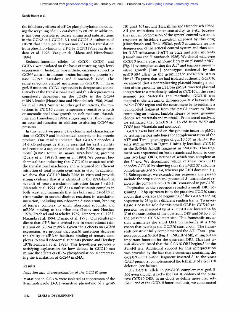
The GCD10 ORF predicts a protein of 478 amino acids wi th a molecular mass of 54,600. Comparison of the de- duced amino acid sequence of GCDIO with protein se- quences in the GenBank, EMBL, and SWISS-PROT data bases revealed no high degree of sequence similarity to any known proteins. However, GCDIO does contain a segment of 65 amino acids (between residues 335 and 400) that is closely related to the consensus sequence for the structural core of the RRM found in many RNA- binding proteins (Fig. 2). The RRM is a degenerate con- sensus sequence containing a total of 7 highly conserved residues and 7 additional positions that are generally oc- cupied by uncharged amino acids (Birney et al. 1993). The RRM-related sequence in GCD10 matchs the con- sensus sequence at 12 of these 14 positions (Fig. 2). Ad- ditional evidence that GCD10 contains a functional RRM is provided by the finding that GCD10 binds RNA in vitro (see below).
GCD10 is an essential gene
As shown in Figure 3, gcdlO mutat ions lead to temper- ature-sensitive growth under nonstarvat ion conditions, suggesting that GCDIO has an essential function beyond its role in GCN4 translational control. To test this pos-
sibility directly, we constructed the delet ion-insert ion allele gcdlOh::URA3 {see Fig. 1; pMG108) and used it to replace one copy of GCDIO in the diploid strains MGY1 and MGY2 [see Materials and methods). Tetrad analysis of each Ura + diploid t ransformant revealed that only
1 40 MNALTT I DFNQHV IVRLP SKNYKIVELKPNTSVS LGKFGA
80 FEVND I IGYPFGLTFE I YYDGEEVSSDENRDSKPKNKIP I
120 GKVRLLSQE IKDVNNDKDDGQSEPPLS I KEKSVSLELSS I
160 DS SATNQNLVNMGSKAQELTVEE IEKMKQE S LS SKE I IDK
200 I I KSHKSFHNKTVY SQEKYVNRKKQKFAKYFTVEYLSS SN
240 LLQFL I DKGD I QRVLDMSQE SMGMLLNLAN I QSEGNYLCM
280 DETGGLLVYFLLERMFGGDNE SKSKGKVIVI HENEHANLD
320 LLKFANYSEKF I KEHVHT I SLLDFFEP P TLQE I QSRFTPL
* I I I I I I PKEEARALKGGKKNS Y YRKLRWYNTQWQ I LELTGEFLYDG
UxUxxLxxxxxxZxxxxLxxxFxxxG
I I I * I I I LVMATTLHLP TLVPKLAEK I HGSRP IVCYGQFKETLLELA xUxxZxxxxxxxxxxxxxxxxxxxxxxUxVxFxxxxZxxA
440 HTLY SDLRFLAP $ I LETRCRP YQS I RGKLHP LMTMKGGGG
478 Y LMWCHRVI PAPEPVSENATAADS SEKLAEHGAKKQK I
Figure 2. Deduced amino acid sequence of GCD10 and loca- tion of a presumptive RRM. The deduced amino acid sequence is given in single-letter code. The RRM sequence motif defined by Birney et al. i1993) is shown below the sequence at the lo- cation of the presumptive RRM in GCD10. Ix) Any residue; (U) uncharged residues L, I, V, A, G, F, W, Y, C, M; (Z) U + S, T. Conserved positions in the RRM that occur in GCD10 are de- noted by vertical lines above the sequence. The two conserved residues of the RRM motif lacking in GCD 10 are denoted by the asterisks, {') above the sequence, the position corresponding to Gly-390 in GCD10 is occupied by valine in most, although not all, RRM-containing proteins.
GENES & DEVELOPMENT 1783
Cold Spring Harbor Laboratory Press on February 24, 2020 - Published by genesdev.cshlp.orgDownloaded from
Garcia-Barrio et al.
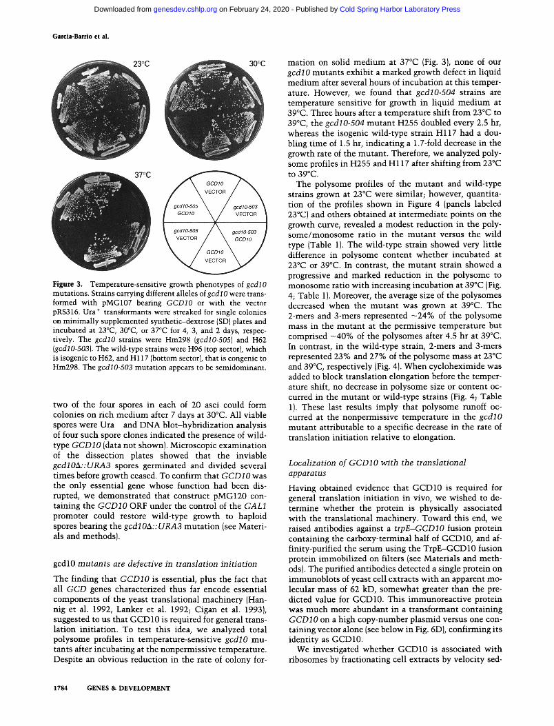
Figure 3. Temperature-sensitive growth phenotypes of gcdlO mutations. Strains carrying different alleles of gcdlO were trans- formed with pMG107 bearing GCDIO or with the vector pRS316. Ura + transformants were streaked for single colonies on minimally supplemented synthetic-dextrose (SD)plates and incubated at 23~ 30~ or 37~ for 4, 3, and 2 days, respec- tively. The gcdlO strains were Hm298 (gcdlO-505) and H62 (gccl10-503). The wild-type strains were H96 (top sector), which is isogenic to H62, and H117 (bottom sector), that is congenic to Hm298. The gcdlO-503 mutation appears to be semidominant.
two of the four spores in each of 20 asci could form colonies on rich med ium after 7 days at 30~ All viable spores were Ura - and DNA blot-hybridizat ion analysis of four such spore clones indicated the presence of wild- type GCD10 (data not shown). Microscopic examinat ion of the dissection plates showed that the inviable gcdlOA::URA3 spores germinated and divided several t imes before growth ceased. To confirm that GCDIO was the only essential gene whose function had been dis- rupted, we demonstrated that construct pMG120 con- taining the GCDIO ORF under the control of the GALl promoter could restore wild-type growth to haploid spores bearing the gcdlOA:: URA3 mutat ion (see Materi- als and methods).
gcdl0 mutants are defective in translation initiation
The finding that GCDIO is essential, plus the fact that all GCD genes characterized thus far encode essential components of the yeast translational machinery (Han- nig et al. 1992, Lanker et al. 1992; Cigan et al. 1993), suggested to us that GCD 10 is required for general trans- lation init iation. To test this idea, we analyzed total polysome profiles in temperature-sensit ive gcdlO mu- tants after incubating at the nonpermissive temperature. Despite an obvious reduction in the rate of colony for-
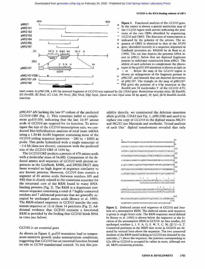
mat ion on solid med ium at 37~ (Fig. 3), none of our gcdl 0 mutants exhibit a marked growth defect in liquid med ium after several hours of incubat ion at this temper- ature. However, we found that gcdlO-504 strains are temperature sensitive for growth in l iquid m e d i u m at 39~ Three hours after a temperature shift from 23~ to 39~ the gcdlO.504 mutant H255 doubled every 2.5 hr, whereas the isogenic wild-type strain H117 had a dou- bling t ime of 1.5 hr, indicating a 1.7-fold decrease in the growth rate of the mutant . Therefore, we analyzed poly- some profiles in H255 and H117 after shift ing from 23~ to 39~
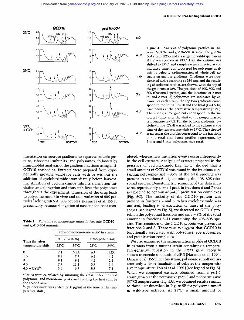
The polysome profiles of the mutan t and wild-type strains grown at 23~ were similar; however, quantita- tion of the profiles shown in Figure 4 (panels labeled 23~ and others obtained at intermediate points on the growth curve, revealed a modest reduction in the poly- some/monosome ratio in the mutan t versus the wild type (Table 1). The wild-type strain showed very little difference in polysome content whether incubated at 23~ or 39~ In contrast, the mutan t strain showed a progressive and marked reduction in the polysome to monosome ratio wi th increasing incubat ion at 39~ (Fig. 4; Table 1). Moreover, the average size of the polysomes decreased when the mutan t was grown at 39~ The 2-mers and 3-mers represented - 2 4 % of the polysome mass in the mutan t at the permissive temperature but comprised - 4 0 % of the polysomes after 4.5 hr at 39~ In contrast, in the wild-type strain, 2-mers and 3-mers represented 23% and 27% of the polysome mass at 23~ and 39~ respectively {Fig. 4). When cycloheximide was added to block translation elongation before the temper- ature shift, no decrease in polysome size or content oc- curred in the mutan t or wild-type strains (Fig. 4; Table 1). These last results imply that polysome runoff oc- curred at the nonpermissive temperature in the gcdlO mutant attributable to a specific decrease in the rate of translation ini t iat ion relative to elongation.
Localization of GCD10 with the translational apparatus
Having obtained evidence that GCD10 is required for general translation ini t iat ion in vivo, we wished to de- termine whether the protein is physical ly associated wi th the translational machinery. Toward this end, we raised antibodies against a trpE-GCDIO fusion protein containing the carboxy-terminal half of GCD10, and af- finity-purified the serum using the TrpE-GCD 10 fusion protein immobi l ized on filters (see Materials and meth- ods). The purified antibodies detected a single protein on immunoblots of yeast cell extracts wi th an apparent mo- lecular mass of 62 kD, somewhat greater than the pre- dicted value for GCD10. This immunoreact ive protein was much more abundant in a transformant containing GCDIO on a high copy-number plasmid versus one con- taining vector alone (see below in Fig. 6D), confirming its identi ty as GCD10.
We investigated whether GCD10 is associated wi th ribosomes by fractionating cell extracts by velocity sed-
1784 GENES & DEVELOPMENT
Cold Spring Harbor Laboratory Press on February 24, 2020 - Published by genesdev.cshlp.orgDownloaded from
GCD10 is the RNA-binding subunit of eIF-3
23~
39~
GCDIO gcdlO-504 2.0
80s 2 3 ~o1-~ 80s 2 3 L \ 4os~s~, ~ ~ ̂ ^~,-.. L \ 4os~s~ ~ ~, ̂ .~..
,o F 1.o I- .,1 0 0
1 .o 1 .o
o o
2.0 2.0 ~'
o o ,i
1.0 i .0
0 0
2.0 ~ 2.0 t ~ ~ 1.0 1.0
0 0
39~ 2.0 2.0 + CYH
10 10~ 0 "0 TOP BOTTOM TOP BOTTOM
t=O
4.5h
1.5h
3h
4.5h
4.5h
Figure 4. Analysis of polysome profiles in iso- genic GCDIO and gcdlO-504 strains. The gcdlO- 504 strain H255 and its isogenic wild-type parent Hl17 were grown at 23~ Half the culture was shifted to 39~ and samples were collected at the indicated times and processed for polysome anal- ysis by velocity-sedimentation of whole cell ex- tracts on sucrose gradients. Gradients were frac- tionated while scanning at 254 nm, and the result- ing absorbance profiles are shown, with the top of the gradients at left. The positions of 40S, 60S, and 80S ribosomal species, and the locations of 2-mer (2) and 3-mer (3) polysomes are indicated by ar- rows. For each strain, the top two gradients corre- spond to the initial (t=0) and the final (t=4.5 hr) time points at the permissive temperature (23~ The middle three gradients correspond to the in- dicated times after the shift to the nonpermissive temperature (39~ For the bottom gradients, cy- cloheximide (CYH) was added to the culture at the time of the temperature shift to 39~ The stippled areas under the profiles correspond to the fractions of the total absorbance profiles represented by 2-mer and 3-mer polysomes (see text).
imenta t ion on sucrose gradients to separate soluble pro- teins, ribosomal subunits, and polysomes, followed by immunoblo t analysis of the gradient fractions using anti- GCD10 antibodies. Extracts were prepared from expo- nent ia l ly growing wild-type cells with or without the addition of cycloheximide immedia te ly before harvest- ing. Addition of cycloheximide inhibi ts translation ini- t iation and elongation and thus stabilizes the polysomes throughout the experiment. Omiss ion of the drug leads to polysome runoff in vitro and accumulat ion of 80S par- ticles lacking m R N A (80S couples)(Ramirez et al. 1991), presumably because elongation of nascent chains is com-
Table 1. Polysome to monosome ratios in isogenic GCD10 and gcdl0-504 mutants
Polysome/monosome ratio" in strain
Hll7(GCDIO) H25S(gcdlO-504) Time (hr) after temperature shift 23~ 39~ 23~ 39~
0 7.1 N.D. 6.7 N.D. 1.5 8.3 7.7 6.3 4.2 3 9.1 9.1 4.5 2.3 4.5 7.7 11.1 5.3 1.4 4.5(+CYH b) 5.9 6.7 5.3 7.1
aRatios were calculated by summing the areas under the total polysomal and monosome peaks and dividing the first sum by the second sum. bCycloheximide was added to 50 ~.g/ml at the time of the tem- perature shift.,
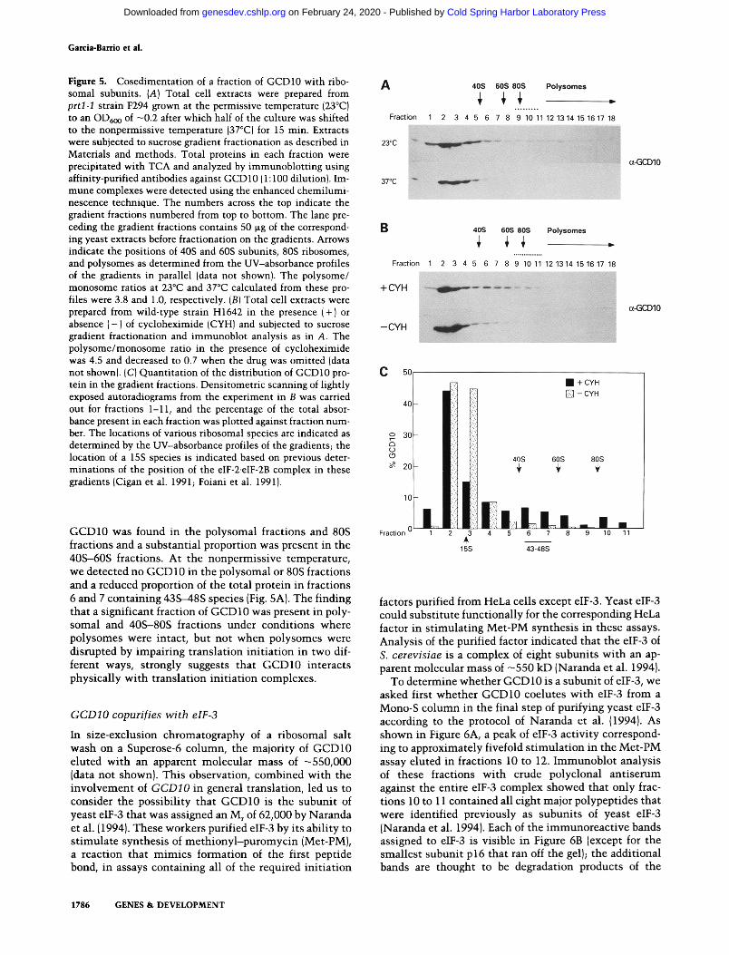
pleted, whereas new ini t iat ion events occur infrequently in the cell extracts. Analysis of extracts prepared in the presence of cycloheximide (Fig. 5B, C) showed that a small amount of GCD 10 was found in the fractions con- taining polysomes and - 3 5 % of the total amount was present in fractions 5-11, containing the 40S--80S ribo- somal species. Densi tometr ic scanning of the data indi- cated reproducibly a small peak in fractions 6 and 7 that is expected to contain 43S-48S preini t iat ion complexes (Fig. 5C). The majority of the GCD10 protein was present in fractions 2 and 3. When cycloheximide was omitted, leading to dissociation of most of the poly- somes (see legend to Fig. 5), we detected no GCD10 pro- tein in the polysomal fractions and only - 8 % of the total amount in fractions 5-11 containing the 40S-80S spe- cies. The remainder of the GCD10 protein was present in fractions 2 and 3. These results suggest that GCD10 is functionally associated wi th polysomes, 80S ribosomes, and preinit iat ion complexes.
We also examined the sedimentat ion profile of GCD 10 in extracts from a mutan t strain containing a tempera- ture-sensitive mutat ion in the PRT1 gene, recently shown to encode a subunit of eIF-3 (Naranda et al. 1994; Danaie et al. 1995). In this strain, polysome runoff occurs after only a short incubation of cells at the nonpermis- sive temperature (Foiani et al. 1991) (see legend to Fig. 5). When we compared extracts obtained from a prt l -1 strain grown at the permissive (23~ and nonpermissive (3 7~ temperatures (Fig. 5A), we obtained results s imilar to those just described in Figure 5B for polysome runoff in wild-type extracts. At 23~ a small amount of
GENES & DEVELOPMENT 1785
Cold Spring Harbor Laboratory Press on February 24, 2020 - Published by genesdev.cshlp.orgDownloaded from
Garcia-Barrio et al.
Figure 5. Cosedimentation of a fraction of GCD10 with ribo- somal subunits. {A) Total cell extracts were prepared from prtl-1 strain F294 grown at the permissive temperature (23~ to an OD6o o of -0.2 after which half of the culture was shifted to the nonpermissive temperature (37~ for 15 rain. Extracts were subjected to sucrose gradient fractionation as described in Materials and methods. Total proteins in each fraction were precipitated with TCA and analyzed by immunoblotting using affinity-purified antibodies against GCD10 (1:100 dilution). Im- mune complexes were detected using the enhanced chemilumi- nescence technique. The numbers across the top indicate the gradient fractions numbered from top to bottom. The lane pre- ceding the gradient fractions contains 50 ~g of the correspond- ing yeast extracts before fractionation on the gradients. Arrows indicate the positions of 40S and 60S subunits, 80S ribosomes, and polysomes as determined from the UV-absorbance profiles of the gradients in parallel (data not shown). The polysome/ monosome ratios at 23~ and 37~ calculated from these pro- files were 3.8 and 1.0, respectively. (B) Total cell extracts were prepared from wild-type strain H1642 in the presence (+) or absence (-) of cycloheximide {CYH) and subjected to sucrose gradient fractionation and immunoblot analysis as in A. The polysome/monosome ratio in the presence of cycloheximide was 4.5 and decreased to 0.7 when the drug was omitted (data not shown). (C) Quantitation of the distribution of GCD10 pro- tein in the gradient fractions. Densitometric scanning of lightly exposed autoradiograms from the experiment in B was carried out for fractions 1-11, and the percentage of the total absor- bance present in each fraction was plotted against fraction num- ber. The locations of various ribosomal species are indicated as determined by the UV-absorbance profiles of the gradients; the location of a 15S species is indicated based on previous deter- minations of the position of the elF-2-eIF-2B complex in these gradients {Cigan et al. 1991; Foiani et al. 1991).
GCD10 was found in the polysomal fractions and 80S fractions and a substantial proportion was present in the 40S-60S fractions. At the nonpermissive temperature, we detected no GCD 10 in the polysomal or 80S fractions and a reduced proportion of the total protein in fractions 6 and 7 containing 43S---48S species {Fig. 5A). The finding that a significant fraction of GCD 10 was present in poly- somal and 40S-80S fractions under condit ions where polysomes were intact, but not when polysomes were disrupted by impairing translat ion ini t ia t ion in two dif- ferent ways, strongly suggests that GCD10 interacts physically wi th translat ion ini t ia t ion complexes.
GCDIO copurifies with elF-3
In size-exclusion chromatography of a ribosomal salt wash on a Superose-6 column, the majori ty of GCD10 eluted wi th an apparent molecular mass of -550,000 [data not shown}. This observation, combined with the involvement of GCD10 in general translation, led us to consider the possibili ty that GCD10 is the subunit of yeast eIF-3 that was assigned an M r of 62,000 by Naranda et al. (1994). These workers purified eIF-3 by its ability to s t imulate synthesis of me th iony l -pu romyc in (Met-PM), a reaction that mimics formation of the first peptide bond, in assays containing all of the required ini t ia t ion
factors purified from HeLa cells except elF-3. Yeast elF-3 could substitute funct ional ly for the corresponding HeLa factor in s t imulat ing Met-PM synthesis in these assays. Analysis of the purified factor indicated that the eIF-3 of S. cerevisiae is a complex of eight subunits wi th an ap- parent molecular mass of - 5 5 0 kD (Naranda et al. 1994}.
To determine whether GCD10 is a subunit of eIF-3, we asked first whether GCD10 coelutes wi th eIF-3 from a Mono-S column in the final step of purifying yeast eIF-3 according to the protocol of Naranda et al. {1994}. As shown in Figure 6A, a peak of eIF-3 activity correspond- ing to approximately fivefold s t imula t ion in the Met-PM assay eluted in fractions 10 to 12. Immunoblo t analysis of these fractions wi th crude polyclonal ant iserum against the entire eIF-3 complex showed that only frac- tions 10 to 11 contained all eight major polypeptides that were identified previously as subunits of yeast eIF-3 (Naranda et al. 1994}. Each of the immunoreac t ive bands assigned to elF-3 is visible in Figure 6B (except for the smallest subunit p 16 that ran off the gel); the additional bands are thought to be degradation products of the
1786 GENES & DEVELOPMENT
Cold Spring Harbor Laboratory Press on February 24, 2020 - Published by genesdev.cshlp.orgDownloaded from

GCD10 is the RNA-binding subunit of eIF-3
Figure 6. Coelution of GCD10 with the yeast eIF-3 complex in ion-exchange chromatography. Fractions from Mono-S 5/5 column chromatography carried out in the final step in purifying the yeast eIF-3 complex were assayed in parallel for eIF-3 activity, eIF-3 subunits, and for GCD10 protein. {A) Assay of eIF-3 biochemical activity. Ten microliters of each fraction was tested for stimulation of Met-PM/synthesis in reactions containing mammalian washed ribosomes and initiation factors (see Materials and methods). The activity is reported as counts per minute of [3H]Met-PM formed in the reaction. In con- trol experiments, no stimulation was obtained when eIF-3 was omitted, whereas approximately fivefold stimulation of Met-PM synthesis was seen when 3.1 ~g of purified HeLa eIF-3 was added. The background value (1123 cpm) obtained with the charged tRNA alone was subtracted. (B) Immunoblot analysis of total eIF-3 subunits. Fifteen-microliter aliquots of fractions 2-15 were analyzed by 10% SDS-PAGE and immunoblotting using rabbit poly- clonal antiserum against eIF-3, and immune complexes were visualized using alkaline phosphatase-conjugated antibodies against rabbit IgG (see Materials and methods). The column fraction numbers are indicated at the bottom, the migration of molecular mass markers is indicated at left, and the migration of the seven largest subunits of eIF-3 and their masses in kD are shown at right {Naranda et al. 1994). {C) Immunoblot analysis of GCD10. The analysis was performed as in B, except that the affinity-purified anti-GCD 10 antiserum was used as the source of primary antibodies. The arrowhead at right indicates the migration of GCDIO as deduced from the experiment shown in D. (D) Immu- noblot analysis of GCD10 in the peak fractions containing eIF-3 in parallel with whole cell extracts prepared from strains overexpressing GCD10. Twenty-microliter aliquots of Mono-S fractions 9-12, containing the peak of eIF-3 activity, and 20 ~g aliquots of whole cell extracts from strain H1642 transformed with vector alone (SC) or high copy-number plasmid pE 107 bear- ing GCDIO plasmid {HC), were analyzed in parallel by 8% SDS-PAGE and immunoblotting using affinity-purified antibodies against GCD10, as de- scribed for the experiment in C. The migration of molecular mass markers and their mass in kD is shown at left; the arrowhead at right indicates the position of GCD10.
larger subunits. Consistent wi th previous findings {Naranda et al. 1994), the fractions immediate ly flanking 10 and 11, which lack some of the smaller eIF-3 sub- units, show reduced (fraction 12) or no activity (fractions 9 and 13) in the Met-PM assay compared to fractions 1'0 and 11.
Immunoblo t analysis of these column fractions, using affinity-purified ant i -GCD10 antibodies, revealed an im- munoreact ive polypeptide with an apparent molecular mass of ~62 kD that peaks in fractions 10 to 12. The polypeptide detected with ant i -GCD10 antibodies and the - 6 2 - k D subunit of eIF-3 detected with anti-eIF-3 an- tibodies by Naranda et al. (1994) showed similar mass distributions across the column fractions (Fig. 6B, C). To demonstrate that the 62-kD polypeptide detected with ant i -GCD10 antibodies in column fractions 10-12 has the same molecular mass as GCD10, we analyzed the Mono-S column fractions containing eIF-3 activity in parallel wi th whole cell extracts containing overex- pressed GCD10 protein. As shown in Figure 6D, the
GCD10 protein detected in cell extracts and the immu- noreactive species in purified eIF-3 have identical mobil- ities in SDS-PAGE. Taken together, the results in Figure 6 indicate that GCD10 coelutes from the Mono-S col- u m n with eIF-3 activity and is identical in mass to the 62-kD subunit of yeast eIY-3 identified by Naranda et al. (1994). GCD10 was found at much lower levels in frac- tion 12 in Figure 6D than in Figure 6C. We at tr ibute this difference to degradation of the protein during storage before conducting the analysis shown in Figure 6D. We presume that GCD10 is less stable in eIF-3 complexes lacking the smaller subunits, the latter being underrep- resented in fraction 12 versus fractions 10 and 11.
CCD I O coimmunoprecipitates with PRT1, a known subunit of elF-3
To provide additional evidence that GCD10 is a subunit of yeast eIF-3, we asked whether it could be coimmuno- precipitated with the PRT1 protein, a known subunit of
GENES & DEVELOPMENT 1787
Cold Spring Harbor Laboratory Press on February 24, 2020 - Published by genesdev.cshlp.orgDownloaded from

Garcia-Barrio et al.
eIF-3 (Naranda et al. 1994; Danaie et al. 1995). The ribo- somal high-salt wash fraction (HSW) or purified eIF-3 from the Mono-S fractions described above {Fig. 6) was immunoprecipitated with antibodies against PRT1, GCD10, or eIF-3, and the resulting immune complexes were probed by immunoblot analysis for PRT1, GCD 10, and the other subunits of eIF-3 using the same antisera. Each of the three antisera immunoprecipitated similar quantities of PRT1 and GCD10 from the HSW and from the Mono-S peak fractions {Fig. 7). We estimated that 50%-70% of the GCD10 and PRT1 in the starting ma- terial was recovered in the immunoprecipitates. Control immunoprecipitations carried out with preimmune sera or with antibodies against initiation factor elF-1A (Chia- Lin et al. 1995) did not precipitate detectable amounts of GCD10 or PRT1 (Fig. 7), whereas the eIF-1A antibodies immunoprecipitated eIF-1A from the H SW fraction (data not shown). These results provide strong evidence that GCD10 and PRT1 are components of the same protein complex. Probing the immune complexes with antibod- ies against eIF-3 showed that the p135 subunit of eIF-3 (Naranda et al. 1994) was also coimmunoprecipitated with GCD10 and PRT1 whether using the antibodies against PRT1, GCD10, or eIF-3 to immunopurify the complex {data not shown). The results in Figure 7 pro- vide strong evidence that GCD10 is an integral compo- nent of yeast eIF-3.
GCDIO is the RNA-binding subunit of elF-3
The yeast eIF-3 complex has RNA-binding activity, and this activity has been used to affinity-purify all eight subunits of the complex (Naranda et al. 1994). Because the 62-kD subunit was the only one found to bind RNA as an isolated polypeptide, it was believed to be respon- sible for the RNA-binding activity of the entire eIF-3 complex. To provide additional proof that GCD10 is identical to the 62-kD subunit of eIF-3 identified by Naranda et al. {1994), we asked whether GCD10 has RNA-binding activity. We began by conducting North- westem analysis of whole cell extracts from strains con- taining high-copy versus single-copy GCD10 using an RNA transcript corresponding to the 5'-untranslated re- gion of Xenopus B-globin mRNA as the probe. As shown in Figure 8A [lanes HC and SC), an RNA-binding protein with the same apparent molecular mass of GCD10 was present at much higher levels in the cell extracts derived from the strain containing high copy-number versus low copy-number GCD10. In contrast, the RNA-binding pro- teins of lower molecular mass {possibly ribosomal pro- teins) were present at roughly the same levels in the two extracts. These results indicate that GCD10 has RNA-binding activity. In parallel, we carried out North- western analysis on the same Mono-S column fractions containing eIF-3 activity described above. As shown previously (Naranda et al. 1994}, fractions 10 and 11 containing the peak of eIF-3 activity (see Fig. 6A) also contained peak amounts of the RNA-binding subunit of eIF-3. This RNA-binding protein coeluted
Figure 7. Coimmunoprecipitation of GCD10 with the 90-kD subunit of eIF-3. Approximately 50 gg of total proteins from the ribosomal high salt wash (HSW} and 4 ~g of purified eIF-3 com- plex [eIF-3 {MonoS)] were immunoprecipitated with polyclonal antibodies against eIF-3 (lanes 1,71, GCD10 {lanes 3,9), PRT1 {lanes 5,11), eIF-1A {lanes 6,12), or the preimmune (PII sera cor- responding to the eIF-3 {lanes 2,81 and GCD10 {lanes 4,10) an- tibodies. The immune complexes were separated by 10% SDS- PAGE, transferred to nitrocellulose, and probed with polyclonal antibodies against PRT1 (A) and GCD10 {B).
and comigrated with GCD10 {Fig. 8A). When the same filter probed with labeled RNA shown in Figure 8A {right) was subsequently probed with antibodies against GCD10 {Fig. 8A, left), we found that the mobilities of the RNA-binding polypeptide and the immunoreactive protein in the fractions containing the peak of eIF-3 activity coincided exactly. These results provide strong evidence that GCD10 is the RNA-binding subunit of eIF-3.
The mammalian eIF-3 is similar to the yeast factor in both size and number of subunits. In addition, the 66-kD subunit of HeLa eIF-3, like GCD10, has RNA-binding activity {Westermann and Nygard 1984; B.J. Fabbri and J.W.B. Hershey, unpubl.). These considerations, com- bined with the functional interchangeability of yeast and HeLa eIF-3 in the Met-PM synthesis assay (Naranda et al. 1994), led us to investigate whether the GCD10 subunit of yeast eIF-3 and the p66 subunit of mammalian eIF-3 have structural similarities. To examine this possibility, we carried out immunoblot analysis of purified HeLa and rabbit eIF-3 using our anti-GCD10 antibodies. As shown in Figure 8B, the GCD 10 antibodies reacted specifically with the p66 subunit of human eIF-3, but not with the corresponding subunit of rabbit eIF-3. Northwestem analysis of the same samples confirmed that nearly equivalent amounts of the GCD 10 subunit of yeast eIF-3 and the p66 subunits of the two mammalian factors had been loaded on the gel {Fig. 8B). These results suggest that yeast GCD10 and HeLa p66 share one or more epitopes that are recognized by our GCD10 antibodies, and they provide additional evidence that GCD10 is the RNA-binding subunit of yeast eIF-3.
1788 GENES & DEVELOPMENT
Cold Spring Harbor Laboratory Press on February 24, 2020 - Published by genesdev.cshlp.orgDownloaded from

GCD10 is the RNA-binding subunit of elF-3
Figure 8. GCD10 has RNA-binding activity and is related immunologically to the RNA-binding subunit of human eIF-3. (A, right) 32P-labeled mRNA. Northwestern analysis was carried out on fractions 8-13 from the Mono-S column containing the peak of eIF-3 activity and several flanking fractions in parallel with the whole cell extracts containing wild-type levels (SC) or overexpressed levels (HC) of GCD10 protein. Aliquots of the Mono-S fractions containing -150 ng of protein and 15qzg aliquots of the whole cell extracts were resolved by 9% SDS-PAGE, transferred to nitrocellulose, hybridized with a 32p-labeled B-globin transcript and subjected to autoradiography, all as described in Materials and methods. (A, left) a-GCD10. The same membrane filter subjected to Northwestern analysis on the right was blocked with blotto for 1 hr and incubated with anti-GCD10 antibodies (1:100 dilution). The immune complexes were visualized exactly as in Fig. 6. The presumptive GCD10 bands labeled by 32p-labeled mRNA in the Northwestern blot at right were superimposed precisely on the immunoreactive GCD10 bands in the immunoblot at left. (B) Approximately 500 ng of purified yeast (Y), HeLa (H), and rabbit reticulocyte (R) eIF-3 complexes were separated by 8% SDS-PAGE and subjected to immunoblot and Northwestern analysis as described in A. The arrowheads indicate the migration of yeast GCD10.
D i s c u s s i o n
Evidence that GCDIO is an essential subuni t of elF-3
Mutations in GCDIO were identified originally by their ability to restore efficient GCN4 expression in mutants lacking the protein kinase GCN2 (Harashima and Hin- nebusch 1986). It was established that gcdlO mutations increase GCN4 expression at the translational level by overcoming the inhibitory effects of the uORFs on ribo- somal reinitiation at the GCN4 start site (Mueller et al. 1987). We showed here that the GCDIO product has an essential function in general translation initiation (Fig. 4) in addition to its gene-specific role in regulating GCN4 expression. Most of the GCD10 protein in cell extracts was found in a high molecular mass complex of -550 kD, and a fraction appears to be associated with translating ribosomes or 43S to 48S preinitiation com- plexes (Fig. 5). Our affinity-purified antibodies against GCD10 cross-reacted with a polypeptide with an appar- ent molecular mass of 62,000 that coeluted from a Mono-S column in the same fractions that contain the major eight subunits of yeast eIF-3 and the biochemical activity ascribed to this factor in stimulating Met-PM synthesis (Fig. 6) (Naranda et al. 1994). Similar results were obtained by assaying column fractions from Super- ose-6 gel filtration chromatography (data not shown), the step preceding Mono-S chromatography in the purifica- tion of yeast eIF-3 (Naranda et al. 1994). In addition to observing copurification of GCD10 with the biochemi- cal activity and protein subunits of elF-3, we found that GCD10 could be specifically coimmunoprecipitated with the PRTl-encoded and 135-kD subunits of eIF-3 using antibodies against GCD10, PRT1, or eIF-3 (Fig. 7).
On the basis its molecular mass, GCD10 should corre- spond to the 62-kD subunit of eIF-3 that was shown pre- viously to have RNA-binding activity (Naranda et al. 1994). We confirmed this prediction by showing that GCD 10 binds RNA in a Northwestern assay and that the RNA-binding subunit of eIF-3 and the polypeptide rec- ognized by anti-GCD 10 antibodies coelute from Mono-S and comigrate precisely in SDS-PAGE (Fig. 8). Finally, we showed that the anti-GCD10 antibodies specifically recognized the 66-kD RNA-binding subunit of human eIF-3 (Fig. 8). The fact that the RNA-binding activity of elF-3 can be used to affinity-purify all eight subunits of the complex provides additional evidence that GCD10, the only subunit known to bind RNA (Naranda et al. 1994), is an integral component of the eIF-3 complex. On the basis of all these results, we conclude that GCDIO encodes the RNA-binding subunit of yeast eIF-3.
The role of GCDIO and elF-3 in GCN4 translational control
According to our model for GCN4 translational control, ribosomes translate uORF1 and reinitiate very effi- ciently at uORFs 2, 3, or 4 when amino acids are abun- dant. These ribosomes cannot reinitiate again and trans- lation of GCN4 is repressed. Under starvation condi- tions, reinitiation at uORFs 2--4 is suppressed partially and many ribosomes are able to reinitiate at GCN4 in- stead (Abastado et al. 1991). The failure of ribosomes to reinitiate at uORFs 2--4 in amino acid-starved cells re- quires phosphorylation of eIF-2 by the protein kinase
GENES & DEVELOPMENT 1789
Cold Spring Harbor Laboratory Press on February 24, 2020 - Published by genesdev.cshlp.orgDownloaded from
Garcia-Barrio et al.
GCN2 {Dever et al. 1992). A large body of genetic and biochemical evidence indicates that phosphorylated eIF-2 down-regulates eIF-2B in yeast, decreasing the level of active eIF-2 in the cell and the concentrat ion of eIF- 2 / G T P / M e t - t R N A ~ et ternary complexes (Hinnebusch 1992~ Cigan et al. 1993~ Vazquez de Aldana and Hinne- busch 1994). This is expected to reduce the rate at which ternary complexes rebind to ribosomes scanning down- stream from uORF1. As a result, many ribosomes will reach the start sites of uORFs 2-4 lacking the ternary complex and cont inue scanning downstream. Essentially all of these ribosomes will rebind the ternary complex before reaching the GCN4 start site and reinit iate there instead {Dever et al. 1992}.
Mutat ions in each of the genes encoding subunits of eIF-2 or eIF-2B have been isolated that mimic eIF-2a phosphorylat ion in causing a reduction in general trans- lat ion ini t ia t ion and const i tut ive derepression of GCN4 t ranslat ion [Hinnebusch 1992). These muta t ions bypass the requirement for GCN2 for efficient translat ion of GCN4 mRNA. Because the muta t ions we isolated in GCD10 have the same characteristics as those affecting
subunits of eIF-2 or eIF-2B, we suspected that GCD10 might be involved in the formation of eIF-2/GTP/Met- tRNA~ et ternary complexes or in s t imulat ing the bind- ing of these complexes to 46S complexes consisting of eIF-3 and 40S ribosomal subunits. Our identif icat ion of GCD10 as a subunit of yeast eIF-3 confirms this predic- t ion because this mul t i subuni t factor is a critical con- s t i tuent of the 43S preini t iat ion complex, and one of its functions identified by in vitro experiments is to stimu- late binding of the ternary complex to small r ibosomal subunits {Benne and Hershey 19781 Trachsel and Stae- hel in 1979; Feinberg et al. 1982}.
Biochemical studies on mammal i an eIF-3 have impli- cated this factor in several steps of the ini t ia t ion process, including dissociation of 80S ribosomes into free 60S subunits and 46S preini t iat ion complexes containing eIF-3 and 40S subunits, binding of eIF-2/GTP/Met- t R N A Met ternary complexes to form 43S complexes, and binding of mRNA to 43S complexes containing both eIF-3 and the ternary complex [Benne and Hershey 19781 Trachsel and Staehelin 1979}. Because ribosomes are al- ready bound to GCN4 mRNA after translat ing uORF 1, it
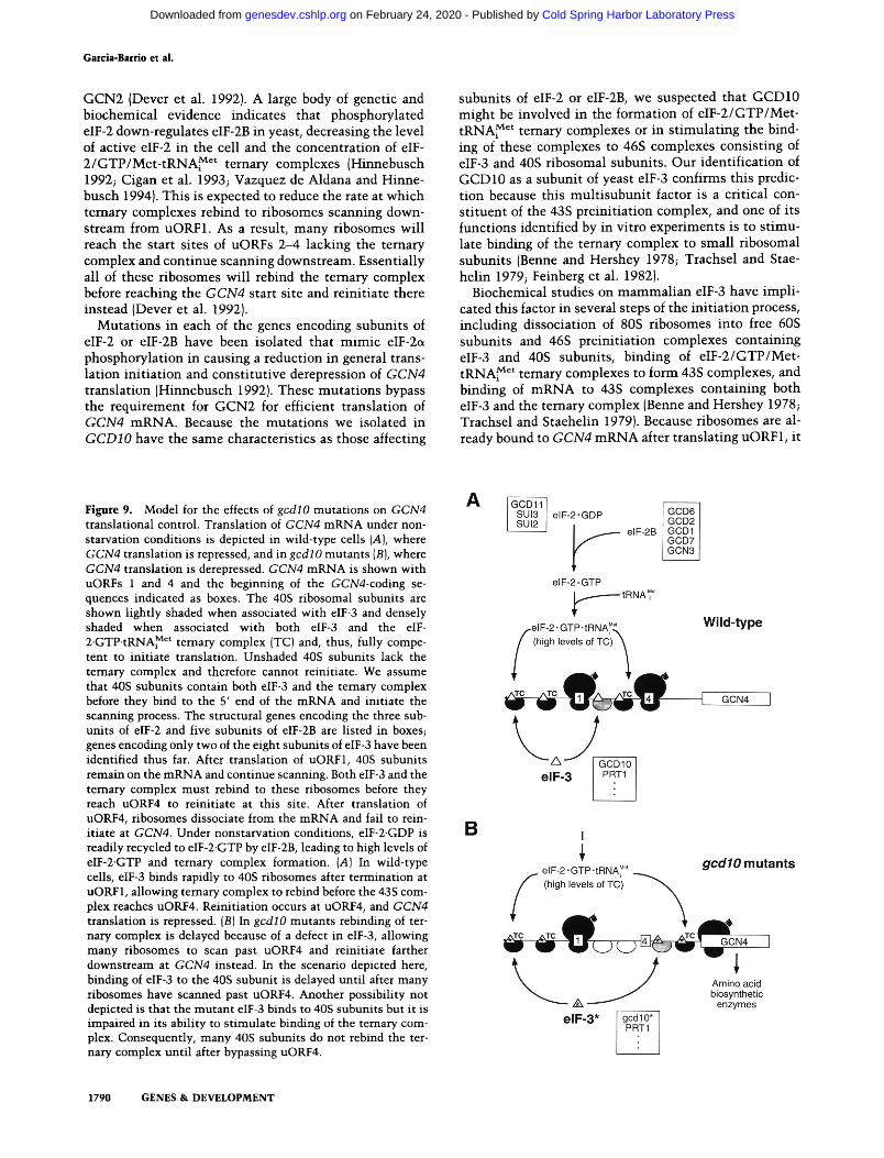
Figure 9. Model for the effects of gcdlO mutations on GCN4 translational control. Translation of GCN4 mRNA under non- starvation conditions is depicted in wild-type cells {A), where GCN4 translation is repressed, and in gcdlO mutants {B), where GCN4 translation is derepressed. GCN4 mRNA is shown with uORFs 1 and 4 and the beginning of the GCN4-coding se- quences indicated as boxes. The 40S ribosomal subunits are shown lightly shaded when associated with eIF-3 and densely shaded when associated with both eIF-3 and the eIF- 2.GTP.tRNA~ Ct ternary complex [TC) and, thus, fully compe- tent to initiate translation. Unshaded 40S subunits lack the ternary complex and therefore cannot reinitiate. We assume that 40S subunits contain both eIF-3 and the ternary complex before they bind to the 5' end of the mRNA and initiate the scanning process. The structural genes encoding the three sub- units of eIF-2 and five subunits of eIF-2B are listed in boxes~ genes encoding only two of the eight subunits of eIF-3 have been identified thus far. After translation of uORF1, 40S subunits remain on the mRNA and continue scanning. Both eIF-3 and the ternary complex must rebind to these ribosomes before they reach uORF4 to reinitiate at this site. After translation of uORF4, ribosomes dissociate from the mRNA and fail to rein- itiate at GCN4. Under nonstarvation conditions, eIF-2.GDP is readily recycled to eIF-2.GTP by eIF-2B, leading to high levels of eIF-2-GTP and ternary complex formation. (A) In wild-type cells, eIF-3 binds rapidly to 40S ribosomes after termination at uORF1, allowing ternary complex to rebind before the 43S com- plex reaches uORF4. Reinitiation occurs at uORF4, and GCN4 translation is repressed. [B) In gcdlO mutants rebinding of ter- nary complex is delayed because of a defect in eIF-3, allowing many ribosomes to scan past uORF4 and reinitiate farther downstream at GCN4 instead. In the scenario depicted here, binding of eIF-3 to the 40S subunit is delayed until after many ribosomes have scanned past uORF4. Another possibility not depicted is that the mutant eIF-3 binds to 40S subunits but it is impaired in its ability to stimulate binding of the ternary com- plex. Consequently, many 40S subunits do not rebind the ter- nary complex until after bypassing uORF4.
1790 GENES & D E V E L O P M E N T
Cold Spring Harbor Laboratory Press on February 24, 2020 - Published by genesdev.cshlp.orgDownloaded from
GCD10 is the RNA-binding subunit of eIF-3
seems un l i ke ly tha t gcdlO m u t a t i o n s derepress GCN4 expression by reducing the abi l i ty of eIF-3 to s t imula te m R N A binding to 43S complexes. Such a defect migh t be expected to reduce GCN4 m R N A t rans la t ion (Gcn- phe- notype) by causing 43S complexes to dissociate f rom the m R N A as they scan downs t r eam from uORF1. A more l ike ly poss ibi l i ty is tha t the gcdl 0 m u t a t i o n s reduce the abi l i ty of eIF-3 to rebind to 40S subuni t s and form 46S complexes after t rans la t ion t e r m i n a t i o n at uORF1. This would resul t in " n a k e d " 40S subuni t s scanning down- s t ream from uORF1 having a reduced abi l i ty to rebind ternary complexes (Fig. 9). Al ternat ively , it is possible tha t gcdlO m u t a t i o n s decrease the abi l i ty of eIF-3 to s t imula te b inding of e I F - 2 / G T P / M e t - t R N A ~ et ternary complexes to 46S complexes ra ther t han delaying the fo rma t ion of these complexes after t e rmina t ion at uORF1. Ei ther of these defects would decrease the rate at wh ich ternary complexes rebind to 40S subuni ts scan- n ing downs t r eam from uORF1, explaining why gcdlO m u t a t i o n s cause r ibosomes to ignore uORFs 2-4 and re in i t ia te at GCN4 in the absence of eIF-2 phosphoryla- t ion by G C N 2 (Fig. 9).
GCD10 has RNA-binding ac t iv i ty (Naranda et al. 1994; Fig. 8) and the deduced amino acid sequence of the prote in conta ins a re la t ively good m a t c h to the RRM consensus sequence (Fig. 2). It has been shown tha t the 66-kD subuni t of m a m m a l i a n eIF-3 can be cross- l inked to 18S rRNA in 46S complexes (Nygard and Wes t e rmann 1982). If an in te rac t ion be tween yeast GCD10 and a site in 18S rRNA is impor t an t for b inding of yeast eIF-3 to 40S subunits , t hen gcdl 0 m u t a t i o n s migh t disrupt this in te rac t ion and delay the rebinding of elF-3 to 40S sub- un i t s after t e r m i n a t i o n at uORF1, as suggested above (Fig. 9). It has also been reported tha t m a m m a l i a n p66 can be cross- l inked to m R N A in 48S pre in i t i a t ion com- plexes (Wes te rmann and Nygard 1984), leading to the no t ion tha t p66 could bridge an in te rac t ion be tween m R N A and 18S in a m a n n e r r emin i scen t of the Shine- Delgarno m R N A - 1 6 S rRNA base-pairing in te rac t ion in prokaryotes. It seems less l ike ly tha t d isrupt ing this in- te rac t ion would derepress GCN4 t rans la t ion eff icient ly because it should equal ly impai r i n i t i a t ion events at uORFs 2-4 and at GCN4.
After t rans la t ing uORF1, r ibosomes m u s t rebind all of the factors required to re in i t ia te t rans la t ion in the t ime it takes to scan the - 2 0 0 nucleot ides separat ing uORF1 from uORF4; otherwise, they wil l bypass uORFs 2--4 and re in i t ia te at GCN4 instead. This str ict t empora l require- m e n t for r e in i t i a t ion at the uORFs is t hough t to be one factor tha t makes GCN4 t rans la t ion m u c h more respon-
M e t sive to the levels of the e IF -2 /GTP/Met - tRNAi ter- nary complex than are in i t i a t ion events on conven t iona l m R N A s lacking uORFs. As a result, GCN4 is a sensi t ive indica tor of the in vivo func t ions of eIF-2, eIF-2B, and eIF-2et k inases (Dever et al. 1993; H innebusch 1994; Ro- mano et al. 1995). The fact tha t a subuni t of eIF-3 has now been ident i f ied genet ica l ly by its effects on GCN4 t rans la t iona l cont ro l suggests tha t this regulatory mech- an i sm can be used as an in vivo assay for this in teres t ing and complex t rans la t ion in i t i a t ion factor.
Mater ia l s and m e t h o d s
Strains, media, and genetic methods
The genotypes and origins of all yeast strains are summarized in Table 2. Hm47 and Hm44 were constructed by crossing H1262 to H752. Hm203 was selected as an AT R spore from a cross between Hml9 and H742. MGY3 was selected as a Ura + Tsm- His- derivative from a cross between RS 191 and Hm270. Trans- formation of yeast strains was carried out according to Ito et al. (1983). Standard genetic techniques and media were as de- scribed by Sherman et al. (1974). Amino acid analog sensitivity was tested as described before (Hinnebusch and Fink 1983b). Escherichia coli strain HBIO1 (BRL) was used for plasmid prop- agation and production of trpE fusion proteins. E. coli strain MV1190 (BRL) was used for generating ssDNA from M13 deriv- atives used in sequencing experiments.
Cloning of GCD10
GCDIO was cloned by complementation of the 3-AT-resistant (AT R) and thermosensitive {Tsm - ) phenotypes of Hm47 using a genomic library constructed in the low copy-number plasmid YCpS0 (Rose et al. 1987). Plasmid DNA was recovered from yeast transformants as already described (Devenish and Newlon 1982) and used to transform E. coli HB101. Plasmid pMG1 and its subclone pMG107 (Fig. 1) were shown to complement the AT R and Tsm- phenotypes in strains carrying gcdlO-501, gcdl 0-502, gcdl 0-504, or gcdl 0-505. In contrast, the gcdl 0-503 allele in strain H62 was complemented only partially, suggest- ing that this allele is semidominant. To prove that pMG 1 con- tains authentic GCDIO the 3.45-kb HindIII fragment was inserted into YIp5 (Parent et al. 1985), and the resulting plas- mid (pI105) was digested at two closely spaced HpaI sites in the genomic sequences and used to transform Hm47 (MATc~gcn2::LEU2 gcdlO-504 ura3-52) to Ura + AT s Tsm +. DNA blot-hybridization analysis (Sambrook et al. 1989) re- vealed that pI105 had integrated at the presumptive GCDIO locus in the transformants we analyzed. One such transfor- mant, Hm270 (Table 2), was crossed to HmS1 (MATa gcn2: :LE U2 gcdl 0-504 ura3-52), which is Ura- AT R Tsm-. The resulting diploids were sporulated and subjected to tetrad anal- ysis. In all 21 tetrads analyzed, only parental ditypes were found for the Ura, AT, and Tsm phenotypes, indicating that the site of pI105 integration was linked to GCDIO.
Plasmid constructions
The subclones constructed from pMG 1 are summarized in Fig- ure 1. Digestion of pMG1 with ClaI yielded a 3.8-kb fragment beginning -340 bp in the vector and ending at the unique C1aI site in the genomic insert that was inserted at the ClaI site of YCp50 to generate pMG101; pMG102 was constructed by reli- gating the ClaI fragment containing the vector backbone de- rived from the same digestion, pMG103 was constructed by inserting the BamHI-SalI fragment from pMG1 {the SalI site is present in the vector backbonel into the corresponding sites in YCp50. pMG104 was constructed from pMG1 by digestion with XhoI and SalI, followed by religation. The 3.45-kb HindIII frag- ment from pMG1 was inserted into the HindIII site of YCp50 to generate pMG105, pI105 contains the same 3.45-kb HindIII frag- ment as pMG105 inserted at the HindIII site of YIp5 {Struhl et al. 1979). pMG107 was constructed by inserting the XbaI-XhoI fragment containing the complete GCDIO-coding sequence and
1 kb downstream of the gene at the XbaI and XhoI sites in the polylinker of pRS316 {Sikorski and Hieter 1989}. pE107 was constructed by inserting the XbaI-XhoI fragment into pRS426
GENES & DEVELOPMENT 1791
Cold Spring Harbor Laboratory Press on February 24, 2020 - Published by genesdev.cshlp.orgDownloaded from

Garcia-Barrio et al.
T a b l e 2. Yeast strains
Strain Genotype Source
H96 H54 H55 H62 Hl17 H174 H255 H742 H752 H1262 H1514 H1515 H1642 Hml9 Hm47 Hm44 Hm203 Hm270 Hm298 MGY1 MGY2 MGY3 RS191
F205
W303
ot gcn2-101 gcn3-101 hisl-29 ura3-52 [HIS4::lacZ, ura3-52] ot gcn2-1 O1 gcn3-1 O1 hisl-29 gcdl 0-501 ura3-52 [HIS4::lacZ, ura3-52] ot gcn2-1 O1 gcn3-1 O1 hisl-29 gcdl 0-502 ura3-52 [HIs4::lacZ, ura3-52]
gcn2-101 gcn3-101 hisl-29 gcdlO-503 ura3-52 [HIS4::lacZ, ura3-52] a gcn2-101 gcn3-101 hisl-29 ura3-52 inol [HIS4::lacZ, URA3] a gcn2-101 gcn3-101 hisl-29 gcdlO-505 ura3-52 inol [HIS4::IacZ, URA3] a gcn2-101 gcn3-101 hisl-29 gcdlO-504 ura3-52 inol [HIS4::IacZ, URA3] a gcn3::LEU2 lys2 leu2-3 leu2-112 oL gcn2::LEU2 ura3-52 leu2-3 leu2-112 a gcn2::LEU2 gcdlO-504 ura3-52 leu2-3 a leu2-112 ~ oL ura3-52 trpl-63 leu2-3 leu2-112 a ura3-52 trpl-63 leu2-3 leu2-112 a ura3-52 leu2-3 leu2-112 trpl-63 [GCN4::lacZ, TRP1]
gcn2::LEU2 gcdlO-504 ura3-52 leu2-3 ~ leu2-112 a gcn2::LEU2 gcdlO-504 ura3-52
a gcn2::LEU2 ura3-52 gcn3::LEU2 gcdlO-504 ura3-52 gcn2::LEU2 GCDIO::URA3 ura3-52
a gcn2-101 gcn3-101 gcdlO-505 hisl-29 ura3-52 [HIS4::lacZ, ura3-52] a/a gcn2: : LE U2/gcn2: : LE U2 GCD10/gcd l 0-504 ura3-52/ura3-52 a/~ trpl-63/trpl-63 ura3-52/ura3-52 leu2-3/leu2-3 leu2-112lieu2-112 a top2-1 GCDIO::URA3 ade2 ura3 his3-11 trpl-1 leu2-3 leu2-112 a top2-1 ade2 ura3-I his3-11 trpl-I leu2-3 leu2-112
ras2-699 (HIS3 insertion) leu2-3 leu2-112 ura3-1 canl-lO0 ade2-1 his3
a leu2-3, his3-11, ade2-1, trpl-1, canl-lO0
Harashima and Hinnebusch {1986) Harashima and Hinnebusch (1986) Harashima and Hinnebusch {1986) Harashima and Hinnebusch (1986) Harashima and Hinnebusch (1986) Harashima and Hinnebusch (1986) Harashima and Hinnebusch (1986) Harashima and Hinnebusch (1986) Harashima and Hinnebusch (1986) this study this study Dever et al. {1992) Dever et al. (1992) H1262 x H752 Hml9 x H752 Hm19 x H752 Hm 47 x H742 HM47/pI105 H174 x H96 Hm44 x Hm47 H1514 x H1515 RS191 x Hm270 R. Sternglanz (State University
of New York, Stony Brook) K. Tatchell {North Carolina
State University, Raleigh) Naranda et al. (1994)
aThe presence of these mutations is uncertain.
(Christianson et al. 1992) digested with ClaI and XhoI, after making the XbaI and ClaI sites blunt ended.
pMG107-AN was constructed as follows, pMGS0, containing the 3.45-kb HindIII fragment from pMG105 (Fig. 1) inserted in the HindIII site of the SK+ polylinker of Bluescript M13+ (Stratagene), was digested with NcoI and SalI (the latter site is in the SK+ polylinker), blunt-ended, and religated to generate pl01. The deleted version of GCDIO in pl01 was recovered by digestion with XbaI and XhoI (the latter site is present in the SK + polylinker) and inserted between the XbaI and XhoI sites of pRS316, generating pMG107-AN, pMG107-FSB was con- structed by digesting pMG107 with BamHI and filling in the single site 5' to GCD10 with Klenow enzyme, followed by re- ligation. The insertion of the 4 bp was verified by sequencing.
Plasmids pMG85 to pMG88 were generated as follows. The two fragments generated by digesting the 3.45-kb HindIII frag- ment from pMG105 with BamHI were subcloned separately between the BamHI and HindIII sites in the polylinkers of Blue- script M13 + vectors, pMG85 and pMG86 (the latter contains the fragment with the ClaI site; Fig. 1) are the plasmids gener- ated by subcloning the fragments in the KS+ polylinker. pMG87 {containing the ClaI site) and pMG88 are the plasmids generated by subcloning the fragments in the SK + polylinker.
pMG108 was constructed by deleting an internal 451-bp frag- ment from the GCDlO-coding region in pMG107 by digestion with ClaI and SpeI, and replacing it with a 1372-bp ClaI-NheI fragment from YEp24 {Parent et al. 1985) containing the URA3 gene. The ClaI-HindIII fragment comprising 247 codons of GCDIO was fused in-frame to the trpE-coding sequence in pATH1 (Dieckmann and Tzagoloff 1985) to generate pTRPC10.
Construction of the correct in-frame fusion was confirmed by DNA sequencing.
pMG120 construct was produced as follows. The BamHI site in the pRS314 polylinker (Sikorski and Hieter 1989) was elim- inated by digestion with BamHI followed by end-filling with Klenow and religation, generating pMG118. The GALl-GALl 0 promoter region present in plasmid pMB150 (Johnston 1987) was isolated by digestion with SalI and EcoRI and introduced into the pMG118 polylinker digested with the same enzymes, to generate pMGllg. Insertion of the BamHI-XhoI fragment from pMG107 containing the complete GCDIO ORF between the BamHI and SalI sites of pMG119 GALl promoter, produced pMG120.
GCD 10 disruption
The method of Rothstein {1983J was used to replace wild-type GCD10 with the deletion/insertion allele shown in Figure 1. The -4.0-kb XbaI-XhoI fragment from pMG108 containing the gcdlOA::URA3 allele was used to transform diploid strains MGYI and MGY2. Replacement of GCD10 with the deletion allele on one homolog was verified by DNA blot-hybridization analysis of total DNA isolated from the Ura + transformants and digested with SpeI. The 1340-bp EcoRI fragment from the GCDIO ORF was radiolabeled and used as the probe in these experiments. As predicted, the probe detected only the -3-kb fragment corresponding to the nondisrupted GCDIO allele in the untransformed diploid parent, whereas an additional -18- kb fragment was detected in the transformed diploid strains.
To demonstrate that gcdlOA::URA3 does not disrupt an ad-
1792 GENES & DEVELOPMENT
Cold Spring Harbor Laboratory Press on February 24, 2020 - Published by genesdev.cshlp.orgDownloaded from
GCDI0 is the RNA-binding subunit o[ elF-3
jacent gene, the Ura + transformants of diploids MGY1 and MGY2 that are heterozygous for gcdlOa::URA3 were trans- formed to Trp + with pMG120, sporulated and subjected to tet- tad analysis. Tetrads with two, three, and four viable spores were obtained and phenotypic analysis showed that two of the four spores in the complete tetrads, and one of the spores in the three-spored tetrads were Ura + (and thus contained gcdl OA:: URA3). All such Ura + spores were Trp +, whereas half {16/33) of the Ura- spores were Trp-. It is interesting that the pMG120 construct complements the lethality of gcdlOA:: URA3 even when glucose is the carbon source and the GALl promoter is expected to be repressed (data not shown).
DNA sequence analysis
Plasmids pMG85-pMG88 were used to generate a series of uni- directional nested deletions by the Exo III method of Henikoff {1987). The deletion subclones were classified by size and se- lected clones harboring inserts that were progressively reduced in size by -250-bp increments were used to generate single- stranded DNA in E. coli MV1190 and sequenced using a Seque- nase version 2.0 kit (U.S. Biochemical Corp.). Regions beyond the HindIII sites, the region around BamHI and certain regions missing in the deletion clones were sequenced using oligonu- cleotides derived from the previously determined sequences as primers, with either pMG107 or pMG101 serving as double- stranded DNA templates. The DNA sequence of 2681 bp in the GCDIO region was determined and deposited in GenBank (ac- cession no. X83511). Computer analysis of DNA sequences used the FASTA software of Pearson and Lipman (1988) and the Genetics Computer Group sequence analysis software package {Devereux et al. 1984).
Chromosomal mapping of GCD10
A set of filters containing an ordered X library of yeast genomic DNA fragments obtained from Dr. Linda Riles and Dr. Maynard Olson at Washington University (St. Louis, Mo; Riles et al. 1993), was probed with the 3.45-kb HindIII fragment from pMG 105 radiolabeled by nick translation {Maniatis et al. 1982), following a protocol provided by L. Riles and M. Olson (pers. comm.) The GCDIO probe hybridized to clones 2784 and 1775, localizing GCDIO to the left arm of chromosome XIV in a re- gion between RAS2/TOP2 and the centromere. To map GCD10 more precisely, we crossed strain MGY3 (mata top2-1 GCD10::URA3 ade2 ura3 his3-11 trpl-1 leu2-3 leu2-112), bear- ing the Tsm- allele top2-1 and a URA3 allele of GCDIO with strain F205 {MATc~ ras2-699 (HIS3 insertion) leu2-3 leu2-112 ura3-1 canl-lO0 ade2-1 his3) containing a RAS2 allele marked with HIS3. Analysis of cosegregation for the Tsm, Ura, and His phenotypes in 145 tetrads gave the following results. For TOP2 and GCDIO, we observed 101 PD, 1 NPD, and 43 TT asci; for RAS2 and GCDIO, we observed 102 PD, 0 NPD, and 43 TT asci; for RAS2 and TOP2, we observed 130 PD, 0 NPD, and 15 TT asci..These results placed GCD10 16.9 cM from TOP2 and 14.82 cM from RAS2 (Breviario et al. 1986). In our crosses, RAS2 and TOP2 were separated by 5.17 cM, half the distance indicated in the last genetic and physical map of S. cerevisiae (Mortimer et al. 1989).
Production and affinity purification of GCD1 O-specific antiserum
The TrpE-GCD10 fusion protein encoded by pTRPC10 was overexpressed in E. coli strain HB101 and purified partially from the insoluble fraction of whole cell extracts by SDS-PAGE as
already described (Dieckmann and Tzagoloff 1985). Gel slices containing -200 ~g of fusion protein were used to inoculate each rabbit. Immunization of the rabbits and collection of the antisera was conducted by Hazelton Laboratories {Vienna, VA). Antibodies specific for GCD10 were affinity purified by a mod- ification of the protocol of Harlow and Lane (1988). Briefly, 1200 ~g of the same E. coli extracts used to isolate the fusion protein for immunizations were fractionated by 10% SDS-PAGE and transferred to Immobilon-P (Millipore). Strips containing the fusion protein were blocked in 5% Blotto for 1 hr and then incubated for 3-5 hr with 500 ~1 of GCDlO-specific antiserum diluted 1:1 in 5% Blotto. The strips were washed once in 5% Blotto and twice in TBS-T (ECL-Amersham). Antibodies were eluted from the strips by incubation for 20 rain in 250 ~1 of 100 mM glycine (pH 2.5), followed by neutralization with 1 lvl Tris- HC1 (pH 8.0) and addition of BSA to 1%.
SDS-PA GE and immunoblotting techniques
Immunoblot analysis of eIF-3 and GCDIO in the experiments shown in Figures 6 and 7 was carried out as follows. After SDS- PAGE (Laemmli 1970), proteins were transferred to Immobilon PVDF membrane {Millipore) using a 10-ram CAPS [3-(cyclohex- ylamino)-l-propanesulfonic acid] (pH 11) buffer containing 10% (vol/vol) methanol. The membrane was blocked in Blotto (0.5% (wt/vol) nonfat dry milk in TST [10 mM Tris-HC1 (pH 7.4), 150 mM NaC1, 0.075% {vol/vol) Tween 20], and probed with a 1: 1000 dilution of rabbit polyclonal antiserum directed against eIF-3 (Naranda et al. 1994), or with 1:100 dilution of the affinity- purified anti-GCD10 antibody. Immune complexes were visu- alized as described previously {Naranda et al. 1994).
RNA-binding (Northwestern) analysis
Aliquots of -20 ~zg of whole cell extracts from transformants of strain H1642 containing the high copy-number GCD10 plasmid pE107 or the vector pRS426 and equal volumes of Mono-S frac- tions (~ 150 ng of protein) were separated by 9% SDS-PAGE and transferred to a 0.45-~m nitrocellulose membrane {Schleicher & Schuell) in 10 mM CAPS {pH 11), 10% (vol/vol) methanol buffer. A 354-nucleotide BamHI runoff transcript from pSP64-x~m (Krieg and Melton 1984) encoding Xenopus ~-globin was pre- pared. The blot was probed with this radiolabeled transcript and developed as described previously {Naranda et al. 1994). After autoradiography to identify RNA-binding polypeptides, the blot was processed for immunodetection using anti-GCD 10 antibod- ies. In the studies performed with pure factors, 500 ng of HeLa, yeast or reticulocyte eIF-3 was resolved by 8% SDS-PAGE and analyzed as just described.
Analysis of polysome profiles
For the experiment shown in Figure 4, strains H117 and H255 were cultured overnight in YEPD at 23~ (room temperature) and used to inoculate four flasks {A, B, C, and D) containing 400 ml of YEPD to an OD6o o of -0.1. Cultures were incubated at room temperature in a rotary shaker at 250 rpm. At OD6oo~-5.0, 175 ml was collected from flasks A and C for the t = 0 samples at room temperature. Cycloheximide was added to the remain- ing culture in flask A at 50 ~g/ml. Simultaneously, the cultures in flasks B and D were centrifuged at room temperature for 10 rain at 4000 rpm and resuspended in 400 ml of YEPD pre- warmed to 39~ From flasks B and D 175 ml was collected immediately for the t = 0 points at 39~ [The polysome profiles were identical for the t = 0 samples at 23~ and 39~ indicating that the centrifugation at room temperature did not affect the
GENES & DEVELOPMENT 1793
Cold Spring Harbor Laboratory Press on February 24, 2020 - Published by genesdev.cshlp.orgDownloaded from

Garcia-Barrio et al.
results (data not shown)]. Concurrent with the temperature shift, cycloheximide was added to flask B. After removal of the 175-ml samples from flasks C and D described above, 175 ml of fresh prewarmed YEPD was added back to each flask. Samples of 200 ml were collected from flasks C and D at 1.5-hr intervals. Cultures were maintained in logarithmic growth phase throughout the experiment by collecting half the volume of the culture at each t ime point and diluting the remaining half wi th an equal volume of prewarmed YEPD. Samples from the control flasks containing cycloheximide were taken only at t = 0 and t = 4.5 hr. Whole cell extracts were prepared and fractionated on linear 7%--47% low-salt sucrose gradients exactly as described previously (Foiani et al. 1991).
Fractionation of polysomes in the prtl-1 strain F294 shown in Figure 5A and immunoblo t analysis of the gradient fractions were carried out as described previously (Foiani et al. 1991), except that total proteins in each fraction were TCA precipi- tated and loaded on the gel, and immunodetec t ion was carried using the ECL kit from Amersham.
Purification of the elF-3 complex
The yeast eIF-3 complex was purified from strain W303 as de- scribed previously (Naranda et al. 1994). Briefly, cells were grown to OD6o o = 1.75, digested with lyticase and homogenized on ice with a Dounce homogenizer, and a ribosomal pellet was prepared from the $30 fraction by centrifugation at 46,000 rpm. The ribosomal HSW enriched in init iation factors was prepared and fractionated on a Superose-6 gel filtration column using the fast protein liquid chromatography system {FPLC) of Pharmacia LKB Biotechnology, Inc. The presence of eIF-3 in column frac- tions was determined by the Met-PM synthesis assay and by immunoblo t analysis, as described below. Fractions containing the eIF-3 complex that correspond to a molecular mass of 550 kD were applied to a Mono-S 5/5 column (FPLC, Pharmacia LKB Biotechnology Inc.) and eluted with a linear 100--450 mM KC1 gradient. The eIF-3 activity in the fractions, which eluted at - 0 . 2 M KC1, was determined by the Met-PM assay.
Met-PM synthesis assay
Yeast elF-3 {YelF-3) was detected by its ability to substitute for HeLa eIF-3 in s t imulat ing Met-PM synthesis (Naranda et al. 1994) in assays containing either 2.0 ~g of purified YeIF-3, or the fractions being tested, and the following amounts of purified HeLa factors: 0.9 ~g of eIF-2, 0.6 ~g of eIF-5, 0.27 ~g of eIF-1A, 0.96 ~g of eIF-5A. Reaction mixtures of 30 ~1 also contained 20 mM Tris-HC1 {pH 7.6), 2 mM Mg(OAc)2, 70 mM KC1, 10 mM [3-mercaptoethanol, 1.6 pmoles of [aH]methionyl-tRNA {sp. act. 2.1x 10³ cpm/pmole), 0.8 mM GTP, 1 mM puromycin, 33 mM ApUpG, 0.06, and 0.15 A26 o units of 40S and 60S rat liver ribo- somal subunits, respectively. The reaction mixtures were incu- bated at 37~ for 20 min, diluted with 0.2 M phosphate buffer {pH 8.0), extracted with ethyl acetate, and an aliquot of the organic phase was counted by liquid scintillation.
Coimmunoprecipitations
Antibodies were cross-linked to GammaBind-G Sepharose beads {Pharmacia) using the bifunctional coupling reagent di- methylp imel imidate (Harlow and Lane 1988). Approximately 2 mg of ant iserum was bound per mill i ter of beads, except GCD 10 antiserum, for which twice this amount was used. After cou- pling to antibodies, beads were equilibrated in buffer A [30 mM HEPES (pH 8.0), 100 mM potassium acetate, 3 mM magnesium acetate, 1 mM dithiothreitol, 0.1% SDS, 1% NP-40, 1 mM
AEBSF (Calbiotech)]. Samples containing 50 ~g of HSW pre- pared as already described (Naranda et al. 1994) or - 4 ~g from the Mono-S fractions containing the peak of eIF-3 activity were adjusted to 100 mM potassium acetate wi th buffer A in a final volume of 100 ~1 and incubated for 30 min at 25~ wi th 25 ~1 of a 1:1 slurry of GammaBind-G Sepharose beads in buffer A. The beads were collected by brief centrifugation and discarded to remove antigens that bound nonspecifically to G Sepharose. The remaining supernatants were mixed with 30 ~1 of beads containing cross-linked antibodies and incubated for 3-5 hr at 4~ on a rotating mixer. Samples were centrifuged briefly and the supernatants removed. The beads were washed wi th 400 ~1 of buffer A four times, and once wi th 400 ~1 of 63 mM Tris-HC1 (pH 6.8). The washed beads were incubated with 50 ~1 of Laem- mli sample buffer (Laemmli 1970), and heated at 90~ for 3 rain, and the supernatants were analyzed by SDS-PAGE and immu- noblotted as described above. In our initial experiments, equiv- alent proportions of supernatants and pellet fractions were an- alyzed to determine the fraction of the proteins that were re- covered in immune complexes.
A c k n o w l e d g m e n t s
We are grateful to Susan MacMillan for technical assistance, to Thomas Dever for the purified rabbit eIF-3, to Graham Pavitt and Thomas Dever for helpful comments on the manuscript, to Lon Phan for pointing out the ggM-l ike motif in GCD10, and to Kelly Tatchell and Roll Sternglanz for gifts of strains. The work was supported in part by grant GM22135 from the U.S.Public Health Service to J.W.B.H., and by grants Bio526/89 from the Comision Interministerial de Ciencia y Tecnologia, Spain and CRG 920605 from NATO to M.T.M.T.G. was supported by a fellowship from the Ministerio de Educacion y Ciencia {M.E.C.) of Spain in the program Quinto Centenario.
The publication costs of this article were defrayed in part by payment of page charges. This article must therefore be hereby marked "advert isement" in accordance with 18 USC section 1734 solely to indicate this fact.
R e f e r e n c e s
Abastado, J.P., P.F. Miller, B.M. Jackson, and A.G. Hinnebusch. 1991. Suppression of ribosomal reinit iation at upstream open reading frames in amino acid-starved cells forms the basis for GCN4 translational control. Mol. Cell. Biol. 11: 486-496.
Benne, R. and J.W.B. Hershey. 1978. The mechanism of action of protein synthesis initiation factors from rabbit reticulocytes. 1. Biol. Chem. 253: 3078-3087.
Bimey, E., S. Kumar, and A.R. Krainer. 1993. Analysis of the RNA-recognition motif and RS and RGG domains: Conser- vation in metazoan pre-mRNA splicing factors. Nucleic Ac- ids Res. 21: 803-5816.
Breviario, D., A. Hinnebusch, J. Cannon, K. Tatchell, and R. Dhar. 1986. Carbon source regulation of RASI expression in Saccharomyces cerevisiae and the phenotypes of ras2- cells. Proc. Natl. Acad. Sci. 83: 4152--4156.
Chia-Lin, W., S.E. MacMillan, and J.W.B. Hershey. 1995. Pro- tein synthesis init iation factor eIF-1A is a moderately abun- dant RNA-binding protein. J. Biol. Chem. 270: 5764--5771.
Christianson, T.W., R.S. Sikorski, M. Dante, and P. Hieter. 1992. Multifunctional yeast high-copy-number shutt le vec- tors. Gene 110: 119-122.
Cigan, A.M., M. Foiani, E.M. Hannig, and A.G. Hinnebusch. 1991. Complex formation by positive and negative transla-
1794 GENES & DEVELOPMENT
Cold Spring Harbor Laboratory Press on February 24, 2020 - Published by genesdev.cshlp.orgDownloaded from

GCD10 is the RNA-binding subunit of elF-3
tional regulators of GCN4. Mol. Cell. Biol. 11: 3217-3228. Cigan, A.M., J.L. Bushman, T.R. Boal, and A.G. Hinnebusch.
1993. A protein complex of translational regulators of GCN4 is the guanine nucleotide exchange factor for eIF-2 in yeast. Proc. Natl. Acad. Sci. 90: 5350-5354.
Danaie, P., B. Wittmer, M. Altmann, and H. Trachsel. 1995. Isolation of a protein complex containing translation initia- tion factor Prtl from Saccharomyces cerevisiae. ]. Biol. Chem. 270: 4288--4292.
de Beus, F., J.S. Brockenbrough, B. Hong, and J.P. Aris. 1994. Yeast NOP2 encodes an essential nucleolar protein with ho- mology to human proliferation marker. I. Cell. Biol. 127: 1799-1813.
Devenish, R. and C. Newlon. 1982. Isolation and characteriza- tion of yeast ring chromosome III by a method applicable to other circular DNAs. Gene 18: 277-288.
Dever, T.E., L. Feng, R.C. Wek, A.M. Cigan, T.D. Donahue, and A.G. Hinnebusch. 1992. Phosphorylation of initiation factor 2a by protein kinase GCN2 mediates gene-specific transla- tional control of GCN4 in yeast. Cell 68: 585-596.
Dever, T.E., J-J. Chen, G.N. Barber, A.M. Cigan, L. Feng, T.F. Donahue, I.M. London, M.G. Katze, and A.G. Hinnebusch. 1993. Mammalian eukaryotic initiation factor 2e~ kinases functionally substitute for GCN2 in the GCN4 translational control mechanism of yeast. Proc. Natl. Acad. Sci. 90: 4616- 4620.
Devereux, J., P. Haeberli, and O. Smithies. 1984. A comprehen- sive set of sequence analysis programs for the VAX. Nucleic Acids Res. 12: 387-395.
Dieckmann, C.L. and A. Tzagoloff. 1985. Assembly of the mi- tochondrial membrane system. J. Biol. Chem. 260: 1513- 1520.
Feinberg, B., C.S. McLaughlin, and K. Moldave. 1982. Analysis of temperature-sensitive mutant ts187 of Saccharomyces cerevisiae altered in a component required for the initiation of protein synthesis. J. Biol. Chem. 257: 10846-10851.
Foiani, M., A.M. Cigan, C.J. Paddon, S. Harashima, and A.G. Hinnebusch. 1991. GCD2, a translational repressor of the GCN4 gene, has a general function in the initiation of pro- tein synthesis in Saccharomyces cerevisiae. Mol. Cell. Biol. 11: 3203-3216.
Hannig, E.M., A.M. Cigan, B.A. Freeman, and T.G. Kinzy. 1992. GCDll, a negative regulator of GCN4 expression, encodes the gamma subunit of eIF-2 in Saccharomyces cerevisiae. Mol. Cell. Biol. 13: 506-520.
Harashima, S. and A.G. Hinnebusch. 1986. Multiple GCD genes required for repression of GCN4, a transcriptional activator of amino acid biosynthetic genes in Saccharomyces cerevi- siae. Mol. Cell. Biol. 6: 3990-3998.
Harlow, E. and D. Lane. 1988. Immunoaffinity purification. In Antibodies: A laboratory manual, pp. 283-523. Cold Spring Harbor Laboratory, Cold Spring Harbor, New York.
Henikoff, S. 1987. Unidirectional digestion with exonuclease III in DNA sequence analysis. Methods Enzymol. 155: 156- 165.
Hershey, J.W.B. 1991. Translational control in mammalian cells. Annu. Rev. Biochem. 60: 717-755.
Hinnebusch, A.G. 1992. General and pathway-specific regula- tory mechanisms controlling the synthesis of amino acid biosynthetic enzymes in Saccharomyces cerevisiae. In The molecular and cellular biology of the yeast Saccharomyces: Gene expression (ed. E.W. Jones, J.R. Pringle, and J.R. Broach), pp. 319-414. Cold Spring Harbor Laboratory Press, Cold Spring Harbor, New York.
~ . 1994. Translational control of GCN4: An in vivo barom- eter of initiation factor activity. Trends Biochem. Sci.
19: 409-414. Hinnebusch, A.G., and G.R. Fink. 1983a. Positive regulation in
the general amino acid control of Saccharomyces cerevisiae. Proc. Natl. Acad. Sci. 80: 5374-5378.
~ . 1983b. Repeated DNA sequences upstream from HIS1 also occur at several other co-regulated genes in Saccharo- myces cerevisiae. [. Biol. Chem. 258: 5238-5247.
Ito, H., Y. Fukada, K. Murata, and A. Kimura. 1983. Transfor- mation of intact yeast cells treated with alkali cations. J. BacterioL 153: 163-168.
Johnston, M. 1987. A model fungal gene regulatory mechanism: The CAL genes of Saccharomyces cerevisiae. Microb. Rev. 51: 458-476.
Krieg, P.A. and D.A. Melton. 1984. Functional messenger RNAs are produced by SP6 in vitro transcription of cloned cDNAs. Nucleic Acids Res. 12: 7057-7070.
Laemmli, U. 1970. Cleavage of structural proteins during the assembly of the head of bacteriophage T4. Nature 227: 680- 685.
Lanker, S., J.L. Bushman, A.G. Hinnebusch, H. Trachsel, and P.P. Mueller. 1992. Autoregulation of the yeast lysyl-tRNA synthetase gene GCD5/KRSI by translational and transcrip- tional control mechanisms. Cell 70: 647-657.
Maniatis, T., E.F. Fritsch, and J. Sambrook. 1982. Molecular cloning: A laboratory manual. Cold Spring Harbor Labora- tory, Cold Spring Harbor, New York.
Mortimer, R.K., D. Schild, C.R. Contopoulou, and J.A. Kans. 1989. Genetic map of Saccharomyces cerevisiae, 10th ed. Yeast 5: 328-331.
Mueller, P.P., S. Harashima, and A.G. Hinnebusch. 1987. A seg- ment of GCN4 mRNA containing the upstream AUG codons confers translational control upon a heterologous yeast transcript. Proc. Natl. Acad. Sci. 84: 2863-2867.
Naranda, T., S.E. MacMillan, and J.W.B. Hershey. 1994. Purified yeast translational initiation factor eIF-3 is an RNA-binding protein complex that contains the PRT1 protein. J. Biol. Chem. 269: 32286-32292.
Nygard, O. and P. Westermann. 1982. Specific interaction of one subunit of eukaryotic initiation factor eIF-3 with 18S rRNA within the binary complex, eIF-3.small ribosomal subunit, as shown by cross-linking experiments. Nucleic Acids Res. 10: 1327-1334.
Parent, S.A., C.M. Fenimore, and K.A. Bostian. 1985. Vector systems for the expression, analysis and cloning of DNA sequences in S. cerevisiae. Yeast 1: 83-138.
Pearson, W.R. and D. Lipman. 1988.Improved tools for biolog- ical sequence comparison. Proc. Natl. Acad. Sci. 85: 2444- 2448.
Query, C.C., R.C. Bentley, and J.D. Keene. 1989. A common RNA recognition motif identified within a defined U1 RNA binding domain of the 70K U1 snRNP protein. Cell 57: 89- 101.
Ramirez, M., R.C. Wek, and A.G. Hinnebusch. 1991. Ribosome- association of GCN2 protein kinase, a translational activa- tor of the GCN4 gene of Saccharomyces cerevisae. Mol. Cell. Biol. 11: 3027-3036.
Riles, L., J.E. Dutchik, A. Baktha, B.K. McCauley, E.C. Thayer, M.P. Leckie, V.V. Braden, J.E. Depke, and M.V. Olson. 1993. Physical maps of the six smallest chromosomes of Saccha- romyces cerevisiae. Genetics 134: 81-150.
R01fes, R.J. and A.G. Hinnebusch. 1993. Translation of the yeast transcriptional activator GCN4 is stimulated by purine lim- itation: Implications for activation of the protein kinase GCN2. Mol. Cell. Biol. 13: 5099-5111.
Romano, P.R., S.R. Green, G.N. Barber, M.B. Mathews, and A.G. Hinnebusch. 1995. Structural requirements for double-
GENES & DEVELOPMENT 1795
Cold Spring Harbor Laboratory Press on February 24, 2020 - Published by genesdev.cshlp.orgDownloaded from

Garcia-Barrio et al.
stranded RNA binding, dimerization, and activation of the human eIF-2~ kinase DAI in Saccharomyces cerevisiae. Mol. Cell. Biol. 15: 365-378.
Rose, M., P. Novick, J. Thomas, D. Botstein, and G.R. Fink. 1987. A Saccharomyces cerevisiae plasmid bank based on a centromere-containing shuttle vector. Gene 60: 237-243.
Rothstein, R.J. 1983. One-step gene disruption in yeast. Meth- ods Enzymol. 101:202-211.
Sambrook, J., E.F. Fritsch, and T. Maniatis. 1989. Molecular cloning: A laboratory manual. Cold Spring Harbor Labora- tory Press, Cold Spring Harbor, New York.
Sherman, F., G.R. Fink, and C.W. Lawrence. 1974. Methods in yeast genetics. Cold Spring Harbor Laboratory, Cold Spring Harbor, New York.
Sikorski, R.S. and P. Hieter. 1989. A system of shuttle vectors and yeast host strains designated for efficient manipulation of DNA in Saccharomyces cerevisiae. Genetics 122: 19-27.
Struhl, K., D.T. Stinchomb, S. Scherer, and R.W. Davis. 1979. High frequency transformation of yeast: Autonomous repli- cation of hybrid DNA molecules. Proc. Natl. Acad. Sci. 76: 1035-1039.
Trachsel, H. and T. Staehelin. 1979. Initiation of mammalian protein synthesis: The multiple functions of the initiation factor eIF-3. Biochim. Biophys. Acta 565: 305-314.
Vazquez de Aldana, C.R. and A.G. Hinnebusch. 1994. Muta- tions in the GCD7 subunit of yeast guanine nucleotide ex- change factor eIF-2B overcome the inhibitory effects of phos- phorylated eIF-2 on translation initiation. Mol. Cell. Biol. 14: 3208-3222.
Vazquez de Aldana, C.R., T.E. Dever, and A.G. Hinnebusch. 1993. Mutations in the c~ subunit of eukaryotic translation initiation factor 2 (eIF-2~) that overcome the inhibitory ef- fects of eIF-2~ phosphorylation on translation initiation. Proc. Natl. Acad. Sci. 90: 7215-7219.
Westermann, P. and O. Nygard. 1984. Cross-linking of mRNA to initiation factor eIF-3, 24 kDa cap binding protein and ribosomal proteins S1, $3/3a, $6 and Sll within the 48S pre-initiation complex. Nucleic Acids Res. 12: 8887-8897.
1796 GENES & DEVELOPMENT
Cold Spring Harbor Laboratory Press on February 24, 2020 - Published by genesdev.cshlp.orgDownloaded from
10.1101/gad.9.14.1781Access the most recent version at doi: 9:1995, Genes Dev.
M T Garcia-Barrio, T Naranda, C R Vazquez de Aldana, et al. subunit of eukaryotic translation initiation factor-3.GCD10, a translational repressor of GCN4, is the RNA-binding
References
http://genesdev.cshlp.org/content/9/14/1781.full.html#ref-list-1
This article cites 48 articles, 28 of which can be accessed free at:
License
ServiceEmail Alerting
click here.right corner of the article or
Receive free email alerts when new articles cite this article - sign up in the box at the top
Copyright © Cold Spring Harbor Laboratory Press
Cold Spring Harbor Laboratory Press on February 24, 2020 - Published by genesdev.cshlp.orgDownloaded from