Hadronentherapie bei Kindern - eprints.hta.lbg.ac.ateprints.hta.lbg.ac.at/1049/1/DSD_88.pdf · ab,...
48
Hadronentherapie bei Kindern Evidenzsynthese zu 15 pädiatrischen Tumoren Kontext zum belgischen HTA-Bericht Decision Support Dokument Nr.: 88 ISSN online: 1998-0469
Transcript of Hadronentherapie bei Kindern - eprints.hta.lbg.ac.ateprints.hta.lbg.ac.at/1049/1/DSD_88.pdf · ab,...
Hadronentherapie bei Kindern
Evidenzsynthese zu 15 pädiatrischen Tumoren
Kontext zum belgischen HTA-Bericht
Decision Support Dokument Nr.: 88 ISSN online: 1998-0469


Hadronentherapie bei Kindern
Evidenzsynthese zu 15 pädiatrischen Tumoren
Kontext zum belgischen HTA-Bericht
Wien, Februar 2015

LBI-HTA | 2015 2
Projektteam
Projektleitung: PD Dr. Claudia Wild
Projektbearbeitung: PD Dr. Claudia Wild
Korrespondenz
Dieser Bericht soll folgendermaßen zitiert werden/This report should be referenced as follows:
Wild C. Hadronentherapie bei Kindern. Evidenzsynthese zu 15 pädiatrischen Tumoren. Kontext zum belgischen HTA-Bericht. Decision Support Dokument Nr. 88 ; 2015. Wien: Ludwig Boltzmann Institut für Health Technology Assessment. Interessenskonflikt
Alle beteiligten AutorInnen erklären, dass keine Interessenskonflikte im Sinne der Uniform Requirements of Manuscripts Statement of Medical Journal Editors (www.icmje.org) bestehen.
Im Auftrag des österreichischen Gesundheitsministeriums wurde unter anderen die in diesem Manuskript beschriebene Intervention als Entscheidungsgrundlage zur Aufnahme in den Leistungskatalog systematisch bewertet.
IMPRESSUM
Medieninhaber und Herausgeber:
Ludwig Boltzmann Gesellschaft GmbH Nußdorferstr. 64, 6 Stock, A-1090 Wien http://www.lbg.ac.at/de/themen/impressum
Für den Inhalt verantwortlich:
Ludwig Boltzmann Institut für Health Technology Assessment (LBI-HTA) Garnisongasse 7/20, A-1090 Wien http://hta.lbg.ac.at/
Die Decision Support Documents des LBI-HTA erscheinen unregelmäßig und dienen der Veröffentlichung der Forschungsergebnisse des Ludwig Boltzmann Instituts für Health Technology Assessments.
Die Decision Support Documents des LBI-HTA erscheinen ausschließlich online und werden der Öffentlichkeit über den Dokumentenserver „http://eprints.hta.lbg.ac.at“ zur Verfügung gestellt:
Decision Support Dokument Nr.: 88
ISSN-online: 1998-0469
© 2015 LBI-HTA – Alle Rechte vorbehalten

Hadronentherapie bei Kindern
LBI-HTA | 2015 3
Inhalt
1 Einleitung ...................................................................................................................................................... 5 1.1 Krankheitslast: Malignome bei Kindern .................................................................................................... 6 1.2 Krankheitslast: Spätfolgen ......................................................................................................................... 10
2 Zusammenfassung der Ergebnisse des KCE-Reviews ............................................................................. 12
3 Literatur ....................................................................................................................................................... 14
KCE Report: Hadron Therapy – an Update of the Scientific Evidence for Specific Paediatric Indications
Foreword .......................................................................................................................................... 1
Summary.......................................................................................................................................... 2
Table of Contents ............................................................................................................................ 4
Synthesis ............................................................................................................................................... 7
1 Introduction ...................................................................................................................................... 7 1.1 Rationale & research questions ...................................................................................................... 7 1.2 What is hadron therapy? ................................................................................................................. 7 1.3 Why proton beam therapy in children? ........................................................................................... 9 1.4 Proton beam therapy – the Holy Grail in paediatric radiation oncology?......................................... 9
2 Systematic literature review ........................................................................................................... 10 2.1 Clinical effectiveness of proton beam therapy and eligibility for
radiotherapy/proton beam therapy by tumour type ....................................................................... 10 2.2 Clinical effectiveness of carbon ion radiotherapy and eligibility for
radiotherapy/carbon ion radiotherapy ........................................................................................... 18
3 Discussion ..................................................................................................................................... 18
4 Key messages ............................................................................................................................... 19
Recommendations ......................................................................................................................... 20
References .................................................................................................................................... 21
Abbildungsverzeichnis
Abbildung 1-1: Krebserkrankungen im Kindesalter (Österreich) 2002–2011, relativ, ............................................... 7
Tabellenverzeichnis
Tabelle 1-1: Krebserkrankungen im Kindesalter (Österreich) 2002–2011, absolut ..................................................... 6
Tabelle 1-2: Krebserkrankungen im Kindesalter (Österreich) 2002–2011, nach Alter der Erkrankung ................... 9
Tabelle 2-1: Zusammenfassung der Ergebnisse und Konklusion ................................................................................ 13


Hadronentherapie bei Kindern
LBI-HTA | 2015 5
1 Einleitung
Die herkömmliche Photonen-Strahlentherapie hat sich in den letzten 1–2 Jahr-zehnten stark weiterentwickelt hin zu schonenderen und nebenwirkungsär-meren Verfahren. Verantwortlich dafür sind nicht nur die zunehmende Prä-zision der Verfahren, sondern auch die Fraktionierung der Gesamtdosis in kleinere Dosen, bis hin zur Hyperfraktionierung (mehrmals tägliche Bestrah-lung).
Seit den späten 90er Jahren haben einige Zentren weltweit eine Strahlenthe-rapie begonnen, die Partikeltherapie (Hadrontherapie), die mit Protonen und Kohlenstoff/C-Ionen arbeitet. Das Verfahren wird insbesondere bei Patien-tInnen angewandt, bei denen die herkömmliche Photonen-Bestrahlung nicht ausreichend genutzt werden kann, weil der Tumor entweder zu tief im Kör-per sitzt oder aber von empfindlichen Organen umgeben ist. Anfang 2015 ging auch das österreichische MedAustron in Betrieb.
Zahlreiche Assessments (zusammengefasst im LBI-HTA Bericht 2013, [1]) ha-ben sich in den letzten Jahren damit befasst, die wissenschaftliche Evidenz aus klinischen Studien zur Hadronentherapie zusammenzufassen. Die Pa-tientInnen-relevante Fragestellung zur Überprüfung des Nutzens jedweder Radiotherapie ist, welche der Strahlentherapien (Photonen, Hadronen) zu besseren klinischen Ergebnissen bei geringerem Nebenwirkungsprofil führt. Gemessen kann dies in vergleichenden Studien durch verbesserte Tumorkon-trolle (validiert durch Reduktion der Krebssterblichkeit) bei gleichzeitig ver-ringerten (Umfeld-) Normalgewebsschädigungen (und entsprechender Lebens-qualität) sowie geringeren Langzeitschäden (gemessen an Reduktion der Ra-diotherapie-induzierten schwerwiegenden Nebenwirkungen und der sekun-dären Malignome) werden.
Ein rezentes (Jänner 2015) belgisches Assessment [2, 3] befasste sich – als update des 2007 Berichts [4] – nur mit der Evidenz zur Hadronentherapie bei pädiatrischen Tumoren. Im Zuge verstärkter Europäischer Zusammen-arbeit, wurde das LBI-HTA bereits in die Erstellung des belgischen Berichts einbezogen. Dieser vorliegende Bericht bettet die belgischen HTA-Ergebnisse in österreichische Daten zu Kindertumoren ein.
herkömmliche Strahlentherapie: Photonen
Partikeltherapie (Hadronentherapie): Protonen und C-Ionen MedAustron seit 2015 in Betrieb
Zusammenfassung von HTAs in LBI-HTA Bericht 2013 Evidenz zu klinischen Ergebnissen (Tumorkontrolle und Krebssterblichkeit) und zu Nebenwirkungen
Belgisches HTA (2015): Evidenz zur Hadronentherapie bei pädiatrischen Tumoren eingebettet in österreichische Daten
Hadronentherapie bei Kindern
6 LBI-HTA | 2015
1.1 Krankheitslast: Malignome bei Kindern
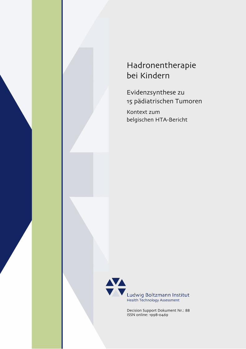
In Österreich werden etwa 250 Krebs-Neuerkrankungen bei Kindern (0–14 Jahre) und Jugendlichen (15–19 Jahre) verzeichnet [5], insgesamt 2.598 Er-krankungen in den letzten 10 Jahren (2002–2011) [6].
Leukämie ist die häufigste Krebsart im Kindesalter (28 %): etwa 80-90 Kin-der erkranken jährlich an akuter Leukämie; insgesamt 719 (von 2.598) Er-krankungen in den letzten 10 Jahren (2002–2011) [6]. Leukämie bezeichnet eine Gruppe von bösartigen Erkrankungen des blutbildenden Systems. Die akute lymphoblastische Leukämie (ALL) betrifft etwa 80 % der an Leukämie erkrankten Kinder (im Alter von 2 bis 8 Jahren, ca 55–60 Kinder und Jugend-liche pro Jahr). Die akute myeloische Leukämie (AML) betrifft etwa 20 % der Kinder (ca 20–25 Kinder pro Jahr). Chronische myeloische Leukämien (CML) spielen bei Kindern eine untergeordnete Bedeutung. Über 80 % aller ALL Kinder und etwa 60 % aller AML Kinder können geheilt werden. Nur wenige ALL-PatientInnen bedürfen – etwa bei einem manifesten Befall des ZNS/Zentralnervensystems – einer Schädelbestrahlung [7]. Die Strahlenthe-rapie wird bei nicht-lymphatischer Leukämie (AML) meist als Vorbereitung für die Stammzelltransplantation durchgeführt [8].
Tabelle 1-1: Krebserkrankungen im Kindesalter (Österreich) 2002–2011, absolut
Alle 2.598
Leukämie 719
Lymphom 421
Zentralnervensystem 421
Tumor von Muskeln und Bindegewebe 173
Knochentumor 153
Keimzelltumor 143
Neuroblastom 140
Malignes Melanom 116
Nierentumor 101
Sonstiger Tumor 211
Sonstige Tumore 211
Andere bösartige epitheliale Neoplasmen ohne Melanome 151
Andere unspezifizierte bösartige Neoplasmen 26
Nicht klassifizierte ICCC oder in situ 0
Retinoblastom 23
Lebertumor 11
Quelle: Statistik Austria [6]
Österreich: etwa 250 Krebs-
Neuerkrankungen jährlich
Leukämie ist häufigste
Krebserkrankung im Kindesalter
80–90 Kinder pro Jahr
wenige
Schädelbestrahlungen

Hadronentherapie bei Kindern
LBI-HTA | 2015 7
Lymphome (Hodgkin und Non-Hodgkin Erkrankung) sind bösartige Tumore, die vom Lymphdrüsengewebe ausgehen und Lymphknoten bilden, die dicht an der Körperoberfläche und zu 80 % im Hals-Kopfbereich liegen. Lympho-me sind die zweithäufigste Krebserkrankung im Kindesalter (16 %). Etwa 33–40 Neuerkrankungen pro Jahr und insgesamt 421 (von 2.598) Erkrankun-gen sind in den letzten 10 Jahren (2002–2011) [6] registriert. Hodgkin-Lym-phome treten eher bei Jugendlichen (ca. 15 Neuerkrankungen pro Jahr), Non-Hodgkin-Lymphome (18–25 Neuerkrankungen pro Jahr) eher bei Kindern auf. Die Behandlung besteht immer aus einer Chemotherapie oder einer Kombination mit einer Strahlentherapie [9, 10]. 70–90 % der erkrankten Kin-der und Jugendlichen können geheilt werden [5].
Tumore des ZNS/Zentralnervensystems, intrakraniale und intraspinale Tu-more (niedriggradige Gliome wie Astrozytome, Gangliogliome und Oligoden-drogliome, hochgradige Gliome wie anaplastisches Astrozytom und Glioblas-tom, Medulloblastome wie primitive neuroektodermale Tumoren/PNET so-wie Ependymome und Kraniopharyngeome) treten bei Kindern und Jugend-lichen in allen Altersstufen auf, insb. aber zwischen dem 5. und 10. Lebens-jahr. Hirntumore sind – ebenso wie Lymphome – die zweithäufigste Krebs-erkrankung im Kindesalter (16 %). Etwa 15 % aller kindlichen Hirntumoren, ca. 10 pro Jahr, sind Medulloblastome [11] und ca 5–10 sind Ependymome [12]. Etwa 63 Hirntumor-Neuerkrankungen pro Jahr treten auf und insgesamt sind 421 (von 2.598) Erkrankungen in den letzten 10 Jahren (2002–2011) [6] registriert. Die Gefährlichkeit von Hirntumoren hängt wesentlich von Lage, Gewebeart und Ausdehnung ab. Heilungschancen liegen zwischen 20 % und 90 % [5]. Nach operativer Entfernung des Tumors besteht eine Behandlung meist aus Chemotherapie und Bestrahlung. Bei kleinen Kindern wird die Be-strahlung wegen der zu erwartenden langfristigen Folgen so lange wie mög-lich verschoben oder auf einen kleinen Teil des Gehirns beschränkt [13, 14].
Abbildung 1-1: Krebserkrankungen im Kindesalter (Österreich) 2002–2011, relativ, Quelle: Statistik Austria [6]
28%
16%
16%
7%
6%
6%
5%
4%
4%
8%
Häufigkeit der Tumorentitäten im Kindesalter, vollendetes 18. Lebensjahr Diagnosejahre 2002-2011
Leukämie
Lymphom
Zentralnervensystem
Tumor von Muskeln und Bindegewebe
Knochentumor
Keimzelltumor
Neuroblastom
Malignes Melanom
Nierentumor
Sonstiger Tumor
Lymphome (Hodgkin und Non-Hodgkin Erkrankung) 33–40 Kinder pro Jahr Strahlentherapie in Kombination mit Chemotherapie
Tumore des ZNS/Hirntumore niedrig- und hochgradige Gliome, Medulloblastome, Ependymome, Kraniopharyngeome 63 Kinder pro Jahr oft auch Strahlentherapie, aber wegen zu erwartenden langfristigen Folgen oft verschoben oder auf kleinen Teil des Gehirns beschränkt

Hadronentherapie bei Kindern
8 LBI-HTA | 2015
Tumore von Muskeln und Bindegewebe werden auch Weichteiltumore ge-nannt. Das häufigste – das Rhabdomyosarkom – ist ein Tumor, der im quer-gestreiften Muskelgewebe entsteht. Andere Weichteiltumore werden im glat-ten Muskelgewebe (Leiomyo-), im Bindegewebe (Fibro und Desmoid-), in den Blutgefäßen (Angio- oder Hämangio-), in der Schleimhaut der Gelenkskapsel (Synovial-) oder im Fettgewebe (Lipo-)gebildet [15]). Jedes Jahr erkranken in Österreich ca. 15 Kinder an einem Weichteiltumor. Ingesamt sind 173 Erkran-kungen (7 % von 2.598) in den letzten 10 Jahren (2002–2011) [6] registriert. Die Behandlung eines Rhabdomyosarkoms setzt sich aus einer Chemothera-pie, einer Operation und manchmal auch einer Strahlentherapie zusammen. Es überleben 80 % der erkrankten Kinder länger als 2 Jahre. Geheilt können etwa 50 % werden [5].
Osteosarkome entstehen im Knochengewebe, Ewing-Tumoren in den Nerven-zellen der Knochen und des Knochenmarks sowie manchmal auch außerhalb des Knochens, während Chondrosarkome im Knorpelgewebe vorkommen [16]. Jedes Jahr erkranken in Österreich ungefähr 8 bis 15 Kinder und Ju-gendliche an einem Knochentumor; Ingesamt sind 153 Erkrankungen (6 % von 2.598) in den letzten 10 Jahren (2002–2011) [6] registriert. Osteosarkome und Ewing-Tumore treten öfter auf als die sehr seltenen Chondrosarkome. Nach einer Operation und Chemotherapie folgt zur Behandlung manchmal auch eine Strahlentherapie. Die Heilungschancen von Kindern und Jugend-lichen mit Knochentumoren liegen zwischen 60–70 % und hängen vom Ort ab, an dem sich der Tumor befindet.
Keimzelltumore sind sehr seltene Erkrankungen, die in den Hoden und Eier-stöcken, aber auch in anderen Geweben ihren Ursprung haben. Jedes Jahr wird in Österreich bei ungefähr 5 bis 8 Kindern ein Keimzelltumor entdeckt [17]. Ingesamt sind 143 Erkrankungen (6 % von 2.598) in den letzten 10 Jah-ren (2002–2011) [6] registriert. 80 % der Kinder können geheilt werden. Kann der Keimzelltumor nicht vollständig entfernt werden oder kommt es zu Me-tastasierungen, bekommt das Kind in jedem Fall eine Chemotherapie und/ oder Strahlentherapie.
Das Neuroblastom ist eine bösartige Erkrankung des sympathischen Nerven-systems: Neuroblastome treten im Bauch-, Becken-, Brust- oder Halsbereich auf. Es werden etwa 25 Neuerkrankungen pro Jahr entdeckt [18] und 140 Erkrankungen (5 % von 2.598) sind in den letzten 10 Jahren (2002-2011) [6] registriert. Betroffen sind Kinder bis zum 8. Lebensjahr [5]. In einem Viertel aller Fälle treten erste Anzeichen (Knoten, Schwellungen etc.) bereits inner-halb der ersten 12 Monate auf. Tritt ein Neuroblastom im Babyalter auf, kann es zumeist geheilt werden. Bei metastasierenden Tumoren (Stadium 4) kann bei ausgewählten Tumorlokalisationen zusätzlich zur Operation und Chemo-therapie eine Strahlentherapie angewandt werden.
Das maligne Melanom, Hautkrebs ist eine seltene Krebserkrankung im Kin-desalter. Jedes Jahr erkranken in Österreich ungefähr 10–12 Kinder an einem Hautkrebs. 116 (4 % von 2.598) Neuerkrankungen sind in den letzten 10 Jah-ren (2002-2011) [6] registriert.
Der Wilmstumor oder Nephroblastom ist ein bösartiges Geschwulst der Niere, das in 50 % der Fälle bis zum 3. Lebensjahr auftritt. Jedes Jahr erkranken in Österreich ungefähr 10–12 Kinder an einem Nephroblastom; insgesamt sind 101 (4 % von 2.598) in den letzten 10 Jahren (2002–2011) [6] registriert. Die Heilung liegt bei 80-90 % der Kinder. Eine Strahlentherapie kann manchmal, neben der Chemotherapie notwendig sein [19].
Weichteiltumore
Rhabdomyosarkom 15 Kinder pro Jahr
manchmal
Strahlentherapie
Knochentumore
Osteosarkome, Ewing-Tumore,
Chondrosarkome
8–15 Kinder pro Jahr
manchmal Strahlentherapie
Keimzelltumore
5–8 Kinder pro Jahr
Strahlentherapie ev. bei Metastasierung
Neuroblastom
25 Kinder pro Jahr
Strahlentherapie nur bei
metastasierenden Tumoren
Malignes Melanom
10–12 Kinder pro Jahr
Nephroblastom,
Wilms-Tumor 10-12 Kinder pro Jahr
manchmal Strahlentherapie
Hadronentherapie bei Kindern
LBI-HTA | 2015 9
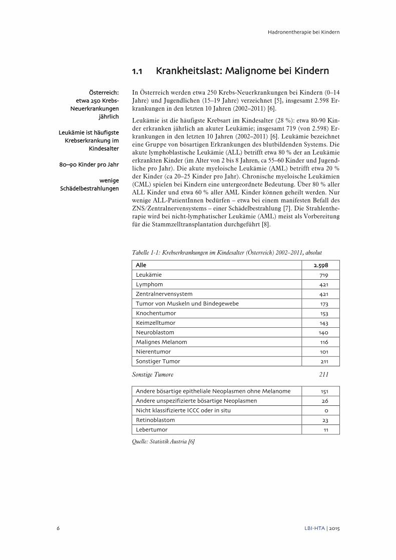
Das Retinoblastom ist eine sehr seltene Erkrankung von Zellen der Netzhaut. Betroffen sind vor allem Säuglinge und Kleinkinder bis 5 Jahre [5]. In 25-30 % der Fälle sind beide Augen betroffen. Jedes Jahr erkranken in Österreich ungefähr 4 Kinder an einem Retinoblastom. insgesamt sind 23 (1 % von 2.598) in den letzten 10 Jahren (2002–2011) [6] registriert. Kinder mit einem Retino-blastom haben Heilungschancen von über 90 %, wobei allerdings die Größe und die Ausbreitung des Tumors eine Rolle spielt [20]. Nur in wenigen Fäl-len kommt eine Strahlentherapie zu Anwendung.
Hepatozelluläre Karzinome sind bei Kinder und Jugendlichen sehr selten (0,4 %, 11 in 10 Jahren, 2002–2011) [6]. Chirurgische Entfernung des Tumors, gefolgt von Chemotherapie sind die und Behandlungen der Wahl. Die Hei-lungschancen liegen bei 50 % [5].
Tabelle 1-2: Krebserkrankungen im Kindesalter (Österreich) 2002–2011, nach Alter der Erkrankung
International Classification of Childhood Cancer
Alle 2002-2011
Alter
0<1 1<5 5<15 15<18 18<19
N N N N N N
Alle 2.598 166 561 1.077 585 209
I Leukemias, myeloproliferative diseases, and myelodysplastic diseases 719 30 254 307 99 29
II Lymphomas and reticuloendothelial neoplasms 421 4 19 193 156 49
III CNS and miscellaneous intracranial and intraspinal neoplasms 421 22 94 224 70 11
IV Neuroblastoma and other peripheral nervous cell tumors 140 51 65 21 3 0
V Retinoblastoma 23 10 12 1 0 0
VI Renal tumors 101 19 58 22 1 1
VII Hepatic tumors 11 3 4 3 1 0
VIII Malignant bone tumors 153 0 7 97 37 12
IX Soft tissue and other extraosseous sarcomas 173 14 33 75 38 13
X Germ cell tumors, trophoblastic tumors, and neoplasms of gonads 143 8 8 41 56 30
XI Other malignant epithelial neoplasms, without malignant melanomas 151 0 2 47 55 47
XI Other malignant epithelial neoplasms, only malignant melanomas 116 1 1 40 58 16
XII Other and unspecified malignant neoplasms 26 4 4 6 11 1
Quelle: Statistik Austria [6]
Retinoblastom 4 Kinder pro Jahr Strahlentherapie selten
Hepatozelluläre Karzinome 1 Kind pro Jahr

Hadronentherapie bei Kindern
10 LBI-HTA | 2015
1.2 Krankheitslast: Spätfolgen
Aufgrund therapeutischer Verbesserungen sind die Überlebensraten unter krebskranken Kindern und Jugendlichen groß. Die Herausforderung in der Krebstherapie stellt aber – neben dem Überleben – die Balance zwischen Hei-lung und langfristiger Morbidität der Überlebenden dar. Im österreichischen Krebsrahmenprogramm ist als ein vorrangiges Ziel, die Einführung eines Sur-vivorship-Passes [21], in dem auch langfristig die Daten der Krebspatien-tInnen dokumentiert werden, geplant.
Tatsächlich ist die Krankheitslast durch chronische Erkrankungen groß: 30 Jahre nach der Diagnose Krebs im Kinder-und Jugendalter beträgt die ku-mulative Inzidenz von chronischen Gesundheitsproblemen 73,4 % (95 % CI; 69,0–77,9), mit einer kumulativen Inzidenz von schwerwiegenden und lebens-bedrohlichen Gesundheitszuständen von 42 % (95 % CI; 33,7–51,2) [22]. Die Risiken der Überlebenden nach Krebs sind – bedingt ebenso durch Rezidive wie durch die langfristigen Nebenwirkungen von Behandlungen des Primär-tumors – späte Mortalität, sekundäre Neoplasmen, aber auch kardio-vaskuläre Erkrankungen, Organdysfunktionen oder Wachstumsverzögerung [22-25].
Strahlentherapie ist eine wesentliche Komponente im Behandlungsspektrum von Neoplasmen im Kindes- und Jugendalter. Unglücklicherweise sind Kin-der- und Jugendliche nicht nur sehr empfänglich im Ansprechen auf Strahlen-therapie, sondern reagieren auch sehr empfindlich auf die Exposition durch radiotherapeutische Strahlenbelastung, was zu späten schweren Nebenwir-kungen auch bei geringen Strahlendosen führen kann. Die Strahlenexposi-tion des Gehirns ist mit neurokognitiven Defiziten, endokrinen Dysfunktio-nen bis zu Hörverlust assoziiert. Insb. Kinder unter 7 Jahren sind hier stär-ker betroffen [26, 27], weswegen im Behandlungsplan Radiotherapie häufig verschoben oder ersetzt wird. Diese Nebenwirkungen können aber auch durch die Erkrankung selbst (Gehirntumor) oder andere Therapien wie Chemothe-rapie oder chirurgische Exzision des Tumors, verursacht werden. Weiters kann kraniospinale Bestrahlung zu Schilddrüsendysfunktion und zu spinaler Wachstumsbeeinträchtigung und zu Schädigungen der Lunge, am Herz und bei intestinale Funktionen führen [27, 28].
Sekundäre Tumore sind die häufigste Todesursache unter Überlebenden. Die Childhood Cancer Survivor Study (mit einer Kohorte von 14.000 PatientIn-nen) zeigt eine 30-Jahres kumulative Inzidenz von 7,9 % (95 % CI; 7,2–8,5) für sekundäre Neoplasmen, was einem 6-fach erhöhtem Risiko – im Vergleich zur allgemeinen Bevölkerung – entspricht [29-31]. Die Tumore treten frühestens 5–19 Jahre nach der Bestrahlung auf. Radiotherapie-induzierte Malignome können sowohl in den benachbarten Regionen oder auch in entfernten Kör-perregionen zu Karzinomen führen. Sarkome treten dagegen an derselben Stelle oder in unmittelbarer Nähe zur bestrahlten Region auf [32]. Eine Un-tersuchung von 115 Sekundärmalignomen zeigte, dass 66 % der Tumore in der benachbarten Region, 22 % mehr als 5 cm entfernt und nur 12 % im Zent-rum der ehedem bestrahlten Region lagen [33]. Die Daten zu Strahlenthera-pie-induzierten sekundären Malignomen sind aber wegen der langen Nach-beobachtungszeit von 25–30 Jahren und in Ermangelung von Kontrollgrup-pen beschränkt: die Childhood Cancer Survivor Study zeigt auch, dass das Risiko zu Sekundärtumoren über die Jahre steigt und kein Plateau – auch 30 Jahre nach Behandlung – aufweist [34].
hohe Überlebensraten
ABER: Balance zwischen Heilung und
langfristigen Folgen für Gesundheit
Survivorship-Pass
30 Jahre nach Krebs im Kinder-und Jugendalter
73 % chronische
Gesundheitsprobleme
42 % schwerwiegende Gesundheitsprobleme:
Sekundäre Tumore, Nebenwirkungen der
Therapien
Spätfolgen von Strahlentherapie am
Kopf:
neurokognitive Defizit, endokrine Störungen,
Wachstums-verzögerung, etc.
sekundäre Malignome frühestens 5–19 Jahre nach der Bestrahlung
6-fach erhöhtes Risiko
schlechte Datenlage
wegen langer Nachbeobachtungszeit

Hadronentherapie bei Kindern
LBI-HTA | 2015 11
Zusammenfassend ist zu sagen, dass das Ziel der Behandlung von Tumoren im Kinder- und Jugendalter nicht nur deren Heilung, sondern auch die Ver-meidung von langfristigen schwerwiegenden Nebenwirkungen sowie die Ver-meidung von sekundären Malignomen ist. Es wird angenommen, dass die Reduktion der Bestrahlung von – dem Tumor – benachbartem Gewebe auch zu einer Reduktion von langfristigen Nebenwirkungen und Folgemalignomen führt. Die Anwendung von Protonentherapie kann die integrale Strahlendo-sis um einen Faktor 2–3 im Vergleich zur konventionellen Photonentherapie oder zur IMRT/Intensity modulated photon therapy reduzieren [35]. Deshalb wird angenommen, dass die Protonentherapie auch geringere langfristige Ne-benwirkungen hat.
Ziel der Behandlung von Tumoren im Kinder- und Jugendalter: Heilung UND Vermeidung von langfristigen schwerwiegenden Nebenwirkungen
Hadronentherapie bei Kindern
12 LBI-HTA | 2015
2 Zusammenfassung der Ergebnisse des KCE-Reviews
Der belgische HTA-Bericht „Hadron Therapy in Children – an update of the scientific evidence for 15 paediatric cancers“ [2, 3] setzte auf den ersten, be-reits 2007 veröffentlichten Bericht [4] auf und wertete die klinische Evidenz zur Wirksamkeit der Hadronentherapie in 15 Indikationen aus. Es konnten insgesamt 21 klinische Studien zu den Indikationen gefunden werden. Eine systematische Suche nach relevanten Publikationen wurde in drei Datenban-ken (Medline via OVID, EMBASE, Cochrane Library) durchgeführt. Syste-matische Reviews und Primärstudien zur Protonentherapie und/oder Koh-lenstoffionentherapie veröffentlicht zwischen 2007 bis März 2014 wurden ge-sucht (ein letztes Update der Suche erfolgte im September 2014).
Abseits der Studiendesigns (nicht-randomisiert, nicht-kontrolliert und retros-pektiv) der Studien – mit den für diese Art von charakteristischen Einschrän-kungen (z. B. Selektionsbias, Recall-Bias) –, zeigten alle Studien schwere me-thodische Mängel (u. a. kleine Stichproben, lange Zeiträume beim Einschluss der PatientInnen, unterschiedliche Behandlungsschemata, kurze Follow-ups, Berichterstattung oder Dokumentation von Komplikationen nur bei einem Teil der PatientInnen). Unter Anwendung von GRADE war die wissenschaft-liche Evidenzlage für alle Ergebnisse in allen Indikationen war sehr gering.
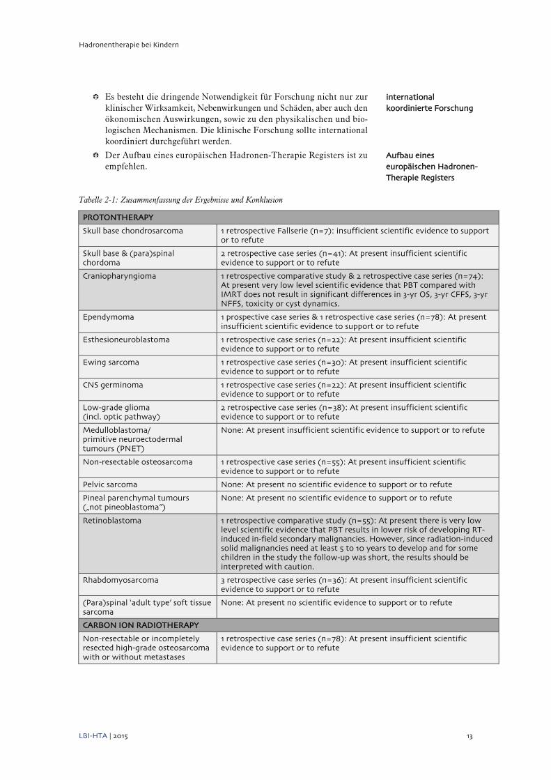
Die Ergebnisse sind in Tabelle 2-1 zusammengefasst und sagen aus, dass in 13 von 15 Indikationen die Evidenz unzureichend ist, um Aussagen zuguns-ten oder gegen Hadronentherapie zu machen. In 2 Indikationen liegt sehr nie-drige Evidenz, die für Gleichwertigkeit zwischen Protonentherapie und IMRT/ Intensitätsmodulierte Strahlentherapie beim Craniopharyngioma, resp. einem geringeren Risiko für Sekundärtumore beim Retinoblastom spricht.
Die Schlussfolgerung lautet dementsprechend, dass weiterhin klinische Da-ten zur Protonentherapie in allen untersuchten pädiatrischen Krebserkran-kungen zur langfristigen Wirksamkeit und zu Nebenwirkungen fehlen. Nur bei sehr wenigen Tumoren ist die Protonentherapie wegen der in hohem Ma-ße vorhersehbaren ernsthaften Schäden bei anderen Formen der Strahlenthe-rapie als einzige Behandlungsmethode möglich. In den meisten Fällen gibt es eine Wahl. Die Hoffnungen auf bessere klinische Ergebnisse mit Proto-nentherapie sind nicht belegt.
Folgende Empfehlungen werden ausgesprochen:
PatientInnen (oder ihre Eltern und Angehörigen) sollte umfassend in-formiert werden, dass trotz der physischen Untermauerung der Pro-tonentherapie, die klinische Wirksamkeit in den untersuchten Indika-tionen noch nicht in klinischen Studien bestätigt wurde.
Kinder sollten nur Protonen-Zentren mit dem notwendigen Know-how in der Behandlung von Kindern mit der spezifischen Pathologie und im Rahmen klinischer Studien mit Langzeit-Follow-up einbezogen werden.
Die genaue Dokumentation der Therapien zur langfristigen Nachbe-obachtung etwa beim Auftreten von sekundären ist zu empfehlen.
Update zu 2007 Bericht
systematische Suche in 3 Datenbanken
21 klinische Studien
Qualitätsbeurteilung der vorliegenden
Evidenz mit GRADE
sehr niedrige Qualität der Evidenz
Ergebnisse: in 13 von 15
Indikationen: Evidenz unzureichend, keine
Aussage möglich in 2 Indikationen: niedrige Evidenz
Schlussfolgerung:
es fehlt weiterhin an klinischen Daten zur
Protonentherapie
Hoffnung vs. Belege
Empfehlungen: PatientInnen-
Informationen zum Mangel an Belegen
Behandlung nur in Zentren mit Erfahrung
mit Kindern, nur im Rah-men klinischer Studien
Langzeit-Dokumentation
Hadronentherapie bei Kindern
LBI-HTA | 2015 13
Es besteht die dringende Notwendigkeit für Forschung nicht nur zur klinischer Wirksamkeit, Nebenwirkungen und Schäden, aber auch den ökonomischen Auswirkungen, sowie zu den physikalischen und bio-logischen Mechanismen. Die klinische Forschung sollte international koordiniert durchgeführt werden.
Der Aufbau eines europäischen Hadronen-Therapie Registers ist zu empfehlen.
Tabelle 2-1: Zusammenfassung der Ergebnisse und Konklusion
PROTONTHERAPY
Skull base chondrosarcoma 1 retrospective Fallserie (n=7): insufficient scientific evidence to support or to refute
Skull base & (para)spinal chordoma
2 retrospective case series (n=41): At present insufficient scientific evidence to support or to refute
Craniopharyngioma 1 retrospective comparative study & 2 retrospective case series (n=74): At present very low level scientific evidence that PBT compared with IMRT does not result in significant differences in 3-yr OS, 3-yr CFFS, 3-yr NFFS, toxicity or cyst dynamics.
Ependymoma 1 prospective case series & 1 retrospective case series (n=78): At present insufficient scientific evidence to support or to refute
Esthesioneuroblastoma 1 retrospective case series (n=22): At present insufficient scientific evidence to support or to refute
Ewing sarcoma 1 retrospective case series (n=30): At present insufficient scientific evidence to support or to refute
CNS germinoma 1 retrospective case series (n=22): At present insufficient scientific evidence to support or to refute
Low-grade glioma (incl. optic pathway)
2 retrospective case series (n=38): At present insufficient scientific evidence to support or to refute
Medulloblastoma/ primitive neuroectodermal tumours (PNET)
None: At present insufficient scientific evidence to support or to refute
Non-resectable osteosarcoma 1 retrospective case series (n=55): At present insufficient scientific evidence to support or to refute
Pelvic sarcoma None: At present no scientific evidence to support or to refute
Pineal parenchymal tumours („not pineoblastoma”)
None: At present no scientific evidence to support or to refute
Retinoblastoma 1 retrospective comparative study (n=55): At present there is very low level scientific evidence that PBT results in lower risk of developing RT-induced in-field secondary malignancies. However, since radiation-induced solid malignancies need at least 5 to 10 years to develop and for some children in the study the follow-up was short, the results should be interpreted with caution.
Rhabdomyosarcoma 3 retrospective case series (n=36): At present insufficient scientific evidence to support or to refute
(Para)spinal ‘adult type’ soft tissue sarcoma
None: At present no scientific evidence to support or to refute
CARBON ION RADIOTHERAPY
Non-resectable or incompletely resected high-grade osteosarcoma with or without metastases
1 retrospective case series (n=78): At present insufficient scientific evidence to support or to refute
international koordinierte Forschung
Aufbau eines europäischen Hadronen- Therapie Registers
Hadronentherapie bei Kindern
14 LBI-HTA | 2015
3 Literatur
[1] Wild C, Hintringer K, Narath M. Hadronentherapie: Protonen und Kohlenstoff-Ionen. Eine Übersicht: Refundierungsstatus, Evidenz und Forschungsstand. Wien: Ludwig Boltzmann Institut für HTA, 2013 Contract No.: 74.
[2] Leroy R, Benahmed N, Hulstaert F, Mambourg F, Fairon N, Van Eycken L, et al. Hadron therapy in children – an update of the scientific evidence for 15 paediatric cancers. Brussels: 2015.
[3] Leroy R, Benahmed N, Hulstaert F, Mambourg F, Fairon N, Van Eycken L, et al. Hadron therapy in children – an update of the scientific evidence for 15 paediatric cancers – supplement. 2015.
[4] Huybrechts M, Obyn C, Gailly J, Mambourg F, Vinck I, Ramaekers D. Hadrontherapie. Brussels: 2007.
[5] Zoubek A. Krebs im Kindesalter. Wien: St. Anna Kinderspital; 2000a [16.02.2015]; Available from: http://www.forschenheiltkrebs.eu/public/pdf/krebs_bei_kindern_und_jugendlichen/infomaterialien/krebs_bei_kindern_jugendl_forschen_heilt_krebs_infomaterial_krebs_im_kindesalter_dr_andreas_zoubek.pdf.
[6] Statistik Austria. Krebs bei Kindern und Jugendlichen. 2015; Available from: http://www.statistik.at/web_de/statistiken/gesundheit/krebserkrankungen/krebs_bei_kindern-und_jugendlichen/index.html.
[7] Kinderkrebshilfe. Akute lymphatische Leukämie. 2014 [16.02.2015]; Available from: http://www.kinderkrebshilfe.at/MDB/pdf/57_ALL_Neu201428229_final.pdf.
[8] Kinderkrebshilfe. Nicht lymphatische Leukämie 2008 [16.02.2015]; Available from: http://www.kinderkrebshilfe.at/upload/6033_Nichtlymphatische_25November08.pdf.
[9] Kinderkrebshilfe. Hodgkin-Lymphom. 2012 [16.02.2015]; Available from: http://www.kinderkrebshilfe.at/upload/HodgkinLymphom.pdf.
[10] Kinderkrebshilfe. Non- Hodgkin-Lymphom. 2011 [16.02.2015]; Available from: http://www.kinderkrebshilfe.at/upload/NonHodgkinLymphomeWEB_2Feb2011.pdf.
[11] Kinderkrebshilfe. Medulloblastom. 2013 [16.02.2016]; Available from: http://www.kinderkrebshilfe.at/MDB/pdf/24_Medulloblastom2_WEB_27August2013.pdf.
[12] Kinderkrebshilfe. Ependymom. 2012 [16.02.2015]; Available from: http://www.kinderkrebshilfe.at/upload/Ependymom.pdf.
[13] Kinderkrebshilfe. Niedriggradige Gliome. 2009 [16.02.2015]; Available from: http://www.kinderkrebshilfe.at/upload/6837_Niedriggradige%20Gliome_16Nov09Endeversion.pdf.
[14] Kinderkrebshilfe. Hochgradige Gliome. 2011 [16.02.2015]; Available from: http://www.kinderkrebshilfe.at/upload/HochgradigeGliomeWEB_2Feb2011.pdf.
[15] Kinderkrebshilfe. Weichteiltumoren. 2009 [16.02.2015]; Available from: http://www.kinderkrebshilfe.at/upload/6836_Weichteiltumoren_25Nov09_Endversion.pdf.
[16] Kinderkrebshilfe. Knochentumore. 2011 [16.02.2015]; Available from: http://www.kinderkrebshilfe.at/upload/KnochentumorenWEB_2Feb2011.pdf.
[17] Kinderkrebshilfe. Keimzelltumoren. 2014 [16.02.2015]; Available from: http://www.kinderkrebshilfe.at/MDB/pdf/58_Keimzelltumoren_11August2014.pdf.
[18] Kinderkrebshilfe. Neuroblastom. 2009 [16.02.2015]; Available from: http://www.kinderkrebshilfe.at/upload/6835_Neuroblastom_25Nov09_Endversion.pdf.
[19] Kinderkrebshilfe. Wilms-Tumor. 2012 [16.02.2015]; Available from: http://www.kinderkrebshilfe.at/upload/Wilmstumor.pdf.
[20] Kinderkrebshilfe. Retinoblastom. 2012 [16.02.2015]; Available from: http://www.kinderkrebshilfe.at/upload/Retinoblastom.pdf.
Hadronentherapie bei Kindern
LBI-HTA | 2015 15
[21] BMG/Bundesministerium für Gesundheit. Krebsrahmenprogramm Österreich. 2014; Available from: http://bmg.gv.at/cms/home/attachments/2/7/0/CH1480/CMS1412233312313/krebsrahmenprogramm_20141002.pdf.
[22] Oeffinger K, Mertens A, Sklar C, Kawashima T, Hudson M, Meadows A, et al. Chronic health conditions in adult survivors of childhood cancer. N Engl J Med. 2006;355(15):1572-82.
[23] Timmermann B. Proton beam therapy for childhood malignancies: Status report. Klin Padiatr 2010;222(3):127-33.
[24] Pritchard-Jones K, Pieters R, Reaman G, Hjorth L, Downie P, Calaminus G, et al. Sustaining innovation and improvement in the treatment of childhood cancer: lessons from high-income countries. Lancet Oncol. 2013;14(3):e95-e103.
[25] Zoubek A. Krebstherapie im Kindesalter. Wien: St. Anna Kinderspital; 2000b [16.02.2015]; Available from: http://www.forschenheiltkrebs.eu/public/pdf/krebs_bei_kindern_und_jugendlichen/ infomaterialien/krebs_bei_kindern_jugendl_forschen_heilt_krebs_infomaterial_krebstherapie_im_kindesalter.pdf.
[26] Rombi B, Ares C, Hug E, Schneider R, Goitein G, Staab A, et al. Spot-scanning proton radiation therapy for pediatric chordoma and chondrosarcoma: Clinical outcome of 26 patients treated at paul scherrer institute. Int J Radiat Oncol Biol Phys. 2013;86(3):578-84.
[27] Cotter S, McBride S, Yock T. Proton radiotherapy for solid tumors of childhood. Technol Cancer Res Treat 2012;11(3):267-78.
[28] Merchant T. Clinical controversies: Proton therapy for pediatric tumors. Semin Radiat Oncol. 2013;23(2):97-108.
[29] Bekelman J, Schultheiss T, Berrington De Gonzalez A. Subsequent malignancies after photon versus proton radiation therapy. Int J Radiat Oncol Biol Phys. 2013;87(1):10-2.
[30] Mertens A, Liu Q, Neglia J, Wasilewski K, Leisenring W, Armstrong G, et al. Cause-specific late mortality among 5-year survivors of childhood cancer: the Childhood Cancer Survivor Study. J Natl Cancer Inst 2008;100(19):1368-79.
[31] Friedman D, Whitton J, Leisenring W, Mertens A, Hammond S, Stovall M, et al. Subsequent neoplasms in 5-year survivors ofchildhood cancer: the Childhood Cancer Survivor Study. J Natl Cancer Inst. 2010;102(14):1083-95.
[32] Hall E. Intensity-modulated radiation therapy, protons, and the risk of second cancers. Int J Radiat Oncol Biol Phys 2006;65(1):1-7.
[33] Diallo I, Haddy N, Adjadj E, Samand A, Quiniou E, Chavaudra J, et al. Frequency distribution of second solid cancer locations in relation to the irradiated volume among 115 patients treated for childhood cancer. Int J Radiat Oncol Biol Phys 2009;74(3):876-83. Epub 2009 Apr 20.
[34] Paganetti H, Athar B, Moteabbed M, Adams A, Schneider U, Yock T. Assessment of radiation-induced second cancer risks in proton therapy and IMRT for organs inside the primary radiation field. Phys Med Biol 2012;57(19):6057-61. Epub 12 September 2012.
[35] Lomax A, Bortfeld T, Goitein G, Debus J, Dykstra C, Tercier P. A treatment planning inter-comparison of proton and intensity modulated photon radiotherapy. Radiother Oncol 1999;51(3):257-71.
http://bmg.gv.at/cms/home/attachments/2/7/0/CH1480/CMS1412233312313/krebsrahmenprogramm_20141002.pdf
KCE REPORT 235Cs
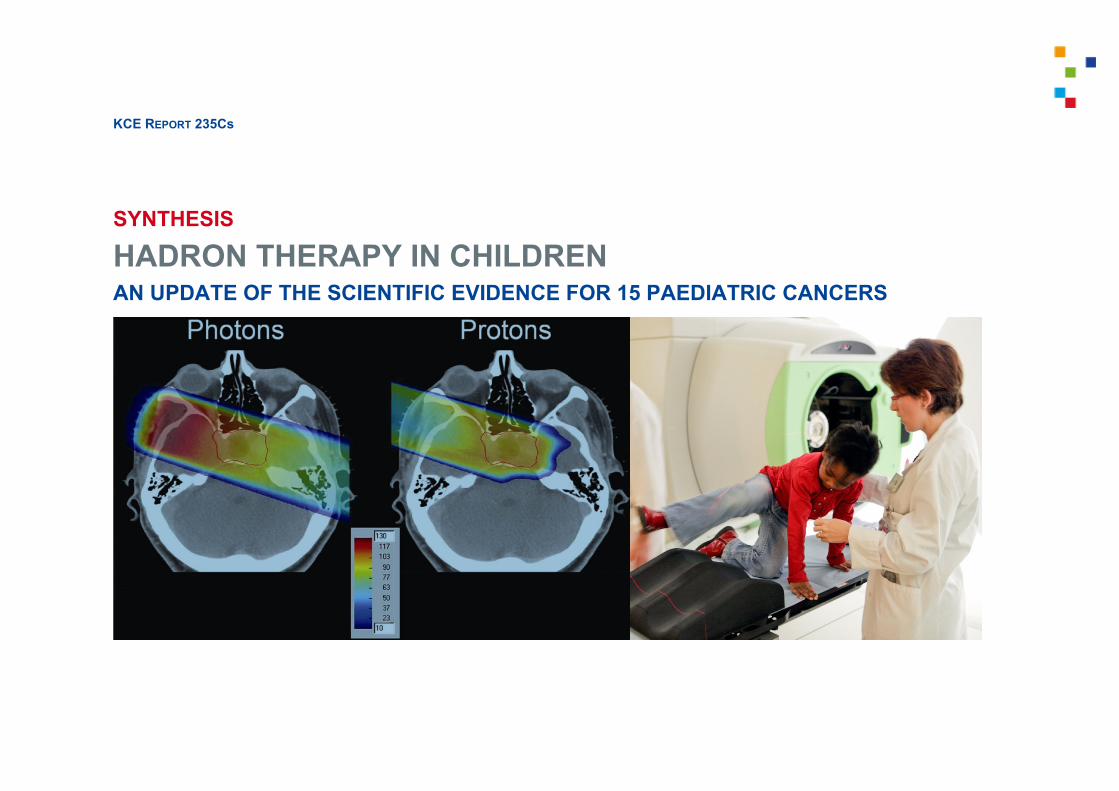
SYNTHESIS
HADRON THERAPY IN CHILDREN AN UPDATE OF THE SCIENTIFIC EVIDENCE FOR 15 PAEDIATRIC CANCERS

2015 www.kce.fgov.be
Belgian Health Care Knowledge Centre
The Belgian Health Care Knowledge Centre (KCE) is an organization of public interest, created on the 24th of December 2002 under the supervision of the Minister of Public Health and Social Affairs. KCE is in charge of conducting studies that support the political decision making on health care and health insurance.
Executive Board Actual Members Substitute Members
President Pierre Gillet CEO - National Institute for Health and Disability Insurance
(vice president) Jo De Cock Benoît Collin
President of the Federal Public Service Health, Food Chain Safety and Environment (vice president)
Dirk Cuypers Christiaan Decoster
President of the Federal Public Service Social Security (vice presi-dent)
Frank Van Massenhove
Jan Bertels
General Administrator of the Federal Agency for Medicines and Health Products
Xavier De Cuyper Greet Musch
Representatives of the Minister of Public Health Brieuc Van Damme Koen Vandewoude Ri De Ridder Yolande Avontroodt Representatives of the Minister of Social Affairs Bert Winnen Magali Pirson Johan De Haes Dirk Ramaekers Representatives of the Council of Ministers Jean-Noël Godin Natacha Beugnier Marc Loix Tijs Neutens Intermutualistic Agency Michiel Callens Frank De Smet Patrick Verertbrug-
gen Jean-Marc
Laasmans Xavier Brenez Geert Messiaen Professional Organisations - representatives of physicians Marc Moens Roland Lemye Jean-Pierre
Baeyens Rita Cuypers
Professional Organisations - representatives of nurses Ellen De Wandeler Ludo Meyers Myriam Hubinon Olivier Thonon Hospital Federations Johan Pauwels Katrien Kesteloot Jean-Claude Praet Pierre Smiets

Social Partners Rita Thys Catherine Rutten Paul Palsterman Celien Van
Moerkerke House of Representatives To be assigned

Control Government commissioner Steven Sterckx
Management General director
Deputy general director Raf Mertens Christian Léonard
Program Management Kristel De Gauquier Dominique Paulus
Contact Belgian Health Care Knowledge Centre (KCE)
Doorbuilding (10th Floor) Boulevard du Jardin Botanique, 55 B-1000 Brussels Belgium T +32 [0]2 287 33 88 F +32 [0]2 287 33 85 [email protected] http://www.kce.fgov.be
2014 www.kce.fgov.be
KCE REPORT Cs HEALTH TECHNOLOGY ASSESSMENT
SYNTHESIS
HADRON THERAPY IN CHILDREN AN UPDATE OF THE SCIENTIFIC EVIDENCE FOR 15 PAEDIATRIC CANCERS
ROOS LEROY, NADIA BENAHMED, FRANK HULSTAERT, FRANÇOISE MAMBOURG, NICOLAS FAIRON, LIESBET VAN EYCKEN, DIRK DE RUYSSCHER

COLOPHON Title: HADRON THERAPY – AN UPDATE OF THE SCIENTIFIC EVIDENCE FOR SPECIFIC PAEDIATRIC INDICA-
TIONS – an update of the scientific evidence for 15 paediatric cancers – Synthesis
Authors: Roos Leroy (KCE), Nadia Benahmed (KCE), Frank Hulstaert (KCE), Françoise Mambourg (KCE), Nicolas Fairon (KCE), Liesbet Van Eycken (Stichting Kankerregister – Fondation Registre du Cancer), Dirk De Ruysscher (KU Leuven)
Project coordinator: Marijke Eyssen (KCE)
Reviewers: Raf Mertens (KCE), Sabine Stordeur (KCE), Geneviève Veereman (KCE)
External experts: Edward Baert (UGent), Yves Benoit (UGent), Sylviane Carbonnelle (AFCN – FANC), Olivier de Witte (Erasme; ULB), Bart Depreitere (KU Leuven), Lorraine Donnay (Clinique & Maternité Sainte-Elisabeth, Namur), Hilde En-gels (RIZIV – INAMI), Nancy Van Damme (Stichting Kankerregister – Fondation Registre du Cancer), Paul Van Houtte (Institut Jules Bordet; ULB), Claudia Wild (Ludwig Boltzmann Institute, Austria)
External validators: Gudrun Goitein (Since September 2014 retired from Paul Scherrer Institute, Villigen, Switzerland), Edward C. Halperin (New York Medical Centre, US), Stefaan Van Gool (KU Leuven)
Acknowledgements: Kris Henau (Stichting Kankerregister – Fondation Registre du Cancer), Mattias Neyt (KCE), Jo Robays (KCE), Chris Segaert (RIZIV – INAMI), Beate Timmerman (Westdeutsches Protonentherapiezentrum Essen, Germany), Leen Verleye (KCE)
Other reported interests: • None declared Layout: Ine Verhulst
Coverpictures: • The left cover image is copyrighted by Sage Publications, Inc. • The right cover image is copyrighted by Eric Bouvet / Institut Curie (ref. 4487)
Disclaimer: The external experts were consulted about a (preliminary) version of the scientific report. Their comments were discussed during meetings. They did not co-author the scientific report and did not necessarily agree with its content.
Subsequently, a (final) version was submitted to the validators. The validation of the report results from a consensus or a voting process between the validators. The validators did not co-author the scientific report and did not necessarily all three agree with its content.
Finally, this report has been approved by a majority of votes by the Executive Board. Only the KCE is responsible for errors or omissions that could persist. The policy recommendations are
also under the full responsibility of the KCE.

Publication date: 08 January 2015
Domain: Health Technology Assessment (HTA)
MeSH: • Proton therapy; Heavy ions; Radiotherapy; Review [Publication type] NLM Classification: WN 250.5.P7
Language: English
Format: Adobe® PDF™ (A4)
Legal depot: D/2015/10.273/03
Copyright: KCE reports are published under a “by/nc/nd” Creative Commons Licence http://kce.fgov.be/content/about-copyrights-for-kce-reports.
How to refer to this document? Leroy R, Benahmed N, Hulstaert F, Mambourg F, Fairon N, Van Eycken L, De Ruysscher D. HADRON THERA-PY – AN UPDATE OF THE SCIENTIFIC EVIDENCE FOR SPECIFIC PAEDIATRIC INDICATIONS – an update of the scientific evidence for 15 paediatric cancers – Synthesis. Health Technology Assessment (HTA) Brussels: Belgian Health Care Knowledge Centre (KCE). 2015. KCE Reports Cs. D/2015/10.273/03.
• This document is available on the website of the Belgian Health Care Knowledge Centre.

KCE Report 235Cs Hadron therapy 1
FOREWORD
If there is one area in health care that is emotionally difficult, it is paediatric oncology. The sight of a child suffer-ing - of a dying child - is not only unbearable, it also evokes a feeling of rebelliousness, an appeal to do every-thing within our capabilities to save this child. And then there is this high-tech radiation technique, which promis-es to offer just that little bit extra. A form of radiation that is at least equally effective against the tumour, but clearly causes less collateral damage to the surrounding tissues and therefore should also cause fewer second-ary tumours induced by the radiation itself. The physical models are convincing, the simulations are promising and the clinical experience appears to be positive.
The stakes are high in every respect, not only because this is about children with cancer. The price tag for a new proton centre can easily exceed 30 million Euros and the running costs are similarly high. Understandably, those who have set out on this path defend their case through thick and thin; and they are determined to con-quer a place for this innovative technique in the health care landscape. From experience we know that this type of hi-tech innovations cannot be stopped anyway, and recent history seems to confirm this also for hadron cen-tres.
It is a downright shame that - even after enormous global investments and at least 120,000 patients treated - there is still virtually no conclusive evidence to support the superiority of this technique in children. Whilst good international, multi-centre studies could quickly provide the required insights for a fraction of the investment costs, the centres and their protagonists mainly continue to act as rival SMEs who compete for patients. And one does not need to look to the suppliers of this heavy infrastructure for support for this type of studies.
So, in response to the question posed to us by the National Institute of Health and Disability Insurance (RIZIV – INAMI) whether there is now more evidence to support the reimbursed paediatric indications - the answer sadly remains “no”. Whilst awaiting the results of the few studies that are ongoing, there is probably little choice other than to give these young patients the benefit of the doubt, but without any guarantee that the result will eventual-ly be positive. This is and remains too little, too late.
Christian LÉONARD
Deputy general director
Raf MERTENS
General director

2 Hadron therapy KCE Report 235Cs
SUMMARY 1 INTRODUCTION Anno 2014 there are no hadron facilities in Belgium; Belgian citizens eligi-ble for hadron therapy (i.e. proton beam therapy (PBT) or carbon ion radio-therapy (CIRT)) are sent abroad. From September 2014 on (and until the end of September 2017), the costs related to hadron therapy (i.e. the treatment, transport and accommodation) are reimbursed if the diagnosis is on the list of eligible indicationsa and if the “Agreement Council for Had-ron Therapy” (akkoordraad/ conseil d'accord) approves the application.
The objective of this study was to evaluate the clinical effectiveness of pro-ton beam (or carbon ion) therapy in those indications in children currently reimbursed by the National Institute for Health and Disability Insurance (RIZIV – INAMI). It concerns the following 16 indications:
Proton beam therapy Skull base chondrosarcoma Skull base & (para)spinal chordoma Craniopharyngioma Ependymoma Esthesioneuroblastoma Ewing sarcoma CNS germinoma Low-grade glioma (incl. optic pathway) Medulloblastoma / primitive neuroectodermal tumours (PNET) Non-resectable osteosarcoma Pelvic sarcoma Pineal parenchymal tumours (not pineoblastoma) Retinoblastoma Rhabdomyosarcoma (Para)spinal ‘adult type’ soft tissue sarcoma
a
http://www.riziv.fgov.be/nl/professionals/verzorgingsinstellingen/ziekenhuizen/zorg/Paginas/Hadron-english.aspx; for osteosarcoma PBT & CIRT are considered, leading to 16 indications in 15 cancers.
KCE Report 235Cs Hadron therapy 3
Carbon ion radiotherapy Non-resectable or incompletely resected high-grade osteosarcoma with or
without metastases
2 METHODS A systematic search for relevant publications was carried out in Medline, EMBASE, and the Cochrane Library. Reviews and primary studies on pro-ton beam therapy and/or carbon ion therapy published between 2007 (i.e. end date of search strategy of previous KCE Hadron HTA1) up to March 2014 were searched. An overview of the inclusion and exclusion criteria, the search strategy and the flow chart of the selection process are provid-ed in the Supplement. A final update of the search (restricted to Medline) was performed on September 11, 2014.
3 RESULTS After selection, we retrieved 21 primary studies on the 16 potential indica-tions under study. On top of the non-randomized, non-controlled and retro-spective nature of the majority of retrieved studies - with the limitations characteristic of these types of studies (e.g. selection bias, recall bias) - all studies suffered from very serious methodological limitations (among oth-ers small sample size, long enrolment period, no clear inclusion nor exclu-sion criteria, variable treatment schemes, short follow-up, no information on the methods and intervals of follow-up, complications only assessed in a subset of patients) and hence when GRADE2 was applied, the level of scientific evidence for all outcomes in all indications was very low. • For retinoblastoma there is very low level scientific evidence that PBT
results in a lower risk of developing RT-induced in-field secondary ma-lignancies. However, since radiation-induced solid malignancies need at least five to ten years to develop and for some children in the study the follow-up was short, the results should be interpreted with caution.
• For craniopharyngioma there is very low level scientific evidence that PBT compared with intensity modulated radiotherapy (IMRT) did not re-sult in significant differences in overall survival, cystic failure-free sur-vival, nodular failure-free survival, toxicity or cyst dynamics.
• For chondrosarcoma, chordoma, ependymoma, esthesioneuroblastoma, Ewing sarcoma, CNS germinoma, glioma, medulloblastoma, non-resectable osteosarcoma (for PBT as well as CIRT) and rhabdomyo-sarcoma there is insufficient scientific evidence to support or to refute the use of PBT (or CIRT) in children.
• For pelvic sarcoma, pineal parenchymal tumour, PNET and (para-)spinal “adult type” soft tissue sarcoma there is no scientific evidence to sup-port or to refute the use of PBT in children.
Based on the 2004-2011 data provided by the Belgian Cancer Registry, it can be estimated that in Belgium 37 children (0-14 y.o.) and 14 adolescents (15-19 y.o.) may be eligible for radiotherapy/proton beam therapy on a yearly basis.
4 CONCLUSIONS Although there is no doubt that proton therapy reduces the radiation dose to normal tissues and organs, to date clinical data on PBT in all paediatric cancers under study is lacking critical information on measures of long-term effectiveness and harm. Prospective comparative clinical trials in the field are urgently needed.
4 Hadron therapy KCE Report 235Cs
SYNTHESIS Table of Contents 1 INTRODUCTION ............................................................................................................................................... 2
2 METHODS ......................................................................................................................................................... 3
3 RESULTS .......................................................................................................................................................... 3
4 CONCLUSIONS ................................................................................................................................................ 3
1 INTRODUCTION .................................................................................................................................... 7
1.1 RATIONALE & RESEARCH QUESTIONS ............................................................................................ 7 1.2 WHAT IS HADRON THERAPY? ............................................................................................................ 7
1.2.1 Proton beam therapy ............................................................................................................... 8 1.2.2 Carbon ion radiotherapy .......................................................................................................... 9
1.3 WHY PROTON BEAM THERAPY IN CHILDREN? ............................................................................... 9 1.4 PROTON BEAM THERAPY – THE HOLY GRAIL IN PAEDIATRIC RADIATION ONCOLOGY? ......... 9 2 SYSTEMATIC LITERATURE REVIEW ............................................................................................... 10
2.1 CLINICAL EFFECTIVENESS OF PROTON BEAM THERAPY AND ELIGIBILITY FOR RADIOTHERAPY/PROTON BEAM THERAPY BY TUMOUR TYPE .................................................. 10 2.1.1 Skull base chondrosarcoma ................................................................................................... 10 2.1.2 Skull base & (para)spinal chordoma ...................................................................................... 11 2.1.3 Craniopharyngioma ................................................................................................................ 11 2.1.4 Ependymoma ......................................................................................................................... 12 2.1.5 Esthesioneuroblastoma ......................................................................................................... 12 2.1.6 Ewing sarcoma ...................................................................................................................... 13 2.1.7 CNS germinoma .................................................................................................................... 13 2.1.8 Low-grade glioma (incl. optic pathway) ................................................................................. 14 2.1.9 Medulloblastoma / primitive neuroectodermal tumours ......................................................... 14

KCE Report 235Cs Hadron therapy 5
2.1.10 Non-resectable osteosarcoma ............................................................................................... 15 2.1.11 Pelvic sarcomas ..................................................................................................................... 15 2.1.12 Pineal parenchymal tumours ................................................................................................. 15 2.1.13 Retinoblastoma ...................................................................................................................... 16 2.1.14 Rhabdomyosarcoma .............................................................................................................. 17 2.1.15 (Para-)spinal ‘adult type’ soft tissue sarcoma (STS) .............................................................. 17
2.2 CLINICAL EFFECTIVENESS OF CARBON ION RADIOTHERAPY AND ELIGIBILITY FOR RADIOTHERAPY/CARBON ION RADIOTHERAPY ............................................................................ 18 Non-resectable osteosarcoma ............................................................................................................. 18
3 DISCUSSION ....................................................................................................................................... 18
4 KEY MESSAGES ................................................................................................................................. 19
LIST OF FIGURES Figure 1 – Radiation dose profiles: photons vs. protons ...................................................................................... 8 Figure 2 – Passive scattering vs. pencil beam (active) scanning ......................................................................... 9
6 Hadron therapy KCE Report 235Cs
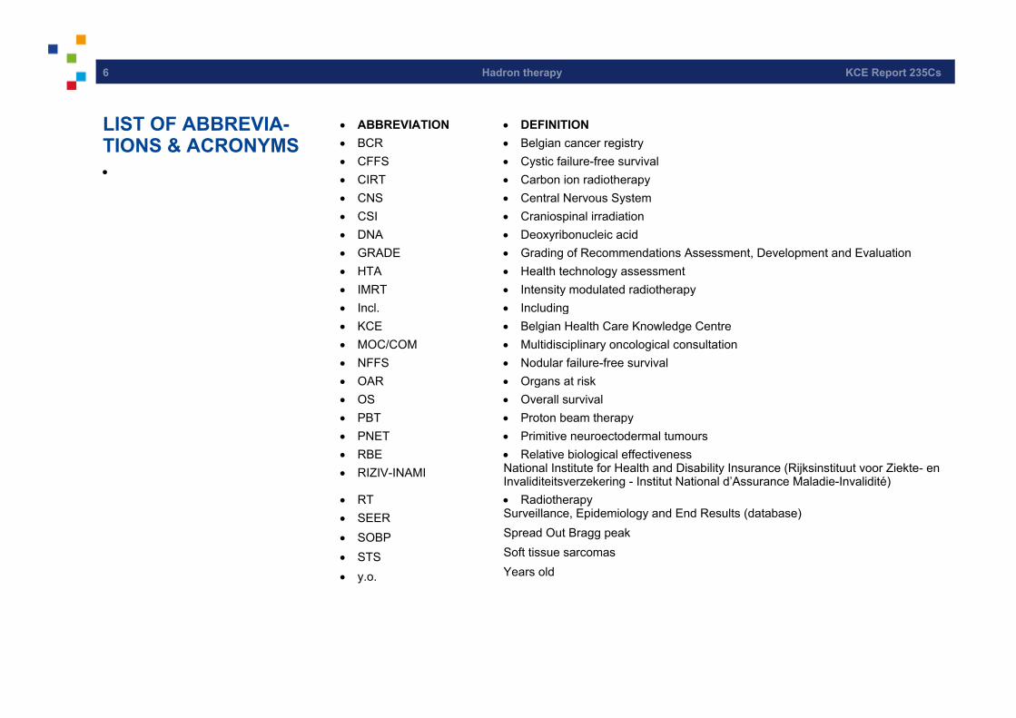
LIST OF ABBREVIA-TIONS & ACRONYMS •
• ABBREVIATION • DEFINITION • BCR • Belgian cancer registry • CFFS • Cystic failure-free survival • CIRT • Carbon ion radiotherapy • CNS • Central Nervous System • CSI • Craniospinal irradiation • DNA • Deoxyribonucleic acid • GRADE • Grading of Recommendations Assessment, Development and Evaluation • HTA • Health technology assessment • IMRT • Intensity modulated radiotherapy • Incl. • Including • KCE • Belgian Health Care Knowledge Centre • MOC/COM • Multidisciplinary oncological consultation • NFFS • Nodular failure-free survival • OAR • Organs at risk • OS • Overall survival • PBT • Proton beam therapy • PNET • Primitive neuroectodermal tumours • RBE • Relative biological effectiveness • RIZIV-INAMI National Institute for Health and Disability Insurance (Rijksinstituut voor Ziekte- en
Invaliditeitsverzekering - Institut National d’Assurance Maladie-Invalidité) • RT • Radiotherapy • SEER Surveillance, Epidemiology and End Results (database)
• SOBP Spread Out Bragg peak
• STS Soft tissue sarcomas • y.o. Years old
KCE Report 235Cs Hadron therapy 7
1 INTRODUCTION Synthesis 1.1 Rationale & research questions
Anno 2014 there are no hadron facilities in Belgium; Belgian citizens eligi-ble for hadron therapy are sent abroad. From September 2014 on (and un-til the end of September 2017), the costs related to hadron therapy (i.e. the treatment, transport and accommodation) are reimbursed through a spe-cially earmarked budget of € 3.6 million per year (an amount that is index-linked). A list of eligible indications for children (and adults) has been de-finedb; this list was based on the Feasibility study of a Hadron Therapy Centre in Belgium (2013)3. The “Agreement Council for Hadron Therapy” (Akkoordraad voor de begeleiding van hadrontherapie/Conseil d'accord pour I'accompagnement de I'hadronthérapie) evaluates every application and decides whether the treatment is reimbursed.
The objective of this study was to evaluate the clinical effectiveness of pro-ton beam (or carbon ion) therapy in the 16 indications in children currently reimbursed by the National Institute for Health and Disability Insurance (RIZIV – INAMI). It concerns the following indications:
Proton beam therapy • Skull base chondrosarcoma • Skull base & (para)spinal chordoma • Craniopharyngioma • Ependymoma • Esthesioneuroblastoma • Ewing sarcoma • CNS germinoma • Low-grade glioma (incl. optic pathway) • Medulloblastoma / Primitive neuroectodermal tumours (PNET) • Non-resectable osteosarcoma • Pelvic sarcoma • Pineal parenchymal tumours (not pineoblastoma)
b
http://www.riziv.fgov.be/nl/professionals/verzorgingsinstellingen/ziekenhuizen/zorg/Paginas/Hadron-english.aspx; for osteosarcoma PBT & CIRT are considered, leading to 16 indications in 15 cancers.
Retinoblastoma Rhabdomyosarcoma (Para)spinal ‘adult type’ soft tissue sarcoma Carbon ion radiotherapy
Non-resectable or incompletely resected high-grade osteosarcoma with or without metastases
1. 2 What is hadron therapy? 2. Hadron therapy or charged particle radiation therapy uses beams of
protons or other charged particles, such as carbon, helium, neon, or sil-icon. At present only protons and carbon ions are in clinical use4. Worldwide, more than 120 000 patients have been treated with particle therapy since 1954: more than 13 000 with carbon ions and more than 105 000 with proton therapy4. Proton beam therapy in children has only been introduced a couple of decades ago; in the US, paediatric patients comprised 13% of all patients treated with PBT in 20125.
Photon radiation (i.e. conventional radiotherapy) deposits most of its ener-gy below the skin surface and in normal tissue going in (‘proximal dose’), hits the target site (the tumour) and still deposits energy and thus affects normal tissues when coming out past the target (‘distal dose’) (Figure 1). In contrast, charged particles deposit a low dose near the surface and a large fraction of their energy at or around the target, at the end of the range of beam penetration. Tissues beyond the tumour location receive very little of the dose. This peak energy delivery is known as the Bragg Peak (Figure 1)6. The absence of radiation distal to the target is one of the major advantages of proton radiotherapy, allowing for substantial tissue sparing. By adjusting the energy of the charged particles and the intensity of the beam, one can deliver pre-specified doses anywhere in the body with high precision7. In this way the proton beam can be adjusted to match the depth and extent of the target volume and excellent conformity can be achieved. Because the Bragg peak of a mono-energetic proton beam is narrow, sev-eral beams with closely spaced penetration depths are used to treat the entirety of the tumour. This area of uniform dose over the entirety of the tumour is termed a Spread Out Bragg Peak (SOBP) (Figure 1). While the SOBP does increase dose deposition proximal to the tumour, the entrance dose usually remains substantially lower than that of photon radiotherapy8.

8 Hadron therapy KCE Report 235Cs
Because charged particles damage cell DNA in qualitatively different ways than photons, the same amount of physical radiation can have much more pronounced biological effects, resulting in larger cellular damage7. The rel-ative biological effectiveness (RBE) is defined as the ratio of a dose of photons to a dose of any particle to produce the same biological effect. The RBE of protons is approximately 1.1, indicating that protons result in approximately 10% more biological damage per unit dose than photons7. Carbon ions have a similar RBE to protons along the particle path but have a markedly increased RBE (estimated at 3-4) at their maximum depth of penetration. As a result, the deleterious effects on normal tissues proximal to the tumour are expected to be similar to proton radiotherapy, while tu-mour killing is enhanced at maximum depth8.
Figure 1 – Radiation dose profiles: photons vs. protons
[Figure – Source: Cotter et al., 2012 p2698 - traduction]
1.2.1 Proton beam therapy
The protons emerging from a cyclotron or synchrotron form a narrow pencil beam; in order to cover a treatment field of the size of a tumour and hence produce a Spread Out Bragg Peak, the pencil beam either scans the target or is scattered by a foil. Currently, both passive scattering and active scan-ning beam delivery systems are in use.
Passive scattering technique (or scatter foil technique)
Passive scattering is currently the most common proton beam technique employed8, 9. A proton beam hits the scatter foil and is spread laterally (Figure 2). The beam is further shaped via brass apertures and compensa-tors to conform to the distal edge of the tumour8. There are several disad-vantages associated with the passive scattering technique; the most im-portant is the production of secondary neutrons, which may induce sec-ondary malignancies9-11. It is estimated that these external neutrons deliver a total-body equivalent dose that is even larger than the leakage radiation from conventional linear accelerators12. Yet, the passive scattering tech-nique may be indicated in those cases where the target has a regular, not too complex shape (G.Goitein, personal communication).
Active scanning technique
There are two types:
Spot-scanning or pencil beam scanning
Only a couple of centres worldwide use this technique where magnets steer a small pencil beam of protons to specific positions within a tumour target without the need for brass apertures or compensators (Figure 2)8. The pencil beam technology has two main advantages over the passive scattering technique. First, it allows for decreasing the entry dose while avoiding an exit dose. Second, the neutron scatter is reduced significantly, an advantage that is particularly important for the paediatric patient8, 9. Yet, pencil beam is more sensitive to any misalignment or density change.
Uniform beam scanning
This technique uses a range modulator, patient collimator and range com-pensator similar to the passive scattering technique, but it utilizes magnets instead of scattering foils to spread the beam laterally13. With this system,

KCE Report 235Cs Hadron therapy 9
the beams are scanned in a fixed pattern with a uniform intensity for each layer, while in the pencil beam scanning system, beams are scanned with variable intensity and pattern14. Overall, the uniform scanning system uses less material in the beam path compared to the passive delivery system and therefore is supposed to produce fewer neutrons13.
Figure 2 – Passive scattering vs. pencil beam (active) scanning
[Figure – Source: Hall 2006 p612]
1.2.2 Carbon ion radiotherapy As carbon ion radiotherapy is hardly used in children and there was only one research question on carbon ion radiotherapy, the interested reader is referred to the Scientific Report for more background information.
1.3 Why proton beam therapy in children? In paediatric radiation oncology, the ultimate goal is to treat the disease while limiting as much as possible the (acute and late) effects of radiation on growth and development, cognition, neuroendocrine function and last but certainly not least the induction of secondary tumours. The age of the paediatric patient plays a major role in the design of the treatment plan. New developments aim at avoiding and/or postponing radiotherapy in chil-dren, e.g. by altering the chemotherapy regimen. Reducing the exposure of normal tissues to therapeutic radiation would presumably decrease the risk of subsequent malignancies and other radiation-induced side effects15. Here, the option of hadron therapy, particularly proton beam therapy, comes in. Essentially, there are two rationales for using proton beam therapy: the dose to organs at risk can be reduced and/ or the risk for second malignan-cies can be lowered, and second, the dose to the tumour can be increased without putting the organs at risk to a higher dose (dose escalation). Alt-hough the latter is appealing, dose-escalation and hypofractionation are experimental approaches that should be restricted to clinical trials.
1.4 Proton beam therapy – the Holy Grail in paediatric radiation oncology?
Despite the thorough physical underpinning of proton beam therapy show-ing a reduction of the radiation dose to normal tissues and organs, sev-eral systematic reviews on the clinical effectiveness of PBT clearly stated that for most clinical indications, it still cannot be concluded that proton beams are clinically truly superior to photon therapy1, 16-19. It remains unproven in the clinic whether protons are more suitable when OAR dose constraints limit the delivery of the most appropriate tumour X-ray radio-therapy doses19. Nor is it known whether proton therapy allows radiation dose escalation without increasing side effects19. What’s more, the clinical application of proton beams still suffers from sev-eral technical limitations and disadvantages, which are elaborated in the Scientific Report. One of the most critical concerns is the production of secondary neutrons with the passive scattering technique as even low neutron doses have a high potential for carcinogenesis20. This is extremely important, in particular because the reduction of secondary cancer risk is in fact one of the principal reasons for the move from photon towards proton beam therapy in children.

10 Hadron therapy KCE Report 235Cs
2 SYSTEMATIC LITERATURE REVIEW A systematic search for relevant publications was done in Medline (through OVID), EMBASE, and the Cochrane Library. Reviews and primary studies on proton beam therapy and/or carbon ion therapy published between 2007 (i.e. end date of the search strategy of the previous KCE Hadron HTA1) up to March 2014 were searched. An overview of the inclusion and exclusion criteria, the search strategy and the flow chart of the selection process are given in the Supplement. A final update of the search (restrict-ed to Medline through OVID) was performed on September 11, 2014.
After selection, we retrieved 21 primary studies on the 16 potential indica-tions under study. On top of the non-randomized, non-controlled and retro-spective nature of the majority of retrieved studies - with the limitations characteristic of these types of studies (e.g. selection bias, recall bias) - all studies suffered from very serious methodological limitations (among oth-ers small sample size, long enrolment period, variable treatment schemes, short follow-up, complications only assessed in a subset of patients) and hence when GRADE2 was applied, the level of scientific evidence for all outcomes in all indications was very low.
2.1 Clinical effectiveness of proton beam therapy and eligibility for radiotherapy/proton beam therapy by tumour type
In the subsequent sections, eligibility for radiotherapy/proton beam therapy (RT/PBT) is based on the report of the multidisciplinary oncological consul-tation where the treatment plan for newly diagnosed cancers is discussed and decided (MOC/COM report).
2.1.1 Skull base chondrosarcoma
Chondrosarcomas are uncommon malignant neoplasms of the cartilage; only 1% of chondrosarcomas arise in the skull base21. Chondrosarcomas are rare in children; when they occur, they tend to be aggressive22. The complete surgical resection of these tumours is most often prevented by their deep location; consequently, a combination of surgery and irradiation has become the mainstay of treatment23.
Incidence in Belgium (2004-11)c Children (0-14 y.o.): <1/year
Eligible for RT/PBT (estimate)d Children (0-14 y.o.): 0
Evidence base PBT 1 retrospective case series (n=7)
Conclusion At present insufficient scientific evidence to support or to refute
c Data provided by the Belgian Cancer Registry. Cave: for some tumour types the indica-
tions under study were slightly redefined. Second, some selection criteria were overlap-ping, resulting in double recordings of some patients. For more details the reader is re-ferred to the Scientific Report.
d Data provided by the Belgian Cancer Registry.

KCE Report 235Cs Hadron therapy 11
2.1.2 Skull base & (para)spinal chordoma
Chordomas are extra-axial tumours that originate from the remnants of the notochord. Chordomas rarely affect children and adolescents24. In children and adolescents surgery is rarely curative because of the difficulty to ob-tain clear margins and the likelihood of chordomas to arise in the skull base, where they are relatively inaccessible to complete surgical exci-sion25. Tumour tissue that remains after surgery, particularly when small in volume, can be managed effectively with radiotherapy24.
Incidence in Belgium (2004-11)c Children (0-14 y.o.): <1/year
Eligible for RT/PBT (estimate)d Children (0-14 y.o.): NA
Evidence base PBT 2 retrospective case series (n=41)
Conclusion At present insufficient scientific evidence to support or to refute
2.1.3 Craniopharyngioma
Craniopharyngiomas are relatively rare intracranial tumours, with a peak incidence occurring at 5-14 years of age26. Despite their histologically be-nign nature, craniopharyngiomas frequently cause profound disabilities due to their proximity to critical structures such as the optic pathway, cere-bral arteries, the hypothalamus, the pituitary gland, cranial nerves and the brain parenchyma26-28,29. There is no consensus on the optimal treatment of newly diagnosed craniopharyngiomas, but surgery and radiotherapy are the cornerstones in their management30. Regardless of the treatment mo-dality, 5- and 10-year overall survival rates in children are greater than 90%31.
Incidence in Belgium (2004-11)c Children (0-14 y.o.): 3/year
Eligible for RT/PBT (estimate)d Children (0-14 y.o.): 1/year
Evidence base PBT 1 retrospective comparative study & 2 retrospective case series (n=74)
Conclusion
At present very low level scientific evidence that PBT compared with IMRT does not result in significant differences in 3-yr OS, 3-yr CFFS, 3-yr NFFS, toxicity or cyst dynamics.
OS: overall survival; CFFS: cystic failure-free survival; NFFS: nodular failure-free survival

12 Hadron therapy KCE Report 235Cs
2.1.4 Ependymoma
Ependymomas are one of the three types of gliomas, tumours of the sup-porting tissue of the brain. In children, most ependymomas arise in or around the fourth ventricle32. One third of cases are diagnosed under the age of three years and the vast majority by age six years8. Standard treat-ment for all grades and ages includes maximal surgical resection and ad-juvant radiotherapy33. For children aged 0-19 years with ependymoma, the overall 5-year relative survival rate is 72.1%34.
Incidence in Belgium (2004-11)c Children (0-14 y.o.): 6/year
Eligible for RT/PBT (estimate)d Children (0-14 y.o.): 4/year
Evidence base PBT 1 prospective case series & 1 ret-rospective case series (n=78)
Conclusion At present insufficient scientific evidence to support or to refute
2.1.5 Esthesioneuroblastoma
Esthesioneuroblastoma, also known as olfactory neuroblastoma, is an un-common malignancy of neural crest origin35, 36. The behaviour of the tu-mour varies from an indolent slow-growing neoplasm to that of a highly aggressive and locally invasive malignancy with a capacity for regional and distant metastases37. Approximately 7% to 20% of patients present at the age of 10 to 24 y.o.35. Surgery and adjuvant radiation therapy have been the mainstay of treatment. Chemotherapy has also been used in combina-tion with surgery and radiation therapy37. Estimated 5-year overall survival rates are 73% for surgery and radiotherapy, 68% for surgery only, 35% for radiotherapy only, and 26% for neither surgery nor radiotherapy38. Incidence in Belgium (2004-11)c Children (0-14 y.o.): <1/year
Eligible for RT/PBT (estimate)d Children (0-14 y.o.): <1/year
Evidence base PBT 1 retrospective case series (n=22e)
Conclusion At present insufficient scientific evidence to support or to refute
e Mixture of children and adults

KCE Report 235Cs Hadron therapy 13
2.1.6 Ewing sarcoma Ewing sarcomas are derived from primordial bone marrow–derived mes-enchymal stem cells. They arise mainly in bone and infrequently in soft tis-sues39. The median age of patients with Ewing sarcoma is 15 years39. Cur-rent treatment consists of a multimodal approach combining surgery, radio-therapy and chemotherapy40, 41. Between 1975 and 2002, the 5-year over-all survival rate has increased from 59% to 76% for children (<15 y.o.) and from 20% to 49% for adolescents (15-19 y.o.)39. Patients with metastatic disease (i.e. 1 out of 4) achieve a 6-year event-free survival of approxi-mately 28% and an overall survival of approximately 30%39. Incidence in Belgium (2004-11)c Children (0-14 y.o.): 8/year
Eligible for RT/PBT (estimate)d Children (0-14 y.o.): 3/year
Evidence base PBT 1 retrospective case series (n=30)
Conclusion At present insufficient scientific evidence to support or to refute
2.1.7 CNS germinoma Central nervous system (CNS) germ cell tumours generally affect adoles-cents42. Two types have been identified: germinomas, which are the most common and carry the most favourable prognosis, and mixed malignant germ cell tumours (also termed non-germinomatous germ cell tumours), which are relatively resistant to therapy43. Germinomas are highly radio-sensitive and have been traditionally treated with radiation therapy alone. Craniospinal irradiation with a boost to the region of the primary tumour has resulted in 5-year overall survival rates greater than 90%44.
Incidence in Belgium (2004-11)c Children (0-14 y.o.): 2/year
Eligible for RT/PBT (estimate)d Children (0-14 y.o.): 2/year
Evidence base PBT 1 retrospective case series (n=22)
Conclusion At present insufficient scientific evidence to support or to refute

14 Hadron therapy KCE Report 235Cs
2.1.8 Low-grade glioma (incl. optic pathway) Any tumour that arises from glial cells is a glioma. Low-grade gliomas are the most common paediatric brain tumour, representing over 30% of all childhood primary brain tumours45. Low-grade gliomas are frequently ame-nable to surgical resection46. Yet, when the risk of post-surgical morbidity is considered too high chemotherapy may be the first line of treatment for children under 7-10 years of age. Radiation therapy is used when tumours progress after chemotherapy or in older children46.
Incidence in Belgium (2004-11)c Children (0-14 y.o.): 47/year
Eligible for RT/PBT (estimate)d Children (0-14 y.o.): 9/year Evidence base PBT 2 retrospective case series (n=38)
Conclusion At present insufficient scientific evidence to support or to refute
2.1.9 Medulloblastoma / primitive neuroectodermal tumours Medulloblastomas and primitive neuroectodermal tumours (PNET) are embryonal tumours, which share the tendency to disseminate throughout the nervous system 47. They occur throughout the paediatric age spectrum, but tend to cluster early in life47. Surgical resection is the mainstay of therapy for all medulloblastoma/PNET. Due to the high metastatic tendency within the CNS, all patients receive “prophylactic” craniospinal irradiation (CSI) for elimination of invisible micrometastases. The 5-year overall survival for children with standard risk medulloblastoma is 75 – 85%48. In the subset of children younger than 5 y.o. long-term disease control is far worse (e.g. ranging from 14% to 55% depending on tumour histology49), although oth-ers reported five-year progression-free and overall survival rates of 85±8% and 95±5%, respectively, in children younger than 3 y.o. with desmoplastic medulloblastoma50. Paediatric PNETs carry an even more dismal progno-sis: the 5-year overall survival ranges between 30 and 40%49.
Incidence in Belgium (2004-11)c Children (0-14 y.o.): 12/year
Eligible for RT/PBT (estimate)d Children (0-14 y.o.): 9/year
Evidence base PBT medullo-blastoma
1 prospective case series & 2 ret-rospective case series (n=147f)
Evidence base PBT PNET None
Conclusion medulloblastoma At present insufficient scientific evidence to support or to refute
Conclusion PNET At present no scientific evidence to support or to refute
f This is an overestimation as some cases may have been reported in 2 publications.
KCE Report 235Cs Hadron therapy 15
2.1.10 Non-resectable osteosarcoma Osteosarcoma is an aggressive, malignant bone-forming mesenchymal tumour, predominantly affecting the long bones of adolescents and young adults. Aggressive local growth and rapid haematogenous systemic dis-semination are typical features. Successful treatment generally requires the combination of effective systemic chemotherapy and complete resection of all clinically detectable disease. Osteosarcomas are generally consid-ered to be radioresistant51, but when complete surgical resection is not possible radiotherapy may be an option to try to extend the progression-free interval52. Local control of the tumour is absolutely critical, because the chances of long term survival are <10% if a complete surgical resection of the tumour is not possible53.
Incidence in Belgium (2004-11)c Children (0-14 y.o.): 9/year
Eligible for RT/PBT (estimate)d Children (0-14 y.o.): <1/year Evidence base PBT 1 retrospective case series (n=55e)
Conclusion At present insufficient scientific evidence to support or to refute
2.1.11 Pelvic sarcomas Treatment of malignant sarcomas of the pelvis poses a challenge for local disease control and oncologic outcome54. Surgical resection is difficult be-cause of the anatomic proximity to many neurovascular structures and the urinary and intestinal tracts and because extensive resection of pelvic sar-comas often necessitates reconstruction to avoid severe functional disabili-ties from the impairment of the load-bearing axis55. At present, there is no consensus yet whether a uniform treatment strategy should be applied to all patients regardless of the histopathology54. Evaluation of the SEERg da-tabase revealed a 5-year overall survival of 47% with osteosarcoma having the worst 5-year survival at 19% and patients with chordoma having the best 5-year survival at 60%54.
Incidence in Belgium (2004-11)c Children (0-14 y.o.): 6/year
Eligible for RT/PBT (estimate)d Children (0-14 y.o.): 2/year
Evidence base PBT None
Conclusion At present no scientific evidence to support or to refute
g The Surveillance, Epidemiology and End Results (SEER) database provides population-
based incidence and survival data for primary malignant tumours collected from 17 registries in the United
States.
16 Hadron therapy KCE Report 235Cs
2.1.12 Pineal parenchymal tumours Tumours originating from the pineal region are very rare; they account for less than 1% of all primary central nervous system tumours56. Pineal parenchymal tumours represent about 10-30% of all tumours in the pineal region57. Treatment may consist of surgery, radiotherapy and/or chemo-therapy. In general, survival of patients with pineal parenchymal tumours is considered much more doubtful compared to that of patients with other pineal region tumours. Evaluation of the SEERg database revealed a 5-year overall survival of 47.2% and a median survival of 4.5 years56.
Incidence in Belgium (2004-11)c Children (0-14 y.o.): <1/year
Eligible for RT/PBT (estimate)d Children (0-14 y.o.): 0/year
Evidence base PBT None
Conclusion At present no scientific evidence to support or to refute
2.1.13 Retinoblastoma Retinoblastoma is a relatively uncommon tumour of childhood that arises in the retina; 95% of cases are diagnosed before age 5 years, and two-thirds of these cases occur before age 2 years. Due to the radiosensitive nature of retinoblastomas, external beam radiation therapy (EBRT) has been thought to be the first line and major treatment method for retinoblas-toma58. However, EBRT may result in orbital bone growth retardation and consequent cosmetic problems, particularly in younger children. Therefore, treatment modalities were shifted toward primary systemic chemotherapy for reducing tumour volume initially (chemo reduction) and additional focal treatment such as cryotherapy, thermotherapy, or brachytherapy58. Ac-cording to estimates based on the SEERg database current 5-year survival rate may be as high as 96.5% (1995–2004)59.
Incidence in Belgium (2004-11)c Children (0-14 y.o.): 12/year
Eligible for RT/PBT (estimate)d Children (0-14 y.o.): 1/year
Evidence base PBT 1 retrospective comparative study (n=55)
Conclusion At present there is very low level scientific evidence that PBT re-sults in lower risk of developing RT-induced in-field secondary malig-nancies. However, since radiation-induced solid malignancies need at least 5 to 10 years to develop and for some children in the study the follow-up was short, the results should be interpreted with cau-tion.

KCE Report 235Cs Hadron therapy 17
2.1.14 Rhabdomyosarcoma Rhabdomyosarcomas are malignancies of mesenchymal cell origin that arise primarily in striated muscle tissues8, 60. In children, the most common primary sites are the orbit (i.e. 35-45% of all childhood rhabdomyosar-coma)61 and the genito-urinary tract62. There is a bimodal incidence distri-bution with a first peak at 6 y.o. and a second peak at adolescence8. Rhabdomyosarcomas require a multidisciplinary approach including sur-gery, chemotherapy and radiotherapy63. Prognosis depends on the histo-logic type and the tumour site63. The overall impression is that survival for most patient subsets is superior with the use of early local therapy, includ-ing RT62.
Incidence in Belgium (2004-11)c Children (0-14 y.o.): 9/year
Eligible for RT/PBT (estimate)d Children (0-14 y.o.): 4/year
Evidence base PBT 3 retrospective case series (n=36)
Conclusion At present insufficient scientific evidence to support or to refute
2.1.15 (Para-)spinal ‘adult type’ soft tissue sarcoma (STS) The most common STS in children younger than 15 y.o. is rhabdomyosar-coma; the remaining soft tissue sarcomas are commonly referred to as non-rhabdomyosarcomatous STS and account for about 3% of all child-hood tumours. The latter are characterized by local aggressiveness and a propensity to metastasize that is correlated to their grade of malignancy64. Radiotherapy plays a dominant role in those tumours which cannot be sur-gically removed without leading to major impairment, yet, it may cause se-vere late side effects. Five year overall survival in children and adolescents with non-rhabdomyosarcomatous STS may be as high as 89% in patients who underwent complete resection at diagnosis, 79% in patients with mar-ginal resection, 52% in initially unresected patients and 17% in patients with metastases at onset64.
Incidence in Belgium (2004-11)c Children (0-14 y.o.): <1/year
Eligible for RT/PBT (estimate)d Children (0-14 y.o.): <1/year
Evidence base PBT None
Conclusion At present no scientific evidence to support or to refute
18 Hadron therapy KCE Report 235Cs
2.2 Clinical effectiveness of carbon ion radiotherapy and eligibility for radiotherapy/carbon ion radiotherapy
Non-resectable osteosarcoma
(For the pathology description, the reader is referred to paragraph 0)
Incidence in Belgium (2004-11)c Children (0-14 y.o.): 9/year
Eligible for RT/CIRT (estimate)d Children (0-14 y.o.): <1/year
Evidence base CIRT 1 retrospective case series (n=78e)
Conclusion At present insufficient scientific evidence to support or to refute
3 DISCUSSION Due to its physical properties, proton therapy spares more normal tissues and organs at risk than conventional radiotherapy. Because a reduction of radiation dose to the healthy tissue is the goal of radioprotection, it is con-ceivable that a decrease of radiation dose to vulnerable tissues by using protons will decrease important side effects and radiation-induced cancers as well. Worldwide a growing number of children is being treated with proton beam therapy (PBT). Yet, we have no Belgian data and a European registry has not been installed yet. A survey among all American proton centres showed that in 2012 a total of 694 paediatric patients were treated5. The six most common tumour types treated were ependymoma, medulloblastoma, low-grade glioma, rhabdomyosarcoma, Ewing sarcoma, and craniopharyngio-ma5; indications for which we found either no or insufficient scientific evi-dence to support or to refute proton beam therapy. It is appalling that only a fraction of children treated with PBT are en-rolled in clinical trials65. There may be several reasons for that, including the fact that many clinicians are convinced that the superior dose distribu-tion and lower integral dose makes proton beam therapy the preferred treatment option, and thus making them reluctant to randomize patients. Furthermore, long-term follow-up, crucial to assess late side effects as well as secondary cancer risk, may be difficult when patients come from large distances or from abroad and will take over a decade. While multicentre studies are definitely the only possible way to get more da-ta on the clinical effectiveness of proton beam therapy, the (international) collaboration between centres is not going without a hitch. In fact, there seems to be some competition between them and funding for this type of research is also lacking. In the medical literature animated debates have been held on the necessity or ethical justification of randomized controlled trials to test proton beam therapy66-69. Given the fact that systematic reviews fail to demonstrate clear evidence of a clinical superiority for protons, it is difficult to under-stand why it would be unethical to perform randomized trials67, 69, except in those cases where there are manifest anatomical and physical reasons against the use of photons (e.g. low-grade glioma, craniopharyngioma, skull base chordoma and skull base osteosarcoma). Most certainly, for prevalent indications (e.g. in adults), there should be no discussion on the

KCE Report 235Cs Hadron therapy 19
necessity of proving PBT’s superiority and cost-effectiveness through ran-domized clinical trials. For children (and for adults with rare cancers), some mitigating factors may apply: in addition to the factors mentioned before, the number of children with cancer requiring radiotherapy as part of their treatment is so small that it is unlikely that prospective randomized trials can be conducted to test if different dose distributions indeed make a clinical difference70. Furthermore, as was pointed out earlier, the clinical application of proton beam therapy still has to contend with serious technical limitations and disadvantages: the magnitude of the lateral penumbra, the uncertainty about the distal edge degradation, range inaccuracies, patient-position re-lated uncertainties, operational difficulties and last but not least cost-effectiveness issues. With an extra cost of 70% to 150%71, 72, the payer - whether public or private - deserves to know how much better the out-comes are. As the treatment of children demands specific skills and precautions (e.g. anaesthesia is required in nearly half of the children5), the concentra-tion of children in a restricted number of centres should be mandatory. Quality assurance is another important aspect not to be neglected. Yet, high quality can only be delivered if the operators have sufficient time; economic pressure to increase the throughput of the machine should never prevail. The protocols being developed by the Particle Therapy Co-Operative Group (PTCOG)h are an important initiative in that respect. Prospective comparative clinical trials in the field are urgently needed. In addition, the establishment of a European Hadron Therapy Registry (EHTR), which holds (anonymised) data on patients treated by European hadron centres would provide a simple but effective solution to the current lack of coherent published data73. In the US the Pediatric Proton Consorti-um Registry (PPCR) was recently installed for that purpose74.
h http://www.ptcog.ch/index.php/clinical-protocols
4 KEY MESSAGES • Based on the 2004-2011 incidence data, it can be estimated
that in Belgium 37 children (0-14 y.o.) and 14 adolescents (15-19 y.o.) may be eligible for RT/PBT on a yearly basis.
• The use of PBT in children is supported by physical data showing an important reduction of the radiation dose to normal tissues. Yet, to date clinical data on PBT in all paediatric cancers under study is lacking critical information on measures of long-term effectiveness and harm.

20 Hadron therapy KCE Report 235Cs
RECOMMENDATIONSi
To the clinicians:
• Patients (or their parents or representatives) should be fully informed that despite the physical underpinning of proton beam therapy, its clinical efficacy for the indications considered in this report has not yet been confirmed in clinical studies.
• Children should be referred to proton beam centres with the necessary expertise in treating children with that specific pathology and involved in clinical studies with long-term follow-up (if recruiting in Europe).
• The registration in the Belgian Cancer Registry (BCR) database of the chemotherapy regimen and radiotherapy schedule (including hadron therapy) administered in children is recommended. This registration can allow, amongst others, the monitoring of secondary malignancies occurrence.
• To the Technical Medical Council & the Insurance Committee of the RIZIV - INAMI: • The current reimbursement for PBT should be reevaluated periodically as new scientific
evidence on effectiveness and safety becomes available. Meanwhile, the 15-year age limit should be reconsidered for certain indications.
• The amount reimbursed for radiotherapy in children should take into account the complexity of treatment administration, including the potential need for anaesthesia. The reimbursement should be made conditional to the registration into the BCR database.
• To the RIZIV - INAMI, BCR & FANC - AFCN and scientific/professional associations: • Our country should actively promote the set-up of a European Hadron Therapy Registry.
Research agenda:
• There is an urgent need for more research, not only on the clinical efficacy, side effects, and harms, but also on the economical aspects, and on the physics and biology. Clinical research should preferentially be conducted in an internationally coordinated way.
i The KCE has sole responsibility for the recommendations.
KCE Report 235Cs Hadron therapy 21
REFERENCES 1. Huybrechts M, Obyn C, Gailly J, Mambourg F, Vinck I, Ramaekers D. Hadrontherapy (Structured abstract). Health Technology Assessment Database. 2007(1).
2. Guyatt G, Oxman AD, Sultan S, Brozek J, Glasziou P, Alonso-Coello P, et al. GRADE guidelines: 11. Making an overall rating of confidence in effect estimates for a single outcome and for all outcomes. J Clin Epidemiol. 2013;66(2):151-7.
3. 3Annemans L, Colardyn F, De Croock R, De Neve W, Duprez F, Gulyban A, et al. Feasability study of a Hadron therapy centre in Belgium. Brussels: 2013.
4. Jermann M. Particle therapy statistics in 2013. International Journal of Particle Therapy. 2014;1(1):4.
5. Chang AL, Yock TI, Mahajan A, Hill-Kaiser C, Keole S, Loredo L, et al. Pediatric proton therapy: patterns of care across the United States. International Journal of Particle Therapy. 2014;1(2):11.
6. Larsson B, Leksell L, Rexed B, Sourander P, Mair W, Andersson B. The high-energy proton beam as a neurosurgical tool. Nature. 1958;182(4644):1222-3.
7. Terasawa T, Dvorak T, Ip S, Raman G, Lau J, Trikalinos TA. Systematic review: Charged-particle radiation therapy for cancer. Ann. Intern. Med. 2009;151(8):556-65.
8. Cotter SE, McBride SM, Yock TI. Proton radiotherapy for solid tumors of childhood. Technol. Cancer Res. Treat. 2012;11(3):267-78.
9. Timmermann B. Proton beam therapy for childhood malignancies: Status report. Klin. Padiatr. 2010;222(3):127-33.
10. Brenner DJ, Hall EJ. Secondary neutrons in clinical proton radiotherapy: a charged issue. Radiother Oncol. 2008;86(2):165-70.
11. Amsbaugh MJ, Zhu XR, Palmer M, Poenisch F, McAleer MF, Mahajan A, et al. Spot scanning proton therapy for craniopharyngioma. Pract. Radiat. Oncol. 2012;2(4):314-8.
12. Hall EJ. Intensity-modulated radiation therapy, protons, and the risk of second cancers. Int J Radiat Oncol Biol Phys. 2006;65(1):1-7.
13. Islam MR, Collums TL, Zheng Y, Monson J, Benton ER. Off-axis dose equivalent due to secondary neutrons from uniform scanning proton beams during proton radiotherapy. Phys Med Biol. 2013;58(22):8235-51.

22 Hadron therapy KCE Report 235Cs
14. Zheng Y, Liu Y, Zeidan O, Schreuder AN, Keole S. Measurements of neutron dose equivalent for a proton therapy center using uniform scanning proton beams. Med Phys. 2012;39(6):3484-92.
15. Bekelman JE, Schultheiss T, Berrington De Gonzalez A. Subsequent malignancies after photon versus proton radiation therapy. Int J Radiat Oncol Biol Phys. 2013;87(1):10-2.
16. Olsen DR, Bruland OS, Frykholm G, Norderhaug IN. Proton therapy - a systematic review of clinical effectiveness. Radiother Oncol. 2007;83(2):123-32.
17. Brada M, Pijls-Johannesma M, De Ruysscher D. Current clinical evidence for proton therapy. Cancer J. 2009;15(4):319-24.
18. Ramaekers BL, Pijls-Johannesma M, Joore MA, Ende P, Langendijk JA, Lambin P, et al. Systematic review and meta-analysis of radiotherapy in various head and neck cancers: comparing photons, carbon-ions and protons (Structured abstract). Cancer Treatment Reviews. 2011;37(3):185-201.
19. De Ruysscher D, Mark Lodge M, Jones B, Brada M, Munro A, Jefferson T, et al. Charged particles in radiotherapy: a 5-year update of a systematic review. Radiother Oncol. 2012;103(1):5-7.
20. Measurements. NCoRP. The Relative Biological Effectiveness of Radiations of Different Quality - Report 104. 1990.
21. Bloch O, Parsa AT. Skull Base Chondrosarcoma. Evidence-Based Treatment Paradigms. Neurosurg. Clin. North Am. 2013;24(1):89-96.
22. Mavrogenis AF, Gambaroti M, Angelini A, Palmerini E, Staals EL, Ruggieri P, et al. Chondrosarcomas revisited. Orthopedics. 2012;35(3):e379-e90.
23. Bloch OG, Jian BJ, Yang I, Han SJ, Aranda D, Ahn BJ, et al. A systematic review of intracranial chondrosarcoma and survival. J Clin Neurosci. 2009;16(12):1547-51.
24. Walcott BP, Nahed BV, Mohyeldin A, Coumans JV, Kahle KT, Ferreira MJ. Chordoma: Current concepts, management, and future directions. Lancet Oncol. 2012;13(2):e69-e76.
25. National Cancer Institute. Other Rare Unusual Cancers of Childhood [Web page]. National Institutes of Health 2014 [updated 2014-08-10; cited 2014-07-11]. Available from:
http://www.cancer.gov/cancertopics/pdq/treatment/unusual-cancers-childhood/patient/page7
26. Iannalfi A, Fragkandrea I, Brock J, Saran F. Radiotherapy in craniopharyngiomas. Clin. Oncol. 2013;25(11):654-67.
27. Laffond C, Dellatolas G, Alapetite C, Puget S, Grill J, Habrand JL, et al. Quality-of-life, mood and executive functioning after childhood craniopharyngioma treated with surgery and proton beam therapy. Brain Inj. 2012;26(3):270-81.
28. Muller HL. Childhood craniopharyngioma. Pituitary. 2013;16(1):56-67.
29. Luu QT, Loredo LN, Archambeau JO, Yonemoto LT, Slater JM, Slater JD. Fractionated proton radiation treatment for pediatric craniopharyngioma: preliminary report. Cancer Journal. 2006;12(2):155-9.
30. Kortmann RD. Different approaches in radiation therapy of craniopharyngioma. Frontiers in Endocrinology. 2011;2(100).
31. National Cancer Institute. Childhood Craniopharyngioma Treatment [Web page]. National Institutes of Health;2014 [updated 2014-08-12; cited 2014-07-11]. Available from: http://www.cancer.gov/cancertopics/pdq/treatment/child-cranio/healthprofessional
32. National Cancer Institute. General Information About Childhood Ependymoma [Web page]. National Institutes of Health 2014 [updated 2014-07-10; cited 2014-08-14]. Available from: http://www.cancer.gov/cancertopics/pdq/treatment/childependymoma/Patient
33. Wright KD, Gajjar A. Current treatment options for pediatric and adult patients with ependymoma. Curr. Treat. Options Oncol. 2012;13(4):465-77.
34. CBTRUS. CBTRUS Statistical Report: Primary Brain and Central Nervous System Tumors Diagnosed in the United States in 2004-2008 (March 23, 2012 Revision). 2012. Available from: www.cbtrus.org
35. Mahooti S, Wakely PE, Jr. Cytopathologic features of olfactory neuroblastoma. Cancer. 2006;108(2):86-92.

KCE Report 235Cs Hadron therapy 23
36. Soler ZM, Smith TL. Endoscopic versus open craniofacial resection of esthesioneuroblastoma: what is the evidence? Laryngoscope. 2012;122(2):244-5.
37. 3Bak M, Wein RO. Esthesioneuroblastoma: a contemporary review of diagnosis and management. Hematol Oncol Clin North Am. 2012;26(6):1185-207.
38. Platek ME, Merzianu M Fau - Mashtare TL, Mashtare Tl Fau - Popat SR, Popat Sr Fau - Rigual NR, Rigual Nr Fau - Warren GW, Warren Gw Fau - Singh AK, et al. Improved survival following surgery and radiation therapy for olfactory neuroblastoma: analysis of the SEER database. Radiat Oncol. 2011;6(41):6-41.
39. National Cancer Institute. Ewing Sarcoma Treatment [Web page]. National Institutes of Health 2014 [updated 2014-08-15; cited 2014-08-14]. Available from: http://www.cancer.gov/cancertopics/pdq/treatment/ewings/HealthProfessional
40. Gray ST, Chen YL, Lin DT. Efficacy of proton beam therapy in the treatment of ewing's sarcoma of the paranasal sinuses and anterior skull base. Skull Base. 2009;19(6):409-16.
41. Iwata S, Yonemoto T, Ishii T, Kumagai K, Imai R, Hagiwara Y, et al. Efficacy of carbon-ion radiotherapy and high-dose chemotherapy for patients with unresectable Ewing's sarcoma family of tumors. Int J Clin Oncol. 2013;18(6):1114-8.
42. MacDonald SM, Trofimov A, Safai S, Adams J, Fullerton B, Ebb D, et al. Proton radiotherapy for pediatric central nervous system germ cell tumors: Early clinical outcomes. Int. J. Radiat. Oncol. Biol. Phys. 2011;79(1):121-9.
43. Robertson PL, Jakacki R, Hukin J, Siffert J, Allen JC. Multimodality therapy for CNS mixed malignant germ cell tumors (MMGCT): results of a phase II multi-institutional study. J Neurooncol. 2014;118(1):93-100.
44. National Cancer Institute. Childhood Central Nervous System Germ Cell Tumors Treatment [Web page]. National Institutes of Health 2014 [updated 2014-08-14; cited 2014-05-09]. Available from: http://www.cancer.gov/cancertopics/pdq/treatment/childCNS-germ-cell/healthprofessional
45. Minturn JE, Fisher MJ. Gliomas in children. Curr. Treat. Options Neurol. 2013;15(3):316-27.
46. Rombi B MS, Maurizio A, Tarbell NJ, Yock TI. Proton Radiotherapy for Childhood Tumors: an Overview of Early Clinical Results. Nucl Med radiat Ther. 2013;4(4):1-9.
47. National Cancer Institute. Childhood Central Nervous System Embryonal Tumors Treatment [Web page]. National Institutes of Health 2014 [updated 2014-05-02; cited 2014-08-07]. Available from: http://www.cancer.gov/cancertopics/pdq/treatment/childCNSembryonal/healthprofessional
48. Brodin NP, Munck Af Rosenschold P, Aznar MC, Kiil-Berthelsen A, Vogelius IR, Nilsson P, et al. Radiobiological risk estimates of adverse events and secondary cancer for proton and photon radiation therapy of pediatric medulloblastoma. Acta Oncol. 2011;50(6):806-16.
49. Jimenez RB, Sethi R, Depauw N, Pulsifer MB, Adams J, McBride SM, et al. Proton radiation therapy for pediatric medulloblastoma and supratentorial primitive neuroectodermal tumors: Outcomes for very young children treated with upfront chemotherapy. Int. J. Radiat. Oncol. Biol. Phys. 2013;87(1):120-6.
50. Rutkowski S, Bode U, Deinlein F, Ottensmeier H, Warmuth-Metz M, Soerensen N, et al. Treatment of early childhood medulloblastoma by postoperative chemotherapy alone. N Engl J Med. 2005;352(10):978-86.
51. Oya N, Kokubo M, Mizowaki T, Shibamoto Y, Nagata Y, Sasai K, et al. Definitive intraoperative very high-dose radiotherapy for localized osteosarcoma in the extremities. Int J Radiat Oncol Biol Phys. 2001;51(1):87-93.
52. Group EESNW, Blay J-Y, Blomqvist C, Bonvalot S, Boukovinas I, Casali PG, et al. Bone sarcomas: ESMO Clinical Practice Guidelines for diagnosis, treatment and follow-up. Ann Oncol. 2012;23 Suppl 7:vii100-9.
53. Blattmann C, Oertel S, Schulz-Ertner D, Rieken S, Haufe S, Ewerbeck V, et al. Non-randomized therapy trial to determine the safety and efficacy of heavy ion radiotherapy in patients with non-resectable osteosarcoma. BMC Cancer. 2010;10.
24 Hadron therapy KCE Report 235Cs
54. Jawad MU, Haleem AA, Scully SP. Malignant sarcoma of the pelvic bones: treatment outcomes and prognostic factors vary by histopathology. Cancer. 2011;117(7):1529-41.
55. Han I, Lee YM, Cho HS, Oh JH, Lee SH, Kim H-S. Outcome after surgical treatment of pelvic sarcomas. Clin Orthop Surg. 2010;2(3):160-6.
56. Al-Hussaini M, Sultan I, Abuirmileh N, Jaradat I, Qaddoumi I. Pineal gland tumors: experience from the SEER database. J Neurooncol. 2009;94(3):351-8.
57. Stoiber EM, Schaible B, Herfarth K, Schulz-Ertner D, Huber PE, Debus J, et al. Long term outcome of adolescent and adult patients with pineal parenchymal tumors treated with fractionated radiotherapy between 1982 and 2003--a single institution's experience. Radiat Oncol. 2010;5:122.
58. Chang JW, Yu YS, Kim JY, Shin DH, Choi J, Kim JH, et al. The clinical outcomes of proton beam radiation therapy for retinoblastomas that were resistant to chemotherapy and focal treatment. Korean J Ophthalmol. 2011;25(6):387-93.
59. Broaddus E, Topham A, Singh AD. Survival with retinoblastoma in the USA: 1975-2004. Br J Ophthalmol. 2009;93(1):24-7.
60. Punyko JA MA, Baker KS, Ness KK, Robison LL, Gurney JG. Long-term survival probabilities for childhood rhabdomyosarcoma. A population-based evaluation. Cancer 2005;103(7):1475-83.
61. Ge X, Huang DS, Shi JT, Ma JM. Multidisciplinary collaborative therapy for 30 children with orbital rhabdomyosarcoma. Asian Pac J Cancer Prev. 2013;14(8):4641-6.
62. National Cancer Institute. Childhood Rhabdomyosarcoma Treatment [Web page]. National Institutes of Health 2014 [updated 2014-08-19; cited 2014-06-13]. Available from: http://www.cancer.gov/cancertopics/pdq/treatment/childrhabdomyosarcoma/HealthProfessional
63. Jurdy L, Merks JHM, Pieters BR, Mourits MP, Kloos RJHM, Strackee SD, et al. Orbital rhabdomyosarcomas: A review. Saudi J. Ophthalmol. 2013;27(3):167-75.
64. Ferrari A, Casanova M, Collini P, Meazza C, Luksch R, Massimino M, et al. Adult-type soft tissue sarcomas in pediatric-age patients: experience at the Istituto Nazionale Tumori in Milan. J Clin Oncol. 2005;23(18):4021-30.
65. Merchant TE. Clinical controversies: Proton therapy for pediatric tumors. Semin. Radiat. Oncol. 2013;23(2):97-108.
66. 66. Goitein M, Cox JD. Should randomized clinical trials be required for proton radiotherapy? J Clin Oncol. 2008;26(2):175-6.
67. Glatstein E, Glick J, Kaiser L, Hahn SM. Should randomized clinical trials be required for proton radiotherapy? An alternative view. J Clin Oncol. 2008;26(15):2438-9.
68. Tepper JE. Protons and parachutes. J Clin Oncol. 2008;26(15):2436-7.
69. Macbeth FR, Williams MV. Proton therapy should be tested in randomized trials. J Clin Oncol. 2008;26(15):2590-1; author reply 3-6.
70. Halperin EC. Randomized prospective trials of innovative radiotherapy technology are necessary. J Am Coll Radiol. 2009;6(1):33-7.
71. Lievens Y, Van den Bogaert W. Proton beam therapy: too expensive to become true? Radiother Oncol. 2005;75(2):131-3.
72. Goitein M, Jermann M. The relative costs of proton and X-ray radiation therapy. Clin Oncol (R Coll Radiol). 2003;15(1):S37-50.
73. Lodge M, Pijls-Johannesma M, Stirk L, Munro AJ, De Ruysscher D, Jefferson T. A systematic literature review of the clinical and cost-effectiveness of hadron therapy in cancer>. Radiother. Oncol. 2007;83(2):110-22.
74. Kasper HBR, L.; Indelicato, D.J.; Symecko, H.; Hartsell, W.; Mahajan, A.; Hill-Kayser, C.; Perkins, S.M.; Chang, A.L.; Childs, S.; Buchsbaum, J.C.; Laurie, F.; Khan, A.J.; Giraud, C.; Yeap, B.Y.; Yock, T.I. The Pediatric Proton Consortium Registry: A Multi-institutional Collaboration in U.S. Proton Centers. International Journal of Particle Therapy. 2014;1(2):11.


![Endbericht - eprints.hta.lbg.ac.ateprints.hta.lbg.ac.at/801/1/HTA-Projektbericht_016.pdf · klinischen Pfades auch vermehrt in Europa zu etablieren [4]. Eine Erhe-bung von AnwenderInnen](https://static.fdokument.com/doc/165x107/5e1f3d60a9edf96fdc76f98a/endbericht-klinischen-pfades-auch-vermehrt-in-europa-zu-etablieren-4-eine-erhe-bung.jpg)