How to Face Skin Cancer with Nanomaterials: A Review
25
https://biointerfaceresearch.com/ 11931 Review Volume 11, Issue 4, 2021, 11931 - 11955 https://doi.org/10.33263/BRIAC114.1193111955 How to Face Skin Cancer with Nanomaterials: A Review Fakhara Sabir 1 , Mahmood Barani 2 , Abbas Rahdar 3, * , Muhammad Bilal 4 , Muhammad Nadeem Zafar 5 , Simona Bungau 6 , George Z. Kyzas 7, * 1 University of Szeged, Faculty of Pharmacy, Institute of Pharmaceutical Technology and Regulatory Affairs, Szeged, H- 6720, Hungary; [email protected] (F.S.); 2 Department of Chemistry, Shahid Bahonar University of Kerman, Kerman 76169-14111, Iran; [email protected] (M.B.); 3 Department of Physics, University of Zabol, Zabol 98613-35856, Iran; [email protected] (A.R.); 4 School of Life Science and Food Engineering, Huaiyin Institute of Technology, Huaian 223003, China; [email protected] (M.B.); 5 Department of Chemistry, University of Gujrat, Gujrat 50700, Pakistan; [email protected] (M.N.Z.); 6 Department of Pharmacy, Faculty of Medicine and Pharmacy, University of Oradea, Oradea 410028, Romania; [email protected] (S.B.); 7 Department of Chemistry, International Hellenic University, Kavala 65404, Greece; [email protected] (G.Z.K.); * [email protected] (G.Z.K.); [email protected] (A.R.); Scopus Author ID 17345938100 (G.Z.K.); 36599143100 (A.R.); Received: 25.11.2020; Revised: 16.12.2020; Accepted: 17.12.2020; Published: 21.12.2020 Abstract: Nanotechnology is one of the most attractive disciplines ranging from materials science to biomedicine. It is one of the most prominent areas of research for targeting skin melanoma and other diseases. The application of nanomaterials in the field of skin has increased interest in topical targeting to increase the permeation and retention and reduce its side effects. Different types of nanomaterials have been used for the application of skin diseases and cancer. These nanomaterials include carbon- nanotubes, liposomes, nanoemulsion, nano micelles, etc. In the United States and globally, the prevalence of melanoma has had a growing rise throughout the last 50 years. This review seeks to discuss various types of treatment and diagnosis of early-stage skin cancer. There are many skin cancer biomarkers for early detection of this type of cancer. Diagnosis of skin cancer forms a way of traditional approaches, and their weaknesses are discussed. Electrochemical and optical biosensors for the diagnosis and monitoring of skin cancer are also was described. After all, some issues should be addressed and overcome regarding the competition of new techniques in cost and convenience of use. Keywords: nanomaterials; skin; cancer; nanoparticles; diagnosis; treatment. © 2020 by the authors. This article is an open-access article distributed under the terms and conditions of the Creative Commons Attribution (CC BY) license (https://creativecommons.org/licenses/by/4.0/). 1. Introduction Skin cancer is a broad term that describes a large number of different malignant lesions of the skin. It can be further categorized into two most common tumors: keratinocyte cancer (also recognized as non-melanoma skin cancer) and melanoma skin cancer. Among these, non- melanoma skin cancer is a widely detected type of cancer globally. In contrast, melanoma skin cancer results in the highest percentage of deaths [1]. The skin's rarer tumors, like cutaneous lymphoma, Kaposi sarcoma, and Merkel Cell Carcinoma, have also been described [2]. Recent epidemiological data revealed the diagnosis of over 5.4 million cases of skin cancer in the United States in 2012 [3]. In the U.S., skin cancer frequency has dramatically increased (>300%) between 1994 and 2014. In 2018, the highest incidence of skin cancer was reported in Australia, affecting approximately 300/100,000 people [4]. Therefore, skin cancer
Transcript of How to Face Skin Cancer with Nanomaterials: A Review

https://biointerfaceresearch.com/ 11931
Review
Volume 11, Issue 4, 2021, 11931 - 11955
https://doi.org/10.33263/BRIAC114.1193111955
How to Face Skin Cancer with Nanomaterials: A Review
Fakhara Sabir 1, Mahmood Barani 2 , Abbas Rahdar 3, *, Muhammad Bilal 4 , Muhammad Nadeem
Zafar 5 , Simona Bungau 6 , George Z. Kyzas 7, *
1 University of Szeged, Faculty of Pharmacy, Institute of Pharmaceutical Technology and Regulatory Affairs, Szeged, H-
6720, Hungary; [email protected] (F.S.); 2 Department of Chemistry, Shahid Bahonar University of Kerman, Kerman 76169-14111, Iran;
[email protected] (M.B.); 3 Department of Physics, University of Zabol, Zabol 98613-35856, Iran; [email protected] (A.R.); 4 School of Life Science and Food Engineering, Huaiyin Institute of Technology, Huaian 223003, China;
[email protected] (M.B.); 5 Department of Chemistry, University of Gujrat, Gujrat 50700, Pakistan; [email protected] (M.N.Z.); 6 Department of Pharmacy, Faculty of Medicine and Pharmacy, University of Oradea, Oradea 410028, Romania;
[email protected] (S.B.); 7 Department of Chemistry, International Hellenic University, Kavala 65404, Greece; [email protected] (G.Z.K.);
* [email protected] (G.Z.K.); [email protected] (A.R.);
Scopus Author ID 17345938100 (G.Z.K.); 36599143100 (A.R.);
Received: 25.11.2020; Revised: 16.12.2020; Accepted: 17.12.2020; Published: 21.12.2020
Abstract: Nanotechnology is one of the most attractive disciplines ranging from materials science to
biomedicine. It is one of the most prominent areas of research for targeting skin melanoma and other
diseases. The application of nanomaterials in the field of skin has increased interest in topical targeting
to increase the permeation and retention and reduce its side effects. Different types of nanomaterials
have been used for the application of skin diseases and cancer. These nanomaterials include carbon-
nanotubes, liposomes, nanoemulsion, nano micelles, etc. In the United States and globally, the
prevalence of melanoma has had a growing rise throughout the last 50 years. This review seeks to
discuss various types of treatment and diagnosis of early-stage skin cancer. There are many skin cancer
biomarkers for early detection of this type of cancer. Diagnosis of skin cancer forms a way of traditional
approaches, and their weaknesses are discussed. Electrochemical and optical biosensors for the
diagnosis and monitoring of skin cancer are also was described. After all, some issues should be
addressed and overcome regarding the competition of new techniques in cost and convenience of use.
Keywords: nanomaterials; skin; cancer; nanoparticles; diagnosis; treatment.
© 2020 by the authors. This article is an open-access article distributed under the terms and conditions of the Creative
Commons Attribution (CC BY) license (https://creativecommons.org/licenses/by/4.0/).
1. Introduction
Skin cancer is a broad term that describes a large number of different malignant lesions
of the skin. It can be further categorized into two most common tumors: keratinocyte cancer
(also recognized as non-melanoma skin cancer) and melanoma skin cancer. Among these, non-
melanoma skin cancer is a widely detected type of cancer globally. In contrast, melanoma skin
cancer results in the highest percentage of deaths [1]. The skin's rarer tumors, like cutaneous
lymphoma, Kaposi sarcoma, and Merkel Cell Carcinoma, have also been described [2]. Recent
epidemiological data revealed the diagnosis of over 5.4 million cases of skin cancer in the
United States in 2012 [3]. In the U.S., skin cancer frequency has dramatically increased
(>300%) between 1994 and 2014. In 2018, the highest incidence of skin cancer was reported
in Australia, affecting approximately 300/100,000 people [4]. Therefore, skin cancer

https://doi.org/10.33263/BRIAC114.1193111955
https://biointerfaceresearch.com/ 11932
characterizes an extensive global health burden with huge psychosocial effects and necessitates
significant attention in terms of technologies and treatments.
Various choices, including surgery, radiotherapy, and chemotherapy, are available to
treat skin cancer. Surgical procedures for treating skin cancers include simple excision of tumor
cells from the skin, shave-excision (abnormal skin area is removed with a small blade), Mohs
surgery (excision of a thin layer of skin from the tumor region followed by viewing under a
microscope to inspect the presence/absence of tumor cells), cryosurgery (freezing of localized
affected tissue and excision by surgery), and curettage and desiccation (treatment to remove
affected skin surface with a curette) [5]. Radiation therapy can be classified into conventional
external radiation therapy, superficial x-ray therapy, and brachytherapy. Volumetric modulated
arc therapy is the latest and novel radiation modality that ensures the continuous delivery of
radiation dose to targeted tumor tissue while circumventing or minimizing the dose to healthy
tissue around the tumor microenvironment [6]. Nevertheless, the lack of histological clearance,
the evolution of undesirable phenotypes in recurrent tumors, and elevated costs are significant
drawbacks associated with the radiation technique [7]. Chemotherapy implicates the use of
potent chemicals in the body to destroy cells proliferated in an unprecedented way. This method
is most widely applied for cancer treatment because the tumor cells multiply and grow faster
than normal body cells. Numerous chemotherapy drugs, including imiquimod, carboplatin, 5-
fluorouracil, sonidegib, trametinib, vemurafenib, vismodegib, dabrafenib, temozolomide,
cobimetinib, dacarbazine, imatinib, binimetinib, selumetinib, nilotinib, nitrourea, and taxanes
are available for the treatment of skin cancer. The drugs contain both topical and can be
administered orally in the form of capsules or tablets, which exert a widespread systemic effect
due to distribution in various body parts via the bloodstream. However, these remedies are not
effective in the complete abatement of cancer. They are associated with many adverse impacts
causing discomfort to the patients. The lack of specificity to cancerous cells often results in
damaging effects on the healthy cells [5].
Given current advances in cancer therapy, nanosystems' application has gained
incredible interest as a promising approach for transporting therapeutic molecules and drugs
[8, 9]; [10]. These nanosystems can be designed for overcoming biological barriers and targeted
delivery of chemotherapeutic drugs to the tumor microenvironment, thus permitting the use of
minimum doses, improving treatment efficiency, and circumventing the extent of side effects
[11, 12]; [13]; [8]. Nanoparticles as drug carriers provide impressive developments in skin
cancer's therapeutic treatment by effective delivery, bioavailability, enhanced cellular
specificity, ability to endure against multi-drug resistance, and improved therapeutic efficiency
[14-16]. In addition, nanoparticle-driven drug transport can increase drug retention along with
tunable release at the targeted affected region inside the skin. The nanoparticulate system has
been employed in many applications, such as drug delivery, tumor targeting, cell imaging, and
image-directed tumor ablation [17-19]. Nanoparticles can penetrate through the skin, reaching
the specific tumor site for directed drug delivery by surface modification. In recent years,
extensive research has been executed to develop useful and novel nanoparticles with promising
drug delivery efficiency to treat skin cancer. Despite the apparent advantages of
nanotherapeutics for cancer treatment, most nanotechnology-based approaches are at the
development stage. However, some of the nanoformulations have entered clinical practice and
already available in the market. For example, doxorubicin containing PEGylated liposome,
Doxil®, is being used for ovarian and breast cancer, Kaposi sarcoma, and multiple myeloma
[13]. Abraxane®, albumin-bound paclitaxel nanoparticles, is another nanoformulation that has

https://doi.org/10.33263/BRIAC114.1193111955
https://biointerfaceresearch.com/ 11933
been approved by the FDA in 2005. It is used to treat various cancers, particularly non-small-
cell lung cancer and metastatic breast carcinoma [11, 12].
Regarding skin cancer, a large number of nanostructured materials, including
nanotubes, quantum dots, liposomes, dendrimers, nano micelles, polymersomes, gold NPs,
nanogels, silica nanoparticles, polymeric nanoparticles, nanospheres, magnetic NPs,
nanostructured lipid carriers, and solid lipid nanoparticles have been successfully fabricated
and characterized for treating brain cancers proficiently [5]. These nanomodalities have also
been enormously involved in detecting skin cancer with superior sensitivity, cost-effectiveness,
minimal underdiagnosis, and overdiagnosis of cancer [20]. In this paper, we studied the recent
reports on applying various nanomaterials to treat and diagnose skin cancer. Therefore, our
review spotlights an up-to-date and comprehensive outlook of the various types of
nanomaterials that have been tested in the delivery of drugs for skin cancers—the information
presented to attract the attention of all those interested in the NPs-skin cancer association.
So with this in mind, in continuation of our group's efforts related to the synthesis of
nanomaterials and investigation about their potential bioapplications [9, 20-47], in the current
work, we reviewed different nanomaterials applied to diagnosis and treatment of skin cancer.
2. Nanomaterials for Targeting Skin Melanoma (Cancer)
The treatment of skin diseases is the most feasible approach compared to the other
invasive routes like parenteral or oral drug administration. Nanosized particles increase focus
for skin targeting for diseases related to the skin to enhance compounds' permeation to reduce
their adverse reactions. Several different types of nanomaterials are understudy for the
treatment of skin diseases and melanoma. These include dendrimers, liposomes, carbon derived
nanoparticles, protein-based, and inorganic nanoformulation. Applications of drugs or active
pharmaceutical ingredients (API) to the skin surface avoid major shifts in the plasma levels
typical for the frequent intervention of fast eliminating drug compounds. While it also allows
API to avoid the first-pass metabolism through the liver after absorption into the intestine.
Transdermal drug delivery route has gained significant importance for systemic treatment, e.g.,
by using drugs such as estrogen or glyceryl trinitrate for chronic suppression of pain [48, 49].
The skin application of various drugs cannot target the API for skin melanoma and
several other diseases and for anti-rheumatic therapy to avoid nonsteroidal anti-inflammatory
drug’s side effects. Researchers are also making significant contributions over the last decades
to kill the malignant cells through specific targeting. Nanodermatology has been applied for
both the treatment and diagnosis of skin cancer. The implementation of nanoparticles to
provide targeted delivery in skin melanoma is widely expected to change cancer therapeutics'
landscape in the foreseeable future. These nanoparticles can bind the polymer into a
malignancy cell membrane or to cytoplasmic or nuclear receptor sites to enhance the
concentration at the target site and reduce its toxicity. The skin application mode has been used
by physicians, not only by dermatologists, that are trying to quantify the amount of API
circulates within the entire organism. The drug carriers responsible for overcoming the stratum
corneum's barrier feature are the main issue of current research [50-52].
2.1. Lipid nanoparticles.
Lipid nanoparticles are among the most biocompatible form of nanoparticles studied
for the skin. Nanoemulsion, liposomes, solid lipid nanoparticles, nanostructured lipid cargos

https://doi.org/10.33263/BRIAC114.1193111955
https://biointerfaceresearch.com/ 11934
are major types of nanoparticles evaluated in skin permeation studies. The other common types
of lipid nanoformulation used for skin delivery, either topical or transdermal delivery,
mentioned in literature are cubosomes, nanodispersion, and niosomes. These drug delivery
systems have been used because of their advantageous properties like occlusive properties,
alteration in release pattern, and enhanced skin perforation linked with fewer side effects[53].
One new type of liposome called elastic type can modulate the capillaries flow via tiny pores
of the skin. It is stated that this lipid formulation can penetrate and take the compound into
deep down layers of the skin. For acute and chronic dermatitis treatment, liposomes
encapsulated with dipotassium glycyrrhizinate have been used as an anti-inflammatory agent
to target the disease. In the dermatological field, liposomes were used as a lubricating and
moisturizing agent. Active substances such as retinoids, glucocorticosteroids encapsulated into
liposomes showed enhanced efficacy and decreased their side effects. The topical liposomes
are superior to conventional ones in some skin diseases such as atopic eczema [54]. Figure 1
showed a list of nanomaterials for skin diseases and skin cancer.
Figure 1. Application of Nanodermatology in treatment of skin diseases and melanoma.
Jain et al. define enhanced delivery of minoxidil encapsulated into neutral liposomes
into hair follicles compared to other formulations. The other drugs like finasteride for treating
alopecia androgenetica treatment. Anti-androgens, including RU 68841 myristate encapsulated
solid lipid nanoparticles reported to enhance the permeation and delivery of the active
pharmaceutical compounds. The liposomal formulations of cyclosporine A stimulated the
reproduction of hairs in rats and demonstrating a significant approach for topical administration
of alopecia areata in humans [55]. The topically applied solid lipid nanoparticles (SLNs) were
considered to lose the skin pores structure. This modification can decrease the skin barrier
attribute and can occlude the skin surface. The topically delivered hyaluronic acid comprising
nano-sized particles can easily permeate via the skin surface. Nano-structured lipid carrier
encapsulated with an active pharmaceutical ingredient for sustained release [56].
https://doi.org/10.33263/BRIAC114.1193111955
https://biointerfaceresearch.com/ 11935
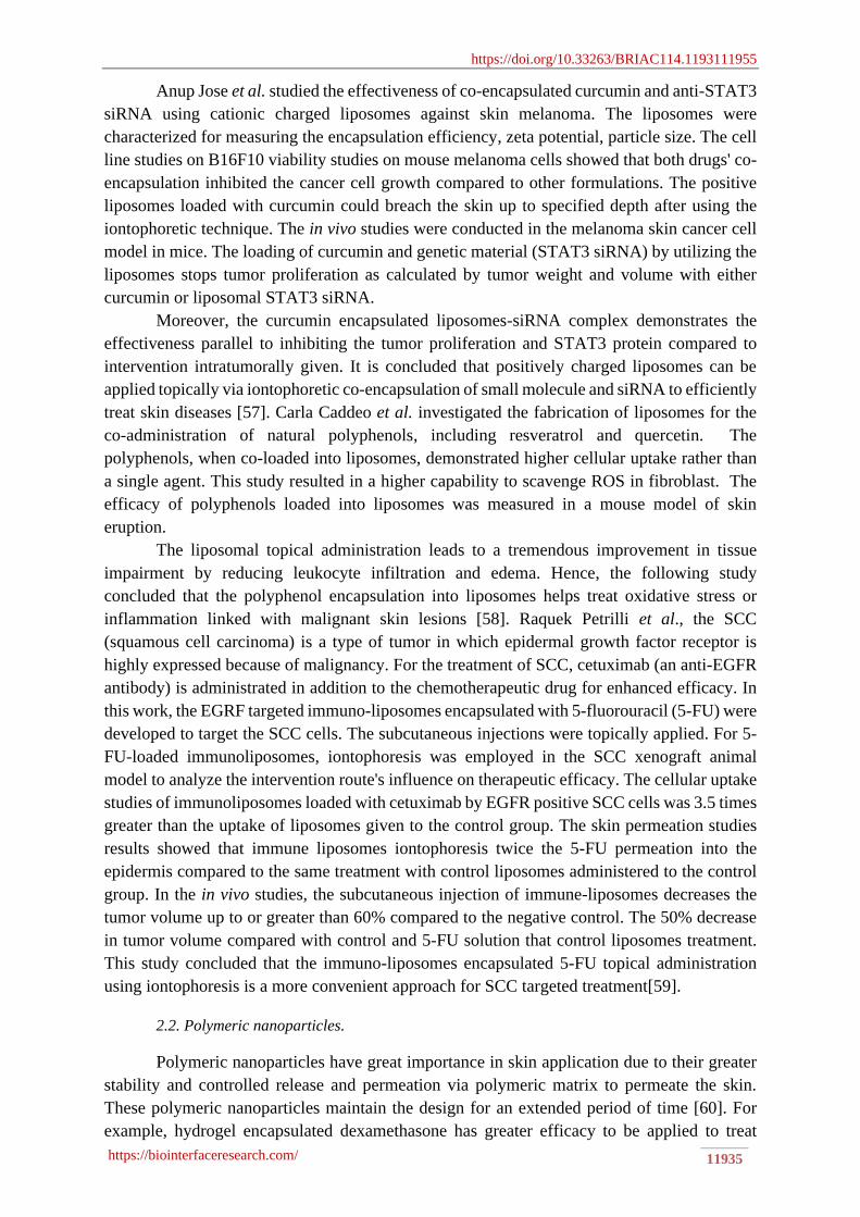
Anup Jose et al. studied the effectiveness of co-encapsulated curcumin and anti-STAT3
siRNA using cationic charged liposomes against skin melanoma. The liposomes were
characterized for measuring the encapsulation efficiency, zeta potential, particle size. The cell
line studies on B16F10 viability studies on mouse melanoma cells showed that both drugs' co-
encapsulation inhibited the cancer cell growth compared to other formulations. The positive
liposomes loaded with curcumin could breach the skin up to specified depth after using the
iontophoretic technique. The in vivo studies were conducted in the melanoma skin cancer cell
model in mice. The loading of curcumin and genetic material (STAT3 siRNA) by utilizing the
liposomes stops tumor proliferation as calculated by tumor weight and volume with either
curcumin or liposomal STAT3 siRNA.
Moreover, the curcumin encapsulated liposomes-siRNA complex demonstrates the
effectiveness parallel to inhibiting the tumor proliferation and STAT3 protein compared to
intervention intratumorally given. It is concluded that positively charged liposomes can be
applied topically via iontophoretic co-encapsulation of small molecule and siRNA to efficiently
treat skin diseases [57]. Carla Caddeo et al. investigated the fabrication of liposomes for the
co-administration of natural polyphenols, including resveratrol and quercetin. The
polyphenols, when co-loaded into liposomes, demonstrated higher cellular uptake rather than
a single agent. This study resulted in a higher capability to scavenge ROS in fibroblast. The
efficacy of polyphenols loaded into liposomes was measured in a mouse model of skin
eruption.
The liposomal topical administration leads to a tremendous improvement in tissue
impairment by reducing leukocyte infiltration and edema. Hence, the following study
concluded that the polyphenol encapsulation into liposomes helps treat oxidative stress or
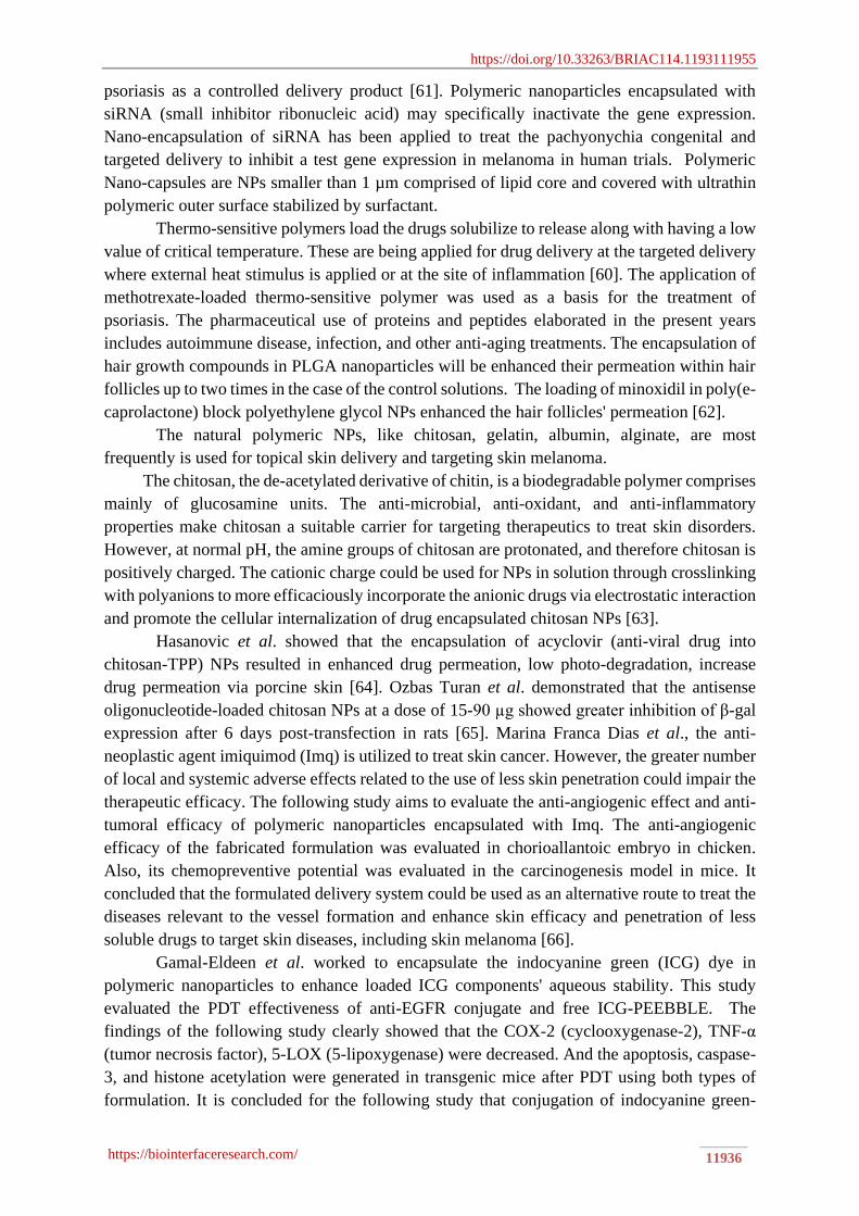
inflammation linked with malignant skin lesions [58]. Raquek Petrilli et al., the SCC
(squamous cell carcinoma) is a type of tumor in which epidermal growth factor receptor is
highly expressed because of malignancy. For the treatment of SCC, cetuximab (an anti-EGFR
antibody) is administrated in addition to the chemotherapeutic drug for enhanced efficacy. In
this work, the EGRF targeted immuno-liposomes encapsulated with 5-fluorouracil (5-FU) were
developed to target the SCC cells. The subcutaneous injections were topically applied. For 5-
FU-loaded immunoliposomes, iontophoresis was employed in the SCC xenograft animal
model to analyze the intervention route's influence on therapeutic efficacy. The cellular uptake
studies of immunoliposomes loaded with cetuximab by EGFR positive SCC cells was 3.5 times
greater than the uptake of liposomes given to the control group. The skin permeation studies
results showed that immune liposomes iontophoresis twice the 5-FU permeation into the
epidermis compared to the same treatment with control liposomes administered to the control
group. In the in vivo studies, the subcutaneous injection of immune-liposomes decreases the
tumor volume up to or greater than 60% compared to the negative control. The 50% decrease
in tumor volume compared with control and 5-FU solution that control liposomes treatment.
This study concluded that the immuno-liposomes encapsulated 5-FU topical administration
using iontophoresis is a more convenient approach for SCC targeted treatment[59].
2.2. Polymeric nanoparticles.
Polymeric nanoparticles have great importance in skin application due to their greater
stability and controlled release and permeation via polymeric matrix to permeate the skin.
These polymeric nanoparticles maintain the design for an extended period of time [60]. For
example, hydrogel encapsulated dexamethasone has greater efficacy to be applied to treat
https://doi.org/10.33263/BRIAC114.1193111955
https://biointerfaceresearch.com/ 11936
psoriasis as a controlled delivery product [61]. Polymeric nanoparticles encapsulated with
siRNA (small inhibitor ribonucleic acid) may specifically inactivate the gene expression.
Nano-encapsulation of siRNA has been applied to treat the pachyonychia congenital and
targeted delivery to inhibit a test gene expression in melanoma in human trials. Polymeric
Nano-capsules are NPs smaller than 1 µm comprised of lipid core and covered with ultrathin
polymeric outer surface stabilized by surfactant.
Thermo-sensitive polymers load the drugs solubilize to release along with having a low
value of critical temperature. These are being applied for drug delivery at the targeted delivery
where external heat stimulus is applied or at the site of inflammation [60]. The application of
methotrexate-loaded thermo-sensitive polymer was used as a basis for the treatment of
psoriasis. The pharmaceutical use of proteins and peptides elaborated in the present years
includes autoimmune disease, infection, and other anti-aging treatments. The encapsulation of
hair growth compounds in PLGA nanoparticles will be enhanced their permeation within hair
follicles up to two times in the case of the control solutions. The loading of minoxidil in poly(e-
caprolactone) block polyethylene glycol NPs enhanced the hair follicles' permeation [62].
The natural polymeric NPs, like chitosan, gelatin, albumin, alginate, are most
frequently is used for topical skin delivery and targeting skin melanoma.
The chitosan, the de-acetylated derivative of chitin, is a biodegradable polymer comprises
mainly of glucosamine units. The anti-microbial, anti-oxidant, and anti-inflammatory
properties make chitosan a suitable carrier for targeting therapeutics to treat skin disorders.
However, at normal pH, the amine groups of chitosan are protonated, and therefore chitosan is
positively charged. The cationic charge could be used for NPs in solution through crosslinking
with polyanions to more efficaciously incorporate the anionic drugs via electrostatic interaction
and promote the cellular internalization of drug encapsulated chitosan NPs [63].
Hasanovic et al. showed that the encapsulation of acyclovir (anti-viral drug into
chitosan-TPP) NPs resulted in enhanced drug permeation, low photo-degradation, increase
drug permeation via porcine skin [64]. Ozbas Turan et al. demonstrated that the antisense
oligonucleotide-loaded chitosan NPs at a dose of 15-90 µg showed greater inhibition of β-gal
expression after 6 days post-transfection in rats [65]. Marina Franca Dias et al., the anti-
neoplastic agent imiquimod (Imq) is utilized to treat skin cancer. However, the greater number
of local and systemic adverse effects related to the use of less skin penetration could impair the
therapeutic efficacy. The following study aims to evaluate the anti-angiogenic effect and anti-
tumoral efficacy of polymeric nanoparticles encapsulated with Imq. The anti-angiogenic
efficacy of the fabricated formulation was evaluated in chorioallantoic embryo in chicken.
Also, its chemopreventive potential was evaluated in the carcinogenesis model in mice. It
concluded that the formulated delivery system could be used as an alternative route to treat the
diseases relevant to the vessel formation and enhance skin efficacy and penetration of less
soluble drugs to target skin diseases, including skin melanoma [66].
Gamal-Eldeen et al. worked to encapsulate the indocyanine green (ICG) dye in
polymeric nanoparticles to enhance loaded ICG components' aqueous stability. This study
evaluated the PDT effectiveness of anti-EGFR conjugate and free ICG-PEEBBLE. The
findings of the following study clearly showed that the COX-2 (cyclooxygenase-2), TNF-α
(tumor necrosis factor), 5-LOX (5-lipoxygenase) were decreased. And the apoptosis, caspase-
3, and histone acetylation were generated in transgenic mice after PDT using both types of
formulation. It is concluded for the following study that conjugation of indocyanine green-

https://doi.org/10.33263/BRIAC114.1193111955
https://biointerfaceresearch.com/ 11937
PEBBLE to anti-EGFR was very efficient in blocking the VEGF and also enhance the caspase-
3 in comparison to free ICG-PEBBLE [67].
Pandey et al. studied atopic dermatitis while fabricating the chitosan nanoparticles for
dermal targeting and increasing the subcutaneous tissue. The developed nanoparticles were
investigated for polydispersity index (PDI), zeta potential, particle size, morphology, etc. The
results indicated that the optimized formulation showed optimum results. In vitro release study
revealed that release under simulated skin conditions. Permeation study of BMV
(Betamethasone valerate) was more significant via BMV-CS-NPs; however, the quantity of
drug into epidermis and dermis was greater HA-BMVCS-NPs, when compared to BMV-CS-
NPs. It is stated that HABMV-CS is an efficient delivery system for more targeted delivery
and enhanced anti-AD efficacy[68]. Figure 2 describes the penetration of CS-BMV-NPs for
the treatment of atopic dermatitis.
Figure 2. A comparative study of atopic dermatitis treatment through hyaluronic acid-coated BMV
encapsulated CS-nanoparticles and without hyaluronic acid coating CS-BMV-NPs.
Bhatnagar et al. developed the bromelain encapsulated poly (lactic-co-glycolic acid)
and studied the anti-tumor effects in the skin tumorigenesis mice model. The following study
results revealed the increased anti-neoplastic effect of NPs in skin cancer model stage II. The
decline in the number of the tumor cells, a decrease in percent tumorigenesis, and percent
mortality in mice were seen in NPs loaded BL compared to free BL. These effects were also
supported by histopathological evaluations. The results demonstrated that the NPs were
fabricated to enhance chemotherapy's efficiency against skin melanoma at small doses[69].
Samaneh Bayat et al. studied the bromelain encapsulated chitosan nanofibers for burn injuries
in laboratory animal models. Chitosan nanofibers loaded with bromelain developed by
electrospinning technique. The physiochemical characteristics of nanofibers were analyzed.
Cytotoxicity analysis was also performed using Alamar blue. The healing process of chitosan
nanofibers was studied in rats for 21 days. The study analysis showed that 2% chitosan w/v
bromelain encapsulated nanofiber was efficient to repair the skin burn [70].
2.3. Nanoemulsions.
Nanoemulsions are stable systems with a clear appearance and carrying special
rheological characteristics. It is a most used delivery system for the sustained release of APIs

https://doi.org/10.33263/BRIAC114.1193111955
https://biointerfaceresearch.com/ 11938
into a deep layer of skin. The nano-emulsion advantage over others is that it can decrease
transepidermal water loss (TEWL) by hydrating the skin and are more permeable to APIs. The
greater solubilization and kinetic stability make them an attractive formulation.
Severino et al. worked on squamous cell carcinoma and basal cell carcinoma, which
were reported to be among the most common malignancies. Chemotherapeutics used under
similar conditions showed nonselective targeting along with severe side effects. These types of
nanoparticles showed sustained release and stability of encapsulated drugs from chemical
degradation. The following technique is very useful for increasing the intracellular quantity of
drugs and decreasing the cytotoxicity [50].
Primo et al. worked on the development of a novel delivery system consisting of
surfactants with magnetic fluid (obtained from maghemite) citrate derivative. The in vitro
studies showed that the formulation presents a significant potential for synergetic application
in the topical release of photosensitivity drugs and shown potential tissues targeting properties
in the photodynamic therapy. In vitro studies revealed that tape stripping could be used to
analyze drugs in the stratum corneum and dermis+epidermis skin layers. The following nano-
emulsion were applied on the pigskin surface in the Franz diffusion cell. The results concluded
that the magnetic nano-emulsion would enhance the drug release on the skin layers deep down
compared to traditional formulation having no magnetic nanoparticles. This is all related to
enhancing the biocompatibility of nanoformulation because of enhancing efficacy for the
extracellular matrix in the skin and enhancing the drug-loaded inside the corneocytes wall [71].
Primo et al. developed the magnetic nanoemulsion to apply in magnetic hyperthermia and
photodynamic treatment. It is concluded that after the intervention of a very little amount of
drug, the tumor inhibition gets enhanced [72].
Giacone et al. studied aimed to fabricate and evaluate nano-emulsion for topical skin
delivery of the cytotoxic agent piplartine. The cytotoxicity study was approximately 2.8 fold
higher when the drug was loaded in chitosan nanoemulsion for targeting the melanoma cells.
The efficacy of this nano-system on melanoma tissues was concentration relevant. The results
supported the significant application of the chitosan modified nanoemulsion loaded with
piplartine to manage skin cancer [73].
Falamas et al. proposed the study to fabricate the ex vivo and in vivo surface-enhanced
signal from mice's skin and then used it against various skin pathologies, including skin cancer.
The chemopreventive activity of natural compound betulin was studied in skin carcinoma in
mice models. The study of various skin pathologies' differentiation was done by performing K
means clustering and components analysis. The efficiency of nanoemulsion loaded with botulin
validated by histological study and the chemometrics techniques showed the direct SERS
differentiation link among the botulin and cancerous nanoemulsion treated skin [74].
Yousef et al. developed curcumin-loaded nanoemulsion against several skin diseases,
including melanoma and psoriasis. To optimize skin delivery formulation, the curcumin QbD
(Quality by Design) approach is utilized to enhance the skin targeting of curcumin. It is
concluded that the efficacy of nanoemulsion for skin targeting of the curcumin, and showed
the mechanism of enhanced permeation. The results concluded that curcumin loaded
nanoemulsion has significant potential to target psoriasis and skin melanoma [75].
2.4. Carbon nanotubes.
The CNTs (carbon nanotubes) are the most stable compounds with good cytoprotective
and anti-oxidant effects. The CNTs conductivity was applied to develop a very good biomarker

https://doi.org/10.33263/BRIAC114.1193111955
https://biointerfaceresearch.com/ 11939
sensor for skin melanoma diagnosis and infections at its early stage[76]. Anna Shvedova et al.
evaluated and screened the effects of carbon nanotubes by utilizing immortalized keratinocytes'
cell culture. The longer exposure of single-wall carbon nanotube (SWCNT), cellular toxicity,
and oxidative stress were analyzed by loss of cell viability, free radicals formation, and
aggregation of per-oxidative products. SWCNT exposure also results in morphological and
ultra-structural modifications in cultured skin cells. It is concluded that skin exposure to
unrefined SWCNT may take to skin toxicity because of accelerated oxidative stress in workers'
skin [77].
Ding et al. use nanotechnology to evaluate the significance of toxic effects on the
environment and people. To evaluate the complete genomic expression analysis and enhanced
phenotypic measurement on human skin, the dermal fibroblast cell populations were exposed
to MWCNTs. The results showed the exposing cells to both types of nanotubes at cytotoxic
doses induced cell cycle arrest that enhances the necrosis. Moreover, these nanotubes' exposure
stimulates the genes involved in cellular transport, metabolism and cell regulation, etc.
Microarray analysis of the promotor analysis showed that p38/ERK-MAPK and interferon
cascades are critical pathway components in the signal transduction, participating in the more
adverse effects observed upon nanotubes exposure[78].
Hasebi et al., the development of stable anticancer drugs and CNTs has been under
investigation. The nanotubes loaded with two drugs, aminolevulinic acid and tretinoin,
evaluated for their stability of the composites. The results showed that tretinoin encapsulated
nanotube is more stable than nanotube-aminolevulinic acid [79].
Moon et al. evaluated the photothermal therapy using nanomaterials as an efficacy
technique for cancer targeting. SWNT is a potential and efficacious candidate as a
photothermal agent. This study demonstrated in vivo obliteration of malignant tumors by
targeting the SWNTs and NIR irradiation. The photo-thermally targeted mice showed a
complete cure of tumor without recurrence phase for 6 months. Most of the SWNTs that
intervene were removed out from the mice in about 2 months. The results concluded that
SWNTs serve as an efficient photothermal agent and a step towards future cancer therapeutics
[80].
Nanda Gopal Sahoo et al. fabricated multiwalled carbon nanotubes and graphene oxide
functionalized by highly biocompatible and hydrophilic for loading and targeting an anti-
cancer compound CPT camptothecin. The MWCNT-PVA and GO-PVA were encapsulated
via pi-pi interactions and showed the potential to kill the breast and skin cancer cells [81].
2.5. Nanomicelles.
The nano-micelles have been used for drugs targeting the skin. These nano-carriers
have few limitations in that they are only deposited in the hair follicles. They might not reach
into the skin layer deep down [82, 83]. Emine Kahraman et al. developed the nano-micelles
loaded with terpenes to meet the challenges and evaluate skin delivery potential. The drug
accumulation and permeation were evaluated in the skin by the tape stripping method. The data
showed that nano-micelles loaded with terpinolene may cause the aggregation of more
concentration of drug in the striped skin as compare to the commercial product and micelles
without terpene. The loading of terpinolene into nanomicelles can be a more feasible approach
for drug delivery [82].
Wang et al. develop and design nano-carriers from polymers for targeting the siRNA
to skin melanoma for transdermal application. Encapsulated siRNA reproduces the efficacy in

https://doi.org/10.33263/BRIAC114.1193111955
https://biointerfaceresearch.com/ 11940
inhibiting the proliferation of skin cancer. The study showed that cationic micelles are effective
as gene delivery nanoplatforms for melanoma therapy [84].
Yang et al. developed in situ gel for controlled drug delivery, encapsulated with
curcumin-loaded micelles, and suitable for skin repair. Studies suggested that curcumin loaded
nanomicelles exhibited good tissue stickiness that can release curcumin for a long time.
Moreover, full-thickness excision models and linear incision models were applied to analyze
wounds' in vivo healing. Histopathologic examination showed that curcumin load nanomicelles
could enhance cutaneous wound repair. It is concluded that biodegradable curcumin
encapsulated nanomicelles have significant potential for wound healing [85].
Xu et al., paclitaxel-loaded micelles were developed, and hydrogel was formulated
while loading these nanoparticles for skin melanoma treatment. Skin penetration and retention
study were performed for the following formulation. The cell lines studied showed more
cellular uptake in B16 melanoma cell lines, and in vivo studies in B16 cells showed preferable
anticancer activity than free taxol [86].
Rosa Juliana et al. develop a new approach for gene therapy for various skin diseases.
The RNA interferences used for gene therapy has been considered as the most novel techniques
for individualized therapy. In several skin diseases, siRNA application was established against
skin cancer, psoriasis, dermatitis, and leprosy. The following approach used siRNA
encapsulated nanomicelles for topical delivery. It acted via inhibition of expression of the
transcript that has potential for gene suppression [87].
Bingxin et al. developed oridonin (ORI) encapsulated nanomicelles by utilizing
monomethoxy poly(ethylene glycol)-poly(ԑ-caprolactone). The study revealed that ORI was
encapsulated into the core-shell structure of micelles that retained its anti-cancer activity. The
ORI could be released from the nanomicelles in a sustained way for in vitro study. The results
concluded that ORI encapsulated micelles showed potential for intravenous and transdermal
drug delivery in cancer therapy [88]. Anup Jose et al. developed cationic liposomes
encapsulated with curcumin and complexed with STAT3 siRNA. The results concluded that
the liposome-siRNA encapsulated with curcumin was taken up by clathrin modulated
endocytosis.
The co-encapsulation of STAT3 siRNA along with curcumin have a higher potential
for cancer cell apoptosis and inhibition with STAT3 siRNA and free curcumin. It is concluded
that cationic liposomes are non-invasive iontophoretic delivery of both co-encapsulated
substances to target the skin malignant cells [89].
2.6. Gold nanoparticles.
Gold nanoparticles (AuNPs) are the most used and applied platforms for treating,
diagnosing, and monitoring skin disease, including skin tumors [90]. Limon et al. developed
gold nanoparticles (GNP) coated with thiolates that highly solubilize in water. The uptake and
internalization of fluorophore coated AuNPs in keratinocytes of human was elaborated using
confocal fluorescence microscopy.
The functionalized nanoparticles inhibited intracellular KLK5 and HaCaT activity and
diminished the IL-8 secretion stimulated by TLR-2 ligands. The following study showed that
GNPs have a significant ability for new intracellular delivery of drugs and antibodies in treating
skin cancer other diseases, including Rosacea [91]. Preet et al. demonstrated the development
of nisin gold nanoparticles loaded with doxorubicin for murine skin cancer treatment. The
results indicate a substantial reduction in tumor volume when calculated at the termination of

https://doi.org/10.33263/BRIAC114.1193111955
https://biointerfaceresearch.com/ 11941
therapy. Therefore, the potential decline in the serum values of all evaluated cytokines NF- κβ,
TNF-α, TNF-β, IL-10. And a potential increase in tissue ROS (reactive oxygen species) levels
and lipid peroxides, and superoxide dismutase in various treatment groups. The following study
showed that the AuNPs assisted co-delivery of nisin doxorubicin for murine cancer cells and
DMBA cells in cultured cells in the lab [92].
Nirmala et al. developed the AuNPs considered for apoptosis and anti-malignant
efficacy at the minimum inhibitory concentration. The cytotoxicity of the gold nanoparticles
can synergize the effects of the phenolic compound of the V.vinifera and the efficacy of the
conjugated AuNPs that causes necrosis and tumor cell death in following cell lines studies [93].
Figure 3. Gold nanoparticles obtained from vinifera peel induce antiproliferative, apoptosis, and necrosis in
melanoma cell lines.
Fratoddi et al. developed the functionalized gold nanoparticles encapsulated with MTX
(methotrexate) to treat moderate to severe inflammatory diseases. The efficacy of the AuNPs
of the gold nanoparticles was loaded with MTX topically administered on the skin and checked
on skin models in vivo and in vitro in the psoriasis mice model. The cell proliferation rate,
inflammation, and epidermal thickness were also evaluated—the K6CD3, Ki67, and CD8 used
for immunochemistry evaluation showed reduced staining in AuNPs. Blood analysis showed
no distinctive differences in blood cell count level and ALT, AST levels at the start, and
treatment termination. Topical gold nanoparticles therapy can stimulate the decrease in
epidermal thickness, reduction in keratinocyte multiplications, and inflammatory infiltration in
the psoriasis mice model [94].
2.7. Mesoporous silica nanoparticles.
Mesoporous silica nanoparticles (MeSNPs) are a wide range of functionalities and are
modular as they can be modified to fit almost any setting. These nanoparticles have a greater
surface to volume ratio that increased the functionalization while maintaining the porosity that
allows the inorganic platform to an appreciable amount of carrier without destabilization of
silica framework. The recent advancement in nano-medicines helps to develop mesoporous
silica nanoparticles at a large scale. There are several studies that showed the use of MeSNPs
in skin carcinoma [95].
Hirai et al. evaluated the efficiency of nSPs (amorphous silica nanoparticles), one of
the broadly used nanomaterials that can induce the targeted effect by penetrating the skin. The
mice having atopic dermatitis (AD) (via dermal injection antigen Dermatophagoides
pteronyssinus) [96]. It is evaluated that amorphous silica nanoparticles can produce immune-
modulating effects that can penetrate the skin. However, nSPs could produce a risk for
augmentation of immune diseases related to skin. Histopathological results showed that Dp and

https://doi.org/10.33263/BRIAC114.1193111955
https://biointerfaceresearch.com/ 11942
silica particles produce augmentation of AD. Moreover, the results are related to skin infections
induce by thymic lymphopoietin and IL-18 [96].
Lio et al., interfering RNA (siRNA) encapsulated mesoporous silica nanoparticles
applied to treat skin diseases that include SCC (squamous cell carcinoma). To study the effects
of silica nanoparticles topically to the SCC in a mouse model. MeSNPs encapsulated with
TGFβR-1 siRNA show a two-fold suppression of TGFβR-1 [97]. Xing Ma et al. developed 5-
aminolevulinic acid (5-ALA) encapsulated MeSNPs were produced to transport 5-ALA for
(PDT) photodynamic therapy against skin cancer cell lines B16F10. The folic acid attached to
the surface of 5-ALA encapsulated HMeSNPs enabled the endocytosis of NPs in cancer cell
lines. Figure 4 describe the targeting of skin cancer cell via mesoporous nanoparticles [98].
Figure 4. 5-aminolevulinic acid (5-ALA) encapsulated mesoporous silica nanoparticles for Photodynamic
therapy against skin cancer cells (B16F10).
P.Scodeller et al. developed silica nanoparticles (SiNPs) via self-assembly layer
methodology. The NPs were analyzed as adjuvant of carboplatin, intervene in A375 cell lines
of human skin cancer having mice and compared with another enzyme (inactivated). It is stated
that the volume of the tumor with SiNPs inactivated hyal was potentially increased in
comparison to the inactivated one. The matrix distorting enzymes are inactivated on SiNPs and
are a more efficient cancer drug aid than the inactivated enzyme [99].
Rosenholm et al. developed the MeSNPs and highlighted the new drug delivery way.
In vivo and in vitro, the therapeutic benefit of these systems for in vivo use has been studied.
Moreover, the results showed that the cellular studies could be replicated along with targeting
and safety issues in cancer therapy [100]. Fiorenza Rancan et al. evaluated the skin permeation
and uptake of SiNPs. The topical application of nanoparticles was linked with dendritic and
epidermis cells independent of their functionalization. The results concluded that, in patients
with eczema and atopic dermatitis, skin layers' analysis is highly significant to assess the
penetration of nanoparticles [101].
2.8. Iron oxide nanoparticles.
The superparamagnetic iron-oxide NPs (SPION) for drug targeting and other
biomedical implementation have been evaluated because of greater drug encapsulating
capacity, intense magnetic responsiveness, and greater targeted delivery efficiency. Literature
suggested that skin composition and structure will not approve the materials greater than 600
Da (>600 Da) to pass through the skin layers. The recent advances in nanomedicines have made
it easy to develop different types of MNPs (magnetic NPs) with few nanometers. Many studies

https://doi.org/10.33263/BRIAC114.1193111955
https://biointerfaceresearch.com/ 11943
demonstrated that these types of special nanomaterials help to permeate the stratum corneum
and hair follicles via the epidermis [102].
Rao et al. developed the EPI-SPION (epirubicin encapsulated iron oxide NPs) to target
skin carcinoma via transdermal route. The modifications of the SPION results in a drug carrier
that applied magnetism for chemotherapy of skin cancer. The cell lines study of skin cancer in
WM266 and HaCaT keratinocyte cells showed that SPION has good compatibility. The in vitro
study evaluated that the EPI loaded SPION can permeate the skin. The study concluded that
the potential of iron oxide NPs for easy transdermal delivery in skin melanoma [103].
Cengelli et al. studied the interactions of biocompatible cationic amino ultra-small
superparamagnetic iron oxide nanoparticles (USPIONs) with human cells in different
dimension cultures (2D and 3D) using electron microscopy and biochemical techniques. The
results showed internalization of the amino-SPIONs by human melanoma cells. The uptake
pathway was clathrin modulated and localized in the lysosome, which induces the stimulation
and decreases the expression of the cathepsin D and transferrin receptors in skin fibroblasts
under evaluation of skin melanoma [104].
Choi et al. studied the photon-activated therapy for skin melanoma and the potential of
the sensitizing effect of iron oxide NPs. X-rays photon stimulated therapy was evaluated by
targeting the CT26 tumor-bearing mice. The decrease in viability and damage was seen in the
FeO-NPs treated cell lines. The results concluded that iron oxide nanoparticles would enhance
the therapeutic effect with a low concentration that may have potential and significant treatment
for the skin's malignancies [105].
Musazzi et al. developed the semi-solid formulation with iron oxide NPs for human
skin penetration to target different skin diseases. The in vitro penetration of IONs aggregates
was evaluated by utilizing isolated stratum corneum (SC) or humans' epidermis. The semisolid
preparation of different FeO-NPs permeation as compared to the aqueous suspension. The Cet
cream formulation enhances the penetration, and low retention amount compared to the cold
cream and favored the accumulation into the skin membrane. The results concluded that the
skin semi-solid formulation could be used to deliver the FeO-NPs without disturbing the
permeation pattern on the skin surface [106].
3. Results and Discussion
When normal cells transform and spread out of control, cancer starts and creates a
tumor. Skin cancer is diagnosed for 3 million Americans each year, making this cancer one of
the most common cancer kinds in this country and also in the world. If skin cancer is detected
early, topical treatments, procedures performed in a dermatologist's office, or ambulatory
surgery may typically be used to treat it [107]. Skin cancer usually kills 1 percent of all cancer
types in the world. Skin cancer can be more severe in some situations and needs treatment by
a multi-modal approach that needs dermatologists and oncologists experts together [108]. Skin
cancer usually showed 4 types of cancer that include squamous, basal, Merkel, and melanoma
cell carcinoma [109]. Melanoma began with melanocytes and was reported as the most deadly
skin cancer. There are some drawbacks to conventional methods. The advancement of new
strategies for the detection of skin cancer is vital [110]. The nanosensor is an analytical
instrument used for the identification of a biological substance. Nanomedicine is playing an
increasingly significant role in the production of nanosensors. The biosensor or nano biosensor
science is widely used to detect biomarkers in skin cancer [111]. It can be claimed that with
the emergence of Nanomedicine and its effect on the production of ultrasensitive instruments,

https://doi.org/10.33263/BRIAC114.1193111955
https://biointerfaceresearch.com/ 11944
it is possibly one of the most promising solutions to fix some of the problems related to the
growing need to create an extremely sensitive, quick, and economical method of a skin cancer
diagnosis. Different electrochemical and optical biosensors have been developed for skin
cancer detection that will be discussed below.
3.1. Biomarkers in skin cancer.
The identification of biomarkers associated with skin cancer has provided new insights
into the area of theranostics cancer. One of the earliest prognostic biomarkers of melanoma is
lactate dehydrogenase (LDH) [112]. Another prognostic biomarker in metastatic skin cancer
patients is serum S100B levels [113]. The BRAF V600 can be the best biomarker for metastatic
melanoma. Recognition of tyrosinase (TYR) mRNAs in serum or blood is another biomarker
for metastatic melanoma [114]. In inflammatory problems, over-expression of
cyclooxygenase-2 is a promising biomarker for tumor inflammogenesis, metastasis, and
angiogenesis [115]. Microsomal prostaglandin E2 synthase-1 was reported in some studies as
a predictor of melanoma or skin cancer [116]. A key role in melanoma progression has been
documented to be played by matrix metalloproteinases ( MMPs) [117].
Many cancers, including malignant melanoma, calcium-binding proteins of S100 group
such as S100B, S100A9, and S100A4, have attracted considerable interest as novel biomarkers
[118]. It is often undetectable in the early stages of melanoma. In contrast, late-stage melanoma
displays substantial circulating tumor DNA (ctDNA) levels [119]. The potential biomarkers of
melanoma and other pathologies can be microRNAs or long noncoding RNAs [120]. The
exosomes of patients with metastatic melanoma were extracted from miRNA19a, miRNA21,
miRNA149, miRNA17, and miRNA126 [121].
Collectively, the discovery of biomarkers derived from liquid biopsies is not as fast as
in comparison to genomics and genomics biomarkers. Also, many proposed biomarkers in the
clinical application may not have adequate validity. Among the factors that limit the use of
ideal biomarkers in POC, applications are the absence of repeatability, optimization
procedures, and process duration. Previous to marketing, biomarkers must meet strict laws by
regulatory authorities.
3.2. Traditional approaches to the diagnosis of skin cancer.
Dermoscopy, self-skin examination, optical coherence tomography, and
ultrasonography are traditional skin cancer detection [122]. A Skin Self-Exam (SSE) is a skin
examination that you do on your own. It is a method to detect any alterations in the skin or
irregular areas. SSE is a simple method of detecting skin cancer with a 6-50% prevalence of
self-diagnosed skin cancer incidence [123]. In dermoscopy, optical magnification (*10) and
fluid immersion were used to view skin lesions as a non-invasive imaging process. From mild
to malignant melanoma, dermoscopy illustrates the development of skin lesions. Compared to
a naked eye examination, dermoscopy improves the susceptibility and precision of detecting
melanoma to around 20 and 10%, respectively [124].
Another traditional technique that microscopically evaluates melanoma at the papillary
dermis level and epidermis is reflectance confocal microscopy or RCM [125]. Different
reflectance indexes of skin cells and their anatomical structure generate contrasts of the final
image in the resulted photograph. For melanin and keratin, the maximum contrast is with a
reflection index of 1.7 and 1.5, respectively. High-frequency ultrasonography has created high-

https://doi.org/10.33263/BRIAC114.1193111955
https://biointerfaceresearch.com/ 11945
resolution images to distinguish different forms of skin cancer. This high resolution
(frequencies up to 100 MHz) helps the system to picture the cutaneous components, epidermis
and dermis, and vessels to the diagnosis of skin cancer [124]. In ultrasound tests, the
appearance of a healthy, hypoechogenic, and thick tissue suggests the existence of skin cancer.
In diagnosing skin cancer, infrared light was applied in optical coherence tomography to create
a no invasive process. In cases of misunderstanding with ultrasonography, OCT is appropriate.
OCT, though, only investigates tumor lesions below 1 mm [126].
In the diagnosis of skin cancer, these conventional techniques have been developed, but
significant disadvantages have restricted their clinical application. Many of these approaches
are time-consuming, costly, and need an experienced expert.
3.3. Electrochemical nanosensors.
Biosensors are commonly used instruments for electrochemical biomarker detection
and can theoretically be used to establish cancer diagnostic with point-of-care checks [127].
An efficient and low-cost detection technique for melanoma cancer diagnosis is the
electrochemical identification of functional melanoma biomarkers, which significantly
optimizes sensitivity and specificity [128]. Immunosensors are effective for cancer detection
and are produced using electrochemical transducer-anchored antibodies. A significant part of
the creation of electrochemical immunosensors is expected to be the manufacturing of
responsive and stable materials by immobilizing biomolecules. The use of nanoparticles in an
immunosensor also enables biological macromolecules with high biological stability and
activity to be effectively immobilized due to their high adsorption ability and wide surface area
[129, 130]. Seenivasan et al. developed a new immunosensor based on SiO2 NPs and
polypyrrole nanocomposite for early detection of melanoma cancer [131].
The particular strategy was based on the attachment between the anti-MC1R antibody
and the melanocortin 1 receptor antigen on the cell's surface. The suggested immunosensor was
particularly sensitive with a sensitivity of 20 cells/mL, accurate and repeatable, and the ready-
to-use electrode loaded with Ab was lasted for 14 days. In another study, Xiang et al. mentioned
a combined nano-mesoporous Co3O4 sheet (Au/Co3O4) and aminated graphene (GS-NH2)
immunosensor based on Au NPs for melanoma adhesion molecule antigen (CD146)
identification [132]. With a LOD of 3.4 pg/mL and S/N of 3, the nano sensor-operated over a
line range of 0.01-15 ng/mL.
Wearable sensors have gained tremendous interest in recent years. Different wearable
electrochemical or optical sensors have been developed to monitor body fluids such as saliva
or sweat in real-time [133].
For the rapid screening of skin melanoma, Bianca et al. developed a portable bendable
nanosensor and a non-invasive micro-needle sensor [134]. The wearable electrochemical
sensors were able to identify the existence of tyrosinase (TYR as skin cancer biomarker). The
elastic sensor showed strong mechanical deformation resistance, whereas the hollow micro-
needle unit was filled with carbon paste coated with catechol to measure the low levels of TYR
in tissue. Pretty biased measurement was obtained by adsorbing the TYR enzyme marker’s
CAT substrate and amperometrically measuring the BQ reaction product at a low-potential of
-0.25 V. The resultant epidermal bandage and wearable microneedle sensors showed good
analytical efficiency and provided significant promise to monitor the TYR biomarker on both
the skin surface and inside the skin.

https://doi.org/10.33263/BRIAC114.1193111955
https://biointerfaceresearch.com/ 11946
Electric cell-substrate impedance sensing (ECIS) and Electrochemical impedance
spectroscopy (EIS) are the common methods currently available for characterizing single cells
based on a specific frequency range of cellular electrical answers and long-term cell
physiological answers [135]. The EIS system has been commonly reported to detect skin
cancer. It includes a disposable electrode probe that differentiates between abnormal and
normal skin lesions using electrical impendence variations [136]. In this regard, A highly
sensitive electrochemical impedance spectroscopy (EIS) approach has been documented by
Prathap et al., useful in identifying melanoma cells precisely and quickly using anti-MC1R
antibody attached PANI-NFs [137]. A perfect linear correlation was obtained between the
current and the target cell (density of 15 to 7000 cells/5mL and LOD of 1 cell/mL).
Silicon nanowire functionalized with antibody field-effect transistors demonstrated
high specificity to analyte identification, allowing analyte concentrations to be sensed at levels
not easily available by other tools [138]. Identifying serum melanoma biomarkers, such as the
newly found tumor necrosis factor receptor superfamily participant TROY (TNFRSF19), is at
low amounts, is an important factor. The identification of TROY in the buffer with various
concentrations and silicon nanowires was reported by Maedler et al. The sensitivity of
nanosensors was evaluated by comparing the signal with that acquired from BSA in the buffer
solution [139]. To differentiate the two signals, both the signal size and the reaction kinetics
were applied. They obtained the dissociation and interaction constants for the reaction of
TROY and antibody by their reaction models.
Despite the benefits of using electrochemical approaches, skin cancer identification at
its early phases needs to study human body fluid with deficient concentrations. To enhance the
therapeutic potential and diagnostic precision, the combination of multimodal imaging and
therapeutic features within a nanoplatform is essential. Integrating electrochemical approaches
with recently designed electro-active complexes and electrochemical sensing nanoparticles can
further increase the limits of detection and susceptibility for new biomarkers. Even more
developments in nanoscience, bioengineering, molecular biology, and computational biology
are expected to open the path to producing accurate electrochemical biosensors to enhance
customized treatment strategies for some diseases.
3.4. Optical nanosensors.
As novel disease biosensors, optical techniques have received considerable attention in
recent years [140]. As a symptom of dermatological diseases, including melanoma, increased
tyrosinase activation is a marker. In melanin biosynthesis, tyrosinase (TYR) is an important
biomarker for detecting actinic injury, vitiligo, and melanoma [141]. Hu et al. published a
simple fluorescent experiment-based carbon quantum dots attached with dopamine (CQDs-
Dopa) for TYR activation [142]. Two wide linear regions were identified by the experiment
(711.1-2925.4 U/L and 44.4-711.1 U/L) and LOD of 17.7 U/L. In blood serum, the suggested
fluorescent analysis was used to test TYR activity.
In other studies, Li et al. developed an oligonucleotide fluorescent probe with pyrene
alteration and in-situ integration of nucleoside phosphoramidites (T-C-A-G) with phosphorus
modification of pyrene at the internal phosphate backbone [143]. They demonstrated that the
inclusion of exactly aligned analytes caused the fluorescence intensity to increase quickly (less
than 20 s) and significantly (up to 23.5 times), whereas analytes with a single mismatch on
both sides of pyrene attached phosphate showed a steady fluorescence intensity level similar
to that of the background. As a practical optical imaging tool, fluorescent-labeled antibodies

https://doi.org/10.33263/BRIAC114.1193111955
https://biointerfaceresearch.com/ 11947
have been applied in the diagnosis of microscopic melanoma. In another study, Larissa et al.
showed that antiangiogenic therapy's addition increases the absorption of monoclonal
antibodies labeled fluorescently within melanoma tumors [144].
One of the favorite new investigations that have drawn great interest from researchers
because of its economical price and good sensitivity is the photoelectrochemical (PEC)
biosensing technique [145]. In this light, a high sensitive photoelectrochemical nanosensor was
reported by Erhu et al. for DNA detection. The nanosensor was based on cascade signal
amplification (hybridization chain reaction, catalytic hairpin assembly, and alkaline
phosphatase) [146]. The T-DNA was detected with LOD of 0.052 fM and linear response range
of 0.1 fM-100 pM. The suggested PEC biosensor demonstrated excellent analytical efficiency.
In another analysis, Lopes et al. prepared biocompatible Au NPs covered with oleic acid and
hyaluronic acid and used in a melanoma cell line (B16F10), keratinocytes, and also
Saccharomyces cerevisiae [147]. Au NPs showed a near-infrared absorption in 750-1400 nm.
The covered Au NPs also demonstrated a spherical shape with an average diameter of 297 nm
without toxic effect on the yeast and cell lines tested. Nonetheless, the viability of the B16F10
cell line was decreased down to 20% after laser application. This study showed promising
results for melanoma treatment and diagnosis.
4. Conclusions and Future Prospectives
Nano dermatology is a novel area of research having applications in the field of
medicines. Many nanomaterials have been implemented in modern society. Updated versions
are developed at a phenomenal rate as some inorganic nanomaterials coupled to label as
antibodies and have proven efficacy for visualizing various skin diseases and skin tumors.
Skin cancer is fast-growing cancer that some vital consideration is required for quick
and precise diagnostic tools. However, despite the outstanding role of conventional methods in
reducing melanoma's mortality rate, the limitations of these methods for clinical use and recent
rapid advancement in optical and electrochemical nanosensors have led to innovative
approaches for detecting melanoma in trial applications.
Effective clinical implementation of the methods of detection demands that they be
incorporated with POC instruments. The important obstacle for electrochemical melanoma
detection is that no biomarker has been showing a perfect signal. Some bio-markers appear to
have low selectivity, raising the likelihood of erroneous results.
Assimilating electrochemical methods with recently created electro-active compounds
and electrochemical biosensors would expand the limits of detection and sensitivity for newer
biomarkers. Furthermore, nanomaterials tend to have a synergistic effect between bioactivity,
permeability, and catalytic potential to improve the final signal. Further advances in medical
technology, genetic engineering, computational biology, and molecular biology are expected
to open the door to the production of effective electrochemical biosensors to enhance
customized treatment strategies for some diseases.
Funding
This research received no external funding.
Acknowledgments
This research has no acknowledgment.

https://doi.org/10.33263/BRIAC114.1193111955
https://biointerfaceresearch.com/ 11948
Conflicts of Interest
The authors declare no conflict of interest.
References
1. Esteva, A.; Kuprel, B.; Novoa, R. A.; Ko, J.; Swetter, S.M.; Blau, H.M.; Thrun, S. Dermatologist-level
classification of skin cancer with deep neural networks. Nature 2017, 542, 115-118,
https://doi.org/10.1038/nature21056.
2. Cullen, J.K.; Simmons, J.L.; Parsons, P.G.; Boyle, G.M. Topical treatments for skin cancer. Advanced drug
delivery reviews 2020, 153, 54-64, https://doi.org/10.1016/j.addr.2019.11.002.
3. Rogers, H.W.; Weinstock, M.A.; Feldman, S.R.; Coldiron, B.M. Incidence estimate of nonmelanoma skin
cancer (keratinocyte carcinomas) in the US population, 2012. JAMA dermatology 2015, 151, 1081-1086,
https://doi.org/10.1001/jamadermatol.2015.1187.
4. Bray, F.; Ferlay, J.; Soerjomataram, I.; Siegel, R.L.; Torre, L.A.; Jemal, A. Global cancer statistics 2018:
GLOBOCAN estimates of incidence and mortality worldwide for 36 cancers in 185 countries. CA: a cancer
journal for clinicians 2018, 68, 394-424, https://doi.org/10.3322/caac.21492.
5. Padya, B.S.; Pandey, A.; Pisay, M.; Koteshwara, K.B.; Chandrashekhar Hariharapura, R.; Bhat, K.U.;
Biswas, S.; Mutalik, S. Stimuli-responsive and cellular targeted nanoplatforms for multimodal therapy of
skin cancer. European Journal of Pharmacology 2021, 890, https://doi.org/10.1016/j.ejphar.2020.173633.
6. Wollina, U.; Schreiber, A.; Merla, K.; Haroske, G. Combined cetuximab and volumetric modulated arc-
radiotherapy in advanced recurrent squamous cell carcinoma of the scalp. Dermatology Reports 2011, 3,
https://doi.org/10.4081/dr.2011.e57.
7. Smith, V.; Walton, S. Treatment of facial basal cell carcinoma: a review. Journal of skin cancer 2011, 2011,
https://doi.org/10.1155/2011/380371.
8. Bilal, M.; Iqbal, H. New Insights on Unique Features and Role of Nanostructured Materials in Cosmetics.
Cosmetics 2020, 7, https://doi.org/10.3390/cosmetics7020024.
9. Rahdar, A.; Hajinezhad, M.R.; Bilal, M.; Askari, F.; Kyzas, G.Z. Behavioral effects of zinc oxide
nanoparticles on the brain of rats. Inorganic Chemistry Communications 2020, 119,
https://doi.org/10.1016/j.inoche.2020.108131.
10. Raza, A.; Hayat, U.; Bilal, M.; Iqbal, H.M.; Wang, J.-Y. Zein-based micro-and nano-constructs and
biologically therapeutic cues with multi-functionalities for oral drug delivery systems. Journal of Drug
Delivery Science and Technology 2020, 58, https://doi.org/10.1016/j.jddst.2020.101818.
11. Sparreboom, A.; Scripture, C.D.; Trieu, V.; Williams, P.J.; De, T.; Yang, A.; Beals, B.; Figg, W.D.; Hawkins,
M.; Desai, N. Comparative preclinical and clinical pharmacokinetics of a cremophor-free, nanoparticle
albumin-bound paclitaxel (ABI-007) and paclitaxel formulated in Cremophor (Taxol). Clinical cancer
research 2005, 11, 4136-4143, https://doi.org/10.1158/1078-0432.CCR-04-2291.
12. Wang, R.; Billone, P.S.; Mullett, W.M. Nanomedicine in action: An overview of cancer nanomedicine on
the market and in clinical trials. Journal of Nanomaterials 2013, 2013, https://doi.org/10.1155/2013/629681.
13. Zhao, M.; Li, H.; Ma, Y.; Gong, H.; Yang, S.; Fang, Q.; Hu, Z. Nanoparticle abraxane possesses impaired
proliferation in A549 cells due to the underexpression of glucosamine 6-phosphate N-acetyltransferase 1
(GNPNAT1/GNA1). International Journal of Nanomedicine 2017, 12,
https://doi.org/10.2147/IJN.S129976.
14. Nguyen, T.X.; Huang, L.; Gauthier, M.; Yang, G.; Wang, Q. Recent advances in liposome surface
modification for oral drug delivery. Nanomedicine 2016, 11, 1169-1185, https://doi.org/10.2217/nnm.16.9.
15. Anselmo, A.C.; Mitragotri, S. Nanoparticles in the clinic. Bioengineering & translational medicine 2016, 1,
10-29, https://doi.org/10.1002/btm2.10003.
16. Zhao, Z.; Ukidve, A.; Krishnan, V.; Mitragotri, S. Effect of physicochemical and surface properties on in
vivo fate of drug nanocarriers. Advanced Drug Delivery Reviews 2019, 143, 3-21,
https://doi.org/10.1016/j.addr.2019.01.002.
17. Rasheed, T.; Nabeel, F.; Raza, A.; Bilal, M.; Iqbal, H.M.N. Biomimetic nanostructures/cues as drug delivery
systems: a review. Materials Today Chemistry 2019, 13, 147-157,
https://doi.org/10.1016/j.mtchem.2019.06.001.
18. Munir, S.; Shah, A.A.; Rahman, H.; Bilal, M.; Rajoka, M.S.R.; Khan, A.A.; Khurshid, M. Nanozymes for
medical biotechnology and its potential applications in biosensing and nanotherapeutics. Biotechnology
Letters 2020, 42, 357-373, https://doi.org/10.1007/s10529-020-02795-3.
19. Ain, Q.-U.; Munir, H.; Jelani, F.; Anjum, F.; Bilal, M. Antibacterial potential of biomaterial derived
nanoparticles for drug delivery application. Materials Research Express 2020, 6,
https://doi.org/10.1088/2053-1591/ab715d.
20. Bilal, M.; Barani, M.; Sabir, F.; Rahdar, A.; Kyzas, G.Z. Nanomaterials for the treatment and diagnosis of
Alzheimer's disease: An overview. NanoImpact 2020, 20, https://doi.org/10.1016/j.impact.2020.100251.
21. Barani, M.; Mirzaei, M.; Torkzadeh-Mahani, M.; Adeli-sardou, M. Evaluation of Carum-loaded Niosomes
on Breast Cancer Cells:Physicochemical Properties, In Vitro Cytotoxicity, Flow Cytometric, DNA

https://doi.org/10.33263/BRIAC114.1193111955
https://biointerfaceresearch.com/ 11949
Fragmentation and Cell Migration Assay. Scientific Reports 2019, 9, 1-10, https://doi.org/10.1038/s41598-
019-43755-w.
22. Barani, M.; Mirzaei, M.; Torkzadeh-Mahani, M.; Lohrasbi-Nejad, A.; Nematollahi, M.H. A new formulation
of hydrophobin-coated niosome as a drug carrier to cancer cells. Materials Science and Engineering: C 2020,
113, https://doi.org/10.1016/j.msec.2020.110975.
23. Barani, M.; Mirzaei, M.; Torkzadeh-Mahani, M.; Nematollahi, M.H. Lawsone-loaded Niosome and its
antitumor activity in MCF-7 breast Cancer cell line: a Nano-herbal treatment for Cancer. Daru : journal of
Faculty of Pharmacy, Tehran University of Medical Sciences 2018, 26, 11-17,
https://doi.orrg/10.1007/s40199-018-0207-3.
24. Barani, M.; Nematollahi, M.H.; Zaboli, M.; Mirzaei, M.; Torkzadeh-Mahani, M.; Pardakhty, A.; Karam,
G.A. In silico and in vitro study of magnetic niosomes for gene delivery: The effect of ergosterol and
cholesterol. Materials Science and Engineering: C 2019, 94, 234-246,
https://doi.org/10.1016/j.msec.2018.09.026.
25. Barani, M.; Sabir, F.; Rahdar, A.; Arshad, R.; Kyzas, G.Z. Nanotreatment and Nanodiagnosis of Prostate
Cancer: Recent Updates. Nanomaterials 2020, 10, https://doi.org/10.3390/nano10091696.
26. Barani, M.; Torkzadeh-Mahani, M.; Mirzaei, M.; Nematollahi, M.H. Comprehensive Evaluation of Gene
Expression in Negative and Positive Trigger-based Targeting Niosomes in HEK-293 Cell Line. Iranian
Journal of Pharmaceutical Research 2020, 19, 166-180, https://doi.org/10.22037/ijpr.2019.112058.13507.
27. Das, S.S.; Bharadwaj, P.; Bilal, M.; Barani, M.; Rahdar, A.; Taboada, P.; Bungau, S.; Kyzas, G.Z. Stimuli-
Responsive Polymeric Nanocarriers for Drug Delivery, Imaging, and Theragnosis. Polymers 2020, 12,
https://doi.org/10.3390/polym12061397.
28. Davarpanah, F.; Khalili Yazdi, A.; Barani, M.; Mirzaei, M.; Torkzadeh-Mahani, M. Magnetic delivery of
antitumor carboplatin by using PEGylated-Niosomes. Daru : journal of Faculty of Pharmacy, Tehran
University of Medical Sciences 2018, 26, 57-64, https://doi.org/10.1007/s40199-018-0215-3.
29. Ebrahimi, A.K.; Barani, M.; Sheikhshoaie, I. Fabrication of a new superparamagnetic metal-organic
framework with core-shell nanocomposite structures: Characterization, biocompatibility, and drug release
study. Materials Science and Engineering: C 2018, 92, 349-355,
https://doi.org/10.1016/j.msec.2018.07.010.
30. Hajizadeh, M.R.; Maleki, H.; Barani, M.; Fahmidehkar, M.A.; Mahmoodi, M.; Torkzadeh-Mahani, M. In
vitro cytotoxicity assay of D-limonene niosomes: an efficient nano-carrier for enhancing solubility of plant-
extracted agents. Research in pharmaceutical sciences 2019, 14, 448-458, https://doi.org/10.4103/1735-
5362.268206.
31. Hajizadeh, M.R.; Parvaz, N.; Barani, M.; Khoshdel, A.; Fahmidehkar, M.A.; Mahmoodi, M.; Torkzadeh-
Mahani, M. Diosgenin-loaded niosome as an effective phytochemical nanocarrier: physicochemical
characterization, loading efficiency, and cytotoxicity assay. DARU Journal of Pharmaceutical Sciences
2019, 27, 329-339, https://doi.org/10.1007/s40199-019-00277-0.
32. Rahdar, A.; Hajinezhad, M.R.; Nasri, S.; Beyzaei, H.; Barani, M.; Trant, J.F. The synthesis of methotrexate-
loaded F127 microemulsions and their in vivo toxicity in a rat model. Journal of Molecular Liquids 2020,
313, https://doi.org/10.1016/j.molliq.2020.113449.
33. Rahdar, A.; Taboada, P.; Hajinezhad, M.R.; Barani, M.; Beyzaei, H. Effect of tocopherol on the properties
of Pluronic F127 microemulsions: Physico-chemical characterization and in vivo toxicity. Journal of
Molecular Liquids 2019, 277, 624-630, https://doi.org/10.1016/j.molliq.2018.12.074.
34. Torkzadeh-Mahani, M.; Zaboli, M.; Barani, M.; Torkzadeh-Mahani, M. A combined theoretical and
experimental study to improve the thermal stability of recombinant D-lactate dehydrogenase immobilized
on a novel superparamagnetic Fe3O4NPs@metal–organic framework. Applied Organometallic Chemistry
2020, 34, https://doi.org/10.1002/aoc.5581.
35. Rahdar, A.; Aliahmad, M.; Samani, M.; HeidariMajd, M.; Susan, M.A.B.H. Synthesis and characterization
of highly efficacious Fe-doped ceria nanoparticles for cytotoxic and antifungal activity. Ceramics
International 2019, 45, 7950-7955, https://doi.org/10.1016/j.ceramint.2019.01.108.
36. Rahdar, A.; Hajinezhad, M.R.; Sivasankarapillai, V.S.; Askari, F.; Noura, M.; Kyzas, G.Z. Synthesis,
characterization, and intraperitoneal biochemical studies of zinc oxide nanoparticles in Rattus norvegicus.
Applied Physics A 2020, 126, 1-9, https://doi.org/10.1007/s00339-020-03535-0.
37. Taimoory, S.M.; Rahdar, A.; Aliahmad, M.; Sadeghfar, F.; Hajinezhad, M.R.; Jahantigh, M.; Shahbazi, P.;
Trant, J.F. The synthesis and characterization of a magnetite nanoparticle with potent antibacterial activity
and low mammalian toxicity. Journal of Molecular Liquids 2018, 265, 96-104,
https://doi.org/10.1016/j.molliq.2018.05.105.
38. Nikazar, S.; Sivasankarapillai, V.S.; Rahdar, A.; Gasmi, S.; Anumol, P.S.; Shanavas, M.S. Revisiting the
cytotoxicity of quantum dots: an in-depth overview. Biophysical Reviews 2020, 12, 703-718,
https://doi.org/10.1007/s12551-020-00653-0.
39. Pillai, A.M.; Sivasankarapillai, V.S.; Rahdar, A.; Joseph, J.; Sadeghfar, F.; Anuf A, R.; Rajesh, K.; Kyzas,
G.Z. Green synthesis and characterization of zinc oxide nanoparticles with antibacterial and antifungal
activity. Journal of Molecular Structure 2020, 1211, https://doi.org/10.1016/j.molstruc.2020.128107.

https://doi.org/10.33263/BRIAC114.1193111955
https://biointerfaceresearch.com/ 11950
40. Rahdar, A.; Hajinezhad, M.R.; Hamishekar, H.; Ghamkhari, A.; Kyzas, G.Z. Copolymer/graphene oxide
nanocomposites as potential anticancer agents. Polymer Bulletin 2020, 1-22, https://doi.org/10.1007/s00289-
020-03354-6.
41. Saravani, R.; Sargazi, S.; Saravani, R.; Rabbani, M.; Rahdar, A.; Taboada, P. Newly crocin-coated magnetite
nanoparticles induce apoptosis and decrease VEGF expression in breast carcinoma cells. Journal of Drug
Delivery Science and Technology 2020, 60, https://doi.org/10.1016/j.jddst.2020.101987.
42. Sivasankarapillai, V.S.; Somakumar, A.K.; Joseph, J.; Nikazar, S.; Rahdar, A.; Kyzas, G.Z. Cancer
theranostic applications of MXene nanomaterials: Recent updates. Nano-Structures & Nano-Objects 2020,
22, https://doi.org/10.1016/j.nanoso.2020.100457.
43. Sivasankarapillai, V.S.; Pillai, A.M.; Rahdar, A.; Sobha, A.P.; Das, S.S.; Mitropoulos, A.C.; Mokarrar,
M.H.; Kyzas, G.Z. On Facing the SARS-CoV-2 (COVID-19) with Combination of Nanomaterials and
Medicine: Possible Strategies and First Challenges. Nanomaterials 2020, 10,
https://doi.org/10.3390/nano10050852.
44. Sayadi, K.; Rahdar, A.; Hajinezhad, M.R.; Nikazar, S.; Susan, M.A.B.H. Atorvastatin-loaded SBA-16
nanostructures: Synthesis, physical characterization, and biochemical alterations in hyperlipidemic rats.
Journal of Molecular Structure 2020, 1202, https://doi.org/10.1016/j.molstruc.2019.127296.
45. Rahdar, A.; Beyzaei, H.; Askari, F.; Kyzas, G.Z. Gum-based cerium oxide nanoparticles for antimicrobial
assay. Applied Physics A 2020, 126, 1-9, https://doi.org/10.1007/s00339-020-03507-4.
46. Davarpanah, A.M.; Rahdar, A.; Dastnae, M.A.; Zeybek, O.; Beyzaei, H. (1-x)BaFe12O19/ xCoFe2O4
hard/soft magnetic nanocomposites: Synthesis, physical characterization, and antibacterial activities study.
Journal of Molecular Structure 2019, 1175, 445-449, https://doi.org/10.1016/j.molstruc.2018.07.092.
47. Rahdar, A.; Hajinezhad, M.R.; Sargazi, S.; Barani, M.; Bilal, M.; Kyzas, G.Z. Deferasirox-loaded pluronic
nanomicelles: Synthesis, characterization, in vitro and in vivo studies. Journal of Molecular Liquids 2020,
https://doi.org/10.1016/j.molliq.2020.114605.
48. Souto, E.B.; Fernandes, A.R.; Martins-Gomes, C.; Coutinho, T.E.; Durazzo, A.; Lucarini, M.; Souto, S.B.;
Silva, A.M.; Santini, A. Nanomaterials for Skin Delivery of Cosmeceuticals and Pharmaceuticals. Applied
Sciences 2020, 10, https://doi.org/10.3390/app10051594.
49. Carter, P.; Narasimhan, B.; Wang, Q. Biocompatible nanoparticles and vesicular systems in transdermal
drug delivery for various skin diseases. International Journal of Pharmaceutics 2019, 555, 49-62,
https://doi.org/10.1016/j.ijpharm.2018.11.032.
50. Severino, P.; Fangueiro, J.F.; Ferreira, S.V.; Basso, R.; Chaud, M.V.; Santana, M.H.A.; Rosmaninho, A.;
Souto, E.B. Nanoemulsions and nanoparticles for non-melanoma skin cancer: effects of lipid materials.
Clinical and Translational Oncology 2013, 15, 417-424, https://doi.org/10.1007/s12094-012-0982-0.
51. Dianzani, C.; Zara, G.P.; Maina, G.; Pettazzoni, P.; Pizzimenti, S.; Rossi, F.; Gigliotti, C.L.; Ciamporcero,
E.S.; Daga, M.; Barrera, G. Drug Delivery Nanoparticles in Skin Cancers. BioMed Research International
2014, 2014, https://doi.org/10.1155/2014/895986.
52. Papakostas, D.; Rancan, F.; Sterry, W.; Blume-Peytavi, U.; Vogt, A. Nanoparticles in dermatology. Archives
of Dermatological Research 2011, 303, https://doi.org/10.1007/s00403-011-1163-7.
53. Huber, L.A.; Pereira, T.A.; Ramos, D.N.; Rezende, L.C.; Emery, F.S.; Sobral, L.M.; Leopoldino, A.M.;
Lopez, R.F. Topical Skin Cancer Therapy Using Doxorubicin-Loaded Cationic Lipid Nanoparticles and
lontophoresis. J Biomed Nanotechnol 2015, 11, 1975-1988, https://doi.org/10.1166/jbn.2015.2139.
54. Amasya, G.; Aksu, B.; Badilli, U.; Onay-Besikci, A.; Tarimci, N. QbD guided early pharmaceutical
development study: Production of lipid nanoparticles by high pressure homogenization for skin cancer
treatment. International Journal of Pharmaceutics 2019, 563, 110-121,
https://doi.org/10.1016/j.ijpharm.2019.03.056.
55. Jain, B.; Singh, B.; Katare, O.P.; Vyas, S.P. Development and characterization of minoxidil-loaded
liposomal system for delivery to pilosebaceous units. Journal of Liposome Research 2010, 20, 105-114,
https://doi.org/10.3109/08982100903161449.
56. Teskač, K.; Kristl, J. The evidence for solid lipid nanoparticles mediated cell uptake of resveratrol.
International Journal of Pharmaceutics 2010, 390, 61-69, https://doi.org/10.1016/j.ijpharm.2009.10.011.
57. Jose, A.; Labala, S.; Ninave, K.M.; Gade, S.K.; Venuganti, V.V.K. Effective Skin Cancer Treatment by
Topical Co-delivery of Curcumin and STAT3 siRNA Using Cationic Liposomes. AAPS PharmSciTech
2018, 19, 166-175, https://doi.org/10.1208/s12249-017-0833-y.
58. Caddeo, C.; Nacher, A.; Vassallo, A.; Armentano, M.F.; Pons, R.; Fernàndez-Busquets, X.; Carbone, C.;
Valenti, D.; Fadda, A.M.; Manconi, M. Effect of quercetin and resveratrol co-incorporated in liposomes
against inflammatory/oxidative response associated with skin cancer. Int J Pharm 2016, 513, 153-163,
https://doi.org/10.1016/j.ijpharm.2016.09.014.
59. Petrilli, R.; Eloy, J.O.; Saggioro, F.P.; Chesca, D.L.; de Souza, M.C.; Dias, M.V.S.; daSilva, L.L.P.; Lee,
R.J.; Lopez, R.F.V. Skin cancer treatment effectiveness is improved by iontophoresis of EGFR-targeted
liposomes containing 5-FU compared with subcutaneous injection. Journal of Controlled Release 2018, 283,
151-162, https://doi.org/10.1016/j.jconrel.2018.05.038.

https://doi.org/10.33263/BRIAC114.1193111955
https://biointerfaceresearch.com/ 11951
60. Zhang, Z.; Tsai, P.C.; Ramezanli, T.; Michniak-Kohn, B.B. Polymeric nanoparticles-based topical delivery
systems for the treatment of dermatological diseases. Wiley interdisciplinary reviews. Nanomedicine and
nanobiotechnology 2013, 5, 205-218.
61. Marchiori, M.L.; Lubini, G.; Dalla Nora, G.; Friedrich, R.B.; Fontana, M.C.; Ourique, A.F.; Bastos, M.O.;
Rigo, L.A.; Silva, C.B.; Tedesco, S.B.; Beck, R.C.R. Hydrogel containing dexamethasone-loaded
nanocapsules for cutaneous administration: preparation, characterization, and in vitro drug release study.
Drug Development and Industrial Pharmacy 2010, 36, 962-971,
https://doi.org/10.3109/03639041003598960.
62. Guterres, S.S.; Alves, M.P.; Pohlmann, A.R. Polymeric nanoparticles, nanospheres and nanocapsules, for
cutaneous applications. Drug Target Insights 2007, 2, 147-157.
63. Tan, Q.; Liu, W.; Guo, C.; Zhai, G. Preparation and evaluation of quercetin-loaded lecithin-chitosan
nanoparticles for topical delivery. Int J Nanomedicine 2011, 6, 1621-1630,
https://doi.org/10.2147/ijn.S22411.
64. Nair, R.S.; Morris, A.; Billa, N.; Leong, C.O. An Evaluation of Curcumin-Encapsulated Chitosan
Nanoparticles for Transdermal Delivery. AAPS PharmSciTech 2019, 20, https://doi.org/10.1208/s12249-
018-1279-6.
65. Özbaş-Turan, S.; Akbuğa, J.; Sezer, A.D. Topical Application of Antisense Oligonucleotide-Loaded
Chitosan Nanoparticles to Rats. Oligonucleotides 2010, 20, 147-153, https://doi.org/10.1089/oli.2009.0222.
66. Dias, M.F. Desenvolvimento e avaliação da atividade quimiopreventiva de nanopartículas contendo
imiquimode em modelo murino de câncer de pele. 2018.
67. Gamal-Eldeen, A.M.; El-Daly, S.M.; Borai, I.H.; Wafay, H.A.; Abdel-Ghaffar, A.-R.B. Photodynamic
therapeutic effect of indocyanine green entrapped in polymeric nanoparticles and their anti-EGFR-conjugate
in skin cancer in CD1 mice. Photodiagnosis and Photodynamic Therapy 2013, 10, 446-459,
https://doi.org/10.1016/j.pdpdt.2013.03.013.
68. Pandey, M.; Choudhury, H.; Gunasegaran, T.A.P.; Nathan, S.S.; Md, S.; Gorain, B.; Tripathy, M.; Hussain,
Z. Hyaluronic acid-modified betamethasone encapsulated polymeric nanoparticles: fabrication,
characterisation, in vitro release kinetics, and dermal targeting. Drug Deliv Transl Res 2019, 9, 520-533,
https://doi.org/10.1007/s13346-018-0480-1.
69. Bhatnagar, P.; Pant, A.B.; Shukla, Y.; Chaudhari, B.; Kumar, P.; Gupta, K.C. Bromelain nanoparticles
protect against 7,12-dimethylbenz[a]anthracene induced skin carcinogenesis in mouse model. European
journal of pharmaceutics and biopharmaceutics: official journal of Arbeitsgemeinschaft fur
Pharmazeutische Verfahrenstechnik e.V 2015, 91, 35-46, https://doi.org/10.1016/j.ejpb.2015.01.015.
70. Bayat, S.; Amiri, N.; Pishavar, E.; Kalalinia, F.; Movaffagh, J.; Hashemi, M. Bromelain-loaded chitosan
nanofibers prepared by electrospinning method for burn wound healing in animal models. Life Sciences
2019, 229, 57-66, https://doi.org/10.1016/j.lfs.2019.05.028.
71. Primo, F.L.; Rodrigues, M.M.A.; Simioni, A.R.; Bentley, M.V.L.B.; Morais, P.C.; Tedesco, A.C. In vitro
studies of cutaneous retention of magnetic nanoemulsion loaded with zinc phthalocyanine for synergic use
in skin cancer treatment. Journal of Magnetism and Magnetic Materials 2008, 320, e211-e214,
https://doi.org/10.1016/j.jmmm.2008.02.050.
72. Primo, F.L.; Michieleto, L.; Rodrigues, M.A.M.; Macaroff, P.P.; Morais, P.C.; Lacava, Z.G.M.; Bentley,
M.V.L.B.; Tedesco, A.C. Magnetic nanoemulsions as drug delivery system for Foscan®: Skin permeation
and retention in vitro assays for topical application in photodynamic therapy (PDT) of skin cancer. Journal
of Magnetism and Magnetic Materials 2007, 311, 354-357, https://doi.org/10.1016/j.jmmm.2006.10.1183.
73. Giacone, D.V.; Dartora, V.F.M.C.; de Matos, J.K.R.; Passos, J.S.; Miranda, D.A.G.; de Oliveira, E.A.;
Silveira, E.R.; Costa-Lotufo, L.V.; Maria-Engler, S.S.; Lopes, L.B. Effect of nanoemulsion modification
with chitosan and sodium alginate on the topical delivery and efficacy of the cytotoxic agent piplartine in
2D and 3D skin cancer models. International Journal of Biological Macromolecules 2020, 165, 1055-1065,
https://doi.org/10.1016/j.ijbiomac.2020.09.167.
74. Falamas, A.; Dehelean, C.A.; Cinta Pinzaru, S. Monitoring of betulin nanoemulsion treatment and molecular
changes in mouse skin cancer using surface enhanced Raman spectroscopy. Vibrational Spectroscopy 2018,
95, 44-50, https://doi.org/10.1016/j.vibspec.2018.01.004.
75. Yousef, S.A.; Mohammed, Y.H.; Namjoshi, S.; Grice, J.E.; Benson, H.A.E.; Sakran, W.; Roberts, M.S.
Mechanistic Evaluation of Enhanced Curcumin Delivery through Human Skin In Vitro from Optimised
Nanoemulsion Formulations Fabricated with Different Penetration Enhancers. Pharmaceutics 2019, 11,
https://doi.org/10.3390/pharmaceutics11120639.
76. Loh, K.J.; Hou, T.-C.; Lynch, J.P.; Kotov, N.A. Carbon Nanotube Sensing Skins for Spatial Strain and
Impact Damage Identification. Journal of Nondestructive Evaluation 2009, 28, 9-25,
https://doi.org/10.1007/s10921-009-0043-y.
77. Shvedova, A.A.; Castranova, V.; Kisin, E.R.; Schwegler-Berry, D.; Murray, A.R.; Gandelsman, V.Z.;
Maynard, A.; Baron, P. Exposure to carbon nanotube material: assessment of nanotube cytotoxicity using
human keratinocyte cells. Journal of toxicology and environmental health. Part A 2003, 66, 1909-1926,
https://doi.org/10.1080/713853956.
https://doi.org/10.33263/BRIAC114.1193111955
https://biointerfaceresearch.com/ 11952
78. Ding, L.; Stilwell, J.; Zhang, T.; Elboudwarej, O.; Jiang, H.; Selegue, J.P.; Cooke, P.A.; Gray, J.W.; Chen,
F.F. Molecular characterization of the cytotoxic mechanism of multiwall carbon nanotubes and nano-onions
on human skin fibroblast. Nano Lett 2005, 5, 2448-2464, https://doi.org/10.1021/nl051748o.
79. Hesabi, M.; Hesabi, M. The interaction between carbon nanotube and skin anti-cancer drugs: a DFT and
NBO approach. Journal of Nanostructure in Chemistry 2013, 3, https://doi.org/10.1186/2193-8865-3-22.
80. Moon, H.K.; Lee, S.H.; Choi, H.C. In Vivo Near-Infrared Mediated Tumor Destruction by Photothermal
Effect of Carbon Nanotubes. ACS Nano 2009, 3, 3707-3713, https://doi.org/10.1021/nn900904h.
81. Sahoo, N.G.; Rana, S.; Cho, J.W.; Li, L.; Chan, S.H. Polymer nanocomposites based on functionalized
carbon nanotubes. Progress in Polymer Science 2010, 35, 837-867,
https://doi.org/10.1016/j.progpolymsci.2010.03.002.
82. Kahraman, E.; Neşetoğlu, N.; Güngör, S.; Ünal, D.Ş.; Özsoy, Y. The combination of nanomicelles with
terpenes for enhancement of skin drug delivery. International Journal of Pharmaceutics 2018, 551, 133-
140, https://doi.org/10.1016/j.ijpharm.2018.08.053.
83. Vilsinski, B.H.; Witt, M.A.; Barbosa, P.M.; Montanha, M.C.; Nunes, C.S.; Bellettini, I.C.; de Castro, L.V.;
Sato, F.; Baesso, M.L.; Muniz, E.C.; Caetano, W. Formulation of chloroaluminum phthalocyanine
incorporated into PS-b-PAA diblock copolymer nanomicelles. Journal of Molecular Liquids 2018, 271, 949-
958, https://doi.org/10.1016/j.molliq.2018.09.034.
84. Wang, M.-Z.; Niu, J.; Ma, H.-J.; Dad, H.A.; Shao, H.-T.; Yuan, T.-J.; Peng, L.-H. Transdermal siRNA
delivery by pH-switchable micelles with targeting effect suppress skin melanoma progression. Journal of
Controlled Release 2020, 322, 95-107, https://doi.org/10.1016/j.jconrel.2020.03.023.
85. Gong, C.Y.; Shi, S.; Dong, P.W.; Yang, B.; Qi, X.R.; Guo, G.; Gu, Y.C.; Zhao, X.; Wei, Y.Q.; Qian, Z.Y.
Biodegradable in situ gel-forming controlled drug delivery system based on thermosensitive PCL-PEG-PCL
hydrogel: part 1--Synthesis, characterization, and acute toxicity evaluation. J Pharm Sci 2009, 98, 4684-
4694, https://doi.org/10.1002/jps.21780.
86. Xu, H.; Wen, Y.; Chen, S.; Zhu, L.; Feng, R.; Song, Z. Paclitaxel skin delivery by micelles-embedded
Carbopol 940 hydrogel for local therapy of melanoma. International Journal of Pharmaceutics 2020, 587,
https://doi.org/10.1016/j.ijpharm.2020.119626.
87. Rosa, J.; Suzuki, I.; Kravicz, M.; Caron, A.; Pupo, A.V.; Praça, F.G.; Bentley, M. Current Non-viral siRNA
Delivery Systems as a Promising Treatment of Skin Diseases. Curr Pharm Des 2018, 24, 2644-2663,
https://doi.org/10.2174/1381612824666180807120017.
88. Xue, B.; Wang, Y.; Tang, X.; Xie, P.; Wang, Y.; Luo, F.; Wu, C.; Qian, Z. Biodegradable self-assembled
MPEG-PCL micelles for hydrophobic oridonin delivery in vitro. J Biomed Nanotechnol 2012, 8, 80-89,
https://doi.org/10.1166/jbn.2012.1358.
89. Jose, A.; Labala, S.; Venuganti, V.V. Co-delivery of curcumin and STAT3 siRNA using deformable cationic
liposomes to treat skin cancer. J Drug Target 2017, 25, 330-341,
https://doi.org/10.1080/1061186x.2016.1258567.
90. Pornpattananangkul, D.; Olson, S.; Aryal, S.; Sartor, M.; Huang, C.-M.; Vecchio, K.; Zhang, L. Stimuli-
Responsive Liposome Fusion Mediated by Gold Nanoparticles. ACS Nano 2010, 4, 1935-1942,
https://doi.org/10.1021/nn9018587.
91. Limón, D.; Fábrega, M.J.; Calpena, A.C.; Badia, J.; Baldomà, L.; Pérez-García, L. Multifunctional Serine
Protease Inhibitor-Coated Water-Soluble Gold Nanoparticles as a Novel Targeted Approach for the
Treatment of Inflammatory Skin Diseases. Bioconjugate Chemistry 2018, 29, 1060-1072,
https://doi.org/10.1021/acs.bioconjchem.7b00717.
92. Preet, S.; Pandey, S.K.; Kaur, K.; Chauhan, S.; Saini, A. Gold nanoparticles assisted co-delivery of nisin and
doxorubicin against murine skin cancer. Journal of Drug Delivery Science and Technology 2019, 53,
https://doi.org/10.1016/j.jddst.2019.101147.
93. Nirmala, J.G.; Akila, S.; Nadar, M.S.A.M.; Narendhirakannan, R.T.; Chatterjee, S. Biosynthesized Vitis
vinifera seed gold nanoparticles induce apoptotic cell death in A431 skin cancer cells. RSC Advances 2016,
6, 82205-82218, https://doi.org/10.1039/C6RA16310F.
94. Fratoddi, I.; Benassi, L.; Botti, E.; Vaschieri, C.; Venditti, I.; Bessar, H.; Samir, M.A.; Azzoni, P.; Magnoni,
C.; Costanzo, A.; Casagrande, V.; Federici, M.; Bianchi, L.; Pellacani, G. Effects of topical methotrexate
loaded gold nanoparticle in cutaneous inflammatory mouse model. Nanomedicine: Nanotechnology, Biology
and Medicine 2019, 17, 276-286, https://doi.org/10.1016/j.nano.2019.01.006.
95. Li, Z.; Barnes, J.C.; Bosoy, A.; Stoddart, J.F.; Zink, J.I. Mesoporous silica nanoparticles in biomedical
applications. Chemical Society Reviews 2012, 41, 2590-2605, https://doi.org/10.1039/C1CS15246G.
96. Hirai, T.; Yoshikawa, T.; Nabeshi, H.; Yoshida, T.; Tochigi, S.; Ichihashi, K.; Uji, M.; Akase, T.; Nagano,
K.; Abe, Y.; Kamada, H.; Itoh, N.; Tsunoda, S.; Yoshioka, Y.; Tsutsumi, Y. Amorphous silica nanoparticles
size-dependently aggravate atopic dermatitis-like skin lesions following an intradermal injection. Part Fibre
Toxicol 2012, 9, https://doi.org/10.1186/1743-8977-9-3.
97. Pan, J.; Ruan, W.; Qin, M.; Long, Y.; Wan, T.; Yu, K.; Zhai, Y.; Wu, C.; Xu, Y. Intradermal delivery of
STAT3 siRNA to treat melanoma via dissolving microneedles. Scientific Reports 2018, 8, 1-11,
https://doi.org/10.1038/s41598-018-19463-2.

https://doi.org/10.33263/BRIAC114.1193111955
https://biointerfaceresearch.com/ 11953
98. Ma, X.; Teh, C.; Zhang, Q.; Borah, P.; Choong, C.; Korzh, V.; Zhao, Y. Redox-responsive mesoporous silica
nanoparticles: a physiologically sensitive codelivery vehicle for siRNA and doxorubicin. Antioxidants &
redox signaling 2014, 21, 707-722, https://doi.org/10.1089/ars.2012.5076.
99. Li, F.; Jin, L.; He, L.; Deng, Y.; He, N. Nanoparticles Applied for Therapy and Diagnosis in Common
Diseases. Science of Advanced Materials 2015, 7, 2103-2122, https://doi.org/10.1166/sam.2015.1982.
100. Rosenholm, J.M.; Mamaeva, V.; Sahlgren, C.; Lindén, M. Nanoparticles in targeted cancer therapy:
mesoporous silica nanoparticles entering preclinical development stage. Nanomedicine (London, England)
2012, 7, 111-120, https://doi.org/10.2217/nnm.11.166.
101. Rancan, F.; Gao, Q.; Graf, C.; Troppens, S.; Hadam, S.; Hackbarth, S.; Kembuan, C.; Blume-Peytavi, U.;
Rühl, E.; Lademann, J.; Vogt, A. Skin penetration and cellular uptake of amorphous silica nanoparticles with
variable size, surface functionalization, and colloidal stability. ACS Nano 2012, 6, 6829-6842,
https://doi.org/10.1021/nn301622h.
102. Gonçalves, L.C.; Seabra, A.B.; Pelegrino, M.T.; de Araujo, D.R.; Bernardes, J.S.; Haddad, P.S.
Superparamagnetic iron oxide nanoparticles dispersed in Pluronic F127 hydrogel: potential uses in topical
applications. RSC Advances 2017, 7, 14496-14503, https://doi.org/10.1039/C6RA28633J.
103. Rao, Y.F.; Chen, W.; Liang, X.G.; Huang, Y.Z.; Miao, J.; Liu, L.; Lou, Y.; Zhang, X.G.; Wang, B.; Tang,
R.K.; Chen, Z.; Lu, X.Y. Epirubicin-loaded superparamagnetic iron-oxide nanoparticles for transdermal
delivery: cancer therapy by circumventing the skin barrier. Small 2015, 11, 239-247.
104. Cengelli, F.; Voinesco, F.; Juillerat-Jeanneret, L. Interaction of cationic ultrasmall superparamagnetic iron
oxide nanoparticles with human melanoma cells. Nanomedicine (London, England) 2010, 5, 1075-1087,
https://doi.org/10.2217/nnm.10.79.
105. Choi, G.H.; Seo, S.J.; Kim, K.H.; Kim, H.T.; Park, S.H.; Lim, J.H.; Kim, J.K. Photon activated therapy
(PAT) using monochromatic synchrotron X-rays and iron oxide nanoparticles in a mouse tumor model:
feasibility study of PAT for the treatment of superficial malignancy. Radiation oncology (London, England)
2012, 7, https://doi.org/10.1186/1748-717x-7-184.
106. Musazzi, U.M.; Cencetti, C.; Franzé, S.; Zoratto, N.; Di Meo, C.; Procacci, P.; Matricardi, P.; Cilurzo, F.
Gellan Nanohydrogels: Novel Nanodelivery Systems for Cutaneous Administration of Piroxicam. Molecular
Pharmaceutics 2018, 15, 1028-1036, https://doi.org/10.1021/acs.molpharmaceut.7b00926.
107. Mogensen, M.; Jemec, G.B. Diagnosis of nonmelanoma skin cancer/keratinocyte carcinoma: a review of
diagnostic accuracy of nonmelanoma skin cancer diagnostic tests and technologies. Dermatol Surg 2007,
33, 1158-1174.
108. Grossman, D.C.; Curry, S.J.; Owens, D.K.; Barry, M.J.; Caughey, A.B.; Davidson, K.W.; Doubeni, C.A.;
Epling, J.W.Jr.; Kemper, A.R.; Krist, A.H.; Kubik, M.; Landefeld, S.; Mangione, C.M.; Silverstein, M.;
Simon, M.A.; Tseng, C.W. Behavioral Counseling to Prevent Skin Cancer: US Preventive Services Task
Force Recommendation Statement. Jama 2018, 319, 1134-1142, https://doi.org/10.1001/jama.2018.1623.
109. Qadir, M. I. Skin cancer: Etiology and management. Pakistan Journal of Pharmaceutical Sciences 2016, 29.
110. Naves, L.B.; Dhand, C.; Venugopal, J.R.; Rajamani, L.; Ramakrishna, S.; Almeida, L. Nanotechnology for
the treatment of melanoma skin cancer. Progress in biomaterials 2017, 6, 13-26,
https://doi.org/10.1007/s40204-017-0064-z.
111. Peng, G.; Hakim, M.; Broza, Y.Y.; Billan, S.; Abdah-Bortnyak, R.; Kuten, A.; Tisch, U.; Haick, H. Detection
of lung, breast, colorectal, and prostate cancers from exhaled breath using a single array of nanosensors.
British Journal of Cancer 2010, 103, 542-551, https://doi.org/10.1038/sj.bjc.6605810.
112. Rodríguez, D.; Morrison, C.J.; Overall, C.M. Matrix metalloproteinases: What do they not do? New
substrates and biological roles identified by murine models and proteomics. Biochimica et Biophysica Acta
(BBA) - Molecular Cell Research 2010, 1803, 39-54, https://doi.org/10.1016/j.bbamcr.2009.09.015.
113. Zhang, Z.; Zhu, S.; Yang, Y.; Ma, X.; Guo, S. Matrix metalloproteinase-12 expression is increased in
cutaneous melanoma and associated with tumor aggressiveness. Tumour biology: the journal of the
International Society for Oncodevelopmental Biology and Medicine 2015, 36, 8593-8600,
https://doi.org/10.1007/s13277-015-3622-9.
114. Lin, T.E.; Bondarenko, A.; Lesch, A.; Pick, H.; Cortés-Salazar, F.; Girault, H.H. Monitoring Tyrosinase
Expression in Non-metastatic and Metastatic Melanoma Tissues by Scanning Electrochemical Microscopy.
Angewandte Chemie (International ed. in English) 2016, 55, 3813-3816.
115. Tondera, C.; Ullm, S.; Laube, M.; Meister, S.; Neuber, C.; Mosch, B.; Kniess, T.; Pietzsch, J. Optical
imaging of COX-2: studies on an autofluorescent 2,3-diaryl-substituted indole-based cyclooxygenase-2
inhibitor. Biochem Biophys Res Commun 2015, 458, 40-45, https://doi.org/10.1016/j.bbrc.2015.01.057.
116. Kim, S.-H.; Hashimoto, Y.; Cho, S.-N.; Roszik, J.; Milton, D.R.; Dal, F.; Kim, S.F.; Menter, D.G.; Yang,
P.; Ekmekcioglu, S.; Grimm, E.A. Microsomal PGE2 synthase-1 regulates melanoma cell survival and
associates with melanoma disease progression. Pigment Cell Melanoma Res 2016, 29, 297-308,
https://doi.org/10.1111/pcmr.12455.
117. Lugowska, I.; Kowalska, M.; Fuksiewicz, M.; Kotowicz, B.; Mierzejewska, E.; Koseła-Paterczyk, H.;
Szamotulska, K.; Rutkowski, P. Serum markers in early-stage and locally advanced melanoma. Tumour
biology : the journal of the International Society for Oncodevelopmental Biology and Medicine 2015, 36,
8277-8285, https://doi.org/10.1007/s13277-015-3564-2.

https://doi.org/10.33263/BRIAC114.1193111955
https://biointerfaceresearch.com/ 11954
118. Hagerty, J.R.; Stanley, R.J.; Almubarak, H.A.; Lama, N.; Kasmi, R.; Guo, P.; Drugge, R.J.; Rabinovitz, H.S.;
Oliviero, M.; Stoecker, W.V. Deep Learning and Handcrafted Method Fusion: Higher Diagnostic Accuracy
for Melanoma Dermoscopy Images. IEEE journal of biomedical and health informatics 2019, 23, 1385-
1391, https://doi.org/10.1109/jbhi.2019.2891049.
119. Gebhardt, C.; Sevko, A.; Jiang, H.; Lichtenberger, R.; Reith, M.; Tarnanidis, K.; Holland-Letz, T.; Umansky,
L.; Beckhove, P.; Sucker, A.; Schadendorf, D.; Utikal, J.; Umansky, V. Myeloid Cells and Related Chronic
Inflammatory Factors as Novel Predictive Markers in Melanoma Treatment with Ipilimumab. Clinical
cancer research : an official journal of the American Association for Cancer Research 2015, 21, 5453-5459,
https://doi.org/10.1158/1078-0432.Ccr-15-0676.
120. Warton, K.; Mahon, K.L.; Samimi, G. Methylated circulating tumor DNA in blood: power in cancer
prognosis and response. Endocrine-related cancer 2016, 23, R157-171, https://doi.org/10.1530/erc-15-0369.
121. Yoshioka, Y.; Kosaka, N.; Konishi, Y.; Ohta, H.; Okamoto, H.; Sonoda, H.; Nonaka, R.; Yamamoto, H.;
Ishii, H.; Mori, M.; Furuta, K.; Nakajima, T.; Hayashi, H.; Sugisaki, H.; Higashimoto, H.; Kato, T.;
Takeshita, F.; Ochiya, T. Ultra-sensitive liquid biopsy of circulating extracellular vesicles using ExoScreen.
Nature Communications 2014, 5, 1-8, https://doi.org/10.1038/ncomms4591.
122. Amirjahan, M.; Sujatha, D.N. Comparative analysis of various classification algorithms for skin Cancer
detection. PG & Research Department of Computer Science, Raja Doraisingam Govt. Art College,
Sivagangai, Tamil Nadu, India 2016, 199-205.
123. Coroiu, A.; Moran, C.; Bergeron, C.; Thombs, B.D.; Geller, A.C.; Kingsland, E.; Körner, A.
Operationalization of skin self-examination in randomized controlled trials with individuals at increased risk
for melanoma: A systematic review. Patient education and counseling 2020, 103, 1013-1026,
https://doi.org/10.1016/j.pec.2019.12.009.
124. Lallas, A.; Longo, C.; Manfredini, M.; Benati, E.; Babino, G.; Chinazzo, C.; Apalla, Z.; Papageorgiou, C.;
Moscarella, E.; Kyrgidis, A.; Argenziano, G. Accuracy of Dermoscopic Criteria for the Diagnosis of
Melanoma In Situ. JAMA Dermatol 2018, 154, 414-419, https://doi.org/10.1001/jamadermatol.2017.6447.
125. Argenziano, G.; Catricalà, C.; Ardigo, M.; Buccini, P.; De Simone, P.; Eibenschutz, L.; Ferrari, A.; Mariani,
G.; Silipo, V.; Sperduti, I.; Zalaudek, I. Seven-point checklist of dermoscopy revisited. The British journal
of dermatology 2011, 164, 785-790, https://doi.org/10.1111/j.1365-2133.2010.10194.x.
126. Shobatake, C.; Miyagawa, F.; Ogawa, K.; Azukizawa, H.; Hirai, T.; Kuwahara, M.; Asada, H. Preoperative
assessment of tumour thickness and vascularity using high-frequency ultrasonography in ten cases of
cutaneous melanoma. European journal of dermatology: EJD 2018, 28, 256-258,
https://doi.org/10.1684/ejd.2018.3223.
127. Russell, C.; Ward, A.C.; Vezza, V.; Hoskisson, P.; Alcorn, D.; Steenson, D.P.; Corrigan, D.K. Development
of a needle shaped microelectrode for electrochemical detection of the sepsis biomarker interleukin-6 (IL-6)
in real time. Biosensors and Bioelectronics 2019, 126, 806-814, https://doi.org/10.1016/j.bios.2018.11.053.
128. Eftekhari, A.; Ahmadian, E.; Salatin, S.; Sharifi, S.; Dizaj, S.M.; Khalilov, R.; Hasanzadeh, M. Current
analytical approaches in diagnosis of melanoma. TrAC Trends in Analytical Chemistry 2019, 116, 122-135,
https://doi.org/10.1016/j.trac.2019.05.004.
129. Felix, F.S.; Angnes, L. Electrochemical immunosensors – A powerful tool for analytical applications.
Biosensors and Bioelectronics 2018, 102, 470-478, https://doi.org/10.1016/j.bios.2017.11.029.
130. Wen, W.; Yan, X.; Zhu, C.; Du, D.; Lin, Y. Recent Advances in Electrochemical Immunosensors. Analytical
Chemistry 2017, 89, 138-156, https://doi.org/10.1021/acs.analchem.6b04281.
131. Seenivasan, R.; Maddodi, N.; Setaluri, V.; Gunasekaran, S. An electrochemical immunosensing method for
detecting melanoma cells. Biosensors & bioelectronics 2015, 68, 508-515,
https://doi.org/10.1016/j.bios.2015.01.022.
132. Ren, X.; Yan, T.; Zhang, Y.; Wu, D.; Ma, H.; Li, H.; Du, B.; Wei, Q. Nanosheet Au/Co3O4-based
ultrasensitive nonenzymatic immunosensor for melanoma adhesion molecule antigen. Biosensors and
Bioelectronics 2014, 58, 345-350, https://doi.org/10.1016/j.bios.2014.03.010.
133. Heikenfeld, J.; Jajack, A.; Rogers, J.; Gutruf, P.; Tian, L.; Pan, T.; Li, R.; Khine, M.; Kim, J.; Wang, J.; Kim,
J. Wearable sensors: modalities, challenges, and prospects. Lab on a Chip 2018, 18, 217-248,
https://doi.org/10.1039/C7LC00914C.
134. Ciui, B.; Martin, A.; Mishra, R.K.; Brunetti, B.; Nakagawa, T.; Dawkins, T.J.; Lyu, M.; Cristea, C.;
Sandulescu, R.; Wang, J. Wearable Wireless Tyrosinase Bandage and Microneedle Sensors: Toward
Melanoma Screening. Adv Healthc Mater 2018, 7.
135. Bertok, T.; Lorencova, L.; Chocholova, E.; Jane, E.; Vikartovska, A.; Kasak, P.; Tkac, J. Electrochemical
Impedance Spectroscopy Based Biosensors: Mechanistic Principles, Analytical Examples and Challenges
towards Commercialization for Assays of Protein Cancer Biomarkers. ChemElectroChem 2019, 6,
989-1003, https://doi.org/10.1002/celc.201800848.
136. Braun, R.P.; Mangana, J.; Goldinger, S.; French, L.; Dummer, R.; Marghoob, A.A. Electrical Impedance
Spectroscopy in Skin Cancer Diagnosis. Dermatologic clinics 2017, 35, 489-493,
https://doi.org/10.1016/j.det.2017.06.009.

https://doi.org/10.33263/BRIAC114.1193111955
https://biointerfaceresearch.com/ 11955
137. Prathap, M.U.A.; Rodríguez, C.I.; Sadak, O.; Guan, J.; Setaluri, V.; Gunasekaran, S. Ultrasensitive
electrochemical immunoassay for melanoma cells using mesoporous polyaniline. Chemical
Communications 2018, 54, 710-714, https://doi.org/10.1039/C7CC09248B.
138. Yen, L.-C.; Pan, T.-M.; Lee, C.-H.; Chao, T.-S. Label-free and real-time detection of ferritin using a horn-
like polycrystalline-silicon nanowire field-effect transistor biosensor. Sensors and Actuators B: Chemical
2016, 230, 398-404, https://doi.org/10.1016/j.snb.2016.02.095.
139. Maedler, C.; Kim, D.; Spanjaard, R.A.; Hong, M.; Erramilli, S.; Mohanty, P. Sensing of the Melanoma
Biomarker TROY Using Silicon Nanowire Field-Effect Transistors. ACS Sensors 2016, 1, 696-701,
https://doi.org/10.1021/acssensors.6b00017.
140. Eivazzadeh-Keihan, R.; Pashazadeh-Panahi, P.; Mahmoudi, T.; Chenab, K.K.; Baradaran, B.; Hashemzaei,
M.; Radinekiyan, F.; Mokhtarzadeh, A.; Maleki, A. Dengue virus: a review on advances in detection and
trends – from conventional methods to novel biosensors. Microchimica Acta 2019, 186,
https://doi.org/10.1007/s00604-019-3420-y.
141. Li, Z.; wang, y.; Zhang, X.; Zeng, C.; Hu, L.; Liang, X.-J. A Tyrosinase-triggered Oxidative Reaction-Based
“Turn-on” Fluorescent Probe for Imaging in Living Melanoma Cells. Sensors and Actuators B: Chemical
2016, 242, https://doi.org/10.1016/j.snb.2016.11.011.
142. Hu, J.-J.; Bai, X.-L.; Liu, Y.-M.; Liao, X. Functionalized carbon quantum dots with dopamine for tyrosinase
activity analysis. Analytica Chimica Acta 2017, 995, 99-105, https://doi.org/10.1016/j.aca.2017.09.038.
143. Li, P.; He, H.; Wang, Z.; Feng, M.; Jin, H.; Wu, Y.; Zhang, L.; Zhang, L.; Tang, X. Sensitive Detection of
Single-Nucleotide Mutation in the BRAF Mutation Site (V600E) of Human Melanoma Using Phosphate–
Pyrene-Labeled DNA Probes. Analytical Chemistry 2016, 88, 883-889,
https://doi.org/10.1021/acs.analchem.5b03523.
144. Sweeny, L.; Prince, A.; Patel, N.; Moore, L.S.; Rosenthal, E.L.; Hughley, B.B.; Warram, J.M.
Antiangiogenic antibody improves melanoma detection by fluorescently labeled therapeutic antibodies. The
Laryngoscope 2016, 126, E387-E395, https://doi.org/10.1002/lary.26215.
145. Tu, W.; Wang, Z.; Dai, Z. Selective photoelectrochemical architectures for biosensing: Design, mechanism
and responsibility. TrAC Trends in Analytical Chemistry 2018, 105, 470-483,
https://doi.org/10.1016/j.trac.2018.06.007.
146. Xiong, E.; Yan, X.; Zhang, X.; Li, Y.; Yang, R.; Meng, L.; Chen, J. A new photoelectrochemical biosensor
for ultrasensitive determination of nucleic acids based on a three-stage cascade signal amplification strategy.
Analyst 2018, 143, 2799-2806, https://doi.org/10.1039/C8AN00609A.
147. Lopes, J.; Coelho, J.M.P.; Vieira, P.M.C.; Viana, A.S.; Gaspar, M.M.; Reis, C. Preliminary Assays towards
Melanoma Cells Using Phototherapy with Gold-Based Nanomaterials. Nanomaterials (Basel) 2020, 10,
https://doi.org/10.3390/nano10081536.