Hypothalamic modulation of the midbrain dopaminergic system · Hypothalamic modulation of the...
130
Hypothalamic modulation of the midbrain dopaminergic system Inaugural-Dissertation zur Erlangung des Doktorgrades der Mathematisch-Naturwissenschaftlichen Fakultät der Heinrich-Heine Universität Düsseldorf vorgelegt von Tatiana Korotkova aus Moskau Düsseldorf 2003
Transcript of Hypothalamic modulation of the midbrain dopaminergic system · Hypothalamic modulation of the...
Hypothalamic modulation of the midbrain dopaminergic system
Inaugural-Dissertation
zur
Erlangung des Doktorgrades der
Mathematisch-Naturwissenschaftlichen Fakultät
der Heinrich-Heine Universität Düsseldorf
vorgelegt von
Tatiana Korotkova
aus Moskau
Düsseldorf
2003
Gedruckt mit der Genehmigung der Mathematisch-Naturwissen- schaftlichen Fakultät der Heinrich-Heine-Universität Düsseldorf Referent: Prof. Dr. H.L. Haas Korreferent: Prof. Dr. J.P. Huston
Tage der mündlichen Prüfung: 18.07, 21.07, 22.07.2003.
Table of contents.
Introduction. 1
1. Dopaminergic system of the brain. 1
1.1. Electrophysiology and morphology of dopaminergic neurons in the ventral
tegmental area (VTA) and substantia nigra (SN). 2
1.2. Burst firing in dopaminergic neurons. 3
1.3. Dopamine release. 4
1.4. Subgroups of dopaminergic neurons: electrophysiological and functional
differences. 5
1.5. Differential vulnerabilities to neurodegeneration of DA midbrain neurons are
associated with distinct functional phenotypes. 7
2. Electrophysiology and morphology of GABAergic cells in VTA and SN. 8
3. Coexpression of TH and GAD in a subgroup of midbrain neurons. 9
4. Efferents and afferents of SN and VTA neurons. 12
4.1. Striatum. 13
4.2. Prefrontal cortex. 17
5. Physiological functions and consequences of DAergic and GABAergic neuronal activity.18
6. Feeding. 23
6.1. Feeding is a natural reward. 23
6.2. Hypothalamus. 23
7. Neuropeptides involved in the regulation of food intake. 25
7.1. Orexins. 25
7.1.1. Orexin receptors. 25
7.1.2. Neuroanatomy of the orexin system. 26
7.1.3. Effect of orexins on feeding. 26
7.1.4. Orexins promote arousal. 27
7.2. Melanin-concentrating hormone (MCH). 29
7.3. Cocaine and amphetamine regulated transcript (CART). 30
7.4. Leptin. 31
7.5. Neuropeptide Y (NPY). 33
7.6. Corticotropin-releasing factor (CRF). 35
7.7. Ghrelin. 36
7.8. Melanocortin System. 37
8. Mechanisms that regulate food intake. 38
8.1. Gustatory Mechanisms. 38
8.2. Reward System for Feeding. 39
9. Arousal. 42
9.1. Substance P (SP). 42
9.2. Histamine (HA). 43
9.2.1. HA promotes arousal. 44
9.2.2. Action of HA on food intake. 44
9.2.3. HA is suggested to inhibit reinforcement. 45
9.3. Modafinil. 45
10. Background and aims of the study. 47
Methods. 50
11. Electrophysiological recordings. 50
11.1.Solutions. 50
11.1.1. Recording solution. 50
11.1.2. Cutting solution. 50
11.1.3. Patch pipette solution. 50
11.2. Slice preparation. 51
11.3. Extracellular single-unit recordings. 52
11.4. Whole-cell patch-clamp recordings. 53
11.5. Recording of field potentials in striatum. 55
12. Immunocytochemistry. 55
12.1. Immunostaining against orexin A. 56
12.2. Staining against tyrosine hydroxylase (TH) in biocytin-filled neurons. 56
13. Single-cell RNA harvest and RT-PCR. 57
14. Drugs. 60
15. Experimental protocols and statistical analysis. 61
16. Results. 63
16.1. Electrophysiological characterization of the recorded neurons. 63
16.1.1. Properties of DAergic neurons in VTA and SN. 63
16.1.2. Properties of GABAergic neurons in VTA and SN. 63
16.2. Effects of orexins on DAergic and GABAergic neurons in SN and VTA. 67
16.2.1. Responses to orexins in DAergic neurons in SN and VTA. 67
16.2.2. Responses to orexins in GABAergic cells in SN. 72
16.2.3. Orexin-immunoreactive fibers are present in SN. 72
16.2.4. Effects of orexins on GABAergic neurons in VTA. 74
16.2.5. Signal transduction mechanism of the orexin-induced excitation of
GABAergic neurons in SN and VTA. 76
16.3. Electrophysiological differences between DAergic cells with different responses
to orexins. 78
16.4. Single-cell RT-PCR from acutely isolated VTA cells. 79
16.5. Application of orexins did not affect components of field potentials in ventral
and dorsal striatum. 83
16.6. Effects of hypothalamic feeding- and arousal-related peptides on VTA neurons.
16.6.1.Effects of orexigenic neuropeptides on VTA neurons. 84
16.6.1.1.Responses to melanin concentrating hormone. 84
16.6.1.2. Responses to neuropeptide Y. 84
16.6.1.3. Responses to ghrelin. 85
16.6.2. Effects of anorectic neuropeptides on VTA neurons. 87
16.6.2.1. Responses to α-melanocyte stimulating hormone. 87
16.6.2.2. Responses to corticotropin-releasing factor (CRF). 87
16.6.2.3. Responses to cocaine and amphetamine-related transcript. 87
16.6.2.4. Responses to leptin. 89
16.6.3. Responses to substance P. 89
16.7. The expression of hypothalamic peptides and their receptors in isolated VTA
neurons. 93
16.8. Effects of histamine on DAergic and GABAergic neurons in SN and VTA. 93
16.9. Action of modafinil on SN and VTA neurons. 96
17. Discussion. 98
18. Summary. 105
19. Reference list. 107
Summary
Ventral tegmental area (VTA) dopaminergic (DAergic) and GABAergic neurons are
critically involved in mechanisms of reward, reinforcement and emotional arousal. The
hypothalamus regulates the homeostatic drive to eat and sends a massive output to the VTA,
including projections from neurons containing orexins, the novel neuropeptides, which
potently modulate arousal and feeding. Single-unit extracellular and whole-cell patch-clamp
recordings, accompanied by the filling of the neuron with biocytin in order to perform post
hoc immunostaining, were used to examine the effects of orexins and other hypothalamic
neuropeptides on cells in the substantia nigra (SN) and the VTA in vitro. Orexins uniformly
excited GABAergic neurons in the SN and the VTA, this effect was blocked by the prior
application of a selective protein kinase A inhibitor. A distinct subgroup of GABAergic
neurons in the VTA with a slow firing rate (0.8 Hz) was found. In DAergic VTA neurons,
orexins caused an increase in firing frequency, burst firing or no change in firing. DAergic
neurons in the SN were not affected by orexins. Neurons showing oscillatory firing in
response to orexins had smaller afterhyperpolarizations (AHP) than the other groups of
dopamine neurons. Single-cell RT-PCR experiments revealed that the calcium binding
protein calbindin that is usually present in cells with the smaller AHPs, was only expressed
in neurons, which also expressed orexin receptors. All VTA neurons from a recently
described group, which express both TH and GAD, expressed orexin receptors and did not
express calbindin. In the VTA, in contrast to dorsal raphe, the expression of both orexin
receptors was not related to the presence or absence of transient receptor potential canonical
channel (TRPC) subunits. Orexins did not affect field potentials in ventral and dorsal
striatum. Another stimulator of food intake, neuropeptide Y (NPY) inhibited half of the
DAergic and GABAergic neurons in the VTA, whereas the anorectic neuropeptide
corticotropin-releasing factor (CRF), which exerts anxiety and arousal, excited a subgroup of
DAergic neurons and all tested GABAergic neurons as well. Melanin-concentrating hormone
(MCH), agouti-related protein (AGRP), ghrelin, leptin and cocaine and amphetamine-related
transcript (CART) did not affect membrane potential or firing rate of the VTA neurons.
Substance P (SP) increased the firing rate of the majority of DAergic and all tested
GABAergic neurons in VTA. Histamine, a strong wake-promoter, did not affect the firing
frequency of DAergic neurons but increased the firing of GABAergic neurons in SN and
VTA. This effect was blocked by prior application of the selective H1 receptor antagonist
mepyramine. The novel wake-promoting drug modafinil inhibited DAergic neurons both in
VTA and SN. This study shows multiple effects of neuropeptides and monoamines on the
mesolimbic system and reflects the complex regulation of arousal and feeding in mammals.
1
Introduction.
Ventral tegmental area (VTA) dopaminergic and GABAergic neurons are critically involved
in brain mechanisms of reward, reinforcement and emotional arousal (Wise and Rompre,
1989). The firing of dopamine neurons in this region is closely correlated with the availability
of primary rewards (food, water, sex) (Schultz, 1998). Activation of VTA neurons initiates
locomotor activity in order to obtain such primary rewards and this activation is associated
with a high level of arousal; compounds which block the dopamine transporter, leading to
enhanced dopaminergic tone in target regions, are potent wake-promoting substances (Wisor
et al., 2001). The VTA receives a massive input from the lateral hypothalamus (Zahm et al.,
2001), including projections from neurons containing the neuropeptides orexins (Fadel and
Deutch, 2002) which potently modulate arousal and feeding. Recent evidence has shown that
loss of orexin neurons or mutation of the orexin 2 receptor causes the sleep disorder
narcolepsy (Willie et al., 2001), which is treated by drugs enhancing dopaminergic tone. The
cellular effects of another compound, modafinil, that is also effective in the treatment of
narcolepsy, are still unknown. A number of other hypothalamic neuropeptides is also involved
in mechanisms of emotional arousal and regulation of feeding and there is a large body of
evidence that they could interact with dopaminergic systems. In the first part of my literature
review the electrophysiological properties, anatomy and functions of the dopaminergic and
GABAergic neurons in substantia nigra and the ventral tegmental area are described, then the
hypothalamus, its role in regulation of food intake, and different hypothalamic neuropeptides
involved in regulating food intake and arousal. Finally studies regarding the neurotransmitter
histamine and the novel waking-inducing drug modafinil in the context of their possible
interaction with dopaminergic systems are summarized.
Literature review.
1. Dopaminergic system of the brain.
Cell bodies of dopaminergic (DA) neurons are located in their majority in the ventroanterior
midbrain (substantia nigra and ventral tegmental area), in the groups numbered A8 to A10.
Their axons project to the dorsal striatum (caudate nucleus and putamen), ventral striatum
including nucleus accumbens, and most areas of the neocortex including, prominently, the
prefrontal cortex. An additional, smaller dopamine cell group is located in the hypothalamus.
2
1.1. Electrophysiology and morphology of dopaminergic neurons in VTA and SN.
Electrophysiological properties of DAergic (termed principal neurons in some papers) cells
in substantia nigra (SN) and the ventral tegmental area (VTA) are extensively described.
Intracellular recordings in SN zona compacta in vitro (Lacey et al., 1989) revealed that
principal neurons fire spontaneous action potentials in the range 1-8 Hz, or are quiescent
(33%); they have action potentials greater than 1 msec in duration; show pronounced time-
dependent inward rectification - a large sag component, which is mediated by the activation
of cyclic nucleotide-regulated cation (Ih, HCN) channels, is observed after injection of
hyperpolarizing currents. Dopamine inhibits firing and/or hyperpolarizes all principal cells,
but mu or delta opioid receptor agonists have no effect. Dopamine and DAergic drugs reduce
the firing frequency of DA neurons due to stimulation of D2-D3 autoreceptors and to a
hyperpolarisation of the membrane produced by an increase in potassium conductance. In
zona compacta, in contrast to zona reticulata, 95% of the neurons are dopaminergic. The
electrophysiological characteristics of DAergic neurons in SN pars reticulata are identical to
those of SN pars compacta, which supports the notion that the dopamine neurons in these two
regions are part of the same neuronal population. The magnitude of input resistance and the
amplitude of action potentials of DA cells differs in different studies- input resistance range
from 80 to 350MOms, the action potentials are generally 50-90mV in amplitude (Richards et
al., 1997). The spontaneous, low-frequency, pacemaker activity of these neurons is generated
by intrinsic membrane properties. The pacemaker duty cycle appears to be regulated by the
interaction of two transmembrane currents: an inward voltage-sensitive pacemaker current
(slow depolarization) that depolarizes the membrane to spike threshold, and an outward
calcium-activated potassium current responsible for postspike afterhyperpolarization (Grace,
1988). The calcium influx that occurs during the action potential, activates, among others,
small-conductance, calcium-activated potassium (SK) channels (Kohler et al., 1996), which in
turn generate a large and prolonged AHP that dominates the first part (50-200msec) of the
interspike interval during pacemaker discharge and is apamin-sensitive (Shepard and Bunney,
1991). The rebound from the AHP initiates another slow depolarization and completes the
pacemaker cycle. Voltage-gated calcium channels play an important role in the AP-mediation
of SK channels in DA neurons. They can also be activated by calcium-mobilizing,
metabotropic neurotransmitter receptors (Fiorillo and Williams, 1998) or by release of
calcium from intracellular calcium stores (Seutin et al., 2000). SK3 mRNA is detected in all
TH-positive neurons displaying medium AHPs; the expression of SK1and/or SK2 mRNA is
3
much lower. There is a significant correlation between IAHP amplitudes and SK3 expression
the lower the SK3 expression, the smaller is IAHPs. The manipulation of SK3 channels in SN
affects the firing rate of neurons, although in VTA the discharge activity is not changed after
application of an SK3 activator or inhibitor (Wolfart et al., 2001). Activation of SK channels
facilitates the synaptically mediated burst induction and in some cases, induces burst firing in
vitro (Shepard and Bunney, 1991). SK channels form a signalling complex with calmodulin as
a calcium detector and channel opening depends solely on submembrane changes of the
intracellular Ca concentration. In SN neurons, SK channels are activated almost exclusively
via T-type Ca channels. The inhibition of T-type channels alone switched the firing pattern of
some DA neurons to an intrinsic burst-firing mode; blocking of both SK and T-type channels
increases the burst occurrence significantly (Wolfart and Roeper, 2002). Reduction of small
conductance calcium-activated potassium current (SK) by application of apamin potentiates
the excitatory effect of ethanol on VTA DAergic neurons (Brodie et al., 1999). The injection
of depolarizing current revealed that in vitro the DAergic neurons could not maintain firing at
high frequencies and displayed stronger frequency adaptation in comparison with GABAergic
neurons. Accommodation continued throughout higher current injections; in addition,
depolarization block could be observed upon strong depolarization (Richards et al., 1997).
Most in vitro electrophysiological studies have considered DA midbrain neurons mainly as a
single population. However, in vivo studies have highlighted functional differences between
subgroups of DA neurons (Chiodo et al., 1984; Shepard and German, 1988).
1.2. Burst firing in dopaminergic neurons.
In vivo a second activity pattern burst firing, in which DA neurons fire spikes in groups of 3
to 8 action potentials of decreasing amplitude and increasing duration, can be observed. It
shows little dependency on the baseline firing rate, although increases in activity typically
cause a transition into the burst firing mode (Grace, 1988). Burst firing is associated with the
unexpected appearance of rewards or stimuli predicting reward (Schultz, 1998). Thus,
determining the sources of afferent input that are responsible for the generation of burst firing
is crucial in understanding the function of ascending DA systems. Burst firing in DA neurons
is dependent, at least in part, on glutamate input, because blockade of glutamate receptors
suppresses this activity pattern in these cells (Charlety et al., 1991). One of the principal
glutamate inputs to the ventral tegmental area (VTA) arises from the PFC (Sesack and Pickel,
1992). Moreover, PFC stimulation increases burst firing of DA neurons (Gariano and Groves,
1988), whereas inactivation of the PFC produces the opposite effect (Svensson and Tung,
4
1989). The inputs from the PPTN and subthalamic nucleus also produce burst firing in VTA
(Overton and Clark, 1997). In vitro, a burst-like pattern, somewhat different from natural
bursts, can be elicited by application of nickel, alone or in combination with apamin, which
blocks a slow afterhyperpolarization (Wolfart and Roeper, 2002), or by NMDA together with
apamin (Seutin et al., 1993).
1.3. Dopamine release
Impulses of dopamine neurons at intervals of 20100 ms lead to a much higher dopamine
concentration in striatum than the same number of impulses at intervals of 200 ms (Garris and
Wightman, 1994). This nonlinearity is mainly due to the rapid saturation of the dopamine
reuptake transporter, which clears the released dopamine from the extrasynaptic region. The
same effect is observed in nucleus accumbens (Wightman and Zimmerman, 1990) and occurs
even with longer impulse intervals because of sparser reuptake sites. Dopamine release after
an impulse burst of 300ms is too short for activating the autoreceptor-mediated reduction of
release or the even slower enzymatic degradation. Thus a bursting dopamine response is
particularly efficient for releasing dopamine. Single impulse releases ~1,000 dopamine
molecules at synapses in striatum and nucleus accumbens. This leads to immediate synaptic
dopamine concentrations of 0.53.0 mM (Garris et al. 1994; Kawagoe et al. 1992). At 40 ms
after release onset, ~90% of dopamine has left the synapse, some of the rest being later
eliminated by synaptic reuptake. At 39 ms after release onset, dopamine concentrations
reach a peak of ~250 nM when all neighboring varicosities simultaneously release dopamine.
Concentrations are homogeneous within a sphere of 4 µm diameter (Gonon 1997), which is
the average distance between varicosities (Doucet et al. 1986; Groves et al. 1995). Maximal
diffusion is restricted to 12 µm by the reuptake transporter and is reached in 75 ms after
release onset. Concentrations would be slightly lower and less homogeneous in regions with
fewer varicosities or when 100% of dopamine neurons are activated, but they are two to three
times higher with impulse bursts. Thus the reward-induced, mildly synchronous, bursting
activations in 75% of dopamine neurons may lead to rather homogeneous concentration peaks
in the order of 150400 nM. Total increases of extracellular dopamine last 200 ms after a
single impulse and 500600 ms after multiple impulses of 20100 ms intervals applied during
100200 ms (Chergui et al., 1994). The extrasynaptic reuptake transporter subsequently
brings dopamine concentrations back to their baseline of 510 nM. Thus in contrast to classic,
strictly synaptic neurotransmission, synaptically released dopamine diffuses rapidly and
reaches short peaks of regionally homogenous extracellular concentrations (Schultz, 2002).
5
1.4. Subgroups of dopaminergic neurons: electrophysiological and functional
differences.
About a half of dopaminergic neurons in SN and VTA express the calcium-binding protein
calbindin (CB). A number of differences are found between CB-negative and CB-positive
neurons. In the substantia nigra pars compacta (SNc), a dorsal and a ventral tier of DA
neurons have been described that project to neurochemically different compartments in the
striatum (Maurin et al., 1999). In addition, some DA neurons are found in substantia nigra
pars reticulata (SNr). Ventral tier SNc and SNr DA neurons that do not express calbindin D28-
k (CB ), project to striatal patch compartments and in turn receive innervation from striatal
projection neurons in the matrix. Conversely, calbindin-positive (CB+) dorsal tier SNc DA
neurons project to the striatal matrix while receiving input from the limbic patch
compartment. CB+ and CB DA neurons have also been described in the VTA but little is
known about their axonal targets (Barrot et al., 2000). The exact function of calbindin in DA
neurons is unknown, but CB+ DA neurons appear to be less vulnerable to degeneration in
Parkinson's disease and its animal models (Liang et al., 1996)
The calbindin-positive and calbindin-negative subpopulations of DA neurons in SN
and VTA neurochemically and anatomically identified DA subpopulations possess significant
electrophysiological differences in particular in response to hyperpolarizing current injections
and in pacemaker frequency control (Neuhoff et al., 2002). In contrast, within individual
neurochemically defined DA subpopulations, variations of these functional properties are not
strongly correlated to their mediolateral or ventrodorsal positions within the respective
nucleus. The anatomical distributions of these functionally and neurochemically distinct DA
subpopulations correlate with the anatomical topography of DA midbrain systems (Maurin et
al., 1999, Joel and Weiner, 2000). This might suggest that DA populations with distinct
axonal targets, like CB+ and CB- SN neurons, possess also different postsynaptic properties.
In the VTA, the distribution of CB+ DA neurons that display the most distinct phenotype with
irregular discharge at higher frequencies combined with a prolonged postinhibitory
hypoexcitability best match the localization of mesoprefrontal DA neurons (Chiodo et al.,
1984). In contrast, the larger, calbindin-negative (VTA/CB-) DA neurons are more likely to
constitute the mesolimbic projections (Oades and Halliday, 1987). However, the direct
functional analysis of retrogradely labelled DA midbrain neurons is not yet made. It was also
found that the differences in Ih currents contribute to selective pacemaker control and
subthreshold properties in identified DA subpopulations. Differences in Ih charge densities are
6
an important mechanism responsible for the functional diversity of DA neurons. Under the
assumption of similar unitary Ih channel properties, these different Ih charge densities would
correspond to different densities of functional Ih channels. Qualitative single-cell RT-mPCR
experiments have shown that DA SN neurons coexpress three of the four Ih channel subunits,
HCN2, HCN3, and HCN4 (Franz et al., 2000). However, the molecular composition of native
neuronal Ih channels that might exist as homomeric or heteromeric complexes as well as the
possible differential Ih channel subunit expression between different DA subpopulations
remains unclear. In this context, quantitative differences in HCN subunit expression might
also play a significant role. Relevant functional differences in subthreshold behavior remain
even during complete inhibition of Ih channels between the different DA subpopulations. This
indicates that other ion channels, such as SK3 channels (Wolfart et al., 2001) are also
differentially expressed in distinct DA populations. The described irregular firing DA VTA
neurons with low SK3 channel density are likely to correspond to the calbindin-positive VTA
subpopulation. In addition, it was shown by quantitative single-cell real-time PCR that
differences in transcript numbers for Kv4 and Kv4 subunits control the A-type potassium
channel density and pacemaker frequency in DA SN neurons (Liss et al., 2001). Other obvious
candidates that might contribute to functional diversity are persistent sodium channels (Grace,
1991) and low-threshold calcium channels (Cardozo and Bean, 1995). Only in SN/CB
neurons Ih channels are directly involved in pacemaker frequency control. Similar results have
been obtained by extracellular recordings in DA neurons (Seutin et al., 2001). Selective
pacemaker control by Ih channels has two important consequences. First, because Ih channels
significantly contribute to the resonance profile of neurons (Hutcheon and Yarom, 2000), the
active Ih channel pool will selectively increase the stability of regular, tonic discharge in
SN/CB DA neurons. Ih channels are likely to do this in concert with the high density of
calcium-activated SK3 channels that are also present in these SN neurons and control
frequency and stability of the pacemaker (Wolfart et al., 2001). In vivo studies have shown
that this DA subtype discharges more regularly and less often in burst mode compared with
VTA DA neurons (Grace and Bunney, 1984). In this context, it is important that the transition
between single spike and burst mode (i.e., tonic and phasic DA signaling) is regarded as an
essential element in the signal processing of the DA system (Schultz, 1998). In addition to
pacemaker control, the differences in Ih channel density could also lead to distinct modes of
phasic postsynaptic integration. Whereas SN/CB DA neurons show an Ih channel-dependent
transient, postinhibitory excitation, VTA/CB+ DA neurons display a pronounced
postinhibitory inhibition. These results indicate that the differences in Ih channel density in
7
DA neurons might be important for the integration of GABAergic signaling, which represents
the most abundant (>70%) synaptic input to DA neurons(Grace and Bunney, 1985). These
postsynaptic differences are well suited to amplify the different pattern of GABA-mediated
indirect rebound excitation or direct inhibition that have both been observed in DA neurons in
vivo (Kiyatkin and Rebec, 1998). It has been postulated that SN/CB DA neurons operate in a
closed striato-nigro-striatal loop providing phasic DA release induced by concerted and
precisely timed disinhibition from nigral and pallidal GABAergic input, whereas SN/CB+ DA
neurons as well as VTA DA neurons are directly inhibited by striatal input in an open-loop
configuration with less temporal precision (Maurin et al., 1999). Thus, the differences in Ih
channel density could contribute to the different polarity and temporal structure of
GABAergic integration in DA neurons.
1.5. Differential vulnerabilities to neurodegeneration of DA midbrain neurons are
associated with distinct functional phenotypes
Anatomical position and differential expression of calbindin were shown to be associated with
differential vulnerability of DA neurons to neurodegeneration in Parkinson's disease and its
related animal models (Liang et al., 1996). There is consensus that the calbindin-negative SN
neurons are significantly more vulnerable compared with the calbindin-positive SN/CB+ and
VTA neurons. However, studies on the calbindin-KO mouse have shown that this protein is
not causally involved in conferring resistance to neurotoxins and thus might only be used as a
marker for less vulnerable cells in the SN (Airaksinen et al., 1997). In this context, it is
noteworthy that only the highly vulnerable class of DA neurons possesses the strong rebound
activation, which might render these neurons more susceptible to glutamatergic input (Beal,
2000). In addition, the most vulnerable DA neurons possess the highest density of Ih channels.
Mitochondrial dysfunction, which is regarded as an important trigger factor of Parkinson's
disease (Beal, 2000), might lead to tonic activation of ATP-sensitive potassium (K-ATP)
channels and consequently to chronic membrane hyperpolarization (Liss et al., 1999a).
Indeed, this tonic activation of K-ATP channels has been demonstrated in DA neurons in the
weaver mouse, a genetic model of dopaminergic neurodegeneration (Liss et al., 1999b).
However, K-ATP channel-mediated membrane hyperpolarization will activate Ih channels and
thus counteract hyperpolarization and also lead to sodium loading. Thus, differential
characteristics of calbindin-positive and calbindin-negative DA neurons might result in
different pathophysiological responses to metabolic stress and contribute to the differential
vulnerability of DA neurons to neurodegeneration (Neuhoff et al., 2002).
8
2. Electrophysiology and morphology of GABAergic cells in VTA and SN.
Most if not all TH-negative neurons in SN and VTA are GABAergic, called secondary in
some papers. They are immunopositive for glutamate decarboxylase (GAD). The GABA
neurons in the SN pars reticulata possess a number of well defined features (Richards et al.,
1997): they have short duration action potentials (0.45 ± 0.03 ms halfwidth), no apparent
rectifying currents, no low threshold calcium spikes, are spontaneously active (7.4 ± 3.7 Hz),
display little frequency adaptation and could maintain high firing rates. Morphological
reconstruction of neurobiotin-filled neurons revealed that the pars reticulata GABA neurons
have more extensive local dendritic arborization than the dopamine neurons from either the
pars reticulata or the pars compacta. The electrophysiology of the GABA neurons suggests
that input activity is translated linearly to spike frequency. These GABA neurons probably
represent the projection neurons of the pars reticulata, and it is thus likely that this basal
ganglia output is frequency coded (Richards et al., 1997). In SN zona compacta GABAergic
neurons (5% of the total) had properties similar to GABAergic neurons in SN pars reticulata.
These neurons fired spontaneous action potentials at frequencies greater than 10 Hz, or were
quiescent (Lacey et al., 1989); had action potentials less than 1 msec in duration and did not
show time-dependent inward rectification with step hyperpolarization. GABAergic neurons
were not affected by dopamine but were hyperpolarized by baclofen, GABA, and the mu
opioid receptor agonist Tyr-D-Ala-Gly-MePhe-Gly-ol (DAMGO) (Lacey et al., 1989).
In the VTA, neurons without detectable TH immunoreactivity lie in close proximity to TH-
labelled cells and are presumed to be GABAergic neurons (Nagai et al., 1983). These neurons
appear to represent a heterogeneous population whose neurochemical identity, projections,
innervation, and physiological significance are less clear than that of DA neurons in the VTA.
VTA non-DA neurons recorded extracellularly in halothane-anesthetized rats were
distinguished from VTA DA neurons by location, spontaneous activity, axonal conduction
velocity, refractory period, and orthodromic-driven activity (Steffensen et al., 1998). The most
distinguishing feature of VTA non-DA neurons was their fast spontaneous activity (19.1
± 1.4 Hz) relative to DA neurons and their uninterrupted phasic activity characterized by
alternating 0.5-2 sec on and 0.5-2.0 sec off periods (mean period, 0.43 ± 0.07 Hz). No
bursting activity was observed in any of the VTA non-DA neurons studied. Unfiltered
recordings of VTA non-DA neuron spikes revealed biphasic action potentials, characterized
by a prominent, initial negative-going component followed by a small positive-going
component. The mean duration of the negative-going spike measured at half-maximal
amplitude was 310 ± 10 µsec. VTA non-DA neurons were found in clusters of neurons whose
9
spontaneous activity appeared to be homogeneous. VTA non-DA neurons had a mean resting
membrane potential of 61.9 ± 1.8 mV, and their mean spike amplitude was 68.3 ± 2.1 mV.
The on period of spontaneous VTA non-DA activity was accompanied by a 9.4 ± 0.9 mV
depolarization. Spontaneous and orthodromic IC-evoked VTA non-DA intracellular spikes
were preceded by an EPSP whose mean amplitude was 7.6 ± 0.3 mV. There appeared to be
little or no spontaneous EPSP activity during the off phase. They are rapidly firing,
nonbursting neurons with reciprocal innervation from the cortex and inhibitory input from the
NAcc indicating that these neurons influence and are influenced by cortical and limbic
structures. They also contain GABA immunoreactivity and receive excitatory synapses from
unlabelled terminals and symmetric inhibitory synapses from terminals that sometimes
contain GABA immunoreactivity. These findings indicate that VTA non-DA neurons are
GABAergic and are also subject to GABA inhibition. The prevailing view was that VTA non-
DA neurons are local circuit interneurons (Beart and McDonald, 1980); however, VTA non-
DA neurons also project to the cortex and ventral striatum (Thierry et al., 1980). Furthermore,
the neurotransmitter used by both local and projection neurons in the VTA, similar to that
used by non-DA neurons in the substantia nigra, is thought to be GABA (Nagai et al., 1983).
This is supported by studies showing that GABAergic terminals provide synaptic input to DA
neurons in the VTA (Bayer and Pickel, 1991). VTA non-DA neurons were driven
antidromically by IC stimulation, indicating that they were not only local circuit interneurons
but that they project to cortical sites as well. Many studies have emphasized the role of
intrinsic membrane properties to explain the rate and pattern of firing of midbrain DA neurons
(Grace and Bunney, 1984; Lacey et al., 1989b; Kang and Kitai, 1993). There are, however,
significant differences between the spontaneous activity of SN and VTA DA neurons in vitro
and in vivo (Wilson et al., 1977; Kita et al., 1986), suggesting that afferent input plays a role in
modulating the activity, particularly the firing pattern, of these neurons. The VTA non-DA
neurons described in the in vitro study share similar characteristics to those recorded in vivo,
including an action potential duration of <1.0 msec, a mean resting membrane potential of
60.8 ± 2.6 mV, and a lack of rectification to hyperpolarizing current steps. When considered
together, these findings strongly suggest that the firing rate of VTA non-DA neurons is a
function of afferent input.
3. Coexpression of TH and GAD in a subgroup of midbrain neurons
10
By using immunocytochemistry, it has been reported that a subpopulation of nigrotectal
neurons coexpresses TH and GAD (glutamic acid decarboxylase) (Campbell et al., 1991).
These findings suggested the possibility that, besides the nondopaminergic GABAergic
nigrostriatal cells, a subgroup of dopaminergic nigrostriatal neurons contains GABA. The
electrophysiological properties and functions of this group are still unknown. These findings
suggested the possibility that, besides the nondopaminergic GABAergic nigrostriatal cells, a
subgroup of dopaminergic nigrostriatal neurons contains the neurotransmitter GABA. The
confirmation of this hypothesis would indicate that the relative role of GABAergic
transmission in the nigrostriatal pathway is more important than currently accepted. Studies
carried out during the last two decades have shown the existence of two GAD isoforms, each
encoded by a different gene, and differing in molecular size and intraneuronal distribution
(Denner and Wu, 1985; Kaufman et al., 1991). One of them, with 67 000 Dalton (GAD67), is
widely distributed throughout the neuron, and the other, with 65 000 Dalton (GAD65), is
localized mostly in axon terminals. Although immunocytochemistry is a useful technique for
identifying GABAergic neurons, in some structures such as the SN, the low concentration of
GAD in somata, together with its high concentration in striatonigral terminals, makes it
difficult to consistently visualize GAD-containing neurons, particularly those containing
GAD65. The possibility that nigrostriatal dopaminergic neurons express GAD67 and/or
GAD65 or their messenger, was studied by combining immunocytochemistry and in situ
hybridization for both GAD isoforms with immunocytochemistry for TH and retrograde
neuronal tracers (Gonzalez-Hernandez et al., 2001). They found GAD67 immunoreactivity in
both the neuropil and somata of the SN. The neurons expressing GAD67 were localized
mainly in the SN pars reticulata, and a few in the SN pars compacta, particularly in its medial
region. Sparse GAD67-immunoreactive neurons were also found in neighbouring DA
midbrain centres, the retrorubral field (A8) and VTA (A10). However, in the
immunofluorescence material, GAD67 and TH double-labelled neurons were not detected. In
contrast to GAD67, GAD65 immunoreactivity was only found in nigral neuropil, with a
higher intensity in the dorsomedial region of the SNc. In the SN pars reticulata the
distribution pattern of neurons containing GAD67 mRNA is similar to that of neurons
containing GAD65 mRNA (Esclapez et al., 1994), suggesting that many of them contain
mRNA for both GAD isoforms. However, in the SN pars compacta, particularly in its
rostromedial region, the number of GAD65 mRNA-positive neurons was higher than that of
GAD67mRNA-positive neurons, suggesting that in this region a subpopulation of nigral cells
11
contains GAD65 mRNA but not GAD67mRNA. The combination of in situ hybridization for
GAD mRNAs and immunocytochemistry or immunofluorescence for TH demonstrated that a
number of them express TH. Interestingly, practically all neurons showing double labelling
were GAD65 mRNA+TH positive, corresponding to 9.8% of the total number of TH
immunoreactive cells, whereas only one was GAD67mRNA+TH positive. With respect to
their topographic distribution, most GAD65 mRNA+TH cells (57.6%) were localized in the
medial third of the SNC, where 19.5% of TH cells contained GAD65 mRNA. A significant
number of double labelled neurons (24.6%) were also found in the VTA. Retrograde
(fluorogold) striatal labeling combined with GAD65 mRNA+TH double labelling in order to
investigate the possibility that these neurons might project to the striatum revealed that 9.4%
of nigrostriatal neurons studied contained both TH and GAD65 mRNA. Thus, a third pathway
formed by approximately 10% of dopaminergic nigrostriatal neurons that contain GAD65
mRNA but not GAD67mRNA and GAD67, and are preferentially localized in the SN pars
compacta and VTA. These distinctive features support the existence of two different
GABAergic nigrostriatal pathways. Although most nigral GABAergic neurons, perhaps also
those GABAergic/nondopaminergic cells projecting to striatum, contain both GAD mRNAs
(Esclapez et al., 1994), dopaminergic/GABAergic cells only express GAD65 mRNA. This
fact may be of interest in the GABAergic transmission in the nigrostriatal system as both
forms, besides differing in their molecular sizes, also differ in other biochemical properties.
GAD67 is widely distributed throughout the neuron as an active holoenzyme form
(holoGAD), practically saturated with cofactor (Kaufman et al., 1991). It has been associated
with functions requiring relatively high levels of GAD synthesis, such as the provision of
constitutive levels of GABA transmitter (Esclapez et al., 1994). GAD65, in contrast, is
present in axon terminals as an inactive apoenzyme (apoGAD, without bound cofactor),
providing a reservoir of GAD (Kaufman et al., 1991). The conversion of GAD65 into its
active form, by binding pyridoxal phosphate, is regulated by energy metabolites, with
inorganic phosphate increasing, and ATP decreasing, the association rates between pyridoxal
phosphate and GAD. In this context, these data suggest different functions for the two
GABAergic nigrostriatal projections. The projection arising from the SN pars reticulata that
expresses GAD67 would be channelled towards maintaining the basal levels of GABA for
tonic neuronal activity and long-lasting demands of GABA synthesis, whereas the projection
arising from the SN pars compacta and the VTA that expresses GAD65 would synthesize
GABA in response to local demands, playing an essential role in the short term regulation of
the GABA transmitter pool.
12
It is known that GABAergic medium spiny cells projecting to the SN pars reticulata and
globus pallidus are the main target of the dopaminergic component of the mesostriatal
projection (Smith and Bolam, 1990). The fact that dopaminergic terminals contain GABA
receptors (Ronken et al., 1993), and that dopaminergic/GABA terminals contain GAD65,
which is quickly activated by energy metabolites, suggest that GABA released from
dopaminergic/GABAergic cells can exert a short auto-regulatory mechanism on the
mesostriatal system. In these dopaminergic cells, GABA cotransmission could play a
modulatory role, protecting these neurons from excessive activity, and thus, exerting a
neuroprotective effect (Gonzalez-Hernandez et al., 2001). This hypothetical autoregulatory
mechanism could help to explain why in Parkinson's disease dopaminergic neurons exhibiting
a higher resistance to degeneration are localized in the medial portion of the SN pars
compacta and the VTA, coinciding with those expressing GAD65 mRNA described in
Gonzalez-Hernandez et al. study.
By contrast, bearing in mind the topographical organization of the mesostriatal projections in
the mediolateral axis (Beckstead et al., 1979), the fact that dopaminergic/GABAergic cells are
restricted to the medial region of the SN pars compacta and the VTA suggests that they
project to the medial striatum and nucleus accumbens. Because the mesoaccumbens
projection has been related extensively to reward phenomena (Wise, 2002), and medial
striatal regions receive projections from the prefrontal and associative cortex,
dopaminergic/GABAergic mesostriatal cells should act on rewarding states on prefrontal and
associative cortex-related functions rather than on those related to the sensorimotor cortex,
which projects to lateral striatal regions (Deniau et al., 1996)
4. Efferents and afferents of SN and VTA neurons.
Neurons of SN pars reticulata together with the internal segment of the globus pallidus form
the major outputs of the basal ganglia. Neurons of SN pars reticulata send axons to the
ventrolateral (VL) and ventromedial (VM) nuclei of the thalamus, the superior colliculus (SC)
and medial pontine reticular formation (PRF). 42,6% of SNr cells inhibit the PRF, about a
half of them had branching axons to SC and/or thalamus (Niijima and Yoshida, 1982).
Nishimura Y. et al (1997) examined the patterns of distribution and collateral projections of
the two major groups of nigrothalamic neurons, i.e., nigro-MD and nigro-VM neurons. A
clear tendency was observed that nigro-MD neurons were distributed more ventrally than
nigro-VM neurons. The nigro-MD neurons were found to send axon collaterals to the superior
13
colliculus more frequently than the nigro-VM neurons. Additional projection fibers from the
SN pars reticulata terminate in several other thalamic nuclei, including the ventrolateral,
centromedial, centrolateral, paracentral and parafascial, however, these nigrothalamic
projections are not as dense as those to MD and VM. It is well known that the nigrotectal
pathway, through which projection fibers from the SNr reach the deep layers of the SC, plays
a crucial role in the onset of saccadic eye movements. The deep layers of the SC, a target for
axon collaterals of nigro-MD neurons, receive input from the frontal eye field and send output
to the paralaminar zone of MD. The VM sends projection fibers to layer I of the frontal cortex
and nigrotalamic and cerebellothalamic inputs converge, to some extent, into the VM.
Additionally, a small number of nigrothalamic neurons were found to send axon collaterals to
the pontine reticular formation. Another main target region for nigroreticular projections is the
pedunculopontine tegmental nucleus (PPTN). SN pars reticulata neurons, giving rise to
nigroreticular fibers to the PPTN, often send axon collaterals to the thalamus (Nishimura et
al., 1997).
SN pars compacta dopamine neurons project mainly in dorsal striatum (caudate nucleus and
putamen), forming the mesostriatal dopaminergic system. The spiny projection neurons of the
neostriatum are a site at which dopamine inputs from the substantia nigra converge with
excitatory inputs from the cerebral cortex. Both dendrites of spiny neurons, and the cell
bodies of cholinergic interneurons, receive dopaminergic input from SN (Freund et al., 1985).
The organization of these two populations with respect to the striatal projection fields
suggests that the substantia nigra might control the flow of cortical information through the
striatum via two different modalities, based respectively on a closed nigrostriatal loop
involving the proximal neurons, and an open loop involving the distal ones (Maurin et al.,
1999).
A10 dopamine neurons project from the VTA to the nucleus accumbens (NAcc), amygdala,
hippocampus and prefrontal cortex, forming the mesocorticolimbic dopamine system
(Albanese and Minciacchi, 1983). VTA GABAergic neurons also project to the prefrontal
cortex, (NAcc) and regulate the activity of VTA dopamine neurons via local axon collaterals
(Steffensen et al., 1998; van Bockstaele and Pickel, 1995).
4.1. Striatum.
14
The ventral striatum (nucleus accumbens) plays a major role in mediating motivation and
reward. Studies of this striatal region have focused on its role in influencing motor outcome
by funnelling information from the limbic system to the motor system (the "limbic/motor
interface")(Mogenson et al., 1980). Nauta et al. (Nauta et al., 1978) first proposed that
dopamine plays a role in this limbic/motor interaction through the accumbens projection to the
substantia nigra, which in turn projects to the dorsal striatum. However, the dorsal striatum is
involved in more than motor function. In primates it is linked not only to motor and premotor
cortical areas but to the whole frontal cortex, including the dorsolateral prefrontal cortex
(Haber et al., 2000). The outflow of the striatum reaches major extrapyramidal motor centers
such as globus pallidus, substantia nigra, and subthalamic nucleus, and the input to this
structure, arising from the neocortex, limbic system, and midbrain, suggests that it plays a
complex integrative role in adaptive motor actions (Kelley et al., 1997).
The nucleus accumbens is best known for its role in mediating the reinforcing and rewarding
properties of drugs of abuse. Drugs such as cocaine, heroin, alcohol, and even nicotine are
hypothesized to produce their rewarding effects via activation of accumbens dopamine (Wise,
2002), and it has been recently postulated that chronic neuroadaptations in this system may
underlie the addiction process. Parallel research has indicated, not surprisingly, that the
nucleus accumbens and its associated circuitry subserve behaviors linked to natural or
biological rewards, such as feeding, drinking, sex, exploration, and appetitive learning
(Robbins and Everitt, 1996). Neurons in the monkey ventral striatum are sensitive to both
primary and conditioned rewards (Schultz et al., 1993). Moreover, neuronal response
plasticity has been demonstrated in striatal neurons during behavioral learning. During
acquisition of sensorimotor conditioning in monkeys, in which a cue predicts delivery of juice
reward, there is a progressive increase in the number of tonically active neurons that respond
to the cue (Aosaki et al., 1994). Indeed, an important theory of striatal function posits that this
structure is crucial for the acquisition and performance of relatively automatic learned
"habits," or basic stimulus-response learning (Packard and White, 1990). Lesions of the
ventral or dorsal striatum have been found to impair acquisition on a variety of learning tasks,
particularly when animals are required to use fixed cues to improve performance. A basic rule
of positive reinforcement is that motor responses will increase in magnitude and vigour if
followed by a rewarding event. It is likely, therefore, that the nucleus accumbens may serve as
a substrate for reinforcement learning. In vivo electrophysiological recording experiments
suggest that ventral and dorsal striatal neurons are sensitive to motivationally significant
stimuli in the environment and show firing properties during appetitive conditioning tasks

15
consistent with adaptive changes during learning (Schultz et al., 1993). The enhanced
dopaminergic signal within the accumbens, provided by both food deprivation and availability
of food reward (Wilson et al., 1995), undoubtedly plays an important role in modulating
response learning.
Several recent studies have implicated the shell subregion of accumbens (AcbSh) as an
important component of a neural system specifically involved in the mediation of feeding
behavior. Inhibition of neurons in the AcbSh by administration of excitatory amino acid
antagonists (Stratford et al., 1998) or GABA agonists (Stratford and Kelley, 1997) elicits
intense feeding in satiated rats. These treatments appear to affect feeding behaviour
specifically, because they do not increase water intake, noningestive gnawing, or locomotor
activity (Stratford et al., 1998). As such, the effect does not appear to be the result of a general
behavioural activation. The majority of cells projecting from the AcbSh are medium spiny
neurons that use GABA as a neurotransmitter, and both symmetric inhibitory GABAergic
terminals and neurochemically uncharacterized asymmetric (presumably excitatory) terminals
have been shown in apposition to the axon hillock of these neurons (Meredith et al., 1993),
placing them in a position to exert a powerful influence on the output of these cells. Thus, the
inhibition of GABAergic AcbSh projection neurons through actions at glutamate and GABA
receptors located on those cells, then disrupting GABA transmission in the terminal fields of
those neurons, may also elicit feeding. Currently, the locations of the relevant terminal fields
are unknown; however, a likely candidate appears to be the lateral hypothalamus (LH). In the
paper by Maldonado-Irizarry et al that initially described the elicitation of feeding from the
AcbSh, it is noted that the intensity of the feeding is similar to the LH stimulation-induced
feeding. They subsequently demonstrated a functional relationship between these two brain
regions by showing that AcbSh-mediated feeding could be attenuated by injections of the
GABAA receptor agonist muscimol into the LH (Maldonado-Irizarry et al., 1995).
Furthermore, it is known that neurons in the AcbSh project directly to the LH (Kirouac and
Ganguly, 1995) and that electrical or chemical stimulation of LH neurons can induce robust
feeding in satiated animals. Bilateral microinjections of muscimol into the AcbSh elicited
intense feeding in satiated rats and greatly increased consumption of chow over a 2 hr period
compared with intake after vehicle injections (Stratford and Kelley, 1999). Injections of
muscimol into the AcbSh also greatly increased the number of cells showing Fos-like
immunoreactivity (Fos-LI) in the LH. Although a significant increase in Fos-LI was observed
throughout the rostrocaudal extent of the LH, the largest increase was seen in the perifornical
region of the nucleus. Cell counts on comparable LH sections demonstrated that AcbSh
16
injections of muscimol significantly increased the number of neurons exhibiting Fos-LI
compared with saline-injected rats. Large increases in Fos-LI also were observed in a number
of brain regions other than the LH, including the lateral septum (LS) and dorsolateral preoptic
region, the PVN, and the caudal LH- ventral tegmental area (VTA) transition zone in the
vicinity of the supramamillary nucleus (SuM), the VTA and medial substantia nigra pars
compacta. Simultaneous administration of the GABAA receptor agonist muscimol into the
AcbSh and saline into the perifornical LH elicited intense feeding in satiated rats. This feeding
response was dose-dependently attenuated by injections of the selective NMDA antagonist
AP-5 into the perifornical LH. Bilateral injections of the selective GABAA receptor blocker
bicuculline or the selective GABAB receptor blocker saclofen did not alter food or water
intake significantly in satiated rats (Stratford and Kelley, 1999). In summary, the feeding
elicited by injecting muscimol into the AcbSh is accompanied by an increase in the synthesis
of Fos in neurons located in the LH, suggesting that these cells are increasing their firing rates
in response to the stimulus. Furthermore, the NMDA receptor-mediated activation of LH
neurons is necessary for the expression of the AcbSh-mediated feeding. Neurons in the
AcbSh, however, do not appear to control the firing rate of LH neurons through a direct
GABAergic projection to the LH. Blocking GABA receptors in the medial ventral pallidum, a
brain region anatomically interposed between the AcbSh and LH, induces robust feeding in
rats (Stratford et al., 1999). Thus, these findings raise the interesting possibility that an
AcbSh-VPm-LH circuit is involved in the control of food intake.
The GABAergic neurons in the striosomes (patches) of the striatum project in a broadly
topographic and partly overlapping manner to dopamine neurons in nearly the entire pars
compacta of substantia nigra, whereas neurons of the much larger striatal matrix contact
predominantly the non-dopamine neurons of pars reticulata of substantia nigra, besides their
projection to globus pallidus (Smith and Bolam, 1990). Neurons in the ventral striatum project
in a non-topographic manner to both pars compacta and pars reticulata of the medial
substantia nigra and to the ventral tegmental area (Schultz, 1998). The GABAergic
striatonigral projection may exert two distinctively different influences on dopamine neurons,
a direct inhibition and an indirect activation (Grace and Bunney, 1985). The latter is mediated
by striatal inhibition of pars reticulata neurons and subsequent GABAergic inhibition from
local axon collaterals of pars reticulata output neurons onto dopamine neurons. This
constitutes a double inhibitory link and results in net activation of dopamine neurons by the
striatum. Thus striosomes and ventral striatum may monosynaptically inhibit and the matrix
may indirectly activate dopamine neurons (Schultz, 1998).
17
4.2. Prefrontal cortex.
One of the principal glutamate inputs to the ventral tegmental area (VTA) arises from the
prefrontal cortex (PFC) (Sesack and Pickel, 1992). Moreover, PFC stimulation increases burst
firing of DA neurons (Tong et al., 1996), whereas inactivation of the PFC produces the
opposite effect. These effects may be mediated by the known monosynaptic projection from
the PFC to DA neurons within the VTA. PFC afferents may target DA neurons that project to
the NAc or those that project back to the PFC, because there is substantial overlap between
the distribution of PFC terminals and the soma and dendrites of both mesoaccumbens and
mesoprefrontal neurons within the VTA (Sesack and Pickel, 1992). In addition, PFC
stimulation produces excitatory responses in mesocortical or mesoaccumbens neurons that
exhibit the physiological characteristics of DA cells(Gariano and Groves, 1988).
Neurochemical studies also indicate that PFC afferents target the DA cell populations that
project to the NAc or to the PFC. Stimulation of the PFC increases levels of extracellular DA
within the NAc, an effect that is blocked by infusion of glutamate antagonists into the VTA
but not into the NAc (Taber and Fibiger, 1995). Inactivation of the PFC produces the opposite
response, indicating a role of the PFC in the regulation of tonic levels of NAc DA. Stimulation
of the PFC by local infusion of glutamate agonists also increases DA levels within the PFC,
whereas glutamate antagonist infusion has the opposite effect. These effects may be
attributable to changes in the activity of PFC neurons that project to mesoprefrontal DA cells,
although mechanisms that are local to the cortex cannot be excluded. Finally, in addition to the
extensively studied DA projections of the VTA, recent studies have also demonstrated that
GABA-containing neurons project from the VTA to both the NAc (van Bockstaele and Pickel,
1995) and to the PFC (Carr and Sesack, 2000). It is not known if these GABA-containing
projection systems receive synaptic input from the PFC. However, both anatomical (Sesack
and Pickel, 1992) and electrophysiological studies have demonstrated monosynaptic contacts
of PFC afferents onto non-DA neurons in the VTA. Thus, both GABA mesoaccumbens and
mesocortical neurons may receive PFC synaptic input. Previous investigations have
demonstrated that PFC terminals synapse on the dendrites of DA and non-DA neurons in the
VTA. To address whether PFC afferents innervate different populations of VTA neurons that
project to the nucleus accumbens (NAc) or to the PFC, a triple labeling method was used that
combined peroxidase markers for anterograde and retrograde tract-tracing with pre-embedding
immunogold-silver labeling for either tyrosine hydroxylase (TH) or GABA (Carr and Sesack,
2000). Within the VTA, PFC terminals formed asymmetric synapses onto dendritic shafts that
were immunoreactive for either TH or GABA. PFC terminals also synapsed on VTA dendrites
18
that were retrogradely labelled from the NAc or the PFC. Dendrites retrogradely labelled from
the NAc and postsynaptic to PFC afferents were sometimes immunoreactive for GABA but
were never TH-labeled. Conversely, dendrites retrogradely labeled from the PFC and
postsynaptic to PFC afferents were sometimes immunoreactive for TH but were never GABA-
labeled. These results provide the first demonstration of PFC afferents synapsing on identified
cell populations in the VTA and indicate a considerable degree of specificity in the targets of
the PFC projection. The unexpected finding of selective PFC synaptic input to GABA-
containing mesoaccumbens neurons and DA-containing mesocortical neurons suggests novel
mechanisms through which the PFC can influence the activity of ascending DA and GABA
projections(Carr and Sesack, 2000).
It has been proposed that VTA DA neurons may be regulated by both a direct excitatory
cortical input to DA neurons and, indirectly, an inhibitory input comprising cortical excitatory
inputs onto VTA non-DA neurons (Wang and French, 1995). Therefore, the excitability of
VTA DA neurons would result from the net effect of direct excitation and indirect inhibition
from non-DA neurons by cortical afferents. The latter may explain why the NMDA receptor
blockers MK-801 and phencyclidine excite DA neurons in vivo, increase DA release in the
NAcc, and produce hyperlocomotion (Steffensen et al., 1998). Corticofugal glutamatergic
projections to VTA DA neurons as well as glutamate receptors have also been implicated in
the development of behavioral sensitization to psychostimulants, an animal model for the
intensification of drug craving believed to underlie human drug addiction. Sensitization
results, in part, from a long-term change in mesocorticolimbic DA transmission and may
involve a disinhibition of dopamine neurons (Steketee and Kalivas, 1991). The disinhibition
of DA neurons may result from decreased excitatory corticofugal drive to VTA non-DA
neurons or from increased GABAergic drive from the NAcc onto VTA non-DA neurons.
Because of their wideband firing activity, dependency on NMDA receptor-mediated cortical
input, and inhibitory modulation by the NAcc, VTA non-DA neurons may contribute to
plasticity in the complex neuronal circuits underlying behavioral sensitization. Steffensen et
al (Steffensen et al., 1998) hypothesize that these neurons receive a physiologically relevant
NMDA receptor-mediated input that paces GABA inhibition to DA neurons in a manner
similar to the role played by thalamic inputs to substantia nigra pars reticulata GABAergic
neurons in mediating inhibition of SNc DA neurons (Tepper et al., 1995)
5. Physiological functions and consequences of DAergic and GABAergic neuronal
activity
19
Ventral tegmental area (VTA) dopaminergic and GABAergic neurons are critically involved
in brain mechanisms of reward, reinforcement and emotional arousal (Wise and Rompre,
1989). The firing of dopamine neurons in this region is closely correlated with the availability
of primary rewards (food, water, sex) (Schultz, 2002). Activation of VTA neurons initiates
locomotor activity in order to obtain such primary rewards and this activation is associated
with a high level of arousal compounds which block the dopamine transporter, leading to
enhanced dopaminergic tone in target regions, are potent wake-promoting substances (Wisor
et al., 2001) . Initial recording studies searched for correlates of parkinsonian motor and
cognitive deficits in dopamine neurons but failed to find clear covariations with arm and eye
movements or with mnemonic or spatial components of delayed response tasks. By contrast, it
was found that dopamine neurons were activated in a very distinctive manner by the
rewarding characteristics of a wide range of somatosensory, visual, and auditory stimuli
(Schultz, 2002).
About 75% of dopamine neurons show phasic activations when animals find hidden food
during exploratory movements in the absence of other phasic stimuli, without being activated
by the movement itself (Romo and Schultz, 1990). The remaining dopamine neurons do
respond to any of the tested environmental stimuli. Dopamine neurons also are activated by a
drop of liquid delivered into the mouth outside of any behavioral task or while learning such
different paradigms as visual or auditory reaction time tasks, spatial delayed response or
alternation, and visual methiocrimination, often in the same animal (Hollerman and Schultz,
1998). The reward responses occur independently of a learning context. Thus dopamine
neurons do not appear to discriminate between different food objects and liquid rewards.
However, their responses distinguish rewards from nonreward objects (Romo and Schultz,
1990). Only 14% of dopamine neurons show the phasic activations when primary aversive
stimuli are presented, such as an air puff to the hand or hypertonic saline to the mouth, and
most of the activated neurons respond also to rewards (Mirenowicz and Schultz, 1996).
Though being nonnoxious, these stimuli are aversive - they disrupt behavior and induce active
avoidance reactions. However, dopamine neurons are not entirely insensitive to aversive
stimuli, as shown by slow depressions or occasional slow activations after pain pinch stimuli
in anesthetized monkeys and by increased striatal dopamine release after electric shock and
tail pinch in awake rats. This suggests that phasic responses of dopamine neurons
preferentially report environmental stimuli with primary appetitive value, whereas aversive
events may be signaled with a considerably slower time course (Schultz, 2002).
20
An important feature of dopamine responses is their dependency on event unpredictability.
The activations following rewards do not occur when food and liquid rewards are preceded by
phasic stimuli that have been conditioned to predict such rewards (Mirenowicz and Schultz,
1996). One crucial difference between learning and fully acquired behavior is the degree of
reward unpredictability. Dopamine neurons are activated by rewards during the learning
phase but stop responding after full acquisition of visual and audiatory reaction time tasks,
spatial delayed response tasks, and simultaneous visual discriminations. The loss of response
is not due to a developing general insensitivity to rewards, as activations following rewards
delivered outside of tasks do not decrement during several months of experiments. The
importance of unpredictability includes the time of reward, as demonstrated by transient
activations following rewards that are suddenly delivered earlier or later than predicted
(Hollerman and Schultz, 1998). Taken together, the occurrence of reward, including its time,
must be unpredicted to activate dopamine neurons.
Dopamine neurons are depressed exactly at the time of the usual occurrence of reward when a
fully predicted reward fails to occur, even in the absence of an immediately preceding
stimulus. This is observed when animals fail to obtain reward because of erroneous behavior,
when liquid flow is stopped by the experimenter despite correct behavior, or when a valve
opens audibly without delivering liquid (Hollerman and Schultz, 1998). When reward
delivery is delayed for 0.5 or 1.0 s, a depression of neuronal activity occurs at the regular time
of the reward, and an activation follows the reward at the new time. Both responses occur
only during a few repetitions until the new time of reward delivery becomes predicted again.
By contrast, delivering reward earlier than habitual results in an activation at the new time of
reward but fails to induce a depression at the habitual time. This suggests that unusually early
reward delivery cancels the reward prediction for the habitual time. Thus dopamine neurons
monitor both the occurrence and the time of reward. In the absence of stimuli immediately
preceding the omitted reward, the depressions do not constitute a simple neuronal response
but reflect an expectation process based on an internal clock tracking the precise time of
predicted reward.
The characteristics of dopamine responses to reward-related stimuli are best illustrated in
learning episodes during which rewards are particularly important for acquiring behavioral
responses. The dopamine reward signal undergoes systematic changes during the progress of
learning and occurs to the earliest phasic reward-related stimulus, this being either a primary
reward or a reward-predicting stimulus (Mirenowicz and Schultz, 1996). During learning,
novel, intrinsically neutral stimuli transiently induce responses that weaken soon and
21
disappear. Primary rewards occur unpredictably during initial pairing with such stimuli and
elicit neuronal activations. With repeated pairing, rewards become predicted by conditioned
stimuli. Activations after the reward decrease gradually and are transferred to the conditioned,
reward-predicting stimulus. If, however, a predicted reward fails to occur because of an error
of the animal, dopamine neurons are depressed at the time the reward would have occurred.
During repeated learning of tasks or task components, the earliest conditioned stimuli activate
dopamine neurons during all learning phases because of generalization to previously learned,
similar stimuli, whereas subsequent conditioned stimuli and primary rewards activate
dopamine neurons only transiently while they are uncertain and new contingencies are being
established. Dopamine responses are elicited by three categories of stimuli. The first category
comprises primary rewards and stimuli that have become valid reward predictors through
repeated and contingent pairing with rewards. These stimuli form a common class of explicit
reward-predicting stimuli, as primary rewards serve as predictors of vegetative rewarding
effects. Effective stimuli apparently have an alerting component, as only stimuli with a clear
onset are effective (Schultz, 2002).
Dopamine neurons show pure activations following explicit reward-predicting stimuli and are
depressed when a predicted but omitted reward fails to occur. The second category comprises
stimuli that elicit generalizing responses. These stimuli do not explicitly predict rewards but
are effective because of their physical similarity to stimuli that have become explicit reward
predictors through conditioning. These stimuli induce activations that are lower magnitude
and engage fewer neurons, as compared with explicit reward-predicting stimuli. They are
frequently followed by immediate depressions. Whereas the initial activation may constitute a
generalized appetitive response that signals a possible reward, the subsequent depression may
reflect the prediction of no reward in a general reward-predicting context and cancel the
erroneous reward assumption. The lack of explicit reward prediction is suggested further by
the presence of activation after primary reward and the absence of depression with no reward.
Together with the responses to reward-predicting stimuli, it appears as if dopamine activations
report an appetitive tagaffixed to stimuli that are related to rewards (Schultz, 2002).
The third category comprises novel or particularly salient stimuli that are not necessarily
related to specific rewards. By eliciting behavioral orienting reactions, these stimuli are
alerting and command attention. However, they also have motivating functions and can be
rewarding. Novel stimuli are potentially appetitive. Novel or particularly salient stimuli
induce activations that are frequently followed by depressions, similar to responses to
generalizing stimuli. Thus the phasic responses of dopamine neurons report events with

22
positive and potentially positive motivating effects, such as primary rewards, reward-
predicting stimuli, reward-resembling events, and alerting stimuli. However, they do not
detect to a large extent events with negative motivating effects, such as aversive stimuli.
(Schultz, 1998). Taken together, it appears that the processing of specific rewards for learning
and maintaining approach behavior would profit strongly from a cooperation between
dopamine neurons signaling the unpredicted occurrence or omission of reward and neurons in
the other structures simultaneously, indicating the specific nature of the reward.
The functions of VTA GABAergic neurons are less studied; therefore, in last years it is
becoming clear, that their contribution in reward processes is more complicated than just
inhibition of DAergic neurons. It was demonstrated (Steffensen et al., 2001) that
approximately 23 s before the rat nosepoked for the intracranial self-stimulation (ICSS) of
medial forebrain bundle (MFB), the spontaneous activity of each neuron increased
progressively reaching a peak around 12 s before the time of nosepoke for MFB ICSS. After
MFB ICSS, the discharge activity was inhibited for around 2 s. On the contrary, before
passive MFB stimulation, neuron firing was unaffected; however, similar to active MFB
stimulation, decreased after stimulation. Interestingly, the pre-stimulation increase in VTA
neuron discharge activity during MFB ICSS often peaked and terminated before the nosepoke
for MFB stimulation, suggesting that these neurons may play a role in the attention to the
possibility of brain stimulation reward. Such increase in activity of GABAergic VTA neurons
during approach may reflect the rat's attention to the potential or predictability of the
rewarding stimulus. Once behavioral responding for ICSS stabilized there was no adaptation
of the GABAergic neurons to pre-stimulation increase or post-stimulation decrease in
discharge activity either within a particular session or during any subsequent ICSS session. In
this respect the pattern of activation of GABAergic neurons differs from those of DAergic
neurons in VTA . Interestingly, this pattern parallels the lack of tolerance to the ICSS
behavior itself (Steffensen et al., 2001). Heroin self-administration produces a pre-injection
increase in VTA neuron activity and a post-injection inhibition of VTA neuron activity. A
number of findings suggested that VTA non-DA neurons may regulate cortical arousal and
psychomotor systems: VTA non-DA firing rates increase markedly during the onset of
movement and decrease markedly with select anesthetics (Steffensen et al., 1998). In addition,
the firing rate of VTA non-DA neurons decreased 42% during slow-wave sleep and increased
114% during REM sleep, relative to wakefulness. If an increase in the mesolimbic
dopaminergic tone is important in brain-reward mechanisms (Wise and Rompre, 1989), it is
reasonable that GABA inhibition of DA neurons by VTA non-DA neurons may be an
23
important mechanism of regulation. In support of this hypothesis, it has recently been
demonstrated that self-administration of GABA antagonists into the VTA is blocked by D2
receptor antagonists (David et al., 1997). On the other hand, a role for DA in ICSS reward
remains controversial as there is no DA release, or decreased DA release, during each operant
response for ICSS, suggesting that DA is a neural substrate for novelty or reward expectation,
rather than reward itself. The GABAergic VTA neurons could play a critical role in attention
to rewarding stimuli, rather than as transducers of reward (Steffensen et al., 2001).
A large body of evidence indicates the existence of a fine balance between the ascending
dopaminergic and descending GABAergic branches of the mesostriato-mesencephalic loop
(Smith and Bolam, 1990). The lesion or pharmacological manipulation of either of these
causes functional and morphological changes in the other; for example, the dopaminergic
denervation of the striatum leads to a remodelling of postsynaptic neurons with an increase of
GABAergic boutons and their synaptic contacts, and changes in GAD mRNA expression
(Gonzalez-Hernandez et al., 2001). By contrast, the administration of GABAergic agents and
the electrical stimulation of the GABAergic nigrostriatal feedback (Paladini et al., 1999), can
produce both a direct inhibition and an indirect excitation of dopaminergic neurons.
6. Feeding.
6.1. Feeding is a natural reward.
Feeding provides substrate for energy metabolism, which is vital to the survival of every
living creature and therefore is subject to intense regulation by brain homeostatic and hedonic
systems. Mammalian brains have evolved several potent and interrelated neuronal systems
that drive feeding behavior. One of the most potent drives for feeding is its rewarding nature.
Any mammal will eat beyond its homeostatic needs if presented with highly palatable food,
which illustrates the rewarding nature of this activity. What makes certain foods rewarding
beyond their metabolic content?
First, to understand the rewarding nature of food, it is necessary to consider the various
homeostatic and hedonic mechanisms that underlie regulation of feeding and make it such a
rewarding experience.
6.2. Hypothalamus.
24
The hypothalamus is providing the suitable neurological substrate for coordinating the needs
of the individual animal with dynamic changes in the environment. The adaptive response
involves complex endocrine, autonomic and somatomotor mechanisms that must be integrated
for these responses to give an optimal benefit to the animal. Moreover, these responses must
be coordinated and subject to a hierarchy of homeostatic priorities, a process termed
motivational time-sharing. Hypothalamic circuits can be viewed as essentially consisting of
sensory and motor components, with intrahypothalamic integrative circuits interposed
between the sensory and the motor parts. The hypothalamus receives sensory information
from visceral sensory regions such as the nucleus of the solitary tract and the subfornical
organ as well as olfactory and other sensory information from limbic regions (in particular,
amygdala). Superimposed on hypothalamic regions receiving these sensory pathways are
projections from the brain stem reticular nuclei that may effect a more general arousal. With
respect to motor pathways, the hypothalamus sends direct and massive projections to
preganglionic autonomic regions and all parts of the pituitary gland, and also to regions that
mediate somatomotor responses. The hypothalamus is best regarded as 3 distinct longitudinal
zones periventricular, containing most of the neurons that project to the pituitary, the medial
zone, consisting of distinct cell groups that receive their major inputs from septum and
amygdala, and the lateral zone. Neurons located in the lateral zone of the hypothalamus are
distributed among the fibers of the medial forebrain bundle and can be viewed as a bed
nucleus for this complex system (Simerly, 1995).
For many years, descriptions of the brain mechanisms for regulating the homeostatic drive to
eat were fixated on a model of two opposing cell groups, the lateral hypothalamus driving
feeding and the ventromedial nucleus inhibiting it to cause satiety. This model was derived
originally from clinical observations in patients with Frolichs syndrome (1901): pituitary
tumors associated with excessive subcutaneous fat. The role of hypothalamus in regulating
food intake was established in experiments of Hetherington and Ranson (1940). They
produced bilateral electrolytic lesions in the hypothalamus of rats and found that a condition
of marked adiposity characterized by as much as a doubling of body weight and an increase of
extractable body lipids was produced by lesions of the dorsomedial and ventromedial
hypothalamic nuclei, the arcuate nucleus, the fornix, the portion of the lateral hypothalamic
area ventral to it and probably also the ventral premamillary nuclei. Conversely, lesions in
adjacent lateral hypothalamus could lead to decreased food intake. Further experiments
(Anand, 1951) demonstrated that the lesions of the lateral hypothalamus at the level adjacent
to the ventromedial nucleus caused loss of feeding and even a death by starvation. Later,
25
several groups pointed out that the lateral hypothalamic lesions that cause hypophagia
interrupt the ascending nigrostriatal bundle, but others found that lateral hypothalamic lesions
produced by kainic acid resulted in hypophagia, even when the ascending dopaminergic
system was not damaged. Electrical self-stimulation studies of the LHA demonstrate that the
LHA participates in a dopaminergic transmission dependent reward system that is markedly
facilitated by food deprivation (Carr and Wolinsky, 1993). Further neuroanatomical studies
showed a lateral hypothalamic cell system with direct projections both to the cerebral cortex
and to the autonomic and motor systems of the brainstem and the spinal cord. This extensive
system was predicted to support a lateral hypothalamic phagic function. This prediction was
confirmed by the subsequent discovery of two peptides, which are expressed exclusively in
the perifornical region and lateral hypothalamic area - orexins, also called hypocretins, and
melanin-concentrating hormone.
7. Neuropeptides involved in the regulation of food intake.
7.1.Orexins
Orexin A is a 33 amino-acid peptide (3562Da). Orexin B (2937Da) has 46% (13/28) amino
acid identity to the orexin A sequence. A single gene composed of two exons and an
intervening intron encodes the orexin neuropeptides. The structure is conserved within rodent
and human genomes. They were found simultaneously by two groups and called orexins
reflecting their role in food intake (Sakurai et al., 1998) and hypocretins because they share
substantial amino acid identities with the gut hormone secretin (de Lecea et al., 1998).
7.1.1. Orexin receptors.
Sakurai et al (Sakurai et al., 1998) identified two orexin receptor subtypes, named orexin-1
(Ox1R) and orexin-2 receptors (Ox2R) that are structurally similar to other G-protein-coupled
neuropeptide receptors. Ox2R has 64% amino acid identity with OX1R. The Ox1R has a
higher affinity for Or-A compared with Or-B (50% inhibitory concentration IC50 = 20 nM
and 250 nM, respectively). In contrast, both orexins bind the Ox2R with equal affinity
(IC50=20nM). Therefore, it appears that Ox1R is moderately selective for OrA, whereas
Ox2R is a nonselective receptor for both orexins. Evidence from receptor-transfected cell
lines and isolated receptor-expressing hypothalamic neurons suggest that OX1R is coupled
exclusively to the Gq subclass of heterotrimeric G proteins, whereas Ox2R may couple to
Gi/o and/or Gq (Sakurai et al., 1998).
26
7.1.2. Neuroanatomy of the orexin system
The orexin-producing group of cells is restricted to the lateral hypothalamic and perifornical
areas, but their projections reach the entire brain (de Lecea et al., 1998, Peyron et al., 1998).
Particularly abundant projections are found in the cerebral cortex, olfactory bulb,
hippocampus, amygdala, septum, diagonal band of Broca, bed nucleus of the stria terminalis,
thalamus, anterior and posterior hypothalamus, midbrain, brainstem and spinal cord. Orexin
immunoreactivity is also reported in the enteric nervous system, pancreas and testes. Orexin
receptors are expressed in a pattern consistent with orexin projections but often have a
differential distribution (Marcus et al., 2001). Ox1R mRNA is highly expressed in the
prefrontal cortex, hippocampus, paraventricular thalamus, ventromedial hypothalamus
(VMH), arcuate nucleus (ARC), dorsal raphe nucleus (DR) and locus coeruleus (LC). Ox2R
mRNA is found in the cerebral cortex, medial thalamic groups, DR and many hypothalamic
nuclei, including the tuberomamillary nucleus (TM), dorsomedial hypothalamus (DMH),
paraventricular hypothalamic nucleus (PVN) and ventral premamillary nuclei. The study of
the anatomical relationships between orexin and dopamine neurons in rats revealed a large
number of retrogradely-labeled cells that formed a band extending from the medial
perifornical area arching dorsally over the fornix and then ventrolaterally into the lateral
hypothalamus (Fadel and Deutch, 2002). 20% of these cells express orexin A-like
immunoreactivity. Axons that were anterogradely labeled from the lateral hypothalamus were
seen throughout the ventral tegmental area, and laid in close proximity to the dendrites and
somata of dopamine neurons. Dopamine and orexin fibers were found to codistribute in the
medial prefrontal cortex; orexin fibers were present in lower density in the medial shell of the
nucleus accumbens, and the central and posterior basolateral nuclei of the amygdala.
Orexin neurons also express mRNA for the orexigenic opioid dynorphin and the secretory
marker secretogranin II, and the biosynthesis of these three peptides may be similarly
regulated. Galanin, another appetite-stimulating neuropeptide, has been identified in orexin
neurons. MCH neurons are also found exclusively in the LHA and project diffusely
throughout the entire CNS, however, they never coexpress orexins (Broberger et al., 1998).
7.1.3. Effect of orexins on feeding.
Acute injection of OrA into the lateral ventricles of fed rats during the early light phase, dose-
dependently and significantly stimulates food consumption (Yamanaka et al., 1999). Similar
experiments with OrB were inconsistent, but positive studies revealed that the feeding effects
27
were shorter than those of OrA, that can be explained by longer resistance of OrA to
peptidases due to its disulfide bonds or by existence of subsets of Ox1R and Ox2R-mediated
feeding pathways. OrA is significantly less potent in stimulating food consumption than NPY,
but its duration of action is apparently longer than that of NPY (Sakurai et al., 1998), and the
magnitude of the maximal effect of orexins is similar to that of other appetite stimulating
peptides, such as MCH and galanin (Edwards et al., 1999). The central administration of an
anti-orexin antibody significantly and dose-dependently suppresses spontaneous feeding in
fasted rats. A selective Or1R antagonist inhibits natural feeding, feeding stimulated by fasting
or i.c.v. injection of OrA. OrA dose-dependently increases gastric acid secretion when given
centrally. The feeding response to acute i.c.v. injection of orexinA is dependent on the time of
the day: there was no effect of orexins at the beginning of the dark phase when normal food
intake is at its highest, but in the early light phase and 6hr into the dark phase the largest Or-
induced increase of food intake was observed (Haynes et al., 1999). One can suggest that, at
the beginning of the dark cycle, when the rat is maximally aroused, orexin-stimulated
pathways are already activated and therefore unresponsive to additional stimulation by
orexins. In more nuclei-specific studies it was found that OrA stimulates feeding when
injected into the PVN, DMH, LHA and perifornical area of the rat. Parallel experiments using
OrB showed no effect on feeding. Several recent studies have hypothesized a fundamental
role of orexin in coordinating endocrine and autonomic responses to falling glucose levels.
For example, hypoglycemia induces Fos expression in orexin neurons and increases orexin
mRNA expression (Moriguchi et al., 1999). Hence, orexin neurons may represent one of the
populations of glucose-inhibited neurons in the lateral hypothalamus that respond to
physiological falls in glucose levels with an increase in activity.
Centrally injected orexins increase drinking at doses similar to those that evoke feeding
(Kunii et al., 1999). This effect is similar in potency to angiotensin II but orexin-stimulated
water intake is longer-lasting. These effects are in keeping with the orexin innervation of the
subfornical organ and the area postrema, regions known to be inportant in fluid homeostasis.
In addition, preprooprexin mRNA levels are upregulated during fluid deprivation.
7.1.4. Orexins promote arousal
More than 70 years ago, von Economo predicted a wake-promoting area in the posterior
hypothalamus and a sleep-promoting region in the preoptic area. Recent studies have
dramatically confirmed these predictions. The ventrolateral preoptic nucleus contains
GABAergic and galaninergic neurons that are active during sleep and are necessary for
28
normal sleep. The posterior lateral hypothalamus contains the orexin neurons that appear to be
crucial for maintaining normal wakefulness. Sleep studies in rats found that Fos
immunoreactivity in orexin neurons is positively correlated with wakefulness and negatively
correlated with the amount of non-REM and REM sleep, suggesting that these neurons are
waking-active. They further demonstrated that orexin neuronal activity is under strong
circadian control since the temporal relationship with wakefulness and the usual onset of the
dark phase is preserved even under conditions of constant darkness. Fos in orexin neurons is
also increased after mild sleep deprivation and administration of the wake-promoting drugs
modafinil and amphetamine. I.c.v. administration of OrA in rodents dose-dependently
increases wakefulness and supressess non-REM and REM sleep. Orexin injection also causes
behavioural changes typical for the aroused state, including increased locomotor activity,
rearing, grooming and searching behaviour (Hagan et al., 1999). In vitro orexins excite all
monoaminergic cell groups, which together constitute an ascending arousal system (Saper et
al., 2001): they depolarize serotonergic cells in dorsal raphe and elicit sodium-dependent
action potentials (Brown et al., 2001a; Brown et al., 2002), noradrenergic neurons in locus
coeruleus (Hagan et al., 1999) and histaminergic neurons in the tuberomamillary nucleus
(Eriksson et al., 2001).
The importance of orexin signalling in wakefulness is emphasized by the fact that mutation in
the Or2R gene (Mignot, 1998) or loss of orexin neurons (Nishino et al., 2000) cause the
disease narcolepsy which is characterized by excessive daytime sleepiness, cataplexy
sporadic loss of muscle tone, and spontaneous occurrence of REM-associated phenomena
during wakefulness. Cataplexy is often provoked by strong positive emotions (for example, in
dogs food-elicited narcolepsy is observed). Orexin knockout mice (Chemelli et al., 1999) or
rats with selective saporin lesions of orexin 2 neurons (Gerashchenko et al., 2001) have a
narcoleptic phenotype as well. The hyperlocomotion and stereotypy induced by
intracerebroventricular orexin application are blocked by dopamine receptor antagonists, and
orexins increase intracellular calcium in acutely isolated A10 dopamine neurons (Nakamura et
al., 2000). The excessive daytime sleepiness of narcoleptics is currently treated with
amphetamine like compounds, which enhance extracellular dopaminergic levels (Nishino and
Mignot, 1997; Wisor et al., 2001; Nakamura et al., 2000). Furthermore, application of
dopamine D2 receptor agonists either systemically or locally into the VTA exacerbates
cataplexy (periods of muscle weakness triggered by emotional stimuli in narcoleptics),
whereas D2 receptor antagonists have the opposite effect (Reid et al., 1996; Okura et al.,
2000). Thus, the dopamine system could mediate some actions of the orexins. Another
29
neuropeptide, which is exclusively expressed in the same area, is melanin-concentrating
hormone.
7.2. Melanin-concentrating hormone (MCH).
The neurons containing MCH and orexins are intermingled but the peptides are not
coexpressed (Broberger et al., 1998); both cell groups have very similar and enormously
wide-ranging projections (Bittencourt et al., 1992, Peyron et al., 1998). The receptors for both
peptides are similarly widely distributed (Kilduff and de Lecea, 2001). Intracerebroventricular
injections of MCH increase food intake in the rat, and MCH mRNA levels are increased by
food deprivation. Leptin-deficient ob/ob mice have elevated levels of MCH mRNA (Qu et al.,
1996). Most persuasively, MCH-/- mice are hypophagic and lean, and mice that overexpress
MCH are obese and hyperleptinemic (Ludwig et al., 2001). The MCH-1 receptor antagonist
SNAP-7941 inhibits food intake stimulated by central administration of MCH, reduces
consumption of palatable food, and, after chronic administration to rats with diet-induced
obesity, results in a marked, sustained decrease in body weight. It also produces effects
similar to clinically used antidepressants and anxiolytics in three animal models of
depression/anxiety: the rat forced-swim test, rat social interaction and guinea pig maternal-
separation vocalization tests (Borowsky et al., 2002). MCH and orexin neurons project to
structures known to participate in feeding (Bittencourt et al., 1992),including the brainstem
motor systems that support behaviors like chewing, licking, and swallowing, served by motor
neurons in the trigeminal, facial, and hypoglossal nuclei, as well as the reticular areas that
surround them and which constitute pattern generators for these behaviors (Lund et al., 1998).
The MCH and orexin neurons also innervate the sympathetic and parasympathetic
preganglionic nuclei in the medulla and the spinal cord. These sites are critical for promoting
salivation, esophageal and gastric motility, gastric acid secretion, and regulation of the
secretion of pancreatic hormones, including insulin and glucagons. A third important target of
the MCH and orexin neurons are the monoaminergic nuclei. In addition, the MCH and orexin
neurons innervate the entire cerebral cortex. Thus, they are able to enhance arousal and
locomotor activity and by this way increase the likelihood that an animal will encounter food,
while priming consumatory pattern generators to lower the threshold for their activation if a
likely food source is identified (Saper et al., 2002).
One of the few sites where the projections of the MCH and orexin neurons differ is the
nucleus accumbens, which receives primarily an MCH input and bears mainly MCH receptors
(Peyron et al., 1998, Bittencourt et al., 1992). By enhancing activity in the nucleus
30
accumbens, the MCH neurons may enhance the hedonic value of food, and by receiving
inputs from the nucleus accumbens, they may participate in a self-reinforcing circuit
thatsupports feeding.
7.3. Cocaine and amphetamine regulated transcript (CART)
Recent evidence indicates the expression of neuropeptides that decrease food intake
and body weight in the lateral hypothalamic area, and this may play a counter-regulatory role,
opposing the systems that increase food intake. One such peptide, CART (cocaine and
amphetamine regulated transcript) is expressed in the lateral hypothalamic area and decreases
food intake when centrally administered. It has been reported that CART peptides colocalize
with melanin-concentrating hormone (MCH) in the lateral hypothalamus and that the
expression of both peptides is regulated by leptin (Kristensen et al., 1998). CART was
originally discovered as a novel up-regulated mRNA in a drug abuse paradigm (Douglass et
al., 1995). Soon after this novel family of neuropeptides was identified, CART peptides were
found to exert effects on food intake after intracerebroventricular injection into the rat brain
(Kristensen et al., 1998). CART peptide-containing neurons are abundant in the core and shell
of the nucleus accumbens, whereas the substantia nigra and ventral tegmental area contain
CART-immunoreactive axons and terminals (Smith et al., 1999). The pattern of distribution
of CART peptide-immunoreactive fibers in the ventral midbrain is strikingly similar to that
displayed by dynorphin-containing afferents, which, for the most, arise from the NAc.
Together, these observations suggest that CART peptides and dynorphin may coexist in a
descending striato-mesencephalic projection. The lateral hypothalamus is one of the primary
sources of inputs to the rat VTA (Zahm et al., 2001). Intra-VTA injections of CART produces
the psychomotor stimulant-like effects of increased locomotor activity and conditioned place
preference (Kimmel et al., 2000). A series of experiments was performed (Dallvechia-Adams
et al., 2002) to elucidate the source, synaptic connectivity, and neurochemical content of
CART peptide-immunoreactive (CARTir) terminals in the rat VTA. Double-labelling
immunofluorescence revealed that approximately 15% of CARTir terminals in the VTA
contain melanin-concentrating hormone (MCH). Furthermore, CART peptides were also
found colocalized with GABA and, to a small extent, with dynorphin in nerve terminals in
both the VTA and SN. In the VTA, CARTir terminals form both symmetric and asymmetric
synapses onto dopaminergic and nondopaminergic distal dendrites, suggesting that various
sources contribute to this innervation. About 30% of CARTir terminals in the VTA and only
15% in the SN appose or form synaptic contact with DA neurons, which support the data
31
showing that GABAergic basal ganglia output neurons in the substantia nigra pars reticulata
(SNr) receive strong CARTir input from the accumbens core. Results of these studies suggest
that CART-influenced feeding and locomotion may be mediated by direct and/or indirect
modulation of VTA dopaminergic neuronal activity (Dallvechia-Adams et al., 2002).
In dehydrated rats mRNA encoding the precursor for corticotropin-releasing factor (CRF) is
increased in a restricted region of the lateral hypothalamic area (Richard et al., 2002). As CRF
is an anorectic peptide, it is possible that lateral hypothalamic area neurons are influenced by
physiological stimuli to integrate body weight and food intake. On the other hand, in contrast
to orexins and MCH, CART and CRF are produced not exclusively in LHA, and their
anorectic actions could be mediated through other sites for example, nearly all CART
neurons in the arcuate and retrochiasmatic area coexpress proopiomelanocortin, and its
derivate, alpha-MSH is a strong anorectic (see below). There is a number of other
mechanisms that regulate satiety system in the brain. The ventromedial and arcuate nuclei
have long been implicated in the integration of circulating autonomic signals producing
satiety. Lesions of these nuclei cause overeating and obesity. They lie directly over the
median eminence, a region where the blood-brain barrier is disrupted and an exchange of
signalling molecules with the bloodstream takes place, where circulating hormones, such as
leptin, can enter the brain.
7.4. Leptin
Zhang and colleagues identified the mutation resulting in obesity in the extensively studied
ob/ob mouse (Zhang et al., 1994). They found that the ob gene encoded a hormone that was
made and secreted by white adipose tissue. They called the OB protein leptin, from the
Greek root leptos thin. A number of subsequent studies demonstrated that leptin levels in
the blood increase when animals are fed and fall when animals are deprived of food
(Frederich et al., 1995). Moreover, it quickly became clear that leptin replacement in ob/ ob
mice corrected their characteristic behavioral, neuroendocrine, and autonomic abnormalities
(Campfield et al., 1995). Then it was demonstrated that the db/db mouse had a mutation in the
long form of the leptin receptor (Chen et al., 1996). Although the high leptin levels in obese
humans indicate that the presence of excess leptin is not a strong inhibitor of feeding, the
ravenous appetite in humans or animals that lack the leptin receptor or ligand indicates that its
absence is a strong stimulus to induce feeding. The leptin receptor is a type 1 cytokine
receptor exerting its effects by activating the janus-kinase/STAT-3 pathway. The long form of
32
the leptin receptor is present in several brain sites, with the most dense expression being
found in the ventral basal hypothalamus, especially the arcuate, ventromedial, dorsomedial,
and ventral premamalliary nuclei in the hypothalamus (Saper et al., 2002). It is suggested that
leptin could alter food intake partly by reducing the appetitive value of food. It is known that
perifornical self-stimulation is modulated by chronic food restriction; there are sites where
rate-frequency function shifts leftward after chronic food restriction, towards weaker
stimulation strength. Leptin decreases the effectiveness of the rewarding stimulation namely
in these sites, but increases the effectiveness of reward stimulation in LH sites which are
unresponsive to food restriction. Thus, leptin could make complementary contribution to
energy balance by reducing food reward while enhancing the value of behaviours
incompatible with feeding (Fulton et al., 2000). Leptin also inhibits responses of sweet-
sensitive taste cells in the tongue (Kawai et al., 2000), suggesting that it may mediate the
hedonic value of food reward beginning at the most peripheral level. Leptin attenuates the
ability of fasting to increase heroin-seeking behavior (Shalev et al., 2001). However, the
specific leptin-responsive cell groups mediating these effects are not known.
The neurons that bear high levels of leptin receptors cluster around the median eminence.
Studies using circulating radiolabeled leptin indicate that it binds to these regions in rat brain.
Although circulating leptin binds primarily to these hypothalamic cell groups, leptin receptors
are expressed (albeit at lower levels) in several extrahypothalamic sites, including the
brainstem. The physiological significance of leptin receptor expression in these sites is not as
well established, but it is likely that they contribute to the diverse effects of leptin and leptin
deficiency. Administration of exogenous leptin activates STAT-3 translocation,
phosphorylation of phosphatidylinositol-3-OH-kinase (PI3-K), and the expression of several
genes, including SOCS-3 and c-fos in neurons in hypothalamic and brainstem cell groups that
bear leptin receptors (Elmquist et al., 1998). Leptin-responsive neurons in the arcuate nucleus
include those containing neuropeptide Y (NPY) and agouti-related protein (AgRP) in the
medial part of the nucleus and those containing pro-opiomelanocortin (POMC) and its
derivatives, including α-melanocyte stimulating hormone (α-MSH), as well as cocaine and
amphetamine-related transcript (CART). The POMC/CART neurons show both Fos and
SOCS-3 expression after leptin, suggesting that they have been activated. This is supported by
the observation that leptin directly depolarizes POMC neurons in hypothalamic slices
(Cowley et al., 2001). POMC neurons innervate the paraventricular nucleus, as well as the
melanin concentrating hormone (MCH) and orexin/hypocretin cells in the LHA, and
sympathetic preganglionic neurons in the spinal cord. The NPY/AgRP neurons express
33
SOCS-3 but not Fos after leptin administration, suggesting that they are inhibited (Cowley et
al., 2001). They innervate many of the same targets as the POMC/CART cells. In the
hypothalamus, α-MSH contained in the POMC terminals is an agonist at melanocortin-4
receptors, whereas AgRP is a peptide antagonist at the same receptors. Thus, the NPY/AgRP
neurons are thought to constitute a potent feeding system that is actively opposed by the
POMC/CART satiety system, and leptin apparently can push the balance toward the latter
(Saper et al., 2002).
7.5. Neuropeptide Y (NPY)
NPY is a widely distributed CNS neuropeptide that has long been considered a major
regulator of feeding. Injections of NPY increase feeding, reduce the latency to feeding
initiation and increase meal size without substantially altering meal frequency (Gehlert,
1999). Rats administrated repeated doses of NPY exhibit a consistent increase in feeding
without developing tolerance to the effects of the peptide. In rats, the most sensitive area to
NPY is the perifornical region (Stanley et al., 1993); that is interesting in the context of NPY
and orexin interaction. Administration of NPY into the region of the paraventricular nucleus
also increases feeding. Neurons expressing NPY in the medial part of the arcuate nucleus
project to the paraventricular nucleus. In fasted mice, as well as in ob/ob and db/db mice,
NPY mRNA is elevated in the arcuate nucleus. Leptin administration blunts this
overexpression. Fos expression in NPY neurons of the arcuate nucleus is induced by i.c.v.
injection of orexin, suggesting that orexin-stimulated feeding may occur through the NPY
pathways (Elias et al., 1998); however, orexin-stimulated feeding is only partially inhibited by
pretreatment with NPY antagonists, suggesting that independent pathways for these peptides
exist (Yamanaka et al., 2000). In obese rats NPY mRNA expression was tenfold increased
whereas expressions of the Y1 and Y5 receptors were decreased by 25% and 50%,
respectively. At the same time, their prepro-orexin mRNA expression was more than twofold
decreased and expressions of their orexin receptors 1 and 2 mRNA were five- and fourfold
increased, respectively. An inverse phenomenon was thus noted between the two peptides: for
NPY, increased levels and downregulation of receptors; and for orexins, diminished levels
with upregulation of receptors (Beck et al., 2001). The reasons for these changes might be
linked to the absence of leptin signaling as similar profiles are found in the ob/ob mice. For
orexins, other factors such as hyperglycemia might be involved.
Neuropeptide Y-immunoreactive neurons in the rat striatum receive synaptic inputs
from dopaminergic axon terminals. Double immunocytochemical study revealed that NPY-
34
immunoreactive neurons receive synaptic inputs from catecholaminergic axon terminals in the
rat neostriatum. Tyrosine hydroxylase-immunoreactive axons were found to be in synaptic
contact with the somas and proximal dendrites of NPY-immunoreactive neostriatal neurons
(Kubota et al., 1988). Thus nigrostriatal dopaminergic neurons may monosynaptically
influence striatal NPY neurons. The NPY system may be regulated by selective activity at
postsynaptic or presynaptic dopamine receptors. Treatment with the D1 receptor agonist, SKF
38393, decreased, while that with the D1 receptor antagonist, SCH 23390, increased NPY-
like immunoreactivity in the globus pallidus and several regions within the caudate-putamen.
SCH 23390 did not change accumbens NPY-like immunoreactivity levels but SKF 38393
increased NPY-like immunoreactivity levels in anterior and decreased NPY-like
immunoreactivity levels in the posterior nucleus accumbens. Reductions in NPY-like
immunoreactivity content occurred in response to administrations of both D2 receptor agonist,
quinpirole, or antagonist, sulpiride, in all identified regions of each structure at some time
point (Midgley et al., 1994). The administration of stimulant drugs which increase
dopaminergic tone - methamphetamine and cocaine- markedly reduced the striatal content of
NPY-like immunoreactivity, but had no effect on NPY in the globus pallidus or the substantia
nigra. SCH-23390 blocked this effect suggesting that NPY levels throughout the nucleus
accumbens and the caudate are regulated through D1 pathways (Westwood and Hanson,
1999).Five receptors for the PP-fold family of peptides exist. They are members of the seven
transmembrane domain-G-protein coupled receptor family and coupled to Gi, mediating an
inhibition of adenylyl cyclase. The Y1 receptor is considered to be postsynaptic and mediates
many of the known actions of NPY in periphery. Y5 has the highest level in the brain. The
feeding induced by exogenous NPY requires both Y5 and Y1 receptors to obtain the full
response(Gehlert, 1999). Surprisingly, deletion of NPY or NPY receptor genes does not
decrease food intake, body weight and response to starvation. NPY neurons project to CRF
neurons and there is evidence suggesting that CRF and NPY exert opposite effects on eating
behavior in the hypothalamus: NPY-induced feeding is significantly increased after blocking
of CRF receptors (Heinrichs et al., 1993).Corticotropin-releasing factor (CRF) and
neuropeptide Y (NPY) produce reciprocal effects on anxiety in the amygdala cells (Sheriff et
al., 2001). The molecular mechanisms of possible CRF-NPY interactions in regulating
anxiety behavior are not known. In the central nervous system, the action of NPY leads to
inhibition of cAMP production while CRF is known to stimulate levels of cAMP in the brain.
Consequently, NPY may antagonize anxiety-like behavior by counter-regulating CRF-
stimulated cAMP accumulation and activation of the protein kinase A pathway. These
35
findings suggest that CRF and NPY may counter-regulate each other in amygdalar neurons
via reciprocal effects on the protein kinase A pathway.
7.6. Corticotropin-releasing factor (CRF)
Members of the CRF family of peptides are capable of strong anorectic and thermogenic
actions that appear coordinated to maximize energy losses. The mammalian CRF system
consists of at least two different CRF receptor subtypes, a CRF-binding protein and
endogenous CRF receptor ligands such as the urocortins. CRF is a 41-amino acid peptide
abundantly expressed in the paraventricular hypothalamic nucleus neurons that project to the
median eminence to stimulate the secretion of adrenocorticotropic hormone. CRF represents
the major controller of the basal and stress-induced activation of the pituitaryadrenal axis,
which is in keeping with the observation that the only remarkable phenotype of the CRF
knockout mouse is a pituitaryadrenal hyporeactivity (Contarino et al., 1999). CRF is also
widely expressed throughout the brain (Merchenthaler et al., 1982) and in peripheral tissues.
In the mammalian brain, it is significantly expressed in hypothalamic and extrahypothalamic
regions, including the olfactory bulb, bed nucleus of the stria terminalis, medial preoptic area,
paraventricular hypothalamic nucleus, lateral hypothalamus, central nucleus of amygdala,
geniculate nucleus, Barington's nucleus, dorsal motor complex and inferior olive. The broad
distribution of CRF neurons conforms to the many expected functions of the peptide
(Turnbull and Rivier, 1997). When injected centrally, CRF evokes autonomic responses,
widespread arousal and anxiety-like behaviors. It activates and inhibits the sympathetic and
parasympathetic branches of the autonomic nervous system. CRF induces anorexia (Heinrichs
and Richard, 1999). A decrease of CRF concentration has been reported in patients with
Parkinson's disease and CRF immunoreactivity is altered in MPTP-treated rats (an animal
model of parkinsonism induced by MPTP- 1-methyl-4-phenyl-1,2,3,6-
tetrahydropyridine)(Huang and Lee, 1995).The CRF1 receptor and the CRF2 receptor together
with the newly discovered non-mammalian CRF3 receptor are G-protein-coupled receptor
types that mediate the effects of the CRF family of peptides. In addition to binding to two
receptors, CRF and its related peptides also bind to the CRF-binding protein (Richard et al.,
2002). The CRF receptor 1 binds with high affinity CRF and urocortin as well as urotensin I.
The CRF1 receptor mRNA is broadly distributed in the brain with high densities of expression
observed in cortical, hypothalamic, limbic and cerebellar regions. Within the paraventricular
hypothalamic nucleus, CRF1 receptor mRNA is not detected under basal conditions but can be
acutely induced by stressful stimuli. Richard et al. have emphasized the involvement of the
36
CRF1 receptor in the CRF-anxiogenic and anorexic effects of CRF (Richard et al., 2002). On
the other hand, urocortins II and III, selective ligands for the CRF2 receptor, have been
described as `stress-coping' peptides (Hsu and Hsueh, 2001). Both peptides are capable of
reducing anxiety, blood pressure and arousal. CRF also decreases meal size in rats with a null
mutation of the leptin receptor, suggesting the independence of CRF effects from leptin
(Kochavi et al., 2001).
7.7. Ghrelin
Another nutritionally regulated hormone that may act in concert with leptin, is ghrelin.
(Tschop et al., 2000). Ghrelin has recently been found as an endogenous ligand of the growth
hormone releasing peptides (GHRPs) receptor. This hormone is synthesized in the stomach,
and its levels increase during food deprivation in animals (Kojima et al., 1999) and peak prior
to meals in humans. Rising ghrelin levels in concert with falling leptin levels may serve as a
critical signal to induce hunger during fasting. The interaction of these two opposing
nutritionally regulated hormones is currently an area of active investigation. The site of action
for ghrelin on feeding is thought to be the hypothalamus, where ghrelin receptors are found in
the ventromedial and arcuate nuclei (Saper et al., 2002), but ghrelin receptors are also located
in other sites, and the actual mechanisms of action remain to be established. In the arcuate
nucleus, NPY/AgRP neurons express ghrelin receptors (Willesen et al., 1999), and ghrelin
induces immediate early gene expression in NPY neurons. NPY antagonists blunt the actions
of ghrelin to induce feeding (Tschop et al., 2000). However, NPY-/- mice still respond to
ghrelin, although some of that response could be due to AgRP release from the same neurons.
The role of ghrelin in motivating feeding is underscored by recent studies in humans. Patients
who had lost an average of 17% of their body weight via enforced dieting (and who typically
regain most of the weight when the diet is ended) have significantly increased ghrelin levels
with exaggerated peaks in pre-meal ghrelin secretion compared to obese controls. On the
other hand, morbidly obese patients who underwent a gastric bypass operation and who lost
36% of their body weight have greatly reduced ghrelin levels, with loss of pre-meal peaks in
ghrelin secretion, and are much more successful at maintaining their weight loss (Saper et al.,
2002). Conversely, patients with the Prader-Willi syndrome, who have profound obesity and
voracious uncontrollable appetite, have remarkably high levels of ghrelin when compared to
other obese individuals (Cummings et al., 2002). Taken together, these observations suggest
that ghrelin plays a key role in the motivation for feeding, but further work is needed to
determine its mechanisms of action and the CNS circuits through which it exerts its effects.
37
7.8. Melanocortin System
The leptin-responsive POMC/CART neurons in the arcuate nucleus cause anorexia and
weight reduction. This effect is thought to be mediated by actions of α-MSH on melanocortin
3 and 4 receptors (MC3-Rs, MC4-Rs) (Saper et al., 2002). Leptin-induced decrease in food
intake is inhibited by pretreatment with the selective MC4 antagonist, HS014 (Kask et al.,
1998). Because AgRP is an endogenous antagonist of α-MSH at these same receptors,
intracerebroventricular administration of AgRP or overexpression of the AgRP gene greatly
increases food intake and body weight (Ollmann et al., 1997). The MC4-R is expressed
predominantly in the CNS, and MC4-R blockade causes an obesity phenotype. MC4-R-/- mice,
humans and mice lacking POMC are obese (Yaswen et al., 1999). Mutations that cause loss of
function of the MC4-R or POMC genes in humans produce obesity and insulin resistance, and
up to 5% of morbidly obese humans may have spontaneous mutations in the MC4-R gene. In
addition, the melanocortin system may mediate the anorexia and weight loss characteristic of
illness and cancer cachexia models. For example, MC4-R-/- mice are resistant to a variety of
models of cancer and cytokine-induced anorexia, and AgRP is effective at increasing food
intake in anorexia models. The main effect of α-MSH on many PVH neurons may be
mediated by presynaptic MC4-Rs expressed by local GABAergic interneurons (Cowley et al.,
1999). MC4-R mRNA is expressed in several hypothalamic sites involved in regulating
feeding, including the PVH, LHA, and arcuate nucleus (Mountjoy et al., 1994). In the
perifornical part of the LHA, which contains orexin and MCH neurons, only a small subset of
α-MSH terminals end upon these cell types (Elias et al., 1998). However, if MC4-Rs were
expressed by local interneurons that provide inputs to the MCH and orexin neurons, the
relationship could only be discerned by intracellular electrophysiology or electron
microscopy. MC4-R mRNA is expressed in several extrahypothalamic regions of the brain
thought to contribute to the regulation of feeding behavior. The nucleus accumbens (NAc)
expresses MC4-R mRNA (Mountjoy et al., 1994) and innervates the LHA (Kirouac and
Ganguly, 1995). The predominant neurons in the NAc that project to the LHA are
GABAergic, and these are thought to contribute to the regulation of food intake (Stratford and
Kelley, 1997). Orexinergic effects of a single injection of AgRP may persist for up to 6 days
and are accompanied by a distinct pattern of Fos expression in the NAc. Thus, the
melanocortin system may contribute not only to the homeostatic control of feeding but also to
38
its hedonic aspects via the MC4-R input to the NAc (Saper et al., 2002). There is a number of
intriguing findings concerning the functional antagonism of opiates and melanocortin
peptides: administration of melanocortins attenuates the acquisition of heroin self-
administration and even inhibits the development of tolerance and physical dependence to
opiates. Several studies demonstrated that melanocortins reduce the analgesic effect of
morphine; they can also induce an opiate-withdrawal-like syndrome in drug-naive animals.
Electrophysiological studies have indicated that melanocortins block morphine-induced
depression of evoked potentials in lumbar ventral root in cats. On the other hand, morphine
down-regulates the expression of POMC mRNA in the hypothalamus and MC4R mRNA
expression in ventral and dorsal striatum but not in hypothalamus, frontal cortex, ventral
tegmentum and substantia nigra (Alvaro et al., 1997). A recent study also suggests that the
effects of drugs that act on the serotoninergic system may be mediated at least in part via the
MC4-R (Heisler et al., 2002). d-Fenfluramine, which is widely used as part of the Phen/Fen
combination to cause weight loss in humans, directly activates POMC neurons, probably via a
serotonin 2c receptor mechanism. MC4-R antagonism blunts the anorectic actions of d-Fen.
Thus, the MC4-R may play a key role in alterations of feeding and body weight in patients
taking serotonin reuptake inhibitors for depression and may be involved in the weight loss
that is typically seen in a variety of psychiatric disorders that are thought to affect the
serotonin system, including depression and anorexia nervosa (Saper et al., 2002). Intra -VTA
administration of α-MSH induces a significant increase in dopamine and DOPAC levels in the
nucleus accumbens (Lindblom et al., 2001)
8. Mechanisms that regulate food intake
8.1. Gustatory Mechanisms
One unique quality of food reward is its strong modulation by palatability cues, particularly
taste and smell. Animals will consume sweet and salty substances past the need for
homeostatic repletion, while even food-deprived animals will avoid substances that are highly
sour or bitter. These behaviors are of considerable adaptive value, as bitter tastes are often
associated with toxic alkaloids, while the acidity of many sour substances can indicate
spoilage or unripeness, and sweet and salty tastes indicate nutrients that are highly important
for survival. The value of a flavor is also potently modified by postingestional cues. For
example, administration of lithium chloride (which induces a feeling of illness) after an
animal has tasted a novel flavor results in one-trial learning of a conditioned taste aversion to
that flavor, even the one that would otherwise be highly preferred. Conditioned taste aversion
39
is also adaptive, as it allows animals to avoid foods that have made them sick in the past. It
suggests, also, that there must be a potent circuitry by which the gustatory hedonic value of
flavors can be modified (Saper et al., 2002). There have recently been substantial advances in
our understanding of taste discrimination at the molecular level. Genes have been recently
identified for a large number of taste receptors, including families of receptors
recognizing the classic four tastes (sweet, salty, sour, bitter) (Lindemann, 2001), as well as
glutamate and possibly other amino acids (Nelson et al., 2001). However, it is not known how
taste information ultimately modulates feeding behaviors. Electrophysiological studies of
individual taste cells in the tongue show that a majority of taste cells respond to more than one
of the four classic tastes, and many respond to three or four of these tastes. Similarly, primary
afferent taste axons also show a wide variety of response profiles, as do taste-response
neurons within brainstem relay nuclei. Taken together, these findings argue against a strict
labeled line theory in which individual tastes would be passed through separate
communication channels. Rather, taste information appears to be encoded in a more complex
across-fiber pattern, in which basic tastes are conveyed by a variety of overlapping channels
(Saper et al., 2002). Mechanisms by which pleasurable and aversive cues are extracted from
such diverse response profiles are unknown but represent an intriguing problem in sensory
information coding. Taste information passes through two brainstem relays, the first located
in the nucleus of the solitary tract and the second in the parabrachial nucleus. The
parabrachial nucleus in turn projects to a gustatory relay nucleus in the thalamus as well as to
the lateral frontal cerebral cortex, the central nucleus of the amygdala, and several
hypothalamic targets, including the lateral hypothalamic area (Bester et al., 1997). Early
electrolytic lesion studies suggested that the gustatory thalamus was critical for hedonic
aspects of taste, but more recent studies using smaller, cell-specific lesions showed that after
gustatory thalamic lesions, robust taste preferences and taste-conditioned behaviors persist.
Hence, subcortical areas are likely to play a critical role in mediating the motivational
qualities of taste, but further research is needed to understand these potentially critical
interactions.
8.2. Reward System for Feeding
Motivation and reward have been studied most extensively in the context of drug addiction
(Wise, 1998) and intracranial self-stimulation (Wise, 2002), where brain reward systems have
been identified that allow the reinforcement of responses that have no homeostatic value.
However, a number of studies have suggested that food rewards and drug rewards may share

40
some common neural substrates, including substantial evidence that opioid receptors play key
roles in both feeding and reward (Kelley et al., 2002). Recently, Hayward et al (Hayward et
al., 2002) reported that mice lacking either enkephalin or β-endorphin peptides showed a
deficit in the ability of food reward to increase bar pressing behavior, regardless of the
palatability and nutrient content of the foods examined. Interestingly, this deficit was
abolished in mice that were fasted prior to the trials. The authors hypothesized that both
peptides contribute to the reinforcing nature and hedonic value of food intake. Moreover, as
this deficit is not observable following a fasting, homeostatic systems can override the opioid
reward pathways during periods of energy demand (Hayward et al., 2002). Put another way,
endogenous opioid systems regulate the hedonic value of food intake independently from the
ongoing metabolic needs of the individual. Furthermore, food deprivation, which enhances
the hedonic response to food, also increases the motivational value of non-food rewards, such
as psychostimulants (Cabeza de Vaca and Carr, 1998), intracranial self-stimulation (Carr,
1996), and heroin intake.
The NAc and its dopaminergic inputs have been strongly implicated in drug addiction and
intracranial self-stimulation, but early evidence did not show a similar involvement of the
NAc in food reward. Rats in which the nucleus accumbens was ablated did not show obvious
reductions in food consumption (Ikemoto and Panksepp, 1996), and such animals still showed
robust operant responding for food, leading to some speculation that food rewards were not
dopamine dependent. However, more recent evidence has again argued for a dopaminergic
contribution to food reward. Mice that genetically lack the ability to produce dopamine
normally die of starvation. However, they resume feeding after introduction of the tyrosine
hydroxylase gene into the caudate-putamen, although introduction of this gene into the NAc
alone is insufficient to restore feeding (Saper et al., 2002).
Pharmacological studies have also shown that the NAc plays an important role in regulating
feeding behaviors, as feeding was potently induced by injections of opioid agonists, GABA
agonists, glutamate antagonists, or nociceptin into the nucleus accumbens (Stratford and
Kelley, 1997). These injections preferentially increased consumption of palatable foods, such
as sweet, salty, or fatty foods or liquids, over consumption of water or less palatable foods,
suggesting that increased food intake did not merely reflect generalized increases in motor
activity. Mu and kappa opioid antagonist injections into the nucleus accumbens inhibited
feeding and sucrose intake, again with preferential reductions in the consumption of sucrose
versus less palatable substances. Thus, NAc circuits may play an important modulatory role,
particularly in promoting the intake of food that is hedonically desirable. The mechanisms of
41
NAc influences on feeding are not well understood. The NAc has extensive reciprocal
interactions with the LHA, which have been hypothesized to regulate feeding behavior
(Stratford and Kelley, 1999).
In addition, the main output target of the GABAergic neurons in the NAc is the ventral
pallidum, which targets the LHA with its GABAergic neurons(Groenewegen et al., 1993).
Thus, the NAc can disinhibit the LHA neurons, much as the striato-pallidal circuit disinhibits
motor responses. Feeding induced by injection of GABA antagonists into the NAc can be
blocked by GABA antagonists in the ventral pallidum as well as by GABA agonists in the
LHA, while conversely, feeding induced by LHA stimulation can be blocked by DA
blockade, suggesting that the striatal circuit plays a critical role in expressing feeding
behavior. The NAc contains very high levels of MCH receptor mRNA expression and MCH
ligand binding sites (Borowsky et al., 2002), suggesting that the MCH neurons in the LHA
play a critical role in this relationship. In addition, the NAc may influence feeding behaviors
via projections to the motor system, including the substantia nigra pars reticulata, or to the
ventral pallidum, which provides a long loop projection to the mediodorsal nucleus of the
thalamus (Groenewegen et al., 1993) and then the prefrontal cortex. The latter projection may
guide the more complex planning of behaviors that acquire or access foodstuffs.
Although DA and NAc mechanisms are widely believed to be involved in reward processes, it
has become increasingly clear that the relationship between DA systems and reward is much
more complex than was initially appreciated. The idea that DA is equivalent to hedonic value
is most likely oversimplified and has been vigorously challenged and modified (Berridge and
Robinson, 1998). Although DA release and DA neuron activity typically increase when
rewards are first presented, the DA response eventually habituates even though animals
continue to seek the reward long after this habituation occurs (Schultz, 1998b). Furthermore,
as the DA response to rewarding stimuli habituates, DA responses become transferred to cues
that anticipate and predict future rewards (Schultz, 2002a). Hence, DA function has been
proposed to be related more to novelty, attention, or anticipation than to the immediate
hedonic impact of the reward itself (Berridge and Robinson, 1998). These emerging views of
the DA system may help explain why lesions of DA neurons or the NAc often did not reduce
food consumptionin those earlier studies, food was given ad libitum, and obtaining it did
not require the instrumental goal-oriented behaviors that are more closely associated with DA
function. Non-dopaminergic systems also contribute importantly to the reward value of food,
including its immediate hedonic impact. Non-dopaminergic mechanisms of reward have
received considerably less study, but serotonin (5HT) has widely recognized influences on
42
feeding and mood and may have important modulatory effects on reward circuits and
motivated behaviors (Fletcher et al., 1999, Pessia et al., 1994). As noted above, serotonin may
also directly influence POMC arcuate neurons involved in feeding via 5HT-2C receptors.
These POMC neurons may not only release α-MSH but also β-endorphin, which acts at opiate
receptors. A prominent ventral striatal role of opiates in food reward has been extensively
examined (Kelley et al., 2002), but given the wide distribution of opioidergic neurons and
receptors, opioid pathways in other brain areas such as the hypothalamus may also play a role.
Finally, endogenous cannabinoids potently influence both appetite and reward in rats and
humans (Di et al., 2001), but little is known about their mechanisms of action.
In summary, there is still a long way to go to a clear and comprehensive understanding of
feeding-reward interactions. It is not yet clear how hypothalamic pathways that regulate the
homeostatic signals for feeding, influence the ability of food to produce pleasure and reward.
Interactions between the LHA and NAc, as well as the interactions between DA, 5HT, and
opioid systems, are likely to play prominent roles, but the details of these interactions are not
well understood. Food rewards are also strongly modulated by taste and satiety cues, but the
pathways by which these modulations occur are not well understood. Finally, the basic neural
substrates of reward behaviors themselves remain a matter of debate, with a number of
competing interpretations of the functions of DA, 5HT, and related circuits. Thus, further
inquiry into the need to feed may provide greater insight into the brain systems that underlie
normal everyday emotions and motivations.
9. Arousal
As described above, the ability for adequate regulation of feeding is closely correlated with
other brain activities. As far as both orexigenic (orexins) and anorectic (CRF) peptides
promote arousal it is important to correlate the actions of these peptides with the effects of
other substances that are known as arousal-promoting. One such neuropeptide is substance P.
9.1. Substance P (SP)
Substance P (SP) was the first identified neuropeptide. It belongs to the tachykinin family and
is widely distributed and extensively studied in the CNS. Injection of SP induces a dopamine-
mediated behavioral activation, increasing spontaneous motor and investigatory behavior. In
vivo release studies have suggested that SP exerts a tonic excitatory influence on nigrostriatal
neurons. SP may activate mesocorticolimbic DA neurons in response to stress (Cador et al.,
43
1986). A number of studies employed a variety of behavioral paradigms to study the
implication of SP in processes of memory and reinforcement. Facilitation of learning by post-
trial injection of SP has been observed at injection sites where the peptide has also been
shown to be positively reinforcing in the lateral hypothalamus, medial septum and ventral
pallidum. Systemic injection of SP even counteracts age-related learning deficits. SP is
released in response to aversive stimulation and modulates defensive reactions after injection
into mesencephalic regions. Antidepressant and anxiolytic drugs can reduce SP levels in
brain; selective NK1 receptor antagonists produce antidepressant-like effects, while SP
agonists had anxiogenic effects in elevated-plus maze (Hasenohrl et al., 2000). In rats, which
had been trained to bar press for food reward on a fixed-ratio (FR) 20 schedule, the i.p.
injection of SP resulted in a dose-related decrement in response rates. SP (250-500
micrograms/kg) decreased operant responding. The IP injection of morphine (10 mg/kg)
markedly suppressed operant responding. So, systemically administered neurokinin SP can
affect operant responding for food reward (Hasenohrl et al., 2000). On the other hand, Cador
et al showed that SP injection into VTA has no effect on operant (fixed interval) responding
behavior. The study of effects of intra-VTA administration of SP on feeding behavior
revealed no differences between experimental and saline groups either for eating duration or
for latency to eat and amount of food intake, while in a food-deprived condition
administration of SP increased latency to eat but not other parameters recorded: food and
water intake, time spent drinking and eating. (Cador et al., 1986)
9.2. Histamine (HA)
The neurotransmitter histamine (HA) is involved in many central nervous system functions
(Brown et al., 2001b, Haas and Panula, 2003). A number of biochemical, electrophysiological
and lesion studies indicated the presence of a neuronal histaminergic system (Haas et al.,
1973, Haas and Wolf, 1977); later localization and projection patterns of histamine neurons
were established (Panula et al., 1984, Watanabe et al., 1984). These studies showed that
histamine neurons are confined to a small region of the posterior hypothalamus the
tuberomammillary nucleus and that these neurons have widespread, diffuse projection
patterns. The highest density of histaminergic fibres is found in the hypothalamus, with all
nuclei receiving a strong or moderate innervation. Histaminergic neurons are mainly large
cells, approximately 2530 µm in diameter (Watanabe et al., 1984), which contain, in addition
to histamine, a number of other neuroactive substances (or at least their synthesizing
44
enzymes), including GABA, adenosine, met-enkephalin, galanin, and substance P (Köhler et
al., 1985, Ericson et al., 1991). They lie on the ventral surface of the brain and send out
several primary dendrites, which subdivide into long, secondary dendrites. The dendrites from
a single neuron often overlap with dendrites from other histamine neurons. Some of these
dendrites project to the mamillary recess or the surface of the median eminence and come into
contact with the CSF (Ericson et al., 1987). Substances present in the CSF, may, thus,
influence the firing of histaminergic neurons.
9.2.1. HA promotes arousal
Numerous studies confirm the involvement of the histamine system in arousal mechanisms.
Histamine neuron firing varies across the sleep-wake cycle and a circadian rhythm of
histamine release has been demonstrated, with higher HA release in periods with high
locomotor activity. Icv injections of HA cause phase-shifts in locomotor activity; in vitro, HA
changes the firing rate of suprachiasmatic nucleus neurons (Cote and Harrington, 1993).
Injection of histidine decarboxylase inhibitor reduces light-induced circadian phase-shifts and
disrupts free-running circadian rhythms of locomotor activity. Bilateral transection of the
posterior hypothalamus, at the level of the tuberomamilary nucleus, or injections of the
GABA-A agonist muscimol to this region lead to somnolence or hypersomnia (Lin et al.,
1989). H1 receptor antagonists, the classical antihistaminics, cause sleepiness. Upregulation of
neuronal histamine levels by oral administration of histidine or by the H3 receptor antagonist,
thioperamide, strongly enhance wakefulness. Histaminergic fibres innervate cholinergic
neurons in the basal forebrain which provide input to the cortex, and those in the
pedunculoponine nucleus of the brain stem which provide input to the thalamus; both nuclei
promote arousal and in vitro HA depolarizes both groups of neurons (Brown et al., 2001b).
9.2.2. Action of HA on food intake
Increase in central histamine produced by icv injection of HA, L-histidine, or application of
thioperamide supresses feeding, whereas H1-R antagonists increase food intake (Ookuma et
al., 1993). Depletion of neuronal histamine enhances feeding-associated locomotor behaviour
in the phase of circadian cycle when histamine release is normally high. Zucker (obese) rats
have a lower hypothalamic HA level and are not responsive to perturbations of the HA
system. The H1 receptor antagonists, chlorpheniramine and mepyramine, were found to
induce feeding and to suppress glucose-sensitive neurons located in the ventromedial
45
hypothalamic nucleus but not those in the paraventricular nucleus or lateral hypothalamus
(Fukagawa et al., 1989).
9.2.3. HA is suggested to inhibit reinforcement
H1 receptor antagonists can act as reinforcers, either when applied alone or in combination
with other reinforcers such as opiates, cocaine and amphetamine. The following facts suggest
that histamine itself is a reinforcement inhibitor: injection of HA into the lateral ventricle
caused an increase in reinforcement threshold and injection of histamine into the lateral
hypothalamus specifically suppress self-stimulation (Cohn et al., 1973); Loading with L-
histidine, which leads to enhanced histaminergic tone, inhibited the morphine-induced place
preference (Suzuki et al., 1995). Several studies of the effects of tuberomamillary nucleus
(TM) lesions demonstrated a facilitation of self-stimulation (Wagner et al., 1993) and of the
performance of rats in a variety of learning tasks, including a habituation paradigm, aversively
motivated learning tasks and water maze (Huston et al., 1997), suggesting a functional
antagonism between histaminergic and dopaminergic systems that is quite surprising taking
into account the histamine-mediated arousal and the fact that other drugs that induce
wakefulness and arousal usually increase dopaminergic tone.
9.3. Modafinil.
There are a number of uncertainties about the molecular bases of other efficacious wake-
promoting compounds - amphetamines and modafinil. Amphetamines block plasma
membrane transporters for DA, NE, and 5-HT and inhibit the vesicular monoamine
transporter (VMAT2), releasing monoamines from the synaptic vesicles into which VMAT2
pumps them (Seiden et al., 1993). Noradrenergic mechanisms have been proposed to explain
the wake-promoting effects of amphetamine-like stimulants. However, dopamine-specific
reuptake blockers can promote wakefulness in normal and narcoleptic animals better than NE
transporter-selective blockers (Nishino and Mignot, 1997). Furthermore, the wake-promoting
effect of amphetamine is maintained after severe reduction of brain norepinephrine produced
by lesions of the noradrenergic cells of the locus coeruleus in cats (Jones et al., 1977).
The mode of action of modafinil, a new wake-promoting compound (chemically a
benzydrylsulfinyacetamid) used in the treatment of sleepiness associated with narcolepsy, is
even more uncertain. Studies have suggested that modafinil increases wakefulness by
46
activating -1 noradrenergic transmission (Duteil et al., 1990) or hypothalamic cells that
contain orexins (Chemelli et al., 1999), or that it may work by modulating GABAergic tone
(Ferraro et al., 1996). Central pharmacological blockade or genetic ablation of alpha-1B-
adrenoreceptors markedly attenuates the behavioral modafinil-induced activation (Stone et al.,
2002). Modafinil exhibits a weak affinity for the dopamine transporter (DAT) (Mignot et al.,
1994). To identify the molecular basis for the wake-promoting effects of amphetamines and
modafinil, the responses to these compounds were studied in DAT knock-out mice. It was
found that the wake-promoting effects of classical stimulants and modafinil are abolished in
DAT knock-out mice (Wisor et al., 2001). On the other hand, modafinil, in contrast to
amphetamine, was unable to modify in vivo the firing patterns of dopaminergic neurons in
substantia nigra and ventral tegmental area and noradrenergic neurons in locus coeruleus
(Akaoka et al., 1991). Injections of haloperidol blocked significantly the amphetamine- but
not modafinil-induced arousal (Lin et al., 1992). The effects of modafinil in vitro have not
been investigated.
47
10. Background and aims of the study.
Dopaminergic and GABAergic neurons in the ventral tegmental area (VTA) are crucially
involved in mechanisms of reward and emotional arousal (Wise and Rompre, 1989). The
hypothalamus regulates homeostatic drive to eat and sends a massive output to the VTA,
including projections from neurons containing orexins (Fadel and Deutch, 2002). VTA
neurons are likely to be involved in both the physiological and pathophysiological roles of
orexins. In narcoleptic canines, the presentation of food, one of the stimuli, which potently
activate VTA dopamine neurons, is an extremely effective trigger for cataplexy (Nishino and
Mignot, 1997). We hypothesized that orexins could activate midbrain dopaminergic neurons.
However, Okura and colleagues have recently shown that manipulations of dopamine D2-like
(D2, D3, D4) receptors can reduce or enhance cataplexy while leaving REM sleep unaffected
(Okura et al., 2000). Dopamine D2-like receptors in the nigrostriatal system are well known to
be involved in the regulation of muscle tone. Thus, patients suffering from Parkinson`s
disease exhibit catalepsy - an inability to initiate movement, accompanied by enhanced
muscle tone. Loss of dopamine neurons in Parkinson`s disease or application of neuroleptics,
such as haloperidol (D2-like receptor antagonist) causes catalepsy via increased firing of
GABAergic substantia nigra pars reticulata neurons which inhibit the thalamocortical motor
system and medial pontine reticular formation (Niijima and Yoshida, 1982, Boulay et al.,
2000; Evans et al., 2001; Blandini et al., 2000). Thus, our next hypothesis claimed that
orexins could affect the GABAergic neurons in SN to maintain the normal regulation of
muscle tone, which is missing in narcoleptics. Taken together, the first aim of my study
was to investigate the action of orexins on dopaminergic and GABAergic neurons in SN
and VTA.
The exact signal transduction mechanism of orexins effects is not yet known. Orexin
receptors are coupled to phospholipase C (Sakurai et al., 1998;van den Pol et al., 1998). In
TM neurons orexin activates the Na+/Ca2+ exchanger (Eriksson et al., 2001). In dorsal raphe
the expression of orexin receptors is correlated with the expression of TRP6 and TRP5
subunits of transient receptor potential (TRP) channels (Sergeeva et al., 2003). These data
suggest a specific mediation of orexin effects in different neuron types. As orexins increase
intracellular calcium in acutely isolated A10 dopamine neurons (Nakamura et al., 2000), we
hypothesized that the action of orexins is dependent on the intracellular or extracellular
concentration of Ca2+. We also hypothesized that the mechanism of orexins’ action could be
48
similar in all aminergic nuclei. The second aim was to study the mechanisms of orexin-
mediated excitation in SN and VTA.
Several recent studies showed a number of differences in the electrophysiological
characteristics of DAergic neurons in the VTA according to the presence or absence of
calcium-binding protein, calbindin (Liss et al., 2001; Neuhoff et al., 2002). As we found that
orexin affected subgroups of DAergic neurons in VTA in different ways, we hypothesized that
these subgroups could vary in their electrophysiological characterisctics such as firing
frequency, spike threshold, AHP amplitude, etc. The other possibility was that expression of
orexin receptors could be related with expression of other food-related peptides such as
neuropeptide Y, or their receptors. Therefore, the third aim was to investigate the
differences in electrophysiology and expression patterns of these groups.
A10 dopamine neurons project from the VTA to the nucleus accumbens (NAcc), amygdala,
hippocampus and prefrontal cortex, forming the mesocorticolimbic dopamine system
(Albanese and Minciacchi, 1983). NAcc is also implicated in reward-related processes.
Stimulation of NAcc shell elicits feeding. (Maldonado-Irizarry et al., 1995). Furthermore, it is
known that neurons in the NAcc shell project directly to the LH (Kirouac and Ganguly, 1995),
so they could have synapses on orexins neurons which are exculsively expressed there. Thus,
we found that it is important to study whether orexins affect NAcc directly. The fourth aim
was an analysis of orexin effects on the nucleus accumbens.
A number of further hypothalamic peptides modulate food intake and arousal in rats. The
exact mechanisms by which these lateral hypothalamic neurons drive feeding have not been
worked out. Several studies pointed out that these peptides could affect the dopaminergic
system: alpha-MSH administered into the VTA induced a significant increase in dopamine
and DOPAC levels in the nucleus accumbens (Lindblom et al., 2001). Intra-VTA injection of
CART induced locomotor activity and promoted conditioned place preference in rats
(Kimmel et al., 2000). Another interesting question is the interaction of orexigenic and
anorectic peptides on the same neurons. It has been shown that corticotropin-releasing factor
(CRF) and neuropeptide Y (NPY) exert opposite effects on eating behavior in the
hypothalamus (Heinrichs et al., 1993) and on anxiety in the amygdala cells (Sheriff et al.,
2001). Our initial hypothesis suggested that neuropeptides that increase food intake, would
excite DAergic VTA neurons, whereas anorectic neuropeptides would inhibit DAergic VTA
neurons. The fifth aim was to investigate the possible mediation of neuropeptide
interactions by the dopaminergic system.
49
Stronger arousal-promoters than the neuropeptides are the neurotransmitter histamine and a
novel wake-promoting compound modafinil. Histamine (HA) is involved in many central
nervous system functions (Brown et al., 2001b). Data concerning the effect of HA on the
dopamine system are contradictory. On the one hand, it was shown that i.p. administration of
H1 antagonists produced increases in both neostriatal and accumbens levels of DA in
anaesthetized rats (Dringenberg et al., 1998; Masukawa et al., 1993), on the other hand,
according to Fleckenstein et al., (Fleckenstein et al., 1993), i.c.v. administration of histamine
increased DOPA accumulation and DOPAC concentration in the nucleus accumbens (Nacc),
but not in the striatum, suggesting the stimulation of mesolimbic, but not nigrostriatal, DA
neurons, through an action at the H1 receptor. Application of histamine in the NAcc of
anesthetized rats enhanced extracellular dopamine levels in a dose-dependent way, which was
also H1-dependent (Galosi et al., 2001). A functional antagonism between histaminergic and
dopaminergic systems has been put forward by (Huston et al., 1997). The cellular basis for
such an antagonism is unknown. As these contradictory facts could not lead to a straight
conclusion on this important issue, we performed an in vitro study to investigate the effect of
histamine on SN and the VTA. The sixth aim was to examine the effect of bath-applied
histamine on the neurons in SN and VTA.
The exact mechanism of action of modafinil is not known. Some authors insist that its effects
are mediated by the noradrenergic but not the dopaminergic system (Lin et al., 1992) and
claim that modafinil has different effects than amphetamines that have side effects such as the
development of tolerance and physical dependence. Some evidence supports the hypothesis
that the actions of modafinil are linked to the dopaminergic system: modafinil exhibits a weak
affinity for the dopamine transporter (DAT) (Mignot et al., 1994) and wake-promoting effects
of classical stimulants and modafinil are abolished in DAT knock-out mice (Wisor et al.,
2001). We suggested that modafinil could produce the effect that is similar to amphetamine,
i.e., inhibition of DAergic neurons in SN and VTA. No study about effects of modafinil in
vitro has been done yet. The seventh aim was to investigate the effect of modafinil on the
neurons in VTA in vitro.
50
11. Methods
11.1. Electrophysiological recordings
11.1.1.Solutions
11.1.1.1. Recording solution
Extracellular and intracellular recordings in brain slices were performed in artificial
cerebrospinal fluid (ACSF), which contained (in mM) 124 NaCl, 3.7 KCl, 25.6 NaHCO3, 1.24
NaH2PO4 , 2 CaCl2, 1,3 MgSO4, and 10 glucose, adjusted to pH 7.4, and possessed an
osmolarity of ~300 mOsm. It was constantly bubbled with carbogen (95% O2/5% CO2 ). The
ACSF was made fresh daily by adding the appropriate amount of glucose and bicarbonate.
During preparation of stock solutions, CaCl2 was first dissolved in a clean glass and then
added last to the stock solution, after the complete dissolving of all other components to
prevent precipitation of calcium carbonate. In one series of experiments low (0.2 mM) Ca2+
/high (4.0 mM) Mg2+ solution was used (the concentration of all other components was the
same as in described above ACSF).
11.1.1.2. Cutting solution
For the slice preparation a neuroprotective cutting solution was used, where NaCl is replaced
with an equimolar concentration (207 mM) of sucrose (Aghajanian and Rasmussen, 1989).
This replacement reduces sodium influx, which occurs with the tendency of neurons to
depolarize during the anoxia that accompanies decapitation, dissection and slicing; and it
decreases the neurotoxic effects (excitotoxic cell swelling) from passive chloride influx
followed by cation and water entry. The concentration of all other components was identical
to ACSF that was used during recordings. The indicator Phenol red (phenolsulfonphtalein)
was added to this solution to monitor the pH. Before slice preparation, a necessary amount of
the cutting solution (about 200ml) was saturated with 95% O2/5% CO2 and frozen to obtain an
ice-cold solution.
11.1.1.3. Patch pipette solution
Patch pipettes were filled with an intracellular solution containing in mM: potassium
gluconate 135, NaCl 5, MgCl2 , HEPES 10, EGTA 0.1, Na2ATP 2, NaGTP 0.5, 0.5 %
Biocytin (pH 7.25 with KOH, 280 mOsm). Patch-pipette solutions were aliquoted into 1ml
Eppendorf-vials and stored at 30°C. Fresh pipette-solution was thawed and filtered (using
syringe filter) before use.

51
11.1.2. Slice preparation
The brain slices were prepared from 3-4 week old male, Wistar rats. In several experiments 2-
week old rats were used, but the data obtained from these animals were always compared with
the recordings obtained from the older rats to rule out that immaturity influenced the results.
Young animals (3-4 weeks) were used for two reasons. First, young tissue is much more
resistant to the trauma of slice preparation than adult tissue. Second, it is much easier to see
individual cells clearly in brain slices prepared from young rats, because myelination is not
completed in these rats.
All experiments were conducted in compliance with German law and with the approval of the
Bezirksregierung Duesseldorf. All efforts were made to minimize the pain and discomfort of
the experimental animals. The animals were quickly decapitated with a pair of big scissors;
the dorsal end of the skull was cleared of skin and muscles, one branch of a small scissors was
inserted in the skull as close as possible to the bone and a longitudinal cut from the dorsal end
of skull to the line between the eyes was made. To ease the following removal of the brain
from the skull, additional cuts were made to the left and the right sides of the initial place of
scissors insertion (the dorsal end of skull) and between the eyes (Fig.1A). These cuts left two
plates of skull covering each hemisphere. A small pair of pincers was used to grasp each part
of the skull and remove it. Dura mater was also carefully removed with the pincers. After the
brain was fully exposed, a spatula was quickly inserted along the front portion of the brain
and was slid forward to cut the olfactory bulbs. Afterwards the brain was completely pushed
out of the skull cavity with the help of a spatula and put into a glass with ice-cold, oxygenated
sucrose-ACSF. It was chilled there about 2-3 minutes; then a brain block containing the VTA
or striatum was cut with a scalpel and glued with Permabond-plastic bonding adhesive to the
platform of a slicer chamber (Fig.1B).
Fig.1. A. The removal of a brain from the skull. B. A brain block that contains straitum
(dotted lines) or the VTA (continuous lines).
52
400 µm thick coronal slices were cut at the level of the rostral VTA (-5.4 to 6.0 mm from
Bregma according to the atlas of Kruger et al., 1995, or striatum (1.5 to 0.9 mm from Bregma
according to the same atlas) using the vibroslicer (Campden Instruments, U.K). The slices
were placed into the chamber filled with constantly bubbled ACSF (containing NaCl) for at
least 1 hour at room temperature. The slices rested there upon a nylon mesh which permitted
exposure of ACSF from both surfaces of the slice. After 1 hour the slice was transferred to the
recording chamber and secured by placing a net atop the slice. There they were constantly
perfused with the same ACSF at a flow rate of 1 ml/min. During flowing in a plastic tube
from the upper vessel to the slice, the ACSF was heated to 32°C by a heating system
containing a thermostat and a heater.
11.1.3. Extracellular single-unit recordings.
Extracellular recordings were obtained using pipettes made from borosilicate glass tubes and
filled with 2M NaCl immediately before use. The resistance of these microelectrodes was 3 -
10 MΩ. An Ag/AgCl wire that conducts the signal to the headstage of the amplifier
Axoclamp 2A or 2B (Axon Instruments, USA), was inserted in the glass microelectrode and
the electrode was tightly fixed on the headstage. The reference Ag/AgCl wire was fixed in
chamber and connected to the headstage as well. Before the experiments, proper grounding of
all instruments through low-resistance ground cables was made. An important aspect of
reducing noise was to minimize ground loops as well as magnetic and electric line-frequency
noise from external sources (computer, monitor, light source) and 50 Hz power lines.
Positioning of the microelectrode was controlled under a dissecting microscope using the
accessory optic tract as a marker, which is the border between the substantia nigra and VTA.
Under visual control the electrode was slowly advanced through the slice with a
Micromanipulator 5171 (Eppendorf) until spontaneous activity was detected on the
oscilloscope (Hameg). During recording the signals were amplified further by a differential
amplifier, digitized by Digidata 1320A(Axon Instruments, USA), filtered between 0.5-10kHz,
sampled at 20 kHz and analyzed with pClamp8 software (Axon Instruments, USA). The
frequency of extracellular action potentials was determined online in bins of 15 s duration.
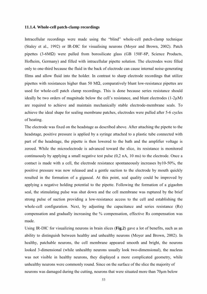
53
11.1.4. Whole-cell patch-clamp recordings
Intracellular recordings were made using the blind whole-cell patch-clamp technique
(Staley et al., 1992) or IR-DIC for visualising neurons (Moyer and Brown, 2002). Patch
pipettes (3-6MΩ) were pulled from borosilicate glass (GB 150F-8P, Science Products,
Hofheim, Germany) and filled with intracellular pipette solution. The electrodes were filled
only to one-third because the fluid in the back of electrode can cause internal noise-generating
films and allow fluid into the holder. In contrast to sharp electrode recordings that utilize
pipettes with resistances higher than 50 MΩ, comparatively blunt low-resistance pipettes are
used for whole-cell patch clamp recordings. This is done because series resistance should
ideally be two orders of magnitude below the cells resistance, and blunt electrodes (1-2µM)
are required to achieve and maintain mechanically stable electrode-membrane seals. To
achieve the ideal shape for sealing membrane patches, electrodes were pulled after 5-6 cycles
of heating.
The electrode was fixed on the headstage as described above. After attaching the pipette to the
headstage, positive pressure is applied by a syringe attached to a plastic tube connected with
part of the headstage, the pipette is then lowered to the bath and the amplifier voltage is
zeroed. While the microelectrode is advanced toward the slice, its resistance is monitored
continuously by applying a small negative test pulse (0,2 nA, 10 ms) to the electrode. Once a
contact is made with a cell, the electrode resistance spontaneously increases by10-50%, the
positive pressure was now released and a gentle suction to the electrode by mouth quickly
resulted in the formation of a gigaseal. At this point, seal quality could be improved by
applying a negative holding potential to the pipette. Following the formation of a gigaohm
seal, the stimulating pulse was shut down and the cell membrane was ruptured by the brief
strong pulse of suction providing a low-resistance access to the cell and establishing the
whole-cell configuration. Next, by adjusting the capacitance and series resistance (Rs)
compensation and gradually increasing the % compensation, effective Rs compensation was
made.
Using IR-DIC for visualizing neurons in brain slices (Fig.2) gave a lot of benefits, such as an
ability to distinguish between healthy and unhealthy neurons (Moyer and Brown, 2002). In
healthy, patchable neurons, the cell membrane appeared smooth and bright, the neurons
looked 3-dimensional (while unhealthy neurons usually look two-dimensional), the nucleus
was not visible in healthy neurons, they displayed a more complicated geometry, while
unhealthy neurons were commonly round. Since on the surface of the slice the majority of
neurons was damaged during the cutting, neurons that were situated more than 70µm below
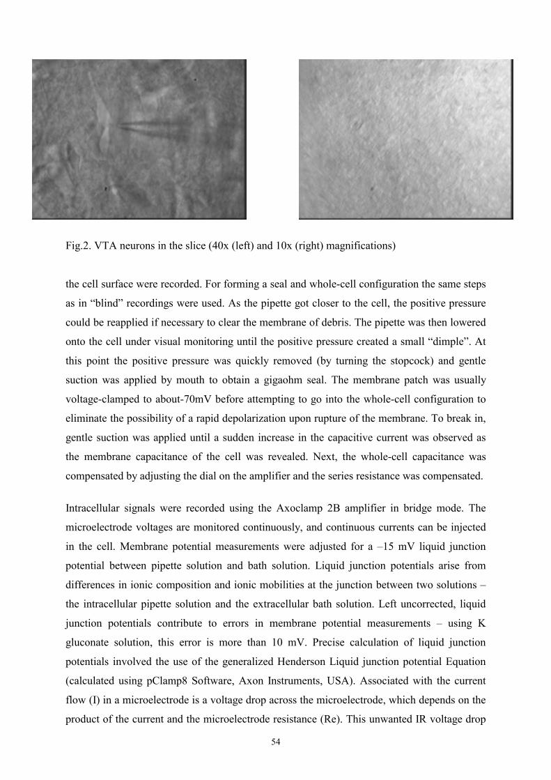
54
Fig.2. VTA neurons in the slice (40x (left) and 10x (right) magnifications)
the cell surface were recorded. For forming a seal and whole-cell configuration the same steps
as in blind recordings were used. As the pipette got closer to the cell, the positive pressure
could be reapplied if necessary to clear the membrane of debris. The pipette was then lowered
onto the cell under visual monitoring until the positive pressure created a small dimple. At
this point the positive pressure was quickly removed (by turning the stopcock) and gentle
suction was applied by mouth to obtain a gigaohm seal. The membrane patch was usually
voltage-clamped to about-70mV before attempting to go into the whole-cell configuration to
eliminate the possibility of a rapid depolarization upon rupture of the membrane. To break in,
gentle suction was applied until a sudden increase in the capacitive current was observed as
the membrane capacitance of the cell was revealed. Next, the whole-cell capacitance was
compensated by adjusting the dial on the amplifier and the series resistance was compensated.
Intracellular signals were recorded using the Axoclamp 2B amplifier in bridge mode. The
microelectrode voltages are monitored continuously, and continuous currents can be injected
in the cell. Membrane potential measurements were adjusted for a 15 mV liquid junction
potential between pipette solution and bath solution. Liquid junction potentials arise from
differences in ionic composition and ionic mobilities at the junction between two solutions
the intracellular pipette solution and the extracellular bath solution. Left uncorrected, liquid
junction potentials contribute to errors in membrane potential measurements using K
gluconate solution, this error is more than 10 mV. Precise calculation of liquid junction
potentials involved the use of the generalized Henderson Liquid junction potential Equation
(calculated using pClamp8 Software, Axon Instruments, USA). Associated with the current
flow (I) in a microelectrode is a voltage drop across the microelectrode, which depends on the
product of the current and the microelectrode resistance (Re). This unwanted IR voltage drop
55
adds to the recorded potential. The Bridge Balance control is used to balance out this voltage
drop so that only membrane potential is recorded. During the repetitive pulses from a
stimulator (Master-8), the bridge balance control was advanced from zero until the fast
voltage steps seen at the start and end of the current step were just eliminated. The bridge
balance was continuously maintained and monitored during current-clamp experiments; the
series resistance was < 50 MΩ. Before bridge balancing, capacitance neutralisation was used.
The capacitance (Cin) at the input of the headstage amplifier is due to the capacitance of the
amplifier input itself (Cin1) plus the capacitance to ground of the microelectrode and any
connecting lead (Cin2). Cin combined with the microelectrode resistance (Re) acts as a lowpass
filter for signals recorded at the tip of the microelectrode. To increase the recording
bandwidth, the technique of capacitance neutralization is used that negates Cin2 and the
effective remnant of Cin1. The capacitance neutralisation circuit attempts to inject into the
headstage input a current which it anticipates to be required to charge and discharge C in2
during signal changes. During the repetitive steps the capacitance neutralization control was
advanced as far as possible without introducing an overshoot in the step response.
A continuous registration of membrane voltage was done on a Gould TA 550 chart recorder
(Gould Electronics, Cleveland, Ohio, USA).
11.1.5. Recording of field potentials in striatum
Coronal striatal slices were prepared as described above (Fig. 1B). A bipolar Ni/Cr
stimulation electrode was placed within the dorsal or ventral striatum, the nucleus accumbens
(NAcc). Constant-current pulses of 80 µs duration (0.3-1.5mA) were applied every 20 s. 15
consecutive responses were averaged off-line to generate one data point. Stimulation strength
was adjusted to give a response, which was 50% of the maximum. Glass micropipettes filled
with ACSF (3-7MΩ) were placed in the striatum, close to the stimulation electrode, to record
extracellular field potentials. Signals were amplified, digitized at 10 kHz and recorded on disc
for off-line analysis using pClamp6 software. Field potentials consisted of two negative-going
components, referred to as N1 and N2. The N1 component represents the direct activation of
medium spiny neuron somata or their axons by the stimulating current, whereas the N2
component is synaptically mediated (Doreulee et al., 2001).
56
12. Immunocytochemistry
12.1. Immunostaining against orexin A
Deeply anaesthetized 4-5 week-old male Wistar rats were perfused transcardially with 0.9%
saline followed by 200 ml of cold 4% paraformaldehyde in 0.1 M phosphate buffer (pH 7.4).
Brains were removed and post-fixed in the same fixative overnight at 4°C. A block containing
the midbrain was cryoprotected in sucrose, cryosectioned at 34 µm thickness, and the sections
mounted on gelatine-coated slides. After drying, the sections were stained according to an
immunofluorescence staining protocol. The sections were first preincubated in phosphate
buffered saline with 0.25% Triton X-100 and 2% normal donkey serum for 90 min at room
temperature. This solution was also used to dilute an affinity-purified goat antiserum against
orexin-A (Santa Cruz Biotechnology, Heidelberg, Germany) to 1:500. This antibody-solution
was applied to the sections for 12-16 h at 4°C. After washing, the immunoreactivity was
revealed by incubation with Alexa Fluor 488-labeled donkey anti-goat IgG (1:500; Molecular
Probes, Eugene, OR) for 90 min at room temperature. After coverslipping with phosphate
buffered 50% glycerol, the slides were studied and photograped with a fluorescence
microscope equipped with the appropriate filters and a digital camera.
In control experiments it was found that preincubation of the orexin-A antiserum with
whole orexin-A peptide blocked the immunoreactivity in all studied brain regions. In those
experiments we could also exclude artifacts due to binding of secondary antibodies to the
tissue.
12.2. Staining against tyrosine hydroxylase (TH) in biocytin-filled neurons.
After recording, slices were fixed in 4% paraformaldehyde in 0.1M phosphate buffer (PB; pH
7.4) for at least 2 weeks, than transfered to 0.1 M phosphate buffer for 24 hours and 30%
sucrose in 0,1M PBS for the next 24 hours. Afterwards slices were cryoprotected in sucrose,
cryosectioned at 40 µm thickness, and the sections mounted on gelatine-coated slides. After
drying, the sections were stained according to the immunofluorescence staining protocol. The
sections were first washed in phosphate buffered saline with 0.25% Triton X-100 (PBS-T) for
5 minutes, then preincubated with 2% normal goat serum in PBS-T for 30 min at room
temperature. This solution was also used to dilute a mouse monoclonal antibody against
tyrosine hydroxylase (TH, Sigma) to 1:500. This antibody solution was applied to the sections
for 12-16 h at 4°C. After washing, sections were incubated with Alexa Fluor 488-labeled goat
57
anti-mouse IgG (1:500; Molecular Probes, Eugene, OR) to reveal the TH immunoreactivity,
and Texas Red-streptavidin (1:200; Molecular Probes) to stain biocytin-filled neurons, for 90
min at room temperature. After washing in PBS and coverslipping with phosphate buffered
50% glycerol, the slides were studied and photographed with a fluorescence microscope
equipped with the appropriate filters and a digital camera.
13. Single-cell RNA harvest and RT-PCR
For preparation of isolated cells, the ventral tegmental area was dissected from the slice and
incubated with papain (Sigma) in crude form (0.3-0.5 mg/ml) for 20 min at 37°C. Thereafter,
the tissue was placed in a bath solution with the following composition (in mM): 150 NaCl,
3.7 KCl, 2.0 CaCl2, 2.0 MgCl2, and 10 HEPES, pH 7.4. Cells were separated by gentle
pipetting. Neurons visually selected on an inverted microscope were digitally photographed.
The electrodes were fabricated from thick-walled borosilicate glass tubes and had resistances
of 2-5 MΩ, after filling with solution (in mM: 140 CsCl, 2 MgCl2, 0.5 CaCl2, 5 EGTA, and
10 HEPES/CsOH), sterilizing by autoclave, and adjusting to a pH of 7.2; the neurons were
approached with a patch electrode and a gigaohm seal was obtained. After establishing the
whole-cell configuration its cytoplasm was sucked into the electrode. This was done under
visual control to ensure that only the cytoplasm and not the entire neuron entered the
electrode. The content of the electrode was released into an Eppendorf vial with reverse-
trancriptase (RT), mixed on a Vortex mixer, centrifuged and incubated at 37oC for 3 hours.
After 1 hour 1µl of RT was added to the probe. Afterwards the probes were frozen and stored
at 30oC. Cell identification was verified by reverse transcriptase (RT)-PCR (polymerase
chain reaction) analysis of tyrosine hydroxylase (TH), GAD 65 and GAD 67 expression.
PCR allows to amplify effectively the DNA from a selected region of the genome. Every
cycle doubles the amount of DNA synthesized in the previous cycle, the products of each
cycle serve as the DNA templates for the next. Each cycle consists of the following steps
denaturation - separation of the DNA strands, annealing hybridisation of sense and antisense
primers with appropriate DNA strand and extension DNA synthesis from primers. A two-
round amplification strategy was used in each protocol. In each round 35 cycles of the
following thermal programs were used: denaturation at 94°C for 48 sec, annealing at 53°C for
1 min, and extension at 72°C for 90 sec. The thin-walled PCR tubes contained a mixture of
first-strand cDNA template (2-5 µl), 10× PCR buffer (5 µl), a 200 µM concentration each of
deoxyNTP (dNTP), a Mg2+ (2-3 mM), a 10pM concentration of each sense and antisense
primer, and 2.5 U of Taq polymerase. Sense and antisense primers (oligonucleotides) were
58
designed using BLAST and Primer Design programmes. The following criteria were used:
melting temperature 52-57 degrees, AT/GC 40-60%, length about 20 b.p.; possibility of
complementary base-pairing between the primers, self-homology and specificity of primers
for amplification of the chosen product were also tested. All oligonucleotides were
synthesized by MWG-Biotech (Ebersberg, Germany). The Taq DNA polymerase is isolated
from a thermophilic bacterium, so it is stable at high temperatures and is not denaturated by
the repeated heat treatments. We used Platinum Taq DNA Polymerase that is recombinant
Taq DNA polymerase complexed with proprietary antibody that inhibits polymerase activity.
Due to specific binding of the inhibitor, this Taq polymerase is in inactive form until heat
denaturation is performed. It provides an automatic hot start that increases sensitivity,
specificity and yield of reaction.
The final reaction volume was adjusted to 10 µl with nuclease-free water (Promega, Madison,
WI). For the second amplification round, 1 µl of the product from the first amplification was
used as a template. The Taq enzyme, PCR buffer, Mg2+ solution, and four dNTPs were all
purchased from Qiagen (Erkrath, Germany). Annealing temperature and Mg concentration
were empirically modified to find optimal conditions that result in specific PCR products.
Amplifications were performed on a thermal cycler (Mastercycler; Eppendorf).
The protocols of the RT reaction and PCR amplification were similar to those described
previously (Vorobjev et al., 2000). Sequencing of the amplification products, which was done
on an automatic sequencer (model 377; Applied Biosystems International, Weiterstadt,
Germany), revealed the identity of the ampified products (by comparison with known
sequences of a given product in the GenBank). The following primers were used (from 5´to
3´): TH (sense): GCTGTCACGTCCCCAAGGTT; antisense:
AAGCGCACAAAATACTCCAGG; antisense2 (for the second round):
CAGCCCGAGACAAGGAGGAG (size of the product was 220 b.p.); for the PCR analysis of
GAD expression in the first round of amplification the degenerated antisense primer:
CCCCAAGCAGCATCCACAT was taken either with GAD65 cDNA-specific sense primer -
TCTTTTCTCCTGGTGGTGCC or with GAD67 cDNA-specific sense primer
TACGGGGTTCGCACAGGTC; for the second round the same sense primers were used in
combination with specific antisense primers CAGTGGTTCCAGCTGTGGC for GAD65
and CGGTTGCATTGACATAAAGGG for GAD67. The following primers were used for the
PCR analysis of orexin receptor expression: in the first round of amplification the degenerated
primer "dg up" [5'-CTGGC(AT)GATGTGCT(GT)GTGAC-3'] was taken either with OR1
cDNA-specific lower 1 primer (5'-AACAGCAGAGGGTGGCAGAT-3') or with OR2 cDNA-
59
specific lower 1 primer (5'-TGGCTGTGCTCTTGAACATC-3'). For the second round of
cDNA amplification, the primers for the OR1 were upper 2 (5'-
TGTTAGTGGACATCACCGAATC-3') and lower 2 (5'-TGAAGCTGAGAGTCAGCACTG-
3'); for the OR2, the lower 2 primer (5'-GGCAATGCAGCTCAATGTAA-3') was used in
combination with the degenerated primer dg up. Primers for the 1st amplification of calbindin
(CB) and neuropeptide Y (NPY) were described in (Cauli et al., 1997): CB (sense) -
AGGCACGAAAGAAGGCTGGAT, CB (antisense) TCCCACACATTTTGATTCCCTG;
NPY (sense) GCCCAGAGCAGAGCACCC, NPY (antisense)
CAAGTTTCATTTCCCATCACCA; for the 2-d amplification the same lower primers were
used in combination with designed CB (sense2): TCCTGCTGCTCTTTCGATGC (size of
product 303 b.p.) or NPY (sense2): GCTCGTGTGTTTGGGCATTCT (251 b.p.). The
following primers for MC4 receptors were used: MC4R (sense) -
GAACAATTCTAGTGATCGTGGCG; MC4R (antisense)
GCAGAAGCCTGATTCCACTG, MC4R (antisense for 2d amplification)
GTGCAAGCTGCCCAGATACA; CART (sense)-AGCGAGGAAGTCCAGCAC, CART
(antisense)- AAGCATGTGCAAATTACACCC, CART (antisense for the 2d amplification)-
CCGAAGGAGGCTGTCACC; substance P (SP, sense)
TGAGCATCTTCTTCAGAGAATCGC, SP (antisense)
ATCGCTGGCAAACTTGTACAACTC, SP (sense for the 2-d amplification)
ATTAATGGGCAAACGGGATGCT.
The following primers were used for simultaneous detection of TRPC channels: In the first
round of amplification all 7 cDNAs of TRPC channel subunits were competitively amplified
with degenerate primers: Dg up: 5´-TGGGGCC(T/C/A)(T/C)TGCAGA
T(A/C)TC(T/A)CTGGGA-3´ and Dg lo: 5´-(G/T)G(A/T)TCG (A/G)GCAAA(C/T)TTCCA
(C/T)TC-3´. In the second round 4 subfamilies were amplified separately with the following
primers: TRPC1 up: 5´-CAG ATGGGACAGATGTTACAAGA -3´ and Dg lo (expected size
of the product 412 b.p.), TRPC2up: 5´-GACATGATCCGGCTCATGTTCAT-3´ and TRPC2
lo: 5´-AGCACAATGACCATGACG-3´(expected size of product 248 b.p.), TRPC4/5 up: 5´-
GAATGCT(G/C)C T(G/T)GA(T/C)AT(A/C)CT(A/C)AA-3´ and Dg lo (size of products 389
and 398 b.p.), TRPC3/6/7 up: 5´-GACAT(A/C)TTCAAGTTCATG(G/T)TC(C/A)T -3´and
Dg lo (size of products 344 and 345 b.p.). Products of the second amplification: TRPC1,
TRPC2 and most of the single cell-derived amplimers of TRPC4/5 and TRPC3/6/7
amplifications were successfully sequenced in the sense direction. Positive controls contained
60
a mixture of 2 or 3 TRPC isoforms in the case of TRPC4/5 and TRPC3/6/7 subfamilies;
therefore they could not be sequenced. In contrast, single neurons expressed as a rule only one
member of these two subfamilies. The obtained sequences were found to match known ones
(GENBANK accession numbers): rat TRPC1 (AF061266), TRPC2 (AF136401), TRPC3
(AB022331), TRPC4 (AF421368), TRPC5 (AF061876), TRPC6 (AB051213) or high
homology with the mouse TRPC7 (AF139923) cDNA. We have submitted the rat TRPC7
cDNA sequence to the GENBANK under accession number AY157999. The identity of
amplified TRPC1 cDNA in single cells was verified by digestion of the obtained amplimers
with Hha I (223 and 189 b.p. fragments could be seen on the gel).
Competitively amplified cDNAs within each of two subfamilies (TRPC4/5 and TRPC3/6/7)
were further digested with the subtype-specific enzymes: MluI (TRPC4-specific, cut
TRPC4/5 in two fragments with 145 and 244 b.p. size), BsrSl I (TRPC5-specific, cut
TRPC4/5amplimer in two fragments of 52 and 346 b.p. size), Vsp I (TRPC3-specific,
TRPC3/6/7 into 61 and 283 b.p.), NcoI (TRPC7-specific, cut TRPC3/6/7 into 98 and 247
b.p.). We failed to find a specific restriction enzyme for the TPRC6 subunit, however MboI
proved to be an optimal tool for identification of cDNAs within the TRPC3/6/7 subfamily. It
cut TRPC6 into 64 and 281 b.p., TRPC7 into 50, 231, 64 b.p and TRPC3 into 156 and 188
b.p. fragments.
The identity of cDNA sequences was revealed by sequencing of the second round
amplification products. Results of amplification were analyzed by agarose gel (1.5%)
electrophoresis and staining with ethidium bromide gels. All products of the second round of
amplification were purified (PCR purification kit from Qiagen) in water and subjected to
sequencing in both directions.
14. Drugs
Drugs used in this study were: Orexin A and B, Melanin concentrating hormone,
Neuropeptide Y, Cocaine and amphetamine regulated transcript, Leptin, Corticotropin-
releasing factor, alpha-Melanocyte stimulating hormone, Substance P, Agouti-related protein
(all from Bachem, Germany), Chelerythrine chloride((Sigma, Germany), DAMGO (Tocris,
UK), H7 (Alexis, Switzerland), H89 dihydrochloride (Alexis, Switzerland), Melanin
concentrating hormone , Orexin A and B (Bachem, Germany), Quinpirole (RBI, USA),
Thapsigargin (Alexis, Switzerland), histamine dihydrochloride (Fluka), mepyramine maleate
(Sigma), cimetidine, thioperamide maleate, bicuculline methochloride and CGP55845 (all
obtained from Tocris). All other chemicals were obtained from Merck (Germany). Drugs
61
were stored at room temperature or fridge, dissolved in water (with the exception of
CGP55845, thapsigargin, KT5720 and modafinil, dissolved in DMSO. The final
concentration of DMSO in the bath was 0.1 %. This concentration of DMSO did not affect the
firing rate of cells in control recordings). All peptides were dissolved in 0.9% saline to a
concentration of 10 µM and the resulting solution was stored as frozen aliquots at 20 °C.
Drugs were bath applied. The aliquots were defrozen immediately before use and diluted in
10ml of ACSF in a medical syringe that was connected by a plastic tube with the recording
chamber.
15. Experimental protocols and statistical analysis
Neurons were recorded for at least 15 min to obtain a stable baseline before a bath application
of drugs. All recorded neurons displayed a regular firing pattern; burst firing was never
observed in control recordings. Only one cell per slice was recorded; one, rarely two slices
from one rat were used. The electrophysiological part of the study was made on 348 rats and
389 slices. If two slices from the same rat were used, different experiments (application of
different drugs or recording of different cell types DAergic or GABAergic) were performed.
Effects of orexins, histamine and modafinil were studied only in drug-naive slices. During
investigation of the signal transduction mechanism of orexin-induced excitation of SN and
VTA, as well as during the study of histamine receptor involvement in histamine-induced
inhibition of DAergic midbrain neurons, the baseline was recorded for 15 min to be sure that
the firing rate of neuron is stable, then one of the inhibitors of signal transduction (for details
see section 16.2.5.) was applied and neuronal activity was recorded for the next 15 min to rule
out an effect of the inhibitor on firing frequency, and, finally, orexin was applied. This part of
the study was performed only with extracellular single-unit recordings to avoid dialysis of the
cells that takes place during long-term whole-cell patch-clamp recordings. In the last part of
study (section 16.6) two types of experiments were made: effects of several neuropeptides on
the same cell were tested by extracellular single-unit recordings: after recording of the
baseline for 15 min the first peptide was applied, after the end of the effect (return of the
baseline to the initial value) and a stable baseline for the next 15 minutes the second drug was
applied and so on. The order of drug application was changed each time to be sure that the
previous application did not affect the next one, the action of each neuropeptide in drug-naïve
cells was also tested. In the next part of the study whole-cell recordings were used to study the
effect of these peptides on the membrane potential of VTA cells.
62
At the end of each experiment the D2 receptor agonist quinpirole (10 µM) or dopamine (30
µM) (it does not matter whether to apply quinpirole or dopamine because they both are D2
receptor agonists) or the µ- receptor agonist Tyr-D-Ala-Gly-NMe-Phe-Gly-ol (DAMGO, 1
µM) were applied to obtain one more evidence for the type of recorded neuron DAergic
neurons in SN and VTA are known to be inhibited by quinpirole or dopamine, but not by
DAMGO, whereas GABAergic VTA neurons - by DAMGO, but not by D2 receptor agonists
(Grace et al., 1988; Lacey et al., 1989) The order of application of these substances was
chosen during the experiment - these two types of neurons have a number of
electrophysiological differences (for details see section 16.1), so one can hypothesize with a
high rate of predictability which type of neuron is recorded and apply a substance which is
likely to inhibit this cell. If, however, the cell was not inhibited by it in 15 minutes, the
inhibitor of the other subtype was applied. Such protocol is routinely used for distinguishing
between midbrain DAergic and GABAergic neurons in most in vitro studies(Grace, 1991;
Johnson et al., 1992 , Yin et al., 2000, Neuhoff et al., 2002; and others).
The following statistical analysis was performed: the significance of the effect of a given
substance (comparison of firing rate or membrane potential of the same cell before and after
the application of drug) was tested with Students t-test (paired). All experiments where data
from different cells were compared - electrophysiological differences between subtypes of
neurons, differences between the effects of orexin A and orexin B, effects of inhibition of
signal transduction on orexin-induced excitation, blocking of histamine receptors on
histamine-induced inhibition, size of neurons with different molecular profile were studied
with Students t-test (unpaired). The relationships between expression of mRNA for
neuropeptides, their receptors, calbindin and markers for dopamine and GABA, were
investigated with χ2 test. Data are presented as mean ± standard error of the mean (SEM).
63
16. Results
16.1. Electrophysiological characterization of the recorded neurons
As described in the introduction, there are two types of neurons in SN and VTA:
dopaminergic (DAergic) and nondopaminergic (presumed GABAergic) which can be
distinguished according to their electrophysiological and pharmacological properties (Grace
and Onn, 1989, Klink et al., 2001).
16.1.1. Properties of DAergic neurons in VTA and SN.
Extracellular single-unit recordings in VTA revealed that presumed dopaminergic neurons
had a regular spontaneous activity, fired at lower frequencies in comparison with GABAergic
neurons (2.23 ± 0.40 Hz, n=25), had broader action potentials (>2 ms) and were inhibited by
the D2 receptor agonist quinpirole (10 µM) or dopamine (30 µM). Dopaminergic cells in the
SN had a firing frequency of 2.3 ± 0.4 Hz and a spike width of 2.63 ± 0.37 ms (n = 9, Fig.3A,
C) and were also inhibited by quinpirole or dopamine; there were no differences in firing rate
and spike width between DAergic neurons in SN and VTA (Korotkova et al., 2002a).
DAergic neurons in the VTA recorded in whole-cell mode possessed the following
characteristics: average firing frequency was 2.86 ± 0.32 Hz - burst firing or spontaneous
alterations in firing rate were never observed in control cells; spike width was significantly
(p<0.0005) broader in dopaminergic neurons (3.3 ± 0.13 ms, n=14) than in GABAergic
neurons (1.68 ± 0.23 ms, n=7) and spike thresholds were significantly (p<0.05) more positive
in dopaminergic (-44.7 ± 1.4 mV, n=14) than in GABAergic cells (-50.8±0.7mV, n=7).
Dopamine cells possessed a prominent Ih current. Ih sag, measured as the percentage reduction
from the peak at the end of a 1 s long step elicited by a 400 pA current injection was 33.1 ±
3.4 % (Fig.4B). Input resistance was 266 ± 48 MΩ (n=14). They were inhibited by dopamine
or quinpirole. The posthoc double stainings also revealed that recordings had been made from
TH positive (n=14, Fig.4A) cells, which are assumed to be dopaminergic cells (Korotkova et
al., 2003).
16.1.2. Properties of GABAergic neurons in VTA and SN.
All recorded nondopaminergic (presumed GABAergic) neurons had a stable spontaneous
activity. In SN GABAergic cells had a firing frequency of 8.6 ± 0.8 Hz and a spike width of

64
1.42 ± 0.17 ms (n = 20, Fig.3 B,D). Their firing rate was unaffected by application of
quinpirole (10 µM) or dopamine (30 µM). This group in SN was uniform while in VTA the
GABAergic cells could be divided into two groups according to their firing rate. One group
fired at a relatively high frequency (7.31 ± 1.35 Hz, n=10, Fig.4C,D), the second group
consisted of slow-firing cells (0.89 ± 0.33 Hz, n = 6, Fig.4 E,F). Both groups had the same
action potential width (<1.5 ms), were unaffected by quinpirole or dopamine and inhibited by
the µ- receptor agonist Tyr-D-Ala-Gly-NMe-Phe-Gly-ol (DAMGO, 1 µM), that provides
further evidence for their GABAergic nature (detals were described above, in section 15).
Further investigation of properties of these two groups by intracellular recordings in current-
clamp whole-cell mode displayed that spike width was significantly more narrow than in
DAergic neurons (1.68 ± 0.23 ms, n=7 vs 3.3 ± 0.13 ms, n=14 in DA neurons, p<0.0005),
spike thresholds were more negative (-50.8 ± 0.7 mV, n=7 vs -44.7 ± 1.4 mV, n=14 in DA
neurons, p<0.05), they did not possess a prominent Ih current: Ih sag, measured as the
percentage reduction from the peak at the end of a 1 s long step elicited by a 300 pA current
injection that led to a similar amount of peak hyperpolarization as in DAergic neurons (~ -
120mV) amounted only to 5.0 ± 0.2 % vs 33.1 ± 3.4 % in DA cells. Analysis of possible
differences between characteristics of GABAergic cells with different firing rates was
performed. One group fired at a relatively high frequency: 8.7 ± 2.2 Hz (n=3; Fig.4D), that
did not differ significantly from frequency of fast-firing GABAergic VTA cells, recorded
extracellularly and described above (7.31 ± 1.35 Hz, n=10) . The second group consisted of
slow-firing cells: 0.77 ± 0.37 Hz (n=4; Fig.4F), which frequency did not differ significantly
from slow-firing GABAergic VTA cells, recorded extracellularly and described above (0.89 ±
0.33 Hz, n = 6). Both groups of cells fired at high frequency (>30Hz) during depolarizing
current steps; this frequency was not different in the two groups. Cells in both groups were
inhibited by the µ- receptor agonist Tyr-D-Ala-Gly-NMe-Phe-Gly-ol (DAMGO, 1 µM) and
unaffected by quinpirole (10 µM) or dopamine (30 µM), that provides further evidence for
their GABAergic nature (detals were described above, in section 15). The level of DAMGO-
induced hyperpolarization did not significantly differ in fast-fired (6.4±1.2mV) and slow-fired
(6.1±0.8mV) neurons. These two groups of non-dopamine cells differed in AHP amplitude
(10.7 ± 0.9 vs 14 ± 0.9 mV in fast-firing and slow-firing cells, respectively, p<0.05), and
spike threshold (-52.2 ± 0.4 and 49.4 ± 0.2 mV, respectively, p<0.005). Spike width did not
differ. Input resistance also did not differ either in dopaminergic (266 ± 48 MΩ, n=14) or
GABAergic cells (197 ± 19 MΩ, n=7), or between GABAergic cells with different firing
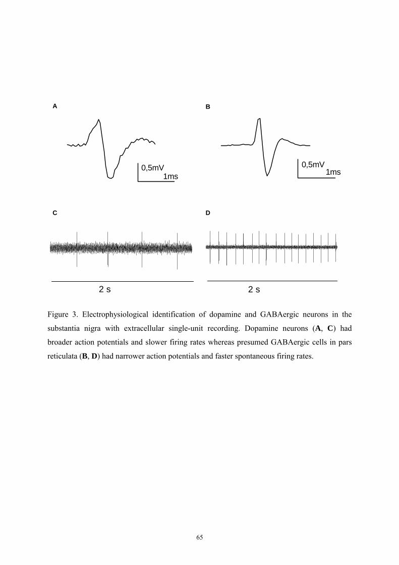
65
Figure 3. Electrophysiological identification of dopamine and GABAergic neurons in the
substantia nigra with extracellular single-unit recording. Dopamine neurons (A, C) had
broader action potentials and slower firing rates whereas presumed GABAergic cells in pars
reticulata (B, D) had narrower action potentials and faster spontaneous firing rates.
0,5mV1ms
0,5mV1ms
A B
C D
2 s 2 s
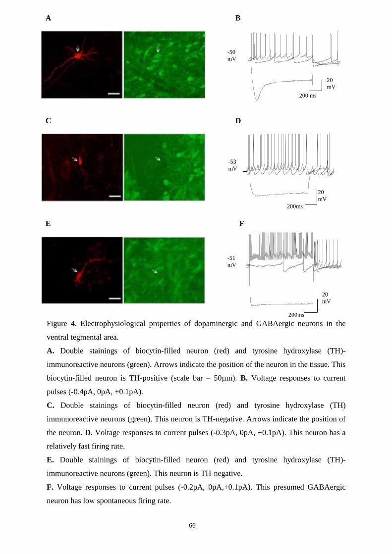
66
Figure 4. Electrophysiological properties of dopaminergic and GABAergic neurons in the
ventral tegmental area.
A. Double stainings of biocytin-filled neuron (red) and tyrosine hydroxylase (TH)-
immunoreactive neurons (green). Arrows indicate the position of the neuron in the tissue. This
biocytin-filled neuron is TH-positive (scale bar – 50µm). B. Voltage responses to current
pulses (-0.4pA, 0pA, +0.1pA).
C. Double stainings of biocytin-filled neuron (red) and tyrosine hydroxylase (TH)
immunoreactive neurons (green). This neuron is TH-negative. Arrows indicate the position of
the neuron. D. Voltage responses to current pulses (-0.3pA, 0pA, +0.1pA). This neuron has a
relatively fast firing rate.
E. Double stainings of biocytin-filled neuron (red) and tyrosine hydroxylase (TH)-
immunoreactive neurons (green). This neuron is TH-negative.
F. Voltage responses to current pulses (-0.2pA, 0pA,+0.1pA). This presumed GABAergic
neuron has low spontaneous firing rate.
200ms
20 mV
-53 mV
C D
200 ms
20 mV
-50 mV
B A
E
20 mV
200ms
-51 mV
F
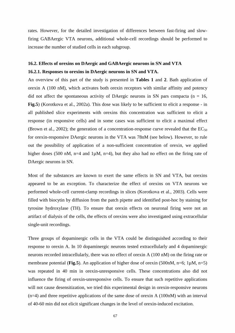
67
rates. However, for the detailed investigation of differences between fast-firing and slow-
firing GABAergic VTA neurons, additional whole-cell recordings should be performed to
increase the number of studied cells in each subgroup.
16.2. Effects of orexins on DAergic and GABAergic neurons in SN and VTA
16.2.1. Responses to orexins in DAergic neurons in SN and VTA.
An overview of this part of the study is presented in Tables 1 and 2. Bath application of
orexin A (100 nM), which activates both orexin receptors with similar affinity and potency
did not affect the spontaneous activity of DAergic neurons in SN pars compacta (n = 16,
Fig.5) (Korotkova et al., 2002a). This dose was likely to be sufficient to elicit a response - in
all published slice experiments with orexins this concentration was sufficient to elicit a
response (in responsive cells) and in some cases was sufficient to elicit a maximal effect
(Brown et al., 2002); the generation of a concentration-response curve revealed that the EC50
for orexin-responsive DAergic neurons in the VTA was 78nM (see below). However, to rule
out the possibility of application of a non-sufficient concentration of orexin, we applied
higher doses (500 nM, n=4 and 1µM, n=4), but they also had no effect on the firing rate of
DAergic neurons in SN.
Most of the substances are known to exert the same effects in SN and VTA, but orexins
appeared to be an exception. To characterize the effect of orexins on VTA neurons we
performed whole-cell current-clamp recordings in slices (Korotkova et al., 2003). Cells were
filled with biocytin by diffusion from the patch pipette and identified post-hoc by staining for
tyrosine hydroxylase (TH). To ensure that orexin effects on neuronal firing were not an
artifact of dialysis of the cells, the effects of orexins were also investigated using extracellular
single-unit recordings.
Three groups of dopaminergic cells in the VTA could be distinguished according to their
response to orexin A. In 10 dopaminergic neurons tested extracellularly and 4 dopaminergic
neurons recorded intracellularly, there was no effect of orexin A (100 nM) on the firing rate or
membrane potential (Fig.5). An application of higher dose of orexin (500nM, n=6; 1µM, n=5)
was repeated in 40 min in orexin-unresponsive cells. These concentrations also did not
influence the firing of orexin-unresponsive cells. To ensure that such repetitive applications
will not cause desensitization, we tried this experimental design in orexin-responsive neurons
(n=4) and three repetitive applications of the same dose of orexin A (100nM) with an interval
of 40-60 min did not elicit significant changes in the level of orexin-induced excitation.
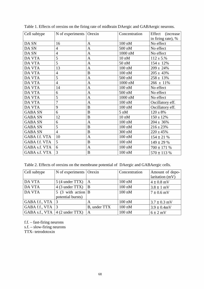
68
Table 1. Effects of orexins on the firing rate of midbrain DAergic and GABAergic neurons.
Cell subtype N of experiments Orexin Concentration Effect (increase in firing rate), %
DA SN 16 A 100 nM No effect DA SN 4 A 500 nM No effect DA SN 4 A 1000 nM No effect DA VTA 5 A 10 nM 112 ± 5.% DA VTA 5 A 50 nM 154 ± 12% DA VTA 13 A 100 nM 209 ± 24% DA VTA 4 B 100 nM 205 ± 43% DA VTA 5 A 500 nM 258 ± 13% DA VTA 4 A 1000 nM 266 ± 11% DA VTA 14 A 100 nM No effect DA VTA 6 A 500 nM No effect DA VTA 5 A 1000 nM No effect DA VTA 7 A 100 nM Oscillatory eff. DA VTA 9 B 100 nM Oscillatory eff. GABA SN 6 B 5 nM 120 ± 8% GABA SN 12 B 10 nM 150 ± 12% GABA SN 6 A 100 nM 204 ± 36% GABA SN 5 B 100 nM 216 ± 23% GABA SN 4 B 300 nM 220 ± 45% GABA f.f. VTA 10 A 100 nM 154 ± 21 % GABA f.f. VTA 5 B 100 nM 149 ± 29 % GABA s.f. VTA 6 A 100 nM 700 ± 171 % GABA s.f. VTA 3 B 100 nM 570 ± 113 %
Table 2. Effects of orexins on the membrane potential of DAergic and GABAergic cells.
Cell subtype N of experiments Orexin Concentration Amount of depo- laritation (mV)
DA VTA 5 (4 under TTX) A 100 nM 4 ± 0.8 mV DA VTA 4 (3 under TTX) B 100 nM 3.8 ± 1 mV DA VTA 5 (3 with action
potential bursts) B 100 nM 7 ± 0.6 mV
GABA f.f., VTA 3 A 100 nM 3.7 ± 0.3 mV GABA f.f., VTA 3 B, under TTX 100 nM 3.9 ± 0.4mV GABA s.f., VTA 4 (2 under TTX) A 100 nM 6 ± 2 mV f.f. – fast-firing neurons s.f. – slow-firing neurons TTX- tetrodotoxin
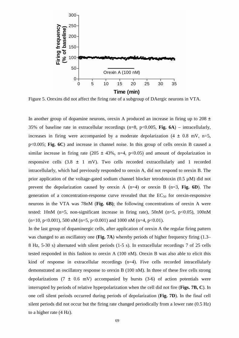
69
Figure 5. Orexins did not affect the firing rate of a subgroup of DAergic neurons in VTA.
In another group of dopamine neurons, orexin A produced an increase in firing up to 208 ±
35% of baseline rate in extracellular recordings (n=8, p<0.005, Fig. 6A) – intracellularly,
increases in firing were accompanied by a moderate depolarization (4 ± 0.8 mV, n=5,
p<0.005; Fig. 6C) and increase in channel noise. In this group of cells orexin B caused a
similar increase in firing rate (205 ± 43%, n=4, p<0.05) and amount of depolarization in
responsive cells (3.8 ± 1 mV). Two cells recorded extracellularly and 1 recorded
intracellularly, which had previously responded to orexin A, did not respond to orexin B. The
prior application of the voltage-gated sodium channel blocker tetrodotoxin (0.5 µM) did not
prevent the depolarization caused by orexin A (n=4) or orexin B (n=3, Fig. 6D). The
generation of a concentration-response curve revealed that the EC50 for orexin-responsive
neurons in the VTA was 78nM (Fig. 6B); the following concentrations of orexin A were
tested: 10nM (n=5, non-significant increase in firing rate), 50nM (n=5, p<0.05), 100nM
(n=10, p<0.001), 500 nM (n=5, p<0.001) and 1000 nM (n=4, p<0.01).
In the last group of dopaminergic cells, after application of orexin A the regular firing pattern
was changed to an oscillatory one (Fig. 7A) whereby periods of higher frequency firing (1.3–
8 Hz, 5-30 s) alternated with silent periods (1-5 s). In extracellular recordings 7 of 25 cells
tested responded in this fashion to orexin A (100 nM). Orexin B was also able to elicit this
kind of response in extracellular recordings (n=4). Five cells recorded intracellularly
demonstrated an oscillatory response to orexin B (100 nM). In three of these five cells strong
depolarizations (7 ± 0.6 mV) accompanied by bursts (3-6) of action potentials were
interrupted by periods of relative hyperpolarization when the cell did not fire (Figs. 7B, C). In
one cell silent periods occurred during periods of depolarization (Fig. 7D). In the final cell
silent periods did not occur but the firing rate changed periodically from a lower rate (0.5 Hz)
to a higher rate (4 Hz).
0 5 10 15 20 25 30 350
50
100
150
200
250
300
Orexin A (100 nM)
Time (min)
Firin
g fr
eque
ncy
(% o
f bas
elin
e)
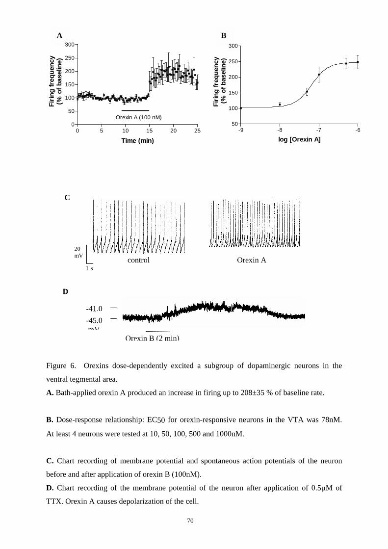
Figure 6. Orexins dos
ventral tegmental area.
A. Bath-applied orexin A
B. Dose-response relati
At least 4 neurons were
C. Chart recording of m
before and after applicat
D. Chart recording of t
TTX. Orexin A causes d
)
-41.0 -45.0 mV
B A
0 5 10 15 20 250
50
100
150
200
250
300
Orexin A (100 nM)
Time (min)
Firin
g fr
eque
ncy
(% o
f bas
elin
e)
-9 -8 -7 -650
100
150
200
250
300
log [Orexin A]
Firin
g fr
eque
ncy
(% o
f bas
elin
e)
20 mV
s A l
C
D
Orexin B (2 min
1
70
e-dependently excited a subgroup
produced an increase in firing up
onship: EC50 for orexin-responsiv
tested at 10, 50, 100, 500 and 1000n
embrane potential and spontaneo
ion of orexin B (100nM).
he membrane potential of the neu
epolarization of the cell.
Orexin
controof dopaminergic neurons in the
to 208±35 % of baseline rate.
e neurons in the VTA was 78nM.
M.
us action potentials of the neuron
ron after application of 0.5µM of
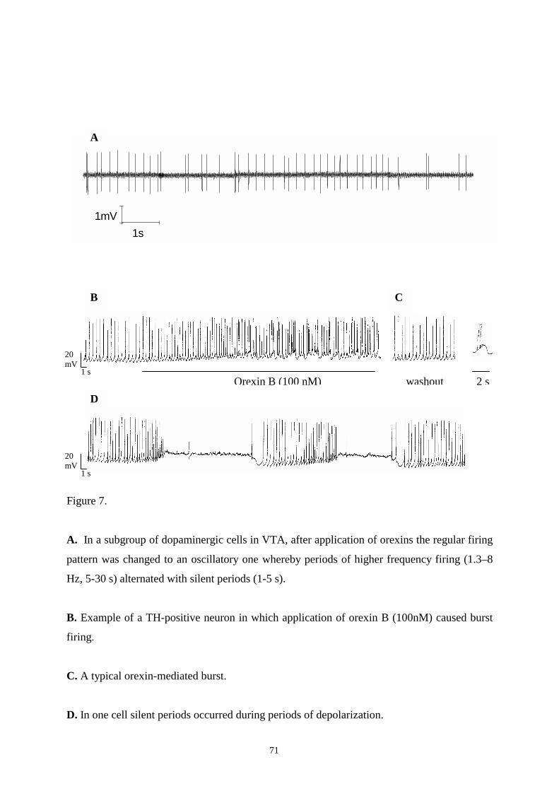
71
Figure 7.
A. In a subgroup of dopaminergic cells in VTA, after application of orexins the regular firing
pattern was changed to an oscillatory one whereby periods of higher frequency firing (1.3–8
Hz, 5-30 s) alternated with silent periods (1-5 s).
B. Example of a TH-positive neuron in which application of orexin B (100nM) caused burst
firing.
C. A typical orexin-mediated burst.
D. In one cell silent periods occurred during periods of depolarization.
1s1mV
2 s 1 s
20 mV
Orexin B (100 nM) washout
1 s
20 mV
A
B
D
C

72
16.2.2. Responses to orexins in GABAergic cells in SN.
The action of orexins on GABAergic cells appeared to be uniform. In contrast to its lack of
effect on DAergic neurons, application of orexin A (100 nM) led to a large increase in the
firing frequency of GABAergic neurons in SN pars reticulata to 204 ± 36 % of baseline (n =
6, p<0.001 Fig. 8A). The application of orexin B (5 - 300 nM), which has a higher affinity for
the type II orexin receptor than the type I receptor also led to a large and dose-dependent
increase in the firing frequency of GABAergic neurons in SN pars reticulata (n = 27, Fig. 8B,
for details see Table 1). The maximal increase in firing rate elicited by 100 nM orexin A (204
± 36 %) and orexin B (211 ± 17 %, n = 5, p<0.005) were not significantly different. The EC50
for orexin B was found to be 12 nM (Fig. 8C). None of the GABAergic cells tested were
unaffected by the orexins.
16.2.3. Orexin-immunoreactive fibers are present in SN.
This uniform action of orexins on firing rate of GABAergic neurons in SN pars reticulata, was
unexpected because a previous study failed to find evidence of orexin fibres in the substantia
nigra pars reticulata (Peyron et al., 1998). We re-examined the distribution of orexin-positive
fibres in the substantia nigra and ventral tegmental area. Three rats were used for this study.
We considered this number of animals to be sufficient because the purpose of this study was
to confirm the presence of orexin fibres in SN rather than to give a detailed analysis of them.
The most rostral part of the SN reticulata was almost devoid of orexin-immunoreactive fibers,
while the density of stained fibers increased caudally (Fig. 8D). However, even in the caudal
part of the SNr, the fibre density was much lower than in the VTA (Fig. 8D). Therefore, we
confirmed the presence of orexin fibres in the caudal part of substantia nigra pars reticulata.
The detailed quantitative analysis of orexins’ immunohistochemistry was not the purpose of
this study.
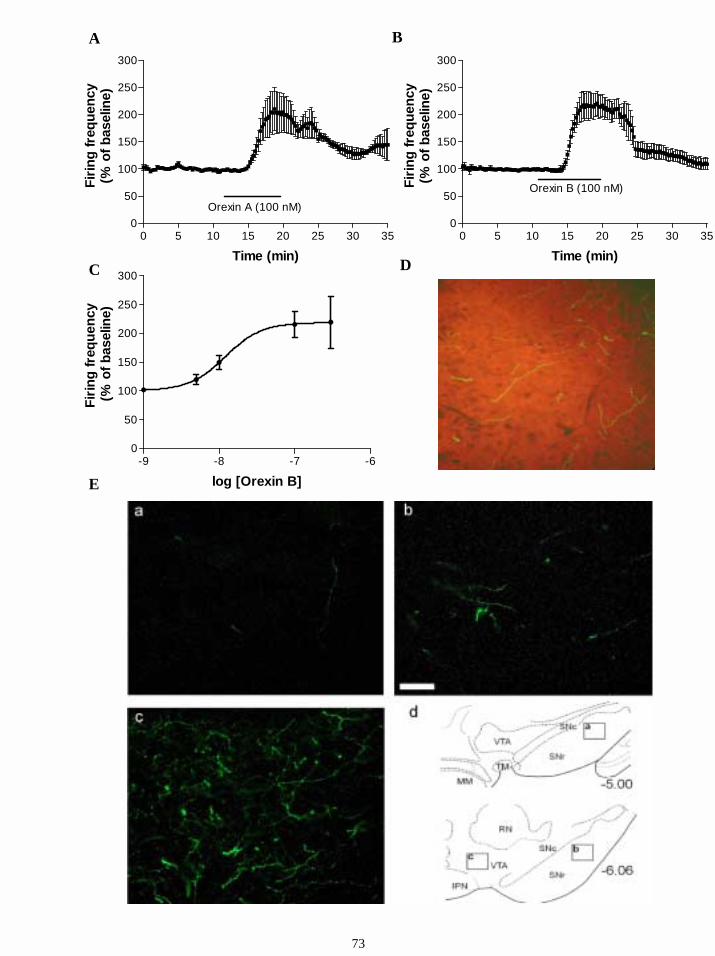
E
0 5 10 15 20 25 30 350
50
100
150
200
250
300
Orexin A (100 nM)
Time (min)
Firin
g fr
eque
ncy
(% o
f bas
elin
e)
0 5 10 15 20 25 30 350
50
100
150
200
250
300
Orexin B (100 nM)
Time (min)
Firin
g fr
eque
ncy
(% o
f bas
elin
e)
-9 -8 -7 -60
50
100
150
200
250
300
log [Orexin B]
Firin
g fr
eque
ncy
(% o
f bas
elin
e)
A B
C D
73
74
Figure 8. Orexins uniformly excite GABAergic cells in substantia nigra
A. Application of orexin A (100 nM) led to a large increase in the firing frequency of
GABAergic neurons in SN pars reticulata to 204 ± 36 % of baseline. B. The application of
orexin B also led to a large and dose-dependent increase in the firing frequency of
GABAergic neurons in SN pars reticulata. C. Dose-response relationship. EC50 was 12 nM
and Hill slope 1.8. At least 4 neurons were tested at 1, 5, 10, 100 and 300nM. D. Orexin
immunolabeling showed the presence of orexin fibres (green) in substanta nigra pars
reticulata (red).
E. Staining demonstrating the presence of orexin fibres in substantia nigra pars reticulata. The
density of immunoreactive fibres is very low in the rostral substantia nigra (a) while the
density is clearly higher in the caudal parts (b). The much higher density of orexin fibres in
the ventral tegmental area is shown in (c). In (d) the locations of (a-c) are indicated. The
rostrocaudal level is given as the distance from bregma. IPN, interpeduncular nucleus; MM,
medial mammillary nucleus, RN, red nucleus, SNc/r, substantia nigra compacta/reticulata;
TM, tuberomammillary nucleus; VTA, ventral tegmental area. Scale bar: 50 µM.
16.2.4. Effects of orexins on GABAergic neurons in VTA
In the VTA two groups of GABAergic cells were found (Figs. 4D, 4F). Both groups of
presumed GABAergic cells were excited by orexins. In extracellular recordings, application
of orexin A (100 nM) to the fast firing cells caused an increase in the firing frequency to 154
± 21 % of baseline (n=10, p<0.005, Fig. 9B). Orexin B (100 nM) also increased the firing of
these cells (n=5, p<0.01, 149 ± 29 %, not significantly different from orexin A). In
intracellular recordings, the application of orexins caused a depolarization of 3.7 ± 0.3 mV
(n=3, Fig. 9A). A similar amount of depolarization (3.9±0.4mV) was seen in the presence of
TTX (n=3, Fig. 9C).
In extracellular recordings from the slow-firing GABAergic cells application of orexin A (100
nM) caused a very large increase in firing rate to 700 ± 171 % of baseline (n=6, p<0.0005,
Fig. 9E). Orexin B increased the firing rate to 570 ± 113 % of baseline (Fig. 9F, n=3, not
significantly different from orexin A). After washout of orexins and stabilization of the firing
rate, the frequency of firing was still higher than before application of orexins, and did not
return to the baseline level during 1 h of washout. In intracellular recordings orexin A (100
nM) caused a depolarization of 6 ± 2 mV (n=4, p<0.05). The depolarization was not blocked
by tetrodotoxin (0.5 µM, n=2).
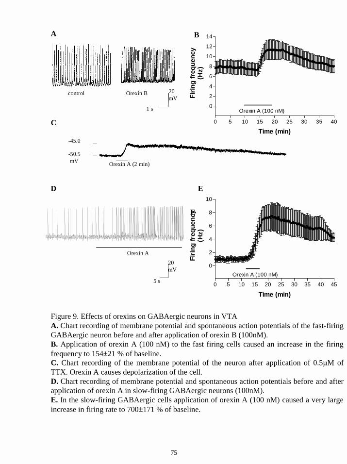
0 5 10 15 20 25 30 35 40 45
0
2
4
6
8
10
Orexin A (100 nM)
Time (min)
Firin
g fr
eque
ncy
(Hz)
Orexin A
0 5 10 15 20 25 30 35 40
0
2
4
6
8
10
12
14
Orexin A (100 nM)
Time (min)
Firin
g fr
eque
ncy
(Hz)
control
1 s
20mV
Orexin B
-50.5 mV
-45.0
Orexin A (2 min)
A B
C
E
D E
Figure 9. Effects of orexins on GABAergic neurons in VTAA. Chart recording of membrane potential and spontaneous action potentials of the fast-firing GABAergic neuron before and after application of orexin B (100nM).B. Application of orexin A (100 nM) to the fast firing cells caused an increase in the firing frequency to 154±21 % of baseline.C. Chart recording of the membrane potential of the neuron after application of 0.5µM of TTX. Orexin A causes depolarization of the cell.D. Chart recording of membrane potential and spontaneous action potentials before and after application of orexin A in slow-firing GABAergic neurons (100nM).E. In the slow-firing GABAergic cells application of orexin A (100 nM) caused a very large increase in firing rate to 700±171 % of baseline.
5 s
20mV
75
76
16.2.5. Signal transduction mechanism of the orexin-induced excitation of GABAergic
neurons in SN and VTA.
In further experiments we investigated the mechanism of the orexin-induced excitation of
GABAergic SN neurons. This part of the study is summarized in Table 3. None of the
inhibitors of signal transduction tested affected the firing rate of pars reticulata or ventral
tegmental area cells on their own. The effect of orexin B (10 nM, Fig. 10A) was not affected
by blocking synaptic transmission (using low (0.2 mM) Ca2+ / high (4 mM) Mg2+ solution),
indicating a direct postsynaptic action on GABAergic cells (n = 4). Bath application of
thapsigargin (1 µM) reduced significantly (p < 0.01) the effect of orexin B (10 nM) on
GABAergic cells, indicating that calcium release from thapsigargin- sensitive intracellular
calcium stores is an important component of the orexin - induced increase of firing rate (n = 6,
Fig. 10B). In the presence of the broad-spectrum kinase inhibitor, H7 (100 µM), the effect of
orexin B (10 nM) was significantly decreased (p< 0,005, n = 7, Fig. 10C). However,
application of chelerythrine (10 µM), a specific inhibitor of protein kinase C, did not block
the response to orexin B, suggesting that protein kinase C does not play a crucial role in the
effect of orexin (n = 7, Fig. 10D). H7 is also an effective blocker of protein kinases A and G.
Therefore, we tested the effect of H89, which at a concentration of 1 µM, shows selectivity
for protein kinase A (IC50 0.048 nM) and protein kinase G (IC50 0.48 µM) over protein kinase
C (IC50 32 µM). At this concentration H89 blocked the effect of orexin B (10 nM, n = 3, p<
0.05). H89 was also effective at a ten-fold lower concentration (0.1 µM), suggesting a role for
protein kinase A (n = 6, p< 0.05, Fig. 10E). To test this more directly we utilized the highly
selective protein kinase A inhibitor KT5720. This drug, applied at a concentration of 1 µM
almost completely blocked the action of 10 nM orexin B (n = 6, p < 0.005, Fig. 10F).
The effects of protein kinase inhibitors in VTA were also investigated using extracellular
recordings. We found that the orexin A-mediated increase in firing rate of fast-firing
GABAergic cells was not affected by the protein kinase C inhibitor, chelerythrine (n=4) but
was significantly reduced by the selective protein kinase A inhibitor KT5720 (n=5, p<0.05).
The orexin-mediated depolarization and increase in firing rate in DAergic cells were not
significantly reduced by application of chelerythrine (10 µM, n=4) or KT 5720 (1µM, n=4).
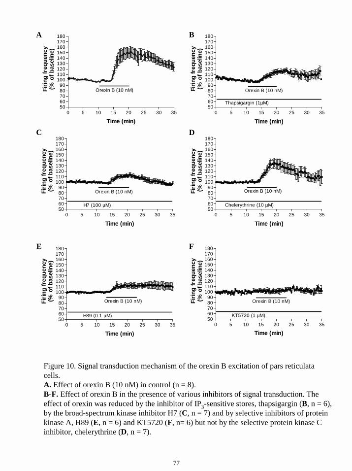
Figure 10. Signal transduction mechanism of the orexin B excitation of pars reticulata cells.A. Effect of orexin B (10 nM) in control (n = 8).B-F. Effect of orexin B in the presence of various inhibitors of signal transduction. The effect of orexin was reduced by the inhibitor of IP3-sensitive stores, thapsigargin (B, n = 6), by the broad-spectrum kinase inhibitor H7 (C, n = 7) and by selective inhibitors of protein kinase A, H89 (E, n = 6) and KT5720 (F, n= 6) but not by the selective protein kinase C inhibitor, chelerythrine (D, n = 7).
0 5 10 15 20 25 30 355060708090
100110120130140150160170180
Time (min)
Firin
g fr
eque
ncy
(% o
f bas
elin
e)
Orexin B (10 nM)
KT5720 (1 µM)
0 5 10 15 20 25 30 355060708090
100110120130140150160170180
H89 (0.1 µM)
Orexin B (10 nM)
Time (min)
Firin
g fr
eque
ncy
(% o
f bas
elin
e)
0 5 10 15 20 25 30 355060708090
100110120130140150160170180
Chelerythrine (10 µM)
Orexin B (10 nM)
Time (min)
Firin
g fr
eque
ncy
(% o
f bas
elin
e)
0 5 10 15 20 25 30 355060708090
100110120130140150160170180
H7 (100 µM)
Orexin B (10 nM)
Time (min)
Firin
g fr
eque
ncy
(% o
f bas
elin
e)
0 5 10 15 20 25 30 355060708090
100110120130140150160170180
Orexin B (10 nM)
Time (min)
Firin
g fr
eque
ncy
(% o
f bas
elin
e)
A
DC
B
FE
0 5 10 15 20 25 30 355060708090
100110120130140150160170180
Orexin B (10 nM)
Thapsigargin (1µM)
Time (min)
Firin
g fr
eque
ncy
(% o
f bas
elin
e)
77
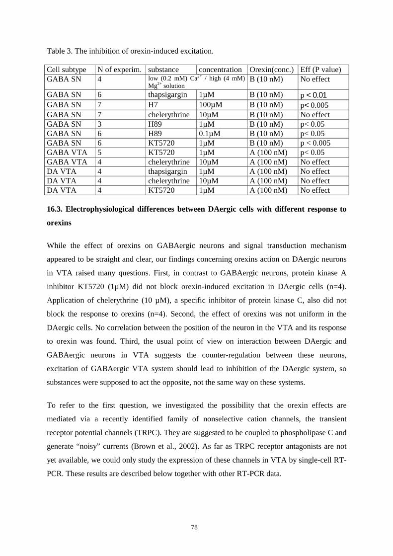
78
Table 3. The inhibition of orexin-induced excitation. Cell subtype N of experim. substance concentration Orexin(conc.) Eff (P value) GABA SN 4 low (0.2 mM) Ca2+ / high (4 mM)
Mg2+ solution B (10 nM) No effect
GABA SN 6 thapsigargin 1µM B (10 nM) p < 0.01 GABA SN 7 H7 100µM B (10 nM) p< 0.005 GABA SN 7 chelerythrine 10µM B (10 nM) No effect GABA SN 3 H89 1µM B (10 nM) p< 0.05 GABA SN 6 H89 0.1µM B (10 nM) p< 0.05 GABA SN 6 KT5720 1µM B (10 nM) p < 0.005 GABA VTA 5 KT5720 1µM A (100 nM) p< 0.05 GABA VTA 4 chelerythrine 10µM A (100 nM) No effect DA VTA 4 thapsigargin 1µM A (100 nM) No effect DA VTA 4 chelerythrine 10µM A (100 nM) No effect DA VTA 4 KT5720 1µM A (100 nM) No effect 16.3. Electrophysiological differences between DAergic cells with different response to
orexins
While the effect of orexins on GABAergic neurons and signal transduction mechanism
appeared to be straight and clear, our findings concerning orexins action on DAergic neurons
in VTA raised many questions. First, in contrast to GABAergic neurons, protein kinase A
inhibitor KT5720 (1µM) did not block orexin-induced excitation in DAergic cells (n=4).
Application of chelerythrine (10 µM), a specific inhibitor of protein kinase C, also did not
block the response to orexins (n=4). Second, the effect of orexins was not uniform in the
DAergic cells. No correlation between the position of the neuron in the VTA and its response
to orexin was found. Third, the usual point of view on interaction between DAergic and
GABAergic neurons in VTA suggests the counter-regulation between these neurons,
excitation of GABAergic VTA system should lead to inhibition of the DAergic system, so
substances were supposed to act the opposite, not the same way on these systems.
To refer to the first question, we investigated the possibility that the orexin effects are
mediated via a recently identified family of nonselective cation channels, the transient
receptor potential channels (TRPC). They are suggested to be coupled to phospholipase C and
generate “noisy” currents (Brown et al., 2002). As far as TRPC receptor antagonists are not
yet available, we could only study the expression of these channels in VTA by single-cell RT-
PCR. These results are described below together with other RT-PCR data.
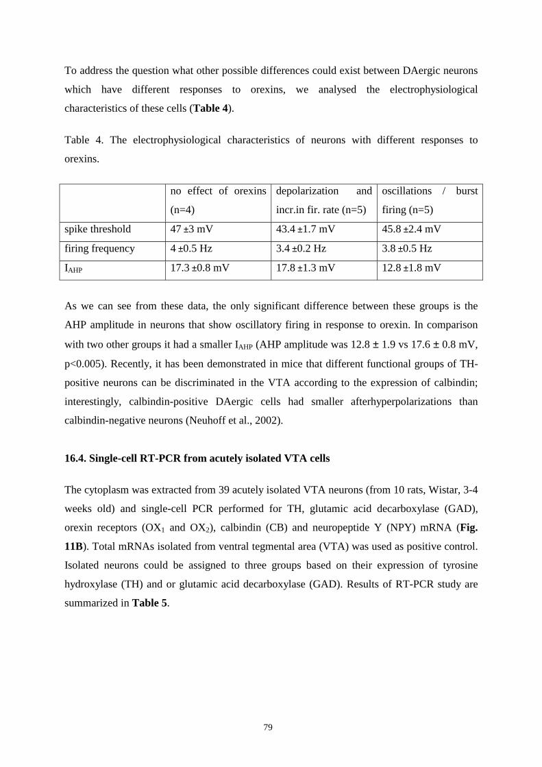
79
To address the question what other possible differences could exist between DAergic neurons
which have different responses to orexins, we analysed the electrophysiological
characteristics of these cells (Table 4).
Table 4. The electrophysiological characteristics of neurons with different responses to
orexins.
no effect of orexins
(n=4)
depolarization and
incr.in fir. rate (n=5)
oscillations / burst
firing (n=5)
spike threshold 47 ±3 mV 43.4 ±1.7 mV 45.8 ±2.4 mV
firing frequency 4 ±0.5 Hz 3.4 ±0.2 Hz 3.8 ±0.5 Hz
IAHP 17.3 ±0.8 mV 17.8 ±1.3 mV 12.8 ±1.8 mV
As we can see from these data, the only significant difference between these groups is the
AHP amplitude in neurons that show oscillatory firing in response to orexin. In comparison
with two other groups it had a smaller IAHP (AHP amplitude was 12.8 ± 1.9 vs 17.6 ± 0.8 mV,
p<0.005). Recently, it has been demonstrated in mice that different functional groups of TH-
positive neurons can be discriminated in the VTA according to the expression of calbindin;
interestingly, calbindin-positive DAergic cells had smaller afterhyperpolarizations than
calbindin-negative neurons (Neuhoff et al., 2002).
16.4. Single-cell RT-PCR from acutely isolated VTA cells
The cytoplasm was extracted from 39 acutely isolated VTA neurons (from 10 rats, Wistar, 3-4
weeks old) and single-cell PCR performed for TH, glutamic acid decarboxylase (GAD),
orexin receptors (OX1 and OX2), calbindin (CB) and neuropeptide Y (NPY) mRNA (Fig.
11B). Total mRNAs isolated from ventral tegmental area (VTA) was used as positive control.
Isolated neurons could be assigned to three groups based on their expression of tyrosine
hydroxylase (TH) and or glutamic acid decarboxylase (GAD). Results of RT-PCR study are
summarized in Table 5.
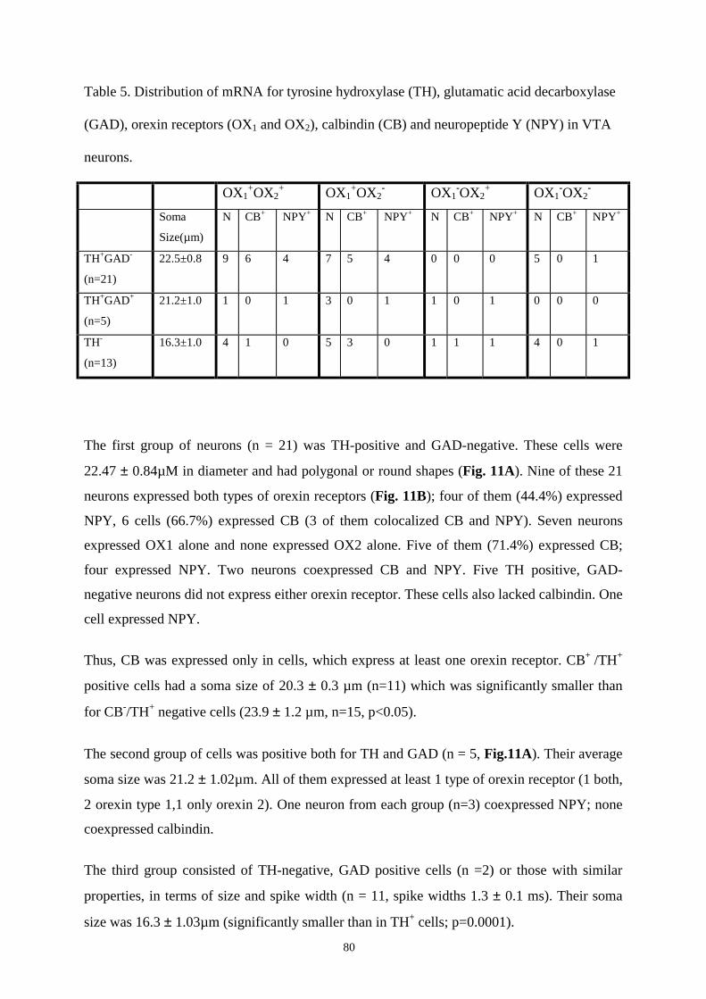
80
Table 5. Distribution of mRNA for tyrosine hydroxylase (TH), glutamatic acid decarboxylase
(GAD), orexin receptors (OX1 and OX2), calbindin (CB) and neuropeptide Y (NPY) in VTA
neurons.
OX1+OX2
+ OX1+OX2
- OX1-OX2
+ OX1-OX2
- Soma
Size(µm)
N CB+ NPY+ N CB+ NPY+ N CB+ NPY+ N CB+ NPY+
TH+GAD-
(n=21)
22.5±0.8 9 6 4 7 5 4 0 0 0 5 0 1
TH+GAD+
(n=5)
21.2±1.0 1 0 1 3 0 1 1 0 1 0 0 0
TH-
(n=13)
16.3±1.0 4 1 0 5 3 0 1 1 1 4 0 1
The first group of neurons (n = 21) was TH-positive and GAD-negative. These cells were
22.47 ± 0.84µM in diameter and had polygonal or round shapes (Fig. 11A). Nine of these 21
neurons expressed both types of orexin receptors (Fig. 11B); four of them (44.4%) expressed
NPY, 6 cells (66.7%) expressed CB (3 of them colocalized CB and NPY). Seven neurons
expressed OX1 alone and none expressed OX2 alone. Five of them (71.4%) expressed CB;
four expressed NPY. Two neurons coexpressed CB and NPY. Five TH positive, GAD-
negative neurons did not express either orexin receptor. These cells also lacked calbindin. One
cell expressed NPY.
Thus, CB was expressed only in cells, which express at least one orexin receptor. CB+ /TH+
positive cells had a soma size of 20.3 ± 0.3 µm (n=11) which was significantly smaller than
for CB-/TH+ negative cells (23.9 ± 1.2 µm, n=15, p<0.05).
The second group of cells was positive both for TH and GAD (n = 5, Fig.11A). Their average
soma size was 21.2 ± 1.02µm. All of them expressed at least 1 type of orexin receptor (1 both,
2 orexin type 1,1 only orexin 2). One neuron from each group (n=3) coexpressed NPY; none
coexpressed calbindin.
The third group consisted of TH-negative, GAD positive cells (n =2) or those with similar
properties, in terms of size and spike width (n = 11, spike widths 1.3 ± 0.1 ms). Their soma
size was 16.3 ± 1.03µm (significantly smaller than in TH+ cells; p=0.0001).
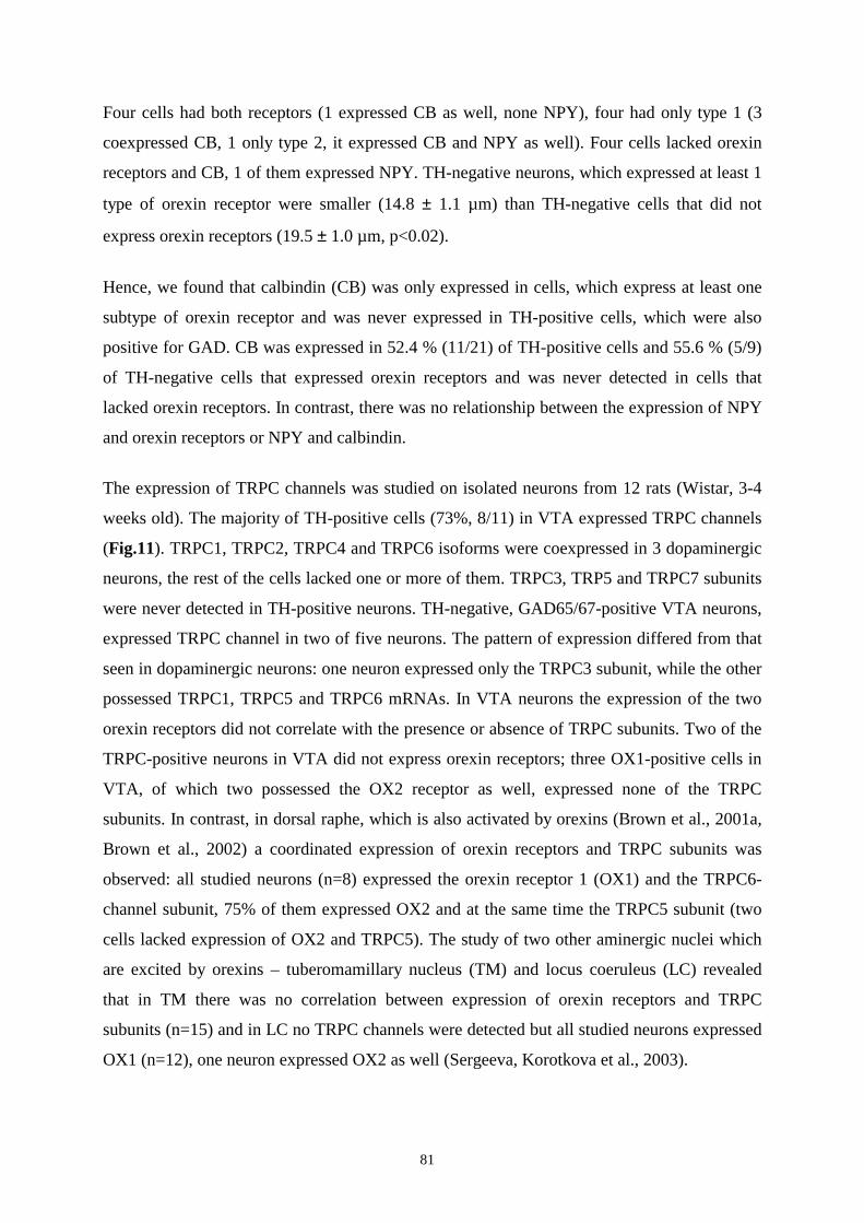
81
Four cells had both receptors (1 expressed CB as well, none NPY), four had only type 1 (3
coexpressed CB, 1 only type 2, it expressed CB and NPY as well). Four cells lacked orexin
receptors and CB, 1 of them expressed NPY. TH-negative neurons, which expressed at least 1
type of orexin receptor were smaller (14.8 ± 1.1 µm) than TH-negative cells that did not
express orexin receptors (19.5 ± 1.0 µm, p<0.02).
Hence, we found that calbindin (CB) was only expressed in cells, which express at least one
subtype of orexin receptor and was never expressed in TH-positive cells, which were also
positive for GAD. CB was expressed in 52.4 % (11/21) of TH-positive cells and 55.6 % (5/9)
of TH-negative cells that expressed orexin receptors and was never detected in cells that
lacked orexin receptors. In contrast, there was no relationship between the expression of NPY
and orexin receptors or NPY and calbindin.
The expression of TRPC channels was studied on isolated neurons from 12 rats (Wistar, 3-4
weeks old). The majority of TH-positive cells (73%, 8/11) in VTA expressed TRPC channels
(Fig.11). TRPC1, TRPC2, TRPC4 and TRPC6 isoforms were coexpressed in 3 dopaminergic
neurons, the rest of the cells lacked one or more of them. TRPC3, TRP5 and TRPC7 subunits
were never detected in TH-positive neurons. TH-negative, GAD65/67-positive VTA neurons,
expressed TRPC channel in two of five neurons. The pattern of expression differed from that
seen in dopaminergic neurons: one neuron expressed only the TRPC3 subunit, while the other
possessed TRPC1, TRPC5 and TRPC6 mRNAs. In VTA neurons the expression of the two
orexin receptors did not correlate with the presence or absence of TRPC subunits. Two of the
TRPC-positive neurons in VTA did not express orexin receptors; three OX1-positive cells in
VTA, of which two possessed the OX2 receptor as well, expressed none of the TRPC
subunits. In contrast, in dorsal raphe, which is also activated by orexins (Brown et al., 2001a,
Brown et al., 2002) a coordinated expression of orexin receptors and TRPC subunits was
observed: all studied neurons (n=8) expressed the orexin receptor 1 (OX1) and the TRPC6-
channel subunit, 75% of them expressed OX2 and at the same time the TRPC5 subunit (two
cells lacked expression of OX2 and TRPC5). The study of two other aminergic nuclei which
are excited by orexins – tuberomamillary nucleus (TM) and locus coeruleus (LC) revealed
that in TM there was no correlation between expression of orexin receptors and TRPC
subunits (n=15) and in LC no TRPC channels were detected but all studied neurons expressed
OX1 (n=12), one neuron expressed OX2 as well (Sergeeva, Korotkova et al., 2003).
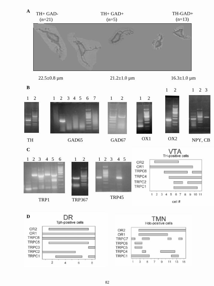
TH+ GAD-(n=21)
TH+ GAD+(n=5)
TH-GAD+(n=13)
22.5±0.8 µm 21.2±1.0 µm 16.3±1.0 µm
A
B
TH
1 2
GAD65
1 2 3 4 5 6 7
GAD67
1 2
OX1
1 2
OX2
1 2
NPY, CB
1 2 3
TRP1
1 2 3 4 5 6
TRP367
1 2
TRP45
1 2 3 4 5
C
D
82
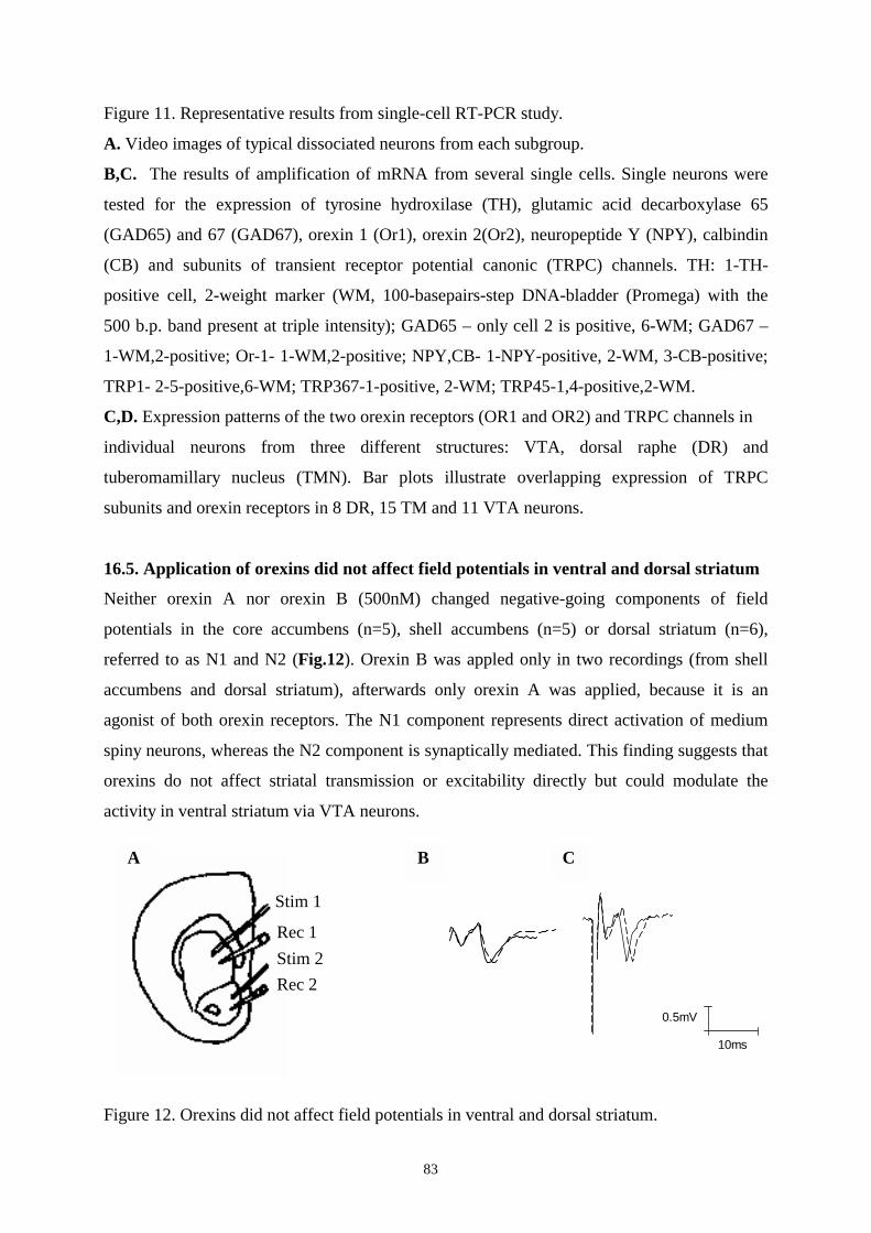
83
Figure 11. Representative results from single-cell RT-PCR study.
A. Video images of typical dissociated neurons from each subgroup.
B,C. The results of amplification of mRNA from several single cells. Single neurons were
tested for the expression of tyrosine hydroxilase (TH), glutamic acid decarboxylase 65
(GAD65) and 67 (GAD67), orexin 1 (Or1), orexin 2(Or2), neuropeptide Y (NPY), calbindin
(CB) and subunits of transient receptor potential canonic (TRPC) channels. TH: 1-TH-
positive cell, 2-weight marker (WM, 100-basepairs-step DNA-bladder (Promega) with the
500 b.p. band present at triple intensity); GAD65 – only cell 2 is positive, 6-WM; GAD67 –
1-WM,2-positive; Or-1- 1-WM,2-positive; NPY,CB- 1-NPY-positive, 2-WM, 3-CB-positive;
TRP1- 2-5-positive,6-WM; TRP367-1-positive, 2-WM; TRP45-1,4-positive,2-WM.
C,D. Expression patterns of the two orexin receptors (OR1 and OR2) and TRPC channels in
individual neurons from three different structures: VTA, dorsal raphe (DR) and
tuberomamillary nucleus (TMN). Bar plots illustrate overlapping expression of TRPC
subunits and orexin receptors in 8 DR, 15 TM and 11 VTA neurons.
16.5. Application of orexins did not affect field potentials in ventral and dorsal striatum
Neither orexin A nor orexin B (500nM) changed negative-going components of field
potentials in the core accumbens (n=5), shell accumbens (n=5) or dorsal striatum (n=6),
referred to as N1 and N2 (Fig.12). Orexin B was appled only in two recordings (from shell
accumbens and dorsal striatum), afterwards only orexin A was applied, because it is an
agonist of both orexin receptors. The N1 component represents direct activation of medium
spiny neurons, whereas the N2 component is synaptically mediated. This finding suggests that
orexins do not affect striatal transmission or excitability directly but could modulate the
activity in ventral striatum via VTA neurons.
Figure 12. Orexins did not affect field potentials in ventral and dorsal striatum.
10ms
0.5mV
Stim 1
Rec 1 Stim 2 Rec 2
A B C
84
A. Diagram showing the position of stimulating and recording electrode. Stimulating and
recording electrodes were placed either in dorsal or in ventral striatum. Stimulation within the
dorsal striatum (Stim 1) elicited a field potential detected with a recording electrode in dorsal
striatum (Rec 1) and likewise for the nucleus accumbens (ventral striatum) (Stim2, Rec2).B,
C. Bath application of orexins did not affect significantly the amplitude on N1 and N2
components in dorsal (B) and ventral (C) striatum.
16.6. Effects of hypothalamic feeding- and arousal-related peptides on VTA neurons
Could other feeding- or arousal-related peptides also affect the DAergic and GABAergic
midbrain systems? The following neuropeptides were studied: melanin-concentrating
hormone (MCH), which is also expressed exclusively in the perifornical hypothalamic area,
and has a similar action on feeding; neuropeptide Y (NPY), a very potent stimulator of food
intake; the orexigenic neuropeptides ghrelin and agouti-related protein (AGRP), α-melanocyte
stimulating hormone (α-MSH), hypothalamic anorectic neuropeptide; corticotropin-releasing
factor (CRF), another anorectic peptide which is also known to promote arousal; anorectic
cocaine and amphetamine-related transcript (CART) and substance P (SP) that has little effect
on food intake but promotes arousal. Effects of these peptides are summarized in Table 6.
16.6.1. Effects of orexigenic neuropeptides on VTA neurons
16.6.1.1. Responses to melanin concentrating hormone
Effects of melanin-concentrating hormone (MCH) were studied only in drug-naive slices.
MCH (100 nM) did not affect the firing frequency of DAergic (n = 4) or GABAergic (n = 4)
neurons in the SN. In VTA MCH (100 nM) failed to affect the firing of dopaminergic (n=4)
or fast-firing GABAergic cells (n=3). The application of 1µM of MCH (n=7 for DAergic and
n=6 for GABAergic cells) also did not affect membrane potential and firing rate of the cells.
16.6.1.2. Responses to neuropeptide Y.
In contrast to the previous sections of study where actions of only one neuropeptide on one
neuron were studied, in the following sections (16.6.1.2.- 16.6.3) effects of several
neuropeptides on the same cell were tested by extracellular single-unit recordings: after
recording of the baseline for 15 min the first peptide was applied, after the end of the effect
(return of the baseline to the initial value) and a stable baseline for the next 15 minutes the
second drug was applied and so on. The order of drug application was changed each time to
85
be sure that the previous application did not affect the next one, the action of each
neuropeptide in drug-naïve cells (in whole-cell intracellular recordings) was also tested.
Extracellular recordings of 30 DAergic neurons, 16 fast-firing GABAergic neurons and 6
slow-firing GABAergic neurons were performed (as in previous parts of the study, only one
cell per slice was recorded, in all but four cases one slice from one rat was cut, one DAergic
and one GABAergic cell from different slices of these four rats were recorded). An overview
of the order of drugs application is presented in Tables 7, 8 and 9. Repetitive applications of
the same drug (higher doses in unresponsive cells) and application of dopamine or DAMGO
that, as always, took place at the end of each experiment, are not shown in these tables.
NPY (300nM, in unresponsive cells higher doses (1µM) were also tested (n=4)) inhibited
40% of DAergic cells, recorded extracellularly (7 out of 19, Fig. 13A), two of them –
completely, others – up to 68,1 ± 15,6 % of control rate (p < 0.01). Two NPY-inhibited
neurons and four cells that were not affected by NPY were drug-naive. Intracellular
recordings in drug-naive rats revealed that reduction in firing frequency was accompanied by
a moderate hyperpolarization (2,5mV; 3 mV, n=2, Fig. 13C). In six NPY-inhibited cells,
which were tested for response to orexins as well, all neurons were excited by orexins.
However, other five neurons, which were also tested for both neuropeptides and were not
affected by NPY, were excited by orexins.
In fast firing GABAergic cells, NPY inhibited 6 out of 11 neurons by up to 55,6 ± 13,8% (p
< 0.005, Fig. 13B), the firing rate of 5 neurons was not changed. Two affected and one –
unaffected neurons were drug-naive. Both slow-firing GABAergic cells tested were inhibited
by NPY (one drug-naive).
Additional recordings of slow-firing GABAergic neurons should be performed to study NPY
action on this subgroup. Additional whole-cell recordings after the application of tetrodotoxin
should be performed in DAergic and GABAergic neurons to study if the effect of NPY on
both groups is direct.
16.6.1.3. Responses to ghrelin
Ghrelin (500 nM) did not affect firing rate and membrane potential of DAergic (n=8), fast
(n=4) and slow (n=2) GABAergic VTA neurons. Two DAergic and two GABAergic neurons
were drug-naive before the application of ghrelin.
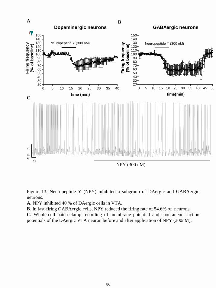
1
2 s
20
mV
NPY (300 nM)
A B
C
Figure 13. Neuropeptide Y (NPY) inhibited a subgroup of DAergic and GABAergic neurons. A. NPY inhibited 40 % of DAergic cells in VTA. B. In fast-firing GABAergic cells, NPY reduced the firing rate of 54.6% of neurons.C. Whole-cell patch-clamp recording of membrane potential and spontaneous action potentials of the DAergic VTA neuron before and after application of NPY (300nM).
Dopaminergic neurons
0 5 10 15 20 25 30 35 402030405060708090
100110120130140150
time (min)
Firin
g fr
eque
ncy
(% o
f bas
elin
e)
Neuropeptide Y (300 nM)
GABAergic neurons
0 5 10 15 20 25 30 35 40 45 502030405060708090
100110120130140150
Neuropeptide Y (300 nM)
time(min)
Firin
g fr
eque
ncy
(% o
f bas
elin
e)
86
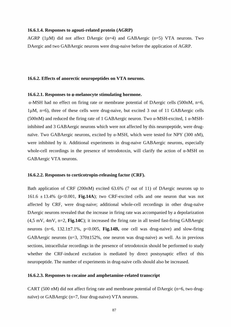
87
16.6.1.4. Responses to agouti-related protein (AGRP)
AGRP (1µM) did not affect DAergic (n=4) and GABAergic (n=5) VTA neurons. Two
DAergic and two GABAergic neurons were drug-naive before the application of AGRP.
16.6.2. Effects of anorectic neuropeptides on VTA neurons.
16.6.2.1. Responses to αααα-melanocyte stimulating hormone.
α-MSH had no effect on firing rate or membrane potential of DAergic cells (500nM, n=6,
1µM, n=6), three of these cells were drug-naive, but excited 3 out of 11 GABAergic cells
(500nM) and reduced the firing rate of 1 GABAergic neuron. Two α-MSH-excited, 1 α-MSH-
inhibited and 3 GABAergic neurons which were not affected by this neuropeptide, were drug-
naïve. Two GABAergic neurons, excited by α-MSH, which were tested for NPY (300 nM),
were inhibited by it. Additional experiments in drug-naive GABAergic neurons, especially
whole-cell recordings in the presence of tetrodotoxin, will clarify the action of α-MSH on
GABAergic VTA neurons.
16.6.2.2. Responses to corticotropin-releasing factor (CRF).
Bath application of CRF (200nM) excited 63.6% (7 out of 11) of DAergic neurons up to
161.6 ± 13.4% (p<0.001, Fig.14A); two CRF-excited cells and one neuron that was not
affected by CRF, were drug-naive; additional whole-cell recordings in other drug-naïve
DAergic neurons revealed that the increase in firing rate was accompanied by a depolarization
(4,5 mV, 4mV, n=2, Fig.14C); it increased the firing rate in all tested fast-firing GABAergic
neurons (n=6, 132.1±7.1%, p<0.005, Fig.14B, one cell was drug-naive) and slow-firing
GABAergic neurons (n=3, 370±152%, one neuron was drug-naive) as well. As in previous
sections, intracellular recordings in the presence of tetrodotoxin should be performed to study
whether the CRF-induced excitation is mediated by direct postsynaptic effect of this
neuropeptide. The number of experiments in drug-naive cells should also be increased.
16.6.2.3. Responses to cocaine and amphetamine-related transcript
CART (500 nM) did not affect firing rate and membrane potential of DAergic (n=6, two drug-
naïve) or GABAergic (n=7, four drug-naive) VTA neurons.
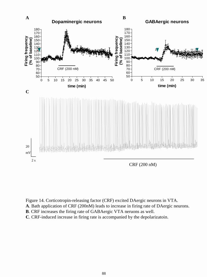
A
Figure 14. Corticotropin-releasing factor (CRF) excited DAergic neurons in VTA.A. Bath application of CRF (200nM) leads to increase in firing rate of DAergic neurons.B. CRF increases the firing rate of GABAergic VTA neruons as well. C. CRF-induced increase in firing rate is accompanied by the depolarizatoin.
B
C
231
2 s
20
mV
CRF (200 nM)
88
Dopaminergic neurons
0 5 10 15 20 25 30 35 40 45 505060708090
100110120130140150160170180
CRF (200 nM)
time (min)
Firin
g fr
eque
ncy
(% o
f bas
elin
e)
GABAergic neurons
0 5 10 15 20 25 30 355060708090
100110120130140150160170180
CRF (200 nM)
time (min)
Firin
g fr
eque
ncy
(% o
f bas
elin
e)
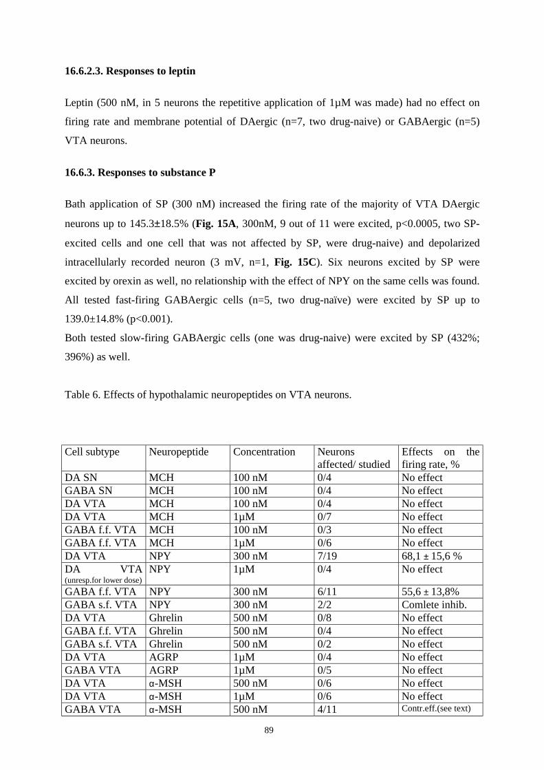
89
16.6.2.3. Responses to leptin
Leptin (500 nM, in 5 neurons the repetitive application of 1µM was made) had no effect on
firing rate and membrane potential of DAergic (n=7, two drug-naive) or GABAergic (n=5)
VTA neurons.
16.6.3. Responses to substance P
Bath application of SP (300 nM) increased the firing rate of the majority of VTA DAergic
neurons up to 145.3±18.5% (Fig. 15A, 300nM, 9 out of 11 were excited, p<0.0005, two SP-
excited cells and one cell that was not affected by SP, were drug-naive) and depolarized
intracellularly recorded neuron (3 mV, n=1, Fig. 15C). Six neurons excited by SP were
excited by orexin as well, no relationship with the effect of NPY on the same cells was found.
All tested fast-firing GABAergic cells (n=5, two drug-naïve) were excited by SP up to
139.0±14.8% (p<0.001). Both tested slow-firing GABAergic cells (one was drug-naive) were excited by SP (432%;
396%) as well.
Table 6. Effects of hypothalamic neuropeptides on VTA neurons.
Cell subtype Neuropeptide Concentration Neurons affected/ studied
Effects on the firing rate, %
DA SN MCH 100 nM 0/4 No effect GABA SN MCH 100 nM 0/4 No effect DA VTA MCH 100 nM 0/4 No effect DA VTA MCH 1µM 0/7 No effect GABA f.f. VTA MCH 100 nM 0/3 No effect GABA f.f. VTA MCH 1µM 0/6 No effect DA VTA NPY 300 nM 7/19 68,1 ± 15,6 % DA VTA (unresp.for lower dose)
NPY 1µM 0/4 No effect
GABA f.f. VTA NPY 300 nM 6/11 55,6 ± 13,8% GABA s.f. VTA NPY 300 nM 2/2 Comlete inhib. DA VTA Ghrelin 500 nM 0/8 No effect GABA f.f. VTA Ghrelin 500 nM 0/4 No effect GABA s.f. VTA Ghrelin 500 nM 0/2 No effect DA VTA AGRP 1µM 0/4 No effect GABA VTA AGRP 1µM 0/5 No effect DA VTA α-MSH 500 nM 0/6 No effect DA VTA α-MSH 1µM 0/6 No effect GABA VTA α-MSH 500 nM 4/11 Contr.eff.(see text)
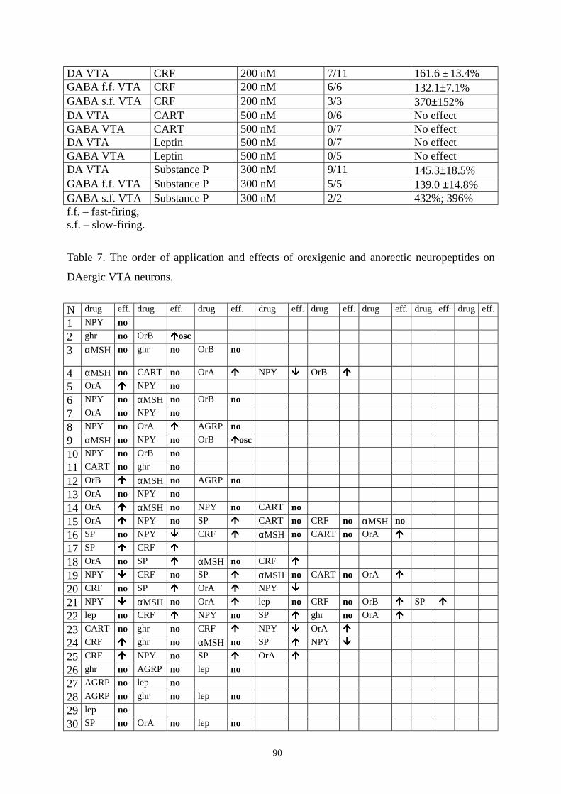
90
DA VTA CRF 200 nM 7/11 161.6 ± 13.4% GABA f.f. VTA CRF 200 nM 6/6 132.1±7.1% GABA s.f. VTA CRF 200 nM 3/3 370±152% DA VTA CART 500 nM 0/6 No effect GABA VTA CART 500 nM 0/7 No effect DA VTA Leptin 500 nM 0/7 No effect GABA VTA Leptin 500 nM 0/5 No effect DA VTA Substance P 300 nM 9/11 145.3±18.5% GABA f.f. VTA Substance P 300 nM 5/5 139.0 ±14.8% GABA s.f. VTA Substance P 300 nM 2/2 432%; 396% f.f. – fast-firing, s.f. – slow-firing.
Table 7. The order of application and effects of orexigenic and anorectic neuropeptides on
DAergic VTA neurons.
N drug eff. drug eff. drug eff. drug eff. drug eff. drug eff. drug eff. drug eff. 1 NPY no 2 ghr no OrB !!!!osc 3 αMSH
no ghr no OrB no
4 αMSH no CART no OrA !!!! NPY """" OrB !!!! 5 OrA !!!! NPY no 6 NPY no αMSH no OrB no 7 OrA no NPY no 8 NPY no OrA !!!! AGRP no 9 αMSH no NPY no OrB !!!!osc 10 NPY no OrB no 11 CART no ghr no 12 OrB !!!! αMSH no AGRP no 13 OrA no NPY no 14 OrA !!!! αMSH no NPY no CART no 15 OrA !!!! NPY no SP !!!! CART no CRF no αMSH no 16 SP no NPY """" CRF !!!! αMSH no CART no OrA !!!! 17 SP !!!! CRF !!!! 18 OrA no SP !!!! αMSH no CRF !!!! 19 NPY """" CRF no SP !!!! αMSH no CART no OrA !!!! 20 CRF no SP !!!! OrA !!!! NPY """" 21 NPY """" αMSH no OrA !!!! lep no CRF no OrB !!!! SP !!!! 22 lep no CRF !!!! NPY no SP !!!! ghr no OrA !!!! 23 CART no ghr no CRF !!!! NPY """" OrA !!!! 24 CRF !!!! ghr no αMSH no SP !!!! NPY """" 25 CRF !!!! NPY no SP !!!! OrA !!!! 26 ghr no AGRP no lep no 27 AGRP no lep no 28 AGRP no ghr no lep no 29 lep no 30 SP no OrA no lep no
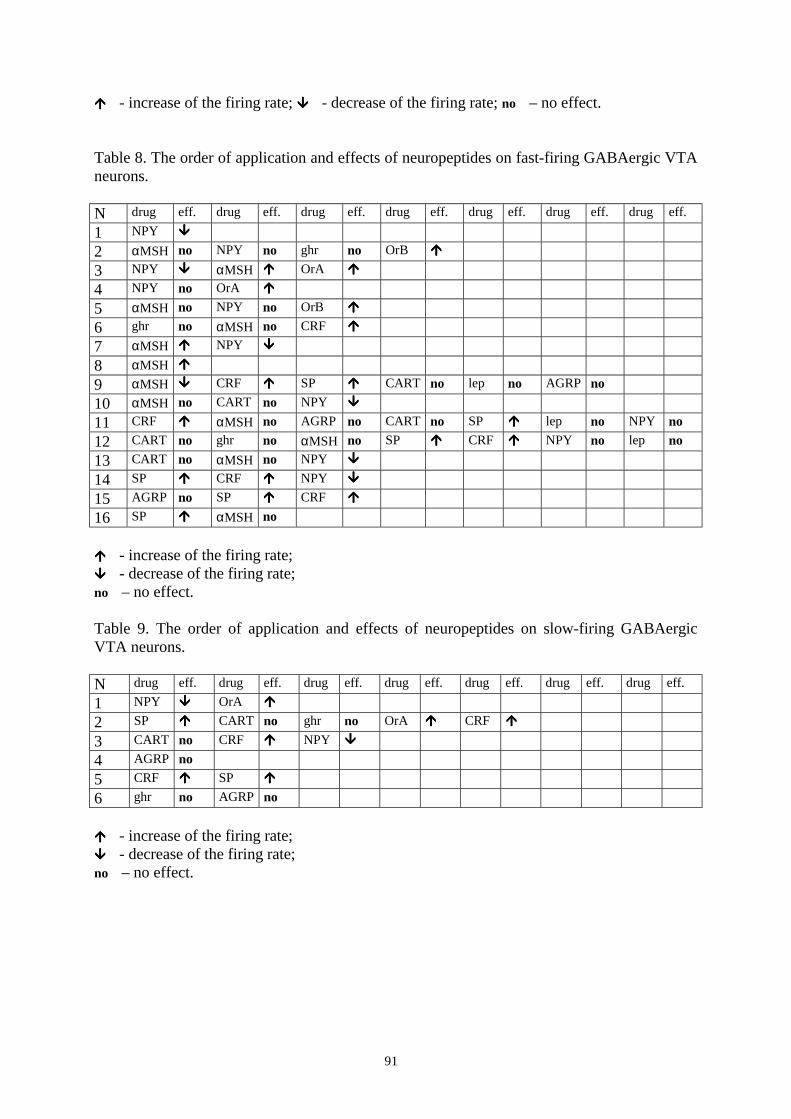
91
! ! ! ! - increase of the firing rate; " " " " - decrease of the firing rate; no – no effect. Table 8. The order of application and effects of neuropeptides on fast-firing GABAergic VTA neurons. N drug eff. drug eff. drug eff. drug eff. drug eff. drug eff. drug eff. 1 NPY """" 2 αMSH no NPY no ghr no OrB !!!! 3 NPY """" αMSH !!!! OrA !!!! 4 NPY no OrA !!!! 5 αMSH no NPY no OrB !!!! 6 ghr no αMSH no CRF !!!! 7 αMSH !!!! NPY """" 8 αMSH !!!! 9 αMSH """" CRF !!!! SP !!!! CART no lep no AGRP no 10 αMSH no CART no NPY """" 11 CRF !!!! αMSH no AGRP no CART no SP !!!! lep no NPY no 12 CART no ghr no αMSH no SP !!!! CRF !!!! NPY no lep no 13 CART no αMSH no NPY """" 14 SP !!!! CRF !!!! NPY """" 15 AGRP no SP !!!! CRF !!!! 16 SP !!!! αMSH no ! ! ! ! - increase of the firing rate; " " " " - decrease of the firing rate; no – no effect. Table 9. The order of application and effects of neuropeptides on slow-firing GABAergic VTA neurons. N drug eff. drug eff. drug eff. drug eff. drug eff. drug eff. drug eff. 1 NPY """" OrA !!!! 2 SP !!!! CART no ghr no OrA !!!! CRF !!!! 3 CART no CRF !!!! NPY """" 4 AGRP no 5 CRF !!!! SP !!!! 6 ghr no AGRP no ! ! ! ! - increase of the firing rate; " " " " - decrease of the firing rate; no – no effect.
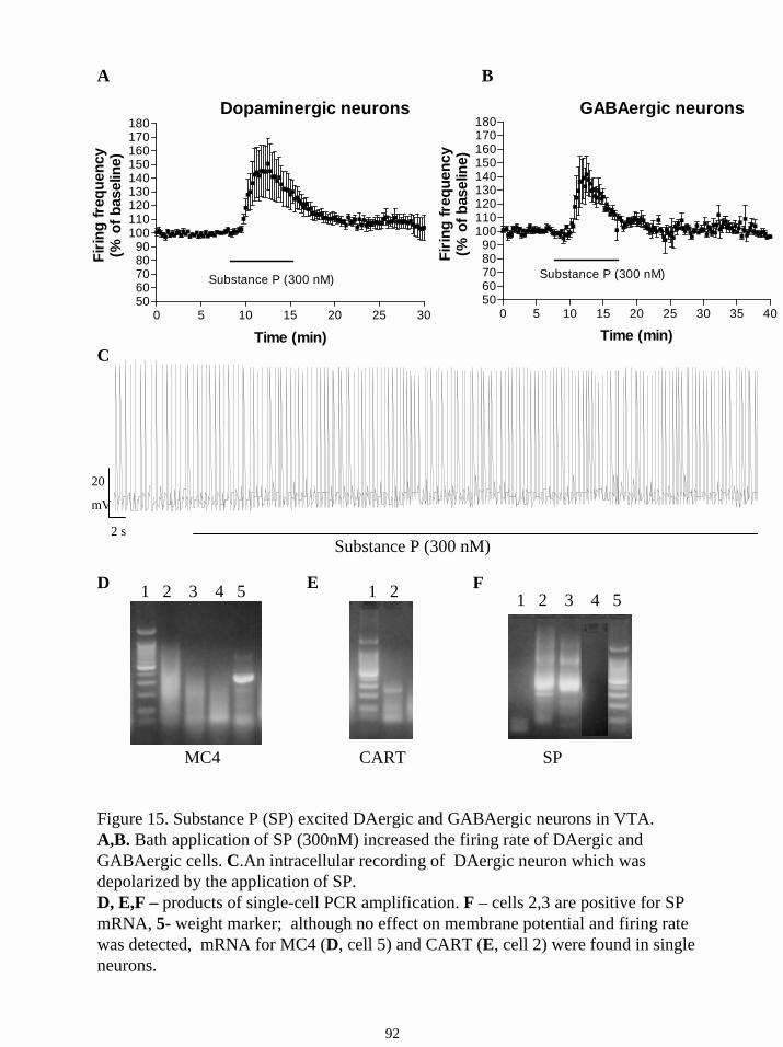
2 s
20
mV
Substance P (300 nM)
A B
C
F
SP
1 2 3 4 5
MC4
1 2 3 4 5
CART
1 2 D E
Figure 15. Substance P (SP) excited DAergic and GABAergic neurons in VTA.A,B. Bath application of SP (300nM) increased the firing rate of DAergic and GABAergic cells. C.An intracellular recording of DAergic neuron which was depolarized by the application of SP.D, E,F – products of single-cell PCR amplification. F – cells 2,3 are positive for SP mRNA, 5- weight marker; although no effect on membrane potential and firing rate was detected, mRNA for MC4 (D, cell 5) and CART (E, cell 2) were found in single neurons.
0 5 10 15 20 25 30 35 405060708090
100110120130140150160170180
Substance P (300 nM)
Time (min)
Firin
g fr
eque
ncy
(% o
f bas
elin
e)
GABAergic neurons
0 5 10 15 20 25 305060708090
100110120130140150160170180
Substance P (300 nM)
Time (min)
Firin
g fr
eque
ncy
(% o
f bas
elin
e)Dopaminergic neurons
92
93
16.7. The expression of hypothalamic peptides and their receptors in isolated VTA
neurons.
The RT-PCR experiments demonstrated the presence of mRNA for the described peptides or
their receptors in the VTA (Figs. 15 D,E,F); The detailed analysis of expression of these
proteins in single cells will allow us to find whether there is a relationship of their expression
in a given neuron. In preliminary, pilot study it was not yet found, suggesting the mediation of
effects through different neurons rather than a counter-regulation of these peptides via
reciprocal effects on the same cells. However, additional expreriments should be performed
to address this question.
Taken together, the effect on VTA cells could be linked to the influence of the given
neuropeptide on arousal, rather than on food intake: NPY is the only substance among the
tested neuropeptides that possesses anxiolytic properties and decreases wakefulness, and it’s
the only one that inhibits DAergic neurons. On the other side, SP that promotes activation but
not food intake, excites the majority of VTA neurons. The next step in our study was to
investigate the effects of two substances that promote arousal – the neurotransmitter histamine
and a novel wake-promoting compound modafinil, which is used for the treatment of
narcolepsy.
16.8. Effects of histamine on DAergic and GABAergic neurons in SN and VTA.
This part of study was carried out on drug-naive neurons. Using extracellular recordings of
the spontaneous activity of DA neurons in SN we found that bath application of HA (10 µM)
did not affect their firing frequency (n=6, Fig. 16A). In contrast, bath application of histamine
(10 µM) increased the firing of GABAergic neurons in SN pars reticulata to 119,4 ± 4,9 % of
control (n=6, p<0.005, Fig. 16 B)(Korotkova et al., 2002b). Hundred µM of histamine caused
a somewhat stronger excitation – to 141,0 ± 14,6% (n=8, Fig.16 C), however, this difference
was not statistically significant. The effect of histamine (10 µM) was blocked by prior
application of the selective H1 receptor antagonist mepyramine (1 µM, n=6, p<0.005). In the
presence of the H2 receptor antagonist cimetidine (50 µM) the response of GABAergic
neurons to histamine (10µM) was not blocked (n = 4).
In the VTA, bath application of histamine (100 µM) did not affect the firing of
dopamine cells (n = 7), but increased the firing of GABAergic neurons to 119,5 ± 7,8 % of
control rate (n = 6, p<0.01). Application of a lower dose of histamine (10 µM) caused the
same level of excitation of GABAergic neurons (118,5 ± 7,2%, n = 6, p<0.01, Fig. 16D). This
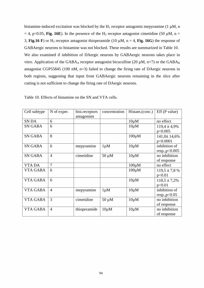
94
histamine-induced excitation was blocked by the H1 receptor antagonist mepyramine (1 µM, n
= 4, p<0.05, Fig. 16E). In the presence of the H2 receptor antagonist cimetidine (50 µM, n =
3, Fig.16 F) or H3 receptor antagonist thioperamide (10 µM, n = 4, Fig. 16G) the response of
GABAergic neurons to histamine was not blocked. These results are summarized in Table 10.
We also examined if inhibition of DAergic neurons by GABAergic neurons takes place in
vitro. Application of the GABAA receptor antagonist bicuculline (20 µM, n=7) or the GABAB
antagonist CGP55845 (100 nM, n=3) failed to change the firing rate of DAergic neurons in
both regions, suggesting that input from GABAergic neurons remaining in the slice after
cutting is not sufficient to change the firing rate of DAergic neurons.
Table 10. Effects of histamine on the SN and VTA cells.
Cell subtype N of exper. hist.receptors antagonists
concentration Histam.(conc.) Eff (P value)
SN DA 6 10µM no effect SN GABA 6 10µM 119,4 ± 4,9%
p<0.005 SN GABA 8 100µM 141,0± 14,6%
p<0.0001 SN GABA 6 mepyramine 1µM 10µM inhibition of
resp.,p<0.005 SN GABA 4 cimetidine 50 µM 10µM no inhibition
of response VTA DA 7 100µM no effect VTA GABA 6 100µM 119,5 ± 7,8 %
p<0.01 VTA GABA 6 10µM 118,5 ± 7,2%
p<0.01 VTA GABA 4 mepyramine 1µM 10µM inhibition of
resp.,p<0.05 VTA GABA 3 cimetidine 50 µM 10µM no inhibition
of response VTA GABA 4 thioperamide 10µM 10µM no inhibition
of response
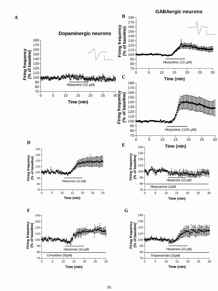
0 5 10 15 20 25 30708090
100110120130140150160170180
Histamine (10 µM)
Time (min)
Firin
g fr
eque
ncy
(% o
f bas
elin
e)
GABAergic neurons
0 5 10 15 20 25 30708090
100110120130140150160170180
Histamine (100 µM)
Time (min)
Firin
g fre
quen
cy(%
of b
asel
ine)0 5 10 15 20 25 30
708090
100110120130140150160170180
Histamine (10 µM)
Dopaminergic neurons
Time (min)
Firin
g fre
quen
cy(%
of b
asel
ine)
0 5 10 15 20 25 3070
80
90
100
110
120
130
140
Time (min)
Firin
g fr
eque
ncy
(% o
f bas
elin
e)
Histamine (10 µM)
0 5 10 15 20 25 3070
80
90
100
110
120
130
140
Time (min)
Firin
g fr
eque
ncy
(% o
f bas
elin
e)
Histamine (10 µM)
Mepyramine (1µM)
0 5 10 15 20 25 3070
80
90
100
110
120
130
140
Time (min)
Firin
g fr
eque
ncy
(% o
f bas
elin
e)
Histamine (10 µM)
Cimetidine (50µM)
0 5 10 15 20 25 3070
80
90
100
110
120
130
140
Time (min)
Firin
g fre
quen
cy(%
of b
asel
ine)
Histamine (10 µM)
Thioperamide (10µM)
A B
C
D
GF
E
95
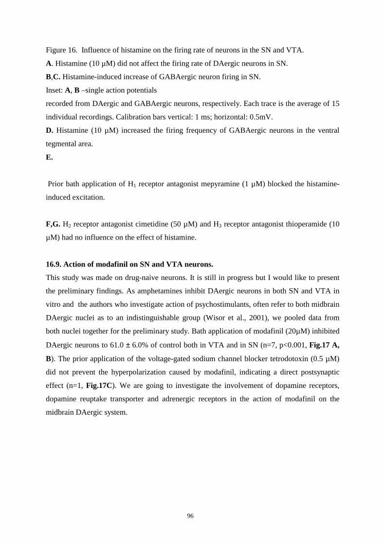
96
Figure 16. Influence of histamine on the firing rate of neurons in the SN and VTA.
A. Histamine (10 µM) did not affect the firing rate of DAergic neurons in SN.
B,C. Histamine-induced increase of GABAergic neuron firing in SN.
Inset: A, B –single action potentials
recorded from DAergic and GABAergic neurons, respectively. Each trace is the average of 15
individual recordings. Calibration bars vertical: 1 ms; horizontal: 0.5mV.
D. Histamine (10 µM) increased the firing frequency of GABAergic neurons in the ventral
tegmental area.
E.
Prior bath application of H1 receptor antagonist mepyramine (1 µM) blocked the histamine-
induced excitation.
F,G. H2 receptor antagonist cimetidine (50 µM) and H3 receptor antagonist thioperamide (10
µM) had no influence on the effect of histamine.
16.9. Action of modafinil on SN and VTA neurons.
This study was made on drug-naive neurons. It is still in progress but I would like to present
the preliminary findings. As amphetamines inhibit DAergic neurons in both SN and VTA in
vitro and the authors who investigate action of psychostimulants, often refer to both midbrain
DAergic nuclei as to an indistinguishable group (Wisor et al., 2001), we pooled data from
both nuclei together for the preliminary study. Bath application of modafinil (20µM) inhibited
DAergic neurons to 61.0 ± 6.0% of control both in VTA and in SN (n=7, p<0.001, Fig.17 A,
B). The prior application of the voltage-gated sodium channel blocker tetrodotoxin (0.5 µM)
did not prevent the hyperpolarization caused by modafinil, indicating a direct postsynaptic
effect (n=1, Fig.17C). We are going to investigate the involvement of dopamine receptors,
dopamine reuptake transporter and adrenergic receptors in the action of modafinil on the
midbrain DAergic system.
0 5 10 15 20 25 30 35 40405060708090
100110120130140150
Modafinil (20 µM)
time(min)
Firin
g fr
eque
ncy
(% o
f bas
elin
e)
Figure 17. Action of modafinil on DAergic neurons in SN and VTA.A, B.Bath application of modafinil (20µM) inhibited DAergic neurons to 61.0±6.0% of control both in VTA and in SN.C. The prior application of tetrodotoxin (0.5 µM) did not prevent the modafinil-induced hyperpolarization
30 s
2mV
Modafinil (20µM)
20mV
30 sModafinil (20µM)
97
A
B
C
98
17. Discussion
We found that in the substantia nigra pars compacta dopamine neuronswere unaffected by
bath applied orexin A, which activates both types of orexin receptors with high affinity
(Sakurai et al., 1998), whereas GABAergic cells in the pars reticulata were strongly excited
by both orexin A and orexin B. These results were unexpected because a previous study of the
distribution of orexin fibres reported a complete absence of orexin-containing fibres from the
substantia nigra pars reticulata (Peyron et al., 1998). We have re-examined this question using
immunohistochemical methods and confirm that orexin fibres are almost completely absent in
the rostral substantia nigra, their density steadily increases in the rostral-caudal direction.
Thus, the anatomical substrate for an orexin modulation of these cells exists. The second
reason why these results were unexpected was the fact that other aminergic neurons are all
potently excited by orexins. However, it is notable that whereas the firing of the other
aminergic neurons mentioned varies across the sleep-wake cycle, that of the dopamine
neurons does not (Miller et al., 1983) and so the lack of modulation by orexins is entirely
consistent with their role in arousal. A previous study found that orexin A-induced
hyperlocomotion and stereotypy are mediated by the dopaminergic system (Nakamura et al.,
2000). Our results suggest that these effects are not mediated by orexin actions on
nigrostriatal dopamine neurons but rather on mesocorticolimbic dopamine neurons located in
the ventral tegmental area (Korotkova et al., 2003), which exhibit an increase in intracellular
calcium (Nakamura et al., 2000) and increased firing in response to orexin A.
In contrast to the SN dopamine neurons the firing rate of GABAergic cells in the pars
reticulata is sensitive to behavioural state (Miller et al., 1983). These cells fire faster during
waking and during rapid-eye movement (REM) sleep in comparison with slow-wave sleep.
We found in vitro that these cells had a firing rate of 8.6 ± 0.8 Hz, which was increased to
16.6 ± 0.8 Hz by 100 nM Orexin B. During the waking state in vivo these cells fire at 12-20
Hz (Miller et al., 1983). Since orexin neurons are most active during the waking state
(Estabrooke et al., 2001;Taheri et al., 2000), orexins (together with biogenic amines – see
below) may act to maintain the high firing rate of these cells during waking.
The response to orexins in GABAergic pars reticulata cells was very homogeneous.
Thus, we were able to investigate the pharmacology and signal transduction mechanisms of
this excitation in detail. Orexin A and B were similarly efficacious in exciting these cells at
100 nM and the dose-response curve for orexin B had an EC50 in the low nanomolar range (12
nM). These observations suggest that the type II orexin receptor is involved in mediating
99
these effects since in transfected CHO cells orexin A and B are similarly potent at the type II
receptor (EC50s 34 and 60 nM respectively) whereas orexin B has an EC50 in the micromolar
range (2.5 µM) for activating the type I receptor (Sakurai et al., 1998). Although Marcus et
al., 2001, did not find expression of mRNA for the orexin type II or type I receptor in the
substantia nigra pars reticulata, these findings depend on the sensitivity of the in situ
histochemistry and may represent false negatives. The type II receptor seems to be more
important in the control of the sleep-wake cycle and in the context of narcolepsy, since
disruption of the OX2R receptor gene leads to narcolepsy (Willie et al., 2001), whereas
disruption of the OX1R gene alone does not lead to obvious behavioural abnormalities (such
as cataplectic attacks) and these animals only exhibit fragmentation of the sleep-wake cycle.
Consequently, the orexin action on reticulata cells is likely to be related to regulation of
activity across the sleep-wake cycle.
It has been reported that orexin receptors are coupled to phospholipase C (Sakurai et
al., 1998;van den Pol et al., 1998). This enzyme generates two second messengers – inositol
trisphosphate (IP3) and diacylglycerol (DAG). IP3 releases calcium from internal stores
whereas DAG potentiates the activity of PKC. Accordingly, we tested inhibitors of these
pathways for their ability to block the orexin B induced excitation of reticulata cells.
Application of thapsigargin reduced the effect of orexin B but did not abolish it, indicating
that calcium release from internal stores plays a role in the excitation of these cells. In
contrast, influx of extracellular calcium does not seem to be necessary, since the magnitude of
the orexin B effect was unchanged in low calcium/high magnesium solution. Application of a
high concentration of the broad-spectrum kinase inhibitor H7 also reduced the magnitude of
the orexin B effect, but, surprisingly, the selective protein kinase C inhibitor chelerythrine
was ineffective. Instead, two protein kinase inhibitors (H89 and KT5720) applied at
concentrations which selectively block protein kinase A were able to block the orexin B-
induced excitation. Coadministration of H7 and thapsigargin did not reduce the magnitude of
the orexin B effect to a greater extent than either drug alone, suggesting that the release of
calcium from internal stores and the activation of protein kinase A are not independent of
each other – one possibility is that calcium released from internal stores activates calcium-
sensitive adenylyl cyclase in these cells.
The firing of GABAergic reticulata cells is extremely important for the regulation of
muscle tone. Abnormal increases in the firing of these cells seen in Parkinson`s disease or
following neuroleptic treatement lead to catalepsy (Niijima and Yoshida, 1982; Blandini et
al., 2000;Boulay et al., 2000;Evans et al., 2001). We propose that an impaired orexin system,
100
as seen in narcolepsy, leads to an abnormally low activity in these cells and predisposes
individuals to what is, in terms of muscle tone, the reverse effect, namely cataplexy. Both
direct and indirect mechanisms are likely to play a role. Thus, we have shown here that
orexins directly and potently excite these cells. In addition, though, orexins excite
serotonergic (Brown et al., 2001a), histaminergic (Eriksson et al., 2001) and noradrenergic
neurons (Hagan et al., 1999) and noradrenaline (Berretta et al., 2000), serotonin (Stanford and
Lacey, 1996) and histamine(see the following part of the study) in turn excite GABAergic
reticulata cells. Thus, in narcoleptics the action of these neurotransmitters is also likely to be
reduced. Recently, it has been shown that oral application of sulpiride, a dopamine D2-like
receptor antagonist, which will indirectly increase the firing of reticulata cells, effectively
blocks cataplexy without affecting REM sleep (Okura et al., 2000). To summarize, the
presence of an abnormally low basal activity of substantia nigra pars reticulata cells may
allow the expression of cataplexy, which is prevented in normal individuals by the inhibitory
projections of reticulata cells to the ventromedial thalamus and medial pontine reticular
formation (Niijima and Yoshida, 1982). This hypothesis can be tested experimentally by
recording from these cells in narcoleptic animals.
GABAergic cells in the VTA are also excited by orexins. We found two groups of
GABAergic neurons in the VTA - fast-firing and slow-firing. The existence of the first group
is well known, but the latter one has not yet been described. We did not find slow-firing
GABAergic cells in SN. The further study of the projection pattern of these cells and their
neurochemical and electrophysiological properties could help to understand the other
differences of these groups in VTA. The depolarization of both groups of GABAergic cells
persisted in the presence of tetrodotoxin indicating that this was also a direct action of
orexins. Similar to our findings in the substantia nigra, the effect of orexins on firing rate in
fast-firing cells of the pars reticulata was blocked by inhibition of protein kinase A. In recent
years it has become clear, that the contribution of GABAergic neurons in the reward
processes is more complicated than just inhibition of DAergic neurons. Steffensen et al
(Steffensen et al., 2001) have shown that the spontaneous activity of GABAergic neurons in
VTA increased progressively, reaching a peak around 1–2 s before the time of an active
intracranial self-stimulation. Such an increase in activity of GABAergic VTA neurons during
the approach may reflect the rat's attention to the expected rewarding stimulus. This indicates
the involvement of the orexin system in reward processes. This hypothesis is further
supported by the finding that orexins excite the majority of neurons in the ventral tegmental
area of the rat. While GABAergic cells were uniformly excited, dopaminergic cells showed a
101
variety of responses. One group of cells was unaffected by orexins, similar to our findings
with dopaminergic neurons in the substantia nigra. A second group of cells showed a large
increase in firing frequency, which was associated with a depolarization. The depolarizing
effect of orexins was not blocked by the voltage-gated sodium channel blocker tetrodotoxin
andtherefore likely represents a direct effect on the recorded neurons. The third group of
dopaminergic cells, which we encountered had their firing pattern changed by the application
of orexins to periods with increased firing separated by silent periods. In intracellular
recordings, silent periods were either associated with hyperpolarizations or with large
depolarizations leading to inactivation of voltage-gated sodium channels. In several cells burst
firing was observed – burst firing is commonly observed in vivo and is associated with the
unexpected appearance of rewards or stimuli predicting reward (Schultz, 1998). In vitro, a
burst firing pattern, somewhat different from “natural” bursts, can be elicited by application
of nickel, alone or in combination with apamin, which blocks a slow afterhyperpolarization
(Wolfart and Roeper, 2002), or by NMDA together with apamin, which blocks a slow
afterhyperpolarization (Seutin et al., 1993). In that regard it is interesting that those cells,
which later showed oscillatory responses to orexins had a significantly reduced AHP in
contrast to other dopaminergic cells. What other differences could be found in these neurons?
We performed single-cell RT-PCR to study mRNA expression in a given neuron. We found
that both orexin receptors were expressed in VTA cells. Many cells contained only the OX1
receptor, which is consistent with our finding that occasional cells responded to orexin A but
not to orexin B in slice experiments. Calbindin (CB) was expressed in half of the VTA cells
that expressed orexin receptors but was never detected in cells that lacked orexin receptors. In
mice the TH+/CB+ cells had smaller afterhyperpolarizations than TH+/CB- VTA neurons
(Neuhoff et al., 2002); so this group of cells may correspond to the cells in which we found
oscillatory responses to orexins. A subpopulation of TH+ cells was also positive for GAD65 –
recently the existence of such a group of cells has been demonstrated using anatomical
techniques (Gonzalez-Hernandez et al., 2001;Carr and Sesack, 2000). They are likely to be
excited by orexins, since we have shown that they expressed orexin receptors. All TH+/GAD+
neurons were CB-negative.
In dorsal raphe, where neurons are also excited by orexins (Brown et al., 2001a) the
expression of orexin receptors is correlated with the expression of transient receptor potential
TRPC6- and TRPC5- channel subunits (Sergeeva et al., 2003). However, in VTA and other
aminergic nuclei (tuberomamillary nucleus, locus coeruleus) the expression of orexin
receptors did not correlate with the occurrence of TRPC channels. Thus, TRPC channels may
102
not represent a general effector mechanism of orexin receptor action. The cell-type specific
regulation of receptor channel coupling needs to be elucidated in future studies.
Dopaminergic and GABAergic neurons in the VTA were excited by orexin A and
orexin B and possess both types of orexin receptors, suggesting that they play a role in both
the arousal/narcolepsy and feeding aspects of the function of orexins. Cataplexy is elicited in
narcoleptics by emotional arousal (Nishino and Mignot, 1997). In narcoleptic dogs, which
have a dysfunctional orexin type II receptor, the most commonly used assay for cataplexy is
the food-elicited cataplexy test (Nishino and Mignot, 1997). Given the role played by VTA
dopamine neurons in the response to primary rewards such as food, dysfunction of the orexin-
regulation of dopamine neurons is likely to be an important component of the triggering
mechanism for cataplexy (Reid et al., 1996). In normal individuals, the orexin modulation of
VTA neurons is likely to be important in transmitting information about the availability of
primary rewards to the mesocorticolimbic reward system. A subpopulation of orexin neurons
possess leptin receptors (Willie et al., 2001;Hakansson et al., 1999) and leptin administration
is known to modulate the rewarding effect of lateral hypothalamic stimulation (Fulton et al.,
2000). Furthermore, orexin neurons are sensitive to metabolic state, receiving input from
glucose-sensitive neurons (Liu et al., 2001). One model of the role of the orexin modulation
of VTA neurons could be as follows: lack of adequate metabolic substrate would lead to
increased activity in orexin neurons, excitation of dopaminergic and GABAergic neurons in
the VTA, leading to increased arousal, locomotor activity and a search for food.
Melanin-concentrating hormone is located in neurons intermingled with the orexin
neurons in the perifornical area. Melanin concentrating hormone (MCH) containing fibres are
present in the SN and VTA (Bittencourt et al., 1992), as is the receptor for MCH, SLC-1
(Chen and Yau, 1994). However, MCH did not affect the firing of any of the neurons we
investigated when tested at similar concentrations to the orexins, suggesting that it may
primarily act on synaptic transmission, rather than directly on cellular excitability. Recently it
has indeed been shown that MCH inhibits synaptic transmission in cultures of lateral
hypothalamic neurons (Gao and van den Pol, 2002). MCH could exert its effects in nucleus
accumbens – it contains very high levels of MCH receptor mRNA expression and MCH
ligand binding sites (Borowsky et al., 2002). We found that orexins did not affect field
potentials in nucleus accumbens and dorsal striatum. A more detailed study (such as whole-
cell patch-clamp recordings of striatal neurons) should reveal any action of orexins in the
striatum.
103
The next aim of the study was to investigate if other hypothalamic peptides also affect
VTA neurons or if this is a unique property of the orexins. Neuropeptide Y (NPY), another
potent food intake stimulator, inhibited nearly a half of the dopaminergic and GABAergic
neurons tested. The anorectic hypothalamic neuropeptide alpha-melanocyte stimulating
hormone (alpha-MSH) had no effect on DAergic cells. Corticotropin-releasing factor (CRF),
an anorectic peptide, which is also known to promote arousal, excited 60% of dopaminergic
and all tested GABAergic neurons. Neither the orexigenic neuropeptides ghrelin, melanin-
concentrating hormone (MCH) and agouti-related protein (AGRP) nor the anorectic peptides
leptin and cocaine and amphetamine-related transcript (CART) affected the firing rate or
membrane potential of VTA neurons. Substance P (SP) that promotes arousal but not food
intake, excited the majority of DAergic VTA neurons and all tested GABAergic neurons.
These diverse pilot data suggest that there is no clear relationship between the ability of
hypothalamic peptides to stimulate food intake and their ability to excite or inhibit VTA
neurons. In contrast, the association with the action of peptide on arousal, is more clear:
peptides which increase arousal all seem to excite VTA neurons and NPY which is known to
possess anxiolytic properties, inhibits VTA neurons. Except for the preliminary finding of
opposite effect of alpha-MSH and NPY on the same GABAergic neurons, which has to be
studied further, no relationship in actions of other tested peptides on the same DAergic or
GABAergic neurons was found, suggesting the mediation of effects through the different
VTA neurons rather than a counter-regulation of these peptides via reciprocal effects on the
same cells. This observation was confirmed by RT-PCR study, which did not yet reveal any
link in expression of orexin receptors, NPY, CART, SP and MC4 receptors in VTA neurons.
Interestingly, in most cases the given peptide affected DAergic and GABAergic neurons in
the same, but not the opposite, way – excited or inhibited both types simultaneously. It’s
possible to suggest that it could be necessary for an excitation of selected dopaminergic
pathways and an inhibition of another group of dopaminergic neurons (through excitation of
GABAergic neurons), so that only particular signal could reach projection targets.
As far as arousal-promoting hypothalamic peptides seemed to affect the VTA cells in a
similar way, in direction opposite to the classical arousal-promoting compound amphetamine,
which inhibits DAergic neurons (Bunney et al., 1973), we studied the action of two other
strong wake-promoting substances: the neuromodulator histamine and a novel compound
modafinil. We have found that both in the SN and the VTA, histamine does not affect the
spontaneous firing of DA neurons in pars compacta, but causes an excitation of GABAergic
104
neurons in SN pars reticulata. This excitation could also cause an inhibition of DAergic
neurons SN pars compacta in vivo (Paladini et al., 1999), therefore leading to inhibition of
nigrostriatal dopaminergic transmission, that might explain the histamine-induced initial
decrease in locomotory activity (Chiavegatto et al., 1998). As we have shown, this inhibition
does not take place in vitro – application of GABAA or GABAB antagonists did not change
the firing rate of DAergic neurons, suggesting that the majority of GABAergic axons was cut
during preparation of slices and that the remaining inputs were not strong enough to influence
the activity of DAergic neurons.
In the VTA, histamine also does not affect dopaminergic neurons, but may inhibit
them in vivo through excitation of GABAergic neurons, thus explaining the functional
antagonism between the histaminergic and dopaminergic systems, desribed above (Huston et
al., 1997).
In the context of the involvement of the histamine system in arousal mechanisms and
the circadian rhythm of histamine release in brain, it is noteworthy that HA did not change the
activity of DAergic neurons, whose firing rate does not vary with the sleep-wake cycle, but
increased the firing rate of GABAergic neurons that are known to fire more during arousal
compared to REM sleep and during REM sleep compared to slow-wave sleep (Miller et al.,
1983).
In the last part of our study we found that modafinil, similar to amphetamine (Bunney
et al., 1973) and cocaine (Bunney et al., 2001), inhibited DAergic neurons in VTA and SN in
vitro. Although in an in vivo study no effect of modafinil on VTA neurons was found
(Akaoka et al., 1991), it could be explained by the low solubility of modafinil (it may have
precipitated before reaching the target neuron) and the small number of experiments in that
study. Together with the findings that modafinil exhibits a weak affinity for the dopamine
transporter (DAT) (Mignot et al., 1994) and the wake-promoting effects of classical
stimulants and modafinil are abolished in DAT knock-out mice (Wisor et al., 2001), our data
confirm the hypothesis that effects of modafinil are mediated, at least partly, through the
dopaminergic system.
105
18. Summary
1. Orexins uniformly excited GABAergic neurons in SN and VTA.
2. Orexin-mediated excitation of GABAergic neurons in SN and VTA was blocked by the
prior application of a selective protein kinase A inhibitor.
3. A distinct subgroup of GABAergic neurons in VTA with a slow firing rate (0.8 Hz) was
found.
4. Orexins caused an increase in firing frequency (EC50 78nM), burst firing or no change in
firing in different groups of A10 dopamine neurons.
5. Neurons showing oscillatory firing in response to orexins had smaller
afterhyperpolarizations than the other groups of dopamine neurons.
6. Single-cell RT-PCR experiments showed that orexin receptors were expressed in both
dopaminergic and non-dopaminergic neurons and that the calcium binding protein calbindin
was only expressed in neurons, which also expressed orexin receptors.
7. A group of VTA neurons, which express both TH and GAD, was also investigated by RT-
PCR. All such neurons expressed orexin receptors and did not express calbindin. The soma
size of these neurons was similar to TH-positive GAD-negative neurons and was significantly
larger than in TH- negative GAD-positive neurons.
8. The following relationships between soma size and cell properties were found: CB+ /TH+
positive cells had a significantly smaller soma size than CB-/TH+ negative cells. TH-negative
neurons, which expressed at least 1 type of orexin receptor were significantly smaller than
TH-negative cells that did not express orexin receptors.
9. In VTA, in contrast to dorsal raphe, the expression of the two orexin receptors was not
related with the presence or absence of TRPC subunits.
10. Orexins did not affect field potentials in ventral and dorsal striatum.
106
11. Melanin-concentrating hormone (MCH), agouti-related protein (AGRP), ghrelin, leptin
and cocaine and amphetamine-related transcript (CART) did not affect membrane potential or
firing rate of the VTA neurons.
12. Neuropeptide Y (NPY) inhibited 40% of DAergic cells and 55.6% of fast-firing
GABAergic neurons in VTA.
13. α-Melanocyte stimulating hormone (α-MSH) had no effect on firing rate or membrane
potential of DAergic cells.
14. Corticotropin-releasing factor (CRF) excited a subgroup of DAergic neurons and all tested
GABAergic neurons as well.
15. Substance P (SP) increased the firing rate of the majority of DAergic and all tested
GABAergic neurons in VTA.
16. Histamine did not affect the firing frequency of DAergic neurons but increased the firing
of GABAergic neurons in SN and VTA. This effect was blocked by prior application of the
selective H1 receptor antagonist mepyramine.
17. Modafinil inhibited DAergic neurons both in VTA and SN.
107
19. Reference List Aghajanian GK, Rasmussen K (1989) Intracellular studies in the facial nucleus
illustrating a simple new method for obtaining viable motoneurons in adult rat brain slices. Synapse 3: 331-338.
Airaksinen MS, Thoenen H, Meyer M (1997) Vulnerability of midbrain dopaminergic neurons in calbindin-D28k- deficient mice: lack of evidence for a neuroprotective role of endogenous calbindin in MPTP-treated and weaver mice. Eur J Neurosci 9: 120-127.
Akaoka H, Roussel B, Lin JS, Chouvet G, Jouvet M (1991) Effect of modafinil and amphetamine on the rat catecholaminergic neuron activity. Neurosci Lett 123: 20-22.
Albanese A, Minciacchi D (1983) Organization of the ascending projections from the ventral tegmental area: a multiple fluorescent retrograde tracer study in the rat. J Comp Neurol 216: 406-420.
Alvaro JD, Tatro JB, Duman RS (1997) Melanocortins and opiate addiction. Life Sci 61: 1-9.
Anand BK, Brobeck JR (1951) Localization of a "feeding center" in the hypothalamus of the rat. Proc Soc Exp Biol Med 77: 323-324.
Aosaki T, Tsubokawa H, Ishida A, Watanabe K, Graybiel AM, Kimura M (1994) Responses of tonically active neurons in the primate's striatum undergo systematic changes during behavioral sensorimotor conditioning. J Neurosci 14: 3969-3984.
Barrot M, Calza L, Pozza M, Le Moal M, Piazza PV (2000) Differential calbindin-immunoreactivity in dopamine neurons projecting to the rat striatal complex. Eur J Neurosci 12: 4578-4582.
Bayer VE, Pickel VM (1991) GABA-labeled terminals form proportionally more synapses with dopaminergic neurons containing low densities of tyrosine hydroxylase- immunoreactivity in rat ventral tegmental area. Brain Res 559: 44-55.
Beal MF (2000) Energetics in the pathogenesis of neurodegenerative diseases. Trends Neurosci 23: 298-304.
Beart PM, McDonald D (1980) Neurochemical studies of the mesolimbic dopaminergic pathway: somatodendritic mechanisms and GABAergic neurones in the rat ventral tegmentum. J Neurochem 34: 1622-1629.
Beck B, Richy S, Dimitrov T, Stricker-Krongrad A (2001) Opposite regulation of hypothalamic orexin and neuropeptide Y receptors and peptide expressions in obese Zucker rats. Biochem Biophys Res Commun 286: 518-523.
Beckstead RM, Domesick VB, Nauta WJ (1979) Efferent connections of the substantia nigra and ventral tegmental area in the rat. Brain Res 175: 191-217.
Berretta N, Bernardi G, Mercuri NB (2000) Alpha(1)-adrenoceptor-mediated excitation of substantia nigra pars reticulata neurons. Neuroscience 98: 599-604.
108
Berridge KC, Robinson TE (1998) What is the role of dopamine in reward: hedonic impact, reward learning, or incentive salience? Brain Res Brain Res Rev 28: 309-369.
Bester H, Besson JM, Bernard JF (1997) Organization of efferent projections from the parabrachial area to the hypothalamus: a Phaseolus vulgaris-leucoagglutinin study in the rat. J Comp Neurol 383: 245-281.
Bittencourt JC, Presse F, Arias C, Peto C, Vaughan J, Nahon JL, Vale W, Sawchenko PE (1992) The melanin-concentrating hormone system of the rat brain: an immuno- and hybridization histochemical characterization. J Comp Neurol 319: 218-245.
Blandini F, Nappi G, Tassorelli C, Martignoni E (2000) Functional changes of the basal ganglia circuitry in Parkinson's disease. Prog Neurobiol 62: 63-88.
Borowsky B, Durkin MM, Ogozalek K, Marzabadi MR, DeLeon J, Lagu B, Heurich R, Lichtblau H, Shaposhnik Z, Daniewska I, Blackburn TP, Branchek TA, Gerald C, Vaysse PJ, Forray C (2002) Antidepressant, anxiolytic and anorectic effects of a melanin- concentrating hormone-1 receptor antagonist. Nat Med 8: 825-830.
Boulay D, Depoortere R, Oblin A, Sanger DJ, Schoemaker H, Perrault G (2000) Haloperidol-induced catalepsy is absent in dopamine D(2), but maintained in dopamine D(3) receptor knock-out mice. Eur J Pharmacol 391: 63-73.
Broberger C, de Lecea L, Sutcliffe JG, Hokfelt T (1998) Hypocretin/orexin- and melanin-concentrating hormone-expressing cells form distinct populations in the rodent lateral hypothalamus: relationship to the neuropeptide Y and agouti gene-related protein systems. J Comp Neurol 402: 460-474.
Brodie MS, McElvain MA, Bunney EB, Appel SB (1999) Pharmacological reduction of small conductance calcium-activated potassium current (SK) potentiates the excitatory effect of ethanol on ventral tegmental area dopamine neurons. J Pharmacol Exp Ther 290: 325-333.
Brown RE, Sergeeva O, Eriksson KS, Haas HL (2001a) Orexin A excites serotonergic neurons in the dorsal raphe nucleus of the rat. Neuropharmacology 40: 457-459.
Brown RE, Sergeeva OA, Eriksson KS, Haas HL (2002) Convergent excitation of dorsal raphe serotonin neurons by multiple arousal systems (orexin/hypocretin, histamine and noradrenaline). J Neurosci 22: 8850-8859.
Brown RE, Stevens DR, Haas HL (2001b) The physiology of brain histamine. Prog Neurobiol 63: 637-672.
Bunney BS, Walters JR, Roth RH, Aghajanian GK (1973) Dopaminergic neurons: effect of antipsychotic drugs and amphetamine on single cell activity. J Pharmacol Exp Ther 185: 560-571.
Bunney EB, Appel SB, Brodie MS (2001) Electrophysiological effects of cocaethylene, cocaine, and ethanol on dopaminergic neurons of the ventral tegmental area. J Pharmacol Exp Ther 297: 696-703.
109
Cador M, Kelley AE, Le Moal M, Stinus L (1986a) Ventral tegmental area infusion of substance P, neurotensin and enkephalin: differential effects on feeding behavior. Neuroscience 18: 659-669.
Campbell KJ, Takada M, Hattori T (1991) Co-localization of tyrosine hydroxylase and glutamate decarboxylase in a subpopulation of single nigrotectal projection neurons. Brain Res 558: 239-244.
Campfield LA, Smith FJ, Guisez Y, Devos R, Burn P (1995) Recombinant mouse OB protein: evidence for a peripheral signal linking adiposity and central neural networks. Science 269: 546-549.
Cardozo DL, Bean BP (1995) Voltage-dependent calcium channels in rat midbrain dopamine neurons: modulation by dopamine and GABAB receptors. J Neurophysiol 74: 1137-1148.
Carr DB, Sesack SR (2000) GABA-containing neurons in the rat ventral tegmental area project to the prefrontal cortex. Synapse 38: 114-123.
Carr KD (1996) Feeding, drug abuse, and the sensitization of reward by metabolic need. Neurochem Res 21: 1455-1467.
Carr KD, Wolinsky TD (1993) Chronic food restriction and weight loss produce opioid facilitation of perifornical hypothalamic self-stimulation. Brain Res 607: 141-148.
Cauli B, Audinat E, Lambolez B, Angulo MC, Ropert N, Tsuzuki K, Hestrin S, Rossier J (1997) Molecular and physiological diversity of cortical nonpyramidal cells. J Neurosci 17: 3894-3906.
Charlety PJ, Grenhoff J, Chergui K, de La CB, Buda M, Svensson TH, Chouvet G (1991) Burst firing of mesencephalic dopamine neurons is inhibited by somatodendritic application of kynurenate. Acta Physiol Scand 142: 105-112.
Chemelli RM, Willie JT, Sinton CM, Elmquist JK, Scammell T, Lee C, Richardson JA, Williams SC, Xiong Y, Kisanuki Y, Fitch TE, Nakazato M, Hammer RE, Saper CB, Yanagisawa M (1999) Narcolepsy in orexin knockout mice: molecular genetics of sleep regulation. Cell 98: 437-451.
Chen H, Charlat O, Tartaglia LA, Woolf EA, Weng X, Ellis SJ, Lakey ND, Culpepper J, Moore KJ, Breitbart RE, Duyk GM, Tepper RI, Morgenstern JP (1996) Evidence that the diabetes gene encodes the leptin receptor: identification of a mutation in the leptin receptor gene in db/db mice. Cell 84: 491-495.
Chen TY, Yau KW (1994) Direct modulation by Ca(2+)-calmodulin of cyclic nucleotide-activated channel of rat olfactory receptor neurons. Nature 368: 545-548.
Chergui K, Suaud-Chagny MF, Gonon F (1994) Nonlinear relationship between impulse flow, dopamine release and dopamine elimination in the rat brain in vivo. Neuroscience 62: 641-645.
110
Chiavegatto S, Nasello AG, Bernardi MM (1998) Histamine and spontaneous motor activity: biphasic changes, receptors involved and participation of the striatal dopamine system. Life Sci 62: 1875-1888.
Chiodo LA, Bannon MJ, Grace AA, Roth RH, Bunney BS (1984b) Evidence for the absence of impulse-regulating somatodendritic and synthesis-modulating nerve terminal autoreceptors on subpopulations of mesocortical dopamine neurons. Neuroscience 12: 1-16.
Cohn CK, Ball GG, Hirsch J (1973) Histamine: effect on self-stimulation. Science 180: 757-758.
Contarino A, Heinrichs SC, Gold LH (1999) Understanding corticotropin releasing factor neurobiology: contributions from mutant mice. Neuropeptides 33(1): 1-12.
Cote NK, Harrington ME (1993) Histamine phase shifts the circadian clock in a manner similar to light. Brain Res 613: 149-151.
Cowley MA, Pronchuk N, Fan W, Dinulescu DM, Colmers WF, Cone RD (1999) Integration of NPY, AGRP, and melanocortin signals in the hypothalamic paraventricular nucleus: evidence of a cellular basis for the adipostat. Neuron 24: 155-163.
Cowley MA, Smart JL, Rubinstein M, Cerdan MG, Diano S, Horvath TL, Cone RD, Low MJ (2001) Leptin activates anorexigenic POMC neurons through a neural network in the arcuate nucleus. Nature 411: 480-484.
Cummings DE, Clement K, Purnell JQ, Vaisse C, Foster KE, Frayo RS, Schwartz MW, Basdevant A, Weigle DS (2002) Elevated plasma ghrelin levels in Prader Willi syndrome. Nat Med 8: 643-644.
Dallvechia-Adams S, Kuhar MJ, Smith Y (2002) Cocaine- and amphetamine-regulated transcript peptide projections in the ventral midbrain: Colocalization with -aminobutyric acid, melanin-concentrating hormone, dynorphin, and synaptic interactions with dopamine neurons. J Comp Neurol 448: 360-372.
David V, Durkin TP, Cazala P (1997) Self-administration of the GABAA antagonist bicuculline into the ventral tegmental area in mice: dependence on D2 dopaminergic mechanisms. Psychopharmacology (Berl) 130: 85-90.
de Lecea L, Kilduff TS, Peyron C, Gao X, Foye PE, Danielson PE, Fukuhara C, Battenberg EL, Gautvik VT, Bartlett FS, Frankel WN, van den Pol AN, Bloom FE, Gautvik KM, Sutcliffe JG (1998) The hypocretins: hypothalamus-specific peptides with neuroexcitatory activity. Proc Natl Acad Sci U S A 95: 322-327.
Deniau JM, Menetrey A, Charpier S (1996) The lamellar organization of the rat substantia nigra pars reticulata: segregated patterns of striatal afferents and relationship to the topography of corticostriatal projections. Neuroscience 73: 761-781.
Denner LA, Wu JY (1985) Two forms of rat brain glutamic acid decarboxylase differ in their dependence on free pyridoxal phosphate. J Neurochem 44: 957-965.
111
Di MV, Goparaju SK, Wang L, Liu J, Batkai S, Jarai Z, Fezza F, Miura GI, Palmiter RD, Sugiura T, Kunos G (2001) Leptin-regulated endocannabinoids are involved in maintaining food intake. Nature 410: 822-825.
Doreulee N, Yanovsky Y, Flagmeyer I, Stevens DR, Haas HL, Brown RE (2001) Histamine H(3) receptors depress synaptic transmission in the corticostriatal pathway. Neuropharmacology 40: 106-113.
Douglass J, McKinzie AA, Couceyro P (1995) PCR differential display identifies a rat brain mRNA that is transcriptionally regulated by cocaine and amphetamine. J Neurosci 15: 2471-2481.
Dringenberg HC, Souza-Silva MA, Schwarting RK, Huston JP (1998) Increased levels of extracellular dopamine in neostriatum and nucleus accumbens after histamine H1 receptor blockade. Naunyn Schmiedebergs Arch Pharmacol 358: 423-429.
Duteil J, Rambert FA, Pessonnier J, Hermant JF, Gombert R, Assous E (1990) Central alpha 1-adrenergic stimulation in relation to the behaviour stimulating effect of modafinil; studies with experimental animals. Eur J Pharmacol 180: 49-58.
Edwards CM, Abusnana S, Sunter D, Murphy KG, Ghatei MA, Bloom SR (1999) The effect of the orexins on food intake: comparison with neuropeptide Y, melanin-concentrating hormone and galanin. J Endocrinol 160: R7-12.
Elias CF, Saper CB, Maratos-Flier E, Tritos NA, Lee C, Kelly J, Tatro JB, Hoffman GE, Ollmann MM, Barsh GS, Sakurai T, Yanagisawa M, Elmquist JK (1998) Chemically defined projections linking the mediobasal hypothalamus and the lateral hypothalamic area. J Comp Neurol 402: 442-459.
Elmquist JK, Ahima RS, Elias CF, Flier JS, Saper CB (1998) Leptin activates distinct projections from the dorsomedial and ventromedial hypothalamic nuclei. Proc Natl Acad Sci U S A 95: 741-746.
Ericson H, Blomqvist A, Köhler C (1991) Origin of neuronal inputs to the region of the tuberomammillary nucleus of the rat brain. J Comp Neurol 311: 45-64.
Ericson H, Watanabe T, Köhler C (1987) Morphological analysis of the tuberomammillary nucleus in the rat brain: delineation of subgroups with antibody against L- histidine decarboxylase as a marker. J Comp Neurol 263: 1-24.
Eriksson KS, Sergeeva O, Brown RE, Haas HL (2001) Orexin/hypocretin excites the histaminergic neurons of the tuberomammillary nucleus. J Neurosci 21: 9273-9279.
Esclapez M, Tillakaratne NJ, Kaufman DL, Tobin AJ, Houser CR (1994) Comparative localization of two forms of glutamic acid decarboxylase and their mRNAs in rat brain supports the concept of functional differences between the forms. J Neurosci 14: 1834-1855.
Estabrooke IV, McCarthy MT, Ko E, Chou TC, Chemelli RM, Yanagisawa M, Saper CB, Scammell TE (2001) Fos expression in orexin neurons varies with behavioral state. J Neurosci 21: 1656-1662.
112
Evans NA, Groarke DA, Warrack J, Greenwood CJ, Dodgson K, Milligan G, Wilson S (2001) Visualizing differences in ligand-induced beta-arrestin-GFP interactions and trafficking between three recently characterized G protein-coupled receptors. J Neurochem 77: 476-485.
Fadel J, Deutch AY (2002) Anatomical substrates of orexin-dopamine interactions: lateral hypothalamic projections to the ventral tegmental area. Neuroscience 111: 379-387.
Ferraro L, Tanganelli S, O'Connor WT, Antonelli T, Rambert F, Fuxe K (1996) The vigilance promoting drug modafinil increases dopamine release in the rat nucleus accumbens via the involvement of a local GABAergic mechanism. Eur J Pharmacol 306: 33-39.
Fiorillo CD, Williams JT (1998) Glutamate mediates an inhibitory postsynaptic potential in dopamine neurons. Nature 394: 78-82.
Fleckenstein AE, Lookingland KJ, Moore KE (1993) Activation of mesolimbic dopaminergic neurons following central administration of histamine is mediated by H1 receptors. Naunyn Schmiedebergs Arch Pharmacol 347: 50-54.
Fletcher PJ, Korth KM, Chambers JW (1999) Selective destruction of brain serotonin neurons by 5,7- dihydroxytryptamine increases responding for a conditioned reward. Psychopharmacology (Berl) 147: 291-299.
Franz O, Liss B, Neu A, Roeper J (2000) Single-cell mRNA expression of HCN1 correlates with a fast gating phenotype of hyperpolarization-activated cyclic nucleotide-gated ion channels (Ih) in central neurons. Eur J Neurosci 12: 2685-2693.
Frederich RC, Hamann A, Anderson S, Lollmann B, Lowell BB, Flier JS (1995) Leptin levels reflect body lipid content in mice: evidence for diet- induced resistance to leptin action. Nat Med 1: 1311-1314.
Freund TF, Bolam JP, Bjorklund A, Stenevi U, Dunnett SB, Powell JF, Smith AD (1985) Efferent synaptic connections of grafted dopaminergic neurons reinnervating the host neostriatum: a tyrosine hydroxylase immunocytochemical study. J Neurosci 5: 603-616.
Fukagawa K, Sakata T, Shiraishi T, Yoshimatsu H, Fujimoto K, Ookuma K, Wada H (1989) Neuronal histamine modulates feeding behavior through H1- receptor in rat hypothalamus. Am J Physiol 256: R605-11.
Fulton S, Woodside B, Shizgal P (2000) Modulation of brain reward circuitry by leptin. Science 287: 125-128.
Galosi R, Lenard L, Knoche A, Haas H, Huston JP, Schwarting RK (2001) Dopaminergic effects of histamine administration in the nucleus accumbens and the impact of H1-receptor blockade. Neuropharmacology 40: 624-633.
Gao XB, van den Pol AN (2002) Melanin-concentrating hormone depresses L-, N-, and P/Q-type voltage- dependent calcium channels in rat lateral hypothalamic neurons. J Physiol 542: 273-286.
113
Gariano RF, Groves PM (1988) Burst firing induced in midbrain dopamine neurons by stimulation of the medial prefrontal and anterior cingulate cortices. Brain Res 462: 194-198.
Garris PA, Wightman RM (1994) Different kinetics govern dopaminergic transmission in the amygdala, prefrontal cortex, and striatum: an in vivo voltammetric study. J Neurosci 14: 442-450.
Gehlert DR (1999) Role of hypothalamic neuropeptide Y in feeding and obesity. Neuropeptides 33: 329-338.
Gerashchenko D, Kohls MD, Greco M, Waleh NS, Salin-Pascual R, Kilduff TS, Lappi DA, Shiromani PJ (2001) Hypocretin-2-saporin lesions of the lateral hypothalamus produce narcoleptic-like sleep behavior in the rat. J Neurosci 21: 7273-7283.
Gonzalez-Hernandez T, Barroso-Chinea P, Acevedo A, Salido E, Rodriguez M (2001) Colocalization of tyrosine hydroxylase and GAD65 mRNA in mesostriatal neurons. Eur J Neurosci 13: 57-67.
Grace AA (1988) In vivo and in vitro intracellular recordings from rat midbrain dopamine neurons. Ann N Y Acad Sci 537: 51-76.
Grace AA (1991) Regulation of spontaneous activity and oscillatory spike firing in rat midbrain dopamine neurons recorded in vitro. Synapse 7: 221-234.
Grace AA, Bunney BS (1984) The control of firing pattern in nigral dopamine neurons: single spike firing. J Neurosci 4: 2866-2876.
Grace AA, Bunney BS (1985) Opposing effects of striatonigral feedback pathways on midbrain dopamine cell activity. Brain Res 333: 271-284.
Grace AA, Onn SP (1989) Morphology and electrophysiological properties of immunocytochemically identified rat dopamine neurons recorded in vitro. J Neurosci 9: 3463-3481.
Groenewegen HJ, Berendse HW, Haber SN (1993) Organization of the output of the ventral striatopallidal system in the rat: ventral pallidal efferents. Neuroscience 57: 113-142.
Haas HL, Anderson EG, Hosli L (1973) Histamine and metabolites: their effects and interactions with convulsants on brain stem neurones. Brain Res 51: 269-278.
Haas H, Panula P (2003) The role of histamine and the tuberomamillary nucleus in the nervous system. Nat Rev Neurosci 4:121-130
Haas HL, Wolf P (1977) Central actions of histamine: microelectrophoretic studies. Brain Res 122: 269-279.
Haber SN, Fudge JL, McFarland NR (2000) Striatonigrostriatal pathways in primates form an ascending spiral from the shell to the dorsolateral striatum. J Neurosci 20: 2369-2382.
114
Hagan JJ, Leslie RA, Patel S, Evans ML, Wattam TA, Holmes S, Benham CD, Taylor SG, Routledge C, Hemmati P, Munton RP, Ashmeade TE, Shah AS, Hatcher JP, Hatcher PD, Jones DN, Smith MI, Piper DC, Hunter AJ, Porter RA, Upton N (1999) Orexin A activates locus coeruleus cell firing and increases arousal in the rat. Proc Natl Acad Sci U S A 96: 10911-10916.
Hakansson M, de Lecea L, Sutcliffe JG, Yanagisawa M, Meister B (1999) Leptin receptor- and STAT3-immunoreactivities in hypocretin/orexin neurones of the lateral hypothalamus. J Neuroendocrinol 11: 653-663.
Hasenohrl RU, Souza-Silva MA, Nikolaus S, Tomaz C, Brandao ML, Schwarting RK, Huston JP (2000) Substance P and its role in neural mechanisms governing learning, anxiety and functional recovery. Neuropeptides 34: 272-280.
Haynes AC, Jackson B, Overend P, Buckingham RE, Wilson S, Tadayyon M, Arch JR (1999) Effects of single and chronic intracerebroventricular administration of the orexins on feeding in the rat. Peptides 20: 1099-1105.
Hayward MD, Pintar JE, Low MJ (2002) Selective reward deficit in mice lacking beta-endorphin and enkephalin. J Neurosci 22: 8251-8258.
Heinrichs SC, Menzaghi F, Pich EM, Hauger RL, Koob GF (1993) Corticotropin-releasing factor in the paraventricular nucleus modulates feeding induced by neuropeptide Y. Brain Res 611: 18-24.
Heinrichs SC, Richard D (1999) The role of corticotropin-releasing factor and urocortin in the modulation of ingestive behavior. Neuropeptides 33: 350-359.
Heisler LK, Cowley MA, Tecott LH, Fan W, Low MJ, Smart JL, Rubinstein M, Tatro JB, Marcus JN, Holstege H, Lee CE, Cone RD, Elmquist JK (2002) Activation of central melanocortin pathways by fenfluramine. Science 297: 609-611.
Hollerman JR, Schultz W (1998) Dopamine neurons report an error in the temporal prediction of reward during learning. Nat Neurosci 1: 304-309.
Hsu SY, Hsueh AJ (2001) Human stresscopin and stresscopin-related peptide are selective ligands for the type 2 corticotropin-releasing hormone receptor. Nat Med 7: 605-611.
Huang CC, Lee EH (1995) Alteration of corticotropin-releasing factor immunoreactivity in MPTP- treated rats. J Neurosci Res 41: 471-480.
Huston JP, Wagner U, Hasenohrl RU (1997) The tuberomammillary nucleus projections in the control of learning, memory and reinforcement processes: evidence for an inhibitory role. Behav Brain Res 83: 97-105.
Hutcheon B, Yarom Y (2000) Resonance, oscillation and the intrinsic frequency preferences of neurons. Trends Neurosci 23: 216-222.
Ikemoto S, Panksepp J (1996) Dissociations between appetitive and consummatory responses by pharmacological manipulations of reward-relevant brain regions. Behav Neurosci 110: 331-345.
115
Joel D, Weiner I (2000) The connections of the dopaminergic system with the striatum in rats and primates: an analysis with respect to the functional and compartmental organization of the striatum. Neuroscience 96: 451-474.
Jones BE, Harper ST, Halaris AE (1977) Effects of locus coeruleus lesions upon cerebral monoamine content, sleep-wakefulness states and the response to amphetamine in the cat. Brain Res 124: 473-496.
Kang Y, Kitai ST (1993) Calcium spike underlying rhythmic firing in dopaminergic neurons of the rat substantia nigra. Neurosci Res 18: 195-207.
Kask A, Rago L, Wikberg JE, Schioth HB (1998) Evidence for involvement of the melanocortin MC4 receptor in the effects of leptin on food intake and body weight. Eur J Pharmacol 360: 15-19.
Kaufman DL, Houser CR, Tobin AJ (1991) Two forms of the gamma-aminobutyric acid synthetic enzyme glutamate decarboxylase have distinct intraneuronal distributions and cofactor interactions. J Neurochem 56: 720-723.
Kawai K, Sugimoto K, Nakashima K, Miura H, Ninomiya Y (2000) Leptin as a modulator of sweet taste sensitivities in mice. Proc Natl Acad Sci U S A 97: 11044-11049.
Kelley AE, Bakshi VP, Haber SN, Steininger TL, Will MJ, Zhang M (2002) Opioid modulation of taste hedonics within the ventral striatum. Physiol Behav 76: 365-377.
Kelley AE, Smith-Roe SL, Holahan MR (1997) Response-reinforcement learning is dependent on N-methyl-D-aspartate receptor activation in the nucleus accumbens core. Proc Natl Acad Sci U S A 94: 12174-12179.
Kilduff TS, de Lecea L (2001) Mapping of the mRNAs for the hypocretin/orexin and melanin- concentrating hormone receptors: networks of overlapping peptide systems. J Comp Neurol 435: 1-5.
Kimmel HL, Gong W, Vechia SD, Hunter RG, Kuhar MJ (2000) Intra-ventral tegmental area injection of rat cocaine and amphetamine- regulated transcript peptide 55-102 induces locomotor activity and promotes conditioned place preference. J Pharmacol Exp Ther 294: 784-792.
Kirouac GJ, Ganguly PK (1995) Topographical organization in the nucleus accumbens of afferents from the basolateral amygdala and efferents to the lateral hypothalamus. Neuroscience 67: 625-630.
Kita T, Kita H, Kitai ST (1986) Electrical membrane properties of rat substantia nigra compacta neurons in an in vitro slice preparation. Brain Res 372: 21-30.
Kiyatkin EA, Rebec GV (1998) Heterogeneity of ventral tegmental area neurons: single-unit recording and iontophoresis in awake, unrestrained rats. Neuroscience 85: 1285-1309.
Klink R, de Kerchove dA, Zoli M, Changeux JP (2001) Molecular and physiological diversity of nicotinic acetylcholine receptors in the midbrain dopaminergic nuclei. J Neurosci 21: 1452-1463.
116
Kochavi D, Davis JD, Smith GP (2001) Corticotropin-releasing factor decreases meal size by decreasing cluster number in Koletsky (LA/N) rats with and without a null mutation of the leptin receptor. Physiol Behav 74: 645-651.
Kohler M, Hirschberg B, Bond CT, Kinzie JM, Marrion NV, Maylie J, Adelman JP (1996) Small-conductance, calcium-activated potassium channels from mammalian brain. Science 273: 1709-1714.
Kojima M, Hosoda H, Date Y, Nakazato M, Matsuo H, Kangawa K (1999) Ghrelin is a growth-hormone-releasing acylated peptide from stomach. Nature 402: 656-660.
Korotkova TM, Eriksson KS, Haas HL, Brown RE (2002a) Selective excitation of GABAergic neurons in the substantia nigra of the rat by orexin/hypocretin in vitro. Regul Pept 104: 83-89.
Korotkova TM, Haas HL, Brown RE (2002b) Histamine excites GABAergic cells in the rat substantia nigra and ventral tegmental area in vitro. Neurosci Lett 320: 133-136.
Korotkova TM, Sergeeva OA, Eriksson KS, Haas HL, Brown RE (2003) Excitation of ventral tegmental area dopaminergic and nondopaminergic neurons by orexins/hypocretins. J Neurosci 23: 7-11.
Köhler C, Swanson L, Haglund L, Wu J-Y (1985) The cytoarchitecture, histochemistry and projections of the tuberomammillary nucleus in the rat. Neuroscience 16: 85-110.
Kristensen P, Judge ME, Thim L, Ribel U, Christjansen KN, Wulff BS, Clausen JT, Jensen PB, Madsen OD, Vrang N, Larsen PJ, Hastrup S (1998) Hypothalamic CART is a new anorectic peptide regulated by leptin. Nature 393: 72-76.
Kruger, L., Saporta, S., and Swanson, L. W. Photographic Atlas of the rat brain cell and fibre architecture illustrated in three planes with stereotaxic coordinates. 1995. Cambridge University Press.
Kubota Y, Inagaki S, Kito S, Shimada S, Okayama T, Hatanaka H, Pelletier G, Takagi H, Tohyama M (1988) Neuropeptide Y-immunoreactive neurons receive synaptic inputs from dopaminergic axon terminals in the rat neostriatum. Brain Res 458: 389-393.
Kunii K, Yamanaka A, Nambu T, Matsuzaki I, Goto K, Sakurai T (1999) Orexins/hypocretins regulate drinking behaviour. Brain Res 842: 256-261.
Lacey MG, Mercuri NB, North RA (1989) Two cell types in rat substantia nigra zona compacta distinguished by membrane properties and the actions of dopamine and opioids. J Neurosci 9: 1233-1241.
Liang CL, Sinton CM, Sonsalla PK, German DC (1996) Midbrain dopaminergic neurons in the mouse that contain calbindin-D28k exhibit reduced vulnerability to MPTP-induced neurodegeneration. Neurodegeneration 5: 313-318.
117
Lin JS, Roussel B, Akaoka H, Fort P, Debilly G, Jouvet M (1992) Role of catecholamines in the modafinil and amphetamine induced wakefulness, a comparative pharmacological study in the cat. Brain Res 591: 319-326.
Lin JS, Sakai K, Vanni MG, Jouvet M (1989) A critical role of the posterior hypothalamus in the mechanisms of wakefulness determined by microinjection of muscimol in freely moving cats. Brain Res 479: 225-240.
Lindblom J, Opmane B, Mutulis F, Mutule I, Petrovska R, Klusa V, Bergstrom L, Wikberg JE (2001) The MC4 receptor mediates alpha-MSH induced release of nucleus accumbens dopamine. Neuroreport 12: 2155-2158.
Lindemann B (2001) Receptors and transduction in taste. Nature 413: 219-225.
Liss B, Bruns R, Roeper J (1999a) Alternative sulfonylurea receptor expression defines metabolic sensitivity of K-ATP channels in dopaminergic midbrain neurons. EMBO J 18: 833-846.
Liss B, Franz O, Sewing S, Bruns R, Neuhoff H, Roeper J (2001) Tuning pacemaker frequency of individual dopaminergic neurons by Kv4.3L and KChip3.1 transcription. EMBO J 20: 5715-5724.
Liss B, Neu A, Roeper J (1999b) The weaver mouse gain-of-function phenotype of dopaminergic midbrain neurons is determined by coactivation of wvGirk2 and K-ATP channels. J Neurosci 19: 8839-8848.
Liu XH, Morris R, Spiller D, White M, Williams G (2001) Orexin a preferentially excites glucose-sensitive neurons in the lateral hypothalamus of the rat in vitro. Diabetes 50: 2431-2437.
Ludwig DS, Tritos NA, Mastaitis JW, Kulkarni R, Kokkotou E, Elmquist J, Lowell B, Flier JS, Maratos-Flier E (2001) Melanin-concentrating hormone overexpression in transgenic mice leads to obesity and insulin resistance. J Clin Invest 107: 379-386.
Lund JP, Kolta A, Westberg KG, Scott G (1998) Brainstem mechanisms underlying feeding behaviors. Curr Opin Neurobiol 8: 718-724.
Maldonado-Irizarry CS, Swanson CJ, Kelley AE (1995) Glutamate receptors in the nucleus accumbens shell control feeding behavior via the lateral hypothalamus. J Neurosci 15: 6779-6788.
Marcus JN, Aschkenasi CJ, Lee CE, Chemelli RM, Saper CB, Yanagisawa M, Elmquist JK (2001) Differential expression of orexin receptors 1 and 2 in the rat brain. J Comp Neurol 435: 6-25.
Masukawa Y, Suzuki T, Misawa M (1993) Differential modification of the rewarding effects of methamphetamine and cocaine by opioids and antihistamines. Psychopharmacology Berl 111: 139-143.
Maurin Y, Banrezes B, Menetrey A, Mailly P, Deniau JM (1999) Three-dimensional distribution of nigrostriatal neurons in the rat: relation to the topography of striatonigral projections. Neuroscience 91: 891-909.
118
Merchenthaler I, Vigh S, Petrusz P, Schally AV (1982) Immunocytochemical localization of corticotropin-releasing factor (CRF) in the rat brain. Am J Anat 165: 385-396.
Meredith GE, Pennartz CM, Groenewegen HJ (1993) The cellular framework for chemical signalling in the nucleus accumbens. Prog Brain Res 99: 3-24.
Midgley LP, Wagstaff JD, Singh NA, Bush LG, Gibb JW, Hanson GR (1994) Dynamic dopaminergic regulation of neuropeptide Y systems in discrete striatal and accumbens regions. Eur J Pharmacol 251: 191-199.
Mignot E (1998) Genetic and familial aspects of narcolepsy. Neurology 50: S16-S22.
Mignot E, Nishino S, Guilleminault C, Dement WC (1994) Modafinil binds to the dopamine uptake carrier site with low affinity. Sleep 17: 436-437.
Miller JD, Farber J, Gatz P, Roffwarg H, German DC (1983) Activity of mesencephalic dopamine and non-dopamine neurons across stages of sleep and walking in the rat. Brain Res 273: 133-141.
Mirenowicz J, Schultz W (1996) Preferential activation of midbrain dopamine neurons by appetitive rather than aversive stimuli. Nature 379: 449-451.
Mogenson GJ, Jones DL, Yim CY (1980) From motivation to action: functional interface between the limbic system and the motor system. Prog Neurobiol 14: 69-97.
Moriguchi T, Sakurai T, Nambu T, Yanagisawa M, Goto K (1999) Neurons containing orexin in the lateral hypothalamic area of the adult rat brain are activated by insulin-induced acute hypoglycemia. Neurosci Lett 264: 101-104.
Mountjoy KG, Mortrud MT, Low MJ, Simerly RB, Cone RD (1994) Localization of the melanocortin-4 receptor (MC4-R) in neuroendocrine and autonomic control circuits in the brain. Mol Endocrinol 8: 1298-1308.
Moyer JR, Brown TH (2002) Patch-clamp techniques applied to brain slices. In: Patch-clamp analysis: advanced techniques (Walz W, Boulton AA, Baker GB, eds), pp 135-194. Humana Press.
Nagai T, McGeer PL, McGeer EG (1983) Distribution of GABA-T-intensive neurons in the rat forebrain and midbrain. J Comp Neurol 218: 220-238.
Nakamura T, Uramura K, Nambu T, Yada T, Goto K, Yanagisawa M, Sakurai T (2000) Orexin-induced hyperlocomotion and stereotypy are mediated by the dopaminergic system. Brain Res 873: 181-187.
Nauta WJ, Smith GP, Faull RL, Domesick VB (1978) Efferent connections and nigral afferents of the nucleus accumbens septi in the rat. Neuroscience 3: 385-401.
Nelson G, Hoon MA, Chandrashekar J, Zhang Y, Ryba NJ, Zuker CS (2001) Mammalian sweet taste receptors. Cell 106: 381-390.
119
Neuhoff H, Neu A, Liss B, Roeper J (2002) I(h) channels contribute to the different functional properties of identified dopaminergic subpopulations in the midbrain. J Neurosci 22: 1290-1302.
Niijima K, Yoshida M (1982) Electrophysiological evidence for branching nigral projections to pontine reticular formation, superior colliculus and thalamus. Brain Res 239: 279-282.
Nishimura Y, Takada M, Mizuno N (1997) Topographic distribution and collateral projections of the two major populations of nigrothalamic neurons. A retrograde labeling study in the rat. Neurosci Res 28: 1-9.
Nishino S, Mignot E (1997) Pharmacological aspects of human and canine narcolepsy. Prog Neurobiol 52: 27-78.
Nishino S, Ripley B, Overeem S, Lammers GJ, Mignot E (2000) Hypocretin (orexin) deficiency in human narcolepsy. Lancet 355: 39-40.
Oades RD, Halliday GM (1987) Ventral tegmental (A10) system: neurobiology. 1. Anatomy and connectivity. Brain Res 434: 117-165.
Okura M, Riehl J, Mignot E, Nishino S (2000) Sulpiride, a D2/D3 blocker, reduces cataplexy but not REM sleep in canine narcolepsy. Neuropsychopharmacology 23: 528-538.
Ollmann MM, Wilson BD, Yang YK, Kerns JA, Chen Y, Gantz I, Barsh GS (1997) Antagonism of central melanocortin receptors in vitro and in vivo by agouti-related protein. Science 278: 135-138.
Ookuma K, Sakata T, Fukagawa K, Yoshimatsu H, Kurokawa M, Machidori H, Fujimoto K (1993) Neuronal histamine in the hypothalamus suppresses food intake in rats. Brain Res 628: 235-242.
Overton PG, Clark D (1997) Burst firing in midbrain dopaminergic neurons. Brain Res Brain Res Rev 25: 312-334.
Packard MG, White NM (1990) Lesions of the caudate nucleus selectively impair "reference memory" acquisition in the radial maze. Behav Neural Biol 53: 39-50.
Paladini CA, Celada P, Tepper JM (1999) Striatal, pallidal, and pars reticulata evoked inhibition of nigrostriatal dopaminergic neurons is mediated by GABA(A) receptors in vivo. Neuroscience 89: 799-812.
Panula P, Yang HY, Costa E (1984) Histamine-containing neurons in the rat hypothalamus. Proc Natl Acad Sci U S A 81: 2572-2576.
Pessia M, Jiang ZG, North RA, Johnson SW (1994) Actions of 5-hydroxytryptamine on ventral tegmental area neurons of the rat in vitro. Brain Res 654: 324-330.
Peyron C, Tighe DK, van den Pol AN, de Lecea L, Heller HC, Sutcliffe JG, Kilduff TS (1998) Neurons containing hypocretin (orexin) project to multiple neuronal systems. J Neurosci 18: 9996-10015.
120
Qu D, Ludwig DS, Gammeltoft S, Piper M, Pelleymounter MA, Cullen MJ, Mathes WF, Przypek R, Kanarek R, Maratos-Flier E (1996) A role for melanin-concentrating hormone in the central regulation of feeding behaviour. Nature 380: 243-247.
Reid MS, Tafti M, Nishino S, Sampathkumaran R, Siegel JM, Mignot E (1996) Local administration of dopaminergic drugs into the ventral tegmental area modulates cataplexy in the narcoleptic canine. Brain Res 733: 83-100.
Richard D, Lin Q, Timofeeva E (2002) The corticotropin-releasing factor family of peptides and CRF receptors: their roles in the regulation of energy balance. Eur J Pharmacol 440: 189-197.
Richards CD, Shiroyama T, Kitai ST (1997) Electrophysiological and immunocytochemical characterization of GABA and dopamine neurons in the substantia nigra of the rat. Neuroscience 80: 545-557.
Robbins TW, Everitt BJ (1996) Neurobehavioural mechanisms of reward and motivation. Curr Opin Neurobiol 6: 228-236.
Romo R, Schultz W (1990) Dopamine neurons of the monkey midbrain: contingencies of responses to active touch during self-initiated arm movements. J Neurophysiol 63: 592-606.
Ronken E, Mulder AH, Schoffelmeer AN (1993) Interacting presynaptic kappa-opioid and GABAA receptors modulate dopamine release from rat striatal synaptosomes. J Neurochem 61: 1634-1639.
Sakurai T, Amemiya A, Ishii M, Matsuzaki I, Chemelli RM, Tanaka H, Williams SC, Richardson JA, Kozlowski GP, Wilson S, Arch JR, Buckingham RE, Haynes AC, Carr SA, Annan RS, McNulty DE, Liu WS, Terrett JA, Elshourbagy NA, Bergsma DJ, Yanagisawa M (1998) Orexins and orexin receptors: a family of hypothalamic neuropeptides and G protein-coupled receptors that regulate feeding behavior. Cell 92: 573-585.
Saper CB, Chou TC, Elmquist JK (2002) The need to feed: homeostatic and hedonic control of eating. Neuron 36: 199-211.
Saper CB, Chou TC, Scammell TE (2001) The sleep switch: hypothalamic control of sleep and wakefulness. Trends Neurosci 24: 726-731.
Schultz W (1998) Predictive reward signal of dopamine neurons. J Neurophysiol 80: 1-27.
Schultz W (2002) Getting formal with dopamine and reward. Neuron 36: 241-263.
Schultz W, Apicella P, Ljungberg T (1993) Responses of monkey dopamine neurons to reward and conditioned stimuli during successive steps of learning a delayed response task. J Neurosci 13: 900-913.
Seiden LS, Sabol KE, Ricaurte GA (1993) Amphetamine: effects on catecholamine systems and behavior. Annu Rev Pharmacol Toxicol 33: 639-677.
121
Sergeeva OA, Korotkova TM, Scherer A, Brown RE and Haas HL (2003) Co-expression of non-selective cation channels of the transient receptor potential canonical family in central aminergic neurones. J Neurochem 85 (6): 1547-1552.
Sesack SR, Pickel VM (1992) Prefrontal cortical efferents in the rat synapse on unlabeled neuronal targets of catecholamine terminals in the nucleus accumbens septi and on dopamine neurons in the ventral tegmental area. J Comp Neurol 320: 145-160.
Seutin V, Johnson SW, North RA (1993) Apamin increases NMDA-induced burst-firing of rat mesencephalic dopamine neurons. Brain Res 630: 341-344.
Seutin V, Massotte L, Renette MF, Dresse A (2001) Evidence for a modulatory role of Ih on the firing of a subgroup of midbrain dopamine neurons. Neuroreport 12: 255-258.
Seutin V, Mkahli F, Massotte L, Dresse A (2000) Calcium release from internal stores is required for the generation of spontaneous hyperpolarizations in dopaminergic neurons of neonatal rats. J Neurophysiol 83: 192-197.
Shalev U, Yap J, Shaham Y (2001) Leptin attenuates acute food deprivation-induced relapse to heroin seeking. J Neurosci 21: RC129.
Shepard PD, Bunney BS (1991) Repetitive firing properties of putative dopamine-containing neurons in vitro: regulation by an apamin-sensitive Ca(2+)-activated K+ conductance. Exp Brain Res 86: 141-150.
Shepard PD, German DC (1988) Electrophysiological and pharmacological evidence for the existence of distinct subpopulations of nigrostriatal dopaminergic neuron in the rat. Neuroscience 27: 537-546.
Sheriff S, Dautzenberg FM, Mulchahey JJ, Pisarska M, Hauger RL, Chance WT, Balasubramaniam A, Kasckow JW (2001) Interaction of neuropeptide Y and corticotropin-releasing factor signaling pathways in AR-5 amygdalar cells. Peptides 22: 2083-2089.
Simerly RB (1995) Anatomical substrates of hypothalamic integration. In: Rat Nervous System (Paxinos G, ed), pp 353-376.
Smith AD, Bolam JP (1990) The neural network of the basal ganglia as revealed by the study of synaptic connections of identified neurones. Trends Neurosci 13: 259-265.
Smith Y, Kieval J, Couceyro PR, Kuhar MJ (1999) CART peptide-immunoreactive neurones in the nucleus accumbens in monkeys: ultrastructural analysis, colocalization studies, and synaptic interactions with dopaminergic afferents. J Comp Neurol 407: 491-511.
Staley KJ, Otis TS, Mody I (1992) Membrane properties of dentate gyrus granule cells: comparison of sharp microelectrode and whole-cell recordings. J Neurophysiol 67: 1346-1358.
122
Stanford IM, Lacey MG (1996) Differential actions of serotonin, mediated by 5-HT1B and 5-HT2C receptors, on GABA-mediated synaptic input to rat substantia nigra pars reticulata neurons in vitro. J Neurosci 16: 7566-7573.
Stanley BG, Magdalin W, Seirafi A, Thomas WJ, Leibowitz SF (1993) The perifornical area: the major focus of (a) patchily distributed hypothalamic neuropeptide Y-sensitive feeding system(s). Brain Res 604: 304-317.
Steffensen SC, Lee RS, Stobbs SH, Henriksen SJ (2001) Responses of ventral tegmental area GABA neurons to brain stimulation reward. Brain Res 906: 190-197.
Steffensen SC, Svingos AL, Pickel VM, Henriksen SJ (1998) Electrophysiological characterization of GABAergic neurons in the ventral tegmental area. J Neurosci 18: 8003-8015.
Steketee JD, Kalivas PW (1991) Sensitization to psychostimulants and stress after injection of pertussis toxin into the A10 dopamine region. J Pharmacol Exp Ther 259: 916-924.
Stone EA, Cotecchia S, Lin Y, Quartermain D (2002) Role of brain alpha 1B-adrenoceptors in modafinil-induced behavioral activity. Synapse 46: 269-270.
Stratford TR, Kelley AE (1997) GABA in the nucleus accumbens shell participates in the central regulation of feeding behavior. J Neurosci 17: 4434-4440.
Stratford TR, Kelley AE (1999) Evidence of a functional relationship between the nucleus accumbens shell and lateral hypothalamus subserving the control of feeding behavior. J Neurosci 19: 11040-11048.
Stratford TR, Kelley AE, Simansky KJ (1999) Blockade of GABAA receptors in the medial ventral pallidum elicits feeding in satiated rats. Brain Res 825: 199-203.
Stratford TR, Swanson CJ, Kelley A (1998) Specific changes in food intake elicited by blockade or activation of glutamate receptors in the nucleus accumbens shell. Behav Brain Res 93: 43-50.
Suzuki T, Takamori K, Misawa M, Onodera K (1995) Effects of the histaminergic system on the morphine-induced conditioned place preference in mice. Brain Res 675: 195-202.
Svensson TH, Tung CS (1989) Local cooling of pre-frontal cortex induces pacemaker-like firing of dopamine neurons in rat ventral tegmental area in vivo. Acta Physiol Scand 136: 135-136.
Taber MT, Fibiger HC (1995) Electrical stimulation of the prefrontal cortex increases dopamine release in the nucleus accumbens of the rat: modulation by metabotropic glutamate receptors. J Neurosci 15: 3896-3904.
Taheri S, Sunter D, Dakin C, Moyes S, Seal L, Gardiner J, Rossi M, Ghatei M, Bloom S (2000) Diurnal variation in orexin A immunoreactivity and prepro-orexin mRNA in the rat central nervous system. Neurosci Lett 279: 109-112.
123
Tepper JM, Martin LP, Anderson DR (1995) GABAA receptor-mediated inhibition of rat substantia nigra dopaminergic neurons by pars reticulata projection neurons. J Neurosci 15: 3092-3103.
Thierry AM, Deniau JM, Herve D, Chevalier G (1980) Electrophysiological evidence for non-dopaminergic mesocortical and mesolimbic neurons in the rat. Brain Res 201: 210-214.
Tong ZY, Overton PG, Clark D (1996) Stimulation of the prefrontal cortex in the rat induces patterns of activity in midbrain dopaminergic neurons which resemble natural burst events. Synapse 22: 195-208.
Tschop M, Smiley DL, Heiman ML (2000) Ghrelin induces adiposity in rodents. Nature 407: 908-913.
Turnbull AV, Rivier C (1997) Corticotropin-releasing factor (CRF) and endocrine responses to stress: CRF receptors, binding protein, and related peptides. Proc Soc Exp Biol Med 215: 1-10.
van Bockstaele EJ, Pickel VM (1995) GABA-containing neurons in the ventral tegmental area project to the nucleus accumbens in rat brain. Brain Res 682: 215-221.
van den Pol AN, Gao XB, Obrietan K, Kilduff TS, Belousov AB (1998) Presynaptic and postsynaptic actions and modulation of neuroendocrine neurons by a new hypothalamic peptide, hypocretin/orexin. J Neurosci 18: 7962-7971.
Vorobjev VS, Sharonova IN, Haas HL, Sergeeva OA (2000) Differential modulation of AMPA receptors by cyclothiazide in two types of striatal neurons. Eur J Neurosci 12: 2871-2880.
Wagner U, Weiler HT, Huston JP (1993) Amplification of rewarding hypothalamic stimulation following a unilateral lesion in the region of the tuberomammillary nucleus. Neuroscience 52: 927-932.
Wang T, French ED (1995) NMDA, kainate, and AMPA depolarize nondopamine neurons in the rat ventral tegmentum. Brain Res Bull 36: 39-43.
Watanabe T, Taguchi Y, Shiosaka S, Tanaka J, Kubota H, Terano Y, Tohyama M, Wada H (1984) Distribution of the histaminergic neuron system in the central nervous system of rats: a fluorescent immunohistochemical analysis with histidine decarboxylase as a marker. Brain Res 295: 13-25.
Westwood SC, Hanson GR (1999) Effects of stimulants of abuse on extrapyramidal and limbic neuropeptide Y systems. J Pharmacol Exp Ther 288: 1160-1166.
Wightman RM, Zimmerman JB (1990) Control of dopamine extracellular concentration in rat striatum by impulse flow and uptake. Brain Res Brain Res Rev 15: 135-144.
Willesen MG, Kristensen P, Romer J (1999) Co-localization of growth hormone secretagogue receptor and NPY mRNA in the arcuate nucleus of the rat. Neuroendocrinology 70: 306-316.
124
Willie JT, Chemelli RM, Sinton CM, Yanagisawa M (2001) To eat or to sleep? orexin in the regulation of feeding and wakefulness. Annu Rev Neurosci 24: 429-458.
Wilson C, Nomikos GG, Collu M, Fibiger HC (1995) Dopaminergic correlates of motivated behavior: importance of drive. J Neurosci 15: 5169-5178.
Wilson CJ, Young SJ, Groves PM (1977) Statistical properties of neuronal spike trains in the substantia nigra: cell types and their interactions. Brain Res 136: 243-260.
Wise RA (1998) Drug-activation of brain reward pathways. Drug Alcohol Depend 51: 13-22.
Wise RA (2002) Brain reward circuitry: insights from unsensed incentives. Neuron 36: 229-240.
Wise RA, Rompre PP (1989) Brain dopamine and reward. Annu Rev Psychol 40: 191-225.
Wisor JP, Nishino S, Sora I, Uhl GH, Mignot E, Edgar DM (2001) Dopaminergic role in stimulant-induced wakefulness. J Neurosci 21: 1787-1794.
Wolfart J, Neuhoff H, Franz O, Roeper J (2001) Differential expression of the small-conductance, calcium-activated potassium channel SK3 is critical for pacemaker control in dopaminergic midbrain neurons. J Neurosci 21: 3443-3456.
Wolfart J, Roeper J (2002) Selective coupling of T-type calcium channels to SK potassium channels prevents intrinsic bursting in dopaminergic midbrain neurons. J Neurosci 22: 3404-3413.
Yamanaka A, Kunii K, Nambu T, Tsujino N, Sakai A, Matsuzaki I, Miwa Y, Goto K, Sakurai T (2000) Orexin-induced food intake involves neuropeptide Y pathway. Brain Res 859: 404-409.
Yamanaka A, Sakurai T, Katsumoto T, Yanagisawa M, Goto K (1999) Chronic intracerebroventricular administration of orexin-A to rats increases food intake in daytime, but has no effect on body weight. Brain Res 849: 248-252.
Yaswen L, Diehl N, Brennan MB, Hochgeschwender U (1999) Obesity in the mouse model of pro-opiomelanocortin deficiency responds to peripheral melanocortin. Nat Med 5: 1066-1070.
Zahm DS, Grosu S, Williams EA, Qin S, Berod A (2001) Neurons of origin of the neurotensinergic plexus enmeshing the ventral tegmental area in rat: retrograde labeling and in situ hybridization combined. Neuroscience 104: 841-851.
Zhang Y, Proenca R, Maffei M, Barone M, Leopold L, Friedman JM (1994) Positional cloning of the mouse obese gene and its human homologue. Nature 372: 425-432.