Immuntherapie des Pankreaskarzinoms: Mechanismen und … · San Diego, CA) and anti-mouse...
74
Aus der Abteilung für Klinische Pharmakologie Leiter: Prof. Dr. med. S. Endres Medizinische Klinik und Poliklinik IV Klinikum der Universität Ludwig-Maximilians-Universität zu München Direktor: Prof. Dr. med. M. Reincke Immuntherapie des Pankreaskarzinoms: Mechanismen und Strategien zur Durchbrechung tumorinduzierter Immunsuppression Dissertation zum Erwerb des Doktorgrades der Medizin an der Medizinischen Fakultät der Ludwig-Maximilians-Universität zu München vorgelegt von Jonathan Ellermeier aus Mainz 2013
Transcript of Immuntherapie des Pankreaskarzinoms: Mechanismen und … · San Diego, CA) and anti-mouse...
Aus der Abteilung für Klinische Pharmakologie
Leiter: Prof. Dr. med. S. Endres
Medizinische Klinik und Poliklinik IV
Klinikum der Universität
Ludwig-Maximilians-Universität zu München
Direktor: Prof. Dr. med. M. Reincke
Immuntherapie des Pankreaskarzinoms: Mechanismen undStrategien zur Durchbrechung tumorinduzierter
Immunsuppression
Dissertation
zum Erwerb des Doktorgrades der Medizin
an der Medizinischen Fakultät der
Ludwig-Maximilians-Universität zu München
vorgelegt von
Jonathan Ellermeier
aus Mainz
2013
II
_____________________________________________________________________
Mit Genehmigung der Medizinischen Fakultät
der Universität München
1. Berichterstatter: Prof. Dr. med. Max Schnurr
Mitberichterstatter: Prof. Dr. rer. nat. Peter Nelson, Ph. D.
Priv. - Doz. Dr. med. Axel Kleespies
Prof. Dr. med. Hans-Joachim Stemmler
Mitbetreuung durch die
promovierten Mitarbeiter: Dr. rer. biol. hum. P. Düwell
Dr. med. J. Wei
Dekan: Prof. Dr. med. Dr. h.c. M. Reiser FACR, FRCR
Tag der mündl. Prüfung: 19.12.2013

III
_____________________________________________________________________
Meinen Eltern in Dankbarkeit
IV
_____________________________________________________________________
Inhaltsverzeichnis
1. Einleitung 11.1 Grundzüge des Immunsystems ..................................................................1
1.1.1 Das Immunsystem ................................................................................. 1
1.1.1.1 Das angeborene Immunsystem ......................................................... 1
1.1.1.2 Das adaptive Immunsystem............................................................... 2
1.1.1 Erkennung von Gefahrensignalen .......................................................... 3
1.1.1.1 Toll-like Rezeptoren (TLR).................................................................3
1.1.2.2 RIG-I like Helikasen (RIG-I) ............................................................... 4
1.2.2.3 Die RIG-I Signalkaskade mit Typ 1 Interferon Aktivierung ................. 5
1.2 Tumorimmuntherapie.................................................................................. 6
1.2.1 Allgemeiner Status Quo .........................................................................6
1.2.2 Immuntherapie des Pankreaskarzinoms ................................................ 7
1.3 Das Pankreaskarzinom............................................................................... 7
1.3.1 Epidemiologie, Diagnose und Therapie.................................................. 7
1.3.2 Ätiologie, Pathologie und Pathogenese.................................................. 8
1.3.3 Die Aggressivität und Letalität des Pankreaskarzinoms ......................... 8
1.3.3.1 Desmoplastische Reaktion ................................................................ 9
1.3.3.2 Immunmodulation und tumorinduzierte Immunsuppression ............... 9
1.4 Transforming growth factor-beta (TGF-β) ................................................... 9
1.4.1 TGF-β als therapeutische Zielstruktur für die Tumorimmuntherapie .....10
1.5 Zusammenfassung/ Summary ..................................................................11
1.5.1 Zusammenfassung der vorgelegten Publikationen............................... 11
1.5.1.1 Anz et al. Int J Cancer 2011............................................................. 11
1.5.1.2 Jacobs et al. Int J Cancer 2011........................................................ 12
1.5.1.3 Ellermeier et al. Cancer Res 2013. ................................................... 13
1.5.2 Summary of the presented publications ............................................... 14
1.5.2.1 Anz et al. Int J Cancer 2011............................................................. 15
1.5.3.2 Jacobs et al. Int J Cancer 2011........................................................ 15
1.5.3.3 Ellermeier et al. Cancer Res 2013. .................................................. 16
2. Literaturverzeichnis 18
3. Abkürzungsverzeichnis 28
4. Ergebnisse 304.1 Originalarbeit: Anz D, et al. Int J Cancer 2011 .......................................... 30
4.2 Originalarbeit: Jacobs C, et al. Int J Cancer 2011 .....................................31

V
_____________________________________________________________________
4.3 Originalarbeit: Ellermeier J, et al. Cancer Res 2013..................................32
5. Danksagung 33
6. Veröffentlichungen 346.1 Originalarbeiten ........................................................................................ 34
6.2 Abstracts und Vorträge ............................................................................. 34

VI
_____________________________________________________________________
1
_____________________________________________________________________
1. Einleitung
1.1 Grundzüge des Immunsystems
1.1.1 Das Immunsystem
Das Immunsystem ist das Schutzsystem höherer Lebewesen, welches sicherstellt,
dass Pathogene, wie zum Beispiel Mikroben, aber auch fehlgeleitete, körpereigene
Zellen erkannt und attackiert werden. Um dieser Aufgabe gerecht zu werden, besteht
das Immunsystem aus einem hochkomplexen Netzwerk verschiedener Erkennungs-
und Abwehrsysteme, welches sowohl zelluläre wie auch nicht-zelluläre Anteile vereint,
schnell reagieren sowie langfristig Immunität sichern kann. Grundsätzlich ist es
möglich, ein angeborenes von einem erworbenen, auch adaptiv genannten,
Immunsystem zu unterscheiden. Zytokine werden von beiden Systemen als
Botenstoffe genutzt. Sie lassen sich also weder dem einen noch dem anderen System
exklusiv zuordnen.
1.1.1.1 Das angeborene Immunsystem
Das angeborene Immunsystem ist der evolutionär ältere Teil des Immunsystems und
bildet die erste, in der Regel schnelle Abwehr. Hierzu werden sowohl die natürlichen
Barrieren des Körpers wie auch bestimmte Zelltypen des Immunsystems gezählt.
Besonders zu erwähnen sind die antigenpräsentierenden Zellen (APC) Monozyten/
Makrophagen sowie dendritische Zellen (DC), die nebst eigener Effektorfunktion vor
allem Pathogenfragmente aufnehmen und den Zellen des adaptiven Immunsystems
präsentieren und somit die entscheidende Verbindung beider Systeme darstellen. Eine
Schlüsselrolle nehmen die DC ein, da sie befähigt sind, exogene Antigene nach
zellulärer Aufnahme zu prozessieren und auf major histocompatibility complex I (MHC-
I) zu präsentieren, was der Aktivierung von CD8+ T-Zellen dient. Natürliche Killerzellen
(NK-Zellen) wiederum vermögen infizierte, entartete und mittels Antikörper markierte
Zellen zu erkennen und zu attackieren.
Eine besondere, heterogene Zellpopulation myeloiden Ursprungs des angeborenen
Immunsystems, insbesondere bei Individuen mit Tumoren, sind die myeloid derived
suppressor cells (MDSC), die verschiedene immunsuppressive Effekte, insbesondere
in unmittelbarer Tumorumgebung, vermitteln (Dumitru et al. 2012).
2
_____________________________________________________________________
1.1.1.2 Das adaptive Immunsystem
Die adaptive Immunantwort, die langsamer, aber nachhaltiger reagiert, basiert auf den
Zellpopulationen der B- und T-Zellen. B-Zellen vermögen, auf einen entsprechenden
Reiz hin, Immunglobuline zu produzieren und sich im Rahmen einer akuten
Immunreaktion zu Plasma- und langfristig zu Gedächtniszellen zu entwickeln.
T-Lymphozyten bestehen aus mehreren Subpopulationen. CD4+ T-Helferzellen werden
durch APC via major histocompatibility complex II (MHC-II) Kontakt aktiviert und tragen
zur Koordinierung der Immunantwort bei. Sie können in Th1 sowie Th2 CD4+ Zellen
unterschieden werden. Im Falle einer Th1 Antwort liegt der Schwerpunkt auf einer
zellulären, CD8+ T-Lymphozyten (siehe unten) sowie Makrophagen aktivierenden
zytotoxischen Immunantwort inklusive Bildung opsonierender IgG Antikörper. Eine Th2
Antwort hingegen fördert die humorale Immunität (hauptsächlich Bildung von IgM, IgA,
IgE). Maßgeblich für eine Entwicklung in die eine oder andere Richtung ist die
Zusammensetzung der zum Zeitpunkt der Aktivierung der CD4+ T-Zelle im Milieu
vorhandenen Zytokine.
Eine weitere Differenzierung von CD4+ T-Zellen sind so genannte Th3-Zellen, die im
Bereich der oralen Toleranz eine entscheidende Rolle spielen. Th17-Zellen wiederum
regen im Rahmen von Entzündungen lokal epitheliale und stromale Zellen zur
Produktion von Chemokinen an, was wiederum neutrophile Granulozyten anlockt.
CD4+CD25+FoxP3+ Zellen werden als regulatorische T-Zellen beizeichnet (Treg) und
modulieren die Immunreaktion, indem sie T-Zellaktivität direkt und auch indirekt durch
Inhibierung von DC unterdrücken und somit überschießende Immunprozesse sowie
Autoimmunität verhindern. Sie inhibieren jedoch auch Effektor T-Zell Aktivität gegen
Tumorantigene, wandern ins Tumorstroma ein und sind somit prominent an
tumorinduzierter Immunsuppression beteiligt (Bluestone 2005, Betts 2006, Colombo
2007). Dies erklärt wahrscheinlich, dass eine hohe Dichte an Treg im Tumorstroma mit
einer schlechten Prognose korreliert (Curiel et al. 2004, Hiraoka et al. 2006, Fu et al.
2007).
Letztlich existiert die Gruppe der CD8+ zytotoxischen T-(Killer) Zellen (CTL), welche
ebenfalls durch APC aktiviert werden. In diesem Fall geschieht dies jedoch durch
Präsentation von Pathogenfragmenten via MHC-I. Den CTL wird somit ermöglicht,
Zellen, welche spezifische Peptide über MHC-I Moleküle auf ihrer Oberfläche
präsentieren, zu erkennen und zu attackieren. Tumor-infiltrierende CTL konnten bei
3
_____________________________________________________________________
verschiedenen Tumorentitäten als positiver prognostischer Faktor identifiziert werden
(Fukunaga et al. 2004).
1.1.1 Erkennung von Gefahrensignalen
Das Immunsystem vermag „Fremd“ von „Selbst“ zu unterscheiden und entsprechend
darauf zu reagieren (Chaplin et al. 2010). Wie bereits 2002 von Matzinger postuliert,
existieren jedoch zusätzlich allgemeine Gefahrensignale, sowohl exo- wie endogenen
Ursprungs, die durch eukaryote Zellen erkannt werden und auf die der Organismus mit
einer entsprechenden Immunantwort reagieren kann. Diese invarianten Strukturen
werden unterschieden in pathogen-associated molecular patterns (PAMP),
microorganism-associated molecular patterns (MAMP) und danger-associated
molecular patterns (DAMP). Detektiert werden sie von unterschiedlichen PRR. Hierzu
gehören unter anderem die membranständigen Lektinrezeptoren (Banchereau et al.
2000), Scavengerrezeptoren (Peiser 2002), Toll-like Rezeptoren (TLR) (Takeda et al.
2005), sowie die zyotosolischen Helikasen retinoic acid-inducible gene I (RIG-I)
(Yonoyama et al. 2005, Kato et al. 2005), melanoma differentiation gene 5 (MDA-5)
(Kang et al. 2002) sowie laboratory of genetics and physiology 2 (LGP-2) (Rothenfußer
et al. 2005). Im Hinblick auf das Spektrum der hier vorgelegten Arbeiten wird sich im
Folgenden auf die detailliere Vorstellung der TLR sowie von RIG-I beschränkt.
1.1.1.1 Toll-like Rezeptoren (TLR)
Erstmals 1985 durch Anderson et al. identifiziert kann man bei Säugetieren mittlerweile
zwischen dreizehn verschiedenen TLR differenzieren. Diejenigen, deren Funktion
bekannt ist, sind entweder auf der Zelloberfläche (TLR1, 2, 4, 5, 6) oder in
endosomalen Membranen (TLR3, 7, 8, 9), hauptsächlich von Immunzellen, lokalisiert
(Akira et al. 2006, Medzhitov 2007, Beutler et al. 2009). Während die erstgenannten
molekulare Muster erkennen, die überwiegend in Bakterien zu finden sind, erkennen
die letztgenannten, ihrer Lokalisation entsprechend, in das Zellinnere eingedrungene
Virusgenommuster im Rahmen von viralen Infektionen. Im Detail werden
Doppelstrang-RNA von TLR3, Einzelstrang-RNA von TLR7, Guanin-reiche
Oligonukleotide von TLR8 und Cytosin-Phosphat-Guanin-Oligodesoxynukleotide (CpG-
ODN) von TLR9 erkannt (Krieg 2002, Barton et al. 2002, Beutler et al. 2004, Kanzler et
al. 2007). Nach Aktivierung kommt es einerseits zur Aktivierung von mitogen-activated
protein kinases (MAP-Kinasen), andererseits zur Translokation von interferon
regulatory factor-3 und -7 (IRF-3 und -7) und nuclear factor κB (NF-κB) in den Nukleus
durch Assoziation von myeloid differentiation primary response gene 88 (MyD88) mit
IL-1 receptor-associated kinase (IRAK). Eine Ausnahme bildet die Aktivierung von
TLR3, bei der es zur Bindung an TIR-domain-containing adapter-inducing interferon-β
4
_____________________________________________________________________
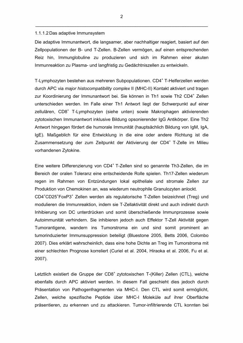
(TRIF) kommt. Das Ergebnis ist eine (verstärkte) Sekretion von proinflammatorischen
Zytokinen sowie Typ 1 Interferonen (siehe auch Abbildung 1).
Abbildung 1: TLR Signalwege. Abbildung nach Adams, 2009
Die Rolle von TLR in der Karzinogenese ist erst unvollständig verstanden. Neuere
Arbeiten postulieren eine fördernde Rolle von TLR7 am Beispiel des
Pankreaskarzinoms (Ochi et al. 2012). Andererseits belegeteine Vielzahl von Arbeiten,
dass TLR Liganden, als Therapeutika eingesetzt, effektive antitumorale Effekte
vermitteln. Der TLR7/8 Agonist Imiquimod wird vor allem bei dermatologischen
Malignomen klinisch eingesetzt. Bacillus Calmette-Guerin (aktiviert TLR2/4) ist eine
Standardbehandlung bei Blasenkarzinomen. Weitere TLR aktivierende Wirkstoffe
befinden sich in klinischer Erprobung (Adams 2009, Galluzzi et al. 2012).
1.1.2.2 RIG-I like Helikasen (RIG-I)
Die RIG-I ähnlichen Helikasen/ATPasen RIG-I, MDA-5 sowie LPG-2 finden sich im
Zytosol von Immun-, aber auch den meisten Nicht-Immunzellen. Sie gehören zu den
superfamily-2 (SF2) Helikasen und teilen sich sieben konservierte Motive, welche die
Nukleinsäure- und ATP-Bindung vermitteln (Gorbalenya et al. 1988, Hopfner et al.
2007). Während MDA-5 Doppelstrang-RNA detektiert und artifiziell durch synthetische
polyinosinic:polycytidylic acid (poly[I:C]) aktiviert werden kann (Kang et al. 2002, Gitlin
et al. 2006, Kato et al. 2006), erkennt RIG-I hautsächlich eine Triphosphatgruppe, die
typischerweise von viralen Polymerasen im Zytosol der Zelle im Rahmen von
Replikationsabläufen am 5‘-Ende von Doppelstrang-RNA Molekülen generiert wird
5
_____________________________________________________________________
(Kato et al. 2005, Hornung et al. 2006, Saito et al. 2008). Zusätzlich bedarf es eines
kurzen basenpaarigen Abschnitts der RNA, welcher bei Einzelstrang-RNA durch eine
loop-Struktur ermöglicht wird (Schmidt et al. 2009). Erwähnenswert ist, dass die
Triphosphatgruppe am 5‘-Ende eines RNA-Strangs auch regelmäßig physiologisch im
Nukleus von Zellen generiert wird, jedoch aufgrund von Spleißungsvorgängen, 5‘-
capping und weiteren Modifikationen normalerweise nicht in direkten Kontakt mit RIG-I
im Zytosol kommt (Pichlmair et al. 2006, Yoneyama et al. 2009). 5‘-Triphosphat RNA
kann mittels in-vitro-Transkription synthetisch hergestellt und mittels Transfektion in die
Zielzellen eingeschleust werden, wodurch sich neue Therapieoptionen ergeben (Kato
et al. 2005, Hornung et al. 2006). Die Rolle von LGP-2 ist bisher nicht vollständig
verstanden, es wird jedoch eine eher regulatorische Funktion angenommen
(Rothenfußer et al. 2005, Pippig et al. 2009).
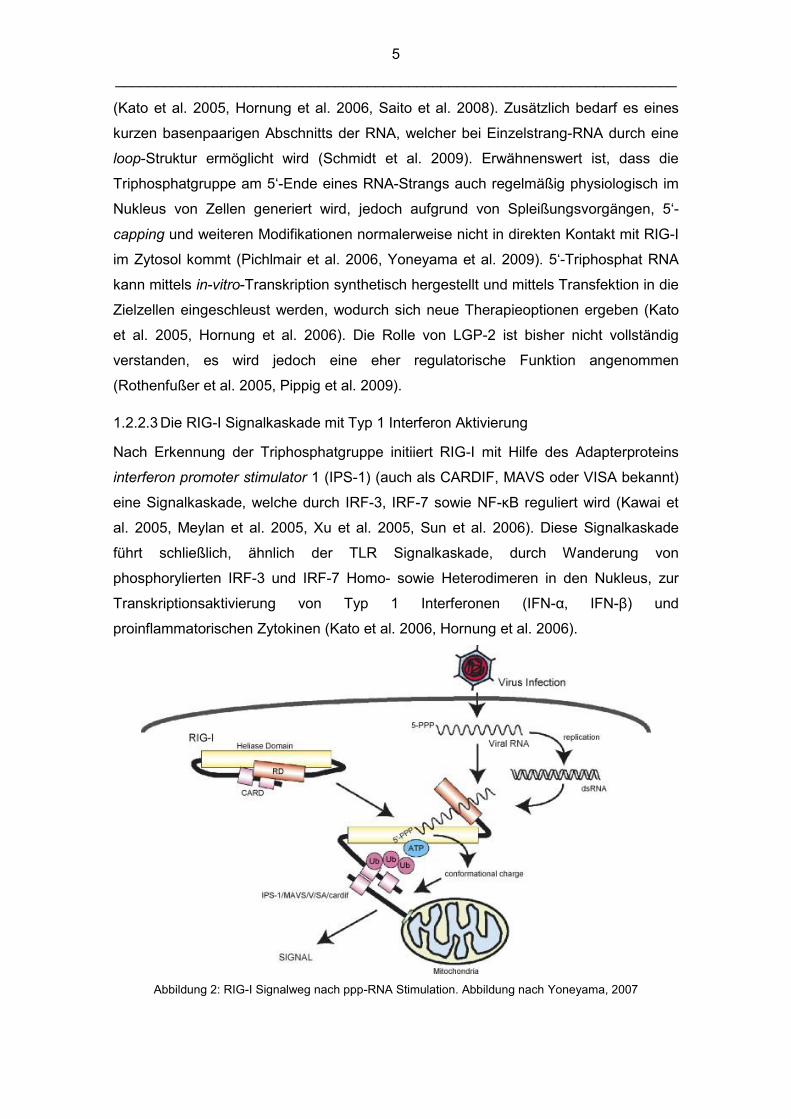
1.2.2.3 Die RIG-I Signalkaskade mit Typ 1 Interferon Aktivierung
Nach Erkennung der Triphosphatgruppe initiiert RIG-I mit Hilfe des Adapterproteins
interferon promoter stimulator 1 (IPS-1) (auch als CARDIF, MAVS oder VISA bekannt)
eine Signalkaskade, welche durch IRF-3, IRF-7 sowie NF-κB reguliert wird (Kawai et
al. 2005, Meylan et al. 2005, Xu et al. 2005, Sun et al. 2006). Diese Signalkaskade
führt schließlich, ähnlich der TLR Signalkaskade, durch Wanderung von
phosphorylierten IRF-3 und IRF-7 Homo- sowie Heterodimeren in den Nukleus, zur
Transkriptionsaktivierung von Typ 1 Interferonen (IFN-α, IFN-β) und
proinflammatorischen Zytokinen (Kato et al. 2006, Hornung et al. 2006).
Abbildung 2: RIG-I Signalweg nach ppp-RNA Stimulation. Abbildung nach Yoneyama, 2007
6
_____________________________________________________________________
Typ 1 Interferone sind Zytokine, die vor allem von Leukozyten, Monozyten und
Fibroblasten produziert werden, jedoch in geringerem Maße auch von anderen
Zellpopulationen, inklusive Tumorzellen, gebildet werden können. Sie greifen direkt in
die intrazelluläre Virusreplikation ein und unterbinden diese, hauptsächlich vermittelt
über den Januskinasen-signal transducer and activator of transcription (JAK-STAT)
Signalweg (Platanias 2005). Nach Bindung an Interferon-Rezeptoren auf den
Ursprungszellen sowie umgebenden Zellen werden, vermittelt über den erwähnten
JAK-STAT Signalweg, MHC-I Moleküle verstärkt auf der Zelloberfläche exprimiert, um
Attacken durch zytotoxische CD8+ T-Zellen zu erleichtern. Aktivierte T-Zellen werden
am Leben gehalten. DC, Makrophagen und NK-Zellen werden aktiviert, sofern sie nicht
bereits selbst virusinfiziert sind und somit bereits autokrin aktiviert wurden. B-Zellen
werden zur Bildung von Antikörpern animiert. Des Weiteren werden verschiedene IFN-
abhängige Botenstoffe der angeborenen Immunität, wie das Chemokin CXCL10, das
Interleukin 2 (IL-2) oder der tumor necrosis factor alpha (TNFα), hochreguliert. Im
Zusammenspiel mit p53 kommt es zu Apoptose der infizierten Zelle (Takaoka et al.
2003, Dunn et al. 2005, Platanias et al. 2005, Pestka 2007, Fensterl et al. 2009).
Zusätzlich kommt es nach Aktivierung der RIG-I-like Helikasen Interferon-unabhängig
zu Apoptose auf dem intrinsischen beziehungsweise mitochondrialen Pfad durch
Aktivierung der BH3-only Proteine Puma, Noxa und in geringerem Maße Bim und Bik
(Besch et al. 2009). Interessanterweise scheinen maligne Zellen für diesen
Apoptosemechanismus besonders anfällig zu sein. Der Grund hierfür liegt
wahrscheinlich im Schutz nicht-maligner Zellen durch BCL-xl, das in Tumorzellen im
Rahmen der so genannten synthetic lethality und oncogene addiction nicht vorhanden
beziehungsweise nicht funktionstüchtig zu sein scheint, da durch Genalterationen im
Prozess der Karzinogenese eine erhöhte Vulnerabilität entstehen kann (Hartwell et al.
1997, Evan 2006).
1.2 Tumorimmuntherapie
1.2.1 Allgemeiner Status Quo
In den vergangenen Jahren ist eine Vielzahl an Ansätzen verfolgt worden, um, jenseits
von Chirurgie, Strahlen- und Chemotherapie, auch die Immunantwort des Organismus
für die Tumortherapie zu nutzen. Mittlerweile hat sich die Immuntherapie als viertes
Standbein der Onkologie etabliert. Das therapeutische Spektrum erstreckt sich vom
Einsatz onkolytischer Viren, Aktivierung von PRR oder der adoptiven T-Zell Therapie
über den Einsatz extrakorporaler DC-Aktivierung bis hin zur großen Gruppe
7
_____________________________________________________________________
immunmodulierender Antikörper, um nur einige der Strategien zu benennen (Krieg
2007, 2008, Vollmer et al. 2009, Bauer et al. 2011, Wu et al. 2012). Ein zentrales
Problem der Immuntherapie besteht darin, dass viele Malignome immunsuppressive
Eigenschaften aufweisen, was die Effektivität der genannten Strategien limitiert
(Armstrong et al. 2001, Franks et al. 2012, Hong et al. 2012). Gerade das
Pankreaskarzinom wartet mit einer Vielzahl immunsuppressiver Eigenschaften auf, die
eine Barriere für eine effektive Immuntherapie darstellen.
1.2.2 Immuntherapie des Pankreaskarzinoms
In den letzten Jahren wurden auch in der Behandlung des Pankreaskarzinoms eine
große Bandbreite therapeutischer Ansätze inklusive Vakzinen, monoklonalen
Antikörpern sowie T-Zell und DC Therapien mit unterschiedlichem jedoch grundsätzlich
nicht durchschlagendem Erfolg untersucht (Bauer et al. 2011, Dodson et al. 2011,
Michl et al. 2013). Aktuell werden mehrere experimentell vielversprechende
Immuntherapeutika klinisch evaluiert. Hierzu gehören die cytotoxic T-lymphocyte-
associated antigen 4 (CTLA-4) Antikörper Ipilimumab und Tremelimumab, welche die
Herunterregulation aktivierter T-Zellen verhindern können, das Fusionsprotein L19-IL2,
welches durch Bindung an die tumorspezifische extradomain B (ED-B) extrem hohe IL-
2 Spiegel lokal im Tumorgewebe induziert, wie auch ein CD40 Agonist, der die T-Zell
Aktivierung sowie die Aktivierung myeloider Zellen im Tumorstroma unterstützt
(Wagner et al. 2008, Hodi et al. 2010, Beatty et al. 2011). Bisher konnte jedoch kein
immunologischer Ansatz als Standardtherapie etabliert werden.
1.3 Das Pankreaskarzinom
1.3.1 Epidemiologie, Diagnose und Therapie
Das Pankreaskarzinom ist die vierthäufigste Todesursache durch Krebserkrankungen
weltweit, obwohl die Inzidenz nur bei etwa 15 pro 100.000 Einwohnern und damit
verhältnismäßig niedrig liegt. Die zusammengefasste Fünf-Jahres-Überlebensrate liegt
bei unter fünf Prozent. Eine Heilung ist nur durch eine, in durchschnittlich weniger als
15 Prozent der Fälle gelingende, chirurgische R0-Resektion möglich. Einzig bei
Diagnose im UICC Stadium I-II (lokal begrenzt, keine Metastasen) erscheint die Fünf-
Jahre-Überlebensrate mit 40 Prozent vielversprechender. Eine frühzeitige Diagnose ist
jedoch nur selten möglich, da die Erkrankung oft lange asymptomatisch bleibt (Jemal
et al. 2008). Der chemotherapeutische Goldstandard in der palliativen Situation, bei der
eine Operation nicht mehr möglich beziehungsweise sinnvoll ist, sowie adjuvant nach
erfolgreicher R0-Resektion, ist das Zytostatikum Gemcitabin. Das Pankreaskarzinom
8
_____________________________________________________________________
ist jedoch weitgehend chemo- und strahlentherapieresistent, so dass große Fortschritte
bezüglich der Heilungsraten bisher kaum erzielt werden konnten (Burris et al. 1997,
Vulfovich et al. 2008). Die Polychemotherapie nach dem FOLFIRINOX-Schema hat
sich beim metastasierten Pankreaskarzinom als effektiver erwiesen, kann jedoch
aufgrund seiner hohen Toxizität nur bei Patienten in sehr gutem Allgemeinzustand
eingesetzt werden (Conroy et al. 2011).
1.3.2 Ätiologie, Pathologie und Pathogenese
Es sind eine Reihe von Risikofaktoren inklusive Zigarettenrauchen, Alkoholkonsum,
Adipositas, chronischer Pankreatitis sowie zystischer Pankreasneoplasien beschrieben
worden. Darüber hinaus bestehen mehrere Tumordispositionssyndrome mit
unterschiedlichem Erkrankungsrisiko, wie das Peutz-Jeghers-Syndrom (STK11 Gen),
die hereditäre Pankreatitis (PRSS1 Gen) und das familiäre Pankreaskarzinom (Gen
unbekannt), um die drei wichtigsten zu nennen.
Unterschieden wird zwischen dem weitaus häufigeren duktalen (circa 90 Prozent) und
dem azinären Karzinom (circa 10 Prozent). Lokalisiert sind beide Typen in circa 70
Prozent im Bereich des Pankreaskopfes.
Ausgangspunkt der Tumorprogression ist in 85 bis 95 Prozent der Fälle eine Mutation
des Onkogens Kras. In 60 bis 80 Prozent der Fälle kann weiterhin eine Genmutation
der Tumorsuppressoren p15 und/ oder p16 sowie in je etwa 50 Prozent der Fälle eine
Mutation in den Tumorsuppressorgenen von p53 und DPC4/ Smad4 detektiert werden
(Wong 2009). Die Mutation des p53 Gens bedeutet, dass Apoptose über den
extrinsischen Pfad nur noch eingeschränkt möglich ist, da hierbei p53 als wichtiger
Induktor wirkt (Igney et al. 2002, Haupt et al. 2003). Zusätzlich findet sich in vielen
Pankreaskarzinomen eine Mutation des transforming growth factor-beta Rezeptors
(TGF-βR) mit nachfolgend gestörter Signalkaskade (Massagué et al. 2008). Neuere
Erkenntnisse sprechen auch chronischen Entzündungsvorgängen, die zur Entstehung
von Tumorstroma beitragen, einen zentralen Anteil an der Kanzerogenese zu (Ochi et
al. 2012).
1.3.3 Die Aggressivität und Letalität des Pankreaskarzinoms
Neben der meist späten Diagnosestellung in entsprechend fortgeschrittenem
Erkrankungsstadium wurden zwei weitere Hauptgründe bisher als Erklärung der hohen
Letalität des Pankreaskarzinoms beschrieben: die desmoplastische Reaktion sowie die
geringe Immunogenität mit ausgeprägter tumorinduzierter Immunsuppression.
9
_____________________________________________________________________
1.3.3.1 Desmoplastische Reaktion
Im Laufe seines Wachstums schafft sich das Pankreaskarzinom eine
bindegewebsreiche, privilegierte Wachstumsumgebung. Im Gegensatz zu anderen
Tumorentitäten, die oft als geballter Karzinomzellhaufen mit intensiver
Gefäßversorgung imponieren, zeigt sich das Pankreaskarzinom als derbe, schlecht
vaskularisierte Masse. Dies führt unter anderem dazu, dass der Abstand zwischen
Blutgefäß und Karzinomzelle zu groß für eine effektive Chemotherapie ist und auch
Zellen des Immunsystems schlechter das Tumorgewebe infiltrieren können (Wong et
al. 2009, Olive et al. 2009, Neesse et al. 2011, Michl et al. 2013).
1.3.3.2 Immunmodulation und tumorinduzierte Immunsuppression
Pankreaskarzinomzellen verfügen über eine nur schwach immunogene Zelloberfläche
mit sehr geringer MHC-I Expression (Costello et al. 1999). Es fehlen Adhäsions- und
ko-stimulatorische Moleküle, was die Anheftung und Aktivierung von Immunzellen
erschwert (Rabinovich et al. 2007). Gerade im Gegensatz zu virusinduzierten Tumoren
fehlen Interferone als immunaktivierende Signale. Somit besteht für das Immunsystem
möglicherweise kaum eine Chance, längerfristig eine Eliminations- oder zumindest
eine Equilibriumsphase im Sinne des Immunüberwachungsmodells aufrechtzuerhalten
(Dunn et al. 2002). Entscheidend ist außerdem eine tumorinduzierte
Immunsuppression durch verschiedene Mediatoren beziehungsweise die Blockade
solcher (Rayman et al. 2000, von Bernstorff et al. 2002, Rabinovich et al. 2007). Es
kommt zu einer Rekrutierung von Treg in das Tumorstroma und zu T-Zell Anergie
(Sakaguchi 2008, Liyanage et al. 2002, Fukunaga et al. 2004, Thomas et al. 2005,
Massagué et al. 2008). Eine zentrale Rolle in all diesen immunsuppressiven
Vorgängen spielt das Zytokin transforming growth factor-beta (TGF-β), welches im
Tumorgewebe überexprimiert wird.
1.4 Transforming growth factor-beta (TGF-β)TGF-β mit seinen Unterklassen TGF-β1, TGF-β2 und TGF-β3 ist ein Zytokin, dass 1983
erstmals aus Kulturüberständen von Tumorzellen isoliert wurde (Assoion et al. 1983,
Frolik et al. 1983, Roberts et al. 1983). Es bindet an Serin-Threonin Kinasen und
beeinflusst die Transkription unterschiedlichster Gene (Massagué et al. 1996). Unter
physiologischen Bedingungen sichert TGF-β die Gewebshomöostase durch Kontrolle
der Zellproliferation und des Zellüberlebens, der Zelldifferenzierung sowie der
Zelladhäsion. TGF-β wird allgemein als potentester, natürlich auftretender
Unterdrücker von Immunfunktion angesehen, unverzichtbar zur Vermeidung von
Autoimmunität (Pennison et al. 2007). TGF-β knockout Mäuse haben sich als nicht
10
_____________________________________________________________________
dauerhaft lebensfähig gezeigt (Tang et al. 1998). Beispielsweise wird orale Toleranz
durch TGF-β induzierten Antikörperswitch zu IgA sowie Th3-Zell Aktivierung ermöglicht
(Gilbert et al. 2011). T-Zellen, die im Falle von entzündlichen Darmerkrankungen für
die überschießende Immunreaktion verantwortlich sind, verfügen über pathologisch
hochreguliertes SMAD7, was eine reduzierte Reaktion auf TGF-β vermittelte,
inhibitorische Signale bewirkt (Becker et al. 2006). Bei Malignom-induzierter
Hypersekretion oder TGF-β Rezeptormutation mit Fehlregulation der anhängigen
Signalkaskade kommt es jedoch zu einer das Tumorwachstum fördernden, massiven
Immunsuppression (Biswas et al. 2004, Ijichi et al. 2006).
Hohe Spiegel von TGF-β im Blut von Pankreaskarzinompatienten korrelieren mit einer
schlechten Prognose (Friess et al. 1993). TGF-β wird eine entscheidende pro-
metastatische Rolle zugesprochen (Bhowmick et al. 2004, Pollard et al. 2004, Kallari et
al. 2006). Die Präsenz von TGF-β an der invasiv wachsenden Seite eines Tumors wird
mit Tumorprogression und Metastasierung assoziiert (Dalal et al. 1993, Padua et al.
2009). Es induziert eine epithelial-to-mesenchymal transition (EMT) der Karzinomzellen
und erhöht dadurch deren Motilität und damit den Grad der Invasivität der Tumorzellen
(Ellenrieder et al. 2001, Bhowmick et al. 2001, 2004, Drabsch et al. 2012). Die
Angiogenese wird zudem gefördert (Roberts et al. 1983). TGF-β ist beteiligt an der
Induktion, der Rekrutierung und Expansion von MDSC (Li et al. 2012). Darüber hinaus
wirkt es auch direkt inhibierend auf Makrophagen, B-Zellen und CTL, reduziert deren
Teilungsrate sowie ihre Fähigkeit, fremde Zellen zu erkennen und zu attackieren. Im
Detail blockiert es die Bildung und Sekretion von Perforin, Granzym, Fas-Ligand sowie
Interferon-γ (IFN-γ), was eine weitestgehende CD8+ T-Zell Anergie zur Folge hat
(Fukunaga et al. 2004, Thomas et al. 2005, Massagué et al. 2008). TGF-β verschiebt
die Immunantwort des Organismus im Gesamten von einer Th1 dominanten,
zytotoxischen zu einer Th2 fokussierten Immunantwort, beziehungsweise induziert
auch direkt die Bildung regulatorischer T-Zellen (Chen et al. 2003, Moutsopoulos et al.
2008). Aus den genannten Gründen kann TGF-β als ein zentrales Molekül der
tumorinduzierten Immunsuppression bezeichnet werden.
1.4.1 TGF-β als therapeutische Zielstruktur für die Tumorimmuntherapie
Aufgrund der beschriebenen Eigenschaften erscheint TGF-β als sinnvolles Ziel einer
Therapie des Pankreaskarzinoms, die auf die Brechung tumorinduzierter
Immunsuppression fokussiert ist. Einige Anti-TGF-β Moleküle waren in präklinischen
Studien bereits effektiv und ein Teil von ihnen wird aktuell in klinischen Studien bei
Patienten mit Melanomen, Glioblastomen, kolorektalen Karzinomen, Nieren-, Brust-
11
_____________________________________________________________________
und auch Pankreaskarzinomen untersucht (Schlingensiepen et al. 2006, Gaspar et al.
2007, Schlingensiepen et al. 2009, Takaku et al. 2010, Drabsch et al. 2012). Hierbei
werden unterschiedliche Strategien verfolgt, wobei meist versucht wird, TGF-β und
seine Wirkung so hoch wie möglich im Signalweg zu neutralisieren. Dies kann
entweder durch die Inhibition oder Sequestrierung der TGF-β Protein Liganden, des
Proteins selbst oder durch die Blockade der TGF-β Rezeptoren erfolgen. Hierfür
werden small molecules, antisense Oligonukleotide, small hairpin RNA oder auch
neutralisierende Antikörper verwendet. Teilweise wurden diese Strategien bereits in
Kombination mit konventionellen Therapien (Chemotherapie, Radiotherapie) oder
Immuntherapien wie dem adoptiven T-Zell Transfer untersucht. Trotz einigem
therapeutischen Erfolg bleiben Zweifel bezüglich der systemischen Nebenwirkungen
mit der Gefahr von de novo Tumoren oder Autoimmunprozessen aufgrund der
vielschichtigen Eigenschaften von TGF-β (Drabsch et al. 2012).
1.5 Zusammenfassung/ Summary
1.5.1 Zusammenfassung der vorgelegten Publikationen
In den letzten Jahrzehnten konnten große Erfolge in der Tumortherapie gefeiert
werden, auch und besonders auf dem Feld der Tumorimmuntherapie. Bei einigen
Tumorerkrankungen, allen voran beim Pankreaskarzinom, blieben die therapeutischen
Fortschritte jedoch dürftig. Als ein zentraler Grund hierfür wird die tumorinduzierte
Immunsuppression angesehen. Daher bedarf es kontinuierlicher Anstrengung, die
Mechanismen dieser Immunsuppression weiter zu verstehen und aus den
gewonnenen Erkenntnissen Therapieansätze zu entwickeln, die letztlich auch Einzug
in den klinischen Alltag halten können.
1.5.1.1 Anz et al. Int J Cancer 2011.
CD103 is a hallmark of tumor-infiltrating regulatory T-cells.
Der erste Teil der Arbeit behandelt die Rolle der CD103+ (auch bekannt als αEβ7)
Subpopulation regulatorischer T-Zellen (Treg). Treg spielen eine entscheidende Rolle
im Rahmen tumorinduzierter Immunsuppression. Die gegen Treg bereits verwendeten,
beziehungsweise theoretisch denkbaren Therapieoptionen, haben den Nachteil, dass
es sich entweder um schlecht erreichbare, intrazelluläre Zielstrukturen handelt
(FoxP3), die Effektivität mit steigender Tumorlast massiv abnimmt (CD25) oder das
Ziel zu unspezifisch ist und es somit zu autoimmunen Nebenwirkungen kommt
(CTLA4) (Onizuka et al. 1999, Kapadia et al. 2005, Colombo et al. 2007).
12
_____________________________________________________________________
Initial konnten wir die gesteigerte immunsuppressive Potenz CD103+ im Vergleich mit
CD103- regulatorischen T-Zellen bestätigen. Es gelang durch Analysen in vier
verschiedenen murinen Tumormodellen (EL4 Lymphom, CT26 Kolonkarzinom, B16
Melanom, Panc02 Pankreaskarzinom) zu zeigen, dass die Population CD103+ Treg
spezifisch für tumorinfiltrierende Treg ist, jedoch CD103 nicht für die Retention dieser
im Tumorstroma verantwortlich zeichnet. Unsere Ergebnisse lassen weiterhin darauf
schließen, dass intratumorales TGF-β entscheidend an der Induktion der CD103+
Subpopulation beteiligt ist, da unter anderem im murinen, orthotopen Panc02
Pankreaskarzinommodell nach systemischer Therapie mit einer siRNA gegen TGF-β
die Zahl CD103+ Treg signifikant reduziert werden konnte. Zusammenfassend lässt
sich sagen, dass CD103 eine potentielle Zielstruktur für die Therapie tumorinduzierter
Immunsuppression darstellt.
1.5.1.2 Jacobs et al. Int J Cancer 2011.
An ISCOM vaccine combined with a TLR9 agonist breaks immune evasion mediated
by regulatory T-cells in an orthotopic model of pancreatic carcinoma.
Im zweiten Teil der Arbeit haben wir in einem murinen, orthotopen
Pankreaskarzinommodell eine Vakzinierungsstrategie gegen Tumorantigene mittels
immunstimulatorischer Komplexe (ISCOM) untersucht. ISCOM-Vakzine bestehen aus
Proteinantigenen, die mit käfigartigen Nanostrukturen, die aus Saponin,
Phospholipiden und Cholesterin aufgebaut sind, komplexiert werden. ISCOM-Vakzine
induzieren eine allgemeine Immunstimulation sowie B- und T-Zell-vermittelte
Immunantworten gegen multiple MHC-II und MHC-I Epitope der entsprechenden
Proteinantigene. Maßgeblich ist eine Aktivierung von DC, die zur
Antigenkreuzpräsentation und nachfolgender T-Zell Aktivierung befähigt werden (Davis
et al. 2004, Schnurr et al. 2005, Drane et al. 2007, Schnurr et al. 2009, Duewell et al.
2011).
Für diese Versuche verwendeten wir eine OVA/ISCOM-Vakzine und generierten
Panc02 Tumorzellen, die Ovalbumin (OVA) als experimentelles Tumorantigen
exprimieren (PancOVA). Der Impfstoff wurde alleine sowie in Kombination mit dem
TLR9 Agonisten CpG-ODN 1826 subkutan injiziert. Zudem wurde eine Kombination
der Vakzine mit einem gegen das Oberflächenmolekül CD25 gerichteten Antikörper
evaluiert, welcher zu einer Depletion von Treg führt.
Die prophylaktische Gabe der OVA/ISCOM Vakzine führte zu einem vollständigen
Tumorschutz durch die hocheffektive Induktion OVA-spezifischer CTL. Bei bereits
13
_____________________________________________________________________
etablierten Tumoren (therapeutische Vakzinierung) jedoch war die alleinige Gabe der
OVA/ISCOM Vakzine nicht ausreichend effektiv. Dies war unter anderem auf eine
tumorvermittelte Induktion von Treg zurückzuführen. Entsprechend verbesserte die
Gabe des CD25 Antikörpers das Therapieergebnis signifikant. Die Kombination der
Vakzine mit dem TLR9 Agonisten CpG ODN 1826 führte zu einer Th1-dominanten
Immunantwort mit Aktivierung von Immunzellen des angeborenen sowie des adaptiven
Immunsystems. Daraus resultierte eine massive Expansion von Antigen-spezifischen
CD8+ CTL. Therapeutisch konnte eine signifikante Lebensverlängerung inklusive
kompletter Tumorregressionen im orthotopen Tumormodell festgestellt werden.
Interessanterweise waren alle der überlebenden Tiere vor einer re-challenge mit
PancOVA Tumoren und einige sogar mit Wildtyp Panc02 Tumoren (ohne OVA
Expression) geschützt. Dieses Ergebnis ließ darauf schließen, dass es durch die
Immuntherapie zu einem T-Zell Gedächtnis mit epitope spreading gekommen war.
Durch den Nachweis p15E-spezifischer CTL im Blut der Langzeitüberlebenden (es
handelt sich bei p15E um ein spezifisches Tumorantigen von Panc02 Zellen) konnte
diese Hypothese bestätigt werden. Die Ergebnisse zeigen eine effiziente Methode,
tumorinduzierte Immunsuppression durch geeignete immuntherapeutische Strategien
zu überwinden. Das Konzept der Tumorvakzine mit unterschiedlichen Tumorantigenen
wird in der Therapie des Pankreaskarzinoms alleine in den USA aktuell in über zehn
klinischen Studien untersucht (http://www.cancer.gov/clinicaltrials/search/
results?protocolsearchid=11444759, 28.02.2013). Die Verwendung von ISCOM-
Vakzinen zusammen mit TLR Liganden könnte hier in Zukunft einen entscheidenden
Vorteil bringen.
1.5.1.3 Ellermeier et al. Cancer Res 2013.
Therapeutic efficacy of bifunctional siRNA combining TGF-β1 silencing with RIG-I
activation in pancreatic cancer.
Der Hauptteil meiner Arbeit befasst sich mit der Therapie des Pankreaskarzinoms
mittels einer bi-funktionalen, RIG-I aktivierenden siRNA gegen TGF-β. TGF-β ist einer
der zentralen Treiber Pankreaskarzinom-induzierter Immunsuppression. RIG-I ist eine
zytosolische Helikase, die virusassoziierte 5‘-Triphosphat-RNA erkennt und nach
Aktivierung zu einer anti-viralen Typ 1 IFN Antwort führt sowie Apoptose induziert. Die
Kombination aus RNA-Interferenz und RIG-I-Aktivierung konnte durch eine
Triphosphatmodifikation am 5‘-Ende der TGF-β-spezifischen siRNA (ppp-TGF-β)
mittels in-vitro-Transkription eines entsprechenden DNA-Templates erreicht werden.
Eine auf ähnliche Weise generierte ppp-siRNA gegen Bcl-2, mit dem Ziel
14
_____________________________________________________________________
Apoptoseinduktion zu verstärken, hatte sich in einer Arbeit von Poeck et al. (2008)
prinzipiell als erfolgreich in einem murinen Melanommodell erwiesen.
Wir konnten zeigen, dass humane Pankreaskarzinomzellen funktionelles RIG-I
exprimieren und somit für eine ppp-RNA-Therapie in Frage kommen. Die von uns
entwickelte ppp-TGF-β führte sowohl in murinen als auch in humanen Tumorzellen zu
einer signifikanten Genexpressionshemmung von TGF-β, zur Produktion pro-
inflammatorischer Zytokine und Chemokine (IFN-β, CXCL10) sowie zu Apoptose in
vitro. Die Bifunktionalität konnte auch in vivo bestätigt werden. In einem murinen,
orthopen Pankreaskarzinommodell kam es nach intravenöser Gabe von ppp-TGF-β zur
Reduktion der TGF-β-Spiegel im Serum und Tumorgewebe, zu systemischer
Immunaktivierung (pro-inflammatorische Zytokine, Immunzellaktivierung) sowie
verstärkter Einwanderung von CD8+ T-Zellen in das Tumorgewebe. Ferner zeigte sich
eine Caspase-9-vermittelte Apoptose von Tumorzellen. Durchflusszytometrische
Analysen der Immunzellinfiltrate im Tumor und lymphatischen Organen zeigten eine
Aktivierung von CD8+ T-Zellen sowie eine Reduktion von MDSC, die zudem einen
Phänotyp verminderter Suppressivität aufwiesen. Hinweise auf Organschäden oder
durch die Therapie induzierte Autoimmunprozesse fanden sich nicht.
Überlebensversuche zeigten einen signifikanten Therapieerfolg der mit ppp-TGF-β
behandelten Versuchstiere, wobei es in 33% der Fälle zu einer kompletten Remission
kam, wie Autopsien der überlebenden Tiere nach 100 Tagen ergaben. Versuche mit
Immunzell-depletierenden Antikörpern konnten belegen, dass der Therapieerfolg auf
der Aktivierung tumorreaktiver CD8+ T-Zellen beruhte, während NK-Zellen entbehrlich
waren. Zusammenfassend konnten wir zeigen, dass der Einsatz einer bi-funktional
wirksamen, immunstimulatorischen siRNA gegen TGF-β ein innovatives, sicheres und
vielversprechendes Therapiekonzept darstellt.
1.5.2 Summary of the presented publications
Over the course of the past decades major progress has been made regarding
malignoma therapies and outcomes. Tumor immunotherapy has been established as
the fourth pillar of tumor therapy. Nevertheless, patients with certain malignancies such
as pancreatic carcinoma have hardly been able to benefit from new therapeutic
regimens. Tumor-mediated immunosuppression is seen as a major reason for this lack
of treatment efficacy. Therefore continuous effort is needed to shed more light on the
mechanisms of tumor-mediated immunosuppression and to develop promising
therapeutic strategies which are suited to eventually enter clinical application.
15
_____________________________________________________________________
1.5.2.1 Anz et al. Int J Cancer 2011.
CD103 is a hallmark of tumor-infiltrating regulatory T-cells.
The first part of my thesis deals with the role of the CD103+ (cluster of differentiation
103, also known as αEβ7) subpopulation of regulatory T-cells (Treg). Treg play a
crucial role in tumor-mediated immunosuppression. Anti-Treg agents already in use or
theoretically plausible exhibit major drawbacks. The target structures are either hardly
accessible due to intracellular localization (FoxP3), therapeutic efficacy plummets while
tumor load increases (CD25) or major side effects of autoimmunity occur due to
unspecificity (CTLA4) (Onizuka et al. 1999, Kapadia et al. 2005, Colombo et al. 2007).
First, we were able to confirm the increased immunosuppressive function of CD103+ as
compared to CD103- Treg. By analyzing four different murine tumor models (EL4
lymphoma, CT26 colon carcinoma, B16 melanoma and Panc02 pancreatic carcinoma)
we were able to show that CD103+ is upregulated in tumor infiltrating Treg. Treg
retention in the tumor stroma however was not mediated by CD103. Our results further
indicated that TGF-β is the key player in the process of induction of the CD103+
subpopulation since TGF-β knockdown via RNA interference in a model of murine
orthotopic pancreatic carcinoma led to significantly reduced numbers of CD103+ Treg
in the tumor tissue. In conclusion we can state that CD103 and its regulation by TGF-β
are potential therapeutic targets to break tumor-mediated immunosuppression.
1.5.3.2 Jacobs et al. Int J Cancer 2011.
An ISCOM vaccine combined with a TLR9 agonist breaks immune evasion mediated
by regulatory T-cells in an orthotopic model of pancreatic carcinoma.
In the second part of my thesis we used an ovalbumine (OVA)-expressing model of
murine, orthotopic Panc02 pancreatic carcinoma to test the therapeutic efficacy of an
ISCOMATRIX (IMX)-based vaccine containing the model antigen OVA (OVA/IMX). IMX
consists of cholesterol, saponin and phoshpolipids and forms particles of approximately
40 nm in diameter. IMX-based vaccines, which contain the adjuvant plus a protein
antigen, have been demonstrated to efficiently induce both humoral and adaptive
immune responses to vaccine antigen. IMX has been shown to activate DC in vivo and
to facilitate antigen cross-presentation by DC (Davis et al. 2004, Schnurr et al. 2005,
Drane et al. 2007, Schnurr et al. 2009, Duewell et al. 2011). To counteract
immunosuppressive mechanisms derived from the tumor we evaluated the IMX-based
vaccine alone, in combination with a TLR9 agonist (CpG-ODN 1826) or with a
depleting antibody against CD25.
16
_____________________________________________________________________
Prophylactic vaccination with OVA/IMX led to effective tumor protection via induction of
antigen-specific CTL. In a therapeutic setting, when tumors were already established,
OVA/IMX did not prove sufficiently effective, partly due to tumor-mediated induction of
Treg. The addition of an anti-CD25 antibody significantly improved therapy outcomes.
The combination of the vaccine with the TLR9 agonist led to a Th1-dominant immune
response along with the activation of the adaptive immune system, especially CD8+ T
cells. Significantly prolonged survival rates including several long-term survivors could
be achieved in the orthotopic PancOVA model of murine pancreatic cancer. Strikingly,
those long-term survivors all rejected PancOVA tumors and partially rejected Panc02
wildtype cancer cells lacking OVA-expression in re-challenge experiments. We
assumed this to be due to T-cell memory induction and epitope spreading. Our
hypothesis was supported by isolating p15E-specific CTL in the blood of long-term
survivors (the p15E epitope is derived from a tumor antigen expressed by Panc02
pancreatic cancer cells). The results show a potent strategy to break tumor-induced
immunosuppression. In the USA alone, there are currently more than ten clinical trials
ongoing, testing vaccination strategies for the treatment of pancreatic carcinoma
(http://www.cancer.gov/clinicaltrials/search/results?protocolsearchid=11444759,
February 28, 2013). The use of IMX vaccines in combination with TLR ligands or other
immune modifiers could potentially create a significant treatment advantage in the
future.
1.5.3.3 Ellermeier et al. Cancer Res 2013.
Therapeutic efficacy of bifunctional siRNA combining TGF-β1 silencing with RIG-I
activation in pancreatic cancer.
The main part of my work focused on the development of a bi-functional, RIG-I
activating siRNA targeting TGF-β for immunotherapy of pancreatic carcinoma. TGF-β
is a key promoter of cancer-induced immunosuppression, which is highly
overexpressed by pancreatic cancer cells. RIG-I is a cytosolic helicase which detects
virus-associated 5’triphosphate-RNA (ppp-RNA) and leads to an anti-viral type 1 IFN
response as well as IFN- and p53-independent apoptosis. The combination of RNA
interference with RIG-I activation in one RNA molecule was reached via 5’-triphosphate
modification of a TGF-β-specific siRNA by in vitro transcription of a corresponding
DNA-template (ppp-TGF-β). A related ppp-modified siRNA targeting the anti-apoptotic
molecule Bcl-2, which was designed to enhance apoptosis induction in tumor cells, has
demonstrated efficacy in a proof-of-principle study in a mouse model of malignant
melanoma (Poeck et al. 2008).
17
_____________________________________________________________________
We were able to show that human pancreatic cancer cells express functional RIG-I and
hence are susceptible to ppp-RNA therapy. Bifunctional ppp-TGF-β led to significant
TGF-β knockdown, production of pro-inflammatory cytokines and apoptosis in murine
as well as human pancreatic cancer cells in vitro. Following intravenous ppp-TGF-β
treatment in an orthotopic model of murine Panc02 pancreatic carcinoma, TGF-β
suppression systemically as well as in the tumor tissue was achieved. Furthermore,
treatment led to systemic immune activation (production of pro-inflammatory cytokines,
activation of immune cells), increased migration of CD8+ T-cells to the tumor tissue and
caspase 9-mediated tumor cell apoptosis. MDSC in tumor-bearing mice were reduced
in number and showed a less suppressive phenotype. Treatment-related toxicity or
autoimmunity was not detected. Survival experiments showed a significant benefit with
complete tumor remission in 33% of all ppp-TGF-β treated animals. Therapeutic
efficacy was significantly better for the bi-functional siRNA molecule as compared to
RNA molecules mediating either RIG-I activation or TGF-β gene silencing alone.
Therapeutic efficacy strongly relied on CD8+ T-cells, whereas NK cells appeared to be
dispensable, as evidenced with immune cell depleting antibodies. In conclusion we
were able to show that RIG-I is a promising target in pancreatic cancer and that bi-
functional immunostimulatory ppp-siRNA targeting TGF-β is an innovative, safe and
promising therapeutic concept.
18
_____________________________________________________________________
2. LiteraturverzeichnisAdams S. Toll-like receptor agonists in cancer therapy. Immunotherapy
2009;1(6):949–964.
Akira S, Uematsu S, Takeuchi O. Pathogen recognition and innate immunity. Cell
2006;124:783-801
Anderson KV, Bokla L, Nusslein-Volhard C. Establishment of dorsalventral polarity in
the Drosophila embryo: the induction of polarity by the Toll gene product. Cell
1985;42:791-8
Armstrong AC, Eaton D, Ewing JC. Science, medicine, and the future: Cellular
immunotherapy for cancer. BMJ 2001;323(7324):1289-93
Assoian RK, Flerudelys BE, Stevenson HC, et al. Expression and secretion of type fl
transforming growth factor by activated human macrophages. Proc Natl Acad Sci USA
1987;84:6020.
Banchereau J, Briere F, Caux C, Davoust J, Lebecque S., et al. Immunobiology of
dendritic cells. Annu Rev Immunol 2000;18:767-811
Barton GM, Medzhitov R. Control of adaptive immune responses by Toll-like
receptors. Curr Opin Immunol 2002;20:380-3
Bauer C, Dauer M, Saraj S, et al. Dendritic cell-based vaccination of patients with
advanced pancreatic carcinoma: results of a pilot study. Cancer Immunol Immunother
2011;60(8):1097-107.
Beatty GL, Chiorean EG, Fishman MP, et al. CD40 agonists alter tumor stroma and
show efficacy against pancreatic carcinoma in mice and humans. Science
2011;331:1612–16.
Becker C, Fantini MC, and Neurath MF. TGF-βeta as a T cell regulator in colitis and
colon cancer. Cytokine Growth Factor Rev 2006;17:97–106.
Besch R, Poeck H, Hohenauer T, Senft D, Hacker G, Berking C, et al. Proapoptotic
signaling induced by RIG-I and MDA-5 results in type I interferon-independent
apoptosis in human melanoma cells. J Clin Invest 2009;119:2399-411.
Betts GJ, Clarke SL, Richards HE, et al. Regulating the immune response to tumours.
Adv Drug Deliv Rev 2006;58:948-61.
19
_____________________________________________________________________
Beutler B. Inferences, questions and possibilities in Toll-like receptor signalling. Nature
2004;430(6996):257-63.
Beutler BA. TLRs and innate immunity. Blood 2009;113: 1399-407
Bhowmick NA, Ghiassi M, Bakin A, et al. Transforming growth factor-beta1 mediates
epithelial to mesenchymal transdifferentiation through a RhoAdependent mechanism.
Mol Biol Cell 2001;12:27–36.
Bhowmick NA, Neilson EG, Moses HL. Stromal fibroblasts in cancer initiation and
progression. Nature 2004;432:332-337.
Biswas S, Chytil A, Washington K, et al. Transforming growth factor beta receptor type
II inactivation promotes the establishment and progression of colon cancer. Cancer
Res 2004;64:4687–4692.
Bluestone JA, Tang Q. How do CD4+CD25+ regulatory T cells control autoimmunity?
Curr Opin Immunol 2005;17:638-42.
Burris H, Storniolo AM. Assessing clinical benefit in the treatment of pancreas cancer:
gemcitabine compared to 5-fluorouracil. Eur J Cancer 1997;33 Suppl 1: S18-22.
Chaplin DD. Overview of the immune response. J Allergy Clin Immunol 2010;125(2
Suppl 2):S3-23.
Chen W, Jin W, Hardegen N, et al. Conversion of peripheral CD4+CD25- naive T cells
to CD4+CD25+ regulatory T cells by TGF-βeta induction of transcription factor Foxp3. J
Exp Med 2003;198(12):1875-86.
Colombo MP, Piconese S. Regulatory-T-cell inhibition versus depletion: the right
choice in cancer immunotherapy. Nat Rev Cancer 2007;7:880-7.
Conroy T, Desseigne F, Ychou M, et al. FOLFIRINOX versus gemcitabine for
metastatic pancreatic cancer. N Engl J Med. 2011;364(19):1817-25.
Costello RT, Gastaut JA, Olive D. Tumor escape from immune surveillance. Arch
Immunol Ther Exp (Warsz) 1999;47(2):83-8.
Curiel TJ, Coukos G, Zou L, et al. Specific recruitment of regulatory T cells in ovarian
carcinoma fosters immune privilege and predicts reduced survival. Nat Med
2004;10:942-9.
20
_____________________________________________________________________
Dala BI, Keown PA, Greenberg AH. Immunocytochemical localization of secreted
transforming growth factor-beta 1 to the advancing edges of primary tumors and to
lymph node metastases of human mammary carcinoma. Am J Pathol 1993;143(2):381-
9
Davis ID, Chen W, Jackson H, et al. Recombinant NY-ESO-1 protein with
ISCOMATRIX adjuvant induces broad integrated antibody and CD4(+) and CD8(+) T
cell responses in humans. Proc Natl Acad Sci USA 2004;101(29):10697-702.
Diebold SS, Kaisho T, Hemmi H, Akira S, Reis e Sousa C. Innate antiviral responses
by means of TLR7-mediated recognition of single-stranded RNA. Science.
2004;303:1529-31.
Dodson LF, Hawkins WG und Peter Goedegebuure P. Potential targets for pancreatic
cancer immunotherapeutics. Immunotherapy 2011;3(4):517–537.
Drabsch Y, Ten Dijke P. TGF-β signalling and its role in cancer progression and
metastasis. Cancer Metastasis Rev 2012;31(3-4):553-68.
Drane D, Gittleson C, Boyle J, et al. ISCOMATRIX adjuvant for prophylactic and
therapeutic vaccines. Expert Rev Vaccines 2007;6(5): 761-72.
Duewell P, Kisser U, Heckelsmiller K, et al. ISCOMATRIX adjuvant combines immune
activation with antigen delivery to dendritic cells in vivo leading to effective cross-
priming of CD8+ T cells. J Immunol 2011;187(1):55-63.
Dumitru CA, Moses K, Trellakis S, et al. Neutrophils and granulocytic myeloid-derived
suppressor cells: immunophenotyping, cell biology and clinical relevance in human
oncology. Cancer Immunol Immunother 2012;61(8):1155-67.
Dunn GP, Bruce AT, Ikeda H, et al. Cancer immunoediting: from immunosurveillance
to tumor escape. Nat Immunol 2002;3(11):991-8.
Dunn GP, Bruce AT, Sheehan KC, et al. A critical function for type I interferons in
cancer immunoediting. Nat Immunol 2005;6:722-9.
Ellenrieder V, Hendler SF, Ruhland C, et al. TGF-βeta-induced invasiveness of
pancreatic cancer cells is mediated by matrix metalloproteinase-2 and the urokinase
plasminogen activator system. Int J Cancer 2001;93(2):204-11
Evan GI. Can’t kick that oncogene habit. Cancer Cell 2006;10:345–347.
Fensterl V, Sen GC. Interferons and viral infections. Biofactors 2009;35(1):14-20.
21
_____________________________________________________________________
Franks HA, Wang Q, Patel PM. New anticancer immunotherapies Anticancer Res.
2012;32(7):2439-53.
Friess H, Yamanaka Y, Buchler M, et al. Enhanced expression of transforming growth
factor beta isoforms in pancreatic cancer correlates with decreased survival.
Gastroenterology 1993;105:1846-56.
Frolik CA, Dart LL, Meyers CA, et al. Purification and initial characterization of a type β
transforming growth factor from human placenta. Proc Natl Acad Sci USA
1983;80:3676.
Fu J, Xu D, Liu Z, et al. Increased regulatory T cells correlate with CD8 T-cell
impairment and poor survival in hepatocellular carcinoma patients. Gastroenterology
2007;132:2328-39.
Fukunaga A, Miyamoto M, Cho Y, et al. CD8+ tumor-infiltrating lymphocytes together
with CD4+ tumor-infiltrating lymphocytes and dendritic cells improve the prognosis of
patients with pancreatic adenocarcinoma. Pancreas 2004;28:e26-31.
Galluzzi L, Vacchelli E, Eggermont A, et al. Trial Watch: Experimental Toll-like
receptor agonists for cancer therapy. Oncoimmunology 2012;1(5):699-716.
Gaspar NJ, Li L, Kapoun AM, et al. Inhibition of transforming growth factor beta
signaling reduces pancreatic adenocarcinoma growth and invasiveness. Mol
Pharmacol 2007;72:152-61.
Gilbert RS, Kobayashi R, Sekine S, et al. Functional transforming growth factor-β
receptor type II expression by CD4+ T cells in Peyer's patches is essential for oral
tolerance induction. PLoS One. 2011;6(11):e27501.
Gitlin L, Barchet W, Gilfillan S, et al. Essential role of mda-5 in type I IFN responses to
polyriboinosinic:polyribocytidylic acid and encephalomyocarditis picornavirus. Proc Natl
Acad Sci USA 2006;103: 8459-64
Gorbalenya AE, Koonin EV, Donchenko AP, Blinov VM. A conserved NTP-motif in
putative helicases. Nature 1988;333(6168):22
Hartwell LH, Szankasi P, Roberts CJ, et al. Integrating genetic approaches into the
discovery of anticancer drugs. Science 1997;278:1064–1068.
Haupt S, Berger M, Goldberg Z, et al. Apoptosis - the p53 network. J Cell Sci
2003;116(Pt 20):4077-85.
22
_____________________________________________________________________
Hiraoka N, Onozato K, Kosuge T, et al. Prevalence of FOXP3+ regulatory T cells
increases during the progression of pancreatic ductal adenocarcinoma and its
premalignant lesions. Clin Cancer Res 2006;12:5423-34.
Hodi FS, O’Day SJ, McDermott DF, et al. Improved survival with ipilimumab in patients
with metastatic melanoma. N Engl J Med. 2010;363(8):711–723.
Hong CW, Zeng Q. Awaiting a new era of cancer immunotherapy. Cancer Res
2012;72(15):3715-9.
Hopfner KP, Michaelis J. Mechanisms of nucleic acid translocases: lessons from
structural biology and single-molecule biophysics. Curr Opin Struct Biol 2007;17(1)87-
95
Hornung V, Ellegast J, Kim S, et al. 5'-Triphosphate RNA is the ligand for RIG-I.
Science 2006;314:994-7.
Igney FH, Krammer PH. Death and anti-death: tumour resistance to apoptosis. Nat
Rev Cancer 2002;2(4):277-88.
Ijichi H, Chytil A, Gorska AE, et al. Aggressive pancreatic ductal adenocarcinoma in
mice caused by pancreas-specific blockade of transforming growth factor-beta
signaling in cooperation with active Kras expression. Genes Dev 2006;20:3147-3160.
Jemal A, Siegel R, Ward E. Cancer statistics, 2008. CA Cancer J Clin 2008;58(2):71-
96.
Kalluri R, Zeisberg M. Fibroblasts in cancer. Nat Rev Cancer 2006;6:392-401.
Kang, D C, Gopalkrishnan RV, Wu Q, et al, MDA-5: An inteferon-inducible putative
RNA helicase with double-stranded RNA-dependent ATPase activity and melanoma
growthsuppressive properties, Proc Natl Acad Sci USA 2002;99:637-642
Kanzler H, Barrat FJ, Hessel EM, et al. Therapeutic targeting of innate immunity with
Toll-like receptor agonists and antagonists. Nat Med 2007;13(5):552-9.
Kapadia D, Fong L. CTLA-4 blockade: autoimmunity as treatment. J Clin Oncol
2005;23:8926-8.
Kato H, Sato S, Yoneyama M, et al. Cell type-specific involvement of RIG-I in antiviral
response. Immunity 2005;7:19-28.
Kato H, Takeuchi O, Sato S, et al. Differential roles of MDA5 and RIG-I helicases in the
recognition of RNA viruses. Natue 2006;441(7089):101-5
23
_____________________________________________________________________
Kawai T, Takahashi K, Sato S, et al. IPS-1, an adaptor triggering RIG-I- and Mda5-
mediated type I interferon induction. Nat Immunol 2005.;6:981–988.
Krieg AM. Development of TLR9 agonists for cancer therapy. J Clin Invest
2007;117(5):1184-94.
Krieg AM. Toll-like receptor 9 (TLR9) agonists in the treatment of cancer. Oncogene.
2008;27(2):161-7.
Krieg, AM. CpG motifs in bacterial DNA and their immune effects. Annu Rev Immunol
2002;20:709-60.
Li Z, Pang Y, Gara SK, Achyut BR, et al. Gr-1+CD11b+ cells are responsible for tumor
promoting effect of TGF-β in breast cancer progression. Int J Cancer
2012;131(11):2584-95.
Liyanage UK, Moore TT, Joo HG, et al. Prevalence of regulatory T cells is increased in
peripheral blood and tumor microenvironment of patients with pancreas or breast
adenocarcinoma. J Immunol 2002;169(5):2756-61.
Massagué J, Weis-Garcia F. Serine/threonine kinase receptors: mediators of
transforming growth factor beta family signals. Cacner Surv 1996;27:41-64.
Massagué J. TGF-βeta in Cancer. Cell 2008;134:215-30.
Matzinger P. The danger model: a renewed sense of self. Science
2002;12;296(5566):301-5.
Medzhitov R. Recognition of microorganisms and activation of the immune response.
Nature 2007;449: 819-26
Meylan E, Curran J, Hofmann K, et al. Cardif is an adaptor protein in the RIG-I antiviral
pathway and is targeted by hepatitis C virus. Nature 2005;437:1167–1172.
Michl P, Gress TM. Current concepts and novel targets in advanced pancreatic
cancer. Gut 2013;62(2):317-26.
Morikane K, Tempero RM, Sivinski CL, et al. Organ-specific pancreatic tumor growth
properties and tumor immunity. Cancer Immunol Immunother 1999;47(5):287-96.
Moutsopoulos NM, Wen J, Wahl SM. TGF-βeta and tumors--an ill-fated alliance. Curr
Opin Immunol 2008;20:234-40.
Neesse A, Michl P, Frese KK, et al. Stromal biology and therapy in pancreatic cancer.
Gut 2011;60(6):861-8.
24
_____________________________________________________________________
Ochi A, Graffeo CS, Zambirinis CP, et al. Toll-like receptor 7 regulates pancreatic
carcinogenesis in mice and humans. J Clin Invest. 2012;pii:63606.
Olive KP, Jacobetz MA, Davidson CJ, et al. Inhibition of Hedgehog signaling enhances
delivery of chemotherapy in a mouse model of pancreatic cancer. Science.
2009;324(5933):1457-61.
Onizuka S, Tawara I, Shimizu J, et al. Tumor rejection by in vivo administration of anti-
CD25 (interleukin-2 receptor alpha) monoclonal antibody. Cancer Res 1999;59:3128-
33.
Padua D, Massagué J. Roles of TGFβ in metastasis. Cell Research 2009;19:89-102.
Peiser, L, Mukhopadhyay S, et al. Scavenger receptors in innate immunity. Curr Opin
Immunol 2002;14:123-8
Pennison M, Pasche B. Targeting transforming growth factor-beta signaling. Curr Opin
Oncol 2007;19:579-85.
Pestka S. The interferons: 50 years after their discovery, there is much more to learn. J
Biol Chem 2007;282(28):20047-51
Pichlmair A, Schulz O, Tan CP, et al. RIG-I-mediated antiviral responses to single-
stranded RNA bearing 5'-phosphates. Science 2006;314:997-1001.
Pippig DA, Hellmuth JC, Cui S et al. The regulatory domain of the RIG-I family
ATPase senses double-stranded RNA. Nucleic Acids Res. 2009;37(6):2014-25
Platanias LC. Mechanisms of type-I- and type-II-interferon-mediated signaling. Nat
Rev Immunol 2005;5(5):375-86.
Poeck H, Besch R, Maihoefer C, et al. 5'-Triphosphate-siRNA: turning gene silencing
and Rig-I activation against melanoma. Nat Med. 2008;14:1256-63.
Pollard JW. Tumour-educated macrophages promote tumour progression and
metastasis. Nat Rev Cancer 2004;4:71-78.
Rabinovich GA, Gabrilovich D, Sotomayor EM. Immunosuppressive strategies that
are mediated by tumor cells. Annu Rev Immunol 2007;25:267-96.
Rayman P, Uzzo RG, Kolenko V, et al. Tumor-induced dysfunction in interleukin-2
production and interleukin-2 receptor signaling: a mechanism of immune escape.
Cancer J Sci Am 2000;6Suppl1:S81-7.
25
_____________________________________________________________________
Roberts AB, Sporn MB, Assoian RK, et al. Transforming growth factor type beta: rapid
induction of fibrosis and angiogenesis in vivo and stimulation of collagen formation in
vitro. Proc Natl Acad Sci USA 1986;83(12):4167-71.
Roberts AB, Anzano MA, Meyers CA, et al. Purification and properties of a type β
transforming growth factor from bovine kidney. Biochemistry 1983;22:5692.
Rothenfusser S, Goutagny N, DiPerna G, et al. The RNA helicase LGP-2 inhibits
TLRindependent sensing of viral replication by retinoic acid-inducible gene-I. J
Immunol 2005;175:5260-5268.
Saito T, Owen DM, Jiang F, et al. Innate immunity induced by composition-dependent
RIG-I recognition of hepatitis C virus RNA. Nature 2008;454:523-7
Sakaguchi S. Regulatory T cells in the past and for the future. Eur J Immunol
2008;38(4):901-37.
Scarpa A, Capelli P, Mukai K, et al. Pancreatic adenocarcinomas frequently show p53
gene mutations. Am J Pathol 1993;142:1534-43.
Schlingensiepen KH, Jaschinski F, Lang SA, et al. Transforming growth factor-beta 2
gene silencing with trabedersen (AP12009) in pancreatic cancer. Cancer Sci
2011:1193-200
Schlingensiepen KH, Schlingensiepen R, Steinbrecher A, et al. Targeted tumor
therapy with the TGF-βeta 2 antisense compound AP 12009. Cytokine Growth Factor
Rev 2006;17:129-39.
Schmidt A, Schwerd T, Hamm W, et al. 5‘-triphosphate RNA requires base-paired
structures to activate antiviral signaling via RIG-I. Proc Natl Acad Sci USA
2009;106(29):12067-72
Schnurr M, Chen Q, Shin A, et al. Tumor antigen processing and presentation depend
critically on dendritic cell type and the mode of antigen delivery. Blood
2005;105(6):2465-72.
Schnurr M, Orban M, Robson NC, et al. ISCOMATRIX adjuvant induces efficient
crosspresentation of tumor antigen by dendritic cells via rapid cytosolic antigen delivery
and processing via tripeptidyl peptidase II. J Immunol 2009;182(3):1253-9.
Sun Q, Sun L, Liu HH, et al. The specific and essential role of MAVS in antiviral innate
immune responses. Immunity 2006;24:633–642.
26
_____________________________________________________________________
Takaku S, Terabe M, Ambrosino E, et al. Blockade of TGF-βeta enhances tumor
vaccine efficacy mediated by CD8(+) T cells. Int J Cancer 2010;126(7):1666-74.
Takaoka A, Hayakawa S, Yanai H, et al. Integration of interferon-alpha/beta signalling
to p53 responses in tumour suppression and antiviral defence. Nature
2003;424(6948):516-23.
Takeda K, Akira S. Toll-like receptors in innate immunity. Int Immunol 2005;17: 1-14
Takeishi Y, Kubota I. Role of Toll-like receptor mediated signaling pathway in ischemic
heart. Front Biosci 2009;14:2553-8.
Thomas DA and Massagué J. TGF-βeta directly targets cytotoxic T cell functions
during tumor evasion of immune surveillance. Cancer Cell 2005;8:369–380.
Vollmer J, Krieg AM. Immunotherapeutic applications of CpG oligodeoxynucleotide
TLR9 agonists. Adv Drug Deliv Rev. 2009;61(3):195-204.
von Bernstorff W, Voss M, Freichel S, Schmid A, Vogel I, Johnk C, et al. Systemic
and local immunosuppression in pancreatic cancer patients. Clin Cancer Res.
2001;7:925s-32s.
Vulfovich M, Rocha-Lima C. Novel advances in pancreatic cancer treatment. Expert
Rev Anticancer Ther 2008;8(6):993-1002.
Wagner K, Schulz P, Scholz A, et al. The targeted immunocytokine L19-IL2 efficiently
inhibits the growth of orthotopic pancreatic cancer. Clin Cancer Res 2008;14:4951–60.
Wong HH, Lemoine NR. Pancreatic cancer: molecular pathogenesis and new
therapeutic targets. Nat Rev Gastroenterol Hepatol 2009;6:412-22.
Wu R, Forget MA, Chacon J, et al. Adoptive T-cell therapy using autologous tumor-
infiltrating lymphocytes for metastatic melanoma: current status and future outlook.
Cancer J. 2012;18(2):160-75.
Xu LG, Wang YY, Han KJ, et al. VISA is an adapter protein required for virus-triggered
IFN-beta signaling. Mol Cell 2005;19:727–740.
Yoneyama M, Kikuchi M, Natsukawa T, et al. The RNA helicase RIG-I has an essential
function in double-stranded RNA-induced innate anticiral responses. Nature Immunol
2004;5:730-737
Yoneyama M, Fujita T. RIG-I family RNA helicases: cytoplasmic sensor for antiviral
innate immunity. Cytokine Growth Factor Rev. 2007;18(5-6):545-51.

27
_____________________________________________________________________
Yoneyama M, Fujita T. RNA recognition and signal transduction by RIG-I-like
receptors. Immunol Rev 2009;227:54-65.
28
_____________________________________________________________________
3. AbkürzungsverzeichnisAPC Antigen-präsentierende Zelle
ATP Adenosin-Triphosphat
BCL-2/xL B-cell lymphoma 2/xL
CD Cluster of differentiation
CpG-ODN Cytosin-Phosphat-Guanin-Oligodesoxynukleotid
CTL Zytotoxischer T-Lymphozyt
CXCL10 C-X-C motif chemokine 10
DAMP Danger-associated molecular pattern
DC Dendritische Zelle
DNA Desoxyribonukleinsäure
EMT Epithelial-to-mesenchymal transition
IFN-α/β/γ Interferon alpha/beta/gamma
Ig Immunglobulin
IL Interleukin
IPS-1 Interferon promoter stimulator 1
IRAK IL-1R-assoziierte Kinase
IRF IFN regulatory factor 3
ISCOM Immuno stimulating complex
LGP-2 Laboratory of genetics and physiology 2
MAMP Microorganism-associated molecular pattern
MAP-Kinasen Mitogen-activated protein Kinasen
MDA-5 Melanoma differentiation gene 5
MDSC Myeloid derived suppressor cell
MHC Major histocompatibility complex
MyD88 Myeloid differentiation factor 88
NF-B Nuclear factor kappa B
NK-Zelle Natürliche Killerzelle
OVA Ovalbumin
PAMP Pathogen-associated molecular pattern
ppp-RNA Triphosphat siRNA
PRR Pattern-recognition-Rezeptor
RIG-I Retinoic acid inducible gene I
RNA Ribonukleinsäure
SMAD7 Mothers against decapentaplegic homolog 7
TGF-β Transforming growth factor beta
TGF-βR Transforming growth factor beta Rezeptor

29
_____________________________________________________________________
TLR Toll-like Rezeptor
TNF-α Tumor necrosis factor alpha
TRIF Toll/IL-1R domain-containing adapter inducing IFN-
30
_____________________________________________________________________
4. Ergebnisse
4.1 Originalarbeit: Anz D, et al. Int J Cancer 2011David Anz*, Wolfgang Müller*, Michaela Golic, Wolfgang G. Kunz, Moritz Rapp, Viktor
H. Koelzer, Jonathan Ellermeier, Joachim W. Ellwart, Max Schnurr, Carole Bourquin,
Stefan Endres.
CD103 is a hallmark of tumor-infiltrating regulatory T cells.
Int J Cancer. 2011 Nov 15;129(10):2417-26.
*contributed equally
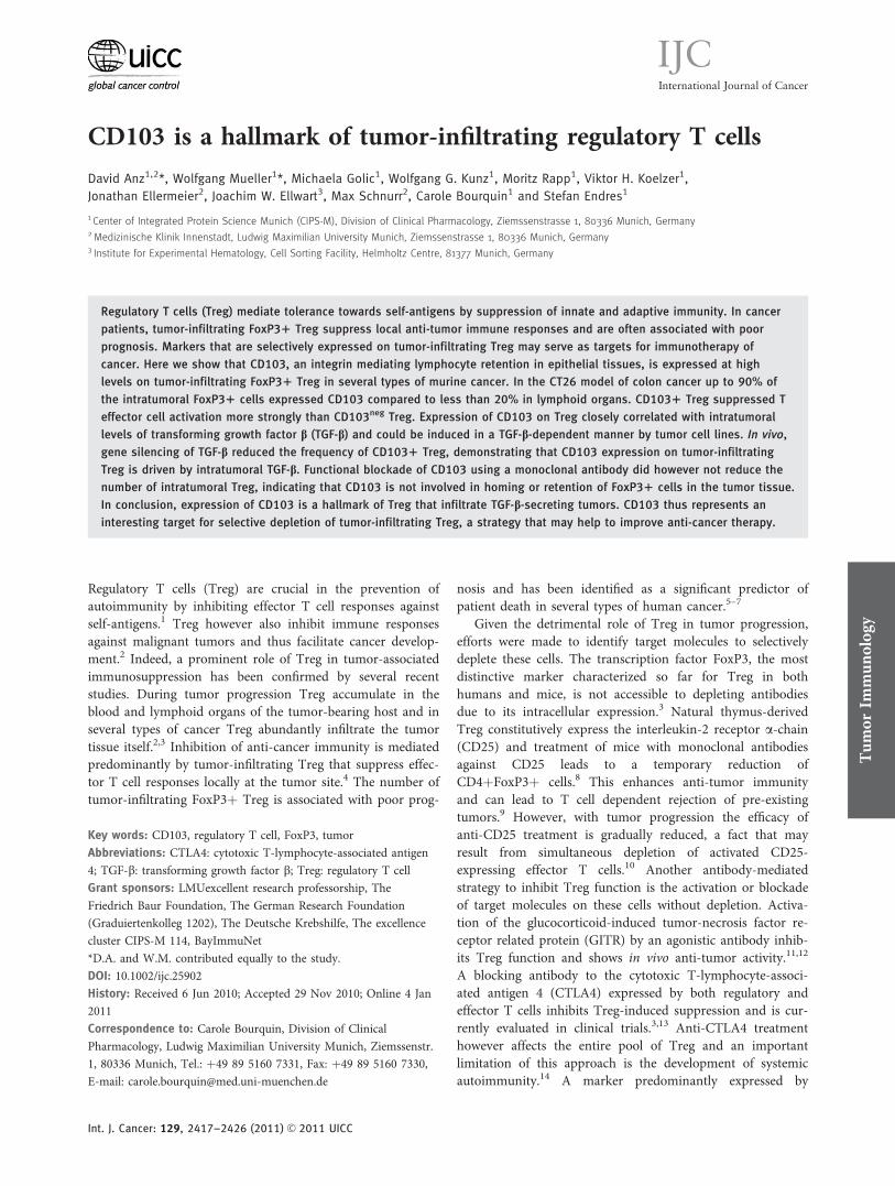
CD103 is a hallmark of tumor-infiltrating regulatory T cells
David Anz1,2*, Wolfgang Mueller1*, Michaela Golic1, Wolfgang G. Kunz1, Moritz Rapp1, Viktor H. Koelzer1,
Jonathan Ellermeier2, Joachim W. Ellwart3, Max Schnurr2, Carole Bourquin1 and Stefan Endres1
1 Center of Integrated Protein Science Munich (CIPS-M), Division of Clinical Pharmacology, Ziemssenstrasse 1, 80336 Munich, Germany2Medizinische Klinik Innenstadt, Ludwig Maximilian University Munich, Ziemssenstrasse 1, 80336 Munich, Germany3 Institute for Experimental Hematology, Cell Sorting Facility, Helmholtz Centre, 81377 Munich, Germany
Regulatory T cells (Treg) mediate tolerance towards self-antigens by suppression of innate and adaptive immunity. In cancer
patients, tumor-infiltrating FoxP31 Treg suppress local anti-tumor immune responses and are often associated with poor
prognosis. Markers that are selectively expressed on tumor-infiltrating Treg may serve as targets for immunotherapy of
cancer. Here we show that CD103, an integrin mediating lymphocyte retention in epithelial tissues, is expressed at high
levels on tumor-infiltrating FoxP31 Treg in several types of murine cancer. In the CT26 model of colon cancer up to 90% of
the intratumoral FoxP31 cells expressed CD103 compared to less than 20% in lymphoid organs. CD1031 Treg suppressed T
effector cell activation more strongly than CD103neg Treg. Expression of CD103 on Treg closely correlated with intratumoral
levels of transforming growth factor b (TGF-b) and could be induced in a TGF-b-dependent manner by tumor cell lines. In vivo,
gene silencing of TGF-b reduced the frequency of CD1031 Treg, demonstrating that CD103 expression on tumor-infiltrating
Treg is driven by intratumoral TGF-b. Functional blockade of CD103 using a monoclonal antibody did however not reduce the
number of intratumoral Treg, indicating that CD103 is not involved in homing or retention of FoxP31 cells in the tumor tissue.
In conclusion, expression of CD103 is a hallmark of Treg that infiltrate TGF-b-secreting tumors. CD103 thus represents an
interesting target for selective depletion of tumor-infiltrating Treg, a strategy that may help to improve anti-cancer therapy.
Regulatory T cells (Treg) are crucial in the prevention ofautoimmunity by inhibiting effector T cell responses againstself-antigens.1 Treg however also inhibit immune responsesagainst malignant tumors and thus facilitate cancer develop-ment.2 Indeed, a prominent role of Treg in tumor-associatedimmunosuppression has been confirmed by several recentstudies. During tumor progression Treg accumulate in theblood and lymphoid organs of the tumor-bearing host and inseveral types of cancer Treg abundantly infiltrate the tumortissue itself.2,3 Inhibition of anti-cancer immunity is mediatedpredominantly by tumor-infiltrating Treg that suppress effec-tor T cell responses locally at the tumor site.4 The number oftumor-infiltrating FoxP3þ Treg is associated with poor prog-
nosis and has been identified as a significant predictor ofpatient death in several types of human cancer.5–7
Given the detrimental role of Treg in tumor progression,efforts were made to identify target molecules to selectivelydeplete these cells. The transcription factor FoxP3, the mostdistinctive marker characterized so far for Treg in bothhumans and mice, is not accessible to depleting antibodiesdue to its intracellular expression.3 Natural thymus-derivedTreg constitutively express the interleukin-2 receptor a-chain(CD25) and treatment of mice with monoclonal antibodiesagainst CD25 leads to a temporary reduction ofCD4þFoxP3þ cells.8 This enhances anti-tumor immunityand can lead to T cell dependent rejection of pre-existingtumors.9 However, with tumor progression the efficacy ofanti-CD25 treatment is gradually reduced, a fact that mayresult from simultaneous depletion of activated CD25-expressing effector T cells.10 Another antibody-mediatedstrategy to inhibit Treg function is the activation or blockadeof target molecules on these cells without depletion. Activa-tion of the glucocorticoid-induced tumor-necrosis factor re-ceptor related protein (GITR) by an agonistic antibody inhib-its Treg function and shows in vivo anti-tumor activity.11,12
A blocking antibody to the cytotoxic T-lymphocyte-associ-ated antigen 4 (CTLA4) expressed by both regulatory andeffector T cells inhibits Treg-induced suppression and is cur-rently evaluated in clinical trials.3,13 Anti-CTLA4 treatmenthowever affects the entire pool of Treg and an importantlimitation of this approach is the development of systemicautoimmunity.14 A marker predominantly expressed by
Key words: CD103, regulatory T cell, FoxP3, tumor
Abbreviations: CTLA4: cytotoxic T-lymphocyte-associated antigen
4; TGF-b: transforming growth factor b; Treg: regulatory T cell
Grant sponsors: LMUexcellent research professorship, The
Friedrich Baur Foundation, The German Research Foundation
(Graduiertenkolleg 1202), The Deutsche Krebshilfe, The excellence
cluster CIPS-M 114, BayImmuNet
*D.A. and W.M. contributed equally to the study.
DOI: 10.1002/ijc.25902
History: Received 6 Jun 2010; Accepted 29 Nov 2010; Online 4 Jan
2011
Correspondence to: Carole Bourquin, Division of Clinical
Pharmacology, Ludwig Maximilian University Munich, Ziemssenstr.
1, 80336 Munich, Tel.: þ49 89 5160 7331, Fax: þ49 89 5160 7330,
E-mail: [email protected]
Tum
orIm
mun
olog
y
Int. J. Cancer: 129, 2417–2426 (2011) VC 2011 UICC
International Journal of Cancer
IJC

tumor-infiltrating Treg would represent a more selective tar-get to enhance anti-cancer immunity.
The plasma membrane-associated molecule CD103, alsocalled aEb7, belongs to the family of integrins and is poorlyexpressed by immune cells in the spleen or the peripherallymph nodes.15 CD103 can be detected mainly on T cell pop-ulations within the mucosal epithelium of the gut and on in-testinal lamina propria leukocytes.16 It is thought that CD103contributes to the retention of lymphocytes in epithelial tis-sues through interaction with its receptor E-cadherinexpressed by epithelial cells.17 Mice deficient for CD103 haveslightly reduced numbers of intestinal intraepithelial lympho-cytes, but apart from that are healthy, indicating that CD103is probably dispensable.18 Among Treg in lymphoid tissues, asubset of about 20% expresses CD103 and these cells displayan effector memory phenotype with low expression levels ofCD45RB and high levels of CD44.19 Some reports furtherindicate that CD103þ Treg more strongly inhibit CD4 T cellproliferation than conventional Treg.19,20 Thus, CD103 is asurface-expressed molecule that marks both intestinal lym-phocytes and a particularly suppressive subtype of Treg.
In this study, we analyzed tumor-infiltrating Treg forexpression of CD103 in four different murine models of can-cer. In all tumor models, we found that the majority of intra-tumoral Treg express CD103 with up to 90% of FoxP3þTreg staining positive for CD103. High proportions ofCD103þ cells were further specific for tumor-infiltratingTreg. CD103 expression on Treg correlated with TGF-bsecretion of tumor cells and could be down-regulated byRNAi-mediated gene silencing of TGF-b. Therapeutic target-ing of CD103 may represent a promising approach toenhance anti-cancer immunity.
Material and MethodsMice and cell lines
Female BALB/c and C57BL/6 mice were purchased from Har-lan-Winkelmann (Borchen, Germany). Mice were 5 to 10weeks of age at the onset of experiments. Animal studies wereapproved by the local regulatory agency (Regierung von Ober-bayern, Munich, Germany). The murine cell lines Colon-26(CT26; Cell Lines Service, Heidelberg, Germany), B16 mela-noma F1 (LGC Promochem, Teddington, UK), EL-4 lym-phoma (Institute of Immunology, University of Munich) andPanc02 (kindly provided by Prof. C. Bruns, Department of Sur-gery, University of Munich) were maintained in DMEM me-dium supplemented with 10% FCS, 1% L-glutamine, 1 U/mlpenicillin and 0.1 mg/ml streptomycin (all PAA Laboratories,Coelbe, Germany). For tumor induction 0.25 � 106 (CT26), 1� 106 (B16 and Panc02), or 5 � 106 (EL4) tumor cells wereinjected subcutaneously into the flank. The tumor-draininglymph nodes were identified by connecting vessels, the proxim-ity to the tumor and the larger size compared to non-draininglymph nodes. Tumor size was expressed as the product of theperpendicular diameters of individual tumors (mm2). Tumorgrowth was calculated as final tumor size divided by the num-
ber of days since injection (mm2/d) to normalize data from in-dependent experiments.
Immunohistology
Tumor tissues were frozen in liquid nitrogen and 5 lm cryo-sections from the center of the tumors were prepared. Thefollowing primary antibodies were used: anti-mouse CD103(Biolegend, San Diego, CA), anti-mouse FoxP3 (Ebioscience,San Diego, CA) and anti-mouse E-Cadherin (Cell SignalingTechnology, Beverly, MA). Cy5 F(ab)2 goat anti-Armenianhamster IgG, biotin F(ab)2 donkey anti-rat IgG, biotin IgGdonkey anti-rabbit IgG and rhodamin red X streptavidinwere used as detection reagents. Nucleic counterstaining wasperformed using DAPI (Sigma Aldrich, Steinheim, Germany).Counting was performed blinded by two independent investi-gators. Images were obtained by fluorescence microscopy(Axiovert 2000 Carl Zeiss, Jena, Germany; 40-fold magnifica-tion) using Carl Zeiss Axiovision software and processedwith Adobe Photoshop for adjustment of contrast and size.
Flow cytometry
For flow cytometry analysis single cell suspensions of spleen,lymph nodes or Peyer’s patches were prepared. Bone marrowcells were harvested from murine femur and tibia and eryth-rocytes were lysed with ammonium chloride buffer (BD Bio-sciences). To isolate lymphocytes from tumor, lung, liver orheart, the tissues were mechanically disrupted, incubated with1 mg/ml collagenase and 0.05 mg/ml DNAse (both SigmaAldrich) and subsequently passed through a cell strainer. Sin-gle cell suspensions were resuspended in 44% Percoll (Bio-chrome, Berlin, Germany) and layered over 67% Percoll priorto centrifugation at 800 g for 30 min. Lymphocytes from theinterphase were stained for flow cytometry. The followingantibodies were used: Pacific Blue or PerCP anti-mouse CD3,PE anti-mouse B220, PE-Cy7 or PerCP anti-mouse CD4,APC-Cy7 anti-mouse CD8 (all Biolegend), FITC anti-mouseCD103 (BD Biosciences, Heidelberg, Germany) and PacificBlue or APC anti-mouse FoxP3 (Ebioscience). Intracellulardetection of FoxP3 was performed using premixed regulatoryT cell staining reagents (Ebioscience). Events were measuredon a FACS Calibur or FACS Canto II flow cytometer (BDBiosciences) and analyzed with FlowJo software (TreeStar,Ashland, OR).
FACS sorting and proliferation assays
Untouched CD4þ T cells were sorted from single cell sus-pensions of lymph nodes by magnetic cell sorting (MiltenyiBiotec, Bergisch Gladbach, Germany). Cells were stained withlabeled antibodies against CD103 (Fitc, BD Biosciences), CD4(PerCP) and CD25 (APC, both from Biolegend). CD4þCD25þ CD103þ, CD4þCD25þCD103neg and CD4þCD25neg cells were obtained by FACS sorting using a FACSAria cell sorter (BD Biosciences) with a purity of more than99%. Treg subsets (3 � 104 cells) and T effector cells (7.5 �104 cells) were cultured in triplicate with anti-CD3-CD28beads (Invitrogen, Carlsbad, CA) at a bead-to-cell ratio of 1:5
Tum
orIm
mun
olog
y
2418 CD103 for selective depletion of Treg
Int. J. Cancer: 129, 2417–2426 (2011) VC 2011 UICC
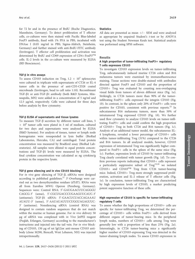
for 72 hr and in the presence of BrdU (Roche Diagnostics,Mannheim, Germany). To detect proliferation of T effectorcells, co-cultures were then stained with Pacific Blue-labeledFoxP3 antibody, fixed with 1% PFA in PBS, incubated withDNAse I (0.05 mg/ml in PBS; Sigma-Aldrich, Steinheim,Germany) and further stained with anti-BrdU-FITC antibody(Invitrogen). T effector cell proliferation and activation wasdetermined by BrdU and CD69 expression of CD4þFoxP3neg
cells. IL-2 levels in the co-culture were measured by ELISA(BD Biosciences).
TGF-b in vitro assays
To assess CD103 induction on Treg, 1.2 � 105 splenocyteswere cultured in triplicate with supernatants of CT26 or EL-4tumor cells in the presence of anti-CD3-CD28 coatedmicrobeads (Invitrogen, bead to cell ratio 1:10). RecombinantTGF-b1 or anti-TGF-b1 antibody (both R&D Systems, Min-neapolis, MN) were added in a concentration of 5 ng/ml and12.5 lg/ml, respectively. Cells were cultured for three daysbefore analysis by flow cytometry.
TGF-b ELISA of supernatants and tissue lysates
To measure TGF-b secretion by different tumor cell lines, 5� 106 tumor cells were plated in 3 ml of medium, culturedfor two days and supernatants were analyzed by ELISA(R&D Systems). For analysis of tissues, tumor or lymph nodehomogenates were resuspended in lysis buffer (BioRadLaboratories, Hercules, CA) and centrifuged. Total proteinconcentration was measured by Bradford assay (BioRad Lab-oratories). All samples were diluted to equal protein concen-trations and TGF-b1 levels were measured by ELISA. Thefinal cytokine concentration was calculated as ng cytokine/gprotein in the respective lysate.
TGF-b gene silencing and in vivo CD103 blocking
For in vivo gene silencing of TGF-b, siRNAs were designedaccording to published guidelines.21 30-Overhangs were car-ried out as two deoxythymidine residues (dTdT). RNAs wereall from Eurofins MWG Operon (Penzberg, Germany).Sequences were: Control RNA: 50-GAUGAACUUCAGGGUCAGCG-30 (sense), 50-CGCUGACCCUGAAGUUCAUC-30
(antisense); TGF-b1 siRNA: 50-GAACUCUACCAGAAAUAUAUU-30 (sense), 50-AAUAUAUUUCUGGUAGAGUUC-30 (antisense). Nonsilencing siRNA (control RNA) wasdesigned to contain random sequences that do not matchwithin the murine or human genome. For in vivo delivery 50lg of siRNA was complexed with in Vivo JetPEI reagent(Peqlab, Erlangen, Germany) according to the manufacturer’sinstructions and injected into the tail vein. For in vivo block-ing of CD103, 150 lg of rat IgG2aj anti-mouse CD103 anti-body (clone M290, Bioxcell, West Lebanon, NH) was injectedintraperitoneally.
Statistics
All data are presented as mean þ/� SEM and were analyzedas appropriate by unpaired Student’s t-test or by ANOVAtest using the Student Newman Keuls test. Statistical analysiswas performed using SPSS software.
ResultsA high proportion of tumor-infiltrating FoxP31 regulatory
T cells expresses CD103
To investigate CD103 expression levels on tumor-infiltratingTreg, subcutaneously induced murine CT26 colon and B16melanoma tumors were examined by immunofluorescencestaining. Tissue sections were double-stained with antibodiesdirected against FoxP3 and CD103 and the proportion ofCD103þ Treg was evaluated by counting non-overlappingvisual fields from tumors of eleven different mice (Fig. 1a).Strikingly, in CT26 tumors more than 90% of the tumor-infiltrating FoxP3þ cells expressed the integrin CD103 (Fig.1b). In contrast, in the spleen only 20% of FoxP3þ cells werepositive for CD103, consistent with previous reports.22 Insubcutaneous B16 melanoma tumors, more than 50% ofintratumoral Treg expressed CD103 (Fig. 1b). We furtherused flow cytometry to analyze CD103 levels on tumor-infil-trating FoxP3þ cells and could confirm the high proportionof CD103þ Treg in both CT26 and B16 tumors (Fig. 1c).Analysis of an additional tumor model, the subcutaneous EL-4 lymphoma, revealed a lower percentage of CD103þ cellswithin tumor-infiltrating Treg (31%) compared to the CT26and B16 tumors. In all analyzed models, however, CD103expression of intratumoral Treg was significantly higher com-pared to FoxP3þ cells in the spleen of the same mice (Fig.1c). Further, expression levels of CD103 by tumor-infiltratingTreg clearly correlated with tumor growth (Fig. 1d). To con-firm previous reports indicating that CD103þ cells representa particularly suppressive subset of Treg19,20 we isolatedCD103þ and CD103neg Treg from CT26 tumor-bearingmice. Indeed, CD103þ Treg more strongly suppressed prolif-eration, activation and IL-2 release of T effector cells (Fig.1e). In conclusion, tumor-infiltrating Treg are characterizedby high expression levels of CD103, a marker predictingpotent suppressive function of these cells.
High expression of CD103 is specific for tumor-infiltrating
regulatory T cells
To assess whether the high proportions of CD103þ cells arespecific for tumor-infiltrating Treg, we determined the per-centage of CD103þ cells within FoxP3þ cells derived fromdifferent organs of tumor-bearing mice. In the peripherallymph nodes, numbers of CD103þ cells among Treg weregenerally low with a proportion of less than 25% (Fig. 2a).Interestingly, in CT26 tumor-bearing mice a significantlyhigher number of CD103-expressing Treg was detected in thetumor-draining lymph nodes. To assess CD103 expression in
Tum
orIm
mun
olog
y
Anz et al. 2419
Int. J. Cancer: 129, 2417–2426 (2011) VC 2011 UICC
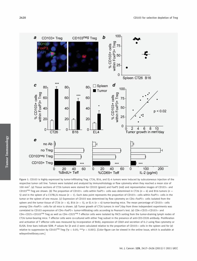
Figure 1. CD103 is highly expressed by tumor-infiltrating Treg. CT26, B16, and EL-4 tumors were induced by subcutaneous injection of the
respective tumor cell line. Tumors were isolated and analyzed by immunohistology or flow cytometry when they reached a mean size of
100 mm2. (a) Tissue sections of CT26 tumors were stained for CD103 (green) and FoxP3 (red) and representative images of CD103þ and
CD103neg Treg are shown. (b) The proportion of CD103þ cells within FoxP3þ cells was determined in CT26 (n ¼ 6) and B16 tumors (n ¼5) and in the spleen of a C57BL/6 mouse (n ¼ 1). Each data point represents the proportion of CD103þ cells within FoxP3þ cells in the
tumor or the spleen of one mouse. (c) Expression of CD103 was determined by flow cytometry on CD4þFoxP3þ cells isolated from the
spleen and the tumor tissue of CT26 (n ¼ 6), B16 (n ¼ 5), or EL-4 (n ¼ 6) tumor-bearing mice. The mean percentage of CD103þ cells
among CD4þFoxP3þ cells for all mice is shown. (d) Tumor growth of CT26 tumors in mm2/day from three independent experiments was
correlated to CD103 expression of CD4þFoxP3þ tumor-infiltrating cells according to Pearson’s test. (e) CD4þCD25þCD103þ and
CD4þCD25þCD103neg Treg as well as CD4þCD25neg T effector cells were isolated by FACS sorting from the tumor-draining lymph nodes of
CT26 tumor-bearing mice. T effector cells were co-cultured with either Treg subset in the presence of anti-CD3-CD28 antibody. Proliferation
and activation of T effector cells was measured by incorporation of BrdU, expression of CD69 and secretion of IL-2 using flow cytometry or
ELISA. Error bars indicate SEM. P values for (b and c) were calculated relative to the proportion of CD103þ cells in the spleen and for (e)
relative to suppression by CD103neg Treg (*p < 0.05; ***p < 0.001). [Color figure can be viewed in the online issue, which is available at
wileyonlinelibrary.com.]
Tum
orIm
mun
olog
y
2420 CD103 for selective depletion of Treg
Int. J. Cancer: 129, 2417–2426 (2011) VC 2011 UICC
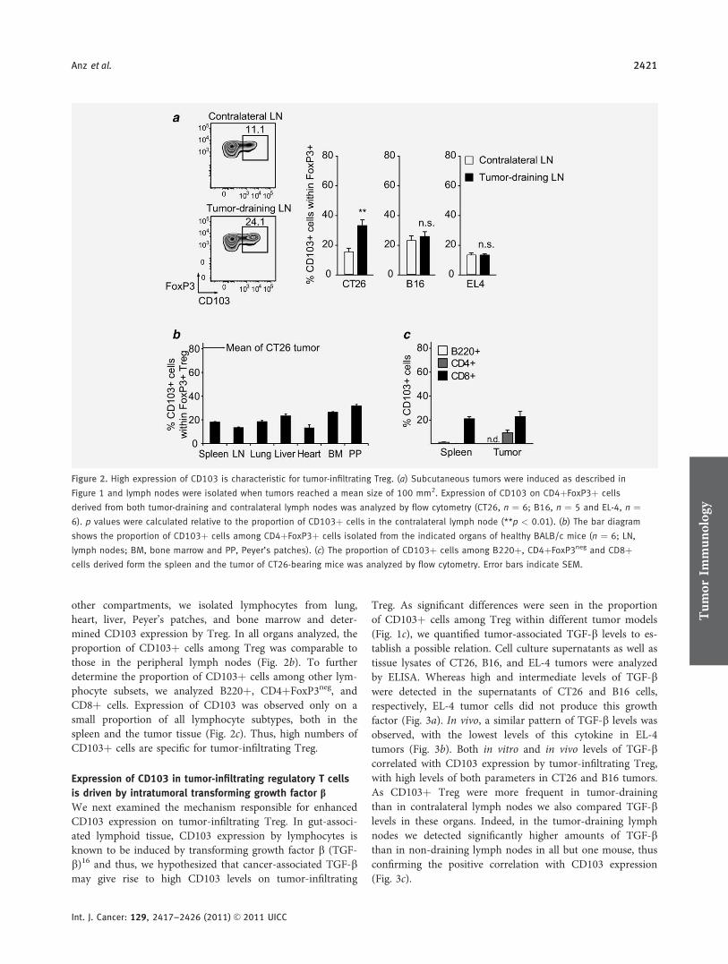
other compartments, we isolated lymphocytes from lung,heart, liver, Peyer’s patches, and bone marrow and deter-mined CD103 expression by Treg. In all organs analyzed, theproportion of CD103þ cells among Treg was comparable tothose in the peripheral lymph nodes (Fig. 2b). To furtherdetermine the proportion of CD103þ cells among other lym-phocyte subsets, we analyzed B220þ, CD4þFoxP3neg, andCD8þ cells. Expression of CD103 was observed only on asmall proportion of all lymphocyte subtypes, both in thespleen and the tumor tissue (Fig. 2c). Thus, high numbers ofCD103þ cells are specific for tumor-infiltrating Treg.
Expression of CD103 in tumor-infiltrating regulatory T cells
is driven by intratumoral transforming growth factor bWe next examined the mechanism responsible for enhancedCD103 expression on tumor-infiltrating Treg. In gut-associ-ated lymphoid tissue, CD103 expression by lymphocytes isknown to be induced by transforming growth factor b (TGF-b)16 and thus, we hypothesized that cancer-associated TGF-bmay give rise to high CD103 levels on tumor-infiltrating
Treg. As significant differences were seen in the proportionof CD103þ cells among Treg within different tumor models(Fig. 1c), we quantified tumor-associated TGF-b levels to es-tablish a possible relation. Cell culture supernatants as well astissue lysates of CT26, B16, and EL-4 tumors were analyzedby ELISA. Whereas high and intermediate levels of TGF-bwere detected in the supernatants of CT26 and B16 cells,respectively, EL-4 tumor cells did not produce this growthfactor (Fig. 3a). In vivo, a similar pattern of TGF-b levels wasobserved, with the lowest levels of this cytokine in EL-4tumors (Fig. 3b). Both in vitro and in vivo levels of TGF-bcorrelated with CD103 expression by tumor-infiltrating Treg,with high levels of both parameters in CT26 and B16 tumors.As CD103þ Treg were more frequent in tumor-drainingthan in contralateral lymph nodes we also compared TGF-blevels in these organs. Indeed, in the tumor-draining lymphnodes we detected significantly higher amounts of TGF-bthan in non-draining lymph nodes in all but one mouse, thusconfirming the positive correlation with CD103 expression(Fig. 3c).
Figure 2. High expression of CD103 is characteristic for tumor-infiltrating Treg. (a) Subcutaneous tumors were induced as described in
Figure 1 and lymph nodes were isolated when tumors reached a mean size of 100 mm2. Expression of CD103 on CD4þFoxP3þ cells
derived from both tumor-draining and contralateral lymph nodes was analyzed by flow cytometry (CT26, n ¼ 6; B16, n ¼ 5 and EL-4, n ¼6). p values were calculated relative to the proportion of CD103þ cells in the contralateral lymph node (**p < 0.01). (b) The bar diagram
shows the proportion of CD103þ cells among CD4þFoxP3þ cells isolated from the indicated organs of healthy BALB/c mice (n ¼ 6; LN,
lymph nodes; BM, bone marrow and PP, Peyer’s patches). (c) The proportion of CD103þ cells among B220þ, CD4þFoxP3neg and CD8þcells derived form the spleen and the tumor of CT26-bearing mice was analyzed by flow cytometry. Error bars indicate SEM.
Tum
orIm
mun
olog
y
Anz et al. 2421
Int. J. Cancer: 129, 2417–2426 (2011) VC 2011 UICC
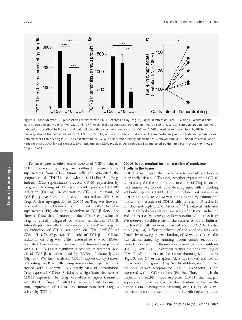
To investigate whether tumor-associated TGF-b triggersCD103expression by Treg, we cultured splenocytes insupernatants from CT26 tumor cells and quantified theproportion of CD103þ cells within CD4þFoxP3þ Treg.Indeed, CT26 supernatants induced CD103 expression byTreg and blocking of TGF-b efficiently prevented CD103induction (Fig. 4a). In contrast to CT26, supernatants ofTGF-b-negative EL-4 tumor cells did not induce CD103 onTreg. A clear up-regulation of CD103 on Treg was howeverobserved upon addition of recombinant TGF-b to EL-4supernatants (Fig. 4b) or by recombinant TGF-b alone (notshown). These data demonstrate that CD103 expression onTreg is directly triggered by tumor cell-derived TGF-b.Interestingly, this effect was specific for FoxP3þ Treg, asno induction of CD103 was seen on CD4þFoxP3neg orCD8þ T cells (Fig. 4c). The role of TGF-b in CD103induction on Treg was further assessed in vivo by siRNA-mediated knock-down. Treatment of tumor-bearing micewith a TGF-b siRNA significantly reduced intratumoral lev-els of TGF-b, as determined by ELISA of tissue lysates(Fig. 4d). We then analyzed CD103 expression by tumor-infiltrating FoxP3þ cells using immunohistology. In micetreated with a control RNA nearly 50% of intratumoralTreg expressed CD103. Strikingly, a significant decrease ofCD103 expression by Treg was observed upon treatmentwith the TGF-b-specific siRNA (Figs. 4e and 4f). In conclu-sion, expression of CD103 by tumor-associated Treg isdriven by TGF-b.
CD103 is not required for the retention of regulatory
T cells in the tumor
CD103 is an integrin that mediates retention of lymphocytesin epithelial tissues.17 To assess whether expression of CD103is necessary for the homing and retention of Treg in malig-nant tumors, we treated tumor-bearing mice with a blockingantibody against CD103. The monoclonal rat anti-mouseCD103 antibody (clone M290) binds to the aE-subunit andblocks the interaction of CD103 with its receptor E-cadherin,but does not deplete CD103þ cells.23,24 Treatment with anti-CD103 antibody was started one week after tumor inductionand infiltration by FoxP3þ cells was evaluated 10 days later.We observed no differences in the number of tumor-infiltrat-ing FoxP3þ cells between untreated and anti-CD103 treatedmice (Fig. 5a). Efficient delivery of the antibody was con-firmed by showing in vivo binding of M290 to CD103; thiswas demonstrated by staining frozen tumor sections oftreated mice with a fluorescence-labeled anti-rat antibody(Fig. 5b). Anti-CD103 treatment further did not alter Treg orCD8 T cell numbers in the tumor-draining lymph nodes(Figs. 5c and 5d) or the spleen (data not shown) and had noimpact on tumor growth (Fig. 5e). In addition, we found thatthe only known receptor for CD103, E-cadherin, is notexpressed within CT26 tumors (Fig. 5f). Thus, although themajority of FoxP3þ cells expresses CD103, this integrinappears not to be required for the retention of Treg in thetumor tissue. Therapeutic targeting of CD103þ cells willtherefore require the use of an antibody with depleting rather
Figure 3. Tumor-derived TGF-b secretion correlates with CD103 expression by Treg. (a) Equal numbers of CT26, B16 and EL-4 tumor cells
were cultured in triplicate for two days and TGF-b levels in the supernatant were determined by ELISA. (b and c) Subcutaneous tumors were
induced as described in Figure 1 and isolated when they reached a mean size of 100 mm2. TGF-b levels were determined by ELISA in
tissue lysates of the respective tumors (CT26, n ¼ 6; B16, n ¼ 4 and EL-4, n ¼ 5) and of the tumor-draining and contralateral lymph nodes
derived from CT26-bearing mice. The concentration of TGF-b in the tumor-draining lymph nodes is shown relative to the contralateral lymph
nodes (set at 100%) for each mouse. Error bars indicate SEM. p values were calculated as indicated by the lines (*p < 0.05; **p < 0.01;
***p < 0.001).
Tum
orIm
mun
olog
y
2422 CD103 for selective depletion of Treg
Int. J. Cancer: 129, 2417–2426 (2011) VC 2011 UICC
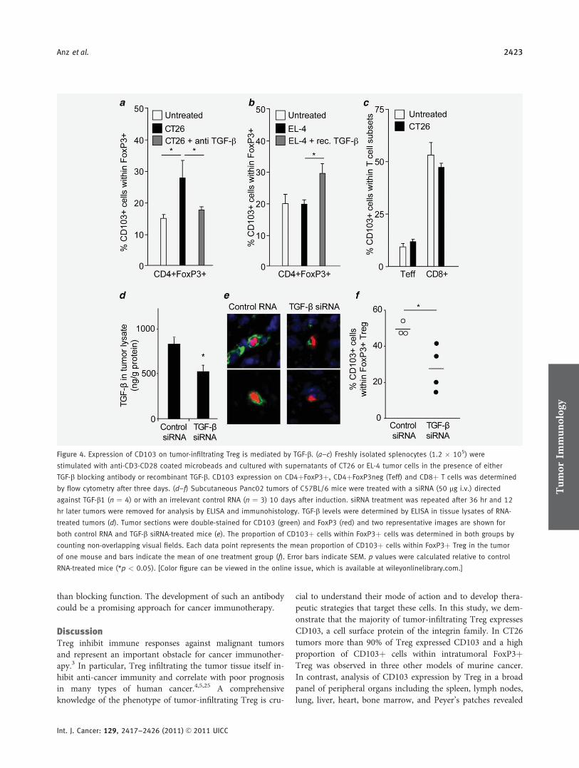
than blocking function. The development of such an antibodycould be a promising approach for cancer immunotherapy.
DiscussionTreg inhibit immune responses against malignant tumorsand represent an important obstacle for cancer immunother-apy.3 In particular, Treg infiltrating the tumor tissue itself in-hibit anti-cancer immunity and correlate with poor prognosisin many types of human cancer.4,5,25 A comprehensiveknowledge of the phenotype of tumor-infiltrating Treg is cru-
cial to understand their mode of action and to develop thera-peutic strategies that target these cells. In this study, we dem-onstrate that the majority of tumor-infiltrating Treg expressesCD103, a cell surface protein of the integrin family. In CT26tumors more than 90% of Treg expressed CD103 and a highproportion of CD103þ cells within intratumoral FoxP3þTreg was observed in three other models of murine cancer.In contrast, analysis of CD103 expression by Treg in a broadpanel of peripheral organs including the spleen, lymph nodes,lung, liver, heart, bone marrow, and Peyer’s patches revealed
Figure 4. Expression of CD103 on tumor-infiltrating Treg is mediated by TGF-b. (a–c) Freshly isolated splenocytes (1.2 � 105) were
stimulated with anti-CD3-CD28 coated microbeads and cultured with supernatants of CT26 or EL-4 tumor cells in the presence of either
TGF-b blocking antibody or recombinant TGF-b. CD103 expression on CD4þFoxP3þ, CD4þFoxP3neg (Teff) and CD8þ T cells was determined
by flow cytometry after three days. (d–f) Subcutaneous Panc02 tumors of C57BL/6 mice were treated with a siRNA (50 lg i.v.) directed
against TGF-b1 (n ¼ 4) or with an irrelevant control RNA (n ¼ 3) 10 days after induction. siRNA treatment was repeated after 36 hr and 12
hr later tumors were removed for analysis by ELISA and immunohistology. TGF-b levels were determined by ELISA in tissue lysates of RNA-
treated tumors (d). Tumor sections were double-stained for CD103 (green) and FoxP3 (red) and two representative images are shown for
both control RNA and TGF-b siRNA-treated mice (e). The proportion of CD103þ cells within FoxP3þ cells was determined in both groups by
counting non-overlapping visual fields. Each data point represents the mean proportion of CD103þ cells within FoxP3þ Treg in the tumor
of one mouse and bars indicate the mean of one treatment group (f). Error bars indicate SEM. p values were calculated relative to control
RNA-treated mice (*p < 0.05). [Color figure can be viewed in the online issue, which is available at wileyonlinelibrary.com.]Tum
orIm
mun
olog
y
Anz et al. 2423
Int. J. Cancer: 129, 2417–2426 (2011) VC 2011 UICC
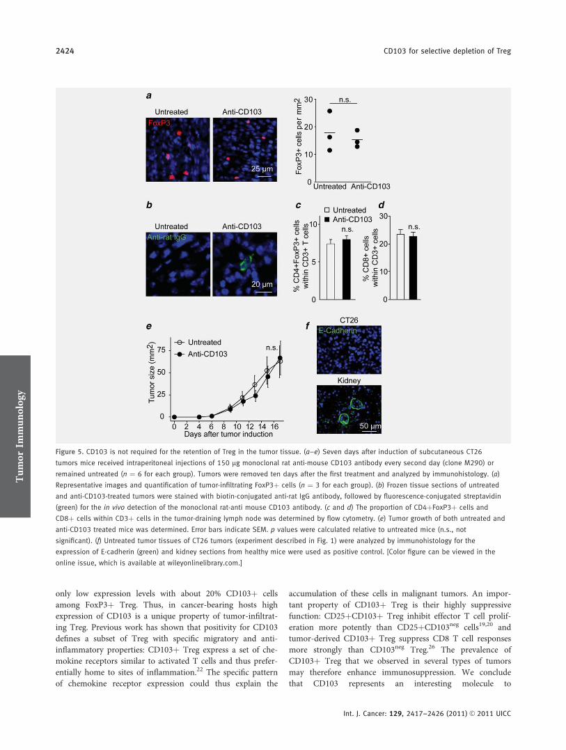
only low expression levels with about 20% CD103þ cellsamong FoxP3þ Treg. Thus, in cancer-bearing hosts highexpression of CD103 is a unique property of tumor-infiltrat-ing Treg. Previous work has shown that positivity for CD103defines a subset of Treg with specific migratory and anti-inflammatory properties: CD103þ Treg express a set of che-mokine receptors similar to activated T cells and thus prefer-entially home to sites of inflammation.22 The specific patternof chemokine receptor expression could thus explain the
accumulation of these cells in malignant tumors. An impor-tant property of CD103þ Treg is their highly suppressivefunction: CD25þCD103þ Treg inhibit effector T cell prolif-eration more potently than CD25þCD103neg cells19,20 andtumor-derived CD103þ Treg suppress CD8 T cell responsesmore strongly than CD103neg Treg.26 The prevalence ofCD103þ Treg that we observed in several types of tumorsmay therefore enhance immunosuppression. We concludethat CD103 represents an interesting molecule to
Figure 5. CD103 is not required for the retention of Treg in the tumor tissue. (a–e) Seven days after induction of subcutaneous CT26
tumors mice received intraperitoneal injections of 150 lg monoclonal rat anti-mouse CD103 antibody every second day (clone M290) or
remained untreated (n ¼ 6 for each group). Tumors were removed ten days after the first treatment and analyzed by immunohistology. (a)
Representative images and quantification of tumor-infiltrating FoxP3þ cells (n ¼ 3 for each group). (b) Frozen tissue sections of untreated
and anti-CD103-treated tumors were stained with biotin-conjugated anti-rat IgG antibody, followed by fluorescence-conjugated streptavidin
(green) for the in vivo detection of the monoclonal rat-anti mouse CD103 antibody. (c and d) The proportion of CD4þFoxP3þ cells and
CD8þ cells within CD3þ cells in the tumor-draining lymph node was determined by flow cytometry. (e) Tumor growth of both untreated and
anti-CD103 treated mice was determined. Error bars indicate SEM. p values were calculated relative to untreated mice (n.s., not
significant). (f) Untreated tumor tissues of CT26 tumors (experiment described in Fig. 1) were analyzed by immunohistology for the
expression of E-cadherin (green) and kidney sections from healthy mice were used as positive control. [Color figure can be viewed in the
online issue, which is available at wileyonlinelibrary.com.]
Tum
orIm
mun
olog
y
2424 CD103 for selective depletion of Treg
Int. J. Cancer: 129, 2417–2426 (2011) VC 2011 UICC
therapeutically target a tumor-resident and highly immuno-suppressive Treg subset.
Due to the central role of Treg in tumor-associated immu-nosuppression, targeting these cells by specific antibodies is apromising approach. Depleting antibodies against CD25 havebeen extensively tested and show some efficacy in mice, butlack significant benefits in patients so far.27 A blockingantibody against CTLA-4, a receptor expressed by T cells,synergistically enhances anti-tumor immunity by inhibitingTreg-induced suppression and promoting T effector cell acti-vation28; this treatment can however lead to systemic autoim-munity as CTLA-4 is ubiquitously expressed.14 For CD103,we have shown predominant expression by Treg in the tu-mor tissue and to some extent in the tumor-draining lymphnode. Thus, targeting this molecule could eliminate Treg atthe sites where they most potently suppress tumor-specificimmunity. Unfortunately, a depleting antibody againstCD103 has so far not been developed. In mice, only antibod-ies of the rat IgG2b and to some extent of the IgG1 isotypebear the potential to induce antibody-mediated cytotoxicityleading to depletion of target cells.29,30 In this study we usedthe monoclonal rat IgG2aj antibody M290, which blocks theinteraction of CD103 with its receptor E-cadherin.23,24 Asexpected, M290 treatment did not deplete CD103þ cells.However, we speculated that anti-CD103 treatment couldreduce Treg numbers via interfering with the retention ofTreg within the tumor. The principal function of CD103 isto mediate adhesion of cells in epithelial tissues17 and forTreg, it has been shown that CD103 is essential for the reten-tion in inflamed skin during infection with the parasiteLeishmania major.31 As Treg numbers were not altered byanti-CD103 treatment, our data show that CD103 is not
involved in the retention of Treg within malignant tumors.This is further supported by the lack of E-cadherin expres-sion in the tumor tissue, the so far only identified ligand forCD103. Thus, blocking CD103 is not sufficient to suppresstumor infiltration by Treg, but generation of depleting anti-bodies will be an interesting approach for immunotherapy ofcancer.
As CD103 does not mediate retention of Treg in the tu-mor tissue, another mechanism must be responsible for theaccumulation of intratumoral FoxP3þ cells expressing thisintegrin. Our data suggest that intratumoral TGF-b promotesthe expression of CD103 on tumor-infiltrating Treg. Wefound that high levels of CD103 were expressed predomi-nantly in those types of tumors with strong TGF-b secretion.In addition, in vivo gene silencing of TGF-b reduced thenumber of intratumoral CD103þ Treg. Our hypothesis issupported by previous reports showing that TGF-b is apotent inducer of CD103 in vitro.32,33 Further, in vivo TGF-b mediates expression of CD103 by intraepithelial and lam-ina propria-associated lymphocytes16 and induces the genera-tion of CD103-expressing FoxP3þ Treg from naıve T cells.34
The high number of tumor-infiltrating CD103þ Treg couldthus result from the conversion of previously nonregulatoryT cells. In conclusion, CD103 represents a good marker toselectively target Treg in TGF-b-secreting tumors and thedevelopment of novel depleting antibodies against CD103may be a promising approach to improve anti-cancertherapy.
AcknowledgementsWe thank Nadja Sandholzer for expert technical assistance. This work ispart of the doctoral thesis of W.M. andM.G.
References
1. Bluestone JA, Tang Q. How doCD4þCD25þ regulatory T cells controlautoimmunity? Curr Opin Immunol 2005;17:638–42.
2. Betts GJ, Clarke SL, Richards HE, GodkinAJ, Gallimore AM. Regulating the immuneresponse to tumours. Adv Drug Deliv Rev2006;58:948–61.
3. Colombo MP, Piconese S. Regulatory-T-cell inhibition versus depletion: the rightchoice in cancer immunotherapy. Nat RevCancer 2007;7:880–7.
4. Yu P, Lee Y, Liu W, Krausz T, Chong A,Schreiber H, Fu YX. Intratumor depletion ofCD4þ cells unmasks tumor immunogenicityleading to the rejection of late-stage tumors. JExp Med 2005;201:779–91.
5. Curiel TJ, Coukos G, Zou L, Alvarez X,Cheng P, Mottram P, Evdemon-Hogan M,Conejo-Garcia JR, Zhang L, Burow M, ZhuY, Wei S, et al. Specific recruitment ofregulatory T cells in ovarian carcinomafosters immune privilege and predictsreduced survival. Nat Med 2004;10:942–9.
6. Fu J, Xu D, Liu Z, Shi M, Zhao P, Fu B,Zhang Z, Yang H, Zhang H, Zhou C, YaoJ, Jin L, et al. Increased regulatory T cellscorrelate with CD8 T-cell impairment andpoor survival in hepatocellular carcinomapatients. Gastroenterology 2007;132:2328–39.
7. Hiraoka N, Onozato K, Kosuge T,Hirohashi S. Prevalence of FOXP3þregulatory T cells increases during theprogression of pancreatic ductaladenocarcinoma and itspremalignant lesions. Clin Cancer Res2006;12:5423–34.
8. Sutmuller RP, van Duivenvoorde LM, vanElsas A, Schumacher TN, Wildenberg ME,Allison JP, Toes RE, Offringa R, Melief CJ.Synergism of cytotoxic T lymphocyte-associated antigen 4 blockade anddepletion of CD25(þ) regulatory T cells inantitumor therapy reveals alternativepathways for suppression of autoreactivecytotoxic T lymphocyte responses. J ExpMed 2001;194:823–32.
9. Golgher D, Jones E, Powrie F, Elliott T,Gallimore A. Depletion of CD25þregulatory cells uncovers immuneresponses to shared murine tumorrejection antigens. Eur J Immunol 2002;32:3267–75.
10. Onizuka S, Tawara I, Shimizu J, SakaguchiS, Fujita T, Nakayama E. Tumor rejectionby in vivo administration of anti-CD25(interleukin-2 receptor alpha) monoclonalantibody. Cancer Res 1999;59:3128–33.
11. Dunn GP, Old LJ, Schreiber RD. The threeEs of cancer immunoediting. Annu RevImmunol 2004;22:329–60.
12. Liyanage UK, Moore TT, Joo HG, TanakaY, Herrmann V, Doherty G, Drebin JA,Strasberg SM, Eberlein TJ, GoedegebuurePS, Linehan DC. Prevalence of regulatoryT cells is increased in peripheral blood andtumor microenvironment of patients withpancreas or breast adenocarcinoma.J Immunol 2002;169:2756–61.
13. Ansell SM, Hurvitz SA, Koenig PA,LaPlant BR, Kabat BF, Fernando D,
Tum
orIm
mun
olog
y
Anz et al. 2425
Int. J. Cancer: 129, 2417–2426 (2011) VC 2011 UICC
Habermann TM, Inwards DJ, Verma M,Yamada R, Erlichman C, Lowy I, et al.Phase I study of ipilimumab, an anti-CTLA-4 monoclonal antibody, in patientswith relapsed and refractory B-cell non-Hodgkin lymphoma. Clin Cancer Res 2009;15:6446–53.
14. Kapadia D, Fong L. CTLA-4 blockade:autoimmunity as treatment. J Clin Oncol2005;23:8926–8.
15. Feng Y, Wang D, Yuan R, Parker CM,Farber DL, Hadley GA. CD103 expressionis required for destruction of pancreaticislet allografts by CD8(þ) T cells. J ExpMed 2002;196:877–86.
16. Kilshaw PJ, Murant SJ. A new surfaceantigen on intraepithelial lymphocytes inthe intestine. Eur J Immunol 1990;20:2201–7.
17. Cepek KL, Shaw SK, Parker CM, RussellGJ, Morrow JS, Rimm DL, Brenner MB.Adhesion between epithelial cells and Tlymphocytes mediated by E-cadherin andthe alpha E beta 7 integrin. Nature 1994;372:190–3.
18. Schon MP, Arya A, Murphy EA, AdamsCM, Strauch UG, Agace WW, Marsal J,Donohue JP, Her H, Beier DR, Olson S,Lefrancois L, et al. Mucosal T lymphocytenumbers are selectively reduced in integrinalpha E (CD103)-deficient mice. J Immunol1999;162:6641–9.
19. Lehmann J, Huehn J, de la Rosa M,Maszyna F, Kretschmer U, Krenn V,Brunner M, Scheffold A, Hamann A.Expression of the integrin alpha Ebeta 7identifies unique subsets of CD25þas well as CD25- regulatory T cells.Proc Natl Acad Sci U S A 2002;99:13031–6.
20. Chen X, Subleski JJ, Kopf H, Howard OM,Mannel DN, Oppenheim JJ. Cutting edge:expression of TNFR2 defines a maximallysuppressive subset of mouse
CD4þCD25þFoxP3þ T regulatory cells:applicability to tumor-infiltrating Tregulatory cells. J Immunol 2008;180:6467–71.
21. Reynolds A, Leake D, Boese Q, Scaringe S,Marshall WS, Khvorova A. Rational siRNAdesign for RNA interference. NatBiotechnol 2004;22:326–30.
22. Huehn J, Siegmund K, Lehmann JC,Siewert C, Haubold U, Feuerer M, DebesGF, Lauber J, Frey O, Przybylski GK,Niesner U, de la Rosa M, et al.Developmental stage, phenotype, andmigration distinguish naive- and effector/memory-like CD4þ regulatory T cells.J Exp Med 2004;199:303–13.
23. Karecla PI, Bowden SJ, Green SJ, KilshawPJ. Recognition of E-cadherin on epithelialcells by the mucosal T cell integrin alphaM290 beta 7 (alpha E beta 7). Eur JImmunol 1995;25:852–6.
24. Corps EM, Robertson A, Dauncey MJ,Kilshaw PJ. Role of the alphaIdomain in ligand binding by integrinalphaEbeta7. Eur J Immunol 2003;33:2599–608.
25. Gobert M, Treilleux I, Bendriss-VermareN, Bachelot T, Goddard-Leon S, Arfi V,Biota C, Doffin AC, Durand I, Olive D,Perez S, Pasqual N, et al. Regulatory Tcells recruited through CCL22/CCR4 areselectively activated in lymphoid infiltratessurrounding primary breast tumors andlead to an adverse clinical outcome. CancerRes 2009;69:2000–9.
26. Lin YC, Chang LY, Huang CT, Peng HM,Dutta A, Chen TC, Yeh CT, Lin CY.Effector/memory but not naive regulatoryT cells are responsible for the loss ofconcomitant tumor immunity. J Immunol2009;182:6095–104.
27. Gerena-Lewis M, Crawford J, Bonomi P,Maddox AM, Hainsworth J, McCune DE,Shukla R, Zeigler H, Hurtubise P,
Chowdhury TR, Fletcher B, Dyehouse K,et al. A Phase II trial of DenileukinDiftitox in patients with previously treatedadvanced non-small cell lung cancer. Am JClin Oncol 2009;32:269–73.
28. Peggs KS, Quezada SA, Chambers CA,Korman AJ, Allison JP. Blockade of CTLA-4 on both effector and regulatory T cellcompartments contributes to the antitumoractivity of anti-CTLA-4 antibodies. J ExpMed 2009;206:1717–25.
29. Hale G, Clark M, Waldmann H.Therapeutic potential of rat monoclonalantibodies: isotype specificity of antibody-dependent cell-mediated cytotoxicity withhuman lymphocytes. J Immunol 1985;134:3056–61.
30. Cobbold SP, Jayasuriya A, Nash A, ProsperoTD, Waldmann H. Therapy with monoclonalantibodies by elimination of T-cell subsets invivo. Nature 1984;312:548–51.
31. Suffia I, Reckling SK, Salay G, Belkaid Y. Arole for CD103 in the retention ofCD4þCD25þ Treg and control ofLeishmania major infection. J Immunol2005;174:5444–55.
32. Hadley GA, Bartlett ST, Via CS,Rostapshova EA, Moainie S. The epithelialcell-specific integrin, CD103 (alpha Eintegrin), defines a novel subset ofalloreactive CD8þ CTL. J Immunol 1997;159:3748–56.
33. Robinson PW, Green SJ, Carter C,Coadwell J, Kilshaw PJ. Studies ontranscriptional regulation of themucosal T-cell integrin alphaEbeta7(CD103). Immunology 2001;103:146–54.
34. Rao PE, Petrone AL, Ponath PD.Differentiation and expansion of T cellswith regulatory function from humanperipheral lymphocytes by stimulation inthe presence of TGF-b. J Immunol 2005;174:1446–55.T
umor
Immun
olog
y
2426 CD103 for selective depletion of Treg
Int. J. Cancer: 129, 2417–2426 (2011) VC 2011 UICC

31
_____________________________________________________________________
4.2 Originalarbeit: Jacobs C, et al. Int J Cancer 2011Collin Jacobs, Peter Düwell, Klaus Heckelsmiller, Jiwu Wei, Franz Bauernfeind,
Jonathan Ellermeier, Ulrich Kisser, Christian A. Bauer, Marc Dauer, Andreas Eigler,
Eugene Maraskovsky, Stefan Endres, Max Schnurr.
An ISCOM vaccine combined with a TLR9 agonist breaks immune evasion mediated
by regulatory T cells in an orthotopic model of pancreatic carcinoma.
Int J Cancer. 2011 Feb 15;128(4):897-907.
An ISCOM vaccine combined with a TLR9 agonist breaksimmune evasion mediated by regulatory T cells in anorthotopic model of pancreatic carcinoma
Collin Jacobs1, Peter Duewell1, Klaus Heckelsmiller1, Jiwu Wei1,2, Franz Bauernfeind3, Jonathan Ellermeier1, Ulrich Kisser1,
Christian A. Bauer1, Marc Dauer4, Andreas Eigler5, Eugene Maraskovsky6, Stefan Endres7 and Max Schnurr1
1 Department of Internal Medicine, University Hospitals, University of Munich, Munich, Germany2 Laboratory of Translational Cancer Therapy, Medical School of Nanjing University, Nanjing, People’s Republic of China3 Institute of Clinical Chemistry and Pharmacology, University Hospitals, University of Bonn, Bonn, Germany4 Department of Medicine II, Saarland University Hospital, Homburg/Saar, Germany5 Department of Medicine I, Klinikum Dritter Orden, Munich, Germany6 CSL Limited, Parkville, Victoria, Australia7 Division of Clinical Pharmacology, Center for Integrated Protein Science Munich (CIPS-M), University of Munich, Munich, Germany
Vaccines based on immune stimulatory complexes (ISCOM) induce T-cell responses against tumor antigen (Ag). However,
immune responses are impaired in pancreatic cancer patients. We investigated the efficacy of an ISCOM vaccine in a murine
pancreatic carcinoma model. Panc02 cells expressing OVA as a model Ag were induced subcutaneously or orthotopically in the
pancreas of C57BL/6 mice. Treatment consisted of an OVA containing ISCOM vaccine, either used alone or in combination with
the TLR9 agonist CpG. The ISCOM vaccine effectively induced Ag-specific CTL capable of killing tumor cells. However, in mice
with established tumors CTL induction by the vaccine was inefficient and did not affect tumor growth. Lack of efficacy correlated
with increased numbers of Treg. Depletion of Treg with anti-CD25 mAb restored CTL induction and prolonged survival. Adding
low-dose CpG to the ISCOM vaccine reduced Treg numbers, enhanced CTL responses and induced regression of pancreatic
tumors in a CD81 T cell–dependent manner. Mice cured from the primary tumor mounted a memory T-cell response against wild-
type Panc02 tumors, indicative of epitope spreading. Combining ISCOM vaccines with TLR agonists is a promising strategy for
breaking tumor immune evasion and deserves further evaluation for the treatment of pancreatic carcinoma.
Pancreatic cancer is one of the most fatal malignancies in theWestern world. It is the fourth leading cause of cancer deathin the United States.1 The 5-year survival rate is less than 5%with a median overall survival time of 3–6 months. Despiteadvancements made over the past two decades in elucidatingmolecular pathways involved in pancreatic carcinogenesisand in the field of targeted therapy, the clinical outcome hasnot yet significantly improved. At the time of diagnosis themajority of patients present with locally advanced, unresect-able tumors or metastatic disease. Even in the small numberof patients who undergo surgery in a curative intention mostpatients succumb to recurrent and metastatic disease. There-fore, new treatments are urgently needed.
Immunotherapy may offer a new treatment option. Pan-creatic carcinoma cells can be recognized by T cells, whichare found in the blood of pancreatic carcinoma patients.2 Tu-mor infiltration with T cells represents a positive prognosticfactor.3 However, pancreatic carcinomas promote systemicand locally active immunosuppressive mechanisms.4 Theseinclude inhibition of T-cell activation, secretion of immuno-suppressive cytokines, defects in Ag presentation and recruit-ment of regulatory T cells (Treg), a subgroup of CD4þ Tcells with suppressor function.5,6 In patients with pancreaticcarcinoma, increased numbers of Treg are found in the
Key words: tumor immunity, vaccination, dendritic cells, antigen
presentation
Abbreviations: Ag: antigen; CpG: unmethylated cytosine/guanine
oligodeoxynucleotide with a phosphodiester backbone; CTL:
cytotoxic T lymphocytes; DC: dendritic cells; ISCOM: immune
stimulatory complex; mAB: monoclonal antibody; OVA: ovalbumin;
TLR: toll-like receptor; Treg: regulatory T cells
Additional Supporting Information may be found in the online
version of this article.
E.M. is employed by CSL, Ltd., whose potential product was studied
in this work.
Collin Jacobs and Peter Duewell contributed equally to this work.
Grant sponsor: Deutsche Forschungsgemeinschaft; Grant numbers:
GK 1202, En 169/7-2; Grant sponsor: Deutsche Krebshilfe (Max
Eder Research Grant)
DOI: 10.1002/ijc.25399
History: Received 29 Jan 2010; Accepted 1 Apr 2010; Online 19 Apr
2010
Correspondence to: Max Schnurr, Medizinische Klinik Innenstadt,
University of Munich, Ziemssenstr. 1, 80336 Munich, Germany, Tel.:
49-89-5160-5300, Fax: 49-89-5160-4406,
E-mail: [email protected]
Tum
orIm
mun
olog
y
Int. J. Cancer: 128, 897–907 (2011) VC 2010 UICC
International Journal of Cancer
IJC
peripheral blood and within the tumor.7 It has been shownthat increased Treg prevalence is a negative prognostic factorin various malignancies, including pancreatic adenocarci-noma.8–10 Therefore, breaking tumor-induced immunosup-pression is a major challenge for immunotherapy.
Tumor vaccines aim at inducing CTL capable of recognizingand killing tumors. A prerequisite is crosspresentation of tu-mor Ag on MHC class I by DC. We previously reported thathuman DC pulsed with tumor cell lysate or apoptotic tumorcells induce CTL responses against pancreatic cancer cellsin vitro.11,12 In another study, we demonstrated that a DC vac-cine combined with gemcitabine-based chemotherapy canachieve prolonged survival in an orthotopic model of pancre-atic carcinoma.13 Other groups have shown that intratumoralimmunization with DC pulsed with tumor RNA or alpha-gal-actosylceramide can induce antitumor immunity in murinemodels of pancreatic cancer.14,15 However, the production ofDC vaccines is a time consuming and expensive process. Apromising strategy is targeting vaccines to DC in vivo. The de-velopment of cell-free vaccines will make vaccines accessible toa larger number of patients and facilitate their evaluation inclinical trials. ISCOM vaccines combine an efficient Ag deliverysystem with the immune-stimulatory activity of saponin andhave been shown to target DC in vivo and to promote humoraland cellular immune responses.16 Ag can either be formulatedinto an ISCOM vaccine during formation or coadministeredwith ISCOMATRIX adjuvant, which is essentially the samestructure but without the incorporated Ag (reviewed in Ref.16). ISCOM vaccines induce efficient crosspresentation of tu-mor Ag by DC via facilitated Ag translocation into the cyto-sol.17,18 A tumor vaccine using the ISCOMATRIX adjuvant hasbeen evaluated in a placebo-controlled clinical trial in patientswith resected NY-ESO-1 expressing tumors, displaying anexcellent safety profile and broad cellular immune responses.19
However, in another trial the same ISCOMATRIX vaccinefailed to induce T-cell responses in patients with advancedtumors, indicative of tumor-induced immune suppression.20
In this study, we investigated the efficacy of an ISCOM-based tumor vaccine in an orthotopic model of pancreatic car-cinoma. Pancreatic tumors induced a potent Ag-specificimmunosuppression rendering the vaccine ineffective. Deple-tion of Treg or combining the vaccine with low-dose CpG wascapable of breaking immunosuppression and induced effectiveCTL-mediated tumor cell killing leading to prolonged survivalof animals with established pancreatic cancers.
Material and MethodsCell culture media and reagents
Cells were cultured in RPMI 1640 medium supplementedwith 10% heat-inactivated FCS (Gibco BRL, Paisley, GB), 1%L-glutamine, 100 U/l penicillin, and 0.1 mg/ml streptomycin(all PAA, Linz, Austria). The OVA/ISCOM vaccine was gen-erated by associating palmitified OVA (Sigma, A7461) intoan ISCOM by formulation with ISCOPREP saponin, phos-pholipids and cholesterol, as previously described.18 CpG oli-
gonucleotide 1826 was purchased from Coley Pharmaceuti-cals. The H2-Kb restricted peptides OVA257–264 (SIINFEKL),TRP2181–188 (VYDFFVWL) and p15E604–611 (KSPWFTTL)were purchased from Jerini Peptide Technologies (Berlin,Germany). CFSE was obtained from Invitrogen, CA.
Mice and immunizations
Six- to 12-week-old female C57BL/6 mice were obtained fromHarlan–Winkelmann (Borchen, Germany). OT-1 mice wereprovided by Prof. Brocker (Department of Immunology, Uni-versity of Munich). Animal experiments were approved by thelocal regulatory agencies. Mice were injected s.c. into the lowerhind leg with the OVA/ISCOM vaccine in 20 ll of PBS (con-taining 0.3 lg OVA and 5 lg ISCOPREP saponin) at weeklyintervals. Six micrograms of CpG were added if indicated.CD8þ T cells or NK cells were depleted via i.p. injection of500 lg of anti-CD8 mAB (clone YTS 169, BioXCell, West Leb-anon) or 200 lg of anti-NK1.1 mAb (clone PK-136, BioXCell).Treg were depleted via i.p. injection of 250 lg of anti-CD25mAb (clone PC61 BioXCell) 1 day prior to vaccination. Deple-tion efficacy was controlled by four-color FACS analysis.
Tumor induction
The Panc02 cell line (C57BL/6) is derived from a methylcho-lanthrene-induced pancreatic adenocarcinoma. Panc02 cellswere transfected with the OVA plasmid (pAC-Neo-OVA,kindly provided by Prof. T. Brocker). The PancOVA cell linewas maintained with 0.5 mg/ml G418. We experimentallyinduced pancreatic carcinomas by injection of 0.5 � 106 tu-mor cells s.c. into the flank. For the orthotopic model, thespleen was surgically mobilized and 2 � 105 PancOVA cellswere injected into the pancreas. Therapeutic vaccinationstarted between days 10–14, when tumors had formed palpa-ble nodules of 5–10 mm2. Tumor size was measured threetimes weekly and determined by the product of perpendiculardiameters. Mice were euthanized when the tumor sizeexceeded 200 mm2 or with the appearance of distress.
LN preparation and cytokine measurement
Vaccine draining LN were removed and processed to singlecell suspensions by passing through a 70-lm cell strainer.Cells were counted and processed for phenotypic and func-tional analysis. For cytokine measurements, freshly isolatedLN were shock frozen in liquid nitrogen, processed using amortar and transferred into 30 ll of lysis buffer (BioRad,Germany). Samples were vortexed for 30 sec and centrifuged(15 min, 12,000g, 4�C). The cell pellet was discarded and thesupernatant served for cytokine analysis. All samples werestandardized using Bradford method. Murine ELISA kits forIL-12p70 and IFN-c (OptEIA) were from BD Biosciences(San Diego, CA). Cytokine measurement and analysis of acti-vation marker expression of lymph node leukocyte popula-tions were done by processing vaccine site draining lymphnodes into single cell suspensions. For intracellular detectionof IL-12 in DC, cells were surface stained for CD8a and
Tum
orIm
mun
olog
y
898 ISCOM vaccine with TLR9 agonist
Int. J. Cancer: 128, 897–907 (2011) VC 2010 UICC
CD11c, fixed and permeabilized using a cytofix/cytoperm kit(BD Biosciences), stained for IL-12p40/70 and analyzed byFACS. Measurement of TGF-b serum levels was done withELISA (eBiosciences, Frankfurt, Germany) according to themanufacturer’s instruction. Serum was incubated with 1 Nhydrochloric acid for 10 min, neutralized with 1 N sodiumhydroxide and immediately used for analysis.
Monoclonal antibodies and FACS
CD3e-APC (clone 145-2C11), CD4-PE (clone GK1.5), CD8a-PerCP (clone 53-6.7), CD11c-APC or CD11c-PE (clone HL3),CD19-PE (clone 1D3), CD86-FITC (clone GL1), NK1.1-PerCP(clone PK 136) and IL-12p40/70-APC (clone C15.6) were allfrom BD Biosciences; CD69-FITC (clone H1.2F3), from CaltagLaboratories, Carlsbad, CA. Treg cells were stained with theMouse Regulatory Staining kit from eBiosciences (CD4-FITC,Foxp3-APC, CD25-PE). Samples were acquired on a FACSCa-libur (Becton Dickinson). Data were analyzed using FlowJosoftware (version 7.2.1, Tree Star, OR).
Intracellular IFN-c staining of Ag-specific T cells and
MHC class I pentamer staining
For intracellular IFN-c staining, peripheral blood was incubatedwith red blood cell lysis buffer (BD Pharm Lyse, BD) for 3 min.Lymphocytes were then stimulated with relevant peptides(1 lg/ml) for 1 hr at 37�C before 1 lg/ml Brefeldin A (Sigma-Aldrich) was added. After 3 hr, cells were surface stained forCD8, then fixed and permeabilized using a cytofix/cytoperm kit(BD Biosciences) and incubated with mAb against IFN-c-FITC(clone XMG1.2, Caltag Laboratories). Pentamer staining was per-formed with H-2Kb OVA257–264 R-PE pentamers (ProImmune,Oxford, UK) according to the manufacturer’s instructions.
In vivo T-cell proliferation and cytotoxicity assay
Splenocytes from OT-1 mice were suspended at 5 � 107
cells/ml in PBS with 0.1% BSA containing 10 lM CFSE for10 min at 37�C. A total of 2 � 106 CFSE-labeled OT-1 cellsin 200 ll PBS were injected i.v. in tumor-bearing mice vacci-nated once with OVA/ISCOM vaccine on the same day. Af-ter 60 hr, blood was collected and OT-1 T-cell proliferationwas analyzed by FACS. For assessing CTL-mediated cytotox-icity splenocytes were labeled with 1 or 10 lM CFSE andpulsed with either OVA257–264 peptide (1 lg/ml) or not. Atotal of 106 labeled cells of each population were mixed andinjected i.v. into vaccinated or nonvaccinated mice. Bloodwas collected 16 hr later and analyzed by FACS. The propor-tion of target to control population was determined and per-centage of killing was calculated as described.13
Immunohistology
Cryostat sections of tumors were air-dried and fixed in ice-cold acetone for 10 min. For analysis of tumor-infiltratingCD8þ T cells sections were incubated with rat anti-mouseCD8 (BD Pharmingen) at a dilution of 1:50, followed by bio-tin-SP-conjugated donkey anti-rat IgG and CyTM2-conjugated
streptavidin (Jackson ImmunoResearch Europe), both at1:500. For detection of Treg sections were incubated with ratanti-mouse Foxp3 (eBioscience) and Syrian hamster anti-mouse CD3 (BD Pharmingen) at 1:50, followed by CyTM3-conjugated goat anti-Syrian hamster IgG (HþL) at 1:400 to-gether with biotin-SP-conjugated donkey anti-rat IgG at 1:500followed by CyTM2-conjugated streptavidin at 1:500 (all fromJackson ImmunoResearch Europe). Slides were covered withVectashield mounting medium containing DAPI (Vector Lab-oratories, Burlingame, CA). Histological images were obtainedusing a Carl Zeiss Axiophot microscope equipped with a digi-tal camera system (DMC 2 digital camera, Polaroid).
Statistics
We determined the statistical significance of differences bythe two-tailed Student’s t-test. For the analysis of tumorgrowth, we used the nonparametric Mann–Whitney U test tocompare the mean values between two groups. Kaplan–Meiersurvival curves were analyzed using the Cox proportionalhazards model. We performed statistical analysis with SPSSsoftware. p values < 0.05 were considered significant.
ResultsISCOM vaccine fails to control growth of established
pancreatic carcinoma due to tumor-induced
immunosuppression via regulatory T cells
Mice were vaccinated on day 0 and boosted at day 7 with anISCOM vaccine containing 0.3 lg of OVA protein. Sevendays later mice were challenged s.c. with PancOVA tumorcells. All vaccinated animals were protected from tumorgrowth, whereas all control animals developed large tumorsrequiring euthanasia (tumor size > 200 mm2) within 40 days(Fig. 1a). To assess the efficacy of the vaccine in a therapeuticsetting, vaccination was initiated on day 10, when tumorshad already formed palpable nodules. In this setting, vaccina-tion influenced neither tumor growth nor survival (Fig. 1b).
We hypothesized that the discrepancy of clinical efficacybetween prophylactic and therapeutic vaccination was due totumor-induced immunosuppression. To assess whethertumors induce vaccination failure, we measured the fre-quency of Ag-specific CTL in the blood of tumor-bearingversus control mice after two vaccinations with OVA/ISCOM.Vaccination induced a high frequency of OVA-specific CTLin animals without tumors as well as with wild-type Panc02tumors, as assessed by MHC class I pentamer staining andintracellular IFN-c staining (ICS). In contrast, animals withPancOVA tumors had a significantly reduced OVA-specificCTL frequency (1.4% vs. 6.2% of CD8þ T cells by ICS forIFN-c, p < 0.01) (Fig. 1c). Thus, the tumor suppressed CTLinduction in an Ag-specific manner.
It has been shown that tumors may impair the functionof DC, which are key regulators of T-cell responses.5 To testthis hypothesis in our model we adoptively transferred CFSE-labeled CD8þ T cells from TCR transgenic OT-1 mice, whichrecognize a H2-Kb restricted OVA epitope. Mice were then
Tum
orIm
mun
olog
y
Jacobs et al. 899
Int. J. Cancer: 128, 897–907 (2011) VC 2010 UICC

vaccinated and proliferation of CFSE-labeled CD8þ T cellswas assessed in peripheral blood. PancOVA tumors had noimpact on OT-1 T-cell proliferation, indicating that CD8þ T-cell stimulation by DC was unimpaired (Fig. 1d). To test kill-ing function of CTL in tumor-bearing animals, we transferredCFSE-labeled peptide-pulsed target cells into vaccinated miceand assessed cytotoxicity in vivo. Target cell killing wasreduced in mice with PancOVA tumors as compared to ani-mals without tumors, correlating with reduced CTL frequen-cies in peripheral blood. In contrast, killing was not signifi-cantly different in mice with wild-type tumors (Fig. 1e).
TGF-b is a potent negative regulator of T-cell functionand serum levels are increased in patients with pancreatic
cancer.7,21 Similarly, we found a significant increase of TGF-b serum levels in mice with pancreatic tumors (79 vs. 1515pg/ml, p < 0.01) (Fig. 2a). Moreover, consistent with clinicalobservations,7 we found an increased number of Treg inblood, lymph nodes and spleens, increasing from 12.5% inhealthy to 18.6% of CD4þ T cells (p < 0.05) in spleens of tu-mor-bearing mice on day 21 after tumor establishment (Fig.2b). Particularly dense infiltrates of Treg were found in thetumors comprising 40% of total CD4þ T cells (Fig. 2b andSupporting Information Fig. 1A). As Ag-specific suppressionof T cells is a key feature of Treg,22 we speculated that Tregplay a central role in the immunosuppressive milieu created bypancreatic cancer. To assess the influence of Treg on the
Figure 1. Therapeutic vaccination with an ISCOM vaccine is ineffective in pancreatic carcinoma. (a) Mice were injected twice with PBS or OVA/
ISCOM at a weekly interval and challenged s.c. with OVA-expressing Panc02 (PancOVA) cells. (b) Mice with palpable PancOVA tumors were
injected twice with OVA/ISCOM or PBS. Tumor growth (left graphs) and survival (right graphs) were monitored. Data of (a) and (b) are shown
as mean tumor sizes with SEM and corresponding survival rates of six to eight mice per group from two independent experiments. (c) Mice
with or without Panc02 or PancOVA tumors were treated with the OVA/ISCOM vaccine. Number of OVA-specific CTL in blood was determined
7 days after the second vaccination by ex vivo peptide stimulation and intracellular cytokine staining for IFN-c of CD8þ T cells or MHC class I
pentamer staining. (d) Mice with or without tumors were vaccinated and CFSE-labeled OT-1 T cells were adoptively transferred via the tail vein.
T-cell proliferation was determined by FACS analysis after 60 hr. (e) Mice with or without Panc02 or PancOVA tumors were vaccinated and
CFSE-labeled, peptide-pulsed target cells were adoptively transferred. Target cell killing was determined by FACS analysis after 18 hr. Data in
(c–e) are shown as mean with SEM of four to six mice per group. Representative data from three independent experiments are shown.
Tum
orIm
mun
olog
y
900 ISCOM vaccine with TLR9 agonist
Int. J. Cancer: 128, 897–907 (2011) VC 2010 UICC
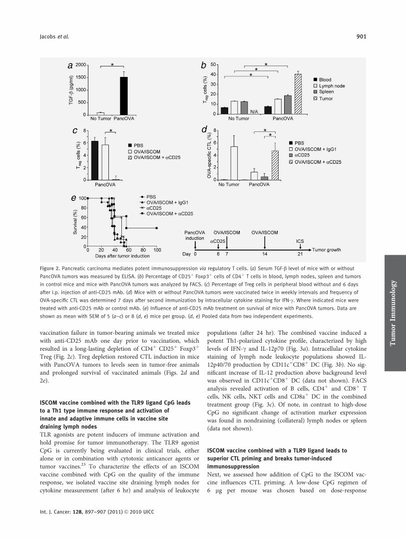
vaccination failure in tumor-bearing animals we treated micewith anti-CD25 mAb one day prior to vaccination, whichresulted in a long-lasting depletion of CD4þ CD25þ Foxp3þ
Treg (Fig. 2c). Treg depletion restored CTL induction in micewith PancOVA tumors to levels seen in tumor-free animalsand prolonged survival of vaccinated animals (Figs. 2d and2e).
ISCOM vaccine combined with the TLR9 ligand CpG leads
to a Th1 type immune response and activation of
innate and adaptive immune cells in vaccine site
draining lymph nodes
TLR agonists are potent inducers of immune activation andhold promise for tumor immunotherapy. The TLR9 agonistCpG is currently being evaluated in clinical trials, eitheralone or in combination with cytotoxic anticancer agents ortumor vaccines.23 To characterize the effects of an ISCOMvaccine combined with CpG on the quality of the immuneresponse, we isolated vaccine site draining lymph nodes forcytokine measurement (after 6 hr) and analysis of leukocyte
populations (after 24 hr). The combined vaccine induced apotent Th1-polarized cytokine profile, characterized by highlevels of IFN-c and IL-12p70 (Fig. 3a). Intracellular cytokinestaining of lymph node leukocyte populations showed IL-12p40/70 production by CD11cþCD8þ DC (Fig. 3b). No sig-nificant increase of IL-12 production above background levelwas observed in CD11cþCD8þ DC (data not shown). FACSanalysis revealed activation of B cells, CD4þ and CD8þ Tcells, NK cells, NKT cells and CD8aþ DC in the combinedtreatment group (Fig. 3c). Of note, in contrast to high-doseCpG no significant change of activation marker expressionwas found in nondraining (collateral) lymph nodes or spleen(data not shown).
ISCOM vaccine combined with a TLR9 ligand leads to
superior CTL priming and breaks tumor-induced
immunosuppression
Next, we assessed how addition of CpG to the ISCOM vac-cine influences CTL priming. A low-dose CpG regimen of6 lg per mouse was chosen based on dose-response
Figure 2. Pancreatic carcinoma mediates potent immunosuppression via regulatory T cells. (a) Serum TGF-b level of mice with or without
PancOVA tumors was measured by ELISA. (b) Percentage of CD25þ Foxp3þ cells of CD4þ T cells in blood, lymph nodes, spleen and tumors
in control mice and mice with PancOVA tumors was analyzed by FACS. (c) Percentage of Treg cells in peripheral blood without and 6 days
after i.p. injection of anti-CD25 mAb. (d) Mice with or without PancOVA tumors were vaccinated twice in weekly intervals and frequency of
OVA-specific CTL was determined 7 days after second immunization by intracellular cytokine staining for IFN-c. Where indicated mice were
treated with anti-CD25 mAb or control mAb. (e) Influence of anti-CD25 mAb treatment on survival of mice with PancOVA tumors. Data are
shown as mean with SEM of 5 (a–c) or 8 (d, e) mice per group. (d, e) Pooled data from two independent experiments.
Tum
orIm
mun
olog
y
Jacobs et al. 901
Int. J. Cancer: 128, 897–907 (2011) VC 2010 UICC
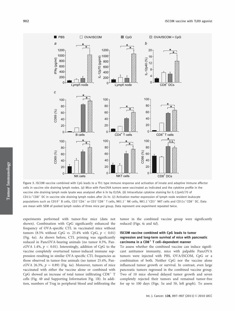
experiments performed with tumor-free mice (data notshown). Combination with CpG significantly enhanced thefrequency of OVA-specific CTL in vaccinated mice withouttumors (8.5% without CpG vs. 25.4% with CpG, p < 0.01)(Fig. 4a). As shown before, CTL priming was significantlyreduced in PancOVA-bearing animals (no tumor 8.5%, Pan-cOVA 1.4%, p < 0.01). Interestingly, addition of CpG to thevaccine completely overturned tumor-induced immune sup-pression resulting in similar OVA-specific CTL frequencies asthose observed in tumor-free animals (no tumor 25.4%, Pan-cOVA 26.3%, p ¼ 0.89) (Fig. 4a). Moreover, tumors of micevaccinated with either the vaccine alone or combined withCpG showed an increase of total tumor infiltrating CD8þ Tcells (Fig. 4b and Supporting Information Fig. 1B). In addi-tion, numbers of Treg in peripheral blood and infiltrating the
tumor in the combined vaccine group were significantlyreduced (Figs. 4c and 4d).
ISCOM vaccine combined with CpG leads to tumor
regression and long-term survival of mice with pancreatic
carcinoma in a CD81 T cell–dependent manner
To assess whether the combined vaccine can induce signifi-cant antitumor immunity, mice with palpable PancOVAtumors were injected with PBS, OVA/ISCOM, CpG or acombination of both. Neither CpG nor the vaccine aloneinfluenced tumor growth or survival. In contrast, even largepancreatic tumors regressed in the combined vaccine group.Two of 10 mice showed delayed tumor growth and sevencompletely rejected their tumors and remained tumor-freefor up to 100 days (Figs. 5a and 5b, left graph). To assess
Figure 3. ISCOM vaccine combined with CpG leads to a Th1 type immune response and activation of innate and adaptive immune effector
cells in vaccine site draining lymph nodes. (a) Mice with PancOVA tumors were vaccinated as indicated and the cytokine profile in the
vaccine site draining lymph node lysate was analyzed after 6 hr by ELISA. (b) Intracellular cytokine staining for IL-12p40/70 of
CD11cþCD8þ DC in vaccine site draining lymph nodes after 24 hr. (c) Activation marker expression of lymph node resident leukocyte
populations such as CD19þ B cells, CD3þCD4þ or CD3þCD8þ T cells, NK1.1þ NK cells, NK1.1þCD3þ NKT cells and CD11cþCD8þ DC. Data
are mean with SEM of pooled lymph nodes of three mice per group. Data represent one experiment repeated twice.
Tum
orIm
mun
olog
y
902 ISCOM vaccine with TLR9 agonist
Int. J. Cancer: 128, 897–907 (2011) VC 2010 UICC
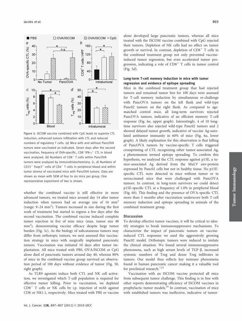
whether the combined vaccine is still effective in moreadvanced tumors, we treated mice around day 14 after tumorinduction when tumors had an average size of 16 mm2
(range: 9–24 mm2). Tumors increased in size during the firstweek of treatment but started to regress a few days after thesecond vaccination. The combined vaccine induced completetumor rejection in five of nine mice (max. tumor size 48mm2), demonstrating vaccine efficacy despite large tumorburden (Fig. 5c). As the biology of subcutaneous tumors maydiffer from orthotopic tumors, we next assessed this vaccina-tion strategy in mice with surgically implanted pancreatictumors. Vaccination was initiated 10 days after tumor im-plantation. All mice treated with PBS, OVA/ISCOM or CpGalone died of pancreatic tumors around day 40, whereas 80%of mice in the combined vaccine group survived an observa-tion period of 100 days without evidence of tumor (Fig. 5b,right graph).
As TLR9 agonists induce both CTL and NK cell activa-tion, we investigated which T-cell population is required foreffective tumor killing. Prior to vaccination, we depletedCD8þ T cells or NK cells by i.p. injection of mAb againstCD8 or NK1.1, respectively. Mice treated with PBS or vaccine
alone developed large pancreatic tumors, whereas all micetreated with the ISCOM vaccine combined with CpG rejectedtheir tumors. Depletion of NK cells had no effect on tumorgrowth or survival. In contrast, depletion of CD8þ T cells inthe combined treatment group not only prevented vaccine-induced tumor regression, but even accelerated tumor pro-gression, indicating a role of CD8þ T cells in tumor control(Fig. 5d).
Long-term T-cell memory induction in mice with tumor
regression and evidence of epitope spreading
Mice in the combined treatment group that had rejectedtumors and remained tumor free for 100 days were assessedfor T-cell memory induction by simultaneous re-challengewith PancOVA tumors on the left flank and wild-typePanc02 tumors on the right flank. As compared to age-matched control mice, all long-term survivors rejectedPancOVA tumors, indicative of an efficient memory T-cellresponse (Fig. 6a, upper graph). Interestingly, 4 of 10 long-time survivors also rejected wild-type Panc02 tumors and 2showed delayed tumor growth, indicative of vaccine Ag-unre-lated antitumor immunity in 60% of mice (Fig. 6a, lowergraph). A likely explanation for this observation is that killingof PancOVA tumors by vaccine-specific T cells triggeredcrosspriming of CTL recognizing other tumor-associated Ag,a phenomenon termed epitope spreading. To confirm thishypothesis, we analyzed the CTL response against p15E, a tu-mor-associated Ag derived from the MuLV env-proteinexpressed by Panc02 cells but not in healthy tissue. No p15E-specific CTL were detected in mice without tumor or inunvaccinated mice that were challenged with PancOVAtumors. In contrast, in long-term survivors we could detectp15E-specific CTL at a frequency of 1.0% in peripheral blood(Fig. 6b). This finding and the presence of OVA-specific CTLmore than 3 months after vaccination underscore both T-cellmemory induction and epitope spreading in animals of thecombined treatment group.
DiscussionTo develop effective tumor vaccines, it will be critical to iden-tify strategies to break immunosuppressive mechanisms. Tocharacterize the impact of pancreatic tumors on vaccine-induced CTL responses we used the aggressively growingPanc02 model. Orthotopic tumors were induced to imitatethe clinical situation. We found several immunosuppressivephenomena, such as high serum levels of TGF-b, increasedsystemic numbers of Treg and dense Treg infiltrates intumors. Our model thus reflects key immune phenomenafound in human pancreatic cancer making it a valuable toolfor preclinical research.7,21
Vaccination with an ISCOM vaccine protected all micefrom subsequent tumor challenge. This finding is in line withother reports demonstrating efficiency of ISCOM vaccines inprophylactic tumor models.24 In contrast, vaccination of micewith established tumors was ineffective, indicative of tumor-
Figure 4. ISCOM vaccine combined with CpG leads to superior CTL
induction, enhanced tumors infiltration with CTL and reduced
numbers of regulatory T cells. (a) Mice with and without PancOVA
tumors were vaccinated as indicated. Seven days after the second
vaccination, frequency of OVA-specific, CD8þIFN-cþ CTL in blood
were analyzed. (b) Numbers of CD8þ T cells within PancOVA
tumors were analyzed by immunohistochemistry. (c, d) Numbers of
CD25þ Foxp3þ cells of CD4þ T cells in peripheral blood and within
tumor stroma of vaccinated mice with PancOVA tumors. Data are
shown as mean with SEM of four to six mice per group. One
representative experiment of two is shown.
Tum
orIm
mun
olog
y
Jacobs et al. 903
Int. J. Cancer: 128, 897–907 (2011) VC 2010 UICC
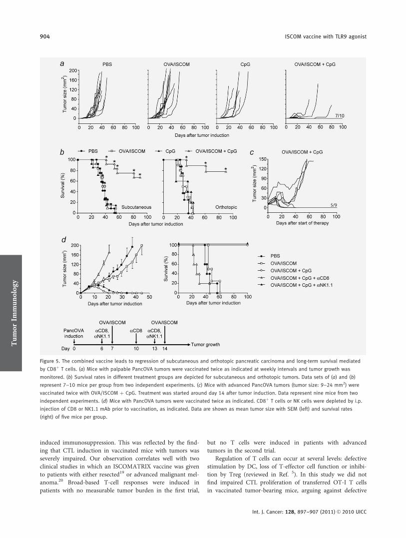
induced immunosuppression. This was reflected by the find-ing that CTL induction in vaccinated mice with tumors wasseverely impaired. Our observation correlates well with twoclinical studies in which an ISCOMATRIX vaccine was givento patients with either resected19 or advanced malignant mel-anoma.20 Broad-based T-cell responses were induced inpatients with no measurable tumor burden in the first trial,
but no T cells were induced in patients with advancedtumors in the second trial.
Regulation of T cells can occur at several levels: defectivestimulation by DC, loss of T-effector cell function or inhibi-tion by Treg (reviewed in Ref. 5). In this study we did notfind impaired CTL proliferation of transferred OT-I T cellsin vaccinated tumor-bearing mice, arguing against defective
Figure 5. The combined vaccine leads to regression of subcutaneous and orthotopic pancreatic carcinoma and long-term survival mediated
by CD8þ T cells. (a) Mice with palpable PancOVA tumors were vaccinated twice as indicated at weekly intervals and tumor growth was
monitored. (b) Survival rates in different treatment groups are depicted for subcutaneous and orthotopic tumors. Data sets of (a) and (b)
represent 7–10 mice per group from two independent experiments. (c) Mice with advanced PancOVA tumors (tumor size: 9–24 mm2) were
vaccinated twice with OVA/ISCOM þ CpG. Treatment was started around day 14 after tumor induction. Data represent nine mice from two
independent experiments. (d) Mice with PancOVA tumors were vaccinated twice as indicated. CD8þ T cells or NK cells were depleted by i.p.
injection of CD8 or NK1.1 mAb prior to vaccination, as indicated. Data are shown as mean tumor size with SEM (left) and survival rates
(right) of five mice per group.
Tum
orIm
mun
olog
y
904 ISCOM vaccine with TLR9 agonist
Int. J. Cancer: 128, 897–907 (2011) VC 2010 UICC
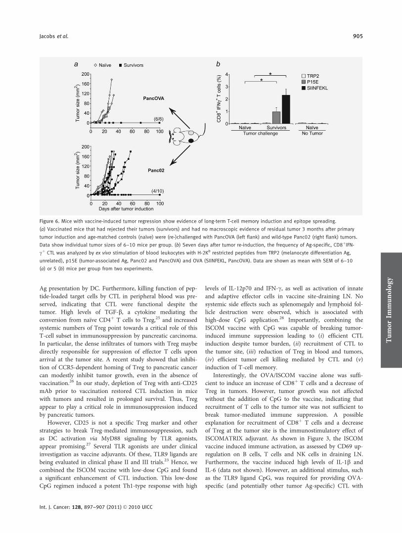
Ag presentation by DC. Furthermore, killing function of pep-tide-loaded target cells by CTL in peripheral blood was pre-served, indicating that CTL were functional despite thetumor. High levels of TGF-b, a cytokine mediating theconversion from naıve CD4þ T cells to Treg,25 and increasedsystemic numbers of Treg point towards a critical role of thisT-cell subset in immunosuppression by pancreatic carcinoma.In particular, the dense infiltrates of tumors with Treg maybedirectly responsible for suppression of effector T cells uponarrival at the tumor site. A recent study showed that inhibi-tion of CCR5-dependent homing of Treg to pancreatic cancercan modestly inhibit tumor growth, even in the absence ofvaccination.26 In our study, depletion of Treg with anti-CD25mAb prior to vaccination restored CTL induction in micewith tumors and resulted in prolonged survival. Thus, Tregappear to play a critical role in immunosuppression inducedby pancreatic tumors.
However, CD25 is not a specific Treg marker and otherstrategies to break Treg-mediated immunosuppression, suchas DC activation via MyD88 signaling by TLR agonists,appear promising.27 Several TLR agonists are under clinicalinvestigation as vaccine adjuvants. Of these, TLR9 ligands arebeing evaluated in clinical phase II and III trials.23 Hence, wecombined the ISCOM vaccine with low-dose CpG and founda significant enhancement of CTL induction. This low-doseCpG regimen induced a potent Th1-type response with high
levels of IL-12p70 and IFN-c, as well as activation of innateand adaptive effector cells in vaccine site-draining LN. Nosystemic side effects such as splenomegaly and lymphoid fol-licle destruction were observed, which is associated withhigh-dose CpG application.28 Importantly, combining theISCOM vaccine with CpG was capable of breaking tumor-induced immune suppression leading to (i) efficient CTLinduction despite tumor burden, (ii) recruitment of CTL tothe tumor site, (iii) reduction of Treg in blood and tumors,(iv) efficient tumor cell killing mediated by CTL and (v)induction of T-cell memory.
Interestingly, the OVA/ISCOM vaccine alone was suffi-cient to induce an increase of CD8þ T cells and a decrease ofTreg in tumors. However, tumor growth was not affectedwithout the addition of CpG to the vaccine, indicating thatrecruitment of T cells to the tumor site was not sufficient tobreak tumor-mediated immune suppression. A possibleexplanation for recruitment of CD8þ T cells and a decreaseof Treg at the tumor site is the immunostimulatory effect ofISCOMATRIX adjuvant. As shown in Figure 3, the ISCOMvaccine induced immune activation, as assessed by CD69 up-regulation on B cells, T cells and NK cells in draining LN.Furthermore, the vaccine induced high levels of IL-1b andIL-6 (data not shown). However, an additional stimulus, suchas the TLR9 ligand CpG, was required for providing OVA-specific (and potentially other tumor Ag-specific) CTL with
Figure 6. Mice with vaccine-induced tumor regression show evidence of long-term T-cell memory induction and epitope spreading.
(a) Vaccinated mice that had rejected their tumors (survivors) and had no macroscopic evidence of residual tumor 3 months after primary
tumor induction and age-matched controls (naıve) were (re-)challenged with PancOVA (left flank) and wild-type Panc02 (right flank) tumors.
Data show individual tumor sizes of 6–10 mice per group. (b) Seven days after tumor re-induction, the frequency of Ag-specific, CD8þIFN-
cþ CTL was analyzed by ex vivo stimulation of blood leukocytes with H-2Kb restricted peptides from TRP2 (melanocyte differentiation Ag,
unrelated), p15E (tumor-associated Ag, Panc02 and PancOVA) and OVA (SIINFEKL, PancOVA). Data are shown as mean with SEM of 6–10
(a) or 5 (b) mice per group from two experiments.
Tum
orIm
mun
olog
y
Jacobs et al. 905
Int. J. Cancer: 128, 897–907 (2011) VC 2010 UICC
tumor cell killing function. As a point of critique it can beargued that TLR9 ligands, which are highly active in mice,could be less active in humans due to differences in TLR9expression in human and mouse DC populations.23 Inongoing studies, we investigate combinations of ISCOM vac-cines with ligands of other TLR (i.e., TLR3, TLR4 and TLR7)finding enhanced CTL responses and antitumor efficacy (datanot shown). Thus, TLR signaling seems to be a generalmechanism for breaking tumor immunosuppression.27
A key element of adaptive immunity is the induction ofmemory, allowing the immune system to quickly and effi-ciently respond to subsequent Ag challenge. Mice that hadrejected tumors in the combined treatment group were pro-tected from subsequent tumor challenge, indicative of effec-tive memory induction. Furthermore, 60% of mice developedan immune response against the wild-type tumor. This led usto hypothesize that successful therapy may induce activationof tumor-reactive CTL specific for vaccine-unrelated tumorAg, e.g., via crosspresentation of tumor Ag by DC in tumor-draining lymph nodes.29 Indeed, in mice that had rejected
the tumor we detected a high frequency of CTL againstp15E, an Ag expressed by wild-type Panc02 cells.13 Thus,ISCOM vaccines are capable of inducing epitope spreading,thereby diversifying antitumor CTL responses.
To our best knowledge, this is the first report of successfultumor vaccination leading to tumor eradication in the aggres-sive orthotopic Panc02 carcinoma model. Tumor vaccinesmay offer new treatment options for patients with pancreaticcarcinoma and deserve further investigation. In this regard,survivin, a tumor Ag expressed by 90% of human pancreaticcancers,30,31 is in preclinical evaluation in our laboratory as arecombinant, full length survivin protein ISCOM vaccine.
AcknowledgementsWe thank Mr. Hal Braley, CSL, Ltd. (Parkville, Victoria, Australia) for pro-viding the OVA/ISCOM vaccine, Dr. Sabine Hoves for critical reading andMs. Yun Liu for technical assistance with immunohistochemistry. This workwas supported by the Deutsche Krebshilfe (Max Eder Research Grant toM.S.), the Deutsche Forschungsgemeinschaft (GK 1202) to M.S., A.E. andS.E., GK 1202 student grants to C.J. and P.D., and En 169/7-2 to S.E.
References
1. Jemal A, Siegel R, Ward E, Hao Y, Xu J,Murray T, Thun MJ. Cancer statistics,2008. CA Cancer J Clin 2008;58:71–96.
2. Schmitz-Winnenthal FH, Volk C,Z’Graggen K, Galindo L, Nummer D,Ziouta Y, Bucur M, Weitz J, SchirrmacherV, Buchler MW, Beckhove P. Highfrequencies of functional tumor-reactive Tcells in bone marrow and blood ofpancreatic cancer patients. Cancer Res2005;65:10079–87.
3. Fukunaga A, Miyamoto M, Cho Y,Murakami S, Kawarada Y, Oshikiri T, KatoK, Kurokawa T, Suzuoki M, Nakakubo Y,Hiraoka K, Itoh T, et al. CD8þ tumor-infiltrating lymphocytes together withCD4þ tumor-infiltrating lymphocytes anddendritic cells improve the prognosis ofpatients with pancreatic adenocarcinoma.Pancreas 2004;28:e26–31.
4. von Bernstorff W, Voss M, Freichel S,Schmid A, Vogel I, Johnk C, Henne-BrunsD, Kremer B, Kalthoff H. Systemic andlocal immunosuppression in pancreaticcancer patients. Clin Cancer Res 2001;7:925s–32s.
5. Rabinovich GA, Gabrilovich D, SotomayorEM. Immunosuppressive strategies that aremediated by tumor cells. Annu RevImmunol 2007;25:267–96.
6. Sakaguchi S. Regulatory T cells in the pastand for the future. Eur J Immunol 2008;38:901–37.
7. Liyanage UK, Moore TT, Joo HG, TanakaY, Herrmann V, Doherty G, Drebin JA,Strasberg SM, Eberlein TJ, GoedegebuurePS, Linehan DC. Prevalence of regulatoryT cells is increased in peripheral blood and
tumor microenvironment of patients withpancreas or breast adenocarcinoma.J Immunol 2002;169:2756–61.
8. Curiel TJ, Coukos G, Zou L, Alvarez X,Cheng P, Mottram P, Evdemon-Hogan M,Conejo-Garcia JR, Zhang L, Burow M, ZhuY, Wei S, et al. Specific recruitment ofregulatory T cells in ovarian carcinomafosters immune privilege and predictsreduced survival. Nature Med 2004;10:942–9.
9. Fu J, Xu D, Liu Z, Shi M, Zhao P, Fu B,Zhang Z, Yang H, Zhang H, Zhou C, YaoJ, Jin L, et al. Increased regulatory T cellscorrelate with CD8 T-cell impairment andpoor survival in hepatocellular carcinomapatients. Gastroenterology 2007;132:2328–39.
10. Hiraoka N, Onozato K, Kosuge T,Hirohashi S. Prevalence of FOXP3þregulatory T cells increases during theprogression of pancreatic ductaladenocarcinoma and its premalignantlesions. Clin Cancer Res 2006;12:5423–34.
11. Schnurr M, Galambos P, Scholz C, Then F,Dauer M, Endres S, Eigler A. Tumor celllysate-pulsed human dendritic cells inducea T-cell response against pancreaticcarcinoma cells: an in vitro model for theassessment of tumor vaccines. Cancer Res2001;61:6445–50.
12. Schnurr M, Scholz C, Rothenfusser S,Galambos P, Dauer M, Robe J, Endres S,Eigler A. Apoptotic pancreatic tumor cellsare superior to cell lysates in promotingcross-priming of cytotoxic T cells andactivate NK and gammadelta T cells.Cancer Res 2002;62:2347–52.
13. Bauer C, Bauernfeind F, Sterzik A, OrbanM, Schnurr M, Lehr HA, Endres S, EiglerA, Dauer M. Dendritic cell-basedvaccination combined with gemcitabineincreases survival in a murine pancreaticcarcinoma model. Gut 2007;56:1275–82.
14. Nagaraj S, Ziske C, Strehl J, Messmer D,Sauerbruch T, Schmidt-Wolf IG. Dendriticcells pulsed with alpha-galactosylceramideinduce anti-tumor immunity againstpancreatic cancer in vivo. Int Immunol2006;18:1279–83.
15. Schmidt T, Ziske C, Marten A, Endres S,Tiemann K, Schmitz V, Gorschluter M,Schneider C, Sauerbruch T, Schmidt-WolfIG. Intratumoral immunization with tumorRNA-pulsed dendritic cells confersantitumor immunity in a C57BL/6pancreatic murine tumor model. CancerRes 2003;63:8962–7.
16. Drane D, Gittleson C, Boyle J,Maraskovsky E. ISCOMATRIX adjuvantfor prophylactic and therapeutic vaccines.Expert Rev Vaccines 2007;6:761–72.
17. Schnurr M, Chen Q, Shin A, Chen W, ToyT, Jenderek C, Green S, Miloradovic L,Drane D, Davis ID, Villadangos J,Shortman K, et al. Tumor antigenprocessing and presentation dependcritically on dendritic cell type and themode of antigen delivery. Blood 2005;105:2465–72.
18. Schnurr M, Orban M, Robson NC, Shin A,Braley H, Airey D, Cebon J, MaraskovskyE, Endres S. ISCOMATRIX adjuvantinduces efficient cross-presentation oftumor antigen by dendritic cells via rapidcytosolic antigen delivery and processing
Tum
orIm
mun
olog
y
906 ISCOM vaccine with TLR9 agonist
Int. J. Cancer: 128, 897–907 (2011) VC 2010 UICC
via tripeptidyl peptidase II. J Immunol2009;182:1253–9.
19. Davis ID, Chen W, Jackson H, Parente P,Shackleton M, Hopkins W, Chen Q,Dimopoulos N, Luke T, Murphy R, ScottAM, Maraskovsky E, et al. RecombinantNY-ESO-1 protein with ISCOMATRIXadjuvant induces broad integrated antibodyand CD4(þ) and CD8(þ) T cell responsesin humans. Proc Natl Acad Sci USA 2004;101:10697–702.
20. Nicholaou T, Ebert LM, Davis ID,McArthur GA, Jackson H, Dimopoulos N,Tan B, Maraskovsky E, Miloradovic L,Hopkins W, Pan L, Venhaus R, et al.Regulatory T-cell-mediated attenuation ofT-cell responses to the NY-ESO-1ISCOMATRIX vaccine in patientswith advanced malignant melanoma.Clin Cancer Res 2009;15:2166–73.
21. Lin Y, Kikuchi S, Tamakoshi A, Obata Y,Yagyu K, Inaba Y, Kurosawa M,Kawamura T, Motohashi Y, Ishibashi T.Serum transforming growth factor-beta1levels and pancreatic cancer risk: a nestedcase-control study (Japan). Cancer CausesControl 2006;17:1077–82.
22. Sakaguchi S, Yamaguchi T, Nomura T,Ono M. Regulatory T cells and immunetolerance. Cell 2008;133:775–87.
23. Krieg AM. Development of TLR9 agonistsfor cancer therapy. J Clin Invest 2007;117:1184–94.
24. Maraskovsky E, Sjolander S, Drane DP,Schnurr M, Le TT, Mateo L, Luft T,Masterman K, Tai T-Y, Chen Q, Green S,Sjolander A, et al. NY-ESO-1 proteinformulated in ISCOMATRIXVR adjuvant isa potent anti-cancer vaccine inducing bothhumoral and CD8þ T cell-mediatedimmunity and protection against NY-ESO-1þ tumours. J Clin Cancer Res 2004;10:2879–90.
25. Chen W, Jin W, Hardegen N, Lei KJ, Li L,Marinos N, McGrady G, Wahl SM.Conversion of peripheral CD4þCD25-naive T cells to CD4þCD25þ regulatory Tcells by TGF-beta induction oftranscription factor Foxp3. J Exp Med2003;198:1875–86.
26. Tan MC, Goedegebuure PS, Belt BA,Flaherty B, Sankpal N, Gillanders WE,Eberlein TJ, Hsieh CS, Linehan DC.Disruption of CCR5-dependent homing ofregulatory T cells inhibits tumor growth in
a murine model of pancreatic cancer.J Immunol 2009;182:1746–55.
27. Pasare C, Medzhitov R. Toll pathway-dependent blockade of CD4þCD25þ T cell-mediated suppression by dendritic cells.Science (New York, NY) 2003;299:1033–6.
28. Heikenwalder M, Polymenidou M, Junt T,Sigurdson C, Wagner H, Akira S,Zinkernagel R, Aguzzi A. Lymphoid follicledestruction and immunosuppression afterrepeated CpG oligodeoxynucleotideadministration. Nat Med 2004;10:187–92.
29. Ribas A, Timmerman JM, Butterfield LH,Economou JS. Determinant spreading andtumor responses after peptide-based cancerimmunotherapy. Trends Immunol 2003;24:58–61.
30. Lee MA, Park GS, Lee HJ, Jung JH, KangJH, Hong YS, Lee KS, Kim DG, Kim SN.Survivin expression and its clinicalsignificance in pancreatic cancer. BMCCancer 2005;5:127.
31. Sarela AI, Verbeke CS, Ramsdale J, DaviesCL, Markham AF, Guillou PJ. Expressionof survivin, a novel inhibitor of apoptosisand cell cycle regulatory protein, inpancreatic adenocarcinoma. Br J Cancer2002;86:886–92.
Tum
orIm
mun
olog
y
Jacobs et al. 907
Int. J. Cancer: 128, 897–907 (2011) VC 2010 UICC
32
_____________________________________________________________________
4.3 Originalarbeit: Ellermeier J, et al. Cancer Res 2013Jonathan Ellermeier*, Jiwu Wei*, Peter Düwell*, Sabine Hoves, Mareike R. Stieg,
Tina Adunka, Daniel Nörenberg, Hans-Joachim Anders, Doris Mayr, Hendrik Poeck,
Gunther Hartmann, Stefan Endres, Max Schnurr.
Therapeutic efficacy of bifunctional siRNA combining TGF-β1 silencing with RIG-I
activation in pancreatic cancer.
Cancer Res. 2013 Mar 15;73(6):1709-1720.
*contributed equally
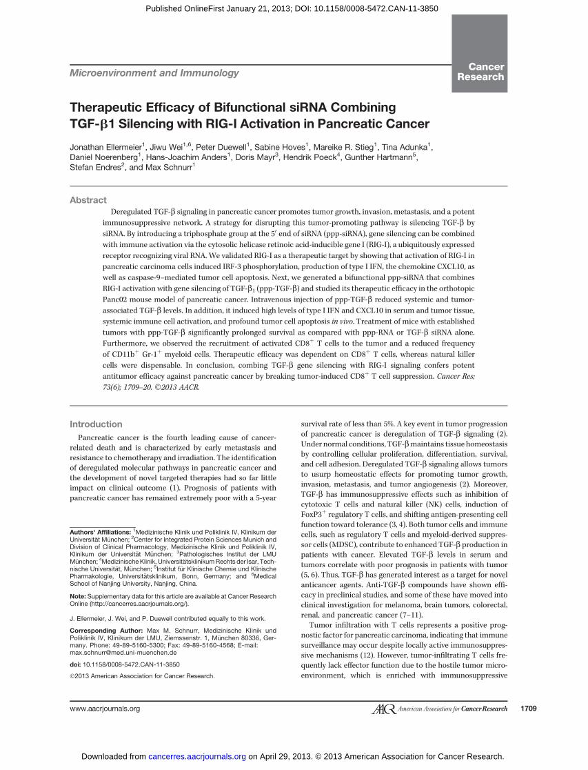
Microenvironment and Immunology
Therapeutic Efficacy of Bifunctional siRNA CombiningTGF-b1 Silencing with RIG-I Activation in Pancreatic Cancer
Jonathan Ellermeier1, Jiwu Wei1,6, Peter Duewell1, Sabine Hoves1, Mareike R. Stieg1, Tina Adunka1,Daniel Noerenberg1, Hans-Joachim Anders1, Doris Mayr3, Hendrik Poeck4, Gunther Hartmann5,Stefan Endres2, and Max Schnurr1
AbstractDeregulated TGF-b signaling in pancreatic cancer promotes tumor growth, invasion, metastasis, and a potent
immunosuppressive network. A strategy for disrupting this tumor-promoting pathway is silencing TGF-b bysiRNA. By introducing a triphosphate group at the 50 end of siRNA (ppp-siRNA), gene silencing can be combinedwith immune activation via the cytosolic helicase retinoic acid-inducible gene I (RIG-I), a ubiquitously expressedreceptor recognizing viral RNA. We validated RIG-I as a therapeutic target by showing that activation of RIG-I inpancreatic carcinoma cells induced IRF-3 phosphorylation, production of type I IFN, the chemokine CXCL10, aswell as caspase-9–mediated tumor cell apoptosis. Next, we generated a bifunctional ppp-siRNA that combinesRIG-I activation with gene silencing of TGF-b1 (ppp-TGF-b) and studied its therapeutic efficacy in the orthotopicPanc02 mouse model of pancreatic cancer. Intravenous injection of ppp-TGF-b reduced systemic and tumor-associated TGF-b levels. In addition, it induced high levels of type I IFN and CXCL10 in serum and tumor tissue,systemic immune cell activation, and profound tumor cell apoptosis in vivo. Treatment of mice with establishedtumors with ppp-TGF-b significantly prolonged survival as compared with ppp-RNA or TGF-b siRNA alone.Furthermore, we observed the recruitment of activated CD8þ T cells to the tumor and a reduced frequencyof CD11bþ Gr-1þ myeloid cells. Therapeutic efficacy was dependent on CD8þ T cells, whereas natural killercells were dispensable. In conclusion, combing TGF-b gene silencing with RIG-I signaling confers potentantitumor efficacy against pancreatic cancer by breaking tumor-induced CD8þ T cell suppression. Cancer Res;73(6); 1709–20. �2013 AACR.
IntroductionPancreatic cancer is the fourth leading cause of cancer-
related death and is characterized by early metastasis andresistance to chemotherapy and irradiation. The identificationof deregulated molecular pathways in pancreatic cancer andthe development of novel targeted therapies had so far littleimpact on clinical outcome (1). Prognosis of patients withpancreatic cancer has remained extremely poor with a 5-year
survival rate of less than 5%. A key event in tumor progressionof pancreatic cancer is deregulation of TGF-b signaling (2).Under normal conditions, TGF-bmaintains tissue homeostasisby controlling cellular proliferation, differentiation, survival,and cell adhesion. Deregulated TGF-b signaling allows tumorsto usurp homeostatic effects for promoting tumor growth,invasion, metastasis, and tumor angiogenesis (2). Moreover,TGF-b has immunosuppressive effects such as inhibition ofcytotoxic T cells and natural killer (NK) cells, induction ofFoxP3þ regulatory T cells, and shifting antigen-presenting cellfunction toward tolerance (3, 4). Both tumor cells and immunecells, such as regulatory T cells and myeloid-derived suppres-sor cells (MDSC), contribute to enhanced TGF-b production inpatients with cancer. Elevated TGF-b levels in serum andtumors correlate with poor prognosis in patients with tumor(5, 6). Thus, TGF-b has generated interest as a target for novelanticancer agents. Anti-TGF-b compounds have shown effi-cacy in preclinical studies, and some of these have moved intoclinical investigation for melanoma, brain tumors, colorectal,renal, and pancreatic cancer (7–11).
Tumor infiltration with T cells represents a positive prog-nostic factor for pancreatic carcinoma, indicating that immunesurveillance may occur despite locally active immunosuppres-sive mechanisms (12). However, tumor-infiltrating T cells fre-quently lack effector function due to the hostile tumor micro-environment, which is enriched with immunosuppressive
Authors' Affiliations: 1Medizinische Klinik und Poliklinik IV, Klinikum derUniversit€at M€unchen; 2Center for Integrated Protein Sciences Munich andDivision of Clinical Pharmacology, Medizinische Klinik und Poliklinik IV,Klinikum der Universit€at M€unchen; 3Pathologisches Institut der LMUM€unchen; 4MedizinischeKlinik, Universit€atsklinikumRechts der Isar, Tech-nische Universit€at, M€unchen; 5Institut f€ur Klinische Chemie und KlinischePharmakologie, Universit€atsklinikum, Bonn, Germany; and 6MedicalSchool of Nanjing University, Nanjing, China.
Note: Supplementary data for this article are available at Cancer ResearchOnline (http://cancerres.aacrjournals.org/).
J. Ellermeier, J. Wei, and P. Duewell contributed equally to this work.
Corresponding Author: Max M. Schnurr, Medizinische Klinik undPoliklinik IV, Klinikum der LMU, Ziemssenstr. 1, M€unchen 80336, Ger-many. Phone: 49-89-5160-5300; Fax: 49-89-5160-4568; E-mail:[email protected]
doi: 10.1158/0008-5472.CAN-11-3850
�2013 American Association for Cancer Research.
CancerResearch
www.aacrjournals.org 1709
on April 29, 2013. © 2013 American Association for Cancer Research. cancerres.aacrjournals.org Downloaded from
Published OnlineFirst January 21, 2013; DOI: 10.1158/0008-5472.CAN-11-3850
factors. Identifying targets and compounds for immune acti-vation is thus of utmost importance for effective immunother-apy. In this respect, in addition to TGF-b inhibition, activationof innate immune effector mechanisms via pattern recognitionreceptors is a promising strategy for breaking tumor-mediatedimmunosuppression (13).
Eukaryotic cells have evolved pattern recognition receptorsfor the detection of viral nucleic acids to trigger appropriateantiviral immune responses. Four members of the toll-likereceptor (TLR) family (TLR3, 7, 8, and 9) are located in theendosomal membrane predominantly in immune cells, where-as the retinoic acid-inducible gene I (RIG-I)-like helicases RIG-Iand melanoma differentiation-associated gene-5 (MDA-5) arelocated in the cytosol of immune and nonimmune cells. Recentwork suggests that RIG-I may represent a novel target forcancer immunotherapy (14, 15). RIG-I is a cytosolic sensor ofviral RNA detecting a triphosphate group at the 50 end gen-erated by viral RNA polymerases (16, 17). Upon activation, RIG-I initiates a signaling cascademediated by IRF-3, IRF-7, andNF-kB, leading to an antiviral response program characterized bythe production of type I IFN (IFN-a and IFN-b) and otherinnate immune response genes, such as the chemokineCXCL10 (18). Moreover, RIG-I signaling induces apoptosis insusceptible cells. In vitro transcribed 50-triphosphate RNA(ppp-RNA) can be applied as RIG-I ligand to trigger proapop-totic signaling via the intrinsic mitochondrial pathway (14, 15).Of note, RIG-I–mediated apoptosis occurs predominantly intumor cells, as nonmalignant cells are protected from proa-poptotic signaling via Bcl-xL (14).
The novel ppp-siRNA strategy offers the advantage of com-bining RIG-I–mediated immune activation with RNAi-medi-ated gene silencing within a single molecule. In the study byPoeck and colleagues, a bifunctional ppp-siRNA silencing theantiapoptotic molecule Bcl-2 resulted in efficient tumor cellapoptosis inmelanoma (15). To date, little is known about RIG-I expression in other cancer types. We hypothesized that dualtargeting of immunosuppression via RIG-I activation and TGF-b silencing promotes efficacy against pancreatic cancer. Westudied RIG-I expression and signaling in human pancreaticcarcinoma cell lines. In addition, the therapeutic efficacy ofa bifunctional ppp-siRNA combining ppp-RNA–mediatedRIG-I signaling with siRNA-mediated TGF-b silencing wasassessed in the aggressive Panc02 mouse model of pancreaticcarcinoma.
Materials and MethodsCell lines and cytokine stimulation
The Panc02 cell line was established from a tumor that wasinduced by 3-methylcholanthren in the pancreas of C57BL/6mice andwas a kind gift of Prof. Christiane Bruns (ChirurgischeKlinik, LMU Munich, Bavaria, Germany; ref. 19). The humanpancreatic adenocarcinoma cell lines PANC-1, MIAPaCa-2,and BxPC-3 cell lines were obtained from American TypeCulture Collection (ATCC) and were used within 6 monthsafter resuscitation (ATCC). PaTu8988t cell line was obtainedfrom the German Collection of Cell Lines (DSMZ). IMIM-PC1was kindly provided by Prof. Patrick Michl (Department ofGastroenterology and Endocrinology, University of Marburg,
Marburg, Germany; ref. 20). Cancer cell lines were culturedin Dulbecco's Modified Eagle's Medium supplemented with10% fetal calf serum (Gibco BRL), 2 mmol/L L-glutamine,100 U/L penicillin, and 0.1 mg/mL streptomycin (PAA). Eachcell line was routinely tested for mycoplasma contaminationby MycoAlert Mycoplasma Detection Kit (LONZA). IFN-awas used at concentrations of 1,000 IU/mL for murine and100 IU/mL for human cell lines (Miltenyi).
siRNAs and transfectionsiRNAs against TGF-b1, RIG-I, Puma, and Noxa were
designed according to published guidelines and were pur-chased from Eurofins MWG Operon (21). The matching 50-triphosphate–modified siRNA was transcribed using the cor-relating DNA template that contained the T7 RNA polymerasepromoter sequence. In vitro transcription of ppp-RNA wasdone using the MEGAshortscript T7 Kit (Ambion) accordingto the manufacturer's protocol (14, 15). All sequences can befound in Supplementary Table S1. Tumor cells were trans-fected with siRNA in OptiMEM (Gibco, BRL) using Lipofecta-mine 2000 (Invitrogen). For in vivo administration, 50 mg ofRNA was complexed with in vivo-JetPEI (Peqlab) at an N/Pratio of 6 in 5% glucose solution for tail vein injection.
Mice, tumor engraftment, and therapySix-week-old female C57BL/6 mice were obtained from
Harlan-Winkelmann, Trif�/� and Tlr7�/� mice in a F6C57BL/6 background originated from S. Akira (Departmentof Host Defense, Osaka University, Japan) and B. Beutler(Center for the Genetics of Host Defense, University of TexasSouthwestern Medical Center, Dallas, TX; refs. 22, 23). Experi-ments were carried out according to ethical guidelinesapproved by the local government. Orthotopic tumors wereinduced by surgical implantation of 2 � 105 Panc02 cells intothe pancreas as described (24). Therapy started on day 10,when sacrificed control mice showed visible tumor nodules(5–8 mm diameter), and was administered twice weekly over3 weeks. Serum was obtained 6 hours after the first RNAinjection for cytokine analysis, and 48 hours after secondand fifth injections for TGF-b measurement. Survival ofmice was monitored daily. Distressed mice were sacrificed.Where indicated, CD8þ T cells or NK cells were depleted oneday before RNA treatment with 500 mg of anti-CD8 mono-clonal antibody (mAb; clone YTS 169.4) or anti-NK1.1 mAb(clone PK136; BioXCell). Depletion efficacy was assessedby fluorescence-activated cell sorting (FACS) analysis ofperipheral blood and was more than 98% after 24 hours.
Protein preparation and Western blot analysisTumor tissue was snap frozen in liquid nitrogen and homog-
enized using mortar and pestle under constant liquid nitrogencooling. Cells were lysed (Bio-Plex Cell Lysis Kit, Bio-Rad) andprotein concentrations of supernatants were analyzed byBradford assay (Bio-Rad) and adjusted for whole proteinconcentration. For Western blotting, cells were lysed inLaemmli buffer and boiled for 10 minutes. Cell lysates wereseparated using a 10% SDS-PAGE. RIG-I was detected withanti-RIG-I mAb (clone ALME-1, ENZO Life Sciences GmbH)
Ellermeier et al.
Cancer Res; 73(6) March 15, 2013 Cancer Research1710
on April 29, 2013. © 2013 American Association for Cancer Research. cancerres.aacrjournals.org Downloaded from
Published OnlineFirst January 21, 2013; DOI: 10.1158/0008-5472.CAN-11-3850
followed by horseradish peroxidase-coupled secondary anti-body (Santa Cruz Biotechnology). Anti-pIRF-3 mAb was fromNew England BioLabs. Blots were visualized using enhancedchemiluminescence substrate (GE Healthcare).
ELISACytokine levels in supernatants, serum, or tumor lysates
were quantified by ELISA for IFN-a and IFN-b (PBL InterferonSource), CXCL10 (R&D Systems), TGF-b (eBiosciences), andTNF-a (BD Biosciences).
Flow cytometryLymphocyte activation in spleens was assessed 12 hours
after in vivo administration of RNA. Spleens were removedand processed into a single cell suspension for staining. Fol-lowing antibodies were used: anti-CD3e (clone 145-2C11),anti-CD4 (clone RM4-5), anti-CD8a (clone 53-6.7), anti-CD19(clone 1D3), anti-NK1.1 (clone NKR-P1B, NKR-P1C, all BDBiosciences), and anti-CD69 (clone H1.2F3, Caltag Laborato-ries). MDSCs were analyzed with: anti-CD11b (clone M1/70),anti-Gr-1 (clone RB6-8C5), anti-Ly6G (clone 1A8), anti-Ly6C(clone AI-21), anti-CD11c (clone HL3), anti-CD80 (clone16-10A1), and anti-Sca-1 (clone D7). For the characterizationof MDSC subpopulations, Gr-1þCD11bþ cells were furthersubdivided by differential expression of the Ly6G. Briefly,Gr-1þCD11bþLy-6Ghi cells were defined as polymorphonu-clear (PMN)-MDSCs, whereas Gr-1þCD11bþLy-6Glo/neg cellswere classified as the monocytic MDSCs. For MHC-I staining,anti-human HLA-A, B, or C (clone G46-2.6) or anti-mouseH-2Db (clone KH95, both BD Biosciences) was used. Sampleswere acquired on a FACSCanto II (BD) and data analyzedusing FlowJo software (Version 8.5.3, Tree Star Inc.).
Detection of apoptosisApoptosis was determined using Annexin V-APC (Invitro-
gen) and propidium iodide staining by flow cytometry. Acti-vation of caspase-9 in tumor cells was analyzed by flowcytometry using the caspase-9 and caspase-3/7 FLICA Kits(Immunochemistry, Biomol). Caspase-9 Western blot analysiswas done using mouse anti-caspase-9 mAb detecting both theproform and active form (New England Biolabs GmbH). Acti-vated caspase-9 in tumor lysates was quantified by colorimet-ric analysis using the Caspase-9 Activity Detection Kit(Abcam). TUNEL staining was conducted using the In SituCell Death Detection Kit (Roche) and mounted with Vecta-shield w/DAPI (Vector Laboratories) for nuclei visualization.Stained tissues were visualized by confocal fluorescencemicroscopy (Leica TCS SP5).
Quantitative real-time PCRTotal mRNA was isolated using the RNeasy Kit (Qiagen).
cDNA was transcribed using Protoscript First Strand DNASynthesis Kit (New England BioLabs). Quantitative real-timePCR (qRT-PCR) was conducted using the LC 480 ProbesMaster Kit and Light Cycler 480 instrument (Roche Diag-nostics). Primers were designed with the Universal Probeslibrary (Roche). The copy numbers of each sample werecorrelated to hypoxanthine phosphoribosyltransferase.
HistologyHematoxylin and eosin (H&E) staining was conducted
according to common protocols. For immunohistology, par-affin-embedded specimens were cut at 3 mm. After deparaffi-nization and rehydration, heat pretreatment was done withProTaqs VAntigen Enhancer (Quartett, Immunodiagnostika &Biotechnologie GmbH). The staining was conducted usingmouse anti-RIG-I mAb (Enzo Life Sciences AG). Detection wasaccomplished by Real Detection System APAAP (Dako) andcounter stained with haematoxylin Gill's Formula (VectorLaboratories). Lymphocyte infiltrates in Panc02 tumors wereanalyzed withmAb anti-CD3 (clone 17A2) and anti-CD8 (clone53-6.7, all BD Pharmingen). Images were obtained by fluores-cence microscopy (Axiovert 2000, Carl Zeiss) and processedwith Adobe PhotoshopCS4 for adjustment of contrast and size.
Statistical analysisData presentmeanþ SD (in vitro data) or SEM (in vivo data).
Significant differences were analyzed using 2-tailed Student ttest. Multiple comparisons were analyzed by one-way ANOVAincluding Bonferroni correction. Survival curves were analyzedwithMantel–Cox test. Statistical analysis was conducted usingGraphPad Prism software (version 5.0a); P values < 0.05 wereconsidered significant.
ResultsPancreatic carcinoma cells express functional RIG-I
RIG-I expression was studied in human PanIN lesions,primary pancreatic adenocarcinomas, and metastases byimmunohistochemistry. We found strong cytosolic stainingfor RIG-I in premalignant lesions and in tumor cells in 10 of 10specimens (Supplementary Fig. S1). We also assessed RIG-Iexpression by qRT-PCR and Western blot analysis in varioushuman pancreatic cancer cell lines, including PANC-1,PaTu8988t, MIAPaCa-2, IMIM-PC-1, and BxPC-3 that werecultured in the absence or presence of IFN-a. All humanpancreatic cancer cell lines tested expressed basal levels ofRIG-I protein that were upregulated upon IFN-a treatment(Fig. 1A).We next assessed RIG-I signaling in pancreatic cancercells in response to treatment with the RIG-I ligand ppp-RNA.We observed phosphorylation of the nuclear transcriptionfactor IRF-3 in all cell lines (Fig. 1B). Furthermore, RIG-Istimulation induced the secretion of CXCL10 (IP-10) andIFN-b (Fig. 1C and D). The cell line MIAPaCa-2 lacked IFN-bproduction due to a deletion of the IFN-b gene (25) but showedIRF-3 phosphorylation and secreted CXCL10, indicative ofintact RIG-I signaling. In addition, FACS analysis revealedupregulation of MHC-I surface expression in all cell lines (Fig.1E). To confirm that these effects were mediated by RIG-I, wesilenced RIG-I in PANC-1 cells with siRNA before ppp-RNAstimulation. Phosphorylation of IRF-3, CXCL10 secretion, andMHC-I expressionwere significantly reduced in RIG-I–silencedcells (Fig. 1F and G). Together, these data show that humanpancreatic cancer cells express functional RIG-I and validateRIG-I as a potential therapeutic target.
We next investigated RIG-I expression in the murine pan-creatic carcinoma cell line Panc02, which forms highly aggres-sive tumors in C57BL/6 mice. RIG-I expression was low in
Bifunctional siRNA Against Pancreatic Cancer
www.aacrjournals.org Cancer Res; 73(6) March 15, 2013 1711
on April 29, 2013. © 2013 American Association for Cancer Research. cancerres.aacrjournals.org Downloaded from
Published OnlineFirst January 21, 2013; DOI: 10.1158/0008-5472.CAN-11-3850
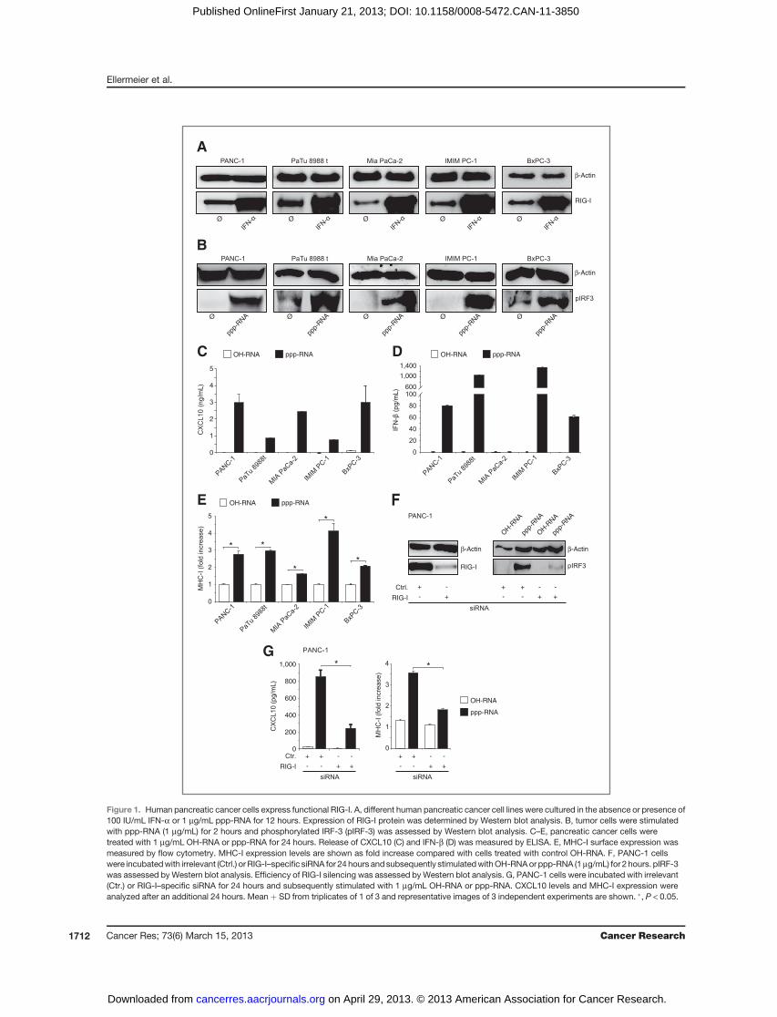
RIG-I
Ø Ø Ø Ø
A
Ø
B
Ø
ppp-
RNA Ø
ppp-
RNA Ø
ppp-
RNA
ppp-
RNAØØ
ppp-
RNA
0
2
4
5
C OH-RNA ppp-RNA
E
0
2
4
5
* *
*
**
OH-RNA
β-Actin
β-Actin
β-Actinβ-Actin
(pg/m
L)
(ng/m
L)
(pg/m
L)
ppp-RNA
OH-RNA ppp-RNA
D
OH-R
NA
ppp-
RNA
OH-R
NA
ppp-
RNA
++
- - ++
- -
RIG-I
RIG-I
+
- +
-
F
G
0
200
400
600
RIG-I
++
- - ++
- - ++
- - ++
- -
**
OH-RNA
ppp-RNA
Figure 1. Human pancreatic cancer cells express functional RIG-I. A, different human pancreatic cancer cell lines were cultured in the absence or presence of100 IU/mL IFN-a or 1 mg/mL ppp-RNA for 12 hours. Expression of RIG-I protein was determined by Western blot analysis. B, tumor cells were stimulatedwith ppp-RNA (1 mg/mL) for 2 hours and phosphorylated IRF-3 (pIRF-3) was assessed by Western blot analysis. C–E, pancreatic cancer cells weretreated with 1 mg/mL OH-RNA or ppp-RNA for 24 hours. Release of CXCL10 (C) and IFN-b (D) was measured by ELISA. E, MHC-I surface expression wasmeasured by flow cytometry. MHC-I expression levels are shown as fold increase compared with cells treated with control OH-RNA. F, PANC-1 cellswere incubatedwith irrelevant (Ctrl.) or RIG-I–specific siRNA for 24 hours and subsequently stimulatedwithOH-RNAor ppp-RNA (1mg/mL) for 2 hours. pIRF-3was assessed by Western blot analysis. Efficiency of RIG-I silencing was assessed by Western blot analysis. G, PANC-1 cells were incubated with irrelevant(Ctr.) or RIG-I–specific siRNA for 24 hours and subsequently stimulated with 1 mg/mL OH-RNA or ppp-RNA. CXCL10 levels and MHC-I expression wereanalyzed after an additional 24 hours. Mean þ SD from triplicates of 1 of 3 and representative images of 3 independent experiments are shown. �, P < 0.05.
Ellermeier et al.
Cancer Res; 73(6) March 15, 2013 Cancer Research1712
on April 29, 2013. © 2013 American Association for Cancer Research. cancerres.aacrjournals.org Downloaded from
Published OnlineFirst January 21, 2013; DOI: 10.1158/0008-5472.CAN-11-3850
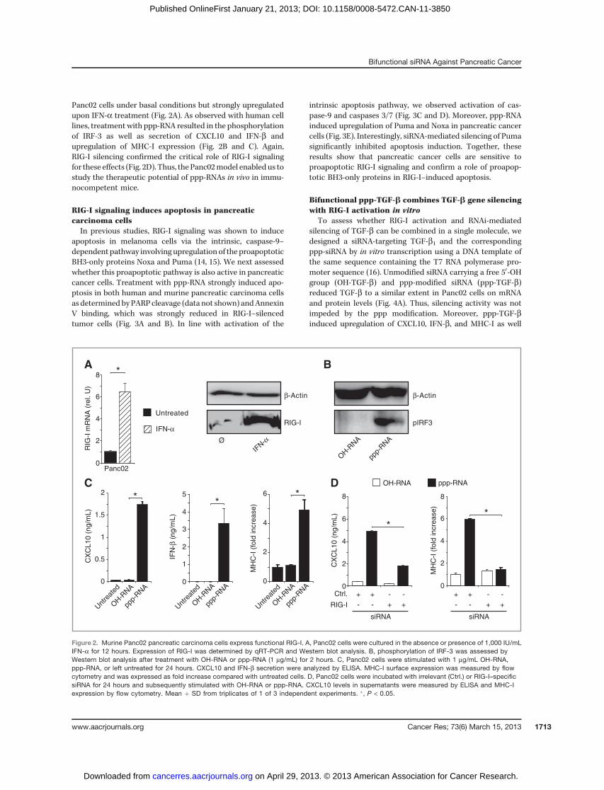
Panc02 cells under basal conditions but strongly upregulatedupon IFN-a treatment (Fig. 2A). As observed with human celllines, treatment with ppp-RNA resulted in the phosphorylationof IRF-3 as well as secretion of CXCL10 and IFN-b andupregulation of MHC-I expression (Fig. 2B and C). Again,RIG-I silencing confirmed the critical role of RIG-I signalingfor these effects (Fig. 2D). Thus, the Panc02model enabled us tostudy the therapeutic potential of ppp-RNAs in vivo in immu-nocompetent mice.
RIG-I signaling induces apoptosis in pancreaticcarcinoma cellsIn previous studies, RIG-I signaling was shown to induce
apoptosis in melanoma cells via the intrinsic, caspase-9–dependent pathway involving upregulation of the proapoptoticBH3-only proteins Noxa and Puma (14, 15). We next assessedwhether this proapoptotic pathway is also active in pancreaticcancer cells. Treatment with ppp-RNA strongly induced apo-ptosis in both human and murine pancreatic carcinoma cellsas determined by PARP cleavage (data not shown) andAnnexinV binding, which was strongly reduced in RIG-I–silencedtumor cells (Fig. 3A and B). In line with activation of the
intrinsic apoptosis pathway, we observed activation of cas-pase-9 and caspases 3/7 (Fig. 3C and D). Moreover, ppp-RNAinduced upregulation of Puma and Noxa in pancreatic cancercells (Fig. 3E). Interestingly, siRNA-mediated silencing of Pumasignificantly inhibited apoptosis induction. Together, theseresults show that pancreatic cancer cells are sensitive toproapoptotic RIG-I signaling and confirm a role of proapop-totic BH3-only proteins in RIG-I–induced apoptosis.
Bifunctional ppp-TGF-b combines TGF-b gene silencingwith RIG-I activation in vitro
To assess whether RIG-I activation and RNAi-mediatedsilencing of TGF-b can be combined in a single molecule, wedesigned a siRNA-targeting TGF-b1 and the correspondingppp-siRNA by in vitro transcription using a DNA template ofthe same sequence containing the T7 RNA polymerase pro-moter sequence (16). Unmodified siRNA carrying a free 50-OHgroup (OH-TGF-b) and ppp-modified siRNA (ppp-TGF-b)reduced TGF-b to a similar extent in Panc02 cells on mRNAand protein levels (Fig. 4A). Thus, silencing activity was notimpeded by the ppp modification. Moreover, ppp-TGF-binduced upregulation of CXCL10, IFN-b, and MHC-I as well
MH
C-I
(fo
ld incre
ase)
0
2
4
6 *
Unt
reat
ed
OH-R
NA
ppp-
RNA
C
0
0.5
1
1.5
2
Unt
reat
ed
OH-R
NA
ppp-
RNA
CX
CL10 (
ng/m
L)
*
0
1
2
3
4
5
IFN
-β
IFN-α
IFN-α
β-Actin β-Actin
(ng
/mL
)
*
Unt
reat
ed
OH-R
NA
ppp-
RNA
D OH-RNA ppp-RNA
0
2
4
6
CX
CL10 (
ng/m
L)
8
MH
C-I
(fo
ld incre
ase)
++
- - ++
- -++
- - ++
- -
**
siRNA siRNA
0
2
4
6
8
Ctrl.
RIG-I
A
RIG
-I m
RN
A (
rel. U
)
Panc02
Untreated
*
0
2
4
6
8
Ø
RIG-I
B
ppp-
RNA
OH-R
NA
pIRF3
Figure 2. Murine Panc02 pancreatic carcinoma cells express functional RIG-I. A, Panc02 cells were cultured in the absence or presence of 1,000 IU/mLIFN-a for 12 hours. Expression of RIG-I was determined by qRT-PCR and Western blot analysis. B, phosphorylation of IRF-3 was assessed byWestern blot analysis after treatment with OH-RNA or ppp-RNA (1 mg/mL) for 2 hours. C, Panc02 cells were stimulated with 1 mg/mL OH-RNA,ppp-RNA, or left untreated for 24 hours. CXCL10 and IFN-b secretion were analyzed by ELISA. MHC-I surface expression was measured by flowcytometry and was expressed as fold increase compared with untreated cells. D, Panc02 cells were incubated with irrelevant (Ctrl.) or RIG-I–specificsiRNA for 24 hours and subsequently stimulated with OH-RNA or ppp-RNA. CXCL10 levels in supernatants were measured by ELISA and MHC-Iexpression by flow cytometry. Mean þ SD from triplicates of 1 of 3 independent experiments. �, P < 0.05.
Bifunctional siRNA Against Pancreatic Cancer
www.aacrjournals.org Cancer Res; 73(6) March 15, 2013 1713
on April 29, 2013. © 2013 American Association for Cancer Research. cancerres.aacrjournals.org Downloaded from
Published OnlineFirst January 21, 2013; DOI: 10.1158/0008-5472.CAN-11-3850
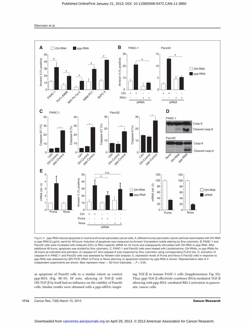
as apoptosis of Panc02 cells to a similar extent as controlppp-RNA (Fig. 4B–D). Of note, silencing of TGF-b withOH-TGF-b by itself had no influence on the viability of Panc02cells. Similar results were obtained with a ppp-siRNA–target-
ing TGF-b in human PANC-1 cells (Supplementary Fig. S2).Thus, ppp-TGF-b effectively combines RNAi-mediated TGF-bsilencing with ppp-RNA–mediated RIG-I activation in pancre-atic cancer cells.
A
0
10
20
30
40
An
ne
xin
V(%
po
sitiv
e)
MIA
Pa
Ca-
2
IMIM
-PC1
PaTu
8988
t
**
*
PANC-1
*
OH-RNA ppp-RNA
*
BxPC-3
50
B PANC-1 Panc02
An
ne
xin
V(%
po
sitiv
e)
0
10
20
30
0
5
10
15
**
siRNA siRNA
OH-RNA
ppp-RNA
Ctrl.
RIG-I
++
- - ++
- - ++
- - ++
- -
0
10
20
30
40
50
An
nexin
V (
% p
ositiv
e) *
n.s.
Ctrl.
Puma
++
- - ++
- -
siRNA
--
- -
Noxa - - -- ++
OH-RNA
ppp-RNA
0
10
20
30
Lipo
OH-C
ontro
l
ppp-
Con
trol
Ca
sp
ase
-9 (
%)
*
0
10
20
30
Lipo
OH-C
ontro
l
ppp-
Con
trol
Ca
sp
ase
-3/7
(%
)
Panc02
Non
e
OH-R
NA
ppp-
RNA
Casp-9
Cleaved casp-9
E
0
1
2
3
4
5
mR
NA
(re
l. U
)
Puma Noxa
C
Ca
sp
ase
-3/7
(%
)
Lipo
OH-C
ontro
l
ppp-
Con
trol
40
20
10
0
30
PANC-1
Ca
sp
ase
-9 (
%)
Lipo
OH-C
ontro
l
ppp-
Con
trol
40
20
10
0
30
Panc02
Casp-9
Cleaved casp-9
PANC-1
D
**
*
0
20
40
60
80
100
120
mR
NA
(re
l. U
)
0
20
40
60
80
100
120
OH-RNA
siRNA
Puma Noxa
Figure 3. ppp-RNA induces apoptosis inmurine and human pancreatic cancer cells. A, different human pancreatic cancer cell lines were treated with OH-RNAor ppp-RNA (2 mg/mL each) for 48 hours. Induction of apoptosis was measured via Annexin V/propidium iodide staining by flow cytometry. B, PANC-1 andPanc02 cells were incubated with irrelevant (Ctrl.) or RIG-I–specific siRNA for 24 hours and subsequently stimulated with OH-RNA or ppp-RNA. Afteradditional 48 hours, apoptosis was studied by flow cytometry. C, PANC-1 and Panc02 cells were treated with Lipofectamine, OH-RNAs, or ppp-RNAs for48 hours as indicated and activation of caspase-3/7 and caspase-9 was measured by flow cytometry using corresponding FLICA kits. D, activation ofcaspase-9 in PANC-1 and Panc02 cells was assessed by Western blot analysis. E, expression levels of Puma and Noxa in Panc02 cells in response toppp-RNA was assessed by qRT-PCR. Effect of Puma or Noxa silencing on apoptosis induction by ppp-RNA is shown. Representative data of 3independent experiments are shown. Bars represent mean þ SD from triplicates. �, P < 0.05.
Ellermeier et al.
Cancer Res; 73(6) March 15, 2013 Cancer Research1714
on April 29, 2013. © 2013 American Association for Cancer Research. cancerres.aacrjournals.org Downloaded from
Published OnlineFirst January 21, 2013; DOI: 10.1158/0008-5472.CAN-11-3850
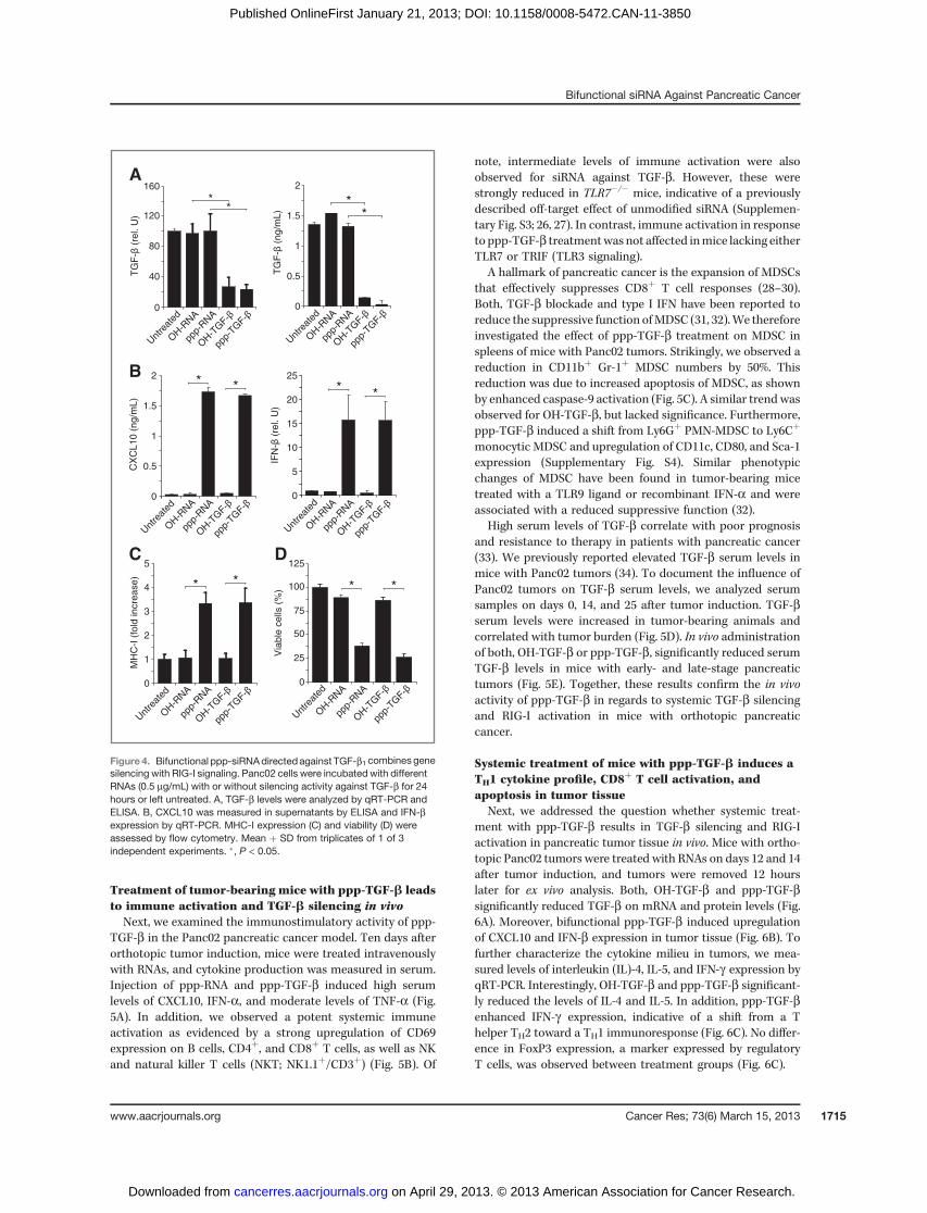
Treatment of tumor-bearing mice with ppp-TGF-b leadsto immune activation and TGF-b silencing in vivoNext, we examined the immunostimulatory activity of ppp-
TGF-b in the Panc02 pancreatic cancer model. Ten days afterorthotopic tumor induction, mice were treated intravenouslywith RNAs, and cytokine production was measured in serum.Injection of ppp-RNA and ppp-TGF-b induced high serumlevels of CXCL10, IFN-a, and moderate levels of TNF-a (Fig.5A). In addition, we observed a potent systemic immuneactivation as evidenced by a strong upregulation of CD69expression on B cells, CD4þ, and CD8þ T cells, as well as NKand natural killer T cells (NKT; NK1.1þ/CD3þ) (Fig. 5B). Of
note, intermediate levels of immune activation were alsoobserved for siRNA against TGF-b. However, these werestrongly reduced in TLR7�/� mice, indicative of a previouslydescribed off-target effect of unmodified siRNA (Supplemen-tary Fig. S3; 26, 27). In contrast, immune activation in responseto ppp-TGF-b treatmentwas not affected inmice lacking eitherTLR7 or TRIF (TLR3 signaling).
A hallmark of pancreatic cancer is the expansion of MDSCsthat effectively suppresses CD8þ T cell responses (28–30).Both, TGF-b blockade and type I IFN have been reported toreduce the suppressive function ofMDSC (31, 32).We thereforeinvestigated the effect of ppp-TGF-b treatment on MDSC inspleens of mice with Panc02 tumors. Strikingly, we observed areduction in CD11bþ Gr-1þ MDSC numbers by 50%. Thisreduction was due to increased apoptosis of MDSC, as shownby enhanced caspase-9 activation (Fig. 5C). A similar trend wasobserved for OH-TGF-b, but lacked significance. Furthermore,ppp-TGF-b induced a shift from Ly6Gþ PMN-MDSC to Ly6Cþ
monocytic MDSC and upregulation of CD11c, CD80, and Sca-1expression (Supplementary Fig. S4). Similar phenotypicchanges of MDSC have been found in tumor-bearing micetreated with a TLR9 ligand or recombinant IFN-a and wereassociated with a reduced suppressive function (32).
High serum levels of TGF-b correlate with poor prognosisand resistance to therapy in patients with pancreatic cancer(33). We previously reported elevated TGF-b serum levels inmice with Panc02 tumors (34). To document the influence ofPanc02 tumors on TGF-b serum levels, we analyzed serumsamples on days 0, 14, and 25 after tumor induction. TGF-bserum levels were increased in tumor-bearing animals andcorrelated with tumor burden (Fig. 5D). In vivo administrationof both, OH-TGF-b or ppp-TGF-b, significantly reduced serumTGF-b levels in mice with early- and late-stage pancreatictumors (Fig. 5E). Together, these results confirm the in vivoactivity of ppp-TGF-b in regards to systemic TGF-b silencingand RIG-I activation in mice with orthotopic pancreaticcancer.
Systemic treatment of mice with ppp-TGF-b induces aTH1 cytokine profile, CD8þ T cell activation, andapoptosis in tumor tissue
Next, we addressed the question whether systemic treat-ment with ppp-TGF-b results in TGF-b silencing and RIG-Iactivation in pancreatic tumor tissue in vivo. Mice with ortho-topic Panc02 tumors were treated with RNAs on days 12 and 14after tumor induction, and tumors were removed 12 hourslater for ex vivo analysis. Both, OH-TGF-b and ppp-TGF-bsignificantly reduced TGF-b on mRNA and protein levels (Fig.6A). Moreover, bifunctional ppp-TGF-b induced upregulationof CXCL10 and IFN-b expression in tumor tissue (Fig. 6B). Tofurther characterize the cytokine milieu in tumors, we mea-sured levels of interleukin (IL)-4, IL-5, and IFN-g expression byqRT-PCR. Interestingly, OH-TGF-b and ppp-TGF-b significant-ly reduced the levels of IL-4 and IL-5. In addition, ppp-TGF-benhanced IFN-g expression, indicative of a shift from a Thelper TH2 toward a TH1 immunoresponse (Fig. 6C). No differ-ence in FoxP3 expression, a marker expressed by regulatoryT cells, was observed between treatment groups (Fig. 6C).
A
B
0
0.5
1
1.5
2
OH-R
NA
OH-T
GF-
ppp-
TGF-
TG
F-
(n
g/m
L)
Unt
reat
ed
ppp-
RNA
0
40
80
120
TG
F-
(re
l. U
)
OH-R
NA
OH-T
GF-
ppp-
TGF-
Unt
reat
ed
ppp-
RNA
** *
*
160
CX
CL
10
(n
g/m
L)
Unt
reat
ed
OH-R
NA
ppp-
RNA
OH-T
GF-
ppp-
TGF-
* *
0
0.5
1
1.5
2
0
1
2
3
4
5
MH
C-I
(fo
ld in
cre
ase
)
C
Unt
reat
ed
OH-R
NA
ppp-
RNA
OH-T
GF-
ppp-
TGF-
* *
0
5
10
15
20
25
IFN
-(r
el. U
)
Unt
reat
ed
OH-R
NA
ppp-
RNA
OH-T
GF-
ppp-
TGF-
* *
D
Via
ble
ce
lls (
%)
0
25
50
75
100
125
OH-T
GF-
Unt
reat
ed
ppp-
TGF-
OH-R
NA
ppp-
RNA
**
Figure 4. Bifunctional ppp-siRNAdirectedagainst TGF-b1 combinesgenesilencing with RIG-I signaling. Panc02 cells were incubated with differentRNAs (0.5 mg/mL) with or without silencing activity against TGF-b for 24hours or left untreated. A, TGF-b levels were analyzed by qRT-PCR andELISA. B, CXCL10 was measured in supernatants by ELISA and IFN-bexpression by qRT-PCR. MHC-I expression (C) and viability (D) wereassessed by flow cytometry. Mean þ SD from triplicates of 1 of 3independent experiments. �, P < 0.05.
Bifunctional siRNA Against Pancreatic Cancer
www.aacrjournals.org Cancer Res; 73(6) March 15, 2013 1715
on April 29, 2013. © 2013 American Association for Cancer Research. cancerres.aacrjournals.org Downloaded from
Published OnlineFirst January 21, 2013; DOI: 10.1158/0008-5472.CAN-11-3850
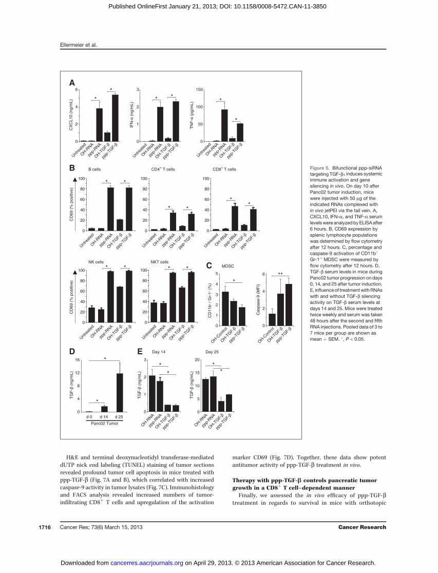
H&E and terminal deoxynucleotidyl transferase-mediateddUTP nick end labeling (TUNEL) staining of tumor sectionsrevealed profound tumor cell apoptosis in mice treated withppp-TGF-b (Fig. 7A and B), which correlated with increasedcaspase-9 activity in tumor lysates (Fig. 7C). Immunohistologyand FACS analysis revealed increased numbers of tumor-infiltrating CD8þ T cells and upregulation of the activation
marker CD69 (Fig. 7D). Together, these data show potentantitumor activity of ppp-TGF-b treatment in vivo.
Therapy with ppp-TGF-b controls pancreatic tumorgrowth in a CD8þ T cell–dependent manner
Finally, we assessed the in vivo efficacy of ppp-TGF-btreatment in regards to survival in mice with orthotopic
A
TN
F-
(pg/m
L)
0
50
100
150
Unt
reat
ed
OH-R
NA
ppp-
RNA
OH-T
GF-
ppp-
TGF-
D
TG
F-
(ng/m
L)
0
1
2
3
OH-R
NA
ppp-
RNA
OH-T
GF-
ppp-
TGF-
TG
F-
(ng/m
L)
0
5
10
15
20
OH-R
NA
ppp-
RNA
OH-T
GF-
ppp-
TGF-
Day 14 Day 25
0
4
8
12
16
d 14 d 25
Panc02 Tumor
TG
F-
(ng/m
L)
*
*
**
*
*
B
*
*
CD
69 (
% p
ositiv
e)
0
20
40
60
80
100
Unt
reat
ed
OH-R
NA
ppp-
RNA
OH-T
GF-
ppp-
TGF-
B cells CD4+ T cells
0
20
40
60
80
100
Unt
reat
ed
OH-R
NA
ppp-
RNA
OH-T
GF-
ppp-
TGF-
CD8+ T cells
* *
0
20
40
60
80
100
Unt
reat
ed
OH-R
NA
ppp-
RNA
OH-T
GF-
ppp-
TGF-
* *
**
IFN
- (
ng/m
L)
Unt
reat
ed
OH-R
NA
ppp-
RNA
OH-T
GF-
ppp-
TGF-
0
1
2
3
* *
CX
CL10 (
ng/m
L)
0
2
4
6
Unt
reat
ed
OH-R
NA
ppp-
RNA
OH-T
GF-
ppp-
TGF-
*
*
0
20
40
60
80
100
Unt
reat
ed
OH-R
NA
ppp-
RNA
OH-T
GF-
ppp-
TGF-
NKT cells
* *
d 0
E
0
20
40
60
80
100
Unt
reat
ed
OH-R
NA
ppp-
RNA
OH-T
GF-
ppp-
TGF-
NK cells
* *
CD
69 (
% p
ositiv
e)
0
3
4
5
CD
11b
+/ G
r-1
+ (
%)
OH-C
ontro
l
OH-T
GF-
ppp-
TGF-
*
2
1
0
2
4
6
Caspase-9
(M
FI)
OH-C
ontro
l
OH-T
GF-
ppp-
TGF-
**
C MDSC
Figure 5. Bifunctional ppp-siRNAtargeting TGF-b1 induces systemicimmune activation and genesilencing in vivo. On day 10 afterPanc02 tumor induction, micewere injected with 50 mg of theindicated RNAs complexed within vivo jetPEI via the tail vein. A,CXCL10, IFN-a, and TNF-a serumlevelswere analyzed by ELISA after6 hours. B, CD69 expression bysplenic lymphocyte populationswas determined by flow cytometryafter 12 hours. C, percentage andcaspase-9 activation of CD11bþ
Gr-1þ MDSC were measured byflow cytometry after 12 hours. D,TGF-b serum levels in mice duringPanc02 tumor progression on days0, 14, and 25 after tumor induction.E, influence of treatmentwith RNAswith and without TGF-b silencingactivity on TGF-b serum levels atdays 14 and 25. Mice were treatedtwice weekly and serum was taken48 hours after the second and fifthRNA injections. Pooled data of 3 to7 mice per group are shown asmean þ SEM. �, P < 0.05.
Ellermeier et al.
Cancer Res; 73(6) March 15, 2013 Cancer Research1716
on April 29, 2013. © 2013 American Association for Cancer Research. cancerres.aacrjournals.org Downloaded from
Published OnlineFirst January 21, 2013; DOI: 10.1158/0008-5472.CAN-11-3850
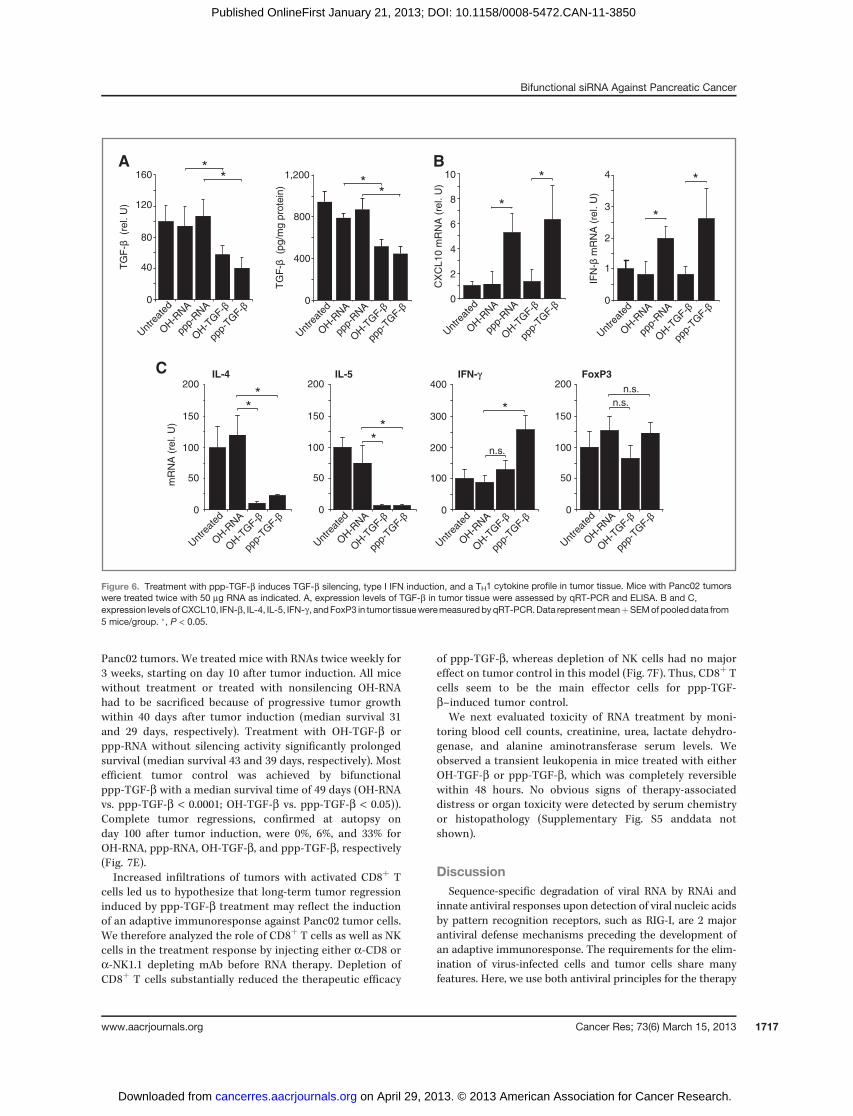
Panc02 tumors. We treated mice with RNAs twice weekly for3 weeks, starting on day 10 after tumor induction. All micewithout treatment or treated with nonsilencing OH-RNAhad to be sacrificed because of progressive tumor growthwithin 40 days after tumor induction (median survival 31and 29 days, respectively). Treatment with OH-TGF-b orppp-RNA without silencing activity significantly prolongedsurvival (median survival 43 and 39 days, respectively). Mostefficient tumor control was achieved by bifunctionalppp-TGF-b with a median survival time of 49 days (OH-RNAvs. ppp-TGF-b < 0.0001; OH-TGF-b vs. ppp-TGF-b < 0.05)).Complete tumor regressions, confirmed at autopsy onday 100 after tumor induction, were 0%, 6%, and 33% forOH-RNA, ppp-RNA, OH-TGF-b, and ppp-TGF-b, respectively(Fig. 7E).Increased infiltrations of tumors with activated CD8þ T
cells led us to hypothesize that long-term tumor regressioninduced by ppp-TGF-b treatment may reflect the inductionof an adaptive immunoresponse against Panc02 tumor cells.We therefore analyzed the role of CD8þ T cells as well as NKcells in the treatment response by injecting either a-CD8 ora-NK1.1 depleting mAb before RNA therapy. Depletion ofCD8þ T cells substantially reduced the therapeutic efficacy
of ppp-TGF-b, whereas depletion of NK cells had no majoreffect on tumor control in this model (Fig. 7F). Thus, CD8þ Tcells seem to be the main effector cells for ppp-TGF-b–induced tumor control.
We next evaluated toxicity of RNA treatment by moni-toring blood cell counts, creatinine, urea, lactate dehydro-genase, and alanine aminotransferase serum levels. Weobserved a transient leukopenia in mice treated with eitherOH-TGF-b or ppp-TGF-b, which was completely reversiblewithin 48 hours. No obvious signs of therapy-associateddistress or organ toxicity were detected by serum chemistryor histopathology (Supplementary Fig. S5 anddata notshown).
DiscussionSequence-specific degradation of viral RNA by RNAi and
innate antiviral responses upon detection of viral nucleic acidsby pattern recognition receptors, such as RIG-I, are 2 majorantiviral defense mechanisms preceding the development ofan adaptive immunoresponse. The requirements for the elim-ination of virus-infected cells and tumor cells share manyfeatures. Here, we use both antiviral principles for the therapy
A
0
Unt
reat
ed
OH-R
NA
ppp-
RNA
OH-T
GF-
ppp-
TGF-
400
800
1,200
0
TG
F-
(p
g/m
g p
rote
in)
**
TG
F-
(r
el. U
)
40
80
120
160
Unt
reat
ed
OH-R
NA
ppp-
RNA
OH-T
GF-
ppp-
TGF-
B*
*
Unt
reat
ed
OH-R
NA
ppp-
RNA
OH-T
GF-
ppp-
TGF-
0
2
4
6
8
10
CX
CL
10
mR
NA
(re
l. U
)
*
*
IFN
- m
RN
A (
rel. U
)
4
2
1
0
3
Unt
reat
ed
OH-R
NA
ppp-
RNA
OH-T
GF-
ppp-
TGF-
*
*
C
mR
NA
(re
l. U
)
200
100
50
0
150
Unt
reat
ed
OH-R
NA
OH-T
GF-
ppp-
TGF-
**
200
100
50
0
150
Unt
reat
ed
OH-R
NA
OH-T
GF-
ppp-
TGF-
**
IL-5IL-4200
100
50
0
150
Unt
reat
ed
OH-R
NA
OH-T
GF-
ppp-
TGF-
n.s.
n.s.
FoxP3IFN-γ400
200
100
0
300
Unt
reat
ed
OH-R
NA
OH-T
GF-
ppp-
TGF-
*
n.s.
Figure 6. Treatment with ppp-TGF-b induces TGF-b silencing, type I IFN induction, and a TH1 cytokine profile in tumor tissue. Mice with Panc02 tumorswere treated twice with 50 mg RNA as indicated. A, expression levels of TGF-b in tumor tissue were assessed by qRT-PCR and ELISA. B and C,expression levels ofCXCL10, IFN-b, IL-4, IL-5, IFN-g , andFoxP3 in tumor tissueweremeasuredbyqRT-PCR.Data representmeanþSEMofpooleddata from5 mice/group. �, P < 0.05.
Bifunctional siRNA Against Pancreatic Cancer
www.aacrjournals.org Cancer Res; 73(6) March 15, 2013 1717
on April 29, 2013. © 2013 American Association for Cancer Research. cancerres.aacrjournals.org Downloaded from
Published OnlineFirst January 21, 2013; DOI: 10.1158/0008-5472.CAN-11-3850
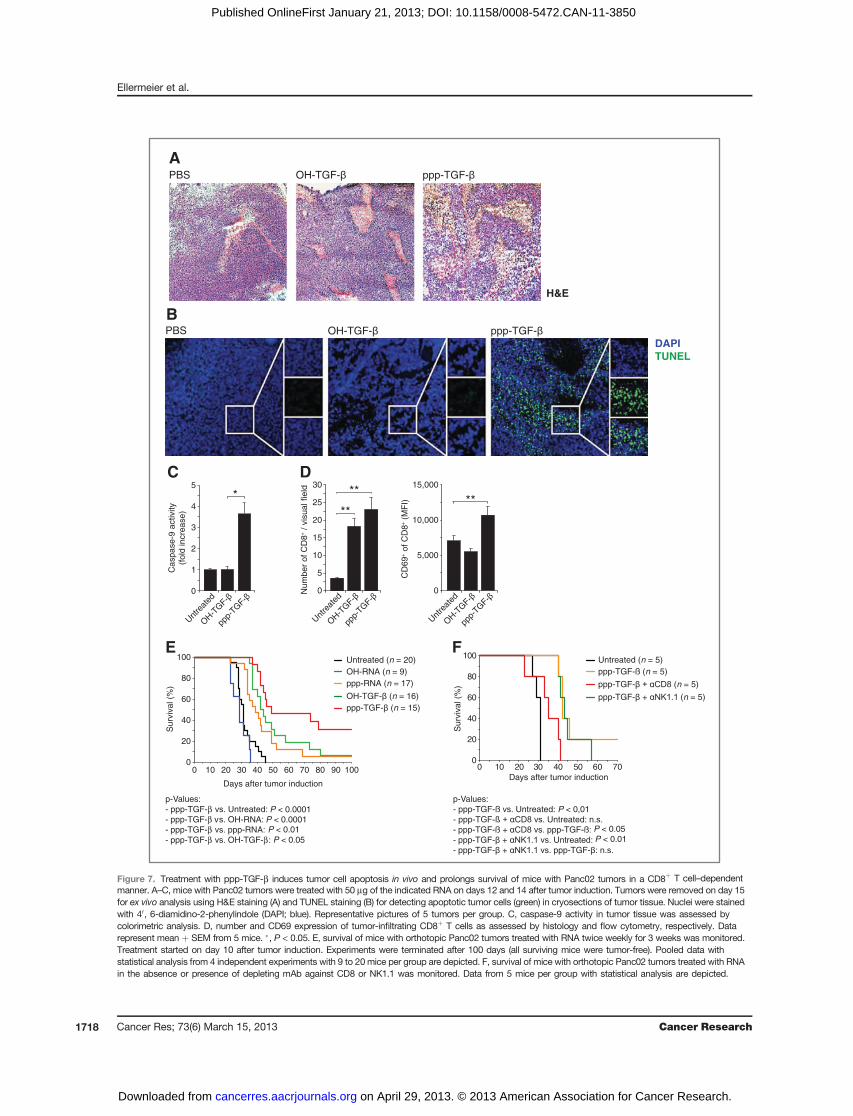
C
0
1
2
3
4
5
Unt
reat
ed
OH-T
GF-
ppp-
TGF-
Ca
sp
ase
-9 a
ctivity
(fo
ld in
cre
ase
)
*
PBS OH-TGF- ppp-TGF-DAPITUNEL
B
0
5
10
15
20
25
30
Num
ber
of
CD
8+ /
vis
ua
l fie
ld
Unt
reat
ed
OH-T
GF-
ppp-
TGF-
**
**
D
0
5,000
10,000
15,000
CD
69
+ o
f C
D8
+ (
MF
I)
Unt
reat
ed
OH-T
GF-
ppp-
TGF-
**
Untreated (n = 5)
ppp-TGF-ß (n = 5)
ppp-TGF- (n = 5)
ppp-TGF- + (n = 5)
F
0 10 20 30 40 500
20
40
60
80
100
Days after tumor induction
Su
rviv
al (%
)
60 70
Untreated (n = 20)
OH-RNA (n = 9)
ppp-RNA (n = 17)
OH-TGF- (n = 16)
ppp-TGF- (n = 15)
E
0
20
40
60
80
100
0 10 20 30 40 50 60 70 80 90 100
Days after tumor induction
Su
rviv
al (%
)
p-Values:
- ppp-TGF-ß Untreated: P < 0,01
P < 0.05
P < 0.0001P < 0.0001P < 0.01
P < 0.05 P < 0.01
- ppp- Untreated
- ppp-TGF-ß + ppp-TGF-ß
- ppp-TGF- +
- ppp-TGF- + ppp-TGF-
p-Values:
- ppp-TGF-β Untreated
- ppp-TGF-β v OH-RNA
- ppp-TGF-β ppp-RNA
- ppp-TGF-β OH-TGF-β
PBS OH-TGF- ppp-TGF-
H&E
A
Figure 7. Treatment with ppp-TGF-b induces tumor cell apoptosis in vivo and prolongs survival of mice with Panc02 tumors in a CD8þ T cell–dependentmanner. A–C, mice with Panc02 tumors were treated with 50 mg of the indicated RNA on days 12 and 14 after tumor induction. Tumors were removed on day 15for ex vivo analysis using H&E staining (A) and TUNEL staining (B) for detecting apoptotic tumor cells (green) in cryosections of tumor tissue. Nuclei were stainedwith 40, 6-diamidino-2-phenylindole (DAPI; blue). Representative pictures of 5 tumors per group. C, caspase-9 activity in tumor tissue was assessed bycolorimetric analysis. D, number and CD69 expression of tumor-infiltrating CD8þ T cells as assessed by histology and flow cytometry, respectively. Datarepresent mean þ SEM from 5 mice. �, P < 0.05. E, survival of mice with orthotopic Panc02 tumors treated with RNA twice weekly for 3 weeks was monitored.Treatment started on day 10 after tumor induction. Experiments were terminated after 100 days (all surviving mice were tumor-free). Pooled data withstatistical analysis from 4 independent experiments with 9 to 20mice per group are depicted. F, survival of mice with orthotopic Panc02 tumors treated with RNAin the absence or presence of depleting mAb against CD8 or NK1.1 was monitored. Data from 5 mice per group with statistical analysis are depicted.
Ellermeier et al.
Cancer Res; 73(6) March 15, 2013 Cancer Research1718
on April 29, 2013. © 2013 American Association for Cancer Research. cancerres.aacrjournals.org Downloaded from
Published OnlineFirst January 21, 2013; DOI: 10.1158/0008-5472.CAN-11-3850
of pancreatic cancer. We show that functional RIG-I isexpressed in human pancreatic cancer cells. Furthermore, adesigned RNA molecule to conduct both RIG-I activation andsilencing of the immunosuppressive cytokine TGF-b inducedtumor regression in a CD8þ T cell–dependent manner in anaggressive mouse model of pancreatic cancer. In this respect,RNA molecules that contained either the RIG-I ligand motifor the silencing capability alone were less effective supportingthe concept that bifunctional siRNA is superior for effectivetumor therapy.A major hurdle in cancer immunotherapy is the profound-
immunosuppression, both systemically and locally withinthe tumor microenvironment. RIG-I signaling leads to type IIFN responses with IFN-dependent gene products triggeringinnate and adaptive immunoresponses (18). These effectsinclude systemic activation of NK cells and T cells, as well asactivation of dendritic cells, which are critical for the induc-tion and regulation of adaptive immunoresponses and play akey role in cancer immune surveillance (35). It is importantto note that upon RIG-I activation, type I IFN not onlyderives from immune cells but also from tumor tissue, asshown for human and murine pancreatic cancer cells in ourwork. Secretion of CXCL10 can attract lymphocytes to tumortissue, and locally produced IFN-b can activate TH1responses and tumor-infiltrating CTL thus enhancing theirkilling function. In this respect, upregulation of MHC-Iexpression by the tumor cells upon RIG-I activation maypromote CTL-mediated tumor recognition and killing. Infact, we observed that treatment with ppp-TGF-b resulted ina TH1 cytokine profile in tumor tissue and a more vigoroustumor infiltration with activated CD8þ T cells. In addition,we found that tumor regression in response to ppp-TGF-btreatment was mediated by CD8þ T cells. This leads to thequestion of how ppp-TGF-b restored CD8þ T cell responsesin tumors.Soluble factors, such as TGF-b (5, 6), and immune cell
populations, such as MDSCs and regulatory T cells, havebeen shown to play immunosuppressive roles in pancreaticcancer (30, 36). In our study, treatment with ppp-TGF-beffectively reduced TGF-b levels in both serum and tumors.The frequency of regulatory T cells was not influenced,however, we have previously reported that TGF-b silencingresults in a marked downregulation of CD103 expression onregulatory T cells (37). As CD103 identifies a particularlysuppressive subtype of regulatory T cells, treatment withppp-TGF-b may counteract regulatory T cell–mediated CTLsuppression. In addition, we observed that ppp-TGF-b sig-nificantly reduced the numbers of CD11bþ Gr-1þ MDSCs.Because MDSCs are frequently found in pancreatic cancertissue and potently suppress CD8þ T cells, this finding isparticularly interesting (36). Moreover, MDSC underwentphenotypic changes, such as upregulation of CD11c, CD80,and Sca-1 expression. Interestingly, similar changes in MDSChave been reported in tumor-bearing mice treated withrecombinant IFN-a and were found to correlate with areduced T cell–suppressive function (32). Thus, TGF-bsilencing and type I IFN induction induced by ppp-TGF-bseem to have additive effects on breaking the immunosup-
pressive milieu created by pancreatic cancer cells and arecapable of tipping the balance toward effective antitumorCTL responses.
A central aspect of ppp-RNA treatment is the induction oftumor cell apoptosis. Pancreatic cancer cells frequentlyacquire loss-of-function mutations of the gatekeeper proteinp53, which reduces their sensitivity toward proapoptoticsignals (38). An elegant strategy to circumvent this limita-tion is the exploitation of p53-independent apoptosis induc-tion. We found that pancreatic carcinoma cell lines, includ-ing those with p53 mutations (PANC-1, BxPC-3, and MIA-PaCa-2), were sensitive to ppp-RNA–mediated apoptosis. Inline with findings in melanoma (14), we found that ppp-RNAtriggers apoptosis via the mitochondrial pathway in pan-creatic cancer cells involving upregulation of the BH3-onlyproteins Noxa and Puma with subsequent caspase-9 acti-vation. Moreover, systemic treatment with ppp-TGF-binduced profound tumor cell apoptosis in vivo, whereasnormal pancreas (as well as other organs, such as liver,kidney, and lung) showed no signs of histopathology. Thesefindings confirm previous reports that tumor cells are highlysusceptible to ppp-RNA–induced apoptosis (14). The predi-lection for tumor cells as compared with healthy tissue iscritical for avoiding toxicity and provides a therapeuticwindow for ppp-RNA treatment.
In conclusion, we identified RIG-I as a novel target forimmunotherapy of pancreatic cancer. Combining RIG-Iactivation with TGF-b silencing via bifunctional ppp-siRNAbreaks tumor-mediated immunosuppressive mechanismsand confers potent antitumor efficacy. Whether this strat-egy can be further improved, for example, by combinationwith cytotoxic agents or immunization, is the focus ofongoing studies. Further improvement can be expected bydesigning new delivery systems for selective tumor target-ing and by assessment in genetically engineered mousemodels of pancreatic cancer, which allow studying effectson the tumor stromal compartment and metastatic spread-ing (39, 40).
Disclosure of Potential Conflicts of InterestNo potential conflicts of interest were disclosed.
AcknowledgmentsThe authors thank Dr. Robert Besch for advice in siRNA design and critical
reading of the manuscript and Andrea Sendelhofert for conducting histopathol-ogy. This work is part of the theses of Jonathan Ellermeier, Mareike R. Stieg, andTina Adunka at the University of Munich.
Grant SupportThis work was supported by the Deutsche Krebshilfe (Max Eder Research
Grant to M.R. Stieg.), the Deutsche Forschungsgemeinschaft SCHN 664/3-1 toM.R. Stieg, and GK 1202 to M.R. Stieg, S. Endres, and Hans-Joachim Anders,and GK 1202 student grants to J. Ellermeier, P. Duewell, M.R. Stieg, and T.Adunka.
The costs of publication of this article were defrayed in part by thepayment of page charges. This article must therefore be hereby markedadvertisement in accordance with 18 U.S.C. Section 1734 solely to indicate thisfact.
Received December 7, 2011; revised December 7, 2012; accepted January 10,2013; published OnlineFirst March 6, 2013.
Bifunctional siRNA Against Pancreatic Cancer
www.aacrjournals.org Cancer Res; 73(6) March 15, 2013 1719
on April 29, 2013. © 2013 American Association for Cancer Research. cancerres.aacrjournals.org Downloaded from
Published OnlineFirst January 21, 2013; DOI: 10.1158/0008-5472.CAN-11-3850
References1. Wong HH, Lemoine NR. Pancreatic cancer: molecular pathogenesis
and new therapeutic targets. Nat Rev Gastroenterol Hepatol 2009;6:412–22.
2. Massag.u�e J. TGFbeta in Cancer. Cell 2008;134:215–30.3. MoutsopoulosNM,WenJ,Wahl SM. TGF-beta and tumors–an ill-fated
alliance. Curr Opin Immunol 2008;20:234–40.4. Yang L, Pang Y, Moses HL. TGF-beta and immune cells: an important
regulatory axis in the tumor microenvironment and progression.Trends Immunol 2010;31:220–7.
5. Friess H, Yamanaka Y, Buchler M, Ebert M, Beger HG, Gold LI, et al.Enhanced expression of transforming growth factor beta isoforms inpancreatic cancer correlates with decreased survival. Gastroenterol-ogy 1993;105:1846–56.
6. Lin Y, Kikuchi S, Tamakoshi A, Obata Y, Yagyu K, Inaba Y, et al. Serumtransforming growth factor-beta1 levels and pancreatic cancer risk: anested case-control study (Japan). Cancer Causes Control 2006;17:1077–82.
7. Schlingensiepen KH, Schlingensiepen R, Steinbrecher A, Hau P,Bogdahn U, Fischer-Blass B, et al. Targeted tumor therapy with theTGF-beta 2 antisense compound AP 12009. Cytokine Growth FactorRev 2006;17:129–39.
8. Gaspar NJ, Li L, Kapoun AM, Medicherla S, Reddy M, Li G, et al.Inhibition of transforming growth factor beta signaling reduces pan-creatic adenocarcinoma growth and invasiveness. Mol Pharmacol2007;72:152–61.
9. Pennison M, Pasche B. Targeting transforming growth factor-betasignaling. Curr Opin Oncol 2007;19:579–85.
10. Jaschinski F, Rothhammer T, Jachimczak P, Seitz C, Schneider A,Schlingensiepen KH. The antisense oligonucleotide trabedersen (AP12009) for the targeted inhibition of TGF-beta2. Curr PharmBiotechnol2011;12:2203–13.
11. Schlingensiepen KH, Jaschinski F, Lang SA, Moser C, Geissler EK,Schlitt HJ, et al. Transforming growth factor-beta 2 gene silencingwithtrabedersen (AP 12009) in pancreatic cancer. Cancer Sci 2011;102:1193–200.
12. Fukunaga A,MiyamotoM,ChoY,Murakami S, KawaradaY,Oshikiri T,et al. CD8þtumor-infiltrating lymphocytes together with CD4þ tumor-infiltrating lymphocytes and dendritic cells improve the prognosis ofpatients with pancreatic adenocarcinoma. Pancreas 2004;28:e26–31.
13. Goutagny N, Estornes Y, Hasan U, Lebecque S, Caux C. Targetingpattern recognition receptors in cancer immunotherapy. Target Oncol2012;7:29–54.
14. Besch R, Poeck H, Hohenauer T, Senft D, Hacker G, Berking C, et al.Proapoptotic signaling induced by RIG-I and MDA-5 results in type Iinterferon-independent apoptosis in human melanoma cells. J ClinInvest 2009;119:2399–411.
15. PoeckH, Besch R,Maihoefer C, RennM, TormoD,Morskaya SS, et al.50-Triphosphate-siRNA: turning gene silencing and Rig-I activationagainst melanoma. Nat Med 2008;14:1256–63.
16. Hornung V, Ellegast J, Kim S, Brzozka K, Jung A, Kato H, et al. 50-Triphosphate RNA is the ligand for RIG-I. Science 2006;314:994–7.
17. Pichlmair A, Schulz O, Tan CP, Naslund TI, Liljestrom P,Weber F, et al.RIG-I-mediated antiviral responses to single-strandedRNAbearing 50-phosphates. Science 2006;314:997–1001.
18. Yoneyama M, Fujita T. RNA recognition and signal transduction byRIG-I-like receptors. Immunol Rev 2009;227:54–65.
19. Corbett TH, Roberts BJ, Leopold WR, Peckham JC, Wilkoff LJ,Griswold DP Jr, et al. Induction and chemotherapeutic response oftwo transplantable ductal adenocarcinomas of the pancreas inC57BL/6 mice. Cancer Res 1984;44:717–26.
20. Vila MR, Lloreta J, Schussler MH, Berrozpe G, Welt S, Real FX. Newpancreas cancers cell lines that represent distinct stages of ductaldifferentiation. Lab Invest 1995;72:395–404.
21. Reynolds A, Leake D, Boese Q, Scaringe S, Marshall WS, Khvorova A.Rational siRNA design for RNA interference. Nat Biotechnol 2004;22:326–30.
22. Hoebe K, Du X, Georgel P, Janssen E, Tabeta K, Kim SO, et al.Identification of Lps2 as a key transducer of MyD88-independent TIRsignalling. Nature 2003;424:743–8.
23. Diebold SS, Kaisho T, Hemmi H, Akira S, Reis e Sousa C. Innateantiviral responses by means of TLR7-mediated recognition of single-stranded RNA. Science 2004;303:1529–31.
24. Bauer C, Bauernfeind F, Sterzik A, OrbanM, Schnurr M, Lehr HA, et al.Dendritic cell-based vaccination combined with gemcitabineincreases survival in a murine pancreatic carcinoma model. Gut 2007;56:1275–82.
25. Chen ZH, Zhang H, Savarese TM. Gene deletion chemoselectivity:codeletion of the genes for p16(INK4), methylthioadenosine phos-phorylase, and the alpha- and beta-interferons in human pancreaticcell carcinoma lines and its implications for chemotherapy.CancerRes1996;56:1083–90.
26. Hornung V, Guenthner-Biller M, Bourquin C, Ablasser A, Schlee M,Uematsu S, et al. Sequence-specific potent induction of IFN-alpha byshort interferingRNA in plasmacytoid dendritic cells throughTLR7.NatMed 2005;11:263–70.
27. Kleinman ME, Yamada K, Takeda A, Chandrasekaran V, Nozaki M,Baffi JZ, et al. Sequence- and target-independent angiogenesis sup-pression by siRNA via TLR3. Nature 2008;452:591–7.
28. Clark CE, Hingorani SR, Mick R, Combs C, Tuveson DA, VonderheideRH. Dynamics of the immune reaction to pancreatic cancer frominception to invasion. Cancer Res 2007;67:9518–27.
29. Pilon-Thomas S, Nelson N, Vohra N, Jerald M, Pendleton L, SzekeresK, et al. Murine pancreatic adenocarcinoma dampens SHIP-1 expres-sion and alters MDSC homeostasis and function. PLoS ONE 2011;6:e27729.
30. Lu T, RamakrishnanR, Altiok S, Youn JI, ChengP, Celis E, et al. Tumor-infiltrating myeloid cells induce tumor cell resistance to cytotoxic Tcells in mice. J Clin Invest 2011;121:4015–29.
31. Fridlender ZG, Sun J, Kim S, Kapoor V, Cheng G, Ling L, et al.Polarization of tumor-associated neutrophil phenotype by TGF-beta:"N1" versus "N2" TAN. Cancer Cell 2009;16:183–94.
32. Zoglmeier C, Bauer H, Norenberg D, Wedekind G, Bittner P, Sand-holzer N, et al. CpG blocks immunosuppression by myeloid-derivedsuppressor cells in tumor-bearing mice. Clin Cancer Res 2011;17:1765–75.
33. von Bernstorff W, VossM, Freichel S, Schmid A, Vogel I, Johnk C, et al.Systemic and local immunosuppression in pancreatic cancer patients.Clin Cancer Res 2001;7:925s-32s.
34. Jacobs C, Duewell P, Heckelsmiller K,Wei J, Bauernfeind F, EllermeierJ, et al. An ISCOM vaccine combined with a TLR9 agonist breaksimmune evasion mediated by regulatory T cells in an orthotopic modelof pancreatic carcinoma. Int J Cancer 2011;128:897–907.
35. Dunn GP, Bruce AT, Sheehan KC, Shankaran V, Uppaluri R, Bui JD,et al. A critical function for type I interferons in cancer immunoediting.Nat Immunol 2005;6:722–9.
36. Bayne LJ, Beatty GL, Jhala N, Clark CE, Rhim AD, Stanger BZ, et al.Tumor-derived granulocyte-macrophage colony-stimulating factorregulates myeloid inflammation and T cell immunity in pancreaticcancer. Cancer Cell 2012;21:822–35.
37. AnzD,MuellerW,GolicM,KunzWG,RappM,Koelzer VH, et al. CD103is a hallmark of tumor-infiltrating regulatory T cells. Int J Cancer2011;129:2417–26.
38. Scarpa A, Capelli P, Mukai K, Zamboni G, Oda T, Iacono C, et al.Pancreatic adenocarcinomas frequently show p53 gene mutations.Am J Pathol 1993;142:1534–43.
39. Hingorani SR, Petricoin EF, Maitra A, Rajapakse V, King C, JacobetzMA, et al. Preinvasive and invasive ductal pancreatic cancer and itsearly detection in the mouse. Cancer Cell 2003;4:437–50.
40. Hingorani SR, Wang L, Multani AS, Combs C, Deramaudt TB, HrubanRH, et al. Trp53R172H and KrasG12D cooperate to promote chro-mosomal instability and widely metastatic pancreatic ductal adeno-carcinoma in mice. Cancer Cell 2005;7:469–83.
Ellermeier et al.
Cancer Res; 73(6) March 15, 2013 Cancer Research1720
on April 29, 2013. © 2013 American Association for Cancer Research. cancerres.aacrjournals.org Downloaded from
Published OnlineFirst January 21, 2013; DOI: 10.1158/0008-5472.CAN-11-3850
33
_____________________________________________________________________
5. DanksagungZuallerst geht mein herzlicher Dank an meinen Doktorvater Prof. Dr. med. Max Schnurr
für die Überlassung des interessanten Themas, die Anleitung zum wissenschaftlichen
Arbeiten sowie die kontinuierliche Unterstützung auch jenseits wissenschaftlicher
Fragestellungen. Ich habe die freundschaftliche Zusammenarbeit immer sehr
genossen.
Ich bedanke mich sehr bei Prof. Dr. med. Stefan Endres für seine Unterstützung sowie
die Aufnahme in die Laborgemeinschaft der „Klin. Pharm.“ mit seiner engagierten und
doch angenehm familiären Atmosphäre.
Weiterhin möchte ich meinen Betreuern und Mitdoktoranden danken, insbesondere Dr.
rer. biol. hum. Peter Düwell, für die Anleitung und Hilfestellung, die sehr kollegiale
Zusammenarbeit sowie den Spaß im Labor und außerhalb.
Ein besonderer Dank geht an meine Eltern, Susanne Schramm-Ellermeier und Dr. rer.
pol. Christian Ellermeier für ihre liebevolle Unterstützung wie kritische Begleitung
während meiner gesamten Ausbildungszeit.
Außerdem möchte ich mich bei meiner Freundin Sophia Böcker bedanken, ohne die
ich wohl nie zum Promovieren nach München gegangen wäre.
34
_____________________________________________________________________
6. Veröffentlichungen
6.1 Originalarbeiten1. David Anz*, Wolfgang Müller*, Michaela Golic, Wolfgang G. Kunz, Moritz Rapp,
Viktor H. Koelzer, Jonathan Ellermeier, Joachim W. Ellwart, Max Schnurr,
Carole Bourquin, Stefan Endres.
CD103 is a hallmark of tumor-infiltrating regulatory T cells.
Int J Cancer. 2011 Nov 15;129(10):2417-26.
2. Collin Jacobs, Peter Düwell, Klaus Heckelsmiller, Jiwu Wei, Franz Bauernfeind,
Jonathan Ellermeier, Ulrich Kisser, Christian A. Bauer, Marc Dauer, Andreas
Eigler, Eugene Maraskovsky, Stefan Endres, Max Schnurr.
An ISCOM vaccine combined with a TLR9 agonist breaks immune evasion
mediated by regulatory T cells in an orthotopic model of pancreatic carcinoma.
Int J Cancer. 2011 Feb 15;128(4):897-907.
3. Jonathan Ellermeier*, Jiwu Wei*, Peter Düwell*, Sabine Hoves, Mareike R.
Stieg, Tina Adunka, Daniel Nörenberg, Hans-Joachim Anders, Doris Mayr,
Hendrik Poeck, Gunther Hartmann, Stefan Endres, Max Schnurr.
Therapeutic efficacy of bifunctional siRNA combining TGF-β1 silencing with
RIG-I activation in pancreatic cancer.
Cancer Res. 2013 Mar 15;73(6):1709-1720.
*contributed equally
6.2 Abstracts und Vorträge1. Jonathan Ellermeier, Jiwu Wei, Peter Düwell, Stefan Endres, Max Schnurr
Treatment of pancreatic carcinoma with a 5’-Triphosphate siRNA targeting the
immunosuppressive molecule TGFβ1.
Retreat des DFG Graduiertenkollegs 1202, Herrsching am Ammersee, 2009
2. Jonathan Ellermeier, Jiwu Wei, Peter Düwell, Stefan Endres, Max Schnurr
Treatment of pancreatic carcinoma with a 5’-Triphosphate siRNA targeting the
immunosuppressive molecule TGFβ1.
3rd Tegernsee Conference on Immunotherapy, Feldafing, 2009
35
_____________________________________________________________________
3. Jonathan Ellermeier, Jiwu Wei, Peter Düwell, Stefan Endres, Max Schnurr
Treatment of pancreatic carcinoma with a 5’-Triphosphate siRNA targeting the
immunosuppressive molecule TGFβ1.
64. DGVS Jahrestagung, Hamburg, 2009
4. Jonathan Ellermeier, Jiwu Wei, Peter Düwell, Stefan Endres, Max Schnurr
Immunotherapy of pancreatic carcinoma with siRNA targeting TGF-β.
37. Jahrestagung der Gesellschaft für Gastroenterologie in Bayern, Augsburg,
2009
5. Jonathan Ellermeier, Jiwu Wei, Peter Düwell, Stefan Endres, Max Schnurr
Tri-functional siRNA combining TGF-β silencing, RIG-I activation and apoptosis
induction induces effective antitumor responses in pancreatic carcinoma.
29. Deutscher Krebskongress, Berlin, 2010
6. Jonathan Ellermeier, Jiwu Wei, Peter Düwell, Stefan Endres, Max Schnurr
ppp-siRNA combining TGF-βeta silencing and RIG-I activation effectively
induces antitumor response in murine pancreatic carcinoma.
Biological Therapy of Cancer Meeting, München, 2010