
Vergleich PFA-100 APACT 4S+ ROTEM und Multiplate Dr. Guido Heymann.
Assemblée Annuelle communeSociété Suisse de Cardiologie (SSC)Société Suisse de Chirurgie Thoracique et Cardio-vasculaire (SSCTCV)
Sociétés invitées:Société Suisse de Cardiologie Pédiatrique (SSCP)Société Suisse d’Hypertension (SSH)
Gemeinsame JahrestagungSchweizerische Gesellschaft für Kardiologie (SGK)Schweizerische Gesellschaft für Thorax-, Herz- und Gefässchirurgie (SGTHG)
Gastgesellschaften:Schweizerische Gesellschaft für Pädiatrische Kardiologie (SGPK)Schweizerische Hypertonie-Gesellschaft (SHG)
Lausanne, 10–12 juin 2009
Supplementum 17ad Médecine cardiovasculaire2009;12(5)22 mai 2009
Médecine cardiovasculaire
Kardiovaskuläre Medizin
Organe officiel de la Société Suissede Cardiologie, de l’Association Suissecontre l’Hypertension,de la Société Suisse d’Angiologieet de la Société Suissede Cardiologie Pédiatrique
Offizielles Organ der SchweizerischenGesellschaft für Kardiologie,der SchweizerischenHypertonie-Gesellschaft,der Schweizerischen Gesellschaftfür Angiologieund der Schweizerischen Gesellschaftfür Pädiatrische Kardiologie
www.kardio.ch

IMPRESSUM
1 SKardiovaskuläre Medizin 2009;12(5): Suppl 17
KardiovaskuläreMedizin
Médecinecardiovasculaire
Offizielles Organ der Schweizerischen Gesellschaft fürKardiologie, der Schweizerischen Hypertonie-Gesellschaft,der Schweizerischen Gesellschaft für Angiologie und derSchweizerischen Gesellschaft für Pädiatrische Kardiologie
Organe officiel de la Société Suisse de Cardiologie,de l’Association Suisse contre l’Hypertension,de la Société Suisse d’Angiologieet de la Société Suisse de Cardiologie Pédiatrique
ChefredaktionDeutschschweizThomas F. Lüscher, Zürich(Chefredaktor)Georg Noll, Zürich(stv. Chefredaktor)
Section editors:Images in cardiovascularmedicineAlain Delabays, MorgesMichel Zuber, Luzern
Chef de rédactionRomandieRené Lerch, Genève
Section editors:Das neue MedikamentJérôme Biollaz, LausanneGeorg Noll, Zürich
Section editor:The new deviceHaran Burri, GenèveStephanWindecker, Bern
Section editors:Das interessante EKGStefan Osswald, BaselJürg Schläpfer, Lausanne
Section editors:Evidence-basedcardiologyHeiner Bucher, BaselJens Hellermann, AltstättenJörg Muntwyler, Kloten
RedaktorenUrs Bauersfeld, ZürichPaul Erne, LuzernAugusto F. Gallino, BellinzonaAndres Jaussi,Yverdon-les-BainsLukas Kappenberger, LausanneBernhard Meier, BernMatthias Pfisterer, BaselJan Steffel, Zürich (Junior Editor)Bernard Waeber, Lausanne
Manuscript managerVeronica Baud
H. Alkadhi, ZürichD. Atar, OsloE. Battegay, ZürichO. Bertel, ZürichM. G. Bianchetti, BellinzonaP. Bösiger, ZürichF. R. Bühler, BaselM. Burnier, LausanneP. Buser, BaselE. Camenzind, GenèveP. G. Camici, LondonT. Carrel, BernR. Corti, ZürichF. Cosentino, RomH. Darioli, LausanneJ. Deanfield, London
P. Dubach, ChurF. Eberli, ZürichV. Falk, ZürichU. Eriksson, ZürichW. Häfeli, HeidelbergD. Hayoz, LausanneO. M. Hess, BernK. Jäger, BaselR. Jenni, ZürichJ. J. P. Kastelein, AmsterdamZ. S. Katusic, Rochester, USAP. Kaufmann, ZürichB. Kwak-Chanson, GenèveM. Lachat, ZürichU. Landmesser, ZürichR. Lehmann, Zürich
F. Mach, GenèveM. Maeder, Victoria, AustralienW. Maier, ZürichC. Marone, BellinzonaF. H. Messerli, New York, USAT. C. Moccetti, LuganoP. Mohacsi, BernJ. Philippe, GenèveO. Ratib, GenèveT. J. Resink, BaselP. Rickenbacher, BruderholzH. Rickli, St. GallenW. Riesen, St. GallenM. Roffi, GenèveF. Ruschitzka, ZürichH. Saner, Olten/Bern
U. Scherrer, LausanneJ. Schwitter, ZürichC. Seiler, BernS. Shaw, BernU. Sigwart, GenèveL. Spieker, GrenchenP. Suter, ZürichM. Turina, ZürichE. Valsangiacomo, ZürichP. M. Vanhoutte, HongkongG. Vassalli, LausanneG. K. von Schulthess, ZürichL. von Segesser, LausanneG. Zünd, Zürich
Editors
Editorial Board
Redaktionsadresse:RedaktionKardiovaskuläre MedizinEMHSchweizerischer Ärzteverlag AGFarnsburgerstrasse 8CH-4132 MuttenzTel. +41 (0)61 467 85 58Fax +41 (0)61 467 85 56E-Mail: [email protected]: http://www.kardio.ch
Vertreterin des Verlagsin der Redaktion:Dr. Natalie Marty([email protected])
Verlag/EditionsEMHSchweizerischer Ärzteverlag AGFarnsburgerstrasse 8CH-4132 MuttenzTel. +41 (0)61 467 85 55Fax +41 (0)61 467 85 56Internet: http://www.emh.ch
Marketing EMHThomas Gierl M.A.Leiter Marketing undKommunikationFarnsburgerstrasse 8CH-4132 Muttenz 1Tel. +41 (0)61 467 85 49Fax +41 (0)61 467 85 56E-Mail: [email protected]
Inserate / AnnoncesEMH SchweizerischerÄrzteverlag AGAriane FurrerAssistentin InserateregieFarnsburgerstrasse 8CH-4132 Muttenz 1Tel. +41 (0)61 467 85 88Fax +41 (0)61 467 85 56E-Mail: [email protected]
Herstellung/ProductionSchwabe AGFarnsburgerstrasse 8Postfach 832CH-4132 Muttenz 1Tel. +41 (0)61 467 85 85Fax +41 (0)61 467 85 86E-Mail: [email protected]
Erscheinungsweise/Mode de parutionDie Zeitschrift erscheint 200911-mal / La revue paraît onzefois en 2009
Abonnementspreis /Prix de l’abonnementJahresabonnement fürNichtmitglieder: Fr. 125.–;Einzelheft Fr. 20.–.
Prix de l’abonnementpour non-membres: Fr. 125.–;prix d’un numéro isolé Fr. 20.–.
ISSN 1423-5528
CopyrightAlle Rechte vorbehalten.Nachdruck, elektronischeWiedergabe und Übersetzung,auch auszugsweise, nur mitschriftlicher Genehmigungdes Verlages gestattet.© 2009 by EMH, BaselSchweizerischer Ärzteverlag AG
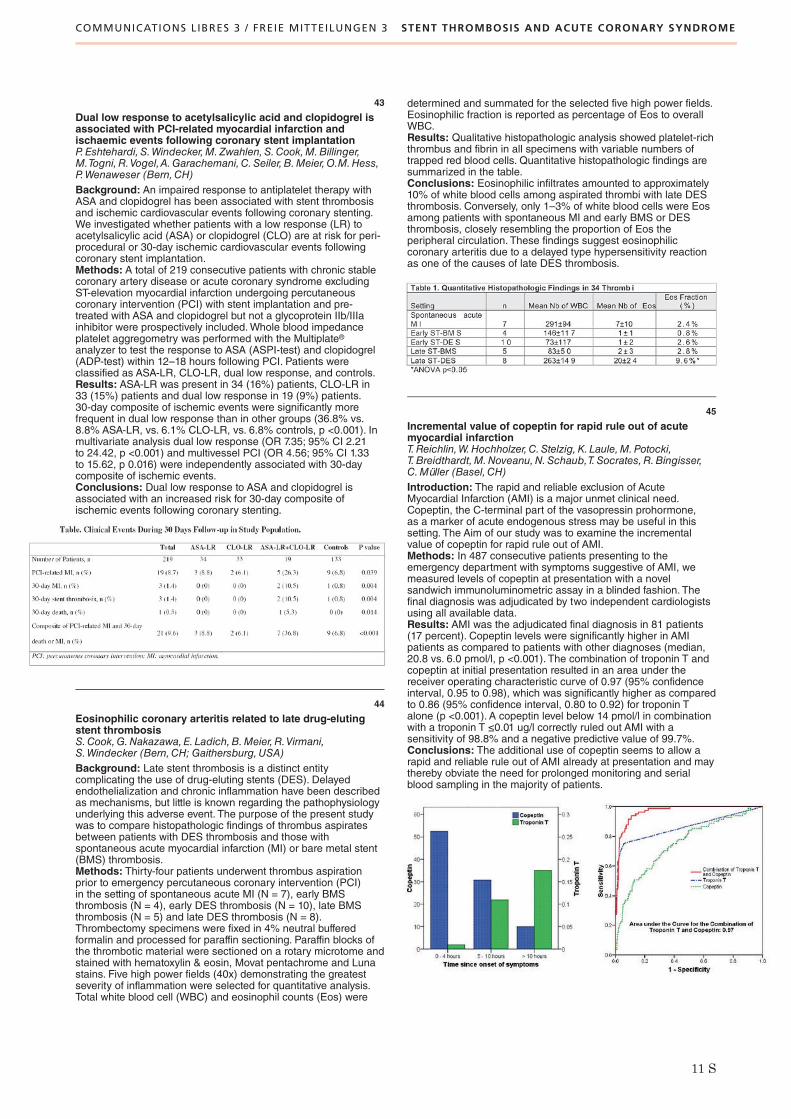
Zu Gunsten einer besserenLesbarkeit wird in unserenArtikeln nur die männlicheForm verwendet. Die weiblicheForm ist immer mitgemeint.
Impressum
HELVETICAC
AR
DIO
LOGICASOC
IETA
S
Schweizerische Gesellschaftfür Pädiatrische KardiologieSociété Suissede Cardiologie PédiatriqueSocietà Svizzeradi Cardiologia Pediatrica
Editores Medicorum Helveticorum
www.kardio.ch

Présentations orales / Vorträge
Cardiologie clinique / Klinische Kardiologie
Clinical cases 1 4 S
Communications libres 1 / Freie Mitteilungen 1
Risk factors 6 S
Communications libres 2 / Freie Mitteilungen 2
Atherogenesis and arteriogenesis 8 S
Communications libres 3 / Freie Mitteilungen 3
Stent thrombosis and acute coronary syndrome 11 S
Communications libres 4 / Freie Mitteilungen 4
Resynchronisation, ICD 13 S
Communications libres 5 / Freie Mitteilungen 5
Aortic stenosis, percutaneous valve replacement 15 S
Communications libres 6 / Freie Mitteilungen 6
Drug eluting stents 18 S
Communications libres 7 / Freie Mitteilungen 7
Congenital heart disease 20 S
Cardiologie clinique / Klinische Kardiologie
Clinical cases 2 22 S
Communications libres 8 / Freie Mitteilungen 8
New “Risk factors” 24 S
Communications libres 9 / Freie Mitteilungen 9
Progenitor cells, myocardial regeneration 26 S
Communications libres 10 / Freie Mitteilungen 10
Echo «advanced» 28 S
Communications libres 11 / Freie Mitteilungen 11
Congestive heart failure 30 S
SOMMAIRE / INHALT
2 SKardiovaskuläre Medizin 2009;12(5): Suppl 17

SOMMAIRE / INHALT
3 SKardiovaskuläre Medizin 2009;12(5): Suppl 17
Communications libres 12 / Freie Mitteilungen 12
Echo conventional 32 S
Communications libres 13 / Freie Mitteilungen 13
Cardiac surgery 35 S
Communications libres 14 / Freie Mitteilungen 14
Myocardial disease / ischaemia 37 S
Communications libres 15 / Freie Mitteilungen 15
Electrophysiology, ablation 39 S
Posters / Poster
Groupe de posters 1 / Postergruppe 1
Basic science 41 S
Groupe de posters 2 / Postergruppe 2
Electrophysiology, pacemaker, arrhytmias 48 S
Groupe de posters 3 / Postergruppe 3
Congestive heart failure, Htx, CMPs, valvular disease, pericardium 54 S
Groupe de posters 4 / Postergruppe 4
CAD, ACS, PCI, CABG 59 S
Groupe de posters 5 / Postergruppe 5
Prevention and rehabilitation 63 S
Groupe de posters 6 / Postergruppe 6
Echo, MR, Nuc, Ergo, congenital heart disease, paediatrics 66 S
Groupe de posters 7 / Postergruppe 7
Clinical cardiology 72 S
Liste des premiers auteurs / Liste der Erstautoren
Clinical cardiology 79 S
CARDIOLOGIE CLINIQUE / KLINISCHE KARDIOLOGIE CLINICAL CASES 1
4 S
16
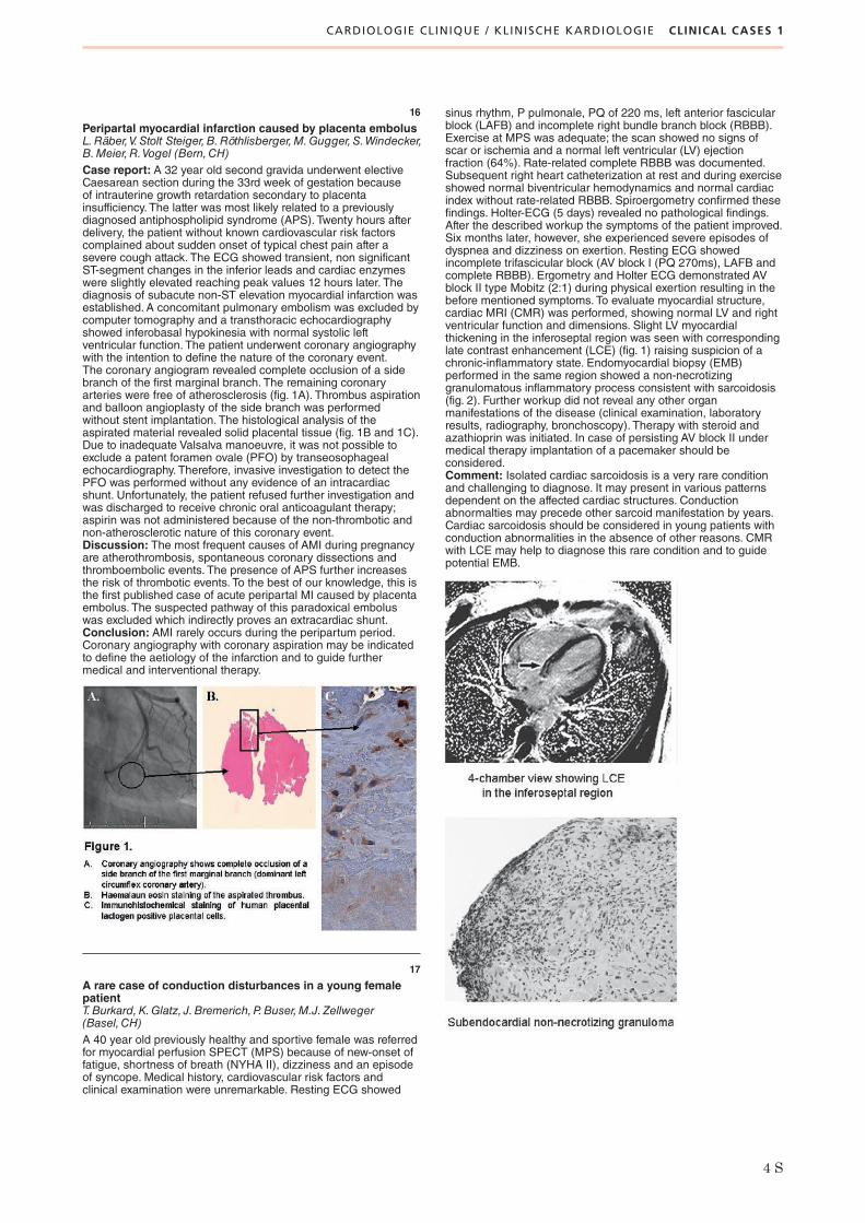
Peripartal myocardial infarction caused by placenta embolusL.Räber,V.Stolt Steiger,B.Röthlisberger,M.Gugger,S.Windecker,B.Meier,R.Vogel (Bern,CH)Case report: A 32 year old second gravida underwent electiveCaesarean section during the 33rd week of gestation becauseof intrauterine growth retardation secondary to placentainsufficiency. The latter was most likely related to a previouslydiagnosed antiphospholipid syndrome (APS). Twenty hours afterdelivery, the patient without known cardiovascular risk factorscomplained about sudden onset of typical chest pain after asevere cough attack. The ECG showed transient, non significantST-segment changes in the inferior leads and cardiac enzymeswere slightly elevated reaching peak values 12 hours later. Thediagnosis of subacute non-ST elevation myocardial infarction wasestablished. A concomitant pulmonary embolism was excluded bycomputer tomography and a transthoracic echocardiographyshowed inferobasal hypokinesia with normal systolic leftventricular function. The patient underwent coronary angiographywith the intention to define the nature of the coronary event.The coronary angiogram revealed complete occlusion of a sidebranch of the first marginal branch. The remaining coronaryarteries were free of atherosclerosis (fig. 1A). Thrombus aspirationand balloon angioplasty of the side branch was performedwithout stent implantation. The histological analysis of theaspirated material revealed solid placental tissue (fig. 1B and 1C).Due to inadequate Valsalva manoeuvre, it was not possible toexclude a patent foramen ovale (PFO) by transeosophagealechocardiography. Therefore, invasive investigation to detect thePFO was performed without any evidence of an intracardiacshunt. Unfortunately, the patient refused further investigation andwas discharged to receive chronic oral anticoagulant therapy;aspirin was not administered because of the non-thrombotic andnon-atherosclerotic nature of this coronary event.Discussion: The most frequent causes of AMI during pregnancyare atherothrombosis, spontaneous coronary dissections andthromboembolic events. The presence of APS further increasesthe risk of thrombotic events. To the best of our knowledge, this isthe first published case of acute peripartal MI caused by placentaembolus. The suspected pathway of this paradoxical emboluswas excluded which indirectly proves an extracardiac shunt.Conclusion: AMI rarely occurs during the peripartum period.Coronary angiography with coronary aspiration may be indicatedto define the aetiology of the infarction and to guide furthermedical and interventional therapy.
17
A rare case of conduction disturbances in a young femalepatientT.Burkard,K.Glatz, J.Bremerich,P.Buser,M.J. Zellweger(Basel,CH)A 40 year old previously healthy and sportive female was referredfor myocardial perfusion SPECT (MPS) because of new-onset offatigue, shortness of breath (NYHA II), dizziness and an episodeof syncope. Medical history, cardiovascular risk factors andclinical examination were unremarkable. Resting ECG showed
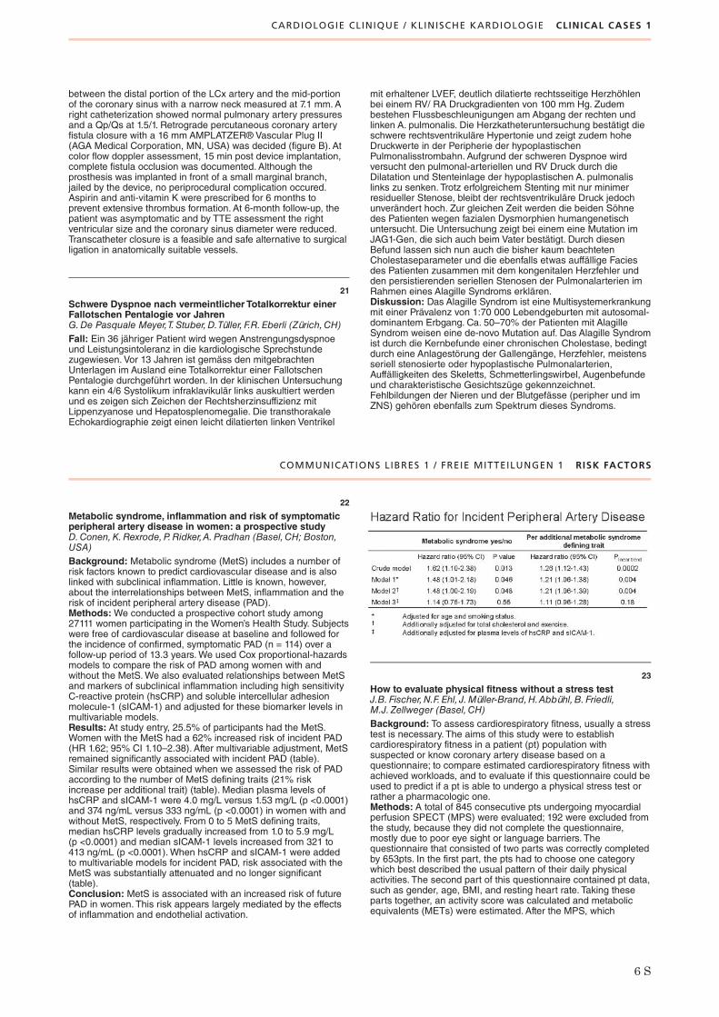
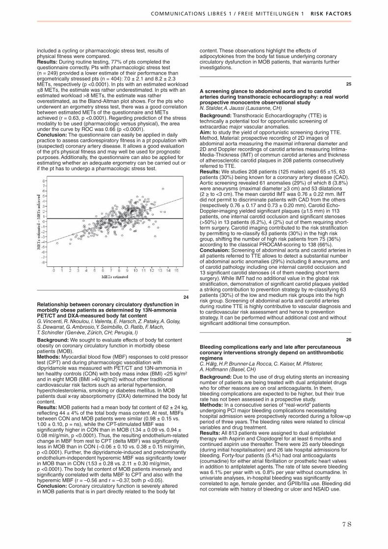
sinus rhythm, P pulmonale, PQ of 220 ms, left anterior fascicularblock (LAFB) and incomplete right bundle branch block (RBBB).Exercise at MPS was adequate; the scan showed no signs ofscar or ischemia and a normal left ventricular (LV) ejectionfraction (64%). Rate-related complete RBBB was documented.Subsequent right heart catheterization at rest and during exerciseshowed normal biventricular hemodynamics and normal cardiacindex without rate-related RBBB. Spiroergometry confirmed thesefindings. Holter-ECG (5 days) revealed no pathological findings.After the described workup the symptoms of the patient improved.Six months later, however, she experienced severe episodes ofdyspnea and dizziness on exertion. Resting ECG showedincomplete trifascicular block (AV block I (PQ 270ms), LAFB andcomplete RBBB). Ergometry and Holter ECG demonstrated AVblock II type Mobitz (2:1) during physical exertion resulting in thebefore mentioned symptoms. To evaluate myocardial structure,cardiac MRI (CMR) was performed, showing normal LV and rightventricular function and dimensions. Slight LV myocardialthickening in the inferoseptal region was seen with correspondinglate contrast enhancement (LCE) (fig. 1) raising suspicion of achronic-inflammatory state. Endomyocardial biopsy (EMB)performed in the same region showed a non-necrotizinggranulomatous inflammatory process consistent with sarcoidosis(fig. 2). Further workup did not reveal any other organmanifestations of the disease (clinical examination, laboratoryresults, radiography, bronchoscopy). Therapy with steroid andazathioprin was initiated. In case of persisting AV block II undermedical therapy implantation of a pacemaker should beconsidered.Comment: Isolated cardiac sarcoidosis is a very rare conditionand challenging to diagnose. It may present in various patternsdependent on the affected cardiac structures. Conductionabnormalties may precede other sarcoid manifestation by years.Cardiac sarcoidosis should be considered in young patients withconduction abnormalities in the absence of other reasons. CMRwith LCE may help to diagnose this rare condition and to guidepotential EMB.

CARDIOLOGIE CLINIQUE / KLINISCHE KARDIOLOGIE CLINICAL CASES 1
5 S
18
High-dose clopidogrel treatment with 900 mg dailyin a patient with subacute stent thrombosis due to non-responsiveness to dual antiplatelet therapyA.Rohner,M.Pfisterer,M.Handke (Basel,CH)Background: Despite dual antiplatelet therapy, stent thrombosisremains a dramatic complication of angioplasty. In fact, not allindividuals respond equally to aspirin and clopidogrel. Non-responsiveness to dual antiplatelet therapy is an independentpredictor of stent thrombosis and adverse cardiovascular events.Case: A 64-year-old patient was admitted to our hospital for acuteanterior STEMI. He had no history of coronary artery disease.Following pre-treatment according to current guidelines(clopidogrel 600mg and aspirin 250 mg), immediate coronaryangiography and percutaneous coronary intervention (PCI) withstenting (DES) of the occluded LAD was performed. In addition todual antiplatelet therapy (clopidogrel 75 mg and aspirin 100 mgmaintenance dose), enoxaparin, atorvastatin, metoprolol andcandesartan were administered. 10 days after angioplasty thepatient suffered re-infarction due to subacute stent thrombosis inthe target vessel, which was successfully treated with emergencyPCI (balloon angioplasty).Whole-blood impedance aggregometryconfirmed platelet resistance to both aspirin and clopidogrel.Consequently the daily doses were each increased to 300 mg.A new analysis revealed normal response to aspirin but apersistently inadequate effect of clopidogrel, thus clopidogrel wassubstituted with ticlopidin. Since this had no effect on plateletinhibition, clopidogrel was reinstated and its dose increased to900 mg daily. At this time as well as at follow-up 8 weeks later, weconfirmed a persistent adequate dual inhibition of plateletactivation. The patient was readmitted 3 months later for atypicalchest pain, control angiography showed a normal patency of thecoronary stent. There were no clinical or laboratory indications oftherapy related side effects during the 3 months follow-up.Conclusion: In this patient with non-responsiveness to dualantiplatelet therapy and subacute stent thrombosis, incrementallyhigher maintenance doses of aspirin and clopidogrel (300 mg and900 mg, respectively) were well tolerated with no therapy relatedside effects. Even though an increase in bleeding complicationsassociated with this high dose of clopidogrel was a potentialconcern, there were no such adverse events. Thus, this mightbe a feasible approach to patients with inadequate therapeuticresponse to standard doses of clopidogrel, where other modesof ADP-receptor-dependent platelet inhibition (i.e. ticlopidin,prasugrel not available) do not yield the desired effect.
19
Acute cardiac failure during late pregnancy: pregnancy-associated cardiomyopathy or tocolysis-associated acutecardiac failure?M.Di Valentino,M.Moccetti,M.Previsdomini,A.Perren,A.Menafoglio,C.Marone,A.Gallino (Bellinzona,CH)Introduction: Post-partum cardio-myopathy is a well knowncomplication after child delivery.Whereas, the medical communityis often unaware of the concept of pregnancy-associatedcardiomyopathy (PACM) which englobes heart failure (HF) thataffects also women late in pregnancy. Acute HF under toclysis isalso a rare pregnancy-associated complication.Case report: A 35-year old woman at 22 weeks of pregnancy,with a history of two spontaneous abortions, was hospitalizedbecause imminent danger of abortion. She underwent tocolysisduring 24 hours (hexoprenalin) which ended up unsuccessfullywith an induced abortion. Few hours after begin of tocolysis, shepresented an acute HF which made necessary a rapid oro-tracheal intubation, and cathecolamine support. On physicalexamination the patient was tachycardic (115 beats/min), BP was105/75 mm Hg (under amines), and there were bilateralpulmonary rales without heart murmur at cardiac auscultation.EKG revealed sinus rhytm with negative T-wawes in V4-V6 leadswhich normalized wihtin 24 hours. Chest X-ray showed interstitialoedema and pulmonary venous congestion with evidence ofKerley B lines. Troponin I was 3.37 μg/l (<0.10 μg/l), CK 244 UI/L(0–160 UI/L), BNP 1110 ng/L (<100 ng/L). Transthoracicechocardiography (TTE) revealed a LVEF of 40% due to diffusemyocardial hypokinesia. There was rapid hemodynamic andclinical recover within 36 hours with extubation and tapering ofthe amines. A therapy with enalapril and metropolol was initiated.
Repeat TTE two days after the admission, showed almostcomplete recovery of the LVEF. Cardiac-MRI performed three dayafter the admission revealed a normal systolic function withoutlate enhancement. At six weeks follow-up she presented nocardiac symptoms and TTE showed a complete normal findingwith normal systolic and diastolic ventricular function.Wepostulate the diagnosis of PACM (dd: acute cardiacdecompensation following high dosage of tocolysis). In spite ofbeing informed about the estimated risk of relapse of 25% in caseof PACM, the patient became pregnant 6 months after the cardiacevent and she delivered, after a normal pregnancy, a normal childwithout cardiac problemConclusion: This case illustrates a case of acute cardiac failureduring late pregnancy which has been initially referred to PACM.However the rapid recovery and the lack of relapse after a furthernormal pregnancy and delivery are more in favour of tocolysis-associated acute cardiac failure
20
Transcatheter coronary artery fistula closureS.Noble,A.Basmadjian,R. Ibrahim (Genève,CH; Montreal,CAN)A 46-year-old man was diagnosed with a coronary fistula morethan 20 years ago. He had no cardiac risk factors. His onlysymptoms were atypical chest pains for many years. At physicalexam, a soft continuous murmur with brisk carotid upstroke couldbe appreciated. The electrocardiogram revealed a sinus rhythmwith criteria for left atrial enlargement. A transthoracicechocardiography (TTE) showed a severely enlarged coronarysinus (figure A), a dilated left main (7.17 mm) and left circumflex(LCx) arteries. The left ventricle (LV) ejection fraction was normal,but the left atrium (4.3 cm), LV (5.9 cm) and right cavities weredilated. A CT scan with reconstructions confirmed the large fistula
CARDIOLOGIE CLINIQUE / KLINISCHE KARDIOLOGIE CLINICAL CASES 1
COMMUNICATIONS LIBRES 1 / FREIE MITTEILUNGEN 1 RISK FACTORS
6 S
between the distal portion of the LCx artery and the mid-portionof the coronary sinus with a narrow neck measured at 7.1 mm.Aright catheterization showed normal pulmonary artery pressuresand a Qp/Qs at 1.5/1. Retrograde percutaneous coronary arteryfistula closure with a 16 mm AMPLATZER® Vascular Plug II(AGA Medical Corporation, MN, USA) was decided (figure B). Atcolor flow doppler assessment, 15 min post device implantation,complete fistula occlusion was documented. Although theprosthesis was implanted in front of a small marginal branch,jailed by the device, no periprocedural complication occured.Aspirin and anti-vitamin K were prescribed for 6 months toprevent extensive thrombus formation. At 6-month follow-up, thepatient was asymptomatic and by TTE assessment the rightventricular size and the coronary sinus diameter were reduced.Transcatheter closure is a feasible and safe alternative to surgicalligation in anatomically suitable vessels.
21
Schwere Dyspnoe nach vermeintlicher Totalkorrektur einerFallotschen Pentalogie vor JahrenG.De Pasquale Meyer,T.Stuber,D.Tüller, F.R.Eberli (Zürich,CH)Fall: Ein 36 jähriger Patient wird wegen Anstrengungsdyspnoeund Leistungsintoleranz in die kardiologische Sprechstundezugewiesen. Vor 13 Jahren ist gemäss den mitgebrachtenUnterlagen im Ausland eine Totalkorrektur einer FallotschenPentalogie durchgeführt worden. In der klinischen Untersuchungkann ein 4/6 Systolikum infraklavikulär links auskultiert werdenund es zeigen sich Zeichen der Rechtsherzinsuffizienz mitLippenzyanose und Hepatosplenomegalie. Die transthorakaleEchokardiographie zeigt einen leicht dilatierten linken Ventrikel
mit erhaltener LVEF, deutlich dilatierte rechtsseitige Herzhöhlenbei einem RV/ RA Druckgradienten von 100 mm Hg. Zudembestehen Flussbeschleunigungen am Abgang der rechten undlinken A. pulmonalis. Die Herzkatheteruntersuchung bestätigt dieschwere rechtsventrikuläre Hypertonie und zeigt zudem hoheDruckwerte in der Peripherie der hypoplastischenPulmonalisstrombahn. Aufgrund der schweren Dyspnoe wirdversucht den pulmonal-arteriellen und RV Druck durch dieDilatation und Stenteinlage der hypoplastischen A. pulmonalislinks zu senken. Trotz erfolgreichem Stenting mit nur minimerresidueller Stenose, bleibt der rechtsventrikuläre Druck jedochunverändert hoch. Zur gleichen Zeit werden die beiden Söhnedes Patienten wegen fazialen Dysmorphien humangenetischuntersucht. Die Untersuchung zeigt bei einem eine Mutation imJAG1-Gen, die sich auch beim Vater bestätigt. Durch diesenBefund lassen sich nun auch die bisher kaum beachtetenCholestaseparameter und die ebenfalls etwas auffällige Faciesdes Patienten zusammen mit dem kongenitalen Herzfehler undden persistierenden seriellen Stenosen der Pulmonalarterien imRahmen eines Alagille Syndroms erklären.Diskussion: Das Alagille Syndrom ist eine Multisystemerkrankungmit einer Prävalenz von 1:70 000 Lebendgeburten mit autosomal-dominantem Erbgang. Ca. 50–70% der Patienten mit AlagilleSyndrom weisen eine de-novo Mutation auf. Das Alagille Syndromist durch die Kernbefunde einer chronischen Cholestase, bedingtdurch eine Anlagestörung der Gallengänge, Herzfehler, meistensseriell stenosierte oder hypoplastische Pulmonalarterien,Auffälligkeiten des Skeletts, Schmetterlingswirbel, Augenbefundeund charakteristische Gesichtszüge gekennzeichnet.Fehlbildungen der Nieren und der Blutgefässe (peripher und imZNS) gehören ebenfalls zum Spektrum dieses Syndroms.
22
Metabolic syndrome, inflammation and risk of symptomaticperipheral artery disease in women: a prospective studyD.Conen,K.Rexrode,P.Ridker,A.Pradhan (Basel,CH; Boston,USA)Background: Metabolic syndrome (MetS) includes a number ofrisk factors known to predict cardiovascular disease and is alsolinked with subclinical inflammation. Little is known, however,about the interrelationships between MetS, inflammation and therisk of incident peripheral artery disease (PAD).Methods:We conducted a prospective cohort study among27111 women participating in the Women’s Health Study. Subjectswere free of cardiovascular disease at baseline and followed forthe incidence of confirmed, symptomatic PAD (n = 114) over afollow-up period of 13.3 years.We used Cox proportional-hazardsmodels to compare the risk of PAD among women with andwithout the MetS.We also evaluated relationships between MetSand markers of subclinical inflammation including high sensitivityC-reactive protein (hsCRP) and soluble intercellular adhesionmolecule-1 (sICAM-1) and adjusted for these biomarker levels inmultivariable models.Results: At study entry, 25.5% of participants had the MetS.Women with the MetS had a 62% increased risk of incident PAD(HR 1.62; 95% CI 1.10–2.38). After multivariable adjustment, MetSremained significantly associated with incident PAD (table).Similar results were obtained when we assessed the risk of PADaccording to the number of MetS defining traits (21% riskincrease per additional trait) (table). Median plasma levels ofhsCRP and sICAM-1 were 4.0 mg/L versus 1.53 mg/L (p <0.0001)and 374 ng/mL versus 333 ng/mL (p <0.0001) in women with andwithout MetS, respectively. From 0 to 5 MetS defining traits,median hsCRP levels gradually increased from 1.0 to 5.9 mg/L(p <0.0001) and median sICAM-1 levels increased from 321 to413 ng/mL (p <0.0001).When hsCRP and sICAM-1 were addedto multivariable models for incident PAD, risk associated with theMetS was substantially attenuated and no longer significant(table).Conclusion: MetS is associated with an increased risk of futurePAD in women. This risk appears largely mediated by the effectsof inflammation and endothelial activation.
23
How to evaluate physical fitness without a stress testJ.B.Fischer,N.F. Ehl, J.Müller-Brand,H.Abbühl,B. Friedli,M.J. Zellweger (Basel,CH)Background: To assess cardiorespiratory fitness, usually a stresstest is necessary. The aims of this study were to establishcardiorespiratory fitness in a patient (pt) population withsuspected or know coronary artery disease based on aquestionnaire; to compare estimated cardiorespiratory fitness withachieved workloads, and to evaluate if this questionnaire could beused to predict if a pt is able to undergo a physical stress test orrather a pharmacologic one.Methods: A total of 845 consecutive pts undergoing myocardialperfusion SPECT (MPS) were evaluated; 192 were excluded fromthe study, because they did not complete the questionnaire,mostly due to poor eye sight or language barriers. Thequestionnaire that consisted of two parts was correctly completedby 653pts. In the first part, the pts had to choose one categorywhich best described the usual pattern of their daily physicalactivities. The second part of this questionnaire contained pt data,such as gender, age, BMI, and resting heart rate. Taking theseparts together, an activity score was calculated and metabolicequivalents (METs) were estimated. After the MPS, which
COMMUNICATIONS LIBRES 1 / FREIE MITTEILUNGEN 1 RISK FACTORS
7 S
included a cycling or pharmacologic stress test, results ofphysical fitness were compared.Results: During routine testing, 77% of pts completed thequestionnaire correctly. Pts with pharmacologic stress test(n = 249) provided a lower estimate of their performance thanergometrically stressed pts (n = 404): 7.0 ± 2.1 and 8.2 ± 2.3METs, respectively (p <0.0001). In pts with an estimated workload≤8 METs, the estimate was rather underestimated. In pts with anestimated workload >8 METs, the estimate was ratheroverestimated, as the Bland-Altman plot shows. For the pts whounderwent an ergometry stress test, there was a good correlationbetween estimated METs of the questionnaire and METsachieved (r = 0.63, p <0.0001). Regarding prediction of the stressmodality to be used (pharmacologic versus physical), the areaunder the curve by ROC was 0.66 (p <0.0001).Conclusion: The questionnaire can easily be applied in dailypractice to assess cardiorespiratory fitness in a pt population with(suspected) coronary artery disease. It allows a good evaluationof the pt’s physical fitness and may well be used for prognosticpurposes. Additionally, the questionnaire can also be applied forestimating whether an adequate ergometry can be carried out orif the pt has to undergo a pharmacologic stress test.
24
Relationship between coronary circulatory dysfunction inmorbidly obese patients as determined by 13N-ammoniaPET/CT and DXA-measured body fat contentG.Vincenti, R.Nkoulou, I.Valenta, E.Harsch, Z.Pataky,A.Golay,S.Dewarrat,G.Ambrosio,Y.Seimbille,O.Ratib, F.Mach,T.Schindler (Genève,Zürich,CH; Perugia, I)Background:We sought to evaluate effects of body fat contentobesity on coronary circulatory function in morbidly obesepatients (MOB).Methods:Myocardial blood flow (MBF) responses to cold pressortest (CPT) and during pharmacologic vasodilation withdipyridamole was measured with PET/CT and 13N-ammonia inten healthy controls (CON) with body mass index (BMI) <25 kg/m2
and in eight MOB (BMI >40 kg/m2) without other traditionalcardiovascular risk factors such as arterial hypertension,hypercholesterolemia, smoking or diabetes mellitus. In MOBpatients dual x-ray absorptiometry (DXA) determined the body fatcontent.Results:MOB patients had a mean body fat content of 62 ± 24 kg,reflecting 44 ± 4% of the total body mass content. At rest, MBFsbetween CON and MOB patients were similar (0.98 ± 0.15 vs.1.00 ± 0.10, p = ns), while the CPT-stimulated MBF wassignificantly higher in CON than in MOB (1.34 ± 0.09 vs. 0.94 ±0.08 ml/g/min, p <0.0001). Thus, the resulting endothelium-relatedchange in MBF from rest to CPT (delta MBF) was significantlyless in MOB than in CON (–0.06 ± 0.10 vs. 0.38 ± 0.15 ml/g/min,p <0.0001). Further, the dipyridamole-induced and predominantlyendothelium-independent hyperemic MBF was significantly lowerin MOB than in CON (1.53 ± 0.28 vs. 2.11 ± 0.30 ml/g/min,p <0.0001). The body fat content of MOB patients inversely andsignificantly correlated with delta MBF to CPT and also with thehyperemic MBF (r = –0.56 and r = –0.37, both p <0.05).Conclusion: Coronary circulatory function is severely alteredin MOB patients that is in part directly related to the body fat
content. These observations highlight the effects ofadipocytokines from the body fat tissue underlying coronarycirculatory dysfunction in MOB patients, that warrants furtherinvestigations.
25
A screening glance to abdominal aorta and to carotidarteries during transthoracic echocardiography: a real worldprospective monocentre observational studyN.Stalder,A. Jaussi (Lausanne,CH)Background: Transthoracic Echocardiography (TTE) istechnically a potential tool for opportunistic screening ofextracardiac major vascular anomalies.Aim: to study the yield of opportunistic screening during TTE.Method, Material: prospective recording of 2D images ofabdominal aorta measuring the maximal infrarenal diameter and2D and Doppler recordings of carotid arteries measuring Intima-Media-Thickness (IMT) of commun carotid arteries and thicknessof atherosclerotic carotid plaques in 208 patients consecutivelyreferred to TTE.Results:We studies 208 patients (125 males) aged 65 ±15, 63patients (30%) being known for a coronary artery disease (CAD).Aortic screening revealed 61 anomalies (29%) of which 8 (3.8%)were aneurysms (maximal diameter ≥3 cm) and 53 dilatations(2 ≥ to <3 cm). The mean carotid IMT was 0.76 ± 0.22 mm. IMTdid not permit to discriminate patients with CAD from the others(respectively 0.76 ± 0.17 and 0.73 ± 0.20 mm). Carotid Echo-Doppler-imaging yielded significant plaques (≥1.5 mm) in 113patients, one internal carotid occlusion and significant stenoses(>50%) in 13 patients (6.2%), 4 (2%) out of them requiring short-term surgery. Carotid imaging contributed to the risk stratificationby permitting to re-classify 63 patients (30%) in the high riskgroup, shifting the number of high risk patients from 75 (36%)according to the classical PROCAM-scoring to 138 (66%).Conclusion: Screening of abdominal aorta and carotid arteries inall patients referred to TTE allows to detect a substantial numberof abdominal aortic anomalies (29%) including 8 aneurysms, andof carotid pathology including one internal carotid occlusion and13 significant carotid stenoses (4 of them needing short termsurgery).While IMT had no additional value in the global riskstratification, demonstration of significant carotid plaques yieldeda striking contribution to prevention strategy by re-classifying 63patients (30%) of the low and medium risk groups into the highrisk group. Screening of abdominal aorta and carotid arteriesduring routine TTE is highly contributive to vascular diagnosis andto cardiovascular risk assessment and hence to preventionstrategy. It can be performed without additional cost and withoutsignificant additional time consumption.
26
Bleeding complications early and late after percutaneouscoronary interventions strongly depend on antithromboticregimensC.Hälg,H.P.Brunner-La Rocca,C.Kaiser,M.Pfisterer,A.Hoffmann (Basel,CH)Background: Due to the use of drug eluting stents an increasingnumber of patients are being treated with dual antiplatelet drugswho for other reasons are on oral anticoagulants. In them,bleeding complications are expected to be higher, but their truerate has not been assessed in a prospective study.Methods: In a consecutive series of “real-world” patientsundergoing PCI major bleeding complications necessitatinghospital admission were prospectively recorded during a follow-upperiod of three years. The bleeding rates were related to clinicalvariables and drug treatment.Results: All 813 patients were assigned to dual antiplatelettherapy with Aspirin and Clopidogrel for at least 6 months andcontinued aspirin use thereafter. There were 25 early bleedings(during initial hospitalisation) and 26 late hospital admissions forbleeding. Forty-four patients (5.4%) had oral anticoagulants(coumadine) for either atrial fibrillation or prosthetic heart valvesin addition to antiplatelet agents. The rate of late severe bleedingwas 6.1% per year with vs. 0.8% per year without coumadine. Inunivariate analyses, in-hospital bleeding was significantlycorrelated to age, female gender, and GPIIb/IIIa use. Bleeding didnot correlate with history of bleeding or ulcer and NSAID use.
COMMUNICATIONS LIBRES 1 / FREIE MITTEILUNGEN 1 RISK FACTORS
COMMUNICATIONS LIBRES 2 / FREIE MITTEILUNGEN 2 ATHEROGENESIS AND ARTERIOGENESIS
8 S
Late bleeding was correlated to age, diabetes, renal failure,malignancy, and coumadine use. Multivariate analysis confirmedthe following independent predictors: GPIIb/IIIa, age, femalegender for early; use of coumadine, diabetes and malignancy forlate bleeding.Conclusions: In patients on single or dual antiplatelet therapyafter PCI, the rate of late bleeding was almost eight times higherwhen coumadine was used for additional reasons. In addition,patients with diabetes or malignancies were at risk for bleedingduring long-term follow-up.
27
Risk of incident atrial fibrillation in users of antihypertensivedrugsB.Schaer,C.Schneider,S. Jick,D.Conen,S.Osswald,C.Meier(Basel,CH; Boston,USA)Background: The risk of developing atrial fibrillation (AF) may bealtered by different antihypertensive drug classes. Some studiessuggest that drugs interfering with the renin-angiotensin systemmay be favorable due to their effect on atrial remodeling.Weperformed a large observational study among hypertensivepatients to assess and compare the relative risk of developingincident AF in users of different antihypertensive drug classes.Methods:We used the UK-based General-Practice-Research-Database (GPRD) to conduct a nested case-control analysiswithin hypertensive patients who received prescriptions forantihypertensive drugs at some point in time. Patients with clinicalrisk factors for AF (e.g. heart failure, valvular heart disease,
alcohol consumption¡) were excluded.We assessed andcompared the risk of developing paroxysmal or persistent AFbetween users of angiotensin-converting-enzyme (ACE)inhibitors, angiotensin-II-receptor blockers (ARBs), β-blockers,or calcium-channel-blockers to the reference group of non-usersof antihypertensive therapies. Long-term use was defined as≥12 prescriptions (~ ≥1 year) for these drugs.Results:Within a base population of some 5 million patients inthe GPRD, we identified the study population of 615,312pharmacologically treated hypertensive patients, and amongthose, we identified 4,640 patients with an incident AF diagnosis,and 17,252 matched controls. More than half of AF cases (53%)were 70 years or older at the time of the first AF diagnosis, and50% were men. Obese patients (BMI ≥30 kg/m2) were at anincreased relative risk of developing AF as compared to normalweight patients (OR 1.38, 95% CI 1.25–1.51). As compared tonon-use periods of antihypertensive drugs, current exclusive long-term use of ACE-inhibitors (OR 0.69, 95% CI 0.60-0.80), ARBs(OR 0.74, 95% CI 0.57–0.96), or β-blockers (OR 0.83, 95% CI0.72–0.95) was associated with a lower relative risk of developingAF, while no effect was seen for calcium-channel blockers.In a second model allowing concurrent use of variousantihypertensive drugs, adjusted for each other, only long-termuse of ACE-inhibitors was associated with a reduced risk estimateof developing AF (OR 0.90, 95% CI 0.81–0.99).Conclusion: In hypertensive patients, long-term use of ACE-inhibitors, ARBs and β-blockers may reduce the risk of developingAF.
35
Metallothionein enhances angiogenesis and arteriogenesisby modulating smooth muscle cell and macrophage functionS.Zbinden,H.Morsli,M.Schmidt, S.E. Epstein,M.Burnett(Bern,CH; Washington DC,USA)Introduction: Metallothionein (MT) is a potentimmunomodulatory molecule known to play a protective role incardiac and cerebral ischemia. Previously, we have shown thatMT is highly upregulated following the induction of acute hindlimbischemia in a mouse model (Lee CW; J Am Coll Cardiol.2004;43(3):474–82). The objectives of this study were todetermine if MT is important in collateral development, and toinvestigate the mechanisms by which MT contributes to flowrecovery following the induction of acute hindlimb ischemia.Methods: Laser Doppler perfusion imaging and Matrigel plugassays were used to assess both collateral flow recovery andangiogenesis in MT knockout (MTKO) mice, compared to wildtypeanimals. Smooth muscle cells (SMCs) were isolated from MT andMT knockout mice, and proliferation, migration and invasionassays were performed. Gene expression of MMP9 and VEGF inSMCs were measured by real time PCR. CD11b+ cells wereisolated from MT knockout and wildtype animals and tested forinvasiveness using an ECIS assay.Results:We found that blood flow recovery (arteriogenesis)measured by Laser Doppler was reduced in MTKO mice(p = 0.017; timepoints from day 0 up to day 21). Furthermore,angiogenesis was impaired in MT knockout mice with significantlyfewer vessels in the Matrigel plugs from the MTKO animalscompared to the plugs from the wildtype mice (6.19 ± 0.916 vs0.333 ± 0.161, p = 0.004). MTKO SMCs showed impairedproliferation using an MTT assay (p <0.05). Migratory capacity ofaortic SMCs from MTKO mice was significantly impairedcompared to wildtype SMCs (O.D. units, 2.38 ± 0.02 vs. 2.76 ±0.06, p = 0.004). A similar pattern was observed in the invasionassay, with reduced invasiveness in the MTKO vs. wildtype cells(O.D. units, 0.700 ± 0.02 vs. 0.926 ± 0.05, p = 0.008)). MTKOSMCs had significantly lower expression levels of matrixmetalloproteinase-9 (MMP9), (2% of wildtype, p = 0.006).Likewise, MMP9 protein levels were decreased in MTKO cells, asdemonstrated by ELISA. VEGF mRNA levels were significantlylower in MTKO SMCs, (43% of wildtype, p = 0.0006), as were
VEGF protein levels (54% of wildtype, p = 0.005). CD11b+ cellsfrom MT knockout mice were more invasive than wildtype cells(p <0.05).Conclusion: Both collateral flow recovery and angiogenesis areimpaired in MT knockout mice. Mechanisms contributing to thesedeficiencies include endothelial, SMC, and macrophagedysfunction in the MT knockout animals.
36
Accelerated early atherosclerosis in mice deficientin L-selectinI. Rozenberg, E. Eriksson,P.Mocharla,A.Hallenberg,P.Rotzius,J.Boren,T.F. Lüscher, F.C.Tanner (Zürich,CH; Stockholm,Göteborg,S)Background: Atherosclerosis is an inflammatory diseasecharacterized by accumulation of leukocytes in the arterial intima.The selectin family of adhesion molecules (L-, E-, and P-selectin)mediates rolling and tethering of leukocytes along theendothelium, which is a first step in leukocyte extravasation.Although the selectin family of adhesion molecules is a wellknown mediator of inflammatory responses, the role of L-selectin(L-sel) in the pathogenesis of atherosclerosis has not beenexamined in vivo.Methods: To address this issue, we crossed L-sel-/- animals withmice lacking apolipoprotein E (ApoE) – an established animalmodel of atherosclerosis.We analyzed ApoE-/-L-sel-/- animalsand the corresponding ApoE-/- controls after 6 and 12 weeks ofhigh cholesterol diet (HCD).Results: After 6 weeks of HCD, ApoE-/-L-sel-/- animalsdeveloped on average 2.46% ± 0.54% aortic lesions, which was2-fold higher as compared to ApoE-/- controls (1.28% ± 0.24%)(P <0.05). In a contrast, after 12 weeks of HCD, there was nodifference in lesion formation between the two groups of animals(p = ns). Interestingly, ApoE-/-L-sel-/- animals exhibited a 20%increase in total plasma cholesterol level after 6, but not after12 weeks of HCD (p <0.05). Plaque analysis did not reveal anydifferences in cellular composition. Moreover, no difference inleukocyte rolling in atherosclerotic animals lacking L-selectin andcontrols was observed using intravital microscopy. However, mice

COMMUNICATIONS LIBRES 2 / FREIE MITTEILUNGEN 2 ATHEROGENESIS AND ARTERIOGENESIS
9 S
lacking L-sel exhibited reduced size and cellularity of peripherallymph nodes as well as increased size of spleen after both 6 and12 weeks of HCD. FACS analysis revealed a significantly elevatedpercentage of inactive T-helper lymphocytes (CD44- CD4+) inblood of L-sel-/- mice, whereas the percentage of memory T-helper cells (CD44+ CD4-) remained the same after 6 weeks ofHCD and diminished after 12 weeks of HCD.Conclusions: These results show that L-selectin is a negativeregulator of early but not advanced atherosclerosis, possibly viaregulation of cholesterol metabolism. The redistribution oflymphocytes occurring in mice lacking L-sel is consistent with itsmajor role in homing of these cells to lymph nodes and does notaffect atherosclerosis development.
37
Laminin receptor activation inhibits endothelial tissue factorexpressionE.W.Holy,S.F.Stämpfli,A.Akhmedov,N.Holm,G.G.Camici,T.F. Lüscher, F.C.Tanner (Zürich,CH)Background: Tissue factor (TF) is an important trigger of arterialthrombosis. The extracellular matrix glycoprotein laminin is amajor compound of the basement membrane contributing tovessel wall integrity. The green tea catechin epigallocatechin-3-gallate (EGCG) is a ligand of the 67 kDa laminin receptor (67-LR).This study investigates the effect of 67-LR activation by lamininand EGCG on endothelial TF expression.Methods and results: Immunofluorescence demonstrated thathuman aortic endothelial cells expressed 67-LR abundantly.Western blot analysis demonstrated that cells grown on lamininexpressed less TF in response to TNF-α than those grown onfibronectin (n = 6; p <0.001). EGCG inhibited TNF-α andhistamine induced TF expression and activity in endothelial andvascular smooth muscle cells in a concentration-dependentmanner (1-30 uM) resulting in 87% reduction of TF expression(n = 5; p <0.001). In contrast, expression of tissue factor pathwayinhibitor was not affected (n = 4; p = NS). Real-time PCR andpromoter studies revealed that inhibition of TF expressionoccurred at the transcriptional level. EGCG (30 uM) impairedactivation of the mitogen-activated protein (MAP) kinase c-Junterminal NH2 kinase (JNK), but not ERK or p38. The JNK inhibitorSP600125 (1 uM) reduced TF promoter activity (n = 4, p <0.001)and protein expression (n = 4; p <0.001). 67-LR blockingantibodies blunted the inhibitory effect of EGCG on JNK activationand TF expression. Expression of Vascular cell adhesionmolecule 1 (VCAM-1) was not regulated by JNK, and itsexpression was not affected by 67-LR activation.Conclusions: Laminin receptor activation inhibits endothelialTF expression by impairing JNK phosphorylation. This studyidentifies a new mechanism by which the basement membranecontributes to hemostatic balance of the endothelium in intactvessels and suggests 67-LR as a potential target for inhibition ofarterial thrombosis.
38
Nebivolol protects against atherosclerosis reducingadhesion molecule expression and foam cells formationvia inhibition of protein kinase C-β intracellular signallingA.Kuroedov, E.Osto,P.Mocharla,T.F. Lüscher, F.Cosentino(Zürich,CH)Background: Nebivolol is a selective β1 blocker with a directvasorelaxant effect involving nitric oxide pathway.We have shownthat chronic treatment with nebivolol, but not atenolol,leads toatherosclerotic plaque stabilization in apoE-/- mice withhistological evidence of higher number of smooth muscle cells inplaque’s shoulder and a thicker fibrous cap in nebivolol-treatedmice. However, the molecular mechanisms of antiatheroscleroticproperties of nebivolol remain unclear.Materials and results: Human aortic endothelial cells (HAECs)were stimulated with IL-1β (10 ng/mL) after 2 hours ofpretreatment with nebivolol or atenolol (5x10⁻⁶ mol/L). IL-1b (10ng/mL) increased VCAM-1 expression in HAEC as assessed byWestern blotting (WB) (826 ± 52% vs control; n = 4; p <0.01).Pretretment with nebivolol resulted in a significant attenuation ofIL-1b-induced VCAM-1 expression (n = 4; p <0.01 vs IL-1 b alone).Increase of MCP-1 in culture medium after stimulation with IL-1bwas completely abolished by nebivolol. In contrast, atenolol did
not affect neither VCAM-1 nor MCP-1 expression.WB withantibodies against phosphorylated PKCβ2 isoform revealed thatincubation of the cells with IL-1b increased phosphorylation ofPKCb2 ser-660 residue. Nebivolol blunted IL-1b-induced ser-660phosphorylation. The differentiation of the monocytic THP-1 cellline into macrophages was induced by phorbol 12-myristate 13-acetate (PMA; 0.1 mmol/L), a potent activator of PKC or IL-1b(10 ng/mL). Pretreatment with nebivolol or atenolol was performed1 hour before PMA or IL-1b exposure. IL-1b elicited an upregulationof CD14 expression, a marker of monocyte transdifferentiation(285 ± 42% vs control; n = 3; p <0.05). Nebivolol (5x10⁻⁶ mol/L)abolished such IL-1b-induced CD14 upregulation. Incubation ofmacrophages with DiI-modified acLDL (acetylated LDL, 10ug/mL) led to lipoprotein uptake. Nebivolol significantly decreaseduptake of acLDL as assessed by flow cytometry (443 ± 57fluorescent units vs 717 ± 113 fluorescent units in control; n = 5;p <0.01). Atenolol did not affect acLDL uptake. As assessed withWB, incubation of monocyte-derived macrophages with PMA/LDLincreased PKCb1 phosphorylation at the Thr-642 residue, whichwas blunted by nebivolol. Atenolol did not exert any significanteffect.Conclusion: Our findings show that nebivolol exerts itsantiatherosclerotic effect via inhibition of PKCb-inducedintracellular signaling in endothelial cells and macrophages.
39
Histamine H1 receptor promotes atherosclerosisdevelopment by enhancing vascular permeabilityI. Rozenberg, L.Rohrer,B.Becher, J.Hofman, J.Soliz, P.Mocharla,A.Akhmedov,T.Watanabe, J.Boren (Zürich,CH;Yokohama, JP;Göteborg,S)Background and aims: Histamine is an endogenous amineplaying an important role in inflammation. Although the H1receptor (H1R) mediates most of the proinflammatory actions ofhistamine, its relevance for atherosclerosis development isunknown.Methods: H1R and H2R signalling was modulated bothpharmacologically and genetically in apolipoproteinE deficientmice (ApoE-/-), a murine model of atherosclerosis. These animalswere treated with the H1R antagonist mepyramine, the H2Rantagonist ranitidine, or placebo during 12 week high cholesteroldiet (HCD, Clinton-Cybulski diet, 1.25% cholesterol). In parallel,H1R-/-ApoE-/- and H2R-/-ApoE-/- were compared to ApoE-/-mice.Results: ApoE-/- mice treated with mepyramine, but notranitidine, exhibited 40% inhibition in atherosclerosis development(p <0.05). Similarly, genetic deletion of H1R in ApoE-/- miceresulted in a 60% decrease in aortic plaque area (p <0.0005) ascompared to controls. Plasma lipid levels were not affected by theabsence of H1R.Aortic permeability to low density lipoproteins(125 I-LDL) was significantly reduced in animals lacking the H1Rboth with and without HCD. Indeed, atheromas of ApoE-/-H1R-/-animals exhibited decreased lipid accumulation when comparedto ApoE-/- controls. Moreover, atherosclerotic plaques of ApoE-/-H1R-/- animals exhibited diminished infiltration with macrophages(p <0.005) and T-helper cells (p <0.05). Consistent with thisobservation, blood lymphocyte number, spleen size, and splenicproduction of Th1 cytokines were reduced upon H1R deletion.Bone marrow transplantation confirmed that plaque formationdepended on the presence of H1R on vascular cells, but not onbone marrow-derived cells.Conclusions: Histamine H1 receptor promotes atherosclerosisdevelopment due to its crucial role in regulating vascularpermeability.
40
High-density lipoprotein from healthy subjects, but not frompatients with coronary artery disease, exerts anti-thromboticeffects on human endothelial cellsE.W.Holy,C.Besler, K.Yonekawa,R.Corti,W.Maier,C.Wyss,J.Alibegovic,N.Kucher,T.F. Lüscher,U. Landmesser, F.C.Tanner(Zürich,CH)Background: Arterial thrombus formation is determined by thebalance between pro- thrombotic mediators such as tissue factor(TF) and plasminogen activator inhibitor type 1 (PAI-1), and anti-thrombotic factors like tissue factor pathway inhibitor (TFPI) or
COMMUNICATIONS LIBRES 2 / FREIE MITTEILUNGEN 2 ATHEROGENESIS AND ARTERIOGENESIS
10 S
tissue plasminogen activator (tPA). Native high-density lipoprotein(HDL) from healthy subjects (HS) has anti-thrombotic properties;however, it remains unknown whether this is the case for HDLfrom patients with stable coronary disease (CAD) or acutecoronary syndrome (ACS).Methods: HDL was isolated by sequential ultracentrifugationfrom HS and patients with CAD and ACD. The effects of HDL(50 ug/ml) on TF, TFPI, and PAI-1 expression in human endothelialcells were determined by Western blot analysis; tPA release wasmeasured by ELISA.Results: HDL from HS impaired thrombin-induced TF expression(–45 ± 5%, p <0.05, n = 16) and activity (–33 ± 8%, p <0.05,n = 7); in contrast, HDL from CAD and ACS patients did not(p = NS, n = 12 and n = 8). Similarly, HDL from HS increasedTFPI expression by 2-fold (p <0.01, n = 8), while HDL from CADand ACS patients had no effect (p = NS). HDL from HS enhancedtPA release (+26 ± 3%; p = 0.05, n = 8); in contrast, HDL fromCAD and ACS patients did not (p = NS, n = 6). Furthermore, HDLfrom HS did not affect PAI-1 expression, while HDL from CADpatients enhanced PAI-1 expression by 74 ± 11% (p <0.05 vs.control, n = 12) and HDL from ACS patients by 128 ± 18%(p <0.05 vs. control and p <0.05 vs. CAD, n = 8). Pretreatmentwith the inhibitor of NO formation, L-NAME (100 uM), abolishedthe anti-thrombotic effects of HDL from HS on TF, TFPI, and tPAexpression. The exogenous nitric oxide donor, DETANO,mimicked the effects of HDL from HS on TF, TFPI, and tPA.Conclusion: This study demonstrates that HDL from healthysubjects exerts anti-thrombotic effects on endothelial cells. Incontrast, HDL from CAD and ACS patients loses theseantithrombotic properties and instead enhances PAI-1 expression,thereby becoming pro-thrombotic. This observation might behighly relevant for HDL-targeted therapies.
41
High-density lipoprotein loses endothelial-protective effectsin patients with stable coronary disease and an acutecoronary syndrome: role of paraoxonase-1 inactivationC.Besler, K.Yonekawa,K.Heinrich, E.W.Holy,C.Doerries,P.Mocharla,Y.Shi,C.Manes,R.Corti,W.Maier,C.Wyss, J.Alibegovic,N.Kucher, F.C.Tanner,A. von Eckardstein,T.F. Lüscher,U. Landmesser (Zürich,CH)Background: High-density lipoprotein (HDL) from healthysubjects (HS) exerts direct endothelial-protective effects, such asstimulation of anti-atherogenic endothelial nitric oxide (NO)production and anti-inflammatory effects. HDL-raising therapiesare currently evaluated as a potential novel therapeutic strategy;however, whether HDL from patients with stable coronary disease(sCAD) or an acute coronary syndrome (ACS) exerts endothelial-protective effects remains unknown.Methods: HDL was isolated from patients with sCAD (n = 20),ACS (n = 20) and HS (n = 25) by sequential ultracentrifugation.HDL’s effects on endothelial cell NO and superoxide productionwere determined by electron spin resonance (ESR) spectroscopy.HDL’s effect on endothelial repair was examined by injection intonude mice with carotid injury and measurement ofre-endothelialized area after 3 days. HDL’s anti-inflammatoryproperties were examined by effects on endothelial VCAM-1expression and endothelial-monocyte adhesion. The activity andcontent of HDL-associated paraoxonase (PON)-1 was examined,and the impact of PON-1 inhibition on HDL’s endothelial effectswas characterized.Results: HDL from HS stimulated endothelial NO production,however, in marked contrast HDL from sCAD and ACS patientsinhibited NO production. Endothelial superoxide production was
markedly reduced by HDL from HS, but not by HDL from sCAD orACS patients. Importantly, HDL from HS accelerated endothelialrepair after carotid injury, that was not observed with HDL fromsCAD or ACS patients. HDL from HS, but not from sCAD andACS patients inhibited endothelial VCAM-1 expression andmonocyte adhesion. Notably, HDL’s anti-inflammatory effects weredependent on endothelial NO production, i.e. these effects wereinhibited by either L-NAME or eNOS siRNA-specific knockdown.PON-1 activity was substantially reduced, whereas PON-1 proteincontent was increased in HDL from CAD patients, suggesting aninactivation of PON-1 in HDL from CAD patients. Notably, PON-1inhibition reversed beneficial endothelial effects of HDL from HS.Conclusion: These findings suggest for the first time that HDLfrom patients with sCAD or ACS loses critical endothelial-protective effects, i.e. the capacity to stimulate endothelial NOproduction or promote endothelial repair. PON-1 inactivation likelyplays a critical role for the loss of endothelial-protective effects ofHDL in patients with CAD. These findings may have importantimplications for designing HDL-targeted therapies.
42
Complement component 3a receptor attenuatesatherosclerosis developmentI. Rozenberg,U.Wagner,T.F. Lüscher, F.C.Tanner (Zürich,CH)Background: The pathogenesis of early atherosclerosis is stillpoorly understood, especially at the molecular level, althoughatherosclerotic vascular disease is the leading cause of death inthe Western world. Up-to-date molecular biological methodspermit efficient identification and characterization of new genes.Methods and results: Using Affimetrix microarrays, we studiedgene expression in thoracic aorta of apolipoproteinE-/- (ApoE-/-)animals fed for 4 weeks with either normal chow or highcholesterol diet (1.25% cholesterol). Over 200 genes weredifferentially regulated; the majority of them was involved inimmune and inflammatory responses, cell adhesion, signaltransduction, and cholesterol transport. Interestingly, mostcomponents of the complement pathway were upregulatedupon high cholesterol diet, with the complement component 3areceptor (C3aR) exhibiting the highest degree of induction.Despite its well known role in inflammation, the relevance of thecomplement cascade for atherosclerosis development remainscontroversial. In order to address this issue, we used geneticallymodified mice lacking C3aR and crossed them withapolipoproteinE (ApoE-/-) deficient animals. Following 12 weeksof a high cholesterol diet (1.25% cholesterol), ApoE-/-C3aR-/-mice and the corresponding ApoE-/- controls were compared.Development of atherosclerosis in aortas was monitored usingoil red-O staining, accumulation of leukocytes in atheromasand expression of cytokines with immunohistochemistry andquantitative RT PCR respectively. ApoE-/- animals lacking C3aRhad on average 14.11% ± 3.16% of descending aortas occupiedby atherosclerotic plaques, which was nearly 2-fold more thanApoE-/- controls (7.11% ± 1.01%). This effect was associated withincreased accumulation of macrophages and lymphocytes inatherosclerotic plaques of ApoE-/-C3aR-/- mice. Moreover,production of the proinflammatory cytokines interferon γ (IFN-γ),chemokine (C-C) ligand-5 (CCL5), tumor necrosis factor β(TNF-β), interleukin-12 (IL-12), and interleukin-18 (IL-18) washigher in aortas of atherosclerotic animals lacking C3aR.Conclusions: This study demonstrates that C3aR protects fromatherosclerosis development, emphasizing the complex role ofthe complement cascade in the development of inflammationduring atherogenesis.
COMMUNICATIONS LIBRES 3 / FREIE MITTEILUNGEN 3 STENT THROMBOSIS AND ACUTE CORONARY SYNDROME
11 S
43
Dual low response to acetylsalicylic acid and clopidogrel isassociated with PCI-related myocardial infarction andischaemic events following coronary stent implantationP.Eshtehardi, S.Windecker,M.Zwahlen,S.Cook,M.Billinger,M.Togni,R.Vogel,A.Garachemani,C.Seiler,B.Meier,O.M.Hess,P.Wenaweser (Bern,CH)Background: An impaired response to antiplatelet therapy withASA and clopidogrel has been associated with stent thrombosisand ischemic cardiovascular events following coronary stenting.We investigated whether patients with a low response (LR) toacetylsalicylic acid (ASA) or clopidogrel (CLO) are at risk for peri-procedural or 30-day ischemic cardiovascular events followingcoronary stent implantation.Methods: A total of 219 consecutive patients with chronic stablecoronary artery disease or acute coronary syndrome excludingST-elevation myocardial infarction undergoing percutaneouscoronary intervention (PCI) with stent implantation and pre-treated with ASA and clopidogrel but not a glycoprotein IIb/IIIainhibitor were prospectively included.Whole blood impedanceplatelet aggregometry was performed with the Multiplate®
analyzer to test the response to ASA (ASPI-test) and clopidogrel(ADP-test) within 12–18 hours following PCI. Patients wereclassified as ASA-LR, CLO-LR, dual low response, and controls.Results: ASA-LR was present in 34 (16%) patients, CLO-LR in33 (15%) patients and dual low response in 19 (9%) patients.30-day composite of ischemic events were significantly morefrequent in dual low response than in other groups (36.8% vs.8.8% ASA-LR, vs. 6.1% CLO-LR, vs. 6.8% controls, p <0.001). Inmultivariate analysis dual low response (OR 7.35; 95% CI 2.21to 24.42, p <0.001) and multivessel PCI (OR 4.56; 95% CI 1.33to 15.62, p 0.016) were independently associated with 30-daycomposite of ischemic events.Conclusions: Dual low response to ASA and clopidogrel isassociated with an increased risk for 30-day composite ofischemic events following coronary stenting.
44
Eosinophilic coronary arteritis related to late drug-elutingstent thrombosisS.Cook,G.Nakazawa,E. Ladich,B.Meier,R.Virmani,S.Windecker (Bern,CH; Gaithersburg,USA)Background: Late stent thrombosis is a distinct entitycomplicating the use of drug-eluting stents (DES). Delayedendothelialization and chronic inflammation have been describedas mechanisms, but little is known regarding the pathophysiologyunderlying this adverse event. The purpose of the present studywas to compare histopathologic findings of thrombus aspiratesbetween patients with DES thrombosis and those withspontaneous acute myocardial infarction (MI) or bare metal stent(BMS) thrombosis.Methods: Thirty-four patients underwent thrombus aspirationprior to emergency percutaneous coronary intervention (PCI)in the setting of spontaneous acute MI (N = 7), early BMSthrombosis (N = 4), early DES thrombosis (N = 10), late BMSthrombosis (N = 5) and late DES thrombosis (N = 8).Thrombectomy specimens were fixed in 4% neutral bufferedformalin and processed for paraffin sectioning. Paraffin blocks ofthe thrombotic material were sectioned on a rotary microtome andstained with hematoxylin & eosin, Movat pentachrome and Lunastains. Five high power fields (40x) demonstrating the greatestseverity of inflammation were selected for quantitative analysis.Total white blood cell (WBC) and eosinophil counts (Eos) were
determined and summated for the selected five high power fields.Eosinophilic fraction is reported as percentage of Eos to overallWBC.Results: Qualitative histopathologic analysis showed platelet-richthrombus and fibrin in all specimens with variable numbers oftrapped red blood cells. Quantitative histopathologic findings aresummarized in the table.Conclusions: Eosinophilic infiltrates amounted to approximately10% of white blood cells among aspirated thrombi with late DESthrombosis. Conversely, only 1–3% of white blood cells were Eosamong patients with spontaneous MI and early BMS or DESthrombosis, closely resembling the proportion of Eos theperipheral circulation. These findings suggest eosinophiliccoronary arteritis due to a delayed type hypersensitivity reactionas one of the causes of late DES thrombosis.
45
Incremental value of copeptin for rapid rule out of acutemyocardial infarctionT.Reichlin,W.Hochholzer,C.Stelzig,K. Laule,M.Potocki,T.Breidthardt,M.Noveanu,N.Schaub,T.Socrates,R.Bingisser,C.Müller (Basel,CH)Introduction: The rapid and reliable exclusion of AcuteMyocardial Infarction (AMI) is a major unmet clinical need.Copeptin, the C-terminal part of the vasopressin prohormone,as a marker of acute endogenous stress may be useful in thissetting. The Aim of our study was to examine the incrementalvalue of copeptin for rapid rule out of AMI.Methods: In 487 consecutive patients presenting to theemergency department with symptoms suggestive of AMI, wemeasured levels of copeptin at presentation with a novelsandwich immunoluminometric assay in a blinded fashion. Thefinal diagnosis was adjudicated by two independent cardiologistsusing all available data.Results: AMI was the adjudicated final diagnosis in 81 patients(17 percent). Copeptin levels were significantly higher in AMIpatients as compared to patients with other diagnoses (median,20.8 vs. 6.0 pmol/l, p <0.001). The combination of troponin T andcopeptin at initial presentation resulted in an area under thereceiver operating characteristic curve of 0.97 (95% confidenceinterval, 0.95 to 0.98), which was significantly higher as comparedto 0.86 (95% confidence interval, 0.80 to 0.92) for troponin Talone (p <0.001). A copeptin level below 14 pmol/l in combinationwith a troponin T ≤0.01 ug/l correctly ruled out AMI with asensitivity of 98.8% and a negative predictive value of 99.7%.Conclusions: The additional use of copeptin seems to allow arapid and reliable rule out of AMI already at presentation and maythereby obviate the need for prolonged monitoring and serialblood sampling in the majority of patients.
COMMUNICATIONS LIBRES 3 / FREIE MITTEILUNGEN 3 STENT THROMBOSIS AND ACUTE CORONARY SYNDROME
12 S
46
C-reactive protein, neopterin and distal embolisation afterpercutaneous coronary interventions: a magnetic resonanceimaging studyD.Locca,C.Bucciarelli-Ducci,G.Ferrante,A. La Manna,A.Grasso,S.K.Prasad,P.Barlis,D.Pennell, J.-C.Kaski,C.Di Mario (London,UK; Roma,Catania, I)Background: In patients undergoing percutaneous coronaryintervention (PCI) Troponin rise is observed in 29–48% of patientsin a standard daily practice procedure. Cardiac MagneticResonance Imaging (CMR) can identify myocardial damagedue to embolization after PCI however the association of Highsensitive C-reactive protein (HS-CRP) or Neopterin levels withmyocardial damage in PCI is currently unknown.Methods: This study was approved by the Royal BromptonHospital institutional ethics committee. Each patient gave writteninformed consent. Patients admitted with potential PCI wereenrolled. LGE CMR scan was performed 24 hours pre- and 24hours post-PCI. Fourty five patients were enrolled, 61 ± 12 yrsold, 33(73%) male. CMR performed pre PCI failed to show LGEin the area of the target vessel. TnI were not elevated at baseline.New LGE areas in the 2nd CMR scan were classified in distal(>10 mm downstream from the stent) or adjacent (close to thestent). Troponin I was assessed at baseline and at 12 and 24hours after PCI.Results: In 35 out of 45 pts, baseline levels of HS-CRP andneopterin were measured.Troponin I elevation occurred in 26(58%) patients, 0.56 ng/ml (0.26–1.23). New areas of LGE weredetected in 15/45 (33%) patients, 0.83 grams (0.32–1.3), all withtroponin rise after PCI. In 7 out of 15 (47%) patients new LGEareas were distal to the stent, in 8 (53%) patients adjacent.Grams of myocardial damage correlated with troponin levels afterPCI, r = 0.64, p <0.001, in the overall population, although therewas no linear relationship. Patients with new distal LGE showeda trend toward higher levels of baseline CRP compared to theremaining patients [7.4 mg/L (2.5–62.9) vs 2.5 mg/L (0.9–6.3),p = 0.08]. HS-CRP was a weak predictor of new distal LGE (oddsratio 1.03, 95% confidence interval (0.99–1.06, p = 0.07)). Therewas no significant difference in the neopterin levels betweenpatients with or without new distal LGE [7.1 nmol/L (6.1–8.3)vs 6.1 nmol/L (4.4–9.0), p = 0.39].Conclusions: Patients who develop myocardial damage due todistal embolization show a trend toward higher baseline CRPlevels. This suggests that increased systemic inflammation maybe a marker of higher friability of coronary plaques and/or ofenhanced inflammatory response of myocardium to embolizingparticles, responsible for subsequent myocardial damage.
47
Impact of a nationwide public awareness campaign on delay,symptoms at admission and outcome in patients with acutecoronary syndromeH.Rickli, N.Duvoisin,D.Radovanovic,P.Ammann,O.Bertel,G.B.Pedrazzini, P.Urban,P.Erne for AMIS Investigatorsand for Swiss Heart FoundationBackground: Time between symptom onset and startingtreatment has the greatest impact on outcome in patients withacute coronary syndrome (ACS).Aim: To assess the influence of a nationwide public campaign“HELP”, an awareness and sensitizing campaign to educate thegeneral population to recognise early symptoms of AMI and toreact rapidly and correctly in these emergency situations, ondelay and outcome in patients treated with ACS.Methods: All ACS patients documented in the AMIS Plus registrywithin two public campaign periods (CP) over 8 weeks in 2007through TV spots, press information, brochures, flyers andpromotions targeting Switzerland (“HELP” campaign supported bythe Swiss Heart Foundation) were included and compared to fourreference periods (RP) in 2005 und 2006.Results: Overall delay between onset of symptoms and hospitaladmission in 852 ACS patients (66.7yrs, 72% males) during CPcompared to 1805 ACS patients (66.8 yrs, 72% males) during RPdid not decrease significantly (180 min (IQR90, 540) vs. 194 min(IQR102, 585) p = 0.167). However, there was a trend towardsearlier admission during CP in males (165 min (IQR 80, 570) vs.190 (IQR 100, 539), p = 0.078) as compared to females (230 min(IQR120, 540) vs. 208 min (IQR 113, 707) p = 0.894). After
exclusion of the outliers the campaign had a significant impact ondelay in male patients (p = 0.034). Awareness of chest pain atadmission increased during the CP as compared to the RP (87%vs.81%, p = 0.001). However, hospital mortality did not change(6.0% vs. 6.5) (p = 0.665).Conclusions: A nationwide public awareness campaign did noteffectively shorten the overall median prehospital delay in ACSpatients, even if there was a trend towards a shorter delay inmales. Although the patients’ awareness of cardiac symptomscould be improved during CP, hospital mortality remainedunchanged. Further CPs are needed to shorten admission delayin ACS, and special focus should be on female patients.
48
Der plötzliche Herztod bei Patienten mit stummer Ischämienach einem Myokardinfarkt: Resultate der SwissInterventional Study on Silent Ischaemia type IIA.W.Schoenenberger,R.Kobza,P. Jamshidi,M.Zuber,A.E.Stuck,M.Pfisterer, P. Erne (Bern, Luzern,Basel,CH)Einleitung: Die Häufigkeit des plötzlichen Herztods und dieFaktoren, die diesen begünstigen, sind bei Patienten mit stummerIschämie nach einem Myokardinfarkt nicht bekannt. Diese Studieversucht, die offenen Fragen zu beantworten.Methode: In der Swiss Interventional Study on Silent IschemiaType II (SWISSI II) wurden 201 Patienten mit stummer Ischämienach einem Myokardinfarkt randomisiert mit einer perkutanenkoronaren Intervention (PCI) oder mit einer anti-ischämischenPharmakotherapie behandelt. Der Endpunkt der vorliegendenAnalyse war der plötzliche Herztod. Multivariable Regressions-modelle wurden verwendet, um mögliche Assoziationen zwischenAnfangs- oder Verlaufsmesswerten und dem plötzlichen Herztodaufzuzeigen.Ergebnis:Während einer durchschnittlichen Beobachtungszeitvon 10,3 ± 2,5 Jahren ereigneten sich 12 plötzliche Herztode,was einer durchschnittlichen jährlichen Ereignisrate von 0,6%entspricht. In der multivariablen Regressionsanalyse war dieAbnahme der linksventrikulären Auswurfsfraktion (LVEF) währendder Verlaufsbeobachtung der einzige unabhängige Prädiktor desplötzlichen Herztods (P = 0,014) neben dem Alter des Patienten.Die LVEF bei Studienbeginn war kein unabhängiger Prädiktor.Die Abnahme der LVEF war grösser bei Patienten unter anti-ischämischer Pharmakotherapie verglichen mit denjenigenPatienten, die eine PCI erhielten (P <0,001). Die Abnahme derLVEF war auch grösser bei Patienten mit residueller Myokard-ischämie oder erneuten Myokardinfarkten verglichen mitPatienten ohne diese Befunde (P = 0,029 und P <0,001). ImVergleich zur anti-ischämischen Pharmakotherapie reduzierteeine PCI die Häufigkeit von residueller Myokardischämie(P <0,001) und erneuten Myokardinfarkten (P = 0,001) währendder Verlaufsbeobachtung.Schlussfolgerung: Patienten mit stummer Ischämie nach einemMyokardinfarkt haben ein erhöhtes Risiko für einen plötzlichenHerztod. Die Ergebnisse weisen darauf hin, dass eine PCI beiPatienten mit stummer Myokardischämie das Auftreten spätererMyokardischämien verhindert, zur Erhaltung der LVEF beiträgt,und damit auch das Risiko des plötzlichen Herztods senkt.
49
Red cell distribution width is an independent indicator ofoutcome in elderly patients with coronary artery diseaseO.Pfister, S.Muzzarelli, L.Grize,M.Pfisterer (Basel,CH)Background: Red cell distribution width (RDW) is a numericalmeasure of anisocytosis in erythrocytes that is routinely reportedon blood counts. Higher levels of RDW may be associated withadverse outcomes in patients with coronary artery disease (CAD).Because RDW increases with age, the prognostic value of RDWin elderly patients with CAD is unknown.We determined theassociation between RDW and the risk of all-cause mortality andadverse cardiovascular outcomes in elderly CAD patients (aged75 years or older).Methods:We performed a post hoc analysis of the TIME study.Baseline RDW was measured in 148 patients aged 75 years orolder with chronic angina who were randomized to either aninvasive or medical treatment strategy and followed for a medianof 4 years. RDW threshold was set at 13.4% according to thepreviously published mean for CAD patients <75 years of age.

COMMUNICATIONS LIBRES 3 / FREIE MITTEILUNGEN 3 STENT THROMBOSIS AND ACUTE CORONARY SYNDROME
COMMUNICATIONS LIBRES 4 / FREIE MITTEILUNGEN 4 RESYNCHRONISATION, ICD
13 S
Cox proportional hazard models were used to examine theassociation of RDW values >13.4%, respectively below or equal13.4% with adverse clinical outcomes.Results: At baseline, 80 patients (54%) had RDW values >13.4%(RDW-high), compared to 68 patients (46%) with RDW below orequal 13.4% (RDW-low). There were no significant differences inbaseline characteristics regarding age, hypertension, diabetes,smoking status, hypercholesterinemia or history of previousmyocardial infarction (MI) between groups. After 4 years of followup 83.8% of RDW-low patients and 67.7% of RDW-high patientswere alive (p <0.01) [figure]. RDW-low patients exhibitedsignificantly less cardiac death and major adverse clinical events(MACE) compared to RDW-high patients (11.8% versus 28.8% forcardiac death, p <0.02; 61.8% versus 80% for MACE, p <0.02)with negative predictive values of 0.88 for cardiac death and 0.92for myocardial infarction. After adjusting for age, sex, anemia andrenal function RDW >13.4% remained an independent indicatorof death (adjusted hazard ratio: 1.94) and MACE (adjusted hazardratio: 1.48).Conclusion:We found an independent increase incardiovascular risk and all cause death in elderly CAD patientswith RDW >13.4%. Given the wide availability of RDW as part ofthe regular blood count, inclusion of RDW in the general riskassessment of elderly CAD patients might be an alternative tonovel, expensive markers of cardiovascular risk.
50
Beating heart coronary surgery in the elderly:twelve years of experience in a single centreE.Ferrari, P.Tozzi, S.Bommeli,M. van Steenberghe,C.Sulzer,F.Stumpe,M.Hurni, L.K. von Segesser (Lausanne,CH)Background: The beating heart coronary surgery (OPCAB),with or without cardiopulmonary bypass, is a common and widelyused technique for the treatment of severe coronary arterydiseases. Use of beating heart coronary surgery is often indicatedfor high-risk patients, as previously reported, and we investigatedour surgical database looking at patients, aged seventy or more,who underwent OPCAB surgery during the last twelve years.Methods: From 1997 to 2008, 557 patients were operated forcoronary artery disease on the beating heart in our cardio-vascular surgery department. 456 (81.8%) were off-pump beatingheart procedures, 71 (12.7%) were MIDCAB and 30 (5.5%) wereon-pump beating heart procedures. The mean age was 64.5 ±10.6, 38.8% of them carryied a previous myocardial infarction and18 (3.2%) were redo cases. Thirty-nine patients (7%) wereemergency cases and 29.8% underwent previous coronarythrombolysis, PTCA and/or stenting. 210 patients over 557(37.7%) were 70 years-old or older. The mean age was 75 ± 3.6years old (range: 70 – 88 yo) and 13 (6.2%) were urgency.Results: The 557 patients operated on the beating heart receiveda mean of 1.73 ± 0.84 graft/patient and the 95.7% received atleast one mammary artery. The 30 days mortality for the entiregroup was 2.5% (14 patients). Nine died for cardiac reasons,4 for MOF and 1 for fatal neurological event. The postoperativemyocardial infarction rate was 1.9% and 1% had a sternalinfection. In the elderly group (210 patients), they received amean of 1.78 ± 0.86 graft/patient and the hospital mortality ratewas 3.3% (7 patients). Reasons were cardiac (5 times) and MOF(2 times).Conclusion: Off-pump coronary surgery is a valid alternative forpatients carrying a severe coronary disease. In particular, theOPCAB surgery in the elderly seems to be a good option forpatients with severe co-morbidities due to the age. Avoiding thecardiopulmonary bypass and the aortic cross-clamping remainsan important factor to prevent and reduce the impact ofneurological events and postoperative complications.
61
Close connection of improvement of left ventricular functionby cardiac resynchronisation therapy (CRT) and incidenceof arrhythmias in ICD patients (CRT-D)B.Schaer,D.Theuns,M.Di Valentino,O.Soliman, L. Jordaens,C.Sticherling,S.Osswald (Basel,CH; Rotterdam,NL)Background: CRT-D has not only been shown to improvesymptoms and to prevent sudden death in heart failure patients,but also to increase left ventricular ejection fraction (LVEF) inmany patients. In those patients who exhibit a sustainedimprovement of LVEF above the cut-off value of 35% (indicationfor primary prophylactic ICD use) and come for devicereplacement due to battery depletion, downgrading to a CRT-pacemaker could be an option, as their risk of arrhythmia mighthave decreased.Methods and results:We analyzed data from two prospectiveregistries of 270 patients in whom a follow-up time of >12 monthsand follow-up echocardiography >8 months after implantation wasrequired. Our discriminator was an improvement of LVEF to>35%.Age of patients was 61 ± 10 years, LVEF 22 ± 5%, 48%had ischemic cardiopathy, secondary prevention was present in25%. During mean follow-up of 40 months, 35% experiencedappropriate ICD-interventions. Echocardiography, performedmean 20 months after implantation, showed an improved LVEFfrom 21.9% (SD 5.4%) to 30.1% (SD 9.8%). An improvement of>10% was seen in 36%, an improvement to >35% in 21% ofpatients. Independent of cardiomyopathy, those who improved to
>35% had less ICD interventions than those who did not(38% vs. 23%; p value 0.03). In primary prevention, patients withimprovement of LVEF to >35% had less ICD-interventions (8%vs. 30%, p value 0.002). Blanking the first 12 months, only 1/47patients received his first ICD-intervention after this period,compared to 33/154 without improvement (p value 0.001). Thesame does not apply to secondary prevention (82% vs. 59%p value 0.2). See figure for more details.
COMMUNICATIONS LIBRES 4 / FREIE MITTEILUNGEN 4 RESYNCHRONISATION, ICD
14 S
Conclusions: In patients whose EF improved to >35%, neverexperienced ICD interventions and had a primary preventionindication, downgrading from a CRT-defibrillator to a CRT-pacemaker might be reasonable at the time of battery depletion,considering their low risk of life-threatening arrhythmias duringfollow-up.
62
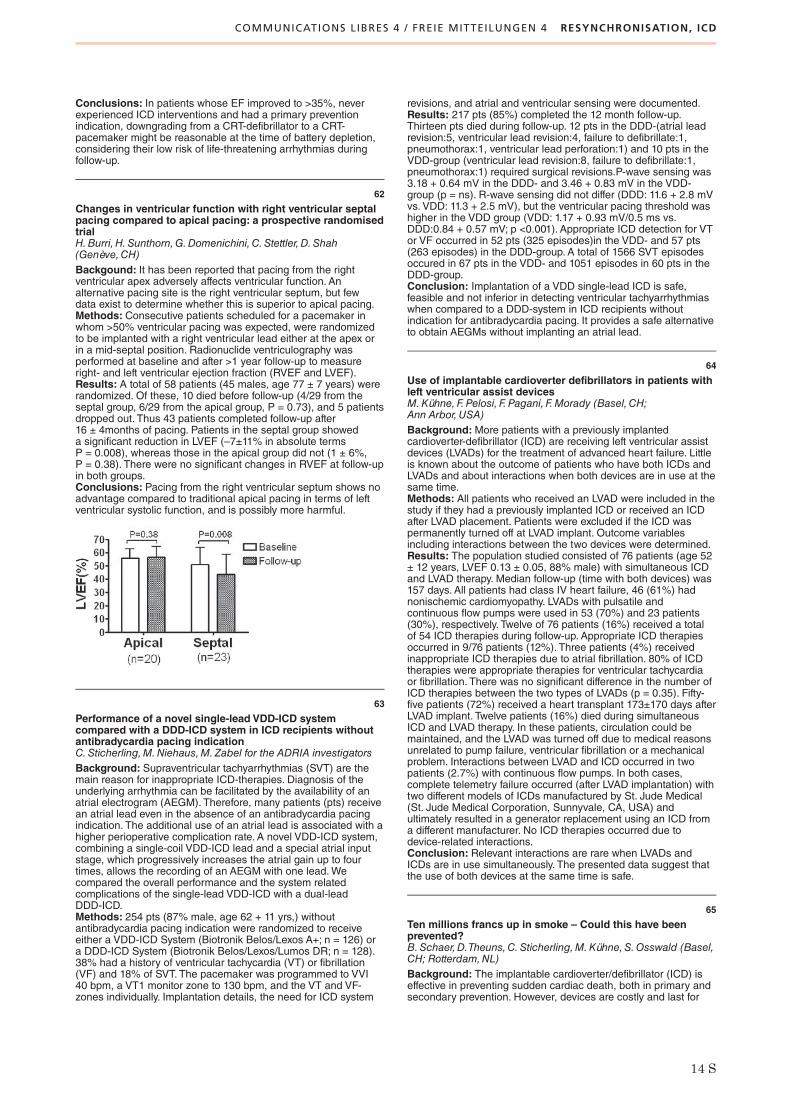
Changes in ventricular function with right ventricular septalpacing compared to apical pacing: a prospective randomisedtrialH.Burri, H.Sunthorn,G.Domenichini,C.Stettler,D.Shah(Genève,CH)Backgound: It has been reported that pacing from the rightventricular apex adversely affects ventricular function. Analternative pacing site is the right ventricular septum, but fewdata exist to determine whether this is superior to apical pacing.Methods: Consecutive patients scheduled for a pacemaker inwhom >50% ventricular pacing was expected, were randomizedto be implanted with a right ventricular lead either at the apex orin a mid-septal position. Radionuclide ventriculography wasperformed at baseline and after >1 year follow-up to measureright- and left ventricular ejection fraction (RVEF and LVEF).Results: A total of 58 patients (45 males, age 77 ± 7 years) wererandomized. Of these, 10 died before follow-up (4/29 from theseptal group, 6/29 from the apical group, P = 0.73), and 5 patientsdropped out. Thus 43 patients completed follow-up after16 ± 4months of pacing. Patients in the septal group showeda significant reduction in LVEF (–7±11% in absolute termsP = 0.008), whereas those in the apical group did not (1 ± 6%,P = 0.38). There were no significant changes in RVEF at follow-upin both groups.Conclusions: Pacing from the right ventricular septum shows noadvantage compared to traditional apical pacing in terms of leftventricular systolic function, and is possibly more harmful.
63
Performance of a novel single-lead VDD-ICD systemcompared with a DDD-ICD system in ICD recipients withoutantibradycardia pacing indicationC.Sticherling,M.Niehaus,M.Zabel for the ADRIA investigatorsBackground: Supraventricular tachyarrhythmias (SVT) are themain reason for inappropriate ICD-therapies. Diagnosis of theunderlying arrhythmia can be facilitated by the availability of anatrial electrogram (AEGM). Therefore, many patients (pts) receivean atrial lead even in the absence of an antibradycardia pacingindication. The additional use of an atrial lead is associated with ahigher perioperative complication rate. A novel VDD-ICD system,combining a single-coil VDD-ICD lead and a special atrial inputstage, which progressively increases the atrial gain up to fourtimes, allows the recording of an AEGM with one lead.Wecompared the overall performance and the system relatedcomplications of the single-lead VDD-ICD with a dual-leadDDD-ICD.Methods: 254 pts (87% male, age 62 + 11 yrs,) withoutantibradycardia pacing indication were randomized to receiveeither a VDD-ICD System (Biotronik Belos/Lexos A+; n = 126) ora DDD-ICD System (Biotronik Belos/Lexos/Lumos DR; n = 128).38% had a history of ventricular tachycardia (VT) or fibrillation(VF) and 18% of SVT. The pacemaker was programmed to VVI40 bpm, a VT1 monitor zone to 130 bpm, and the VT and VF-zones individually. Implantation details, the need for ICD system
revisions, and atrial and ventricular sensing were documented.Results: 217 pts (85%) completed the 12 month follow-up.Thirteen pts died during follow-up. 12 pts in the DDD-(atrial leadrevision:5, ventricular lead revision:4, failure to defibrillate:1,pneumothorax:1, ventricular lead perforation:1) and 10 pts in theVDD-group (ventricular lead revision:8, failure to defibrillate:1,pneumothorax:1) required surgical revisions.P-wave sensing was3.18 + 0.64 mV in the DDD- and 3.46 + 0.83 mV in the VDD-group (p = ns). R-wave sensing did not differ (DDD: 11.6 + 2.8 mVvs. VDD: 11.3 + 2.5 mV), but the ventricular pacing threshold washigher in the VDD group (VDD: 1.17 + 0.93 mV/0.5 ms vs.DDD:0.84 + 0.57 mV; p <0.001). Appropriate ICD detection for VTor VF occurred in 52 pts (325 episodes)in the VDD- and 57 pts(263 episodes) in the DDD-group. A total of 1566 SVT episodesoccured in 67 pts in the VDD- and 1051 episodes in 60 pts in theDDD-group.Conclusion: Implantation of a VDD single-lead ICD is safe,feasible and not inferior in detecting ventricular tachyarrhythmiaswhen compared to a DDD-system in ICD recipients withoutindication for antibradycardia pacing. It provides a safe alternativeto obtain AEGMs without implanting an atrial lead.
64
Use of implantable cardioverter defibrillators in patients withleft ventricular assist devicesM.Kühne,F.Pelosi, F.Pagani, F.Morady (Basel,CH;Ann Arbor,USA)Background:More patients with a previously implantedcardioverter-defibrillator (ICD) are receiving left ventricular assistdevices (LVADs) for the treatment of advanced heart failure. Littleis known about the outcome of patients who have both ICDs andLVADs and about interactions when both devices are in use at thesame time.Methods: All patients who received an LVAD were included in thestudy if they had a previously implanted ICD or received an ICDafter LVAD placement. Patients were excluded if the ICD waspermanently turned off at LVAD implant. Outcome variablesincluding interactions between the two devices were determined.Results: The population studied consisted of 76 patients (age 52± 12 years, LVEF 0.13 ± 0.05, 88% male) with simultaneous ICDand LVAD therapy. Median follow-up (time with both devices) was157 days. All patients had class IV heart failure, 46 (61%) hadnonischemic cardiomyopathy. LVADs with pulsatile andcontinuous flow pumps were used in 53 (70%) and 23 patients(30%), respectively. Twelve of 76 patients (16%) received a totalof 54 ICD therapies during follow-up. Appropriate ICD therapiesoccurred in 9/76 patients (12%). Three patients (4%) receivedinappropriate ICD therapies due to atrial fibrillation. 80% of ICDtherapies were appropriate therapies for ventricular tachycardiaor fibrillation. There was no significant difference in the number ofICD therapies between the two types of LVADs (p = 0.35). Fifty-five patients (72%) received a heart transplant 173±170 days afterLVAD implant. Twelve patients (16%) died during simultaneousICD and LVAD therapy. In these patients, circulation could bemaintained, and the LVAD was turned off due to medical reasonsunrelated to pump failure, ventricular fibrillation or a mechanicalproblem. Interactions between LVAD and ICD occurred in twopatients (2.7%) with continuous flow pumps. In both cases,complete telemetry failure occurred (after LVAD implantation) withtwo different models of ICDs manufactured by St. Jude Medical(St. Jude Medical Corporation, Sunnyvale, CA, USA) andultimately resulted in a generator replacement using an ICD froma different manufacturer. No ICD therapies occurred due todevice-related interactions.Conclusion: Relevant interactions are rare when LVADs andICDs are in use simultaneously. The presented data suggest thatthe use of both devices at the same time is safe.
65
Ten millions francs up in smoke – Could this have beenprevented?B.Schaer,D.Theuns,C.Sticherling,M.Kühne,S.Osswald (Basel,CH; Rotterdam,NL)Background: The implantable cardioverter/defibrillator (ICD) iseffective in preventing sudden cardiac death, both in primary andsecondary prevention. However, devices are costly and last for

COMMUNICATIONS LIBRES 4 / FREIE MITTEILUNGEN 4 RESYNCHRONISATION, ICD
COMMUNICATIONS LIBRES 5 / FREIE MITTEILUNGEN 5 AORTIC STENOSIS, PERCUTANEOUS VALVE REPLACEMENT
15 S
4–6 years until battery depletion. Only about 50% of patientsreceive ICD therapy during long-term follow-up. Aim of the studywas to identify baseline characteristics that could help to identifypatients who might never receive appropriate ICD therapy.Methods: Out of our prospective ICD register encompassing700 patients, we identified all patients with a follow-up of morethan 5 years. Cost of an ICD was estimated at 50000 CHF.Baseline factors included were: age (<60/>60), underlyingcardiopathy (ischemic; dilated; other), ejection fraction (<30;30–50; >50%), prevention mode (primary/secondary), results ofelectrophysiology studies (inducible; non-inducible; none) andcardiovascular drug therapy. Uni- and multivariate analyses wereperformed to identify predictors of appropriate ICD-therapy.Results:We identified 244 patients, 87% male, age 58 ± 12 years,ejection fraction 37 ± 15%, follow-up duration 94 ± 26 months, inwhom a total of 521 devices were implanted. 111 patients (45%)never received any appropriate ICD therapy (182 devices; 111with 1, 60 with 2, 9 with 3 and 2 with 4 implanted devices, totalingup to almost 10 Mio CHF). 40% of patients who received ICDtherapy had VF therapy as their first ICD therapy. Results of uni-and multivariate analyses are shown in the table.Conclusions: In multivariate analysis, the only predictivebaseline factor for subsequent appropriate ICD therapy wasinducibility of tachyarrhythmia. Given the considerable amount ofmoney spent on patients who never receive ICD therapy, studiesto better identify these patients are urgently needed. Until then, inevery patient the indication to replace an ICD that has never beenused should be reevaluated at the time of battery depletion.
66
Implantable cardioverter defibrillators in the elderly:chances and risksB.Schaer,W.Anne, L. Jordaens,D.Theuns,C.Sticherling,S.Osswald (Basel,CH; Rotterdam,NL)Background: The Implantable cardioverter defibrillator (ICD) iseffective in preventing sudden cardiac death. However, elderlypatients have been underrepresented in major clinical trials. Therole of ICDs in this subgroup is still ill defined and controversial,due to more competing risks and supposedly lower expected lifespan. Of note, mean life expectancy of a Swiss male aged 75 isanother ten years.Methods: Data stem from the two corresponding, prospectivelymanaged ICD.We analyzed baseline characteristics and survivaldata of all ICD-patients who were >75 years old at the time ofdevice implantation. All patients could be followed regardingsurvival status until November 2008. Kaplan-Meier survivalanalysis was performed. Using the score proposed recently byGoldenberg et al., mortality in the population was assessedaccording to the amount of competing risks. This score contains4 clinical risk factors (QRS duration >120 ms, NYHA class >II,renal failure and atrial fibrillation).Results: A total of 180 patients were implanted between February1999 and July 2008. Age was 77 ± 2 years (range 75–85), 48patients were ≥80 years old, 86% were male, and 82% hadcoronary artery disease. ICDs implanted were VVI in 40%, DDDin 34% and CRT in 26%. Follow-up time was 2.3 ± 1.8 years.114 patients were implanted for secondary prevention of suddencardiac death. Age was not statistically different between groups.Survival rates after 1, 2, 3 and 4 years were 87%, 82%, 75% and57%, respectively. Survival was similar between primary andsecondary prevention groups. Adequate ICD therapy has beendelivered in 41% of the patients during follow-up. 35% and 44%of the patients received appropriate ICD therapy (ATP or shock)during the first and second year after implantation, respectively.One-year survival decreased from 93% in patients without any ofthe above-mentioned risk factors to 65% in those patients with 3or more factors. Patients with 3 or 4 risk factors received as manyICD therapies as patients with less risk factors (<3 clinical riskfactors 39%, >3 clinical risk factors 48%, p = 0.37).Conclusions: ICD patients older than 75 years still have afavorable survival, independent of prevention indication andbenefit also from ICD therapy. Implantation of an ICD should thusnot wrongfully be withheld from them solely on the basis of age.However, competing risks must be taken into account.
67
Incidence and predictors of atrioventricular conductionimpairment and need for permanent pacemaker aftertranscatheter aortic valve implantationL.Roten,S.Windecker,G.Hellige,H.Tanner,T.Suter,T.P.Carrel,B. Eberle,B.Meier, E.Delacrétaz,P.Wenaweser (Bern,CH)Introduction: Transcatheter aortic valve implantation (TAVI) hasemerged as valuable treatment alternative to conventional aorticvalve replacement in selected patients. Atrioventricular (AV)conduction impairment has been reported after TAVI.Methods:We assessed AV conduction impairment (newcomplete AV block or left bundle branch block) and the need for apermanent pacemaker in consecutive patients undergoing TAVI atour institution, using the two CE-approved devices, the CoreValveRevalving system (CRS) and the Edwards Sapien Valve (ESV).
Results: Between August 2007 and December 2008, 67 patientswithout pre-existing permanent pacemaker underwent successfulTAVI (41 with CRS and 26 with ESV). Of these patients, 31 (47%)required a pacemaker after the intervention or developed a newleft bundle branch block, 27 (66%) in the CRS group, and 4 (16%)in the ESV group (p <0.001). A permanent pacemaker wasimplanted in 23 patients (34%), 20 (49%) in the CRS group, and3 (12%) in the ESV group (p = 0.002). Complete AV block was thereason for pacemaker implantation in 14 patients (21%), 11 (27%)in the CRS group, and 3 (12%) in the ESV group (p = 0.134). Thereason for pacemaker implantation in the remaining 9 patients(all CRS group) was progressive AV conduction impairment in 8,and new left bundle branch block with increasing QRS width in1. Univariate predictors for the need of permanent pacemaker
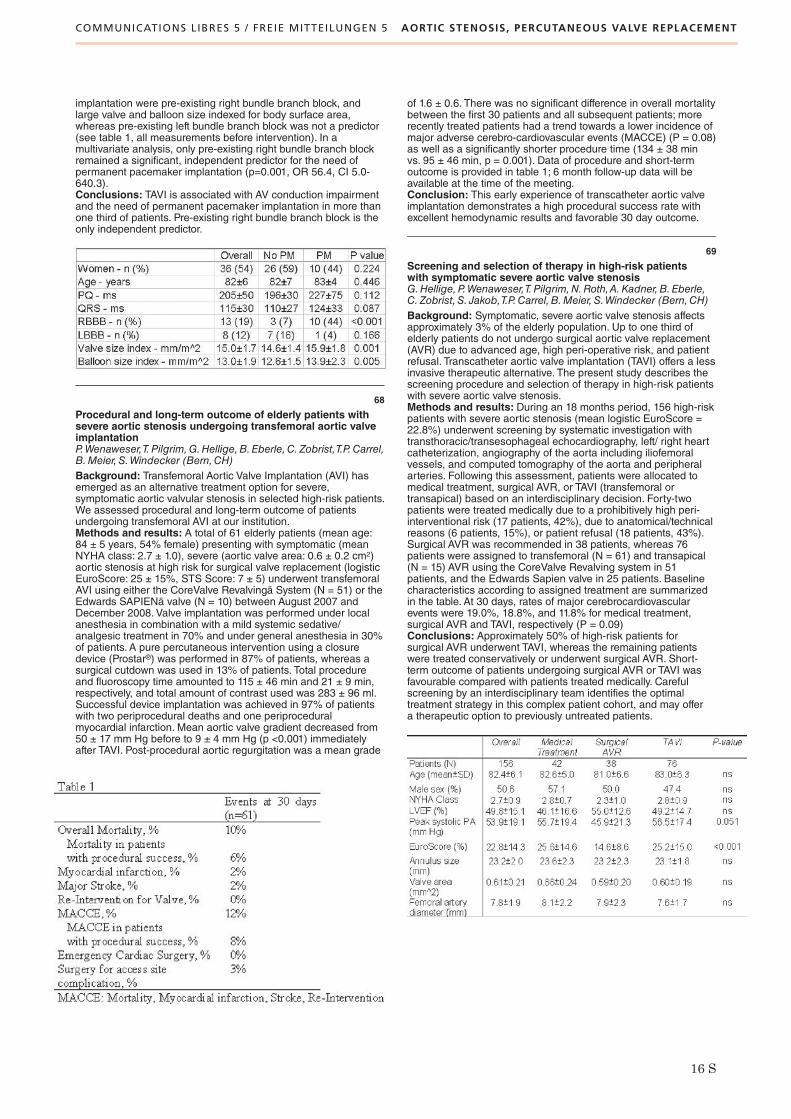
COMMUNICATIONS LIBRES 5 / FREIE MITTEILUNGEN 5 AORTIC STENOSIS, PERCUTANEOUS VALVE REPLACEMENT
16 S
implantation were pre-existing right bundle branch block, andlarge valve and balloon size indexed for body surface area,whereas pre-existing left bundle branch block was not a predictor(see table 1, all measurements before intervention). In amultivariate analysis, only pre-existing right bundle branch blockremained a significant, independent predictor for the need ofpermanent pacemaker implantation (p=0.001, OR 56.4, CI 5.0-640.3).Conclusions: TAVI is associated with AV conduction impairmentand the need of permanent pacemaker implantation in more thanone third of patients. Pre-existing right bundle branch block is theonly independent predictor.
68
Procedural and long-term outcome of elderly patients withsevere aortic stenosis undergoing transfemoral aortic valveimplantationP.Wenaweser,T.Pilgrim,G.Hellige,B.Eberle,C.Zobrist,T.P.Carrel,B.Meier, S.Windecker (Bern,CH)Background: Transfemoral Aortic Valve Implantation (AVI) hasemerged as an alternative treatment option for severe,symptomatic aortic valvular stenosis in selected high-risk patients.We assessed procedural and long-term outcome of patientsundergoing transfemoral AVI at our institution.Methods and results: A total of 61 elderly patients (mean age:84 ± 5 years, 54% female) presenting with symptomatic (meanNYHA class: 2.7 ± 1.0), severe (aortic valve area: 0.6 ± 0.2 cm2)aortic stenosis at high risk for surgical valve replacement (logisticEuroScore: 25 ± 15%, STS Score: 7 ± 5) underwent transfemoralAVI using either the CoreValve Revalvingä System (N = 51) or theEdwards SAPIENä valve (N = 10) between August 2007 andDecember 2008. Valve implantation was performed under localanesthesia in combination with a mild systemic sedative/analgesic treatment in 70% and under general anesthesia in 30%of patients. A pure percutaneous intervention using a closuredevice (Prostar®) was performed in 87% of patients, whereas asurgical cutdown was used in 13% of patients. Total procedureand fluoroscopy time amounted to 115 ± 46 min and 21 ± 9 min,respectively, and total amount of contrast used was 283 ± 96 ml.Successful device implantation was achieved in 97% of patientswith two periprocedural deaths and one periproceduralmyocardial infarction. Mean aortic valve gradient decreased from50 ± 17 mm Hg before to 9 ± 4 mm Hg (p <0.001) immediatelyafter TAVI. Post-procedural aortic regurgitation was a mean grade
of 1.6 ± 0.6. There was no significant difference in overall mortalitybetween the first 30 patients and all subsequent patients; morerecently treated patients had a trend towards a lower incidence ofmajor adverse cerebro-cardiovascular events (MACCE) (P = 0.08)as well as a significantly shorter procedure time (134 ± 38 minvs. 95 ± 46 min, p = 0.001). Data of procedure and short-termoutcome is provided in table 1; 6 month follow-up data will beavailable at the time of the meeting.Conclusion: This early experience of transcatheter aortic valveimplantation demonstrates a high procedural success rate withexcellent hemodynamic results and favorable 30 day outcome.
69
Screening and selection of therapy in high-risk patientswith symptomatic severe aortic valve stenosisG.Hellige,P.Wenaweser,T.Pilgrim,N.Roth,A.Kadner,B.Eberle,C.Zobrist, S. Jakob,T.P.Carrel,B.Meier, S.Windecker (Bern,CH)Background: Symptomatic, severe aortic valve stenosis affectsapproximately 3% of the elderly population. Up to one third ofelderly patients do not undergo surgical aortic valve replacement(AVR) due to advanced age, high peri-operative risk, and patientrefusal. Transcatheter aortic valve implantation (TAVI) offers a lessinvasive therapeutic alternative. The present study describes thescreening procedure and selection of therapy in high-risk patientswith severe aortic valve stenosis.Methods and results: During an 18 months period, 156 high-riskpatients with severe aortic stenosis (mean logistic EuroScore =22.8%) underwent screening by systematic investigation withtransthoracic/transesophageal echocardiography, left/ right heartcatheterization, angiography of the aorta including iliofemoralvessels, and computed tomography of the aorta and peripheralarteries. Following this assessment, patients were allocated tomedical treatment, surgical AVR, or TAVI (transfemoral ortransapical) based on an interdisciplinary decision. Forty-twopatients were treated medically due to a prohibitively high peri-interventional risk (17 patients, 42%), due to anatomical/technicalreasons (6 patients, 15%), or patient refusal (18 patients, 43%).Surgical AVR was recommended in 38 patients, whereas 76patients were assigned to transfemoral (N = 61) and transapical(N = 15) AVR using the CoreValve Revalving system in 51patients, and the Edwards Sapien valve in 25 patients. Baselinecharacteristics according to assigned treatment are summarizedin the table. At 30 days, rates of major cerebrocardiovascularevents were 19.0%, 18.8%, and 11.8% for medical treatment,surgical AVR and TAVI, respectively (P = 0.09)Conclusions: Approximately 50% of high-risk patients forsurgical AVR underwent TAVI, whereas the remaining patientswere treated conservatively or underwent surgical AVR. Short-term outcome of patients undergoing surgical AVR or TAVI wasfavourable compared with patients treated medically. Carefulscreening by an interdisciplinary team identifies the optimaltreatment strategy in this complex patient cohort, and may offera therapeutic option to previously untreated patients.
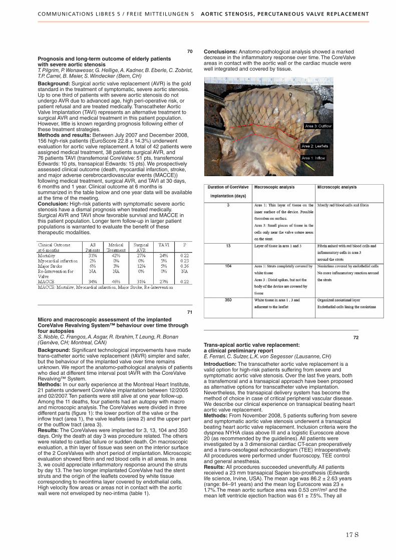
COMMUNICATIONS LIBRES 5 / FREIE MITTEILUNGEN 5 AORTIC STENOSIS, PERCUTANEOUS VALVE REPLACEMENT
17 S
70
Prognosis and long-term outcome of elderly patientswith severe aortic stenosisT.Pilgrim,P.Wenaweser,G.Hellige,A.Kadner,B.Eberle,C.Zobrist,T.P.Carrel,B.Meier, S.Windecker (Bern,CH)Background: Surgical aortic valve replacement (AVR) is the goldstandard in the treatment of symptomatic, severe aortic stenosis.Up to one third of patients with severe aortic stenosis do notundergo AVR due to advanced age, high peri-operative risk, orpatient refusal and are treated medically. Transcatheter AorticValve Implantation (TAVI) represents an alternative treatment tosurgical AVR and medical treatment in this patient population.However, little is known regarding prognosis following either ofthese treatment strategies.Methods and results: Between July 2007 and December 2008,156 high-risk patients (EuroScore 22.8 ± 14.3%) underwentevaluation for aortic valve replacement. A total of 42 patients wereassigned medical treatment, 38 patients surgical AVR, and76 patients TAVI (transfemoral CoreValve: 51 pts, transfemoralEdwards: 10 pts, transapical Edwards: 15 pts).We prospectivelyassessed clinical outcome (death, myocardial infarction, stroke,and major adverse cerebrocardiovascular events (MACCE))following medical treatment, surgical AVR, and TAVI at 30 days,6 months and 1 year. Clinical outcome at 6 months issummarized in the table below and one year data will be availableat the time of the meeting.Conclusion: High-risk patients with symptomatic severe aorticstenosis have a dismal prognosis when treated medically.Surgical AVR and TAVI show favorable survival and MACCE inthis patient population. Longer term follow-up in larger patientpopulations is warranted to evaluate the benefit of thesetherapeutic modalities.
71
Micro and macroscopic assessment of the implantedCoreValve Revalving System™ behaviour over time throughfour autopsiesS.Noble,C.Frangos,A.Asgar,R. Ibrahim,T. Leung,R.Bonan(Genève,CH; Montreal,CAN)Background: Significant technological improvements have madetrans-catheter aortic valve replacement (tAVR) simpler and safer,but the behaviour of the implanted valve over time remainsunknown.We report the anatomo-pathological analysis of patientswho died at different time interval post tAVR with the CoreValveRevalving™ System.Methods: In our early experience at the Montreal Heart Institute,21 patients underwent CoreValve implantation between 12/2005and 02/2007. Ten patients were still alive at one year follow-up.Among the 11 deaths, four patients had an autopsy with macroand microscopic analysis. The CoreValves were divided in threedifferent parts (figure 1): the lower portion of the valve or theinflow tract (area 1), the valve leaflets (area 2) and the upper partor the outflow tract (area 3).Results: The CoreValves were implanted for 3, 13, 104 and 350days. Only the death at day 3 was procedure related. The otherswere related to cardiac failure or sudden death. On macroscopicevaluation, a thin layer of tissue was seen on the interior surfaceof the 2 CoreValves with short period of implantation. Microscopicevaluation showed fibrin and red blood cells in all areas. In area3, we could appreciate inflammatory response around the strutsby day 13. The two longer implantated CoreValve had the stentstruts and the origin of the leaflets covered by white tissuecorresponding to neointima layer covered by endothelial cells.High velocity flow areas or areas not in contact with the aorticwall were not enveloped by neo-intima (table 1).
Conclusions: Anatomo-pathological analysis showed a markeddecrease in the inflammatory response over time. The CoreValveareas in contact with the aortic wall or the cardiac muscle werewell integrated and covered by tissue.
72
Trans-apical aortic valve replacement:a clinical preliminary reportE.Ferrari, C.Sulzer, L.K. von Segesser (Lausanne,CH)Introduction: The transcatheter aortic valve replacement is avalid option for high-risk patients suffering from severe andsymptomatic aortic valve stenosis. Over the last five years, botha transfemoral and a transapical approach have been proposedas alternative options for transcatheter valve implantation.Nevertheless, the transapical delivery system has become themethod of choice in case of critical peripheral vascular disease.We describe our clinical experience on transapical beating heartaortic valve replacement.Methods: From November 2008, 5 patients suffering from severeand symptomatic aortic valve stenosis underwent a transapicalbeating heart aortic valve replacement. Inclusion criteria were thedyspnea NYHA class above III and a logistic Euroscore above20 (as recommended by the guidelines). All patients wereinvestigated by a 3 dimensional cardiac CT-scan preoperativelyand a trans-oesofageal echocardiogram (TEE) intraoperatively.All procedures were performed under fluoroscopy, TEE controland general anesthesia.Results: All procedures succeeded uneventfully. All patientsreceived a 23 mm transapical Sapien bio-prosthesis (Edwardslife science, Irvine, USA). The mean age was 86.2 ± 2.63 years(range: 84–91 years) and the mean log Euroscore was 23 ±1.7%.The mean aortic surface area was 0.53 cm2/m2 and themean left ventricle ejection fraction was 61 ± 7.5%. They all
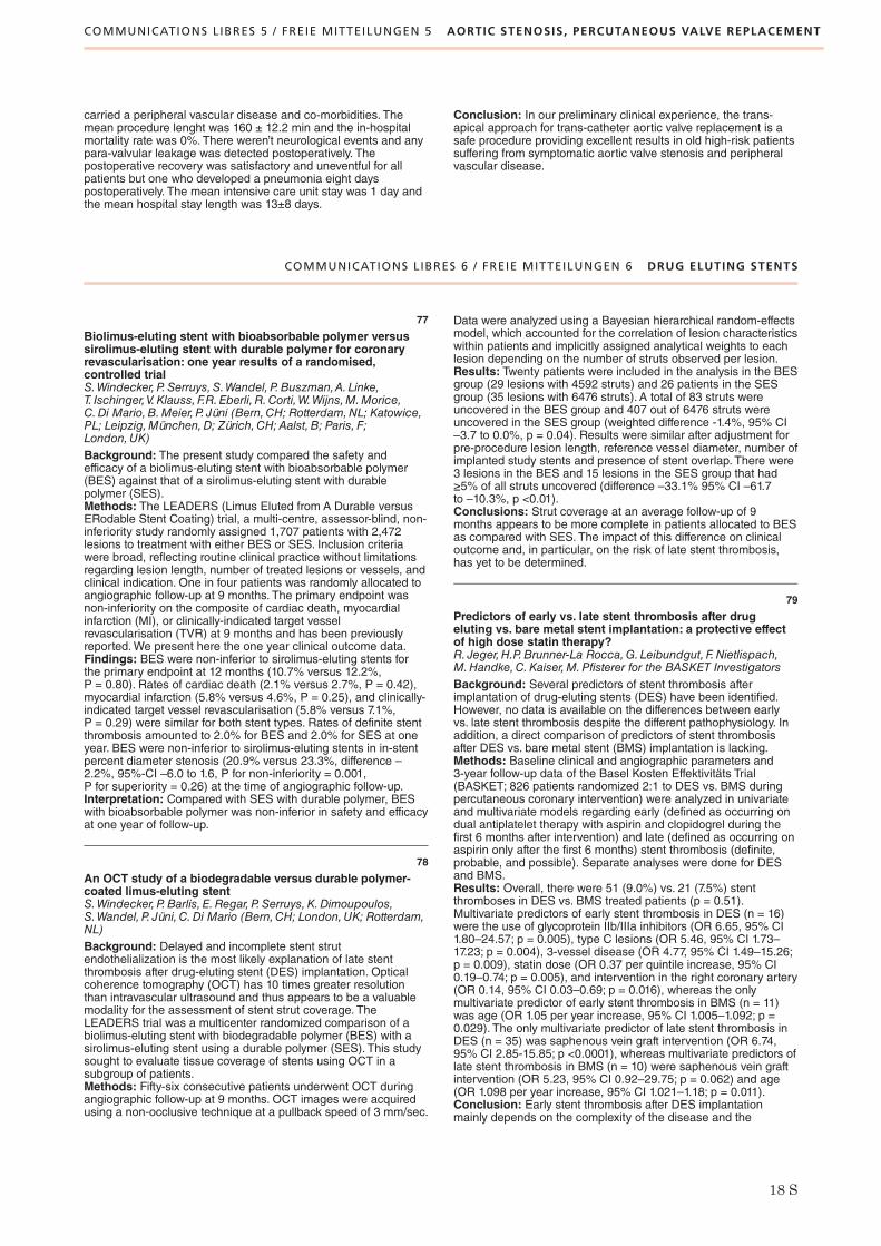
COMMUNICATIONS LIBRES 5 / FREIE MITTEILUNGEN 5 AORTIC STENOSIS, PERCUTANEOUS VALVE REPLACEMENT
COMMUNICATIONS LIBRES 6 / FREIE MITTEILUNGEN 6 DRUG ELUTING STENTS
18 S
carried a peripheral vascular disease and co-morbidities. Themean procedure lenght was 160 ± 12.2 min and the in-hospitalmortality rate was 0%. There weren’t neurological events and anypara-valvular leakage was detected postoperatively. Thepostoperative recovery was satisfactory and uneventful for allpatients but one who developed a pneumonia eight dayspostoperatively. The mean intensive care unit stay was 1 day andthe mean hospital stay length was 13±8 days.
Conclusion: In our preliminary clinical experience, the trans-apical approach for trans-catheter aortic valve replacement is asafe procedure providing excellent results in old high-risk patientssuffering from symptomatic aortic valve stenosis and peripheralvascular disease.
77
Biolimus-eluting stent with bioabsorbable polymer versussirolimus-eluting stent with durable polymer for coronaryrevascularisation: one year results of a randomised,controlled trialS.Windecker,P.Serruys,S.Wandel, P.Buszman,A. Linke,T. Ischinger,V.Klauss, F.R.Eberli, R.Corti,W.Wijns,M.Morice,C.Di Mario,B.Meier, P. Jüni (Bern,CH; Rotterdam,NL; Katowice,PL; Leipzig,München,D; Zürich,CH; Aalst,B; Paris, F;London,UK)Background: The present study compared the safety andefficacy of a biolimus-eluting stent with bioabsorbable polymer(BES) against that of a sirolimus-eluting stent with durablepolymer (SES).Methods: The LEADERS (Limus Eluted from A Durable versusERodable Stent Coating) trial, a multi-centre, assessor-blind, non-inferiority study randomly assigned 1,707 patients with 2,472lesions to treatment with either BES or SES. Inclusion criteriawere broad, reflecting routine clinical practice without limitationsregarding lesion length, number of treated lesions or vessels, andclinical indication. One in four patients was randomly allocated toangiographic follow-up at 9 months. The primary endpoint wasnon-inferiority on the composite of cardiac death, myocardialinfarction (MI), or clinically-indicated target vesselrevascularisation (TVR) at 9 months and has been previouslyreported.We present here the one year clinical outcome data.Findings: BES were non-inferior to sirolimus-eluting stents forthe primary endpoint at 12 months (10.7% versus 12.2%,P = 0.80). Rates of cardiac death (2.1% versus 2.7%, P = 0.42),myocardial infarction (5.8% versus 4.6%, P = 0.25), and clinically-indicated target vessel revascularisation (5.8% versus 7.1%,P = 0.29) were similar for both stent types. Rates of definite stentthrombosis amounted to 2.0% for BES and 2.0% for SES at oneyear. BES were non-inferior to sirolimus-eluting stents in in-stentpercent diameter stenosis (20.9% versus 23.3%, difference –2.2%, 95%-CI –6.0 to 1.6, P for non-inferiority = 0.001,P for superiority = 0.26) at the time of angiographic follow-up.Interpretation: Compared with SES with durable polymer, BESwith bioabsorbable polymer was non-inferior in safety and efficacyat one year of follow-up.
78
An OCT study of a biodegradable versus durable polymer-coated limus-eluting stentS.Windecker,P.Barlis, E.Regar,P.Serruys,K.Dimoupoulos,S.Wandel, P. Jüni,C.Di Mario (Bern,CH; London,UK; Rotterdam,NL)Background: Delayed and incomplete stent strutendothelialization is the most likely explanation of late stentthrombosis after drug-eluting stent (DES) implantation. Opticalcoherence tomography (OCT) has 10 times greater resolutionthan intravascular ultrasound and thus appears to be a valuablemodality for the assessment of stent strut coverage. TheLEADERS trial was a multicenter randomized comparison of abiolimus-eluting stent with biodegradable polymer (BES) with asirolimus-eluting stent using a durable polymer (SES). This studysought to evaluate tissue coverage of stents using OCT in asubgroup of patients.Methods: Fifty-six consecutive patients underwent OCT duringangiographic follow-up at 9 months. OCT images were acquiredusing a non-occlusive technique at a pullback speed of 3 mm/sec.
Data were analyzed using a Bayesian hierarchical random-effectsmodel, which accounted for the correlation of lesion characteristicswithin patients and implicitly assigned analytical weights to eachlesion depending on the number of struts observed per lesion.Results: Twenty patients were included in the analysis in the BESgroup (29 lesions with 4592 struts) and 26 patients in the SESgroup (35 lesions with 6476 struts). A total of 83 struts wereuncovered in the BES group and 407 out of 6476 struts wereuncovered in the SES group (weighted difference -1.4%, 95% CI–3.7 to 0.0%, p = 0.04). Results were similar after adjustment forpre-procedure lesion length, reference vessel diameter, number ofimplanted study stents and presence of stent overlap. There were3 lesions in the BES and 15 lesions in the SES group that had≥5% of all struts uncovered (difference –33.1% 95% CI –61.7to –10.3%, p <0.01).Conclusions: Strut coverage at an average follow-up of 9months appears to be more complete in patients allocated to BESas compared with SES. The impact of this difference on clinicaloutcome and, in particular, on the risk of late stent thrombosis,has yet to be determined.
79
Predictors of early vs. late stent thrombosis after drugeluting vs. bare metal stent implantation: a protective effectof high dose statin therapy?R. Jeger,H.P.Brunner-La Rocca,G. Leibundgut, F.Nietlispach,M.Handke,C.Kaiser,M.Pfisterer for the BASKET InvestigatorsBackground: Several predictors of stent thrombosis afterimplantation of drug-eluting stents (DES) have been identified.However, no data is available on the differences between earlyvs. late stent thrombosis despite the different pathophysiology. Inaddition, a direct comparison of predictors of stent thrombosisafter DES vs. bare metal stent (BMS) implantation is lacking.Methods: Baseline clinical and angiographic parameters and3-year follow-up data of the Basel Kosten Effektivitäts Trial(BASKET; 826 patients randomized 2:1 to DES vs. BMS duringpercutaneous coronary intervention) were analyzed in univariateand multivariate models regarding early (defined as occurring ondual antiplatelet therapy with aspirin and clopidogrel during thefirst 6 months after intervention) and late (defined as occurring onaspirin only after the first 6 months) stent thrombosis (definite,probable, and possible). Separate analyses were done for DESand BMS.Results: Overall, there were 51 (9.0%) vs. 21 (7.5%) stentthromboses in DES vs. BMS treated patients (p = 0.51).Multivariate predictors of early stent thrombosis in DES (n = 16)were the use of glycoprotein IIb/IIIa inhibitors (OR 6.65, 95% CI1.80–24.57; p = 0.005), type C lesions (OR 5.46, 95% CI 1.73–17.23; p = 0.004), 3-vessel disease (OR 4.77, 95% CI 1.49–15.26;p = 0.009), statin dose (OR 0.37 per quintile increase, 95% CI0.19–0.74; p = 0.005), and intervention in the right coronary artery(OR 0.14, 95% CI 0.03–0.69; p = 0.016), whereas the onlymultivariate predictor of early stent thrombosis in BMS (n = 11)was age (OR 1.05 per year increase, 95% CI 1.005–1.092; p =0.029). The only multivariate predictor of late stent thrombosis inDES (n = 35) was saphenous vein graft intervention (OR 6.74,95% CI 2.85-15.85; p <0.0001), whereas multivariate predictors oflate stent thrombosis in BMS (n = 10) were saphenous vein graftintervention (OR 5.23, 95% CI 0.92–29.75; p = 0.062) and age(OR 1.098 per year increase, 95% CI 1.021–1.18; p = 0.011).Conclusion: Early stent thrombosis after DES implantationmainly depends on the complexity of the disease and the

COMMUNICATIONS LIBRES 6 / FREIE MITTEILUNGEN 6 DRUG ELUTING STENTS
19 S
intervention, with the use of a higher statin dose possibly havinga protective effect against early stent thrombosis. The observationof a strong dose-dependent protective effect of statins to preventearly stent thrombosis after DES is new, hypothesis-generating,and should be evaluated in an adequately powered prospectiverandomized trial.
80
Impact of stent overlap on angiographic and long-termclinical outcome in patients undergoing drug-eluting stentimplantationL.Räber,S.Cook,S.Wandel, P.Wenaweser,M.Togni,R.Vogel,M.Billinger,C.Seiler, F.R.Eberli,T.F. Lüscher,B.Meier, P. Jüni,S.Windecker (Bern, Zürich,CH)Background: Drug-eluting stent (DES) overlap has beenassociated with delayed healing and increased inflammation inexperimental studies, but its impact on clinical outcome is not wellestablished.We therefore compared the angiographic and long-term clinical outcome between patients with and without DESoverlap.Methods and results: In the SIRTAX trial, 1012 patientsunderwent treatment with sirolimus-eluting or paclitaxel-elutingstents in a randomized controlled trial. A stratified analysis ofangiographic outcome at 8 months, and clinical outcome at3 years was performed according to the presence or absenceof DES overlap (OL). OL was present in 134 (13%) patients,whereas 878 patients (87%) had no DES overlap (NOL). Thecauses of OL were excessive lesion length (44%), incompletelesion coverage (36%), and edge dissections (20%). Majoradverse cardiac events (cardiac death, MI, TLR) were morecommon in OL (25.4%) than NOL (15.7%, HR = 1.67, 95% CI1.14–2.43, P <0.01) patients at 3 years. Both, the risk of TLR(20.2% vs. 11.3%; HR 1.82; CI 1.19–2.79, P <0.01) as well as therisk of death or MI (10.0% vs. 17.2%; HR 1.75 CI 1.10–2.78,P = 0.02) were increased in OL compared with NOL patients.The results remained robust using a sensitivity analysis adjustedfor pre-procedural lesion length, vessel size, and diabetes.Angiographic follow-up at 8 months showed increased late loss(0.17 ± 0.41 vs. 0.33 ± 0.61, P = 0.02) and higher rates of in-segment restenosis (15.0% vs. 8.6%, P = 0.07) in OL than NOLlesions, which was related to the site of OL.Conclusions: DES overlap occurs in >10% of patientsundergoing PCI in routine clinical practice and is most commonlyrelated to excessive lesion length. DES overlap is associated withimpaired angiographic and long-term clinical outcome.
81
Impact of incomplete stent apposition on long-term clinicaloutcome after drug-eluting stent implantationS.Cook,P. Eshtehardi, S.Wandel, L.Räber,P.Wenaweser,M.Togni,M.Billinger,R.Vogel,C.Seiler,R.Corti,G.Sütsch, F.R.Eberli,B.Meier, P. Jüni, S.Windecker (Bern,CH)Background: Incomplete stent apposition (ISA) is more commonafter drug-eluting stent (DES) than bare metal stent (BMS)implantation. The impact of ISA on clinical outcome after DESimplantation is not well established. The aim of the present studywas to compare long-term clinical outcome in patients with andwithout ISA as assessed by intravascular ultrasound (IVUS)8 months after implantation of sirolimus-eluting stents (SES)or paclitaxel-eluting stents (PES).Methods and results: A total of 196 patients included into theSIRTAX trial underwent IVUS at the time of follow-up angiography8 months after implantation of DES (SES: 92 patients, PES:104 patients) and were prospectively followed for 3 years. IVUSimaging was performed using motorized pullback (0.5 mm/s) andimages were analyzed offline by blinded outcome assessors.ISA was defined as one or more stent struts separated from the
vessel wall with evidence of blood speckles behind the strut.ISA was present in 37 patients (19%) at 8 months and was moreprevalent in SES than PES treated patients (28.3% vs. 10.6%,p = 0.001). Baseline clinical, angiographic and proceduralcharacteristics were similar for both ISA and non-ISA groupsexcept for stent type (>SES in the ISA group), and stent length(22.1 ± 11.6 mm vs. 18.0 ± 8.7 mm, p = 0.01) respectively. At 3years of follow-up, there were no significant differences regardingoverall mortality, cardiac death, myocardial infarction (MI), orrepeat revascularization. Very late definite stent thrombosis wasencountered in 3 patients (8.1%) with ISA and no patient withoutISA (0%, p <0.001).Conclusion: ISA as assessed by IVUS 8 months after DESimplantation was more prevalent in SES than PES, and showedsimilar clinical outcome at 3 years of follow-up in ISA and non-ISApatients. Very late definite stent thrombosis was observed only inpatients with ISA, suggesting a potential role of vascularremodeling in the pathogenesis of this adverse event
82
Paclitaxel-eluting biodegradable coronary bypass grafts:a step towards reduction of neointima formationB.H.Walpoth, F. Innocente,D.Mandracchia, E.Pektok,B.Nottelet,J.-C.Tille, S. de Valence,A.Kalangos,R.Gurny,M.Moeller(Genève, Lausanne,CH)Introduction: Small-calibre vascular prostheses (<6 mm) areunsatisfactory for their in vivo performance, especially for CABG.Reasons for failure are early thrombosis and late intimalhyperplasia. To overcome this we prepared biodegradable small-calibre vascular prostheses using electrospun polycaprolactone(PCL) nanofibres with slow releasing paclitaxel (PTX), an anti-proliferative drug.Methods: PCL solutions containing PTX (0–1% wt/wt) were usedto prepare non-wovennanofibre-based 2-mm ID prostheses.Mechanical, morphological properties and drug loading,distribution and release were studied in vitro. Infrarenal abdominalaortic replacement was carried out with non-drug-loaded anddrug-loaded prostheses in 18 rats and followed up to 6 months.Patency, stenosis, tissue reaction and drug effect onendothelialisation, vascular remodelling and neointima formation(morphometry) were studied in vivo.Results: In vitro: prostheses showed controlled morphologymimicking extra-cellular matrix (fibre diameter 500–2000 nm) withmechanical properties similar to those of native vessels (tensilestress >1.4 MPa, tensile strain >100%). PTX loaded grafts withsuitable mechanical properties were obtained by a factorialdesign approach, and incorporated PTX was released in acontrolled manner over 1 month. In vivo: angiography showed100% patency, no stenosis, no aneurysmal dilatation and therewere no differences among the two groups. Major morphologicdifferences were found between the non-drug-loaded anddrugloaded prostheses. Endothelial coverage and cell ingrowthwere significantly reduced at 3 weeks and delayed at 12 and 24weeks in the PTX Group, but as envisioned, neointima formationwas significantly reduced in the drug-loaded grafts at 12 weeks,and delayed at 6 months.Conclusions: Degradable, electrospun, nanofibre,polycaprolactone prostheses are promising since, in vitro theymaintain their mechanical properties (regardless of PTX loading),and in vivo show good patency, re-endothelialize and remodelwith autologous cells. PTX loading delays endothelialisation andcellular ingrowth. Conversely, it reduces neointima formation untilthe end-point of our study (6 months) and thus may be aninteresting option for coronary bypass grafts. However, long-termfollow-up studies are needed to confirm the usefulness of anti-proliferative drug-release in biodegradable scaffolds forcardiovascular clinical applications.
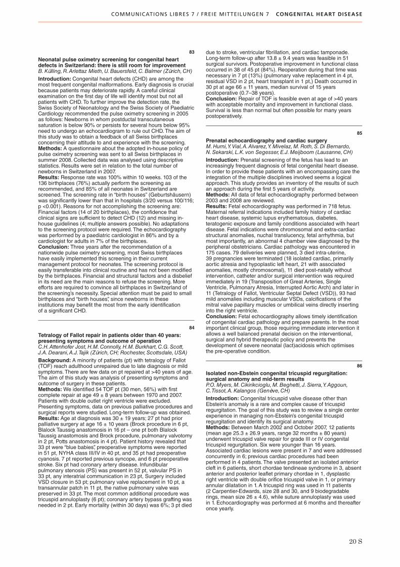
COMMUNICATIONS LIBRES 7 / FREIE MITTEILUNGEN 7 CONGENITAL HEART DISEASE
20 S
83
Neonatal pulse oximetry screening for congenital heartdefects in Switzerland: there is still room for improvementB.Külling,R.Arlettaz Mieth,U.Bauersfeld,C.Balmer (Zürich,CH)Introduction: Congenital heart defects (CHD) are among themost frequent congenital malformations. Early diagnosis is crucialbecause patients may deteriorate rapidly. A careful clinicalexamination on the first day of life will identify most but not allpatients with CHD. To further improve the detection rate, theSwiss Society of Neonatology and the Swiss Society of PaediatricCardiology recommended the pulse oximetry screening in 2005as follows: Newborns in whom postductal transcutaneoussaturation is below 90% or persists for several hours below 95%need to undergo an echocardiogram to rule out CHD. The aim ofthis study was to obtain a feedback of all Swiss birthplacesconcerning their attitude to and experience with the screening.Methods: A questionnaire about the adopted in-house policy ofpulse oximetry screening was sent to all Swiss birthplaces insummer 2008. Collected data was analysed using descriptivestatistics. Results were set in relation to the total number ofnewborns in Switzerland in 2007.Results: Response rate was 100% within 10 weeks. 103 of the136 birthplaces (76%) actually perform the screening asrecommended, and 85% of all neonates in Switzerland arescreened. The screening rate in “birth houses” (Geburtshäusern)was significantly lower than that in hospitals (3/20 versus 100/116;p <0.001). Reasons for not accomplishing the screening are:Financial factors (14 of 20 birthplaces), the confidence thatclinical signs are sufficient to detect CHD (12) and missing in-house guidelines (4; multiple answers possible). No adaptationsto the screening protocol were required. The echocardiographywas performed by a paediatric cardiologist in 86% and by acardiologist for adults in 7% of the birthplaces.Conclusion: Three years after the recommendation of anationwide pulse oximetry screening, most Swiss birthplaceshave easily implemented this screening in their currentmanagement protocol for neonates. The screening protocol iseasily transferable into clinical routine and has not been modifiedby the birthplaces. Financial and structural factors and a disbeliefin its need are the main reasons to refuse the screening. Moreefforts are required to convince all birthplaces in Switzerland ofthe screening’s necessity. Special attention must be paid to smallbirthplaces and “birth houses”, since newborns in theseinstitutions may benefit the most from the early identificationof a significant CHD.
84
Tetralogy of Fallot repair in patients older than 40 years:presenting symptoms and outcome of operationC.H.Attenhofer Jost,H.M.Connolly,H.M.Burkhart,C.G.Scott,J.A.Dearani,A.J.Tajik (Zürich,CH; Rochester, Scottsdale,USA)Background: A minority of patients (pt) with tetralogy of Fallot(TOF) reach adulthood unrepaired due to late diagnosis or mildsymptoms. There are few data on pt repaired at >40 years of age.The aim of this study was analysis of presenting symptoms andoutcome of surgery in these patients.Methods:We identified 54 TOF pt (30 men, 56%) with firstcomplete repair at age 49 ± 8 years between 1970 and 2007.Patients with double outlet right ventricle were excluded.Presenting symptoms, data on previous palliative procedures andsurgical reports were studied. Long-term follow-up was obtained.Results: Age at diagnosis was 30 ± 19 years; 27 pt had priorpalliative surgery at age 16 ± 10 years (Brock procedure in 6 pt,Blalock Taussig anastomosis in 16 pt – one pt both BlalockTaussig anastomosis and Brock procedure, pulmonary valvotomyin 2 pt, Potts anastomosis in 4 pt). Patient history revealed that33 pt were “blue babies”, preoperative symptoms were reportedin 51 pt, NYHA class III/IV in 40 pt, and 35 pt had preoperativecyanosis. 7 pt reported previous syncope, and 6 pt preoperativestroke. Six pt had coronary artery disease. Infundibularpulmonary stenosis (PS) was present in 52 pt, valvular PS in33 pt, any interatrial communication in 23 pt, Surgery includedVSD closure in 53 pt; pulmonary valve replacement in 10 pt, atransannular patch in 11 pt, the native pulmonary valve waspreserved in 33 pt. The most common additional procedure wastricuspid annuloplasty (6 pt); coronary artery bypass grafting wasneeded in 2 pt. Early mortality (within 30 days) was 6%; 3 pt died
due to stroke, ventricular fibrillation, and cardiac tamponade.Long-term follow-up after 13.8 ± 9.4 years was feasible in 51surgical survivors. Postoperative improvement in functional classoccurred in 38 of 45 pt (84%). Reoperation during that time wasnecessary in 7 pt (13%) (pulmonary valve replacement in 4 pt,residual VSD in 2 pt, heart transplant in 1 pt,) Death occurred in30 pt at age 66 ± 11 years, median survival of 15 yearspostoperative (0.7–38 years).Conclusion: Repair of TOF is feasible even at age of >40 yearswith acceptable mortality and improvement in functional class.Survival is less than normal but often possible for many yearspostoperatively.
85
Prenatal echocardiography and cardiac surgeryM.Hurni,Y.Vial,A.Alvarez,Y.Mivelaz,M.Roth,S.Di Bernardo,N.Sekarski, L.K. von Segesser, E.J.Meijboom (Lausanne,CH)Introduction: Prenatal screening of the fetus has lead to anincreasingly frequent diagnosis of fetal congenital heart disease.In order to provide these patients with an encompassing care theintegration of the multiple disciplines involved seems a logicalapproach. This study provides an inventory of the results of suchan approach during the first 5 years of activity.Methods: All data of fetal echocardiography performed between2003 and 2008 are reviewed.Results: Fetal echocardiography was performed in 718 fetus.Maternal referral indications included family history of cardiacheart disease, systemic lupus erythematosus, diabetes,teratogenic exposure and family conditions associated with heartdisease. Fetal indications were chromosomal and extra-cardiacstructural anomalies, nuchal translucency, fetal arrhythmia, butmost importantly, an abnormal 4 chamber view diagnosed by theperipheral obstetricians. Cardiac pathology was encountered in175 cases. 79 deliveries were planned, 3 died intra-uterine,39 pregnancies were terminated (18 isolated cardiac, primarilyaortic atresia and hypoplastic left heart, 21 with associatedanomalies, mostly chromosomal), 11 died post-natally withoutintervention, catheter and/or surgical intervention was requiredimmediately in 19 (Transposition of Great Arteries, SingleVentricle, Pulmonary Atresia, Interrupted Aortic Arch) and later in11 (Tetralogy of Fallot, Ventricular Septal Defect (VSD)), 93 hadmild anomalies including muscular VSDs, calcifications of themitral valve papillary muscles or umbilical veins directly insertinginto the right ventricle.Conclusion: Fetal echocardiography allows timely identificationof congenital cardiac pathology and prepare parents. In the mostimportant clinical group, those requiring immediate intervention itallows a well balanced prenatal decision on the interventional,surgical and hybrid therapeutic policy and prevents thedevelopment of severe neonatal (lact)acidosis which optimisesthe pre-operative condition.
86
Isolated non-Ebstein congenital tricuspid regurgitation:surgical anatomy and mid-term resultsP.O.Myers,M.Cikirikcioglu,M.Beghetti, J. Sierra,Y.Aggoun,C.Tissot,A.Kalangos (Genève,CH)Introduction: Congenital tricuspid valve disease other thanEbstein’s anomaly is a rare and complex cause of tricuspidregurgitation. The goal of this study was to review a single centerexperience in managing non-Ebstein’s congenital tricuspidregurgitation and identify its surgical anatomy.Methods: Between March 2002 and October 2007, 12 patients(mean age 25.3 ± 26.9 years, range 32 months ± 80 years)underwent tricuspid valve repair for grade III or IV congenitaltricuspid regurgitation. Six were younger than 16 years.Associated cardiac lesions were present in 7 and were addressedconcurrently in 6; previous cardiac procedures had beenperformed in 4 patients. The valve presented an isolated anteriorcleft in 6 patients, short chordae tendineae syndrome in 3, absentanterior and posterior leaflet primary chordae in 1, dysplasticright ventricle with double orifice tricuspid valve in 1, or primaryannular dilatation in 1. A tricuspid ring was used in 11 patients(2 Carpentier-Edwards, size 28 and 30, and 9 biodegradablerings, mean size 26 ± 4.6), while suture annuloplasty was usedin 1. Echocardiography was performed at 6 months and thereafteronce yearly.
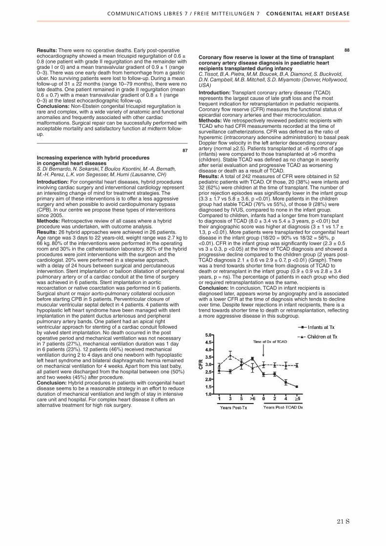
COMMUNICATIONS LIBRES 7 / FREIE MITTEILUNGEN 7 CONGENITAL HEART DISEASE
21 S
Results: There were no operative deaths. Early post-operativeechocardiography showed a mean tricuspid regurgitation of 0.6 ±0.8 (one patient with grade II regurgitation and the remainder withgrade I or 0) and a mean transvalvular gradient of 0.9 ± 1 (range0–3). There was one early death from hemorrhage from a gastriculcer. No surviving patients were lost to follow-up. During a meanfollow-up of 31 ± 22 months (range 10–79 months), there were nolate deaths. One patient remained in grade II regurgitation (mean0.6 ± 0.7) with a mean transvalvular gradient of 0.8 ± 1 (range0–3) at the latest echocardiographic follow-up.Conclusions: Non-Ebstein congenital tricuspid regurgitation israre and complex, with a wide variety of anatomic and functionalanomalies and frequently associated with other cardiacmalformations. Surgical repair can be successfully performed withacceptable mortality and satisfactory function at midterm follow-up.
87
Increasing experience with hybrid proceduresin congenital heart diseasesS.Di Bernardo,N.Sekarski,T.Boulos Ksontini,M.-A.Bernath,M.-H.Perez, L.K. von Segesser,M.Hurni (Lausanne,CH)Introduction: For congenital heart diseases, hybrid proceduresinvolving cardiac surgery and interventional cardiology representan interesting change of mind for treatment strategies. Theprimary aim of these interventions is to offer a less aggressivesurgery and when possible to avoid cardiopulmonary bypass(CPB). In our centre we propose these types of interventionssince 2005.Methods: Retrospective review of all cases where a hybridprocedure was undertaken, with outcome analysis.Results: 26 hybrid approaches were achieved in 26 patients.Age range was 3 days to 22 years-old, weight range was 2.7 kg to66 kg. 80% of the interventions were performed in the operatingroom and 30% in the catheterisation laboratory. 80% of the hybridprocedures were joint interventions with the surgeon and thecardiologist. 20% were performed in a stepwise approach,with a delay of 24 hours between surgical and percutaneousintervention. Stent implantation or balloon dilatation of peripheralpulmonary artery or of a cardiac conduit at the time of surgerywas achieved in 6 patients. Stent implantation in aorticrecoarctation or native coarctation was performed in 6 patients.Surgical shunt or major aorto-pulmonary collateral occlusionbefore starting CPB in 5 patients. Perventricular closure ofmuscular ventricular septal defect in 4 patients. 4 patients withhypoplastic left heart syndrome have been managed with stentimplantation in the patent ductus arteriosus and peripheralpulmonary artery bands. One patient had an apical rightventricular approach for stenting of a cardiac conduit followedby valved stent implantation. No death occurred in the postoperative period and mechanical ventilation was not necessaryin 7 patients (27%), mechanical ventilation duration was 1 dayin 6 patients (23%). 12 patients (46%) received mechanicalventilation during 2 to 4 days and one newborn with hypoplasticleft heart syndrome and bilateral diaphragmatic hernia remainedon mechanical ventilation for 4 weeks. Apart from this last baby,all patient were discharged from the hospital between one (50%)and two weeks (45%) after procedure.Conclusion: Hybrid procedures in patients with congenital heartdisease seems to be a reasonable strategy in an effort to reduceduration of mechanical ventilation and length of stay in intensivecare unit and hospital. For complex heart disease it offers analternative treatment for high risk surgery.
88
Coronary flow reserve is lower at the time of transplantcoronary artery disease diagnosis in paediatric heartrecipients transplanted during infancyC.Tissot,B.A.Pietra,M.M.Boucek,B.A.Diamond,S.Buckvold,D.N.Campbell,M.B.Mitchell, S.D.Miyamoto (Denver,Hollywood,USA)Introduction: Transplant coronary artery disease (TCAD)represents the largest cause of late graft loss and the mostfrequent indication for retransplantation in pediatric recipients.Coronary flow reserve (CFR) measures the functional status ofepicardial coronary arteries and their microcirculation.Methods:We retrospectively reviewed pediatric recipients withTCAD who had CFR measurements recorded at the time ofsurveillance catheterizations. CFR was defined as the ratio ofhyperemic (intracoronary adenosine administration) to basal peakDoppler flow velocity in the left anterior descending coronaryartery (normal ≥2.5). Patients transplanted at <6 months of age(infants) were compared to those transplanted at >6 months(children). Stable TCAD was defined as no change in severityafter serial evaluation and progressive TCAD as worseningdisease or death as a result of TCAD.Results: A total of 242 measures of CFR were obtained in 52pediatric patients with TCAD. Of those, 20 (38%) were infants and32 (62%) were children at the time of transplant. The number ofprior rejection episodes was significantly lower in the infant group(3.3 ± 1.7 vs 5.8 ± 3.6, p <0.01). More patients in the childrengroup had stable TCAD (76% vs 55%), of those 9 (28%) werediagnosed by IVUS, compared to none in the infant group.Compared to children, infants had a longer time from transplantto diagnosis of TCAD (8.0 ± 3.4 vs 5.4 ± 3 years, p <0.01) buttheir angiographic score was higher at diagnosis (3 ± 1 vs 1.7 ±1.3, p <0.01). More patients were transplanted for congenital heartdisease in the infant group (18/20 = 90% vs 18/32 = 56%, p<0.01). CFR in the infant group was significantly lower (2.3 ± 0.5vs 3 ± 0.3, p <0.05) at the time of TCAD diagnosis and showed aprogressive decline compared to the children group (2 years post-TCAD diagnosis 2.1 ± 0.6 vs 2.9 ± 0.7, p <0.01) (Graph). Therewas a trend towards shorter time from diagnosis of TCAD todeath or retransplant in the infant group (0.9 ± 0.9 vs 2.8 ± 3.4years, p = ns). The percentage of patients in each group who diedor required retransplantation was the same.Conclusion: In conclusion, TCAD in infant recipients isdiagnosed later, appears worse by angiography and is associatedwith a lower CFR at the time of diagnosis which tends to declineover time. Despite fewer rejections in infant recipients, there is atrend towards shorter time to death or retransplantation, reflectinga more aggressive disease in this subgroup.

CARDIOLOGIE CLINIQUE / KLINISCHE KARDIOLOGIE CLINICAL CASES 2
22 S
89
Hand surgery triggering acute coronary artery disease?N.F.Ehl,A.Schlumpf,M. Liechti,M.Filipovic,M.J. Zellweger(Basel,CH)An eighty year old female patient (pt) was referred for electivecarpal tunnel syndrome decompression surgery. The preoperativecheck-up including ECG was normal. The operation itself wassuccessful; during the intervention, the anesthesiologistrecognised T wave inversions with new QT prolongation onECG; the pt was completely asymptomatic. Postoperative ECGis shown in figure. Laboratory work-up showed normal cardiacenzymes and electrolytes; vital signs were normal all the time.Nevertheless, because of the distinct ECG changes a firstmanifestation of silent coronary artery disease (CAD) wasassumed. For further CAD evaluation a cardiac magneticresonance examination was done, but no evidence of CAD wasobserved (normal LVEF, no perfusion abnormalities, no relevantwall motion abnormalities). A follow-up ECG (figure) a few monthslater showed a normalization of the ECG changes:Comment:Minor T wave changes are a common postoperativephenomenon, and there is a broad differential diagnosis.However, so-called giant negative T waves (defined as T waveswith an amplitude of at least 5–15 mV) are rarely observed,especially if these are generalised – like in our pt. Most importantdifferentials are shown on table: Most of these possibleexplanations can be excluded (the idiopathic global T waveinversion syndrome with its female predominance is an exclusiondiagnosis).We consider an adverse drug event as the mostprobable cause: our pt got chloroprocaine 40 mg intravenouslyas regional analgesia with a Bier’s Block; with this technique it ispossible that some amount of the anesthetic gets into thecirculation system (e.g. by an inadvertent premature deflationof the tourniquet control). Chloroprocaine (like other localanesthetics) primary blocks natrium-channels, but also thepotassium-channels of the cell membrane; in higherconcentrations, adverse reactions can develop because ofactions in cardiac myocytes and also in central neurons. T waveinversions induced by procainamide are described, probably dueto its potential to prolong the QT interval. Although similar caseswith chloroprocaine are not yet described, we postulate a similarmechanism by this substance due to a systemic effect.
90
Amplatzer Septal Occluder to treat iatrogenic cardiacperforationsV.Stolt Steiger, S.Cook,A.Garachemani,R.Vogel,C.Seiler,S.Windecker,B.Meier (Bern,CH)Introduction: Iatrogenic free wall cardiac perforation is a rare butserious complication encountered during percutaneous cardiacprocedures, which usually lead to tamponade and death. Septal
occluder devices have been developed for sealing intracardiacshunts but may be also used in this emergency setting.Methods and results:We report cases of iatrogenic heartperforations treated with implantation of Amplatzer SeptalOccluders (ASO). The cases are summarized in table 1. In the firstcase, a iatrogenic left ventricle (LV) perforation during LV biopsywas closed up with 4 mm ASO. In the second case, a 4 mm ASOwas used for sealing of a RA perforation due to a port-a-cath.The third case appeared during the transseptal puncture forimplantation of a Tandem Heart device in a patient withcardiogenic shock and was successfully treated by implantationof a 5 mm ASO. The fourth patient came for a percutaneous VSDclosure after an anteroseptal myocardial infarction. Perforation ofthe posterolateral free wall of the left ventricle was establishedand led to the deployment of 12 mm ASO. The last patientsuffered from free LV wall perforation during the invasiveinvestigation of a severe aortic stenosis and was treated byimplantation of 4 mm ASO.All patients were alive after 24 hoursbut the last patient died after 2 days of a right ventricularinfarction with low cardiac output complicated by multiple organfailure.Conclusions: Percutaneous device occlusion of iatrogenic heartperforation seems to be a safe and efficient method to treatiatrogenic heart perforation. It is imperative not to withdraw theperforation catheter or wire to maintain access to the hole forclosure.
91
Fermeture percutanée d’une communicationinterventriculaire post infarctusS.Noble, J.Choufani,R. Ibrahim (Genève,CH; Montreal,CAN)Un patient de 67 ans nous est adressé pour évaluation d’unedyspnée d’apparition progressive devenue classe NYHA 3/4associée à une orthopnée et des œdèmes des membresinférieurs (OMI). Deux ans auparavant, il avait présenté unépisode de douleur rétrosternale prolongé pour lequel il n’avaitpas consulté. A l’examen clinique, le patient est cachectique,normocarde avec une pression artérielle de 80/60 mm Hg. Lechoc de pointe est latéralisé et étalé. L’auscultation cardiaquerévèle un souffle holosystolique 4/6 maximal en parasternalgauche. Après traitement par furosémide intraveineux, lespoumons sont sans râles audibles et les OMI régressent.Une échocardiographie transthoracique met en evidence unventricule gauche dilaté avec un anévrsyme inféro-latéral basalde 3.5 x 4.2 cm et une communication interventriculaire (CIV)inféro-septale de 16x18 mm avec shunt bidirectionnel (gauche-droit prédominant). Au niveau valvulaire, il existe une régurgitationmitrale 2/4 et tricuspidienne 3/4. La fraction d’éjection est évaluéeà 50%. La coronarographie révèle une lésion de 80% de lacoronaire droite moyenne avec une occlusion distale et laventriculographie confirme l’anévrysme ainsi que la CIV. Lecathétérisme droit-gauche permet de mesurer une pressionartérielle pulmonaire systolique à 75 mmHg, valeur quasisystémique sachant que la pression artérielle systolique est à80 mm Hg au niveau de l’aorte. Le ratio débit pulmonaire surdébit systémique (Qp/Qs) s’élève à 3.8/1. L’indication à lafermeture de cette CIV post infarctus est retenue. En raison durisque chirurgical non négligeable (Logistic EuroSCORE 17.64%),une fermeture percutanée est décidée. La CIV est traverséedepuis le ventricule gauche par un guide qui va être capturé auniveau de l’artère pulmonaire à l’aide d’un catheter Goosenecksnare 25 mm (Microvena, MN, USA) provenant de la veinefémorale droite, permettant ainsi la formation d’une boucleartério-veineuse. Le dispositif Muscular VSD-PI 24 mm (AGAMedical Corporation, MN, USA) est ensuite délivré par la veinefémorale droite (introducteur Amplatzer TorqVue de 12 Fr, AGA).
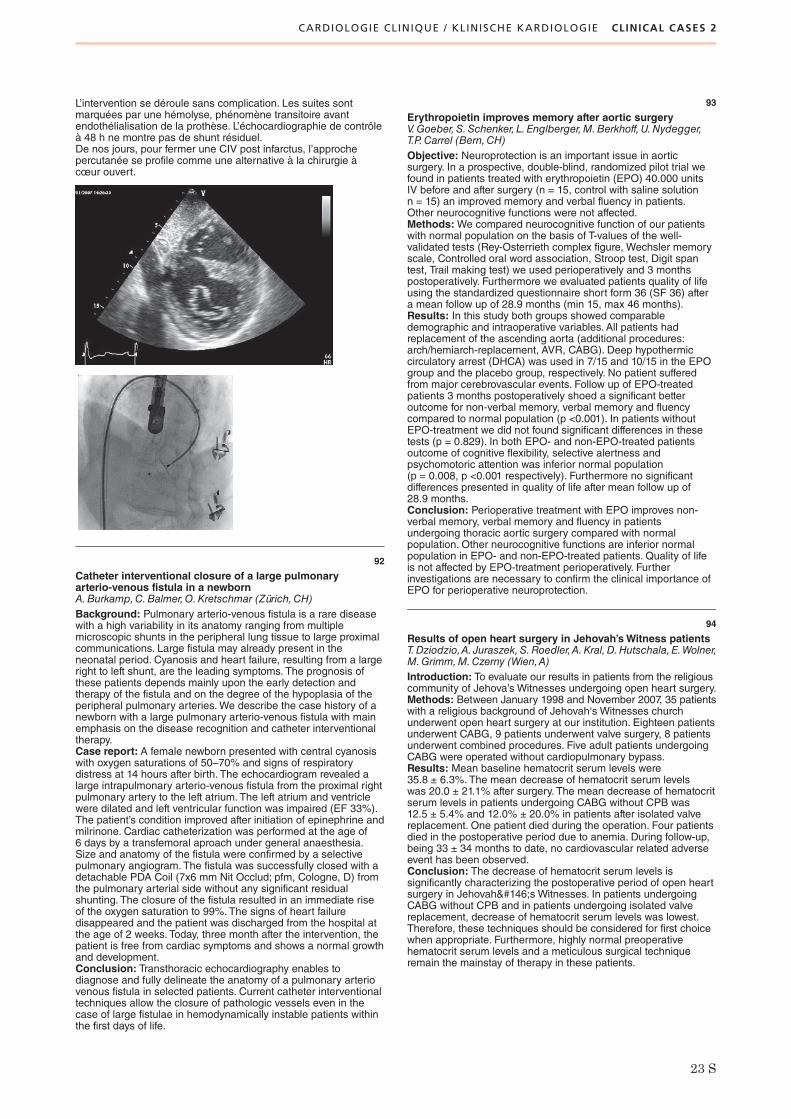
CARDIOLOGIE CLINIQUE / KLINISCHE KARDIOLOGIE CLINICAL CASES 2
23 S
L’intervention se déroule sans complication. Les suites sontmarquées par une hémolyse, phénomène transitoire avantendothélialisation de la prothèse. L’échocardiographie de contrôleà 48 h ne montre pas de shunt résiduel.De nos jours, pour fermer une CIV post infarctus, l’approchepercutanée se profile comme une alternative à la chirurgie àcœur ouvert.
92
Catheter interventional closure of a large pulmonaryarterio-venous fistula in a newbornA.Burkamp,C.Balmer,O.Kretschmar (Zürich,CH)Background: Pulmonary arterio-venous fistula is a rare diseasewith a high variability in its anatomy ranging from multiplemicroscopic shunts in the peripheral lung tissue to large proximalcommunications. Large fistula may already present in theneonatal period. Cyanosis and heart failure, resulting from a largeright to left shunt, are the leading symptoms. The prognosis ofthese patients depends mainly upon the early detection andtherapy of the fistula and on the degree of the hypoplasia of theperipheral pulmonary arteries.We describe the case history of anewborn with a large pulmonary arterio-venous fistula with mainemphasis on the disease recognition and catheter interventionaltherapy.Case report: A female newborn presented with central cyanosiswith oxygen saturations of 50–70% and signs of respiratorydistress at 14 hours after birth. The echocardiogram revealed alarge intrapulmonary arterio-venous fistula from the proximal rightpulmonary artery to the left atrium. The left atrium and ventriclewere dilated and left ventricular function was impaired (EF 33%).The patient’s condition improved after initiation of epinephrine andmilrinone. Cardiac catheterization was performed at the age of6 days by a transfemoral aproach under general anaesthesia.Size and anatomy of the fistula were confirmed by a selectivepulmonary angiogram. The fistula was successfully closed with adetachable PDA Coil (7x6 mm Nit Occlud; pfm, Cologne, D) fromthe pulmonary arterial side without any significant residualshunting. The closure of the fistula resulted in an immediate riseof the oxygen saturation to 99%. The signs of heart failuredisappeared and the patient was discharged from the hospital atthe age of 2 weeks. Today, three month after the intervention, thepatient is free from cardiac symptoms and shows a normal growthand development.Conclusion: Transthoracic echocardiography enables todiagnose and fully delineate the anatomy of a pulmonary arteriovenous fistula in selected patients. Current catheter interventionaltechniques allow the closure of pathologic vessels even in thecase of large fistulae in hemodynamically instable patients withinthe first days of life.
93
Erythropoietin improves memory after aortic surgeryV.Goeber,S.Schenker, L. Englberger,M.Berkhoff,U.Nydegger,T.P.Carrel (Bern,CH)Objective: Neuroprotection is an important issue in aorticsurgery. In a prospective, double-blind, randomized pilot trial wefound in patients treated with erythropoietin (EPO) 40.000 unitsIV before and after surgery (n = 15, control with saline solutionn = 15) an improved memory and verbal fluency in patients.Other neurocognitive functions were not affected.Methods:We compared neurocognitive function of our patientswith normal population on the basis of T-values of the well-validated tests (Rey-Osterrieth complex figure, Wechsler memoryscale, Controlled oral word association, Stroop test, Digit spantest, Trail making test) we used perioperatively and 3 monthspostoperatively. Furthermore we evaluated patients quality of lifeusing the standardized questionnaire short form 36 (SF 36) aftera mean follow up of 28.9 months (min 15, max 46 months).Results: In this study both groups showed comparabledemographic and intraoperative variables. All patients hadreplacement of the ascending aorta (additional procedures:arch/hemiarch-replacement, AVR, CABG). Deep hypothermiccirculatory arrest (DHCA) was used in 7/15 and 10/15 in the EPOgroup and the placebo group, respectively. No patient sufferedfrom major cerebrovascular events. Follow up of EPO-treatedpatients 3 months postoperatively shoed a significant betteroutcome for non-verbal memory, verbal memory and fluencycompared to normal population (p <0.001). In patients withoutEPO-treatment we did not found significant differences in thesetests (p = 0.829). In both EPO- and non-EPO-treated patientsoutcome of cognitive flexibility, selective alertness andpsychomotoric attention was inferior normal population(p = 0.008, p <0.001 respectively). Furthermore no significantdifferences presented in quality of life after mean follow up of28.9 months.Conclusion: Perioperative treatment with EPO improves non-verbal memory, verbal memory and fluency in patientsundergoing thoracic aortic surgery compared with normalpopulation. Other neurocognitive functions are inferior normalpopulation in EPO- and non-EPO-treated patients. Quality of lifeis not affected by EPO-treatment perioperatively. Furtherinvestigations are necessary to confirm the clinical importance ofEPO for perioperative neuroprotection.
94
Results of open heart surgery in Jehovah’sWitness patientsT.Dziodzio,A. Juraszek,S.Roedler,A.Kral,D.Hutschala, E.Wolner,M.Grimm,M.Czerny (Wien,A)Introduction: To evaluate our results in patients from the religiouscommunity of Jehova’s Witnesses undergoing open heart surgery.Methods: Between January 1998 and November 2007, 35 patientswith a religious background of Jehovah‘s Witnesses churchunderwent open heart surgery at our institution. Eighteen patientsunderwent CABG, 9 patients underwent valve surgery, 8 patientsunderwent combined procedures. Five adult patients undergoingCABG were operated without cardiopulmonary bypass.Results: Mean baseline hematocrit serum levels were35.8 ± 6.3%. The mean decrease of hematocrit serum levelswas 20.0 ± 21.1% after surgery. The mean decrease of hematocritserum levels in patients undergoing CABG without CPB was12.5 ± 5.4% and 12.0% ± 20.0% in patients after isolated valvereplacement. One patient died during the operation. Four patientsdied in the postoperative period due to anemia. During follow-up,being 33 ± 34 months to date, no cardiovascular related adverseevent has been observed.Conclusion: The decrease of hematocrit serum levels issignificantly characterizing the postoperative period of open heartsurgery in Jehovah’s Witnesses. In patients undergoingCABG without CPB and in patients undergoing isolated valvereplacement, decrease of hematocrit serum levels was lowest.Therefore, these techniques should be considered for first choicewhen appropriate. Furthermore, highly normal preoperativehematocrit serum levels and a meticulous surgical techniqueremain the mainstay of therapy in these patients.

COMMUNICATIONS LIBRES 8 / FREIE MITTEILUNGEN 8 NEW “RISK FACTORS”
24 S
95
Systemic vascular dysfunction in children conceived byassisted reproductive technologiesS.F.Rimoldi, S.Garcin,S.F. de Marchi,T.Stuber,H.Duplain,M.Germond,C.Sartori, U.Scherrer,Y.Allemann (Bern, Lausanne,CH)Background: Environmental influences acting early in lifepredispose to cardiovascular disease in adulthood. Assistedreproductive technologies (ART) have allowed millions of infertilecouples to have children. ART involves the manipulation of earlyembryos at a time when they may be particularly vulnerable toexternal disturbances. Accordingly, ART has been found tomodulate the epigenome. The safety of ART for long-term healthis, therefore, of utmost importance, but there is little information.We recently showed that children born after ART display vasculardysfunction at high altitude, a condition known to facilitate thedetection of endothelial dysfunction.We wondered whether asimilar dysfunction was also present under normoxic conditions.Methods: 65 healthy singletons born from ART (mean age11.1 ± 2.4 y, 27 girls) and 57 age-, sex- and body weight-matchedcontrols (11.9 ± 2.3 y, 30 girls) were studied al low altitude (Bern,540 m).We assessed the flow-mediated endothelium-dependentvasodilation (FMD) of the brachial artery, its endothelium-independent vasodilation (glycerin trinitrate, 250 μg) and pulsewave velocity (PWV), a proxy of arterial stiffness. 8-isoprostaglandin F2-α (8-iPF2a) plasma concentration wasmeasured as a proxy of oxidative stress.Results: The major new finding was that children born after ARTdisplayed marked systemic vascular dysfunction under normoxia:FMD was 20% smaller (6.7 ± 1.6 vs. 8.6 ± 1.7%, P <0.0001, fig. 1)and PWV significantly faster (10.0 ± 2.7 vs. 8.3 ± 1.8 m/sec,P <0.0001, fig. 2) in children born after ART. This vasculardysfunction was not related to a structural vasculopathy, sinceendothelium-independent vasodilation was similar (13.5 ± 2.3vs. 13.9 ± 2.5%, P = 0.38) in both groups; nor was related todyslipidemia or altered glucose homeostasis, since lipid, glucoseand insulin plasma concentration, as well as the insulinresistance index (HOMA) and glucose tolerance werecomparable. In contrast, 8-iPF2a was significantly higher inchildren born after ART than in controls (83.4 ± 41.5 vs. 55.1 ±28.2 pg/ml, P = 0.0008).Conclusions: ART predispose the offspring to systemic vasculardysfunction that is possibly related to increased oxidative stress.The magnitude of this dysfunction is similar to what has beenreported in children with type 1 diabetes.We hypothesize that thispredispose them to premature cardiovascular disease andspeculate that epigenetic mechanisms may cause vasculardysfunction in offsping of ART.
96
Systemic endothelial dysfunction in normotensive offspringof pre-eclampsiaS.F.Rimoldi,C.Sartori, S.Garcin,M.Villena,U.Scherrer,Y.Allemann (Bern, Lausanne,CH; La Paz,BOL)Background: Epidemiological studies suggest that adverseevents in utero are associated with cardiovascular disease inadulthood, but the mechanisms are not known. Hypoxiaassociated with high-altitude exposure may facilitate the detectionof vascular dysfunction. Recently, it has been suggested thatoffspring of mothers having suffered from preeclampsia arepredisposed to hypertension, but the underlying mechanism isnot clear.We hypothesized that young normotensive offspringof preeclampsia display systemic vascular dysfunction, andspeculated that high-altitude exposure may facilitate the detectionof this problem.Methods:We, therefore, assessed endothelium-dependent(flow-mediated vasodilation, FMD) and -independent (glycerintrinitrate, 250 ìg) vasodilation, vascular stiffness (pulse wavevelocity, PWV), and central arterial blood pressure (applanationtonometry) in 15 healthy normotensive offspring of preeclampsia(mean ± SD age, 14 ± 7 y) who were born at term, and 16matched control subjects. All subjects were born and permanentlyliving at high altitude (3600 m).Results: The major new finding was that young, normotensiveoffspring of preeclampsia displayed systemic endothelialdysfunction, as evidenced by a roughly 25 percent smaller FMD(6.4 ± 1.0 vs. 8.1 ± 1.3%, P = 0.0007, fig. 1) than in controls. Incontrast, arterial stiffness (PWV, 7.8 ± 1.0 vs. 8.0 ± 1.6 m/s) andendothelium-independent vasodilation (16.4 ± 2.7 vs. 15.8 ±3.6%) were comparable in offspring of preeclampsia and controls.The endothelial dysfunction in the offspring of preeclampsia wasnot related to a difference in the central arterial blood pressure(systolic 97 ± 7 vs. 96 ± 8, diastolic 76 ± 7 vs. 74 ± 5 mm Hg,offspring vs. controls) or arterial oxygen saturation (91.6 ± 2.1 vs.90.5 ± 2.0%).Conclusions: Here we provide the very first evidence thatendothelial function in young normotensive offspring ofpreeclampsia is abnormal, whereas arterial stiffness andendothelium-independent vasodilation are still normal.Wespeculate that as in other populations at risk, endothelialdysfunction represents a very early step in the development ofarterial hypertension and cardiovascular disease later in life. Todetect this early vascular dysfunction, the measurement of FMDis more sensitive than the assessment of PWV.
97
Autonomic dysregulation in children conceived withassisted reproductive technologiesT.Stuber,S.F.Rimoldi, P. Lim,S.Garcin,H.Duplain, J.Bloch,C.Sartori, U.Scherrer,Y.Allemann,N.Peters (Zürich,Bern,CH;London,UK; Lausanne,CH)Assisted reproductive technologies (ART) have been found tomodulate the epigenome and we previously reported systemicand pulmonary vascular dysfunction in apparently healthyadolescent children born with ART after exposing them to hypoxicconditions. Effects of ART on autonomic regulation have not beenpreviously studied and we report the first data of HRV in a largegroup of adolescent children born with ART. HRV was measuredin 21 children born with In vitro fertilization (IVF) and 32 born afterintracytoplasmatic sperm injection (ICSI) and in 44 age and sexmatched controls from 5 minutes ECG recordings at low altitude(450 m) and 24h after arriving at the high altitude research station
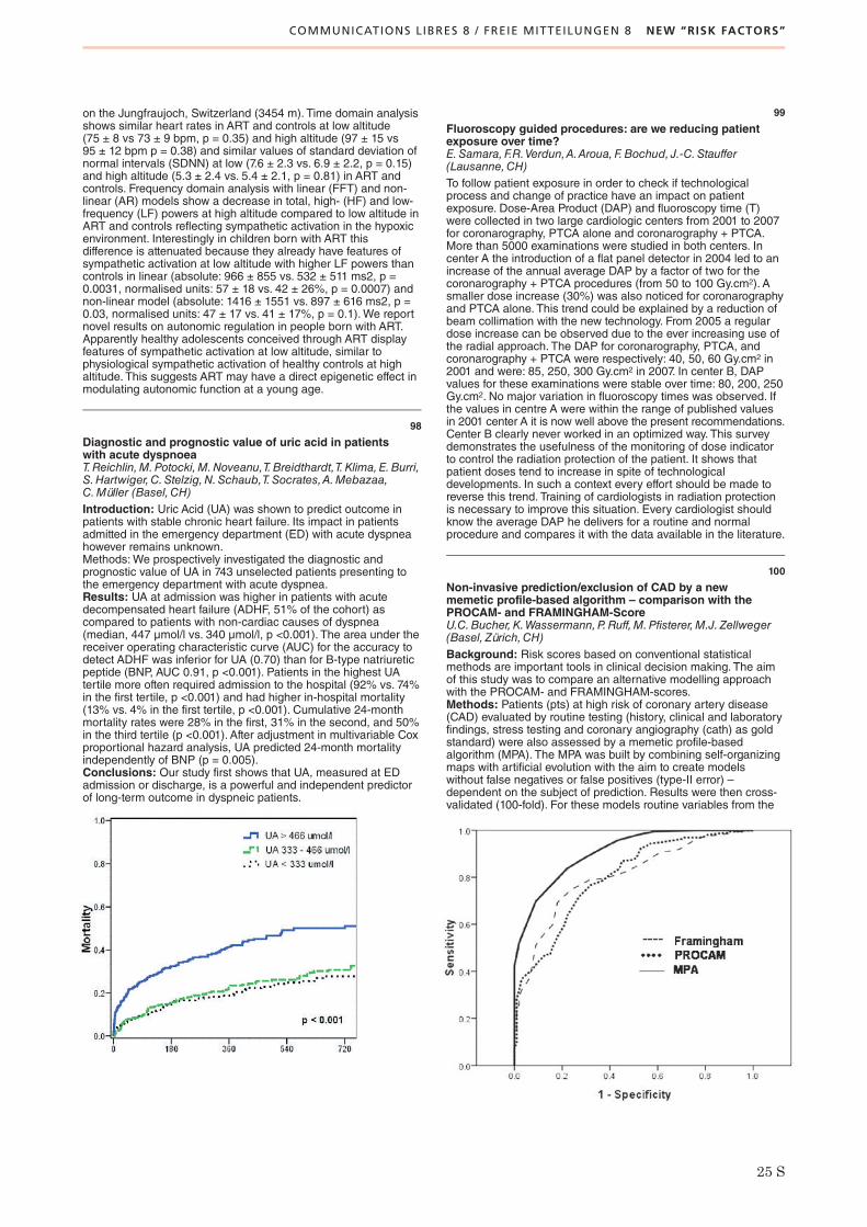
COMMUNICATIONS LIBRES 8 / FREIE MITTEILUNGEN 8 NEW “RISK FACTORS”
25 S
on the Jungfraujoch, Switzerland (3454 m). Time domain analysisshows similar heart rates in ART and controls at low altitude(75 ± 8 vs 73 ± 9 bpm, p = 0.35) and high altitude (97 ± 15 vs95 ± 12 bpm p = 0.38) and similar values of standard deviation ofnormal intervals (SDNN) at low (7.6 ± 2.3 vs. 6.9 ± 2.2, p = 0.15)and high altitude (5.3 ± 2.4 vs. 5.4 ± 2.1, p = 0.81) in ART andcontrols. Frequency domain analysis with linear (FFT) and non-linear (AR) models show a decrease in total, high- (HF) and low-frequency (LF) powers at high altitude compared to low altitude inART and controls reflecting sympathetic activation in the hypoxicenvironment. Interestingly in children born with ART thisdifference is attenuated because they already have features ofsympathetic activation at low altitude with higher LF powers thancontrols in linear (absolute: 966 ± 855 vs. 532 ± 511 ms2, p =0.0031, normalised units: 57 ± 18 vs. 42 ± 26%, p = 0.0007) andnon-linear model (absolute: 1416 ± 1551 vs. 897 ± 616 ms2, p =0.03, normalised units: 47 ± 17 vs. 41 ± 17%, p = 0.1).We reportnovel results on autonomic regulation in people born with ART.Apparently healthy adolescents conceived through ART displayfeatures of sympathetic activation at low altitude, similar tophysiological sympathetic activation of healthy controls at highaltitude. This suggests ART may have a direct epigenetic effect inmodulating autonomic function at a young age.
98
Diagnostic and prognostic value of uric acid in patientswith acute dyspnoeaT.Reichlin,M.Potocki,M.Noveanu,T.Breidthardt,T.Klima,E.Burri,S.Hartwiger,C.Stelzig,N.Schaub,T.Socrates,A.Mebazaa,C.Müller (Basel,CH)Introduction: Uric Acid (UA) was shown to predict outcome inpatients with stable chronic heart failure. Its impact in patientsadmitted in the emergency department (ED) with acute dyspneahowever remains unknown.Methods:We prospectively investigated the diagnostic andprognostic value of UA in 743 unselected patients presenting tothe emergency department with acute dyspnea.Results: UA at admission was higher in patients with acutedecompensated heart failure (ADHF, 51% of the cohort) ascompared to patients with non-cardiac causes of dyspnea(median, 447 μmol/l vs. 340 μmol/l, p <0.001). The area under thereceiver operating characteristic curve (AUC) for the accuracy todetect ADHF was inferior for UA (0.70) than for B-type natriureticpeptide (BNP, AUC 0.91, p <0.001). Patients in the highest UAtertile more often required admission to the hospital (92% vs. 74%in the first tertile, p <0.001) and had higher in-hospital mortality(13% vs. 4% in the first tertile, p <0.001). Cumulative 24-monthmortality rates were 28% in the first, 31% in the second, and 50%in the third tertile (p <0.001). After adjustment in multivariable Coxproportional hazard analysis, UA predicted 24-month mortalityindependently of BNP (p = 0.005).Conclusions: Our study first shows that UA, measured at EDadmission or discharge, is a powerful and independent predictorof long-term outcome in dyspneic patients.
99
Fluoroscopy guided procedures: are we reducing patientexposure over time?E.Samara, F.R.Verdun,A.Aroua,F.Bochud, J.-C.Stauffer(Lausanne,CH)To follow patient exposure in order to check if technologicalprocess and change of practice have an impact on patientexposure. Dose-Area Product (DAP) and fluoroscopy time (T)were collected in two large cardiologic centers from 2001 to 2007for coronarography, PTCA alone and coronarography + PTCA.More than 5000 examinations were studied in both centers. Incenter A the introduction of a flat panel detector in 2004 led to anincrease of the annual average DAP by a factor of two for thecoronarography + PTCA procedures (from 50 to 100 Gy.cm2). Asmaller dose increase (30%) was also noticed for coronarographyand PTCA alone. This trend could be explained by a reduction ofbeam collimation with the new technology. From 2005 a regulardose increase can be observed due to the ever increasing use ofthe radial approach. The DAP for coronarography, PTCA, andcoronarography + PTCA were respectively: 40, 50, 60 Gy.cm2 in2001 and were: 85, 250, 300 Gy.cm2 in 2007. In center B, DAPvalues for these examinations were stable over time: 80, 200, 250Gy.cm2. No major variation in fluoroscopy times was observed. Ifthe values in centre A were within the range of published valuesin 2001 center A it is now well above the present recommendations.Center B clearly never worked in an optimized way. This surveydemonstrates the usefulness of the monitoring of dose indicatorto control the radiation protection of the patient. It shows thatpatient doses tend to increase in spite of technologicaldevelopments. In such a context every effort should be made toreverse this trend. Training of cardiologists in radiation protectionis necessary to improve this situation. Every cardiologist shouldknow the average DAP he delivers for a routine and normalprocedure and compares it with the data available in the literature.
100
Non-invasive prediction/exclusion of CAD by a newmemetic profile-based algorithm – comparison with thePROCAM- and FRAMINGHAM-ScoreU.C.Bucher,K.Wassermann,P.Ruff,M.Pfisterer,M.J. Zellweger(Basel, Zürich,CH)Background: Risk scores based on conventional statisticalmethods are important tools in clinical decision making. The aimof this study was to compare an alternative modelling approachwith the PROCAM- and FRAMINGHAM-scores.Methods: Patients (pts) at high risk of coronary artery disease(CAD) evaluated by routine testing (history, clinical and laboratoryfindings, stress testing and coronary angiography (cath) as goldstandard) were also assessed by a memetic profile-basedalgorithm (MPA). The MPA was built by combining self-organizingmaps with artificial evolution with the aim to create modelswithout false negatives or false positives (type-II error) –dependent on the subject of prediction. Results were then cross-validated (100-fold). For these models routine variables from the
COMMUNICATIONS LIBRES 8 / FREIE MITTEILUNGEN 8 NEW “RISK FACTORS”
COMMUNICATIONS LIBRES 9 / FREIE MITTEILUNGEN 9 PROGENITOR CELLS, MYOCARDIAL REGENERATION
26 S
above mentioned examinations were used. MPA based resultswere then compared with the PROCAM- and FRAMINGHAM-score. The results of the stress test were added to the PROCAM-and FRAMINGHAM-scores to allow an adequate comparison.Results: 260 pts without prior diagnosis of CAD who werereferred for cath were evaluated, mean age 64 ± 10 y, female 77(30%). 160 pts (62%) had evidence of CAD as defined by at leastone stenosis >50% on cath. Using the MPA in this high riskpopulation, 37.9% of healthy patients could be predicted withouttype-II-error. Regarding pts with CAD, a mean of 37.4% of pts
could be predicted without false negatives. Areas under thecurves (AUC) regarding CAD prediction were 0.906, 0.799 and0.803 for MPA, PROCAM and FRAMINGHAM, respectively(figure). The MPA predicted CAD clearly better than the PROCAMand FRAMINGHAM-score.Conclusions: The MPA allowed to identify 38% of pts as healthyand these pts would not have needed cath. Prediction of CAD byMPA including stress test results provided a more accurateprediction of CAD than PROCAM- and FRAMINGHAM-scorescombined with stress test data in this population.
101
Impaired in vivo endothelial repair capacity and acceleratedsenescence of endothelial progenitor cells in preypertensionG.Giannotti, C.Doerries,M.Müller,T.Horvàth,H. Jiang,S.A.Sorrentino,N.Steenken,C.Manes,M.Marzilli, K.L.Rudolph,H.Drexler,U. Landmesser (Hannover,D; Zürich,CH; Ulm,D;Pisa, I)Background: Prehypertension and endothelial dysfunction areassociated with increased cardiovascular (CV) risk and havebeen suggested to contribute to development of hypertension.However, no data are available on in vivo endothelial repaircapacity of EPCs and its relation to endothelial dysfunction inthese patients.We therefore examined EPC in vivo endothelialrepair capacity in patients with prehypertension and hypertensionand analyzed its relation to endothelial function and senescence.Methods: EPCs were isolated from healthy subjects (HS; n = 16),prehypertensive (n = 16) and hypertensive patients (n = 20).Endothelium-dependent, flow-mediated vasodilation (FMD) of theradial artery, a highly sensitive index of endothelial function, wasdetermined by high-resolution ultrasound. In vivo endothelialrepair capacity of EPCs (5x10⁵) was examined after transplantationinto nude mice with carotid injury, and re-endothelialized area(REA) was determined after 3 days. EPC senescence wasassessed by both real time PCR telomere length measurementand senescence-associated β-galactosidase (SA-β-Gal) staining.Nitric oxide (NO) and superoxide production of EPCs werecharacterized by electron spin resonance (ESR) spectroscopyanalysis. Circulating CD34+KDR+ and endothelial apoptoticmicroparticles were examined by FACS analysis.Results: In vivo endothelial repair capacity of EPCs wasmarkedly reduced in prehypertensive patients as compared to HS(REA 15 ± 3 vs. 26 ± 3%; P <0.05), and related to impairedendothelial function (r = 0.38; P <0.05). EPC senescence, asindicated by both, reduced telomere length and increased SA-β-Gal staining, was markedly accelerated in prehypertensive andhypertensive patients and related to impaired in vivo endothelialrepair capacity. NO production of EPCs was significantly reducedin prehypertensive/hypertensive patients. CirculatingCD34+KDR+ EPC levels and endothelial apoptotic microparticleswere not different between the groups.Conclusion: This study demonstrates for the first time that in vivoendothelial repair capacity of EPCs is reduced in prehypertensiveand hypertensive patients and is related to endothelialdysfunction. Accelerated senescence and reduced NO productionof EPCs likely contribute to impaired EPC in vivo endothelialrepair capacity in prehypertension/hypertension. These findingsprovide novel insights into how prehypertension may contribute todevelopment and progression of vascular disease.
102
A novel cardiac progenitor cell population formsspontaneously beating cardiospheresM.-E.Roehrich,A.Spicher,P.Vogt,G.Vassalli (Lausanne, Lugano,CH)Introduction: Different populations of cardiac stem/progenitorcells have been isolated based on surface marker expression.However, these populations do not give rise to beatingcardiomyocytes in vitro, or do so only rarely when co-culturedwith mature cardiomyocytes. The aim of the present study was to
isolate cardiac stem/progenitor cells independent of surfacemarkers.Methods: Cells isolated from murine neonatal and adult hearts(8 weeks of age) were cultured in vitro. Aldehyde dehydrogenase(ALDH) activity, a stem/progenitor cell marker, was determinedusing the fluorescent substrate Aldefluor. Surface and intracellularmarkers were assessed by flow cytometry and immunochemistry,respectively.Results: Neonatal cells adhering to plastic dishes wereexpanded and cultured for >5 months. A cell population displayinga characteristic morphology appeared after 4-8 weeks of culture.This population was significantly enriched for ALDH-bright cellsthat expressed the stem cell-associated marker ABCG2, whichmediates the SP phenotype. This population also expressed thehematopoietic marker CD45, the monocytic marker CD11b, thestem cell marker c-kit (in 80% of the cells), and the progenitor cellmarker CD44. Unlike reported cardiac progenitor cell populations,this one did not express stem cell antigen-1. In monolayer culture,cells formed sperical multicellular aggregates (cardiospheres)characteristic of stem/progenitor cells. In many instances,cardiospheres displayed spontaneous rhythmic contractile activitylasting for up to 1 month. Beating frequency and mechanicalcontractions were responsive to β-adrenergic stimuli, indicatingmature cardiomyocytes.When seeded on biological scaffolds,the progenitor cells proliferated, colonized the scaffolds, andexpressed the cardiac differentiation marker α-sarcomeric actinin.Adult cardiac cells also formed cardiospheres which, however,did not beat spontaneously.Conclusions:We have characterized a population of post-natalcardiac progenitor cells that, unlike published ones, expressesmonocytoid markers along with stem cell markers, and possessesthe unique ability to form spontaneously beating cardiosphereswithout the need for differentiation growth media nor for co-culturing with mature cardiomyocytes. Although CD45 isconsidered to be a lineage-restricted hematopoietic marker, ourdata suggest that CD45-positive cardiac progenitor cells do existand can differentiate into functional cardiomyocytes.
103
Adult human cardiac progenitor cells display highaldehyde dehydrogenase activity and form cardiospheresA.Spicher,M.-E.Roehrich,P.Vogt,G.Vassalli(Lausanne, Lugano,CH)Recent evidence suggests the human heart contains residentstem cells that have regenerative potential. These cells have beenisolated based on different surface or metabolic markers, such ashigh aldehyde dehydrogenase (ALDH) activity. However, nomarker definitively identifies cardiac stem cells. Formation ofspherical cell aggregates (“cardiospheres”) is a hallmark ofcardiac stem cells (analogously to neurospheres formed by neuralstem cells).Methods: Samples of human atrial myocardium were collected atsurgery and either digested or processed as primary explantcultures. ALDH activity was assessed by FACS using afluorescent substrate. Surface markers were determined by FACS.Cell sorting for CD146 (melanoma cell adhesion molecule), amarker of endothelial progenitors and mesenchymal stem cells(MSCs), was performed with antibody-coupled magneticmicrobeads.
COMMUNICATIONS LIBRES 9 / FREIE MITTEILUNGEN 9 PROGENITOR CELLS, MYOCARDIAL REGENERATION
27 S
Results: Atrial myocardium contained a variable number ofALDH-high cells (median: 25%; range: 2–49% of all the non-myocytic cells; n = 22). This number tended to increase with agebut was not correlated with presence of ischemic or valvular heartdisease, diabetes, or hypertension. ALDH-high cells showedpredominantly positive (>70%) for the hematopoietic andendothelial marker CD34, the endothelial marker CD31, theendothelial and MSC marker CD105, CD29, and CD49b. Primaryexplant outgrowths were enriched for ALDH-high cells (61 ± 11%;n = 24). CD146-positive cells expressed CD105, CD54, the earlyendothelial marker CD31 (in 60% of the cells), and the stem cellmarker c-kit (in 40% of the cells). CD146-negative cells expressedNG2 proteoglycan, a pericyte marker. Formation of cardiosphereswas virtually restricted to the CD146-positive subpopulation andwas enhanced in cardiac differentiation medium. Cardiosphere-derived cells were expanded by serial passages from P1 to P5and characterized by FACS. They showed positive (>95%) for theMSC markers CD13, CD44, CD105, CD29 and CD49b, andcontained subpopulations positive for CD90, CD31, CD38,CD49a, CD49d, CD71 or CD106.Conclusions: Adult human myocardium contains ALDH-highcells that can be cultured for >20 passages and formcardiospheres, which are largely composed of MSCs. CD146 cellsorting purifies a subpopulation that contains the cardiosphereforming cells.
104
Heart-infiltrating inflammatory cells contain prominin-1+/CD133+ progenitors contributing to TGF-β mediatedmyocardial fibrosis in inflammatory heart diseaseG.Kania,P.Blyszczuk,M.Stein,A.Valaperti, S.Dirnhofer,C.M.Matter,U. Eriksson (Basel, Zürich,CH)Background: Heart injury often ends up in a common finalpathway of cardiac pathological remodelling and fibrosis,promoting the development of heart failure. Experimentalautoimmune myocarditis (EAM) is a CD4+ T cell mediated mousemodel of inflammatory heart disease.Methods and results: EAM was induced in mice byimmunization with myosin α heavy chain/incomplete Freund’sadjuvant.We showed that inflammatory cells, accumulated withinthe myocardium during acute phase of inflammation, containedaround 30% of prominin-1+ progenitor cells. Analysis of fibrotichearts in immunized chimera mice, lethally irradiated andreconstituted with EGFP+ bone marrow cells, demonstrated that>60% of cardiac fibroblasts originated from bone marrow. Incontrast, we observed no EGFP signal in αMyHC+cardiomyocytes. Furthermore, heart-infiltrating prominin-1+ cells,isolated from the inflamed heart, expanded and differentiated invitro, represented a common progenitor for fibroblasts andmacrophages, but not for cardiomyocytes. Consequently, afterintracardial injection into inflamed/fibrotic heart these cellscontributed to the fibrosis formation, neither prevented EAMprogression, nor participated in the heart regeneration. Ourstudies evidently showed that fibroblast differentiation of heart-infiltrating prominin-1+ cells depended on TGF-β signallingmediating the activation of Smad proteins. Blocking of in vivoTGF-β production with α-TGF-β antibody resulted in thesignificant reduction of the heart fibrosis.Conclusions: Inflamed heart contained bilineage prominin-1+progenitor cells actively contributed to the development ofmyocardial fibrosis, but not to heart regeneration subsequentEAM.
105
Impaired in vivo repair capacity of endothelial progenitorcells in patients with chronic heart failure: potential noveltherapeutic targetsC.Dörries,S.Briand,P. Jakob,C.Besler, P.Mocharla,M.Müller,C.Manes,S.Kaufmann, I.Bühler,M.Hermann,F.Ruschitzka,G.Noll,T.F. Lüscher,U. Landmesser (Zürich,CH)Background: Endothelial progenitor cells (EPCs) promoteendothelial repair and ischemia-induced neovascularisation, andcell therapy is currently intensely explored as a potential noveltherapeutic approach in patients with chronic heart failure (CHF).However, the effectiveness of cell therapy may critically dependon the in vivo repair capacity of patient-derived progenitor cells.
We therefore characterized in vivo repair capacity of EPCs frompatients with CHF due to ischemic and dilated cardiomyopathy(ICM and DCM) as compared to healthy subjects (HS) andanalysed potential novel mechanisms to improve their repaircapacity.Methods: Endothelial progenitor cells were isolated fromperipheral blood from patients with heart failure due to ICM(n = 18) or DCM (n = 16) and healthy subjects (n = 19). In vivoendothelial repair capacity of EPCs was examined aftertransplantation into nude mice with vascular injury. NO andsuperoxide production of EPCs were determined by electronspin resonance spectroscopy. The microRNA profile of EPCswas characterized using a microRNA array (Exiqon; Denmark).Furthermore, the secretom of EPCs was characterized by usinga protein array.Results: In vivo endothelial repair capacity of EPCs derived frompatients with heart failure due to ICM and DCM was markedlyreduced as compared to EPCs from healthy subjects(reendothelialized area: 16.7 ± 1.1 versus 16.6 ± 1.7 versus 25.4 ±2.1%; P <0.05). EPCs from patients with heart failure due to ICMand DCM had an impaired NO production compared to healthysubjects (1892 ± 110.9 versus 1922 ± 144.2 versus 2502 ± 185.7nmol NO / h / 2*106 EPCs; P <0.05), that may, at least in part,account for an impaired endothelial repair capacity. Moreover,microRNA profiling of EPCs indicated miRNA expression in EPCsregulating angiogenic growth factors and homing molecules,thereby identifying a novel target to improve their repair capacity.Conclusions: In vivo endothelial repair capacity of EPCs ismarkedly reduced in patients with heart failure due to ICM andDCM as compared to healthy subjects that may limit theeffectiveness of cell-based therapeutic approaches in thesepatients. Reduced NO bioavailability of EPCs from patients withCHF likely contributes to a reduced repair capacity. Moreover,microRNAs regulating angiogenic growth factors and homingmolecules represent an attractive target to improve repaircapacity of patient-derived EPCs in CHF.
106
Foetal liver kinase receptor (FLT3) is expressed on cardiacmyocytes and mediates pro-survival signalingO.Pfister,V. Lorenz,S.P.Häuselmann,B.I.Rosc-Schlüter, K.Bühler,L.Kenins,A.Wodnar-Filipowicz,G.M.Kuster (Basel,CH)Background: Fetal liver kinase (Flt3) is a member of the tyrosinekinase family of receptors that mediates cell survival via Akt andERK1/2 signaling in hematopoietic cells. To date nothing is knownabout expression and function of Flt3 in cardiac myocytes. In thisstudy we determined Flt3 expression and Flt3-dependent pro-survival signaling in neonatal rat ventricular myocytes (NRVM).Methods: NRVMs were kept under basal conditions with 20%O2/5% CO2 until they were subjected to hypoxia (1% O2/ 5%CO2) for 24-48 hours. Gene expression of Flt3-receptor wasdetermined under basal and hypoxic conditions using quantitativeRT-PCR analysis. Recombinant Flt3 ligand (rhFL) was used toactivate the Flt3-receptor. Phosphorylation of Akt and ERK1/2was determined by Western Blot analysis. Cell survival wasassessed by fluorescent activated cell sorting (FACS) usingpropidium iodid staining.Results: RT-PCR analyses revealed robust gene expression ofFlt3-receptor with time-dependent up-regulation upon exposure tohypoxia (+100 ± 23%, p <0.05, at 16 h and +155 ± 69%, p <0.01,at 24 h of hypoxia, respectively). Administration of rhFL (500ng/ml) to NRVMs induced a significant (p <0.05) transientincrease in Akt- and ERK1/2-phosphorylation, which peaked at10 min (+66 ± 13% for Akt and +79 ± 46% for ERK1/2), therebydocumenting activation of pro-survival signaling in cardiacmyocytes in response to Flt3 receptor-stimulation. In line withactivated pro-survival signaling, administration of rhFL improvedcell survival as compared to vehicle-treated NRVMs in threedifferent in vitro models of ischemia: low oxygen (1% O2), serum-starvation, and exposure to hydrogen peroxide (500 uM),respectively.Conclusion: This is the first evidence of Flt3-induced pro-survivalsignaling in cardiac myocytes. Given a pro-survival role of Flt3 inthe myocardium, inhibition of Flt3 by tyrosine kinase inhibitors asused for the treatment of leukemia might result in increasedcardiotoxicity particularly under conditions of hypoxic stress.

COMMUNICATIONS LIBRES 10 / FREIE MITTEILUNGEN 10 ECHO “ADVANCED”
28 S
107
Real-time three-dimensional echocardiographic assessmentof the right ventricle in comparison with magnetic resonanceimagingG.Leibundgut,A.Rohner,A.M.Bernheim,A.Kessel,M.J. Zellweger,P.Buser,M.Handke (Basel,CH)Background: Right ventricular ejection fraction (RVEF)represents a strong and independent predictor of mortality in leftheart failure. Due to the complexity of right-ventricular geometry,the value of 2D-echocardiography is limited in determiningvolumes and ejection fraction. Real-time 3D echocardiography(RT3DE) has been proven to be reliable in the assessment of leftventricle and is already established in that field.Objectives: Until now it has been unknown whether rightventricular (RV) volumes and function can be reliably assessedby RT3DE.A novel software package enables rapid quantificationof RV size and function through a unique 3D surface model. Aimof the study was to validate the new technology using magneticresonance imaging (MRI) as gold standard.Methods: Patients with normal or abnormal right ventricle dueto various cardiac diseases (coronary artery disease, myocarditis,valvular heart disease, arrhythmogenic right ventricularcardiomyopathy, amyloidosis, iron overload, and connectivetissue disorders) were included in the study. Patients wererecruited following cardiac MRI. Acquisition of a pyramidal datavolume was done by transthoracic RT3DE. Off-line reconstructionwas performed of a dynamic polyhedron model of the RV basedon the endsystolic and enddiastolic contours at predefinedstandard cutting planes. RV volumes and EF were determinedand compared with MRI data. All measurements were done in ablinded fashion.Results: A total of 57 patients were examined. The mean valuesfor right ventricular enddiastolic volume (RVEDV), endsystolicvolume (RVESV), stroke volume (RVSV) and ejection fraction(RVEF) are given in table 1.We found moderate correlationsbetween the measurements obtained by RT3DE and MRI (table 1).Bland-Altmann analysis showed slight to moderate meandifferences for RVEDV (14 ± 26 ml), RVESV (8 ± 16 ml) andRVSV (7 ± 16 ml) but no significant mean difference for RVEF(0.6 ± 8%).Conclusion: RT3DE may become a time- and cost-savingalternative to MRI in the quantitative assessment of the RV.Particularly, there is close agreement between the two modalitiesin the determination of RV systolic function.
108
Regulation der myokardialen Perfusion unter hohendiastolischen Drücken bei externer GegenpulsationS.F.de Marchi, S.Gloekler, S.F.Rimoldi,H.Steck,C.Seiler(Bern,CH)Einleitung: Der externen Gegenpulsation (externalcounterpulsation, ECP) wird eine perfusionssteigernde Wirkungim Myokard zugeschrieben. Die zugrundeliegenden Datenstammen jedoch aus invasiven Messungen in den epikardialenArterien, und nicht aus direkten Perfusionsmessungen.Methoden: 20 gesunde Probanden wurden unter steigendenECP-Druckstufen (0, 80, 160, 240 und 300 mm Hg) mittelsquantitativer Myokard-Kontrastechokardiographie untersucht.Es wurden drei Aufnahmesequenzen auf jeder ECP-Stufedurchgeführt. Die Perfusion wurde in end-diastolischen Bildernausgewertet. Endpunkte waren: der absolute myokardialeBlutfluss (MBF, ml/min/g) und die Fraktion myokardialenBlutvolumens (relatives Blutvolumen rBV, ml/ml). Die arteriellenDruckkurven wurden sphygmometrisch aufgezeichnet unddigitalisiert. Perfusion und Druckkurven wurden mit Hilfe eigenerMatlab-Softwares analysiert. Der mikrovaskuläre Widerstandwurde wie folgt berechnet: diastolischer Druck / MBF (inmmHg*min/ml*g).Wiederholte Messungen wurden mittels ANOVArepeated measures und dem Dunnett’s Post Test verglichen. Auf-
bzw. absteigende Trends wurden mittels Post Test für signifikanteTrends ermittelt.Ergebnisse: Es gab eine signifikante Steigerung desdiastolischen Blutdruckes unter steigenden ECP-Stufen vonanfänglich 80 ± 11 auf maximal 134 ± 14 mm Hg (p <0.0001,signifikant ab Stufe 80mmHg). Das rBV zeigte einen signifikantenAnstieg von 0.11 ± 0.04 auf maximal 0.14 ± 0.04 (p = 0.01). DerMBF zeigte einen signifikanten Aufwärtstrend von 1.12 ± 0.38 bis1.44 ± 0.88 ml/min/g (p <0.05). Der mikrovaskuläre Widerstanddes Myokards zeigte ab Stufe 240mmHg eine signifikanteZunahme von 78 ± 29 auf maximal 127 ± 77 mm Hg*min/ml*g(p = 0.0004).Schlussfolgerungen: Unter ECP kommt es bei hohen Gegen-pulsationsdrücken zu einer Zunahme des intramyokardialenBlutvolumens, sowie zu einer Widerstandserhöhung der Mikro-zirkulation. Letztere scheint einer Perfusionssteigerung effiziententgegenzuwirken.Wir interpretieren sie als eine bisher nichtbeschriebene Gegenregulation zum forcierten Perfusionsdruck.Die in früheren invasiven Studien gezeigte signifikanteFlusssteigerung in den epikardialen Koronararterien unter ECPkönnte somit bei hohen Gegenpulsationsdrücken in erheblichemMasse durch Rekrutierung von arterio-arteriellen oder -venösenShunts an der Mikrozirkulation vorbeigeleitet werden.
109
Geometric versus non-geometric methods to quantifyright ventricular function by echocardiographyM.Schwerzmann,K.Wustmann,A.Wahl, F.Praz,C.Eigenmann,C.Seiler (Bern,CH)Background: Echocardiographic assessment of right ventricular(RV) systolic function is important and challenging. Differentgeometric and non-geometric methods have been proposed toquantify RV systolic function by echocardiography. It is unknownhow these methods correlate with RV ejection fraction (EF)measured by cardiac magnetic resonance imaging (MRI) underdifferent RV loading conditions.Methods: In 43 stable patients (pts) undergoing cardiac MRIwithin 60 days of echocardiographic assessment of RV function,we compared MRI-based RV-EF with (1) the fractional areachange (FAC) in the 4-chamber-view, (2) the peak systolicvelocity of the lateral tricuspid annulus (DTI-S), (3) its amount ofsystolic apical motion (TAM) and with (4) the RV myocardialperformance index (MPI) derived from the RV isovolumetricindices. Binary exposure variables for increased RV pressure load(defined as RV systolic pressure >40 mm Hg, n = 14) or increasedRV volume load (presence of severe pulmonary regurgitation,n = 14) were added to the linear regression model used forcalculating the correlations between RV-EF and FAC, DTI-S,TAM and MPI, respectively.Results: Mean RV-EF was 45% (range 22–71%) and mean timeinterval between MRI and echocardiography was 20 days.
COMMUNICATIONS LIBRES 10 / FREIE MITTEILUNGEN 10 ECHO “ADVANCED”
29 S
Table 1 shows the corresponding correlations between theechocardiographic assessment of RV systolic function and MRI-derived RV-EF without (column 2) and with (column 3) exposurevariables for different loading conditions. Column 4 and 5 indicatethe p-value of the exposure variables in the correspondingmultiple regression model.Conclusion: FAC correlates best with MRI-derived RV-EF and itscorrelation is not affected by increased RV volume- or pressureload. In this study, non-geometric models correlate less well withRV-EF, and the MPI is particularly sensitive to loading conditionswhen compared with MRI-derived RV-EF.
110
Reduced atrial emptying after orthotopic hearttransplantation masquerading as restrictive transmitralDoppler flow patternP.Pascale,P.Monney, X. Jeanrenaud,N.Aebischer,P.Yerly, P.Vogt,E.Pruvot, J. Schläpfer (Lausanne,CH)Background: Pulsed-waved Doppler evaluation of transmitralblood flow velocity provides insights into left atrial (LA)mechanical function in addition to its widespread use for theevaluation of the left ventricular diastolic function. An early (E) tolate (A) diastolic filling velocities ratio greater than 2 is typicallyseen in the late stage of diastolic dysfunction. However, thistransmitral filling pattern has also been noted as a consequenceof the transient depressed LA contractility that occurs aftercardioversion of atrial fibrillation.We hypothesized that theimpaired function and dilation of the LA demonstrated afterstandard biatrial anastomotic technique of orthotopictransplantation (BAT) could also lead to this pseudorestrictivepattern.We therefore aimed to evaluate in these patients thetransmitral Doppler flow pattern and the lateral tissue Dopplermitral annular velocity (Ea) as preload-independent index of LVrelaxation.Method: This retrospective cohort study included consecutivepatients after BAT who had one or more transthoracicechocardiograms at our university-based hospital between 2005and 2007. Criteria for inclusion included the presence of sinusrhythm and adequate echocardiographic two-dimensionalimaging. Patients with fusion of the E and A velocities defined asan E velocity exceeding 20cm/s at the beginning of the A velocitywere excluded.Results: The study population comprised 68 patients 91 ± 73months after BAT. 13 patients were excluded because of fusionof the E and A velocities. 38 patients (69%) had an E/A ratio>2 (2.93 ± 0.6). LV ejection fraction (61% ± 11%) and LA volume(125 ± 63 ml) did not significantly differ compared to patients withan E/A ratio ≤2. In patients with an E/A ratio > 2, the E/Ea ratiowas 7.5 ± 3. A ratio >10 consistent with significant reduction in theLV compliance was observed in only 5 patients, one of which hadtransplant rejection.Conclusion: Our study suggests that a pseudorestrictive fillingpattern may frequently be observed after BAT. A truly restrictivefilling related to advanced diastolic dysfunction was deemedunlikely in most patients considering an elevated Ea velocity withnormal E/Ea ratio. Recognition of this pseudorestrictive patternnot only avoids misdiagnosis of diastolic dysfunction, but mayalso provide a clue to the diagnosis of other causes of impairedLA function as an occult atrial tachyarrhythmia.
111
Right Ventricle Myocardial Performance Index in patientswith severe COPD and secondary pulmonary hypertensiontreated with BosentanM.Di Valentino,A. Linka,M.Tamm,D.Stolz (Bellinzona,Winterthur,Basel,CH)Background: Right ventricle myocardial performance index (RV-MPI) is a non-invasive Doppler measurement of global ventriclefunction (systole and diastole) defined as ratio of the sum ofisovolumic contraction time and isovolumic relaxation time dividedby the systolic ejection time (normal value mean 0.28 SD 0.04).An increased RV-MPI is associated with either right ventricledysfunction or pulmonary hypertension. Few data are knownabout the value of RV-MPI in patients with severe chronic
obstructive pulmonary disease (COPD) treated with bosentan,an endothelin receptor antagonist.Aim: To asses the feasibility of the RV-MPI in patients with severeCOPD and associated pulmonary hypertension (PH) treated withbosentan.Methods: Double-blind, placebo-controlled study. Patients withsevere or very severe COPD (GOLD III-IV) were randomlyassigned in a 2:1 ratio to receive either bosentan or placebo for12 weeks. RV-MPI was calculated at baseline and after threemonths of treatment.Results: Thirty patients were included in the study; 20 wereassigned to bosentan (69.5 SD 8.8 yo, 1 male) and 10 to placebo(65 SD 7.9 yo, 7 male). Systolic pulmonary arterial pressure wassimilar in the two groups at baseline (median 32 mm Hg [IQR 29–38] vs. 37 mm Hg [20–42], p = 0.779) and after 3 months (median30 mm Hg [26–34] vs. 33 mm Hg [29–39], p = 0.288). RV-MPI atbaseline was 0.49 [0.44–0.79] in the bosentan group and 0.42[0.35–0.49] in the placebo group, p = 0.05. After 12 weeks oftreatment, RV-MPI was 0.44 [0.3-0.64] in the bosentan groupplacebo group and 0.56 (0.41–0.76) in the placebo group(p = 0.006).Conclusion: Measurement of RV-MPI is feasible in patients withsevere COPD and COPD-associated PAH. Bosentan therapyimproves RV-MPI significantly. These results should be confirmedin a larger cohort.
112
Mitral E/Ea is a marker of systolic dysfunction but not ofelevated filling pressure in paediatric patients withcardiomyopathyC.Tissot, L.B.Willis, K.L.Villavicencio,C.M.Phelps,A.K.Younoszai,S.D.Miyamoto (Denver,USA)Introduction: Doppler echocardiography is used to assessdiastolic function and correlates with filling pressures in adults.The objective of this study is to identify echocardiographicmarkers able to differentiate systolic from diastolic dysfunctionand echocardiographic markers of increased filling pressures inchildren with cardiomyopathy.Methods: All children with dilated (DCM), hypertrophic (HCM)or restrictive (RCM) cardiomyopathy who had a cardiaccatheterization and echocardiogram between 2006 and 2008were reviewed. Patients were divided into: primarly systolicdysfunction (SD), ie patients with DCM and primarly diastolicdysfunction (DD), ie patients with HCM or RCM.Echocardiographic measurements included ejection fraction (EF),dP/dt of mitral regurgitation (MR), mitral inflow (MV), tricuspidinflow (TV) and pulmonary vein (PV) Doppler, tissue Dopplervelocities (Ea), color M-mode inflow propagation velocity (Vp).Results: Thirty-four catheterizations and echocardiograms wereperformed in 29 children with an interval between studies of 5 ±58 days, of those 21 were in the SD group and 8 in the DD group.The mean age was 9.5 ± 6.2 years and was lower in the SDgroup (8 ± 6.7 vs 13 ± 3 yr, p <0.01). Compared to patients withDD, the SD group had lower MVE (0.76 ± 0.25 vs 0.94 ± 0.28m/s, p <0.05), PVS and PVD wave (0.41 ± 0.17 vs 0.56 ± 0.18m/s, p <0.01; 0.47 ± 0.17 vs 0.58 ± 0.15 m/s, p <0.05respectively) and PVA wave reversal (0.24 ± 0.12 vs 0.36 ± 0.14m/s, p <0.01) velocities, MVE deceleration time (0.09 ± 0.03 vs0.12 ± 0.03 s, p <0.01), MVA (0.09 ± 0.03 vs 0.11 ± 0.02 s,p <0.05) and PVA (0.09 ± 0.03 vs 0.15 ± 0.04 s, p <0.01)durations, and MVEa veloctiy (10 ± 5 vs 13 ± 4 cm/s, p <0.05).The SD group had lower Vp (67 ± 35, SD vs 106 ± 55, p <0.05)and dP/dt of MR (620 ± 251, SD vs 1648 ± 520, DD, p <0.01).Mitral E/Ea was higher in the SD group (12 ± 5, SD vs 6 ± 2,DD, p <0.01) and correlated with systolic dysfunction (11 ± 5,EF <55% vs 7 ± 4, EF ≥55%, p <0.05) but not with filling pressure(10 ± 5, PCWP ≥12 vs 10 ± 6, PCWP <12 mm Hg, p = ns).Tricuspid E/Ea correlated with mean PAP (27 ± 10 mm Hg,TVE/Ea ≤8 vs 19 ± 3 mm Hg, TVE/Ea >8, p <0.05).Conclusion: Mitral E/Ea correlates with systolic dysfunction butnot with filling pressures in children with cardiomyopathy, whereastricuspid E/Ea correlates with pulmonary arterial pressure.Prospective studies in larger populations are needed to confirmthese findings.
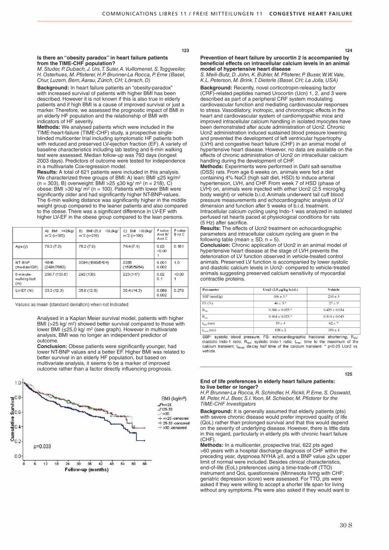
COMMUNICATIONS LIBRES 11 / FREIE MITTEILUNGEN 11 CONGESTIVE HEART FAILURE
30 S
123
Is there an “obesity paradox” in heart failure patientsfrom the TIME-CHF population?M.Studer,P.Dubach, J.Urs,T.Suter,A.Vuillomenet, S.Toggweiler,H.Osterhues,M.Pfisterer,H.P.Brunner-La Rocca,P.Erne (Basel,Chur, Luzern,Bern,Aarau, Zürich,CH; Lörrach,D)Background: In heart failure patients an “obesity-paradox”with increased survival of patients with higher BMI has beendescribed. However it is not known if this is also true in elderlypatients and if high BMI is a cause of improved survival or just amarker. Therefore, we assessed the prognostic impact of BMI inan elderly HF population and the relationship of BMI withindicators of HF severity.Methods:We analysed patients which were included in theTIME-heart-failure (TIME-CHF) study, a prospective single-blinded multicenter trial including symptomatic HF patients bothwith reduced and preserved LV-ejection fraction (EF). A variety ofbaseline characteristics including lab testing and 6-min walkingtest were assessed. Median follow-up was 793 days (longest2003 days). Predictors of outcome were tested for independencein a multivariate Cox-regression model.Results: A total of 621 patients were included in this analysis.We characterized three groups of BMI: A) lean: BMI ≤25 kg/m2
(n = 303), B) overweight: BMI >25 ≤30 kg/ m2 (n = 218), C)obese: BMI >30 kg/ m2 (n = 100). Patients with lower BMI weresignificantly older and had significantly higher NT-BNP-values.The 6-min walking distance was significantly higher in the middleweight group compared to the leaner patients and also comparedto the obese. There was a significant difference in LV-EF withhigher LV-EF in the obese group compared to the lean persons.
Analysed in a Kaplan Meier survival model, patients with higherBMI (>25 kg/ m2) showed better survival compared to those withlower BMI (≤25,0 kg/ m2 (see graph). However in multivariateanalysis, BMI was no longer an independent predictor ofoutcome.Conclusion: Obese patients were significantly younger, hadlower NT-BNP values and a better EF. Higher BMI was related tobetter survival in an elderly HF population, but based onmultivariate analysis, it seems to be a marker of improvedoutcome rather than a factor directly influencing prognosis.
124
Prevention of heart failure by urocortin 2 is accompanied bybeneficial effects on intracellular calcium levels in an animalmodel of hypertensive heart diseaseS.Meili-Butz,D. John,K.Bühler,M.Pfisterer, P.Buser,W.W.Vale,K.L.Peterson,M.Brink,T.Dieterle (Basel,CH; La Jolla,USA)Background: Recently, novel corticotropin-releasing factor(CRF)-related peptides named Urocortin (Ucn) 1, 2, and 3 weredescribed as part of a peripheral CRF system modulatingcardiovascular function and mediating cardiovascular responsesto stress. Vasodilatory, inotropic, and chronotropic effects in theheart and cardiovascular system of cardiomyopathic mice andimproved intracellular calcium handling in isolated myocytes havebeen demonstrated after acute administration of Ucn2. ChronicUcn2 administration induced sustained blood pressure loweringand prevented the development of left ventricular hypertrophy(LVH) and congestive heart failure (CHF) in an animal model ofhypertensive heart disease. However, no data are available on theeffects of chronic administration of Ucn2 on intracellular calciumhandling during the development of CHF.Methods: Experiments were performed in Dahl salt-sensitive(DSS) rats. From age 6 weeks on, animals were fed a dietcontaining 4% NaCl (high salt diet, HSD) to induce arterialhypertension, LVH, and CHF. From week 7 of HSD (phase ofLVH) on, animals were injected with either Ucn2 (2.5 microg/kgbody weight) or vehicle b.i.d. Animals underwent tail cuff bloodpressure measurements and echocardiographic analysis of LVdimension and function after 5 weeks of b.i.d. treatment.Intracellular calcium cycling using Indo-1 was analyzed in isolatedperfused rat hearts paced at physiological conditions for rats(5 Hz) after sacrifice.Results: The effects of Ucn2 treatment on echocardiographicparameters and intracellular calcium cycling are given in thefollowing table (mean ± SD, n = 5).Conclusion: Chronic application of Ucn2 in an animal model ofhypertensive heart disease at the stage of LVH prevents thedeterioration of LV function observed in vehicle-treated controlanimals. Preserved LV function is accompanied by lower systolicand diastolic calcium levels in Ucn2- compared to vehicle-treatedanimals suggesting preserved calcium sensitivity of myocardialcontractile proteins.
125
End of life preferences in elderly heart failure patients:to live better or longer?H.P.Brunner-La Rocca,R.Schindler,H.Rickli, P. Erne,S.Osswald,M.Peter,H.J.Beer,S.I.Yoon,M.Schieber,M.Pfisterer for theTIME-CHF InvestigatorsBackground: It is generally assumed that elderly patients (pts)with severe chronic disease would prefer improved quality of life(QoL) rather than prolonged survival and that this would dependon the severity of underlying disease. However, there is little datain this regard, particularly in elderly pts with chronic heart failure(CHF).Methods: In a multicenter, prospective trial, 622 pts aged>60 years with a hospital discharge diagnosis of CHF within thepreceding year, dyspnoea NYHA ≥II, and a BNP value ≥2x upperlimit of normal were included. Besides clinical characteristics,end-of-life (EoL) preferences using a time-trade-off (TTO)instrument and QoL questionnaire (Minnesota living with CHF;geriatric depression score) were assessed. For TTO, pts wereasked if they were willing to accept a shorter life span for livingwithout any symptoms. Pts were also asked if they would want to

COMMUNICATIONS LIBRES 11 / FREIE MITTEILUNGEN 11 CONGESTIVE HEART FAILURE
31 S
be resuscitated if this would be required. Predictors of EoLpreferences were evaluated.Results: Pts were 79 ± 8 (60–97) years of age, 59% male with amean LVEF of 35 ± 13%. Main causes of CHF were coronary(53%) and hypertensive (28%) heart disease. Of the 622 pts, 556(89%) replied to the TTO question, of which 74% were not willingto trade survival time for improved QoL.Willingness to trade timefor QoL was higher in pts with NYHA ≥III (33 vs 21%, p = .001),lower haemoglobin (12.7 ± 1.9 vs 13.2 ± 1.8 g/dl, p = .002), higherdepression score (5.1 ± 3.3 vs 4.1 ± 3.0, p = .003), and who wereliving alone (34 vs 22%, p = .002; all independent predictors). Age≥75 years, female gender, LVEF >45% and osteoporosis wereadditional univariate predictors of this willingness. Of 603 (97%)expressing their resuscitation preference, 51% would havewanted resuscitation, 39% not, and 10% were undetermined. Noresuscitation was preferred by older pts, women, those livingalone and with a higher depression score in multivariate analysis.All other variables were not related to this preference or only inunivariate analysis (hemoglobin, history of stroke, arthritis).Conclusions: The majority of elderly CHF pts prefer longevityover QoL and half would want to be resuscitated. EoL preferenceswere mainly determined by symptoms, social status, depression,and to certain degree also by age and female gender.
126
International organ exchange or how can we increase thenumber of heart transplantations in Switzerland?D.Vernet, S.Schneider,H.-P.Marti, F.F. Immeron behalf of Swisstransplant (Bern,CH)Introduction: Organ shortening is an important issue in generalan especially in heart transplantations (HTX) in Switzerland.International exchange within the European organ exchangeorganisations (EOEO) is of increasing interest. The aim of thestudy is to analyse the frequency of foreign heart offers toSwisstransplant and the involved centers and the reasons foracceptance or refusal.Patients and methods: A retrospective study was conductedbetween 01/04 and 18/11/2008 to analyse all foreign heart offers.Reaction time (Swisstransplant and transplant center), quality ofmedical information and decision process were evaluated. Thedata were collected from the Swisstransplant database and fromthe EOEO organ offer form sheets. Results were analyzed inrelation to medical decisions and logistic aspects.Results: Overall 289 foreign donor hearts have been offered.Average age of the donor was 24.4 years. Nearly 60% of thedonors were under the age of 30 years. 8 hearts have beenaccepted and were successfully transplanted, corresponding to5.8% of the total number of HTX in this time period inSwitzerland. In 12 foreign offers decision time was too long andthe organs were no longer available at the time of decision, whichwould have increased the number of HTX of 14.5%. 90% of theaccepted organs were within a range of 1000 km distancebetween donor and transplant centre. Logistic refusal was foundin 44 offers mainly due to long distances (>1000 km).Conclusion: Fast decision making would allow to substantiallyincrease the number of heart transplants in Switzerland. Theintroduction of new technologies, such as perfusion machines,must be considered in the close future. Transports over longdistances would be tolerable and high quality organs could beobtained.
127
Mitral annular shape, size, and motion in normals and inpatients with cardiomyopathy: evaluation with computedtomographyP.Trigo Trindade, L.Desbiolles,P.Stolzmann,S. Leschka,H.Scheffel,A.Plass,T.Schertler,M.Genoni, P.Cattin,B.Marincek,T. Frauenfelder,H.Alkadhi (Zürich,CH)Objectives: To assess prospectively, in healthy subjects andin patients with dilated (DCM) and hypertrophic obstructivecardiomyopathy (HOCM), the three-dimensional (3D) shape,size, and motion of the mitral annulus (MA) using computedtomography (CT).Materials and methods: Twenty patients with no cardiacabnormalities (referred to as normals), 15 with DCM, and 15 withHOCM as determined by echocardiography underwent contrast-enhanced, retrospectively electrocardiography-gated 64-slice CTof the heart. The MA was manually segmented in 10%-steps ofthe RR-interval with a dedicated 3D-software employing the point-wrap algorithm. The MA shape, area size, change of the MA area,and apicobasal MA motion throughout the cardiac cycle weredetermined and compared between the groups. Intercommissuraldistances were measured with CT and compared with findingsduring surgery in 9 patients undergoing ring annuloplasty.Results: The MA was non-planar in all phases and subjects,being largest in diastole and smallest in systole. The MA area wassignificantly (P <.001) larger in patients with DCM (11.5 ± 4.1 cm2/m2) as compared to normals (5.5 ± 0.9cm2/m2) and HOCM (4.7 ±0.9 cm2/m2). The change of MA area throughout the cardiac cyclewas significantly (P <.017) smaller in patients with DCM (12.2 ±3.3%/m2) as compared to normals (20.0 ± 7.9%/m2) and HOCM(20.5 ± 7.7%/m2). The mean apicobasal motion was significantly(P <.017) smaller in patients with DCM (2.2 ± 1.0 mm/m2) ascompared to normals (3.6 ± 0.8 mm/m2) and HOCM (2.7 ± 0.7mm/m2). Intercommisural distances as determined by CT showeda good correlation (r = 0.68,P <0.05) with intraoperativemeasurements (mean difference 0.44 mm, limits of agreement –2.73–3.62 mm).Conclusion: Our study provides in-vivo human data on the 3Dshape, size, and motion of the MA in healthy subjects. Significantvariations in size and motion of the MA were noted betweennormals and patients with DCM and HOCM.This imagingtechnique may prove extremely valuable in the choice of aprosthetic ring prior to surgical mitral valve reconstruction and inplanning interventional mitral ring annuloplasty. Further studiesare needed to address the impact of this method on clinicaloutcome after surgery or interventional procedures.
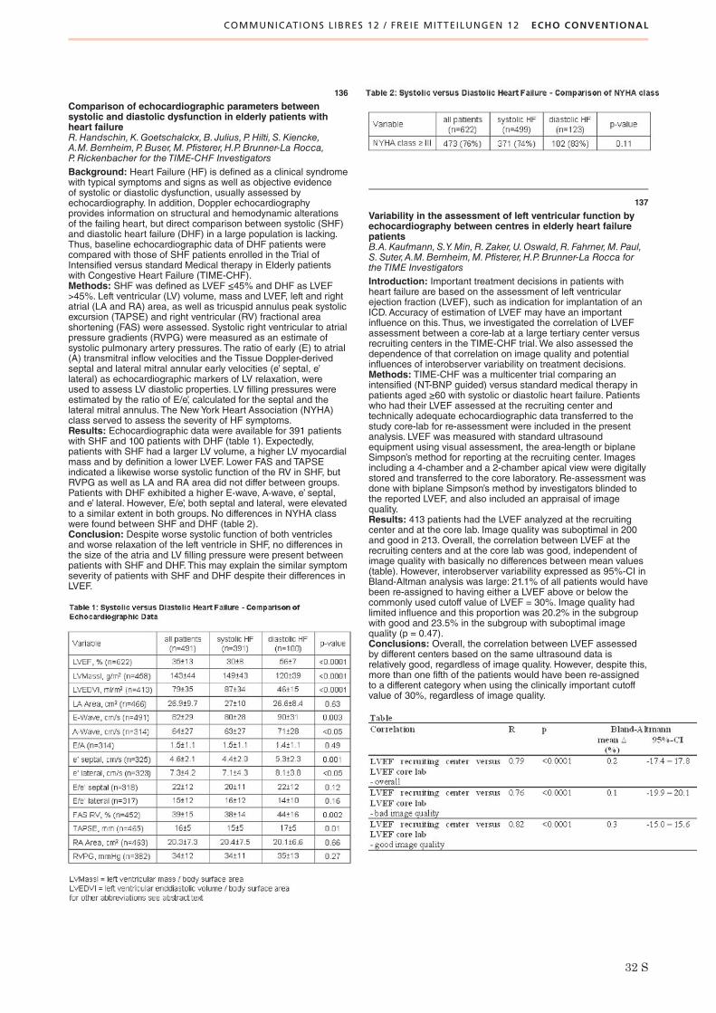
COMMUNICATIONS LIBRES 12 / FREIE MITTEILUNGEN 12 ECHO CONVENTIONAL
32 S
136
Comparison of echocardiographic parameters betweensystolic and diastolic dysfunction in elderly patients withheart failureR.Handschin,K.Goetschalckx,B. Julius,P.Hilti, S.Kiencke,A.M.Bernheim,P.Buser,M.Pfisterer,H.P.Brunner-La Rocca,P.Rickenbacher for the TIME-CHF InvestigatorsBackground: Heart Failure (HF) is defined as a clinical syndromewith typical symptoms and signs as well as objective evidenceof systolic or diastolic dysfunction, usually assessed byechocardiography. In addition, Doppler echocardiographyprovides information on structural and hemodynamic alterationsof the failing heart, but direct comparison between systolic (SHF)and diastolic heart failure (DHF) in a large population is lacking.Thus, baseline echocardiographic data of DHF patients werecompared with those of SHF patients enrolled in the Trial ofIntensified versus standard Medical therapy in Elderly patientswith Congestive Heart Failure (TIME-CHF).Methods: SHF was defined as LVEF ≤45% and DHF as LVEF>45%. Left ventricular (LV) volume, mass and LVEF, left and rightatrial (LA and RA) area, as well as tricuspid annulus peak systolicexcursion (TAPSE) and right ventricular (RV) fractional areashortening (FAS) were assessed. Systolic right ventricular to atrialpressure gradients (RVPG) were measured as an estimate ofsystolic pulmonary artery pressures. The ratio of early (E) to atrial(A) transmitral inflow velocities and the Tissue Doppler-derivedseptal and lateral mitral annular early velocities (e’ septal, e’lateral) as echocardiographic markers of LV relaxation, wereused to assess LV diastolic properties. LV filling pressures wereestimated by the ratio of E/e’, calculated for the septal and thelateral mitral annulus. The NewYork Heart Association (NYHA)class served to assess the severity of HF symptoms.Results: Echocardiographic data were available for 391 patientswith SHF and 100 patients with DHF (table 1). Expectedly,patients with SHF had a larger LV volume, a higher LV myocardialmass and by definition a lower LVEF. Lower FAS and TAPSEindicated a likewise worse systolic function of the RV in SHF, butRVPG as well as LA and RA area did not differ between groups.Patients with DHF exhibited a higher E-wave, A-wave, e’ septal,and e’ lateral. However, E/e’, both septal and lateral, were elevatedto a similar extent in both groups. No differences in NYHA classwere found between SHF and DHF (table 2).Conclusion: Despite worse systolic function of both ventriclesand worse relaxation of the left ventricle in SHF, no differences inthe size of the atria and LV filling pressure were present betweenpatients with SHF and DHF. This may explain the similar symptomseverity of patients with SHF and DHF despite their differences inLVEF.
137
Variability in the assessment of left ventricular function byechocardiography between centres in elderly heart failurepatientsB.A.Kaufmann,S.Y.Min,R.Zaker,U.Oswald,R.Fahrner,M.Paul,S.Suter,A.M.Bernheim,M.Pfisterer,H.P.Brunner-La Rocca forthe TIME InvestigatorsIntroduction: Important treatment decisions in patients withheart failure are based on the assessment of left ventricularejection fraction (LVEF), such as indication for implantation of anICD.Accuracy of estimation of LVEF may have an importantinfluence on this. Thus, we investigated the correlation of LVEFassessment between a core-lab at a large tertiary center versusrecruiting centers in the TIME-CHF trial.We also assessed thedependence of that correlation on image quality and potentialinfluences of interobserver variability on treatment decisions.Methods: TIME-CHF was a multicenter trial comparing anintensified (NT-BNP guided) versus standard medical therapy inpatients aged ≥60 with systolic or diastolic heart failure. Patientswho had their LVEF assessed at the recruiting center andtechnically adequate echocardiographic data transferred to thestudy core-lab for re-assessment were included in the presentanalysis. LVEF was measured with standard ultrasoundequipment using visual assessment, the area-length or biplaneSimpson’s method for reporting at the recruiting center. Imagesincluding a 4-chamber and a 2-chamber apical view were digitallystored and transferred to the core laboratory. Re-assessment wasdone with biplane Simpson’s method by investigators blinded tothe reported LVEF, and also included an appraisal of imagequality.Results: 413 patients had the LVEF analyzed at the recruitingcenter and at the core lab. Image quality was suboptimal in 200and good in 213. Overall, the correlation between LVEF at therecruiting centers and at the core lab was good, independent ofimage quality with basically no differences between mean values(table). However, interobserver variability expressed as 95%-CI inBland-Altman analysis was large: 21.1% of all patients would havebeen re-assigned to having either a LVEF above or below thecommonly used cutoff value of LVEF = 30%. Image quality hadlimited influence and this proportion was 20.2% in the subgroupwith good and 23.5% in the subgroup with suboptimal imagequality (p = 0.47).Conclusions: Overall, the correlation between LVEF assessedby different centers based on the same ultrasound data isrelatively good, regardless of image quality. However, despite this,more than one fifth of the patients would have been re-assignedto a different category when using the clinically important cutoffvalue of 30%, regardless of image quality.
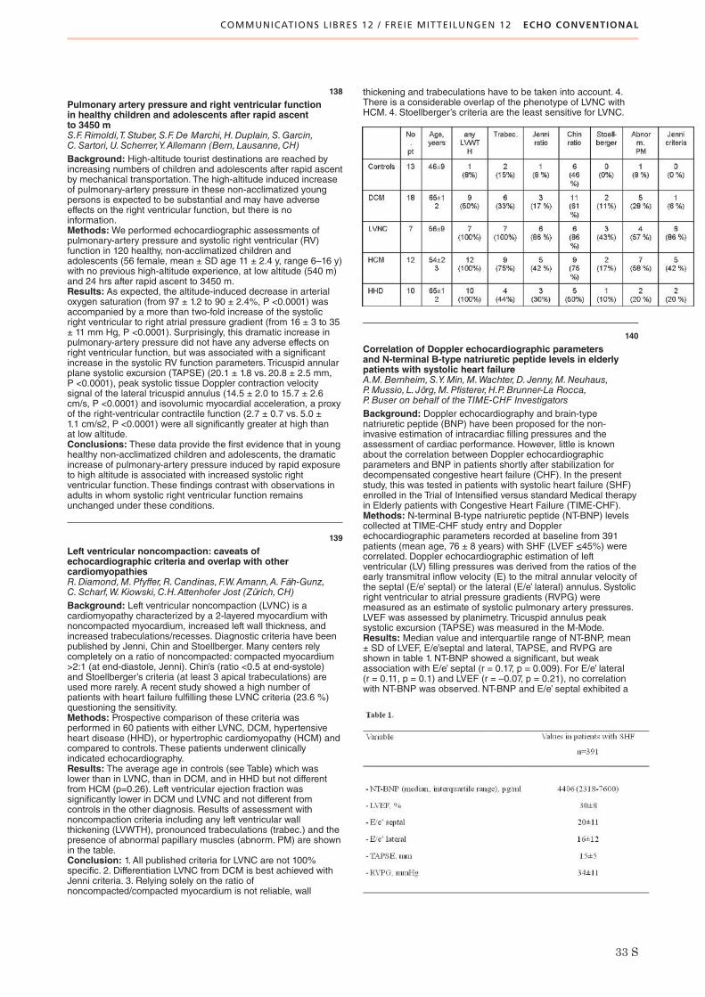
COMMUNICATIONS LIBRES 12 / FREIE MITTEILUNGEN 12 ECHO CONVENTIONAL
33 S
138
Pulmonary artery pressure and right ventricular functionin healthy children and adolescents after rapid ascentto 3450 mS.F.Rimoldi,T.Stuber,S.F.De Marchi,H.Duplain,S.Garcin,C.Sartori, U.Scherrer,Y.Allemann (Bern, Lausanne,CH)Background: High-altitude tourist destinations are reached byincreasing numbers of children and adolescents after rapid ascentby mechanical transportation. The high-altitude induced increaseof pulmonary-artery pressure in these non-acclimatized youngpersons is expected to be substantial and may have adverseeffects on the right ventricular function, but there is noinformation.Methods:We performed echocardiographic assessments ofpulmonary-artery pressure and systolic right ventricular (RV)function in 120 healthy, non-acclimatized children andadolescents (56 female, mean ± SD age 11 ± 2.4 y, range 6–16 y)with no previous high-altitude experience, at low altitude (540 m)and 24 hrs after rapid ascent to 3450 m.Results: As expected, the altitude-induced decrease in arterialoxygen saturation (from 97 ± 1.2 to 90 ± 2.4%, P <0.0001) wasaccompanied by a more than two-fold increase of the systolicright ventricular to right atrial pressure gradient (from 16 ± 3 to 35± 11 mm Hg, P <0.0001). Surprisingly, this dramatic increase inpulmonary-artery pressure did not have any adverse effects onright ventricular function, but was associated with a significantincrease in the systolic RV function parameters. Tricuspid annularplane systolic excursion (TAPSE) (20.1 ± 1.8 vs. 20.8 ± 2.5 mm,P <0.0001), peak systolic tissue Doppler contraction velocitysignal of the lateral tricuspid annulus (14.5 ± 2.0 to 15.7 ± 2.6cm/s, P <0.0001) and isovolumic myocardial acceleration, a proxyof the right-ventricular contractile function (2.7 ± 0.7 vs. 5.0 ±1.1 cm/s2, P <0.0001) were all significantly greater at high thanat low altitude.Conclusions: These data provide the first evidence that in younghealthy non-acclimatized children and adolescents, the dramaticincrease of pulmonary-artery pressure induced by rapid exposureto high altitude is associated with increased systolic rightventricular function. These findings contrast with observations inadults in whom systolic right ventricular function remainsunchanged under these conditions.
139
Left ventricular noncompaction: caveats ofechocardiographic criteria and overlap with othercardiomyopathiesR.Diamond,M.Pfyffer,R.Candinas, F.W.Amann,A.Fäh-Gunz,C.Scharf,W.Kiowski,C.H.Attenhofer Jost (Zürich,CH)Background: Left ventricular noncompaction (LVNC) is acardiomyopathy characterized by a 2-layered myocardium withnoncompacted myocardium, increased left wall thickness, andincreased trabeculations/recesses. Diagnostic criteria have beenpublished by Jenni, Chin and Stoellberger. Many centers relycompletely on a ratio of noncompacted: compacted myocardium>2:1 (at end-diastole, Jenni). Chin’s (ratio <0.5 at end-systole)and Stoellberger’s criteria (at least 3 apical trabeculations) areused more rarely. A recent study showed a high number ofpatients with heart failure fulfilling these LVNC criteria (23.6 %)questioning the sensitivity.Methods: Prospective comparison of these criteria wasperformed in 60 patients with either LVNC, DCM, hypertensiveheart disease (HHD), or hypertrophic cardiomyopathy (HCM) andcompared to controls. These patients underwent clinicallyindicated echocardiography.Results: The average age in controls (see Table) which waslower than in LVNC, than in DCM, and in HHD but not differentfrom HCM (p=0.26). Left ventricular ejection fraction wassignificantly lower in DCM und LVNC and not different fromcontrols in the other diagnosis. Results of assessment withnoncompaction criteria including any left ventricular wallthickening (LVWTH), pronounced trabeculations (trabec.) and thepresence of abnormal papillary muscles (abnorm. PM) are shownin the table.Conclusion: 1. All published criteria for LVNC are not 100%specific. 2. Differentiation LVNC from DCM is best achieved withJenni criteria. 3. Relying solely on the ratio ofnoncompacted/compacted myocardium is not reliable, wall
thickening and trabeculations have to be taken into account. 4.There is a considerable overlap of the phenotype of LVNC withHCM. 4. Stoellberger’s criteria are the least sensitive for LVNC.
140
Correlation of Doppler echocardiographic parametersand N-terminal B-type natriuretic peptide levels in elderlypatients with systolic heart failureA.M.Bernheim,S.Y.Min,M.Wachter,D. Jenny,M.Neuhaus,P.Mussio, L. Jörg,M.Pfisterer,H.P.Brunner-La Rocca,P.Buser on behalf of the TIME-CHF InvestigatorsBackground: Doppler echocardiography and brain-typenatriuretic peptide (BNP) have been proposed for the non-invasive estimation of intracardiac filling pressures and theassessment of cardiac performance. However, little is knownabout the correlation between Doppler echocardiographicparameters and BNP in patients shortly after stabilization fordecompensated congestive heart failure (CHF). In the presentstudy, this was tested in patients with systolic heart failure (SHF)enrolled in the Trial of Intensified versus standard Medical therapyin Elderly patients with Congestive Heart Failure (TIME-CHF).Methods: N-terminal B-type natriuretic peptide (NT-BNP) levelscollected at TIME-CHF study entry and Dopplerechocardiographic parameters recorded at baseline from 391patients (mean age, 76 ± 8 years) with SHF (LVEF ≤45%) werecorrelated. Doppler echocardiographic estimation of leftventricular (LV) filling pressures was derived from the ratios of theearly transmitral inflow velocity (E) to the mitral annular velocity ofthe septal (E/e’ septal) or the lateral (E/e’ lateral) annulus. Systolicright ventricular to atrial pressure gradients (RVPG) weremeasured as an estimate of systolic pulmonary artery pressures.LVEF was assessed by planimetry. Tricuspid annulus peaksystolic excursion (TAPSE) was measured in the M-Mode.Results: Median value and interquartile range of NT-BNP, mean± SD of LVEF, E/e’septal and lateral, TAPSE, and RVPG areshown in table 1. NT-BNP showed a significant, but weakassociation with E/e’ septal (r = 0.17, p = 0.009). For E/e’ lateral(r = 0.11, p = 0.1) and LVEF (r = –0.07, p = 0.21), no correlationwith NT-BNP was observed. NT-BNP and E/e’ septal exhibited a
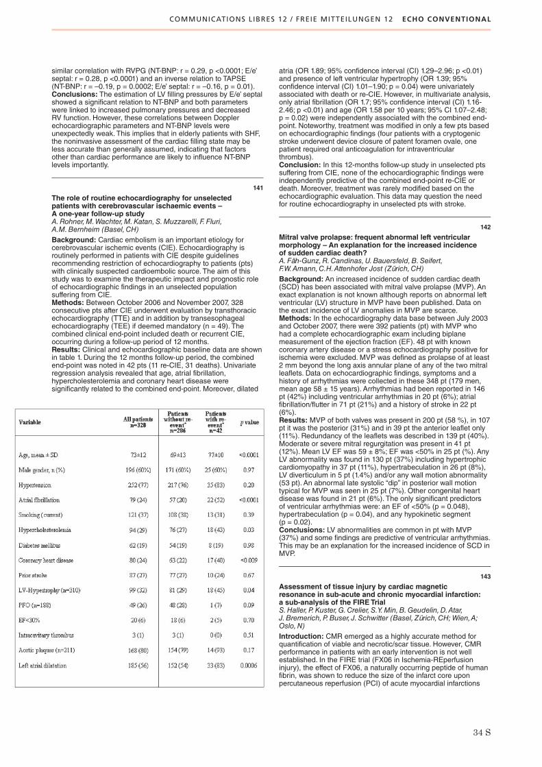
COMMUNICATIONS LIBRES 12 / FREIE MITTEILUNGEN 12 ECHO CONVENTIONAL
34 S
similar correlation with RVPG (NT-BNP: r = 0.29, p <0.0001; E/e’septal: r = 0.28, p <0.0001) and an inverse relation to TAPSE(NT-BNP: r = –0.19, p = 0.0002; E/e’ septal: r = –0.16, p = 0.01).Conclusions: The estimation of LV filling pressures by E/e’ septalshowed a significant relation to NT-BNP and both parameterswere linked to increased pulmonary pressures and decreasedRV function. However, these correlations between Dopplerechocardiographic parameters and NT-BNP levels wereunexpectedly weak. This implies that in elderly patients with SHF,the noninvasive assessment of the cardiac filling state may beless accurate than generally assumed, indicating that factorsother than cardiac performance are likely to influence NT-BNPlevels importantly.
141
The role of routine echocardiography for unselectedpatients with cerebrovascular ischaemic events –A one-year follow-up studyA.Rohner,M.Wachter,M.Katan,S.Muzzarelli, F. Fluri,A.M.Bernheim (Basel,CH)Background: Cardiac embolism is an important etiology forcerebrovascular ischemic events (CIE). Echocardiography isroutinely performed in patients with CIE despite guidelinesrecommending restriction of echocardiography to patients (pts)with clinically suspected cardioembolic source. The aim of thisstudy was to examine the therapeutic impact and prognostic roleof echocardiographic findings in an unselected populationsuffering from CIE.Methods: Between October 2006 and November 2007, 328consecutive pts after CIE underwent evaluation by transthoracicechocardiography (TTE) and in addition by transesophagealechocardiography (TEE) if deemed mandatory (n = 49). Thecombined clinical end-point included death or recurrent CIE,occurring during a follow-up period of 12 months.Results: Clinical and echocardiographic baseline data are shownin table 1. During the 12 months follow-up period, the combinedend-point was noted in 42 pts (11 re-CIE, 31 deaths). Univariateregression analysis revealed that age, atrial fibrillation,hypercholesterolemia and coronary heart disease weresignificantly related to the combined end-point. Moreover, dilated
atria (OR 1.89; 95% confidence interval (CI) 1.29–2.96; p <0.01)and presence of left ventricular hypertrophy (OR 1.39; 95%confidence interval (CI) 1.01–1.90; p = 0.04) were univariatelyassociated with death or re-CIE. However, in multivariate analysis,only atrial fibrillation (OR 1.7; 95% confidence interval (CI) 1.16-2.46; p <0.01) and age (OR 1.58 per 10 years; 95% CI 1.07–2.48;p = 0.02) were independently associated with the combined end-point. Noteworthy, treatment was modified in only a few pts basedon echocardiographic findings (four patients with a cryptogenicstroke underwent device closure of patent foramen ovale, onepatient required oral anticoagulation for intraventricularthrombus).Conclusion: In this 12-months follow-up study in unselected ptssuffering from CIE, none of the echocardiographic findings wereindependently predictive of the combined end-point re-CIE ordeath. Moreover, treatment was rarely modified based on theechocardiographic evaluation. This data may question the needfor routine echocardiography in unselected pts with stroke.
142
Mitral valve prolapse: frequent abnormal left ventricularmorphology – An explanation for the increased incidenceof sudden cardiac death?A.Fäh-Gunz,R.Candinas,U.Bauersfeld,B.Seifert,F.W.Amann,C.H.Attenhofer Jost (Zürich,CH)Background: An increased incidence of sudden cardiac death(SCD) has been associated with mitral valve prolapse (MVP). Anexact explanation is not known although reports on abnormal leftventricular (LV) structure in MVP have been published. Data onthe exact incidence of LV anomalies in MVP are scarce.Methods: In the echocardiography data base between July 2003and October 2007, there were 392 patients (pt) with MVP whohad a complete echocardiographic exam including biplanemeasurement of the ejection fraction (EF). 48 pt with knowncoronary artery disease or a stress echocardiography positive forischemia were excluded. MVP was defined as prolapse of at least2 mm beyond the long axis annular plane of any of the two mitralleaflets. Data on echocardiographic findings, symptoms and ahistory of arrhythmias were collected in these 348 pt (179 men,mean age 58 ± 15 years). Arrhythmias had been reported in 146pt (42%) including ventricular arrhythmias in 20 pt (6%); atrialfibrillation/flutter in 71 pt (21%) and a history of stroke in 22 pt(6%).Results: MVP of both valves was present in 200 pt (58 %), in 107pt it was the posterior (31%) and in 39 pt the anterior leaflet only(11%). Redundancy of the leaflets was described in 139 pt (40%).Moderate or severe mitral regurgitation was present in 41 pt(12%). Mean LV EF was 59 ± 8%; EF was <50% in 25 pt (%). AnyLV abnormality was found in 130 pt (37%) including hypertrophiccardiomyopathy in 37 pt (11%), hypertrabeculation in 26 pt (8%),LV diverticulum in 5 pt (1.4%) and/or any wall motion abnormality(53 pt). An abnormal late systolic “dip” in posterior wall motiontypical for MVP was seen in 25 pt (7%). Other congenital heartdisease was found in 21 pt (6%). The only significant predictorsof ventricular arrhythmias were: an EF of <50% (p = 0.048),hypertrabeculation (p = 0.04), and any hypokinetic segment(p = 0.02).Conclusions: LV abnormalities are common in pt with MVP(37%) and some findings are predictive of ventricular arrhythmias.This may be an explanation for the increased incidence of SCD inMVP.
143
Assessment of tissue injury by cardiac magneticresonance in sub-acute and chronic myocardial infarction:a sub-analysis of the FIRETrialS.Haller, P.Kuster,G.Crelier, S.Y.Min,B.Geudelin,D.Atar,J.Bremerich,P.Buser, J.Schwitter (Basel, Zürich,CH; Wien,A;Oslo,N)Introduction: CMR emerged as a highly accurate method forquantification of viable and necrotic/scar tissue. However, CMRperformance in patients with an early intervention is not wellestablished. In the FIRE trial (FX06 in Ischemia-REperfusioninjury), the effect of FX06, a naturally occurring peptide of humanfibrin, was shown to reduce the size of the infarct core uponpercutaneous reperfusion (PCI) of acute myocardial infarctions
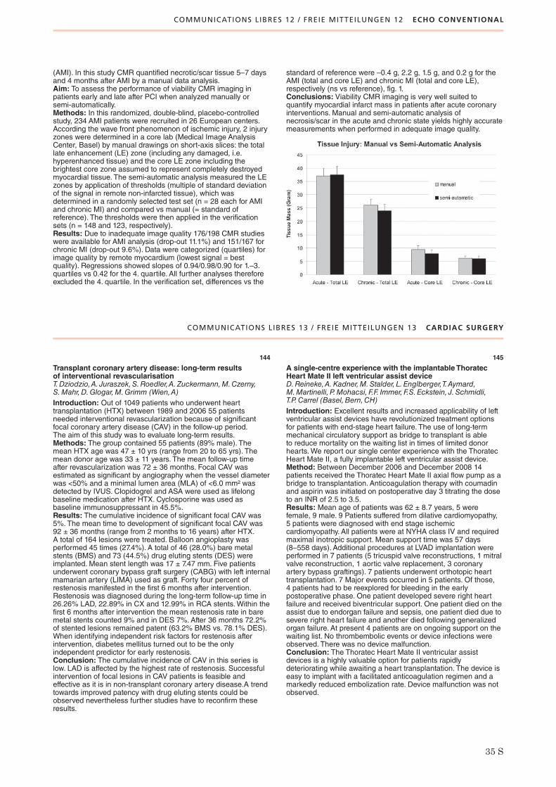
COMMUNICATIONS LIBRES 12 / FREIE MITTEILUNGEN 12 ECHO CONVENTIONAL
COMMUNICATIONS LIBRES 13 / FREIE MITTEILUNGEN 13 CARDIAC SURGERY
35 S
(AMI). In this study CMR quantified necrotic/scar tissue 5–7 daysand 4 months after AMI by a manual data analysis.Aim: To assess the performance of viability CMR imaging inpatients early and late after PCI when analyzed manually orsemi-automatically.Methods: In this randomized, double-blind, placebo-controlledstudy, 234 AMI patients were recruited in 26 European centers.According the wave front phenomenon of ischemic injury, 2 injuryzones were determined in a core lab (Medical Image AnalysisCenter, Basel) by manual drawings on short-axis slices: the totallate enhancement (LE) zone (including any damaged, i.e.hyperenhanced tissue) and the core LE zone including thebrightest core zone assumed to represent completely destroyedmyocardial tissue. The semi-automatic analysis measured the LEzones by application of thresholds (multiple of standard deviationof the signal in remote non-infarcted tissue), which wasdetermined in a randomly selected test set (n = 28 each for AMIand chronic MI) and compared vs manual (= standard ofreference). The thresholds were then applied in the verificationsets (n = 148 and 123, respectively).Results: Due to inadequate image quality 176/198 CMR studieswere available for AMI analysis (drop-out 11.1%) and 151/167 forchronic MI (drop-out 9.6%). Data were categorized (quartiles) forimage quality by remote myocardium (lowest signal = bestquality). Regressions showed slopes of 0.94/0.98/0.90 for 1.–3.quartiles vs 0.42 for the 4. quartile. All further analyses thereforeexcluded the 4. quartile. In the verification set, differences vs the
standard of reference were –0.4 g, 2.2 g, 1.5 g, and 0.2 g for theAMI (total and core LE) and chronic MI (total and core LE),respectively (ns vs reference), fig. 1.Conclusions: Viability CMR imaging is very well suited toquantify myocardial infarct mass in patients after acute coronaryinterventions. Manual and semi-automatic analysis ofnecrosis/scar in the acute and chronic state yields highly accuratemeasurements when performed in adequate image quality.
144
Transplant coronary artery disease: long-term resultsof interventional revascularisationT.Dziodzio,A. Juraszek,S.Roedler,A. Zuckermann,M.Czerny,S.Mahr,D.Glogar,M.Grimm (Wien,A)Introduction: Out of 1049 patients who underwent hearttransplantation (HTX) between 1989 and 2006 55 patientsneeded interventional revascularization because of significantfocal coronary artery disease (CAV) in the follow-up period.The aim of this study was to evaluate long-term results.Methods: The group contained 55 patients (89% male). Themean HTX age was 47 ± 10 yrs (range from 20 to 65 yrs). Themean donor age was 33 ± 11 years. The mean follow-up timeafter revascularization was 72 ± 36 months. Focal CAV wasestimated as significant by angiography when the vessel diameterwas <50% and a minimal lumen area (MLA) of <6.0 mm2 wasdetected by IVUS. Clopidogrel and ASA were used as lifelongbaseline medication after HTX. Cyclosporine was used asbaseline immunosuppressant in 45.5%.Results: The cumulative incidence of significant focal CAV was5%. The mean time to development of significant focal CAV was92 ± 36 months (range from 2 months to 16 years) after HTX.A total of 164 lesions were treated. Balloon angioplasty wasperformed 45 times (27.4%). A total of 46 (28.0%) bare metalstents (BMS) and 73 (44.5%) drug eluting stents (DES) wereimplanted. Mean stent length was 17 ± 7.47 mm. Five patientsunderwent coronary bypass graft surgery (CABG) with left internalmamarian artery (LIMA) used as graft. Forty four percent ofrestenosis manifested in the first 6 months after intervention.Restenosis was diagnosed during the long-term follow-up time in26.26% LAD, 22.89% in CX and 12.99% in RCA stents.Within thefirst 6 months after intervention the mean restenosis rate in baremetal stents counted 9% and in DES 7%.After 36 months 72.2%of stented lesions remained patent (63.2% BMS vs. 78.1% DES).When identifying independent risk factors for restenosis afterintervention, diabetes mellitus turned out to be the onlyindependent predictor for early restenosis.Conclusion: The cumulative incidence of CAV in this series islow. LAD is affected by the highest rate of restenosis. Successfulintervention of focal lesions in CAV patients is feasible andeffective as it is in non-transplant coronary artery disease.A trendtowards improved patency with drug eluting stents could beobserved nevertheless further studies have to reconfirm theseresults.
145
A single-centre experience with the implantable ThoratecHeart Mate II left ventricular assist deviceD.Reineke,A.Kadner,M.Stalder, L. Englberger,T.Aymard,M.Martinelli, P.Mohacsi, F.F. Immer, F.S. Eckstein, J.Schmidli,T.P.Carrel (Basel,Bern,CH)Introduction: Excellent results and increased applicability of leftventricular assist devices have revolutionized treatment optionsfor patients with end-stage heart failure. The use of long-termmechanical circulatory support as bridge to transplant is ableto reduce mortality on the waiting list in times of limited donorhearts.We report our single center experience with the ThoratecHeart Mate II, a fully implantable left ventricular assist device.Method: Between December 2006 and December 2008 14patients received the Thoratec Heart Mate II axial flow pump as abridge to transplantation. Anticoagulation therapy with coumadinand aspirin was initiated on postoperative day 3 titrating the doseto an INR of 2.5 to 3.5.Results: Mean age of patients was 62 ± 8.7 years, 5 werefemale, 9 male. 9 Patients suffered from dilative cardiomyopathy,5 patients were diagnosed with end stage ischemiccardiomyopathy. All patients were at NYHA class IV and requiredmaximal inotropic support. Mean support time was 57 days(8–558 days). Additional procedures at LVAD implantation wereperformed in 7 patients (5 tricuspid valve reconstructions, 1 mitralvalve reconstruction, 1 aortic valve replacement, 3 coronaryartery bypass graftings). 7 patients underwent orthotopic hearttransplantation. 7 Major events occurred in 5 patients. Of those,4 patients had to be reexplored for bleeding in the earlypostoperative phase. One patient developed severe right heartfailure and received biventricular support. One patient died on theassist due to endorgan failure and sepsis, one patient died due tosevere right heart failure and another died following generalizedorgan failure. At present 4 patients are on ongoing support on thewaiting list. No thrombembolic events or device infections wereobserved. There was no device malfunction.Conclusion: The Thoratec Heart Mate II ventricular assistdevices is a highly valuable option for patients rapidlydeteriorating while awaiting a heart transplantation. The device iseasy to implant with a facilitated anticoagulation regimen and amarkedly reduced embolization rate. Device malfunction was notobserved.

COMMUNICATIONS LIBRES 13 / FREIE MITTEILUNGEN 13 CARDIAC SURGERY
36 S
146
Quality of life after orthotopic heart transplantationfollowing ventricular assist device therapyD.Reineke, F.F. Immer, L. Englberger,M.Stalder,T.Aymard,E.Krähenbühl, J. Schmidli,T.P.Carrel (Bern,CH)Introduction: Due to limited supply of donor hearts, patients arestill dying on the waiting list for heart transplantation. The use oflong-term mechanical circulatory support is able to reduce thismortality and bridge patients to transplantation.While mostpublications are focused on thromboembolic events, devicedysfunction and outcome, the aim of this study was to lookat the quality of live of patients having received orthotopic hearttransplantation after assist device therapy.Methods: Between May 2000 and December 2006 40 Patientswere treated with ventricular assist devices. Thirteen patients diedbefore being transplanted. Twenty-three were transplanted and leftthe hospital. Quality of life was assessed with the Short Form36-Item Health Survey (SF -36). To further assess fear anddepression patients were confronted with the Hospital Anxietyand Depression Scale (HADS).Results: A total of 18 patients returned the questionnaires. Sf-36and HADS showed an overall excellent quality of life in patientsafter heart transplantation following ventricular assist devicetherapy.While mental stability and fitness in the SF36 werecomparable to a standard population (105.3 ± 14.3), twoparameters covering physical aspects, physical functioning androle physical, were only slightly lower than those of an averagepopulation (90.2 ± 15.9, 86.2 ± 36.3) but still in the rangeconsidered to be normal. Results of the HADS questionnaireare without pathological findings.Conclusion: Quality of life in patients after heart transplantationfollowing ventricular assist device therapy is excellent andcomparable to a standard population. Compared to the populationon assist devices it is mainly the mental situation which showsfurther improvement after orthotopic heart transplantation.Patients receiving assist devices emphasize their physicalimprovement.
147
Transapical aortic valve implantation for high-risk patientswith aortic stenosis provides excellent early resultsA.Kadner,P.Wenaweser,S.Windecker,B. Eberle,C.Zobrist,F.S. Eckstein,T.P.Carrel (Basel,Bern,CH)Background: The current treatment of choice for symptomaticaortic stenosis is surgical aortic valve replacement (AVR) withcardiopulmonary bypass (CPB). However, AVR may beassociated with significant operative morbidity and mortality inelderly high-risk patients. The less invasive transapical aorticvalve implantation (TAP-AVI) is an interesting alternative for thosepatients.We present our early results with this technique.Methods: TAP-AVI was performed via a small anterolateralminithoracotomy in 19 patients (18 pts Bern, 1 pt Basel) fromJanuary 2008 to December 2008. A balloon expandabletranscatheter xenograft prosthesis (Edwards SAPIEN™ THV,Edwards Lifesciences, Irvine, CA, USA) was used. Mean age was79.3 ± 6 years and 7 (37%) patients were female. Implantationwas performed using fluoroscopic and echocardiographicvisualization by a interdisciplinary team of cardiac surgeons,interventional cardiologists, and cardiac anaesthetists. Averagelogistic EuroSCORE predicted risk for mortality was 27.4 ± 11%.11 (58%) patients were reoperations following CABG, AVR, andMVR.Results: TAP-AVI (n = 7–23 mm prosthesis, n = 12–26 mmprosthesis) was successfully performed on the beating heartunder temporary rapid ventricular pacing in all patients. Noconversion to open heart surgery was performed. One patientunderwent IABP-implantation post-TAP-AVI for low cardiac output.All patients were successfully discharged from hospital. Earlymortality was 0%. There was no prosthesis migration orembolization. Echocardiography revealed good hemodynamicfunction in all patients and some central valvular incompetence in12 patients (grade 1 = 9, grade 2 = 3) and a paravalvular leakagein 11 patients (grade 1 = 7, grade 2 = 3, grade 3 = 1). Two patientsrequired the implantation of a permanent pacemaker.Conclusions: Transapical aortic valve implantation is aninteresting option for high-risk patients and provides excellentearly results. A close collaboration among different specialists is
necessary for an optimal patient selection and treatment.However, prior to the expansion of this technique to lower riskgroups, detailed long-term outcomes and risk estimations has tobe confirmed.
148
One-year experience with the 2nd generation 3F-Enablesutureless aortic valve prosthesisA.Kadner,T.Aymard, L. Englberger,M.Stalder,M.Grapow,F.S. Eckstein,T.P.Carrel (Basel,Bern,CH)Background: Currently, percutaneous aortic valve replacement(AVR) is strongly promoted as an alternative to traditional openheart valve surgery. Beside its reduced invasiveness, this newapproach shows severe limitations, such as no removal of thediseased valve, difficulty of accurate positioning of the prosthesis,and limitations of available sizes of valve prostheses. As analternative, the nitinol-stented 3F-Enable equine pericardial valveprosthesis was developed to allow a sutureless prosthesisimplantation.We report our early experience with this new implant.Material and methods: Between 08/2007 to 12/2008, 30patients (age 76 ± 6 years, 23 females, 27 pts – Bern, 3 pts –Basel) underwent AVR with the 2nd generation sutureless 3Fvalve prosthesis. All patients were operated for symptomaticaortic stenosis (NYHA III, Euroscore: median 7, range: 4–10).Echocardiography was performed pre-, intra-, and postoperatively,as well as at 6 and 12 months follow-up.Results: Prosthesis sizes were 27 mm (n = 4), 25 mm (n = 6),and 23 mm (n = 12), 21 mm (n = 5), 19 mm (n = 3). A reducedsternotomy (incision length 6–8 cm) was performed in 5 patients.ECC time was 56 ± 15 min, X-clamp time 37 ± 8 min,deployment-time 9 ± 6 min (re-deployment: n = 4). Follow-up iscomplete with a mean of 6 months (range: 1–14 months). Onepatient died of cardiac tamponade on post-op day one. A secondpatient died of CHF at 7 months postoperatively. One transientneurologic event occurred. Five patient required the implantationof a permanent pacemaker. Mean pressure gradients were 10 ± 4mm Hg, peak gradients 17 ± 6 mm Hg. Three patient showed aparavalvular leakage of grade one. Two non-valve-relatedexplants were performed, intraoperatively and at 4 months post-op.Conclusions: The new generation of the 3F Enable suturelessaortic valve shows promising results. This valve has the potentialto reduce X-clamp time and might be particularly interesting forcombined procedures. The modified design of the 2nd generationof this prosthesis appears to reduce the earlier reportedcomplications with paravalvular leakage.
149
Quality of life of GUCH patients after Senning /Mustard correctionO.Loup,M.Schwerzmann,M.Pavlovic, J.-P.Pfammatter,T.P.Carrel,A.Kadner (Bern,CH)Background: Until the early ninethies the Senning/Mustardoperation was the standard technique for correction of TGA.However, its long-term complications (RV-failure, arrhythmias,reinterventions) are well recognized. In contrast, little is knownabout the QoL of this special subgroup of GUCH patients.Methods: 122 patients (mean age 23 ± 7 years) operated forTGA by Senning/ Mustard (n = 102) and Switch operation (n = 20)fullfilled 3 questionnaires (the SF-36 Health Survey, the HADS-test and a disease specific questionnaire). The scores werecompared with age- and gendermatched standard populationdata and in relation to the performed operation.Results: The QoL of Senning/Mustard patients is comparableto standard population in all SF-36 and HADS dimensions, but issignificantly lower in many categories compared to switchpatients. Age has a significant negative impact on the generalhealth of the Senning/Mustard patients (p <0.05). Re-operationshows no significant influence on QoL. Significantly more patientsafter Senning/Mustard (43%) have arrhythmias (p = 0.02), ofwhich 34% required treatment (p = 0.01). NYHA functional statusreveals higher classes for Senning/Mustard vs switch patients,indeed 55% of them feel limited in their physical capacity (21% ofswitch patients). 21% receive a disability pension (none Switch).Conclusions: QoL of GUCH patients after Senning/Mustardcorrection is comparable to the standard population. However,

COMMUNICATIONS LIBRES 13 / FREIE MITTEILUNGEN 13 CARDIAC SURGERY
COMMUNICATIONS LIBRES 14 / FREIE MITTEILUNGEN 14 MYOCARDIAL DISEASE / ISCHAEMIA
37 S
they score significantly lower in many dimensions compared toswitch patients, show a higher percentage of long-termcomplications, a lower functional status, feeling more limited intheir physical capacity. These findings highlight the necessity of aspecialized long-term follow-up of this specific subgroup of GUCHpatients.
150
Follow-up of coronary bypass grafts by low-dose dualsource computer tomographyA.Plass,N.Azemai, L.Desbiolles,P.Stolzmann,H.Alkadhi,J.Grünenfelder (Zürich,CH)Dual Source Computertomography (DSCT) in step-and-shootmode (SASm) enables noninvasive assessment of bypass graftswith low radiation dose in patients with low heart rates.Weassess feasibility of SASm-DSCT for follow-up of coronary arterybypass grafts in clinical routine and different bypass criteria incorrelation to intraoperative measurements.Methods: 27 patients (23 males, 4 females, age 65 ± 8 years)underwent DSCT postoperatively in SASm or ECG triggereddepending of heart rates. Radiation dose values were calculated.Assessment of image quality and positioning of the grafts in Multi-Planar reconstruction (2- dimensional) and 3-dimensional VolumeRendering Technique. Patencies, proximal and distal anastomosiswere evaluated. Intraoperative pulsatility indices (PI) and flowmeasurements (F), as well as distal anastomosis diameters werecorrelated with Pearson correlation.Results: In 67% (18/27) of patients in SAS-mode (Radiation dose3.2 ± 1.3 mSiev) and in 33% (9/27) (7 patients Heart rate >70beats/min, 2 patients with atrial fibrillation) patients ECG triggered(Radiation dose 10.9 ± 2.1 mSiev). Image qualities: 20 scansgood, 6 moderate and one not assessable. Assessment of 67grafts (26 left mammary arteries, 26 venous/15 free arterial grafts)showed in 66 grafts good positioning, one slight kinking. 99%(66/67) of bypasses patent and one occluded. All proximalanastomoses were evaluable, 12% (8/67) distal anastomoseswere not assessable due to small diameter (4), clip-artefacts (2),reduced image quality (2). In the bypass grafts could be evaluatedin mean for F 27 ± 14 ml/min, for PI 2.9 ± 1.2 and for distalanastomosis diameters 0.2 ± 0.07 mm. Pearson correlationshowed that F negatively correlated with the PI (r =/- 0.36,p <0.01), PI negatively correlated with the distal anastomosisdiameters (r =/- 0.33, p <0.01). Distal anastomosis diameterswith F showed no correlation.Conclusion: DSCT in the SAS-mode is very well applicable inpatients with heart rates lower than 70/min and allowsassessment of bypass grafts at low radiation dose. Because ofthat DSCT in SASmode can be considered as quality control and
follow-up imaging tool after coronary artery bypass grafting. Inparameters correlation we could demonstrate the important roleof the pulsatility index, especially for assessment of theperformed distal anatomosis.
151
Endoscopic saphenous vein and radial artery graftharvesting for coronary artery bypass-establishingstandardsB.Winkler, P.Matt,M.Grapow,F.Rueter,D. Inderbitzin, F.S.Eckstein (Basel,Bern,CH)Introduction: Traditional open vein and arterial graft harvestingthrough a long incision can result in significant wound morbidity.The endoscopic approach may become the technique of choicebecause of both decreased morbidity compared with the opentechnique and patient preference. This video presentation aims topoint out major steps of successful endoscopic vein and arterialgraft harvesting with safeguards and pitfalls in the operativetechnique.Methods: In 2008, more than 100 patients undergoing coronaryartery bypass received endoscopic saphenous vein and/or radialartery harvesting using the Datascope ClearGlide® VesselHarvesting Device (Montvale, NJ, USA) accomplished in HighDefinition (HD) quality. Endoscopic techniques used for access a3 cm longitudinal incision and mean carbon dioxid insufflationpressure of 8 mmHg.An optical vessel dissector was used as firststep of dissection, and then for graft harvesting a small ultraretractor with a precision bipolar multitool.Results:While both endoscopic harvesting techniques require alearning curve of around 5 to 10 cases, afterwards the conversionrate to the open technique is low (2–5%). HD optical systemappropriation augmented the performance in addition throughhigh contrast visibility which is a major criteria for less timeconsuming endoscopic procedures. Our experience with the first100 patients undergoing endoscopic vein and/or arterial graftharvesting for coronary artery bypass surgery showed asignificant decrease in complications such as cellulitis, woundinfection, hematoma, seroma, neuropathy and neuralgia andimproved the cosmetic result compared with the open technique.Conclusion: Endoscopic graft harvesting for coronary arterybypass surgery is now the technique of choice in our center dueto a decreased lower and upper extremity morbidity comparedwith the open technique. Endoscopic techniques require alearning curve with key steps to follow, otherwise complicationsmay occur and the harvesting becomes time consuming. Thisvideo attempts to present such steps and pitfalls for a successfulendoscopic graft harvesting.
159
Rac1 and superoxide mediate β1-integrin up-regulationin cardiomyocytes inherent to compensated hypertrophyS.P.Häuselmann,B.I.Rosc-Schlüter,V. Lorenz,S.Meili-Butz,D. John, I. Plaisance,M.Brink,T.Dieterle,O.Pfister,G.M.Kuster(Basel,CH)Introduction: β1-integrin mediates cardiomyocyte hypertrophyand is up-regulated under hypertrophic conditions in the heart.However, mechanisms and dynamics of its regulation inhypertrophic remodeling are unclear. Reactive oxygen species(ROS) are important mediators of myocardial remodeling andmay participate in β1-integrin regulation.Methods/results:We applied endothelin-1 (ET-1)-inducedcardiomyocyte hypertrophy for in vitro and the Dahl-salt sensitiverat (DSR) model of hypertensive heart disease for in vivo studiesto determine mechanisms and ROS-dependency of regulated β1-integrin expression. In cultured cardiomyocytes, ET-1 increasedβ1-integrin expression time-dependently. Adenoviral expression ofa dominant-negative mutant form of Rac1 (AdN17Rac1), aregulator of NADPH oxidase, or over-expression of the antioxidantCu/Zn superoxide dismutase (AdCu/ZnSOD) abolished the ET-1-
induced β1-integrin increase, suggesting that it is Rac1-superoxide dependent. Experiments using pharmacologicalinhibitors showed that ET-1-induced β1-integrin expression isregulated by activation of two distinct pathways: 1) MEK/ERK and2) epidermal growth factor receptor (EGFR)-mediated activationof PI3K/Akt. AdN17Rac1 or AdCuZnSOD reduced MEKphosphorylation and inhibited EGFR phosphorylation in responseto ET-1. In DSR, myocardial β1-integrin expression wassignificantly increased during compensated hypertrophy (week8 of high-salt diet), but declined during the transition to ventriculardilation/failure (week 12). This decline was associated withdecreased Rac1 expression and ERK- and Akt-phosphorylation.Conclusion: Up-regulation of β1-integrin in cardiomyocytes ismediated via Rac1-superoxide dependent activation of the pro-hypertrophic MEK/ERK and the EGFR-PI3K/Akt pathways and isinherent to compensated hypertrophy. Loss of a proportionateβ1-integrin increase may mark a turning point in the transitionfrom cardiac hypertrophy to failure. Understanding the regulatorymechanisms of factors important to adaptive remodeling may helpdevelop treatment strategies to prevent transition to heart failure.
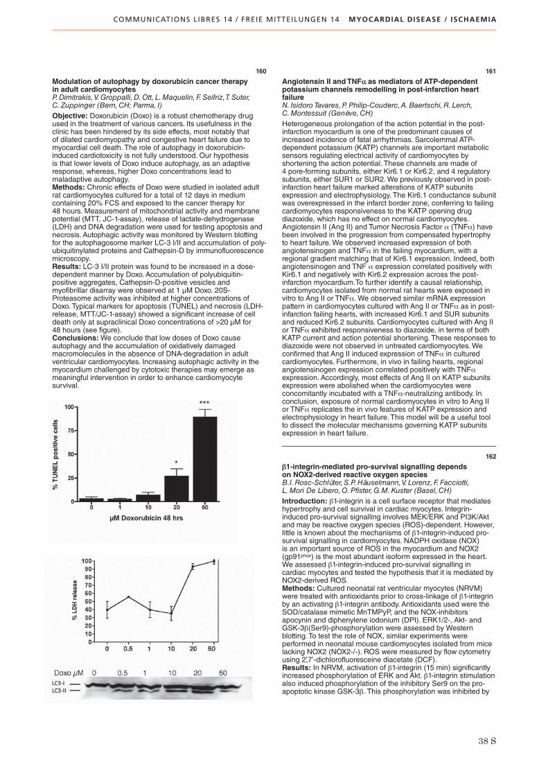
COMMUNICATIONS LIBRES 14 / FREIE MITTEILUNGEN 14 MYOCARDIAL DISEASE / ISCHAEMIA
38 S
160
Modulation of autophagy by doxorubicin cancer therapyin adult cardiomyocytesP.Dimitrakis,V.Groppalli, D.Ott, L.Maquelin, F.Seifriz,T.Suter,C. Zuppinger (Bern,CH; Parma, I)Objective: Doxorubicin (Doxo) is a robust chemotherapy drugused in the treatment of various cancers. Its usefulness in theclinic has been hindered by its side effects, most notably thatof dilated cardiomyopathy and congestive heart failure due tomyocardial cell death. The role of autophagy in doxorubicin-induced cardiotoxicity is not fully understood. Our hypothesisis that lower levels of Doxo induce autophagy, as an adaptiveresponse, whereas, higher Doxo concentrations lead tomaladaptive autophagy.Methods: Chronic effects of Doxo were studied in isolated adultrat cardiomyocytes cultured for a total of 12 days in mediumcontaining 20% FCS and exposed to the cancer therapy for48 hours. Measurement of mitochondrial activity and membranepotential (MTT, JC-1-assay), release of lactate-dehydrogenase(LDH) and DNA degradation were used for testing apoptosis andnecrosis. Autophagic activity was monitored by Western blottingfor the autophagosome marker LC-3 I/II and accumulation of poly-ubiquitinylated proteins and Cathepsin-D by immunofluorescencemicroscopy.Results: LC-3 I/II protein was found to be increased in a dose-dependent manner by Doxo. Accumulation of polyubiquitin-positive aggregates, Cathepsin-D-positive vesicles andmyofibrillar disarray were observed at 1 μM Doxo. 20S-Proteasome activity was inhibited at higher concentrations ofDoxo. Typical markers for apoptosis (TUNEL) and necrosis (LDH-release, MTT/JC-1-assay) showed a significant increase of celldeath only at supraclinical Doxo concentrations of >20 μM for48 hours (see figure).Conclusions:We conclude that low doses of Doxo causeautophagy and the accumulation of oxidatively damagedmacromolecules in the absence of DNA-degradation in adultventricular cardiomyocytes. Increasing autophagic activity in themyocardium challenged by cytotoxic therapies may emerge asmeaningful intervention in order to enhance cardiomyocytesurvival.
161
Angiotensin II and TNFα as mediators of ATP-dependentpotassium channels remodelling in post-infarction heartfailureN. Isidoro Tavares,P.Philip-Couderc,A.Baertschi,R. Lerch,C.Montessuit (Genève,CH)Heterogeneous prolongation of the action potential in the post-infarction myocardium is one of the predominant causes ofincreased incidence of fatal arrhythmias. Sarcolemmal ATP-dependent potassium (KATP) channels are important metabolicsensors regulating electrical activity of cardiomyocytes byshortening the action potential. These channels are made of4 pore-forming subunits, either Kir6.1 or Kir6.2, and 4 regulatorysubunits, either SUR1 or SUR2.We previously observed in post-infarction heart failure marked alterations of KATP subunitsexpression and electrophysiology. The Kir6.1 conductance subunitwas overexpressed in the infarct border zone, conferring to failingcardiomyocytes responsiveness to the KATP opening drugdiazoxide, which has no effect on normal cardiomyocytes.Angiotensin II (Ang II) and Tumor Necrosis Factor α (TNFα) havebeen involved in the progression from compensated hypertrophyto heart failure.We observed increased expression of bothangiotensinogen and TNFα in the failing myocardium, with aregional gradient matching that of Kir6.1 expression. Indeed, bothangiotensinogen and TNF α expression correlated positively withKir6.1 and negatively with Kir6.2 expression across the post-infarction myocardium.To further identify a causal relationship,cardiomyocytes isolated from normal rat hearts were exposed invitro to Ang II or TNFα. We observed similar mRNA expressionpattern in cardiomyocytes cultured with Ang II or TNFα as in post-infarction failing hearts, with increased Kir6.1 and SUR subunitsand reduced Kir6.2 subunits. Cardiomyocytes cultured with Ang IIor TNFα exhibited responsiveness to diazoxide, in terms of bothKATP current and action potential shortening. These responses todiazoxide were not observed in untreated cardiomyocytes.Weconfirmed that Ang II induced expression of TNFα in culturedcardiomyocytes. Furthermore, in vivo in failing hearts, regionalangiotensinogen expression correlated positively with TNFαexpression. Accordingly, most effects of Ang II on KATP subunitsexpression were abolished when the cardiomyocytes wereconcomitantly incubated with a TNFα-neutralizing antibody. Inconclusion, exposure of normal cardiomyocytes in vitro to Ang IIor TNFα replicates the in vivo features of KATP expression andelectrophysiology in heart failure. This model will be a useful toolto dissect the molecular mechanisms governing KATP subunitsexpression in heart failure.
162
β1-integrin-mediated pro-survival signalling dependson NOX2-derived reactive oxygen speciesB.I.Rosc-Schlüter, S.P.Häuselmann,V. Lorenz, F. Facciotti,L.Mori De Libero,O.Pfister,G.M.Kuster (Basel,CH)Introduction: β1-integrin is a cell surface receptor that mediateshypertrophy and cell survival in cardiac myocytes. Integrin-induced pro-survival signalling involves MEK/ERK and PI3K/Aktand may be reactive oxygen species (ROS)-dependent. However,little is known about the mechanisms of β1-integrin-induced pro-survival signalling in cardiomyocytes. NADPH oxidase (NOX)is an important source of ROS in the myocardium and NOX2(gp91phox) is the most abundant isoform expressed in the heart.We assessed β1-integrin-induced pro-survival signalling incardiac myocytes and tested the hypothesis that it is mediated byNOX2-derived ROS.Methods: Cultured neonatal rat ventricular myocytes (NRVM)were treated with antioxidants prior to cross-linkage of β1-integrinby an activating β1-integrin antibody. Antioxidants used were theSOD/catalase mimetic MnTMPyP, and the NOX-inhibitorsapocynin and diphenylene iodonium (DPI). ERK1/2-, Akt- andGSK-3β(Ser9)-phosphorylation were assessed by Westernblotting. To test the role of NOX, similar experiments wereperformed in neonatal mouse cardiomyocytes isolated from micelacking NOX2 (NOX2-/-). ROS were measured by flow cytometryusing 2’,7’-dichlorofluoresceine diacetate (DCF).Results: In NRVM, activation of β1-integrin (15 min) significantlyincreased phosphorylation of ERK and Akt. β1-integrin stimulationalso induced phosphorylation of the inhibitory Ser9 on the pro-apoptotic kinase GSK-3β. This phosphorylation was inhibited by

COMMUNICATIONS LIBRES 14 / FREIE MITTEILUNGEN 14 MYOCARDIAL DISEASE / ISCHAEMIA
COMMUNICATIONS LIBRES 15 / FREIE MITTEILUNGEN 15 ELECTROPHYSIOLOGIE, ABLATION
39 S
the PI3K-inhibitors Wortmannin and LY294002, as well as theMEK inhibitor U0126, localising GSK-3β downstream ofMEK/ERK and PI3K/Akt, respectively. Apocynin, DPI and, to alesser extent, MnTMPyP all inhibited β1-integrin-induced ERK-,Akt- and GSK-3β(Ser9)-phosphorylation. Activation of β1-integrininduced a time-dependent oxidative burst that peaked at 5 andsubsided by 60 min. Furthermore, β1-integrin-induced ERK-, Akt-and GSK-3β(Ser9)-signals were completely abolished in NOX2-/-as compared to wild type mouse cardiac myocytes.
Conclusion: Β1-integrin-induced pro-survival signalling incardiomyocytes is mediated by NOX2-derived ROS that activateboth the MEK/ERK and the PI3K/Akt pathways, leading toinhibition of the pro-apoptotic kinase GSK-3β. This protectiveeffect of NOX-derived ROS in cardiomyocytes should be takeninto consideration when developing therapeutic strategies toinhibit NOX in cardiovascular disease in the future.
164
Accuracy of impedance and electrogram characteristicsusing electro-anatomic mapping to determine sub-endocardial scar in patients with post-infarction ventriculararrhythmias: comparison with delayed enhanced magneticresonance imagingM.Kühne,B.Desjardins,H.Oral, F.Morady, F.Bogun(Basel,CH; Ann Arbor,USA)Background: Identification of infarct scar is important whentargeting ventricular arrhythmia in post-infarction patients. Scartissue is characterized by low voltage, electrogram fractionation,and low impedance. No accuracy measurements have beensystematically performed in humans comparing these differentmodalities of scar assessment. Delayed enhanced magneticresonance imaging (DEMRI) has a high accuracy in defining scartissue in vivo. The purpose of this study was to determine thediagnostic accuracy of impedance compared to electrogramcharacteristics for scar detection by using DEMRI as a goldstandard.Methods:We included 7 consecutive patients (6 male, mean age64 ± 9 years, EF: 0.28 ± 0.14) with ischemic cardiomyopathy,who underwent ablation of ventricular arrhythmias with anelectroanatomic mapping system. DEMRI was obtained prior toablation. Receiver operator characteristics (ROC) curves wereconstructed to determine the accuracy of impedance, bipolarvoltage and electrogram width for determination ofsubendocardial scar based on DEMRI.Results: A total of 1110 endocardial sites were analyzed. Bipolarvoltage with a cut-off of <1.1 mV had an area under the ROCcurve of 0.75 (95% CI 0.72–0.78) and a sensitivity of 68% andspecificity of 69% for differentiating normal tissue fromsubendocardial infarct. Electrogram width with a cut-off of >93 mswas similarly accurate with an area under the ROC curve of0.74 (95% CI 0.71–0.77; sensitivity 67%, specificity 67%).Bipolar voltage and electrogram width was more accurate thanimpedance (p <0.001). Impedance was not accurate in separatingnormal from scar tissue with an area under the ROC curve of0.55 (95% CI 0.52–0.59; sensitivity: 52%, specificity 53%).Conclusion: Definition of infarct scar using an electroanatomicmapping system can be achieved using bipolar voltage orelectrogram width. Its diagnostic accuracy is significantly bettercompared to impedance using DEMRI as a gold standard.
165
Predictors of mortality or heart transplantation afterradiofrequency catheter ablation of ventricular tachycardiaJ.Seiler, K.C.Roberts-Thomson,D.Steven,K. Inada,U.B.Tedrow,B.A.Koplan,W.G.Stevenson (Boston,USA)Introduction: Recurrent ventricular tachycardia (VT) hasemerged as a marker for mortality in patients (pts) with implanteddefibrillators (ICD). Radiofrequency catheter ablation (RFA) canprevent recurrent VT.We sought to determine predictors ofmortality or heart transplantation (HTx) after RFA of VT. Methods:Pts with structural heart disease undergoing the first RFA of VTat our institution between April 1999 and November 2008 wereincluded. Predictors of the combined endpoint of death orHTx were determined by Cox regression analysis.Results: Complete data for preliminary analysis were available in386 pts (age, 60 ± 15 years; male, 319 (82.6%); coronary arterydisease (CAD), 234 (60.6%); ejection fraction (EF) 33 ± 16 %).
During mean follow-up of 13 months, the endpoint was reached in57 (14.8%) pts (death, 52 pts; HTx, 5 pts). The estimated mediantime to endpoint was 2,145 (95% CI, 1,829-2,461) days. Inunivariate analysis, older age, lower EF, CAD, ICD, intra-procedural hemodynamic support, and peri-procedural majorcomplication were predictors of mortality/HTx; in a multivariatemodel, lower EF, CAD, and hemodynamic support remainedsignificant predictors (Table). The presence of both CAD and EF≤30% was associated with mortality/HTx of 22.2%, compared to4.9% for pts with none of these factors. Acute ablation successdid not predict mortality/HTx.Conclusions: The acute effect of RFA does not predictmortality/HTx. Clinical risk factors and requirement ofhemodynamic support identify a high risk group for poor outcome.
166
Poor implementation of CHADS2 risk score to guideantithrombotic management in atrial fibrillation patientsat risk for thromboembolismD.Altmann,C.Sticherling,M.Kühne,S.Osswald,B.Schaer (Basel,CH)Background and aim: Antithrombotic therapy for prevention ofthromboembolism is recommended for all patients with atrialfibrillation (AF), except for those with lone AF or contraindications.In 2003/2004 the Euro Heart Survey on AF (EHS) investigatedpatterns of stroke prevention management in numerous Europeancountries, and described an oral anticoagulation (OAC) rate of60% irrespective of the underlying CHADS2 risk score.The aim of our study was to determine contemporary OAC ratesin AF-patients seen in the emergency department of a Swissuniversity hospital and to find out reasons for withholdingthromboembolism prophylaxis in eligible patients.Methods: Consecutive patients with AF were screened for thepresence of non-valvular paroxysmal or permanent AF betweenJuly and December 2008. Patients were enrolled if AF wasmentioned in the medical record, and if an ECG documentingAF within the past 12 months was available. Subjects with newlydiagnosed AF or with another indication for OAC than AF wereexcluded from subsequent analysis. According to the CHADS2risk score, patients were divided into a low (CHADS2 = 0), anintermediate (CHADS2 = 1) and a high risk (CHADS2 ≥2) group.Antithrombotic drug prescription at hospital admission wasanalysed with reference to current guidelines.

COMMUNICATIONS LIBRES 15 / FREIE MITTEILUNGEN 15 ELECTROPHYSIOLOGIE, ABLATION
40 S
Results: 171 patients were screened, 32 patients (19%) wereexcluded due to newly diagnosed AF. Thus 139 patients werestudied. 8 (6%) pts had a CHADS2 score of 0, 33 (24%) patientsof 1, and 98 (70%) patients of ≥2, respectively. Prescription ofOAC was low in high risk patients, and not different to patients atintermediate or low risk (figure). The main reasons why therapywith OAC was withheld in high-risk patients were assumed drugmalcompliance (26%), eventual risk of fall (22%), and a historyof major bleeding (14%).Conclusion: In this survey of AF patients, antithrombotic drugprescription was hardly tailored to the risk profile of patients. OACwas underused in eligible patients and overused in patientsat intermediate and low risk. Our results emphasise that theawareness of physicians in Switzerland to apply the CHADS2 riskscore for prevention of thromboembolism in patients with AF istoo low.
167
Brugada syndrome: repolarisation, conduction or diastolicdisorder? A critical look at established mechanismsS.Chevallier,A.Van Oosterom,E.Pruvot (Lausanne,CH)Introduction: The Brugada syndrome (BrS) is a potentially lifethreatening arrhythmic disorder, whose electrophysiologicalmechanisms remain controversial. One hypothesis states thattype 1 BrS ECG pattern results from an abnormal transmuralgradient of repolarization, with heterogeneous reduction ofepicardial (epi) action potentials (APs) duration and dome butunaltered endocardial (endo) APs, causing ST elevation withinverted T waves in leads V1 and V2 (Antzelevitch C. JCE 2001).Another hypothesis states that type 1 BrS results from delayedactivation of right ventricular outflow tract (RVOT) APs, withprolonged local depolarization time and reversed repolarizationforces, which inscribe negative T waves in leads V1 and V2(Meregalli PG. Card Res 2005). ECGsim, an interactive programdownloadable from the web (www.ecgsim.org), computes 12-leadECGs based on a realistic 3-dimensional representation of epiand endo APs, their local depolarization and repolarization times,amplitude and diastolic potentials. ECGsim was used to test boththe repolarization and the depolarization hypotheses.Methods and results: alterations of the default parametersettings were applied to a circular region (3.4 cm diameter) at theRVOT facing lead V2. Repolarization hypothesis: shortening of epiAPs (from 242 ms at baseline to 55 ms) did not produce any STelevation nor RBBB type ECG morphology, but positive T-wavesof increased amplitude restricted to leads V1 to V3 (from 0.5 mVat baseline to 1.5 mV in lead V2). Depolarization hypothesis: a 80ms delay in activation of RVOT epi APs (from 45 ms at baseline to125 ms) produced a RBBB pattern with prolonged QRS duration(from 98 ms at baseline to 131 ms in V2) and negative T-wavesrestricted to leads V1 and V2, but no ST elevation. Importantly,the atypical RBBB with a 2 mm ST elevation of type 1 BrS couldbe reproduced by raising diastolic potentials of the RVOT regionby 19 mV (from –85 mV at baseline to –67 mV).Conclusions: Using a realistic APs-based ECG program, none ofthe commonly supposed alterations reproduced the type 1 BrSECG pattern. Shortening of RVOT epi APs increased T-wavesamplitude, while delayed activation of the RVOT region producedRBBB. Interestingly, ST elevation localized to anterior chest leadswas achieved by raising the diastolic potential of epi RVOT APs.Our findings suggest that BrS may also comprise alterations ofcellular mechanisms involved in the maintenance of RVOTdiastolic potential.
168
The effect of open-irrigated radiofrequency catheterablation of atrial fibrillation on left atrial pressureJ.Seiler,D.Steven,K.C.Roberts-Thomson,K. Inada,J.B.Patel,W.G.Stevenson (Boston,USA)Introduction: Open-irrigated catheter ablation poses anintravascular volume challenge and risk of cardiacdecompensation.We sought to determine left atrial (LA) pressurechanges in open-irrigated atrial fibrillation (AF) ablation. Methods:LA pressure was measured via transseptal vascular sheaths at thebeginning and end of the LA procedure in patients undergoingopen-irrigated ablation of AF or atrial tachycardia (AT) secondaryto a previous AF ablation.Results: Sixteen patients undergoing 16 procedures wereincluded for a prelimnary analysis. The total volume load duringthe procedure was 3,406 ± 939 ml, and the fluid balance duringthe LA procedure was +1,375 ± 786 ml. LA pressure increased in9, was unchanged (± 1.0 mm Hg) in 1, and decreased in 6patients, respectively. Patients in the top tercile of LA pressurechanges (n = 5, delta p = 6.4 ± 1.6 mm Hg) had a fluid balance of+1,975 ± 996 ml, compared to those in the lower two thirds of LApressure changes (n = 11, delta p = –0.8 ± 3.5 mm Hg) with a fluidbalance of +1,102 ± 518 ml (p = 0.07). In 7 patients who were insinus rhythm at the beginning and end of the procedure, who hada positive fluid balance of 1,025 ± 508 ml, there was a trendtowards a significant increase in LA pressure (delta p = 2.9 ±3.1 mm Hg, p = 0.09). In patients who were in AF or AT and thenconverted to sinus rhythm at the end of the procedure, LApressure did not significantly change (delta p = 0.4 ± 5.3 mm Hg,p = 0.95) despite a positive fluid balance of 1,647 ± 880 ml. The11 patients who received 20–40 mg of furosemide during theprocedure had no increase in LA pressure (delta p = 0.5 ± 5.0 mmHg, p = 0.79). One patient who did not receive furosemideexperienced post-procedural pulmonary edema.Conclusion: In patients undergoing open-irrigated AF ablation,the LA pressure change is not a reliable indicator of fluid balance,a substantial fluid volume is acutely absorbed with little change.Conversion from AF to sinus rhythm may help accommodateLA pressure to the volume load. Intra-procedural diuresis mayprevent an increase in LA pressure when substantial volume isadministered.
169
Radiofrequency ablation guided by mechanical terminationof idiopathic ventricular arrhythmias originating in the rightventricular outflow tractM.Kühne, J.-F.Sarrazin,H.Oral, F.Morady, F.Bogun(Basel,CH; Ann Arbor,USA)Background: Termination of ventricular tachycardia (VT)secondary to mechanical pressure has been described forfascicular and post-infarction VT. Mechanical interruption ofidiopathic ventricular arrhythmias (VT/ premature ventricularcomplexes [PVCs]) from the right ventricular outflow tract (RVOT)has not been described in systematic fashion.Methods: Eighteen consecutive patients (13 female, age 49 ± 13years, ejection fraction 0.55 ± 0.12) underwent mapping andablation for RVOT VT or PVCs. In 7 patients, 9 distinct VTs (meancycle length 440 ± 127 ms), and in 11 patients, 11 distinct PVCsoriginating in the RVOT were targeted. Mechanical terminationwas considered present if a reproducibly inducible VT was nolonger inducible or if frequent PVCs suddenly ceased with themapping catheter at a particular location. Endocardial activationtime, electrogram characteristics and pace-mapping morphologywere assessed at this location. Radiofrequency energy wasdelivered if mechanical termination was observed.Results: All targeted arrhythmias were successfully ablated.In 7 of 18 patients (39%) catheter manipulation terminated thearrhythmia with the mapping catheter located at a particular site.Local endocardial activation time was earlier at sites ofmechanical termination (–31 ± 7 ms) compared to effective siteswithout termination (–25 ± 3 ms, p = 0.04). The 10-ms isochronalarea was smaller in patients with mechanical interruption (0.35 ±0.2 cm2) compared to patients without mechanical termination(1.33 ± 0.9 cm2, p = 0.01). At all sites susceptible to mechanicaltrauma, the pace-map displayed a match with the targeted VT/PVC.All sites where mechanical termination of VT or PVCsoccurred were effective ablation sites.Conclusions: Mechanical suppression at the site of origin ofidiopathic RVOT arrhythmias frequently occurs during themapping procedure and is a reliable indicator of effective ablationsites. Mechanical termination of RVOT arrhythmias may beindicative of a more localized arrhythmogenic substrate.
GROUPE DE POSTERS 1 / POSTERGRUPPE 1 BASIC SCIENCE
41 S
P172
A multimarker approach to assess the influence ofinflammation on the incidence of atrial fibrillation in womenD.Conen,P.Ridker,B. Everett,U.B.Tedrow, L.Rose,N.Cook,M.Albert (Basel,CH; Boston,USA)Background: One prospective study among elderly individualsshowed an association between levels of high sensitivity C-reactive protein (hsCRP) and the development of atrial fibrillation(AF). However, prospective information in other individuals isscarce, and evaluation of multiple biomarkers may provideadditional information, given the multifactorial pathogenesis ofinflammation.Methods:We prospectively followed 24730 healthy, middle-agedwomen (median age 53 years) from the Women’s Health Study,who were free of AF at baseline and who provided a baselineblood sample assayed for hsCRP, soluble intercellular adhesionmolecule-1 (sICAM-1) and fibrinogen. Using these biomarkers,we created an inflammation score that reflected the number ofbiomarkers in the highest tertile per individual. Time to incidentAF (confirmed by medical record review) was assessed usingCox proportional-hazards models.Results: During 13.4 years of follow-up, 610 incident AF eventsoccurred. Median (interquartile range) levels of hsCRP, sICAM-1and fibrinogen were 2.0 (0.8–4.3) mg/L, 342 (300–393) ng/mLand 350 (307–402) mg/dL, respectively. Spearman correlationcoefficients were 0.29 for hsCRP-sICAM-1, 0.41 for hsCRP-fibrinogen and 0.27 for sICAM-1-fibrinogen, suggesting onlymoderate correlations. Assessed individually, all three biomarkerswere associated with incident AF, but most of these relationshipsbecame substantially attenuated after adjustment for body massindex. By contrast, we found an independent relationship betweenthe inflammation score and incident AF. Risk estimates acrossincreasing inflammation score categories are shown in the Table.In models adjusted for traditional cardiovascular risk factors, therisk of incident AF significantly increased across inflammationscore categories (p = 0.0002). This significant trend persistedafter additional adjustment for body mass index (p = 0.032).In the fully adjusted model, those with all three biomarkers in thehighest tertile had a significant 41% increase in risk of incidentAF (table).Conclusion: Inflammation is an important risk factor for thedevelopment of AF.While a substantial part of this risk ismediated by body mass index, multimarker evaluation revealedan independent inflammatory component.
P173
CD8α+ dendritic cell vaccination prevents CD4+Th17cell-mediated experimental autoimmune myocarditisA.Valaperti (Basel,CH)Experimental autoimmune myocarditis (EAM) represents a CD4+Th17 cell-mediated mouse model of inflammatory heart disease.IL-17 promotes the accumulation of inflammatory infiltrates in theheart. Interferon-γ (IFN-γ), on the other hand, reversibly inhibitsautoreactive CD4+ T cells through a negative NO-dependentfeedback mechanism, resolving autoimmune myocarditis.Vaccination with TNF-α-stimulated, myosin-peptide (MyHC-α)-loaded splenic CD8α+ dendritic cells (DC) at days –7, –5, –3prevented the development of EAM after subsequent MyHC-α/CFA immunization. In contrast, neither LPS-antiCD40-stimulated/MyHC-α-loaded, nor TNF-α-stimulated/OVA323-339-loaded splenic CD8α+ DC were protective. The protective effect of
TNF-α/MyHC-α/CD8α+ DC was IFN-γ-mediated, because TNF-α/MyHC-α/CD8α±vaccinated IFN-γR-/- mice were not protectedfrom MyHC-α/CFA-induced autoimmune myocarditis. Importantly,there were no differences in the numbers and function of IFN-γ-producing regulatory T cells in MyHC-α/CFA immunized miceafter vaccination with TNF-α/MyHC-α/CD8α+, LPS-antiCD40/MyHC-α/CD8α+, or solvent.In conclusion, vaccination with myosin-peptide-loaded TNF-α-stimulated CD8α+ DC promoted a CD4+ Th1-biased heart-specific T cell response protecting mice from inflammatory heartdisease.
P174
Transcriptional regulation of the human urocortin 2 geneby hypoxia-inducible factor 1K.Bühler, I. Plaisance,T.Dieterle,M.Brink (Basel,CH)Background: Urocortin 2 (Ucn2), a member of the corticotropinreleasing factor family, has recently been shown to exert potentbeneficial effects in the cardiovascular system. These includeinhibition of apoptosis, improvement of cardiomyocyte contractilefunction and decrease of oxidative stress. Tissue hypoxia, acommon pathophysiological feature in cardiovascular disease,stimulates the expression of genes involved in glucosemetabolism, cell survival, and resistance to oxidative stress.Because the regulation of human Ucn2 (hUcn2) expression ispoorly understood, we investigated the role of hypoxia in thetranscriptional regulation of hUcn2.Methods and results: Expression of hUcn2 mRNA wassignificantly increased in TE-671 cells exposed to hypoxia orciclopirox olamine (CPX), which prevents the proteolyticdegradation of hypoxia-inducible factor (HIF), compared to thenormoxic control. Gene silencing of endogenous HIF1αexpression using specific siRNA abolished the hypoxia- and CPX-induced increase of hUcn2 expression, indicating the involvementof HIF1α in the hUcn2 gene regulation. A luciferase-linkedfragment of the 3’- flanking region (3’FLR3) of the hUcn2 genecontaining two putative HIF-responsive elements (HRE1 andHRE2) was activated by hypoxia and CPX in TE-671 cells. Sitedirected mutagenesis of either sequence demonstrated thatHRE1 is crucial for the HIF1α-dependent luciferase activation.Competition assays revealed that HRE1 is capable to bindHIF1allpha. Because Ucn2 has cardioprotective properties,we extended our analysis of Ucn2 expression and regulationto cardiac cells. Like in TE-671, hypoxia markedly increasedluciferase activity in heart-derived H9c2 cells transfected withthe 3’FLR3 of the hUcn2 gene. Moreover, in rat neonatalcardiomyocytes hypoxia significantly induced Ucn2 mRNAexpression. Corresponding increases in Ucn2 protein levels weredetected by immunofluorescence microscopy.Conclusion:We have demonstrated that Ucn2 expression isinduced by hypoxia in TE-671 cells and in two cardiomyocytemodels, and that hUcn2 gene regulation critically depends on thespecific interaction of HIF1α with HRE1. Hypoxia-mediatedstimulation of cardioprotective Ucn2 may help to keep cardiacfunction and prevent apoptosis in ischemic heart disease.
P175
Prominent role for the endothelial gap junction proteinconnexin40 in myocardial ischaemia-reperfusion injury inmiceS.Morel,V.Braunersreuther, I. Roth,C.Chadjichristos,C.De Wit,T.A.B.Van Veen,B.R.Kwak,F.Mach (Genève,CH; Lübeck,D;Utrecht,NL)Intercellular channels formed by connexins (Cx) have beenshown to play a critical role in cardiovascular disease. Indeed,connexin-deficient mice showed modifications in cell signalling,that appeared crucial in atherosclerosis and restenosis. In theheart, Cx40 is expressed in atrial myocytes, the conductionsystem and in endothelial cells. Here, we study the implicationof endothelial Cx40 during ischemia and reperfusion in mice. Forthis purpose, we used the Cre-loxP system to create a mouse linein which Cx40 is deleted from the endothelium only.Immunostainings on TIE2-Cre+ Cx40fl/fl ApoE-/- mice confirmedthe absence of Cx40 in the endothelium, whereas the protein wasnormally expressed in the atria and cardiac conduction system.Moreover, mean arterial pressure and heart rates were not
GROUPE DE POSTERS 1 / POSTERGRUPPE 1 BASIC SCIENCE
42 S
different between controls (TIE2-Cre- Cx40fl/fl ApoE-/- andTIE2-Cre+ Cx40wt/wt ApoE-/-) and TIE2-Cre+ Cx40fl/fl ApoE-/-animals. Sixteen-week-old mice with or without endothelial-specific deletion of Cx40 were subjected to in vivo left coronaryartery occlusion for 30 minutes and sacrificed 24-hours afterreperfusion for analysis of infarct size. Myocardial surfaces areasat risk and infracted areas were measured from computed imagesusing NIH Image software. Areas at risk, normalized to total leftventricle surfaces areas, were similar between the experimentalgroups, i.e controls 29.17 ± 1.19% (N = 11) and TIE2-Cre-Cx40fl/fl ApoE-/- 29.29 ± 2.37% (N = 7, n.s.). Interestingly, theinfarct area, normalized to areas at risk, was significantlyincreased in TIE2-Cre+ Cx40fl/fl ApoE-/- mice as compared tocontrols (20.67 ± 4.74% and 8.47 ± 1.45%, respectively, P <0.01).We conclude that endothelial Cx40 is implicated in resistance ofthe heart to ischemia-reperfusion injury. These findings underlineonce more the importance of connexin-mediated intercellularcommunication in cardiovascular inflammation, and may pointtowards novel therapeutic strategies to limit the cardiac injuryafter coronary interventions.
176
P-Gylcoprotein protects from adverse myocardialremodelling after ischaemia-reperfusion injuryO.Pfister,D.Cullen,A.Oikonomopoulos,K.Sereti, S.Ngoy,R. Liao(Basel,CH; Boston,USA)Background: P-glycoprotein (Pgp) is a multidrug resistanttransporter protein that protects cancer cells from apoptotic celldeath. Recent evidence of Pgp expression in hypoxic tissueincluding the heart further suggests a specific role of Pgp in cellsundergoing ischemic injury. Although Pgp expression has beendemonstrated in mammal and human ischemic hearts, itsfunctional significance in ischemic heart disease remains elusive.In this study we determined the specific role of Pgp in post-infarction myocardial remodeling utilizing a model of myocardialischemia reperfusion in mice lacking Pgp expression (Pgp-nullmice).Methods: Anesthetized Pgp-null (n = 8) and Pgp-wildtype(n = 8) mice underwent open chest occlusion of the left anteriordescending coronary artery for 40 minutes followed byreperfusion. Serial echocardiograms were performed at baselineand 1 and 4 weeks after ischemia-reperfusion injury (IR) todetermine standard functional and morphological parameters.After 4 weeks, animals were sacrificed and heart/body weightratio as well as wet/dry ratio of lung und liver weight weredetermined to assess pulmonary and liver congestion.Results: There were no differences in echocardiographicparameters between Pgp-wildtype and Pgp-null mice at baseline.Ischemia-Reperfusion (IR) led to a similar decrease in leftventricular (LV) function in both groups as documented one weekafter IR. Four weeks after IR, however, echocardiographicassessment of LV dimensions and function documented adverseLV remodeling and markedly mitigated LV function in Pgp-nullmice compared to Pgp-wildtype mice [Table]. In accordance todeterioration of LV function, Pgp-null mice demonstratedpronounced pulmonary congestion as evidenced by significantlyhigher wet/dry lung ratios compared to Pgp-wildtype mice (4.6 ±0.1 vs 4.0 ± 0.2; p <0.05). Heart body weight- and wet/dry liverratios were not significantly different between groups.Conclusion: Expression of the multidrug resistant protein Pgp ispart of the inherent cardio-protective program to prevent adversemyocardial remodeling after ischemic injury. Pharmaceuticaltherapies involving Pgp inhibition may thus facilitate adversemyocardial remodeling in ischemic heart disease.
P177
SIRT1 regulates endothelial tissue factor expressionA.Breitenstein,A.Akhmedov,M.Stein,G.G.Camici,T.F. Lüscher,C.M.Matter, F.C.Tanner (Zürich,CH)Background: The mammalian silent information regulator-two1 (SIRT1) is critically involved in regulating key processes suchas stress response, apoptosis, and senescence; in line with this,SIRT1 exerts beneficial effects on cardiovascular risk factors suchas aging, diabetes as well as atherosclerosis. Nevertheless, therole of SIRT1 in arterial thrombosis has not yet been elucidatedin detail. Tissue factor (TF), the key activator of the coagulationcascade, is crucially involved in thrombus formation. The currentstudy was therefore designed to investigate the effect of SIRT1on tissue factor (TF) expression.Methods and results: Pharmacological SIRT1 inhibition withsplitomicin (25–200 uM) or sirtinol (15–60 uM) enhanced basaland TNF-α induced endothelial TF protein expression in aconcentration-dependent manner (n = 4; p <0.05). This effect wasparalleled by an increase in TF surface activity (n = 4; p <0.05).In contrast, SRT2183-and resveratrol, which activate SIRT1,impaired TF expression and activity (n = 4; p <0.05). Real-timepolymerase chain reaction revealed that SIRT1 inhibition inducedbasal and TNF-α induced TF transcription (n = 4; p <0.05),whereas activation of the MAP kinases (JNK, p38, and ERK)remained unaffected (n = 3; p = NS). TF promotor studies indeedrevealed that the TF promotor was activated by SIRT1 inhibition(n = 5; p <0.05). In line with this observation, nucleartranslocation and DNA-binding capacity of the p65-subunit of NF-kappaB was increased when SIRT1 was inhibited (n = 5; p <0.05)suggesting that the effect of SIRT1 on TF expression is mediatedby interference of SIRT1 with NF-kappaB/p65 activation.Conclusions: SIRT1 modulates the activity of NF-kappaB/p65 inendothelial cells and thereby regulates TF expression. Hence,modulating SIRT1 offers possible novel therapeutic optionstargeting arterial thrombosis, in particular in patients withcardiovascular risk factors such as aging and diabetes.
P178
Rapamycin induces tissue factor and promotes arterialthrombosis in vivoG.G.Camici, J. Steffel, I.Amanovic,A.Breitenstein, J.Baldinger,S.Keller,T.F. Lüscher, F.C.Tanner (Zürich,CH)Background: Drug-eluting stents (DES) may be associated withan increased risk for stent thrombosis as compared to bare-metalstents (BMS). In cultured human endothelial cells, rapamycin, asubstance used on first-generation DES, induces tissue factor(TF), a key enzyme in the initiation of the coagulation cascade,by inhibiting the activity of the mammalian target of rapamycin(mTOR). However, the effect of mTOR inhibition on TF activityand thrombus formation in vivo has not yet been studied.Moreover, it is presently unclear whether and to what degree thesecond-generation DES substances everolimus and zotarolimushave an effect on endothelial TF expression.Methods and results: In a mouse carotid artery photochemicalinjury model, rapamycin decreased time to thrombotic occlusionby 40%, increased TF activity, and abrogated phosphorylation ofp70S6K as compared to vehicle-treated mice. In vitro, rapamycin,everolimus, and zotarolimus (each 10-7 mol/l) enhanced TNF-αinduced TF expression by 2.2-, 1.7-, and 2.4-fold, respectively; thisincrease was paralleled by a 1.5-, 1.4-, and 1.5-fold, increase in TFsurface activity, respectively. Similar to rapamycin, everolimus andzotarolimus abrogated TNF-α-induced p70S6K phosphorylationunder these conditions.Conclusions: Rapamycin increases TF activity and promotesarterial thrombosis in vivo at concentrations relevant in patientsundergoing DES implantation; this effect may increase thethrombogenicity of DES. Since everolimus and zotarolimusaugment endothelial TF expression and activity in vitro in asimilar manner as rapamycin, these findings may be relevant bothfor first- and second generation DES.
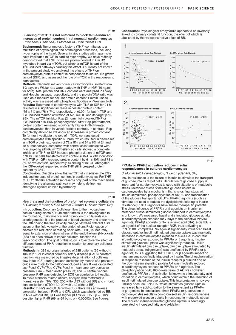
GROUPE DE POSTERS 1 / POSTERGRUPPE 1 BASIC SCIENCE
43 S
P179
Silencing of mTOR is not sufficient to block TNF-a-inducedincreases of protein content in rat neonatal cardiomyocytesI. Plaisance,P.Shende,C.Morandi,M.Brink (Basel,CH)Background: Tumor necrosis factor-a (TNF) contributes to amultitude of physiological and pathological processes, includinghypertrophy of the heart. Several in vivo studies with rapamycinhave implicated mTOR in cardiac hypertrophy.We have recentlydemonstrated that TNF increases protein content in C2C12myotubes in part via mTOR, but whether mTOR is part of theTNF-induced pathways causing this effect is currently not known.In the present study we analyzed the effects of TNF oncardiomyocyte protein content in comparison to insulin-like growthfactor-I (IGF), and assessed the role of mTOR in the responses toboth factors.Methods: Neonatal rat ventricular cardiomyocytes isolated from1-3 days old Wistar rats were treated with TNF or IGF (10 ng/mlfor both). Total protein and DNA content were analyzed in Lowryand Hoechst assays, respectively, and the protein/DNA ratio wasused as a measure for cellular protein content. Protein kinaseactivity was assessed with phospho-antibodies on Western blots.Results: Treatment of cardiomyocytes with TNF or IGF for 24 hresulted in a significant increase in cellular protein content(65 ± 5% and 76 ± 7%, respectively, p <0.001 for both). TNF andIGF induced marked activation of Akt, mTOR and its target p70-S6K. The mTOR inhibitor Rap (2 ng/ml) fully blocked TNF orIGF-induced p70-S6K phosphorylation. After Rap-pretreatment,protein content remained significantly higher in TNF-treatedcardiomyocytes than in vehicle-treated controls. In contrast, Rapcompletely abolished IGF-induced increases in protein content.To further investigate the role of mTOR, we transfectedcardiomyocytes with specific siRNAs, which resulted in decreasesin mTOR protein expression of 72 ± 5% and 64 ± 6% at 24 and48 h, respectively, compared with control cells transfected withnon targeting siRNA. mTOR-silenced cells showed a completeinhibition of TNF- or IGF-induced phosphorylation of mTOR andp70S6K. In cells transfected with control siRNA, a 24h incubationwith TNF or IGF increased protein content by 61 ± 10% and 78 ±8% above controls, respectively. Silencing of mTOR abrogatedthe IGF-evoked response, while TNF still increased proteincontent by 35%.Conclusion: Our data show that mTOR fully mediates the IGF-induced increase of protein content in cardiomyocytes. For TNF,mTOR/p70-S6K activation represents only part of the mechanism.Identifying the alternate pathway may help to define newstrategies against cardiac hypertrophy.
P180
Heart rate and the function of preformed coronary collateralsS.Gloekler, P.Meier, S.F. de Marchi,T.Traupe,C.Seiler (Bern,CH)Introduction: Coronary (collateral) flow to the myocardiumoccurs during diastole. Fluid shear stress is the driving force inthe formation, maintainance and promotion of collaterals (i.e.arteriogenesis). It is the product of blood viscosity and shear rate,the latter being the fluid velocity change between different fluidlayers or the fluid velocity at the endothelium. Prolongation ofdiastole via reduction of resting heart rate (RHR) is, naturally,equal to extension of shear stress at the endothelium. β-blockade(BB) has been shown to impair collateral function viavasoconstriction. The goal of this study is to explore the effect ofdifferent forms of RHR reduction in relation to coronary collateralfunction.Methods: In 365 coronary arteries of 285 patients (99 without-,186 with 1- or 2-vessel coronary artery disease (CAD)) collateralfunction was measured by invasive determination of collateralflow index (CFI) during balloon occlusion by means of a pressureguide wire distal to the balloon-occluded artery. It was calculatedas: (Poccl-CVP)/(Pao-CVP); Poccl = mean coronary occlusivepressure; Pao = mean aortic pressure; CVP = central venouspressure. RHR was detected by ECG on admission to hospital.To avoid stenosis-related effects, analysis was restricted tonormal vessels (NVs: 333; 200 with- ,133 without BB) and chronictotal occlusions (CTOs: 32; 20 with-, 12 without BB).Results: In NVs and CTOs without BB, there was an inversecorrelation between RHR and CFI, which was abolished by BB.In NVs without BB, CFI was higher (0.176 vs 0.153, p = 0.02)despite higher RHR (69 vs 64 bpm, p = 0.00002). See figures.
Conclusion: Physiological bradycardia appears to be inverselylinked to coronary collateral function, the effect of which isabolished by the vasoconstrictive effect of BB.
P181
PPARα or PPARβ activation reduces insulinresponsiveness in cultured cardiomyocytesC.Montessuit, I. Papageorgiou,R. Lerch (Genève,CH)Insulin resistance is the failure of insulin to stimulate the transportof glucose into its target cells. Regulation of glucose supply isimportant for cardiomyocytes to cope with situations of metabolicstress. Metabolic stress stimulates glucose uptake incardiomyocytes by a mechanism that shares final steps withinsulin stimulation: phosphorylation of AS160 and translocationof the glucose transporter GLUT4. PPARα agonists (such asfibrates) are used to reduce the dyslipidemia leading to insulinresistance; PPARβ agonists have similar therapeutic potential.The direct influence of PPARα or β agonists on insulin- ormetabolic stress-stimulated glucose transport in cardiomyocytesis unknown.We measured basal and stimulated glucose uptakein cardiomyocytes exposed for 7 days to the selective PPARαagonists, PPARβ agonists or 9-cis retinoic acid (RA). 9-cis RA isan agonist of the nuclear receptor RXR, thus activating allPPAR/RXR complexes. No agonist significantly influenced basalglucose uptake. Insulin-stimulated glucose uptake was markedlyincreased in cardiomyocytes exposed to 9-cis RA. In contrast,in cardiomyocytes exposed to PPARα or β agonists, insulin-stimulated glucose uptake was significantly reduced. Unlikeinsulin-stimulated glucose uptake, glucose uptake stimulated bymetabolic stress (oligomycin) was unaffected by PPARα or βagonists, thus suggesting that PPARα or β agonists impact onmechanisms specifically triggered by insulin. The phosphorylationin response to insulin of the insulin receptor β subunit and ofthe downstream signaling protein Akt was modestly reducedin cardiomyocytes exposed to PPARα or β agonists. Thephosphorylation of AS160 downstream of Akt was howeverunaffected. PPARα or β activation is known to stimulate fatty acidoxidation in cardiomyocytes, which could explain the reduction ofinsulin-stimulated glucose uptake. This interpretation is howeverunlikely because 9-cis RA, which stimulates glucose uptake,increased fatty acid oxidation to the same extent as PPARαor β agonists. In conclusion, direct PPARα or β activation incardiomyocytes results in compromised insulin responsivenesswith preserved glucose uptake in response to metabolic stress.The reduced insulin-stimulated glucose uptake is seeminglyunrelated to increased fatty acid oxidation.
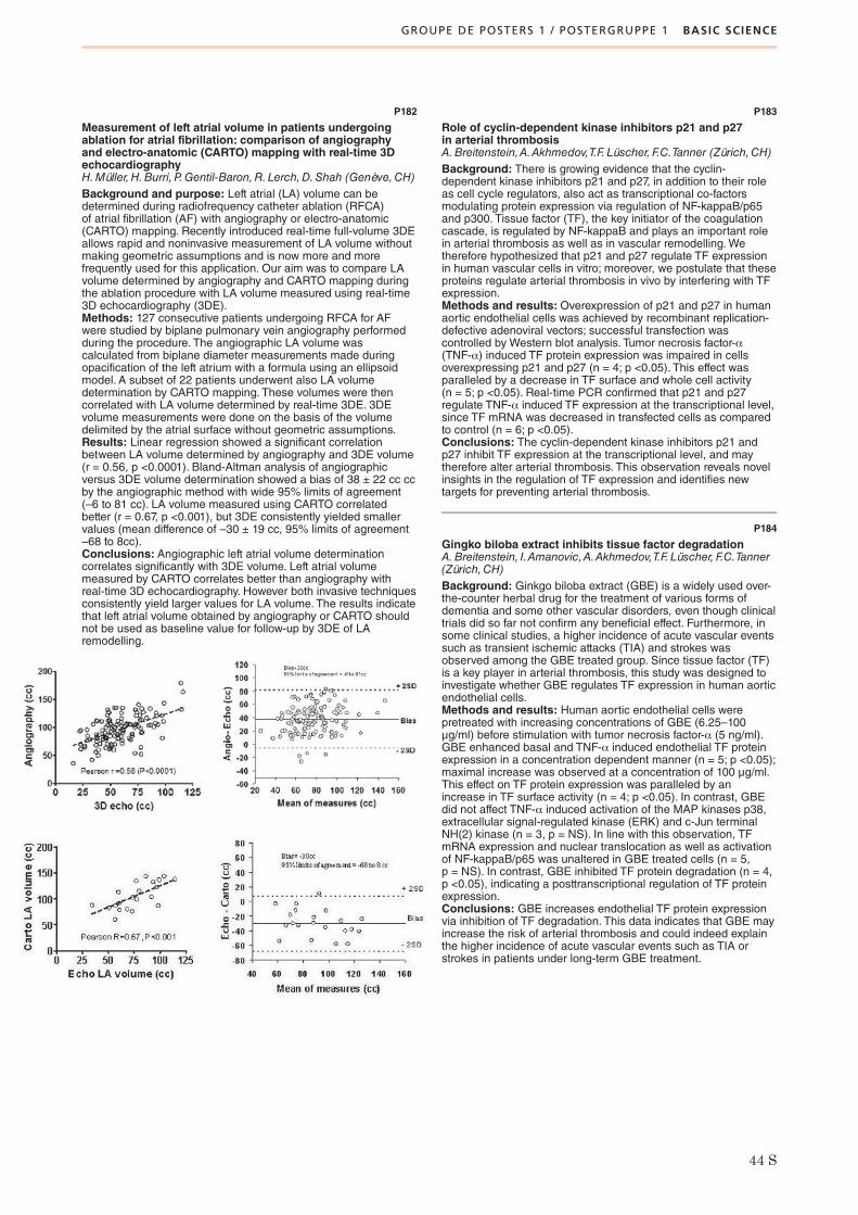
GROUPE DE POSTERS 1 / POSTERGRUPPE 1 BASIC SCIENCE
44 S
P182
Measurement of left atrial volume in patients undergoingablation for atrial fibrillation: comparison of angiographyand electro-anatomic (CARTO) mapping with real-time 3DechocardiographyH.Müller,H.Burri, P.Gentil-Baron,R. Lerch,D.Shah (Genève,CH)Background and purpose: Left atrial (LA) volume can bedetermined during radiofrequency catheter ablation (RFCA)of atrial fibrillation (AF) with angiography or electro-anatomic(CARTO) mapping. Recently introduced real-time full-volume 3DEallows rapid and noninvasive measurement of LA volume withoutmaking geometric assumptions and is now more and morefrequently used for this application. Our aim was to compare LAvolume determined by angiography and CARTO mapping duringthe ablation procedure with LA volume measured using real-time3D echocardiography (3DE).Methods: 127 consecutive patients undergoing RFCA for AFwere studied by biplane pulmonary vein angiography performedduring the procedure. The angiographic LA volume wascalculated from biplane diameter measurements made duringopacification of the left atrium with a formula using an ellipsoidmodel. A subset of 22 patients underwent also LA volumedetermination by CARTO mapping. These volumes were thencorrelated with LA volume determined by real-time 3DE. 3DEvolume measurements were done on the basis of the volumedelimited by the atrial surface without geometric assumptions.Results: Linear regression showed a significant correlationbetween LA volume determined by angiography and 3DE volume(r = 0.56, p <0.0001). Bland-Altman analysis of angiographicversus 3DE volume determination showed a bias of 38 ± 22 cc ccby the angiographic method with wide 95% limits of agreement(–6 to 81 cc). LA volume measured using CARTO correlatedbetter (r = 0.67, p <0.001), but 3DE consistently yielded smallervalues (mean difference of –30 ± 19 cc, 95% limits of agreement–68 to 8cc).Conclusions: Angiographic left atrial volume determinationcorrelates significantly with 3DE volume. Left atrial volumemeasured by CARTO correlates better than angiography withreal-time 3D echocardiography. However both invasive techniquesconsistently yield larger values for LA volume. The results indicatethat left atrial volume obtained by angiography or CARTO shouldnot be used as baseline value for follow-up by 3DE of LAremodelling.
P183
Role of cyclin-dependent kinase inhibitors p21 and p27in arterial thrombosisA.Breitenstein,A.Akhmedov,T.F. Lüscher, F.C.Tanner (Zürich,CH)Background: There is growing evidence that the cyclin-dependent kinase inhibitors p21 and p27, in addition to their roleas cell cycle regulators, also act as transcriptional co-factorsmodulating protein expression via regulation of NF-kappaB/p65and p300. Tissue factor (TF), the key initiator of the coagulationcascade, is regulated by NF-kappaB and plays an important rolein arterial thrombosis as well as in vascular remodelling.Wetherefore hypothesized that p21 and p27 regulate TF expressionin human vascular cells in vitro; moreover, we postulate that theseproteins regulate arterial thrombosis in vivo by interfering with TFexpression.Methods and results: Overexpression of p21 and p27 in humanaortic endothelial cells was achieved by recombinant replication-defective adenoviral vectors; successful transfection wascontrolled by Western blot analysis. Tumor necrosis factor-α(TNF-α) induced TF protein expression was impaired in cellsoverexpressing p21 and p27 (n = 4; p <0.05). This effect wasparalleled by a decrease in TF surface and whole cell activity(n = 5; p <0.05). Real-time PCR confirmed that p21 and p27regulate TNF-α induced TF expression at the transcriptional level,since TF mRNA was decreased in transfected cells as comparedto control (n = 6; p <0.05).Conclusions: The cyclin-dependent kinase inhibitors p21 andp27 inhibit TF expression at the transcriptional level, and maytherefore alter arterial thrombosis. This observation reveals novelinsights in the regulation of TF expression and identifies newtargets for preventing arterial thrombosis.
P184
Gingko biloba extract inhibits tissue factor degradationA.Breitenstein, I.Amanovic,A.Akhmedov,T.F. Lüscher, F.C.Tanner(Zürich,CH)Background: Ginkgo biloba extract (GBE) is a widely used over-the-counter herbal drug for the treatment of various forms ofdementia and some other vascular disorders, even though clinicaltrials did so far not confirm any beneficial effect. Furthermore, insome clinical studies, a higher incidence of acute vascular eventssuch as transient ischemic attacks (TIA) and strokes wasobserved among the GBE treated group. Since tissue factor (TF)is a key player in arterial thrombosis, this study was designed toinvestigate whether GBE regulates TF expression in human aorticendothelial cells.Methods and results: Human aortic endothelial cells werepretreated with increasing concentrations of GBE (6.25–100μg/ml) before stimulation with tumor necrosis factor-α (5 ng/ml).GBE enhanced basal and TNF-α induced endothelial TF proteinexpression in a concentration dependent manner (n = 5; p <0.05);maximal increase was observed at a concentration of 100 μg/ml.This effect on TF protein expression was paralleled by anincrease in TF surface activity (n = 4; p <0.05). In contrast, GBEdid not affect TNF-α induced activation of the MAP kinases p38,extracellular signal-regulated kinase (ERK) and c-Jun terminalNH(2) kinase (n = 3, p = NS). In line with this observation, TFmRNA expression and nuclear translocation as well as activationof NF-kappaB/p65 was unaltered in GBE treated cells (n = 5,p = NS). In contrast, GBE inhibited TF protein degradation (n = 4,p <0.05), indicating a posttranscriptional regulation of TF proteinexpression.Conclusions: GBE increases endothelial TF protein expressionvia inhibition of TF degradation. This data indicates that GBE mayincrease the risk of arterial thrombosis and could indeed explainthe higher incidence of acute vascular events such as TIA orstrokes in patients under long-term GBE treatment.
GROUPE DE POSTERS 1 / POSTERGRUPPE 1 BASIC SCIENCE
45 S
P185
HLA I antibodies induce VEGF production in endothelial cellsM.Bieri,M.Oroszlan,N. Liegti, J.Bieri, P.Mohacsi (Bern,CH)Background: Anti human leukocyte antigen class I (HLA I)antibodies were shown to activate several protein kinases inendothelial cells (EC), which induces proliferation and cellsurvival. A criterion of antibody mediated rejection is theoccurrence of interstitial edema. Here, we investigated the effectof anti-HLA I antibodies on endothelial permeability andproliferation.Methods: ECs of umbilical veins were isolated and effects of anti-HLA I antibodies (W6/32) were determined. Permeability wasassessed by a trans-well filter system; protein and mRNAexpression of vascular endothelial (VE-) cadherin or vascularendothelial growth factor (VEGF) was quantified by Western blotor RT-PCR, respectively. Phosphorylation was determined byimmunoprecipitation followed by phospho-tyrosine immunoblot.Proliferation was measured by the wound healing assay.Results: Endothelial permeability dose-dependently increased inthe presence of anti-HLA I antibodies. The protein expression ofVE-cadherin decreased after anti-HLA I stimulation of ECsalready after 30 min, which was inhibited after Src and VEGFreceptor type 2 (VEGFR2) blocking. The mRNA level of VE-cadherin was not affected by anti-HLA I treatment. Post-transcriptional phosphorylation of VE-cadherin was measuredafter 30 min anti-HLA I stimulation, which was reverted byVEGFR2 blocking. VEGF production of ECs was increased afteranti-HLA I administration. The VEGF production was inhibited byblocking phosphatidylinositol-3-kinase (PI3K) and mammaliantarget of rapamycin (mTOR), whereas blocking Src had no effect.Endothelial proliferation increased dose-dependently after anti-HLA I treatment. Anti-HLA I induced proliferation was inhibited byblocking mTOR and VEGFR2. Src did not show an effect onendothelial proliferation after anti-HLA I stimulation.Conclusion: Anti-HLA I antibodies stimulate ECs to produceVEGF, which activates VEGFR2 in an autocrine manner. ActivatedVEGFR2 initiates two distinct effects: on one hand, endothelialpermeability increased by a Src dependent phosphorylation ofVE-cadherin, on the other hand, endothelial proliferationincreased in an Src independent manner.
P186
The crosstalk between cardiomyocytes and microvascularendothelial cells in cardiotoxic cancer therapyM.Chiusa, F.Seitriz, L.Maquelin, F.Timolati,T. Suter,C. Zuppinger,S. Zbinden (Bern,CH)Objective: Cardiac microvascular endothelial cells (CMEC) aremodulators of cardiac performance and there is also an intimatecontact between CMEC and cardiomyocytes. Treatment of breastcancer with doxorubicin (Doxo) in combination with Herceptin®
(Trastuzumab) results in an increased incidence of heart failure.Adult rat ventricular myocytes (ARVM) under Doxo treatmentshow an increase in oxidative stress, down-regulate thetranscription factor GATA4, and genes involved in cell viability,calcium homeostasis and cytoskeleton structure.We tested, ifVEGF is a cardio-protective factor in cardiomyocytes andmicrovascular endothelial cells undergoing hypoxia, oxidativestress, or Doxo treatment, and if anti-VEGF therapy can preventa cross talk between CMEC and ARVM.Methods: Adult rat ventricular cardiomyocytes (ARVM) wereisolated and cultured in serum-free medium and treated for18–48 hours. Cardiac microvascular endothelial cells (CMEC)were isolated and used in the second passage, cultured inmedium with 20% NCS until confluence, then in medium with0.5% NCS for 24 hours before treatment, and treated for 24hours. Protein quantification was performed by Western Blot,VEGF release was measured by a rat specific VEGF ELISA-kit,cell death by TUNEL assay and necrosis by the live/death assay.Results: ARVM and CMEC under hypoxia condition (1.5% O2) for3–24 hours showed a maximum release of VEGF after 12 hours.CMEC under Doxo treatment (0.5–20 μM) released VEGF with amaximum at 1 μM Doxo. On the contrary, ARVM did not releasethis growth factor under the same condition. Hydrogen peroxide at300 μM suppressed the release of VEGF in CMEC. CMEC andARVM showed an dose-dependent increase in cell death(necrosis) by Doxo treatment. Neuregulin-1 β (NRG), known to bereleased by CMEC under oxidative stress conditions, induced the
expression of GATA-4, sodium-calcium-exchanger (NCX), theanti-apoptotic mitochondrial protein Bcl-2, and of the ErbB2receptor protein itself in ARVM.Conclusion: These preliminary data suggest, that both CMECand ARVM are able to produce VEGF under hypoxia condition,but only CMEC after Doxo treatment.We presume, that VEGF is aprotective factor in the signaling between ARVM and CMEC.Anti-VEGF therapy, such as by Bevacizumab, could prevent thiscrosstalk and increase cardiovascular damage by cytotoxiccancer therapy.
P187
Sirolimus reduces cubilin and megalin expression inproximal tubular epithelial cells by using an angiotensin II-dependent pathway: an explanation of reduced albuminreabsorptionM.Oroszlan,M.Bieri,A. Farkas,N. Ligeti, P.Mohacsi (Bern,CH)Objective: Proteinuria, which is a sign of the progression ofchronic renal disease or (non)-allograft pathology can appearafter conversion to sirolimus (SRL) or in case of de novo SRLtreatment in transplanted patients. It has been reported thatangiotensin converting enzyme inhibitors (ACE-Inh) orangiotensin II (Ang II) receptor antagonists reduce urinary proteinloss.Aim: This study was aimed to investigate the role Ang II and AngII type 1 receptor (AT1R) in the regulation of known albuminreceptors, such as cubilin and megalin expression andconsequently in albumin endocytosis on human renal proximaltubular epithelial cells (PTEC) following SRL treatment.Methods: A human renal PTEC line (HK-2) was used. Cells weretreated with SRL for 24 hrs. The binding of bovine serum albumin(BSA) to HK-2 was investigated by flow cytometry. The uptake ofBSA and the expression of megalin and cubilin were determinedby cellular ELISA. Values are given compared to thecorresponding vehicle control (dimethyl-sulfoxide).Results: SRL in concentrations of 1, 10 and 100 nM dose-dependently decreased the binding (–12.5% ± 18.2; –24.5% ±6.8, p = 0.0056; –26% ± 6.3, p = 0.0038, respectively) and theuptake of BSA (–5.3% ± 6; –17% ± 8.1, p = 0.025; –16% ± 4.6,p = 0.006, respectively). Reduced BSA binding and uptake after100 nM SRL administration were significantly prevented by theuse of an ACE-Inh (ramipril, 10 μM) (–8.8% ± 8.8, p = 0.014 and1.8% ± 9.6, p = 0.014, respectively) or an AT1R blocker (losartan,10 μM) (–12.5% ± 10.4, p = 0.029 and –14% ± 8.5, respectively).Furthermore, SRL in concentrations of 1, 10 and 100 nMdecreased the expression of megalin (–7.7% ± 10.2; –16.7% ± 7.7,p = 0.023; –18.8% ± 6, p = 0.0085, respectively) and cubilin(–3.5% ± 3.3; –12.8% ± 5.6, p = 0.019; –14.7% ± 6.8, p = 0.023,respectively) on HK-2. Coadministration of 100 nM SRL andramipril or losartan in concentration of 10 μM resulted in improvedmegalin (–6.1% ± 2.36, p = 0.014 and –11.2% ± 6.5, p = 0.05,respectively) and cubilin (–5.9% ± 3.5, p = 0.014 and –4.62%± 4.6, p = 0.014, respectively) expression.Conclusion: The decreased albumin endocytosis of PTEC ispartially preventable by the use of losartan or ramipril afteradministration of SRL. The blockade of Ang II restored albuminendocytosis possibly due to the ameliorated cubilin and megalinexpression.
P188
Oxidized low density lipoprotein activates p66shcin human aortic endothelial cellsY.Shi,G.G.Camici,A.Akhmedov,F.C.Tanner,T.F. Lüscher(Zürich,CH)Background: P66shc is a mitochondrial protein and novelmediator of oxidative stress. It has been involved in vascularaging, atherosclerosis and diabetic endothelial dysfunction. Thepresent study was designed to investigate the effect of oxidizedlow density lipoprotein (oxLDL) on the expression of p66shcprotein in human aorta endothelial cells.Methods: The expression of p66shc protein was measured bywestern blotting. The production of superoxide anion wasmeasured by electron-spin resonance spectroscopy.Results: After 24 hours, oxLDL increased p66shcphosphorylation in a dose dependent manner, but did not changethe total protein expression. In the presence of oxLDL (10 ug/ml),
GROUPE DE POSTERS 1 / POSTERGRUPPE 1 BASIC SCIENCE
46 S
inhibition of LOX-1 receptor reduced the phosphorylation ofp66shc, indicating that the effect of oxLDL on the p66shc is partlyinvolved by LOX-1. Either selective (CGP53353) or non-selective(Go6967 or Calphostin C) inhibitors of PKC β II reducedphosphorylated p66shc, indicating that also PKC β II is involvedin p66shc phosphorylation. Furthermore, oxLDL activated PKC βII on both Theronine641 and Serine660, confirming the role ofPKC β in the p66shc activation. Apocynin reduced both p66shcand PKC β II phosphorylation, suggesting that reactive oxygenspecies play an important role. In addition, oxLDL enhanced theproduction of superoxide. Apocynin, but not the inhibitors of PKCβ, reduced the production of superoxide, indicating that reactiveoxygen species is the upstream of PKC kinase.Conclusion: oxLDL activates p66shc protein in human aortaendothelial cells. Both ROS and PKC are involved in thisactivation process of p66shc.
P189
Endogenous and synthetic cannabinoids inhibit tumournecrosis factor-α-induced proinflammatory activities inhuman neutrophilsF.Montecucco,S. Lenglet, F.Burger,M.Bertolotto, F.Mach,S.Steffens (Genève,CH; Genova, I)Introduction: Increasing evidence supports a crucial role forneutrophils in atherosclerosis and its acute complications,such as acute myocardial infarction. These cells migrate ininflammatory tissues in response to different chemokines with theaim to neutralize inflammatory triggers. However, once retainedin tissues, they release several mediators that further increaseinflammatory processes and damage. Tumor necrosis factor-α(TNF-α) is a pivotal inducer of neutrophil inflammatory functions.Here, we investigated a possible modulation of TNF-α-mediateddramatic effects by endogenous and synthetic cannabinoids.Methods: Adhesion molecule, TNF-α receptor and annexinexpression on human neutrophils were determined by flowcytometry. Neutrophil migration was assessed by using modifiedBoyden Chamber. The release of superoxide anion wasmeasured by a modified method of Babior and collegue. P38MAPK and ERK 1/2 activation was assessed by western blotanalysis.Results: Pre-treatment with the endocannabinoid 2-AG or thesynthetic cannabinoid receptor CB2 ligand JWH-133 dose-dependently inhibited TNF-α-induced changes in cell surfaceadhesion molecule expression (CD11b, CD11c, CD18 and L-selectin). Accordingly, pre-treatment with 2-AG or JWH-133inhibited TNF-α-induced migration in response to CCL3. On theother hand, 2-AG or JWH-133 did not have any effect on TNF-α-induced superoxide anion release by human neutrophils. In orderto identify the possible mechanisms involved in cannabinoid-mediated anti-inflammatory activities, we further investigated theeffect of 2-AG or JWH-133 on neutrophil functions. No increase inapoptosis rate was observed upon treatment with cannabinoids.Furthermore, 2-AG or JWH-133 did not directly induce neutrophilmigration, and no changes in TNF-α receptor I and II surfaceexpression were observed. Conversely, 2-AG or JWH-133 dose-dependently inhibited TNF-α-induced p38 MAPK and ERK1/2 phosphorylation.
P190
Loss of thrombin-induced endothelial activationin cellular senescence by heterochromatic gene silencingD.J.Kurz,H.Greutert, S.Payeli, S.Briand,T.F. Lüscher, F.C.Tanner(Zürich,CH)Background: Cellular senescence, a programmed state inducedby multiple deleterious triggers, is characterised by permanentcell-cycle exit and altered gene expression. In humans it isconsidered a tumour suppressor mechanism, mediated byremoval of damaged or mutated cells from the cell cycle. Cellularsenescence is also thought to contribute to human ageing, aphenomenon rich in cardiovascular pathology.We have previouslyshown that induction of tissue factor (TF) by thrombin or –independently of cell-surface receptors – phorbol-ester (PMA)is virtually lost in endothelial cells undergoing replicativesenescence. Here we describe the mechanisms involved in thisphenomenon.
Methods and results: Human umbilical vein endothelial cells(HUVEC) were cultured in endothelial growth medium and seriallypassaged at confluence. Cellular senescence generally occurredafter 35–40 population doublings. Thrombin or PMA stronglyinduced TF protein and mRNA in young cells, while this effectwas severely blunted in senescent cultures. Investigation of thedownstream targets of PMA revealed no alteration in the mitogen-activated protein kinase and the nuclear factor-kappa B signaltransduction cascades in senescence. Stimulation of young andsenescent cultures transfected with a plasmid containing theluciferase gene under control of the TF core promotor showedsimilar luminescence, effectively ruling out any role forsenescence-related changes in intracellular signalling ortranscription factor expression in the loss of TF inducibility atsenescence.We further showed that this phenomenon can not beexplained by accelerated TF mRNA degradation. Taken together,these findings suggested altered gene accessability. Usingchromatin-immunoprecipitation we demonstrated thatsenescence-related loss of TF gene inducibility occurs followingheterochromatin formation resulting from loss of histone H3acetylation at the TF promotor. Finally, adenoviral-mediatedoverexpression of telomerase in presenescent cultures enabledescape from senescence and restored inducibility of TF by PMA.Conclusions: Induction of TF expression is severely blunted insenescent endothelial cells as a result of heterochromatic genesilencing at the TF promotor. This extends the involvement of thismechanism in senescence beyond cell cycle-related genes andsuggests a novel tumour-suppressing action of senescence byinhibition of tumour angiogenesis.
P191
From SWISS-AMI to CARDIASTIM –Translation of a clinical,progenitor-cell based protocol into GMPD.Sürder,T.Moccetti,G.Astori,G.Soldati, J. Schwitter, P. Erne,S.Windecker,A.Moschovitis,T.F. Lüscher,R.Corti (Lugano,Zürich,Luzern,Bern,CH)Since under International regulations, cell based medicinalproducts (CBMP) are requested to be produced following “GoodManufacturing Practices” (GMP) rules. Moreover, any procedureused to manipulate the cell population of interest should bevalidated and performed in a controlled environment (clean room).Finally, the CBMP release testing should ensure cell identity,potency, sterility, absence of adventitious agents and lowendotoxin levels. Here we report on our experience with theongoing SWISS-AMI trial, a phase II multicenter randomizedclinical trial (Clinicaltrials.gov id NCT00355186).Methods: In the above mentioned, ongoing trial we arerandomizing in a 2:1 pattern 150 patients with AMI successfullytreated within 24h by percutaneous coronary intervention (PCI)of the infarct-related coronary artery (IRA) in one control and twoBMC treatment groups. The control group is treated with state-of-the-art medical management. The treatment groups receiveintracoronary (i.c.) administration of BMC either 5–7 days or3–4 weeks after AMI, respectively. LV parameters are assessedby cardiac MRI at baseline, and after 4 and 12 months in a blindMRI core-lab. 50 ml of bone marrow are harvested under localanestesia from the iliac crest. In a centralized laboratory, BMCare isolated by density gradient centrifugation, washed andresuspended in 10 ml injectable medium. BMC are then reinfusedin the IRA via an over-the-wire balloon. The primary endpoint ischange in global LVEF by MRI at 4 months vs. baseline.Results: Cell processing for the SWISS-AMI Trial has beenrevised and optimized according to GMP criteria. Quality controlshave been introduced “in-process” as for sterility, identity andpotency of the cells. In 2008, SWISSMEDIC certified“CARDIASTIM“ as the first CBMP according to GMP criteria inSwitzerland. As for SWISS-AMI, so far 4 active centers haveincluded 89 patients. A mean of 170 ± 110 Mio BMC per patient(mean ± SD, n = 51) have been infused. Cell viability afterprocessing was 95.4 ± 3.5%. Functional testing showed aninvasion capacity of 50.4 ± 18.7% (n = 11). In-vivo quality tests inan animal model are ongoing.Conclusions:With the present study we were able to get GMPcertification for “CARDIASTIM”, the first CBMP of Switzerland.With the ongoing clinical trial we hope furthermore to confirmtherapeutical benefit of i.c. administration of BMC after AMIand to learn more about the optimal time point for cell therapy.

GROUPE DE POSTERS 1 / POSTERGRUPPE 1 BASIC SCIENCE
47 S
P192
Effects of NO donors on the cardiopulmonary systemG.Milano,M.Samaja, L.K. von Segesser(Lausanne,CH; Milano, I)Objective:We previously demonstrated that exposure to chronichypoxia (CH) developed pulmonary hypertension and rightventricular hypertrophy (RVH). In this study, we investigatedwhether L-Arginine, a substrate of endothelial nitric oxidesynthase (eNOS) and Molsidomine, a NO donor, alleviate thecardiovascular and pulmonary alterations induced by CH.Methods: Male rats were maintained in either normoxic (21%O2) or hypoxic chambers (10% O2) for 15 days as control ortreated with L-Arginine (L-ARG, 20 mg/ml), NOS inhibitor N-nitro-L-arginine methyl ester (L-NAME, 45 mg/Kg) or Molsidomine(MOL, 15 mg/Kg). Drugs was given daily in the drinking water.The right ventricular systolic pressures (RVSP), RVH, the ratioof lung weight to body weight and percent wall thickness ofpulmonary vessels, cardiac and pulmonary phosphorylationof eNOS proteins were measured.Results: The increase in RVSP was significantly attenuated in theL-ARG and MOL groups.Whereas the treatment with L-ARGattenuated the increase in RVH, MOL or L-NAME were ineffective.Chronic treatment with drugs did not prevent the increase in theratio of lung weight to body weight. In all conditions, the amountof water, expressed per gram wet tissue weight, was not differentbetween N and CH lungs, indicating that edema was not presentin these tissues. In contrast, CH developed an increase in medialwall thickness of small pulmonary arteries (0-100 mm) whilelarger arteries were not affected. This was attenuated by L-ARG orMOL treatments. Although CH decreased cardiac and pulmonaryphosphorylated eNOS levels, treatment with L-ARG and MOLrestored the normoxic level of eNOS phosphorylation.Conclusions: In the heart, chronic administration of NO donorsduring CH induce an attenuation in right ventricular systolicpressure and right ventricular hypertrophy, probably due toincreased phophorylation of eNOS proteins. AlthoughMolsidomine normalizes the right ventricular systolic pressure,it does not influence the development of right ventricularhypertrophy. In the lungs, chronic treatment with NO donors doesnot prevent pulmonary hypertrophy but attenuates the wallthickness increase in small pulmonary arterioles, probably due todirect effect of pulmonary hypertension.
P193
Aeration during chronic hypoxia in vivo improves post-ischaemic recovery and decreases injury in rat heartsG.Milano,M.Samaja,A.Corno,G.Vassalli, L.K. von Segesser(Lausanne,CH; Milano, I; Liverpool,UK)Objective: A clinical feature that complicates the post-surgeryoutcome in cyanotic children, chronic hypoxia (CH) is oftenconsidered cardioprotective on the basis of experimental studies.Here, we test the hypothesis that daily aeration during chronichypoxia (CHA) protect hearts subsequently exposed toischemia/reperfusion.Methods: Male 5-week old Sprague-Dawley rats weremaintained for 15 days under either CH (10% O2) or CHA (10%O2 + 1-h daily exposure to air). Normoxic (21% O2) rats were thecontrol. To assess tolerance to ischemia/reperfusion in vivo,hearts were subjected to 30-min occlusion of the left anteriordescending artery followed by 3-h reperfusion, with measurementof the injury by tetrazolium staining. To distinguish intrinsic fromcirculatory factors, end-diastolic pressure and (developedpressure) x heart rate were measured in ex-vivo Langendorff-perfused isolated hearts during 30-min global ischemia and45-min reperfusion. Some biochemical parameters wereevaluated at the end of hypoxia exposure (n = 5/subgroup).Results: In comparison to CH, CHA hearts displayed reducedinfarct size after both the in vivo and ex-vivo protocols. Myocardialperformance recovered better in CHA hearts, which displayedreduced apoptosis (TUNEL), greater phosphorylation of theendothelial isoform of NO synthase and enhanced plasma levelof nitrates and nitrites.Conclusions: Exposure to hypoxia for 15 days impairsmyocardial tolerance to ischemia/reperfusion, in agreement withclinical evidence. Daily aeration alleviates the CH-inducedderangement by accelerating intrinsic adaptive changes, whichinclude reduced apoptosis and improved NO metabolism.
P194
Functional behaviour of human internal thoracic arteryfrom patients with diabetes mellitus in vitroM.Grapow,D.Reineke,T.Kern,B.Winkler, E.Müller-Schweinitzer,T.P.Carrel, F.S. Eckstein (Basel,Bern,CH)Introduction: The aim of this analysis was to comparevasoreactive properties of internal thoracic artery (ITA) graftsfrom diabetic (DM) to those of non-diabetic (ND) patients.Method: Ring segments of internal thoracic artery (ITA), takenfrom patients undergoing coronary artery bypass grafting, weresuspended in organ bath chambers filled with modified Krebs-Henseleit solution and contractile responses to potassiumchloride (KCl), noradrenaline (NA), endothelin-1 (ET-l), andendothelium-dependent relaxant responses to acetylcholine(ACH) were recorded isometrically.Results: NA stimulated ITA from both groups within similarconcentration ranges while ITA from DM patients proved to besignificantly more sensitive to both ET-1 and KCl than ITA fromND. Furthermore, maximal contractile responses indicated thatKCl (3.79 ± 0.30 g, n = 7 in DM and 2.50 ± 0.23 g, n = 29 in ND,P <0.05) evoked significantly higher responses in ITA from DMas compared to the ND control group while both NA and ET-lstimulated ITAs from both groups with similar efficacies.Endothelium-dependent relaxant responses to ACH proved to besimilar in both groups when expressed as percentages of thepre-existing tone.Conclusion: The present data support the contention that DM-induced endothelial dysfunction is less frequent in human ITAthan expected from observations in systemic vascular beds.
P195
Rapamycin impairs endothelial cell function in humanarteriesM.Grapow,D.Reineke,P.Matt,B.Winkler,T.P.Carrel,E.Müller-Schweinitzer, F.S. Eckstein (Basel,Bern,CH)Introduction: Evidence has accumulated that treatment withrapamycin negatively impacts endothelial function in humancoronary arteries. Recently Rapamycin has been shown toinhibit mTORC2 which was previously believed to be rapamycin-insensitive. Prolonged treatment with rapamycin reduces thelevels of mTORC2 below those needed to maintain AKT/PKBsignalling, a key protein in the regulation of cell function andsurvival. Since AKT/PKB plays a pivotal role in activation of eNOSour goal was confirm this pathway in human internal thoracicarteries (ITA) in vitro.Methods: Vascular reactivity of ITA rings (44 patients)undergoing elective coronary artery bypass surgery wasinvestigated using the organ bath technique. After incubationwith rapamycin (solvent control, 0.1, 1 and 10 μmol rapamycin)for 20 h, rings were challenged with the contractile agonistnoradrenaline (NA, 1μM) and the relaxant compoundacetylcholine (ACH, 1 μM). Morphological analysis wasperformed on 12 of these rings from 4 patients by scanningelectron microscopy (SEM).Results: No differences were obtained in contractile responseto NA between the groups. Comparison of the ACH- inducedrelaxation revealed a concentration dependent decrease, beingsignificant for groups being incubated with 1 and 10 μmolrapamycin (in % of control: 1 μmol 66 ± 7, p <0.01; 10 μmol 36 ± 7,p <0.001; solvent control 112 ± 17 n.s.). During evaluation of theSEM specimens the examinators were not able to differentiatebetween controls and samples exposed to rapamycin for 20 h.In all groups the endothelial layer was well preserved.Conclusion: Our results confirm the modification by rapamycin ofthe above described signaltransduction pathway with inhibition ofmTORC2/AKT/PKB.We demonstrated that incubation of humanITA with rapamycin affects negatively the endothelium-dependentvasorelaxant responses in a concentration dependend manner invitro without inducing morphological changes.

GROUPE DE POSTERS 2 / POSTERGRUPPE 2 ELECTROPHYSIOLOGY, PACEMAKER, ARRHYTMIAS
48 S
P196
The presence of left atrial thrombus despite anticoagulationin patients undergoing left atrial ablation for atrial fibrillationcannot be predicted by clinical risk factorsS.Bull, J.P.De Bono,K.Rajappan,Y.Bashir,T.Betts,H.Becher(Oxford,UK)Conventionally the use of warfarin for thromboprophylaxis inpatients with atrial fibrillation is based upon clinical risk factors,such as the CHAD2 scoring system. Guidelines also recommendthat all patients undergoing elective cardioversion areanticoagulated before the procedure. Left atrial thrombus is anabsolute contraindication to left atrial catheter ablation (LACA)for atrial fibrillation as catheter manipulation is likely to result inembolisation. The incidence of thrombus in low-risk patientswithout anti-coagulation and high-risk patients with therapeuticanti-coagulation is not known. Transoesphagealechocardiography (TOE) is a very sensitive technique fordetecting left atrial thrombus. This study examines whethertherapeutic anticoagulation for a minimum of 1 month in allpatients undergoing LACA is effective in preventing left atrialthrombus formation.Methods:We analyzed the records and preoperative TOEimages of 160 consecutive patients undergoing LACA (age56 ± 11 years, 76% male). All patients were therapeuticallyanticoagulated with warfarin for at least 1 month beforeadmission.Warfarin was stopped 2 days before admission andTOE performed on the day of the procedure. All the TOE imageswere reviewed for evidence of left atrial appendage thrombosis bytwo independent cardiologists.Results: Four patients were found to have left atrial thrombusdespite therapeutic preoperative anticoagulation, leading tocancellation of their procedure. Three had paroxysmal atrialfibrillation and one persistent atrial fibrillation. The presenceof thrombus was not predicted by CHAD2 scores (0,1, 2, and 4)or age ( 42, 62, 72 and 61 years). One patient had severe leftventricular dysfunction, none had valvular disease. Left atrialthrombus reduced in size but still persisted in two patients withthe lowest CHAD score despite increased intensity ofanticoagulation.Conclusion: There is a 2.5% incidence of left atrial thrombusdetected by TOE in patients presenting for LACA despiteanticoagulation with warfarin. This can not be predicted from thepatients by arrhythmia burden, age or risk factors. These clots arefrequently more resistant to more intensive anticoagulation. TOEis therefore mandatory in all patients before LACA to exclude leftatrial thrombus.
P197
Pulmonary vein stripping and haemoptysis: a uniquecomplication of catheter ablation of atrial fibrillationP.Monney,P.Pascale,M.Fromer, E.Pruvot (Lausanne,CH)Case report: A 60-y old woman was referred for pulmonary veinsisolation (PVI) because of refractory paroxysmal atrial fibrillation(AF). After a single trans-septal puncture, a mapping catheter wasinserted for left atrium and pulmonary vein (PV) mapping.Following the acquisition of a set of points in the right inferior PV,the catheter could not be maneuvered anymore. Gentle tractionwas applied, producing a downward shift of the heart silhouetteon fluoroscopy. Advancement of the guiding sheath to offeradditional support, transient increase in catheter saline flow toincrease upstream PV pressure, and intravenous nitrates infusionto dilate the vein all failed to free the catheter. Stronger tractionand rotation allowed us to slowly retrieve the catheter tip.Significantly, a resistance was noted during this maneuver.Examination of the catheter showed a thin, translucent membranecovering its distal 8 mm, which appeared macroscopicallyas a branch of the PV that had been stripped off. The selectiveangiography of the right inferior PV showed no contrastextravasation but a tapered narrowing of a vein branchsuggesting endothelial tearing. Trans-esophageal echocardio-graphy showed no effusion. The procedure was resumed withsuccessful PVI. The patient reported post-operatively continuouslight oppressive chest pain without significant ECG alteration.Clinical examination was also normal. The day after theprocedure, she presented small amounts of haemoptysisspontaneously resolving within 2 days. The patient wasdischarged from hospital 4 days after PVI under therapeuticanticoagulation.
Discussion: Total complication incidence of PVI was reportedto be 3.9% with no procedure-related death in the most recentregistry. Common adverse events associated with the procedureinclude vascular access lesions, thermic injuries and thrombo-embolisms. This unique case shows that catheter entrapment ina PV is a rare but potentially serious complication of AF ablation.Although tamponade was expected, it did not happen.Interestingly, the haemoptysis indicates that bleeding occurredinto the pulmonary parenchyma but not into the pericardial space.This is in accordance with a recent anatomic study reporting ashorter pericardial sleeve over the right inferior PV (4.5 mm) ascompared to the other 3 veins (9.2–13.7 mm). No specifictherapeutic measure was undertaken for the patient besidesdelaying hospital discharge and reintroduction of therapeuticanticoagulation.
P198
Are atrial fibrillation ablation success rates from highvolume tertiary care centres reproducible?M.Rotter,M.Haissaguerre (Bern,CH; Bordeaux-Pessac,F)Introduction: Catheter ablation of atrial fibrillation (AF) yieldssuccess rates as high as 90% in experienced centers.Whetherthese results can be reproduced in smaller centers isquestionable. The aim of this study was to show that theseresults are transferable to smaller centers.Methods: The first 100 consecutive patients referred for catheterablation of AF at a new ablation center were prospectivelyfollowed up for one year. In all patients circumferential pulmonaryvein isolation was performed, in patients with episodes >12 hoursand patients with persistent AF additional complex fractionatedatrial electrogram ablation and/or linear ablation was performed.The operator performing all ablations was trained for 2 years at ahighly specialized AF ablation center. Follow up with patientinterview and 7-day event recording was carried out at 3, 6 and12 months. Any clinical tachycardia after >2 months ordocumented AF episodes lasting >30s were considered asfailures.Results: 138 ablations in 100 patients (age 60 ± 10 y, 72% male)were performed from January 07 to August 08. Patients sufferedfrom AF for 5 ± 4.5 years with 10 ± 9 episodes/months and failedon 2.3 ± 1 antiarrhythmic drugs. A cardiomyopathy was found in46% of patients; LVEF <50% in 10 patients. Left atrial size was43 ± 6 mm.AF was persistent in 47 patients; duration ofpersistent AF was 18 ± 26 months. Cumulative success rate after8 (3–15) months was 89% with 11% of patients on antiarrhythmicdrugs. Complications occurred in 5 patients (3 hematomas, one
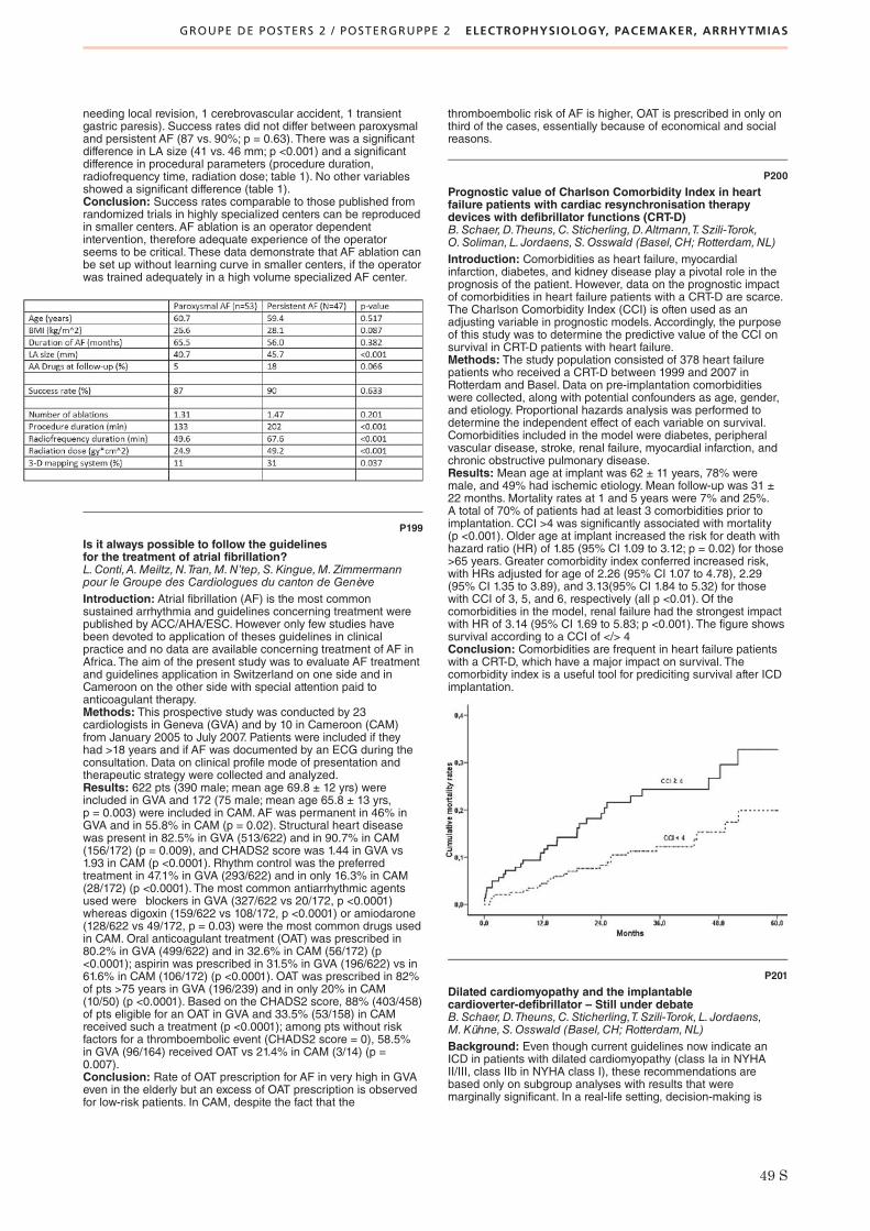
GROUPE DE POSTERS 2 / POSTERGRUPPE 2 ELECTROPHYSIOLOGY, PACEMAKER, ARRHYTMIAS
49 S
needing local revision, 1 cerebrovascular accident, 1 transientgastric paresis). Success rates did not differ between paroxysmaland persistent AF (87 vs. 90%; p = 0.63). There was a significantdifference in LA size (41 vs. 46 mm; p <0.001) and a significantdifference in procedural parameters (procedure duration,radiofrequency time, radiation dose; table 1). No other variablesshowed a significant difference (table 1).Conclusion: Success rates comparable to those published fromrandomized trials in highly specialized centers can be reproducedin smaller centers. AF ablation is an operator dependentintervention, therefore adequate experience of the operatorseems to be critical. These data demonstrate that AF ablation canbe set up without learning curve in smaller centers, if the operatorwas trained adequately in a high volume specialized AF center.
P199
Is it always possible to follow the guidelinesfor the treatment of atrial fibrillation?L.Conti,A.Meiltz,N.Tran,M.N’tep,S.Kingue,M.Zimmermannpour le Groupe des Cardiologues du canton de GenèveIntroduction: Atrial fibrillation (AF) is the most commonsustained arrhythmia and guidelines concerning treatment werepublished by ACC/AHA/ESC. However only few studies havebeen devoted to application of theses guidelines in clinicalpractice and no data are available concerning treatment of AF inAfrica. The aim of the present study was to evaluate AF treatmentand guidelines application in Switzerland on one side and inCameroon on the other side with special attention paid toanticoagulant therapy.Methods: This prospective study was conducted by 23cardiologists in Geneva (GVA) and by 10 in Cameroon (CAM)from January 2005 to July 2007. Patients were included if theyhad >18 years and if AF was documented by an ECG during theconsultation. Data on clinical profile mode of presentation andtherapeutic strategy were collected and analyzed.Results: 622 pts (390 male; mean age 69.8 ± 12 yrs) wereincluded in GVA and 172 (75 male; mean age 65.8 ± 13 yrs,p = 0.003) were included in CAM.AF was permanent in 46% inGVA and in 55.8% in CAM (p = 0.02). Structural heart diseasewas present in 82.5% in GVA (513/622) and in 90.7% in CAM(156/172) (p = 0.009), and CHADS2 score was 1.44 in GVA vs1.93 in CAM (p <0.0001). Rhythm control was the preferredtreatment in 47.1% in GVA (293/622) and in only 16.3% in CAM(28/172) (p <0.0001). The most common antiarrhythmic agentsused were blockers in GVA (327/622 vs 20/172, p <0.0001)whereas digoxin (159/622 vs 108/172, p <0.0001) or amiodarone(128/622 vs 49/172, p = 0.03) were the most common drugs usedin CAM. Oral anticoagulant treatment (OAT) was prescribed in80.2% in GVA (499/622) and in 32.6% in CAM (56/172) (p<0.0001); aspirin was prescribed in 31.5% in GVA (196/622) vs in61.6% in CAM (106/172) (p <0.0001). OAT was prescribed in 82%of pts >75 years in GVA (196/239) and in only 20% in CAM(10/50) (p <0.0001). Based on the CHADS2 score, 88% (403/458)of pts eligible for an OAT in GVA and 33.5% (53/158) in CAMreceived such a treatment (p <0.0001); among pts without riskfactors for a thromboembolic event (CHADS2 score = 0), 58.5%in GVA (96/164) received OAT vs 21.4% in CAM (3/14) (p =0.007).Conclusion: Rate of OAT prescription for AF in very high in GVAeven in the elderly but an excess of OAT prescription is observedfor low-risk patients. In CAM, despite the fact that the
thromboembolic risk of AF is higher, OAT is prescribed in only onthird of the cases, essentially because of economical and socialreasons.
P200
Prognostic value of Charlson Comorbidity Index in heartfailure patients with cardiac resynchronisation therapydevices with defibrillator functions (CRT-D)B.Schaer,D.Theuns,C.Sticherling,D.Altmann,T.Szili-Torok,O.Soliman, L. Jordaens,S.Osswald (Basel,CH; Rotterdam,NL)Introduction: Comorbidities as heart failure, myocardialinfarction, diabetes, and kidney disease play a pivotal role in theprognosis of the patient. However, data on the prognostic impactof comorbidities in heart failure patients with a CRT-D are scarce.The Charlson Comorbidity Index (CCI) is often used as anadjusting variable in prognostic models. Accordingly, the purposeof this study was to determine the predictive value of the CCI onsurvival in CRT-D patients with heart failure.Methods: The study population consisted of 378 heart failurepatients who received a CRT-D between 1999 and 2007 inRotterdam and Basel. Data on pre-implantation comorbiditieswere collected, along with potential confounders as age, gender,and etiology. Proportional hazards analysis was performed todetermine the independent effect of each variable on survival.Comorbidities included in the model were diabetes, peripheralvascular disease, stroke, renal failure, myocardial infarction, andchronic obstructive pulmonary disease.Results: Mean age at implant was 62 ± 11 years, 78% weremale, and 49% had ischemic etiology. Mean follow-up was 31 ±22 months. Mortality rates at 1 and 5 years were 7% and 25%.A total of 70% of patients had at least 3 comorbidities prior toimplantation. CCI >4 was significantly associated with mortality(p <0.001). Older age at implant increased the risk for death withhazard ratio (HR) of 1.85 (95% CI 1.09 to 3.12; p = 0.02) for those>65 years. Greater comorbidity index conferred increased risk,with HRs adjusted for age of 2.26 (95% CI 1.07 to 4.78), 2.29(95% CI 1.35 to 3.89), and 3.13(95% CI 1.84 to 5.32) for thosewith CCI of 3, 5, and 6, respectively (all p <0.01). Of thecomorbidities in the model, renal failure had the strongest impactwith HR of 3.14 (95% CI 1.69 to 5.83; p <0.001). The figure showssurvival according to a CCI of </> 4Conclusion: Comorbidities are frequent in heart failure patientswith a CRT-D, which have a major impact on survival. Thecomorbidity index is a useful tool for prediciting survival after ICDimplantation.
P201
Dilated cardiomyopathy and the implantablecardioverter-defibrillator – Still under debateB.Schaer,D.Theuns,C.Sticherling,T.Szili-Torok, L. Jordaens,M.Kühne,S.Osswald (Basel,CH; Rotterdam,NL)Background: Even though current guidelines now indicate anICD in patients with dilated cardiomyopathy (class Ia in NYHAII/III, class IIb in NYHA class I), these recommendations arebased only on subgroup analyses with results that weremarginally significant. In a real-life setting, decision-making is

GROUPE DE POSTERS 2 / POSTERGRUPPE 2 ELECTROPHYSIOLOGY, PACEMAKER, ARRHYTMIAS
50 S
even more complex, as issues such as prevention mode,additional cardiac resynchronization therapy (CRT) and thepotential of EF improvement over time need to be considered.Methods:We analysed DCM patients with an ICD implanted forprimary or secondary prevention of sudden cardiac deathregarding appropriate ICD interventions, overall mortality and leftventricular ejection fraction (LVEF) during follow-up and stratifiedpatients into four subgroups of LVEF above or below 20% andprimary and secondary prevention, respectively. Patients stemfrom two large prospective ICD registries in Switzerland and theNetherlands.Results:We studied 349 patients, 30% were female, ejectionfraction was 24 ± 8%, in whom ICDs (CRT = 198) were implantedbetween 1.1.2000 and 31.10.2008. Indication was primaryprevention in 81% of CRT vs. 55% in VVI/DDD devices (p<0.0001). Patients were on heart failure therapy with ACE-inibitors/ARBs in 94%, blockers in 75% and diuretics in 78%.Follow-up was 33 ± 25 months, mortality 12% (annual 4.3%).ICD therapies were delivered in 33%, mainly for ventriculartachycardia. KM-curves of device therapy according to the foursubgroups are shown in the table. Secondary prevention wasmore prone to arrhythmic events. These were more also often(all p < 0.05) seen in non-CRT patients. Baseline LVEF did notinfluence events (p = 0.12). Cumulative event rates at 5 yearswere 53% in secondary and 23% in primary prevention(p <0.001). In 221 patients echo-determined LVEF after at least6 months of follow-up was available. LVEF improved for almost10% (CRT 22 to 31%, non-CRT 26 to 35%; p <0.0001). Thisimprovement was independent of the stimulation mode (p >0.5).Improvement to an EF >35% was seen in 25% of CRT and in45% of non-CRT patients (p = 0.004).Conclusions: In DCM patients with an ICD indication for primaryprevention, the rate of ICD therapy is quite low. As these patientshave a considerable potential of an improvement of their leftventricular function, independent of stimulation mode, theindication to implant a device should be made with caution andprobably only after a certain observation period.
P202
Sad reality of out-of-hospital cardiac arrest inFrench-speaking SwitzerlandE.Katz, J.Metzger,D.Fishman,M.Potin,M.Niquille,V.Della Santa,C.Sénéchaud,M.Rodriguez,W.Garcia,P.Vogt (Lausanne,Sion,Genève,Neuchâtel, La Chaux-de-Fonds,Porrentruy, Fribourg,CH)Background: Survival from out-of-hospital cardiacarrest(OHCA)is dismal. New strategies for OHCA resuscitationincluding public access defibrillation (PAD) emerged recently.However only population based data would allow us to access theeffect of interventions on survival. Our goal was so far to evaluatethe suitability of new resuscitation approaches in our area.Methods: An OHCA registry was set up in 2007 in LausanneUniversity Hospital. It enlisted the help of >1500 Generalpractitioners (GPs), 26 Emergency Medical Services (EMS), and23 hospitals to prospectively collect the data of every adult (>18years) who suffered from OHCA in french-speaking Switzerland(catchment population: 1.5 mln inhabitants).
Results: After 1.5 year of observation data of 1319 cardiac arrestvictims were recorded, 68% were males; the mean age was of70 (SD14) years. Nearly half of victims were known for heartdisease.75% of OHCA occured at home, 2% in medical practice,19% in public places, 2% at workplace and the site of 2% ofOHCA was not known. Bystander cardio-pulmonary resuscitation(CPR) was performed by 29% of witnesses and every secondrescuer (48%) was health care professional. First recorded rhythmwas ventricular fibrillation or ventricular tachycardia in 19% ofvictims, asystole in 67% and pulseless electrical activity in 14%.Survival to hospital discharge was of 6.5%.Conclusions: In our region efforts should be made to strengthenevery link in the chain of survival before starting PADinitiative.Only 21% of OHCA can be potentially reached by PADprogramm and less than one fifth of OHCA are reached inshockable rhythm. So far widespread CPR training should beprovided to lay people. General population including familymembers of people known for cardiac disease should be informabout premonitory symptoms of cardiac arrest. EmergencyMedical Services call-to-arrival time should be shortened. PADprograms in our region should include automated externaldefibrillators installation in multi-family dwellings and cardiacarrest watchers network.
P203
Is out-of-hospital cardiac arrest really sudden?E.Katz, J.Metzger,D.Fishman,M.Potin,M.Niquille,W.Garcia,C.Sénéchaud,R.Kehtari,M.Rodriguez,A.Kocher,P.Vogt(Lausanne,Sion,Genève,Fribourg, La Chaux-de-Fonds,Neuchâtel, Porrentruy,Délémont,CH)Background: Survival rates from out-of-hospital cardiac arrest(OHCA) remain low despite increasing efforts to improveresuscitation care. Better understanding of OHCA circumstancesshould be helpful in developing preventive measures andfacilitating proper reactions to such event.Our goal was so far toaccess circumstances of OHCA in our area.Methods: An OHCA registry was set up in 2007 in LausanneUniversity Hospital. It enlisted the help of >1500 Generalpractitioners (GPs), 26 Emergency Medical Services (EMS),and 23 hospitals to prospectively collect the data of every adult(>18 years) who suffered from OHCA in french-speakingSwitzerland (catchment population: 1.5 mln inhabitants)Results: After 18 months data of 1319 OHCA were collected,48% of victims were previously known for cardiac disease.Everysecond OHCA victim (51%) experienced premonitory symptomsbefore cardiac arrest. Premonitory symptoms were: chest pain in31%, dyspnea in 31%, malaise in 26%, tiredness in 9%,palpitations, headache and dizziness in 1% of cases.75% of OHCA occured at home.Conclusions: In our area OHCA occurs mostly at home and onehalf of victims is already known for cardiac disease. Every secondvictim had typical warning symptoms: mostly chest pain, dyspneaor malaise. Although much-hailed use of community baseddefibrillation strategies is expected to improve survival wesuggest that targeted educational programs(including basic lifesupport) for general population and patients known for cardiacdisease and their relatives should have even higher priority.
P204
Cardiac biomarkers after ICD testingM.Mutschelknauss,H.Rickli, R.Widmer,K.Choka,P.Ammann(St.Gallen,CH)Introduction: The necessity of defibrillation efficacy tests in ICD-patients has been questioned because of possible complicationsand/or morbidity. Little data assessing clinical and biochemicalparameters after ICD testing is available. The present study aimsto prove whether induction of ventricular fibrillation (VF) duringICD testing leads to an elevation of cardiac biomarkers orparameters of myocardial cell necrosis.Methods: Prospective single-centre study in 47 ICD recipients(38 m, mean age 58.7 ± 14 yrs, CHD 31 pts, mean LVEF 34 ±13.3%). VF was induced with a 1 Joule T-wave shock. B-typenatriuretic peptide BNP, Troponin I cTNI, Creatinkinase CK,potassium and creatinin were analyzed from blood samples takenright before, one and six hours after ICD testing. Pts with acuteheart failure, hypokalemia or insufficient anticoagulation for atrial
GROUPE DE POSTERS 2 / POSTERGRUPPE 2 ELECTROPHYSIOLOGY, PACEMAKER, ARRHYTMIAS
51 S
fibrillation were excluded from the study. Delivered shock energyfor termination of VF was 31 J (13 pts) and 35 J (34 pts).Results: Values for cTNI (cTNI 0h mean 0.03 μg/L, IQR 0.03–0.06 μg/L, cTNI 6h mean 0.05 μg/L, IQR 0.03–0.08 μg/L, p =0.0027) showed a significant elevation six hours after ICD-testingcompared to baseline values. Other serum levels of biomarkersat baseline did not change compared to six hours after ICDtesting(BNP mean 311 ± 294 ng/L vs 352 ± 352 ng/L, CK 107 ±46 U/L vs 109 ± 47 U/L). None of the patients was hospitalizeddue to MACE within 1 month after ICD testing (mean follow-up265 ± 77 d). ICD testing resulted in clinical relevant changes intwo patients (4.3 %) with disconnection of SVC coil and reversedshock polarity.Conclusions: ICD testing via induction of VF leads to asignificant elevation of cTNI compared to baseline values whileother cardiac biomarkers do not change. Elevation of cTNI afterICD testing did not increase the MACE rate.
P205
Electrical storm in patients with an implantable cardioverter-defibrillator – triggered by scars, ischaemia or...G.Leibundgut, S.Osswald,C.Sticherling,M.Kühne,B.Schaer(Basel,CH)Background: Electrical storm (ES) is defined as the occurrenceof 3 ICD interventions (ATP and/or shocks) within 24 hours andhas been identified as a prognostic factor of subsequent death.However, ES might have its seeds in different reasons and thedefinition usually used is quite broad, as series of ATP cannot becompared to series of shocks.We therefore tried to determineoutcome, triggers and measures taken in patients with coronaryartery disease (CAD) and severe ES.Methods: In our prospective ICD registry we identified allpatients with ES.We used a more constricted definition of ES:a series of ?3 episodes of ventricular tachyarrhythmia (VT) orfibrillation (VF) that required high energy shocks, usually leadingto urgent hospitalisation.We restricted the study to CAD patients,as in these patients ischemia as a potential trigger is oftenexcluded by non-/invasive testing in addition to routine lab testsand history taking. In all patients follow-up was defined as the12 months following the first ES.Results: From our ICD registry database we retrieved 431patients with CAD. 32 (7.4%) patients fulfilled our criteria of ES.4 patients were excluded due to missing follow-up data. In theremaining 28 patients mean age was 68 ± 11 years, 12 (43%)patients died, 1 (4%) in direct correlation with ES. To identifypossible ischemia myocardial perfusion szintigraphy (MPS) wasperformed in 12 (43%) and coronary angiography in 15 (54%)patients.We identified as triggers: hypokalemia or physical activityin 1 (4%) patient respectively, infection in 3 (11%) and ischemiain 6 (21%) patients. Identification of a trigger was not possible in15 (54%) patients and 2 (7%) patients had an inadequate ICD-therapy. Measures taken were: adjustment of anti-ischemicmedication in 4 (14%) and percutaneous coronary intervention(PCI) in 6 (21%) patients, no adjustments were done in 18 (64%)patients. After the first ES, which occurred 33 ± 34 months afterimplantation of the ICD, 10 (36%) patients sustained at least oneother ES during follow-up. 1 (17%) of the patients who had anegative MPS and 3 (50%) of the patients with PCI to correctischemia sustained another ES during follow-up.Conclusion: Most of the identified triggers for ES in patientswith known CAD remain unknown and are different from ischemia.Patients with suspected or proven ischemia triggering an ESundergoing PCI have more frequent future events than patientswithout ischemia.
P206
Does remote monitoring of implantable defibrillatorsincrease the workload of the pacemaker clinic? Initialexperience with the CareLink NetworkD.Senouf,H.Sunthorn,D.Shah,C.Stettler,H.Burri (Genève,CH)Background: Recent implantable cardioverter defibrillator (ICD)models can communicate wirelessly with home transmitters andsend data to the physician, thus allowing remote monitoring. Anypre-defined alert (involving atrial arrhythmia, device integrity, orprogramming issues) automatically triggers an e-mail alert to thephysician. A potential concern with remote monitoring is
increased workload due to numerous incoming messages.Wedescribe our initial experience with remote monitoring of ICDpatients using the Medtronic CareLinkTM network.Methods: Patients implanted with a Medtronic ICD equipped withwireless technology were provided with a transmitter to install athome and connect to the Care Link network via their landlinephone. All alerts (except for lung fluid accumulation) wereactivated.We analyzed the number of alerts receivedautomatically, as well as the number and clinical repercussions ofdevice interrogations performed manually by the patients.Results: A total of 22 patients were included. Of these, 6 patientsdid not install the system for various reasons. Thus 16 patientshad remote monitoring with a mean follow-up of 87 ± 71 days(range 14–270 days). During this period, we received a total of48 transmissions. Of these, only 5 involved an automatic alertevent with e-mail notification (all from a single patient) andconcerned sustained atrial arrhythmia for which anticoagulationwas initiated. Initial set-up was responsible for 16 transmissions.There were 27 manual transmissions by 6 patients. Manualtransmissions led to clinically significant interventions in 2 ofthese patients. Medical therapy was adjusted in 1 patient dueto evidence of lung oedema. In the other patient, incessant slowventricular tachycardia was diagnosed by the real-timeelectrogram strip, which proved to be life-saving as the patientwas immediately admitted to hospital, where the arrhythmiadegenerated into rapid ventricular tachycardia with ineffectiveICD shocks but response to amiodarone.Conclusions: Remote monitoring resulted in a negligibleincrease in workload as automatic alerts occurred in only 1/16(6%) of patients. The technology has the potential to improvepatient safety and quality of care, and proved to be life-saving inone patient.
P207
Does sudden cardiac death spare doctors?E.Katz, J.Metzger,D.Fishman,M.Potin,M.Niquille,R.Kehtari,C.Sénéchaud,W.Garcia,M.Rodriguez,P.Vogt (Lausanne,Sion,Genève,Neuchâtel, La Chaux-de-Fonds, Fribourg,Porrentruy,CH)Background: Survival from out-of-hospital cardiac arrest (OHCA)is currently low. Patients reaction while experiencing premonitorysymptoms OHCA is crucial. Health care professionals aresupposed to be better informed about symptoms precedingcardiac arrest and measures to take than lay public. Few dataabout circumstances of OHCA among health professionals areawailable.We were interested so far to access reaction onpremonitory symptoms among health professionals.Methods: An OHCA registry was set up in 2007 in LausanneUniversity Hospital. It enlisted the help of >1500 Generalpractitioners (GPs), 26 Emergency Medical Services (EMS), and23 hospitals to prospectively collect the data of every adult (>18years) who suffered from OHCA in french-speaking Switzerland.Results: During first 12 months data of 17 health professionals(1.5% of total OHCA) suffered from OHCA in our area (mean age71 years ± 13 years). Nearly three quarters (12/17) of victimswere males; 70% were known for previous cardiac disease; 11/17(64%) of victims suffered from OHCA at home. Among 6 personswho were known to experience premonitory symptoms beforeOHCA only one activated Emergency Medical Service. Only oneperson survived to hospital discharge (5.8%).Conclusions: Survival from OHCA among health professionals inour area does not differ from general population.Whileindisputably better informed doctors and nurses does not alwaysreact as they advise to their patients. The information to healthcare workers about myocardial infarction and cardiac arrestshould be strenghtened and focused on practical measures.
P208
Long-term follow-up of AAI pacemakers for sick-sinussyndrome – Changes inWenkebach point behaviourH.Bussell, S.Osswald,C.Sticherling,M.Kühne,B.Schaer (Basel,CH)Background: Current guidelines propose AAI pacemakers forpatients with a symptomatic sick-sinus syndrome (SSS). However,as cardiologists fear the development of additional AV-nodaldisease during mid- and long-term follow-up, this one-lead systemis underutilised in Switzerland. Aim of the study therefore was to

GROUPE DE POSTERS 2 / POSTERGRUPPE 2 ELECTROPHYSIOLOGY, PACEMAKER, ARRHYTMIAS
52 S
evaluate the long-term follow-up of our AAI patients. A specialfocus was set on Wenkebach point (WB) behaviour over time.Methods:We retrospectively analysed all our patients in whoman AAI-pacemaker was implanted between 1992 and 2006 andwho were followed either at our clinic or in private practice.Prerequisite for AAI pacemaker implantation was pure SSS (nobundle branch block in the resting ECG, no documented AV-block,WB during implant test >120/min).Whilst the number of systemupgrade due to symptomatic AV-block was available for allpatients, WB behaviour was available only in patients inpermanent control at our hospital.WB behaviour was tested atevery visit at our hospital, but only inconsistently in privatepractice, so that these patients were excluded.Results: An AAI pacemaker was implanted in 140 patients, 62%were female, age was 74 ± 13 years (range 19-94). During follow-up of 3.4 ± 2.1 years, 24 patients (17%) died and 9 underwentsystem upgrade (6.4%; annual incidence 1.9%). Complete WBdata were available in 71 patients, with a follow-up of 4.6 ± 3.2years. Between implantation and last follow-up, WB decreasedfrom 135 ± 19 to 126 ± 17/min. Mean values according to agegroups and year after implantation are shown in the figure.Conclusions: AAI pacemakers in patients with pure SSS showan excellent long-term outcome, with only a minority of patientsneeding system upgrade.WB point behaviour is stable over time.
P209
In vivo measurements of atrial repolarisation alternansbased on standard pacemaker technologyF. Jousset, P.Ruchat, J.Vesin,M.Fromer,M.Burki, P.Pascale,E.Pruvot (Lausanne,CH)Introduction: It has been shown that repolarization alternans(Re-ALT), a beat-to-beat alternation in action potential duration,enhances dispersion of repolarization above a critical heart rateand promotes susceptibility to ventricular arrhythmias. It isunknown whether Re-ALT is measurable in the atria usingstandard pacemakers and whether Re-ALT plays a role inpromoting AF.Methods: two DDD pacemakers (PM) with two right atrial (RA)leads were implanted in 3 male sheep (>60 kg) after ablation ofthe AV junction. The first PM was used to deliver rapid pacing(400 beats) for measurements of RA Re-ALT, the second one torecord a broadband unipolar RA electrogram (EGM). Re-ALT wasconsidered present when the 2 following conditions were fulfilled:1) beat-to-beat alternation in T-wave apex amplitude (Ta) for>4 consecutive beats, and 2) statistically significant differentdistributions of odd and even Ta (p <0.01).Results: Re-ALT amplitude progressively increased as a functionof pacing CL from a mean value of 52 ± 5 microV at 400 ms toa maximal value of 184 ± 36 microV at a mean pacing CL of210 ± 36 ms. Importantly, Re-ALT appeared intermittent but noperiodicity was noticed, and its onset was independent of the
pacing CL. In one sheep, rapid pacing at a CL of 230 ms inducedan episode of non sustained AF that was preceded by overt Re-ALT.Conclusions: in-vivo measurement of atrial repolarization showsthat Re-ALT amplitude increased as a function of pacing rate. Re-ALT appeared intermittent but no periodicity was observed. Theobservation of overt Re-ALT preceding an episode of nonsustained AF suggests that Re-ALT is a promising parameter forin vivo assessment of AF susceptibility using standard pacemakertechnology.
P210
Atrial flutter ablation using a linear electrodewith duty-cycled radiofrequencyS.Boll, L.Dang,C.Scharf (Zürich,CH)Background: Duty cycled radiofrequency ablation (RFA) is usedfor ablation of atrial fibrillation (AF). A novel electrode prototypewas designed to map and deliver linear lesions for atrial flutters(AFL).Methods: In 51 consecutive patients (pts) AFL was diagnosedas a primary arrhythmia (n = 9) or during RFA for paroxysmal(n = 29) or persistent (n = 10) AF. In additional 3 pts a focal rightatrial tachycardia (AT) was mapped and ablated with the linearelectrode.Results: A cavotricuspid isthmus (CTI) dependent AFL wasdiagnosed in 36 pts and complete block achieved in 32 (88%)with a mean of 4.7 ± 3.2 burns (range 1–15). In 15 pts 22 leftatrial flutters (LAFL, CL 269 ± 50 ms, range 217–450 ms) weremapped conventionally with the linear electrode at the roof(n = 12), mitral isthmus (n = 5), at the septum (n = 3) and otherlocalization (n = 1) or nor found (n = 1). Successful RFA withtermination of LAFL was observed in 14 (63%). Complete mitralblock was documented in 3 pts (60%) and not achieved in 2 pts(40%) because on inability to deliver RF within the CS with thelinear electrode. The remaining 8 LAFL (36%) could not beablated because of changing activation sequence or recurrent AF.All 3 AT in the right atrium were ablated successfully.Conclusion: The current prototype of a linear ablation electrodeis successful in CTI and ablation of AT and LAFL. Left atrialflutters remain challenging if activation varies and AF occurs.
P211
Do patients on oral anticoagulation require less heparinto achieve therapeutic ACT?W.Diefenbacher, F.Amanpour,R.Nikolic,C.H.Attenhofer Jost,A. Fäh-Gunz, E.Straumann,B.Nägeli, D.Maurer,R.Candinas,L.Dang, J.Muntwyler,C.Scharf (Zürich,CH)Introduction: Patients (pts) undergoing ablation of atrialfibrillation (AF) receive unfractionated heparin to achieve anactivated clotting time (ACT) above 250 seconds to preventintracardiac thrombus formation and embolism. It is unknownwhether pts on therapeutic oral anticoagulation (INR >2.0)require less heparin to achieve similar ACT levels.Methods: In pts undergoing AF ablation the ACT was measuredbefore, 10 min after administration of i.v. unfractionated heparinand in 30 min intervals thereafter. A multiple linear regressionmodel was used to assess the influence of body weight, INR, preprocedure ACT and heparin on ACT after heparin administration.Results: Among 91 pts (74% men, age 60 ± 8 y, body weight83 ± 15 kg) there were 22 pts (24%) with subtherapeutic INR <2,52 pat with an INR between 2 and 2.9 and 17 pts with INR above3. The INR correlated with baseline ACT (r = 0.47, p <0.005).A mean of 8200 ± 1800 U unfractionated heparin was givenregardless of baseline INR (no correlation, p = 0.2). Univariatepredicators of post heparin ACT were baseline INR (p <0.001),heparin dose (p <0.001) and baseline ACT (p <0.005). In amultiple regression model, baseline INR and heparin dose werethe strongest predictors of post heparin ACT (p <0.001). Byestimate the heparin dose should be reduced by ca. 3000U perunit of INR elevation.Conclusion: Anticoagulation with elevated INR has a significanteffect on baseline ACT and heparin effect on ACT. In ptsundergoing AF ablation on oral anticoagulation, the heparin doseshould be reduced by about 3000 U per unit of INR elevation.
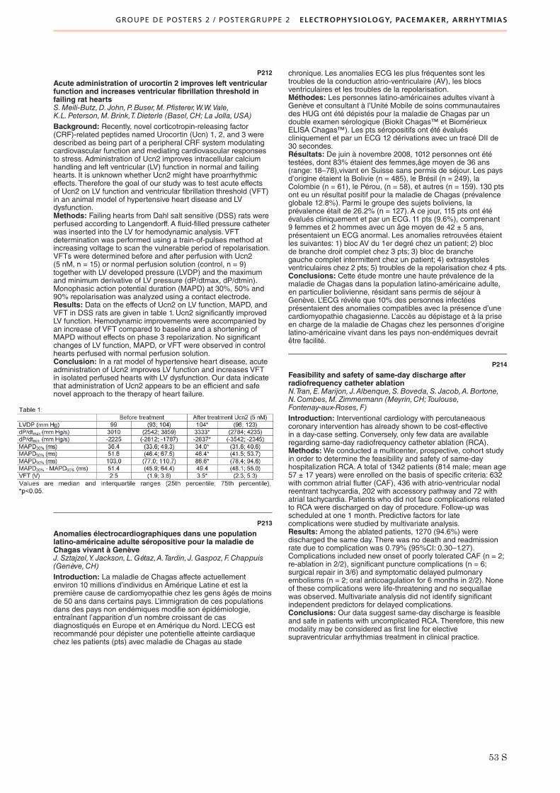
GROUPE DE POSTERS 2 / POSTERGRUPPE 2 ELECTROPHYSIOLOGY, PACEMAKER, ARRHYTMIAS
53 S
P212
Acute administration of urocortin 2 improves left ventricularfunction and increases ventricular fibrillation threshold infailing rat heartsS.Meili-Butz,D. John,P.Buser,M.Pfisterer,W.W.Vale,K.L.Peterson,M.Brink,T.Dieterle (Basel,CH; La Jolla,USA)Background: Recently, novel corticotropin-releasing factor(CRF)-related peptides named Urocortin (Ucn) 1, 2, and 3 weredescribed as being part of a peripheral CRF system modulatingcardiovascular function and mediating cardiovascular responsesto stress. Administration of Ucn2 improves intracellular calciumhandling and left ventricular (LV) function in normal and failinghearts. It is unknown whether Ucn2 might have proarrhythmiceffects. Therefore the goal of our study was to test acute effectsof Ucn2 on LV function and ventricular fibrillation threshold (VFT)in an animal model of hypertensive heart disease and LVdysfunction.Methods: Failing hearts from Dahl salt sensitive (DSS) rats wereperfused according to Langendorff. A fluid-filled pressure catheterwas inserted into the LV for hemodynamic analysis. VFTdetermination was performed using a train-of-pulses method atincreasing voltage to scan the vulnerable period of repolarisation.VFTs were determined before and after perfusion with Ucn2(5 nM, n = 15) or normal perfusion solution (control, n = 9)together with LV developed pressure (LVDP) and the maximumand minimum derivative of LV pressure (dP/dtmax, dP/dtmin).Monophasic action potential duration (MAPD) at 30%, 50% and90% repolarisation was analyzed using a contact electrode.Results: Data on the effects of Ucn2 on LV function, MAPD, andVFT in DSS rats are given in table 1. Ucn2 significantly improvedLV function. Hemodynamic improvements were accompanied byan increase of VFT compared to baseline and a shortening ofMAPD without effects on phase 3 repolarization. No significantchanges of LV function, MAPD, or VFT were observed in controlhearts perfused with normal perfusion solution.Conclusion: In a rat model of hypertensive heart disease, acuteadministration of Ucn2 improves LV function and increases VFTin isolated perfused hearts with LV dysfunction. Our data indicatethat administration of Ucn2 appears to be an efficient and safenovel approach to the therapy of heart failure.
P213
Anomalies électrocardiographiques dans une populationlatino-américaine adulte séropositive pour la maladie deChagas vivant à GenèveJ.Sztajzel,Y. Jackson, L.Gétaz,A.Tardin, J.Gaspoz, F.Chappuis(Genève,CH)Introduction: La maladie de Chagas affecte actuellementenviron 10 millions d’individus en Amérique Latine et est lapremière cause de cardiomyopathie chez les gens âgés de moinsde 50 ans dans certains pays. L’immigration de ces populationsdans des pays non endémiques modifie son épidémiologie,entraînant l’apparition d’un nombre croissant de casdiagnostiqués en Europe et en Amérique du Nord. L’ECG estrecommandé pour dépister une potentielle atteinte cardiaquechez les patients (pts) avec maladie de Chagas au stade
chronique. Les anomalies ECG les plus fréquentes sont lestroubles de la conduction atrio-ventriculaire (AV), les blocsventriculaires et les troubles de la repolarisation.Méthodes: Les personnes latino-américaines adultes vivant àGenève et consultant à l’Unité Mobile de soins communautairesdes HUG ont été dépistés pour la maladie de Chagas par undouble examen sérologique (Biokit Chagas™ et BiomérieuxELISA Chagas™). Les pts séropositifs ont été évaluéscliniquement et par un ECG 12 dérivations avec un tracé DII de30 secondes.Résultats: De juin à novembre 2008, 1012 personnes ont ététestées, dont 83% étaient des femmes,âge moyen de 36 ans(range: 18–78),vivant en Suisse sans permis de séjour. Les paysd’origine étaient la Bolivie (n = 485), le Brésil (n = 249), laColombie (n = 61), le Pérou, (n = 58), et autres (n = 159). 130 ptsont eu un résultat positif pour la maladie de Chagas (prévalenceglobale 12.8%). Parmi le groupe des sujets boliviens, laprévalence était de 26.2% (n = 127). A ce jour, 115 pts ont étéévalués cliniquement et par un ECG. 11 pts (9.6%), comprenant9 femmes et 2 hommes avec un âge moyen de 42 ± 5 ans,présentaient un ECG anormal. Les anomalies retrouvées étaientles suivantes: 1) bloc AV du 1er degré chez un patient; 2) blocde branche droit complet chez 3 pts; 3) bloc de branchegauche complet intermittent chez un patient; 4) extrasystolesventriculaires chez 2 pts; 5) troubles de la repolarisation chez 4 pts.Conclusions: Cette étude montre une haute prévalence de lamaladie de Chagas dans la population latino-américaine adulte,en particulier bolivienne, résidant sans permis de séjour àGenève. L’ECG révèle que 10% des personnes infectéesprésentaient des anomalies compatibles avec la présence d’unecardiomyopathie chagasienne. L’accès au dépistage et à la priseen charge de la maladie de Chagas chez les personnes d’originelatino-américaine vivant dans les pays non-endémiques devraitêtre facilité.
P214
Feasibility and safety of same-day discharge afterradiofrequency catheter ablationN.Tran, E.Marijon, J.Albenque,S.Boveda,S. Jacob,A.Bortone,N.Combes,M.Zimmermann (Meyrin,CH;Toulouse,Fontenay-aux-Roses, F)Introduction: Interventional cardiology with percutaneaouscoronary intervention has already shown to be cost-effectivein a day-case setting. Conversely, only few data are availableregarding same-day radiofrequency catheter ablation (RCA).Methods:We conducted a multicenter, prospective, cohort studyin order to determine the feasibility and safety of same-dayhospitalization RCA.A total of 1342 patients (814 male; mean age57 ± 17 years) were enrolled on the basis of specific criteria: 632with common atrial flutter (CAF), 436 with atrio-ventricular nodalreentrant tachycardia, 202 with accessory pathway and 72 withatrial tachycardia. Patients who did not face complications relatedto RCA were discharged on day of procedure. Follow-up wasscheduled at one 1 month. Predictive factors for latecomplications were studied by multivariate analysis.Results: Among the ablated patients, 1270 (94.6%) weredischarged the same day. There was no death and readmissionrate due to complication was 0.79% (95%CI: 0.30–1.27).Complications included new onset of poorly tolerated CAF (n = 2;re-ablation in 2/2), significant puncture complications (n = 6;surgical repair in 3/6) and symptomatic delayed pulmonaryembolisms (n = 2; oral anticoagulation for 6 months in 2/2). Noneof these complications were life-threatening and no sequallaewas observed. Multivariate analysis did not identify significantindependent predictors for delayed complications.Conclusions: Our data suggest same-day discharge is feasibleand safe in patients with uncomplicated RCA. Therefore, this newmodality may be considered as first line for electivesupraventricular arrhythmias treatment in clinical practice.
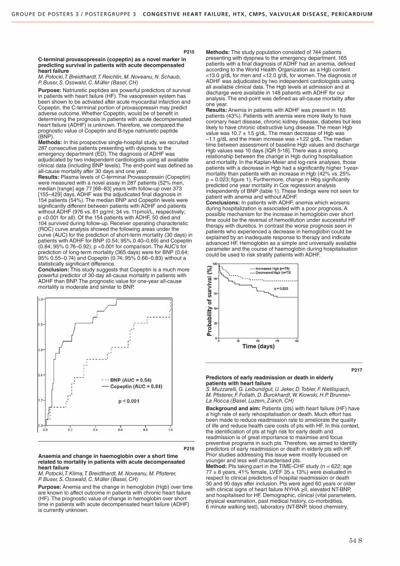
GROUPE DE POSTERS 3 / POSTERGRUPPE 3 CONGESTIVE HEART FAILURE, HTX, CMPS, VALVULAR DISEASE, PERICARDIUM
54 S
P215
C-terminal provasopressin (copeptin) as a novel marker inpredicting survival in patients with acute decompensatedheart failureM.Potocki,T.Breidthardt,T.Reichlin,M.Noveanu,N.Schaub,P.Buser,S.Osswald,C.Müller (Basel,CH)Purpose: Natriuretic peptides are powerful predictors of survivalin patients with heart failure (HF). The vasopressin system hasbeen shown to be activated after acute myocardial infarction andCopeptin, the C-terminal portion of provasopressin may predictadverse outcome.Whether Copeptin, would be of benefit indetermining the prognosis in patients with acute decompensatedheart failure (ADHF) is unknown. Therefore, we compared theprognostic value of Copeptin and B-type natriuretic peptide(BNP).Methods: In this prospective single-hospital study, we recruited287 consecutive patients presenting with dyspnea to theemergency department (ED). The diagnosis of ADHF wasadjudicated by two independent cardiologists using all availableclinical data (including BNP levels). The end-point was defined asall-cause mortality after 30 days and one year.Results: Plasma levels of C-terminal Provasopressin (Copeptin)were measured with a novel assay in 287 patients (52% men,median [range] age 77 [68–83] years with follow-up over 373[155–429] days. ADHF was the adjudicated final diagnosis in154 patients (54%). The median BNP and Copeptin levels weresignificantly different between patients with ADHF and patientswithout ADHF (976 vs. 81 pg/ml; 34 vs. 11pmol/L, respectively;p <0.001 for all). Of the 154 patients with ADHF, 50 died and104 survived during follow-up. Receiver operating characteristic(ROC) curve analysis showed the following areas under thecurve (AUC) for the prediction of short-term mortality (30 days) inpatients with ADHF for BNP (0.54; 95% 0.40–0.69) and Copeptin(0.84; 95% 0.76–0.92); p <0.001 for comparison. The AUC’s forprediction of long-term mortality (365 days) were for BNP (0.64;95% 0.55–0.74) and Copeptin (0.74; 95% 0.66–0.83) without astatistically significant difference.Conclusion: This study suggests that Copeptin is a much morepowerful predictor of 30-day all-cause mortality in patients withADHF than BNP. The prognostic value for one-year all-causemortality is moderate and similar to BNP.
P216
Anaemia and change in haemoglobin over a short timerelated to mortality in patients with acute decompensatedheart failureM.Potocki,T.Klima,T.Breidthardt,M.Noveanu,M.Pfisterer,P.Buser,S.Osswald,C.Müller (Basel,CH)Purpose: Anemia and the change in hemoglobin (Hgb) over timeare known to affect outcome in patients with chronic heart failure(HF). The prognostic value of change in hemoglobin over shorttime in patients with acute decompensated heart failure (ADHF)is currently unknown.
Methods: The study population consisted of 744 patientspresenting with dyspnea to the emergency department. 165patients with a final diagnosis of ADHF had an anemia, definedaccording to the World Health Organization as a Hgb content<13.0 g/dL for men and <12.0 g/dL for women. The diagnosis ofADHF was adjudicated by two independent cardiologists usingall available clinical data. The Hgb levels at admission and atdischarge were available in 148 patients with ADHF for ouranalysis. The end-point was defined as all-cause mortality afterone year.Results: Anemia in patients with ADHF was present in 165patients (43%). Patients with anemia were more likely to havecoronary heart disease, chronic kidney disease, diabetes but lesslikely to have chronic obstructive lung disease. The mean Hgbvalue was 10.7 ± 1.5 g/dL. The mean decrease of Hgb was–1.1 g/dL and the mean increase was +1.22 g/dL. The mediantime between assessment of baseline Hgb values and dischargeHgb values was 10 days [IQR 5-18]. There was a strongrelationship between the change in Hgb during hospitalisationand mortality. In the Kaplan-Meier and log-rank analyses, thosepatients with a decrease in Hgb had a significantly higher 1-year-mortality than patients with an increase in Hgb (42% vs. 25%p = 0.023; figure 1). Furthermore, change in Hbg significantlypredicted one year mortality in Cox regression analysisindependently of BNP (table 1). These findings were not seen forpatient with anemia and without ADHF.Conclusions: In patients with ADHF, anemia which worsensduring hospitalization is associated with a poor prognosis. Apossible mechanism for the increase in hemoglobin over shorttime could be the reversal of hemodilution under successful HFtherapy with diuretics. In contrast the worse prognosis seen inpatients who experienced a decrease in hemoglobin could beexplained by an inadequate response to therapy and indicateadvanced HF. Hemoglobin as a simple and universally availableparameter and the course of haemoglobin during hospitalisationcould be used to risk stratify patients with ADHF.
P217
Predictors of early readmission or death in elderlypatients with heart failureS.Muzzarelli,G. Leibundgut,U. Jeker,D.Tobler, F.Nietlispach,M.Pfisterer, F. Follath,D.Burckhardt,W.Kiowski,H.P.Brunner-La Rocca (Basel, Luzern, Zürich,CH)Background and aim: Patients (pts) with heart failure (HF) havea high rate of early rehospitalisation or death. Much effort hasbeen made to reduce readmission rate to ameliorate the qualityof life and reduce health care costs of pts with HF. In this context,the identification of pts at high risk for early death andreadmission is of great importance to maximise and focuspreventive programs in such pts. Therefore, we aimed to identifypredictors of early readmission or death in elderly pts with HF.Prior studies addressing this issue were mostly focussed onyounger and less well characterised pts.Method: Pts taking part in the TIME-CHF study (n = 622; age77 ± 8 years, 41% female, LVEF 35 ± 13%) were evaluated inrespect to clinical predictors of hospital readmission or death30 and 90 days after inclusion. Pts were aged 60 years or olderwith clinical signs of heart failure NYHA ≥II, elevated NT-BNP,and hospitalised for HF. Demographic, clinical (vital parameters,physical examination, past medical history, co-morbidities,6 minute walking test), laboratory (NT-BNP, blood chemistry,

GROUPE DE POSTERS 3 / POSTERGRUPPE 3 CONGESTIVE HEART FAILURE, HTX, CMPS, VALVULAR DISEASE, PERICARDIUM
55 S
blood count), echocardiographic (left ventricular ejection fraction)and social (living, marital and educational status) parameterswere obtained at baseline and included in a multivariable logisticregression to identify independent predictors for early events.Results: The rate of hospital readmission or death was high(11% after 30 days, 25% after 90 days). Independent predictorsof rehospitalisation or death 30 and 90 days after discharge areshown in table 1.Conclusion: Coronary artery disease, NYHA class, pacemakerrhythm (mostly right ventricular pacing in the present collective)and co-morbidities, like renal disease and anemia wereindependent predictors of early hospital readmission or deathamong elderly patients discharged for HF. Interestingly, age, leftventricular ejection fraction as well as living- and social statuswere not associated with early events.
P218
Autologous bone marrow progenitor cells infusionin a clinical, randomised control trial in heart failure:a delivery procedure safety analysisD.Locca,C.Yeo,T.Burchell,A. Flett,M.Rothman,C.Knight,P.Brookman,N.Saunders,S.Aggrawal,A.Mathur (London,UK)Background: The REGENERATE IHD Trial is a randomisedongoing control trial comparing the effects of G-CSF andautologous bone marrow progenitor cells infusion on qualityof life and left ventricular function in patients with end stage heartfailure secondary to ischaemic heart disease. This preliminarysubanalysis describe the intra myocardial and intra coronary stemcell/placebo administration in the largest clinical stem cell trial inUK.Methods: This sub-study analysis compares different routes ofdelivery of bone marrow mononuclear cells. The operators areexperienced interventionists blinded to the infusion of cell orplacebo. Intracoronary stem cell/placebo was administratedwith an over the wire baloon and the intra myocardial deliveryperformed with the Noga TM biological delivery system (Cordis,Miami, FL, USA). All cardiac events were recorded during theprocedure. The peri procedure inpatient stay was 2 days. Allpatients underwent continuous ECG monitoring and had anechocardiogram assessment post procedure and prior todischarge. A clinical follow up (outpatient) was performed7 days post stem cell/placebo delivery.Results: 35 patients are currently included in the interventionalarm of the study. 17 had intracoronary stem cell/placebo therapy,18 patients underwent intra myocardial infusion using the NogaTM biological delivery system (Cordis,Miami,FL,USA). In bothgroups no deaths were reported in the immediate peri procedurefollow up nor at 7 days. One patient in the intramyocardial group(5.5%, n = 18) had a sustained ventricular tachycardia episodeduring the procedure (major cardiac event) and was successfullycardioverted to sinus rhythm. Two patients experienced minorcardiac events with transient atrio-ventricular block (11.8%,n = 18). No pericardial effusions were seen. In the intracoronarygroup (n = 17) no patients underwent emergency percutaneouscoronary intervention in the target vessel following the infusion ofthe cell/placebo solution. All patients had a TIMI 3 flow score postdelivery of therapy. No major or minor cardiac events wererecorded in the intracoronary group.
Conclusion: No significant differences in cardiac events betweenpatients recovery on stem cell/placebo infusion were seen. Thesepreliminary results suggest that both intra coronary andintramyocardial routes of administration are safe in patients withheart failure secondary to ischaemic heart disease.
P219
The prognostic impact of systemic inflammatorydiseases in patients with congestive heart failureT.Burkard,O.Pfister,M.Maeder,S.Kurz,R.Zaker,S.Toggweiler,A.Bullinger,M.Pfisterer,H.P.Brunner-La Rocca,H.Ricklion behalf of the TIME-CHF-InvestigatorsBackground: It is known, that an inflammatory state is partof the pathophysiology of congestive heart failure (CHF).However, little is known about the impact of the presenceof a systemic inflammatory disease (SID) – defined by aninflammatory syndrome with constitutional symptoms andinvolvement of at least two organs (e.g. rheumatoid arthritis,systemic lupus erythematosus, giant cell arthritis) – on theclinical course and prognosis of patients (pts) with CHF.Methods: 622 pts (mean age 77 ± 8 years, left ventricularejection fraction (LVEF) 35 ± 13%, female 41%) who participatedin the Trial of Intensified vs. standard Medical Therapy in Elderlypatients with Congestive Heart Failure (TIME-CHF), amulticentre, randomized heart failure trial were analysed.Wecompared 35 pts suffering from SID with 587 pts without SID.Primary endpoint was all-cause hospitalisation free survival;secondary endpoints were survival (without death by cancer)and heart failure hospitalisation free survival during an 18-monthfollow up.Results: At baseline pts with SID had significantly worse renalfunction (creatinine 130 ± 40 vs. 115 ± 39 umol/l; p = 0.02) andhigher NT-BNP values (median 5601 pg/ml [interquartile range(IQR) 2736–8095] vs. 3733 pg/ml [IQR 1903–6847]; p = 0.02)than pts without SID. There were no other significant differencesbetween the 2 groups including treatment regime, age, gender,cause of CHF, cardiovascular risk factors, co-morbidities, NYHAclass and LVEF. By Kaplan Meier analysis and multivariate Cox-regression models the presence of SID was an independent riskfactor for higher hospitalisation and mortality rates in regard to theprimary and secondary endpoints. Detailed results for thedifferent endpoints are provided in table 1. Kaplan Meier curve forthe primary endpoint is shown in figure 1.Conclusion: In elderly pts with CHF, the presence of SID wasassociated not only with a significantly worse outcome regardingall cause mortality and hospitalisation rates but also with highermortality and hospitalisation rates caused by CHF. The presenceof a SID therefore may have to be considered as bad prognosticfactor when treating CHF patients.
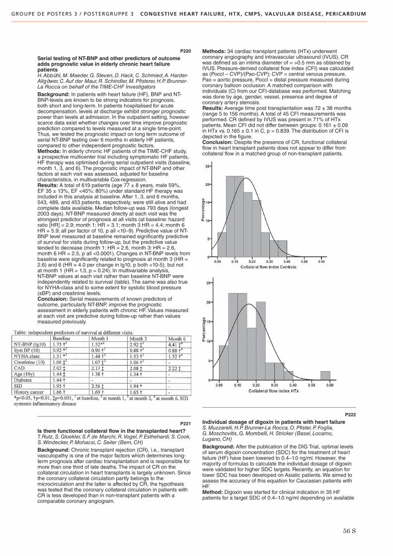
GROUPE DE POSTERS 3 / POSTERGRUPPE 3 CONGESTIVE HEART FAILURE, HTX, CMPS, VALVULAR DISEASE, PERICARDIUM
56 S
P220
Serial testing of NT-BNP and other predictors of outcomeadds prognostic value in elderly chronic heart failurepatientsH.Abbühl,M.Maeder,G.Steven,D.Hack,C.Schmied,A.Harder-Allgöwer,C.Auf der Maur,R.Schindler,M.Pfisterer,H.P.Brunner-La Rocca on behalf of the TIME-CHF InvestigatorsBackground: In patients with heart failure (HF), BNP and NT-BNP-levels are known to be strong indicators for prognosis,both short and long-term. In patients hospitalised for acutedecompensation, levels at discharge exhibit stronger prognosticpower than levels at admission. In the outpatient setting, howeverscarce data exist whether changes over time improve prognosticprediction compared to levels measured at a single time-point.Thus, we tested the prognostic impact on long term outcome ofserial NT-BNP testing over 6 months in elderly HF patients,compared to other independent prognostic factors.Methods: In elderly chronic HF patients of the TIME-CHF study,a prospective multicenter trial including symptomatic HF patients,HF therapy was optimised during serial outpatient visits (baseline,month 1, 3, and 6). The prognostic impact of NT-BNP and otherfactors at each visit was assessed, adjusted for baselinecharacteristics, in multivariable Cox-regression.Results: A total of 619 patients (age 77 ± 8 years, male 59%,EF 35 ± 13%, EF <45%: 80%) under standard HF therapy wasincluded in this analysis at baseline. After 1, 3, and 6 months,543, 489, and 453 patients, respectively, were still alive and hadcomplete data available. Median follow-up was 793 days (longest2003 days). NT-BNP measured directly at each visit was thestrongest predictor of prognosis at all visits (at baseline: hazardratio [HR] = 2.9; month 1: HR = 3.1; month 3 HR = 4.4; month 6HR = 5.9; all per factor of 10, p all <10–9). Predictive value of NT-BNP level measured at baseline remained significantly predictiveof survival for visits during follow-up, but the predictive valuetended to decrease (month 1: HR = 2.6, month 3: HR = 2.6,month 6 HR = 2.5, p all <0.0001). Changes in NT-BNP levels frombaseline were significantly related to prognosis at month 3 (HR =2.6) and 6 (HR = 4.0 per change in lg10, p both <10-5), but notat month 1 (HR = 1.3, p = 0.24). In multivariable analysis,NT-BNP values at each visit rather than baseline NT-BNP wereindependently related to survival (table). The same was also truefor NYHA-class and to some extent for systolic blood pressure(sBP) and creatinine levels.Conclusion: Serial measurements of known predictors ofoutcome, particularly NT-BNP, improve the prognosticassessment in elderly patients with chronic HF. Values measuredat each visit are predictive during follow-up rather than valuesmeasured previously.
P221
Is there functional collateral flow in the transplanted heart?T.Rutz,S.Gloekler, S.F. de Marchi,R.Vogel, P. Esthehardi, S.Cook,S.Windecker,P.Mohacsi,C.Seiler (Bern,CH)Background: Chronic transplant rejection (CR), i.e., transplantvasculopathy is one of the major factors which determines long-term prognosis after cardiac transplantation and is responsible formore than one third of late deaths. The impact of CR on thecollateral circulation in heart transplants is largely unknown. Sincethe coronary collateral circulation partly belongs to themicrocirculation and the latter is affected by CR, the hypothesiswas tested that the coronary collateral circulation in patients withCR is less developed than in non-transplant patients with acomparable coronary angiogram.
Methods: 34 cardiac transplant patients (HTx) underwentcoronary angiography and intravascular ultrasound (IVUS). CRwas defined as an intima diameter of = >0.5 mm as obtained byIVUS. Pressure-derived collateral flow index (CFI) was calculatedas (Poccl – CVP)/(Pao-CVP); CVP = central venous pressure,Pao = aortic pressure, Poccl = distal pressure measured duringcoronary balloon occlusion. A matched comparison withindividuals (C) from our CFI-database was performed. Matchingwas done by age, gender, vessel, presence and degree ofcoronary artery stenosis.Results: Average time post transplantation was 72 ± 38 months(range 5 to 156 months). A total of 45 CFI measurements wasperformed. CR defined by IVUS was present in 71% of HTxpatients. Mean CFI did not differ between groups: 0.161 ± 0.09in HTx vs. 0.165 ± 0.1 in C, p = 0.839. The distribution of CFI isdepicted in the figure.Conclusion: Despite the presence of CR, functional collateralflow in heart transplant patients does not appear to differ fromcollateral flow in a matched group of non-transplant patients.
P222
Individual dosage of digoxin in patients with heart failureS.Muzzarelli, H.P.Brunner-La Rocca,O.Pfister, P. Foglia,G.Moschovitis,G.Mombelli, H.Stricker (Basel, Locarno,Lugano,CH)Background: After the publication of the DIG Trial, optimal levelsof serum digoxin concentration (SDC) for the treatment of heartfailure (HF) have been lowered to 0.4–1.0 ng/ml. However, themajority of formulas to calculate the individual dosage of digoxinwere validated for higher SDC targets. Recently, an equation forlower SDC has been developed on Asiatic patients.We aimed toassess the accuracy of this equation for Caucasian patients withHF.Method: Digoxin was started for clinical indication in 35 HFpatients for a target SDC of 0.4–1.0 ng/ml depending on available

GROUPE DE POSTERS 3 / POSTERGRUPPE 3 CONGESTIVE HEART FAILURE, HTX, CMPS, VALVULAR DISEASE, PERICARDIUM
57 S
dosages. Patients did not receive concurrent drugs known tointeract with digoxin. The individual daily dosage of digoxin wascalculated based on the above mentioned formula: Dosage (microg/day) = SDC (ng/ml) x [2.22 x Ccr (ml/min.) + 25.7], whereCcr is the creatinine clearance according to Cockroft-Gault.After 1 month, all patients were seen in our outpatients HF clinic.Blood samples for the measurement of SDC were obtained6–8 hours after the last dose. The adherence to digoxin treatmentwas assessed with the CARDIA-questionnaire.Results: Most of the patients were male (n = 31, 78%). Themedian age was 72 ± 11 years and the Ccr (Cockroft-Gault)56 ± 22 ml/min. After 1 month, SDC was in the therapeutic range(0.4–1.0 ng/ml) in 26 patients (74%), sub-therapeutic in 5 (14%)patients and supra-therapeutic in 4 (12%) patients (meanSDC = 0.68 ± 0.26 ng/ml, min. = 0.3 ng/ml, max. = 1.2 ng/ml).Four patients with sub-therapeutic SDC admitted poor medicaladherence, 3 patients with supra-therapeutic SDC had a relevantworsening of renal function. After exclusion of the patients withpoor adherence, the correlation between the predicted and themeasured SDC was good (r = 0.6; p = 0.01, see fig. 1–2). Themean predicted SDC was 0.72 ± 0.18 ng/ml, the mean measuredSDC was 0.75 ± 0.24 ng/ml.Conclusion: The correlation between the predicted andmeasured SDC was quite good. Furthermore, in 93% of thepatients with good adherence to medical regime and stable renalfunction the SDC was in the therapeutic range. The maximalachieved SDC of 1.2 ng/ml is clearly below the toxicconcentration. Therefore, the present study supports the clinicalvalidity of the described equation even in Caucasian patients, butregular control of the renal function is advisable.
P223
New-onset atrial fibrillation predicts increased mortalityin stable heart failureA.Häberlin,H.Tanner, E.Delacrétaz,P.Meier, P.Mohacsi,R.Hullin(Bern,CH)Introduction: Atrial fibrillation is the most prevalent cardiacarrhythmia in patients with chronic heart failure (CHF) and isassociated with an adverse prognosis. The aim of this study wasto investigate the prevalence of new-onset atrial fibrillation (AF)in patients with stable mild to moderate heart failure on optimalmedical treatment and to characterize the prognosis associatedwith new-onset AF.Methods: Prospective observational case-control study design.Participants were assigned to sinus rhythm (SR) group when inSR at baseline and without history of paroxysmal AF. Patients withknown paroxysmal or persistent AF or atrial flutter were assignedto the group with known AF. Survival was analyzed separatelyin patients with SR, AF, and new-onset AF during follow-up.Results: The analysis included 209 patients, 66 (32%) withknown AF. Survival was not different in patients with SR or knownAF. New-onset AF developed in 22/143 (15%) patients aftera median follow-up of 22 months and was associated withincreased mortality (hazard ratio 1.62, confidence limits 1.06–2.49. p = 0.03), Figure 1. Median BNP was higher in patientswith new-onset AF (434 vs. 275 pg/ml, p = 0.001), CRP was notdifferent. In a multivariate model advanced age was predictive fornew-onset AF (p = 0.035), while BNP tended to have a predictiverole (p = 0.058).Conclusions: New-onset AF occurs with a high prevalence inpatients with stable mild to moderate CHF and is associated withan increased mortality in this population.
P224
Course of exercise capacity, ventilatory efficiency andleft ventricular function in the second year after orthotopiccardiac transplantationR.K.Binder, J.-P.Schmid ,T.P.Carrel,H.Saner,P.Mohacsi,R.Hullin (Bern,CH)Background: Orthotopic cardiac transplantation (HTx) is anestablished treatment for end stage heart failure. HTx not onlyimproves survival but increases exercise capacity, peak oxygenconsumption (peak VO2) and ventilatory efficiency. After HTx,cardiopulmonary remodelling occurs within weeks and monthsfollowing HTx. The aim of this study was to investigate, whetherfurther cardiopulmonary remodelling and functional ameliorationoccurs in the second year post-transplant.Methods: Twenty-six consecutive HTx recipients (20 male,mean age 45.5 ± 13.9 years) underwent symptom limited cardio-pulmonary exercise testing on a cycle ergometer 12 and 24months post-transplant. Ventilatory efficiency was assessedby the VE/VCO2-slope and the oxygen uptake efficiency slope(OUES). Transthoracic echocardiography measured systolicleft ventricular function (LV-EF) by the Simpson’s method andfunctional diastolic parameters by diastolic mitral inflow patterns

GROUPE DE POSTERS 3 / POSTERGRUPPE 3 CONGESTIVE HEART FAILURE, HTX, CMPS, VALVULAR DISEASE, PERICARDIUM
58 S
and tissue Doppler imaging. Statistical comparisons comprisedrepeated-measures analysis of variance and Bonferronicorrections for multiple testing.Results: Neither maximal exercise capacity (12 vs 24 months:105.4 ± 36.3 vs. 112.4 ± 47.6 Watt, p = 0.31), nor peak VO2 (20.7± 5.8 vs. 21.2 ± 7.5 ml/kg/min, p = 0.54) changed significantly, andremained below the average of age-matched healthy controlsafter 2 years (67.1 ± 26.6% and 66.6 ± 19.2%, respectively).Furthermore, ventilatory efficiency parameters (VE/VCO2-slope:36.9 ± 5.0 vs. 36.8 ± 4.1, p = 0.86; OUES: 1540.1 ± 446.3 vs.1497.6 ± 504.9, p = 0.37) and LV systolic and diastolic function(LV-EF: 63.8 ± 2.1% vs. 63.7 ± 2.1%, p = 0.70; diastolic septalmitral annular e-velocity: 8.4 ± 1.2 cm/s vs. 9.6 ± 1.9 cm/s, p =0.26) did not change. A decrease in the FEV1/VC-ratio in thesecond year post-transplant was of borderline significance (83.2± 10.4% vs. 77.9 ± 13.6%, p = 0.046, corrected p >0.05) due toan insignificant increase in vital capacity (percentage of predictedvalues: 84.3 ± 16.8% vs 87.1 ± 15.6%, p = 0.15).Conclusions: This study demonstrates that exercise capacity,peak VO2, ventilatory efficiency and left ventricular functionremain stable in the second year post-transplant. However,ventilatory efficiency remains stable despite changes in theFEV1/VC ratio which suggests ongoing cardiopulmonaryadaptation after the first postoperative year. Future studies shouldcharacterize the nature of this remodelling process and the role ofphysical exercise continued on a regular basis.
P225
Safety and efficacy of ECG-triggered muscularcounterpulsation for haemodynamic improvement of cardiacfunctionB.H.Walpoth,S.Higazi,M.Billinger,S.Windecker, L. Lapanashvili,O.M.Hess (Genève,Bern,Winterthur,CH)Introduction: ECG-triggered peripheral muscularcounterpulsation (MCP) was proposed as a new non-invasivetechnique for treatment of acute cardiac failure in coronary arterydisease (CAD). Study purpose: 1) evaluate safety and efficiencyof MCP; 2) determine its hemodynamic effect on cardiac function.Methods: In 16 patients (7 no CAD and 9 CAD) MCP wasperformed on calves, thighs and lower abdomen for 3-minuteseach at low (<15 V) and high (15–25 V) amplitudes. ECG-triggering was used to synchronize stimulation during earlydiastole. LV function was assessed by the conductance methodusing simultaneous LV pressure-volume loops. Heart rate, meanaortic, LV end-diastolic and end-systolic pressures, LV volumeswere measured. LV ejection fraction, dP/dt, cardiac output,systemic vascular resistance and cardiac efficiency werecalculated.Results: MCP was associated with an increase in cardiacoutput at all 3 stimulation sites. In CAD patients, there was 22%decrease in peripheral resistance, 12% increase in cardiac index,18% diminution of end-diastolic pressure, and 16% reduction instroke work. Hemodynamic effects were less marked in controlpatients. There were no complications but 5 of 16 patientsreported some itching sensations during stimulation.Conclusions: MCP is safe and efficient for improving cardiacfunction in patients with CAD. The technique is completely non-invasive and simple to use with a portable stimulation device.Best effects of MCP are obtained in CAD patients with abdominalor thigh stimulation.
P226
Shorter ventilation times and hospital stay afterminithoracotomy aortic valve replacement versus mediansternotomyT.M.Torre, F.Siclari, S.Demertzis,R.Trunfio,R.Mauri,G.Franciosi,T.Cassina (Lugano,CH)Introduction: An antero-lateral short minithoracotomy approachfor aortic valve replacement (AVR) has been proposed todecrease patient discomfort and fasten recovery.Weretrospectively reviewed the operative results of two matchedgroups of patients undergoing AVR with the two techniques.Methods: Between august 2003 and november 2008, 43 patientsunderwent minithoracotomy AVR (group M). The operative resultswere compared to a sternotomy AVR group of patients (Group S)operated in the same period. Patients were equally matched forcardiovascular risk factor, sex, mean age, Euroscore, BSA, meanejection fraction, reoperation incidence and valvular lesions.Results: There were no operative deaths. Conversion tosternotomy was necessary in 1 group M patient for an hemidiskblockage. Cross clamping time (78.4 ± 18.2 vs. 44.4 ± 6.6 min),cardiopulmonary bypass time (134.1 ± 43.1 vs. 99.1 ± 44.7) andoperative time (232.9 ± 55.4 vs. 161.9 ± 32.3) were longer ingroup M.Ventilation time and length of hospital stay were shorterin group M (respectively 2.4 ± 0.17vs10.8 ± 4.0 h, p .0001 and 8.8± 3.7 vs.11.1 ± 11.5 days, p 0.0005). In group M there were morereoperations for bleeding and transfusion rate compared to groupS. No statistically significant differences were observed betweenthe two groups for postoperative atrial fibrillation, woundinfections and stroke.Conclusions: This retrospective study shows that in theminithoracotomy group, although operative times were longer,ventilation time and hospital stay were shorter. Theminithoracotomy approach can be considered safe for AVR andmay contribute to a rapid postoperative recovery.
P227
Thoracoscopic and robotic tricuspid valve annuloplastywith absorbable ring: initial experienceA.Panos,P.O.Myers,A.Kalangos (Genève,CH)Objectives: Remodeling annuloplasty with a prosthetic ring hasbeen shown to be superior to other repair techniques of thetricuspid valve, but remains difficult in thoracoscopy.We presentour initial experience with the intra-annularly implanted BioringKalangos annuloplasty ring in the tricuspid position usingminimally invasive access and propose some modifications toadapt it to the particularities of minimally invasive cardiac surgery.Methods: Patients undergoing thoracoscopic or robotic tricuspidannuloplasty alone or combined with other cardiac operationswere prospectively included between March and September2008. The feasibility, ease of implantation and echocardiographicfollow up were evaluated.Results: 10 patients with a median age of 44 years old (range16–81) underwent thoracoscopic tricuspid valve annuloplasty,8 for functional tricuspid regurgitation and 2 for tricuspidendocarditis. Six were operated through a small anterolateralthoracotomy and 4 with the da Vinci S robotic system. MeanCPB and aortic cross clamping was 123 ± 30min and 86 ± 28 minrespectively. The implantation of the ring was successful in all thepatients. There was 1 late death (40 days postoperatively) fromMOF. No patient required reoperation. On discharge, 7 patientshad no or discrete TR (grade = <1) and 2 patients had moderateTR (grade II–III) with no tricuspid stenosis. Follow-up ranged from1–6 months, during which all of the patients remained within thesame TR grade as on discharge.Conclusion: This biodegradable ring offers a simple and quickimplantation which is feasible in minimally invasive approaches.It provides acceptable early results and simplifies thoracoscopicand robotic annuloplasty.
GROUPE DE POSTERS 3 / POSTERGRUPPE 3 CONGESTIVE HEART FAILURE, HTX, CMPS, VALVULAR DISEASE, PERICARDIUM
GROUPE DE POSTERS 4 / POSTERGRUPPE 4 CAD, ACS, PCI, CABG
59 S
P228
Flow separation in aortic valve bioprosthesesT.Syburra,D.Obrist,A. Landolt,T.Rösgen,M.Genoni (Zürich,CH)Introduction: Durability of bioprostheses is related to calcificationof the valve cusps, leading to structural valve degeneration.Calcification may be related to flow separation phenomena at thetrailing edge of the valve cusps under high-output conditions.Weinvestigate the fluid-structure interaction in aortic valvebioprostheses at incremental constant flow rates sequencing asystolic cycle.Method:We investigated three different types of bioprostheses:porcine stented, bovine pericardium stented and porcinestentless. The valves are mounted in a constant-flow test sectionfilled with water. The test section consists of a hemispherical inletupstream of the valve as left ventricular outflow tract and aPlexiglas tube with a diameter of 30 mm downstream of the valve.The closed-loop flow through the test section is controlled by aflow meter and an electrical pump. The pressure in the system ismeasured as follows: (1) before the inlet, (2) in the tube 20 mmdistal and (3) 300 mm distal from the valve. The cusps‘ dynamicsare captured with a CCD camera. The pressure and camerasignals are captured electronically and computer-processed.Results: The pressure drop over the valve increasesmonotonically with the flow rate and is related to the openingarea of the valve. The dynamics of the valves depend on the flowrate. At some specific flow rates, the leaflets exhibit sustainedoscillations, visible also in an increased pressure drop. Thefluttering mode is specific to the valve type: The bovinepericardium valve exhibits traveling waves with high amplitudesover the whole cusps at low frequencies (~3 Hz) for a flow rateof about 8 l/min and at higher frequencies (>4 Hz) for flow rateshigher than 14 l/min, whereas the porcine valves show only low-amplitude oscillations at the cusps’ edges over a broad range offrequencies and flow rates.Conclusion: Fluttering is a sign of flow separation. It inducesmechanical fatigue, particle deposition on the cusps, and mayaccelerate structural valve disease. The fluttering modes may bein resonance with the pulsatile flow, which can become aparticular issue in bovine pericardium valves. Furthermeasurements in steady state and in pulsatile flow conditions willshow the relationship between the pressure drop over the valve,the effective opening area and the fluttering modes.
P229
Myths and realities of mechanical circulatory support:a single-centre experienceP.Tozzi, F.Stumpe,M.Hurni, P.Ruchat,D.Delay, E. Ferrari,J.Horisberger, L.K. von Segesser (Lausanne,CH)Introduction: Numerous high-tech devices with different workingprinciples, sizes and drive units have been developed formechanical circulatory in patients with refractory cardiogenicshock. They can be used as bridge to recovery, bridge totransplant and destination therapy according to patient‘s needs.We report our clinical experience on which is based our workingprotocol for selection and management of patients and devices.Methods: Out of 11.000 patients surgically treated for cardiacdiseases in our institution, 65 have received mechanicalcardiocirculatory support. Intent to treat comprised bridging totransplantation in 26 patients, bridging to recovery in 38 patients,and destination therapy in 1 patient. Six different systems havebeen used: the extracorporeal devices, the Biomedicuscentrifugal pump (n = 41) and the Abiomed BVS 5000 (n = 7)were used for short-term support. The Thoratec VAD (n = 13)located in paracorporeal (n = 11) or intracorporeal (n = 2) positionand HeartMate II (n = 4) and Berlin Heart Incore (n = 1) werepreferably used for longterm support. The LionHeart (n = 1) wasused for destination therapy.Results: Heart failure etiology was ischemic cardiomyopathy in21 (32%) cases, dilated and congenital cardiomyopathy in 38(58.5%) cases, myocarditis in 4 (6%) cases, andmiscellaneous other causes in 2 (3%) cases. Four patients (6%)received biventricular assistance (Thoratec VAD). 36 (55.3%) diedin hospital. Out of the 36 patients died in hospital, 27 (75%) hadECMO (12/27 were pediatric cases), 6 (16.6%) had LVAD and 3(8.4%) had Abiomed. Adult population had a mortality rate of 48%(24/49 patients). Over the 18 patients that received the assistanceas bridge to transplant, 12 (67%) were successfully transplanted.Causes of death were hemorrhagic shock (21–58.5%), MOF dueto septic shock (11–30.5%), brain lesions (2–5.5%), intestinalischemia (2–5.5%). 27 (41.5%) patients were discharged. Meanduration of the assistance was 40 days (max of 977 days).Conclusions: ECMO is easy and quick to implant but there ishigh incidence of fatal hemorrhage if the assistance lasts forseveral days. Pulsatile LVAD is associated to slow recovery andhigh infection rates. Continuous flow LVAD seems to provide thebest postoperative outcome with the lowest complications rate.Intracorporeal implant had lowercomplication rate.
P230
Migraine evolution after percutaneous closure of patentforamen ovaleF.Praz,A.Wahl,T.Tai,M.Schwerzmann,S.Windecker,H.Mattle,B.Meier (Bern,CH)Background: Patent foramen ovale (PFO) has been linked tomigraine, and retrospective studies reported an improvement inmigraine prevalence and frequency after PFO closure for otherreasons.We investigated the outcome of 102 patients sufferingfrom migraine headaches and undergoing PFO closure usingthe Amplatzer PFO Occluder.Methods: Out of 432 patients undergoing PFO closure forsecondary prevention of paradoxical embolism, 102 (24%; aged52 ± 10 years; 56% male) suffered from migraine or migrainewith aura (64%) according to the criteria of the InternationalHeadache Society. PFO closure was performed under localanesthesia without intraprocedural echocardiography. Clinicaloutcome was retrospectively assessed for up to 9 years.Weused scales to evaluate the frequency (<1 x/ month, 1 x/ month,2–3 x/ month, 1x/ week, 2–6 x/ week, 1x /day, >1x /day), theduration (<4 hr, 4–72 hr, >72 hr), and the intensity (from 0 to10) of headache episodes. Patients also self-rated the overallimprovement or worsening in quartiles.Results: All implantation procedures were successful. Therewere no device related complications. No patients was lost
during 4.9 ± 1.9 years of follow-up. At 6 months, complete PFOclosure as assessed by contrast TEE was achieved in 94%,whereas a minimal, moderate, or large residual shunt persisted in4%, 1%, and 1%, respectively. One patient (1%) experienced arecurrent transient ischemic attack 4 years after PFO closure.Following the intervention migraine headaches totallydisappeared in 34 patients (33%), and improved by at least 50%in 50 additional patients (49%); 7 patients (7%) reported adecrease of 25%.While headache remained unchanged in 7patients (7%); 4 patients (4%) experienced worsening headaches.Mean subjective improvement was 74 ± 30%. Overall, meanheadache frequency (from 2–3 x/ month to 1x /month; p <0.001),duration (from 4–72 hr to <4 hr; p <0.001), and intensity (from 7 ±2 to 2 ± 2; p <0.001) improved significantly. The prevalence of anymigraine headaches (from 100% to 58%; p <0.001), of migrainewith aura (from 64% to 17%; p <0.001) and the number ofpatients taking any migraine medication (from 93% to 55%; p<0.001) decreased significantly. No risk factors for migrainepersistence could be identified.Conclusions: These results suggest that percutaneous PFOclosure durably alters the spontaneous course of shuntassociated migraine, in addition to the elimination of thepossibility of paradoxical embolism through the PFO.
GROUPE DE POSTERS 4 / POSTERGRUPPE 4 CAD, ACS, PCI, CABG
60 S
P231
Percutaneous closure of patent foramen ovale:long-term follow-up results in a single-centreG.Girod,C.Roguelov,T.Kabir,A.Aminian,A.Delabays,J.-C.Stauffer, P.Vogt, E. Eeckhout (Lausanne,CH)Background: Percutaneous closure of patent foramen ovale(PFO) is increasingly performed during the last decade. It hasbeen shown safe and feasible using a wide variety of devices,and guided by transoesophageal (TEE) or intracardiacechocardiography (ICE). The long term results of percutaneousPFO closure are still rare.Methods: Between December 1998 and December 2008, 215consecutive patients (pts) (F: 88, age: 43 ± 13 y.) were referredfor percutaneous closure of PFO. The indication for PFO closurewas cryptogenic stroke (72%), transient ischaemic attack (18%),migraine (3%), paradoxical embolism (3%), platypnea-orthodeoxia syndrome (2%), decompression hilness (1%) andsecondary closure with second device (1%). Interatrial septalaneurysm (>11 mm excursion in TM mode) was present in 78%of the pts. The Starflex occluder was used in 130 pts, theBioSTAR device in 50 pts, the PFO star occluder in 23 pts andAmplatzer PFO-occluder in 12 pts. The closure was guided byTEE in 88 pts and by ICE in 127 pts.Results: All pts but one had successful device placement.There was 1 major complication (<0.5%) with device embolisationrequiring surgical arterial femoral retrieval without long termsequelae and the second attempt was successful. Minorcomplications were recorded in 10 pts (4.5%), 5 pts with transientatrial fibrillation, 3 pts with transient ST elevation during theprocedure, 1 patient with pharyngeal haematoma, 1 patientpresented a fistula between the aorta and the left atrium witha PFO star device which disappeared after interruption ofanticoagulation. During a mean follow-up period of 50 months(4-121), 3 ischeamic strokes (1.3 %, annual rate <0.5%; 2 pts withclosed PFO and 1 with antiphospholipides antibodies) and nodeath were reported. At long term, 96.5 % of the PFO wereclosed. Five pts (2.5%) had still grade III shunt (3 with PFO-Starand 2 with Starflex occluder).Conclusion: our experience with percutaneous closure of PFOand adults attests safety, feasibility and long term efficacy of thisapproach. Clinical outcome might be better after PFO closure incomparison with medical treatment reported in the literature.
P232
Late results of percutaneous closure of atrial septaldefects using the amplatzer ASD Occluder in 200 patientsF.Praz,A.Wahl, J.-P.Pfammatter, S.Perruchoud,M.Pavlovic,A.Remondino,S.Windecker,B.Meier (Bern,CH)Background: Percutaneous closure of atrial septal defects (ASD)is usually performed with the Amplatzer ASD Occluder. Thisdevice is associated with a low risk for complications and a highrate of complete closure.Methods: Between April 1997 and April 2008 a total of 200consecutive patients underwent percutaneous ASD closure withan Amplatzer ASD Occluder. Mean age was 38 ± 22 years(median 39; range 1–83). There were 140 females and 60 males,of whom 30 females and 12 males were children. Balloon sizingwas performed in all patients. Intraprocedural transoesophagealechocardiographic (TEE) guidance and general anesthesia wererestricted to the 42 children, while in the 158 adults, the devicewas implanted under fluoroscopic guidance only.Results: The implantation procedure (stable positioning of adevice after balloon sizing of the defect) was successful in all but1 pediatric patient (0.5%). She underwent uneventful surgery1 day later for poor device position. Mean device size (diameterof neck between disks) was 22 ± 8 mm (range 6–40). There was1 post-procedural complication (0.5%; transient perimyocarditispossibly related to the device). At last echocardiographic follow-up, 12 ± 11 months after the procedure, 90% of patients had noresidual shunt, whereas a minimal, moderate or large shuntpersisted in 7%, 2%, and 1%, respectively. Three patients (1.5%)underwent implantation of a second device due to the persistenceof a residual shunt. During one of the redo procedures, anAmplatzer device embolized to the right ventricle and had to beremoved surgically, at which time the ASD was also closed.During a mean follow-up period of 3 ± 2 years (median 2.7; range0.2–8.5 years), there was 1 death (73 years old female, 19
months after device implantation cause unknown), but noischemic stroke, TIA, peripheral embolic event, or diving incident.Five patients (2.5%) experienced transient atrial fibrillationrequiring medical treatment in 1 patient and cardioversion in1 patient. Shortly after the intervention 10 patients (5%) reportednon-sustained arrhythmias, requiring blocker therapy in 1 case.Conclusions: The present study confirms the safety, feasibility,and efficacy of percutaneous ASD closure using the AmplatzerASD Occluder in a large patient population. In adults,percutaneous ASD closure is safe and feasible under fluoroscopicguidance only.
P233
The long-term outcome of carotid artery stentingin a real world populationM.Vurma,G.I.W.Galasko,C.Bösch,D.Tüller, K.Nedeltchev,F.R.Eberli (Zürich,CH)Background: Carotid artery stenting (CAS) has emerged as analternative to carotid endarterectomy for the prevention of strokedue to carotid artery disease. Consequently, an increasingnumber of both symptomatic and asymptomatic elderly patients(>80 years of age) (pts >80) undergo CAS. It has been suggestedthat patient age >80 adversely affects short-term outcome.Furthermore, little is known on the long-term outcome of CAS inyounger pts (<80) and no data are available in pts >80. Therefore,we assessed the influence of age on the outcome in a populationof symptomatic and asymptomatic patients.Methods: Two hundred consecutive patients undergoing CASfrom 12/1998 to 01/2007 were included in a prospective registry.Follow-up was done at 30 days, 1 year and then yearly thereafter.Outcome was assessed by death or stroke rates. Thirty-day datawere available in all patients and long-term follow up data in198 pts (99%). Mean follow-up was 886 days.Results: Patient mean age was 71 ± 9 years, 74% were male,64% had symptomatic stenoses. Patient age increased over time,from 68.7 years for the first 50 patients to 74.2 years for the last50 patients (p = 0.003). Hospital stay was 2.4 days for pts <80 vs.4.7 days for pts >80 (p = 0.001). Overall, the 30-day death orstroke rate was 5.0% (2.8-8.3%): 2.6% for those aged <70; 6.7%for those aged 70-79, and 6.9% for those aged >80 (p = 0.27,chi squared test for trend). After day 30, four non-fatal ipsilateralstrokes occurred resulting in 1-year, 3-year and 5-year rates of1.9%, 3.0% and 3.0%, respectively. In the total study populationfour early and 26 late deaths occurred, with only 5 (17%) ofneurological aetiology. Overall 1, 3 and 5 year mortality rates forthose aged <70 rates were 97.3%, 95.5% and 92.5%, for thoseage 70-79 92,1%, 85,2% and 75,2%, compared with 84.9%,58.5% and 32.9% for pts >80 (p <0.0001). Increased age was theonly multivariate predictor of mortality (p <0.0001).Conclusions: The age of patients undergoing CAS is increasing.CAS in those <70 years old has excellent peri-procedural andlong-term outcome out to 5 years. In patients >80 years of agethere is a trend for worse short term outcome and, importantly,long-term outcome is characterized by an extremely high overallmortality rate, predominantly non-neurological in cause.Therefore, in patients >80 years of age CAS is effective inreducing symptoms i.e. reducing stroke rates, but its effect onoverall prognosis is undetermined.
P234
Possible stent thrombosis: fact or fiction?C.Kaiser,R. Jeger,B.Friedli, P.Rickenbacher,H.P.Brunner-LaRocca,M.Pfisterer for the BASKET InvestigatorsHintergrund: Gemäss dem Academic Research Consortium (1)werden Stentthrombosen (ST) in «definite», «probable» und«possible» eingeteilt.Während bei «probable» ST mit hoherWahrscheinlichkeit von einer ST ausgegangen werden kann,wird eine «possible» Stenttrhombose (PST) bei jedem unklarenTodesfall >30 Tage nach Stentimplantation angenommen.Aufgrund dieser vagen Definition werden PST in den meistenrandomisierten Studien und Registern nicht gezählt, obwohlderen Existenz aufgrund pathologisch-anatomischer Datenbewiesen wurde. Das Ziel der vorliegenden Analyse war es, dieWahrscheinlichkeit des Vorliegens einer ST bei allen Patientenmit PST in einer Real-world Population zu definieren.
GROUPE DE POSTERS 4 / POSTERGRUPPE 4 CAD, ACS, PCI, CABG
61 S
Methode: In der BASKET-Studie wurden 826 konsekutivePatienten 2:1 zu Implantation eines DES versus BMSrandomisiert. Für die vorliegende Analyse wurden alle vomCritical Events Committee als PST klassierten Todesfälle währenddes 36-Monat Follow-up nochmals mittels Studium aller Aktenund Angiographie-Befunde sowie Befragung von Angehörigenund Hausärzten klassiert. Als wahrscheinlich für das Vorliegeneiner ST wurden plötzliche Todesfälle mit klinischem Verdachtauf Infarkt oder bei erhaltener Pumpfunktion klassiert, als nichtwahrscheinlich plötzliche Todesfälle im Rahmen von schwerenZweiterkankungen, bei schlechter Pumpfunktion oder beiklinischem Verdacht auf Lungenembolie.Ergebnisse:Während 36 Monaten wurden 36 «definitive»,17 «probable» und 17 PST beobachtet. 12 (71%) der PST tratenbei Patienten mit DES, 5 (29%) bei Patienten mit BMS auf. 10(59%) der PST waren mit grosser Wahrscheinlichkeit durch eineST bedingt: 6 plötzliche Herztode bei erhaltener Pumpfunktionund 4 mit hochgradigem Verdacht auf Infarkt. Bei 7 (41%)Patienten war der Todesfall mit grosser Wahrscheinlichkeit nichtauf eine ST zurückzuführen: 2 plötzliche Herztode bei schlechterPumpfunktion und 4 im Rahmen einer schweren Zweiterkrankungoder Lungenembolie.Schlussfolgerungen: Von den 17 Ereignissen, welche voneinem unabhängigen Critical Events Committee als PST klassiertwurden, war diese Diagnose in 59% wahrscheinlich, während beiden übrigen eine andere Ursache für den plötzlichen Todesfallangenommen werden muss. Die Umstände von als PSTklassierten Todesfällen müssen somit genau analysiert werden,um mit hoher Wahrscheinlichkeit korrekt zugeordnet werden zukönnen.1: Cutlip DE, et al. Circulation. 2007;115:2344–51.
P235
Stent thrombosis up to 3 years after stenting for st-segmentelevation myocardial infarction versus for stable angina –Comparison of the effects of drug-eluting versus bare-metalstentsG.Leibundgut, F.Nietlispach,U.Pittl, H.P.Brunner-La Rocca,C.Kaiser,M.Pfisterer (Basel,CH)Background: The long-term safety of drug-eluting stents (DES)for the treatment of ST-segment elevation myocardial infarction(STEMI) is unclear and may differ from that in stable angina(stable) patients as noted in autopsy studies.Methods: To assess this problem, 210 consecutive STEMI and323 stable patients, randomized 2:1 to DES versus (vs) bare-metal stents (BMS), were followed for 3 years for definite/probablestent thrombosis (ST) and cardiac death/myocardial infarction.Events occuring during the initial 6 months were separated fromlater events.Results: The 3-year rate of ST was 8.1% in STEMI vs 3.4% instable patients (p = 0.02), with corresponding rates of 9.4% vs2,9% (p = 0.01) for DES and of 5.6% vs 4,3% (p = 0.48) forBMS patients, respectively. This difference appeared only after 6months: 4.6% in STEMI vs 1.7% in stable patients (p = 0.05) andin DES treated patients (6.2% vs 2.0%, p = 0.05). Results ofST were paralleled by findings of clinical events, although heredifferences were less pronounced, but also seen only late afterstenting. Thus, in STEMI patients, late events occurred morefrequently after DES vs BMS implantation (11.6% vs 3.0%,p = 0.03), compared to results in stable patients (DES 6.4%,BMS 1.9%, p = 0.06).Conclusions:We observed an increased rate of late ST, and to alesser extent of related clinical events, in patients after stenting forSTEMI vs for stable angina, particularly if treated with DES. Thismay explain outcome differences between results of pivotal trialsin SA patients vs those of “real-world” patients.
P236
Safety of balloon occlusion in angiographically normalcoronary arteriesS.Gloekler, P.Meier,C.Seiler (Bern,CH)Introduction: Temporary balloon occlusion of an angiographicallynormal coronary artery segment may be necessary for collateralfunction assessment and procedures such as optical coherencetomography and angioscopy. Theoretically, this can injure thevessel wall. So far, there are no data on the safety of normal-
vessel coronary balloon occlusion, and accordingly, the purposeof the present study was to assess the complication rate relatedto such a procedure.Methods: Normal-vessel coronary balloon occlusion for 60–120seconds at low inflation pressures of 1–3 atm for collateralfunction assessment was performed in 426 patients between1996 and 2008. The term “normal” referes to normal angiographywith absence or presence of stenoses in remote vessels. In allpatients, procedural complications, findings at repeat coronaryangiography and clinical adverse events were recorded.Results: In 1 of 426 patients (0.2%), acute thrombus formationduring guidewire probing occurred. In 150 patients (35%), re-angiography was performed with a mean follow-up of 10 months.There were no de-novo stenoses at the site of previous occlusion(0%). During long-term follow-up of >1 year and up to 5½ years,2 patients (1.3%) developed de-novo stenoses. Of the 276patients without re-angiography (65%), 3 (in all ≥1 coronaryartery diseased) died (2 cardiac, 1 noncardiac death), and1 was lost to follow-up (mean duration of 45 months).Conclusion: Brief angioplasty balloon occlusion at low inflationpressure in a normal coronary artery is a safe procedure withlong-term angiographic de-novo stenosis development ofapproximately 1 out of 100. Considering the almost 4-year follow-up duration, it cannot be deduced whether the stenotic lesionshad occurred due to the normal-vessel occlusion or due to thenatural course of atherosclerosis.
P237
Comparison of intracardiac ultrasound and conventionalangiography in individuals with atypical chest pain andinconclusive stress testing presenting with intermediate orsevere stenosis detected by contrast-enhanced 64-slicecomputed tomographyI. D’Angeli,T.Moccetti, F. Faletra, E.Pasotti,A.Auricchio,G.B.Pedrazzini (Lugano,CH)Introduction: Multidetector computed tomography (CT)angiography has powerful discriminative ability to identify thosepatients with and without coronary obstruction. Recent data onthe detection and characterization of coronary plaque indicatethat MDCT can reliably measure plaque area, plaque remodelingand the degree of stenosis with good correlation to intravascularultrasound (IVUS) and conventional angiography, respectively,in symptomatic patients and in patients with acute coronarysyndrome.Methods: 55 individuals (48 men, mean age 63.5 ± 9.7),presenting with atypical chest pain and inconclusive stress testingfor ruling out coronary artery disease, underwent QCA and IVUSafter CT angiography demonstrated significant CAD. 109 plaquesin the major coronary vessels, with stenosis degree >50%,were obtained. Correlations of vessel obstruction (lumen areastenosis), lumen cross-sectional area, plaque cross-sectionalarea, as well as, plaque volume and remodelling index for MDCTand IVUS and lumen area stenosis and lumen cross-sectionalarea for MDCT, QCA and IVUS were determined by calculatingthe Lin coefficient. For all measurements Pearson’s r and Bland-Altman correlation were evaluated.Results: MDCT and IVUS yield similar results for plaque area(LAS r = 0.881, p <0.0001; L-CSA r = 0.904, p <0.0001; P-CSAr = 0.907, p <0.05), plaque volume (r = 0.986, p = 0.046) andremodelling index (r = 0.800, p = 0.256). QCA understimatescoronary lesions compared to MDCT (LAS: r = 0.443, p <0.05;L-CSA r = 0.484, p <0.05) and IVUS (LAS r = 0.473, p <0.05,L-CSA r = 0.5000, p <0.05). Positive coronary artery remodellingwas present in 69/109 (63%) plaques, with a mean remodellingindex of 1.21 ± 0.3.Conclusions: Our data show that 64-slice CT can reliably detectand measure the severity of stenosis and it is best correlated toIVUS rather than QCA. Its ability to noninvasively detect andcharacterize morphology and composition of coronary lesionswould represent an attractive alternative to conventionalangiography, especially in individuals with atypical chest pain andinconclusive stress testing. Currently there are no comparativedata between IVUS or conventional angiography to MDCTangiography in these selected group of individuals. Moreover,very little is known about plaque morphology and plaqueremodeling of these individuals. Positive coronary plaque mightalso be present in clinical stable setting and is associated withangiographically underestimation of stenosis degree.
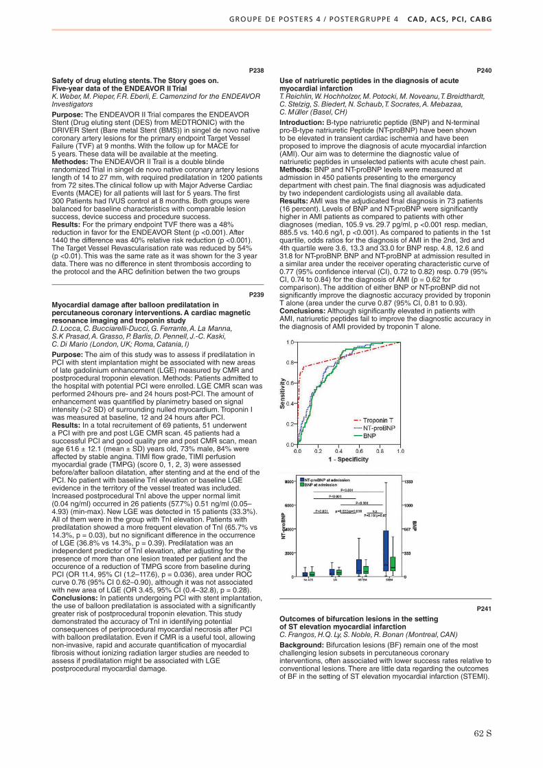
GROUPE DE POSTERS 4 / POSTERGRUPPE 4 CAD, ACS, PCI, CABG
62 S
P238
Safety of drug eluting stents.The Story goes on.Five-year data of the ENDEAVOR II TrialK.Weber,M.Pieper, F.R.Eberli, E.Camenzind for the ENDEAVORInvestigatorsPurpose: The ENDEAVOR II Trial compares the ENDEAVORStent (Drug eluting stent (DES) from MEDTRONIC) with theDRIVER Stent (Bare metal Stent (BMS)) in singel de novo nativecoronary artery lesions for the primary endpoint Target VesselFailure (TVF) at 9 months.With the follow up for MACE for5 years. These data will be available at the meeting.Methodes: The ENDEAVOR II Trail is a double blinderandomized Trial in singel de novo native coronary artery lesionslength of 14 to 27 mm, with required predilatation in 1200 patientsfrom 72 sites.The clinical follow up with Major Adverse CardiacEvents (MACE) for all patients will last for 5 years. The first300 Patients had IVUS control at 8 months. Both groups werebalanced for baseline characteristics with comparable lesionsuccess, device success and procedure success.Results: For the primary endpoint TVF there was a 48%reduction in favor for the ENDEAVOR Stent (p <0.001). After1440 the difference was 40% relative risk reduction (p <0.001).The Target Vessel Revascularisation rate was reduced by 54%(p <0.01). This was the same rate as it was shown for the 3 yeardata. There was no difference in stent thrombosis according tothe protocol and the ARC definition betwen the two groups
P239
Myocardial damage after balloon predilatation inpercutaneous coronary interventions. A cardiac magneticresonance imaging and troponin studyD.Locca,C.Bucciarelli-Ducci,G.Ferrante,A. La Manna,S.K Prasad,A.Grasso,P.Barlis,D.Pennell, J.-C.Kaski,C.Di Mario (London,UK; Roma,Catania, I)Purpose: The aim of this study was to assess if predilatation inPCI with stent implantation might be associated with new areasof late gadolinium enhancement (LGE) measured by CMR andpostprocedural troponin elevation. Methods: Patients admitted tothe hospital with potential PCI were enrolled. LGE CMR scan wasperformed 24hours pre- and 24 hours post-PCI. The amount ofenhancement was quantified by planimetry based on signalintensity (>2 SD) of surrounding nulled myocardium. Troponin Iwas measured at baseline, 12 and 24 hours after PCI.Results: In a total recruitement of 69 patients, 51 underwenta PCI with pre and post LGE CMR scan. 45 patients had asuccessful PCI and good quality pre and post CMR scan, meanage 61.6 ± 12.1 (mean ± SD) years old, 73% male, 84% wereaffected by stable angina. TIMI flow grade, TIMI perfusionmyocardial grade (TMPG) (score 0, 1, 2, 3) were assessedbefore/after balloon dilatation, after stenting and at the end of thePCI. No patient with baseline TnI elevation or baseline LGEevidence in the territory of the vessel treated was included.Increased postprocedural TnI above the upper normal limit(0.04 ng/ml) occurred in 26 patients (57.7%) 0.51 ng/ml (0.05–4.93) (min-max). New LGE was detected in 15 patients (33.3%).All of them were in the group with TnI elevation. Patients withpredilatation showed a more frequent elevation of TnI (65.7% vs14.3%, p = 0.03), but no significant difference in the occurrenceof LGE (36.8% vs 14.3%, p = 0.39). Predilatation was anindependent predictor of TnI elevation, after adjusting for thepresence of more than one lesion treated per patient and theoccurence of a reduction of TMPG score from baseline duringPCI (OR 11.4, 95% CI (1.2–117.6), p = 0.036), area under ROCcurve 0.76 (95% CI 0.62–0.90), although it was not associatedwith new area of LGE (OR 3.45, 95% CI (0.4–32.8), p = 0.28).Conclusions: In patients undergoing PCI with stent implantation,the use of balloon predilatation is associated with a significantlygreater risk of postprocedural troponin elevation. This studydemonstrated the accuracy of TnI in identifying potentialconsequences of periprocedural myocardial necrosis after PCIwith balloon predilatation. Even if CMR is a useful tool, allowingnon-invasive, rapid and accurate quantification of myocardialfibrosis without ionizing radiation larger studies are needed toassess if predilatation might be associated with LGEpostprocedural myocardial damage.
P240
Use of natriuretic peptides in the diagnosis of acutemyocardial infarctionT.Reichlin,W.Hochholzer,M.Potocki,M.Noveanu,T.Breidthardt,C.Stelzig,S.Biedert,N.Schaub,T.Socrates,A.Mebazaa,C.Müller (Basel,CH)Introduction: B-type natriuretic peptide (BNP) and N-terminalpro-B-type natriuretic Peptide (NT-proBNP) have been shownto be elevated in transient cardiac ischemia and have beenproposed to improve the diagnosis of acute myocardial infarction(AMI). Our aim was to determine the diagnostic value ofnatriuretic peptides in unselected patients with acute chest pain.Methods: BNP and NT-proBNP levels were measured atadmission in 450 patients presenting to the emergencydepartment with chest pain. The final diagnosis was adjudicatedby two independent cardiologists using all available data.Results: AMI was the adjudicated final diagnosis in 73 patients(16 percent). Levels of BNP and NT-proBNP were significantlyhigher in AMI patients as compared to patients with otherdiagnoses (median, 105.9 vs. 29.7 pg/ml, p <0.001 resp. median,885.5 vs. 140.6 ng/l, p <0.001). As compared to patients in the 1stquartile, odds ratios for the diagnosis of AMI in the 2nd, 3rd and4th quartile were 3.6, 13.3 and 33.0 for BNP resp. 4.8, 12.6 and31.8 for NT-proBNP. BNP and NT-proBNP at admission resulted ina similar area under the receiver operating characteristic curve of0.77 (95% confidence interval (CI), 0.72 to 0.82) resp. 0.79 (95%CI, 0.74 to 0.84) for the diagnosis of AMI (p = 0.62 forcomparison). The addition of either BNP or NT-proBNP did notsignificantly improve the diagnostic accuracy provided by troponinT alone (area under the curve 0.87 (95% CI, 0.81 to 0.93).Conclusions: Although significantly elevated in patients withAMI, natriuretic peptides fail to improve the diagnostic accuracy inthe diagnosis of AMI provided by troponin T alone.
P241
Outcomes of bifurcation lesions in the settingof ST elevation myocardial infarctionC.Frangos,H.Q. Ly,S.Noble,R.Bonan (Montreal,CAN)Background: Bifurcation lesions (BF) remain one of the mostchallenging lesion subsets in percutaneous coronaryinterventions, often associated with lower success rates relative toconventional lesions. There are little data regarding the outcomesof BF in the setting of ST elevation myocardial infarction (STEMI).
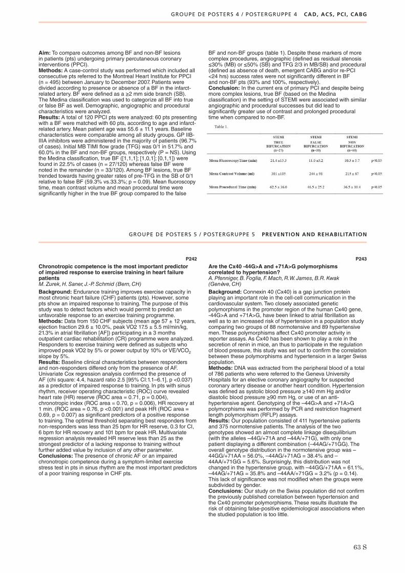
GROUPE DE POSTERS 4 / POSTERGRUPPE 4 CAD, ACS, PCI, CABG
GROUPE DE POSTERS 5 / POSTERGRUPPE 5 PREVENTION AND REHABILITATION
63 S
Aim: To compare outcomes among BF and non-BF lesionsin patients (pts) undergoing primary percutaneous coronaryinterventions (PPCI).Methods: A case-control study was performed which included allconsecutive pts referred to the Montreal Heart Institute for PPCI(n = 495) between January to December 2007. Patients weredivided according to presence or absence of a BF in the infarct-related artery. BF were defined as a ≥2 mm side branch (SB).The Medina classification was used to categorize all BF into trueor false BF as well. Demographic, angiographic and proceduralcharacteristics were analyzed.Results: A total of 120 PPCI pts were analyzed: 60 pts presentingwith a BF were matched with 60 pts, according to age and infarct-related artery. Mean patient age was 55.6 ± 11.1 years. Baselinecharacteristics were comparable among all study groups. GP IIB-IIIA inhibitors were administered in the majority of patients (96.7%of cases). Initial MB TIMI flow grade (TFG) was 0/1 in 51.7% and60.0% in the BF and non-BF groups, respectively (P = NS). Usingthe Medina classification, true BF ([1,1,1]; [1,0,1]; [0,1,1]) werefound in 22.5% of cases (n = 27/120) whereas false BF werenoted in the remainder (n = 33/120). Among BF lesions, true BFtrended towards having greater rates of pre-TFG in the SB of 0/1relative to false BF (59.3% vs.33.3%; p = 0.09). Mean fluoroscopytime, mean contrast volume and mean procedural time weresignificantly higher in the true BF group compared to the false
BF and non-BF groups (table 1). Despite these markers of morecomplex procedures, angiographic (defined as residual stenosis≤30% (MB) or ≤50% (SB) and TFG 2/3 in MB/SB) and procedural(defined as absence of death, emergent CABG and/or re-PCI<24 hrs) success rates were not significantly different in BFand non-BF pts (93% and 100%, respectively).Conclusion: In the current era of primary PCI and despite beingmore complex lesions, true BF (based on the Medinaclassification) in the setting of STEMI were associated with similarangiographic and procedural successes but did lead tosignificantly greater use of contrast and prolonged proceduraltime when compared to non-BF.
P242
Chronotropic competence is the most important predictorof impaired response to exercise training in heart failurepatientsM.Zurek,H.Saner, J.-P.Schmid (Bern,CH)Background: Endurance training improves exercise capacity inmost chronic heart failure (CHF) patients (pts). However, somepts show an impaired response to training. The purpose of thisstudy was to detect factors which would permit to predict anunfavorable response to an exercise training programme.Methods: Data from 150 CHF subjects (mean age 57 ± 12 years,ejection fraction 29.6 ± 10.0%, peak VO2 17.5 ± 5.5 ml/min/kg,21.3% in atrial fibrillation [AF]) participating in a 3 monthsoutpatient cardiac rehabilitation (CR) programme were analyzed.Responders to exercise training were defined as subjects whoimproved peak VO2 by 5% or power output by 10% or VE/VCO2slope by 5%.Results: Baseline clinical characteristics between respondersand non-responders differed only from the presence of AF.Univariate Cox regression analysis confirmed the presence ofAF (chi square: 4.4, hazard ratio 2.5 [95% CI:1.1–6.1], p <0.037)as a predictor of impaired response to training. In pts with sinusrhythm, receiver operating characteristic (ROC) curve revealedheart rate (HR) reserve (ROC area = 0.71, p = 0.004),chronotropic index (ROC area = 0.70, p = 0.006), HR recovery at1 min. (ROC area = 0.76, p <0.001) and peak HR (ROC area =0.69, p = 0.007) as significant predictors of a positive responseto training. The optimal threshold separating best responders fromnon-responders was less than 25 bpm for HR reserve, 0.3 for CI,6 bpm for HR recovery and 101 bpm for peak HR. Multivariateregression analysis revealed HR reserve less than 25 as thestrongest predictor of a lacking response to training withoutfurther added value by inclusion of any other parameter.Conclusions: The presence of chronic AF or an impairedchronotropic competence during a symptom-limited exercisestress test in pts in sinus rhythm are the most important predictorsof a poor training response in CHF pts.
P243
Are the Cx40 -44G>A and +71A>G polymorphismscorrelated to hypertension?A.Pfenniger,B. Foglia, F.Mach,R.W. James,B.R.Kwak(Genève,CH)Background: Connexin 40 (Cx40) is a gap junction proteinplaying an important role in the cell-cell communication in thecardiovascular system. Two closely associated geneticpolymorphisms in the promoter region of the human Cx40 gene,-44G>A and +71A>G, have been linked to atrial fibrillation aswell as to an increased risk of hypertension in a population studycomparing two groups of 88 normotensive and 89 hypertensivemen. These polymorphisms affect Cx40 promoter activity inreporter assays. As Cx40 has been shown to play a role in thesecretion of renin in mice, an thus to participate in the regulationof blood pressure, this study was set out to confirm the correlationbetween these polymorphisms and hypertension in a larger Swisspopulation.Methods: DNA was extracted from the peripheral blood of a totalof 786 patients who were referred to the Geneva UniversityHospitals for an elective coronary angiography for suspectedcoronary artery disease or another heart condition. Hypertensionwas defined as systolic blood pressure ≥140 mm Hg and/ordiastolic blood pressure ≥90 mm Hg, or use of an anti-hypertensive agent. Genotyping of the –44G>A and +71A>Gpolymorphisms was performed by PCR and restriction fragmentlength polymorphism (RFLP) assays.Results: Our population consisted of 411 hypertensive patientsand 375 normotensive patients. The analysis of the twogenotypes showed an almost complete linkage disequilibrium(with the alleles –44G/+71A and –44A/+71G), with only onepatient displaying a different combination (–44AG/+71GG). Theoverall genotype distribution in the normotensive group was –44GG/+71AA = 56.0%, –44AG/+71AG = 38.4% and –44AA/+71GG = 5.6%. Surprisingly, this distribution was notchanged in the hypertensive group, with –44GG/+71AA = 61.1%,–44AG/+71AG = 35.8% and –44AA/+71GG = 3.2% (p = 0.14).This lack of significance was not modified when the groups weresubdivided by gender.Conclusions: Our study on the Swiss population did not confirmthe previously published correlation between hypertension andthe Cx40 promoter polymorphisms. These results illustrate therisk of obtaining false-positive epidemiological associations whenthe studied population is too little.
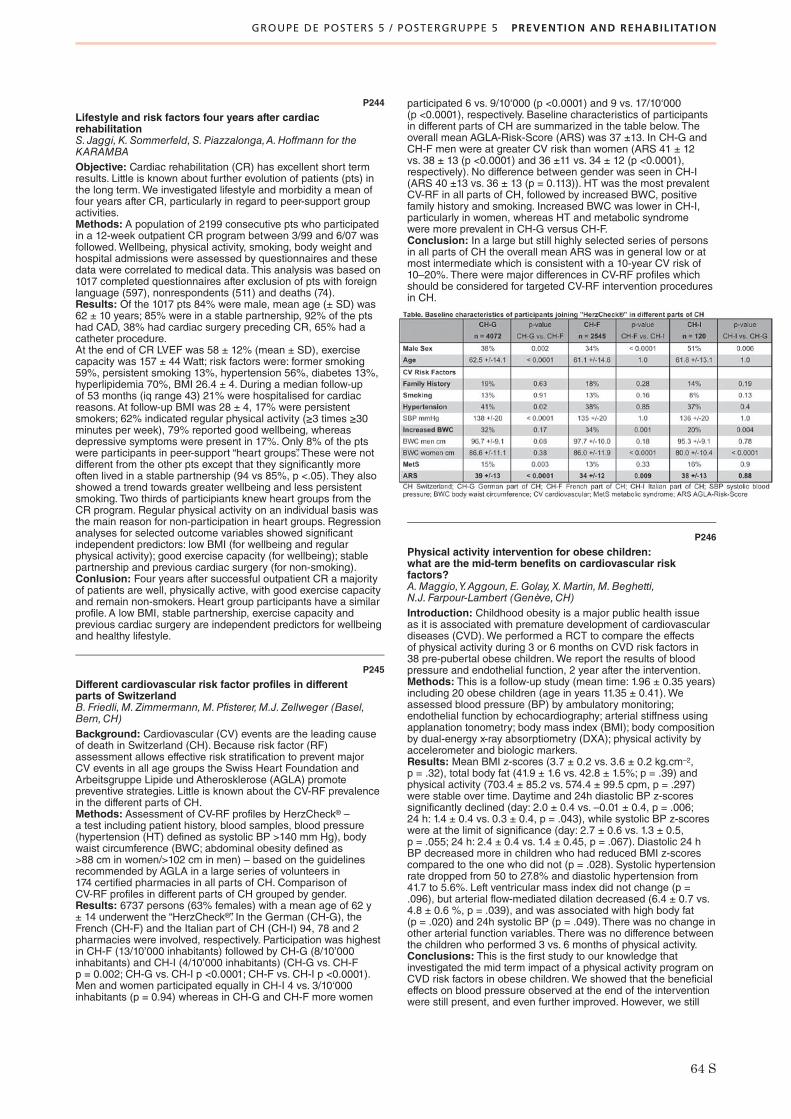
GROUPE DE POSTERS 5 / POSTERGRUPPE 5 PREVENTION AND REHABILITATION
64 S
P244
Lifestyle and risk factors four years after cardiacrehabilitationS. Jaggi, K.Sommerfeld,S.Piazzalonga,A.Hoffmann for theKARAMBAObjective: Cardiac rehabilitation (CR) has excellent short termresults. Little is known about further evolution of patients (pts) inthe long term.We investigated lifestyle and morbidity a mean offour years after CR, particularly in regard to peer-support groupactivities.Methods: A population of 2199 consecutive pts who participatedin a 12-week outpatient CR program between 3/99 and 6/07 wasfollowed.Wellbeing, physical activity, smoking, body weight andhospital admissions were assessed by questionnaires and thesedata were correlated to medical data. This analysis was based on1017 completed questionnaires after exclusion of pts with foreignlanguage (597), nonrespondents (511) and deaths (74).Results: Of the 1017 pts 84% were male, mean age (± SD) was62 ± 10 years; 85% were in a stable partnership, 92% of the ptshad CAD, 38% had cardiac surgery preceding CR, 65% had acatheter procedure.At the end of CR LVEF was 58 ± 12% (mean ± SD), exercisecapacity was 157 ± 44 Watt; risk factors were: former smoking59%, persistent smoking 13%, hypertension 56%, diabetes 13%,hyperlipidemia 70%, BMI 26.4 ± 4. During a median follow-upof 53 months (iq range 43) 21% were hospitalised for cardiacreasons. At follow-up BMI was 28 ± 4, 17% were persistentsmokers; 62% indicated regular physical activity (≥3 times ≥30minutes per week), 79% reported good wellbeing, whereasdepressive symptoms were present in 17%. Only 8% of the ptswere participants in peer-support “heart groups”. These were notdifferent from the other pts except that they significantly moreoften lived in a stable partnership (94 vs 85%, p <.05). They alsoshowed a trend towards greater wellbeing and less persistentsmoking. Two thirds of participiants knew heart groups from theCR program. Regular physical activity on an individual basis wasthe main reason for non-participation in heart groups. Regressionanalyses for selected outcome variables showed significantindependent predictors: low BMI (for wellbeing and regularphysical activity); good exercise capacity (for wellbeing); stablepartnership and previous cardiac surgery (for non-smoking).Conlusion: Four years after successful outpatient CR a majorityof patients are well, physically active, with good exercise capacityand remain non-smokers. Heart group participants have a similarprofile. A low BMI, stable partnership, exercise capacity andprevious cardiac surgery are independent predictors for wellbeingand healthy lifestyle.
P245
Different cardiovascular risk factor profiles in differentparts of SwitzerlandB.Friedli,M.Zimmermann,M.Pfisterer,M.J. Zellweger (Basel,Bern,CH)Background: Cardiovascular (CV) events are the leading causeof death in Switzerland (CH). Because risk factor (RF)assessment allows effective risk stratification to prevent majorCV events in all age groups the Swiss Heart Foundation andArbeitsgruppe Lipide und Atherosklerose (AGLA) promotepreventive strategies. Little is known about the CV-RF prevalencein the different parts of CH.Methods: Assessment of CV-RF profiles by HerzCheck® –a test including patient history, blood samples, blood pressure(hypertension (HT) defined as systolic BP >140 mm Hg), bodywaist circumference (BWC; abdominal obesity defined as>88 cm in women/>102 cm in men) – based on the guidelinesrecommended by AGLA in a large series of volunteers in174 certified pharmacies in all parts of CH. Comparison ofCV-RF profiles in different parts of CH grouped by gender.Results: 6737 persons (63% females) with a mean age of 62 y± 14 underwent the “HerzCheck®”. In the German (CH-G), theFrench (CH-F) and the Italian part of CH (CH-I) 94, 78 and 2pharmacies were involved, respectively. Participation was highestin CH-F (13/10’000 inhabitants) followed by CH-G (8/10’000inhabitants) and CH-I (4/10’000 inhabitants) (CH-G vs. CH-Fp = 0.002; CH-G vs. CH-I p <0.0001; CH-F vs. CH-I p <0.0001).Men and women participated equally in CH-I 4 vs. 3/10‘000inhabitants (p = 0.94) whereas in CH-G and CH-F more women
participated 6 vs. 9/10‘000 (p <0.0001) and 9 vs. 17/10‘000(p <0.0001), respectively. Baseline characteristics of participantsin different parts of CH are summarized in the table below. Theoverall mean AGLA-Risk-Score (ARS) was 37 ±13. In CH-G andCH-F men were at greater CV risk than women (ARS 41 ± 12vs. 38 ± 13 (p <0.0001) and 36 ±11 vs. 34 ± 12 (p <0.0001),respectively). No difference between gender was seen in CH-I(ARS 40 ±13 vs. 36 ± 13 (p = 0.113)). HT was the most prevalentCV-RF in all parts of CH, followed by increased BWC, positivefamily history and smoking. Increased BWC was lower in CH-I,particularly in women, whereas HT and metabolic syndromewere more prevalent in CH-G versus CH-F.Conclusion: In a large but still highly selected series of personsin all parts of CH the overall mean ARS was in general low or atmost intermediate which is consistent with a 10-year CV risk of10–20%. There were major differences in CV-RF profiles whichshould be considered for targeted CV-RF intervention proceduresin CH.
P246
Physical activity intervention for obese children:what are the mid-term benefits on cardiovascular riskfactors?A.Maggio,Y.Aggoun,E.Golay, X.Martin,M.Beghetti,N.J. Farpour-Lambert (Genève,CH)Introduction: Childhood obesity is a major public health issueas it is associated with premature development of cardiovasculardiseases (CVD).We performed a RCT to compare the effectsof physical activity during 3 or 6 months on CVD risk factors in38 pre-pubertal obese children.We report the results of bloodpressure and endothelial function, 2 year after the intervention.Methods: This is a follow-up study (mean time: 1.96 ± 0.35 years)including 20 obese children (age in years 11.35 ± 0.41).Weassessed blood pressure (BP) by ambulatory monitoring;endothelial function by echocardiography; arterial stiffness usingapplanation tonometry; body mass index (BMI); body compositionby dual-energy x-ray absorptiometry (DXA); physical activity byaccelerometer and biologic markers.Results: Mean BMI z-scores (3.7 ± 0.2 vs. 3.6 ± 0.2 kg.cm–2,p = .32), total body fat (41.9 ± 1.6 vs. 42.8 ± 1.5%; p = .39) andphysical activity (703.4 ± 85.2 vs. 574.4 ± 99.5 cpm, p = .297)were stable over time. Daytime and 24h diastolic BP z-scoressignificantly declined (day: 2.0 ± 0.4 vs. –0.01 ± 0.4, p = .006;24 h: 1.4 ± 0.4 vs. 0.3 ± 0.4, p = .043), while systolic BP z-scoreswere at the limit of significance (day: 2.7 ± 0.6 vs. 1.3 ± 0.5,p = .055; 24 h: 2.4 ± 0.4 vs. 1.4 ± 0.45, p = .067). Diastolic 24 hBP decreased more in children who had reduced BMI z-scorescompared to the one who did not (p = .028). Systolic hypertensionrate dropped from 50 to 27.8% and diastolic hypertension from41.7 to 5.6%. Left ventricular mass index did not change (p =.096), but arterial flow-mediated dilation decreased (6.4 ± 0.7 vs.4.8 ± 0.6 %, p = .039), and was associated with high body fat(p = .020) and 24h systolic BP (p = .049). There was no change inother arterial function variables. There was no difference betweenthe children who performed 3 vs. 6 months of physical activity.Conclusions: This is the first study to our knowledge thatinvestigated the mid term impact of a physical activity program onCVD risk factors in obese children.We showed that the beneficialeffects on blood pressure observed at the end of the interventionwere still present, and even further improved. However, we still
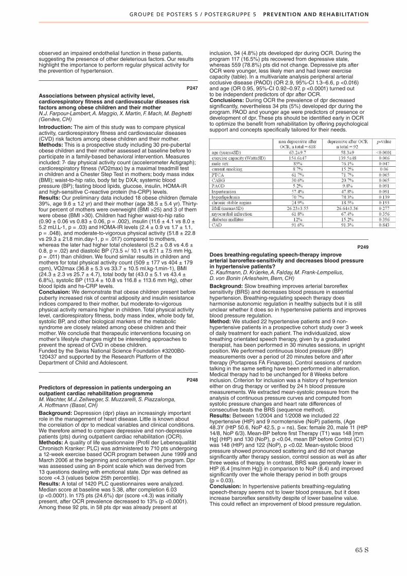
GROUPE DE POSTERS 5 / POSTERGRUPPE 5 PREVENTION AND REHABILITATION
65 S
observed an impaired endothelial function in these patients,suggesting the presence of other deleterious factors. Our resultshighlight the importance to perform regular physical activity forthe prevention of hypertension.
P247
Associations between physical activity level,cardiorespiratory fitness and cardiovascular diseases riskfactors among obese children and their motherN.J. Farpour-Lambert,A.Maggio, X.Martin, F.Mach,M.Beghetti(Genève,CH)Introduction: The aim of this study was to compare physicalactivity, cardiorespiratory fitness and cardiovascular diseases(CVD) risk factors among obese children and their mother.Methods: This is a prospective study including 30 pre-pubertalobese children and their mother assessed at baseline before toparticipate in a family-based behavioral intervention. Measuresincluded: 7- day physical activity count (accelerometer Actigraph);cardiorespiratory fitness (VO2max) by a maximal treadmill testin children and a Chester Step Test in mothers; body mass index(BMI); waist-to-hip ratio, body fat by DXA; systemic bloodpressure (BP); fasting blood lipids, glucose, insulin, HOMA-IRand high-sensitive C-reactive protein (hs-CRP) levels.Results: Our preliminary data included 18 obese children (female39%, age 9.6 ± 1.2 yr) and their mother (age 38.5 ± 5.4 yr). Thirty-four percent of mothers were overweight (BMI >25) and 3 of themwere obese (BMI >30). Children had higher waist-to-hip ratio(0.90 ± 0.06 vs 0.83 ± 0.06, p = .002), insulin (11.6 ± 4.1 vs 8.0 ±5.2 mU.L-1, p = .03) and HOMA-IR levels (2.4 ± 0.9 vs 1.7 ± 1.1,p = .048), and moderate-to-vigorous physical activity (51.8 ± 22.8vs 29.3 ± 21.8 min.day-1, p = .017) compared to mothers,whereas the later had higher total cholesterol (5.2 ± 0.8 vs 4.6 ±0.8, p = .02) and diastolic BP (73.5 +/ 10.1 vs 67.1 ± 7.5 mm Hg,p = .011) than children.We found similar results in children andmothers for total physical activity count (509 ± 177 vs 404 ± 179cpm), VO2max (36.8 ± 5.3 vs 33.7 ± 10.5 ml.kg-1.min-1), BMI(24.3 ± 2.3 vs 25.7 ± 4.7), total body fat (43.0 ± 5.1 vs 43.4 ±6.8%), systolic BP (113.4 ± 10.8 vs 116.8 ± 113.6 mm Hg), otherblood lipids and hs-CRP levels.Conclusion:We demonstrate that obese children present beforepuberty increased risk of central adiposity and insulin resistanceindices compared to their mother, but moderate-to-vigorousphysical activity remains higher in children. Total physical activitylevel, cardiorespiratory fitness, body mass index, whole body fat,systolic BP, and other biological markers of the metabolicsyndrome are closely related among obese children and theirmother.We conclude that therapeutic interventions focusing onmother’s lifestyle changes might be interesting approaches toprevent the spread of CVD in obese children.Funded by the Swiss National Science Foundation #3200B0-120437 and supported by the Research Platform of theDepartment of Child and Adolescent.
P248
Predictors of depression in patients undergoing anoutpatient cardiac rehabilitation programmeM.Wachter,M.J. Zellweger,S.Muzzarelli, S.Piazzalonga,A.Hoffmann (Basel,CH)Background: Depression (dpr) plays an increasingly importantrole in the management of heart disease. Little is known aboutthe correlation of dpr to medical variables and clinical conditions.We therefore aimed to compare depressive and non-depressivepatients (pts) during outpatient cardiac rehabilitation (OCR).Methods: A quality of life questionnaire (Profil der LebensqualitätChronisch Kranker: PLC) was administered to 710 pts undergoinga 12-week exercise based OCR program between June 1999 andMarch 2006 at the beginning and completion of the program. Dprwas assessed using an 8-point scale which was derived from13 questions dealing with emotional state. Dpr was defined asscore <4.3 (values below 25th percentile).Results: A total of 1420 PLC questionnaires were analyzed.Median score at baseline was 5.38, after completion 6.03(p <0.0001). In 175 pts (24.6%) dpr (score <4.3) was initiallypresent, after OCR prevalence decreased to 13% (p <0.0001).Among these 92 pts, in 58 pts dpr was already present at
inclusion, 34 (4.8%) pts developed dpr during OCR. During theprogram 117 (16.5%) pts recovered from depressive state,whereas 559 (78.8%) pts did not change. Depressive pts afterOCR were younger, less likely men and had lower exercisecapacity (table). In a multivariate analysis peripheral arterialocclusive disease (PAOD) (OR 2.9, 95%-CI 1.3–6.6, p <0.016)and age (OR 0.95, 95%-CI 0.92–0.97, p <0.0001) turned outto be independent predictors of dpr after OCR.Conclusions: During OCR the prevalence of dpr decreasedsignificantly, nevertheless 34 pts (5%) developed dpr during theprogram. PAOD and younger age were predictors of presence ordevelopment of dpr. These pts should be identified early in OCRto optimize the benefit from rehabilitation by offering psychologicalsupport and concepts specifically tailored for their needs.
P249
Does breathing-regulating speech-therapy improvearterial baroreflex-sensitivity and decreases blood pressurein hypertensive patients?C.Kaufmann,D.Krüerke,A. Falday,M.Frank-Lempelius,D. von Bonin (Arlesheim,Bern,CH)Background: Slow breathing improves arterial baroreflexsensitivity (BRS) and decreases blood pressure in essentialhypertension. Breathing-regulating speech therapy doesharmonise autonomic regulation in healthy subjects but it is stillunclear whether it does so in hypertensive patients and improvesblood pressure regulation.Method:We studied 22 hypertensive patients and 9 non-hypertensive patients in a prospective cohort study over 3 weekof daily treatment for each patient. The individualized, slowbreathing orientated speech therapy, given by a graduatedtherapist, has been performed in 30 minutes sessions, in uprightposition.We performed continuous blood pressure (BP)measurements over a period of 20 minutes before and aftertherapy (Portapress FA Finapress). Control sessions of randomtalking in the same setting have been performed in alternation.Medical therapy had to be unchanged for 8 Weeks beforeinclusion. Criterion for inclusion was a history of hypertensioneither on drug therapy or verified by 24 h blood pressuremeasurements.We extracted mean-systolic pressure from theanalysis of continuous pressure curves and computed fromsystolic pressure changes and heart rate differences ofconsecutive beats the BRS (sequence method).Results: Between 1/2004 and 1/2008 we included 22hypertensive (HtP) and 9 normotensive (NoP) patients, (Age48.3Y (HtP 50.6, NoP 42.5, p = ns), Sex: female 20, male 11 (HtP14/8, NoP 6/3). Mean-BP before first Therapy (T1) was 148 [mmHg] (HtP) and 130 (NoP), p <0.04, mean BP before Control (C1)was 148 (HtP) and 122 (NoP), p <0.02. Mean-systolic bloodpressure showed pronounced scattering and did not changesignificantly after therapy session, control session as well as afterthree weeks of therapy. In contrast, BRS was generally lower inHtP (6.4 [ms/mm Hg]) in comparison to NoP (8.4) and improvedsignificantly over the whole therapy period in both groups(p = 0.03).Conclusion: In hypertensive patients breathing-regulatingspeech-therapy seems not to lower blood pressure, but it doesincrease baroreflex sensitivity despite of lower baseline value.This could reflect an improvement of blood pressure regulation.
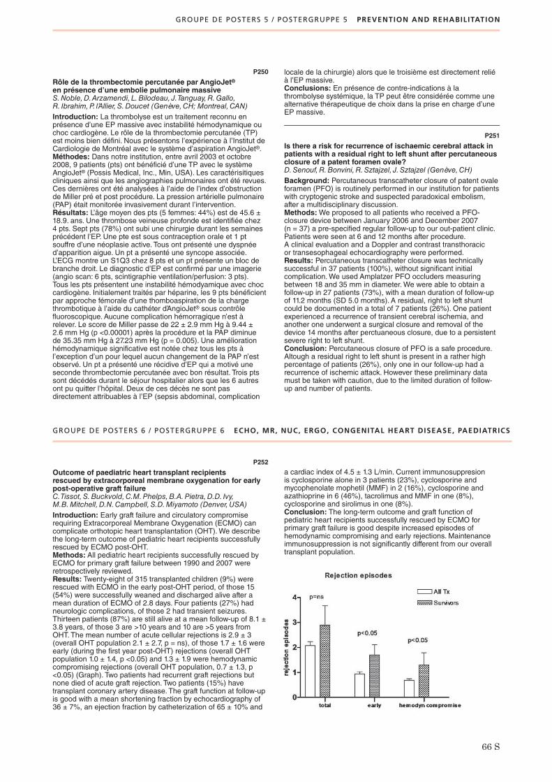
GROUPE DE POSTERS 5 / POSTERGRUPPE 5 PREVENTION AND REHABILITATION
GROUPE DE POSTERS 6 / POSTERGRUPPE 6 ECHO, MR, NUC, ERGO, CONGENITAL HEART DISEASE, PAEDIATRICS
66 S
P250
Rôle de la thrombectomie percutanée par AngioJet®en présence d’une embolie pulmonaire massiveS.Noble,D.Arzamendi, L.Bilodeau, J.Tanguay,R.Gallo,R. Ibrahim,P. l’Allier, S.Doucet (Genève,CH; Montreal,CAN)Introduction: La thrombolyse est un traitement reconnu enprésence d’une EP massive avec instabilité hémodynamique ouchoc cardiogène. Le rôle de la thrombectomie percutanée (TP)est moins bien défini. Nous présentons l’expérience à l’Institut deCardiologie de Montréal avec le système d’aspiration AngioJet®.Méthodes: Dans notre institution, entre avril 2003 et octobre2008, 9 patients (pts) ont bénéficié d’une TP avec le systèmeAngioJet® (Possis Medical, Inc., Min, USA). Les caractérisitiquescliniques ainsi que les angiographies pulmonaires ont été revues.Ces dernières ont été analysées à l’aide de l’index d’obstructionde Miller pré et post procédure. La pression artérielle pulmonaire(PAP) était monitorée invasivement durant l’intervention.Résultats: L’âge moyen des pts (5 femmes: 44%) est de 45.6 ±18.9. ans. Une thrombose veineuse profonde est identifiée chez4 pts. Sept pts (78%) ont subi une chirurgie durant les semainesprécédent l’EP. Une pte est sous contraception orale et 1 ptsouffre d’une néoplasie active. Tous ont présenté une dyspnéed’apparition aigue. Un pt a présenté une syncope associée.L’ECG montre un S1Q3 chez 8 pts et un pt présente un bloc debranche droit. Le diagnostic d’EP est confirmé par une imagerie(angio scan: 6 pts, scintigraphie ventilation/perfusion: 3 pts).Tous les pts présentent une instabilité hémodyamique avec choccardiogène. Initialement traités par héparine, les 9 pts bénéficientpar approche fémorale d’une thomboaspiration de la chargethrombotique à l’aide du cathéter d’AngioJet® sous contrôlefluoroscopique. Aucune complication hémorragique n’est àrelever. Le score de Miller passe de 22 ± 2.9 mm Hg à 9.44 ±2.6 mm Hg (p <0.00001) après la procédure et la PAP diminuede 35.35 mm Hg à 27.23 mm Hg (p = 0.005). Une améliorationhémodynamique significative est notée chez tous les pts àl’exception d’un pour lequel aucun changement de la PAP n’estobservé. Un pt a présenté une récidive d’EP qui a motivé uneseconde thrombectomie percutanée avec bon résultat. Trois ptssont décédés durant le séjour hospitalier alors que les 6 autresont pu quitter l’hôpital. Deux de ces décès ne sont pasdirectement attribuables à l’EP (sepsis abdominal, complication
locale de la chirurgie) alors que le troisième est directement reliéà l’EP massive.Conclusions: En présence de contre-indications à lathrombolyse systémique, la TP peut être considérée comme unealternative thérapeutique de choix dans la prise en charge d’uneEP massive.
P251
Is there a risk for recurrence of ischaemic cerebral attack inpatients with a residual right to left shunt after percutaneousclosure of a patent foramen ovale?D.Senouf,R.Bonvini,R.Sztajzel, J. Sztajzel (Genève,CH)Background: Percutaneous transcatheter closure of patent ovaleforamen (PFO) is routinely performed in our institution for patientswith cryptogenic stroke and suspected paradoxical embolism,after a multidisciplinary discussion.Methods:We proposed to all patients who received a PFO-closure device between January 2006 and December 2007(n = 37) a pre-specified regular follow-up to our out-patient clinic.Patients were seen at 6 and 12 months after procedure.A clinical evaluation and a Doppler and contrast transthoracicor transesophageal echocardiography were performed.Results: Percutaneous transcatheter closure was technicallysuccessful in 37 patients (100%), without significant initialcomplication.We used Amplatzer PFO occluders measuringbetween 18 and 35 mm in diameter.We were able to obtain afollow-up in 27 patients (73%), with a mean duration of follow-upof 11.2 months (SD 5.0 months). A residual, right to left shuntcould be documented in a total of 7 patients (26%). One patientexperienced a recurrence of transient cerebral ischemia, andanother one underwent a surgical closure and removal of thedevice 14 months after perctuaneous closure, due to a persistentsevere right to left shunt.Conclusion: Percutaneous closure of PFO is a safe procedure.Altough a residual right to left shunt is present in a rather highpercentage of patients (26%), only one in our follow-up had arecurrence of ischemic attack. However these preliminary datamust be taken with caution, due to the limited duration of follow-up and number of patients.
P252
Outcome of paediatric heart transplant recipientsrescued by extracorporeal membrane oxygenation for earlypost-operative graft failureC.Tissot, S.Buckvold,C.M.Phelps,B.A.Pietra,D.D. Ivy,M.B.Mitchell, D.N.Campbell, S.D.Miyamoto (Denver,USA)Introduction: Early graft failure and circulatory compromiserequiring Extracorporeal Membrane Oxygenation (ECMO) cancomplicate orthotopic heart transplantation (OHT).We describethe long-term outcome of pediatric heart recipients successfullyrescued by ECMO post-OHT.Methods: All pediatric heart recipients successfully rescued byECMO for primary graft failure between 1990 and 2007 wereretrospectively reviewed.Results: Twenty-eight of 315 transplanted children (9%) wererescued with ECMO in the early post-OHT period, of those 15(54%) were successfully weaned and discharged alive after amean duration of ECMO of 2.8 days. Four patients (27%) hadneurologic complications, of those 2 had transient seizures.Thirteen patients (87%) are still alive at a mean follow-up of 8.1 ±3.8 years, of those 3 are >10 years and 10 are >5 years fromOHT. The mean number of acute cellular rejections is 2.9 ± 3(overall OHT population 2.1 ± 2.7, p = ns), of those 1.7 ± 1.6 wereearly (during the first year post-OHT) rejections (overall OHTpopulation 1.0 ± 1.4, p <0.05) and 1.3 ± 1.9 were hemodynamiccompromising rejections (overall OHT population, 0.7 ± 1.3, p<0.05) (Graph). Two patients had recurrent graft rejections butnone died of acute graft rejection. Two patients (15%) havetransplant coronary artery disease. The graft function at follow-upis good with a mean shortening fraction by echocardiography of36 ± 7%, an ejection fraction by catheterization of 65 ± 10% and
a cardiac index of 4.5 ± 1.3 L/min. Current immunosuppresionis cyclosporine alone in 3 patients (23%), cyclosporine andmycophenolate mophetil (MMF) in 2 (16%), cyclosporine andazathioprine in 6 (46%), tacrolimus and MMF in one (8%),cyclosporine and sirolimus in one (8%).Conclusion: The long-term outcome and graft function ofpediatric heart recipients successfully rescued by ECMO forprimary graft failure is good despite increased episodes ofhemodynamic compromising and early rejections. Maintenanceimmunosuppression is not significantly different from our overalltransplant population.

GROUPE DE POSTERS 6 / POSTERGRUPPE 6 ECHO, MR, NUC, ERGO, CONGENITAL HEART DISEASE, PAEDIATRICS
67 S
P253
Evaluation of flow and pulsation of the peripheral pulmonaryarteries in presence of severe pulmonary regurgitation:a magnetic resonance imaging studyE.Voser,C.Kellenberger, E.Valsangiacomo Buechel (Zürich,CH)Introduction: In presence of pulmonary regurgitation (PR) afterrepair of tetralogy of Fallot (TOF), alteration of flow may extendinto the pulmonary arteries (Pas) side branches.Echocardiography provides good information about Pasdimensions but is not very accurate in quantifying blood flow.We sought to evaluate the flow and pulsation characteristics ofthe Pas by using cardiovascular magnetic resonance (CMR) inpatients with PR and to assess potential correlation betweenthe Pas dimensions and pulsation and blood flow.Methods: CMR for quantification of PR and of right ventricular(RV) dimensions was performed in 24 patients with repairedTOF and significant PR. Median age was 18 years (range 1–64),weight 48.5 kg (7.7–85). Flow and cross-sectional area changes(pulsation) during cardiac cycle were measured by acquiringphase contrast (PC) cine images through the main (MPA), theright (RPA) and the left pulmonary artery (LPA). Flow wascorrected by repeating the same PC CMR acquisitions for eachlocation on a static phantom.Results: Significant PR was present in all subjects, with a meanMPA regurgitation fraction (RF) of 41.4 ± 8.4%. None of thepatients showed Pas stenosis. Thus there was a symmetricalforward flow into RPA (35.6 ± 9.3 ml/m2) and LPA ( 31.7 ± 8.7ml/m2); however the net flow ratio was significantly higher in theRPA than in the LPA, 57.2 ± 9.7% vs. 42.8 ± 9.7% (p = 0.0013),due to a higher RF in the LPA (42.8 ± 12.6% vs 32.1 ± 8.7%, p<0.0001). The LPA maximal cross-sectional area (330.0 ± 97.7mm2/m2) was significantly larger than the RPA area (274.9 ± 88.4mm2/m2, p = 0.001). Pulsation was similar in the RPA and in theLPA, with a cross-sectional area reduction of 42.5 ± 9% in theRPA and 39.1 ± 10% in the LPA.A positive correlation was foundbetween RPA pulsation and RPA RF (r 0.5, p 0.02), RPAregurgitant flow (r 0.5, p 0.02), RPA forward flow (r 0.5, p 0.01),MPA RF (r 0.5, p 0.02), MPA regurgitant flow (r 0.7, p 0.001), MPAforward flow (r 0.6, p 0.004), RV enddiastolic (r 0.6, p 0.003)and endsystolic volumes (r 0.4, r 0.04). No correlation wasdemonstrated between LPA pulsation and all these parameters.Conclusion: In presence of pulmonary regurgitation, RPApulsation mirrors the flow conditions in the RPA and in the MPA.RPA cross-sectional area changes may be used to estimate theamount of PR. The peculiar geometry of the LPA in TOF maydetermine the difference observed in flow, size and pulsation.
P254
N-13 ammonia PET/CT in the assessment of myocardialflow reserve in myocardial areas with and without regionalperfusion defects during vasomotor stressR.Nkoulou,G.Vincenti, I.Valenta,C.Steiner, S.Dewarrat,G.Ambrosio,Y.Seimbille,O.Ratib, F.Mach,T.Schindler(Genève,CH; Perugia, I)Background:While cardiac PET perfusion imaging provides ahigh diagnostic accuracy in the detection of “culprit” flow-limitingepicardial lesion, it may not necessarily identify other lesions inmultivessel CAD. In this regard, we concurrently evaluatedregional myocardial flow reserve (MFR) in myocardial areas withand without stress-induced perfusion defects in patients withangiographically documented CAD.Methods: Myocardial blood flow (MBF) at rest and duringdipyridamole-stimulated hyperemia was determined with N-13ammonia PET/CT in patients with and without regional stress-induced perfusion defects (n = 9 and n = 15, respectively), andthe MFR was evaluated. Based on the findings of coronaryangiography and PET, myocardial segments were divided into3 types: segments with stress-induced perfusion defects andcoronary stenosis (group A), segments with normal perfusionbut with stenosis >50% diameter (group B), and segments withnormal perfusion and no stenosis >50% diameter (group C).Results: Resting regional MBF did not differ among the3 groups (0.96 ± 0.26 vs. 0.93 ± 0.19 vs. 0.94 ± 0.20 ml/g/min,respectively). Hyperemic MBFs showed a progressive increasefrom group A to C (1.08 ± 0.30 vs. 1.25 ± 0.32 vs. 1.63 ± 0.38ml/g/min, respectively). The MFR in group A tended to be lowerthan in group B (1.15 ± 0.28 vs. 1.38 ± 0.28), while it was
significantly lower as compared to group C (1.15 ± 0.28 vs. 1.78 ±0.58, p <0.05). In addition, the MFR in group B was significantlylower than in group C (1.38 ± 0.28 vs. 1.78 ± 0.58, p <0.05).Conclusions: The MFR was lowest in myocardial territorieswith regional perfusion defects on stress PET images underlyingepicardial stenosis >50%, while it was intermediate in thoseterritories with no regional perfusion defects but with arterystenosis >50% diameter, and highest in normal perfusionterritories without artery stenosis >50%.Adding the non-invasiveevaluation of regional MFR to PET perfusion imaging mayimprove the identification of flow-limiting epicardial lesions inmultivessel CAD.
P255
PET-measured hyperaemic, longitudinal myocardial flowgradient in individuals with cardiovascular risk but withor without coronary artery calcificationI.Valenta,G.Vincenti, R.Nkoulou,C.Steiner, S.Dewarrat,Y.Seimbille,O.Ratib, F.Mach,T.Schindler (Zürich,Genève,CH)Background:We investigated possible differences of alongitudinal myocardial flow gradient during pharmacologically-induced hyperemia, reflecting a non-invasive probe of epicardialvasomotor dysfunction, in cardiovascular risk individuals withoutor with coronary artery calcification (CAC).Methods: CAC was determined by a 64 slice CT of the PET/CTsystem. Myocardial blood flow (MBF) was measured quantitativelywith 13N-ammonia and PET/CT in ml/g/min at rest, and duringpharmacologic vasodilation with dipyridamole in healthy controls(CON, n = 10) and in individuals with cardiovascular risk factorssuch as arterial hypertension, smoking, diabetes mellitus,hypercholeterolemia and obesity but without CAC (group 1;n = 15) or with CAC (group 2, n = 13). MBF was assessedglobally as mean MBF, and in the mid and mid-distal myocardiumof the left ventricle (LV). A decrease in MBF from mid to mid-distal LV myocardium was defined as longitudinal MBF gradient.Results: In group 2, CT-measured a mean CAC of 1101 ± 1008Hounsfield.Compared with CON, the global hyperemic MBFprogressively declined in group 1 and group 2 (2.10 ± 0.60 vs.1.65 ± 0.30 and 1.45 ± 0.48 ml/g/min; p <0.001), while it did notdiffer significantly between group 1 and group 2. Absolute MBFsduring pharmacologic vasodilation were significantly lower in themid-distal than in the mid LV myocardium in group 1 and group 2(1.45 ± 0.25 vs 1.78 ± 0.23 ml/g/min, and 1.35 ± 0.47 vs 1.58 ±0.52 ml/g/min, p <0.0001), resulting in a MBF gradient that wassignificantly higher in group1 than in group 2 (0.32 ± 0.15 vs.0.23 ± 0.11 ml/g/min, p <0.0001), not observed in CON (0.006 ±0.05 ml/g/min, p = NS). Notably, the MBF gradient in group1–2 correlated significantly with the mid LV MBF duringpharmacologic vasodilation (r = 0.42, p <0.05), implicating thevelocity of coronary blood flow as an important determinant of theMBF gradient.Conclusion: As it was observed, the hyperemic MBF gradientwas more pronounced in coronary risk individuals without CACthan in those with CAC, while it dependent the velocity ofcoronary blood flow. These preliminary results further emphasizefunctional and/or structural alterations of the epicardial vessel asprevalent cause for the longitudinal MBF gradient.
P256
Diagnostic value of semi-quantitative and quantitativeevaluation of PET/CT-measured myocardial perfusionfor the identification of flow-limiting epicardial stenosisG.Vincenti, R.Nkoulou, I.Valenta,C.Steiner, S.Dewarrat,G.Ambrosio,Y.Seimbille,O.Ratib, F.Mach,T.Schindler(Genève,Zürich,CH; Perugia, I)Introduction:We aimed to evaluate the diagnostic value of asemiquantitative and quantitative evaluation of PET/CT-measuredmyocardial perfusion for the identification of flow-limitingepicardial stenosis.Methods:We evaluated 25 consecutive patients with suspectedCAD undergoing stress-rest N-13 ammonia cardiac PET/CT.Myocardial perfusion was assessed visually and myocardialperfusion segments were graded on a semiquantitative 5-pointscoring system. In addition, regional myocardial blood flow (MBF)was determined quantitatively with PET and tracer kineticmodeling of N-13 ammonia in ml/g/min at rest, during

GROUPE DE POSTERS 6 / POSTERGRUPPE 6 ECHO, MR, NUC, ERGO, CONGENITAL HEART DISEASE, PAEDIATRICS
68 S
dipyridamole-stimulated hyperemia, and the correspondingregional myocardial flow reserve (MFR= hyperemic MBF / restMBF)was calculated. A regional MFR <1.7 was consideredabnormal. Coronary angiography was performed within 7 daysof the cardiac PET/CT study and any epicardial stenosis >75%was estimated as morphologically significant.Results: On a vessel based analysis, the visual andsemiquantitative analysis had a sensitivity, specifity, PPV, NPVand diagnostic accuracy of 64%, 100%, 100%, 85% and 87%,respectively. Calculating the regional MFR with PET and defininga MFR <1.7 as abnormal yielded a sensitivity, specifity, PPV,NPV and diagnostic accuracy of 100%, 47%, 48%, 100% and64%, respectively. Combining both parameters resulted in anintermediate sensitivity, specifity, PPV, NPV and diagnosticaccuracy of 76%, 77%, 58%, 89% and 77%, respectively.Conclusions: Combining both semiquantitative and quantitativeevaluation of myocardial perfusion with PET/CT may emergeas a new promising analytic approach to further optimize theidentification and characterization of flow-limiting CAD burden.
P257
Rapid semi-quantitative analysis of right ventricularfunction using multidetector computer tomography:a feasability studyS.C.Koestner, J.G.J.Groothuis,R.Nijveldt,M.J.W.Götte,M.R.Meijerink,A.M.Beek,M.B.M.Hofman,A.C. van Rossum(Bern,CH; Amsterdam,NL)Background: Right ventricular (RV) function is an importantprognostic factor in many cardiopulmonary diseases. Thecalculation of RV ejection fraction (EF) using multidetectorcomputed tomography (MDCT) is reliable but still time-consuming.This study investigates the accuracy of novel rapid, semi-quantitative methods for the assessment of RV EF.Methods: 22 patients with suspected coronary heart diseaseunderwent both ECG-gated 64-slice computed tomographycoronary angiography (CTCA) and cine cardiovascular magneticresonance (CMR) on a 1.5T scanner. RV end-diastolic volume,end-systolic volume and EF were calculated using standard short-axis full coverage of both ventricles using CMR. CMRmeasurements served as the standard of reference. MultiphaseMDCT reconstructions were made for the standard 4-chamberview, a perpendicular 2-chamber view of the RV, and a transverseview at a standardized location. Tricuspid annulus plane systolicexcursion (TAPSE) was measured on all three views. Additionally,fractional area shortening on both 4-chamber and transverseviews was evaluated. These different methods were comparedto the standard of reference.Results: RVEF ranged from 37.9% to 66.4% (mean 56.2 ± 7.9%).The use of TAPSE on the basis of the RV 2-chamber viewshowed the best correlation with the gold standard (r = 0.76,p <0.001). The linear correlation formula was EF = (0.44*TAPSE)– 6.57. TAPSE on the 4-chamber (r = 0.61, p=0.002) andtransverse (r = 0.46, p = 0.031) reconstructions, and fractionalarea shortening on the transverse reconstruction (r = 0.51,p = 0.015) showed weaker correlations with the standard ofreference. Fractional area shortening using the 4-chamber viewshowed no significant correlation with the standard of reference(r = 0.31, p = 0.166).Conclusions: Measurement of TAPSE using a single 2-chamberreconstruction of the RV correlates well with the gold standard ofcontiguous short-axis measurements using CMR. This methodcan be used as a rapid, semi-quantitative screening tool forpatients undergoing MDCT.
P258
Interventioneller ASD-Verschluss mit dem SolysafeSeptal Okkluder im Kindesalter – erste viel versprechendeErgebnisseO.Kretschmar,W.Knirsch (Zürich,CH)Einleitung: Der Solysafe Septal Okkluder (SSO) ist ein neuesselbstzentrierendes und repositionierbares Device mit 2 PolyesterPatches befestigt an insgesamt 8 Phynox-Drähten zuminterventionellen ASD-Verschluss. Durch sein flaches Profil bieteter sich zum Einsatz im Kindesalter an.Wir berichten über unsere
ersten viel versprechenden Erfahrungen. Methode: RetrospektiveAnalyse von insgesamt 30 pädiatrischen Patienten (Alter mean8.9 (1.8–17.8) Jahre, Gewicht mean 31.6 (9.5–86) kg), bei denenseit 04/2007 in unserem Zentrum ein SSO zum interventionellenASD-Verschluss implantiert wurde.Ergebnis: 27 Prozeduren waren erfolgreich (90%), bei einemPatienten wurden 2 SSO implantiert. Die Defektdurchmesserlagen zwischen 6 und 20 mm (mean 10.8), Devicegrössen von15 mm (14x), 20 mm (7x), 25 mm (6x) und 30 mm (1x) wurdenimplantiert. Der Eingriff dauerte mean 74 Minuten. Bei den3 erfolglosen Versuchen war kein retroaortaler Randsaumvorhanden, die Defekte wurden schliesslich mit einem AmplatzerSeptal Okkluder (20 + 22 + 26 mm) verschlossen. AlsKomplikation trat bei zwei Kindern nach Schleusenwechsel eineThrombose der vena femoralis auf. Die Verschlussrate lag akutbei 76%, nach 3 Monaten bei 87%, nach 6 und 12 Monaten bei100%. Das Device konfigurierte sich jeweils sehr flach imVorhofseptum, venöse Einflussbehinderungen traten nicht auf.Im Follow-up von 10.9 (1–21) Monaten wurden keineThrombenbildung am Device und/oder Arrhythmien beobachtet.Schlussfolgerung: Der interventionelle ASD-Verschluss mit demSSO ist im Kindesalter bei mittelgrossen ASDs eine sehr guteAlternative zum Amplatzer Device und lässt sich sicher undkomplikationsarm durchführen. Vorteile sind sein flaches Profil,eine hohe Device-Stabilität und hohe Verschlussraten. Ausserdemist das Risiko für Luftembolien durch eine fehlende langeSchleuse minimiert und es werden lediglich 5 Device-Grössenfür alle Defekte bis 30 mm benötigt.
P259
Exercise capacity and invasive haemodynamicmeasurements predict outcome in children withcardiomyopathyC.Tissot, L.B.Willis, K.L.Villavicencio,A.K.Younoszai,C.M.Phelps,J.L.Cavanaugh,S.D.Miyamoto (Denver,USA)Introduction: Doppler and Tissue Doppler echocardiography isused to assess diastolic function and has been shown to correlatewith filling pressures in adults. The purposes of this study were to(1) identify echocardiographic measurements that correlate withfilling pressures and (2) identify predictive markers for outcome inchildren with cardiomyopathy.Methods: All children with dilated (DCM), hypertrophic (HCM)or restrictive (RCM) cardiomyopathy who had a cardiaccatheterization and echocardiogram between 2006 and 2008were reviewed. Patient outcome was divided into: (A) alive andfree from transplant or (T) transplanted or dead.Echocardiographic measurements included biplane ejectionfraction (EF), dP/dt of mitral regurgitation (normal >1200mmHg/sec), mitral inflow (MV) and pulmonary vein (PV) Doppler,tissue Doppler Ea (E/Ea ≤8 normal and ≥15 abnormal), colorM-mode inflow propagation velocity (Vp normal <45 cm/sec).Results: Thirty-four catheterizations and echocardiograms wereperformed in 29 children with an interval between studies of 5 ±58 days. Diagnosis was DCM in 21, HCM in 4 and RCM in 4. Themean age was 9.5 ± 6.2 years and was lower in the DCM group(8 ± 6.7 vs 13 ± 3 years for RCM and HCM, p <0.01). There were14 studies in A patients and 20 in T patients. Compared to Apatients, the T group had a lower EF (33 ± 21 vs 49 ± 13%,p <0.05) and mitral inflow E wave deceleration time (MVE DT,0.09 ± 0.03 vs 0.14 ± 0.09sec, p <0.05). Right atrial pressure,mean PA pressure and wedge pressure (PCWP) measured atcatheterization were significantly higher (p <0.05) in T patients.Mixed venous saturation, cardiac index and exercise capacity(VO2 max 15 ± 4 vs 26 ± 7 ml/min/kg, p <0.05) were lower in theT group. MVE DT was lower in patients with a PCWP ≥12 mm Hg(0.08 ± 0.03 vs 0.14 ± 0.08, p <0.01). Mitral E/Ea and Vp didnot correlate with filling pressures or outcome. DP/dt of MR<1200 mm Hg/sec and a higher mitral E/Ea (11.5 ± 4 vs 5.5 ± 1,p <0.05) correlated with systolic dysfunction.Conclusion: In conclusion, catheterization measurements,VO2 max and MVE DT predicted outcome in children withcardiomyopathy. MVE DT correlated with filling pressures, whiledP/dt of MR and mitral E/Ea correlated with systolic function.Prospective studies in larger populations are needed to confirmthese findings and to identify useful non-invasive markers ofoutcome in children with cardiomyopathy.
GROUPE DE POSTERS 6 / POSTERGRUPPE 6 ECHO, MR, NUC, ERGO, CONGENITAL HEART DISEASE, PAEDIATRICS
69 S
P260
Foetal cardiac rhabdomyoma: long-term outcomeS.Degueldre,P.Chockalingam,Y.Vial, S.Di Bernardo,J.-P.Pfammatter,N.Sekarski,Y.Mivelaz,P. Jeannet, E.J.Meijboom(Lausanne,Bern,CH)Objectives: This study analyses the long term cardiac andneurological outcome of patients with cardiac rhabdomyoma(CR) in order to allow comprehensive prenatal counselling.Background: Because of the relative rarity of the disease, thereis paucity of data concerning the outcome of patients with CR.Methods: A retrospective study including all cases withechocardiographic diagnosis of CR encountered betweenApril 1986-August 2006.Results: Of 24 CR patients identified, 7 were diagnosed in-uteroat a gestational age between 28–35 weeks and 17 postnatallybetween 10 days-5 years. 20 had multiple CR and 4 had singleCR. They were situated predominantly in the LV (70%), RV (52%)and IVS (48%) and to a lesser extent in the atria (13%) andpericardium (4%). Follow-up echocardiography over 3 months-17 years (mean 4.54 years) in 18 showed complete postnatalregression of CR in 3, partial regression in 13 and no change in2. Cardiac complications were encountered in 5 patients, 1 withWPW syndrome and SVT requiring anti-arrhythmic therapy,1 with sub-aortic obstruction needing surgical intervention and3 with occasional bouts of paroxysmal SVT. Long-term follow-uprevealed tuberous sclerosis of Bourneville (TSB) as definitediagnosis in 22, complicated by epilepsy in 16 and developmentaldelay in 14.Conclusions: CR generally regresses after birth and after thehigh risk perinatal period cardiac-related problems are rare. Therelatively more neurodevelopmental complications of the almostalways associated TSB however should form a dominating aspectof the prenatal counselling of parents whose fetuses arediagnosed with this rare disease.
P261
Recovery of cardiac function in recipient twins followinglaser for stage 4 twin to twin transfusion syndromeA.Martin,R.Weber, E. Jaeggi,R.Windrim,G.Seaward, L.Hornberger,C.Barrea,G.Ryan (Toronto,CAN; Zürich,CH)Objective: To evaluate the timing and pattern of cardiac functionchanges in recipient twins following laser in stage 4 TTTSMethods: Retrospective review of serial fetal ultrasoundevaluation in 21 cases of (Quintero) stage 4 TTTS treated withlaser (1999-2008). Fetal ultrasounds, including echocardiography,were done before and serially after laser. Cardiac functionevaluation included cardiothoracic index, pulmonary arterial flow,AV valve regurgitation, venous Doppler (RV and LV inflows,UV, DV, IHV, IVC), isovolumetric relaxation time, ventricularcontractility (LV & RV shortening fraction) and wall thickness.We evaluated the pattern and timing of recovery of hydrops andcardiac function in recipient twins following laser.Results: Of 21 cases treated, 3 with dual fetal demise followinglaser were excluded. Complete fetal echocardiographic follow upwas available in 15 and partially in 3. Abnormalities in pulmonaryartery blood flow normalized in all within 4 days. Ascites resolvedin 12/13 within 2 weeks. Five with tricuspid and one with mitralregurgitation had not resolved by delivery. Right ventricularsystolic function normalized in utero in 8/10 cases with completefollow up, in a mean of 26 days. In 9/10 cases, left ventriculardysfunction normalized in utero, in a mean of 17 days. VenousDopplers normalized in 16-24 days. Ventricular hypertrophywas the last to improve, taking a mean of 33 days and did notcompletely resolve by delivery in any case.Conclusion: Regardless of severity, improvement in systoliccardiac function, resolution of hydrops and normalization ofvenous and umbilical arterial waveforms occur in most recipienttwins with stage 4 TTTS within days of laser therapy. Valvularregurgitation and ventricular hypertrophy are the slowest toimprove and may not completely resolve before delivery
P262
Safety and efficacy of paediatric outpatient radiofrequencycatheter ablationsM.Tomaske,R.Candinas,M.Weiss,U.Bauersfeld (Zürich,CH)Purpose: Radiofrequency catheter ablations (RFA) are frequentlyperformed as treatment for supraventricular tachycardia inchildren older than 4 years of age. Aim of this study was toevaluate safety and efficacy of paediatric outpatient RFA.Patients and methods: Between 06/2002 and 11/2008, 271 RFAwere prospectively analyzed. Exclusion criteria for outpatientprocedures were age <4 years, complex RFA in congenital heartdisease, arterial access, distance to home more than 1 hour orparent preference for inpatient RFA. All RFA were performedunder general anaesthesia. Patients with transseptal puncturereceived heparin. All patients underwent pre- and post-proceduralechocardiography and electrocardiogram. Discharge was within6 hours after conclusion of RFA. To identify potentialcomplications after discharge, parental follow-up phone calls theday after outpatient RFA procedure were performed. All patientswere seen in outpatient clinics 1 month after RFA.Results: A total of 97/ 271 (37%) patients aged 12.4 ± 3.8 yearsqualified for outpatient RFA. Accessory pathway ablations (n = 50)and atrioventricular node modifications (n = 39) were the mostcommon RFA.A transseptal approach was performed in 35 RFA.Median procedure time was 1.5 hours (range: 1.1–4.3), with amedian fluoroscopy time of 10 minutes (range: 4–86). RFA wassuccessful in 94/ 97 (97%) patients. Post-proceduralechocardiography with special attention for intracardiac thrombi,pleural effusion and inflow patterns from systemic veins or thecoronary sinus were normal in all patients. Anaesthetic adverseevents, predominantly post-interventional nausea and vomiting,were observed in 10 (10%) patients. Hospital discharge within6 hours after conclusion of RFA was practicable in all but onepatient due to ongoing nausea. Follow-up phone calls did notreveal further complications. Recurrence of tachycardia aftersuccessful RFA was observed in 6 of 94 (6%) patients andprompted re-intervention in 4 of 6 (67%).Conclusions: Outpatient RFA is feasible and safe in selectedpaediatric patients. No RFA related complication was observed.Anaesthetic adverse events were nausea and vomiting due togeneral anaesthesia. Success rate and recurrence rate oftachycardia was favourable after outpatient RFA.
P263
Comparison of echocardiographic parameters betweenpatients with systolic heart failure aged 60–74 years andpatients ≥75 yearsM.Gutmann,K.Goetschalckx,P.Buser,U.Pittl, H.Pfluger, F.Voss,B.A.Kaufmann,M.Pfisterer,H.P.Brunner-La Rocca,W.Estlinbaumfor the TIME-CHF investigatorsBackground: The prevalence of heart failure increases with age.Age may influence structural and hemodynamic alterations of theheart, which may be assessed by Doppler echocardiography.Therefore, we compared the baseline echocardiographic data ofpatients with reduced left ventricular (LV) ejection fraction (EF; i.e.
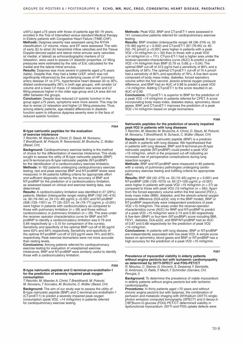
GROUPE DE POSTERS 6 / POSTERGRUPPE 6 ECHO, MR, NUC, ERGO, CONGENITAL HEART DISEASE, PAEDIATRICS
70 S
≤45%) aged ≥75 years with those of patients age 60–74 years,enrolled in the Trial of Intensified versus standard Medical therapyin Elderly patients with Congestive Heart Failure (TIME-CHF).Methods: Dyspnea severity was assessed using the NYHAclassification. LV volume, mass, and EF were assessed. The ratioof early (E) to atrial (A) transmitral inflow velocities and the TissueDoppler-derived septal and lateral mitral annular early velocities(e’ medial, e’ lateral) as echocardiographic markers of LVrelaxation, were used to assess LV diastolic properties. LV fillingpressures were estimated by the ratio of E/e’, calculated for themedial and the lateral mitral annulus.Results: Dyspnea was more severe in patients aged ≥75 years(table). Despite that, they had a better LVEF, which was notsignificantly influenced by the underlying cause of HF (coronaryartery disease 31 vs 27%, hypertensive heart disease 33 vs 30%,dilated cardiomyopathy 26% in both age groups), with smaller LVvolume and a lower LV mass. LV relaxation was worse and LVfilling pressure higher in the older age group and LA area did notdiffer between the groups.Conclusion: Despite less reduced LV systolic function in thegroup aged ≥75 years, symptoms were more severe. This may bedue to worse LV relaxation and higher LV filling pressures. Thus,among elderly patients, age-related differences in diastolicfunction seem to influence dyspnea severity even in the face ofreduced systolic function.
P264
B-type natriuretic peptides for the evaluationof exercise intoleranceT.Reichlin,M.Maeder,A.Christ,D.Staub,M.Noveanu,T.Breidthardt,M.Potocki,R.Twerenbold,M.Brutsche,C.Müller(Basel,CH)Background: Cardiopulmonary exercise testing is the methodof choice for the differentiation of exercise intolerance. This studysought to assess the utility of B-type natriuretic peptide (BNP)and N-terminal-pro-B-type natriuretic peptide (NT-proBNP)for the identification of a cardiocirculatory exercise limitation.Methods: In 162 patients undergoing cardiopulmonary exercisetesting, rest and peak exercise BNP and NT-proBNP levels weremeasured. In 94 patients fulfilling criteria for appropriate effortand sufficient diagnostic certainty, the accuracy of BNP andNT-proBNP for the prediction of a cardiocirculatory limitation,as assessed based on clinical and exercise testing data, wasdetermined.Results: A cardiocirculatory limitation was identified in 27 (29%)patients. Median (interquartile range) resting BNP (162 (45–415)vs. 39 (19–94) vs. 24 (15–46) pg/ml; p <0.001) and NT-proBNP(506 (129–1167) vs. 77 (35–237) vs. 34 (19–77) pg/ml; p <0.001)were higher in patients with cardiocirculatory as compared tothose with pulmonary limitation (n = 28) and those withoutcardiocirculatory or pulmonary limitation (n = 39). The area underthe receiver operator characteristics curve for BNP and NT-proBNP to identify a cardiocirculatory limitation was 0.79 and0.84 respectively (p = 0.15 for comparison of the curves).Sensitivity and specificity of the optimal BNP cut-off of 85 pg/mlwere 63% and 84% respectively. Sensitivity and specificity ofthe optimal NT-proBNP cut-off of 223 pg/ml were 74% and 85%respectively. Peak exercise biomarkers were not more accuratethan resting levels.Conclusions: Among patients referred for cardiopulmonaryexercise testing for evaluation of unexplained exerciseintolerance, BNP and NT-proBNP were similarly useful to identifythose with a cardiocirculatory limitation.
P265
B-type natriuretic peptide and C-terminal-pro-endothelin-1for the prediction of severely impaired peak oxygenconsumptionT.Reichlin,M.Maeder,A.Christ,T.Breidthardt,M.Potocki,M.Noveanu,T.Socrates,M.Brutsche,C.Müller (Basel,CH)Background: The aim of our study was to assess the utility ofB-type natriuretic peptide (BNP) and C-terminal-pro-endothelin-1(CT-proET-1) to predict a severely impaired peak oxygenconsumption (peak VO2, <14 ml/kg/min) in patients referredfor cardiopulmonary exercise testing.
Methods: Peak VO2, BNP and CT-proET-1 were assessed in141 consecutive patients referred for cardiopulmonary exercisetesting.Results: BNP (median (interquartile range) 48 (38–319) vs. 33(15–86) pg/ml; p = 0.002) and CT-proET-1 (87 (76-95) vs. 60(52–74) pmol/l; p <0.001) were higher in patients with a peakVO2 <14 ml/kg/min (n = 30) than in those with a peak VO2≥14 ml/kg/min (n = 111). CT-pro-ET-1 had a higher area under thereceiver-operator-characteristics curve (AUC) to predict a peakVO2 <14 ml/kg/min than BNP (0.79 vs. 0.68; p = 0.04). Theoptimal BNP cut-off of 37.2 pg/ml had a sensitivity of 80% and aspecificity of 56%. The optimal CT-proET-1 cut-off of 74.4 pmol/lhad a sensitivity of 80% and specificity of 76%.A five-item scorecomposed of body mass index, diabetes, forced expiratoryvolume within the first second, alveolo-arterial oxygen pressuredifference, and BNP had an AUC of 0.88 to predict a peak VO2<14 ml/kg/min. Adding CT-proET-1 to the score resulted in anAUC of 0.92.Conclusions: CT-proET-1 is superior to BNP for the prediction ofa peak VO2 <14 ml/kg/min in patients referred for CPET. A scoreincorporating body mass index, diabetes status, spirometry, bloodgases, BNP, and CT-proET-1 improves the prediction of a peakVO2 <14 ml/kg/min based on single biomarkers.
P266
Natriuretic peptides for the prediction of severly impairedpeak VO2 in patients with lung diseasesT.Reichlin,M.Maeder,M.Brutsche,A.Christ,D.Staub,M.Potocki,M.Noveanu,T.Breidthardt,N.Schaub,C.Müller (Basel,CH)Background: B-type natriuretic peptide (BNP) is a predictorof death in patients with lung disease.We hypothesised thatin patients with lung disease, BNP and N-terminal-pro-B-typenatriuretic peptide (NT-proBNP) could predict a peak VO2<15 ml/kg/min, which is the proposed cut-off indicating anincreased risk of perioperative complications during lungresection surgery.Methods: BNP and NT-proBNP were measured in 85 patientswith a variety of pulmonary pathologies undergoing cardio-pulmonary exercise testing and fulfilling criteria for appropriateeffort.Results: BNP (69 (42–270) vs. 33 (15–65) pg/ml; p = 0.001) andNT-proBNP (290 (129–1075) vs. 65 (21-129) pg/ml; p <0.001)were higher in patients with peak VO2 <15 ml/kg/min (n = 27) ascompared to those with peak VO2 (15 ml/kg/min (n = 58)). Apartfrom the forced expiratory volume within the first second (FEV1),body mass index (BMI), diabetes, and the alveolo-arterial oxygenpressure difference (D(A-a)O2; only in the BNP model), BNP orNT-proBNP respectively were independent predictors of peakVO2 <15 ml/kg/min. The areas under the receiver-operator-characteristics curve (AUC) for BNP and NT-proBNP to predictof a peak VO2 <15 ml/kg/min were 0.73 and 0.80 respectively.A five-item (BNP) or four-item (NT-proBNP) score including BMI,FEV1, diabetes, D(A-a)O2, and BNP/NT-proBNP had an AUCof 0.87 and 0.88 respectively for the prediction of peak VO2<15 ml/kg/min.Conclusions: In patients with lung disease, BNP or NT-proBNPare independently associated with low peak VO2. A simple scorebased on spirometry, blood gases and BNP or NT-proBNP has ahigh accuracy for the prediction of a peak VO2 <15 ml/kg/min.
P267
Prevalence of myocardial viability in elderly patientswithout angina pectoris but with ischaemic cardiomyopathyas determined by 201TI-SPECT and FDG-PET/CTR.Nkoulou,C.Steiner,G.Vincenti, S.Dewarrat,Y.Seimbille,G.Ambrosio,O.Ratib, F.Mach,T.Schindler (Genève,CH;Perugia, I)Background: To determine the prevalence of viable myocardiumin elderly patients without angina pectoris but with ischemiccardiomyopathy.Procedures: In thirty patients aged >70 years and withoutchronic angina pectoris but with dyspnea, the combination ofperfusion and metabolic imaging with 201thallium (201TI) single-photon emission computed tomography (SPECT) and 2-deoxy-2-[18F]fluoro-D-glucose (FDG) PET/CT determined viability indysfunctional myocardium. 201TI and FDG uptake defects were

GROUPE DE POSTERS 6 / POSTERGRUPPE 6 ECHO, MR, NUC, ERGO, CONGENITAL HEART DISEASE, PAEDIATRICS
71 S
graded visually on a four-point scale: 0 = normal, 1 = mildlyreduced, 2 = moderately reduced, and 3 = severely reduced.A concordant reduction in 201TI and FDG activity scores wasclassified as a blood flow/metabolism match, subsequentlyreferred to as nonviable myocardium.A reduction in 201TI uptakemore severe than the reduction in glucose metabolic activityby >1 point was defined as a blood flow/metabolism mismatch,subsequently referred to as viable myocardiumResults: All patients (mean age 76 ± 5, range: 70–84 yrs)werein the NYHA functional class II and III and presented a meanleft-ventricular ejection fraction of 38 ± 7%. Thirteen (43%) hada history of myocardial infarction, and 14 (46%) had undergoneprevious PCI or coronary artery bypass surgery. On standardvisual analysis 9 (30%) showed perfusion-metabolic matches(nonviable myocardium), and 21 (70%) had perfusion metabolicmismatches (viable myocardium). Both, mismatches and matchescoexisted in 2 patients (6%).201TI -Tracer uptake as index ofmyocardial flow was more severely reduced in viable than non-viable myocardium (1.80 ± 0.78 vs. 2.3 ± 0.83, p <0.0001),while there was a considerable overlap.Conclusions: Based on the presence of viable dysfunctionalmyocardium, 70% of elderly patients with ischemiccardiomyopathy may be considered for coronaryrevascularization.Whether coronary revascularization of viablemyocardium in elderly patients also translates into animprovement of symptoms and clinical outcome remains to beinvestigated.
P268
Tetralogy of Fallot with absent pulmonary valve, totalanomalous pulmonary venous connection and majorsystemic to pulmonary collateral arteryP.O.Myers,Y.Aggoun,A.Kalangos, J.Sierra,M.Beghetti,C.Tissot (Genève,CH)Introduction: Absent pulmonary valve syndrome is a relativelyrare variant of tetralogy of Fallot (TOF), and has been reportedin 4 patients in association with totally anomalous pulmonaryvenous return (TAPVR). Although up to 25% of patients withTOF/absent pulmonary valve syndrome have systemic-to-pulmonary collaterals, this is the first time it is reported inassociation with TAPVR to our knowledge.Methods: A 2-year-old boy was referred to our institution forcyanosis and mild signs and symptoms of heart failure.Transthoracic echocardiography showed TOF with featurestypical of absent pulmonary valve syndrome. There was severepulmonary valve stenosis with a peak systolic gradient of 85mmHg and free pulmonary regurgitation, associated withaneurysmal dilatation of the branch pulmonary arteries. There
was also a secundum atrial septal defect and anomalouspulmonary venous connection to a non-obstructed confluentleading to the right atrium at the junction of superior vena cava.A tubular structure was visualized between the abdominal aortaand the inferior vena cava, with continuous systolo-diastolicaccelerated flow. Cardiac MRI confirmed that this structure wasa major aorto-pulmonary collateral artery.Results:We performed a staged approach, starting withinterventional embolization of the collateral artery followed bysuccessful surgical transatrial patent foramen ovale enlargementand redirection of the anomalous pulmonary venous connectionto the left atrium using a pericardial patch, patch closure of theventricular septal defect and right ventricular outflow tractreconstruction with a Labcor® valved conduit.Conclusions: This rare association of TOF/absent pulmonaryvalve, TAPVR and MAPCA and its unique physiology illustratesthe importance of a complete preoperative diagnosis forsuccessful staged hybrid management.
P269
Rapid analysis of right ventricular volumes and functionusing cardiovascular magnetic resonance imagingS.C.Koestner,M.J.W.Götte,R.Nijveldt, J.G.J.Groothuis,A.M.Beek,M.B.M.Hofman,A.C. van Rossum(Bern,CH; Amsterdam,NL)Introduction: The function of the right ventricle (RV) is animportant prognostic factor in heart disease. Assessmentof RV function using the gold standard disk-area (or Simpson)technique is time consuming and semi-quantitative evaluation(e.g. TAPSE) shows unsatisfactory correlation with quantitativeanalysis. This study investigates the use of an area-lengthtechnique for rapid assessment of RV volumes and functionusing cardiovascular magnetic resonance imaging (CMR).Methods: 12 healthy volunteers and 25 patients with RV dilationand/or dysfunction underwent cine CMR on a 1.5T scanner. RVend-diastolic volume (RVEDV), end-systolic volume (RVESV) andejection fraction (RVEF) were calculated using a stack of short-axis cines as the standard of reference (Simpson). A 4-chamberview and a perpendicular RV 2-chamber view were acquiredto calculate RV volumes and RVEF using a simple area-lengthmethod (A*A/L) without correction coefficient in both end-diastolicand end-systolic phases with A the RV area, and L the apex tobase length. This was tested for both a single and a biplane long-axis view. Finally, TAPSE was measured on the 4-chamber view.Area-length methods and TAPSE were compared to the standardof reference.Results: Analysis of the area-length method using long-axisorientations was faster (~2 min) than the Simpson method(~20 min). The estimation of RVEF using biplane area-lengthmethod correlated well with short-axis calculation (r2 = 0.72,p <0.01). There was a trend to slightly overestimate RVEF(difference 5.5%, p = 0.045). The correlation between RVEF andsingle long-axis analysis (4-chamber) or TAPSE was weaker(r2 = 0.49, p <0.01 and r2 = 0.37, p <0.01, respectively). Therewas a good correlation between the two orientations in RVEDVmeasurements (r2 = 0.78, p <0.01) and in ESV measurements(r2 = 0.82, p <0.01) using the biplane method. There was nosignificant volume differences between the two methods (EDV:mean (area-length) = 196.5 ± 92.4 mL, mean (Simpson) = 208.4± 78.7 mL, p = 0.55; ESV: mean (area-length) = 104.2 ± 70 mL,mean (Simpson) = 119.0 ± 58.4 mL, p = 0.33).Conclusions: Measurements of RV volumes and RVEF usingbiplane area-length method correlate well with the gold standardof contiguous short-axis measurements, despite the particulargeometry of the RV.As analysis time is significantly shorter, thismethod can be used as a rapid, quantitative screening tool.
P270
Cardiac synchrony and function with chronic single-siteleft versus right ventricular epicardial pacing in paediatricpatients after patch closure of ventricular septal defectsM.Tomaske,O.A.Breithardt,U.Bauersfeld (Zürich,CH;Coburg,D)Purpose: Numerous anatomical pathologies or post-surgicalinjuries of the conduction system may cause ventriculardyssynchrony in paediatric patients.We evaluated the impact
GROUPE DE POSTERS 6 / POSTERGRUPPE 6 ECHO, MR, NUC, ERGO, CONGENITAL HEART DISEASE, PAEDIATRICS
GROUPE DE POSTERS 7 / POSTERGRUPPE 7 CLINICAL CARDIOLOGY
72 S
of chronic single-site left (LVP) versus right ventricular pacing(RVP) on ventricular synchrony and function in patients withpatch closure of a ventricular septal defect (VSD).Patients and methods: A total of 14 paediatric patients withpatch closure of a large perimembraneous VSD (≥5 mm) wereenrolled. Surgical complete heart block was the indication forsingle-site epicardial left ventricular free wall pacing (LVP, n = 10,pacing duration: 5.0 ± 3.9 years) or right ventricular apex pacing(RVP, n = 5, pacing duration: 6.2 ± 2.5 years). Patients with lessthan 99% ventricular pacing, and less than 1 year of pacing wereexcluded. Conventional echocardiographic parameters, colourDoppler M-mode and myocardial circumferential 2D strainanalysis were obtained. To define the severity of LV dyssynchrony,LV-mechanical delay was measured as a 12 segment LV modelincluding the mitral valve and papillary muscle level. Data aregiven as mean ± SD.
Results: Paced QRS duration was far above normal levels butdid not differ between groups (LVP: 167 ± 28 ms; RVP: 173 ± 14ms). Interventricular mechanical delay (LVP: 17 ± 15; RVP: 50 ±32 ms), septal-to-lateral wall motion delay (LVP: 39 ± 24, RVP:73 ± 26 ms) and LV-mechanical delay (LVP: 45 ± 12, RVP: 75 ±24 ms) was preserved for LVP but not RVP. Early systolic bulgingof the VSD patch towards the right ventricle led to a prolongedseptal-to-posterior wall motion delay for both, LVP (142 ± 99 ms)and RVP (313 ± 29 ms). Global systolic function determined byLV ejection fraction was normal for LVP but not RVP(LVP: 56 ± 9%, RVP: 49 ± 5%).Conclusions: Conventional and 2D strain echocardiographicmeasurements indicate preserved LV synchrony and function inpaediatric patients after VSD patch closure with LVP but notRVP. However, large VSD patches are acting like a functionalaneurysm with paradoxical motion on echocardiography in bothgroups.
P271
Myotonic dystrophy, a potential killerR.Brenner, L. Jörg,P.Ammann,H.Rickli (St.Gallen,CH)A 19 year old man suffered a cardiac arrest during a promenadewith his friends. Ventricular fibrillation was recorded and cardiacresuscitation was started immediately. After 3 defibrillations, sinusrhythm could be achieved. The ECG recording demonstratedatrial flutter with changing conduction and episodes of atrialfibrillation. After cardiac stabilization the Patient was extubated3 days later. Further anamnesis uncovered that the father as wellas a cousin of the patient suffered from myotonic dystrophy (MD).The cousin died with 19 years following a cardiac arrest. ECGmonitoring showed intercurrent III° AV- Block as well as severalasymptomatic episodes of ventricular tachycardias. After thesurvived cardiac arrest, a double chamber internal cardioverterdefibrillator (ICD) was implanted. Neuromuscular testing andgenetic analyses confirmed the diagnosis of a myotonicdystrophy. Myotonic dystrophy (MD) is a chronic, slowlyprogressing, autosomal dominant inherited multisystemic disease.There are two types of MD: First, MD type I (MD I), also referredas myotonia Curschmann- Steinert. MD manifests at any agefrom birth to old age. The clinical presentation is characterizedby wasting of the muscles with delayed relaxation, cataracts,endocrine changes, hypersomnia with daytime sleepinessdevelopmental delays and cognitive impairment. MD I and IIare associated with both cardiac conduction disturbances andstructural heart abnormalities. In MD II, cardiac involvementis usually less severe than in MD I. Electrocardiographicabnormalities are common in patients with MD and includeconduction abnormalities or atrial fibrillation. The conductionabnormalities are the most frequent electrocardiographicmanifestations, indicating that the His- Purkinje system is involvedin the disease process. This case illustrates that potentially lethalarrhythmias inducing sudden cardiac death may occur inMD patients even in the absence of neurologic symptomscharacterizing the systemic illness. Given the unpredictablecourse of the cardiac disturbances related to MD, a prophylacticpacemaker or even an ICD implantation is probably warrantedeven in asymptomatic patients with ECG abnormalities such asatrioventricular conduction disease to prevent sudden death.
P272
Isolation of the right subclavian artery in a patientwith interrupted aortic archA.Oxenius,H.P.Kuen,C.Balmer,R.Prêtre, E.ValsangiacomoBuechel (Zürich, Luzern,CH)Case report: A 15 days old newborn girl presented withcardiovascular shock. Upon clinical suspicion of congenital heartdisease (CHD), medical treatment with prostaglandin anddobutamin was initiated. Echocardiography showed a left sidedinterrupted aortic arch Typ B and a large ventricular septal defect(VSD). Magnetic resonance imaging (MRI) was requested forpreoperative exact anatomical delineation of the interrupted aorticarch and of its branches. In addition, MRI depicted an isolatedright subclavian artery originating from the right pulmonary artery
(figure). Fluorescent in situ hybridization (FISH) for microdeletion22q11 was positive. The girl was first stabilized and underwent2 days later successful complete surgical repair withreconstruction of the aortic arch, VSD-closure and reimplantationof the right subclavian artery into the aortic arch.Discussion: Origin of the subclavian artery from the pulmonaryartery is a rare anomaly of the aortic arch. It is defined as a loss ofcontinuity between the subclavian artery and the aorta, witha persistent connection to the homolateral pulmonary arterythrough a patent ductus arteriosus. It is mostly associated withintracardiac or aortic arch anomalies. Embryologically, isolationof the subclavian artery always occurs on the contralateral sideof the aortic arch. The right subclavian artery is four times lessfrequently involved than the left subclavian artery. This lesion isusually asymptomatic and mainly recognized during evaluationof CHD. Patients may present with diminished pulses and bloodpressure or lower oxygen saturation in the involved arm;pulmonary steal or subclavian steal syndrome can occur. Theincidence of microdeletion 22q11 in patients with interrupted aorticarch is 50-60%. In patients with an anomaly of the subclavianartery (aberrant origin from descending aorta, isolation, distalductal origin from the pulmonary artery and cervical origin) theincidence of microdeletion 22q11 may rise up to 81%.Conclusion: Careful echocardiographic evaluation of the aorticarch with all its branches should be made in every patient with anaortic arch anomaly. In selected complex cases, in whichadditional vascular lesions could not be better defined, MRangiography represents an essential preoperative examination.
P273
Utilité de l’IRM cardiaque chez un patient avec dyspnéed’effort, douleurs rétrosternales atypiques, histoire familialede mort subite et hypertrophie ventriculaire gauche sévèreJ.Sztajzel, I. Bennani, J.Vallée,D.Didier (Genève,CH)Nous présentons le cas d’un homme de 51 ans, de race noire,se plaignant depuis plusieurs semaines d’une dyspnée d’effortassociée à des douleurs thoraciques atypiques. Dans sonanamnèse familiale on pouvait relever un décès d’originecardiaque chez une sœur âgée de 27 ans. A l’examen physique iln’avait pas de signes d’insufffisance cardiaque. L’ECG montrait unrythme sinusal régulier, sans signes d’hypertrophie ventriculairegauche et l’écho cardiaque révélait une hypertrophie

GROUPE DE POSTERS 7 / POSTERGRUPPE 7 CLINICAL CARDIOLOGY
73 S
concentrique ventriculaire gauche importante avec fractiondéjection à 40%, sans obstruction hémodynamique, il existaitdes signes pour une dysfonction diastolique et une hypertensionpulmonaire discrète. Finalement on pouvait noter encore unebicuspidie aortique sans gradient significatif. Vu l’absenced’hypertension artérielle et de gradient transvalvulaire aortiquesignificatif, les diagnostics suivants ont été évoqués:cardiomyopathie hypertrophique familiale sans obstruction,cardiomyopathie d’origine africaine, et amyloïdose cardiaque.Une IRM cardiaque a été pratiquée. Celle-ci confirmait unehypertrophie ventriculaire gauche concentrique avec uneépaisseur myocardique en diastole de 19 mm et une fractiond’ejection discrètement abaissée (42%). Après injection deGadolinium, les séquences de rehaussement tardif montraient unaspect très évocateur d’amyloïdose cardiaque avec difficulté derégler le temps d’inversion, inversion du signal du sang circulantet infiltration diffuse par un hypersignal hétérogène du myocardeventriculaire gauche ainsi que de la paroi des oreillettes et duseptum interauriculaire. Le patient a subi par la suite une biopsiehépatique confirmant une amyloïdose sévère. Le cas de cepatient illustre l’utilité de l’IRM cardiaque dans des situations oùl’écho cardiaque ne peut donner une étiologie claire de l’atteintemyocardique.
P274
Transient left-ventricular apical ballooning – A newdifferential diagnosis of the “Peripartum Cardiomyopathy”?C.Schmied,B.Urscheler, L. Joerg,H.Rickli,G.Drack(St.Gallen,CH)A young woman with elective caesarean delivery at 37 + 5 weeksof gestation was sent to an emergency ward 6 days after withperipheral edema, severe dyspnea and clinical and radiologicalstigmata of heart failure. ECG was normal but echocardiographyshowed reduced left ventricular function (EF 25%, global
hypokinesia, hyper-contractile basis). Beside elevated liverenzymes and a brain natriuretic protein of 2341U/l, blood testswere normal. She was treated with i.v.-diuretics and recoveredrapidly during 5–7 days, when she could leave the hospitalwithout any discomfort. Before discharge a cardiac MR wasperformed. Surprisingly it showed normal findings with a normalwall-motion pattern (LVEF 56%). Up to now, this stable coursehappens without any medication which she suspended afterdischarge. Even though rapid recovery of LV-function does notexclude a peripartum-cardiomyopathy (PPC) theechocardiographic findings and the fact that pregnancy anddelivery represent a physical and psychological stress guided usto the differential-diagnosis of stress induced LV-cardiomyopathy(Takotsubo). It is highly interesting and should be examined infuture studies if the hormonal changes during pregnancy anddelivery represent a likewise risk profile as does apostmenopausal status, which is seen as an established riskfactor so far. So, that entity could feature an important differentialdiagnosis of the PPC, which has prognostic implications:Whereas an episode of PPC mostly implicates a dissuasion offurther pregnancies, the appearance of stress induced LVcardiomyopathy probably implies a minimal risk of recurrentevents.
P275
Mediainsult durch Spätthrombose an einem AmplatzerASD Occluder zwei Jahre nach ImplantationK.Weber, F.Hamann,D.Moormann (Kreuzlingen,CH)Einleitung: Bei Patienten mit perkutanem Verschluss eines PFOoder ASD mit einem Amplatzer Occluder wird für drei Monatedie Doppelkombination aus Aspirin und Plavix und danach fürmindestens ein halbes Jahr Aspirin allein verordnet. Danachgeht man davon aus, dass es zu einer weitgehendenEndotheliasisierung gekommen ist und kein erhöhtesthromboembolisches Risiko mehr besteht. Bei unseremPatienten kam es eineinhalb Jahre nach der transösophagealenEchokardiographie (TEE) Kontrolle mit Dokumentation einesguten Interventionserfolgs ohne Thrombennachweis zu einemgrossen Mediainsult und Nachweis eines Thrombus am ASD-Occluder.Kasuistik: Dem im April 2006 33-jährigen Patienten war wegeneines kongenitalen AV-Blocks III° 1983 ein Schrittmacherimplantiert worden, der 2002 erneuert werden musste. Seit 1996war es zu rezidivierenden cerebrovaskulären Insulten im Sinnevon transienten ischämischen Attacken gekommen, weshalb beieinem bekannten kleinen ASD vom Secundum Typ die Indikationzum perkutanen Verschluss bei Verdacht auf kryptogene Insultegestellt wurde. Im Mai 2005 erfolgte dann der Verschluss miteinem 9 mm Amplatzer ASD-Occluder. Nach 6 Monaten erfolgte

GROUPE DE POSTERS 7 / POSTERGRUPPE 7 CLINICAL CARDIOLOGY
74 S
die TEE Kontrolle mit Dokumentation eines komplettenVerschlusses des ASD und dem Ausschluss von dem Deviceadhärenten Thromben.Wegen depressiven Stimmungs.schwankungen bekam der Patient in der Folge Deanxit. Im April2008 erlitt er dann einen grossen Mediainsult mit Hemipareselinks und Sprachstörung. Er wurde notfallmässig im Zeitfensterlysiert. In der TEE Kontrolle 3 Tage nach dem Ereignis fandensich noch Thromben am linksatrialen Anteil des ASDOccluders.Wegen diese Befundes wurde der Patient dannoral antikoaguliert.Diskussion: Trotz Ausschluss von Thromben nach einem halbenJahr mittels TEE kann es in seltenen Fällen zu einerSpätthrombose kommen.
P276
Qu’est devenue cette masse mobile dans le ventriculegauche?J.Peyrou,R. Lerch,H.Müller (Genève,CH)M. K, 59 ans est connu pour infarctus antérieur il y a 7 moiscompliqué 4 mois après, par une occlusion du stent de l’IVAtraitée par angioplastie avec mise en place d’un stent actif surl’IVA et la première diagonale avec fraction d’éjection à 35% etlarge akinésie antéroseptale et apicale. Le patient était sousaspirine, clopidogrel, bêtabloquants, statine et IEC. Il présentedepuis quelques jours une dyspnée d’effort aggravative puis derepos, accompagnée de douleurs rétrosternales irradiantdans le bras gauche qui motive son hospitalisation. L’épisodeactuel de dyspnée est en rapport avec une décompensationcardiaque gauche liée à une mauvaise compliance thérapeutique,qui se corrige rapidement sous diurétiques. On ne retrouve aucunargument pour un épisode ischémique aigu (coronairesperméables à la coronarographie). Devant une augmentation desD-dimères, une embolie pulmonaire est écartée par unangioscanner thoracique, qui met en évidence une masse auniveau de l’apex du ventricule gauche. L’échographietransthoracique confirme la présence d’une volumineuse masse(2x2 cm) hyperéchogène lisse arrondie très mobile, pédiculée enregard de l’apex. La fraction d’éjection est sévèrement diminuéeà 25–30%.Au vu de la très importante mobilité de cette massetoujours présente après 24 heures d’héparine IV, l’indicationd’exérèse est retenue. Un ultime contrôle par échographietransoesophagienne réalisé en salle juste avant l’intervention neretrouve plus la masse dans le ventricule gauche. Il existe uneimage en double contour au niveau de l’apex correspondantprobablement à un thrombus mural résiduel. L’intervention n’estpas réalisée. Le patient n’a pas présenté de signe clinique ouscannographique d’embolisation, ni neurologique, ni périphérique(abdomen, membres inférieurs). Le patient est sorti sousanticoagulation efficace par Sintrom. Les auteurs rapportent uncas de masse pédiculée fortement mobile en regard d’un apexakinétique, découverte fortuitement chez un patient auxantécédents d’infarctus du myocarde avec importante séquelleapicale. Dans ce contexte, il s’agissait d’un thrombus. Ce cas,permet de discuter l’indication à une exérèse chirurgicale devantla suspicion d’un thrombus mobile intraventriculaire. Il existe peude données dans la littérature. L’indication à une anticoagulationest en tout cas indiscutable. Quelle doit être la surveillance et laprise en charge des thrombus ventriculaires mobiles?
P277
Symptomatic left atrial thrombus: anticoagulation,thrombolysis or surgery?A.Testuz,S.Ciaroni (Meyrin,CH)Introduction: Ischemic stroke of cardioembolic origin is one ofthe main complications of atrial fibrillation (AF). Transthoracic(TTE) and transesophageal (TEE) echocardiography play acrucial role in the initial evaluation, and in the detection of anatrial thrombus and its subsequent management.Case report: A 60 year old woman with no medical follow-upbut formerly treated for arterial hypertension seeks medical carebecause of progressive dyspnea during the last 10 days. Onadmission her pulse is irregular at 160 beats per minute, herblood pressure is 90/65 mm Hg and she exhibits signs of acuteglobal heart failure. The electrocardiogram shows AF, which is ofunknown duration. TTE shows normal sized left ventricle withseverly diminished ejection fraction (20–30%), dilated left atrium,
moderate aortic stenosis and mild to moderate aorticregurgitation. Anticoagulation is initiated, and on the secondday of her stay she presents a sudden left upper limb paresis.A cerebral CT-scan rules out intracranial hemorrage, and TEEshows a large thrombus in the left atrial appendage whichextends into the atrium (see figure).Discussion: There are currently no clear recommendationsregarding managment of left atrial thrombi. Alternativesmentionned in the litterature are anticoagulation (minimum3 weeks), thrombolysis or surgical removal. The choice betweenthese three strategies depends on the clinical context (acutestroke or not) and on the echocardiographic characteristics of thethrombus. Anticoagulation will be chosen in case of a sessile andasymptomatic thrombus, while thrombolysis or surgery will bepreferred in presence of a floating or pediculated symptomaticthrombus. Nevertheless an organised (heterogenous, calcifiedand sessile) thrombus can also split up during anticoagulationor thrombolysis, and the pedicle of a floating thrombus is likely todissolve, which would release the thrombus from the left atrium.The last consideration will certainly encourage to choose surgicalremoval, as we did for our patient who also presented with asignificant aortic valvulopathy necessitating remplacement. It ishowever impossible to say if anticoagulation favored embolisationin this particular case. Finally, although TTE is performant andeasily feasible in the initial managment of AF, our case illustratesits limits in the detection of thrombi in the left atrium or atrialappendage, this is why TEE is the recommended diagnostic toolin this situation.
P278
Discordance entre ergométrie et stress pharmacologiquesous dobutamine dans le contexte d’une sténose aortiquemodérée: utilité de l’échocardiographie d’effort physiqueH.Müller, F.Ricou,S. Zaza,R. Lerch (Genève,CH)Un patient hypertendu de 76 ans, connu pour une sténoseaortique modérée sur bicuspidie depuis de nombreuses années,se plaint récemment d’oppressions rétrosternales à l’effort. Unenouvelle échocardiographie transthoracique montre une stabilitéde la sténose aortique (surface calculée à 1.2 cm2, gradientmoyen 20 mm Hg). Une ergométrie s’avère cliniquement etélectriquement positive pour une ischémie dès 80% de la FCmaximale prédite associée à une chute tensionnelle. Par la suiteune IRM de stress sous Dobutamine ne montre pas d’anomaliesde la contractilité myocardique sous stress pharmacologique. Enraison d’une persistance de la symptomatologie du patient unenouvelle ergométrie est effectuée confirmant les données dupremier test d’effort, notamment la chute tensionnelle à l’effort.La coronarographie montre une maladie coronariennerelativement peu sévère avec une sténose de l’IVA moyennejugée non significative (<50%), une lésion intermédiaire de50–70% de l’artère circonflexe moyenne, et deux lésionssignificatives mais dans des territoires limités (une lésion de70–90% d’une petite branche marginale et une lésion de 70–90%de l’artère coronaire droite distale). Dans ce contexte le patientest adressé pour une échocardiographie d’effort physique. Lesimages de repos montrent une fraction d’éjection globale normaleet des signes indirects en faveur d’une élévation des pressions deremplissage du ventricule gauche. Au début de l’effort on observeun recrutement de tous les territoires mais ensuite on assiste àune rapide détérioration de la cinétique segmentaire. On observe
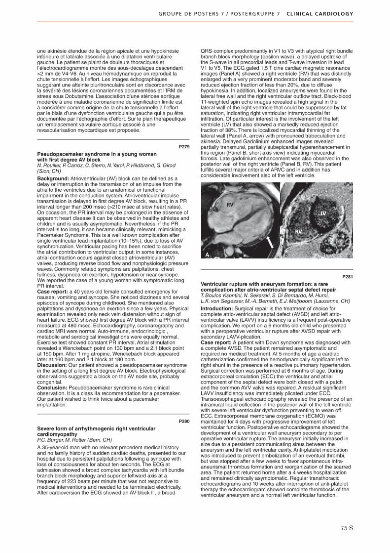
GROUPE DE POSTERS 7 / POSTERGRUPPE 7 CLINICAL CARDIOLOGY
75 S
une akinésie étendue de la région apicale et une hypokinésieinférieure et latérale associée à une dilatation ventriculairegauche. Le patient se plaint de douleurs thoraciques etl’électrocardiogramme montre des sous-décalages descendant>2 mm de V4-V6. Au niveau hémodynamique on reproduit lachute tensionnelle à l’effort. Les images échographiquessuggérant une atteinte pluritronculaire sont en discordance avecla sévérité des lésions coronariennes documentées et l’IRM destress sous Dobutamine. L’association d’une sténose aortiquemodérée à une maladie coronarienne de signification limite està considérer comme origine de la chute tensionnelle à l’effortpar le biais d’une dysfonction ventriculaire gauche qui a pu êtredocumentée par l’échographie d’effort. Sur le plan thérapeutiqueun remplacement valvulaire aortique associé à unerevascularisation myocardique est proposée.
P279
Pseudopacemaker syndrome in a young womanwith first degree AV blockN.Rouiller, P.Carroz,C.Sierro,N.Yarol, P.Hildbrand,G.Girod(Sion,CH)Background: Atrioventricular (AV) block can be defined as adelay or interruption in the transmission of an impulse from theatria to the ventricles due to an anatomical or functionalimpairment in the conduction system.Atrioventricular impulsetransmission is delayed in first degree AV block, resulting in a PRinterval longer than 200 msec (>210 msec at slow heart rates).On occasion, the PR interval may be prolonged in the absence ofapparent heart disease It can be observed in healthy athletes andchildren and is usually asymptomatic. Nevertheless, if the PRinterval is too long, it can became clinically relevant, mimicking aPacemaker Syndrome. This is a well known complication aftersingle ventricular lead implantation (10–15%), due to loss of AVsynchronization. Ventricular pacing has been noted to sacrificethe atrial contribution to ventricular output; in some instances,atrial contraction occurs against closed atrioventricular (AV)valves, producing reverse blood flow and nonphysiologic pressurewaves. Commonly related symptoms are palpitations, chestfullness, dyspnoea on exertion, hypotension or near syncope.We reported the case of a young woman with symptomatic longPR interval.Case report: a 40 years old female consulted emergency fornausea, vomiting and syncope. She noticed dizziness and severalepisodes of syncope during childhood. She mentioned alsopalpitations and dyspnoea on exertion since a few years. Physicalexamination revealed only neck vein distension without sign ofheart failure. ECG showed first degree AV block with a PR intervalmeasured at 480 msec. Echocardiography, coronarography andcardiac MRI were normal. Auto-immune, endocrinologic ,metabolic and serological investigations were equally normal.Exercise test showed constant PR interval. Atrial stimulationrevealed a Wenckebach point on 130 bpm and a 2:1 block pointat 150 bpm.After 1 mg atropine, Wenckebach block appearedlater at 160 bpm and 2:1 block at 180 bpm.Discussion: Our patient showed a pseudopacemaker syndromein the setting of a long first degree AV block. Electrophysiologicalobservations suggested a block on the AV node site, probablycongenital.Conclusion: Pseudopacemaker syndrome is rare clinicalobservation. It is a class IIa recommendation for a pacemaker.Our patient wished to think twice about a pacemakerimplantation.
P280
Severe form of arrhythmogenic right ventricularcardiomyopathyP.C.Burger,M.Rotter (Bern,CH)A 35-year-old man with no relevant precedent medical historyand no family history of sudden cardiac deaths, presented to ourhospital due to persistent palpitations following a syncope withloss of consciousness for about ten seconds. The ECG atadmission showed a broad complex tachycardia with left bundlebranch block morphology and superior leftward axis at afrequency of 223 beats per minute that was not responsive tomedical interventions and needed to be terminated electrically.After cardioversion the ECG showed an AV-block I°, a broad
QRS-complex predominantly in V1 to V3 with atypical right bundlebranch block morphology (epsilon wave), a delayed upstroke ofthe S-wave in all precordial leads and T-wave inversion in leadV1 to V5. The ECG gated 1,5 T cine cardiac magnetic resonanceimages (Panel A) showed a right ventricle (RV) that was distinctlyenlarged with a very prominent moderator band and severelyreduced ejection fraction of less than 20%, due to diffusehypokinesia. In addition, localized aneurysms were found in thelateral free wall and the right ventricular outflow tract. Black-bloodT1-weighted spin echo images revealed a high signal in thelateral wall of the right ventricle that could be suppressed by fatsaturation, indicating right ventricular intramyocardial fatinfiltration. Of particular interest is the involvement of the leftventricle (LV) that also showed a markedly reduced ejectionfraction of 38%. There is localized myocardial thinning of thelateral wall (Panel A, arrow) with pronounced trabeculation andakinesia. Delayed Gadolinium enhanced images revealedpartially transmural, partially subepicardial hyperenhancement inthis region (Panel B, short axis view) indicating myocardialfibrosis. Late gadolinium enhancement was also observed in theposterior wall of the right ventricle (Panel B, RV). This patientfulfills several major criteria of ARVC and in addition hasconsiderable involvement also of the left ventricle.
P281
Ventricular rupture with aneurysm formation: a rarecomplication after atrio-ventricular septal defect repairT.Boulos Ksontini,N.Sekarski, S.Di Bernardo,M.Hurni,L.K. von Segesser,M.-A.Bernath, E.J.Meijboom (Lausanne,CH)Introduction: Surgical repair is the treatment of choice forcomplete atrio-ventricular septal defect (AVSD) and left atrio-ventricular valve (LAVV) insufficiency is a frequent post-operativecomplication.We report on a 6 months old child who presentedwith a peroperative ventricular rupture after AVSD repair withsecondary LAVV-plication.Case report: A patient with Down syndrome was diagnosed witha complete AVSD. The patient remained asymptomatic andrequired no medical treatment. At 5 months of age a cardiaccatheterization confirmed the hemodynamically significant left toright shunt in the presence of a reactive pulmonary hypertension.Surgical correction was performed at 6 months of age. Duringextracorporeal circulation (ECC) the ventricular and atrialcomponent of the septal defect were both closed with a patchand the common AVV valve was repaired. A residual significantLAVV insufficiency was immediately plicated under ECC.Transoesophageal echocardiography revealed the presence of anintramural liquid collection in the posterior wall of the left ventriclewith severe left ventricular dysfunction preventing to wean offECC. Extracorporeal membrane oxygenation (ECMO) wasmaintained for 4 days with progressive improvement of leftventricular function. Postoperative echocardiograms showed thedevelopment of a ventricular wall aneurysm secondary to peroperative ventricular rupture. The aneurysm initially increased insize due to a persistent communicating sinus between theaneurysm and the left ventricular cavity. Anti-platelet medicationwas introduced to prevent embolization of an eventual thrombi,but was stopped after a few weeks to favor spontaneous intra-aneurismal thrombus formation and reorganization of the scarredarea. The patient returned home after a 4 weeks hospitalizationand remained clinically asymptomatic. Regular transthoracicechocardiograms and 10 weeks after interruption of anti-platelettherapy the echocardiogram showed complete thrombosis of theventricular aneurysm and a normal left ventricular function.
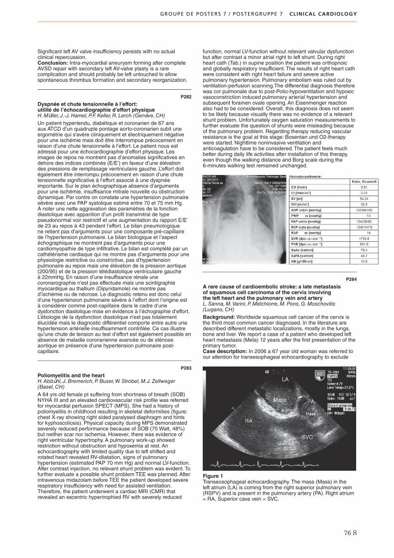
GROUPE DE POSTERS 7 / POSTERGRUPPE 7 CLINICAL CARDIOLOGY
76 S
Significant left AV valve insufficiency persists with no actualclinical repercussion.Conclusion: Intra-myocardial aneurysm forming after completeAVSD repair with secondary left AV-valve plasty is a rarecomplication and should probably be left untouched to allowspontaneous thrombus formation and secondary reorganization.
P282
Dyspnée et chute tensionnelle à l’effort:utilité de l’échocardiographie d’effort physiqueH.Müller, J.-J.Hamel, P.F.Keller,R. Lerch (Genève,CH)Un patient hypertendu, diabétique et coronarien de 67 ansaux ATCD d’un quadruple pontage aorto-coronarien subit uneergométrie qui s’avère cliniquement et électriquement négativepour une ischémie mais doit être interrompue précocement enraison d’une chute tensionnelle à l’effort. Le patient nous estadressé pour une échocardiographie d’effort physique. Lesimages de repos ne montrent pas d’anomalies significatives endehors des indices combinés (E/E’) en faveur d’une élévationdes pressions de remplissage ventriculaire gauche. L’effort doitégalement être interrompu précocement en raison d’une chutetensionnelle significative à l’effort associé à une dyspnéeimportante. Sur le plan échographique absence d’argumentspour une ischémie, insuffisance mitrale nouvelle ou obstructiondynamique. Par contre on constate une hypertension pulmonairesévère avec une PAP systolique estimé entre 70 et 75 mm Hg.A noter une nette aggravation des paramètres de la fonctiondiastolique avec apparition d’un profil transmitral de typepseudonormal voir restrictif et une augmentation du rapport E/E’de 23 au repos à 43 pendant l’effort. Le bilan pneumologiquene retient pas d’arguments pour une composante pré-capillairede l’hypertension pulmonaire. Le bilan biologique et l’aspectéchographique ne montrent pas d’arguments pour unecardiomyopathie de type infiltrative. Le bilan est complété par uncathétérisme cardiaque qui ne montre pas d’arguments pour unephysiologie restrictive ou constrictive, pas d’hypertensionpulmonaire au repos mais une élévation de la pression aortique(200/95) et de la pression télédiastolique ventriculaire gaucheà 22mmHg. En raison d’une insuffisance rénale unecoronarographie n’est pas effectuée mais une scintigraphiemyocardique au thallium (Dipyridamole) ne montre pasd’ischémie ou de nécrose. Le diagnostic retenu est donc celuid’une hypertension pulmonaire sévère à l’effort dont l’origine està considérer comme post-capillaire dans le cadre d’unedysfonction diastolique mise en évidence à l’échographie d’effort.L’étiologie de la dysfonction diastolique n’est pas totalementélucidée mais le diagnostic différentiel comporte entre autre unehypertension artérielle insuffisamment contrôlée. Ce cas illustrequ’une chute de tension au test d’effort est également possible enabsence de maladie coronarienne avancée ou de sténoseaortique en présence d’une hypertension pulmonaire post-capillaire.
P283
Poliomyelitis and the heartH.Abbühl, J.Bremerich,P.Buser,W.Strobel,M.J. Zellweger(Basel,CH)A 64 yrs old female pt suffering from shortness of breath (SOB)NYHA III and an elevated cardiovascular risk profile was referredfor myocardial perfusion SPECT (MPS). She had a history ofpoliomyelitis in childhood resulting in skeletal deformities (figure:chest X-ray showing right sided paralysed diaphragm and hintsfor kyphoscoliosis). Physical capacity during MPS demonstratedseverely reduced performance because of SOB (75 Watt, 48%)but neither scar nor ischemia. However, there was evidence ofright ventricular hypertrophy. A pulmonary work-up showedrestriction without obstruction and hypoxemia at rest. Anechocardiography with limited quality due to left shifted androtated heart revealed RV-dilatation, signs of pulmonaryhypertension (estimated PAP 70 mm Hg) and normal LV-function.After contrast injection, no relevant shunt problem was evident. Tofurther evaluate a possible shunt problem TEE was planned. Afterintravenous midazolam before TEE the patient developed severerespiratory insufficiency with need for assisted ventilation.Therefore, the patient underwent a cardiac MRI (CMR) thatrevealed an excentric hypertrophied RV with severely reduced
function, normal LV-function without relevant valvular dysfunctionbut after contrast a minor atrial right to left shunt. During rightheart cath (Tab.) in supine position the patient was orthopnoicand globally respiratory insufficient. The results of right heart cathwere consistent with right heart failure and severe activepulmonary hypertension. Pulmonary embolism was ruled out byventilation-perfusion scanning.The differential diagnosis thereforewas cor pulmonale due to post-Polio-hypoventilation and hypoxicvasoconstriction induced pulmonary arterial hypertension andsubsequent foramen ovale opening. An Eisenmenger reactionalso had to be considered. Overall, this diagnosis does not seemto be likely because visually there was no evidence of a relevantshunt problem. Unfortunately oxygen saturation measurements tofurther evaluate the question of shunts were misleading becauseof the pulmonary problem. Regarding therapy reducing vascularresistance is the goal at this stage: Bosentan und O2-therapywere started. Nighttime noninvasive ventilation andanticoagulation have to be considered. The patient feels muchbetter during daily life activities after installation of this therapy,even though the walking distance and Borg scale during the6-minutes walking test remained unchanged.
P284
A rare cause of cardioembolic stroke: a late metastasisof squamous cell carcinoma of the cervix involvingthe left heart and the pulmonary vein and arteryL.Sanna,M.Varini, P.Melchiorre,M.Pons,G.Moschovitis(Lugano,CH)Background:Worldwide squamous cell cancer of the cervix isthe third most common cancer diagnosed. In the literature aredescribed different metastatic localizations, mostly in the lungs,bone and liver.We report a case of a patient who developed leftheart metastasis (Meta) 12 years after the first presentation of theprimary tumor.Case description: In 2006 a 67 year old woman was referred toour attention for transesophageal echocardiography to exclude
Figure 1Transeosophageal echocardiography. The mass (Mass) in theleft atrium (LA) is coming from the right superior pulmonary vein(RSPV) and is present in the pulmonary artery (PA). Right atrium= RA, Superior cava vein = SVC.
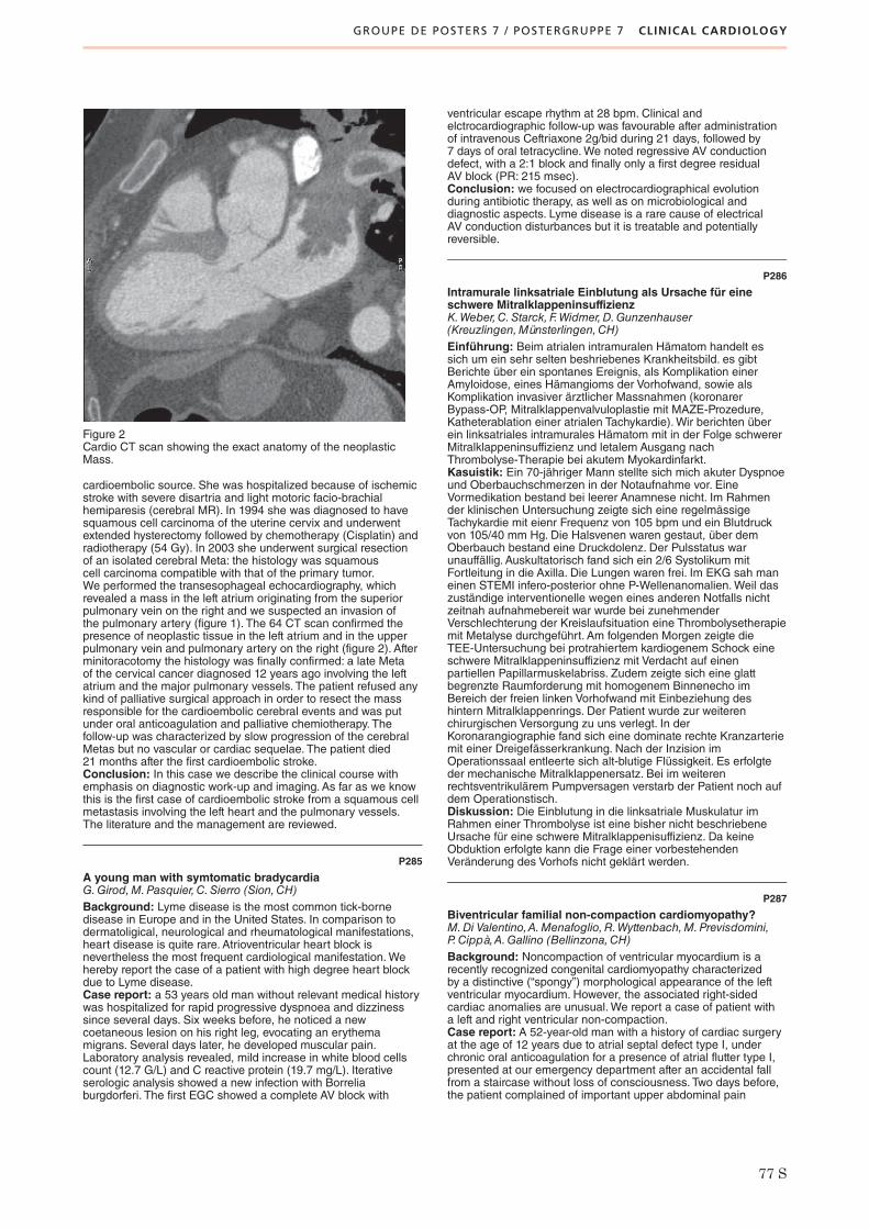
GROUPE DE POSTERS 7 / POSTERGRUPPE 7 CLINICAL CARDIOLOGY
77 S
cardioembolic source. She was hospitalized because of ischemicstroke with severe disartria and light motoric facio-brachialhemiparesis (cerebral MR). In 1994 she was diagnosed to havesquamous cell carcinoma of the uterine cervix and underwentextended hysterectomy followed by chemotherapy (Cisplatin) andradiotherapy (54 Gy). In 2003 she underwent surgical resectionof an isolated cerebral Meta: the histology was squamouscell carcinoma compatible with that of the primary tumor.We performed the transesophageal echocardiography, whichrevealed a mass in the left atrium originating from the superiorpulmonary vein on the right and we suspected an invasion ofthe pulmonary artery (figure 1). The 64 CT scan confirmed thepresence of neoplastic tissue in the left atrium and in the upperpulmonary vein and pulmonary artery on the right (figure 2). Afterminitoracotomy the histology was finally confirmed: a late Metaof the cervical cancer diagnosed 12 years ago involving the leftatrium and the major pulmonary vessels. The patient refused anykind of palliative surgical approach in order to resect the massresponsible for the cardioembolic cerebral events and was putunder oral anticoagulation and palliative chemiotherapy. Thefollow-up was characterized by slow progression of the cerebralMetas but no vascular or cardiac sequelae. The patient died21 months after the first cardioembolic stroke.Conclusion: In this case we describe the clinical course withemphasis on diagnostic work-up and imaging. As far as we knowthis is the first case of cardioembolic stroke from a squamous cellmetastasis involving the left heart and the pulmonary vessels.The literature and the management are reviewed.
P285
A young man with symtomatic bradycardiaG.Girod,M.Pasquier,C.Sierro (Sion,CH)Background: Lyme disease is the most common tick-bornedisease in Europe and in the United States. In comparison todermatoligical, neurological and rheumatological manifestations,heart disease is quite rare. Atrioventricular heart block isnevertheless the most frequent cardiological manifestation.Wehereby report the case of a patient with high degree heart blockdue to Lyme disease.Case report: a 53 years old man without relevant medical historywas hospitalized for rapid progressive dyspnoea and dizzinesssince several days. Six weeks before, he noticed a newcoetaneous lesion on his right leg, evocating an erythemamigrans. Several days later, he developed muscular pain.Laboratory analysis revealed, mild increase in white blood cellscount (12.7 G/L) and C reactive protein (19.7 mg/L). Iterativeserologic analysis showed a new infection with Borreliaburgdorferi. The first EGC showed a complete AV block with
ventricular escape rhythm at 28 bpm. Clinical andelctrocardiographic follow-up was favourable after administrationof intravenous Ceftriaxone 2g/bid during 21 days, followed by7 days of oral tetracycline.We noted regressive AV conductiondefect, with a 2:1 block and finally only a first degree residualAV block (PR: 215 msec).Conclusion: we focused on electrocardiographical evolutionduring antibiotic therapy, as well as on microbiological anddiagnostic aspects. Lyme disease is a rare cause of electricalAV conduction disturbances but it is treatable and potentiallyreversible.
P286
Intramurale linksatriale Einblutung als Ursache für eineschwere MitralklappeninsuffizienzK.Weber,C.Starck, F.Widmer,D.Gunzenhauser(Kreuzlingen,Münsterlingen,CH)Einführung: Beim atrialen intramuralen Hämatom handelt essich um ein sehr selten beshriebenes Krankheitsbild. es gibtBerichte über ein spontanes Ereignis, als Komplikation einerAmyloidose, eines Hämangioms der Vorhofwand, sowie alsKomplikation invasiver ärztlicher Massnahmen (koronarerBypass-OP, Mitralklappenvalvuloplastie mit MAZE-Prozedure,Katheterablation einer atrialen Tachykardie).Wir berichten überein linksatriales intramurales Hämatom mit in der Folge schwererMitralklappeninsuffizienz und letalem Ausgang nachThrombolyse-Therapie bei akutem Myokardinfarkt.Kasuistik: Ein 70-jähriger Mann stellte sich mich akuter Dyspnoeund Oberbauchschmerzen in der Notaufnahme vor. EineVormedikation bestand bei leerer Anamnese nicht. Im Rahmender klinischen Untersuchung zeigte sich eine regelmässigeTachykardie mit eienr Frequenz von 105 bpm und ein Blutdruckvon 105/40 mm Hg. Die Halsvenen waren gestaut, über demOberbauch bestand eine Druckdolenz. Der Pulsstatus warunauffällig. Auskultatorisch fand sich ein 2/6 Systolikum mitFortleitung in die Axilla. Die Lungen waren frei. Im EKG sah maneinen STEMI infero-posterior ohne P-Wellenanomalien.Weil daszuständige interventionelle wegen eines anderen Notfalls nichtzeitnah aufnahmebereit war wurde bei zunehmenderVerschlechterung der Kreislaufsituation eine Thrombolysetherapiemit Metalyse durchgeführt. Am folgenden Morgen zeigte dieTEE-Untersuchung bei protrahiertem kardiogenem Schock eineschwere Mitralklappeninsuffizienz mit Verdacht auf einenpartiellen Papillarmuskelabriss. Zudem zeigte sich eine glattbegrenzte Raumforderung mit homogenem Binnenecho imBereich der freien linken Vorhofwand mit Einbeziehung deshintern Mitralklappenrings. Der Patient wurde zur weiterenchirurgischen Versorgung zu uns verlegt. In derKoronarangiographie fand sich eine dominate rechte Kranzarteriemit einer Dreigefässerkrankung. Nach der Inzision imOperationssaal entleerte sich alt-blutige Flüssigkeit. Es erfolgteder mechanische Mitralklappenersatz. Bei im weiterenrechtsventrikulärem Pumpversagen verstarb der Patient noch aufdem Operationstisch.Diskussion: Die Einblutung in die linksatriale Muskulatur imRahmen einer Thrombolyse ist eine bisher nicht beschriebeneUrsache für eine schwere Mitralklappenisuffizienz. Da keineObduktion erfolgte kann die Frage einer vorbestehendenVeränderung des Vorhofs nicht geklärt werden.
P287
Biventricular familial non-compaction cardiomyopathy?M.Di Valentino,A.Menafoglio,R.Wyttenbach,M.Previsdomini,P.Cippà,A.Gallino (Bellinzona,CH)Background: Noncompaction of ventricular myocardium is arecently recognized congenital cardiomyopathy characterizedby a distinctive (“spongy”) morphological appearance of the leftventricular myocardium. However, the associated right-sidedcardiac anomalies are unusual.We report a case of patient witha left and right ventricular non-compaction.Case report: A 52-year-old man with a history of cardiac surgeryat the age of 12 years due to atrial septal defect type I, underchronic oral anticoagulation for a presence of atrial flutter type I,presented at our emergency department after an accidental fallfrom a staircase without loss of consciousness. Two days before,the patient complained of important upper abdominal pain
Figure 2Cardio CT scan showing the exact anatomy of the neoplasticMass.
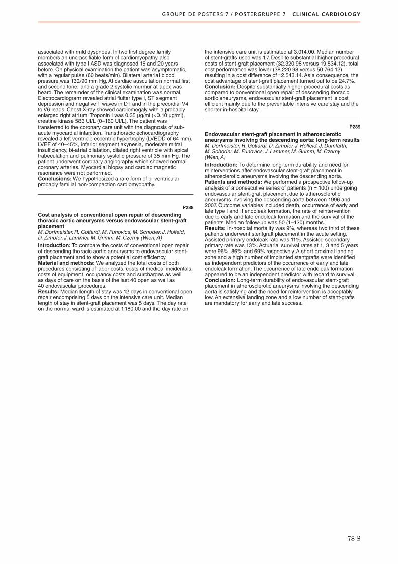
GROUPE DE POSTERS 7 / POSTERGRUPPE 7 CLINICAL CARDIOLOGY
78 S
associated with mild dyspnoea. In two first degree familymembers an unclassifiable form of cardiomyopathy alsoassociated with type I ASD was diagnosed 15 and 20 yearsbefore. On physical examination the patient was asymptomatic,with a regular pulse (60 beats/min). Bilateral arterial bloodpressure was 130/90 mm Hg.At cardiac auscultation normal firstand second tone, and a grade 2 systolic murmur at apex washeard. The remainder of the clinical examination was normal.Electrocardiogram revealed atrial flutter type I, ST segmentdepression and negative T waves in D I and in the precordial V4to V6 leads. Chest X-ray showed cardiomegaly with a probablyenlarged right atrium. Troponin I was 0.35 μg/ml (<0.10 μg/ml),creatine kinase 583 UI/L (0–160 UI/L). The patient wastransferred to the coronary care unit with the diagnosis of sub-acute myocardial infarction. Transthoracic echocardiographyrevealed a left ventricle eccentric hypertrophy (LVEDD of 64 mm),LVEF of 40–45%, inferior segment akynesia, moderate mitralinsufficiency, bi-atrial dilatation, dilated right ventricle with apicaltrabeculation and pulmonary systolic pressure of 35 mm Hg. Thepatient underwent coronary angiography which showed normalcoronary arteries. Myocardial biopsy and cardiac magneticresonance were not performed.Conclusions:We hypothesized a rare form of bi-ventricularprobably familial non-compaction cardiomyopathy.
P288
Cost analysis of conventional open repair of descendingthoracic aortic aneurysms versus endovascular stent-graftplacementM.Dorfmeister,R.Gottardi,M.Funovics,M.Schoder, J.Holfeld,D.Zimpfer, J. Lammer,M.Grimm,M.Czerny (Wien,A)Introduction: To compare the costs of conventional open repairof descending thoracic aortic aneurysms to endovascular stent-graft placement and to show a potential cost efficiency.Material and methods:We analyzed the total costs of bothprocedures consisting of labor costs, costs of medical incidentals,costs of equipment, occupancy costs and surcharges as wellas days of care on the basis of the last 40 open as well as40 endovascular procedures.Results: Median length of stay was 12 days in conventional openrepair encomprising 5 days on the intensive care unit. Medianlength of stay in stent-graft placement was 5 days. The day rateon the normal ward is estimated at 1.180.00 and the day rate on
the intensive care unit is estimated at 3.014.00. Median numberof stent-grafts used was 1.7. Despite substantial higher proceduralcosts of stent-graft placement (32.320.98 versus 19.534.12), totalcost performance was lower (38.220.98 versus 50.764.12)resulting in a cost difference of 12.543.14. As a consequence, thecost advantage of stent-graft placement turned out to be 24.7%.Conclusion: Despite substantially higher procedural costs ascompared to conventional open repair of descending thoracicaortic aneurysms, endovascular stent-graft placement is costefficient mainly due to the preventable intensive care stay and theshorter in-hospital stay.
P289
Endovascular stent-graft placement in atheroscleroticaneurysms involving the descending aorta: long-term resultsM.Dorfmeister,R.Gottardi,D. Zimpfer, J.Holfeld, J.Dumfarth,M.Schoder,M.Funovics, J. Lammer,M.Grimm,M.Czerny(Wien,A)Introduction: To determine long-term durability and need forreinterventions after endovascular stent-graft placement inatherosclerotic aneurysms involving the descending aorta.Patients and methods:We performed a prospective follow-upanalysis of a consecutive series of patients (n = 100) undergoingendovascular stent-graft placement due to atheroscleroticaneurysms involving the descending aorta between 1996 and2007. Outcome variables included death, occurrence of early andlate type I and II endoleak formation, the rate of reinterventiondue to early and late endoleak formation and the survival of thepatients. Median follow-up was 50 (1–120) months.Results: In-hospital mortality was 9%, whereas two third of thesepatients underwent stentgraft placement in the acute setting.Assisted primary endoleak rate was 11%.Assisted secondaryprimary rate was 13%.Actuarial survival rates at 1, 3 and 5 yearswere 96%, 86% and 69% respectively. A short proximal landingzone and a high number of implanted stentgrafts were identifiedas independent predictors of the occurrence of early and lateendoleak formation. The occurrence of late endoleak formationappeared to be an independent predictor with regard to survival.Conclusion: Long-term durability of endovascular stent-graftplacement in atherosclerotic aneurysms involving the descendingaorta is satisfying and the need for reintervention is acceptablylow. An extensive landing zone and a low number of stent-graftsare mandatory for early and late success.

LISTE DES PREMIERS AUTEURS / ERSTAUTORENVERZEICHNIS
79 S
Abbühl H 56 S, 76 SAltmann D 39 SAttenhofer Jost CH 20 S
Bernheim AM 33 SBesler C 10 SBieri M 45 SBinder RK 57 SBoll S 52 SBoulos Ksontini T 75 SBreitenstein A 42 S, 44 SBrenner R 72 SBrunner-La Rocca HP 30 SBucher UC 25 SBühler K 41 SBull S 48 SBurger PC 75 SBurkamp A 23 SBurkard T 4 S, 55 SBurri H 14 SBussell H 51 S
Camici GG 42 SChevallier S 40 SChiusa M 45 SConen D 6 S, 41 SConti L 49 SCook S 11 S, 19 S
D’Angeli I 61 Sde Marchi SF 28 SDe Pasquale Meyer G 6 SDegueldre S 69 SDi Bernardo S 21 SDi Valentino M 5 S, 29 S, 77SDiamond R 33 SDiefenbacher W 52 SDimitrakis P 38 SDorfmeister M 78 SDörries C 27 SDziodzio T 23 S, 35 S
Ehl NF 22 SEshtehardi P 11 S
Fäh-Gunz A 34 SFarpour-Lambert NJ 65 SFerrari E 13 S, 17 S
Fischer JB 6 SFrangos C 62 SFriedli B 64 S
Giannotti G 26 SGirod G 60 S, 77 SGloekler S 43 S, 61 SGoeber V 23 SGrapow M 47 SGutmann M 69 S
Häberlin A 57 SHälg C 7 SHaller S 34 SHandschin R 32 SHäuselmann SP 37 SHellige G 16 SHoly EW 9 SHurni M 20 S
Isidoro Tavares N 38 S
Jaggi S 64 SJeger R 18 SJousset F 52 S
Kadner A 36 SKaiser C 60 SKania G 27 SKatz E 50 S, 51 SKaufmann BA 32 SKaufmann C 65 SKoestner SC 68 S, 71 SKretschmar O 68 SKühne M 14 S, 39 S, 40 SKülling B 20 SKuroedov A 9 SKurz DJ 46 S
Leibundgut G 28 S, 51 S,61 S
Locca D 12 S, 55 S, 62 SLoup O 36 S
Maggio A 64 SMartin A 69 SMeili-Butz S 30 S, 53 SMilano G 47 S
Monney P 48 SMontecucco F 46 SMontessuit C 43 SMorel S 41 SMüller H 44 S, 74 S, 76 SMutschelknauss M 50 SMuzzarelli S 54 S, 56 SMyers PO 20 S, 71 S
Nkoulou R 67 S, 70 SNoble S 5 S, 17 S, 22 S,
66 S
Oroszlan M 45 SOxenius A 72 S
Panos A 58 SPascale P 29 SPeyrou J 74 SPfenniger A 63 SPfister O 12 S, 27 S, 42 SPilgrim T 17 SPlaisance I 43 SPlass A 37 SPotocki M 54 SPraz F 59 S, 60 S
Räber L 4 S, 19 SReichlin T 11 S, 25 S, 62 S,
70 SReineke D 35 S, 36 SRickli H 12 SRimoldi SF 24 S, 33 SRoehrich ME 26 SRohner A 5 S, 34 SRosc-Schlüter BI 38 SRoten L 15 SRotter M 48 SRouiller W 75 SRozenberg I 8 S, 9 S, 10 SRutz T 56 S
Samara E 25 SSanna L 76 SSchaer B 8 S, 13 S, 14 S,
15 S, 49 SSchmied C 73 SSchoenenberger AW 12 S
Schwerzmann M 28 SSeiler J 39 S, 40 SSenouf D 51 S, 66 SShi Y 45 SSpicher A 26 SStalder N 7 SSticherling C 14 SStolt Steiger V 22 SStuber T 24 SStuder M 30 SSürder D 46 SSyburra T 59 SSztajzel J 53 S, 72 S
Testuz A 74 STissot C 21 S, 29 S, 66 S,
68 STomaske M 69 S, 71 STorre TM 58 STozzi P 59 STran N 53 STrigo Trindade P 31 S
Valaperti A 41 SValenta I 67 SVernet D 31 SVincenti G 7 S, 67 SVoser E 67 SVurma M 60 S
Wachter M 65 SWalpoth BH 19 S, 58 SWeber K 62 S, 73 S, 77 SWenaweser P 16 SWindecker S 18 SWinkler B 37
Zbinden S 8 SZurek M 63 S
Die Ziffern beziehen sich auf die Seitenzahlen des Supplementums
Supplementum 17 der Zeitschrift «Kardiovaskuläre Medizin»© 2009 by EMH Schweizerischer Ärzteverlag AG, Basel

![N EU OFEN-MANUFAKTUR HESS UG SPECKSTEIN- & KAMINÖFEN … · SIM 1/3/Varia 2R-55h-4S. Höhe [Gh] = 1712 mm Gewicht = 520 kg. SIM 1/2/Mini 2LRh-4S. Höhe [Gh] = 1437 mm Gewicht = 420](https://static.fdokument.com/doc/165x107/5fd4f537e1f0a0252e7665f9/n-eu-ofen-manufaktur-hess-ug-speckstein-kaminfen-sim-13varia-2r-55h-4s.jpg)