Lehrstuhl für Technische Mikrobiologie - mediaTUM · Lehrstuhl für Technische Mikrobiologie...
145
Lehrstuhl für Technische Mikrobiologie CHARACTERIZATION OF THE MICROBIAL ECOSYSTEM OF CEREAL FERMENTATIONS USING MOLECULAR BIOLOGICAL METHODS Martin R. A. Müller Vollständiger Abdruck der von der Fakultät für Brauwesen, Lebensmitteltechnologie und Milchwissenschaft der Technischen Universität München zur Erlangung des akademischen Grades eines Doktor-Ingenieurs genehmigten Dissertation. Vorsitzender: Univ.-Prof. Dr. rer. nat. H. Klostermeyer Prüfer der Dissertation: 1. Univ.-Prof. Dr. rer. nat. habil. R. F. Vogel 2. Univ. Prof. Dr. rer. nat. W. P. Hammes, Univ. Hohenheim 3. Univ.-Prof. Dr.-Ing. habil. W. Back Die Dissertation wurde am 04.04.2000 bei der Technischen Universität München eingereicht und durch die Fakultät für Brauwesen, Lebensmitteltechnologie und Milchwissenschaft am 26.06.2000 angenommen.
Transcript of Lehrstuhl für Technische Mikrobiologie - mediaTUM · Lehrstuhl für Technische Mikrobiologie...

Lehrstuhl für Technische Mikrobiologie
CHARACTERIZATION OF THE MICROBIAL ECOSYSTEM OF CEREAL FERMENTATIONS USING MOLECULAR BIOLOGICAL METHODS
Martin R. A. Müller
Vollständiger Abdruck der von der Fakultät für Brauwesen, Lebensmitteltechnologie und Milchwissenschaft der Technischen Universität München zur Erlangung des akademischen
Grades eines
Doktor-Ingenieurs
genehmigten Dissertation.
Vorsitzender: Univ.-Prof. Dr. rer. nat. H. Klostermeyer
Prüfer der Dissertation:
1. Univ.-Prof. Dr. rer. nat. habil. R. F. Vogel
2. Univ. Prof. Dr. rer. nat. W. P. Hammes, Univ. Hohenheim
3. Univ.-Prof. Dr.-Ing. habil. W. Back
Die Dissertation wurde am 04.04.2000 bei der Technischen Universität München eingereicht
und durch die Fakultät für Brauwesen, Lebensmitteltechnologie und Milchwissenschaft am
26.06.2000 angenommen.
TABLE OF CONTENTS
i
TABLE OF CONTENTS
General Introduction____________________________________________ 1
Microflora of Cereal Fermentations ______________________________ 2
Taxonomy of Lactic Acid Bacteria _______________________________ 5
Identification of Lactic Acid Bacteria ____________________________ 11 The Ribosomal Genes _____________________________________ 11 DNA-Based Typing _______________________________________ 14 DNA-DNA Hybridization___________________________________ 19 Chemotaxonomic Methods _________________________________ 20 Whole Cell Protein Patterns________________________________ 20
The Potential of Fermented Cereal-Based Products as Probiotics (Symbiotics) _______________________________________________ 21
Motivations and Objectives of the Study___________________________ 23
References ___________________________________________________ 24
Chapter I Monitoring the Growth of Lactobacillus species During a Rye Flour Fermentation ______________________________________________ (37-62) Chapter II Multiplex PCR for the Detection of Lactobacillus pontis and Two Related Species in a Sourdough Fermentation ___________________________ (63-80) Chapter III Lactobacillus frumentis sp. nov., a New Lactic Acid Bacterium Isolated from Rye Bran Fermentations with a Long Fermentation Period__________ (81-104)

TABLE OF CONTENTS
ii
Chapter IV Polyphasic Identification of Wild Yeast Strains from Greek Sourdoughs _____________________________________________ (105-129) Additional Insights and Conclusions ________________________ (130-136)
ZUSAMMENFASSUNG
iii
ZUSAMMENFASSUNG
CHARAKTERISIERUNG DES MIKROBIELLEN ÖKOSYSTEMS VON GETREIDEFERMENTATIONEN MIT HILFE VON MOLEKULARBIOLOGISCHEN METHODEN Milchsäurebakterien (MSB) stellen den Hauptteil der mikrobiellen Flora bei der
Fermentation pflanzlicher und tierischer Lebensmittel dar. Bei Getreidefermentationen
ist der Gattung Lactobacillus eine tragende Rolle beizumessen.
Sauerteigfermentationen lassen sich drei Typen (Typ I-III) zuordnen, die hauptsächlich
die Art der Führung mit zahlreichen internen und externen Parametern widerspiegeln.
Diese wiederum haben entscheidenden Einfluß auf die Zusammensetzung der
Mikroflora. Typ I Teige sind Sauerteig-Starterpräparate, die fortlaufend oft über
mehrere Jahre hinweg geführt werden. Durch wiederholtes Anfrischen werden die
Organismen in einem stoffwechselaktiven Zustand gehalten. Die Hauptflora von Typ I
Teigen besteht aus Lactobacillus sanfranciscensis, der meist mit der Hefe Candida
milleri vergesellschaftet ist. Typ II Teige werden über einen längeren Zeitraum von bis
zu fünf Tagen bei höheren Temperaturen (40°C) und Teigausbeuten geführt. Dies
wiederum führt zu einer typischen Mikroflora, die häufig aus L. pontis, L. panis, L.
reuteri, L. fermentum und homofermentativen Stämmen von L. amylovorus bestehen
kann. Typ III Teige sind getrocknete, pulverförmige Sauerteige mit physiologisch
aktiven Organismen, wie L. plantarum, L. brevis oder Pediococcus pentosaceus, die
sich durch ihre Trocknungsresistenz auszeichnen.
Die klassische Identifizierung von MSB beruht auf einer Erfassung
phänotypischer Merkmale, die dann zur Charakterisierung herangezogen werden.
Standardmerkmale sind die Fähigkeit der Organismen bestimmte Kohlenhydrate zu
verwerten, Substrate zu spalten oder ihr makro-, bzw. ihr mikroskopisches
Erscheinungsbild. Gerade bei stark an ein Milieu angepaßten Organismen, wie es die
MSB aus Sauerteig sind, führt diese Vorgehensweise nicht immer zu eindeutigen

ZUSAMMENFASSUNG
iv
Ergebnissen. Dazu kommt, dass Sauerteig-MSB eben durch ihre Anpassung an ihr
Milieu auf Labormedien oft schwer kultivierbar sind, was eine schnelle
Routineidentifizierung erschwert.
Um diese Problematik zu umgehen, wurden in dieser Arbeit
Identifizierungsmethoden entwickelt, die auf den Genotyp von MSB gerichtet sind.
Diese haben Nukleinsäuren als Zielmoleküle, wobei hier die 16S rRNA eine tragende
Rolle spielt. Für einen schnellen Nachweis von Sauerteig-Laktobazillen wurde
beruhend auf vorhandenen und selbst erzeugten 16S rRNA Daten ein PCR-Nachweis
entwickelt, der eine schnelle und zuverlässige Identifizierung von L. pontis, L. panis
und der in dieser Arbeit neu beschriebenen Spezies L. frumenti sowie eine
Differenzierung von phylogenetisch verwandten Spezies und anderen Sauerteig-MSB
erlaubt. Darüber hinaus wurde das PCR-System mit einer DNA-Extraktion direkt aus
dem Sauerteig kombiniert, was einen noch schnelleren Nachweis ohne
Kultivierungsschritt ermöglicht. Die Daten über das Vorhandensein von L. pontis und
L. frumenti in einer Modellfermentation stimmen mit alternativen Untersuchungen
überein. Die PCR-Nachweismethoden können als schnelles Identifizierungswerkzeug,
sowie als zur Kontrolle und Analyse von Fermentationen eingesetzt werden.
Eine weitere PCR gestützte Technik, die in dieser Arbeit entwickelt wurde, ist
die Erzeugung von "DNA-Fingerabdrücken" mittels RAPD-PCR. Sie spiegelt typische
Polymorphismen auf der DNA der untersuchten Organismen wider. Diese Methode
wurde angewandt, um das Verständnis zur mikrobiellen Ökologie in
Sauerteigfermentationen zu verbessern. Dazu wurde die Flora industrieller Typ II
Fermentationen untersucht, indem die Isolate aus unabhängigen Fermentationen aus
einem längeren Zeitraum mittels RAPD-PCR zu Genotypen gruppiert wurden. Es
konnte gezeigt werden, dass die Flora solcher unter nicht sterilen Bedingungen
geführten Fermentationen sehr stabil ist. Die dominante Flora besteht aus 70% L.
amylovorus und 30% L. pontis, L. frumenti und selten L. reuteri. Durch die
Etablierung einer dem industriellen Prozess nachempfundenen Laborfermentation
konnten wichtige Erkenntnisse zur Dynamik, sowie zum Verhalten bei Variation von

ZUSAMMENFASSUNG
v
Prozessparametern, erhalten werden. Die Laborfermentation erwies sich als ähnlich
stabil und von ihrer Florenzusammensetzung als vergleichbar zum industriellen
Vorbild. Die prozentuale Zusammensetzung verschob sich mit zunehmender
Fermentationszeit zur heterofermentativen Spezies L. frumenti, was durch
physiologische Untersuchungen bestätigt wurde. Temperaturänderungen zwischen
34°C, 40°C und 46°C führten zu keiner nennenswerten Florenverschiebung, es konnte
aber eine Temperatur für optimales Wachstum und Säureproduktion von 40°C
bestimmt werden.
RAPD-PCR erwies sich neben der Charakterisierung von MSB auch für Hefen
aus Sauerteigen geeignet. Dazu wurden aus traditionellen griechischen Sauerteigen 45
Hefestämme mit RAPD-PCR typisiert. Alle Isolate konnten drei Clustern zugeordnet
werden. Durch Einbeziehung von Referenzstämmen konnten diese als Saccharomyces
cerevisiae, Yarrowia lipolytica und Pichia membranaefaciens identifiziert werden.
Diese Ergebnisse konnten durch alternative Ansätze, wie FT-IR-Spektroskopie,
SDS-PAGE und physiologische Charakterisierung untermauert werden. Während S.
cerevisiae und P. membranaefaciens immer wieder in Sauerteigen angetroffen werden
können, ist Y. lipolytica zuvor noch nicht in Sauerteigen beschrieben worden.
16S rDNA Sequenzierung und vergleichende Sequenzanalyse erwies sich als die
zuverlässigste Methode zur Identifizierung von neuen Isolaten aus Sauerteigen.
Zahlreiche Stämme aus einer Typ II Fermentation konnten nach der Einberechnung in
einen phylogenetischen Baum keiner bekannten Spezies eindeutig zugeordnet werden.
Die nächsten Nachbarn sind L. vaginalis, L. oris, L. pontis, L. panis und L. reuteri.
Weitere Untersuchungen zu physiologischen Eigenschaften, chemotaxonomischen
Merkmalen, sowie G + C-Gehalt und DNA-DNA Homologie, ergaben, dass es sich
um eine eigenständige Spezies handelt, für die der Name L. frumenti vorgeschlagen
wurde.
vi
ACKNOWLEGEMENTS
Ich möchte mich ganz besonders bei Prof. Dr. Rudi F. Vogel bedanken, der mir die Möglichkeit gegeben hat an seinem Lehrstuhl die vorliegende Arbeit anzufertigen. Bedanken möchte ich mich außerdem für die Begeisterung, die er dem Thema Sauerteig und somit meiner Arbeit entgegenbrachte und dieser Funke auch auf mich übersprang und mich ständig neu motivierte. Bedanken möchte ich mich für die große Freiheit und das Vertrauen. Es gibt sicher nicht viele Lehrstühle, die wissenschaftliches Arbeiten mit Kollegialität, Freundschaft und angenehmer Atmosphäre so vereinen und die Arbeit somit zum Vergnügen macht. Weiterhin möchte ich mich bei meinem Betreuer Dr. Matthias Ehrmann für seine große Unterstützung bei der Anfertigung dieser Arbeit danken, die ständige Bereitschaft zur Diskussion und die unendliche Geduld bei der Aufgabe „Ingenieure“ in die tieferen Weihen der Molekularbiologie einzuführen. Bedanken möchte ich mich bei den CTAs des Lehrstuhles ohne die der Wissenschaftsbetrieb am Lehrstuhl nicht möglich wäre. So möchte ich mich insbesondere bei Frau Monika Hadek bedanken, ihrer Hilsbereitschaft, insbesondere bei der Isolierung „tausender“ DNAs und der Stammhalt und immer neuer Isolate aus dem Sauerteig. Meine Arbeit wäre ohne die Firma Ernst Böcker GmbH&Co.KG sicher so nicht möglich geworden. Deshalb möchte ich ein großes Dankeschön an Dr. Georg Böcker aussprechen, der es mir ermöglichte in seiner Firma wissenschaftlich zu arbeiten. Danke natürlich auch für die finanzielle Unterstützung über all die Jahre. Natürlich möchte ich mich bei allen Mitarbeitern bedanken insbesondere Herrn Oliver Luft, der mich in die Geheimnisse der Sauerteig-Mikrobiologie einweihte. Ganz besonders möchte ich mich bei Herrn Dr. Peter Stolz bedanken, der meine Arbeit von Seiten der Industrie betreute, der ständig ein offenes Ohr für mich hatte und somit die Industriekooperation erst möglich machte. Daneben, und nicht weniger wichtig, sind die zahlreichen privaten Aktionen, die wir auf die Beine gestellt haben. Ich möchte die gemeinsame Mountain-Bike-Action nicht missen und hoffe auch zukünftig darauf. Was wäre ein Arbeitsalltag ohne Kollegen. Die meisten sind zu Freunden geworden. Ich bedanke mich ganz herzlich bei ihnen für die schöne Zeit innerhalb und außerhalb der TMW.
BIBLIOGRAPHY
vii
BIBLIOGRAPHY POSTER PRESENTATIONS Müller, M., Ehrmann, M. & Vogel, R. F. (1996). Identification and behaviour of
lactobacilli in the production of natural bread-souring agents. 5th Symposium on
Lactic Acid Bacteria in Veldhoven, Netherlands.
Müller, M. R. A., Ehrmann, M. A., Stolz, P., Böcker, G. & Vogel, R. F. (1997).
Identifizierung von Lactobacillen in Getreidefermentationen. 48.
Tagung für Getreidechemie in Detmold, Germany.
Müller, M. R. A., Rouvet, M., Brassart, D., Böcker, G., Ehrmann, M. A. & Vogel,
R. F. (1998).Adhesion of Lactobacillus strains from cereal fermentations to
human intestinal cells. Conference of 'Functional foods: designer food for the
future' in Cork, Ireland, published in: Int Dairy Journal, 8 (5/6). 584.
Müller, M. R. A., Ehrmann, M. A. & Vogel, R. F. (1999). Monitoring the growth of
Lactobacillus species during a rye bran fermentation. ICC Conference, Valencia,
Spain.
Leissner, C. E. W., Müller, M. R. A., Niessen, L., Ehrmann, M. A. & Vogel, R. F.
(2000).Use of the AFLP fingerprinting method for the differentiation and
identification of lactic acid bacteria. Congress of 'Microbiology 2000' in Munich,
Germany, published in: Biospektrum Sonderausgabe, 155.
Müller, M. R. A., Ehrmann, M. A. & Vogel, R. F. (2000). Lactobacillus cerealis sp.
nov. a new lactic acid bacterium isolated from rye bran fermentations with a long
fermentation period. Congress of 'Microbiology 2000' in Munich, Germany,
published in: Biospektrum Sonderausgabe, 154.

BIBLIOGRAPHY
viii
PAPERS Vogel, R. F., Müller, M., Stolz, P. & Ehrmann, M. (1996). Ecology in sourdoughs
produced by traditional and modern technologies. Adv Food Sci (CMTL), 18
(5/6), 152-159.
Vogel, R. F., Knorr, R., Müller, M. R. A., Steudel, U., Gänzle, M. & Ehrmann, M.
A. (1999). Non dairy lactic fermentations: the cereal world. Antonie van
Leeuwenhoek, 76, 403-411.
Müller, M. R. A., Ehrmann, M. A. & Vogel, R. F. (2000). Multiplex PCR for the
detection of Lactobacillus pontis and two related species in a sourdough
fermentation. Appl Environ Microbiol, 66 (5), 2113-2116.
Paramithiotis, S., Müller, M. R. A., Ehrmann, M. A., Tsakalidou, E., Seiler, H.,
Vogel, R. F. & Kalantzopoulos, G. (2000). Polyphasic identification of wild
yeast strains isolated from Greek sourdoughs. System Appl Microbiol., 23, 156-
164.
Müller, M. R. A., Wolfrum, G., Stolz, P., Ehrmann, M. A. & Vogel, R. F. (2001).
Monitoring the growth of Lactobacillus species during a rye flour fermentation.
Food Microbiology 18, 000-000.
Müller, M. R. A., Ehrmann, M. A. & Vogel, R. F. (2000). Lactobacillus frumenti sp.
nov., a new lactic acid bacterium isolated from rye bran fermentations with a
long fermentation period. Int J Syst Evol Microbiol, 50, 2127- 2133.

GENERAL INTRODUCTION
1
GENERAL INTRODUCTION
In the beginning of mankind the pattern of living was mainly embossed by a
nomadic way. Nutrition was ensured by collecting plant material and hunting of
animals just in time. With the commencement of settling down, and the introduction of
agriculture men reached independence. Perishable raw material had to be processed to
obtain tenably food. Apart from cooking and drying and before the use of
preservatives like salt or smoke, at least 3000 years ago the fermentation of vegetable
and animal products was the main method to guarantee the shelf life of food. Since
these products had preferable keeping properties, were edible, tasteful and moreover
healthy the fermentation was carried out under empirically standardized conditions. It
is assumed that fermentative conservation of food initially happened accidentally and
without any prior knowledge about the underlying processes. Probably the first
evidence of a selective inoculation can be dated back to 2500 BC to the Sumarians
who induced the fermentation of milk (Fuller, 1992).
Another evidence for early microbial food preservations is the fermentation of
cereals. In ancient times, cereals were consumed raw, or as porridge or gruel. As early
about 7000-6000 B. C. humans baked their own bread (Lönner & Ahrné, 1995).
Excavation in Switzerland established that sourdough bread was part of the typical diet
over 5000 years ago (Währen, 1985).
In modern times, fermentation as a mean of food preservation has been
supplemented or partly replaced by other preservation methods in developed countries.
However, fermentation remains a primary means of preservation in underdeveloped
countries and is still important in developed countries, because of its low energy
requirements and the unique organoleptic properties it imparts to the product
(Daeschel et al., 1987). The fermentation of plant material including cucumbers,
cabbage, olives and namely the fermentation of cereal grains constitutes large volumes
and diversity.

GENERAL INTRODUCTION
2
Microflora of Cereal Fermentations
Most plant materials will undergo a lactic acid fermentation if properly
contained, as is the case for flour mixed with water. The microflora of a spontaneous
cereal fermentation underlies a temporal succession. The surface of cereals and mature
intact cereal grains is at the beginning entirely dominated by high numbers of gram
negative, aerobic bacteria like Enterobacteriaceae (Krämer 1997). Most investigations
have demonstrated that the number of lactic acid bacteria on plant material is very low
(10 to 103 cells g-1) or undetectable (Fenton, 1987). During storage, counts of these
organisms decline and heterotrophic, saprophytic organisms appear, including lactic
acid bacteria (LAB) like the genera Pediococcus, Enterococcus, Leuconostoc and
Weissella (Spicher et al., 1997).
These mesophilic organisms also form the microbial composition of flour, while
both lactobacilli and pediococci were found among the lactic acid bacteria (Lönner &
Ahrné, 1995). In the simplest way, fermentation of meal is started by the addition of
water. Nutritional substrates are dissolved, diffuse into the brine and become available
for the bacteria. Together with the reduced redox potential and commencing
acidification of the substrate the microflora gradually alters to LAB during
fermentation. The fact of a very low number of LAB on plant material as well as in
unfermented meal (Hamad et al., 1997) and the marked increase of LAB in
fermentations has been partially explained by Müller & Seyfarth (1997) by a viable
but non-cultivable state of epiphytic LAB on the surface of plant material. In general, a
wide range of genera and species of LAB can be isolated from early stages of cereal
fermentations. Homofermentative LAB generally associated with plant material like
Pediococcus species, Lactobacillus plantarum, L. casei, L. farciminis are dominating
diverse spontaneously fermented rye and wheat flour sourdoughs but also
heterofermentative species like L. brevis, L. buchneri, and L. fermentum have been
isolated.
The continuous propagation of cereal fermentations by back-slopping is leading
to a stable sourdough microflora, characterized by a higher acid tolerance and a

GENERAL INTRODUCTION
3
metabolism well adapted to the cereal environment. In contrast to the mentioned
spontaneous cereal fermentations the flora of continuously propagated sourdoughs is
dominated by the genus Lactobacillus (Hammes & Vogel, 1997) and here mainly
heterofermentative species (Stolz, 1995), which can be allotted to the species L.
sanfranciscensis (Kline & Sugihara, 1971; Weiss & Schillinger, 1984; Böcker et al.,
1990), L. fermentum, L. reuteri (Vogel et al., 1994; Stolz et al., 1995; Hamad et al.,
1997), L. pontis (Vogel et al., 1994; Müller et al., 1999), L. panis (Wiese et al., 1996).
The dominance of heterofermentative LAB can be explained by their effective maltose
metabolism, by their capability to use fructose as electron acceptor, and their glucose
accumulation. The occurrence of homofermentative species in such established
sourdoughs is rather uncommon. It is worth to mention that L. amylovorus constitutes
a dominant element in the flora of sorghum sourdoughs (Hamad et al., 1992) as well
as in long term fermentations (Vogel et al., 1996; Suwelack et al., 1997; Müller et al.,
1999). Furthermore, strains of L. johnsonii and L. crispatus have been isolated from
sourdoughs but there is no indication about their importance in sourdough (Böcker et
al., 1995).
The microbial ecology of sourdough fermentations is determined by various
indigenous and exogenous parameters. Endogenous parameters are mainly comprising
the chemical and microbiological quality of the meal, respectively. The possibility of
an extern control stands behind the exogenous parameters. Fermentation temperature,
dough yield, redox potential, fermentation time, manner of refreshment and last but
not least the mother sponge are exhibiting strong influence on the microflora. Böcker
et al. (1995) introduced a classification (type I-III) of industrial sourdough
fermentations which takes into consideration the kind of propagation and the manner
of preparation, resulting in typical bacterial communities. Type I doughs are
characterized by continuous back-slopping with a lower dough yield. Most traditional
sourdoughs can be classified as type I. The dominant organism isolated from these
sourdoughs is L. sanfranciscensis. A typical commercial type I sourdough is the BRS
(“Böcker Reinzucht Sauer”).

GENERAL INTRODUCTION
4
To fulfill the requirements of modern baking technology more efficient
fermentation processes are emerging within the field of sourdough technology. Type II
sourdoughs are produced by continuous propagation and an extended fermentation
time. This relatively young type of sourdough fermentations originates from the
demand for pumpable sourdoughs in industrial applications in bread factories, bakeries
and producers of sourdough products. These demands have already been coped by the
industry as it can be seen in patents (Menge 1977; Suwelack et al., 1997). In contrast
to type I doughs type II sourdoughs exhibit in contrast to type I doughs a higher dough
yield and sometimes increased fermentation temperature. Microorganisms found in
these sourdoughs are belonging to the species L. pontis, L. panis, L. reuteri, L.
fermentum and L. amylovorus (Vogel et al., 1999).
Type III sourdoughs can be regarded as artificially composed dried sourdoughs
in that lactic acid starter bacteria have been selected with respect to their robustness for
drying. They are added as souring enhancer to sourdoughs for the bread dough
production. Isolates from these sourdoughs matching the desired properties can be
allotted to the species L. plantarum, L. brevis and Pc. pentosaceus (Hochstrasser et al.,
1993; Böcker et al., 1995).
Applying LAB in a freeze dried state is a further method to initiate sourdough
fermentation. Bacterial isolates from, for example, a mature sourdough or other natural
environments are selected and tested on their suitability as sourdough starters but also
on their viability after drying. Commercial strains are for example L. delbrueckii L-22
and L. brevis L-62 from Chr. Hansen’s Laboratories (Budolfsen-Hansen, 1989). In
contrast to the type I sourdough starters these strains are not well adapted to the cereal
environment and cannot compete with the indigenous flora, which makes a frequent
inoculation necessary.
GENERAL INTRODUCTION
5
Taxonomy of Lactic Acid Bacteria
Lactic acid bacteria are a group of bacteria united by a constellation of
morphological, metabolic, and physiological characteristics. The general description of
the bacteria included in the LAB group is gram-positive, non-sporing, non-respiring
cocci or rods, which produce lactic acid as the sole (homofermentative) or as a major
(heterofermentative) end-product during the fermentation of carbohydrates. Over the
years many attempts have been made for a comprehensive taxonomy of lactic acid
bacteria.
About 100 years ago the term LAB was used synonymously with “milk-souring
organisms” due to their occurrence in milk and the pioneer work of Lister (1873)
isolating the first pure culture which was probably Lactococcus lactis.
In 1919 Orla Jensen presented a monograph which had great impact on bacterial
systematic. He introduced some key characters including morphological, physiological
properties, and optimum growth temperature. On the basic of these properties three
sub genera of lactobacilli – Streptobacterium, Betabacterium, Thermobacterium –
have been formed. Despite considerable changes in taxonomy his classification
scheme remained accepted and remarkably unchanged.
While in former times only phenotypic characters could be examined and
evaluated as “phylogenetic” markers, today's scientists have the means to study in
detail macromolecules of the cell, believed to be more accurate in defining
relationships and phylogenetic positions. The greatest advances have been achieved by
studying the structure and sequence of different kinds of nucleic acids. Already in
1965 Zuckerkandl & Pauling introduced the powerful idea that molecules can be
documents of evolutionary history or molecular chronometers and they postulated that
the comparison of macromolecular sequences could be used to determine the full range
of phylogenetic relationships, including bacteria. The development of efficient
methods to determine the sequence of nucleic acids (Sanger, 1977) was a prerequisite
for the construction of new evolutionary concepts. An important milestone was the
extensive work of Woese and his colleagues a decade later over the primary and

GENERAL INTRODUCTION
6
secondary structure of 16S rRNA molecule. rRNA is the core of the ribosome holding
the key to the mechanism of translation (Gutell et al., 1994). Therefore, rRNA
sequences are under strict evolutionary constraints, most likely due to the functional
importance of the rRNA. However, within this nearly constant overall structure,
molecular sequences in most regions of the molecule are continually evolving,
corresponding to the evolutionary distance, and are a prerequisite for good
evolutionary chronometers. For E. coli the 16S rRNA is composed by 1,542 nt split
into 568 nt with conservative and 974 nt with variable character. The direct sequencing
of rRNA by using the enzyme reverse transcriptase (Lane et al., 1985) and finally the
development of the Polymerase Chain Reaction (Saiki et al., 1988) the number of
ribosomal sequences steadily increased. They constitute the basis of modern microbial
taxonomy. Comparative sequence analysis and phylogenetic calculations resulted in
the construction of phylogenetic trees. For the small subunit (16S) rRNA expanded
sequence databases for today well over 16,000 of such molecules have been
catalogued in public databases.
Our current picture of the phylogeny of the Bacteria is derived almost entirely
from analysis of only one gene and for example the forthcoming editions of Bergey's
Manual of Systematic Bacteriology base their respective phylogenetic relationships
among microorganisms upon the 16S rRNA tree. Therefore some additional
considerations and evaluations are made on this. For instance, although the overall
phylogenetic information content of the 23S rRNA molecule is greater than that of the
16S rRNA molecule, the number of currently available complete 23S rRNA sequences
is rather poor in comparison to those of the 16S rRNA. Therefore, it can be assumed
that the 16S rRNA approach to elucidating bacterial phylogeny remains the standard
for this field.
The question if phylogenetic markers exist other than the 16S rRNA or
respective genes and if the corroborate the rRNA-based relationship is treated in the
following. A phylogenetic marker should fulfill as partially mentioned above several
prerequisites, like wide distribution, functional constancy, genetic stability, and a
GENERAL INTRODUCTION
7
reasonable number of independently evolving positions or regions. Another desirable
feature of such a marker is the possibility to generate a sequence database describing a
wide spectrum of phylogenetically diverse organisms. Genome sequencing and
comparative sequence analysis revealed that the major part of the genes is not
ubiquitously present. Data sets comprising the major part of the bacterial phyla are
available for the 23S rRNA, the rRNA polymerase, the elongation factor Tu, the
F1F10ATPase b-subunit, the RecA protein, and the HSP60 heat shock protein.
Phylogenetic trees based upon these markers support principally the phylogenetic
relationships that appear in the 16S rRNA-based view of bacterial phylogeny.
Differences may be due to the reduced information content and the resolution power of
the protein markers, gene duplications leading to paralogous markers, lateral gene
transfer or a too fast evolution. (Ludwig et al., 1998). It can not be completely
excluded that even stable markers such as rRNA genes are subjected to lateral gene
transfer, but they are certainly less exchangeable than most other genes. Groisillier &
Lonvaud-Funel (1999) carried out a study based confirming this theory for LAB. They
compared 16S rRNA sequences with malolactic enzyme gene sequences (mle) and
additionally with the amino acid sequence of several malolactic and malic enzymes. In
contrast to the 16S rRNA derived tree where Pediococcus and Lactobacillus are
intermixed they were separated in the mle tree. The phylogenetic tree of the amino
sequences of malic and malolactic enzymes showed two groups one where all bacteria
species are intermixed. In here to some extend the influence of the habitat is reflected.
L. salivarius which is only found in human oral cavity and in the intestine of animals
seems to evolve faster, L. plantarum, L. brevis, L. rhamnosus are found both in
fermented food and in human samples and are therefore grouped together with L.
salivarius. Other LAB exclusively isolated from fermented food are grouped
separately. Moreover, the good agreement of rRNA derived trees to such from other
macromolecules supports the conclusion that the 16S rRNA approach is a proper
method for inferring phylogenetic relationship among bacteria.
GENERAL INTRODUCTION
8
Based on 16S rRNA data bacteria can be divided into 17 phyla. The gram
positive bacteria form two lines of descent, one with a G + C (mol%) content with less
than 50%, the so called Clostridium phylum and another with more than 50% the
Actinomyces phylum (Schleifer et al., 1995b). Phylogenetically LAB belong to the
Clostridium branch of gram positive bacteria. Traditionally the genus Bifidobacterium
was associated with the LAB but has been separated because of the G+C content
greater than 55 mol% and have been therefore clustered to the Actinomyces branch.
Nevertheless, the latter are also considered as LAB, because of their similar
physiological and biochemical properties and the sharing of some common ecological
niches such as the gastro-intestinal tract (Klein et al., 1998). Often mentioned in regard
with LAB is the genus Sporolactobacillus, which originally consisted of catalase-
negative, spore-forming homofermentative strains (Yanagida et al., 1997).
Phylogenetically they are not related to LAB but rather to the genus Bacillus.
Although there are applications of such strains like S. cellulosolvens or S. inulinus
(Kanwar et al., 1995; Abelyan & Abelyan, 1997) in the fermentative production of
lactic acid they are not considered in the following. Other food relevant bacteria
belonging to the Actinomycetes are Propionibacterium sp. and Brevibacterium sp..
Today, LAB with a certain importance in foods can be assigned to the genera
Carnobacterium, Vagococcus, Enterococcus, Aerococcus, Alloiococcus,
Tetragenococcus, Lactococcus, Streptococcus, Weissella, Leuconostoc, Lactobacillus
and Pediococcus (Vandamme et al., 1996). A detailed description of the latter genera
with respect to their role in fermentative food production, human disease and spoilage
of food a given in the review article of Stiles & Holzapfel (1997). Very recently the
new genus Paralactobacillus, with a single species P. selangorensis was introduced
(Leisner et al., 2000). These strains have been isolated from chili bo a Malaysian food
ingredient. By phenotypic as well as by genotypic methods it could be shown that this
new taxon can be clearly separated from the Lactobacillus casei-Pediococcus group.
GENERAL INTRODUCTION
9
The most prominent LAB inevitably associated with fermentative food and feed
production are Lactobacillus (L.), Lactococcus (Lc.), Streptococcus (Str.), Leuconostoc
(Leuc.) and Pediococcus (Pc.).
In the non-dairy field of fermentative food production the genus Lactobacillus
plays a dominant role (Vogel et al., 1999) thus described here more in detail. As
mentioned above first trails have been made by Orla Jensen (1919) to subdivide the
physiologically heterogeneous genus Lactobacillus. Genetically lactobacilli are also
exhibiting a great variety. The G + C-content varies between minimum 32% of L. mali
and maximum 54% of L. pontis or L. fermentum. This span is twice as large as the
proposed treshold value of 10% for genus identity (Schleifer & Stackebrandt, 1983).
Furthermore, they are exceeding the 50 mol% proposed as threshold between the
Clostridium and the Actinomyces branch.
A first comprehensive phylogenetic study of LAB including lactobacilli was
carried out by Collins and co-workers (1991). Sequence data were determined by RT
sequencing of 16S rRNA and aligned to several reference strains. Although it should
not be attached to much importance to these data because RT sequences often contain
sequence errors (Schleifer & Ludwig, 1995) a picture of the relatedness could be
formed. All lactobacilli could be clustered into three groups. Group I (L. delbrueckii-
group) encompasses mainly homofermentative species, but also facultatively
heterofermentative species are included. Group II – the largest one – consists of over
30 lactobacilli, whereby no difference between homo- or heterofermentative
metabolism can be recognized. Furthermore, underlining that morphology is a poor
indicator for relatedness also Pediococcus species belong to this group. Group III
(designated the Leuc. paramesenteroides-group) contained the genus Leuconostoc and
some obligatory heterofermentative lactobacilli. Species of this group like Leuc.
paramesenteroides and some atypical Lactobacillus sp. like L. confusus, L.
halotolerans, L. kandleri, L. minor and L. viridescens have been reclassified and
moreover a new species W. hellenica was described by Collins et al. (1993) to the new
GENERAL INTRODUCTION
10
genus Weissella. Recently the genus Oenococcus was proposed comprising
Oenococcus oeni former Leuc. oenos from wine (Dicks et al., 1995).
A more sophisticated classification scheme was presented by Schleifer & Ludwig
(1995) using only fully (at least 90%) sequenced 16S rRNA and analyzed by more
than one algorithm for the calculation of phylogenetic inference, namely distance
matrix, maximum parsimony and maximum likelihood. Following grouping has been
proposed:
L. acidophilus-group: former L. delbrueckii-group (Collins et al., 1991),
named after L. acidophilus, because it is a more representative
species than L. delbrueckii regarding the G+C-content
L. salivarius-group: obligatory homofermentative and facultatively
heterofermentative species, no consistent peptidoglycan type (L.
agilis, L. mali and L. ruminis contain meso-diaminopimelic acid
instead of lysine in their peptidoglycan)
L. buchneri-group: obligatory heterofermentative lactobacilli,
remarkable differences in their DNA composition
L. reuteri-group: obligatory heterofermentative species, widespread
range of G+C-content, derivation in the peptidoglycan with
ornithine instead of lysine at L. fermentum and L. vaginalis.
L. plantarum-group: no consistent metabolic activity
For a consensus type of taxonomy Hammes & Vogel (1995) proposed in their review
about the genus Lactobacillus an arrangement of species with respect to their
fermentation pathway of pentoses and hexoses (Group A-C) in combination with their
phylogenetic relationship (a = L. delbrueckii-group, b = L. casei-Pediococcus group, c
= Leuconostoc group) according to the grouping of Collins et al. (1991) and the
peptidoglycan type of the cell wall.

GENERAL INTRODUCTION
11
Identification of LAB
The identification with traditional methods are mainly based on physiological
characters like the capability to ferment certain sugars, to produce gas or to exhibit
certain enzyme activities. They are sufficient for a rough characterization but not for
unequivocal identification purposes. Furthermore, these procedures are time-
consuming and ambiguous (Pot et al., 1993). Phenotypic responses can also be
affected by environmental conditions (Schleifer et al., 1995b), e.g. during the
investigation of sourdough lactobacilli, certain wild-type strains fermented more
carbon-sources than the corresponding type strain (Lönner et al., 1990). Furthermore,
it may be impossible that conventional methods do allow a differentiation between
phylogenetically distinct species as stated by Hayford et al. (1999) for L. reuteri and L.
fermentum, thus applying genotypic methods. For further phenotypic properties like
cell or colony morphology similar observations can be made. On the other hand, an
advantage of phenotypic tests is that they provide evidence of the functionality of
strains. Therefore a great demand exists for fast and reliable application for
identification purposes.
The Ribosomal Genes
As mentioned in the general part about taxonomy ribosomal sequences are
reflecting the genotype of bacteria. With these especially 16S ribosomal sequences
many possibilities were opened for basic as well as for applied research. The strategy
of sequencing of rRNA including stretches of variable regions with subsequent
comparative sequence analysis in already existing databases allows an unequivocal
identification of LAB at the species level and at last a grouping into phylogenetic trees
(Hamad et al., 1997; Cocconcelli et al., 1997; Kurzak et al., 1998, Morea et al., 1998;
Roushdy et al., 1998, Müller et al., 1999). Although the species-specific region of the
16S rRNA is located in the V1 to the V3 region, identification is more accurate, if the
whole gene is sequenced (Stackebrandt & Goebel, 1994).
GENERAL INTRODUCTION
12
This means that nearly 1.5 kb have to be sequenced. In some cases the sequence
analysis of the region between 16S and 23S rRNA genes (intergenic spacer region =
ITS) have a greater force of expression concerning the species-specifity, than 16S
rRNA itself and even species like L. plantarum, L. pentosus and L. pseudoplantarum
or L. casei and L. rhamnosus can be discriminated from each other (Tilsala-Timisjärvi
& Alatossava, 1997; Berthier & Ehrlich, 1998, Tannock et al., 1999).
By comparative sequence analyses of large numbers of rRNA, regions of
different variability can be recognized. They serve as ideal targets for the detection and
identification of bacteria from the genus down to the subspecies level (Vandamme et
al., 1996) with derived oligonucleotide probes or PCR primers.
Probes for the lactobacilli occurring in cereal fermentation have been itemized by
Vogel et al. (1999). If the resolution on the 16S rRNA is no more sufficient the 23S
rRNA has been chosen as target molecule (Betzl et al., 1990; Ehrmann et al., 1992;
Hertel et al., 1991). Despite the greater force of expression of the 23S rRNA sequence
data they didn’t win too much recognition due to the sequence length of 2.3 kbp in
comparison to 1.5 kbp.
Since probes are applied in food microbiology different variations of
hybridization techniques have been elaborated. If the aim of an experiment is to proof
the identity of strains dot blot hybridization against blotted rRNA (Ampe et al., 1999)
or specifically amplified 16S rDNA for enhanced sensitivity is the appropriate tool
(Klijn et al., 1991).
To study mixed culture populations colony hybridization was developed.
Colonies are grown on membranes placed on an agar plate or transferred from the
plate to the membrane. After lysis the released nucleic acids can be detected and
colonies are specifically quantified. This method has been successfully applied to
differentiate dairy starters (lactococci) and contaminants (enterococci) (Betzl et al.,
1990) and for a specific enumeration of LAB in grape must and wine (Lonvaud-Funel
et al., 1991).

GENERAL INTRODUCTION
13
A simultaneous identification for LAB in fermented food without a previous
cultivation step can be achieved by reverse dot blot hybridization (Ehrmann et al.,
1994). Tailed oligonucleotides are applied as capture probes on the membrane and in
vitro amplified mixed culture rRNA is hybridized against.
A method with increasing impact for the understanding of microbial community
structure in environmental microbiology is the in-situ hybridization. Cells can be
detected without prior cultivation directly in the respective habitat. The application has
widely been used to elucidate the microbial composition of ecosystems whose bacteria
are difficult or not cultivable. The principle is the same as for other hybridization
techniques with the difference that organisms stay intact, only rendered permeable for
the probe targeting the rRNA. By coupling different fluorescent dyes to the probe a
simultaneous detection at different taxonomic levels or of different species is possible.
Investigations of diverse food relevant LAB genera has been carried out by Beimfohr
and co-workers (1993).
A logical advancement from the application of probes in hybridization
procedures is the development of specific PCR protocols. The main advantages in
comparison to other phenotypical analyses is the increased sensitivity of amplified
target regions and the reduced time need. Furthermore, the elaboration of multiplex
PCR assays allows the simultaneous detection of more than one species or other
genetically encoded properties in one reaction, respectively. Examples for a sensitive
16S rRNA based detection of in foods are the detection of beer spoiling LAB (Yasui et
al., 1997; Stewart et al., 1996), L. sanfranciscensis (Zapparoli & Torriani, 1997) from
sourdough and a specific multiplex PCR for the detection of L. pontis, L. panis, L.
cerealis sp. nov. in sourdough samples (Müller et al., 2000). The simultaneous
identification of the aggregation-promoting factor (APF) and L. gasseri bearing this
gene was enabled by a multiplex PCR assay elaborated by Lucchini et al. (1998).

GENERAL INTRODUCTION
14
DNA-Based Typing
The advances in molecular biology during the last decade has resulted in a large
number of methods for the analysis and characterization of nucleic acids. In particular,
since the introduction of the PCR (Saiki et al., 1987) most of the nucleic acid based
methods rely on the amplification of target sequences. In contrast to the described
methods for identification by probes typing methods are based on the generation of
fingerprints generated by electrophoretic separation of DNA fragments. Today the
most important methods to distinguish bacteria at the (sub)specific level are Randomly
Amplified Polymorphic DNA (RAPD), Amplified Fragment Length Polymorphism
(AFLP), and Amplified Ribosomal DNA Restriction Analysis (ARDRA) including an
enzymatic restriction besides a PCR step. Further methods based on nucleic acids are
genomic DNA restriction analysis, plasmid profiling and ribotyping (Rodtong &
Tannock, 1993). For the latter automated systems like the RiboPrinter (Microbial
Characterization System (QualiconTM, Wilmington, Detroit, USA) are available. The
system includes DNA isolation, restriction with EcoR1, separation by gel
electrophoresis directly linked to a membrane transfer, hybridization with an universal
ribosomal probe, and the visualization and characterization of the patterns. Very
recently Kontula et al. (2000) used this system for the identification of LAB isolates
from human colon biopsies. Zhong et al. (1998) did a comprehensive investigation
applying these methods for the differentiation of Lactobacillus species.
RAPD, first introduced by Williams et al. (1990), relies on the amplification of
fragments with only a single short primer present. In order to allow annealing of the
primer to the target DNA, the annealing step of the reaction is run under low
stringency conditions. Together with a random, non-specific primer sequence species
up to strain specific fingerprints are generated. This method has especially some merit
when no sequence data are available for the genome in question. Dykes & van Holy
(1994) have pointed out the need to utilize new and rapid techniques, such as RAPD,
for strain typing among Lactobacillus sp. Since these days the RAPD-PCR technique

GENERAL INTRODUCTION
15
has been applied to many problems in bacterial microbiology mainly in the
characterization of complex habitats or the differentiation of isolates.
A protocol for the typing of strains belonging to lactobacilli, enterococci and
streptococci was developed by Cocconcelli and co-workers (1995). In further studies
the same protocol was used to study population dynamics in whey fermentation
(Cocconcelli et al., 1997).
L. plantarum and L. pentosus are not distinguishable by their 16S rRNA
sequence. Van Reenen & Dicks (1996) presented an RAPD-PCR analysis of the latter
strains by which they could be differentiated. L. plantarum was also the subject of an
investigation carried out by Johansson and co-workers (1995) examining the influence
of DNA preparation on the pattern quality. They found out that RAPD was able to
group strains according to their functionality as the ability to break down starch and
the in vitro adherence to human intestinal cells.
L. helveticus is a LAB species well adapted to the dairy environment and
involved in the production of cheese. In a work performed by Quiberoni et al. (1998)
the typing potential of RAPD-PCR technique to investigate the genetic diversity
among L. helveticus strains from whey was evaluated and compared with the
phenotypical diversity of the strains, determined by technological parameters.
Some strains of L. sakei have the ability to produce ropy slime, which may spoil
vacuum-packaged cooked meat products. Björkroth and co-workers (1996) applied
RAPD as a method to discriminate between non-slime and slime producing strains.
The genetic fingerprints of these strains have been compared with physiological
properties.
Similar investigations about L. helveticus were carried out by Giraffa et al.
(1998). By RAPD typing they could explain that strain heterogeneity was not only
strain-dependent but could also be related to the source of isolates.
L. hilgardii and L. brevis constitute two phenotypically close species differing in
their ability to ferment arabinose. Sohier et al. (1999) used the discriminatory power of
RAPD-PCR to classify strains isolated from different wines. Similar investigations
GENERAL INTRODUCTION
16
availing RAPD-fingerprinting have been carried out by Torriani et al. (1996) to clarify
the taxonomic position of L. sakei and L. curvatus strains.
It is important that LAB especially applied as starter cultures can be followed and
recognized during fermentation. Several studies showed that this is possible by RAPD
products which may serve as probes or may be sequenced to provide oligonucleotide
probes and primers for specifically detecting groups of strains. As demonstrated by
Erlandson & Batt (1998) probes derived from RAPD fragments can be applied in a
colony hybridization assay for the specific detection of lactococci strains in mixed
starter culture preparations. Hayford et al. (1999) used this approach for a specific
differentiation between L. fermentum and L. reuteri in maize dough. A PCR
application with primers derived from fragments was performed by Berthier & Ehrlich
(1999) for the specific detection of L. curvatus and L. sakei.
Analogous problems as for bacteria arise for yeast taxonomy. Traditionally,
yeasts are identified by morphological and physiological criteria or by the biochemical
composition of the yeast cells. However, these methods are generally laborious and
time consuming. The RAPD assay is a less time consuming tool and has also been
shown suitable for the identification of food-borne yeast species (Baleiras Couta et al.,
1994, Baleiras Couta et al., 1995; Laidlaw et al. 1996; Paramithiotis et al., 2000).
AFLP (Amplified Fragment Length Polymorphism) was first introduced by Vos
and co-workers (1995) for the analysis of plant genomes. As the RAPD technique this
method is based on the detection of naturally occurring DNA polymorphisms. The
underlying theory is that variations in banding patterns are a direct reflection of the
genetic relationship between bacterial strains examined and therefore that these
banding patterns can be considered as genomic fingerprints allowing numerical
analyses for characterization and identification purposes (Janssen et al., 1996). The
AFLP concept basically consists of three steps: (i) digestion of total cellular DNA with
two restriction enzymes, a rare cutter and a frequent cutter and the ligation of halfsite-
secific adapters to the restriction sites of al fragments, (ii) the selective amplification
of these fragments with two PCR primers that have corresponding adaptor- and

GENERAL INTRODUCTION
17
restriction site sequences as their target sites, (iii) the separation of PCR products by
PAGE. The primers contain at their 3' end one or more bases, the selective ends. So
only primers, which match perfectly will initiate DNA synthesis. The variation of the
selective ends has therefore major influence of the complexity of the patterns and on
the discriminatory power. The first AFLP studies in the field of prokaryotes dealt with
the investigation of the diversity of clinical relevant strains (Valsangiacomo et al.,
1995; Huys et al., 1996). In the field of LAB only a few approaches using the AFLP
technique have been and up to now two published investigations are available.
Gancheva and colleagues (Gancheva et al., 1999) performed a polyphasic approach
towards the identification of strains belonging to the L. acidophilus rRNA group
(Schleifer & Ludwig, 1995). AFLP typing was compared with result of SDS-PAGE
and RAPD typing. The discriminatory power of AFLP derived patterns was
comparable with the others and even species like L. gasseri and L. johnsonii which
could not be separated by SDS-PAGE could be clearly differentiated by AFLP. The
clear allocation of the recently described new species L. amylolyticus and L. iners into
new clusters demonstrated the applicability of AFLP to identify new taxons. Kunene
and co-workers (Kunene et al., 2000) applied AFLP to distinguish strains of L.
plantarum and Leuc. mesenteroides from different sorghum doughs.
ARDRA is a technique combining the knowledge of ribosomal RNA sequences
and their specific amplification (Amplified Ribosomal DNA) together with the
characterization of strains by their restriction pattern (Restriction Analysis). This kind
of RFLP was named ARDRA by Vaneechoutte et al. (1992) in a study which enabled
the distinction of well characterized cultured type strains. The main advantage of this
method is, that no sequence information about the amplified 16S rRNA is required. An
advancement of this method is the characterization of pure culture rDNA for the
analysis of natural microbial communities without cultivation (Weidner et al., 1996).
Total community genomic DNA is extracted without culturing the participating
microorganisms. The presence of universally conserved sequences at the 5’ and 3’
ends allows the amplification of nearly complete 16S rRNA genes fragments of the
GENERAL INTRODUCTION
18
extracted DNA. The PCR product can than be cloned and the resulting 16S rRNA gene
library can be screened by a variety of methods. Colony hybridization using specific
probes may be used. As mentioned for the conventional RFLP inserts may be
amplified and characterized by a restriction digest. Alternatively, single-lane
sequencing can also be done to allow higher resolution screening (Ward et al., 1990)
followed by complete sequencing and identification as reported above.
While the analysis of cloned PCR products is almost exclusively applied in
environmental biology, ARDRA of pure cultures can also be found in the investigation
of LAB in foods (Giraffa et al., 1998b).

GENERAL INTRODUCTION
19
DNA-DNA Hybridization
The properties of specific base pair formation between complementary or foreign
strands together with the temperature or alkali dependent denaturation and reversible
reassociation makes DNA-DNA hybridization a powerful and sensitive tool to assess
genetic relatedness between organisms. While in the above described methods for
identification and determination of species relatedness only stretches of DNA are
applied as target regions, in DNA-DNA hybridization (DDH) studies the entire
genome of two organisms is compared. Hence, the force of expression is markedly
increased as compared to other methods. In the discussion whether 16S rRNA
sequence determination or DDH is the appropriate tool for species delineation
Stackebrandt & Goebel stated in their taxonomic note (1994) that 16S rRNA may not
the appropriate method to replace DNA reassociation for the delineation of species and
measurement of intraspecies relationship. The rationale for the DDH as standard
origins from numerous studies, in which a high degree of correlation between DNA
similarity and chemotaxonomic, genomic, serological and numerical phenetic
similarity could be found. Nevertheless, 16S rRNA analysis is a most valuable
addition to the polyphasic approach to bacterial classification, and for the species level
it is extremely helpful in deciding whether DNA reassociation needs to be performed
(Stackebrandt & Goebel, 1994).
Many different methods based on diverse physiochemical properties are available
for measuring the reassociation of DNA from different strains. The degree of
reassociation depends upon the similarity of the nucleotide sequence, thus allowing a
quantification of the degree of relatedness, commonly expressed as percent homology.

GENERAL INTRODUCTION
20
There are at least five different hybridization methods available for taxonomic
studies (Schleifer & Stackebrandt, 1983):
- DNA-filter method
- Competition method
- Hydroxyapatite method
- Spectrophotometric method
- Nuclease S1 method
Chemotaxonomic Methods
Chemotaxonomy refers to the application of analytical methods to collect
information on various chemical constituents of the cell to classify bacteria
Whole Cell Protein Patterns
The electrophoretic separation (SDS-PAGE) of extracted cell proteins results in
patterns with a resolution between the species and sub-species level. It has proven to
be extremely reliable for comparing and grouping large numbers of closely related
strains. Furthermore, it may serve as an identification tool if large databases including
reference are built up. It can be seen as a helpful tool integrated in a polyphasic
approach for the characterization of bacteria. Tsakalidou et al. (1997) applied SDS-
PAGE of whole cell proteins to Weissella strains which could not be allotted to known
Weissella species. Pot et al. (1993) used SDS-PAGE to resolve a taxonomic problem
concerning phylogenetically close related species of the heterogeneous L. acidophilus
complex.

GENERAL INTRODUCTION
21
The Potential of Fermented Cereal-Based Products as Probiotics (Symbiotics)
Cereal-based foods are a major source of dietary energy and nutrients worldwide
(Salovaara, 1998). Apart from non-fermented cereal foods like rice or pasta, fermented
products are constituting the staple food in almost every civilization, reflected in a
huge variety of different products. Especially in developing countries with their
tropical climates lactic acid fermentation is a low cost method for enhancing food
quality, safety and shelf-life (Cooke et al., 1987). Processing of cereal foods is often
completed with a final boiling or baking stage killing any lactic acid bacteria present in
the cereal material. However, it is not mandatory to cook or bake the fermented
cereals. Examples were fermentation follows heat treatment is “kishk” a product
combination of fermented milk with boiled, dried and ground grains (Steinkraus et al.,
1983).
This strategy of combining the benefits of cereal dietary fibers, which may affect
as so called prebiotic substances the intestinal microecology (Tannock 1990), and
viable counts of lactic acid bacteria could act as an equivalent of lactic fermented dairy
products. Here the application of so called probiotic starter cultures often in
combination with prebiotics (fructo-oligosaccharides, xylo-oligosaccharides) leading
to synbiotics already has been fully established. Most probiotic strains applied in the
dairy field have been originally derived from the host’s intestine because of the
suggestion that their colonization is improved by host-specific adherence properties
(Tannock 1990). Following this strategy Molin et al. (1992) studied the influence of
fermented oatmeal soup using intestinal lactobacilli on human’s health. A fermented
oat product which matches these conceptions has been realized in Finland, where it is
already marketed (Salovaara & Kurka, 1991). Alternatively an approach where the
indigenous fermentation flora acts as “probiotic” could be imaginable (Müller et al.,
1998).
In both cases the knowledge of potential probiotic properties of the fermentation
flora has to be improved. Amongst the characterization of strains, stability during
processing, resistance against gastric juice, the adherence properties to intestinal cells
GENERAL INTRODUCTION
22
as a prerequisite for the host’s colonization are of major interest (Havenaar et al.,
1992). To study bacterial adherence to intestinal cells and the problems which arise
with in vivo investigations led to the development of in vitro model systems. The basic
approach is based on the isolation of cell lines that have properties of various cell
types which occur in the intestinal epithelium. One of the cell lines used extensively in
studies of bacterial adherence is the Caco-2 cell line derived from a human colon
carcinoma. Main feature of this cell lines why they are as suitable for this kind of
studies is their spontaneous enterocyte-like differentiation of the brush border
microvilli. Furthermore they provide an excellent system not only for studying the
adherence but also how these bacteria may interact with pathogenic bacteria that
compete within the same ecosystem (Greene & Klaenhammer, 1994). The alternative
approach for an investigation of the behavior of LAB in the intestinal environment is
to take in vivo colon biopsies.
GENERAL INTRODUCTION
23
Motivations and Objectives of the Study
Cereal fermentations represent a complex ecosystem. The microflora of such
fermentations is, except at the beginning of the fermentation, dominated by lactic acid
bacteria sometimes associated with yeasts. In contrast to the dairy field, where
fermentations can be carried out with pasteurized materials and inoculated with
defined starter organisms, cereal constitute a non sterile substrate with a rather
heterogeneous composed flora. If a cereal fermentation was propagated over a longer
time, the respective microflora is well adapted to this environment, resulting in a
unique and stable composition of different species and strains. Traditionally,
quantitative bacterial composition was determined by counting colony forming units
with a subsequent identification by their physiological and morphological
characteristics. The determination of such phenotypic properties is quite time
consuming and not reliable. This is especially true for sourdough LAB exhibiting
different properties in comparison to the corresponding type strains as a result of the
already mentioned adaptation to their special environment. Therefore, more
sophisticated strategies are required in food microbiology. In most cases they are
targeting the genotype of bacteria revealed as the method of choice.
The overall aim of this work has been to establish more rapid and objective
methods to identify, detect and characterize LAB from cereal fermentations and get
insights in the ecology of cereal fermentations, the organisms involved, their behavior
and conclude on process improvement and new fields of application.

GENERAL INTRODUCTION
24
References
Abelyan, V. A. & Abelyan, L. A. (1997). Production of immobilized cells of
Sporolactobacillus inulinus in a continuously filling fermenter. Appl Biochem
Microbiol 33, 205-207.
Agati, V., Guyot, J. P., Morlon-Guyot, J., Talamond, P. & Hounhouigan, D. J.
(1998). Isolation and characterization of new amylolytic strains of Lactobacillus
fermentum from fermented maize doughs (mawé and ogi) from Benin. J Appl
Microbiol 85, 512-520.
Ampe, F., ben Omar, N. & Guyot, J.-P. (1999). Culture-independent quantification
of physiologically-active microbial groups in fermented foods using rRNA-targeted
oligonucleotide probes: application to pozol, a mexicanMexican lactic acid fermented
maize dough. J Appl Microbiol 87, 131-140.
Baleiras Couto, M. M., Van der Vossen, J. M. B. M., Hofstra, H. & Huis in’t
Veld, J. H. J. (1994). RAPD analysis: a rapid technique for differentiation of spoilage
yeast. Int J Food Microbiol 24, 249-260.
Baleiras Couto, M. M., Vogels, J. T. W. E., Hofstra, H., Huis in’t Veld, J. H. J. &
Van der Vossen, J. M. B. M. (1995). Random amplified polymorphic DNA and
restriction enzyme analysis of PCR amplified rDNA in taxonomy: two identification
techniques for food-borne yeasts. J Appl Bacteriol 79, 525-535.
Beimfohr, C., Krause, A., Amann, R., Ludwig, W. & Schleifer, K. H. (1993). In
situ identification of lactococci, enterococci and streptococci. Syst Appl Microbiol 16,
450-456.
Berthier, F. & Ehrlich, S. D. (1998). Rapid species identification within two groups
of closely related lactobacilli using PCR primers that target the 16S/23S rRNA spacer
region. FEMS Microbiol Lett 161, 97-106.
GENERAL INTRODUCTION
25
Berthier, F. & Ehrlich, S. D. (1999). Genetic diversity within Lactobacillkus sakei
and Lactobacillus curvatus and design of PCR primers for istits detection using
randomly amplified polymorphic DNA. Int J Syst Bacteriol 49, 997-1007.
Betzl, D., Ludwig, W. & Schleifer, K. H. (1990). Identification of lactococci and
enterococci by colony hybridization with 23S rRNA-targeted oligonucleotide probes.
Appl Environ Microbiol 56, 2927-2929.
Björkroth, J., Ridell, J. & Korkeala, H. (1996). Characterization of Lactobacillus
sake strains associating with production of ropy slime by randomly amplified
polymorphic DNA (RAPD) and pulsed. field gel electrophoresis. Int J Food Microbiol
31, 59-68.
Böcker, G., Stolz, P. & Hammes, W. P. (1995). Neue Erkenntnisse zum Ökosystem
Sauerteig und zur Physiologie der sauerteigtypischen Stämme Lactobacillus
sanfrancisco und Lactobacillus pontis. Getreide Mehl und Brot 49, 370-374.
Böcker, G., Vogel, R. F. & Hammes, W. P. (1990). Lactobacillus sanfrancisco als
stabiles Element in einem Reinzucht-Sauerteig Präparat. Getreide Mehl und Brot 44,
269-271.
Budolfsen-Hansen, G. (1989). Starter cultures for rye and wheat breads. Brot &
Backwaren 37 (1/2), 32-34.
Cocconcelli, P. S., Parisi, M. G., Senini, L. & Bottazzi, V. (1997). Use of RAPD and
16S rDNA sequencing for the study of Lactobacillus population dynamics in natural
whey culture. Lett Appl Microbiol 25, 8-12.
Cocconcelli, P. S., Porro, D., Galandini, S. & Senini, L. (1995).
DevelpmentDevelopment of RAPD protocol for typing of strains of lactic acid bacteria
and enterococci. Lett Appl Microbiol 21, 376-379.
Collins, M. D., Rodrigues, U., Ash, C., Aguirre, M., farrow, J. A. E., Martinez-
Murcia, A., Phillips, B. A., Williams, A. M. & Wallbanks, S. (1991). Phylogenetic
GENERAL INTRODUCTION
26
analysis of the genus Lactobacillus and related lactic acid bacteria as determined by
reverse transcriptase sequencing of 16S rRNA. FEMS Microbiol Lett 77, 5-12.
Collins, M. D., Samelis, J., Metaxopoulos, J. & Wallbanks, S. (1993). Taxonomic
studies on some leuconostoc-like organisms from fermented sausages: description of a
new genus Weissella for the Leuconostoc paramesenteroides group of species. J Appl
Bacteriol 75, 595-603.
Cooke, R. D., Twiddy, R. D. & Reilly, P. J. A. (1987). Lactic acid fermentation as a
low-cost means of food preservation in tropical countries. FEMS Microbiol Rev. 46,
369-379.
Daeschel, M. A., Andersson, R. E. & Fleming, H. P. (1987). Microbial ecology of
fermenting plant materials. FEMS Microbiol Rev 46, 357-367.
Dicks, L. M., Dellaglio, F. & Collins, M. D. (1995). Proposal to reclassify
Leuconostoc oenos as Oenococcus oeni [corrig.] gen. nov., comb. nov.. Int J Syst
Bacteriol 45, 395-397.
Dykes, G. A. & van Holy, A. (1994). Strain typing in the genus Lactobacillus. Lett
Appl Microbiol 19, 63-66.
Ehrmann, M. A., Ludwig, W. & Schleifer, K. H. (1992). Species specific
oligonucleotide probe for the identification of Streptococcus thermophilus. System
Appl Microbiol 15, 453-455.
Ehrmann, M., Ludwig, W. & Schleifer, K. H. (1994). Reverse dot blot
hybridization: A useful method for the direct identification of lactic acid bacteria in
fermented food. FEMS Microbiol Lett 117, 143-150.
Erlandson, K. & Batt, C. A. (1997). Strain-specific differentiation of lactococci in
mixed starter culture populations using randomly amplified polymorphic DNA-derived
probes. Appl Environ Microbiol 63, 2702-2707.
Fenton, M. P. (1987). An investigation into the sources of lactic acid bacteria in grass
silage. J Appl Bacteriol 62, 181-188.

GENERAL INTRODUCTION
27
Fuller, R. (1992). History an development of probiotic. In Probiotics: The scientific
basis. 1st edn, pp 1-9. Edited by R. Fuller. Chapman & Hall: London.
Gancheva, A., Pot, B., Vanhonacker, K., Hoste, B. & Kersters, K. (1999).A
polyphasic approach towards the identification of strains belonging to Lactobacillus
acidophilus and related species. System Appl Microbiol 22, 573-585.
Giraffa, G., De Vecchi, P. & Rossetti, L. (1998b). Note: Identification of
Lactobacillus delbrueckii subspecies bulgaricus and subspecies lactis dairy isolates by
amplified rDNA restriction analysis. J Appl Microbiol 85, 918-924.
Giraffa, G., de Vecchi, P., Rossi, P., Nicastro, G. & Fortina, M. G. (1998a).
Genotypic heterogeneity among Lactobacillus helveticus strains isolated from natural
cheese starters. J Appl Microbiol 85, 411-416.
Greene, J. D. & Klaenhammer, T. R. (1994). Factors involved in adherence of
lactobacilli to human Caco-2 cells. Appl Environ Microbiol 60, 4487-4494.
Gutell, R. R., Larsen, N. & Woese, C. R. (1994). Lessons from an evolving rRNA.
16S and 23S rRNA structures from a comparative perspecstive. Microbiol Rev 58, 10-
26.
Hamad, S. H., Böcker, G., Vogel, R. F. & Hammes, W. P. (1992). Microbiological
and chemical analysis of fermented sorghum dough for kishra production. Appl
Microbiol Biotechnol 37, 728-731.
Hamad, S. H., Dieng, M. C., Ehrmann, M. A. & Vogel, R. F. (1997).
Characterization of the bacterial flora of sudaneseSudanese sorghum flour and
sorghum sourdough. J Appl Microbiol 83, 764-770.
Hammes, W. P. & Vogel, R. F. (1995). The genus Lactobacillus. In The Lactic Acid
Bacteria: The genera of lactic acid bacteria, vol. 2, pp. 19-54. Edited by B. J. B.
Wood & W. P. Holzapfel. Blackie Academic & Professional: Glasgow.

GENERAL INTRODUCTION
28
Hammes, W. P. & Vogel, R. F. (1997). Sauerteig. In Mikrobiologie der Lebensmittel:
Lebensmittel pflanzlicher Herkunft, 1st ed., pp 263-285, Edited by Müller, G.,
Holzapfel, W. and Weber, H., Behr’s Verlag: Hamburg.
Havenaar, R., Brink, B. T. & Huis In’t Veld, J. H. J. (1992). Selection of strains for
probiotic use. In Probiotics: The scientific basis, 1st ed., pp. 209-221. Edited by R.
Fuller. Chapman & Hall: London.
Hayford, A. E., Petersen, A., Vogensen, F. K. & Jakobsen, M. (1999). Use of
conserved randomly amplified polymorphic DNA (RAPD) fragments for
characterization of Lactobacillus fermentum in ghanaianGhanaian fermented maize
dough. Appl Environ Microbiol 65, 3213-3221.
Head, I. M., Saunders, J. R. & Pickup, R. W. (1998). Microbial evolution, diversity,
and ecology: A decade of ribosomal RNA analysis of uncultivated microorganisms.
Microb Ecol 35, 1-21.
Hertel, C., Ludwig, W., Obst, M., Vogel, R. F., Hammes, W. P. & Schleifer, K. H.
(1991). 23S rRNA-targeted oligonucleotide probes for the rapid identification of meat
lactobacilli. System Appl Microbiol 14, 173-177.
Hill, F. (1993). Process for manufacture of wheat sourdough. German Federal
Republic Patent Application.
Hochstrasser, R. E., Ehret, A., Geiges, O. & Schmidt-Lorenz, W. (1993).
Microbiological examination of leavenings and starter cultures. Mitteilungen aus dem
Gebiet der Lebensmitteluntersuchung und Hygiene 84(5), 622-629.
Huys, G., Kersters, I., Coopman, R., Janssen, P. & Kersters, K. (1996). Genotypic
diversity among Aeromonas isolates recovered from drinking water production plants
as revealed by AFLPTM analysis. System Appl Microbiol 19, 428-435.
Janssen, P., Coopman, R., Huys, G., Swings, J., Bleeker, M., Vos, P., Zabeau, M. &
Kersters, K. (1996). Evaluation of the DNA fingerprinting method AFLP as a new tool
in bacterial taxonomy. Microbiology 142, 1881-1893.

GENERAL INTRODUCTION
29
Johansson, M.-L., Quednau, M., Molin, G. & Ahrné, S. (1995). Randomly
amplified polymorphic DNA (RAPD) for rapid typing of Lactobacillus plantarum
strains. Lett Appl Microbiol 21, 155-159.
Kanwar, S. S., Chadha, B. S., Tewari, H. K. & Sharma, V. K. (1995). Continuous
production of lactic acid from molasses by free and immobilized Sporolactobacillus
cellulosolvens. Worl World J Microbiol Biotechnol 11, 687-688.
Klein, G., Pack, A., Bonaparte, C. & Reuter, G. (1998). Taxonomy and physiology
of probiotic lactic acid bacteria. Int J Food Microbiol 41, 103-125.
Klijn, N., Weerkamp, A. H. & de Vos, W. M. (1991). Identification of mesophilic
lactic acid bacteria by using polymerase chain reaction-amplified variable regions of
16S rRNA and specific DNA probes. Appl Environ Microbiol 57, 3390-3393.
Kline, L. & Sugihara, T. F. (1971). Microorganisms of the San Francisco sour dough
bread process. II. Isolation and characterization of undescribed bacterial species
responsible for the souring activity. Appl Microbiol 21, 4569-4465.
Kontula, P., Suihko, M.-L., Suortti, T., Tenkanen, M., Mattila-Sandholm, T. &
von Wright, A. (2000). The isolation of lactic acid bacteria from human colonic
biopsies after enrichment on lactose derivates and rye arabinoxylo-oligosaccharides.
Food Microbiol 17, 13-22.
Krämer, J. (1997). Lebensmittel-Mikrobiologie, 3rd ed., Eugen Ulmer Verlag:
Stuttgart.
Kunene, N. F., Geornaras, I., von Holy, A. & Hastings, J. W. (2000).
Characterization of lactic acid bacteria from a sorghum-based fermented weaning food
by analysis of soluble proteins and AFLP fingerprinting. Appl Environ Microbiol. 66,
1084-1092.
Kurzak, P., Ehrmann, M. A. & Vogel, R. F. (1998). Diversity of lactic acid bacteria
associated with ducks. System Appl Microbiol 21, 588-592.
GENERAL INTRODUCTION
30
Laidlaw, L. Tompkins, T. A., Savard, L. & Dowhanick, T. M. (1996).
Identification and differentiation of brewing yeast using specific and RAPD
polymerase chain reaction. J Am Soc Brew Chem 54, 97-102.
Lane, D. J., Pace, B., Olson, G. J., Stahl, D. A., Sogin, M. L. & Pace, N. R. (1985).
Rapid detrminationdetermination of 16S ribosomal RNA sequences for phylogeneticv
analyses. Proc Natl Acad Sci USA 82, 6955-6959.
Leisner, J. J., Vancanneyt, M., Goris, J., Christensen, H. & Rusul, G. (2000).
Description of Paralactobacillus selangorensis gen. nov., sp. nov., a new lactic acid
bacterium isolated from chili bo, a malaysian food ingredient. Int J Syst Evol
Microbiol 50, 19-24.
Lönner, C. & Ahrné, S. (1995). Lactobacillus (Baking). In Food Biotechnology:
Microorganisms. pp 797-844. Edited by Y. H. Hui and G. G. Khjachatourians. VCH
Publishers: New York, Weinheim, Cambridge.
Lönner, C., Preve-Akesson, K. & Ahrné, S. (1990). Plasmid contents of lactic acid
bacteria isolated from different types of sour doughs. Curr Microbiol 20, 201-207.
Lonvaud-Funel, A., Joyeux, A. & Ledoux, O. (1991). Specific
enumaerationenumeration of lactic acid bacteria in grape must and wine by colony
hybridization wihwith non-isotopic DNA probes. J Appl Bacteriol 71, 501-508.
Lucchini, F., Kmet, V., Cesena, C., Coppi, L., Bottazzi, V. & Morelli, L. (1998).
Specific detection of a probiotic Lactobacillus strain in faecalfecal samples by using
multiplex PCR. FEMS Microbiol Lett 158, 273-278.
Ludwig, W., Strunk, O., Klugbauer, S., Klugbauer, N., Weizenegger, M.,
Neumaier, J., Bachleitner, M. & Schleifer, K. H. (1998). Bacterial phylogeny based
on comparative sequence analysis. Electrophoresis 19, 554-568.
Majer, D., Mithen, R., Lewis, B. G., Vos, P. & Oliver, R. P. (1996). The use of
AFLP fingerprinting for the detection of genetic variation in fungi. Myc Res 100,
1107-1111.
GENERAL INTRODUCTION
31
Menge, W. (1977). Verfahren zur Herstellung eines natürlichen flüssigen Sauerteiges
für die Bereitung von Brot und Backwaren. German Federal Republic Patent
Application DT 26 11 972 B1.
Molin, G., Andersson, R., Ahrné,S. Lönner, C., Marklinder, I., Johansson, M.-L.,
Jeppsson, B. & Bengmark, S. (1992). Effect of fermented oatmeal soup on the
cholesterol level and the Lactobacillus colonization of rat intestinal mucosa. Antonie
van Leeuvenhoek 61, 167-173.
Morea, M., Baruzzi, F., Cappa, F. & Cocconcelli, P. S. (1998). Molecular
charaterizationcharacterization of the Lactobacillus community in traditional
processing of MorzarellaMozzarella cheese. Int J Food Microbiol 43, 53-60.
Müller, M. R. A., Ehrmann, M. A. & Vogel, R. F. (2000). Multiplex PCR for the
detection of Lactobacillus pontis and two related species in a sourdough fermentation.
Appl Environ Microbiol ( in press).
Müller, M. R. A., Ehrmann, M. A. & Vogel, R. F. (1999). Lactobacillus cerealis sp.
nov., a new lactic acid bacterium isolated from rye bran fermentations with a long
fermentation period. submitted.
Müller, M. R. A., Rouvet, M., Brassart, D., Böcker, D., Ehrmann, M. A. & Vogel,
R. F. (1998). Adhesion of Lactobacillus strains from cereal fermentations to human
intestinal cells. Int Dairy J 8, 584.
Müller, M. R. A., Wolfrum, G., Stolz, P., Ehrmann, M. A. & Vogel, R. F. (1999).
Monitoring the growth of Lactobacillus species during a rye bran fermentation. Food
Microbiol, submitted.
Müller, M. R. A., Wolfrum, G., Stolz, P., Ehrmann, M. A. & Vogel, R. F. (1999).
Monitoring the growth of Lactobacillus species during a rye bran fermentation.
submitted.
GENERAL INTRODUCTION
32
Paramithiotis, S., Müller, M. R. A., Ehrmann, M. A., Tsakalidou, E., Seiler, H.,
Vogel, R. F. & Kalantzopoulos, G. (2000). Polyphasic identification of wild yeast
strains isolated from greek sourdoughs. System Appl Microbiol (in press).
Pot, B., Hertel, C., Ludwig, W., Descheemaeker, P., Kersters, K. & Schleifer, K.
H. (1993). Identification and classification of Lactobacillus acidophilus, L. gasseri and
L. johnsonii strains by SDS-PAGE and rRNA targeted oligonucleotide probe
hybridization. J Gen Microbiol 139, 513-517.
Quiberoni, A., Tailliez, P., Quénée, Suárez,V. & Reinheimer, J. (1998). Genetic
(RAPD-PCR) and technological diversities among wild Lactobacillus helveticus
strains. J Appl Microbiol 85, 591-596.
Reenen Van, C. A., Dicks, l. M. T. (1996). Evaluation of numerical analysis ofd
random amplified polymorphic DNA (RAPD)-PCR as a method to differentiate
Lactobacillus plantarum and Lactobacillus pentosus. Curr Microbiol 32, 183-187.
Rodtong, S. & Tannock, G. W. (1993). Differentiation of Lactobacillus strains by
ribotyping. Appl Environ Microbiol 59, 3480-3484.
Roushdy, I. M., Ehrmann, M. A. & Vogel, R. F. (1998). Molecular identification
and characterization of halo-tolerant lactic acid bacteria isolated from soft pickled
`Domiati´ cheese. Adv Food Sci (CMTL) 20, 40-45.
Saiki, R. K., Gelfand, D. H., Stoffel, S., Scharf, S. J., Higuchi, R. G., Horn, G. T.,
Mullis, K. B. & Erlich, H. A. (1987). Primer-directed enzymatic amplification of
DNA with a thermostable DNA polymerase. Science 239, 487-491.
Salovaara, H. & Kurka, A.-M. (1991). Food product containing dietary fiber and
method of making said product. Intern. Patent No. PCT/F191/00157.
Salovaara, H. (1998). Lactic acid bacteria in cereal-based products. In Lactic acid
bacteria: Microbiology and functional aspects. 2nd edn, pp 115-139. Edited by S.
Salminen and A. von Wright. Marcel Dekker: New yorkYork, Basel, Hong Kong.
GENERAL INTRODUCTION
33
Sanger, F., Nicklen, S., & Coulson, A. R. (1977). DNA sequencing with chain-
terminating inhibitors. Proc Natl Acas Sci USA 74, 5463-5467.
Schleifer, K. H. & Ludwig, W. (1995a). Phylogeny of the genus Lactobacillus and
related genera. System Appl Microbiol 18, 461-467.
Schleifer, K. H. & Stackebrandt, E. (1983). Molecular systematics of prokaryotes.
Ann Rev Microbiol 37, 143-87.
Schleifer, K. H., Ehrmann, M., Beimfohr, C., Brockmann, E., Ludwig, W. &
Amann, R. (1995b). Application of molecular methods for classification and
identification of lactic acid bacteria. Int dairy Journal 5, 1081-1094.
Sohier, D., Coulon, J. & Lonvaud-Funel, A. (1999). Molecular identification of
Lactobacillus hilgardii and genetic relatedness with Lactobacillus brevis. Int J Syst
Bacteriol 49, 1075-1081.
Spicher, G., Röcken, W. & Schmidt, H. L. (1997). Getreide und
Getreidemahlerzeugnisse. In Mikrobiologie der Lebensmittel: Lebensmittel
pflanzlicher Herkunft, 1st ed., pp 231-260, Edited by Müller, G., Holzapfel, W. and
Weber, H., Behr’s Verlag: Hamburg.
Stackebrandt, E. & Goebel, B. M. (1994). Taxonomic note: A place for DNA-DNA
reassociation and 16S rRNA sequence analysis in the present species definition in
bacteriology. Int J Syst Bacteriol 44, 846-849.
Steinkraus: Handbook of indigenous foods0
Stewart, R. J. & Dowhanick, T. M. (1996). Rapid detection of lactic acid bacteria in
fermenter samples using a nested polymerase chain reaction. J Am Brew Chem 54, 78-
84.
Stiles, M. E. & Holzapfel, W. H. (1997). Lactic acid bacteria of foods and their
current taxonomy. Int J Food Microbiology 36, 1-29.
Stolz, P. (1995). Untersuchungen des Maltosemetabolismus von Laktobazillen aus
Sauerteig. Verlag Ulrich Grauer: Stuttgart.
GENERAL INTRODUCTION
34
Stolz, P., Vogel, R. F. & Hammes, W. P. (1995). Utilization of electron acceptors by
lactobacilli from sourdoughs. Z Lebensm Unters Forsch 201, 402-410.
Suwelack, W., Fecke, H. C. & Linnemann, R. (1997). Manufacture of dried
sourdough concentrates with high acidity. German Federal Republic Patent
Application DE 195 30 890 A1.
Tannock, G. W., Tilsala-Timisjarvi, A., Rodtong, S., Ng, J., Munro, K. &
Alatossava, T. (1999). Identification of Lactobacillus isolates from the gastrointestinal
tract, silage, and yoghurt by 16S-23S rRNA gene intergenic spacer region sequence
comparison. Appl Environ Microbiol 65, 4264-4267.
Tilsala-Timisjärvi, A. & Alatossava, T. (1997). Development of oligonucleotide
primers from the 16S-23S rRNA intergenic sequences for identifying different dairy
and probiotic lactic acid bacteria by PCR. Int J Food Microbiol 35, 49-56.
Torriani, S., van Reenen, C. A., Klein, G., Reuter, G., Dellaglio, F. & Dicks, L. M.
T. (1996). Lactobacillus curvatus subsp. nov. and Lactobacillus curvatus subsp.
melibiosus subsp. nov. and Lactobacillus sake subsp. sake subsp. nov. and
Lactobacillus sake subsp. carnosus subsp. nov., new subspecies of Lactobacillus
curvatus Abo-Elnaga and Kandler 1965 and Lactobacillus sake Katagiri, Kitahara, and
Fukami 1934 (Klein et al. 1996, emended descriptions), respectively. Int J Syst
Bacteriol 46, 1158-1163.
Valsangiacomo, C., Baggi, F., Gaia, V., Balmelli, T., Peduzzi, R. & Piffaretti, J.-C.
(1995). Use of amplified fragment length polymorphism in molecular typing of
Legionella pneumophila and application to epidemiological studies. J Clin Microbiol
33, 1716-1719.
Vandamme, P., Pot, B., Gillis, M., de Vos, P., Kersters, K. & Swings, J. (1996).
Polyphasic taxonomy, a consensus approach to bacterial systematics. Microbiol Rev
60, 407-438.
GENERAL INTRODUCTION
35
Vaneechoutte, M., Rossau, R., De Vos, P., Gillis, M., Janssens, D., Paepe, N., De
Rouck, A., Fiers, T., Claeys, G. & Kersters, K. (1993). Rapid identification of
bacteria of the Comamonadaceae with amplified ribosomal DN-restriction analysis
(ARDRA). FEMS Microbiol Lett 93, 227-234.
Vogel, R. F., Böcker, G., Stolz, P., Ehrmann, M., Fanta, D., Ludwig, W., Pot, B.,
Kersters, K., Schleifer, K. H. & Hammes W. P. (1994). Identification of lactobacilli
from sourdough and description of Lactobacillus pontis sp. nov.. Int J Syst Bacteriol
44, 223-229.
Vogel, R. F., Knorr, R., Müller, R. A., Steudel, U., Gänzle, M. G. & Ehrmann, M.
A. (1999). Non-dairy lactic fermentations: the cereal world. Antonie van Leeuwenhoek
76, 403-411.
Vogel, R. F., Müller, M., Stolz, P. & Ehrmann, M. (1996). Ecology in sourdoughs
produced by traditional and modern technologies. Adv Food Sci (CMTL) 18, 152-159.
Vos, P., Hogers, R., Bleeker, M., Reijans, M., Van de Lee, T. Hornes, M., Frijters,
A., Pot, J.,Peleman, J., Kuiper, M. & Zabeau, M. (1995). AFLP – a new technique
for DNA fingerprinting. Nucl Acid Res 23, 4407-4414.
Währen, M. (1985). Die Entwicklungsstationen vom Korn zum Brot im 5. Und 4.
Jahrtausend. Neueste Untersuchungsergebnisse von Ausgrabungsfunden. Getreide
Mehl und Brot 39 (12). 373-379.
Ward, D. M., Weller, R. & Bateson, M. M. (1990). 16S rRNA sequences reveal
unculured inhabitants of a well-studied thermal community. FEMS Microbiol Rev 75,
105-116.
Weidner, S., Arnold, W. & Pühler, A. (1996). Diversity of uncultured
microorganisms associated with the seagrass Halophila stipulacea estimated by
restriction fragment length polymorphism analysis of PCR-amplified 16S rRNA genes.
Appl Environ Microbiol 62, 766-771.

GENERAL INTRODUCTION
36
Weiss, N. & Schillinger, U. (1984). Lactobacilllus sanfrancisco sp. nov., nom.
rev..System Appl Microbiol 5, 230-232.
Wiese, B. G., Strohmar, W., Rainey, F. A. & Diekmann, H. (1996). Lactobacillus
panis sp. nov., from sourdough with a long fermentation period. Int J Syst Bacteriol
46, 449-453.
Williams, J. G. K., Kubelik, A. R., Livak, K. J., Rafalski, J. A. & Tingey, S. V.
(1990). DNA polymorphism amplified by arbitrary primers are useful as genetic
markers. Nucl Acid Res 18, 6531-6536.
Woese, C. (1987). Bacterial evolution. Microbiological Reviews 51, 221-271.
Yanagida, F., Suzuki, K.-I., Kozaki, M. & Komagata, K. (1997). Proposal of
Sporolactobacillus nakayamae subsp. nakayamae sp. nov., subsp. nov.,
Sporolactobacillus nakayamae subsp. racemicus supsp. nov., Sporolactobacillus
terrae sp. nov., Sporolactobacillus kofuensis sp. nov., and Sporolactobacillus lactosus
sp. nov.. Int J Syst Bacteriol 47, 499-504.
Yasui, T., Okamoto, T., Taguchi, H. (1997). A specific oligonucleotide primer for
the rapid detection of Lactobacillus lindneri by polymerase chain reaction. Can J
Microbiol 43, 157-163.
Zapparoli, G. & Torriani, S. (1997). Rapid identification and detection of
Lactobacillus sanfrancisco in sourdough by species-specific PCR with 16S rRNA-
targeted primers. System Appl Microbiol 20, 640-644.
Zhong W., Millsap K., Bialkowska-Hobrzanska, H. & Reid, G. (1998).
Differentiation of Lactobacillus species by molecular typing. Appl Environ Microbiol
64, 2418-2423.
Zuckerkandl, E. & Pauling. L. (1965). Molecules as documents of evolutionary
history. J Theor Biol 8, 357-366.

CHAPTER I
37
CHAPTER I
MONITORING THE GROWTH OF LACTOBACILLUS SPECIES DURING
A RYE FLOUR FERMENTATION
CHAPTER I
38
Summary
The natural microbial community conducting an industrial sourdough
fermentation was investigated by molecular biological methods using the following
strategy: strains were isolated and subjected to RAPD (randomly-amplified
polymorphic DNA) PCR. After computer-supported pattern analysis and clustering of
the strains the 16S rDNA of members of each distinct cluster were partially (530 bp) or
completely (1570 bp) sequenced and identified by comparative sequence analysis. The
predominant strains of this fermentation could be allotted to the species Lactobacillus
amylovorus, Lactobacillus pontis and a species, which phylogenetically takes an
intermediate position between L. pontis and L. panis. Sporadically, strains were
identified as L. reuteri. In a second step the effect of external factors was investigated
under the controlled conditions of a lab-scale process. Fermentations were carried out
at 34°C, 40°C and 46°C. The development of the flora was consistent in independent
fermentations as proved by RAPD typing of randomly-picked colonies. The microbial
community in these fermentations was identical to those found in an industrial scale.
The qualitative composition of the flora was not affected by the temperature. L.
amylovorus was the dominant species. With increasing fermentation time, a shift
toward the predominance of heterofermentative lactobacilli was observed. This finding
was underlined by metabolic studies and stoichiometric calculations of the metabolic
pathways. With increasing temperature the percentage of homofermentative organisms
was reduced. Furthermore, the growth rate and the metabolic activity increased,
followed by an immediate decrease of the growth rate at 46°C and lower terminal
values of lactate, acetate and ethanol, respectively.
CHAPTER I
39
Introduction
The application of mixed starter cultures has a long tradition in bread making.
Before the pre-industrial era, artisanal bakeries obtained their sourdough starters from
other bakeries or from a household. In both of these cases, the sourdough originated
from a spontaneous sourdough fermentation. Through continuous propagation, adding
flour and water at regular intervals, the sourdoughs have been kept metabolically
active, enhancing the desired fermentation (Daeschel et al., 1987). As a result of the
selective pressure exerted by the environmental conditions non-aseptic cereal
fermentations are characterized by a rather stable association of lactobacilli.
Sourdoughs produced by long term fermentations were recently designated as
type I (Böcker et al., 1995). They are mainly dominated both in rye and wheat flour
doughs by the species Lactobacillus sanfranciscensis often associated with the yeast
Candida milleri (Sugihara et al., 1970, Spicher et al., 1980, Böcker et al., 1990). In
addition to these traditional fermentation processes a huge variety of modified
fermentations have been developed. To fulfill the requirements of modern baking
technology, more efficient fermentation processes are being developed within the field
of sourdough technology.
Sourdoughs produced by continuous propagation and a prolonged fermentation
time recently were designated as type II (Böcker et al. 1995). This relatively young
type of sourdough fermentation originates from the demand for pumpable sourdoughs
in industrial applications in bread factories, bakeries and producers of sourdough
products. In contrast to type I doughs, type II sourdoughs exhibit a higher dough yield
and sometimes an increased fermentation temperature. The microflora differs from that
of type I doughs. Strains often belong to the species L. pontis, L. reuteri L. johnsonii
(Böcker et al., 1995) or L. panis (Wiese et al., 1996). In most of the cases the
fermented broth is applied in liquid or dried form as a dough-souring supplement and
aroma carrier.
CHAPTER I
40
The classical approach to bacterial taxonomy solely based on morphological and
physiological features does not reflect the phylogenetic relationship of bacteria. The
application of such methods, e.g. fermentation patterns, often leads to a
misidentification of the strains and an insufficient description of the natural community
(Vogel et al., 1994). Even the inclusion of the cell wall composition, cellular fatty
acids and other characteristics into the characterization scheme results in ambiguity, as
these can be influenced by environmental parameters. Therefore, the application of
more sophisticated methods is needed in food microbiology and the fermentation
industry for a thorough and reliable description of the microflora.
The application of molecular genetic techniques to determine the relatedness of
food-associated lactic acid bacteria (LAB) has resulted in significant changes in their
taxonomic classification (Stiles & Holzapfel, 1997). Currently, most of the genotypic
identification and detection techniques are focused on the rRNA molecule. Because the
character of the 16S rRNA molecule ranges from conserved to highly variable,
identification at different levels is possible. The methods for the identification of
bacterial isolates include the sequencing of variable 16S rDNA regions and the
comparative sequence analysis with already existing sequences organized in databases
like the ARB database (Ludwig & Strunk, 1995). Polymerase chain reaction (PCR) has
become a powerful tool in various environmental, clinical and food microbiological
applications since its invention (Saiki et al., 1987).
Arbitrary amplification of polymorphic DNA sequences has increasingly been
reported as a method for the genetic characterization of micro-organisms, and there are
many variations of this technique (Tyler et al., 1997). Arbitrarily-primed PCR, RAPD
analysis, and DNA amplification fingerprinting are the main variants (Caetano-Anollés
et al., 1991, Williams et al., 1990, Welsh et al., 1990). The underlying theory is the
same: single primers with arbitrary sequence are used to target an unspecified genomic
sequence in order to generate a genetic profile. The amplified fragments are resolved
by gel electrophoresis, resulting in band patterns. These fingerprints are specific for
CHAPTER I
41
individual organisms leading to a discrimination among bacterial isolates, whereby no
prior knowledge of genetic information is required. A refined technique for the
determination at the strain level in combination with oligonucleotide hybridization
techniques is presented by Erlandson & Batt (1997).
Our objective was not only to characterize the bacterial microflora of an
industrial type II rye flour sourdough but to follow its composition and metabolic
changes throughout a fermentation. To study the influence of fermentation parameters
a lab-scale fermentation was installed.

CHAPTER I
42
Materials and Methods
Strains, Media and Cultivation Conditions
All strains were numbered according to the strain collection of the Technische
Mikrobiologie Weihenstephan (TMW). Reference and type strains were included in the
RAPD analysis: L. panis TMW 1.684 (DSM 6035T), L. panis TMW 1.649 (DSM
6036), L. reuteri TMW 1.693 (DSM 20016T), L. oris TMW 1.16 (DSM 4864T), L.
pontis TMW 1.84 (ATCC 51518T) L. pontis TMW 1.85 (ATCC 51519), L. pontis
TMW 1.597 (LTH 3572).
The isolated strains were grown on a modified MRS (mMRS) medium (Vogel et
al., 1994). All reagents were ordered from Merck (Darmstadt, Germany) except meat
extract (Oxoid, Wesel, Germany) and agar-agar (Difco, Detroit, USA). Ninety percent
of the water was replaced by a rye flour extract. For the preparation of 1 l of rye flour
extract, 40 g of commercial rye flour together with 2 g of malted wheat meal and 0,8 g
of trypsine (1:250, 1300 BAEE U/ mg solid, Sigma, Deisenhofen, Germany) were
incubated at 50°C for 24 h in a shaking incubator. The solids were separated by
centrifugation (5000 x g for 15 min).
Strains from sourdough samples were incubated in 10 ml screw cap tubes at
40°C. Solid media were incubated under an atmosphere consisting of 90% N2 / 10%
CO2 (v/v). Reference strains were incubated at temperatures indicated by the strain
collection.

CHAPTER I
43
Fermentation and Sampling
Samples of the industrial process were taken daily over the whole fermentation
period of 5 days. Additionally, pH-value and total titrable acids (TTA) were
determined on an aliquot of 10 g sourdough blended with 100 ml distilled water. The
TTA is expressed as the amount of 0.1 M NaOH (ml) to reach a final pH of 8.5 in a
titration. The total amount of LAB was determined by counting colony forming units
(cfu) on mMRS.
For the lab-scale fermentation, 600 g of commercial rye flour were mixed with
2400 ml of temperated tap water, 900 µl of amyloglucosidase (Novo, Denmark) and 90
g of 48-hour-old starter material of the industrial process in a Biostat-fermenter
(Braun, Melsungen, Germany). All fermentations were run under temperature control.
HPLC analysis
The metabolites lactate, acetate and ethanol were determined and quantified by
HPLC. A Merck (Darmstadt, Germany) OAKC column was used at 70°C for the
separation. The fluid phase was 5 mM H2SO4, the flow rate was 0.4 ml min-1. For
detecting the peaks a refractometric detector was used.
DNA isolation
DNA was extracted from pure cultures using a modified accelerated enzymatic
lysis method after Lewington (Lewington et al., 1987). After lysis was completed cell
debris was incubated for 10 min with chloroform-isoamyl alcohol (24:1, v:v) without
prior separation of the cell debris. RNase and proteinase treatments were not carried
out.
CHAPTER I
44
RAPD-PCR
The colonies subjected to RAPD-PCR were picked randomly. Before the
isolation they were marked on the bottom of the petri-dish so as not to be influenced by
colony morphology. At least 30 colonies were selected. RAPD-PCR was carried out
with the oligonucleotide primer M13V. The primer sequence was 5‘ - GTT TTC CCA
GTC ACG AC - 3‘. All RAPD-PCR reactions were performed in TopYieldTM Strips
(Nunc, Denmark) with oil overlay (50µl) and TECAN sealings (Tecan, Kreilsheim,
Germany). The conditions for PCR amplification were as follows:
One µl of genomic DNA, 5 µl of 10 x reaction buffer, 5 mM MgCl2, 200 nM
each of the four deoxynucleotides, 1,5 U Taq polymerase (all components from
Amersham Pharmacia Biotech, Freiburg, Germany) and 20 pmol of primer M13V
(Interactiva, Ulm, Germany). The PCR reactions were carried out on a Hybaid Omni
Gene thermocycler (MWG-Biotech, Ebersberg, Germany). The cycling program was:
(96°C/3 min; 35°C/5 min; 75°C/5 min) 3cycles; (96°C/1 min; 55°C/2 min; 75°C/3
min) 32 cycles.
Electrophoresis of RAPD-PCR products
All PCR products (10 µl DNA+10µl water+5µl AGS running buffer) were
electrophoretically separated on a 1.5% TBE agarose gel (0,5xTBE) in a MWG-
Biotech chamber (20 cm x 25 cm) for three hours at constant 170 V. As size marker
encompassing the whole range of fragments was loaded at 1 µg per lane of the
BioSizerTM (AGS, Heidelberg, Germany). After 15 min staining with ethidium
bromide, patterns were digitally saved by the E.A.S.Y. system (Herolab, Griesheim,
Germany).
CHAPTER I
45
Cluster analysis
The patterns were evaluated by the Gel Compar 4.1 package (Applied Math,
Kortrijk, Belgium). As clustering algorithm, the Unweighted Pair Group Method using
arithmetic averages (UPGMA), was applied. Similarities between RAPD patterns were
calculated using the Pearson product-moment correlation coefficient (r x 100).
16S rDNA amplification and sequencing.
PCR-mediated amplification of the 16S rDNA was carried out in strips (Brandt,
Wertheim, Germany) on a Gradient Master Thermocycler (Eppendorff, Hamburg,
Germany). The amplification conditions were as follows: 1 µl of genomic DNA, 10 µl
10 x reaction buffer, 200 nM each of the four deoxynucleotides, 1,5 U Taq polymerase,
20 pmol primer each (616V, 630R), H2Obidest. to a final volume of 100 µl. The PCR
program used was: (94°C/2 min) 1 x, (94°C/45 sec-52°C/1 min-72°C/30sec) 30 x,
(94°C/1 min-72°C/4 min) 1 x. PCR products were purified by QIAquick PCR
purification kit (Quiagen, Hilden, Germany) and were eluted with 60 µl Tris (10 mM,
pH 7). For a routine identification the primer 610R was used leading to a sequence of
520 bp. Sequence determination of the 16S rRNA gene regions were performed as
described previously (Weizenegger et al., 1991)
DNA sequences were determined by the chain-termination method (Sanger et al.,
1977) using the Thermo Sequenase fluorescent labeled primer cycle sequencing kit
with 7-deaza-dGTP (Amersham Life Science, Buckinghamshire, England) and
electrophoresed on an ALFexpress sequencer (Amersham Pharmacia Biotech,
Freiburg, Germany) or alternatively using the ABI Prism� Dye Terminator Cycle
Sequencing Kit (Perkin Elmer) on an ABI 373 stretch sequencing system by a
commercial service (SequiServe, Vaterstetten, Germany).
CHAPTER I
46
Phylogenetic Analysis.
The first step of an identification was an on-line investigation using the
computational service of the Ribosomal Database Project
(http://www.cme.msu.edu/RDP/analyses.html). A precise phylogenetic position was
obtained by alignment and parsimony analysis using the ARB database package
(Ludwig and Strunk 1996).

CHAPTER I
47
Results
Microbiological and Analytical Data of an Industrial Rye Sourdough Process
The total cell count was determined and expressed in colony forming units (cfu)
of an industrial rye flour fermentation over the whole period of 5 days. Additional
characterization was achieved by pH, TTA and temperature measurement of the
fermentation broth (Figure 1).
0
10
20
30
40
50
0 20 40 60 80 100
Time (h)
pH, T
TA (m
l 0,1
M N
aOH
), T
(°C
)
1
10
100
cfu
x 1
08 / g
Figure 1. Plots of growth (�) expressed by colony forming units (cfu), pH (�), TTA (X) measured by the amount of 0.1 M NaOH to titrate a pH of 8.5, and temperature (�) during an industrial type II rye flour fermentation.
Samples taken from the first day directly after inoculation had a total bacterial count of
1.38 x 10⁸ cfu g-1 on mMRS agar. The initial pH value was 5.8, the TTA was 5.8 and
the temperature 42.2°C. After 24 h the maximum bacterial count of 2.6 x 10⁹ was
reached. 5 hours later the minimum pH value of 3.4 was measured. The maximum
TTA of 46 was reached at the fifth day, whereby the bacterial count dropped down to
3.2 x 10⁸ cfu g-1. Temperature was over 40°C till the end of the second day,
subsequently it dropped down to 34.3°C at the end of the fermentation.
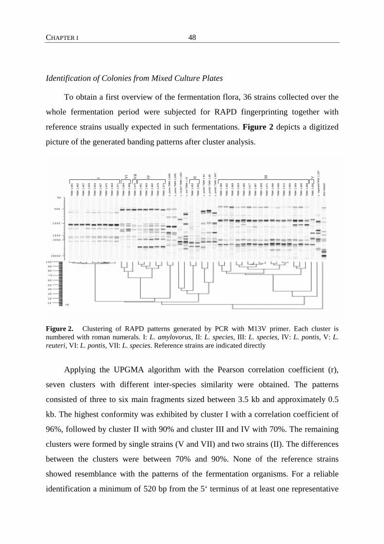
CHAPTER I
48
Identification of Colonies from Mixed Culture Plates
To obtain a first overview of the fermentation flora, 36 strains collected over the
whole fermentation period were subjected for RAPD fingerprinting together with
reference strains usually expected in such fermentations. Figure 2 depicts a digitized
picture of the generated banding patterns after cluster analysis.
TMW
1.6
61
TMW
1.6
62
TMW
1.6
57
TMW
1.6
59
TMW
1.6
53
TMW
1.6
67
TMW
1.6
70
TMW
1.6
64
TMW
1.6
71
TMW
1.6
84
TMW
1.6
68
TMW
1.6
75
TMW
1.6
51
TMW
1.6
63
TMW
1.6
82
TMW
1.6
78
TMW
1.6
74
L.pa
nis
TMW
1.6
48
L.pa
nis
TMW
1.6
49
L.re
uter
iTM
W 1
.693
L.or
isTM
W 1
.16
TMW
1.6
52
TMW
1.6
55
L.po
ntis
TMW
1.8
4
L.po
ntis
TMW
1.8
5
L.po
ntis
TMW
1.5
97
TMW
1.6
90
TMW
1.6
92
TMW
1.6
69
TMW
1.6
54
TMW
1.6
80
TMW
1.6
77
TMW
1.6
87
TMW
1.6
69
TMW
1.6
71
TMW
1.6
86
TMW
1.6
66
TMW
1.6
72
TMW
1.6
65
TMW
1.6
58
TMW
1.6
60
TMW
1.6
89
TMW
1.6
56
L.va
gina
lisTM
W1.
197
Size
Mar
ker
I VI
II III
VIVVII
5 0 0
1 0 0 0
1 5 0 0
2 0 0 0
2 0 0 0 0
b p
r %
1 0 0
9 0
8 0
7 0
6 0
5 04 0
3 0
2 0
1 0
Figure 2. Clustering of RAPD patterns generated by PCR with M13V primer. Each cluster is numbered with roman numerals. I: L. amylovorus, II: L. species, III: L. species, IV: L. pontis, V: L. reuteri, VI: L. pontis, VII: L. species. Reference strains are indicated directly
Applying the UPGMA algorithm with the Pearson correlation coefficient (r),
seven clusters with different inter-species similarity were obtained. The patterns
consisted of three to six main fragments sized between 3.5 kb and approximately 0.5
kb. The highest conformity was exhibited by cluster I with a correlation coefficient of
96%, followed by cluster II with 90% and cluster III and IV with 70%. The remaining
clusters were formed by single strains (V and VII) and two strains (II). The differences
between the clusters were between 70% and 90%. None of the reference strains
showed resemblance with the patterns of the fermentation organisms. For a reliable
identification a minimum of 520 bp from the 5‘ terminus of at least one representative

CHAPTER I
49
of each cluster was determined. Strains of cluster I comprise the obligate
homofermentative species L. amylovorus. The two isolates of cluster II could not be
allotted to a certain species. Together with the strains of cluster III and VII they took
an intermediate position between their closest phylogenetic neighbors L. pontis, and L.
vaginalis and exhibit a heterofermentative metabolism. For more detailed information
the complete 16S rDNA was sequenced of the strains L. species TMW 1.655 and L.
species TMW 1.666. To each of the phylogenetically related species differences of 30
bp were determined. In the following the strains are named L. species together with the
roman number of the cluster. Cluster IV and VI comprise the heterofermentative
species L. pontis with a correlation of 64%. The single species of cluster V was
identified as L. reuteri also a heterofermentative Lactobacillus.
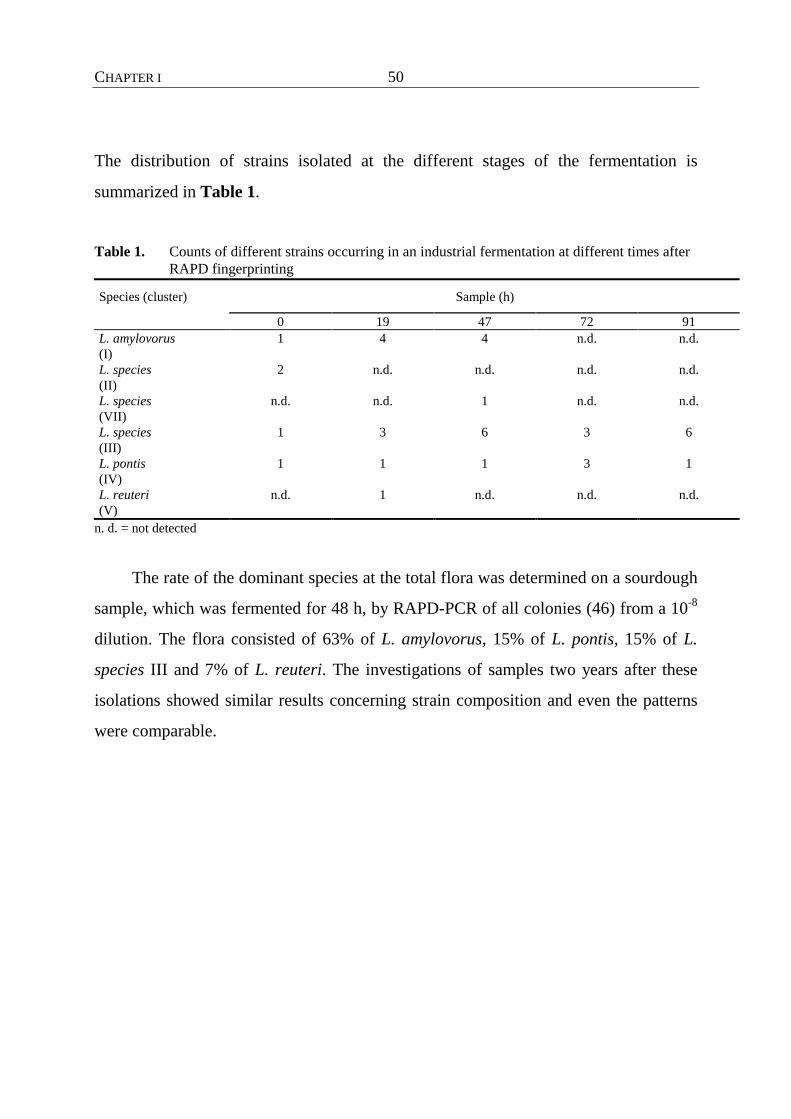
CHAPTER I
50
The distribution of strains isolated at the different stages of the fermentation is
summarized in Table 1.
Table 1. Counts of different strains occurring in an industrial fermentation at different times after RAPD fingerprinting
Species (cluster) Sample (h)
0 19 47 72 91 L. amylovorus (I)
1 4 4 n.d. n.d.
L. species (II)
2 n.d. n.d. n.d. n.d.
L. species (VII)
n.d. n.d. 1 n.d. n.d.
L. species (III)
1 3 6 3 6
L. pontis (IV)
1 1 1 3 1
L. reuteri (V)
n.d. 1 n.d. n.d. n.d.
n. d. = not detected
The rate of the dominant species at the total flora was determined on a sourdough
sample, which was fermented for 48 h, by RAPD-PCR of all colonies (46) from a 10-8
dilution. The flora consisted of 63% of L. amylovorus, 15% of L. pontis, 15% of L.
species III and 7% of L. reuteri. The investigations of samples two years after these
isolations showed similar results concerning strain composition and even the patterns
were comparable.
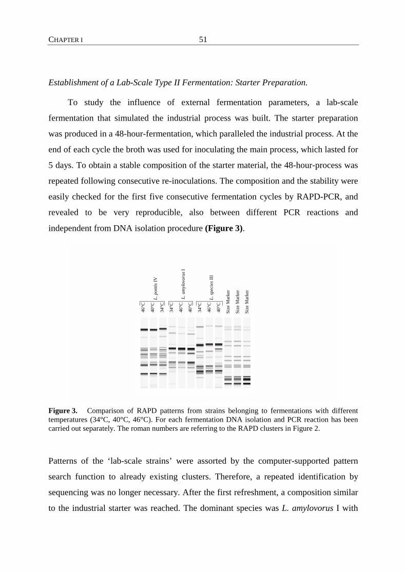
CHAPTER I
51
Establishment of a Lab-Scale Type II Fermentation: Starter Preparation.
To study the influence of external fermentation parameters, a lab-scale
fermentation that simulated the industrial process was built. The starter preparation
was produced in a 48-hour-fermentation, which paralleled the industrial process. At the
end of each cycle the broth was used for inoculating the main process, which lasted for
5 days. To obtain a stable composition of the starter material, the 48-hour-process was
repeated following consecutive re-inoculations. The composition and the stability were
easily checked for the first five consecutive fermentation cycles by RAPD-PCR, and
revealed to be very reproducible, also between different PCR reactions and
independent from DNA isolation procedure (Figure 3).
46°
C
40°
C
34°
C
34°
C
46°
C
40°
C
34°
C
46°
C
40°
C
Size
Mar
ker
Size
Mar
ker
Size
Mar
ker
L. p
ontis
IV
L. a
myl
ovor
us I
L. sp
ecie
s III
Figure 3. Comparison of RAPD patterns from strains belonging to fermentations with different temperatures (34°C, 40°C, 46°C). For each fermentation DNA isolation and PCR reaction has been carried out separately. The roman numbers are referring to the RAPD clusters in Figure 2.
Patterns of the ‘lab-scale strains’ were assorted by the computer-supported pattern
search function to already existing clusters. Therefore, a repeated identification by
sequencing was no longer necessary. After the first refreshment, a composition similar
to the industrial starter was reached. The dominant species was L. amylovorus I with
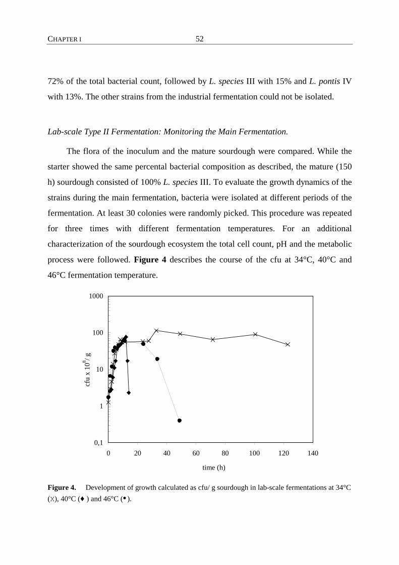
CHAPTER I
52
72% of the total bacterial count, followed by L. species III with 15% and L. pontis IV
with 13%. The other strains from the industrial fermentation could not be isolated.
Lab-scale Type II Fermentation: Monitoring the Main Fermentation.
The flora of the inoculum and the mature sourdough were compared. While the
starter showed the same percental bacterial composition as described, the mature (150
h) sourdough consisted of 100% L. species III. To evaluate the growth dynamics of the
strains during the main fermentation, bacteria were isolated at different periods of the
fermentation. At least 30 colonies were randomly picked. This procedure was repeated
for three times with different fermentation temperatures. For an additional
characterization of the sourdough ecosystem the total cell count, pH and the metabolic
process were followed. Figure 4 describes the course of the cfu at 34°C, 40°C and
46°C fermentation temperature.
0,1
1
10
100
1000
0 20 40 60 80 100 120 140
time (h)
cfu
x 10
8 / g
Figure 4. Development of growth calculated as cfu/ g sourdough in lab-scale fermentations at 34°C (X), 40°C (♦ ) and 46°C (• ).

CHAPTER I
53
Until the beginning of the stationary phase the development can be regarded as
almost temperature independent. Only the growth rate at the 46°C-fermentation was
slightly higher. At all three temperatures a mean maximum of 6 x 10⁹ cfu g-1 after 23 h
was reached. While the bacteria of the 34°C and 40°C-fermentation, respectively,
stayed in the stationary phase till 120 h and 55 h, respectively, at the 46°C-
fermentation a very immediate decrease of the cfu after 25 h occurred. This finding
correlated well with the production of metabolic products (lactate, acetate/ethanol)
recorded over the fermentation time by HPLC (Figure 5).
0
20
40
60
80
100
120
0 20 40 60 80 100 120 140time (h)
Met
abol
ic p
rodu
cts (
mm
ol/ l
)
Figure 5. Comparison of the formation of the metabolic products lactate, acetate and ethanol of the homo- and heterofermentative metabolism in fermentations with 34°C (X), 40°C (♦ ) and 46°C (�) fermentation temperature.
The flora of the 46°C-fermentation showed the fastest acid production till 25 h,
correlating with the growth curve, except that lower terminal values were obtained. In
the other fermentations the maximum was reached later and higher terminal values
were reached.
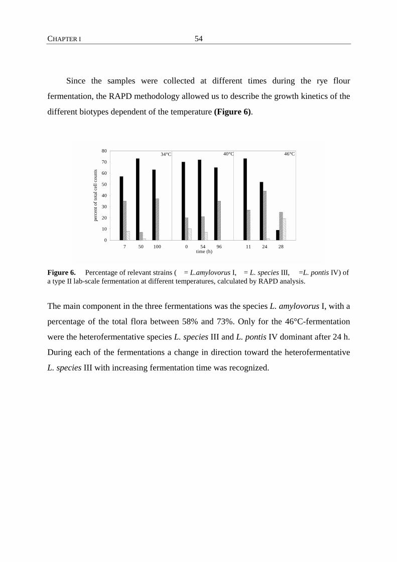
CHAPTER I
54
Since the samples were collected at different times during the rye flour
fermentation, the RAPD methodology allowed us to describe the growth kinetics of the
different biotypes dependent of the temperature (Figure 6).
11 24 280 54 96time (h)
7 50 100
perc
ent o
f tot
al c
ell c
ount
s
0
10
20
30
40
50
60
70
8034°C 40°C 46°C
Figure 6. Percentage of relevant strains ( = L.amylovorus I, = L. species III, =L. pontis IV) of a type II lab-scale fermentation at different temperatures, calculated by RAPD analysis.
The main component in the three fermentations was the species L. amylovorus I, with a
percentage of the total flora between 58% and 73%. Only for the 46°C-fermentation
were the heterofermentative species L. species III and L. pontis IV dominant after 24 h.
During each of the fermentations a change in direction toward the heterofermentative
L. species III with increasing fermentation time was recognized.
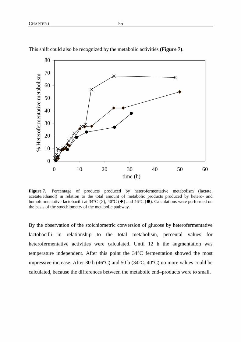
CHAPTER I
55
This shift could also be recognized by the metabolic activities (Figure 7).
0
10
20
30
40
50
60
70
80
0 10 20 30 40 50 60time (h)
% H
eter
ofer
men
tativ
e m
etab
olis
m
Figure 7. Percentage of products produced by heterofermentative metabolism (lactate, acetate/ethanol) in relation to the total amount of metabolic products produced by hetero- and homofermentative lactobacilli at 34°C (X), 40°C (�) and 46°C (�). Calculations were performed on the basis of the stoechiometry of the metabolic pathway. By the observation of the stoichiometric conversion of glucose by heterofermentative
lactobacilli in relationship to the total metabolism, percental values for
heterofermentative activities were calculated. Until 12 h the augmentation was
temperature independent. After this point the 34°C fermentation showed the most
impressive increase. After 30 h (46°C) and 50 h (34°C, 40°C) no more values could be
calculated, because the differences between the metabolic end–products were to small.
CHAPTER I
56
Discussion
The microflora of traditional sourdoughs, which have been classified as type I, have
been investigated since many years (Sugihara et al., 1970; Kline & Sugihara 1971;
Stolz et al., 1993, Stolz et al., 1995 a,b). Independent of the geographical origin this
kind of sourdough is driven by strains of L. sanfranciscensis, selected only by the
environmental conditions induced by the sourdough fermentation technology.
Completely different parameters are applied at the type II sourdough fermentations,
resulting in a different microbial ecosystem, whose composition and population
dynamics are described here.
The bacterial flora of the industrial fermentation consisted out of four species
belonging to the genus Lactobacillus. The lab-scale sourdough fermentation showed a
consistent composition with strains belonging to the same RAPD-clusters. The only
homofermentative species could be assigned to the species L. amylovorus. During the
industrial fermentation L. amylovorus could not be isolated after 47 h. A similar
tendency was observed at the lab-scale experiments. With increasing fermentation time
the percentage of L. amylovorus at the total cell count decreased. After 150 h at 40°C
no pattern specific for L. amylovorus was obtained. In contrast to diminishing numbers
of L. amylovorus, those of the heterofermentative species L. species III increased. This
tendency was underlined by a metabolic monitoring of the end-products lactate, acetate
and ethanol.
The presence of L. amylovorus in type II sourdoughs has not been reported
before. Strohmar (1992) investigated a rye flour sourdough fermented for at least 48h
and they only found heterofermentative species. However, a similar association of L.
amylovorus together with the heterofermentative species L. reuteri and L. fermentum in
traditional sorghum doughs is described by Hamad et al. (1992).
L. pontis was the first described sourdough Lactobacillus which showed a close
phylogenetic relationship to the species L. oris, L. vaginalis from human origin and
CHAPTER I
57
L. reuteri isolated from mammalians. Recently, Wiese et al. (1996) described L. panis ,
which was previously only ambiguously characterized. Like L. pontis, it could be
assorted to the same phylogentic group to which L. species II, L. species III and L.
species VII belong. L. species III was the dominating heterofermentative strain in our
industrial and lab-scale fermentation, respectively. They seemed to have the highest
acid-tolerance, surviving for 150 h where pH values of 3.2 and TTA values of 40 were
reached. Complete 16S rDNA sequence analysis of the strains L. species TMW 1.666
(cluster III) and L. species TMW 1.655 (II) and partial sequencing (520 bp) of L.
species TMW 1.675 revealed obvious differences of about 2% to their phylogenetic
neighbors L. vaginalis, L. oris and the sourdough organisms L. pontis and L. panis. No
cellular nor similarities of colony morphology to the reference strains could be
observed. Therefore, it is currently not possible to decide on a final systematic
position.
Apart from the investigation of physiological and morphological properties
chemotaxonomic and genotypic methods are leading to a comprehensive classification
of bacteria. The limit of these systems in the study of complex microbial ecosystems is
the low number of samples that can be analyzed. Especially in fermentations of non-
sterilized substrates, these complex studies have been hampered by the lack of methods
for identifying and typing of Lactobacillus strains (Cocconcelli et al., 1997). We
applied M13-RAPD-PCR to elucidate the biodiversity of a special sourdough
fermentation process. Environmental parameters such as increased fermentation
temperature and an extended souring phase lead to a well-adapted, stable flora within
this dough. An unambiguous identification on the phylogenetic level is especially
important in this case, because of the widespread physiological properties which often
cannot be assorted to reference organisms from other habitats as a result of adaptation
to different environments. These finding is confirmed by investigations of Hamad et al.
(1997) of sorghum sourdough and Strohmar (1992) of a rye flour sourdough. Proof that
growth conditions can also markedly affect cell morphology was presented by
CHAPTER I
58
Schleifer et al. (1985), in a study in which L. xylosus was reclassified as Lactococcus.
lactis subsp. lactis on the basis of phylogenetic data. Data indicating that it may be
even impossible to distinguish lactobacilli by their physiological properties were given
by Kandler & Weiss (1986): L. reuteri and L. fermentum, both associated with the
lactic acid fermentation of sourdough, were indistinguishable by conventional tests, but
genetically unrelated, as indicated by the difference in mol% G + C of the DNA.
Besides its potential use in characterization of the flora, RAPD-typing was a good
tool for checking the long term stability of such fermentations in independent batches.
The consistency and reproducibility between batches revealed RAPD-PCR as an
appropriate tool for monitoring the behavior of fermentation organisms under different
conditions which may be suitable for other food environments. Furthermore the
transformation from an industrial process to a lab-scale fermentation was feasible
without a change in the microbial composition of the flora allowing for further
metabolic and ecological investigations. This information can be used to develop such
processes with respect to flora composition and thus metabolic products affecting the
sensory properties and staling of baked goods.
CHAPTER I
59
References
Böcker, G., Vogel, R. F. & Hammes, W. P. (1990). Lactobacillus sanfrancisco als
stabiles
Element in einem Reinzucht-Sauerteig-Präparat. Getreide Mehl und Brot 44, 269-274.
Böcker, G., Stolz, P. & Hammes, W. P. (1995). Neue Erkenntnisse zum Ökosystem
Sauerteig und zur Physiologie des sauerteigtypischen Stämme Lactobacuillus
sanfrancisco und Lactobacillus pontis. Getreide Mehl und Brot 49 (6), 370-374.
Caetano-Anollés, G., Bassam, B. J. & Gresshoff, P. M. (1991). DNA amplification
fingerprinting using very short arbitrary oligonucleotide primers. Bio/Technology 9,
553-557.
Cocconcelli, P. S., Parisi, M. G., Senini, L. & Bottazzi, V. (1997). Use of RAPD and
16S rDNA sequencing for the study of Lactobacillus population dynamics in natural
whey culture. Lett. Appl. Microbiol. 25, 8-12.
Daeschel, M. A., Andersson, R. E. & Fleming, H. P. (1987). Microbial ecology of
fermenting plant materials. FEMS Microbiology Reviews 46, 357-367.
Erlandson, K. & Batt, C. A. (1997). Strain-specific differentiation of lactococci in
mixed starter culture populations using randomly amplified polymorphic DNA-derived
probes. Appl. Environ. Microbiol. 63 (7), 2702-2707.
Hamad, S. H., Böcker, G., Vogel, R. F. & Hammes, W. P. (1992). Microbial and
chemical analysis of fermented sorghum dough for kishra production. Appl. Microbiol.
Biotechnol. 37, 728-731.
Hamad, S. H., Dieng, M. C., Ehrmann, M. A. & Vogel, R. F. (1997).
Characterization of the bacterial flora of Sudanese sorghum flour and sorghum
sourdough. J. Appl. Microbiol. 83, 764-770.

CHAPTER I
60
Holzapfel, W. (1997). Use of starter cultures in fermentation on a household scale.
Food Control 8 (5/6), 241-258.
Kandler, O. & Weiss, N., (1986). Genus: Lactobacillus. In Bergey’s Manual of
Systematic Bacteriology (Eds Sneath, P.H.A., Mair, N.S., Sharpe, M.E. and Holt, J. G.)
pp. 1209-1234. Baltimore MD.
Kline, L. & Sugihara, T. F. (1971). Microorganisms of the San Francisco sour dough
bread process. Appl. Microbiol. 21 (3), 459-465.
Levington, J., Greenaway, S. D. & Spillane, B. J. (1987). Rapid small scale
preparations of bacterial genomic DNA, suitable for cloning and hybridization
analysis. Lett. Appl. Microbiol. 5, 51-53.
Ludwig, W. & Strunk, O. (1996). ARB, a software environment for sequence data.
http//www.mikro.biologie.tu-muenchen.de\pub\ARB\documentation
Saiki, R. K., Gelfand, D. H., Stoffel, S., Scharf, S. J., Higuchi, R., Horn, G. T.,
Mullis, K. B. & Ehrlich, H. A. (1988). Primer-directed enzymatic amplification of
DNA with a thermostable DNA polymerase. Science 293, 487-491.
Sanger, F., Nicklen, S. & Coulson, A. (1977). DNA sequencing with chain-
terminating inhibitors. Proc. Natl. Acad. Sci. USA 74, 5463-5467.
Schleifer, K. H., Kraus, J., Dvorak, C., Kilpper-Bälz, R., Collins, M. D. & Fischer,
W. (1985). Transfer of Streptococcus lactis and related streptococci to the genus
Lactococcus gen. nov.. System. Appl. Microbiol. 6, 183-195.
Spicher, G., Schröder, R. & Stephan, H. (1980). Die Mikroflora des Sauerteiges, X.
Mitteilung: Die backtechnische Wirkung der in „Reinzuchtsauern“ auftretenden
Milchsäuerbakterien (Genus Lactobacillus Beijerink). Z Lebensm Unters Forsch
171,119-129.
Stiles, M. E. & Holzapfel, W. H. (1997). Lactic acid bacteria of foods and their
current taxonomy. Int. J. Food Microbiol. 36, 1-29.
CHAPTER I
61
Stolz, P., Böcker, G., Vogel, R. F. & Hammes, W. P. (1993). Utilisation of maltose
and glucose by lactobacilli isolated from sourdough. FEMS Microbiology Letters 109,
237-242.
Stolz, P., Böcker, G., Hammes, W. P. & Vogel, R. F. (1995a). Utilization of electron
acceptors by lactobacilli isolated from sourdough: I. Lactobacillus sanfrancisco. Z
Lebensm Unters Forsch 201, 91-96.
Stolz, P., Vogel, R. F. & Hammes, W. P. (1995b). Utilization of electron acceptors
by lactobacilli isolated from sourdough: II. Lactobacillus pontis, L. amylovorus, L.
fermentum and L. reuteri. Z Lebensm Unters Forsch 201, 402-410.
Strohmar, W. & Diekmann, H. (1992). The microflora of a sourdough developed
during extended souring phases. Z Lebensm Unters Forsch 194, 536-540.
Sugihara, T. F., Kline, L & McCready, L. B. (1970). Nature of the San Francisco
sour dough french bread process: II. Microbiological aspects. The Bakers Digest 44,
51-57.
Tyler, K. D., Wang, G., Tyler, S. D. & Johnson, W. M. (1997). Factors affecting
reliability and reproducibility of amplification-based DNA fingerprinting of
representative bacterial pathogens. J. Clin. Microbiol. 35 (2), 339-346.
Vogel, R. F., Böcker, G., Stolz, P., Ehrmann, M., Fanta, D., Ludwig, W., Pot, B.,
Kersters, K., Schleifer, K. H. & Hammes W. P. (1994). Identification of lactobacilli
from sourdough and description of Lactobacillus pontis sp. nov.. Int. J. Syst. Bacteriol.
44 (2), 223-229.
Weizenegger, M., Neumann, M., Stackebrandt, E. & Ludwig, W. (1991).
Eubacterium alactolyticum phylogenetically groups with Eubacterium limnosum,
Acetobacterium woodii and Clostridium barkeri. Syst. Appl. Microbiol. 15, 32-36.
Welsh, J. & McClelland, M. (1990). Fingerprinting genomes using PCR with
arbitrary primers. Nucl. Ac. Res. 18, 7213-7218.
CHAPTER I
62
Wiese, B. G., Stromar, W., Rainey, F. A. & Diekmann, V. (1996). Lactobacillus
panis sp. nov., from sourdough with a long fermentation period. Int. J. Syst. Bacteriol.
46, 449-453.
Williams, J. G. K., Kubelik, A. R., Livak, K. J., Rafalski, J. A. & Tingey, S. V. (1990).
DNA polymorphisms amplified by arbitrary primers are useful as genetic markers. Nucl. Ac.
Res.18,6531-6536.
CHAPTER II 63
CHAPTER II
MULTIPLEX PCR FOR THE DETECTION OF LACTOBACILLUS PONTIS AND TWO
RELATED SPECIES IN A SOURDOUGH FERMENTATION

CHAPTER II 64
Summary
A specific multiplex PCR assay based on the amplification of parts of the 16S
rRNA molecule was designed. Primers derived from variable regions of the 16S rRNA
provided a means of easily differentiating the species Lactobacillus pontis and
Lactobacillus panis. They could be clearly discriminated from the phylogenetically
related species Lactobacillus vaginalis, Lactobacillus oris and Lactobacillus reuteri
and from other lactobacilli commonly known to be present in sourdough. Other strains
isolated together with L. pontis from an industrial sourdough fermentation could be
clearly separated from these species by comparative sequence analysis and
construction of a specific PCR primer. For a fast identification a DNA isolation
protocol based on the ultrasonic lysis of cells from single colonies was developed. To
demonstrate the potential of such techniques for tracking these organisms in a
laboratory-scale fermentation, we combined the specific PCR assay with direct DNA
extraction from the organisms in the sourdough without previous cultivation.

CHAPTER II 65
Introduction
The most prominent lactic acid bacteria (LAB) of the sourdough microflora
belong to the genus Lactobacillus (Linko et al., 1997). Sourdough fermentations have
been categorized by Böcker and coworkers (Böcker et al., 1995) by taking into account
the kind of propagation and the applied fermentation parameters resulting in typical
microbial communities. Sourdoughs maintained by a continuous back-slopping over an
extended period have been designated type I sourdoughs. They are characterized by a
microflora which is mainly dominated by Lactobacillus sanfranciscensis (Gänzle et al.,
1998). Type II sourdoughs are fermented over a longer period at elevated temperatures
and higher water content. Typical lactobacilli isolated from this fermentations are
Lactobacillus pontis and Lactobacillus panis, both endemic to cereal fermentations.
Further species from type II fermentations have been recently itemized by Vogel et al.
(Vogel et al., 1996; Vogel et al., 1999).
L. pontis is close in the phylogenetic tree to Lactobacillus vaginalis,
Lactobacillus oris, and Lactobacillus reuteri (Vogel et al., 1994). Wiese and
coworkers described L. panis as a new species which is phylogenetically related to L.
vaginalis, L. oris, and L. reuteri (Wiese et al., 1996).
Traditionally, physiological properties of bacteria, such as their capability to
degrade carbohydrates and certain enzyme activities, have been applied for
identification purposes. Especially for LAB, reliable identification depending on this
properties is almost impossible because of similar nutritional requirements of different
species due to adaption to a certain environment (Ampe et al., 1999; Hamad et al.,
1997). For this reason various approaches targeting the genotype of bacterial cells,
which reflect the natural relationship, have been described and steadily introduced into
applied and research aspects of food microbiology. During the last decade rRNA
emerged as a suitable target molecule for identification purposes (Betz et al., 1990;
Ehrmann et al., 1994; Klijn et al., 1991).
CHAPTER II 66
In the framework of the description of L. pontis as a new species (Vogel et al.,
1994), a 16S rRNA-targeted oligonucleotide probe in the V1 region (Neefs et al.,
1993) of the 16S rRNA was designed. There were sufficient sequence variations to
differentiate L. pontis from other sourdough lactobacilli. The 16S ribosomal DNA
(rDNA) sequence of L. panis (Wiese et al., 1996) revealed that this species is most
closely related to L. oris, followed by L. vaginalis and L. pontis. The binding sites on
the 16S rDNA of L. panis and L. pontis for the probe previously described for L. pontis
are identical to each other. So far no evaluated system for the specific identification
and differentiation of these two species has been available.
Therefore, we have developed 16S rDNA targeted primers for a specific PCR to
distinguish these two lactobacilli. Apart from the mentioned species, primers have
been designed for a third sourdough lactobacillus (Lactobacillus sp.) originally isolated
from an industrial type II rye fermentation. Comparative sequence analysis of 16S
rDNA revealed the Lactobacillus sp. to be an intermediate between L. pontis and L.
vaginalis, but no final systematic position has been established up to now. To
demonstrate the applicability of the PCR system to trace these organisms during
fermentation, we developed a laboratory-scale fermentation similar to an industrial
sourdough process.
CHAPTER II 67
Materials and Methods
Laboratory-scale fermentation
The fermentation was performed in a 5-liter stirred reactor (Biostat, Braun,
Melsungen, Germany) at 40°C. The substrate consisted of 900 g of rye bran mixed
with 2,400 ml of preheated tap water. To start the fermentation, 90 g of 48-h-old
sourdough of the corresponding industrial process was added.
Sampling
Sourdough samples were collected aseptically. For the determination of the total
cell count, expressed as CFU per milliliter of sourdough, samples were serially diluted
1:10 with NaCl (0.9% [wt/vol]) and plated on modified MRS (3) using a spiral plater
(Spiralsystems, Inc., Cincinnati, Ohio). The plates were incubated under a modified
atmosphere (90% N2, 10% CO2). To determine the total amount of aerobic bacteria in
the flour without starter, plate count agar (Merck, Darmstadt, Germany) was used.
Additionally, sourdough samples of 5 ml for the direct isolation of DNA from the
organisms were taken and stored at –20°C.
Cultivation and Storage of Strains
All reference organisms (see Table 2) were cultivated on mMRS (Vogel et al.,
1994). The incubation temperatures were 30°C for L. pontis, L. sanfranciscensis, and
Lactobacillus farciminis, 37°C for L. panis, L. oris, L. vaginalis and L. reuteri.
Lactobacillus sp. TMW 1.655 and DSM 13145 isolated from an industrial
fermentation process, and TMW 1.1104 and TMW 1.1098 isolated from previous lab-
scale fermentations, were cultivated at 40°C. Stock cultures were stored at –80°C in
80% (wt/wt) glycerol.
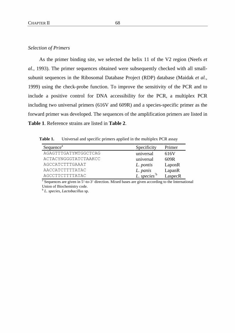
CHAPTER II 68
Selection of Primers
As the primer binding site, we selected the helix 11 of the V2 region (Neefs et
al., 1993). The primer sequences obtained were subsequently checked with all small-
subunit sequences in the Ribosomal Database Project (RDP) database (Maidak et al.,
1999) using the check-probe function. To improve the sensitivity of the PCR and to
include a positive control for DNA accessibility for the PCR, a multiplex PCR
including two universal primers (616V and 609R) and a species-specific primer as the
forward primer was developed. The sequences of the amplification primers are listed in
Table 1. Reference strains are listed in Table 2.
Table 1. Universal and specific primers applied in the multiplex PCR assay
Sequencea Specificity Primer AGAGTTTGATYMTGGCTCAG universal 616V ACTACYNGGGTATCTAAKCC universal 609R AGCCATCTTTGAAAT L. pontis LaponR AACCATCTTTTATAC L. panis LapanR AGCCTTCTTTTATAC L. species´b LaspecR
a Sequences are given in 5‘-to-3‘ direction. Mixed bases are given according to the International Union of Biochemistry code. b L. species, Lactobacillus sp.
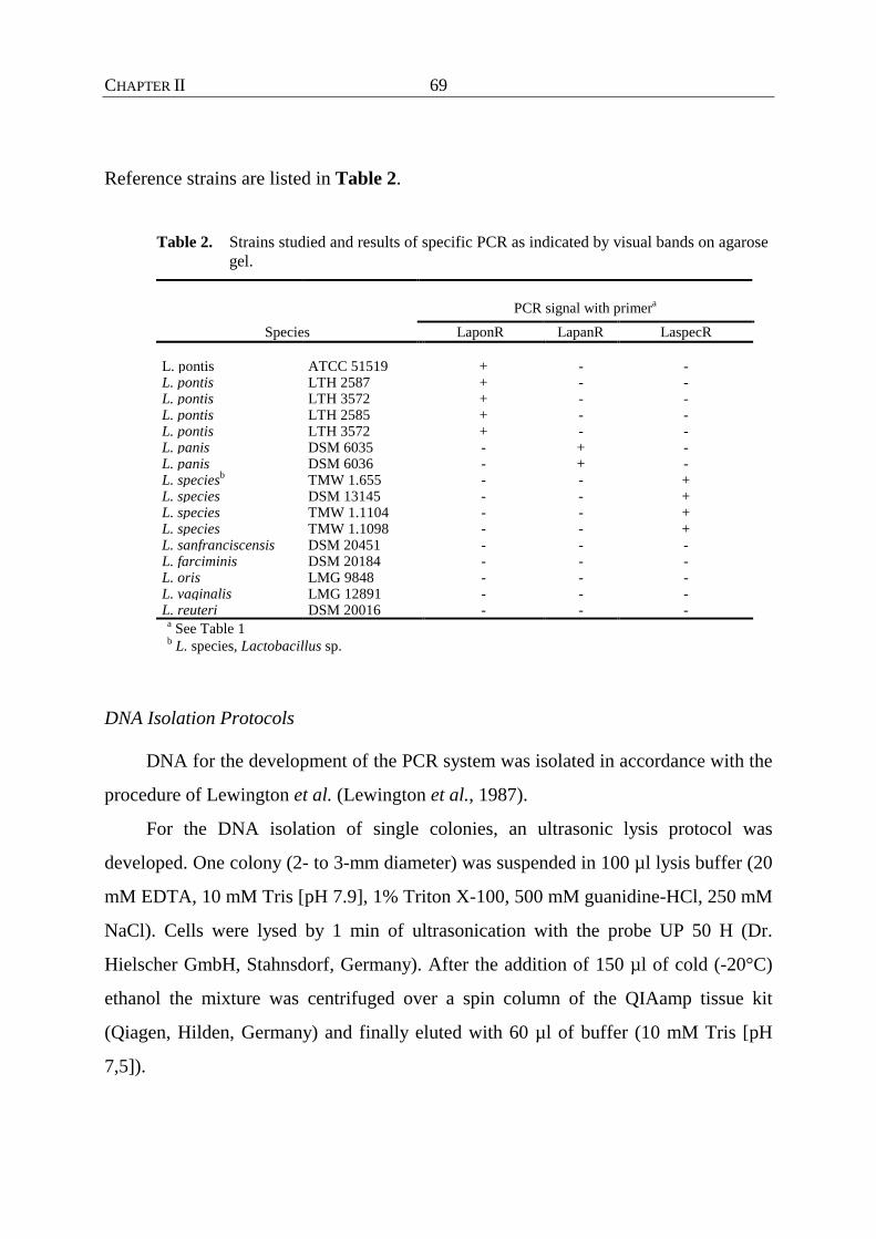
CHAPTER II 69
Reference strains are listed in Table 2.
Table 2. Strains studied and results of specific PCR as indicated by visual bands on agarose gel.
PCR signal with primera
Species LaponR LapanR LaspecR
L. pontis ATCC 51519 + - - L. pontis LTH 2587 + - - L. pontis LTH 3572 + - - L. pontis LTH 2585 + - - L. pontis LTH 3572 + - - L. panis DSM 6035 - + - L. panis DSM 6036 - + - L. speciesb TMW 1.655 - - + L. species DSM 13145 - - + L. species TMW 1.1104 - - + L. species TMW 1.1098 - - + L. sanfranciscensis DSM 20451 - - - L. farciminis DSM 20184 - - - L. oris LMG 9848 - - - L. vaginalis LMG 12891 - - - L. reuteri DSM 20016 - - -
a See Table 1 b L. species, Lactobacillus sp.
DNA Isolation Protocols
DNA for the development of the PCR system was isolated in accordance with the
procedure of Lewington et al. (Lewington et al., 1987).
For the DNA isolation of single colonies, an ultrasonic lysis protocol was
developed. One colony (2- to 3-mm diameter) was suspended in 100 µl lysis buffer (20
mM EDTA, 10 mM Tris [pH 7.9], 1% Triton X-100, 500 mM guanidine-HCl, 250 mM
NaCl). Cells were lysed by 1 min of ultrasonication with the probe UP 50 H (Dr.
Hielscher GmbH, Stahnsdorf, Germany). After the addition of 150 µl of cold (-20°C)
ethanol the mixture was centrifuged over a spin column of the QIAamp tissue kit
(Qiagen, Hilden, Germany) and finally eluted with 60 µl of buffer (10 mM Tris [pH
7,5]).
CHAPTER II 70
For isolation of bacterial DNA directly out of the sourdough a method based on
enzymatic lysis was developed and evaluated. The sourdough sample of 5 ml was
suspended in 10 ml of phosphate-buffered saline (PBS) (Sambrook et al., 1989) and
centrifuged for 5 min at 1,500 ✕ g. Ten milliliters of the supernatant was transferred in
a new tube and centrifuged for 15 min at 5,000 ✕ g. The pellet was resuspended in 4
ml of PBS, and 1.5 ml of this suspension was transferred in an Eppendorf tube and
centrifuged for 5 min at 5,000 ✕ g to collect the cell material. The supernatant was
discarded. The pellet was resuspended in 180 µl of TES (50 mM Tris-HCl, 50 mM
NaCl, 10 mM EDTA [pH 8.0]) containing 20 mg of lysozyme ml-1. For sufficient cell
lysis it was incubated at 37°C for 40 min on a shaking platform (90 rpm). After the
addition of 20 µl proteinase K and 200 µl lysis buffer AL (QIAamp tissue kit), the
reaction mixture was incubated at 70°C for 30 min and for a further 30 min at 95°C.
After the addition of 210 µl of cold ethanol (-20°C) it was centrifuged over a spin
column (QIAamp Tissue Kit) and washed with 500 µl of the supplied buffers AW1
and AW2. The DNA was eluted with preheated (70°C) 10 mM Tris (pH 7.5) and
stored at –20°C.
PCR Conditions
The amplification of the 16S rDNA was carried out in strips (Braun, Wertheim,
Germany) on a Gradient master thermocycler (Eppendorff, Hamburg, Germany), in a
total volume of 25 µl. Primers were obtained from Interactiva (Ulm, Germany). In
order to use the three sets of reactions mixtures (a set consisted of 609R, 616V, and the
specific probe for L. pontis, L. panis, or the Lactobacillus sp.) in parallel, the optimum
annealing temperature was determined by gradient PCR between 40°C and 54°C with
1.1°C increments. The optimum annealing temperature was 46.5°C. The amplification
conditions for the multiplex PCR assay with three primers were as follows: 0.5 µl of
genomic DNA, 2.5 µl of 10x reaction buffer, 100 nM (each) deoxynucleoside
triphosphate, 0.5 U Taq polymerase (Amersham Pharmacia Biotech, Piscataway, N.J.),
CHAPTER II 71
10 pmol of the specific primer and 616V (universal, Table 1), 2.5 pmol of primer 609R
(universal, Table 1), 1% dimethyl sulfoxide, and deionized H2O to a final volume of 25
µl. The amplification conditions were as follows: initial denaturation (94°C for 120 s)
followed by 25 cycles of denaturation (94°C for 45 s), annealing (46.5°C for 60 s), and
extension (72°C for 60 s). PCR products were electrophoretically separated on a 2%
agarose gel and stained with ethidium bromide. As the size marker, a 100 bp ladder
(Amersham Pharmacia Biotech) was used.

CHAPTER II 72
Results and Discussion
Figure 1 shows that it is possible to identify and discriminate between L. pontis,
L. panis and the closely related Lactobacillus sp. with the primer combinations
deduced from the variable V2 region of the 16S rDNA.
LapoR616V/609R
LapanR616V/609R
LaspecR616V/609R
Primer set:
M 1 2 3 4 5 6 7 8 9 10 M
800 bp
300 bp200 bp
Figure 1. Multiplex PCR assay for the specific identification of L. pontis, L. panis, and the Lactobacillus sp. Primers indicated at the top are the specific primer for the identification of the following strains: L. pontis (LapoR) ATCC 51518 (lane 1), ATCC 51519 (lane 2). LTH 2587 (lane 3), and LTH 3572 (lane 4); L. panis (LapanR) DSM 6035 (lane 5) and DSM 6036 (lane 6); and Lactobacillus sp. strains (LaspecR) TMW 1.1098 (lane 7), TMW 1.1104 (lane 8), DSM 13145 (lane 9), and TMW 1.655 (lane 10). The specifically amplified fragment has a size of 236-bp (616V plus the specific reverse primer). The 800-bp fragment was amplified by the two universal primers 616V and 609R. Lane M, 100-bp ladder.
The primer combination 616V (universal) and the respective specific primers led to a
236-bp fragment for the three species. Primer 616V together with the universal reverse
primer 609R allows the simultaneous amplification of a 800-bp fragment. In this way a
combined specific identification in combination with a test on DNA accessibility was
possible. So false-negative results could be avoided. The amplification of DNA
isolated from strains of L. pontis and the Lactobacillus sp. led to an unspecific product

CHAPTER II 73
with 1,300 bp. We accepted this as a compromise solution in order to have the
possibility to run all three identification reactions in parallel. The specificity of the
PCR assay was tested on several lactobacilli (Table 1). No cross-reactions appeared.
To have a tool for a fast and reliable identification of single pure cultures, the
already-evaluated PCR assay was combined with a fast DNA isolation procedure.
Time-consuming enzymatic lysis procedures and variable susceptibility of bacteria to
the lysozyme were overcome by ultrasonic treatment of the cells, with subsequent
purification and concentration by binding DNA to a silica matrix. The cell material of
a single colony was found to be sufficient for the PCR. Figure 2 illustrates a typical
result for the amplification of DNA of representative strains of the investigated
sourdough lactobacilli.
Figure 2. 16S rDNA amplification of DNA isolated from a single colony of representative strains of the species L. pontis ATCC 51519 (lane 1), L. panis DSM 6035 (lane 2), and Lactobacillus sp. strain DSM 13145 (lane 3). The fragments were amplified with the universal primer 616V and the respecitive species-specific reverse primers. The 800-bp fragment was amplified by the two universal primers 616V and 609R. Lane M, 100-bp ladder.

CHAPTER II 74
The described PCR assay in combination with the fast-lysis procedure of even small
amounts of cell material has major advantages over classical identification techniques,
as physiological and biochemical identification is time-consuming and often not
reliable. This is especially true for L. pontis, which exhibits a wide strain-dependent
range of fermented carbohydrates, making a clear identification difficult. The fast
ultrasonic lysis made it possible to identify this lactobacillus at the species level in 4 h.
The application of this species-specific PCR technique for a rapid identification
provides an attracting alternative to conventional methods.
For the evaluation of the applicability of multiplex PCR for monitoring the
lactobacilli from a mixed culture without prior cultivation, laboratory-scale sourdough
fermentation was set up. The fermentation was based on rye bran and was started with
sourdough from the corresponding industrial process which had been fermented for 48
h at similar conditions. Previous microbiological investigations of this as well as of
laboratory-scale processes by randomly amplified polymorphic DNA typing and 16S
rDNA sequence analysis showed that strains of the Lactobacillus sp., L. pontis, and
L.actobacillus amylovorus predominated in the flora (unpublished results). As shown
in Figure 3, it was possible to specifically amplify DNA from L. pontis and the
Lactobacillus sp.
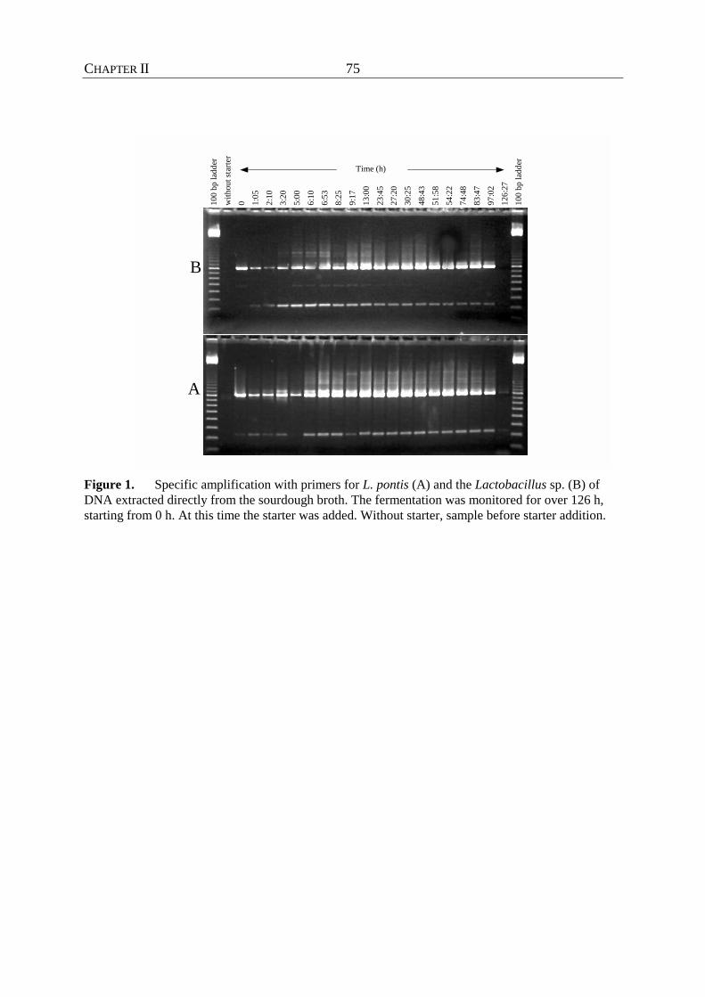
CHAPTER II 75
100
bp la
dder
with
out s
tarte
r0 1:
052:
103:
205:
006:
106:
538:
259:
1713
:00
23:4
527
:20
30:2
548
:43
51:5
854
:22
74:4
883
:47
97:0
212
6:27
100
bp la
dder
Time (h)
A
B
Figure 1. Specific amplification with primers for L. pontis (A) and the Lactobacillus sp. (B) of DNA extracted directly from the sourdough broth. The fermentation was monitored for over 126 h, starting from 0 h. At this time the starter was added. Without starter, sample before starter addition.

CHAPTER II 76
L. panis could not be identified in this fermentation, which resulted in the 800-bp
fragment of the universal primers 616V and 609R only (a picture of the gel is not
shown). Samples before starter addition showed no specific signal in both cases, even
the universal primers targeting any bacterial DNA gave almost no signal (A) or no
signal (B). This was confirmed by the classical microbiological investigation of plating
on plate count agar, where a total cell count of 10 to 100 CFU was determined. On
mMRS no LAB growth could be determined before starter was added. Directly after
inoculation the total cell count was 9.3 ✕ 106 CFU ml sourdough-1 , reaching its
maximum of 3.2 ✕ 109 CFU ml sourough-1 after 24 h and dropping to a final cell count
of 5.5 ✕ 107 CFU ml sourdough-1 after 126 h. At this time for both primer
combinations a signal with the two universal primers could be obtained, but only for L.
pontis could a weak signal be recognized. During the fermentation the intensities of
signals for both the universally amplified product and the specific fragment increased,
becoming lower after 54 h and disappearing completely for Lactobacillus sp. after 126
h. The total cell count at the end of the fermentation seems to be a detection limit for
directly extracted DNA from sourdough with this method. This limit seems to be high
in comparison to those from other investigations in this field. For example, Zapparoli
& Torriani (1997) could amplify 102 CFU ml of diluted sourdough-1. Nevertheless the
aim of this study was not to detect such small amounts but to have a fast and easy tool
for detecting dominant lactobacilli from such fermentations. The direct isolation of
total bacterial community DNA with a subsequent specific PCR can be considered a
valuable tool for monitoring these lactobacilli in mixed microbial populations.
Moreover, no genotypic method to identify L. panis was available. Only a
combined identification with L. pontis, not discrimination between these two species,
was possible. With the presented specific PCR it could be demonstrated that it is
possible to differentiate between these closely related species.
The Lactobacillus sp., a dominant element in the investigated fermentation, could
be clearly discriminated from phylogenetically related species by genotypic

CHAPTER II 77
identification techniques. To clarify the definitive phylogenetic position, further
investigations are being prepared.

CHAPTER II 78
References
Ampe, F., N Ben Omar, N. & Guyot, J.-P. (1999). Culture-independent
quantification of physiologically-active microbial groups in fermented foods using
rRNA-targeted oligonucleotide probes: application to pozol, a mexican lactic acid
fermented maize dough. J Appl Microbiol 87, 131-140.
Betzl, D., Ludwig, W. & Schleifer, K. H. (1990). Identification of lactococci and
enterococci by colony hybridization with 23S rRNA-targeted oligonucleotide probes.
Appl Environ Microbiol 56, 2927-2929.
Böcker, G., Stolz, P. & Hammes, W. P. (1995). Neue Erkenntnisse zum Ökosystem
Sauerteig und zur Physiologie der sauerteigtypischen Stämme Lactobacillus
sanfrancisco und Lactobacillus pontis. Getreide Mehl und Brot 49, 370-374.
Ehrmann, M., Ludwig, W. & Schleifer, K. H. (1994). Reverse dot blot
hybridization: A useful method for the direct identification of lactic acid bacteria in
fermented food. FEMS Microbiol Lett 117, 143-150.
Gänzle, M. G., Ehmann, M. & Hammes, W. P. (1998). Modeling of growth of
Lactobacillus sanfranciscensis and Candida milleri in response to process parameters
of sourdough fermentations. Appl Environ Microbiol 64, 2616-2623.
Hamad, S. H., Dieng, M. C., Ehrmann, M. A. & Vogel, R. F. (1997).
Characterization of the bacterial flora of Sudanese sorghum sourdough. J Appl
Microbiol 83, 764-770.
Klijn, N., Weerkamp, A. H. & De Vos, W. M. (1991). Identification of mesophilic
lactic acid bacteria by using polymerase chain reaction-amplified variable regions of
16S rRNA and specific DNA probes. Appl Environ Microbiol 57, 3390-3393.
CHAPTER II 79
Lewington, J., Greenaway, S. D. & Spillane, B. J. (1987). Rapid small scale
preparations of bacterial genomic DNA, suitable for cloning and hybridization
analysis. Lett Appl Microbiol 5, 51-53.
Linko, Y.-Y., Javanainen, P. & Linko, S. (1997). Biotechnology of bread baking.
Trends in Food Science&Technology 8, 339-344.
Maidak, B. L., Cole, J. R., Parker, C. T., Garrity, G. M., Larsen, N., Li, B.,
Lilburn, T. G., McCaughey, M. J., Olsen, J., Overbeek, R., Pramanik, S.,
Schmidt, T. M., Tiedje, J. M. & Woese, C. R. (1999). A new version of the RDP
(Ribosomal Database Project). Nucleic Acids Res 27, 171-173.
Neefs, J.-M., Van De Peer, Y., De Rijks, P., Chapelle, S. & De Wachter, R. (1993).
Compilation of small ribosomal subunit RNA sequences. Nucleic Acids Res 21, 3025-
3049.
Sambrook, J., Fritsch, E. F. & Maniatis, V. (1989). Molecular Cloning: A laboratory
manual. 2nd edition. Cold Spring Harbor Labarotory Press, USA.
Vogel, R. F., Böcker, G., Stolz, P., Ehrmann, M., Fanta, D., Ludwig, W., Pot, B.,
Kersters, K., Schleifer, K. H. & Hammes W. P. (1994). Identification of lactobacilli
from sourdough and description of Lactobacillus pontis sp. nov.. Int J Syst Bacteriol
44 (2), 223-229.
Vogel, R. F., Knorr, R., Müller, M. R. A., Steudel, U., Gänzle, M. G. & Ehrmann,
M. A. (1999). Non-dairy lactic fermentations: the cereal world. Antonie van
Leeuwenhoek 76, 403-411.
Vogel, R. F., Müller, M., Stolz, P. & Ehrmann, M. (1996). Ecology in sourdoughs
produced by traditional and modern technologies. Adv Food Sci (CMTL) 18, 152-159.
Wiese, B. G., Strohmar, W., Rainey, F. A. & Diekmann, V. (1996). Lactobacillus
panis sp. nov., from sourdough with a long fermentation period. Int J Syst Bacteriol
46, 449-453.
CHAPTER II 80
Zapparoli, G., & Torriani, S. (1997). Rapid identification and detection of
Lactobacillus sanfrancisco in sourdough by species-specific PCR with 16S rRNA-
targeted primers. System Appl Microbiol 20, 640-644.

CHAPTER III 81
CHAPTER III
LACTOBACILLUS FRUMENTI SP. NOV., A NEW LACTIC ACID BACTERIUM
ISOLATED FROM RYE-BRAN FERMENTATIONS WITH A LONG FERMENTATION
PERIOD

CHAPTER III 82
Summary
Within the framework of the characterization of the microflora of an industrial
sourdough fermentation, strains of Lactobacillus amylovorus, L. pontis and two other
strains were isolated which could not be associated with a valid species. These latter
strains were Gram-positive, catalase-negative, non-spore-forming, non-motile rods that
could be clearly differentiated from known species by 16S rDNA sequence analysis.
For further characterization, the morphological, physiological (sugar
fermentation, formation of D/L-lactate, hydrolysis of arginine, growth temperature,
CO2 production) and chemotaxonomic (G+C content, cell wall composition, SDS-
PAGE of whole-cell proteins) properties have been determined.
Fitting of the complete 16S rDNA sequence into alignments of 16.000 of such
sequences, together with the subsequent phylogenetic calculations, allowed the
reconstruction of a phylogenetic tree. These data showed that the two strains were
phylogenetically related but formed an independent cluster distinct from their closest
neighbours, L. pontis, Lactobacillus panis, Lactobacillus oris, Lactobacillus vaginalis
and Lactobacillus reuteri.
The results of DNA-DNA hybridization experiments indicated that the new two
isolates represent a new Lactobacillus species, for which the name Lactobacillus
frumenti is proposed; the type strain of this species is DSM 13145T (= LMG 19473).
CHAPTER III 83
Introduction
Lactic acid bacteria (LAB), especially the genera of Lactococcus, Leuconostoc,
Pediococcus and Lactobacillus, play an important role in the fermentative food and
feed production (Herrero et al., 1996). In non-lactic fermentations, lactobacilli, in
particular, contribute to a huge variety of spontaneously fermented indigenous foods
and beverages as well as to biotechnological processes carried out under controlled
conditions.
The fermentation of starchy substrate can be found in many countries.
Numerous reports have elucidated the microbial populations in such fermentations,
e.g. fermentation of sour cassava (Figueroa et al., 1995, Giraud et al., 1998, Morlon-
Gyot et al. 1998), ogi (Johansson et al., 1995), pozol (Ampe, 1999) and kishra (Hamad
et al., 1997). In Europe, cereal fermentations are mainly applied to the brewing
industry, providing sourmashes, and to baking, in which sourdough plays an important
role in the preparation of bread dough to improve dough machinability, breadcrumb
structure, keeping properties and flavour (Salovaara, 1998). The microbial ecology of
such cereal fermentations is determined by several exogenous and endogenous
parameters, as reported by Vogel et al. (1996). Whilst a wide variety of LAB, e.g.
Lactobacillius brevis, Lactobacillus alimentarius, Lactobacillus farciminis, Weissella
confusa or Enterococcus faecium can be found in spontaneous homemade sourdough
fermentations, Lactobacillus sanfranciscensis, Lactobacillus pontis and Lactobacillus
panis are endemic in cereal fermentations, because their competitive metabolism has
adapted to this environment. Böcker et al. (1995) introduced a classification of
industrial sourdough fermentations which takes the kind of propagation and the
manner of preparation into consideration, resulting in typical bacterial communities.
Whilst L. sanfranciscensis constitutes a stable element (Böcker et al., 1990) in the so-
called type I doughs, micro-organisms found in liquid doughs with an extended
fermentation period and higher temperatures (type II) belong to the species L. pontis,

CHAPTER III 84
L. panis, Lactobacillus reuteri, Lactobacillus fermentum and Lactobacillus
amylovorus (Vogel et al., 1999).
In this paper we report the description of a new Lactobacillus species from an
industrial type II rye-bran fermentation and for which we propose the name
Lactobacillus frumenti.

CHAPTER III 85
Methods
Strains, Medium and Culture Conditions
Strains were isolated from an industrial rye-bran sourdough fermentation. For
cultivation, the modified MRS (mMRS) medium described by Vogel et al., (1994) was
prepared with an additional modification whereby 90% of the water was replaced with
a rye-bran extract. For the preparation of 1 l of this extract, 40 g rye bran and 2 g
malted wheat meal together with 0.8 g trypsin were incubated for 24 h at 50 °C. The
liquid was separated by filtration. Strains of L. frumenti were at 40 °C in line with the
fermentation temperature of the sourdough from which they were isolated. The other
strains were incubated at the temperature recommended by the respective strain
collection. Solid media were incubated under a modified atmosphere (N2 : CO2 90% :
10%, v/v). Cultures were maintained at –80°C in glycerol (a pellet of 10 ml overnight
culture in 500 µl fresh medium to 500 µl glycerol). The purity of the cultures was
checked microscopically and by preparing streak cultures. The two isolates that are
described here as the new species L. frumenti sp. nov. bear the numbers of the strain
collection of the Technische Mikrobiologie Weihenstephan, i.e. TMW 1.655 and
TMW 1.666. Strain TMW 1.666 was deposited as type strain of the species as LMG
19473T and DSM 13145T, and will be referred to as the latter in this paper. The
following type strains were used as reference organisms: Lactobacillus vaginalis DSM
5837T, L. pontis DSM 8475T, L. panis DSM 6035T, L. reuteri DSM 20016T, L. oris
DSM 4864T, Lactobacillus. buchneri DSM 20057T.
Physiological characterization
Sugar-fermentation patterns were determined by using a microtitre plate assay.
The reproducibility was verified by repeated analysis using cultures grown on mMRS.
All sugar solutions (2% w/w) were sterilised by filtration through a 0.2 µm filter. Cells
were suspended in a medium containing (per 250 ml) 5 g peptone from casein, 1 g of
CHAPTER III 86
meat extract, 3.5 g yeast extract, 125 mg cysteine, 250 µl Tween 80, 100 mg
bromcresol purple, 2.5 g sodium acetate, 250 mg citric acid (ammonium salt), 62.5 mg
KH2PO4, 125 mg Na2HPO4, 100 mg MgSO4, 5 mg MnSO4 and 5 mg FeSO4. The
solution was autoclaved for 15 min at 121°C. A 2 ml of an overnight culture of each
strain was centrifuged and washed three times with 2 ml PBS buffer (8 g NaCl, 0.2 g
KCl, 1.44 g Na2HPO4, 0.24 g KH2PO4, made to 1 l with dH2O, pH 7.4). The cell
concentration was adjusted with water to an OD578 of 1; 45 µl of this cell suspension
was added to 1155 µl test medium. From this cell suspension, aliquots of 40 µl were
added to each of the wells. In the first well, sugar was replaced with sterile water. To
avoid evaporation and to guarantee anaerobic conditions, the wells were covered with
paraffin. The change of the indicator from purple to yellow was documented after 24,
48 and 168 h. Only definitive changes were rated as positive results. To evaluate the
system, additional experiments were carried out using the API 50 CH kit
(bioMérieux).
The formation of the lactate isomeres in the fermented broth was determined
enzymically using the DL-lactate test kit (Boehringer Mannheim).
Arginine hydrolysis was determined according the methods described by Sharpe
(1979).
Morphological Characteristics
Cell morphology was studied using phase-contrast microscopy. Gram-
determinations were performed using the KOH method of Gregersen (1978).
Whole-Cell Protein Analysis
For the preparation of cell extracts, 70-80 mg (wet weight) of bacterial cells was
treated as described by Vogel et al. (1994). Electrophoresis was performed as
described by Laemmli (1970), using a 12% (v/v) SDS-polyacrylamide separating gel.
Gels were run in a Mini-Protean II electrophoresis cell (BioRad), coomassie-stained
and dried on a vacuum gel dryer. The protein patterns were compared visually. The
CHAPTER III 87
LMW electrophoresis calibration kit (Amersham Pharmacia Biotech) was used as a
size maker.
DNA Base Composition
The G + C contents (mol%) of the DNA of strains TMW 1.655 and DSM 13145T
were determined using an HPLC analytical method. The experiments were performed
by the DSMZ and were carried out using the protocol previously described by
Tamaoka & Komagata (1984). Wild-type lambda phage DNA was used as the
standard (Meshbah et al., 1989). The G + C content (mol%) was determined according
to Meshbah et al. (1989).
Cell Wall
The peptidoglycan structure of the cell wall was determined by the DSMZ.
DNA Isolation
DNA was isolated according a protocol described by Marmur (1961), with some
modifications. One hour before cells were harvested, penicillin G (Sigma) was added
to inhibit the synthesis of cross-linkages of the cell wall and therefore to facilitate the
lysis. A wet weight of 70 mg cells was used for the DNA isolation. After the protocol,
lysis was completed after 45-90 min after the addition of lysozyme and mutanolysine.
For some strains, more effective lysis was obtained by using an overnight lysis at 4°C
and a subsequent proteinase K treatment at 60°C for 1 h, before continuation with the
normal protocol. The purified and vacuum-dried DNA was dissolved in 2 x SSC (0.3
M NaCl, 0.03 M Na3 citrate 2 H2O, pH 7.0). This DNA preparation served for the
DNA-DNA hybridization experiments as well as for 16S rDNA amplification. The
purity of the culture subjected to DNA isolation was proved as described above before
the addition of penicillin. Furthermore, DNA preparations were checked for their
CHAPTER III 88
authenticity in a randomly amplified polymorphic DNA (RAPD)-PCR assay
(Paramithiotis et al., 2000), comparing the patterns with those of primary preparations.
DNA-DNA Hybridization
The determination of DNA homology values was carried out by using
chromosomal DNA (chrDNA) from strain DSM 13145T and from L. pontis DSM
8475Tas probes. The chrDNA probes were labelled by nick translation using a kit
system (Promega) incorporating biotin-21-dUTP (Clontech) for 2h at 16°C. The DNA
(5 µg) of selected type strains was transferred by using a dot-blot block (Stratagene)
on a positively charged nylon membrane (Boehringer Mannheim) and then fixed by
incubation at 80°C for 1 h. The DNA of L. buchneri DSM 20054T was included to
check the sensitivity of the system for a more distantly related Lactobacillus species.
The hybridization procedure was carried out as described by Ehrmann et al. (1994),
except for the hybridization and washing steps, which were performed with alternative
buffers described in the protocol of Engler-Blum et al. (1993). To determine the
accessibility of the DNA, a parallel hybridization was carried out with a 5‘ biotin-
labelled universal probe (612R, 5´-GTAAGGTTYTNCGCGT-3´) targeting the 16S
rDNA. For the hybridization, 100 ng chrDNA probe 20 pmol oligo probe (each per ml
hybridization solution) were used. Hybridization was performed at 65°C for the
chrDNA probe and 42°C for the oligo probe. The detection system consisted of a
combination of alkaline phosphatase and the substrate CDP-Star (Boehriner
Mannheim). Membranes were exposed to a Kodak X-Omat film (Sigma-Aldrich). The
spot intensity was calculated with the IMAGE MASTER 2D Elite software
(Amersham Pharmacia Biotech). Calculations of homology values was performed in
principal as described by Liebl et al. (1991).
Additional DNA-DNA hybridization experiments for L. frumenti DSM 13145T,
L. frumenti TMW 1.655 and L. vaginalis DSM 5837T were performed by the DSMZ
according to the protocol of De Ley et al. (1970), but with the modifications described
CHAPTER III 89
by Huß et al. (1983) and Escara & Hutton (1980). The renaturation rates were
calculated according to Jahnke (1992) with the program TRANSFER : BAS.
16S rDNA Amplification and Sequencing
PCR-mediated amplification of the complete 16S rDNA was carried out in a
Gradient Master thermocycler (Eppendorf). All reagents were from Amersham
Pharmacia Biotech, unless otherwise indicated. The amplification conditions were as
follows: 1 µl genomic DNA, 10 µl 10x reaction buffer, 200 nM each of the four
deoxynucleotides, 1.5 U Taq polymerase, 20 pmol each primer (Interactiva) (616V, 5‘-
AGAGTTTGATYMTGGCTCAG-3‘; 630R, 5‘-CAKAAAGGAGGTGATCC-3‘) and
dH2O to a final volume of 100 µl. The amplification conditions were as follows: 94 °C
for 2 min; 30 cycles of 94 °C for 45 s, 52°C for 1 min, 72 °C for 30 s; 94 °C for 1 min,
72 °C for 4 min. PCR products were purified by the QIAquick PCR purification kit
(Quiagen) and were eluted with 60 µl elution buffer. DNA sequences were determined
by the chain-termination method (Sanger et al., 1977) using the Thermo Sequenase
fluorescence-labelled primer cycle sequencing kit with 7-deaza-dGTP (Amersham Life
Science) and separated on an ALF express sequencer (Amersham Pharmacia Biotech)
or, alternatively, using the ABI Prism Dye Terminator Cycle Sequencing Kit (Perkin
Elmer) on an ABI 373 stretch sequencing system by a commercial service
(SequiServe, Germany). For sequencing, the amplification primer 616V together with
the internal primers 609R [5’-ACT AC(CT) (AGC)GG GTA TCT AA(GT) CC-3’],
612R [5’-GTA AGG TT(CT) T(AGCT)C GCG T-3’], 607R [5’-ACG TGT GTA GCC
C-3’], 606R [5’-T(AG)A CGG (GC)C(AG) GTG TGT ACA-3’] and 607V [5’-GGG
CTA CAC ACG TGC-3’] were used.

CHAPTER III 90
Phylogenetic Analysis
The complete 16S rDNA sequences of L. frumenti DSM 13145T and TMW 1.655
were fitted into alignments of approximately 16000 homologous full and partial
primary structures available in public databases (Ludwig, 1995). Distance-matrix,
maximum-parsimony and maximum-likelihood methods were applied for tree
reconstruction as implemented in the ARB software package (W. Ludwig & O. Strunk;
http://www.mikro.biologie.tu-muenchen.de/pub/ARB/documentation). Different data-
sets varying with respect to included outgroup reference sequences as well as
alignment positions were analysed.

CHAPTER III 91
Results
Colony and Cell Morphology
On mMRS, the colonies of L. frumenti DSM 13145T were white with regular
sharp edges and after 3 d growth, they were 1 mm in diameter. The colonies of TMW
1.655 were of a similar shape but were a little smaller and appeared whitish and
transparent.
The cells of strains TMW 1.655 and L. frumenti DSM 13145T were non-spore-
forming, non-motile rods, that occurred singly or in pairs (seldom in chains). In liquid
cultures of mMRS after 24 h at 40 °C, cells of TMW 1.655 had a tendency to form
nest-shaped aggregations. Depending on the period of growth, the ends of the cells
were bent like hooks. The cells of L. frumenti DSM 13145T were more regular in
shape than those of TMW 1.655. The size of a single cell was difficult to determine,
because of the heterogeneity. The cell length was between 3 and 5 µm and the
diameter was 0.3 µm. The KOH test indicated a Gram-positive behaviour.
Physiological and Biochemical Properties
The sugar patterns and further physiological and biochemical characteristics of L.
frumenti strains and the reference organisms are listed in Table 1.
Melezitose, rhamnose, methyl α-D-mannoside and L-arabinose were fermented only
by strain L. frumenti DSM 13145T. 5-ketogluconate was fermented only by strain
TMW 1.655.
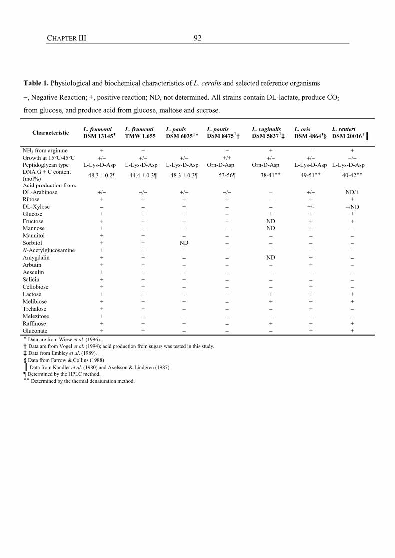
CHAPTER III 92
Table 1. Physiological and biochemical characteristics of L. ceralis and selected reference organisms
−, Negative Reaction; +, positive reaction; ND, not determined. All strains contain DL-lactate, produce CO2
from glucose, and produce acid from glucose, maltose and sucrose.
Characteristic L. frumenti DSM 13145T
L. frumenti TMW 1.655
L. panis DSM 6035T٭
L. pontis DSM 8475T†
L. vaginalis DSM 5837T‡
L. oris DSM 4864T§
L. reuteri DSM 20016T║
NH3 from arginine + + − + + − + Growth at 15°C/45°C +/− +/− +/− +/+ +/− +/− +/− Peptidoglycan type L-Lys-D-Asp L-Lys-D-Asp L-Lys-D-Asp Orn-D-Asp Orn-D-Asp L-Lys-D-Asp L-Lys-D-Asp DNA G + C content (mol%) 48.3 ± 0.2¶ 44.4 ± 0.3¶ 48.3 ± 0.3¶ 53-56¶ 38-41٭٭42-40 ٭٭51-49 ٭٭
Acid production from: DL-Arabinose +/− −/− +/− −/− − +/− ND/+ Ribose + + + + − + + DL-Xylose − − + − − +/- −/ND Glucose + + + − + + + Fructose + + + + ND + + Mannose + + + − ND + − Mannitol + + − − − − − Sorbitol + + ND − − − − N-Acetylglucosamine + + − − − − − Amygdalin + + − − ND + − Arbutin + + − − − + − Aesculin + + + − − − − Salicin + + + − − − − Cellobiose + + − − − + − Lactose + + + − + + + Melibiose + + + − + + + Trehalose + + − − − + − Melezitose + − − − − − − Raffinose + + + − + + + Gluconate + + − − − + + .Data are from Wiese et al. (1996) ٭† Data are from Vogel et al. (1994); acid production from sugars was tested in this study. ‡ Data from Embley et al. (1989). § Data from Farrow & Collins (1988) ║ Data from Kandler et al. (1980) and Axelsson & Lindgren (1987). ¶ Determined by the HPLC method. .Determined by the thermal denaturation method ٭٭
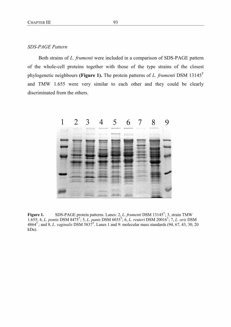
CHAPTER III 93
SDS-PAGE Pattern
Both strains of L. frumenti were included in a comparison of SDS-PAGE pattern
of the whole-cell proteins together with those of the type strains of the closest
phylogenetic neighbours (Figure 1). The protein patterns of L. frumenti DSM 13145T
and TMW 1.655 were very similar to each other and they could be clearly
discriminated from the others.
Figure 1. SDS-PAGE protein patterns. Lanes: 2, L. frumenti DSM 13145T; 3, strain TMW 1.655; 4, L. pontis DSM 8475T; 5, L. panis DSM 6035T; 6, L. reuteri DSM 20016T; 7, L. oris DSM 4864T ; and 8, L. vaginalis DSM 5837T. Lanes 1 and 9: molecular mass standards (94, 67, 43, 30, 20 kDa).

CHAPTER III 94
Phylogenetic Position
For both strain TMW 1.655 and L. frumenti DSM 13145T, 1561 bp of the 16S
rDNA were sequenced. Fig. 2 shows a phylogenetic tree based on these sequence data
and reflects the phylogenetic position of L. frumenti.
Figure 2. Phylogenetic tree demonstrating the relationship between L. frumenti DSM 13145T and the most closely related lactobacilli. The tree was constructed by using a maximum-likelihood approach and was based on a data-set that included only positions present in at least 50% of all available Lactobacillus 16S rRNA sequences. Bar, 10% homology divergence.
The percentage similarities between L. frumenti and the type strains of the other
species were as follows: L. vaginalis, 97.7%; L. panis, 97.6%; L. pontis, 97.3%; L.
oris, 96.8%; L. reuteri, 94.3%. The 16S rRNA sequence of Lactobacillus sakei was
used as an outgroup rRNA reference.
L. fermentum
L. pontis L. frumenti
L. vaginalis L. panis
L. oris
L. reuteri
L. sakei
0.10
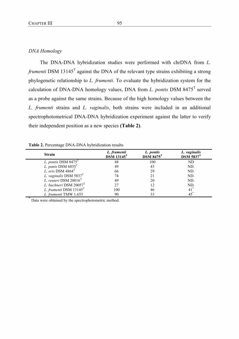
CHAPTER III 95
DNA Homology
The DNA-DNA hybridization studies were performed with chrDNA from L.
frumenti DSM 13145T against the DNA of the relevant type strains exhibiting a strong
phylogenetic relationship to L. frumenti. To evaluate the hybridization system for the
calculation of DNA-DNA homology values, DNA from L. pontis DSM 8475T served
as a probe against the same strains. Because of the high homology values between the
L. frumenti strains and L. vaginalis, both strains were included in an additional
spectrophotometrical DNA-DNA hybridization experiment against the latter to verify
their independent position as a new species (Table 2).
Table 2. Percentage DNA-DNA hybridization results
Strain L. frumenti DSM 13145T
L. pontis DSM 8475T
L. vaginalis DSM 5837T
L. pontis DSM 8475T 48 100 ND L. panis DSM 6035T 49 43 ND. L. oris DSM 4864T 66 29 ND. L. vaginalis DSM 5837T 74 21 ND. L. reuteri DSM 20016T 49 20 ND. L. buchneri DSM 20057T 27 12 ND. L. frumenti DSM 13145T 100 46 41* L. frumenti TMW 1.655 90 33 45* * Data were obtained by the spectrophotometric method.
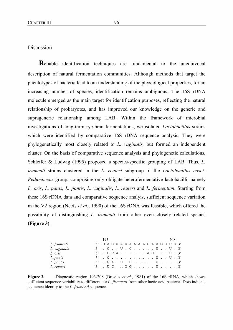
CHAPTER III 96
Discussion
Reliable identification techniques are fundamental to the unequivocal
description of natural fermentation communities. Although methods that target the
phentotypes of bacteria lead to an understanding of the physiological properties, for an
increasing number of species, identification remains ambiguous. The 16S rDNA
molecule emerged as the main target for identification purposes, reflecting the natural
relationship of prokaryotes, and has improved our knowledge on the generic and
suprageneric relationship among LAB. Within the framework of microbial
investigations of long-term rye-bran fermentations, we isolated Lactobacillus strains
which were identified by comparative 16S rDNA sequence analysis. They were
phylogenetically most closely related to L. vaginalis, but formed an independent
cluster. On the basis of comparative sequence analysis and phylogenetic calculations,
Schleifer & Ludwig (1995) proposed a species-specific grouping of LAB. Thus, L.
frumenti strains clustered in the L. reuteri subgroup of the Lactobacillus casei-
Pediococcus group, comprising only obligate heterofermentative lactobacilli, namely
L. oris, L. panis, L. pontis, L. vaginalis, L. reuteri and L. fermentum. Starting from
these 16S rDNA data and comparative sequence analyis, sufficient sequence variation
in the V2 region (Neefs et al., 1990) of the 16S rDNA was feasible, which offered the
possibility of distinguishing L. frumenti from other even closely related species
(Figure 3).
193 208 L. frumenti 5‘ U A G U A U A A A A G A A G G C U 3‘ L. vaginalis 5‘ . C . . U . C . . . . . U . . U . 3‘ L. oris 5‘ . C C A . . . . . . A G . . . U . 3‘ L. panis 5‘ . C . . . . . . . . . . U . . U . 3‘ L. pontis 5‘ . G A . U . C . . . . . U . . . . 3‘ L. reuteri 5‘ . U C . n G G . . . . . U . . . . 3‘
Figure 3. Diagnostic region 193-208 (Brosius et al., 1981) of the 16S rRNA, which shows sufficient sequence variability to differentiate L. frumenti from other lactic acid bacteria. Dots indicate sequence identity to the L. frumenti sequence.

CHAPTER III 97
A PCR-based system that allows L. frumenti to be differentiated from these and other
sourdough lactobacilli has already been described by Müller et. al. (2000). This
supports the hypothesis that strains DSM 13145T and TMW 1.655 should be separated
from other members of the L. reuteri group.
Despite the close phylogenetic relationships of species within the L. reuteri
group, G + C contents range between 38 and 54 mol%. The closest phylogenetic
neighbor of L. frumenti (with a 16S rDNA similarity of 97.7%) is L. vaginalis. This
relationship is confirmed by their similar G + C content (43.3 versus 41 mol%). The
biggest difference in the G + C contents was shown with L. pontis. In this case, no
correlation with 16S rRNA homology data can be deduced.
The relatedness of L. frumenti and L. vaginalis with respect to G + C content and
16S rRNA sequence data contrasts with the chemical composition of the cell walls of
these micro-organisms. L. frumenti has a cell wall of the peptidoglycan type [ A4α (L-
Lys-D-Asp)], which constitutes the main feature of species of the Cb group (Hammes
& Vogel, 1995). The cell wall of L. vaginalis is in the same grouop but lysine is
replaced by ornithine, which can be seen as additional proof of the status of L.
frumenti as an independent species.
SDS-PAGE pattern analysis of whole-cell proteins has proven to be a reliable
tool for the discrimination of even closely related species (Pot et. al., 1993). SDS-
PAGE comparisons (in which the two strains of L. frumenti exhibited almost identical
patterns) allowed L. frumenti DSM 13145T and TMW 1.655 to be clearly
differentiated from the species L. pontis, L. panis, L. oris, L. vaginalis and L. reuteri.
This, again can be seen as a further proof of the independent status of L. frumenti as
new species.
The determination of DNA reassociation values constitutes a meaningful method
for unequivocal species description (Stackebrandt & Goebel, 1994). In particular,
when closely related species (≥ 97% rRNA homology) are inspected, the resolution
power of 16S rRNA sequences is limited (Fox et al., 1992), but DNA similarity can
range between 10 and 100% (Stackebrandt & Goebel, 1994). The threshold value for a
phylogenetic definition of a species, as proposed by Wayne et al. (1987), should not

CHAPTER III 98
exceed 70%. In our case, the DNA-DNA hybridization studies clearly adhered to this
principle. For L. vaginalis and L. pontis, rRNA similarity values with respect to L.
frumenti were 97.7 and 97.3%, respectively. However, DNA-DNA homology values
between L. frumenti and L. vaginalis and between L. frumenti and L. pontis amounted
to 74 and 48%, respectively. The spectrophotometric technique used as reference
method for the determination of homology values for the closest relative, L. vaginalis,
produced lower values between L. vaginalis and TMW 1.655 or DSM 13145T (41 and
45%, respectively). The homology values obtained by dot-blot hybridization of L.
frumenti against the other species were confirmed by alternative hybridization with the
DNA of L. pontis. The value for similarity to L. frumenti (46%) was almost identical to
that determined by hybridization of L. frumenti against L. pontis (48%). Moreover, the
similarity calculated between L. pontis and L. panis was identical (43%) to the value
determined by the spectrophotometric technique (Wiese et al., 1996). Summarizing the
DNA hybridization results (Table 2), all species could be clearly separated from L.
frumenti, indicating its independent status as a species.
In view of the phylogenetic evidence (16S rDNA, DNA-DNA homology)
presented, the protein-pattern differences measured by SDS-PAGE and the phenotypic
distinctiveness of DSM 13145T and TMW 1.655, it is proposed that these strains
belong to a new species, L. frumenti sp. nov..

CHAPTER III 99
Description of Lactobacillus frumenti sp. nov. (Müller, Ehrmann and Vogel)
Lactobacillus frumenti (fru.men’ti. L. gen. N. frumenti from cereal).
The cells are Gram-positive, non-motile, non-spore-forming rods that occur singly or
in pairs (seldom in chains). Growth was observed up to 45 °C but not at 15 °C. They
are facultatively anaerobic, catalase-negative and obligately heterofermentative. The
L-isomer constitutes more than 85% of the total lactic acid content. Acid is produced
from L-arabinose, ribose, galactose, glucose, fructose, mannose, mannitol, sorbitol, N-
acetylglucosamine, amygdalin, arbutin, aesculin (hydrolysed), salicin, cellobiose,
maltose, lactose, melibiose, sucrose, trehalose, raffinose and gluconate. Melezitose,
rhamnose, methyl α-D-mannoside and L-arabinose were fermented only by strain
DSM 13145T. 5-Ketogluconate was fermented only by strain TMW 1.655. The type
strain is Lactobacillus frumenti DSM 13145T (= LMG 19473T).
CHAPTER III 100
References
Ampe, F., Ben Omar, N. & Gyot, J.-P. (1998). Recovery of total microbial RNA
from lactic acid fermented foods with a high starch content. Lett Appl Microbiol 27,
270-274.
Axelsson, L. & Lindgren, S. (1987). Characterization and DNA homology of
Lactobacillus strains isolated from pig intestine. J Appl Bacteriol 62, 433-440.
Böcker, G., Stolz, P. & Hammes, W. P. (1995). Neue Erkenntnisse zum Ökosystem
Sauerteig und zur Physiologie des sauerteigtypischen Stämme Lactobacuillus
sanfrancisco und Lactobacillus pontis. Getreide Mehl und Brot 49, 370-374.
Böcker, G., Vogel, R. F. & Hammes, W. P. (1990). Lactobacillus sanfrancisco als
stabiles Element in einem Reinzucht-Sauerteig-Präparat. Getreide Mehl und Brot 44,
269-274.
Brosius, J., Dull, T. J., Sleeter, D. D. & Noller, H. F. (1981). Gene organisation and
primary structure of a ribosomal RNA operon from Escherichia coli. J Mol Biol 148,
107-127.
De Ley, J., Cattoir, H. & Reynaerts, A. (1970). The quantitative measurement of
DNA hybridization from renaturation rates. Eur J Biochem 12, 133-142.
Ehrmann, M., Ludwig, W. & Schleifer, K. H. (1994). Reverse dot blot
hybridization: a useful method for the direct identification of lactic acid bacteria in
fermented foods. FEMS Microbiol Lett 117, 143-150.
Embley, T. M., Faquir, N., Bossart, W. & Collins, M. D. (1989). Lactobacillus
vaginalis sp. nov. from the human vagina. Int J Syst Bacteriol 39, 368-370.
Engler-Blum, G., Meier, M., Frank, J. & Müller, G. A. (1993). Reduction of
background problems in nonradioactive northern and southern blot analysis enables
higher sensitivity than 32P-based hybridization. Anal Biochem 210, 235-244.

CHAPTER III 101
Escara, J. F. & Hutton, J. R. (1980). Thermal stability and renaturation of DNA in
dimethyl-sulphoxide solutions: acceleration of renaturation rate. Biopolymers 19,
1315-1327.
Farrow, J. A. E. & Collins, M. D. (1988). Lactobacillus oris sp. nov. from the human
oral cavity. Int J Syst Bacteriol 38, 116-118.
Figueroa, C., Davilla, A. M. & Pourquié, J. (1995). Lactic acid bacteria of the sour
cassava starch fermentation. Letters in Appl Microbiol 21, 126-130.
Fox, G. E., Wisotzkey, J. D. & Jurtshuk Jr., P. (1992). How close is close: 16S
rRNA sequence identity may not be sufficient to guarantee species identity. Int J Syst
Bacteriol 42, 166-170.
Giraud, E., Champailler, A., Moulard, S. & Raimbault, M. (1998). Development
of a miniaturized selective counting strategy of lactic acid bacteria for evaluation of
mixed starter in a model cassava fermentation. J Appl Microbiol 84, 444-450.
Gregersen, T. (1978). Rapid method for distinction of Gram-negative from Gram-
positive bacteria. Eur J Appl Microbiol Biotechnol 5, 123-127.
Hamad, S. H., Dieng, M. C., Ehrmann, M. A. & Vogel, R. F. (1997).
Characterization of the bacterial flora of Sudanese sorghum flour and sorghum
sourdough. J Appl Microbiol 83, 764-770.
Hammes, W. P. & Vogel, R. F. (1995). The genus Lactobacillus. In The genera of
lactic acid bacteria, vol. 2, pp. 19-54. Edited by B. J. B. Wood & W. Holzapfel.
Glasgow: Blackie Academic & Professional.
Herrero, M., Mayo, B., González & Suárez, J. E. (1996). Evaluation of
technologically important traits in lactic acid bacteria isolated from spontaneous
fermentations. J Appl Bacteriol 81, 565-570.
Huß, V. A. R., Festl, H. & Schleifer, K. H. (1983). Studies on the spectrophotometric
determination of DNA hybridization from renaturation rates. Syst Appl Microbiol 4,
184-192.
CHAPTER III 102
Jahnke, K.-D. (1992). BASIC computer program for evaluation of spectroscopic
DNA renaturation data from GILFORD SYSTEM 2600 spectrophotometer on a
PC/XT/AT type personal computer. J Microbiol Meth 15, 61-73.
Johansson, M.-L., Sanni, A., Lönner, C. & Molin, G. (1995). Phenotypically based
taxonomy using API 50CH of lactobacilli from nigerian ogi and the occurrence of
starch fermenting strains. Int J Food Microbiol 25, 159-168.
Kandler, O., Stetter, K.-O. & Köhl, R. (1980). Lactobacillus reuteri sp. nov., a new
species of heterofermentative lactobacilli. Zentralblatt für Bakteriologie,
Mikrobiologie und Hygiene Abt. 1, Orig. C 1, 264-269.
Laemmli, U. K. (1970). Cleavage of structural proteins during the assembly of the
head of bacteriophage T4. Nature 227, 680-685.
Liebl, W., Ehrmann, M., Ludwig, W. & Schleifer, K. H. (1991). Transfer of
“Brevibacterium flavum“ DSM 20411, “Brevibacterium lactofermentum“ DSM 20412
and DSM 1412, and Corynebacterium lilium DSM 20137T to Corynebacterium
glutamicum and their distinction by rRNA gene restriction patterns. Int J Syst
Bacteriol 41, 255-260.
Ludwig, W. (1995). Sequence databases. In Molecular Microbial Ecology Manual,
pp. 3.3.5.1-22. Edited by A. D. L. Akkermans, J. D. van Elsas & F. J. de Bruijn.
Amsterdam: Kluwer.
Marmur, J. (1961). A procedure for the isolation of deoxyribonucleic acid from
microorganisms. J Mol Biol 3, 208-218.
Meshbah, M., Premachandran, U. & Whitmann, W. (1989). Precise measurement
of the G + C content of deoxyribonucleic acid by high performance liquid
chromatography. Int J Syst Bacteriol 39, 159-167.
Morlon-Guyot, J., Guyot, J. P., Pot, B., De Haut, I. J. & Raimbault, M. (1998).
Lactobacillus manihotivorans sp. nov., a new starch-hydrolysing lactic acid bacterium
isolated during cassava sour starch fermentation. Int J Syst Bacteriol 48, 1101-1109.

CHAPTER III 103
Müller, M. R. A., Ehrmann, M. A. & Vogel, R. F. (2000). Multiplex PCR for the
detection of Lactobacillus pontis and two related species in a sourdough fermentation.
Appl Environ Microbiol 66, 2113-2116.
Neefs, J.-M., van De Peer, Y., De Rijks, P., Chapelle, S. & De Wachter, R. (1993).
Compilation of small ribosomal subunit RNA sequences. Nucleic Acids Res. 21, 3025-
3049.
Schleifer, K. H. & Ludwig, W. (1995). Phylogeny of Genus Lactobacillus and
related Genera. System Appl Microbiol 18, 461-467.
Paramithiotis, S., Müller, M. R. A., Ehrmann, M. A., Tsakalidou, E., Seiler, H.,
Vogel, R. & Kalantzopoulos, G. (2000). Polyphasic identification of wild yeast
strains isolated from greek sourdoughs. System Appl Microbiol 23, 156-164.
Pot, B., Hertel, C., Ludwig, W., Descheemaeker, P., Kersters, K. & Schleifer, K.-
H. (1993). Identification and classification of Lactobacillus acidophilus, L. gasseri and
L. johnsonii strains by SDS-PAGE and rRNA-targeted oligonucleotide probe
hybridization. J Gen Microbiol 139, 513-517.
Salovaara, H. (1998). Lactic acid bacteria in cereal products. In Lactic Acid Bacteria-
Technology and Health Effects, 2nd edn, pp. 115-138. Edited by S. Salminen & A.
Von Wright. New York: Marcel Dekker.
Sanger, F., Nicklen, S. & Coulson, A. (1977). DNA sequencing with chain-
terminating inhibitors. Proc Natl Acad Sci USA 74, 5463-5467.
Schleifer, K. H. & Ludwig, W. (1995). Phylogeny of genus Lactobacillus and related
genera. System Appl Microbiol 18, 461-467.
Sharpe, M. E. (1979). Identification methods for microbiologists. In: Technical
Series, 2 nd edn, no. 14, pp. 233-259. Edited by F. A. Skinner, D. W. Lovelock. New
York: Academic Press.
CHAPTER III 104
Stackebrandt, E. & Goebel, B. M. (1994). Taxonomic note: A place for DNA-DNA
reassociation and 16S rRNA sequence analysis in the present species definition in
bacteriology. Int J Syst Bacteriol 44, 846-849.
Tamaoka, J. & Komagata, K. (1984). Determination of DNA base composition by
reversed-phase high-performance liquid chromatography. FEMS Microbiol Lett 25,
125-128.
Vogel, R. F., Böcker, G., Stolz, P., Ehrmann, M., Fanta, D., Ludwig, W., Pot, B.,
Kersters, K., Schleifer, K. H. & Hammes W. P. (1994). Identification of lactobacilli
from sourdough and description of Lactobacillus pontis sp. nov.. Int J Syst Bacteriol
44, 223-229.
Vogel, R. F., Knorr, R., Müller, R. A., Steudel, U., Gänzle, M. G. & Ehrmann, M.
A. (1999). Non-dairy lactic fermentations: the cereal world. Antonie van Leeuwenhoek
76, 403-411.
Vogel, R. F., Müller, M., Stolz, P. & Ehrmann, M. (1996). Ecology in sourdoughs
produced by traditional and modern technologies. Adv Food Sci (CMTL) 18, 152-159.
Wayne, L. G., Brenner, D. J., Colwell, R. R., Grimont, P. A. D., Kandler, O.,
Krichevsky, M. I., Moore, L. H., Moore, W. E. C., Murray, R. G. E.,
Stackebrandt, E., Starr, M. P. & Trüper, H. G. (1987). Report of the Ad Hoc
Committee on Reconciliation of approaches to bacterial systematics. Int J Syst
Bacteriol 37, 463-464.
Wiese, B. G., Strohmar, W., Rainey, F. A. & Diekmann, H. (1996). Lactobacillus
panis sp. nov., from sourdough with a long fermentation period. Int J Syst Bacteriol
46, 449-453.

CHAPTER IV
105
CHAPTER IV
POLYPHASIC IDENTIFICATION OF WILD YEAST STRAINS ISOLATED FROM
GREEK SOURDOUGHS

CHAPTER IV
106
Summary
A total of forty-five wild yeast strains were isolated from five traditional Greek
sourdoughs. Strains were identified using the classical identification technique along
with the sodium dodecyl sulfate-polyacrylamide gel electrophoresis of whole cell
proteins (SDS-PAGE), Fourier transform-infrared spectroscopy (FT-IR) and the
randomly amplified polymorphic DNA-polymerase chain reaction analysis (RAPD-
PCR). The latter methods confirmed the classical identification. According to the
results obtained, 14 strains were identified as Saccharomyces cerevisiae strains, twenty
five as Pichia membranaefaciens strains and six as Yarrowia lipolytica.
CHAPTER IV
107
Introduction
Yeasts are one of the most important groups of microorganisms related with
human nutrition. They have been used for millennia and they are still used in bread
making and in the production of alcoholic beverages. Furthermore, several yeast
species such as Candida albicans and Cryptococcus neoformans are of medical
importance, while many species like Zygosaccharomyces bailii, Pichia
membranaefaciens and Saccharomyces cerevisiae may cause spoilage in a wide range
of foods (Deak & Beuchat, 1996; Kockova-Kratochvilova, 1990).
In bread making many different functional properties have been defined for
yeasts. Leavening is their main role (Sugihara et al., 1971) and the production of either
primary or secondary metabolites, such as alcohols, esters and carbonyl compounds, is
of major importance for the formation of the characteristic bread flavor (Martinez-
Anaya et al., 1990; Collar et al., 1994; Hansen & Hansen, 1994; Damiani et al., 1996).
Through their enzymatic activities, mainly promoted by amylolytic, proteolytic and
lipolytic enzymes, yeasts can affect not only the organoleptic characteristics, but the
overall appearance of the final product as well (Martinez-Anaya, 1996; Collar et al.,
1994; Antuna & Martinez-Anaya, 1993).
Recent studies on cereal fermentations mainly focused on the characterization of
sourdough fermentations based on wheat or rye flour as raw materials. Basically, three
types of sourdough fermentations can be distinguished (Böcker et al., 1995) (i) type I
doughs sustained by repeated inoculation at ambient temperature (20-30°C) according
to traditional procedures, (ii) type II doughs using elevated temperatures, longer
fermentation times and higher water content, mostly used in industrialized processes to
produce bakery pre-products, and (iii) type III processes initiated by defined starter
cultures. Predominance of certain microorganisms, as far as spontaneous cereal
fermentations are concerned, is decided by the degree od adaptation of the
microorganisms to the respective environment. As a general rule, lactic acid bacteria

CHAPTER IV
108
are the predominant microorganisms and in many cases yeasts are present in
significant numbers (Vogel et al., 1999).
It is generally accepted that the use of sourdough in bread making yields in
products of high quality in terms of sensory characteristics and shelf-life. Furthermore
the sourdough bread preparation process is technologically essential for baking whole
rye meal bread to improve dough machinability, bread crumb properties and flavor
(Salovaara, 1998). This is due to the synergistic effect of yeasts and lactic acid bacteria (LAB)
present in this product (Roecken & Voysey, 1995; Linko et al., 1997; Rosenquist & Hansen,
1998). According to this, isolation, identification and study of the biochemical and
technological features of these microorganisms is of great technological importance.
The detailed study of sourdough microflora can contribute to the design and
production of starter cultures for industrial scale applications.
Nowadays, a great number of identification techniques are available. Beside the
classical phenotypic methods relying on morphological, physiological and biochemical
criteria, investigations with chemotaxonomic methods (SDS-PAGE of whole cell
proteins, fatty acid analysis, FT-IR spectroscopy) and genotypic methods
(electrophoretic karyotyping, (PCR)-RFLP, AFLP, rRNA analysis, RAPD) are
becoming more and more applied in the field of food microbiology for characterization
and identification of yeasts (Baleiras Couto et al., 1994; Baleiras Couto et al., 1995;
Kosse et al., 1997; Noronha-da-Costa et al., 1996; Romano et al., 1996; Kümmerle et
al., 1998; Vancanneyt et al., 1991; Mäntynen et al., 1999).
We have chosen a polyphasic approach for the characterization of sourdough
yeast, combining FT-IR spectroscopy together with the PCR-based molecular
technique RAPD fingerprinting and the conventional biochemical/morphological
characterization.
Traditional identification rely upon several physiological and biochemical tests.
The mode of vegetative and sexual reproduction along with the ability to ferment
sugars and aerobically growth on various carbon and nitrogen compounds together
with growth under stressing conditions are important in identification. Although this
CHAPTER IV
109
procedure is time-consuming and laborious and only leads to species level
identification, is still regarded as the reference one.
SDS-PAGE of whole cell proteins has been successfully used in bacterial and
yeast identification (Vancanneyt et al., 1991). It is a valuable criterion for classifying
microorganisms at the species level, when reliable reference patterns are available,
because of its high levels of sensitivity and reproducibility (Vancanneyt et al., 1991).
FT-IR spectroscopy originally used in chemical analysis for the identification of
substances has been applied for the first time to identify microorganisms (Naumann et
al., 1994). In principle a FT-IR spectrum of the whole cell is compared with a
reference spectrum library. Very recently Kümmerle (1998) demonstrated the
relevance of this technique for food microbiology. A reference database for yeasts
with about 900 spectra has been built up by which food-borne yeasts have been
successfully identified.
RAPD, first introduced by Williams et al. (1990), relies on the amplification of
fragments with only a single short primer present. In order to allow annealing of the
primer to the target DNA, the annealing step of the reaction is run under low
stringency conditions. Together with a random, non-specific primer sequence species
up to strain specific fingerprints are generated. Since these days the RAPD technique
was applied to many problems both in fungal and bacterial microbiology mainly in the
characterization of complex habitats or the differentiation of isolates (Tompkins et al.,
1996; Laidlaw et al., 1996). The reason of the integration of different kinds of data and
information leading to a consensus type of taxonomy (Vandamme et al., 1996) was to
overcome the limits of each single method and to improve the reliability of the whole
expression.
Greek traditional wheat sourdoughs belong to type I. Up to now there is no data
concerning chemical and microbiological characteristics of traditional Greek
sourdoughs. One of the most common traditional Greek wheat sourdoughs is based on
mixing flour and tap water (40-50°C) in the presence of basil (Ocimum basilicum), an
aromatic plant closely related to the country’s tradition and religion. The dough is
CHAPTER IV
110
subjected to fermentation for one day at ambient temperature (25-35°C). After one or
two subsequent refreshments and overnight fermentation, it is ready to serve as an
inoculum for bread making. The sourdoughs that have been examined were prepared
according to this procedure.
The aim of this work was not only to provide data concerning chemical and
microbiological characteristics for both household and semi-industrial traditional
Greek wheat sourdoughs, but to present a polyphasic approach to yeast identification
as well.

CHAPTER IV
111
Materials and Methods
Yeast Strains
All wild yeast strains have been isolated from Greek sourdoughs, three from
household scale fermentations (sourdough I-III) and two from bakeries (sourdough IV,
V). Strain numbers are given according to the strain collection (ACA-YC) of the
Laboratory of Dairy Research of Agricultural University of Athens. Reference strains
for RAPD have been obtained from the CBS (Candida ovalis CBS 7298T, Yarrowia
lipolytica CBS 6164), from the DSMZ (Zygosacharomyces rouxii DSM 2531,
Debaromyces hansenii DSM 70244, Pichia membranaefaciens DSM 70366,
Saccharomyces cerevisiae DSM 70449T) and from the NCYC (S. cerevisiae NCYC
1200) collections. Reference strains included in the FT-IR analysis, others than for
RAPD-PCR, were Y. lipolytica CBS 6124T, Y. lipolytica M331, P. membranaefaciens
CBS 107T and S. cerevisiae CBS 5900.
Sampling
Sourdough samples were collected aseptically, stored at 4°C and analyzed the
same day.
pH and Total Titratable Acidity (TTA) Measurement
10 g of the sample was homogenized with 90 ml of sterile distilled water. The pH
value was recorded and the acidity was titrated using 0.1N NaOH to final pH 8.5. The
TTA was expressed in ml 0.1N NaOH.
Isolation of Yeast and Lactic Acid Bacteria
Samples (10 g) were diluted 1:10 with 90 ml NaCl (0.9% w/v). Numbers of
colony forming units (CFU) were determined by serial dilutions on yeast glucose
chloramphenicol (YGC) agar for yeasts and on modified Rogosa (mMRS) agar,
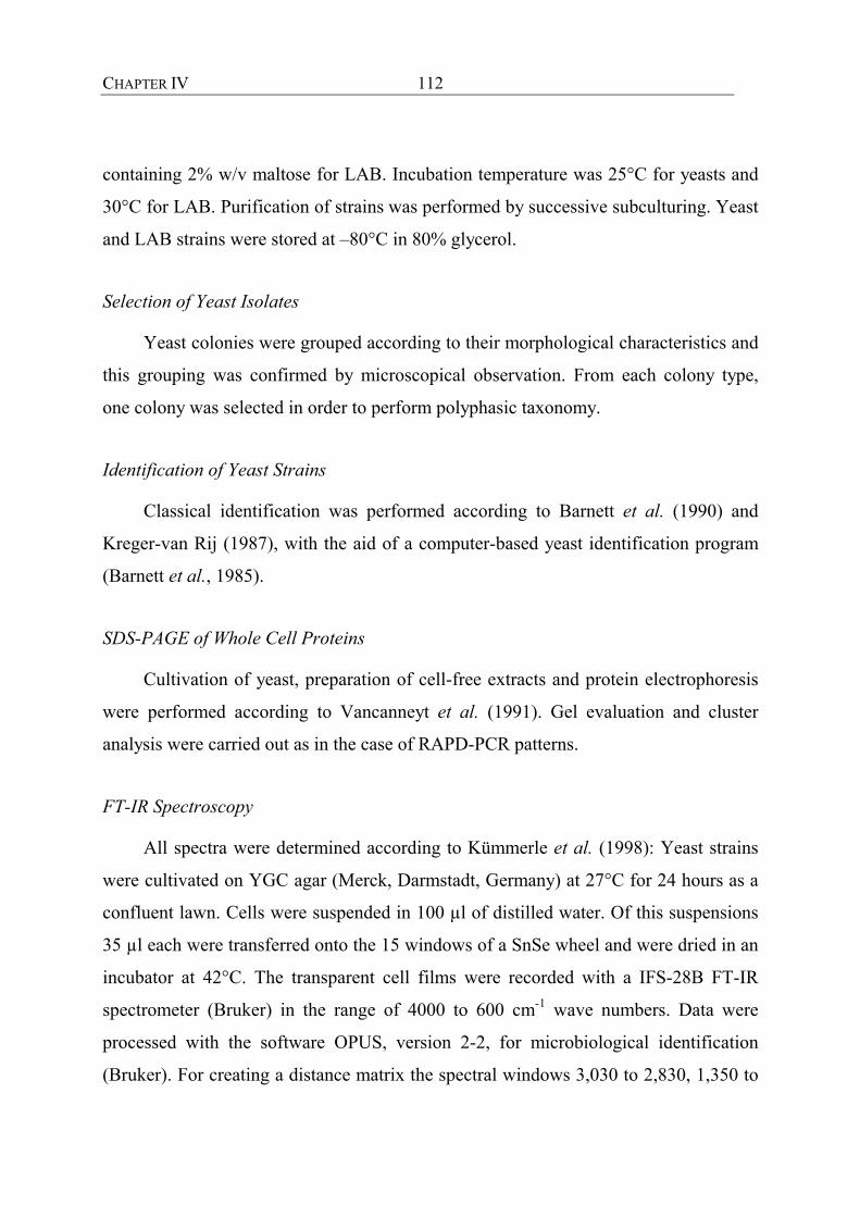
CHAPTER IV
112
containing 2% w/v maltose for LAB. Incubation temperature was 25°C for yeasts and
30°C for LAB. Purification of strains was performed by successive subculturing. Yeast
and LAB strains were stored at –80°C in 80% glycerol.
Selection of Yeast Isolates
Yeast colonies were grouped according to their morphological characteristics and
this grouping was confirmed by microscopical observation. From each colony type,
one colony was selected in order to perform polyphasic taxonomy.
Identification of Yeast Strains
Classical identification was performed according to Barnett et al. (1990) and
Kreger-van Rij (1987), with the aid of a computer-based yeast identification program
(Barnett et al., 1985).
SDS-PAGE of Whole Cell Proteins
Cultivation of yeast, preparation of cell-free extracts and protein electrophoresis
were performed according to Vancanneyt et al. (1991). Gel evaluation and cluster
analysis were carried out as in the case of RAPD-PCR patterns.
FT-IR Spectroscopy
All spectra were determined according to Kümmerle et al. (1998): Yeast strains
were cultivated on YGC agar (Merck, Darmstadt, Germany) at 27°C for 24 hours as a
confluent lawn. Cells were suspended in 100 µl of distilled water. Of this suspensions
35 µl each were transferred onto the 15 windows of a SnSe wheel and were dried in an
incubator at 42°C. The transparent cell films were recorded with a IFS-28B FT-IR
spectrometer (Bruker) in the range of 4000 to 600 cm-1 wave numbers. Data were
processed with the software OPUS, version 2-2, for microbiological identification
(Bruker). For creating a distance matrix the spectral windows 3,030 to 2,830, 1,350 to

CHAPTER IV
113
1,200 and 900 to 700 cm-1 with no weighting factors were used. The dendrogram was
calculated with an average–linkage algorithm using a reprolevel of 30 for each spectral
window. Good identification was received when the spectral distance (SD) to a
reference spectrum was below 1.
DNA Preparation
5 ml of an overnight culture was pelleted by centrifugation (5000g for15 min)
and resuspended in 180 µl ATL lysis buffer (QIAamp Tissue Kit; Quiagen, Hilden,
Germany) according to the tissue protocol. Cells were lysed by 2 min of ultrasonic
treatment with a sonication sonde (UP 50 H, Dr. Hielscher GmbH, Germany). The
subsequent procedures were carried out by means of the tissue protocol. DNA was
eluted with 200 µl of preheated (70°C) distilled water.
DNA Fingerprinting
Fingerprinting using RAPD-PCR was performed in TopYield TM Strips (Nunc,
Denmark) in a total volume of 50 µl. The amplification primer was the M13V
universal primer (5‘ – GTT TTC CCA GTC ACG AC – 3‘). The PCR mixture
contained 1 µl genomic DNA, 5 µl 10x reaction buffer, 5 mM MgCl2, 200 nM each of
the 4 deoxynucleotides, 1.5 U Taq polymerase (all components from Amersham
Pharmacia Biotech, Freiburg, Germany) and 20 pmol of primer M13V (Interactiva,
Ulm, Germany). The PCR reactions were carried out on a Hybaid Omni Gene
thermocycler (MWG-Biotech, Ebersberg, Germany). The program was: (96°C/3 min-
35°C/5 min-75°C/5 min)3x; (96°C/1 min-55°C/2 min-75°C/3 min) 32x.
Electrophoresis
All PCR products (10 µl DNA + 10 µl water + 5 µl AGS loading dye) were
electrophoretically separated on a 1.5% agarose gel (0.5xTBE) in a MWG-Biotech
chamber (20 cm x 25 cm) for three hours at constant 170 V. As size marker
encompassing the whole range of fragments 1 µg per lane of the BioSizerTM (AGS,
CHAPTER IV
114
Heidelberg, Germany) was loaded. After 15 min staining with ethidium bromide
patterns were digitally saved by the E.A.S.Y. system (Herolab, Griesheim, Germany).
Cluster Analysis
Pictures were evaluated by the Gel Compar 4.1 package (Applied Math, Kortrijk,
Belgium). As clustering algorithm the Unweighted Pair Group Method using
arithmetic averages (UPGMA) was applied. Similarities between RAPD patterns were
calculated using the Pearson product-moment correlation coefficient (r%).
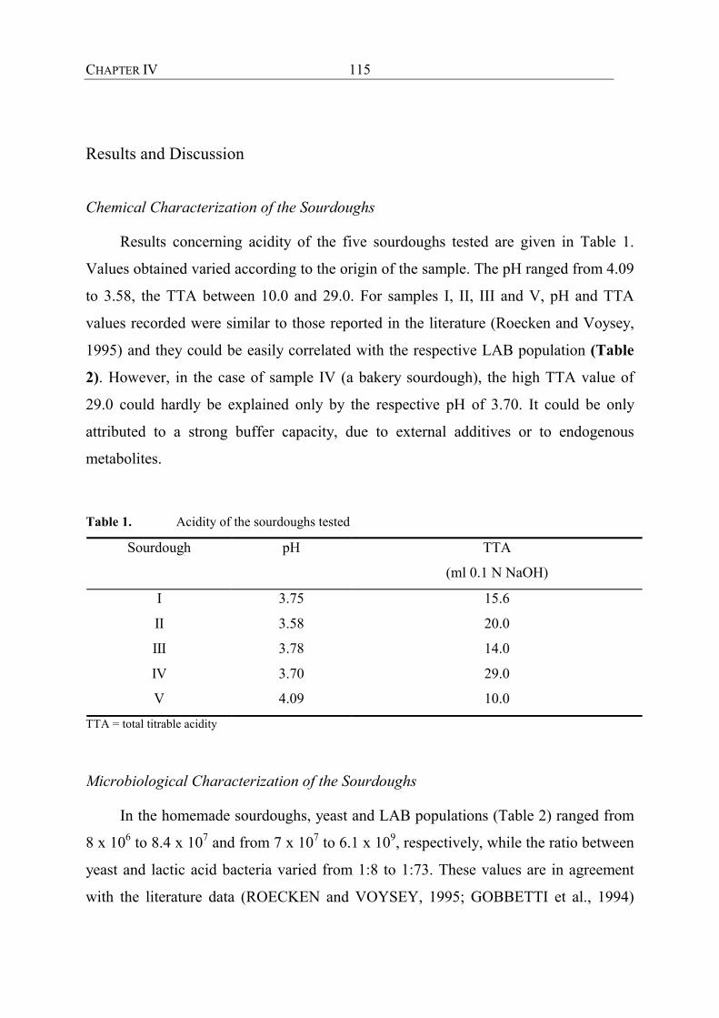
CHAPTER IV
115
Results and Discussion
Chemical Characterization of the Sourdoughs
Results concerning acidity of the five sourdoughs tested are given in Table 1.
Values obtained varied according to the origin of the sample. The pH ranged from 4.09
to 3.58, the TTA between 10.0 and 29.0. For samples I, II, III and V, pH and TTA
values recorded were similar to those reported in the literature (Roecken and Voysey,
1995) and they could be easily correlated with the respective LAB population (Table
2). However, in the case of sample IV (a bakery sourdough), the high TTA value of
29.0 could hardly be explained only by the respective pH of 3.70. It could be only
attributed to a strong buffer capacity, due to external additives or to endogenous
metabolites.
Table 1. Acidity of the sourdoughs tested
Sourdough pH TTA
(ml 0.1 N NaOH)
I 3.75 15.6
II 3.58 20.0
III 3.78 14.0
IV 3.70 29.0
V 4.09 10.0
TTA = total titrable acidity Microbiological Characterization of the Sourdoughs
In the homemade sourdoughs, yeast and LAB populations (Table 2) ranged from
8 x 10⁶ to 8.4 x 10⁷ and from 7 x 10⁷ to 6.1 x 10⁹, respectively, while the ratio between
yeast and lactic acid bacteria varied from 1:8 to 1:73. These values are in agreement
with the literature data (ROECKEN and VOYSEY, 1995; GOBBETTI et al., 1994)

CHAPTER IV
116
and that could be considered as an indication that no addition of commercial yeast took
place and that these doughs were spontaneously fermented. On the contrary, in
sourdoughs originating from bakeries, yeast populations were much higher than those
of lactic acid bacteria. This was probably due to the fact that commercial yeast was
added for leavening acceleration.
Table. 2. Microbial counts of the sourdough tested
Sourdough yeasts
(CFU x 107)
LAB
(CFU x 107)
I 0.9 7
II 0.8 20
III 8.4 610
IV 0.2 0.05
V 0.3 0.06
CFU = colony forming units, LAB = lactic acid bacteria
The yeast colonies observed were grouped according to their morphological
characteristics. Five colony types were distinguished in sourdough I, while four, two,
one and one in sourdoughs II, III, IV and V, respectively. From each colony type,
initially one colony was selected. However, in some cases during the purification
procedure some colonies exhibited a variety on morphology; these colonies were also
purified and thus finally more colonies were selected for identification. This way,
twenty-two colony types were selected from sourdough I, while seven, eleven, one and
four from sourdoughs II, III, IV and V, respectively. In this meaning, homemade
sourdoughs compared to the bakeries ones showed higher variety of colony
morphology.
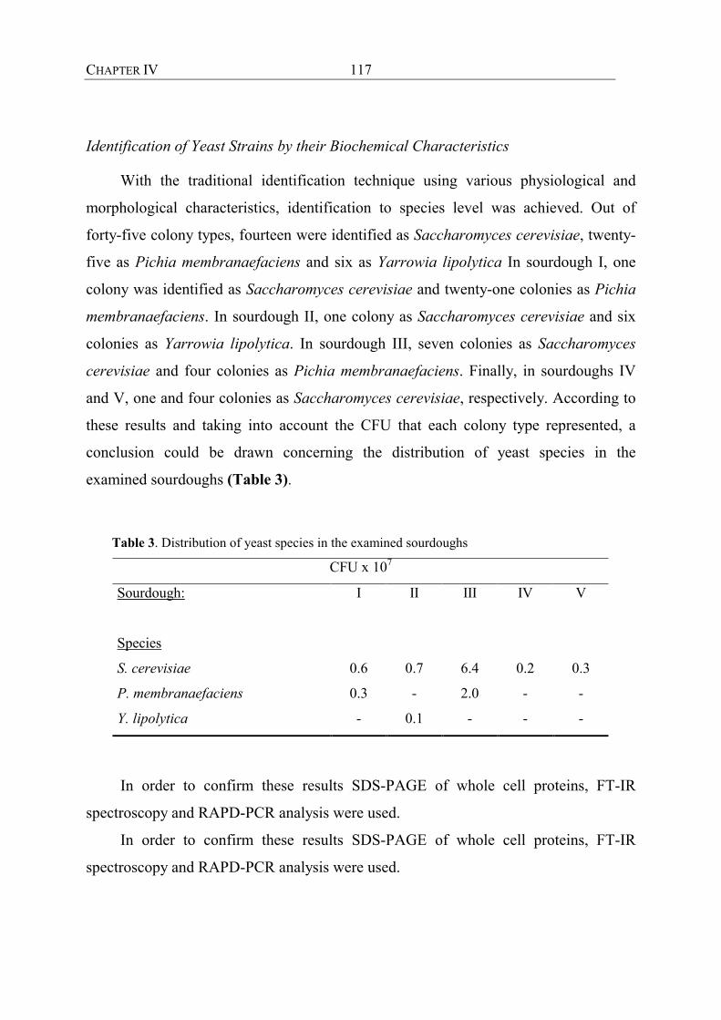
CHAPTER IV
117
Identification of Yeast Strains by their Biochemical Characteristics
With the traditional identification technique using various physiological and
morphological characteristics, identification to species level was achieved. Out of
forty-five colony types, fourteen were identified as Saccharomyces cerevisiae, twenty-
five as Pichia membranaefaciens and six as Yarrowia lipolytica In sourdough I, one
colony was identified as Saccharomyces cerevisiae and twenty-one colonies as Pichia
membranaefaciens. In sourdough II, one colony as Saccharomyces cerevisiae and six
colonies as Yarrowia lipolytica. In sourdough III, seven colonies as Saccharomyces
cerevisiae and four colonies as Pichia membranaefaciens. Finally, in sourdoughs IV
and V, one and four colonies as Saccharomyces cerevisiae, respectively. According to
these results and taking into account the CFU that each colony type represented, a
conclusion could be drawn concerning the distribution of yeast species in the
examined sourdoughs (Table 3).
Table 3. Distribution of yeast species in the examined sourdoughs
CFU x 107
Sourdough:
Species
I II III IV V
S. cerevisiae 0.6 0.7 6.4 0.2 0.3
P. membranaefaciens 0.3 - 2.0 - -
Y. lipolytica - 0.1 - - -
In order to confirm these results SDS-PAGE of whole cell proteins, FT-IR
spectroscopy and RAPD-PCR analysis were used.
In order to confirm these results SDS-PAGE of whole cell proteins, FT-IR
spectroscopy and RAPD-PCR analysis were used.
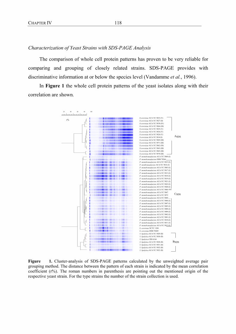
CHAPTER IV
118
Characterization of Yeast Strains with SDS-PAGE Analysis
The comparison of whole cell protein patterns has proven to be very reliable for
comparing and grouping of closely related strains. SDS-PAGE provides with
discriminative information at or below the species level (Vandamme et al., 1996).
In Figure 1 the whole cell protein patterns of the yeast isolates along with their
correlation are shown.
10080604020
S cerevisiae ACA-YC 5023 (V)S cerevisiae ACA-YC 5027 (II)S cerevisiae ACA-YC 5038 (IV)S cerevisiae ACA-YC 5064 (III)S cerevisiae ACA-YC 5025 (V)S cerevisiae ACA-YC 5026 (V)S cerevisiae ACA-YC 5024 (V)S cerevisiae ACA-YC 5014 (I)S cerevisiae ACA-YC 5069 (III)S cerevisiae ACA-YC 5071 (III)S cerevisiae ACA-YC 5062 (III)S cerevisiae ACA-YC 5063 (III)S cerevisiae ACA-YC 5065 (III)S cerevisiae ACA-YC 5070 (III)P. memebranaefaciens ACA-YC 5068 (I)P. memebranaefaciens DSM 70366P. memebranaefaciens ACA-YC 5015 (I)P. membranaefaciens ACA-YC 5016 (I)P. memebranaefaciens ACA-YC 5003 (I)P. memebranaefaciens ACA-YC 5017 (I)P. memebranaefaciens ACA-YC 5013 (I)P. memebranaefaciens ACA-YC 5018 (I)P. memebranaefaciens ACA-YC 5019 (I)P. memebranaefaciens ACA-YC 5021 (I)P. memebranaefaciens ACA-YC 5022 (I)P. memebranaefaciens ACA-YC 5020 (I)P. memebranaefaciens ACA-YC 5066 (III)P. memebranaefaciens ACA-YC 5067 (III)P. memebranaefaciens ACA-YC 5072 (III)P. memebranaefaciens ACA-YC 5004 (I)P. memebranaefaciens ACA-YC 5006 (I)P. memebranaefaciens ACA-YC 5007 (I)P. memebranaefaciens ACA-YC 5005 (I)P. memebranaefaciens ACA-YC 5008 (I)P. memebranaefaciens ACA-YC 5009 (I)P. memebranaefaciens ACA-YC 5002 (I)P. memebranaefaciens ACA-YC 5001 (I)P. memebranaefaciens ACA-YC 5010 (I)P. memebranaefaciens ACA-YC 5011 (I)P. memebranaefaciens ACA-YC 5012 (I)S. cerevisiae NCYC 1200S. cerevisiae DSM 70449Y. lipolytica ACA-YC 5029 (II)Y. lipolytica ACA-YC 5030 (II)Y. lipolytica CBS 6164Y. lipolytica ACA-YC 5028 (II)Y. lipolytica ACA-YC 5031 (II)Y. lipolytica ACA-YC 5032 (II)Y. lipolytica ACA-YC 5033 (II)
ASDS
CSDS
BSDS
r%
Figure 1. Cluster-analysis of SDS-PAGE patterns calculated by the unweighted average pair grouping method. The distance between the pattern of each strain is indicated by the mean correlation coefficient (r%). The roman numbers in parenthesis are pointing out the mentioned origin of the respective yeast strain. For the type strains the number of the strain collection is used.

CHAPTER IV
119
In order to confirm the result of the classical identification, reference strains were
included. All strains were grouped into three major clusters representing the yeast
species as identified by the classical identification. Cluster A contains all S. cerevisiae
strains isolated from traditional Greek sourdough. The reference strains of S.
cerevisiae showed a correlation of 62% with the sourdough isolates. This low
correlation could be attributed to the distortion of the patterns due to the smear
observed at the area of the low molecular weight proteins of the electrophoretic profile
since the similarity was high at the area of high molecular weight proteins. This smear
was probably caused by insufficient washing of the cells and it is most likely
remaining of the growth medium consisting of complexes of proteins with
polysaccharides. Although the cultivation of the cells with the preparation of cell-free
extracts was repeated, the problem persisted. In cluster C all P. membranaefaciens
isolates were included. The respective reference strain showed a correlation of 85%
with the sourdough isolates. Finally, all Y. lipolytica strains were included in cluster B.
The respective reference strains showed a correlation between 80% and 96% with the
sourdough isolates.
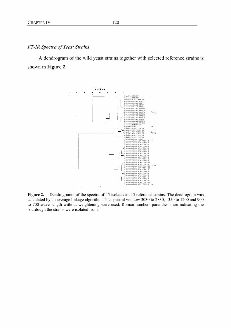
CHAPTER IV
120
FT-IR Spectra of Yeast Strains
A dendrogram of the wild yeast strains together with selected reference strains is
shown in Figure 2.
Y. lipolytica CBS 6124TS. cerevisiae CBS 5900S.cerevisiae ACA-YC 5026 (V)S. cerevisiae ACA-YC 5025 (V)S. cerevisiae ACA-YC 5024 (V)S. cerevisiae ACA-YC 5023 (V)S. cerevisiae ACA-YC 5027 (II)S. cerevisiae ACA-YC 5038 (IV)S. cerevisiae ACA-YC 5063 (III)S. cerevisiae ACA-YC 5069 (III)S. cerevisiae ACA-YC 5064 (III)S. cerevisiae ACA-YC 5070 (III)S. cerevisiae ACA-YC 5062 (III)S. cerevisiae ACA-YC 5014 (I)S. cerevisiae ACA-YC 5065 (III)S. cerevisiae ACA-YC 5071 (III)S. cerevisiae DSM 70449TY. lipolytica M331Y. lipolytica ACA-YC 5030 (II)Y. lipolytica ACA-YC 5029 (II)Y. lipolytica ACA-YC 5028 (II)Y. lipolytica ACA-YC 5033 (II)Y. lipolytica ACA-YC 5031 (II)Y. lipolytica ACA-YC 5032 (II)P. membranaefaciens CBS 107TP. membranaefaciens ACA-YC 5021 (I)P. membranaefaciens ACA-YC 5020 (I)P. membranaefaciens ACA-YC 5013 (I)P. membranaefaciens ACA-YC 5004 (I)P. membranaefaciens ACA-YC 5010 (I)P. membranaefaciens ACA-YC 5007 (I)P. membranaefaciens ACA-YC 5009 (I)P. membranaefaciens ACA-YC 5011 (I)P. membranaefaciens ACA-YC 5015 (I)P. membranaefaciens ACA-YC 5012 (I)P. membranaefaciens ACA-YC 5008 (I)P. membranaefaciens ACA-YC 5001 (I)P. membranaefaciens ACA-YC 5068 (III)P. membranaefaciens ACA-YC 5019 (I)P. membranaefaciens ACA-YC 5067 (III)P. membranaefaciens ACA-YC 5066 (III)P. membranaefaciens ACA-YC 5022 (I)P. membranaefaciens ACA-YC 5006 (I)P. membranaefaciens ACA-YC 5018 (I)P. membranaefaciens ACA-YC 5003 (I)P. membranaefaciens ACA-YC 5016 (I)P. membranaefaciens ACA-YC 5002 (I)P. membranaefaciens ACA-YC 5017 (I)P. membranaefaciens ACA-YC 5072 (III)P. membranaefaciens ACA-YC 5005 (I)
AFT-IR
BFT-IR
CFT-IR
Figure 2. Dendrogramm of the spectra of 45 isolates and 5 reference strains. The dendrogram was calculated by an average linkage algorithm. The spectral window 3030 to 2830, 1350 to 1200 and 900 to 700 wave length without weightening were used. Roman numbers parenthesis are indicating the sourdough the strains were isolated from.

CHAPTER IV
121
Figure 3 is showing the original spectras of selected strains the calculation of the
dendrogram is based on.
Figure. 3. Original spectra in the meaningful range of 1800 to 600 cm-1 wave length of an isolate of Pichia membranaefaciens (bottom), Saccharomyces cerevisiae (middle) and Yarrowia lipolytica (top), each.
The dendrogram is divided in three distinct clusters. Cluster A contains 14 isolates
with the strain S. cerevisiae CBS 5900; the type strain has a major distance. Cluster B
includes 6 isolates with the reference strain Y. lipolytica M331, an isolate from dairy
environment. The type strain of Y. lipolytica does not cluster with the phenon B.
Cluster C contains 25 isolates together with the type strain of P. membranaefaciens.
The clustering of all isolates together with the reference strains was in agreement with
the findings of classical identification.
Characterization of Yeast Flora in Sourdoughs with RAPD Analysis
A crucial point for the construction of meaningful patterns is the isolation of
DNA out of the examined strain. Conventional methods are often based on the
enzymatic lysis of the cell wall followed by time consuming purification steps with
organic solvents. While the yield of DNA is dependent on the degree of lysis it can
differ between strains due to their cell wall structure. For a rapid and reliable

CHAPTER IV
122
extraction a modified procedure of the Quiagen Tissue Kit was used. The disruption of
cells by ultrasonic treatment in a buffer containing chaotropic salts and the separation
and purification of the released DNA by binding at the silica matrix of the spin
columns emerged as a appropriate method. In contrast to the findings of Davin-Regli
et al. (1995) our own results indicated that pattern quality is almost not affected by
DNA concentration. Figure 4 depicts a digitized picture of the RAPD-PCR generated
banding patterns after cluster analysis.
B
RA
PD
AR
APD
CR
APD
r %
0 1
0 2
0 3
0 4
0 5
0 6
0 70
80
90
100
600
8001000
2000
400060008000
20000
bp
Z. ro
uxii
DSM
253
1C
. ova
lis C
BS 7
298T
D. h
anse
nii D
SM 7
0244
S. c
erev
isiae
AC
A-Y
C 5
064
(III)
S. c
erev
isiae
AC
A-Y
C 5
065
(III)
S. c
erev
isiae
AC
A-Y
C 5
071
(III)
S. c
erev
isiae
AC
A-Y
C 5
063
(III)
S. c
erev
isiae
AC
A-Y
C 5
025
(V)
S. c
erev
isiae
AC
A-Y
C 5
026
(V)
S. c
erev
isiae
AC
A-Y
C 5
027
(II)
S. c
erev
isiae
AC
A-Y
C 5
014
(I)S.
cer
evisi
ae A
CA
-YC
502
3 (V
)S.
cer
evisi
ae A
CA
-YC
506
2 (II
I)S.
cer
evisi
ae A
CA
-YC
503
8 (IV
)S.
cer
evisi
ae A
CA
-YC
506
9 (II
I)S.
cer
evisi
ae A
CA
-YC
507
0 (II
I)S.
cer
evisi
ae A
CA
-YC
502
4 (V
)S.
cer
evisi
ae N
CY
C 12
00S.
cer
evisi
ae D
SM 7
0449
Y. li
poly
tica
AC
A-Y
C 5
032
(II)
Y. li
poly
tica
AC
A-Y
C 5
033
(II)
Y. li
poly
tica
CBS
616
4Y.
lipo
lytic
a A
CA
-YC
503
0 (I
I)Y.
lipo
lytic
a A
CA
-YC
503
1 (I
I)Y.
lipo
lytic
a A
CA
-YC
502
8 (I
I)Y.
lipo
lytic
a A
CA
-YC
502
9 (I
I)P.
mem
bran
aefa
cien
s ACA
-YC
501
0 (I)
P. m
embr
anae
faci
ens A
CA-Y
C 5
011
(I)P.
mem
bran
aefa
cien
s ACA
-YC
501
2 (I)
P. m
embr
anae
faci
ens A
CA-Y
C 5
013
(I)P.
mem
bran
aefa
cien
s ACA
-YC
500
2 (I)
P. m
embr
anae
faci
ens A
CA-Y
C 5
003
(I)P.
mem
bran
aefa
cien
s ACA
-YC
500
4 (I)
P. m
embr
anae
faci
ens A
CA-Y
C 5
001
(I)P.
mem
bran
aefa
cien
s ACA
-YC
500
8 (I)
P. m
embr
anae
faci
ens A
CA-Y
C 5
009
(I)P.
mem
bran
aefa
cien
s ACA
-YC
506
8 (II
I)P.
mem
bran
aefa
cien
s ACA
-YC
507
2 (II
I)P.
mem
bran
aefa
cien
s ACA
-YC
506
6 (II
I)P.
mem
bran
aefa
cien
s ACA
-YC
506
7 (II
I)P.
mem
bran
aefa
cien
s ACA
-YC
502
2 (I)
P. m
embr
anae
faci
ens A
CA-Y
C 5
015
(I)P.
mem
bran
aefa
cien
s ACA
-YC
501
8 (I)
P. m
embr
anae
faci
ens A
CA-Y
C 5
017
(I)P.
mem
bran
aefa
cien
s ACA
-YC
501
6 (I)
P. m
embr
anae
faci
ens A
CA-Y
C 5
020
(I)P.
mem
bran
aefa
cien
s ACA
-YC
502
1 (I)
P. m
embr
anae
faci
ens A
CA-Y
C 5
019
(I)P.
mem
bran
aefa
cien
s ACA
-YC
500
6 (I)
P. m
embr
anae
faci
ens A
CA-Y
C 5
005
(I)P.
mem
bran
aefa
cien
s ACA
-YC
500
7 (I)
P. m
embr
anae
faci
ens D
SM 7
0366
Size
Mar
ker
AG
S III
Figure 4. Cluster analysis of RAPD patterns calculated by the unweighted average pair grouping method. The distance between the pattern of each strain is indicated by the mean correlation coefficient (r%). As shown by the metric scale and the DNA size marker fragments between a range of 20 kb and 500 bp are included in the analysis. The roman numbers in parenthesis are pointing out the mentioned origin of the respective yeast strain. For the type strains the number of the strain collection is used.
CHAPTER IV
123
All strains isolated out of the 5 sourdoughs from different homemade and accordingly
bakeries sourdough could be grouped into the 3 major clusters A, B, C standing for S.
cerevisiae, Y. lipolytica and P. membranaefaciens, respectively. For a confirmation of
the allocation, type strains have been enclosed in the investigation. In cluster A the S.
cerevisiae reference strains showed a correlation between 72% and 88% to the
sourdough isolates. The minimum correlation between the representatives of cluster A
was 84%, whereby no significant difference between the different habitats could be
observed. For cluster B similar correlation values can be realized. Sourdough II was
the only habitat where Y. lipolytica has been isolated from. The strains TMW 3.103
and TMW 3.104 exhibited a little lower correlation of 75% to the rest where it was
between 80% and 93%. The largest cluster is that for P. membranaefaciens. Almost all
isolates from sourdough I are belonging to cluster C, whereas only four isolates from
the sourdough III matched in this cluster. The minimum correlation of the isolates is
84%. The reference strain DSM 70366 showed a obvious absence of bands resulting in
correlation factor of 72% in comparison to the rest.
RAPD fingerprinting of yeast strains isolated from 5 Greek sourdoughs of
different origin proved to be a good and economical tool for the characterization of
yeasts from these complex habitats. Besides a description of their diversity it could be
demonstrated that unknown isolates easily could be identified by the inclusion of
reference organisms. In combination with the fast DNA isolation procedure results can
be obtained within 2 days after cultivation, which offers obvious advantages beside
classical identification procedures. In comparison, SDS-PAGE, FT-IR spectroscopy
and RAPD showed the same efficiency at species level discrimination. The major
advantage of RAPD in opposite to SDS-PAGE and FT-IR is that there is no necessity
to cultivate organisms under standardized conditions, because it relies on genotypic
properties. The main advantage of SDS-PAGE against FT-IR and RAPD is that it is
less expensive. The pronounced advantage of identification with FT-IR is that one
strain can be identified within merely 10 minutes work and that this method is a very
CHAPTER IV
124
simple and low-cost procedure. Contrary to SDS-PAGE and RAPD a huge reference
library for food-borne yeasts is available.
In this respect, SDS-PAGE, FT-IR and RAPD-PCR analysis are powerful tools
for identification of yeasts. All methods confirmed the identification results obtained
when the classical technique was used.
S. cerevisiae strains were isolated from all five sourdoughs examined. Depending
on the origin of the sample, P. membranaefaciens or Y. lipolytica could also be found.
When S. cerevisiae was associated with P. membranaefaciens the ratio ranged from
1:0.5-1:0.31 with S. cerevisiae dominating. This could be explained by the fact that S.
cerevisiae isolates could ferment all sugars present in the dough, i.e. glucose, fructose,
sucrose and maltose. Regardless the glucose repression, S. cerevisiae has an advantage
towards other yeast species that can not ferment all these carbon sources. P.
membranaefaciens isolates could only ferment glucose, but about 8 times slower than
S. cerevisiae isolates do. When S. cerevisiae was associated with Y. lipolytica the ratio
was 1:0.14 with S. cerevisiae dominating. This could be explained by the fact that Y.
lipolytica isolates possesses no fermentative metabolism, while they oxidize only
glucose. These facts along with the occurrence of S. cerevisiae strains in every
sourdough tested led to the conclusion that this species represents the indigenous yeast
microflora while P. membranaefaciens and Y. lipolytica appeared to be a random
microflora of traditional greek sourdoughs.
The presence of S. cerevisiae in the sourdough microflora is very often
mentioned (Ottogalli et al., 1996; Rossi, 1996; Gobetti, 1994) and it seems that, in
many cases, is part of the indigenous flora. Nevertheless, its overall presence could be
the result of the extensive use of S. cerevisiae in bakeries. The presence of P.
membranaefaciens strains in sourdough microflora is also mentioned by Rossi (1996)
and it has been found in traditional Portuguese bread doughs (Almeida & Pais, 1996).
For Y. lipolytica it is the first time that the presence of this species in sourdough
microflora is reported. Both species are able to grow at low pH foodstuffs and thus the

CHAPTER IV
125
presence of both can be justified, even though Y. lipolytica is mostly associated with
meat and dairy products.
CHAPTER IV
126
References
Almeida, M. & Pais, C. (1996). Leavening ability and freeze tolerance of yeasts
isolated from traditional corn and rye bread doughs. Appl Environ Microbiol 62, 4401-
4404.
Antuna, B., Martinez-Anaya, M. A. (1993). Sugar uptake and involved enzymatic
activities by yeasts and lactic acid bacteria: their relationship with breadmaking
quality. Int J Food Microbiol 18, 191-200.
Baleiras Couto, M. M, Vogels, J. T. W. E., Hofstra, H., Huis in’t Veld, J.H.J. &
van der Vossen, J. M. B. M. (1995). Random amplified polymorphic DNA and
restriction enzyme analysis of PCR amplified rDNA in taxonomy: two identification
techniques for food-borne yeasts. J Appl Bacteriol 79, 525-535.
Baleiras Couto, M. M, van der Vossen, J. M. B. M., Hofstra, H. & Huis in’t Veld,
J. H. J. (1994). RAPD analysis: a rapid technique for differentiation of spoilage
yeasts. Int J Food Microbiol 24, 249-260.
Barnett, J. A., Payne, R. W. & Yarrow, D. Characteristics and identification, pp. 25.
In: Yeasts, Cambridge, Cambridge University Press (1990)
Barnett, J. A., Payne, R. W. & Yarrow, D. Yeast identification program User's
manual. Cambridge University Press (1985)
Collar, C., Benedito de Barber, C. & Martinez-Anaya, M. A. (1994). Microbial
sourdoughs influence acidification properties and breadmaking potential of wheat
dough. J Food Sci 59 (3), 629-633.
Damiani, P., Gobbetti, M., Cossignani, L., Corsetti, A., Simonetti M. S. & Rossi,
J. (1996). The sourdough microflora: Characterization of hetero- and
homofermentative lactic acid bacteria, yeasts and their interactions on the basis of the
volatile compounds produced. Lebensm Wiss Technol 29, 63-70.
CHAPTER IV
127
Davin-Regli, A., Abed, Y., Charrel, R. N., Bollet, C. & Micco, P. (1995). Variation
in DNA concentrations significantly affect the reproduceability of RAPD fingerprint
patterns. Res Microbiol 146, 561-568.
Deak, T. (1995). Methods for the rapid detection and identification of yeasts in foods.
Trends Food Sci Technol 6, 287-292.
Deak, T. & Beuchat, L. R. Handbook of food-spoilage yeasts. CRC Press, Inc. (1996)
Gobbetti, M., Corsetti, A., Rossi, J., LaRosa, F. & DeVincenzi, S. (1994).
Identification and clustering of lactic acid bacteria and yeasts from wheat sourdoughs
of central Italy. Ital J Food Sci 1, 85-94.
Hansen, B. & Hansen, A. (1994). Volatile compounds in wheat sourdoughs produced
by lactic acid bacteria and sourdough yeasts. Z Lebensm Unters Forsch 198, 202-209.
Kockova-Kratochvilova, A. Yeasts and yeast-like organisms. VCH Publishers, New
York, NY (USA) (1996)
Kosse, D., Seiler, H., Amann, R. I., Ludwig, W. & Scherer, S. (1997). Identification
of yeasts with 18S rRNA-targeted oligonucleotide probes. System Appl Microbiol 20,
468-480.
Kreger-van Rij, N. W. J. A taxonomic study, pp 52. In: The yeasts. Elsevier Science
Publishers B.V.-Amsterdam (1987)
Kümmerle, M., Scherer, S. & Seiler, H. (1998). Rapid and reliable identification of
food-borne yeasts by Fourier-transform infrared spectroscopy. Appl Environ Microbiol
64 (6), 2207-2214.
Laidlaw, L., Tompkins, T. A., Savard, L. & Dowhanick, T. M. (1996).
Identification and differentiation of brewing yeasts using specific and RAPD
polymerase chain reaction. J Am Soc Brew Chem 54(2), 97-102.
Linko, Y-Y., Javanainen, P. & Linko, S. (1997). Biotechnology of bread baking.
Trends Food Sci Technol 8, 339-344.

CHAPTER IV
128
Lopes, M. B., Soden, A., Martens, A. L., Henschke, P. A. & Langridge, P. (1998).
Differentiation and species identification of yeasts using PCR. Int J System Bacteriol
48, 279-286.
Martinez-Anaya, M. A. (1996). Enzymes and bread flavor. J Agric Food Chem (44)
9, 2469-2480.
Martinez-Anaya, M. A., Torner, M. J., Benedito de Barber, C. (1990). Microflora
of the sourdough of wheat flour bread. XIV. Changes in volatile compounds during
fermentation of doughs prepared with pure microorganisms and their mixtures. Z
Lebensm Unters Forsch 190, 126-131.
Mäntynen, V. H., Korhola, M., Gudmundsson, H., Turakainen, H., Alfredsson, G.
A., Salovaara, H. & Lindström, K. (1999). A polyphasic study on the taxonomic
position of industrial sour dough yeasts. System Appl Microbiol 22, 87-96.
Naumann, D., Helm, D. & Schultz, C. (1994). Characterization and identification of
microorganisms by FT-IR spectroscopy and FT-IR microscopy, pp. 67. In: Bacterial
diversity and systematics (F.G. Priest, A. Ramos-Cormenzana, B. J. Tindall, eds.),
Plenum Press, New York 1994.
Noronha-da-Costa, P., Rodrigues, C., Spencer-Martins, I. & Loureiro, V. (1996).
Fatty acid patterns of film-forming yeasts and new evidence for the heterogeneity of
Pichia membranaefaciens. Lett Appl Microbiol 23, 79-84.
Ottogalli, G., Galli, A. & Foschino, R. (1996). Italian bakery products obtained with
sourdough: Characterization of the typical microflora. Adv Food Sci 18 (5/6), 131-144.
Romano, A., Casaregola, S., Torre, P. & Gaillardin, C. (1996). Use of RAPD and
mitochondrial DNA RFLP for typing of Candida zeylanoides and Debaryomyces
hansenii yeast strains isolated from cheese. System Appl Microbiol 19, 255-264.
Roecken, W. & Voysey, P. A. (1995). Sourdough fermentation in bread making. J
Appl Bacteriol 79, 38-48.
CHAPTER IV
129
Rosenquist, H. & Hansen, A. (1998). The antimicrobial effect of organic acids,
sourdough and nisin against Bacillus subtilis and B. licheniformis isolated from wheat
bread. J Appl Microbiol 85, 621-631.
Rossi, J. (1996). The yeasts in sourdough. Adv Food Sci 18 (5/6), 201-211.
Salovaara, H. Lactic acid bacteria in cereal products. pp. 115-138. In: Lactic acid
bacteria Technology and Health effects 2nd ed. (S. Salminen and A. von Wright, eds.).
Marcel Dekker (1998).
Sugihara, T. F., Kline, L. & Miller, M. W. (1971). Microorganisms of the San
Francisco sourdough bread process. I. Yeasts responsible for the leavening action.
Appl Microbiol 21(3), 456-458.
Tompkins, A. T., Stewart, R., Savard, L., Russell, I. & Dowhanick, T. M. (1996).
RAPD-PCR characterization of brewery yeast and beer spoilage bacteria. J Am Soc
Brew Chem 54(2), 91-96.
Vancanneyt, M., Pot, B., Hennebert, G & Kersters, K. (1991). Differentiation of
yeast species based on electrophoretic whole-cell protein patterns. System Appl
Microbiol 14, 23-32.
Vandamme, P., Pot, B., Gillis, M., de Vos, P., Kersters, K. & Swings, J. (1996).
Polyphasic taxonomy, a consensus approach to bacterial systematics. Microbiol Rev
60(2), 407-438.
Vogel, R. F., Knorr, R., Müller, M. R. A., Steudel, U., Gänzle, M. G., Ehrmann,
M. A. (1999). Non-dairy lactic fermentations: the cereal world. Antonie van
Leeuwenhoek 76, 403-411.
Welsh, J. & McClelland, M. (1990). Fingerprinting genomes using PCR with
arbitrary primers. Nucl Acid Res 18, 7213-7219.
Williams, J. G. K., Kubelik, A. R., Livak, K. J., Rafalski, J. A. & Tingey, S. V.
(1990). DNA polymorphism amplified by arbitrary primers are useful as genetic
markers. Nucl Acid Res 18, 6531-6536.

ADDITIONAL INSIGHTS AND CONCLUSIONS 130
ADDITIONAL INSIGHTS AND CONCLUSIONS
The consequent application of molecular techniques to investigate microbial
ecosystems in food fermentations is an important prerequisite for the better
understanding of the respective processes. This was demonstrated for the recalcitrant
microorganisms of sourdough fermentations. The results obtained not only provide a
profound insight in the discriminatory power of the respective state of the art tools.
New useful methods were developed to describe occurrence and behavior of
lactobacilli in the sourdough ecosystem, and a so far unmatched overview is provided
on the dynamics of the LAB sourdough flora. Besides the scientific insights in the
ecology of these fermentation the organisms involved, and their behavior in response
to external and internal process parameters conclusions may be drawn for industrial
applications, to ensure product quality, to improve the process, steering it in a suitable
way or to construct novel processes on a rationale basis.
Historically, methods to achieve all these insights changed dramatically during
the last 20 years. At the beginning of modern microbiology microorganisms have been
classified by their phenotypic properties, like certain metabolic activities or their
morphology. This aspects are sufficient for the construction of classification schemes
and still important for the screening and evaluation of strains with desired
physiological properties. However, for fast and reliable identification and moreover
detection purposes these methods are improper due to the variability of the phenotype
of bacteria as a fact for example of the adaptation to special environments, mutations
or the loss of plasmids. Especially for LAB, differences in fermentation patterns are
small giving raise to uncertain results. In many cases and as demonstrated in this work
species from cereal fermentations exhibit a strong strain dependent phenotype making
a clear identification almost impossible. Therefore, to cope with the demands of
modern food microbiology for a fast and unequivocal identification more accurate
methods are desired. In the last decade methods targeting the genotype achieved the
greatest importance. Aside from the latter, chemotaxonomic methods meaning the
ADDITIONAL INSIGHTS AND CONCLUSIONS 131
setup of a classification scheme based on the chemical characterization of cellular
components like whole cell protein patterns, fatty acid analyses, determination of the
peptidoglycan type, or the G+C content of the DNA are applied. However, with the
breakthrough and establishment of genotypic methods they are losing importance, but
are still necessary in polyphasic approaches as for example for the description of new
taxons, and to determine a strains suitability to conduct a process.
Today the phylogeny of microorganisms is reflected in a genealogical tree
divided into the three domains Bacteria, Archeae and Eucarya is based on rRNA
sequences. The 16S rRNA-based trees are reflecting the phylogenetic relationship
among prokaryotes and therefore had a major impact on their taxonomy. Nucleotide
sequencing and other analytical techniques – most notably, PCR-based DNA
amplification methods – have dramatically expanded the rRNA sequence database
leading to a more and more detailed classification of species. Culture independent
rRNA approaches led to additional insights of bacterial ecosystems. These theoretical
awareness of the significance of the rRNA represents a framework for applied
approaches to answer practical problems in environmental and food microbiology. A
fundamental technical progress to realize this was the introduction of the polymerase
chain reaction. In the following approaches we used to investigate the ecology of
cereal fermentations are presented and basic conclusions are drawn.
A fundamental question at the beginning of our investigations was if the LAB
grown in the laboratory are really representing the in situ diversity of the fermentation
flora or if these microorganisms that grow on isolation plates are the ones best adapted
to the artificial growth conditions and are not necessarily those that are metabolically
active or abundant in the environment. This would drastically influence the results of
methods including a pre-selection by a cultivation step and therefore introducing bias.
The main steps of this investigation were the extraction of total community DNA from
the sourdough, amplification of the 16S rDNA, cloning the 16S rDNA-plasmid-
construct into E. coli, reamplification of the 16S rDNA inserts, and finally
characterization of the inserts. For the DNA extraction we used a strategy described in
ADDITIONAL INSIGHTS AND CONCLUSIONS 132
Chapter II. For the amplification primers with a Not I cleavage site were used,
making an easy sticky end ligation into the vector possible. After cloning, insert-
positive clones were picked followed by a reamplification of the insert. To
characterize the insert we sequenced only a single base (T), which allowed a higher
throughput and resulted in meaningful patterns. The inclusion of reference organisms
usually found in such fermentations allowed a direct allocation. For a definitive
identification inserts were sequenced. Finally it could be demonstrated that the
sourdough flora of the investigated system consisted mainly of L. amylovorus and L.
pontis. This correlated well with previous culture based investigations. It can be
concluded, that on the one hand side methods based on the direct DNA isolation from
the sourdough are appropriate for the description of such fermentations and on the
other hand methods including a cultivation step are reflecting the actually present
flora. For a routine analysis of food fermentations this method would be too time
consuming.
The intention of a further culture independent study was the fast and
simultaneous monitoring of the fermentation flora throughout the fermentation time.
LAB and bacteria in general may exhibit species dependent length polymorphisms
within the same variable regions on the 16S rDNA. When amplifying this region from
mixed culture DNA using the same universal primers products with different length
are generated. These differences may amount only a few base pairs making a highly
resolving electrophoresis necessary. We detected the PCR-products by using a Cy-5
labeled primer on an sequencer. The development was followed of a lab-scale
sourdough fermentation described in Chapter I. The flora of this fermentation was
mainly composed of L. amylovorus, L. pontis and the closely related L. frumenti as
described in Chapter III. With the ss-amplified rDNA-FLP (single stranded amplified
ribosomal DNA fragment length polymorphism) two peaks could be detected one with
269 bp representing L. amylovorus and one with 273 bp representing both species, L.
ADDITIONAL INSIGHTS AND CONCLUSIONS 133
pontis and L. frumenti (Vogel et al., 19991). The calculation of the peak area allowed a
semi-quantitative estimation about the presence of these organisms. These results were
in good correlation with the numbers calculated after RAPD analysis (Chapter I). The
developed method has also been successfully applied to monitor crop associated
lactobacilli in ducks during a feeding experiment. It can be concluded that this method
constitutes an ideal possibility for a simultaneous monitoring of several species.
Besides the detection, quantification and recording of population dynamics are
possible. A prerequisite for this method is to know how the flora is composed and that
the component species have a suitable rRNA fragment length polymorphism.
In Chapter II a culture independent method was described demonstrating the
possibility of a fast and specific identification and detection of sourdough LAB. A
PCR system with primers specific for the closely related species L. pontis, L. panis and
L. frumenti (see Chapter III) allowed the differentiation of these species and on the
other side the tracking during a sourdough fermentation. Such PCR systems may have
practical applications like the survey of industrial fermentations or to verify species
identity. This may especially be true for sourdough LAB like L. pontis which is known
to exhibit a heterogeneous fermentation profile and morphological appearance, and
therefore is hardly identified by the latter.
RAPD-PCR is another genotypic method we applied for the characterization of
the microflora of cereal fermentations. In contrast to rRNA based methods, RAPD is
reflecting polymorphisms on the entire genome leading to the differentiation of
genomovars. At first RAPD is a typing method leading to a characteristic fingerprint
of single organisms. These fingerprints may than be used for discrimination from other
isolates or to proof the authenticity of strains. Potential industrial applications could be
the specific detection of starter organisms with certain properties. We did an
investigation where the persistence of strains of L. sanfranciscensis - a major element
in this industrial starter preparation - was controlled, when a different flour type was
1 Vogel, R. F., Knorr, R., Müller, M. R. A., Steudel, U., Gänzle, M. G. & Ehrmann, M. A. (1999). Non dairy lactic fermentations: the cereal world. Antonie van Leeuwenhoek 76, 403-411.

ADDITIONAL INSIGHTS AND CONCLUSIONS 134
fermented spontaneously. The comparison of RAPD patterns of the original strains
with the patterns of the new isolates, showed that the strains were still present besides
some other species. This makes clear that RAPD typing is also applicable for routine
analyses.
RAPD typing is moreover an appropriate tool for ecological investigations of
microbial communities to obtain an qualitative and quantitative overview of the
microbial composition. Representatives of each cluster can be identified (see rDNA
sequencing) making a fast and reliable description of such fermentations feasible. We
applied this strategy for an microbial investigation of indigenous Greek sourdough
fermentations. In comparison to a physiological characterization this approach
revealed to be faster and the result were more definite. The major part of isolates
belong to L. sanfranciscensis almost independent of the origin, followed by Weissella
species, Pediococcus species, Enterococcus species, and additional Lactobacillus
species. The same sourdoughs have been investigated on their yeast flora (see
Chapter IV). It was demonstrated that this RAPD typing technique is not only limited
to LAB. The cluster analyses was consistent with other typing methods, like SDS-
PAGE and FT-IR analyses. Furthermore, the wild yeast strains could be identified by
including reference and type strains in the analyses.
A basic question in taxonomy is the differentiation below the species and
subspecies level. RAPD typing is often leading to similar patterns with polymorphisms
within one species or clearly set aside patterns leading to a differentiation of
genomovars which could even be used for identification at the strain level. This may
especially be useful for the evaluation of starter organisms with particular properties.
For this reason and to seek out if there exist regional strain varieties of isolates of L.
sanfranciscensis from various sourdoughs and starters from different locations in
Europe have been investigated by RAPD typing. The major part of the strains grouped
together in 7 clusters. A correlation to the provenance could not be made. An
explanation for this may be due to commercial starters, which are not only limited to
industrial but also to artesanal products or because they originate from artesanal ones.
ADDITIONAL INSIGHTS AND CONCLUSIONS 135
To evaluate the discriminatory power of RAPD these typing method was
compared with AFLP derived clusters (Leissner et al., 20002). In this study it could be
demonstrated that the AFLP technique originally developed for analyses in the field of
eucaryotes is suitable as well as for applied problems in the field of LAB. Within the
genera Lactobacillus, Pediococcus, Enterococcus and Weissella the organisms could
be unequivocally distinguished at the species level. A comparison with the RAPD
generated patterns showed good correlation, whereas the RAPD patterns exhibited
more polymorphisms within a species.
In Chapter I another strategy including RAPD typing was presented. The main
targets of this study have been to check the stability of the microbial composition in a
lab-scale fermentation when started with industrial sourdough. After several
refreshment steps randomly picked clones were subjected for RAPD typing by what a
percental composition could be calculated. The established fermentation was
monitored in the same way and the influence of external parameters like temperature
was determined. The consistency of the RAPD patterns over a longer time and their
independence of DNA quality and PCR batches allowed a database setup by which
subsequent patterns were allocated and strains were identified. Finally, it could be
demonstrated that it is possible to entirely describe the microbiological status of an
industrial process, to transfer it into a lab-scale fermentation with a similar flora and
that RAPD typing may be a suitable to tool to verify this model.
Apart from the taxonomic investigations but as a result of the genotypic
characterization and the phylogenetic classification of the sourdough isolates adhesion
studies have been performed (Müller et al., 19983). Many isolates from cereal
fermentations showed a close phylogenetic relationship or even identicalness to
species from the human environment like L. pontis or L. frumenti to L. vaginalis, L.
oris and L. reuteri or L. amylovorus to L. acidophilus or L. johnsonii. This fact was the
2 Leissner, C. E. W., Müller, M. R. A., Niessen, L., Ehrmann, M. A. & Vogel, R. F. (2000). Use of the AFLP fingerprinting method for the differentiation and identification of lactic acid bacteria. Biospektrum Sonderausgabe, 155. 3 Müller, M. R. A., Rouvet, M., Brassart, D., Böcker, G., Ehrmann, M. A. & Vogel, R. F. (1998). Adhesion of Lactobacillus strains from cereal fermentations to human intestinal cells. Int Dairy Journal 8, 584
ADDITIONAL INSIGHTS AND CONCLUSIONS 136
motivation to investigate the adhesion properties of LAB from the cereal environment
to human Caco-2 cells as an important feature for probiotic cultures. Strains of L.
reuteri, L. amylovorus and L. fermentum from different cereal fermentation showed
adhesion to the Caco-2 cells as revealed by liquid scintillation counting and laser
scanning microscopy. This capability renders them possible probiotic organisms but
further investigations have to follow.


![Modulhandbuch Wissenschaftszentrum Weihenstephan fur ... · Mikrobiologie / Organismische und Molekulare Mikrobiologie (Microbiology / Organismic and Molecular Microbiology) [WZ2503]](https://static.fdokument.com/doc/165x107/5dd09306d6be591ccb61a6c0/modulhandbuch-wissenschaftszentrum-weihenstephan-fur-mikrobiologie-organismische.jpg)