Metabolism and Action of Glucocorticoids and Interference ... · SDR short chain...
163
Metabolism and Action of Glucocorticoids and Interference with the Antioxidant Redox Pathway Inauguraldissertation zur Erlangung der Würde eines Doktors der Philosophie vorgelegt der Philosophisch-Naturwissenschaftlichen Fakultät der Universität Basel von Denise Verena Kratschmar aus Böblingen Deutschland Basel, 2011 Originaldokument gespeichert auf dem Dokumentenserver der Universität Basel edoc.unibas.ch Dieses Werk ist unter dem Vertrag „Creative Commons Namensnennung-Keine kommerzielle Nutzung-Keine Bearbeitung 2.5 Schweiz“ lizenziert. Die vollständige Lizenz kann unter creativecommons.org/licences/by-nc-nd/2.5/ch eingesehen werden.
Transcript of Metabolism and Action of Glucocorticoids and Interference ... · SDR short chain...

Metabolism and Action of Glucocorticoids and Interference
with the Antioxidant Redox Pathway
Inauguraldissertation zur
Erlangung der Würde eines Doktors der Philosophie
vorgelegt der
Philosophisch-Naturwissenschaftlichen Fakultät
der Universität Basel
von
Denise Verena Kratschmar
aus Böblingen
Deutschland
Basel, 2011
Originaldokument gespeichert auf dem Dokumentenserver der Universität Basel edoc.unibas.ch
Dieses Werk ist unter dem Vertrag „Creative Commons Namensnennung-Keine kommerzielle Nutzung-Keine Bearbeitung 2.5 Schweiz“ lizenziert. Die vollständige Lizenz
kann unter creativecommons.org/licences/by-nc-nd/2.5/ch
eingesehen werden.

Namensnennung-Keine kommerzielle Nutzung-Keine Bearbeitung 2.5 Schweiz
Sie dürfen:
das Werk vervielfältigen, verbreiten und öffentlich zugänglich machen
Zu den folgenden Bedingungen:
Namensnennung. Sie müssen den Namen des Autors/Rechteinhabers in der von ihm festgelegten Weise nennen (wodurch aber nicht der Eindruck entstehen darf, Sie oder die Nutzung des Werkes durch Sie würden entlohnt).
Keine kommerzielle Nutzung. Dieses Werk darf nicht für kommerzielle Zwecke verwendet werden.
Keine Bearbeitung. Dieses Werk darf nicht bearbeitet oder in anderer Weise verändert werden.
• Im Falle einer Verbreitung müssen Sie anderen die Lizenzbedingungen, unter welche dieses Werk fällt, mitteilen. Am Einfachsten ist es, einen Link auf diese Seite einzubinden.
• Jede der vorgenannten Bedingungen kann aufgehoben werden, sofern Sie die Einwilligung des Rechteinhabers dazu erhalten.
• Diese Lizenz lässt die Urheberpersönlichkeitsrechte unberührt.
Quelle: http://creativecommons.org/licenses/by-nc-nd/2.5/ch/ Datum: 3.4.2009
Die gesetzlichen Schranken des Urheberrechts bleiben hiervon unberührt.
Die Commons Deed ist eine Zusammenfassung des Lizenzvertrags in allgemeinverständlicher Sprache: http://creativecommons.org/licenses/by-nc-nd/2.5/ch/legalcode.de
Haftungsausschluss:Die Commons Deed ist kein Lizenzvertrag. Sie ist lediglich ein Referenztext, der den zugrundeliegenden Lizenzvertrag übersichtlich und in allgemeinverständlicher Sprache wiedergibt. Die Deed selbst entfaltet keine juristische Wirkung und erscheint im eigentlichen Lizenzvertrag nicht. Creative Commons ist keine Rechtsanwaltsgesellschaft und leistet keine Rechtsberatung. Die Weitergabe und Verlinkung des Commons Deeds führt zu keinem Mandatsverhältnis.
Genehmigt von der Philosophisch-Naturwissenschaftlichen Fakultät
auf Antrag von
Prof. Dr. Alex Odermatt
PD PhD. Hubert Hug
Basel, den 24.05 2011
Prof. Dr. Martin Spiess
Dekan
CONTENT
Index
1 Abbreviations .................................................................................................................... 1
2 Summary ........................................................................................................................... 3
3 Introduction ....................................................................................................................... 5
3.1 Glucocorticoids and mineralocorticoids: a historical overview ................................... 5
3.1.1 Physiological synthesis and regulation of glucocorticoids .................................. 6
3.1.2 Pathology of impaired glucocorticoid release ..................................................... 9
3.2 Pre-receptor metabolism and action of glucocorticoids ........................................... 10
3.2.1 11β-hydroxysteroid dehydrogenases ................................................................ 10
3.2.1.1 11β-hydroxysteroid dehydrogenase type 2 ................................................. 11
3.2.1.2 Pathologies related to 11β-hydroxysteroid dehydrogenase type 2 ............. 11
3.2.1.3 11β-hydroxysteroid dehydrogenase type 1 and hexose-6-phosphate-dehydrogenase ........................................................................................... 12
3.2.1.4 Pathologies related to 11β-hydroxysteroid dehydrogenase type 1 ............. 13
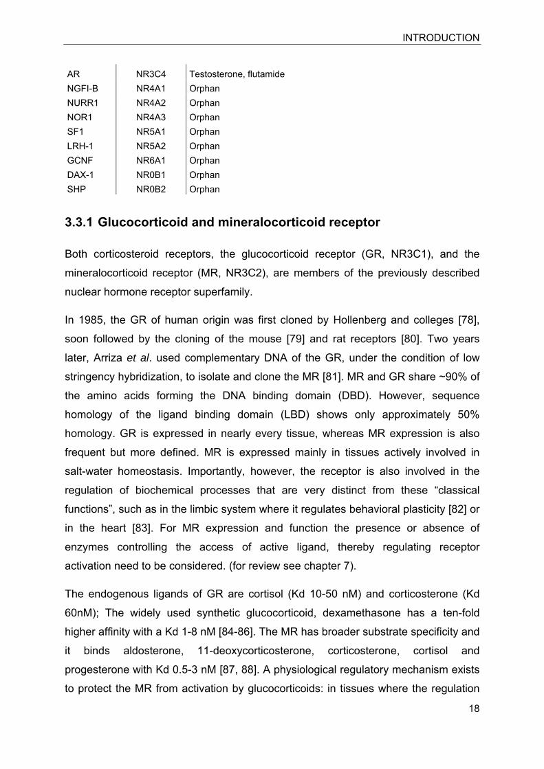
3.3 The nuclear receptor superfamily ............................................................................ 15
3.3.1 Glucocorticoid and mineralocorticoid receptor .................................................. 18
3.3.2 General mechanism of transactivation ............................................................. 19
3.4 Antioxidant redox pathway ....................................................................................... 20
3.4.1 Nuclear factor-erythroid 2 (NF-E2)-related factor 2 (Nrf2) ................................ 23
3.4.2 The Kelch-like ECH-associated protein1 .......................................................... 24
3.4.3 Interaction of Nrf2 and Keap1: Putative mechanism within the antioxidant redox pathway ............................................................................................................ 26
4 DIBUTYLTIN DISRUPTS GLUCOCORTICOID RECEPTOR FUNCTION AND IMPAIRS GLUCOCORTICOID-INDUCED SUPPRESSION OF CYTOKINE PRODUCTION. ....... 29
5 11β-HYDROXYSTEROID DEHYDROGENASE 1 INHIBITING CONSTITUENTS FROM ERIOBOTRYA JAPONICA REVEALED BY BIOACTIVITY-GUIDED ISOLATION AND COMPUTATIONAL APPROACHES. .............................................................................. 41
6 CHARACTERIZATION OF ACTIVITY AND BINDING MODE OF GLYCYRRHETINIC ACID DERIVATIVES INHIBITING 11β-HYDROXYSTEROID DEHYDROGENASE TYPE 2 ...................................................................................................................................... 51
CONTENT
7 TISSUE-SPECIFIC MODULATION OF MINERALOCORTICOID RECEPTOR FUNCTION BY 11β-HYDROXYSTEROID DEHYDROGENASES: AN OVERVIEW ...... 66
7.1 Abstract .................................................................................................................... 66
7.2 Introduction .............................................................................................................. 66
7.3 Kidney ...................................................................................................................... 67
7.4 Gastrointestinal tract ................................................................................................ 72
7.5 Adrenals ................................................................................................................... 72
7.6 Immune system ........................................................................................................ 73
7.7 Brain ......................................................................................................................... 74
7.8 Bone ......................................................................................................................... 81
7.9 Adipose tissue .......................................................................................................... 83
7.10 Heart ........................................................................................................................ 84
7.11 Skeletal muscle ........................................................................................................ 88
7.12 Skin .......................................................................................................................... 88
7.13 Outlook ..................................................................................................................... 89
7.14 Acknowledgements .................................................................................................. 89
7.15 References ............................................................................................................... 89
8 ELEVATED 11β-HSD1-MEDIATED GLUCOCORTICOID ACTIVATION RESULTS IN IMPAIRED NRF2-DEPENDENT ANTIOXIDANT RESPONSE ..................................... 100
8.1 Abstract .................................................................................................................. 101
8.1.1 Keywords ........................................................................................................ 101
8.1.2 Abbreviations .................................................................................................. 101
8.2 Introduction ............................................................................................................ 101
8.3 Experimental procedure ......................................................................................... 104
8.3.1 Materials ......................................................................................................... 104
8.3.2 Cell culture ...................................................................................................... 104
8.3.3 Transfection of cells ........................................................................................ 104
8.3.4 Detection of hydrogen peroxide sensitivity by confocal microscopy ............... 105
8.3.5 Nrf2 transactivation assays ............................................................................. 105
CONTENT
8.3.6 Determination of 11β-HSD1 activity in intact H4H1 cells ................................ 106
8.3.7 Analysis of mRNA expression by real-time RT-PCR ...................................... 106
8.3.8 Calculations and statistical analysis ............................................................... 107
8.4 Results ................................................................................................................... 108
8.4.1 Glucocorticoid-mediated inhibition of Nrf2-dependent reporter gene activation in HEK-293 cells ................................................................................................. 108
8.4.2 Induction of the Nrf2-dependent ARE8L-reporter construct in rat H4IIE hepatoma cells ................................................................................................ 109
8.4.3 11β-HSD1-mediated glucocorticoid activation suppresses Nrf2 transactivation capacity ........................................................................................................... 111
8.4.4 Sulforaphane does not affect 11β-HSD1 activity ............................................ 112
8.4.5 11β-HSD1 inhibitors and GR antagonists improve Nrf2 transactivation capacity . ........................................................................................................................ 113
8.4.6 NQO1 expression in H4IIE cells is suppressed by cortisol but not by cortisone .. ........................................................................................................................ 114
8.4.7 Inhibition of 11β-HSD1 restored sulforaphane-induced NQO1 mRNA expression in H4H1 cells ................................................................................ 115
8.4.8 Glucocorticoid-dependent impairment of HO-1 function and susceptibility to H2O2 ................................................................................................................ 117
8.5 Discussion .............................................................................................................. 119
8.6 Acknowledgements ................................................................................................ 122
8.7 References ............................................................................................................. 123
9 Conclusion and Outlook ................................................................................................ 127
10 References ................................................................................................................ 137
11 Acknowledgements ................................................................................................... 155
ABBREVIATIONS
1
1 Abbreviations 11β-HSD1 11β-hydroxysteroid dehydrogenase type 1
11β-HSD2 11β-hydroxysteroid dehydrogenase type 2
ABCC ATP-binding cassette, sub-family C (CFTR/MRP), member
ACTH adrenocorticotropic hormone
ADH alcohol dehydrogenase
AME apparent mineralocorticoid excess
ARE antioxidant responsible element
C/EBP CCAAT/enhancer-binding-protein
CBX carbenoxolone
CRD apparent cortisone reductase deficiency
CRF corticotrophin releasing factor
CYP11B1 11β-hydroxylase
DBT dibutyltin
GA glycerrhetinic acid
GAPDH glyceraldehyde-3-phosphate dehydrogenase
GR glucocorticoid receptor
GRE glucocorticoid response elements
GSTA2 glutathione S-transferase alpha 2
H6PDH hexose-6-phosphate dehydrogenase
HO-1 heme oxygenase 1
HPA hypothalamic-pituitary-adrenal axis
HTS high-throughput-screening
IL-6 interleukine-6
Keap1 kelch-like ECH-associated protein 1
MR mineralocorticoid receptor
ABBREVIATIONS
2
NALD nonalcoholic liver disease
NASH nonalcoholic steatohepatitis
NF-κB nuclear factor-kappa B
NQO1 NAD(P)H dehydrogenase, quinone 1
Nrf2 nuclear factor (erythroid-derived 2)-like 2
S sulforaphane
SCN suprachiasmatic nucleus
SDR short chain dehydrogenase/reductase
TBHQ tertiar butyl hydroquinone
TBT tributyltin
TNFα tumor-necrosis-factor α
SUMMARY
3
2 Summary Disturbance of endocrine systems and signaling pathways can lead to severe
disorders. Such disorders can have endogenous as well exogenous origin. The
awareness of environmentally occurring xenobiotics that are able to directly interfere
with and modulate the action of endogenous hormones has driven the need for
mechanistic studies. Although there is a vast literature on potentially endocrine
disrupting chemicals, there are only few studies investigating disturbance of
glucocorticoid action by xenobiotics, despite of the importance of these hormones. In
this work, the organotin dibutyltin (DBT) was identified as an endocrine disruptor of
the glucocorticoid pathway. Its extensive use in plastic industry, as well as an
antifouling agent explains its occurrence in water and seafood. In the present study,
we were able to show that DBT disturbs GR mediated anti-inflammatory effects.
Furthermore, DBT was found to potentiate NFκB mediated production of the pro-
inflammatory cytokines IL-6 and TNFα in macrophages. The presented work
therefore contributes to the mechanistic understanding of DBT-induced
immunotoxicity.
There are several therapeutic purposes accompanied by the modulation of the
endogenous hormone system. In traditional medicines natural compounds, and plant
extracts are applied since centuries for different purposes, including the treatment of
diseases such as diabetes and hypertension. The benefits of evidence based
medicines, even if their mechanisms of action are unknown, are widely accepted. In
conventional medicine the re-awareness of naturally derived compounds and their
huge potential promoted the investigation of the underlying specific mechanisms of
action of such compounds over the last decades. In this context, the present work
investigated effects of eriobotrya japonica, a plant used for anti-diabetic treatment in
Chinese medicine. The project aimed to identify potential constituents that are active
on 11β-HSD1. Several pentacyclic triterpenes were isolated and further
characterized. These compounds included potent and, compared with 11β-HSD2,
selective 11β-HSD1 inhibitors such as corosolic acid and urosolic acid, as well as
urosolic acid derivatives with only low inhibitory potential but considerable synergistic
effects. Inhibitors for research and/or therapeutic purposes ideally display high
selectivity to avoid miss-leading interpretations of their action. Furthermore,
therapeutic intervention requires selective inhibitors to prevent unexpected side
SUMMARY
4
effects. The most famous triterpenoid inhibiting 11β-HSD enzymes is glycyrrhetinic
acid (GA), present in liquorice. GA is a potent, but non-selective inhibitor of both 11β-
HSD isoformes. Recently, GA was used as a starting compound and chemical
modifications of its back-bone enabled the development of potent and specific
inhibitors against 11β-HSD2. The present work describes the characterization of
these novel 11β-HSD2 inhibitors. The inhibitors were characterized for their inhibitory
potential by determining their IC50 values and selectivity for 11β-HSD enzymes as
well as their species specificity by using human and mouse enzymes. Moreover, the
capability for the inhibition of the endogenous 11β-HSD2 enzyme in intact cells was
investigated.
Selective inhibition of 11β-HSD1 was proposed over the last years as promising drug
target to cope with the consequences of obesity and diabetes type II and the
metabolic syndrome. The present study supports beneficial effects of 11β-HSD1
inhibition from a different point of view. Our data suggest that excessive
glucocorticoid activation by 11β-HSD1 may interfere with the antioxidant redox
pathway by a GR-dependent manner. The present work describes the active
glucocorticoid-dependent inhibition of classic target genes of the Nrf2-Keap1
detoxification pathway on both mRNA as well as protein level. Thus, the work
supports the existence of important cross-talk between GR and Nrf2. Pathologically
enhanced glucocorticoid activation, as exists in patients with alcoholic liver disease
(ALD), may impair the cellular detoxification capacity.
In conclusion, the presented studies highlight different aspects of the interference of
small molecules with the glucocorticoid pathway, including the endocrine disruption
by DBT and inhibition of 11β-HSD enzymes, by natural and synthetic compounds.
The identification and characterization of specific inhibitors against 11β-HSD1 and
11β-HSD2 offers valuable mechanistic tools. Further, the work provides evidence for
the interference of 11β-HSD1 action with the antioxidant redox pathway and
therefore may contribute to a deeper understanding of the pathology of locally
enhanced glucocorticoids.
In conclusion, the presented studies should contribute to a better understanding of
glucocorticoid related pathologies and the underlying mechanisms.
INTRODUCTION
5
3 Introduction 3.1 Glucocorticoids and mineralocorticoids: a historical
overview
In 1951, the Nobel Prize in Medicine or Physiology was awarded to Tadeus
Reichstein, Philip Showalter Hench and Edward Calvin Kendall for their independent
work on the “discovery of hormones of the adrenal cortex, their structure and
biological effects” [1]. However, glucocorticoids were already used back in 1900,
when Solomon Solis-Cohen administered adrenal extracts to patients suffering from
asthma [2]. He did not assign the observed beneficial effect to glucocorticoid
hormones. The isolation and later the chemical synthesis of cortisone allowed to
investigate the therapeutic effects of glucocorticoids in more depth, and revealed
their potential in the treatment of inflammatory diseases such as rheumatoid arthritis
[3, 4]. It is noteworthy, that already in those first studies, side effects such as sodium
retention, hyperkalemia, psychological changes, as well as bone fractures in
osteoporotic patients were recorded to accompany systemic glucocorticoid treatment
[3].
Synthetic glucocorticoids are potent drugs with a wide spread use in clinics and they
still represent the most abundantly used and potent anti-inflammatory therapeutic to
treat infection-related inflammation as well as autoimmune driven inflammatory
diseases and neuroinflammatory disorders (e.g. multiple sclerosis (MS)) [5-8]. Like
cortisone, aldosterone was isolated from the adrenals by the group around Tadeus
Reichstein and reported in 1953 as a compound called “electocortine” [9]. The new
hormone was isolated, crystallized and described as a hormone “with especially high
effectiveness on mineral metabolism” [9]. Shortly afterwards electrocortine was first
termed aldosterone and chemical characterized (C21H28O5) by the same group [10].
Hans Selye and co-workers discovered the link between adrenocortical hormones
and both physiological and pathophysiological stress response [11]. Furthermore, it
became obvious that glucocorticoids exert important impact on glucose metabolism
[11]. Selye established a to date existing nomenclature, in order to distinguish
between glucocorticoids (“sugar active”) and mineralocorticoids (“salt-active”) [11].
Indeed, glucocorticoids enhance hepatic gluconeogenesis, reduce glucose uptake in
INTRODUCTION
6
peripheral tissues such as skeletal muscle and thereby retain glucose homeostasis.
The terminology ”glucocorticoid”, however, does not reflect their highly versatile
effects on the regulation of expression of up to 20% of the genes in the mammalian
genome [12].
3.1.1 Physiological synthesis and regulation of glucocorticoids
Systematically, glucocorticoids and mineralocorticoids are synthesized from
cholesterol by enzymes located in the adrenal glands. The rate limiting step of
glucocorticoid biosynthesis is controlled by the steroid acute regulatory protein
(StAR), which regulates uptake of cholesterol into the mitochondrial membrane
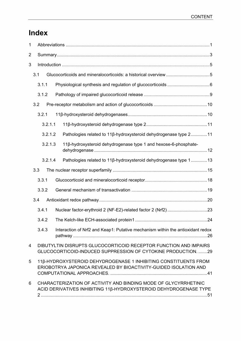
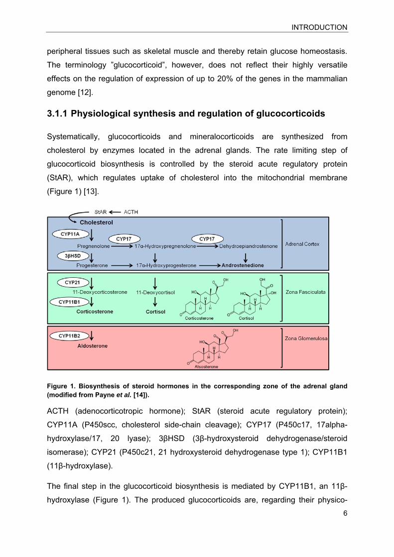
(Figure 1) [13].
Figure 1. Biosynthesis of steroid hormones in the corresponding zone of the adrenal gland (modified from Payne et al. [14]).
ACTH (adenocorticotropic hormone); StAR (steroid acute regulatory protein);
CYP11A (P450scc, cholesterol side-chain cleavage); CYP17 (P450c17, 17alpha-
hydroxylase/17, 20 lyase); 3βHSD (3β-hydroxysteroid dehydrogenase/steroid
isomerase); CYP21 (P450c21, 21 hydroxysteroid dehydrogenase type 1); CYP11B1
(11β-hydroxylase).
The final step in the glucocorticoid biosynthesis is mediated by CYP11B1, an 11β-
hydroxylase (Figure 1). The produced glucocorticoids are, regarding their physico-
INTRODUCTION
7
chemical properties, expected to pass cellular membranes and enter target cells via
passive transport.
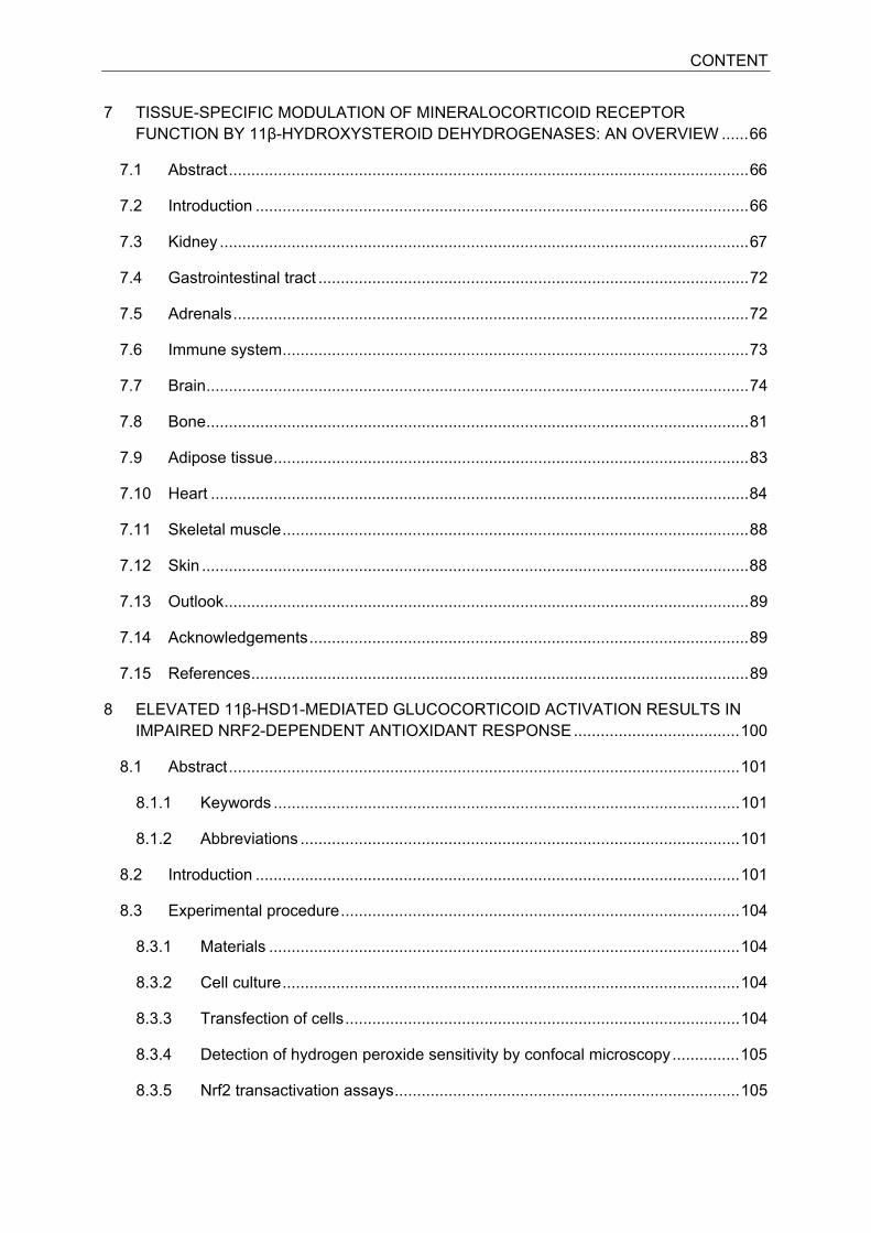
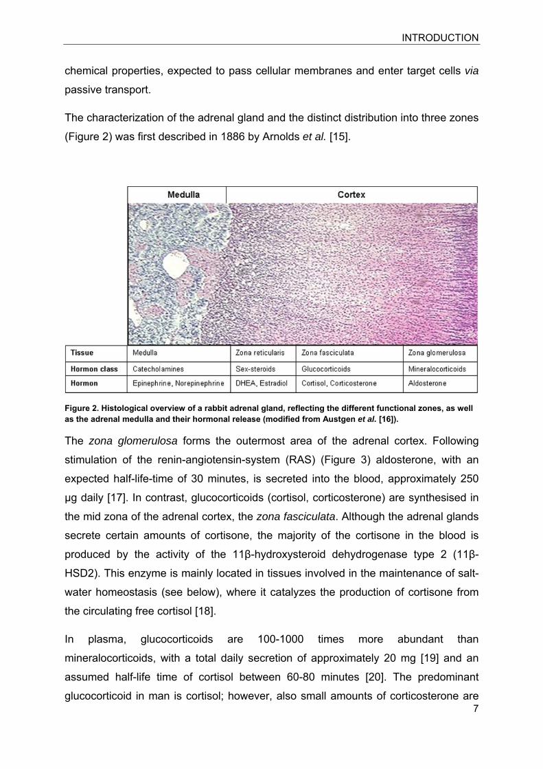
The characterization of the adrenal gland and the distinct distribution into three zones
(Figure 2) was first described in 1886 by Arnolds et al. [15].
Figure 2. Histological overview of a rabbit adrenal gland, reflecting the different functional zones, as well as the adrenal medulla and their hormonal release (modified from Austgen et al. [16]).
The zona glomerulosa forms the outermost area of the adrenal cortex. Following
stimulation of the renin-angiotensin-system (RAS) (Figure 3) aldosterone, with an
expected half-life-time of 30 minutes, is secreted into the blood, approximately 250
μg daily [17]. In contrast, glucocorticoids (cortisol, corticosterone) are synthesised in
the mid zona of the adrenal cortex, the zona fasciculata. Although the adrenal glands
secrete certain amounts of cortisone, the majority of the cortisone in the blood is
produced by the activity of the 11β-hydroxysteroid dehydrogenase type 2 (11β-
HSD2). This enzyme is mainly located in tissues involved in the maintenance of salt-
water homeostasis (see below), where it catalyzes the production of cortisone from
the circulating free cortisol [18].
In plasma, glucocorticoids are 100-1000 times more abundant than
mineralocorticoids, with a total daily secretion of approximately 20 mg [19] and an
assumed half-life time of cortisol between 60-80 minutes [20]. The predominant
glucocorticoid in man is cortisol; however, also small amounts of corticosterone are
INTRODUCTION
8
present in the human plasma. In contrast, cortisol is absent, or has very low plasma
levels in rodents, with corticosterone being the major active glucocorticoid in mice
and rats. Glucocorticoid release is daytime-dependent and oscillates following the
circadian rhythm. The cellular source of the mammalian circadian rhythm is the
suprachiasmatic nucleus (SCN) localized in the anterior hypothalamus [21].
Destruction of the SCN leads to a disturbance in the fluctuation of glucocorticoid
release over daytime [22]. In human plasma, cortisol reaches the highest levels in the
morning (20-400 nM) and bottom levels at night (5-100 nM) [23].
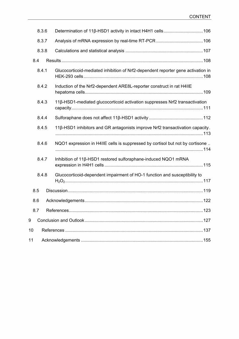
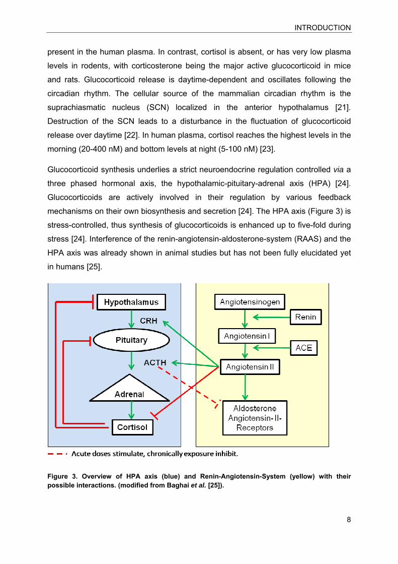
Glucocorticoid synthesis underlies a strict neuroendocrine regulation controlled via a
three phased hormonal axis, the hypothalamic-pituitary-adrenal axis (HPA) [24].
Glucocorticoids are actively involved in their regulation by various feedback
mechanisms on their own biosynthesis and secretion [24]. The HPA axis (Figure 3) is
stress-controlled, thus synthesis of glucocorticoids is enhanced up to five-fold during
stress [24]. Interference of the renin-angiotensin-aldosterone-system (RAAS) and the
HPA axis was already shown in animal studies but has not been fully elucidated yet
in humans [25].
Figure 3. Overview of HPA axis (blue) and Renin-Angiotensin-System (yellow) with their possible interactions. (modified from Baghai et al. [25]).
INTRODUCTION
9
CRH, corticotropin-releasing-hormone; ACTH, adrenocorticotropic hormone; ACE,
angiotensin-converting enzyme.
Although free glucocorticoids are secreted into the plasma at high concentrations,
90% of the plasmatic cortisol and corticosterone is bound to corticosteroid-binding
globulins (CBG) [26]. These steroid carriers belong to the clade A serine proteinase
inhibitor (serpin) family, and act as substrates for the neutrophil elastase [26].
Proteolytic cleavage of CBG by neutrophil esterases irreversibly destroys
glucocorticoid binding [26]. Growing evidence suggests, that CBG serves as a
storage pool for glucocorticoids to enable a direct and acute release of 80% of the
CBG-bound cortisol at the site of inflammation [27]. Locally released glucocorticoids
thereby allow a much faster reaction against inflammatory insults. Moreover, in
mouse strains with low CBG plasma levels (e.g. BC57BL/6) [28] as well as in CBG
KO-mice [29] an enhanced susceptibility against acute inflammation was observed.
The main CBG pool is produced by hepatocytes; however, CBG mRNA is also
expressed in other tissues such as pancreas and kidney that may indicate a defined
and tissue specific binding of locally occurring glucocorticoids [30]. Low affinity
binding of cortisol also occurs to albumin in the plasma, thus final free cortisol levels
are in the range of 4-10% of the total secreted cortisol [31]. Since the affinity of CBG
is much lower for the inactive glucocorticoid cortisone, and given that the
concentration of cortisone in the blood is five-times less than that of cortisol, the
amounts of free cortisol and free cortisone are comparable [18, 32-34].
3.1.2 Pathology of impaired glucocorticoid release
The pathologies of Addison’s disease and Cushing’s syndrome both involve
disturbances of the glucocorticoid (cortisol) content in the blood and, as a
consequence, dramatically decreased or increased glucocorticoid-dependent
functions. Addison’s disease is characterized by reduced cortisol levels leading to
impaired stress resistance, hypertrophy of the lymphoid organs, weight loss,
hypoglycaemia and hypotension [35]. Causes for the disease are disruption of
glucocorticoid biosynthesis as well as autoimmune driven destruction of the adrenal
cortex. On the other hand, the Cushing’s syndrome is characterized, among others,
by central obesity, muscle atrophy, hyperglycaemia, elevated cholesterol and insulin
resistance, as well as severe hypertension and immunodeficiency. This hormonal
INTRODUCTION
10
disorder can be caused by excess of ACTH or CRH, a consequence of pituitary gland
adenomas or tumors of the adrenal gland [36]. Moreover, iatrogenic causes are
common after prolonged medication with glucocorticoids [37]. Regardless of the
reason, the Cushing’s syndrome describes the pathologically enhanced cortisol level
of the blood. The therapeutic options involve glucocorticoid receptor antagonism
leading to a normalization of blood pressure [38] or tumor surgery and
adrenalectomy.
3.2 Pre-receptor metabolism and action of glucocorticoids
Although glucocorticoid synthesis and neuroendocrine regulation of glucocorticoid
release is strictly regulated, glucocorticoid metabolism in peripheral tissues further
represents a level of regulatory control contributing to the sophisticated network of
glucocorticoid-mediated regulation of physiological functions.
3.2.1 11β-hydroxysteroid dehydrogenases
11β-hydroxysteroid dehydrogenases (11β-HSDs) belong to the superfamily of short
chain dehydrogenases (SDR), which counts over 46’000 members, of which about 70
different genes are known in human [39]. Enzymes of the family are present in all
investigated genomes and seem to be a part of the original enzyme constitution [39].
Two distinct functional 11β-HSD glucocorticoid metabolizing enzymes have
extensively been investigated so far, termed 11β-HSD1 and 11β-HSD2. Both 11β-
HSDs are microsomal, anchoring in the membrane of the endoplasmatic reticulum
(ER) [40]. The two enzymes share only approximately 18% sequence homology,
including the active motif consisting of a conserved amino acid triad of tyrosine,
serine and lysine residues [41]. The catalytic domains of the type 1 and type 2
enzymes have inverted orientations [42] and opposite catalytic functions
(summarized in Table 1.).
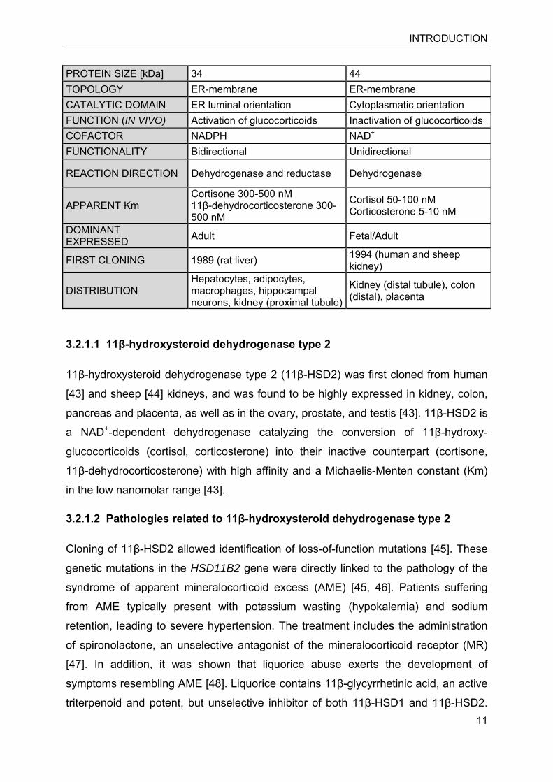
Table 1. Overview of biochemical parameters of 11β-hydroxysteroid dehydrogenases type 1 and type 2
Human 11β-HSD1 11β-HSD2 CHROMOSOME 1 16 GENE SIZE [Kb] 30 6.2 EXON/INTRON 6/5 5/4 AMINO ACIDS 292 405
INTRODUCTION
11
PROTEIN SIZE [kDa] 34 44 TOPOLOGY ER-membrane ER-membrane CATALYTIC DOMAIN ER luminal orientation Cytoplasmatic orientation FUNCTION (IN VIVO) Activation of glucocorticoids Inactivation of glucocorticoids COFACTOR NADPH NAD+ FUNCTIONALITY Bidirectional Unidirectional
REACTION DIRECTION Dehydrogenase and reductase Dehydrogenase
APPARENT Km Cortisone 300-500 nM 11β-dehydrocorticosterone 300-500 nM
Cortisol 50-100 nM Corticosterone 5-10 nM
DOMINANT EXPRESSED Adult Fetal/Adult
FIRST CLONING 1989 (rat liver) 1994 (human and sheep kidney)
DISTRIBUTION Hepatocytes, adipocytes, macrophages, hippocampal neurons, kidney (proximal tubule)
Kidney (distal tubule), colon (distal), placenta
3.2.1.1 11β-hydroxysteroid dehydrogenase type 2
11β-hydroxysteroid dehydrogenase type 2 (11β-HSD2) was first cloned from human
[43] and sheep [44] kidneys, and was found to be highly expressed in kidney, colon,
pancreas and placenta, as well as in the ovary, prostate, and testis [43]. 11β-HSD2 is
a NAD+-dependent dehydrogenase catalyzing the conversion of 11β-hydroxy-
glucocorticoids (cortisol, corticosterone) into their inactive counterpart (cortisone,
11β-dehydrocorticosterone) with high affinity and a Michaelis-Menten constant (Km)
in the low nanomolar range [43].
3.2.1.2 Pathologies related to 11β-hydroxysteroid dehydrogenase type 2
Cloning of 11β-HSD2 allowed identification of loss-of-function mutations [45]. These
genetic mutations in the HSD11B2 gene were directly linked to the pathology of the
syndrome of apparent mineralocorticoid excess (AME) [45, 46]. Patients suffering
from AME typically present with potassium wasting (hypokalemia) and sodium
retention, leading to severe hypertension. The treatment includes the administration
of spironolactone, an unselective antagonist of the mineralocorticoid receptor (MR)
[47]. In addition, it was shown that liquorice abuse exerts the development of
symptoms resembling AME [48]. Liquorice contains 11β-glycyrrhetinic acid, an active
triterpenoid and potent, but unselective inhibitor of both 11β-HSD1 and 11β-HSD2.
INTRODUCTION
12
Mutated 11β-HSD2 raised the molecular explanation for the AME phenotype and
explained the imbalance between urinary 11β-hydroxy- and 11β-ketoglucocorticoids
[45, 46].
Regarding the action of 11β-HSD2 as “gate-keeper” for the MR, thus primarily
regulating blood pressure, the deficiency of 11β-HSD2 activity, regardless of its
reasons (genetic, food-intake/inhibition), can cause of hypertension [49, 50].
3.2.1.3 11β-hydroxysteroid dehydrogenase type 1 and hexose-6-phosphate-dehydrogenase
11β-hydroxysteroid dehydrogenase type 1 (11β-HSD1) was first cloned from the rat
liver and reported in 1989 [51]. 11β-HSD1 catalyzes the interconversion of inactive
(cortisone and 11β-dehydrocorticosterone) and active (cortisol and corticosterone)
glucocorticoids, however, it acts predominantly as a reductase in intact cells and in
vivo, using nicotinamideadenine dinucleotide phosphate (NADPH) as cofactor [34]. It
is highly expressed in liver, gonads, adipose tissue and skeletal muscle, and lower
expression levels have been found in certain regions of the brain, the lung, testis,
ovary, adrenal glands and vascular cells [34].
In vitro assays using the purified protein revealed that 11β-HSD1 is a bidirectional
enzyme preferably acting as dehydrogenase catalyzing the oxidation of active 11β-
hydroxyglucocorticoids and using NADP+ as cofactor [41, 52-54]. Initial kinetic
analyses revealed an apparent Km of 1.8 μM for corticosterone and 17 μM for
cortisol, respectively [34]. Later, studies with purified protein reported Km values
between 300-500 nM for corticosterone and cortisol respectively suggesting a loss of
enzymatic function in some older purification protocols [55]. However, as mentioned
above, studies in intact cells including fibroblasts, hepatocytes, lung cells, stromal
adipose cells, and hippocampal neurons revealed that 11β-HSD1 acts predominantly
as a reducing enzyme generating cortisol or corticosterone and using NADPH as
cofactor [34]. Data obtained from experiments using intact cells are supported by
kinetic studies suggesting an apparent Km of about 0.3 μM for cortisone compared
with a Km of about 2 μM for cortisol [56, 57]. Observations in isolated primary
hepatocytes led to further confusion since 11β-HSD1 rapidly lost its reductase activity
after a short cultivation period. In addition, 11β-HSD1 activity was shown to be
influenced by the differentiation state of a cell, e.g. in 3T3-L1 derived mouse
INTRODUCTION
13
adipocytes, where 11β-HSD1 reductase activity appears during differentiation of
preadipocytes into adipocytes [58, 59].
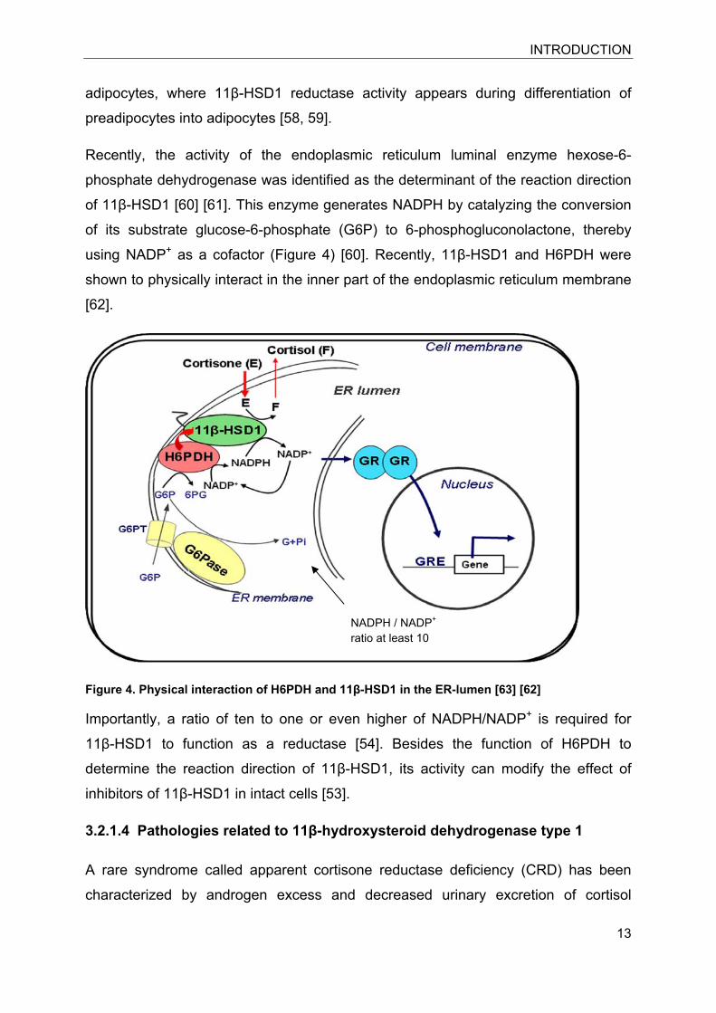
Recently, the activity of the endoplasmic reticulum luminal enzyme hexose-6-
phosphate dehydrogenase was identified as the determinant of the reaction direction
of 11β-HSD1 [60] [61]. This enzyme generates NADPH by catalyzing the conversion
of its substrate glucose-6-phosphate (G6P) to 6-phosphogluconolactone, thereby
using NADP+ as a cofactor (Figure 4) [60]. Recently, 11β-HSD1 and H6PDH were
shown to physically interact in the inner part of the endoplasmic reticulum membrane
[62].
Figure 4. Physical interaction of H6PDH and 11β-HSD1 in the ER-lumen [63] [62]
Importantly, a ratio of ten to one or even higher of NADPH/NADP+ is required for
11β-HSD1 to function as a reductase [54]. Besides the function of H6PDH to
determine the reaction direction of 11β-HSD1, its activity can modify the effect of
inhibitors of 11β-HSD1 in intact cells [53].
3.2.1.4 Pathologies related to 11β-hydroxysteroid dehydrogenase type 1
A rare syndrome called apparent cortisone reductase deficiency (CRD) has been
characterized by androgen excess and decreased urinary excretion of cortisol
NADPH / NADP+
ratio at least 10
INTRODUCTION
14
metabolites [64]. Homozygous mutations in the H6PD gene without change in the
coding sequence of the HSD11B1 [65] as well as heterozygous mutations of the
HSD11B1 gene without affected H6PD gene [66] were reported for the CRD
phenotype.
The metabolic syndrome is characterized by a co-incidence of disturbances such as
obesity, hypertension, elevated plasma triglycerides, and cardiovascular disease
(Table 2). Obesity and the possible outcome, the metabolic syndrome, are
consequences of inappropriate life-style leading to disturbance of multiple pathways.
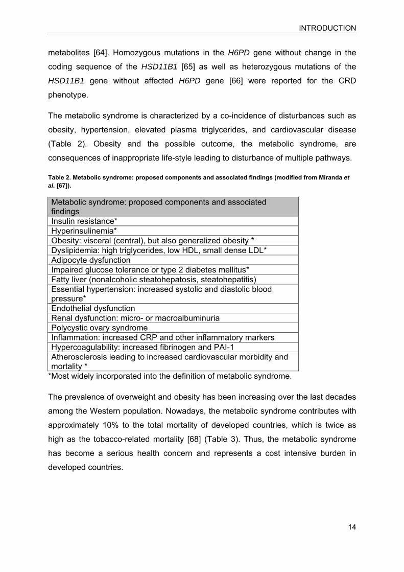
Table 2. Metabolic syndrome: proposed components and associated findings (modified from Miranda et al. [67]).
Metabolic syndrome: proposed components and associated findings Insulin resistance* Hyperinsulinemia* Obesity: visceral (central), but also generalized obesity * Dyslipidemia: high triglycerides, low HDL, small dense LDL* Adipocyte dysfunction Impaired glucose tolerance or type 2 diabetes mellitus* Fatty liver (nonalcoholic steatohepatosis, steatohepatitis) Essential hypertension: increased systolic and diastolic blood pressure* Endothelial dysfunction Renal dysfunction: micro- or macroalbuminuria Polycystic ovary syndrome Inflammation: increased CRP and other inflammatory markers Hypercoagulability: increased fibrinogen and PAI-1 Atherosclerosis leading to increased cardiovascular morbidity and mortality *
*Most widely incorporated into the definition of metabolic syndrome.
The prevalence of overweight and obesity has been increasing over the last decades
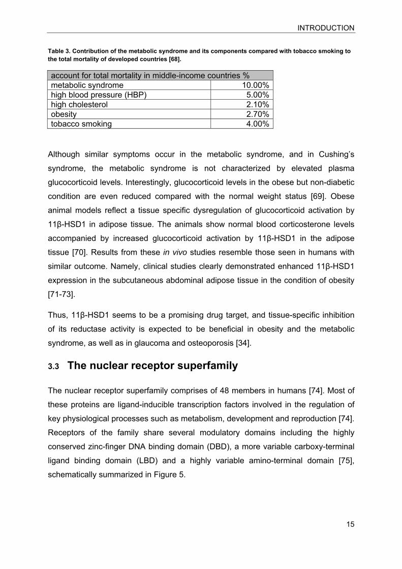
among the Western population. Nowadays, the metabolic syndrome contributes with
approximately 10% to the total mortality of developed countries, which is twice as
high as the tobacco-related mortality [68] (Table 3). Thus, the metabolic syndrome
has become a serious health concern and represents a cost intensive burden in
developed countries.
INTRODUCTION
15
Table 3. Contribution of the metabolic syndrome and its components compared with tobacco smoking to the total mortality of developed countries [68].
account for total mortality in middle-income countries % metabolic syndrome 10.00%high blood pressure (HBP) 5.00%high cholesterol 2.10%obesity 2.70%tobacco smoking 4.00%
Although similar symptoms occur in the metabolic syndrome, and in Cushing’s
syndrome, the metabolic syndrome is not characterized by elevated plasma
glucocorticoid levels. Interestingly, glucocorticoid levels in the obese but non-diabetic
condition are even reduced compared with the normal weight status [69]. Obese
animal models reflect a tissue specific dysregulation of glucocorticoid activation by
11β-HSD1 in adipose tissue. The animals show normal blood corticosterone levels
accompanied by increased glucocorticoid activation by 11β-HSD1 in the adipose
tissue [70]. Results from these in vivo studies resemble those seen in humans with
similar outcome. Namely, clinical studies clearly demonstrated enhanced 11β-HSD1
expression in the subcutaneous abdominal adipose tissue in the condition of obesity
[71-73].
Thus, 11β-HSD1 seems to be a promising drug target, and tissue-specific inhibition
of its reductase activity is expected to be beneficial in obesity and the metabolic
syndrome, as well as in glaucoma and osteoporosis [34].
3.3 The nuclear receptor superfamily
The nuclear receptor superfamily comprises of 48 members in humans [74]. Most of
these proteins are ligand-inducible transcription factors involved in the regulation of
key physiological processes such as metabolism, development and reproduction [74].
Receptors of the family share several modulatory domains including the highly
conserved zinc-finger DNA binding domain (DBD), a more variable carboxy-terminal
ligand binding domain (LBD) and a highly variable amino-terminal domain [75],
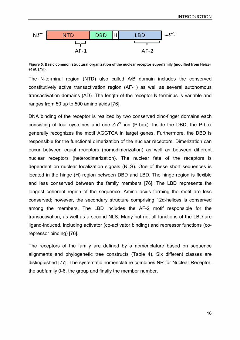
schematically summarized in Figure 5.
INTRODUCTION
16
Figure 5. Basic common structural organization of the nuclear receptor superfamily (modified from Heizer et al. [75]).
The N-terminal region (NTD) also called A/B domain includes the conserved
constitutively active transactivation region (AF-1) as well as several autonomous
transactivation domains (AD). The length of the receptor N-terminus is variable and
ranges from 50 up to 500 amino acids [76].
DNA binding of the receptor is realized by two conserved zinc-finger domains each
consisting of four cysteines and one Zn2+ ion (P-box). Inside the DBD, the P-box
generally recognizes the motif AGGTCA in target genes. Furthermore, the DBD is
responsible for the functional dimerization of the nuclear receptors. Dimerization can
occur between equal receptors (homodimerization) as well as between different
nuclear receptors (heterodimerization). The nuclear fate of the receptors is
dependent on nuclear localization signals (NLS). One of these short sequences is
located in the hinge (H) region between DBD and LBD. The hinge region is flexible
and less conserved between the family members [76]. The LBD represents the
longest coherent region of the sequence. Amino acids forming the motif are less
conserved; however, the secondary structure comprising 12α-helices is conserved
among the members. The LBD includes the AF-2 motif responsible for the
transactivation, as well as a second NLS. Many but not all functions of the LBD are
ligand-induced, including activator (co-activator binding) and repressor functions (co-
repressor binding) [76].
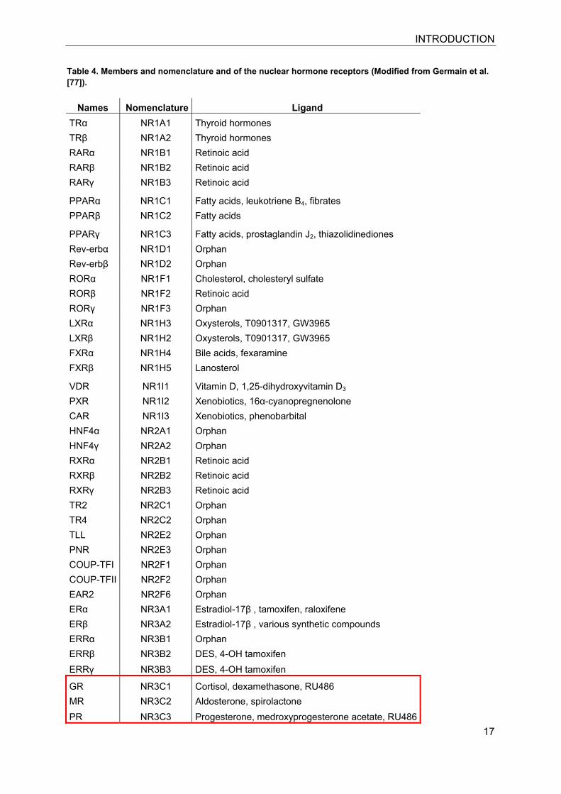
The receptors of the family are defined by a nomenclature based on sequence
alignments and phylogenetic tree constructs (Table 4). Six different classes are
distinguished [77]. The systematic nomenclature combines NR for Nuclear Receptor,
the subfamily 0-6, the group and finally the member number.
INTRODUCTION
17
Table 4. Members and nomenclature and of the nuclear hormone receptors (Modified from Germain et al. [77]).
Names Nomenclature Ligand TRα NR1A1 Thyroid hormones TRβ NR1A2 Thyroid hormones RARα NR1B1 Retinoic acid RARβ NR1B2 Retinoic acid RARγ NR1B3 Retinoic acid
PPARα NR1C1 Fatty acids, leukotriene B4, fibrates PPARβ NR1C2 Fatty acids
PPARγ NR1C3 Fatty acids, prostaglandin J2, thiazolidinediones Rev-erbα NR1D1 Orphan Rev-erbβ NR1D2 Orphan RORα NR1F1 Cholesterol, cholesteryl sulfate RORβ NR1F2 Retinoic acid RORγ NR1F3 Orphan LXRα NR1H3 Oxysterols, T0901317, GW3965 LXRβ NR1H2 Oxysterols, T0901317, GW3965 FXRα NR1H4 Bile acids, fexaramine FXRβ NR1H5 Lanosterol
VDR NR1I1 Vitamin D, 1,25-dihydroxyvitamin D3 PXR NR1I2 Xenobiotics, 16α-cyanopregnenolone CAR NR1I3 Xenobiotics, phenobarbital HNF4α NR2A1 Orphan HNF4γ NR2A2 Orphan RXRα NR2B1 Retinoic acid RXRβ NR2B2 Retinoic acid RXRγ NR2B3 Retinoic acid TR2 NR2C1 Orphan TR4 NR2C2 Orphan TLL NR2E2 Orphan PNR NR2E3 Orphan COUP-TFI NR2F1 Orphan COUP-TFII NR2F2 Orphan EAR2 NR2F6 Orphan ERα NR3A1 Estradiol-17β , tamoxifen, raloxifene ERβ NR3A2 Estradiol-17β , various synthetic compounds ERRα NR3B1 Orphan ERRβ NR3B2 DES, 4-OH tamoxifen ERRγ NR3B3 DES, 4-OH tamoxifen
GR NR3C1 Cortisol, dexamethasone, RU486 MR NR3C2 Aldosterone, spirolactone PR NR3C3 Progesterone, medroxyprogesterone acetate, RU486
INTRODUCTION
18
AR NR3C4 Testosterone, flutamide NGFI-B NR4A1 Orphan NURR1 NR4A2 Orphan NOR1 NR4A3 Orphan SF1 NR5A1 Orphan LRH-1 NR5A2 Orphan GCNF NR6A1 Orphan DAX-1 NR0B1 Orphan SHP NR0B2 Orphan
3.3.1 Glucocorticoid and mineralocorticoid receptor
Both corticosteroid receptors, the glucocorticoid receptor (GR, NR3C1), and the
mineralocorticoid receptor (MR, NR3C2), are members of the previously described
nuclear hormone receptor superfamily.
In 1985, the GR of human origin was first cloned by Hollenberg and colleges [78],
soon followed by the cloning of the mouse [79] and rat receptors [80]. Two years
later, Arriza et al. used complementary DNA of the GR, under the condition of low
stringency hybridization, to isolate and clone the MR [81]. MR and GR share ~90% of
the amino acids forming the DNA binding domain (DBD). However, sequence
homology of the ligand binding domain (LBD) shows only approximately 50%
homology. GR is expressed in nearly every tissue, whereas MR expression is also
frequent but more defined. MR is expressed mainly in tissues actively involved in
salt-water homeostasis. Importantly, however, the receptor is also involved in the
regulation of biochemical processes that are very distinct from these “classical
functions”, such as in the limbic system where it regulates behavioral plasticity [82] or
in the heart [83]. For MR expression and function the presence or absence of
enzymes controlling the access of active ligand, thereby regulating receptor
activation need to be considered. (for review see chapter 7).
The endogenous ligands of GR are cortisol (Kd 10-50 nM) and corticosterone (Kd
60nM); The widely used synthetic glucocorticoid, dexamethasone has a ten-fold
higher affinity with a Kd 1-8 nM [84-86]. The MR has broader substrate specificity and
it binds aldosterone, 11-deoxycorticosterone, corticosterone, cortisol and
progesterone with Kd 0.5-3 nM [87, 88]. A physiological regulatory mechanism exists
to protect the MR from activation by glucocorticoids: in tissues where the regulation
INTRODUCTION
19
of the maintenance of salt-water balance takes place, such as the distal tubule and
the cortical collecting duct of the kidney, distal colon and sweat-glands, the receptor
is co-expressed with the glucocorticoid inactivating enzyme 11β-HSD2 (see Chapter
3.2.1 above). A well known GR antagonist is mifepristone (also known as RU-486),
which was originally developed as a progesterone receptor antagonist to prevent
pregnancy, but binds also GR with high affinity [89, 90]. The most abundantly used
unselective MR antagonist is spironolactone, which is still used in the clinics and has
found recent attention to treat essential hypertension and heart failure [91-93]. Use of
spironolactone was already reported in 1960 for the treatment of patients suffering
from edema, congestive cardiac failures and nephrotic syndrome [94]. Another,
newer MR antagonist is eplerenon (Inspra®), which although selective, has a rather
low affinity to the receptor (Kd approximately 30 µM) [95].
3.3.2 General mechanism of transactivation
The unliganded receptor is localized in the cytoplasm as part of a multiprotein
complex, including molecular chaperones such as heat shock protein (HSP) 90,
HSP70, HSP56, as well as other proteins such as p23 and CYP40. In the presence
of active hormone ligand, the receptor undergoes conformational changes, dimerizes,
discloses its NLS and releases associated proteins from the receptor complex [96,
97].
Activated GR and MR homodimers translocate to the nucleus with help of the
importin system [97]. Receptor complexes then bind to glucocorticoid response
elements (GRE). The GRE consensus sequence is defined as
GGTACANNNTGTTCT [38]. GREs are located in the promoter region of GR and MR
target genes. Binding of the active receptor complex and recruitment of the
transcription machinery leads to the induction or repression of transcription [98, 99].
The activity and action of nuclear receptors is further modulated by post-translational
modifications such as phosphorylation, ubiquitination, SUMOylation, methylation or
acetylation. Splice variants and variants due to distinct translational initiation with
different or similar activities of these described nuclear receptors are also known
[100-104].
INTRODUCTION
20
3.4 Antioxidant redox pathway
The antioxidant redox pathway is part of the cellular detoxification system. The
metabolic detoxification process of cells involves detoxification of xenobiotics as well
as potential endogenous toxicants with the aim to inactivate the compound and finally
excrete a water soluble and harmless product.
In general, detoxification processes can be separated in three different steps,
involving distinct proteins:
Phase I reactions: This step is often called functionalization, and reactive groups can
be introduced in lipophilic molecules to facilitate subsequent conjungation, thereby
enhancing solubility. Phase I biotransformation involves cytochrom P450
monooxygenases, monoaminooxigenases and dehydrogenases/reductases that are
responsible for the oxidation, reduction and hydrolysis of compounds. The metabolic
products of phase I reactions are often highly reactive metabolites that, if not
efficiently removed, are able to lead to toxic insults [105, 106].
Phase II reactions: These reactions comprise conjugation, of reactive carboxyl,
hydroxyl, sulfhydryl, and amino groups with glucuronicacid, sulphate or glutathione.
The products of this metabolic process display higher solubility and are mostly less
active or even inactive. Conjungation is mediated by a variety of enzymes including
gluthation-S-transferases, sulfotrasferases, methyltransferases, UDP-glucuronosyl-
transferases and N-acetyltransferases [105, 107].
Phase III reactions: These reactions involve transport proteins, such as multidrug
resistance-related proteins (mdr) and ATP-binding cassette proteins (ABC-
transporters) as well as other transporters and carriers [108], and they mediate
excretion of the solubilized chemical.
Enzymes of the antioxidant redox pathway belong to all three phases of the
detoxification process, with many of them belonging to phase II. However, these
enzymes and transporters have the commonality of a specific regulatory DNA
element, which mediates their induced expression after electrophile insults [109-112].
One of the first reports on an enzyme later well known as a member of the phase II
detoxifying machinery was published in 1958 by Ernster et al. [113]. Ernster et al.
INTRODUCTION
21
reported a soluble NAD(P)H: (quinone acceptor) oxidoreductase (NQO1), which was
present in rat liver homogenates also known as DT-diaphorase [114]. The purified
NQO1 protein was characterized as a dicoumarol sensitive enzyme, catalyzing a two
electron reduction reaction [115]. Because of its characteristics, NQO1 was expected
to play a role in cytoprotection against toxic chemicals [114].
The identification of additional enzymes responsible for the detoxification of
xenobiotic insults such as glutathione-S-transferases (GST), promoted research on
their transcriptional regulation and determining their basal and induced status,
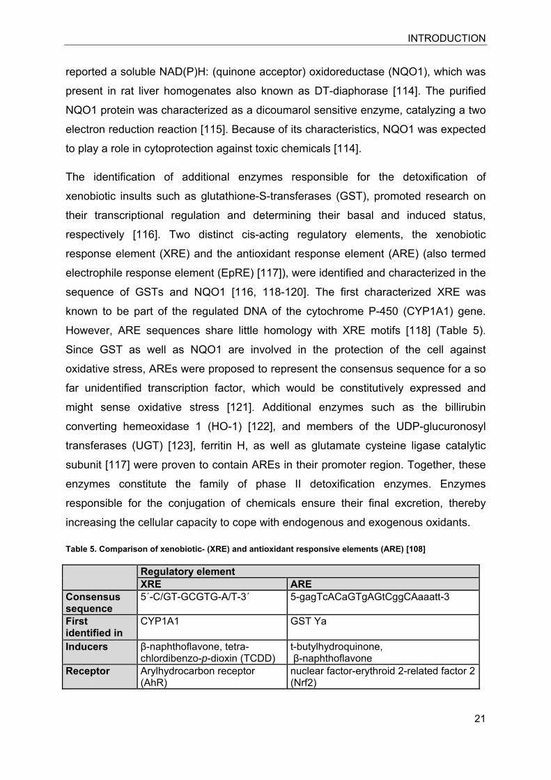
respectively [116]. Two distinct cis-acting regulatory elements, the xenobiotic
response element (XRE) and the antioxidant response element (ARE) (also termed
electrophile response element (EpRE) [117]), were identified and characterized in the
sequence of GSTs and NQO1 [116, 118-120]. The first characterized XRE was
known to be part of the regulated DNA of the cytochrome P-450 (CYP1A1) gene.
However, ARE sequences share little homology with XRE motifs [118] (Table 5).
Since GST as well as NQO1 are involved in the protection of the cell against
oxidative stress, AREs were proposed to represent the consensus sequence for a so
far unidentified transcription factor, which would be constitutively expressed and
might sense oxidative stress [121]. Additional enzymes such as the billirubin
converting hemeoxidase 1 (HO-1) [122], and members of the UDP-glucuronosyl
transferases (UGT) [123], ferritin H, as well as glutamate cysteine ligase catalytic
subunit [117] were proven to contain AREs in their promoter region. Together, these
enzymes constitute the family of phase II detoxification enzymes. Enzymes
responsible for the conjugation of chemicals ensure their final excretion, thereby
increasing the cellular capacity to cope with endogenous and exogenous oxidants.
Table 5. Comparison of xenobiotic- (XRE) and antioxidant responsive elements (ARE) [108]
Regulatory element XRE ARE
Consensus sequence
5´-C/GT-GCGTG-A/T-3´ 5-gagTcACaGTgAGtCggCAaaatt-3
First identified in
CYP1A1 GST Ya
Inducers β-naphthoflavone, tetra-chlordibenzo-p-dioxin (TCDD)
t-butylhydroquinone, β-naphthoflavone
Receptor Arylhydrocarbon receptor (AhR)
nuclear factor-erythroid 2-related factor 2 (Nrf2)
INTRODUCTION
22
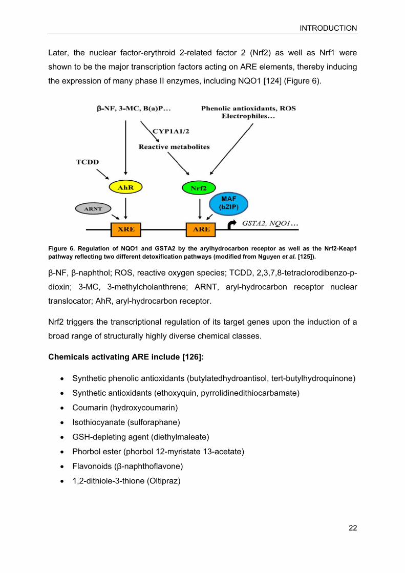
Later, the nuclear factor-erythroid 2-related factor 2 (Nrf2) as well as Nrf1 were
shown to be the major transcription factors acting on ARE elements, thereby inducing
the expression of many phase II enzymes, including NQO1 [124] (Figure 6).
Figure 6. Regulation of NQO1 and GSTA2 by the arylhydrocarbon receptor as well as the Nrf2-Keap1 pathway reflecting two different detoxification pathways (modified from Nguyen et al. [125]).
β-NF, β-naphthol; ROS, reactive oxygen species; TCDD, 2,3,7,8-tetraclorodibenzo-p-
dioxin; 3-MC, 3-methylcholanthrene; ARNT, aryl-hydrocarbon receptor nuclear
translocator; AhR, aryl-hydrocarbon receptor.
Nrf2 triggers the transcriptional regulation of its target genes upon the induction of a
broad range of structurally highly diverse chemical classes.
Chemicals activating ARE include [126]:
• Synthetic phenolic antioxidants (butylatedhydroantisol, tert-butylhydroquinone)
• Synthetic antioxidants (ethoxyquin, pyrrolidinedithiocarbamate)
• Coumarin (hydroxycoumarin)
• Isothiocyanate (sulforaphane)
• GSH-depleting agent (diethylmaleate)
• Phorbol ester (phorbol 12-myristate 13-acetate)
• Flavonoids (β-naphthoflavone)
• 1,2-dithiole-3-thione (Oltipraz)
INTRODUCTION
23
3.4.1 Nuclear factor-erythroid 2 (NF-E2)-related factor 2 (Nrf2)
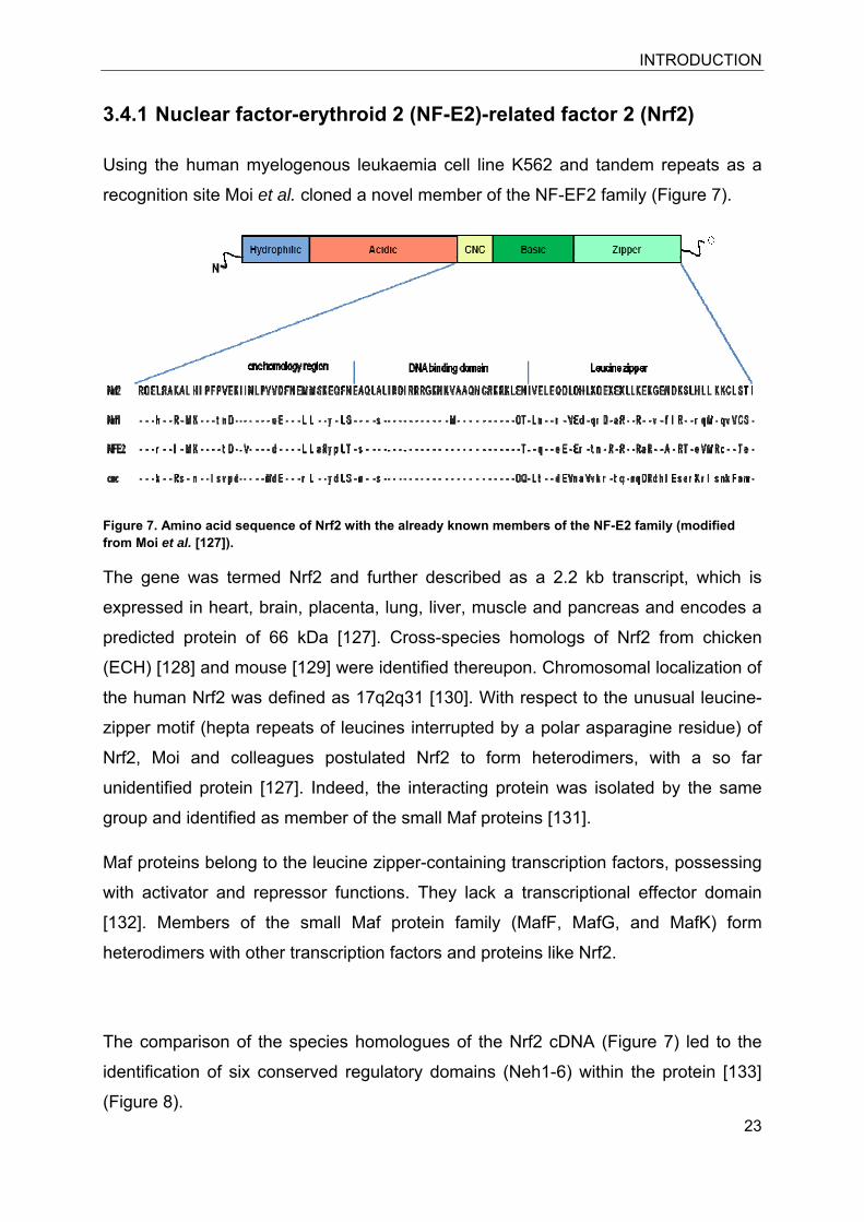
Using the human myelogenous leukaemia cell line K562 and tandem repeats as a
recognition site Moi et al. cloned a novel member of the NF-EF2 family (Figure 7).
Figure 7. Amino acid sequence of Nrf2 with the already known members of the NF-E2 family (modified from Moi et al. [127]).
The gene was termed Nrf2 and further described as a 2.2 kb transcript, which is
expressed in heart, brain, placenta, lung, liver, muscle and pancreas and encodes a
predicted protein of 66 kDa [127]. Cross-species homologs of Nrf2 from chicken
(ECH) [128] and mouse [129] were identified thereupon. Chromosomal localization of
the human Nrf2 was defined as 17q2q31 [130]. With respect to the unusual leucine-
zipper motif (hepta repeats of leucines interrupted by a polar asparagine residue) of
Nrf2, Moi and colleagues postulated Nrf2 to form heterodimers, with a so far
unidentified protein [127]. Indeed, the interacting protein was isolated by the same
group and identified as member of the small Maf proteins [131].
Maf proteins belong to the leucine zipper-containing transcription factors, possessing
with activator and repressor functions. They lack a transcriptional effector domain
[132]. Members of the small Maf protein family (MafF, MafG, and MafK) form
heterodimers with other transcription factors and proteins like Nrf2.
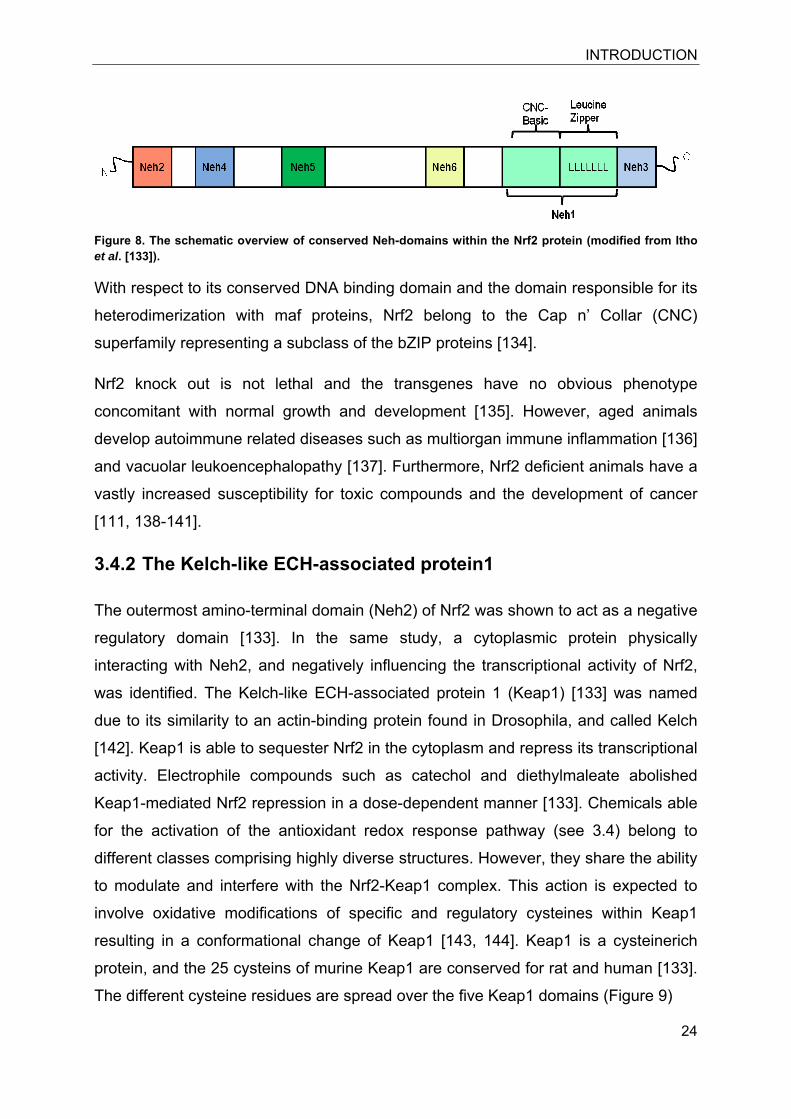
The comparison of the species homologues of the Nrf2 cDNA (Figure 7) led to the
identification of six conserved regulatory domains (Neh1-6) within the protein [133]
(Figure 8).
INTRODUCTION
24
Figure 8. The schematic overview of conserved Neh-domains within the Nrf2 protein (modified from Itho et al. [133]).
With respect to its conserved DNA binding domain and the domain responsible for its
heterodimerization with maf proteins, Nrf2 belong to the Cap n’ Collar (CNC)
superfamily representing a subclass of the bZIP proteins [134].
Nrf2 knock out is not lethal and the transgenes have no obvious phenotype
concomitant with normal growth and development [135]. However, aged animals
develop autoimmune related diseases such as multiorgan immune inflammation [136]
and vacuolar leukoencephalopathy [137]. Furthermore, Nrf2 deficient animals have a
vastly increased susceptibility for toxic compounds and the development of cancer
[111, 138-141].
3.4.2 The Kelch-like ECH-associated protein1
The outermost amino-terminal domain (Neh2) of Nrf2 was shown to act as a negative
regulatory domain [133]. In the same study, a cytoplasmic protein physically
interacting with Neh2, and negatively influencing the transcriptional activity of Nrf2,
was identified. The Kelch-like ECH-associated protein 1 (Keap1) [133] was named
due to its similarity to an actin-binding protein found in Drosophila, and called Kelch
[142]. Keap1 is able to sequester Nrf2 in the cytoplasm and repress its transcriptional
activity. Electrophile compounds such as catechol and diethylmaleate abolished
Keap1-mediated Nrf2 repression in a dose-dependent manner [133]. Chemicals able
for the activation of the antioxidant redox response pathway (see 3.4) belong to
different classes comprising highly diverse structures. However, they share the ability
to modulate and interfere with the Nrf2-Keap1 complex. This action is expected to
involve oxidative modifications of specific and regulatory cysteines within Keap1
resulting in a conformational change of Keap1 [143, 144]. Keap1 is a cysteinerich
protein, and the 25 cysteins of murine Keap1 are conserved for rat and human [133].
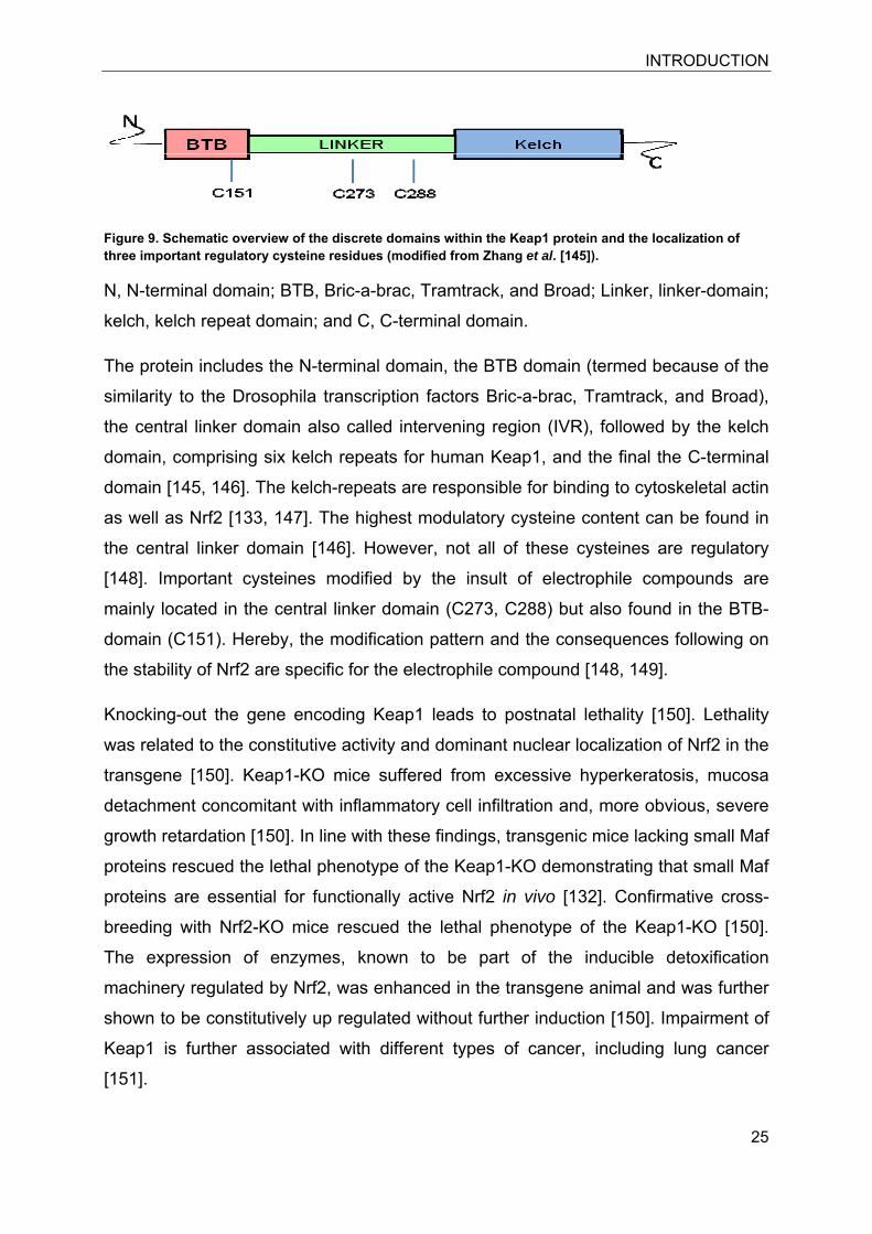
The different cysteine residues are spread over the five Keap1 domains (Figure 9)
INTRODUCTION
25
Figure 9. Schematic overview of the discrete domains within the Keap1 protein and the localization of three important regulatory cysteine residues (modified from Zhang et al. [145]).
N, N-terminal domain; BTB, Bric-a-brac, Tramtrack, and Broad; Linker, linker-domain;
kelch, kelch repeat domain; and C, C-terminal domain.
The protein includes the N-terminal domain, the BTB domain (termed because of the
similarity to the Drosophila transcription factors Bric-a-brac, Tramtrack, and Broad),
the central linker domain also called intervening region (IVR), followed by the kelch
domain, comprising six kelch repeats for human Keap1, and the final the C-terminal
domain [145, 146]. The kelch-repeats are responsible for binding to cytoskeletal actin
as well as Nrf2 [133, 147]. The highest modulatory cysteine content can be found in
the central linker domain [146]. However, not all of these cysteines are regulatory
[148]. Important cysteines modified by the insult of electrophile compounds are
mainly located in the central linker domain (C273, C288) but also found in the BTB-
domain (C151). Hereby, the modification pattern and the consequences following on
the stability of Nrf2 are specific for the electrophile compound [148, 149].
Knocking-out the gene encoding Keap1 leads to postnatal lethality [150]. Lethality
was related to the constitutive activity and dominant nuclear localization of Nrf2 in the
transgene [150]. Keap1-KO mice suffered from excessive hyperkeratosis, mucosa
detachment concomitant with inflammatory cell infiltration and, more obvious, severe
growth retardation [150]. In line with these findings, transgenic mice lacking small Maf
proteins rescued the lethal phenotype of the Keap1-KO demonstrating that small Maf
proteins are essential for functionally active Nrf2 in vivo [132]. Confirmative cross-
breeding with Nrf2-KO mice rescued the lethal phenotype of the Keap1-KO [150].
The expression of enzymes, known to be part of the inducible detoxification
machinery regulated by Nrf2, was enhanced in the transgene animal and was further
shown to be constitutively up regulated without further induction [150]. Impairment of
Keap1 is further associated with different types of cancer, including lung cancer
[151].
INTRODUCTION
26
As a conclusion, both Keap1 and Nrf2 are suggested to act as intracellular sensors
for oxidative stress, further leading to the transcriptional induction of genes for phase
II detoxifying enzymes [133].
3.4.3 Interaction of Nrf2 and Keap1: Putative mechanism within the antioxidant redox pathway
Upon identification of the key regulators of the ARE pathway (Nrf2, Keap1, MAF) the
elucidation of the mode of action and of the mechanism responsible for the regulation
of gene transcription represented a major challenge. Currently, the following
observations and scenario are accepted among most researchers in the field.
1. Nrf2 is a functionally active transcription factor that controls basal and
inducible expression of its target genes [152].
2. Keap1 is a constitutively expressed negative regulator of Nrf2. Keap1 acts as
an adaptor protein, which promotes ubiquitination of Nrf2 by the cullin-3-
dependent pathway [145, 147, 152, 153].
3. Nrf2 is an unstable protein with short half-life (15-30 min), and its degradation
via the ubiquitin-pathway is mediated by the 26S-proteasome [154].
4. Keap1 contains reactive cysteine residues, some of which were shown to be
regulatory (Cys: 257, 273, 288, and 297) and therefore expected to act as
redox sensors. Modification of the regulatory cysteines is electrophile-specific
and can in some cases stabilize Nrf2 by preventing its degradation [148].
Recognition of Nrf2 by Keap1 is mediated by tow highly conserved motifs within the
Nrf2 protein namely DLG and ETGE. In this process Keap1 is expected to bind Nrf2
in a Hinge-and-Latch fashion over a two-site-substrate recognition model [155]. The
stoichiometry of the Nrf2-Keap1 complex is 1:2, while one Nrf2 molecule is bound to
a homodimer of Keap1. Homodimerization of Keap1 molecules occur between the N-
terminal BTB/POZ motifs. BTB/POZ motifs have been found in zinc-finger proteins,
and such proteins contain, like Keap1, kelch motifs. Homodimerization instead of
heterodimerisation is a common characteristic for BTB containing proteins.
Furthermore, these motifs have been shown to mediate transcriptional repression as
well as interaction with common co-repressors such as nuclear receptor co-repressor
INTRODUCTION
27
1 (N-CoR) and nuclear receptor corepressor 2 (N-CoR 2 or SMRT). Keap1 acts as
substrate adaptor protein for the Cul3-dependent E3 ubiquitin ligase complex [145].
Ubiquitination of proteins requires a defined lysine residue position on the substrate
for transfer of the ubiquitin moiety. The DLG motive of the Nrf2 has low affinity for
Keap1 and functions as latch, responsible for the functional positioning of Nrf2
molecule for ubiquitination under non stressed conditions [155]. On the other hand,
the ETGE motif has high affinity and represents the hinge [155]. Within the interaction
of Nrf2 and Keap1 under non-stressed conditions the lysines comprising the Nhe2
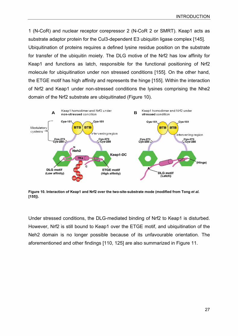
domain of the Nrf2 substrate are ubiquitinated (Figure 10).
Figure 10. Interaction of Keap1 and Nrf2 over the two-site-substrate mode (modified from Tong et al. [155]).
Under stressed conditions, the DLG-mediated binding of Nrf2 to Keap1 is disturbed.
However, Nrf2 is still bound to Keap1 over the ETGE motif, and ubiquitination of the
Neh2 domain is no longer possible because of its unfavourable orientation. The
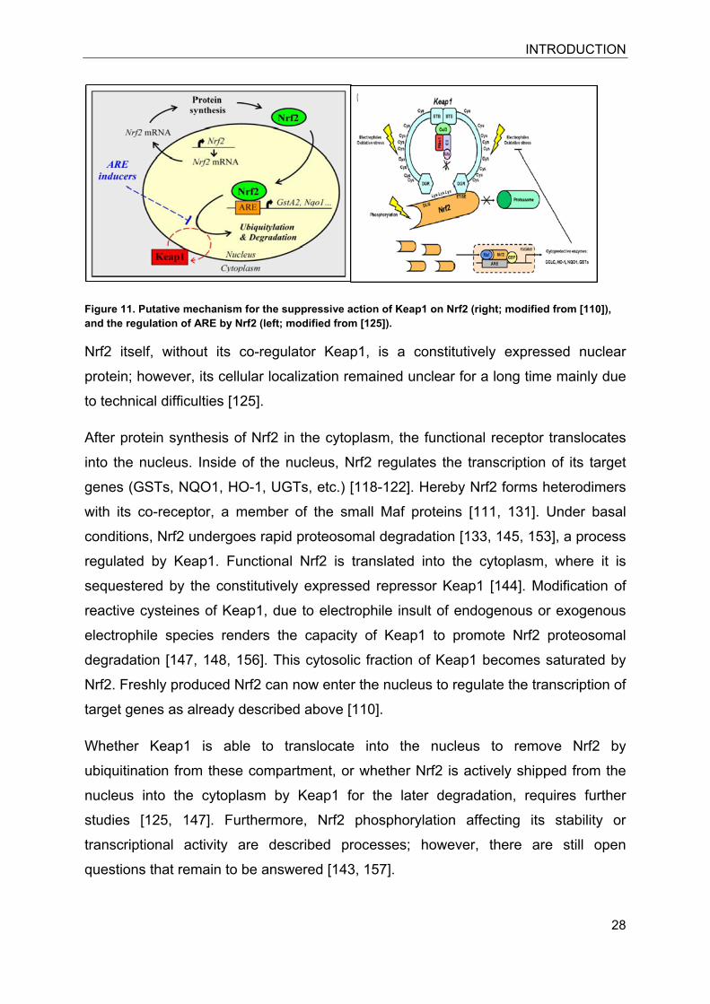
aforementioned and other findings [110, 125] are also summarized in Figure 11.
INTRODUCTION
28
Figure 11. Putative mechanism for the suppressive action of Keap1 on Nrf2 (right; modified from [110]), and the regulation of ARE by Nrf2 (left; modified from [125]).
Nrf2 itself, without its co-regulator Keap1, is a constitutively expressed nuclear
protein; however, its cellular localization remained unclear for a long time mainly due
to technical difficulties [125].
After protein synthesis of Nrf2 in the cytoplasm, the functional receptor translocates
into the nucleus. Inside of the nucleus, Nrf2 regulates the transcription of its target
genes (GSTs, NQO1, HO-1, UGTs, etc.) [118-122]. Hereby Nrf2 forms heterodimers
with its co-receptor, a member of the small Maf proteins [111, 131]. Under basal
conditions, Nrf2 undergoes rapid proteosomal degradation [133, 145, 153], a process
regulated by Keap1. Functional Nrf2 is translated into the cytoplasm, where it is
sequestered by the constitutively expressed repressor Keap1 [144]. Modification of
reactive cysteines of Keap1, due to electrophile insult of endogenous or exogenous
electrophile species renders the capacity of Keap1 to promote Nrf2 proteosomal
degradation [147, 148, 156]. This cytosolic fraction of Keap1 becomes saturated by
Nrf2. Freshly produced Nrf2 can now enter the nucleus to regulate the transcription of
target genes as already described above [110].
Whether Keap1 is able to translocate into the nucleus to remove Nrf2 by
ubiquitination from these compartment, or whether Nrf2 is actively shipped from the
nucleus into the cytoplasm by Keap1 for the later degradation, requires further
studies [125, 147]. Furthermore, Nrf2 phosphorylation affecting its stability or
transcriptional activity are described processes; however, there are still open
questions that remain to be answered [143, 157].
DBT IMPAIRS GR FUNKTION
29
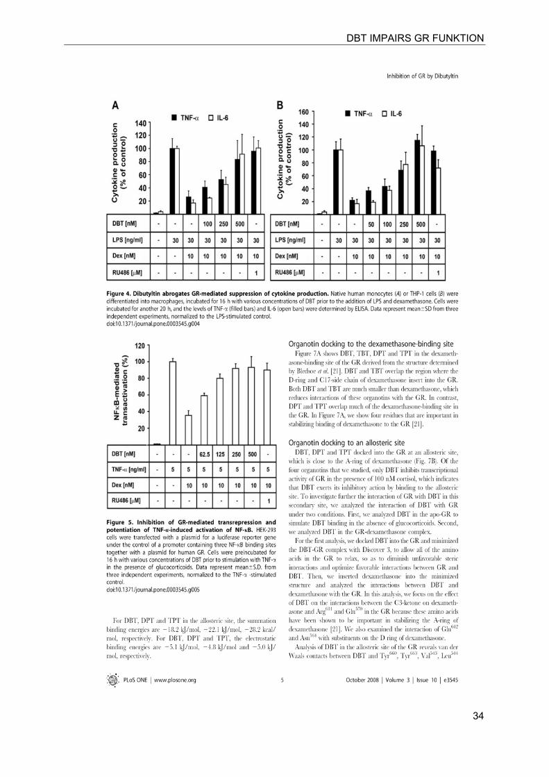
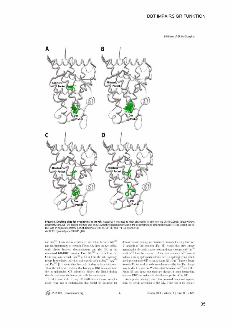
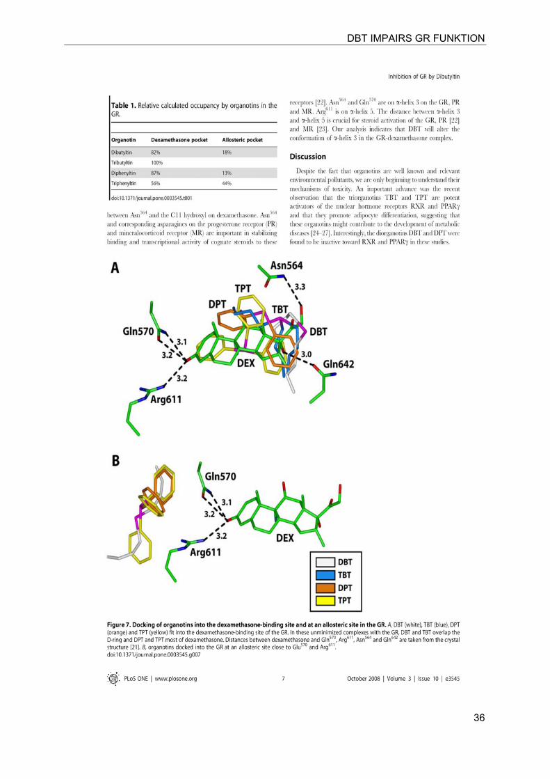
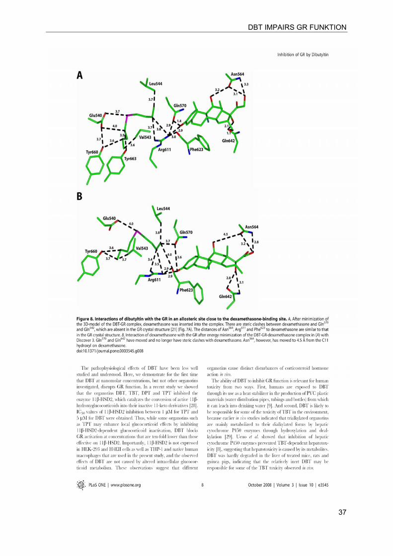
4 DIBUTYLTIN DISRUPTS GLUCOCORTICOID RECEPTOR FUNCTION AND IMPAIRS GLUCOCORTICOID-INDUCED SUPPRESSION OF CYTOKINE PRODUCTION.
Christel Gumy1, 2, Charlie Chandsawangbhuwana 3, Anna A. Dzyakanchuk 1, Denise
V. Kratschmar 1, Michael E. Baker 3*, Alex Odermatt1*
1 Division of Molecular and Systems Toxicology, Department of Pharmaceutical
Sciences, University of Basel, Basel, Switzerland, 2 Department of Nephrology and Hypertension, University of Berne, Berne,
Switzerland, 3 Department of Medicine, University of California San Diego, La Jolla, California,
United States of America
The Study revealed the disruption of GR-dependent action in general and especially
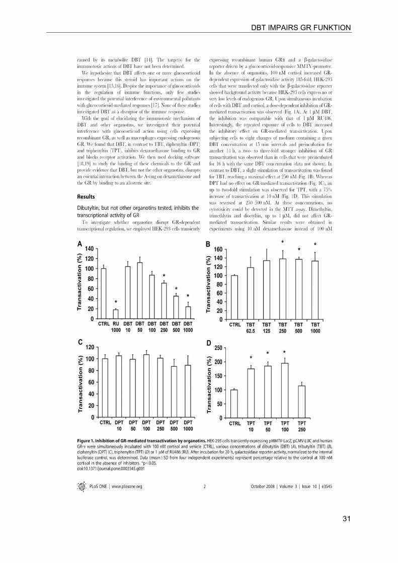
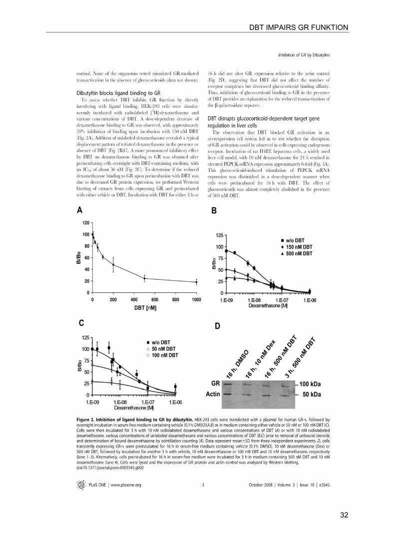
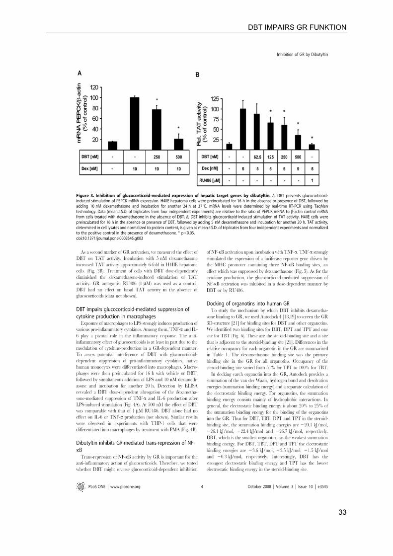
in inflammation by dibutyltin.
DBT IMPAIRS GR FUNKTION
30
DBT IMPAIRS GR FUNKTION
31
DBT IMPAIRS GR FUNKTION
32
DBT IMPAIRS GR FUNKTION
33
DBT IMPAIRS GR FUNKTION
34
DBT IMPAIRS GR FUNKTION
35
DBT IMPAIRS GR FUNKTION
36
DBT IMPAIRS GR FUNKTION
37
DBT IMPAIRS GR FUNKTION
38
DBT IMPAIRS GR FUNKTION
39
DBT IMPAIRS GR FUNKTION
40
11β-HSD1 INHIBITORS FROM ERIOBOTRYA JAPONICA
41
5 11β-HYDROXYSTEROID DEHYDROGENASE 1 INHIBITING CONSTITUENTS FROM ERIOBOTRYA JAPONICA REVEALED BY BIOACTIVITY-GUIDED ISOLATION AND COMPUTATIONAL APPROACHES.
Judith M. Rollinger a,*, Denise V. Kratschmar b, Daniela Schuster c, Petra H. Pfisterer a, Christel Gumy b, Evelyne M. Aubry b, Sarah Brandstötter a, Hermann Stuppner a,
Gerhard Wolber c, Alex Odermatt b,*
a Institute of Pharmacy/Pharmacognosy and Center for Molecular Biosciences
Innsbruck, University of Innsbruck, Innrain 52c, A-6020 Innsbruck, Austria b Division of Molecular and Systems Toxicology, Department of Pharmaceutical
Sciences, University of Basel, Klingelbergstrasse 50, CH-4056 Basel, Switzerland c Institute of Pharmacy/Pharmaceutical Chemistry and Center for Molecular
Biosciences Innsbruck, University of Innsbruck, Innrain 52c, A-6020 Innsbruck,
Austria
In this publication, we used a pharmacophore-based virtual screening approach to reveal
selective 11β-HSD1 inhibitors from the leaves of loquat (eriobotrya japonica) a known anti
diabetic in Chinese medicine; the most promising hits were evaluated using in vitro assays.
11β-HSD1 INHIBITORS FROM ERIOBOTRYA JAPONICA
42
11β-HSD1 INHIBITORS FROM ERIOBOTRYA JAPONICA
43
11β-HSD1 INHIBITORS FROM ERIOBOTRYA JAPONICA
44
11β-HSD1 INHIBITORS FROM ERIOBOTRYA JAPONICA
45
11β-HSD1 INHIBITORS FROM ERIOBOTRYA JAPONICA
46
11β-HSD1 INHIBITORS FROM ERIOBOTRYA JAPONICA
47
11β-HSD1 INHIBITORS FROM ERIOBOTRYA JAPONICA
48
11β-HSD1 INHIBITORS FROM ERIOBOTRYA JAPONICA
49
11β-HSD1 INHIBITORS FROM ERIOBOTRYA JAPONICA
50
11β-HSD2 INHIBITORS
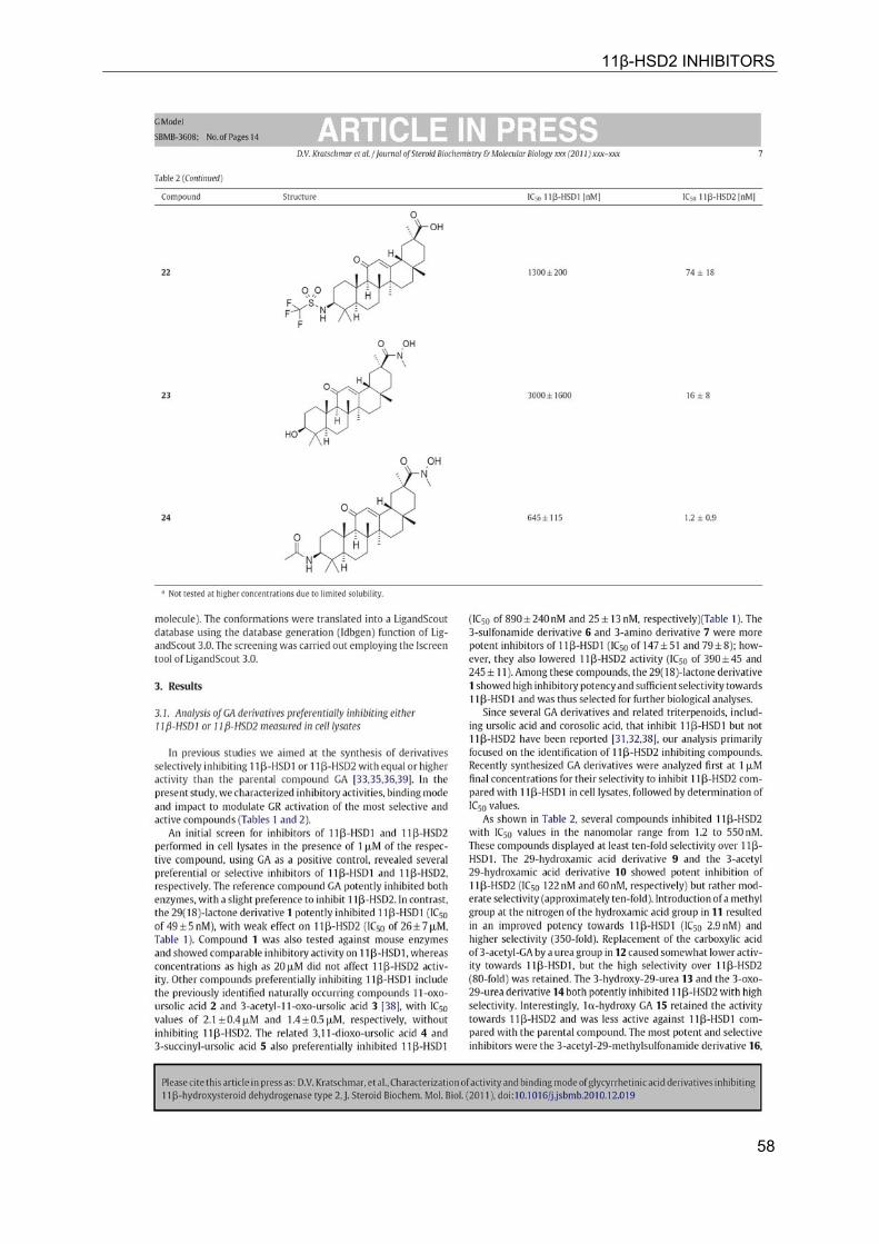
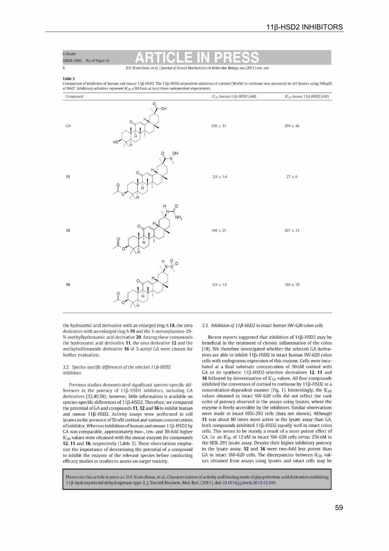
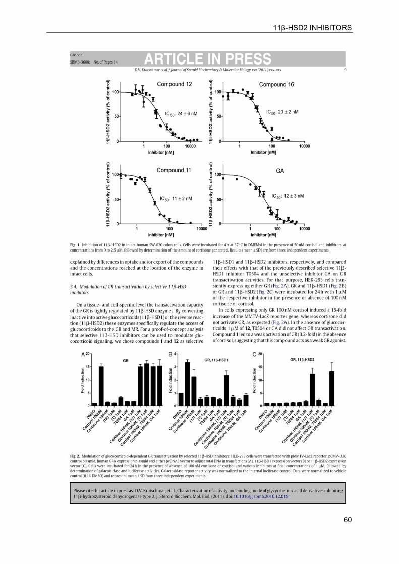
51
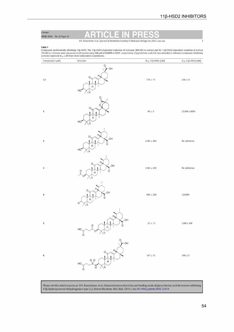
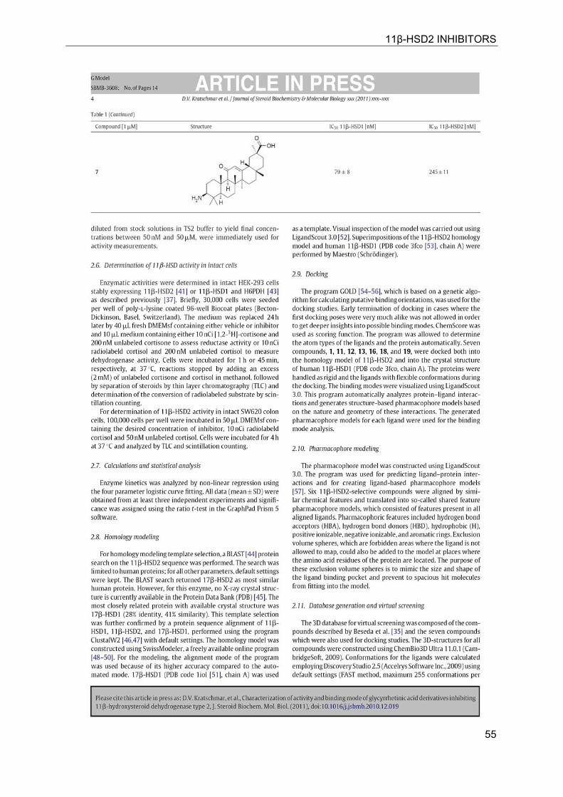
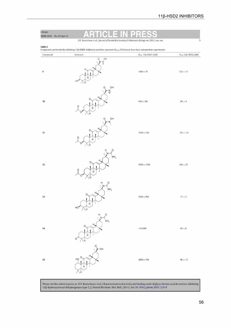
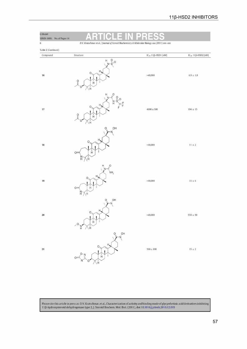
6 CHARACTERIZATION OF ACTIVITY AND BINDING MODE OF GLYCYRRHETINIC ACID DERIVATIVES INHIBITING 11β-HYDROXYSTEROID DEHYDROGENASE TYPE 2
Denise V. Kratschmara,1, Anna Vuorinenb,1, Thierry Da Cunhaa, Gerhard Wolberc,d,
Dirk Classen-Houbene, Otto Doblhoffe, Daniela Schusterb,∗, Alex Odermatta,∗∗
a Swiss Center for Applied Human Toxicology and Division of Molecular and Systems
Toxicology, Department of Pharmaceutical Sciences, University of Basel,
Klingelbergstrasse 50, CH-4056 Basel, Switzerland b Institute of Pharmacy, Department of Pharmaceutical Chemistry and Center for
Molecular Biosciences Innsbruck – CMBI, University of Innsbruck, Innrain 52c,
A-6020 Innsbruck, Austria c Free University Berlin, Institute of Pharmacy, Pharmaceutical Chemistry, Königin-
Luise-Str. 2+4, 14195 Berlin, Germany d Inte:Ligand GmbH, Mariahilfer Str. 74B/11, 1070 Vienna, Austria e onepharm Research & Development GmbH, Veterinärplatz 1, 1210 Vienna, Austria
In this publication, we characterized a set of novel glycyrrhetinic acid derivates for
their selective inhibition potential against 11β-HSD2. Inhibitors were developed by
chemical modification of the glycyrrhetinic acid backbone
11β-HSD2 INHIBITORS
52
11β-HSD2 INHIBITORS
53
11β-HSD2 INHIBITORS
54
11β-HSD2 INHIBITORS
55
11β-HSD2 INHIBITORS
56
11β-HSD2 INHIBITORS
57
11β-HSD2 INHIBITORS
58
11β-HSD2 INHIBITORS
59
11β-HSD2 INHIBITORS
60
11β-HSD2 INHIBITORS
61
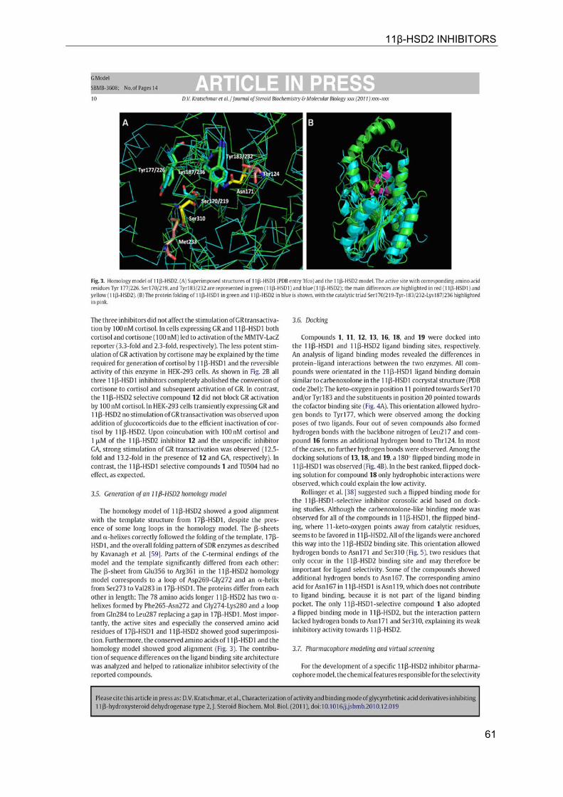
11β-HSD2 INHIBITORS
62
11β-HSD2 INHIBITORS
63
11β-HSD2 INHIBITORS
64
11β-HSD2 INHIBITORS
65
MR EXPRESSION AND REGULATION
66
7 TISSUE-SPECIFIC MODULATION OF MINERALOCORTICOID RECEPTOR FUNCTION BY 11β-HYDROXYSTEROID DEHYDROGENASES: AN OVERVIEW
Alex Odermatt*1 and Denise V. Kratschmar1
1 Division of Molecular and Systems Toxicology, Department of Pharmaceutical
Sciences, University of Basel, Klingelbergstrasse 50, CH-4056 Basel, Switzerland
7.1 Abstract
In the last decade significant progress has been made in the understanding of mineralocorticoid receptor (MR) function and its implications for physiology and disease. The knowledge on the essential role of MR in the regulation of electrolyte concentrations and blood pressure has been significantly extended, and the relevance of excessive MR activation in promoting inflammation, fibrosis and heart disease as well as the adverse effects on brain function is now widely recognized. Despite this considerable progress, the mechanisms of MR function in various cell-types are still poorly understood. Key modulators of MR function include the glucocorticoid receptor (GR), which may affect MR function by formation of heterodimers and by differential genomic and non-genomic responses on gene expression, and 11β-hydroxysteroid dehydrogenases (11β-HSDs), which determine the availability of intracellular concentrations of active glucocorticoids. In this review we attempted to provide an overview of the knowledge on MR expression with regard to the presence or absence of GR, 11β-HSD2 and 11β-HSD1/hexose-6-phosphate dehydrogenase (H6PDH) in various tissues and cell types. The consequences of cell-specific differences in the coexpression of MR with these proteins need to be further investigated in order to understand the role of MR in a given tissue as well as its systemic impact.
7.2 Introduction
The use of complementary DNA of the glucocorticoid receptor (GR, systematic name NR3C1) and low-stringency hybridization by Arizza et al. led to the identification of a cDNA coding for a 107 kDa polypeptide, which was functionally characterized as mineralocorticoid receptor (MR) [1]. The MR is also known as aldosterone receptor and under the systematic name NR3C2 (Nuclear Receptor subfamily 3, group C, member 2). MR and GR share about 90% amino acid homology in their DNA binding domain (DBD) but only about 50% in their ligand binding domain (LBD). Evolutionary analyses suggested that MR and GR evolved from a common ancestor and that the MR was the first to diverge from the ancient receptor gene [2, 3]. Importantly, MR existed well before aldosterone appeared in evolution, whereas GR seems to have appeared later in evolution. This may explain the rather broad substrate specificity of MR, compared with the more selective GR. Whereas MR binds aldosterone, 11-deoxycorticosterone, corticosterone,
cortisol and progesterone with similarly high affinities and Kd values between 0.5 and 3 nM, GR shows a higher selectivity to cortisol and corticosterone with Kd values of 20-70 nM [1, 4, 5].
The cloning of MR allowed its exact localization in various tissues and identification of specific cell types expressing this receptor. The subsequent cloning of 11�-hydroxysteroid dehydrogenase type 1 (11�-HSD1)[6] and 11�-HSD2 [7, 8] and determination of their tissue- and cell-specific expression patterns then allowed a comparison with the expression pattern of MR and GR. It soon became clear that MR is not only expressed in cells where 11β-HSD2 acts as a “gate-keeper” to protect MR from high concentrations of glucocorticoids and rendering specificity for aldosterone [9, 10]. As discussed below, the MR plays an important role in cells coexpressing 11�-HSD1, including macrophages, preadipocytes/adipocytes, osteoblasts/osteoclasts, and microglia cells, by modulating cell proliferation and inflammatory
MR EXPRESSION AND REGULATION
67
response. Thus, the classic view of mineralocorticoid target tissues, where MR function is strictly regulated by aldosterone, has to be reconsidered.
7.3 Kidney
The kidney is considered as the classical mineralocorticoid target tissues. High-affinity aldosterone binding sites, corresponding to MRs, and lower affinity glucocorticoid binding sites, corresponding to GRα, have been characterized in rat kidneys almost 40 years ago [11, 12]. Aldosterone-induced renal epithelial sodium transport was found to be dependent on a nuclear transactivating receptor that was later identified as MR [1, 13]. The MR has similar high affinities to bind aldosterone, progesterone, 11-deoxycorticosterone, corticosterone and cortisol, with Kd values between 0.5 and 3 nM [1, 4], whereas the GR shows higher ligand selectivity but approximately 20-fold lower affinity for cortisol and very weak affinity for aldosterone (Kd about 500 nM). The identification of 11β-HSD2 as a “gate-keeper” to protect MR from active 11β-hydroxyglucocorticoids (cortisol in humans, corticosterone in rodents) that are present in plasma at about 1000-fold higher concentrations than aldosterone provided an explanation for the specificity of this receptor towards aldosterone [9, 10].
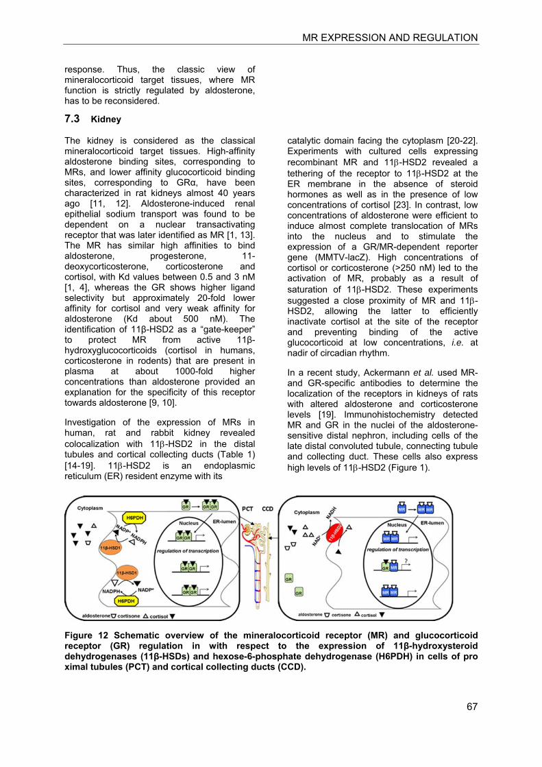
Investigation of the expression of MRs in human, rat and rabbit kidney revealed colocalization with 11β-HSD2 in the distal tubules and cortical collecting ducts (Table 1) [14-19]. 11β-HSD2 is an endoplasmic reticulum (ER) resident enzyme with its
catalytic domain facing the cytoplasm [20-22]. Experiments with cultured cells expressing recombinant MR and 11β-HSD2 revealed a tethering of the receptor to 11β-HSD2 at the ER membrane in the absence of steroid hormones as well as in the presence of low concentrations of cortisol [23]. In contrast, low concentrations of aldosterone were efficient to induce almost complete translocation of MRs into the nucleus and to stimulate the expression of a GR/MR-dependent reporter gene (MMTV-lacZ). High concentrations of cortisol or corticosterone (>250 nM) led to the activation of MR, probably as a result of saturation of 11β-HSD2. These experiments suggested a close proximity of MR and 11β-HSD2, allowing the latter to efficiently inactivate cortisol at the site of the receptor and preventing binding of the active glucocorticoid at low concentrations, i.e. at nadir of circadian rhythm.
In a recent study, Ackermann et al. used MR- and GR-specific antibodies to determine the localization of the receptors in kidneys of rats with altered aldosterone and corticosterone levels [19]. Immunohistochemistry detected MR and GR in the nuclei of the aldosterone-sensitive distal nephron, including cells of the late distal convoluted tubule, connecting tubule and collecting duct. These cells also express high levels of 11β-HSD2 (Figure 1).
Figure 12 Schematic overview of the mineralocorticoid receptor (MR) and glucocorticoid receptor (GR) regulation in with respect to the expression of 11β-hydroxysteroid dehydrogenases (11β-HSDs) and hexose-6-phosphate dehydrogenase (H6PDH) in cells of pro ximal tubules (PCT) and cortical collecting ducts (CCD).
MR EXPRESSION AND REGULATION
68
In addition, MR and GR expression was found in the thick ascending limb and in intercalated cells, where 11β-HSD2 is absent. It was suggested that MR in intercalated cells may be involved in proton secretion, thereby playing an essential role in acid/base regulation. The role of MR in these cell types remains to be elucidated. In rats on a high-salt diet, which is known to lower plasma aldosterone, MR localization to the nuclei was unchanged, whereas GR localized to the cytoplasm in the aldosterone-sensitive distal nephron. The actual diet-induced changes in circulating aldosterone and corticosterone levels, however, have not been determined in this
study. Also, it remains unclear how altered aldosterone levels might affect GR but not MR localization. Furthermore, the time point where the samples have been taken is not indicated, and it will be important to compare the localization of MR and GR in different cell types during peak glucocorticoids and at nadir during circadian rhythm. Nevertheless, the authors observed cytoplasmic localization of both MR and GR in adrenalectomized rats, and low dose corticosterone replacement led to nuclear translocation of MR but not GR. The GR translocated to the nuclei only in cells not expressing 11β-HSD2.
GR was found to be coexpressed with 11β-HSD1 and H6PDH mainly in the third segment of the proximal tubules [24-26] and probably plays a role in regulating glucose and lipid uptake and metabolism. Chronically elevated glucocorticoid activation in proximal tubules is likely to cause adverse metabolic effects and disturbances in transport processes in these cells and warrants further investigation.
Several investigators reported the expression of MR in glomerular mesangial cells and observed aldosterone-induced cell proliferation [27-32]. A recent study with cultured rat mesangial cells provided evidence for the involvement of the MR in the stimulation of mesangial cell proliferation by high glucose medium [29]. Induction of cell proliferation was prevented by incubation with an antagonist and siRNA against MR and by an inhibitor of extracellular signal-regulated kinase kinase (MEK). Furthermore, aldosterone-dependent apoptotic and mitogenic effects were demonstrated in human mesangial cells [30]. The pro-apoptotic effects of aldosterone were prevented by co-treatment with spironolactone as well as by antioxidants and free radical scavengers. Aldosterone has been shown to increase reactive oxygen species (ROS) production by a mechanism involving activation of NADPH oxidase in renal and cardiovascular tissues [33-38]. Long-term administration of aldosterone to rats caused mesangial cell proliferation and expansion of the mesangium [37]. Thus, exposure to chronically high aldosterone levels might cause mesangial cell damage, independent of its hemodynamic effects.
Glomerular mesangial cells were initially reported to express 11β-HSD1, and an upregulation of its expression was observed in the presence of the pro-inflammatory cytokines TNF-α and IL-1β [39]. However, other investigators reported expression of MR,
aldosterone synthase (CYP11B2) and 11β-HSD2 in rat mesangial cells, and provided evidence for a role of mitogen-activated protein kinase 1/2, cyclin D1 and cyclin A in the aldosterone-induced mesangial cell proliferation and of a Smad2- and TGF-β1-dependent stimulation of fibronectin production [32, 40, 41]. The expression and role of the respective 11β-HSD enzyme and species differences have to be studied.
Recent evidence suggested that elevated CYP11B2 levels and MR activation in podocytes may contribute to the progression of diabetic nephropathy [42, 43]. Immortalized podocytes expressing MR, CYP11B2 and 11β-HSD2 were incubated with physiological (5.6 mM) and high (30 mM) concentrations of glucose [42]. MR and CYP11B2 expression were increased upon high glucose treatment, whereas 11β-HSD2 was not altered. Enhanced MR and CYP11B2 expression was also found in glomeruli of streptozotocin treated diabetic rats, and aldosterone levels were increased in these animals. Furthermore, treatment of type 2 diabetic Otsuka-Long-Evans-Tokushima-Fatty (OLETF) rats with the MR antagonist eplerenone enhanced the blood pressure-independent anti-proteinuric effects of angiotensinogen II type 1 receptor blocker. These observations indicate that chronically elevated MR activity may contribute to impaired glomerular function by adverse effects on podocytes.
The bidirectional enzyme 11β-HSD1 has been detected in renal medullary and interstitial cells that express GR but not MR [25, 44]. Interestingly, H6PDH seems to be absent in these cells suggesting that another enzyme might provide NADPH in the ER or that 11β-HSD1 might act as a dehydrogenase in these cells to modulate GR activity. The role of 11β-HSD1 in the modulation of GR function in these cells remains to be clarified.
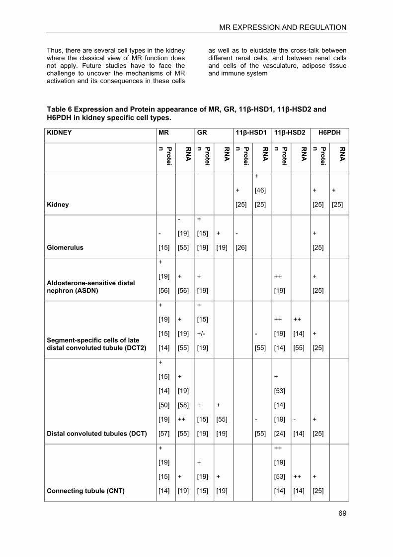
MR EXPRESSION AND REGULATION
69
Thus, there are several cell types in the kidney where the classical view of MR function does not apply. Future studies have to face the challenge to uncover the mechanisms of MR activation and its consequences in these cells
as well as to elucidate the cross-talk between different renal cells, and between renal cells and cells of the vasculature, adipose tissue and immune system
Table 6 Expression and Protein appearance of MR, GR, 11β-HSD1, 11β-HSD2 and H6PDH in kidney specific cell types.
KIDNEY MR GR 11β-HSD1 11β-HSD2 H6PDH
Protein R
NA
Protein R
NA
Protein R
NA
Protein R
NA
Protein R
NA
Kidney
+
[25]
+
[46]
[25]
+
[25]
+
[25]
Glomerulus
-
[15]
-
[19]
[55]
+
[15]
[19]
+
[19]
-
[26]
+
[25]
Aldosterone-sensitive distal nephron (ASDN)
+
[19]
[56]
+
[56]
+
[19]
++
[19]
+
[25]
Segment-specific cells of late distal convoluted tubule (DCT2)
+
[19]
[15]
[14]
+
[19]
[55]
+
[15]
+/-
[19]
-
[55]
++
[19]
[14]
++
[14]
[55]
+
[25]
Distal convoluted tubules (DCT)
+
[15]
[14]
[50]
[19]
[57]
+
[19]
[58]
++
[55]
+
[15]
[19]
+
[55]
[19]
-
[55]
+
[53]
[14]
[19]
[24]
-
[14]
+
[25]
Connecting tubule (CNT)
+
[19]
[15]
[14]
+
[19]
+
[19]
[15]
+
[19]
++
[19]
[53]
[14]
++
[14]
+
[25]
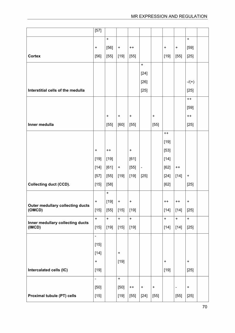
MR EXPRESSION AND REGULATION
70
[57]
Cortex
+
[56]
+
[56]
[55]
+
[19]
++
[55]
+
[19]
+
[55]
+
[59]
[25]
Interstitial cells of the medulla
+
[24]
[26]
[25]
-/(+)
[25]
Inner medulla
+
[55]
+
[60]
+
[55]
+
[55]
++
[59]
++
[25]
Collecting duct (CCD).
+
[19]
[14]
[57]
[15]
++
[19]
[61]
[55]
[58]
+
[19]
+
[61]
[55]
[19]
-
[25]
++
[19]
[53]
[14]
[62]
[24]
[62]
++
[14]
+
[25]
Outer medullary collecting ducts (OMCD)
+
[15]
+
[19]
[55]
+
[15]
+
[19]
++
[14]
++
[14]
+
[25]
Inner medullary collecting ducts (IMCD)
+
[15]
+
[19]
+
[15]
+
[19]
+
[14]
+
[14]
+
[25]
Intercalated cells (IC)
-
[15]
[14]
+
[19]
+
[19]
+
[19]
+
[25]
Proximal tubule (PT) cells
-
[50]
[15]
+
[50]
[19]
++
[55]
+
[24]
+
[55]
-
[55]
+
[25]
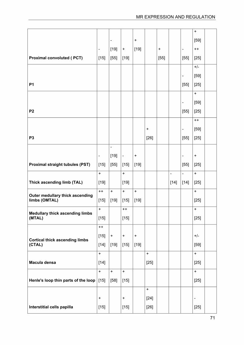
MR EXPRESSION AND REGULATION
71
Proximal convoluted ( PCT)
-
[15]
-
[19]
[55]
+
[19]
+
[19]
+
[55]
-
[55]
+
[59]
++
[25]
P1
-
[55]
+/-
[59]
[25]
P2
-
[55]
+
[59]
[25]
P3
+
[26]
-
[55]
++
[59]
[25]
Proximal straight tubules (PST)
-
[15]
-
[19]
[55]
-
[15]
+
[19]
-
[55]
+
[25]
Thick ascending limb (TAL)
+
[19]
+
[19]
-
[14]
-
[14]
+
[25]
Outer medullary thick ascending limbs (OMTAL)
++
[15]
+
[19]
+
[15]
+
[19]
+
[25]
Medullary thick ascending limbs (MTAL)
+
[15]
++
[15]
+
[25]
Cortical thick ascending limbs (CTAL)
++
[15]
[14]
+
[19]
+
[15]
+
[19]
+/-
[59]
Macula densa
+
[14]
+
[25]
+
[25]
Henle's loop thin parts of the loop
+
[15]
+
[58]
+
[15]
+
[25]
Interstitial cells papilla
+
[15]
+
[15]
+
[24]
[26]
-
[25]
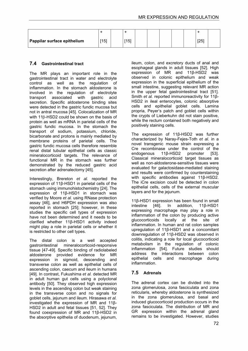
MR EXPRESSION AND REGULATION
72
Pappilar surface epithelium
+
[15]
+
[15]
+
[25]
7.4 Gastrointestinal tract
The MR plays an important role in the gastrointestinal tract in water and electrolyte control as well as the regulation of inflammation. In the stomach aldosterone is involved in the regulation of electrolyte transport associated with gastric acid secretion. Specific aldosterone binding sites were detected in the gastric fundic mucosa but not in antral mucosa [45]. Colocalization of MR with 11β-HSD2 could be shown on the basis of protein as well as mRNA in parietal cells of the gastric fundic mucosa. In the stomach the transport of sodium, potassium, chloride, bicarbonate and protons is mainly mediated by membrane proteins of parietal cells. The gastric fundic mucosa cells therefore resemble renal distal tubular epithelial cells as classic mineralocorticoid targets. The relevance of functional MR in the stomach was further demonstrated by the reduced gastric acid secretion after adrenalectomy [45].
Interestingly, Brereton et al. reported the expression of 11β-HSD1 in parietal cells of the stomach using immunohistochemistry [24]. The expression of 11β-HSD1 in stomach was verified by Moore et al. using RNase protection assay [46], and H6PDH expression was also reported in stomach [25]; however, in these studies the specific cell types of expression have not been determined and it needs to be clarified whether 11β-HSD1 activity indeed might play a role in parietal cells or whether it is restricted to other cell types.
The distal colon is a well accepted gastrointestinal mineralocorticoid-responsive tissue [47-49]. Specific binding of radiolabeled aldosterone provided evidence for MR expression in sigmoid, descending and transverse colon as well as epithelial cells of ascending colon, caecum and ileum in humans [49]. In contrast, Fukushima et al. detected MR in adult human gut cells using a polyclonal antibody [50]. They observed high expression levels in the ascending colon but weak staining in the transverse colon and no signals for goblet cells, jejunum and ileum. Hirasawa et al. investigated the expression of MR and 11β-HSD2 in adult and fetal tissues [51, 52]. They found coexpression of MR and 11β-HSD2 in the absorptive epithelia of duodenum, jejunum,
ileum, colon, and excretory ducts of anal and esophageal glands in adult tissues [52]. High expression of MR and 11β-HSD2 was observed in colonic epithelium and weak expression in the superficial epithelium of the small intestine, suggesting relevant MR action in the upper fetal gastrointestinal tract [51]. Smith et al. reported immunoreactivity for 11β-HSD2 in ileal enterocytes, colonic absorptive cells and epithelial goblet cells. Lamina propria, Peyer’s patch and goblet cells within the crypts of Lieberkuhn did not stain positive, while the rectum contained both negatively and positively staining cells.
The expression of 11β-HSD2 was further characterized by Naray-Fejes-Toth et al. in a novel transgenic mouse strain expressing a Cre recombinase under the control of the endogenous 11β-HSD2 promoter [53]. Classical mineralocorticoid target tissues as well as non-aldosterone-sensitive tissues were evaluated for galactosidase-mediated staining and results were confirmed by counterstaining with specific antibodies against 11β-HSD2. The iCre excision could be detected in colon epithelial cells, cells of the external muscular layers and for the jejunum.
11β-HSD1 expression has been found in small intestine [46]. In addition, 11β-HSD1 expressing macrophage may play a role in inflammation of the colon by producing active glucocorticoids locally at the site of inflammation. In human and rat colon samples upregulation of 11β-HSD1 and a concomitant downregulation of 11β-HSD2 was observed in colitis, indicating a role for local glucocorticoid metabolism in the regulation of colonic inflammation [54]. Future studies should address the interactions between colon epithelial cells and macrophage during inflammation.
7.5 Adrenals
The adrenal cortex can be divided into the zona glomerulosa, zona fasciculata and zona reticularis, whereby aldosterone is synthesized in the zona glomerulosa, and basal and induced glucocorticoid production occurs in the zona fasciculata. The distribution of MR and GR expression within the adrenal gland remains to be investigated. However, studies
MR EXPRESSION AND REGULATION
73
with GR and MR knockout mice indicated important roles of these receptors for adrenal function. In fetal adrenal glands of GR knockout mice, which die immediately after birth, an extensive hypertrophy and hyperplasia of the cortical zones of the adrenal gland was observed with a disorganized and reduced medullary region and a lack of adrenalin producing cells [55]. Hubert et al. studied the impact of MR gene disruption on the renin angiotensin aldosterone system in 8 days old mice [56]. These mice developed pseudohypoaldosteronism type I with high plasma renin, angiotensin II and aldosterone. Histological analyses revealed a significantly enlarged zona glomerulosa, which extended more deeply toward the medullary region than in wild-type mice. The zona fasciculata was reduced and hardly detectable in MR knockout animals. Importantly, renin mRNA expression was hardly detectable in wild-type and heterozygous mice but up to ten-fold higher in the enlarged zona glomerulosa of MR knockout mice. In contrast, angiotensin receptor 1 mRNA was not changed, whereas angiotensin receptor 2 was two-fold lower in adrenals of MR knockout mice. It is not clear whether the observed changes in the adrenal glands are exclusively a result of the systemic effects of the severe sodium depletion and hypovolemia and adaptive responses or whether MR and GR in specific cells of the adrenal gland might contribute to these disturbances.
Using in situ hybridization Shimojo et al. found 11β-HSD1 predominantly in cells at the cortico-medullary junction within the inner cortex, where it was proposed to play a role in regulating the supply of cortex-derived corticosterone to the medullary chromaffin cells [57]. Other investigators applied immunohistochemistry and detected 11β-HSD1 in the outer layer of cells corresponding to the glomerulosa but not in the fasciculata and reticularis [24, 25]. They observed occasional spots and short streaks radiating through the zona fasciculata and reticularis and associated this expression pattern to neuronal cells and/or interstitial fibroblasts. Some staining for 11β-HSD1 was also observed in the medulla. A relatively high expression of H6PDH has been found in adrenals from rats, with highest expression in chromaffin cells [25]. Thus, H6PDH is not coexpressed with 11β-HSD1 in chromaffin cells and the role of NADPH generation in the ER of these cells remains to be determined.
11β-HSD2 mRNA was more abundant in the cortex compared with medulla and its expression was uniformly distributed over the adrenal gland [57]. In humans, 11β-HSD2 was not detected in adult adrenals but in fetal tissue [58]. Similarly, 11β-HSD2 could not be detected in adrenals from adult mice [53, 59]. In contrast, 11β-HSD2 was detected by immunohistochemistry in the fasciculata and reticularis but not in the glomerulosa and medulla. 11β-HSD2 staining was observed in cord-like structures, consistent with expression in steroid-secreting cells.
Future studies should address the role of 11β-HSDs and their corresponding receptors on adrenal function. There is limited knowledge on impaired function of these enzymes in adrenals in inflammation and metabolic diseases.
7.6 Immune system
Glucocorticoids are potent modulators of the immune system and most of their effects are mediated either directly or indirectly by GR [60, 61]. In clinics, glucocorticoids still belong to the most abundantly used and potent anti-inflammatory therapeutics. Numerous synthetic steroids are available such as dexamethasone, betamethasone, triamcinolone, budenoside, prednisolone and others. Glucocorticoids are widely used to treat acute inflammation as well as autoimmune driven chronic inflammatory diseases and neuroinflammatory disorders [62-64]. In contrast, much less is known on the role of MR in the regulation of immune functions.
Several studies showed that monocytes and macrophages coexpress MR and GR [65-71]. Interestingly, 11β-HSD1 is absent in the undifferentiated and cycling monocytes [72]. Once activated and recruited to the inflamed tissue, monocytes undergo differentiation into macrophages. During this process 11β-HSD1 expression is induced and reaches high levels in the differentiated macrophages. In addition, macrophages show high expression of H6DPH. This raises the question how MR and GR in the presence of 11β-HSD1/H6PDH can be regulated distinctly and how they are involved in the coordination of immune regulation.
Usher et al. generated mice specifically lacking MR in myeloid cells and showed that MR is essential for efficient macrophage activation by proinflammatory cytokines [71]. Macrophage derived from MR-deficient myeloid cells displayed an impaired activation pattern, and in mice deletion of MR in macrophages
MR EXPRESSION AND REGULATION
74
resembled the effects of MR antagonists and protected against cardiac hypertrophy, fibrosis and vascular damage caused by treatment with angiotensin II/L-NAME. Furthermore, myeloid-derived dendritic cells express MR. Herrada et al. demonstrated an aldosterone-mediated increase in CD8+ T-cell activation that was dependent on dendritic cells. Aldosterone-mediated MR activation induced MAPK signaling and secretion of IL-6 and TGF-β1 by dendritic cells. Further, aldosterone induced Th17 cell-mediated immune response. The altered, aldosterone-mediated dendritic cell activity might promote inflammatory damage in the heart and other organs (see also section on heart).
MR expression was also found in neutrophils [73]. Incubation of neutrophils with aldosterone inhibited the activation of NF-κB by interleukin-8 (IL-8) and granulocyte/macrophage colony-stimulating factor. Spironolactone abolished NF-κB inhibitioin by aldosterone, indicating an MR-specific effect. Incubation with IL-8 strongly induced TNF-α mRNA expression, an effect that was prevented by aldosterone. These results suggest anti-inflammatory effects of MR in neutrophils that might be relevant when they interact with endothelial cells. Thus, MR seems to mediate pro- and anti-inflammatory effects, depending on the cell type.
7.7 Brain
Corticosteroids play a pivotal role in the control of brain activity and are involved in regulating stress response, mood, sleeping behavior, memory function and release of neuroendocrine hormones [74]. Both MR and GR are expressed in the brain, with differences in their sites of expression and functions. Immunohistochemistry, in situ hybridization and binding of radiolabeled aldosterone revealed high expression of MR in neurons of the hippocampus, lateral septum, medial and central amygdala, olfactory nucleus, layer II of the cortex and brain stem sensory and motor neurons [75-79]. MR is also found in the anterior hypothalamus and circumventricular tissues including chorioid plexus. The GR is widely expressed in the brain in neurons and glial cells [75, 79-81]. High expression is found in the limbic system (hippocampus, septum and amygdala), in the parvocellular neurons of the paraventricular nucleus of the hypothalamus and in the supraoptic nucleus. High expression of GR is also observed in the ascending monoaminergic neurons of the brain stems.
Importantly, in hippocampal neurons and microglia cells MR and GR are expressed in the absence of 11β-HSD2 [53, 82] but presence of 11β-HSD1 [83-85], suggesting predominant occupation of the receptors by 11β-hydroxyglucocorticoids. Low levels of glucocorticoids are expected to predominantly act through MR, thereby functioning in a proactive mode by regulating the sensitivity of neuroendocrine stress responses [76, 86]. High levels of glucocorticoids, for example during stress, lead to the occupancy of MR and GR, whereby the GR is thought to play a pivotal role in counteracting MR effects and mediating recovery from the stress response.
Confocal laser scanning microscopy revealed a specialized nuclear clustering for MR and GR in neuronal cells of the CA1 region [87]. The two receptors were found in distinct nuclear domains but also in clusters where they colocalize, indicating the formation of receptor homodimers and heterodimers. The formation of MR-GR heterodimers in rat hippocampal neurons has been demonstrated, and activation of GR was shown to inhibit MR-mediated regulation of neuronal function [88]. Using recombinant receptors Trapp et al. observed enhanced activation of the mouse mammary tumor virus (MMTV) promoter driven LacZ gene upon coexpressing MR and GR compared with cells transfected with one of the receptors only [89]. In contrast, Liu et al. coexpressed MR and GR in monkey kidney CV-1 cells and observed significantly lower activation of a TAT3-TATA-reporter construct compared to cells expressing only MR or GR [90]. These observations indicate highly cell- and promoter-specific effects by MR and GR homodimers and heterodimers.
Inflammation results in increased local and circulating levels of active glucocorticoids (for review see [63]). The inflammatory response in the brain involves a coordinated action of monocytes, macrophages, astrocytes and microglia cells. Microglia cells express both MR and GR in the presence of 11β-HSD1 [91]. Like macrophage, microglia cells belong to the specialized cells of the immune system. They express MHC II (major histocompatibility complex) and therefore act as professional antigen-presenting cells (APCs) in the brain. Microglia cells are able to produce cytokines and neurotrophic factors. High doses of the synthetic glucocorticoids methylprednisolone and dexamethasone were shown to suppress the expression of MHC II on the surface of microglia cells [92, 93]. The suppressive effect of these ligands is likely a result of their high
MR EXPRESSION AND REGULATION
75
concentrations and GR-selectivity. In contrast, low doses of endogenous glucocorticoids that mainly act through MR may stimulate the inflammatory response.
Excess glucocorticoid action during stress or upon upregulation of 11β-HSD1 by pro-inflammatory cytokines during inflammation exerts adverse effects on hippocampal neurons and causes impaired cognitive functions. Increased glucocorticoid levels have been associated with cognitive impairments and hippocampal atrophy both in rodents and humans [94, 95]. In aging mice an increase in 11β-HSD1 levels in the CA3 hippocampus and parietal cortex correlated with impaired cognitive performance, whereby circulating glucocorticoid levels and corticosteroid receptor expression did not correlate with cognitive function [96]. Transgenic mice overexpressing 11β-HSD1 specifically in the forebrain region showed premature age-associated cognitive deficits, suggesting a causal role of elevated 11β-HSD1 expression. This is supported by the observation that mice deficient in 11β-HSD1 have lover intrahippocampal corticosterone levels and that they show a delayed decline in age-related cognitive function [84]. A reduced 11β-HSD1 expression in transgenic animals as well as inhibition of the enzyme resulted in improved memory function, suggesting that inhibition of 11β-HSD1 may show beneficial effects in treating age-related cognitive disorders [97]. In line with the adverse effects of elevated glucocorticoids in the hippocampus, transgenic expression of 11β-HSD2 in dentate gyrus granule cells reversed the adverse effects of high glucocorticoid treatment on granule cell
and CA1 pyramidal cell excitability and on spatial reference memory [98].
The observation that 11β-HSD1 in neurons catalyzes the regeneration of active glucocorticoids indicates the coexpression with H6PDH. A recent analysis of H6PDH expression revealed a distinct size of the immunoreactive protein at 60 kDa in whole brain tissue compared with 90 kDa for the classical protein [25]. It remains to be clarified whether an alternatively spliced variant or a post-translationally modified H6PDH is expressed in the brain.
Colocalization of MR and 11β-HSD2, indicating aldosterone sensitivity of MR, was demonstrated in the nucleus of the solitary tract using immunofluorescence [82]. Furthermore, MR and 11β-HSD2 were both detected in amygdala, subcommissural organ, the ventromedial nucleus of the hypothalamus, and locus coerulens. The localization of 11β-HSD2 in these tissues was confirmed in transgenic mice expressing β-galactosidase under the control of the endogenous HSD11B2 promoter [53]. 11β-HSD2 expression was found in the nucleus of the solitary tract and amygdala as observed in earlier studies, in the external cuneate nuclei in the medulla, in the external granular layer in the cerebellum, in the pontine reticular formation and pontine nuclei and periaqueductal gray in the pons/midbrain region. 11β-HSD2 was also found in the hypothalamus and in several thalamic nuclei. Coexpression of MR and 11β-HSD2 in the periventricular regions render selectivity of MR to aldosterone to modulate volume regulation and sympathetic outflow as well as salt appetite.
Table 7 Expression and Protein appearance of MR, GR, 11β-HSD1, 11β-HSD2 and H6PDH in brain specific cell types.
BRAIN MR GR 11β-HSD1 11β-HSD2 H6PDH
Protein
RN
A
Protein
RN
A
Protein
RN
A
Protein
RN
A
Protein
RN
A
Brain
+
[1]
[86]
+
[86]
[1]
-
[25]
+
[25]
-
[55]
+
[25]
+
[25]
Medulla
Nucleus of the solitary tract + +++
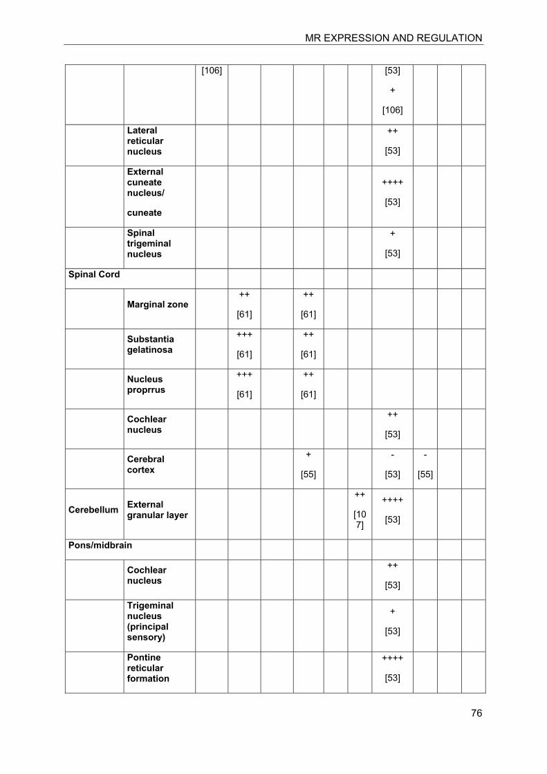
MR EXPRESSION AND REGULATION
76
[106] [53]
+
[106]
Lateral reticular nucleus
++
[53]
External cuneate nucleus/
cuneate
++++
[53]
Spinal trigeminal nucleus
+
[53]
Spinal Cord
Marginal zone ++
[61]
++
[61]
Substantia gelatinosa
+++
[61]
++
[61]
Nucleus proprrus
+++
[61]
++
[61]
Cochlear nucleus
++
[53]
Cerebral cortex
+
[55]
-
[53]
-
[55]
Cerebellum External granular layer
++
[107]
++++
[53]
Pons/midbrain
Cochlear nucleus
++
[53]
Trigeminal nucleus (principal sensory)
+
[53]
Pontine reticular formation
++++
[53]
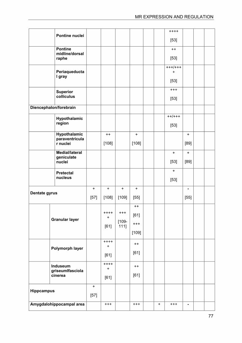
MR EXPRESSION AND REGULATION
77
Pontine nuclei ++++
[53]
Pontine midline/dorsal raphe
++
[53]
Periaqueductal gray
+++/++++
[53]
Superior colliculus
+++
[53]
Diencephalon/forebrain
Hypothalamic region
++/+++
[53]
Hypothalamic paraventricular nuclei
++
[108]
+
[108]
+
[89]
Medial/lateral geniculate nuclei
+
[53]
+
[89]
Pretectal nucleus
+
[53]
Dentate gyrus +
[57]
+
[108]
+
[109]
+
[55]
-
[55]
Granular layer ++++
+
[61]
+++
[109-111]
++
[61]
+++
[109]
Polymorph layer ++++
+
[61]
++
[61]
lnduseum griseumlfasciola cinerea
++++
+
[61]
++
[61]
Hippcampus +
[57]
Amygdalohippocampal area +++ +++ + +++ -
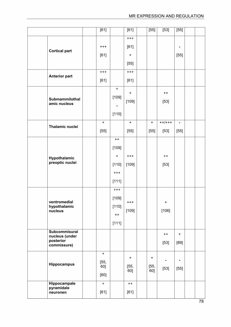
MR EXPRESSION AND REGULATION
78
[61] [61] [55] [53] [55]
Cortical part +++
[61]
+++
[61]
+
[55]
-
[55]
Anterior part +++
[61]
+++
[61]
Submammilothalamic nucleus
+
[109]
-
[110]
+
[109]
++
[53]
Thalamic nuclei +
[55]
+
[55]
+
[55]
++/+++
[53]
-
[55]
Hypothalamic preoptic nuclei
++
[109]
+
[110]
+++
[111]
+++
[109]
++
[53]
ventromedial hypothalamic nucleus
+++
[109]
[110]
++
[111]
+++
[109]
+
[106]
Subcommisural nucleus (under posterior commissure)
++
[53]
+
[89]
Hippocampus
+
[55, 60]
[60]
+
[55, 60]
+
[55, 60]
-
[53]
-
[55]
Hippocampale pyramidale neuronen
+
[61]
++
[61]
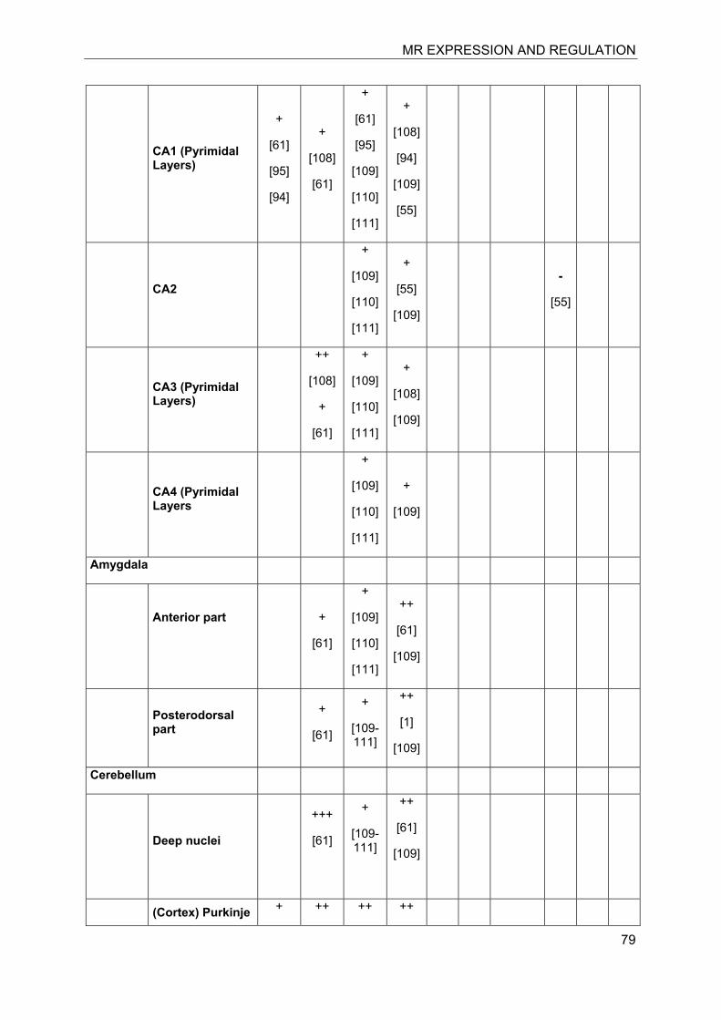
MR EXPRESSION AND REGULATION
79
CA1 (Pyrimidal Layers)
+
[61]
[95]
[94]
+
[108]
[61]
+
[61]
[95]
[109]
[110]
[111]
+
[108]
[94]
[109]
[55]
CA2
+
[109]
[110]
[111]
+
[55]
[109]
-
[55]
CA3 (Pyrimidal Layers)
++
[108]
+
[61]
+
[109]
[110]
[111]
+
[108]
[109]
CA4 (Pyrimidal Layers
+
[109]
[110]
[111]
+
[109]
Amygdala
Anterior part
+
[61]
+
[109]
[110]
[111]
++
[61]
[109]
Posterodorsal part
+
[61]
+
[109-111]
++
[1]
[109]
Cerebellum
Deep nuclei
+++
[61]
+
[109-111]
++
[61]
[109]
(Cortex) Purkinje + ++ ++ ++
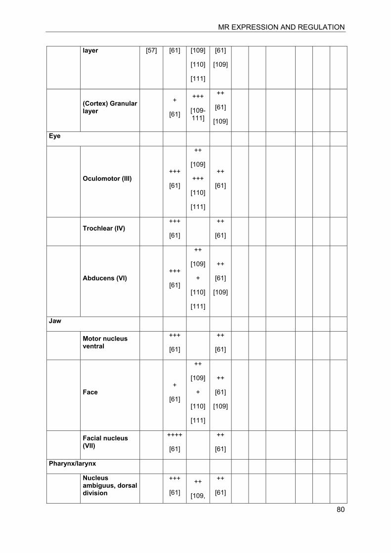
MR EXPRESSION AND REGULATION
80
layer [57] [61] [109]
[110]
[111]
[61]
[109]
(Cortex) Granular layer
+
[61]
+++
[109-111]
++
[61]
[109]
Eye
Oculomotor (III) +++
[61]
++
[109]
+++
[110]
[111]
++
[61]
Trochlear (IV) +++
[61]
++
[61]
Abducens (VI) +++
[61]
++
[109]
+
[110]
[111]
++
[61]
[109]
Jaw
Motor nucleus ventral
+++
[61]
++
[61]
Face +
[61]
++
[109]
+
[110]
[111]
++
[61]
[109]
Facial nucleus (VII)
++++
[61]
++
[61]
Pharynx/larynx
Nucleus ambiguus, dorsal division
+++
[61] ++
[109,
++
[61]
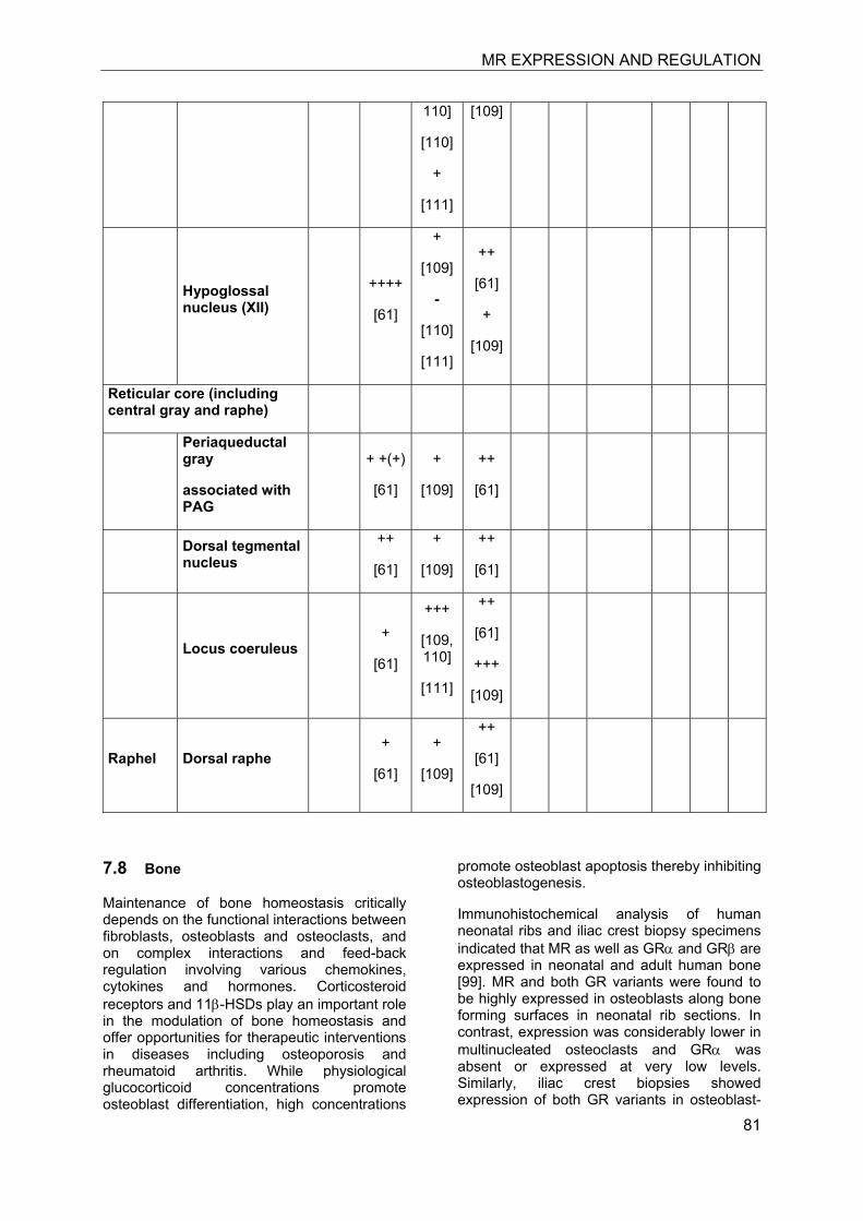
MR EXPRESSION AND REGULATION
81
110]
[110]
+
[111]
[109]
Hypoglossal nucleus (XII)
++++
[61]
+
[109]
-
[110]
[111]
++
[61]
+
[109]
Reticular core (including central gray and raphe)
Periaqueductal gray
associated with PAG
+ +(+)
[61]
+
[109]
++
[61]
Dorsal tegmental nucleus
++
[61]
+
[109]
++
[61]
Locus coeruleus +
[61]
+++
[109, 110]
[111]
++
[61]
+++
[109]
Raphel Dorsal raphe +
[61]
+
[109]
++
[61]
[109]
7.8 Bone
Maintenance of bone homeostasis critically depends on the functional interactions between fibroblasts, osteoblasts and osteoclasts, and on complex interactions and feed-back regulation involving various chemokines, cytokines and hormones. Corticosteroid receptors and 11β-HSDs play an important role in the modulation of bone homeostasis and offer opportunities for therapeutic interventions in diseases including osteoporosis and rheumatoid arthritis. While physiological glucocorticoid concentrations promote osteoblast differentiation, high concentrations
promote osteoblast apoptosis thereby inhibiting osteoblastogenesis.
Immunohistochemical analysis of human neonatal ribs and iliac crest biopsy specimens indicated that MR as well as GRα and GRβ are expressed in neonatal and adult human bone [99]. MR and both GR variants were found to be highly expressed in osteoblasts along bone forming surfaces in neonatal rib sections. In contrast, expression was considerably lower in multinucleated osteoclasts and GRα was absent or expressed at very low levels. Similarly, iliac crest biopsies showed expression of both GR variants in osteoblast-
MR EXPRESSION AND REGULATION
82
like cells in cancellous surfaces, whereas very few osteocytes stained positive for GR. MR expression was found in osteoblasts and in about one-third of cancellous osteocytes. The presence of mRNA of the two GR variants and of MR was confirmed by RT-PCR in cultured primary human osteoblasts. Evidence for the lack of GRα but presence of GRβ expression in human and rat osteoclasts, and of considerably lower GR and MR in osteoclasts compared with osteoblasts, was supported by other investigators [100, 101]. Interestingly, MR was not detected in early fetal bone tissue beyond 12 weeks of gestation [102], suggesting a role of this receptor in terminal differentiation.
Early observations in patients with apparent mineralocorticoid excess (AME) provided evidence for a role of 11β-HSD2 in modulating bone homeostasis. AME patients suffered, among other complications, from retarded growth, osteopenia and minimal trauma bone fractures, effects that were ameliorated upon treatment with the MR antagonist spironolactone [103, 104]. Activation of MR by aldosterone enhanced proliferation of cultured osteoblasts from rat calvaria, an effect inhibited by specific MR antagonists [105]. Furthermore, MR antagonists inhibited the production of pro-inflammatory cytokines, including TNF-α and INF-γ, and have potential in the treatment of arthritis [106].
Using the MG-63 human osteosarcoma cell line, Cooper et al. reported a decreased expression of 11β-HSD2 upon treatment with TNF-α or IL-1β [107]. In contrast, primary osteoblasts express 11β-HSD1 and the levels of this enzyme were stimulated by exposure to TNF-α or IL-1β, thus leading to enhanced local concentrations of active glucocorticoids. The authors proposed that pro-inflammatory cytokines may exert some of their effects within bone, e.g. periarticular erosions in inflammatory arthritis, by increasing local glucocorticoid concentrations.
Jia et al. studied the role of glucocorticoids in transgenic mice specifically expressing 11β-HSD2 in osteoclasts [108]. Treatment of wild-type and transgenic mice with pharmacological doses of glucocorticoids enhanced apoptosis in cancellous osteoblasts and decreased osteoblast, osteoid and bone formation. Glucocorticoids stimulated the osteoclast marker calcitonin receptor on wild-type but not transgenic mice. Importantly, glucocorticoids decreased the number of cancellous osteoclasts in transgene but not wild-type
mice. The observed loss of bone density in wild-type mice was prevented by 11β-HSD2 overexpression in the transgene. The authors concluded that the early, rapid loss of bone caused by glucocorticoid excess resulted from direct actions on osteoclasts.
Glucocorticoids expand the life span of osteoblasts and decrease bone density. An early increased bone resorption followed by a diminished osteoclastogenesis and a consequently decreased bone resorption was observed in a mouse model of glucocorticoid-induced osteoporosis [109]. Mice implanted with slow release prednisone pellets displayed early activation of osteoclastogenesis and adipogenesis as well as prolonged suppression of osteogenesis [110]. In this model the synthetic glucocorticoid receptor ligand prednisone required prior activation by 11β-HSD1 in the liver or locally in the osteoblast.
Buttgereit et al. studied the impact of osteoblast-targeted transgenic overexpression of 11β-HSD2 on joint inflammation, cartilage damage, and bone metabolism in the K/BxN mouse serum transfer model of autoimmune arthritis [111]. Wild-type and transgenic mice developed acute arthritis but in the latter arthritis and local inflammatory activity were significantly attenuated. Transgenic overexpression of 11β-HSD2 ameliorated bone resorption as well as loss of bone volume, and improved osteoblast activity, suggesting that osteoblasts modulate the immune-mediated inflammatory response in a glucocorticoid-dependent manner.
Intraarticular corticosteroid application in patients with inflammatory arthritis reduced synovial RANKL expression [112]. Glucocorticoids inhibited osteoprotegerin expression and increased receptor activator of NF-κB ligand (RANKL) synthesis by osteoblasts, thereby promoting osteoclastogenesis [112, 113]. However, following priming with TNF-α, a condition mimicking pro-inflammatory milieu of the rheumatoid joint, glucocorticoids were found to decrease RANKL expression [112]. Thus, glucocorticoids affect bone cells differently in the presence or absence of inflammatory mediators and they may have a bone conserving effect in rheumatoid arthritis despite of inducing osteoporosis in the spine.
MR EXPRESSION AND REGULATION
83
7.9 Adipose tissue
Adipose tissue is derived from lipoblasts representing the adipocyte precursor cells. Two functionally distinct adipocytes are differentiated from these pre-adipocytes, forming either brown (BAT) or white adipose tissue (WAT). BAT in adults is derived from the abundant fat tissue of the fetal and newborn mammal organism, where it is responsible for the shiver-free heat production. The adverse metabolic effects of excessive fat accumulation have been associated mainly with WAT. Transgenic mice with disruption of uncoupling protein function and a primary deficiency of BAT were prone to obesity [114]. Functional adipose tissue includes additional cell types such as endothelial cells enabling its high vascularization, fibroblasts, and macrophages responsible for the numerous endocrine and immune functions. Adipose tissue can be broadly categorized in subcutaneous and visceral fat depots. Besides its major function as lipid storage, fat tissue performs other general functions, including protection against temperature fluctuations (subcutaneous fat) and resistance of organs like stomach, heart and kidneys against mechanical stress (visceral fat).
Adipocytes and adipose tissue infiltrating macrophages are derived from the same bone marrow stem cells, and they express MR and GR in the presence 11β-HSD1 and H6PDH. There is increasing evidence for a key role of MR in mediating adverse effects in metabolic disease. Elevated levels of MR were found in obese, diabetic mice (ob/ob and db/db), which have increased expression of pro-inflammatory and pro-fibrotic factors and reduced expression of adiponectin and PPARγ in adipose tissue [115, 116]. Treatment with the MR selective antagonist eplerenone normalized the impaired regulation of obesity-related genes, suppressed macrophage infiltration and attenuated insulin resistance [116]. Moreover, incubation of undifferentiated preadipocytes with 10 nM aldosterone for 24 h increased the expression of TNF-α and MCP1 and decreased adiponectin and PPARγ [115]. A recent study with cultured 3T3-L1 and 3T3-F442A adipocytes and human primary preadipocytes reported dose-dependent inhibition of adipose differentiation and potent anti-adipogenic effects of the MR antagonist drospirenone [117]. Further evidence for a pro-inflammatory adipogenic effect of MR was provided from a recent study with GR- and MR-deficient adipocytes [118]. Expression of the pro-inflammatory factors IL-6 and MCP1
was enhanced in GR knockout adipocytes upon treatment with corticosterone, indicating an MR-dependent stimulation of the pro-inflammatory factors. Deletion of MR resulted in a complete loss of lipid accumulation, whereas deletion of GR led to rather subtle disturbances of adipogenesis during early differentiation. These observations are in line with an earlier study using a brown adipocyte cell model [119]. Aldosterone promoted adipocyte differentiation, indicated by an accumulation of intracytoplasmic lipid droplets and a concentrations-dependent increase in intracellular triglyceride content. The aldosterone-dependent effects were not affected by the GR antagonist RU-486 but abolished by MR antagonists, indicating a key role of MR in the early phase of adipocyte differentiation.
Glucocorticoids control the terminal differentiation of adipose precursor cells and essentially modulate adipocyte function [120]. Given the more than ten-fold higher affinity of MR compared with GR for 11β-hydroxyglucocorticoids, it can be assumed that the MR is occupied by 11β-hydroxyglucocorticoids in preadipocytes and adipocytes. Depending on the available glucocorticoid concentration lower affinity GRs are activated and may counteract the effects of MR through mechanisms that need to be uncovered but likely include receptor heterodimerization. 11β-HSD1 is highly expressed in adipose tissue, although its expression is considerably lower than in liver. In contrast, 11β-HSD2 mRNA and activity was detected only at low levels in adipose stromal vascular cells [121, 122]. Comparison of the expression of 11β-HSD1 and 11β-HSD2 revealed a 22-fold and 8-fold lower expression of the latter in subcutaneous fat, respectively. Moreover, in the obese situation infiltrated macrophage expressing 11β-HSD1 further increase local cortisol production and MR activation, thus promoting inflammation [123].
In mouse preadipocytes isolated from mesenteric and subcutaneous fat 11β-HSD1 was found to function as oxoreductase, thereby activating glucocorticoids [124]. Treatment of mice with high-fat diet, leading to the stimulation of 11β-HSD1 activity, resulted in enhanced preadipocyte differentiation in wild-type but not 11β-HSD1 knockout animals. It was shown that in the widely used 3T3-L1 fibroblast-like cells 11β-HSD1 expression is absent prior to differentiation but increases with progression of differentiation and is highly expressed in the fully differentiated state [125,
MR EXPRESSION AND REGULATION
84
126]. Importantly, H6PDH is similarly expressed before and after differentiation [125]. Furthermore, a continuous expression of H6PDH was observed during the differentiation of human adipose-derived mesenchymal stem cells, indicating that 11β-HSD1 functions as an oxoreductase in both preadipocytes and adipocytes [127]. H6PDH knockout mice, where 11β-HSD1 is thought to act as a dehydrogenase, display diminished lipogenesis, lipolysis rates and fatty acid release upon fasting, and they have significantly reduced adipose tissue mass, although average adipocyte size was not altered [128].
The association of enhanced local glucocorticoid activation by 11β-HSD1 with visceral obesity and the development of insulin resistance, type 2 diabetes and cardiovascular disease is being extensively studied (reviewed in [129, 130]). Based on the fact that the MR has more than ten-fold higher affinity than GR and that it is coexpressed with 11β-HSD1 and H6PDH in preadipocytes and adipocytes, we propose that the pro-inflammatory and pro-adipogenic effects of elevated local glucocorticoid concentrations are probably mainly mediated by activation of MR. Thus, a combination of an MR antagonist and an 11β-HSD1 inhibitor may prove beneficial in the treatment of metabolic disease.
7.10 Heart
In the heart MR expression has been demonstrated in cardiomyocytes and fibroblasts, cells of atria and ventricles, the aorta and pulmonary artery as well as in vascular endothelial and smooth muscle cells [131-134]. In addition, macrophage, which infiltrate the heart during inflammation, express high levels of MR.
Several clinical studies revealed an association of elevated MR activity with vascular inflammation and cardiac fibrosis, and an increased risk for congestive heart failure. Supplementation of the standard therapy of angiotensin-converting enzyme inhibitor, loop diuretic and digoxin for patients with heart failure with the MR antagonists spironolactone (RALES [135]) and eplerenone (EPHESUS [136]) demonstrated a 30% and 15% improvement in mortality. Furthermore, treatment with MR antagonists decreased blood pressure in patients with essential hypertension and left ventricular hypertrophy (4E-left ventricular hypertrophy study [137]). The MR-dependent exacerbation of tissue
damage in cardiac ischemia can be ameliorated by eplerenone [138]. Thus, MR antagonists have a beneficial impact on post-myocardial infarction therapy and in treatment of patients with essential hypertension.
In line with clinical trials, animal studies addressed the mechanisms of MR activation in heart disease and provided evidence for beneficial effects of antagonists [139-146]. A causal role of MR was demonstrated in transgenic mice by conditional overexpression specifically in cardiomyocytes. These mice exhibited cardiac remodeling with severe ventricular arrhythmias and increased mortality [147]. In mice chronic severe pressure overload due to aortic constriction caused cardiac hypertrophy, followed by left ventricular dilatation and heart failure [148]. Cardiomyocyte-specific deletion of MR prevented the increase in left ventricular inner diastolic diameter and wall tension but did not prevent cardiac hypertrophy. Similarly, eplerenone did not prevent cardiac hypertrophy but delayed the transition to myocardial failure [148, 149]. Cardiac fibrosis caused by chronic pressure overload was not reduced in mice with a specific knockout of MR in fibroblasts [148]. However, deletion of MR in macrophage attenuated the production of ROS in the heart and prevented inflammation and fibrosis induced by treatment with deoxycorticosterone/salt [69] or angiotensin II [71]. These observations indicate a key role of MR in infiltrating macrophage in the progression of vascular inflammation and cardiac fibrosis.
In humans cardiomyocytes have no or very low levels of 11β-HSD1 and 11β-HSD2 [24]. Vascular smooth muscle cells do not express 11β-HSD1 but both endothelial cells and vascular smooth muscle cells were reported to express 11β-HSD2 [133, 150, 151]. In contrast, 11β-HSD1 expression was found in rodent cardiac vascular smooth muscle cells, whereby enzyme activity was higher in quiescent cells compared with proliferating cells [152]. Although some discrepancies may be due to contamination of vascular smooth muscle cell preparations with endothelial cells, species-specific differences need to be considered and care should be taken in extrapolating results from studies with rodents to the human situation.
Evidence for an important role of 11β-HSD2 in the heart was provided from observations in 11β-HSD2 knockout mice. 11β-HSD2 expression is low in fetal mouse heart,
MR EXPRESSION AND REGULATION
85
whereas MR is highly expressed. Nevertheless, 11β-HSD2 knockout mice exhibit significantly enlarged heart size and a high mortality rate [153]. A recent study reported a highly significant negative correlation between 11β-HSD2 expression and the thickness of the left ventricular wall in sheep [154]. The chronic administration of moderate doses of cortisol during late gestation resulted in a significantly decreased 11β-HSD2 expression in the heart and caused an increase of the fetal heart weight.
Since 11β-HSD2 converts active into inactive glucocorticoids, it restores aldosterone specificity of MR and inhibits GR activation. Thus, both GR and MR might be responsible for the development of elevated heart size. Interestingly, transgenic mice with a cardiac-specific overexpression of 11β-HSD2 developed cardiac hypertrophy, which was attenuated after treatment with eplerenone. In the presence of high 11β-HSD2 activity, circulating glucocorticoids may no longer be sufficiently high to activate GR in order to counteract aldosterone-dependent MR activity. In contrast, a lack of 11β-HSD2 is expected to cause glucocorticoid-dependent MR activation and GR might no longer be able to efficiently counteract the fully activated MR, thus providing an explanation why both a lack of 11β-HSD2 and its overexpression cause cardiac hypertrophy.
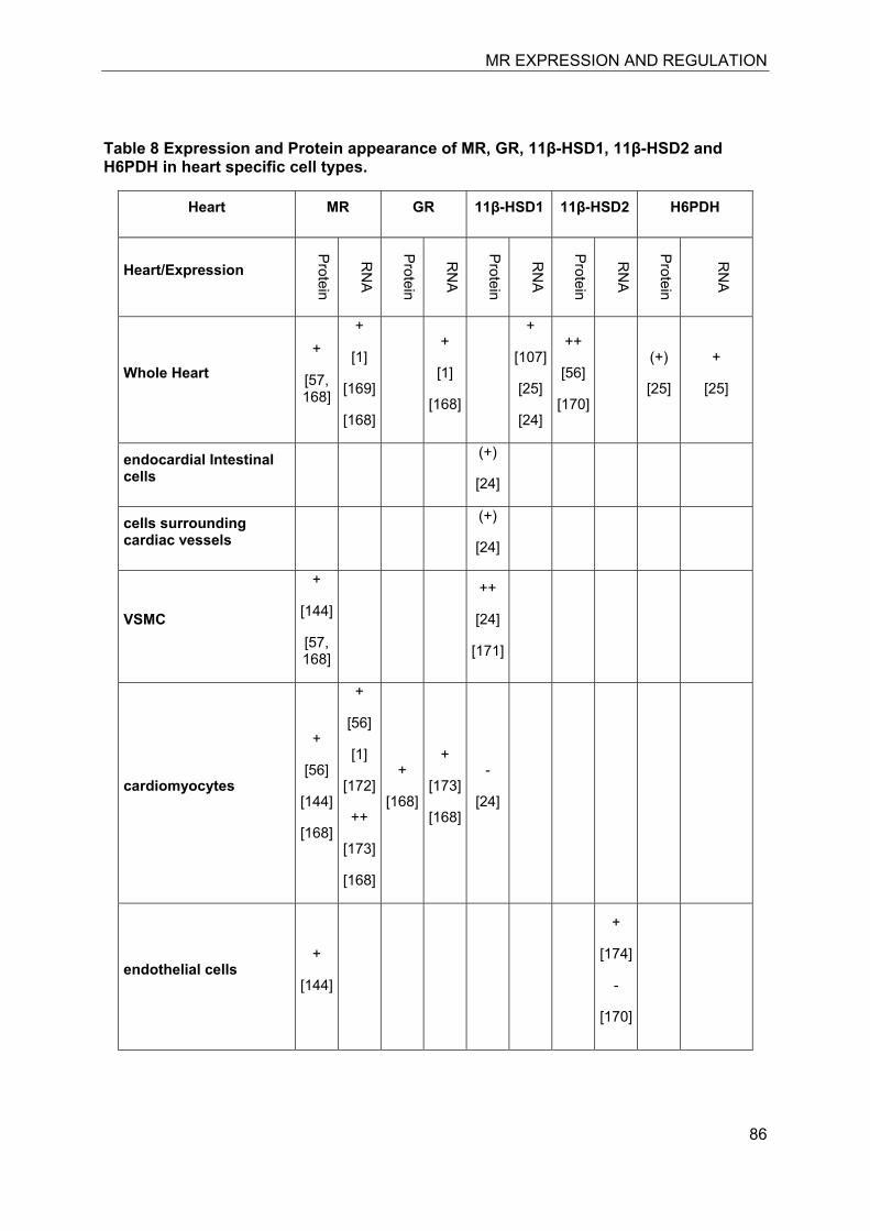
MR EXPRESSION AND REGULATION
86
Table 8 Expression and Protein appearance of MR, GR, 11β-HSD1, 11β-HSD2 and H6PDH in heart specific cell types.
Heart MR GR 11β-HSD1 11β-HSD2 H6PDH
Heart/Expression
Protein
RN
A
Protein
RN
A
Protein
RN
A
Protein
RN
A
Protein
RN
A
Whole Heart +
[57, 168]
+
[1]
[169]
[168]
+
[1]
[168]
+
[107]
[25]
[24]
++
[56]
[170]
(+)
[25]
+
[25]
endocardial Intestinal cells
(+)
[24]
cells surrounding cardiac vessels
(+)
[24]
VSMC
+
[144]
[57, 168]
++
[24]
[171]
cardiomyocytes
+
[56]
[144]
[168]
+
[56]
[1]
[172]
++
[173]
[168]
+
[168]
+
[173]
[168]
-
[24]
endothelial cells +
[144]
+
[174]
-
[170]
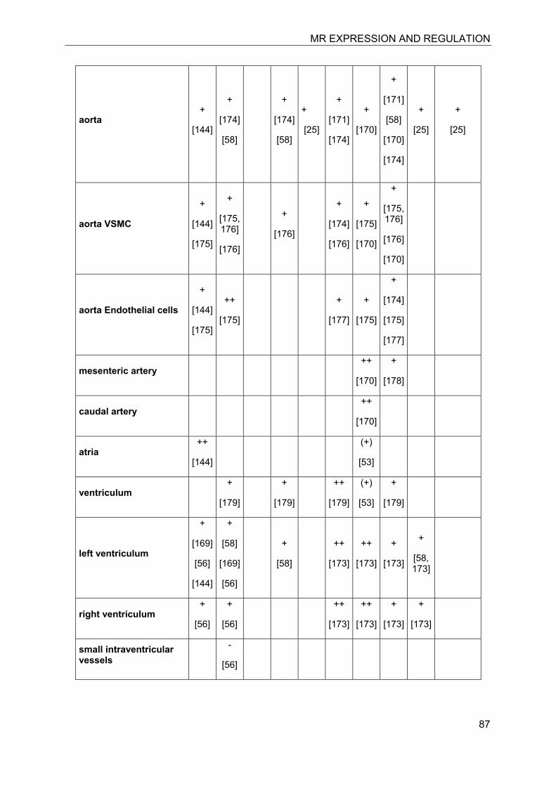
MR EXPRESSION AND REGULATION
87
aorta +
[144]
+
[174]
[58]
+
[174]
[58]
+
[25]
+
[171]
[174]
+
[170]
+
[171]
[58]
[170]
[174]
+
[25]
+
[25]
aorta VSMC
+
[144]
[175]
+
[175, 176]
[176]
+
[176]
+
[174]
[176]
+
[175]
[170]
+
[175, 176]
[176]
[170]
aorta Endothelial cells
+
[144]
[175]
++
[175]
+
[177]
+
[175]
+
[174]
[175]
[177]
mesenteric artery ++
[170]
+
[178]
caudal artery ++
[170]
atria ++
[144]
(+)
[53]
ventriculum +
[179]
+
[179]
++
[179]
(+)
[53]
+
[179]
left ventriculum
+
[169]
[56]
[144]
+
[58]
[169]
[56]
+
[58]
++
[173]
++
[173]
+
[173]
+
[58, 173]
right ventriculum +
[56]
+
[56]
++
[173]
++
[173]
+
[173]
+
[173]
small intraventricular vessels
-
[56]
MR EXPRESSION AND REGULATION
88
7.11 Skeletal muscle
Skeletal muscles have a high demand of energy, and glucocorticoids are essentially regulating insulin-stimulated glucose uptake, glycogen storage and carbohydrate metabolism. Chronically elevated glucocorticoid levels have been associated with insulin resistance and impaired carbohydrate metabolism as well as catabolic pathways causing muscle atrophy. Recent studies provided evidence for a functional expression of MR (besides GR) in soleus muscle [155]. High aldosterone levels, in addition to their cardiotoxic effects, led to the induction of apoptosis in soleus muscle. In soleus muscle high aldosterone levels increased NADPH oxidase and the production of ROS, decreased Akt phosphorylation and GLUT4 expression, and induced insulin resistance [156]. Spironolactone ameliorated aldosterone-induced cardiac and skeletal muscle myopathy, providing evidence for an MR-dependent mechanism. As in cardiomyocytes, 11β-HSD2 expression could not be detected using immunofluorescence and ribonuclease protection assay in skeletal muscle of normal and Cre-recombinant male mice [46, 53].
11β-HSD1 is expressed in skeletal muscle with a 13-times higher level in soleus (type I-rich fibers) compared with tibialis anterior (type IIb-rich fibers)[157]. Impaired regulation of 11β-HSD1 activity in myotubes from diabetic patients may contribute to insulin resistance [158-160]. H6PDH is also expressed in soleus and tibialis anterior and plays an important role in the regulation of muscle cell differentiation and function. It can be assumed that chronic hyperglycemia leads to enhanced local glucocorticoid activation by 11β-HSD1/H6PDH, thereby activating local GR and/or MR and contributing to impaired glucose transport and insulin resistance.
The role of H6PDH has been further investigated in transgenic mice. H6PDH deficient mice developed a vacuolating myopathy, predominantly manifested in type II muscle fibers [161]. In further studies with H6PDH/11β-HSD1 double knock-out mice, this phenotype was clearly associated with H6PDH deficiency, independent of 11β-HSD1 function [157]. Fiber type differentiation can be influenced by physical activity. Marathon runners, for example, show a type I-rich fiber composition of the gastrocnemius, whereas a type IIb-rich composition is observed in sprinters. Thus, fiber type differentiation may
be modulated by the level of energy demand and redox reactions in the endoplasmic reticulum. Whether and how glucocorticoids are involved in these processes remains unclear. The lack of 11β-HSD1 oxoreductase activity in knockout mice may be compensated by an activation of the HPA axis leading to higher circulating corticosterone levels [162], although strain-dependent differences need to be considered [163]. Contradicting findings were reported concerning serum corticosterone concentrations in H6PDH-deficient mice. Significantly increased adrenal weight and elevated plasma corticosterone concentrations were reported by two independent groups [164, 165]; however, two other studies reported significantly decreased plasma corticosterone levels in H6PDH-deficient animals [157, 166]. Since all of these reports are based on data from transgenic mice from the same background, i.e. C57BL6J strain, the reason for the observed differences remains unclear. Nevertheless, 11β-HSD1 was significantly upregulated in H6PDH knockout mice in tibialis anterior, but not in soleus or in the liver [157].
7.12 Skin
Skin consists of different layers, including the epidermis, dermis and subcutis (also named hypodermis). Innervation, vascularization, the presence of secretory glands such as sweat glands and sebaceous glands, hair folicles and pacinian corpuscles enhance the complexity of skin as an organ. Epidermis can be further divided into five distinct areas namely stratum corneum, stratum lucidum, stratum granulosum, stratum spinosum, and final stratum basale. The major cell types present in the epidermis are keratinocytes, langerhans cells and “touch sensors” like merkel-ranvier cells. Keratinocytes in the four outer layers forming the epidermis are mitotically inactive and die upon their travel to the surface because of a loss of nutrition supply. During this process they undergo keratinization. In contrast to the epidermis, the dermis consists of connective tissue and contains many blood and lymphatic vessels. The subcutis is a highly immunologically active tissue and contains dendritic cells and fibroblasts as well as adipocytes.
Glucocorticoids are widely used in the treatment of inflammatory and hyperproliferative skin disorders. A major limitation, however, is the development of skin atrophy [167]. The relative contribution of GR and MR to skin atrophy remains unclear. In situ
MR EXPRESSION AND REGULATION
89
hybridization and immunohistochemistry revealed MR expression in human keratinocytes of the epidermis, in sweat glands and sebaceous glands and in hair follicles [168]. Mice overexpressing MR specifically in keratinocytes and hair follicles exhibited developmental and post-natal impairments of the epidermis and hair follicles [169]. Transgenic mice showed premature epidermal barrier formation at embryonic day 16.5, decreased hair follicle density and epidermal atrophy and increased keratinocyte apoptosis at embryonic day 18.5 and premature eye opening when MR was overexpressed throughout gestation. When MR expression was induced after birth, mice developed alopecia and hair follicle cysts.
Important information on the role of GR in skin was provided from transgenic mice overexpressing GR specifically in keratinocytes [170]. Newborn mice exhibited severe skin lesions due to epidermal hypoplasia and underdeveloped dysplastic hair follicles. In adult mice, an impaired hyperplastic and inflammatory response to the tumor promoting agent TPA with impaired NF-κB signaling was observed.
11β-HSD2 activity was found in isolated sweat glands but was very low or absent in epidermis biopsies [168]. Whereas MR was detected in excretory ducts in the basal cell layers, 11β-HSD2 was found in the luminal cells of the ducts [52]. 11β-HSD2 was also absent in sebaceous and apocrine glands. 11β-HSD2 mRNA expression and immunoreactivity was detected in the highly vascularized dermis in arterioles [150].
The therapeutic efficacy of dermal applications of cortisone and prednisone is dependent on the oxoreductase activity of 11β-HSD1, which was found to be higher in mouse compared with human skin biopsies [171]. H6PDH was found to be highly expressed in whole human skin specimens, although no specific localization was determined. Expression of 11β-HSD1 in mouse skin was demonstrated by several groups using RT-PCR, immunodetection as well as transgenic animals [46, 53, 171]. Tiganescu et al. localized 11β-HSD1 expression in mice to keratinocytes in the epidermis, and to dermal standing fibroblasts and hair follicles [171]. In humans, 11β-HSD1 expression was shown to be sex-hormone dependent. Pre-menopausal women express higher 11β-HSD1 levels in epidermal keratinocytes than post-menopausal women. The opposite was found in the dermis, where
11β-HSD1 expression was higher in post-menopausal compared with pre-menopausal women.
In a recent study aldosterone was found to modulate the deposition of extracellular matrix in human skin [172]. Aldosterone stimulated the expression of collagen type I and elastin, and enhanced elastic fiber deposition in primary cultured skin fibroblasts. Interestingly, spironolactone and eplerenone stimulated the elastogenic effect of aldosterone. The authors provided evidence for a MR-independent mechanism involving the activation of insulin growth factor-I receptor and suggested the use of aldosterone in therapy for dermal lesions to prevent their recurrence after surgical excision. Although the underlying mechanism for these observations needs to be elucidated, these observations indicated that not all effects of aldosterone are mediated by MR.
7.13 Outlook
The analysis of expression patterns of MR, GR, 11β-HSD2, and 11β-HSD1/H6PDH in various organs and cell types contributed significantly to the current understanding of MR function. However, there are several key questions that need to be addressed in future studies, including the mechanism of MR activation in cells coexpressing 11β-HSD1/H6PDH, the impact of varying ligand availability during circadian rhythm, and the functional interaction of MR and GR and modulation of their activities by receptor specific associated proteins as well as post-translational modifications of the receptor and associated proteins. Further, alternative MR ligands and ligand-independent activation of MR might be relevant in some situation and likely depend on cell-specific regulation.
7.14 Acknowledgements
This work was supported by the Swiss National Science Foundation (No. 31003A-124912 to A.O.). A.O. has a Chair in Molecular and Systems Toxicology from the Novartis Research Foundation.
Disclosure statement: The authors of this manuscript have nothing to declare
7.15 References
1. Arriza, J.L., et al., Cloning of human mineralocorticoid receptor complementary DNA: structural and functional kinship with the
MR EXPRESSION AND REGULATION
90
glucocorticoid receptor. Science, 1987. 237(4812): p. 268-75.
2. Kassahn, K.S., M.A. Ragan, and J.W. Funder, Mineralocorticoid Receptors: Evolutionary and Pathophysiological Considerations. Endocrinology, 2011.
3. Hu, X. and J.W. Funder, The evolution of mineralocorticoid receptors. Mol Endocrinol, 2006. 20(7): p. 1471-8.
4. Krozowski, Z.S. and J.W. Funder, Renal mineralocorticoid receptors and hippocampal corticosterone-binding species have identical intrinsic steroid specificity. Proc Natl Acad Sci U S A, 1983. 80(19): p. 6056-60.
5. Funder, J. and K. Myles, Exclusion of corticosterone from epithelial mineralocorticoid receptors is insufficient for selectivity of aldosterone action: in vivo binding studies. Endocrinology, 1996. 137(12): p. 5264-8.
6. Agarwal, A.K., et al., Cloning and expression of rat cDNA encoding corticosteroid 11 beta-dehydrogenase. J Biol Chem, 1989. 264(32): p. 18939-43.
7. Agarwal, A.K., et al., NAD(+)-dependent isoform of 11 beta-hydroxysteroid dehydrogenase. Cloning and characterization of cDNA from sheep kidney. J Biol Chem, 1994. 269(42): p. 25959-62.
8. Albiston, A.L., et al., Cloning and tissue distribution of the human 11 beta-hydroxysteroid dehydrogenase type 2 enzyme. Mol Cell Endocrinol, 1994. 105(2): p. R11-7.
9. Edwards, C.R., et al., Localisation of 11 beta-hydroxysteroid dehydrogenase--tissue specific protector of the mineralocorticoid receptor. Lancet, 1988. 2(8618): p. 986-9.
10. Funder, J.W., et al., Mineralocorticoid action: target tissue specificity is enzyme, not receptor, mediated. Science, 1988. 242(4878): p. 583-5.
11. Funder, J.W., D. Feldman, and I.S. Edelman, Specific aldosterone binding in rat kidney and parotid. Journal of steroid biochemistry, 1972. 3(2): p. 209-18.
12. Funder, J.W., D. Feldman, and I.S. Edelman, The roles of plasma binding and receptor specificity in the mineralocorticoid action of aldosterone. Endocrinology, 1973. 92(4): p. 994-1004.
13. Porter, G.A., R. Bogoroch, and I.S. Edelman, On the Mechanism of Action of Aldosterone on Sodium Transport: the Role of Rna Synthesis. Proceedings of the National Academy of Sciences of the United States of America, 1964. 52: p. 1326-33.
14. Bostanjoglo, M., et al., 11Beta-hydroxysteroid dehydrogenase, mineralocorticoid receptor, and thiazide-sensitive Na-Cl cotransporter expression by distal tubules. J Am Soc Nephrol, 1998. 9(8): p. 1347-58.
15. Farman, N., et al., Immunolocalization of gluco- and mineralocorticoid receptors in rabbit kidney. Am J Physiol, 1991. 260(2 Pt 1): p. C226-33.
16. Farman, N. and J.P. Bonvalet, Aldosterone binding in isolated tubules. III. Autoradiography along the rat nephron. The American journal of physiology, 1983. 245(5 Pt 1): p. F606-14.
17. Farman, N. and J.P. Bonvalet, Aldosterone binding in isolated tubules. IV. Autoradiography along the nephron of the spontaneously hypertensive rat. The American journal of physiology, 1985. 249(1 Pt 2): p. F99-106.
18. Lombes, M., et al., Immunohistochemical localization of renal mineralocorticoid receptor by using an anti-idiotypic antibody that is an internal image of aldosterone. Proc Natl Acad Sci U S A, 1990. 87(3): p. 1086-8.
19. Ackermann, D., et al., In vivo nuclear translocation of mineralocorticoid and glucocorticoid receptors in rat kidney: differential effect of corticosteroids along the distal tubule. American journal of physiology, 2010. 299(6): p. F1473-85.
20. Odermatt, A., et al., The N-terminal anchor sequences of 11beta-hydroxysteroid dehydrogenases determine their orientation in the endoplasmic reticulum membrane. J Biol Chem, 1999. 274(40): p. 28762-70.
21. Naray-Fejes-Toth, A. and G. Fejes-Toth, Subcellular localization of the type 2 11beta-hydroxysteroid dehydrogenase. A green fluorescent protein study. J Biol Chem, 1996. 271(26): p. 15436-42.
22. Naray-Fejes-Toth, A. and G. Fejes-Toth, Extranuclear localization of endogenous 11beta-hydroxysteroid dehydrogenase-2 in
MR EXPRESSION AND REGULATION
91
aldosterone target cells. Endocrinology, 1998. 139(6): p. 2955-9.
23. Odermatt, A., P. Arnold, and F.J. Frey, The intracellular localization of the mineralocorticoid receptor is regulated by 11beta-hydroxysteroid dehydrogenase type 2. J Biol Chem, 2001. 276(30): p. 28484-92.
24. Brereton, P.S., et al., Light and electron microscopy localization of the 11beta-hydroxysteroid dehydrogenase type I enzyme in the rat. Endocrinology, 2001. 142(4): p. 1644-51.
25. Gomez-Sanchez, E.P., et al., Hexose-6-phosphate dehydrogenase and 11beta-hydroxysteroid dehydrogenase-1 tissue distribution in the rat. Endocrinology, 2008. 149(2): p. 525-33.
26. Gong, R., et al., Co-localization of glucocorticoid metabolizing and prostaglandin synthesizing enzymes in rat kidney and liver. Life Sci, 2008. 83(21-22): p. 725-31.
27. Greene, E.L., S. Kren, and T.H. Hostetter, Role of aldosterone in the remnant kidney model in the rat. The Journal of clinical investigation, 1996. 98(4): p. 1063-8.
28. Huang, S., et al., Aldosterone-induced mesangial cell proliferation is mediated by EGF receptor transactivation. American journal of physiology, 2009. 296(6): p. F1323-33.
29. Liu, G., et al., Involvement of mineralocorticoid receptor in high glucose-induced big mitogen-activated protein kinase 1 activation and mesangial cell proliferation. Journal of hypertension, 2010. 28(3): p. 536-42.
30. Mathew, J.T., et al., Aldosterone induces mesangial cell apoptosis both in vivo and in vitro. American journal of physiology, 2008. 295(1): p. F73-81.
31. Nishiyama, A., et al., Involvement of aldosterone and mineralocorticoid receptors in rat mesangial cell proliferation and deformability. Hypertension, 2005. 45(4): p. 710-6.
32. Terada, Y., et al., Aldosterone stimulates proliferation of mesangial cells by activating mitogen-activated protein kinase 1/2, cyclin D1, and cyclin A. J Am Soc Nephrol, 2005. 16(8): p. 2296-305.
33. Egido, J., Vasoactive hormones and renal sclerosis. Kidney international, 1996. 49(2): p. 578-97.
34. Sato, A., et al., Effectiveness of aldosterone blockade in patients with diabetic nephropathy. Hypertension, 2003. 41(1): p. 64-8.
35. Miyata, K., et al., Aldosterone stimulates reactive oxygen species production through activation of NADPH oxidase in rat mesangial cells. Journal of the American Society of Nephrology, 2005. 16(10): p. 2906-12.
36. Nishikawa, T., et al., Human renal mesangial cells produce aldosterone in response to low-density lipoprotein (LDL). The Journal of steroid biochemistry and molecular biology, 2005. 96(3-4): p. 309-16.
37. Nishiyama, A., et al., Possible contributions of reactive oxygen species and mitogen-activated protein kinase to renal injury in aldosterone/salt-induced hypertensive rats. Hypertension, 2004. 43(4): p. 841-8.
38. Virdis, A., et al., Spironolactone improves angiotensin-induced vascular changes and oxidative stress. Hypertension, 2002. 40(4): p. 504-10.
39. Escher, G., et al., Tumor necrosis factor alpha and interleukin 1beta enhance the cortisone/cortisol shuttle. J Exp Med, 1997. 186(2): p. 189-98.
40. Lai, L., et al., Aldosterone promotes fibronectin production through a Smad2-dependent TGF-beta1 pathway in mesangial cells. Biochemical and biophysical research communications, 2006. 348(1): p. 70-5.
41. Zhang, M., et al., Aldosterone promotes fibronectin synthesis in rat mesangial cells via ERK1/2-stimulated Na-H+ exchanger isoform 1. American journal of nephrology, 2010. 31(1): p. 75-82.
42. Lee, S.H., et al., Activation of local aldosterone system within podocytes is involved in apoptosis under diabetic conditions. American journal of physiology, 2009. 297(5): p. F1381-90.
43. Nishiyama, A., et al., Mineralocorticoid receptor blockade enhances the antiproteinuric effect of an angiotensin II blocker through inhibiting podocyte injury in type 2 diabetic rats. The Journal of pharmacology and
MR EXPRESSION AND REGULATION
92
experimental therapeutics, 2010. 332(3): p. 1072-80.
44. Rundle, S.E., et al., The intrarenal localization of mineralocorticoid receptors and 11 beta-dehydrogenase: immunocytochemical studies. Endocrinology, 1989. 125(3): p. 1700-4.
45. Kato, K., et al., Coexpression of mineralocorticoid receptors and 11beta-hydroxysteroid dehydrogenase 2 in human gastric mucosa. J Clin Endocrinol Metab, 1999. 84(7): p. 2568-73.
46. Moore, X.L., I. Hoong, and T.J. Cole, Expression of the 11beta-hydroxysteroid dehydrogenase 2 gene in the mouse. Kidney Int, 2000. 57(4): p. 1307-12.
47. Binder, H.J., et al., Demonstration of specific high affinity receptors for aldosterone in cytosol of rat colon. Endocrinology, 1986. 118(2): p. 628-31.
48. Schulman, G., et al., Characterization of the rat colonic aldosterone receptor and its activation process. J Biol Chem, 1986. 261(26): p. 12102-8.
49. Rafestin-Oblin, M.E., et al., Mineralocorticoid receptors in the epithelial cells of human colon and ileum. J Steroid Biochem, 1984. 20(1): p. 311-5.
50. Fukushima, K., et al., Immunohistochemical localization of mineralocorticoid receptor in human gut. Tohoku J Exp Med, 1991. 165(2): p. 155-63.
51. Hirasawa, G., et al., 11Beta-hydroxysteroid dehydrogenase type 2 and mineralocorticoid receptor in human fetal development. The Journal of clinical endocrinology and metabolism, 1999. 84(4): p. 1453-8.
52. Hirasawa, G., et al., Colocalization of 11 beta-hydroxysteroid dehydrogenase type II and mineralocorticoid receptor in human epithelia. J Clin Endocrinol Metab, 1997. 82(11): p. 3859-63.
53. Naray-Fejes-Toth, A. and G. Fejes-Toth, Novel mouse strain with Cre recombinase in 11beta-hydroxysteroid dehydrogenase-2-expressing cells. American journal of physiology, 2007. 292(1): p. F486-94.
54. Zbankova, S., et al., 11beta-hydroxysteroid dehydrogenase 1 and 2
expression in colon from patients with ulcerative colitis. Journal of gastroenterology and hepatology, 2007. 22(7): p. 1019-23.
55. Roland, B.L., Z.S. Krozowski, and J.W. Funder, Glucocorticoid receptor, mineralocorticoid receptors, 11 beta-hydroxysteroid dehydrogenase-1 and -2 expression in rat brain and kidney: in situ studies. Mol Cell Endocrinol, 1995. 111(1): p. R1-7.
56. Lombes, M., et al., Prerequisite for cardiac aldosterone action. Mineralocorticoid receptor and 11 beta-hydroxysteroid dehydrogenase in the human heart. Circulation, 1995. 92(2): p. 175-82.
57. Gomez-Sanchez, C.E., et al., Development of a panel of monoclonal antibodies against the mineralocorticoid receptor. Endocrinology, 2006. 147(3): p. 1343-8.
58. Uhrenholt, T.R., et al., Rapid inhibition of vasoconstriction in renal afferent arterioles by aldosterone. Circ Res, 2003. 93(12): p. 1258-66.
59. Tanahashi, K. and S.H. Hori, Immunohistochemical localization of hexose 6-phosphate dehydrogenase in various organs of the rat. J Histochem Cytochem, 1980. 28(11): p. 1175-82.
60. Sheppard, K. and J.W. Funder, Mineralocorticoid specificity of renal type I receptors: in vivo binding studies. The American journal of physiology, 1987. 252(2 Pt 1): p. E224-9.
61. Arriza, J.L., et al., The neuronal mineralocorticoid receptor as a mediator of glucocorticoid response. Neuron, 1988. 1(9): p. 887-900.
62. Sinclair, A.J., et al., Corticosteroids, 11beta-hydroxysteroid dehydrogenase isozymes and the rabbit choroid plexus. J Neuroendocrinol, 2007. 19(8): p. 614-20.
63. Berger, S., et al., Analysis of glucocorticoid and mineralocorticoid signalling by gene targeting. Endocrine research, 1996. 22(4): p. 641-52.
64. Hubert, C., et al., Effects of mineralocorticoid receptor gene disruption on the components of the renin-angiotensin system in 8-day-old mice. Molecular endocrinology (Baltimore, Md, 1999. 13(2): p. 297-306.
MR EXPRESSION AND REGULATION
93
65. Shimojo, M., C.B. Whorwood, and P.M. Stewart, 11 beta-Hydroxysteroid dehydrogenase in the rat adrenal. J Mol Endocrinol, 1996. 17(2): p. 121-30.
66. Coulter, C.L., et al., Expression of 11beta-hydroxysteroid dehydrogenase type 2 (11betaHSD-2) in the developing human adrenal gland and human adrenal cortical carcinoma and adenoma. Mol Cell Endocrinol, 1999. 154(1-2): p. 71-7.
67. Cole, T.J., Cloning of the mouse 11 beta-hydroxysteroid dehydrogenase type 2 gene: tissue specific expression and localization in distal convoluted tubules and collecting ducts of the kidney. Endocrinology, 1995. 136(10): p. 4693-6.
68. Barnes, P.J., Mechanisms and resistance in glucocorticoid control of inflammation. The Journal of steroid biochemistry and molecular biology, 2010. 120(2-3): p. 76-85.
69. Baschant, U. and J. Tuckermann, The role of the glucocorticoid receptor in inflammation and immunity. The Journal of steroid biochemistry and molecular biology, 2010. 120(2-3): p. 69-75.
70. Hannan, T.J., et al., Early severe inflammatory responses to uropathogenic E. coli predispose to chronic and recurrent urinary tract infection. PLoS Pathog, 2010. 6(8).
71. Tischner, D. and H.M. Reichardt, Glucocorticoids in the control of neuroinflammation. Mol Cell Endocrinol, 2007. 275(1-2): p. 62-70.
72. Montano Loza, A.J. and A.J. Czaja, Current therapy for autoimmune hepatitis. Nat Clin Pract Gastroenterol Hepatol, 2007. 4(4): p. 202-14.
73. Robertson, N.M., et al., Demonstration of nuclear translocation of the mineralocorticoid receptor (MR) using an anti-MR antibody and confocal laser scanning microscopy. Mol Endocrinol, 1993. 7(9): p. 1226-39.
74. Joyce, D.A., J.H. Steer, and L.J. Abraham, Glucocorticoid modulation of human monocyte/macrophage function: control of TNF-alpha secretion. Inflamm Res, 1997. 46(11): p. 447-51.
75. Lim, H.Y., et al., Glucocorticoids exert opposing effects on macrophage function
dependent on their concentration. Immunology, 2007. 122(1): p. 47-53.
76. Harizi, H., P. Mormede, and J.B. Corcuff, Inter-strain differences in glucocorticoid and mineralocorticoid effects on macrophage and lymphocyte functions in mice. J Neuroimmunol, 2008. 204(1-2): p. 38-42.
77. Rickard, A.J., et al., Deletion of mineralocorticoid receptors from macrophages protects against deoxycorticosterone/salt-induced cardiac fibrosis and increased blood pressure. Hypertension, 2009. 54(3): p. 537-43.
78. Wilson, P., et al., Mediators of mineralocorticoid receptor-induced profibrotic inflammatory responses in the heart. Clin Sci (Lond), 2009. 116(9): p. 731-9.
79. Usher, M.G., et al., Myeloid mineralocorticoid receptor controls macrophage polarization and cardiovascular hypertrophy and remodeling in mice. J Clin Invest, 2010. 120(9): p. 3350-64.
80. Thieringer, R., et al., 11 Beta-hydroxysteroid dehydrogenase type 1 is induced in human monocytes upon differentiation to macrophages. Journal of immunology (Baltimore, Md., 2001. 167(1): p. 30-5.
81. Bergmann, A., et al., Aldosterone abrogates nuclear factor kappaB-mediated tumor necrosis factor alpha production in human neutrophils via the mineralocorticoid receptor. Hypertension, 2010. 55(2): p. 370-9.
82. McEwen, B.S., E.R. De Kloet, and W. Rostene, Adrenal steroid receptors and actions in the nervous system. Physiological reviews, 1986. 66(4): p. 1121-88.
83. de Kloet, E.R., et al., Brain mineralocorticoid receptors and centrally regulated functions. Kidney international, 2000. 57(4): p. 1329-36.
84. McEwen, B.S., J.M. Weiss, and L.S. Schwartz, Selective retention of corticosterone by limbic structures in rat brain. Nature, 1968. 220(5170): p. 911-2.
85. De Kloet, E.R. and J.M. Reul, Feedback action and tonic influence of corticosteroids on brain function: a concept arising from the heterogeneity of brain receptor systems. Psychoneuroendocrinology, 1987. 12(2): p. 83-105.
MR EXPRESSION AND REGULATION
94
86. De Kloet, E.R., et al., Brain corticosteroid receptor balance in health and disease. Endocr Rev, 1998. 19(3): p. 269-301.
87. Nichols, N.R., et al., Glucocorticoid regulation of glial responses during hippocampal neurodegeneration and regeneration. Brain research, 2005. 48(2): p. 287-301.
88. Tanaka, J., et al., Glucocorticoid- and mineralocorticoid receptors in microglial cells: the two receptors mediate differential effects of corticosteroids. Glia, 1997. 20(1): p. 23-37.
89. Robson, A.C., et al., 11 Beta-hydroxysteroid dehydrogenase type 2 in the postnatal and adult rat brain. Brain research, 1998. 61(1-2): p. 1-10.
90. Rajan, V., C.R. Edwards, and J.R. Seckl, 11 beta-Hydroxysteroid dehydrogenase in cultured hippocampal cells reactivates inert 11-dehydrocorticosterone, potentiating neurotoxicity. J Neurosci, 1996. 16(1): p. 65-70.
91. Yau, J.L., et al., Lack of tissue glucocorticoid reactivation in 11beta -hydroxysteroid dehydrogenase type 1 knockout mice ameliorates age-related learning impairments. Proc Natl Acad Sci U S A, 2001. 98(8): p. 4716-21.
92. Pelletier, G., et al., Localization and glucocorticoid regulation of 11beta-hydroxysteroid dehydrogenase type 1 mRNA in the male mouse forebrain. Neuroscience, 2007. 145(1): p. 110-5.
93. De Kloet, E.R., et al., Brain corticosteroid receptor balance in health and disease. Endocrine reviews, 1998. 19(3): p. 269-301.
94. van Steensel, B., et al., Partial colocalization of glucocorticoid and mineralocorticoid receptors in discrete compartments in nuclei of rat hippocampus neurons. J Cell Sci, 1996. 109 ( Pt 4): p. 787-92.
95. Joels, M. and E.R. de Kloet, Mineralocorticoid receptor-mediated changes in membrane properties of rat CA1 pyramidal neurons in vitro. Proc Natl Acad Sci U S A, 1990. 87(12): p. 4495-8.
96. Trapp, T., et al., Heterodimerization between mineralocorticoid and glucocorticoid receptor: a new principle of glucocorticoid
action in the CNS. Neuron, 1994. 13(6): p. 1457-62.
97. Liu, W., et al., Steroid receptor heterodimerization demonstrated in vitro and in vivo. Proc Natl Acad Sci U S A, 1995. 92(26): p. 12480-4.
98. Gottfried-Blackmore, A., et al., Microglia express functional 11 beta-hydroxysteroid dehydrogenase type 1. Glia, 2010. 58(10): p. 1257-66.
99. Boylan, M.T., et al., CD80 (B7-1) and CD86 (B7-2) expression in multiple sclerosis patients: clinical subtype specific variation in peripheral monocytes and B cells and lack of modulation by high dose methylprednisolone. J Neurol Sci, 1999. 167(2): p. 79-89.
100. Kiefer, R. and G.W. Kreutzberg, Effects of dexamethasone on microglial activation in vivo: selective downregulation of major histocompatibility complex class II expression in regenerating facial nucleus. J Neuroimmunol, 1991. 34(2-3): p. 99-108.
101. Yau, J.L., et al., Chronic treatment with the antidepressant amitriptyline prevents impairments in water maze learning in aging rats. The Journal of neuroscience, 2002. 22(4): p. 1436-42.
102. Lupien, S.J., et al., Cortisol levels during human aging predict hippocampal atrophy and memory deficits. Nat Neurosci, 1998. 1(1): p. 69-73.
103. Holmes, M.C., et al., 11beta-hydroxysteroid dehydrogenase type 1 expression is increased in the aged mouse hippocampus and parietal cortex and causes memory impairments. The Journal of neuroscience, 2010. 30(20): p. 6916-20.
104. Sooy, K., et al., Partial deficiency or short-term inhibition of 11beta-hydroxysteroid dehydrogenase type 1 improves cognitive function in aging mice. The Journal of neuroscience, 2010. 30(41): p. 13867-72.
105. Dumas, T.C., et al., Anti-glucocorticoid gene therapy reverses the impairing effects of elevated corticosterone on spatial memory, hippocampal neuronal excitability, and synaptic plasticity. The Journal of neuroscience, 2010. 30(5): p. 1712-20.
106. Geerling, J.C. and A.D. Loewy, Aldosterone-sensitive neurons in the nucleus of the solitary tract: efferent projections. The
MR EXPRESSION AND REGULATION
95
Journal of comparative neurology, 2006. 497(2): p. 223-50.
107. Albiston, A.L., R.E. Smith, and Z.S. Krozowski, Sex- and tissue- specific regulation of 11 beta-hydroxysteroid dehydrogenase mRNA. Mol Cell Endocrinol, 1995. 109(2): p. 183-8.
108. Harris, H.J., et al., Intracellular regeneration of glucocorticoids by 11beta-hydroxysteroid dehydrogenase (11beta-HSD)-1 plays a key role in regulation of the hypothalamic-pituitary-adrenal axis: analysis of 11beta-HSD-1-deficient mice. Endocrinology, 2001. 142(1): p. 114-20.
109. Morimoto, M., et al., Distribution of glucocorticoid receptor immunoreactivity and mRNA in the rat brain: an immunohistochemical and in situ hybridization study. Neuroscience research, 1996. 26(3): p. 235-69.
110. Ahima, R.S. and R.E. Harlan, Charting of type II glucocorticoid receptor-like immunoreactivity in the rat central nervous system. Neuroscience, 1990. 39(3): p. 579-604.
111. Cintra, A., et al., Mapping and computer assisted morphometry and microdensitometry of glucocorticoid receptor immunoreactive neurons and glial cells in the rat central nervous system. Neuroscience, 1994. 62(3): p. 843-97.
112. Beavan, S., et al., Colocalization of glucocorticoid and mineralocorticoid receptors in human bone. Journal of bone and mineral research, 2001. 16(8): p. 1496-504.
113. Dempster, D.W., et al., Glucocorticoids inhibit bone resorption by isolated rat osteoclasts by enhancing apoptosis. The Journal of endocrinology, 1997. 154(3): p. 397-406.
114. Abu, E.O., et al., The localization of the functional glucocorticoid receptor alpha in human bone. The Journal of clinical endocrinology and metabolism, 2000. 85(2): p. 883-9.
115. Condon, J., et al., Expression of type 2 11beta-hydroxysteroid dehydrogenase and corticosteroid hormone receptors in early human fetal life. The Journal of clinical endocrinology and metabolism, 1998. 83(12): p. 4490-7.
116. Milford, D.V., C.H. Shackleton, and P.M. Stewart, Mineralocorticoid hypertension and congenital deficiency of 11 beta-hydroxysteroid dehydrogenase in a family with the syndrome of 'apparent' mineralocorticoid excess. Clinical endocrinology, 1995. 43(2): p. 241-6.
117. Batista, M.C., et al., Spironolactone-reversible rickets associated with 11 beta-hydroxysteroid dehydrogenase deficiency syndrome. The Journal of pediatrics, 1986. 109(6): p. 989-93.
118. Agarwal, M.K., et al., Evidence for receptor-mediated mineralocorticoid action in rat osteoblastic cells. The American journal of physiology, 1996. 270(4 Pt 1): p. C1088-95.
119. Bendtzen, K., P.R. Hansen, and K. Rieneck, Spironolactone inhibits production of proinflammatory cytokines, including tumour necrosis factor-alpha and interferon-gamma, and has potential in the treatment of arthritis. Clinical and experimental immunology, 2003. 134(1): p. 151-8.
120. Cooper, M.S., et al., Modulation of 11beta-hydroxysteroid dehydrogenase isozymes by proinflammatory cytokines in osteoblasts: an autocrine switch from glucocorticoid inactivation to activation. J Bone Miner Res, 2001. 16(6): p. 1037-44.
121. Jia, D., et al., Glucocorticoids act directly on osteoclasts to increase their life span and reduce bone density. Endocrinology, 2006. 147(12): p. 5592-9.
122. Weinstein, R.S., et al., Inhibition of osteoblastogenesis and promotion of apoptosis of osteoblasts and osteocytes by glucocorticoids. Potential mechanisms of their deleterious effects on bone. The Journal of clinical investigation, 1998. 102(2): p. 274-82.
123. Yao, W., et al., Glucocorticoid excess in mice results in early activation of osteoclastogenesis and adipogenesis and prolonged suppression of osteogenesis: a longitudinal study of gene expression in bone tissue from glucocorticoid-treated mice. Arthritis and rheumatism, 2008. 58(6): p. 1674-86.
124. Buttgereit, F., et al., Transgenic disruption of glucocorticoid signaling in mature osteoblasts and osteocytes attenuates K/BxN mouse serum-induced arthritis in vivo. Arthritis and rheumatism, 2009. 60(7): p. 1998-2007.
MR EXPRESSION AND REGULATION
96
125. Makrygiannakis, D., et al., Intraarticular corticosteroids decrease synovial RANKL expression in inflammatory arthritis. Arthritis and rheumatism, 2006. 54(5): p. 1463-72.
126. Hofbauer, L.C., et al., Stimulation of osteoprotegerin ligand and inhibition of osteoprotegerin production by glucocorticoids in human osteoblastic lineage cells: potential paracrine mechanisms of glucocorticoid-induced osteoporosis. Endocrinology, 1999. 140(10): p. 4382-9.
127. Lowell, B.B., et al., Development of obesity in transgenic mice after genetic ablation of brown adipose tissue. Nature, 1993. 366(6457): p. 740-2.
128. Guo, C., et al., Mineralocorticoid receptor blockade reverses obesity-related changes in expression of adiponectin, peroxisome proliferator-activated receptor-gamma, and proinflammatory adipokines. Circulation, 2008. 117(17): p. 2253-61.
129. Hirata, A., et al., Blockade of mineralocorticoid receptor reverses adipocyte dysfunction and insulin resistance in obese mice. Cardiovascular research, 2009. 84(1): p. 164-72.
130. Caprio, M., et al., Antiadipogenic effects of the mineralocorticoid receptor antagonist drospirenone: potential implications for the treatment of metabolic syndrome. Endocrinology, 2011. 152(1): p. 113-25.
131. Hoppmann, J., et al., The balance between gluco- and mineralo-corticoid action critically determines inflammatory adipocyte responses. The Journal of endocrinology, 2010. 204(2): p. 153-64.
132. Penfornis, P., et al., The mineralocorticoid receptor mediates aldosterone-induced differentiation of T37i cells into brown adipocytes. Am J Physiol Endocrinol Metab, 2000. 279(2): p. E386-94.
133. Gaillard, D., et al., Control of terminal differentiation of adipose precursor cells by glucocorticoids. J Lipid Res, 1991. 32(4): p. 569-79.
134. Engeli, S., et al., Regulation of 11beta-HSD genes in human adipose tissue: influence of central obesity and weight loss. Obes Res, 2004. 12(1): p. 9-17.
135. Lee, M.J., et al., Depot-specific regulation of the conversion of cortisone to
cortisol in human adipose tissue. Obesity (Silver Spring), 2008. 16(6): p. 1178-85.
136. Weisberg, S.P., et al., Obesity is associated with macrophage accumulation in adipose tissue. J Clin Invest, 2003. 112(12): p. 1796-808.
137. De Sousa Peixoto, R.A., et al., Preadipocyte 11beta-hydroxysteroid dehydrogenase type 1 is a keto-reductase and contributes to diet-induced visceral obesity in vivo. Endocrinology, 2008. 149(4): p. 1861-8.
138. Atanasov, A.G., et al., Hexose-6-phosphate dehydrogenase determines the reaction direction of 11beta-hydroxysteroid dehydrogenase type 1 as an oxoreductase. FEBS letters, 2004. 571(1-3): p. 129-33.
139. Napolitano, A., et al., 11Beta-hydroxysteroid dehydrogenase 1 in adipocytes: expression is differentiation-dependent and hormonally regulated. J Steroid Biochem Mol Biol, 1998. 64(5-6): p. 251-60.
140. Senesi, S., et al., Constant expression of hexose-6-phosphate dehydrogenase during differentiation of human adipose-derived mesenchymal stem cells. Journal of molecular endocrinology, 2008. 41(3): p. 125-33.
141. Bujalska, I.J., et al., Lack of hexose-6-phosphate dehydrogenase impairs lipid mobilization from mouse adipose tissue. Endocrinology, 2008. 149(5): p. 2584-91.
142. Gathercole, L.L. and P.M. Stewart, Targeting the pre-receptor metabolism of cortisol as a novel therapy in obesity and diabetes. The Journal of steroid biochemistry and molecular biology, 2010. 122(1-3): p. 21-7.
143. Staab, C.A. and E. Maser, 11beta-Hydroxysteroid dehydrogenase type 1 is an important regulator at the interface of obesity and inflammation. The Journal of steroid biochemistry and molecular biology, 2010. 119(1-2): p. 56-72.
144. Lombes, M., et al., Immunohistochemical and biochemical evidence for a cardiovascular mineralocorticoid receptor. Circ Res, 1992. 71(3): p. 503-10.
145. Jaffe, I.Z. and M.E. Mendelsohn, Angiotensin II and aldosterone regulate gene transcription via functional mineralocortocoid receptors in human coronary artery smooth muscle cells. Circ Res, 2005. 96(6): p. 643-50.
MR EXPRESSION AND REGULATION
97
146. Jaffe, I.Z., et al., Mineralocorticoid receptor activation promotes vascular cell calcification. Arteriosclerosis, thrombosis, and vascular biology, 2007. 27(4): p. 799-805.
147. Funder, J.W., et al., Vascular type I aldosterone binding sites are physiological mineralocorticoid receptors. Endocrinology, 1989. 125(4): p. 2224-6.
148. Pitt, B., et al., The effect of spironolactone on morbidity and mortality in patients with severe heart failure. Randomized Aldactone Evaluation Study Investigators. N Engl J Med, 1999. 341(10): p. 709-17.
149. Pitt, B., et al., Eplerenone, a selective aldosterone blocker, in patients with left ventricular dysfunction after myocardial infarction. N Engl J Med, 2003. 348(14): p. 1309-21.
150. Pitt, B., et al., Effects of eplerenone, enalapril, and eplerenone/enalapril in patients with essential hypertension and left ventricular hypertrophy: the 4E-left ventricular hypertrophy study. Circulation, 2003. 108(15): p. 1831-8.
151. Fraccarollo, D., et al., Immediate mineralocorticoid receptor blockade improves myocardial infarct healing by modulation of the inflammatory response. Hypertension, 2008. 51(4): p. 905-14.
152. Brown, N.J., Aldosterone and vascular inflammation. Hypertension, 2008. 51(2): p. 161-7.
153. Turchin, A., et al., Effect of acute aldosterone administration on gene expression profile in the heart. Endocrinology, 2006. 147(7): p. 3183-9.
154. Young, M., et al., Mineralocorticoids, hypertension, and cardiac fibrosis. J Clin Invest, 1994. 93(6): p. 2578-83.
155. Young, M., G. Head, and J. Funder, Determinants of cardiac fibrosis in experimental hypermineralocorticoid states. Am J Physiol, 1995. 269(4 Pt 1): p. E657-62.
156. Robert, V., et al., Increased cardiac types I and III collagen mRNAs in aldosterone-salt hypertension. Hypertension, 1994. 24(1): p. 30-6.
157. Rocha, R., et al., Selective aldosterone blockade prevents angiotensin II/salt-induced vascular inflammation in the rat heart. Endocrinology, 2002. 143(12): p. 4828-36.
158. Rocha, R., et al., Aldosterone induces a vascular inflammatory phenotype in the rat heart. Am J Physiol Heart Circ Physiol, 2002. 283(5): p. H1802-10.
159. Rocha, R., et al., Aldosterone: a mediator of myocardial necrosis and renal arteriopathy. Endocrinology, 2000. 141(10): p. 3871-8.
160. Ouvrard-Pascaud, A., et al., Conditional mineralocorticoid receptor expression in the heart leads to life-threatening arrhythmias. Circulation, 2005. 111(23): p. 3025-33.
161. Lother, A., et al., Ablation of Mineralocorticoid Receptors in Myocytes But Not in Fibroblasts Preserves Cardiac Function. Hypertension, 2011.
162. Kuster, G.M., et al., Mineralocorticoid receptor inhibition ameliorates the transition to myocardial failure and decreases oxidative stress and inflammation in mice with chronic pressure overload. Circulation, 2005. 111(4): p. 420-7.
163. Smith, R.E., et al., Localization of 11 beta-hydroxysteroid dehydrogenase type II in human epithelial tissues. J Clin Endocrinol Metab, 1996. 81(9): p. 3244-8.
164. Kayes-Wandover, K.M. and P.C. White, Steroidogenic enzyme gene expression in the human heart. The Journal of clinical endocrinology and metabolism, 2000. 85(7): p. 2519-25.
165. Brem, A.S., et al., Bidirectional activity of 11 beta-hydroxysteroid dehydrogenase in vascular smooth muscle cells. Steroids, 1995. 60(5): p. 406-10.
166. Qin, W., et al., Transgenic model of aldosterone-driven cardiac hypertrophy and heart failure. Circ Res, 2003. 93(1): p. 69-76.
167. Reini, S.A., et al., Increased maternal cortisol in late-gestation ewes decreases fetal cardiac expression of 11beta-HSD2 mRNA and the ratio of AT1 to AT2 receptor mRNA. Am J Physiol Regul Integr Comp Physiol, 2006. 291(6): p. R1708-16.
168. Lopez-Andres, N., et al., Aldosterone induces cardiotrophin-1 expression in HL-1 adult cardiomyocytes. Endocrinology, 2008. 149(10): p. 4970-8.
169. Yoshida, M., et al., Mineralocorticoid receptor is overexpressed in cardiomyocytes of
MR EXPRESSION AND REGULATION
98
patients with congestive heart failure. Congest Heart Fail, 2005. 11(1): p. 12-6.
170. Walker, B.R., et al., 11 beta-hydroxysteroid dehydrogenase in vascular smooth muscle and heart: implications for cardiovascular responses to glucocorticoids. Endocrinology, 1991. 129(6): p. 3305-12.
171. Hadoke, P.W., et al., Endothelial cell dysfunction in mice after transgenic knockout of type 2, but not type 1, 11beta-hydroxysteroid dehydrogenase. Circulation, 2001. 104(23): p. 2832-7.
172. Le Menuet, D., et al., Mineralocorticoid receptor overexpression in embryonic stem cell-derived cardiomyocytes increases their beating frequency. Cardiovasc Res, 2010. 87(3): p. 467-75.
173. Klusonova, P., et al., Chronic intermittent hypoxia induces 11beta-hydroxysteroid dehydrogenase in rat heart. Endocrinology, 2009. 150(9): p. 4270-7.
174. Christy, C., et al., 11beta-hydroxysteroid dehydrogenase type 2 in mouse aorta: localization and influence on response to glucocorticoids. Hypertension, 2003. 42(4): p. 580-7.
175. Caprio, M., et al., Functional mineralocorticoid receptors in human vascular endothelial cells regulate intercellular adhesion molecule-1 expression and promote leukocyte adhesion. Circulation research, 2008. 102(11): p. 1359-67.
176. Tsugita, M., et al., Differential regulation of 11beta-hydroxysteroid dehydrogenase type-1 and -2 gene transcription by proinflammatory cytokines in vascular smooth muscle cells. Life Sci, 2008. 83(11-12): p. 426-32.
177. Brem, A.S., et al., Localization of 2 11beta-OH steroid dehydrogenase isoforms in aortic endothelial cells. Hypertension, 1998. 31(1 Pt 2): p. 459-62.
178. Takeda, Y., Pathophysiological roles of vascular 11beta-hydroxysteroid dehydrogenase and aldosterone. J Steroid Biochem Mol Biol, 2003. 85(2-5): p. 443-7.
179. Rossier, M.F., M. Python, and A.D. Maturana, Contribution of mineralocorticoid and glucocorticoid receptors to the chronotropic and hypertrophic actions of aldosterone in neonatal rat ventricular
myocytes. Endocrinology, 2010. 151(6): p. 2777-87.
180. Burniston, J.G., et al., Aldosterone induces myocyte apoptosis in the heart and skeletal muscles of rats in vivo. J Mol Cell Cardiol, 2005. 39(2): p. 395-9.
181. Lastra, G., et al., Low-dose spironolactone reduces reactive oxygen species generation and improves insulin-stimulated glucose transport in skeletal muscle in the TG(mRen2)27 rat. Am J Physiol Endocrinol Metab, 2008. 295(1): p. E110-6.
182. Semjonous, N.M., et al., Hexose-6-phosphate dehydrogenase contributes to skeletal muscle homeostasis independent of 11beta-hydroxysteroid dehydrogenase type 1. Endocrinology, 2011. 152(1): p. 93-102.
183. Whorwood, C.B., et al., Increased glucocorticoid receptor expression in human skeletal muscle cells may contribute to the pathogenesis of the metabolic syndrome. Diabetes, 2002. 51(4): p. 1066-75.
184. Morgan, S.A., et al., 11beta-hydroxysteroid dehydrogenase type 1 regulates glucocorticoid-induced insulin resistance in skeletal muscle. Diabetes, 2009. 58(11): p. 2506-15.
185. Jang, C., et al., Altered activity of 11beta-hydroxysteroid dehydrogenase types 1 and 2 in skeletal muscle confers metabolic protection in subjects with type 2 diabetes. The Journal of clinical endocrinology and metabolism, 2007. 92(8): p. 3314-20.
186. Lavery, G.G., et al., Deletion of hexose-6-phosphate dehydrogenase activates the unfolded protein response pathway and induces skeletal myopathy. The Journal of biological chemistry, 2008. 283(13): p. 8453-61.
187. Carter, R.N., et al., Hypothalamic-pituitary-adrenal axis abnormalities in response to deletion of 11beta-HSD1 is strain-dependent. J Neuroendocrinol, 2009. 21(11): p. 879-87.
188. Lavery, G.G., et al., Hypoglycemia with Enhanced Hepatic Glycogen Synthesis in Recombinant Mice Lacking Hexose-6-Phosphate Dehydrogenase. Endocrinology, 2007. 148(12): p. 6100-6106.
189. Rogoff, D., et al., Abnormalities of glucose homeostasis and the hypothalamic-pituitary-adrenal axis in mice lacking hexose-6-
MR EXPRESSION AND REGULATION
99
phosphate dehydrogenase. Endocrinology, 2007. 148(10): p. 5072-80.
190. Lavery, G.G., et al., Hexose-6-phosphate dehydrogenase knock-out mice lack 11 beta-hydroxysteroid dehydrogenase type 1-mediated glucocorticoid generation. J Biol Chem, 2006. 281(10): p. 6546-51.
191. Schacke, H., W.D. Docke, and K. Asadullah, Mechanisms involved in the side effects of glucocorticoids. Pharmacology & therapeutics, 2002. 96(1): p. 23-43.
192. Kenouch, S., et al., Human skin as target for aldosterone: coexpression of mineralocorticoid receptors and 11 beta-hydroxysteroid dehydrogenase. The Journal of clinical endocrinology and metabolism, 1994. 79(5): p. 1334-41.
193. Sainte Marie, Y., et al., Targeted skin overexpression of the mineralocorticoid
receptor in mice causes epidermal atrophy, premature skin barrier formation, eye abnormalities, and alopecia. The American journal of pathology, 2007. 171(3): p. 846-60.
194. Perez, P., et al., Altered skin development and impaired proliferative and inflammatory responses in transgenic mice overexpressing the glucocorticoid receptor. The FASEB journal, 2001. 15(11): p. 2030-2.
195. Tiganescu, A., et al., Localization, age- and site-dependent expression, and regulation of 11beta-hydroxysteroid dehydrogenase type 1 in skin. The Journal of investigative dermatology, 2011. 131(1): p. 30-6.
196. Mitts, T.F., et al., Aldosterone and mineralocorticoid receptor antagonists modulate elastin and collagen deposition in human skin. The Journal of investigative dermatology, 2010. 130(10): p. 2396-406.
11β-HSD1 ACTIVITY IMPAIRS NRF2-DEPENDENT PATHWAY
100
8 ELEVATED 11β-HSD1-MEDIATED GLUCOCORTICOID ACTIVATION RESULTS IN IMPAIRED NRF2-DEPENDENT ANTIOXIDANT RESPONSE
Denise V. Kratschmar1, Fabienne Biétry1, Julia Birk1, Thierry Da-Cunha1, Chris E. Goldring2
and Alex Odermatt1*
1 Swiss Center for Applied Human Toxicology and Division of Molecular and Systems
Toxicology, Department of Pharmaceutical Sciences, University of Basel, Klingelbergstrasse
50, 4056 Basel, Switzerland.
2 Drug Safety Research Group, Department of Pharmacology & Therapeutics, The University
of Liverpool, Sherrington Buildings, Ashton Street, Liverpool L69 3GE, United Kingdom.
(Manuscript in preparation.)
11β-HSD1 ACTIVITY IMPAIRS NRF2-DEPENDENT PATHWAY
101
8.1 Abstract
The antioxidant redox response pathway is essential for the daily metabolic challenge
of organisms. Thereby nuclear factor-erythroid 2 (NF-E2)-related factor 2 (Nrf2) and
its target enzymes such as NAD(P)H dehydrogenase, quinone 1 (NQO1) and heme
oxygenase 1 (HO-1) reduce oxidative stress derived from electrophilic compounds of
xenobiotic and endogenous sources. The glucocorticoid receptor (GR) was shown to
negatively regulate the Nrf2-dependent pathway. In this study, we focused on the
impact of the 11β-hydroxysterid dehydrogenase 1 (11β-HSD1)–dependent
glucocorticoid activation on the Nrf2-dependent anti-oxidant response. We show that
11β-HSD1 activity impairs Nrf2-dependent gene expression. The marker genes
NQO1 and HO-1 were suppressed by 11β-HSD1 generated glucocorticoids, an effect
that was reversed by inhibition of 11β-HSD1. Furthermore, our results demonstrate
that elevated 11β-HSD1 expression renders cellular susceptibility against hydrogen
peroxide induced cytotoxicity. The negative interference of 11β-HSD1-dependent
glucocorticoid activation with the Nrf2-dependent pathway was reversed by the use of
selective inhibitors. In conclusion, we show that inhibition of 11β-HSD1 can improve
the cellular capacity to cope with oxidative stress and prevent susceptibility to
oxidative damage.
8.1.1 Keywords
11beta-hydroxysteroid dehydrogenase, glucocorticoid receptor, metabolism,
xenobiotics, redox, glucocorticoid, NQO1, Nrf2.
8.1.2 Abbreviations
11β-HSD, 11β-hydroxysteroid dehydrogenase; ER, endoplasmic reticulum; NQO1,
NAD(P)H dehydrogenase, quinone 1; Nrf2, nuclear factor-erythroid 2 (NF-E2)-related
factor 2; GR, glucocorticoid receptor; H6PDH, hexose-6-phosphate dehydrogenase;
SDR, short-chain dehydrogenase/reductase.
8.2 Introduction
The liver is a highly metabolically active organ, regulating energy homeostasis,
including carbohydrate and lipid metabolism, as well as the detoxification of
11β-HSD1 ACTIVITY IMPAIRS NRF2-DEPENDENT PATHWAY
102
xenobiotics and many reactive endogenous chemicals. Numerous cytoprotective
genes are expressed in hepatocytes, including those under the control of the
transcription factor nuclear factor-erythroid 2 (NF-E2)-related factor 2 (Nrf2). Nrf2 is
the key player of the antioxidant redox response pathway, representing a tightly
regulated multi-diverse defense system [1]. Upon recognition of specific DNA
elements on the promoters of its target genes Nrf2 regulates the basal as well as
ligand-induced expression of specialized cytoprotective enzymes [2]. The consensus
sequence recognized by Nrf2 5-gagTcACaGTgAGtCggCAaaatt-3 is designated as
antioxidant responsive element (ARE). AREs are located in the promotor region of
enzymes known to cope with oxidative and chemical stress [3]. The importance of
Nrf2 is shown in knockout animals (nrf2−/−), which exhibit an enhanced susceptibility
towards xenobiotic stress due to reduced basal and inducible expression of
cytoprotective genes [4-7]. NAD(P)H:quinone oxidoreductases (NQO) [8],
hemeoxygenase 1 (HO-1) [9] and glutathione S-transferases (GST) [10] are key
phase II detoxification enzymes that are transcriptionally regulated by Nrf2 (for review
see [1]). The expression of these enzymes is induced by oxidative stress caused by
various mediators (xenobiotics, antioxidants, heavy metals, UV light, and ionizing
radiation) [2].
A recent study reported gender divergent expression of NQO1 in two different but not
all rat strains [11]. In male Sprague Dawley (SD) rats two-times lower basal hepatic
NQO1 mRNA expression has been found compared with female rats. Furthermore,
the induction of NQO1 expression upon treatment with known inducers (butylated
hydroxyanisole, the drug oltipraz) was shown to be more pronounced in female
compared with male rats. The authors concluded that females may have a greater
capacity to combat oxidative stress and thus exhibit a decreased susceptibility to
carcinogenes [11]. Indeed, it was shown earlier that male rats are more susceptible
to carcinogenic xenobiotics [12]. Gender related differences were also found for
humans [13].
Activation of the glucocorticoid receptor (GR) by dexamethasone has been shown to
repress Nrf2-mediated constitutive and oltipraz- or tert-butylhydroquinone (t-BHQ)-
inducible GSTA2 gene induction in the rat hepatoma cell line H4IIE [14]. Silencing
mediator for retinoid and thyroid hormone receptors (SMRT), a corepressors
recruited to the activated GR, was supposed to play a key role in the inhibitory
11β-HSD1 ACTIVITY IMPAIRS NRF2-DEPENDENT PATHWAY
103
mechanism [14]. The literature on glucocorticoid-dependent transcriptional regulation
is mostly based on experiments with the synthetic glucocorticoid dexamethasone that
has been shown to also activate PXR at high concentrations [15-17]. Glucocorticoid-
mediated regulation in the liver mainly depends on the expression and activity of 11β-
hydroxysterid dehydrogenase (11β-HSD1), which converts inactive 11β-
ketoglucocorticoids (cortisone, 11-dehydrocorticosterone) into their active 11β-
hydroxy forms (cortisol, corticosterone) [18]. 11β-HSD1 is facing the endoplasmic
reticulum (ER) and requires NADPH, which is provided by hexose-6-phosphat
dehydrogenase (H6PDH) [19, 20]. By utilizing glucose-6-phosphate, H6PDH controls
the NADPH/NADP+ redox couple in the ER and constitutes a link between
carbohydrate metabolism and hormonal regulation. The impact of 11β-HSD1 on the
antioxidant redox pathway has not yet been studied. Moreover, gender differences
have been observed for 11β-HSD1 in rat liver with 18-times lower expression in
female rats compared with male [21, 22].
In this study we used H4IIE cells, known for their endogenous expression of
functional Nrf2 [14, 23, 24] and required co-regulatory enzymes, as well as H4IIE
cells transiently or stably transfected with 11β-HSD1 [25] to elucidate the impact of
11β-HSD1 on the antioxidant redox response pathway.
11β-HSD1 ACTIVITY IMPAIRS NRF2-DEPENDENT PATHWAY
104
8.3 Experimental procedure
8.3.1 Materials
[1,2-3H]-cortisone was purchased from American Radiolabeled Chemicals (St. Louis,
MO), cell culture media from Invitrogen (Carlsbad, CA), Sulforaphane were obtained
from Sigma-Aldrich (Buchs, Swizerland) and all other chemicals were obtained from
Fluka AG (Buchs, Switzerland) of the highest purity available. 8x ARE Reporter
plasmid, Nrf2 and Keap1 expression constructs have been described earlier [26].
8.3.2 Cell culture
HEK-293 cells were cultured in Dulbecco's modified Eagle medium (DMEM)
supplemented with 10% fetal bovine serum, 4.5 g/L glucose, 50 U/mL
penicillin/streptomycin, 2 mM glutamine, and 1 mM HEPES, pH 7.4. Rat H4IIE
hepatoma cells and H4IIE cells stably expressing 11β-HSD1 (clone H4H1) [25] were
cultured in antibiotic free Dulbecco's modified Eagle medium (DMEM) supplemented
as given above.
8.3.3 Transfection of cells
HEK-293 cells (200’000 cells/well) were seeded in poly-L-lysine coated 24-well
plates, incubated for 16 h and transfected using calcium phosphate precipitation with
8 x ARE-luciferase reporter (0.20 µg/well), pCMV-LacZ galactosidase transfection
control (0.03 µg/well), human recombinant GR-α (0.20 µg/well) and either human
Nrf2 (0.20 µg/well).
H4IIE and H4H1 cells were transfected using electroporation (Neon™, Invitrogen)
according to the protocol from the manufacturer. Briefly, cells were trypsinized,
washed once with PBS, centrifuged for 2 min at 100 x g and resuspended in 288 μL
resuspension buffer with the final transfection density of 1 x 10⁶ cells/mL. Cells were
than subjected to a single pulse using a 100 μL gold tip at 1375 V for 30 ms, with a
total amount of 2.5 μg DNA consisting of 8 x ARE-Luciferase reporter (2 µg) and
pCMV-LacZ galactosidase transfection control (0,5 µg).
11β-HSD1 ACTIVITY IMPAIRS NRF2-DEPENDENT PATHWAY
105
To assess the impact of 11β-HSD1, H4IIE cells, were transiently transfected as
described above, with plasmids for 11β-HSD1 (2 µg) or pcDNA3 control (2 µg), 8 x
ARE-luciferase reporter (2 µg) and pCMV-LacZ galactosidase transfection control
(0,5 µg). The total amount of DNA was 6 μg.
To assess the susceptibility of H4IIE cells to hydrogen peroxide-mediated redox
sensitivity, cells were transfected with either pcDNA3 (2 µg) or 11β-HSD1 (2 µg) and
4 μg of the cytosolic HyPer-plasmid [27] to yield a total amount of 8 μg of DNA. Cells
(100’000 cells per well) were cultured in DMEM for 24 h at 37°C in 6-well plates
containing glass coverslips. Cells were then washed once with charcoal-treated,
steroid-free DMEM (DMEMct) and incubated for another 3 h. The culture medium
was replaced with fresh DMEMct containing cortisone (100 nM) with or without
specific 11β-HSD1 inhibitor T0504 (1 μM) [25, 28] and cells were cultured for another
24 h.
8.3.4 Detection of hydrogen peroxide sensitivity by confocal microscopy
For single cell imaging, the Leica confocal microscopy system SP5 was used.
Scanning was performed at 400 Hz frequency in a 512 x 512 pixel formate. Exitation
of the protonated form of HyPer [27] was performed using the 405 nm laser line.
Excitation of the charged form of the chromofore was measured at 488 nm, and
emission was recorded between 500 and 554 nm. Pictures were taken every 20
seconds. Ratios between 488 nm and 405 nm were recorded and calculated using
the Leica confocal microscopy software.
8.3.5 Nrf2 transactivation assays
HEK-293 cells were seeded (200’000 cells/well) in 24-well plates were washed twice
with DMEM 6 h post transfection, followed by cultivation for 16-24 h at 37°C in
antibiotic-free DMEM to allow sufficient expression. Cells were then washed once
with steroid- and serum-free DMEM (DMEMsf) and incubated for 3 h at 37°C. The
culture medium was replaced with fresh DMEMsf containing sulforaphane (10 μM),
T0504 (1 μM), RU-486 (1 μM), and combinations of them, in the presence or
absence of steroids (100 nM). After incubation for another 24 h, cells were washed
11β-HSD1 ACTIVITY IMPAIRS NRF2-DEPENDENT PATHWAY
106
once with PBS and lysed with 60 µl lysis buffer of the Tropix kit (Applied Biosystems,
Foster City, CA) supplemented with 0.5 mM dithiothreitol. Lysed samples were frozen
at -80°C for at least 20 min. Lysates (20 µL) were analyzed for luciferase activity
using a home-made luciferine-solution [29]. β-galactosidase activity was analyzed in
20 µL sample using the Tropix kit according to the manufacturer.
8.3.6 Determination of 11β-HSD1 activity in intact H4H1 cells
Enzymatic activities were determined in intact H4H1 cells stably expressing 11β-
HSD1 as described previously [30]. Briefly, 30’000 cells were seeded per well in 96-
well plates (Becton-Dickinson, Basel, Switzerland). Cells were washed once 24 h
later with 50 μL DMEMsf and incubated for another 3 h at 37°C. The medium was
replaced by 40 μL fresh medium containing either vehicle, sulforaphane or T0504,
and 10 μL medium containing 10 nCi [1,2-3H]-cortisone and 200 nM unlabeled
cortisone to assess 11β-HSD1 reductase activity. Cells were incubated for 40 min at
37°C and reactions stopped by adding an excess (2 mM) of unlabeled cortisone and
cortisol in methanol, followed by separation of steroids by thin layer chromatography
(TLC) and determination of the conversion of radiolabeled substrate by scintillation
counting.
8.3.7 Analysis of mRNA expression by real-time RT-PCR
Rat H4IIE hepatoma cells (500’000 cells per well) were cultured in 24-well plates with
DMEM for 12 h at 37°C. Cells were then washed once with DMEMct and incubated
for another 3 h at 37°C. The culture medium was replaced with fresh DMEMct
containing sulforaphane (10 μM), T0504 (1 μM), RU-486 (1 μM) and combinations of
them, in the presence or absence of steroids (100 nM), followed by incubation for
another 24 h at 37°C. The expression of NQO1 mRNA was determined by real-time
RT-PCR. Briefly, total mRNA was extracted from either H4IIE or H4H1 cells using the
Trizol method (Invitrogen, Carlsbad, CA) according to the manufacturer. RNA
concentration and purity was determined spectrophotometrically by measuring
fluorescence at 260 nm, 230 nm and 280 nm. Total mRNA (2 μg) was reverse
transcribed to cDNA using the Superscript-III First-Strand Synthesis System and
oligodT, as recommended by the manufacturer. (Invitrogen, Carlsbad CA). Relative
quantification of NQO1 mRNA expression level was performed by RT-PCR on a
11β-HSD1 ACTIVITY IMPAIRS NRF2-DEPENDENT PATHWAY
107
RotorGene 6000 (Corbett, Australia) and SYBR-Green following the instructions from
the manufacturer (KAPA SYBR® FAST qPCR Kit (Bosten, United States). The
relative expression of each gene compared with the internal control GAPDH was
determined using the delta-delta-CT method.
8.3.8 Calculations and statistical analysis
All data (mean ± SD) were obtained from at least three independent experiments and
significance was assessed using unpaired student t-test or one-way ANOVA followed
by Bonferroni post tests in the GraphPad Prism 5 software.
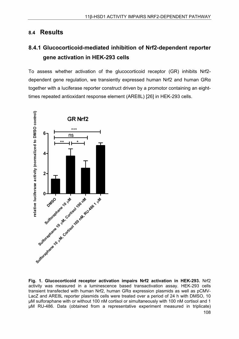
11β-HSD1 ACTIVITY IMPAIRS NRF2-DEPENDENT PATHWAY
108
8.4 Results
8.4.1 Glucocorticoid-mediated inhibition of Nrf2-dependent reporter gene activation in HEK-293 cells
To assess whether activation of the glucocorticoid receptor (GR) inhibits Nrf2-
dependent gene regulation, we transiently expressed human Nrf2 and human GRα
together with a luciferase reporter construct driven by a promotor containing an eight-
times repeated antioxidant response element (ARE8L) [26] in HEK-293 cells.
Fig. 1. Glucocorticoid receptor activation impairs Nrf2 activation in HEK-293. Nrf2 activity was measured in a luminescence based transactivation assay. HEK-293 cells transient transfected with human Nrf2, human GRα expression plasmids as well as pCMV-LacZ and ARE8L reporter plasmids cells were treated over a period of 24 h with DMSO, 10 μM sulforaphane with or without 100 nM cortisol or simultaneously with 100 nM cortisol and 1 μM RU-486. Data (obtained from a representative experiment measured in triplicate)
11β-HSD1 ACTIVITY IMPAIRS NRF2-DEPENDENT PATHWAY
109
represent mean±SD; n =3. *, P < 0.05, **, P < 0.01 *** P < 0.001. P value was obtained using a one-way ANOVA followed by Bonferroni post tests compared with control (DMSO).
Incubation of the cells with 10 μM sulforaphane led to a four-fold activation of the
Nrf2-induced luciferase production. Activation of GR by 100 nM cortisol suppressed
the Nrf2-dependent activation of the reporter gene by 10 μM Sulforaphane. The GR
antagonist RU-486 (1 μM) was able to fully restore Nrf2-mediated activation of the
ARE8L reporter.
8.4.2 Induction of the Nrf2-dependent ARE8L-reporter construct in rat H4IIE hepatoma cells
To characterize the responsiveness of the Nrf2 pathway in rat H4IIE hepatoma cells,
we transiently transfected the cells with the ARE8L luciferase reporter construct. As
shown in Fig. 2A, sulforaphane stimulated ARE-dependent reporter activity
approximately seven-fold. Co-transfection of the cells with recombinant Nrf2 further
stimulated reporter activity almost two-fold. Since Nrf2 protein has a short half-life (of
about 15 min), which is significantly enhanced by proteasome inhibitors as reported
earlier [31, 32], ARE-reporter activity was also assessed in cells treated with
sulforaphane (10 μM) and the proteasome inhibitor MG132 (10 μM). Total luciferase
reporter activity was two to three times higher in the presence of the proteasome
inhibitor compared with its absence.
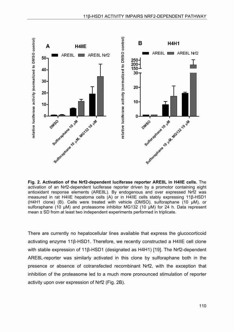
11β-HSD1 ACTIVITY IMPAIRS NRF2-DEPENDENT PATHWAY
110
Fig. 2. Activation of the Nrf2-dependent luciferase reporter ARE8L in H4IIE cells. The activation of an Nrf2-dependent luciferase reporter driven by a promotor containing eight antioxidant response elements (ARE8L). By endogenous and over expressed Nrf2 was measured in rat H4IIE hepatoma cells (A) or in H4IIE cells stably expressing 11β-HSD1 (H4H1 clone) (B). Cells were treated with vehicle (DMSO), sulforaphane (10 μM), or sulforaphane (10 μM) and proteasome inhibitor MG132 (10 μM) for 24 h. Data represent mean ± SD from at least two independent experiments performed in triplicate.
There are currently no hepatocellular lines available that express the glucocorticoid
activating enzyme 11β-HSD1. Therefore, we recently constructed a H4IIE cell clone
with stable expression of 11β-HSD1 (designated as H4H1) [19]. The Nrf2-dependent
ARE8L-reporter was similarly activated in this clone by sulforaphane both in the
presence or absence of cotransfected recombinant Nrf2, with the exception that
inhibition of the proteasome led to a much more pronounced stimulation of reporter
activity upon over expression of Nrf2 (Fig. 2B).
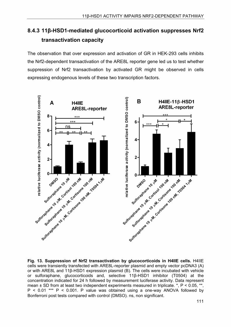
11β-HSD1 ACTIVITY IMPAIRS NRF2-DEPENDENT PATHWAY
111
8.4.3 11β-HSD1-mediated glucocorticoid activation suppresses Nrf2 transactivation capacity
The observation that over expression and activation of GR in HEK-293 cells inhibits
the Nrf2-dependent transactivation of the ARE8L reporter gene led us to test whether
suppression of Nrf2 transactivation by activated GR might be observed in cells
expressing endogenous levels of these two transcription factors.
Fig. 13. Suppression of Nrf2 transactivation by glucocorticoids in H4IIE cells. H4IIE cells were transiently transfected with ARE8L-reporter plasmid and empty vector pcDNA3 (A) or with ARE8L and 11β-HSD1 expression plasmid (B). The cells were incubated with vehicle or sulforaphane, glucocorticoids and, selective 11β-HSD1 inhibitor (T0504) at the concentration indicated for 24 h followed by measurement luciferase activity. Data represent mean ± SD from at least two independent experiments measured in triplicate. *, P < 0.05, **, P < 0.01 *** P < 0.001. P value was obtained using a one-way ANOVA followed by Bonferroni post tests compared with control (DMSO). ns, non significant.
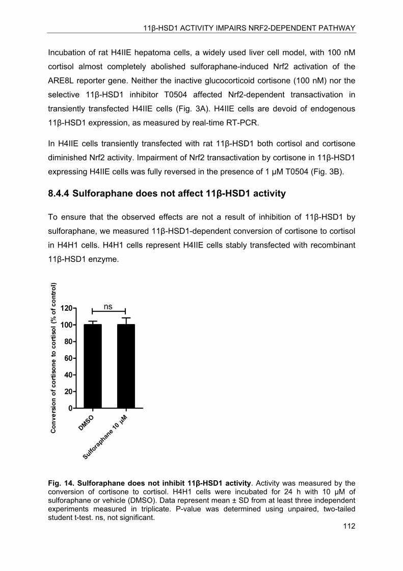
11β-HSD1 ACTIVITY IMPAIRS NRF2-DEPENDENT PATHWAY
112
Incubation of rat H4IIE hepatoma cells, a widely used liver cell model, with 100 nM
cortisol almost completely abolished sulforaphane-induced Nrf2 activation of the
ARE8L reporter gene. Neither the inactive glucocorticoid cortisone (100 nM) nor the
selective 11β-HSD1 inhibitor T0504 affected Nrf2-dependent transactivation in
transiently transfected H4IIE cells (Fig. 3A). H4IIE cells are devoid of endogenous
11β-HSD1 expression, as measured by real-time RT-PCR.
In H4IIE cells transiently transfected with rat 11β-HSD1 both cortisol and cortisone
diminished Nrf2 activity. Impairment of Nrf2 transactivation by cortisone in 11β-HSD1
expressing H4IIE cells was fully reversed in the presence of 1 μM T0504 (Fig. 3B).
8.4.4 Sulforaphane does not affect 11β-HSD1 activity
To ensure that the observed effects are not a result of inhibition of 11β-HSD1 by
sulforaphane, we measured 11β-HSD1-dependent conversion of cortisone to cortisol
in H4H1 cells. H4H1 cells represent H4IIE cells stably transfected with recombinant
11β-HSD1 enzyme.
Fig. 14. Sulforaphane does not inhibit 11β-HSD1 activity. Activity was measured by the conversion of cortisone to cortisol. H4H1 cells were incubated for 24 h with 10 μM of sulforaphane or vehicle (DMSO). Data represent mean ± SD from at least three independent experiments measured in triplicate. P-value was determined using unpaired, two-tailed student t-test. ns, not significant.
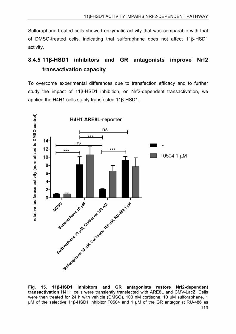
11β-HSD1 ACTIVITY IMPAIRS NRF2-DEPENDENT PATHWAY
113
Sulforaphane-treated cells showed enzymatic activity that was comparable with that
of DMSO-treated cells, indicating that sulforaphane does not affect 11β-HSD1
activity.
8.4.5 11β-HSD1 inhibitors and GR antagonists improve Nrf2 transactivation capacity
To overcome experimental differences due to transfection efficacy and to further
study the impact of 11β-HSD1 inhibition, on Nrf2-dependent transactivation, we
applied the H4H1 cells stably transfected 11β-HSD1.
Fig. 15. 11β-HSD1 inhibitors and GR antagonists restore Nrf2-dependent transactivation H4H1 cells were transiently transfected with ARE8L and CMV-LacZ. Cells were then treated for 24 h with vehicle (DMSO), 100 nM cortisone, 10 μM sulforaphane, 1 μM of the selective 11β-HSD1 inhibitor T0504 and 1 μM of the GR antagonist RU-486 as
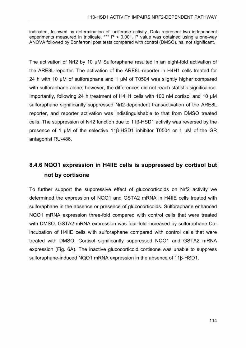
11β-HSD1 ACTIVITY IMPAIRS NRF2-DEPENDENT PATHWAY
114
indicated, followed by determination of luciferase activity. Data represent two independent experiments measured in triplicate. *** P < 0.001. P value was obtained using a one-way ANOVA followed by Bonferroni post tests compared with control (DMSO). ns, not significant.
The activation of Nrf2 by 10 μM Sulforaphane resulted in an eight-fold activation of
the ARE8L-reporter. The activation of the ARE8L-reporter in H4H1 cells treated for
24 h with 10 μM of sulforaphane and 1 μM of T0504 was slightly higher compared
with sulforaphane alone; however, the differences did not reach statistic significance.
Importantly, following 24 h treatment of H4H1 cells with 100 nM cortisol and 10 μM
sulforaphane significantly suppressed Nrf2-dependent transactivation of the ARE8L
reporter, and reporter activation was indistinguishable to that from DMSO treated
cells. The suppression of Nrf2 function due to 11β-HSD1 activity was reversed by the
presence of 1 μM of the selective 11β-HSD1 inhibitor T0504 or 1 μM of the GR
antagonist RU-486.
8.4.6 NQO1 expression in H4IIE cells is suppressed by cortisol but not by cortisone
To further support the suppressive effect of glucocorticoids on Nrf2 activity we
determined the expression of NQO1 and GSTA2 mRNA in H4IIE cells treated with
sulforaphane in the absence or presence of glucocorticoids. Sulforaphane enhanced
NQO1 mRNA expression three-fold compared with control cells that were treated
with DMSO. GSTA2 mRNA expression was four-fold increased by sulforaphane Co-
incubation of H4IIE cells with sulforaphane compared with control cells that were
treated with DMSO. Cortisol significantly suppressed NQO1 and GSTA2 mRNA
expression (Fig. 6A). The inactive glucocorticoid cortisone was unable to suppress
sulforaphane-induced NQO1 mRNA expression in the absence of 11β-HSD1.
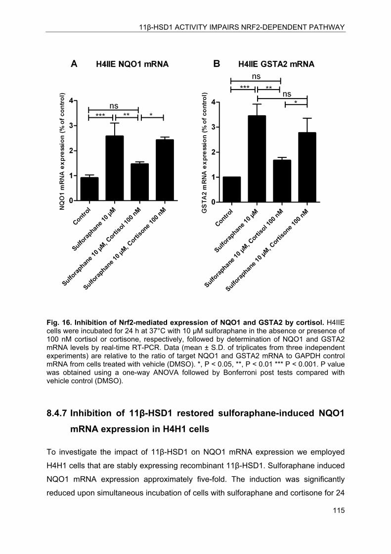
11β-HSD1 ACTIVITY IMPAIRS NRF2-DEPENDENT PATHWAY
115
Fig. 16. Inhibition of Nrf2-mediated expression of NQO1 and GSTA2 by cortisol. H4IIE cells were incubated for 24 h at 37°C with 10 μM sulforaphane in the absence or presence of 100 nM cortisol or cortisone, respectively, followed by determination of NQO1 and GSTA2 mRNA levels by real-time RT-PCR. Data (mean ± S.D. of triplicates from three independent experiments) are relative to the ratio of target NQO1 and GSTA2 mRNA to GAPDH control mRNA from cells treated with vehicle (DMSO). *, P < 0.05, **, P < 0.01 *** P < 0.001. P value was obtained using a one-way ANOVA followed by Bonferroni post tests compared with vehicle control (DMSO).
8.4.7 Inhibition of 11β-HSD1 restored sulforaphane-induced NQO1 mRNA expression in H4H1 cells
To investigate the impact of 11β-HSD1 on NQO1 mRNA expression we employed
H4H1 cells that are stably expressing recombinant 11β-HSD1. Sulforaphane induced
NQO1 mRNA expression approximately five-fold. The induction was significantly
reduced upon simultaneous incubation of cells with sulforaphane and cortisone for 24
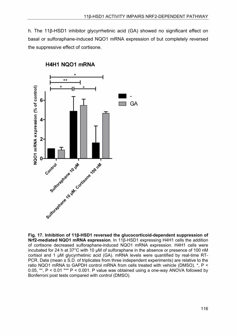
11β-HSD1 ACTIVITY IMPAIRS NRF2-DEPENDENT PATHWAY
116
h. The 11β-HSD1 inhibitor glycyrrhetinic acid (GA) showed no significant effect on
basal or sulforaphane-induced NQO1 mRNA expression of but completely reversed
the suppressive effect of cortisone.
Fig. 17. Inhibition of 11β-HSD1 reversed the glucocorticoid-dependent suppression of Nrf2-mediated NQO1 mRNA expression. In 11β-HSD1 expressing H4H1 cells the addition of cortisone decreased sulforaphane-induced NQO1 mRNA expression. H4H1 cells were incubated for 24 h at 37°C with 10 μM of sulforaphane in the absence or presence of 100 nM cortisol and 1 μM glycyrrhetinic acid (GA). mRNA levels were quantified by real-time RT-PCR. Data (mean ± S.D. of triplicates from three independent experiments) are relative to the ratio NQO1 mRNA to GAPDH control mRNA from cells treated with vehicle (DMSO). *, P < 0.05, **, P < 0.01 *** P < 0.001. P value was obtained using a one-way ANOVA followed by Bonferroni post tests compared with control (DMSO).
11β-HSD1 ACTIVITY IMPAIRS NRF2-DEPENDENT PATHWAY
117
8.4.8 Glucocorticoid-dependent impairment of HO-1 function and susceptibility to H2O2
Next, we studied the impact of 11β-HSD1 activity on the Nrf2-dependent target gene
HO-1. H4IIE cells were transiently transfected with a plasmid for the cytosolic
hydrogen peroxide sensor HyPer and either 11β-HSD1 or pcDNA3 (empty vector).
Cells were treated with 100 nM of cortisone for 24 h. The real time measurements in
H4IIE cells transiently transfected with the cytosolic HyPer-senor and pcDNA3
showed a three-fold increase in the HyPer signal upon addition of 10 μM H2O2. The
total response was reduced by about 50% after 30 min. In contrast, H4IIE transiently
transfected with 11β-HSD1 showed an enhanced response to H2O2 with an
approximately ~four-fold increased HyPer signal, indicating enhanced oxidative
stress. After 30 min the HyPer signal was only slightly reduced, indicating enhanced
oxidative stress, due to the impaired activity of HO-1 in 11β-HSD1 expressing H4IIE
cells. In 11β-HSD1 expressing H4IIE cells simultaneously treated with 100 nM
cortisone and with 1 μM T0504, the total response was slightly lower compared to
cells in the absence of the inhibitor, although statistically not significant, and the cells
recovered more quickly from the H2O2 challenge. Inhibition of 11β-HSD1 seemed to
be beneficial and the total response was significantly reduced by about 30% after 30
min (Fig. 8A) compared with cells expressing 11β-HSD1 but in the absence of
T0504.
Fig. 18. 11β-HSD1 expressing cells are more susceptible to H2O2 induced oxidative stress H4IIE cells transiently transfected with either pcDNA3 or 11β-HSD1 expression plasmid treated for 24 h with 100 nM cortisone in the presence or absence of 11β-HSD1 inhibitor T0504. Following incubation the medium was replaced by assay buffer (HBSS)
11β-HSD1 ACTIVITY IMPAIRS NRF2-DEPENDENT PATHWAY
118
containing 1g/L of glucose. Single cell real-time measurements were performed on a Leica confocal microscope SP5. (A) After 5 min baseline adaption, cells were exposed to a final concentration of 10 μM H2O2 and recovery was compared between differentially transfected cells over a period of 30 min. Data represent mean ± SEM of three different cells for each transfection. *, P < 0.05, **, P < 0.01 *** P < 0.001. P value was obtained using a one-way ANOVA followed by Bonferroni post tests compared with pcDNA3. (B) After 5 min baseline adaption, cells were challenged with a 100 μM H2O2 bolus and reduction of cytosolic oxidative stress was measured over a period of 45 min. Data represent mean+SEM of seven different cells for each transfection. *** P < 0.001. P value was obtained using unpaired, two-tailed student t-test.
Further, a higher concentration of H2O2 was used to challenge the cells (Fig. 8B) in
order to address whether the observed differences were more pronounced. H4IIE
cells transfected with pcDNA3 or 11β-HSD1 were treated for 24 h with 100 nM of
cortisone, followed by real-time measurements and challenged with a bolus of 100
μM H2O2. The pcDNA3 transfected cells showed a three-fold increase in HyPer
signal, followed by a rapid decline and normalization of the signal. After 45 min, the
signal reached almost baseline level. On the other hand, 11β-HSD1 expressing cells
also responded immediately but showed a more pronounced response to H2O2 (five-
fold increase). In addition, the stress signal was only slightly reduced over 45 min
period, indicating an impaired reduction of reactive oxygen species (ROS).
11β-HSD1 ACTIVITY IMPAIRS NRF2-DEPENDENT PATHWAY
119
8.5 Discussion
During lifetime organisms are continuously exposed to toxicants derived from
endogenous sources like reactive metabolites, reactive oxygen species (OH-, H2O2,
O2-) as well as environmental xenobiotics. At concentrations exceeding detoxification
capacities these compounds are able to disturb physiological functions, ultimately
contributing to severe dysfunctions such as inflammation, cardiovascular and
neurodegenerative-disorders, diabetes or cancer. To cope with the burden of reactive
chemicals organisms developed a sophisticated defence mechanism. Key enzymes
involved in coping with oxidative stress include those induced by the antioxidant
redox response pathway under the control of Nrf2. Deficiency or impairment of Nrf2
has been closely related with many diseases such as arthritis [33, 34], diabetes [35,
36], Parkinson’s [37] and various forms of cancer [38, 39]. Despite the key role of
Nrf2 in redox regulation its mechanism of action is highly complex and not yet fully
understood [1, 24, 40]. Only few studies address the cross-talk between Nrf2 and the
essential glucocorticoid signaling pathway.
In most toxicology studies addressing the impact of glucocorticoids on detoxification
reactions the potent synthetic ligand dexamethasone was used. However,
dexamethasone has clearly distinct properties than cortisol the endogenous
glucocorticoid. Because of its “constitutive” activity dexamethasone circumvents the
important interconversion by 11β-HSD enzymes, which is in contrast to the
endogenous glucocorticoids. Endogenous glucocorticoids can be metabolically
inactivated by 11β-HSD2 in tissues such as the kidney and regenerated by 11β-
HSD1 mainly in the liver. This is not the case for dexamethasone, since
dexamethasone is not efficiently converted to 11-ketodexamethasone and because
11-ketodexamethasone is still a potent GR agonist [29]. In addition dexamethasone
is approximately ten-times more potent than cortisol, and it has been shown that high
concentrations of dexamethasone can activate pregnane-X receptor (PXR) [15]. PXR
and its co-receptor the retinoid-X receptor (RXR) are involved in the detoxification of
xenobiotics while they transcriptionally regulate the expression of phase I (CYP3A4)
and phase II enzymes [15]. Some of the phase II enzymes regulated by Nrf2 (e.g.
NQO1) contain both XRE and ARE motifs [41] in their promoter regions. Differential
regulation by the use of high concentrations of dexamethasone due to PXR activation
11β-HSD1 ACTIVITY IMPAIRS NRF2-DEPENDENT PATHWAY
120
has been reported for glutathione-S-transferase (GSTA2) [42, 43]. Therefore, it is
important to distinguish between effects of synthetic and endogenous
glucocorticoids.
In this study we focused on effects of endogenous glucocorticoids and on the role of
glucocorticoid activation by 11β-HSD1 upstream of the GR on the Nrf2 pathway.
Concentrations close to physiological levels (i.e. 100 nM) were used the present
study, providing valuable information on the regulation of Nrf2 under physiological
conditions.
Using H4IIE cells, we show that in the absence of 11β-HSD1 cortisol but not
cortisone affects Nrf2 activity as measured in transactivation assays or in NQO1
mRNA expression (Fig.3 and Fig. 6). Thus, in tissues lacking 11β-HSD1 expression
or in tissues expressing 11β-HSD2 (placenta, renal distal tubulus, distal colon) the
Nrf2 pathway is modulated by extracellular availability or even insensitive to
glucocorticoids, which is in clear contrast to tissues with high 11β-HSD1 expression
(liver, adipose, hippocampal neurones).
A recent study reported an increased hepatic 11β-HSD1 expression in patients with
alcoholic liver disease (ALD) [44]. ALD associated disorders are fatty liver,
inflammation, and cirrhosis and hepatocellular carcinoma in patients with liver
cirrhosis [45]. Nrf2 prevents ethanol-induced liver injury by detoxification of
acetaldehyde and inhibition of metabolite accumulation. Nrf2 knockout animals (nrf2 -
/-) showed dramatically increased mortality after feeding of ethanol doses which are
well tolerated in wild-type mice [46].
On a basic cellular level our results indicate that 11β-HSD1 inhibition might be
beneficial to restore the capacity for detoxification processes regulated by Nrf2. This
was supported by transactivation assays, NQO1 mRNA expression levels (Fig. 7),
and by the use of the intracellular redox-sensor HyPer reflecting the activity of HO-1
after challenge by H2O2 (Fig 8). In the case of ALD the authors claimed that inhibition
of 11β-HSD1 could be a novel therapeutic approach to treat alcoholic pseudo-
Cushing’s [44]. We therefore hypothesize that inhibition of 11β-HSD1 may be further
beneficial for the detoxification capacity of the liver, at least in patients with ALD.
11β-HSD1 ACTIVITY IMPAIRS NRF2-DEPENDENT PATHWAY
121
Expression of 11β-HSD1 is increased in adipose tissue of obese humans and
rodents. Transgenic mice selectively over expressing 11β-HSD1 (aP2-HSD1) in the
adipose tissue develop the metabolic syndrome including visceral obesity,
dyslipidemia, insulin resistance, diabetes, and hypertension [47]. In comparison to
the human metabolic syndrome, plasma corticosterone levels in the aP2-HSD1 mice
are unaltered while local glucocorticoid activation in adipose tissue is enhanced. In
aP2-HSD1 mice hepatic 11β-HSD1 is not altered. However, corticosterone delivery
to the liver is tree-times increased because of excessive activation of glucocorticoids
derived from adipose tissue [47]. Obesity involves chronic inflammation and as a
consequence enhanced infiltration of macrophages [48]. Macrophages express 11β-
HSD1, thereby further enhancing locally active glucocorticoids. Thus, in obesity the
Nrf2 pathway may be suppressed by elevated glucocorticoid levels. In contrast, 11β-
HSD1 knockout animals fed with high fat diet showed reduced cytotoxic T-cell and
macrophage infiltration in visceral fat compared with wild-type mice [49]. Reduced
macrophage infiltration in adipose tissue observed for the 11β-HSD1 transgene
further supports the assumption that 11β-HSD1 inhibition is beneficial in these
situations. To study the effect of 11β-HSD1 inhibition in adipose tissue and the
consequences for the Nrf2 pathway, a fatcell-specific inhibitor as developed recently
[50] would provide a unique mechanistic tool. This inhibitor was successfully shown
to protect against diet-induced obesity in mice [50]. In obese patients with type 2
diabetes hepatic 11β-HSD1 activity was found to be sustained, coincident with an
increased whole body 11β-HSD1 activity compared with normal weight patients [51].
The transgenic ApoE-mice over expressing 11β-HSD1 specifically in the liver exhibit
a phenotype resembling humans suffering under non-alcoholic fatty liver disease
[52]. The phenotype includes insulin-resistance, hypertension and metabolic
syndrome symptomatic concomitant with a normal weight status [53, 54].
In contrast to obesity a clinical study investigating morbidly obese patients showed a
20-fold higher 11β-HSD1 mRNA expression in liver compared with adipose tissue
[56]. The study further convincingly showed that hepatic 11β-HSD1 expression in
these patients is directly proportional to the body mass index (BMI). According to the
outcome of the study the authors suggested that 11β-HSD1 activity in hepatic as well
as visceral adipose tissue might be pathogenic in obesity [55]. Thus, enhanced 11β-
HSD1 expression in liver or adipose tissue leads to an increased local activation of
11β-HSD1 ACTIVITY IMPAIRS NRF2-DEPENDENT PATHWAY
122
glucocorticoids. 11β-hydroxygluocorticoids are then further able to repress the Nrf2
related activation of phase II detoxification enzymes including NQO1 and HO-1 (Fig.
3 and Fig. 6).
In conclusion, the present study contributes to the knowledge of the defined
regulation of the antioxidant redox response pathway. It further suggests that
inhibition of 11β-HSD1 may improve Nrf2-dependent cell defense, which may be
beneficial for patients with ALD or obesity as well as patients with chronic
inflammation such as diabetes or rheumatoid arthritis. To investigate these potentially
beneficial effects in vivo further studies are needed.
8.6 Acknowledgements
This work was supported by the Swiss National Science Foundation (No. 31003A-
124912). A.O. has a Chair of Molecular and Systems Toxicology by the Novartis
Research Foundation.
11β-HSD1 ACTIVITY IMPAIRS NRF2-DEPENDENT PATHWAY
123
8.7 References
1. Copple, I.M., et al., The Nrf2-Keap1 defence pathway: role in protection against drug-induced toxicity. Toxicology, 2008. 246(1): p. 24-33.
2. Jaiswal, A.K., Nrf2 signaling in coordinated activation of antioxidant gene expression. Free radical biology & medicine, 2004. 36(10): p. 1199-207.
3. Nioi, P., et al., Identification of a novel Nrf2-regulated antioxidant response element (ARE) in the mouse NAD(P)H:quinone oxidoreductase 1 gene: reassessment of the ARE consensus sequence. The Biochemical journal, 2003. 374(Pt 2): p. 337-48.
4. Itoh, K., et al., An Nrf2/small Maf heterodimer mediates the induction of phase II detoxifying enzyme genes through antioxidant response elements. Biochemical and biophysical research communications, 1997. 236(2): p. 313-22.
5. Kwak, M.K., et al., Role of transcription factor Nrf2 in the induction of hepatic phase 2 and antioxidative enzymes in vivo by the cancer chemoprotective agent, 3H-1, 2-dimethiole-3-thione. Molecular medicine, 2001. 7(2): p. 135-45.
6. Chan, K., X.D. Han, and Y.W. Kan, An important function of Nrf2 in combating oxidative stress: detoxification of acetaminophen. Proceedings of the National Academy of Sciences of the United States of America, 2001. 98(8): p. 4611-6.
7. Kitamura, Y., et al., Increased susceptibility to hepatocarcinogenicity of Nrf2-deficient mice exposed to 2-amino-3-methylimidazo[4,5-f]quinoline. Cancer science, 2007. 98(1): p. 19-24.
8. Venugopal, R. and A.K. Jaiswal, Nrf1 and Nrf2 positively and c-Fos and Fra1 negatively regulate the human antioxidant response element-mediated expression of NAD(P)H:quinone oxidoreductase1 gene. Proceedings of the National Academy of Sciences of the United States of America, 1996. 93(25): p. 14960-5.
9. Alam, J., et al., Nrf2, a Cap'n'Collar transcription factor, regulates induction of the heme oxygenase-1 gene. The Journal of biological chemistry, 1999. 274(37): p. 26071-8.
10. McMahon, M., et al., The Cap'n'Collar basic leucine zipper transcription factor Nrf2 (NF-E2 p45-related factor 2) controls both constitutive and inducible expression of intestinal detoxification and glutathione biosynthetic enzymes. Cancer research, 2001. 61(8): p. 3299-307.
11. Augustine, L.M., et al., Gender divergent expression of Nqo1 in Sprague Dawley and August Copenhagen x Irish rats. Journal of biochemical and molecular toxicology, 2008. 22(2): p. 93-100.
12. Balish, E., et al., Effect of age, sex, and intestinal flora on the induction of colon tumors in rats. Journal of the National Cancer Institute, 1977. 58(4): p. 1103-6.
13. Kirsch-Volders, M., et al., Gender-related differences in response to mutagens and carcinogens. Mutagenesis, 2010. 25(3): p. 213-21.
14. Ki, S.H., et al., Glucocorticoid receptor (GR)-associated SMRT binding to C/EBPbeta TAD and Nrf2 Neh4/5: role of SMRT recruited to GR in GSTA2 gene repression. Molecular and cellular biology, 2005. 25(10): p. 4150-65.
15. Pascussi, J.M., et al., Dexamethasone induces pregnane X receptor and retinoid X receptor-alpha expression in human hepatocytes: synergistic
11β-HSD1 ACTIVITY IMPAIRS NRF2-DEPENDENT PATHWAY
124
increase of CYP3A4 induction by pregnane X receptor activators. Molecular pharmacology, 2000. 58(2): p. 361-72.
16. Scheer, N., et al., In vivo responses of the human and murine pregnane X receptor to dexamethasone in mice. Drug metabolism and disposition: the biological fate of chemicals. 38(7): p. 1046-53.
17. Shi, D., D. Yang, and B. Yan, Dexamethasone transcriptionally increases the expression of the pregnane X receptor and synergistically enhances pyrethroid esfenvalerate in the induction of cytochrome P450 3A23. Biochemical pharmacology, 2010. 80(8): p. 1274-83.
18. Tomlinson, J.W., et al., 11beta-hydroxysteroid dehydrogenase type 1: a tissue-specific regulator of glucocorticoid response. Endocrine reviews, 2004. 25(5): p. 831-66.
19. Atanasov, A.G., et al., Hexose-6-phosphate dehydrogenase determines the reaction direction of 11beta-hydroxysteroid dehydrogenase type 1 as an oxoreductase. FEBS Lett, 2004. 571(1-3): p. 129-33.
20. Lavery, G.G., et al., Hypoglycemia with enhanced hepatic glycogen synthesis in recombinant mice lacking hexose-6-phosphate dehydrogenase. Endocrinology, 2007. 148(12): p. 6100-6.
21. Albiston, A.L., R.E. Smith, and Z.S. Krozowski, Sex- and tissue- specific regulation of 11 beta-hydroxysteroid dehydrogenase mRNA. Molecular and cellular endocrinology, 1995. 109(2): p. 183-8.
22. Gomez-Sanchez, E.P., et al., Hexose-6-phosphate dehydrogenase and 11beta-hydroxysteroid dehydrogenase-1 tissue distribution in the rat. Endocrinology, 2008. 149(2): p. 525-33.
23. Telakowski-Hopkins, C.A., R.G. King, and C.B. Pickett, Glutathione S-transferase Ya subunit gene: identification of regulatory elements required for basal level and inducible expression. Proceedings of the National Academy of Sciences of the United States of America, 1988. 85(4): p. 1000-4.
24. Nguyen, T., P. Nioi, and C.B. Pickett, The Nrf2-antioxidant response element signaling pathway and its activation by oxidative stress. The Journal of biological chemistry, 2009. 284(20): p. 13291-5.
25. Arampatzis, S., et al., Comparative enzymology of 11beta-hydroxysteroid dehydrogenase type 1 from six species. Journal of molecular endocrinology, 2005. 35(1): p. 89-101.
26. Wang, X.J., J.D. Hayes, and C.R. Wolf, Generation of a stable antioxidant response element-driven reporter gene cell line and its use to show redox-dependent activation of nrf2 by cancer chemotherapeutic agents. Cancer research, 2006. 66(22): p. 10983-94.
27. Belousov, V.V., et al., Genetically encoded fluorescent indicator for intracellular hydrogen peroxide. Nature methods, 2006. 3(4): p. 281-6.
28. Hermanowski-Vosatka, A., et al., 11beta-HSD1 inhibition ameliorates metabolic syndrome and prevents progression of atherosclerosis in mice. J Exp Med, 2005. 202(4): p. 517-27.
29. Rebuffat, A.G., et al., The 11-ketosteroid 11-ketodexamethasone is a glucocorticoid receptor agonist. Molecular and cellular endocrinology, 2004. 214(1-2): p. 27-37.
30. Dzyakanchuk, A.A., et al., 11beta-Hydroxysteroid dehydrogenase 1 reductase activity is dependent on a high ratio of NADPH/NADP(+) and is stimulated by extracellular glucose. Molecular and cellular endocrinology, 2009. 301(1-2): p. 137-41.
11β-HSD1 ACTIVITY IMPAIRS NRF2-DEPENDENT PATHWAY
125
31. Stewart, D., et al., Degradation of transcription factor Nrf2 via the ubiquitin-proteasome pathway and stabilization by cadmium. The Journal of biological chemistry, 2003. 278(4): p. 2396-402.
32. Zhang, D.D. and M. Hannink, Distinct cysteine residues in Keap1 are required for Keap1-dependent ubiquitination of Nrf2 and for stabilization of Nrf2 by chemopreventive agents and oxidative stress. Molecular and cellular biology, 2003. 23(22): p. 8137-51.
33. Maicas, N., et al., Deficiency of Nrf2 accelerates the effector phase of arthritis and aggravates joint disease. Antioxid Redox Signal, 2011.
34. Wruck, C.J., et al., Role of oxidative stress in rheumatoid arthritis: insights from the Nrf2-knockout mice. Annals of the rheumatic diseases, 2010: p. 844-850.
35. Luo, Z.F., et al., Prevention of diabetic nephropathy in rats through enhanced renal antioxidative capacity by inhibition of the proteasome. Life sciences, 2011. 88(11-12): p. 512-20.
36. Tan, Y., et al., Diabetic downregulation of Nrf2 activity via ERK contributes to oxidative stress-induced insulin resistance in cardiac cells in vitro and in vivo. Diabetes, 2011. 60(2): p. 625-33.
37. Jazwa, A., et al., Pharmacological targeting of the transcription factor Nrf2 at the basal ganglia provides disease modifying therapy for experimental Parkinsonism. Antioxid Redox Signal, 2011.
38. Hu, R., et al., Regulation of NF-E2-related factor 2 signaling for cancer chemoprevention: antioxidant coupled with antiinflammatory. Antioxid Redox Signal, 2010. 13(11): p. 1679-98.
39. Taguchi, K., H. Motohashi, and M. Yamamoto, Molecular mechanisms of the Keap1-Nrf2 pathway in stress response and cancer evolution. Genes Cells, 2011. 16(2): p. 123-40.
40. Zhang, D.D., Mechanistic studies of the Nrf2-Keap1 signaling pathway. Drug metabolism reviews, 2006. 38(4): p. 769-89.
41. Favreau, L.V. and C.B. Pickett, Transcriptional regulation of the rat NAD(P)H:quinone reductase gene. Identification of regulatory elements controlling basal level expression and inducible expression by planar aromatic compounds and phenolic antioxidants. The Journal of biological chemistry, 1991. 266(7): p. 4556-61.
42. Falkner, K.C., et al., Regulation of the rat glutathione S-transferase A2 gene by glucocorticoids: involvement of both the glucocorticoid and pregnane X receptors. Molecular pharmacology, 2001. 60(3): p. 611-9.
43. Falkner, K.C. and R.A. Prough, Regulation of the rat glutathione S-transferase A2 gene by glucocorticoids: crosstalk through C/EBPs. Drug metabolism reviews, 2007. 39(2-3): p. 401-18.
44. Ahmed, A., et al., Induction of hepatic 11beta-hydroxysteroid dehydrogenase type 1 in patients with alcoholic liver disease. Clinical endocrinology, 2008. 68(6): p. 898-903.
45. Gyamfi, M.A. and Y.J. Wan, Pathogenesis of alcoholic liver disease: the role of nuclear receptors. Experimental biology and medicine (Maywood, N.J.), 2010. 235(5): p. 547-60.
46. Lamle, J., et al., Nuclear factor-eythroid 2-related factor 2 prevents alcohol-induced fulminant liver injury. Gastroenterology, 2008. 134(4): p. 1159-68.
47. Masuzaki, H., et al., A transgenic model of visceral obesity and the metabolic syndrome. Science 2001. 294(5549): p. 2166-70.
11β-HSD1 ACTIVITY IMPAIRS NRF2-DEPENDENT PATHWAY
126
48. Stanton, M.C., et al., Inflammatory Signals shift from adipose to liver during high fat feeding and influence the development of steatohepatitis in mice. Journal of inflammation (London, England), 2011. 8(1): p. 8.
49. Wamil, M., et al., Novel Fat Depot-Specific Mechanisms Underlie Resistance to Visceral Obesity and Inflammation in 11{beta}-Hydroxysteroid Dehydrogenase Type 1-Deficient Mice. Diabetes, 2011.
50. Liu, J., et al., Adipose tissue-targeted 11beta-hydroxysteroid dehydrogenase type 1 inhibitor protects against diet-induced obesity. Endocrine journal, 2011: p. 199-209.
51. Paterson, J.M., et al., Metabolic syndrome without obesity: Hepatic overexpression of 11beta-hydroxysteroid dehydrogenase type 1 in transgenic mice. Proceedings of the National Academy of Sciences of the United States of America, 2004. 101(18): p. 7088-93.
52. Ruderman, N., et al., The metabolically obese, normal-weight individual revisited. Diabetes, 1998. 47(5): p. 699-713.
53. Marchesini, G., et al., Nonalcoholic fatty liver disease: a feature of the metabolic syndrome. Diabetes, 2001. 50(8): p. 1844-50.
54. Stimson, R.H., et al., Increased Whole-Body and Sustained Liver Cortisol Regeneration by 11{beta}-Hydroxysteroid Dehydrogenase Type 1 in Obese Men With Type 2 Diabetes Provides a Target for Enzyme Inhibition. Diabetes, 2011. 60(3): p. 720-5.
55. Baudrand, R., et al., Overexpression of 11beta-hydroxysteroid dehydrogenase type 1 in hepatic and visceral adipose tissue is associated with metabolic disorders in morbidly obese patients. Obesity surgery, 2010. 20(1): p. 77-83.
OUTLOOK AND CONCLUSION
127
9 Conclusion and Outlook 11β-HSD1-mediated glucocorticoid activation is essential for the regulation of GR
function. A subtle fine-tuning of 11β-HSD1 activity is critical and locally enhanced
levels of cortisol (corticosterone in rodents) have been associated with metabolic
disorders. This was best demonstrated, in studies with transgenic mouse models.
The present work focused in a major part on the characterization of specific inhibitors
against the glucocorticoid metabolizing enzymes 11β-HSD1 and 11β-HSD2 some of
the inhibitors were isolated from the natural source loquat, others were synthesized
by modification of 18β-glycyrrhetinic acid, the active constituent of the roots and
rhizomes of licorice (Glycyrrhiza spp.).
11β-HSD1 is currently considered as a promising drug target for the therapeutic
intervention of obesity and its outcome, the metabolic syndrome. Results from
studies with transgenic mice promoted the development of 11β-HSD1 inhibitors. 11β-
HSD1 overexpression specifically in adipose tissue results in a pathologic phenotype
described by accumulation of visceral fat and diabetic characteristics such as glucose
intolerance, insulin- and leptin-resistance, increased free-fatty acids, as well as
hypertension and chronic inflammation [158]. Furthermore, adipose specific
overexpression resulted in enhanced intra-adipose glucocorticoid activation. This
leads to higher adipose and portal corticosterone concentrations without affecting
systemic glucocorticoid levels, measured in plasma [159]. In transgene mice with
liver specific overexpression of 11β-HSD1 neither obesity nor glucose intolerance
was obvious; however, the animals developed symptoms of the metabolic syndrome,
including enhanced hepatic lipid synthesis, mild insulin resistance and steatosis
[160]. In contrast, reduction of 11β-HSD1 activity by knock-down or knock-out
protects against diet induced obesity concomitant with a lean and non-diabetic
phenotype [161, 162]. Taken together, these studies among others revealed 11β-
HSD1 as a potential drug target for the therapeutic intervention of metabolic diseases
[159].
Inhibition of 11β-HSD1 for therapeutic purpose requires highly selective inhibitors
whereby 11β-HSD2 is the first anti-target to be considered for selectivity assessment.
Non-selective inhibitors such as 18β-glycyrrhetinic acid (GA) have been shown to
cause hypertension as a result of potassium wasting and sodium retention due to
OUTLOOK AND CONCLUSION
128
glucocorticoid-dependent activation of the MR [163]. Furthermore, other enzymes
such as 17β-HSDs have to be considered in these approaches, to guarantee that sex
hormone regulation is not affected by these inhibitors. In general, it would be more
beneficial to use tissue-specific delivery of inhibitors, or pro-drugs that are activated
specifically in the tissue of interest. Alternatively, topic instead of systemic
applications may help to prevent unwanted side-effects as well as adverse
compensatory effects. Recently, an adipose tissue-targeted 11β-HSD1 inhibitor was
described, with beneficial outcome against diet induced obesity [164].
In contrast to synthetic pharmaceutical inhibitors, only few studies address the
potential use of compounds from natural sources. The present work contributes to
the identification of 11β-HSD1 inhibitors from natural sources. It further describes the
mechanism and the site of action of the isolated compounds. Moreover, we
highlighted the chemical class of pentacyclic triterpenes of the ursane type as active
inhibitory compounds of 11β-HSD1.
Loquat (eriobotrya japonica) is used in Chinese medicine as an anti-diabetic.
However, the mechanism of action was not fully elucidated. We described active
plant constituents that may be responsible for some of the beneficial effects of loquat
consumption in diabetes. For the discovery, a ligand-based pharmacophore model
was used and selected hits were further evaluated by docking into the 11β-HSD1
binding site and the use of in vitro activity assays. Compounds isolated from loquat
leaves were characterized as pentacyclic triterpenes. These compounds inhibited
11β-HSD1 and unlike GA they showed no or only weak effects on 11β-HSD2. The
most potent compounds isolated from the extracts were corosolic acid with an IC50 of 0.8 μM, followed by urosolic acid with an IC50 of 2 μM for 11β-HSD1. From the
dichlormethane extracts 12 different triterpenoids were identified, including already
described constituents of E. japonica such as maslinic acid methyl ester, 3-O-trans-p-
coumaroyltormentic acid, 3-O-cis-p-coumaroyltormentic acid and tormentic acid.
However, two compounds identified as 2-α hydroxy-3-oxo urs-12-en-28-oic acid and
uvaol have previously not been reported for this commonly used plant source.
Several of the identified triterpenoids exerted only weak inhibitory effect; However, a
mixture of these individual compounds at concentrations were they exerted little or no
effect, inhibited up to 90% of 11β-HSD1 activity, indicating a remarkable synergistic
OUTLOOK AND CONCLUSION
129
effect. This observation is in line with the opinion that natural products might have
more pronounced effects when used as whole plant extracts rather than as single
isolated compounds due to synergistically active constituents. Traditional medicines
are based on naturally derived products and the potential of nature to accommodate
many potent and/or selective drugs seems to be huge. High throughput screening
(HTS) approaches may reveal further compounds which can be used directly or
serve as basis for the development of therapeutically valuable drugs. However, HTS
is cost intensive; therefore, 3D-modeling accompanied with in vitro biological assays
are an alternative to exploit the available structural knowledge of target proteins for
identification of novel active entities. The predictive potential of these approaches for
active drugs and/or possible adverse effects increases our understanding of the
mechanisms behind drugs.
Natural compounds used in traditional medicines, and herbs and plants known as
“household remedies” represent drugs that are of mechanistic value and that are
broadly accepted in the population because of their natural source. The identification
of substance classes, as shown in our study, therefore improves the existing
knowledge on natural bioactive compounds.
With respect to its potency and selectivity for 11β-HSD1, corosolic acid may be used
as a starting point for chemical modifications that might lead to more potent inhibitors
with improved pharmacokinetic properties.
This approach was recently used for the development of selective 11β-HSD1 and
11β-HSD2 inhibitors using the non-selective GA as a starting compound. In the
present work, we characterized nine selective 11β-HSD1 and fifteen 11β-HSD2
inhibitors for their biological activity and species specificity on 11β-HSD2. Species-
specific differences of inhibitors need to considered; and have been described for
11β-HSD1 inhibition by GA-derivatives and other inhibitor classes [165, 166]. We
showed that the starting compound GA was comparably active toward mouse and
human 11β-HSD2. However, the chemical modification of three tested GA-
derivatives enhanced the species-specificity for the human compared with the mouse
enzyme. With respect to animal experiments and restriction to existing animal models
for the proof-of-concept regarding subsequent clinical trials, species-specific
variability represents an important aspect. The use of docking and pharmacophore
OUTLOOK AND CONCLUSION
130
models might lead to an improved prediction of species-specific variability and
potency of bioactive compounds.
11β-HSD2 is well accepted as an off-target with respect to hypertension and
cardiovascular complications as a result of cortisol-dependent activation of renal MR.
Recently, potential applications for the beneficial use of 11β-HSD2 inhibitors have
been suggested. These approaches include end-stage renal disease patients on
hemodialysis. In these patients, treatment of hyperkalemia to lower the risk of
hyperkalemic arrhythmias is essential [167]. Inhibition of 11β-HSD2 by licorice
consumption was able to reduce serum potassium in hemodialysis patients, probably
by enhanced intestinal secretion as a result of 11β-HSD2 inhibition and MR activation
in the distal colon. However, high doses of GA were used in short-time applications,
and long-term studies have to address potential adverse effects. Furthermore, GA is
a non-selective inhibitor of both 11β-HSD enzymes. The inhibition of 11β-HSD1 for
example in liver, renal proximal tubules, in testicular leydig cells, and in the
hypothalamus might cause adverse effects. Moreover, there is evidence that GA
inhibits some 17β-HSD enzymes. Therefore, selective inhibitors are required for
follow-up studies to confirm and clearly address the effects seen with licorice.
Hyperkalemia is also a rare but underestimated event for metastatic infiltration of the
adrenals as a consequence of breast and lung cancers with the outcome of adrenal
insufficiency [168]. In patients with adrenal insufficiency and for treatment of
symptoms such as hypotension, hyperkalemia and hyponatremia selective 11β-
HSD2 inhibitors might be beneficial. Acute intoxication with paramethoxy-
metamphetamin, a frequently used party drug, can be lethal at least in part because
of hyperkalemia and hypoglycemia [169]. Thus, selective inhibition of 11β-HSD2 may
lead to an increase in cortisol, thereby increasing blood glucose levels and reduce
hypoglycemia. Cortisol activation of MR in the intestine might be beneficial in such
cases of hypokalemia as well.
In recent studies, elevated 11β-HSD2 expression was also reported for some forms
of cancer and in chronic colon inflammation [170, 171]. However, most of these
studies used GA. Thus, in further studies selective inhibitors against 11β-HSD2
should be used to confirm the beneficial effects of 11β-HSD2 inhibition.
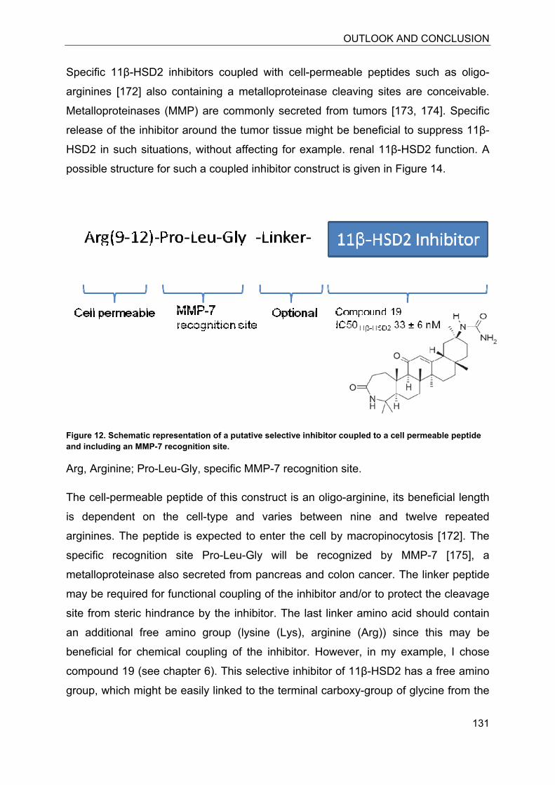
OUTLOOK AND CONCLUSION
131
Specific 11β-HSD2 inhibitors coupled with cell-permeable peptides such as oligo-
arginines [172] also containing a metalloproteinase cleaving sites are conceivable.
Metalloproteinases (MMP) are commonly secreted from tumors [173, 174]. Specific
release of the inhibitor around the tumor tissue might be beneficial to suppress 11β-
HSD2 in such situations, without affecting for example. renal 11β-HSD2 function. A
possible structure for such a coupled inhibitor construct is given in Figure 14.
Figure 12. Schematic representation of a putative selective inhibitor coupled to a cell permeable peptide and including an MMP-7 recognition site.
Arg, Arginine; Pro-Leu-Gly, specific MMP-7 recognition site.
The cell-permeable peptide of this construct is an oligo-arginine, its beneficial length
is dependent on the cell-type and varies between nine and twelve repeated
arginines. The peptide is expected to enter the cell by macropinocytosis [172]. The
specific recognition site Pro-Leu-Gly will be recognized by MMP-7 [175], a
metalloproteinase also secreted from pancreas and colon cancer. The linker peptide
may be required for functional coupling of the inhibitor and/or to protect the cleavage
site from steric hindrance by the inhibitor. The last linker amino acid should contain
an additional free amino group (lysine (Lys), arginine (Arg)) since this may be
beneficial for chemical coupling of the inhibitor. However, in my example, I chose
compound 19 (see chapter 6). This selective inhibitor of 11β-HSD2 has a free amino
group, which might be easily linked to the terminal carboxy-group of glycine from the
OUTLOOK AND CONCLUSION
132
tripeptide motif that functions as specific MMP-7 recognition site. The following
aspects should be considered in general for such a hypothetic construct.
1) The total construct must be cell-permeable.
2) The cytotoxicity must be negligible for the total as well as the cleaved
product(s).
3) The cleavage of the inhibitor must be possible and specific.
4) The inhibitor must be still active, even after structural changes due to cleavage
from the pro-drug.
5) The construct must be soluble in a water based solution at pH 7.4.
Selective 11β-HSD2 inhibitors are highly valuable as tools for basic research. The
awareness of a mosaic like expression pattern for 11β-HSD1 and 11β-HSD2 in many
tissues may explain contradictive findings for the use of non-selective inhibitors. In
this work, we reviewed MR expression in various organs. MR has broader substrate
specificity compared with GR; therefore, MR function is tightly defined by the
presence or absence of glucocorticoid metabolizing enzymes. Selective inhibition of
11β-HSD1 and 11β-HSD2 is required to clearly address mechanistic findings in vivo
and in vitro.
A challenge for biologically used compounds remains cellular uptake. Therefore, the
inhibitors characterized in this work were also used in intact SW-620 colon cells,
known for the endogenous expression of 11β-HSD2. The tested inhibitors were found
to be highly active for endogenous 11β-HSD2 inhibition in SW-620 cells.
Interestingly, IC50 values obtained from SW-620 cells were lower compared with data
obtained from HEK-293 cells or even from cell lysates. This may be explained by
compound removal due to transport mechanisms in HEK-293 cells, or compound
accumulation by SW-620 cells, if compared with lysates.
Compounds may act differently in intact cells compared with lysates because of
active transport mechanisms that are able to remove the substance from the cell, and
the specific chemical characteristics. The chemical structure per se might prevent
cellular uptake, for example when containing highly charged groups. Further
possibilities for cellular inactivity might be protein binding of the compound within the
OUTLOOK AND CONCLUSION
133
cell, or binding to serum proteins such as albumin contained in the culture media. A
general challenge is the solubility in the water-based culture media as well as the
stability of the compound over the cultivation period at the temperature of 37°C and
at neutral pH value. Thus, the compound must be sufficiently soluble to reach the
active concentration at the site of the target enzyme. Besides, the chemical stability is
essential for storage of the compound, to guarantee continuous activity without
accumulation of decomposition products.
Detoxification of reactive metabolites or toxic xenobiotics is in part mediated by
enzymes of the antioxidant redox response pathway. In the present study, I showed
that 11β-HSD1 activity modulates the cellular capacity to cope with oxidative stress.
The 11β-HSD1-dependent glucocorticoid activation suppresses the activity of Nrf2
and its target enzymes. The effect of glucocorticoids was GR-dependent [176].
Therefore, I suggest that the inhibition of 11β-HSD1 may have beneficial effects by
restoring the Nrf2-dependent cell defense system. This may also contribute to the
beneficial effects in obesity and the metabolic syndrome were oxidative stress-
dependent damage is a major problem.
The dominant hepatic outcome of the metabolic syndrome is designated nonalcoholic
fatty liver disease (NAFLD), The most severe form is nonalcoholic steatohepatitis
(NASH). NASH is characterized by hepatic inflammation and fibrosis, ultimately
leading to cirrhosis and hepatocellular carcinoma [177]. Both liver-specific
overexpression of 11β-HSD1 in mice, and human visceral fat accumulation has been
associated with the pathogenesis of NAFLD [178]. In general, fat accumulation is
accompanied by enhanced expression of 11β-HSD1 and locally elevated active
glucocorticoid levels. In the present work, 11β-HSD1-mediated activation of
glucocorticoids was shown to suppress the Nrf2-dependent antioxidant redox
response pathway. Nrf2 knock-out mice were investigated by Sugimoto et al. for the
development of nutritional steatohepatitis [179]. They found that Nrf2 depletion leads
to an increase of nutrition-mediated inflammation and fibrosis marker gene
expression and the onset of NASH [139, 179]. In my experiments, I found that cortisol
is able to reduce the induction of Nrf2-dependent transactivation in the rat hepatoma
cell line H4IIE. Furthermore, I could show that NQO1 expression is reduced in H4H1
cells, upon treatment with cortisone. H4H1 cells stably express 11β-HSD1 and
therefore efficiently convert inactive cortisone into the active glucocorticoid cortisol.
OUTLOOK AND CONCLUSION
134
Cortisol is able to suppress Nrf2 induced expression of NQO1 and GSTA2, followed
by a reduced detoxification capacity of the cell. A direct comparison of transiently
11β-HSD1 transfected H4IIE cells revealed that 11β-HSD1 expression reduces the
detoxification of hydrogen peroxide (H2O2). H2O2 is removed by HO-1, another
essential target enzyme of Nrf2.
These observations suggest that Nrf2-dependent detoxification pathway is at least in
part suppressed by elevated glucocorticoids as a result of enhanced 11β-HSD1
expression. With respect to the data presented in this work, it might be possible that
obesity, accompanied with chronically enhanced 11β-HSD1 activity and higher
cortisol levels leads to a reduction of Nrf2-dependent detoxification capacity, which
might further contribute to damage by increasing oxidative stress in chronically
inflamed hepatic tissue. To evaluate this hypothesis, in vivo studies and selective
11β-HSD1 inhibition are needed. Besides, the metabolic syndrome, and its
increasing importance with respect to increasing costs for treatment in industrial
countries, acute and chronic liver toxicity are of interest for clinical as well as basic
research. The discovery of underlying mechanisms for example of ethanol-mediated
toxicity has been extensively studied; however, there are still several open questions
and further research is required. Alcoholic liver disease in human is accompanied
with a five-fold increase in hepatic 11β-HSD1 expression compared with healthy
subjects or patients with chronic liver diseases [180]. Thus, in line with the nutrition
mediated hepatic outcome also alcohol consumption and abuse is associated with
fatty liver, inflammation fibrosis and cirrhosis.
Since Nrf2-induced expression is involved in ethanol detoxification, Nrf2 knock-out
animals (Nrf2-/-) are highly susceptible to ethanol doses which are well tolerated by
wild-type animals (WT) [181]. Serum levels of interleukine-6 (IL-6) and tumor-
necrosis-factor-α (TNFα) are significantly enhanced in transgenic mice accompanied
with increased infiltration of kupffer-cells (macrophages) [181]. Macrophages express
11β-HSD1 and pro-inflammatory cytokines were shown to enhance 11β-HSD1
expression. Therefore, inflammation, regardless of its source, will further increase
11β-HSD1 expression and lead to enhanced local levels of active glucocorticoids.
Locally enhanced glucocorticoid levels in inflamed tissue may be able to suppress
the Nrf2-dependent detoxification machinery.
OUTLOOK AND CONCLUSION
135
In general, inflammation is associated with increased cytokine release mediated by
enhanced infiltration of macrophages. Independent of obesity, it was shown that
adipose tissue inflammation is associated with hepatic fat accumulation [182]. This
information suggest that in situations favoring chronic inflammation elevated 11β-
HSD1 may lead to a reduced capacity of the antioxidant redox response pathway,
which would lead to further oxidative damage in the inflamed tissue.
There is a clear positive correlation between tobaccos consumption and lung cancer;
however, not all smokers will develop lung cancer. Gender differences in lung cancer
patients have been recognized, but there are some controversies. Compared with
gender-matched non-smokers male smokers have a 22-fold higher risk to develop
lung cancer than non-smokers compared with a in twelve-fold higher of risk for
female smokers compared with non-smokers [183]. Furthermore, the incidence for
cancer development in man is higher, concomitant with a significantly lower relative
survival rate over one and five years compared with women [184]. On the other hand
some studies reported a higher risk for female smokers to develop lung cancer [185].
These contradictive findings may be related to age-differences of the individuals e.g.
pre- versus post-menopausal women, to lifestyle changes, or to the investigated
carcinogen causative for cancer development. 11β-HSD1 and Nrf2 are both involved
in the detoxification of tobacco toxins. Furthermore, the expression of both 11β-HSD1
and the Nrf2-dependent target NQO1 were shown to be gender-dependent. 11β-
HSD1 shows higher expression in male [186, 187] whereas NQO1 expression was
found to be lower in male compared with female [188]. However, these studies reflect
the expression in the rat and are not directly translatable to the human situation. 11β-
HSD1 expression in smokers is subject to high interindividual variances with respect
to protein activity and expression [183]. However, gene expression analysis between
smokers with or without lung cancer revealed a significantly lower expression of 22
known Nrf2-regulated genes, including NQO1, for smokers with lung cancer [189].
Therefore, further studies for tissue specific expression of 11β-HSD1 in association
with Nrf2 target genes not only in lung cancer would be desirable.
In general, imbalances in the expression or activity of any gene and protein can be
associated with pathological effects. Therefore, a sophisticated regulatory network
has to control key nuclear receptors such as Nrf2 to control their function. Thus,
besides the beneficial effects of Nrf2 in detoxification reactions, unbalanced
OUTLOOK AND CONCLUSION
136
activation of Nrf2 is associated with disorders, including lung cancer. Within the
antioxidant redox response pathway, Keap1 is a negative regulator for Nrf2. It
mediates ubiquitination of Nrf2 and thereby promotes its proteosomal removal.
Keap1 gene silencing due to promotor methylation, low expression levels, as well as
loss-of-function mutations are directly related to some types of lung cancer and have
been shown for cancer cell lines [151, 190]. Furthermore, 11β-HSD1 may also be
beneficial for the detoxification of lung cancer carcinogens. The nitrosamine 4-
methylnitrosamino-1-(3-pyridyl)-1-butanone (NNK) exerts carcinogenic properties
after Cyp450-dependent metabolism [183]. 11β-HSD1 can catalyze the reduction of
NNK to the non-carcinogenic 4-methylnitrosamino-1-(3-pyridyl)-1-butanol (NNAL),
however, other enzymes belonging to the aldo-keto reductase family seem to be
more important regarding NKK metabolism [183].
The action of 11β-HSD1 and Nrf2 are bivalent and interference with both underlying
pathways can be beneficial as well as harmful. Well defined studies and experiments
should assess potential therapeutic applications.
To further study the impact of 11β-HSD1 in the antioxidant redox response pathway,
the use of liver-specific 11β-HSD1 knock-out animals would be highly useful.
Elucidation of the susceptibility of the transgene compared with the wild-type animal
to hepatotoxic compounds such as acetaminophen or ethanol should help to
understand the role of 11β-HSD1 for liver detoxification. Moreover, H6PDH knock-out
animals may be used to address beneficial effects of 11β-HSD1 inhibition since in
these animals 11β-HSD1 is unable to convert cortisone to cortisol because of a
depletion of its cofactor NADPH.
REFERENCES
137
10 References 1. Nobel Lectures Physiology or Medicine 1942-1962, E.P.C., Amsterdam, Physiology
or Medicine. (1964 ).
2. Solis-Cohen, S., The use of adrenal substance in the treatment of asthma. Journal of
the Association for the Care of Asthma. 27 (1990) 401-406.
3. Hench, P.S., The present status of cortisone and ACTH in general medicine. Proc R
Soc Med. 43 (1950) 769-773.
4. Hench, P.S., C.H. Slocumb, and Et Al., The effects of the adrenal cortical hormone
17-hydroxy-11-dehydrocorticosterone (Compound E) on the acute phase of
rheumatic fever; preliminary report. Mayo Clin Proc. 24 (1949) 277-297.
5. Ahlem, C., D. Auci, K. Mangano, C. Reading, J. Frincke, D. Stickney, and F. Nicoletti,
HE3286: a novel synthetic steroid as an oral treatment for autoimmune disease.
Annals of the New York Academy of Sciences. 1173 (2009) 781-790.
6. Hannan, T.J., I.U. Mysorekar, C.S. Hung, M.L. Isaacson-Schmid, and S.J. Hultgren,
Early severe inflammatory responses to uropathogenic E. coli predispose to chronic
and recurrent urinary tract infection. PLoS pathogens. 6 (2010) e1001042.
7. Montano Loza, A.J. and A.J. Czaja, Current therapy for autoimmune hepatitis. Nature
clinical practice. Gastroenterology and hepatology. 4 (2007) 202-214.
8. Newcomer, A.D., R.D. Miller, N.G. Hepper, and E.T. Carter, Pulmonary Dysfunction in
Rheumatoid Arthritis and Systemic Lupus Erythematosus. Diseases of the chest. 46
(1964) 562-570.
9. Simpson, S.A., J.F. Tait, A. Wettstein, R. Neher, J. Von Euw, and T. Reichstein,
[Isolation from the adrenals of a new crystalline hormone with especially high
effectiveness on mineral metabolism]. Experientia. 9 (1953) 333-335.
10. Simpson, S.A., J.F. Tait, A. Wettstein, R. Neher, J. Von Euw, O. Schindler, and T.
Reichstein, [Constitution of aldosterone, a new mineralocorticoid]. Experientia. 10
(1954) 132-133.
11. Selye, H., The general adaptation syndrome and the diseases of adaptation. The
Journal of clinical endocrinology and metabolism. 6 (1946) 117-230.
12. Galon, J., D. Franchimont, N. Hiroi, G. Frey, A. Boettner, M. Ehrhart-Bornstein, J.J.
O'shea, G.P. Chrousos, and S.R. Bornstein, Gene profiling reveals unknown
enhancing and suppressive actions of glucocorticoids on immune cells. Federation of
American Societies for Experimental Biology. 16 (2002) 61-71.
13. Miller, W.L., Mechanism of StAR's regulation of mitochondrial cholesterol import.
Molecular and cellular endocrinology. 265-266 (2007) 46-50.
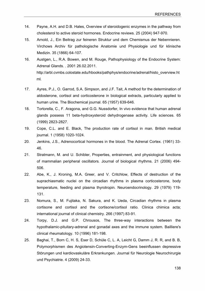
REFERENCES
138
14. Payne, A.H. and D.B. Hales, Overview of steroidogenic enzymes in the pathway from
cholesterol to active steroid hormones. Endocrine reviews. 25 (2004) 947-970.
15. Arnold, J., Ein Beitrag zur feineren Struktur und dem Chemismus der Nebennieren.
Virchows Archiv für pathologische Anatomie und Physiologie und für klinische
Medizin. 35 (1866) 64-107.
16. Austgen, L., R.A. Bowen, and M. Rouge, Pathophysiology of the Endocrine System:
Adrenal Glands. . 2001 26.02.2011.
http://arbl.cvmbs.colostate.edu/hbooks/pathphys/endocrine/adrenal/histo_overview.ht
ml.
17. Ayres, P.J., O. Garrod, S.A. Simpson, and J.F. Tait, A method for the determination of
aldosterone, cortisol and corticosterone in biological extracts, particularly applied to
human urine. The Biochemical journal. 65 (1957) 639-646.
18. Tortorella, C., F. Aragona, and G.G. Nussdorfer, In vivo evidence that human adrenal
glands possess 11 beta-hydroxysteroid dehydrogenase activity. Life sciences. 65
(1999) 2823-2827.
19. Cope, C.L. and E. Black, The production rate of cortisol in man. British medical
journal. 1 (1958) 1020-1024.
20. Jenkins, J.S., Adrenocortical hormones in the blood. The Adrenal Cortex. (1961) 33-
46.
21. Stratmann, M. and U. Schibler, Properties, entrainment, and physiological functions
of mammalian peripheral oscillators. Journal of biological rhythms. 21 (2006) 494-
506.
22. Abe, K., J. Kroning, M.A. Greer, and V. Critchlow, Effects of destruction of the
suprachiasmatic nuclei on the circadian rhythms in plasma corticosterone, body
temperature, feeding and plasma thyrotropin. Neuroendocrinology. 29 (1979) 119-
131.
23. Nomura, S., M. Fujitaka, N. Sakura, and K. Ueda, Circadian rhythms in plasma
cortisone and cortisol and the cortisone/cortisol ratio. Clinica chimica acta;
international journal of clinical chemistry. 266 (1997) 83-91.
24. Torpy, D.J. and G.P. Chrousos, The three-way interactions between the
hypothalamic-pituitary-adrenal and gonadal axes and the immune system. Bailliere's
clinical rheumatology. 10 (1996) 181-198.
25. Baghai, T., Born C, H. S, Eser D, Schüle C, L. A, Leicht G, Damm J, R. R, and B. B,
Polymorphismen des Angiotensin-Converting-Enzym-Gens beeinflussen depressive
Störungen und kardiovaskuläre Erkrankungen. Journal für Neurologie Neurochirurgie
und Psychiatrie. 4 (2009) 24-33.
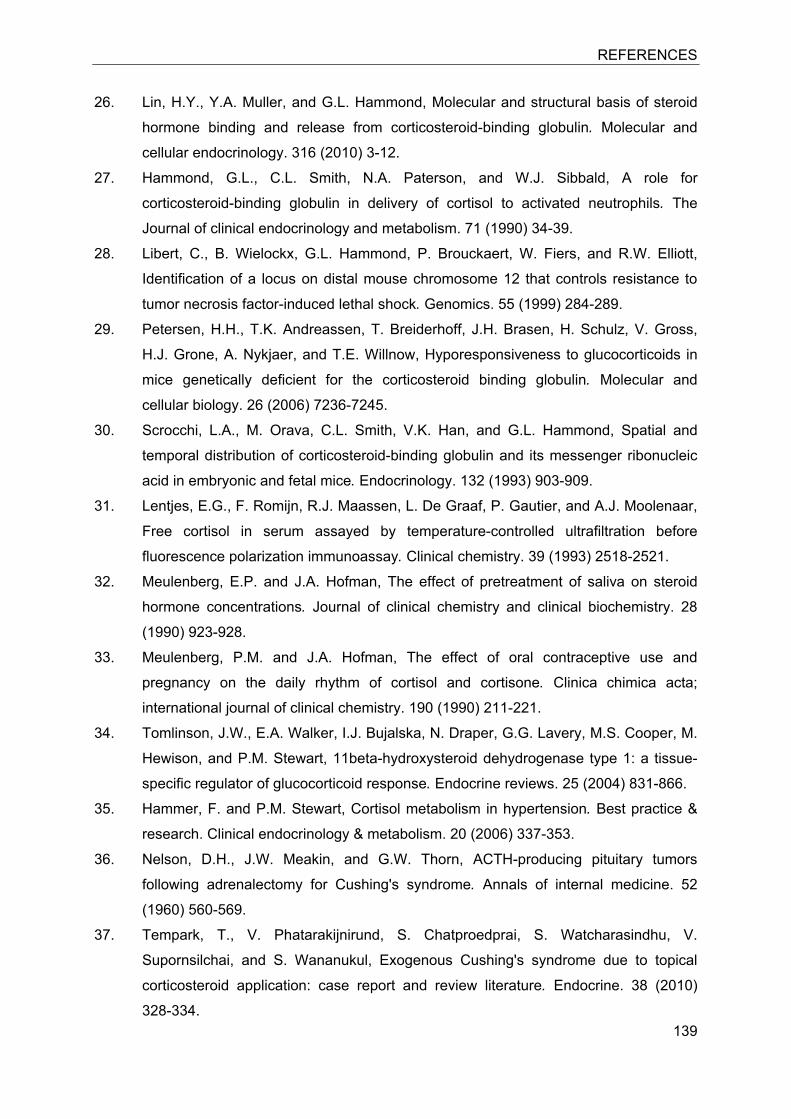
REFERENCES
139
26. Lin, H.Y., Y.A. Muller, and G.L. Hammond, Molecular and structural basis of steroid
hormone binding and release from corticosteroid-binding globulin. Molecular and
cellular endocrinology. 316 (2010) 3-12.
27. Hammond, G.L., C.L. Smith, N.A. Paterson, and W.J. Sibbald, A role for
corticosteroid-binding globulin in delivery of cortisol to activated neutrophils. The
Journal of clinical endocrinology and metabolism. 71 (1990) 34-39.
28. Libert, C., B. Wielockx, G.L. Hammond, P. Brouckaert, W. Fiers, and R.W. Elliott,
Identification of a locus on distal mouse chromosome 12 that controls resistance to
tumor necrosis factor-induced lethal shock. Genomics. 55 (1999) 284-289.
29. Petersen, H.H., T.K. Andreassen, T. Breiderhoff, J.H. Brasen, H. Schulz, V. Gross,
H.J. Grone, A. Nykjaer, and T.E. Willnow, Hyporesponsiveness to glucocorticoids in
mice genetically deficient for the corticosteroid binding globulin. Molecular and
cellular biology. 26 (2006) 7236-7245.
30. Scrocchi, L.A., M. Orava, C.L. Smith, V.K. Han, and G.L. Hammond, Spatial and
temporal distribution of corticosteroid-binding globulin and its messenger ribonucleic
acid in embryonic and fetal mice. Endocrinology. 132 (1993) 903-909.
31. Lentjes, E.G., F. Romijn, R.J. Maassen, L. De Graaf, P. Gautier, and A.J. Moolenaar,
Free cortisol in serum assayed by temperature-controlled ultrafiltration before
fluorescence polarization immunoassay. Clinical chemistry. 39 (1993) 2518-2521.
32. Meulenberg, E.P. and J.A. Hofman, The effect of pretreatment of saliva on steroid
hormone concentrations. Journal of clinical chemistry and clinical biochemistry. 28
(1990) 923-928.
33. Meulenberg, P.M. and J.A. Hofman, The effect of oral contraceptive use and
pregnancy on the daily rhythm of cortisol and cortisone. Clinica chimica acta;
international journal of clinical chemistry. 190 (1990) 211-221.
34. Tomlinson, J.W., E.A. Walker, I.J. Bujalska, N. Draper, G.G. Lavery, M.S. Cooper, M.
Hewison, and P.M. Stewart, 11beta-hydroxysteroid dehydrogenase type 1: a tissue-
specific regulator of glucocorticoid response. Endocrine reviews. 25 (2004) 831-866.
35. Hammer, F. and P.M. Stewart, Cortisol metabolism in hypertension. Best practice &
research. Clinical endocrinology & metabolism. 20 (2006) 337-353.
36. Nelson, D.H., J.W. Meakin, and G.W. Thorn, ACTH-producing pituitary tumors
following adrenalectomy for Cushing's syndrome. Annals of internal medicine. 52
(1960) 560-569.
37. Tempark, T., V. Phatarakijnirund, S. Chatproedprai, S. Watcharasindhu, V.
Supornsilchai, and S. Wananukul, Exogenous Cushing's syndrome due to topical
corticosteroid application: case report and review literature. Endocrine. 38 (2010)
328-334.
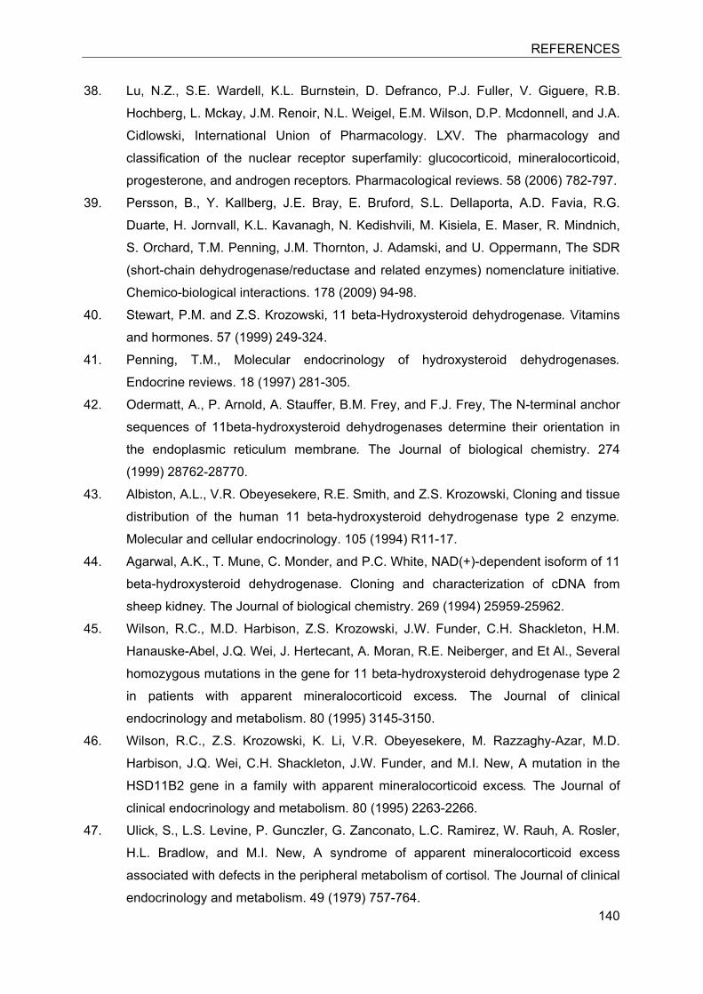
REFERENCES
140
38. Lu, N.Z., S.E. Wardell, K.L. Burnstein, D. Defranco, P.J. Fuller, V. Giguere, R.B.
Hochberg, L. Mckay, J.M. Renoir, N.L. Weigel, E.M. Wilson, D.P. Mcdonnell, and J.A.
Cidlowski, International Union of Pharmacology. LXV. The pharmacology and
classification of the nuclear receptor superfamily: glucocorticoid, mineralocorticoid,
progesterone, and androgen receptors. Pharmacological reviews. 58 (2006) 782-797.
39. Persson, B., Y. Kallberg, J.E. Bray, E. Bruford, S.L. Dellaporta, A.D. Favia, R.G.
Duarte, H. Jornvall, K.L. Kavanagh, N. Kedishvili, M. Kisiela, E. Maser, R. Mindnich,
S. Orchard, T.M. Penning, J.M. Thornton, J. Adamski, and U. Oppermann, The SDR
(short-chain dehydrogenase/reductase and related enzymes) nomenclature initiative.
Chemico-biological interactions. 178 (2009) 94-98.
40. Stewart, P.M. and Z.S. Krozowski, 11 beta-Hydroxysteroid dehydrogenase. Vitamins
and hormones. 57 (1999) 249-324.
41. Penning, T.M., Molecular endocrinology of hydroxysteroid dehydrogenases.
Endocrine reviews. 18 (1997) 281-305.
42. Odermatt, A., P. Arnold, A. Stauffer, B.M. Frey, and F.J. Frey, The N-terminal anchor
sequences of 11beta-hydroxysteroid dehydrogenases determine their orientation in
the endoplasmic reticulum membrane. The Journal of biological chemistry. 274
(1999) 28762-28770.
43. Albiston, A.L., V.R. Obeyesekere, R.E. Smith, and Z.S. Krozowski, Cloning and tissue
distribution of the human 11 beta-hydroxysteroid dehydrogenase type 2 enzyme.
Molecular and cellular endocrinology. 105 (1994) R11-17.
44. Agarwal, A.K., T. Mune, C. Monder, and P.C. White, NAD(+)-dependent isoform of 11
beta-hydroxysteroid dehydrogenase. Cloning and characterization of cDNA from
sheep kidney. The Journal of biological chemistry. 269 (1994) 25959-25962.
45. Wilson, R.C., M.D. Harbison, Z.S. Krozowski, J.W. Funder, C.H. Shackleton, H.M.
Hanauske-Abel, J.Q. Wei, J. Hertecant, A. Moran, R.E. Neiberger, and Et Al., Several
homozygous mutations in the gene for 11 beta-hydroxysteroid dehydrogenase type 2
in patients with apparent mineralocorticoid excess. The Journal of clinical
endocrinology and metabolism. 80 (1995) 3145-3150.
46. Wilson, R.C., Z.S. Krozowski, K. Li, V.R. Obeyesekere, M. Razzaghy-Azar, M.D.
Harbison, J.Q. Wei, C.H. Shackleton, J.W. Funder, and M.I. New, A mutation in the
HSD11B2 gene in a family with apparent mineralocorticoid excess. The Journal of
clinical endocrinology and metabolism. 80 (1995) 2263-2266.
47. Ulick, S., L.S. Levine, P. Gunczler, G. Zanconato, L.C. Ramirez, W. Rauh, A. Rosler,
H.L. Bradlow, and M.I. New, A syndrome of apparent mineralocorticoid excess
associated with defects in the peripheral metabolism of cortisol. The Journal of clinical
endocrinology and metabolism. 49 (1979) 757-764.
REFERENCES
141
48. Gomez-Sanchez, E.P. and C.E. Gomez-Sanchez, Central hypertensinogenic effects
of glycyrrhizic acid and carbenoxolone. The American journal of physiology. 263
(1992) E1125-1130.
49. Lovati, E., P. Ferrari, B. Dick, K. Jostarndt, B.M. Frey, F.J. Frey, U. Schorr, and A.M.
Sharma, Molecular basis of human salt sensitivity: the role of the 11beta-
hydroxysteroid dehydrogenase type 2. The Journal of clinical endocrinology and
metabolism. 84 (1999) 3745-3749.
50. Walker, B.R., P.M. Stewart, C.H. Shackleton, P.L. Padfield, and C.R. Edwards,
Deficient inactivation of cortisol by 11 beta-hydroxysteroid dehydrogenase in
essential hypertension. Clinical endocrinology. 39 (1993) 221-227.
51. Agarwal, A.K., C. Monder, B. Eckstein, and P.C. White, Cloning and expression of rat
cDNA encoding corticosteroid 11 beta-dehydrogenase. The Journal of biological
chemistry. 264 (1989) 18939-18943.
52. Schweizer, R.A., A.G. Atanasov, B.M. Frey, and A. Odermatt, A rapid screening
assay for inhibitors of 11beta-hydroxysteroid dehydrogenases (11beta-HSD):
flavanone selectively inhibits 11beta-HSD1 reductase activity. Molecular and cellular
endocrinology. 212 (2003) 41-49.
53. Balazs, Z., L.G. Nashev, C. Chandsawangbhuwana, M.E. Baker, and A. Odermatt,
Hexose-6-phosphate dehydrogenase modulates the effect of inhibitors and
alternative substrates of 11beta-hydroxysteroid dehydrogenase 1. Molecular and
cellular endocrinology. 301 (2009) 117-122.
54. Dzyakanchuk, A.A., Z. Balazs, L.G. Nashev, K.E. Amrein, and A. Odermatt, 11beta-
Hydroxysteroid dehydrogenase 1 reductase activity is dependent on a high ratio of
NADPH/NADP(+) and is stimulated by extracellular glucose. Molecular and cellular
endocrinology. 301 (2009) 137-141.
55. Hult, M., H. Jornvall, and U.C. Oppermann, Selective inhibition of human type 1
11beta-hydroxysteroid dehydrogenase by synthetic steroids and xenobiotics. FEBS
Lett. 441 (1998) 25-28.
56. Tannin, G.M., A.K. Agarwal, C. Monder, M.I. New, and P.C. White, The human gene
for 11 beta-hydroxysteroid dehydrogenase. Structure, tissue distribution, and
chromosomal localization. The Journal of biological chemistry. 266 (1991) 16653-
16658.
57. Stewart, P.M., B.A. Murry, and J.I. Mason, Human kidney 11 beta-hydroxysteroid
dehydrogenase is a high affinity nicotinamide adenine dinucleotide-dependent
enzyme and differs from the cloned type I isoform. The Journal of clinical
endocrinology and metabolism. 79 (1994) 480-484.
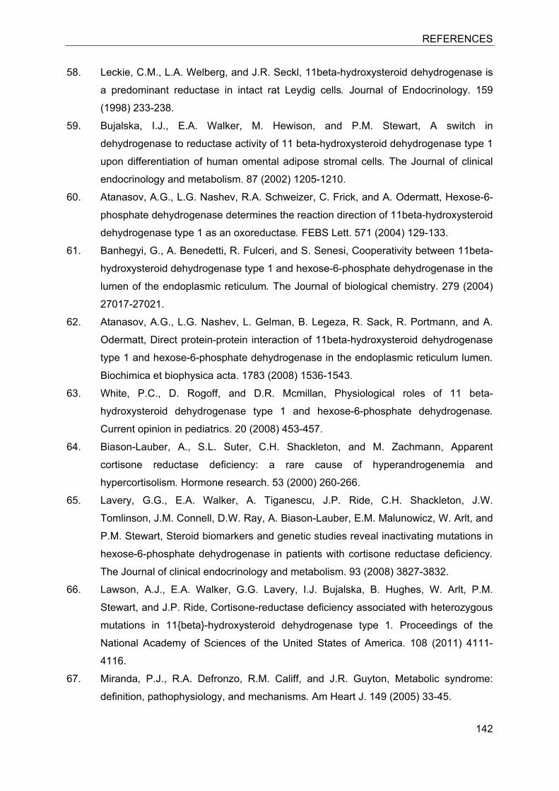
REFERENCES
142
58. Leckie, C.M., L.A. Welberg, and J.R. Seckl, 11beta-hydroxysteroid dehydrogenase is
a predominant reductase in intact rat Leydig cells. Journal of Endocrinology. 159
(1998) 233-238.
59. Bujalska, I.J., E.A. Walker, M. Hewison, and P.M. Stewart, A switch in
dehydrogenase to reductase activity of 11 beta-hydroxysteroid dehydrogenase type 1
upon differentiation of human omental adipose stromal cells. The Journal of clinical
endocrinology and metabolism. 87 (2002) 1205-1210.
60. Atanasov, A.G., L.G. Nashev, R.A. Schweizer, C. Frick, and A. Odermatt, Hexose-6-
phosphate dehydrogenase determines the reaction direction of 11beta-hydroxysteroid
dehydrogenase type 1 as an oxoreductase. FEBS Lett. 571 (2004) 129-133.
61. Banhegyi, G., A. Benedetti, R. Fulceri, and S. Senesi, Cooperativity between 11beta-
hydroxysteroid dehydrogenase type 1 and hexose-6-phosphate dehydrogenase in the
lumen of the endoplasmic reticulum. The Journal of biological chemistry. 279 (2004)
27017-27021.
62. Atanasov, A.G., L.G. Nashev, L. Gelman, B. Legeza, R. Sack, R. Portmann, and A.
Odermatt, Direct protein-protein interaction of 11beta-hydroxysteroid dehydrogenase
type 1 and hexose-6-phosphate dehydrogenase in the endoplasmic reticulum lumen.
Biochimica et biophysica acta. 1783 (2008) 1536-1543.
63. White, P.C., D. Rogoff, and D.R. Mcmillan, Physiological roles of 11 beta-
hydroxysteroid dehydrogenase type 1 and hexose-6-phosphate dehydrogenase.
Current opinion in pediatrics. 20 (2008) 453-457.
64. Biason-Lauber, A., S.L. Suter, C.H. Shackleton, and M. Zachmann, Apparent
cortisone reductase deficiency: a rare cause of hyperandrogenemia and
hypercortisolism. Hormone research. 53 (2000) 260-266.
65. Lavery, G.G., E.A. Walker, A. Tiganescu, J.P. Ride, C.H. Shackleton, J.W.
Tomlinson, J.M. Connell, D.W. Ray, A. Biason-Lauber, E.M. Malunowicz, W. Arlt, and
P.M. Stewart, Steroid biomarkers and genetic studies reveal inactivating mutations in
hexose-6-phosphate dehydrogenase in patients with cortisone reductase deficiency.
The Journal of clinical endocrinology and metabolism. 93 (2008) 3827-3832.
66. Lawson, A.J., E.A. Walker, G.G. Lavery, I.J. Bujalska, B. Hughes, W. Arlt, P.M.
Stewart, and J.P. Ride, Cortisone-reductase deficiency associated with heterozygous
mutations in 11{beta}-hydroxysteroid dehydrogenase type 1. Proceedings of the
National Academy of Sciences of the United States of America. 108 (2011) 4111-
4116.
67. Miranda, P.J., R.A. Defronzo, R.M. Califf, and J.R. Guyton, Metabolic syndrome:
definition, pathophysiology, and mechanisms. Am Heart J. 149 (2005) 33-45.
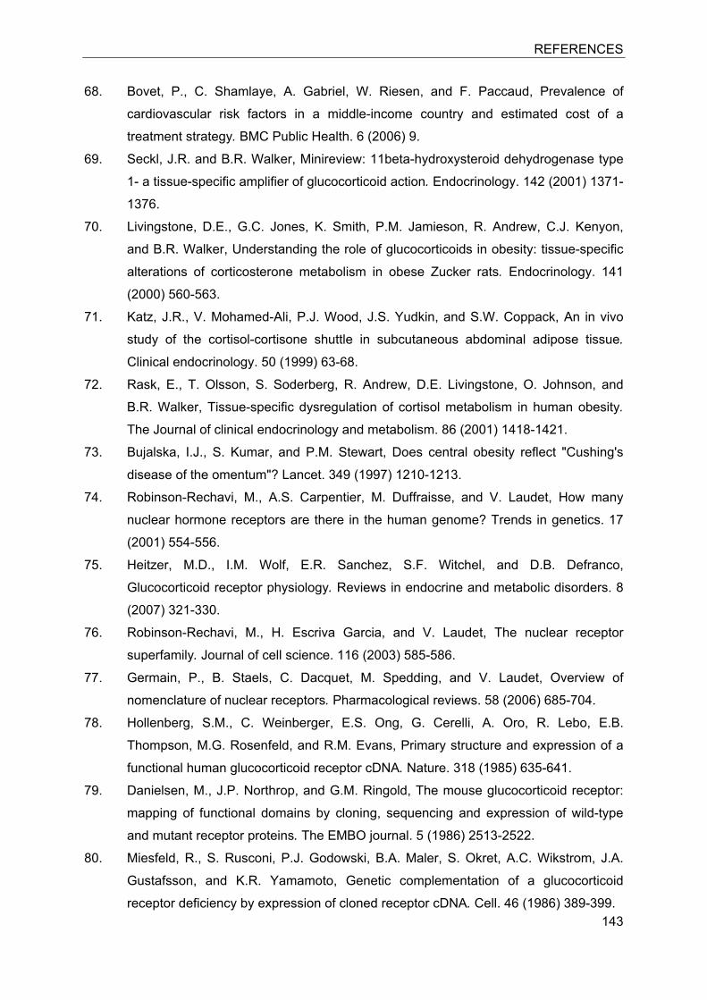
REFERENCES
143
68. Bovet, P., C. Shamlaye, A. Gabriel, W. Riesen, and F. Paccaud, Prevalence of
cardiovascular risk factors in a middle-income country and estimated cost of a
treatment strategy. BMC Public Health. 6 (2006) 9.
69. Seckl, J.R. and B.R. Walker, Minireview: 11beta-hydroxysteroid dehydrogenase type
1- a tissue-specific amplifier of glucocorticoid action. Endocrinology. 142 (2001) 1371-
1376.
70. Livingstone, D.E., G.C. Jones, K. Smith, P.M. Jamieson, R. Andrew, C.J. Kenyon,
and B.R. Walker, Understanding the role of glucocorticoids in obesity: tissue-specific
alterations of corticosterone metabolism in obese Zucker rats. Endocrinology. 141
(2000) 560-563.
71. Katz, J.R., V. Mohamed-Ali, P.J. Wood, J.S. Yudkin, and S.W. Coppack, An in vivo
study of the cortisol-cortisone shuttle in subcutaneous abdominal adipose tissue.
Clinical endocrinology. 50 (1999) 63-68.
72. Rask, E., T. Olsson, S. Soderberg, R. Andrew, D.E. Livingstone, O. Johnson, and
B.R. Walker, Tissue-specific dysregulation of cortisol metabolism in human obesity.
The Journal of clinical endocrinology and metabolism. 86 (2001) 1418-1421.
73. Bujalska, I.J., S. Kumar, and P.M. Stewart, Does central obesity reflect "Cushing's
disease of the omentum"? Lancet. 349 (1997) 1210-1213.
74. Robinson-Rechavi, M., A.S. Carpentier, M. Duffraisse, and V. Laudet, How many
nuclear hormone receptors are there in the human genome? Trends in genetics. 17
(2001) 554-556.
75. Heitzer, M.D., I.M. Wolf, E.R. Sanchez, S.F. Witchel, and D.B. Defranco,
Glucocorticoid receptor physiology. Reviews in endocrine and metabolic disorders. 8
(2007) 321-330.
76. Robinson-Rechavi, M., H. Escriva Garcia, and V. Laudet, The nuclear receptor
superfamily. Journal of cell science. 116 (2003) 585-586.
77. Germain, P., B. Staels, C. Dacquet, M. Spedding, and V. Laudet, Overview of
nomenclature of nuclear receptors. Pharmacological reviews. 58 (2006) 685-704.
78. Hollenberg, S.M., C. Weinberger, E.S. Ong, G. Cerelli, A. Oro, R. Lebo, E.B.
Thompson, M.G. Rosenfeld, and R.M. Evans, Primary structure and expression of a
functional human glucocorticoid receptor cDNA. Nature. 318 (1985) 635-641.
79. Danielsen, M., J.P. Northrop, and G.M. Ringold, The mouse glucocorticoid receptor:
mapping of functional domains by cloning, sequencing and expression of wild-type
and mutant receptor proteins. The EMBO journal. 5 (1986) 2513-2522.
80. Miesfeld, R., S. Rusconi, P.J. Godowski, B.A. Maler, S. Okret, A.C. Wikstrom, J.A.
Gustafsson, and K.R. Yamamoto, Genetic complementation of a glucocorticoid
receptor deficiency by expression of cloned receptor cDNA. Cell. 46 (1986) 389-399.
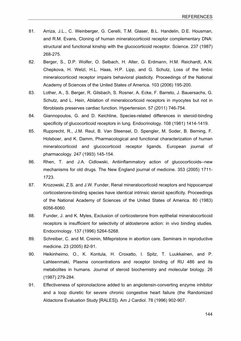
REFERENCES
144
81. Arriza, J.L., C. Weinberger, G. Cerelli, T.M. Glaser, B.L. Handelin, D.E. Housman,
and R.M. Evans, Cloning of human mineralocorticoid receptor complementary DNA:
structural and functional kinship with the glucocorticoid receptor. Science. 237 (1987)
268-275.
82. Berger, S., D.P. Wolfer, O. Selbach, H. Alter, G. Erdmann, H.M. Reichardt, A.N.
Chepkova, H. Welzl, H.L. Haas, H.P. Lipp, and G. Schutz, Loss of the limbic
mineralocorticoid receptor impairs behavioral plasticity. Proceedings of the National
Academy of Sciences of the United States of America. 103 (2006) 195-200.
83. Lother, A., S. Berger, R. Gilsbach, S. Rosner, A. Ecke, F. Barreto, J. Bauersachs, G.
Schutz, and L. Hein, Ablation of mineralocorticoid receptors in myocytes but not in
fibroblasts preserves cardiac function. Hypertension. 57 (2011) 746-754.
84. Giannopoulos, G. and D. Keichline, Species-related differences in steroid-binding
specificity of glucocorticoid receptors in lung. Endocrinology. 108 (1981) 1414-1419.
85. Rupprecht, R., J.M. Reul, B. Van Steensel, D. Spengler, M. Soder, B. Berning, F.
Holsboer, and K. Damm, Pharmacological and functional characterization of human
mineralocorticoid and glucocorticoid receptor ligands. European journal of
pharmacology. 247 (1993) 145-154.
86. Rhen, T. and J.A. Cidlowski, Antiinflammatory action of glucocorticoids--new
mechanisms for old drugs. The New England journal of medicine. 353 (2005) 1711-
1723.
87. Krozowski, Z.S. and J.W. Funder, Renal mineralocorticoid receptors and hippocampal
corticosterone-binding species have identical intrinsic steroid specificity. Proceedings
of the National Academy of Sciences of the United States of America. 80 (1983)
6056-6060.
88. Funder, J. and K. Myles, Exclusion of corticosterone from epithelial mineralocorticoid
receptors is insufficient for selectivity of aldosterone action: in vivo binding studies.
Endocrinology. 137 (1996) 5264-5268.
89. Schreiber, C. and M. Creinin, Mifepristone in abortion care. Seminars in reproductive
medicine. 23 (2005) 82-91.
90. Heikinheimo, O., K. Kontula, H. Croxatto, I. Spitz, T. Luukkainen, and P.
Lahteenmaki, Plasma concentrations and receptor binding of RU 486 and its
metabolites in humans. Journal of steroid biochemistry and molecular biology. 26
(1987) 279-284.
91. Effectiveness of spironolactone added to an angiotensin-converting enzyme inhibitor
and a loop diuretic for severe chronic congestive heart failure (the Randomized
Aldactone Evaluation Study [RALES]). Am J Cardiol. 78 (1996) 902-907.
REFERENCES
145
92. Batterink, J., S.N. Stabler, A.M. Tejani, and C.T. Fowkes, Spironolactone for
hypertension. Cochrane database of systematic reviews. (2010) CD008169.
93. Hamaguchi, S., S. Kinugawa, M. Tsuchihashi-Makaya, K. Goto, D. Goto, T. Yokota,
S. Yamada, H. Yokoshiki, A. Takeshita, and H. Tsutsui, Spironolactone use at
discharge was associated with improved survival in hospitalized patients with systolic
heart failure. Am Heart J. 160 (2010) 1156-1162.
94. Farrelly, R.O., R.N. Howie, and J.D. North, Use of spironolactone and
hydrochlorothiazide in treatment of oedema. British medical journal. 2 (1960) 339-
343.
95. Menard, J., The 45-year story of the development of an anti-aldosterone more
specific than spironolactone. Molecular and cellular endocrinology. 217 (2004) 45-52.
96. Pratt, W.B. and D.O. Toft, Regulation of signaling protein function and trafficking by
the hsp90/hsp70-based chaperone machinery. Experimental biology and medicine
(Maywood, N.J.). 228 (2003) 111-133.
97. Elbi, C., D.A. Walker, G. Romero, W.P. Sullivan, D.O. Toft, G.L. Hager, and D.B.
Defranco, Molecular chaperones function as steroid receptor nuclear mobility factors.
Proceedings of the National Academy of Sciences of the United States of America.
101 (2004) 2876-2881.
98. Kumar, R. and E.B. Thompson, Transactivation functions of the N-terminal domains
of nuclear hormone receptors: protein folding and coactivator interactions. Molecular
and cellular endocrinology. 17 (2003) 1-10.
99. Kumar, R. and E.B. Thompson, Gene regulation by the glucocorticoid receptor:
structure:function relationship. Journal of steroid biochemistry and molecular biology.
94 (2005) 383-394.
100. Duma, D., C.M. Jewell, and J.A. Cidlowski, Multiple glucocorticoid receptor isoforms
and mechanisms of post-translational modification. Journal of steroid biochemistry
and molecular biology. 102 (2006) 11-21.
101. Wang, Z. and M.J. Garabedian, Modulation of glucocorticoid receptor transcriptional
activation, phosphorylation, and growth inhibition by p27Kip1. The Journal of
biological chemistry. 278 (2003) 50897-50901.
102. Kovacs, J.J., P.J. Murphy, S. Gaillard, X. Zhao, J.T. Wu, C.V. Nicchitta, M. Yoshida,
D.O. Toft, W.B. Pratt, and T.P. Yao, HDAC6 regulates Hsp90 acetylation and
chaperone-dependent activation of glucocorticoid receptor. Molecular and cellular
biology. 18 (2005) 601-607.
103. Le Drean, Y., N. Mincheneau, P. Le Goff, and D. Michel, Potentiation of
glucocorticoid receptor transcriptional activity by sumoylation. Endocrinology. 143
(2002) 3482-3489.
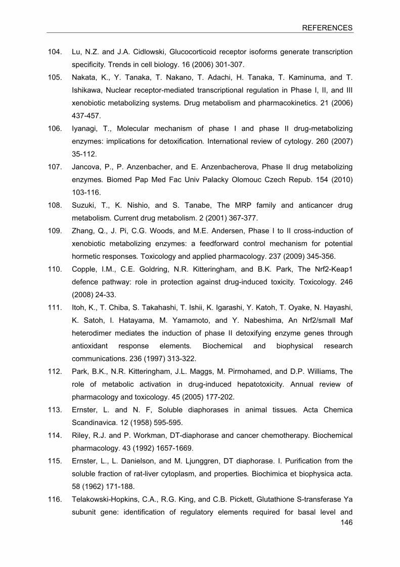
REFERENCES
146
104. Lu, N.Z. and J.A. Cidlowski, Glucocorticoid receptor isoforms generate transcription
specificity. Trends in cell biology. 16 (2006) 301-307.
105. Nakata, K., Y. Tanaka, T. Nakano, T. Adachi, H. Tanaka, T. Kaminuma, and T.
Ishikawa, Nuclear receptor-mediated transcriptional regulation in Phase I, II, and III
xenobiotic metabolizing systems. Drug metabolism and pharmacokinetics. 21 (2006)
437-457.
106. Iyanagi, T., Molecular mechanism of phase I and phase II drug-metabolizing
enzymes: implications for detoxification. International review of cytology. 260 (2007)
35-112.
107. Jancova, P., P. Anzenbacher, and E. Anzenbacherova, Phase II drug metabolizing
enzymes. Biomed Pap Med Fac Univ Palacky Olomouc Czech Repub. 154 (2010)
103-116.
108. Suzuki, T., K. Nishio, and S. Tanabe, The MRP family and anticancer drug
metabolism. Current drug metabolism. 2 (2001) 367-377.
109. Zhang, Q., J. Pi, C.G. Woods, and M.E. Andersen, Phase I to II cross-induction of
xenobiotic metabolizing enzymes: a feedforward control mechanism for potential
hormetic responses. Toxicology and applied pharmacology. 237 (2009) 345-356.
110. Copple, I.M., C.E. Goldring, N.R. Kitteringham, and B.K. Park, The Nrf2-Keap1
defence pathway: role in protection against drug-induced toxicity. Toxicology. 246
(2008) 24-33.
111. Itoh, K., T. Chiba, S. Takahashi, T. Ishii, K. Igarashi, Y. Katoh, T. Oyake, N. Hayashi,
K. Satoh, I. Hatayama, M. Yamamoto, and Y. Nabeshima, An Nrf2/small Maf
heterodimer mediates the induction of phase II detoxifying enzyme genes through
antioxidant response elements. Biochemical and biophysical research
communications. 236 (1997) 313-322.
112. Park, B.K., N.R. Kitteringham, J.L. Maggs, M. Pirmohamed, and D.P. Williams, The
role of metabolic activation in drug-induced hepatotoxicity. Annual review of
pharmacology and toxicology. 45 (2005) 177-202.
113. Ernster, L. and N. F, Soluble diaphorases in animal tissues. Acta Chemica
Scandinavica. 12 (1958) 595-595.
114. Riley, R.J. and P. Workman, DT-diaphorase and cancer chemotherapy. Biochemical
pharmacology. 43 (1992) 1657-1669.
115. Ernster, L., L. Danielson, and M. Ljunggren, DT diaphorase. I. Purification from the
soluble fraction of rat-liver cytoplasm, and properties. Biochimica et biophysica acta.
58 (1962) 171-188.
116. Telakowski-Hopkins, C.A., R.G. King, and C.B. Pickett, Glutathione S-transferase Ya
subunit gene: identification of regulatory elements required for basal level and
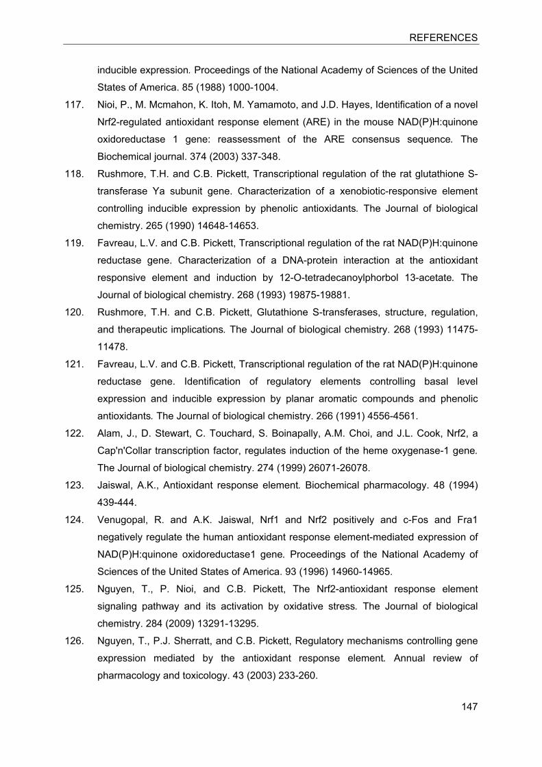
REFERENCES
147
inducible expression. Proceedings of the National Academy of Sciences of the United
States of America. 85 (1988) 1000-1004.
117. Nioi, P., M. Mcmahon, K. Itoh, M. Yamamoto, and J.D. Hayes, Identification of a novel
Nrf2-regulated antioxidant response element (ARE) in the mouse NAD(P)H:quinone
oxidoreductase 1 gene: reassessment of the ARE consensus sequence. The
Biochemical journal. 374 (2003) 337-348.
118. Rushmore, T.H. and C.B. Pickett, Transcriptional regulation of the rat glutathione S-
transferase Ya subunit gene. Characterization of a xenobiotic-responsive element
controlling inducible expression by phenolic antioxidants. The Journal of biological
chemistry. 265 (1990) 14648-14653.
119. Favreau, L.V. and C.B. Pickett, Transcriptional regulation of the rat NAD(P)H:quinone
reductase gene. Characterization of a DNA-protein interaction at the antioxidant
responsive element and induction by 12-O-tetradecanoylphorbol 13-acetate. The
Journal of biological chemistry. 268 (1993) 19875-19881.
120. Rushmore, T.H. and C.B. Pickett, Glutathione S-transferases, structure, regulation,
and therapeutic implications. The Journal of biological chemistry. 268 (1993) 11475-
11478.
121. Favreau, L.V. and C.B. Pickett, Transcriptional regulation of the rat NAD(P)H:quinone
reductase gene. Identification of regulatory elements controlling basal level
expression and inducible expression by planar aromatic compounds and phenolic
antioxidants. The Journal of biological chemistry. 266 (1991) 4556-4561.
122. Alam, J., D. Stewart, C. Touchard, S. Boinapally, A.M. Choi, and J.L. Cook, Nrf2, a
Cap'n'Collar transcription factor, regulates induction of the heme oxygenase-1 gene.
The Journal of biological chemistry. 274 (1999) 26071-26078.
123. Jaiswal, A.K., Antioxidant response element. Biochemical pharmacology. 48 (1994)
439-444.
124. Venugopal, R. and A.K. Jaiswal, Nrf1 and Nrf2 positively and c-Fos and Fra1
negatively regulate the human antioxidant response element-mediated expression of
NAD(P)H:quinone oxidoreductase1 gene. Proceedings of the National Academy of
Sciences of the United States of America. 93 (1996) 14960-14965.
125. Nguyen, T., P. Nioi, and C.B. Pickett, The Nrf2-antioxidant response element
signaling pathway and its activation by oxidative stress. The Journal of biological
chemistry. 284 (2009) 13291-13295.
126. Nguyen, T., P.J. Sherratt, and C.B. Pickett, Regulatory mechanisms controlling gene
expression mediated by the antioxidant response element. Annual review of
pharmacology and toxicology. 43 (2003) 233-260.
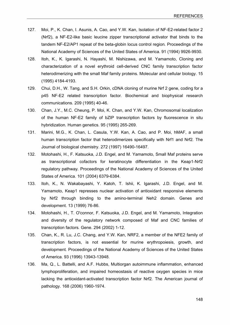
REFERENCES
148
127. Moi, P., K. Chan, I. Asunis, A. Cao, and Y.W. Kan, Isolation of NF-E2-related factor 2
(Nrf2), a NF-E2-like basic leucine zipper transcriptional activator that binds to the
tandem NF-E2/AP1 repeat of the beta-globin locus control region. Proceedings of the
National Academy of Sciences of the United States of America. 91 (1994) 9926-9930.
128. Itoh, K., K. Igarashi, N. Hayashi, M. Nishizawa, and M. Yamamoto, Cloning and
characterization of a novel erythroid cell-derived CNC family transcription factor
heterodimerizing with the small Maf family proteins. Molecular and cellular biology. 15
(1995) 4184-4193.
129. Chui, D.H., W. Tang, and S.H. Orkin, cDNA cloning of murine Nrf 2 gene, coding for a
p45 NF-E2 related transcription factor. Biochemical and biophysical research
communications. 209 (1995) 40-46.
130. Chan, J.Y., M.C. Cheung, P. Moi, K. Chan, and Y.W. Kan, Chromosomal localization
of the human NF-E2 family of bZIP transcription factors by fluorescence in situ
hybridization. Human genetics. 95 (1995) 265-269.
131. Marini, M.G., K. Chan, L. Casula, Y.W. Kan, A. Cao, and P. Moi, hMAF, a small
human transcription factor that heterodimerizes specifically with Nrf1 and Nrf2. The
Journal of biological chemistry. 272 (1997) 16490-16497.
132. Motohashi, H., F. Katsuoka, J.D. Engel, and M. Yamamoto, Small Maf proteins serve
as transcriptional cofactors for keratinocyte differentiation in the Keap1-Nrf2
regulatory pathway. Proceedings of the National Academy of Sciences of the United
States of America. 101 (2004) 6379-6384.
133. Itoh, K., N. Wakabayashi, Y. Katoh, T. Ishii, K. Igarashi, J.D. Engel, and M.
Yamamoto, Keap1 represses nuclear activation of antioxidant responsive elements
by Nrf2 through binding to the amino-terminal Neh2 domain. Genes and
development. 13 (1999) 76-86.
134. Motohashi, H., T. O'connor, F. Katsuoka, J.D. Engel, and M. Yamamoto, Integration
and diversity of the regulatory network composed of Maf and CNC families of
transcription factors. Gene. 294 (2002) 1-12.
135. Chan, K., R. Lu, J.C. Chang, and Y.W. Kan, NRF2, a member of the NFE2 family of
transcription factors, is not essential for murine erythropoiesis, growth, and
development. Proceedings of the National Academy of Sciences of the United States
of America. 93 (1996) 13943-13948.
136. Ma, Q., L. Battelli, and A.F. Hubbs, Multiorgan autoimmune inflammation, enhanced
lymphoproliferation, and impaired homeostasis of reactive oxygen species in mice
lacking the antioxidant-activated transcription factor Nrf2. The American journal of
pathology. 168 (2006) 1960-1974.
REFERENCES
149
137. Hubbs, A.F., S.A. Benkovic, D.B. Miller, J.P. O'callaghan, L. Battelli, D. Schwegler-
Berry, and Q. Ma, Vacuolar leukoencephalopathy with widespread astrogliosis in
mice lacking transcription factor Nrf2. The American journal of pathology. 170 (2007)
2068-2076.
138. Chan, K., X.D. Han, and Y.W. Kan, An important function of Nrf2 in combating
oxidative stress: detoxification of acetaminophen. Proceedings of the National
Academy of Sciences of the United States of America. 98 (2001) 4611-4616.
139. Chowdhry, S., M.H. Nazmy, P.J. Meakin, A.T. Dinkova-Kostova, S.V. Walsh, T.
Tsujita, J.F. Dillon, M.L. Ashford, and J.D. Hayes, Loss of Nrf2 markedly exacerbates
nonalcoholic steatohepatitis. Free radical biology and medicine. 48 (2010) 357-371.
140. Wruck, C.J., A. Fragoulis, A. Gurzynski, L.O. Brandenburg, Y.W. Kan, K. Chan, J.
Hassenpflug, S. Freitag-Wolf, D. Varoga, S. Lippross, and T. Pufe, Role of oxidative
stress in rheumatoid arthritis: insights from the Nrf2-knockout mice. Annals of the
rheumatic diseases. (2010) 844-850.
141. Kitamura, Y., T. Umemura, K. Kanki, Y. Kodama, S. Kitamoto, K. Saito, K. Itoh, M.
Yamamoto, T. Masegi, A. Nishikawa, and M. Hirose, Increased susceptibility to
hepatocarcinogenicity of Nrf2-deficient mice exposed to 2-amino-3-
methylimidazo[4,5-f]quinoline. Cancer science. 98 (2007) 19-24.
142. Xue, F. and L. Cooley, kelch encodes a component of intercellular bridges in
Drosophila egg chambers. Cell. 72 (1993) 681-693.
143. Copple, I.M., A. Lister, A.D. Obeng, N.R. Kitteringham, R.E. Jenkins, R. Layfield, B.J.
Foster, C.E. Goldring, and B.K. Park, Physical and functional interaction of
sequestosome 1 with Keap1 regulates the Keap1-Nrf2 cell defense pathway. The
Journal of biological chemistry. 285 (2010) 16782-16788.
144. Zipper, L.M. and R.T. Mulcahy, The Keap1 BTB/POZ dimerization function is required
to sequester Nrf2 in cytoplasm. The Journal of biological chemistry. 277 (2002)
36544-36552.
145. Zhang, D.D., S.C. Lo, J.V. Cross, D.J. Templeton, and M. Hannink, Keap1 is a redox-
regulated substrate adaptor protein for a Cul3-dependent ubiquitin ligase complex.
Molecular and cellular biology. 24 (2004) 10941-10953.
146. Zhang, D.D. and M. Hannink, Distinct cysteine residues in Keap1 are required for
Keap1-dependent ubiquitination of Nrf2 and for stabilization of Nrf2 by
chemopreventive agents and oxidative stress. Molecular and cellular biology. 23
(2003) 8137-8151.
147. Itoh, K., N. Wakabayashi, Y. Katoh, T. Ishii, T. O'connor, and M. Yamamoto, Keap1
regulates both cytoplasmic-nuclear shuttling and degradation of Nrf2 in response to
electrophiles. Genes to cells. 8 (2003) 379-391.
REFERENCES
150
148. Dinkova-Kostova, A.T., W.D. Holtzclaw, R.N. Cole, K. Itoh, N. Wakabayashi, Y.
Katoh, M. Yamamoto, and P. Talalay, Direct evidence that sulfhydryl groups of Keap1
are the sensors regulating induction of phase 2 enzymes that protect against
carcinogens and oxidants. Proceedings of the National Academy of Sciences of the
United States of America. 99 (2002) 11908-11913.
149. Hong, F., K.R. Sekhar, M.L. Freeman, and D.C. Liebler, Specific patterns of
electrophile adduction trigger Keap1 ubiquitination and Nrf2 activation. The Journal of
biological chemistry. 280 (2005) 31768-31775.
150. Wakabayashi, N., K. Itoh, J. Wakabayashi, H. Motohashi, S. Noda, S. Takahashi, S.
Imakado, T. Kotsuji, F. Otsuka, D.R. Roop, T. Harada, J.D. Engel, and M. Yamamoto,
Keap1-null mutation leads to postnatal lethality due to constitutive Nrf2 activation.
Nature genetics. 35 (2003) 238-245.
151. Wang, R., J. An, F. Ji, H. Jiao, H. Sun, and D. Zhou, Hypermethylation of the Keap1
gene in human lung cancer cell lines and lung cancer tissues. Biochemical and
biophysical research communications. 373 (2008) 151-154.
152. Mcmahon, M., K. Itoh, M. Yamamoto, S.A. Chanas, C.J. Henderson, L.I. Mclellan,
C.R. Wolf, C. Cavin, and J.D. Hayes, The Cap'n'Collar basic leucine zipper
transcription factor Nrf2 (NF-E2 p45-related factor 2) controls both constitutive and
inducible expression of intestinal detoxification and glutathione biosynthetic enzymes.
Cancer research. 61 (2001) 3299-3307.
153. Furukawa, M. and Y. Xiong, BTB protein Keap1 targets antioxidant transcription
factor Nrf2 for ubiquitination by the Cullin 3-Roc1 ligase. Molecular and cellular
biology. 25 (2005) 162-171.
154. Stewart, D., E. Killeen, R. Naquin, S. Alam, and J. Alam, Degradation of transcription
factor Nrf2 via the ubiquitin-proteasome pathway and stabilization by cadmium. The
Journal of biological chemistry. 278 (2003) 2396-2402.
155. Tong, K.I., B. Padmanabhan, A. Kobayashi, C. Shang, Y. Hirotsu, S. Yokoyama, and
M. Yamamoto, Different electrostatic potentials define ETGE and DLG motifs as
hinge and latch in oxidative stress response. Molecular and cellular biology. 27
(2007) 7511-7521.
156. Fourquet, S., R. Guerois, D. Biard, and M.B. Toledano, Activation of NRF2 by
nitrosative agents and H2O2 involves KEAP1 disulfide formation. The Journal of
biological chemistry. 285 (2010) 8463-8471.
157. Zhang, D.D., Mechanistic studies of the Nrf2-Keap1 signaling pathway. Drug
metabolism reviews. 38 (2006) 769-789.

REFERENCES
151
158. Masuzaki, H., J. Paterson, H. Shinyama, N.M. Morton, J.J. Mullins, J.R. Seckl, and
J.S. Flier, A transgenic model of visceral obesity and the metabolic syndrome.
Science. 294 (2001) 2166-2170.
159. Morton, N.M. and J.R. Seckl, 11beta-hydroxysteroid dehydrogenase type 1 and
obesity. Frontiers of hormone research. 36 (2008) 146-164.
160. Paterson, J.M., N.M. Morton, C. Fievet, C.J. Kenyon, M.C. Holmes, B. Staels, J.R.
Seckl, and J.J. Mullins, Metabolic syndrome without obesity: Hepatic overexpression
of 11beta-hydroxysteroid dehydrogenase type 1 in transgenic mice. Proceedings of
the National Academy of Sciences of the United States of America. 101 (2004) 7088-
7093.
161. Morton, N.M., L. Ramage, and J.R. Seckl, Down-regulation of adipose 11beta-
hydroxysteroid dehydrogenase type 1 by high-fat feeding in mice: a potential adaptive
mechanism counteracting metabolic disease. Endocrinology. 145 (2004) 2707-2712.
162. Morton, N.M., M.C. Holmes, C. Fievet, B. Staels, A. Tailleux, J.J. Mullins, and J.R.
Seckl, Improved lipid and lipoprotein profile, hepatic insulin sensitivity, and glucose
tolerance in 11beta-hydroxysteroid dehydrogenase type 1 null mice. The Journal of
biological chemistry. 276 (2001) 41293-41300.
163. Ferrari, P., The role of 11beta-hydroxysteroid dehydrogenase type 2 in human
hypertension. Biochimica et biophysica acta. 1802 (2010) 1178-1187.
164. Liu, J., L. Wang, A. Zhang, W. Di, X. Zhang, L. Wu, J. Yu, J. Zha, S. Lv, P. Cheng, M.
Hu, Y. Li, H. Qi, G. Ding, and Y. Zhong, Adipose tissue-targeted 11beta-
hydroxysteroid dehydrogenase type 1 inhibitor protects against diet-induced obesity.
Endocrine journal. (2011) 199-209.
165. Su, X., N. Vicker, H. Lawrence, A. Smith, A. Purohit, M.J. Reed, and B.V. Potter,
Inhibition of human and rat 11beta-hydroxysteroid dehydrogenase type 1 by 18beta-
glycyrrhetinic acid derivatives. Journal of steroid biochemistry and molecular biology.
104 (2007) 312-320.
166. Arampatzis, S., B. Kadereit, D. Schuster, Z. Balazs, R.A. Schweizer, F.J. Frey, T.
Langer, and A. Odermatt, Comparative enzymology of 11beta-hydroxysteroid
dehydrogenase type 1 from six species. Journal of molecular endocrinology. 35
(2005) 89-101.
167. Farese, S., A. Kruse, A. Pasch, B. Dick, B.M. Frey, D.E. Uehlinger, and F.J. Frey,
Glycyrrhetinic acid food supplementation lowers serum potassium concentration in
chronic hemodialysis patients. Kidney international. 76 (2009) 877-884.
168. Nagler, M., B. Muller, V. Briner, and R. Winterhalder, Severe hyperkalemia and
bilateral adrenal metastasis. Journal of oncology. 2009 (2009) 831979.
REFERENCES
152
169. Refstad, S., Paramethoxyamphetamine (PMA) poisoning; a 'party drug' with lethal
effects. Acta anaesthesiologica Scandinavica. 47 (2003) 1298-1299.
170. Rabbitt, E.H., J. Ayuk, K. Boelaert, M.C. Sheppard, M. Hewison, P.M. Stewart, and
N.J. Gittoes, Abnormal expression of 11 beta-hydroxysteroid dehydrogenase type 2
in human pituitary adenomas: a prereceptor determinant of pituitary cell proliferation.
Oncogene. 22 (2003) 1663-1667.
171. Zhang, M.Z., J. Xu, B. Yao, H. Yin, Q. Cai, M.J. Shrubsole, X. Chen, V. Kon, W.
Zheng, A. Pozzi, and R.C. Harris, Inhibition of 11beta-hydroxysteroid dehydrogenase
type II selectively blocks the tumor COX-2 pathway and suppresses colon
carcinogenesis in mice and humans. The Journal of clinical investigation. 119 (2009)
876-885.
172. Zhou, J., W. Liu, R.C. Pong, G. Hao, X. Sun, and J.T. Hsieh, Analysis of oligo-
arginine cell-permeable peptides uptake by prostate cells. Amino Acids. (2010).
173. Li, Y.J., Z.M. Wei, Y.X. Meng, and X.R. Ji, Beta-catenin up-regulates the expression
of cyclinD1, c-myc and MMP-7 in human pancreatic cancer: relationships with
carcinogenesis and metastasis. World journal of gastroenterology. 11 (2005) 2117-
2123.
174. Zucker, S. and J. Vacirca, Role of matrix metalloproteinases (MMPs) in colorectal
cancer. Cancer metastasis reviews. 23 (2004) 101-117.
175. Zhang, H., Pyro-Gly-Pro-Leu-Gly-Leu-Ala-Arg-Lys(BHQ3). Molecular Imaging and
Contrast Agent Database (MICAD) (2004) 20.04.2011
http://www.ncbi.nlm.nih.gov/books/NBK23111/.
176. Ki, S.H., I.J. Cho, D.W. Choi, and S.G. Kim, Glucocorticoid receptor (GR)-associated
SMRT binding to C/EBPbeta TAD and Nrf2 Neh4/5: role of SMRT recruited to GR in
GSTA2 gene repression. Molecular and cellular biology. 25 (2005) 4150-4165.
177. Bugianesi, E., N. Leone, E. Vanni, G. Marchesini, F. Brunello, P. Carucci, A. Musso,
P. De Paolis, L. Capussotti, M. Salizzoni, and M. Rizzetto, Expanding the natural
history of nonalcoholic steatohepatitis: from cryptogenic cirrhosis to hepatocellular
carcinoma. Gastroenterology. 123 (2002) 134-140.
178. Eguchi, Y., T. Mizuta, Y. Sumida, E. Ishibashi, Y. Kitajima, H. Isoda, H. Horie, T.
Tashiro, E. Iwamoto, H. Takahashi, T. Kuwashiro, S. Soejima, Y. Kawaguchi, Y. Oda,
S. Emura, R. Iwakiri, I. Ozaki, T. Eguchi, N. Ono, K. Anzai, K. Fujimoto, and S.
Koizumi, The pathological role of visceral fat accumulation in steatosis, inflammation,
and progression of nonalcoholic fatty liver disease. Journal of gastroenterology. 46
Suppl 1 (2011) 70-78.
179. Sugimoto, H., K. Okada, J. Shoda, E. Warabi, K. Ishige, T. Ueda, K. Taguchi, T.
Yanagawa, A. Nakahara, I. Hyodo, T. Ishii, and M. Yamamoto, Deletion of nuclear
REFERENCES
153
factor-E2-related factor-2 leads to rapid onset and progression of nutritional
steatohepatitis in mice. American journal of physiology. Gastrointestinal and liver
physiology. 298 (2010) G283-294.
180. Ahmed, A., S. Saksena, M. Sherlock, S.P. Olliff, E. Elias, and P.M. Stewart, Induction
of hepatic 11beta-hydroxysteroid dehydrogenase type 1 in patients with alcoholic liver
disease. Clinical endocrinology. 68 (2008) 898-903.
181. Lamle, J., S. Marhenke, J. Borlak, R. Von Wasielewski, C.J. Eriksson, R. Geffers,
M.P. Manns, M. Yamamoto, and A. Vogel, Nuclear factor-eythroid 2-related factor 2
prevents alcohol-induced fulminant liver injury. Gastroenterology. 134 (2008) 1159-
1168.
182. Makkonen, J., J. Westerbacka, M. Kolak, J. Sutinen, A. Corner, A. Hamsten, R.M.
Fisher, and H. Yki-Jarvinen, Increased expression of the macrophage markers and of
11beta-HSD-1 in subcutaneous adipose tissue, but not in cultured monocyte-derived
macrophages, is associated with liver fat in human obesity. International journal of
obesity. 31 (2007) 1617-1625.
183. Finckh, C., A. Atalla, G. Nagel, B. Stinner, and E. Maser, Expression and NNK
reducing activities of carbonyl reductase and 11beta-hydroxysteroid dehydrogenase
type 1 in human lung. Chemico-biological interactions. 130-132 (2001) 761-773.
184. Sagerup, C.M., M. Smastuen, T.B. Johannesen, A. Helland, and O.T. Brustugun,
Sex-specific trends in lung cancer incidence and survival: a population study of 40
118 cases. Thorax. 66 (2011) 301-307.
185. Anttila, S., H. Raunio, and J. Hakkola, Cytochrome P450 Mediated Pulmonary
Metabolism of Carcinogens: Regulation and Cross-talk in Lung Carcinogenesis.
American journal of respiratory cell and molecular biologyl. (2010).
186. Albiston, A.L., R.E. Smith, and Z.S. Krozowski, Sex- and tissue- specific regulation of
11 beta-hydroxysteroid dehydrogenase mRNA. Molecular and cellular endocrinology.
109 (1995) 183-188.
187. Gomez-Sanchez, E.P., D.G. Romero, A.F. De Rodriguez, M.P. Warden, Z.
Krozowski, and C.E. Gomez-Sanchez, Hexose-6-phosphate dehydrogenase and
11beta-hydroxysteroid dehydrogenase-1 tissue distribution in the rat. Endocrinology.
149 (2008) 525-533.
188. Augustine, L.M., C.D. Fisher, A.J. Lickteig, L.M. Aleksunes, A.L. Slitt, and N.J.
Cherrington, Gender divergent expression of Nqo1 in Sprague Dawley and August
Copenhagen x Irish rats. Journal of biochemical and molecular toxicology. 22 (2008)
93-100.
189. Wang, X., B.N. Chorley, G.S. Pittman, S.R. Kleeberger, J. Brothers, 2nd, G. Liu, A.
Spira, and D.A. Bell, Genetic variation and antioxidant response gene expression in

REFERENCES
154
the bronchial airway epithelium of smokers at risk for lung cancer. PLoS One. 5
(2010) e11934.
190. Ohta, T., K. Iijima, M. Miyamoto, I. Nakahara, H. Tanaka, M. Ohtsuji, T. Suzuki, A.
Kobayashi, J. Yokota, T. Sakiyama, T. Shibata, M. Yamamoto, and S. Hirohashi,
Loss of Keap1 function activates Nrf2 and provides advantages for lung cancer cell
growth. Cancer research. 68 (2008) 1303-1309.
ACKNOWLEDGEMENTS
155
11 Acknowledgements
First of all, I would like to thank my supervisor Professor Alex Odermatt for the
organizational support, which enabled me to do my PhD thesis. I would also like to
thank for multidisciplinary and challenging discussions and for expert advice and
support during this period. I learned a lot!
I would like to thank PD Dr. Hubert Hug from DSM Nutritional Products who has
taken the time for being a referee of the present work.
I am grateful to Zoltan Balazs and Lyubomir Nashev for their helpful advice and
support many times. Special thanks also go to Arne Meyer who created an
atmosphere in the lab, necessary for doing great research.
I would like to thank all the members of the lab(s), Zoltan Balazs, Lyubomir Nashev,
Boonrat Chantong, Arne Meyer, Balazs Legeza, Carlos Penno, Cornelia
Fürstenberger, Julia Birk, Christian Appenzeller-Herzog and Thierry Da Cunha for
their support and simply for being great colleagues.
My dearest thanks go to Ronald Winter who supported me during all the time,
encourage me and never lost his patience.
Last but not least, I would like to honor my mother, thank you for being there for me
and believing in me.
DENISE KRATSCHMAR - CURRICULUM VITÆ
Nationality German
Date of Birth May 15, 1977
Marital Status Single
Address Weilstrasse 83, 4125 Riehen
Phone +41-(0)78 7833 089
E-Mail [email protected]
Educational Background/Professional Training
09/96 to 06/98 CTA (Chemical Technical Assistant) Otto-Johannsen-Technikum, Reutlingen (Germany)
(Apprenticeship), Grade: 1.8
09/00 to 06/02 Evening High School Kerschensteinerschule, Reutlingen (Germany)
(Fachhochschulreife), Grade: 1.8
10/02 to 07/05 Chemical Engineering with Marketing (Diplomingenieur) Reutlingen University (Germany)
Grade: 1.4 (Best in Class of 2005) Internship (for Diploma Thesis) EMC Microcollections, Tuebingen (Germany) Diploma thesis: “Synthesis of cell permeable peptides in molecular imaging” (Thesis Grade 1.0)
10/05 to 05/07 MSc in Biomedical Engineering University of Life Sciences, Albstadt-Sigmaringen (Germany)
Grade: 1.7 Internship (for Master Thesis) Gambro Research, Hechingen (Germany) Master thesis: “Co-culture of MSC and TEC: Studies on interaction, proliferation and phenotype development” (Thesis Grade 1.3)
03/08 to 11 PhD in Toxicology
Institute of Molecular and Systems Toxicology (Prof. Dr. Alex Odermatt) University of Basel, Basel (Switzerland) PhD thesis: Metabolism and Action of Glucocorticoids and Interference with the Antioxidant-Redox-Pathway
Professional Experience
08/98 to 09/02 Laboratory Manager Reutlingen University (Germany) Responsible for the operation of three laboratories in chemical engineering, biotechnology and microbiology:
Additional research and commercial projects for the Institute of Applied Sciences (IAF, Professor Dr. Reinhard Kuhn)
Publications 1999 Analysis of oligosaccharides by MEKC with aminobenzoic alkyl esters
as derivatization agents Kratschmar D, S. Wallner, M. Florenski, D. Schmid and Kuhn R., Chromatographia 1999, 50, 596-600
2008 Dibutyltin disrupts glucocorticoid receptor function and impairs glucocorticoid-induced suppression of cytokine production Gumy C, Chandsawangbhuwana C, Dzyakanchuk AA, Kratschmar DV, Baker ME, Odermatt A., PLoS One. 2008; 3(10).
2010 11beta-Hydroxysteroid dehydrogenase 1 inhibiting constituents from Eriobotrya japonica revealed by bioactivity-guided isolation and computational approaches. Rollinger JM, Kratschmar DV, Schuster D, Pfisterer PH, Gumy C, Aubry EM, Brandstötter S, Stuppner H, Wolber G, Odermatt A., Bioorganic & Medicinal Chemistry 2010 Feb 15;18(4):1507-15
2011 Characterization of activity and binding mode of glycyrrhetinic acid derivatives inhibiting 11β-hydroxysteroid dehydrogenase type 2 Kratschmar DV, Vuorinen A, Da Cunha T, Wolber G, Classen-Houben D, Doblhoff O, Schuster D, and Odermatt A., Journal of Steroid Biochemistry & Molecular Biology. 2011 Jan 12. [Epub ahead of print]
2011 Synthesis of new glycyrrhetinic acid derived ring A azepanone, 29-urea and 29-hydroxamic acid derivatives as selective 11β hydroxysteroid dehydrogenase 2 inhibitors Rawindra Gaware, Rupesh Khunt, Laszlo Czollner, Christian Stanetty, Thierry Da Cunha Denise V. Kratschmar Alex Odermatt, Paul Kosma, Ulrich Jordis, Claßen-Houben D., Bioorganic & Medicinal Chemistry 19 (2011) 1866–1880
2011 Tissue-specific modulation of mineralocorticoid receptor function by 11β hydroxysteroid dehydrogenases: an overview Alex Odermatt and Denise V. Kratschmar, submitted to: Molecular & Cellular Endocrinology, under revision (review article).
Language Skills
German Mother Tongue
English Proficient