Mitochondrial COQ9 is a lipid-binding protein that ...
9
Mitochondrial COQ9 is a lipid-binding protein that associates with COQ7 to enable coenzyme Q biosynthesis Danielle C. Lohman a,1 , Farhad Forouhar b,1 , Emily T. Beebe c , Matthew S. Stefely a , Catherine E. Minogue d , Arne Ulbrich d , Jonathan A. Stefely a , Shravan Sukumar a , Marta Luna-Sánchez e,f , Adam Jochem a , Scott Lew b , Jayaraman Seetharaman b , Rong Xiao g,h , Huang Wang g,h , Michael S. Westphall i , Russell L. Wrobel c , John K. Everett g,h , Julie C. Mitchell a,j , Luis C. Ló pez e,f , Joshua J. Coon d,i,k , Liang Tong b,2 , and David J. Pagliarini a,c,2 Departments of a Biochemistry, d Chemistry, j Mathematics, and k Biomolecular Chemistry, University of Wisconsin–Madison, Madison, WI 53706; b Department of Biological Sciences, Northeast Structural Genomics Consortium, Columbia University, New York, NY 10027; c Mitochondrial Protein Partnership, University of Wisconsin–Madison, Madison, WI 53706; e Instituto de Biotecnología, Centro de Investigació n Biomé dica, Parque Tecnoló gico de Ciencias de la Salud, Armilla, Granada, Spain; f Departamento de Fisiología, Facultad de Medicina, Universidad de Granada, Granada, Spain; g Center for Advanced Biotechnology and Medicine and h Department of Molecular Biology, Rutgers, The State University of New Jersey, Piscataway, NJ 08854; and i Genome Center of Wisconsin, University of Wisconsin–Madison, Madison, WI 53706 Edited by Brenda A. Schulman, St. Jude Children’s Research Hospital, Memphis, TN, and approved September 26, 2014 (received for review July 10, 2014) Coenzyme Q (CoQ) is an isoprenylated quinone that is essential for cellular respiration and is synthesized in mitochondria by the combined action of at least nine proteins (COQ1–9). Although most COQ proteins are known to catalyze modifications to CoQ precursors, the biochemical role of COQ9 remains unclear. Here, we report that a disease-related COQ9 mutation leads to extensive disruption of the CoQ protein biosynthetic complex in a mouse model, and that COQ9 specifically interacts with COQ7 through a series of conserved residues. Toward understanding how COQ9 can perform these functions, we solved the crystal structure of Homo sapiens COQ9 at 2.4 Å. Unexpectedly, our structure reveals that COQ9 has structural homology to the TFR family of bacterial transcriptional regulators, but that it adopts an atypical TFR dimer orientation and is not predicted to bind DNA. Our structure also reveals a lipid-binding site, and mass spectrometry-based analyses of purified COQ9 demonstrate that it associates with multiple lipid species, including CoQ itself. The conserved COQ9 residues neces- sary for its interaction with COQ7 comprise a surface patch around the lipid-binding site, suggesting that COQ9 might serve to pres- ent its bound lipid to COQ7. Collectively, our data define COQ9 as the first, to our knowledge, mammalian TFR structural homolog and suggest that its lipid-binding capacity and association with COQ7 are key features for enabling CoQ biosynthesis. COQ9 | coenzyme Q | ubiquinone | COQ7 | TFR family U biquinone, also known as coenzyme Q (CoQ), is a lipophilic, redox-active small molecule that is present in nearly every cellular membrane. CoQ is a critical component of the mito- chondrial electron transport chain where it shuttles electrons from complexes I and II to complex III. In addition to its vital role in cellular respiration, CoQ is instrumental in cellular antioxidation, extracellular electron transport, and membrane rigidity (1). The de novo biosynthesis of CoQ in eukaryotes takes place in the mitochondrial matrix via the collective action of at least 10 proteins (COQ1–10; Fig. S1) (2). Mutations in these proteins can cause primary CoQ deficiency—a condition associated with cerebellar ataxia, kidney disease, isolated myopathy, and severe childhood-onset multisystemic disorders (3, 4). Alteration in CoQ levels has also been associated with significant life span extensions in organisms ranging from Saccharomyces cerevisiae to mice (5–7). In S. cerevisiae (2, 8, 9), and potentially in higher eukaryotes (10, 11), most of the COQ proteins form a biosynthetic complex on the matrix face of the inner mitochondrial membrane. Although the majority of these proteins catalyze chemical modifications to CoQ precursors, the biochemical functions for COQ4, 8, and 9 have yet to be elucidated (8, 12, 13). Recently, García-Corzo et al. developed a mouse harboring a truncated version of Coq9 (Coq9 R239X )—modeled after a simi- lar mutation observed in a human patient—that causes an encephalomyopathy associated with CoQ deficiency (11, 14). A hallmark feature of these mice is a decrease in the level of Coq7, a diiron protein that catalyzes the final hydroxylation of CoQ (15–19), and an accumulation of the Coq7 substrate, demethoxy coenzyme Q (DMQ) (11) (Fig. S1). This observation is consis- tent with the accumulation of DMQ seen in Coq7 -/- mice, which fail to survive past embryonic development (20, 21). Together, these observations suggest that a central function of Coq9 is to promote the stability and activity of Coq7. Despite these recent insights, elucidating the function of COQ9 at the molecular level has remained particularly challenging. Here, we report the first (to our knowledge) crystal structure of a Coq9 ortholog, human COQ9, at 2.4-Å resolution. Our work reveals Significance Coenzyme Q (CoQ) is a requisite component of the mitochon- drial oxidative phosphorylation machinery that produces more than 90% of cellular ATP. Despite the discovery of CoQ more than 50 years ago, many aspects of its biosynthesis remain obscure. These include the functions of uncharacterized CoQ- related proteins whose disruption can cause human diseases. Our work reveals that one such protein, COQ9, is a lipid-bind- ing protein that enables CoQ biosynthesis through its physical and functional interaction with COQ7, and via its stabilization of the entire CoQ biosynthetic complex. Unexpectedly, COQ9 achieves these functions by repurposing an ancient bacterial fold typically used for transcriptional regulation. Collectively, our work adds new insight into a core component of the CoQ biosynthesis process. Author contributions: D.C.L., E.T.B., J.C.M., J.J.C., L.T., and D.J.P. designed research; D.C.L., F.F., E.T.B., M.S.S., C.E.M., A.U., S.S., and M.L.-S. performed research; D.C.L., F.F., M.S.S., C.E.M., A.U., J.A.S., M.L.-S., A.J., S.L., J.S., R.X., H.W., M.S.W., R.L.W., J.K.E., L.C.L., and J.J.C. contributed new reagents/analytic tools; D.C.L., F.F., E.T.B., M.S.S., C.E.M., A.U., S.S., J.C.M., L.C.L., J.J.C., L.T., and D.J.P. analyzed data; and D.C.L. andD.J.P. wrote the paper. The authors declare no conflict of interest. This article is a PNAS Direct Submission. Data deposition: The atomic coordinates and structure factors have been deposited in the Protein Data Bank, www.pdb.org (PDB ID code 4RHP). 1 D.C.L. and F.F. contributed equally to this work. 2 To whom correspondence may be addressed. Email: [email protected] or ltong@ columbia.edu. This article contains supporting information online at www.pnas.org/lookup/suppl/doi:10. 1073/pnas.1413128111/-/DCSupplemental. www.pnas.org/cgi/doi/10.1073/pnas.1413128111 PNAS Early Edition | 1 of 9 BIOCHEMISTRY PNAS PLUS
Transcript of Mitochondrial COQ9 is a lipid-binding protein that ...
Mitochondrial COQ9 is a lipid-binding protein thatassociates with COQ7 to enable coenzymeQ biosynthesisDanielle C. Lohmana,1, Farhad Forouharb,1, Emily T. Beebec, Matthew S. Stefelya, Catherine E. Minogued, Arne Ulbrichd,Jonathan A. Stefelya, Shravan Sukumara, Marta Luna-Sáncheze,f, Adam Jochema, Scott Lewb, Jayaraman Seetharamanb,Rong Xiaog,h, Huang Wangg,h, Michael S. Westphalli, Russell L. Wrobelc, John K. Everettg,h, Julie C. Mitchella,j,Luis C. Lopeze,f, Joshua J. Coond,i,k, Liang Tongb,2, and David J. Pagliarinia,c,2
Departments of aBiochemistry, dChemistry, jMathematics, and kBiomolecular Chemistry, University of Wisconsin–Madison, Madison, WI 53706; bDepartmentof Biological Sciences, Northeast Structural Genomics Consortium, Columbia University, New York, NY 10027; cMitochondrial Protein Partnership, Universityof Wisconsin–Madison, Madison, WI 53706; eInstituto de Biotecnología, Centro de Investigacion Biomedica, Parque Tecnologico de Ciencias de la Salud,Armilla, Granada, Spain; fDepartamento de Fisiología, Facultad de Medicina, Universidad de Granada, Granada, Spain; gCenter for Advanced Biotechnologyand Medicine and hDepartment of Molecular Biology, Rutgers, The State University of New Jersey, Piscataway, NJ 08854; and iGenome Center ofWisconsin, University of Wisconsin–Madison, Madison, WI 53706
Edited by Brenda A. Schulman, St. Jude Children’s Research Hospital, Memphis, TN, and approved September 26, 2014 (received for review July 10, 2014)
Coenzyme Q (CoQ) is an isoprenylated quinone that is essentialfor cellular respiration and is synthesized in mitochondria by thecombined action of at least nine proteins (COQ1–9). Althoughmost COQ proteins are known to catalyze modifications to CoQprecursors, the biochemical role of COQ9 remains unclear. Here,we report that a disease-related COQ9mutation leads to extensivedisruption of the CoQ protein biosynthetic complex in a mousemodel, and that COQ9 specifically interacts with COQ7 througha series of conserved residues. Toward understanding how COQ9can perform these functions, we solved the crystal structure ofHomo sapiens COQ9 at 2.4 Å. Unexpectedly, our structure revealsthat COQ9 has structural homology to the TFR family of bacterialtranscriptional regulators, but that it adopts an atypical TFR dimerorientation and is not predicted to bind DNA. Our structure alsoreveals a lipid-binding site, and mass spectrometry-based analysesof purified COQ9 demonstrate that it associates with multiple lipidspecies, including CoQ itself. The conserved COQ9 residues neces-sary for its interaction with COQ7 comprise a surface patch aroundthe lipid-binding site, suggesting that COQ9 might serve to pres-ent its bound lipid to COQ7. Collectively, our data define COQ9 asthe first, to our knowledge, mammalian TFR structural homologand suggest that its lipid-binding capacity and association withCOQ7 are key features for enabling CoQ biosynthesis.
COQ9 | coenzyme Q | ubiquinone | COQ7 | TFR family
Ubiquinone, also known as coenzyme Q (CoQ), is a lipophilic,redox-active small molecule that is present in nearly every
cellular membrane. CoQ is a critical component of the mito-chondrial electron transport chain where it shuttles electrons fromcomplexes I and II to complex III. In addition to its vital role incellular respiration, CoQ is instrumental in cellular antioxidation,extracellular electron transport, and membrane rigidity (1).The de novo biosynthesis of CoQ in eukaryotes takes place in
the mitochondrial matrix via the collective action of at least 10proteins (COQ1–10; Fig. S1) (2). Mutations in these proteinscan cause primary CoQ deficiency—a condition associated withcerebellar ataxia, kidney disease, isolated myopathy, and severechildhood-onset multisystemic disorders (3, 4). Alteration in CoQlevels has also been associated with significant life span extensionsin organisms ranging from Saccharomyces cerevisiae to mice (5–7).In S. cerevisiae (2, 8, 9), and potentially in higher eukaryotes (10,11), most of the COQ proteins form a biosynthetic complex on thematrix face of the inner mitochondrial membrane. Although themajority of these proteins catalyze chemical modifications to CoQprecursors, the biochemical functions for COQ4, 8, and 9 have yetto be elucidated (8, 12, 13).
Recently, García-Corzo et al. developed a mouse harboringa truncated version of Coq9 (Coq9R239X)—modeled after a simi-lar mutation observed in a human patient—that causes anencephalomyopathy associated with CoQ deficiency (11, 14). Ahallmark feature of these mice is a decrease in the level of Coq7,a diiron protein that catalyzes the final hydroxylation of CoQ(15–19), and an accumulation of the Coq7 substrate, demethoxycoenzyme Q (DMQ) (11) (Fig. S1). This observation is consis-tent with the accumulation of DMQ seen in Coq7−/− mice, whichfail to survive past embryonic development (20, 21). Together,these observations suggest that a central function of Coq9 is topromote the stability and activity of Coq7.Despite these recent insights, elucidating the function of COQ9
at the molecular level has remained particularly challenging. Here,we report the first (to our knowledge) crystal structure of a Coq9ortholog, human COQ9, at 2.4-Å resolution. Our work reveals
Significance
Coenzyme Q (CoQ) is a requisite component of the mitochon-drial oxidative phosphorylation machinery that produces morethan 90% of cellular ATP. Despite the discovery of CoQ morethan 50 years ago, many aspects of its biosynthesis remainobscure. These include the functions of uncharacterized CoQ-related proteins whose disruption can cause human diseases.Our work reveals that one such protein, COQ9, is a lipid-bind-ing protein that enables CoQ biosynthesis through its physicaland functional interaction with COQ7, and via its stabilizationof the entire CoQ biosynthetic complex. Unexpectedly, COQ9achieves these functions by repurposing an ancient bacterialfold typically used for transcriptional regulation. Collectively,our work adds new insight into a core component of the CoQbiosynthesis process.
Author contributions: D.C.L., E.T.B., J.C.M., J.J.C., L.T., and D.J.P. designed research; D.C.L.,F.F., E.T.B., M.S.S., C.E.M., A.U., S.S., and M.L.-S. performed research; D.C.L., F.F., M.S.S.,C.E.M., A.U., J.A.S., M.L.-S., A.J., S.L., J.S., R.X., H.W., M.S.W., R.L.W., J.K.E., L.C.L., and J.J.C.contributed new reagents/analytic tools; D.C.L., F.F., E.T.B., M.S.S., C.E.M., A.U., S.S., J.C.M.,L.C.L., J.J.C., L.T., and D.J.P. analyzed data; and D.C.L. and D.J.P. wrote the paper.
The authors declare no conflict of interest.
This article is a PNAS Direct Submission.
Data deposition: The atomic coordinates and structure factors have been deposited in theProtein Data Bank, www.pdb.org (PDB ID code 4RHP).1D.C.L. and F.F. contributed equally to this work.2To whom correspondence may be addressed. Email: [email protected] or [email protected].
This article contains supporting information online at www.pnas.org/lookup/suppl/doi:10.1073/pnas.1413128111/-/DCSupplemental.
www.pnas.org/cgi/doi/10.1073/pnas.1413128111 PNAS Early Edition | 1 of 9
BIOCH
EMISTR
YPN
ASPL
US
that COQ9 shares striking structural homology to members ofthe TetR family of regulators (TFRs)—a small molecule-bindingfamily largely composed of bacterial transcriptional regulators(22, 23). Our COQ9 structure reveals the presence of a lipidmolecule bound in a hydrophobic pocket; however, in contrastto canonical TFR proteins, COQ9 likely does not retain DNA-binding capabilities. We demonstrate that COQ9 is essential forthe stabilization of the putative CoQ biosynthetic complex inhigher eukaryotes and that it physically associates with COQ7.Mutations of residues within a COQ9 surface patch, near thelipid-binding pocket of COQ9, disrupt this interaction and leadto decreased CoQ production in yeast, concomitant with thebuildup of the COQ7 substrate. Our work defines the first mam-malian TFR structural homolog, to our knowledge, and identifiesCOQ9 lipid- and protein-binding features central to its role inenabling CoQ biosynthesis.
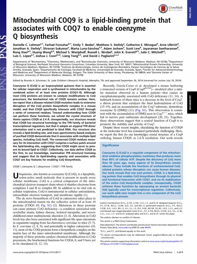
ResultsCoq9 Is Essential for the Stability of Mouse CoQ Biosynthesis Proteins.Mice homozygous for a truncated version of Coq9 (Coq9R239X)have decreased levels of Coq7 and accumulate the substrate forthe Coq7 reaction (Fig. S1) (14); however, the effect of thismutation on the other COQ proteins—and on mitochondrialand cellular proteins in general—has not been evaluated. InS. cerevisiae, complete loss of Coq9p destabilizes the CoQ complexand precludes CoQ production, whereas partial loss, aided byartificial stabilization of the complex, allows minimal CoQ pro-duction and accumulation of DMQ (2, 24–26). Here, we sought todetermine whether disruption of Coq9, via the Coq9R238X mutation,affected the stability of COQ proteins in mice, where the exis-tence of a CoQ biosynthetic complex has yet to be shown.To test whether steady-state levels of other proteins involved
in CoQ biosynthesis were decreased in the Coq9R239X mice, weperformed quantitative analysis of the proteomes from Coq9R239X
and wild-type mouse tissue. The proteomes from three tissues(heart, kidney, and cerebellum) were analyzed by liquid chro-matography (LC)-MS/MS using tandem mass tagging to mea-sure relative protein abundances (Fig. 1 A and B). Strikingly, ouranalysis shows that the steady-state levels of all detected mem-bers of the putative biosynthetic complex are specifically de-
creased in Coq9R239X hearts relative to other proteins (Fig. 1C).Interestingly, several other mitochondrial proteins changed sig-nificantly in abundance, perhaps suggesting their involvement inCoQ biosynthesis or in compensatory processes invoked duringCoQ deficiency (Fig. S1 and Dataset S1). For example, the levelof a mitochondrial sulfide-quinone oxidoreductase (Sqrdl) issignificantly decreased in Coq9R239X mice hearts. This result,along with reports of sulfide accumulation in CoQ-deficientyeast (27), indicates a connection between CoQ biosynthesisand mitochondrial hydrogen sulfide metabolism.A similar decrease in COQ proteins was observed in the
Coq9R239X kidneys and cerebellums (Fig. 1D and Fig. S1). No-tably, several COQ proteins (Coq6, Coq4, Adck3) were notdetected in our analysis of the cerebellum tissue, and Coq7 levelsyielded mixed results, resulting in no significant change (Fig. 1Dand Dataset S1). The lack of detection could indicate that theseproteins are not present in this tissue, suggesting that a CoQbiosynthetic complex might not be an obligate feature of CoQbiosynthesis in vertebrates. However, only the lack of Coq4 incerebellum is supported by recent global proteomics analyses(28, 29). The absence of these proteins in our analyses is mostlikely due to the stochastic nature of peptide sampling combinedwith the reduced likelihood of detecting low-abundance proteinswithin heterogeneous cell types from a tissue with lower mito-chondrial content than heart and kidney (30). Of all the COQpolypeptides that are detected and whose abundances aresignificantly changing, only Coq6 in the kidney deviates fromthe decreasing trend in Coq9R239X mice. This divergence couldreflect a tissue-specific regulatory feature of CoQ biosynthesisand CoQ biosynthetic complex formation. Overall, this analysissupports the existence of a CoQ biosynthetic complex in highereukaryotes and indicates that intact Coq9 is essential for the stabilityof other CoQ biosynthetic proteins.
COQ9 Crystal Structure Reveals TFR Structural Homology and a BoundPhospholipid. Little is known about the structure or biochemicalfunction(s) of COQ9, and thus it remains unclear how it can helpstabilize the CoQ complex or influence the COQ7 reaction. Togain insight into these features, we sought to solve the crystalstructure of a COQ9 ortholog. Ultimately, we were able to purifyand crystallize an N-terminally truncated (NΔ79) version of
A B
0 100 200 300Mitochondrial targeting sequence Mature Coq9
N CFull length:R239X:
Coq9 M. musculus
N C
aa
D
WT
Coq9R239X Quant.LC-MS/MS
Tissue Homogenate
Tag & MixSamples
SCXFrac.3x
3x
C
Heart
Kidney
Cerebellum
Fold
Δ L
og2(C
oq9R
239X
/WT)
-3
-2
-1
0
1
Coq3 Coq4 Coq5 Coq6 Coq7 Adck3 Coq9
1
2
0.01
10
-2 -1 1
Fold ΔLog2(Coq9R239X/WT)
-Log
10(p
-val
ue)
1
0.1
Coq9
Coq7 Coq4 Coq5
Adck3 Coq3
Coq6
Fig. 1. Coq9R239X mice have decreased steady-state levels of CoQ biosynthesis proteins. (A) Coq9R239X truncation scheme designed by García-Corzo et al. (11).(B) Experimental design for proteomic LC-MS/MS analysis of WT and Coq9R239X mice tissue (heart, kidney, cerebellum with n = 3 for each tissue). (C) Volcanoplot of protein abundance (•) changes between WT and Coq9R239X mouse heart [Fold Δ Log2(Coq9
R239X/WT) versus –Log10(P value)] with COQ proteins la-beled. (D) COQ protein abundance changes between WT and Coq9R239X in ( ) heart, (■) kidney, and ( ) cerebellum [Fold Δ Log2(Coq9
R239X/WT)] with errorbars showing standard deviation. See also Fig. S1.
2 of 9 | www.pnas.org/cgi/doi/10.1073/pnas.1413128111 Lohman et al.
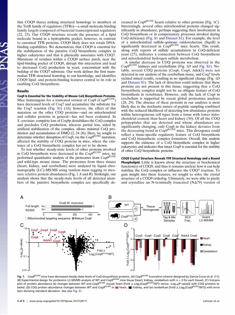
human COQ9 that diffracted at 2.4 Å. This truncation removes35 hydrophilic and poorly conserved residues downstream of thepredicted mitochondrial targeting sequence, thus leaving themajority (87%) of the mature protein intact (Fig. 2 A and B, andTable 1).Our crystal structure reveals that COQ9 is structurally ho-
mologous to members of the TFRs. By comparing the structureof COQ9 to all structures in the Protein Data Bank (PDB), wefound that the top 600 homologous proteins are all TFR orpredicted TFR family members (31). The COQ9 structure alignsto TFR family members with z scores of as high as 16.2; however,its sequence identity with these family members is quite low, withonly 14% sequence identity shared between the most similarstructures. TFR family members are generally bacterial tran-scriptional regulators that form homodimers and bind smallmolecule ligands and DNA. Members contain nine α-helices thatform two domains: a DNA-binding domain with a helix-turn-helix (HTH) motif (helices α1–3) and a ligand-binding domainwith a central triangle of helices (helices α4–9) (22). COQ9shares these structural features, but also has a 10th α-helix at itsC terminus (Fig. 2 C and D). This helix is ordered in only one ofthe two COQ9 molecules in the asymmetric unit, and the loopconnecting it to helix 9 is disordered. As such, this 10th helixmight assume a different conformation in vivo. Additionally,although the dimers observed in TFR family members form afour-helix bundle consisting of the C-terminal two helices (α8–9)from each monomer (22), the corresponding helices do not in-teract in the COQ9 dimer, which instead has the single 10th helixat the dimer interface.The two COQ9 molecules in the asymmetric unit form a di-
mer, in agreement with static light-scattering analysis of recom-binant COQ9 in solution (Fig. S2), with a total of 1,250 Å2 of thesurface area of each monomer buried in the dimer interface.This interface consists of mostly hydrophobic residues, includingmany noninteracting residues and a single hydrogen bond con-tained in the 10th helix. Intriguingly, COQ9 crystallized with
a bound lipid at the dimer interface, consistent with the smallmolecule-binding capabilities of the TFR proteins. The partialelectron density suggests that this phospholipid has at least 10carbons in each acyl group, which extend into a predominantlyhydrophobic pocket comprised from both monomers (Fig. 3 A–C). This pocket aligns well with that of a protein in the FadRsubclass of TFRs that cocrystallized with its ligand, acyl CoA(Fig. 2 C and D) (32). The COQ9 pocket is a 20-Å–deep cavityenriched in aliphatic, neutral, and aromatic amino acids. Thereare also two charged residues (R189 and E255) residing at thebottom of the pocket, which may play a role in ligand recogni-tion. Although the full acyl tails of the COQ9 ligand were notresolved, there is sufficient space and unassigned electron densityto suggest that COQ9 has the capacity to bind a larger hydrophobicmolecule than modeled. Although the bound lipid may not repre-sent a bona fide endogenous ligand, the pocket properties suggestthat a hydrophobic ligand for COQ9 is likely (Fig. 3B).To attempt to determine the identity of the cocrystallized
lipid, we performed LC-MS/MS on lipids extracted from re-combinant COQ9 expressed in both Escherichia coli and wheatgerm extract. We detected phosphatidylglycerol and phosphati-dylethanolamine with various acyl chain lengths and saturationsas the most abundant phospholipid species copurifying withCOQ9 made from E. coli (Fig. 3D and Fig. S2). However, thesephospholipids are highly abundant in E. coli and the calculatedlipid/protein stoichiometries suggest that the COQ9/phospho-lipid stoichiometries exceed the 2:0.5 ratio expected for a COQ9dimer with 50% lipid occupancy (Fig. S2). Overall, these data,along with the lack of clear electron density for the head group,suggest that the cocrystallizing lipid is a mixture of phospho-lipid species that help to solubilize the hydrophobic regionsof COQ9 when overexpressed recombinantly. Interestingly,in each case, we also detected the host organism’s version ofCoQ—CoQ8 in E. coli and CoQ9 in wheat germ extract—copurifying with COQ9, albeit in lower stoichiometric ratios(Fig. 3D and Fig. S2).
90°
COQ9 dimerN
N
N
C
BA
aa0 100 200 300Mitochondrial targeting sequence Mature COQ9
N CFull length:Crystallized: N C
COQ9 H. sapiens
C COQ9 monomer
FadR monomer
Structural overlay
NN
C
C
Small molecule binding domain
Helix-turn-helix domain
D
aa0 100 200 300Helix-turn-helix domain Small molecule
binding domain
N CFadR:COQ9:
TFR structural homology
N C
α10
α10
α10
Fig. 2. COQ9 crystal structure reveals structural homology to TFR family members. (A) Crystallization construct of COQ9. (B) Crystal structure of the COQ9dimer with the monomers colored in blue and green, the cavity highlighted, and the cocrystallizing lipid shown. (C) COQ9 structural homology sequencescheme with FadR, a TFR family member (PDB 3anp) (32). (D) Individual and structural overlay of COQ9 (blue) and FadR (gray) monomers with their respectivecavities and cocrystallizing ligands.
Lohman et al. PNAS Early Edition | 3 of 9
BIOCH
EMISTR
YPN
ASPL
US
Despite the similarity of COQ9’s ligand binding domain tothose of other TFR proteins, its HTH motif is quite distinct.In particular, the COQ9 HTH largely lacks the positive chargesrequired for DNA binding by other TFR proteins, includingFadR, suggesting that human COQ9 is unlikely to bind DNAitself. To assess this possibility more formally, we applied theDNA binding site identifier (DBSI) (33), which identifies aminoacids with a propensity to bind DNA and chemically relatedmolecules such as heparin (34). Calculations performed on theCOQ9 monomer failed to identify the COQ9 HTH domain asa DNA-binding site (Fig. 4). In contrast, DBSI analyses correctlyidentified the known DNA binding sites of FadR (Fig. 4 and Fig.S3). Additionally, the position of the HTH motifs within theCOQ9 dimer differs from other TFR proteins. TFR dimers aretypically arranged such that the DNA-binding domains are ad-jacent and poised to interact with palindromic sequences (22).The dimer orientation of COQ9 within the crystal structure,however, places the HTH motifs on opposite sides of the dimer.Importantly, additional DBSI analyses on FadR and COQ9dimers in their crystallographic orientations, along with a theo-retical model of COQ9 threaded onto the FadR dimer usingMODELLER (35), also failed to identify the COQ9 HTH do-main as a DNA-binding site (Fig. S3). Overall, although it isdifficult to prove the complete lack of DNA-binding capacity,our analyses suggest that COQ9 has shed the DNA-bindingfunction of typical TFR proteins.
COQ9 Conserved Residues and Functional Domains Are Distinct fromArchetypal TFR Proteins. The crystal structure reveals that COQ9retains the two main structural domains of TFR family proteins:a HTH and a small molecule-binding domain. We reasoned thatregions within these domains that are highly conserved in pri-mary sequence among COQ9 homologs are likely to be importantfor COQ9 function. We identified regions of high conservationusing the Consurf server and mapped the overall pattern of con-servation onto the COQ9 crystal structure (Fig. 5A and Fig. S4)(36–38). Two regions are highlighted: one within the HTH motifthat is typically involved in DNA binding in TFR family members(23), and one within the small molecule-binding domain, which isthe most highly conserved region of COQ9 (Fig. 5A).To begin identifying regions within these domains that are
important for the role of COQ9 in CoQ biosynthesis, we con-ducted a yeast genetic complementation assay using single pointmutations in coq9 from S. cerevisiae. We designed conservativeand disruptive point mutations for 18 Coq9p residues that wehypothesized to be functionally important based on their loca-tion within the structure, their evolutionary conservation, and onthe established importance of corresponding residues from otherTFR proteins. Previous work has shown that Δcoq9 yeast have aninability to grow on nonfermentable carbon sources, which canbe rescued by ectopic expression of wild-type coq9 (25). Here, wetested the functionality of the yeast Coq9p mutants by measuringtheir capacity to rescue this Δcoq9 growth phenotype (Fig. 5Band Fig. S5). For instance, the expression of WT or S59A Coq9prescues Coq9p function, whereas expression of other mutantsresulted in midrange (e.g., W188D) or severely hindered (e.g.,W188K) growth phenotypes.The results of this mutant screen reveal specific regions of
sequence that are likely important for the biological functionof Coq9p (Fig. 5B). First, multiple point mutations within thehydrophobic cavity of the small molecule-binding domain impairrescue of the Δcoq9 phenotype. This result suggests that bindinga hydrophobic molecule is indeed important for Coq9p function.Second, a portion of the overall small molecule binding domain,which we term the “COQ9 surface patch (CSP),” is likewisevery sensitive to point mutations. This region, which is solventexposed in the crystal structure, is also highly conserved amongCOQ9 homologs, suggesting that it is a particularly importantfunctional area of COQ9. In contrast, point mutations in resi-dues throughout the N-terminal HTH motif that structurallyalign with the DNA-binding domain of TFR family members donot appear to disrupt Coq9p function. Consistently, the residuesthat align structurally with DNA contacts in TFR proteins arenot particularly well conserved between COQ9 orthologs (Fig.S4). Coupled with our DNA-binding prediction analyses, theseresults lend further weight to our hypothesis that DNA binding isnot a key feature of COQ9 function.
COQ9 Physically and Functionally Interacts with COQ7. Given therequirement of full-length Coq9 for the stability of the mouseCoQ complex shown in this work, we hypothesized that thehighly conserved COQ9 surface patch may be crucial for a pro-tein–protein interaction with another member of the CoQ bio-synthetic complex. Because the mice harboring the truncatedCoq9 gene accumulate the substrate for Coq7 (11), and as Coq7pis reported to coprecipitate with Coq9p when isolated from yeast(24), we chose to test whether there is a protein–protein inter-action between human COQ9 and COQ7. To do so, we coex-pressed these two proteins using a cell-free wheat germ extractprotein expression system. Physical association between COQ9and COQ7 was assayed by coexpressing strep-tag II (SII)-taggedCOQ9 (SII-NΔ45-COQ9) and six-histidine (6H)-tagged COQ7(6H-NΔ38-COQ7), purifying the proteins by their tag affinity,and visualizing copurifying proteins by SDS/PAGE. Our resultsshow that COQ7 copurified with COQ9, and vice versa, indicating
Table 1. Data collection and refinement statistics
Crystal parametersSpace group P21Cell dimensions
a, b, c, Å 38.340, 97.628, 64.109α, β, γ, ° 90, 95.766, 90
Matthews coefficient, Å3/Da 2.2Data quality
Wavelength, Å 0.97937Resolution, Å 48.8–2.4 (2.49–2.4)Rmerge, %* 8.9 (51.4)No. of unique reflections 35,868No. of reflections in Rfree set† 3,587Mean redundancy 3.6 (3.7)Completeness, overall, % 99.8 (100)Mean I/σI 15.8 (2.5)
Refinement statisticsRfree, % 17.9Rwork, % 24.6Completeness, % 99.38 (87.7)
Model qualityRmsd bond lengths, Å 0.008Rmsd bond angles, ° 1.1Ramachandran plot, %
Most favored 96.1Additional allowed 3.9
B factor, Å2 41.7Model contents
Protomers in ASU 2Protein residues A: 95–283
B: 93–233, 237–284, 293–311Ligand 1 PEFNo. atoms 3,263
Protein 3,168Ligand 34
Water molecules 61
*Values in parentheses are for the highest resolution shell.†10% of the reflections were selected for free R calculation.
4 of 9 | www.pnas.org/cgi/doi/10.1073/pnas.1413128111 Lohman et al.

that human COQ9 and COQ7 can form a complex in vitro (Fig. 6Aand Fig. S6).To define the regions of COQ9 that contribute to the human
COQ9–COQ7 interaction, we attempted to copurify COQ7 withpoint mutants of COQ9. Formation of the COQ9–COQ7 com-plex was assayed by cell-free coexpression of SII-NΔ45-COQ9and 6H-NΔ38-COQ7, followed by sequential StrepTactin andnickel affinity purifications. The large excess of COQ9 relative toCOQ7 during the first round of purification suggests that non-complexed COQ9 was purified along with the COQ9–COQ7complex (Fig. 6B and Fig. S6). When the product of the firstpurification was subjected to a second purification using the tagon COQ7, we were able to separate the complex from freeCOQ9 and visualize the amount of COQ9–COQ7 protein complexformed with each COQ9 mutant (Fig. 6C and Fig. S6). We used therelative tryptophan fluorescence of COQ9 and COQ7 after two
rounds of purification to quantify whether the COQ9–COQ7complex was intact, diminished, or absent in the case of eachCOQ9 mutant (Fig. 6D). These analyses identify a number ofCOQ9 residues that perturb the COQ9–COQ7 interaction(Fig. 6 B–D and Fig. S6). Several of the corresponding humanCOQ9 mutants that disrupted the human COQ9–COQ7 com-plex in vitro also disrupted S. cerevisiae Coq9p function in vivo.In general, these mutations occurred primarily in two regions—within the hydrophobic cavity of COQ9 and along the COQ9surface patch.The mutations identified above can be disruptive because they
are either necessary for the COQ9–COQ7 interaction or becausethey result in general destabilization of COQ9. To distinguishbetween these possibilities, we purified mutant versions of re-combinant human NΔ79-COQ9 from E. coli. Each COQ9 mu-tant was tested for thermal stability using differential scanning
DB
A
R244
N154
R189
S216
S218
T221
T253
E255
Y241
V150
L193 W200L204
L207M208
I213L220
L219L217
M223
A245A248
A249
Y251
L256L301
W240
I250
Dimer interface
C
Co-purifying lipids
Wheat germE. coli
1
10
102
103
104
PCPEPGCoQ
105
COQ9
pmol
sFig. 3. COQ9 has the ability to bind lipid. (A) Open-book view of the COQ9 dimer interface with the cocrystallizing lipid shown at the lipid-binding site. (B)Electrostatic surface (positive in blue and negative in red) of the COQ9 monomer cavity showing residues on the lipid-binding surface, with hydrophobicresidues in black. (C) Electron density map of the cocrystallizing, partially resolved phospholipid, phosphatidic acid (10:0/10:0). (D) Copurifying lipids (inpicomoles) with recombinant COQ9 purified from (■) E. coli or ( ) wheat germ extract (CoQ, coenzyme Q; PC, phosphatidylcholine; PE, phosphatidyletha-nolamine; PG, phosphatidylglycerol) with the picomolar amount of recombinant COQ9 from E. coli indicated (red dashed line). See also Fig. S2.
COQ9 monomer
FadR monomer
DNA-binding site identifier predictionStrongWeak
Small moleculebinding domain
Helix-turn-helixdomain
Helix-turn-helix domainα3α1 α2 α4
Fig. 4. COQ9 HTH domain is not predicted to bind DNA. Residue predictions made by DBSI (33) for residues within the HTH domain in COQ9 and FadRmonomers are shown and colored on a scale from weak (blue) to strong (red) likelihood of interacting with DNA. See also Fig. S3.
Lohman et al. PNAS Early Edition | 5 of 9
BIOCH
EMISTR
YPN
ASPL
US

fluorimetry (39). A large decrease in the melting temperature(Tm) of the COQ9 mutants relative to wild-type, or the absenceof an observable melt curve, would indicate that the mutant islikely unfolded or drastically misfolded. These analyses revealedthat multiple mutations in or around the COQ9 lipid-bindingcavity, such as L190E, M227E, and L256K, resulted in severelyless thermally stable (ΔTm > 5 °C) protein, suggesting that thedisrupted COQ9–COQ7 interaction for these mutants is at leastpartially due to improperly folded COQ9 (Fig. 6 D–F). However,several mutants along the COQ9 surface patch, includingW240K, W240D, Y241K, and D237K, have melt temperaturessimilar to WT (ΔTm < 5 °C) and yet still severely disrupt theCOQ9–COQ7 complex (Fig. 6 D–F). These results suggest thatthe COQ9 surface patch is an important functional domain be-cause of its contribution to the COQ9–COQ7 protein interaction.Because the substrate for Coq7 (DMQ) accumulates in the
Coq9R239X mice, we hypothesized that the protein–protein in-teraction identified between COQ9 and COQ7 affects the effi-ciency of the Coq7 reaction. To test whether the folded COQ9mutants that disrupt the human COQ9–COQ7 interaction invitro are sufficient to diminish the yeast Coq7p reaction, wemonitored CoQ6 production in Δcoq9 yeast containing theserespective Coq9p mutants. Specifically, we measured the levelsof DMQ6 and CoQ6 produced by actively respiring yeast withLC-MS/MS, and used the DMQ6/(DMQ6 + CoQ6) ratio asa proxy for Coq7p activity. As expected, the CoQ6 levels corre-sponded well with the respiratory competency of the yeast strainsdescribed above (Fig. S7). However, in the three strains har-boring a Coq9p variant expected to disrupt the Coq9p–Coq7p
interaction, the Coq7p reaction was less efficient than the strainrescued by WT Coq9p (Fig. 6G and Fig. S7). No DMQ6 or CoQ6production was observed in the control (empty vector) strainthat completely lacked Coq9p; this is consistent with previousreports and is likely due to complete disruption of the entireCoQ biosynthetic complex (25, 26). Together, these resultssuggest that the conserved residues on the COQ9 surfacepatch are essential for its physical and functional interactionwith COQ7.
DiscussionThe discovery of CoQ completed the final missing piece ofthe electron transport chain (ETC) machinery postulated byWarburg and Keilin (13). Following its isolation by Crane andGreen in the 1950s, CoQ was established as a requisite gatewayfor electrons passing from CI and CII to CIII in the ETC, and hasalso been linked to other key antioxidant and electron transferfunctions in membranes throughout the cell (40, 41). Decreasedlevels of CoQ lead to CoQ deficiency, which has heterogeneousclinical manifestations with presentations ranging from adult-onset isolated myopathy to fatal infantile multisystemic disease(4). CoQ deficiency is also common in mitochondrial diseasepatients, and, although CoQ supplementation has been popular,the success of this treatment is variable (42, 43).Despite the vital role for CoQ in human health and disease,
many core aspects of its endogenous biosynthesis in eukaryotesremain obscure, thereby preventing pathway manipulation as atherapeutic option. In particular, the functions of three proteinsremain almost completely uncharacterized, despite being essential
A
HTHdomain
Small moleculebinding domain
H. sapiensM. musculusG. gallusD. rerioD. melanogasterA. thalianaS. cerevisiae
959093
1081058736
S E E Q L Q H R I L T A A L E F - V P A H G W T A E A I A E G A Q S L G L S S A A A S M FS E E Q L Q H R I L T A A L E F - V P A H G W T A E A I A E G A Q S L G L S S A A A S M FS E E Q L Q H R I L T A A L E F - V P E H G W T A E A I A E G A K T L G L S A A A A G M FT E E Q L Q A R I L T T A L E F - V P Q H G W T V E A I A A G A E T L G L S A A S T G M FK V D A I R A K I L D A A L Q H - V P Q Q G W T R Q A I I L G A E E S G Y P S V V H G M FE F Q E E Q A R V L S A S L R H - V P R L G W T E E A M M A G S R D V G V S P S I V G S FG K E S P Q Y K V L S L A L Q K F V P E H G F S E R S I V E S L N E L G Y P S S M I S S I
α3α1 α2HTH domain
* * ** * *
H. sapiensM. musculusG. gallusD. rerioD. melanogasterA. thalianaS. cerevisiae
225220223238232211172
D D M W H Y A G D Q - S T D F N W Y T R R A M LD D M W H Y A G D Q - S T D F N W Y T R R A V LD D I W H Y A G D Q - S T D F N W Y T R R A V LD D I W Y Y A G D R - S T D V N W Y T R R A A LD D I C Y Y S G D R - S V D F G W Y T R R V G LD E I W H A V G D G - A S D L D W Y V K R T I LD D M I Y F S N E K D H H D S A W Y A K R L A V
α7 α8COQ9 surface patch
****B HTH domain Small molecule binding domain
CavityCOQ9 surface patch
D185AD185KD185N
W188DW188KY189AY189DY189FY189KR192AR192ER192L
W188A
+
+++
+
+
+/-
+/-
-
-
++
+/-
Mutant PhenotypeL135EL135KM149IM174AM174EM174IM174KY199AY199FL204AL204DL204K
-
++
+
++
++
+/-
+/-
-
Mutant Phenotype
+
S59AS59DS59KE65AE65KS74AS75KI77KI80D
++++++
+++
Mutant PhenotypeT119AT119ET119MD209AD209KD209NT245AT245DT245M
++++++
+++
Mutant PhenotypePhenotypeWT
NoneW188D
++/-
Mutant
Phenotype key
-
Fig. 5. COQ9 has a conserved surface patch with key functional residues. (A) COQ9 residue conservation using 104 homologs mapped onto the monomerstructure with the most conserved residues shown in increasing shades of blue. The HTH domain and COQ9 surface patch are highlighted in a subset sequencealignment with the most conserved residues colored in increasing shades of blue and invariant residues boxed and in yellow. Residues tested in the yeastgenetic screen are indicated (*). (B) Coq9p point mutants, assayed by S. cerevisiae genetic complementation, with respiration competent (+), diminishedcompetence (+/−), or incompetent (−) phenotypes. See also Figs. S4 and S5.
6 of 9 | www.pnas.org/cgi/doi/10.1073/pnas.1413128111 Lohman et al.

for CoQ production: COQ4, COQ8 (ADCK3), and COQ9. Here,we describe the structure of COQ9 along with its physical andfunctional interaction with COQ7.We discovered that human COQ9 is structurally homologous
to members of the TFRs. The TFRs are a bacterial family ofproteins containing two distinct domains: a DNA-binding do-main and a small molecule-binding domain. Characterized TFRfamily members bind a variety of ligands—from small moleculessuch as tetracycline to lipids such as stearoyl-CoA (22, 23, 32).This binding event affects the TFRs’ ability to bind DNA and, inturn, affects transcription of target genes (22, 23, 32). Notably,COQ9 is the first reported nonbacterial protein to display the
TFR fold (rcsb.org as of August 27, 2014). We suggest that, incontrast to its structural counterparts, COQ9 is unlikely to act asa transcription factor. Although COQ9 exhibits the HTH motiffound in the DNA-binding domain of TFRs, DBSI calculationssuggest that this domain, which is correctly predicted in otherTFRs, is absent in COQ9. Furthermore, there is a lack of posi-tive charge in this region, and residues that structurally map toresidues traditionally involved in DNA recognition are not highlyconserved in COQ9. Additionally, Coq9p function is not im-paired by point mutations within this region. These observationssuggest that the TFR structural fold has been repurposed inCOQ9 for a function other than transcriptional regulation.
ΔTm
(ºC
) CO
Q9
(Tm
, MT - T
m, W
T)
-15
-10
-5
5WT L256K
L190EM227E
W240KW240D
Y241KD237K
0* *
A
D
F
G
SII Purification
COQ9
COQ7
37
2520
WT
S131KL1
90EM22
7E
D237K
W24
0D
W24
0K
Y241DY24
1KL2
56KD26
1KkDa La
dder
kDaCOQ9
COQ7
37
2520
WT
S131KL1
90EM22
7E
D237K
W24
0D
W24
0K
Y241DY24
1KL2
56KD26
1KLa
dder
6H Purification
0
20
40
60
80
100
WT
S131KL1
90E
M227E
D237K
W24
0D
W24
0K
Y241DY24
1KL2
56K
D261K
% In
tera
ctio
n C
OQ
9-C
OQ
7
B
C
Purification: SII SII SII6H 6H 6H6H SIISII-COQ9: + +++++ - -H6-COQ7: +-- ++ +++
COQ9
COQ7
37 kDa
2520
Ladd
er
Y241
M227M227
L190L190
D261
Helix-turn-helix domain
Small molecule binding domain
Cavity
COQ9 surface patch
S131
W240
D237
L256L256
0
0.1
0.2
0.3
0.4
0.5
WT D185K W188D Y189K Vector
DM
Q6 / (
DM
Q6+
CoQ
6)
* **
E
Fig. 6. COQ9 physically and functionally associates with COQ7. (A) Tagged SII-COQ9 and H6-COQ7 cotranslated in wheat germ extract, purified using the SIIor 6H affinity tag, or purified sequentially by both tags as indicated by arrows. (B) Point mutants of SII-COQ9 coexpressed with 6H-COQ7 and purified usingthe SII affinity tag on SII-COQ9. (C) The elution from the SII purification purified using the 6H affinity tag on 6H-COQ7. (D) Percentage of COQ9–COQ7complex formed with each COQ9 point mutant relative to WT and measured by relative tryptophan fluorescence from the sequential purification shown in C.(E) Melting temperature of COQ9NΔ79 point mutants compared with WT [ΔTm,°C (COQ9MT
– COQ9WT)] measured by differential scanning fluorimetry. Errorbars show standard deviation (n = 3) and the absence of an observable melt curve is indicated (*). (F) COQ9 monomer with residues assayed for interactionwith COQ7 colored by whether point mutants maintain the interaction (green), disrupt the interaction (red), or disrupt the interaction and the meltingtemperature of COQ9 (yellow). (G) Coq7p reaction efficiency measured by LC-MS/MS detection of DMQ6/(DMQ6 + CoQ6) in Δcoq9 strains harboringCoq9p point mutants, with error bars showing 95% confidence intervals and statistical significance using a two-tailed Student’s t-test annotated by *P value <0.001 (n = 6). See also Figs. S6 and S7.
Lohman et al. PNAS Early Edition | 7 of 9
BIOCH
EMISTR
YPN
ASPL
US
A second function of the TFR fold is to enable binding tosmall molecules. In addition to having a small molecule-bindingdomain with a predominately hydrophobic cavity, the COQ9crystal structure reveals a bound phospholipid. However, be-cause the recombinant COQ9 was not purified from its native,mitochondrial location, we suspect that these abundant cop-urifying and cocrystallizing phospholipids might not be theendogenous COQ9 ligands. Although we likely have not yetidentified a bona fide endogenous ligand for COQ9, its structuralhomology, along with its cocrystallizing lipid, suggest that COQ9binds lipid as part of its role in CoQ biosynthesis. Because COQ9is a member of the CoQ complex, and because CoQ and CoQintermediates are thought to be part of the complex, it is temptingto speculate that COQ9 binds either CoQ or a CoQ precursor(2, 8). Unambiguous identification of an endogenous ligand forCOQ9 will be challenging, because this protein exists natively inthe CoQ biosynthetic protein complex and the presence of othermembers may affect the protein–ligand interaction (2, 24). Thisis especially true if the ligand is an intermediate in the CoQbiosynthesis pathway, as might be expected in light of theCoq7 reaction deficit in Coq9R239X mice. Regardless, the in-teraction of COQ9 with a CoQ precursor would be consistentwith a previous report of a CoQ species being part of the matureCoQ complex (8, 17, 44) and would present a straightforwardmodel for how COQ9 might functionally interact with otherCOQ proteins.In addition to identifying an unexpected lipid-binding domain
in COQ9, we show that human COQ9 engages in a protein–protein interaction with COQ7. COQ7 catalyzes the penultimatestep in CoQ biosynthesis—the hydroxylation of demethoxy CoQ(15, 16, 18, 19, 45). We identify a highly conserved CSP that isimportant for maintaining the interaction with COQ7. Further-more, we show that mutations in the CSP cause a decrease in theCoq7p reaction efficiency in yeast. A truncation of COQ9, ob-served first in a human patient with CoQ deficiency and thenmodeled in mice, truncates this CSP as well as a major portion ofthe cavity, presumably precluding lipid binding (Fig. S7). In bothmice and humans, the COQ7 reaction is impeded and the lateintermediate demethoxy CoQ accumulates (11, 14). Together,these results strongly suggest that the CSP is crucial for both thephysical and functional interaction with COQ7. Furthermore,the vicinity of the CSP to the cocrystallizing lipid and to thehydrophobic cavity indicates that the COQ7 interaction and thelipid-binding capacity of COQ9 are related.Our work might help resolve certain unexplained aspects of
CoQ biosynthesis. For instance, it is known that, due to its hy-drophobicity, mature CoQ resides within the bilayer interface(46). Presumably, the precursors to CoQ, which have similarchemical properties, would need to be sequestered out of themembrane while being modified by other COQ proteins. GivenCOQ9’s ability to bind lipid, and its interaction with at leastone CoQ-modifying enzyme (COQ7), we speculate that COQ9interacts with and presents the immature CoQ species to otherproteins in the biosynthetic complex before releasing the finalproduct back into the membrane. Another possibility for COQ9function is that it catalyzes one of the missing reaction steps inCoQ biosynthesis; however, this possibility seems less likely formultiple reasons. First, TFR proteins are not known to possesscatalytic functions. Second, one would expect the active site to bein the most highly conserved region of COQ9, the CSP; yet,when this region is truncated in mice, low levels of CoQ are stillproduced. Collectively, the work presented here provides thefirst biochemical and structural analysis of COQ9, to ourknowledge, and provides a foundation for future work aimedat establishing its endogenous ligand(s) and elucidating itsrelationship with the other COQ proteins.
Materials and MethodsFull experimental procedures and associated references are available in SIMaterials and Methods. Detailed methods for cloning, protein purification,differential scanning fluorimetry, yeast genetic complementation andhomology modeling, as well as full methods on LC-MS/MS proteomic andlipidomic analysis, crystallization and structure determination, and compu-tational modeling, can be found in SI Materials and Methods.
Proteomics Analysis of Mice Tissue by LC-MS/MS. Mice experiments wereperformed according to a protocol approved by the Institutional Animal Careand Use Committee of the University of Granada (procedure CEEA 2010-275)and were in accordance with the European Convention for the Protection ofVertebrate Animals used for Experimental and Other Scientific Purposes(CETS # 123) and the Spanish laws (32/2007 and R.D. 1201/2005). Protein wasextracted by probe sonification and quantified by bicinchoninic acid assay.Proteins were reduced, alkylated, and digested with trypsin. The resultingpeptides were labeled with six-plex tandem mass tag (TMT) isobaric labels,mixed in equal amounts by mass, and fractionated by strong cation ex-change chromatography. Protein fractions were analyzed by nano reverse-phase liquid chromatography coupled to an Orbitrap Elite (Thermo). Spectrawere searched using the open mass spectrometry search algorithm (47), andresults were filtered to 1% false discovery rate at the unique peptide levelusing the COMPASS software suite. TMT quantification and protein group-ing were performed according to previously reported rules (48).
Lipidomics Analysis by LC-MS/MS. Lipids from either mouse tissue, purifiedrecombinant COQ9, or Δcoq9 yeast harboring Coq9p constructs wereextracted and submitted to discovery-type targeted lipidomics analyses onan Ascentis Express C18 column held at 35 °C (150 mm × 2.1 mm × 2.7 μmparticle size; Supelco) using an Accela LC Pump (500 μL/min flow rate;Thermo Scientific). Full extraction methods, LC-MS/MS data collection, andanalysis methods are available in SI Materials and Methods.
Crystallization, Data Collection, and Structure Determination. The selenome-thionine-labeled human COQ9NΔ79 was crystallized by microbatch method at18 °C. Crystals of COQ9NΔ79 were cryoprotected by 25% (vol/vol) ethyleneglycol and flash-frozen in liquid N2 for data collection at 100 K. The crystalsof human COQ9NΔ79 belong to space group P21 with cell parameters of a =38.3 Å, b = 97.6 Å, and c = 64.1 Å and β = 95.8°. There are two molecules ofCOQ9NΔ79 in the crystallographic asymmetric unit of the protein. A single-wavelength anomalous diffraction dataset to 2.4-Å resolution was collectedat the peak absorption wavelength of selenium at the X4A beamline of theNational Synchrotron Light Source. The diffraction images were processedwith the HKL package (49), and 17 of 22 possible selenium sites were locatedwith the program Shelx (50). SOLVE/RESOLVE (51) was used for phasing thereflections and automated model building, which correctly placed 49%of the residues with side chains in each protomer of ASU. The entiremodel was built with the program XtalView (52) and refined by PHENIX(53). Noncrystallographic symmetry restraint was applied for most stagesof the refinement of the structure, but it was released at the final re-finement stage.
Computational Modeling of DNA-Binding Residues. DBSI (33) is a structure-based model that accurately predicts binding sites for DNA on the 3D surfaceof a protein. To generate electrostatic features within DBSI, solutions to thePoisson–Boltzmann equation were generated using PBEQ (54) via theCHARMM-GUI (55) website (www.charmm-gui.org). The CHARMM-GUIparameters that differ from default (33) are as follows: dielectric constantfor the protein interior (2.0); coarse finite-difference grid spacing (1.0 Å);fine finite-difference grid spacing (0.5 Å). The calculations were repeatedindependently for these structures in both monomeric and dimeric forms.The DBSI program was then run with these structures and their respectiveelectrostatic potential maps as inputs.
ACKNOWLEDGMENTS. We thank the following members of the Universityof Wisconsin–Madison-based Mitochondrial Protein Partnership (MPP) andthe Northeast Structural Genomics (NeSG) Consortium for technical andmanagerial assistance: Fabian Suchy, David Aceti, John Primm, Brian Fox,and John Markley (MPP), and Thomas Acton and Gaetano Montelione (NeSG).We thank Andrew Reidenbach for technical assistance and critical reading ofthe manuscript, as well as Aseem Ansari for helpful suggestions. This work wassupported by a Searle Scholars Award, a Shaw Scientist Award, and NIH GrantU01GM094622 (to D.J.P.), NIH Grant U54GM094597 (to L.T.), National Instituteof General Medical Sciences Biotechnology Training Grant 5T32GM08349 (toD.C.L.), NIH Ruth L. Kirschstein National Research Service Award F30AG043282
8 of 9 | www.pnas.org/cgi/doi/10.1073/pnas.1413128111 Lohman et al.
(to J.A.S.), National Library of Medicine (NLM) Training Grant to the Compu-tation and Informatics in Biology and Medicine Training Program NLM
T15LM007359 (to C.E.M.), and by the Ramón y Cajal Programme RYC-2011-07643 and the Junta de Andalucía P10-CTS-6133 (to L.C.L. and M.L.-S.).
1. Turunen M, Olsson J, Dallner G (2004) Metabolism and function of coenzyme Q.Biochim Biophys Acta 1660(1-2):171–199.
2. He CH, Xie LX, Allan CM, Tran UC, Clarke CF (2014) Coenzyme Q supplementation orover-expression of the yeast Coq8 putative kinase stabilizes multi-subunit Coq poly-peptide complexes in yeast coq null mutants. Biochim Biophys Acta 1841(4):630–644.
3. DiMauro S, Quinzii CM, Hirano M (2007) Mutations in coenzyme Q10 biosyntheticgenes. J Clin Invest 117(3):587–589.
4. Emmanuele V, et al. (2012) Heterogeneity of coenzyme Q10 deficiency: Patient studyand literature review. Arch Neurol 69(8):978–983.
5. Lakowski B, Hekimi S (1996) Determination of life-span in Caenorhabditis elegans byfour clock genes. Science 272(5264):1010–1013.
6. Larsen PL, Clarke CF (2002) Extension of life-span in Caenorhabditis elegans by a dietlacking coenzyme Q. Science 295(5552):120–123.
7. Liu X, et al. (2005) Evolutionary conservation of the clk-1-dependent mechanism oflongevity: Loss of mclk1 increases cellular fitness and lifespan in mice. Genes Dev19(20):2424–2434.
8. Tran UC, Clarke CF (2007) Endogenous synthesis of coenzyme Q in eukaryotes.Mitochondrion 7(Suppl):S62–S71.
9. González-Mariscal I, et al. (2014) Regulation of coenzyme Q biosynthesis in yeast:A new complex in the block. IUBMB Life 66(2):63–70.
10. Ashraf S, et al. (2013) ADCK4 mutations promote steroid-resistant nephrotic syn-drome through CoQ10 biosynthesis disruption. J Clin Invest 123(12):5179–5189.
11. García-Corzo L, et al. (2013) Dysfunctional Coq9 protein causes predominant ence-phalomyopathy associated with CoQ deficiency. Hum Mol Genet 22(6):1233–1248.
12. Wang Y, Hekimi S (2013) Molecular genetics of ubiquinone biosynthesis in animals.Crit Rev Biochem Mol Biol 48(1):69–88.
13. Pagliarini DJ, Rutter J (2013) Hallmarks of a new era in mitochondrial biochemistry.Genes Dev 27(24):2615–2627.
14. Duncan AJ, et al. (2009) A nonsense mutation in COQ9 causes autosomal-recessiveneonatal-onset primary coenzyme Q10 deficiency: A potentially treatable form ofmitochondrial disease. Am J Hum Genet 84(5):558–566.
15. Marbois BN, Clarke CF (1996) The COQ7 gene encodes a protein in Saccharomycescerevisiae necessary for ubiquinone biosynthesis. J Biol Chem 271(6):2995–3004.
16. Stenmark P, et al. (2001) A new member of the family of di-iron carboxylate proteins.Coq7 (clk-1), a membrane-bound hydroxylase involved in ubiquinone biosynthesis.J Biol Chem 276(36):33297–33300.
17. Tran UC, et al. (2006) Complementation of Saccharomyces cerevisiae coq7 mutantsby mitochondrial targeting of the Escherichia coli UbiF polypeptide: Two functionsof yeast Coq7 polypeptide in coenzyme Q biosynthesis. J Biol Chem 281(24):16401–16409.
18. Behan RK, Lippard SJ (2010) The aging-associated enzyme CLK-1 is a member of thecarboxylate-bridged diiron family of proteins. Biochemistry 49(45):9679–9681.
19. Lu TT, Lee SJ, Apfel UP, Lippard SJ (2013) Aging-associated enzyme human clock-1:Substrate-mediated reduction of the diiron center for 5-demethoxyubiquinone hy-droxylation. Biochemistry 52(13):2236–2244.
20. Nakai D, et al. (2001) Mouse homologue of coq7/clk-1, longevity gene in Caeno-rhabditis elegans, is essential for coenzyme Q synthesis, maintenance of mitochon-drial integrity, and neurogenesis. Biochem Biophys Res Commun 289(2):463–471.
21. Levavasseur F, et al. (2001) Ubiquinone is necessary for mouse embryonic de-velopment but is not essential for mitochondrial respiration. J Biol Chem 276(49):46160–46164.
22. Cuthbertson L, Nodwell JR (2013) The TetR family of regulators. Microbiol Mol BiolRev 77(3):440–475.
23. Ramos JL, et al. (2005) The TetR family of transcriptional repressors. Microbiol MolBiol Rev 69(2):326–356.
24. Hsieh EJ, et al. (2007) Saccharomyces cerevisiae Coq9 polypeptide is a subunit ofthe mitochondrial coenzyme Q biosynthetic complex. Arch Biochem Biophys 463(1):19–26.
25. Johnson A, et al. (2005) COQ9, a new gene required for the biosynthesis of coenzymeQ in Saccharomyces cerevisiae. J Biol Chem 280(36):31397–31404.
26. Xie LX, et al. (2012) Overexpression of the Coq8 kinase in Saccharomyces cerevisiaecoq null mutants allows for accumulation of diagnostic intermediates of the co-enzyme Q6 biosynthetic pathway. J Biol Chem 287(28):23571–23581.
27. Zhang M, Wakitani S, Hayashi K, Miki R, Kawamukai M (2008) High production of
sulfide in coenzyme Q deficient fission yeast. Biofactors 32(1-4):91–98.28. KimMS, et al. (2014) A draft map of the human proteome. Nature 509(7502):575–581.29. Wilhelm M, et al. (2014) Mass-spectrometry-based draft of the human proteome.
Nature 509(7502):582–587.30. Pagliarini DJ, et al. (2008) A mitochondrial protein compendium elucidates complex I
disease biology. Cell 134(1):112–123.31. Holm L, Rosenström P (2010) Dali server: Conservation mapping in 3D. Nucleic Acids
Res 38(web server issue):W545–W549.32. Agari Y, Agari K, Sakamoto K, Kuramitsu S, Shinkai A (2011) TetR-family transcrip-
tional repressor Thermus thermophilus FadR controls fatty acid degradation. Micro-
biology 157(Pt 6):1589–1601.33. Zhu X, Ericksen SS, Mitchell JC (2013) DBSI: DNA-binding site identifier. Nucleic Acids
Res 41(16):e160.34. Zhu X, Ericksen SS, Demerdash ON, Mitchell JC (2013) Data-driven models for protein
interaction and design. Proteins 81(12):2221–2228.35. Eswar N, et al. (2006) Comparative protein structure modeling using Modeller. Curr
Protoc Bioinformatics Chap 5:Unit 5.6.36. Ashkenazy H, Erez E, Martz E, Pupko T, Ben-Tal N (2010) ConSurf 2010: Calculating
evolutionary conservation in sequence and structure of proteins and nucleic acids.
Nucleic Acids Res 38(web server issue):W529–W533.37. Celniker G, et al. (2013) ConSurf: Using evolutionary data to raise testable hypotheses
about protein function. Isr J Chem 53(3-4):199–206.38. Berezin C, et al. (2004) ConSeq: The identification of functionally and structurally
important residues in protein sequences. Bioinformatics 20(8):1322–1324.39. Niesen FH, Berglund H, Vedadi M (2007) The use of differential scanning fluorimetry
to detect ligand interactions that promote protein stability. Nat Protoc 2(9):2212–2221.40. Crane FL (2007) Discovery of ubiquinone (coenzyme Q) and an overview of function.
Mitochondrion 7(Suppl):S2–S7.41. Crane FL (2010) Discovery of plastoquinones: A personal perspective. Photosynth Res
103(3):195–209.42. Quinzii CM, Hirano M (2010) Coenzyme Q and mitochondrial disease. Dev Disabil Res
Rev 16(2):183–188.43. Quinzii CM, Hirano M (2011) Primary and secondary CoQ(10) deficiencies in humans.
Biofactors 37(5):361–365.44. Marbois B, et al. (2005) Coq3 and Coq4 define a polypeptide complex in yeast mi-
tochondria for the biosynthesis of coenzyme Q. J Biol Chem 280(21):20231–20238.45. Jonassen T, et al. (1998) Yeast Clk-1 homologue (Coq7/Cat5) is a mitochondrial protein
in coenzyme Q synthesis. J Biol Chem 273(6):3351–3357.46. Kagan VE, Quinn PJ (2001) Coenzyme Q: Molecular Mechanisms in Health and Disease
(CRC, Boca Raton, FL).47. Geer LY, et al. (2004) Open mass spectrometry search algorithm. J Proteome Res 3(5):
958–964.48. Phanstiel DH, et al. (2011) Proteomic and phosphoproteomic comparison of human ES
and iPS cells. Nat Methods 8(10):821–827.49. Otwinowski Z, Minor W (1997) Processing of X-ray diffraction data collected in os-
cillation mode. Methods Enzymol 276:307–326.50. Sheldrick GM (1990) Phase annealing in Shelx-90—direct methods for larger struc-
tures. Acta Crystallogr A 46:467–473.51. Terwilliger TC (2003) SOLVE and RESOLVE: Automated structure solution and density
modification. Methods Enzymol 374:22–37.52. McRee DE (1999) XtalView/Xfit—a versatile program for manipulating atomic coor-
dinates and electron density. J Struct Biol 125(2-3):156–165.53. Adams PD, et al. (2010) PHENIX: a comprehensive Python-based system for macro-
molecular structure solution. Acta Crystallogr D66:213–221.54. Jo S, Vargyas M, Vasko-Szedlar J, Roux B, Im W (2008) PBEQ-Solver for online visu-
alization of electrostatic potential of biomolecules. Nucleic Acids Res 36(web server
issue):W270–W275.55. Jo S, Kim T, Iyer VG, Im W (2008) CHARMM-GUI: A web-based graphical user interface
for CHARMM. J Comput Chem 29(11):1859–1865.
Lohman et al. PNAS Early Edition | 9 of 9
BIOCH
EMISTR
YPN
ASPL
US