Monitoring and Characterization of T-Lymphocyte ... Abu-Khader Ph.D.pdf · Monitoring and...
129
Monitoring and Characterization of T-Lymphocyte Reconstitution after Allogeneic Stem Cell Transplantation DISSERTATION ZUR ERLANGUNG DES DOKTORGRADES DER NATURWISSENSCHAFTEN (DR.RER.NAT.) DER FAKULTÄT III - BIOLOGIE UND VORKLINISCHE MEDIZIN - DER UNIVERSITÄT REGENSBURG vorgelegt von Ahmad Abu-Khader aus Amman – Jordanien Februar / 2006
-
Upload
phungtuyen -
Category
Documents
-
view
226 -
download
0
Transcript of Monitoring and Characterization of T-Lymphocyte ... Abu-Khader Ph.D.pdf · Monitoring and...
Monitoring and Characterization ofT-Lymphocyte Reconstitution after
Allogeneic Stem Cell Transplantation
DISSERTATION ZUR ERLANGUNG DES DOKTORGRADES DER
NATURWISSENSCHAFTEN (DR.RER.NAT.) DER FAKULTÄT III
- BIOLOGIE UND VORKLINISCHE MEDIZIN -
DER UNIVERSITÄT REGENSBURG
vorgelegt von
Ahmad Abu-Khader
aus
Amman – Jordanien
Februar / 2006
The work presented in this thesis was carried out in the Department of Hematology and
Oncology at the University Hospital Regensburg from February 2004 to March 2006.
Parts of this work will be presented in:
32nd Annual Meeting of the European Group for Blood and Marrow Transplantation,
Hamburg, Germany, March 19-22 2006.
Promotionsgesuch eingereicht am: 16.02.2006
Die Arbeit wurde angeleitet von: Prof. Dr. med. Ernst Holler
Prüfungsausschuss:
Vorsitzender: Prof. Dr. Richard Warth
1. Gutachter: Prof. Dr. Hans Kalbitzer
2. Gutachter: Prof. Dr. Ernst Holler
3. Prüfer: Prof. Dr. Ralph Witzgall

To
my parents,
my wife Reem,
my twins Jana and Mutaz
and
for the coming twins.

____________________________________________________________Table of Contents
i
Table of Contents
Page
1 Introduction........................................................................................... 1
1.1 Stem cell transplantation (SCT)........................................................... 1
1.1.1 Stem Cell Sources................................................................................... 2
1.1.2 Autologous SCT...................................................................................... 2
1.1.3 Allogeneic SCT....................................................................................... 2
1.2 Cellular reconstitution after SCT........................................................ 4
1.3 Cytomegalovirus (CMV) infection after allogeneic SCT................... 5
1.4 Graft-versus-host disease (GvHD)....................................................... 7
1.5 Graft-versus-leukemia (GvL) effect..................................................... 9
1.6 Minor histocompatibility antigens (mHAgs)...................................... 11
1.7 Immune monitoring approaches.......................................................... 12
1.7.1 Cytokines................................................................................................. 12
1.7.2 Mixed lymphocytes reaction (MLR)....................................................... 13
1.7.3 Tetramer (TM) staining........................................................................... 13
1.7.4 Intracellular cytokines (ICC) flow cytometry......................................... 14
1.7.5 Enzyme-linked immunospot (ELISPOT) ............................................... 14
1.7.6 Real time-polymerase chain reaction (RT-PCR)..................................... 15
2 Materials and Methods......................................................................... 17
2.1 Materials................................................................................................ 17
2.1.1 Equipments.............................................................................................. 17
2.1.2 Chemicals and disposable goods............................................................. 17
2.1.3 Monoclonal antibodies............................................................................ 19
2.1.4 CD8 and IFN-γ primers........................................................................... 19
2.1.5 CD8 and IFN-γ probes............................................................................ 19
2.1.6 Kits.......................................................................................................... 20
2.1.7 Media....................................................................................................... 20
2.1.8 Cell lines.................................................................................................. 20
2.2 Patients................................................................................................... 20
2.2.1 Analysis of T-cell reactivity to CMV in patients.................................... 20
2.2.2 Analysis of a potential GvL effect (e.g. WT1) in patients...................... 21
____________________________________________________________Table of Contents
ii
2.2.3 Analysis of T-cell reactivity to mHAg (e.g. HY) in patients.................. 22
2.2.4 Analysis of patients for GvHD................................................................ 22
2.3 Methods.................................................................................................. 23
2.3.1 Cells processing....................................................................................... 23
2.3.1.1 Isolation of PBMCs................................................................................. 23
2.3.1.2 Freezing and storage................................................................................ 23
2.3.1.3 Thawing and resting................................................................................ 23
2.3.2 MLR........................................................................................................ 23
2.3.3 Flow cytometry....................................................................................... 24
2.3.3.1 Cell surface immunophenotyping........................................................... 24
2.3.3.2 ICC staining............................................................................................. 25
2.3.3.3 TM staining............................................................................................. 25
2.3.3.4 PKH26 staining......................................................................................... 26
2.3.3.5 Flow cytometric analysis......................................................................... 26
2.3.4 RT-PCR................................................................................................... 26
2.3.4.1 Cell isolation........................................................................................... 26
2.3.4.2 RNA isolation.......................................................................................... 26
2.3.4.3 cDNA synthesis....................................................................................... 27
2.3.4.4 RT-PCR (TaqMan®) procedure............................................................... 27
2.3.4.5 Results analysis....................................................................................... 28
2.3.5 ELISPOT................................................................................................. 28
2.4 Data evaluation and statistics............................................................... 29
2.4.1 Flow cytometry....................................................................................... 29
2.4.2 Classical MLR......................................................................................... 29
2.4.3 ELISPOT................................................................................................. 29
2.4.4 RT-PCR................................................................................................... 29
2.4.5 Linear regression and correlations.......................................................... 29
3 Results.................................................................................................... 30
3.1 Reconstitution of CMV-reactive T cells.............................................. 30
3.1.1 TM detection assay was able to detect CMV-reactive T cells................ 31
3.1.2 IC IFN-γ assay can detect CMV-reactive CTLs..................................... 34
3.1.3 RT-PCR detection assay can monitor CMV-reactive T cells.................. 40
____________________________________________________________Table of Contents
iii
3.1.4 ELISPOT assay detected CMV-reactive CTLs....................................... 44
3.1.5 Comparison of CMV-monitoring methods in transplanted patients....... 49
3.1.5.1 Sensitivity comparisons between monitoring assays.............................. 49
3.1.5.2 Impact of serotyping on reconstitution of CMV-reactive CTLs............. 50
3.1.5.3 Correlation of CMV monitoring assays.................................................. 51
3.2 Monitoring of WT1-specific T cells..................................................... 54
3.2.1 RT-PCR assay can detect WT1-reactive T cells..................................... 55
3.2.2 ELISPOT assay can not assess WT1-reactive CTLs............................... 59
3.3 Monitoring of mHAg-reactive T cells.................................................. 63
3.3.1 RT-PCR assay can detect HY-reactive T cells........................................ 64
3.3.2 ELISPOT assay can not detect HY-reactive CTLs................................. 66
3.4 Monitoring of GvHD............................................................................. 71
3.4.1 Detection of IFN-γ by IC flow cytometry assay..................................... 71
3.4.1.1 Establishment of GvHD model............................................................... 71
3.4.1.2 IC flow cytometry can not detect alloreactive T cell in GvHD patients. 76
3.4.2 RT-PCR can detect IFN-γ mRNA in the healthy MLR settings............. 80
3.4.3 RT-PCR can assess alloreactive T cells in GvHD patients..................... 80
3.5 Impact of clinical parameters on T cell functionality........................ 81
4 Discussion............................................................................................... 87
4.1 Monitoring of CMV-reactive T cells.................................................... 87
4.2 Monitoring of WT1-reactive T cells..................................................... 93
4.3 Monitoring of HY-reactive T cells....................................................... 96
4.4 Monitoring of alloreactive T cells........................................................ 99
4.5 Impact of the clinical data on the detected T-cell properties…….... 101
4.6 Limitations of the used monitoring assays.......................................... 101
4.7 Further suggested work........................................................................ 102
5 Summary................................................................................................ 103
6 References.............................................................................................. 105
7 Acknowledgements................................................................................ 119
iv
Abbreviations
Ag AntigenALL Acute lymphoblastic leukemiaAML Acute lymphocytic leukemiaAPC AllophycocyaninAPCs Antigen-presenting cellsCD Cluster of differentiationCML Chronic myelogenous leukemiaCMV CytomegalovirusCTL , Tc Cytotoxic T lymphocytesDMSO DimethylsulfoxidELISPOT Enzyme-linked immunoSPOTFACS Fluorescence activated cell sorterFCS Fetal calf serumFITC Fluorescein- isothiocyanategp100 Melanoma-specific antigenGvHD Graft versus host diseaseGvL Graft versus leukemiaHLA Human leukocyte antigenHY Male-specific histocompatibility antigenIC IntracellularICC Intracellular cytokineIFN-γ Interferon gammaIL InterleukinIono. IonomycinMDS Myelodysplastic syndromemHAg Minor histocompatibility antigenMHC Major histocompatibility complexMLR Mixed lymphocytes reactionMM Multiple myelomaNHL Non-Hodgkin's lymphomasPBMCs Peripheral blood mononuclear cellsPE R-PhycoerythrinPerCP Peridin chlorophyll proteinPMA Phorbol myristate acetatepp65 Phosphoprotein antigenPt. PatientRT-PCR Real time-polymerase chain reactionSEB Staphylococcal enterotoxin BTh Helper T lymphocytesTM TetramerTx TransplantationWT1 Wilms' tumour suppressor gene
________________________________________________________________Introduction
1
1. Introduction
1.1 Stem cell transplantation (SCT)
It was apparent from the early mouse studies that there was potential application of
chemo-irradiation and marrow grafting for therapy of leukemia and other blood diseases. The
notion of a transplantable stem cell from which all hematopoiesis could be generated led to
widespread application of marrow transplantation for hematologic malignancies using
intensive irradiation and intravenous (i.v.) infusion of marrow to protect the recipient from the
inevitable lethal marrow aplasia.(1)
However, hemopoietic stem cell transplantation (SCT) refers to the use of marrow,
peripheral or umbilical cord blood as the source of self-renewing progenitor cells capable of
differentiating into blood cells of all lineages.(2,3) In general, the bone marrow transplantation
was first attempted, albeit unsuccessfully, when human bone marrow cells were injected
intravenously to treat a patient with aplastic anemia.(4)
The first studies of human SCT were pioneered by Thomas E. Donnall and colleagues in
the late 1950s.(5,6) Although all the early clinical transplantation efforts failed, most probably
due to poor human leukocyte antigen (HLA) matching, research continued and more
successful transplantations were reported in the early 1970s.(7,8) For his pioneer work in this
field, Thomas E. Donnall received the Nobel Prize in medicine in 1990. Today, SCT is a well-
established treatment method for hematological malignancies (e.g. leukemia, lymphoma and
myeloma), nonmalignant bone marrow disorders (e.g. aplastic anemia) and genetic diseases
associated with abnormal hematopoiesis and function (e.g. thalassemia, sickle cell anemia and
severe combined immunodeficiency).(8-11)
As SCT is considered the best treatment option for many hematological malignancies, the
transplant numbers have increased five-fold during the last decade. Moreover, to monitor the
fast increase in adopting the SCT as a treatment, data from 118,167 SCT (36% allogeneic,
64% autologous) collected within the EBMT activity survey from 1990 to 2001 were used to
assess trends over time, transplant rates and coefficient of variation (CV) of transplant rates
among European countries for acute myeloid leukemia (AML), acute lymphocytic leukemia
(ALL), chronic myeloid leukemia (CML), myelodysplastic syndromes (MDS),
lymphoproliferative disorders (LPS) and multiple myeloma (MM). Transplant rates increased
________________________________________________________________Introduction
2
in all European countries and for all indications from 1990 to 2001, for example, from 1.7-
fold (CML) to 24.8-fold (MM).(12)
1.1.1 Stem Cell Sources
The source of the stem cells used for transplantation depends on the type of tumor, the
presence of bone marrow involvement, the patient’s age and the availability of a suitable
donor. Hemopoietic stem cell donors can be the patients themselves as in an autologous
transplantation, a genetically identical twin in a syngeneic transplantation, or a related or non-
related HLA matched donor for an allogeneic transplantation. Hemopoietic stem cells also
may be collected from placental or umbilical cord blood. Cord blood banks may provide
donors for a larger number of patients who require allografts but do not have access to
“conventional” donors. It is expected that cord blood cell use will increase because of the low
incidence of immunological complications experienced by the recipients.(2)
1.1.2 Autologous SCT
Autologous transplantations most frequently are used for myeloma, autoimmune diseases,
germ cell tumors, the acute and chronic leukemias, the non-Hodgkin’s lymphomas and
Hodgkin’s disease, as well as some solid tumors such as testicular, ovarian and breast
malignancies.(2,3,12)
There has been a dramatic increase in the number of autologous peripheral blood stem cell
transplants over the last decade, for example in 2000, it is estimated that 25,000 autologous
transplantations were completed.(2) Moreover, the autologous peripheral blood cell has many
advantages over the bone marrow autografts such as the faster recovery of cell counts, lesser
transplant morbidity, shorter hospital stay and reduced cost.(3) So it was rational, due to all of
these advantages, to increase the autologous peripheral blood stem cell transplants.
The advantages of an autologous SCT include lack of a need to find a suitable donor and
lack of graft immunoreactions against the host, since the patient is the donor. The
disadvantages of autologous SCT include the possibility of infusing the patient’s own
malignant cells as part of the transplantation and the absence of “graft-versus-tumor” effect.
1.1.3 Allogeneic SCT
Allogeneic transplantations most frequently are used for acute and chronic leukemias,
myelodysplasia and nonmalignant diseases (e.g. aplastic anemia, immunodeficiencies,
________________________________________________________________Introduction
3
inherited metabolic disorders).(4,12) Even recently, the highest proportion of allogeneic
transplants was found in AML and MDS, the lowest for MM and LPS.(12) Worldwide, there
were 78,022 registered allogeneic transplantations from 1970 to 2001, and at least in 2000, it
estimated that 15,000 allogeneic transplantations were completed.(2) Moreover, the numbers
of allogeneic SCT carried out in Europe for the hematological malignancies from 1990 to
2001 was 42,868 composing 36% of all the total 118,167 SCTs.(12)
The choice between the more risky allogeneic transplant and an autologous procedure
depends on patient age, the underlying disease, donor availability and institutional preference.
For patients whose diseases or medical conditions are not applicable for the autologous
transplantation, a suitable donor must be located for allogeneic transplantation. The inevitable
immunological mismatch of allogeneic transplants can be beneficial to some patients for the
resulting graft-versus-leukemia (GvL) effect or can generate adverse sequelae due to the
resulting graft-versus-host disease (GvHD) (Table 1).
Complications resulting from infections are the most common cause of morbidity and
mortality immediately after allogeneic transplantation. GvHD and infection followed by
recurrence or progression of primary disease are the leading causes of death after the
peritransplantation period.(2) On the other hand, as most of the allogeneic SCTs use stem cells
from a matched or identical HLA donor, the cure rates following allogeneic SCT with HLA-
matched siblings exceed 85% for some otherwise lethal diseases.(1)
In this study new methods will be established to monitor the contradictory GvHD and the
GvL effect following the allogeneic SCT.
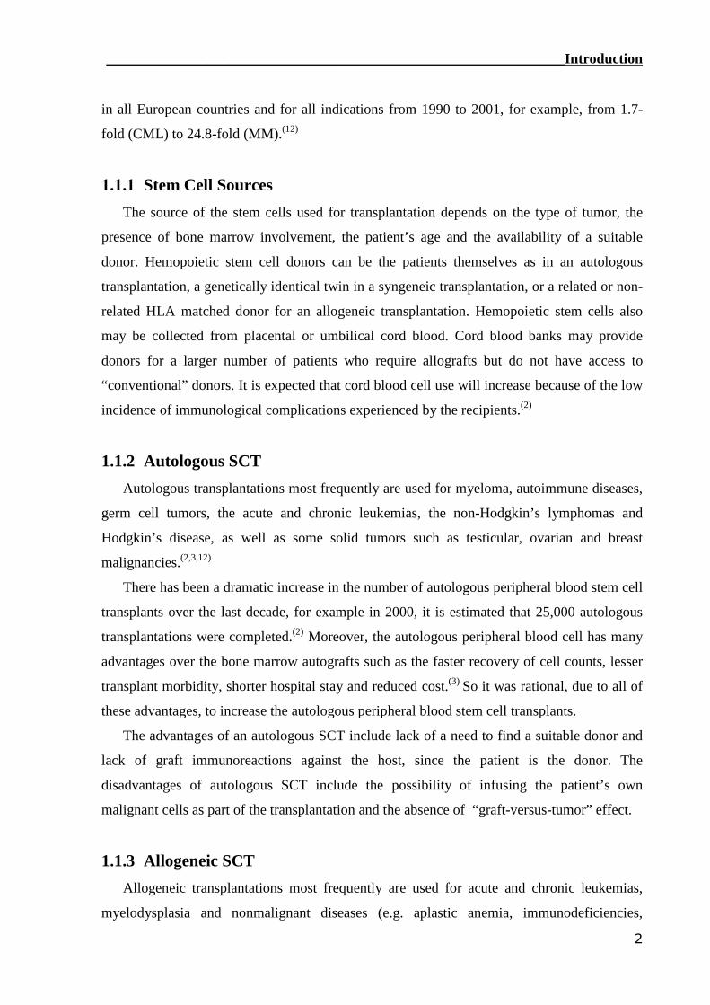
Transplantation Type Stem CellSource
DonorSearch
GvHD* GvLEffect*
MalignantCell Infusion*
Allogeneic Matcheddonor
Yes Yes Yes No
Syngeneic Identicaltwin
No No No No
Autologous Patient No No No Yes
* Possible sequelae.
Table 1: Stem cell sources and sequelae.
________________________________________________________________Introduction
4
1.2 Cellular reconstitution after SCT
The establishment of the donor immune system in the recipient (i.e. reconstitution) takes
months to years to complete and functional immunocompetent T cells are reconstituted.(4) It
initially involves the expansion of a postthymic donor T cell repertoire showing many unusual
phenotypic and functional features. Normalization of the immune system in the recipient
requires the emergence of tolerized T cells processed from precursors through the recipient
thymus. This event is delayed and may be incomplete in older recipients.(13) In the first few
months following bone marrow or blood SCT, the immune repertoire is dominated by T cells
expanding from transplanted T cells derived from the donor’s peripheral blood T cell
compartment. This consists predominantly of central and effector memory cells with a smaller
population of naïve T cells and endstage effector cells. It is these postthymic cells that are
largely responsible for the success or failure of the transplant through their impact on
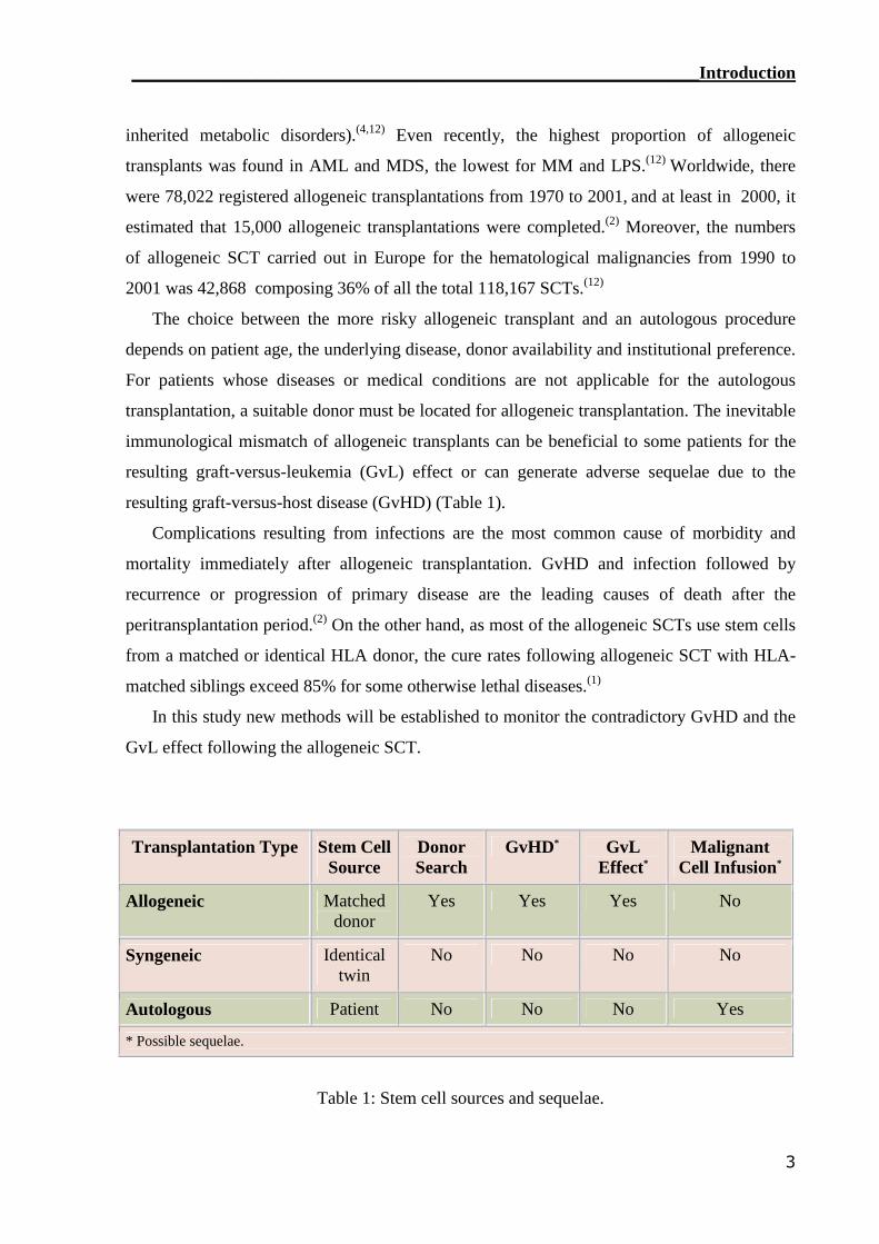
engraftment, GvHD, GVL, and reactivating viruses (Figure 1).(13)
Figure 1: Course of events and risks associated with allogeneic transplantation. Gray boxes
represent the 4 components of transplantation as outlined in the text; open boxes represent
some risks at different stages.
Antigens (Ags) eliciting T cell responses in donor cells can be classified as (1) major or
minor histocompatibility antigens (MHC or mHAgs) that can be either tissue restricted or
widely distributed on many tissues, (2) normal (nonalleleic) protein sequences overexpressed
________________________________________________________________Introduction
5
or aberrantly expressed in malignant cells, or (3) Ags representing a unique tumor specific
peptide sequence. It should also be remembered that donor T cells contain a repertoire of
memory cells responding to Ags of herpes group viruses (e.g. cytomegalovirus (CMV),
Epstein Barr virus (EBV), herpes simplex virus (HSV)) resident in the recipient (Table 2).(13)
Non-tissue-restricted
Minor histocompatibility antigens (mHAgs)
Major histocompatibility (MHC) antigens directly and indirectly presented
Tissue-restricted/aberrantly expressed
mHAgs: HY, HA-1, HA-2, HB-1
Nonalleleic: proteinase 3, WT-1, telomerase
Malignant-cell-restricted
Products of chromosome translocation: t9:22, t15:17
Virus-specific
CMV: pp65, IE1
EBV : EBNA 1-3, LMP-1, LMP-2
Table 2: Classification of well-characterized antigens driving donor T-cell responses.
HY, male-specific minor histocompatibility antigen; HA, minor histocompatibility antigen;
HB-1, B cell minor histocompatibility antigen-1; WT1, Wilms’ tumor; t, translocation; pp,65
phosphoprotein 65 antigen; IE1, intermediate-early 1 antigen; EBNA, Epstein Barr nuclear
antigen; LMP, latent membrane protein.
More detailed information on the reconstitution of Ag-specific responses has been derived
from intracellular cytokine flow cytometry, cytokine secretion assays [enzyme-linked
immunospot (ELISPOT), matrix affinity technology] and class I HLA-peptide tetramer
labelling.(14) But little is done in the field of molecular biology to assess the reconstitution of
Ag-specific T cells after SCT. Therefore, in this study, the reconstitution of CMV- and
mHAg-specific T cells (e.g. anti-HY and anti-WT1 T cells) will be monitored in allogeneic
transplanted patients following SCT.
1.3 Cytomegalovirus (CMV) infection after allogeneic SCT
Cytomegalovirus (CMV) is a frequent pathogen in humans and is usually associated with
asymptomatic infection, followed by a state of viral persistence or latency. Human CMV
________________________________________________________________Introduction
6
establishes persistent lifelong infections in most (50%-85%) individuals.(15,16) The virus
primarily infects endothelial cells in a range of tissues and, after a lytic cycle, establishes an
asymptomatic latent infection.(15) The principal site of virus latency in the peripheral
circulation is likely the monocyte.(17)
CMV infection still remains a major cause of morbidity and mortality after SCT. In
contrast to patients treated with high-dose chemotherapy and autologous SCT, patients after
allogeneic SCT are at a much higher risk of active CMV infection because of the delayed
recovery of T- and B-cell functions.(18) In the context of immunologic impairment due to
conditioning for SCT, 60% to 70% of high-risk (CMV-seropositive) or CMV-seronegative
patients who receive transplants from a seropositive donor; if no preventive measures are
taken, will be under the risk of developing CMV disease during the first 100 days after
conventional or nonmyeloablative SCT. And moreover, approximately ~20–30% will
develop CMV disease during the first year, unless preemptive strategies were adopted..(17,18)
In the early days of allogeneic SCT, CMV can be observed to reactivate 30 to 60 days after
transplantation, and disease occurred in approximately one half of patients.(19) The time of
onset of disease increased to approximately six months after allogeneic SCT, and mortality
due to late-onset CMV caused the preponderance of deaths at a rate approaching 10% for
allogeneic SCT recipients.(20) Effective antiviral prophylaxis and early intervention has led to
decrease the active CMV infection and disease after day 100 after transplantation. Patients
developing late-onset CMV disease are characterized by a delayed reconstitution of CMV-
specific T-cell responses.(21,22)
As CMV infection after allogeneic SCT is frequently associated with life-threatening
invasive lung and visceral disease,(23-25) monitoring the CMV load and the presence of the
CMV-specific T cells are too important. Accurate monitoring of CMV-specific T-cell
reconstitution is required for appropriate decision on treatment, such as anti-viral drugs,
which have adverse effects. As many researchers took in consideration the reconstitution of
CMV-specific T cells after allogeneic SCT,(26-28) it was found that the threshold level for
protection from CMV reactivation was estimated (e.g. over 1×10⁶ cells/l peripheral blood
with the IFN-γ-ELISPOT assay).(26) Recurrence of CMV infection occurred only in the
patients who failed to generate a cytotoxic T lymphocyte (CTL) response to the virus,(27) so
the monitoring of CMV-specific CTLs may help in identifying the subset of patients at risk
from recurrent infection or disease. In a pilot study in a limited number of patients at high risk
for late-onset CMV disease, a single transfusion of a donor-derived ex vivo expanded
________________________________________________________________Introduction
7
polyclonal CMV-specific T-cell line was found to be associated with clearance of the viral
load from the blood and reconstitution of CMV-specific T-cell responses in some of the
patients, indicating a potential strategy to prevent late CMV disease.(29)
There are many methods in use for detecting CMV in the body fluids such as rapid
culture, antibody assays, antigenemia assays, and DNA detection methods.(30,31) The effect of
pre-emptive CMV treatment based on early detection of CMV reactivation - by methods
including antigenemia assays,(30,31) polymerase chain reaction (PCR)(30,32-34) or recently by the
reverse transcription-PCR(31,35-38) - has been found to increase the time to onset of CMV
disease, with the result that late disease is the current main CMV-related problem in SCT.(39-
41)
In this study, the reconstitution of CMV-specific T-cells will be monitored to predict the
patients’ protection against the CMV. In addition, new functional assay will be established to
assess the CMV-specific T cells in the patients’ peripheral blood after allogeneic SCT.
1.4 Graft-versus-host disease (GvHD)
Billingham and Brent described how injection of newborn mice with viable spleen cells
from adult donors of a different strain resulted in the development of what they termed runt
disease,(42) but it was the Danish physician Morton Simonsen who introduced the name graft-
versus-host disease (GvHD). Normally, GvHD divided into two types, a) acute GvHD
(aGvHD) which occurs within the first 100 days after transplantation and b) chronic GvHD
(cGvHD) is distinguished from aGvHD by clinical symptoms that can resemble an overlap of
several connective tissue diseases (e.g. lupus erythematosus, mixed connective tissue disease,
scleroderma, Sjogren syndrome, biliary cirrhosis, idiopathic pulmonary fibrosis). The
classical definition of cGvHD is GvHD that persists or occurs de novo beyond 80-100 days
post SCT (Figure 1).(43)
Despite adequate post-transplantation immunosuppressive therapy, GvHD remains a
major cause of morbidity and mortality in the allogeneic SCT setting, even in patients who
receive HLA-identical sibling grafts.(44) Up to 30% of the recipients of stem cells or bone
marrow transplantation from HLA-identical related donors and most patients who receive
cells from other sources (e.g. matched, unrelated, non-HLA-identical siblings; cord blood)
will develop ≥ Grade 2 aGvHD despite immunosuppressive prophylaxis.(45,46)
GvHD can occur when transplanted donor-derived T cells recognize MHC or mHAgs
proteins and their associated peptides expressed by antigen-presenting cells (APC). A widely
________________________________________________________________Introduction
8
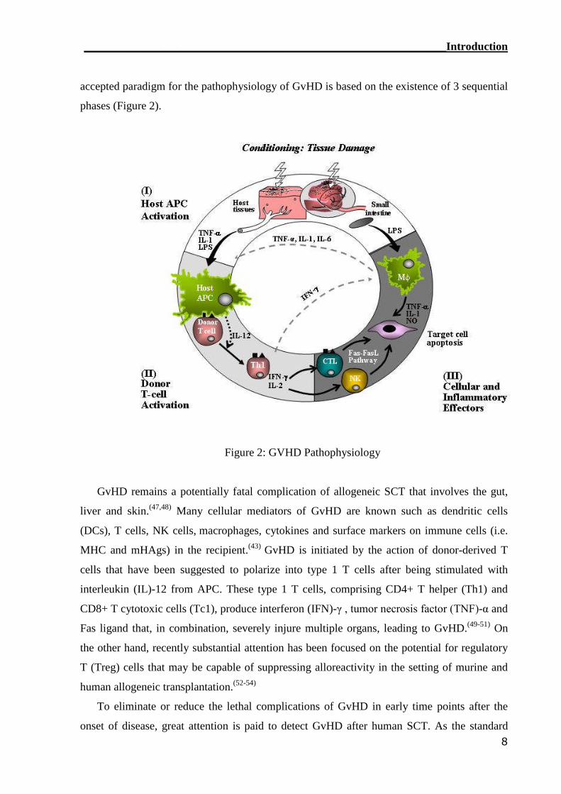
accepted paradigm for the pathophysiology of GvHD is based on the existence of 3 sequential
phases (Figure 2).
Figure 2: GVHD Pathophysiology
GvHD remains a potentially fatal complication of allogeneic SCT that involves the gut,
liver and skin.(47,48) Many cellular mediators of GvHD are known such as dendritic cells
(DCs), T cells, NK cells, macrophages, cytokines and surface markers on immune cells (i.e.
MHC and mHAgs) in the recipient.(43) GvHD is initiated by the action of donor-derived T
cells that have been suggested to polarize into type 1 T cells after being stimulated with
interleukin (IL)-12 from APC. These type 1 T cells, comprising CD4+ T helper (Th1) and
CD8+ T cytotoxic cells (Tc1), produce interferon (IFN)-γ , tumor necrosis factor (TNF)-α and
Fas ligand that, in combination, severely injure multiple organs, leading to GvHD.(49-51) On
the other hand, recently substantial attention has been focused on the potential for regulatory
T (Treg) cells that may be capable of suppressing alloreactivity in the setting of murine and
human allogeneic transplantation.(52-54)
To eliminate or reduce the lethal complications of GvHD in early time points after the
onset of disease, great attention is paid to detect GvHD after human SCT. As the standard
________________________________________________________________Introduction
9
methods used normally to diagnose GvHD, which are the histological methods, overlapped
with viral infections, so other methods was established. For example, host-reactive
lymphocytes with broad specificity have been observed in GvHD patients using the limiting
dilution techniques, which are time consuming for detecting functional T cell analysis. In the
meantime, many techniques were established to predict GvHD such as, T lymphocyte
precursors frequency analysis,(55) some serum markers (e.g. the levels of TNF-α , IFN-γ , IL-
10, soluble Fas, and IL-18),(49,51,56,57) polymorphism of IL-10(58) and transforming growth
factor (TGF)-β1(59) genes, ELISPOT assays,(47,60) and T cell receptor (TCR)-Vβ clonotypic
analysis.(60-62) In addition, multimers technology has been developed which allows flow
cytometric detection of specific T cells independently of their activation state.(63) But most of
these analysis methods have not been reported to be necessarily predicting GvHD. Thus, there
are contradictory results among these methods and there still remain problems with attempts
to use these parameters as reliable and sensitive markers of GvHD. Therefore, in this study,
the monitoring of alloreactive T cells in the patients peripheral blood will be monitored by
establishing two assays including the detection of intracellular cytokine (ICC) by flow
cytometer and real time-polymerase chain reaction (RT-PCR) assay.
1.5 Graft-versus-leukemia (GvL) effect
There is compelling evidence, much of which is derived from the results of allogeneic
SCT, that human leukemias can also be recognized and eliminated by T cells. The
immunologically mediated graft-versus-leukemia (GvL) effect that was predicted by animal
model studies of allogeneic SCT has been documented in clinical trials. Patients who receive
an allogeneic transplant for advanced leukemia have a lower probability of leukemic relapse
if they develop acute and/or chronic GvHD as a complication of the transplant.(64,65) Also,
Kolb and Holler were able to prove that donor transfused lymphocytes exhibits a GvL effect
and increases chimerism after bone marrow transplantation.(66) The risk of leukemic relapse is
increased after syngeneic SCT or T-cell depleted allogeneic SCT, suggesting a critical role for
donor T cells specific for allogeneic determinants in initiating or mediating the GvL effect.(67)
Target T-cell epitopes involved in the GvL reaction are either autologous tumor associated
antigens (TAAs)(68) or allogeneic mHAgs(69) expressed by the tumor. In allogeneic SCT, T
cells specific for mHAgs - which are peptides that differ between donor and recipient due to
polymorphism in the genome - provide a potent GvL effect.(70) However, T cells specific for
some mHAgs are also responsible for GvHD, and means of reliably segregating the
________________________________________________________________Introduction
10
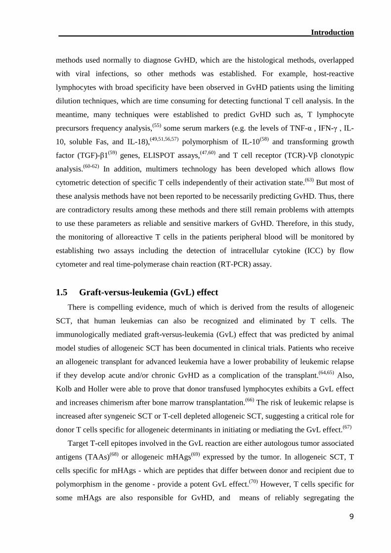
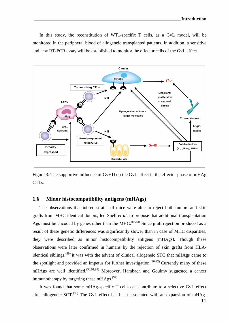
beneficial GvL effect from GvHD has not yet been well established (Figure 3).(71) For better
understanding such segregation, recently it was found that T cells contribute to GvHD and
GvL via membrane-bound or locally released TNF.(72) Moreover it was found that the
cytolytic activity of T cells is primarily mediated through the Fas-Fas ligand and perforin-
granzyme pathways.(73) In experiments with purely selected donor T cells, the FasL pathway
was important for GvHD activity by both CD4(+) and CD8(+) T cells, whereas the perforin
pathway was required for CD8-mediated GvL activity. These data demonstrate, in an
allogeneic bone marrow transplanted murine model, that donor T cells mediate GvHD activity
through the FasL pathway compared to GvL activity which use the perforin pathway. This
suggests that donor T cells make differential use of cytolytic pathways and that the specific
blockade of one cytotoxic pathway may be used to prevent GvHD without interfering with
GvL activity.(74) On the other hand and surprisingly, it was found that T cells deficient for
both Fas ligand and perforin can still exert GvL activity in vivo in mouse models.(75,76) This
was resolved by Schmaltz et al. who found that the TNF-related apoptosis-inducing ligand
(TRAIL) is mediating the GvL but not the GvHD. These data suggest that strategies to
enhance TRAIL-mediated GvL activity could decrease relapse rates of malignancies after
hematopoietic SCT without exacerbation of GvHD.(73)
In addition, there is evidence that effector mechanisms other than T cells may also
contribute to GVL activity either directly or as a consequence of inflammation induced by
allogeneic T cells. This was confirmed by the observation that natural killer (NK) cells can
lyse leukemic cells in vitro. However, NK cells may be particularly effective for inducing
GvL activity after T cell depleted haploidentical transplant where disparity between killer
inhibitory receptors (KIRs) expressed by donor NK cells and HLA molecules on recipient
leukemic cells favors NK activation.(77,78)
One approach for separating GvL from GvHD is to identify peptides that are recognized
by T cells and presented by leukemic cells but not by tissues that are a target of GvHD. There
are several broad categories of proteins that may give rise to Ags that could be targets of a
selective GvL response. These include a) tumor-specific proteins resulting from chromosome
translocations such as bcr/abl or PML/RAR, or from mutations such as p21 ras,(79-81) b)
normal proteins that are overexpressed in leukemic cells such as WT-1 or proteinase 3,(82,83)
and c) mHAgs that are selectively expressed in recipient hematopoietic cells including
leukemic cells but not in nonhematopoietic cells.(84-86)
________________________________________________________________Introduction
11
In this study, the reconstitution of WT1-specific T cells, as a GvL model, will be
monitored in the peripheral blood of allogeneic transplanted patients. In addition, a sensitive
and new RT-PCR assay will be established to monitor the effector cells of the GvL effect.
Figure 3: The supportive influence of GvHD on the GvL effect in the effector phase of mHAg
CTLs.
1.6 Minor histocompatibility antigens (mHAgs)
The observations that inbred strains of mice were able to reject both tumors and skin
grafts from MHC identical donors, led Snell et al. to propose that additional transplantation
Ags must be encoded by genes other than the MHC.(87,88) Since graft rejection produced as a
result of these genetic differences was significantly slower than in case of MHC disparities,
they were described as minor histocompatibility antigens (mHAgs). Though these
observations were later confirmed in humans by the rejection of skin grafts from HLA-
identical siblings,(89) it was with the advent of clinical allogeneic STC that mHAgs came to
the spotlight and provided an impetus for further investigation.(90-92) Currently many of these
mHAgs are well identified.(90,91,93) Moreover, Hambach and Goulmy suggested a cancer
immunotherapy by targeting these mHAgs.(94)
It was found that some mHAg-specific T cells can contribute to a selective GvL effect
after allogeneic SCT.(95) The GvL effect has been associated with an expansion of mHAg-
GvL
GvHD Soluble factors
(e.g., IFN-γ, TNF-α)
Tumor stroma
Up-regulation of tumor
Target molecules
Direct anti-
proliferative
or cytotoxic
effects
Kill
KillAPCs
APCs
maturation
Broadly expressed
mHag CTLs
Eppithelial cells
Broadly
expressed
Tumor mHag CTLs
Cancer
Angio-
stasis
________________________________________________________________Introduction
12
specific T cells that also exhibit a suppressive effect on the growth of leukemic precursors in
vitro.(86,96) Moreover, isolation,(97) generation(98,99) and expansion (100) of the mHAgs-specific
T cells are visible recently. On the other hand and unfortunately, many mHAgs are
ubiquitously expressed, thus T cells may also target normal tissues leading to GvHD.(101-103)
In fact, mHAg-specific T cells are detectable in patients with active GvHD.(104) Furthermore it
was found that pregnancy is able to prime mHAg-specific T-cell responses for both autosomal
(e.g. HA-1) and anti-HY (male specific) mHAgs.(105,106) Therefore, the WT1- and HY-specific
T cells, as mHAg models, will be monitored in this study especially their reconstitution
following allogeneic SCT.
1.7 Immune monitoring approaches
1.7.1 Cytokines
In general, cytokines provide a direct measure of the effector function of T
lymphocytes.(107) For example, T cells that produce interleukin (IL)-2, tumor necrosis factor
(TNF)-α and interferon (IFN)-γ are considered to be of the Th1 phenotype, compared with T
cells that produce predominantly IL-4, IL-5, IL-10, and IL-13, which define the Th2
phenotype.(50)
In the past decade, the analysis of cytokine production became increasingly important in
unraveling the course of an immune response, in the evaluation of specific therapies, and in
the search of the pathophysiologic mechanisms at the base of many diseases. Fore example,
TNF-α has been implicated in the pathophysiology of GvHD at several steps in the process,
including induction of apoptosis in target tissues through the TNF-α receptor; activation of
macrophages, neutrophils, eosinophils, B cells, and T cells; stimulating production of
additional inflammatory cytokines (IL-1, IL-6, IL-10, IL-12, and TNF-α itself); increased
expression of HLA; and the facilitation of T-lymphocyte lysis.(72,108-110) In addition, IFN-γ is
another pro-inflammatory cytokine that can be secreted in GvHD pathophysiology (Figure 2)
and in response to CMV.(47,111) Therefore, in addition to IFN-γ, some cytokines will be traced
in many of the assays adopted in this study. But it should be kept in mind that the cytokines
may be similar in both GvHD and GvL because they will be secreted in response to Ag
whether it was alloantigen (in the case of GvHD) or tumor Ag (in the GvL). So, to solve this
problem, the secreted cytokines should be correlated to the applied Ag.

________________________________________________________________Introduction
13
1.7.2 Mixed lymphocytes reaction (MLR)
When lymphocytes from genetically different individuals are mixed together in tissue
culture blast transformation occurs, a reaction known as the mixed lymphocyte reaction
(MLR). The MLR is a clinically relevant in vitro assay where lymphocytes from one
individual (responders, R) are incubated with the lymphocytes of another individual
(stimulator, S) which have been previously rendered incapable of blast transformation by
gamma-irradiation.(112) It is presumed that the MLR is an in vitro analog of in vivo
alloreactivity, and is widely used in transplantation immunology to measure recipient T-cell
responses against donor tissues due to the mismatch of MHC antigens (especially class II).(113)
The MLR ability to predict possible rejection of the donor organ in the transplant recipient
can be used in the allo-settings in healthy individuals.(114-116) So, a modified MLR assay will
be established and adopted to assess the alloreactive T cells in the allogeneic transplanted
patients following SCT. This will establish an assay which, hopefully, will predict the GvHD.
1.7.3 Tetramer (TM) staining
Recently developed MHC multimer technologies allow visualization and isolation of Ag-
specific T cells. Fore example, the introduction of peptide–MHC class I tetrameric complex
technology initiated a profound revolution in the field of cellular immunology.(117) Class I
HLA-peptide tetramers (TM) are soluble complexes of four synthetic HLA molecules
associated with a specific peptide that fits the peptide-binding groove of the HLA molecule
under study. TMs are conjugated with a fluorochrome to allow their visualization by flow
cytometry.(118)
Peptide-MHC class I tetrameric complexes are proving invaluable as fluorescent reagents
for enumeration, characterization, and isolation of peptide-specific T cells and have afforded
many advantages over previous techniques, particularly the ability to directly quantify and
phenotype Ag-specific T cells with minimal in vitro manipulation.(119) However, functional
analysis and in vivo transfer of MHC multimer-stained cells is hampered by the persistence of
TCR-MHC interactions and subsequently induced signaling events.(118,120) But interestingly,
new types of MHC multimers were generated, which can be monomerized in the presence of
a competitor, resulting in rapid loss of the staining reagent allowing “reversible” T-cell
staining procedure.(121)
However, a cell is not characterized so much by what lies on its surface (e.g. TM), but by
what resides inside, where most of its biology takes place and which is likely to reflect its
________________________________________________________________Introduction
14
functional phenotype. Therefore the phenotypical TM staining in addition to other functional
assays will be adopted to monitor the reconstituted T cells.
1.7.4 Intracellular cytokines (ICC) flow cytometry
The most popular method to assess cytokines is the enzyme-linked immunosorbent assay
(ELISA), which is applied to measure cytokine secretion in supernatants, whereas flow
cytometry is used to determine intracellular cytokine (ICC) production.(122) Moreover, it was
found that the circulating lymphocyte subsets linked to the ICC profiles in normal
humans.(123) ICC flow cytometry is based on direct detection of ICC expression with
fluorochrome-conjugated anticytokine antibodies after short periods of activation with various
stimuli (e.g. 5-6h). Stimulation can be performed with peripheral blood mononuclear cells
(PBMCs), whole blood, lymph nodes, or other biologic fluids.(124) Staining of the ICC depend
on the identification of cytokine-specific monoclonal antibodies which are compatible with a
fixation-permeabilization procedure.(123)
It is well known that the ICC assays have some disadvantages such as the low sensitivity
(10-4) - compared to ELISPOT - and the cell-fixation which limits further functional assays to
be performed.(107) Although there is no absolute quantitative measurement of the produced
cytokine by the ICC flow cytometric method, but it can easily identifies the cytokine-
producing cell type by phenotyping for many cell markers (e.g. cell lineage, activation and
apoptosis). For the fast and phenotypical criteria of the ICC assays, it will be used in this
study to monitor the reconstitution of T cells in allogeneic transplanted patients.
1.7.5 Enzyme-linked immunospot (ELISPOT)
The enzyme-linked immunospot (ELISPOT) assay is based on the principle of the ELISA
detecting antigen-induced secretion of cytokines trapped by an immobilized antibody and
visualized by an enzyme-coupled second antibody.(125) In a recent study, IFN-γ-ELISPOT
assay showed to have good reproducibility for the determination of Ag-specific T cells in
different laboratories.(126) The ELISPOT assay has the advantage of detecting only
activated/memory T cells and the cytokine release can be detected at the single cell level,
allowing direct determination of T cell frequencies.(127) Furthermore, this assay has been
found to be more sensitive than ELISA (e.g. detection limit of 10-200 times lower) and ICC
staining.(128,129) The high sensitivity and easy performance, allowing a direct enumeration of
peptide-reactive T cells without prior in vitro expansion, makes the ELISPOT assay
________________________________________________________________Introduction
15
eminently well suited to monitor and measure T-cell responses.(130) For these advantages, the
ELISPOT assay will be adopted to monitor the reconstitution of T cells after allogeneic SCT.
1.7.6 Real time-polymerase chain reaction (RT-PCR)
The real time-polymerase chain reaction (RT-PCR) assay is based on the principle that
amplification of cDNA by the polymerase chain reaction (PCR) follows a strict mathematical
equation whereby with each cycle of amplification two copies are made from each individual.
Thus, the amount of cDNA amplified after a given number of cycles will be directly
proportional to the log2 of the starting amount of template. This quantitation is achieved with
a gene-specific nucleotide probe complementary to a region of DNA nested between the PCR
primers. This probe is labeled with a reporter fluorochrome and also with a quencher that can
absorb fluorescence. The quencher can only quench the reporter fluorescence when the two
dyes are close to each other. During amplification the probe is removed from the DNA strand
and degraded by the 5'-3' exonuclease activity of Taq DNA polymerase and the fluorochrome
is separated from the quencher yielding one unit of fluorescence for each cycle of
amplification. By recording incremental fluorescence at each PCR cycle it is, therefore,
possible to calculate the starting amount of cDNA template. (124,131,132) Also, the RT-PCR
instruments allow “real time” detection of PCR products as they accumulate during PCR
cycles. Thus, by RT-PCR it is possible to gather quantitative information about gene
expression in any given specimen.
Many significant advantages to the use of RT-PCR for immune monitoring were
described such as its flexibility, sensitivity and reliability.(124,127,133) Also, the RT-PCR can be
considered the method of choice for the rapid and reproducible measurement of gene
expression in small samples.(134) In addition to its sensitivity, RT-PCR also provides
flexibility of analysis since cDNA is quite stable for future analysis.(127) Moreover, recently
the RT-PCR assay was developed to assess the many cytokines in the murine as well in
human.(133,135) For example, serial sampling of fine-needle aspirates of metastases from
melanoma patients receiving IL-2-based vaccinations has been performed to assess changes in
expression of IL-10, TGF-β, and IFN-γ mRNA levels by RT-PCR assay.(136)
This tool offers unique advantages and should be considered as part of a repertoire used to
design a comprehensive immune monitoring strategy.(124) Moreover, it was suggested that
RT-PCR represents a useful tool for the monitoring of patients undergoing immune
manipulation (e.g. SCT). However, little information is available in the literature about the
________________________________________________________________Introduction
16
utilization of RT-PCR for immune monitoring, as this methodology has been only recently
applied to this field. Therefore, the RT-PCR assay will be adopted in this study to monitor the
reconstitution of donor T cells in the allogeneic transplanted patient’s body.
The present study strived to investigate, using various techniques, whether Ag-specific T
cells could be monitored in allogeneic transplanted patients, and if so, how the used
techniques correlate to each other. Four main questions were addressed:
1) Can the reconstitution of CMV-specific T cells after allogeneic SCT be monitored using
phenotypical and functional techniques?
Approach: using TM, ICC, ELISPOT and RT-PCR techniques to monitor the reconstituted
CMV-reactivity by the aid of CMV pp65-peptide and/or pp65-protein.
2) Can the GvL effect be monitored after allogeneic SCT using functional techniques
especially the RT-PCR? This was address by monitoring the reconstitution of WT1-reactive T
cells using ELISPOT and RT-PCR techniques using two HLA-A2-restricted peptides,
namely: WH187 and Db126.
3) Can the mHAgs-reactive T cells be monitored after allogeneic SCT using functional
techniques especially the RT-PCR? This was addressed by monitoring the HY-reactive T cells
using ELISPOT and RT-PCR techniques using SMCY-derived HY peptide.
4) Can the alloreactive T cells in the allogeneic transplanted patients’ peripheral blood be
monitored by intracellular (IC) cytokine staining? This was investigated by first establishing
and optimizing an alloreactive mixed lymphocyte reaction (MLR) model that can simulate the
GvHD settings, then testing some allogeneic transplanted patients’ samples for detecting the
presence of alloreactive T cells.
_______________________________________________________Materials and Methods
17
2. Materials and Methods
2.1 Materials
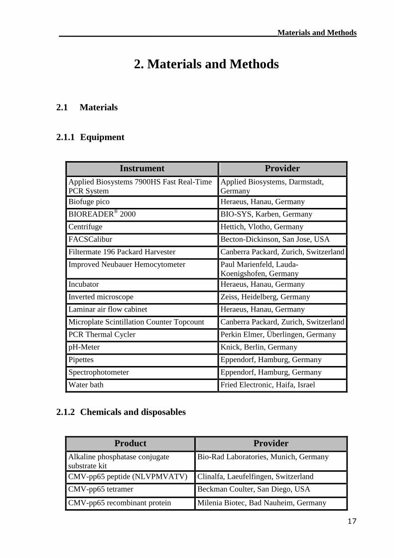
2.1.1 Equipment
Instrument ProviderApplied Biosystems 7900HS Fast Real-TimePCR System
Applied Biosystems, Darmstadt,Germany
Biofuge pico Heraeus, Hanau, Germany
BIOREADER® 2000 BIO-SYS, Karben, Germany
Centrifuge Hettich, Vlotho, Germany
FACSCalibur Becton-Dickinson, San Jose, USA
Filtermate 196 Packard Harvester Canberra Packard, Zurich, Switzerland
Improved Neubauer Hemocytometer Paul Marienfeld, Lauda-Koenigshofen, Germany
Incubator Heraeus, Hanau, Germany
Inverted microscope Zeiss, Heidelberg, Germany
Laminar air flow cabinet Heraeus, Hanau, Germany
Microplate Scintillation Counter Topcount Canberra Packard, Zurich, Switzerland
PCR Thermal Cycler Perkin Elmer, Überlingen, Germany
pH-Meter Knick, Berlin, Germany
Pipettes Eppendorf, Hamburg, Germany
Spectrophotometer Eppendorf, Hamburg, Germany
Water bath Fried Electronic, Haifa, Israel
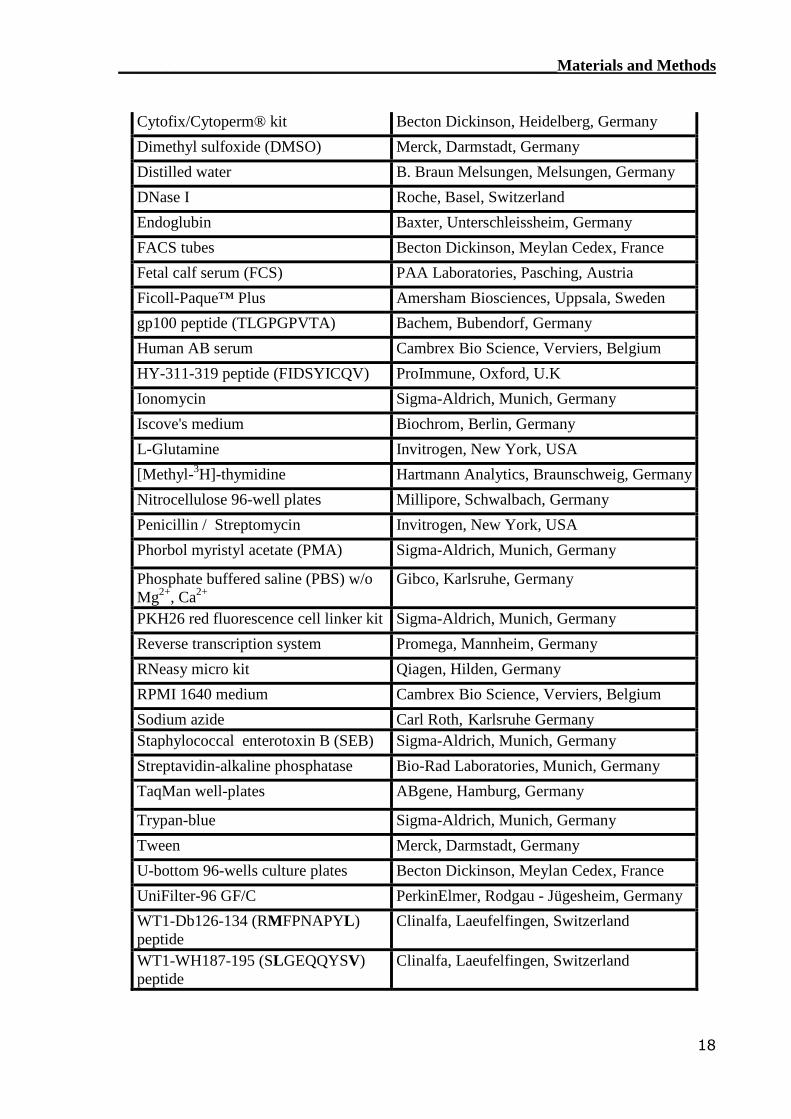
2.1.2 Chemicals and disposables
Product ProviderAlkaline phosphatase conjugatesubstrate kit
Bio-Rad Laboratories, Munich, Germany
CMV-pp65 peptide (NLVPMVATV) Clinalfa, Laeufelfingen, Switzerland
CMV-pp65 tetramer Beckman Coulter, San Diego, USA
CMV-pp65 recombinant protein Milenia Biotec, Bad Nauheim, Germany
_______________________________________________________Materials and Methods
18
Cytofix/Cytoperm® kit Becton Dickinson, Heidelberg, Germany
Dimethyl sulfoxide (DMSO) Merck, Darmstadt, Germany
Distilled water B. Braun Melsungen, Melsungen, Germany
DNase I Roche, Basel, Switzerland
Endoglubin Baxter, Unterschleissheim, Germany
FACS tubes Becton Dickinson, Meylan Cedex, France
Fetal calf serum (FCS) PAA Laboratories, Pasching, Austria
Ficoll-Paque™ Plus Amersham Biosciences, Uppsala, Sweden
gp100 peptide (TLGPGPVTA) Bachem, Bubendorf, Germany
Human AB serum Cambrex Bio Science, Verviers, Belgium
HY-311-319 peptide (FIDSYICQV) ProImmune, Oxford, U.K
Ionomycin Sigma-Aldrich, Munich, Germany
Iscove's medium Biochrom, Berlin, Germany
L-Glutamine Invitrogen, New York, USA
[Methyl-3H]-thymidine Hartmann Analytics, Braunschweig, Germany
Nitrocellulose 96-well plates Millipore, Schwalbach, Germany
Penicillin / Streptomycin Invitrogen, New York, USA
Phorbol myristyl acetate (PMA) Sigma-Aldrich, Munich, Germany
Phosphate buffered saline (PBS) w/oMg2+, Ca2+
Gibco, Karlsruhe, Germany
PKH26 red fluorescence cell linker kit Sigma-Aldrich, Munich, Germany
Reverse transcription system Promega, Mannheim, Germany
RNeasy micro kit Qiagen, Hilden, Germany
RPMI 1640 medium Cambrex Bio Science, Verviers, Belgium
Sodium azide Carl Roth, Karlsruhe GermanyStaphylococcal enterotoxin B (SEB) Sigma-Aldrich, Munich, Germany
Streptavidin-alkaline phosphatase Bio-Rad Laboratories, Munich, Germany
TaqMan well-plates ABgene, Hamburg, Germany
Trypan-blue Sigma-Aldrich, Munich, Germany
Tween Merck, Darmstadt, Germany
U-bottom 96-wells culture plates Becton Dickinson, Meylan Cedex, France
UniFilter-96 GF/C PerkinElmer, Rodgau - Jügesheim, Germany
WT1-Db126-134 (RMFPNAPYL)peptide
Clinalfa, Laeufelfingen, Switzerland
WT1-WH187-195 (SLGEQQYSV)peptide
Clinalfa, Laeufelfingen, Switzerland
_______________________________________________________Materials and Methods
19
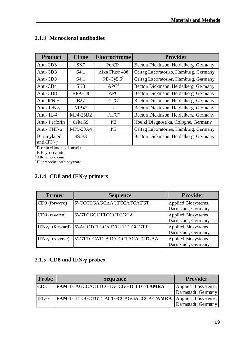
2.1.3 Monoclonal antibodies
Product Clone Fluorochrome ProviderAnti-CD3 SK7 PerCP1 Becton Dickinson, Heidelberg, GermanyAnti-CD3 S4.1 Alxa Fluor 488 Caltag Laboratories, Hamburg, GermanyAnti-CD3 S4.1 PE-Cy5.52 Caltag Laboratories, Hamburg, GermanyAnti-CD4 SK3 APC3 Becton Dickinson, Heidelberg, GermanyAnti-CD8 RPA-T8 APC Becton Dickinson, Heidelberg, GermanyAnti-IFN-γ B27 FITC1 Becton Dickinson, Heidelberg, GermanyAnti- IFN-γ NIB42 - Becton Dickinson, Heidelberg, GermanyAnti- IL-4 MP4-25D2 FITC4 Becton Dickinson, Heidelberg, GermanyAnti- Perforin deltaG9 PE Hoelzl Diagnostika, Cologne, GermanyAnti- TNF-α MP9-20A4 PE Caltag Laboratories, Hamburg, GermanyBiotinylatedanti-IFN-γ
4S.B3 - Becton Dickinson, Heidelberg, Germany
1 Peridin chlorophyll protein2 R-Phycoerythrin3 Allophycocyanin4 Fluorescein-isothiocyanate
2.1.4 CD8 and IFN-γ primers
Primer Sequence ProviderCD8 (forward) 5'-CCCTGAGCAACTCCATCATGT Applied Biosystems,
Darmstadt, GermanyCD8 (reverse) 5'-GTGGGCTTCGCTGGCA Applied Biosystems,
Darmstadt, GermanyIFN-γ (forward) 5'-AGCTCTGCATCGTTTTGGGTT Applied Biosystems,
Darmstadt, GermanyIFN-γ (reverse) 5'-GTTCCATTATCCGCTACATCTGAA Applied Biosystems,
Darmstadt, Germany
2.1.5 CD8 and IFN-γ probes
Probe Sequence ProviderCD8 FAM-TCAGCCACTTCGTGCCGGTCTTC-TAMRA Applied Biosystems,
Darmstadt, GermanyIFN-γ FAM-TCTTGGCTGTTACTGCCAGGACCCA-TAMRA Applied Biosystems,
Darmstadt, Germany
_______________________________________________________Materials and Methods
20
2.1.6 Kits
BD Cytofix/Cytoperm™ kit was used to perforate the cells to allow IC cytokines detection.
In addition, total RNA was isolated from PBMCs by using RNeasy Micro kit.
2.1.7 Media
Both RPMI1640 and Iscove’s media were used and were called “’complete” when
containing the following:
Additives Final Concentration
Pooled human AB serum…………………………10%
Penicillin / Streptomycin………………………….1%
L-Glutamine…………………………………….....2%
Also a freezing medium, composed of 90% fetal calf serum (FCS) and 10% DMSO, was
used to freeze cells under liquid nitrogen.
2.1.8 Cell lines
The human HLA-A*0201-positive leukemia B-cell line C1R.A2 (expressing a transfected
genomic clone of HLA.A2) was used as Ag presenting cells. It is unable to present
endogenous Ags so it just expresses surface peptide-pulsed HLA-A2 molecules. These cells
were cultured in complete-RPMI 1640 medium until peptide pulsing.
2.2 Patients
All the included patients underwent allogeneic SCT, and their donors were mandatory
matched for the HLA-A,-B and -C and optional matched for the HLA-DR and -DP allele. All
patients and their donors gave informed consent approved by local ethics committee (IRB).
2.2.1 Analysis of T-cell reactivity to CMV in patients
Eighteen patients (n=18) were tested for the reconstitution of CMV-specific T cells post
transplantation. The CMV serological status was identified for all patients and donors. CMV
pp65 protein was used as stimulus due to its dominant Ag recognition by CD4 T cells. In
addition, one attractive candidate stimulating Ag the immunodominant HLA-A2 peptide
epitope CMV pp65 (495-503) was used.(137,138) T-cell reactivity for the CMV Ags was tested
_______________________________________________________Materials and Methods
21
in all of the patients in the experimental-group (i.e. HLA-A2 positive) and control-group (i.e.
HLA-A2 negative). PBMCs from ten patients were monitored at serial time point to follow
the reconstitution of CMV pp65-specific T cells. After testing most of the patients’ PBMCs
by TM staining, ICC flow cytometry, ELISPOT assay and RT-PCR assay, the correlation
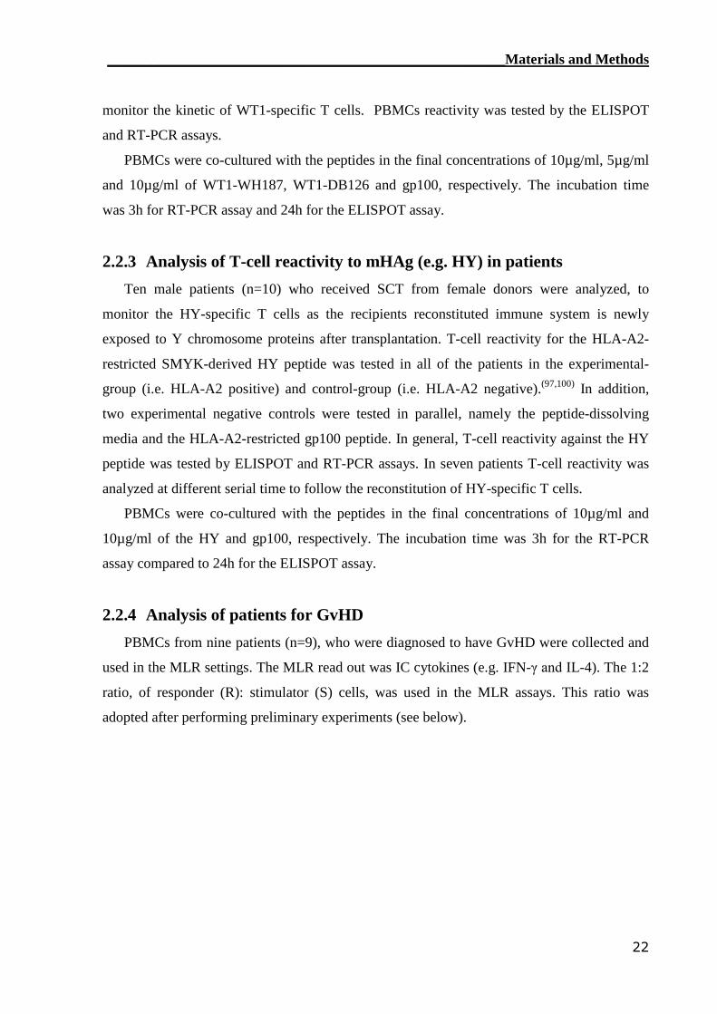
between the different assays was calculated. The tested combinations of CMV serological
status were as shown in table 3 :
CMV Status
Patient Donor
HLA-A2+ + +- ++ -
HLA-A2- + +- ++ -
Table 3: Patient-Donor CMV serological combinations used to detect reconstituted CMV-
specific CD8 T cells.
PBMCs were co-cultured in final concentration of 10µg/ml for the pp65 peptide,(139)
and 20µl/ml for the CMV pp65 protein (i.e. no stock concentration was given by the
provider). The incubation time was 3h (RT-PCR), 6h (ICC) and 24h (ELISPOT), respectively.
2.2.2 Analysis of a potential GvL effect (e.g. WT1) in patients
Patients with leukemia in which the WT1 Ag is potentially over-expressed on the
leukemic blasts (140,141) were enrolled in this study. PBMCs from twelve patients with AML
(n=9), ALL (n=2) and CML (n=1) leukemia were tested for the reconstitution of WT1-
specific T cells after SCT. Two 9-mer WT1 peptides containing the major anchor motifs
essential for binding to HLA-A2 molecules, were used to detect WT1-specific T cells. These
two peptides (Db126: RMFPNAPYL, WH187: SLGEQQYSV, bold letters represent anchor
motifs) are well known to exhibit high binding affinity(140,142). All patients in the
experimental-group were HLA-A2-positive compared to HLA-A2-negative in the control-
group. Moreover, another negative controls were used namely the peptide-dissolving media
and the HLA-A2-restricted gp100 peptide which is derived from the melanocyte lineage-
specific protein PMEL 17 (256-264). Five patients were analyzed at different time points to
_______________________________________________________Materials and Methods
22
monitor the kinetic of WT1-specific T cells. PBMCs reactivity was tested by the ELISPOT
and RT-PCR assays.
PBMCs were co-cultured with the peptides in the final concentrations of 10µg/ml, 5µg/ml
and 10µg/ml of WT1-WH187, WT1-DB126 and gp100, respectively. The incubation time
was 3h for RT-PCR assay and 24h for the ELISPOT assay.
2.2.3 Analysis of T-cell reactivity to mHAg (e.g. HY) in patients
Ten male patients (n=10) who received SCT from female donors were analyzed, to
monitor the HY-specific T cells as the recipients reconstituted immune system is newly
exposed to Y chromosome proteins after transplantation. T-cell reactivity for the HLA-A2-
restricted SMYK-derived HY peptide was tested in all of the patients in the experimental-
group (i.e. HLA-A2 positive) and control-group (i.e. HLA-A2 negative).(97,100) In addition,
two experimental negative controls were tested in parallel, namely the peptide-dissolving
media and the HLA-A2-restricted gp100 peptide. In general, T-cell reactivity against the HY
peptide was tested by ELISPOT and RT-PCR assays. In seven patients T-cell reactivity was
analyzed at different serial time to follow the reconstitution of HY-specific T cells.
PBMCs were co-cultured with the peptides in the final concentrations of 10µg/ml and
10µg/ml of the HY and gp100, respectively. The incubation time was 3h for the RT-PCR
assay compared to 24h for the ELISPOT assay.
2.2.4 Analysis of patients for GvHD
PBMCs from nine patients (n=9), who were diagnosed to have GvHD were collected and
used in the MLR settings. The MLR read out was IC cytokines (e.g. IFN-γ and IL-4). The 1:2
ratio, of responder (R): stimulator (S) cells, was used in the MLR assays. This ratio was
adopted after performing preliminary experiments (see below).
_______________________________________________________Materials and Methods
23
2.3 Methods
2.3.1 Cells processing
2.3.1.1 Isolation of PBMCs
After informed consent and the approval of local Ethical Committee, heparinized
peripheral blood was withdrawn from patients. Peripheral blood mononuclear cells (PBMCs)
were isolated by density gradient centrifugation over Ficoll-Hypaque Plus® according to the
manufacturer recommendations with few modifications. In brief, blood was diluted with PBS
at a 1:1 ratio, then overlayed over Ficoll solution. After centrifugation at 380g for 20min at
room temperature, the interphase was transferred into a new tube. After washing the cells
once with PBS, the erythrocytes were removed by treatment with lysis buffer, another
washing step in PBS was performed.
2.3.1.2 Freezing and storage
After counting on hemocytometer, PBMCs were resuspended in freezing medium and
aliquoted in cryovials. The cryovials were transferred immediately to slow-freeze containers
(i.e. “Mr. Frosty”) which were placed in a -80C freezer for 4h to 24h. Then the cryovials
were transferred in liquid nitrogen.
2.3.1.3 Thawing and resting
PBMCs were thawed in a 37oC water bath, and then transferred to at least ten folds of
complete RPMI medium. Cells were centrifuged at 300g for 10min at 4ºC, resuspended in
complete RPMI medium and rested overnight at 37°C in polypropylene tubes. Finally, the
recovered PBMCs were counted and viability was determined by hemocytometer and trypan
blue vital staining respectively. In general, 60-80% of the rested cells were viable.
2.3.2 MLR
An experimental system was established to simulate the sensitization process in patients
with GvHD patient’s body. In this system HLA-mismatched healthy PBMCs were used to
simulate the alloreactions in GVHD patients.(114) The sensitization step is called as “primary
_______________________________________________________Materials and Methods
24
MLR”, and the real alloreaction is called “secondary MLR” in this thesis. Normally the
primary MLR was performed for seven days and the secondary MLR for 16h.
The PBMCs were plated in U-bottomed 96-well culture plates and incubated at 37°C in
5% CO2. For primary MLR, responder cells (R) were mixed with irradiated (75Gy)
stimulator cells (iS) in R:S ratio of 1:2. After seven days, the sensitized responder cells (sR)
were harvested and a secondary MLR was conducted using the same sR:iS ratio (i.e. 1:2). The
secondary MLR cells were harvested after 3h or 6h, and the IFN-γ mRNA or IC cytokines
were determined by RT-PCR or flow cytometry, respectively (see below). As positive control,
PBMCs were stimulated with 2µg/ml phorbol 12-myristate 13-acetate (PMA) and 100µM
ionomycin (Iono.).
Classical MLR, using 3H-thymidine incorporation, was performed to check the capability
of sR to proliferate in response to second round of stimulation with iS(115). Briefly, responder
PBMC (0.1x10⁶ cells/well), together with irradiated (75Gy) stimulator PBMC (0.2x10⁶
cells/well) were incubated for 7 days in 96-well round-bottom plates in a final culture medium
volume of 200µl/well. On day 7 of culture, 0.037 MBq/ml [methyl-3H]-thymidine was added
to the wells and the plates were incubated for 16h at 37° and 5% CO2 in a humidified
chamber. DNA was harvested on a filter using the Packard Harvester and dried. Subsequently,
scintillation fluid was added and the filters were sealed. The incorporated [3H]-thymidine was
measured using a liquid scintillation counter. Counts per minute (cpm.) were used as readout
for proliferation.
2.3.3 Flow cytometry
2.3.3.1 Cell Surface immunophenotyping
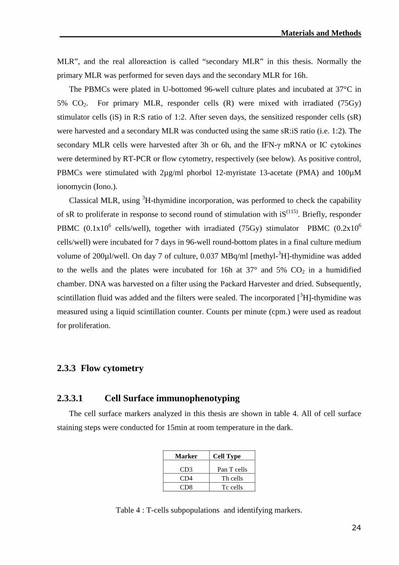
The cell surface markers analyzed in this thesis are shown in table 4. All of cell surface
staining steps were conducted for 15min at room temperature in the dark.
Marker Cell Type
CD3 Pan T cellsCD4 Th cellsCD8 Tc cells
Table 4 : T-cells subpopulations and identifying markers.
_______________________________________________________Materials and Methods
25
Cells were washed with FACS wash buffer. The indicated monoclonal antibodies were
added onto the cell pellet, and incubated for 15min at room temperature. A final washing step
was performed with FACS wash buffer.
2.3.3.2 ICC staining
Intracellular cytokine production was measured in responding cells after inhibiting their
secretion. PBMCs were incubated for 6h after adding the peptide and/or protein then ICC was
performed. In the MLR settings, the incubation time was expanded to 16h before ICC
measurement. As positive control, a combination of 2µg/ml PMA and 100µM ionomycin was
used to activate T cells for cytokines production. The secretion of cytokines was stopped by
adding either GolgiPlug (i.e. Brefeldin A). These protein transport inhibitors accumulate
cytokines in the intracellular compartments (e.g. Golgi apparatus and endoplasmic reticulum)
allowing the measurement of intracellular cytokines by FACS. For tracking the accumulated
cytokines, the BD Cytofix/Cytoperm Plus™ kit was used as recommended by the
manufacturer. In brief, activated PBMCs were harvested and washed with FACS buffer to
block Abs non-specific binding in the following steps. After staining for cell surface markers,
cells were fixed and permeabilized by thoroughly resuspending in 250μl of BD
Cytofix/Cytoperm solution for 20min at 4°C. After washing in 1× BD Perm/Wash solution,
fixed/permeabilized cells were resuspended in 50μl of BD Perm/Wash solution containing
anti-cytokine antibody (e.g. IFN-γ or TNF-α or IL-4 or IL-10 or Perforin). Cells were
incubated for 30min at 4°C in the dark followed by a washing step with 1× BD Perm/Wash
solution, which was also used as staining buffer prior to flow cytometric analysis.
2.3.3.3 TM staining
HLA-A2-restricted CMVpp65-TM loaded with soluble peptide sequence was used to
assess the frequency of reconstituted CMVpp65-specific Tc cells in blood. The staining
protocol was adopted from the CMVpp65-TM manufacturer, with some modifications. In
brief, 1x10⁶ PBMCs were washed and resuspended in 50µl FACS wash buffer. Then PE-
labeled CMVpp65-TM in addition to monoclonal anti-CD3 and anti-CD8 antibodies were
added. After an incubation step for 30min at room temperature in the dark, cells were washed
with 2ml FACS wash buffer and analyzed by FACS. At least 0.5x10⁶ vviable leukocytes were
acquired.
_______________________________________________________Materials and Methods
26
2.3.3.4 PKH26 staining
The stimulator cells in the secondary MLR settings were labelled with PKH26, a red
fluorescent dye that stably integrates into the cell membrane. This step is necessary to
distinguish between stimulator and responder cells (143). Before staining, stimulator PBMCs
were washed twice with serum-free medium. The cells were then resuspended in 200µl
loading buffer (an aqueous, osmolarity-regulating solution containing no Ca2+ or other
physiological salts). 200µl of freshly prepared PKH26 was added to reach a final concentration
of 2.5µM. After an incubation period for 30min at room temperature the staining reaction was
stopped by incubation with 2ml of human serum for 2min. This step is recommended to bind
most of the residual lipophilic PKH26 dye to serum proteins. Following centrifugation, for
5min at 300g, cells were washed twice with 25ml of complete RPMI-1640 medium, and cell
recovery was determined by cell counting.
2.3.3.5 Flow cytometric analysis
For the flow cytometry analysis, 0.3x10⁶ PBMCs were acquired to monitor the ICC
production compared to 0.5x10⁶ for the TM or all viable lymphocytes in the MLR settings.
Flow cytometric analysis was performed using FACS Calibur apparatus and Cellquest Pro
software.
2.3.4 RT-PCR
2.3.4.1 Cell isolation
Cells were harvested by centrifugation at 300g for 10min at 4ºC in eppendorf tubes. Cells
were either stored at -80ºC or were used immediately for RNA extraction.
2.3.4.2 RNA isolation
Cell lysis and total RNA isolation was performed as previously described.(144) Total RNA
was isolated from test samples using the Qiagen RNeasy micro kit with minor modifications.
Briefly, after disrupting cells by RLT buffer and vortexing, the homogenized cell lysate was
mixed with 1 volume of 70% ethanol. After applying onto RNeasy MinElute Spin Columns
and centrifuging for 15s at 8,000g, the flow-through was discarded and the columns were
washed by adding RW1 buffer and centrifuged for 15s at 8,000g. This step was repeated
_______________________________________________________Materials and Methods
27
again with replacing the RW1 buffer by RPE buffer, and then the silica-gel membranes were
dried by adding 500µl of 80% ethanol followed by longer centrifugation step for 3min at
8,000g. After discarding the flow-through, the columns were centrifuged again in
microcentrifuge for 5min at maximum speed followed by discarding the flow-through.
Finally, total RNA was eluted by pipetting 14-25μl RNase-free water onto the silica-gel
membrane and centrifuging for 1min at maximum speed.
The concentration of eluted total RNA was determined by a UV spectrophotometer.
Purified total RNA was either stored at –20°C in RNase-free water or was used immediately
to prepare cDNA.
2.3.4.3 cDNA synthesis
Reverse transcription of mRNA into complementary DNA (cDNA) was carried out using
the Promega Reverse Transcription System. For reverse transcription of IFN-γ and CD8
mRNA, 0.2-0.5µg of total mRNA was mixed with MgCl2, 10X reverse transcription buffer,
dNTP mixture, recombinant RNasin® ribonuclease inhibitor, AMV reverse transcriptase,
oligo (dT) 15 primer and nuclease-free water. The mixture was incubated for 60min at 42ºC,
then 5min at 95ºC and finally for 5min at 4ºC. The synthesized cDNA was stored at -20°C
until usage or immediately used for RT-PCR.
2.3.4.4 RT-PCR (TaqMan®) procedure
Measurement of IFN-γ mRNA gene expression was performed using the Applied
Biosystems ABI Prism 7900 Sequence Detection system as described previously.(144) The
feasibility of this approach for the analysis of Ag-specific T-cell responses has been shown
previously.(145)
PCR primers for IFN-γ, CD8, and Taqman Probes were designed to span exon-exon
junctions to prevent transcription of genomic DNA. After generation of cDNA by reverse
transcription of mRNA, the number of cDNA copies was calculated using the molecular
weight of each gene-specific amplicon. To generate standard curves, serial dilutions of the
amplified gene at known concentrations were tested. CDNA specimens, cDNA standards, and
water, as negative control, were mixed in total volumes of 20µl with Taqman master mix,
400nM primers and 200nM probes.
Thermal cycling was as follows: 2min at 50°C, 10min at 95°C, 40-cycles of 15s at 95°C
and finally 1min at 60°C. Standard curves extrapolation of both IFN-γ and CD8 was
_______________________________________________________Materials and Methods
28
performed using the copy number unit. Sample data were normalized by dividing the IFN-γ
transcripts copy number by the CD8 transcripts copy number.
2.3.4.5 Results analysis
A 2-fold difference in gene expression was found to be within the discrimination ability of
the assay. All RT-PCR assays were performed in duplicates and reported as the mean ±
standard deviation.
2.3.5 ELISPOT
The Letsch et al.(125) ELISPOT assay was adopted with some modifications.(130) In brief,
ELISPOT plates were coated with 15µg/ml mouse-anti-human IFN-γ-mAb for overnight at
4ºC. Then blocking was performed with complete Iscove’s medium for 2h at 37ºC. Overnight
rested PBMCs were cultured in final concentration of 0.05-0.4×10⁶ cells per well into the
coated ELISPOT plates in triplicates when there were enough cells. Stimulation was
performed as follows:
(i) 10μg/ml for CMVpp65, WT1-WH187, HY and gp100.
(ii) 5μg/ml for WT1-Db126.
(iii) 20µl/ml for CMVpp65 protein (stock concentration was not provided by the provider).
(iv) 0.5-1μg/ml PMA and 0.25-0.5nM ionomycin as positive control.
After 24h of incubation at 37°C in 5% CO2, cells were removed by washing six times with
solution of PBS and 0.05% Tween-20. Then, the plates were incubated with 0.626μg/ml anti-
human-IFN-γ biotinylated mAb for 24h at 4°C. After washing the plate six times with PBS,
streptavidin-alkaline phosphatase diluted 1/1,600 was added for 2h at room temperature.
Following another six PBS washes, plates were incubated with BCIP/NBT substrate for
30min. Color development was stopped by washing five times with distilled water. Plates
were dried overnight at room temperature and colored spots were counted using an automated
ELISPOT reader.
_______________________________________________________Materials and Methods
29
2.3 Data evaluation and statistics
2.4.1 Flow cytometry
Acquired cells were compared to negative controls, and then plotted in dot plots in
comparison to negative control.
2.4.2 Classical MLR
Results were presented as c.p.m. means ± S.D. Significant cell proliferation (p<0.05) in
the R+S settings was determined by students t test in comparison to the R cells alone as
negative control.
2.4.3 ELISPOT
Results were expressed as the mean ± standard deviation of counted IFN-γ spots number
after incubation for 24h. A T-cell response was considered positive if the number of spots
with peptide exceeds the number of spots by 10 and the difference between the single values
containing peptide and control is statistically significant at a level of p ≤ 0.05 using the t
test.(124)
2.4.4 RT-PCR
To determine specific response to stimulation, mRNA for IFN-γ from stimulated PBMCs
versus non-stimulated (background) was detected by RT-PCR. The IFN-γ mRNA copy
number was first corrected for CD8 mRNA. A cutoff value (stimulation index) of 2.0 for the
IFN-γ mRNA ratio obtained from stimulated with relevant to non-stimulated PBMCs was
considered to be evidence of epitope specificity. In other words, to minimize the possibility of
falsely considering PBMCs immunoreactivity, a 2-fold increase in stimulated:unstimulated
IFN-γ transcript ratio was assumed as evidence of epitope-specific reactivity.
2.4.5 Linear regression and correlations
The linear regression and correlation analysis of the read outs were calculated using the
Pearson correlation analysis with two-tailed P values. Statistical significance was achieved
when P < 0.05 using Prism 4 software.
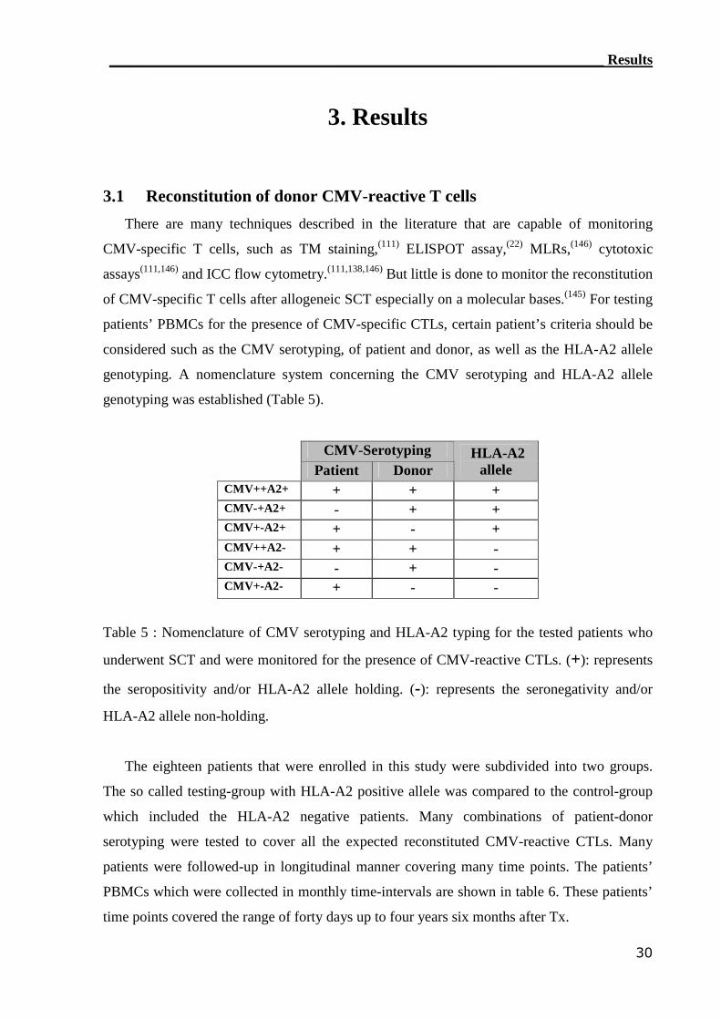
____________________________________________________________________ Results
30
3. Results
3.1 Reconstitution of donor CMV-reactive T cells
There are many techniques described in the literature that are capable of monitoring
CMV-specific T cells, such as TM staining,(111) ELISPOT assay,(22) MLRs,(146) cytotoxic
assays(111,146) and ICC flow cytometry.(111,138,146) But little is done to monitor the reconstitution
of CMV-specific T cells after allogeneic SCT especially on a molecular bases.(145) For testing
patients’ PBMCs for the presence of CMV-specific CTLs, certain patient’s criteria should be
considered such as the CMV serotyping, of patient and donor, as well as the HLA-A2 allele
genotyping. A nomenclature system concerning the CMV serotyping and HLA-A2 allele
genotyping was established (Table 5).
CMV-SerotypingPatient Donor
HLA-A2allele
CMV++A2+ + + +CMV-+A2+ - + +CMV+-A2+ + - +CMV++A2- + + -CMV-+A2- - + -CMV+-A2- + - -
Table 5 : Nomenclature of CMV serotyping and HLA-A2 typing for the tested patients who
underwent SCT and were monitored for the presence of CMV-reactive CTLs. (+): represents
the seropositivity and/or HLA-A2 allele holding. (-): represents the seronegativity and/or
HLA-A2 allele non-holding.
The eighteen patients that were enrolled in this study were subdivided into two groups.
The so called testing-group with HLA-A2 positive allele was compared to the control-group
which included the HLA-A2 negative patients. Many combinations of patient-donor
serotyping were tested to cover all the expected reconstituted CMV-reactive CTLs. Many
patients were followed-up in longitudinal manner covering many time points. The patients’
PBMCs which were collected in monthly time-intervals are shown in table 6. These patients’
time points covered the range of forty days up to four years six months after Tx.
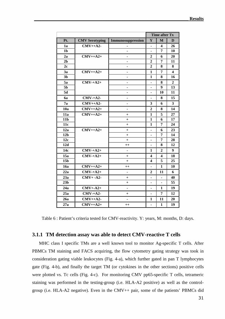
____________________________________________________________________ Results
31
Time after TxPt. CMV Serotyping Immunosuppression Y M D1a - - 4 261b
CMV++A2-- - 7 10
2a - 2 6 202b - 2 7 112c
CMV++A2+
- 2 8 83a - 1 7 43b
CMV++A2+- 1 8 16
5a - - 8 25b - - 9 135d
CMV-+A2+
- - 10 116a CMV-+A2- - - 8 157a CMV++A2- - 3 6 3
10a CMV++A2+ - 2 8 1411a + 1 5 2711b + 1 6 1711c
CMV++A2+
+ 1 7 2412a + - 6 2312b + - 7 1412c + - 7 2812d
CMV++A2+
++ - 8 1214c CMV-+A2+ - 1 2 915a + 4 4 1815b
CMV-+A2++ 4 5 25
16a CMV++A2+ ++ - 1 1022a CMV-+A2+ - 2 11 623a + - - 4023b
CMV+ -A2-+ - - 55
24a CMV+-A2+ - - 1 1925a CMV-+A2- + - 7 1226a CMV++A2- - 1 11 2027a CMV++A2+ ++ - 1 19
Table 6 : Patient’s criteria tested for CMV-reactivity. Y: years, M: months, D: days.
3.1.1 TM detection assay was able to detect CMV-reactive T cells
MHC class I specific TMs are a well known tool to monitor Ag-specific T cells. After
PBMCs TM staining and FACS acquiring, the flow cytometry gating strategy was took in
consideration gating viable leukocytes (Fig. 4-a), which further gated in pan T lymphocytes
gate (Fig. 4-b), and finally the target TM (or cytokines in the other sections) positive cells
were plotted vs. Tc cells (Fig. 4-c). For monitoring CMV pp65-specific T cells, tetrameric
staining was performed in the testing-group (i.e. HLA-A2 positive) as well as the control-
group (i.e. HLA-A2 negative). Even in the CMV++ pair, some of the patients’ PBMCs did
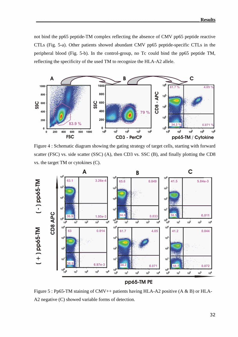
____________________________________________________________________ Results
32
not bind the pp65 peptide-TM complex reflecting the absence of CMV pp65 peptide reactive
CTLs (Fig. 5-a). Other patients showed abundant CMV pp65 peptide-specific CTLs in the
peripheral blood (Fig. 5-b). In the control-group, no Tc could bind the pp65 peptide TM,
reflecting the specificity of the used TM to recognize the HLA-A2 allele.
Figure 4 : Schematic diagram showing the gating strategy of target cells, starting with forward
scatter (FSC) vs. side scatter (SSC) (A), then CD3 vs. SSC (B), and finally plotting the CD8
vs. the target TM or cytokines (C).
Figure 5 : Pp65-TM staining of CMV++ patients having HLA-A2 positive (A & B) or HLA-
A2 negative (C) showed variable forms of detection.
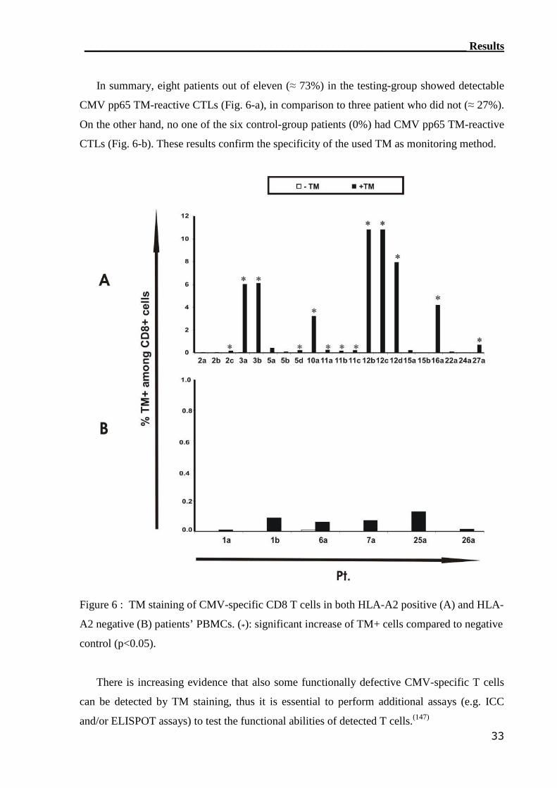
____________________________________________________________________ Results
33
In summary, eight patients out of eleven (≈ 73%) in the testing-group showed detectable
CMV pp65 TM-reactive CTLs (Fig. 6-a), in comparison to three patient who did not (≈ 27%).
On the other hand, no one of the six control-group patients (0%) had CMV pp65 TM-reactive
CTLs (Fig. 6-b). These results confirm the specificity of the used TM as monitoring method.
Figure 6 : TM staining of CMV-specific CD8 T cells in both HLA-A2 positive (A) and HLA-
A2 negative (B) patients’ PBMCs. (*): significant increase of TM+ cells compared to negative
control (p<0.05).
There is increasing evidence that also some functionally defective CMV-specific T cells
can be detected by TM staining, thus it is essential to perform additional assays (e.g. ICC
and/or ELISPOT assays) to test the functional abilities of detected T cells.(147)
____________________________________________________________________ Results
34
3.1.2 IC IFN-γ assay can detect CMV-reactive CTLs
IC IFN-γ was detectable by flow cytometry after 6h reactivation period (Fig. 7). Pt.15a,
who has a HLA-A2 allele, did not have detectable CMV-specific CTLs even after four years
after Tx (Fig. 7-a). This patient did not show any reactive CTLs for neither CMV pp65
peptide nor protein. On the other hand, Pt.2a showed a strong reactivity for CMV pp65
protein but not the peptide. This reflects the presence of CTLs that recognized other epitopes,
of the pp65 protein, rather than the used peptide (Fig. 7-b). Some patients like Pt.3a showed
strong CMV reactivity for both tested CMV Ags (Fig. 7-c). This reflects the presence of many
CMV-specific CTLs in the patients’ peripheral blood. In this patient even some CMV-
reactive Th could be seen in the lower right quadrant, which secreted IFN-γ in MHC class II
manner. In the patients’ control-group, having no HLA-A2 allele, IFN-γ production seemed to
vary. For example in Pt.26a no detectable IFN-γ production in either pp65 peptide or pp65
protein panels (Fig. 7-d), even though this patient underwent SCT two years ago. On the other
hand other patients such as Pt.1a, who underwent SCT five months, showed a significant IFN-
γ production after stimulation with pp65 protein but not with the pp65 peptide (Fig. 7-e). So
this patient has CMV-specific CTLs recognizing many of the pp65 protein epitopes, but not
the tested pp65 peptide which can not be presented by this patient’s antigen presenting cells
due to its HLA-A2 specificity.
A longitudinal follow-up was in scope as PBMCs from many patients were collected on
monthly base. For the longitudinal follow-up, some patients did not show any IFN-γ
production after both viral Ags stimulations (Fig. 8-a). This reactivity was not detectable even
after four years and four months (Pt.15a) or five months (Pt.15b) of Tx. Other patients
developed CMV pp65 protein-reactivity and produced IFN-γ in response to only pp65
protein, but not the HLA-2-restricted peptide. For example, Pt.11a PBMCs showed pp65
reactivity for the protein, but not the peptide, after one year and half (Fig. 8-b). In addition to
the presence of pp65 protein reactive CTLs in peripheral blood, some patients lately
developed CMV reactivity for the pp65 peptide. Eight months after Tx, Pt.5 found to has
CTLs that were able to produce IFN-γ in response to pp65 protein, but not pp-65-peptide (Fig.
8-c) after Tx. But this patient developed CMV reactivity for the pp65 peptide after nine
months of Tx. Other patients’ PBMCs were able to produce IFN-γ in response to both pp65
peptide and pp65 protein. Even PBMCs from the Pt.12b were able to start their CMV
reactivity as early as the seventh month after Tx (Fig. 8-d). So, using the pp-65 protein is
better than using the pp65 HLA-restricted peptides to monitor the CMV reactivity.
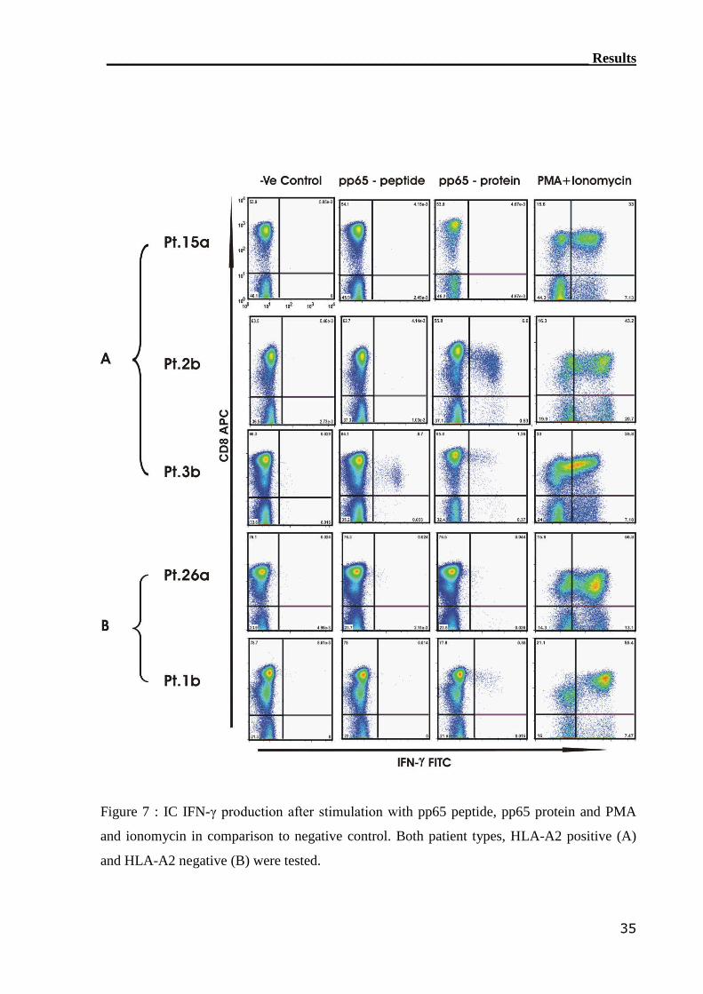
____________________________________________________________________ Results
35
Figure 7 : IC IFN-γ production after stimulation with pp65 peptide, pp65 protein and PMA
and ionomycin in comparison to negative control. Both patient types, HLA-A2 positive (A)
and HLA-A2 negative (B) were tested.
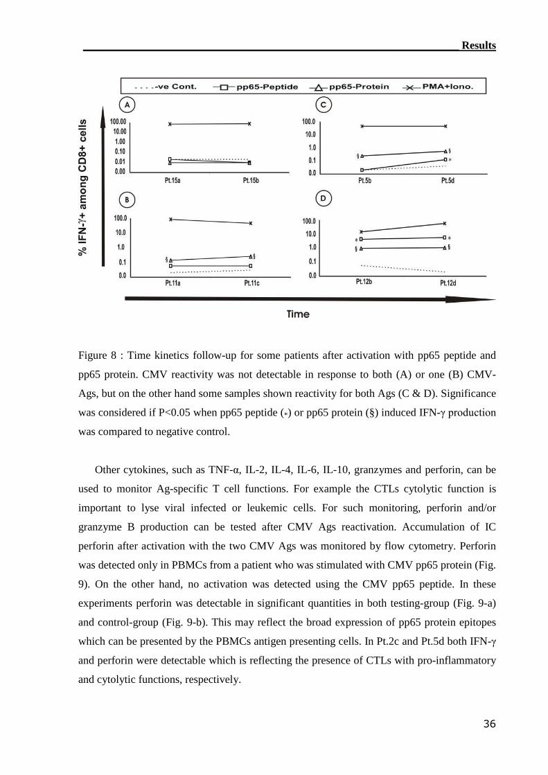
____________________________________________________________________ Results
36
Figure 8 : Time kinetics follow-up for some patients after activation with pp65 peptide and
pp65 protein. CMV reactivity was not detectable in response to both (A) or one (B) CMV-
Ags, but on the other hand some samples shown reactivity for both Ags (C & D). Significance
was considered if P<0.05 when pp65 peptide (*) or pp65 protein (§) induced IFN-γ production
was compared to negative control.
Other cytokines, such as TNF-α, IL-2, IL-4, IL-6, IL-10, granzymes and perforin, can be
used to monitor Ag-specific T cell functions. For example the CTLs cytolytic function is
important to lyse viral infected or leukemic cells. For such monitoring, perforin and/or
granzyme B production can be tested after CMV Ags reactivation. Accumulation of IC
perforin after activation with the two CMV Ags was monitored by flow cytometry. Perforin
was detected only in PBMCs from a patient who was stimulated with CMV pp65 protein (Fig.
9). On the other hand, no activation was detected using the CMV pp65 peptide. In these
experiments perforin was detectable in significant quantities in both testing-group (Fig. 9-a)
and control-group (Fig. 9-b). This may reflect the broad expression of pp65 protein epitopes
which can be presented by the PBMCs antigen presenting cells. In Pt.2c and Pt.5d both IFN-γ
and perforin were detectable which is reflecting the presence of CTLs with pro-inflammatory
and cytolytic functions, respectively.
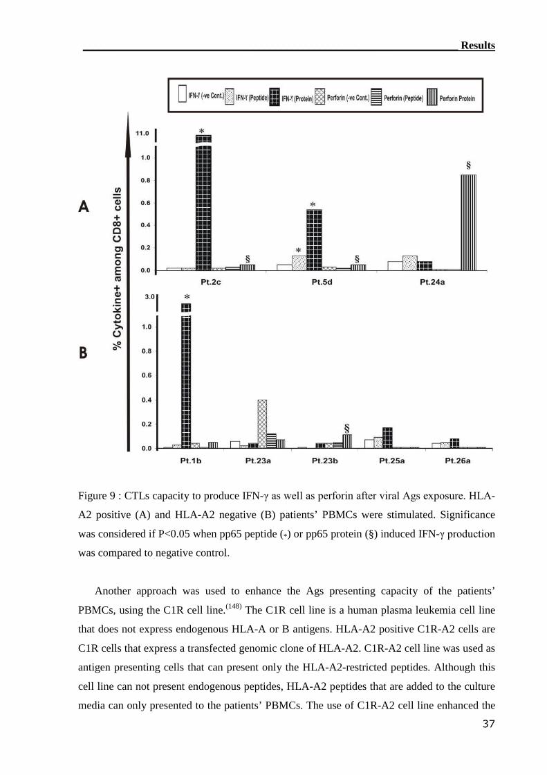
____________________________________________________________________ Results
37
Figure 9 : CTLs capacity to produce IFN-γ as well as perforin after viral Ags exposure. HLA-
A2 positive (A) and HLA-A2 negative (B) patients’ PBMCs were stimulated. Significance
was considered if P<0.05 when pp65 peptide (*) or pp65 protein (§) induced IFN-γ production
was compared to negative control.
Another approach was used to enhance the Ags presenting capacity of the patients’
PBMCs, using the C1R cell line.(148) The C1R cell line is a human plasma leukemia cell line
that does not express endogenous HLA-A or B antigens. HLA-A2 positive C1R-A2 cells are
C1R cells that express a transfected genomic clone of HLA-A2. C1R-A2 cell line was used as
antigen presenting cells that can present only the HLA-A2-restricted peptides. Although this
cell line can not present endogenous peptides, HLA-A2 peptides that are added to the culture
media can only presented to the patients’ PBMCs. The use of C1R-A2 cell line enhanced the
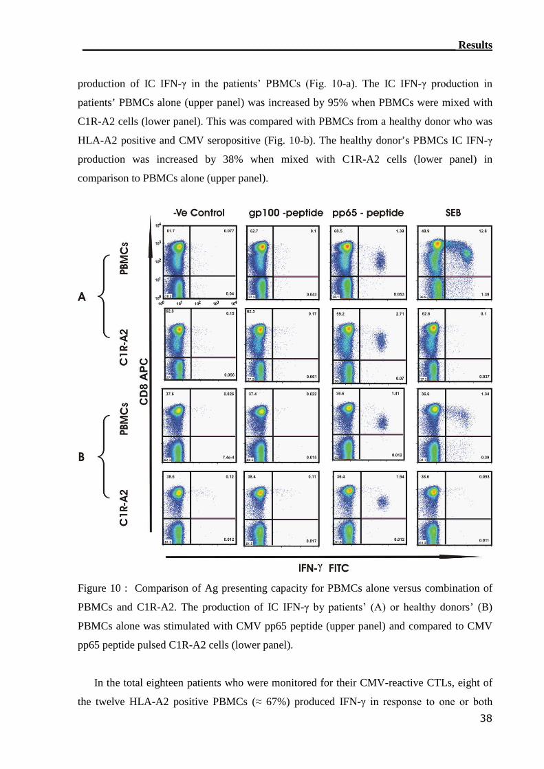
____________________________________________________________________ Results
38
production of IC IFN-γ in the patients’ PBMCs (Fig. 10-a). The IC IFN-γ production in
patients’ PBMCs alone (upper panel) was increased by 95% when PBMCs were mixed with
C1R-A2 cells (lower panel). This was compared with PBMCs from a healthy donor who was
HLA-A2 positive and CMV seropositive (Fig. 10-b). The healthy donor’s PBMCs IC IFN-γ
production was increased by 38% when mixed with C1R-A2 cells (lower panel) in
comparison to PBMCs alone (upper panel).
Figure 10 : Comparison of Ag presenting capacity for PBMCs alone versus combination of
PBMCs and C1R-A2. The production of IC IFN-γ by patients’ (A) or healthy donors’ (B)
PBMCs alone was stimulated with CMV pp65 peptide (upper panel) and compared to CMV
pp65 peptide pulsed C1R-A2 cells (lower panel).
In the total eighteen patients who were monitored for their CMV-reactive CTLs, eight of
the twelve HLA-A2 positive PBMCs (≈ 67%) produced IFN-γ in response to one or both
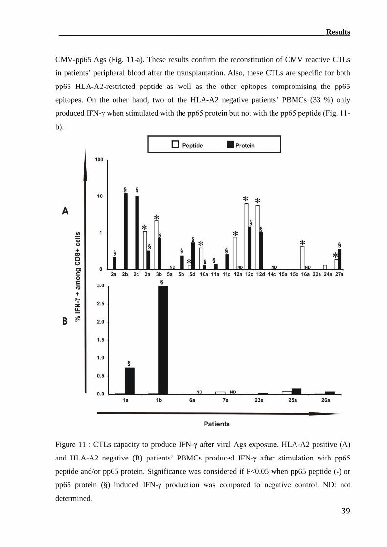
____________________________________________________________________ Results
39
CMV-pp65 Ags (Fig. 11-a). These results confirm the reconstitution of CMV reactive CTLs
in patients’ peripheral blood after the transplantation. Also, these CTLs are specific for both
pp65 HLA-A2-restricted peptide as well as the other epitopes compromising the pp65
epitopes. On the other hand, two of the HLA-A2 negative patients’ PBMCs (33 %) only
produced IFN-γ when stimulated with the pp65 protein but not with the pp65 peptide (Fig. 11-
b).
Figure 11 : CTLs capacity to produce IFN-γ after viral Ags exposure. HLA-A2 positive (A)
and HLA-A2 negative (B) patients’ PBMCs produced IFN-γ after stimulation with pp65
peptide and/or pp65 protein. Significance was considered if P<0.05 when pp65 peptide (*) or
pp65 protein (§) induced IFN-γ production was compared to negative control. ND: not
determined.
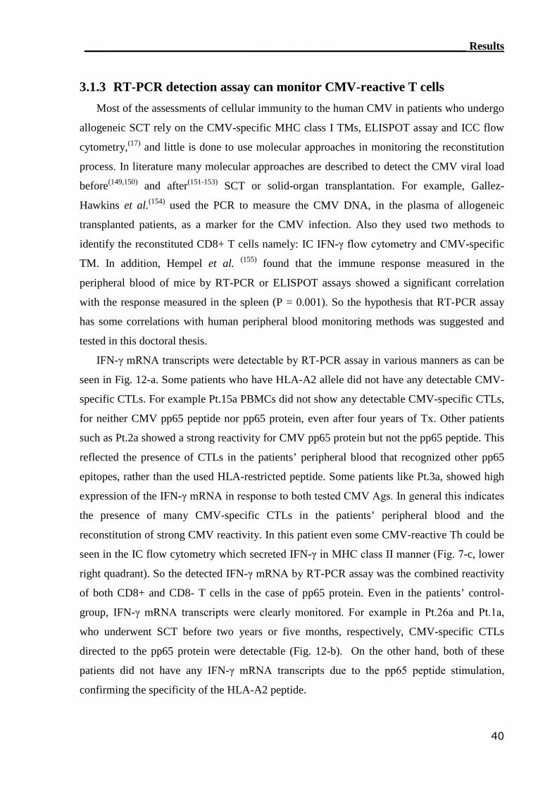
____________________________________________________________________ Results
40
3.1.3 RT-PCR detection assay can monitor CMV-reactive T cells
Most of the assessments of cellular immunity to the human CMV in patients who undergo
allogeneic SCT rely on the CMV-specific MHC class I TMs, ELISPOT assay and ICC flow
cytometry,(17) and little is done to use molecular approaches in monitoring the reconstitution
process. In literature many molecular approaches are described to detect the CMV viral load
before(149,150) and after(151-153) SCT or solid-organ transplantation. For example, Gallez-
Hawkins et al.(154) used the PCR to measure the CMV DNA, in the plasma of allogeneic
transplanted patients, as a marker for the CMV infection. Also they used two methods to
identify the reconstituted CD8+ T cells namely: IC IFN-γ flow cytometry and CMV-specific
TM. In addition, Hempel et al. (155) found that the immune response measured in the
peripheral blood of mice by RT-PCR or ELISPOT assays showed a significant correlation
with the response measured in the spleen (P = 0.001). So the hypothesis that RT-PCR assay
has some correlations with human peripheral blood monitoring methods was suggested and
tested in this doctoral thesis.
IFN-γ mRNA transcripts were detectable by RT-PCR assay in various manners as can be
seen in Fig. 12-a. Some patients who have HLA-A2 allele did not have any detectable CMV-
specific CTLs. For example Pt.15a PBMCs did not show any detectable CMV-specific CTLs,
for neither CMV pp65 peptide nor pp65 protein, even after four years of Tx. Other patients
such as Pt.2a showed a strong reactivity for CMV pp65 protein but not the pp65 peptide. This
reflected the presence of CTLs in the patients’ peripheral blood that recognized other pp65
epitopes, rather than the used HLA-restricted peptide. Some patients like Pt.3a, showed high
expression of the IFN-γ mRNA in response to both tested CMV Ags. In general this indicates
the presence of many CMV-specific CTLs in the patients’ peripheral blood and the
reconstitution of strong CMV reactivity. In this patient even some CMV-reactive Th could be
seen in the IC flow cytometry which secreted IFN-γ in MHC class II manner (Fig. 7-c, lower
right quadrant). So the detected IFN-γ mRNA by RT-PCR assay was the combined reactivity
of both CD8+ and CD8- T cells in the case of pp65 protein. Even in the patients’ control-
group, IFN-γ mRNA transcripts were clearly monitored. For example in Pt.26a and Pt.1a,
who underwent SCT before two years or five months, respectively, CMV-specific CTLs
directed to the pp65 protein were detectable (Fig. 12-b). On the other hand, both of these
patients did not have any IFN-γ mRNA transcripts due to the pp65 peptide stimulation,
confirming the specificity of the HLA-A2 peptide.
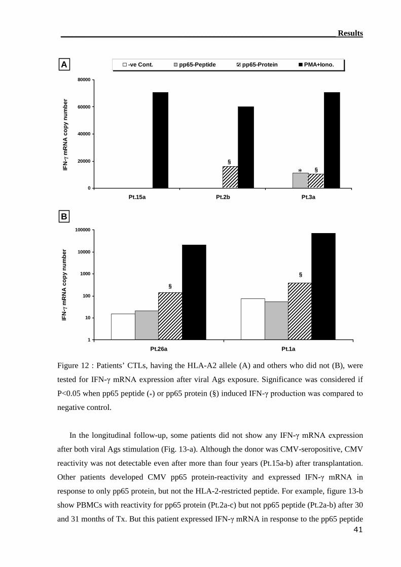
____________________________________________________________________ Results
41
0
20000
40000
60000
80000
Pt.15a Pt.2b Pt.3a
IFN
-γm
RN
Aco
py
nu
mb
er-ve Cont. pp65-Peptide pp65-Protein PMA+Iono.
§*§
A
1
10
100
1000
10000
100000
Pt.26a Pt.1a
IFN
-γm
RN
Aco
py
nu
mb
er
§
§
B
Figure 12 : Patients’ CTLs, having the HLA-A2 allele (A) and others who did not (B), were
tested for IFN-γ mRNA expression after viral Ags exposure. Significance was considered if
P<0.05 when pp65 peptide (*) or pp65 protein (§) induced IFN-γ production was compared to
negative control.
In the longitudinal follow-up, some patients did not show any IFN-γ mRNA expression
after both viral Ags stimulation (Fig. 13-a). Although the donor was CMV-seropositive, CMV
reactivity was not detectable even after more than four years (Pt.15a-b) after transplantation.
Other patients developed CMV pp65 protein-reactivity and expressed IFN-γ mRNA in
response to only pp65 protein, but not the HLA-2-restricted peptide. For example, figure 13-b
show PBMCs with reactivity for pp65 protein (Pt.2a-c) but not pp65 peptide (Pt.2a-b) after 30
and 31 months of Tx. But this patient expressed IFN-γ mRNA in response to the pp65 peptide

____________________________________________________________________ Results
42
at the 32 months after Tx. In addition to the presence of pp65 protein reactive CTLs in
peripheral blood, some patients lately developed CMV reactivity for the pp65 peptide. Eight
months after Tx, Pt.5 shown to have CTLs that expressed IFN-γ mRNA in response to pp65
protein, but not pp-65-peptide (Fig. 13-c). But this patient developed CMV reactivity for the
pp65 peptide at the ninth month after Tx. Finally, some patients PBMCs such as Pt.12b were
able to express IFN-γ mRNA in response to both pp65 peptide and pp65 protein (Fig. 13-d).
Even this patient started his CMV reactivity as early as the seventh month after Tx. As a
summary, the use of pp-65 protein is better than the pp65 HLA-restricted peptides to monitor
the CMV-reactivity by the RT-PCR assay.
Figure 13 : CMV-reactivity monitoring time kinetics follow-up in some patients using the
RT-PCR assay. CMV-reactivity was not detectable for both Ags (A), or found in addition to
all pp65 protein time points (B) in some of pp65 peptide treatment (C). Also in some patients,
CMV-reactivity was found in all time points after both pp65 protein and pp65 peptide (D).
Significance was considered if P<0.05 when pp65 peptide (*) or pp65 protein (§) induced
IFN-γ production was compared to negative control.
Blood samples from thirty patients were monitored for the presence of CMV-reactive
CTLs. In the HLA-A2 positive patients’ samples, fifteen out of twenty two (68%) expressed
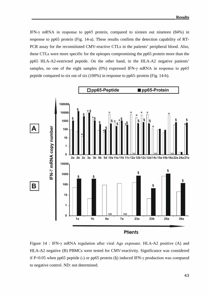
____________________________________________________________________ Results
43
IFN-γ mRNA in response to pp65 protein, compared to sixteen out nineteen (84%) in
response to pp65 protein (Fig. 14-a). These results confirm the detection capability of RT-
PCR assay for the reconstituted CMV-reactive CTLs in the patients’ peripheral blood. Also,
these CTLs were more specific for the epitopes compromising the pp65 protein more than the
pp65 HLA-A2-restricted peptide. On the other hand, in the HLA-A2 negative patients’
samples, no one of the eight samples (0%) expressed IFN-γ mRNA in response to pp65
peptide compared to six out of six (100%) in response to pp65–protein (Fig. 14-b).
Figure 14 : IFN-γ mRNA regulation after viral Ags exposure. HLA-A2 positive (A) and
HLA-A2 negative (B) PBMCs were tested for CMV-reactivity. Significance was considered
if P<0.05 when pp65 peptide (*) or pp65 protein (§) induced IFN-γ production was compared
to negative control. ND: not determined.
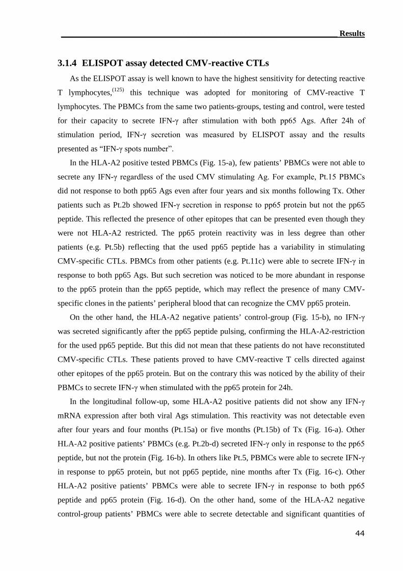
____________________________________________________________________ Results
44
3.1.4 ELISPOT assay detected CMV-reactive CTLs
As the ELISPOT assay is well known to have the highest sensitivity for detecting reactive
T lymphocytes,(125) this technique was adopted for monitoring of CMV-reactive T
lymphocytes. The PBMCs from the same two patients-groups, testing and control, were tested
for their capacity to secrete IFN-γ after stimulation with both pp65 Ags. After 24h of
stimulation period, IFN-γ secretion was measured by ELISPOT assay and the results
presented as “IFN-γ spots number”.
In the HLA-A2 positive tested PBMCs (Fig. 15-a), few patients’ PBMCs were not able to
secrete any IFN-γ regardless of the used CMV stimulating Ag. For example, Pt.15 PBMCs
did not response to both pp65 Ags even after four years and six months following Tx. Other
patients such as Pt.2b showed IFN-γ secretion in response to pp65 protein but not the pp65
peptide. This reflected the presence of other epitopes that can be presented even though they
were not HLA-A2 restricted. The pp65 protein reactivity was in less degree than other
patients (e.g. Pt.5b) reflecting that the used pp65 peptide has a variability in stimulating
CMV-specific CTLs. PBMCs from other patients (e.g. Pt.11c) were able to secrete IFN-γ in
response to both pp65 Ags. But such secretion was noticed to be more abundant in response
to the pp65 protein than the pp65 peptide, which may reflect the presence of many CMV-
specific clones in the patients’ peripheral blood that can recognize the CMV pp65 protein.
On the other hand, the HLA-A2 negative patients’ control-group (Fig. 15-b), no IFN-γ
was secreted significantly after the pp65 peptide pulsing, confirming the HLA-A2-restriction
for the used pp65 peptide. But this did not mean that these patients do not have reconstituted
CMV-specific CTLs. These patients proved to have CMV-reactive T cells directed against
other epitopes of the pp65 protein. But on the contrary this was noticed by the ability of their
PBMCs to secrete IFN-γ when stimulated with the pp65 protein for 24h.
In the longitudinal follow-up, some HLA-A2 positive patients did not show any IFN-γ
mRNA expression after both viral Ags stimulation. This reactivity was not detectable even
after four years and four months (Pt.15a) or five months (Pt.15b) of Tx (Fig. 16-a). Other
HLA-A2 positive patients’ PBMCs (e.g. Pt.2b-d) secreted IFN-γ only in response to the pp65
peptide, but not the protein (Fig. 16-b). In others like Pt.5, PBMCs were able to secrete IFN-γ
in response to pp65 protein, but not pp65 peptide, nine months after Tx (Fig. 16-c). Other
HLA-A2 positive patients’ PBMCs were able to secrete IFN-γ in response to both pp65
peptide and pp65 protein (Fig. 16-d). On the other hand, some of the HLA-A2 negative
control-group patients’ PBMCs were able to secrete detectable and significant quantities of
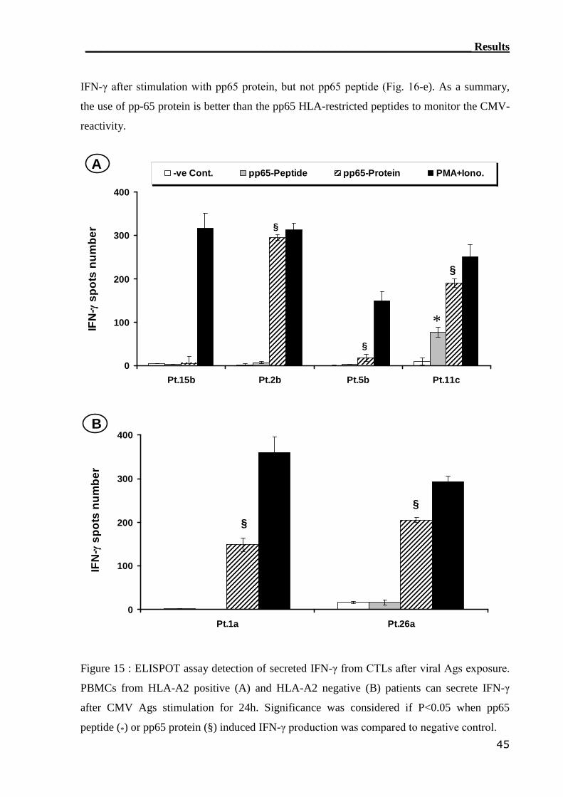
____________________________________________________________________ Results
45
IFN-γ after stimulation with pp65 protein, but not pp65 peptide (Fig. 16-e). As a summary,
the use of pp-65 protein is better than the pp65 HLA-restricted peptides to monitor the CMV-
reactivity.
0
100
200
300
400
Pt.15b Pt.2b Pt.5b Pt.11c
IFN
-γsp
ots
nu
mb
er
-ve Cont. pp65-Peptide pp65-Protein PMA+Iono.
*
§
§
§
A
0
100
200
300
400
Pt.1a Pt.26a
IFN
-γsp
ots
nu
mb
er
B
§
§
Figure 15 : ELISPOT assay detection of secreted IFN-γ from CTLs after viral Ags exposure.
PBMCs from HLA-A2 positive (A) and HLA-A2 negative (B) patients can secrete IFN-γ
after CMV Ags stimulation for 24h. Significance was considered if P<0.05 when pp65
peptide (*) or pp65 protein (§) induced IFN-γ production was compared to negative control.
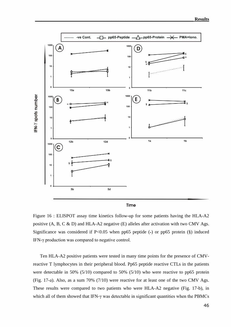
____________________________________________________________________ Results
46
Figure 16 : ELISPOT assay time kinetics follow-up for some patients having the HLA-A2
positive (A, B, C & D) and HLA-A2 negative (E) alleles after activation with two CMV Ags.
Significance was considered if P<0.05 when pp65 peptide (*) or pp65 protein (§) induced
IFN-γ production was compared to negative control.
Ten HLA-A2 positive patients were tested in many time points for the presence of CMV-
reactive T lymphocytes in their peripheral blood. Pp65 peptide reactive CTLs in the patients
were detectable in 50% (5/10) compared to 50% (5/10) who were reactive to pp65 protein
(Fig. 17-a). Also, as a sum 70% (7/10) were reactive for at least one of the two CMV Ags.
These results were compared to two patients who were HLA-A2 negative (Fig. 17-b), in
which all of them showed that IFN-γ was detectable in significant quantities when the PBMCs
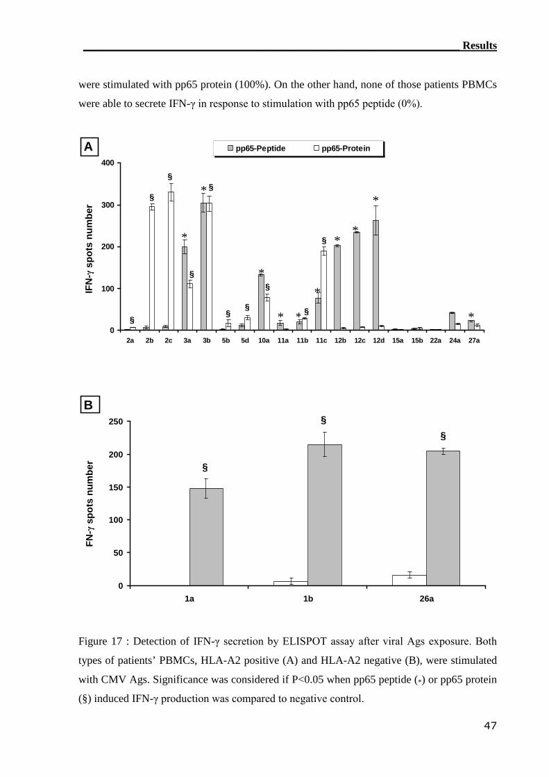
____________________________________________________________________ Results
47
were stimulated with pp65 protein (100%). On the other hand, none of those patients PBMCs
were able to secrete IFN-γ in response to stimulation with pp65 peptide (0%).
0
100
200
300
400
2a 2b 2c 3a 3b 5b 5d 10a 11a 11b 11c 12b 12c 12d 15a 15b 22a 24a 27a
IFN
-γsp
ots
nu
mb
er
pp65-Peptide pp65-Protein
§*
*
*
* *
*
**
*
*§
§
§§
§
§
§
§
§
A
0
50
100
150
200
250
1a 1b 26a
FN
-γsp
ots
nu
mb
er
§
§
§
B
Figure 17 : Detection of IFN-γ secretion by ELISPOT assay after viral Ags exposure. Both
types of patients’ PBMCs, HLA-A2 positive (A) and HLA-A2 negative (B), were stimulated
with CMV Ags. Significance was considered if P<0.05 when pp65 peptide (*) or pp65 protein
(§) induced IFN-γ production was compared to negative control.
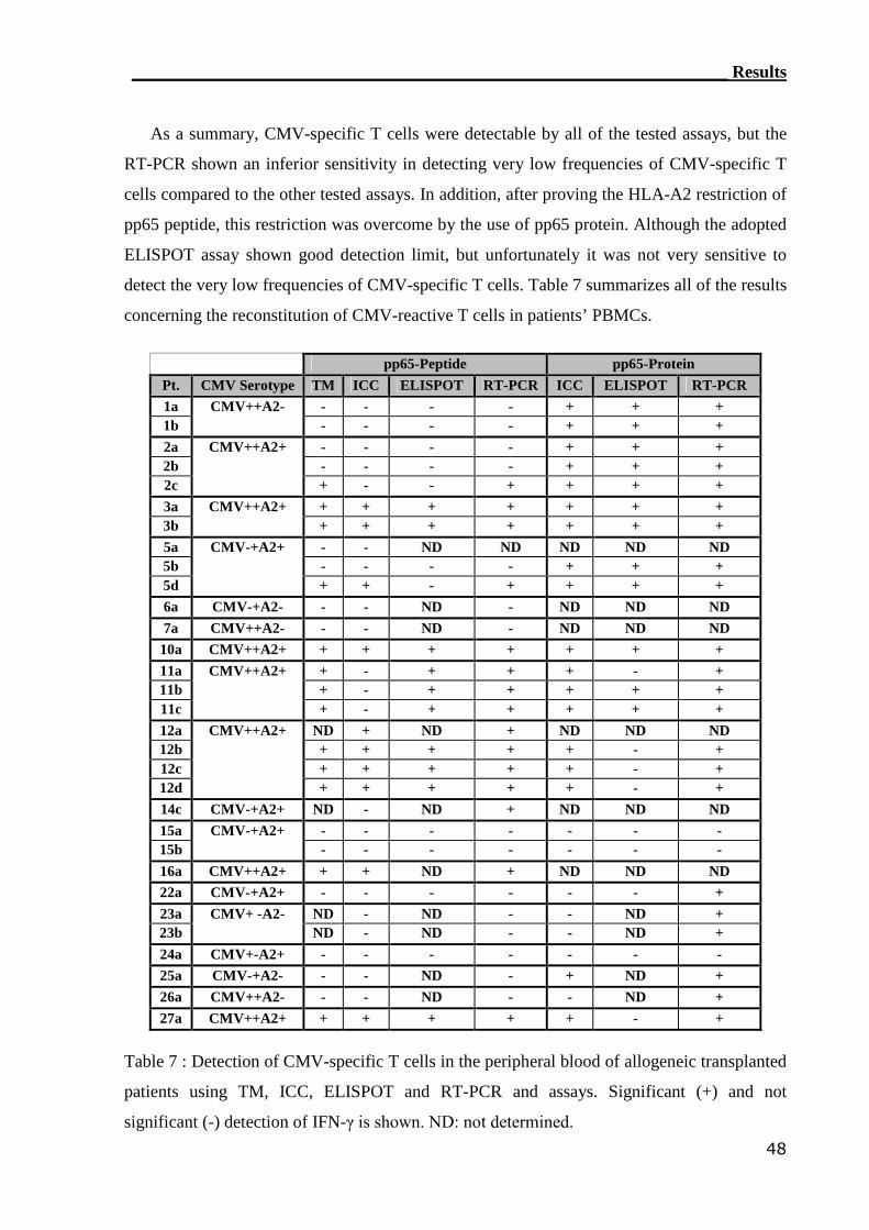
____________________________________________________________________ Results
48
As a summary, CMV-specific T cells were detectable by all of the tested assays, but the
RT-PCR shown an inferior sensitivity in detecting very low frequencies of CMV-specific T
cells compared to the other tested assays. In addition, after proving the HLA-A2 restriction of
pp65 peptide, this restriction was overcome by the use of pp65 protein. Although the adopted
ELISPOT assay shown good detection limit, but unfortunately it was not very sensitive to
detect the very low frequencies of CMV-specific T cells. Table 7 summarizes all of the results
concerning the reconstitution of CMV-reactive T cells in patients’ PBMCs.
pp65-Peptide pp65-ProteinPt. CMV Serotype TM ICC ELISPOT RT-PCR ICC ELISPOT RT-PCR1a - - - - + + +1b
CMV++A2-- - - - + + +
2a - - - - + + +2b - - - - + + +2c
CMV++A2+
+ - - + + + +3a + + + + + + +3b
CMV++A2++ + + + + + +
5a - - ND ND ND ND ND5b - - - - + + +5d
CMV-+A2+
+ + - + + + +6a CMV-+A2- - - ND - ND ND ND7a CMV++A2- - - ND - ND ND ND
10a CMV++A2+ + + + + + + +11a + - + + + - +11b + - + + + + +11c
CMV++A2+
+ - + + + + +12a ND + ND + ND ND ND12b + + + + + - +12c + + + + + - +12d
CMV++A2+
+ + + + + - +14c CMV-+A2+ ND - ND + ND ND ND15a - - - - - - -15b
CMV-+A2+- - - - - - -
16a CMV++A2+ + + ND + ND ND ND22a CMV-+A2+ - - - - - - +23a ND - ND - - ND +23b
CMV+ -A2-ND - ND - - ND +
24a CMV+-A2+ - - - - - - -25a CMV-+A2- - - ND - + ND +26a CMV++A2- - - ND - - ND +27a CMV++A2+ + + + + + - +
Table 7 : Detection of CMV-specific T cells in the peripheral blood of allogeneic transplanted
patients using TM, ICC, ELISPOT and RT-PCR and assays. Significant (+) and not
significant (-) detection of IFN-γ is shown. ND: not determined.
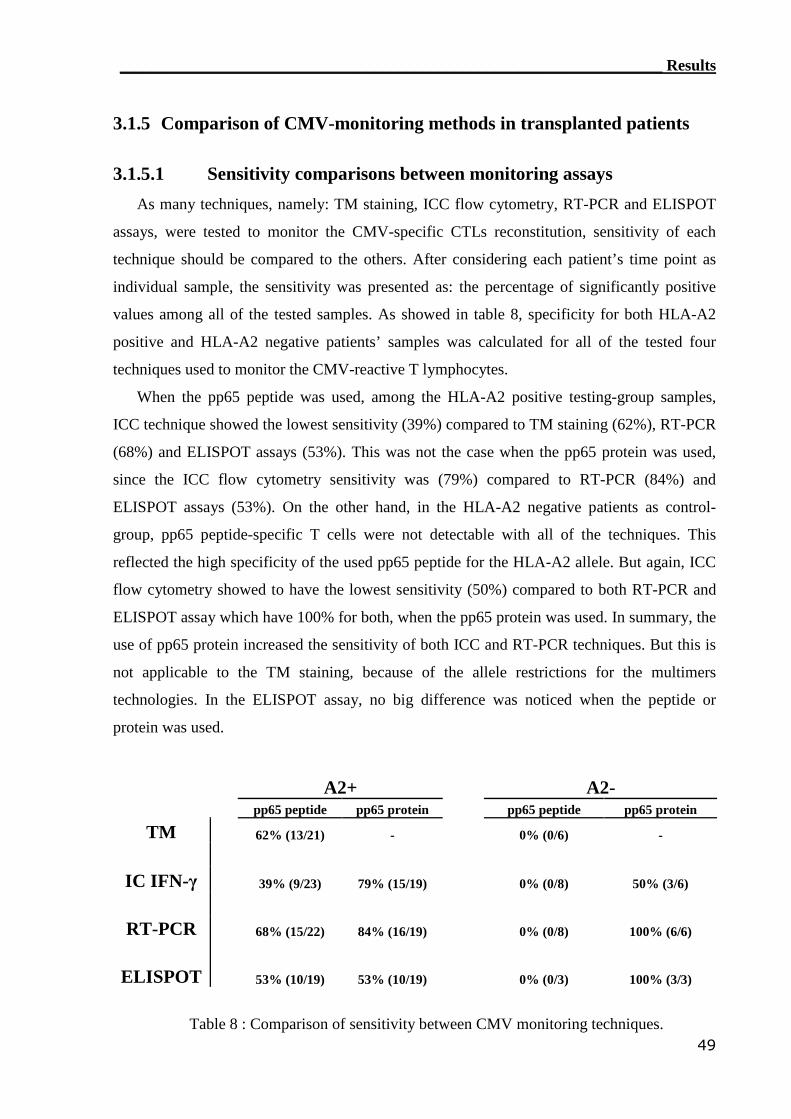
____________________________________________________________________ Results
49
3.1.5 Comparison of CMV-monitoring methods in transplanted patients
3.1.5.1 Sensitivity comparisons between monitoring assays
As many techniques, namely: TM staining, ICC flow cytometry, RT-PCR and ELISPOT
assays, were tested to monitor the CMV-specific CTLs reconstitution, sensitivity of each
technique should be compared to the others. After considering each patient’s time point as
individual sample, the sensitivity was presented as: the percentage of significantly positive
values among all of the tested samples. As showed in table 8, specificity for both HLA-A2
positive and HLA-A2 negative patients’ samples was calculated for all of the tested four
techniques used to monitor the CMV-reactive T lymphocytes.
When the pp65 peptide was used, among the HLA-A2 positive testing-group samples,
ICC technique showed the lowest sensitivity (39%) compared to TM staining (62%), RT-PCR
(68%) and ELISPOT assays (53%). This was not the case when the pp65 protein was used,
since the ICC flow cytometry sensitivity was (79%) compared to RT-PCR (84%) and
ELISPOT assays (53%). On the other hand, in the HLA-A2 negative patients as control-
group, pp65 peptide-specific T cells were not detectable with all of the techniques. This
reflected the high specificity of the used pp65 peptide for the HLA-A2 allele. But again, ICC
flow cytometry showed to have the lowest sensitivity (50%) compared to both RT-PCR and
ELISPOT assay which have 100% for both, when the pp65 protein was used. In summary, the
use of pp65 protein increased the sensitivity of both ICC and RT-PCR techniques. But this is
not applicable to the TM staining, because of the allele restrictions for the multimers
technologies. In the ELISPOT assay, no big difference was noticed when the peptide or
protein was used.
A2+ A2-pp65 peptide pp65 protein pp65 peptide pp65 protein
TM 62% (13/21) - 0% (0/6) -
IC IFN-γ 39% (9/23) 79% (15/19) 0% (0/8) 50% (3/6)
RT-PCR 68% (15/22) 84% (16/19) 0% (0/8) 100% (6/6)
ELISPOT 53% (10/19) 53% (10/19) 0% (0/3) 100% (3/3)
Table 8 : Comparison of sensitivity between CMV monitoring techniques.
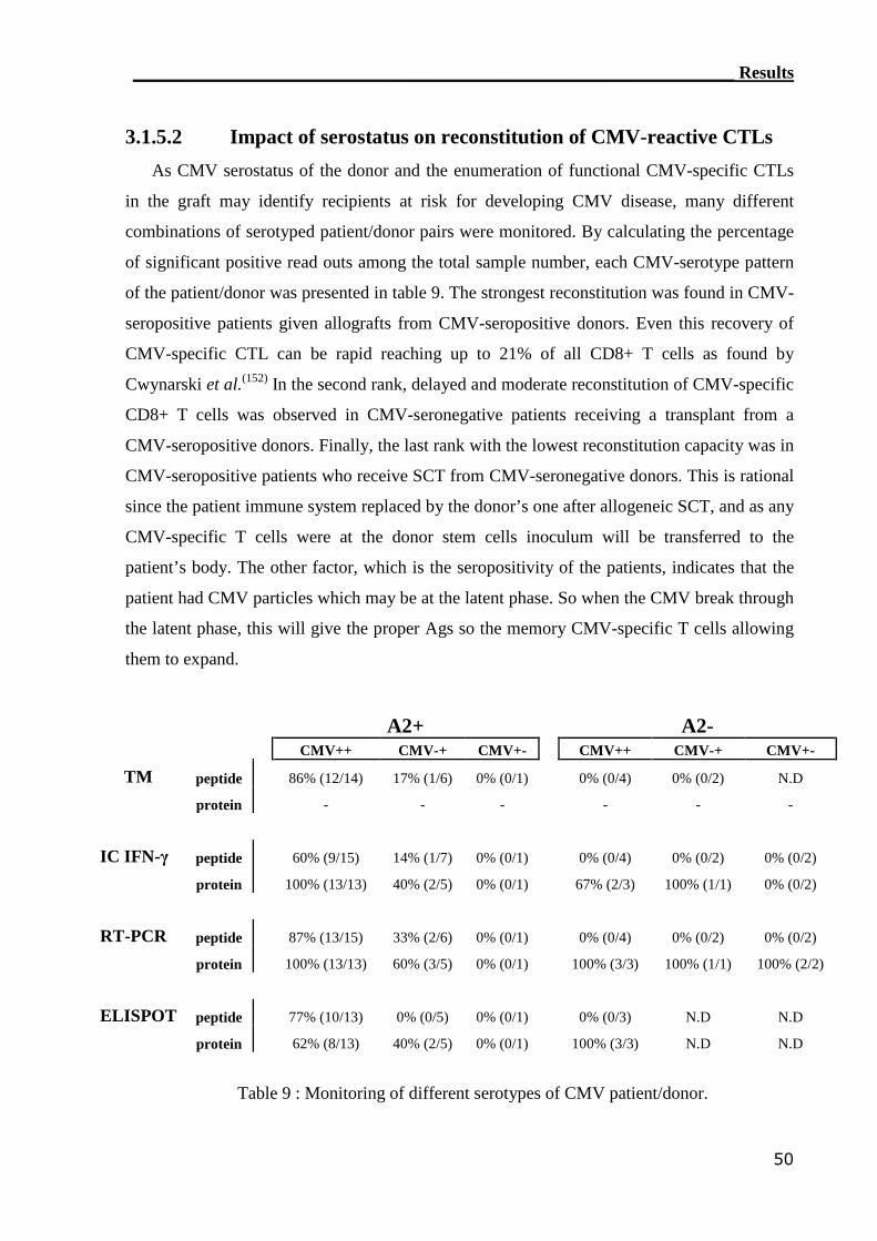
____________________________________________________________________ Results
50
3.1.5.2 Impact of serostatus on reconstitution of CMV-reactive CTLs
As CMV serostatus of the donor and the enumeration of functional CMV-specific CTLs
in the graft may identify recipients at risk for developing CMV disease, many different
combinations of serotyped patient/donor pairs were monitored. By calculating the percentage
of significant positive read outs among the total sample number, each CMV-serotype pattern
of the patient/donor was presented in table 9. The strongest reconstitution was found in CMV-
seropositive patients given allografts from CMV-seropositive donors. Even this recovery of
CMV-specific CTL can be rapid reaching up to 21% of all CD8+ T cells as found by
Cwynarski et al.(152) In the second rank, delayed and moderate reconstitution of CMV-specific
CD8+ T cells was observed in CMV-seronegative patients receiving a transplant from a
CMV-seropositive donors. Finally, the last rank with the lowest reconstitution capacity was in
CMV-seropositive patients who receive SCT from CMV-seronegative donors. This is rational
since the patient immune system replaced by the donor’s one after allogeneic SCT, and as any
CMV-specific T cells were at the donor stem cells inoculum will be transferred to the
patient’s body. The other factor, which is the seropositivity of the patients, indicates that the
patient had CMV particles which may be at the latent phase. So when the CMV break through
the latent phase, this will give the proper Ags so the memory CMV-specific T cells allowing
them to expand.
A2+ A2-CMV++ CMV-+ CMV+- CMV++ CMV-+ CMV+-
TM peptide 86% (12/14) 17% (1/6) 0% (0/1) 0% (0/4) 0% (0/2) N.D
protein - - - - - -
IC IFN-γ peptide 60% (9/15) 14% (1/7) 0% (0/1) 0% (0/4) 0% (0/2) 0% (0/2)
protein 100% (13/13) 40% (2/5) 0% (0/1) 67% (2/3) 100% (1/1) 0% (0/2)
RT-PCR peptide 87% (13/15) 33% (2/6) 0% (0/1) 0% (0/4) 0% (0/2) 0% (0/2)
protein 100% (13/13) 60% (3/5) 0% (0/1) 100% (3/3) 100% (1/1) 100% (2/2)
ELISPOT peptide 77% (10/13) 0% (0/5) 0% (0/1) 0% (0/3) N.D N.D
protein 62% (8/13) 40% (2/5) 0% (0/1) 100% (3/3) N.D N.D
Table 9 : Monitoring of different serotypes of CMV patient/donor.
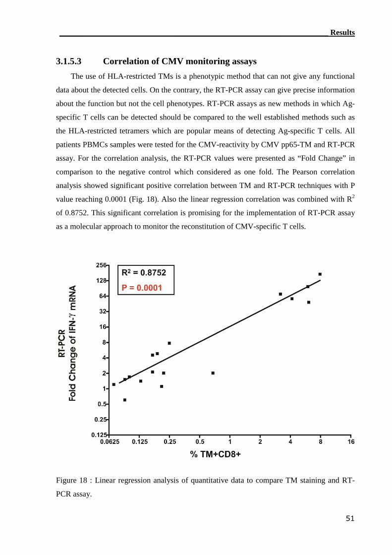
____________________________________________________________________ Results
51
3.1.5.3 Correlation of CMV monitoring assays
The use of HLA-restricted TMs is a phenotypic method that can not give any functional
data about the detected cells. On the contrary, the RT-PCR assay can give precise information
about the function but not the cell phenotypes. RT-PCR assays as new methods in which Ag-
specific T cells can be detected should be compared to the well established methods such as
the HLA-restricted tetramers which are popular means of detecting Ag-specific T cells. All
patients PBMCs samples were tested for the CMV-reactivity by CMV pp65-TM and RT-PCR
assay. For the correlation analysis, the RT-PCR values were presented as “Fold Change” in
comparison to the negative control which considered as one fold. The Pearson correlation
analysis showed significant positive correlation between TM and RT-PCR techniques with P
value reaching 0.0001 (Fig. 18). Also the linear regression correlation was combined with R2
of 0.8752. This significant correlation is promising for the implementation of RT-PCR assay
as a molecular approach to monitor the reconstitution of CMV-specific T cells.
Figure 18 : Linear regression analysis of quantitative data to compare TM staining and RT-
PCR assay.
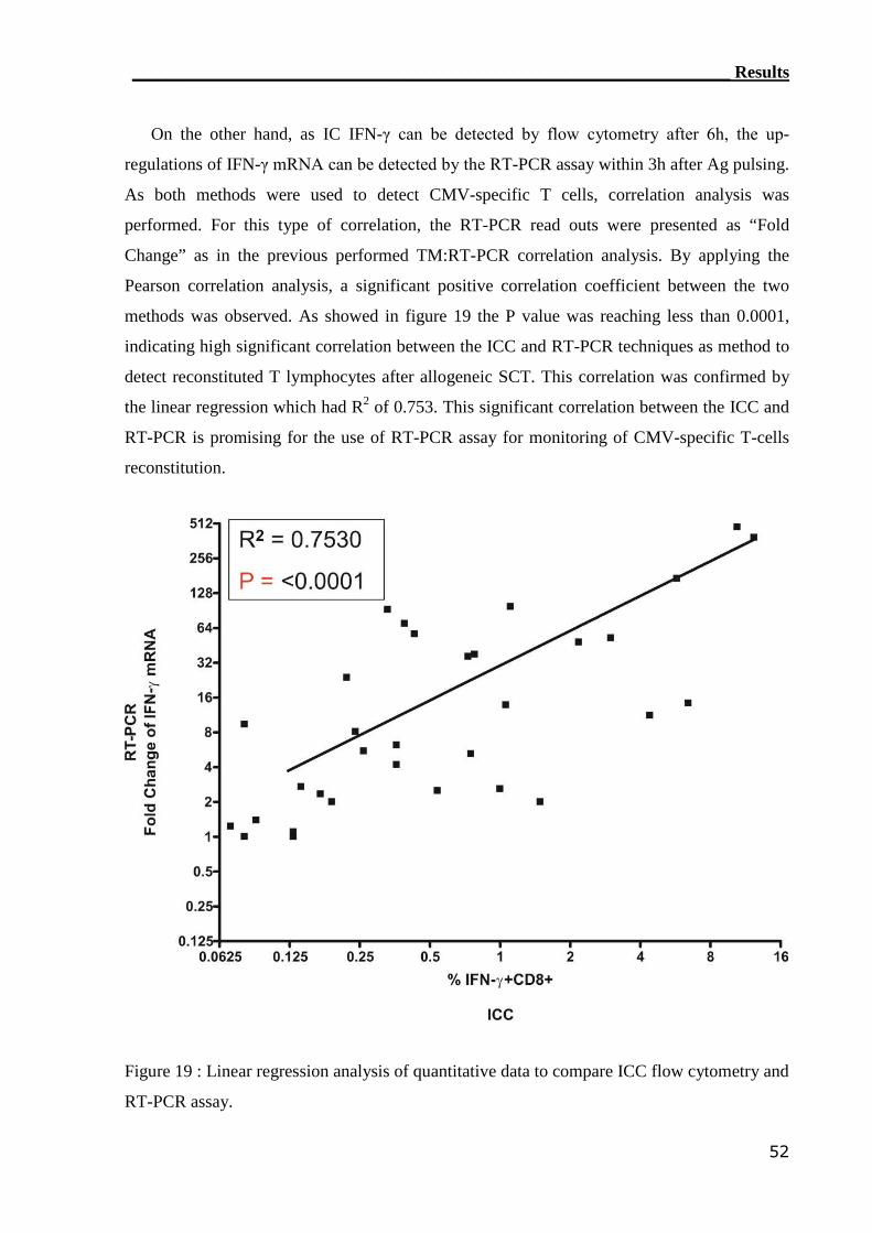
____________________________________________________________________ Results
52
On the other hand, as IC IFN-γ can be detected by flow cytometry after 6h, the up-
regulations of IFN-γ mRNA can be detected by the RT-PCR assay within 3h after Ag pulsing.
As both methods were used to detect CMV-specific T cells, correlation analysis was
performed. For this type of correlation, the RT-PCR read outs were presented as “Fold
Change” as in the previous performed TM:RT-PCR correlation analysis. By applying the
Pearson correlation analysis, a significant positive correlation coefficient between the two
methods was observed. As showed in figure 19 the P value was reaching less than 0.0001,
indicating high significant correlation between the ICC and RT-PCR techniques as method to
detect reconstituted T lymphocytes after allogeneic SCT. This correlation was confirmed by
the linear regression which had R2 of 0.753. This significant correlation between the ICC and
RT-PCR is promising for the use of RT-PCR assay for monitoring of CMV-specific T-cells
reconstitution.
Figure 19 : Linear regression analysis of quantitative data to compare ICC flow cytometry and
RT-PCR assay.
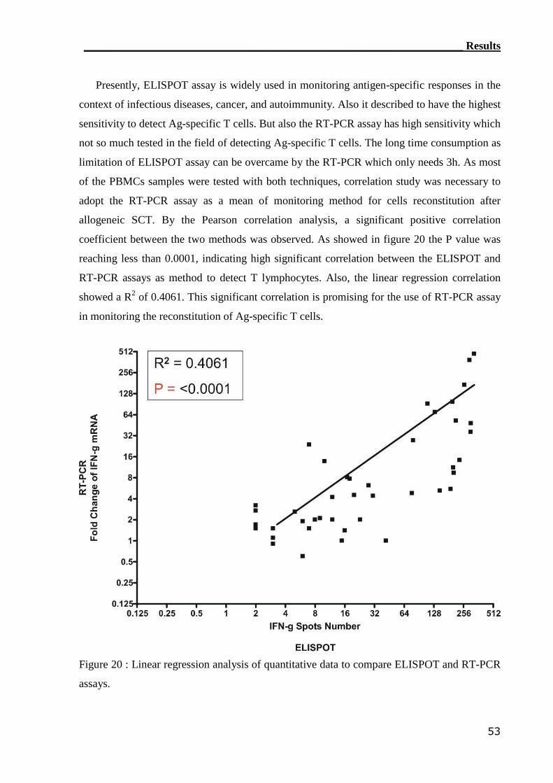
____________________________________________________________________ Results
53
Presently, ELISPOT assay is widely used in monitoring antigen-specific responses in the
context of infectious diseases, cancer, and autoimmunity. Also it described to have the highest
sensitivity to detect Ag-specific T cells. But also the RT-PCR assay has high sensitivity which
not so much tested in the field of detecting Ag-specific T cells. The long time consumption as
limitation of ELISPOT assay can be overcame by the RT-PCR which only needs 3h. As most
of the PBMCs samples were tested with both techniques, correlation study was necessary to
adopt the RT-PCR assay as a mean of monitoring method for cells reconstitution after
allogeneic SCT. By the Pearson correlation analysis, a significant positive correlation
coefficient between the two methods was observed. As showed in figure 20 the P value was
reaching less than 0.0001, indicating high significant correlation between the ELISPOT and
RT-PCR assays as method to detect T lymphocytes. Also, the linear regression correlation
showed a R2 of 0.4061. This significant correlation is promising for the use of RT-PCR assay
in monitoring the reconstitution of Ag-specific T cells.
Figure 20 : Linear regression analysis of quantitative data to compare ELISPOT and RT-PCR
assays.
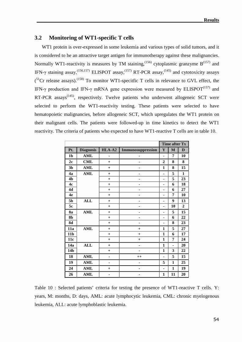
____________________________________________________________________ Results
54
3.2 Monitoring of WT1-specific T cells
WT1 protein is over-expressed in some leukemia and various types of solid tumors, and it
is considered to be an attractive target antigen for immunotherapy against these malignancies.
Normally WT1-reactivity is measures by TM staining,(156) cytoplasmic granzyme B(157) and
IFN-γ staining assay,(156,157) ELISPOT assay,(157) RT-PCR assay,(145) and cytotoxicity assays
(51Cr release assays).(158) To monitor WT1-specitfic T cells in relevance to GVL effect, the
IFN-γ production and IFN-γ mRNA gene expression were measured by ELISPOT(157) and
RT-PCR assays(145), respectively. Twelve patients who underwent allogeneic SCT were
selected to perform the WT1-reactivity testing. These patients were selected to have
hematopoietic malignancies, before allogeneic SCT, which upregulates the WT1 protein on
their malignant cells. The patients were followed-up in time kinetics to detect the WT1
reactivity. The criteria of patients who expected to have WT1-reactive T cells are in table 10.
Time after TxPt. Diagnosis HLA-A2 Immunosuppression Y M D1b AML - - - 7 102c CML + - 2 8 83b AML + - 1 8 154a + - - 5 14b + - - 5 234c + - - 6 184d + - - 6 274e
AML
+ - - 7 105b + - - 9 135c
ALL+ - - 10 2
8a + - - 5 158b + - - 6 228d
AML
+ - - 8 2311a + + 1 5 2711b + + 1 6 1711c
AML
+ + 1 7 2414a + - 1 - 2014b
ALL+ - 1 3 22
18 AML - ++ - 5 1519 AML - - 5 1 2524 AML + - - 1 1926 AML - - 1 11 20
Table 10 : Selected patients’ criteria for testing the presence of WT1-reactive T cells. Y:
years, M: months, D: days, AML: acute lymphocytic leukemia, CML: chronic myelogenous
leukemia, ALL: acute lymphoblastic leukemia.
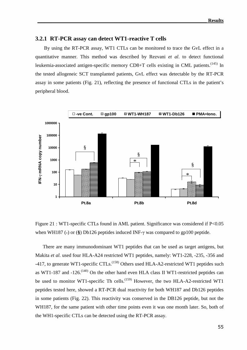
____________________________________________________________________ Results
55
3.2.1 RT-PCR assay can detect WT1-reactive T cells
By using the RT-PCR assay, WT1 CTLs can be monitored to trace the GvL effect in a
quantitative manner. This method was described by Rezvani et al. to detect functional
leukemia-associated antigen-specific memory CD8+T cells existing in CML patients.(145) In
the tested allogeneic SCT transplanted patients, GvL effect was detectable by the RT-PCR
assay in some patients (Fig. 21), reflecting the presence of functional CTLs in the patient’s
peripheral blood.
1
10
100
1000
10000
100000
1000000
Pt.8a Pt.8b Pt.8d
IFN
-γm
RN
Aco
py
nu
mb
er
-ve Cont. gp100 WT1-WH187 WT1-Db126 PMA+Iono.
*
*
§§
§
Figure 21 : WT1-specific CTLs found in AML patient. Significance was considered if P<0.05
when WH187 (*) or (§) Db126 peptides induced INF-γ was compared to gp100 peptide.
There are many immunodominant WT1 peptides that can be used as target antigens, but
Makita et al. used four HLA-A24 restricted WT1 peptides, namely: WT1-228, -235, -356 and
-417, to generate WT1-specific CTLs.(158) Others used HLA-A2-restricted WT1 peptides such
as WT1-187 and -126.(140) On the other hand even HLA class II WT1-restricted peptides can
be used to monitor WT1-specific Th cells.(159) However, the two HLA-A2-restricted WT1
peptides tested here, showed a RT-PCR dual reactivity for both WH187 and Db126 peptides
in some patients (Fig. 22). This reactivity was conserved in the DB126 peptide, but not the
WH187, for the same patient with other time points even it was one month later. So, both of
the WH1-specific CTLs can be detected using the RT-PCR assay.
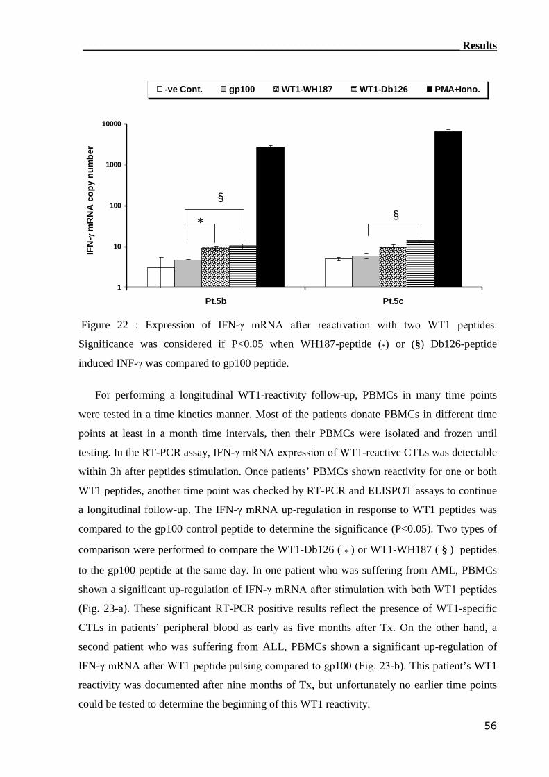
____________________________________________________________________ Results
56
1
10
100
1000
10000
Pt.5b Pt.5c
IFN
-γm
RN
Aco
py
nu
mb
er-ve Cont. gp100 WT1-WH187 WT1-Db126 PMA+Iono.
*
§
§
Figure 22 : Expression of IFN-γ mRNA after reactivation with two WT1 peptides.
Significance was considered if P<0.05 when WH187-peptide (*) or (§) Db126-peptide
induced INF-γ was compared to gp100 peptide.
For performing a longitudinal WT1-reactivity follow-up, PBMCs in many time points
were tested in a time kinetics manner. Most of the patients donate PBMCs in different time
points at least in a month time intervals, then their PBMCs were isolated and frozen until
testing. In the RT-PCR assay, IFN-γ mRNA expression of WT1-reactive CTLs was detectable
within 3h after peptides stimulation. Once patients’ PBMCs shown reactivity for one or both
WT1 peptides, another time point was checked by RT-PCR and ELISPOT assays to continue
a longitudinal follow-up. The IFN-γ mRNA up-regulation in response to WT1 peptides was
compared to the gp100 control peptide to determine the significance (P<0.05). Two types of
comparison were performed to compare the WT1-Db126 ( * ) or WT1-WH187 ( § ) peptides
to the gp100 peptide at the same day. In one patient who was suffering from AML, PBMCs
shown a significant up-regulation of IFN-γ mRNA after stimulation with both WT1 peptides
(Fig. 23-a). These significant RT-PCR positive results reflect the presence of WT1-specific
CTLs in patients’ peripheral blood as early as five months after Tx. On the other hand, a
second patient who was suffering from ALL, PBMCs shown a significant up-regulation of
IFN-γ mRNA after WT1 peptide pulsing compared to gp100 (Fig. 23-b). This patient’s WT1
reactivity was documented after nine months of Tx, but unfortunately no earlier time points
could be tested to determine the beginning of this WT1 reactivity.
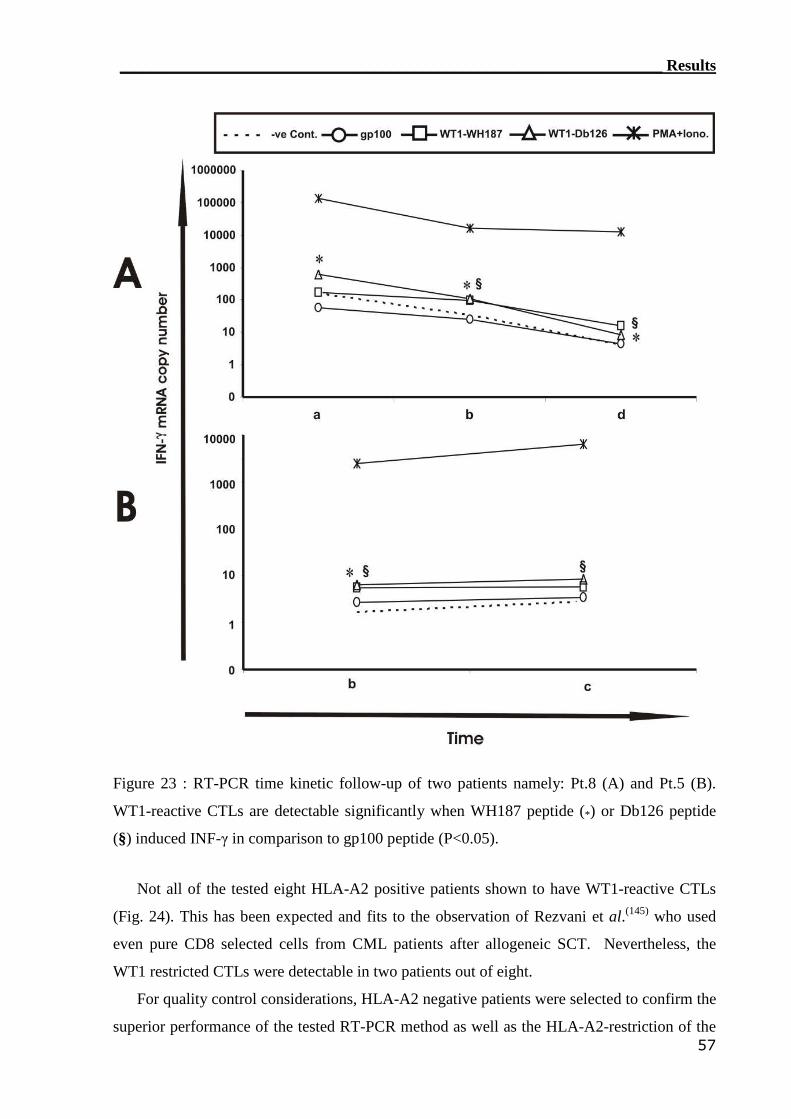
____________________________________________________________________ Results
57
Figure 23 : RT-PCR time kinetic follow-up of two patients namely: Pt.8 (A) and Pt.5 (B).
WT1-reactive CTLs are detectable significantly when WH187 peptide (*) or Db126 peptide
(§) induced INF-γ in comparison to gp100 peptide (P<0.05).
Not all of the tested eight HLA-A2 positive patients shown to have WT1-reactive CTLs
(Fig. 24). This has been expected and fits to the observation of Rezvani et al.(145) who used
even pure CD8 selected cells from CML patients after allogeneic SCT. Nevertheless, the
WT1 restricted CTLs were detectable in two patients out of eight.
For quality control considerations, HLA-A2 negative patients were selected to confirm the
superior performance of the tested RT-PCR method as well as the HLA-A2-restriction of the
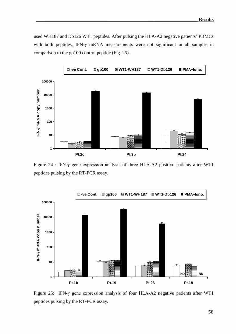
____________________________________________________________________ Results
58
used WH187 and Db126 WT1 peptides. After pulsing the HLA-A2 negative patients’ PBMCs
with both peptides, IFN-γ mRNA measurements were not significant in all samples in
comparison to the gp100 control peptide (Fig. 25).
1
10
100
1000
10000
100000
Pt.2c Pt.3b Pt.24
IFN
-γm
RN
Aco
py
nu
mp
er
-ve Cont. gp100 WT1-WH187 WT1-Db126 PMA+Iono.
Figure 24 : IFN-γ gene expression analysis of three HLA-A2 positive patients after WT1
peptides pulsing by the RT-PCR assay.
1
10
100
1000
10000
100000
Pt.1b Pt.19 Pt.26 Pt.18
IFN
-γ m
RN
Aco
py
nu
mb
er
-ve Cont. gp100 WT1-WH187 WT1-Db126 PMA+Iono.
ND ND
Figure 25: IFN-γ gene expression analysis of four HLA-A2 negative patients after WT1
peptides pulsing by the RT-PCR assay.
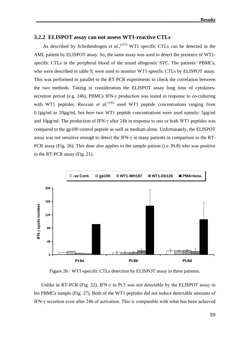
____________________________________________________________________ Results
59
3.2.2 ELISPOT assay can not assess WT1-reactive CTLs
As described by Scheibenbogen et al.,(157) WT1 specific CTLs can be detected in the
AML patient by ELISPOT assay. So, the same assay was used to detect the presence of WT1-
specific CTLs in the peripheral blood of the tested allogeneic STC. The patients’ PBMCs,
who were described in table 9, were used to monitor WT1-spesific CTLs by ELISPOT assay.
This was performed in parallel to the RT-PCR experiments to check the correlation between
the two methods. Taking in consideration the ELISPOT assay long time of cytokines-
secretion period (e.g. 24h), PBMCs IFN-γ production was tested in response to co-culturing
with WT1 peptides. Rezvani et al.(145) used WT1 peptide concentrations ranging from
0.1µg/ml to 10µg/ml, but here two WT1 peptide concentrations were used namely: 5µg/ml
and 10µg/ml. The production of IFN-γ after 24h in response to one or both WT1 peptides was
compared to the gp100 control peptide as well as medium alone. Unfortunately, the ELISPOT
assay was not sensitive enough to detect the IFN-γ in many patients in comparison to the RT-
PCR assay (Fig. 26). This dose also applies to the sample patient (i.e. Pt.8) who was positive
in the RT-PCR assay (Fig. 21).
0
40
80
120
160
200
Pt.8a Pt.8b Pt.8d
IFN
-γsp
ots
nu
mb
er
-ve Cont. gp100 WT1-WH187 WT1-Db126 PMA+Iono.
Figure 26 : WT1-specific CTLs detection by ELISPOT assay in three patients.
Unlike in RT-PCR (Fig. 22), IFN-γ in Pt.5 was not detectable by the ELISPOT assay in
his PBMCs sample (Fig. 27). Both of the WT1 peptides did not induce detectable amounts of
IFN-γ secretion even after 24h of activation. This is comparable with what has been achieved
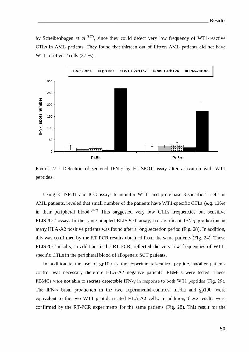
____________________________________________________________________ Results
60
by Scheibenbogen et al.(157), since they could detect very low frequency of WT1-reactive
CTLs in AML patients. They found that thirteen out of fifteen AML patients did not have
WT1-reactive T cells (87 %).
0
50
100
150
200
250
300
Pt.5b Pt.5c
IFN
-γsp
ots
nu
mb
er
-ve Cont. gp100 WT1-WH187 WT1-Db126 PMA+Iono.
Figure 27 : Detection of secreted IFN-γ by ELISPOT assay after activation with WT1
peptides.
Using ELISPOT and ICC assays to monitor WT1- and proteinase 3-specific T cells in
AML patients, reveled that small number of the patients have WT1-specific CTLs (e.g. 13%)
in their peripheral blood.(157) This suggested very low CTLs frequencies but sensitive
ELISPOT assay. In the same adopted ELISPOT assay, no significant IFN-γ production in
many HLA-A2 positive patients was found after a long secretion period (Fig. 28). In addition,
this was confirmed by the RT-PCR results obtained from the same patients (Fig. 24). These
ELISPOT results, in addition to the RT-PCR, reflected the very low frequencies of WT1-
specific CTLs in the peripheral blood of allogeneic SCT patients.
In addition to the use of gp100 as the experimental-control peptide, another patient-
control was necessary therefore HLA-A2 negative patients’ PBMCs were tested. These
PBMCs were not able to secrete detectable IFN-γ in response to both WT1 peptides (Fig. 29).
The IFN-γ basal production in the two experimental-controls, media and gp100, were
equivalent to the two WT1 peptide-treated HLA-A2 cells. In addition, these results were
confirmed by the RT-PCR experiments for the same patients (Fig. 28). This result for the
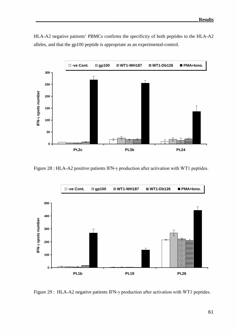
____________________________________________________________________ Results
61
HLA-A2 negative patients’ PBMCs confirms the specificity of both peptides to the HLA-A2
alleles, and that the gp100 peptide is appropriate as an experimental-control.
0
50
100
150
200
250
300
Pt.2c Pt.3b Pt.24
IFN
-γsp
ots
nu
mb
er
-ve Cont. gp100 WT1-WH187 WT1-Db126 PMA+Iono.
Figure 28 : HLA-A2 positive patients IFN-γ production after activation with WT1 peptides.
0
100
200
300
400
500
Pt.1b Pt.19 Pt.26
IFN
-γsp
ots
nu
mb
er
-ve Cont. gp100 WT1-WH187 WT1-Db126 PMA+Iono.
Figure 29 : HLA-A2 negative patients IFN-γ production after activation with WT1 peptides.
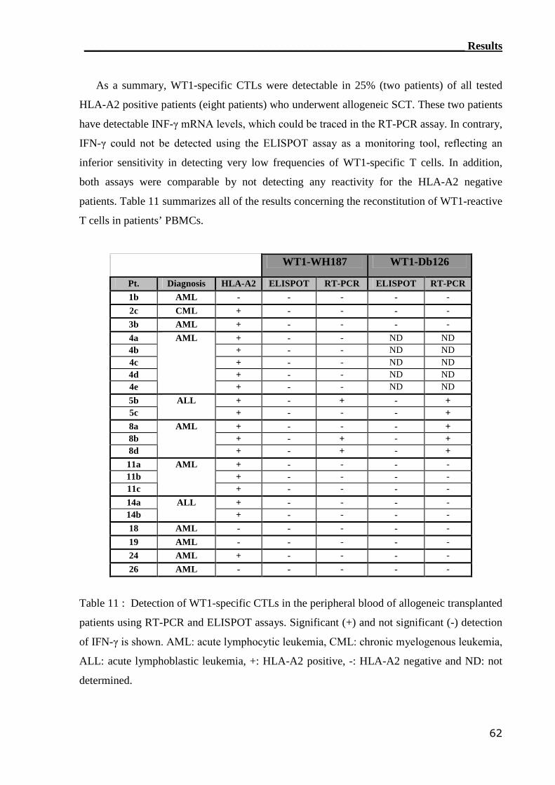
____________________________________________________________________ Results
62
As a summary, WT1-specific CTLs were detectable in 25% (two patients) of all tested
HLA-A2 positive patients (eight patients) who underwent allogeneic SCT. These two patients
have detectable INF-γ mRNA levels, which could be traced in the RT-PCR assay. In contrary,
IFN-γ could not be detected using the ELISPOT assay as a monitoring tool, reflecting an
inferior sensitivity in detecting very low frequencies of WT1-specific T cells. In addition,
both assays were comparable by not detecting any reactivity for the HLA-A2 negative
patients. Table 11 summarizes all of the results concerning the reconstitution of WT1-reactive
T cells in patients’ PBMCs.
WT1-WH187 WT1-Db126
Pt. Diagnosis HLA-A2 ELISPOT RT-PCR ELISPOT RT-PCR1b AML - - - - -2c CML + - - - -3b AML + - - - -4a + - - ND ND4b + - - ND ND4c + - - ND ND4d + - - ND ND4e
AML
+ - - ND ND5b + - + - +5c
ALL+ - - - +
8a + - - - +8b + - + - +8d
AML
+ - + - +11a + - - - -11b + - - - -11c
AML
+ - - - -14a + - - - -14b
ALL+ - - - -
18 AML - - - - -19 AML - - - - -24 AML + - - - -26 AML - - - - -
Table 11 : Detection of WT1-specific CTLs in the peripheral blood of allogeneic transplanted
patients using RT-PCR and ELISPOT assays. Significant (+) and not significant (-) detection
of IFN-γ is shown. AML: acute lymphocytic leukemia, CML: chronic myelogenous leukemia,
ALL: acute lymphoblastic leukemia, +: HLA-A2 positive, -: HLA-A2 negative and ND: not
determined.
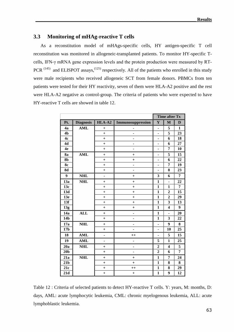
____________________________________________________________________ Results
63
3.3 Monitoring of mHAg-reactive T cells
As a reconstitution model of mHAgs-specific cells, HY antigen-specific T cell
reconstitution was monitored in allogeneic-transplanted patients. To monitor HY-specific T-
cells, IFN-γ mRNA gene expression levels and the protein production were measured by RT-
PCR (145) and ELISPOT assays,(125) respectively. All of the patients who enrolled in this study
were male recipients who received allogeneic SCT from female donors. PBMCs from ten
patients were tested for their HY reactivity, seven of them were HLA-A2 positive and the rest
were HLA-A2 negative as control-group. The criteria of patients who were expected to have
HY-reactive T cells are showed in table 12.
Time after TxPt. Diagnosis HLA-A2 Immunosuppression Y M D4a + - - 5 14b + - - 5 234c + - - 6 184d + - - 6 274e
AML
+ - - 7 108a + + - 5 158b + + - 6 228c + - - 7 198d
AML
+ - - 8 239 NHL - + 3 6 7
13a + + 1 - 2213c + + 1 1 713d + + 1 2 1513e + + 1 2 2913f + + 1 3 1313g
NHL
+ + 1 4 914a + - 1 - 2014b
ALL+ - 1 3 22
17a + - - 9 817b
NHL+ - - 10 25
18 AML - ++ - 5 1519 AML - - 5 1 25
20a + - 2 4 520b
NHL+ - 2 6 7
21a + + 1 7 2421b + + 1 8 821c + ++ 1 8 2921d
NHL
+ + 1 9 12
Table 12 : Criteria of selected patients to detect HY-reactive T cells. Y: years, M: months, D:
days, AML: acute lymphocytic leukemia, CML: chronic myelogenous leukemia, ALL: acute
lymphoblastic leukemia.
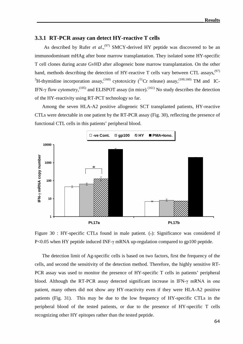
____________________________________________________________________ Results
64
3.3.1 RT-PCR assay can detect HY-reactive T cells
As described by Rufer et al.,(97) SMCY-derived HY peptide was discovered to be an
immunodominant mHAg after bone marrow transplantation. They isolated some HY-specific
T cell clones during acute GvHD after allogeneic bone marrow transplantation. On the other
hand, methods describing the detection of HY-reactive T cells vary between CTL assays,(97)
3H-thymidine incorporation assay,(160) cytotoxicity (51Cr release) assay,(100,160) TM and IC-
IFN-γ flow cytometry,(105) and ELISPOT assay (in mice).(161) No study describes the detection
of the HY-reactivity using RT-PCT technology so far.
Among the seven HLA-A2 positive allogeneic SCT transplanted patients, HY-reactive
CTLs were detectable in one patient by the RT-PCR assay (Fig. 30), reflecting the presence of
functional CTL cells in this patients’ peripheral blood.
1
10
100
1000
10000
Pt.17a Pt.17b
IFN
-γm
RN
Aco
py
nu
mb
er
-ve Cont. gp100 HY PMA+Iono.
*
Figure 30 : HY-specific CTLs found in male patient. (*): Significance was considered if
P<0.05 when HY peptide induced INF-γ mRNA up-regulation compared to gp100 peptide.
The detection limit of Ag-specific cells is based on two factors, first the frequency of the
cells, and second the sensitivity of the detection method. Therefore, the highly sensitive RT-
PCR assay was used to monitor the presence of HY-specific T cells in patients’ peripheral
blood. Although the RT-PCR assay detected significant increase in IFN-γ mRNA in one
patient, many others did not show any HY-reactivity even if they were HLA-A2 positive
patients (Fig. 31). This may be due to the low frequency of HY-specific CTLs in the
peripheral blood of the tested patients, or due to the presence of HY-specific T cells
recognizing other HY epitopes rather than the tested peptide.
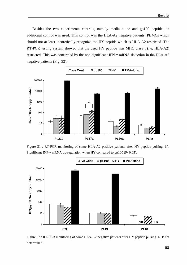
____________________________________________________________________ Results
65
Besides the two experimental-controls, namely media alone and gp100 peptide, an
additional control was used. This control was the HLA-A2 negative patients’ PBMCs which
should not at least theoretically recognize the HY peptide which is HLA-A2-restricted. The
RT-PCR testing system showed that the used HY peptide was MHC class I (i.e. HLA-A2)
restricted. This was confirmed by the non-significant IFN-γ mRNA detection in the HLA-A2
negative patients (Fig. 32).
1
10
100
1000
10000
100000
Pt.21a Pt.17a Pt.20a Pt.4a
IFN
-γm
RN
Aco
py
nu
mb
er
-ve Cont. gp100 HY PMA+Iono.
*
Figure 31 : RT-PCR monitoring of some HLA-A2 positive patients after HY peptide pulsing. (*):
Significant INF-γ mRNA up-regulation when HY compared to gp100 (P<0.05).
1
10
100
1000
10000
100000
Pt.9 Pt.19 Pt.18
IFN
g-γ
mR
NA
cop
yn
um
ber
-ve Cont. gp100 HY PMA+Iono.
ND ND
Figure 32 : RT-PCR monitoring of some HLA-A2 negative patients after HY peptide pulsing. ND: not
determined.
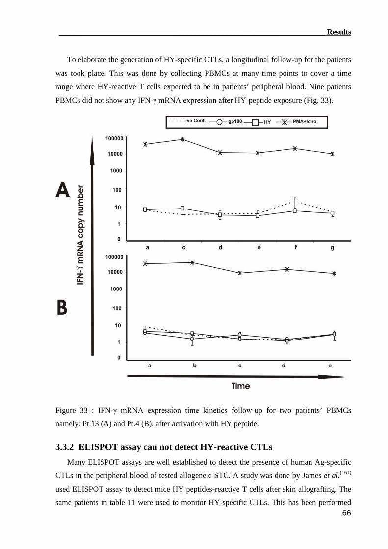
____________________________________________________________________ Results
66
To elaborate the generation of HY-specific CTLs, a longitudinal follow-up for the patients
was took place. This was done by collecting PBMCs at many time points to cover a time
range where HY-reactive T cells expected to be in patients’ peripheral blood. Nine patients
PBMCs did not show any IFN-γ mRNA expression after HY-peptide exposure (Fig. 33).
Figure 33 : IFN-γ mRNA expression time kinetics follow-up for two patients’ PBMCs
namely: Pt.13 (A) and Pt.4 (B), after activation with HY peptide.
3.3.2 ELISPOT assay can not detect HY-reactive CTLs
Many ELISPOT assays are well established to detect the presence of human Ag-specific
CTLs in the peripheral blood of tested allogeneic STC. A study was done by James et al.(161)
used ELISPOT assay to detect mice HY peptides-reactive T cells after skin allografting. The
same patients in table 11 were used to monitor HY-specific CTLs. This has been performed
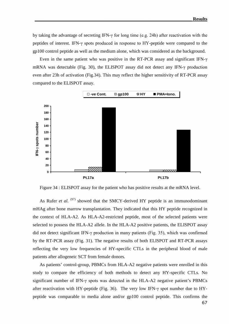
____________________________________________________________________ Results
67
by taking the advantage of secreting IFN-γ for long time (e.g. 24h) after reactivation with the
peptides of interest. IFN-γ spots produced in response to HY-peptide were compared to the
gp100 control peptide as well as the medium alone, which was considered as the background.
Even in the same patient who was positive in the RT-PCR assay and significant IFN-γ
mRNA was detectable (Fig. 30), the ELISPOT assay did not detect any IFN-γ production
even after 23h of activation (Fig.34). This may reflect the higher sensitivity of RT-PCR assay
compared to the ELISPOT assay.
0
20
40
60
80
100
120
140
160
180
200
Pt.17a Pt.17b
IFN
-γsp
ots
nu
mb
er
-ve Cont. gp100 HY PMA+Iono.
Figure 34 : ELISPOT assay for the patient who has positive results at the mRNA level.
As Rufer et al. (97) showed that the SMCY-derived HY peptide is an immunodominant
mHAg after bone marrow transplantation. They indicated that this HY peptide recognized in
the context of HLA-A2. As HLA-A2-restricted peptide, most of the selected patients were
selected to possess the HLA-A2 allele. In the HLA-A2 positive patients, the ELISPOT assay
did not detect significant IFN-γ production in many patients (Fig. 35), which was confirmed
by the RT-PCR assay (Fig. 31). The negative results of both ELISPOT and RT-PCR assays
reflecting the very low frequencies of HY-specific CTLs in the peripheral blood of male
patients after allogeneic SCT from female donors.
As patients’ control-group, PBMCs from HLA-A2 negative patients were enrolled in this
study to compare the efficiency of both methods to detect any HY-specific CTLs. No
significant number of IFN-γ spots was detected in the HLA-A2 negative patient’s PBMCs
after reactivation with HY-peptide (Fig. 36). The very low IFN-γ spot number due to HY-
peptide was comparable to media alone and/or gp100 control peptide. This confirms the
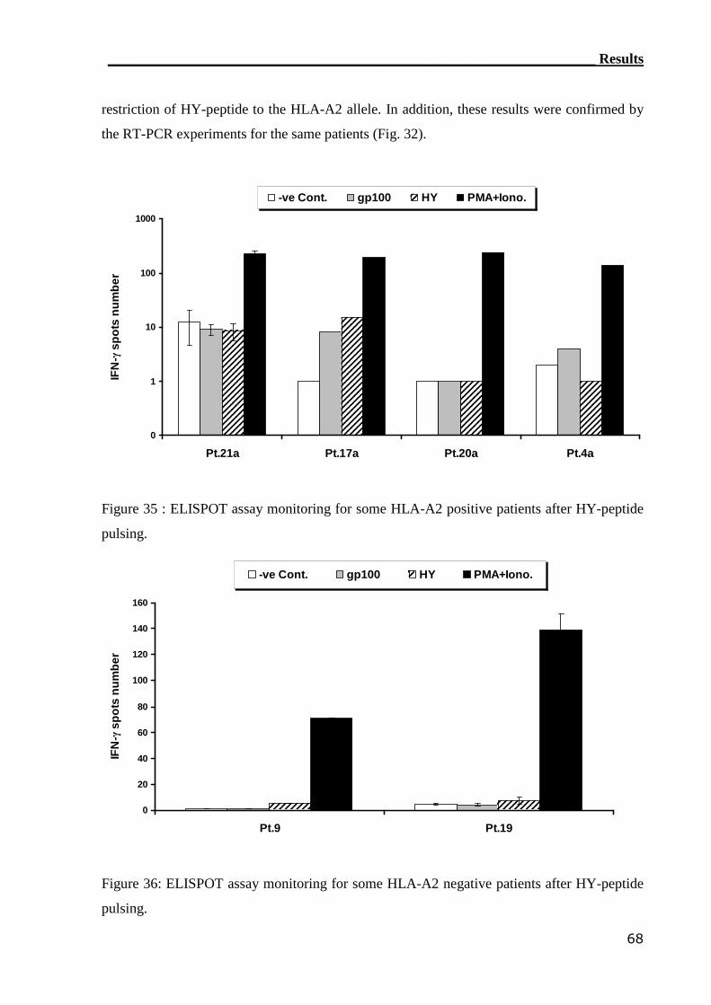
____________________________________________________________________ Results
68
restriction of HY-peptide to the HLA-A2 allele. In addition, these results were confirmed by
the RT-PCR experiments for the same patients (Fig. 32).
0
1
10
100
1000
Pt.21a Pt.17a Pt.20a Pt.4a
IFN
-γsp
ots
nu
mb
er
-ve Cont. gp100 HY PMA+Iono.
Figure 35 : ELISPOT assay monitoring for some HLA-A2 positive patients after HY-peptide
pulsing.
0
20
40
60
80
100
120
140
160
Pt.9 Pt.19
IFN
-γsp
ots
nu
mb
er
-ve Cont. gp100 HY PMA+Iono.
Figure 36: ELISPOT assay monitoring for some HLA-A2 negative patients after HY-peptide
pulsing.
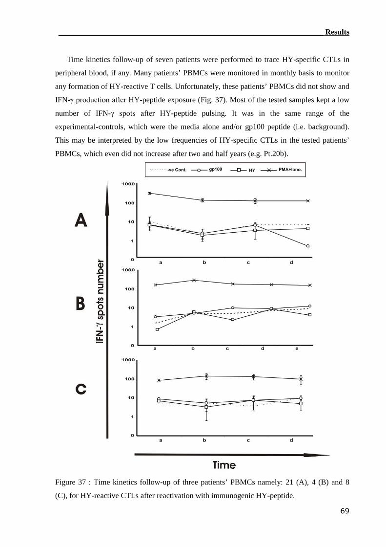
____________________________________________________________________ Results
69
Time kinetics follow-up of seven patients were performed to trace HY-specific CTLs in
peripheral blood, if any. Many patients’ PBMCs were monitored in monthly basis to monitor
any formation of HY-reactive T cells. Unfortunately, these patients’ PBMCs did not show and
IFN-γ production after HY-peptide exposure (Fig. 37). Most of the tested samples kept a low
number of IFN-γ spots after HY-peptide pulsing. It was in the same range of the
experimental-controls, which were the media alone and/or gp100 peptide (i.e. background).
This may be interpreted by the low frequencies of HY-specific CTLs in the tested patients’
PBMCs, which even did not increase after two and half years (e.g. Pt.20b).
Figure 37 : Time kinetics follow-up of three patients’ PBMCs namely: 21 (A), 4 (B) and 8
(C), for HY-reactive CTLs after reactivation with immunogenic HY-peptide.
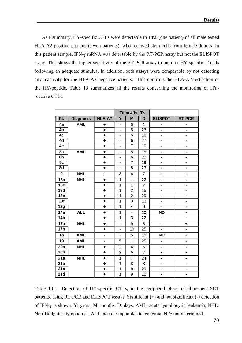
____________________________________________________________________ Results
70
As a summary, HY-specific CTLs were detectable in 14% (one patient) of all male tested
HLA-A2 positive patients (seven patients), who received stem cells from female donors. In
this patient sample, IFN-γ mRNA was detectable by the RT-PCR assay but not the ELISPOT
assay. This shows the higher sensitivity of the RT-PCR assay to monitor HY-specific T cells
following an adequate stimulus. In addition, both assays were comparable by not detecting
any reactivity for the HLA-A2 negative patients. This confirms the HLA-A2-restriction of
the HY-peptide. Table 13 summarizes all the results concerning the monitoring of HY-
reactive CTLs.
Time after TxPt. Diagnosis HLA-A2 Y M D ELISPOT RT-PCR4a + - 5 1 - -4b + - 5 23 - -4c + - 6 18 - -4d + - 6 27 - -4e
AML
+ - 7 10 - -8a + - 5 15 - -8b + - 6 22 - -8c + - 7 19 - -8d
AML
+ - 8 23 - -9 NHL - 3 6 7 - -
13a + 1 - 22 - -13c + 1 1 7 - -13d + 1 2 15 - -13e + 1 2 29 - -13f + 1 3 13 - -13g
NHL
+ 1 4 9 - -14a + 1 - 20 ND -14b
ALL+ 1 3 22 - -
17a + - 9 8 - +17b
NHL+ - 10 25 - -
18 AML - - 5 15 ND -19 AML - 5 1 25 - -20a + 2 4 5 - -20b
NHL+ 2 6 7 - -
21a + 1 7 24 - -21b + 1 8 8 - -21c + 1 8 29 - -21d
NHL
+ 1 9 12 - -
Table 13 : Detection of HY-specific CTLs, in the peripheral blood of allogeneic SCT
patients, using RT-PCR and ELISPOT assays. Significant (+) and not significant (-) detection
of IFN-γ is shown. Y: years, M: months, D: days, AML: acute lymphocytic leukemia, NHL:
Non-Hodgkin's lymphomas, ALL: acute lymphoblastic leukemia. ND: not determined.
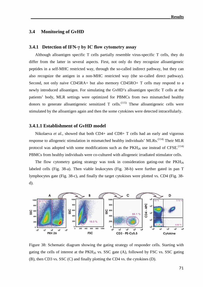
____________________________________________________________________ Results
71
3.4 Monitoring of GvHD
3.4.1 Detection of IFN-γ by IC flow cytometry assay
Although alloantigen specific T cells partially resemble virus-specific T cells, they do
differ from the latter in several aspects. First, not only do they recognize alloantigeneic
peptides in a self-MHC restricted way, through the so-called indirect pathway, but they can
also recognize the antigen in a non-MHC restricted way (the so-called direct pathway).
Second, not only naive CD45RA+ but also memory CD45RO+ T cells may respond to a
newly introduced alloantigen. For simulating the GvHD’s alloantigen specific T cells at the
patients’ body, MLR settings were optimized for PBMCs from two mismatched healthy
donors to generate alloantigeneic sensitized T cells.(115) These alloantigeneic cells were
stimulated by the alloantigen again and then the some cytokines were detected intracellularly.
3.4.1.1 Establishment of GvHD model
Nikolaeva et al., showed that both CD4+ and CD8+ T cells had an early and vigorous
response to allogeneic stimulation in mismatched healthy individuals’ MLRs.(114) Their MLR
protocol was adopted with some modifications such as the PKH26 use instead of CFSE.(114)
PBMCs from healthy individuals were co-cultured with allogeneic irradiated stimulator cells.
The flow cytometry gating strategy was took in consideration gating-out the PKH26
labeled cells (Fig. 38-a). Then viable leukocytes (Fig. 38-b) were further gated in pan T
lymphocytes gate (Fig. 38-c), and finally the target cytokines were plotted vs. CD4 (Fig. 38-
d).
Figure 38: Schematic diagram showing the gating strategy of responder cells. Starting with
gating the cells of interest at the PKH26 vs. SSC gate (A), followed by FSC vs. SSC gating
(B), then CD3 vs. SSC (C) and finally plotting the CD4 vs. the cytokines (D).
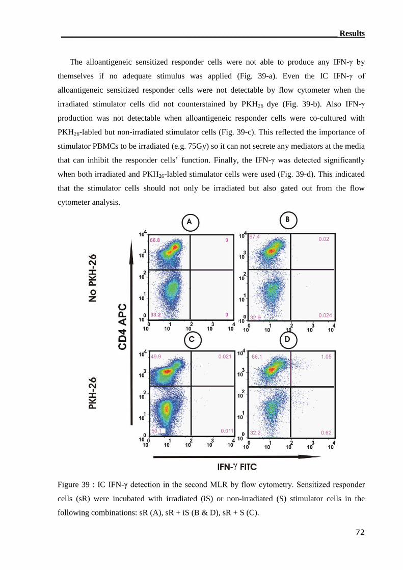
____________________________________________________________________ Results
72
The alloantigeneic sensitized responder cells were not able to produce any IFN-γ by
themselves if no adequate stimulus was applied (Fig. 39-a). Even the IC IFN-γ of
alloantigeneic sensitized responder cells were not detectable by flow cytometer when the
irradiated stimulator cells did not counterstained by PKH26 dye (Fig. 39-b). Also IFN-γ
production was not detectable when alloantigeneic responder cells were co-cultured with
PKH26-labled but non-irradiated stimulator cells (Fig. 39-c). This reflected the importance of
stimulator PBMCs to be irradiated (e.g. 75Gy) so it can not secrete any mediators at the media
that can inhibit the responder cells’ function. Finally, the IFN-γ was detected significantly
when both irradiated and PKH26-labled stimulator cells were used (Fig. 39-d). This indicated
that the stimulator cells should not only be irradiated but also gated out from the flow
cytometer analysis.
Figure 39 : IC IFN-γ detection in the second MLR by flow cytometry. Sensitized responder
cells (sR) were incubated with irradiated (iS) or non-irradiated (S) stimulator cells in the
following combinations: sR (A), sR + iS (B & D), sR + S (C).
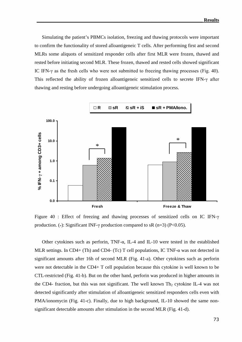
____________________________________________________________________ Results
73
Simulating the patient’s PBMCs isolation, freezing and thawing protocols were important
to confirm the functionality of stored alloantigeneic T cells. After performing first and second
MLRs some aliquots of sensitized responder cells after first MLR were frozen, thawed and
rested before initiating second MLR. These frozen, thawed and rested cells showed significant
IC IFN-γ as the fresh cells who were not submitted to freezing thawing processes (Fig. 40).
This reflected the ability of frozen alloantigeneic sensitized cells to secrete IFN-γ after
thawing and resting before undergoing alloantigeneic stimulation process.
0.0
0.1
1.0
10.0
100.0
Fresh Freeze & Thaw
%IF
N-γ
+am
on
gC
D3+
cells
R sR sR + iS sR + PMA/Iono.
**
Figure 40 : Effect of freezing and thawing processes of sensitized cells on IC IFN-γ
production. (*): Significant INF-γ production compared to sR (n=3) (P<0.05).
Other cytokines such as perforin, TNF-α, IL-4 and IL-10 were tested in the established
MLR settings. In CD4+ (Th) and CD4- (Tc) T cell populations, IC TNF-α was not detected in
significant amounts after 16h of second MLR (Fig. 41-a). Other cytokines such as perforin
were not detectable in the CD4+ T cell population because this cytokine is well known to be
CTL-restricted (Fig. 41-b). But on the other hand, perforin was produced in higher amounts in
the CD4- fraction, but this was not significant. The well known Th2 cytokine IL-4 was not
detected significantly after stimulation of alloantigeneic sensitized responders cells even with
PMA/ionomycin (Fig. 41-c). Finally, due to high background, IL-10 showed the same non-
significant detectable amounts after stimulation in the second MLR (Fig. 41-d).
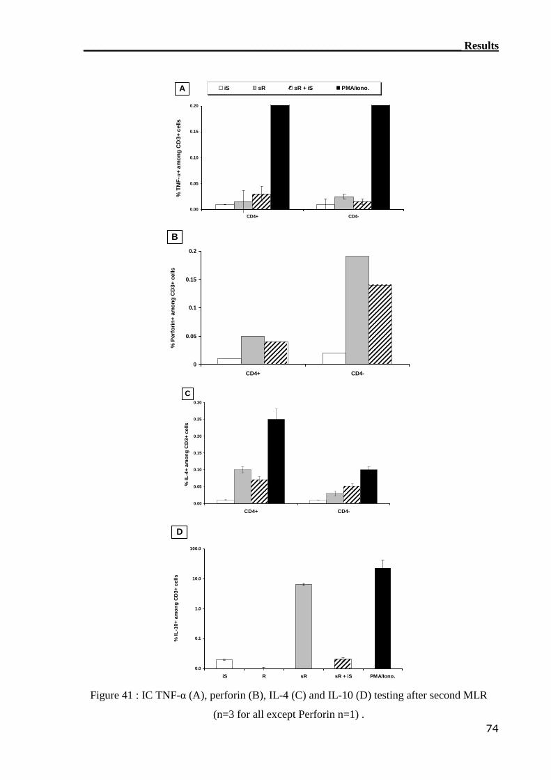
____________________________________________________________________ Results
74
0.00
0.05
0.10
0.15
0.20
CD4+ CD4-
%T
NF
-α+
amo
ng
CD
3+ce
lls
iS sR sR + iS PMA/iono.A
0
0.05
0.1
0.15
0.2
CD4+ CD4-
%P
erfo
rin
+am
ong
CD
3+ce
lls
B
0.00
0.05
0.10
0.15
0.20
0.25
0.30
CD4+ CD4-
%IL
-4+
amo
ng
CD
3+ce
lls
C
0.0
0.1
1.0
10.0
100.0
iS R sR sR + iS PMA/Iono.
%IL
-10+
amo
ng
CD
3+ce
lls
D
Figure 41 : IC TNF-α (A), perforin (B), IL-4 (C) and IL-10 (D) testing after second MLR
(n=3 for all except Perforin n=1) .
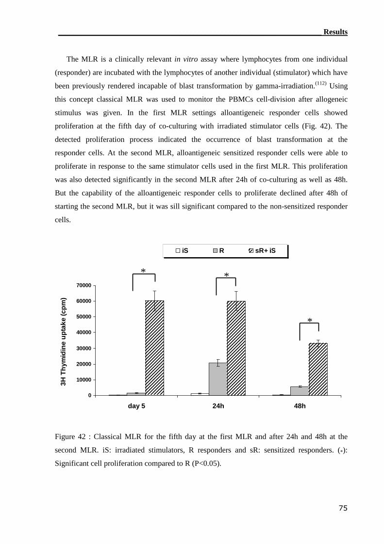
____________________________________________________________________ Results
75
The MLR is a clinically relevant in vitro assay where lymphocytes from one individual
(responder) are incubated with the lymphocytes of another individual (stimulator) which have
been previously rendered incapable of blast transformation by gamma-irradiation.(112) Using
this concept classical MLR was used to monitor the PBMCs cell-division after allogeneic
stimulus was given. In the first MLR settings alloantigeneic responder cells showed
proliferation at the fifth day of co-culturing with irradiated stimulator cells (Fig. 42). The
detected proliferation process indicated the occurrence of blast transformation at the
responder cells. At the second MLR, alloantigeneic sensitized responder cells were able to
proliferate in response to the same stimulator cells used in the first MLR. This proliferation
was also detected significantly in the second MLR after 24h of co-culturing as well as 48h.
But the capability of the alloantigeneic responder cells to proliferate declined after 48h of
starting the second MLR, but it was sill significant compared to the non-sensitized responder
cells.
0
10000
20000
30000
40000
50000
60000
70000
day 5 24h 48h
3HT
hym
idin
eu
pta
ke(c
pm
)
iS R sR+ iS
* *
*
Figure 42 : Classical MLR for the fifth day at the first MLR and after 24h and 48h at the
second MLR. iS: irradiated stimulators, R responders and sR: sensitized responders. (*):
Significant cell proliferation compared to R (P<0.05).
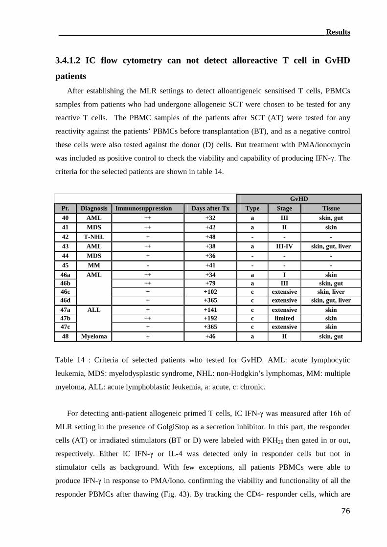
____________________________________________________________________ Results
76
3.4.1.2 IC flow cytometry can not detect alloreactive T cell in GvHD
patients
After establishing the MLR settings to detect alloantigeneic sensitised T cells, PBMCs
samples from patients who had undergone allogeneic SCT were chosen to be tested for any
reactive T cells. The PBMC samples of the patients after SCT (AT) were tested for any
reactivity against the patients’ PBMCs before transplantation (BT), and as a negative control
these cells were also tested against the donor (D) cells. But treatment with PMA/ionomycin
was included as positive control to check the viability and capability of producing IFN-γ. The
criteria for the selected patients are shown in table 14.
GvHDPt. Diagnosis Immunosuppression Days after Tx Type Stage Tissue40 AML ++ +32 a III skin, gut41 MDS ++ +42 a II skin42 T-NHL + +48 - - -43 AML ++ +38 a III-IV skin, gut, liver44 MDS + +36 - - -45 MM - +41 - - -
46a ++ +34 a I skin46b ++ +79 a III skin, gut46c + +102 c extensive skin, liver46d
AML
+ +365 c extensive skin, gut, liver47a + +141 c extensive skin47b ++ +192 c limited skin47c
ALL
+ +365 c extensive skin48 Myeloma + +46 a II skin, gut
Table 14 : Criteria of selected patients who tested for GvHD. AML: acute lymphocytic
leukemia, MDS: myelodysplastic syndrome, NHL: non-Hodgkin’s lymphomas, MM: multiple
myeloma, ALL: acute lymphoblastic leukemia, a: acute, c: chronic.
For detecting anti-patient allogeneic primed T cells, IC IFN-γ was measured after 16h of
MLR setting in the presence of GolgiStop as a secretion inhibitor. In this part, the responder
cells (AT) or irradiated stimulators (BT or D) were labeled with PKH26 then gated in or out,
respectively. Either IC IFN-γ or IL-4 was detected only in responder cells but not in
stimulator cells as background. With few exceptions, all patients PBMCs were able to
produce IFN-γ in response to PMA/Iono. confirming the viability and functionality of all the
responder PBMCs after thawing (Fig. 43). By tracking the CD4- responder cells, which are
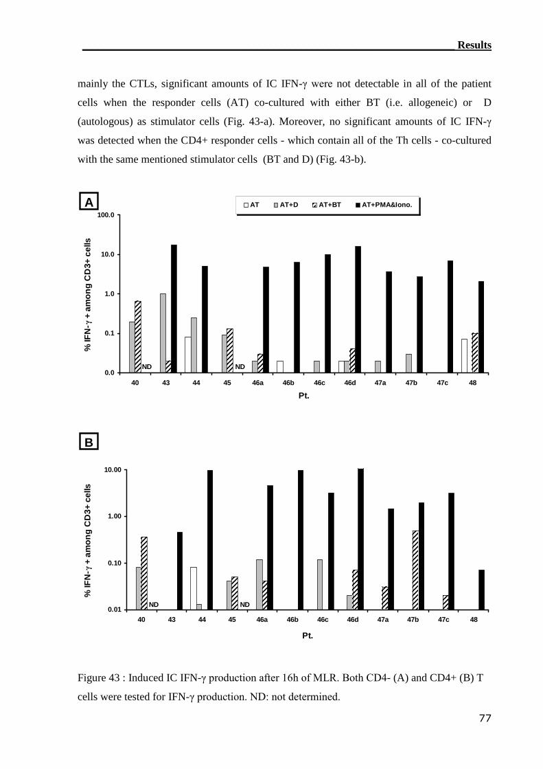
____________________________________________________________________ Results
77
mainly the CTLs, significant amounts of IC IFN-γ were not detectable in all of the patient
cells when the responder cells (AT) co-cultured with either BT (i.e. allogeneic) or D
(autologous) as stimulator cells (Fig. 43-a). Moreover, no significant amounts of IC IFN-γ
was detected when the CD4+ responder cells - which contain all of the Th cells - co-cultured
with the same mentioned stimulator cells (BT and D) (Fig. 43-b).
0.0
0.1
1.0
10.0
100.0
40 43 44 45 46a 46b 46c 46d 47a 47b 47c 48
Pt.
%IF
N-γ
+am
on
gC
D3+
cells
AT AT+D AT+BT AT+PMA&Iono.A
ND ND
0.01
0.10
1.00
10.00
40 43 44 45 46a 46b 46c 46d 47a 47b 47c 48
Pt.
%IF
N-γ
+am
on
gC
D3+
cells
B
ND ND
Figure 43 : Induced IC IFN-γ production after 16h of MLR. Both CD4- (A) and CD4+ (B) T
cells were tested for IFN-γ production. ND: not determined.
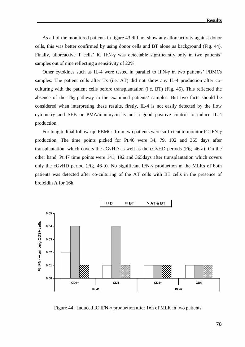
____________________________________________________________________ Results
78
As all of the monitored patients in figure 43 did not show any alloreactivity against donor
cells, this was better confirmed by using donor cells and BT alone as background (Fig. 44).
Finally, alloreactive T cells’ IC IFN-γ was detectable significantly only in two patients’
samples out of nine reflecting a sensitivity of 22%.
Other cytokines such as IL-4 were tested in parallel to IFN-γ in two patients’ PBMCs
samples. The patient cells after Tx (i.e. AT) did not show any IL-4 production after co-
culturing with the patient cells before transplantation (i.e. BT) (Fig. 45). This reflected the
absence of the Th2 pathway in the examined patients’ samples. But two facts should be
considered when interpreting these results, firstly, IL-4 is not easily detected by the flow
cytometry and SEB or PMA/ionomycin is not a good positive control to induce IL-4
production.
For longitudinal follow-up, PBMCs from two patients were sufficient to monitor IC IFN-γ
production. The time points picked for Pt.46 were 34, 79, 102 and 365 days after
transplantation, which covers the aGvHD as well as the cGvHD periods (Fig. 46-a). On the
other hand, Pt.47 time points were 141, 192 and 365days after transplantation which covers
only the cGvHD period (Fig. 46-b). No significant IFN-γ production in the MLRs of both
patients was detected after co-culturing of the AT cells with BT cells in the presence of
brefeldin A for 16h.
0.00
0.01
0.02
0.03
0.04
0.05
CD4+ CD4- CD4+ CD4-
Pt.41 Pt.42
%IF
N-γ
+am
on
gC
D3+
cells
D BT AT & BT
Figure 44 : Induced IC IFN-γ production after 16h of MLR in two patients.
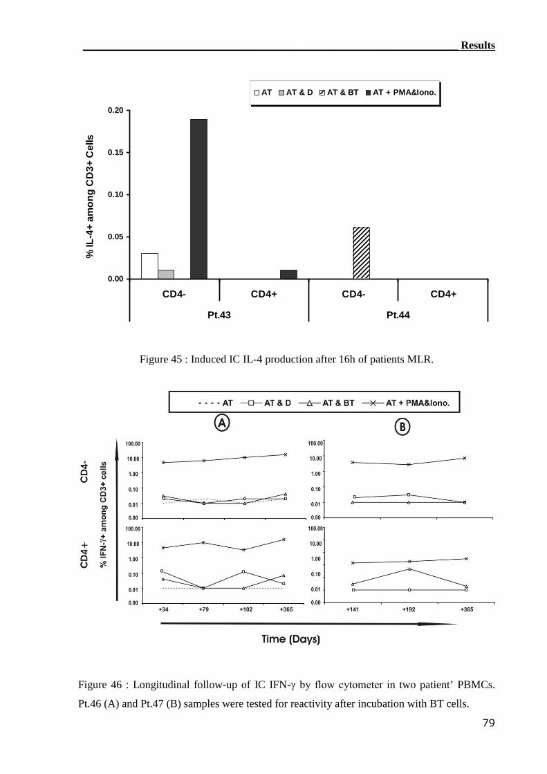
____________________________________________________________________ Results
79
0.00
0.05
0.10
0.15
0.20
CD4- CD4+ CD4- CD4+
Pt.43 Pt.44
%IL
-4+
amo
ng
CD
3+C
ells
AT AT & D AT & BT AT + PMA&Iono.
Figure 45 : Induced IC IL-4 production after 16h of patients MLR.
Figure 46 : Longitudinal follow-up of IC IFN-γ by flow cytometer in two patient’ PBMCs.
Pt.46 (A) and Pt.47 (B) samples were tested for reactivity after incubation with BT cells.
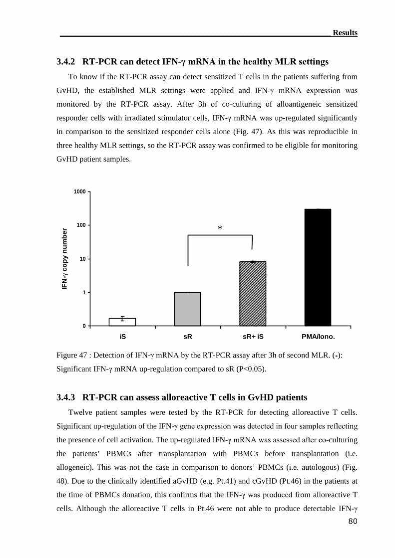
____________________________________________________________________ Results
80
3.4.2 RT-PCR can detect IFN-γ mRNA in the healthy MLR settings
To know if the RT-PCR assay can detect sensitized T cells in the patients suffering from
GvHD, the established MLR settings were applied and IFN-γ mRNA expression was
monitored by the RT-PCR assay. After 3h of co-culturing of alloantigeneic sensitized
responder cells with irradiated stimulator cells, IFN-γ mRNA was up-regulated significantly
in comparison to the sensitized responder cells alone (Fig. 47). As this was reproducible in
three healthy MLR settings, so the RT-PCR assay was confirmed to be eligible for monitoring
GvHD patient samples.
0
1
10
100
1000
iS sR sR+ iS PMA/Iono.
IFN
-γco
py
nu
mb
er *
Figure 47 : Detection of IFN-γ mRNA by the RT-PCR assay after 3h of second MLR. (*):
Significant IFN-γ mRNA up-regulation compared to sR (P<0.05).
3.4.3 RT-PCR can assess alloreactive T cells in GvHD patients
Twelve patient samples were tested by the RT-PCR for detecting alloreactive T cells.
Significant up-regulation of the IFN-γ gene expression was detected in four samples reflecting
the presence of cell activation. The up-regulated IFN-γ mRNA was assessed after co-culturing
the patients’ PBMCs after transplantation with PBMCs before transplantation (i.e.
allogeneic). This was not the case in comparison to donors’ PBMCs (i.e. autologous) (Fig.
48). Due to the clinically identified aGvHD (e.g. Pt.41) and cGvHD (Pt.46) in the patients at
the time of PBMCs donation, this confirms that the IFN-γ was produced from alloreactive T
cells. Although the alloreactive T cells in Pt.46 were not able to produce detectable IFN-γ
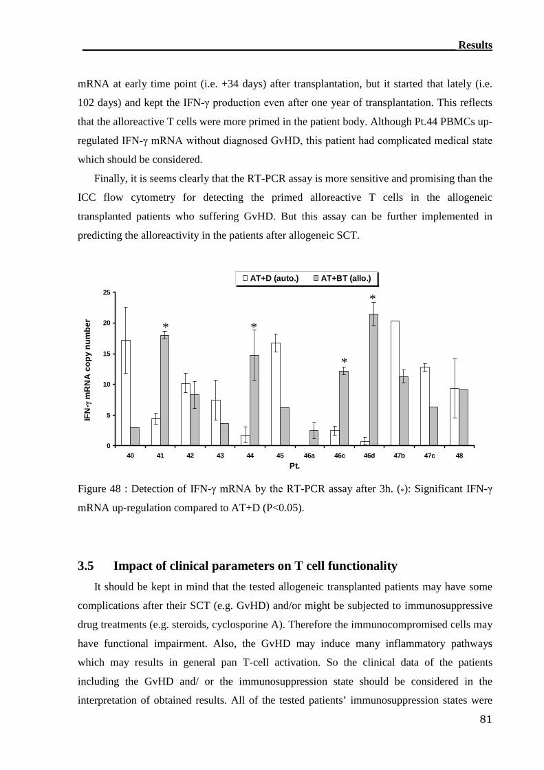
____________________________________________________________________ Results
81
mRNA at early time point (i.e. +34 days) after transplantation, but it started that lately (i.e.
102 days) and kept the IFN-γ production even after one year of transplantation. This reflects
that the alloreactive T cells were more primed in the patient body. Although Pt.44 PBMCs up-
regulated IFN-γ mRNA without diagnosed GvHD, this patient had complicated medical state
which should be considered.
Finally, it is seems clearly that the RT-PCR assay is more sensitive and promising than the
ICC flow cytometry for detecting the primed alloreactive T cells in the allogeneic
transplanted patients who suffering GvHD. But this assay can be further implemented in
predicting the alloreactivity in the patients after allogeneic SCT.
0
5
10
15
20
25
40 41 42 43 44 45 46a 46c 46d 47b 47c 48
Pt.
IFN
-γm
RN
Aco
py
nu
mb
er
AT+D (auto.) AT+BT (allo.)
* *
*
*
Figure 48 : Detection of IFN-γ mRNA by the RT-PCR assay after 3h. (*): Significant IFN-γ
mRNA up-regulation compared to AT+D (P<0.05).
3.5 Impact of clinical parameters on T cell functionality
It should be kept in mind that the tested allogeneic transplanted patients may have some
complications after their SCT (e.g. GvHD) and/or might be subjected to immunosuppressive
drug treatments (e.g. steroids, cyclosporine A). Therefore the immunocompromised cells may
have functional impairment. Also, the GvHD may induce many inflammatory pathways
which may results in general pan T-cell activation. So the clinical data of the patients
including the GvHD and/ or the immunosuppression state should be considered in the
interpretation of obtained results. All of the tested patients’ immunosuppression states were
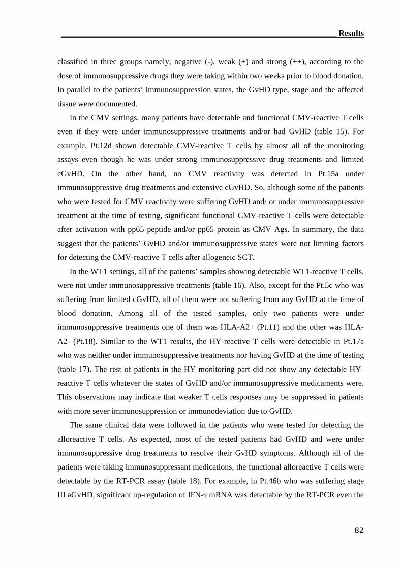
____________________________________________________________________ Results
82
classified in three groups namely; negative (-), weak (+) and strong (++), according to the
dose of immunosuppressive drugs they were taking within two weeks prior to blood donation.
In parallel to the patients’ immunosuppression states, the GvHD type, stage and the affected
tissue were documented.
In the CMV settings, many patients have detectable and functional CMV-reactive T cells
even if they were under immunosuppressive treatments and/or had GvHD (table 15). For
example, Pt.12d shown detectable CMV-reactive T cells by almost all of the monitoring
assays even though he was under strong immunosuppressive drug treatments and limited
cGvHD. On the other hand, no CMV reactivity was detected in Pt.15a under
immunosuppressive drug treatments and extensive cGvHD. So, although some of the patients
who were tested for CMV reactivity were suffering GvHD and/ or under immunosuppressive
treatment at the time of testing, significant functional CMV-reactive T cells were detectable
after activation with pp65 peptide and/or pp65 protein as CMV Ags. In summary, the data
suggest that the patients’ GvHD and/or immunosuppressive states were not limiting factors
for detecting the CMV-reactive T cells after allogeneic SCT.
In the WT1 settings, all of the patients’ samples showing detectable WT1-reactive T cells,
were not under immunosuppressive treatments (table 16). Also, except for the Pt.5c who was
suffering from limited cGvHD, all of them were not suffering from any GvHD at the time of
blood donation. Among all of the tested samples, only two patients were under
immunosuppressive treatments one of them was HLA-A2+ (Pt.11) and the other was HLA-
A2- (Pt.18). Similar to the WT1 results, the HY-reactive T cells were detectable in Pt.17a
who was neither under immunosuppressive treatments nor having GvHD at the time of testing
(table 17). The rest of patients in the HY monitoring part did not show any detectable HY-
reactive T cells whatever the states of GvHD and/or immunosuppressive medicaments were.
This observations may indicate that weaker T cells responses may be suppressed in patients
with more sever immunosuppression or immunodeviation due to GvHD.
The same clinical data were followed in the patients who were tested for detecting the
alloreactive T cells. As expected, most of the tested patients had GvHD and were under
immunosuppressive drug treatments to resolve their GvHD symptoms. Although all of the
patients were taking immunosuppressant medications, the functional alloreactive T cells were
detectable by the RT-PCR assay (table 18). For example, in Pt.46b who was suffering stage
III aGvHD, significant up-regulation of IFN-γ mRNA was detectable by the RT-PCR even the
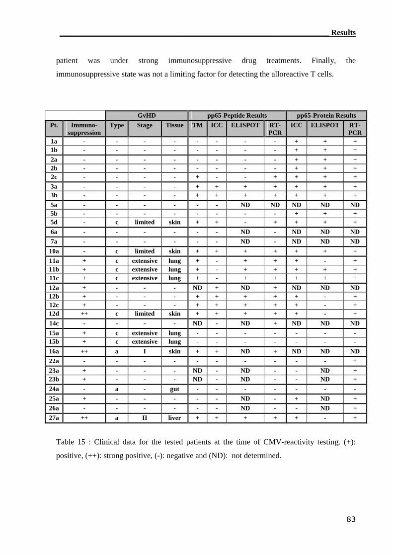
____________________________________________________________________ Results
83
patient was under strong immunosuppressive drug treatments. Finally, the
immunosuppressive state was not a limiting factor for detecting the alloreactive T cells.
GvHD pp65-Peptide Results pp65-Protein ResultsPt. Immuno-
suppressionType Stage Tissue TM ICC ELISPOT RT-
PCRICC ELISPOT RT-
PCR1a - - - - - - - - + + +1b - - - - - - - - + + +2a - - - - - - - - + + +2b - - - - - - - - + + +2c - - - - + - - + + + +3a - - - - + + + + + + +3b - - - - + + + + + + +5a - - - - - - ND ND ND ND ND5b - - - - - - - - + + +5d - c limited skin + + - + + + +6a - - - - - - ND - ND ND ND7a - - - - - - ND - ND ND ND
10a - c limited skin + + + + + + +11a + c extensive lung + - + + + - +11b + c extensive lung + - + + + + +11c + c extensive lung + - + + + + +12a + - - - ND + ND + ND ND ND12b + - - - + + + + + - +12c + - - - + + + + + - +12d ++ c limited skin + + + + + - +14c - - - - ND - ND + ND ND ND15a + c extensive lung - - - - - - -15b + c extensive lung - - - - - - -16a ++ a I skin + + ND + ND ND ND22a - - - - - - - - - - +23a + - - - ND - ND - - ND +23b + - - - ND - ND - - ND +24a - a - gut - - - - - - -25a + - - - - - ND - + ND +26a - - - - - - ND - - ND +27a ++ a II liver + + + + + - +
Table 15 : Clinical data for the tested patients at the time of CMV-reactivity testing. (+):
positive, (++): strong positive, (-): negative and (ND): not determined.
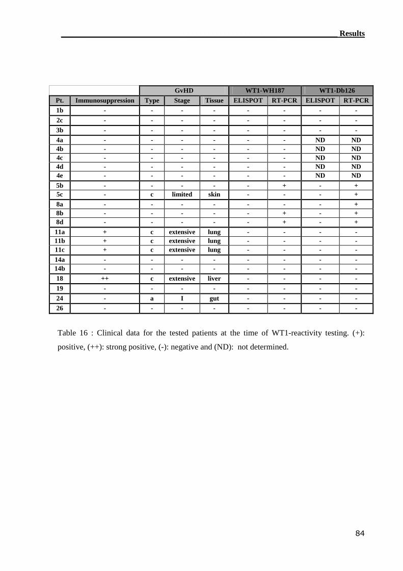
____________________________________________________________________ Results
84
GvHD WT1-WH187 WT1-Db126Pt. Immunosuppression Type Stage Tissue ELISPOT RT-PCR ELISPOT RT-PCR1b - - - - - - - -2c - - - - - - - -3b - - - - - - - -4a - - - - - - ND ND4b - - - - - - ND ND4c - - - - - - ND ND4d - - - - - - ND ND4e - - - - - - ND ND5b - - - - - + - +5c - c limited skin - - - +8a - - - - - - - +8b - - - - - + - +8d - - - - - + - +11a + c extensive lung - - - -11b + c extensive lung - - - -11c + c extensive lung - - - -14a - - - - - - - -14b - - - - - - - -18 ++ c extensive liver - - - -19 - - - - - - - -24 - a I gut - - - -26 - - - - - - - -
Table 16 : Clinical data for the tested patients at the time of WT1-reactivity testing. (+):
positive, (++): strong positive, (-): negative and (ND): not determined.
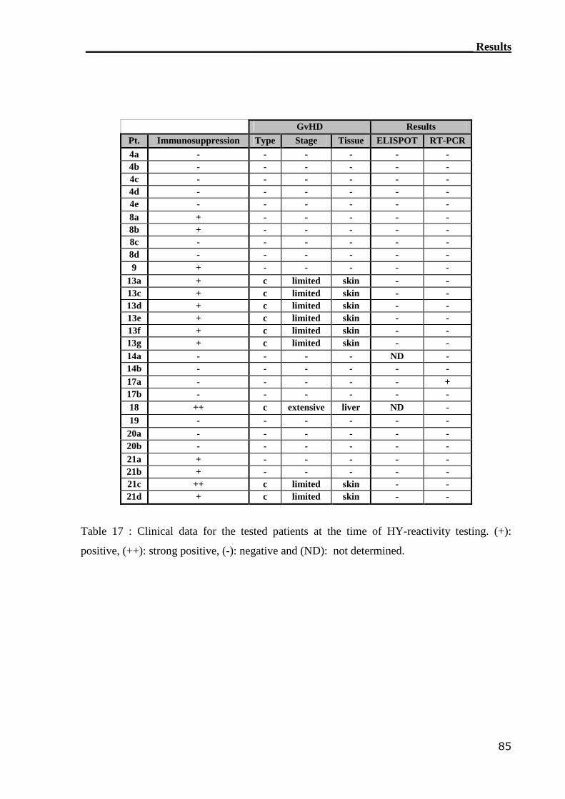
____________________________________________________________________ Results
85
GvHD ResultsPt. Immunosuppression Type Stage Tissue ELISPOT RT-PCR4a - - - - - -4b - - - - - -4c - - - - - -4d - - - - - -4e - - - - - -8a + - - - - -8b + - - - - -8c - - - - - -8d - - - - - -9 + - - - - -
13a + c limited skin - -13c + c limited skin - -13d + c limited skin - -13e + c limited skin - -13f + c limited skin - -13g + c limited skin - -14a - - - - ND -14b - - - - - -17a - - - - - +17b - - - - - -18 ++ c extensive liver ND -19 - - - - - -
20a - - - - - -20b - - - - - -21a + - - - - -21b + - - - - -21c ++ c limited skin - -21d + c limited skin - -
Table 17 : Clinical data for the tested patients at the time of HY-reactivity testing. (+):
positive, (++): strong positive, (-): negative and (ND): not determined.
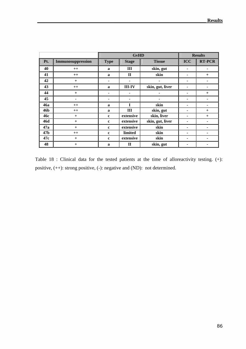
____________________________________________________________________ Results
86
GvHD ResultsPt. Immunosuppression Type Stage Tissue ICC RT-PCR
40 ++ a III skin, gut - -41 ++ a II skin - +42 + - - - - -43 ++ a III-IV skin, gut, liver - -44 + - - - - +45 - - - - - -
46a ++ a I skin - -46b ++ a III skin, gut - +46c + c extensive skin, liver - +46d + c extensive skin, gut, liver - -47a + c extensive skin - -47b ++ c limited skin - -47c + c extensive skin - -48 + a II skin, gut - -
Table 18 : Clinical data for the tested patients at the time of alloreactivity testing. (+):
positive, (++): strong positive, (-): negative and (ND): not determined.
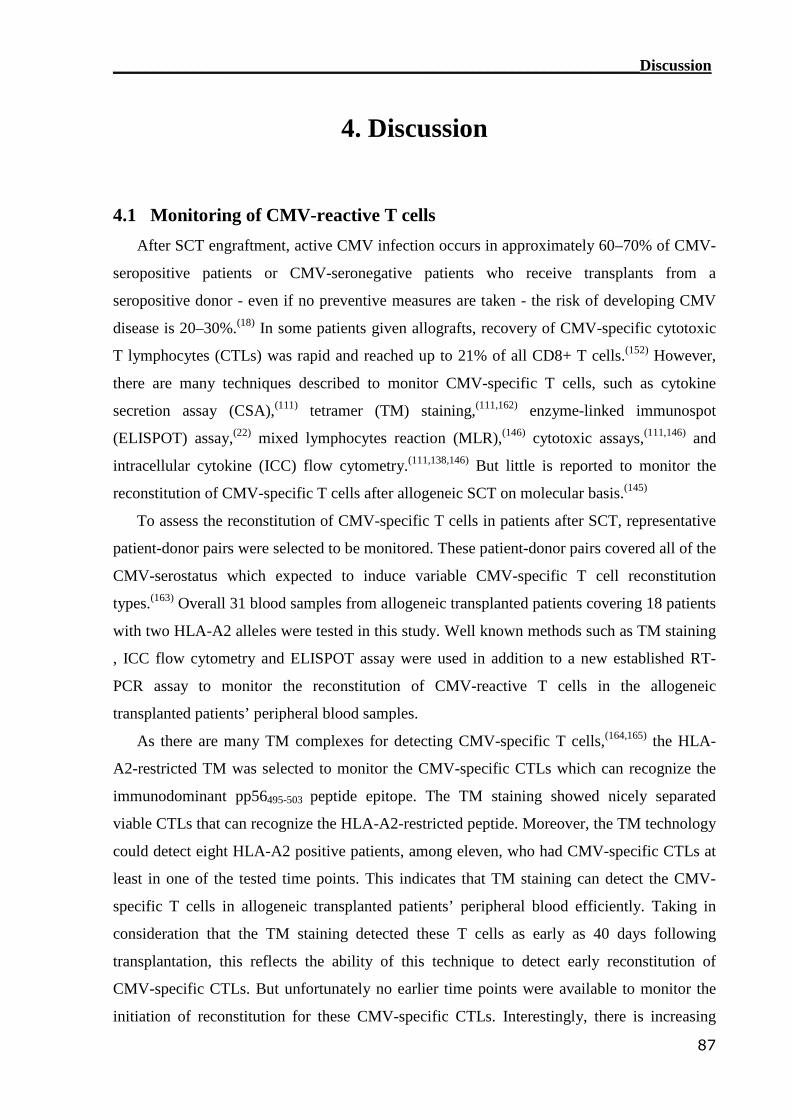
__________________________________________________________________Discussion
87
4. Discussion
4.1 Monitoring of CMV-reactive T cells
After SCT engraftment, active CMV infection occurs in approximately 60–70% of CMV-
seropositive patients or CMV-seronegative patients who receive transplants from a
seropositive donor - even if no preventive measures are taken - the risk of developing CMV
disease is 20–30%.(18) In some patients given allografts, recovery of CMV-specific cytotoxic
T lymphocytes (CTLs) was rapid and reached up to 21% of all CD8+ T cells.(152) However,
there are many techniques described to monitor CMV-specific T cells, such as cytokine
secretion assay (CSA),(111) tetramer (TM) staining,(111,162) enzyme-linked immunospot
(ELISPOT) assay,(22) mixed lymphocytes reaction (MLR),(146) cytotoxic assays,(111,146) and
intracellular cytokine (ICC) flow cytometry.(111,138,146) But little is reported to monitor the
reconstitution of CMV-specific T cells after allogeneic SCT on molecular basis.(145)
To assess the reconstitution of CMV-specific T cells in patients after SCT, representative
patient-donor pairs were selected to be monitored. These patient-donor pairs covered all of the
CMV-serostatus which expected to induce variable CMV-specific T cell reconstitution
types.(163) Overall 31 blood samples from allogeneic transplanted patients covering 18 patients
with two HLA-A2 alleles were tested in this study. Well known methods such as TM staining
, ICC flow cytometry and ELISPOT assay were used in addition to a new established RT-
PCR assay to monitor the reconstitution of CMV-reactive T cells in the allogeneic
transplanted patients’ peripheral blood samples.
As there are many TM complexes for detecting CMV-specific T cells,(164,165) the HLA-
A2-restricted TM was selected to monitor the CMV-specific CTLs which can recognize the
immunodominant pp56495-503 peptide epitope. The TM staining showed nicely separated
viable CTLs that can recognize the HLA-A2-restricted peptide. Moreover, the TM technology
could detect eight HLA-A2 positive patients, among eleven, who had CMV-specific CTLs at
least in one of the tested time points. This indicates that TM staining can detect the CMV-
specific T cells in allogeneic transplanted patients’ peripheral blood efficiently. Taking in
consideration that the TM staining detected these T cells as early as 40 days following
transplantation, this reflects the ability of this technique to detect early reconstitution of
CMV-specific CTLs. But unfortunately no earlier time points were available to monitor the
initiation of reconstitution for these CMV-specific CTLs. Interestingly, there is increasing
__________________________________________________________________Discussion
88
evidence that also functionally defect CMV-specific CTLs can be detected by TM staining,
thus it is essential to perform additional functional assays (e.g. ICC, ELISPOT and RT-PCR
assays) to confirm functionality of the TM detected T cells.(147)
The detection of specific IFN-γ-producing T cells by flow cytometry usually requires high
T cell frequencies (minimum 0.02% specific T cells in the CD3+CD8+ T-cell subpopulation,
corresponding to approximately 40 specific T cells per one million PBMC).(125) In this study,
the ICC staining detected eight HLA-A2 positive patients, among twelve, who have CMV-
specific T cells at least in one of the tested time points. This indicates that ICC staining can
detect the CMV-specific T cells in allogeneic transplanted patients’ peripheral blood
efficiently. Taking the advantage of phenotyping CMV-reactive T cells by ICC, it was clear
that these cells were CD8+ T cells reflecting the CTLs phenotype. But in some patients CD8-
T cells (e.g. helper T lymphocyte (Th)) showed a capability to secrete IFN-γ when stimulated
with the recombinant pp65 protein. Taking all together, the detected CMV-specific T cells
were found to be a combination of a majority of CTLs and a minority of Ths. This may be due
to the presence of CD4+ Th cells in 10- to 100-fold less than CD8+ Tc cells.(107) Although
CTLs were detectable by activation with pp65 peptide and/or recombinant protein, Ths were
only detectable when the pp65 protein was used. These results which are in concordance with
others,(138) reflect the specificity of the used peptide to the HLA-A2 allele, and the presence of
other CMV-specific T cells in the patients’ peripheral blood that can not be detected by the
TM staining such as the Ths. Furthermore, the use of pp65 protein was found to be more
efficient than pp65 peptide as re-stimulating Ag for the CMV-specific T cells. In addition to
the IFN-γ assessment, the cytotoxic functional perforin was assessed in the reconstituted
CMV-specific T cells. Significant IC perforin was detectable in some patients’ samples,
indicating that the detected CMV-specific T cells were really functional CTLs that have
cytotoxic abilities in addition to the type 1 cytokines pathway (e.g. IFN-γ).
In the longitudinal follow-up, many forms of CMV responses were seen using the pp65
peptide and/or protein as stimulus. In some experiments, the initiation of reconstitution for
CMV-specific T cells was detectable using pp65 protein but not the peptide which was
detected later. These results indicate that the CMV-reactivity can be monitored by IC flow
cytometry using both the pp65 peptide and protein, but the later can detect the CMV-specific
T cells earlier. This may be interpreted as that the pp65 protein contains many antigenic
epitopes that can induce the generation of many CMV-specific T-cell clones, which are not
only HLA-A2-specific.(138) In order to enhance the CMV Ags presentation, the C1R-A2 cell
__________________________________________________________________Discussion
89
lines were loaded with pp65 peptide then co-cultured with the PBMCs that contain the CMV-
specific T cells. This model showed enhancement of detection for IC IFN-γ by flow
cytometer, which not have been published before. This was in agreement with Arlen et al.
concerning the enhancing the ELISPOT assays by using the same concept.(148)
In general, in IC flow cytometry it is possible to routinely identify positive populations on
the order of 0.1% or even slightly less. But populations significantly lower, on the order of
0.01%, will not be distinguishable from background, because they reach the average level of
spontaneous cytokine-secreting cells in peripheral blood.(124) On the other hand, ELISPOT
assay is the “ex vivo” assay with the lowest limit of detection of IFN-γ-producing T cells,
allowing the reliable detection of as few as 1-10 specific T-cells per one million
PBMC.(125,166,166) To further define the CMV-specific T cells a sensitive ELISPOT assay was
adopted.(17) In this study, ELISPOT assay detected, at least in one of the tested time points,
fifteen HLA-A2 positive patients’ samples among nineteen covering seven out of ten patients
who have CMV-specific T cells. This indicates that the ELISPOT assay can monitor the
reconstitution of CMV-specific T cells in allogeneic transplanted patients’ peripheral blood
efficiently. Again, pp65 peptide and protein induced different forms of IFN-γ secretion. With
more focusing, IFN-γ-spots were generated in response to either pp56 peptide or protein, but
some times they were for both CMV Ags. In view of these results, the reconstituted cells were
found to compose from different CMV-specific T cell clones that can recognize different
epitopes from the pp65 protein. These cells expected to be a mix of different memory T cells
that can respond fast when counteracting with their Ag for the second time.(167) Although the
ELISPOT assay can monitor the reconstituted CMV-specific T cells in high sensitivity, it has
many disadvantages such as the time consuming protocols (i.e. 4 days) and the incapability of
phenotyping the CMV-specific effector cells.(167) Moreover the main disadvantage is that,
even with the aid of specialized imaging equipment, there is a certain degree of subjectivity in
the interpretation of results as a threshold for the size, intensity, and gradient of the spots are
user-defined.(127)
As the sensitivities of ICC and ELISPOT assays are dependent on the background from T
cells spontaneously producing cytokines, various procedures help to reduce background
staining.(125) To get rid of background effect in the ELISPOT assay, Hempel found that the
immune response measured in the peripheral blood of mice by RT-PCR and ELISPOT
showed a significant correlation to the response measured in the spleen (P=0.001).(155) In
addition to the new introduction of RT-PCR assays as monitoring assays for Ag-specific T
__________________________________________________________________Discussion
90
cells and due to their high sensitivity and reproducibility,(127) this approach was adopted.
Thus, the following hypothesis was proposed: can the RT-PCR assay detect patients’
peripheral blood CMV-specific T cells with correlation to the other adopted monitoring
assays? At least in one of the tested time points, the RT-PCR assay detected nineteen HLA-
A2 positive patient samples among twenty one, covering ten out of twelve patients, who have
CMV-specific T cells. Simply these data indicate that RT-PCR assay can efficiently monitor
the reconstitution of CMV-specific T cells in the peripheral blood of allogeneic transplanted
patients following SCT. With few exceptions, significant up-regulation of IFN-γ mRNA
expression was noticed in the entire patient samples treated with pp65 protein. On the other
hand, the up-regulation of IFN-γ gene expression was induced in some samples when re-
stimulated with pp65 peptide. Even it was noticed that the pp65 peptide always induced cell-
activation in parallel to pp65 protein, but not the other way around. These results indicate that
the RT-PCR assay can monitor the CMV-specific T cells even in lower frequencies than
detected by TM, ICC and ELISPOT.
In the longitudinal time follow-up study, the RT-PCR assay was able to monitor the
already found or newly initiated CMV-reactivity. In addition, the RT-PCR assay was able to
detect the reconstitution of CMV-specific T cells as early as forty days after SCT. Taking in
consideration the results of the two CMV Ags, more detection sensitivity was shown by the
use of pp65 protein rather than the peptide. In view of these results, the different reconstituted
CMV-specific T cell clones composing all of the different pp65 protein epitopes were
detectable by this very sensitive RT-PCR assay. In addition to the ability of RT-PCR assay to
monitor the reconstituted CMV-specific T cells in high sensitivity, also it has many other
advantages such as the short stimulation time (i.e. 3 hours). But unfortunately, the RT-PCR
detected CMV-specific effector cells can not be isolated or phenotyped.(127)
Interestingly, the CMV-reactive T cells were detectable by ICC flow cytometry,
ELISPOT, and RT-PCR assays but not TM staining in the control group patients who do not
have the HLA-A2 allele to present the HLA-A2-restrected pp65 peptide. This was performed
by the use of pp65 protein as CMV stimulus that can be processed by the patients’ antigen
presenting cells (APCs). After the pp65 protein processing in the exogenous pathway, all of
the pp65 protein immunodominant epitopes presented to all of the CMV-specific T cells
found in the PBMCs mixture. This gave the ability to detect the non-HLA-A2-restricted
CMV-reactive T cells. Unfortunately this is not applicable by the TM staining since the
__________________________________________________________________Discussion
91
multimers technology depends on the pre-selection of the HLA-restricted peptides to be
loaded into the TM-peptide complexes.(118,120)
The sensitivity of the adopted detection assays is very important to monitor the Ag-
specific T cells in the peripheral blood of the allogeneic transplanted patients. As four
techniques were adopted to monitor the reconstitution of CMV-specific T cells, the sensitivity
for each technique was determined. The previous described high sensitivity for the TM
staining(137) was reproduced in this study when the pp65 peptide was used as an Ag.
Moreover, the same high sensitivity was found in the RT-PCR assay, in contrary to the ICC
staining which showed the lowest sensitivity compared to TM staining and RT-PCR assay.
This confirms that established functional RT-PCR assay has the same high sensitivity of the
phenotypic TM staining. On the other hand, all of the techniques did not detect any pp65
peptide reactivity in the HLA-A2 negative patients’ samples, confirming the specificity of the
techniques in addition to the HLA-A2-restriction for the used peptide. Assessment of the
CMV reactivity using the pp65 protein increased the sensitivities in ICC flow cytometry and
RT-PCR assay, reflecting better monitoring ability using the pp65 protein rather than the
HLA-A2-restricted peptide. Interestingly, the non-HLA-A2-restricted CMV-specific T cells
could be monitored in the HLA-A2 negative patients by ICC staining, ELISPOT assay, and
RT-PCR assay when the pp65 protein was used as stimulating Ag. Taking all together, the
new established RT-PCR assay can monitor the reconstitution of CMV-specific T cells with
high specificity and without HLA-restriction if the antigenic protein was used.
Normally when the RT-PCR assays newly introduced to test new target gene,
comparisons should be performed in parallel to the already existing methods, and then the
correlation should be determined.(168,169) Although the established RT-PCR assay has high
sensitivity similar to that found in the other methods, another testing strategy was adopted by
checking the correlation of the established RT-PCR assay with the other methods. In general,
the RT-PCR assay was correlated significantly with the TM staining (P=0.005), which was
not yet been published elsewhere. Even a higher correlation was found when the RT-PCR
assay results were correlated to the ELISPOT and ICC (P=0.0001). Moreover, the Pearson
correlation coefficient (R2) was high when the RT-PCR assay was compared to ICC staining,
reflecting good linearity between the two techniques. This was not the case when the RT-PCR
assay was compared to the ELISPOT assay and TM staining. In view of these results, the
higher correlation coefficient can be explained by the higher frequencies of CMV-specific T
cells in the allogeneic transplanted patients following SCT.
__________________________________________________________________Discussion
92
In healthy CMV-seropositive individuals, up to 40% of all T cells in the peripheral blood
can be specific for CMV,(170) underlining the importance of a strong CMV-specific cellular
immunity in containing persistent CMV infection. As the CMV-serostatus of the donors
influence the outcome of allogeneic SCT in some patient categories,(163) the results from the
four monitoring techniques were correlated to the CMV-serostatus of the tested patients and
their donors. In concordance with Cwynarski et al. who found that the CMV-reactive T cells
can reach up to 21% of all CD8+ T cells,(152) the recovery of CMV-specific CTLs was rapid
when both the patient and the donor were seropositive for CMV before SCT. In addition to
these results, the tested pp65 protein enhanced the detection limit up to 100% in most of the
tested patients. For the first time, with few exceptions, this could be achieved in both HLA-
A2 positive and negative patients by ICC flow cytometry, ELISPOT and RT-PCR assays, but
could not be applied to the TM staining. On the other hand, early reconstitution was not
observed if either the donor or recipient was seronegative for CMV, especially when the
patient was CMV-seronegative and his donor was CMV-seropositive. In the CMV-
seropositive patients received transplant from CMV-seronegative donors displayed, delayed
reconstitution of CMV-specific T cells were seen which have been documented elsewhere.(152)
These results illustrate a novel concept concerning the use of the established RT-PCR assay as
a monitoring tool for the reconstitution of CMV-specific T cells in patients at risk due their
own or their donors CMV-serostatus.
As a summary these results concerning the monitoring of allogeneic transplanted patients,
confirm the ability of monitoring CMV-reactive T cells by the classical methods (e.g. TM
staining, ICC staining, and ELISPOT assay) as well as the new established RT-PCR assay.
Moreover, in addition to the fast assessment, the RT-PCR showed a high sensitivity which
correlates to the other known methods. Although functionally defective CMV-specific T cells
can be detected by TM staining,(147) the RT-PCR assay has the advantage of detecting the
functionality of the reconstituted CMV-specific T cells with a similar sensitivity to the TM
staining. By demonstrating the high correlations between the RT-PCR and the classical
methods, the detection of CMV-reactive T cells in low frequencies by RT-PCR assay is no
more under doubt. Finally, the use of whole antigenic and immunodominant CMV pp65
protein gave a novel approach to monitor CMV-specific T cells in regardless of the patients
HLA typing.
__________________________________________________________________Discussion
93
4.2 Monitoring of WT1-reactive T cells
Enrolling representative patient samples is also important for covering all of the
possibilities of detecting WT1-reactive T cells in peripheral blood. Therefore, assessment of
twenty two leukemic samples covering twelve patients, were eligible to be used. In addition,
most of the well known blood malignancies that over-express the WT1 antigen were selected
to be monitored including AML, ALL and CML.(140,142,171) Moreover, the patient samples
covered a range of time between five months and five years and two months following
allogeneic SCT. In this model all of the expected HY-reactive T cells should be in scope, so
the other determinant for monitoring was the sensitivity of the testing techniques.
Most of the tools used to detect WT1-reactive T cells are TM staining,(171,172)
immunohistochemistry,(172) ELISPOT assay,(157) ICC flow cytometry,(157,171) cytotoxicity
assays,(158,173) and recently a molecular approach was established.(145) As Scheibenbogen et al.
could detect WT1-specific T cells by high sensitive ELISPOT assay(157) this assay was
adopted. Although the adopted ELISPOT assay reported to have high sensitivity,
unfortunately it did not detect any WT1-reactive T cells in patient samples. To overcome the
failure of ELISPOT assay to detect the WT1-specific T cells, a sensitive RT-PCR assay was
in scope. The adopted assay was established by Rezvani et al. with high sensitivity to detect
frequencies of 1 responding T cell / 100,000 T cells.(145) Some of the RT-PCR assay results
were significant positive for IFN-γ mRNA gene expression indicating the presence of WT1-
reactive T cells in the patients peripheral blood following allogeneic SCT. Taken together the
detection of WT1-reactive T cells by the RT-PCR, but not the ELISPOT, confirms the higher
sensitivity of the RT-PCR.
By using the highly sensitive ELISPOT assay, Scheibenbogen et al. did not detect any
WH187-reactive T cells in all of the fifteen AML patients they tested, compared to two
patient samples that secreted IFN-γ upon stimulation with WT1-Db126 peptide.(157)
According to the present study, interestingly the non-detectable WT1-reactive T cells in the
ELISPOT assay was in concordance with Scheibenbogen et al. who barely could detect these
CTLs. In an effort to eradicate doubt concerning the ability of the ELISPOT assay to detect
the WT1-reactive T cells, a significant up-regulation of IFN-γ mRNA upon stimulation with
both peptides was detected using the RT-PCR assay. In line with these results, obvious
evidence that the two HLA-A2-restricted WT1 peptides differ in their reactivity was
observed, since the WH187 peptide reactivity was detected by RT-PCR in 17% of the
eighteen patient samples compared to 28% in response to the Db126 peptide. According to
__________________________________________________________________Discussion
94
the present study WT1-Db126 peptide showed a higher immunogenicity than WT1-WH187.
Consequently, the RT-PCR is good to be used as a monitoring tool for detecting the
generation of GvL-specific T cells.
Differences in detecting IFN-γ mRNA after stimulation with the two WT1 HLA-A2-
restricted peptides, may be interpreted due to differences in the immunogenicity of both
peptides. In murine model, coincidentally Db126 peptide demonstrated the same order of
binding affinity as that of viral Ags, which is the strongest Ags for CTLs induction.
Moreover, the cytotoxic activity of Db126-specific CTLs showed to have half the maximal
lysis in the range of nanomolar of the peptide.(173) Thus, only the Db126 peptide with the
highest binding affinity for HLA-A2 molecules could elicit CTL responses. The same
consideration should be in mind when viewing the vaccination trials against some leukemias
which adopted the Db126 peptide as a vaccine in the HLA-A2 positive patients.(156,171) Even
in a case study, vaccination with Db126 peptide induced complete remission in a patient with
recurrent AML by in the absence of toxicity.(171) In view of these results, Db126 peptide
should be considered to be the most promising target peptide to induce CTL responses against
WT1 in HLA-A2 positive patients with malignant neoplasms.
In contrast to ELISPOT assay, which did not detect any WT1-reactive T cells, RT-PCR
was able to detect significant increase of IFN-γ mRNA in response to WT1 peptides. Doubts
were raised about the detection limit of ELISPOT assay concerning the following question:
dose the ELISPOT assay has enough detection limit to detect WT1-specific T cells at patients
peripheral blood samples? In general, the “limit of detection” of a method is the capacity of
the method to detect small amounts of a substance with some assurance, and it is known that
the limit of detection by ELISPOT was reported to be 10–200 times lower than ELISA
performed on culture supernatants.(124) On the other hand, the detection of specific IFN-γ-
producing T cells by ELISPOT assay is better than flow cytometry which is usually requires
higher T cell frequencies (approximately 40 specific T cells per one million PBMC).125
However, the ELISPOT assay sensitivity is depending on the spontaneous secretion of
cytokines by T cells as a background. Although not so many researchers used the ELISPOT
assay to monitor WT1-reactive T cells,(157) approximately the same results were obtained in
comparison to this work. This may be due to the low frequencies of WT1-specific CTLs in
the peripheral blood of the tested patients, or due to the presence of WT1-specific T cells
recognizing other WT1 epitopes rather than the tested peptides.
__________________________________________________________________Discussion
95
In general, RT-PCR assays are usually used to diagnose the leukemias and to assess the
WT1 expression in tumor’s blasts,(156,158,174) but not monitoring the reconstituted WT1-
reactive T cells. Recently the detection of WT1-reactive T cells by RT-PCR was established
in purely selected CD8 positive T cells, but not in PBMCs.(145) Despite that the ELISPOT
assay did not detect WT1-reactive T cells among all of the patient PBMC samples, RT-PCR
demonstrated significant up-regulation of IFN-γ mRNA in response to WT1 peptides. In
terms of adopting RT-PCR protocol with a high sensitivity and capability of detecting Ag-
specific T cells at frequencies in the order of 1/100,000 CD8+ T cells,(145) so the RT-PCR
results should be more reliable than the ELISPOT. In line with this high sensitivity, the
significant up-regulation of IFN-γ mRNA after WT1 peptide pulsing confirming the presence
of WT1-reactive T cells at the selected patients’ peripheral blood. These results illustrate a
novel concept of monitoring WT1-reactive T cells by RT-PCR assay even if the frequency is
below the detection limit of the most powerful technique to detect Ag-specific T cells, which
is the ELISPOT assay.
Although the presence of WT1-reactive T cells in the peripheral blood of allogeneic
transplanted patients is important, the reconstitution time after transplantation is important
too. In this study, the minimal time required to detect reconstituted WT1-reactive T cells was
five months and fifteen days, but in another patient it was nine moths and thirteen days
following transplantation. Unfortunately, no earlier blood samples were available for both
patients to detect any earlier reconstitution of the GvL effector T cells. For both patients, the
detection of WT1-reactive T cells by RT-PCR assay was in concordance with what have been
found by others.(145) Interestingly and for the first time WT1-reactive T cells were detected by
molecular approach in AML and ALL patients. These results can be further expanded to
screen other groups of patients to monitor the reconstitution of WT1-reactive T cell during the
first five months following allogeneic SCT.
These results indicate that the GvL effect can be monitored by RT-PCR but not ELISPOT
assay. This is due to the higher sensitivity of the RT-PCR assay which can detect very low
frequencies of Ag-specific T cells.(145) Also the present study clearly indicates a tight junction
between the selected WT1 peptides and the up-regulation of IFN-γ mRNA as a final readout
for the established RT-PCR assay. Concomitantly, WT1-Db126 peptide seems to have more
immunogenicity compared to the WT1-WH187 peptide. Moreover in this model, the GvL
effector T cells that can recognize the tumor cells having the WT1 protein can be monitored
as early as five months following SCT, but earlier time follow-up is suggested.
__________________________________________________________________Discussion
96
4.3 Monitoring of HY-reactive T cells
Allogeneic stem cell donors can be incompatible at different levels. Even in the HLA-
identical pairs it will be still incompatible for numerous minor histocompatibility antigens
(mHAg). Nevertheless, some incompatibilities are found to be associated with an increased
risk of graft-versus-host disease (GvHD), which could be related to the way the immune
system recognizes these antigens. In HLA-identical bone marrow transplantation, GvHD may
be induced by disparities in mHAgs between the donor and the recipient, with the antigen
being present in the recipient and not in the donor. CTLs specific for mHags of the recipients
can be isolated from the blood of recipients with GvHD.(97)
To enroll representative patient’s samples to detect HY-reactive T cells in peripheral
blood, all of the selected patients were male recipients who received SCT from female donors.
In addition to the selected patients who were suffering AML, ALL and NHL before SCT,
samples covered a range of time between five months up to five years and two months after
allogeneic SCT. Assessments of twenty eight patient samples from ten patients were eligible
to be used. In this model all of the expected HY-reactive T cells should be in scope, so the
other determinant for monitoring will be the sensitivity of the testing assays. Although twenty
five HLA-A2 positive patient samples were monitored, only one sample was positive for the
HY-reactivity. This confirms the eligibility of the monitoring assays to detect HY-reactive T
cells in the patient’s peripheral blood.
There are many methods describing the detection of HY-reactive T cells such as CTL
assays,(97) 3H-thymidine incorporation assay,(160) cytotoxicity (51Cr release) assay,(160,100) TM
staining(175) and IC-IFN-γ flow cytometry,(105) but no study described the detection of the
human HY-reactive T cells using molecular technologies so far. Interestingly, James et al. and
others could detect HY-specific T cells in mice using high sensitive ELISPOT assay.(161,176)
Although high sensitive ELISPOT assay was adopted as monitoring tool, unfortunately no
detection of HY-specific T cells was achieved in all the patients’ samples. On the other hand,
as the HY-specific T cells were documented to be detectable in mice by using the RT-PCR
assay, the same concept was adopted to monitor the reconstitution of HY-specific T cells in
allogeneic transplanted patients.(177) In contrast to the ELISPOT negative results, the adopted
RT-PCR assay which showed previously a higher detection sensitivity, was able to detect the
activated T cells after stimulation with the HY peptide. Concomitantly, the RT-PCR is good
to be used as a monitoring tool for detecting the generation of mHAg-specific T cells.
__________________________________________________________________Discussion
97
In the GvHD models, coincidentally H2-Dk epitope(175) and FIDSYICQV SMCY-derived(100,105) HY peptides demonstrated the strongest mHAgs for CTLs induction in mice and
human, respectively. In this model the RT-PCR assay positive results were specific to the HY
peptide stimulation when compared to the low background IFN-γ mRNA expressed in
response to the gp100 HLA-A2-restricted peptide. In general, this significant up-regulation of
IFN-γ mRNA after stimulation with the HY HLA-A2-restricted peptide may be interpreted by
the immunogenicity of HY peptide. By demonstrating detectable HY-reactive T cells in one
patient among seven, these patient’s PBMCs were able to produce IFN-γ after activation with
the HY peptide which may reflect that the HY antigen promotes cell-mediated immunity
rather than antibody production and humoral immunity.(50) In view of these results, HY
peptide should be considered in monitoring of reconstitution of mHAgs-specific T cells.
In contrast to ELISPOT assay, which did not detect any HY-reactive T cells, RT-PCR was
able to detect significant increase of IFN-γ mRNA in response to HY peptide. Again, doubt
rises about the detection limit of ELISPOT assay and the following question appeared: can the
ELISPOT assay detect HY-reactive T cells in patients’ peripheral blood? To answer this
question we should keep in mind that the ELISPOT detection limit was reported to be lower
than ELISA and ICC, and the sensitivity of ELISPOT assay is depending on the background
which is the spontaneous secretion of cytokines by T cells. Although no researchers used the
ELISPOT assay to monitor human HY-reactive T cells, these ELISPOT results are believed to
be realistic as this assay showed high sensitivity in detecting CMV-reactive T cells. The non-
detectable HY-specific T cells by the ELISPOT assay may be interpreted by the low
frequency of these CTLs in the peripheral blood of the tested patients, or due to the presence
of HY-specific T cells recognizing other HY epitopes rather than the tested peptide.
Recently RT-PCR assays were established to detect ex vivo generated and expanded HY-
reactive T cells in murine models.(175,177) Due to fact that no literature was found concerning
the monitoring of reconstituted HY-reactive T cells in allogeneic transplanted patients’
peripheral blood, so the RT-PCR assay was adopted. Despite that the ELISPOT assay did not
detect any HY-reactive T cells among all of the tested patients’ PBMC samples, RT-PCR
demonstrated significant up-regulation of IFN-γ mRNA in response to the HY peptide in one
patient. Moreover, this patient developed eosinophilia at the time of testing which is
considered as an early marker of GvHD.(178,179) In terms of adopting the RT-PCR assay which
has high sensitivity and capability to detect Ag-specific T cells at low frequencies, the RT-
PCR assay results showed more reliability than the ELISPOT assay. In line with this high
__________________________________________________________________Discussion
98
sensitivity, the significant up-regulation of IFN-γ mRNA after HY peptide pulsing confirming
the presence of HY-reactive T cells at the selected patients’ peripheral blood. These results
illustrate a novel concept of monitoring HY-reactive T cells by RT-PCR even if the frequency
is below the detection limit of the ELISPOT assay.
Not only the presence of HY-reactive T cells in the peripheral blood of allogeneic
transplanted patients was in the focus of this study, but also the reconstitution time required
for these T cells to be detectable was important too. In viewing the RT-PCR results, the HY-
reactive T cells were detectable only in one patient’s blood sample with a reconstitution time
of nine moths and eight days following transplantation. Concerning the absence of the HY-
reactivity two months later in the same patient, this may be interpreted by the administration
of immunosuppressive drugs to overcome the GvHD symptoms. Unfortunately, no earlier
blood samples were available for this patient to detect any earlier reconstitution of the HY-
specific T cells. For this patient, the detection of HY-reactive T cells was in accordance with
what has been found by others.(97) Interestingly, for the first time HY-reactive T cells were
detected by molecular approach (e.g. RT-PCR) in allogeneic transplanted patients which have
not yet been published. These results can be further expanded to screen other groups of
patients to monitor the reconstitution of HY-reactive T cell during the first nine months
following allogeneic SCT.
Taking all together, these results indicate that the HY-reactivity can be monitored by RT-
PCR assay but not the ELISPOT assay. This is because of the higher sensitivity of the
established RT-PCR assay compared to the ELISOT assay. Also the present study clearly
indicates a tight junction between the selected HY peptide and the up-regulation of IFN-γ
mRNA as a final readout for the established RT-PCR assay. Concomitantly, the RT-PCR
assay confirmed that the FIDSYICQV SMCY-derived HY peptide is immunodominant
mHAg after allogeneic SCT. Moreover in this mHAg model, the HY effector T cells which
can recognizes the autosomal HY protein on the male patient’s tissues could be monitored as
early as nine months following transplantation, but earlier time follow-up is suggested for
future work. However, the present data provide evidence that the emergence and activation of
HY-specific T cell in allogeneic transplanted patients can be monitored to predict any
developing GvHD in parallel to eosinophilia. In conclusion, these findings provide evidence
that HY-specific CTLs may be induced in male patients given a stem cell graft from a female
donor, which may lead to the development of either GvL reactions or GvHD or both.
__________________________________________________________________Discussion
99
4.4 Monitoring of alloreactive T cells
Great attention is paid to the detection of GvHD and after human SCT. For example, host-
reactive lymphocytes with broad specificity have been observed in GvHD patients using the
limiting dilution techniques, which are time consuming for detecting functional T cell
analysis. In the meantime, many techniques were established to predict GvHD such as, first:
the in vitro skin explant assay,(180) second: T lymphocyte precursors frequency analysis,(55)
third: some serum markers, such as the levels of TNF-α , IFN-γ , IL-10, soluble Fas, and IL-
18,(49,51,51,56,57) fourth: polymorphism of IL-10,(58) transforming growth factor (TGF)-β1(59) and
other genes(181), fifth: ELISPOT assays,(47,60) and fifth: T cell’s TCR-Vβ clonotypic
analysis.(60-62) In addition, TM technology has been developed which allows flow cytometric
detection of specific T cells independently of their activation state.(63) But many of these
analysis methods have not been reported to be necessarily predicting GvHD, but it may detect
many inflammation states. Thus, there are contradictory results among these methods and
there still remain problems with attempts to use these parameters as reliable and sensitive
markers of GvHD.
In general, systematic analyses of alloreactive lymphocytes in large cohorts of patients
have only been performed in bone marrow grafts before transplantation,(55) whereas only very
small cohorts of patients have been analyzed for the presence of circulating alloreactive
lymphocytes after transplantation.(182) Thus, a valid in vitro immune monitoring of GvHD
patients seems essential for further improvements in allogeneic stem cell transplantation. In
this study, a mixed lymphocyte reaction (MLR) model was established to simulate the
generation of alloreactive T cells in the GvHD patients’ peripheral blood. Successfully, this
model produced alloreactive T cells that can produce IFN-γ when counteracting with their
stimulator cells which bearing their specific allo-antigens (allo-Ags). Many optimizing steps
were done successfully, including freezing-thawing protocols, gating-out stimulator cells
from the FACS analysis, and testing the ability of secreting many cytokines.
The MLR-generated allospecific T cells were able to produce significant IFN-γ after co-
culturing with their specific allo-Ag. This IC IFN-γ production was in concordance with
others.(114,183) To assess this IC IFN-γ production by flow cytometer, irradiation and
counterstaining of the allo-Ag-holding stimulator cells were found to be essential, reflecting
weak IFN-γ production in the alloreaction which can be detected significantly after gating-out
the irradiated and counterstained allo-Ags-holding cells from the flow cytometric analysis. To
simulate the effect of freezing-thawing on the allogeneic transplanted patient samples, MLR-
__________________________________________________________________Discussion
100
generated alloreactive T cells were frozen and thawed before testing. Despite this freezing-
thawing step, the alloreactive T cells capability to produce IFN-γ was kept in the significant
levels. Taking all together, this model was optimized to monitor the IFN-γ production in the
situation of alloreactions. In addition to the monitoring of IFN-γ production in the generated
alloreaction settings, TNF-α, IL-4, IL-10 and perforin production was tested in the same MLR
settings. Unfortunately none of them were detectable significantly by IC staining and flow
cytometric analysis, reflecting the generation of type 1 cytokines pathway in the established
alloreactive MLR settings, but not the type 2 cytokines pathway.
Correlation of immunological and clinical data is essential to improve the understanding
of the immunology of GvHD. As an optimized MLR testing system was established,
monitoring of alloreactive T cell in allogeneic transplanted patients was eligible. So, nine
allogeneic transplanted GvHD- and non-GvHD-suffering patients were monitored for the
presence of alloreactive T cells in their peripheral blood. Unfortunately, none of the fourteen
patient samples showed significant IC IFN-γ production after exposure to the patient cells
before SCT. However it should be kept in mind that, in the patient experiments, IFN-γ signal
was not easily distinguishable in the flow cytometeric analysis therefore the use of IC flow
cytometry as monitoring tool should be more optimized before considering as eligible method
to monitor GvHD patients.
On the other hand, the IL-4 production was not significant in all of the patients’ tested
CD4+ (i.e. Th) and CD4- (i.e. Tc) T cell subpopulations. Interestingly, these results can be
interpreted as the alloreactive T cells in the allogeneic transplanted patients use the type 1
cytokines pathway more than the type 2, and this is with concordance with what have been
found by Kasakura.(50) But these results should be carefully considered since IL-4 can not be
detected easily by flow cytometers and as IL-4 signal normally appears as a shoulder to the
CD4+ T cells more than a separated cell population.
As there are no molecular approaches to monitor the alloreactivity in the GvHD patients,
the RT-PCR assay was established and adopted. Assessment of IFN-γ gene expression was
monitored in the optimized healthy mismatched-MLR alloreaction settings. The up-regulation
of IFN-γ mRNA expression after the second exposure to allo-Ag reflects the ability of RT-
PCR assay to monitor the alloreactive T cells. Interestingly, significant up-regulation of IFN-γ
mRNA expression in some patient samples after transplantation (AT) was seen after co-
culturing with the patient cells before transplantation (BT). This indicates the ability of
detecting alloreactive T cells in the some patients suffering GvHD. These results indicates,
__________________________________________________________________Discussion
101
for the first time, that the RT-PCR is promising tool for monitoring the alloreactive T cells in
the peripheral blood of allogeneic transplanted patients especially predicting GvHD.
4.5 Impact of the clinical parameters on the detected T-cell properties
Unlike the majority of the tested patients for CMV-, WT1- and HY-reactive T cells, few
patients were under immunosuppressive drug treatments and/or suffering GvHD. The
immunosuppressive state of the patients should be considered since it may cause the
impairment of T-cell functionality. This is possible especially when a high dose of
corticosteroids (e.g. Prednisolone) was applied, which can suppress all of the
immunocompetent reactive T cells in the patient.(4,42) As seen in the CMV and GvHD
monitoring parts, the immunosuppression state was not a limiting factor for detecting the
functional T cells. But it may clarify why some patients’ PBMCs did not show any IFN-γ
production after a proper Ag stimulation. The negative results may be really due to the
absence of the Ag-reactive T cells in the tested samples, but also it could be due to the
impairment of the T cells in the donated patients’ peripheral blood.
Another factor which may affect the interpretation of the CMV-, WT1- and HY-reactivity
results is the presence of GvHD. Although some of the tested patients had this disease at the
time of blood donation, positive and negative results could be obtained. As the GvHD can
occur in many of the allogeneic transplanted patients, these results should be carefully
interpreted for any other Ag-specific T cells rather than the alloreactive T cells. But this does
not mean that the result is falsely positive since the used Ags were very specific and can not
be detected by pan-reactive T cells.
4.6 Limitations of the used monitoring assays
Each monitoring assay has advantages and limitations which should be considered. For
example, the TM staining gives only phenotypical but not functional data about the tested
cells.(167) On the other hand, the ELISPOT assay can give the functional but not phenotypical
data about the Ag-reactive T cells.(167) Also, the ICC and ELISPOT assays do not allow the
Ag-specific T cells isolation, since the cell should be perforated or discarded to give the
functional data.(107) Although the RT-PCR assay gave the best results and showed the superior
sensitivity, unfortunately no phenotypical data can be obtained from this assay.(127) So, the
IFN-γ mRNA up-regulation after applying certain Ag can not be correlated to specific T-cell
sub-population. Concomitantly and as a compromise, the RT-PCR assay should be performed
__________________________________________________________________Discussion
102
in parallel with phenotypical assays (e.g. TM staining and ICC detection) that can identify the
Ag-reactive T cell sub-population.
4.7 Further suggested work
Although the monitoring assays in this work were promising, the results need
confirmation by further testing. For example, more patients should be tested for the
reconstitution of WT1-, HY- and allo-specific T cells. This will increase the confidence about
the already found results. Also, testing patients in earlier time points after SCT will give
better view concerning the reconstitution of specific T cells after allogeneic SCT. In addition,
other cytokines such as the TNF-α, IL-4, perforin and IL-2 are strongly suggested to be traced
in the patient settings.(109) This will give better view concerning the cytokine pathways used
by the immune cells in the settings of GvHD (e.g. Th1, Th2 and Tc cells functional).(50)
Finally, patients under strong immunosuppressive drug treatments should be excluded from
any further experiments for monitoring the WT1- and HY-reactive T cells. Another solution
can be the blood donation before the administration of the immunosuppressive medicaments.
By this approach, the negative results will be directly correlated to the cells’ Ag-reactivity and
not to the impairment of tested cells due to the administration of immunosuppressive
medicaments (e.g. cyclosporine, corticosteroids, and methotrexate).(2)
_________________________________________________________________ Summary
103
5. Summary
After administration of the hematopoietic stem cells inoculum into the eligible patients in
the allogeneic setting, many hematopoietic cells reconstitute in the following months to years
after transplantation. The reconstituted cells can be pathogen-specific (e.g. cytomegalovirus
(CMV)-reactive T cells), leukemia-specific (i.e. graft versus leukemia (GvL) effect) or
recipient-specific (e.g. graft-versus-host disease (GvHD)). Thus, valid in vitro immune
monitoring strategies should broaden the understanding of CMV immunity, GvL and GvHD,
which seems essential for further improvements in allogeneic stem cell transplantation (SCT).
Furthermore, approaches of this type should help to dissect allo-specific from tumor-specific
immune responses and will help to clarify, how both mechanisms are interconnected and how
they can be best put in action for therapeutic purposes.
The CMV-specific T cells reconstitution was monitored efficiently, in allogeneic
transplanted patients, by classical approaches such as tetramer (TM) staining, intracellular
cytokine (ICC) flow cytometry and enzyme-linked immunospot (ELISPOT) assay, in addition
to the newly introduced real time-polymerase chain reaction (RT-PCR) assay. In general, the
RT-PCR assay was highly sensitive in comparison to the other methods, and was also
correlated to all of them. Interestingly, the use of the pp65 protein as CMV antigen enhanced
the monitoring capabilities in comparison to the pp65 HLA-A2-restricted peptide, and
allowed testing of HLA-A2 negative patients.
As an example for assessment of the GvL effect in allogeneic transplanted patients, the
Wilms' tumor suppressor gene (WT1)-specific T cells were monitored. Unfortunately the
WT1-specific T cells were not detectable by a sensitive ELISPOT assay, but on the other
hand and for the first time the established RT-PCR assay was able to track two types of WT1-
reactive T cell clones in the same samples. In addition and as not described before, the RT-
PCR assay was able to monitor the reconstitution of WT1-specific T cells in patients who
were suffering AML and ALL before transplantation.
As the minor histocompatibility antigens (mHAgs) play a dual role in the GvL and GvHD,
the reconstitution of male-specific mHags (HY)-specific T cells were monitored. The adopted
_________________________________________________________________ Summary
104
sensitive ELISPOT assay did not detect any HY-specific T cells in all of the tested patients’
samples. On the contrary, the established RT-PCR assay successfully detected the HY-
specific T cells in one patient who later developed eosinophilia as a GvHD complication. As
for the first time these cells were detectable by RT-PCR assay in the allogeneic transplanted
patients’ peripheral blood, this give a novel concept of predicting GvHD due to the generation
of mHAg-specific T cells in the patients body.
Due to the still insufficient knowledge on mechanisms involved in GvHD, it was
considered a major aim to tailor the allo-reactive T cells in the patients’ peripheral blood in
order to avoid fatal courses of GvHD. An allogeneic mixed lymphocyte reaction (MLR)
model was established and optimized to simulate the presence of the allo-reactive T cells in
the GvHD patients’ blood. After applying the established MLR model on some patients’
samples, weak but significant allo-reactive T cells could be monitored in few samples by
using the RT-PCR assay but not ICC flow cytometry. For better detection of the allo-reactive
T cells in the patients’ peripheral blood, the use of RT-PCR assay is strongly advised for
monitoring GvHD.
Finally more patients and other cytokines are suggested to be further monitored to give
better view. Overall, the results of this work strongly support that monitoring methods can be
established to be used in clinical practice in the near future.
________________________________________________________________References
105
6. References
(1) Baron F, Storb R, Little MT. Hematopoietic cell transplantation: five decades ofprogress. Arch Med Res. 2003;34:528-544.
(2) Westbrook SD, Paunovich ED, Freytes CO. Adult hemopoietic stem celltransplantation. J Am Dent Assoc. 2003;134:1224-1231.
(3) Saba N, Abraham R, Keating A. Overview of autologous stem cell transplantation.Crit Rev Oncol Hematol. 2000;36:27-48.
(4) Leger CS, Nevill TJ. Hematopoietic stem cell transplantation: a primer for the primarycare physician. CMAJ. 2004;170:1569-1577.
(5) Thomas ED, Lochte HL, Jr., Lu WC, Ferrebee JW. Intravenous infusion of bonemarrow in patients receiving radiation and chemotherapy. N Engl J Med.1957;257:491-496.
(6) Little MT, Storb R. History of haematopoietic stem-cell transplantation. Nat RevCancer. 2002;2:231-238.
(7) Thomas ED, Buckner CD, Rudolph RH et al. Allogeneic marrow grafting forhematologic malignancy using HL-A matched donor-recipient sibling pairs. Blood.1971;38:267-287.
(8) Tomas ED, Storb R, Clift RA et al. Bone-marrow transplantation (second of twoparts). N Engl J Med. 1975;292:895-902.
(9) Hobbs JR. Bone-marrow transplantation for severe genetic anaemia. Lancet.1988;2:507-508.
(10) Good RA. Bone marrow transplantation for immunodeficiency diseases. Am J MedSci. 1987;294:68-74.
(11) Groth CG, Ringden O. Transplantation in relation to the treatment of inherited disease.Transplantation. 1984;38:319-327.
(12) Gratwohl A, Baldomero H, Passweg J et al. Hematopoietic stem cell transplantationfor hematological malignancies in Europe. Leukemia. 2003;17:941-959.
(13) Barrett AJ, Rezvani K, Solomon S et al. New developments in allotransplantimmunology. Hematology (Am Soc Hematol Educ Program ). 2003;350-371.
(14) Peggs KS. Immune reconstitution following stem cell transplantation. LeukLymphoma. 2004;45:1093-1101.
(15) Zanghellini F, Boppana SB, Emery VC, Griffiths PD, Pass RF. Asymptomatic primarycytomegalovirus infection: virologic and immunologic features. J Infect Dis.1999;180:702-707.
________________________________________________________________References
106
(16) Alford CA, Stagno S, Pass RF, Britt WJ. Congenital and perinatal cytomegalovirusinfections. Rev Infect Dis. 1990;12 Suppl 7:S745-S753.
(17) Lacey SF, Diamond DJ, Zaia JA. Assessment of cellular immunity to humancytomegalovirus in recipients of allogeneic stem cell transplants. Biol Blood MarrowTransplant. 2004;10:433-447.
(18) Hebart H, Einsele H. Clinical aspects of CMV infection after stem cell transplantation.Hum Immunol. 2004;65:432-436.
(19) Meyers JD, Flournoy N, THOMAS ED. Nonbacterial pneumonia after allogeneicmarrow transplantation: a review of ten years' experience. Rev Infect Dis.1982;4:1119-1132.
(20) Boeckh M, Nichols WG, Papanicolaou G et al. Cytomegalovirus in hematopoieticstem cell transplant recipients: Current status, known challenges, and future strategies.Biol Blood Marrow Transplant. 2003;9:543-558.
(21) Krause H, Hebart H, Jahn G, Muller CA, Einsele H. Screening for CMV-specific Tcell proliferation to identify patients at risk of developing late onset CMV disease.Bone Marrow Transplant. 1997;19:1111-1116.
(22) Hebart H, Daginik S, Stevanovic S et al. Sensitive detection of humancytomegalovirus peptide-specific cytotoxic T-lymphocyte responses by interferon-gamma-enzyme-linked immunospot assay and flow cytometry in healthy individualsand in patients after allogeneic stem cell transplantation. Blood. 2002;99:3830-3837.
(23) Ljungman P, Engelhard D, Link H et al. Treatment of interstitial pneumonitis due tocytomegalovirus with ganciclovir and intravenous immune globulin: experience ofEuropean Bone Marrow Transplant Group. Clin Infect Dis. 1992;14:831-835.
(24) Ljungman P. Immune reconstitution and viral infections after stem celltransplantation. Bone Marrow Transplant. 1998;21 Suppl 2:S72-S74.
(25) Junghanss C, Boeckh M, Carter RA et al. Incidence and outcome of cytomegalovirusinfections following nonmyeloablative compared with myeloablative allogeneic stemcell transplantation, a matched control study. Blood. 2002;99:1978-1985.
(26) Ohnishi M, Sakurai T, Heike Y et al. Evaluation of cytomegalovirus-specific T-cellreconstitution in patients after various allogeneic haematopoietic stem celltransplantation using interferon-gamma-enzyme-linked immunospot and humanleucocyte antigen tetramer assays with an immunodominant T-cell epitope. Br JHaematol. 2005;131:472-479.
(27) Lamba R, Carrum G, Myers GD et al. Cytomegalovirus (CMV) infections and CMV-specific cellular immune reconstitution following reduced intensity conditioningallogeneic stem cell transplantation with Alemtuzumab. Bone Marrow Transplant.2005;36:797-802.
________________________________________________________________References
107
(28) Scholl S, Mugge LO, Issa MC et al. Impact of early NK cell recovery on developmentof GvHD and CMV reactivation in dose-reduced regimen prior to allogeneic PBSCT.Bone Marrow Transplant. 2005;35:183-190.
(29) Einsele H, Roosnek E, Rufer N et al. Infusion of cytomegalovirus (CMV)-specific Tcells for the treatment of CMV infection not responding to antiviral chemotherapy.Blood. 2002;99:3916-3922.
(30) Zaia JA. Diagnosis of CMV. Pediatr Infect Dis J. 1999;18:153-154.
(31) Griscelli F, Barrois M, Chauvin S et al. Quantification of human cytomegalovirusDNA in bone marrow transplant recipients by real-time PCR. J Clin Microbiol.2001;39:4362-4369.
(32) Ljungman P, von Dobeln L, Ringholm L et al. The value of CMV and fungal PCR formonitoring for acute leukaemia and autologous stem cell transplant patients. Scand JInfect Dis. 2005;37:121-127.
(33) Von Muller L, Hampl W, Hinz J et al. High variability between results of different in-house tests for cytomegalovirus (CMV) monitoring and a standardized quantitativeplasma CMV PCR assay. J Clin Microbiol. 2002;40:2285-2287.
(34) Einsele H, Ehninger G, Hebart H et al. Polymerase chain reaction monitoring reducesthe incidence of cytomegalovirus disease and the duration and side effects of antiviraltherapy after bone marrow transplantation. Blood. 1995;86:2815-2820.
(35) Provenzano M, Mocellin S, Bettinotti M et al. Identification of immune dominantcytomegalovirus epitopes using quantitative real-time polymerase chain reactions tomeasure interferon-gamma production by peptide-stimulated peripheral bloodmononuclear cells. J Immunother. 2002;25:342-351.
(36) Greenlee DJ, Fan H, Lawless K, Harrison CR, Gulley ML. Quantitation of CMV byreal-time PCR in transfusable RBC units. Transfusion. 2002;42:403-408.
(37) Li H, Dummer JS, Estes WR et al. Measurement of human cytomegalovirus loads byquantitative real-time PCR for monitoring clinical intervention in transplant recipients.J Clin Microbiol. 2003;41:187-191.
(38) Kearns AM, Guiver M, James V, King J. Development and evaluation of a real-timequantitative PCR for the detection of human cytomegalovirus. J Virol Methods.2001;95:121-131.
(39) Zaia JA, Schmidt GM, Chao NJ et al. Preemptive ganciclovir administration basedsolely on asymptomatic pulmonary cytomegalovirus infection in allogeneic bonemarrow transplant recipients: long-term follow-up. Biol Blood Marrow Transplant.1995;1:88-93.
(40) Zaia JA, Gallez-Hawkins GM, Tegtmeier BR et al. Late cytomegalovirus disease inmarrow transplantation is predicted by virus load in plasma. J Infect Dis.1997;176:782-785.
________________________________________________________________References
108
(41) Zaia JA, Sissons JG, Riddell S et al. Status of Cytomegalovirus Prevention andTreatment in 2000. Hematology (Am Soc Hematol Educ Program ). 2000;339-355.
(42) Devetten MP, Vose JM. Graft-versus-host disease: how to translate new insights intonew therapeutic strategies. Biol Blood Marrow Transplant. 2004;10:815-825.
(43) Gilliam AC. Update on graft versus host disease. J Invest Dermatol. 2004;123:251-257.
(44) Couriel D, Caldera H, Champlin R, Komanduri K. Acute graft-versus-host disease:pathophysiology, clinical manifestations, and management. Cancer. 2004;101:1936-1946.
(45) Basara N, Kiehl MG, Fauser AA. New therapeutic modalities in the treatment of graft-versus-host disease. Crit Rev Oncol Hematol. 2001;38:129-138.
(46) Goker H, Haznedaroglu IC, Chao NJ. Acute graft-vs-host disease: pathobiology andmanagement. Exp Hematol. 2001;29:259-277.
(47) Hirayama M, Azuma E, Kumamoto T et al. Prediction of acute graft-versus-hostdisease and detection of distinct end-organ targets by enumeration of peripheral bloodcytokine spot-forming cells. Transplantation. 2005;80:58-65.
(48) Ross WA, Couriel D. Colonic graft-versus-host disease. Curr Opin Gastroenterol.2005;21:64-69.
(49) Liem LM, van Lopik T, van Nieuwenhuijze AE et al. Soluble Fas levels in sera ofbone marrow transplantation recipients are increased during acute graft-versus-hostdisease but not during infections. Blood. 1998;91:1464-1468.
(50) Kasakura S. [A role for T-helper type 1 and type 2 cytokines in the pathogenesis ofvarious human diseases]. Rinsho Byori. 1998;46:915-921.
(51) Holler E, Kolb HJ, Moller A et al. Increased serum levels of tumor necrosis factoralpha precede major complications of bone marrow transplantation. Blood.1990;75:1011-1016.
(52) Edinger M, Hoffmann P, Ermann J et al. CD4+CD25+ regulatory T cells preservegraft-versus-tumor activity while inhibiting graft-versus-host disease after bonemarrow transplantation. Nat Med. 2003;9:1144-1150.
(53) Hoffmann P, Ermann J, Edinger M, Fathman CG, Strober S. Donor-typeCD4(+)CD25(+) regulatory T cells suppress lethal acute graft-versus-host diseaseafter allogeneic bone marrow transplantation. J Exp Med. 2002;196:389-399.
(54) Clark FJ, Gregg R, Piper K et al. Chronic graft-versus-host disease is associated withincreased numbers of peripheral blood CD4+CD25high regulatory T cells. Blood.2004;103:2410-2416.
(55) Theobald M, Nierle T, Bunjes D, Arnold R, Heimpel H. Host-specific interleukin-2-secreting donor T-cell precursors as predictors of acute graft-versus-host disease in
________________________________________________________________References
109
bone marrow transplantation between HLA-identical siblings. N Engl J Med.1992;327:1613-1617.
(56) Dickinson AM, Sviland L, Hamilton PJ et al. Cytokine involvement in predictingclinical graft-versus-host disease in allogeneic bone marrow transplant recipients.Bone Marrow Transplant. 1994;13:65-70.
(57) Holler E, Roncarolo MG, Hintermeier-Knabe R et al. Prognostic significance ofincreased IL-10 production in patients prior to allogeneic bone marrowtransplantation. Bone Marrow Transplant. 2000;25:237-241.
(58) Lin MT, Storer B, Martin PJ et al. Relation of an interleukin-10 promoterpolymorphism to graft-versus-host disease and survival after hematopoietic-celltransplantation. N Engl J Med. 2003;349:2201-2210.
(59) Hattori H, Matsuzaki A, Suminoe A et al. Polymorphisms of transforming growthfactor-beta1 and transforming growth factor-beta1 type II receptor genes areassociated with acute graft-versus-host disease in children with HLA-matched siblingbone marrow transplantation. Bone Marrow Transplant. 2002;30:665-671.
(60) Michalek J, Collins RH, Hill BJ, Brenchley JM, Douek DC. Identification andmonitoring of graft-versus-host specific T-cell clone in stem cell transplantation.Lancet. 2003;361:1183-1185.
(61) Tsutsumi Y, Tanaka J, Minami H et al. Monitoring of T-cell repertoire was useful forpredicting graft-versus-host disease prognosis in a patient with chronic myelogeneousleukemia after allogeneic bone marrow transplantation. Transplant Proc.2004;36:3200-3202.
(62) Beck RC, Wlodarski M, Gondek L et al. Efficient identification of T-cell clonesassociated with graft-versus-host disease in target tissue allows for subsequentdetection in peripheral blood. Br J Haematol. 2005;129:411-419.
(63) Molldrem JJ, Lee PP, Wang C et al. Evidence that specific T lymphocytes mayparticipate in the elimination of chronic myelogenous leukemia. Nat Med.2000;6:1018-1023.
(64) Weiden PL, Flournoy N, THOMAS ED et al. Antileukemic effect of graft-versus-hostdisease in human recipients of allogeneic-marrow grafts. N Engl J Med.1979;300:1068-1073.
(65) Weiden PL, Sullivan KM, Flournoy N, Storb R, THOMAS ED. Antileukemic effectof chronic graft-versus-host disease: contribution to improved survival after allogeneicmarrow transplantation. N Engl J Med. 1981;304:1529-1533.
(66) Kolb HJ, Holler E. Adoptive immunotherapy with donor lymphocyte transfusions.Curr Opin Oncol. 1997;9:139-145.
(67) Riddell SR, Murata M, Bryant S, Warren EH. T-cell therapy of leukemia. CancerControl. 2002;9:114-122.
________________________________________________________________References
110
(68) Molldrem JJ, Komanduri K, Wieder E. Overexpressed differentiation antigens astargets of graft-versus-leukemia reactions. Curr Opin Hematol. 2002;9:503-508.
(69) Spierings E, Wieles B, Goulmy E. Minor histocompatibility antigens--big in tumourtherapy. Trends Immunol. 2004;25:56-60.
(70) Bleakley M, Riddell SR. Molecules and mechanisms of the graft-versus-leukaemiaeffect. Nat Rev Cancer. 2004;4:371-380.
(71) Riddell SR. Finding a place for tumor-specific T cells in targeted cancer therapy. JExp Med. 2004;200:1533-1537.
(72) Schmaltz C, Alpdogan O, Muriglan SJ et al. Donor T cell-derived TNF is required forgraft-versus-host disease and graft-versus-tumor activity after bone marrowtransplantation. Blood. 2003;101:2440-2445.
(73) Schmaltz C, Alpdogan O, Kappel BJ et al. T cells require TRAIL for optimal graft-versus-tumor activity. Nat Med. 2002;8:1433-1437.
(74) Schmaltz C, Alpdogan O, Horndasch KJ et al. Differential use of Fas ligand andperforin cytotoxic pathways by donor T cells in graft-versus-host disease and graft-versus-leukemia effect. Blood. 2001;97:2886-2895.
(75) Winter H, Hu HM, Urba WJ, Fox BA. Tumor regression after adoptive transfer ofeffector T cells is independent of perforin or Fas ligand (APO-1L/CD95L). JImmunol. 1999;163:4462-4472.
(76) Braun MY, Lowin B, French L, Acha-Orbea H, Tschopp J. Cytotoxic T cells deficientin both functional fas ligand and perforin show residual cytolytic activity yet lose theircapacity to induce lethal acute graft-versus-host disease. J Exp Med. 1996;183:657-661.
(77) Ruggeri L, Capanni M, Casucci M et al. Role of natural killer cell alloreactivity inHLA-mismatched hematopoietic stem cell transplantation. Blood. 1999;94:333-339.
(78) Cervantes F, Pierson BA, McGlave PB, Verfaillie CM, Miller JS. Autologousactivated natural killer cells suppress primitive chronic myelogenous leukemiaprogenitors in long-term culture. Blood. 1996;87:2476-2485.
(79) Butt NM, Wang L, Abu-Eisha HM, Christmas SE, Clark RE. BCR-ABL-specific Tcells can be detected in healthy donors and in chronic myeloid leukemia patientsfollowing allogeneic stem cell transplantation. Blood. 2004;103:3245.
(80) Bocchia M, Korontsvit T, Xu Q et al. Specific human cellular immunity to bcr-abloncogene-derived peptides. Blood. 1996;87:3587-3592.
(81) Van Elsas A, Nijman HW, Van der Minne CE et al. Induction and characterization ofcytotoxic T-lymphocytes recognizing a mutated p21ras peptide presented by HLA-A*0201. Int J Cancer. 1995;61:389-396.
________________________________________________________________References
111
(82) Ohminami H, Yasukawa M, Fujita S. HLA class I-restricted lysis of leukemia cells bya CD8(+) cytotoxic T-lymphocyte clone specific for WT1 peptide. Blood.2000;95:286-293.
(83) Molldrem JJ, Clave E, Jiang YZ et al. Cytotoxic T lymphocytes specific for anonpolymorphic proteinase 3 peptide preferentially inhibit chronic myeloid leukemiacolony-forming units. Blood. 1997;90:2529-2534.
(84) Warren EH, Greenberg PD, Riddell SR. Cytotoxic T-lymphocyte-defined humanminor histocompatibility antigens with a restricted tissue distribution. Blood.1998;91:2197-2207.
(85) Warren EH, Gavin M, Greenberg PD, Riddell SR. Minor histocompatibility antigensas targets for T-cell therapy after bone marrow transplantation. Curr Opin Hematol.1998;5:429-433.
(86) Mutis T, Verdijk R, Schrama E et al. Feasibility of immunotherapy of relapsedleukemia with ex vivo-generated cytotoxic T lymphocytes specific for hematopoieticsystem-restricted minor histocompatibility antigens. Blood. 1999;93:2336-2341.
(87) Snell GD. Methods for study of histocompatability genes and isoantigens. MethodsMed Res. 1964;10:1-7.
(88) Barth R, Counce S, Smith P, Snell GD. Strong and weak histocompatibility genedifferences in mice and their role in the rejection of homografts of tumors and skin.Ann Surg. 1956;144:198-204.
(89) Ceppellini R, Bigliani S, Curtoni ES, Leigheb G. Experimental allotransplantation inman. II. The role of A 1 , A 2 , and B antigens. 3. Enhancement by circulatingantibody. Transplant Proc. 1969;1:390-394.
(90) Simpson E, Scott D, James E et al. Minor H antigens: genes and peptides. TransplImmunol. 2002;10:115-123.
(91) Laylor R, Cannella L, Simpson E, Dazzi F. Minor histocompatibility antigens andstem cell transplantation. Vox Sang. 2004;87 Suppl 2:11-14.
(92) Ren EC, Kangueane P, Kolatkar P et al. Molecular modeling of the minorhistocompatibility antigen HA-1 peptides binding to HLA-A alleles. Tissue Antigens.2000;55:24-30.
(93) Choi EY, Yoshimura Y, Christianson GJ et al. Quantitative analysis of the immuneresponse to mouse non-MHC transplantation antigens in vivo: the H60histocompatibility antigen dominates over all others. J Immunol. 2001;166:4370-4379.
(94) Hambach L, Goulmy E. Immunotherapy of cancer through targeting of minorhistocompatibility antigens. Curr Opin Immunol. 2005;17:202-210.
(95) Randolph SS, Gooley TA, Warren EH, Appelbaum FR, Riddell SR. Female donorscontribute to a selective graft-versus-leukemia effect in male recipients of HLA-matched, related hematopoietic stem cell transplants. Blood. 2004;103:347-352.
________________________________________________________________References
112
(96) Marijt WA, Heemskerk MH, Kloosterboer FM et al. Hematopoiesis-restricted minorhistocompatibility antigens HA-1- or HA-2-specific T cells can induce completeremissions of relapsed leukemia. Proc Natl Acad Sci U S A. 2003;100:2742-2747.
(97) Rufer N, Wolpert E, Helg C et al. HA-1 and the SMCY-derived peptide FIDSYICQV(H-Y) are immunodominant minor histocompatibility antigens after bone marrowtransplantation. Transplantation. 1998;66:910-916.
(98) Mutis T, Blokland E, Kester M, Schrama E, Goulmy E. Generation of minorhistocompatibility antigen HA-1-specific cytotoxic T cells restricted by nonself HLAmolecules: a potential strategy to treat relapsed leukemia after HLA-mismatched stemcell transplantation. Blood. 2002;100:547-552.
(99) Oosten LE, Blokland E, van Halteren AG et al. Artificial antigen-presenting constructsefficiently stimulate minor histocompatibility antigen-specific cytotoxic Tlymphocytes. Blood. 2004;104:224-226.
(100) Takami A, Sugimori C, Feng X et al. Expansion and activation of minorhistocompatibility antigen HY-specific T cells associated with graft-versus-leukemiaresponse. Bone Marrow Transplant. 2004;34:703-709.
(101) Dickinson AM, Wang XN, Sviland L et al. In situ dissection of the graft-versus-hostactivities of cytotoxic T cells specific for minor histocompatibility antigens. Nat Med.2002;8:410-414.
(102) Tseng LH, Lin MT, Hansen JA et al. Correlation between disparity for the minorhistocompatibility antigen HA-1 and the development of acute graft-versus-hostdisease after allogeneic marrow transplantation. Blood. 1999;94:2911-2914.
(103) Goulmy E, Schipper R, Pool J et al. Mismatches of minor histocompatibility antigensbetween HLA-identical donors and recipients and the development of graft-versus-host disease after bone marrow transplantation. N Engl J Med. 1996;334:281-285.
(104) Mutis T, Gillespie G, Schrama E et al. Tetrameric HLA class I-minorhistocompatibility antigen peptide complexes demonstrate minor histocompatibilityantigen-specific cytotoxic T lymphocytes in patients with graft-versus-host disease.Nat Med. 1999;5:839-842.
(105) James E, Chai JG, Dewchand H et al. Multiparity induces priming to male-specificminor histocompatibility antigen, HY, in mice and humans. Blood. 2003;102:388-393.
(106) Mommaas B, Stegehuis-Kamp JA, van Halteren AG et al. Cord blood comprisesantigen-experienced T cells specific for maternal minor histocompatibility antigenHA-1. Blood. 2005;105:1823-1827.
(107) Thiel A, Scheffold A, Radbruch A. Antigen-specific cytometry--new tools arrived!Clin Immunol. 2004;111:155-161.
(108) Ferrara JL, Cooke KR, Teshima T. The pathophysiology of acute graft-versus-hostdisease. Int J Hematol. 2003;78:181-187.
________________________________________________________________References
113
(109) Korngold R, Marini JC, de Baca ME, Murphy GF, Giles-Komar J. Role of tumornecrosis factor-alpha in graft-versus-host disease and graft-versus-leukemia responses.Biol Blood Marrow Transplant. 2003;9:292-303.
(110) Fowler DH, Foley J, Whit-Shan HJ et al. Clinical "cytokine storm" as revealed bymonocyte intracellular flow cytometry: correlation of tumor necrosis factor alpha withsevere gut graft-versus-host disease. Clin Gastroenterol Hepatol. 2004;2:237-245.
(111) Bissinger AL, Rauser G, Hebart H et al. Isolation and expansion of humancytomegalovirus- specific cytotoxic T lymphocytes using interferon-gamma secretionassay. Exp Hematol. 2002;30:1178-1184.
(112) Bromelow KV, Hirst W, Mendes RL et al. Whole blood assay for assessment of themixed lymphocyte reaction. J Immunol Methods. 2001;247:1-8.
(113) Chen JC, Chang ML, Muench MO. A kinetic study of the murine mixed lymphocytereaction by 5,6-carboxyfluorescein diacetate succinimidyl ester labeling. J ImmunolMethods. 2003;279:123-133.
(114) Nikolaeva N, Uss E, van Leeuwen EM, van Lier RA, ten B, I. Differentiation ofhuman alloreactive CD4+ and CD8+ T cells in vitro. Transplantation. 2004;78:815-824.
(115) Rentenaar RJ, Vosters JL, van Diepen FN et al. Differentiation of human alloreactiveCD8(+) T cells in vitro. Immunology. 2002;105:278-285.
(116) Suchin EJ, Langmuir PB, Palmer E et al. Quantifying the frequency of alloreactive Tcells in vivo: new answers to an old question. J Immunol. 2001;166:973-981.
(117) Altman JD, Moss PA, Goulder PJ et al. Phenotypic analysis of antigen-specific Tlymphocytes. Science. 1996;274:94-96.
(118) Kraan J, Arroz M, Keeney M et al. Flow cytometric enumeration of Class I HLA-restricted, peptide-specific CD8+ T lymphocytes using tetramer technology andsingle-platform absolute T-cell counting. J Biol Regul Homeost Agents. 2003;17:268-278.
(119) Meidenbauer N, Hoffmann TK, Donnenberg AD. Direct visualization of antigen-specific T cells using peptide-MHC-class I tetrameric complexes. Methods.2003;31:160-171.
(120) Appay V, Rowland-Jones SL. The assessment of antigen-specific CD8+ T cellsthrough the combination of MHC class I tetramer and intracellular staining. JImmunol Methods. 2002;268:9-19.
(121) Knabel M, Franz TJ, Schiemann M et al. Reversible MHC multimer staining forfunctional isolation of T-cell populations and effective adoptive transfer. Nat Med.2002;8:631-637.
(122) Schuerwegh AJ, De Clerck LS, Bridts CH, Stevens WJ. Comparison of intracellularcytokine production with extracellular cytokine levels using two flow cytometrictechniques. Cytometry B Clin Cytom. 2003;55:52-58.
________________________________________________________________References
114
(123) Matsui M, Araya S, Wang HY et al. Circulating lymphocyte subsets linked tointracellular cytokine profiles in normal humans. Clin Exp Immunol. 2003;134:225-231.
(124) Keilholz U, Weber J, Finke JH et al. Immunologic monitoring of cancer vaccinetherapy: results of a workshop sponsored by the Society for Biological Therapy. JImmunother. 2002;25:97-138.
(125) Letsch A, Scheibenbogen C. Quantification and characterization of specific T-cells byantigen-specific cytokine production using ELISPOT assay or intracellular cytokinestaining. Methods. 2003;31:143-149.
(126) Scheibenbogen C, Romero P, Rivoltini L et al. Quantitation of antigen-reactive T cellsin peripheral blood by IFNgamma-ELISPOT assay and chromium-release assay: afour-centre comparative trial. J Immunol Methods. 2000;244:81-89.
(127) Hernandez-Fuentes MP, Warrens AN, Lechler RI. Immunologic monitoring. ImmunolRev. 2003;196:247-264.
(128) Tanguay S, Killion JJ. Direct comparison of ELISPOT and ELISA-based assays fordetection of individual cytokine-secreting cells. Lymphokine Cytokine Res.1994;13:259-263.
(129) Carter LL, Swain SL. Single cell analyses of cytokine production. Curr OpinImmunol. 1997;9:177-182.
(130) Carvalho LH, Hafalla JC, Zavala F. ELISPOT assay to measure antigen-specificmurine CD8(+) T cell responses. J Immunol Methods. 2001;252:207-218.
(131) Kammula US, Marincola FM, Rosenberg SA. Real-time quantitative polymerase chainreaction assessment of immune reactivity in melanoma patients after tumor peptidevaccination. J Natl Cancer Inst. 2000;92:1336-1344.
(132) Duong CQ, Bared SM, Abu-Khader A et al. Expression of the lysophospholipidreceptor family and investigation of lysophospholipid-mediated responses in humanmacrophages. Biochim Biophys Acta. 2004;1682:112-119.
(133) Fujiwara H, El Ouriaghli F, Grube M et al. Identification and in vitro expansion ofCD4+ and CD8+ T cells specific for human neutrophil elastase. Blood.2004;103:3076-3083.
(134) Blaschke V, Reich K, Blaschke S, Zipprich S, Neumann C. Rapid quantitation ofproinflammatory and chemoattractant cytokine expression in small tissue samples andmonocyte-derived dendritic cells: validation of a new real-time RT-PCR technology. JImmunol Methods. 2000;246:79-90.
(135) Stordeur P, Poulin LF, Craciun L et al. Cytokine mRNA quantification by real-timePCR. J Immunol Methods. 2002;259:55-64.
(136) Bellone G, Turletti A, Artusio E et al. Tumor-associated transforming growth factor-beta and interleukin-10 contribute to a systemic Th2 immune phenotype in pancreaticcarcinoma patients. Am J Pathol. 1999;155:537-547.
________________________________________________________________References
115
(137) Hobeika AC, Morse MA, Osada T et al. Enumerating antigen-specific T-cellresponses in peripheral blood: a comparison of peptide MHC Tetramer, ELISpot, andintracellular cytokine analysis. J Immunother. 2005;28:63-72.
(138) Kern F, Bunde T, Faulhaber N et al. Cytomegalovirus (CMV) phosphoprotein 65makes a large contribution to shaping the T cell repertoire in CMV-exposedindividuals. J Infect Dis. 2002;185:1709-1716.
(139) Einsele H, Rauser G, Grigoleit U et al. Induction of CMV-specific T-cell lines usingAg-presenting cells pulsed with CMV protein or peptide. Cytotherapy. 2002;4:49-54.
(140) Oka Y, Tsuboi A, Elisseeva OA, Udaka K, Sugiyama H. WT1 as a novel targetantigen for cancer immunotherapy. Curr Cancer Drug Targets. 2002;2:45-54.
(141) Inoue K, Sugiyama H. [WT 1 and leukemia]. Rinsho Ketsueki. 1995;36:552-558.
(142) Oka Y, Elisseeva OA, Tsuboi A et al. Human cytotoxic T-lymphocyte responsesspecific for peptides of the wild-type Wilms' tumor gene (WT1 ) product.Immunogenetics. 2000;51:99-107.
(143) Fischer K, Mackensen A. The flow cytometric PKH-26 assay for the determination ofT-cell mediated cytotoxic activity. Methods. 2003;31:135-142.
(144) Grube M, Rezvani K, Wiestner A et al. Autoreactive, cytotoxic T lymphocytesspecific for peptides derived from normal B-cell differentiation antigens in healthyindividuals and patients with B-cell malignancies. Clin Cancer Res. 2004;10:1047-1056.
(145) Rezvani K, Grube M, Brenchley JM et al. Functional leukemia-associated antigen-specific memory CD8+ T cells exist in healthy individuals and in patients with chronicmyelogenous leukemia before and after stem cell transplantation. Blood.2003;102:2892-2900.
(146) Rauser G, Einsele H, Sinzger C et al. Rapid generation of combined CMV-specificCD4+ and CD8+ T-cell lines for adoptive transfer into recipients of allogeneic stemcell transplants. Blood. 2004;103:3565-3572.
(147) Engstrand M, Lidehall AK, Totterman TH et al. Cellular responses to cytomegalovirusin immunosuppressed patients: circulating CD8+ T cells recognizing CMVpp65 arepresent but display functional impairment. Clin Exp Immunol. 2003;132:96-104.
(148) Arlen PM, Gulley JL, Palena C et al. A novel ELISPOT assay to enhance detection ofantigen-specific T cells employing antigen-presenting cells expressing vector-drivenhuman B7-1. J Immunol Methods. 2003;279:183-192.
(149) Ljungman P, von Dobeln L, Ringholm L et al. The value of CMV and fungal PCR formonitoring for acute leukaemia and autologous stem cell transplant patients. Scand JInfect Dis. 2005;37:121-127.
(150) Ikewaki J, Ohtsuka E, Satou T et al. Real-time PCR assays based on distinct genomicregions for cytomegalovirus reactivation following hematopoietic stem celltransplantation. Bone Marrow Transplant. 2005;35:403-410.
________________________________________________________________References
116
(151) Piiparinen H, Hockerstedt K, Gronhagen-Riska C, Lautenschlager I. Comparison oftwo quantitative CMV PCR tests, Cobas Amplicor CMV Monitor and TaqMan assay,and pp65-antigenemia assay in the determination of viral loads from peripheral bloodof organ transplant patients. J Clin Virol. 2004;30:258-266.
(152) Cwynarski K, Ainsworth J, Cobbold M et al. Direct visualization of cytomegalovirus-specific T-cell reconstitution after allogeneic stem cell transplantation. Blood.2001;97:1232-1240.
(153) Mengoli C, Cusinato R, Biasolo MA et al. Assessment of CMV load in solid organtransplant recipients by pp65 antigenemia and real-time quantitative DNA PCR assay:correlation with pp67 RNA detection. J Med Virol. 2004;74:78-84.
(154) Gallez-Hawkins G, Thao L, Lacey SF et al. Cytomegalovirus immune reconstitutionoccurs in recipients of allogeneic hematopoietic cell transplants irrespective ofdetectable cytomegalovirus infection. Biol Blood Marrow Transplant. 2005;11:890-902.
(155) Hempel DM, Smith KA, Claussen KA, Perricone MA. Analysis of cellular immuneresponses in the peripheral blood of mice using real-time RT-PCR. J ImmunolMethods. 2002;259:129-138.
(156) Oka Y, Tsuboi A, Taguchi T et al. Induction of WT1 (Wilms' tumor gene)-specificcytotoxic T lymphocytes by WT1 peptide vaccine and the resultant cancer regression.Proc Natl Acad Sci U S A. 2004;101:13885-13890.
(157) Scheibenbogen C, Letsch A, Thiel E et al. CD8 T-cell responses to Wilms tumor geneproduct WT1 and proteinase 3 in patients with acute myeloid leukemia. Blood.2002;100:2132-2137.
(158) Makita M, Hiraki A, Azuma T et al. Antilung cancer effect of WT1-specific cytotoxicT lymphocytes. Clin Cancer Res. 2002;8:2626-2631.
(159) Knights AJ, Zaniou A, Rees RC, Pawelec G, Muller L. Prediction of an HLA-DR-binding peptide derived from Wilms' tumour 1 protein and demonstration of in vitroimmunogenicity of WT1(124-138)-pulsed dendritic cells generated according to anoptimised protocol. Cancer Immunol Immunother. 2002;51:271-281.
(160) Spierings E, Vermeulen CJ, Vogt MH et al. Identification of HLA class II-restrictedH-Y-specific T-helper epitope evoking CD4+ T-helper cells in H-Y-mismatchedtransplantation. Lancet. 2003;362:610-615.
(161) James E, Scott D, Chai JG et al. HY peptides modulate transplantation responses toskin allografts. Int Immunol. 2002;14:1333-1342.
(162) Gamadia LE, van Leeuwen EM, Remmerswaal EB et al. The size and phenotype ofvirus-specific T cell populations is determined by repetitive antigenic stimulation andenvironmental cytokines. J Immunol. 2004;172:6107-6114.
________________________________________________________________References
117
(163) Ljungman P, Brand R, Einsele H et al. Donor CMV serologic status and outcome ofCMV-seropositive recipients after unrelated donor stem cell transplantation: an EBMTmegafile analysis. Blood. 2003;102:4255-4260.
(164) Lacey SF, Martinez J, Gallez-Hawkins G et al. Simultaneous reconstitution ofmultiple cytomegalovirus-specific CD8+ cell populations with divergent functionalityin hematopoietic stem-cell transplant recipients. J Infect Dis. 2005;191:977-984.
(165) Gondo H, Himeji D, Kamezaki K et al. Reconstitution of HLA-A*2402-restrictedcytomegalovirus-specific T-cells following stem cell transplantation. Int J Hematol.2004;80:441-448.
(166) Meierhoff G, Ott PA, Lehmann PV, Schloot NC. Cytokine detection by ELISPOT:relevance for immunological studies in type 1 diabetes. Diabetes Metab Res Rev.2002;18:367-380.
(167) Volk HD, Kern F. Insights into the specificity and function of (allo)antigen-reactive Tcells. Am J Transplant. 2001;1:109-114.
(168) Meyer-Koenig U, Weidmann M, Kirste G, Hufert FT. Cytomegalovirus infection inorgan-transplant recipients: diagnostic value of pp65 antigen test, qualitativepolymerase chain reaction (PCR) and quantitative Taqman PCR. Transplantation.2004;77:1692-1698.
(169) Yun Z, Lewensohn-Fuchs I, Ljungman P et al. A real-time TaqMan PCR for routinequantitation of cytomegalovirus DNA in crude leukocyte lysates from stem celltransplant patients. J Virol Methods. 2003;110:73-79.
(170) Einsele H, Hebart H. CMV-specific immunotherapy. Hum Immunol. 2004;65:558-564.
(171) Mailander V, Scheibenbogen C, Thiel E et al. Complete remission in a patient withrecurrent acute myeloid leukemia induced by vaccination with WT1 peptide in theabsence of hematological or renal toxicity. Leukemia. 2004;18:165-166.
(172) Tsuboi A, Oka Y, Osaki T et al. WT1 peptide-based immunotherapy for patients withlung cancer: report of two cases. Microbiol Immunol. 2004;48:175-184.
(173) Oka Y, Udaka K, Tsuboi A et al. Cancer immunotherapy targeting Wilms' tumor geneWT1 product. J Immunol. 2000;164:1873-1880.
(174) Wu F, Oka Y, Tsuboi A et al. Th1-biased humoral immune responses against Wilmstumor gene WT1 product in the patients with hematopoietic malignancies. Leukemia.2005;19:268-274.
(175) Millrain M, Scott D, Addey C et al. Identification of the Immunodominant HY H2-DkEpitope and Evaluation of the Role of Direct and Indirect Antigen Presentation in HYResponses. J Immunol. 2005;175:7209-7217.
(176) Chai JG, James E, Dewchand H, Simpson E, Scott D. Transplantation toleranceinduced by intranasal administration of HY peptides. Blood. 2004;103:3951-3959.
________________________________________________________________References
118
(177) Laylor R, Dewchand H, Simpson E, Dazzi F. Engraftment of allogeneic hematopoieticstem cells requires both inhibition of host-versus-graft responses and 'space' forhomeostatic expansion. Transplantation. 2005;79:1484-1491.
(178) Basara N, Kiehl MG, Fauser AA. Eosinophilia indicates the evolution to acute graft-versus-host disease. Blood. 2002;100:3055.
(179) Daneshpouy M, Socie G, Lemann M et al. Activated eosinophils in uppergastrointestinal tract of patients with graft-versus-host disease. Blood. 2002;99:3033-3040.
(180) Hromadnikova I, Sedlacek P, Stary J et al. An in vitro skin explant assay as apredictive assay for graft-versus-host disease in a cohort of pediatric transplants.Pediatr Transplant. 2001;5:258-265.
(181) Dickinson AM, Middleton PG, Rocha V, Gluckman E, Holler E. Geneticpolymorphisms predicting the outcome of bone marrow transplants. Br J Haematol.2004;127:479-490.
(182) Bunjes D, Theobald M, Nierle T, Arnold R, Heimpel H. Presence of host-specificinterleukin 2-secreting T helper cell precursors correlates closely with active primaryand secondary chronic graft-versus-host disease. Bone Marrow Transplant.1995;15:727-732.
(183) Rentenaar RJ, Vosters JL, van Lier RA, ten Berge IJ. Detection of allospecific T cellsby intracellular cytokine staining and flowcytometry. Transplant Proc. 2001;33:426-427.

_________________________________________________________ Acknowledgements
119
7. Acknowledgements
I would like to thank Prof. Dr. Ernst Holler for taking me as a Ph.D. student and for his
interest in my work and his continuous support during all those years.
I also wish to thank Prof. Dr. Hans Robert Kalbitzer for always going the extra mile for
external Ph.D. students and for supervising and reviewing my thesis.
I am very grateful to PD Dr. Stefan Krause for his enthusiasm and his guidance and all his
support even in the hard times.
I also would like to especially thank Dr. Karin Fischer, for always being there for me, for our
many discussions, for endless support and her faith in me and for pointing my nose in the
right direction when I most desperately needed it.
Dr. Matthias Grube for teaching me the RT-PCR assay, inspiring my Ph.D. work and being
there whenever I needed him.
I would like to thank Heike Bremm for the good atmosphere and fruitful discussions, for the
occasional chats on arbitrary topics over coffee, donating me her own blood and supporting
me in the last years.
I thank Constanze Winter for teaching me the ELISPOT assay, her occasional chats on many
topics, sporadic gifted sweets and donating me her own blood.
Additionally, I would like to express special thanks to:
Stephanie Kareth for the time and discussions she invested in the RNA isolation, cDNA
preparation and performing the RT-PCR assay.
Prof. Andreas Mackensen, Monika Laumer and Sandra Vogl for the diligent help and
chemical providing.
Dr. Patricia Ewing-Bartz who introduced me to the MLR assay.
Prof. Dr. Charalampos Aslanidis for the never ending offer to help writing this thesis.
Dr. Ashraf Dada for his continuous scientific and social support.
Dr. Joachim Hahn for helping me to go through the patients file to get my data.
Dr. Gerhard Hildebrandt for his helpful scientific comments and social talks.
Nico Jentsch for getting me going with the SDS program and for his helps.
Dr. Hartmann Habbicht who was always there when I needed him whatever happened in my
life since my arrival to Germany.

_________________________________________________________ Acknowledgements
120
Dr. Yousif Bilto for opening my eyes on science and helping me to start my Ph.D. in
Germany.
Last but not least, I wish to thank my mother and father, for their love and support during all
these years, enabling me to study and giving me the time to grow up.
Finally, I have to thank my wife Reem for being to my side in all the hard times we faced in
our life, supporting me by all the means to reach the point of defending my Ph.D. thesis and
for giving our life two twins. Also, I have to thank my first twins Jana and Mutaz for ensuring
that my home life was never boring.
121
Erklärung
Hiermit erkläre ich ehrenwörtlich, dass ich die vorliegende Arbeit selbst angefertigt habe und
keine anderen als die von mir angegebenen Quellen und Hilfsmittel benutzt habe. Die aus
fremden Quellen übernommenen ldeen sind als solche kenntlich gemacht. Die vorliegende
Arbeit wurde noch keiner Prüfungsbehörde vorgelegt.
Regensburg, Februar / 2006
Ahmad A. Abu-Khader


![Synthesis and Characterization of [n]Cumulenes ...](https://static.fdokument.com/doc/165x107/58a181de1a28abb24d8c126c/synthesis-and-characterization-of-ncumulenes-.jpg)