
Novel deep branching Cu-containing membrane-bound ...
145
__________________ Novel deep branching Cu-containing membrane-bound monooxygenases: distribution and function _________________ Dissertation zur Erlangung des akademischen Grades Doktor der Naturwissenschaften (Dr. rer. nat.) dem Fachbereich Biologie der Philipps-Universität Marburg vorgelegt von Stephanie Hainbuch aus Alsfeld Marburg an der Lahn 2015
Transcript of Novel deep branching Cu-containing membrane-bound ...
__________________
Novel deep branching Cu-containing membrane-bound monooxygenases:
distribution and function
_________________
Dissertation
zur Erlangung des akademischen Grades
Doktor der Naturwissenschaften
(Dr. rer. nat.)
dem Fachbereich Biologie
der Philipps-Universität Marburg
vorgelegt von
Stephanie Hainbuch
aus Alsfeld
Marburg an der Lahn 2015


Die Untersuchungen zur folgenden Arbeit wurden von Oktober 2011 bis März 2015 am Max-Planck-
Institut für terrestrische Mikrobiologie in Marburg unter der Leitung von Prof. Dr. Peter Frenzel
durchgeführt.
Vom Fachbereich Biologie der Philipps-Universität Marburg als Dissertation angenommen am:
Erstgutachter: Prof. Dr. Peter Frenzel
Zweitgutachter: Prof. Dr. Michael Bölker
Tag der Disputation: 30.11.2015

Die in dieser Dissertation beschriebenen Ergebnisse sind in den folgenden Publikationen
veröffentlicht bzw. zur Veröffentlichung vorgesehen:
Hainbuch, S., C. Lüke & P. Frenzel. An unexpected diversity of Cu-containing membrane-bound
monooxygenases: new pmoA-like sequences retrieved from aquatic environments and pure
cultures. AEM in Revision
Hainbuch, S., C. Lüke & P. Frenzel. Monooxygenases involved in the degradation of short chained gaseous hydrocarbons in a rice field soil. In preparation

I
Table of contents
Summary
II Zusammenfassung III
1 Introduction
1 1.1 Atmospheric methane 1 1.2 Methanotrophs 2 1.3 Hydrocarbons and hydrocarbon degrading bacteria 8 1.4 Aims of this study 12 1.5 References 14
2 An unexpected diversity of Cu-containing membrane-bound monooxygenases: New pmoA-like sequences retrieved from aquatic environments and pure cultures. 25
2.1 Abstract 25 2.2 Introduction 26 2.3 Material and methods 29 2.4 Results 34 2.5 Discussion 42 2.6 References 47
3 Magnetic capture of iCuMMO sequences: A prove of concept 53
3.1 Abstract 53 3.2 Introduction 54 3.3 Material and methods 59 3.4 Results 63 3.5 Discussion 68 3.6 References 72
4 Monooxygenases involved in the degradation of short chained gaseous hydrocarbons in a rice field soil
75
4.1 Abstract 75 4.2 Introduction 76 4.3 Material and methods 80 4.4 Results 83 4.5 Discussion 98 4.6 References 110
5 General discussion and outlook 118
5.1 General discussion and outlool 118 5.3 Outlook 123 5.4 References 126 Publication list 130 Curriculum vitae 131 Contribution by other people 132

II
Summary
The key enzyme of the aerobic methane oxidation is the particulate methane monooxygenase
(pMMO) pMMOs are members of the great family of Cu-containing membrane-bound
monooxygenases (CuMMO). Genes of the pMMO operon can occur in multiple copies within the
genome of methanotrophic bacteria. Some of them encode pMMO isoenzymes with alternative
functions. A new isoenzyme (pXMO) has been recently found in some alpha- and gamma-
proteobacterial methanotrophs. pxmA sequences of this isoenzyme do not cluster within groups of
characterized pmoA sequences but within the environmental group (M84_P105) that belongs to the
distantly related intermediate CuMMO (iCuMMO). To analyze the distribution of pxmA sequences in
methanotrophic pure cultures and nature primers were designed that target several iCuMMO groups
(including M84_P105). The pxmA could be detected in several strains of the methylotrophic genera
Methylomonas, Methylobacter and Methylosarcina. Additionally, it could be shown that pxmA
sequences are widespread and numerous in different environment. Almost all iCuMMO groups are
not represented by pure cultures. Hence, little sequence information is available which makes the
study of the iCuMMOs difficult. A magnetic capture hybridization method (MCH) was established to
gain more sequence information of the iCuMMOs. MCH avoids the use of specific primers and may
provide long target sequences and information about operon structures of the iCuMMOs.
The physiological functions of the iCuMMOs are unknown. Due to a phylogenetic relationship of
pxmA sequences to sequences of alkane oxidizers we suggested that they might be involved in
alkane degradation, too. However, incubation experiments of pure cultures and environmental
indicate that the analyzed iCuMMOs are not involved in alkane degradation. Pure culture incubations
indicate that the pxmA of the environmental group M84_P105 might be involved in methane
oxidation. But further studies need to be performed to confirm this hypothesis. The physiological
function of the other iCuMMO groups remains still unknown. iCuMMOs were underestimated for a
long time but this study shows that are widely distributed and may play an important role global
element cycles.
Methanotrophic bacteria has been believed to be obligate but facultative methanotrophs has been
found among the type II methanotrophs that grow on substrates with carbon-carbon bounds like
acetate, pyruvate, succinate, malate and ethanol. In this study we could show that type II
methanotrophs play a role in the degradation of short chained alkanes in rice field soils. If they use
the alkanes directly or if they use metabolic products provided by other bacteria needs to be
analyzed. But these findings show that the restricted role of the methanotrophs to certain substrates
and specific functions needs to be expended.

III
Zusammmenfassung
Das Schlüsselenzym der aeroben Methan Oxidation ist die partikuläre Methan Monooxygenase
(pMMO). Sie gehört zur großen Familie der Kupfer-abhängigen membran-gebundenen
Monooxygenasen (CuMMO). Die Gene des pMMO Operons können in mehreren Kopien im Genom
von methanotrophen Bakterien vorliegen. Einige davon kodieren für pMMO Isoenzyme mit
alternativen Funktionen. A neues Isoenzym (pXMO) wurde vor kurzem in einigen Alpha- und
Gammaproteobacterien gefunden. pxmA Sequenzen dieses Enzyms fallen nicht in phylogenetische
Gruppen charakterisierter pmoA Sequenzen, sondern in eine Gruppe von Umweltsequenzen
(M84_P105), die zu den entfernt verwandten CuMMOs (iCuMMO) gehört. Um die Verbreitung von
pxmA Sequenzen in methanotrophen Reinkulturen und in der Umwelt zu untersuchen, wurden
Primer hergestellt, die spezifisch für einige iCuMMO Gruppen (inklusive der M84_P105 Gruppe) sind.
Mit diesen neuen Primern konnte die pxmA in mehreren Methylomonas, Methylobacter und
Methylosarcina Stämmen methylotropher Gattungen nachgewiesen werden. Zusätzlich konnte
gezeigt werden, dass pxmA Sequenzen in verschieden Umwelten weit verbreitet sind und häufig
vorkommen. Fast alle iCuMMO Gruppen besitzen keine Vertreter von Reinkulturen. Darum sind nur
wenige Sequenzinformationen vorhanden, was die Untersuchungen der iCuMMO schwierig gestaltet.
Eine Hybridisierungsmethode basieren auf magnetischen Sonden (magnetic capture hybridization,
MCH) wurde entwickelt, um zusätzliche Sequenzinformationen zu erhalten. Diese Methode umgeht
den Gebrauch von Primern und kann im Idealfall lange Sequenzen liefern, die auch Informationen
über die Operonstruktur der iCuMMOs enthalten können.
Die physiologische Funktion der iCuMMO ist nicht bekannt. Aufgrund einer phylogenetischen
Verwandtschaft von pxmA Sequenzen zu Sequenzen, die mit der Alkane Oxidation in Verbindung
gebracht werden, wurde vermutet, dass pxmA Sequenzen auch darin involviert sein könnten.
Inkubationsexperimente mit Reinkulturen und Umweltproben lassen jedoch darauf schließen, dass
dies nicht der Fall ist. Reinkultur Inkubationen weisen vielmehr darauf hin, dass pxmA Sequenzen an
der Oxidation von Methan beteiligt sind. Um diese Hypothese zu bekräftigen müssen allerdings
weitere Experimente durchgeführt werden. Die physiologische Funktion andere iCuMMO ist
weiterhin unbekannt. Die iCuMMO wurden bislang wenig beachtet. Diese Studie zeigt allerdings,
dass sie weit verbreitet sind und möglicherweise eine wichtige Rolle im globalen Elementkreislauf
spielen.
Methanotrophe Bakterien wurden lange für obligat gehalten. Aber fakultative Methanotrophe
wurden gefunden, die zu den Typ II Methanotophen gehören und Substrate mit Kohlenstoff-

IV
Kohlenstoff-Bindungen wie Acetat, Pyruvat, Succinat, Malat und Ethanol verwerten können. In dieser
Studie konnten wir zeigen, dass Typ II Methanotrophe eine Rolle beim Abbau von kurzkettigen
Alkanen in Reisfeldböden spielen. Ob sie diese direkt verwerten oder ob sie Abbauprodukte der
Alkane verwerten, die von anderen Bakterien zur Verfügung gestellt wurden, kann nicht
abschließend geklärt werden. Jedoch zeigt diese Studie, dass die Beschränkung der Rolle der
Methanotrophen auf wenige Substrate und spezifische physiologische Funktionen erweitert werden
muss.

Chapter 1 Introduction
1
Introduction
1.1 Athmospheric methane
Methane is next to carbon dioxide and water vapor the most prevalent greenhouse gas in
earth’s atmosphere (Change, 2007). Though the atmospheric concentration of this simple
alkane, consisting of one carbon- and four hydrogen atoms, is much lower than CO2, its
global warming potential is 33-times higher and makes methane a very potent greenhouse
gas (Shindell et al., 2009). The concentration of methane in the atmosphere stagnated for
nearly a decade (Dlugokencky et al., 2003) but a renewed growth of methane in the
atmosphere has been reported (Rigby et al., 2008, Bergamaschi et al., 2013). Atmospheric
methane derives from biogenic sources including natural wetlands, rice agriculture, landfills,
termites, freshwater sediments and oceans and non-biogenic sources including burning of
fossil fuel, waste treatment, biomass burning and geological sources such as geothermal or
volcanic methane (Chen & Prinn, 2005, Wuebbles & Hayhoe, 2002, Change, 2007). The
largest sink for methane is the troposphere. Methane reacts with hydroxyl radicals forming
mainly water and carbon dioxide. This photochemical reaction accounts for 90% of the total
methane oxidation (Change, 2007). Other sinks of atmospheric methane are the diffusion of
methane into the stratosphere and the microbial oxidation in upland soils (Conrad, 1996,
Bender & Conrad, 1992). About 75% of the atmospheric methane originates from a group of
anaerobic microbes, the methanogenic archaea. This biogenic methane is produced in a
multistep process, the methanogenesis, as an end product in the anaerobic decomposition
Chapter 1 Introduction
2
of organic matter (Chen & Prinn, 2005, Conrad & Frenzel, 2002, Thauer et al., 2008). Main
substrates that are used for methane formation are acetate or carbon dioxide and hydrogen.
A specialized group of microorganisms is able to use methane as a sole carbon and energy
source: the methanotrophs. They can be found both in aerobic and anaerobic environments
(Hanson and Hanson, 1996, Conrad, 2009, Boetius et al., 2000, Raghoebarsing et al., 2006).
Due to the methanotrophs, only a part of the produced methane is released into the
atmosphere. They act as biofilters (Reeburgh, 2003, Reim et al., 2012, Conrad & Frenzel,
2002). It is considered that 80% of the CH4 produced in soil by methanogenic archaea is
consumed by methanotrophic bacteria at oxic-anoxic interfaces (Hanson and Hanson, 1996,
Conrad et al., 2007). By the interfering with the global methane cycle and reducing of the
produced methane the methanotrophs play an important role in the global methane cycle.
1.2 Methanotrophs
Methanotrophs are a diverse and specialized subgroup of the methylotrophic prokaryotes
that have the unique ability to use methane as their sole carbon and energy source
(Trotsenko & Murrell, 2008). Methanotrophs are widespread in nature. The methanotrophic
bacteria that oxidize methane aerobically can be found at oxic–anoxic interfaces in a variety
of environments like wetlands, soils, rice paddies, marine and freshwater sediments,
landfills, peatlands (e.g. Knief et al., 2003, Krause et al., 2010, Liebner et al., 2009, Morris et
al., 2002, Nercessian et al., 2005, Reay et al., 2001, Tuomivirta et al., 2009, Dumont et al.,
2011). Most of the known methanotrophic bacteria grow best at moderate conditions
(neutral pH, mesophilic temperature and low salinity). However, methanotrophs were found
that are thermotolerant (Bodrossy et al., 1997, Bodrossy et al., 1999, Islam et al., 2008),

Chapter 1 Introduction
3
psychrotolerant (Omelchenko et al., 1993, Kalyuzhnaya et al., 1999, Wartiainen et al., 2006),
halotolerant (Heyer et al., 2005), alkalitolerant (Khmelenina et al., 1997) and acidophilic
(Dedysh et al., 1998, Dedysh et al., 2004, Pol et al., 2007, Dunfield et al., 2007).
Three phyla are known to include methanotrophic bacteria: Proteobacteria,
Verrucomicrobia, and NC10. Methanotrophic proteobacteria were classically divided into
two groups, type I and type II, based on physiological, morphological and phylogenetical
characteristics (Bowman, 2006, Trotsenko & Murrell, 2008, Whittenbury, 1975). This
historically grown differentiation corresponds well to molecular phylogeny (Lüke & Frenzel,
2011). Type I methanotrophs are grouped within the gammaproteobacteria. All genera
belong to the family of Methylococcaceae. Type I methanotrophs consist of the subgroups:
type Ia (e.g. Methylomonas, Methylovolum, Methylobacter, Methylosarcina,
Methylomicrobium, Methylomarinum, Methylosoma, Methylohalobius), type Ib (e.g.
Methylococcus, Methylocaldum, Methylogaea, Methylothermus, Methylohalobius) and type
Ic. Type Ic was previously described as type X (Bowman, 2006, Geymonat et al., 2011,
Hanson & Hanson, 1996). Type Ic is represented by one cultivated ammonium oxidizer,
Nitrosococcus oceani (Ward, 1990, Holmes et al., 1995, Klotz et al., 2006), environmental
sequences encoding for monooxygenases with unknown substrate specificity (Lüke &
Frenzel, 2011) and putative methane monooxygenases (USCγ; Knief et al., 2003). Unusual
filamentous methanotrophs have been found within the genera Crenothrix and Clonotrix
belonging to type I and the family of Methylococcaceae (Stoecker et al., 2006, Vigliotta et al.,
2007). The alphaproteobacterial type II methanotrophs include the families
Methylocystaceae and Beijerinckiaceae with the genera Methylocystis, Methylosinus,

Chapter 1 Introduction
4
Methylocapsa, Methylocella, and Methyloferula. Another phylum containing methanotrophs
are the Verrucomicrobia. They were first isolated from extreme environments growing at
low pH and high temperatures (Dunfield et al., 2007, Islam et al., 2008, Pol et al., 2007) but
recently species able to grow at moderate growth conditions were found, too (Sharp et al.,
2014, van Teeseling et al., 2014). The phylum NC10 represents bacteria able to oxidize
methane aerobically coupled to denitrification under anoxic conditions (Ettwig et al., 2009).
The methanotrophic bacteria were thought to be obligate methylotrophs that could only
grow on methane, methanol and in some cases at a narrow range of C1 compounds like
formaldehyde, formate and methylamine (e.g. Bowman, 2006). However, the ability of
methanotrophs to use compounds with carbon-carbon was shown by Dedish and colleagues.
The facultative methanotrophic Methylocella palustris strain was able to utilize the
multicarbon substrates acetate, pyruvate, succinate, malate and ethanol (Dedysh et al.,
2005). Furthermore, Methylocapsa and Methylocystis species were found that were able to
grow on acetate as substrate (Dunfield et al., 2010, Belova et al., 2011). Facultative
methanotrophs might be more common than thought until now.
While methane is oxidized aerobically by methanotrophic bacteria, the anaerobic methane
oxidation by methanotrophic archaea depends on alternative electron acceptors: SO42-, Fe3+,
Mn4+, NO2- and NO3-. Sulfate dependent methane oxidation is performed by a consortium of
sulfate reducing bacteria and methanotrophic archaea (Hoehler et al., 1994, Knittel &
Boetius, 2009). The methanotrophic archaea are clustered in three distinct groups (ANME-1,
ANME-2, ANME-3) that are related to methanogens (Niemann et al., 2006, Orphan et al.,
2002). Methane oxidation of a microbial consortium coupled to denitrification was detected

Chapter 1 Introduction
5
in anoxic sediments. Both nitrate and nitrite could act as an electron acceptor
(Raghoebarsing et al., 2006, Haroon et al., 2013). Candidatus Methylomirabilis oxyfera that
belongs to the NC10 bacteria is able to perform the anaerobic oxidation without
methanotrophic archaea. Candidatus Methylomirabilis oxyfera produces its own oxygen
supply in an intra-aerobic metabolism by metabolizing nitrite via nitric oxide into oxygen and
dinitrogen gas (Ettwig et al., 2010, Ettwig et al., 2009, Wu et al., 2011). Candidatus
Methanoperedens nitroreducens is affiliated with ANME and may be able to oxidize methane
anaerobically through a reverse methanogenesis pathway (Haroon et al., 2013). In marine
sediments the oxidation of methane in the presence of Fe3+ and Mn4+ by a microbial
population could be detected (Beal et al., 2009). The methanotrophic archaea are globally
distributed in many environments like marine and limnic water columns and sediments,
landfills and soils and play a significant role as a methane sink (Cadillo-Quiroz et al., 2008,
Castro et al., 2004, Eller et al., 2005, Grossman et al., 2002, MacLean et al., 2007).
In aerobic methanotrophs methane is oxidized via the intermediates methanol,
formaldehyde and formate to carbon dioxide in the dissimilatory pathway (Figure 1). The key
enzyme of this pathway is the methane monooxygenase (MMO). Two types of the MMO are
described, one is located in the cytoplasma of the cell (soluble MMO, sMMO), the other is
attached to the cytoplasmic membrane in a particulate form (particulate MMO, pMMO).
Nearly all MOB possess a pMMO while some have an additional sMMO. The latter is the only
MMO in the genera Methylocella and Methyloferula (Dedysh et al., 2000, Vorobev et al.,
2011, Dunfield et al., 2003). Gene expression in methanotrophs containing both MMOs is
regulated by copper concentration. Under low copper conditions, when the copper-biomass
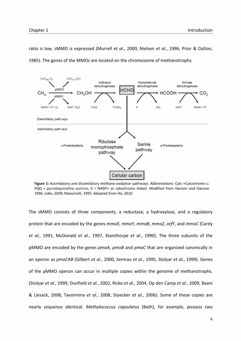
Chapter 1 Introduction
6
ratio is low, sMMO is expressed (Murrell et al., 2000, Nielsen et al., 1996, Prior & Dalton,
1985). The genes of the MMOs are located on the chromosome of methanotrophs.
The sMMO consists of three components, a reductase, a hydroxylase, and a regulatory
protein that are encoded by the genes mmoX, mmoY, mmoB, mmoZ, orfY, and mmoC (Cardy
et al., 1991, McDonald et al., 1997, Stainthorpe et al., 1990). The three subunits of the
pMMO are encoded by the genes pmoA, pmoB and pmoC that are organized canonically in
an operon as pmoCAB (Gilbert et al., 2000, Semrau et al., 1995, Stolyar et al., 1999). Genes
of the pMMO operon can occur in multiple copies within the genome of methanotrophs.
(Stolyar et al., 1999, Dunfield et al., 2002, Ricke et al., 2004, Op den Camp et al., 2009, Baani
& Liesack, 2008, Tavormina et al., 2008, Stoecker et al., 2006). Some of these copies are
nearly sequence identical. Methylococcus capsulatus (Bath), for example, possess two

Chapter 1 Introduction
7
virtually identical copies of the complete pmoCAB operon and an additional copy of pmoC
(Stolyar et al., 1999). This distribution is similar to the system of ammonium oxidizers that
can also contain two copies of the gene encoding the ammonia monooxygenase, amoCAB,
and a third amoC gene (Sayavedra-Soto et al., 1998). Two nearly identical copies of the
pmoCAB could be shown in several other methanotrophs including strains of Methylocystis,
Methylosinus and the Verrucomicrobia. Sequence divergent copies of the pmoCAB operon
could be detected in several type II methanotrophs: pmoCAB2. Genes of this operon encode
for the isoenzyme pMMO2 (Yimga et al., 2003, Baani & Liesack, 2008, Dunfield et al., 2002).
pmoA2 sequences of this second isoenzyme posses only 68,5% identity and 83,0% identity to
the first pmoA at the amino acid level. In Methylocystis strain SC2 it could be shown that the
methane monooxygenase encoded by pmoCAB2 is responsible for the oxidation of methane
at atmospheric concentrations (Baani & Liesack, 2008, Ricke et al., 2004). Divergent copies of
the pmo operon have also been found in strains of Verrucomicrobia and Crenothrix (Dunfield
et al., 2007, Stoecker et al., 2006). Very recently a second isoenzyme could be detected in
same type Ia and type II methanotrophs: pXMO (Tavormina et al., 2011, Vorobev et al.,
2014). pxmA sequences, coding a subunit of the pXMO, are only distantly related to
characterized pmoA sequences (53% identical and 73% similar on the amino acid level).
Additionally, the operon structure shows an unusual non-canonically gene order pxmABC
(Tavormina et al., 2011).
The pMMO of methanotrophic bacteria is a member of a diverse enzyme family: the copper
containing membrane-bound monooxygenases (CuMMOs). Monooxygenases in general are
enzymes that catalyze the insertion of one oxygen atom, derived from molecular oxygen,
Chapter 1 Introduction
8
into many organic substrates (van Berkel et al., 2006). The bacterial CuMMOs, that require
copper ions for hydroxylation of their substrates, were thought to be restricted to
methanotrophic bacteria and ammonium oxidizing bacteria (AOB) for a long time. The
pMMO and the ammonium monooxygenase (AMO) are evolutionary related enzymes that
share many characteristics like subunit composition, metal component, inhibition profile and
operon structure (Holmes et al., 1995). While the pMMO is relatively substrate specific and
only able to oxidize methane and short-chained alkanes and alkenes (Burrows et al., 1984,
Trotsenko & Murrell, 2008), the AMO has a wide substrate spectrum including several apolar
compounds such as carbon monoxide and some hydrocarbons (Hooper et al., 1997). The
AMO is also able to oxidize methane but does not play a significant role in global methane
oxidation (Bender & Conrad, 1994, Bodelier & Frenzel, 1999, Bosse et al., 1993, Jiang &
Bakken, 1999). A third member of the CuMMOs has been found in strains of Nocardia and
Mycobacterium: pBMO (Hamamura et al., 1999, Hamamura et al., 2001, Sayavedra-Soto et
al., 2011). The butane monooxygenase is a new branch in the family of CuMMOs and shows
that there is no restriction of the CuMMOs to MOB and AOB. Additionally, sequences of
CuMMOs have been found that could be linked to ethane and ethylene degrading
(Nakamura et al. BAH22833, BAH22839; Redmond et al., 2010, Suzuki et al., 2012).
1.3 Hydrocarbons and hydrocarbon degrading bacteria
Alkanes and alkenes are exclusively formed by carbon and hydrogen atoms that can be
linear, cyclic or branched. Small hydrocarbons up to a length of four carbon atoms are
gaseous at ambient temperatures while larger molecules are liquid or solid. Significant
sources of short chained hydrocarbons are seeps and vents from natural gas and oil
Chapter 1 Introduction
9
deposits. Natural gases contain methane (70-99%), 1-10% ethane and other gaseous
hydrocarbons (Cooley et al., 2009, Shennan, 2006). Although the major part of the short
chained alkanes and alkenes is created by geochemical processes, microorganisms, marine
algae, insects and plants provide hydrocarbons in most soil and water environments, too
(Cooley et al., 2009, Giebler et al., 2013). They are produced as moisture barriers, as reserve
materials and pheromones (Nie et al., 2014, van Beilen & Funhoff, 2007). Anaerobic
decomposition in soil sediments, sewage sludge and anaerobic digesters result in gases
consisting of methane (50-60%), CO2 (40%) and a minor concentration (up to 1%) of non-
methane volatile organic compounds also containing small hydrocarbons (Shennan, 2006,
Tassi et al., 2009). Studies on microbial hydrocarbon degradation started about a century
ago (Söhngen, 1913). The research focused mainly on topics related to oil production and
the use of bacteria and yeasts to convert oil components and solve oil-pollution problems
(van Beilen & Funhoff, 2007). Though the apolar hydrocarbons are very inert and need much
energy to be activated, many organisms metabolize alkanes and alkenes. Microorganisms
including bacteria, yeasts and fungi involved in the degradation were identified during the
last century (Labinger & Bercaw, 2002, Rojo, 2009b, van Beilen & Funhoff, 2007, Shennan,
2006). Most of the bacterial strains that grow on hydrocarbons are heterotrophic and use
other carbon sources as growth substrate in addition (Harayama et al., 2004, Margesin et al.,
2003, Rojo, 2009b). Many hydrocarbon degrading bacteria can utilize a wide range of
alkanes and alkenes for they contain multiple alkane hydroxylases with overlapping
substrate ranges (Kotani et al., 2003, Sabirova et al., 2006, van Beilen & Funhoff, 2007, van
Beilen et al., 2003). The predominat group of bacteria that can grow on hydrocarbons is the
so called CMNR group; Gram-positive bacteria belonging to the genera Corynebacterium,

Chapter 1 Introduction
10
Mycobacterium, Nocardia, and Rhodococcus (Shennan, 2006, Hamamura et al., 2001). Gram-
negative representatives of alkane degraders belong to the genera Pseudomonas,
Acinetobacter, Alcaligenes and Burkholderia (Shennan, 2006). Beside the heterotrophic
bacteria, that prefer other grows substrates to alkanes, some bacteria seem to be highly
specialized to grow on hydrocarbons (e.g. Alcanivorax, Thalassolituus) (Rojo, 2009, Sabirova
et al., 2006, Brakstad & Lodeng, 2005).
The first step of the aerobic degradation of gaseous hydrocarbons is the initial oxidation
catalyzed by a monooxygenase. Alkanes are oxidized to the primary or secondary alcohols
and further converted to aldehydes or ketones respectively. Alkenes are oxidized by adding
an oxygen atom across the olefin bond forming an epoxyalkane, a highly reactive and toxic
product that is immediately metabolized (Shennan, 2006, Wentzel et al., 2007, Kotani et al.,
2006). Different enzyme classes are involved in the oxidation of hydrocarbons. Most alkane
oxygenases have a wide substrate range. The methane monooxygenases sMMO and pMMO
play a key role in the degradation of methane, the shortest hydrocarbon. Enzymes that are
related to the sMMO are involved in the oxidation of gaseous alkanes. A butane
monooxygenase (BMO) similar to the sMMO hydroxylates C2-C9 alkanes in Pseudomonas
butanovora (Sluis et al., 2002, Dubbels et al., 2007). A BMO with properties of the sMMO
was found in Mycobacterium vaccae OB5 (Hamamura et al., 1999). Gordonia sp. TY-5,
Mycobacterium sp. TY-6 and Pseudonocardia sp. TY-7 possess a propane monooxygenase
similar to the sMMO, oxidizing propane at the terminal or subterminal position (Kotani et al.,
2006, Kotani et al., 2007). Butane monooxygenases similar to the pMMO were found in
Nocardia and Mycobacterium: pBMO (Hamamura et al., 1999, Hamamura et al., 2001,

Chapter 1 Introduction
11
Sayavedra-Soto et al., 2011). Another class of enzymes involved in hydrocarbon degradation
are alk hydroxylases, integral-membrane non haem diiron monooxygenases that oxidize
alkanes at the terminal position. The alkB gene is coding the trans-membrane alkane
monooxygenase of the alk enzyme system and is used as a marker gene to detect and study
alkane degraders (van Beilen et al., 2001, Bertrand et al., 2005). AlkB homologues show high
sequence diversity. They have been found in Gram-positive and Gram-negative
microorganism, including strains of the genera Acinetobacter, Alcanivorax, Burkholderia,
Mycobacterium, Pseudomonas and Rhodococcus (Smits et al., 1999, Smits et al., 2002, Marin
et al., 2003, van Beilen et al., 2004, Kuhn et al., 2009). Most AlkB hydroxylase homologous
are involved in the oxidation of C5-C16 alkanes. However, the Pseudomonas putina GPo1 AlkB
oxidizes propane and butane as well. Hence, the AlkB hydroxylases may play an important
role in the oxidation of gaseous alkanes. An oxidation of methane and ethane by AlkB could
not be shown (van Beilen et al., 2005). Another group alkane hydroxylases are the
Cytochrome P450 enzymes that are involved in the degradation of C5-C16 alkanes. Those
enzymes are ubiquitous among bacteria (e.g. strains of the genera Acinetobacter,
Mycobacteria, Rhodococcus) and yeasts that are involved in the degradation of alkanes in
some environments (van Beilen et al., 2005, Sekine et al., 2006, Funhoff et al., 2006, Schmitz
et al., 2000, Lida et al., 2000). Several other alkane hydroxylases including Cu2+-dependent
alkane hydroxylases and flavin-binding monooxygenases were found but they are specialized
in the oxidation of long-chained alkanes and do not play a role in the oxidation of gaseous
alkanes (Tani et al., 2001, Throne-Holst et al., 2007, Feng et al., 2007). Under anaerobic
conditions bacteria use nitrate, sulfate or ferric iron instead of oxygen as electron acceptor
to degrade hydrocarbons (Aeckersberg et al., 1991, Ehrenreich et al., 2000, Rueter et al.,

Chapter 1 Introduction
12
1994, Seeliger et al., 1998). Though the growth of anaerobic alkane degraders is very slow,
they play an important role in the degradation of hydrocarbons in the environment. The
degradation of the short chained alkanes propane and butane could be shown by a strain of
the Desulfosarcina/Desulfococcus cluster (Kniemeyer et al., 2007). Strains of other genera
(e.g. Azoracus, Rhodocyclus, Desulfobacterium and Desulfovibrio) are involved in the
degradation of longer alkanes (C6-C20) (Aeckersberg et al., 1991, Ehrenreich et al., 2000,
Rueter et al., 1994).
1.4 Aims of this study
The particulate methane monooxygenase (pMMO), the key enzyme of the aerobic methane
oxidation, is a member of the Cu-containing membrane-bound monooxygenases (CuMMO),
a family of widespread and diverse enzymes. Methane oxidizing bacteria (MOB) possessing
the pMMO have been studied intensively within the last years. The existence of multiple
copies of the pMMO (isoenzymes) within the genome of MOBs is known and analyzed for
quite some time. However, a new pMMO isoenzyme (pXMO) has been found recently in
several MOB that is only distantly related to characterized pMMOs. Research on the
distribution of the pXMO among MOB just started. The physiological function of this enzyme
is still unknown. Furthermore, sequences of CuMMO isoenzymes (iCuMMOs), distantly
related to pMMO and ammonium monooxygenase (AMO) sequences, were found in several
phylogenetical studies in different habitats. The distribution of the iCuMMO sequences and
possible substrates of the corresponding iCuMMO enzymes are unknown.

Chapter 1 Introduction
13
This PhD thesis focused on iCuMMOs to get more insights into their distribution and
ecological functions and to extend the view on the CuMMOs which was until now mostly
restricted to pMMOs and AMOs.
Chapter 2 An unexpected diversity of copper containing membrane-bound monooxygenases:
new pmoA-like sequences retrieved from aquatic environments and pure cultures
Here we analyzed different environmental habitats and methanotrophic pure cultures for
the occurrence of CuMMO sequences that are only distantly related to known pMMOs.
Newly generated primers targeting selected environmental CuMMO groups (iCuMMOs)
were used to study the environmental distribution and the possible physiological function of
the pXMO in Methylobacter luteus 53v in an incubation experiment.
Chapter 3 Magnetic capture of iCuMMO sequences: A prove of concept
Culture independent PCR based methods are powerful tools to study methanotrophic
communities in diverse habitats. Here, we established a method to avoid the use of specific
primers that are essential for the success of the PCR dependent methods.
Chapter 4 Monooxygenases involved in the degradation of short chained gaseous hydrocarbons in a rice field soil
Bacterial monooxygenases are widespread in nature and are able to oxidize a variety of
different substrates. In this chapter we analyzed if Cu-containing monooxygenases and AlkB
hydroxylases are involved in the oxidation of alkanes and alkenes in rice filed soils and which
organisms are the key players in the hydrocarbon degradation.
Chapter 1 Introduction
14
1.5 References Aeckersberg, F., F. Bak & F. Widdel, (1991) Anaerobic oxidation of saturated hydrocarbons to CO2 by
a new type of sulfate-reducing bacterium. Arch. Microbiol. 156: 5-14. Baani, M. & W. Liesack, (2008) Two isozymes of particulate methane monooxygenase with different
methane oxidation kinetics are found in Methylocystis sp strain SCZ. Proc. Natl. Acad. Sci. U. S. A. 105: 10203-10208.
Beal, E.J., C.H. House & V.J. Orphan, (2009) Manganese- and iron-dependent marine methane
oxidation. Science 325: 184-187. Belova, S.E., M. Baani, N.E. Suzina, P.L.E. Bodelier, W. Liesack & S.N. Dedysh, (2011) Acetate
utilization as a survival strategy of peat-inhabiting Methylocystis spp. Environ. Microbiol. Rep. 3: 36-46.
Bender, M. & R. Conrad, (1992) Kinetics of CH4 oxidation in oxic soils exposed to ambient air or high
CH4 mixing ratios. FEMS Microbiol. Ecol. 101: 261-270. Bender, M. & R. Conrad, (1994) Microbial oxidation of methane, ammonium and carbon-monoxide,
and turnover of nitrous-oxide and nitric-oxide in soils. Biogeochem. 27: 97-112. Bergamaschi, P., S. Houweling, A. Segers, M. Krol, C. Frankenberg, R.A. Scheepmaker, E. Dlugokencky,
S.C. Wofsy, E.A. Kort, C. Sweeney, T. Schuck, C. Brenninkmeijer, H. Chen, V. Beck & C. Gerbig, (2013) Atmospheric CH4 in the first decade of the 21st century: Inverse modeling analysis using SCIAMACHY satellite retrievals and NOAA surface measurements. J. Geophys. Res., [Atmos.] 118: 7350-7369.
Bertrand, E., R. Sakai, E. Rozhkova-Novosad, L. Moe, B.G. Fox, J.T. Groves & R.N. Austin, (2005)
Reaction mechanisms of non-heme diiron hydroxylases characterized in whole cells. J. Inorg. Biochem. 99: 1998-2006.
Bodelier, P.L.E. & P. Frenzel, (1999) Contribution of methanotrophic and nitrifying bacteria to CH4
and NH4+ oxidation in the rhizosphere of rice plants as determined by new methods of
discrimination. Appl. Environ. Microbiol. 65: 1826-1833. Bodrossy, L., E.M. Holmes, A.J. Holmes, K.L. Kovacs & J.C. Murrell, (1997) Analysis of 16S rRNA and
methane monooxygenase gene sequences reveals a novel group of thermotolerant and thermophilic methanotrophs, Methylocaldum gen. nov. Arch. Microbiol. 168: 493-503.
Bodrossy, L., K.L. Kovacs, I.R. McDonald & J.C. Murrell, (1999) A novel thermophilic methane-oxidising gamma-Proteobacterium. FEMS Microbiol. Lett. 170: 335-341.
Boetius, A., K. Ravenschlag, C.J. Schubert, D. Rickert, F. Widdel, A. Gieseke, R. Amann, B.B. Jorgensen,
U. Witte & O. Pfannkuche, (2000) A marine microbial consortium apparently mediating anaerobic oxidation of methane. Nature 407: 623-626.
Bosse, U., P. Frenzel & R. Conrad, (1993) Inhibition of methane oxidation by ammonium in the
surface-layer of a littoral sediment. FEMS Microbiol. Ecol. 13: 123-134.
Chapter 1 Introduction
15
Bowman, J., (2006) The Methanotrophs. The Families Methylococcaceae and
Methylocystaceae. In The Prokaryotes. Dworkin,M. (ed). New York: Springer, 266- 289. Brakstad, O.G. & A.G. Lodeng, (2005) Microbial diversity during biodegradation of crude oil in
seawater from the North Sea. Microb. Ecol. 49: 94-103. Burrows, K.J., A. Cornish, D. Scott & I.J. Higgins, (1984) Substrate specificities of the soluble and
particulate methane monooxygenases of Methylosinus Trichosporium Ob3b. J. Gen. Microbiol. 130: 3327-3333.
Cadillo-Quiroz, H., E. Yashiro, J.B. Yavitt & S.H. Zinder, (2008) Characterization of the archaeal
community in a minerotrophic fen and terminal restriction fragment length polymorphism-directed isolation of a novel hydrogenotrophic methanogen. Appl. Environ. Microbiol. 74: 2059-2068.
Cardy, D.L., V. Laidler, G.P. Salmond & J.C. Murrell, (1991) Molecular analysis of the methane
monooxygenase (MMO) gene cluster of Methylosinus trichosporium OB3b. Mol. Microbiol. 5: 335-342.
Castro, H., A. Ogram & K.R. Reddy, (2004) Phylogenetic characterization of methanogenic
assemblages in eutrophic and oligotrophic areas of the Florida Everglades. Appl. Environ. Microbiol. 70: 6559-6568.
Chen, Y.H. & R.G. Prinn, (2005) Atmospheric modeling of high- and low-frequency methane
observations: Importance of interannually varying transport. J. Geophys. Res., [Atmos.] 110. doi: 10.1029/2004JD005542
Conrad, R., (1996) Soil microorganisms as controllers of atmospheric trace gases (H2, CO, CH4, OCS,
N2O, and NO). Microbiol. Mol. Biol. Rev. 60: 609-640. Conrad,R. & P. Frenzel, (2002) Flooded soils. In Encyclopedia of Environmental Microbiology.
Britton,G. (ed). New York: John Wiley & Sons, 1316-1333. Cooley, R.B., P.J. Bottomley & D.J. Arp, (2009) Growth of a non-methanotroph on natural gas:
ignoring the obvious to focus on the obscure. Environ. Microbiol. Rep. 1: 408-413. Dedysh, S.N., Y.Y. Berestovskaya, L.V. Vasylieva, S.E. Belova, V.N. Khmelenina, N.E. Suzina, Y.A.
Trotsenko, W. Liesack & G.A. Zavarzin, (2004) Methylocella tundrae sp. nov., a novel methanotrophic bacterium from acidic tundra peatlands. Int. J. Syst. Evol. Microbiol. 54: 151-156.
Dedysh, S.N., C. Knief & P.F. Dunfield, (2005) Methylocella species are facultatively methanotrophic.
J. Bacteriol. 187: 4665-4670. Dedysh, S.N., W. Liesack, V.N. Khmelenina, N.E. Suzina, Y.A. Trotsenko, J.D. Semrau, A.M. Bares, N.S.
Panikov & J.M. Tiedje, (2000) Methylocella palustris gen. nov., sp. nov., a new methane-oxidizing acidophilic bacterium from peat bogs, representing a novel subtype of serine-pathway methanotrophs. Int. J. Syst. Evol. Microbiol. 50: 955-969.

Chapter 1 Introduction
16
Dedysh, S.N., N.S. Panikov & J.M. Tiedje, (1998) Acidophilic methanotrophic communities from
Sphagnum peat bogs. Appl. Environ. Microbiol. 64: 922-929. Dlugokencky, E.J., S. Houweling, L. Bruhwiler, K.A. Masarie, P.M. Lang, J.B. Miller & P.P. Tans, (2003)
Atmospheric methane levels off: Temporary pause or a new steady-state? Geophys. Res. Lett. 30. doi: 10.1029/2003GL018126
Dubbels, B.L., L.A. Sayavedra-Soto & D.J. Arp, (2007) Butane monooxygenase of Pseudomonas
butanovora: purification and biochemical characterization of a terminal-alkane hydroxylating diiron monooxygenase. Microbiology 153: 1808-1816.
Dunfield, P.F., S.E. Belova, A.V. Vorob'ev, S.L. Cornish & S.N. Dedysh, (2010) Methylocapsa aurea sp.
nov., a facultative methanotroph possessing a particulate methane monooxygenase, and emended description of the genus Methylocapsa. Int. J. Syst. Evol. Microbiol. 60: 2659-2664.
Dunfield, P.F., V.N. Khmelenina, N.E. Suzina, Y.A. Trotsenko & S.N. Dedysh, (2003) Methylocella
silvestris sp nov., a novel methanotroph isolated from an acidic forest cambisol. Int. J. Syst. Evol. Microbiol. 53: 1231-1239.
Dunfield, P.F., M.T. Yimga, S.N. Dedysh, U. Berger, W. Liesack & J. Heyer, (2002) Isolation of a
Methylocystis strain containing a novel pmoA-like gene. FEMS Microbiol. Ecol. 41: 17-26. Dunfield, P.F., A. Yuryev, P. Senin, A.V. Smirnova, M.B. Stott, S. Hou, B. Ly, J.H. Saw, Z. Zhou, Y. Ren, J.
Wang, B.W. Mountain, M.A. Crowe, T.M. Weatherby, P.L. Bodelier, W. Liesack, L. Feng, L. Wang & M. Alam, (2007) Methane oxidation by an extremely acidophilic bacterium of the phylum Verrucomicrobia. Nature 450: 879-882.
Dumont, M.G., B. Pommerenke, P. Casper & R. Conrad, (2011) DNA-, rRNA- and mRNA-based stable
isotope probing of aerobic methanotrophs in lake sediment. Environ. Microbiol. 13: 1153-1167.
Ehrenreich, P., A. Behrends, J. Harder & F. Widdel, (2000) Anaerobic oxidation of alkanes by newly
isolated denitrifying bacteria. Arch. Microbiol. 173: 58-64. Eller, G., L. Kanel & M. Kruger, (2005) Cooccurrence of aerobic and anaerobic methane oxidation in
the water column of Lake Plusssee. Appl. Environ. Microbiol. 71: 8925-8928. Ettwig, K.F., M.K. Butler, D. Le Paslier, E. Pelletier, S. Mangenot, M.M.M. Kuypers, F. Schreiber, B.E.
Dutilh, J. Zedelius, D. de Beer, J. Gloerich, H.J.C.T. Wessels, T. van Alen, F. Luesken, M.L. Wu, K.T. Pas-Schoonen, H.J.M.O. den Camp, E.M. Janssen-Megens, K.J. Francoijs, H. Stunnenberg, J. Weissenbach, M.S.M. Jetten & M. Strous, (2010) Nitrite-driven anaerobic methane oxidation by oxygenic bacteria. Nature 464: 543-548.
Ettwig, K.F., T. van Alen, K.T. van de Pas-Schoonen, M.S.M. Jetten & M. Strous, (2009) Enrichment
and molecular detection of denitrifying methanotrophic bacteria of the NC10 phylum. Appl. Environ. Microbiol. 75: 3656-3662.

Chapter 1 Introduction
17
Feng, L., W. Wang, J. Cheng, Y. Ren, G. Zhao, C. Gao, Y. Tang, X. Liu, W. Han, X. Peng, R. Liu & L. Wang, (2007) Genome and proteome of long-chain alkane degrading Geobacillus thermodenitrificans NG80-2 isolated from a deep-subsurface oil reservoir. Proc. Natl. Acad. Sci. U. S. A. 104: 5602-5607.
Funhoff, E.G., U. Bauer, I. Garcia-Rubio, B. Witholt & J.B. van Beilen, (2006) CYP153A6, a soluble P450
oxygenase catalyzing terminal-alkane hydroxylation. J. Bacteriol. 188: 5220-5227. Geymonat, E., L. Ferrando & S.E. Tarlera, (2011) Methylogaea oryzae gen. nov., sp. nov., a mesophilic
methanotroph isolated from a rice paddy field. Int. J. Syst. Evol. Microbiol. 61: 2568-2572. Giebler, J., L.Y. Wick, A. Chatzinotas & H. Harms, (2013) Alkane-degrading bacteria at the soil-litter
interface: comparing isolates with T-RFLP-based community profiles. FEMS Microbiol. Ecol. 86: 45-58.
Gilbert, B., I.R. McDonald, R. Finch, G.P. Stafford, A.K. Nielsen & J.C. Murrell, (2000) Molecular
analysis of the pmo (particulate methane monooxygenase) operons from two type II methanotrophs. Appl. Environ. Microbiol. 66: 966-975.
Grossman, E.L., L.A. Cifuentes & I.M. Cozzarelli, (2002) Anaerobic methane oxidation in a landfill-
leachate plume. Environ. Sci. Technol. 36: 2436-2442. Hamamura, N., R.T. Storfa, L. Semprini & D.J. Arp, (1999) Diversity in butane monooxygenases among
butane-grown bacteria. Appl. Environ. Microbiol. 65: 4586-4593. Hamamura, N., C.M. Yeager & D.J. Arp, (2001) Two distinct monooxygenases for alkane oxidation in
Nocardioides sp. strain CF8. Appl. Environ. Microbiol. 67: 4992-4998. Hanson, R.S. & T.E. Hanson, (1996) Methanotrophic bacteria. Microbiol. Rev. 60: 439-471 Harayama, S., Y. Kasai & A. Hara, (2004) Microbial communities in oil-contaminated seawater. Curr.
Opin. Biotechnol. 15: 205-214. Haroon, M.F., S.H. Hu, Y. Shi, M. Imelfort, J. Keller, P. Hugenholtz, Z.G. Yuan & G.W. Tyson, (2013)
Anaerobic oxidation of methane coupled to nitrate reduction in a novel archaeal lineage. Nature 500: 567-574.
Heyer, J., U. Berger, M. Hardt & P.F. Dunfield, (2005) Methylohalobius crimeensis gen. nov., sp. nov.,
a moderately halophilic, methanotrophic bacterium isolated from hypersaline lakes of Crimea. Int. J. Syst. Evol. Microbiol. 55: 1817-1826.
Hoehler, T.M., M.J. Alperin, D.B. Albert & C.S. Martens, (1994) Field and laboratory studies of
methane oxidation in an anoxic marine sediment - Evidence for a methanogen-sulfate reducer consortium. Global. Biogeochem. Cy. 8: 451-463.
Holmes, A.J., A. Costello, M.E. Lidstrom & J.C. Murrell, (1995) Evidence that particulate methane
monooxygenase and ammonia monooxygenase may be evolutionarily related. FEMS Microbiol. Lett. 132: 203-208.

Chapter 1 Introduction
18
Hooper, A.B., T. Vannelli, D.J. Bergmann & D.M. Arciero, (1997) Enzymology of the oxidation of ammonia to nitrite by bacteria. Antonie van Leeuwenhoek 71: 59-67.
Intergovernmental Panel on Climate Change, (2007) Climate change 2007: The physical science basis.
summary for policymakers. contribution of working group I to the fourth assessment report of the intergovernmental panel on climate change. IPCC Secretariat, Geneva.
Islam, T., S. Jensen, L.J. Reigstad, O. Larsen & N.K. Birkeland, (2008) Methane oxidation at 55°C and
pH 2 by a thermoacidophilic bacterium belonging to the Verrucomicrobia phylum. Proc. Natl. Acad. Sci. 105: 300-304.
Jiang, Q.Q. & L.R. Bakken, (1999) Nitrous oxide production and methane oxidation by different
ammonia-oxidizing bacteria. Appl. Environ. Microbiol. 65: 2679-2684. Kalyuzhnaya, M.G., V.N. Khmelenina, S. Kotelnikova, L. Holmquist, K. Pedersen & Y.A. Trotsenko,
(1999) Methylomonas scandinavica sp. nov., a new methanotrophic psychrotrophic bacterium isolated from deep igneous rock ground water of Sweden. Syst. Appl. Microbiol. 22: 565-572.
Khmelenina, V.N., M.G. Kalyuzhnaya, N.G. Starostina, N.E. Suzina & Y.A. Trotsenko, (1997) Isolation
and characterization of halotolerant alkaliphilic methanotrophic bacteria from Tuva soda lakes. Curr. Microbiol. 35: 257-261.
Klotz, M.G., D.J. Arp, P.S.G. Chain, A.F. El-Sheikh, L.J. Hauser, N.G. Hommes, F.W. Larimer, S.A.
Malfatti, J.M. Norton, A.T. Poret-Peterson, L.M. Vergez & B.B. Ward, (2006) Complete genome sequence of the marine, chemolithoautotrophic, ammonia-oxidizing bacterium Nitrosococcus oceani ATCC 19707. Appl. Environ. Microbiol. 72: 6299-6315.
Knief, C., A. Lipski & P.F. Dunfield, (2003) Diversity and activity of methanotrophic bacteria in
different upland soils. Appl. Environ. Microbiol. 69: 6703-6714. Kniemeyer, O., F. Musat, S.M. Sievert, K. Knittel, H. Wilkes, M. Blumenberg, W. Michaelis, A. Classen,
C. Bolm, S.B. Joye & F. Widdel, (2007) Anaerobic oxidation of short-chain hydrocarbons by marine sulphate-reducing bacteria. Nature 449: 898-901.
Knittel, K. & A. Boetius, (2009) Anaerobic oxidation of methane: progress with an unknown process.
Annu. Rev. Microbiol. 63: 311-334. Kotani, T., Y. Kawashima, H. Yurimoto, N. Kato & Y. Sakai, (2006) Gene structure and regulation of
alkane monooxygenases in propane-utilizing Mycobacterium sp TY-6 and Pseudonocardia sp TY-7. J. Biosci. Bioeng. 102: 184-192.
Kotani, T., T. Yamamoto, H. Yurimoto, Y. Sakai & N. Kato, (2003) Propane monooxygenase and
NAD(+)-dependent secondary alcohol dehydrogenase in propane metabolism by Gordonia sp strain TY-5. J. Bacteriol. 185: 7120-7128.
Kotani, T., H. Yurimoto, N. Kato & Y. Sakai, (2007) Novel acetone metabolism in a propane-utilizing
bacterium, Gordonia sp. strain TY-5. J. Bacteriol. 189: 886-893.

Chapter 1 Introduction
19
Krause, S., C. Luke & P. Frenzel, (2010) Succession of methanotrophs in oxygen-methane counter-gradients of flooded rice paddies. ISME J. 4: 1603-1607.
Kuhn, E., G.S. Bellicanta & V.H. Pellizari, (2009) New alk genes detected in Antarctic marine
sediments. Environ. Microbiol. 11: 669-673. Labinger, J.A. & J.E. Bercaw, (2002) Understanding and exploiting C-H bond activation. Nature 417:
507-514. Lida, T., T. Sumita, A. Ohta & M. Takagi, (2000) The cytochrome P450ALK multigene family of an n-
alkane-assimilating yeast, Yarrowia lipolytica: cloning and characterization of genes coding for new CYP52 family members. Yeast 16: 1077-1087.
Liebner, S., K. Rublack, T. Stuehrmann & D. Wagner, (2009) Diversity of aerobic methanotrophic
bacteria in a permafrost active layer soil of the Lena Delta, Siberia. Microbial Ecology 57: 25-35.
Lüke, C. & P. Frenzel, (2011) Potential of pmoA amplicon pyrosequencing for methanotroph diversity
studies. Appl. Environ. Microbiol. 77: 6305-6309. MacLean, L.C.W., T.J. Pray, T.C. Onstott, E.L. Brodie, T.C. Hazen & G. Southam, (2007) Mineralogical,
chemical and biological characterization of an anaerobic biofilm collected from a borehole in a deep gold mine in South Africa. Geomicrobiol. J. 24: 491-504.
Margesin, R., D. Labbe, F. Schinner, C.W. Greer & L.G. Whyte, (2003) Characterization of
hydrocarbon-degrading microbial populations in contaminated and pristine Alpine soils. Appl Environ. Microbiol. 69: 3085-3092.
Marin, M.M., L. Yuste & F. Rojo, (2003) Differential expression of the components of the two alkane
hydroxylases from Pseudomonas aeruginosa. J. Bacteriol. 185: 3232-3237. McDonald, I.R., H. Uchiyama, S. Kambe, O. Yagi & J.C. Murrell, (1997) The soluble methane
monooxygenase gene cluster of the trichloroethylene-degrading methanotroph Methylocystis sp. strain M. Appl. Environ. Microbiol. 63: 1898-1904.
Morris, S.A., S. Radajewski, T.W. Willison & J.C. Murrell, (2002) Identification of the functionally
active methanotroph population in a peat soil microcosm by stable-isotope probing. Appl. Environ. Microbiol 68: 1446-1453.
Murrell, J.C., I.R. McDonald & B. Gilbert, (2000) Regulation of expression of methane
monooxygenases by copper ions. Trends Microbiol. 8: 221-225. Nercessian, O., E. Noyes, M.G. Kalyuzhnaya, M.E. Lidstrom & L. Chistoserdova, (2005) Bacterial
populations active in metabolism of C-1 compounds in the sediment of Lake Washington, a freshwater lake. Appl. Environ. Microbiol 71: 6885-6899.
Nie, Y., C.Q. Chi, H. Fang, J.L. Liang, S.L. Lu, G.L. Lai, Y.Q. Tang & X.L. Wu, (2014) Diverse alkane
hydroxylase genes in microorganisms and environments. Sci. Rep.Uk. 4. doi:10.1038/srep04968

Chapter 1 Introduction
20
Nielsen, A.K., K. Gerdes, H. Degn & J.C. Murrell, (1996) Regulation of bacterial methane oxidation: Transcription of the soluble methane monooxygenase operon of Methylococcus capsulatus (Bath) is repressed by copper ions. Microbiol. Uk. 142: 1289-1296.
Niemann, H., T. Losekann, D. de Beer, M. Elvert, T. Nadalig, K. Knittel, R. Amann, E.J. Sauter, M.
Schluter, M. Klages, J.P. Foucher & A. Boetius, (2006) Novel microbial communities of the Haakon Mosby mud volcano and their role as a methane sink. Nature 443: 854-858.
Omelchenko, M.V., L.V. Vasilyeva & G.A. Zavarzin, (1993) Psychrophilic methanotroph from tundra
soil. Curr. Microbiol. 27: 255-259. Op den Camp, H.J.M., T. Islam, M.B. Stott, H.R. Harhangi, A. Hynes, S. Schouten, M.S.M. Jetten, N.K.
Birkeland, A. Pol & P.F. Dunfield, (2009) Environmental, genomic and taxonomic perspectives on methanotrophic Verrucomicrobia. Environ. Microbiol. Rep. 1: 293-306.
Orphan, V.J., C.H. House, K.U. Hinrichs, K.D. McKeegan & E.F. DeLong, (2002) Multiple archaeal
groups mediate methane oxidation in anoxic cold seep sediments. Proc. Natl. Acad. Sci. U. S. A. 99: 7663-7668.
Pol, A., K. Heijmans, H.R. Harhangi, D. Tedesco, M.S. Jetten & H.J. Op den Camp, (2007)
Methanotrophy below pH 1 by a new Verrucomicrobia species. Nature 450: 874-878. Prior, S.D. & H. Dalton, (1985) The Effect of Copper Ions on Membrane Content and Methane
Monooxygenase activity in methanol-grown cells of Methylococcus Capsulatus (Bath). J. Gen. Microbiol. 131: 155-163.
Raghoebarsing, A.A., A. Pol, K.T. van de Pas-Schoonen, A.J. Smolders, K.F. Ettwig, W.I. Rijpstra, S.
Schouten, J.S. Damste, H.J. Op den Camp, M.S. Jetten & M. Strous, (2006) A microbial consortium couples anaerobic methane oxidation to denitrification. Nature 440: 918-921.
Reay, D.S., S. Radajewski, J.C. Murrell, N. McNamara & D.B. Nedwell, (2001) Effects of land-use on
the activity and diversity of methane oxidizing bacteria in forest soils. Soil Biol. Biochem. 33: 1613-1623.
Redmond, M.C., D.L. Valentine & A.L. Sessions, (2010) Identification of novel methane-, ethane-, and
propane-oxidizing bacteria at marine hydrocarbon seeps by stable isotope probing. Appl. Environ. Microbiol. 76: 6412-6422.
Reeburgh, W.S., (2003) Global methane biogeochemistry. In Treatise on Geochemistry, Vol. 4: The
Atmosphere. Keeling, R.F., Holland, H.D., and Turekian, K.K. (eds). Oxford, UK: ElsevierPergamon, pp. 65–89.
Reim, A., C. Lüke, S. Krause, J. Pratscher & P. Frenzel, (2012) One millimetre makes the difference:
high-resolution analysis of methane-oxidizing bacteria and their specific activity at the oxic-anoxic interface in a flooded paddy soil. ISME J. 6: 2128-2139.
Ricke, P., C. Erkel, M. Kube, R. Reinhardt & W. Liesack, (2004) Comparative analysis of the
conventional and novel pmo (Particulate methane monooxygenase) Operons from Methylocystis strain SC2. Appl. Environ. Microbiol. 70: 3055-3063.

Chapter 1 Introduction
21
Rigby, M., R.G. Prinn, P.J. Fraser, P.G. Simmonds, R.L. Langenfelds, J. Huang, D.M. Cunnold, L.P. Steele, P.B. Krummel, R.F. Weiss, S. O'Doherty, P.K. Salameh, H.J. Wang, C.M. Harth, J. Muhle & L.W. Porter, (2008) Renewed growth of atmospheric methane. Geophys. Res. Lett. 35.
Rojo, F., (2009) Degradation of alkanes by bacteria. Environ. Microbiol. 11: 2477-2490. Rueter, P., R. Rabus, H. Wilkes, F. Aeckersberg, F.A. Rainey, H.W. Jannasch & F. Widdel, (1994)
Anaerobic oxidation of hydrocarbons in crude oil by new types of sulphate-reducing bacteria. Nature 372: 455-458.
Sabirova, J.S., M. Ferrer, D. Regenhardt, K.N. Timmis & P.N. Golyshin, (2006) Proteomic insights into metabolic adaptations in Alcanivorax borkumensis induced by alkane utilization. J. Bacteriol. 188: 3763-3773.
Sayavedra-Soto, L.A., N. Hamamura, C.W. Liu, J.A. Kimbrel, J.H. Chang & D.J. Arp, (2011) The
membrane-associated monooxygenase in the butane-oxidizing Gram-positive bacterium Nocardioides sp. strain CF8 is a novel member of the AMO/PMO family. Environ. Microbiol. Rep. 3: 390-396.
Sayavedra-Soto, L.A., N.G. Hommes, J.J. Alzerreca, D.J. Arp, J.M. Norton & M.G. Klotz, (1998)
Transcription of the amoC, amoA and amoB genes in Nitrosomonas europaea and Nitrosospira sp, NpAV. FEMS Microbiol. Lett. 167: 81-88.
Schmitz, C., I. Goebel, S. Wagner, A. Vomberg & U. Klinner, (2000) Competition between n-alkane-
assimilating yeasts and bacteria during colonization of sandy soil microcosms. Appl. Microbiol. Biotechnol. 54: 126-132.
Seeliger, S., R. Cord-Ruwisch & B. Schink, (1998) A periplasmic and extracellular c-type cytochrome of
Geobacter sulfurreducens acts as a ferric iron reductase and as an electron carrier to other acceptors or to partner bacteria. J. Bacteriol. 180: 3686-3691.
Sekine, M., S. Tanikawa, S. Omata, M. Saito, T. Fujisawa, N. Tsukatani, T. Tajima, T. Sekigawa, H.
Kosugi, Y. Matsuo, R. Nishiko, K. Imamura, M. Ito, H. Narita, S. Tago, N. Fujita & S. Harayama, (2006) Sequence analysis of three plasmids harboured in Rhodococcus erythropolis strain PR4. Environ. Microbiol. 8: 334-346.
Semrau, J.D., D. Zolandz, M.E. Lidstrom & S.I. Chan, (1995) The role of copper in the pMMO of
Methylococcus Capsulatus (Bath) - a structural vs catalytic function. J. Inorg. Biochem. 58: 235-244.
Sharp, C.E., A.V. Smirnova, J.M. Graham, M.B. Stott, R. Khadka, T.R. Moore, S.E. Grasby, M. Strack &
P.F. Dunfield, (2014) Distribution and diversity of Verrucomicrobia methanotrophs in geothermal and acidic environments. Environ. Microbiol. 16: 1867-1878.
Shennan, J.L., (2006) Utilisation of C2-C4 gaseous hydrocarbons and isoprene by microorganisms. J.
Chem. Technol. Biotechnol. 81: 237-256. Shindell, D.T., G. Faluvegi, D.M. Koch, G.A. Schmidt, N. Unger & S.E. Bauer, (2009) Improved
attribution of climate forcing to emissions. Science 326: 716-718.

Chapter 1 Introduction
22
Sluis, M.K., L.A. Sayavedra-Soto & D.J. Arp, (2002) Molecular analysis of the soluble butane monooxygenase from Pseudomonas butanovora. Microbiology 148: 3617-3629.
Smits, T.H., S.B. Balada, B. Witholt & J.B. van Beilen, (2002) Functional analysis of alkane hydroxylases
from Gram-negative and Gram-positive bacteria. J. Bacteriol. 184: 1733-1742. Smits, T.H.M., M. Rothlisberger, B. Witholt & J.B. van Beilen, (1999) Molecular screening for alkane
hydroxylase genes in Gram-negative and Gram-positive strains. Environ. Microbiol. 1: 307-317.
Söhngen, N.L., (1913) Benzin, Petroleum, Paraffinöl und Paraffin als Kohlenstoff- und Energiequelle
für Mikroben. Zentralblatt für Microbiologie, Parasitenkunde, Infektionskrankheiten und Hygiene 2. Abteilung: 595-609.
Stainthorpe, A.C., V. Lees, G.P.C. Salmond, H. Dalton & J.C. Murrell, (1990) The methane monooxygenase gene cluster of Methylococcus capsulatus (Bath). Gene 91: 27-34.
Stoecker, K., B. Bendinger, B. Schoning, P.H. Nielsen, J.L. Nielsen, C. Baranyi, E.R. Toenshoff, H. Daims
& M. Wagner, (2006) Cohn's Crenothrix is a filamentous methane oxidizer with an unusual methane monooxygenase. Proc. Natl. Acad. Sci. U. S. A. 103: 2363-2367.
Stolyar, S., A.M. Costello, T.L. Peeples & M.E. Lidstrom, (1999) Role of multiple gene copies in
particulate methane monooxygenase activity in the methane oxidizing bacterium Methylococcus capsulatus (Bath). Microbiol.Uk. 145: 1235-1244.
Suzuki, T., T. Nakamura & H. Fuse, (2012) Isolation of two novel marine ethylene assimilating
bacteria, Haliea species ETY-M and ETY-NAG, containing particulate methane monooxygenase-like genes. Microbes Environ. 27: 54-60.
Tani, A., T. Ishige, Y. Sakai & N. Kato, (2001) Gene structures and regulation of the alkane hydroxylase
complex in Acinetobacter sp. strain M-1. J. Bacteriol. 183: 1819-1823. Tassi, F., G. Montegrossi, O. Vaselli, C. Liccioli, S. Moretti & B. Nisi, (2009) Degradation of C2-C15
volatile organic compounds in a landfill cover soil. Sci. Total. Environ. 407: 4513-4525. Tavormina, P.L., V.J. Orphan, M.G. Kalyuzhnaya, M.S.M. Jetten & M.G. Klotz, (2011) A novel family of
functional operons encoding methane/ammonia monooxygenase-related proteins in gammaproteobacterial methanotrophs. Environ. Microbiol. Rep. 3: 91-100.
Tavormina, P.L., W. Ussler & V.J. Orphan, (2008) Planktonic and sediment-associated aerobic
methanotrophs in two seep systems along the North American margin. Appl. Environ. Microbiol. 74: 3985-3995.
Thauer, R.K., A.K. Kaster, H. Seedorf, W. Buckel & R. Hedderich, (2008) Methanogenic archaea:
ecologically relevant differences in energy conservation. Nat. Rev. Microbiol. 6: 579-591. Throne-Holst, M., A. Wentzel, T.E. Ellingsen, H.K. Kotlar & S.B. Zotchev, (2007) Identification of novel
genes involved in long-chain n-alkane degradation by Acinetobacter sp. strain DSM 17874. Appl. Environ. Microbiol. 73: 3327-3332.

Chapter 1 Introduction
23
Tuomivirta, T.T., K. Yrjala & H. Fritze, (2009) Quantitative PCR of pmoA using a novel reverse primer correlates with potential methane oxidation in Finnish fen. Res. Microbiol. 160: 751-756.
van Beilen, J.B. & E.G. Funhoff, (2007) Alkane hydroxylases involved in microbial alkane degradation. Appl. Microbiol. Biotechnol. 74: 13-21
van Beilen, J.B., R. Holtackers, D. Luscher, U. Bauer, B. Witholt & W.A. Duetz, (2005) Biocatalytic
production of perillyl alcohol from limonene by using a novel Mycobacterium sp. cytochrome P450 alkane hydroxylase expressed in Pseudomonas putida. Appl. Environ. Microbiol. 71: 1737-1744.
van Beilen, J.B., Z. Li, W.A. Duetz, T.H.M. Smits & B. Witholt, (2003) Diversity of alkane hydroxylase
systems in the environment. Oil & Gas Science and Technology-Revue de l Institut Francais du Petrole 58: 427-440.
van Beilen, J.B., M.M. Marin, T.H.M. Smits, M. Rothlisberger, A.G. Franchini, B. Witholt & F. Rojo,
(2004) Characterization of two alkane hydroxylase genes from the marine hydrocarbonoclastic bacterium Alcanivorax borkumensis. Environ. Microbiol. 6: 264-273.
van Beilen, J.B., S. Panke, S. Lucchini, A.G. Franchini, M. Rothlisberger & B. Witholt, (2001) Analysis of
Pseudomonas putida alkane-degradation gene clusters and flanking insertion sequences: evolution and regulation of the alk genes. Microbiology 147: 1621-1630.
van Berkel, W.J., N.M. Kamerbeek & M.W. Fraaije, (2006) Flavoprotein monooxygenases, a diverse
class of oxidative biocatalysts. J. Biotechnol. 124: 670-689. van Teeseling, M.C.F., A. Pol, H.R. Harhangi, S. van der Zwart, M.S.M. Jetten & H.J.M.O. den Camp,
(2014) Expanding the verrucomicrobial methanotrophic world: Description of three rovel species of Methylacidimicrobium gen. nov. Appl. Environ. Microbiol 80: 6782-6791.
Vigliotta, G., E. Nutricati, E. Carata, S.M. Tredici, M. De Stefano, P. Pontieri, D.R. Massardo, M.V. Prati,
L. De Bellis & P. Alifano, (2007) Clonothrix fusca Roze 1896, a filamentous, sheathed, methanotrophic gamma-proteobacterium. Appl. Environ. Microbiol. 73: 3556-3565.
Vorobev, A., S. Jagadevan, S. Jain, K. Anantharaman, G.J. Dick, S. Vuilleumier & J.D. Semrau, (2014)
Genomic and transcriptomic analyses of the facultative methanotroph Methylocystis sp. strain SB2 grown on methane or ethanol. Appl. Environ. Microbiol. 80: 3044-3052.
Vorobev, A.V., M. Baani, N.V. Doronina, A.L. Brady, W. Liesack, P.F. Dunfield & S.N. Dedysh, (2011)
Methyloferula stellata gen. nov., sp nov., an acidophilic, obligately methanotrophic bacterium that possesses only a soluble methane monooxygenase. Int. J. Syst. Evol. Microbiol. 61: 2456-2463.
Ward, B.B., (1990) Kinetics of ammonia oxidation by a marine nitrifying bacterium - Methane as a
substrate analog. Microb. Ecol. 19: 211-225. Wartiainen, I., A.G. Hestnes, I.R. McDonald & M.M. Svenning, (2006) Methylobacter tundripaludum
sp nov., a methane-oxidizing bacterium from Arctic wetland soil on the Svalbard islands, Norway (78° N). Int. J. Syst. Evol. Microbiol. 56: 109-113.
Chapter 1 Introduction
24
Wentzel, A., T.E. Ellingsen, H.K. Kotlar, S.B. Zotchev & M. Throne-Holst, (2007) Bacterial metabolism of long-chain n-alkanes. Appl. Microbiol. Biotechnol. 76: 1209-1221.
Whittenbury, R., J. Colby, H. Dalton & H.L. Reed, (1975) The different types of methaneoxidizing
bacteria and some of their more unusual properties. Schlegel, H.G., Gottschalk, G., and Pfennig, N. (eds). Göttingen: Akademie der Wissenschaften, pp. 1-9.
Wu, M.L., K.F. Ettwig, M.S.M. Jetten, M. Strous, J.T. Keltjens & L. van Niftrik, (2011) A new intra-
aerobic metabolism in the nitrite-dependent anaerobic methane-oxidizing bacterium Candidatus Methylomirabilis oxyfera. Biochem. Soc. Trans. 39: 243-248.
Wuebbles, D.J. & K. Hayhoe, (2002) Atmospheric methane and global change. Earth. Sci. Rev. 57:
177-210. Yimga, M.T., P.F. Dunfield, P. Ricke, H. Heyer & W. Liesack, (2003) Wide distribution of a novel pmoA-
like gene copy among type II methanotrophs, and its expression in Methylocystis strain SC2. Appl. Environ. Microbiol. 69: 5593-5602.
Chapter 2 Abstract
25
2 An unexpected diversity of Cu-containing membrane-bound monooxygenases: new pmoA-like sequences retrieved from aquatic environments and pure cultures.
Stephanie Hainbuch, Claudia Lüke*, and Peter Frenzel
2.1 Abstract
An isoenzyme of a Cu-containing membrane-bound methane monooxygenase (CuMMO)
that is only distantly related to known CuMMO sequences has been recently found in several
alpha- and gammaproteobacterial methanotrophs. Classical primer sets targeting the pmoA
gene (encoding a subunit of the CuMMO) discriminate against these sequences. Here, we
designed new reverse primers targeting in total four deep branching groups of CuMMO: the
gammaproteobacterial isoenzyme and three related environmental clusters. We studied the
occurrence of these pmoA-like sequences in nature and in methanotrophic pure cultures.
Pyrosequencing results show that they are widespread and highly abundant in rice field soils
and lake sediments. We furthermore observed habitat-specific distribution patterns. In pure
cultures the isoenzyme seems to be restricted to strains of the type Ia genera
Methylomonas, Methylobacter and Methylosarcina. A phylogenetic comparison of pmoA,
pxmA and 16S rRNA genes of these strains indicates that the pxmA evolved vertically within
the type Ia methanotrophs. Incubation studies of Methylobacter luteus with different

Chapter 2 Introduction
26
substrates let us suggest that the physiological role of the CuMMO isoenzyme is the
oxidation of methane rather than ammonium or ethane.
2.2 Introduction
Cu-containing membrane-bound monooxygenases (Coleman et al., 2011) comprise two
enzymes catalyzing key reactions in global carbon and nitrogen cycles: methane and
ammonia monooxygenases. Methane monooxygenases are the key enzymes in methane
oxidizing bacteria that help to mitigate methane emissions (Frenzel, 2000, Conrad, 2009,
Reeburgh, 1997) which is, next to carbon dioxide, the most important greenhouse gas
(Intergovernmental Panel on Climate Change, 2007) . Methanotrophs interfere with the
global methane cycle by acting as a biofilter in high-methane environments like wetlands
and landfills (Reim et al., 2012, Henneberger et al., 2012, Conrad & Frenzel, 2002), or as a
sink to atmospheric methane in upland soils (Conrad, 1996, Bender & Conrad, 1992).
Methanotrophs have the unique ability to use methane as their sole carbon and energy
source (Trotsenko & Murrell, 2008), but some were recently found to exploit also simple
organic substrates (Belova et al., 2011, Dedysh et al., 2005, Theisen et al., 2005). The
majority of cultivated methanotrophs are proteobacteria. Among these canonical methane
oxidizers, two types are distinguished: type I and type II (Whittenb.R et al., 1970). This
historically grown differentiation corresponds well to molecular phylogenies (Lüke & Frenzel,
2011). Type I methanotrophs are gammaproteobacteria and further divided into type Ia (e.g.
Methylobacter, Methylovolum, Methyloglobulus, Methylomicrobium, Methylomarinum,
Methylomonas, Methylosarcina, and Methylosoma) and type Ib (e.g. Methylocaldum,
Chapter 2 Introduction
27
Methylococcus, Methylogaea and Methylothermus / Methylohalobius). Type Ic was also
defined as a separate group represented by one cultivated ammonium oxidizer,
Nitrosococcus oceani, and putative MMOs (JR2/JR3, USC-gamma, OPU1; Lüke & Frenzel,
2011, Horz et al., 2005, Knief et al., 2003, Hayashi et al., 2007). The alphaproteobacterial
type II methanotrophs includes the genera Methylocystis, Methylosinus, Methylocapsa,
Methylocella and Methyloferula. More recently, the spectrum has widened: the filamentous
gammaproteobacterial Crenothrix and Clonothrix were found to be methanotrophs
(Stoecker et al., 2006, Vigliotta et al., 2007), methane oxidizing Verrucomicrobia were
isolated from extreme and moderate environments (Pol et al., 2007, Islam et al., 2008,
Dunfield et al., 2007, Sharp et al., 2014, van Teeseling et al., 2014), and the nitrite-reducing
and O2-generating Ca. Methylomirabilis oxyfera was characterized (Ettwig et al., 2010).
Nearly all methanotrophic bacteria possess a particulate methane monooxygenase (pMMO),
while some have an additional Fe-containing soluble MMO (sMMO). The latter is the only
MMO in Methylocella and Methyloferula (Dedysh et al., 2004, Dedysh et al., 2000, Dunfield
et al., 2003, Vorobev et al., 2011). Besides its main substrate, the pMMO may also oxidize
alternative substrates like ammonia, short chained alkanes, and haloalkanes (Elliott et al.,
1997, Semrau, 2011, Burrows et al., 1984, Bedard & Knowles, 1989). The genes for the three
subunits of pMMO are organized as pmoCAB. They can occur in near sequence-identical or
divergent copies within the genome. One example of a sequence-divergent pMMO copy is
the pMMO2 of Methylocystis SC2 that has different methane oxidation kinetics providing a
selective advantage at low methane concentrations (Baani & Liesack, 2008). More recently,
another pMMO isoenzyme (referred to as pXMO) organized as pxmABC has been found in
Chapter 2 Introduction
28
type Ia and type II methanotrophs (Tavormina et al., 2011, Svenning et al., 2011, Vorobev et
al., 2014).
The pmoA gene encodes for the beta-subunit of the pMMO. It is highly conserved and often
used in environmental studies to detect and characterize methanotrophs. The homologous
pxmA sequences form a deep-branching separate lineage (M84_P105) that clusters
phylogenetical between characterized pmoA sequences and the amoA sequences of
ammonia oxidizers (Lüke & Frenzel, 2011, Dumont et al., 2014). M84_P105 is related to
some other environmental sequence clusters (e.g., RA21, TUSC, AOB_rel; (Dumont et al.,
2014, Lüke & Frenzel, 2011)). For pragmatic reasons, this group of clusters will be named
intermediate copper containing membrane-bound monooxygenases (iCuMMO) and the
term pxmA will be used as synonym for the gene encoding the beta-subunit of an iCuMMO.
Substrate spectrum and specificity of the respective proteins are unknown.
The pmoA-targeting standard primer set covers a wide range of the methanotrophic
diversity (Holmes et al., 1995). Besides pmoA sequences the primer co amplifies genes
encoding for similar CuMMOs like the amoA of betaproteobacterial ammonium oxidizers
and, to a minor degree, also pxmA sequences may be co-amplified, in addition. Using a
modified primer, sequences belonging to the M84_P105 cluster were identified as the pxmA
of a type Ia methanotroph (Tavormina et al., 2011). Unfortunately, the chosen primers had
some mismatches against the environmental sequences falling into this cluster. While the
preferred substrate of this isoenzyme in type Ia and type II methanotrophs is still unknown,
other iCuMMO sequences cluster near to sequences of putative ethane monooxygenases
Chapter 2 Materials and Methods
29
(Redmond et al., 2010). Hence, one may speculate that the respective proteins’ substrates
are short-chained alkanes, too.
Here, we designed specific primers targeting the iCuMMO groups M84_P105, RA21, AOB_rel
and TUSC (Lüke & Frenzel, 2011), respectively. Studying the occurrence of these clusters in
aquatic environments, we applied pyrosequencing to samples from three rice fields and two
lake sediments. In addition, we tested pure cultures for presence of iCuMMO encoding
genes. We retrieved pxmA sequences from Methylobacter, Methylosarcina and
Methylomonas, and studied the transcription of the respective gene in Methylobacter luteus
53v under methane, ethane, both substrates together, and ammonia, respectively.
Population growth approximated by copy number of pxmA were followed by competitive
PCR (Reim et al., 2012) and compared to number of transcripts.
2.2 Material and Methods
Field sites and samples
Soil samples were taken from fields in Italy and China. The Italian paddy field is situated in
the lowlands of river Po (Vercelli: N 45°20’; W 8°25’) and managed by the CRA-Agricultural
Research Council, Rice Research Unit, in Vercelli. This field had been planted to wetland rice
for more than a century (Lüke et al., 2010). Soil parameters and agricultural practices have
been described elsewhere (Holzapfelpschorn & Seiler, 1986, Kruger et al., 2001). The
Chinese paddy fields are situated near Cixi, province Zhejiang. One field is under permanent
agriculture since 50 years (China50: N 30°11.066′; E 121°21.351′), the other since 2000 years

Chapter 2 Material and Methods
30
(China2000: N 30°05.455′; E 121°26.738′). Soil parameters and history have been described
elsewhere (Ho et al., 2011, Kolbl et al., 2014, Cheng et al., 2009). Samples were taken from
the plow layer before fields were flooded in spring 2009. Upon arrival in Germany, the
samples (approximately 500 kg per site) were homogenized manually. In short, pots were
planted with rice and kept in a greenhouse according to agricultural practice and climate,
respectively. Details have been described elsewhere (Roth et al., 2013). Samples were taken
from pots planted to local rice varieties, qian you yi hao (Chinese soils) and KORAL (Italian
soil). For a comparison of all three sites see also Lüke et al.(2014).
Lake sediments were sampled from Lake Constance (Germany) and Lake Neusiedl (Austria).
The sample from Lake Constance (N 47°43.382’; E 9°10.751’) was taken from organic rich
sediment in the shallow littoral next to a small reed bed. The sample from Lake Neusiedl (N
47°55.951'; E 16°45.397') was taken from a ditch through the reed belt.
Total DNA and RNA were extracted from freeze-dried soils as described elsewhere (Reim et
al., 2012). For amplification of pxmA sequences total DNA was extracted from the three
paddy fields and the lake sediments. The primers A189f, M84_P105r, TUSCr, RA21_2r and
AOB_relr were modified (adapters and barcodes were attached) and the PCR was performed
as described before (Lüke et al., 2014). 454 amplicon sequencing was performed by GATC
(Konstanz, Germany). The pyrosequencing data were evaluated with the ARB software
package (Ludwig et al., 2004). Sequences containing insertions or deletion resulting in a shift
of the reading frame were excluded manually. For further phylogenetic analysis only
sequences with a read length of at least 130 amino acids were used.

Chapter 2 Material and Methods
31
Pure cultures: Nucleic acid extraction and PCR
From pure cultures 0.5 mL were suspended in phosphate buffer (pH8) and TNS. Cells were
disrupted by bead beating. DNA and RNA were purified by phenol-chloroform-isoamyl
alcohol and chloroform-isoamyl-alcohol extraction. Nucleic acids were precipitated with
PEG, washed with ethanol (70%), and eluted in EB buffer. Samples used for transcriptional
analysis were digested with RQI DNase (Promega, Madison, WI, USA) following the
manufactures protocol. RNA was purified with the RNeasy® Plus mini Kit (Qiagen, Hilden,
Germany).
pmoA genes were amplified using the forward primer A189f and the reverse primer A682r
(Table 1; (Holmes et al., 1995)). For amplification of pxmA sequences four reverse primers
were designed based on an alignment of publicly available sequences clustering within the
iCuMMO groups. The novel reverse primers were designed to target the clusters M84_P105,
RA21, TUSC, and AOB_rel (Table 1). The PCR reactions were performed as described before
(Lüke et al., 2010).
16s rRNA genes were amplified using the primers Eu9/27f (5’-GAG TTT GAT C(AC)T GGC TCA
G-3’) (Lane, 1991) and Eu1492r (5’-ACG G(CT)T ACC TTG TTA CGA CCT-3’) (Weisburg et al.,
1991). The composition of the PCR Mix corresponded to that used for the pmoA and pxmA
PCR. The PCR program started with an initial denaturing step at 94 °C for 5 min, followed by
30 cycles of denaturing (1 min at 94 °C), annealing (1 min at 53 °C) and elongation (1 min at
72 °C). A final elongation step was carried out at 72 °C for 10 min. PCR products were
analyzed by 1 % agarose gel electrophoresis and visualized by with GelRed® Nucleic Acid
Stain (Biotium, Hayward, CA, USA).
Chapter 2 Material and Methods
32
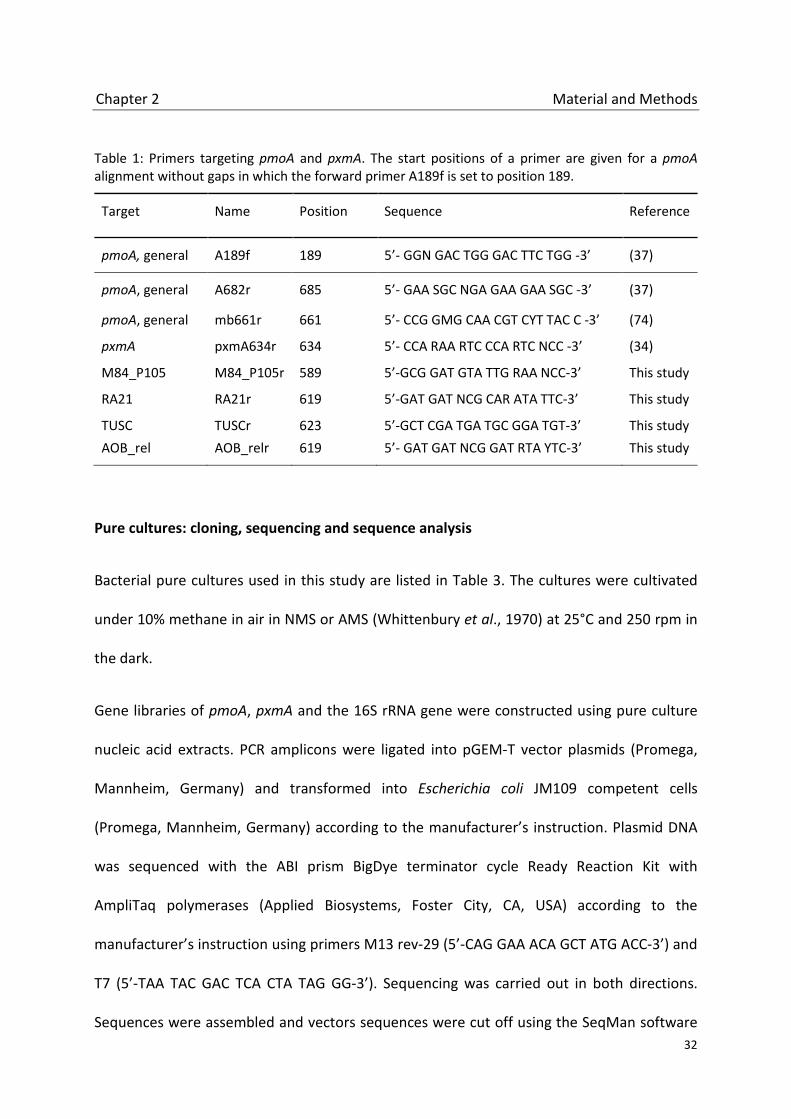
Table 1: Primers targeting pmoA and pxmA. The start positions of a primer are given for a pmoA alignment without gaps in which the forward primer A189f is set to position 189.
Target Name Position Sequence Reference
pmoA, general A189f 189 5’- GGN GAC TGG GAC TTC TGG -3’ (37)
pmoA, general A682r 685 5’- GAA SGC NGA GAA GAA SGC -3’ (37)
pmoA, general mb661r 661 5’- CCG GMG CAA CGT CYT TAC C -3’ (74)
pxmA pxmA634r 634 5’- CCA RAA RTC CCA RTC NCC -3’ (34)
M84_P105 M84_P105r 589 5’-GCG GAT GTA TTG RAA NCC-3’ This study
RA21 RA21r 619 5’-GAT GAT NCG CAR ATA TTC-3’ This study
TUSC TUSCr 623 5’-GCT CGA TGA TGC GGA TGT-3’ This study AOB_rel AOB_relr 619 5’- GAT GAT NCG GAT RTA YTC-3’ This study
Pure cultures: cloning, sequencing and sequence analysis
Bacterial pure cultures used in this study are listed in Table 3. The cultures were cultivated
under 10% methane in air in NMS or AMS (Whittenbury et al., 1970) at 25°C and 250 rpm in
the dark.
Gene libraries of pmoA, pxmA and the 16S rRNA gene were constructed using pure culture
nucleic acid extracts. PCR amplicons were ligated into pGEM-T vector plasmids (Promega,
Mannheim, Germany) and transformed into Escherichia coli JM109 competent cells
(Promega, Mannheim, Germany) according to the manufacturer’s instruction. Plasmid DNA
was sequenced with the ABI prism BigDye terminator cycle Ready Reaction Kit with
AmpliTaq polymerases (Applied Biosystems, Foster City, CA, USA) according to the
manufacturer’s instruction using primers M13 rev-29 (5’-CAG GAA ACA GCT ATG ACC-3’) and
T7 (5’-TAA TAC GAC TCA CTA TAG GG-3’). Sequencing was carried out in both directions.
Sequences were assembled and vectors sequences were cut off using the SeqMan software

Chapter 2 Material and Methods
33
(DNA-Star software package, Lasergene, Madison, WI, USA). Phylogenetic analysis were
done in ARB ver. 5.2 (Ludwig et al., 2004) and the online RAxML platform (Stamatakis et al.,
2008, Stamatakis, 2006). Sequences used for pure culture analysis contained at least 142
(pxmA) or 140 (pmoA) amino acids, and 697 nucleotides (16S rRNA), respectively.
For the construction of phylogenetic trees pmoA, pxmA and amoA sequences from public
databases and an existing database (Lüke & Frenzel, 2011) were used additionally. 16S rRNA
sequence analyses were based on the SILVA 108 database, release September 2011 (Quast
et al., 2013).
Incubations
For pure culture incubation 30 mL of an ammonium mineral salt medium (AMS) were
inoculated with 1.5 mL of a Methylobacter luteus 53v pre-culture. The cultures were
incubated with air and 2% methane (A, C) or 2% ethane (B). To set up (C), 2% ethane was
added when methane was nearly consumed. The cultures were incubated at 25°C and 200
rpm in the dark. Substrate consumption was followed by gas chromatography.
Competitive t-RFLP
A competitive PCR (cPCR) was performed as described before (Reim et al., 2012). DNA
standards were generated by PCR of rice field soil DNA (China2000) using the primers A189f
and Inner-rev-M84_P105_2_Mbacter (5’-GAT GAT GCG CAG RTA TTC CGA CCG GTT GGT GGA
ACA TG-3’) (Reim et al., 2012). RNA standards for quantifying the transcriptional level of
pxmA were generated by PCR using the primers A189f_T7 (5’-AAT ACG ACT CAC TAT AGG
GGG NGA CTG GGA CTT CTG G-3’) and Inner-rev-M84_P105_2_Mbacter.

Chapter 2 Results
34
2.3 Results
Environmental iCuMMO sequences
To study the occurrence of pxmA in different environments, samples were analyzed by deep
sequencing with primers targeting the iCuMMO groups M84_P105, RA_21, TUSC and
AOB_rel (Table 1). The basis for the phylogenetic analysis was an extensive database of
pmoA, pxmA and amoA sequences from pure cultures and various environments (Lüke &
Frenzel, 2011). The deep sequencing analysis resulted in about 21.400 high-quality pxmA
and pmoA sequences (Figure 1, Table 2).
Table 2: Phylogenetic distribution of sequences retrieved with the four new reverse primers depicting the percentage of sequences clustering within a specific lineage of pmoA/pxmA. Sequences retrieved with primer 682r from Vercelli, China50, and China2000 are shown for comparison (15).
Reverse primer
Phylogenetic assignment Total
number M84_P105 RA21 TUSC AOB_rel Type Ia
Type Ib
Type II FG Other*
M84_P105r 99.74 0.03 0.07 0.00 0.00 0.00 0.12 0.01 0.04 7575 RA21r 80.52 17.42 0.00 0.01 0.00 0.00 0.00 2.00 0.05 7766 TUSCr 1.04 0.00 47.39 39.24 0.13 0.13 11.73 0.07 0.26 1534
AOB_relr 0.14 0.06 31.32 35.83 0.14 19.64 10.78 0.57 1.50 4878 682r 0.03 0.09 0.9 7.33 0.26 33.03 26.05 0 32.28 3439
* for the A682r primer, others include mainly the amoA gene of ammonia oxidizing betaproteobacteria.
Virtually all sequences retrieved with the reverse primer M84_P105r fell into the M84_P105
cluster, suggesting that this primer is indeed very specific (Table 2). Sequences of this group
showed a wide distribution throughout all environments, being found in all rice field soils
and the lake sediments. Within the M84_P105 group, pxmA sequences formed the
subgroups G1-G5 (Figure 2). Independent on treeing algorithm (neighbor joining or
maximum likelyhood method) or subset of sequences used in analysis, sequences of each
group always clustered together. All subgroups contained environmental sequences.

Chapter 2 Results
35
Subgroup 1 includes in addition pxmA sequences of Methylobacter tundripaludum SV96,
Methylobacter marinus A45, Methylobacter luteus 53v, Methylomonas methanica S1,
Methylomonas spec. VMn. Group 2 contains pxmA sequences of Methylosarcina
quisquilarum AML-D4, Methylosarcina fibrata AML-C10 and Methylomicrobium album BG8.
Subgroup 3 is represented by pxmA sequences of a Methyloglobulus morosus strain
belonging to the type Ia methanotrophs (Deutzmann et al., 2014). Subgroup 5 includes pxmA
sequneces of the type II methanotrophs Methylocystis sp. SB2 and Methylocystis rosea.
Subgroups G1 and G3 were mainly represented by sequences retrieved from lake sediments
whereas G2, G4 and G5 contain mainly of sequences amplified from rise soils.
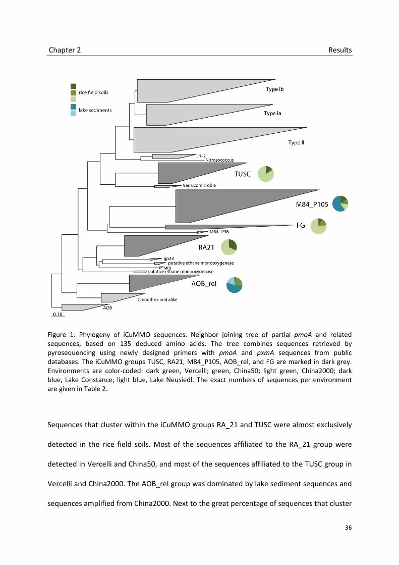
Chapter 2 Results
36
Figure 1: Phylogeny of iCuMMO sequences. Neighbor joining tree of partial pmoA and related sequences, based on 135 deduced amino acids. The tree combines sequences retrieved by pyrosequencing using newly designed primers with pmoA and pxmA sequences from public databases. The iCuMMO groups TUSC, RA21, M84_P105, AOB_rel, and FG are marked in dark grey. Environments are color-coded: dark green, Vercelli; green, China50; light green, China2000; dark blue, Lake Constance; light blue, Lake Neusiedl. The exact numbers of sequences per environment are given in Table 2.
Sequences that cluster within the iCuMMO groups RA_21 and TUSC were almost exclusively
detected in the rice field soils. Most of the sequences affiliated to the RA_21 group were
detected in Vercelli and China50, and most of the sequences affiliated to the TUSC group in
Vercelli and China2000. The AOB_rel group was dominated by lake sediment sequences and
sequences amplified from China2000. Next to the great percentage of sequences that cluster
Chapter 2 Results
37
within the already known environmental iCuMMO groups, we detected few sequences that
fell outside the iCuMMO clusters. In most cases only few sequences clustered together
outside the known groups. We did not indicate these sequences as new groups. An
exception was the new group FG (Freshwater sediment Group) that contains about 200
pxmA sequences amplified from rice field soils.
pxmA in pure cultures
Using the newly designed primers, representative pure cultures of type Ia, type Ib and type II
methanotrophs were tested for the occurrence of pxmA genes (Table 3). pxmA genes could
be retrieved from type Ia methanotrophs Methylomonas spec VMn, Methylobacter luteus
53v, Methylosarcina fibrata AML-C10 and Methylosarcina quisquilarum AML-D4. No pxmA
genes could be detected in type Ib methanotroph Methylococcus capsulatus Texas and
Methylocaldum E10a, in type II methanotroph Methylosinus trichosporium OB3b,
Methylosinus trichosporium I4/1 and Methylocystis heyeri H2(T), and type Ia methanotroph
Methylomicrobium alcaliphilum 2OZ. In contrast to the study of Tavormina and colleagues
(Tavormina et al., 2011), pxmA could also be detected in strains of the genus
Methylosarcina. All pxmA sequences clustered within the M84_P105 group. The phylogeny
of pmoA, pxmA and 16S rRNA sequences of type Ia methanotrophs was compared using
Maximum-Likelihood trees of nine pmoA, eight pxmA and ten 16S rRNA gene sequences
(Figure 3). In addition, a pair-wise comparison of sequence similarities between partial
pxmA, pmoA and 16S rRNA gene sequences was performed. The relationship of similarities
of pmoA and pxmA was linear for genes (R=0.77) and deduced proteins (R=0.76). Also, there
was a linear relationship between similarities of 16S rRNA genes and pxmA genes (R=0.74) or

Chapter 2 Results
38
proteins (R=0.63), and between 16S rRNA genes and pmoA genes (R=0.81) or proteins
(R=0.83). All correlation coefficients are highly significant (p < 0.01).
Table 3: Occurrence of pxmA in type Ia MOB pure cultures. The source gives either the DSMZ strain identification or the reference from which sequences were extracted for comparative phylogenetic analysis (Figure 3). +/-: presence/absence of the respective gene; NA: sequence not available; *: sequenced in this work.
Taxonominc name Strain Source pxmA pmoA 16S rRNA
Methylomonas methanica S1 1 + + +
Methylomonas sp. VMn 2 +* +* +*
Methylobacter luteus 53v NCIMB 11914 +* +* +*
Methylobacter tundripaludum SV96 DSM 17260 + + +
Methylobacter marinus A45 1 + NA +
Methylosarcina quisquilarum AML-D4 DSM 13737 +* +* +*
Methylosarcina fibrata AML-C10 DSM13736 +* +* +*
Methylosarcina lacus LW14 1 - + +
Methylomicrobium album BG8 1 + + +
Methylomicrobium kenyense 1 - + +
Methylomicrobium alcaliphilum 2 Z DSM 19304 - * +* +*
1: Tavormina et al. 2011 (34). 2: Methylomonas sp. VMn is from the culture collection at the MPI for Terrestrial Microbiology in Marburg. The original source is not documented. The culture has a 16S rRNA-sequence similarity of 98% to Methylomonas methanica (AF150806,1) and other Methylomonas sp. strains like Methylomonas sp. LW16.

Chapter 2 Results
39
Figure 2. Phylogenetic and ecological differentiations within the M84_P105 cluster. Neighbor joining tree of partial pmoA and related sequences as in Figure 1. In addition to environmental sequences, group 1 contains pxmA sequences from Methylobacter tundripaludum, Methylobacter marinus, Methylobacter luteus, Methylomonas methanica and Methylomonas spec. Group 2 contains environmental sequences and pxmA sequence of Methylosarcina quisquilarum, Methylosarcina fibrata and Methylomicrobium album. Groups 3 contains environmental sequences and pxmA sequences from Methyloglobulus morosus. Group 4 contain only environmental sequences. Group 5 contain environmental pxmA sequences and sequences of type II methanotrophs Methylocystis sp. SB2 and Methylocystis rosea. Environments are color-coded: dark green, Vercelli; green, China50; light green, China2000; dark blue, Lake Constance; light blue, Lake Neusiedl. The exact numbers of sequences per environment are given in Table S1.

Chapter 2 Results
40
Figure 3. Maximum likelihood trees of corresponding pmoA (A; 140 amino acids), pxmA (B; 142 amino acids), and 16S rRNA sequences (C; 697 nucleotides) of type Ia methanotrophic bacteria. Sequences retrieved in this study are marked by an asterisk.
Transcription of pxmA
As the physiological function of pXMO is still unknown, an incubation study with different
substrates was performed: methane, ethane, and a combination of methane and ethane
(Figure 4). Substrate usage, population growth and transcriptional pxmA levels were studied
exemplarily in Methylobacter luteus 53v. Methane was rapidly oxidized in all setups. The
methane concentrations decreased from 700 µM to 17µM within three days of incubation,

Chapter 2 Results
41
and from 680-750 µM to 120-130 µM within four days of incubation. The incubation with
ethane in air alone showed initially a minor decrease of ethane, but stayed constant
afterwards (data not shown). In the experiment with methane and ethane (Figure 4), the
latter was added after 55.5 h, when methane was nearly used up. Ethane oxidation started
immediately and stopped at a threshold concentration of 390-420 µM (n=2). pxmA
transcripts could be detected after 45.5 hours of incubation. The highest number of
transcripts was measured after 53h (6.46·104 copies/µL). The copy number decreased when
methane oxidation stopped and before ethane was added after 77 h. No pxmA transcripts
could be detected afterwards
Figure 4. Substrate consumption, pxmA gene and transcript copy number of Methylobacter luteus (NCIMB 11914) with methane and ethane as substrates (Setup C).

Chapter 2 Discussion
42
2.4 Discussion
The diversity of pmoA sequences in rice field soils and other environments has been
analyzed in a variety of studies. Next generation amplicon sequencing of pmoA has
furthermore indicated that the methanotrophic diversity seemed to be well covered (Lüke &
Frenzel, 2011). In contrast, pxmA sequences that cluster within the intermediate Cu-
containing membrane-bound monooxygenase groups (iCuMMO) are not well studied so far.
Only few sequences of these groups were available which lead to the conclusion that these
sequences might be rare in nature. However, the available iCuMMO sequences were
retrieved from many different environments like forest-, volcanic-, arctic- and dessert soils,
as well as paddy soils, landfill cover soils, freshwater lakes and others (Angel & Conrad, 2009,
Dorr et al., 2010, Holmes et al., 1999, Jia et al., 2007, King & Nanba, 2008, Knief et al., 2005,
Pacheco-Oliver et al., 2002, Reay et al., 2001, Singh et al., 2009). Hence, these sequences are
widely distributed in nature though they seem to be not very numerous. This fact could be
due to the use of the classical primer pair A189f/A682r (Holmes et al., 1995) that has the
potential to amplify a wide range of pxmA sequences but is known to discriminate against
several environmental clusters. Also quite often used is the primer par A189f/mb661
(Costello & Lidstrom, 1999) that discriminates even stronger against non-canonical pxmAs.
The reverse primer A682r and the primer pxmA634r that were used to amplify the pxmA in
another study (Tavormina et al., 2011) showed several mismatches to sequences of the
iCuMMOs. With the newly designed reverse primers targeting the iCuMMO groups we could
detected about 21.000 pxmA sequences. The modification of the classical primer sets
resulted in a totally different distribution of sequences within the pmoA tree and threw a
new light on the to-date only little observed iCuMMOs. Contrary to what was previously

Chapter 2 Discussion
43
assumed sequences of the iCuMMO are very numerous and widely distributed in freshwater
sediments and rice field soils. Despite this wide dispersal, environment-related distribution
patterns could be observed. While the groups TUSC, RA21 and FG are mainly represented by
rice field soil sequences, the AOB_rel and the M84_P105 groups seem to be mixed groups of
rice field soil and lake sediment sequences (Figure 1). However, a closer look at the
distribution of the sequences within the mixed cluster M84_P105 showed that sequences of
different habitats tended to cluster in different subgroups (Figure 2). Analyses of more
environments will show, if this division is also true for other habitats.
Four of these subgroups contain pxmA sequences of cultivated type Ia and type II
methanotrophs. The pxmA gene of Methylobacter and Methylomonas species cluster within
the subgroup G1. However, the pxmA of the phsychrophilic Methylobacter tundripaludum
cluster separately from other G1 sequences, but together with rice field soil sequences.
Thus, the pxmA of Methylobacter tundripaludum is present in freshwater environments of
the temperate zones, while its pmoA has mainly retrieved from the Arctic. The subgroup 2
contains pxmA sequences of two Methylosarcina strains and of Methylomicrobium album. It
has been shown that 16S rRNA and pmoA sequences of Methylomicrobium album cluster
closely to sequences of Methylosarcina (Wise et al., 2001, Kalyuzhnaya et al., 2008, this
study) yet apart from other Methylomicrobia species. Thus, Methylomicrobium album is not
representative for its genus and a re-classification has already been proposed (Kalyuzhnaya
et al., 2008).The pxmA could not be detected in Methylomicrobium alcaliphilum and
Methylomicrobium kenyense (Tavormina et al., 2011). These are both halo(alkali)philic
methanotrophs that were isolated from highly alkaline soda lakes (Khmelenina et al., 1997,

Chapter 2 Discussion
44
Sorokin et al., 2000). Neutrophilic Methylomicrobium strains like Methylomicrobium
pelagicum (Sieburth et al., 1987) or Methylomicrobium japanense (Fuse et al., 1998) still
need to be tested for presence of the pxmA gene. Subgroup 3 contains primarily
environmental sequences retrieved from Lake Constance. Additionally, pxmA sequences of a
newly isolated type Ia methanotroph Methyloglobulus morosus which was isolated from this
lake clustered within this subgroup (Deutzmann et al., 2014). While the subgroups 1, 2 and 3
are represented by type Ia methanotrophs, subgroups 5 contains pxmA sequences of type II
methanotrophs.
A ternary diagram constructed from phylogenetic inference of pmoA, pxmA and 16S rRNA of
type Ia methanotrophic gene sequences showed a good agreement between tree structures
(Figure 2). In addition, a pair-wise comparison of DNA and protein sequence similarities of
pxmA versus pmoA, pxmA versus 16SrRNA and pmoA versus 16S rRNA revealed a significant
correlation. No evidence of a recent horizontal transfer of the pxmA gene was found in type I
methanotrophic bacteria. It rather seemed that the pxmA gene was acquired early in
evolution and developed further in parallel to the pmoA and the 16S rRNA genes. It remains
unanswered when this happened. It is assumed that the ability to oxidize methane was
achieved by the alphaproteobacteria just once in their evolution (Tamas et al., 2014). If this
is also true for the gammaproteobacteria it is likely that the pxm operon was acquired
before the radiation of the type I methanotrophs started. The pxmA in type I methanotrophs
has been found until now only in strains of type Ia methanotrophs, no in type Ib
methanotrophs. In a sense this is surprising, because we could show that the pxmA is widely
distributed in freshwater environments, where type Ib methanotrophs dominate. But no

Chapter 2 Discussion
45
pxmA sequences could be retrieved from the type Ib strains Methylococcus capsulatus Texas
and Methylocaldum sp. However, most type Ib sequences from freshwater habitats cluster
within phylogenetic groups that are not represented by any cultured representative (Lüke &
Frenzel, 2011). Either the type Ib methanotrophs do not possess the pxmA or we just cannot
yet link a pxmA to its corresponding type Ib pmoA due to the lack of representative pure
cultures. Because of that, it is difficult to guess if the pxmA was acquired before or after the
division of the type Ia und type Ib methanotrophs.
The physiological role of the different iCuMMOs is unknown until now. They are distantly
related to methane and ammonium monooxygenases and to marine sequences from
putative ethane degraders (Coleman et al., 2011, Redmond et al., 2010, Suzuki et al., 2012,
Sayavedra-Soto et al., 2011). It seemed likely that iCuMMOs could also be involved in the
degradation of these substrates. It was suggested that the pXMO isoenzyme in type Ia
methanotrophs plays a role in the ammonium oxidation (Tavormina et al., 2011) but we
could not see any evidence that ammonium was used as a substrate for the iCuMMO in this
study (no growth on AMS medium without methane addition; data not shown).
Furthermore, incubation experiments of Methylobacter luteus 53v do not indicate that
ethane is a possible substrate. The culture grew well on methane but showed no growth on
ethane. Additionally, the transcription of the pxmA could only be detected when methane
was available as a substrate. However, this transcription was very low. It has been shown
earlier that pMMO isoenzymes in alphaproteobacteria and Verrucomicrobia are expressed
at different methane or oxygen concentrations (Baani & Liesack, 2008, Khadem et al., 2012).
Thus, it could be possible that the pxm operon encodes for a methane monooxygenase with

Chapter 2 Discussion
46
an alternative enzyme kinetic that provide a selective advantage under changing
environmental conditions.
Acknowledgements
We thank Elisabetta Lupotto, Zhihong Cao and Yao Pan for help with sampling. We would
furthermore like to thank Andreas Reim for excellent technical assistance with competitive
PCR analysis. This work, as part of the European Science Foundation EUROCORES
Programme EuroEEFG, was supported from funds to PF by the Deutsche
Forschungsgemeinschaft (DFG).

Chapter 2 References
47
2.5 References
Angel, R. & R. Conrad, (2009) In situ measurement of methane fluxes and analysis of transcribed particulate methane monooxygenase in desert soils. Environ. Microbiol. 11: 2598-2610.
Baani, M. & W. Liesack, (2008) Two isozymes of particulate methane monooxygenase with different
methane oxidation kinetics are found in Methylocystis sp strain SCZ. Proceedings of the Proc. Natl. Acad. Sci. U. S. A. 105: 10203-10208.
Bedard, C. & R. Knowles, (1989) Physiology, biochemistry, and specific inhibitors of CH4, NH4
+, and CO oxidation by methanotrophs and nitrifiers. Microbiol. Rev. 53: 68-84.
Belova, S.E., M. Baani, N.E. Suzina, P.L.E. Bodelier, W. Liesack & S.N. Dedysh, (2011) Acetate
utilization as a survival strategy of peat inhabiting Methylocystis spp. Environ. Microbiol. Rep. 3: 36-46.
Bender, M. & R. Conrad, (1992) Kinetics of CH4 oxidation in oxic soils exposed to ambient air or high
CH4 mixing ratios. FEMS Microbiol. Ecol. 101: 261-270. Burrows, K.J., A. Cornish, D. Scott & I.J. Higgins, (1984) Substrate specificities of the soluble and
particulate methane monooxygenases of Methylosinus trichosporium Ob3b. J. Gen. Microbiol. 130: 3327-3333.
Cheng, Y.Q., L.Z. Yang, Z.H. Cao, E. Ci & S.X. Yin, (2009) Chronosequential changes of selected
pedogenic properties in paddy soils as compared with non paddy soils. Geoderma 151: 31-41.
Coleman, N.V., N.B. Le, M.A. Ly, H.E. Ogawa, V. McCarl, N.L. Wilson & A.J. Holmes, (2011)
Hydrocarbon monooxygenase in Mycobacterium: recombinant expression of a member of the ammonia monooxygenase superfamily. ISME J. 6: 171–182
Conrad, R., (1996) Soil microorganisms as controllers of atmospheric trace gases (H2, CO, CH4, OCS,
N2O, and NO). Microbiol. Mol. Biol. Rev. 60: 609-640. Conrad, R., (2009) The global methane cycle: recent advances in understanding the microbial
processes involved. Environ. Microbiol. Rep. 1: 285-292. Conrad, R. & P. Frenzel, (2002) Flooded Soils. Encyclopedia of Environ. Microbiol. 1316-1333. Costello, A.M. & M.E. Lidstrom, (1999) Molecular characterization of functional and phylogenetic
genes from natural populations of methanotrophs in lake sediments. Appl. Environ. Microbiol. 65: 5066-5074.
Dedysh, S.N., Y.Y. Berestovskaya, L.V. Vasylieva, S.E. Belova, V.N. Khmelenina, N.E. Suzina, Y.A.
Trotsenko, W. Liesack & G.A. Zavarzin, (2004) Methylocella tundrae sp. nov., a novel methanotrophic bacterium from acidic tundra peatlands. Int. J. Syst. Evol. Microbiol. 54: 151-156.

Chapter 2 References
48
Dedysh, S.N., C. Knief & P.F. Dunfield, (2005) Methylocella species are facultatively methanotrophic. J. Bacteriol. 187: 4665-4670.
Dedysh, S.N., W. Liesack, V.N. Khmelenina, N.E. Suzina, Y.A. Trotsenko, J.D. Semrau, A.M. Bares, N.S.
Panikov & J.M. Tiedje, (2000) Methylocella palustris gen. nov., sp. nov., a new methane-oxidizing acidophilic bacterium from peat bogs, representing a novel subtype of serine-pathway methanotrophs. Int. J. Syst. Evol. Microbiol. 50: 955-969.
Dorr, N., B. Glaser & S. Kolb, (2010) Methanotrophic communities in brazilian ferralsols from
naturally forested, afforested, and agricultural sites. Appl. Environ. Microbiol. 76: 1307-1310. Dumont, M.G., C. Lüke, Y.C. Deng & P. Frenzel, (2014) Classification of pmoA amplicon
pyrosequences using BLAST and the lowest common ancestor method in MEGAN. Front. Microbiol. 5: 34. doi: 10.3389/fmicb.2014.00034
Dunfield, P.F., V.N. Khmelenina, N.E. Suzina, Y.A. Trotsenko & S.N. Dedysh, (2003) Methylocella
silvestris sp nov., a novel methanotroph isolated from an acidic forest cambisol. Int. J. Syst. Evol. Microbiol. 53: 1231-1239.
Dunfield, P.F., A. Yuryev, P. Senin, A.V. Smirnova, M.B. Stott, S. Hou, B. Ly, J.H. Saw, Z. Zhou, Y. Ren, J.
Wang, B.W. Mountain, M.A. Crowe, T.M. Weatherby, P.L.E. Bodelier, W. Liesack, L. Feng, L. Wang & M. Alam, (2007) Methane oxidation by an extremely acidophilic bacterium of the phylum Verrucomicrobia. Nature 450: 879-882.
Elliott, S.J., M. Zhu, L. Tso, H.H.T. Nguyen, J.H.K. Yip & S.I. Chan, (1997) Regio- and stereoselectivity of
particulate methane monooxygenase from Methylococcus capsulatus (Bath). J. Am. Chem. Soc. 119: 9949-9955.
Ettwig, K.F., M.K. Butler, D. Le Paslier, E. Pelletier, S. Mangenot, M.M.M. Kuypers, F. Schreiber, B.E.
Dutilh, J. Zedelius, D. de Beer, J. Gloerich, H.J.C.T. Wessels, T. van Alen, F. Luesken, M.L. Wu, K.T. Pas-Schoonen, H.J.M.O. den Camp, E.M. Janssen-Megens, K.J. Francoijs, H. Stunnenberg, J. Weissenbach, M.S.M. Jetten & M. Strous, (2010) Nitrite-driven anaerobic methane oxidation by oxygenic bacteria. Nature 464: 543-548.
Frenzel, P., (2000) Plant-associated methane oxidation in rice fields and wetlands. Adv. Microb. Ecol.
16: 85-114. Fuse, H., M. Ohta, O. Takimura, K. Murakami, H. Inoue, Y. Yamaoka, J.M. Oclarit & T. Omori, (1998)
Oxidation of trichloroethylene and dimethyl sulfide by a marine Methylomicrobium strain containing soluble methane monooxygenase. Biosci., Biotechnol., Biochem. 62: 1925-1931.
Hayashi, T., H. Obata, T. Gamo, Y. Sano & Y. Naganuma, (2007) Distribution and phylogenetic
characteristics of the genes encoding enzymes relevant to methane oxidation in oxygen minimum zones of the eastern Pacific Ocean. Res. J. Environ. Sci. 1: 275-284.
Henneberger, R., C. Lüke, L. Mosberger & M.H. Schroth, (2012) Structure and function of
methanotrophic communities in a landfill-cover soil. FEMS Microbiol. Ecol. 81: 52-65.

Chapter 2 References
49
Ho, A., C. Lüke, Z.H. Cao & P. Frenzel, (2011) Ageing well: methane oxidation and methane oxidizing bacteria along a chronosequence of 2000 years. Environ. Microbiol. Rep. 3: 738-743.
Holmes, A.J., A. Costello, M.E. Lidstrom & J.C. Murrell, (1995) Evidence that particulate methane
monooxygenase and ammonia monooxygenase may be evolutionarily related. FEMS Microbiol. Lett. 132: 203-208.
Holmes, A.J., P. Roslev, I.R. McDonald, N. Iversen, K. Henriksen & J.C. Murrell, (1999) Characterization
of methanotrophic bacterial populations in soils showing atmospheric methane uptake. Appl. Environ. Microbiol. 65: 3312-3318.
Holzapfelpschorn, A. & W. Seiler, (1986) Methane emission during a cultivation period from an Italian
rice paddy. J. Geophys. Res., [Atmos.] 91: 1803-1814. Horz, H.P., V. Rich, S. Avrahami & B.J.M. Bohannan, (2005) Methane-oxidizing bacteria in a California
upland grassland soil: Diversity and response to simulated global change. Appl. Environ. Microbiol. 71: 2642-2652.
Intergovernmental Panel on Climate Change, (2007) Climate change 2007: The physical science basis.
summary for policymakers. contribution of working group I to the fourth assessment report of the intergovernmental panel on climate change. IPCC Secretariat, Geneva.
Islam, T., S. Jensen, L.J. Reigstad, O. Larsen & N.K. Birkeland, (2008) Methane oxidation at 55° C and
pH 2 by a thermoacidophilic bacterium belonging to the Verrucomicrobia phylum. Proc. Natl. Acad. Sci. 105: 300-304.
Jia, Z.J., H. Kikuchi, T. Watanabe, S. Asakawa & M. Kimura, (2007) Molecular identification of
methane oxidizing bacteria in a Japanese rice field soil. Biol. Fertil. Soils. 44: 121-130. Kalyuzhnaya, M.G., V. Khmelenina, B. Eshinimaev, D. Sorokin, H. Fuse, M. Lidstrom & Y. Trotsenko,
(2008) Classification of halo(alkali)philic and halo(alkali)tolerant methanotrophs provisionally assigned to the genera Methylomicrobium and Methylobacter and emended description of the genus Methylomicrobium. Int. J. Syst. Evol. Microbiol. 58: 591-596.
Khadem, A.F., A. Pol, A.S. Wieczorek, M.S.M. Jetten & H.J.M.O. den Camp, (2012) Metabolic
regulation of "Ca. Methylacidiphilum fumariolicum" SoIV cells grown under different nitrogen and oxygen limitations. Front. Microbiol. 3: 266. doi: 10.3389/fmicb.2012.00266.
Khmelenina, V.N., M.G. Kalyuzhnaya & Y.A. Trotsenko, (1997) Physiological and biochemical
properties of a haloalkalitolerant methanotroph. Microbiology 66: 365-370. King, G.M. & K. Nanba, (2008) Distribution of atmospheric methane oxidation and methanotrophic
communities on Hawaiian volcanic deposits and soils. Microbes Environ. 23: 326-330. Knief, C., A. Lipski & P.F. Dunfield, (2003) Diversity and activity of methanotrophic bacteria in
different upland soils. Appl. Environ. Microbiol. 69: 6703-6714.
Chapter 2 References
50
Knief, C., S. Vanitchung, N.W. Harvey, R. Conrad, P.F. Dunfield & A. Chidthaisong, (2005) Diversity of methanotrophic bacteria in tropical upland soils under different land uses. Appl. Environ. Microbiol. 71: 3826-3831.
Kolbl, A., P. Schad, R. Jahn, W. Amelung, A. Bannert, Z.H. Cao, S. Fiedler, K. Kalbitz, E. Lehndorff, C.
Muller-Niggemann, M. Schloter, L. Schwark, V. Vogelsang, L. Wissing & I. Kogel-Knabner, (2014) Accelerated soil formation due to paddy management on marshlands (Zhejiang Province, China). Geoderma 228: 67-89.
Kruger, M., P. Frenzel & R. Conrad, (2001) Microbial processes influencing methane emission from
rice fields. Global. Change. Biol. 7: 49-63. Lane, D.J., (1991) 16S/23S rRNA sequencing, p. 115-175. In Stackebrandt E., Goodfellow M. (ed)
Nucleic acids techniques in bacterial systematics, pp. 115–147. John Wiley & Sons, Chichester.
Ludwig, W., O. Strunk, R. Westram, L. Richter, H. Meier, Yadhukumar, A. Buchner, T. Lai, S. Steppi, G.
Jobb, W. Forster, I. Brettske, S. Gerber, A.W. Ginhart, O. Gross, S. Grumann, S. Hermann, R. Jost, A. Konig, T. Liss, R. Lussmann, M. May, B. Nonhoff, B. Reichel, R. Strehlow, A. Stamatakis, N. Stuckmann, A. Vilbig, M. Lenke, T. Ludwig, A. Bode & K.H. Schleifer, (2004) ARB: a software environment for sequence data. Nucleic. Acids. Res. 32: 1363-1371.
Lüke, C. & P. Frenzel, (2011) Potential of pmoA amplicon pyrosequencing for methanotroph diversity
studies. Appl. Environ. Microbiol. 77: 6305-6309. Lüke, C., P. Frenzel, A. Ho, D. Fiantis, P. Schad, B. Schneider, L. Schwark & S.R. Utami, (2014)
Macroecology of methane-oxidizing bacteria: the beta-diversity of pmoA genotypes in tropical and subtropical rice paddies. Environ. Microbiol. 16: 72-83.
Lüke, C., S. Krause, S. Cavigiolo, D. Greppi, E. Lupotto & P. Frenzel, (2010) Biogeography of wetland
rice methanotrophs. Environ. Microbiol. 12: 862-872. Pacheco-Oliver, M., I.R. McDonald, D. Groleau, J.C. Murrell & C.B. Miguez, (2002) Detection of
methanotrophs with highly divergent pmoA genes from Arctic soils. FEMS Microbiol. Lett. 209: 313-319.
Pol, A., K. Heijmans, H.R. Harhangi, D. Tedesco, M.S.M. Jetten & H.J.M. Op den Camp, (2007)
Methanotrophy below pH1 by a new Verrucomicrobia species. Nature 450: 874-878 Quast, C., E. Pruesse, P. Yilmaz, J. Gerken, T. Schweer, P. Yarza, J. Peplies & F.O. Glockner, (2013) The
SILVA ribosomal RNA gene database project: improved data processing and web-based tools. Nucleic. Acids. Res. 41: 590-596.
Reay, D.S., S. Radajewski, J.C. Murrell, N. McNamara & D.B. Nedwell, (2001) Effects of land-use on
the activity and diversity of methane oxidizing bacteria in forest soils. Soil Biol. Biochem. 33: 1613-1623.

Chapter 2 References
51
Reeburgh, W.S., (1997) Figures summarizing the global cycles of biogeochemically important elements. Bull. Ecol. Soc. America. 78: 260-267.
Redmond, M.C., D.L. Valentine & A.L. Sessions, (2010) Identification of novel methane-, ethane-, and
propane-oxidizing bacteria at marine hydrocarbon seeps by stable isotope probing. Appl. Environ. Microbiol. 76: 6412-6422.
Reim, A., C. Lüke, S. Krause, J. Pratscher & P. Frenzel, (2012) One millimetre makes the difference:
high-resolution analysis of methane-oxidizing bacteria and their specific activity at the oxic-anoxic interface in a flooded paddy soil. ISME J. 6: 2128-2139.
Roth, P.J., E. Lehndorff, A. Hahn, P. Frenzel & W. Amelung, (2013) Cycling of rice rhizodeposits
through peptide-bound amino acid enantiomers in soils under 50 and 2000 years of paddy management. Soil Biol. Biochem. 65: 227-235.
Sayavedra-Soto, L.A., N. Hamamura, C.W. Liu, J.A. Kimbrel, J.H. Chang & D.J. Arp, (2011) The
membrane-associated monooxygenase in the butane-oxidizing Gram-positive bacterium Nocardioides sp. strain CF8 is a novel member of the AMO/PMO family. Environ. Microbiol. Rep. 3: 390-396.
Semrau, J.D., (2011) Bioremediation via methanotrophy: Overview of recent findings and suggestions
for future research. Front. Microbiol. 2: 209 doi: 10.3389/fmicb.2011.00209 Sieburth, J.M., P.W. Johnson, M.A. Eberhardt, M.E. Sieracki, M. Lidstrom & D. Laux, (1987) The first
methane oxidizing bacterium from the upper mixing layer of the deep cean: Methylomonas pelagica sp nov. Curr. Microbiol. 14: 285-293.
Singh, B.K., K.R. Tate, D.J. Ross, J. Singh, J. Dando, N. Thomas, P. Millard & J.C. Murrell, (2009) Soil
methane oxidation and methanotroph responses to afforestation of pastures with Pinus radiata stands. Soil Biol. Biochem. 41: 2196-2205.
Sorokin, D.Y., B.E. Jones & J.G. Kuenen, (2000) An obligate methylotrophic, methane-oxidizing
Methylomicrobium species from a highly alkaline environment. Extremophiles 4: 145-155. Stamatakis, A., (2006) RAxML-VI-HPC: Maximum likelihood-based phylogenetic analyses with
thousands of taxa and mixed models. Bioinformatics 22: 2688-2690. Stamatakis, A., P. Hoover & J. Rougemont, (2008) A rapid bootstrap algorithm for the RAxML web
servers. Syst. Biol. 57: 758-771. Stoecker, K., B. Bendinger, B. Schoning, P.H. Nielsen, J.L. Nielsen, C. Baranyi, E.R. Toenshoff, H. Daims
& M. Wagner, (2006) Cohn's Crenothrix is a filamentous methane oxidizer with an unusual methane monooxygenase. Proc. Natl. Acad. Sci. U. S. A. 103: 2363-2367.
Suzuki, T., T. Nakamura & H. Fuse, (2012) Isolation of two novel marine ethylene-assimilating
bacteria, Haliea species ETY-M and ETY-NAG, containing particulate methane monooxygenase-like genes. Microbes Environ. 27: 54-60.
Svenning, M.M., A.G. Hestnes, I. Wartiainen, L.Y. Stein, M.G. Klotz, M.G. Kalyuzhnaya, A. Spang, F.
Bringel, S. Vuilleumier, A. Lajus, C. Medigue, D.C. Bruce, J.F. Cheng, L. Goodwin, N. Ivanova, J.
Chapter 2 References
52
Han, C.S. Han, L. Hauser, B. Held, M.L. Land, A. Lapidus, S. Lucas, M. Nolan, S. Pitluck & T. Woyke, (2011) Genome sequence of the Arctic methanotroph Methylobacter tundripaludum SV96. J. Bacteriol. 193: 6418-6419.
Tamas, I., A.V. Smirnova, Z. He & P.F. Dunfield, (2014) The (d)evolution of methanotrophy in the
Beijerinckiaceae--a comparative genomics analysis. ISME J. 8: 369-382. Tavormina, P.L., V.J. Orphan, M.G. Kalyuzhnaya, M.S.M. Jetten & M.G. Klotz, (2011) A novel family of
functional operons encoding methane/ammonia monooxygenase-related proteins in gammaproteobacterial methanotrophs. Environ. Microbiol. Rep. 3: 91-100.
Theisen, A.R., M.H. Ali, S. Radajewski, M.G. Dumont, P.F. Dunfield, I.R. McDonald, S.N. Dedysh, C.B.
Miguez & J.C. Murrell, (2005) Regulation of methane oxidation in the facultative methanotroph Methylocella silvestris BL2. Mol. Microbiol. 58: 682-692.
Trotsenko, Y.A. & J.C. Murrell, (2008) Metabolic aspects of aerobic obligate methanotrophy. Adv.
Appl. Microbiol. 63: 183-229. Vigliotta, G., E. Nutricati, E. Carata, S.M. Tredici, M. De Stefano, P. Pontieri, D.R. Massardo, M.V. Prati,
L. De Bellis & P. Alifano, (2007) Clonothrix fusca Roze 1896, a filamentous, sheathed, methanotrophic gamma-proteobacterium. Appl. Environ. Microbiol. 73: 3556-3565.
Vorobev, A.V., M. Baani, N.V. Doronina, A.L. Brady, W. Liesack, P.F. Dunfield & S.N. Dedysh, (2011)
Methyloferula stellata gen. nov., sp nov., an acidophilic, obligately methanotrophic bacterium that possesses only a soluble methane monooxygenase. Int. J. Syst. Evol. Microbiol. 61: 2456-2463.
Weisburg ,W.G., S.M. Barns, D.A.Pelletier & D.J. Lane, (1991) 16S ribosomal DNA amplification for
phylogenetic study. J. Bacteriol. 173: 697-703. Whittenbury, R., K.C. Phillips & J.F. Wilkinson, (1970) Enrichment, Isolation and Some Properties of
Methane-Utilizing Bacteria. J. Gen. Microbiol. 61: 205-218. Wise, M.G., J.V. McArthur & L.J. Shimkets, (2001) Methylosarcina fibrata gen. nov., sp. nov. and
Methylosarcina quisquiliarum sp.nov., novel type I methanotrophs. Int. J. Syst. Evol. Microbiol. 51: 611-621.

Chapter 3 Abstract
53
3 Magnetic capture of iCuMMO sequences: A prove of concept
3.1 Abstract
Culture independent methods are good molecular tools to study the distribution and
diversity of methanotrophic communities in different environments. Most of these methods
are PCR based techniques that rely on sequence specific primers. In environmental studies,
when little or no sequence information of the target organisms is available, the generation
of appropriate primers is difficult. Here, we adapted the magnetic hybridization capture
method (MHC) to detect Cu-containing membrane-bound monooxygenases (CuMMOs) in
environments and to avoid the use of specific primers. Exemplarily, a group of sequences
clustering into the environmental iCuMMO group FG were chosen to develop and evaluate
the MHC. Biotinylated capture probes targeting sequences of the FG group were generated.
The probes were used to capture FG sequences out of a complex mix of environmental
sequences. First results were successful: FG sequences could be detected and concentrated
by MHC. Following improvements of this method could make the MHC a powerful method
to study under represented organisms in different habitats.

Chapter 3 Introduction
54
3.2 Introduction
Aerobic methane oxidation is performed by methane oxidizing bacteria (MOB), a specialized
widespread group of organisms that can be found in many terrestrial and aquatic soils. The
key enzyme of the aerobic methane oxidizing pathway of most MOB is the particulate
methane monooxygenase (pMMO) that belongs to the great family of Cu-containing
membrane-bound monooxygenases (CuMMOs). A subunit of the pMMO is encoded by the
pmoA gene. This gene is highly conserved, and often used as a functional marker gene to
identify methanotrophs.
Common tools to detect and study methanotrophic communities in nature are culture
independent methods like PCR based techniques. To obtain preferably significant results
using these techniques sequence specific primers are crucial. Primer mismatches prevent
binding to target sequences and lead to weak or no amplification results. Several primer sets
targeting the pmoA gene have been established to cover the methanotrophic diversity. The
classical primer set is A189f / A682r (Holmes et al., 1995). It has been used frequently in
many environmental studies. However, this primerset has disadvantages in specificity.
Besides pmoA sequences, sequences of the amoA of betaproteobacterial ammonium
oxidizers and, to a minor degree, other pxmA sequences (genes of the environmental
intermediate CuMMOs; iCuMMOs) are often co-amplified. Another drawback of this
primerset is the poor coverage of sequences of environmental pxmA sequences (Bourne et
al., 2001). To overcome the problems of specificity and coverage several alternative primers
were designed to amplify pmoA genes, e.g. mb661 (Costello & Lidstrom, 1999), A650r

Chapter 3 Introduction
55
(Bourne et al., 2001), A621r (Tuomivirta et al., 2009), and nmb650r (Shrestha et al., 2010).
The reverse primer mb661 was designed to specifically amplify pmoA sequences and exclude
amoA and pxmA sequences. A650r lacks a good coverage of the pmoA sequences but covers
pxmA of environmental groups. A621r mainly amplifies sequences of type II methanotrophs
and is especially used when A682r yields no products in amplification of those sequences.
Though it was tried frequently to find a universal pmoA primer, none is available that covers
the pmoA as well as the pxmA sequences of the iCuMMO groups.
A current example of the importance of the use of specific primers is the detection of the
pMMO isoenzyme (pXMO) in MOB (Hainbuch et al., in prep., Tavormina et al., 2011).
Methanotrophic pure cultures have been studied for a long time (e.g. Bowman et al., 1993,
Whittenbury, 1984, Wise et al., 2001). But the pXMO in MOB could be detected not until the
reverse primer of the classical primer set was exchanged by an alternative reverse primer.
Though the classical primer set A189f/A682r have the potential to amplify pxmA sequences,
several primer mismatches lead to a dramatically under-representation of those sequences
in pure culture and environmental studies. Additionally, A189f/A682r discriminates against
pxmA sequences that cluster in several environmental iCuMMO groups (RA21, TUSC, ABO-
rel, FG). Only few sequences of these groups were published, but the design of new reverse
primers moved the iCuMMO groups to the fore. Their sequences are very numerous and
widely distributed in nature and of more ecological importance than believed so far.
Culture independent PCR based methodologies are powerful tools to study methanotrophic
bacteria in the environments. However, specific primers are essential for the success of PCR
dependent methods. Important for the design of specific primers are appropriate template

Chapter 3 Introduction
56
sequences. In environmental studies, when little or no sequence information of the target
organisms is available to design suitable primers, PCR techniques become a great challenge.
The environmental iCuMMO groups, for example, are not represented by pure cultures.
Additionally, only partial pxmA sequences with a length of about 450 bp, derived from next
generation sequencing, are available as templates for primer design. This makes the design
of specific primers a difficult or insoluble task, especially if the group of target organisms is
only represented by few sequences. A method that is independent of specific primers is the
magnetic hybridization capture (MHC). This method works in principle in the following way:
A biotinylated oligonucleotide hybridization probe is designed that is specific for the target
sequence. The probe is incubated with a heterogeneous mix of nucleic acids to allow it to
anneal to the corresponding target sequence. The nucleotide mix containing the probe-
target hybridization is afterwards incubated with streptavidin-coated magnetic beads. The
streptavidin binds to the biotin of the probes. A magnetic force is than aplied to the probe-
target-magnetic bead complex which makes it possible to concentrate and separate this
complex from non-target DNA by several washing steps. The use of MHC provides several
advantages compared to PCR techniques. While it is advantageous for primer design to have
long template sequences (for primers border the PCR amplicon), short template sequences
are sufficient for capture probe design. The probe binds within the target DNA fragments,
which makes it possible to capture fragments ranging in length of a few hundred bases to
many megabases in size. Another advantage is that hybridization methods are less prone for
sequence mismatches than PCR primers. This makes them very useful for environmental
sequence enrichments where only little sequence information is available. A great diversity
of sequences could be captured with a single probe.
Chapter 3 Introduction
57
So far, the MHC technique is mainly constricted to clinical studies and pure culture
researches. Here, the method should be applied to environmental researches. As target
sequences for MHC a subgroup of the newly found iCuMMO Freshwater Group (FG_N3) was
chosen exemplarily. Few sequences with a length of about 450 bp of this group are available.
The main focus of this study is to adapt MHC for a routinely use in analyzing bacterial
diversity in nature. By studying the iCuMMO Freshwater Group in particular, the aim is to
capture long target sequences by MHC. These long sequences could deliver important
information for the study of phylogenetic relationships and could give an insight into the
gene structure of the iCuMMO operons. Studies by Tarvormina et al. (2011) showed that
pXMO operons have a gene order, which differs from known pMMO operons. The question,
if this gene order is unique for this group, or if alternative operon structures are widespread
among the environmental iCuMMO clusters, could be resolved by the use of MHC
techniques. In addition, sequence information of the operons of the iCuMMO Freshwater
Group (FG_N3) and other environmental groups could help to design new gene specific
primers for downstream analyses like qPCR. Several reverse primers have been designed
targeting sequences of iCuMMO groups, whereas the forward primer A189f was used for a
long time in many studies. This primer seems to match most of the methanotrophic pmoA
sequences, but we do not know, if this is true for the sequences of the environmental
groups. New sequence specific primers based on captured sequences could help to evaluate
the use of different primers.

Chapter 3 Introduction
58
Figure 1: Illustration of the principle of the probe design (1A) and the magnetic capture (1B).
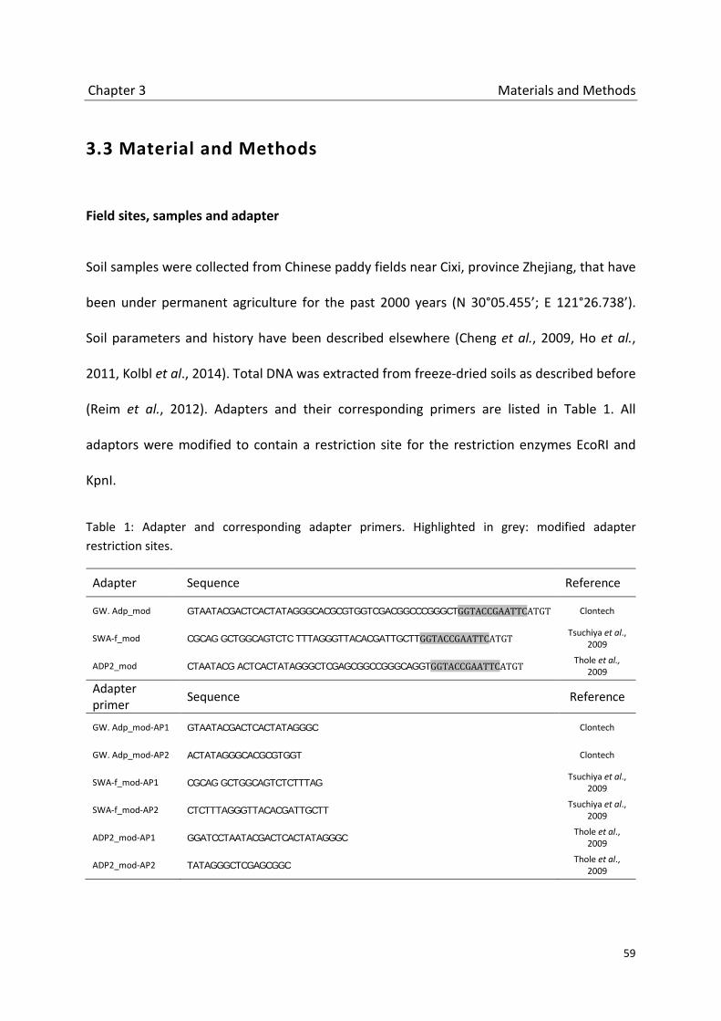
Chapter 3 Materials and Methods
59
3.3 Material and Methods
Field sites, samples and adapter
Soil samples were collected from Chinese paddy fields near Cixi, province Zhejiang, that have
been under permanent agriculture for the past 2000 years (N 30°05.455’; E 121°26.738’).
Soil parameters and history have been described elsewhere (Cheng et al., 2009, Ho et al.,
2011, Kolbl et al., 2014). Total DNA was extracted from freeze-dried soils as described before
(Reim et al., 2012). Adapters and their corresponding primers are listed in Table 1. All
adaptors were modified to contain a restriction site for the restriction enzymes EcoRI and
KpnI.
Table 1: Adapter and corresponding adapter primers. Highlighted in grey: modified adapter restriction sites.
Adapter Sequence Reference
GW. Adp_mod GTAATACGACTCACTATAGGGCACGCGTGGTCGACGGCCCGGGCTGGTACCGAATTCATGT Clontech
SWA-f_mod CGCAG GCTGGCAGTCTC TTTAGGGTTACACGATTGCTTGGTACCGAATTCATGT Tsuchiya et al.,
2009
ADP2_mod CTAATACG ACTCACTATAGGGCTCGAGCGGCCGGGCAGGTGGTACCGAATTCATGT Thole et al.,
2009
Adapter primer Sequence Reference
GW. Adp_mod-AP1 GTAATACGACTCACTATAGGGC Clontech
GW. Adp_mod-AP2 ACTATAGGGCACGCGTGGT Clontech
SWA-f_mod-AP1 CGCAG GCTGGCAGTCTCTTTAG Tsuchiya et al.,
2009
SWA-f_mod-AP2 CTCTTTAGGGTTACACGATTGCTT Tsuchiya et al.,
2009
ADP2_mod-AP1 GGATCCTAATACGACTCACTATAGGGC Thole et al.,
2009
ADP2_mod-AP2 TATAGGGCTCGAGCGGC Thole et al.,
2009

Chapter 3 Materials and Methods
60
Restriction digest and ligation
Extracted environmental DNA (2 µg) and adapter DNA (2 µg) were digested with KpnI or
EcoRI (Fermentas, St. Leon-Rot, Germany) at 37 °C for 3h. Digested environmental DNA was
purified afterwards using the Qiaquick PCR Purification Kit (Qiagen, Hilden, Germany)
according to manufacturer's instructions. Digested adapter DNA was purified with Post-
Reaction Clean-Up Spin Columns (Sigma-Aldrich, Munich, Germany) following the
manufacturer's protocol.
Ligation of digested environmental DNA and adapter DNA was performed using T4 DNA
Ligase (Promega, Madison, Wisc., USA). 500ng environmental DNA and 250 ng adapter DNA
were mixed with 5 U T4 DNA Ligase and 5 µL Ligase 10x Buffer, and filled up to 50 µL with
molecular grade water (Sigma). Ligation was carried out at 4 °C over night.
Probe design
Primers to amplify the magnetic capture probe were designed based on an alignment of 35
sequences clustering in the subgroup 3 of the Freshwater Group (FG_N3) (Hainbuch et al. in
revision). PCR reactions with newly designed primers FG_new3for (5’-
GTGGCGATTTCCTACTGGAGC-3’) or FG_new3for_biot (5’-GTGGCGATTTCCTACTGGAGC-3’
containing 5': Biotin-TEG) and FG_new3rev (5’-TGTCCAGTCGAGGATGATCCC-3’) were
performed. 200ng template DNA (China 2000) was mixed with 0.8 U of Taq Polymerase
(Invitrogen, Darmstadt, Germany), 5 µl 10X PCR buffer without Mg2+ (Invitrogen,
Darmstadt, Germany), 2 µL MgCl (50mM) (Invitrogen, Darmstadt, Germany), 1.5 µL dNTP
mix (10mM each), 0.02 mg bovine serum albumine (Roche), 5% (v/v) DMSO, 30 pmol of each

Chapter 3 Materials and Methods
61
primer and molecular-grade water (Sigma- Aldrich, Munich, Germany) in a total volume of
50 µl. The PCR program started with an initial denaturation step at 94 °C for 5 min, followed
by 35 cycles of denaturizing (30 sec at 94°C), annealing (30 sec at 54 °C) and elongation
(30 sec at 72 °C). A final elongation step was carried out at 72 °C for 10 min. PCR products
were analyzed by 1 % agarose gel electrophoresis, visualized with GelRed® Nucleic Acid Stain
(Biotium, Hayward, CA, USA) and purified via gel extraction using the QIAquick Gel
Extraction Kit (Qiagen, Hilden, Germany) according to manufacturer’s instructions. Cloning
and sequencing of the capture probe was performed as described before (Chapter 2).
Hybridization and magnetic capture
500 ng environmental DNA or adaptor ligated environmental DNA and 500 ng biotinylated
probe were incubated for 5 min at 95°C and mixed afterwards with a preheated (65°C)
hybridization mixture containing 0.3 µL 10% SDS, 0.6 µL Denhard’s reagent (50x) (Sigma-
Aldrich), 8.4 µL SSC 20x (Invitrogen, Darmstadt, Germany) and molecular-grade water
(Sigma- Aldrich, Munich, Germany) to a final volume of 25 µL. The hybridization mix was
incubated for 24 h at 65 °C shaking.
For magnetic capture 5 µL of resuspended Dynabeads® M-280 Streptavidin (Dynabeads®
kilobase BINDER™ kit, Invitrogen Darmstadt, Germany) were transferred into a 1.5 mL tube
an placed on a magnet (MagneSphere® Technology Magnetic Separation Stand, Promega,
Madison, Wisc., USA). After removing the supernatant, Dynabeads were washed with 25 µL
Binding solution und finally resuspended in 25 µL Binding solution (Dynabeads® kilobase
BINDER™ kit, Invitrogen Darmstadt, Germany). The hybridization mix (room temperature)

Chapter 3 Materials and Methods
62
was carefully mixed with the Dynabeads and incubated at room temperature for 3 h on an
overhead shaker. The hybridization/Dynabead mix was washed twice with 50 µL washing
solution (Dynabeads® kilobase BINDER™ kit, Invitrogen Darmstadt, Germany), once with
molecular-grade water (Sigma- Aldrich, Munich, Germany) and finally resuspended in
25 µL molecular-grade water (Sigma- Aldrich, Munich, Germany). To dissolve the
environmental DNA from the captures biotinylated probes, the hybridization/Dynabead mix
was denaturized for 3 min at 96°C and placed on the magnet. The supernatant containing
the environmental DNA was collected.
The supernatant was analyzed by PCR with the primer pair A189f (Holmes et al., 1995) and
RA21r (Chapter 2). The reverse primer RA21r was chosen because sequences of the iCuMMO
Freshwater Group (FG_N3) were amplified with this reverse primer in an earlier next
generation sequencing analysis (Hainbuch et al., in revision). The PCR mastermix, PCR,
cloning and sequencing were performed as described before (Chapter: probe design).
Amplification strategies
Whole genome amplification was performed using the REPLI-g® Mini Kit (Qiagen, Hilden,
Germany) according to manufacturer’s instructions. Adapter PCR was performed as
described before (Chapter: probe design) using the adapter primer (Table 1) or combinations
of adaptor primer and the gene specific primers A189f, A682r (Holmes et al., 1995), RA21r
(Chapter 2), FG_new3for and FG_new3rev (this study). Additional PCR reactions were
performed with the LongRange PCR Kit (Qiagen, Hilden, Germany) according to the
manufacturer’s protocol.

Chapter 3 Results
63
3.4 Results
Capture probe design
Biotinylated probes were designed to target the subgroup FG_N3 of the recently discovered
Freshwater Group (FG). Capture probes should be specific for the FG_N3 group and should
feature a good coverage. Therefore, the probes were generated by PCR amplification with
FG_N3 group specific primers. Rice field soil DNA (China 2000) was used as a template DNA,
for sequences of the FG_N3 group were found in this environment in an earlier study
(Hainbuch et al., in revision). The specific primers were designed on the basis of available
FG_N3 sequences (Hainbuch et al., in revision). The PCR amplification to generate the probes
resulted in 157 bp sequences. Those were cloned, sequenced and phylogenetically analyzed.
Probe sequences of two suitable clones (Probe_FGN3_1 and Probe_FGN3_2) clustered
within the FG_N3 group (Figure 2, 3). Both were very similar and were used to generate the
biotinylated probe by PCR with the FG_N3 group specific primers with a biotin labeled
forward primer.

Chapter 3 Results
64
Figure 2: Alignment of probe sequences Probe_FGN3_1 and Probe_FGN3_2 (black) with sequences of the Freshwater Group subgroup FG_N3 (blue). Nucleotide position refers to the forward primer A189.
Magnetic Capture
The aim of the MHC in this study is to capture FG_N3 sequences out of complex mix of
environmental sequences. As basic material for MHC environmental DNA (China2000) was
chosen and was restricted with the enzymes EcoRI or KpnI to get fragments of a suitable
length for capture. Both enzymes belong to the class of restriction enzymes that possess a 6
base pair recognition site. The occurrence of restriction sites in genomes can be calculated
by the mathematical formula [1/4(N)], whit N = number of nucleotides present in the
recognition site (Ji & Braam, 2010). Theoretically, EcoRI and KpnI should cleave genomes
every 4096 base pairs. Preferable, the chosen restriction enzymes should not have a
recognition site within the pxmA sequence and the operon of the iCuMMO FG_N3 groups.
However, it is not possible to predict the cleavage of the restriction enzymes within the
iCuMMO Freshwater Group (FG_N3) sequences. But to get an insight into the distribution of
the cleavage sites within iCuMMO sequences several pMMO operons, including the operons
of Methylomicrobium japanense (AB253367), Methylosinus trichosporium (MTU31650),
Chapter 3 Results
65
Methylocaldum sp. T-025 (AB275418), Methylococcus capsulatus Bath (AE017282) and
Methylocystis strain SC2 (BX649604), were analyzed as a substitute for iCuMMO operons.
Though gene sequences of the pMMO and iCuMMO operons are only distantly related the
pMMOs may give an insight into the distribution of the restriction sites of the chosen
restriction enzymes. None pMMO operon contained one of the restriction sites, so the
enzymes EcoRI or KpnI were chosen for this study.
After cleavage, DNA fragments were hybridized with the biotinylated probe and captured
with magnetic beads (Figure 1). The captured fragments were eluated and analyzed by PCR
with primer pair A189 /RA21r for sequences clustering into the Freshwater Group FG_N3.
Amplicons of the test PCR were cloned and sequenced. In total eight clones were analyzed.
One sequence could not be related to any iCuMMO group. Six sequences clustered within
the M84_P105 group that is related to the Freshwater Group. One sequence clustered
within the FG_N3 group (Figure 3).

Chapter 3 Results
66
Figure 3: Phylogeny of Freshwater Group sequences. Neighbor joining tree of partial pmoA and pxmA sequences, based on 135 deduced amino acids. 3A: The tree combines sequences retrieved by a prior pyrosequencing analysis with pmoA and pxmA sequences from public databases. The Freshwater Group is highlighted in blue. 3B: Close up of the Freshwater subgroup FG_N3 with sequences of the capture probes Probe_FGN3_1 and Probe_FGN3_2 and the sequence retrieved from magnetic capture MC_5.
Chapter 3 Results
67
Amplification strategies
The sequencing of the first MHC sample showed that the method of the magnetic capture is
generally working and that sequences of the FG_N3 group were among the captured
sequences. Hence, the method is suitable for the use in environmental studies. However, the
nucleic concentrations of the captured and eluated fragment after MHC (MHC output) is
relatively low (about 5ng /µL). For downstream analysis like next generation sequencing
these concentration are not sufficient. Hence, two methods were tested to increase the DNA
concentration of the MHC output.
Multiple displacement amplification (MDA)
MDA plays an important role in genomic research, when the amount of DNA is limited. φ29
DNA polymerase and random primers are used to amplify micrograms of DNA from samples
that contain only very little starting material (down to 1ng). Here, we used MDA to amplify
the MHC output. In principle, the amplification worked. MDAs resulted in samples that
contained 350 ng/µL DNA (total amount: 17.5 ng). Fragment size ranged from about 1 kb to
> 10kb. The DNA amplified by the φ29 DNA polymerase is single stranded (ssDNA). For
further analyses by next generation sequencing a second strand synthesis of the ssDNA to
double stranded DNA (dsDNA) needed to be performed. Here, this synthesis by further PCR
amplification with random hexamers failed.
Adaptor Ligation-Based PCR
Generally, this method includes the digest of an adapter and an uncharacterized DNA
sequence by restriction enzymes, the ligation of the adaptor and the uncharacterized DNA,

Chapter 3 Discussion
68
and the PCR amplification by adaptor specific primers. Here, three adaptors with their
corresponding adaptor primers were tested. After ligation, PCR reactions with different
primer-combinations were tested for optimal amplification results of the uncharacterized
DNA (MHC output): (1) forward and reverse adaptor primer, (2) a combination of adaptor
primer and gene specific primer and (3) gene specific primers. Amplicons could only be
achieved from setups with specific primers. PCR reactions with adaptor primer or a
combination of adaptor primer and specific primer failed.
3.5 Discussion
Magnetic beads and target specific oligonucleotide capture probes were used to isolate and
purify nucleic acids in several studies (Lund et al., 1988, Mangiapan et al., 1996, Millar et al.,
1995, Muir et al., 1993, Olsvik et al., 1994, Parham et al., 2007, Rodriguez et al., 2012, Gnirke
et al., 2009). Although the use of this techniques is mainly constricted to clinical studies and
pure culture researches so far, the methods hold a great potential for environmental studies
with their complex metagenomes, low copy number sequences, and the presence of
possible inhibitors that impact downstream analyses (Carpenter et al., 2013, Jacobsen,
1995).
To prove the concept of magnetic hybridization capture (MHC) a group of sequences
(FG_N3) of a prior deep sequencing analysis (Hainbuch et al. in revision) was chosen. Those
sequences were co-amplified in the previous study by the primer pair A189/RA21r. The
percentage of amplified FG_N3 sequences in previous study was 0.4%. FG_N3 sequences

Chapter 3 Discussion
69
were exemplarily determined as target DNA for MHC. The aim was to enrich the target DNA
and to capture preferably long target sequences for operon analyses. First MHC tests were
successful. The analysis of the MHC output showed an increase of the fraction of FG_N3
sequences from 0.4 % (precious study) to 12.5 % after MHC. These first results showed that
the target enrichment worked effective. We were able to capture and enrich the target
sequences out of a complex mixture of environmental sequences without the use of PCR and
gene specific primers.
MHC may provide lots of advantages, but some technical challenges need to be solved
before this method can be used routinely in environmental studies. Beside some minor
challenges like probe specificity that could be improved for example by the use of RNA
probes, the major inconvenience of MHC is the low MHC output concentration. The nucleic
acid concentrations after MHC range from 3-5 µg/µL. A PCR amplification step of the MHC
output is unavoidable to provide a sufficient amount of DNA for NGS methods. Multiple
displacement methods (MDM) seem to be a good choice. The advantage of MDMs is the
little amount of starting material that is required and the ability of the φ29 DNA polymerase
to amplify long sequences (Alsmadi et al., 2009). However, a disadvantage of this method is
the production of ssDNA. In this study, the second strand synthesis required for further
downstream analysis failed. A problem might have been that MDM sequences were too long
or branched for a proper second strand synthesis. An alternative for MDMs could be ligation
mediated PCR methods (LM-PCR). LM-PCR include PCR methods like inverse PCR (Ochman et
al., 1988, Tsaftaris et al., 2010), vectorette PCR (Arnold & Hodgson, 1991, Hengen, 1995) and
cassette PCR (Isegawa et al., 1992, Kilstrup & Kristiansen, 2000, Padegimas & Reichert, 1998,

Chapter 3 Discussion
70
Rishi et al., 2004, Siebert et al., 1995) that are commonly used for genome walking. These
methods have in common that uncharacterized DNA sequences can be detected by the use
of flanking DNA with known sequences (e.g. adaptors). Here, we choose an adaptor PCR
method that includes the digestion of environmental and adaptor DNA by restriction
enzymes, the ligation of both, and the amplification of the ligated product with adaptor.
Unfortunately, no convincing results could be obtained by the following adaptor PCR, so far.
A critical step in LM-PCR methods is the choice of proper restriction enzymes for DNA
cleavage. Though the cleavage frequency can be calculated theoretically, the actual
fragment sizes of the digested environmental DNA cannot be predicted, when sequence
information of the target organisms are unavailable. An inappropriate restriction enzyme
could, for example, lead to fragments that are too long for downstream analysis like PCR
amplification. So, a focus in the optimizing of the LM-PCR should be put on the selecting of
proper restriction enzymes. Since no operon or genome information are available for the
environmental iCuMMO groups, genomes of characterized MOB and ammonium oxidizing
bacteria could be used to evaluate different restriction enzymes, alternatively. Whole
genome DNA of pure cultures should be digested to predict the fragment sizes after
digestion. Restriction fragments should range in the size of about 3 kb to allow the following
amplification by long range PCR. Fragments of contemplable enzymes should afterwards be
analyzed for the occurrence of target sequences like pmoA, pxmA and amoA sequences. This
evaluation could help to choose a restriction enzyme for the MHC of environmental
iCuMMO sequences, but a final assessment could only be made when the restriction enzyme
is directly used in LM-PCR with environmental DNA. Another critical point that appears

Chapter 3 Discussion
71
during MHC is the capture of unspecific DNA fragment due to universal adaptors. LM-PCR
adaptors are ligated to environmental DNA before MHC. During enrichment by MHC,
adaptor sequences can hybridize to each other, which may lead to long fragment chains and
unspecific capture of nontarget DNA fragments. This could be avoided by the use of blocking
oligonucleotides, whose sequences correspond to the sequence of the respective adaptor.
Other common problems of LM-PCR methods are low ligation efficiencies and primer
mispriming, which lead to low specificity and efficiency. Several improved protocols were
developed to overcome these problems (Padegimas & Reichert, 1998, Thanh et al., 2012).
Though the MHC and the following LM-PCR need to be improved to obtain optimal results
for downstream analyses like NGS, it could be shown that these techniques are suitable for
environmental researches. MHC can provide totally new insights into the gene sequences
and operon structures of the iCuMMO groups and may help in general to get deeper insights
into the extremely rich world of yet uncultivated organisms.

Chapter 3 References
72
3.6 References
Alsmadi, O., F. Alkayal, D. Monies & B.F. Meyer, (2009) Specific and complete human genome amplification with improved yield achieved by phi29 DNA polymerase and a novel primer at elevated temperature. BMC res. notes 2: doi:10.1186/1756-0500-2-48
Arnold, C. & I.J. Hodgson, (1991) Vectorette PCR: a novel approach to genomic walking. PCR Meth.
Appl. 1: 39-42. Bourne, D.G., I.R. McDonald & J.C. Murrell, (2001) Comparison of pmoA PCR primer sets as tools for
investigating methanotroph diversity in three Danish soils. Appl. Environ. Microbiol. 67: 3802-3809.
Bowman, J.P., L.I. Sly, P.D. Nichols & A.C. Hayward, (1993) Revised taxonomy of the methanotrophs:
Description of Methylobacter gen. nov., emendation of Methylococcus, validation of Methylosinus and Methylocystis Species, and a proposal that the family Methylococcaceae includes only the group I methanotrophs. Int. J. Syst. Bacteriol. 43: 735-753.
Carpenter, M.L., J.D. Buenrostro, C. Valdiosera, H. Schroeder, M.E. Allentoft, M. Sikora, M.
Rasmussen, S. Gravel, S. Guillen, G. Nekhrizov, K. Leshtakov, D. Dimitrova, N. Theodossiev, D. Pettener, D. Luiselli, K. Sandoval, A. Moreno-Estrada, Y.R. Li, J. Wang, M.T.P. Gilbert, E. Willerslev, W.J. Greenleaf & C.D. Bustamante, (2013) Pulling out the 1%: Whole Genome Capture for the targeted enrichment of ancient DNA sequencing libraries. Am. J. Hum. Genet. 93: 852-864.
Cheng, Y.Q., L.Z. Yang, Z.H. Cao, E. Ci & S.X. Yin, (2009) Chronosequential changes of selected
pedogenic properties in paddy soils as compared with non paddy soils. Geoderma 151: 31-41.
Costello, A.M. & M.E. Lidstrom, (1999) Molecular characterization of functional and phylogenetic
genes from natural populations of methanotrophs in lake sediments. Appl. Environ. Microbiol. 65: 5066-5074.
Gnirke, A., A. Melnikov, J. Maguire, P. Rogov, E.M. LeProust, W. Brockman, T. Fennell, G. Giannoukos,
S. Fisher, C. Russ, S. Gabriel, D.B. Jaffe, E.S. Lander & C. Nusbaum, (2009) Solution hybrid selection with ultra-long oligonucleotides for massively parallel targeted sequencing. Nature Biotechnol. 27: 182-189.
Hengen, P.N., (1995) Methods and reagents: Vectorette, Splinkerette and Boomerang DNA
amplification. Trends Biochem. Sci. 20: 372-373. Ho, A., C. Lüke, Z.H. Cao & P. Frenzel, (2011) Ageing well: methane oxidation and methane oxidizing
bacteria along a chronosequence of 2000 years. Environ. Microbiol. Rep. 3: 738-743. Holmes, A.J., A. Costello, M.E. Lidstrom & J.C. Murrell, (1995) Evidence that particulate methane
monooxygenase and ammonia monooxygenase may be evolutionarily related. FEMS Microbiol. Lett. 132: 203-208.

Chapter 3 References
73
Isegawa, Y., J. Sheng, Y. Sokawa, K. Yamanishi, O. Nakagomi & S. Ueda, (1992) Selective amplification of cDNA sequence from total RNA by cassette-ligation mediated polymerase chain reaction (Pcr): Application to sequencing 6.5 Kb genome segment of hantavirus Strain B-1. Mol. Cell. Probe 6: 467-475.
Jacobsen, C.S., (1995) Microscale detection of specific bacterial DNA in soil with a magnetic capture
hybridization and PCR amplification assay. Appl. Environ. Microbiol. 61: 3347-3352. Kolbl, A., P. Schad, R. Jahn, W. Amelung, A. Bannert, Z.H. Cao, S. Fiedler, K. Kalbitz, E. Lehndorff, C.
Muller-Niggemann, M. Schloter, L. Schwark, V. Vogelsang, L. Wissing & I. Kogel-Knabner, (2014) Accelerated soil formation due to paddy management on marshlands (Zhejiang Province, China). Geoderma 228: 67-89.
Ji, J.B. & J. Braam, (2010) Restriction site extension PCR: A novel method for high-throughput
characterization of tagged DNA fragments and genome walking. PLoS One 5. doi: 10.1371/journal.pone.0010577
Kilstrup, M. & K.N. Kristiansen, (2000) Rapid genome walking: a simplified oligo-cassette mediated
polymerase chain reaction using a single genome specific primer. Nucleic Acids Res. 28: E55. doi: 10.1093/nar/28.11.e55
Lund, V., R. Schmid, D. Rickwood & E. Hornes, (1988) Assessment of methods for covalent binding of
nucleic acids to magnetic beads, Dynabeads, and the characteristics of the bound nucleic acids in hybridization reactions. Nucleic Acids Res. 16: 10861-10880.
Mangiapan, G., M. Vokurka, L. Shouls, J. Cadranel, D. Lecossier, J. vanEmbden & A.J. Hance, (1996)
Sequence capture PCR improves detection of mycobacterial DNA in clinical specimens. J. Clin. Microbiol. 34: 1209-1215.
Millar, D.S., S.J. Withey, M.L.V. Tizard, J.G. Ford & J. Hermontaylor, (1995) Solid-phase hybridization
capture of low-abundance target DNA sequences: application to the polymerase chain reaction detection of Mycobacterium paratuberculosis and Mycobacterium avium subsp. silvaticum. Anal. Biochem. 226: 325-330.
Muir, P., F. Nicholson, M. Jhetam, S. Neogi & J.E. Banatvala, (1993) Rapid diagnosis of enterovirus
infection by magnetic bead extraction and polymerase chain reaction detection of enterovirus RNA in clinical specimens. J. Clin. Microbiol. 31: 31-38.
Ochman, H., A.S. Gerber & D.L. Hartl, (1988) Genetic applications of an inverse polymerase chain
reaction. Genetics 120: 621-623. Olsvik, O., T. Popovic, E. Skjerve, K.S. Cudjoe, E. Hornes, J. Ugelstad & M. Uhlen, (1994) Magnetic
separation techniques in diagnostic microbiology. Clin. Microbiol. Rev. 7: 43-54. Padegimas, L.S. & N.A. Reichert, (1998) Adaptor ligation-based polymerase chain reaction mediated
walking. Anal. Biochem. 260: 149-153. Parham, N.J., F.J. Picard, R. Peytavi, M. Gagnon, G. Seyrig, P.A. Gagne, M. Boissinot & M.G. Bergeron,
(2007) Specific magnetic bead-based capture of genomic DNA from clinical samples:

Chapter 3 References
74
Application to the detection of group B streptococci in vaginal/anal swabs. Clin. Chem. 53: 1570-1576.
Reim, A., C. Lüke, S. Krause, J. Pratscher & P. Frenzel, (2012) One millimetre makes the difference:
high-resolution analysis of methane-oxidizing bacteria and their specific activity at the oxic-anoxic interface in a flooded paddy soil. ISME J. 6: 2128-2139.
Rishi, A.S., N.D. Nelson & A. Goyal, (2004) Genome walking of large fragments: an improved method.
J. Biotech. 111: 9-15. Rodriguez, D., A.V. Longo & K.R. Zamudio, (2012) Magnetic capture hybridization of
Batrachochytrium dendrobatidis genomic DNA. J. Microbiol. Meth. 90: 156-159. Shrestha, M., P.M. Shrestha, P. Frenzel & R. Conrad, (2010) Effect of nitrogen fertilization on
methane oxidation, abundance, community structure, and gene expression of methanotrophs in the rice rhizosphere. ISME J. 4: 1545-1556.
Siebert, P.D., A. Chenchik, D.E. Kellogg, K.A. Lukyanov & S.A. Lukyanov, (1995) An improved PCR
method for walking in uncloned genomic DNA. Nucleic Acids Res. 23: 1087-1088. Tavormina, P.L., V.J. Orphan, M.G. Kalyuzhnaya, M.S.M. Jetten & M.G. Klotz, (2011) A novel family of
functional operons encoding methane/ammonia monooxygenase-related proteins in gammaproteobacterial methanotrophs. Environ. Microbiol. Rep. 3: 91-100.
Thanh, T., V.T. Chi, M.P. Abdullah, H. Omar & S. Napis, (2012) Efficiency of ligation-mediated PCR and
TAIL-PCR methods for isolation of RbcS promoter sequences from green microalgae Ankistrodesmus convolutus. Mol. Biol. (Mosk) 46: 64-70.
Tsaftaris, A., K. Pasentzis & A. Argiriou, (2010) Rolling circle amplification of genomic templates for
inverse PCR (RCA-GIP): a method for 5'- and 3'-genome walking without anchoring. Biotechnol. Lett. 32: 157-161.
Tuomivirta, T.T., K. Yrjala & H. Fritze, (2009) Quantitative PCR of pmoA using a novel reverse primer
correlates with potential methane oxidation in Finnish fen. Res. Microbiol. 160: 751-756. Whittenbury, R. & N.R. Krieg, (1984). Family Methylococcaceae. In Bergey's Manual of Systematic
Bacteriology, first edition, vol. 1 , pp. 256-261. Edited by N. R. Krieg and J. G. Holt, The Williams & Wilkins Co.
Wise, M.G., J.V. McArthur & L.J. Shimkets, (2001) Methylosarcina fibrata gen. nov., sp. nov. and
Methylosarcina quisquiliarum sp.nov., novel type 1 methanotrophs. Int. J. Syst. Evol. Microbiol. 51: 611-621.

Chapter 4 Abstract
75
4 Monooxygenases involved in the degradation of short chained gaseous hydrocarbons in a rice field soil
4.1 Abstract
The great enzyme family of bacterial monooxygenases contains several enzyme systems able
to initiate the degradation of short chained alkanes by oxidation. Members of this family are
the Cu-containing membrane bound monooxygenases (CuMMOs). They are known oxidize
methane (pMMO), ammonium (AMO) and short chained alkanes (BMO). The group of the
environmental intermediate CuMMOs (iCuMMO) seems to be of great importance in nature,
too. Due to a sequences relationship of the (iCuMMO) to potential alkane degraders it is
speculated that they could be also involved in alkane oxidation. Another enzyme system, the
alkB hydroxylase, is often present in heterotrophic bacteria and is involved in the alkane
degradation in many environments. Here, we analyzed the distribution and activity of
monooxygenases in rice field soils incubated with short chained alkanes (methane, ethane,
propane and butane). The bacterial community structure at several time points of the
incubation was analyzed by T-RFLP fingerprinting and deep sequencing analysis. We
observed that type II methanotrophs are the predominat member of the methanotrophic
community in methane, ethane and propane long time incubations and that they are active,
transcribing the pmoA and pmoA2. The iCuMMO of the groups RA21, AOB_rel and
M84_P105 seems to play no role in the degradation of alkanes in rice field soils. Bacterial
strains possessing an AlkB homologue contributed to a great part of the bacterial community

Chapter 4 Introduction
76
in ethane, propane and butane incubation. But no transcription of the alkB could be shown
leading to the suggestion that the alk system is not involved in alkane degradation in rice
fields too.
4.2 Introduction
Alkanes and alkenes are exclusively formed by carbon and hydrogen atoms. Small
hydrocarbons up to a length of four carbon atoms are gaseous at ambient temperatures.
Though these apolar molecules are very inert and need much energy to be activated, many
organisms metabolize alkanes and alkenes (Labinger & Bercaw, 2002, Rojo, 2009, van Beilen
& Funhoff, 2007). Significant sources of short chained hydrocarbons are seeps and vents
from natural gas and oil deposits. Hydrocarbons from these sources contain methane (70-
99%), 1-10% ethane and higher gaseous hydrocarbons (Cooley et al., 2009, Shennan, 2006).
The major part of the short chained alkanes and alkenes is created by geochemical
processes. But prokaryotes, marine algae, insects and plants also produce hydrocarbons in
most soil and water environments (Cooley et al., 2009, Giebler et al., 2013). They are
produced as moisture barriers, as storage materials and pheromones (Nie et al., 2014, van
Beilen & Funhoff, 2007). Anaerobic decomposition in soils, sediments, sewage sludge and
anaerobic digesters result in gases consisting of methane (50-60%), CO2 (40%) and up to 1%
of non-methane volatile organic compounds (also containing hydrocarbons) (Shennan, 2006,
Tassi et al., 2009).
Short chained alkanes and alkenes are metabolized by a variety of microorganisms. The
smallest hydrocarbon, methane, is utilized by the methane oxidizing bacteria (MOB) as a sole

Chapter 4 Introduction
77
carbon and energy source. Some of the MOB can additionally use multicarbon compounds
such as acetate, pyruvate, succinate, malate and ethanol (Semrau et al., 2011, Trotsenko &
Murrell, 2008, Belova et al., 2011, Dedysh et al., 2005, Theisen et al., 2005). The key enzyme
of the methane metabolism is the methane monooxygenase (MMO) that initializes the
aerobe methane oxidation (Frenzel, 2000, Conrad, 2009). Two forms of the MMO can be
distinguished in methane oxidizing bacteria (MOB): the particulate methane monooxygenase
(pMMO), present in most MOB, and the soluble methane monooxygenase (sMMO). Both
enzymes can catalyze the oxidation, respectively, the co oxidation of several alkanes in
addition to methane.
Other short chained gaseous alkanes (ethane, propane and butane) and alkenes (ethylene,
propylene and butylene) are used by many different types of microbes including bacteria,
yeast and fungi (van Beilen et al., 2003, Shennan, 2006). Most of the bacterial strains that
are able to oxidize hydrocarbons are heterotrophic bacteria (Harayama et al., 2004,
Margesin et al., 2003, Rojo, 2009). The predominat group of typical soil bacteria that can
grow on hydrocarbons is the so called CMNR group; Gram-positive bacteria belonging to the
genera Corynebacterium, Mycobacterium, Nocardia, and Rhodococcus. In enrichment
experiments with gaseous alkanes, most isolates belong to members of the CMNR groups.
They may be key-players in alkane degradation (Shennan, 2006, Hamamura et al., 2001).
Gram-negative representatives of alkane degraders belong to the genera Pseudomonas,
Acinetobacter, Alcaligenes and Burkholderia (Shennan, 2006). Beside the heterotrophic
bacteria, that prefer other grows substrates to alkanes, some bacteria seem to be highly

Chapter 4 Introduction
78
specialized to grow on hydrocarbons (e.g. Alcanivorax, Thalassolituus) (Rojo, 2009, Sabirova
et al., 2006, Brakstad & Lodeng, 2005).
Many alkane degrading bacteria contain multiple alkane hydroxylases with overlapping
substrate ranges (Kotani et al., 2003, Sabirova et al., 2006, van Beilen & Funhoff, 2007, van
Beilen et al., 2003). Several enzyme systems are known to be involved in short chained
hydrocarbon oxidation. One family of alkane hydrocarbon oxidizing enzymes are the soluble
di-iron and membrane bound copper containing monooxygenases. They are phylogenetically
related to the sMMO and pMMO of methanotrophic bacteria. Pseudomonas butanovora
possess a non-heme butane monooxygenase (BMO) similar to the sMMO that oxidizes C2 to
C9 alkanes (Doughty et al., 2006). A propane monooxygenase (PMO) similar to sMMO was
found in Gordonia sp. TY5 (Kotani et al., 2003). Copper containing monooxygenases similar
to pMMO were found in Nocardioides CF8 and Mycobacterium (Coleman et al., 2011,
Sayavedra-Soto et al., 2011). The integral membrane bound non-heme di-iron alkane
hydroxylase (alk system) is another member of the alkane oxidizing enzymes (van Beilen &
Funhoff, 2007). Most membrane bound di-iron alkane hydroxylases oxidize alkanes longer
than C10. However, it was shown that AlkB in Pseudomonas putida GPo1 oxidizes the short
chained alkanes propane and butane (Johnson & Hyman, 2006). Other alkane oxidation
systems like Cytochrome P450 alkane hydroxylases, Cu2+-dependent alkane hydroxylases
and flavin-binding monooxygenases are known but they are specialized for oxidation of long-
chained alkanes and do not play a role in the oxidation of gaseous alkanes (van Beilen et al.,
2005, Sekine et al., 2006, Funhoff et al., 2006, Schmitz et al., 2000, Lida et al., 2000, Tani et
al., 2001, Throne-Holst et al., 2007, Feng et al., 2007).
Chapter 4 Introduction
79
In rice field soils and aquatic habitats many sequences were recently retrieved from only
distantly related pMMO sequences (Hainbuch et al., in revision). They cluster together with
previously found sequences from various environments forming groups (iCuMMOs) without
pure culture representatives that are phylogenetically located between pmoA and amoA
sequences (Angel & Conrad, 2009, Holmes et al., 1999, Jia et al., 2007, Knief et al., 2005,
Dorr et al., 2010, King & Nanba, 2008, Pacheco-Oliver et al., 2002, Reay et al., 2001).
Substrates and physiological functions of the corresponding enzymes could not be resolved
until now. However, a phylogenetic relationship of these sequences to potential ethane
degraders suggests short chained hydrocarbons as potential substrates (Redmond et al.,
2010). Hence we hypothesize that iCuMMOs might play a role in the degradation of small
hydrocarbons in nature. Here, we incubated Chinese rice field soil slurries with short chained
gaseous alkanes and alkenes (methane, ethane, ethylene, propane, propylene, butane and
butylenes). The substrate consumption was followed by Gas Chromatography (GC) analysis
with time. The structure of the microbial communities was analyzed at various time points
during incubation on DNA and mRNA level by T-RFLP and deep sequencing with primers
targeting the pmoA, pxmA, alkB and 16S rRNA gene.

Chapter 4 Material and Methaods
80
4.3 Material and Methods
Field sites and Incubation
Soil samples were collected from Chinese paddy fields near Cixi, province Zhejiang, that have
been under permanent agriculture for the past 2000 years (N 30°05.455’; E 121°26.738’).
Soil parameters and history have been described elsewhere (Cheng et al., 2009, Ho et al.,
2011, Kolbl et al., 2014).
Soil slurries were prepared by mixing 10 mL of dematerialized water with 2g of freeze dried
rice field soil. They were incubated with air and 10 % of alkanes (methane, ethane, propane
or butane) or alkenes (ethylene, propylene or butylene). A first experimental setup was
incubated at 25°C, a second setup at 20°C, 25°C or 37°C and 200 rpm in the dark. Alkane,
alkene and oxygen consumption was followed by gas chromatography. The soil slurries were
regularly flushed with fresh air to replenish oxygen. Afterwards alkanes and alkenes were
reestablished by injection. To gain an overview of the hydrocarbon degradation the
hydrocarbon consumption was illustrated in an overview graph. Hydrocarbon consumption
was calculated in µM over time (Figure 1 and 2). The slurries were incubated for 17 and 41
days (methane incubation), 129, 168, 223, 262 and 348 days (ethane and propane
incubation) and 168, 223, 313 and 348 days (butane incubation). The slurries incubated with
the alkenes ethylene, propylene and butylenes were incubated for 337 days. After
incubation aliquots of 2 mL soil slurry were shock frozen in liquid nitrogen and stored for
further analysis at – 80 °C.
Chapter 4 Material and Methaods
81
Nucleic acid extraction
Total DNA and RNA were extracted from thawed soil slurry samples as described elsewhere
(Hainbuch et al., in revision). RNA was prepared using the RQ1 RNase-free DNase (Promega,
Madison, Wisc., USA) followed by purification with the RNeasy Mini Kit (Qiagen, Hilden,
Germany) according to the manufacturer's instructions.
T-RFLP Analyses
The pmoA genes were amplified using the primers A189f (5’- GGN GAC TGG GAC TTC TGG -
3’) and A682r (5’- GAA SGC NGA GAA GAA SGC -3’) (Holmes et al., 1995). For pxmA sequence
amplification forward primer A189f and reverse primers M84_P105r (5’-GCG GAT GTA TTG
RAA NCC-3’), AOB_relr (5’- GAT GAT NCG GAT RTA YTC-3’) or RA21r (5’-GAT GAT NCG CAR
ATA TTC-3’) (Hainbuch et al., in revision) were used. alkB genes were amplified using the
primers AlkBf (5’-AAY CAN GCN CAY GAR CTN GGN CAY AA -3’) and AlkBr (5’- GCR TGR TGR
TCN GAR TGN CGY TG -3’) (Kloos et al., 2006). The 16S rRNA primers used in this study were
343Fmod (5’- TAC GGG WGG CWG CA -3’) and 748Rmod (5’- GGG TMT CTA ATC CBK TT -3’)
(Kohler et al., 2012).
For T-RFLP analyses all forward primers were FAM-labeled. PCR amplifications at different
time points of the alkane incubation with primers targeting pmoA, pxmA, alkB and 16S rRNA
genes were performed as described before (Lüke et al., 2014). For T-RFLP analyses of mRNA
samples cDNA was generated using the SuperScript® III Reverse Transcriptase (Invitrogen,
Darmstadt, Germany). Two µL of purified total RNA was mixed with 0.5 pmol random
hexamers (Invitrogen), 10 pmol of each dNTP (Promega) and molecular-grade water (Sigma-
Aldrich, Munich, Germany) in a total volume of 11.5 µL. The mix was denaturized for 5 min

Chapter 4 Material and Methaods
82
at 60°C and hold at 25°C afterwards. 4 µL of the first strand buffer, 0.1 µmol DTT, 0.01 mg
bovine serum albumin (Roche), 1 U SUPERase In™ RNase Inhibitor (Ambion, Austin, Tex.,
USA), 200 U SuperScript ™ III RT and molecular-grade water (Sigma-Aldrich) in a total
volume of 7.5 µL were mixed to the RNA mix. The first strand synthesis was performed at
50°C for 60 min. The reaction was inactivated at 70°C for 15 min. The following PCR reaction
was performed as described above. PCR products were analyzed on a 1% agarose gel and
extracted from the gel using the QIAquick Gel Extraction Kit (Qiagen).
The pmoA, pxmA and alkB PCR products (100 ng) were digested with the FastDigest MspI
enzyme (Fermentas, St. Leon-Rot, Germany) at 37°C for 5 min. The bacterial 16S rRNA gene
PCR product was digested with FastDigest Cfr13I (Fermentas, St. Leon-Rot, Germany) at 37°C
for 5 min. After digestion the samples were purified with Post-Reaction Clean-Up Spin
Columns (Sigma-Aldrich) according to the manufacturer's instructions. Two µL of the
samples were mixed with 11 µL Hi-Di Formamide (Applied Biosystems, Foster City, Calif.,
USA) and 0.2 μl of an internal DNA fragment length standard (MapMarker 1000,
50−1,000 bp, x-rhodamine, Eurogentec, Ougree, Belgium). After denaturation for 2 min at
94°C the restriction fragments of the samples were separated and detected with capillary
electrophoresis and an automatic sequencer 3130 Genetic Analyzer, Applied Biosystems;
30 min at 15 kV and 9 µA) and the t-RF fragments analyzed with GeneMapper Version 4.0
(Applied Biosystems).
Deep Sequencing Analysis
For 454 amplicon sequencing the primers A189f, A682r, M84_P105r, RA21r, AOB_relr, AlkBf,
AlkBr, 343Fmod and 748Rmod were modified and the PCR was performed as described

Chapter 4 Results
83
before (Lüke et al., 2014). The sequencing was performed at GATC (Konstanz, Germany). The
pyrosequencing data of pmoA and pxmA samples were evaluated with the ARB software
package (Ludwig et al., 2004). Sequences containing insertion or deletion resulting in a shift
of the reading frame were excluded manually. For further phylogenetic analysis only
sequences with a read length of at least 130 amino acids were used. Pyrosequencing data of
the 16S rRNA gene samples were aligned using the online SINA alignment service (Pruesse et
al., 2012) and were further analyzed by sequence analyses with the ARB software using the
SILVA 108 database, release September 2011(Quast et al., 2013). alkB sequences were
analyzed using BLAST and MEGAN (Altschul et al., 1990, Huson et al., 2011).
4.4 Results
Alkane and alkene Uptake
Methane consumption started after 6 days of incubation and increased rapidly in all
replicates. Altogether, about 690 µM of methane were consumed within 17 days, 2200 µM
after 41 days (Figure 1A). Ethane consumption started after a lag phase of 35 to 160 days
(Figure 1B). 410 µM of ethane was consumed after 130 days of incubation, 940 and 1970 µM
after 348 days. Propane oxidation started after 90 to 179 days (Figure 1C). 500 to 650 µM of
propane were consumed after 131 to 348 days. The longest lag phase could be detected for
butane (Figure 1D). After 200 days the butane consumption started. In total, about 170 to
360 µM was consumed after 348 days. At the beginning of the alkane and alkene incubation
oxygen was consumed rapidly without a detectable decrease of the alkane and alkene

Chapter 4 Results
84
concentrations in all samples. The oxygen concentrations decreased to 5 – 10 % after 25
days and down to 2-5 % after 80 days of incubation. During this time alkane oxidation
already started in several samples but stopped again due to limited oxygen concentrations.
Hence, after 80 days soil slurries were flushed with fresh air. The oxygen concentrations
were monitored afterwards. When oxygen was available all alkanes were consumed
continuously. Ethane and propane were oxidized relatively rapid after the lag phase, while
butane seemed to be oxidized slower. No alkene consumption could be detected within 348
days of incubation.
Figure 1: Alkane and alkene consumption over incubation time. A: methane incubations (17 and 41 days). B: ethane (129, 262, 348 days) and ethylene (348 days) incubation. C: propane (129, 262, 313 and 348 days) and propylene (348 days) incubation. D: butane (313, 348 days) and butylene (348 days) incubation

Chapter 4 Results
85
In a second experiment the effect of different temperatures on oxidation of ethane,
propane, and butane, respectively, was tested (Figure: 2). At 25°C, ethane consumption
started immediately in three out of four replicates. Propane consumption started after 50 to
75 days and butane consumption after 50 to 116 days of incubation. At 30°C, ethane and
propane consumption started after 50 to 150 days, butane consumption after 116 to 154
days. At 37°C ethane oxidation started after a lag phase of 50 days in three replicates. No
oxidation could be detected in propane and butane incubations at 37°C.
To compare the average consumption of the alkanes at different temperatures the time
point 154 days was chosen. This time point lies at the end of the incubation experiment and
includes measurements of all 4 replicates. In average 219.9 µM ethane, 294.0 µM propane
and 312.6 µM butane were consumed after 154 days at 25 °C. Compared to the first
incubation experiment (Figure 1) the average consumption of all alkanes lie approximately in
the same range. Differences between the first and the second set up might be due to the
oxygen limitation in the first experiment. In the second set up the oxygen concentration was
monitored during the entire incubation. The average ethane consumption after 154 days at
30°C was 626.5 µM. The consumption nearly tripled compared to the incubation at 25°C.
Propane consumptions after 154 days were nearly similar at 25°C (294.0 µM) and 30°C
(275.5 µM). In contrast the consumption of butane was much lower at 30°C (64.5 µM)
compared to the incubation at 25°C. At 37°C only ethane was oxidized. The total
consumption was 286.5 µM, which lies in the range of ethane that was consumed at 25°C.
No propane or butane was oxidized at 37°C.
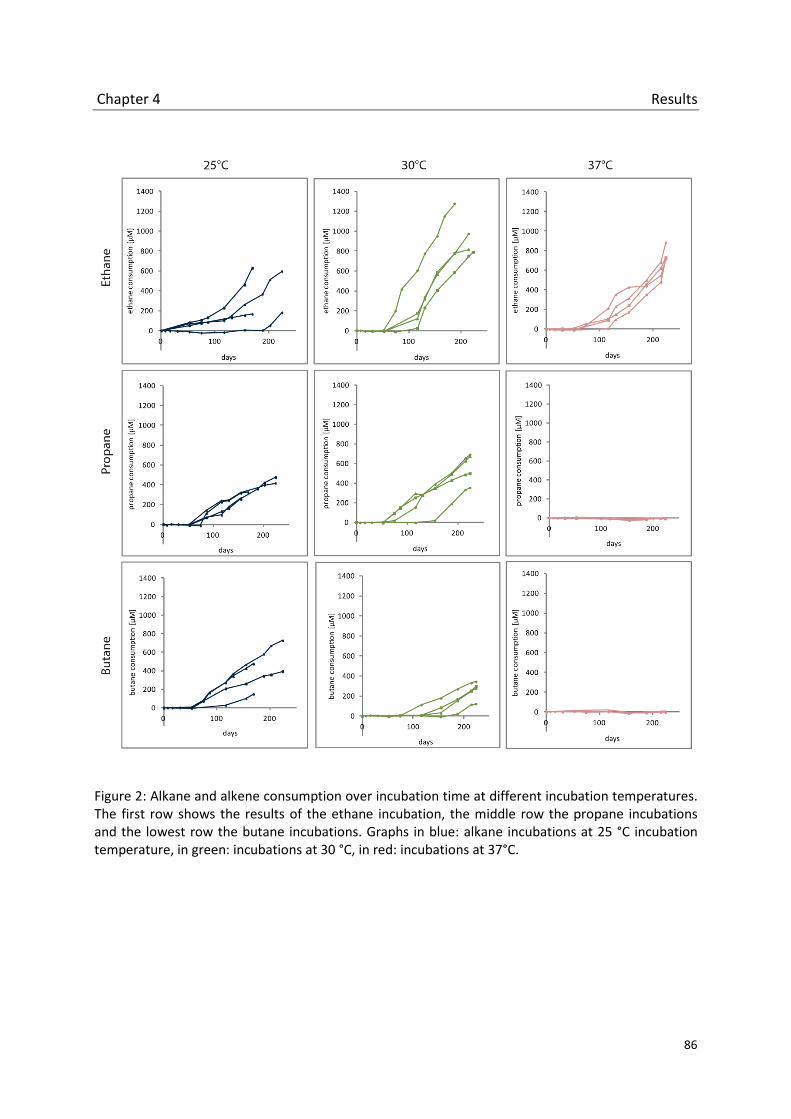
Chapter 4 Results
86
Figure 2: Alkane and alkene consumption over incubation time at different incubation temperatures. The first row shows the results of the ethane incubation, the middle row the propane incubations and the lowest row the butane incubations. Graphs in blue: alkane incubations at 25 °C incubation temperature, in green: incubations at 30 °C, in red: incubations at 37°C.

Chapter 4 Results
87
T-RFLP Analyses
To study the changes of the bacterial community composition in methane, ethane, propane
and butane incubations over time, T-RFLP analyses were performed. Primers were used to
target the 16S rRNA gene and the pmoA, pxmA and alkB genes. Additionally, pmoA
transcripts were analyzed by T-RFLP.
T-RFLP analyses of the 16S rRNA gene resulted in very diverse T-RFLP patterns (data not
shown). In methane incubations three fragments were dominant after 17 days of incubation:
117bp, 125bp and 146bp. After 41 days the fragment 146bp dominated. This fragment was
also prominent in ethane incubations though other fragments appeared and disappeared
during incubation (e.g. 135bp, 438 bp). The fragment 125bp that was also prominent in
methane incubations after 168 days. Towards the end of the incubation the T-RFLP pattern
became more diverse. Several new fragments appeared (e.g. 55bp, 60bp, 156bp). T-RFLP
pattern of the propane and butane incubations were even more diverse. Though the known
fragments from methane and ethane incubation (126bp, 135 bp, 147bp and 156bp) also
appear in propane and butane incubations lots of other fragments, sometimes only in small
percentages, came up during the course of the experiment. The high quantity of fragments
made it difficult to evaluate the 16S rRNA gene T-RFLP. Additionally, TRFs could not be
assigned to corresponding OTUs on the basis of the 16S rRNA gene sequences retrieved from
pyrosequencing analysis (see below: Pyrosequencing Analysis). However, the T-RFLP
analyses showed that the methane incubation was mainly dominated by a few OTUs that
also appeared in the incubation with the other alkanes.

Chapter 4 Results
88
The T-RFLP analysis of the alkB gene revealed three prominent fragments of the size 128bp,
137bp and 230bp (data not shown). Especially, the propane and butane incubations were
mainly dominated by those three fragments. Methane and ethane incubations showed a
wider range of fragments (e.g. 66bp, 100bp and 196bp), though the three fragments 128bp,
137bp and 230bp were also present. Until now, no clone library is available to evaluate the
alkB fragments and assign them to microorganisms. However, the T-RFLP results lead to the
assumption that the diversity of organisms possessing the alkB is higher in methane and
ethane grown samples than in propane and butane grown samples.
Figure 3 shows the summary of the results of the pmoA T-RFLP analyses. The most
prominent fragments in most samples had the length of 245bp or 352bp. OTUs were
assigned based on clones and pyrosequencing analyses (Lüke et al., 2011, Lüke & Frenzel,
2011, Yimga et al., 2003). The fragment 245bp can be assigned to the pmoA of type II
methanotrophs Methylocystis and Methylosinus. The other dominant fragment of the size
352bp can be either assigned to the pmoA of type I methanotrophs or the pmoA2 of
Methylocystis and Methylosinus. Fragment 536bp is assigned to type I methanotrophs.
The fragment pattern from methane grown samples stayed relative constant after 17d and
41d of incubation. The samples seem to be dominated by type II methanotrophs and type I
methanotrophs. In ethane incubations the relative composition of fragments changed over
time. The high relative abundance of the fragments 245bp (Methylocystis and Methylosinus)
and 352bp (Type I methanotrophs or isoenzyme pmoA2 of type II methanotrophs) seem to
decrease in favor of the abundance of the fragments 236bp, 448bp, 509bp and 536bp (Type I
methanotrophs). The fragments 236bp, 448bp and 509bp could not be related to any known
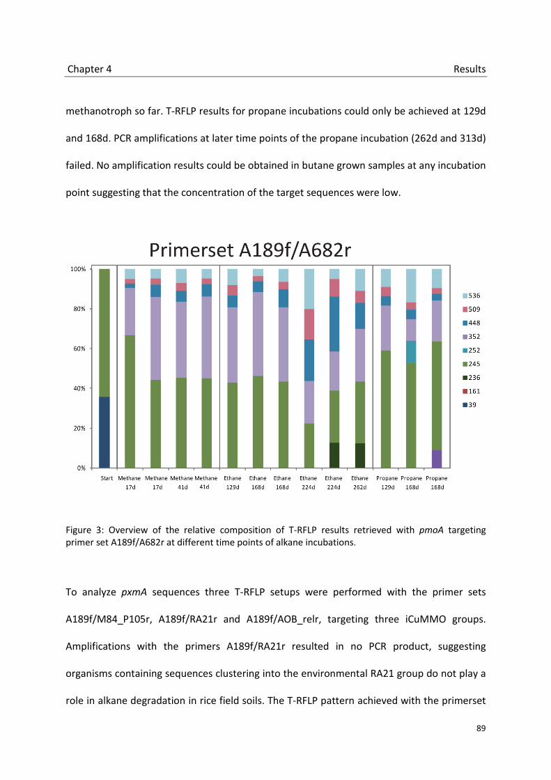
Chapter 4 Results
89
methanotroph so far. T-RFLP results for propane incubations could only be achieved at 129d
and 168d. PCR amplifications at later time points of the propane incubation (262d and 313d)
failed. No amplification results could be obtained in butane grown samples at any incubation
point suggesting that the concentration of the target sequences were low.
Figure 3: Overview of the relative composition of T-RFLP results retrieved with pmoA targeting primer set A189f/A682r at different time points of alkane incubations.
To analyze pxmA sequences three T-RFLP setups were performed with the primer sets
A189f/M84_P105r, A189f/RA21r and A189f/AOB_relr, targeting three iCuMMO groups.
Amplifications with the primers A189f/RA21r resulted in no PCR product, suggesting
organisms containing sequences clustering into the environmental RA21 group do not play a
role in alkane degradation in rice field soils. The T-RFLP pattern achieved with the primerset

Chapter 4 Results
90
A189f/AOB_relr in samples grown with methane or ethane was dominated by two
fragments: 245bp and 350bp (data not shown). The fragment pattern was very similar to the
pattern resulted in amplification with the pmoA primer set A189f/A682r. Previous deep
sequencing studies (Hainbuch et al., in prep.) showed that the primer pair A189f/AOB_relr is
not specific to sequences of the iCuMMO group AOB_rel but can also amplify sequences
belonging to other iCuMMO groups and pmoA sequences of type Ib and type II
methanotrophs. On the basis of previous deep sequencing studies (Chapter 1) the fragments
245bp and 350bp could not be assigned to the AOB_rel group. So, it is likely that these
fragments do not represent sequences of the iCuMMO group AOB_rel but co amplified
sequences of type II methanotrophs. Fragment 245bp can be assigned to the pmoA of
Methylocystis and Methylosinus and 352bp the pmoA2 that is present in several type II
methanotrophs or type I methanotrophs.
Figure 4 shows an overview of the T-RFLP results of pxmA sequences obtained with the
primerset A189f/M84_P105r. The fragment patterns from methane grown samples were
similar after 17 days and 41 days. Based on pyrosequencing results (Hainbuch et al., in
revision) the dominant fragment 33bp could be related to sequences of the iCuMMO group
M84_P105 though this fragment is not exclusively restricted to the M84_P105 groups but
could represent type I and II methanotrophs and other iCuMMO groups, too. The other
three fragments 225bp, 374bp and 426bp could also be assigned to M84_P105 sequences
but additionally to sequences of type I methanotrophs and pmoA2 sequences of type II
methanotrophs. In ethane grown samples the fragment diversity increased. The most
prominent new fragment 350bp could not be assigned to any known M84_P105 sequences

Chapter 4 Results
91
but to type I methanotrophs or pmoA2 sequences. T-RFLP results of propane grown samples
could be obtained from incubation after 129 and 168 days of incubation. As in the T-RFLP
results of the pmoA (figure 3) no PCR product could be amplified at later time point of the
propane incubation suggesting low template concentrations or no templates in those
samples. The fragment pattern of the propane incubation is similar to the pattern of the
methane incubation with the prominent fragments 33bp, 225bp, 374bp and 436bp though
some less abundant additional fragment appeared.
Figure 4: Overview of the relative composition of T-RFLP results retrieved with the primer set A189f/M84_P105r at different time points of alkane incubations.
Transcriptional T-RFLP analyses were performed for pmoA, pxmA and alkB sequences with
the same primer sets that were used on DNA level (A189f/A682r, A189f/M84_P105r,

Chapter 4 Results
92
A189f/RA21r, A189f/AOB_relr and AlkBf/AlkBr) in all alkane incubations at different time
points. mRNA transcripts could be detected exclusively with the pmoA targeting
A189f/A682r primers. Neither the use of the primers A189f/M84_P105r, A189f/RA21r,
A189f/AOB_relr targeting the sequences of the iCuMMO groups nor the use of the primers
targeting alkB sequence resulted in amplicons. Results of the mRNA T-RFLP amplified with
pmoA targeting primers are shown in figure 5. The most prominent fragments on mRNA
basis (245bp and 352bp) accord to the most frequent fragments that could be detected on
DNA level using the A189f/A682r primer pair. The fragment pattern in samples grown with
methane stayed constant at day 17 and 41. The same fragment pattern could be detected in
ethane incubations after 168 and 224 days. In propane grown samples the transcription was
low at day 168.

Chapter 4 Results
93
Figure 5: Overview of the relative composition of T-RFLP results retrieved from transcriptional analyses with the primer set A189f/M84_P105r at different time points of alkane incubations. A: methane incubation (17 and 41 days). B: ethane incubation (129, 168, 224 and 262 days). C: propane incubation (168 days). Number in blue: fragment sizes in bp.

Chapter 4 Results
94
Deep sequencing analysis
T-RFLP analyses gave an overall impression of the temporal change in the bacterial
community composition in methane, ethane, propane and butane incubations. Yet, deep
sequencing analysis declares for much finer insights in the composition. Primers targeting
the pmoA (A189f/A682r), the pxmA of the M84_P105 group (A189f/M84_P105r), the alkB
(AlkBf/AlkB) and the 16S rRNA gene (343Fmod/748Rmod) were used for deep sequencing
analysis. The analysis was performed at a late time point of the alkane incubation. It resulted
in about 58500 sequences. After first quality checks 13853 sequences of appropriate length
(pmoA, pxmA and 16S rRNA gene: >400 bp, AlkB: >100 bp) remained for further quality
verification. After sequence aligning in total 9971 high quality sequences remained for
phylogenetic analysis.
Figure 6 shows sequences retrieved by deep sequencing with the primer pairs A189f/A682r
and A189f/M84_P105r. Most sequences amplified by the A189f/M84_P105r primerset
clustered within the G5 subgroup of the iCuMMO M84_P105 in all alkane incubations. A
small portion of sequences (3-5%) clustered into the pmoA2 group of the type II
methanotrophs. 0.2 % of sequences of the ethane incubation clustered into the G4
subgroup of the iCuMMO M84_P105. The pmoA analysis was dominated by sequences
clustering within groups of type II methanotrophs. At incubation start type II sequences
contributed to 76.4 % to the total amount of sequences. 22.2 % clustered into the group of
ammonium oxidizers (AOB) and 1.4% into the group of the Type Ib methanotrophs. The
relative proportion of type II sequences increased in methane incubation to 90.1 % and to

Chapter 4 Results
95
100.0 % in ethane incubation. No sequences could be obtained from propane incubation
with the pmoA primerset.
Figure 6: Deep sequencing analysis targeting the pmoA and the pxmA. A: Phylogenetic tree containing pmoA and pxmA sequences. Highlighted in dark grey: phylogenetic groups that contain sequences of the deep sequencing analysis. B: diagrams of sequences retrieved with the primer set A189f/A682r targeting the pmoA. Small numbers: numbers of sequences retrieved. C: diagrams of sequences retrieved with the primer set A189f/M84_P105r targeting the pxmA. Small numbers: numbers of sequences retrieved.
Deep sequencing analyses of the alkB gene resulted in 1740 sequences of which 1353
sequences could be assigned to the domain of the bacteria by the metagenome analyzer
MEGAN5 (Huson et al., 2011). While the percentage of sequences that could be assigned
was low in the start and methane grown samples (18.1 % and 13.3 %) the percentage
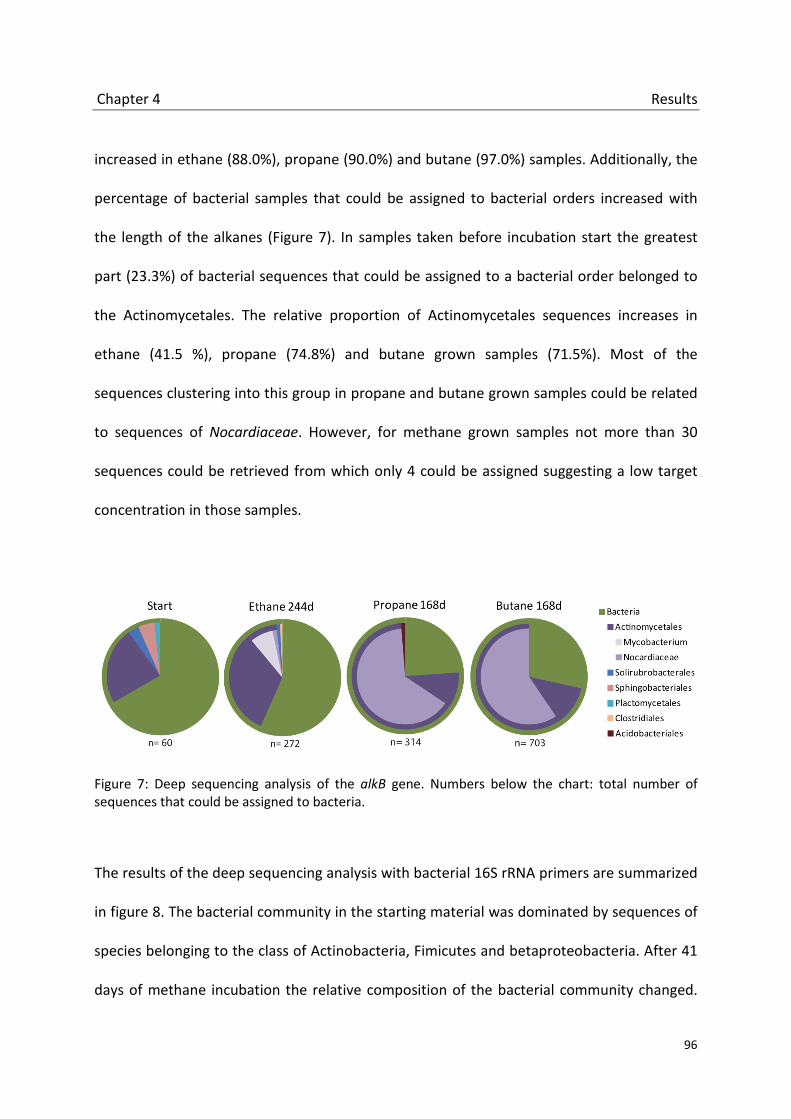
Chapter 4 Results
96
increased in ethane (88.0%), propane (90.0%) and butane (97.0%) samples. Additionally, the
percentage of bacterial samples that could be assigned to bacterial orders increased with
the length of the alkanes (Figure 7). In samples taken before incubation start the greatest
part (23.3%) of bacterial sequences that could be assigned to a bacterial order belonged to
the Actinomycetales. The relative proportion of Actinomycetales sequences increases in
ethane (41.5 %), propane (74.8%) and butane grown samples (71.5%). Most of the
sequences clustering into this group in propane and butane grown samples could be related
to sequences of Nocardiaceae. However, for methane grown samples not more than 30
sequences could be retrieved from which only 4 could be assigned suggesting a low target
concentration in those samples.
Figure 7: Deep sequencing analysis of the alkB gene. Numbers below the chart: total number of sequences that could be assigned to bacteria.
The results of the deep sequencing analysis with bacterial 16S rRNA primers are summarized
in figure 8. The bacterial community in the starting material was dominated by sequences of
species belonging to the class of Actinobacteria, Fimicutes and betaproteobacteria. After 41
days of methane incubation the relative composition of the bacterial community changed.

Chapter 4 Results
97
The samples grown with methane were dominated by two bacterial groups: the order of
Clostridiales and the family Methylocystaceae. Ethane, propane and butane grown samples
were more diverse, though the community composition of alkane incubations looked very
similar. Among the alkane incubations the main differences are primarily founded by the
relative distributions of the bacterial orders. The proportion of the proteobacterial TH18
cluster increased from few percentages in the starting material to 10 – 28 % in ethane,
propane and butane incubations. The relative proportion of betaproteobacteria decreased
while the alphaproteobacteria increased compared to the starting material. Though the
proportion of the alphaproteobacteria is similar in ethane, propane and butane grown
samples, the composition differs. In ethane grown samples sequences of the family
Methylocystaceae contribute to the portion of alphaproteobacteria sequences. But
Methylocystaceae sequences are infrequent in propane grown samples and not detectable
in samples grown with butane. The class of Actinobacteria that is very diverse in the staring
material is mainly dominated by the orders Frankiales and Corynebacteriales in ethane,
propane and butane grown samples. Sequences belonging to the phylum of the Firmicutes
are present in all samples. They are most dominant in samples grown with methane but also
present in ethane, propane and butane grown samples, though the relative proportion of
sequences of the order Bacillales increase compared to the order of Clostridiales. The
gammaproteobacterial Chromatiales only appear in butane grown samples.

Chapter 4 Discussion
98
Figure 8: Deep sequencing analysis based on the bacterial 16S rRNA gene.
4.5 Discussion
Gaseous hydrocarbons are ubiquitous in nature. They are part of natural gases formed by
geochemical processes or produced by microorganisms, marine algae, insects and plants as
moisture barriers, reserve materials and pheromones in most soil and water environments
(Cooley et al., 2009, Shennan, 2006, Giebler et al., 2013, Nie et al., 2014, van Beilen &
Funhoff, 2007). In this study the bacterial community involved in the degradation of short
chained gaseous hydrocarbons in a rice field soil was analyzed. Thereby, a special focus was
set on the methanotrophic community, on microorganisms possessing methane
monooxygenase isoenzymes belonging to the iCuMMO groups and on bacteria utilizing the
alk enzyme system to degrade alkanes and alkenes.

Chapter 4 Discussion
99
Methanotrophic bacteria involved in alkane degradation
In this study, the bacterial communities were analyzed by two methods: T-RFLP and deep
sequencing. T-RFLP is a robust and reproducible fingerprinting method that has been used in
a variety of ecological studies to analyze methanotrophic communities (Lüke et al., 2011,
Horz et al., 2001). T-RFLP results indicate that a great part of the methanotrophic community
in methane, ethane and propane incubations persist of species of the type II methanotrophic
genera Methylocystis and Methylosinus. Fingerprinting methods like T-RFLP are sometimes
limited in phylogenetic resolution and differentiation. Also in this study, several T-RFs could
not or not clearly be affiliated to one specific methanotrophic group. A dominant T-RF
(352bp), for example, represents both the pMMO of type I methanotrophs and the pMMO2
isoenzyme of type II methanotrophs. However, the supposed dominance of type II
methanotrophs based on T-RFLP data could be verified by deep sequencing analysis both on
pmoA and 16S rRNA level.
The deep sequencing analysis of pmoA and the bacterial 16S rRNA gene provided a large
dataset of high-quality sequences and gave a deep insight into the methanotrophic
community: Most pmoA gene sequences could be assigned to type II methanotrophs (start:
76.4 %, methane: 90.1 %, ethane: 100%, propane: no results). Additionally, almost all
16S rRNA sequences that could be assigned to methanotrophic bacteria belonged to the
alphaproteobacterial family Methylocystaceae that contains amongst others the genera
Methylocystis and Methylosinus.
The alphaproteobacterial type II methanotrophs are ubiquitous in most environments. Due
to their ability of stress tolerance they are thought to be very stable and able to form

Chapter 4 Discussion
100
ubiquitous seed banks in soil environments (Eller et al., 2005, Krause et al., 2012, Ho &
Frenzel, 2012, Graham et al., 1993). However, despite their wide distribution active type II
methanotrophic communities could only be shown in few studies. The activity of
methanotrophs has been monitored in a variety of environmental studies by transcriptional
gene analysis or stable isotope probing (SIP). Both methods seem to be largely congruent
and seem to illustrate the activity of the methanotrophs in a similar dimension (Henneberger
et al., 2014). In most studies based on gene transcript analyses, pmoA transcripts of type I
methanotrophs dominated while transcripts of type II methanotrophs could not or only be
retrieved in low levels (Bodrossy et al., 2006, Krause et al., 2010, Krause et al., 2012, Liebner
et al., 2009). The active methanotrophic community detected by SIP experiments in varies
habitats was dominated by type I methanotrophs, too (Qiu et al., 2008, Dumont et al., 2011,
Graef et al., 2011). In this study the transcription of pmoA was taken as a proxy for activity
and was analyzed by T-RFLP (Figure 5). Transcripts of the pmoA could be detected in
methane, ethane and propane samples and could be related to the pmoA and pmoA2 of
type II methanotrophs.
In methane grown samples active methanotrophs were detected after 17 and 41 days of
incubation. It was shown in previous studies that type II methanotrophs became active after
long term incubations of 30 days though the transcription level was low (Krause et al., 2012).
Another study shows a dominance of active type II methanotrophs after 40 days of
incubation after biomass destruction (Ho et al., 2011).In ethane and propane incubations the
relative proportion of methanotrophic bacteria seems to be rather low. 6.6 % and 0.02 % of
the bacterial 16S rRNA sequences retrieved by deep sequencing could be assigned to

Chapter 4 Discussion
101
methanotrophic bacteria. However, the proportion increased compared to the starting
samples (no sequences of methanotrophic bacteria could be detected). Transcripts of pmoA
and pmoA2 of type II methanotrophs could be detected in ethane grown samples and in very
low transcripts levels in propane incubations, too. The II methanotrophs seem to play a
dominant role within the methanotrophic community in long time methane incubation in
environments where short alkanes like ethane and, to a minor part, propane are available as
substrate. However, longer alkanes like butane do not seem to affect bacterial
methanotrophic communities.
It has been believed for a long time that methanotrophic bacteria are obligate and that they
could grow exclusively on methane, methanol and some other C1 compounds (Bowman,
2006). But it is known that at least some methanotrophs grow on substrates with carbon-
carbon bounds like acetate, pyruvate, succinate, malate and ethanol (Dedysh et al., 2005,
Dunfield et al., 2010, Belova et al., 2011, Im et al., 2011). Facultative type II methanotrophs
could to be involved in the degradation of alkanes in this study, too. A transcription of pmoA
and pxmA of type II methanotrophs was monitored. But it seems unlikely that the
corresponding pMMOs are involved in the alkane oxidation. pMMOs have a narrow
substrate range. They are known to be able to co-oxidize several other hydrocarbons like
ethane (e.g. Chapter 2) but these substrates are usually not assimilated and used as a
growth substrate by methanotrophs possessing the pMMO. It could be possible that the
soluble methane monooxygenase (sMMO) is involved in alkane degradation by the type II
methanotrophs. The sMMO has a wider substrate range than the pMMO. But further studies
Chapter 4 Discussion
102
need to confirm this assumption because the analysis of the sMMO was not a in the focus of
this study.
However, until now no methanotrophic bacterium could be detected that grow on ethane or
propane as a carbon source. It could be possible that type II methanotrophs in this study do
not directly grow on short alkanes. Methanotrophs have been found in close interactions
with other organisms such as plants and algae (Kip et al., 2010, van der Ha et al., 2011) or
other bacteria (Modin et al., 2007, van der Ha et al., 2013, Hrsak & Begonja, 2000, Iguchi et
al., 2011). Besides abiotic factors like oxygen, nutrients, moisture and temperature that
affect methanotrophic activity (Conrad, 2007, Hanson & Hanson, 1996, Krause et al., 2012,
Reim et al., 2012) other bacteria interact with methanotrophs significantly. While providing
carbon compounds derived from methane, methanotrophs benefit in return from other
bacteria by the remove of toxic waste product from methane oxidation, the providing of
essential metabolites and the creation of a stable environment with ideal gas composition
(Dumont et al., 2011, Murase & Frenzel, 2007, Petersen & Dubilier, 2009, Hanson & Hanson,
1996, Iguchi et al., 2011, Stock et al., 2013, Ho et al., 2014). To date, several bacteria,
including strains of Xanthobacter (Lidstromoconnor et al., 1983, Uchiyama et al., 1992),
Hyphomicrobia (Bowman, 2006), Rhizobium (Iguchi et al., 2011) and Spingopyxis (Jeong et
al., 2014) have been found to life in close relationship with methanotrophs or stimulate their
growth. These bacteria belong to orders that are known to include bacterial families that are
able to oxidize short chained alkanes like ethane and propane. In a bacterial consortium, the
heterotrophic bacteria could provide metabolic products like ethanol that may be used as
growth substrate by facultative methanotrophs like type II Methylocystis strains (Im et al.,

Chapter 4 Discussion
103
2011). In this study, for example, 16S rRNA sequences in ethane and propane incubation
could be related to Xanthobacter strains. Hence, consortia might be possible. However,
those assumptions are speculative and cannot be verified by the results of this study. A
stable isotope study with labeled 13C alkanes might give the yet unproven insights into the
degradation pathways of short gaseous alkanes in rice fields and could indicate the role of
the methanotrophic bacteria within this pathway.
iCuMMOs seem to play no role in the degradation of alkanes and alkenes in rice field soils.
The iCuMMO groups RA21, AOB_rel and M84_P105 were analyzed by T-RFLP in methane,
ethane, propane and butane samples. No amplification products could be found with
primers targeting the AOB_rel and the RA21. Sequences of the iCuMMO group M84_P105
could be detected by T-RFLP and pyrosequencing. Most M84_P105 sequences clustered into
the subgroup 5 that contains pxmA sequences of two Methylocystis strains: Methylocystis
sp. SB2 and Methylocystis rosea. Few sequences clustered within the pmoA2 group of the
type II methanotrophs. But no transcripts could be amplified suggesting that the pXMO is
not involved in the degradation of alkanes and alkene, too. Enzymes of the three analyzed
iCuMMO groups seem to have other physiological functions than the oxidation of alkanes.
They might be involved in the methane oxidation like the pMMO2 of the type II
methanotrophs and may provide selective advantages in environments with fluctuating
nutrient availabilities (e.g. low oxygen concentrations). However, the physiological functions
could not be resolved until now.

Chapter 4 Discussion
104
Alkane degrading bacteria
Compared to researches on methanotrophic bacteria, the interest in microorganisms able to
metabolize short chained gaseous alkanes has been rather low for a long time. However,
new prospects in biotechnological fields like biotransformation and remedial treatments
have lead to an extended interest in alkane degrading bacteria (ADB). Lots of studies that
focus on alkane degrading bacteria in nature have been obtained in oil contaminated and
marine environments, few in soil environments (Sutton et al., 2013, Brakstad & Lodeng,
2005, Sabirova et al., 2006, Wang et al., 2010, Redmond & Valentine, 2012, Giebler et al.,
2013, Schulz et al., 2012). To detect ADB in rice field soils and to study their abundance and
diversity, the marker gene alkB, coding the trans-membrane alkane monooxygenase of the
Alk enzyme system, has been used (Kuhn et al., 2009, Perez-de-Mora et al., 2011, Wang et
al., 2010). ADB were analyzed by alkB T-RFLP and pyrosequencing as well as 16S rRNA
pyrosequencing. The evaluation of T-RFLP results was utterly impossible due to a lack of an
appropriate clone library and the resulting inability to assigned T-RFs to ADB, but
pyrosequencing results gave an insight into the community composition. ADB virtually
played no role in rice field samples incubated with methane. This can be explained by the
fact that bacteria possessing the alk enzyme system or other alkane degrading systems like
bacterial P450 oxygenases or propane and butane monooxygenases are usually not able to
oxidize methane (van Beilen & Funhoff, 2005).
Most sequences retrieved by alkB pyrosequencing from ethane, propane and butane grown
samples could be assigned to the genera Mycobacterium and Nocardia that belong to the
Actinomycetales (Figure 7), a divers order of the Gram- positive Actinobacteria.
Chapter 4 Discussion
105
Mycobacterium seemed to be dominant in ethane, Nocardia in propane and butane
incubations. 16S rRNA pyrosequencing analyses confirmed these results. Mycobacterium and
Nocardia are members of the common alkane degrading CNMR group. In rice field soils the
members of the CNMR group do not seem to be very dominant (start: 3.4 %, ethane: 2.7%,
propane: 5.1 %, butane: 6.8%). Other bacterial groups were more frequent and increased
during alkane incubations. The order Rhizobiales with the families Bradyrhizobiaceae,
Xanthobacteraceae, Hyphomicrobiaceae and the environmental cluster DUNssu371
contributed to 9.2 % (ethane), 10.2% (propane) and 16.1 % (butane) to the total bacterial
community. The order Rhizobiales is known to contain species that possess the alk enzyme
system and that could therefore be involved in the degradation of short chained alkanes
(Ouattara et al., 2003, Maier et al., 1978, Malik & Claus, 1979). Bacillales sequences
belonging to the order of Firmicutes contribute to 5.3 % (ethane), 8.4 % (propane) and 5.5 %
(butane) to the bacterial community. The alk enzyme could be found in species of this order,
too (Tourova et al., 2008). Rhizobiales and Bacillales species could be detected in this study
only by 16S rRNA analysis. On the basis of the alkB marker gene no sequences could be
found that could be related to these bacterial orders. A possible explanation for the
discrepancy between 16S rRNA and alkB could be a detection limitation of alkB sequences
by the used primerset. The alkB gene sequences are very diverse among the ADB. This
diversity could hardly be covered by a single primer set. In this study, a primer set was
chosen that amplifies a great part of sequences of the alkB-containing bacterial community.
But it seems to lack alkB sequences of Rhizobiales and Bacillales species (Jurelevicius et al.,
2013, Kloos et al., 2006). To overcome the problem of sequence coverage a use of several
alkB primer sets covering different alkB-containing bacterial groups should be recommended

Chapter 4 Discussion
106
for further analyses of these genes. Although the relative proportion of alkB containing
bacteria like Mycobacterium, Nocardia, Rhizobiales and Bacillales increased in alkane
incubations over time, no alkB transcripts could be detected. An explanation could be that
bacteria possessing the alk enzyme system are involved in alkane degradation but use other
enzyme complexes to oxidize alkanes. Many hydrocarbon degrading bacteria are known to
contain multiple alkane hydroxylases with overlapping substrate ranges (Kotani et al., 2003,
Sabirova et al., 2006, van Beilen & Funhoff, 2007, van Beilen et al., 2003). Mycobacterium
strains, for example, are known to contain six different monooxygenases including alkB,
Cytochrome P450 and propane and butane monooxygenases (Coleman et al., 2011). It could
be possible that not the alk enzyme system but another monooxygenase is involved in the
degradation of short chained alkanes in rice fields.
Beside species of the orders Corynebacteriales, Rhizobiales and Bacillales that are known to
be involved in degradation of alkanes, species of other bacterial orders are also dominant in
ethane, propane and butane incubations, too (Figure 8). Surprisingly, in spite the fact that
the alkane grown samples were incubated aerobically, a great portion of sequences
retrieved by 16S rRNA deep sequencing could be assigned to the bacterial order of the
Clostridiales, a group of mainly anaerobe Gram-positive bacteria. One may speculate that
these bacteria could have grown in anoxic micro aggregates beside the aerobically growing
methanotrophs. During incubation the alkanes were oxidized rapidly and oxygen
concentrations sank low (concentrations of about 5%) which could have favored the growth
of the anaerobic Clostridiales and other anaerobic bacteria in anaerobic microstructures.

Chapter 4 Discussion
107
A great part of sequences clustered within the order of the Frankiales (start: 3.7%, ethane:
8.8%, propane: 23.1 %, butane: 14.7 %). Most of the sequences clustering within this order
could be assigned to the family Sporichtyaceae, facultative anaerobe chemoorganotrophic
bacteria that grow on complex hydrocarbons like humic acids (Williams et al., 1989, Suzuki et
al., 1999). Alkane degradation is not described for the Sporichtyaceae. The order of
Rhodospirillales is mainly represented by sequences assigned to environmental clusters (I-10
and DA111) related to sequences of the family Rhodospirillaceae (Ethane: 8.8%, Propane:
6.2 %, Butane: 6.6 %). The Rhodospirillaceae are bacteria that are able to photo assimilate
organic compounds under anaerobic conditions and are able of chemotrophic growths under
aerobic conditions. Another group of sequences that contributed to a great part of the
bacterial community clustered within the proteobacterial environmental cluster TH18 (start:
1.5 %, ethane: 12.3 %, propane: 23.2%, butane: 27.3%). Sequences of this cluster has been
found in a variety of environments (Elshahed et al., 2008, Forget et al., 2010, Isenbarger et
al., 2008, Lesaulnier et al., 2008, Riviere et al., 2009, Santelli et al., 2008, Zhang et al., 2010).
But so far no representative pure cultures and thereby no information about physiology of
the corresponding organisms are available. It could be possible that TA18 proteobacteria are
involved in alkane degradation.
Alkene degraders
In this study a focus was set on the analysis of the degradation of alkanes in a rice field soil
and the analysis of the involved bacterial community. But additional incubations were
performed with alkenes as substrates: Ethylene, propylene and butylene. Alkenes are
oxidized by monooxygenases forming epoxy-alkanes, highly reactive products that are toxic

Chapter 4 Discussion
108
to cells. But several microorganisms are able to rapidly eliminate those intermediate
products and are thereby able to grow on alkenes. Common species that metabolize short
chained alkenes belong to the genera Mycobacterium, Nocardia, Rhodococcus,
Pseudomonas and Xanthobacter (Debont, 1976, Furuhashi et al., 1981, Habetscrutzen et al.,
1984, Saeki et al., 1999, Vanginkel et al., 1987, Verce et al., 2000, Vlieg et al., 1998, Shennan,
2006). Alkane incubation showed that members of some of these genera are present in rice
field soils and that they were able to be enriched by alkane incubations. However, no
oxidation of alkenes could be detected during about 350 days of incubation. Though the
genera containing alkene degrading bacteria are present, the alkene degraders of those
groups are absent or not active in rice field soils.
Summery and conclusion
Among the methanotrophic bacteria the type II methanotrophs are dominant in long term
methane incubations as well as in ethane and propane incubations. It could be shown that
the community is active and not only present in a dormant stadium. Methanotrophs seem to
be involved in ethane and propane degradation in rice field soils, whereat it can be
speculated if they use alkanes directly or use metabolic substrates provided by
heterotrophic bacteria. As a part of a consortium the methanotrophs may play an important
role in the degradation of short chained alkanes in rice field soils. Important heterotrophic
bacteria that might be involved in alkane degradation belong to orders of the
Corynebacteriales, Rhizobiales and Bacillales. If those bacteria use the alk enzyme system or
other alkane degrading enzymes systems like Cytochrome P450 or propane or butane

Chapter 4 Discussion
109
monooxygenases could not be clearly explained certainly. The oxidation of hydrocarbons in
rice field soil and in general may be more important than previously assumed. Further
analysis (e.g. SIP studies) could help to get a better understanding of the processes and
microorganisms involved in alkane degradation and could help to show another aspect of
role of methanotrophs in this environment that seems to be not only restricted to methane
oxidation.

Chapter 5 References
110
4.4 References
Altschul, S. F., W. Gish, W. Miller, E.W. Myers & D.J. Lipman, (1990) Basic local alignment search tool. J. Mol. Biol. 215, 403–410. doi: 10.1006/jmbi.1990.9999
Baani, M. & W. Liesack, (2008) Two isozymes of particulate methane monooxygenase with different
methane oxidation kinetics are found in Methylocystis sp strain SC2. Proceedings of the Proc. Natl. Acad. Sci. U. S. A. 105: 10203-10208.
Belova, S.E., M. Baani, N.E. Suzina, P.L.E. Bodelier, W. Liesack & S.N. Dedysh, (2011) Acetate
utilization as a survival strategy of peat-inhabiting Methylocystis spp. Environ. Microbiol. Rep. 3: 36-46.
Bodrossy, L., N. Stralis-Pavese, M. Konrad-Koszler, A. Weilharter, T.G. Reichenauer, D. Schofer & A.
Sessitsch, (2006) mRNA-based parallel detection of active methanotroph populations by use of a diagnostic microarray. Appl. Environ. Microbiol. 72: 1672-1676.
Bowman, J., (2006) The Methanotrophs. The Families Methylococcaceae and
Methylocystaceae. In The Prokaryotes. Dworkin,M. (ed). New York: Springer, 266- 289. Brakstad, O.G. & A.G. Lodeng, (2005) Microbial diversity during biodegradation of crude oil in
seawater from the North Sea. Microb. Ecol. 49: 94-103. Chen, Y., M.G. Dumont, A. Cebron & J.C. Murrell, (2007) Identification of active methanotrophs in a
landfill cover soil through detection of expression of 16S rRNA and functional genes. Environ. Microbiol. 9: 2855-2869.
Chen, Y., M.G. Dumont, N.P. McNamara, P.M. Chamberlain, L. Bodrossy, N. Stralis-Pavese & J.C.
Murrell, (2008) Diversity of the active methanotrophic community in acidic peatlands as assessed by mRNA and SIP-PLFA analyses. Environ. Microbiol. 10: 446-459.
Cheng, Y.Q., L.Z. Yang, Z.H. Cao, E. Ci & S.X. Yin, (2009) Chronosequential changes of selected
pedogenic properties in paddy soils as compared with non-paddy soils. Geoderma 151: 31-41.
Coleman, N.V., N.B. Le, M.A. Ly, H.E. Ogawa, V. McCarl, N.L. Wilson & A.J. Holmes, (2011a)
Hydrocarbon monooxygenase in Mycobacterium: recombinant expression of a member of the ammonia monooxygenase superfamily. ISME J. 6: 171-182
Coleman, N.V., S. Yau, N.L. Wilson, L.M. Nolan, M.D. Migocki, M.a. Ly, B. Crossett & A.J. Holmes,
(2011b) Untangling the multiple monooxygenases of Mycobacterium chubuense strain NBB4, a versatile hydrocarbon degrader. Environ. Microbiol. Rep. 3: 297-307.
Conrad, R., (2007) Microbial ecology of methanogens and methanotrophs. Adv. Agro. 96: 1- 63. Conrad, R., (2009) The global methane cycle: recent advances in understanding the microbial
processes involved. Environ. Microbiol. Rep. 1: 285-292.

Chapter 5 References
111
Cooley, R.B., P.J. Bottomley & D.J. Arp, (2009) Growth of a non-methanotroph on natural gas: ignoring the obvious to focus on the obscure. Environ. Microbiol. Rep. 1: 408-413.
Debont, J.A.M., (1976) Oxidation of ethylene by soil bacteria. A. Van. Leeuw. J. Microb. 42: 59-71. Dedysh, S.N., C. Knief & P.F. Dunfield, (2005) Methylocella species are facultatively methanotrophic.
J. Bacteriol. 187: 4665-4670. Doughty, D.M., L.A. Sayavedra-Soto, D.J. Arp & P.J. Bottomley, (2006) Product repression of alkane
monooxygenase expression in Pseudomonas butanovora. J. Bacteriol. 188: 2586-2592. Dumont, M.G., B. Pommerenke, P. Casper & R. Conrad, (2011) DNA-, rRNA- and mRNA-based stable
isotope probing of aerobic methanotrophs in lake sediment. Environ. Microbiol. 13: 1153-1167.
Dunfield, P.F., S.E. Belova, A.V. Vorob'ev, S.L. Cornish & S.N. Dedysh, (2010) Methylocapsa aurea sp.
nov., a facultative methanotroph possessing a particulate methane monooxygenase, and emended description of the genus Methylocapsa. Int. J. Syst. Evol. Microbiol. 60: 2659-2664.
Eller, G., L. Kanel & M. Kruger, (2005) Cooccurrence of aerobic and anaerobic methane oxidation in
the water column of Lake Plusssee. Appl. Environ. Microbiol. 71: 8925-8928. Elshahed, M.S., N.H. Youssef, A.M. Spain, C. Sheik, F.Z. Najar, L.O. Sukharnikov, B.A. Roe, J.P. Davis,
P.D. Schloss, V.L. Bailey & L.R. Krumholz, (2008) Novelty and uniqueness patterns of rare members of the soil biosphere. Appl. Environ. Microbiol. 74: 5422-5428.
Forget, N.L., S.A. Murdock & S.K. Juniper, (2010) Bacterial diversity in Fe-rich hydrothermal sediments
at two South Tonga Arc submarine volcanoes. Geobiology 8: 417-432. Frenzel, P., (2000) Plant-associated methane oxidation in rice fields and wetlands. Adv. Microb. Ecol.
16: 85-114. Furuhashi, K., A. Taoka, S. Uchida, I. Karube & S. Suzuki, (1981) Production of 1,2-Epoxyalkanes from
1-Alkenes by Nocardia-Corallina B-276. Eur. J. Appl. Microbiol. 12: 39-45. Giebler, J., L.Y. Wick, A. Chatzinotas & H. Harms, (2013) Alkane-degrading bacteria at the soil-litter
interface: comparing isolates with T-RFLP-based community profiles. FEMS Microbiol. Ecol. 86: 45-58.
Graef, C., A.G. Hestnes, M.M. Svenning & P. Frenzel, (2011) The active methanotrophic community in
a wetland from the High Arctic. Environ. Microbiol. Rep.3: 466-472. Graham, D.W., J.A. Chaudhary, R.S. Hanson & R.G. Arnold, (1993) Factors affecting competition
between type I and type II methanotrophs in two-organism, continuous-flow reactors. Microb. Ecol. 25: 1-17.
Habetscrutzen, A.Q.H., L.E.S. Brink, C.G. Vanginkel, J.A.M. Debont & J. Tramper, (1984) Production of
epoxides from gaseous alkenes by restingcell suspensions and immobilized cells of alkene-utilizing bacteria. Appl. Microbiol. Biotechnol. 20: 245-250.

Chapter 5 References
112
Hamamura, N., C.M. Yeager & D.J. Arp, (2001) Two distinct monooxygenases for alkane oxidation in Nocardioides sp. strain CF8. Appl. Environ. Microbiol. 67: 4992-4998.
Hanson, R.S. & T.E. Hanson, (1996) Methanotrophic bacteria. Microbiol. Rev. 60: 439. Harayama, S., Y. Kasai & A. Hara, (2004) Microbial communities in oil-contaminated seawater. Curr.
Opin. Biotechnol. 15: 205-214. Henneberger, R., E. Chiri, P.E. Bodelier, P. Frenzel, C. Luke & M.H. Schroth, (2014) Field-scale tracking
of active methane-oxidizing communities in a landfill cover soil reveals spatial and seasonal variability. Environ. Microbiol. doi: 10.1111/1462-2920.12617.
Ho, A., K. de Roy, O. Thas, J. De Neve, S. Hoefman, P. Vandamme, K. Heylen & N. Boon, (2014) The
more, the merrier: heterotroph richness stimulates methanotrophic activity. ISME J. 8: 1945-1948.
Ho, A. & P. Frenzel, (2012) Heat stress and methane oxidizing bacteria: Effects on activity and
population dynamics. Soil Biol. Biochem. 50: 22-25. Ho, A., C. Luke, A. Reim & P. Frenzel, (2013) Selective stimulation in a natural community of methane
oxidizing bacteria: Effects of copper on pmoA transcription and activity. Soil Biol. Biochem. 65: 211-216.
Ho, A., C. Lüke, Z.H. Cao & P. Frenzel, (2011) Ageing well: methane oxidation and methane oxidizing
bacteria along a chronosequence of 2000 years. Environ. Microbiol. Rep. 3: 738-743. Holmes, A.J., A. Costello, M.E. Lidstrom & J.C. Murrell, (1995) Evidence that particulate methane
monooxygenase and ammonia monooxygenase may be evolutionarily related. FEMS Microbiol. Lett. 132: 203-208.
Horz, H.P., M.T. Yimga & W. Liesack, (2001) Detection of methanotroph diversity on roots of
submerged rice plants by molecular retrieval of pmoA, mmoX, mxaF, and 16S rRNA and ribosomal DNA, including pmoA-based terminal restriction fragment length polymorphism profiling. Appl. Environ. Microbiol. 67: 4177-4185.
Hrsak, D. & A. Begonja, (2000) Possible interactions within a methanotrophic-heterotrophic
groundwater community able to transform linear alkylbenzenesulfonates. Appl. Environ. Microbiol. 66: 4433-4439.
Huson, D. H., S. Mitra, H.J. Ruscheweyh, N. Weber & S.C. Schuster, (2011) Integrative analysis of
environmental sequences using MEGAN4. Genome. Res. 21: 1552–1560. doi: 10.1101/gr.120618.111
Iguchi, H., H. Yurimoto & Y. Sakai, (2011) Stimulation of methanotrophic growth in cocultures by
cobalamin excreted by Rhizobia. Appl. Environ. Microbiol. 77: 8509-8515. Im, J., S.W. Lee, S. Yoon, A.A. DiSpirito & J.D. Semrau, (2011) Characterization of a novel facultative
Methylocystis species capable of growth on methane, acetate and ethanol. Environ. Microbiol. Rep. 3: 174-181.
Chapter 5 References
113
Isenbarger, T.A., M. Finney, C. Rios-Velazquez, J. Handelsman & G. Ruvkun, (2008) Miniprimer PCR, a new lens for viewing the microbial world. Appl. Environ. Microbiol. 74: 840-849.
Jeong, S.Y., K. Cho & T.G. Kim, (2014) Density-dependent enhancement of methane oxidation activity
and growth of Methylocystis sp. by a non methanotrophic bacterium Sphingopyxis sp. Biotech. Rep. 4: 128-133.
Johnson, E.L. & M.R. Hyman, (2006) Propane and n-butane oxidation by Pseudomonas putida GPo1.
Appl. Environ. Microbiol. 72: 950-952. Jurelevicius, D., V.M. Alvarez, R. Peixoto, A.S. Rosado & L. Seldin, (2013) The use of a combination of
alkB primers to better characterize the distribution of alkane degrading bacteria. Plos One 8. doi: 10.1371/journal.pone.0066565
Kip, N., J.F. van Winden, Y. Pan, L. Bodrossy, G.J. Reichart, A.J.P. Smolders, M.S.M. Jetten, J.S.S.
Damste & H.J.M. Op den Camp, (2010) Global prevalence of methane oxidation by symbiotic bacteria in peat-moss ecosystems. Nat. Geosci. 3: 617-621.
Kloos, K., J.C. Munch & M. Schloter, (2006) A new method for the detection of alkane-
monooxygenase homologous genes (alkB) in soils based on PCR-hybridization. J. Microbiol. Methods. 66: 486-496.
Kolbl, A., P. Schad, R. Jahn, W. Amelung, A. Bannert, Z.H. Cao, S. Fiedler, K. Kalbitz, E. Lehndorff, C.
Muller-Niggemann, M. Schloter, L. Schwark, V. Vogelsang, L. Wissing & I. Kogel-Knabner, (2014) Accelerated soil formation due to paddy management on marshlands (Zhejiang Province, China). Geoderma 228: 67-89.
Kohler, T., C. Dietrich, R.H. Scheffrahn & A. Brune, (2012) High-resolution analysis of gut Environment
and bacterial microbiota reveals functional compartmentation of the gut in wood feeding hgher termites (Nasutitermes spp.). Appl. Environ. Microbiol. 78: 4691-4701.
Kotani, T., T. Yamamoto, H. Yurimoto, Y. Sakai & N. Kato, (2003) Propane monooxygenase and
NAD(+)-dependent secondary alcohol dehydrogenase in propane metabolism by Gordonia sp strain TY-5. J. Bacteriol. 185: 7120-7128.
Krause, S., C. Lüke & P. Frenzel, (2010) Succession of methanotrophs in oxygen-methane counter-
gradients of flooded rice paddies. ISME J. 4: 1603-1607. Krause, S., C. Lüke & P. Frenzel, (2012) Methane source strength and energy flow shape
methanotrophic communities in oxygen-methane counter-gradients. Environ. Microbiol. Rep. 4: 203-208.
Kuhn, E., G.S. Bellicanta & V.H. Pellizari, (2009) New alk genes detected in Antarctic marine
sediments. Environ. Microbiol. 11: 669-673. Labinger, J.A. & J.E. Bercaw, (2002) Understanding and exploiting C-H bond activation. Nature 417:
507-514.

Chapter 5 References
114
Lesaulnier, C., D. Papamichail, S. McCorkle, B. Ollivier, S. Skiena, S. Taghavi, D. Zak & D. van der Lelie, (2008) Elevated atmospheric CO2 affects soil microbial diversity associated with trembling aspen. Environ. Microbiol. 10: 926-941.
Lidstromoconnor, M.E., G.L. Fulton & A.E. Wopat, (1983) Methylobacterium-Ethanolicum - a
Syntrophic Association of 2 Methylotrophic Bacteria. J. Gen. Microbiol. 129: 3139-3148. Liebner, S., K. Rublack, T. Stuehrmann & D. Wagner, (2009) Diversity of Aerobic Methanotrophic
Bacteria in a Permafrost Active Layer Soil of the Lena Delta, Siberia. Microb. Ecol. 57: 25-35. Ludwig, W., O. Strunk, R. Westram, L. Richter, H. Meier, Yadhukumar, A. Buchner, T. Lai, S. Steppi, G.
Jobb, W. Forster, I. Brettske, S. Gerber, A.W. Ginhart, O. Gross, S. Grumann, S. Hermann, R. Jost, A. Konig, T. Liss, R. Lussmann, M. May, B. Nonhoff, B. Reichel, R. Strehlow, A. Stamatakis, N. Stuckmann, A. Vilbig, M. Lenke, T. Ludwig, A. Bode & K.H. Schleifer, (2004) ARB: a software environment for sequence data. Nucleic. Acids. Res. 32: 1363-1371.
Lüke, C., L. Bodrossy, E. Lupotto & P. Frenzel, (2011) Methanotrophic bacteria associated to rice
roots: the cultivar effect assessed by T-RFLP and microarray analysis. Environ. Microbiol. Rep. 3: 518-525.
Lüke, C. & P. Frenzel, (2011) Potential of pmoA Amplicon Pyrosequencing for Methanotroph Diversity
Studies. Appl. Environ. Microbiol. 77: 6305-6309. Lüke, C., P. Frenzel, A. Ho, D. Fiantis, P. Schad, B. Schneider, L. Schwark & S.R. Utami, (2014)
Macroecology of methane-oxidizing bacteria: the beta-diversity of pmoA genotypes in tropical and subtropical rice paddies. Environ. Microbiol. 16: 72-83.
Maier, R.J., N.E.R. Campbell, F.J. Hanus, F.B. Simpson, S.A. Russell & H.J. Evans, (1978) Expression of
Hydrogenase Activity in Free-Living Rhizobium-Japonicum. Proceedings of the National Proc. Natl. Acad. Sci. U. S. A. 75: 3258-3262.
Malik, K.A. & D. Claus, (1979) Xanthobacter-Flavus, a New Species of Nitrogen-Fixing Hydrogen
Bacteria. Int. J. Syst. Bacteriol. 29: 283-287. Margesin, R., D. Labbe, F. Schinner, C.W. Greer & L.G. Whyte, (2003) Characterization of
hydrocarbon-degrading microbial populations in contaminated and pristine Alpine soils. Appl. Environ. Microbiol. 69: 3085-3092.
Modin, O., K. Fukushi & K. Yamamoto, (2007) Denitrification with methane as external carbon source.
Water Res. 41: 2726-2738. Murase, J. & P. Frenzel, (2007) A methane-driven microbial food web in a wetland rice soil. Environ.
Microbiol. 9: 3025-3034. Nie, Y., C.Q. Chi, H. Fang, J.L. Liang, S.L. Lu, G.L. Lai, Y.Q. Tang & X.L. Wu, (2014) Diverse alkane
hydroxylase genes in microorganisms and environments. Sci. Rep. Uk. 4. doi:10.1038/srep04968
Chapter 5 References
115
Ouattara, A.S., E.A. Assih, S. Thierry, J.L. Cayol, M. Labat, O. Monroy & H. Macarie, (2003) Bosea minatitlanensis sp nov., a strictly aerobic bacterium isolated from an anaerobic digester. Int. J. Syst. Evol. Microbiol. 53: 1247-1251.
Perez-de-Mora, A., M. Engel & M. Schloter, (2011) Abundance and diversity of n-alkane degrading
bacteria in a forest soil contaminated with hydrocarbons and metals: A molecular study on alkB homologous genes. Microb. Ecol. 62: 959-972.
Petersen, J.M. & N. Dubilier, (2009) Methanotrophic symbioses in marine invertebrates. Environ.
Microbiol. Rep. 1: 319-335. Pruesse, E., J. Peplies & F.O. Glockner, (2012) SINA: Accurate high-throughput multiple sequence
alignment of ribosomal RNA genes. Bioinformatics 28: 1823-1829. Qiu, Q.F., M. Noll, W.R. Abraham, Y.H. Lu & R. Conrad, (2008) Applying stable isotope probing of
phospholipid fatty acids and rRNA in a Chinese rice field to study activity and composition of the methanotrophic bacterial communities in situ. ISME J. 2: 602-614.
Quast, C., E. Pruesse, P. Yilmaz, J. Gerken, T. Schweer, P. Yarza, J. Peplies & F.O. Glöckner, (2013) The
SILVA ribosomal RNA gene database project: improved data processing and web-based tools. Nucleic Acids Res. 41: D590-D596.
Redmond, M.C. & D.L. Valentine, (2012) Natural gas and temperature structured a microbial
community response to the Deepwater Horizon oil spill. Proceedings of the National Proc. Natl. Acad. Sci. U. S. A. 109: 20292-20297.
Reim, A., C. Lüke, S. Krause, J. Pratscher & P. Frenzel, (2012) One millimetre makes the difference:
high-resolution analysis of methane-oxidizing bacteria and their specific activity at the oxic-anoxic interface in a flooded paddy soil. ISME J. 6: 2128-2139.
Riviere, D., V. Desvignes, E. Pelletier, S. Chaussonnerie, S. Guermazi, J. Weissenbach, T. Li, P.
Camacho & A. Sghir, (2009) Towards the definition of a core of microorganisms involved in anaerobic digestion of sludge. ISME J 3: 700-714.
Rojo, F., (2009) Degradation of alkanes by bacteria. Environ. Microbiol. 11: 2477-2490. Sabirova, J.S., M. Ferrer, D. Regenhardt, K.N. Timmis & P.N. Golyshin, (2006) Proteomic insights into
metabolic adaptations in Alcanivorax borkumensis induced by alkane utilization. J. Bacteriol. 188: 3763-3773.
Saeki, H., M. Akira, K. Furuhashi, B. Averhoff & G. Gottschalk, (1999) Degradation of trichloroethene
by a linear-plasmid-encoded alkene monooxygenase in Rhodococcus corallinus (Nocardia corallina) B-276. Microbiol. Uk 145: 1721-1730.
Santelli, C.M., B.N. Orcutt, E. Banning, W. Bach, C.L. Moyer, M.L. Sogin, H. Staudigel & K.J. Edwards,
(2008) Abundance and diversity of microbial life in ocean crust. Nature 453: 653-656.

Chapter 5 References
116
Sayavedra-Soto, L.A., N. Hamamura, C.W. Liu, J.A. Kimbrel, J.H. Chang & D.J. Arp, (2011) The membrane-associated monooxygenase in the butane-oxidizing Gram-positive bacterium Nocardioides sp. strain CF8 is a novel member of the AMO/PMO family. Environ. Microbiol. Rep. 3: 390-396.
Schulz, S., J. Giebler, A. Chatzinotas, L.Y. Wick, I. Fetzer, G. Welzl, H. Harms & M. Schloter, (2012)
Plant litter and soil type drive abundance, activity and community structure of alkB harbouring microbes in different soil compartments. ISME J. 6: 1763-1774.
Semrau, J.D., A.A. DiSpirito & S. Vuilleumier, (2011) Facultative methanotrophy: false leads, true
results, and suggestions for future research. FEMS Microbiol. Lett. 323: 1-12. Shennan, J.L., (2006) Utilisation of C-2-C-4 gaseous hydrocarbons and isoprene by microorganisms. J.
Chem. Technol. Biotechnol. 81: 237-256. Stock, M., S. Hoefman, F.M. Kerckhof, N. Boon, P. De Vos, B. De Baets, K. Heylen & W. Waegeman,
(2013) Exploration and prediction of interactions between methanotrophs and heterotrophs. Res. Microbiol. 164: 1045-1054.
Sutton, N.B., F. Maphosa, J.A. Morillo, W. Abu Al-Soud, A.A.M. Langenhoff, T. Grotenhuis, H.H.M.
Rijnaarts & H. Smidt, (2013) Impact of Long-Term Diesel Contamination on Soil Microbial Community Structure. Appl. Environ. Microbiol. 79: 619-630.
Suzuki, S.I., T. Okuda & S. Komatsubara, (1999) Selective isolation and distribution of Sporichthya
strains in soil. Appl. Environ. Microbiol. 65: 1930-1935. Tassi, F., G. Montegrossi, O. Vaselli, C. Liccioli, S. Moretti & B. Nisi, (2009) Degradation of C2-C15
volatile organic compounds in a landfill cover soil. Sci. Total. Environ. 407: 4513-4525. Theisen, A.R., M.H. Ali, S. Radajewski, M.G. Dumont, P.F. Dunfield, I.R. McDonald, S.N. Dedysh, C.B.
Miguez & J.C. Murrell, (2005) Regulation of methane oxidation in the facultative methanotroph Methylocella silvestris BL2. Mol. Microbiol. 58: 682-692.
Tourova, T.P., T.N. Nazina, E.M. Mikhailova, T.A. Rodionova, A.N. Ekimov, A.V. Mashukova & A.B.
Poltaraus, (2008) alkB homologs in thermophilic bacteria of the genus Geobacillus. Mol. Biol. 42: 217-226.
Trotsenko, Y.A. & J.C. Murrell, (2008) Metabolic aspects of aerobic obligate methanotrophy. Adv.
Appl. Microbiol. 63: 183-229. Uchiyama, H., T. Nakajima, O. Yagi & T. Nakahara, (1992) Role of Heterotrophic Bacteria in Complete
Mineralization of Trichloroethylene by Methylocystis Sp Strain-M. Appl. Environ. Microbiol. 58: 3067-3071.
van Beilen, J.B. & E.G. Funhoff, (2005) Expanding the alkane oxygenase toolbox: new enzymes and
applications. Curr. Opin. Biotechnol. 16: 308-314. van Beilen, J.B. & E.G. Funhoff, (2007) Alkane hydroxylases involved in microbial alkane degradation.
Appl. Microbiol. Biotechnol. 74: 13-21.

Chapter 5 References
117
van Beilen, J.B., Z. Li, W.A. Duetz, T.H.M. Smits & B. Witholt, (2003) Diversity of alkane hydroxylase systems in the environment. Oil & Gas Science and Technology-Revue de l Institut Francais du Petrole 58: 427-440.
van der Ha, D., B. Bundervoet, W. Verstraete & N. Boon, (2011) A sustainable, carbon neutral
methane oxidation by a partnership of methane oxidizing communities and microalgae. Water Res. 45: 2845-2854.
van der Ha, D., I. Vanwonterghem, S. Hoefman, P. De Vos & N. Boon, (2013) Selection of associated
heterotrophs by methane-oxidizing bacteria at different copper concentrations. Anton. Leeuw. Int. J. G. 103: 527-537.
Vanginkel, C.G., H.G.J. Welten & J.A.M. Debont, (1987) Oxidation of gaseous and volatile
hydrocarbons by selected alkene-utilizing bacteria. Appl. Environ. Microbiol. 53: 2903-2907. Verce, M.F., R.L. Ulrich & D.L. Freedman, (2000) Characterization of an isolate that uses vinyl chloride
as a growth substrate under aerobic conditions. Appl. Environ. Microbiol. 66: 3535-3542. Vlieg, J.E.T.V., J. Kingma, A.J. van den Wijngaard & D.B. Janssen, (1998) A glutathione S-transferase
with activity towards cis-1,2-dichloroepoxyethane is involved in isoprene utilization by Rhodococcus sp. strain AD45. Appl. Environ. Microbiol. 64: 2800-2805.
Wang, W.P., L.P. Wang & Z.Z. Shao, (2010) Diversity and abundance of oil-degrading bacteria and
alkane hydroxylase (alkB) genes in thesubtropical seawater of Xiamen Island. Microb. Ecol. 60: 429-439.
Whittenbury, R., S.L. Davies & J.F. Davey, (1970) Exospores and cysts formed by methane-utilizing
bacteria. J. Gen. Microbiol. 61: 219-226. Williams, S.T., M. Goodfellow & G. Alderson, (1989) Genus Sporichthya. In: Williams ST, Sharpe ME,
Holt JG (eds) Bergey’s manual of systematic bacteriology, vol. 4. Williams and Wilkins, Baltimore, Md, pp 2507–2508
Yimga, M.T., P.F. Dunfield, P. Ricke, H. Heyer & W. Liesack, (2003) Wide distribution of a novel pmoA-like gene copy among type II methanotrophs, and its expression in Methylocystis strain SC2. Appl. Environ. Microbiol. 69: 5593-5602.
Zhang, P., Y. Chen, Q. Zhou, X. Zheng, X. Zhu & Y. Zhao, (2010) Understanding short-chain fatty acids
accumulation enhanced in waste activated sludge alkaline fermentation: kinetics and microbiology. Environ. Sci. Technol. 44: 9343-9348.

Chapter 5 General discussion and outlook
118
5 General discussion and outlook
Methanotrophic bacteria have been studied for over a century. The first bacterium with the
ability to utilize methane as a sole carbon and energy source has been already isolated in
1906, followed by many other isolates (Söhngen, 1906, Whittenb.R et al., 1970, Tavormina
et al., 2011, Vorobev et al., 2014). Intense studies on methanotrophs in the following years
gave deep insights into their biochemistry, diversity and environmental distribution and
helped to discover and understand their complex ecological function of methanotrophs in
nature. Especially, culture independent methods, like next generation sequencing, provided
enormous datasets and extended the knowledge that we have about methanotrophic
bacteria. The enzyme that stands in the focus of the research on methanotrophs is the
methane monooxygenase (pMMO) a particulate Cu-containing membrane-bound
monooxygenase (CuMMO).
In this PhD thesis the main focus lies on the study of CuMMO that are only distantly related
to the well characterized pMMO: the intermediate CuMMOs (iCuMMOs). Chapter 2 focuses
on the occurrence of iCuMMO genes in nature and methanotrophic pure cultures and
possible physiological functions of the corresponding enzyme. New reverse primers
targeting pxmA subunits of deep branching environmental iCuMMO groups (M84_P105,
AOB_rel, TUSC and RA21) were designed for environmental pyrosequencing analysis of rice

Chapter 5 General discussion and outlook
119
field soils and lake sediments and for pure culture screening. An incubation study was
performed to resolve the physiological function of the group M84_P105 iCuMMOs.
Chapter 3 reviews the development of a primer independent method (magnetic
hybridization capture, MHC) for detection of iCuMMO sequences in nature. In Chapter 4 the
participation of hydrocarbon monooxygenases and iCuMMOs in alkane and alkene oxidation
in nature was analyzed. T-RFLP and next generation sequencing was used to detect the
diversity of monooxygenase enzymes involved in the hydrocarbon degradation in rice field
soils.
The CuMMOs are a diverse enzyme family. Several bacterial phyla including proteobacteria,
Verrucomicrobia, Actinobacteria and the candidate division NC10 are known to possess
CuMMOs. The physiological functions of CuMMOs range from the oxidation of methane
(pMMO) and ammonium (AMO) to the oxidation of short chained hydrocarbons (e.g.
pBMO). In several environmental studies on the field of microbial ecology iCuMMOs
sequences were detected by co-amplification (Angel & Conrad, 2009, Holmes et al., 1999, Jia
et al., 2007, Knief et al., 2005, Dorr et al., 2010, King & Nanba, 2008, Pacheco-Oliver et al.,
2002, Reay et al., 2001). In this thesis the wide distribution of the iCuMMOs in rice field soils
and aquatic habitats could be shown (Chapter 1). The iCuMMOs might not be rare in nature
as assumed so far but may play an important role in the global cycle of elements. The
physiological role of the analyzed iCuMMO groups remains still unknown. A relationship of
iCuMMO sequences to sequences of alkane degraders led us suggest that the iCuMMO
might be involved in hydrocarbon oxidation (Nakamura et al. BAH22833, BAH22839;
Redmond et al., 2010, Suzuki et al., 2012) , but no indication could be found that short

Chapter 5 General discussion and outlook
120
alkanes are the main substrates of the iCuMMO (Chapter 2 and 4). However, it could be
possible that hydrocarbons are co-substrates of the iCuMMOs that provide electrons for
energy yielding mechanisms. Microorganisms possessing the iCuMMO might not grow on
hydrocarbons as sole substrates but may benefit from hydrocarbon degradation while
growing on other substrates like methane. It could be shown that the growth of
methanotrophic bacteria could be stimulated by the co-oxidation of ethane in addition to
methane in previous studies (e.g. Malashenko et al., 2000). Another possible role of the
iCuMMO could be substrate oxidation under limiting conditions (e.g. methane of oxygen).
An alternative function of the pMMO in methanotrophic bacteria was demonstrated earlier
in alphaproteobacteria: The isoenzyme pMMO2 is expressed at low methane concentrations
(Baani & Liesack, 2008). Another pMMO isoenzyme was found in verrucomicrobial
methanotrophs that was expressed under low oxygen concentrations (Khadem et al., 2012).
The iCuMMOs could be enzymes that are active under substrate limiting conditions and may
therefore provide a selective advantage in certain environments.
Culture independent methods like next generation sequencing provided new insights in the
distribution of iCuMMO sequences in rice field soils and aquatic habitats. But these methods
cannot provide information about the microorganisms possessing the corresponding
proteins or their physiological function. It is not possible to draw a conclusion from the
phylogenetic relationship of iCuMMO sequences to pMMO and AMO sequences to a similar
substrate range of these enzymes. Studies on bacterial pure cultures are necessary to get
explicit insights into the physiological functions of the iCuMMOs. Unfortunately, most
iCuMMO groups cannot be related to known microorganism. An exception is the iCuMMO

Chapter 5 General discussion and outlook
121
group M84_P105. Sequences clustering into the M84_P105 group were identified as
sequences of the pXMO isoenzyme of alpha- and gammaproteobacteria (Chapter 1,
Tavormina et al., 2011, Vorobev et al., 2014). To study the function of the pXMO, genetic
manipulations of methanotrophic strains containing the isoenzyme may be beneficial. In
previous studies, knock-out mutants of type II methanotrophs were established to study the
function of the pMMO2 isoenzyme. pMMO2 mutants were generated by sequence-specific
fusion PCR (Baani & Liesack, 2008). pmo genes were deleted by replacing them with an
antibiotic marker gene. Comparative incubation studies of mutants and wild type strains
revealed a growth of mutants, which only possessed the functional pMMO2 isoenzyme, at
low methane concentrations for several months. However, the generation of knock-out
mutants is a slow process taking fairly long time. Recently, new genetic systems to establish
knock-out mutants in methanotrophic bacteria were published (Puri et al., 2015). They may
help to simplify the creation of mutants for further analysis of substrate specificity of the
pXMO and other iCuMMOs.
The occurrence of multiple copies of genes coding the pMMO in genomes of
methanotrophic bacteria has been reported in several studies (Baani & Liesack, 2008,
Dunfield et al., 2002, Op den Camp et al., 2009, Ricke et al., 2004, Stoecker et al., 2006,
Stolyar et al., 1999). The pXMO of alpha- and gamma-proteobacteria was first described by
Tavormina et al. (2011). The operon structure of the pXMO is non-canonically with the gene
order pxmABC. It varies from the canonically C-A-B order that is found in methane and
ammonium oxidizing bacteria (Arp et al., 2007, Op den Camp et al., 2009). Predictions about
the evolutionary origins of the non-canonically pXMO are difficult to make due to a lack of

Chapter 5 General discussion and outlook
122
sufficient operon datasets. Magnetic hybridization capture hybridization (MHC, Chapter 3)
may provide operon datasets of pXMOs for evolutionary analysis. Additionally, operon
structures of other iCuMMO groups could be achieved by this method. The non-canonically
operon structure of the pXMO seem to be unusual. However, it has been shown earlier that
operons of the ammonium monooxygenase (AMO) of Crenarcheota are arranged in
alternative orders, too (Nicol & Schleper, 2006). The AMO of Crenarcheota and the iCuMMO
are only very distantly related. Nevertheless, it could indicate that alternative operon
structures are common within other iCuMMO groups, too. For further evolutionary
predictions of the origins and the relationships of iCuMMOs additional operon structures are
required.
MHC could also provide sequence information about the different subunits of the iCuMMOs.
The PmoA subunit is mostly in the focus of the analysis of CuMMOs. This may be due to the
fact that this subunit was believed to contain the active centre of the enzyme for a long time
(DiSpirito et al., 1992). However, analyses of crystal structures of the pMMO revealed two
possible active centers of the pMMO: A dicopper centre coordinated by histidine residues
located within the PmoB and a metal centre formed by carboxylate and histidine residues
within the PmoC (Hakemian et al., 2008, Lieberman & Rosenzweig, 2005, Smith et al., 2011).
Liew et al. (2014) used site-directed mutations on a hydrocarbon monooxygenase (HMO)
model system to predict the active site of the CuMMOs. They showed that a mutation in the
metal binding residue in PmoB leaded to a reduced activity of the enzyme. A mutation in
metal centre ligands in PmoC leaded to an inoperable enzyme. The active centre seems to be
coordinated by PmoC. Additionally, a mutation in a strongly conserved area within PmoC
Chapter 5 General discussion and outlook
123
shifted the substrate preference of the enzyme towards smaller alkanes (Liew et al., 2014).
These findings lead to the conclusion that studies on the PmoA or PxmA might not be
optimal for function related interpretations. Studies of the PmoC might be more
appropriate. Magnetic hybridization capture (MHC) that could be able to capture long target
sequences may provide sequences containing complete operons of the iCuMMOs that also
contain pmoC sequences. This sequence information might give deeper insights in the
phylogenetic relationship and the function of the iCuMMOs.
5.1 Outlook and concluding
This PhD work focused on the enzyme superfamily of CuMMOs that was believed to be
restricted to methane and ammonium oxidizing bacteria for a long time. Several CuMMOs
with alternative substrates or yet unknown functions have been found within the last years
(Nakamura et al. BAH22833, BAH22839; Redmond et al., 2010, Suzuki et al., 2012,
Tavormina et al., 2011, Vorobev et al., 2014, Hamamura et al., 1999, Hamamura et al., 2001,
Sayavedra-Soto et al., 2011, Swan et al., 2011).
The methanotrophs and the CuMMOs have been already studied for a long time. However,
the great potential of CuMMO in methanotrophs and iCuMMOs in nature arise during the
last years. A main point that contributes to this fact may be the limitation of molecular
techniques. Primers that are often used in environmental studies are only able to detect the

Chapter 5 General discussion and outlook
124
diversity that matches them. A process in detection of a new diversity of iCuMMOs might
bring the use of capture probes (Chapter 3) that are not as sensitive to target sequences
containing mismatches. However, they only detect matching sequences, too. To be
independent on available sequence information for primer and probe design, metagenomic
analysis is the method of choice to detect a new diversity of “hidden” CuMMOs (Fierer et al.,
2007). Another method that disclaims the use of primers or probes is single cell genomics. It
is a powerful tool in environmental ecology that provides large-scale genomic information
about individual microorganisms (Rinke et al., 2014). An advantage of this method could be
the possibility to link CuMMO sequences to organisms for the whole genome is available for
analysis.
The primarily assumption that methanotrophic bacteria are obligate and can only grow on
methane, methanol and in some cases on C1 compounds could be disproven already several
years ago. Facultative methanotrophs are known that are able to utilize multicarbon
substrates like acetate, pyruvate, succinate, malate and ethanol (Belova et al., 2011, Dedysh
et al., 2005, Dunfield et al., 2010). In Chapter 4 we could show that type II methanotrophs
could grow under short alkane incubation. If they use these alkanes directly as substrates or
if they are part a consortium that degenerate the alkanes could not be discovered,
terminatory. Stable isotope probing using 13C labeled alkanes as substrates may provide
information about the organisms involved in alkane degradation. This could also show if the
methanotrophs assimilate the alkanes or use them for energy yield.
Summarized, the potential of the CuMMOs is enormous. We are just at the beginning of
getting insights into the diversity and the importance of the CuMMOs that are aside of the

Chapter 5 General discussion and outlook
125
relatively well characterized ammonium and methane monooxygenases. Further studies
need to be performed to get new insights in the substrate ranges of the CuMMOs and the
organisms possessing these interesting enzymes.

Chapter 5 References
126
5.1 References
Angel, R. & R. Conrad, (2009) In situ measurement of methane fluxes and analysis of transcribed particulate methane monooxygenase in desert soils. Environ. Microbiol. 11: 2598-2610.
Arp, D.J., P.S.G. Chain & M.G. Klotz, (2007) The Impact of Genome Analyses on Our Understanding of
Ammonia-Oxidizing Bacteria. Annu. Rev. Microbiol. 61: 503-528 Baani, M. & W. Liesack, (2008) Two isozymes of particulate methane monooxygenase with different
methane oxidation kinetics are found in Methylocystis sp strain SCZ. Proceedings of the Proc. Natl. Acad. Sci. U. S. A. 105: 10203-10208.
Belova, S.E., M. Baani, N.E. Suzina, P.L.E. Bodelier, W. Liesack & S.N. Dedysh, (2011) Acetate
utilization as a survival strategy of peat inhabiting Methylocystis spp. Environ. Microbiol. Rep. 3: 36-46.
Dedysh, S.N., C. Knief & P.F. Dunfield, (2005) Methylocella species are facultatively methanotrophic.
J. Bacteriol. 187: 4665-4670. DiSpirito, A. A., J. Gulledge, J. C. Murrell, A. K. Shiemke, M. E. Lidstrom & C. L. Krema, (1992)
Trichloroethylene oxidation by the membrane associated methane monooxygenase in type I, type II and type X methanotrophs. Biodegradation 2:151–164.
Dorr, N., B. Glaser & S. Kolb, (2010) Methanotrophic Communities in Brazilian Ferralsols from
Naturally Forested, Afforested, and Agricultural Sites. Appl. Environ. Microbiol. 76: 1307-1310.
Dunfield, P.F., S.E. Belova, A.V. Vorob'ev, S.L. Cornish & S.N. Dedysh, (2010) Methylocapsa aurea sp.
nov., a facultative methanotroph possessing a particulate methane monooxygenase, and emended description of the genus Methylocapsa. Int. J. Syst. Evol. Microbiol. 60: 2659-2664.
Dunfield, P.F., M.T. Yimga, S.N. Dedysh, U. Berger, W. Liesack & J. Heyer, (2002) Isolation of a
Methylocystis strain containing a novel pmoA-like gene. FEMS Microbiol. Ecol. 41: 17-26. Fierer, N., M. Breitbart, J. Nulton, P. Salamon, C. Lozupone, R. Jones, M. Robeson, R.A. Edwards, B.
Felts, S. Rayhawk, R. Knight, F. Rohwer & R.B. Jackson, (2007) Metagenomic and small-subunit rRNA analyses reveal the genetic diversity of bacteria, archaea, fungi, and viruses in soil. Appl. Environ. Microbiol. 73: 7059-7066.
Hakemian, A.S., K.C. Kondapalli, J. Telser, B.M. Hoffman, T.L. Stemmler & A.C. Rosenzweig, (2008) The
metal centers of particulate methane monooxygenase from Methylosinus trichosporium OB3b. Biochemistry U.S. 47: 6793-6801.
Hamamura, N., R.T. Storfa, L. Semprini & D.J. Arp, (1999) Diversity in butane monooxygenases among
butane-grown bacteria. Appl. Environ. Microbiol. 65: 4586-4593.

Chapter 5 References
127
Hamamura, N., C.M. Yeager & D.J. Arp, (2001) Two distinct monooxygenases for alkane oxidation in Nocardioides sp. strain CF8. Appl. Environ. Microbiol. 67: 4992-4998.
Holmes, A.J., A. Costello, M.E. Lidstrom & J.C. Murrell, (1995) Evidence that particulate methane
monooxygenase and ammonia monooxygenase may be evolutionarily related. FEMS Microbiol. Lett. 132: 203-208.
Jia, Z.J., H. Kikuchi, T. Watanabe, S. Asakawa & M. Kimura, (2007) Molecular identification of
methane oxidizing bacteria in a Japanese rice field soil. Biol. Fertil. Soils. 44: 121-130. Khadem, A.F., A. Pol, A.S. Wieczorek, M.S.M. Jetten & H.J.M.O. den Camp, (2012) Metabolic
regulation of "Ca. Methylacidiphilum fumariolicum" SoIV cells grown under different nitrogen and oxygen limitations. Front. Microbiol. 3. doi: 10.3389/fmicb.2012.00266
King, G.M. & K. Nanba, (2008) Distribution of Atmospheric Methane Oxidation and Methanotrophic
Communities on Hawaiian Volcanic Deposits and Soils. Microbes Environ. 23: 326-330. Knief, C., S. Vanitchung, N.W. Harvey, R. Conrad, P.F. Dunfield & A. Chidthaisong, (2005) Diversity of
methanotrophic bacteria in tropical upland soils under different land uses. Appl. Environ. Microbiol. 71: 3826-3831.
Lieberman, R.L. & A.C. Rosenzweig, (2005) Crystal structure of a membrane-bound metalloenzyme
that catalyses the biological oxidation of methane. Nature 434: 177-182. Liew, E.F., D.C. Tong, N.V. Coleman & A.J. Holmes, (2014) Mutagenesis of the hydrocarbon
monooxygenase indicates a metal centre in subunit-C, and not subunit-B, is essential for copper-containing membrane monooxygenase activity. Microbiology 160: 1267-1277.
Malashenko, Y., I. Sokolov & V. Romanovskaya, (2000) Role of monooxygenase reaction during
assimilation of non growth substrates by methanotrophs. J. Mol. Catal. B.Enzym. 10: 305-312.
Nicol, G.W. & C. Schleper, (2006) Ammonia-oxidising Crenarchaeota: important players in the
nitrogen cycle? Trends microbiol. 14: 207-212. Op den Camp, H.J.M., T. Islam, M.B. Stott, H.R. Harhangi, A. Hynes, S. Schouten, M.S.M. Jetten, N.K.
Birkeland, A. Pol & P.F. Dunfield, (2009) Environmental, genomic and taxonomic perspectives on methanotrophic Verrucomicrobia. Environ. Microbiol. Rep. 1: 293-306.
Pacheco-Oliver, M., I.R. McDonald, D. Groleau, J.C. Murrell & C.B. Miguez, (2002) Detection of
methanotrophs with highly divergent pmoA genes from Arctic soils. FEMS Microbiol. Lett. 209: 313-319.
Puri, A.W., S. Owen, F. Chu, T. Chavkin, D.A.C. Beck, M.G. Kalyuzhnaya & M.E. Lidstrom, (2015)
Genetic tools for the industrially promising methanotroph Methylomicrobium buryatense. Appl. Environ. Microbiol. 81: 1766-1772.

Chapter 5 References
128
Reay, D.S., S. Radajewski, J.C. Murrell, N. McNamara & D.B. Nedwell, (2001) Effects of land-use on the activity and diversity of methane oxidizing bacteria in forest soils. Soil Biol. Biochem. 33: 1613-1623.
Redmond, M.C., D.L. Valentine & A.L. Sessions, (2010) Identification of Novel Methane-, Ethane-, and
Propane-Oxidizing Bacteria at Marine Hydrocarbon Seeps by Stable Isotope Probing. Appl. Environ. Microbiol. 76: 6412-6422.
Ricke, P., C. Erkel, M. Kube, R. Reinhardt & W. Liesack, (2004) Comparative analysis of the conventional and novel pmo (Particulate methane monooxygenase) operons from Methylocystis strain SC2. Appl. Environ. Microbiol. 70: 3055-3063.
Rinke, C., J. Lee, N. Nath, D. Goudeau, B. Thompson, N. Poulton, E. Dmitrieff, R. Malmstrom, R.
Stepanauskas & T. Woyke, (2014) Obtaining genomes from uncultivated environmental microorganisms using FACS-based single-cell genomics. Nature Prot. 9: 1038-1048.
Sayavedra-Soto, L.A., N. Hamamura, C.W. Liu, J.A. Kimbrel, J.H. Chang & D.J. Arp, (2011) The
membrane-associated monooxygenase in the butane-oxidizing Gram-positive bacterium Nocardioides sp. strain CF8 is a novel member of the AMO/PMO family. Environ. Microbiol. Rep. 3: 390-396.
Smith, S.M., S. Rawat, J. Telser, B.M. Hoffman, T.L. Stemmler & A.C. Rosenzweig, (2011) Crystal
structure and characterization of particulate methane monooxygenase from Methylocystis species strain M. Biochemistry U.S. 50: 10231-10240.
Söhngen, N.L., (1906) Uber Bakterien, welche Methan als Kohlenstoffnahrung und Energiequelle
gebrauchen. Parasitenkd. Infectionskr. Abt. 2 15: 513-517. Stoecker, K., B. Bendinger, B. Schoning, P.H. Nielsen, J.L. Nielsen, C. Baranyi, E.R. Toenshoff, H. Daims
& M. Wagner, (2006) Cohn's Crenothrix is a filamentous methane oxidizer with an unusual methane monooxygenase. Proc. Natl. Acad. Sci. U. S. A.103: 2363-2367.
Stolyar, S., A.M. Costello, T.L. Peeples & M.E. Lidstrom, (1999) Role of multiple gene copies in
particulate methane monooxygenase activity in the methane-oxidizing bacterium Methylococcus capsulatus Bath. Microbiol-Uk 145: 1235-1244.
Suzuki, T., T. Nakamura & H. Fuse, (2012) Isolation of two novel marine ethylene-assimilating
bacteria, Haliea species ETY-M and ETY-NAG, containing particulate methane monooxygenase-like genes. Microbes Environ. 27: 54-60.
Swan, B.K., M. Martinez-Garcia, C.M. Preston, A. Sczyrba, T. Woyke, D. Lamy, T. Reinthaler, N.J.
Poulton, E.D.P. Masland, M.L. Gomez, M.E. Sieracki, E.F. DeLong, G.J. Herndl & R. Stepanauskas, (2011) Potential for chemolithoautotrophy among ubiquitous bacteria lineages in the Dark Ocean. Science 333: 1296-1300.
Tavormina, P.L., V.J. Orphan, M.G. Kalyuzhnaya, M.S.M. Jetten & M.G. Klotz, (2011) A novel family of
functional operons encoding methane/ammonia monooxygenase-related proteins in gammaproteobacterial methanotrophs. Environ. Microbiol. Rep. 3: 91-100.

Chapter 5 References
129
Vorobev, A., S. Jagadevan, S. Jain, K. Anantharaman, G.J. Dick, S. Vuilleumier & J.D. Semrau, (2014) Genomic and transcriptomic analyses of the facultative methanotroph Methylocystis sp. strain SB2 grown on methane or ethanol. Appl. Environ. Microbiol. 80: 3044-3052.
Whittenbury, R., K.C. Phillips & J.F. Wilkinson, (1970) Enrichment, Isolation and Some Properties of
Methane-Utilizing Bacteria. J. Gen. Microbiol. 61: 205-218.
130
Wissenschaftliche Publikationen
Hainbuch, S., C. Lüke & P. Frenzel. An unexpected diversity of Cu-containing membrane-bound
monooxygenases: new pmoA-like sequences retrieved from aquatic environments and pure
cultures. In Revision
Hainbuch, S., C. Lüke & P. Frenzel Monooxygenases involved in the degradation of short chained gaseous hydrocarbons in a rice field soil. In preparation
Beiträge zu wissenschaftlichen Tagungen Stephanie Hainbuch, Claudia Lüke, Peter Frenzel; “Function and Diversity of Monooxygenase Isoenzymes in Type Ia Methanotrophic Bacteria”; GRC: Molecular Basis of Microbial OneCarbon Metabolism; August 2012; Lewiston ME, USA, (Poster presentation)
Stephanie Hainbuch, Claudia Lüke, Peter Frenzel; “Deep branching monooxygenases in methanotrophic bacteria: function and diversity” ISME 14th The Power Of The Small; August 2012; Copenhagen, Denmark (Poster presentation) Stephanie Hainbuch, Claudia Lüke, Peter Frenzel; “Copper-containing Monooxygenases in Type I Methanotrophs: Environmental Distribution and Function” EuroEEFG conference Frontiers in Ecological and Evolutionary Genomics; May 2013, Noordwijkerhout, The Netherlands (Poster presentation)

131

132
Abgrenzung der Eigenleistung
Diese Arbeit wurde angefertigt im Rahmen des EuroEEFG Programms angefertigt und wurde
finanziert von der Deutschen Forschungsgemeinschaft (DFG) unterstützt von der Europäischen
Forschunggesellschaft (ESF). Das Hauptthema dieser Arbeit wurde von meinem Betreuer, Prof. Dr.
Peter Frenzel sowie Dr. Claudia Lüke, konzipiert. Soweit nicht anders erwähnt, wurden alle
Experimente von mir selbst geplant und durchgeführt, sowie anschließend in Form eines
Manuskriptes ausgewertet. Das Verfassen des Manuskriptes erfolgte in Zusammenarbeit mit
meinem Betreuer, Prof. Dr. Peter Frenzel und Dr. Claudia Lüke.

Danksagungen
An erster Stelle möchte ich mich besonders bei Prof. Dr. Peter Frenzel für die Möglichkeit
bedanken, meine Doktorarbeit in seiner Arbeitsgruppe anfertigen zu dürfen. Ich bedanke
mich für das interessante Thema, das ich bearbeitet habe, für seine Betreuung und für alle
Anregungen und Denkanstöße. Die zum Entstehen dieser Arbeit beigetragen haben.
Desweiteren möchte ich mich bei Prof. Dr. Michael Bölker für die Übernahme des
Zweitgutachtens bedanken.
Ein ganz besonderes „Danke schön“ geht an Dr. Claudia Lüke, die mich wunderbar bereut
hat und immer ein offenes Ohr für Fragen jeglicher Art hatte und mir mit hilfreichen Tipps
und Ratschlägen zur Seite stand. DANKE!
Ein großer Dank geht an Ali, Bellinda, Andreas, Markus, Sarah, Adrian und Birte für die
kompetente Hilfe bei wissenschaftlichen Fragen rund ums Labor und den Computer. Ich
danke euch auch für die schöne Arbeitsatmosphäre und eure Freundschaft. Desweiteren
danke ich den Mitgliedern der Abteilung Biogeochemie für hilfreiche und weiterführende
Diskussionen und die angenehme Arbeitsatmosphäre.
Zuletzt möchte ich meiner Familie und meinen Freunden für ihre Unterstützung, ihren
Zuspruch und ihr Verständnis danken. Ein besonderer Dank geht an Wilhelm Gerbig.

Erklärung Ich versichere, dass ich meine Dissertation
“Novel deep branching Cu-containing membrane-bound monooxygenases:
distribution and function” selbstständig und ohne unerlaubte Hilfe angefertigt habe und mich keiner als der von mir
ausdrücklich bezeichneten Quellen und Hilfen bedient habe. Diese Dissertation wurde in der
jetzigen oder ähnlichen Form noch bei keiner anderen Hochschule eingereicht und hat noch keinen
sonstigen Prüfungszwecken gedient.
Marburg | November 2015


![Baustoffdesign Einführung.ppt [Kompatibilitätsmodus] · containing cement, sand and chemical admixtures etc. Machine foundation Earthquake retrofit Pile top. Baustoffdesign Composition](https://static.fdokument.com/doc/165x107/5dd13028d6be591ccb64a4e7/baustoffdesign-einfhrungppt-kompatibilittsmodus-containing-cement-sand-and.jpg)