On the enzymatic mechanism of 2-hydroxyisocaproyl-CoA ... · concomitant with hydrolysis of ATP, a...
84
On the enzymatic mechanism of 2-hydroxyisocaproyl-CoA dehydratase from Clostridium difficile Dissertation zur Erlangung des Doktorgrades der Naturwissenschaften (Dr. rer. nat.) dem Fachbereich Biologie der Philipps-Universität Marburg vorgelegt von Jihoe Kim aus Korea Marburg/Lahn 2004
Transcript of On the enzymatic mechanism of 2-hydroxyisocaproyl-CoA ... · concomitant with hydrolysis of ATP, a...

On the enzymatic mechanism of
2-hydroxyisocaproyl-CoA dehydratase
from Clostridium difficile
Dissertation
zur
Erlangung des Doktorgrades
der Naturwissenschaften
(Dr. rer. nat.)
dem
Fachbereich Biologie
der Philipps-Universität Marburg
vorgelegt von
Jihoe Kim aus Korea
Marburg/Lahn 2004
Die Untersuchungen zur vorliegenden Arbeit wurden von Oktober 2001 bis November 2004
am Fachbereich Biologie der Philipps-Universität Marburg unter der Leitung von Herrn Prof.
Dr. W. Buckel durchgeführt.
Vom Fachbereich Biologie
der Philipps-Universität Marburg als Dissertation am _______________ angenommen.
Erstgutachter: Prof. Dr. W. Buckel
Zweitgutachter: Prof. Dr. R. Thauer
Tag der mündlichen Prüfung: _______________
Die im zeitlichen Rahmen dieser Dissertation erzielten Ergebnisse sind in folgenden
Publikationen veröffentlicht:
Kim, J., Hetzel, M., Boiangiu, C. D. & Buckel, W. (2004) Dehydration of (R)-2-hydroxyacyl-
CoA to enoyl-CoA in the fermentation of α-amino acids by anaerobic bacteria. FEMS
Microbiol. Rev. 28, 455-468.
Buckel, W., Hetzel, M. & Kim, J. (2004) ATP-driven electron transfer in enzymatic radical
reactions. Curr. Opin. Chem. Biol. 8, 462-467
Kim, J., Darley, D. & Buckel, W. (2004) 2-Hydroxyisocaproyl-CoA dehydratase and its
activator from Clostridium difficile, Euro. J. Biochem. in press.

Contents - 1 - 1
Contents Abbreviations....................................................................................................... 4
Zusammenfassung............................................................................................... 5
Summary .............................................................................................................. 6
Introduction ......................................................................................................... 7
1. Fermentation of amino acids by clostridia ..................................................................... 7
2. Radicals in enzymatic processes .................................................................................... 9
3. Dehydration of (R)-2-hydroxy acids: 2-hydroxyacyl-CoA dehydratase...................... 10
4. Fermentation of leucine by Clostridium difficile ......................................................... 15
5. Goals of the work ......................................................................................................... 17
Materials and Methods ..................................................................................... 18
1. Materials....................................................................................................................... 18
1.1. Chemicals and reagents........................................................................................ 18
1.1.1. (R)- and (S)-2-Hydroxyisocaproate.............................................................. 18
1.1.2. (E)-2-Isocaprenoate (4-methyl-trans-2-pentenoate) .................................... 18
1.1.3. 2-Hydroxyisocaproyl-CoA........................................................................... 18
1.1.4. (R)-2-Hydroxy[2-2H1]isocaproate ................................................................ 19
1.1.5. (R)-2-Hydroxy[3-2H2]isocaproate ................................................................ 19
1.1.6. (R)-2-Hydroxy[2,3-2H3]isocaproate ............................................................. 19
1.1.7. (R)-2-Hydroxy[1-13C]isocaproate ................................................................ 19
1.2. Instruments and columns...................................................................................... 20
1.3. Anaerobic work .................................................................................................... 20
1.4. Bacteria and culture media................................................................................... 20
1.4.1. Clostridium difficile...................................................................................... 20
1.4.2. Escherichia coli............................................................................................ 22
1.5. Plasmids ............................................................................................................... 22
1.6. Antibiotics ............................................................................................................ 22
2. Methods for DNA work ............................................................................................... 23
2.1. Plasmid DNA isolation......................................................................................... 23
2.2. Genomic DNA isolation from C. difficile ............................................................ 23
2.3. Agarose gel electrophoresis ................................................................................. 24
2.4. Elution of DNA fragments from agarose gel ....................................................... 24
2.5. DNA restriction and ligation ................................................................................ 24
Contents - 2 - 2
2.6. Dialysis of ligation mixtures ................................................................................ 24
2.7. Preparation of competent E. coli cells for electrotransformation......................... 24
2.8. Electrotransformation........................................................................................... 25
2.9. DNA concentration and purity determination...................................................... 25
2.10. PCR reactions................................................................................................... 25
2.11. PCR primers ..................................................................................................... 26
2.12. Cloning of the genes......................................................................................... 26
2.13. Sequencing of the cloned genes ....................................................................... 27
3. Methods for protein work............................................................................................. 28
3.1. Gene expressions and protein purification ........................................................... 28
3.2. Purification of (R)-2-hydroxyisocaproyl-CoA dehydratase................................. 29
3.3. Preparation of soluble membrane protein ............................................................ 29
3.4. Enzyme activity assays......................................................................................... 30
3.4.1. (R)-2-Hydroxyisocaproate dehydrogenase................................................... 30
3.4.2. (E)-2-Isocaprenoyl-CoA:2-hydroxyisocaproate CoA transferase................ 30
3.4.3. ATPase activity of activator......................................................................... 31
3.4.4. (R)-2-Hydroxyisocaproyl-CoA dehydratase ................................................ 31
3.4.5. NADH:ferredoxin oxidoreductase ............................................................... 32
3.5. Determination of protein concentration ............................................................... 32
3.6. Non-heme iron determination .............................................................................. 32
3.7. Acid-labile sulfur determination .......................................................................... 33
3.8. Iodometric determination of the sulfide standard ................................................ 34
3.9. Flavin determination ............................................................................................ 34
3.10. Separation of activated dehydratase from activator ......................................... 34
3.11. Complex of dehydratase and activator; formation and purification................. 35
3.12. Protein molecular mass determination ............................................................. 36
Results................................................................................................................. 37
1. Putative gene cluster for the reduction of leucine by C. difficile ................................. 37
2. (R)-2-Hydroxyisocaproate dehydrogenase................................................................... 38
2.1. Analysis of ldhA ................................................................................................... 38
2.2. Cloning and expression of ldhA and protein purification .................................... 38
2.3. Substrate specificity ............................................................................................. 40
3. (E)-2-Isocaprenoyl-CoA:2-hydroxyisocaproate CoA-transferase ............................... 41
3.1. Analysis of hadA .................................................................................................. 41

Contents - 3 - 3
3.2. Cloning and expression of hadA and protein purification.................................... 41
3.3. Properties.............................................................................................................. 41
3.4. Inactivation by NaBH4 or hydroxylamine............................................................ 45
4. Activator of (R)-2-hydroxyisocaproyl-CoA dehydratase............................................. 45
4.1. Analysis of hadI ................................................................................................... 45
4.2 Cloning and expression of hadI and protein purification..................................... 47
4.3. UV-vis spectra and ATPase activity .................................................................... 47
5. (R)-2-Hydroxyisocaproyl-CoA dehydratase ................................................................ 49
5.1. Analysis of hadBC ............................................................................................... 49
5.2. Cloning and expression of hadBC and protein purification................................. 49
6. Dehydratase purification from C. difficile.................................................................... 51
7. Direct continuous activity assay................................................................................... 52
8. Catalytic activation of the dehydratase by its activator................................................ 53
9. Electron recycling: separation of the activated dehydratase from its activator............ 54
10. Metronidazole effect ................................................................................................ 56
11. UV-vis spectrum of cofactor supernatant................................................................. 57
12. Metal analysis........................................................................................................... 57
13. Complex of activator and dehydratase ..................................................................... 59
14. Detection of a substrate-derived organic radical by EPR spectroscopy................... 61
15. Deuterium kinetic isotope effects............................................................................. 64
16. Preliminary stereochemistry..................................................................................... 64
Discussion ........................................................................................................... 65
1. (R)-2-Hydroxyisocaproate dehydrogenase................................................................... 65
2. (E)-2-Isocaprenoyl-CoA:2-hydroxyisocaproate CoA transferase................................ 66
3. Activator....................................................................................................................... 68
4. (R)-2-Hydroxyisocaproyl-CoA dehydratase ................................................................ 69
5. Outlook......................................................................................................................... 73
References .......................................................................................................... 74

Abbreviations - 4 - 4
Abbreviations
DTT Dithiothreitol
EPR Electron Paramagnetic Resonance
FPLC Fast Protein Liquid Chromatography
FMN Riboflavin-5'-phosphate
FAD Flavin Adenine Dinucleotide
Maldi-TOF MS Matrix-assisted laser desorption ionisation - time of
flight mass spectrometry
Mops 4-Morpholinepropanesulfonic acid
OD Optical Density
SDS Sodium dodecylsulfate
TEMED N,N,N',N'-Tetraethylethylenediamine
TCA Trichloroacetic acid
Tris 2-Amino-2-(hydroxymethyl)-1,3-propanediol
UV-vis Ultraviolet visible

Zusammenfassung - 5 - 5
Zusammenfassung
Die Gene ldhA und hadA aus Clostridium difficile (DSMZ 1296T) wurden kloniert und in
Escherichia coli exprimiert. Die erhaltenen Proteine wurden gereinigt und als D-2-
Hydroxyisocaproat-Dehydrogenase (LdhA) und 2-Hydroxyisocaproat-CoA-Transferase
(HadA) identifiziert. Die Enzyme katalysieren zwei Schritte in der Fermentation von Leucin
zu Ammonium, CO2, Isovalerat und Isocaproat. Die nächsten im Genom von C. difficile
liegenden Gene hadBC und hadI wurden ebenfalls aktiv exprimiert und als 2-
Hydroxyisocaproyl-CoA-Dehydratase (HadBC) und ihrem Aktivator (HadI) identifiziert. Die
Dehydratase katalysiert die Eliminierung von Wasser aus (R)-2-Hydroxyisocaproyl-CoA zu
Isocaprenoyl-CoA, die eine chemisch schwierige Reaktion darstellt, da das Proton in der β-
Position nicht aktiviert ist (pK ca. 40). Wir postulieren, dass erst die Reduktion des Substrats
mit einem Elektron die Eliminierung ermöglicht, wobei der pK mindestens bis 14 gesenkt
wird. Anschließend wird das Elektron wieder ans Enzym zurückgegeben.
Die heterodimere Dehydratase und der homodimere Aktivator sind Eisen-Schwefel-
Proteine, in denen keine weiteren prosthetischen Gruppen (oder Metalle wie Molybdän)
detektiert werden konnten. Der durch Ferredoxin reduzierte Aktivator überträgt unter ATP-
Hydrolyse ein Elektron auf die Dehydratase, die dadurch in den katalytisch aktiven Zustand
überführt wird. Dieser ATP-getriebene Elektronen-Transfer ähnelt dem der Nitrogenase. Die
aktivierte und vom Aktivator abgetrennte Dehydratase katalysiert ca. 10000 Umsätze bis das
Elektron durch Oxidation verloren geht. Durch anschließende Zugabe von ATP und Aktivator
kann die inaktivierte Dehydratase wieder voll aktiviert werden. Die Bildung eines stabilen
aktiven AlF4--induzierten Komplexes aus Dehydratase und Aktivator stützt den postulierten
Elektronentransport vom Aktivator zur Dehydratase und zeigt ebenfalls die Rückgewinnung
des benötigten Elektrons nach jedem Turnover. In Übereinstimmung damit werden zur
maximalen Aktivität nur substöchiometrische Mengen an Aktivator (Aktivator/Dehydratase =
1:10) benötigt. Mit Hilfe der EPR-Spektroskopie wurde zum ersten Mal während der
Dehydratisierung eines 2-Hydroxyacyl-CoA-Derivats ein organisches Radikalsignal
detektiert. Mit Hilfe von isotopmarkierten Substraten veränderten sich die EPR-Spektren in
einer für das Ketylradikalanion des Isocaprenoyl-CoA charakteristischen Weise.

Summary - 6 - 6
Summary
The genes ldhA and hadA, from Clostridium difficile (DSMZ 1296T) were cloned and
expressed in Escherichia coli. The obtained proteins were purified and characterised as D-2-
hydroxyisocaproate dehydrogenase (LdhA) and 2-hydroxyisocaproate CoA-transferase
(HadA) involved in two consecutive steps in the pathway of leucine fermentation to ammonia,
CO2, isovalerate and isocaproate. The downstream genes hadBC and hadI were also
functionally expressed and shown to encode the novel 2-hydroxyisocaproyl-CoA dehydratase
(HadBC) and its activator (HadI). The activated dehydratase catalyses the dehydration of (R)-
2-hydroxyisocaproyl-CoA to isocaprenoyl-CoA, which is a chemically difficult step since the
proton in the β-position is not activated (pK ca. 40). We postulated that the reduction of the
substrate by one electron enables the elimination, whereby the pK is lowered to at least 14.
After the reaction the electron is returned to the dehydratase, which may catalyse many
turnovers.
The extremely oxygen-sensitive homodimeric activator as well as the heterodimeric
dehydratase contain iron-sulfur cluster(s); other prosthetic groups specifically molybdenum
were not detected. The reduced activator transfers one electron to the dehydratase
concomitant with hydrolysis of ATP, a process similar to that observed with the unrelated
nitrogenase. The reduced dehydratase separated from the activator and ATP catalysed almost
104 dehydration turnovers until the electron was lost by oxidation. By adding activator and
ATP the enzyme could be fully reactivated. The active tight complex of the two protein
components induced by AlF4- and ATP underpins the postulated electron transfer from the
activator to the dehydratase and demonstrates again that the electron is recycled after each
turnover. In agreement with this observation, only substoichiometric amounts of activator
(activator/dehydratase = 1:10) were required to generate full activity. An organic radical
proposed to mediate the dehydration was detected by EPR spectroscopy for the first time in a
2-hydroxyacyl-CoA dehydratase. The changes of the EPR spectra induced by the use of
labelled substrates showed that the radical was substrate-derived. These results are the first
clear evidence for a radical involved in a dehydration mechanism and suggest a new way to
form a radical in enzymatic reactions.
Introduction - 7 - 7
Introduction 1. Fermentation of amino acids by clostridia
Many chemotrophic organisms are able to thrive from proteinogenous α-amino acids.
Aerobes and respiring anaerobes usually convert these valuable nutrients to the corresponding
α-oxo acids and oxidise them further via the Krebs cycle to CO2. In the absence of electron
acceptors, such as oxygen, nitrate or sulfate, only “Clostridia”, “Fusobacteria” and a few other
anaerobes can use amino acids as energy substrates (6, 8, 50). These organisms are able to
ferment amino acids to ammonia, CO2, short chain fatty acids and molecular hydrogen. In the
famous Stickland reaction one amino acid is oxidised to ammonia, CO2 and a fatty acid,
whose chain has been shortened by one carbon as compared to that of the parent substrate,
whereas the other amino acid is reduced to a fatty acid with the same carbon skeleton. An
example is the pairwise fermentation of isoleucine and leucine by Clostridium difficile to
isobutyrate and 4-methylpentanoate (isocaproate), respectively. On the other hand, various
clostridia use fermentation pathways, in which single amino acids act as electron donors as
well as acceptors. Thus Acidaminococcus fermentans and Fusobacterium nucleatum convert
glutamate to crotonyl-CoA, which is oxidised to acetate and reduced to butyrate. In many
fermentations also hydrogen is produced, whereby protons rather than part of the substrate act
as electron acceptors. The oxidative pathways usually do not differ from those of respiring
organisms, whereas the reductive branches of Stickland reactions or the conversions of single
amino acids to intermediates, which are able to perform redox reactions, are unique in most
cases. Most α-amino acids have to be deaminated by β-elimination to unsaturated fatty acids,
in order to be reduced to the saturated carboxylates.
In general α-amino acids are resistant towards β-elimination, since the pK of the non-
activated β-proton (pK ≈ 40) is too high for a basic residue of an enzyme. This may be one
reason, why Nature has chosen α-amino acids as building blocks of proteins. The only
exceptions are aspartate, which can be considered as α- as well as β-amino acid, as well as
histidine and phenylalanine. Enzymes with the electrophilic prosthetic group MIO
(methylidene imidazolone) catalyse the β-elimination of ammonia from both aromatic amino
acids. The electrophilic MIO adds to the aromatic ring and thus lowers the pK of the β-
hydrogen (73). On the other hand, β-amino acids are easy to deaminate, since the pK of the α-
hydrogen is about 30 and can be lowered to 21 by CoA-thiol ester formation (1) and further to
about 7 by hydrogen bonding from two backbone amides of the enzyme to the carbonyl group
as shown for octanoyl-CoA in medium chain acyl-CoA dehydrogenase (34).
Introduction - 8 - 8
(R)-Lactyl-CoA
O
SCoA
C
O
SCoA
O
SCoA -OOC
O
SCoA
O
SCoA-OOC
O
SCoA
Acryloyl-CoA
(R)-2-Hydroxyglutaryl-CoA Glutaconyl-CoA
(R)-Phenyllactyl-CoA Cinnamoyl-CoA
Alanine, serine, cysteine,(threonine, methionine)
Glutamate, glutamine, histidine
Phenylalanine,(tyrosine, tryptophan)
Leucine
O
SCoA
O
SCoA
(R)-2-Hydroxyisocaproyl-CoA Isocaprenoyl-CoA
fatty acids
H2O +H3
OH
H
OH
H
OH
H2O +
H2O +
H2O +
H
OH
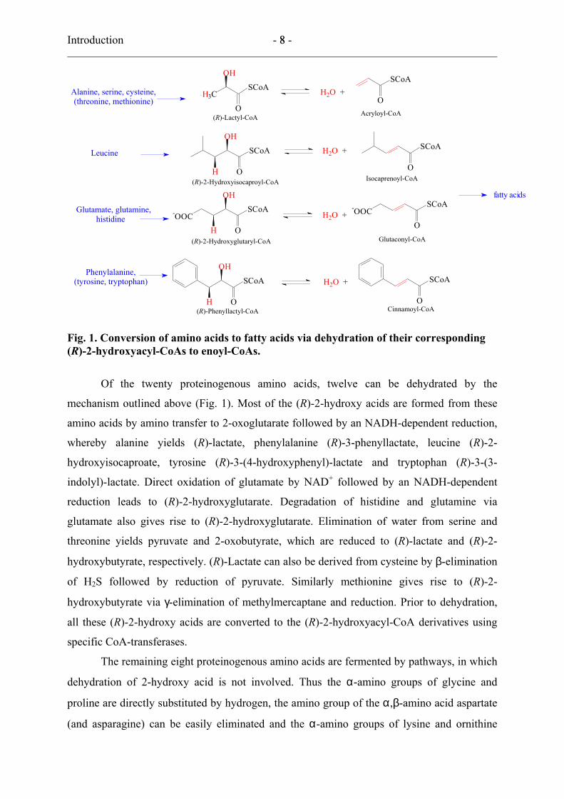
Fig. 1. Conversion of amino acids to fatty acids via dehydration of their corresponding (R)-2-hydroxyacyl-CoAs to enoyl-CoAs.
Of the twenty proteinogenous amino acids, twelve can be dehydrated by the
mechanism outlined above (Fig. 1). Most of the (R)-2-hydroxy acids are formed from these
amino acids by amino transfer to 2-oxoglutarate followed by an NADH-dependent reduction,
whereby alanine yields (R)-lactate, phenylalanine (R)-3-phenyllactate, leucine (R)-2-
hydroxyisocaproate, tyrosine (R)-3-(4-hydroxyphenyl)-lactate and tryptophan (R)-3-(3-
indolyl)-lactate. Direct oxidation of glutamate by NAD+ followed by an NADH-dependent
reduction leads to (R)-2-hydroxyglutarate. Degradation of histidine and glutamine via
glutamate also gives rise to (R)-2-hydroxyglutarate. Elimination of water from serine and
threonine yields pyruvate and 2-oxobutyrate, which are reduced to (R)-lactate and (R)-2-
hydroxybutyrate, respectively. (R)-Lactate can also be derived from cysteine by β-elimination
of H2S followed by reduction of pyruvate. Similarly methionine gives rise to (R)-2-
hydroxybutyrate via γ-elimination of methylmercaptane and reduction. Prior to dehydration,
all these (R)-2-hydroxy acids are converted to the (R)-2-hydroxyacyl-CoA derivatives using
specific CoA-transferases.
The remaining eight proteinogenous amino acids are fermented by pathways, in which
dehydration of 2-hydroxy acid is not involved. Thus the α-amino groups of glycine and
proline are directly substituted by hydrogen, the amino group of the α,β-amino acid aspartate
(and asparagine) can be easily eliminated and the α-amino groups of lysine and ornithine

Introduction - 9 - 9
(from arginine) are shifted to the β-position. Isoleucine and valine are only oxidised, probably
because branching at the β-carbon may be not accepted by the dehydratases.
2. Radicals in enzymatic processes
Until recently, many enzymes have been reported to catalyse by radical mechanisms. These
enzymes utilize the high reactivity of radicals to perform catalysis. Catalytic radicals are
either the radicals derived from cofactors, such as AdoCbl (adenosylcobalamin) (92) and S-
adenosylmethionine (38), or protein radicals (85). Radical-catalysed reactions have a common
feature: substrates of these enzymes can not be activated by an acid-base mechanism. It is
worth noting that all of these reactions are chemically difficult under mild, physiological
conditions without enzymes. Enzymatic radical catalysis can therefore be defined as the
mechanism of catalysis by which enzymes catalyse chemically difficult reactions by utilizing
the high reactivity of free radicals.
Enzymes produce radicals by three different ways: homolysis of a weak covalent
bond, one-electron oxidation, and one-electron reduction. The first way is applied by
coenzyme B12-dependent enzymes, such as glutamate mutase, in which the homolysis of the
weak carbon–cobalt bond of adenosylcobalamin (130 kJ/mol) affords cob(II)alamin and the
5’-deoxyadenosine radical necessary to initiate the rearrangement of (S)-glutamate to (2S,3S)-
3-methylaspartate by abstraction of the 4Si-hydrogen (17). The aerobic ribonucleotide
reductase is an example, in which the tyrosine radical is formed by one-electron oxidation
with molecular oxygen activated by the dinuclear iron centre, although the primary event
might be homolysis of the O–O-bond of the peroxo intermediate (5). A one electron oxidation
has been postulated for the catalysis of 4-hydroxybutyryl-CoA dehydratase, in which the
enolate anion of the substrate is oxidised by the prosthetic group FAD to an enoxy radical
(17). The formation of this radical lowers the pK of the β-proton of 4-hydroxybutyryl-CoA
from about 40 to 14, enabling the dehydration to crotonyl-CoA via a ketyl radical anion (82).
The emerging large family of S-adenosylmethionine (SAM) radical enzymes uses one-
electron reduction of the ‘high-energy’ compound SAM to generate the 5’-deoxyadenosine
radical (38). Another example of radical formation by one-electron reduction is the syn-
dehydration of (R)-2-hydroxyacyl- CoA to (E)-2-enoyl-CoA. Apparently a one-electron
transfer to the thiol ester carbonyl affording the ketyl radical anion initiates this reaction. The
ketyl acts as a nucleophile and expels the adjacent hydroxyl group. To meet the low redox
potential of the thiol ester carbonyl, the reducing power of ferredoxin is enhanced by
hydrolysis of ATP (55).

Introduction - 10 - 10
3. Dehydration of (R)-2-hydroxy acids: 2-hydroxyacyl-CoA dehydratase
It has been proposed that the dehydration of (R)-2-hydroxyacyl-CoA to enoyl-CoA can only
be achieved by conversion of the electrophilic thiol ester carbonyl into a nucleophile, a
process called ‘Umpolung’ (charge reversal). The thiol ester carbonyl has properties of an
electrophile ketone, which can be reduced by one electron to a nucleophilic ketyl radical
anion. Hence, reduction of 2-hydroxyacyl-CoA to its ketyl radical anion would facilitate the
elimination of the hydroxyl group to yield an enoxy radical, which can be deprotonated to the
ketyl radical anion of the product enoyl-CoA. Oxidation of the latter to the unsaturated
product by the next incoming substrate would complete the catalytic cycle (15, 68) (Fig. 2). It
has been calculated that the pK of the enoxy radical has been lowered to 14, about 26 units
less than the pK of the β-proton of the 2-hydroxy acid (82). Similar to β-hydroxyacyl-CoA,
the pK of the enoxy radical may further be lowered by hydrogen bonding.
CoAS
O
R
H
OH
CoAS R
O
CoAS R
H
OH
O
CoAS R
OHH
N N
CoAS R
HO
CoAS R
O
H+
H2OH+
e-
(R)-2-Hydroxyacyl-CoA (E)-2-Enoyl-CoA
Ketyl radical anion I
Enoxy radical
Ketyl radical anion II
Fig. 2. Proposed mechanism for the dehydration of (R)-2-hydroxyacyl-CoA to (E)-2-enoyl-CoA using an electron as cofactor catalysed by dehydratase. The function of activator is only to initiate the catalytic cycle by ATP-induced electron transfer. Although calculations of the ketyl radical anion have indicated that the negative charge sits mainly at the oxygen, the resonance structure depicted here contributes to the stabilisation of the radical and shows how the ketyl acting as nucleophile expels the hydroxyl group (11).
Introduction - 11 - 11
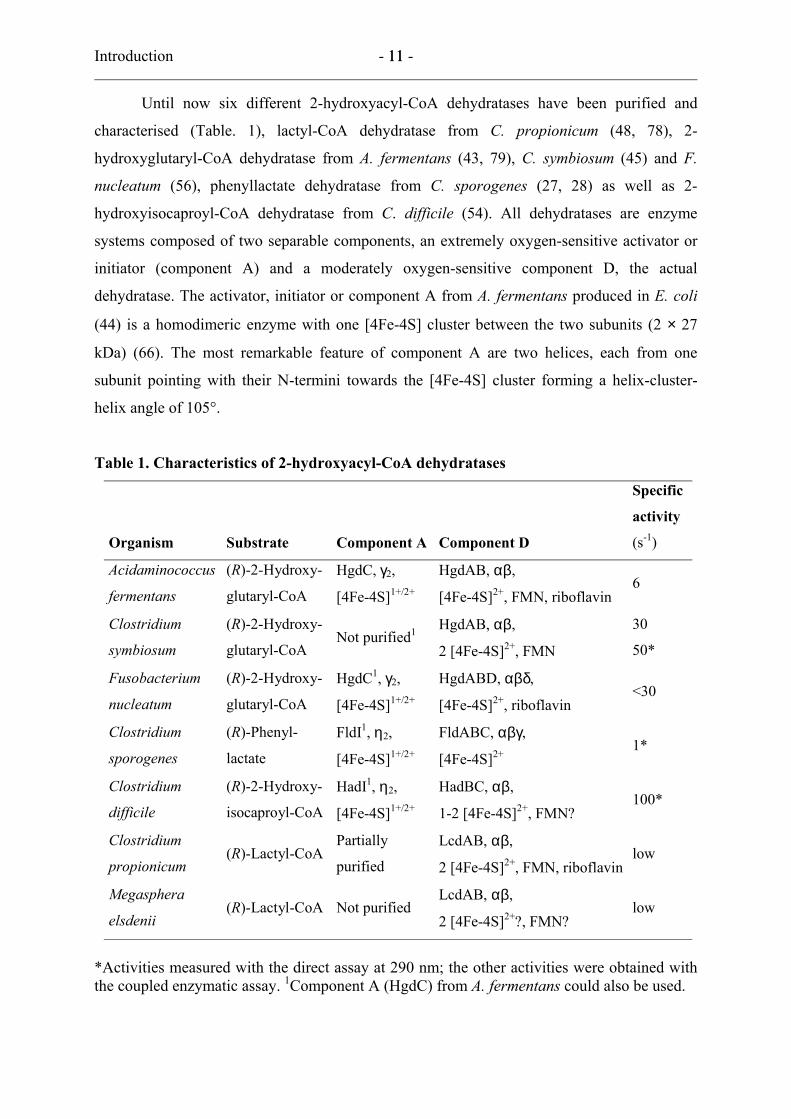
Until now six different 2-hydroxyacyl-CoA dehydratases have been purified and
characterised (Table. 1), lactyl-CoA dehydratase from C. propionicum (48, 78), 2-
hydroxyglutaryl-CoA dehydratase from A. fermentans (43, 79), C. symbiosum (45) and F.
nucleatum (56), phenyllactate dehydratase from C. sporogenes (27, 28) as well as 2-
hydroxyisocaproyl-CoA dehydratase from C. difficile (54). All dehydratases are enzyme
systems composed of two separable components, an extremely oxygen-sensitive activator or
initiator (component A) and a moderately oxygen-sensitive component D, the actual
dehydratase. The activator, initiator or component A from A. fermentans produced in E. coli
(44) is a homodimeric enzyme with one [4Fe-4S] cluster between the two subunits (2 × 27
kDa) (66). The most remarkable feature of component A are two helices, each from one
subunit pointing with their N-termini towards the [4Fe-4S] cluster forming a helix-cluster-
helix angle of 105°.
Table 1. Characteristics of 2-hydroxyacyl-CoA dehydratases
Organism Substrate Component A Component D
Specific
activity
(s-1)
Acidaminococcus
fermentans
(R)-2-Hydroxy-
glutaryl-CoA
HgdC, γ2,
[4Fe-4S]1+/2+
HgdAB, αβ,
[4Fe-4S]2+, FMN, riboflavin 6
Clostridium
symbiosum
(R)-2-Hydroxy-
glutaryl-CoA Not purified1
HgdAB, αβ,
2 [4Fe-4S]2+, FMN
30
50*
Fusobacterium
nucleatum
(R)-2-Hydroxy-
glutaryl-CoA
HgdC1, γ2,
[4Fe-4S]1+/2+
HgdABD, αβδ,
[4Fe-4S]2+, riboflavin <30
Clostridium
sporogenes
(R)-Phenyl-
lactate
FldI1, η2,
[4Fe-4S]1+/2+
FldABC, αβγ,
[4Fe-4S]2+ 1*
Clostridium
difficile
(R)-2-Hydroxy-
isocaproyl-CoA
HadI1, η2,
[4Fe-4S]1+/2+
HadBC, αβ,
1-2 [4Fe-4S]2+, FMN? 100*
Clostridium
propionicum (R)-Lactyl-CoA
Partially
purified
LcdAB, αβ,
2 [4Fe-4S]2+, FMN, riboflavin low
Megasphera
elsdenii (R)-Lactyl-CoA Not purified
LcdAB, αβ,
2 [4Fe-4S]2+?, FMN? low
*Activities measured with the direct assay at 290 nm; the other activities were obtained with the coupled enzymatic assay. 1Component A (HgdC) from A. fermentans could also be used.
Introduction - 12 - 12
A similar architecture is found in the phylogenetically unrelated iron protein of nitrogenase
from Azotobacter vinelandii with a helix-cluster-helix angle of 150°. Upon binding to
component D, probably the angle opens to 180° as observed in the complex of nitrogenase
iron protein with molybdenum-iron protein in the presence of ADP-AlF4− (76). Component A
has a low ATPase activity (ca. 0.1 s−1) but only in the reduced [4Fe-4S]+ state. The structure
of component A also revealed that the [4Fe-4S] cluster is easily accessible from the solvent.
This may be the reason for the extreme oxygen-sensitivity. The redox potential of component
A could not be measured, but the cluster becomes almost completely reduced by flavodoxin
(E0′ ca. −420 mV) or ferredoxin (E0′ = −405 mV) (88) indicating a potential of about −350
mV or even higher. The closely related components A from C. sporogenes and F. nucleatum
(86) have also been purified in the same way and revealed almost identical properties. Each
known genome of an anaerobic bacterium (including E. coli) or archaeon contains at least one
deduced homologue of this exciting protein. There are even four homologous genes of
component A in the genome of Clostridium acetobutylicum (69).
Component D of A. fermentans has been characterised as a heterodimeric enzyme (54
+ 42 kDa) containing one [4Fe-4S] cluster, one riboflavin-5′-phosphate (FMN) and about 0.1
riboflavin. Molybdenum has also been found in this protein, but the content of 0.1
mol/heterodimer appears to be too low to be significant. Furthermore, the same amount of Mo
has been detected in component D from C. symbiosum, but this metal is absent in 2-
hydroxyglutaryl-CoA dehydratase from F. nucleatum. Interestingly, component D from C.
symbiosum contains two [4Fe-4S] clusters. The smaller β-subunit of this protein could be
crystallised and its crystal structure has been determined. It revealed one [4Fe-4S] cluster and
one FMN at a distance of 17 Å. Unexpectedly, only three iron atoms of the cluster are
coordinated by cysteines, whereas the fourth has a not conserved tyrosine as ligand (Holger
Dobbek and Berta Martins, unpublished). The 2-hydroxyglutaryl-CoA dehydratase from F.
nucleatum is unique, since it is composed of three different subunits (53, 56, 86). The third
subunit does not seem to be related to any other protein. Component D of phenyllactate
dehydratase from C. sporogenes is also a trimeric protein. The third and largest subunit (46
kDa), however, has been characterised as a cinnamoyl-CoA:phenyllactate CoA-transferase,
which catalyses the formation of (R)-phenyllactyl-CoA. The other two subunits are
homologues of the α and β-subunits (45 and 37 kDa) of components D from C. symbiosum
and A. fermentans. Hence, activity of phenyllactate dehydratase requires in addition to (R)-
phenyllactate, ATP, MgCl2, a reducing agent (see below) and component A also catalytic
amounts of cinnamoyl-CoA. The mechanism comprises a combination of the mechanisms of
Introduction - 13 - 13
citrate lyase and 2-hydroxyglutaryl-CoA dehydratase. Initially (R)-phenyllactyl-CoA and the
final product (E)-cinnamate are generated from cinnamoyl-CoA and (R)-phenyllactate. In the
next step cinnamoyl-CoA is regenerated by dehydration of (R)-phenyllactyl-CoA. This
enzyme complex clearly shows that formation of the thiol ester substrate is a prerequisite for
the dehydration of 2-hydroxy acids. In the fermentation of glutamate via 2-hydroxyglutarate,
the dehydration at the thiol ester level could be due to the participation of the CoA-ester in the
consecutive decarboxylation step (27, 28).
The reductive activation of component D of the dehydratases requires component A,
ATP, MgCl2, and a reducing agent. In vitro dithionite or Ti(III)citrate are suitable one-
electron donors, whereas in vivo a clostridial-type, two [4Fe-4S] cluster-containing ferredoxin
(88) or flavodoxin (43) serve for this purpose. The further fate of the electron in the activation
process remains unclear. Whereas the Mössbauer spectrum clearly revealed oxidation of
component A during activation, the concomitant reduction of the [4Fe-4S] cluster(s) of
component D could not be observed by this method. The active component D, however,
exhibited an EPR-signal (g < 2.0), which has been interpreted as that of Mo(V) (43). The
recently detected tyrosine-coordination of the [4Fe-4S] cluster by X-ray crystallography may
lead to another speculation. This non-innocent ligand could be reduced to a radical anion
stabilised by the cluster.
The putative mechanism of activation and dehydration can now be described in the
following way (Fig. 3): The cluster of component A, to which two ADP are bound, is reduced
by ferredoxin or flavodoxin with one electron to [4Fe-4S]+. Then ADP is exchanged by ATP,
which causes the helix-cluster-helix angle to open from 105° to 180°. This conformational
change enables component A to dock on component D and the electron is transferred from A
to D with concomitant hydrolysis of two ATP. Thereby the electron transfer becomes
irreversible and component A returns to its ‘ground state’ with two ADP and oxidised [4Fe-
4S]2+. Upon addition of (R)-2-hydroxyacyl-CoA to the reduced component D the electron is
further transferred to the substrate to form the ketyl radical anion, which initiates the
dehydration as proposed above. Afterwards the electron is returned to component D and
transferred further to the next incoming substrate. Thus multiple turnovers are possible
without additional consumption of ATP. Only if the electron is lost by oxidation, another
activation with hydrolysis of two ATP becomes necessary. If each turnover would require
hydrolysis of ATP, the organism would be unable to thrive from glutamate, since the
fermentation only yields 0.6 mol ATP/mol glutamate.
Introduction - 14 - 14
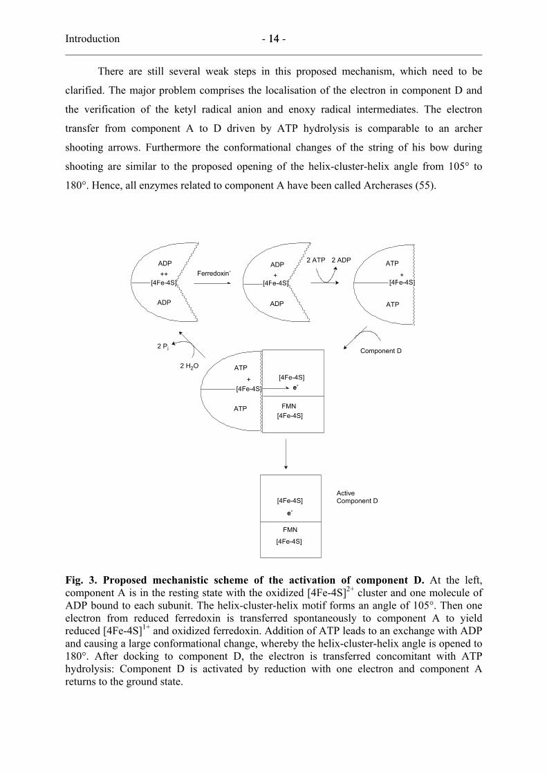
There are still several weak steps in this proposed mechanism, which need to be
clarified. The major problem comprises the localisation of the electron in component D and
the verification of the ketyl radical anion and enoxy radical intermediates. The electron
transfer from component A to D driven by ATP hydrolysis is comparable to an archer
shooting arrows. Furthermore the conformational changes of the string of his bow during
shooting are similar to the proposed opening of the helix-cluster-helix angle from 105° to
180°. Hence, all enzymes related to component A have been called Archerases (55).
[4Fe-4S]
[4Fe-4S] [4Fe-4S]
ADP
ADP
[4Fe-4S]
ADP
ADP
ATP
ATP
++++
+
ATP
ATP
Ferredoxin-
2 ADP2 ATP
[4Fe-4S]
[4Fe-4S]
[4Fe-4S]
FMN
FMN
[4Fe-4S]
e-
2 H2O
2 Pi Component D
Active Component D
e-
Fig. 3. Proposed mechanistic scheme of the activation of component D. At the left, component A is in the resting state with the oxidized [4Fe-4S]2+ cluster and one molecule of ADP bound to each subunit. The helix-cluster-helix motif forms an angle of 105°. Then one electron from reduced ferredoxin is transferred spontaneously to component A to yield reduced [4Fe-4S]1+ and oxidized ferredoxin. Addition of ATP leads to an exchange with ADP and causing a large conformational change, whereby the helix-cluster-helix angle is opened to 180°. After docking to component D, the electron is transferred concomitant with ATP hydrolysis: Component D is activated by reduction with one electron and component A returns to the ground state.
Introduction - 15 - 15
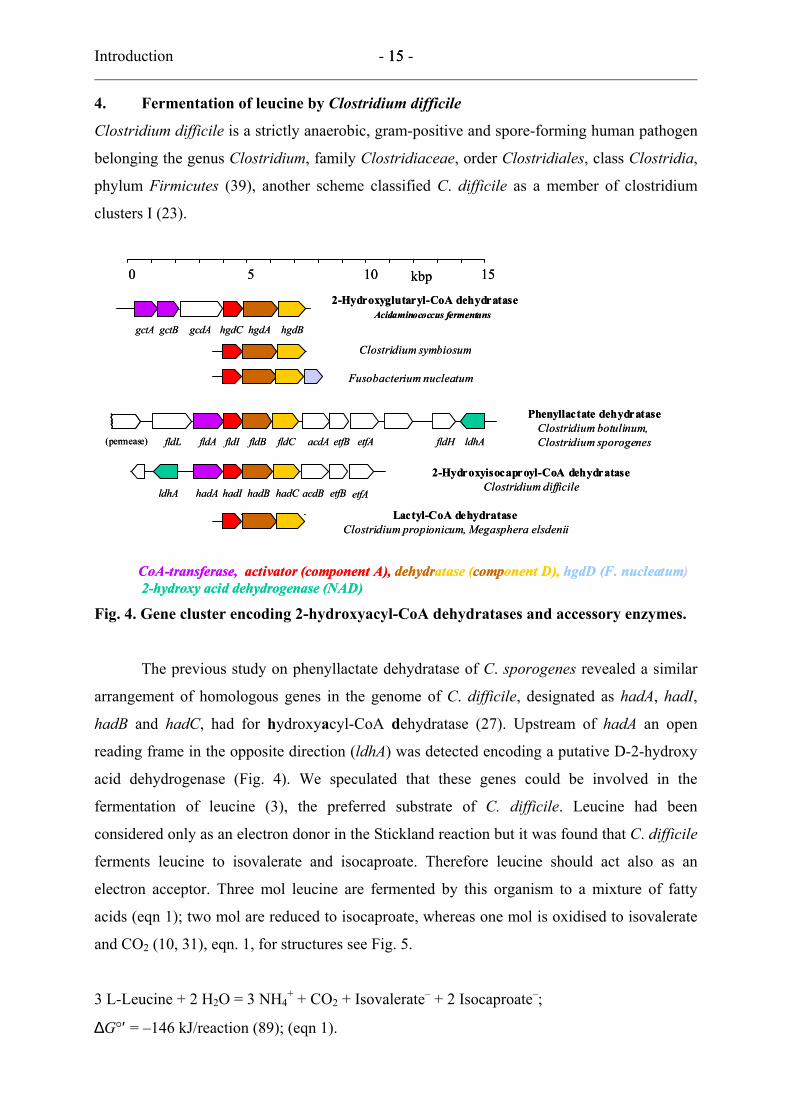
4. Fermentation of leucine by Clostridium difficile
Clostridium difficile is a strictly anaerobic, gram-positive and spore-forming human pathogen
belonging the genus Clostridium, family Clostridiaceae, order Clostridiales, class Clostridia,
phylum Firmicutes (39), another scheme classified C. difficile as a member of clostridium
clusters I (23).
0 5 10 15kbp
hadIhadA hadB hadC etfB etfAldhA acdB
2-Hydroxyisocaproyl-CoA dehydrataseClostridium difficile
Phenyllactate dehydrataseClostridium botulinum,Clostridium sporogenesfldIfldA fldB fldC etfB etfA fldHfldL acdA ldhA(permease)
gcdA hgdAhgdC hgdBgctBgctAAcidaminococcus fermentans
Clostridium symbiosum
2-Hydroxyglutaryl-CoA dehydratase
CoA-transferase, dehydratase (component D), hgdD (F. nucleatum)2-hydroxy acid dehydrogenase (NAD)
Fusobacterium nucleatum
Lactyl-CoA dehydrataseClostridium propionicum, Megasphera elsdenii
activator (component A),activator (component A),
Fig. 4. Gene cluster encoding 2-hydroxyacyl-CoA dehydratases and accessory enzymes.
The previous study on phenyllactate dehydratase of C. sporogenes revealed a similar
arrangement of homologous genes in the genome of C. difficile, designated as hadA, hadI,
hadB and hadC, had for hydroxyacyl-CoA dehydratase (27). Upstream of hadA an open
reading frame in the opposite direction (ldhA) was detected encoding a putative D-2-hydroxy
acid dehydrogenase (Fig. 4). We speculated that these genes could be involved in the
fermentation of leucine (3), the preferred substrate of C. difficile. Leucine had been
considered only as an electron donor in the Stickland reaction but it was found that C. difficile
ferments leucine to isovalerate and isocaproate. Therefore leucine should act also as an
electron acceptor. Three mol leucine are fermented by this organism to a mixture of fatty
acids (eqn 1); two mol are reduced to isocaproate, whereas one mol is oxidised to isovalerate
and CO2 (10, 31), eqn. 1, for structures see Fig. 5.
3 L-Leucine + 2 H2O = 3 NH4+ + CO2 + Isovalerate– + 2 Isocaproate–;
∆G°′ = –146 kJ/reaction (89); (eqn 1).
Introduction - 16 - 16
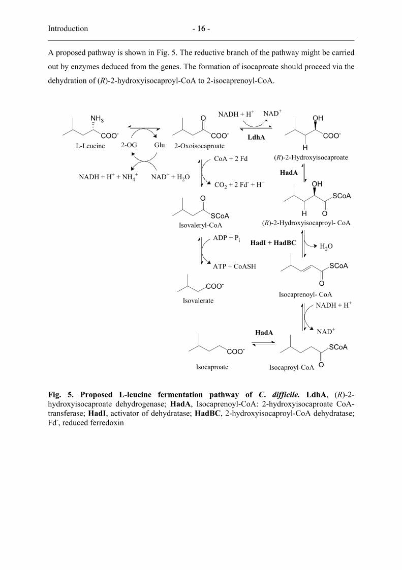
A proposed pathway is shown in Fig. 5. The reductive branch of the pathway might be carried
out by enzymes deduced from the genes. The formation of isocaproate should proceed via the
dehydration of (R)-2-hydroxyisocaproyl-CoA to 2-isocaprenoyl-CoA.
COO-
ONH3
COO-
COO-
SCoA
O
NADH + H+ + NH4+
NADH + H+ NAD+
NAD+
ATP + CoASH
OH
COO-
H LdhA
L-Leucine 2-Oxoisocaproate
Isovaleryl-CoA
Isovalerate
ADP + Pi
2-OG Glu
NAD+ + H2O
(R)-2-Hydroxyisocaproate
H2O
OH
H
SCoA
O
O
SCoACOO-
Isocaproyl-CoAIsocaproate
HadA
HadA
(R)-2-Hydroxyisocaproyl- CoA
O
SCoA
HadI + HadBC
CoA + 2 Fd
CO2 + 2 Fd- + H+
NADH + H+Isocaprenoyl- CoA
Fig. 5. Proposed L-leucine fermentation pathway of C. difficile. LdhA, (R)-2-hydroxyisocaproate dehydrogenase; HadA, Isocaprenoyl-CoA: 2-hydroxyisocaproate CoA-transferase; HadI, activator of dehydratase; HadBC, 2-hydroxyisocaproyl-CoA dehydratase; Fd-, reduced ferredoxin
Introduction - 17 - 17
5. Goals of the work
Before the study on 2-hydroxyisocaproyl-CoA dehydratase, 2-hydroxyacyl-CoA dehydratases
from 5 different organisms were purified and characterised in our group. However several
weak points in the proposed mechanism were not clarified as mentioned. One of the major
problems is to localise the electron in dehydratase (component D). Most of the characterised
dehydratases contain FMN and iron-sulfur cluster for a possible place of electron, however,
none of them seemed to change their redox state. As another target the metal molybdenum
was detected in 2-hydroxyglutaryl-CoA dehydratases, but is unlikely to be the place for the
electron. The other point for the verification of the mechanism are the proposed ketyl radical
anion and enoxy radical intermediates, which have not been observed. Therefore, this thesis
focuses on clarification of weak points in the proposed mechanism of 2-hydroxyacyl-CoA
dehydratase through the study of 2-hydroxyisocaproyl-CoA dehydratase of C. difficile.
Materials and Methods - 18 -
Materials and Methods 1. Materials
1.1. Chemicals and reagents
All used chemical compounds and reagents were purchased, if not mentioned separately in the
text, from the companies, Sigma (Steinheim), Merck (Darmstadt), Roth (Karlsruhe), Fluka
(Neu-Ulm), Bio-Rad-Laboratories (München) or Serva (Heidelberg). The materials for
molecular biology were obtained from New England Biolabs (Frankfurt am Main), Abgene
(Hamburg), Roche (Mannheim) and Amersham Biosciences (Freiburg). The primers were
synthesised by MWG (Ebersberg).
1.1.1. (R)- and (S)-2-Hydroxyisocaproate
(R)- and (S)-2-Hydroxyisocaproate were synthesized by Dr. Daniel Darley from D- and L-
leucine respectively by treatment of the corresponding amino acids with sodium nitrite in
dilute sulfuric acid (24).
1.1.2. (E)-2-Isocaprenoate (4-methyl-trans-2-pentenoate)
(E)-2-Isocaprenoate was synthesized by condensation of isobutyraldehyde and malonic acid
in pyridine-piperidine (40).
1.1.3. 2-Hydroxyisocaproyl-CoA
2-Hydroxyisocaproyl-CoA was synthesized from the mixed acid anhydride of 2-
hydroxyisocaproate by a modified method of the thiol ester synthesis described by
Kawaguchi. 2-Hydroxyisocaproate (110 µmol) and 1,1'-carbonyldiimidazole (100 µmol) were
dissolved in 500 µl tetrahydrofuran (> 99% HPLC grade). To form the CoA-ester, 200 µl of
mixed acid anhydride was added to 10 µmol CoASH in 500 µl 100 mM NaHCO3 and
incubated at room temperature for 1 hour. The reaction was stopped by acidifying of the
reaction with 1 M HCl (< pH 4.0) and loaded on a Sep-pak C18 cartridge (Waters, USA)
which was equilibrated with 0.1 % trifluoroacetic acid. The column was washed with 5
column volume of 0.1% (v/v) trifluoroacetic acid and CoA ester was eluted in 5 ml 1% (v/v)
trifuloroacetic acid and 50% (v/v) acetonitrile. For the molecular mass confirmation by
MALDI-TOF mass spectroscopy, the purified CoA ester was applied on thin layer of indole-
2-carboxylic acid prepared from a solution of 300 mM indole-2-carboxylic acid in acetone.
MALDI-TOF mass spectra were collected at an accelerating voltage of 15,000 V, 58% grid
Materials and Methods - 19 -
voltage, and a delay time of 50 ns in the reflector mode of the instrument at a mirror ratio of
1.07 with indole-2-carboxylic acid as matrix [56].
1.1.4. (R)-2-Hydroxy[2-2H1]isocaproate
(R)-2-Hydroxy[2-2H1]isocaproate was synthesised by reduction of 2-oxoisocaproate by (R)-2-
hydroxyisocaprate dehydrogenase (LdhA) with [2-2H]formate, formate dehydrogenase and
NAD+;
50 mM Photassium phosphate buffer pH 7.6
0.1 mM NAD+
75 mM 2-Oxoisocaproate
300 mM [2-2H]formic acid (neutralized with NaOH)
0.1 U (R)-2-Hydroxyisocaprate dehydrogenase
0.4 U Formate dehydrogenase
The reaction mixture was made up to total volume 1 ml H2O and incubated overnight at room
temperature. After acidification (pH ≈ 1) with 1 M HCl, the excess formic acid was removed
by either extractions (3 x 3 ml) and the product dissolved in D2O was confirmed by NMR.
1.1.5. (R)-2-Hydroxy[3-2H2]isocaproate
2-Oxoisocaproate (150 µmol) was dissolved in 1 ml D2O and incubated at 80 °C for 3 hours
yielding 2-oxo[3-2H2]isocaproate (≈ 100 % exchange confirmed by NMR). Subsequently,
reduction by (R)-2-hydroxyisocaprate dehydrogenase with formic acid (HCOOH), formate
dehydrogenase and NAD+ was prepared as described in 1.1.4.
1.1.6. (R)-2-Hydroxy[2,3-2H3]isocaproate
2-Oxo[3-2H2]isocaproate was synthesised as described in 1.1.5. and reduced by (R)-2-
hydroxyisocaprate dehydrogenase with [2-2H]formate, formate dehydrogenase and NAD+ as
described in 1.1.4.
1.1.7. (R)-2-Hydroxy[1-13C]isocaproate
2-Oxo[1-13C]isocaproate was purchased from Cambridge Isotope Laboratories, Inc. and
reduced by (R)-2-hydroxyisocaprate dehydrogenase with formic acid (HCOOH), formate
dehydrogenase and NAD+ as described in 1.1.4.

Materials and Methods - 20 -
1.2. Instruments and columns
Beckman (Munich) supplied the ultra centrifuge, Sorvall (München) the cooling centrifuges.
Anaerobic experiments have been done in an anaerobic glove box supplied by Coy
Laboratories, Ann Arbor MI, (USA). The FPLC system and the UV-vis photometer,
Ultrospec 400, installed in glove box were obtained from Amersham Biosciences (Freiburg).
HP 8453 UV-visible diode array spectrophotometer (USA) was used for measuring UV-vis
spectra and Amersham Biosciences Kontron spectrophotometer was used for aerobic activity
assays. The columns DEAE Sepharose HR 26/10, Phenyl Sepharose FF HR 26/10, Superdex
200 HR 26/10, Q-Sepharose High performance HiLoadTM 26/10, Superdex-G25 (5 ml) were
obtained from Amersham Biosciences (Freiburg). HPLC columns were from Merck
(Darmstadt). Strep-Tactin MacroPrep column was purchased from IBA GmbH (Göttingen).
1.3. Anaerobic work
Purification of the activator and 2-hydroxyisocaproyl-CoA dehydratase were performed at 15
– 20 °C in an anaerobic glove box under a nitrogen atmosphere containing 5 % H2. Buffers
for enzyme purification were prepared by boiling and cooling under vacuum. Afterwards the
buffers were flushed with nitrogen and transferred to the anaerobic chamber where
dithiothreitol (2 - 5 mM) was added and stirred overnight. Enzyme activity was determined
inside the anaerobic chamber with an Ultrospec 4000 spectrophotometer.
1.4. Bacteria and culture media
1.4.1. Clostridium difficile
Clostridium difficile (DSMZ 1296T) was cultivated under anaerobic conditions in 100 ml
serum bottles. For the 100 L fermenter culture of the organism, a 10 L overnight pre-culture
was used to inoculate the fermenter. The culture medium had the following composition:
Per liter
NaHCO3 5.0 g
Glucose 2.0 g
Leucine 1.0 g
Thioglycolic acid 0.5 g
Resazurine 1 mg
100-fold Phosphates 10.0 ml
100-fold Chlorides 10.0 ml

Materials and Methods - 21 -
100-fold Sulfates 10.0 ml
100-fold Amino acids and Vitamins 10.0 ml
100-fold Phosphates per liter
KH2PO4 30.0 g
Na2HPO4 150.0 g
100-fold Chlorides per liter
NaCl 90.0 g
CaCl2 x 2H2O 2.6 g
MgCl2 x 6H2O 2.0 g
MnCl2 x 4H2O 1.0 g
CoCl2 x 6H2O 0.1 g
100-fold Sulfates per liter
(NH4)2SO4 4.0 g
FeSO4 x 7H2O 0.4 g
100-fold Amino acids and Vitamins per liter
Proline 80.0 g
Cysteine 50.0 g
Each: Arginine, Glycine, Histidine, Isoleucine,
Methionine, Threonine, Tryptophan, Valine 10.0 g
Ca-Pantothenate 100 mg
Pyridoxine-HCl 10 mg
Biotin 1 mg
The bottle of the prepared medium was tightly closed with a rubber stopper and a hypodermic
needle was introduced for pressure released. The medium was boiled until the blue-red colour
of resazurine disappeared then the air above the medium was replaced by nitrogen. The
autoclaved medium could be stored at room temperature in a dark place.
Materials and Methods - 22 -
1.4.2. Escherichia coli
Escherichia coli was normally grown at 37 °C in Standard I medium (Merck: 1.5% Pepton,
0.3 % yeast extract, 100 mM NaCl, 6 mM D-Glucose) containing antibiotic(s) depending on
the harboured plasmid. The strain DH5α {F – φ80 ∆ lacZ∆M15∆(lacZYA-argF)U169, deoR,
recA1, end A1,hsdR17(rk–, mk+), phoA, supE44,λ–, thi-1, gyrA96, relA1} was used for gene
cloning and BL21-CodonPlus(DE3)-RIL {E. coli B F–, ompT, hsdS(rB– mB
–), dcm+, Tetr, gal
λ(DE3), endA, Hte [argU ileY leuW Camr]} for the gene expressions.
1.5. Plasmids
pASK-IBA7 (IBA GmbH) (tet promoter/operator, N-terminal Strep-tag II, cytosolic
localization of the recombinant protein, Ampr) was used for the ldhA expression and pASK-
IBA3 (IBA GmbH) (tet promoter/operator, C-terminal Strep-tag II, cytosolic localization of
the recombinant protein, Ampr) used for hadA, hadI and hadBC expression.
1.6. Antibiotics
The stock of antibiotics was prepared and used as described below.
Antibiotic Stock Final concentration
Ampicillin 100 mg/ml H2O 100 µg/ml
sterilised by filtration (0.2 µm)
Chloramphenicol 50 mg/ml 70 % ethanol 50 µg/ml
Materials and Methods - 23 -
2. Methods for DNA work
2.1. Plasmid DNA isolation
Plasmid DNA isolation was done by alkaline lysis methods using solutions described below.
Solution I
50 mM Glucose
10 mM EDTA
25 mM Tris/HCl pH 8.0
Solution II
0.2 M NaOH, 1% SDS (made fresh)
Solution III
3 M Potassium acetate / glacial acetic acid pH 4.8
Standard I medium 5 ml containing antibiotic(s) was inoculated with a bacterial colony and
incubated with gyration overnight at 37 °C. The culture was transferred into an Eppendorf
tube and harvested at 13000 x g in microfuge for 2 minutes. The bacterial pellet was
suspended in 100 µl Solution I then lysised by adding 200 µl Solution II, and neutralized with
150 µl Solution III. The soluble supernatant was separated from cell debris by centrifugation
for 5 minutes and transferred new Eppendorf tube. The plasmid DNA was extracted with 2
volumes of isopropanol and obtained by centrifugation as a white pellet. DNA pellet was
washed with 1 ml 70 % ethanol, dried and dissolved in TE buffer (10 mM Tris/HCl pH 8.0, 1
mM EDTA).
2.2. Genomic DNA isolation from C. difficile
For the genomic DNA isolation, 2 g of C. difficile cells were suspended in 3 ml Tris-sucrose
buffer (10 mM Tris/HCl pH 8.0, 25 % sucrose). The suspended cells were incubated at 37 °C
for 90 minutes with gentle shaking after adding 100 mg lysozyme. Then, 4 ml of 10 mM
Tris/HCl pH 8.0, 25 mM EDTA was added and incubated on ice for 15 minutes. After adding
20 mg proteinase K and 100 mg RNase, the mixture was incubated at 37 °C for 3 hours. The
protein by extraction with 3 x saturated phenol and 1 x chloroform/isoamylalcohol (24 : 1).
The aqueous phase was transferred to a dialysis bag for overnight dialysis in TE (10 mM
Tris/HCl, 1 mM EDTA pH 8.0) buffer.
Materials and Methods - 24 -
2.3. Agarose gel electrophoresis
Agarose powder was mixed with electrophoresis TAE-buffer (2 M Tris, 1 ml acetic acid, 50
mM EDTA (50x)) to the desired concentration, then heated in a microwave oven until
completely melted. After cooling the solution to about 60°C, it was poured into a casting tray
containing a sample comb and allowed to solidify at room temperature. After the gel had
solidified, the comb was removed and the gel was inserted horizontally into the
electrophoresis chamber just covered with buffer. DNA samples mixed with loading buffer
(0.21% Bromophenol Blue, 0.21% Xylene Cyanol FF, 0.2 M EDTA, pH 8.0, and 50%
Glycerol) were then pipetted into the sample wells, and a voltage was applied. Bromophenol
blue and xylene cyanol dyes migrate through agarose gels at roughly the same rate as double-
stranded DNA fragments of 300 and 4000 bp, respectively. When adequate migration had
occurred, DNA fragments were stained with ethidium bromide and placed on a ultraviolet
transilluminator.
2.4. Elution of DNA fragments from agarose gel
DNA bands were exposed on an UV-illuminator (using short wavelength) and rapidly cut out
from the agarose gel. Extraction was performed following the manual of the QIAquick Gel
Extraction Kit (QIAGEN GmbH).
2.5. DNA restriction and ligation
Restriction reactions were usually performed following the enzyme insert manual. For
ligations of double stranded DNA, T4-DNA ligase (Amersham Biosciences) were used
following the enzyme insert manual.
2.6. Dialysis of ligation mixtures
The ligation mixture was dialysed before electrotransformation. The ligation mixture was
pipetted on Millipore-Membrane (#VSWP 02500) which was floating on the water or TE
buffer. After 30 minutes of dialysis, the ligation mixture was carefully recovered from the
membrane and used for electrotransformation.
2.7. Preparation of competent E. coli cells for electrotransformation
An overnight 5 ml standard I medium culture inoculated with a fresh E. coli single colony
from a plate was used to inoculate a 500 ml main culture grown till the exponential phase
(OD578 = 0.5 – 0.8). The cells were harvested by a pre-cooled (4 °C) high-speed centrifuge
Materials and Methods - 25 -
with 6000 x g for 20 minutes. The harvested cell was washed two times with 500 ml ice-cold
sterile H2O and one time with 20 ml 10 % glycerol. The washed cells were resuspended with
1 ml 10 % glycerol and 40 µl aliquots in thin-wall 500 µl tubes were stored at –80 °C.
2.8. Electrotransformation
The dialysed ligation mixture was added to 40 µl competent cells and transferred to a Gene-
Pulser cuvette (Bio-Rad cat# 165-2086). A pulse was given to the cuvette using the following
settings: 25 µF, 1.8 kV and 200 Ohm. The cuvette was washed with 500 µl Standard I
medium and transferred to a sterile 1.5 ml Eppendorf tube. The transformation mixture was
incubated for 30 minutes at 37°C before plating on a Standard I agar plate containing
antibiotic(s). The agar plate was incubated overnight at 37 °C to get the colonies.
2.9. DNA concentration and purity determination
The DNA concentration and purity were determined measuring OD260 and OD280.
OD260 = 1 corresponds to 50 µg/ml of dsDNA
OD260/OD280 < 1.8 indicates contamination with protein or phenol
OD260/OD280 > 1.8 indicates contamination with RNA
OD260/OD280 ≈ 1.8 indicates pure dsDNA
2.10. PCR reactions
PCR reactions were performed using a proofreading DNA polymerase, Extensor Hi-Fidelity
PCR Enzyme Mix (ABgene) and the reaction mixtures were made with following
concentration of the ingredients and cycling program:
Concentration of ingredients
Final concentration
dNTP 200 µM
Forward primer 500 nM
Reverse primer 500 nM
Template DNA 20 to 200 pg/µl (plasmid DNA)
1 to 2 ng/µl (genomic DNA)
Proofreading DNA polymerase 1 U
Materials and Methods - 26 -
Cycling program
1. 94 °C 3 min
2. 94 °C 30 sec
3. 55 °C (depending on primer) 30 sec
4. 68 °C 1 min 30 sec (depending on the length of target gene)
5. 68 °C 10 min
29 x from 2. to 4.
2.11. PCR primers
PCR primers were designed using free software, Primer D’Signer (IBA GmbH) introducing
BsaI restriction site (underlined) on the primers. The primers are described below:
For ldhA
Forward, 5’-ATGGTAGGTCTCAGCGCAAAATACTAGTATTTGGAGCACGCG-3’
Reverse, 5’-ATGGTAGGTCTCATATCAATTTACTCTATTAGTAGCAGTTCCTG-3’
For hadA
Forward, 5’-ATGGTAGGTCTCAAATGCTTTTAGAAGGAGTTAAAGTAGTAGA-3’
Reverse, 5’-ATGGTAGGTCTCAGCGCTATATCTTACAACTTTACTATCTTTAAAG-3’
For hadI
Forward, 5’-ATGGTAGGTCTCAAATGTACACAATGGGATTAGATATAGGTTC-3’
Reverse, 5’-ATGGTAGGTCTCAGCGCTTATATTTTTCACTTCTTTTTGTGATTCT-3’
For hadBC
Forward, 5’-ATGGTAGGTCTCAAATGTCTGAAAAAAAAGAAGCTAGAGTAGT-3’
Reverse, 5’-ATGGTAGGTCTCAGCGCTCGCTAAACTCATCATCTCAGCAAA-3’
2.12. Cloning of the genes
The amplified fragments of the genes, ldhA (999 bp), hadA (1200 bp), hadI (801 bp) and
hadBC (2354 bp) were restricted and ligated into BsaI restriction site of pASK-IBA3
(provides C-terminal Strep-tag II peptide, Trp-Ser-His-Pro-Gln-Phe-Glu-Lys, fused proteins,
HadA, HadI, HadBC) or IBA7 (provides N-terminal Strep-tag II peptide, Trp-Ser-His-Pro-
Gln-Phe-Glu-Lys, fused protein, LdhA)
Materials and Methods - 27 -
2.13. Sequencing of the cloned genes
IRD (Infra-Red-Dye) labelled primers (5’ IRD 700 forward and 5’ IRD 800 reverse primers)
were synthesised for sequencing:
Standard primers,
pASK-IBA forward: 5’-AGA GTT ATT TTA CCA CTC CCT-3’
pASK-IBA reverse: 5’-GCT CCA TCC TTC ATT ATA GC-3’
Internal primers,
ldhA forward internal: 5’-TGA TTA CCG TTG GAT AGC TG-3’
ldhA reverse internal: 5’-GAC GCA GTA GCG GTA AAC G-3’
hadA forward internal: 5’-ATC TCC AGC AAA TAC AGC AG-3’
hadA reverse internal: 5’-GAC GCA GTA GCG GTA AAC G-3’
hadBC forward internal: 5’-GAA ATT ATA CAT GCA GCT GG-3’
hadBC reverse internal: 5’-GAC GCA GTA GCG GTA AAC G-3’
hadBC forward internal II: 5’-TTC TCC TTC TCC AAT GAA TG-3’
hadBC reverse internal II: 5’-TCA AGT TCT CTT TTT ACG CC-3’
The standard primers were used for all sequencings and the internal primers were used to
complete sequences, which were too long to be determined by the standard primers. In order
to exclude possible errors by DNA polymerase, three different clones from three different
PCRs were sequenced and mutations (nucleotides in one clone different from the same
nucleotide in the other two colnes) were removed by recombination of the clones.
Materials and Methods - 28 -
3. Methods for protein work
3.1. Gene expressions and protein purification
For the expression of genes, plasmid constructs were transformed into E. coli BL21-
CodonPlus(DE3)-RIL harbouring extra rare codon (arg, ileY and leuW) tRNA genes. An
overnight pre-culture (100 ml) inoculated with a fresh single colony from a Standard I agar
plate was grown in the Standard I medium with ampicillin (100µg/ml) and chloramphenicol
(50µg/ml) was used to inoculate 2 L Standard I medium containing the same antibiotics at 37
°C, 30 °C, or room temperature under aerobic or anaerobic conditions. When the culture
reached the mid-exponential phase (A590 = 0.5 - 0.7) gene expression was induced with
anhydrotetracycline (200µg/L). After another 3h growth, the cells were harvested and re-
suspended in equilibration buffer. The cells were broken using a French Press operating at
140 MPa or sonication and cell debris were removed by ultra-centrifugation at 100,000 x g for
1 h. The supernatant was loaded on a 5 mL Strep-Tactin MacroPrep column, which was
equilibrated with equilibration buffer. After loading the cell free extract, the column was
washed with at least 10 column volumes of equilibration buffer. The pure protein was eluted
with equilibration buffer + 3 mM D-desthiobiotin. Buffers for recombinant protein
purification;
Protein Equilibration buffer Elution buffer
LdhA 100 mM Tris/HCl pH 8.0 Equilibration buffer
300 mM NaCl + 3 mM D-desthiobiotin
1 mM EDTA
HadA 100 mM photassium phosphate pH 7.5 Equilibration buffer
300 mM NaCl + 3 mM D-desthiobiotin
1 mM EDTA
HadI 50 mM Mops pH 7.2 Equilibration buffer
300 mM NaCl + 1 mM ADP
10 mM MgCl2 + 3 mM D-desthiobiotin
2 mM DTT
HadBC 50 mM Mops pH 7.0 Equilibration buffer
300 mM NaCl + 3 mM D-desthiobiotin
2 mM DTT

Materials and Methods - 29 -
3.2. Purification of (R)-2-hydroxyisocaproyl-CoA dehydratase
C. difficile cells were cultivated as described in Materials 1.4.1. in 2 L tightly closed bottles
containing anoxic defined medium supplemented with L-leucine (1g/L; 7.6 mM). Cells were
harvested, washed and resuspended in buffer A containing 50 mM Mops pH 7.0 and 2 mM
dithiothreitol, yield 3 g wet cell paste. The preparation of the cell free extract was performed
as that described in the activator purification. The cell free extract was filtered (0.45 µm pore
size) and loaded a DEAE-Sepharose fast-flow column (3 × 10 cm) equilibrated with buffer A.
The column was washed with 70 mL buffer A and the proteins were eluted at a rate of 3 mL
min-1 with a linear gradient of 0 – 1.0 M NaCl in buffer A. The active brown fractions were
eluted around 0.4 M NaCl. An equal volume of 2.0 M (NH4)2SO4 in buffer A was added to
the pooled fractions from the first column, which were then loaded on a phenyl-Sepharose
column (3 x 10 cm) equilibrated with buffer B (50 mM Mops pH 7.0, 1.0 M (NH4)2SO4, 2
mM dithiothreitol). After washing the column with 70 mL buffer B, the active brown
dehydratase eluted around 0.1 M (NH4)2SO4 with a linear gradient of 1.0 – 0 M (NH4)2SO4 in
buffer B at a rate of 3 mL min-1. The dehydratase fractions were concentrated on an Amicon
PM 30 cell and desalted against buffer A, then loaded on a Q-Sepharose column (1.8 × 10 cm)
equilibrated with buffer A. After a washing step with 60 ml buffer A, the dehydratase was
eluted around 0.5 M NaCl with a linear gradient of 0 – 1.0 M NaCl in buffer A at a rate of 3
mL min-1. The dehydratase was finally concentrated with an Amicon Ultra-4 PLTK Ultracel-
Pl (30 kDa cut off).
The recombinant 2-hydroxyisocaproyl-CoA dehydratase from E. coli was purified by
the same method, since the enzyme was not absorbed at the Strep-Tactin MacroPrep column.
After the phenyl-Sepharose column the enzyme was already pure and therefore the Q-
Sepharose column could be omitted.
3.3. Preparation of soluble membrane protein
The C. difficile cell debris and membranes separated from cell-free extract as described above
were washed three times with 50 mM Mops pH 7.0, 2 mM DTT. The washed membrane
fraction was dissolved in 10 ml 50 mM Mops pH 7.0, 2 mM DTT 10 % n-dodecyl-β-D-
matoside. The soluble part of membrane fraction was separated from the insoluble part by
ultra-centrifugation at 100,000 x g for 30 minutes.

Materials and Methods - 30 -
3.4. Enzyme activity assays
3.4.1. (R)-2-Hydroxyisocaproate dehydrogenase
(R)-2-Hydroxyisocaproate dehydrogenase activity was measured aerobically in 50 mM
Tris/HCl pH 8.0, 0.2 mM NADH and 0.1 mM 2-oxoisocaproate in total volume of 1.0 mL at
room temperature. After addition of enzyme, the decrease of NADH (∆ε340 = 6.2 mM-1 cm-1)
absorbance was followed at 340 nm.
COO-
ONADH + H+ NAD+
LdhA COO-
OH
The reverse reaction could also be measured by the formation of formazane from
iodonitrosotetrazolium chloride in presence of meldola blue. Contents in total volume of the
assay (1 mL ) was as shown below. The assay was started by addition of the enzyme and
followed by the increase of the absorbance of formazane at 492 nm (ε492 = 19.6 mM-1 cm-1).
NADH + INT NAD+ + INT-formazanMeldola bule
(ε492 = 19.6 mM-1 cm-1)NADH + INT NAD+ + INT-formazan
Meldola bule
(ε492 = 19.6 mM-1 cm-1)
In 1 mL test
65 mM TEA (Triethanolamine)
8 mM Potassium phosphate pH 7.6
4.3 mg Triton –X100
77 µM INT (Iodonitrosotetrazolium chloride)
165 µg Meldola blue
2 mM NAD+
3.4.2. (E)-2-Isocaprenoyl-CoA:2-hydroxyisocaproate CoA transferase
(E)-2-Isocaprenoyl-CoA:(R)-2-hydroxyisocaproate CoA-transferase was measured
aerobically in 50 mM potassium phosphate pH 7.0 and 100 µM (R)-2-hydroxyisocaproyl-
CoA (ε260 = 16 mM-1 cm-1) in total volume of 1.0 mL at room temperature. After addition of
the enzyme, the reaction was initiated by addition of (E)-2-isocaprenoate (1 mM final
Materials and Methods - 31 -
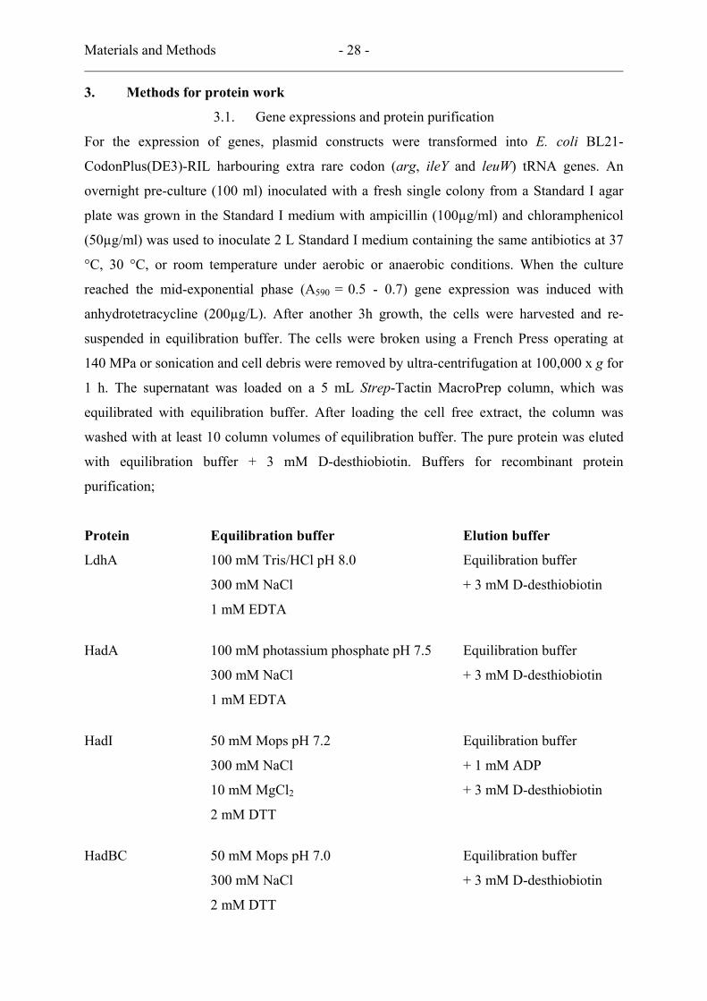
concentration) and the absorbance increase by formation of (E)-2-isocaprenoyl-CoA (ε260 =
22mM-1 cM-1) was followed at 260 nm (∆ε260 = 6 mM-1 cm-1).
(ε260 = 16 mM-1 cm-1)
(ε260 = 22 mM-1 cm-1)
COO-
OHOH
O
SCoA
COO-
O
SCoA
HadA
(ε260 = 16 mM-1 cm-1)
(ε260 = 22 mM-1 cm-1)
COO-
OHOH
O
SCoA
COO-
O
SCoA
HadA
3.4.3. ATPase activity of activator
ATPase activity of activator was measured by determination of ADP-formation using a
coupled assay with PK (pyruvate kinase) and LD (lactate dehydrogenase) (26, 42). The total
volume of 1.0 ml contained the activator in 50 mM Tris-HCl pH 8.0, 1 mM PEP
(phosphoenolpyruvate), 10 mM MgCl2, 1 mM ATP, 0.2 mM NADH, 2 U pyruvate kinase and
2 U lactate dehydrogenase by following the absorbance decrease of NADH at 340 nm (ε340 =
6.2 mM-1 cm-1) after addition of the dehydratase.
LactatePEP Pyruvate
ADP ATP NADH + H+ NAD+
PK LD
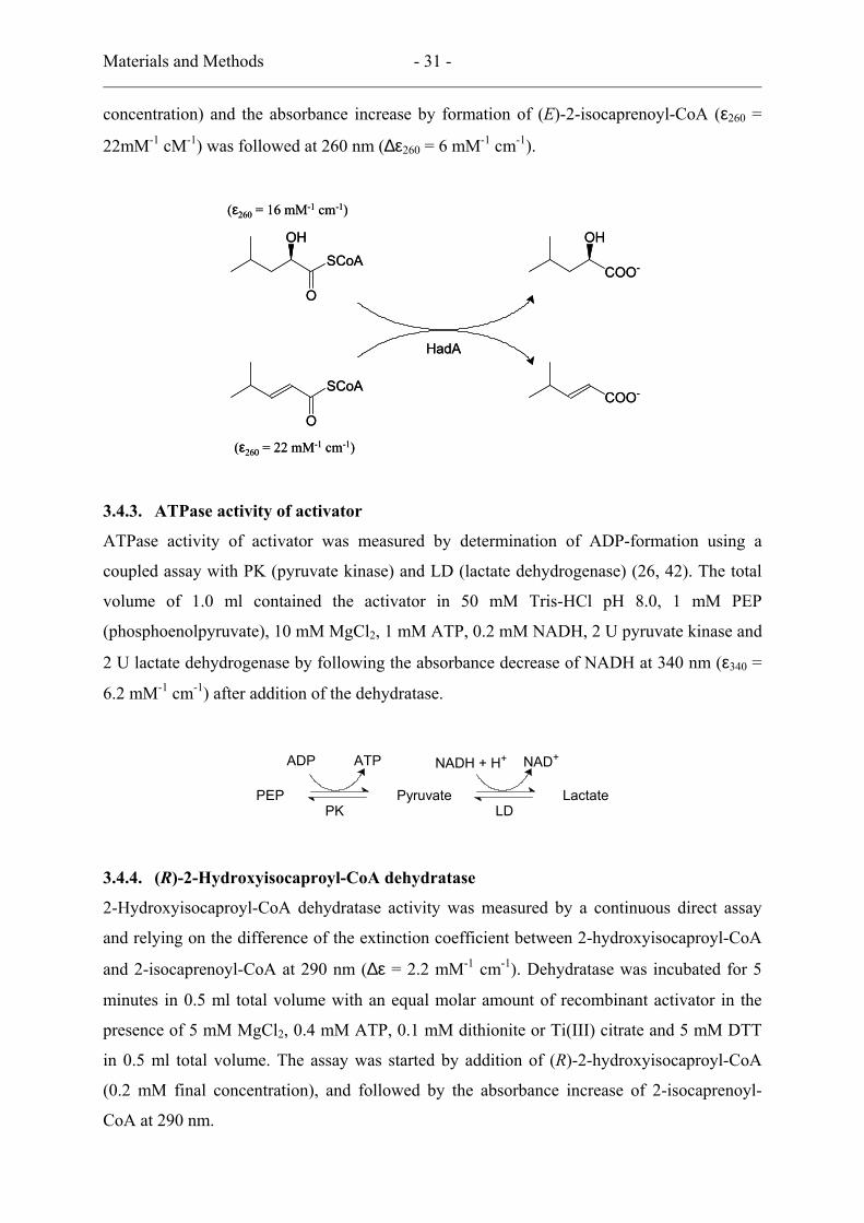
3.4.4. (R)-2-Hydroxyisocaproyl-CoA dehydratase
2-Hydroxyisocaproyl-CoA dehydratase activity was measured by a continuous direct assay
and relying on the difference of the extinction coefficient between 2-hydroxyisocaproyl-CoA
and 2-isocaprenoyl-CoA at 290 nm (∆ε = 2.2 mM-1 cm-1). Dehydratase was incubated for 5
minutes in 0.5 ml total volume with an equal molar amount of recombinant activator in the
presence of 5 mM MgCl2, 0.4 mM ATP, 0.1 mM dithionite or Ti(III) citrate and 5 mM DTT
in 0.5 ml total volume. The assay was started by addition of (R)-2-hydroxyisocaproyl-CoA
(0.2 mM final concentration), and followed by the absorbance increase of 2-isocaprenoyl-
CoA at 290 nm.
Materials and Methods - 32 -
(∆ε290 = 2.2 mM-1 cm-1)
OH
O
SCoA
O
SCoA
HadBC HadI
H2O
(∆ε290 = 2.2 mM-1 cm-1)
OH
O
SCoA
O
SCoA
HadBC HadI
H2O
3.4.5. NADH:ferredoxin oxidoreductase
The membrane NADH:ferredoxin oxidoreductase activity was measured in 100 mM Tris/HCl
pH 8.0, 200 µM NADH, ferricyanide and addition of the enzyme probe started the reaction.
The reaction was followed by the absorbance decrease of ferrocyanide reduced from
ferricyanide at 420 nm (ε420 = 1.02 mM-1 cm-1).
3.5. Determination of protein concentration
Protein concentration was determined by the Bradford method (9). The assay is based on the
shift of the absorbance maximum for an acidic solution of Coomassie Brilliant Blue G-250
from 465 nm to 595 nm upon binding of protein. A standard 0 – 7 µg of BSA was made up to
an 800 µl volume with water and 200 µl Coomassie Brilliant Blue G-250 reagent. The
reactions were incubated in the dark at room temperature for 30 minutes and the absorbance
was measured at 595 nm.
3.6. Non-heme iron determination
The iron complexed by the protein is liberated by treatment with hydrochloric acid. Excess
acid is neutralized with ammonium acetate, Fe3+ is converted to Fe2+ by reduction with
ascorbic acid. Precipitated protein is complexed with sodium dodecylsulfate. Finally the iron
chelator is added to form a blue Fe2+-chelator complex (35). Used compounds are described
below:
1 % (m/v) HCl
7.5 % (m/v) Ammoniumacetate
2.5 % (m/v) Sodium dodecylsulfate (SDS)
4 % (m/v) Ascorbic acid, (freshly prepared)
1.5 % (m/v) Iron chelator, (3-(2-pyridyl)-5,6-bis(5-sulfo-2-furyl)-1,2,4-triazine,
disodium salt
0.2 mM (NH4)2Fe(SO4)2 x 6H2O (freshly prepared)

Materials and Methods - 33 -
Three samples of the unknown, two blanks and six samples of iron standard (2,4,8,12,16 and
20 µM final concentrations) were diluted to 100 µl with water in Eppendorf tubes,
subsequently, 100 µl 1 % HCl was added. The samples were mixed by gentle shaking,
incubated at 80 °C for 10 minutes and cooled down to room temperature. 500 µl
ammoniumacetate, 100 µl 4 % ascorbic acid, 100 µl sodium dodecylsulfate and 100 µl iron
chelator were added sequentially with vortex and short centrifugation. The reaction mixtures
were centrifuged at 9000 x g for 10 minutes and the absorbance at 593 nm was measured
against water.
3.7. Acid-labile sulfur determination
The iron-sulfur protein is denatured in an alkaline medium containing zinc hydroxide.
Released sulfide is co-precipitated with Zn(OH)2 as ZnS. After acidification, H2S condenses
with two molecules of N,N’-dimethyl-p-phenylenediamine to form methylene blue (22). The
reagents are described below:
1 % (m/v) Zinc acetetate (freshly prepared from 10 %)
7 % (m/v) Sodium hydroxide
0.1 % (m/v) N,N’-dimethyl-p-phenylenediamine (DMPD) in 5 M HCl
10 mM FeCl3 in 1 M HCl
≈ 2 mM Sulfide standard (Na2S x 9H2O);
A crystal of appropriate size (≈ 0.5 g) is blotted on filter paper, rapidly weighed and added to
a 1 L volumetric flask containing 10 mM NaOH which has been purged of air with nitrogen.
The flask is closed immediately and the solution is stirred magnetically. The solution was
independently standardized iodometrically.
Three protein samples, two blanks, five sulfide standards (5 – 50 µM) and two protein
samples with sulfide standard additions were put in Eppendorf tubes and made up to 200 µl
with distilled water, 0.6 ml 1 % zinc acetate and 0.05 ml of 7 % NaOH were added, mixed
and incubated for 15 minutes at room temperature. After adding 150 µl DMPD and FeCl3, the
tubes were closed immediately and vortexed vigorously for 30 seconds and incubated for 20
minutes at room temperature. The reaction tubes were centrifuged at 9,000 x g for 5 minutes
and the absorbance were measured at 670 nm against water.

Materials and Methods - 34 -
3.8. Iodometric determination of the sulfide standard
The gravimetric preparation of a sulfide standard using Na2S x 9H2O is inaccurate because of
the hygroscopic nature of the compound leading to overestimation of the sulfide in the protein
sample. An accurate amount of iodine (I2) is partly reduced with a known volume of sulfide
standard solution. Remaining iodine is then determined by titration with sodium thiosulfate,
and the sulfide concentration of the Na2S solution can be calculated by substraction of this
volume from titration of the same amount of idodine without added S2-.
45 mM (should be accurate) Sodium thiosulfate (Na2S2O3)
40 mM in 50 ml 300 mM KI I2
0.35 g/70 ml H2O Soluble starch (indicator) boiled and cooled down under
continuous stirring
In a 100 ml Erlenmeyer flask, 25 ml water was mixed with exactly 5 ml iodine solution and 1
ml 2 N sulfuric acid. The mixture was titrated with sodium thiosulfate until the solution
turned almost colourless. Then 0.5 ml indicator solution was added for further titration with
sodium thiosulfate. After the accurate amount of iodine was determined, the same amount of
iodine was partly reduced with 25 ml sulfide standard and the remaining iodine was titrated
with sodium thiosulfate. The stoichiometry (I2 + 2S2O3 = 2I- + S4O6, I2 + S2- = S + 2I-) was
used for calculation.
3.9. Flavin determination
The flavin bound to the protein was characterized with HPLC. The enzyme solution was
denatured with 3 % TCA and centrifuged to remove the denatured protein. The yellow
supernatant was analysed using a hydrophobic reverse phase column, RP-18 column (5 µm).
The sample was eluted with 25 % methanol in 50 mM ammonium formate. Riboflavin, FMN
and FAD (10 µM) were used as standards, which were treated in the same way as the enzyme
solution. A flow rate of 1 ml/min was used and the absorbance at 266 nm was used for
detection of flavin.
3.10. Separation of activated dehydratase from activator
Dehydratase (4.4 mg) was activated with 1.0 mg activator in the presence of 50 mM Mops pH
7.0, 0.4 mM ATP, 5 mM MgCl2, 5 mM dithiothreitol, and 0.1 mM dithionite (total volume
2.0 ml) as described in activity assay but in the absence of bovine serum albumin. After 30

Materials and Methods - 35 -
min incubation at room temperature, 1 µL was assayed for activity without further activation
and the reaction mixture was loaded on a 5 mL Strep-Tactin MacroPrep column, previously
reduced with 50 mM Mops pH 7.0, 5 mM dithiothreitol and 0.1 mM dithionite, and
equilibrated with 50 mM Mops pH 7.0, 300 mM NaCl, 10 mM MgCl2 and 5 mM
dithiothreitol. The tagged activator was bound to the column while the dehydratase-containing
flow-through was collected in 1 mL fractions. An UV/visible spectrum was taken from the
peak fraction (1.2 mg dehydratase/mL), which was also analysed for activity. Therefore a 2
µL aliquot was added to 50 mM Tris/HCl pH 8.0 and the reaction was started with 0.2 µmol
(R)-2-hydroxyisocaproyl-CoA, total volume 0.5 ml, d = 1 cm. After the reaction had ceased,
two additional 0.2 µmol (R)-2-hydroxyisocaproyl-CoA aliquots were added. Finally the
enzyme was completely re-activated by 0.1 mM dithionite, 0.4 mM ATP, 5 mM MgCl2 and 5
mM dithiothreitol and 30 µg activator (added last). On an SDS/polyacrylamide gel, to which
20 µL of the separated dehydratase was applied, the double band of the dehydratase (40 kDa)
but no trace of the activator (30 kDa) was visible upon Coomassie staining.
3.11. Complex of dehydratase and activator; formation and purification
The complex formation was performed as described for the nitrogenase complex using the
transition state ATP analogue ADP-AlF4 (76). Dehydratase and activator (1.5 : 1 molar ratio)
were incubated for 30 minutes at room temperature in the anoxic globe box with the mixture
described below:
50 mM Tris/HCl pH 8.0
5 mM MgCl2
5 mM Dithiothreitol
1 mM ATP
1 mM Dithionite
1 mM AlF3
10 mM KF
The complex could be purified using Strep-tag affinity, DEAE Sepharose Fast flow and gel-
filtration columns. However, the fastest and most stable way was concentration and buffer (50
mM Tris/HCl pH8.0) exchange on Amicon Ultra-4 PLTK Ultracel-Pl (100 kDa cut off). After
purification two protein bands of dehydratase and activator were shown on SDS-
Materials and Methods - 36 -
polyacrylamide gel. The purified complex was used for crystallisation and the dehydratase
activity measurement.
3.12. Protein molecular mass determination
Apparent molecular mass of enzymes were determined by gel filtration on a Superose 6
column in 150 mM NaCl with 50 mM Tris/HCl, pH 8.0 at a flow rate of 0.5 mL/min.
Amylase, aldolase, bovine serum albumin, catalase and cytochrome c were used for
calibration. The molecular mass standards were obtained from Roche Molecular
Biochemicals (Mannheim, Germany).
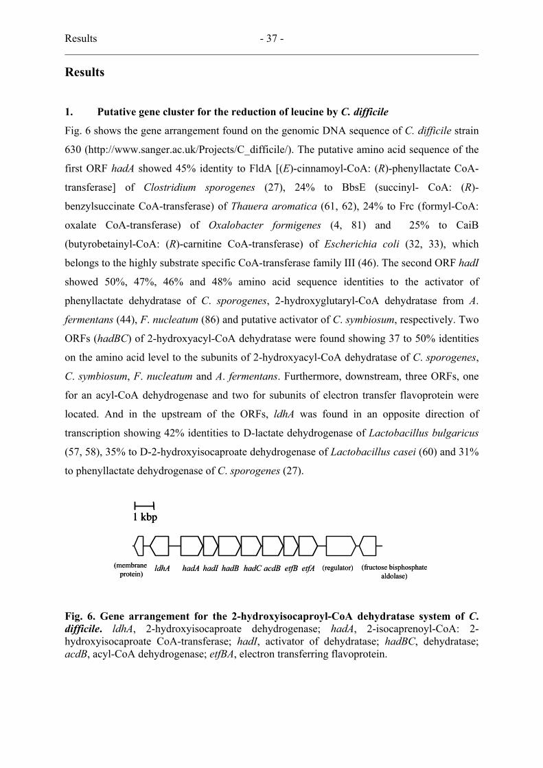
Results - 37 -
Results
1. Putative gene cluster for the reduction of leucine by C. difficile
Fig. 6 shows the gene arrangement found on the genomic DNA sequence of C. difficile strain
630 (http://www.sanger.ac.uk/Projects/C_difficile/). The putative amino acid sequence of the
first ORF hadA showed 45% identity to FldA [(E)-cinnamoyl-CoA: (R)-phenyllactate CoA-
transferase] of Clostridium sporogenes (27), 24% to BbsE (succinyl- CoA: (R)-
benzylsuccinate CoA-transferase) of Thauera aromatica (61, 62), 24% to Frc (formyl-CoA:
oxalate CoA-transferase) of Oxalobacter formigenes (4, 81) and 25% to CaiB
(butyrobetainyl-CoA: (R)-carnitine CoA-transferase) of Escherichia coli (32, 33), which
belongs to the highly substrate specific CoA-transferase family III (46). The second ORF hadI
showed 50%, 47%, 46% and 48% amino acid sequence identities to the activator of
phenyllactate dehydratase of C. sporogenes, 2-hydroxyglutaryl-CoA dehydratase from A.
fermentans (44), F. nucleatum (86) and putative activator of C. symbiosum, respectively. Two
ORFs (hadBC) of 2-hydroxyacyl-CoA dehydratase were found showing 37 to 50% identities
on the amino acid level to the subunits of 2-hydroxyacyl-CoA dehydratase of C. sporogenes,
C. symbiosum, F. nucleatum and A. fermentans. Furthermore, downstream, three ORFs, one
for an acyl-CoA dehydrogenase and two for subunits of electron transfer flavoprotein were
located. And in the upstream of the ORFs, ldhA was found in an opposite direction of
transcription showing 42% identities to D-lactate dehydrogenase of Lactobacillus bulgaricus
(57, 58), 35% to D-2-hydroxyisocaproate dehydrogenase of Lactobacillus casei (60) and 31%
to phenyllactate dehydrogenase of C. sporogenes (27).
hadIhadA hadB hadC etfB etfA (regulator)ldhA(membraneprotein)
acdB (fructose bisphosphatealdolase)
1 kbp
hadIhadA hadB hadC etfB etfA (regulator)ldhA(membraneprotein)
acdB (fructose bisphosphatealdolase)
1 kbp1 kbp
Fig. 6. Gene arrangement for the 2-hydroxyisocaproyl-CoA dehydratase system of C. difficile. ldhA, 2-hydroxyisocaproate dehydrogenase; hadA, 2-isocaprenoyl-CoA: 2-hydroxyisocaproate CoA-transferase; hadI, activator of dehydratase; hadBC, dehydratase; acdB, acyl-CoA dehydrogenase; etfBA, electron transferring flavoprotein.

Results - 38 -
2. (R)-2-Hydroxyisocaproate dehydrogenase
2.1. Analysis of ldhA
The gene ldhA was composed of 999 base pairs (A and T 677, 67.8% and C and G 322,
32.2%) and coding for 332 amino acids with a calculated molecular mass of 36519.84 Da and
isoelectric point of 4.9. The codon usage of the gene showed a pattern, different from the
codon usage of E. coli, which could be anticipated from the low GC content of C. difficile.
The direction of the transcription was reversed from the other genes for the dehydratase
system and no additional gene was found between ldhA and hadA. The deduced amino acid
sequence showed identities to the D-lactate and D-2-hydroxyisocaproate dehydrogenase
family.
2.2. Cloning and expression of ldhA and protein purification
PCR primers were designed as described in Materials and Methods. The restriction enzyme
BsaI site was introduced in the primers for in-frame cloning into the expression vector pASK-
IBA7. The amplified DNA fragment (≈ 1 kb) was cut with BsaI and ligated into the
expression vector, which supports an N-terminal fused Strep-tag II peptide protein for one-
step purification. Three clones from three different PCR reactions were sequenced to exclude
possible errors of the DNA polymerase. The cloned ldhA was composed of 999 nucleotide
base pairs with three nucleotide bases substitutions (C555 → T, T894 → C and T948 → C)
(Fig. 7) compared to the known sequence from Sanger Center, but the same substitutions were
found in all three different clones. Therefore, it was concluded that these three substituted
nucleotides were due to a difference in the strain used, however, the deduced 332 amino acids
were 100% identical to the known sequence.
The plasmid construct named p7ldhA was transformed into E. coli strain BL21-
CodonPlus(DE3)-RIL to get more efficient protein production. Cultures were grown under
aerobic conditions at 37 °C with 5% inoculation and induced with anhydrotetracycline (200
µg/L) in the exponential phase (A590 = 0.5 – 0.7). After another 3 h of growth, cells were
harvested. The cell free extract was obtained after centrifugation of cells opened by
sonication. Finally, pure protein was purified by purification using Strep-Tactin affinity
chromatography (Fig. 8).
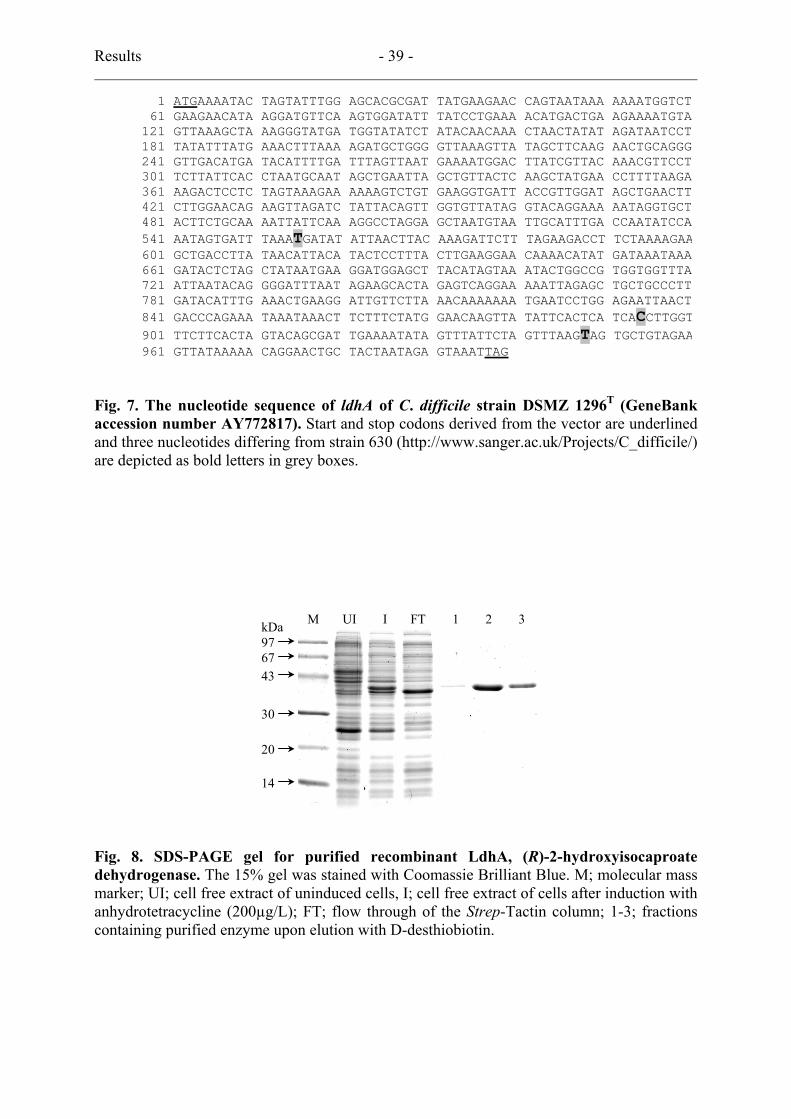
Results - 39 -
1 ATGAAAATAC TAGTATTTGG AGCACGCGAT TATGAAGAAC CAGTAATAAA AAAATGGTCT 61 GAAGAACATA AGGATGTTCA AGTGGATATT TATCCTGAAA ACATGACTGA AGAAAATGTA121 GTTAAAGCTA AAGGGTATGA TGGTATATCT ATACAACAAA CTAACTATAT AGATAATCCT181 TATATTTATG AAACTTTAAA AGATGCTGGG GTTAAAGTTA TAGCTTCAAG AACTGCAGGG241 GTTGACATGA TACATTTTGA TTTAGTTAAT GAAAATGGAC TTATCGTTAC AAACGTTCCT301 TCTTATTCAC CTAATGCAAT AGCTGAATTA GCTGTTACTC AAGCTATGAA CCTTTTAAGA361 AAGACTCCTC TAGTAAAGAA AAAAGTCTGT GAAGGTGATT ACCGTTGGAT AGCTGAACTT421 CTTGGAACAG AAGTTAGATC TATTACAGTT GGTGTTATAG GTACAGGAAA AATAGGTGCT481 ACTTCTGCAA AATTATTCAA AGGCCTAGGA GCTAATGTAA TTGCATTTGA CCAATATCCA541 AATAGTGATT TAAATGATAT ATTAACTTAC AAAGATTCTT TAGAAGACCT TCTAAAAGAA601 GCTGACCTTA TAACATTACA TACTCCTTTA CTTGAAGGAA CAAAACATAT GATAAATAAA661 GATACTCTAG CTATAATGAA GGATGGAGCT TACATAGTAA ATACTGGCCG TGGTGGTTTA721 ATTAATACAG GGGATTTAAT AGAAGCACTA GAGTCAGGAA AAATTAGAGC TGCTGCCCTT781 GATACATTTG AAACTGAAGG ATTGTTCTTA AACAAAAAAA TGAATCCTGG AGAATTAACT841 GACCCAGAAA TAAATAAACT TCTTTCTATG GAACAAGTTA TATTCACTCA TCACCTTGGT901 TTCTTCACTA GTACAGCGAT TGAAAATATA GTTTATTCTA GTTTAAGTAG TGCTGTAGAA961 GTTATAAAAA CAGGAACTGC TACTAATAGA GTAAATTAG
Fig. 7. The nucleotide sequence of ldhA of C. difficile strain DSMZ 1296T (GeneBank accession number AY772817). Start and stop codons derived from the vector are underlined and three nucleotides differing from strain 630 (http://www.sanger.ac.uk/Projects/C_difficile/) are depicted as bold letters in grey boxes.
M UI I FT 1 2 3 kDa 97 67
43 30 20 14
Fig. 8. SDS-PAGE gel for purified recombinant LdhA, (R)-2-hydroxyisocaproate dehydrogenase. The 15% gel was stained with Coomassie Brilliant Blue. M; molecular mass marker; UI; cell free extract of uninduced cells, I; cell free extract of cells after induction with anhydrotetracycline (200µg/L); FT; flow through of the Strep-Tactin column; 1-3; fractions containing purified enzyme upon elution with D-desthiobiotin.
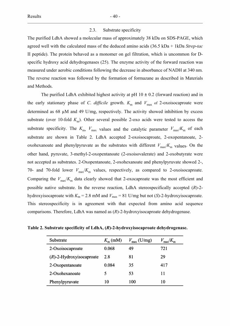
Results - 40 -
2.3. Substrate specificity
The purified LdhA showed a molecular mass of approximately 38 kDa on SDS-PAGE, which
agreed well with the calculated mass of the deduced amino acids (36.5 kDa + 1kDa Strep-tac
II peptide). The protein behaved as a monomer on gel filtration, which is uncommon for D-
specific hydroxy acid dehydrogenases (25). The enzyme activity of the forward reaction was
measured under aerobic conditions following the decrease in absorbance of NADH at 340 nm.
The reverse reaction was followed by the formation of formazane as described in Materials
and Methods.
The purified LdhA exhibited highest activity at pH 10 ± 0.2 (forward reaction) and in
the early stationary phase of C. difficile growth. Km and Vmax of 2-oxoisocaproate were
determined as 68 µM and 49 U/mg, respectively. The activity showed inhibition by excess
substrate (over 10-fold Km). Other several possible 2-oxo acids were tested to access the
substrate specificity. The Km, Vmax values and the catalytic parameter Vmax/Km of each
substrate are shown in Table 2. LdhA accepted 2-oxoisocaproate, 2-oxopentanoate, 2-
oxohexanoate and phenylpyruvate as the substrates with different Vmax/Km values. On the
other hand, pyruvate, 3-methyl-2-oxopentanoate (2-oxoisovalerate) and 2-oxobutyrate were
not accepted as substrates. 2-Oxopentanoate, 2-oxohexanoate and phenylpyruvate showed 2-,
70- and 70-fold lower Vmax/Km values, respectively, as compared to 2-oxoisocaproate.
Comparing the Vmax/Km data clearly showed that 2-oxocaproate was the most efficient and
possible native substrate. In the reverse reaction, LdhA stereospecifically accepted (R)-2-
hydroxyisocaproate with Km = 2.8 mM and Vmax = 81 U/mg but not (S)-2-hydroxyisocaproate.
This stereospecificity is in agreement with that expected from amino acid sequence
comparisons. Therefore, LdhA was named as (R)-2-hydroxyisocaproate dehydrogenase.
Table 2. Substrate specificity of LdhA, (R)-2-hydroxyisocaproate dehydrogenase.
1010010Phenylpyruvate
115352-Oxohexanoate
417350.0842-Oxopentanoate
29812.8(R)-2-Hydroxyisocaproate
721490.0682-Oxoisocaproate
Vmax/KmVmax (U/mg)Km (mM)Substrate
1010010Phenylpyruvate
115352-Oxohexanoate
417350.0842-Oxopentanoate
29812.8(R)-2-Hydroxyisocaproate
721490.0682-Oxoisocaproate
Vmax/KmVmax (U/mg)Km (mM)Substrate

Results - 41 -
3. (E)-2-Isocaprenoyl-CoA:2-hydroxyisocaproate CoA-transferase
3.1. Analysis of hadA
The gene hadA was composed of 1200 base pairs (A and T 809, 67.6% and C and G 391,
32.62%) for 399 amino acids with a calculated molecular mass of 44236.94 Da and an
isoelectric point of 5.0. The deduced amino acid sequence showed identities to family III CoA
transferases. Recently, the structures of two formate CoA-transferases (Frc of O. formigenes
(74, 75) and YfdW of E. coli (41)) were reported. They suggested a possible reaction
mechanism, in which two conserved amino acid residues in the formate CoA-transferases
(Tyr59 and Asp169, the numbering is for Frc) play an important role. However only one amino
acid residue corresponding to Asp169 is conserved in FldA (cinnamoyl-CoA:phenyllactate
CoA-transferase) of C. sporogenes and HadA (2-isocaprenoyl-CoA:2-hydroxyisocaproate
CoA-transferase) of C. difficile (Fig. 9).
3.2. Cloning and expression of hadA and protein purification
The ORF, hadA was cloned into pASK-IBA3 (named p3hadA) to produce a C-terminal Strep-
tag II-fused protein, because the N-terminal Strep-tag II fused HadA from pASK-IBA7 clone
showed no activity, presumably, due to disturbance of the active site by the Strep-tag II
peptide. The DNA sequence, obtained from three clones of three different PCR reactions, was
composed of 1200 bp for 399 amino acids. There were two nucleotides (Fig. 10) different
from the strain 680 sequence at G459 → A and T816 → C causing no change to the encoded
amino acids. The production and purification of HadA was performed in the same way with
LdhA except that E. coli cells were grown at 30 °C to increase the yield of the protein.
3.3. Properties
On SDS/PAGE purified HadA appeared just below the 43 kDa protein marker (Fig. 11) (The
calculated molecular mass of HadA = 44.2 kDa + 1kDa of Strep-tac II peptide). On Superose
6 gel filtration the protein behaved as homodimer. The enzyme activity of HadA was
measured at 260 nm using the difference of the extinction coefficients (∆ε260 = 6 mM-1 cm-1)
of (R)-2-hydroxyisocaproyl-CoA (ε260 = 16 mM-1 cm-1) and 2-isocaprenoyl-CoA (ε260 = 22
mM-1 cm-1) as described in Materials and Methods. The measurement was performed by
following the absorbance increase of (E)-2-isocaprenoyl-CoA in the presence of (E)-2-
isocaprenoate started by adding (R)-2-hydroxyisocaproyl-CoA.

Results - 42 -
10 20 30 40 50 60 70 80 | | | | | | | | FldA MENNTNMFSGVKVIELANFIAAPAAGRFFADGGAEVIKIESPAGDPLRYTAPSEGRPLSQEENTTYDLENANKKAIVLNL
HadA -----MLLEGVKVVELSSFIAAPCCAKMLGDWGAEVIKIEPIEGDGIRVMGGTFKSPASDDENPMFELENGNKKGVSINV
YfdW ---MSTPLQGIKVLDFTGVQSGPSCTQMLAWFGADVIKIERP---GVGDVTRHQLRDIPDIDALYFTMLNSNKRSIELNT
Frc ---MTKPLDGINVLDFTHVQAGPACTQMMGFLGANVIKIERR---GSGDMTRGWLQDKPNVDSLYFTMFNCNKRSIELDM
90 100 110 120 130 140 150 160 | | | | | | | | FldA KSEKGKKILHEMLAEADILLTNWRTKALVKQGLDYETLKEKYPKLVFAQITGYGEKGPDKDLPGFDYTAFFARGGVSGTL
HadA KSKEGVEILHKLLSEADIFVTNVRVQALEKMGIAYDQIKDKYPGLIFSQILGYGEKGPLKDKPGFDYTAYFARGGVSQSV
YfdW KTAEGKEVMEKLIREADILVENFHPGAIDHMGFTWEHIQEINPRLIFGSIKGFDECSPYVNVKAYENVAQAAGGAASTTG
Frc KTPEGKELLEQMIKKADVMVENFGPGALDRMGFTWEYIQELNPRVILASVKGYAEGHANEHLKVYENVAQCSGGAAATTG
170 180 190 200 210 220 230 240 | | | | | | | | FldA YEKGTVPPNVVPGLGDHQAGMFLAAGMAGALYKAKTTGQGDKVTVSLMHSAMYGLGIMIQAAQYKDHG---LVYPINRNE
HadA MEKGTSPANTAAGFGDHYAGLALAAGSLAALHKKAQTGKGERVTVSLFHTAIYGMGTMITTAQYGN------EMPLSREN
YfdW FWDG-PPLVSAAALGDSNTGMHLLIGLLAALLHREKTGRGQRVTMSMQDAVLNLCRVKLRDQQRLDKLGYLEEYPQY-PN
Frc FWDG-PPTVSGAALGDSNSGMHLMIGILAALEMRHKTGRGQKVAVAMQDAVLNLVRIKLRDQQRLERTGILAEYPQAQPN
250 260 270 280 290 300 310 320 | | | | | | | | FldA TPNP----------------------------FIVSYKSKDDYFVQVCMPPYDVFYDRFMTALGREDLVGDERYNKIENL
HadA PNSP----------------------------LMTTYKCKDGRWIQLALIQYNKWLGKFCKVINREYILEDDRYNNIDSM
YfdW GTFG-----------DAVPRGGNAGGGGQPGWILKCKGWETDPNAYIYFTIQEQNWENTCKAIGKPEWITDPAYSTAHAR
Frc FAFDRDGNPLSFDNITSVPRGGNAGGGGQPGWMLKCKGWETDADSYVYFTIAANMWPQICDMIDKPEWKDDPAYNTFEGR
330 340 350 360 370 380 390 400 | | | | | | | | FldA KDGRAKEVYSIIEQQMVTKTKDEWDNIFRDADIPFAIAQTWEDLLEDEQAWANDYLYKMKYPTGNERALVRLPVFFKEAG
HadA VN-HVEDLVKIVGEAMLEKTLDEWSALLEEADLPFEKIQSCEDLLDDEQAWANDFLFKKTYDSGNTGVLVNTPVMFRNEG
YfdW QP-HIFDIFAEIEKYTVTIDKHEAVAYLTQFDIPCAPVLSMKEISLDPSLRQSGSVVEVEQPLRGKYLTVGCPMKFS-AF
Frc VD-KLMDIFSFIETKFADKDKFEVTEWAAQYGIPCGPVMSMKELAHDPSLQKVGTVVEVVDEIRGNHLTVGAPFKFS-GF
410 420 430 440 | | | | FldA LPEYNQSPQIAENTVEVLKEMGYTEQEIEELEKDKDIMVRKEK
HadA IKEYTPAPKVGQHTVEVLKSLGYDEEKINNFKDSK--VVRY--
YfdW TPDIKAAPLLGEHTAAVLQELGYSDDEIAAMKQNH--AI----
Frc QPEITRAPLLGEHTDEVLKELGLDDAKIKELHAKQ--VV----
Fig. 9. Homology analysis of CoA-transferases. HadA, 2-hydroxyisocaproate CoA-transferase of C. difficile; FldA, cinnamoyl-CoA:phenyllactate CoA-transferase of C. sporogenes; YfdW, formate CoA-transferases of E. coli; Frc, formate CoA-transferases of O. formigenes. Identical amino acid residues are in bold letters and the putative active site Tyr and Asp are in grey boxes.

Results - 43 -
1 ATGCTTTTAG AAGGAGTTAA AGTAGTAGAA CTTTCAAGTT TCATCGCAGC ACCATGTTGT 61 GCAAAAATGT TAGGTGACTG GGGTGCAGAG GTTATTAAGA TTGAACCTAT AGAAGGTGAT 121 GGAATAAGAG TTATGGGTGG AACATTTAAA TCTCCAGCAT CAGATGATGA AAACCCTATG 181 TTTGAATTAG AAAATGGAAA TAAAAAGGGT GTAAGTATTA ATGTAAAATC AAAAGAAGGA 241 GTAGAAATAT TACATAAATT ATTATCAGAA GCAGACATAT TTGTAACTAA TGTTAGAGTT 301 CAAGCATTAG AAAAAATGGG TATAGCTTAT GACCAAATAA AAGATAAGTA TCCAGGATTA 361 ATATTCTCTC AAATATTAGG ATATGGTGAA AAAGGACCTT TAAAAGATAA ACCAGGATTT 421 GACTATACTG CATACTTCGC AAGAGGAGGA GTTAGCCAAT CTGTTATGGA AAAAGGAACA 481 TCTCCAGCAA ATACAGCAGC AGGATTTGGT GACCACTATG CAGGTCTAGC ACTAGCAGCA 541 GGAAGTTTAG CAGCATTACA TAAAAAAGCT CAAACTGGTA AAGGTGAGAG AGTAACAGTA 601 AGTCTTTTCC ATACAGCTAT ATATGGAATG GGAACAATGA TAACAACAGC ACAATACGGA 661 AATGAAATGC CTTTATCAAG AGAAAATCCA AACAGCCCAT TAATGACTAC ATATAAATGT 721 AAAGATGGAA GATGGATTCA ATTAGCTTTA ATACAATACA ACAAGTGGTT AGGCAAATTC 781 TGTAAGGTTA TAAATAGAGA ATATATATTA GAAGACGATA GATATAATAA CATAGATTCA 841 ATGGTTAATC ATGTTGAAGA TTTAGTTAAG ATAGTTGGAG AAGCTATGTT AGAAAAAACA 901 TTAGACGAGT GGTCAGCTTT ATTAGAAGAA GCAGACTTAC CATTTGAAAA AATTCAAAGC 961 TGTGAAGATT TATTAGATGA CGAACAAGCT TGGGCAAATG ACTTCTTATT TAAGAAAACA 1021 TACGATAGCG GAAATACAGG TGTCTTAGTT AATACTCCAG TTATGTTTAG AAATGAAGGA 1081 ATTAAAGAAT ATACACCAGC ACCAAAAGTA GGTCAACATA CTGTAGAAGT ATTAAAATCT 1141 TTAGGCTACG ATGAAGAGAA AATAAATAAC TTTAAAGATA GTAAAGTTGT AAGATATTAA
Fig 10. The nucleotide sequence of hadA of C. difficile strain DSMZ 1296T (GeneBank accession number AY772818). Start and stop codons derived from the vector are underlined and two nucleotides differing from strain 630 (http://www.sanger.ac.uk/Projects/C_difficile/) are depicted as bold letters in grey boxes.
M UI I FT 1 2
97kDa
67kDa
43kDa
30kDa
20kDa
14kDa
M UI I FT 1 2
97kDa
67kDa
43kDa
30kDa
20kDa
14kDa
Fig. 11. SDS-PAGE gel for purified recombinant HadA, 2-hydroxyisocaproate CoA-transferase. The SDS-polyacrylamide gel (15%) was stained with Coomassie Brilliant Blue. M, molecular mass marker; UI, cell free extract of uninduced cells; I, cell free extract of cells after induction with anhydrotetracycline (200µg/L); FT, flow through from the Strep-Tactin column; 1-2, purified fractions obtained by elution with D-desthiobiotin.
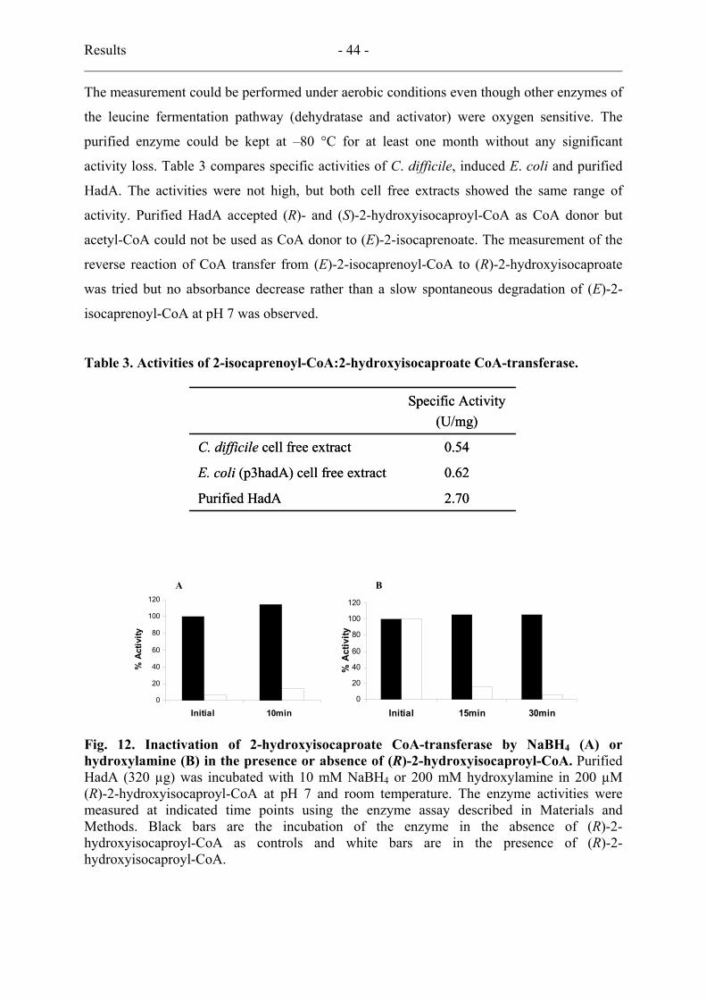
Results - 44 -
The measurement could be performed under aerobic conditions even though other enzymes of
the leucine fermentation pathway (dehydratase and activator) were oxygen sensitive. The
purified enzyme could be kept at –80 °C for at least one month without any significant
activity loss. Table 3 compares specific activities of C. difficile, induced E. coli and purified
HadA. The activities were not high, but both cell free extracts showed the same range of
activity. Purified HadA accepted (R)- and (S)-2-hydroxyisocaproyl-CoA as CoA donor but
acetyl-CoA could not be used as CoA donor to (E)-2-isocaprenoate. The measurement of the
reverse reaction of CoA transfer from (E)-2-isocaprenoyl-CoA to (R)-2-hydroxyisocaproate
was tried but no absorbance decrease rather than a slow spontaneous degradation of (E)-2-
isocaprenoyl-CoA at pH 7 was observed.
Table 3. Activities of 2-isocaprenoyl-CoA:2-hydroxyisocaproate CoA-transferase.
2.70Purified HadA
0.62E. coli (p3hadA) cell free extract
0.54C. difficile cell free extract
Specific Activity(U/mg)
2.70Purified HadA
0.62E. coli (p3hadA) cell free extract
0.54C. difficile cell free extract
Specific Activity(U/mg)
0
20
40
60
80
100
120
Initial 10min
% A
ctiv
ity
0
20
40
60
80
100
120
Initial 15min 30min
% A
ctiv
ity
A B
0
20
40
60
80
100
120
Initial 10min
% A
ctiv
ity
0
20
40
60
80
100
120
Initial 15min 30min
% A
ctiv
ity
A B
Fig. 12. Inactivation of 2-hydroxyisocaproate CoA-transferase by NaBH4 (A) or hydroxylamine (B) in the presence or absence of (R)-2-hydroxyisocaproyl-CoA. Purified HadA (320 µg) was incubated with 10 mM NaBH4 or 200 mM hydroxylamine in 200 µM (R)-2-hydroxyisocaproyl-CoA at pH 7 and room temperature. The enzyme activities were measured at indicated time points using the enzyme assay described in Materials and Methods. Black bars are the incubation of the enzyme in the absence of (R)-2-hydroxyisocaproyl-CoA as controls and white bars are in the presence of (R)-2-hydroxyisocaproyl-CoA.

Results - 45 -
3.4. Inactivation by NaBH4 or hydroxylamine
In case of family I CoA transferases, the reaction proceeds via a ping-pong mechanism,
forming a CoA-thioester intermediate on the glutamate residue of the enzyme (46, 80). The
glutamyl-CoA-thioester was reduced by NaBH4 or cleaved by hydroxylamine yielding
irreversibly inactive enzyme (16). The reaction mechanism of family III CoA transferases has
not been elucidated, but kinetic analysis suggested the reaction might be proceeded via a
ternary complex formation of the substrates (46, 62). Nevertheless the partial inactivation of
two family III CoA transferases (BbsEF of T. aromatica and FldA of C. sporogenes) was
reported by the reduction with NaBH4 or cleavage with hydroxylamine in the presence of
corresponding CoA ester. A similar inactivation of HadA was obtained as shown in Fig. 12.
HadA exhibited 90% inactivation by NaBH4 reduction, while in the absence of a CoA ester
substrate no inactivation was observed. Hydroxylamine also inactivated the enzyme showing
20% residual activity after 15 minutes and only 5 % after 30 minutes incubation.
4. Activator of (R)-2-hydroxyisocaproyl-CoA dehydratase
4.1. Analysis of hadI
The gene hadI was identified by homology analysis with known activators of C. sporogenes
(FldI) and A. fermentans (HgdC) showing 55 and 51 % amino acid sequence identities,
respectively. The identified gene was composed of 810 bp (A and T 538, 66.4% and C and G
72, 33.6%) coding for 269 amino acids. Amino acid sequence alignment with the other
activators revealed conserved ATP binding motifs G(I/V/L)D(I/V)G and (V/I)IDIG and the 2
cysteine residues known to coordinate the [4Fe-4S] cluster (20, 52). The conserved cluster
helix known from HgdC of A. fermentans plays a role in binding to [4Fe-4S] cluster located at
the interface and bridging the monomers (66). The deduced amino acid sequence of the 810-
bp hadI-gene contained three in-frame methionines (MLKMYTM) in the N-terminal region.
Furthermore the lack of a nucleotide space between the ribosome binding site (A/GGAGG/A)
and the start codon (ATG) (Fig. 13) indicated a possible error in the assignment of the start
codon. It was assumed, therefore, that the next in-frame ATG could be the real start codon of
hadI, consequently expanding the nucleotide space from 0 to 9 nucleotides and revising the
ORF, hadI, from 810 to 801 bp.

Results - 46 -
Number of Nucleotides Gene Nucleotide sequence between G/AGAAG/A and ATG
ldhA CAGGAGGGTATTGATATGAAAATACTA 8hadA AAGGAGGAAAACGTTATGCTTTTAGAA 8hadI AAGGAGAATGCTAAAGATGTACACAAT 9hadB AAAGAGGGATGAAAATGTCTGAAAAA 7hadC ATAGAGGTGAGATATAATGGAAGCTATT 9
Number of Nucleotides Gene Nucleotide sequence between G/AGAAG/A and ATG
ldhA CAGGAGGGTATTGATATGAAAATACTA 8hadA AAGGAGGAAAACGTTATGCTTTTAGAA 8hadI AAGGAGAATGCTAAAGATGTACACAAT 9hadB AAAGAGGGATGAAAATGTCTGAAAAA 7hadC ATAGAGGTGAGATATAATGGAAGCTATT 9
Fig. 13. Nucleotide sequences around the ribosome binding site and start codons of the genes. The ribosome binding sites and the start codons are shown in bold letters. Abbreviations of the genes are as described in Fig. 6. The number of nucleotides shows the nucleotide space between ribosome binding site and start codon. By using the start codons shown in grey no active proteins could be obtained.
1 ATGTACACAA TGGGATTAGA TATAGGTTCA ACTGCATCAA AGGGAGTAAT CTTAAAGAAT 61 GGGGAAGATA TTGTAGCTTC TGAAACAATA TCCTCTGGTA CTGGGACTAC TGGACCATCA121 AGAGTTTTAG AAAAATTATA TGGCAAGACA GGTCTTGCAA GAGAAGATAT TAAAAAAGTT181 GTAGTTACAG GATATGGAAG AATGAACTAT TCAGATGCTG ATAAGCAAAT AAGTGAATTA241 AGCTGTCATG CTAGAGGGGT AAATTTCATA ATTCCAGAGA CAAGAACCAT TATTGACATA301 GGTGGTCAAG ATGCAAAGGT ATTAAAATTA GATAATAATG GAAGACTATT AAACTTTCTT361 ATGAATGACA AATGTGCTGC AGGTACAGGA AGATTTTTAG ATGTAATGGC AAAAATAATA421 GAGGTTGATG TATCTGAACT CGGAAGTATA TCTATGAATT CTCAAAATGA AGTATCAATA481 AGCAGTACAT GTACAGTATT TGCAGAGTCT GAGGTTATAT CACATTTATC TGAAAATGCA541 AAAATTGAAG ATATAGTGGC AGGTATTCAT ACTTCAGTAG CAAAGAGAGT TTCTAGCCTA601 GTAAAAAGAA TAGGAGTACA AAGAAATGTA GTTATGGTTG GTGGGGTTGC TAGAAATAGT661 GGTATTGTAA GAGCTATGGC AAGAGAAATC AACACAGAAA TTATTGTACC TGATATACCT721 CAATTAACTG GTGCTTTAGG AGCAGCGTTA TATGCTTTTG ATGAAGCAAA AGAATCACAA781 AAAGAAGTGA AAAATATATA A
Fig 14. The nucleotide sequence of modified and cloned hadI of C. difficile strain DSMZ 1296T (GeneBank accession number AY772815). Start and stop codons derived from vector are underlined and two nucleotide substitutions compared to the strain 680 are depicted as the thick letters in grey box.
M CFE FT Ac
97 67
43 30 20 14
kDa
Fig. 15. Purified recombinant HadI, activator of dehydratase. SDS-PAGE (15%) stained with Coomassie Brilliant Blue. M, molecular mass marker; CFE, cell free extract of the cells induced with anhydrotetracycline 200µg/L; FT, flow-through from the column; Ac, purified activator after elution with D-desthiobiotin.

Results - 47 -
4.2 Cloning and expression of hadI and protein purification
Since the activators of 2-hydroxyacyl-CoA dehydratases are known to be extremely sensitive
against oxygen and difficult to purify in sufficient amount from the native organism (67), No
effort was made to obtain pure HadI from C. difficile cell free extracts. Therefore expression
of the hadI gene in E. coli seemed to be the preferred method. The produced activator from
810 bp hadI as Strep-tag II peptides fused to the N- or C-terminus was obtained as insoluble
or non-functional, which neither did exhibit ATPase activity nor activate dehydratase. We
decided to modify the ORF choosing the second methionine as start codon and these
shortened hadI from 810 to 801 bp. The modified hadI was amplified using the PCR primers,
which are described in Materials and Methods, and cloned into pASK-IBA3 named Np3I. The
nucleotide sequence (Fig. 14) showed two nucleotide substitutions at C303 → T and A645 →
G comparing to the strain 680, but the encoded amino acids were 100% identical to those of
the sequence from the Sanger Center. The cloned gene was coding for 266 amino acids
starting with (MYTM), instead of 269 amino acids.
In order to produce the activator of 2-hydroxyisocaproyl-CoA dehydratase, E. coli
cells harbouring the Np3I were grown and induced under anaerobic conditions. The harvested
cells were opened by a French Press to avoid heating the sensitive enzyme and the produced
protein fused with a Strep-tag II peptide on the C-terminus was purified using a Strep-Tactin
affinity column. The pure activator eluted in Mops pH 7.0 containing, in addition to 3 mM D-
desthiobiotin, 1 mM ADP and 10 mM MgCl2 to maintain stability (Fig. 15).
4.3. UV-vis spectra and ATPase activity
The UV-visible spectra of the purified activator as isolated showed a shoulder around 370 nm,
which was shifted to 420 nm after reduction by a 10 fold excess of dithionite with
concomitant formation of an absorbance band at 570 nm visible. After oxidation of the
isolated form with a 10 fold excess of thionine, the 370 nm shoulder was shifted to 400 nm
with 10% decrease of absorbance (Fig. 16). By chemical analysis, 4 ± 0.5 non-heme iron and
2 ± 0.1 acid labile sulfur were detected indicating one [4Fe-4S] cluster in the isolated protein.
The low observed sulfur content presumably resulted from loss of H2S during storage of the
extremely labile [4Fe-4S] protein. The other known activators from C. sporogenes and A.
fermentans showed ATP hydrolysis activities when the proteins were oxidized (>2.0 s-1 and 4-
6 s-1, respectively) (27, 43). However, both reduced and oxidized HadI showed low ATP
hydrolysis activity (< 1.5 U/mg), but the reduced HadI in presence of dehydratase showed
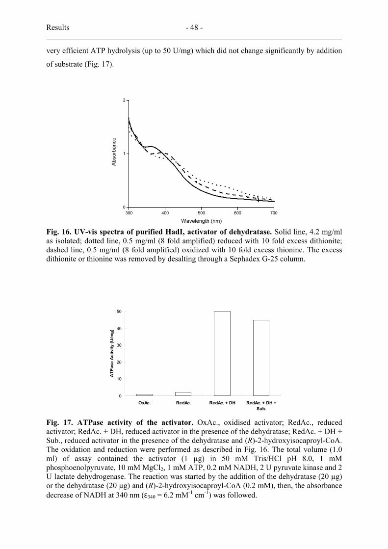
Results - 48 -
very efficient ATP hydrolysis (up to 50 U/mg) which did not change significantly by addition
of substrate (Fig. 17).
300 400 500 600 7000
1
2
Abso
rban
ce
Wavelength (nm)
Fig. 16. UV-vis spectra of purified HadI, activator of dehydratase. Solid line, 4.2 mg/ml as isolated; dotted line, 0.5 mg/ml (8 fold amplified) reduced with 10 fold excess dithionite; dashed line, 0.5 mg/ml (8 fold amplified) oxidized with 10 fold excess thionine. The excess dithionite or thionine was removed by desalting through a Sephadex G-25 column.
0
10
20
30
40
50
OxAc. RedAc. RedAc. + DH RedAc. + DH +Sub.
ATP
ase
Act
ivity
(U/m
g)
Fig. 17. ATPase activity of the activator. OxAc., oxidised activator; RedAc., reduced activator; RedAc. + DH, reduced activator in the presence of the dehydratase; RedAc. + DH + Sub., reduced activator in the presence of the dehydratase and (R)-2-hydroxyisocaproyl-CoA. The oxidation and reduction were performed as described in Fig. 16. The total volume (1.0 ml) of assay contained the activator (1 µg) in 50 mM Tris/HCl pH 8.0, 1 mM phosphoenolpyruvate, 10 mM MgCl2, 1 mM ATP, 0.2 mM NADH, 2 U pyruvate kinase and 2 U lactate dehydrogenase. The reaction was started by the addition of the dehydratase (20 µg) or the dehydratase (20 µg) and (R)-2-hydroxyisocaproyl-CoA (0.2 mM), then, the absorbance decrease of NADH at 340 nm (ε340 = 6.2 mM-1 cm-1) was followed.

Results - 49 -
5. (R)-2-Hydroxyisocaproyl-CoA dehydratase
5.1. Analysis of hadBC
The deduced amino acid sequence of hadBC of the two subunits of (R)-2-hydroxyisocaproyl-
CoA dehydratase showed 37 – 50% identity to related 2-hydroxyacyl-CoA dehydratases of C.
sporogenes (FldBC), A. fermentans (HgdAB), C. symbiosum (HgdAB) and F. nucleatum
(HgdAB). The ORF hadB for the B-subunit was composed of 1233 bp (A and T 883 67.7%
and C and G 397 32.3%) encoding 410 amino acids. The next ORF hadC for the C-subunit
was 1128 bp (A and T 768 68.1% and C and G 360 31.9%) encoding 375 amino acids. The
start codon of hadC had an overlap of 1 nucleotide with the stop codon of hadB. A
presumable error in the assignment of the start codon was found on hadB (Fig. 13). Two in-
frame methionines (MKM) were found in N-terminus of the HadB. When the second
methionine was assigned as the start, the nucleotide space between ribosome binding site and
the start codon could be extended to 7 instead of 1. A possible ribosome binding site for
subunit C was found in the structural gene hadB 9 nucleotides apart from the start codon of
hadC, which was expressed.
5.2. Cloning and expression of hadBC and protein purification
Finally hadBC was modified from 2360 bp to 2354 extending the space between the ribosome
binding site of hadB and its start codon to 7 bp. The modified hadBC for the two subunits of
dehydratase was amplified as one fragment with the primers described in Materials and
Methods. The PCR amplified ≈ 2.4 kb fragment was cut and ligated into the BsaI restriction
site of pASK-IBA3 giving Np3BC. The nucleotide sequences from three different PCRs
showed 5 – 10 mutations, which were removed by recombination of the clones. The final
nucleotide sequence of recombined hadBC (Np3BC) was composed of 2354 bp coding for
783 amino acids and having three silent nucleotide substitutions at G285 → A, T870 → C and
T2003 → C (Fig. 18).
E. coli cell harbouring the plasmid construct Np3BC was grown anaerobically at room
temperature to increase the solubility of the recombinant protein. The E. coli cell free extract
after induction showed thick protein bands by SDS/PAGE around the 43-kDa molecular mass
marker, which were not seen in non-induced E. coli. After addition of activator, a dehydratase
activity of 9 U/mg equal to that in the C. difficile cell-free extract was obtained in E. coli cell-
free extract while no activity could be measured when the non-revised hadBC genes were
expressed. Harvesting and opening the cells were done as described in Materials and
Methods. The protein could not be purified using the affinity column as while the other
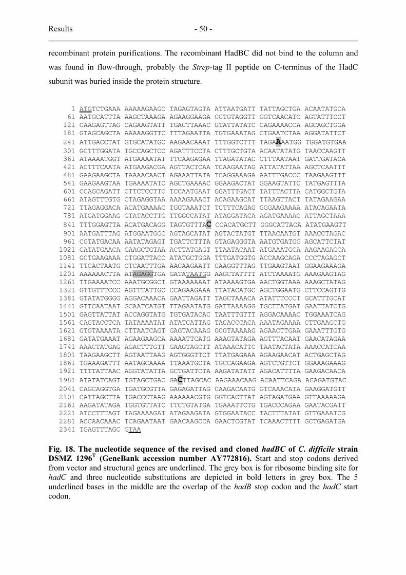
Results - 50 -
recombinant protein purifications. The recombinant HadBC did not bind to the column and
was found in flow-through, probably the Strep-tag II peptide on C-terminus of the HadC
subunit was buried inside the protein structure.
1 ATGTCTGAAA AAAAAGAAGC TAGAGTAGTA ATTAATGATT TATTAGCTGA ACAATATGCA 61 AATGCATTTA AAGCTAAAGA AGAAGGAAGA CCTGTAGGTT GGTCAACATC AGTATTTCCT 121 CAAGAGTTAG CAGAAGTATT TGACTTAAAC GTATTATATC CAGAAAACCA AGCAGCTGGA 181 GTAGCAGCTA AAAAAGGTTC TTTAGAATTA TGTGAAATAG CTGAATCTAA AGGATATTCT 241 ATTGACCTAT GTGCATATGC AAGAACAAAT TTTGGTCTTT TAGAAAATGG TGGATGTGAA 301 GCTTTGGATA TGCCAGCTCC AGATTTCCTA CTTTGCTGTA ACAATATATG TAACCAAGTT 361 ATAAAATGGT ATGAAAATAT TTCAAGAGAA TTAGATATAC CTTTAATAAT GATTGATACA 421 ACTTTCAATA ATGAAGACGA AGTTACTCAA TCAAGAATAG ATTATATTAA AGCTCAATTT 481 GAAGAAGCTA TAAAACAACT AGAAATTATA TCAGGAAAGA AATTTGACCC TAAGAAGTTT 541 GAAGAAGTAA TGAAAATATC AGCTGAAAAC GGAAGACTAT GGAAGTATTC TATGAGTTTA 601 CCAGCAGATT CTTCTCCTTC TCCAATGAAT GGATTTGACT TATTTACTTA CATGGCTGTA 661 ATAGTTTGTG CTAGAGGTAA AAAAGAAACT ACAGAAGCAT TTAAGTTACT TATAGAAGAA 721 TTAGAGGACA ACATGAAAAC TGGTAAATCT TCTTTCAGAG GGGAAGAAAA ATACAGAATA 781 ATGATGGAAG GTATACCTTG TTGGCCATAT ATAGGATACA AGATGAAAAC ATTAGCTAAA 841 TTTGGAGTTA ACATGACAGG TAGTGTTTAC CCACATGCTT GGGCATTACA ATATGAAGTT 901 AATGATTTAG ATGGAATGGC AGTAGCATAT AGTACTATGT TTAACAATGT AAACCTAGAC 961 CGTATGACAA AATATAGAGT TGATTCTTTA GTAGAGGGTA AATGTGATGG AGCATTCTAT1021 CATATGAACA GAAGCTGTAA ACTTATGAGT TTAATACAAT ATGAAATGCA AAGAAGAGCA1081 GCTGAAGAAA CTGGATTACC ATATGCTGGA TTTGATGGTG ACCAAGCAGA CCCTAGAGCT1141 TTCACTAATG CTCAATTTGA AACAAGAATT CAAGGTTTAG TTGAAGTAAT GGAAGAAAGA1201 AAAAAACTTA ATAGAGGTGA GATATAATGG AAGCTATTTT ATCTAAAATG AAAGAAGTAG1261 TTGAAAATCC AAATGCGGCT GTAAAAAAAT ATAAAAGTGA AACTGGTAAA AAAGCTATAG1321 GTTGTTTCCC AGTTTATTGC CCAGAAGAAA TTATACATGC AGCTGGAATG CTTCCAGTTG1381 GTATATGGGG AGGACAAACA GAATTAGATT TAGCTAAACA ATATTTCCCT GCATTTGCAT1441 GTTCAATAAT GCAATCATGT TTAGAATATG GATTAAAAGG TGCTTATGAT GAATTATCTG1501 GAGTTATTAT ACCAGGTATG TGTGATACAC TAATTTGTTT AGGACAAAAC TGGAAATCAG1561 CAGTACCTCA TATAAAATAT ATATCATTAG TACACCCACA AAATAGAAAA CTTGAAGCTG1621 GTGTAAAATA CTTAATCAGT GAGTACAAAG GCGTAAAAAG AGAACTTGAA GAAATTTGTG1681 GATATGAAAT AGAAGAAGCA AAAATTCATG AAAGTATAGA AGTTTACAAT GAACATAGAA1741 AAACTATGAG AGACTTTGTT GAAGTAGCTT ATAAACATTC TAATACTATA AAACCATCAA1801 TAAGAAGCTT AGTAATTAAG AGTGGGTTCT TTATGAGAAA AGAAGAACAT ACTGAGCTAG1861 TGAAAGATTT AATAGCAAAA TTAAATGCTA TGCCAGAAGA AGTCTGTTCT GGAAAGAAAG1921 TTTTATTAAC AGGTATATTA GCTGATTCTA AAGATATATT AGACATTTTA GAAGACAACA1981 ATATATCAGT TGTAGCTGAC GACTTAGCAC AAGAAACAAG ACAATTCAGA ACAGATGTAC2041 CAGCAGGTGA TGATGCGTTA GAGAGATTAG CAAGACAATG GTCAAACATA GAAGGATGTT2101 CATTAGCTTA TGACCCTAAG AAAAAACGTG GGTCACTTAT AGTAGATGAA GTTAAAAAGA2161 AAGATATAGA TGGTGTTATC TTCTGTATGA TGAAATTCTG TGACCCAGAA GAATACGATT2221 ATCCTTTAGT TAGAAAAGAT ATAGAAGATA GTGGAATACC TACTTTATAT GTTGAAATCG2281 ACCAACAAAC TCAGAATAAT GAACAAGCCA GAACTCGTAT TCAAACTTTT GCTGAGATGA2341 TGAGTTTAGC GTAA
Fig. 18. The nucleotide sequence of the revised and cloned hadBC of C. difficile strain DSMZ 1296T (GeneBank accession number AY772816). Start and stop codons derived from vector and structural genes are underlined. The grey box is for ribosome binding site for hadC and three nucleotide substitutions are depicted in bold letters in grey box. The 5 underlined bases in the middle are the overlap of the hadB stop codon and the hadC start codon.
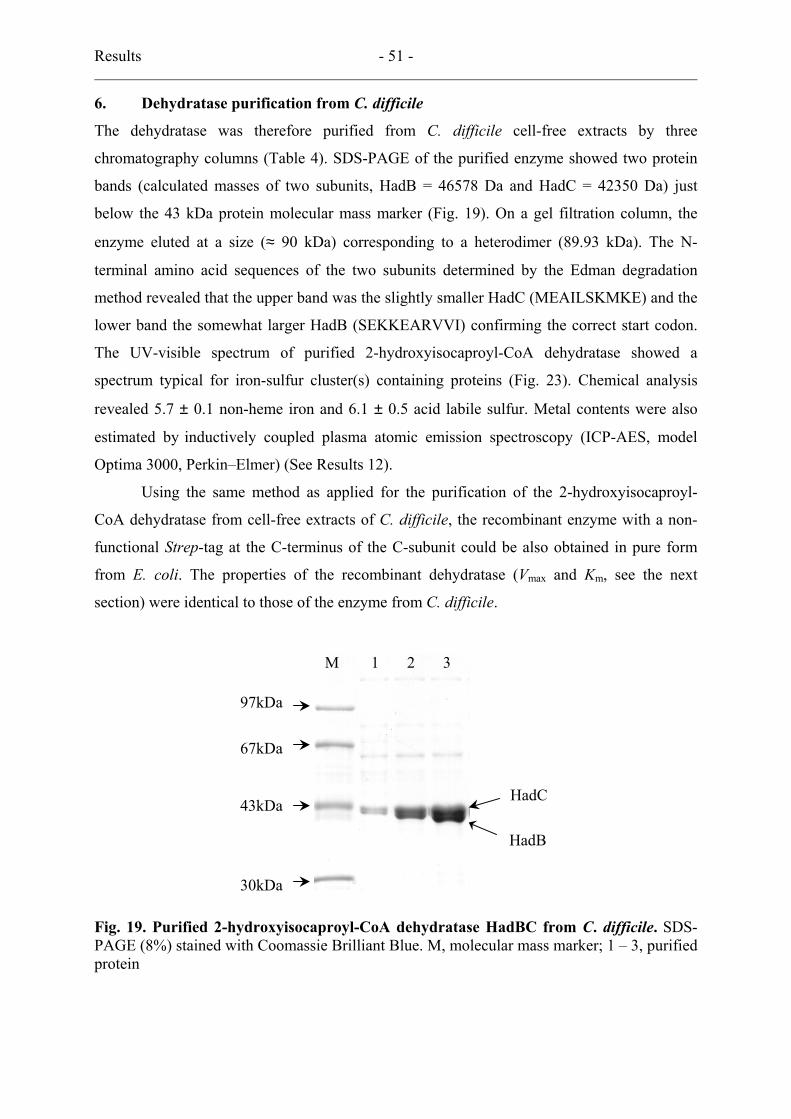
Results - 51 -
6. Dehydratase purification from C. difficile
The dehydratase was therefore purified from C. difficile cell-free extracts by three
chromatography columns (Table 4). SDS-PAGE of the purified enzyme showed two protein
bands (calculated masses of two subunits, HadB = 46578 Da and HadC = 42350 Da) just
below the 43 kDa protein molecular mass marker (Fig. 19). On a gel filtration column, the
enzyme eluted at a size (≈ 90 kDa) corresponding to a heterodimer (89.93 kDa). The N-
terminal amino acid sequences of the two subunits determined by the Edman degradation
method revealed that the upper band was the slightly smaller HadC (MEAILSKMKE) and the
lower band the somewhat larger HadB (SEKKEARVVI) confirming the correct start codon.
The UV-visible spectrum of purified 2-hydroxyisocaproyl-CoA dehydratase showed a
spectrum typical for iron-sulfur cluster(s) containing proteins (Fig. 23). Chemical analysis
revealed 5.7 ± 0.1 non-heme iron and 6.1 ± 0.5 acid labile sulfur. Metal contents were also
estimated by inductively coupled plasma atomic emission spectroscopy (ICP-AES, model
Optima 3000, Perkin–Elmer) (See Results 12).
Using the same method as applied for the purification of the 2-hydroxyisocaproyl-
CoA dehydratase from cell-free extracts of C. difficile, the recombinant enzyme with a non-
functional Strep-tag at the C-terminus of the C-subunit could be also obtained in pure form
from E. coli. The properties of the recombinant dehydratase (Vmax and Km, see the next
section) were identical to those of the enzyme from C. difficile.
97kDa
67kDa
43kDa
30kDa
M 1 2 3
HadB
HadC
Fig. 19. Purified 2-hydroxyisocaproyl-CoA dehydratase HadBC from C. difficile. SDS-PAGE (8%) stained with Coomassie Brilliant Blue. M, molecular mass marker; 1 – 3, purified protein
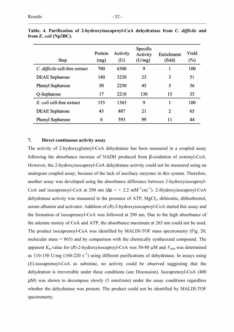
Results - 52 -
Table. 4. Purification of 2-hydroxyisocaproyl-CoA dehydratase from C. difficile and from E. coli (Np3BC).
4411995936Phenyl Sepharose
6522188743DEAE Sepharose
100191363153E. coli cell-free extract
3515130221017Q-Sepharose
36545225050Phenyl Sepharose
513233220140DEAE Sepharose
100196300700C. difficile cell-free extract
Yield(%)
Enrichment (fold)
Specific Activity (U/mg)
Activity(U)
Protein(mg)Step
4411995936Phenyl Sepharose
6522188743DEAE Sepharose
100191363153E. coli cell-free extract
3515130221017Q-Sepharose
36545225050Phenyl Sepharose
513233220140DEAE Sepharose
100196300700C. difficile cell-free extract
Yield(%)
Enrichment (fold)
Specific Activity (U/mg)
Activity(U)
Protein(mg)Step
7. Direct continuous activity assay
The activity of 2-hydroxyglutaryl-CoA dehydratase has been measured in a coupled assay
following the absorbance increase of NADH produced from β-oxidation of crotonyl-CoA.
However, the 2-hydroxyisocaproyl-CoA dehydratase activity could not be measured using an
analogous coupled assay, because of the lack of auxiliary enzymes in this system. Therefore,
another assay was developed using the absorbance difference between 2-hydroxyisocaproyl-
CoA and isocaprenoyl-CoA at 290 nm (∆ε = + 2.2 mM−1cm−1). 2-Hydroxyisocaproyl-CoA
dehydratase activity was measured in the presence of ATP, MgCl2, dithionite, dithiothreitol,
serum albumin and activator. Addition of (R)-2-hydroxyisocaproyl-CoA started this assay and
the formation of isocaprenoyl-CoA was followed at 290 nm. Due to the high absorbance of
the adenine moiety of CoA and ATP, the absorbance maximum at 263 nm could not be used.
The product isocaprenoyl-CoA was identified by MALDI-TOF mass spectrometry (Fig. 20,
molecular mass = 865) and by comparison with the chemically synthesized compound. The
apparent Km value for (R)-2-hydroxyisocaproyl-CoA was 50-80 µM and Vmax was determined
as 110-150 U/mg (160-220 s–1) using different purifications of dehydratase. In assays using
(E)-isocaprenoyl-CoA as substrate, no activity could be observed suggesting that the
dehydration is irreversible under these conditions (see Discussion). Isocaprenoyl-CoA (400
µM) was shown to decompose slowly (5 nmol/min) under the assay conditions regardless
whether the dehydratase was present. The product could not be identified by MALDI-TOF
spectrometry.
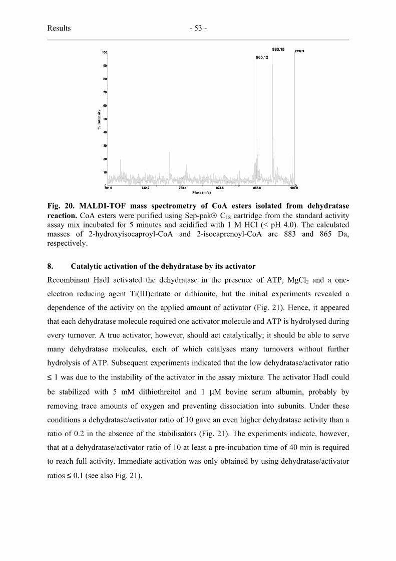
Results - 53 -
701.0 742.2 783.4 824.6 865.8 907.0Mass (m/z)
0
2732.9
0
10
20
30
40
50
60
70
80
90
100
%In
tens
ity
883.15
865.12
Mass (m/z)
% In
tens
ity
701.0 742.2 783.4 824.6 865.8 907.0Mass (m/z)
0
2732.9
0
10
20
30
40
50
60
70
80
90
100
%In
tens
ity
701.0 742.2 783.4 824.6 865.8 907.0Mass (m/z)
0
2732.9
0
10
20
30
40
50
60
70
80
90
100
%In
tens
ity
883.15
865.12
Mass (m/z)
% In
tens
ity
Fig. 20. MALDI-TOF mass spectrometry of CoA esters isolated from dehydratase reaction. CoA esters were purified using Sep-pak C18 cartridge from the standard activity assay mix incubated for 5 minutes and acidified with 1 M HCl (< pH 4.0). The calculated masses of 2-hydroxyisocaproyl-CoA and 2-isocaprenoyl-CoA are 883 and 865 Da, respectively.
8. Catalytic activation of the dehydratase by its activator
Recombinant HadI activated the dehydratase in the presence of ATP, MgCl2 and a one-
electron reducing agent Ti(III)citrate or dithionite, but the initial experiments revealed a
dependence of the activity on the applied amount of activator (Fig. 21). Hence, it appeared
that each dehydratase molecule required one activator molecule and ATP is hydrolysed during
every turnover. A true activator, however, should act catalytically; it should be able to serve
many dehydratase molecules, each of which catalyses many turnovers without further
hydrolysis of ATP. Subsequent experiments indicated that the low dehydratase/activator ratio
≤ 1 was due to the instability of the activator in the assay mixture. The activator HadI could
be stabilized with 5 mM dithiothreitol and 1 µM bovine serum albumin, probably by
removing trace amounts of oxygen and preventing dissociation into subunits. Under these
conditions a dehydratase/activator ratio of 10 gave an even higher dehydratase activity than a
ratio of 0.2 in the absence of the stabilisators (Fig. 21). The experiments indicate, however,
that at a dehydratase/activator ratio of 10 at least a pre-incubation time of 40 min is required
to reach full activity. Immediate activation was only obtained by using dehydratase/activator
ratios ≤ 0.1 (see also Fig. 21).
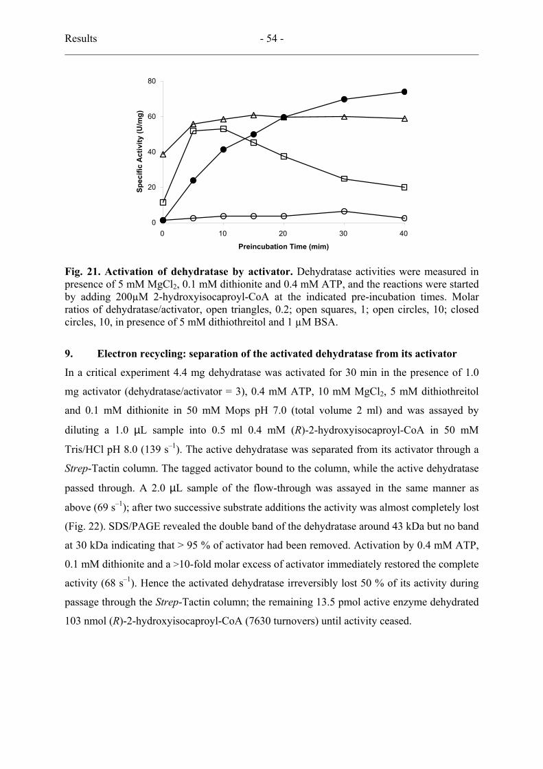
Results - 54 -
0
20
40
60
80
0 10 20 30
Preincubation Time (mim)
Spec
ific
Act
ivity
(U/m
g)
40
Fig. 21. Activation of dehydratase by activator. Dehydratase activities were measured in presence of 5 mM MgCl2, 0.1 mM dithionite and 0.4 mM ATP, and the reactions were started by adding 200µM 2-hydroxyisocaproyl-CoA at the indicated pre-incubation times. Molar ratios of dehydratase/activator, open triangles, 0.2; open squares, 1; open circles, 10; closed circles, 10, in presence of 5 mM dithiothreitol and 1 µM BSA.
9. Electron recycling: separation of the activated dehydratase from its activator
In a critical experiment 4.4 mg dehydratase was activated for 30 min in the presence of 1.0
mg activator (dehydratase/activator = 3), 0.4 mM ATP, 10 mM MgCl2, 5 mM dithiothreitol
and 0.1 mM dithionite in 50 mM Mops pH 7.0 (total volume 2 ml) and was assayed by
diluting a 1.0 µL sample into 0.5 ml 0.4 mM (R)-2-hydroxyisocaproyl-CoA in 50 mM
Tris/HCl pH 8.0 (139 s–1). The active dehydratase was separated from its activator through a
Strep-Tactin column. The tagged activator bound to the column, while the active dehydratase
passed through. A 2.0 µL sample of the flow-through was assayed in the same manner as
above (69 s–1); after two successive substrate additions the activity was almost completely lost
(Fig. 22). SDS/PAGE revealed the double band of the dehydratase around 43 kDa but no band
at 30 kDa indicating that > 95 % of activator had been removed. Activation by 0.4 mM ATP,
0.1 mM dithionite and a >10-fold molar excess of activator immediately restored the complete
activity (68 s–1). Hence the activated dehydratase irreversibly lost 50 % of its activity during
passage through the Strep-Tactin column; the remaining 13.5 pmol active enzyme dehydrated
103 nmol (R)-2-hydroxyisocaproyl-CoA (7630 turnovers) until activity ceased.
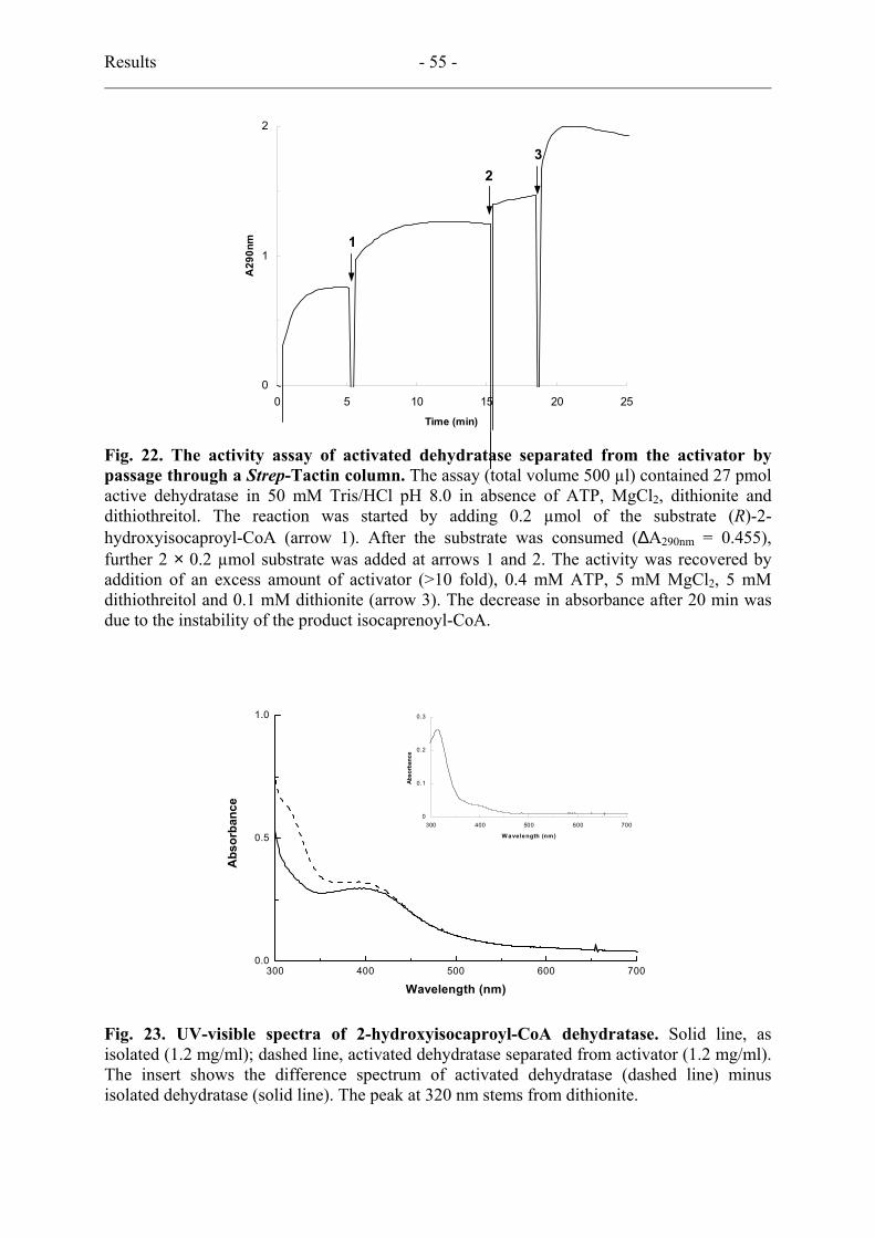
Results - 55 -
0
1
2
0 5 10 15 20 25
Time (min)
A29
0nm
32
1
0
1
2
0 5 10 15 20 25
Time (min)
A29
0nm
32
1
Fig. 22. The activity assay of activated dehydratase separated from the activator by passage through a Strep-Tactin column. The assay (total volume 500 µl) contained 27 pmol active dehydratase in 50 mM Tris/HCl pH 8.0 in absence of ATP, MgCl2, dithionite and dithiothreitol. The reaction was started by adding 0.2 µmol of the substrate (R)-2-hydroxyisocaproyl-CoA (arrow 1). After the substrate was consumed (∆A290nm = 0.455), further 2 × 0.2 µmol substrate was added at arrows 1 and 2. The activity was recovered by addition of an excess amount of activator (>10 fold), 0.4 mM ATP, 5 mM MgCl2, 5 mM dithiothreitol and 0.1 mM dithionite (arrow 3). The decrease in absorbance after 20 min was due to the instability of the product isocaprenoyl-CoA.
300 400 500 600 7000.0
0.5
1.0
Abs
orba
nce
Wavelength (nm)
0
0.1
0.2
0.3
300 400 500 600 700
Wavelength (nm)
Abso
rban
ce
Fig. 23. UV-visible spectra of 2-hydroxyisocaproyl-CoA dehydratase. Solid line, as isolated (1.2 mg/ml); dashed line, activated dehydratase separated from activator (1.2 mg/ml). The insert shows the difference spectrum of activated dehydratase (dashed line) minus isolated dehydratase (solid line). The peak at 320 nm stems from dithionite.

Results - 56 -
This experiment shows that the activated dehydratase retains its activity (i) in the absence of
0.4 mM ATP, which was diluted in the assay prior to the affinity chromatography to 0.8 µM;
(ii) after affinity chromatography at < 0.8 µM ATP and in the absence of at least 95 % of the
activator (dehydratase/activator > 160 and absence of stabilisators); (iii) turnover causes rapid
inactivation. The UV-visible spectra between 300 and 700 nm of the dehydratase as isolated
and after activation + affinity chromatography are shown in Fig. 23, which reveal the
absorbance of a [4Fe-4S]2+ cluster around 400 nm. In the difference spectrum (insert of Fig.
23) the peak at 320 nm stems from dithionite, whereas the increase in absorbance around 400
nm may be caused by the irreversible inactivation of 50 % of the dehydratase during affinity
chromatography. In another experiment, in which the Strep-Tactin column was not treated
with dithionite prior to the affinity chromatography (see Materials and Methods 3.10), the
yield of active dehydratase was only 10 %, but the absorbance increase around 400 nm was
higher. In contrast to that expected for a reduction of a [4Fe-4S]2+ cluster, no decrease in
absorbance was observed.
0.0
0.2
0.4
0.6
0.8
1.0
1.2
1.4
0 5 10 15 20 25
Time(hour)
Log
OD
(578
nm)
Fig. 24. The effect of metronidazole on C. difficile growth. Open circles, no metronidazole; open squares, 5 µM; open triangles, 10 µM; closed circles, 25 µM; closed squares, 50 µM; closed triangles, 100 µM.
10. Metronidazole effect
It was suggested that 2-hydroxyisocaproyl-CoA dehydratase or the activator could be a very
sensitive target of metronidazole (26), which has been used as an antibiotic for C. difficile
infections in human body (83). Indeed, metronidazole and the other nitro-compound such as
4-nitrophenol and chloramphenicol inhibited 2-hydroxylglutaryl-CoA dehydratases at 10 µM
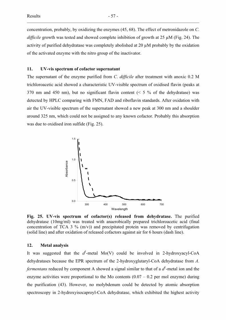
Results - 57 -
concentration, probably, by oxidizing the enzymes (45, 68). The effect of metronidazole on C.
difficile growth was tested and showed complete inhibition of growth at 25 µM (Fig. 24). The
activity of purified dehydratase was completely abolished at 20 µM probably by the oxidation
of the activated enzyme with the nitro group of the inactivator.
11. UV-vis spectrum of cofactor supernatant
The supernatant of the enzyme purified from C. difficile after treatment with anoxic 0.2 M
trichloroacetic acid showed a characteristic UV-visible spectrum of oxidised flavin (peaks at
370 nm and 450 nm), but no significant flavin content (< 5 % of the dehydratase) was
detected by HPLC comparing with FMN, FAD and riboflavin standards. After oxidation with
air the UV-visible spectrum of the supernatant showed a new peak at 300 nm and a shoulder
around 325 nm, which could not be assigned to any known cofactor. Probably this absorption
was due to oxidised iron sulfide (Fig. 25).
300 400 500 600 7000.0
0.5
1.0
1.5
Abso
rban
ce
Wavelength
Fig. 25. UV-vis spectrum of cofactor(s) released from dehydratase. The purified dehydratase (10mg/ml) was treated with anaerobically prepared trichloroacetic acid (final concentration of TCA 3 % (m/v)) and precipitated protein was removed by centrifugation (solid line) and after oxidation of released cofactors against air for 6 hours (dash line).
12. Metal analysis
It was suggested that the d1-metal Mo(V) could be involved in 2-hydroxyacyl-CoA
dehydratases because the EPR spectrum of the 2-hydroxyglutaryl-CoA dehydratase from A.
fermentans reduced by component A showed a signal similar to that of a d1-metal ion and the
enzyme activities were proportional to the Mo contents (0.07 – 0.2 per mol enzyme) during
the purification (43). However, no molybdenum could be detected by atomic absorption
spectroscopy in 2-hydroxyisocaproyl-CoA dehydratase, which exhibited the highest activity
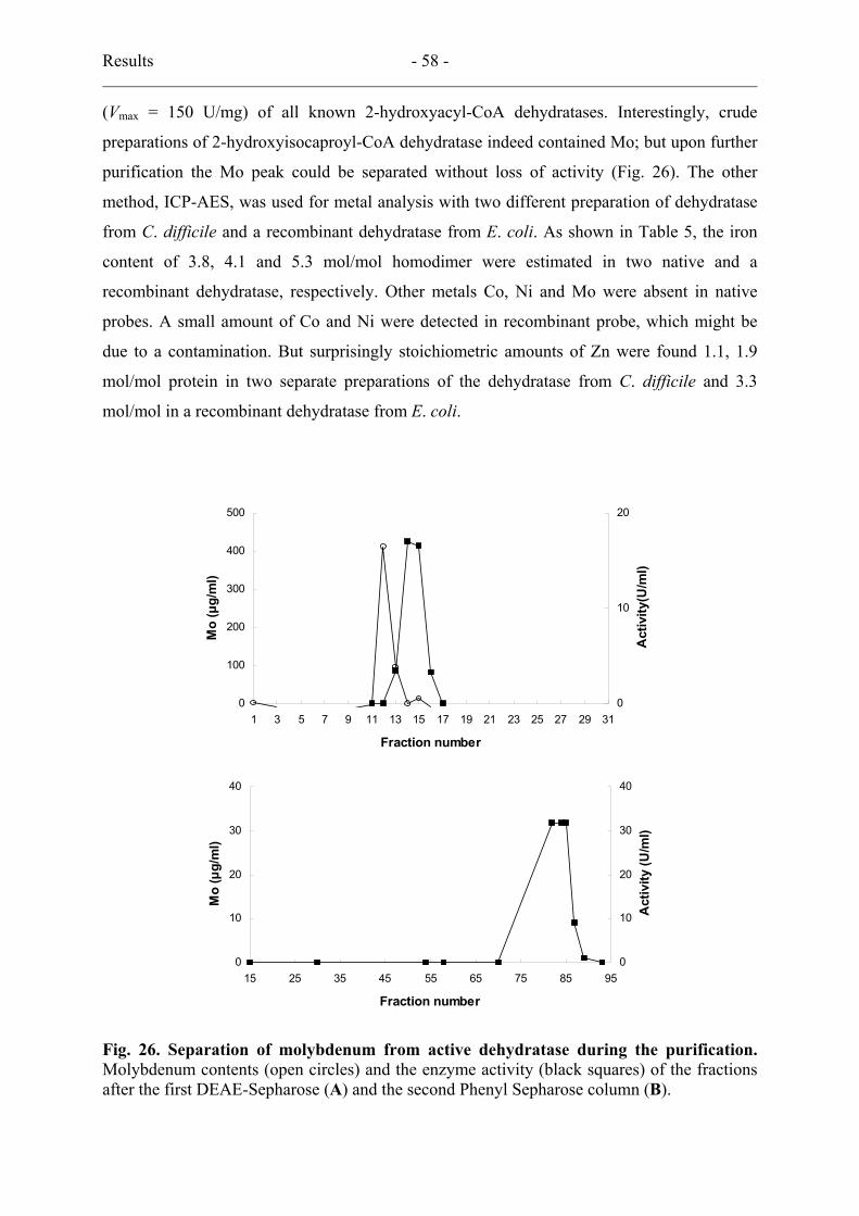
Results - 58 -
(Vmax = 150 U/mg) of all known 2-hydroxyacyl-CoA dehydratases. Interestingly, crude
preparations of 2-hydroxyisocaproyl-CoA dehydratase indeed contained Mo; but upon further
purification the Mo peak could be separated without loss of activity (Fig. 26). The other
method, ICP-AES, was used for metal analysis with two different preparation of dehydratase
from C. difficile and a recombinant dehydratase from E. coli. As shown in Table 5, the iron
content of 3.8, 4.1 and 5.3 mol/mol homodimer were estimated in two native and a
recombinant dehydratase, respectively. Other metals Co, Ni and Mo were absent in native
probes. A small amount of Co and Ni were detected in recombinant probe, which might be
due to a contamination. But surprisingly stoichiometric amounts of Zn were found 1.1, 1.9
mol/mol protein in two separate preparations of the dehydratase from C. difficile and 3.3
mol/mol in a recombinant dehydratase from E. coli.
0
100
200
300
400
500
1 3 5 7 9 11 13 15 17 19 21 23 25 27 29 31
Fraction number
Mo
(µg/
ml)
0
10
20
Act
ivity
(U/m
l)
0
10
20
30
40
15 25 35 45 55 65 75 85 95
Fraction number
Mo
(µg/
ml)
0
10
20
30
40
Act
ivity
(U/m
l)
0
100
200
300
400
500
1 3 5 7 9 11 13 15 17 19 21 23 25 27 29 31
Fraction number
Mo
(µg/
ml)
0
10
20
Act
ivity
(U/m
l)
0
10
20
30
40
15 25 35 45 55 65 75 85 95
Fraction number
Mo
(µg/
ml)
0
10
20
30
40
Act
ivity
(U/m
l)
Fig. 26. Separation of molybdenum from active dehydratase during the purification. Molybdenum contents (open circles) and the enzyme activity (black squares) of the fractions after the first DEAE-Sepharose (A) and the second Phenyl Sepharose column (B).
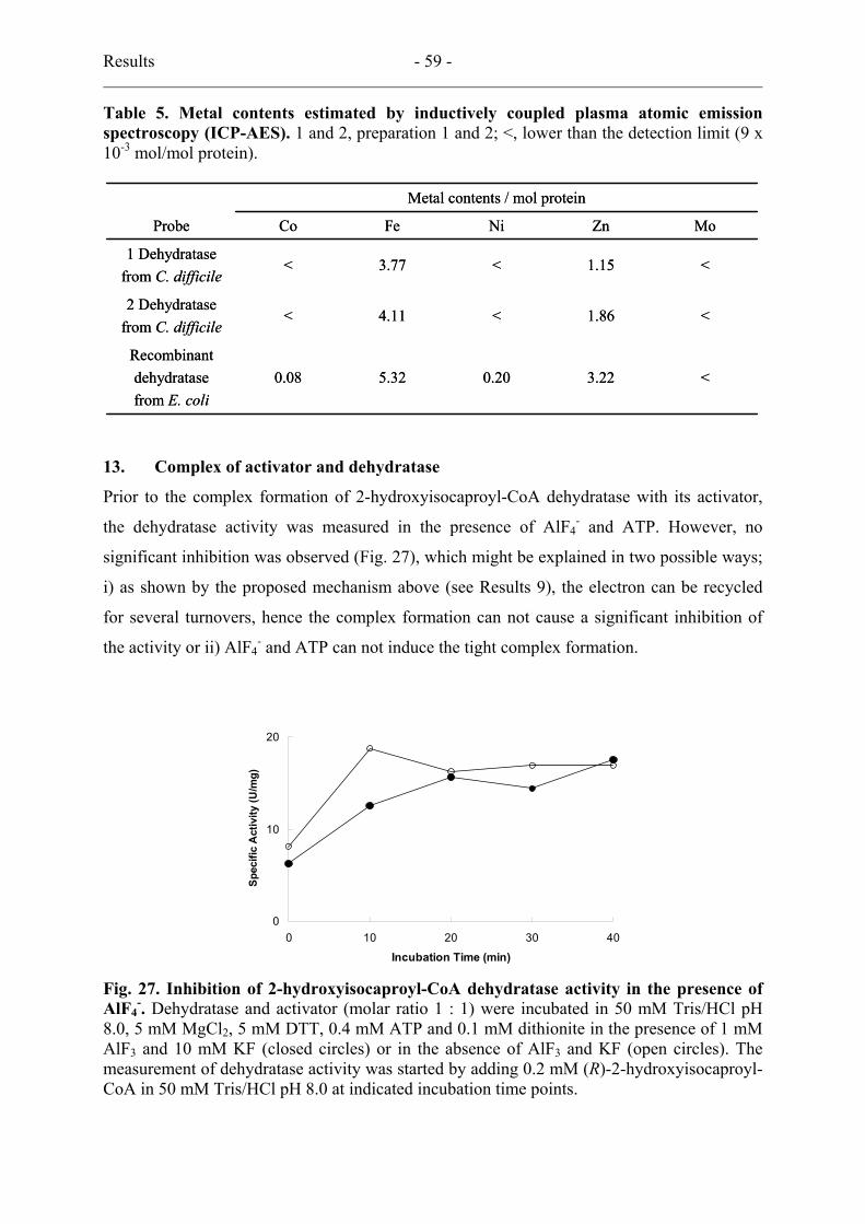
Results - 59 -
Table 5. Metal contents estimated by inductively coupled plasma atomic emission spectroscopy (ICP-AES). 1 and 2, preparation 1 and 2; <, lower than the detection limit (9 x 10⁻³ mol/mol protein).
<3.220.205.320.08Recombinantdehydratasefrom E. coli
<1.86<4.11<2 Dehydratase
from C. difficile
<1.15<3.77<1 Dehydratase
from C. difficile
MoZnNiFeCoProbe
Metal contents / mol protein
<3.220.205.320.08Recombinantdehydratasefrom E. coli
<1.86<4.11<2 Dehydratase
from C. difficile
<1.15<3.77<1 Dehydratase
from C. difficile
MoZnNiFeCoProbe
Metal contents / mol protein
13. Complex of activator and dehydratase
Prior to the complex formation of 2-hydroxyisocaproyl-CoA dehydratase with its activator,
the dehydratase activity was measured in the presence of AlF4- and ATP. However, no
significant inhibition was observed (Fig. 27), which might be explained in two possible ways;
i) as shown by the proposed mechanism above (see Results 9), the electron can be recycled
for several turnovers, hence the complex formation can not cause a significant inhibition of
the activity or ii) AlF4- and ATP can not induce the tight complex formation.
0
10
20
0 10 20 30 4
Incubation Time (min)
Spec
ific
Act
ivity
(U/m
g)
0
Fig. 27. Inhibition of 2-hydroxyisocaproyl-CoA dehydratase activity in the presence of AlF4
-. Dehydratase and activator (molar ratio 1 : 1) were incubated in 50 mM Tris/HCl pH 8.0, 5 mM MgCl2, 5 mM DTT, 0.4 mM ATP and 0.1 mM dithionite in the presence of 1 mM AlF3 and 10 mM KF (closed circles) or in the absence of AlF3 and KF (open circles). The measurement of dehydratase activity was started by adding 0.2 mM (R)-2-hydroxyisocaproyl-CoA in 50 mM Tris/HCl pH 8.0 at indicated incubation time points.

Results - 60 -
0 10 20 30 40 50 6
Elution tim e (m in)
Abs
orba
nce
at 2
80 n
m
0
A
B
1 2
0 10 20 30 40 50 6
Elution tim e (m in)
Abs
orba
nce
at 2
80 n
m
0
A
B
1 2
Fig. 28. Separation of AlF4- induced complex by gel filtration on Superdex 200. A,
dehydratase (120 µg) and activator (144 µg) were incubated as described in Fig. 27 for 30 minutes at room temperature in the presence of 1 mM AlF3 and 10 mM KF; B, same as A in the absence of AlF3 and KF. Dashed line 1, the elution peak for the complex; Dashed line 2, the elution peak for the overlap of dehydratase and activator.
M Ac DH 1 2 3
97
67
43
30
20
14
kDaM Ac DH 1 2 3
97
67
43
30
20
14
kDa
Dehydratase
Activator
M Ac DH 1 2 3
97
67
43
30
20
14
kDaM Ac DH 1 2 3
97
67
43
30
20
14
kDa
Dehydratase
Activator
Fig. 29. Coomassie stained SDS-PAGE (15 %) of tight complex purified using Strep-Tactin affinity column. M, protein size marker; Ac, purified activator; DH, partially purified dehydratase; 1, purified complex after incubation of dehydratase/activator (1.8/1 molar ratio) as described in Fig. 27 in the presence of 1 mM AlF3 and 10 mM KF; 2, in the absence of AlF3 and KF; 3, in presence of 1 mM AlF3, 10 mM KF and 1 mM (R)-2-hydorxyisocaproyl-CoA.
In order to get an answer, the purification of the complex of the activator and the dehydratase
was performed by gel filtration chromatography (Fig. 28). The complex induced by AlF4- and
ATP could be seen by an earlier elution time but the separation was not good enough due to
the overlap of non-complexed proteins. Another purification of the complex using the Strep-
Tactin affinity column could be applied. When the activator forms the complex with the
dehydratase, the complex could be bound to the column by using the Strep-tag II peptide on
the C-terminus of the activator. The purified complex from the affinity column showed
clearly two protein bands of the dehydratase and the activator (intensities of the dehydratase :
activator = 1 : 1 were confirmed by Image quantitator) while one protein band of the activator

Results - 61 -
could be seen on SDS-PAGE of the non-induced reaction (Fig. 29). The complex formation
was not affected by the addition of the substrate showing two protein bands on SDS-PAGE.
However, the purified complex from the affinity column was not stable enough to be
concentrated on 100 kDa cut off spin column, probably because of the high salt concentration
(300 mM) in the elution buffer. Afterwards the complex was purified on Amicon Ultra-4
PLTK Ultracel-Pl (100 kDa cut off) by exchanging the buffer and removing non-complxed
proteins. The purified complex exhibited about 20 % dehydratase activity of the non-purified
complex, which might be loss of the electron by the oxidation of the sensitive complex as the
activated dehydratase (See Results 9). The purified active AlF4--induced complex was used
for the crystallisation trial for the structure analysis. The obtained microcrystals exhibited
bands of the dehydratase and activator on SDS/PAGE (performed by Dr. Berta M. Martin in
Universität Bayreuth).
14. Detection of a substrate-derived organic radical by EPR spectroscopy
EPR spectra were obtained to probe the formation of an organic radical which might be
involved in 2-hydroxyacyl-CoA dehydration mechanism. The activator of 2-
hydroxyisocaproyl-CoA dehydratase from C. difficile was reduced with 10 fold excess
dithionite and excess dithionite was removed by gel filtration on Sephadex G-25.
300 320 340 360 380
Fie ld (m T)
50 100 150 200 250 300
Fie ld (m T)
AC+ADP
AC+DH+ADP
AC+DH+ATP
AlF4-complex+ATP
AlF4-complex+ATP+ substrate
AC+DH+ATP+ substrate
X 0.5
A B
300 320 340 360 380
Fie ld (m T)
50 100 150 200 250 300
Fie ld (m T)
AC+ADP
AC+DH+ADP
AC+DH+ATP
AlF4-complex+ATP
AlF4-complex+ATP+ substrate
AC+DH+ATP+ substrate
X 0.5
50 100 150 200 250 300
Fie ld (m T)
AC+ADP
AC+DH+ADP
AC+DH+ATP
AlF4-complex+ATP
AlF4-complex+ATP+ substrate
AC+DH+ATP+ substrate
AC+ADP
AC+DH+ADP
AC+DH+ATP
AlF4-complex+ATP
AlF4-complex+ATP+ substrate
AC+DH+ATP+ substrate
X 0.5
A B
Fig. 30. High spin (A) and low spin (B) EPR signals of (R)-2-hydroxyisocaproyl-CoA dehydratase. Activator/dehydratase 50 µM, ATP/ADP 10 mM, MgCl2 5 mM, substrate 0.1 mM. EPR conditions: microwave frequency, 9.458 GHz; modulation frequency, 100 kHz; temperature, 10 K; modulation amplitude, 1.25 mT; microwave power, 20 mW (B), 0.1 mW (A).
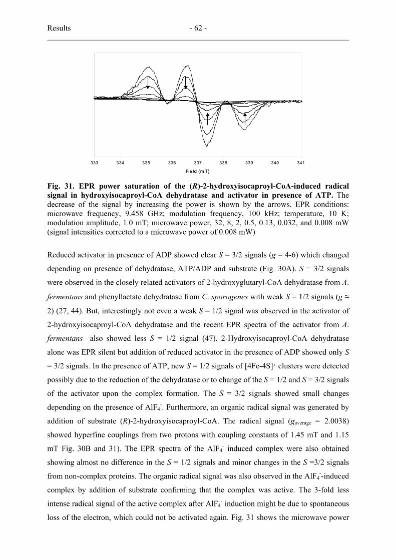
Results - 62 -
333 334 335 336 337 338 339 340 341
Fie ld (m T)
333 334 335 336 337 338 339 340 341
Fie ld (m T)
Fig. 31. EPR power saturation of the (R)-2-hydroxyisocaproyl-CoA-induced radical signal in hydroxyisocaproyl-CoA dehydratase and activator in presence of ATP. The decrease of the signal by increasing the power is shown by the arrows. EPR conditions: microwave frequency, 9.458 GHz; modulation frequency, 100 kHz; temperature, 10 K; modulation amplitude, 1.0 mT; microwave power, 32, 8, 2, 0.5, 0.13, 0.032, and 0.008 mW (signal intensities corrected to a microwave power of 0.008 mW)
Reduced activator in presence of ADP showed clear S = 3/2 signals (g = 4-6) which changed
depending on presence of dehydratase, ATP/ADP and substrate (Fig. 30A). S = 3/2 signals
were observed in the closely related activators of 2-hydroxyglutaryl-CoA dehydratase from A.
fermentans and phenyllactate dehydratase from C. sporogenes with weak S = 1/2 signals (g ≈
2) (27, 44). But, interestingly not even a weak S = 1/2 signal was observed in the activator of
2-hydroxyisocaproyl-CoA dehydratase and the recent EPR spectra of the activator from A.
fermentans also showed less S = 1/2 signal (47). 2-Hydroxyisocaproyl-CoA dehydratase
alone was EPR silent but addition of reduced activator in the presence of ADP showed only S
= 3/2 signals. In the presence of ATP, new S = 1/2 signals of [4Fe-4S]+ clusters were detected
possibly due to the reduction of the dehydratase or to change of the S = 1/2 and S = 3/2 signals
of the activator upon the complex formation. The S = 3/2 signals showed small changes
depending on the presence of AlF4-. Furthermore, an organic radical signal was generated by
addition of substrate (R)-2-hydroxyisocaproyl-CoA. The radical signal (gaverage = 2.0038)
showed hyperfine couplings from two protons with coupling constants of 1.45 mT and 1.15
mT Fig. 30B and 31). The EPR spectra of the AlF4- induced complex were also obtained
showing almost no difference in the S = 1/2 signals and minor changes in the S =3/2 signals
from non-complex proteins. The organic radical signal was also observed in the AlF4--induced
complex by addition of substrate confirming that the complex was active. The 3-fold less
intense radical signal of the active complex after AlF4- induction might be due to spontaneous
loss of the electron, which could not be activated again. Fig. 31 shows the microwave power
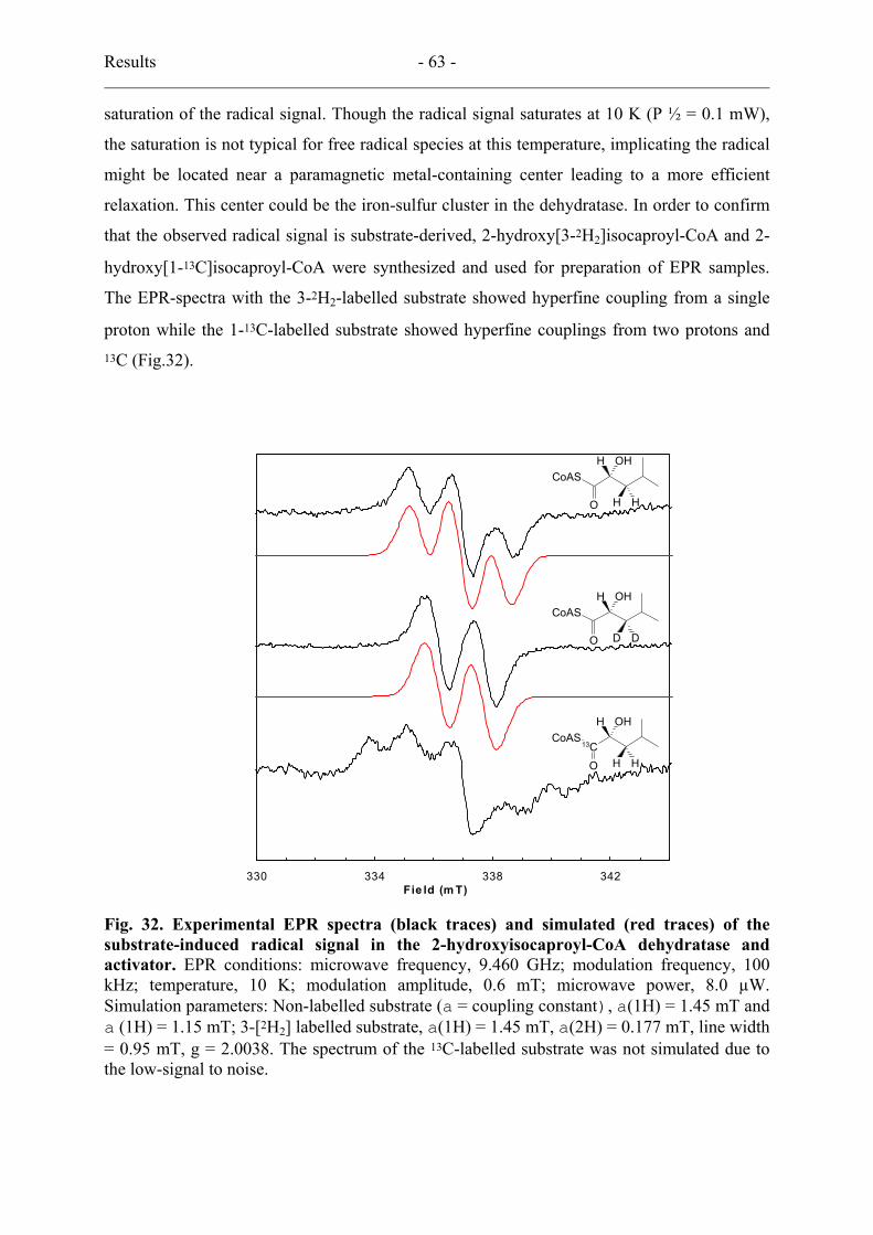
Results - 63 -
saturation of the radical signal. Though the radical signal saturates at 10 K (P ½ = 0.1 mW),
the saturation is not typical for free radical species at this temperature, implicating the radical
might be located near a paramagnetic metal-containing center leading to a more efficient
relaxation. This center could be the iron-sulfur cluster in the dehydratase. In order to confirm
that the observed radical signal is substrate-derived, 2-hydroxy[3-2H2]isocaproyl-CoA and 2-
hydroxy[1-13C]isocaproyl-CoA were synthesized and used for preparation of EPR samples.
The EPR-spectra with the 3-2H2-labelled substrate showed hyperfine coupling from a single
proton while the 1-13C-labelled substrate showed hyperfine couplings from two protons and 13C (Fig.32).
330 334 338 342Fie ld (m T)
CoAS
O
H OH
H H
CoAS
O
H OH
D D
CoAS
O
H OH
H H
13C
Fig. 32. Experimental EPR spectra (black traces) and simulated (red traces) of the substrate-induced radical signal in the 2-hydroxyisocaproyl-CoA dehydratase and activator. EPR conditions: microwave frequency, 9.460 GHz; modulation frequency, 100 kHz; temperature, 10 K; modulation amplitude, 0.6 mT; microwave power, 8.0 µW. Simulation parameters: Non-labelled substrate (a = coupling constant), a(1H) = 1.45 mT and a (1H) = 1.15 mT; 3-[2H2] labelled substrate, a(1H) = 1.45 mT, a(2H) = 0.177 mT, line width = 0.95 mT, g = 2.0038. The spectrum of the 13C-labelled substrate was not simulated due to the low-signal to noise.

Results - 64 -
Table 6. Summary of the substrate deuterium kinetic isotope effects.
1.60.96154(R)-2-Hydroxy[2,3-2H3]isocaproyl-CoA
1.41.08888(R)-2-Hydroxy[2-2H1]isocaproyl-CoA
1.31.15864(R)-2-Hydroxy[3-2H2]isocaproyl-CoA
(=1)1.478110(R)-2-Hydroxyisocaproyl-CoA
kH/kDVmax/KmKm (µM)Vmax (U/mg)
1.60.96154(R)-2-Hydroxy[2,3-2H3]isocaproyl-CoA
1.41.08888(R)-2-Hydroxy[2-2H1]isocaproyl-CoA
1.31.15864(R)-2-Hydroxy[3-2H2]isocaproyl-CoA
(=1)1.478110(R)-2-Hydroxyisocaproyl-CoA
kH/kDVmax/KmKm (µM)Vmax (U/mg)
15. Deuterium kinetic isotope effects
The kinetic isotope effects (kH/kD) on the overall 2-hydroxyisocaproyl-CoA dehydratase
reaction were obtained with deuterium labelled 2-hydroxyisocaproyl-CoA (Table 6) by
measuring the Vmax/Km values. Labelled substrates were synthesised and the activity was
measured as described in Materials and Methods.
16. Preliminary stereochemistry
The dehydratase accepted the R-isomer more efficiently while only 10 % activity could be
obtained with the (S)-2-hydroxyisocaproyl-CoA, which might be due to a contamination with
the R-isomer. Since an equal mixture of (R)- and (S)-2-hydroxyisocaproyl-CoA gave only half
of the enzymatic activity, we assume that that the S-isomer can also bind at the active site of
the enzyme but can not be dehydrated. In assays using (E)-isocaprenoyl-CoA as substrate, no
activity could be observed suggesting that the dehydration produces (Z)-2-isocaprenoyl-CoA
or is irreversible under these conditions. Isocaprenoyl-CoA (400 µM) was shown to
decompose slowly (5 nmol/min) under the assay conditions regardless whether the
dehydratase was present.
Discussion - 65 -
Discussion
1. (R)-2-Hydroxyisocaproate dehydrogenase
D-2-Hydroxyisocaproate dehydrogenase was discovered together with its L-specific
counterpart, named L-2-hydroxyisocaproate dehydrogenase (77). The low specificity of the
enzymes with respect to the substrate’s side-chain makes them attractive for biotechnological
applications for the synthesis of chiral compounds (49). D-2-Hydroxyisocaproate
dehydrogenase belongs to the NAD+ dependent 2-hydroxycarboxylate dehydrogenase family.
2-Hydroxycarboxylate dehydrogenases showing broad substrate specificity can be divided
into two classes (7): the mandelate dehydrogenase family, which prefers substrate with
branched C3-atoms and the 2-hydroxyisocaproate dehydrogenase family, for which a branch
in the substrate side chain at the C3-position is unfavorable. (R)-2-Hydroxyisocaproate
dehydrogenase from C. difficile (LdhA) is the third enzyme in the second class. The other
known two D-2-hydroxyisocaproate dehydrogenases are from Lactobacillus casei (25), whose
the crystal structure was solved, and from Lactobacillus delbrueckii (7), whose gene was
cloned and the protein was produced in E. coli. HadA is a monomeric enzyme whereas the
other two D-specific dehydrogenases are homodimers; despite this difference, all three
enzymes show similar substrate specificities. The crystal structure of the enzyme from L.
casei revealed that some amino acid residues (fairly conserved in HadA) are responsible for
substrate specificity. They suggested that Leu51 (in the L. casei enzyme) is the key residue
for the differences in substrate specificity between D-2-hydroxyisocaproate and D-lactate
dehydrogenase. Recently, D-lactate dehydrogenase form Lactobaciluus pentosus was
converted to D-2-hydroxyisocaproate dehydrogenase by replacing the corresponding Tyr by
Leu (90). This key residue is not conserved but occupied by Glu in LdhA which might
explain the complete absence of any activity with pyruvate, 3-methyl-2-oxopentonate and 2-
oxobutyrate. Even though a lot is known about structure and substrate specificity so far, the
role of the D-2-hydroxyisocaproate dehydrogenase from lactobacilli in their metabolism is
unknown. However the gene arrangement (Fig. 6) in the genome of C. difficile shows that
ldhA belongs to the gene cluster of the 2-hydroxyisocaproyl-CoA dehydratase system for
leucine reduction. The substrate specificity of the enzyme reveals that 2-hydroxyisocaproate
should be the natural substrate in vivo. Therefore HadA is clearly the first enzyme in the
reductive branch of leucine fermentation.
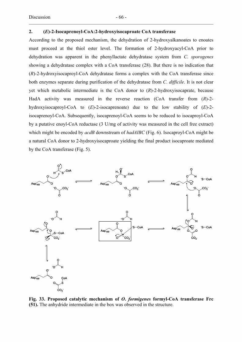
Discussion - 66 -
2. (E)-2-Isocaprenoyl-CoA:2-hydroxyisocaproate CoA transferase
According to the proposed mechanism, the dehydration of 2-hydroxyalkanoates to enoates
must proceed at the thiol ester level. The formation of 2-hydroxyacyl-CoA prior to
dehydration was apparent in the phenyllactate dehydratase system from C. sporogenes
showing a dehydratase complex with a CoA transferase (28). But there is no indication that
(R)-2-hydroxyisocaproyl-CoA dehydratase forms a complex with the CoA transferase since
both enzymes separate during purification of the dehydratase from C. difficile. It is not clear
yet which metabolic intermediate is the CoA donor to (R)-2-hydroxyisocaprate, because
HadA activity was measured in the reverse reaction (CoA transfer from (R)-2-
hydroxyisocaproyl-CoA to (E)-2-isocaprenoate) due to the low stability of (E)-2-
isocaprenoyl-CoA. Subsequently, isocaprenoyl-CoA seems to be reduced to isocaproyl-CoA
by a putative enoyl-CoA reductase (3 U/mg of activity was measured in the cell free extract)
which might be encoded by acdB downstream of hadAIBC (Fig. 6). Isocaproyl-CoA might be
a natural CoA donor to 2-hydroxyisocaproate yielding the final product isocaproate mediated
by the CoA transferase (Fig. 5).
Asp169
O-
O
O
H SCoA
Asp169 O
OH
O-
SCoA
Asp169 O
O H
O
-S CoA
Asp169
O
O
CO2--O
S CoAAsp169
O H
O
O-O O
CO2
Asp169
O
O
O CO2-
-S CoA-S CoA
Asp169
O-
O
O
-O H
O
-O H
O
-O H
-O CO2-
O
-O CO2-
O
-O CO2-
O
O
CO2-
SCoA
Asp169
O-
O
O
H SCoA
Asp169 O
OH
O-
SCoA
Asp169 O
O H
O
-S CoA
Asp169
O
O
CO2--O
S CoAAsp169
O H
O
O-O O
CO2
Asp169
O
O
O CO2-
-S CoA-S CoA
Asp169
O-
O
O
-O H
O
-O H
O
-O H
-O CO2-
O
-O CO2-
O
-O CO2-
O
O
CO2-
SCoA
Fig. 33. Proposed catalytic mechanism of O. formigenes formyl-CoA transferase Frc (51). The anhydride intermediate in the box was observed in the structure.

Discussion - 67 -
2-Hydroxyisocaproate CoA-transferase is homologous only with class III enzymes
supposing that the reaction is mediated by a ternary complex. Recently, a detailed mechanism
(Fig. 33) was proposed on the basis of structural and kinetic analysis of formate CoA-
transferase (41, 51, 75). Crystal structure of wild type and site-specific mutants revealed that
Asp169 as conserved residue in class III enzymes formed an anhydride intermediate with the
substrate carboxylate. The authors also observed kinetics different from the ping-pong scheme
of class I enzymes even though the same chemical strategy is suggested for the formation of
anhydride intermediates. Double reciprocal plots of activity vs. oxalate concentration at three
different formyl-CoA concentration showed the intersection of lines as observed in other class
III enzymes. The result indicates a formation of the ternary complex of the enzyme with two
substrates, which is different from a transient thiolester formation as shown for class I
enzymes. The different kinetics were explained with the position of free CoA in the active
site. In the class I enzymes, bound CoA reacts with the initial anhydride in the absence of the
second substrate releasing the first product and forming a glutamyl-CoA-thiol ester which was
irreversibly reduced or cleaved by sodium borohydride or hydroxylamine, respectively. On
the other hand, the free CoA in formate CoA-transferase is positioned too far away from the
relevant carbonyl group of Asp169 to form an enzyme-linked thiol ester and the first product
can be released only by the second substrate.
This proposed mechanism does not explain all results from the class III enzymes. A
curious partial inactivation by sodium borohydride was reported for cinnamoyl-
CoA:phenyllactate CoA transferase (FldA) of C. sporogenes (28) and succinyl-CoA:(R)-
benzylsuccinate CoA-transferase (BbsF) of Thauera aromatica (62) even though similar
kinetic results with double-reciprocal plots were obtained. In case of 2-hydroxyisocaproyl-
CoA transferase (HadA), complete inactivation by sodium borohydride was observed.
However the kinetic results obtained were not precise enough due to the low sensitivity of the
activity assay. Asp169, suggested in formate CoA-transferase as the anhydride forming
residue, is conserved in FldA and HadA (Fig. 9) but not in BbsF, furthermore, Tyr59, which
might stabilize the tetrahedral anhydride intermediates, is not conserved in FldA and HadA.
Therefore the proposed mechanism for the class III enzyme might be incorrect, or at least
does not apply to all class III enzymes. Further studies on HadA could help to define the
mechanism of class III enzymes.
Discussion - 68 -
3. Activator
Previous work on 2-hydroxyglutaryl-CoA dehydratase showed that the activator alone has
ATPase activity (4 – 6 s–1) only in the oxidised state, which was not improved in presence of
dehydratase and substrate (42). The results in this work, which revealed less ATPase activity
(< 1.5 s-1) of the activator HadI regardless of its oxidation state, question those data. Therefore
the original data obtained with component A of 2-hydroxyglutaryl-CoA dehydratase have
been re-calculated and found too high by at least a factor of ten. Furthermore, repetition of the
ATPase measurements with the activator of A. fermentans by applying the conditions used in
this work gave only insignificant activities (47). Therefore we conclude that the activators
from A. fermentans and C. difficile both are devoid of significant ATPase activity. Addition of
dehydratase to the corresponding reduced activator/component A, however, gave high
ATPase activities; in case of HadI + HadBC up to 50 U/mg activator were achieved. This
result fits much better to the proposed mechanism, since the electron should only be
transferred in a complex of both proteins driven by ATP-hydrolysis.
It was known that the activator is extremely sensitive against oxygen and even under
anoxic conditions, ADP or ATP and MgCl2 had to be added in the buffers in order to maintain
stability. In this work two additional stabilising factors have been discovered, DTT and high
protein concentration, which was achieved by adding BSA. In previous work on the activator
of 2-hydroxyglutaryl-CoA dehydratase, the coupled activity assay already contained DTT and
sufficient protein in the form of helping enzymes. It is assumed that the activator of 2-
hydroxyisocaproyl-CoA dehydratase is more stable than the other activators, since only S =
3/2 signals were detected. Component A of 2-hydroxyglutaryl-CoA dehydratase revealed
additional S = 1/2 signals, which was interpreted as irreversibly inactivated [2Fe-2S]2+
clusters (44). The less stable component A might be due to the presence of ATP instead of
ADP in the elution buffer. The binding of ATP at the activator causes a conformational
change which exposes the [4Fe-4S] cluster to solvent leading to a more rapid inactivation.
The usual spin ground state of [4Fe-4S]1+ found in biological systems is S = 1/2, but several
cases of mixtures of S = 1/2 and S = 3/2 were reported in the study of nitrogenase Fe-protein
(63, 64), the Pyrococcus furiosus ferredoxin (94), the Bacillus subtilis glutamine
phosphoribosylpyrophosphate amidotransferase (70) and the Clostridium thermoaceticum CO
dehydrogenase (65). Pure S = 3/2 signals of [4Fe-4S]1+ cluster are not as common as spin
mixtures, but only in the case of the A33Y mutant of P. furiosus ferredoxin a pure S = 3/2
state was observed. The wild type P. furiosus ferredoxin has mixed spin states of [4Fe-4S]1+
coordinated by four cysteins in the ferredoxin-like arrangement (–C-X2-C-X2-C- and a remote

Discussion - 69 -
–C-P-) (29). The pure S = 3/2 signal in the reduced activator shown in this work is the first
example for [4Fe-4S]1+ clusters coordinated by non-ferredoxin-like arranged cysteines.
Similar changes of S = 3/2 and 1/2 signals were observed for the [4Fe-4S]1+cluster of the
nitrogenase Fe-protein depending on ATP or ADP binding. The nucleotide binding on the Fe-
protein causes the conformational change, which could switch the electronic ground state of
the [4Fe-4S]1+cluster. Although the sequence homology does not show any relationship, the
activator has a similar protein structure, probably changing the conformation by nucleotide
binding. Indeed S = 1/2 signals were induced in the presence of ATP and dehydratase, but
EPR signals could not certainly be assigned to the [4Fe-4S]1+cluster of the activator or of the
dehydratase. Further studies by Mössbauer spectroscopy will have to clarify this aspect.
4. (R)-2-Hydroxyisocaproyl-CoA dehydratase
The results described in this work clearly show that the hadIBC-genes of C. difficile encode a
novel 2-hydroxyacyl-CoA dehydratase (HadBC) and its activator (HadI), probably specific
for the dehydration of (R)-2-hydroxyisocaproyl-CoA to isocaprenoyl-CoA, but besides the S-
isomer no other substrate was tested yet. Since the known enzymatic eliminations of water
from (R)-2-hydroxyacyl-CoA to (E)-2-enoyl-CoA, (R)-2-hydroxyglutaryl-CoA to (E)-
glutaconyl-CoA (14), (R)-lactyl-CoA to acryloyl-CoA (12) and (R)-phenyllactyl-CoA to (E)-
cinnamoyl-CoA (71) all occur in a syn-fashion, we assume that this will also be the case for
(R)-2-hydroxyisocaproyl-CoA to (E)-2-isocaprenoyl-CoA, which, however, remains to be
determined. The inability to measure the hydration of the chemically synthesised (E)-2-
isocaprenoyl-CoA could be either due to the unfavourable equilibrium or due to the Z-isomer
being the correct substrate. It has been shown that 2-hydroxyglutaryl-CoA dehydratase
catalysed the reverse reaction. The conditions, however, were different; this experiment was
performed in the cell-free extract using (E)-glutaconate in the presence of acetyl-CoA as
substrate and the formed (R)-2-hydroxyglutarate was determined enzymatically (14).
The requirement of activator (HadI), dehydratase (HadBC), ATP, Mg2+, and dithionite
for activity of 2-hydroxyisocaproyl-CoA dehydratase indicates that this enzyme acts by the
same mechanism as that proposed for 2-hydroxyacyl-CoA dehydratase (18, 55). The reduced
activator transfers one electron to the dehydratase concomitant with hydrolysis of ATP.
Although the stoichiometry of 1 or 2 ATP/electron remains to be determined, the
homodimeric structure of the activator with one [4Fe-4S] cluster strongly suggests 2
ATP/electron as observed with nitrogenase (55). The reduced dehydratase transfers the
electron further to the substrate to generate the ketyl radical anion I, which expels the adjacent
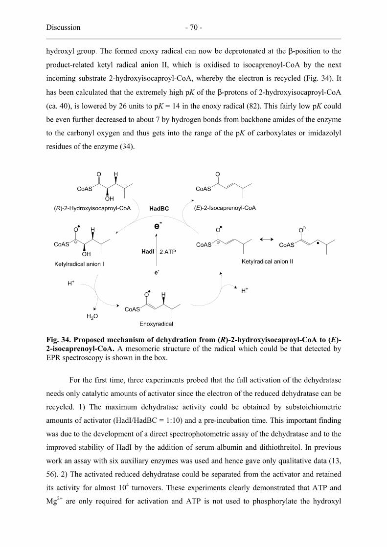
Discussion - 70 -
hydroxyl group. The formed enoxy radical can now be deprotonated at the β-position to the
product-related ketyl radical anion II, which is oxidised to isocaprenoyl-CoA by the next
incoming substrate 2-hydroxyisocaproyl-CoA, whereby the electron is recycled (Fig. 34). It
has been calculated that the extremely high pK of the β-protons of 2-hydroxyisocaproyl-CoA
(ca. 40), is lowered by 26 units to pK = 14 in the enoxy radical (82). This fairly low pK could
be even further decreased to about 7 by hydrogen bonds from backbone amides of the enzyme
to the carbonyl oxygen and thus gets into the range of the pK of carboxylates or imidazolyl
residues of the enzyme (34).
CoAS
O
CoAS
O
OH
H
CoAS
O H
H+
H2O
H+
CoAS
O
OH
H
e-
HadBC(R)-2-Hydroxyisocaproyl-CoA
Ketylradical anion I Ketylradical anion II
Enoxyradical
e-
(E)-2-Isocaprenoyl-CoA
2 ATP HadI
O
CoAS
CoAS
O
Fig. 34. Proposed mechanism of dehydration from (R)-2-hydroxyisocaproyl-CoA to (E)-2-isocaprenoyl-CoA. A mesomeric structure of the radical which could be that detected by EPR spectroscopy is shown in the box.
For the first time, three experiments probed that the full activation of the dehydratase
needs only catalytic amounts of activator since the electron of the reduced dehydratase can be
recycled. 1) The maximum dehydratase activity could be obtained by substoichiometric
amounts of activator (HadI/HadBC = 1:10) and a pre-incubation time. This important finding
was due to the development of a direct spectrophotometric assay of the dehydratase and to the
improved stability of HadI by the addition of serum albumin and dithiothreitol. In previous
work an assay with six auxiliary enzymes was used and hence gave only qualitative data (13,
56). 2) The activated reduced dehydratase could be separated from the activator and retained
its activity for almost 104 turnovers. These experiments clearly demonstrated that ATP and
Mg2+ are only required for activation and ATP is not used to phosphorylate the hydroxyl
Discussion - 71 -
group in order to facilitate the elimination as suggested in the early work on lactyl-CoA
dehydratase (2). The authors R. L. Anderson and W. A. Wood already addressed the energetic
enigma if each dehydration would require one ATP, this means that, in the case of C. difficile,
generation of one ATP by substrate-level phosphorylation consumes two ATP (Fig. 5 and eqn
1). Therefore it was proposed that one ATP must be sufficient to activate the dehydratase for
at least 100 turnovers (55), which has now been experimentally verified. The activated
enzyme may become inactivated simply by one-electron oxidation with traces of oxygen or
by a second electron transfer to a radical intermediate, which would result in isocaproyl-CoA
rather than isocaprenoyl-CoA as product, but according to the measured turnover only one in
104. The inactivation by substrate is reminiscent of coenzyme B12-dependent mutases. The
suicide inactivation of β-lysine 5,6-aminomutase is caused by the substrate-induced one
electron transfer from cob(II)alamin to the 5′-deoxyadenosine radical resulting in the inactive
pair of cob(III)alamin and 5′-deoxyadenosine (87). 3) Another experimental evidence was
obtained from the active complex of the dehydratase and the activator. In case of nitrogenase,
the inhibition of the activity was observed in presence of AlF4- and MgATP (ADP-AlF4
- a
transition state of ATP hydrolysis (21)) which induce the tight complex of the Fe-and MoFe-
protein (30, 72). The Fe-protein plays a role in electron transfer to the main enzyme MoFe-
protein which is concomitant with the hydrolysis of 2 ATP/electron. When the reduction of
the MoFe-protein has occurred, the transient complex dissociates again for further electron
transfers. The reduction of 1 mol dinitrogen to 2 mol ammonia needs 8 electrons and 16
MgATPs requiring 7 times dissociation. The tight complex can only transfer once an electron
and then inhibits nitrogenase. In the dehydratase system a one-electron transfer is sufficient
for catalysis explaining the lack of inhibition by AlF4-, furthermore, the active complex could
be purified. In previous publications on 2-hydroxyglutaryl-CoA dehydratase (43), the terms
component A and component D were used for the activator and the dehydratase, respectively,
implicating that only both components together are able to form an active enzyme. The
important result that even in the complete absence of activator the reduced 2-
hydroxyisocaproyl-CoA dehydratase is catalytically active has consequences for the
nomenclature. From a recently submitted paper (54) onwards we decided to call component A
just activator or archerase (55) and component D just dehydratase.
The result in this work most important for the mechanism is the detection of a
substrate-derived organic radical by EPR spectroscopy. An organic radical has never been
detected in any known 2-hydroxylacyl-CoA dehydratase system, although a radical
mechanism was proposed already 20 years ago (79). The detected organic radical is substrate-

Discussion - 72 -
derived, which was proved with labelled substrates, however, a series of other labelled
substrates (Fig.35) would be needed to define the ketyl radical anion II as the EPR active
species (Fig. 34 in box). As described in the introduction a radical generation in enzyme
reactions needs cofactors such as adenosylcobalamin (91, 92), S-adenosylmethionine (37, 38)
or diferric-tyrosyl radical (84, 85).
The 2-hydroxyisocaproyl-CoA dehydratase might suggest a new way of an organic radical
generation since no known cofactor was detected and needed for the dehydration. The only
possible cofactor is the putative [4Fe-4S] cluster as shown by the induction of EPR S = 1/2
signals during the reduction of the dehydratase, which has to be confirmed by Mössbauer
spectroscopy after separation of the activated dehydratase separated from the activator.
Furthermore the substrate-derived radical signal was shown to be relaxed by increasing the
microwave power implicating a possible role of the [4Fe-4S] cluster in radical generation.
Another important result of this work is the finding that 2-hydroxyisocaproyl-CoA
dehydratase, the 2-hydroxyacyl-CoA dehydratase with highest ever-observed activity (up to
220 s–1), contains no molybdenum and hardly any flavin. Therefore these two cofactors seem
not to play any role in the other 2-hydroxyacyl-CoA dehydratases. Molybdenum may be an
impurity that could not be separated from 2-hydroxyglutaryl-CoA dehydratase and flavin
(FMN and/or riboflavin) could bind fortuitously. Interestingly, crude preparations of 2-
hydroxyisocaproyl-CoA dehydratase obtained from C. difficile do contain Mo, which is lost
during further purification. The only prosthetic group of the dehydratase, which after
activation could carry the catalytic electron, is a putative iron-sulfur cluster, whose structure
remains to be determined by spectroscopic and crystallographic methods. This cluster must
have a very negative redox potential (E0′ < −600 mV), since no activity could be observed
after treatment of the inactive dehydratase with excess dithionite or Ti(III)citrate in the
absence of the activator HadI and ATP. On the other hand this cluster cannot be very unusual,
since it is synthesised by enzymes not only present in C. difficile but also in E. coli (36) as
shown by the functional heterologous expression of the hadBC genes. Hence, isocaproyl-CoA
dehydratase and its activator appear as simple iron-sulfur proteins without any special
cofactors or rare elements. Owing to this simplicity, one may conclude that 2-hydroxyacyl-
CoA dehydratases have evolved very early during the emergence of life (93), probably with
an unknown biosynthetic anabolic rather than catabolic function. Even though no cofactor for
radical formation is needed in 2-hydroxyacyl-CoA dehydration, one could suggest an
alternative dehydration mechanism as known from AdoCbl-dependent diol dehydratase
catalysing the conversion of 1,2-diols to corresponding aldehyde (91). The very low primary

Discussion - 73 -
deuterium isotope effects (kH/kD = 1.3 – 1.6) with labelled substrates excluded this possibility,
while a significant primary isotope effect (kH/kD > 3) was observed in the mechanism of diol
dehydratase because the breaking C-H bond is the rate-limiting step.
5. Outlook
The crystal structure of the dehydratase and the tight complex will reveal and confirm a lot of
points which should be verified. The crystallisation of proteins for the structural analysis is an
ongoing project in cooperation with Dr. Holger Dobbek and Dr. Berta M. Martin in the
Universität Bayreuth. Another key experiment, Mössbauer spectroscopy will be done to
clarify electron transfer from activator to dehydratase and further to the substrate. At the same
time EPR spectroscopy with other labelled substrates (Fig. 35) are needed to define the
structure of the ketyl radical anion. I would like to close discussion with the scheme for the
preparation of labelled substrates applying the enzymes involved in the leucine biosynthesis
(discussion with Dr. Antonio J. Pierik and Dr. Daniel Darley) (19, 59) (Fig. 35). Using
commercially available compounds (2-keto[3-2H]isovalerate for 1, [2-13C]acetate for 2 or 2-
keto[2-13C]isovalerate for 3 in Fig. 35), labelled 2-ketoisocaproates can be synthesised and
then converted to (R)-2-hydroxyisocaproate by (R)-2-hydroxyisocaproate dehydrogenase
(LdhA) as described in Materials and Methods.
O
COO-XY COO-
COO-HO
XY
ZCOO-
COO-
OHX
YZ
COO-
OX
YZ
[2-Z]Acetyl-CoA
NAD+ + CO2
NADH
CoAS
O
OH
13CCoAS
O
OH
13CCoAS
O
OHD
1 2 3
O
COO-XY COO-
COO-HO
XY
ZCOO-
COO-
OHX
YZ
COO-
OX
YZ
[2-Z]Acetyl-CoA
NAD+ + CO2
NADH
CoAS
O
OH
13CCoAS
O
OH
13CCoAS
O
OHD
1 2 3
Fig. 35. Labelled substrates for the determination of ketyl radical anion and the scheme of the synthesis. α-Isopropylmalate synthase, isomerase and β-isopropylmalate dehydrogenase (decarboxylase) will be involved in the synthesis. X = H or 2H, Y and Z = C or 13C.
References - 74 -
References
1. Amyes, T. L., and J. P. Richard. 1992. Generation and stability of a simple thiol ester
enolate in aqueous solution. J. Am. Chem. Soc. 114:10297-10302.
2. Anderson, R. L., and W. A. Wood. 1969. Carbohydrate metabolism in microorganisms.
Annu. Rev. Microbiol. 23:539-578.
3. Bader, J., P. Rauschenbach, and H. Simon. 1982. On a hitherto unknown fermentation
path of several amino acids by proteolytic clostridia. FEBS Lett. 140:67-72.
4. Baetz, A. L., and M. J. Allison. 1990. Purification and characterization of formyl-
coenzyme A transferase from Oxalobacter formigenes. J. Bacteriol. 172:3537-3540.
5. Baldwin, J., C. Krebs, L. Saleh, M. Stelling, B. H. Huynh, J. M. Bollinger, Jr., and P.
Riggs-Gelasco. 2003. Structural characterization of the peroxodiiron(III) intermediate
generated during oxygen activation by the W48A/D84E variant of ribonucleotide reductase
protein R2 from Escherichia coli. Biochemistry 42:13269-13279.
6. Barker, H. A. 1961. Title In: The Bacteria (Gunsalus, Ed.), vol. 2. Academic Press Inc.,
New York. pp 151-207.
7. Bernard, N., K. Johnsen, T. Ferain, D. Garmyn, P. Hols, J. J. Holbrook, and J.
Delcour. 1994. NAD+-dependent D-2-hydroxyisocaproate dehydrogenase of Lactobacillus
delbrueckii subsp. bulgaricus. Gene cloning and enzyme characterization. Eur. J. Biochem.
224:439-446.
8. Boone, D. R., R. W. Castenholz, and G. M. Garrity. 2001. Bergey's manual of
systematic bacteriology, 2nd ed. Springer, New York.
9. Bradford, M. M. 1976. A rapid and sensitive method for the quantitation of microgram
quantities of protein utilizing the principle of protein-dye binding. Anal. Biochem. 72:248-
254.
10. Britz, M. L., and R. G. Wilkinson. 1982. Leucine dissimilation to isovaleric and
isocaproic acids by cell suspensions of amino acid fermenting anaerobes: the Stickland
reaction revisited. Can. J. Microbiol. 28:291-300.
11. Brückner, R. 2003. Reaktionsmechanismen. Organishe Reaktionen, Stereochemie,
moderne Synthesemethoden. Specktrum Verlag, Heidelberg. pp 6-10.
12. Brunelle, S. L., and R. H. Abeles. 1993. The stereochemistry of hydration of acrylyl-
CoA catalyzed by lactyl-CoA dehydratase. Bioorganic Chemistry 21:118-126.

References - 75 -
13. Buckel, W. 1986. Biotin-dependent decarboxylases as bacterial sodium pumps:
Purification and reconstitution of glutaconyl-CoA decarboxylase from Acidaminococcus
fermentans. Methods Enzymol 125:547-558.
14. Buckel, W. 1980. The reversible dehydration of (R)-2-hydroxyglutarate to (E)-
glutaconate. Eur J Biochem 106:439-447.
15. Buckel, W. 2001. Unusual enzymes involved in five pathways of glutamate
fermentation. Appl. Microbiol. Biotechnol. 57:263-273.
16. Buckel, W., U. Dorn, and R. Semmler. 1981. Glutaconate CoA-transferase from
Acidaminococcus fermentans. Eur. J. Biochem. 118:315-321.
17. Buckel, W., Golding, B.T. 1999. Radical species in the catalytic pathways of
enzymes from anaerobes. FEMS Microbiol. Rev. 22:523-541.
18. Buckel, W., Hetzel, M. & Kim, J. 2004. ATP-driven electron transfer in enzymatic
radical reactions. Curr. Opin. Chem. Biol. 8:462-467.
19. Burns, R. O., H. E. Umbarger, and S. R. Gross. 1963. The biosynthesis of leucine.
III. The conversion of α-hydroxy-β-carboxyisocaproate to α-ketoisocaproate.
Biochemistry 338:1053-1058.
20. Buss, K. A., D. R. Cooper, C. Ingram-Smith, J. G. Ferry, D. A. Sanders, and M.
S. Hasson. 2001. Urkinase: structure of acetate kinase, a member of the ASKHA
superfamily of phosphotransferases. J. Bacteriol 183:680-686.
21. Chabre, M. 1990. Aluminofluoride and beryllofluoride complexes: a new phosphate
analogs in enzymology. Trends Biochem. Sci. 15:6-10.
22. Cline, J. D. 1969. Spectrophotometric determination of hydrogen sulfide in natural
waters. Limnol. Oceanogr. 14:454-458.
23. Collins, M. D., P. A. Lawson, A. Willems, J. J. Cordoba, J. Fernandez-
Garayzabal, P. Garcia, J. Cai, H. Hippe, and J. A. Farrow. 1994. The phylogeny of the
genus Clostridium: proposal of five new genera and eleven new species combinations. Int.
J. Syst. Bacteriol. 44:812-826.
24. Degerbeck, F., B. Fransson, L. Grehn, and U. Ragnarsson. 1995. Synthesis of 15N-
labelled Leu-enkephalins. Acta. Chem. Scand. 49:149-151.
25. Dengler, U., K. Niefind, M. Kiess, and D. Schomburg. 1997. Crystal structure of a
ternary complex of D-2-hydroxyisocaproate dehydrogenase from Lactobacillus casei,
NAD+ and 2-oxoisocaproate at 1.9 Å resolution. J. Mol. Biol. 267:640-660.

References - 76 -
26. Dickert, S., A. J. Pierik, and W. Buckel. 2002. Molecular characterization of
phenyllactate dehydratase and its initiator from Clostridium sporogenes. Mol Microbiol
44:49-60.
27. Dickert, S., A. J. Pierik, and W. Buckel. 2002. Molecular characterization of
phenyllactate dehydratase and its initiator from Clostridium sporogenes. Mol. Microbiol.
44:49-60.
28. Dickert, S., A. J. Pierik, D. Linder, and W. Buckel. 2000. The involvement of
coenzyme A esters in the dehydration of (R)-phenyllactate to (E)-cinnamate by
Clostridium sporogenes. Eur. J. Biochem. 267:3874-3884.
29. Duderstadt, R. E., P. S. Brereton, M. W. Adams, and M. K. Johnson. 1999. A pure
S = 3/2 [4Fe-4S]+ cluster in the A33Y variant of Pyrococcus furiosus ferredoxin. FEBS
Lett. 454:21-26.
30. Duyvis, M. G., H. Wassink, and H. Haaker. 1996. Formation and characterization of
a transition state complex of Azotobacter vinelandii nitrogenase. FEBS Lett 380:233-236.
31. Elsden, S. R., and M. G. Hilton. 1978. Volatile acid production from threonine,
valine, leucine and isoleucine by clostridia. Arch. Microbiol. 117:165-172.
32. Elssner, T., C. Engemann, K. Baumgart, and H. P. Kleber. 2001. Involvement of
coenzyme A esters and two new enzymes, an enoyl-CoA hydratase and a CoA-transferase,
in the hydration of crotonobetaine to L-carnitine by Escherichia coli. Biochemistry
40:11140-11148.
33. Engemann, C., T. Elssner, and H. P. Kleber. 2001. Biotransformation of
crotonobetaine to L(-)-carnitine in Proteus sp. Arch. Microbiol. 175:353-359.
34. Engst, S., P. Vock, M. Wang, J. J. Kim, and S. Ghisla. 1999. Mechanism of
activation of acyl-CoA substrates by medium chain acyl-CoA dehydrogenase: interaction
of the thioester carbonyl with the flavin adenine dinucleotide ribityl side chain.
Biochemistry 38:257-267.
35. Fish, W. W. 1988. Rapid colorimetric micromethod for the quantitation of complexed
iron in biological samples. Methods Enzymol 158:357-364.
36. Frazzon, J., and D. R. Dean. 2003. Formation of iron-sulfur clusters in bacteria: an
emerging field in bioinorganic chemistry. Curr. Opin. Chem. Biol. 7:166-173.
37. Frey, P. A. 2001. Radical mechanisms of enzymatic catalysis. Annu. Rev. Biochem.
70:121-148.
38. Frey, P. A., and O. T. Magnusson. 2003. S-Adenosylmethionine: a wolf in sheep's
clothing, or a rich man's adenosylcobalamin? Chem. Rev. 103:2129-2148.

References - 77 -
39. Garrity, G. M., D. R. Boone, and R. W. Castenholz. 2001. Bergey's manual of
systematic bacteriology, 2nd ed. Springer, New York.
40. Gibson, T. W., and W. F. Erman. 1972. The photochemistry of substituted 1,5-
hexadien-3-ones. J. Org. Chem. 37:1148-1154.
41. Gruez, A., V. Roig-Zamboni, C. Valencia, V. Campanacci, and C. Cambillau.
2003. The crystal structure of the Escherichia coli YfdW gene product reveals a new fold
of two interlaced rings identifying a wide family of CoA transferases. J. Biol. Chem.
278:34582-34586.
42. Hans, M., E. Bill, I. Cirpus, A. J. Pierik, M. Hetzel, D. Alber, and W. Buckel.
2002. Adenosine triphosphate-induced electron transfer in 2-hydroxyglutaryl-CoA
dehydratase from Acidaminococcus fermentans. Biochemistry 41:5873-82.
43. Hans, M., E. Bill, I. Cirpus, A. J. Pierik, M. Hetzel, D. Alber, and W. Buckel.
2002. Adenosine triphosphate-induced electron transfer in 2-hydroxyglutaryl-CoA
dehydratase from Acidaminococcus fermentans. Biochemistry 41:5873-5882.
44. Hans, M., W. Buckel, and E. Bill. 2000. The iron-sulfur clusters in 2-
hydroxyglutaryl-CoA dehydratase from Acidaminococcus fermentans. Biochemical and
spectroscopic investigations. Eur. J. Biochem. 267:7082-7093.
45. Hans, M., J. Sievers, U. Müller, E. Bill, J. A. Vorholt, D. Linder, and W. Buckel.
1999. 2-hydroxyglutaryl-CoA dehydratase from Clostridium symbiosum. Eur. J. Biochem.
265:404-414.
46. Heider, J. 2001. A new family of CoA-transferases. FEBS Lett 509:345-349.
47. Hetzel, M. 2004. Zum Mechanismus der 2-Hydroxyglutaryl-CoA Dehydratase aus
Clsotridium symbiosum. Philipps-Universität, Marburg.
48. Hofmeister, A. E., and W. Buckel. 1992. (R)-Lactyl-CoA dehydratase from
Clostridium propionicum. Stereochemistry of the dehydration of (R)-2-hydroxybutyryl-
CoA to crotonyl-CoA. Eur. J. Biochem. 206:547-552.
49. Hummel, W., and M. R. Kula. 1989. Dehydrogenases for the synthesis of chiral
compounds. Eur. J. Biochem. 184:1-13.
50. Jackins, H. C., Barker, H.A. 1951. Fermentative processes of the fusiform bacteria.
J. Bacteriol. 61:101-114.
51. Jonsson, S., S. Ricagno, Y. Lindqvist, and N. G. Richards. 2004. Kinetic and
mechanistic characterization of the formyl-CoA transferase from Oxalobacter formigenes.
J. Biol. Chem. 279:36003-36012.
52. Kabsch, W., and K. C. Holmes. 1995. The actin fold. FASEB J. 9:167-174.

References - 78 -
53. Kapatral, V., I. Anderson, N. Ivanova, G. Reznik, T. Los, A. Lykidis, A.
Bhattacharyya, A. Bartman, W. Gardner, G. Grechkin, L. Zhu, O. Vasieva, L. Chu,
Y. Kogan, O. Chaga, E. Goltsman, A. Bernal, N. Larsen, M. D'Souza, T. Walunas, G.
Pusch, R. Haselkorn, M. Fonstein, N. Kyrpides, and R. Overbeek. 2002. Genome
sequence and analysis of the oral bacterium Fusobacterium nucleatum strain ATCC 25586.
J. Bacteriol. 184:2005-2018.
54. Kim, J., Darley, D. & Buckel, W. 2004. 2-Hydroxyisocaproyl-CoA dehydratase and
its activator from Clostridium difficile. Euro. J. Biochem. in press.
55. Kim, J., M. Hetzel, C. D. Boiangiu, and W. Buckel. 2004. Dehydration of (R)-2-
hydroxyacyl-CoA to enoyl-CoA in the fermentation of alpha-amino acids by anaerobic
bacteria. FEMS Microbiol. Rev. 28:455-468.
56. Klees, A. G., D. Linder, and W. Buckel. 1992. 2-Hydroxyglutaryl-CoA dehydratase
from Fusobacterium nucleatum (subsp. nucleatum): an iron-sulfur flavoprotein. Arch.
Microbiol. 158:294-301.
57. Kochhar, S., N. Chuard, and H. Hottinger. 1992. Cloning and overexpression of the
Lactobacillus bulgaricus NAD+-dependent D-lactate dehydrogenase gene in Escherichia
coli:purification and characterization of the recombinant enzyme. Biochem. Biophys. Res.
Commun. 185:705-712.
58. Kochhar, S., H. Hottinger, N. Chuard, P. G. Taylor, T. Atkinson, M. D. Scawen,
and D. J. Nicholls. 1992. Cloning and overexpression of Lactobacillus helveticus D-
lactate dehydrogenase gene in Escherichia coli. Eur. J. Biochem. 208:799-805.
59. Kohlhaw, G. B. 2003. Leucine biosynthesis in fungi: entering metabolism through the
back door. Microbiol. Mol. Biol. Rev. 67:1-15.
60. Lerch, H. P., H. Blocker, H. Kallwass, J. Hoppe, H. Tsai, and J. Collins. 1989.
Cloning, sequencing and expression in Escherichia coli of the D-2-hydroxyisocaproate
dehydrogenase gene of Lactobacillus casei. Gene 78:47-57.
61. Leutwein, C., and J. Heider. 1999. Anaerobic toluene-catabolic pathway in
denitrifying Thauera aromatica: activation and beta-oxidation of the first intermediate,
(R)-(+)-benzylsuccinate. Microbiology 145 ( Pt 11):3265-3271.
62. Leutwein, C., and J. Heider. 2001. Succinyl-CoA:(R)-benzylsuccinate CoA-
transferase: an enzyme of the anaerobic toluene catabolic pathway in denitrifying bacteria.
J. Bacteriol. 183:4288-4295.
References - 79 -
63. Lindahl, P. A., E. P. Day, T. A. Kent, W. H. Orme-Johnson, and E. Münck. 1985.
Mössbauer, EPR, and magnetization studies of the Azotobacter vinelandii Fe protein.
Evidence for a [4Fe-4S]1+ cluster with spin S = 3/2. J. Biol. Chem. 260:11160-11173.
64. Lindahl, P. A., N. J. Gorelick, E. Münck, and W. H. Orme-Johnson. 1987. EPR
and Mössbauer studies of nucleotide-bound nitrogenase iron protein from Azotobacter
vinelandii. J. Biol. Chem. 262:14945-14953.
65. Lindahl, P. A., S. W. Ragsdale, and E. Münck. 1990. Mössbauer study of CO
dehydrogenase from Clostridium thermoaceticum. J. Biol. Chem. 265:3880-3888.
66. Locher, K. P., M. Hans, A. P. Yeh, B. Schmid, W. Buckel, and D. C. Rees. 2001.
Crystal structure of the Acidaminococcus fermentans 2-hydroxyglutaryl-CoA dehydratase
component A. J. Mol. Biol. 307:297-308.
67. Müller, U., and W. Buckel. 1995. Activation of (R)-2-hydroxyglutaryl-CoA
dehydratase from Acidaminococcus fermentans. Eur J Biochem 230:698-704.
68. Müller, U., and W. Buckel. 1995. Activation of (R)-2-hydroxyglutaryl-CoA
dehydratase from Acidaminococcus fermentans. Eur. J. Biochem. 230:698-704.
69. Nölling, J., G. Breton, M. V. Omelchenko, K. S. Makarova, Q. Zeng, R. Gibson,
H. M. Lee, J. Dubois, D. Qiu, J. Hitti, Y. I. Wolf, R. L. Tatusov, F. Sabathe, L.
Doucette-Stamm, P. Soucaille, M. J. Daly, G. N. Bennett, E. V. Koonin, and D. R.
Smith. 2001. Genome sequence and comparative analysis of the solvent-producing
bacterium Clostridium acetobutylicum. J. Bacteriol. 183:4823-4838.
70. Onate, Y. A., S. J. Vollmer, R. L. Switzer, and M. K. Johnson. 1989. Spectroscopic
characterization of the iron-sulfur cluster in Bacillus subtilis glutamine
phosphoribosylpyrophosphate amidotransferase. J. Biol. Chem. 264:18386-18391.
71. Pitsch, C., and H. Simon. 1982. The stereochemical course of the water elimination
from (2R)-phenyllactate in the amino acid fermentation of Clostridium sporogenes. Hoppe
Seyler's Z. Physiol. Chem. 363:1253-1257.
72. Renner, K. A., and J. B. Howard. 1996. Aluminum fluoride inhibition of
nitrogenase: stabilization of a nucleotide.Fe-protein.MoFe-protein complex. Biochemistry
35:5353-5358.
73. Retey, J. 2003. Discovery and role of methylidene imidazolone, a highly electrophilic
prosthetic group. Biochim. Biophys. Acta. 1647:179-184.
74. Ricagno, S., S. Jonsson, N. Richards, and Y. Lindqvist. 2003. Crystallization and
preliminary crystallographic analysis of formyl-CoA tranferase from Oxalobacter
formigenes. Acta. Crystallogr. D. Biol. Crystallogr. 59:1276-1277.

References - 80 -
75. Ricagno, S., S. Jonsson, N. Richards, and Y. Lindqvist. 2003. Formyl-CoA
transferase encloses the CoA binding site at the interface of an interlocked dimer. EMBO J.
22:3210-3219.
76. Schindelin, H., C. Kisker, J. L. Schlessman, J. B. Howard, and D. C. Rees. 1997.
Structure of ADP x AIF4--stabilized nitrogenase complex and its implications for signal
transduction. Nature 387:370-376.
77. Schütte, H., W. Hummel, and M. R. Kula. 1985. Improved enzyme screening by
automated fast protein liquid chromatography. Anal. Biochem. 151:547-553.
78. Schweiger, G., and W. Buckel. 1984. On the dehydration of (R)-lactate in the
fermentation of alanine to propionate by Clostridium propionicum. FEBS Lett 171:79-84.
79. Schweiger, G., R. Dutscho, and W. Buckel. 1987. Purification of 2-hydroxyglutaryl-
CoA dehydratase from Acidaminococcus fermentans. An iron-sulfur protein. Eur. J.
Biochem. 169:441-448.
80. Selmer, T., and W. Buckel. 1999. Oxygen exchange between acetate and the catalytic
glutamate residue in glutaconate CoA-transferase from Acidaminococcus fermentans.
Implications for the mechanism of CoA-ester hydrolysis. J. Biol. Chem. 274:20772-20778.
81. Sidhu, H., S. D. Ogden, H. Y. Lung, B. G. Luttge, A. L. Baetz, and A. B. Peck.
1997. DNA sequencing and expression of the formyl coenzyme A transferase gene, frc,
from Oxalobacter formigenes. J. Bacteriol. 179:3378-3381.
82. Smith, D. M., W. Buckel, and H. Zipse. 2003. Deprotonation of enoxy radicals:
theoretical validation of a 50-year-old mechanistic proposal. Angew. Chem. Int. Ed. Engl.
42:1867-1870.
83. Stoddart, B., and M. H. Wilcox. 2002. Clostridium difficile. Curr. Opin. Infect. Dis.
15:513-518.
84. Stubbe, J., J. Ge, and C. S. Yee. 2001. The evolution of ribonucleotide reduction
revisited. Trends. Biochem. Sci. 26:93-99.
85. Stubbe, J. A. 1989. Protein radical involvement in biological catalysis? Annu. Rev.
Biochem. 58:257-285.
86. Tamannaei, A. 2003. Untersuchungen zum Katalysemechanismus der 2-
Hydroxyglutaryl-CoA Dehydratase aus Fusobacterium nucleatum. Philipps-Universität,
Marburg.
87. Tang, K. H., C. H. Chang, and P. A. Frey. 2001. Electron transfer in the substrate-
dependent suicide inactivation of lysine 5,6-aminomutase. Biochemistry 40:5190-5199.

References - 81 -
88. Thamer, W., I. Cirpus, M. Hans, A. J. Pierik, T. Selmer, E. Bill, D. Linder, and
W. Buckel. 2003. A two [4Fe-4S]-cluster-containing ferredoxin as an alternative electron
donor for 2-hydroxyglutaryl-CoA dehydratase from Acidaminococcus fermentans. Arch.
Microbiol. 179:197-204.
89. Thauer, R. K., K. Jungermann, and K. Decker. 1977. Energy conservation in
chemotrophic anaerobic bacteria. Bacteriol. Rev. 41:100-180.
90. Tokuda, C., Y. Ishikura, M. Shigematsu, H. Mutoh, S. Tsuzuki, Y. Nakahira, Y.
Tamura, T. Shinoda, K. Arai, O. Takahashi, and H. Taguchi. 2003. Conversion of
Lactobacillus pentosus D-lactate dehydrogenase to a D-hydroxyisocaproate dehydrogenase
through a single amino acid replacement. J. Bacteriol. 185:5023-5026.
91. Toraya, T. 2003. Radical catalysis in coenzyme B12-dependent isomerization
(eliminating) reactions. Chem. Rev. 103:2095-2127.
92. Toraya, T. 2000. Radical catalysis of B12 enzymes: structure, mechanism,
inactivation, and reactivation of diol and glycerol dehydratases. Cell. Mol. Life. Sci.
57:106-127.
93. Wächtershäuser, G. 1992. Groundworks for an evolutionary biochemistry: the iron-
sulphur world. Prog. Biophys. Mol. Biol. 58:85-201.
94. Zambrano, I. C., A. T. Kowal, L. E. Mortenson, M. W. Adams, and M. K.
Johnson. 1989. Magnetic circular dichroism and electron paramagnetic resonance studies
of hydrogenases I and II from Clostridium pasteurianum. J. Biol. Chem. 264:20974-20983.