On the Mechanism of Electric Response of the...
5
This work has been digitalized and published in 2013 by Verlag Zeitschrift für Naturforschung in cooperation with the Max Planck Society for the Advancement of Science under a Creative Commons Attribution 4.0 International License. Dieses Werk wurde im Jahr 2013 vom Verlag Zeitschrift für Naturforschung in Zusammenarbeit mit der Max-Planck-Gesellschaft zur Förderung der Wissenschaften e.V. digitalisiert und unter folgender Lizenz veröffentlicht: Creative Commons Namensnennung 4.0 Lizenz. ELECTRIC RESPONSE MECHANISM OF PHOTORECEPTORS 409 On the Mechanism of Electric Response of the Photoreceptors of the Barnacle and Other Animals CHRISTOF C. KRISCHER Institut für Neurobiologie, Kernforschungsanlage, 517 Jülich, Postfach 365 (Z. Naturforsch. 27 b, 409—413 [1972] ; received November 15, 1971) Photoreceptors electric response mechanism For the barnacle photoreceptors it is estimated that at 20 °C a half-maximal flash response requires the photic activation of 1% of the tubulous microvilli. The decrease of photo-electric effi- ciency at high intensities and low temperature is attributed to less efficient 2 quantum activation of microvilli. The estimate of the light induced charge flux yields evidence for ionic depletion in an extracellular space which is restricted by glia cells. The integration of the current during the transient of the response to steps of intense light yields limits for the extracellular volume which are in accord with electron microscopic results. The observed reaction scheme with a ratio of latency time : duration of response : adaptation time independent of temperatur and type of photo- receptor is in a model explained with the same transfer processes occuring in volume elements of different size. A brief review of photoreceptors of other animals shows that in spite of different durations of the responses the reaction scheme of flash responses and the photo-electric efficiency (ÄS 10 5 ions per absorbed photon) are similar for different photoreceptors. The study of the transduction reaction in vision i. e. the enormous amplification of the energy of the absorbed light (input) into the electric energy of the receptor potential (output) requires a quanti- tative knowledge of the input-output relation. The photo-electric efficiency 17 q 1 represents the charge flow which is caused by each absorbed photon. In a preceeding publication 2 tjq was estimated to about 10 5 e 0 /(hr) abs f° r the median and the lateral photo- receptor of the barnacle Balanus eburneus. It is the aim of this publication to compare these results with those obtained from other photoreceptors and to discuss the different phases of the electrical re- sponses to flashes (receptor potentials) in regard to structures in the photoreceptor which were observed with the electron microscope at the median photo- receptor of the similar species Balanus cariosus 3 . The consequences of the light induced reactions and ionic movements in the photoreceptors of the bar- nacle are to be discussed. It is also to be tested whether the conclusions which were drawn from the measurements at the barnacle photoreceptors can be applied in a similar fashion to results which were obtained at photoreceptors of other animals by other authors. 1. Photo-chemical reactions (input system) It was shown 2 that the median eye of Balanus eburneus absorbs approximately 10 6 photons at 21 °C if the flash stimulus has half-saturating inten- sity. About 10% of the volume of the photoreceptor consists in tubulus microvilli ( 3 , Fig. 4) which — according to the opinion of most investigators — contain the light absorbing photopigment. In order to ensure that excitation of the receptor membrane is accomplished only by photons and not by statisti- cal (thermal) processes it appears plausible that the microvillus with a content of about 10 4 photopig- ment molecules (10~ 2 ° cm 3 /molecule of rhodopsin 4 , 10% rhodopsin/microvillus) is the energy con- verting entity. The average volume of one microvil- lus is about 10" 15 cm 3 3 , the entire microvillus volume of the photoreceptor is approximately 10~ 7 cm 3 3 ' 2 . It therefore follows with the above assump- tions that of the 10 8 microvilli which are present in the photoreceptor about 1% absorb one photon each in order to produce a half-saturation response. On the basis of the assumption that maximum output efficiency is achieved only for single photon stimu- lation of one microvillus it becomes evident that flash stimuli which exceed the half-saturation inten- sity by two orders of magnitude lead to a participa- tion of about 100% of the microvilli in photon ab- sorption. Since light absorption is a statistical pro- cess many microvilli will at such intensities already have absorbed two photons. The output efficiency in regard to the incident quanta is for such high light intensities most likely to decrease. Requests for reprints should be sent to Dr. C. C. KRISCHER, Institut für Neurobiologie, Kernforschungsanlage, D-5170 Jülich, Postfach 365.
Transcript of On the Mechanism of Electric Response of the...

This work has been digitalized and published in 2013 by Verlag Zeitschrift für Naturforschung in cooperation with the Max Planck Society for the Advancement of Science under a Creative Commons Attribution4.0 International License.
Dieses Werk wurde im Jahr 2013 vom Verlag Zeitschrift für Naturforschungin Zusammenarbeit mit der Max-Planck-Gesellschaft zur Förderung derWissenschaften e.V. digitalisiert und unter folgender Lizenz veröffentlicht:Creative Commons Namensnennung 4.0 Lizenz.
ELECTRIC RESPONSE MECHANISM OF PHOTORECEPTORS 4 0 9
On the Mechanism of Electric Response of the Photoreceptors of the Barnacle and Other Animals
CHRISTOF C . KRISCHER Institut für Neurobiologie, Kernforschungsanlage, 517 Jülich, Postfach 365
(Z. Naturforsch. 27 b, 409—413 [1972] ; received November 15, 1971)
Photoreceptors electric response mechanism
For the barnacle photoreceptors it is estimated that at 20 °C a half-maximal flash response requires the photic activation of 1% of the tubulous microvilli. The decrease of photo-electric effi-ciency at high intensities and low temperature is attributed to less efficient 2 quantum activation of microvilli. The estimate of the light induced charge flux yields evidence for ionic depletion in an extracellular space which is restricted by glia cells. The integration of the current during the transient of the response to steps of intense light yields limits for the extracellular volume which are in accord with electron microscopic results. The observed reaction scheme with a ratio of latency time : duration of response : adaptation time independent of temperatur and type of photo-receptor is in a model explained with the same transfer processes occuring in volume elements of different size. A brief review of photoreceptors of other animals shows that in spite of different durations of the responses the reaction scheme of flash responses and the photo-electric efficiency (ÄS 105 ions per absorbed photon) are similar for different photoreceptors.
The study of the transduction reaction in vision i. e. the enormous amplification of the energy of the absorbed light (input) into the electric energy of the receptor potential (output) requires a quanti-tative knowledge of the input-output relation. The photo-electric efficiency 17q 1 represents the charge flow which is caused by each absorbed photon. In a preceeding publication 2 tjq was estimated to about 105 e0 / (hr) abs f ° r the median and the lateral photo-receptor of the barnacle Balanus eburneus. It is the aim of this publication to compare these results with those obtained from other photoreceptors and to discuss the different phases of the electrical re-sponses to flashes (receptor potentials) in regard to structures in the photoreceptor which were observed with the electron microscope at the median photo-receptor of the similar species Balanus cariosus3. The consequences of the light induced reactions and ionic movements in the photoreceptors of the bar-nacle are to be discussed. It is also to be tested whether the conclusions which were drawn from the measurements at the barnacle photoreceptors can be applied in a similar fashion to results which were obtained at photoreceptors of other animals by other authors.
1. Photo-chemical reactions (input system)
It was shown2 that the median eye of Balanus eburneus absorbs approximately 106 photons at 21 °C if the flash stimulus has half-saturating inten-
sity. About 10% of the volume of the photoreceptor consists in tubulus microvilli (3, Fig. 4) which — according to the opinion of most investigators — contain the light absorbing photopigment. In order to ensure that excitation of the receptor membrane is accomplished only by photons and not by statisti-cal (thermal) processes it appears plausible that the microvillus with a content of about 104 photopig-ment molecules (10~2° cm3/molecule of rhodopsin 4, 10% rhodopsin/microvillus) is the energy con-verting entity. The average volume of one microvil-lus is about 10"1 5 cm3 3, the entire microvillus volume of the photoreceptor is approximately 10~7
cm3 3' 2. It therefore follows with the above assump-tions that of the 108 microvilli which are present in the photoreceptor about 1% absorb one photon each in order to produce a half-saturation response. On the basis of the assumption that maximum output efficiency is achieved only for single photon stimu-lation of one microvillus it becomes evident that flash stimuli which exceed the half-saturation inten-sity by two orders of magnitude lead to a participa-tion of about 100% of the microvilli in photon ab-sorption. Since light absorption is a statistical pro-cess many microvilli will at such intensities already have absorbed two photons. The output efficiency in regard to the incident quanta is for such high light intensities most likely to decrease.
Requests for reprints should be sent to Dr. C. C. KRISCHER, Institut für Neurobiologie, Kernforschungsanlage, D-5170 Jülich, Postfach 365.
4 1 0 C. C. KRISCHER
The observed temperature dependence of the flash energy required to produce a half maximal ( = half-saturation) response with a factor of 7 — 10 for 10° temperature change (()10 = 7 —10) could be explained with a "usual" ()10 of 2 — 3 of the che-mical reaction of the microvilli and an additional saturation phenomenon due to multiple photon acti-vation.
2. Electric response (output = charge flux)
The receptor potential is caused by the passing of ions An [moles] across the receptor membrane. Dividing the electrical charge flow Aq by the F a r a -d a y constant F ^ 105 Asec/mole one obtains for An = i At/F moles, where i is the current and At the duration of an idealized rectangular response. Since the ions change place within volume elements V [cm3] (on both sides of the receptor membrane) the concentration change Ac = An/V is given by:
i At moles F V cm 3
With the approximate charge flow Asec (half-saturation response) and the total recep-tor volume F ^ I O - 6 cm3 2 one calculates with (eq. 1) a concentration change in the receptor of 4 - 1 0 - 5
mole/1. This light-induced concentration change is negligibly small compared to the usual intracellular concentrations of Na® or K® ions ( 1 0 - 2 to 10" 1
mole/1). Electron microscopy revealed 3, however, that the
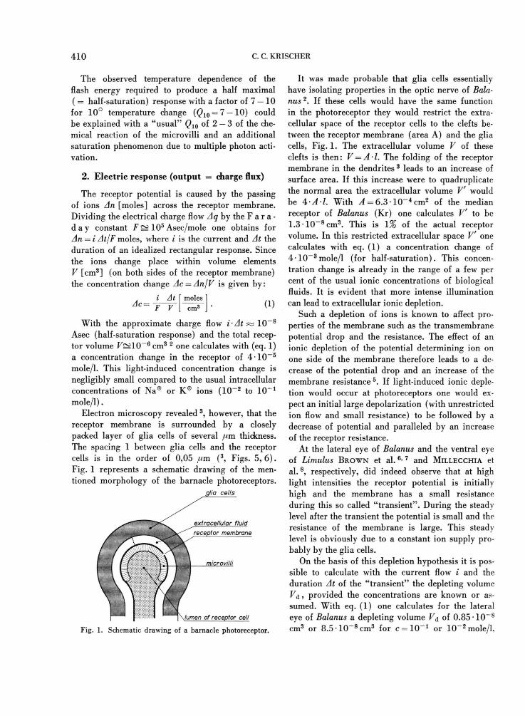
receptor membrane is surrounded by a closely packed layer of glia cells of several jam thickness. The spacing 1 between glia cells and the receptor cells is in the order of 0,05 ^m (3, Figs. 5, 6 ) . Fig. 1 represents a schematic drawing of the men-tioned morphology of the barnacle photoreceptors.
glia cells
Fig. 1. Schematic drawing of a barnacle photoreceptor.
It was made probable that glia cells essentially have isolating properties in the optic nerve of Bala-nus 2. If these cells would have the same function in the photoreceptor they would restrict the extra-cellular space of the receptor cells to the clefts be-tween the receptor membrane (area A) and the glia cells, Fig. 1. The extracellular volume V of these clefts is then: V = A-1. The folding of the receptor membrane in the dendrites 3 leads to an increase of surface area. If this increase were to quadruplicate the normal area the extracellular volume V would be 4 A l. With A = 6.3 1 0 " 4 c m 2 of the median receptor of Baianus (Kr) one calculates V' to be 1.3 10 _ 8 cm 3 . This is 1% of the actual receptor volume. In this restricted extracellular space V one calculates with eq. (1) a concentration change of 4 - 1 0 - 3 mole/1 (for half-saturation). This concen-tration change is already in the range of a few per cent of the usual ionic concentrations of biological fluids. It is evident that more intense illumination can lead to extracellular ionic depletion.
Such a depletion of ions is known to affect pro-perties of the membrane such as the transmembrane potential drop and the resistance. The effect of an ionic depletion of the potential determining ion on one side of the membrane therefore leads to a de-crease of the potential drop and an increase of the membrane resistance 5. If light-induced ionic deple-tion would occur at photoreceptors one would ex-pect an initial large depolarization (with unrestricted ion flow and small resistance) to be followed by a decrease of potential and paralleled by an increase of the receptor resistance.
At the lateral eye of Balanus and the ventral eye of Limulus BROWN et a l . 6 - 7 and MILLECCHIA et al. 8, respectively, did indeed observe that at high light intensities the receptor potential is initially high and the membrane has a small resistance during this so called "transient". During the steady level after the transient the potential is small and the resistance of the membrane is large. This steady level is obviously due to a constant ion supply pro-bably by the glia cells.
On the basis of this depletion hypothesis it is pos-sible to calculate with the current flow i and the duration At of the "transient" the depleting volume V &, provided the concentrations are known or as-sumed. With eq. (1) one calculates for the lateral eye of Balanus a depleting volume V& of 0.85 1 0 - 8
cm3 or 8.5 * 10~8 cm3 for c = 1 0 " 1 or 10~2 mole/1,

ELECTRIC RESPONSE MECHANISM OF PHOTORECEPTORS 4 1 1
respectively ( £ = 1 . 7 - 1 0 _ 7 i 4 , At = 0.5 sec 6 ) . Com-parison with the volume which was calculated from histological data of V = 4.2 • 1 0 - 6 cm3 shows that these values of volumina are indeed with 0.5% and 5% of the total receptor volume in the exspected range. Even through these estimates do not prove that the transient is a consequence of ionic deple-tion, they only show that such a process could be one possible explanation for the "transient".
These estimates are in agreement with the findings of ZERBST et al. 9 who discussed the restricted extra-cellular space as a chemical capacitor which in-fluences the time constant of the photo response. This capacity per unit area C* is given by:
L ~ AE AE Asec F e m 2 (2)
where Ac is the depleting concentration change, F the F a r a d a y constant, I the thickness of the ex-tracellular space and AE the change of membrane potential due to depletion.
With the normal ionic concentrations of Ac — l O ^ - l O ^ M and Z = 1.5 • 10~6 cm 9 or 5 10~6
cm 3 and AE = 25 mV one calculates for C* the limits 2000 to 60 //F/cm2. This is an "interfacial" capacitor two to three orders of magnitude larger than the capacitor of the loligo nerve membrane 5. Since the response is 1msec and 100 msec for the spike of the loligo nerve and flash response of the Balanus photoreceptor, respectively the interpreta-tion of ZERBST et al. 9 is confirmed with the experi-mental findings. These estimates are confirmed by the measurements of BROWN et al. 7 who determined with the current clamp technique an "apparent" spe-cific membrane capacity of 180 //F/cm2 .
The discussion is certainly incomplete in so far as it does not include the ionic milieu of the photo-chemical microvillus system. Ionic concentration changes of this system could have similar effects.
It could also be possible that Na®- and K® cur-rents subsequently flow during the receptor poten-tial. It is, however difficult to investigate this pos-sibility because so little is known about the function of the glia cells which surround the receptor cells.
2.1. Equilibration processes
If ionic depletion is possible in photoreceptors the normal functioning of the visual process re-quires after the charge flux of the electric response equilibration processes in order to restore the "nor-
mal" concentration gradients. Because ionic reser-voirs in cells do not have unlimited capacity cer-tain energy consuming "pumps" (glia cells?) have to become operative in order to refill the volume elements. Ions will therefore have to be transported certain distances within the cells.
Short light flashes initiate a delayed and slow electric response (of 50 to 100 msec duration) the characteristies of which is independent of tempera-ture : Time parameters such as latency time Jiat, time to reach the maximum 2max, total duration tt and light-dark adaptation time £ad are related by constant ratios with each other 2. These ratios J]at : tmilx : tt : fa(j are approximately the same for the two different photoreceptors of Balanus 2. The total durations of the flash responses differ, however, by a factor of two for the two photoreceptors which were investi-gated 2. It was assumed that this identity of reaction scheme and difference in reaction rate can be ex-plained as being due to the same process occuring in compartments of different size. The receptor with the smaller compartments reacts in this model faster than the receptor with the larger compart-ments. The identity of reaction scheme would mean that the proportioning of all the compartments which participate in the reaction and their rates are the same for both receptors.
The transfer of energy or mass in the compart-ments could be rate determining for the light-induced reaction. It is evident that for the equilibra-tion to the original state similar processes could con-trol the rate.
In the simple case in which heat, energy or mass (particles) spread in an unrestricted space a charac-teristical length A [cm] which is passed in a certain time T [sec] are correlated by:
X = V~Dx [cm] (3) where D [cm2/sec] is the transfer coefficient. For diffusion a typical value of D for common ions in water such as K® is about 1 0 - 5 cm2/sec.
Applied to the transfer processes in the compart-ments eq. (3) states that the reaction time r i. e. duration of the reaction of the latency period £]at
or the adaptation time ia(j are proportional to the square of the characteristic length X of the cor-responding compartments. If it is assumed that transfer coefficients for various photoreceptive pro-cesses are comparable in magnitude it is possible to make with the measured values of *iat and t&(j

4 1 2 C. C. KRISCHER
estimates of size of the corresponding compartments. If the adaptation e. g. the recovery of the photo-receptor after exhaustion by intense illumination requires transfer processes within the whole receptor the size of this "compartment" would be equal to the size of the photoreceptor; 2a(1 = r, where r is the half diameter of the receptor. With eq. (3) one can calculate the size of the compartment of the reaction of the latency period A]at: For the same D one ob-tains: = V ad : 'lat 5 e<l- (3) . Insertion of the measured parameters of the median photoreceptor of the barnacle with r = 30/ /m, Ja<i = 1.5sec, = 27.9 msec2 yields A]at to 4 //m. This value is in a similar range as half the diameter of the microvillus zones of the median photoreceptor of the similar species balanus cariosus (3, Fig. 4 ) . It remains to be proved that the microvillus zones of the lateral photoreceptor are larger than the microvilli of the median photoreceptor. According to the above hypothesis they should have a diameter of about ]/2 times larger than the median, so (^i a t) lateral ~
5.7 /<m. The observed temperature dependence of the time
course of the flash-induced receptor potentials with a Q10 of 2 — 3 for the characteristic time parameters is an indication of the chemical nature of the transfer
process. Diffusion controlled reactions should have a much smaller temperature coefficient.
3. Comparison of flash responses of Balanus with those of other animals
It is an important question whether the observed dependence of flash response parameters on the size of the two eyes of Balanus also apply to photorecep-tors of other animals; i .e. whether the observed photo-electric processes are of general applicability in the visual process.
A comparison of experimental results in table I shows that — although crude assumptions had to be made — the photo-electric efficiency rjq , eq (15) 1
is for the considered photoreceptors of similar magnitude, i .e. in the order of 1014 Asec/(hj')ai,s
which is equivalent to 0.6 -105 univalent ions per absorbed photon. The discrepancy of N° 3 (Bala-nus) may be due to the fact that r]q was — in con-trast to r]q in N° 1 and 2 — calculated for steady light stimulation. The comparison of the photo-elec-tric efficiencies therefore shows that this important parameter of the visual process is similar in magni-tude for different animals such as Aplysia and rat.
No. animal ° C
^lat 0-5 [msec]
'lat: h • 'ad (d^/dOo-5 [V/sec] [Asec/hi'abs]
author and remarks
1 Balanus med
2 Balanus lat
3 Balanus lat
A pis
Limulus lateral
6 Limulus ventral
7 Loligo
8 Aplysia
9 rat
21° 19
21° 38
2 0 - 2 5 ° -
23°
9°
15
170
2 0 - 2 5 ° 50
1 : 5 . 4 : 54 1.2
1 : 3 : 165 0.5
1 : 6.3 : 300 0.65
1 : 3 :230 0.13
1 : 5 . 5 : - 0.5
14° 500 1 : 4 . 5 : - 0.09
3 3 - 3 5 ° 10(?) 1 : 5 : - ( ? ) 0 ,5(? )
2 • 1 0 - 1 4
2 • IO-14
2 • 10-16
4.4 • IO-14
10-14
9.2 • IO-14
1.1 • 10-15
B R O W N e t a l 6 F i g . 1 d u r i n g t h e transient, step of light; i — 7.5 • 10 - 8 A; I * = 100 lx ; light ab-sorption = 1 0 % ; A = 10_4 cm 2
F . B A U M A N N I 2
F U O R T E S a n d H O D G K I N u F i g . 5 , rel. int. 0.0005; light absorption = 1 0 % ; Re = 1 0 5 ß , response duration = 0.2 sec MILLECCHIA et al .13 '8 At • inten-sity = 5 lx • sec, light absorption = ' 1 0 % ; i-At = 1.8 • lO- 8 Asec ; A = lO- 5 cm 2
HAGINSI4 corrected for light ab-sorption = 1 0 %
JACKLET1° Fig. 6, light absorption = 1 0 % ; A = lO- 4 c m 2 ; Re = 105 Q; half saturation: E = 25 m V
3 . 7 - 1 0 - 1 3 H A G I N S e t a l . 1 5
Table 1. Parameters of flash responses of photoreceptors of various animals.
ELECTRIC RESPONSE MECHANISM OF PHOTORECEPTORS 4 1 3
The comparison of time-dependent response para-meters in Table I shows that the reaction schemes of electric flash responses are similar for various photo-receptors : The characteristic response parameters such as latency time i]at, total duration tt adapta-tion time ia(] i. e. the ratio : tt : ad a r e similar for all the investigated photoreceptors. Long laten-cies are usually followed by relatively low values of the potential increase with time. So, apparently many photoreceptors obey the same photoreceptive reaction scheme even though the standard para-meter of the photo-chemical reaction, the latency time Z]at at half saturation, differs by as much as a factor of 50, see ^ato,5 of N° 8 and 9. A detailed morphological comparison of the structural elements wrhich contain the photo-pigment would certainly be of interest. According to the model 2 the receptors with the longer latency should have the larger com-partments. For the eye of Aplysia the length of the rhabdomers10 (corresponding to the microvillus zones of Balanus of the diameter of about 4 /<m) is about 20 ^m. Insertion of the half-saturation latency *iat = 500 msec and the dimension X — 20 /um in eq. (3) yields for this eye approximately the same "transfer coefficient" as for Balanus. The under-lying processes therefore seem to be the same for both eyes. This fact is also documented by the ob-servation that the dependence of the latencies on the light intensity is for the considered photorecep-tors very similar provided they are normalized to the same excitation. So is d (^]at) o >5/d log (/*•<) for many photoreceptors i. e. Limulus ventral eye8 Li-
1 C. C. KRISCHER, Z . N a t u r f o r s c h . 2 6 b , 1 3 2 2 [ 1 9 7 1 ] . 2 C. C. KRISHER, Z. Naturforsch. 26 b, 1326 [1971 ] . 3 W . H. FAHRENBACH, Z. Zellforsch. 66, 233 [1965 ] . 4 G . W A L D , A n g e w . C h e m . 8 0 , 8 5 7 [ 1 9 6 8 ] . 5 A . L . H O D G K I N a n d A . F . HUXLEY, J . P h y s i o l . 1 1 7 , 5 0 0
[ 1 9 5 2 ] . 6 H . M . B R O W N , R . W . M E E C H , H . KOIKE, a n d S . H A G I W A R A ,
Science [Washington] 1 6 6 , 2 4 0 [1969] . 7 H . M . B R O W N , S . H A G I W A R A , S . KOIKE, a n d R . M . M E E C H ,
J. Physiol. 2 0 8 , 3 8 5 [1970] . 8 R . MILLECCHIA a n d A . M A U R O , J . g e n . P h y s i o l . 5 4 , 3 1 0
[1969] .
mulus lateral eye 11 Balanus6 and Aplysia10 in the range of 30 — 50% per decade of light intensity ( [« lat l o , 5 = 1 0 0 % ) . JACKLET 10 observed (in accor-dance with the observations at Balanus) a tempera-ture dependence of 2 —2.5 per 10° for the latency time of the eye of Aplysia.
Conclusion
The similarity of photo-electric efficiency and time course of flash induced receptor potentials of different photoreceptors might serve as indication for a common mechanism of the primary electric response of these photoreceptors. It remains to be tested experimentally whether the receptor length constant 2e [cm] — which is defined as the ratio of photopigment volume to excitable membrane area 1 — is the rate determining parameter in vision. / e would be analogous to An , the length constant of a nerve. For both types, visual and nervous excita-tion volume and surface properties would thus characterize the rate. Only intense studies of mor-phology and function of photoreceptors will show to what extent this generalization is valid. It also remains to be investigated whether there is in photo-receptors a "tuning" of the receptor length constant and the effective interfacial capacitance of extra-cellularly restricted space in such a manner that a large length constant would require a large capaci-tor (large extracellular space) in order to become operative in the most efficient manner.
9 E . ZERBST, K . - H . DITTBERNER, a n d E . W I L L I A M , K y b e r n e t i k 2, 160 [1965] ,
10 J. W . JACKLET, J. gen. Physiol. 53, 21 [1969] . 1 1 M . G . F . FUORTES a n d A . L . H O D G K I N , J . P h y s i o l . 1 7 2 ,
239 [1964] . 12 F. BAUMANN, J. gen. Physiol. 52, 855 [1968] . 1 3 R . MILLECCHIA a n d A . M A U R O , J . g e n . P h y s i o l . 5 4 , 3 3 1
[1969] . 14 W . A . HAGINS, Cold Spring Harbor Symposis on Quantita-
tive Biology "Sensory Receptors" 30, 403 [1965] . 1 5 W . A . HAGINS, R . D . PENN, a n d S . YOSHIKAMI, B i o p h y s i -
cal J. 10, 380 [1970] .