Palmare winkelstabile Plattenosteosynthese der instabilen ... · In recent years, the distal radius...
17
1 Operat Orthop Traumatol 2004 · Nr. 3 © Urban & Vogel Palmare winkelstabile Plattenosteosynthese der instabilen distalen Radiusfraktur Anterior Fixed Angle Plate Fixation of Unstable Distal Radius Fractures Veith L. Moser 1 , Christian Pessenlehner 2 , Maximilian Meier 2 , Hermann Krimmer 2 Operative Orthopädie und T raumatologie Zusammenfassung Operationsziel Anatomische Reposition und dauerhafte Retention. Übungs- und winkelstabile palmare Plattenosteosynthe- se. Wiederherstellung der Form und Funktion des Handge- lenks. Indikationen Distale instabile Radiusfrakturen. Kontraindikationen Frakturen im Kindes- und Adoleszentenalter. Operationstechnik Radiopalmarer Zugang. Osteosynthese der reponierten Fragmente am distalen Radius unter Verwendung winkel- stabiler Plattensysteme. Weiterbehandlung 2 Wochen dorsale Gipsschiene bis zur Wundheilung. Akti- ve Mobilisierung der Finger ab dem 1. postoperativen Tag mit dem Ziel des kompletten Faustschlusses und der kom- pletten Fingerstreckung. Bei ausgeprägter Trümmerzone wird das Handgelenk 4 Wochen in der Gipsschiene immo- bilisiert. Ergebnisse 62 Patienten (Durchschnittsalter 55 Jahre) wurden nach diesem Prinzip operiert und im Mittel 11 Monate (6–23 Monate) postoperativ nachuntersucht. Die Bewegungs- ausmaße waren im Schnitt um 19% bei Extension/Flexion, um 13% bei Radial-/Ulnarduktion und um 10% bei Pronati- on/Supination im Vergleich zur Gegenseite vermindert, entsprechend einer nur geringen Funktionseinschrän- kung. Dies spiegelt sich in einem durchschnittlichen DASH-Score von 19 Punkten wider, wobei dieser Wert ent- sprechend der Spannbreite von 0 (keine Einschränkung) bis 100 (maximale Einschränkung) ein gutes Ergebnis be- legt. Ein Schwerpunkt der Nachuntersuchung wurde auf die ra- diologische Kontrolle gelegt. In allen Fällen konnte eine anatomische Wiederherstellung der Radiusgelenkfläche Abstract Objective Anatomic reduction and permanent maintenance of re- duction thanks to a fixed angle device placed on the pal- mar aspect of the distal radius permitting early exercises. Restitution of form and function of wrist. Indications Displaced unstable fractures of the distal radius. Contraindications Radial fractures in children and adolescents. Surgical Technique Radioanterior approach. Internal fixation of the reduced fragments with a fixed angle device system. Postoperative Management Posterior slab immobilization for 2 weeks; active mobiliza- tion of fingers starting on day 1 with the goal to be able to make a fist and to completely extend the fingers. In case of severely comminuted fractures slab immobilization for 4 weeks. Results 62 patients (average age 55 years) were treated by this method and underwent retrospective evaluation with mean follow-up of 11 months (6–23 months). Range of motion decreased on average with 19% for ex- tension/flexion, 13% for radial abduction/ulnar adduction, and 10% for pronation/supination compared to the oppo- site hand. This corresponds to a negligible loss of function. The mean value of the DASH score reached 19 points (min- imum 0 points = no limitation, 100 points = maximal limi- tation), representing a good result. Operat Orthop Traumatol 2003;15:1–XX DOI 10.1007/s00064-004-■■■■■-■ 1 Abteilung für Wiederherstellungs- und Plastische Chirurgie, Universitätsklinik für Chirurgie, AKH, Wien, Österreich, 2 Klinik für Handchirurgie, Rhön-Klinikum, Bad Neustadt/Saale. OOT 267 Moser 4/04
-
Upload
truongthuy -
Category
Documents
-
view
217 -
download
0
Transcript of Palmare winkelstabile Plattenosteosynthese der instabilen ... · In recent years, the distal radius...

1Operat Orthop Traumatol 2004 · Nr. 3 © Urban & Vogel
Palmare winkelstabile Plattenosteosynthese der instabilen distalen RadiusfrakturAnterior Fixed Angle Plate Fixation of Unstable Distal Radius FracturesVeith L. Moser1, Christian Pessenlehner2, Maximilian Meier2, Hermann Krimmer2
Operative Orthopädie und Traumatologie
ZusammenfassungOperationsziel
Anatomische Reposition und dauerhafte Retention. Übungs- und winkelstabile palmare Plattenosteosynthe-se. Wiederherstellung der Form und Funktion des Handge-lenks.
IndikationenDistale instabile Radiusfrakturen.
KontraindikationenFrakturen im Kindes- und Adoleszentenalter.
OperationstechnikRadiopalmarer Zugang. Osteosynthese der reponierten Fragmente am distalen Radius unter Verwendung winkel-stabiler Plattensysteme.
Weiterbehandlung2 Wochen dorsale Gipsschiene bis zur Wundheilung. Akti-ve Mobilisierung der Finger ab dem 1. postoperativen Tag mit dem Ziel des kompletten Faustschlusses und der kom-pletten Fingerstreckung. Bei ausgeprägter Trümmerzone wird das Handgelenk 4 Wochen in der Gipsschiene immo-bilisiert.
Ergebnisse62 Patienten (Durchschnittsalter 55 Jahre) wurden nach diesem Prinzip operiert und im Mittel 11 Monate (6–23 Monate) postoperativ nachuntersucht. Die Bewegungs-ausmaße waren im Schnitt um 19% bei Extension/Flexion, um 13% bei Radial-/Ulnarduktion und um 10% bei Pronati-on/Supination im Vergleich zur Gegenseite vermindert, entsprechend einer nur geringen Funktionseinschrän-kung. Dies spiegelt sich in einem durchschnittlichen DASH-Score von 19 Punkten wider, wobei dieser Wert ent-sprechend der Spannbreite von 0 (keine Einschränkung) bis 100 (maximale Einschränkung) ein gutes Ergebnis be-legt.Ein Schwerpunkt der Nachuntersuchung wurde auf die ra-diologische Kontrolle gelegt. In allen Fällen konnte eine anatomische Wiederherstellung der Radiusgelenkfläche
AbstractObjective
Anatomic reduction and permanent maintenance of re-duction thanks to a fixed angle device placed on the pal-mar aspect of the distal radius permitting early exercises. Restitution of form and function of wrist.
IndicationsDisplaced unstable fractures of the distal radius.
ContraindicationsRadial fractures in children and adolescents.
Surgical TechniqueRadioanterior approach. Internal fixation of the reduced fragments with a fixed angle device system.
Postoperative ManagementPosterior slab immobilization for 2 weeks; active mobiliza-tion of fingers starting on day 1 with the goal to be able to make a fist and to completely extend the fingers. In case of severely comminuted fractures slab immobilization for 4 weeks.
Results62 patients (average age 55 years) were treated by this method and underwent retrospective evaluation with mean follow-up of 11 months (6–23 months).Range of motion decreased on average with 19% for ex-tension/flexion, 13% for radial abduction/ulnar adduction, and 10% for pronation/supination compared to the oppo-site hand. This corresponds to a negligible loss of function. The mean value of the DASH score reached 19 points (min-imum 0 points = no limitation, 100 points = maximal limi-tation), representing a good result.
Operat Orthop Traumatol 2003;15:1–XX
DOI 10.1007/s00064-004-■■■■■-■
1 Abteilung für Wiederherstellungs- und Plastische Chirurgie, Universitätsklinik für Chirurgie, AKH, Wien, Österreich,
2 Klinik für Handchirurgie, Rhön-Klinikum, Bad Neustadt/Saale.
OOT 267 Moser 4/04

Moser VL, et al. Fixed Angle Plate Fixation of Radius Fractures
2 Orthop Traumatol 2004 · No. 4 © Urban & Vogel
VorbemerkungenDie distale Radiusfraktur loco typico, wie sie von Col-les 1814 beschrieben wurde, unterliegt seit einigen Jahren einem therapeutischen Wandel [3, 4, 6, 14]. Durch konservative Behandlung im Gipsverband so-wie minimal invasive Stabilisierung mit Kirsch-ner-Drähten kann das Repositionsergebnis bei Frak-turen mit Trümmerzone häufig nicht oder nur vorü-bergehend gehalten werden. Auch die alleinige externe Fixation unter Reposition durch Ligamentotaxis führt oft zu keiner dauerhaften Retention. Die Kombinati-on beider Verfahren kann zwar eine schleichende Sin-terung der Fragmente weitgehend verhindern, setzt aber voraus, dass die Drähte nach Fixateurentfernung noch ca. 6 Wochen bis zum Abschluss der Osteogene-se belassen werden.
Die Vorteile des palmaren Zugangs liegen in der besseren Weichteildeckung, geringeren Gefahr von Sehnenirritationen und besseren Kontrolle der Repo-sition an der meist nur einfach frakturierten palmaren Kortikalis. Aus diesem Grund führen wir seit langem die Osteosynthese nach Korrekturosteotomien auch fehlerhaft verheilter Frakturen vom Extensionstyp mit palmarer Platte und „Knochenblock“ aus [10].
Bei frischen Frakturen, insbesondere Mehrfrag-mentfrakturen mit dorsaler Trümmerzone, stellte die Schraubenlockerung mit sekundärem Korrekturver-lust bisher ein Problem dar. Die Ursache dafür lag in der fehlenden stabilen bikortikalen Verankerung der Schrauben, die in der dorsalen Trümmerzone bereits primär keinen Halt gefunden hatten. Deshalb war auch eine zusätzliche Spongiosaplastik oder die Ver-wendung von Knochenersatzstoffen von einem dorsa-len Zugang aus nötig.
Der steigende Patientenanspruch sowie soziale und ökonomische Faktoren haben dazu geführt, eine ana-tomische Rekonstruktion sowie eine dauerhafte Re-
Introductory RemarksIn recent years, the distal radius fracture, first de-scribed by Colles in 1814, has seen a great change in the approach to its treatment [3, 4, 6, 14]. By using a conservative treatment in a cast or by trying to stabi-lize the fracture with minimally invasive Kirschner wires, the reduction of the communited fracture is of-ten not or only temporarily maintained. Even the sole external fixation after reduction by ligamentotaxis does often not lead to a permanent maintenance of re-duction. A combination of both methods may prevent a slow impaction but requires the wires to be left in place for another 6 weeks after the removal of the fix-ator until bony healing has been accomplished.
The advantage of an anterior approach lies in an im-proved soft-tissue coverage, less danger of irritation to the tendons, and better control of reduction of the cor-tex, in most instances only fractured anteriorly. For this reason, we have been using an anterior plate fixa-tion and bone block after a corrective osteotomy for malunited fractures of the extension type [10].
In acute fractures, especially those with multiple fragments and posterior comminution, screw loosening with secondary loss of correction constituted a major problem. This was caused by the lack of stable bicorti-cal screw purchase, as the screws did not find a proper anchorage in the posterior comminution. Therefore, an additional cancellous bone graft or the use of a bone substitute inserted posteriorly was necessary.
Since, nowadays, patients have increased demands and socioeconomic factors have become more rele-vant, an anatomic restoration and a permanent main-tenance have been targeted together with a postopera-tive immobilization of short duration and early exer-cises.
Based on the principle of fixed angle devices new methods of osteosynthesis have been developed. They function like an
erreicht werden. Ein bedeutsamer sekundärer Korrektur-verlust war nicht zu beobachten, ebenso wenig eine se-kundäre Schraubendislokation. Bei allen Patienten kam es ohne Spongiosaplastik zu einer Knochenneubildung in den Stauchungszonen.
SchlüsselwörterRadiusfraktur · Winkelstabile Osteosynthese · Fi-xateur interne
The emphasis of this study was put on the radiologic fol-low-up; neither relevant secondary loss of correction nor relevant loss of length of the radius were seen. Further-more, no screw displacement occurred. In the comminut-ed area endosteal bone formation could be observed, even though no cancellous bone grafting was performed.
Key Words Radius fracture · Fixed angle device · Internal fix-ation
Moser VL, et al. Winkelstabile Plattenosteosynthese von Radiusfrakturen
3Operat Orthop Traumatol 2004 · Nr. 4 © Urban & Vogel
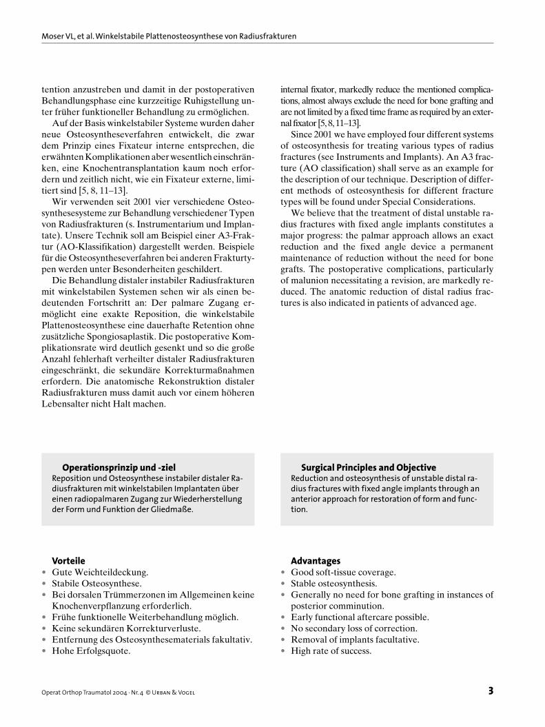
tention anzustreben und damit in der postoperativen Behandlungsphase eine kurzzeitige Ruhigstellung un-ter früher funktioneller Behandlung zu ermöglichen.
Auf der Basis winkelstabiler Systeme wurden daher neue Osteosyntheseverfahren entwickelt, die zwar dem Prinzip eines Fixateur interne entsprechen, die erwähnten Komplikationen aber wesentlich einschrän-ken, eine Knochentransplantation kaum noch erfor-dern und zeitlich nicht, wie ein Fixateur externe, limi-tiert sind [5, 8, 11–13].
Wir verwenden seit 2001 vier verschiedene Osteo-synthesesysteme zur Behandlung verschiedener Typen von Radiusfrakturen (s. Instrumentarium und Implan-tate). Unsere Technik soll am Beispiel einer A3-Frak-tur (AO-Klassifikation) dargestellt werden. Beispiele für die Osteosyntheseverfahren bei anderen Frakturty-pen werden unter Besonderheiten geschildert.
Die Behandlung distaler instabiler Radiusfrakturen mit winkelstabilen Systemen sehen wir als einen be-deutenden Fortschritt an: Der palmare Zugang er-möglicht eine exakte Reposition, die winkelstabile Plattenosteosynthese eine dauerhafte Retention ohne zusätzliche Spongiosaplastik. Die postoperative Kom-plikationsrate wird deutlich gesenkt und so die große Anzahl fehlerhaft verheilter distaler Radiusfrakturen eingeschränkt, die sekundäre Korrekturmaßnahmen erfordern. Die anatomische Rekonstruktion distaler Radiusfrakturen muss damit auch vor einem höheren Lebensalter nicht Halt machen.
Vorteile• Gute Weichteildeckung.• Stabile Osteosynthese.• Bei dorsalen Trümmerzonen im Allgemeinen keine
Knochenverpflanzung erforderlich.• Frühe funktionelle Weiterbehandlung möglich.• Keine sekundären Korrekturverluste.• Entfernung des Osteosynthesematerials fakultativ.• Hohe Erfolgsquote.
internal fixator, markedly reduce the mentioned complica-tions, almost always exclude the need for bone grafting and are not limited by a fixed time frame as required by an exter-nal fixator [5, 8, 11–13].
Since 2001 we have employed four different systems of osteosynthesis for treating various types of radius fractures (see Instruments and Implants). An A3 frac-ture (AO classification) shall serve as an example for the description of our technique. Description of differ-ent methods of osteosynthesis for different fracture types will be found under Special Considerations.
We believe that the treatment of distal unstable ra-dius fractures with fixed angle implants constitutes a major progress: the palmar approach allows an exact reduction and the fixed angle device a permanent maintenance of reduction without the need for bone grafts. The postoperative complications, particularly of malunion necessitating a revision, are markedly re-duced. The anatomic reduction of distal radius frac-tures is also indicated in patients of advanced age.
Advantages• Good soft-tissue coverage.• Stable osteosynthesis.• Generally no need for bone grafting in instances of
posterior comminution.• Early functional aftercare possible.• No secondary loss of correction.• Removal of implants facultative.• High rate of success.
Operationsprinzip und -zielReposition und Osteosynthese instabiler distaler Ra-diusfrakturen mit winkelstabilen Implantaten über einen radiopalmaren Zugang zur Wiederherstellung der Form und Funktion der Gliedmaße.
Surgical Principles and ObjectiveReduction and osteosynthesis of unstable distal ra-dius fractures with fixed angle implants through an anterior approach for restoration of form and func-tion.
Moser VL, et al. Fixed Angle Plate Fixation of Radius Fractures
4 Orthop Traumatol 2004 · No. 4 © Urban & Vogel
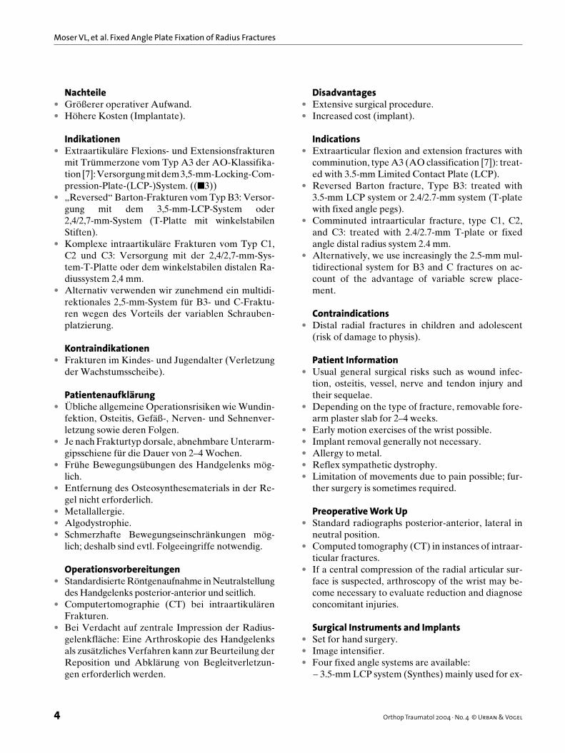
Nachteile• Größerer operativer Aufwand.• Höhere Kosten (Implantate).
Indikationen• Extraartikuläre Flexions- und Extensionsfrakturen
mit Trümmerzone vom Typ A3 der AO-Klassifika-tion [7]: Versorgung mit dem 3,5-mm-Locking-Com-pression-Plate-(LCP-)System. ((■3))
• „Reversed“ Barton-Frakturen vom Typ B3: Versor-gung mit dem 3,5-mm-LCP-System oder 2,4/2,7-mm-System (T-Platte mit winkelstabilen Stiften).
• Komplexe intraartikuläre Frakturen vom Typ C1, C2 und C3: Versorgung mit der 2,4/2,7-mm-Sys-tem-T-Platte oder dem winkelstabilen distalen Ra-diussystem 2,4 mm.
• Alternativ verwenden wir zunehmend ein multidi-rektionales 2,5-mm-System für B3- und C-Fraktu-ren wegen des Vorteils der variablen Schrauben-platzierung.
Kontraindikationen• Frakturen im Kindes- und Jugendalter (Verletzung
der Wachstumsscheibe).
Patientenaufklärung• Übliche allgemeine Operationsrisiken wie Wundin-
fektion, Osteitis, Gefäß-, Nerven- und Sehnenver-letzung sowie deren Folgen.
• Je nach Frakturtyp dorsale, abnehmbare Unterarm-gipsschiene für die Dauer von 2–4 Wochen.
• Frühe Bewegungsübungen des Handgelenks mög-lich.
• Entfernung des Osteosynthesematerials in der Re-gel nicht erforderlich.
• Metallallergie.• Algodystrophie.• Schmerzhafte Bewegungseinschränkungen mög-
lich; deshalb sind evtl. Folgeeingriffe notwendig.
Operationsvorbereitungen• Standardisierte Röntgenaufnahme in Neutralstellung
des Handgelenks posterior-anterior und seitlich.• Computertomographie (CT) bei intraartikulären
Frakturen.• Bei Verdacht auf zentrale Impression der Radius-
gelenkfläche: Eine Arthroskopie des Handgelenks als zusätzliches Verfahren kann zur Beurteilung der Reposition und Abklärung von Begleitverletzun-gen erforderlich werden.
Disadvantages• Extensive surgical procedure.• Increased cost (implant).
Indications• Extraarticular flexion and extension fractures with
comminution, type A3 (AO classification [7]): treat-ed with 3.5-mm Limited Contact Plate (LCP).
• Reversed Barton fracture, Type B3: treated with 3.5-mm LCP system or 2.4/2.7-mm system (T-plate with fixed angle pegs).
• Comminuted intraarticular fracture, type C1, C2, and C3: treated with 2.4/2.7-mm T-plate or fixed angle distal radius system 2.4 mm.
• Alternatively, we use increasingly the 2.5-mm mul-tidirectional system for B3 and C fractures on ac-count of the advantage of variable screw place-ment.
Contraindications• Distal radial fractures in children and adolescent
(risk of damage to physis).
Patient Information• Usual general surgical risks such as wound infec-
tion, osteitis, vessel, nerve and tendon injury and their sequelae.
• Depending on the type of fracture, removable fore-arm plaster slab for 2–4 weeks.
• Early motion exercises of the wrist possible.• Implant removal generally not necessary.• Allergy to metal.• Reflex sympathetic dystrophy.• Limitation of movements due to pain possible; fur-
ther surgery is sometimes required.
Preoperative Work Up• Standard radiographs posterior-anterior, lateral in
neutral position.• Computed tomography (CT) in instances of intraar-
ticular fractures.• If a central compression of the radial articular sur-
face is suspected, arthroscopy of the wrist may be-come necessary to evaluate reduction and diagnose concomitant injuries.
Surgical Instruments and Implants• Set for hand surgery.• Image intensifier.• Four fixed angle systems are available: – 3.5-mm LCP system (Synthes) mainly used for ex-

Moser VL, et al. Winkelstabile Plattenosteosynthese von Radiusfrakturen
5Operat Orthop Traumatol 2004 · Nr. 4 © Urban & Vogel
Instrumentarium und Implantate• Handchirurgisches Instrumentarium.• Bildwandler.• Es stehen vier winkelstabile Systeme zur Verfü-
gung: – 3,5-mm-AO-LCP-System (Synthes GmbH & Co.,
Im Kirchenhürstle 4–6, 79224 Umkirch), vorwie-gend für extraartikuläre Frakturen vom Typ A der AO-Klassifikation (Abbildung 1a);
– 2,4/2,7-mm-System-T-Platte (Synthes), vor allem bei sehr weit distal gelegener Frakturzone von Typ-B- und -C-Frakturen der AO-Klassifikation (Abbildung 1b);
– winkelstabiles distales Radiussystem 2,4 mm (Synthes) für B- und C-Frakturen mit kleinen Fragmenten und intraartikulärer Beteiligung (Abbildung 2);
– winkelstabiles multidirektionales 2,5-mm-Sys-tem (Medartis AG, Austraße 24, 4051 Basel, Schweiz) mit sphärischer Kopfraumverblockung der Schrauben und einem Freiheitsgrad von 15° in allen Ebenen. Hierdurch wird eine der Frak-tur angepasste variable Schraubenplatzierung ermöglicht, die gerade bei der Versorgung von C-Frakturen hilfreich ist (Abbildung 3).
traarticular fractures of type A (AO classification; Figure 1a);
– 2.4/2.7-mm system T-plate (Synthes) mainly emp-loyed in very distally located fracture areas of type B and C fractures (AO classification; Figure 1b);
– fixed angle radius system 2.4 mm (Synthes) for B and C fractures with small fragments and intraar-ticular involvement (Figure 2);
– 2.5-mm multidirectional fixed angle system (Me-dartis Co, Austraße 24, 4051 Basel, Switzerland). This system permits a locking fo the spherical screw heads with a 15° of freedom in all planes (variable screw placement adapted to the type of fracture, particularly of C fractures; Figure 3).
Abbildungen 1a und 1ba) LCP-T-Platte mit 3,5-mm-Kopfverriegelungsschrauben dis-tal und Kombinationslöchern für 3,5-mm-Kopfverriegelungs-schraube oder Kortikalisschraube im Schaft.b) 2,4/2,7-mm-T-Platte mit Wahlmöglichkeit zwischen win-kelstabilen Stiften oder 2,4-mm-Kortikalisschrauben distal und 2,7-mm-Kortikalisschrauben im Schaft.
Figures 1a and 1ba) LCP T-plate with 3.5-mm locking screws distally and combi-nation holes for 3.5-mm locking screws or cortical screws proximally (shaft).b) 2.4/2.7-mm T-plate with possibility to choose between fixed angle pegs or 2.4-mm cortical screws distally and 2.7-mm cortical screws proximally (shaft).
a b
Abbildung 3Multidirektionales 2,5-mm-System mit Schwenkbereich der Schrauben von 15° in allen Richtungen.
Figure 32.5-mm multidirectional system with a freedom of 15° of the screw heads.
Abbildung 2Winkelstabiles dista-les Radiussystem 2,4 mm mit 2,4-mm-Kopfverrie-gelungsschrauben distal und Kombina-tionslöchern für 2,4-mm-Kopfverrie-gelungschrauben oder 2,4- bzw. 2,7-mm-Kortikalis-schrauben im Schaft.
Figure 22.4-mm fixed angle device for distal radius with 2.4-mm lock-ing screws distally and combination holes for 2.4-mm locking screws or 2.4- or 2.7-mm cortical screws proximally (shaft).

Moser VL, et al. Fixed Angle Plate Fixation of Radius Fractures
6 Orthop Traumatol 2004 · No. 4 © Urban & Vogel
Anästhesie und Lagerung• Plexusanästhesie oder Allgemeinnarkose.• Rückenlage.• Der Arm wird auf dem Handtisch in Supinations-
stellung des Unterarms ausgelagert. Tuchrolle als Repositionshilfe unter das Handgelenk.
• Blutleere und Oberarmblutsperre.• „Single shot“ i.v. Antibiotikumprophylaxe ((■4))
(z.B. Cephalosporin der zweiten Generation).
Anesthesia and Positioning• Brachial plexus or endotracheal anesthesia.• Supine.• The arm in supination placed on an arm board, tow-
el roll under the wrist to facilitate reduction.• Esmarch and tourniquet on upper arm.• Single i.v. injection of an antibiotic (such as a sec-
ond-generation cephalosporin).
Operationstechnik
Abbildungen 4 bis 11
Am Beispiel einer A3-Fraktur nach AO unter Ver-wendung einer 3,5-mm-LCP-Platte
Surgical Technique
Figures 4 to 11
An A3 Fracture and Use of a 3.5-mm LCP Plate Serving as an Example
Abbildung 4Röntgenologisches Beispiel einer A3-Fraktur bei einem 56-jährigen Patienten nach Leitersturz.
Figure 4Radiograph of A3 fracture in a 56-year-old man after fall from a ladder.
Moser VL, et al. Winkelstabile Plattenosteosynthese von Radiusfrakturen
7Operat Orthop Traumatol 2004 · Nr. 4 © Urban & Vogel
Abb.5
Abb.6
N. medianus
A. radialis
M. flexor carp. rad.
M. flexor poll. long.
A. radialisM. pronator quad.
M. flexor carp. rad.
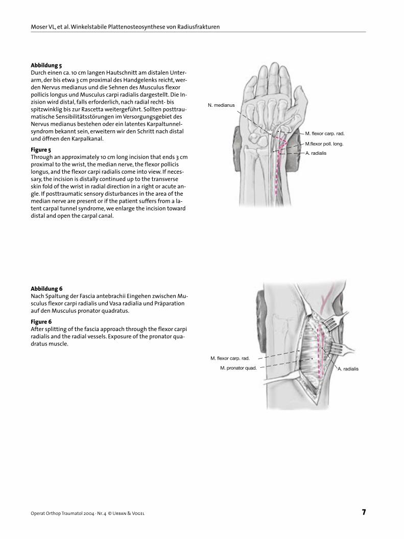
Abbildung 5Durch einen ca. 10 cm langen Hautschnitt am distalen Unter-arm, der bis etwa 3 cm proximal des Handgelenks reicht, wer-den Nervus medianus und die Sehnen des Musculus flexor pollicis longus und Musculus carpi radialis dargestellt. Die In-zision wird distal, falls erforderlich, nach radial recht- bis spitzwinklig bis zur Rascetta weitergeführt. Sollten posttrau-matische Sensibilitätsstörungen im Versorgungsgebiet des Nervus medianus bestehen oder ein latentes Karpaltunnel-syndrom bekannt sein, erweitern wir den Schritt nach distal und öffnen den Karpalkanal.
Figure 5Through an approximately 10 cm long incision that ends 3 cm proximal to the wrist, the median nerve, the flexor pollicis longus, and the flexor carpi radialis come into view. If neces-sary, the incision is distally continued up to the transverse skin fold of the wrist in radial direction in a right or acute an-gle. If posttraumatic sensory disturbances in the area of the median nerve are present or if the patient suffers from a la-tent carpal tunnel syndrome, we enlarge the incision toward distal and open the carpal canal.
Abb.5
Abb.6
N. medianus
A. radialis
M. flexor carp. rad.
M. flexor poll. long.
A. radialisM. pronator quad.
M. flexor carp. rad.
Abbildung 6Nach Spaltung der Fascia antebrachii Eingehen zwischen Mu-sculus flexor carpi radialis und Vasa radialia und Präparation auf den Musculus pronator quadratus.
Figure 6After splitting of the fascia approach through the flexor carpi radialis and the radial vessels. Exposure of the pronator qua-dratus muscle.
Moser VL, et al. Fixed Angle Plate Fixation of Radius Fractures
8 Orthop Traumatol 2004 · No. 4 © Urban & Vogel
Abb.7
N. medianusMm. flexores,
R. superficialisn. rad.
Längszug
Longitudinal traction
Längszug
Longitudinal traction
Druck von dorsal
Pressure from dorsal
Abb. 8
M. pronator quad.
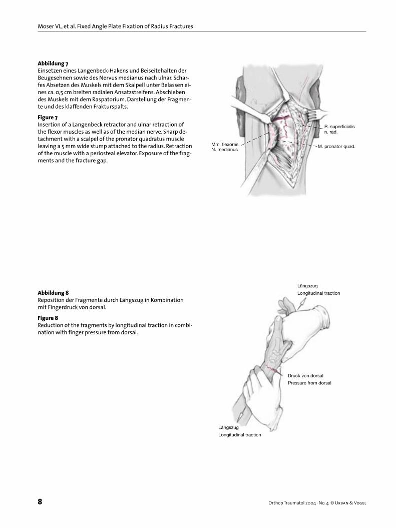
Abbildung 7Einsetzen eines Langenbeck-Hakens und Beiseitehalten der Beugesehnen sowie des Nervus medianus nach ulnar. Schar-fes Absetzen des Muskels mit dem Skalpell unter Belassen ei-nes ca. 0,5 cm breiten radialen Ansatzstreifens. Abschieben des Muskels mit dem Raspatorium. Darstellung der Fragmen-te und des klaffenden Frakturspalts.
Figure 7Insertion of a Langenbeck retractor and ulnar retraction of the flexor muscles as well as of the median nerve. Sharp de-tachment with a scalpel of the pronator quadratus muscle leaving a 5 mm wide stump attached to the radius. Retraction of the muscle with a periosteal elevator. Exposure of the frag-ments and the fracture gap.
Abb.7
N. medianusMm. flexores,
R. superficialisn. rad.
Längszug
Longitudinal traction
Längszug
Longitudinal traction
Druck von dorsal
Pressure from dorsal
Abb. 8
M. pronator quad.
Abbildung 8Reposition der Fragmente durch Längszug in Kombination mit Fingerdruck von dorsal.
Figure 8Reduction of the fragments by longitudinal traction in combi-nation with finger pressure from dorsal.

Moser VL, et al. Winkelstabile Plattenosteosynthese von Radiusfrakturen
9Operat Orthop Traumatol 2004 · Nr. 4 © Urban & Vogel
Abb.9
Abb.10
Abbildung 9Auswählen und Anmodellieren einer geeigneten Platte. Die Platte wird proximal mit einer Schraube im Gleitloch fixiert. Bildwandlerkontrolle zum Nachweis einer anatomischen Re-position und korrekten Plattenlage. Gegebenenfalls muss das Ergebnis korrigiert und die Platte in Längsrichtung verscho-ben werden, falls sie die distale Radiuskante überragt und da-mit Sehnen irritieren könnte. Eine subchondrale Schraubenla-ge muss gewährleistet sein. Bei älteren Frakturen, bei denen eine manuelle Reposition zunächst nicht gelingt, lässt sich manchmal noch ein gutes Ergebnis erzielen, wenn alle Schrauben im distalen Fragment fixiert werden und dann „über die Platte“ reponiert wird.Aufschrauben der Führungsbuchse in die Gewinde der Schraubenlöcher am Querschenkel. Aufbohren, Längenmes-sung und Einschrauben der winkelstabilen Implantate (Schraube oder Bolzen). Bildwandlerkontrolle zur Bestätigung der extraartikulären, subchondralen Lage und korrekten Län-ge von Schrauben und Platte.
Figure 9Choice and adaptation of the correct plate. With a screw in-serted into the gliding hole the plate is fixed proximally. Im-age intensifier control to verify the anatomic reduction and the correct plate position. If necessary, the position has to be corrected and the plate displaced in a longitudinal direction should the plate exceed the distal radial rim that could lead to tendon irritation.The screws have definitely to be placed subchondrally. In re-mote fractures where a manual reduction does not succeed in the beginning, the reduction may become successful when, after placement of all screws in the distal fragment, the reduction is done with the help of the plate.Insertion of a drilling sleeve into the threads of the screw holes at the crossbar of the plate. Drilling, determination of screw length, and insertion of fixed angle implant (screws or bolts). Image intensifier control to verify the extraarticular, subchondral position and the correct length of screws and plate.

Moser VL, et al. Fixed Angle Plate Fixation of Radius Fractures
10 Orthop Traumatol 2004 · No. 4 © Urban & Vogel
Abb.9
Abb.10
Abbildung 10Einbringen weiterer Schrauben, wobei im Längsschenkel in der Regel wegen Erhalt der palmaren und dorsalen Kortikalis keine winkelstabilen Schrauben notwendig sind. Nach Stabi-lisierung der Radiusfragmente manuelle Überprüfung der Stabilität im distalen Radioulnargelenk. Bei Instabilität Refi-xation des abgerissenen Processus styloideus ulnae mit einer Zuggurtungsosteosynthese.Bei Verdacht auf skapholunäre Bandverletzung Kinematogra-phie unter Bildwandler. Bei Nachweis einer derartigen Läsion Reposition und Transfixation der Ossa scaphoideum und lu-natum mit Kirschner-Drähten.Ausgiebige Spülung. Adaptation des Musculus pronator qua-dratus. Einlegen einer Redon-Drainage, mehrschichtiger Wundverschluss. Steriler Verband und dorsale Unterarmgips-schiene bis zu den Metakarpalköpfchen in ca. 20° Extension der Hand im Handgelenk.
Figure 10Insertion of additional screws; generally, fixed angle screws are not required in the plate’s long arm, as the palmar and dorsal cortices are intact. Once the radius fragments are sta-bilized, the stability of the distal radioulnar joint can be checked manually. If instability is present, refixation of the avulsed ulnar styloid process with a tension band osteosyn-thesis.If an injured scapholunar ligament is suspected, kinematog-raphy with image intensifier. If lesion is shown, reduction and fixation of scaphoid and lunate with Kirschner wires.Copious irrigation. Reattachment of pronator quadratus mus-cle. Suction drain. Wound closure in layers. Sterile dressing and posterior forearm slab up to the metacarpal heads in about 20° extension of the hand at the wrist.
Abbildung 11Röntgenkontrolle des linken Handgelenks in zwei Ebenen nach 11 Monaten mit Wiederherstellung der anatomischen Gelenkwinkel und knöcherner Konsolidierung ohne sekundä-ren Repositionsverlust.
Figure 11Radiographic control of the left wrist in two planes 11 months after anatomic reconstruction of the joint angles showing a bony consolidation without secondary loss of reduction.

Moser VL, et al. Winkelstabile Plattenosteosynthese von Radiusfrakturen
11Operat Orthop Traumatol 2004 · Nr. 4 © Urban & Vogel
Besonderheiten• Bei sehr weit distal gelegener Frakturebene wird ei-
ne 2,4-mm-Platte mit distaler palmarer Abstützung verwendet (Abbildungen 12 und 13).
• Muss der Zugang nach dorsal erweitert werden, werden zusätzlich die Sehne des Musculus brachi-oradialis und das erste Strecksehnenfach abgelöst, und der Radiusschaft wird nach ulnar aus der Wun-de luxiert. Die Frakturzone wird dargestellt und un-ter Sicht reponiert (Abbildungen 14 bis 16).
• Bei Mehrfragmentfrakturen zusätzlich schräge Ar-throtomie zwischen den beiden palmaren V-Bän-dern und Ausspülen des Hämarthros sowie Repo-sition der Einzelfragmente unter direktem Blick auf die Gelenkfläche. Gegebenenfalls temporäre Fixierung der Fragmente mit Kirschner-Drähten (Abbildungen 17 und 18).
Postoperative Behandlung• Die Patienten werden angewiesen, die Extremität
hochzulagern und die Finger so früh wie möglich zu bewegen (Fingerstreckung-Faustschluss etwa zehn-mal pro Stunde). Die Redon-Drainage wird bei ei-ner Förderrate von < 20 ml in 24 h am 1. oder 2. postoperativen Tag entfernt. Freie Beweglichkeit der Metakarpophalangealgelenke sowie im Ellen-
Special Considerations• If the fracture is located very distally, a 2.4-mm plate
with a distal buttress is used (Figures 12 and 13).• If a posterior approach has to be added, the tendons
of the brachioradialis muscle and of the first exten-sor compartment have to be retracted radially and the radial shaft exposed ulnarly. Visualization of the
Abbildungen 12a und 12ba) Röntgenaufnahmen des linken Handgelenks einer 46-jährigen Patienten 3 Wochen nach Extensions-fraktur.b) Im CT eindeutiger Nachweis der intraartiku-lären Beteiligung (C1-Frak-tur) mit dorsaler Trüm-merzone und sehr weit distal gelegener Frakture-bene.
Figures 12a and 12ba) Radiographs of a left wrist in a 46-year-old pati-ent, 3 weeks after extension fracture.b) Clear proof of intraarticular involvement visible in CT (C1 frac-ture); the fracture is situated far distally.
a
b
Abbildung 13Ausheilungs-bilder 14 Mo-nate posto-perativ nach Versorgung mit distal an-gelegter win-kelstabiler 2,4-mm-Plat-te ohne se-kundären Re-positionsver-lust.
Figure 13Radiographs taken 14 months postoperatively. Fracture treated with a 2.4-mm plate, inserted distally. No secondary loss of correction.

Moser VL, et al. Fixed Angle Plate Fixation of Radius Fractures
12 Orthop Traumatol 2004 · No. 4 © Urban & Vogel
bogengelenk ist Voraussetzung zur Entlassung. An-derenfalls weiter Krankengymnastik unter stationä-ren Bedingungen.
• Das Handgelenk wird mit einer dorsalen Longuette ohne Daumeneinschluss für 2 Wochen und bei aus-gedehnten Trümmerzonen für 4 Wochen ruhig ge-stellt. Entfernung der Wundfäden nach 2 Wochen.
• Ab dem 1. postoperativen Tag werden die Finger täglich aktiv mobilisiert mit dem Ziel des komplet-ten Faustschlusses und der kompletten Fingerstre-ckung.
fracture area and reduction of the fracture (Figures 14 to 16).
• In the presence of comminuted fractures an addi-tional oblique arthrotomy between both anterior V-ligaments, evacuation and irrigation of the hem-arthrosis, and reduction of each fragment under di-rect vision of the articular surface have to be done. Temporary fixation of the fragments, if needed, with Kirschner wires (Figures 17 and 18).
Abbildungen 14a und 14bRöntgenaufnahmen des linken Handgelenks eines 29-jährigen Patienten nach Sturz aus 5 m Höhe. Nachweis einer Trümmerfraktur mit ausgeprägter Dislokation der Gelenkfläche nach dorsal.
Figures 14a and 14bRadiographs of the left wrist of a 29-year-old patient after a fall from the height of 4 m. Extension fracture with marked posterior displacement of the articular surface.
a b
Abbildungen 15a und 15bIm CT Nachweis der Impression der Gelenkfläche (C2-Fraktur).
Figures 15a and 15bProof of compression of articular surface, CT (C2 fracture).
a b

Moser VL, et al. Winkelstabile Plattenosteosynthese von Radiusfrakturen
13Operat Orthop Traumatol 2004 · Nr. 4 © Urban & Vogel
• Nach Abnahme des Gipsverbands fünfmal wö-chentlich krankengymnastische Behandlung (aktiv und passiv). Zusätzlich wird die Extremität zur Aus-führung der Tätigkeiten des täglichen Lebens frei-gegeben. Der Patient wird zu möglichst selbsttäti-
Postoperative Management• Patients are advised to keep the arm elevated and to
move the fingers as soon as feasible (extension of fingers-making of fist, ten times every hour). Re-moval of drain on 1st or 2nd postoperative day, if drained amount does not surpass 20 ml in 24 h. To permit discharge, the patient must have unlimited motion of the metacarpophalangeal and elbow joints. Otherwise physiotherapy has to be continued on an inpatient basis.
• The wrist is immobilized for 2 weeks with a slab that does not include the thumb. In instances of severe comminution immobilization for 4 weeks. Suture removal at 2 weeks.
• After the 1st postoperative day hand and fingers are daily actively moved while the slab is taken off with the goal to be able to make a complete fist and com-plete extension.
• After 6 weeks ((■6)) the slab is eliminated and physiotherapy (active and passive), five times week-ly, started. The patient is also encouraged to use the hand freely for daily tasks and to do daily exercises on his/her own. Sports activities and heavy work are not to be undertaken until bony consolidation, usu-ally after 6–8 weeks.
• Comminuted fractures are to be immobilized for 4 weeks. Passive mobilization after temporary re-moval of the slab begins after 2 weeks depending on the state of the fracture, at the latest after 4 weeks. Other treatment regimens constitute an exception.
Abbildung 16Repositions-ergebnis nach Versor-gung mit palmarer Platte und multidirekti-onalen win-kelstabilen Schrauben. Dies ermög-licht in der ersten Reihe eine sehr weit distal gelegene Schraubenplatzierung mit zentraler Unterstützung der Gelenkfläche und in der zweiten Reihe ein nach distal ge-neigtes Einbringen mit subchondraler Unterstützung der dorsalen Gelenkfläche.
Figure 16Anterior placement of a plate and multidirectional fixed an-gle screws. This ensures in the first row a very distal insertion of screws with central support of the joint surface and in the second row a distally angled screw insertion giving support to the subchondral bone of the posterior joint surface
Abbildungen 17a und 17bRöntgenaufnahmen des linken Handgelenks in zwei Ebenen bei ei-nem 54-jährigen Patienten nach Fahrradsturz mit Nachweis einer C3-Fraktur.
Figures 17a and 17bRadiographs of the left wrist in two planes of a 54-year-old patient af-ter a bicycle accident. C3 fracture. a b

Moser VL, et al. Fixed Angle Plate Fixation of Radius Fractures
14 Orthop Traumatol 2004 · No. 4 © Urban & Vogel
gen Bewegungsübungen angehalten. Sportliche Tätigkeiten sowie stärkere Belastungen der Glied-maße sind erst nach knöcherner Konsolidierung ca. 6–8 Wochen postoperativ gestattet.
• Trümmerfrakturen werden für 4 Wochen ruhig ge-stellt. Die passive Mobilisierung des Handgelenks kann je nach Fraktursituation unter Abnahme der Schiene nach 2 Wochen begonnen werden, spätes-tens aber nach 4 Wochen. Besondere, von diesem Schema abweichende Konzepte stellen die Ausnah-me dar.
Fehler, Gefahren, Komplikationen• Schädigung des Nervus medianus oder seines Ra-
mus palmaris: Mikrochirurgische Behandlung.• Verletzung der Arteria radialis: Sofortige mikrochi-
rurgische Naht.• Nachblutung: Operative Revision; Hämatomaus-
räumung. Blutstillung, Drainage.• Übersehen einer skapholunären Bandverletzung
oder TFCC-Läsion: ((■5)) Karpaler Kollaps mit nachfolgender radiokarpaler Arthrose, instabiles distales Radioulnargelenk: Indikation zur Repositi-on mit temporärer Fixation mit Kirschner-Draht und Refixation des Processus styloideus ulnae.
• Intraartikuläre Lage der Implantate: Verschiebung der Platte nach proximal und Neupositionierung der Schrauben oder Bolzen.
• Irritationen der Strecksehnen durch zu lange Schrauben oder Bolzen: Wahl kürzerer Implantate oder frühzeitige Materialentfernung. Bei Sehnen-rupturen: Sehnenrekonstruktion.
• Drohendes postoperatives Karpaltunnelsyndrom: Karpalkanal öffnen.
• Postoperative Schwellung und Schmerzen können durch konsequentes Hochlagern sowie Gabe nichts-teroidaler Antiphlogisitka verringert werden; so-fortige aktive Bewegungen der Finger führen zur Reduktion eines Ödems.
• Infektionen treten selten auf; ein höheres Risiko ist bei offenen Frakturen oder immunsupprimierten Patienten gegeben. Infektionen werden nach den Prinzipien der septischen Chirurgie behandelt.
• Algodystrophie: Durch kontrollierte und dosierte Frühmobilisation meist vermeidbar. Sonst medika-mentöse Therapie (Analgetika, Stellatumblocka-den) sowie physikalische und ergotherapeutische Behandlung möglichst unter stationären Bedingun-gen. Im Spätstadium: Operative Arthrolyse.
• Trotz optimaler Reposition verbleibt in der Regel vor allem bei den komplexen artikulären Frakturen
Errors, Hazards, Complications• Injury to the median nerve or its palmar branch: mi-
crosurgical repair.• Injury to the radial artery: immediate microsurgical
repair.• Hemorrhage: surgical revision, hematoma evacua-
tion, hemostasis, and drainage.• A scapholunar ligament injury or a TFCC ((■5)) le-
sion has been missed: carpal collapse with subse-quent radiocarpal osteoarthritis, unstable distal ra-dioulnar joint: temporary reduction with Kirschner wires and refixation of the ulnar styloid process.
• Intraarticular implant position: reposition the plate proximally and reinsert screws or bolts.
• Irritation of the extensor tendons by too long screws or bolts: choice of shorter implants or early removal of implants. In instances of tendon rupture: recon-struction of the tendons.
• Threat of carpal tunnel syndrome: open carpal ca-nal.
• Postoperative swelling and pain: decrease by conse-quent elevation of the arm, administration of nonste-roidal anti-inflammatory medication, immediate ac-tive movement of the fingers to reduce the edema.
• Infections are seen rarely; the risk is increased in open fractures or in patients with suppressed im-mune system. Infections are treated according to established methods.
• Reflex sympathetic dystrophy: generally avoidable by controlled and early careful mobilization. If oc-
Abbildung 18Repositions-ergebnis nach Versor-gung mit 2,4/2,7-mm- T-Platte mit drei winkel-stabilen Stif-ten zentral und zwei 2,4-mm-Kor-tikalisschrau-ben am Pro-cessus styloi-deus radii.
Figure 18Treatment with a 2.4/2.7-mm T-plate with three locking pegs centrally and two 2.4-mm cortical screws in the radial styloid process.

Moser VL, et al. Winkelstabile Plattenosteosynthese von Radiusfrakturen
15Operat Orthop Traumatol 2004 · Nr. 4 © Urban & Vogel
ein Bewegungsdefizit.• Unvollständige Reposition der Fragmente mit Aus-
heilung in Fehlstellung: Schmerzhafte Funkti-onseinschränkung und frühzeitige Entwicklung ei-ner Arthrose; vor allem bei sog. C-Frakturen kön-nen Folgeeingriffe wie Handgelenkdenervation, radioskapholunäre Teilversteifung oder eine „Ret-tungsoperation“ am distalen Radioulnargelenk (El-lenkopfteilresektion nach Bowers [1] oder die Ope-ration nach Kapandji-Sauvé [9]) notwendig wer-den.
ErgebnisseAn unserer Abteilung wurden im Zeitraum von Janu-ar 2002 bis Juli 2003 62 distale Radiusfrakturen mit winkelstabiler palmarer Plattenosteosynthese behan-delt. Das Durchschnittalter der 38 weiblichen und 24 männlichen Patienten betrug 54,7 Jahre (21–83 Jah-re).
Nach der AO-Klassifikation [7] handelte es sich um drei A2-, 24 A3-, sieben B3-, 14 C1-, neun C2- und fünf C3-Frakturen; sämtliche außer den B3-Frakturen und einer C3-Fraktur waren Extensionsfrakturen.
Bei einem Patienten musste zusätzlich zum palma-ren Zugang dorsal inzidiert werden, da eine skapholu-näre Bandverletzung vorlag. Sie wurde reponiert, ge-näht und temporär mit Kirschner-Drähten fixiert. Bei drei Patienten war wegen Instabilität im distalen Ra-dioulnargelenk eine Refixation des frakturierten Pro-cessus styloideus ulnae durch Zuggurtungsosteosyn-these notwendig. Neun Patienten waren bereits zuvor mit Kirschner-Draht-Osteosynthesen oder einem Fi-xateur unzureichend behandelt worden.
Die Operation fand durchschnittlich 5½ Tage (0–40 Tage) nach dem Unfall statt. Alle Patienten wurden nach durchschnittlich 11 Monaten (6–23 Monate) kli-nisch und radiologisch kontrolliert.
Zur Bewertung der funktionellen Resultate ver-wendeten wir die DASH-Kriterien, die die Funktion der oberen Gliedmaße und die Beurteilung des Resul-tats aus der Sicht des Patienten erfassen [2]. Die Punk-teverteilung zwischen 0 und 100 Punkten (0 entspricht keiner Einschränkung, 100 maximaler Einschränkung) spiegelt das Ergebnis der Behandlung wider.
Radiologische ResultateAlle Patienten konnten zum Zeitpunkt der Nachun-tersuchung radiologisch beurteilt werden: Alle Frak-turen waren knöchern konsolidiert. Die Ulnavarianz betrug im Mittel –0,2 mm (Spannbreite –2 mm bis +2 mm), der dorsopalmare Winkel 9° (Spannbreite 0–
curring, medical treatment with analgesics, stella-tum block and physical and occupational therapy, preferably on an inpatient basis. In late stage: surgi-cal arthrolysis.
• Even if an optimal reduction has been achieved, a deficit in motion is often present, especially after comminuted intraarticular fractures.
• Inadequate reduction of the fragments resulting in malunion: painful limitation of motion and early de-velopment of osteoarthritis. Especially after type C fractures revision surgery with denervation of the wrist, partial radioscapholunar arthrodesis or as a salvage operation: resection of the ulnar head ac-cording to Bowers [1] or an ulnar shortening accord-ing to Kapandji-Sauvé [9].
ResultsBetween January 2002 and July 2003, we treated 62 distal radius fractures with an anterior fixed angle plate osteosynthesis. The average age of our patients (38 women, 24 men) was 54.7 years (21–83 years).
According to the AO classification [7] the fractures consisted of three A2, 24 A3, seven B3, 14 C1, nine C2, and five C3 type fractures; all except the B3 fractures and one C3 fracture were extension fractures.
In one patient we had to use, in addition to an ante-rior approach, a posterior incision due to a scapholu-nar dissociation. It was reduced, temporarily fixed with Kirschner wires and the ligament sutured. Due to an instability of the distal radioulnar joint three patients required a refixation by a tension band osteosynthesis of the fractured styloid process. Nine patients had pre-viously undergone an inadequate wire osteosynthesis or fixator application.
Surgery took place at an average of 5½ days (0–40 days) after the injury. Clinical and radiologic follow-up of all patients was done after an average of 11 months (6–23 months).
For assessment of the functional results we used the DASH criteria [2], which assess the function of the up-per limb and consider the outcome from the patient’s perspective. The variation between 0 and 100 points (0 points = no limitation, 100 points = maximal limita-tion) mirrors the result of the treatment.
Radiologic ResultsAt the time of follow-up all patient underwent a radio-logic control. All fractures had consolidated. The ulnar variance amounted to an average of –0.2 mm (range –2 mm to +2 mm), the dorsoanterior angle to 9° (range 0–15°), ulnar inclination to 21° (range 12–31°).

Moser VL, et al. Fixed Angle Plate Fixation of Radius Fractures
16 Orthop Traumatol 2004 · No. 4 © Urban & Vogel
15°), die ulnare Inklination 21° (Spannbreite 12–31°).Der Processus styloideus ulnae war bei 31 Patienten
unauffällig; bei neun Patienten war der Abriss geheilt, bei 22 bestand eine Pseudarthrose.
Zwei Frakturen wiesen einen Repositionsverlust in Längsrichtung von 2 mm unter Erhalt der Gelenkflä-chenstellung auf (A3-Fraktur, 17 Tage nach Unfall versorgt mit 3,5-mm-LCP-Platte; C1-Fraktur, versorgt mit 2,4/2,7-mm-T-Platte). Bei der A3-Fraktur lagen die distalen winkelstabilen Schrauben im Frakturspalt; im Fall der C1-Fraktur waren bei ausgedehnter Trüm-merzone dorsal zwei winkelstabile Stifte nicht ausrei-chend.
Klinische ResultateDie Bewegungsausmaße waren im Schnitt um 19% bei Extension/Flexion, um 13% bei Radial-/Ulnarduktion und um 10% bei Pronation/Supination im Vergleich zur Gegenseite vermindert. Das entspricht einer nur geringen Funktionseinschränkung und spiegelt sich in einem durchschnittlichen DASH-Score von 19 Punk-ten wider. Dieser Wert belegt entsprechend der Spann-breite des Maßstabs von 0 (keine Einschränkung) bis 100 (maximale Einschränkung) ein gutes Ergebnis.
KomplikationenBei einem Patienten war am 2. postoperativen Tag
eine Revisionsoperation mit Korrektur wegen intraar-tikulärer Schraubenlage im dorsalen Drittel der Radi-uskonsole erforderlich. Wegen der gleichen Proble-matik musste bei einem weiteren Patienten das Osteo-synthesematerial nach knöcherner Heilung frühzeitig entfernt werden.
Eine algodystrophe Reaktion konnte mit konserva-tiven Maßnahmen beherrscht werden.
Sehnen- oder Nervenirritationen waren nicht zu verzeichnen.
No abnormality of the styloid process was noted in 31 patients; in nine patients the avulsion had healed, and in 22 a pseudarthrosis existed.
In two fractures a loss of reduction led to a shorten-ing of the radius by 2 mm without affecting the posi-tion of the articular surface (one A3 fracture treated 17 days after trauma with a 3.5-mm LCP plate; one C1 fracture treated with a 2.4/2.7-mm T-plate). In the A3 fracture the fixed angle screws were placed in the frac-ture gap; in the C1 fracture which showed an extended posterior zone of comminution two fixed angle pegs had not been sufficient.
Clinical ResultsCompared to the opposite arm the range of motion was on average reduced by 19% in extension/flexion, by 13% radial abduction/ulnar adduction, and by 10% in pronation/supination. This represents a small limi-tation of function and corresponds to an average DASH score of 19 points. This can be accepted as a good result in view of the wide range of the score be-tween 0 (no limitation) and 100 (maximal limitation).
ComplicationsA revision became necessary in one patient on the 2nd postoperative day to correct an intraarticular screw placed in the posterior third of the rim of the radius. In another patient the same complication required an early removal of the implants after bony healing had occurred.
One occurrence of a reflex sympathetic dystrophy was controlled by conservative methods.
No irritation to tendons or nerves has been noted.
Literatur – References1. Bowers W. The distal radioulnar joint. In: Green DP, ed. Operative hand
surgery, 4th edn. New York: Churchill Livingstone, 1999:986–1032.2. Germann G, Wind G, Harth A. Der DASH-Fragebogen – ein neues In-
strument zur Beurteilung von Behandlungsergebnissen an der oberen Extremität. Handchir Mikrochir Plast Chir 1999;31:149–52.
3. Knirk JL, Jupiter JB. Intra-articular fractures of the distal end of the ra-dius in young adults. J Bone Joint Surg Am 1986;68:647–59.
4. Langenberg R. Die konservative Behandlung der distalen Radiusfrak-turen. Unfallchirurg 1989;92:1–5.
5. Leung F, Zhu L, Ho H, et al. Palmar plate fixation of AO type C2 fracture of distal radius using a locking compression plate – a biomechanical study in a cadaveric model. J Hand Surg [Br] 2003;28:263–6.
6. Lidström A. Fractures of the distal end of the radius. Acta Orthop Scand Suppl 1959;41:1–118.
7. Müller ME, Nazarian S, Koch P, et al. The comprehensive classification of fractures of long bones. Berlin–Heidelberg–New York: Springer, 1990.
8. Orbay JL, Fernandez DL. Volar fixation for dorsally displaced fractures of the distal radius: A preliminary report. J Hand Surg [Am] 2002;27:205–15.
9. Pechlaner S, Sailer R. Die Arthrodese des distalen Radioulnargelenkes mit Segmentresektion aus der Elle. Operationsverfahren nach Kar-pandji-Sauvé. Operat Orthop Traumatol 1993;5:48–59.
10. Prommersberger KJ, Van Schoonhoven J, Lanz ((■1)). Outcome after corrective osteotomy for malunited fractures of the distal end of the radius. J Hand Surg [Br] 2002;27:55–60.
11. Sakhaii M, Gruenewold M, Klonz A, et al. Ergebnisse nach palmarer Plattenosteosynthese mit der winkelstabilen T-Platte bei 100 distalen Radiusfrakturen. Unfallchirurg 2003;106:272–80.
Moser VL, et al. Winkelstabile Plattenosteosynthese von Radiusfrakturen
17Operat Orthop Traumatol 2004 · Nr. 4 © Urban & Vogel
12. Thielke KH, Spors-Schrödter L, Wagner T, et al. Winkelstabile Plat-tenosteosynthese am distalen Radius: Lösung einer Problemfraktur? Akt Traumatol 2002;32:245–50. ((■2))
13. Uzdil T, Neumann W, Bauschke A, et al. Die palmare winkelstabile Plat-tenosteosynthese bei distalen Radiusextensionsfrakturen. Akt Trau-matol 2001;31:141–8.
14. Wiemer P, Köster G, Felderhoff J, et al. Frakturen am distalen Radius. Orthopäde 1999;28:846–52.
Korrespondenzanschrift – Address for CorrespondencePriv.-Doz. Dr. Hermann KrimmerKlinik für HandchirurgieRhön-KlinikumSalzburger Leite 1D-97616 Bad Neustadt/SaaleTelefon (+49/9771) 66-2801, Fax 65-9201E-Mail: [email protected]
((Anmerkungen wie dt. Fassung, zusätzlich:■6: Bitte Zeitangabe prüfen; gemäß übrigem Text 2
bis maximal 4 Wochen))
((Anmerkungen des Lektorats:■1: Bitte Initiale/n Lanz einfügen■2: Bitte prüfen, ob Einfügung der im Manuskript fehlenden Bandnum-
mer ok.■3: Bitte differierende Abkürzungserklärungen zu LCP prüfen: im dt. Text
Locking Compression Plate, im engl. Text Limited Contact Plate■4: Bitte Formulierung prüfen. Korrekturvorschlag: Einmalige i.v. Gabe
eines Antibiotikums zur Infektionsprophylaxe■5: Bitte prüfen, ob Einfügung einer Abkürzungserklärung erforderlich))