Patient-Specific Induced Pluripotent Stem- Cell Models of ... · Mouse and human induced...
79
TECHNISCHE UNIVERSITÄT MÜNCHEN Fakultät für Medizin I. Medizinische Klinik des Klinikums rechts der Isar, Molekulare Kardiologie Patient-Specific Induced Pluripotent Stem- Cell Models of Cardiac Disease Christian B. Jung Vollständiger Abdruck der von der Fakultät für Medizin der Technischen Universität München zur Erlangung des akademischen Grades eines Doctor of Philosophy (Ph.D.) genehmigten Dissertation. Vorsitzende/r: Univ.- Prof. Dr. Steffen Massberg Prüfer der Dissertation: 1. Univ.- Prof. Dr. Karl-Ludwig Laugwitz 2. Univ.- Prof. Dr. Franz Hofmann (i.R.) Die Dissertation wurde am 07.02.2012 bei der Fakultät für Medizin der Technischen Universität München eingereicht und durch die Fakultät für Medizin am 28.02.2012 angenommen.
Transcript of Patient-Specific Induced Pluripotent Stem- Cell Models of ... · Mouse and human induced...
TECHNISCHE UNIVERSITÄT MÜNCHEN
Fakultät für Medizin
I. Medizinische Klinik des Klinikums rechts der Isar, Molekulare
Kardiologie
Patient-Specific Induced Pluripotent Stem-
Cell Models of Cardiac Disease
Christian B. Jung
Vollständiger Abdruck der von der Fakultät für Medizin der Technischen Universität München
zur Erlangung des akademischen Grades eines
Doctor of Philosophy (Ph.D.)
genehmigten Dissertation.
Vorsitzende/r: Univ.- Prof. Dr. Steffen Massberg
Prüfer der Dissertation:
1. Univ.- Prof. Dr. Karl-Ludwig Laugwitz 2. Univ.- Prof. Dr. Franz Hofmann (i.R.)
Die Dissertation wurde am 07.02.2012 bei der Fakultät für Medizin der Technischen
Universität München eingereicht und durch die Fakultät für Medizin am 28.02.2012
angenommen.


DECLARATION
I hereby declare, that the here presented Ph.D. thesis was prepared by myself without the
illegitimate help of a third party or resources other than the ones I quoted in the references. The
thesis was completed within the time of three months from the start.
This thesis was not presented to any other board of examiners.
Munich, January 2012
Christian B. Jung

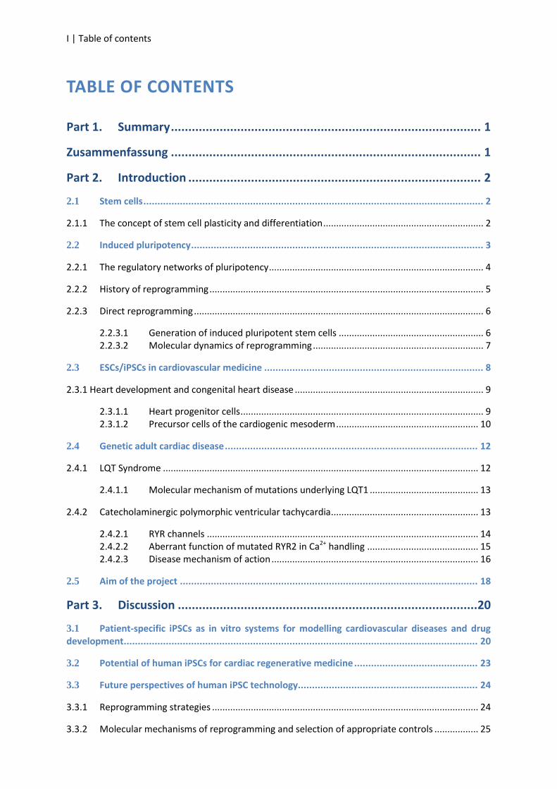
I | Table of contents
TABLE OF CONTENTS
Part 1. Summary ......................................................................................... 1
Zusammenfassung ......................................................................................... 1
Part 2. Introduction .................................................................................... 2
2.1 Stem cells ......................................................................................................................... 2
The concept of stem cell plasticity and differentiation .............................................................. 2 2.1.1
2.2 Induced pluripotency ........................................................................................................ 3
The regulatory networks of pluripotency ................................................................................... 4 2.2.1
History of reprogramming .......................................................................................................... 5 2.2.2
Direct reprogramming ................................................................................................................ 6 2.2.3
Generation of induced pluripotent stem cells ........................................................ 6 2.2.3.1 Molecular dynamics of reprogramming .................................................................. 7 2.2.3.2
2.3 ESCs/iPSCs in cardiovascular medicine .............................................................................. 8
2.3.1 Heart development and congenital heart disease ......................................................................... 9
Heart progenitor cells .............................................................................................. 9 2.3.1.1 Precursor cells of the cardiogenic mesoderm ....................................................... 10 2.3.1.2
2.4 Genetic adult cardiac disease .......................................................................................... 12
LQT Syndrome .......................................................................................................................... 12 2.4.1
Molecular mechanism of mutations underlying LQT1 .......................................... 13 2.4.1.1
Catecholaminergic polymorphic ventricular tachycardia ......................................................... 13 2.4.2
RYR channels ......................................................................................................... 14 2.4.2.1 Aberrant function of mutated RYR2 in Ca2+ handling ........................................... 15 2.4.2.2 Disease mechanism of action ................................................................................ 16 2.4.2.3
2.5 Aim of the project .......................................................................................................... 18
Part 3. Discussion ...................................................................................... 20
3.1 Patient-specific iPSCs as in vitro systems for modelling cardiovascular diseases and drug development .............................................................................................................................. 20
3.2 Potential of human iPSCs for cardiac regenerative medicine ............................................ 23
3.3 Future perspectives of human iPSC technology................................................................ 24
Reprogramming strategies ....................................................................................................... 24 3.3.1
Molecular mechanisms of reprogramming and selection of appropriate controls ................. 25 3.3.2
Table of contents | II
3.4 Final remarks.................................................................................................................. 26
Part 4. Acknowlegements .......................................................................... 27
Part 5. References ..................................................................................... 28
Part 6. Articles ........................................................................................... 34
Mouse and human induced pluripotent stem cells as a source for multipotent Isl1+ cardiovascular progenitors FASEB J. 2010 Mar;24(3):700-11.
Patient-specific induced pluripotent stem-cell models for long-QT syndrome. N Engl J Med. 2010 Oct 7;363(15):1397-409.
Dantrolene rescues arrhythmogenic RYR2 defect in a patient-specific stem cell model of catecholaminergic polymorphic ventricular tachycardia. EMBO Mol Med. 2012 Mar;4(3):180-91.
III | GLOSSSARY
GLOSSSARY
AP Action potential
APD Action potential duration
bFGF basic fibroblast growth factor
BMP4 Bone morphogenetic protein 4
Ca2+ Calcium
CaMKII Ca2+/Calmodulin dependent kinase II
CHD Congenital heart disease
CICR Calcium induced calcium release
CPVT Catecholaminergic polymorphic ventricular tachycardia
cTnC Cardiac troponin I
CTnI Cardiac troponin C
DAD Delayed afterdepolarization
DNA Deoxyribonucleic acid
EAD Early afterdepolarization
EB Embryoid body
ECC Electric-Contraction coupling
FHF First heart field
hESC Human embryonic stem cell
iPSC Induced pluripotent stem cell
LIF Leukemia inhibitory factor
LQT Long QT syndrome
LTCC L-type Ca2+ channel
mESC Mouse embryonic stem cell
NCX Na+/Ca2+ exchanger
OFT Outflow tract
PKA Protein kinase A
RYR Ryanodine receptor
SCD Sudden cardiac death
SCNT Somatic cell nuclear transfer
SHF Second heart field
SOICR Store overload induced Ca2+ release
SR Sarcoplasmatic reticulum
TALEN Transcription activator-like effector nucleases
TdP Torsade de Pointes

1 | Summary
Part 1. SUMMARY
Stem cells, despite being the subject of ethical and political debates, provide fascinating prospects
for biomedical applications by both their ability to renew themselves and to differentiate into
specialized cell types in vitro. Since the first isolation of murine embryonic stem cells in 1981,
remarkable advances and groundbreaking findings were observed. During the past five years, the
field gained further momentum by the discovery of a new platform technology (induced pluripotent
stem cell (iPSC) technology) allowing the artificial creation of cells identical to embryonic stem cells
from a donor’s adult somatic cells. The ability to readily obtain patient-specific stem cells and to
differentiate them into a variety of embryonic tissue-specific progenitors and adult somatic cells has
opened the door to the development of stem cell based models of human diseases. This cumulative
thesis relies on the iPSC technology to investigate how patient-specific induced pluripotent stem cells
can be used as an in vitro model system to study congenital and adult forms of genetic cardiac
diseases, such as long-QT syndrome and catecholaminergic polymorphic ventricular tachycardia, and
to investigate the potential of these cells for screening drug compounds and for developing patient-
specific therapies.
ZUSAMMENFASSUNG
Durch ihre Fähigkeiten sich selbst zu erneuern und in vitro in verschiedenste spezialisierte Zellen
auszureifen, bieten Stammzellen faszinierende Aussichten für biomedizinische Anwendungen
wenngleich sie immer noch Grund ethischer und politischer Diskussionen sind. Seit der ersten
Isolierung von murinen embryonischen Stammzellen im Jahr 1981 wurden beachtliche Fortschritte
erzielt und bahnbrechende Entdeckungen gemacht. Während der letzten fünf Jahre hat dieses
Forschungsfeld weiteres Momentum aufgebaut durch die Entdeckung einer neuen
Plattformtechnologie (induzierte pluripotente Stammzell (iPSC) -Technologie) die es erlaubt künstlich
aus adulten somatischen Spenderzellen Zellen zu erzeugen, die identisch sind zu embryonalen
Stammzellen. Die Möglichkeit nun auf einfachem Wege patienten-spezifische Stammzellen zu
erhalten und diese in eine Vielzahl von embryonalen, gewebsspezifischen Vorläuferzellen zu
differenzieren hat die Tür geöffnet hin zur Entwicklung von Stammzell-basierten Zellmodellen
humaner Erkrankungen. Diese kumulative Promotionsarbeit nutzt die iPSC Technologie um zu
untersuchen, inwieweit patienten-spezifische induzierte Stammzellen als ein in vitro Modellsystem
dienen können um kongenitale und Erwachsenen-Formen genetischer Herzkrankheiten, wie z.B. das
Long-QT Syndrom und die katecholaminerge polymorphe ventrikuläre Tachykardie zu studieren und
herauszufinden, ob sich diese Zellen nutzen lassen um neuartige Pharmaka zu screenen und
patienten-spezifische Therapien zu entwickeln.
Introduction | 2
Dif
fere
nti
atio
n
Zygote
Emybryonic Stem Cell
Adult Stem Cell
Mature Cells
Totipotent
Pluripotent
Multipotent
Unipotent
Part 2. INTRODUCTION
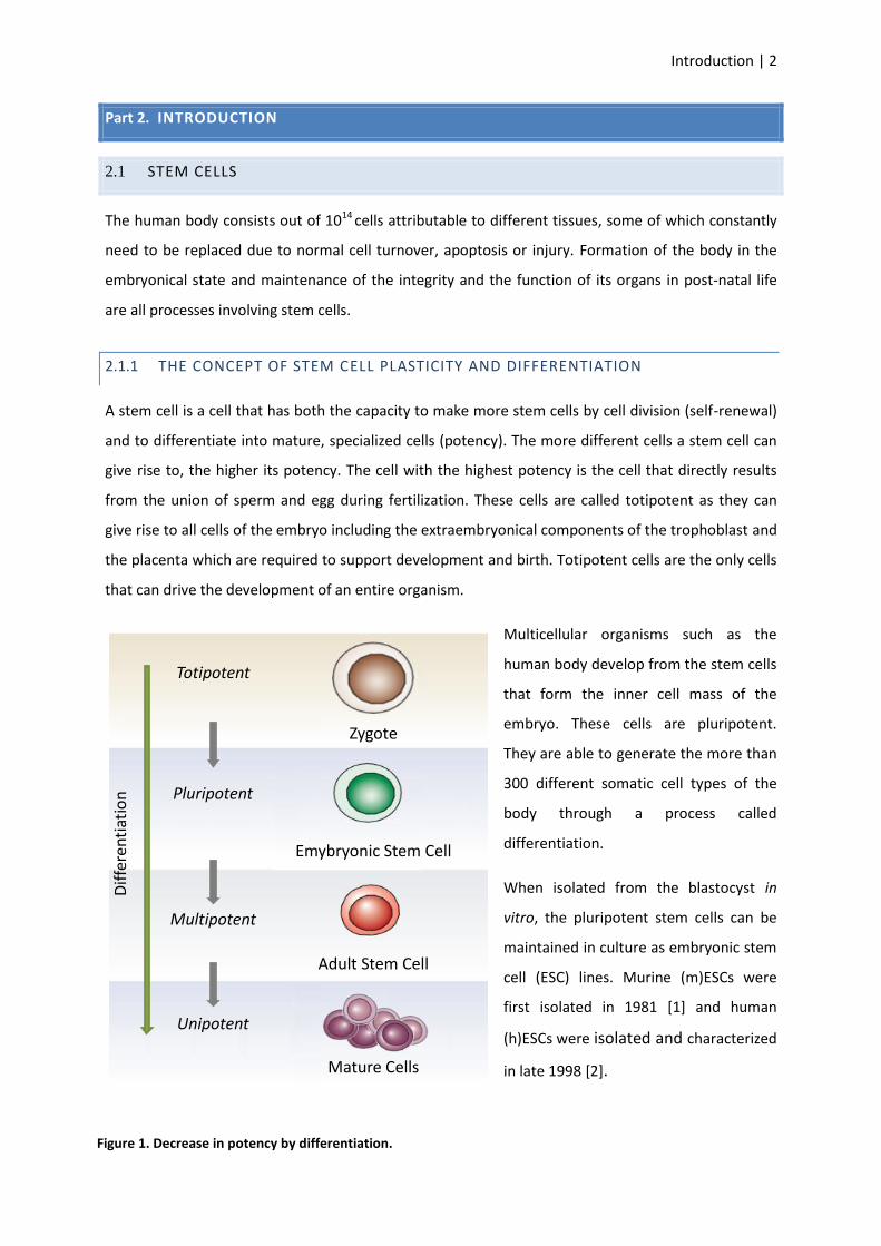
2.1 STEM CELLS
The human body consists out of 1014 cells attributable to different tissues, some of which constantly
need to be replaced due to normal cell turnover, apoptosis or injury. Formation of the body in the
embryonical state and maintenance of the integrity and the function of its organs in post-natal life
are all processes involving stem cells.
THE CONCEPT OF STEM CELL PLASTICITY AND DIFFERENTIATION 2.1.1
A stem cell is a cell that has both the capacity to make more stem cells by cell division (self-renewal)
and to differentiate into mature, specialized cells (potency). The more different cells a stem cell can
give rise to, the higher its potency. The cell with the highest potency is the cell that directly results
from the union of sperm and egg during fertilization. These cells are called totipotent as they can
give rise to all cells of the embryo including the extraembryonical components of the trophoblast and
the placenta which are required to support development and birth. Totipotent cells are the only cells
that can drive the development of an entire organism.
Multicellular organisms such as the
human body develop from the stem cells
that form the inner cell mass of the
embryo. These cells are pluripotent.
They are able to generate the more than
300 different somatic cell types of the
body through a process called
differentiation.
When isolated from the blastocyst in
vitro, the pluripotent stem cells can be
maintained in culture as embryonic stem
cell (ESC) lines. Murine (m)ESCs were
first isolated in 1981 [1] and human
(h)ESCs were isolated and characterized
in late 1998 [2].
Figure 1. Decrease in potency by differentiation.
3 | Introduction
As the embryo develops, its cells become progressively more specialized and pluripotency is lost
(Figure 1), although some tissues retain what are called multipotent cells (or adult stem cells) that
can only give rise to cells of that specific tissue and that are considered reminiscent of the embryonic
tissue-restricted progenitors. Adult stem cells maintain the integrity and function of organ systems
during adult life by replacing the cells that are lost owing to normal cell turnover, apoptosis or injury.
Adult stem cells have been identified in several postnatal organs, e.g. the blood (hematopoietic stem
cells), the brain (neural stem cells) or the bone-marrow (mesenchymal stem cells) [3].
Cells that can no longer give rise to cells other than of their own type are referred to as unipotent.
2.2 INDUCED PLURIPOTENCY
All nucleated cells of the human body, although functionally very different, maintain complete
genomes. Yet, a neuronal cell does not naturally turn into a cell of the gut. What determines the
cellular identity of a neuron and ultimately makes it different from an intestinal cell is hence not the
fact that it carries different genes but the fact that a different set of genes is being expressed in the
respective cells. Thus, the state of a cell is determined by its transcriptome, which is regulated by
epigenetic modifications.
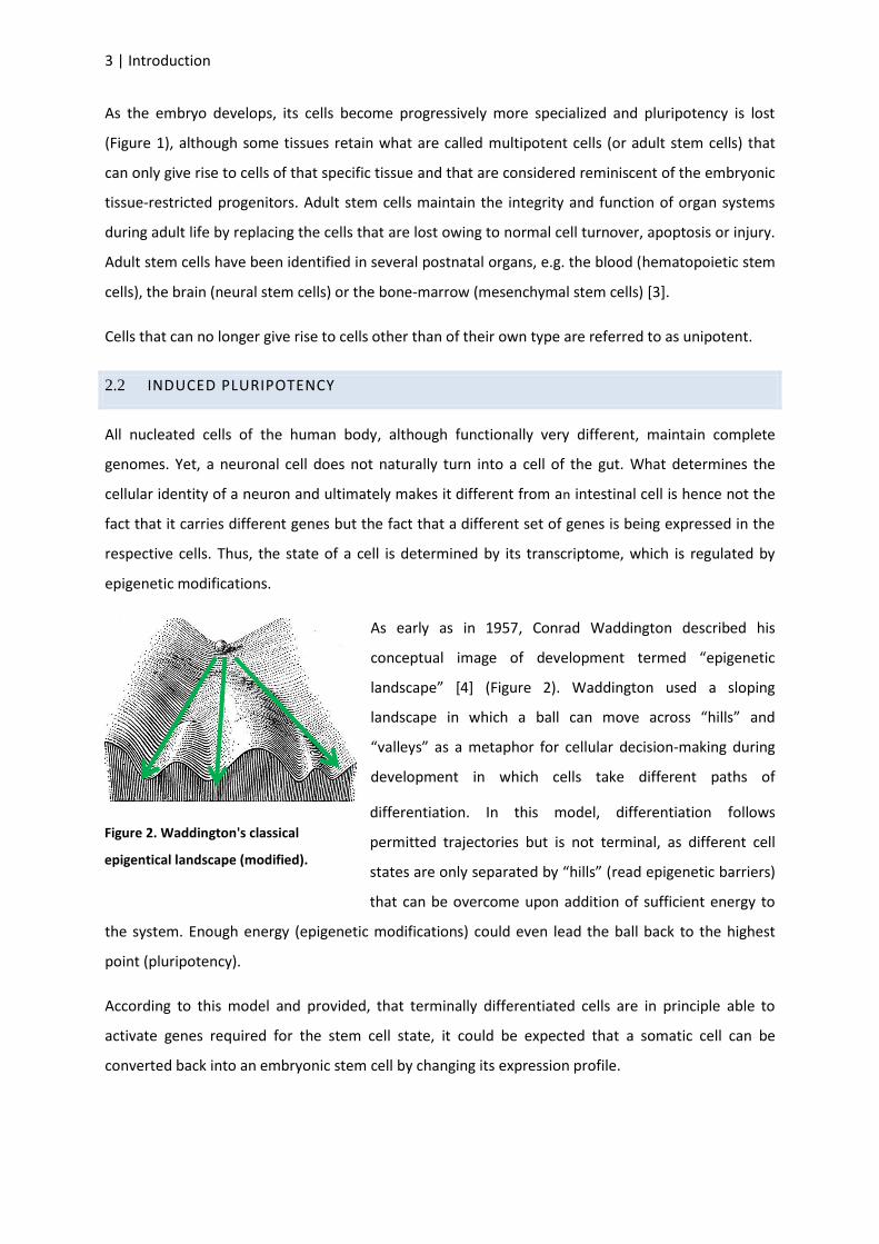
As early as in 1957, Conrad Waddington described his
conceptual image of development termed “epigenetic
landscape” [4] (Figure 2). Waddington used a sloping
landscape in which a ball can move across “hills” and
“valleys” as a metaphor for cellular decision-making during
development in which cells take different paths of
differentiation. In this model, differentiation follows
permitted trajectories but is not terminal, as different cell
states are only separated by “hills” (read epigenetic barriers)
that can be overcome upon addition of sufficient energy to
the system. Enough energy (epigenetic modifications) could even lead the ball back to the highest
point (pluripotency).
According to this model and provided, that terminally differentiated cells are in principle able to
activate genes required for the stem cell state, it could be expected that a somatic cell can be
converted back into an embryonic stem cell by changing its expression profile.
Figure 2. Waddington's classical
epigentical landscape (modified).

Introduction | 4
As opposed to the process of differentiation in which the potency decreases, the attempt to change a
- unipotent - somatic cell into a pluripotent embryonic stem cell would result in an increase of
potency which is why this process is called dedifferentiation (or reprogramming) (Figure 3).
Figure 3. Model of differentiation and dedifferentiation.
THE REGULATORY NETWORKS OF PLURIPOTENCY 2.2.1
The immense scientific interest in embryonic stem cells is largely owned to their potential to
differentiate into all cell types, hence their pluripotent properties. In order for embryonic stem cells
to be used as a research tool one has to be able to keep them in culture for prolonged periods of
time without loss of their differentiation potential. This requires well-defined culture conditions and
a deep understanding of how embryonic stem cells sustain their undifferentiated state on a
molecular basis through transcription factors and signaling pathways.
Early studies have shown that there are significant differences between mESCs and hESCs. While
mESCs require leukemia inhibitory factor (LIF) and bone morphogenetic protein 4 (BMP4) as essential
growth factors in culture, hESCs rely on Activin A and basic fibroblast growth factor (bFGF)[2, 5, 6].
However, there is a set of key pluripotency factors, namely Oct4, Sox2 and Nanog, which are central
to the regulation of pluripotency in both mESC and hESC and constitute a core transcriptional
network. These transcription factors regulate themselves and each other, showing features of feed-
forward loops, and co-act in activating other pluripotency factors as well as in repressing
differentiation genes [7, 8]. Recent studies suggest PRDM14 to also be part of the core
transcriptional network in hESCs [9].
Differentiation
Pluripotent
Dedifferentiation
Emybryonic Stem Cell
Mature Cells
Unipotent
5 | Introduction
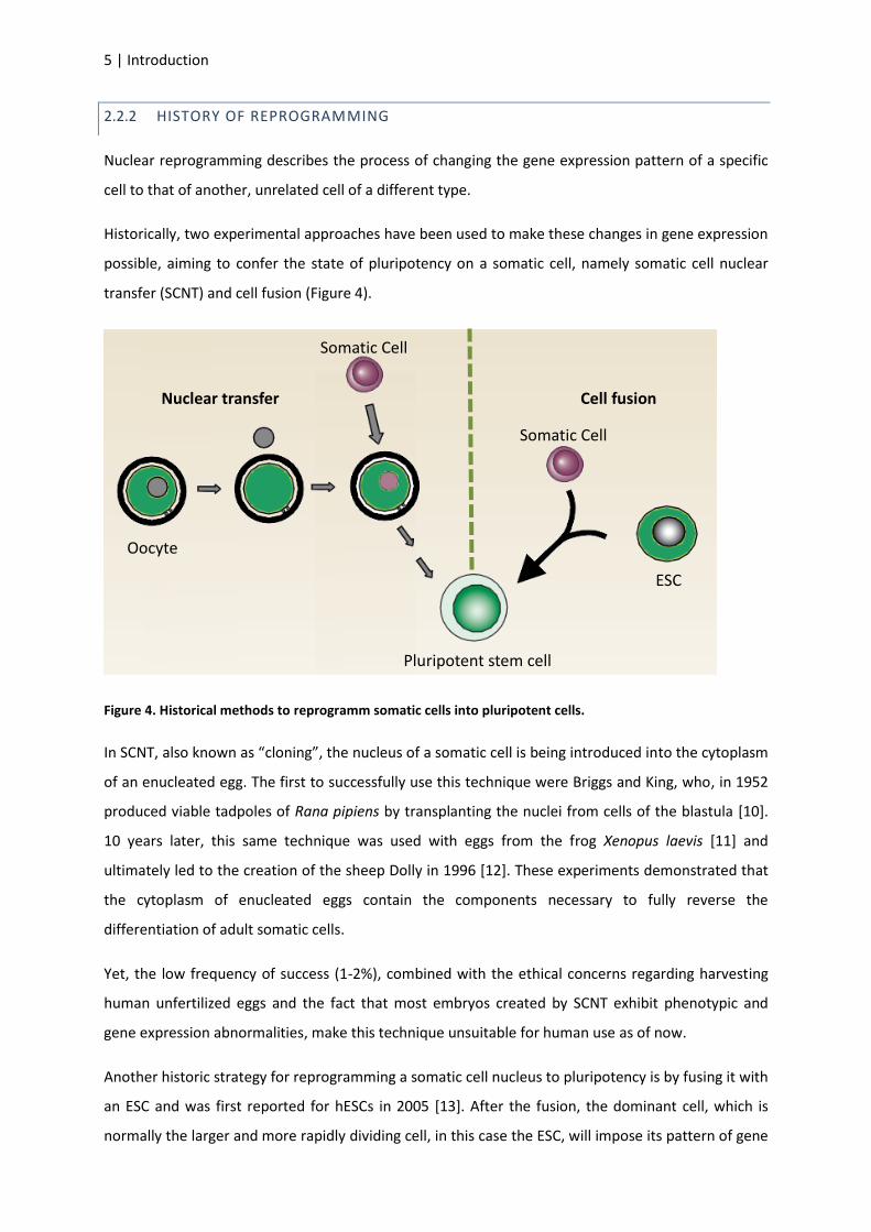
HISTORY OF REPROGRAMMING 2.2.2
Nuclear reprogramming describes the process of changing the gene expression pattern of a specific
cell to that of another, unrelated cell of a different type.
Historically, two experimental approaches have been used to make these changes in gene expression
possible, aiming to confer the state of pluripotency on a somatic cell, namely somatic cell nuclear
transfer (SCNT) and cell fusion (Figure 4).
Figure 4. Historical methods to reprogramm somatic cells into pluripotent cells.
In SCNT, also known as “cloning”, the nucleus of a somatic cell is being introduced into the cytoplasm
of an enucleated egg. The first to successfully use this technique were Briggs and King, who, in 1952
produced viable tadpoles of Rana pipiens by transplanting the nuclei from cells of the blastula [10].
10 years later, this same technique was used with eggs from the frog Xenopus laevis [11] and
ultimately led to the creation of the sheep Dolly in 1996 [12]. These experiments demonstrated that
the cytoplasm of enucleated eggs contain the components necessary to fully reverse the
differentiation of adult somatic cells.
Yet, the low frequency of success (1-2%), combined with the ethical concerns regarding harvesting
human unfertilized eggs and the fact that most embryos created by SCNT exhibit phenotypic and
gene expression abnormalities, make this technique unsuitable for human use as of now.
Another historic strategy for reprogramming a somatic cell nucleus to pluripotency is by fusing it with
an ESC and was first reported for hESCs in 2005 [13]. After the fusion, the dominant cell, which is
normally the larger and more rapidly dividing cell, in this case the ESC, will impose its pattern of gene
Pluripotent stem cell
Somatic Cell
Somatic Cell
Nuclear transfer Cell fusion
ESC
Oocyte
Introduction | 6
expression on the partner cell. It appears therefore, that ESCs possess factors in either their nucleus
[14] or their cytoplasm [15] that are able to induce pluripotency in somatic cells. As these pluripotent
cells generated by fusion maintain the chromosomes from both cells (tetraploid), rejection upon
implantation is likely.
DIRECT REPROGRAMMING 2.2.3
The fact that somatic cell nuclei can be reprogrammed by transfer into oocytes or fusion with ESCs
indicates that oocytes and ESCs contain reprogramming factors.
In 2006, the identification of these reprogramming factors by the group behind Shinya Yamanaka
brought significant advance to this field. For the first time, mouse somatic cells were reprogrammed
into an ESC state without the contribution of a second pluripotent cell but only by forced expression
of four specific pluripotency-associated genes that were singled out by screening a pool of 24
candidate genes. These cells were named induced pluripotent stem cells (iPSCs) and shared many
properties with ESCs [16], e.g. morphology, growth characteristics and gene expression.
iPSCs were then defined as a type of pluripotent stem cells that can be generated from various adult
somatic cell types by forced expression of certain combinations of key ESC- associated transcription
factors and that are similar to ESCs in their morphology, expression of important ESC marker genes,
and their ability to form teratomas and yield live chimaeras when injected into mouse blastocysts.
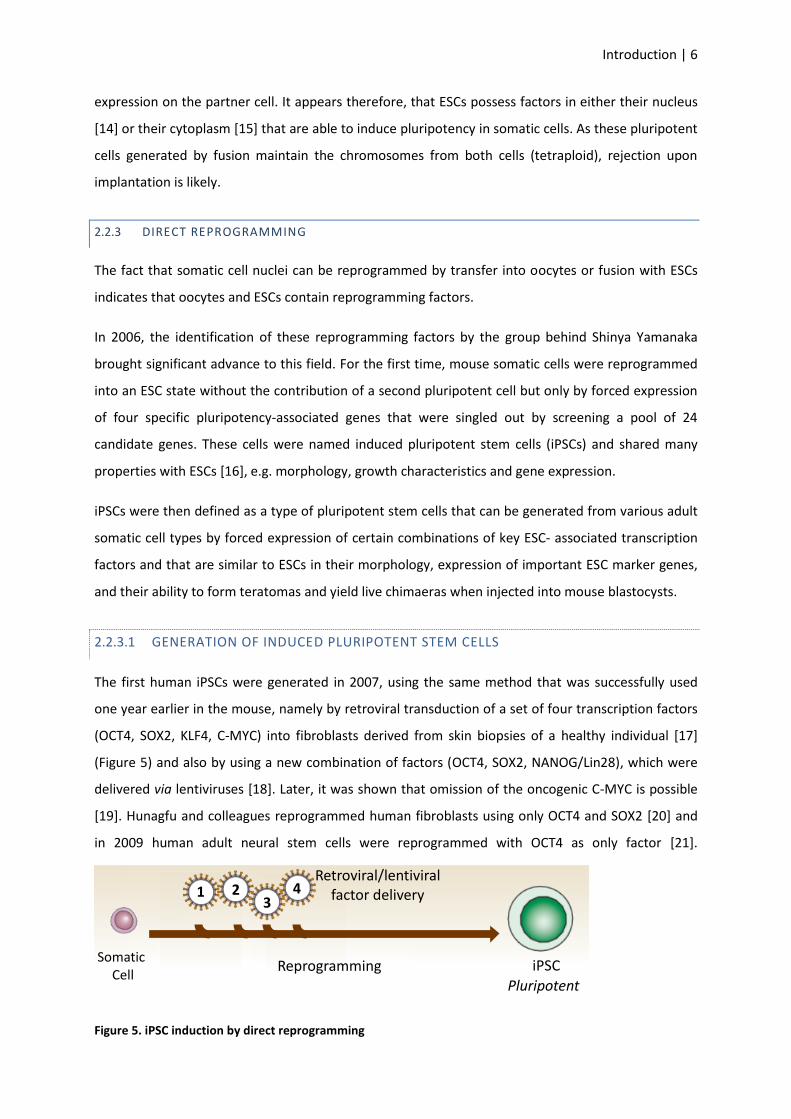
GENERATION OF INDUCED PLURIPOTENT STEM CELLS 2.2.3.1
The first human iPSCs were generated in 2007, using the same method that was successfully used
one year earlier in the mouse, namely by retroviral transduction of a set of four transcription factors
(OCT4, SOX2, KLF4, C-MYC) into fibroblasts derived from skin biopsies of a healthy individual [17]
(Figure 5) and also by using a new combination of factors (OCT4, SOX2, NANOG/Lin28), which were
delivered via lentiviruses [18]. Later, it was shown that omission of the oncogenic C-MYC is possible
[19]. Hunagfu and colleagues reprogrammed human fibroblasts using only OCT4 and SOX2 [20] and
in 2009 human adult neural stem cells were reprogrammed with OCT4 as only factor [21].
Figure 5. iPSC induction by direct reprogramming
1 2 3
4
Pluripotent
iPSC Reprogramming
Retroviral/lentiviral factor delivery
Somatic Cell
7 | Introduction
MOLECULAR DYNAMICS OF REPROGRAMMING 2.2.3.2
Reprogramming of cells into iPSCs is not an instant event but a dynamic process over a period of 3-4
weeks. During this time, the epigenomic pattern of the somatic cell is being reset to the one of an
embryonic stem cell. The rate at which this successfully happens is extremely low (0,01-0,1%) and
two models have been proposed to explain this low efficiency, the “elite” and the “stochastic”
model.
The “elite model”, in brief, proposes that the low efficiency of iPSC generation goes back to the fact
that not all, but only a few cells in a culture of somatic cells are amenable to reprogramming in the
first place and that these are somatic stem or progenitor cells, that can be found in most adult tissue
cultures and that are closer to the state of pluripotent cells than terminally differentiated cells [22].
In contrast, the “stochastic model” postulates that all cells are equally suited for reprogramming, but
for a cell to become pluripotent, a series of stochastic epigenetic events (“roadblocks”) needs to
happen and only a strict minority of cells will successfully pass all these “roadblocks”. According to a
working model proposed by Nagy [23], mainly three phases in the molecular changes of a somatic
cell can be distinguished during reprograming (Figure 6).
Figure 6. iPSC generation is a multistep process. Adopted from [23].
In the early phase, termed “Area of return”, the somatic cell starts to change its morphology and to
down-regulate the expression of its somatic markers. The reprogramming process is purely driven by
the presence of pluripotency transcription factors originating from the forced expression of the
transgenes. The “roadblock” during this phase could be the extinction of the somatic program.
At the beginning of the middle phase, referred to as “Area 51”, the cell has not yet switched on its
own endogenous copies of the pluripotency genes. Provided that the expression from the exogenous
sequences is sufficiently strong, the transcription factors will start to activate the endogenous gene
Introduction | 8
loci and make the cells increasingly independent from the exogenes over time. This is likely to be the
criteria to exit “Area 51” and the main important regulator of successful reprogramming.
Although the exact temporal sequence of changes during the late phase, “Area of iPS cells”, remains
elusive, it marks the point at which the expression of pluripotency proteins from the endogenes
becomes sufficiently strong to, in part, support the further transition of a cell towards a pluripotent
state as the silencing of the viral sequences starts as a consequence of the reprogramming. In this
stage, ESC-like colonies (“pre-iPSCs”) become obvious, genome-wide remodeling of chromatin
modifications, such as DNA and histone tail methylation, takes place and the cell starts to express
early pluripotency markers such as SSEA-1 and alkaline phosphatase. Yet, the acquisition of
pluripotency remains incomplete and awaits the full independence of exogenous factor expression
and complete activation of endogenous pluripotency genes, finalized X chromosome reactivation and
loss of repressive chromatin character at many pluripotency genes marked by expression of Nanog
and Oct4.
The vast majority of all cells will not continue along this timeline but rather become defective during
reprogramming and will be either selected out by culture conditions or ultimately revert to its state
of origin.
2.3 ESCS/IPSCS IN CARDIOVASCULAR MEDICINE
The ability to generate functional cell types from ESCs and iPSCs offers unprecedented opportunities
to develop novel cell-based therapies for degenerative diseases, to establish predictive drug toxicity
test or to model human disease in culture.
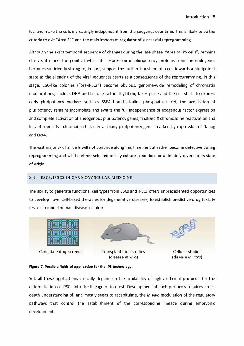
Figure 7. Possible fields of application for the iPS technology.
Yet, all these applications critically depend on the availability of highly efficient protocols for the
differentiation of iPSCs into the lineage of interest. Development of such protocols requires an in-
depth understanding of, and mostly seeks to recapitulate, the in vivo modulation of the regulatory
pathways that control the establishment of the corresponding lineage during embryonic
development.
Candidate drug screens Transplantation studies (disease in vivo)
Cellular studies (disease in vitro)

9 | Introduction
2.3.1 HEART DEVELOPMENT AND CONGENITAL HEART DISEASE
The mammalian heart is a highly specialized organ and is the first to develop during embryogenesis.
Its proper function relies on the controlled development of atrial and ventricular myocardium,
endocardium, the outflow tract, the coronary tree, the heart valves and the conduction system.
Despite decades of tracing cell lineages and descriptive embryology of the heart’s origins, a more
complete and accurate picture of cardiogenesis has only recently emerged [24-26].
HEART PROGENITOR CELLS 2.3.1.1
During cardiogenesis three populations of progenitors have been identified to contribute to the
different compartments of the heart (Figure 8) and each of these populations gives rise to distinct
cardiac cell types: the cardiogenic mesoderm forms the major proportion of myocytes of the
ventricles, the atria and the outflow tract (OFT) and additionally contributes to cells of the
conduction system and to the cushion cells of the aortic and pulmonary valves; the cardiac neural
crest progenitors give rise to the distal smooth muscle cells of the OFT and to the autonomic nervous
system of the heart; and finally, the proepicardial organ produces the vascular support cells of the
coronary vessels, the interstitial fibroblasts embedded in the myocardium and some myocytes,
mainly in the atrioventricular septum [27, 28].
Figure 8. Origin and lineage relationship of cardiac cell types. Adopted from [24].
An important principle in heart development is that the regulation of different cardiac precursors
must be tightly controlled so that the correct progenitor population proliferates, migrates and
differentiates at the correct time and in the correct location.
Due to this complexity, many errors may happen during heart development, which commonly leads
to the appearance of congenital heart diseases (CHDs). CHDs constitute a major percentage of
clinically significant birth defects with an estimated incidence of 4-14 per 1000 live infants [29].
Introduction | 10
Although in the past decades human genetic studies have identified numerous genes that are
associated with inherited and sporadic forms of CHDs, the mechanisms of how deficiencies in these
genes translate to structural defects still remain unknown [30].
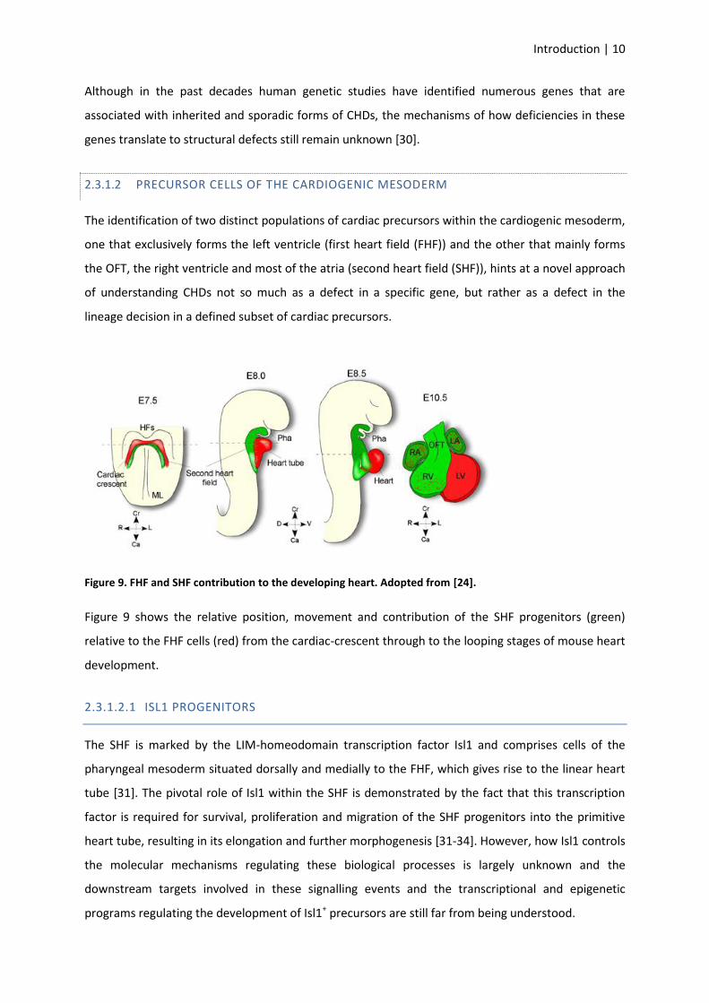
PRECURSOR CELLS OF THE CARDIOGENIC MESODERM 2.3.1.2
The identification of two distinct populations of cardiac precursors within the cardiogenic mesoderm,
one that exclusively forms the left ventricle (first heart field (FHF)) and the other that mainly forms
the OFT, the right ventricle and most of the atria (second heart field (SHF)), hints at a novel approach
of understanding CHDs not so much as a defect in a specific gene, but rather as a defect in the
lineage decision in a defined subset of cardiac precursors.
Figure 9. FHF and SHF contribution to the developing heart. Adopted from [24].
Figure 9 shows the relative position, movement and contribution of the SHF progenitors (green)
relative to the FHF cells (red) from the cardiac-crescent through to the looping stages of mouse heart
development.
2.3.1.2.1 ISL1 PROGENITORS
The SHF is marked by the LIM-homeodomain transcription factor Isl1 and comprises cells of the
pharyngeal mesoderm situated dorsally and medially to the FHF, which gives rise to the linear heart
tube [31]. The pivotal role of Isl1 within the SHF is demonstrated by the fact that this transcription
factor is required for survival, proliferation and migration of the SHF progenitors into the primitive
heart tube, resulting in its elongation and further morphogenesis [31-34]. However, how Isl1 controls
the molecular mechanisms regulating these biological processes is largely unknown and the
downstream targets involved in these signalling events and the transcriptional and epigenetic
programs regulating the development of Isl1+ precursors are still far from being understood.
11 | Introduction
In 2005, it has been demonstrated, using tamoxifen-inducible Cre/lox technology, that Isl1 is a
developmental lineage marker for undifferentiated cardiac progenitors and enables their isolation
from embryonic and postnatal mouse and human hearts [32]. Cardiac fibroblasts allow the cells to
self-renew, maintaining their ability to subsequently differentiate into functional myocytes with
action potential characteristics of atrial, ventricular, or conduction cells and intact excitation-
contraction coupling (ECC) [32, 35].
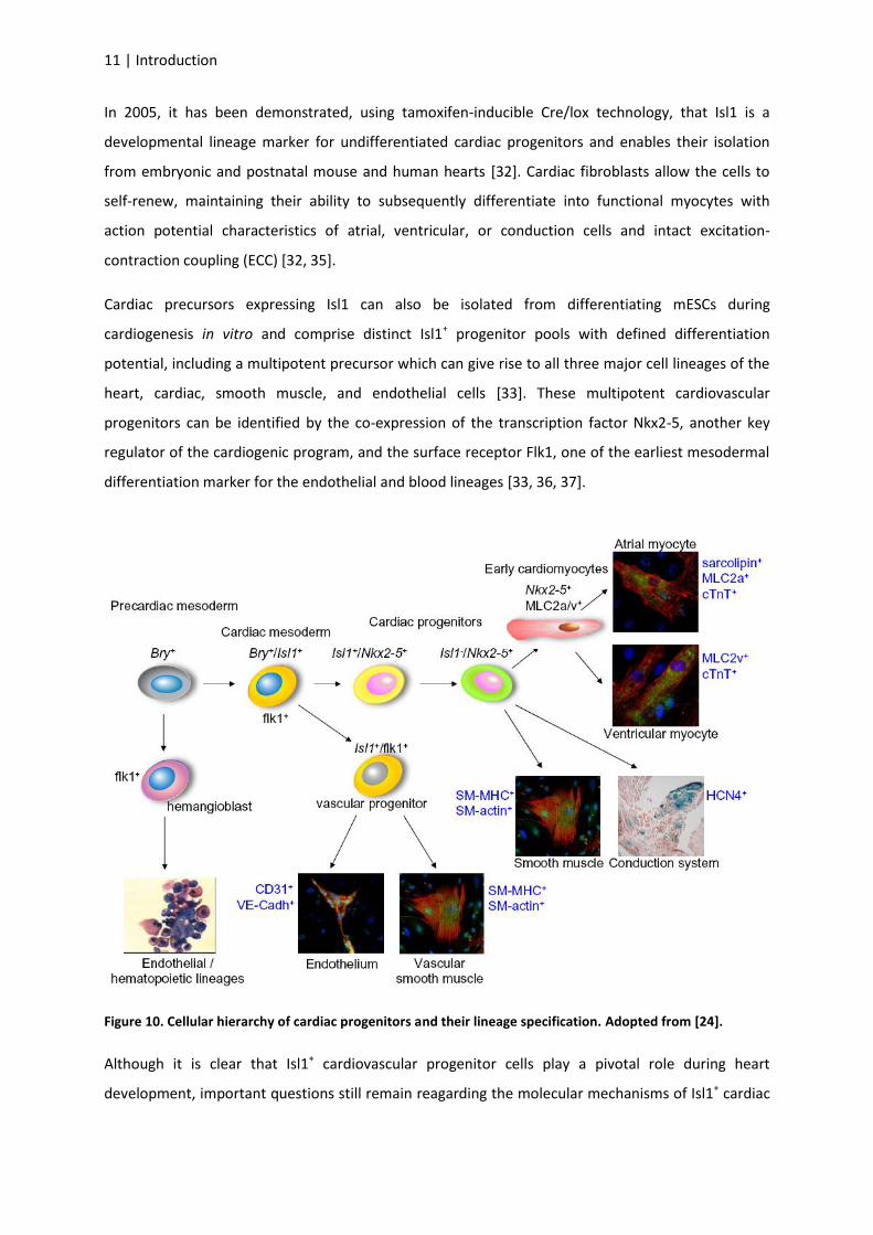
Cardiac precursors expressing Isl1 can also be isolated from differentiating mESCs during
cardiogenesis in vitro and comprise distinct Isl1+ progenitor pools with defined differentiation
potential, including a multipotent precursor which can give rise to all three major cell lineages of the
heart, cardiac, smooth muscle, and endothelial cells [33]. These multipotent cardiovascular
progenitors can be identified by the co-expression of the transcription factor Nkx2-5, another key
regulator of the cardiogenic program, and the surface receptor Flk1, one of the earliest mesodermal
differentiation marker for the endothelial and blood lineages [33, 36, 37].
Figure 10. Cellular hierarchy of cardiac progenitors and their lineage specification. Adopted from [24].
Although it is clear that Isl1+ cardiovascular progenitor cells play a pivotal role during heart
development, important questions still remain reagarding the molecular mechanisms of Isl1+ cardiac
Introduction | 12
progenitor maintenance, lineage specification and differentiation and how causative genes of CHDs
affect cell-fate decisions in the ISL1 lineage during human cardiogenesis.
The ability to isolate and selectively expand ISL1+ cardiovascular progenitors offers a powerful cell-
based in vitro system to answer these questions and generate human models of CHDs.
2.4 GENETIC ADULT CARDIAC DISEASE
Cardiovascular disease remains the leading cause of death worldwide accounting for 12.8% of all
deaths in 2008 (WHO website).
This is in part attributable to the lifestyle and unhealthy diet in the industrialized world but also owed
to the lack of suitable models that fully reflect the genetically diverse nature of these diseases and
adequately address their long-term development.
Genetic cardiac disease can be subdivided into two classes, cardiomyopathies and channelopathies.
Cardiomyopathies go back to structural and functional abnormalities of the heart. In contrast,
channelopathies occur in the absence of structural defects and are caused by dysfunctional cardiac
ion channels causing electrical instability of the cells. Since the normal heart pumping function relies
on proper electrical propagation through the ventricles, channelopathies can trigger life-threatening
arrhythmias that may result in sudden cardiac death (SCD). Approximately 10-20% of all sudden
deaths happen in absence of structural cardiac abnormalities.
At present, the class of cardiac channelopathies comprises four distinct conditions namely,
congenital long-QT syndrome (LQT), catecholaminergic polymorphic ventricular tachycardia (CPVT),
Brugada syndrome, and short-QT syndrome.
LQT SYNDROME 2.4.1
The LQT syndrome is a disease characterized by delayed cardiac repolarization, which causes
prolongation of the QT interval on the surface electrocardiogram. The clinical manifestations of the
disorder involve polymorphic ventricular tachycardia (often termed torsades de pointes (TdP)) and
syncopal episodes, which often result in cardiac arrest and sudden death in otherwise healthy young
individuals [38]. Besides the symptomatic treatment with -blockers and the implantation of
automated defibrillators to terminate fatal arrhythmia, no causal therapy is currently available [39].
The most frequent forms of LQTS are acquired as an adverse effect of treatment with medications
that block cardiac potassium channels, such as class III antiarrhythmic drugs and antihistamines, or
by electrolyte disturbances that alter the electrochemical conditions needed for normal cardiac
13 | Introduction
excitability. Less commonly, LQTS is inherited as an autosomal dominant (Romano-Ward syndrome)
or recessive (Jervell and Lange-Nielsen syndrome) disorder.
Congenital LQTS has been subdivided into types based on the gene in which causative mutations
occur. At least 12 different LQTS-associated genes have been identified so far, most of which encode
ion channels specifically involved in the generation of the cardiac action potential (AP) [40, 41].
However, in ~80-90% of genotyped patients, the underlying causes are mutations in two main genes:
the KCNQ1 (also known as KVLQT1 or Kv7.1) gene, which encodes the pore-forming α-subunits of the
channels generating the IKs current (LQT1 syndrome), and the KCNH2 (also known as HERG or
Kv11.1) gene, which produces the α-subunits of the channels responsible for the IKr conductance
(LQT2 syndrome) [41-43].
MOLECULAR MECHANISM OF MUTATIONS UNDERLYING LQT1 2.4.1.1
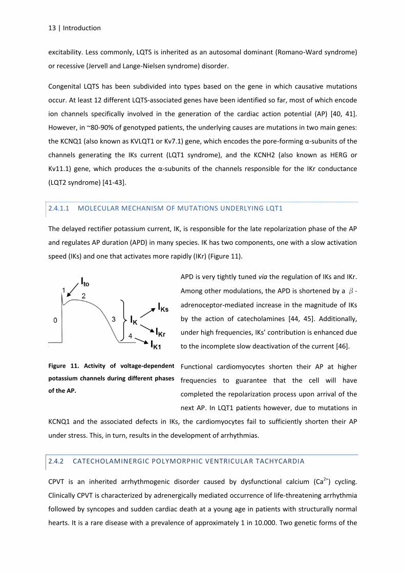
The delayed rectifier potassium current, IK, is responsible for the late repolarization phase of the AP
and regulates AP duration (APD) in many species. IK has two components, one with a slow activation
speed (IKs) and one that activates more rapidly (IKr) (Figure 11).
APD is very tightly tuned via the regulation of IKs and IKr.
Among other modulations, the APD is shortened by a β-
adrenoceptor-mediated increase in the magnitude of IKs
by the action of catecholamines [44, 45]. Additionally,
under high frequencies, IKs’ contribution is enhanced due
to the incomplete slow deactivation of the current [46].
Functional cardiomyocytes shorten their AP at higher
frequencies to guarantee that the cell will have
completed the repolarization process upon arrival of the
next AP. In LQT1 patients however, due to mutations in
KCNQ1 and the associated defects in IKs, the cardiomyocytes fail to sufficiently shorten their AP
under stress. This, in turn, results in the development of arrhythmias.
CATECHOLAMINERGIC POLYMORPHIC VENTRICULAR TACHYCARDIA 2.4.2
CPVT is an inherited arrhythmogenic disorder caused by dysfunctional calcium (Ca2+) cycling.
Clinically CPVT is characterized by adrenergically mediated occurrence of life-threatening arrhythmia
followed by syncopes and sudden cardiac death at a young age in patients with structurally normal
hearts. It is a rare disease with a prevalence of approximately 1 in 10.000. Two genetic forms of the
Figure 11. Activity of voltage-dependent
potassium channels during different phases
of the AP.
Introduction | 14
disease have been described: CPVT1 accounting for at least 50% of all cases and associated with
autosomal dominant mutations in the cardiac ryanodine receptor (RYR2), and the very rare form
CPVT2, linked to recessive mutations in calsequestrin (CSQ2) [47]. Both proteins belong to the
multimolecular Ca2+ release channel complex of the sarcoplasmic reticulum (SR) that supports
myocyte Ca2+ cycling and contractile activity. Despite the high mortality of 30-50% by the age of 35
years, to date, no causative treatment exists and patients are treated purely symptomatically with -
blockers and implantable defibrillators or, as a last resort, by sympathetic denervation.
RYR CHANNELS 2.4.2.1
Ryanodine receptors were originally identified during the testing of ryanodine, a plant alkaloid , as a
potential insecticide due to its paralyzing effect on insects [48]. Ryanodine was subsequently found
to induce widespread paralysis in cardiac and skeletal muscles and to bind to a specific component of
the SR [49]. So far, three mammalian isoforms of RYR have been described. RYR1, predominantly
expressed in skeletal muscle, RYR2, the cardiac isoform, and RYR3 mainly found in neuronal tissue.
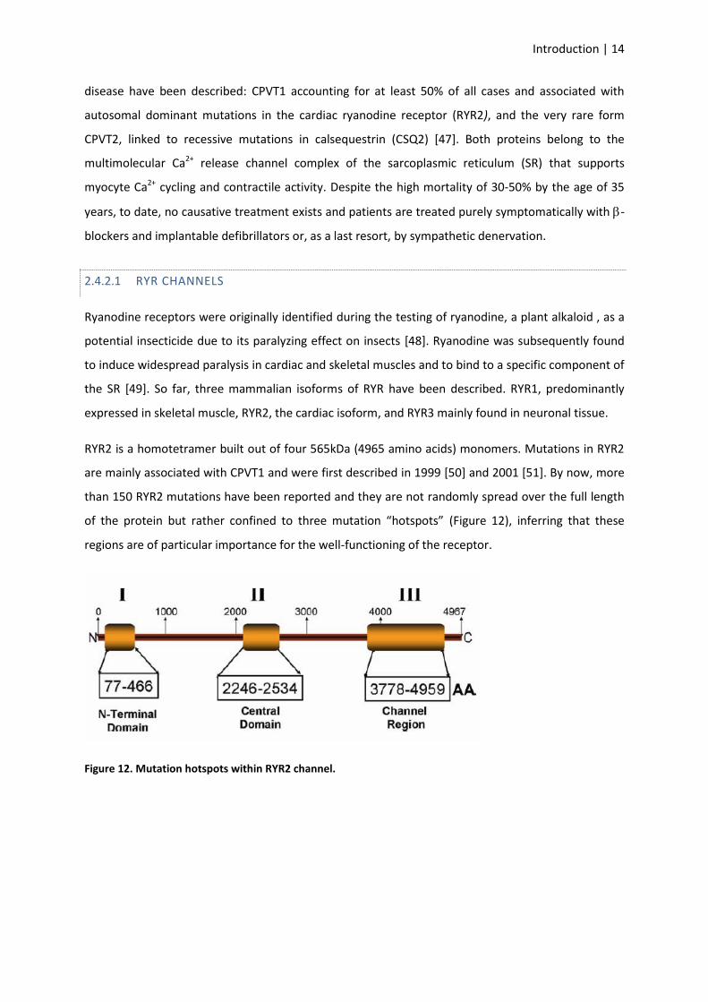
RYR2 is a homotetramer built out of four 565kDa (4965 amino acids) monomers. Mutations in RYR2
are mainly associated with CPVT1 and were first described in 1999 [50] and 2001 [51]. By now, more
than 150 RYR2 mutations have been reported and they are not randomly spread over the full length
of the protein but rather confined to three mutation “hotspots” (Figure 12), inferring that these
regions are of particular importance for the well-functioning of the receptor.
Figure 12. Mutation hotspots within RYR2 channel.
15 | Introduction
ABERRANT FUNCTION OF MUTATED RYR2 IN CA2+ HANDLING 2.4.2.2
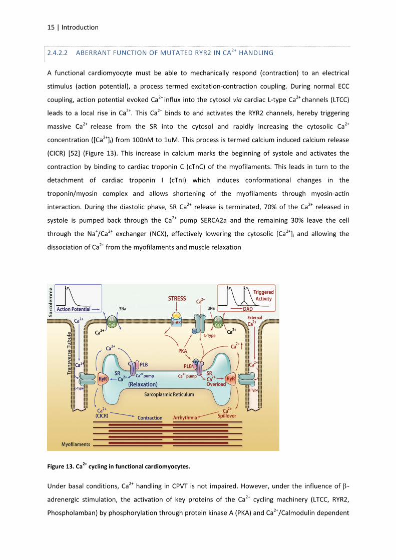
A functional cardiomyocyte must be able to mechanically respond (contraction) to an electrical
stimulus (action potential), a process termed excitation-contraction coupling. During normal ECC
coupling, action potential evoked Ca2+ influx into the cytosol via cardiac L-type Ca2+ channels (LTCC)
leads to a local rise in Ca2+. This Ca2+ binds to and activates the RYR2 channels, hereby triggering
massive Ca2+ release from the SR into the cytosol and rapidly increasing the cytosolic Ca2+
concentration ([Ca2+]i) from 100nM to 1uM. This process is termed calcium induced calcium release
(CICR) [52] (Figure 13). This increase in calcium marks the beginning of systole and activates the
contraction by binding to cardiac troponin C (cTnC) of the myofilaments. This leads in turn to the
detachment of cardiac troponin I (cTnI) which induces conformational changes in the
troponin/myosin complex and allows shortening of the myofilaments through myosin-actin
interaction. During the diastolic phase, SR Ca2+ release is terminated, 70% of the Ca2+ released in
systole is pumped back through the Ca2+ pump SERCA2a and the remaining 30% leave the cell
through the Na+/Ca2+ exchanger (NCX), effectively lowering the cytosolic [Ca2+]i and allowing the
dissociation of Ca2+ from the myofilaments and muscle relaxation
Figure 13. Ca2+
cycling in functional cardiomyocytes.
Under basal conditions, Ca2+ handling in CPVT is not impaired. However, under the influence of -
adrenergic stimulation, the activation of key proteins of the Ca2+ cycling machinery (LTCC, RYR2,
Phospholamban) by phosphorylation through protein kinase A (PKA) and Ca2+/Calmodulin dependent
Introduction | 16
kinase II (CaMKII) is evoking the disease phenotype. Under this condition of augmented Ca2+ influx
and SR Ca2+ uptake, resulting in SR Ca2+ overload, the mutant RYR2 channel is no longer able to
remain tightly closed in diastole. This leads to aberrant Ca2+ release during diastole and hence to
augmented diastolic [Ca2+]i, which has the potential to reverse the mode of action of NCX. Instead of
pumping Na+ out of the cell at this stage, NCX is pumping Na+ into the cell, resulting in arrythmogenic
membrane depolarization, visible as delayed afterdepolarization (DADs). If this depolarization
reaches a certain threshold, the voltage-activated Na+ channels open, which can lead to a full
spontaneous AP (triggered activity).
DISEASE MECHANISM OF ACTION 2.4.2.3
Data gained from knock-in/out mice and channel overexpression in HEK cells have led to the
formulation of three hypotheses on how mutations in RYR2 may lead to diastolic SR Ca2+ leak and
arrhythmias.
The first hypothesis is developed around the disruption of the critical interaction between the RYR2
channel and one of its modulating proteins, FKBP12.6. In 2004, Wehrens and colleagues conducted
experiments in FKBP12.6 null and heterozygous mice and found that they exhibited ventricular
arrhythmias and SCD after -adrenergic stimulation, while being normal at rest. Occurrence of
arrhythmias could be abolished by administration of JTV519, a drug with RYR2/FKBP12.6 stabilizing
properties, in FKBP12.6+/- but not in FKBP12.6-/- mice [53]. They concluded that RYR2 leakiness results
from a decreased receptor affinity to FKBP12.6 due to the mutation. Although it may be possible that
selected mutations alter FKBP12.6 binding to RYR2, this hypothesis has been recently challenged and
an increasing body of evidence clearly demonstrates that alterations in FKBP12.6-RYR2 interaction
are unlikely to be the common cause of CPVT1 [54-58].
The second hypothesis is called store-overload induced Ca2+-release (SOICR). Evidences suggest that
RYR2 channels also sense the SR Ca2+ by a luminal Ca2+ activation site distinct from the cytosolic Ca2+
activation site. Jiang and colleagues [59] proposed that the mutations in RYR2 lead to an altered
luminal SR Ca2+ sensing which in turn lowers the SR Ca2+ threshold at which RYR2-mediated Ca2+
release happens. While this is benign under baseline conditions when SR Ca2+ load is normal and far
under the threshold level, it becomes pathological under the influence of -adrenergic stimulation,
when the SR store is filled to a higher level due to the action of PKA on phospholamban. In this
condition of store-overload, luminal Ca2+ levels shoot above the Ca2+ release threshold of mutated
RYR2 channels, leading to repetitive, premature RYR2 activation.
17 | Introduction
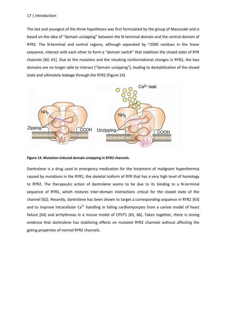
The last and youngest of the three hypotheses was first formulated by the group of Mazsuzaki and is
based on the idea of “domain unzipping” between the N-terminal domain and the central domain of
RYR2. The N-terminal and central regions, although separated by ~2000 residues in the linear
sequence, interact with each other to form a “domain switch” that stabilizes the closed state of RYR
channels [60, 61]. Due to the mutation and the resulting conformational changes in RYR2, the two
domains are no longer able to interact (“domain unzipping”), leading to destabilization of the closed
state and ultimately leakage through the RYR2 (Figure 14).
Figure 14. Mutation-induced domain unzipping in RYR2 channels.
Dantrolene is a drug used in emergency medication for the treatment of malignant hyperthermia
caused by mutations in the RYR1, the skeletal isoform of RYR that has a very high level of homology
to RYR2. The therapeutic action of dantrolene seems to be due to its binding to a N-terminal
sequence of RYR1, which restores inter-domain interactions critical for the closed state of the
channel [62]. Recently, dantrolene has been shown to target a corresponding sequence in RYR2 [63]
and to improve intracellular Ca2+ handling in failing cardiomyocytes from a canine model of heart
failure [64] and arrhythmias in a mouse model of CPVT1 [65, 66]. Taken together, there is strong
evidence that dantrolene has stabilizing effects on mutated RYR2 channels without affecting the
gating properties of normal RYR2 channels.
Introduction | 18
2.5 AIM OF THE PROJECT
To date, many of the most critical and puzzling human cardiovascular disorders cannot be adequately
studied because specific human cardiovascular cell types, such as cardiomyocytes, coronary
endothelial and smooth muscle cells, cannot be easily obtained. Two crucial steps towards reaching
the goal of studying specific cardiovascular cell types from patients with various forms of congenital
or acquired heart diseases have recently been made: 1) the identification of multipotent
cardiovascular progenitor cells not only in mammalian embryos and postnatal (adult) heart but also
as an intermediate stage during differentiation of embryonic stem cells; 2) the breakthrough
discovery that adult somatic cells can be reprogrammed back to a pluripotent state by ectopic
expression of few defined transcription factors - iPSC technology. Therefore, iPSCs could be an ideal
source to obtain patient-specific cardiac progenitors and in turn large number of cardiovascular cells
with the disease-causing mutation. This will represent a powerful in vitro system to study
pathogenesis of cardiovascular diseases at the cellular level and perform molecular and genetic
screens to enable patient-specific drug design.
In a series of connected projects, this work first focused on validating the mouse and human iPSC
system as a source of functional cardiovascular progenitors and their differentiated derivatives
(cardiomyocytes, smooth muscle cells and endothelial cells). Successive studies aimed to generate
patient-specific iPSC-based models of inherited arrythmogenic cardiac disorders affecting the
functionality of cardiac myocytes and use these models to study disease mechanisms and test
potential disease aggravators and possible novel customized treatment options. The specific aims of
the different projects are detailed below:
Project 1. Mouse and human iPSCs as a source for multipotent Isl1+ cardiovascular progenitors.
Generation of iPSCs from Isl1-Cre / R26R indicator mouse lines enabling irreversible marking
and isolation of Isl1+ cardiovascular progenitors and their differentiated progeny.
Molecular and functional characterization of mouse iPSC-derived Isl1+ cardiovascular
precursors in vitro and in vivo.
Generation of human iPSCs from healthy individuals and establishment of a differentiation
protocol to obtain human ISL1+ cardiovascular progenitors
Project 2. Patient-specific iPSC models for LQT syndrome
Generation of human iPSC lines from patients affected by the long QT1 syndrome and
healthy controls.
19 | Introduction
Disease phenotype analysis: characterization of the electrophysiological properties (AP and
ion currents, particularly IKs) of patient-specific LQT1-iPSC-derived cardiomyocytes compared
to control-iPSC-generated cells under normal and stress conditions.
Examination of the pathophysiological mechanisms of the KCNQ1 R190Q mutation.
Demonstration of the protective effect of -blockade in LQT1 patient-specific cells and
validation of the model system as a platform for drug testing.
Project 3. Dantrolene rescues arrythmogenic RYR2 defect in a patient-specific stem cell model of
catecholaminergic polymorphic ventricular tachycardia
Generation of human iPSC lines from a patient affected by CPVT1 and carrying a novel RYR2
S406L mutation.
Disease phenotype analysis: characterization of Ca2+ handling properties in CPVT1-derived
cardiomyocytes and susceptibility to DADs and triggered activity under normal and stress
conditions.
Demonstration of the disease phenotype rescue by dantrolene as a novel causal therapy for
CPVT1.
Discussion | 20
Part 3. DISCUSSION
3.1 PATIENT-SPECIFIC IPSCS AS IN VITRO SYSTEMS FOR MODELLING CARDIOVASCULAR
DISEASES AND DRUG DEVELOPMENT
The derivation of the first mESC line in 1981 and the subsequent genetic manipulation initiated a
new era towards a better understanding of molecular mechanisms of disease. Numerous mouse
strains carrying defined mutations in their genome have been generated ever since and used for the
analysis of gene function in vivo, generating a tremendous amount of new data helping to increase
our understanding of disease mechanisms. For cardiovascular diseases, however, mouse models and
heterologous systems are not always able to fully recapitulate the disease phenotype seen in
patients, due to species differences at both physiological and genetic levels. This is the case, for
example, for the mouse models of LQT1 syndrome, since little or no IKs has been observed in adult
mouse myocytes [67]. As human heart donor cells are not readily available, it has been difficult to
study cardiac disorders directly in patient cells and the research has been awaiting for alternative
human models of cardiovascular diseases.
Human pluripotent stem cells represent good candidates for generating such models, as they can
generate an unlimited number of cardiomyocytes of all three subtypes: ventricular, atrial and nodal.
Until four years ago, human disease-specific pluripotent cells could only be made by genetically
modifying existing human ESC lines or by establishing new human ESCs from embryos carrying those
monogenic disorders detectable via pre-implantation genetic diagnosis. For different reasons each of
this methods is very restrictive, and few diseases have been captured in this way. In addition, many
genetic disorders display variable penetrance and severity of clinical symptoms from patient to
patient. This lack of consistency is due to the complex interactions between genetic background and
environment and may extend to the properties of derived pluripotent stem cells. However, disease-
specific hESCs can never have such clinical history owing to their origins. The ability to reprogram
human somatic cells to a pluripotent state using the iPSC technology offers now the possibility to
produce large quantities and a variety of cells with a person’s own genetic background.
Since the initial publication on human iPSCs, this technology has captured scientists with promise and
hope based on the potential that these cells hold for disease modeling, drug development, and
regenerative medicine without the drawbacks of hESCs, which remain ethically and legally disputed
because of their origin that requires the destruction of embryos during isolation.
Over the course of this series of projects, we could first demonstrate that m/hiPSCs can serve as a
source of Isl1+ cardiac progenitors and that they display multipotency into all three cardiovascular
21 | Discussion
lineages (cardiac myocytes, smooth muscle, and endothelial cells) in vitro and in vivo [68]. A genetic
marking technique in the mouse has enabled us to purify iPSC-derived Isl1+ progenitors by FACS and
to perform a genome-wide transcriptional profile of the cells at different stages of cardiac
development in vitro. Moreover, we have optimized a protocol to direct human iPSCs,
reprogrammed from healthy individuals, to the cardiac lineage and efficiently generated individual-
specific ISL1+ cardiovascular progenitors and functional cardiac myocytes [46]. This work offers a
foundation for in vitro model systems using ISL1+ cardiovascular precursors to identify signaling
pathways controlling SHF renewal, lineage specification and differentiation and to study
pathogenesis of human CHDs resulting from the alterations in transcriptional and epigenetic
programs of SHF ISL1+ cardiovascular progenitors. Moreover, it demonstrates the feasibility of large-
scale production of multipotent, non-tumorigenic cardiac cells for clinical and translational
applications in the future.
Later, we were able to prove for the first time, that human iPSCs can be used to model the specific
pathology seen in two different genetically inherited cardiac diseases, LQT1 syndrome and CPVT1,
and to investigate the therapeutic action of medications, illustrating the promise of iPSC technology
for gaining new insights into human cardiac disease pathogenesis and patient-specific treatment [69,
70].
Several issues should be considered when using iPSCs to model adult forms of cardiac diseases. First,
differentiation of ESCs/iPSCs into the cardiac lineage leads to the generation of all three major
subtypes of myocytes of the heart, namely ventricular, atrial, and nodal cells, which can be
distinguished based on their electrophysiological properties and specific molecular marker
expression. This is a double edge sword: while obtaining different types of myocytes allows to model
cardiac disorders affecting different heart lineages, for some diseases it could be necessary to
analyze specifically one myocyte subtype, and, if the AP is not the read-out of the disease assay, it is
challenging to identify the subtype of interest. Tracking approaches using cell-type-specific lineage
reporters or identification of cell-type-specific surface markers could greatly advance iPSC modeling
in the cardiac field. Secondly, hESC-/hiPSC-derived cardiomyocytes resemble immature embryonic or
fetal cardiomyocytes more closely than adult ones, as demonstrated by the expression profile
analysis of key genes involved in ECC and Ca2+ handling performed in this work and by the AP
characteristics. While this aspect did not impair the ability of LQT1- and CPVT1-iPSC-derived
myocytes to fully recapitulate the pathophysiological features of the disorders, it may become a
limitation for modeling other cardiac diseases, for instance late on-set acquired disorders. Hence,
tools improving in vitro maturation of iPSC-derived cardiomyocytes need to be developed in order to
attain the full potential of the iPSC technology for cardiac disease modeling.
Discussion | 22
All the iPSC models of cardiac diseases reported so far, including the two described in this work, are
monogenetic disorders [71-73]. However, most of the diseases affecting the cardiovascular system,
such as coronary heart disease, hypertension, diabetes, cardiomyopathy, are complex and multi-
factorial. It will be decisive to see, whether iPSCs will be able to reliably model such diseases as well
in the future. Additionally, even monogenetic diseases do not always present full penetrance in a
given family, as in the case of LQT syndrome. Future research will have to show whether or not this
can be adequately reproduced in vitro, or whether the epigenetic changes occurring during
reprogramming and continued cell culture erase these differences.
Pharmaceutical drug development requires test systems that are capable of fully recapitulating the
molecular and physiological hallmarks of a disease phenotype while generating reproducible data
when used for high-throughput screening of large compound libraries. Due to the large interspecies
variability in heart electrophysiology this holds especially true for disorders associated with
mutations in cardiac ion channels (“channelopathies”).
Up to now, however, drug discovery was hampered by the lack of disease-specific in vitro models and
mostly relied on transgenic animal cells or heterologous systems which often did not accurately
reproduce all aspects of the human disease phenotype or generated ambiguous results owned to
limitations due to interspecies variability. Having unlimited access to cells harboring disease-specific
mutations has the potential to radically increase the possibilities in the process of target
identification and target validation by being able to uncover the molecular mechanism and cellular
basis of the disease in question.
With respect to drug testing, multiple potential blockbuster drugs have been pulled out of the
market in recent years due to cardiotoxicity. In fact, occurrence of drug-induced arrhythmic events
was the single most common cause of drug-withdrawal in the recent decade. The iPSC technology
has the potential to help to overcome these problems in the future by providing patient-specific
cardiomyocytes on which promising compounds can be tested before entering the cost-intensive
clinical stage.
Many human diseases don’t only arise from mutations in one specific gene but can go back to
mutations in a variety of genes. Although all patients may present with similar clinical phenotypes,
the affected molecular pathways can be very different and efficient therapy only possible if the
functional heterogeneity of the disease is properly addressed by the development of mutation-
specific drugs. The iPSC technology holds the potential to provide genetically matched cells from any
patient, rendering patient-specific drug design possible and providing the option to safely assess the
therapeutic benefit of a drug for a patient in vitro prior to administration.
23 | Discussion
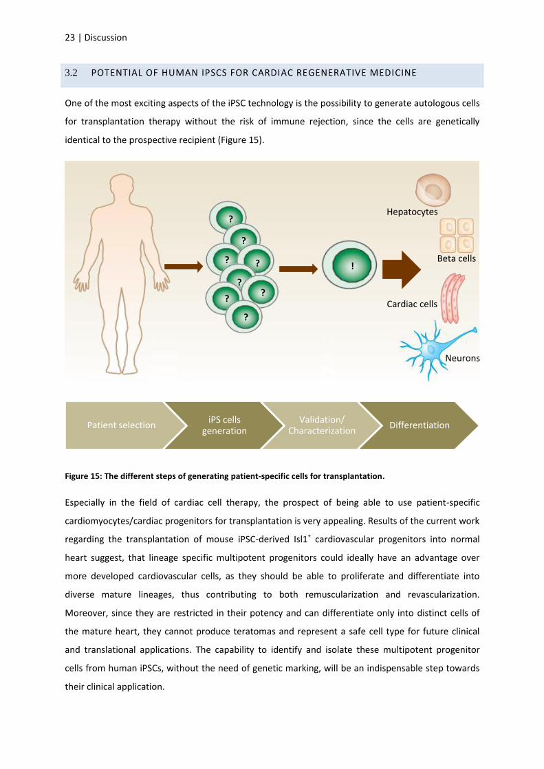
3.2 POTENTIAL OF HUMAN IPSCS FOR CARDIAC REGENERATIVE MEDICINE
One of the most exciting aspects of the iPSC technology is the possibility to generate autologous cells
for transplantation therapy without the risk of immune rejection, since the cells are genetically
identical to the prospective recipient (Figure 15).
Figure 15: The different steps of generating patient-specific cells for transplantation.
Especially in the field of cardiac cell therapy, the prospect of being able to use patient-specific
cardiomyocytes/cardiac progenitors for transplantation is very appealing. Results of the current work
regarding the transplantation of mouse iPSC-derived Isl1+ cardiovascular progenitors into normal
heart suggest, that lineage specific multipotent progenitors could ideally have an advantage over
more developed cardiovascular cells, as they should be able to proliferate and differentiate into
diverse mature lineages, thus contributing to both remuscularization and revascularization.
Moreover, since they are restricted in their potency and can differentiate only into distinct cells of
the mature heart, they cannot produce teratomas and represent a safe cell type for future clinical
and translational applications. The capability to identify and isolate these multipotent progenitor
cells from human iPSCs, without the need of genetic marking, will be an indispensable step towards
their clinical application.
Patient selection iPS cells
generation Validation/
Characterization Differentiation
Hepatocytes
Cardiac cells
Neurons
Beta cells ?
?
?
?
?
?
?
?
!
Discussion | 24
Besides using iPSC-derived cells directly for transplantation in vivo, they can also be potentially
valuable in tissue engineering approaches. As opposed to direct cell transplantation, cardiac tissue
engineering strives to take into account the three-dimensionality of the heart and the fact, that the
highly complex function of the myocardium relies on a variety of cells embedded in a mesh of
extracellular matrix for its well-functioning rather than just purely on cardiomyocytes. Being able to
isolate cells at the cardiac progenitor state from iPSCs could prove extremely helpful in the
generation of functional cardiac patches for future regenerative therapeutic approaches.
3.3 FUTURE PERSPECTIVES OF HUMAN IPSC TECHNOLOGY
Although iPSC derivation is legally and ethically less problematic than work on embryonic stem cells
and the field is rapidly advancing, the technology still needs to overcome several issues in order to
serve as an efficient research tool and ultimately be of use in clinical applications.
REPROGRAMMING STRATEGIES 3.3.1
Two major hurdles stand in the way of reliable, consistent derivation of patient-specific iPSCs: the
accessibility of patient material and the reliance on integrating viral vectors as the most efficient
method to deliver reprogramming factors.
Human iPSCs have been obtained from distinct somatic cell populations, including neural cells [74],
keratinocytes [75, 76], stomach and liver cells [77], adipocytes[78], and recently T-lymphocytes [79-
81] isolated from whole blood samples. Sampling of peripheral blood is one of the most commonly
performed and least invasive clinical procedures and blood can be easily stored. Thus, the recent
reports describing the generation of iPSCs from peripheral blood obtained from healthy donors
encourage the hope that the same could be achieved with blood from patients. This would progress
the field and bring it closer to clinical use by allowing for the bio-banking of patient material and
providing a simple source to obtain patient specific iPSCs.
For delivery of the reprogramming factors, the most widely practiced method, transduction via
retrovirus or lentivirus, results in random integration of foreign genetic elements into the genome,
which may cause insertional mutagenesis and inadvertently affect the differentiation of iPSCs into
relevant cell types. Random integrations render each iPSC line genetically distinct. Although the
integrated viruses are transcriptionally silenced following reprogramming, re-expression of any of the
reprogramming factors may interfere with differentiation and subsequent cell behavior [82].
Alternative approaches, such as the use of single [83] or multiple transient transfections [84], non-
integrating vectors [85], excisable vectors [86-88], direct protein transduction [89-91], RNA-based
Sendai viruses [92-94], mRNA-based transcription factor delivery [95, 96] and microRNA transfections
25 | Discussion
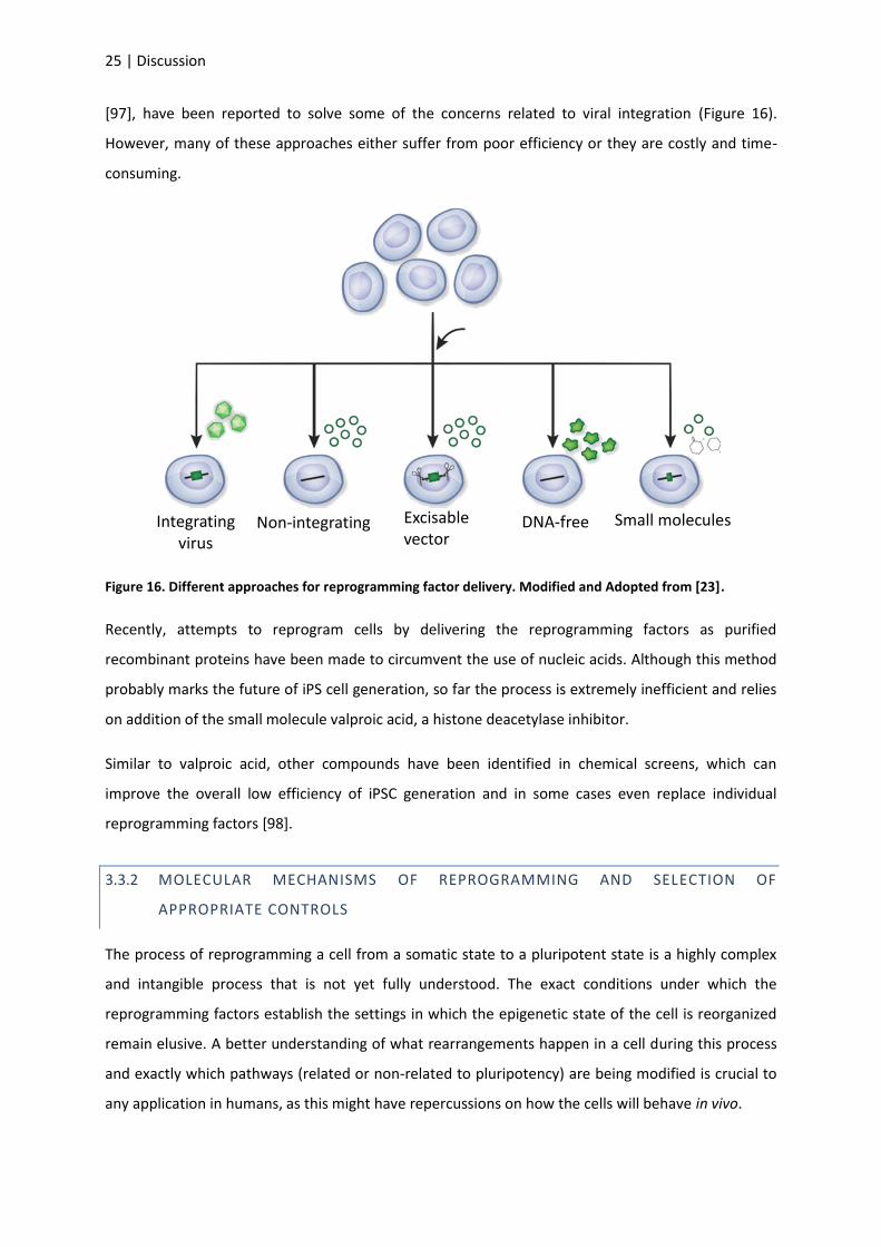
[97], have been reported to solve some of the concerns related to viral integration (Figure 16).
However, many of these approaches either suffer from poor efficiency or they are costly and time-
consuming.
Figure 16. Different approaches for reprogramming factor delivery. Modified and Adopted from [23].
Recently, attempts to reprogram cells by delivering the reprogramming factors as purified
recombinant proteins have been made to circumvent the use of nucleic acids. Although this method
probably marks the future of iPS cell generation, so far the process is extremely inefficient and relies
on addition of the small molecule valproic acid, a histone deacetylase inhibitor.
Similar to valproic acid, other compounds have been identified in chemical screens, which can
improve the overall low efficiency of iPSC generation and in some cases even replace individual
reprogramming factors [98].
MOLECULAR MECHANISMS OF REPROGRAMMING AND SELECTION OF 3.3.2
APPROPRIATE CONTROLS
The process of reprogramming a cell from a somatic state to a pluripotent state is a highly complex
and intangible process that is not yet fully understood. The exact conditions under which the
reprogramming factors establish the settings in which the epigenetic state of the cell is reorganized
remain elusive. A better understanding of what rearrangements happen in a cell during this process
and exactly which pathways (related or non-related to pluripotency) are being modified is crucial to
any application in humans, as this might have repercussions on how the cells will behave in vivo.
Integrating virus
Non-integrating Excisable
vector DNA-free Small molecules
Discussion | 26
More general questions that still require a more thorough answer are how equal iPSCs actually are to
hESCs on genomic and epigenomic levels and to what extent iPSC lines exhibit biological variability
among themselves. As for the genetic level, several publications proved chromosomal aberrations
[99], subchromosomal copy number variation [100] and point mutations in coding sequences [101].
In terms of epigenetic marking, it was shown that hiPSCs retain an epigenetic memory of their donor
cells and that this influences their differentiation potential [102]. In-depth profiling and
documentation of new iPSC lines is hence going to be crucial to assure comparability between results
coming from cell lines generated in different laboratories.
In order for results generated with iPS-derived cells to be meaningful, the selection of proper
controls is of the highest importance. Ideally, an age-matched, unaffected individual from the same
pedigree is chosen, and even then, the controls may differ in their genetic background as every
person carries disease-relevant polymorphisms. It is quite possible, that only mutation-corrected
iPSC lines of the same individual are a satisfying control. However, genetic modification of hiPSC lines
has remained challenging and cost-intensive due to the difficulty of targeting specific endogenous
genes in hiPSC. Zinc-finger nucleases and transcription activator–like effector nucleases (TALENs)
represent a new promising technology to significantly enhance the ability to genetically modify
human iPSCs, due to the enzyme-mediated introduction of double-stranded breaks in genomic DNA
at the site of a desired alteration [103, 104].
3.4 FINAL REMARKS
While at present human iPSCs serve mainly as in vitro models to study disease mechanisms and for
drug screening, their future in cell transplantation studies is yet unclear and will depend on future
research and how well issues concerning safety and efficacy of reprogramming as well as costliness
of patient-specific generation, testing and monitoring of iPSCs under good manufacturing practice
(GMP) will be addressed. Recently, it could be shown that fibroblasts can also be trans-differentiated
(direct cell conversion), without the bypass of the potentially tumorigenic iPSC state, into neurons
[105], cardiomyocytes [106] or blood progenitors[107]. This constitutes an interesting way to
increase the safety of the technology, especially with eyes on potential future methods of in vivo
reprogramming.
Yet, in all likelihood, iPSC technology will not be able to offer treatment for degenerative disease nor
model all diseases for decades more than years. Despite this, one can be optimistic given the pace of
big advances that the field of stem cell technology has experience ever since the discovery of induced
pluripotent stem cells.

27 | Acknowlegements
Part 4. ACKNOWLEGEMENTS
A lot of people were involved directly or indirectly in the successful completion of this work and it would
not have been possible for me to write this thesis without the help and continuous support of the
wonderful people around me. I could not possibly leave the institute without expressing my sincere
gratitude.
First of all, I would like to thank my principal supervisor Prof. Karl-Ludwig Laugwitz for offering me the
possibility to perform my Ph.D. studies in his Laboratory for Molecular Cardiology at the Klinikum rechts
der Isar. I cannot put in words how much I appreciated the active support at all stages of the project. I am
grateful for assistance in selecting collaborators, valuable advice and the outstanding freedom I was able
to enjoy in the laboratory.
Everything I know about stem cell culture and how good experimental science is done, I learned through
my direct supervisor Dr. Alessandra Moretti. I most highly appreciate all the significant contributions in
time, ideas, and in reviewing data, that were necessary to make my Ph.D. experience productive and
successful. Her joy and enthusiasm for research was contagious and motivational for me, even during
tough times in the Ph.D. pursuit. I am also thankful for the excellent example of a successful scientist she
set for me.
During the experimental work of this Ph.D. study a number of excellent scientists were involved in
different aspects of the projects. I would like to especially thank Dr. Milena Bellin, Dr. Jason Lam and Dr.
Tatjana Dorn who quickly became friends rather than colleagues.
Other past and present group members that I had the pleasure to work with or alongside are Dr. Harold
Ayetey, Dr. Laura Iop, Dr. Thomas Brade, Jessica Haas and Elvira Parotta. I would like to thank Diana
Grewe , Christina Scherb and Sabine Teuber for technical support.
A big thank you is owed to the people involved in our collaborations for their invaluable help with
performing experiments relative to the CPVT and the LQT1 project. These were mainly PD Dr. Michael
Mederos and Dr. Ursula Storch from the Pharmacology Department at the LMU Munich and Prof. Peter
Lipp at the Universität des Saarlandes and PD Dr.Andrea Welling at the Department of Pharmacology of
the TU Munich.
I would like to thank Prof. Steffen Massberg and Prof. Franz Hofmann for reading and evaluating this
thesis and for agreeing to be part of my thesis defense committee.
Also and importantly, I would like to thank the Luxembourgish Fonds National de la Recherche for
providing me with funding for this project in general and its publication.
References | 28
Part 5. REFERENCES
1. Evans, M.J. and M.H. Kaufman, Establishment in culture of pluripotential cells from mouse embryos. Nature, 1981. 292(5819): p. 154-6.
2. Thomson, J.A., et al., Embryonic stem cell lines derived from human blastocysts. Science, 1998. 282(5391): p. 1145-7.
3. Eckfeldt, C.E., E.M. Mendenhall, and C.M. Verfaillie, The molecular repertoire of the 'almighty' stem cell. Nat Rev Mol Cell Biol, 2005. 6(9): p. 726-37.
4. Waddington, C.H., The Strategy of the Genes; a Discussion of Some Aspects of Theoretical
Biology ed. A. Unwin. 1957, London.
5. Ying, Q.L., et al., BMP induction of Id proteins suppresses differentiation and sustains embryonic stem cell self-renewal in collaboration with STAT3. Cell, 2003. 115(3): p. 281-92.
6. James, D., et al., TGFbeta/activin/nodal signaling is necessary for the maintenance of pluripotency in human embryonic stem cells. Development, 2005. 132(6): p. 1273-82.
7. Boyer, L.A., et al., Core transcriptional regulatory circuitry in human embryonic stem cells. Cell, 2005. 122(6): p. 947-56.
8. Silva, J., et al., Nanog is the gateway to the pluripotent ground state. Cell, 2009. 138(4): p. 722-37.
9. van den Berg, D.L., et al., An Oct4-centered protein interaction network in embryonic stem cells. Cell Stem Cell, 2010. 6(4): p. 369-81.
10. Briggs, R. and T.J. King, Transplantation of Living Nuclei From Blastula Cells into Enucleated Frogs' Eggs. Proceedings of the National Academy of Sciences of the United States of America, 1952. 38(5): p. 455-63.
11. Gurdon, J.B., The developmental capacity of nuclei taken from intestinal epithelium cells of feeding tadpoles. Journal of Embryology and Experimental Morphology, 1962. 10: p. 622-40.
12. Wilmut, I., et al., Viable offspring derived from fetal and adult mammalian cells. Nature, 1997. 385(6619): p. 810-3.
13. Cowan, C.A., et al., Nuclear reprogramming of somatic cells after fusion with human embryonic stem cells. Science, 2005. 309(5739): p. 1369-73.
14. Do, J.T. and H.R. Scholer, Nuclei of embryonic stem cells reprogram somatic cells. Stem Cells, 2004. 22(6): p. 941-9.
15. Strelchenko, N., et al., Reprogramming of human somatic cells by embryonic stem cell cytoplast. Reproductive Biomedicine Online, 2006. 12(1): p. 107-11.
16. Takahashi, K. and S. Yamanaka, Induction of pluripotent stem cells from mouse embryonic and adult fibroblast cultures by defined factors. Cell, 2006. 126(4): p. 663-76.
17. Takahashi, K., et al., Induction of pluripotent stem cells from adult human fibroblasts by defined factors. Cell, 2007. 131(5): p. 861-72.
18. Yu, J., et al., Induced pluripotent stem cell lines derived from human somatic cells. Science, 2007. 318(5858): p. 1917-20.
29 | References
19. Nakagawa, M., et al., Generation of induced pluripotent stem cells without Myc from mouse and human fibroblasts. Nat Biotechnol, 2008. 26(1): p. 101-6.
20. Huangfu, D., et al., Induction of pluripotent stem cells from primary human fibroblasts with only Oct4 and Sox2. Nat Biotechnol, 2008. 26(11): p. 1269-75.
21. Kim, J.B., et al., Oct4-induced pluripotency in adult neural stem cells. Cell, 2009. 136(3): p. 411-9.
22. Yamanaka, S., Elite and stochastic models for induced pluripotent stem cell generation. Nature, 2009. 460(7251): p. 49-52.
23. Nagy, A. and K. Nagy, The mysteries of induced pluripotency: where will they lead? Nature Methods, 2010. 7(1): p. 22-4.
24. Laugwitz, K.L., et al., Islet1 cardiovascular progenitors: a single source for heart lineages? Development, 2008. 135(2): p. 193-205.
25. Buckingham, M., S. Meilhac, and S. Zaffran, Building the mammalian heart from two sources of myocardial cells. Nat Rev Genet, 2005. 6(11): p. 826-35.
26. Srivastava, D., Making or breaking the heart: from lineage determination to morphogenesis. Cell, 2006. 126(6): p. 1037-48.
27. Hutson, M.R. and M.L. Kirby, Model systems for the study of heart development and disease. Cardiac neural crest and conotruncal malformations. Semin Cell Dev Biol, 2007. 18(1): p. 101-10.
28. Martin-Puig, S., Z. Wang, and K.R. Chien, Lives of a heart cell: tracing the origins of cardiac progenitors. Cell Stem Cell, 2008. 2(4): p. 320-31.
29. Hoffman, J.I. and S. Kaplan, The incidence of congenital heart disease. J Am Coll Cardiol, 2002. 39(12): p. 1890-900.
30. Bruneau, B.G., The developmental genetics of congenital heart disease. Nature, 2008. 451(7181): p. 943-8.
31. Cai, C.L., et al., Isl1 identifies a cardiac progenitor population that proliferates prior to differentiation and contributes a majority of cells to the heart. Dev Cell, 2003. 5(6): p. 877-89.
32. Laugwitz, K.L., et al., Postnatal isl1+ cardioblasts enter fully differentiated cardiomyocyte lineages. Nature, 2005. 433(7026): p. 647-53.
33. Moretti, A., et al., Multipotent embryonic isl1+ progenitor cells lead to cardiac, smooth muscle, and endothelial cell diversification. Cell, 2006. 127(6): p. 1151-65.
34. Sun, Y., et al., Islet 1 is expressed in distinct cardiovascular lineages, including pacemaker and coronary vascular cells. Dev Biol, 2007. 304(1): p. 286-96.
35. Qyang, Y., et al., The renewal and differentiation of Isl1+ cardiovascular progenitors are controlled by a Wnt/beta-catenin pathway. Cell Stem Cell, 2007. 1(2): p. 165-79.
36. Kattman, S.J., T.L. Huber, and G.M. Keller, Multipotent flk-1+ cardiovascular progenitor cells give rise to the cardiomyocyte, endothelial, and vascular smooth muscle lineages. Dev Cell, 2006. 11(5): p. 723-32.
References | 30
37. Wu, S.M., et al., Developmental origin of a bipotential myocardial and smooth muscle cell precursor in the mammalian heart. Cell, 2006. 127(6): p. 1137-50.
38. Roden, D.M., Clinical practice. Long-QT syndrome. N Engl J Med, 2008. 358(2): p. 169-76.
39. Sauer, A.J., et al., Long QT syndrome in adults. J Am Coll Cardiol, 2007. 49(3): p. 329-37.
40. Saenen, J.B. and C.J. Vrints, Molecular aspects of the congenital and acquired Long QT Syndrome: clinical implications. J Mol Cell Cardiol, 2008. 44(4): p. 633-46.
41. Crotti, L., et al., Congenital long QT syndrome. Orphanet J Rare Dis, 2008. 3: p. 18.
42. Curran, M.E., et al., A molecular basis for cardiac arrhythmia: HERG mutations cause long QT syndrome. Cell, 1995. 80(5): p. 795-803.
43. Sanguinetti, M.C., et al., Coassembly of K(V)LQT1 and minK (IsK) proteins to form cardiac I(Ks) potassium channel. Nature, 1996. 384(6604): p. 80-3.
44. Sanguinetti, M.C., et al., Isoproterenol antagonizes prolongation of refractory period by the class III antiarrhythmic agent E-4031 in guinea pig myocytes. Mechanism of action. Circulation Research, 1991. 68(1): p. 77-84.
45. Walsh, K.B. and R.S. Kass, Regulation of a heart potassium channel by protein kinase A and C. Science, 1988. 242(4875): p. 67-9.
46. Jurkiewicz, N.K. and M.C. Sanguinetti, Rate-dependent prolongation of cardiac action potentials by a methanesulfonanilide class III antiarrhythmic agent. Specific block of rapidly activating delayed rectifier K+ current by dofetilide. Circulation Research, 1993. 72(1): p. 75-83.
47. Priori, S.G. and S.R. Chen, Inherited dysfunction of sarcoplasmic reticulum Ca2+ handling and arrhythmogenesis. Circ Res, 2011. 108(7): p. 871-83.
48. Rogers, E.F., F.R. Koniuszy, and et al., Plant insecticides; ryanodine, a new alkaloid from Ryania speciosa Vahl. Journal of the American Chemical Society, 1948. 70(9): p. 3086-8.
49. Jenden, D.J. and A.S. Fairhurst, The pharmacology of ryanodine. Pharmacological Reviews, 1969. 21(1): p. 1-25.
50. Swan, H., et al., Arrhythmic disorder mapped to chromosome 1q42-q43 causes malignant polymorphic ventricular tachycardia in structurally normal hearts. Journal of the American College of Cardiology, 1999. 34(7): p. 2035-42.
51. Priori, S.G., et al., Mutations in the cardiac ryanodine receptor gene (hRyR2) underlie catecholaminergic polymorphic ventricular tachycardia. Circulation, 2001. 103(2): p. 196-200.
52. Greenstein, J.L. and R.L. Winslow, Integrative systems models of cardiac excitation-contraction coupling. Circulation Research, 2011. 108(1): p. 70-84.
53. Wehrens, X.H., et al., FKBP12.6 deficiency and defective calcium release channel (ryanodine receptor) function linked to exercise-induced sudden cardiac death. Cell, 2003. 113(7): p. 829-40.
54. George, C.H., G.V. Higgs, and F.A. Lai, Ryanodine receptor mutations associated with stress-induced ventricular tachycardia mediate increased calcium release in stimulated cardiomyocytes. Circulation Research, 2003. 93(6): p. 531-40.
31 | References
55. Jiang, D., et al., Enhanced store overload-induced Ca2+ release and channel sensitivity to luminal Ca2+ activation are common defects of RyR2 mutations linked to ventricular tachycardia and sudden death. Circulation Research, 2005. 97(11): p. 1173-81.
56. Liu, N., et al., Arrhythmogenesis in catecholaminergic polymorphic ventricular tachycardia: insights from a RyR2 R4496C knock-in mouse model. Circulation Research, 2006. 99(3): p. 292-8.
57. Xiao, J., et al., Removal of FKBP12.6 does not alter the conductance and activation of the cardiac ryanodine receptor or the susceptibility to stress-induced ventricular arrhythmias. J Biol Chem, 2007. 282(48): p. 34828-38.
58. Guo, T., et al., Kinetics of FKBP12.6 binding to ryanodine receptors in permeabilized cardiac myocytes and effects on Ca sparks. Circulation Research, 2010. 106(11): p. 1743-52.
59. Jiang, D., et al., RyR2 mutations linked to ventricular tachycardia and sudden death reduce the threshold for store-overload-induced Ca2+ release (SOICR). Proceedings of the National Academy of Sciences of the United States of America, 2004. 101(35): p. 13062-7.
60. Yamamoto, T., R. El-Hayek, and N. Ikemoto, Postulated role of interdomain interaction within the ryanodine receptor in Ca(2+) channel regulation. J Biol Chem, 2000. 275(16): p. 11618-25.
61. Liu, Z., et al., Dynamic, inter-subunit interactions between the N-terminal and central mutation regions of cardiac ryanodine receptor. J Cell Sci, 2010. 123(Pt 10): p. 1775-84.
62. Paul-Pletzer, K., et al., Identification of a dantrolene-binding sequence on the skeletal muscle ryanodine receptor. J Biol Chem, 2002. 277(38): p. 34918-23.
63. Paul-Pletzer, K., et al., Probing a putative dantrolene-binding site on the cardiac ryanodine receptor. Biochem J, 2005. 387(Pt 3): p. 905-9.
64. Kobayashi, S., et al., Dantrolene, a therapeutic agent for malignant hyperthermia, markedly improves the function of failing cardiomyocytes by stabilizing interdomain interactions within the ryanodine receptor. J Am Coll Cardiol, 2009. 53(21): p. 1993-2005.
65. Uchinoumi, H., et al., Catecholaminergic polymorphic ventricular tachycardia is caused by mutation-linked defective conformational regulation of the ryanodine receptor. Circ Res, 2010. 106(8): p. 1413-24.
66. Kobayashi, S., et al., Dantrolene, a therapeutic agent for malignant hyperthermia, inhibits catecholaminergic polymorphic ventricular tachycardia in a RyR2(R2474S/+) knock-in mouse model. Circ J, 2010. 74(12): p. 2579-84.
67. Marx, S.O., et al., Requirement of a macromolecular signaling complex for beta adrenergic receptor modulation of the KCNQ1-KCNE1 potassium channel. Science, 2002. 295(5554): p. 496-9.
68. Moretti, A., et al., Mouse and human induced pluripotent stem cells as a source for multipotent Isl1+ cardiovascular progenitors. Faseb J, 2010. 24(3): p. 700-11.
69. Jung, C.B., et al., Dantrolene rescues arrhythmogenic RYR2 defect in a patient-specific stem cell model of catecholaminergic polymorphic ventricular tachycardia. EMBO Molecular Medicine, 2011.
70. Moretti, A., et al., Patient-specific induced pluripotent stem-cell models for long-QT syndrome. New England Journal of Medicine, 2010. 363(15): p. 1397-409.
References | 32
71. Yazawa, M., et al., Using induced pluripotent stem cells to investigate cardiac phenotypes in Timothy syndrome. Nature, 2011. 471(7337): p. 230-4.
72. Carvajal-Vergara, X., et al., Patient-specific induced pluripotent stem-cell-derived models of LEOPARD syndrome. Nature, 2010. 465(7299): p. 808-12.
73. Itzhaki, I., et al., Modelling the long QT syndrome with induced pluripotent stem cells. Nature, 2011. 471(7337): p. 225-9.
74. Eminli, S., et al., Reprogramming of neural progenitor cells into induced pluripotent stem cells in the absence of exogenous Sox2 expression. Stem Cells, 2008. 26(10): p. 2467-74.
75. Maherali, N., et al., Directly reprogrammed fibroblasts show global epigenetic remodeling and widespread tissue contribution. Cell Stem Cell, 2007. 1(1): p. 55-70.
76. Aasen, T., et al., Efficient and rapid generation of induced pluripotent stem cells from human keratinocytes. Nat Biotechnol, 2008. 26(11): p. 1276-84.
77. Aoi, T., et al., Generation of pluripotent stem cells from adult mouse liver and stomach cells. Science, 2008. 321(5889): p. 699-702.
78. Sun, N., et al., Feeder-free derivation of induced pluripotent stem cells from adult human adipose stem cells. Proceedings of the National Academy of Sciences of the United States of America, 2009. 106(37): p. 15720-5.
79. Loh, Y.H., et al., Reprogramming of T cells from human peripheral blood. Cell Stem Cell, 2010. 7(1): p. 15-9.
80. Staerk, J., et al., Reprogramming of human peripheral blood cells to induced pluripotent stem cells. Cell Stem Cell, 2010. 7(1): p. 20-4.
81. Seki, T., et al., Generation of induced pluripotent stem cells from human terminally differentiated circulating T cells. Cell Stem Cell, 2010. 7(1): p. 11-4.
82. Haile, R.W., et al., A molecular/epidemiologic analysis of expression of cyclooxygenases 1 and 2, use of nonsteroidal antiinflammatory drugs, and risk of colorectal adenoma. Clinical Colorectal Cancer, 2005. 4(6): p. 390-5.
83. Ma, B., et al., Comparison of the protein-protein interfaces in the p53-DNA crystal structures: towards elucidation of the biological interface. Proceedings of the National Academy of Sciences of the United States of America, 2005. 102(11): p. 3988-93.
84. Okita, K., et al., Generation of mouse induced pluripotent stem cells without viral vectors. Science, 2008. 322(5903): p. 949-53.
85. Stadtfeld, M., et al., Induced pluripotent stem cells generated without viral integration. Science, 2008. 322(5903): p. 945-9.
86. wapek, V.V. and P. Chvosta, Consistent approximations in generalized-master-equation theories: Application to the asymptotic-time symmetry-breaking problem. Phys Rev A, 1991. 43(6): p. 2819-2826.
87. C, W.B., Cancer Research. Science, 1912. 35(913): p. 979-80.
88. C, W.S., Pictures in cell biology Microtubule dynamics in migrating cells. Trends in Cell Biology, 1997. 7(10): p. 407.
33 | References
89. Robins, H., et al., A relative-entropy algorithm for genomic fingerprinting captures host-phage similarities. Journal of Bacteriology, 2005. 187(24): p. 8370-4.
90. C, W.W., The Total Eclipse of September 9, 1904. Science, 1904. 20(519): p. 812.
91. Waddington, C., et al., Open-label, randomised, parallel-group, multicentre study to evaluate the safety, tolerability and immunogenicity of an AS03(B)/oil-in-water emulsion-adjuvanted (AS03(B)) split-virion versus non-adjuvanted whole-virion H1N1 influenza vaccine in UK children 6 months to 12 years of age. Health Technology Assessment, 2010. 14(46): p. 1-130.
92. Klemm, P., et al., Unleashing animal-assisted therapy. Nursing, 2010. 40(10): p. 12-3.
93. Nilsen, B.S., et al., Fragmentation cross sections of relativistic 8436Kr and 10947Ag nuclei in targets from hydrogen to lead. Phys Rev C Nucl Phys, 1995. 52(6): p. 3277-3290.
94. Geer, L.Y., et al., Charge-changing fragmentation of 10.6 GeV/nucleon 197Au nuclei. Phys Rev C Nucl Phys, 1995. 52(1): p. 334-345.
95. McKennis, A.T. and C. Waddington, Thyroplasty type I for unilateral vocal cord paralysis. AORN Journal, 1994. 60(1): p. 38-42.
96. Chen, C., et al., Interactions in hydrogen of relativistic neon to nickel projectiles: Total charge-changing cross sections. Phys Rev C Nucl Phys, 1994. 49(6): p. 3200-3210.
97. McKennis, A.T. and C. Waddington, Unilateral vocal cord paralysis. ORL-Head and Neck Nursing, 1994. 12(1): p. 9.
98. Desponts, C. and S. Ding, Using small molecules to improve generation of induced pluripotent stem cells from somatic cells. Methods in Molecular Biology, 2010. 636: p. 207-18.
99. Mayshar, Y., et al., Identification and classification of chromosomal aberrations in human induced pluripotent stem cells. Cell Stem Cell, 2010. 7(4): p. 521-31.
100. Hussein, S.M., et al., Copy number variation and selection during reprogramming to pluripotency. Nature, 2011. 471(7336): p. 58-62.
101. Gore, A., et al., Somatic coding mutations in human induced pluripotent stem cells. Nature, 2011. 471(7336): p. 63-7.
102. Bar-Nur, O., et al., Epigenetic memory and preferential lineage-specific differentiation in induced pluripotent stem cells derived from human pancreatic islet Beta cells. Cell Stem Cell, 2011. 9(1): p. 17-23.
103. Binns, W.R., et al., Charge, mass, and energy changes during fragmentation of relativistic nuclei. Phys Rev C Nucl Phys, 1989. 39(5): p. 1785-1798.
104. Waddington, C.J., et al., Stopping of relativistic heavy ions in various media. Phys Rev A, 1986. 34(5): p. 3700-3706.
105. Vierbuchen, T., et al., Direct conversion of fibroblasts to functional neurons by defined factors. Nature, 2010. 463(7284): p. 1035-41.
106. Ieda, M., et al., Direct reprogramming of fibroblasts into functional cardiomyocytes by defined factors. Cell, 2010. 142(3): p. 375-86.
107. Szabo, E., et al., Direct conversion of human fibroblasts to multilineage blood progenitors. Nature, 2010. 468(7323): p. 521-6.
Articles | 34
Part 6. ARTICLES
MOUSE AND HUMAN INDUCED PLURIPOTENT STEM CELLS AS A SOURCE FOR
MULTIPOTENT ISL1+ CARDIOVASCULAR PROGENITORS FASEB J. 2010 MAR;24(3):700-11.
PATIENT-SPECIFIC INDUCED PLURIPOTENT STEM-CELL MODELS FOR LONG-QT
SYNDROME. N ENGL J MED. 2010 OCT 7;363(15):1397-409.
DANTROLENE RESCUES ARRHYTHMOGENIC RYR2 DEFECT IN A PATIENT-SPECIFIC STEM
CELL MODEL OF CATECHOLAMINERGIC POLYMORPHIC VENTRICULAR TACHYCARDIA.
EMBO MOL MED. 2012 MAR;4(3):180-91.
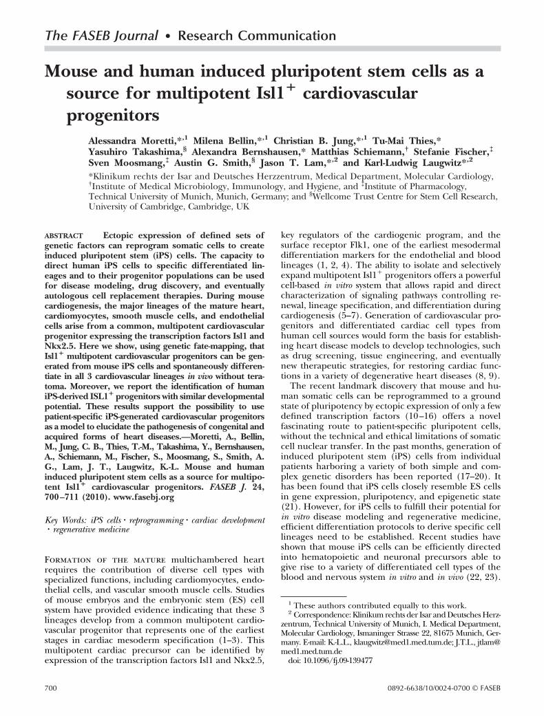
The FASEB Journal • Research Communication
Mouse and human induced pluripotent stem cells as asource for multipotent Isl1� cardiovascularprogenitors
Alessandra Moretti,*,1 Milena Bellin,*,1 Christian B. Jung,*,1 Tu-Mai Thies,*Yasuhiro Takashima,§ Alexandra Bernshausen,* Matthias Schiemann,† Stefanie Fischer,‡
Sven Moosmang,‡ Austin G. Smith,§ Jason T. Lam,*,2 and Karl-Ludwig Laugwitz*,2
*Klinikum rechts der Isar and Deutsches Herzzentrum, Medical Department, Molecular Cardiology,†Institute of Medical Microbiology, Immunology, and Hygiene, and ‡Institute of Pharmacology,Technical University of Munich, Munich, Germany; and §Wellcome Trust Centre for Stem Cell Research,University of Cambridge, Cambridge, UK
ABSTRACT Ectopic expression of defined sets ofgenetic factors can reprogram somatic cells to createinduced pluripotent stem (iPS) cells. The capacity todirect human iPS cells to specific differentiated lin-eages and to their progenitor populations can be usedfor disease modeling, drug discovery, and eventuallyautologous cell replacement therapies. During mousecardiogenesis, the major lineages of the mature heart,cardiomyocytes, smooth muscle cells, and endothelialcells arise from a common, multipotent cardiovascularprogenitor expressing the transcription factors Isl1 andNkx2.5. Here we show, using genetic fate-mapping, thatIsl1� multipotent cardiovascular progenitors can be gen-erated from mouse iPS cells and spontaneously differen-tiate in all 3 cardiovascular lineages in vivo without tera-toma. Moreover, we report the identification of humaniPS-derived ISL1� progenitors with similar developmentalpotential. These results support the possibility to usepatient-specific iPS-generated cardiovascular progenitorsas a model to elucidate the pathogenesis of congenital andacquired forms of heart diseases.—Moretti, A., Bellin,M., Jung, C. B., Thies, T.-M., Takashima, Y., Bernshausen,A., Schiemann, M., Fischer, S., Moosmang, S., Smith, A.G., Lam, J. T., Laugwitz, K.-L. Mouse and humaninduced pluripotent stem cells as a source for multipo-tent Isl1� cardiovascular progenitors. FASEB J. 24,700–711 (2010). www.fasebj.org
Key Words: iPS cells � reprogramming � cardiac development� regenerative medicine
Formation of the mature multichambered heartrequires the contribution of diverse cell types withspecialized functions, including cardiomyocytes, endo-thelial cells, and vascular smooth muscle cells. Studiesof mouse embryos and the embryonic stem (ES) cellsystem have provided evidence indicating that these 3lineages develop from a common multipotent cardio-vascular progenitor that represents one of the earlieststages in cardiac mesoderm specification (1–3). Thismultipotent cardiac precursor can be identified byexpression of the transcription factors Isl1 and Nkx2.5,
key regulators of the cardiogenic program, and thesurface receptor Flk1, one of the earliest mesodermaldifferentiation markers for the endothelial and bloodlineages (1, 2, 4). The ability to isolate and selectivelyexpand multipotent Isl1� progenitors offers a powerfulcell-based in vitro system that allows rapid and directcharacterization of signaling pathways controlling re-newal, lineage specification, and differentiation duringcardiogenesis (5–7). Generation of cardiovascular pro-genitors and differentiated cardiac cell types fromhuman cell sources would form the basis for establish-ing heart disease models to develop technologies, suchas drug screening, tissue engineering, and eventuallynew therapeutic strategies, for restoring cardiac func-tions in a variety of degenerative heart diseases (8, 9).
The recent landmark discovery that mouse and hu-man somatic cells can be reprogrammed to a groundstate of pluripotency by ectopic expression of only a fewdefined transcription factors (10–16) offers a novelfascinating route to patient-specific pluripotent cells,without the technical and ethical limitations of somaticcell nuclear transfer. In the past months, generation ofinduced pluripotent stem (iPS) cells from individualpatients harboring a variety of both simple and com-plex genetic disorders has been reported (17–20). Ithas been found that iPS cells closely resemble ES cellsin gene expression, pluripotency, and epigenetic state(21). However, for iPS cells to fulfill their potential forin vitro disease modeling and regenerative medicine,efficient differentiation protocols to derive specific celllineages need to be established. Recent studies haveshown that mouse iPS cells can be efficiently directedinto hematopoietic and neuronal precursors able togive rise to a variety of differentiated cell types of theblood and nervous system in vitro and in vivo (22, 23).
1 These authors contributed equally to this work.2 Correspondence: Klinikum rechts der Isar and Deutsches Herz-
zentrum, Technical University of Munich, I. Medical Department,Molecular Cardiology, Ismaninger Strasse 22, 81675 Munich, Ger-many. E-mail: K.-L.L., [email protected]; J.T.L., [email protected]
doi: 10.1096/fj.09-139477
700 0892-6638/10/0024-0700 © FASEB

The aim of the current study was to test whether iPS cellscould be a source of Isl1� cardiovascular progenitors thatdisplay multipotency into all 3 cardiovascular lineages in vitroand in vivo. As proof of principle, we first generated iPS cellsfrom Isl1-Cre/R26R-YFP or -LacZ double-heterozygousmice, which are genetically marked to allow identifica-tion and purification of Isl1-expressing cells and theirdifferentiated progeny. Through transcriptional profil-ing of purified mouse YFP� cells at different develop-mental stages we identified the Isl1� cardiovascularprecursor state and proved its multipotency by sponta-neous differentiation in vivo. Using skin fibroblastsfrom 2 healthy individuals we produced human iPS(hiPS) cell lines possessing a gene expression signaturesimilar to that of human ES cells and able to differen-tiate into cell types of all 3 germ layers. We have usedthese iPS cells to efficiently generate individual-specificISL1� cardiovascular progenitors and functional car-diac myocytes.
These results represent a first step toward the gener-ation of patient-specific iPS-cell-based ISL1� cardiovas-cular precursors, which would be extremely valuable indesigning disease-specific assays for screening of drugcardiotoxicity, in identifying and validating therapeutictargets, and in studying mechanisms of cardiovasculardisorders. Furthermore, the accessibility of patient-specific iPS-cell-derived cardiac progenitors may repre-sent a significant advantage over differentiated cells orpluripotent stem cells to achieve large-scale productionof multipotent, tumor-free cardiac cells for clinical andtranslational applications in the future.
MATERIALS AND METHODS
Mice
Isl1-Cre mice were generously provided by Sylvia Evans (Uni-versity of California–San Diego, La Jolla, CA, USA) (24). Theconditional Cre reporter mouse lines R26R-LacZ and R26R-YFP were generated by Philippe Soriano (Mt. Sinai School ofMedicine, New York, NY, USA) (25) and Frank Costantini(Columbia University Medical Center, New York, NY, USA)(26), respectively. Isl1-Cre/R26R double-heterozygous micewere generated by crossing single-heterozygous mice. Miceare in a mixed 129 � C57Bl/6 background.
Cell culture
For isolation of dermal fibroblasts, skin from newborn Isl1-Cre/R26R mice and human dermal biopsies were mincedinto 2-mm pieces, placed on culture dishes, and incubated inQuantum 333 medium (PAA, Pasching, Austria). Cells mi-grating out of the explants were passaged in DMEM contain-ing 10% FBS and used at passage 4 for iPS cell induction.Mouse iPS cells were grown on mitomycin-C-treated murineembryonic fibroblasts (MEF feeders) in standard ES medium[DMEM supplemented with 2 mM l-glutamine, 0.1 mMnonessential amino acids, 1 mM sodium pyruvate, 0.1 mM�-mercaptoethanol, 50 U/ml penicillin, 50 �g/ml streptomy-cin, and 0.1 �g/ml leukemia inhibitory factor (LIF)] contain-ing 15% knockout serum replacement (KSR; Invitrogen,Karlsruhe, Germany) for 5 passages and then maintained inES medium containing 15% FBS. Human iPS cells weregrown on MEF feeders in human ES medium consisting of
DMEM/F12 supplemented with 20% KSR, 2 mM l-glutamine,0.1 mM nonessential amino acids, 0.1 mM �-mercaptoethanol,50 U/ml penicillin, 50 �g/ml streptomycin, and 10 ng/mlhuman b-FGF (R&D System, Minneapolis, MN, USA). Humanresearch subject protocol was approved by the institutionalreview boards and the ethics committee of both the Klinikumrechts der Isar and the Technical University of Munich.
Retroviral production and iPS cell induction
Isl1-Cre/R26R skin fibroblasts were infected overnight withpooled equal volumes of viral supernatants (supplementedwith 8 �g/ml polybrene) generated by independent tranfec-tions of HEK293T cells (Fugene HD; Roche, Basel, Switzer-land) with pMXs-based retroviral vectors for the mouse Oct4,Sox2, Klf4, and c-Myc (Addgene plasmids 13366, 13367, 13370,and 13375; Addgene, Cambridge, MA, USA) and the packag-ing plasmid pCL-Eco (Addgene plasmid 12371) in DMEMcontaining 10% FBS, as previously reported (27). Four daysafter infection, skin fibroblasts were reseeded on MEF feeders(0.5�105 cells/10-cm dish for fibroblasts transduced with all4 factors, and 3.5�105 cells/10-cm dish for fibroblasts trans-duced with 3 factors in absence of c-Myc) and maintained inES medium supplemented with 15% KSR. Medium waschanged every day, and colonies were picked for expansion atd 16 and 30 after 4-factor and 3-factor transduction, respec-tively. For human iPS cell induction, retroviruses for the 4factors were independently produced by transfectingHEK293T cells (Fugene HD) with pMXs vectors encoding forthe human OCT4, SOX2, KLF4, and c-MYC (Addgene plas-mids 17217, 17218, 17219, and 17220) and the combinationof moloney gag-pol plasmid pUMVC (Addgene plasmid8449) and VSV envelope plasmid (Addgene plasmid 8454) inDMEM containing 10% FBS. Viral supernatants were har-vested after 48 and 72 h, filtered through a 0.45-�m low-protein-binding cellulose acetate filter, concentrated by aspin column (Millipore, Billerica, MA, USA), and used di-rectly to infect twice (24 h apart) 1.5 � 105 human fibroblastsin the presence of 8 �g/ml polybrene. After 6 d, cells wereseeded on MEF feeders at the density of 5 � 104 cells/10-cmdish and cultured for 4 additional weeks in human ESmedium before iPS colonies were manually picked.
Teratoma formation
We injected 0.5 � 106 mouse iPS cells in the left ventricularmyocardium of SCID nude mice, and 4 wk later hearts werefixed in 4% formaldehyde and embedded in paraffin. Sec-tions were stained with hematoxylin and eosin.
Chimera generation
After injection of 8–10 Isl1-Cre/R26R iPS cells in eachblastocyst isolated from C57Bl/6 mice, blastocysts were trans-ferred into E2.5 pseudo-pregnant CD1 females, and chimeraswere analyzed at embryonic day 13–14 or after birth by YFPfluorescence or LacZ staining.
Immunohistochemistry, LacZ, and AP staining
Cells in culture and heart cryosections (10 �m) were fixedwith 3.7% formaldehyde and subjected to specific immuno-staining by using the following primary antibodies: mouseNanog (1:500, rabbit polyclonal; Novus Biologicals, Littleton,CO, USA), human Nanog (1:500, rabbit polyclonal; Abcam,Cambridge, UK), TRA1–81-Alexa-Fluor-488-conjugated (1:20, mouse monoclonal; BD Pharmingen, San Diego, CA,
701IPS-DERIVED ISL1� CARDIOVASCULAR PROGENITORS
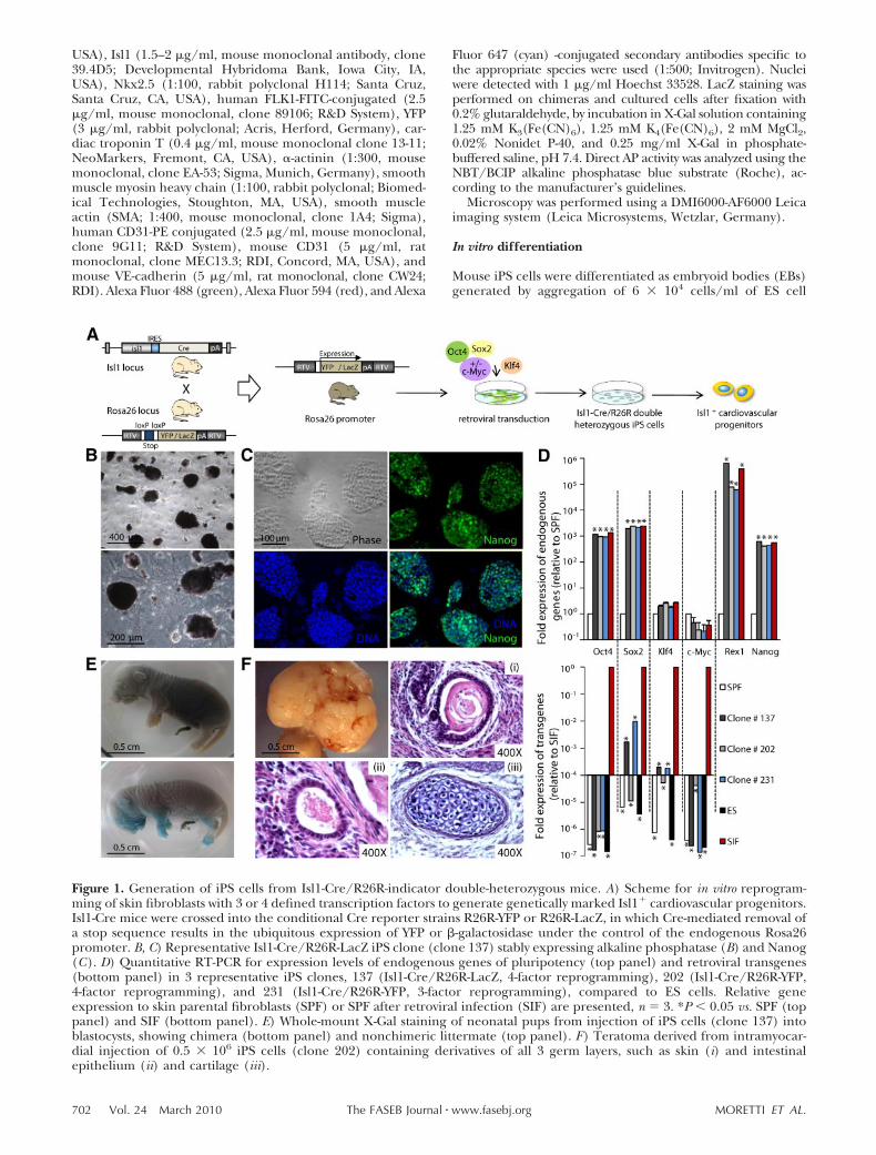
USA), Isl1 (1.5–2 �g/ml, mouse monoclonal antibody, clone39.4D5; Developmental Hybridoma Bank, Iowa City, IA,USA), Nkx2.5 (1:100, rabbit polyclonal H114; Santa Cruz,Santa Cruz, CA, USA), human FLK1-FITC-conjugated (2.5�g/ml, mouse monoclonal, clone 89106; R&D System), YFP(3 �g/ml, rabbit polyclonal; Acris, Herford, Germany), car-diac troponin T (0.4 �g/ml, mouse monoclonal clone 13-11;NeoMarkers, Fremont, CA, USA), �-actinin (1:300, mousemonoclonal, clone EA-53; Sigma, Munich, Germany), smoothmuscle myosin heavy chain (1:100, rabbit polyclonal; Biomed-ical Technologies, Stoughton, MA, USA), smooth muscleactin (SMA; 1:400, mouse monoclonal, clone 1A4; Sigma),human CD31-PE conjugated (2.5 �g/ml, mouse monoclonal,clone 9G11; R&D System), mouse CD31 (5 �g/ml, ratmonoclonal, clone MEC13.3; RDI, Concord, MA, USA), andmouse VE-cadherin (5 �g/ml, rat monoclonal, clone CW24;RDI). Alexa Fluor 488 (green), Alexa Fluor 594 (red), and Alexa
Fluor 647 (cyan) -conjugated secondary antibodies specific tothe appropriate species were used (1:500; Invitrogen). Nucleiwere detected with 1 �g/ml Hoechst 33528. LacZ staining wasperformed on chimeras and cultured cells after fixation with0.2% glutaraldehyde, by incubation in X-Gal solution containing1.25 mM K3(Fe(CN)6), 1.25 mM K4(Fe(CN)6), 2 mM MgCl2,0.02% Nonidet P-40, and 0.25 mg/ml X-Gal in phosphate-buffered saline, pH 7.4. Direct AP activity was analyzed using theNBT/BCIP alkaline phosphatase blue substrate (Roche), ac-cording to the manufacturer’s guidelines.
Microscopy was performed using a DMI6000-AF6000 Leicaimaging system (Leica Microsystems, Wetzlar, Germany).
In vitro differentiation
Mouse iPS cells were differentiated as embryoid bodies (EBs)generated by aggregation of 6 � 104 cells/ml of ES cell
Figure 1. Generation of iPS cells from Isl1-Cre/R26R-indicator double-heterozygous mice. A) Scheme for in vitro reprogram-ming of skin fibroblasts with 3 or 4 defined transcription factors to generate genetically marked Isl1� cardiovascular progenitors.Isl1-Cre mice were crossed into the conditional Cre reporter strains R26R-YFP or R26R-LacZ, in which Cre-mediated removal ofa stop sequence results in the ubiquitous expression of YFP or �-galactosidase under the control of the endogenous Rosa26promoter. B, C) Representative Isl1-Cre/R26R-LacZ iPS clone (clone 137) stably expressing alkaline phosphatase (B) and Nanog(C). D) Quantitative RT-PCR for expression levels of endogenous genes of pluripotency (top panel) and retroviral transgenes(bottom panel) in 3 representative iPS clones, 137 (Isl1-Cre/R26R-LacZ, 4-factor reprogramming), 202 (Isl1-Cre/R26R-YFP,4-factor reprogramming), and 231 (Isl1-Cre/R26R-YFP, 3-factor reprogramming), compared to ES cells. Relative geneexpression to skin parental fibroblasts (SPF) or SPF after retroviral infection (SIF) are presented, n � 3. *P � 0.05 vs. SPF (toppanel) and SIF (bottom panel). E) Whole-mount X-Gal staining of neonatal pups from injection of iPS cells (clone 137) intoblastocysts, showing chimera (bottom panel) and nonchimeric littermate (top panel). F) Teratoma derived from intramyocar-dial injection of 0.5 � 106 iPS cells (clone 202) containing derivatives of all 3 germ layers, such as skin (i) and intestinalepithelium (ii) and cartilage (iii).
702 Vol. 24 March 2010 MORETTI ET AL.The FASEB Journal � www.fasebj.org
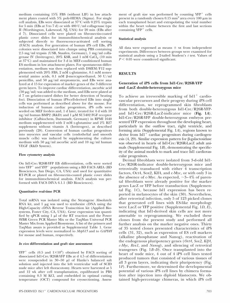
medium containing 15% FBS (without LIF) in low attach-ment plates coated with 5% poly-HEMA (Sigma). For singlecell analysis, EBs were dissociated at 37°C with 0.25% trypsinfor 5 min (EBs at 3 to 7 d) or with 480 U/ml collagenase typeII (Worthington, Lakewood, NJ, USA) for 20 min (EBs afterd 7). Dissociated cells were plated on fibronectin-coatedplastic cover slides for immunohistochemical analysis orsubjected directly to fluorescence-activated cell sorting(FACS) analysis. For generation of human iPS cell EBs, iPScolonies were dissociated into clumps using PBS containing2.5 mg/ml trypsin (USB, Staufen, Germany), 1 mg/ml colla-genase IV (Invitrogen), 20% KSR, and 1 mM CaCl2 (10 minat 37°C) and maintained for 3 d in MEF-conditioned humanES medium in low attachment plates. For spontaneous differ-entiation, medium was then replaced with DMEM/F12 sup-plemented with 20% FBS, 2 mM l-glutamine, 0.1 mM nones-sential amino acids, 0.1 mM �-mercaptoethanol, 50 U/mlpenicillin, and 50 �g/ml streptomycin, and EBs were ana-lyzed at d 15 for expression of marker genes of the 3 differentgerm layers. To improve cardiac differentiation, ascorbic acid(50 �g/ml) was added to the medium, and EBs were plated atd 7 on gelatin-coated dishes for better detection of beatingfoci. Dissociation of human iPS-cell-derived EBs into singlecells was performed as described above for the mouse. Forinduction of human cardiac progenitors, iPS cells wereseeded on MEF feeders and, 1 d later, treated for 4 d with 10ng/ml human BMP2 (R&D) and 1 �M SU5402 FGF receptorinhibitor (Calbiochem, Darmstadt, Germany) in RPMI 1640medium supplemented with 2 mM l-glutamine and 2% B27supplement without vitamin A (Invitrogen), as describedpreviously (28). Conversion of human cardiac progenitorsinto myocytes and vascular cells (endothelial and smoothmuscle cells) was induced by supplementing the culturemedium with 50 �g/ml ascorbic acid and 10 ng/ml humanVEGF (R&D System).
Flow cytometry analysis
On Isl1-Cre/R26R-YFP EB differentiation, cells were sortedinto YFP� and YFP� populations using a BD FACS ARIA (BDBiosciences, San Diego, CA, USA) and used for quantitativeRT-PCR or plated on fibronectin-coated plastic cover slidesfor immunohistochemical analysis. FACS analysis was per-formed with FACS DIVA 6.1.1 (BD Biosciences).
Quantitative real-time PCR
Total mRNA was isolated using the Stratagene AbsolutelyRNA kit, and 1 �g was used to synthesize cDNA using theHigh-Capacity cDNA Reverse Transcrition kit (Applied Bio-systems, Foster City, CA, USA). Gene expression was quanti-fied by qPCR using 1 �l of the RT reaction and the PowerSYBR Green PCR Master Mix or the TaqMan Universal PCRMaster Mix from Applied Biosystems. A list of the primers andTaqMan assays is provided as Supplemental Table 1. Geneexpression levels were normalized to MrpS15 and to GAPDHfor mouse and human, respectively.
In vivo differentiation and graft size assessment
YFP� cells (0.5 and 1�106) obtained by FACS sorting ofdissociated Isl1-Cre/R26R-YFP EBs at d 4.5 of differentiationwere resuspended in 30–50 �l of Hanks’s balanced saltsolution and injected into the left ventricular wall of SCIDnude mice after left thoracotomy. Hearts were harvested at 5and 12 wk after cell transplantation, equilibrated in PBScontaining 0.3 M KCl, and embedded in optimal cuttingtemperature (OCT) compound for cryosectioning. Assess-
ment of graft size was performed by counting YFP� cellspresent in a randomly chosen 0.15 mm2 area every 100 �m ineach transplanted heart and extrapolating the total numberwithin the heart volume between the first and last sectionscontaining YFP� cells.
Statistical analysis
All data were expressed as means se from independentexperiments. Differences between groups were examined forstatistical analysis using a 2-tailed Student’s t test. Values ofP � 0.05 were considered significant.
RESULTS
Generation of iPS cells from Isl1-Cre/R26R-YFPand -LacZ double-heterozygous mice
To achieve an irreversible marking of Isl1� cardio-vascular precursors and their progeny during iPS celldifferentiation, we reprogrammed skin fibroblastsfrom both double-heterozygous Isl1-Cre/R26R-YFP-and Isl1-Cre/R26R-LacZ-indicator mice (Fig. 1A).Isl1-Cre/R26R-YFP double-heterozygous embryos pre-sented YFP expression throughout the developing heart,particularly in the outflow tract, right ventricle, andforming atria (Supplemental Fig. 1A), regions known toderive from Isl1� cardiac progenitors during cardiogen-esis (4, 29). Similar expression pattern for �-galactosidasewas observed in hearts of Isl1-Cre/R26R-LacZ adult ani-mals (Supplemental Fig. 1B), demonstrating the specific-ity of the animal models to mark and trace Isl1 cardiovas-cular progenitors.
Dermal fibroblasts were isolated from 3-d-old Isl1-Cre/R26R-indicator double-heterozygous mice andretrovirally transduced with either 4 transcriptionfactors, Oct4, Sox2, Klf4, and c-Myc, or with only 3 inthe absence of c-Myc. As expected, 3– 4% of paren-tal fibroblasts were already positive for the markergenes LacZ or YFP before transduction (Supplemen-tal Fig. 1C), because Isl1 expression has been re-ported in melanocytes of the skin (30). Nevertheless,after retroviral infection, only 5 of 123 picked clonesthat generated cell lines with ES-like morphologywere LacZ or YFP positive (Supplemental Fig. 1D, E),indicating that Isl1-derived skin cells are not moreamenable to reprogramming. We excluded theseclones from the present study and performed allfurther analysis on the marker negative ones. Thirtyof 35 tested clones presented characteristics of iPScells (31, 32), such as expression of ES cell markers(alkaline phosphatase and Nanog), reactivation ofthe endogenous pluripotency genes (Oct4, Sox2, Klf4,c-Myc, Rex1, and Nanog), and silencing of retroviraltransgenes (Fig. 1B–D). Once transplanted into theheart of nude mice, 4 out of 4 iPS cell lines testedproduced tumors that consisted of various tissues ofall 3 germ layers, indicating their pluripotency (Fig.1F). Furthermore, we determined the developmentalpotential of various iPS cell lines by chimera forma-tion after injection into diploid blastocysts. We ob-tained high-percentage chimeras, in which iPS cell
703IPS-DERIVED ISL1� CARDIOVASCULAR PROGENITORS
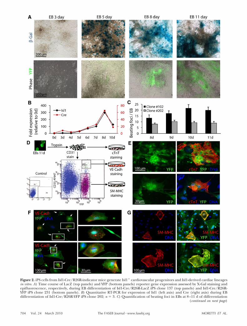
Figure 2. iPS cells from Isl1-Cre/R26R-indicator mice generate Isl1� cardiovascular progenitors and Isl1-derived cardiac lineagesin vitro. A) Time course of LacZ (top panels) and YFP (bottom panels) reporter gene expression assessed by X-Gal staining andepifluorescence, respectively, during EB differentiation of Isl1-Cre/R26R-LacZ iPS clone 137 (top panels) and Isl1-Cre/R26R-YFP iPS clone 231 (bottom panels). B) Quantitative RT-PCR for expression of Isl1 (left axis) and Cre (right axis) during EBdifferentiation of Isl1-Cre/R26R-YFP iPS clone 202; n � 3. C) Quantification of beating foci in EBs at 8–11 d of differentiation
(continued on next page)
704 Vol. 24 March 2010 MORETTI ET AL.The FASEB Journal � www.fasebj.org
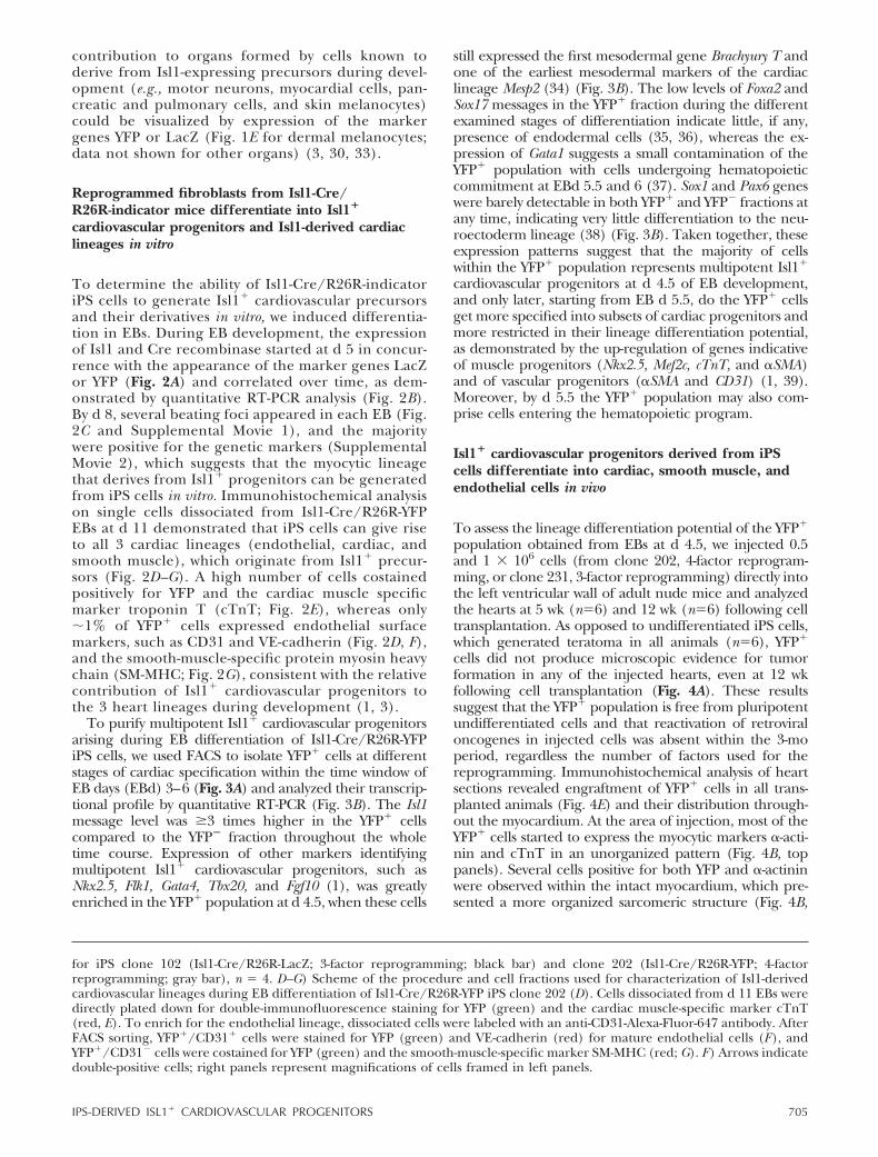
contribution to organs formed by cells known toderive from Isl1-expressing precursors during devel-opment (e.g., motor neurons, myocardial cells, pan-creatic and pulmonary cells, and skin melanocytes)could be visualized by expression of the markergenes YFP or LacZ (Fig. 1E for dermal melanocytes;data not shown for other organs) (3, 30, 33).
Reprogrammed fibroblasts from Isl1-Cre/R26R-indicator mice differentiate into Isl1�
cardiovascular progenitors and Isl1-derived cardiaclineages in vitro
To determine the ability of Isl1-Cre/R26R-indicatoriPS cells to generate Isl1� cardiovascular precursorsand their derivatives in vitro, we induced differentia-tion in EBs. During EB development, the expressionof Isl1 and Cre recombinase started at d 5 in concur-rence with the appearance of the marker genes LacZor YFP (Fig. 2A) and correlated over time, as dem-onstrated by quantitative RT-PCR analysis (Fig. 2B).By d 8, several beating foci appeared in each EB (Fig.2C and Supplemental Movie 1), and the majoritywere positive for the genetic markers (SupplementalMovie 2), which suggests that the myocytic lineagethat derives from Isl1� progenitors can be generatedfrom iPS cells in vitro. Immunohistochemical analysison single cells dissociated from Isl1-Cre/R26R-YFPEBs at d 11 demonstrated that iPS cells can give riseto all 3 cardiac lineages (endothelial, cardiac, andsmooth muscle), which originate from Isl1� precur-sors (Fig. 2D–G). A high number of cells costainedpositively for YFP and the cardiac muscle specificmarker troponin T (cTnT; Fig. 2E), whereas only1% of YFP� cells expressed endothelial surfacemarkers, such as CD31 and VE-cadherin (Fig. 2D, F),and the smooth-muscle-specific protein myosin heavychain (SM-MHC; Fig. 2G), consistent with the relativecontribution of Isl1� cardiovascular progenitors tothe 3 heart lineages during development (1, 3).
To purify multipotent Isl1� cardiovascular progenitorsarising during EB differentiation of Isl1-Cre/R26R-YFPiPS cells, we used FACS to isolate YFP� cells at differentstages of cardiac specification within the time window ofEB days (EBd) 3–6 (Fig. 3A) and analyzed their transcrip-tional profile by quantitative RT-PCR (Fig. 3B). The Isl1message level was �3 times higher in the YFP� cellscompared to the YFP� fraction throughout the wholetime course. Expression of other markers identifyingmultipotent Isl1� cardiovascular progenitors, such asNkx2.5, Flk1, Gata4, Tbx20, and Fgf10 (1), was greatlyenriched in the YFP� population at d 4.5, when these cells
still expressed the first mesodermal gene Brachyury T andone of the earliest mesodermal markers of the cardiaclineage Mesp2 (34) (Fig. 3B). The low levels of Foxa2 andSox17 messages in the YFP� fraction during the differentexamined stages of differentiation indicate little, if any,presence of endodermal cells (35, 36), whereas the ex-pression of Gata1 suggests a small contamination of theYFP� population with cells undergoing hematopoieticcommitment at EBd 5.5 and 6 (37). Sox1 and Pax6 geneswere barely detectable in both YFP� and YFP� fractions atany time, indicating very little differentiation to the neu-roectoderm lineage (38) (Fig. 3B). Taken together, theseexpression patterns suggest that the majority of cellswithin the YFP� population represents multipotent Isl1�
cardiovascular progenitors at d 4.5 of EB development,and only later, starting from EB d 5.5, do the YFP� cellsget more specified into subsets of cardiac progenitors andmore restricted in their lineage differentiation potential,as demonstrated by the up-regulation of genes indicativeof muscle progenitors (Nkx2.5, Mef2c, cTnT, and �SMA)and of vascular progenitors (�SMA and CD31) (1, 39).Moreover, by d 5.5 the YFP� population may also com-prise cells entering the hematopoietic program.
Isl1� cardiovascular progenitors derived from iPScells differentiate into cardiac, smooth muscle, andendothelial cells in vivo
To assess the lineage differentiation potential of the YFP�
population obtained from EBs at d 4.5, we injected 0.5and 1 � 106 cells (from clone 202, 4-factor reprogram-ming, or clone 231, 3-factor reprogramming) directly intothe left ventricular wall of adult nude mice and analyzedthe hearts at 5 wk (n�6) and 12 wk (n�6) following celltransplantation. As opposed to undifferentiated iPS cells,which generated teratoma in all animals (n�6), YFP�
cells did not produce microscopic evidence for tumorformation in any of the injected hearts, even at 12 wkfollowing cell transplantation (Fig. 4A). These resultssuggest that the YFP� population is free from pluripotentundifferentiated cells and that reactivation of retroviraloncogenes in injected cells was absent within the 3-moperiod, regardless the number of factors used for thereprogramming. Immunohistochemical analysis of heartsections revealed engraftment of YFP� cells in all trans-planted animals (Fig. 4E) and their distribution through-out the myocardium. At the area of injection, most of theYFP� cells started to express the myocytic markers �-acti-nin and cTnT in an unorganized pattern (Fig. 4B, toppanels). Several cells positive for both YFP and �-actininwere observed within the intact myocardium, which pre-sented a more organized sarcomeric structure (Fig. 4B,
for iPS clone 102 (Isl1-Cre/R26R-LacZ; 3-factor reprogramming; black bar) and clone 202 (Isl1-Cre/R26R-YFP; 4-factorreprogramming; gray bar), n � 4. D–G) Scheme of the procedure and cell fractions used for characterization of Isl1-derivedcardiovascular lineages during EB differentiation of Isl1-Cre/R26R-YFP iPS clone 202 (D). Cells dissociated from d 11 EBs weredirectly plated down for double-immunofluorescence staining for YFP (green) and the cardiac muscle-specific marker cTnT(red, E). To enrich for the endothelial lineage, dissociated cells were labeled with an anti-CD31-Alexa-Fluor-647 antibody. AfterFACS sorting, YFP�/CD31� cells were stained for YFP (green) and VE-cadherin (red) for mature endothelial cells (F), andYFP�/CD31� cells were costained for YFP (green) and the smooth-muscle-specific marker SM-MHC (red; G). F) Arrows indicatedouble-positive cells; right panels represent magnifications of cells framed in left panels.
705IPS-DERIVED ISL1� CARDIOVASCULAR PROGENITORS
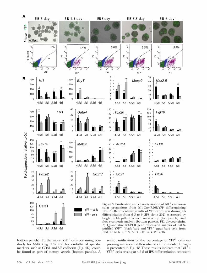
bottom panels). Furthermore, YFP� cells costaining pos-itively for SMA (Fig. 4C) and for endothelial specificmarkers, such as CD31 and VE-cadherin (Fig. 4D), couldbe found as part of mature vessels (bottom panels). A
semiquantification of the percentage of YFP� cells ex-pressing markers of differentiated cardiovascular lineagesis presented in Fig. 4F. These results indicate that Isl1�/YFP� cells arising at 4.5 d of iPS differentiation represent
lsl1 BryT Mesp2 Nkx2.5
Flk1 Gata4 Tbx20 Fgf10
cTnT Mef2c aSma CD31
Foxa2 Sox17 Sox1 Pax6
Gata1 Figure 3. Purification and characterization of Isl1� cardiovas-cular progenitors from Isl1-Cre/R26R-YFP differentiatingEBs. A) Representative results of YFP expression during EBdifferentiation from d 3 to 6 (iPS clone 202) as assessed bybright field-epifluorescence microscopy (top panels) andflow cytometric analysis (bottom panels). PE, phycoerythrin.B) Quantitative RT-PCR gene expression analysis of FACS-purified YFP� (black bar) and YFP� (gray bar) cells fromEBd 4.5 to 6; n � 3. *P � 0.05 vs. YFP� cells.
706 Vol. 24 March 2010 MORETTI ET AL.The FASEB Journal � www.fasebj.org
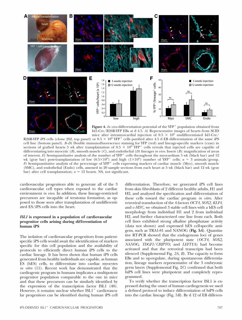
cardiovascular progenitors able to generate all of the 3cardiovascular cell types when exposed to the cardiacenvironment in vivo. In addition, these lineage-restrictedprecursors are incapable of teratoma formation, as op-posed to those seen after transplantation of undifferenti-ated ES/iPS cells into the heart.
ISL1 is expressed in a population of cardiovascularprogenitor cells arising during differentiation ofhuman iPS
The isolation of cardiovascular progenitors from patient-specific iPS cells would await the identification of markersspecific for this cell population and the availability ofprotocols to efficiently direct human iPS cells to thecardiac lineage. It has been shown that human iPS cellsgenerated from healthy individuals are capable, as humanES (hES) cells, to differentiate into cardiac myocytesin vitro (11). Recent work has demonstrated that thecardiogenic program in humans implicates a multipotentprogenitor population comparable to the one in miceand that these precursors can be similarly identified bythe expression of the transcription factor ISL1 (40).However, it remains unclear whether ISL1� cardiovascu-lar progenitors can be identified during human iPS cell
differentiation. Therefore, we generated iPS cell linesfrom skin fibroblasts of 2 different healthy adults, H1 andH2, and analyzed the specification and differentiation ofthese cells toward the cardiac program in vitro. Afterretroviral transduction of the 4 factors OCT4, SOX2, KLF4,and c-MYC, we obtained 3 stable cell lines with a hES cellmorphology from individual H1 and 2 from individualH2, and further characterized one line from each. Bothcell lines exhibited strong alkaline phosphatase activity(data not shown) and expressed hES cell-specific anti-gens, such as TRA1-81 and NANOG (Fig. 5A). Quantita-tive RT-PCR showed that the endogenous loci of genesassociated with the pluripotent state (OCT4, SOX2,NANOG, TDGF1/CRIPTO, and LEFTYA) had becomeactivated and that the retroviral transcripts had beensilenced (Supplemental Fig. 2A, B). The capacity to formEBs and to up-regulate, during spontaneous differentia-tion, lineage markers representative of the 3 embryonicgerm layers (Supplemental Fig. 2C) confirmed that bothhiPS cell lines were pluripotent and completely repro-grammed.
To verify whether the transcription factor ISL1 is ex-pressed during the onset of human cardiogenesis we useda defined protocol to induce differentiation of hiPS cellsinto the cardiac lineage (Fig. 5B). By d 12 of EB differen-
Figure 4. In vivo differentiation potential of the YFP� population obtained fromIsl1-Cre/R26R-YFP EBs at d 4.5. A) Representative images of hearts from SCIDmice after intramyocardial injection of 0.5 � 106 undifferentiated Isl1-Cre/
R26R-YFP iPS cells (clone 202, top panel) or 0.5 � 106 YFP� cells purified after 4.5 d EB differentiation of the same iPScell line (bottom panel). B–D) Double immunofluorescence staining for YFP (red) and lineage-specific markers (cyan) insections of grafted hearts 5 wk after transplantation of 0.5 � 106 YFP� cells reveals that injected cells are capable ofdifferentiating into myocytic (B), smooth muscle (C), and endothelial (D) lineages in vivo. Insets (B): magnification of areasof interest. E) Semiquantitative analysis of the number of YFP� cells throughout the myocardium 5 wk (black bar) and 12wk (gray bar) post-transplantation of low (0.5�106) and high (1�106) number of YFP� cells; n � 3 animals/group.F) Semiquantitative analysis of the percentage of YFP� cells expressing markers of cardiac muscle (Myo), smooth muscle(SMC), and endothelial (Endo) cells, assessed in 20 sample sections from each heart at 5 wk (black bar) and 12 wk (graybar) after cell transplantation; n � 12 hearts. NS, not significant.
707IPS-DERIVED ISL1� CARDIOVASCULAR PROGENITORS
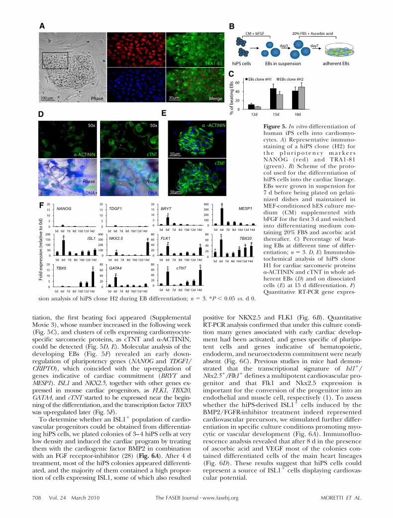
tiation, the first beating foci appeared (SupplementalMovie 3), whose number increased in the following week(Fig. 5C), and clusters of cells expressing cardiomyocyte-specific sarcomeric proteins, as cTNT and �-ACTININ,could be detected (Fig. 5D, E). Molecular analysis of thedeveloping EBs (Fig. 5F) revealed an early down-regulation of pluripotency genes (NANOG and TDGF1/CRIPTO), which coincided with the up-regulation ofgenes indicative of cardiac commitment (BRYT andMESP1). ISL1 and NKX2.5, together with other genes ex-pressed in mouse cardiac progenitors, as FLK1, TBX20,GATA4, and cTNT started to be expressed near the begin-ning of the differentiation, and the transcription factor TBX5was up-regulated later (Fig. 5F).
To determine whether an ISL1� population of cardio-vascular progenitors could be obtained from differentiat-ing hiPS cells, we plated colonies of 3–4 hiPS cells at verylow density and induced the cardiac program by treatingthem with the cardiogenic factor BMP2 in combinationwith an FGF receptor-inhibitor (28) (Fig. 6A). After 4 dtreatment, most of the hiPS colonies appeared differenti-ated, and the majority of them contained a high propor-tion of cells expressing ISL1, some of which also resulted
positive for NKX2.5 and FLK1 (Fig. 6B). QuantitativeRT-PCR analysis confirmed that under this culture condi-tion many genes associated with early cardiac develop-ment had been activated, and genes specific of pluripo-tent cells and genes indicative of hematopoietic,endoderm, and neuroectoderm commitment were nearlyabsent (Fig. 6C). Previous studies in mice had demon-strated that the transcriptional signature of Isl1�/Nkx2.5�/Flk1� defines a multipotent cardiovascular pro-genitor and that Flk1 and Nkx2.5 expression isimportant for the conversion of the progenitor into anendothelial and muscle cell, respectively (1). To assesswhether the hiPS-derived ISL1� cells induced by theBMP2/FGFR-inhibitor treatment indeed representedcardiovascular precursors, we stimulated further differ-entiation in specific culture conditions promoting myo-cytic or vascular development (Fig. 6A). Immunofluo-rescence analysis revealed that after 8 d in the presenceof ascorbic acid and VEGF most of the colonies con-tained differentiated cells of the main heart lineages(Fig. 6D). These results suggest that hiPS cells couldrepresent a source of ISL1� cells displaying cardiovas-cular potential.
NANOG TDGF1 BRYT
FLK1
MESP1
TBX20NKX2.5ISL1
TBX5 GATA4 cTNT
Figure 5. In vitro differentiation ofhuman iPS cells into cardiomyo-cytes. A) Representative immuno-staining of a hiPS clone (H2) fort h e p l u r i p o t e n c y m a r k e r sNANOG (red) and TRA1-81(green). B) Scheme of the proto-col used for the differentiation ofhiPS cells into the cardiac lineage.EBs were grown in suspension for7 d before being plated on gelati-nized dishes and maintained inMEF-conditioned hES culture me-dium (CM) supplemented withbFGF for the first 3 d and switchedinto differentiating medium con-taining 20% FBS and ascorbic acidthereafter. C) Percentage of beat-ing EBs at different time of differ-entiation; n � 3. D, E) Immunohis-tochemical analysis of hiPS cloneH1 for cardiac sarcomeric proteins�-ACTININ and cTNT in whole ad-herent EBs (D) and on dissociatedcells (E) at 15 d differentiation. F)Quantitative RT-PCR gene expres-
sion analysis of hiPS clone H2 during EB differentiation; n � 3. *P � 0.05 vs. d 0.
708 Vol. 24 March 2010 MORETTI ET AL.The FASEB Journal � www.fasebj.org
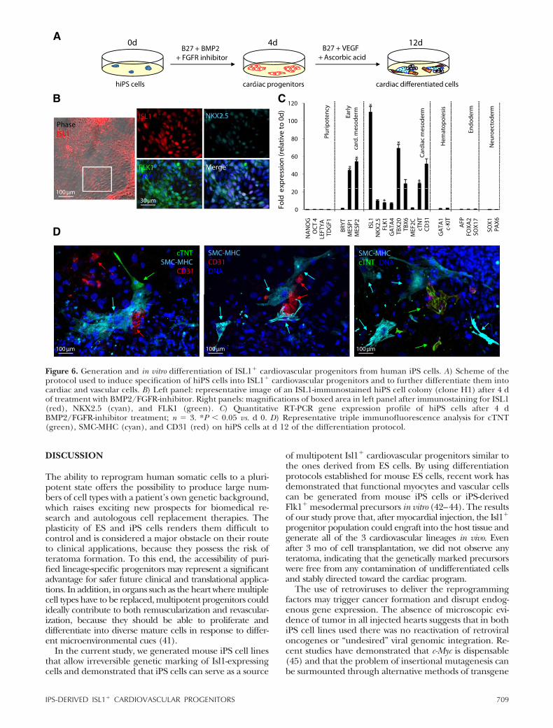
DISCUSSION
The ability to reprogram human somatic cells to a pluri-potent state offers the possibility to produce large num-bers of cell types with a patient’s own genetic background,which raises exciting new prospects for biomedical re-search and autologous cell replacement therapies. Theplasticity of ES and iPS cells renders them difficult tocontrol and is considered a major obstacle on their routeto clinical applications, because they possess the risk ofteratoma formation. To this end, the accessibility of puri-fied lineage-specific progenitors may represent a significantadvantage for safer future clinical and translational applica-tions. In addition, in organs such as the heart where multiplecell types have to be replaced, multipotent progenitors couldideally contribute to both remuscularization and revascular-ization, because they should be able to proliferate anddifferentiate into diverse mature cells in response to differ-ent microenvironmental cues (41).
In the current study, we generated mouse iPS cell linesthat allow irreversible genetic marking of Isl1-expressingcells and demonstrated that iPS cells can serve as a source
of multipotent Isl1� cardiovascular progenitors similar tothe ones derived from ES cells. By using differentiationprotocols established for mouse ES cells, recent work hasdemonstrated that functional myocytes and vascular cellscan be generated from mouse iPS cells or iPS-derivedFlk1� mesodermal precursors in vitro (42–44). The resultsof our study prove that, after myocardial injection, the Isl1�
progenitor population could engraft into the host tissue andgenerate all of the 3 cardiovascular lineages in vivo. Evenafter 3 mo of cell transplantation, we did not observe anyteratoma, indicating that the genetically marked precursorswere free from any contamination of undifferentiated cellsand stably directed toward the cardiac program.
The use of retroviruses to deliver the reprogrammingfactors may trigger cancer formation and disrupt endog-enous gene expression. The absence of microscopic evi-dence of tumor in all injected hearts suggests that in bothiPS cell lines used there was no reactivation of retroviraloncogenes or “undesired” viral genomic integration. Re-cent studies have demonstrated that c-Myc is dispensable(45) and that the problem of insertional mutagenesis canbe surmounted through alternative methods of transgene
Figure 6. Generation and in vitro differentiation of ISL1� cardiovascular progenitors from human iPS cells. A) Scheme of theprotocol used to induce specification of hiPS cells into ISL1� cardiovascular progenitors and to further differentiate them intocardiac and vascular cells. B) Left panel: representative image of an ISL1-immunostained hiPS cell colony (clone H1) after 4 dof treatment with BMP2/FGFR-inhibitor. Right panels: magnifications of boxed area in left panel after immunostaining for ISL1(red), NKX2.5 (cyan), and FLK1 (green). C) Quantitative RT-PCR gene expression profile of hiPS cells after 4 dBMP2/FGFR-inhibitor treatment; n � 3. *P � 0.05 vs. d 0. D) Representative triple immunofluorescence analysis for cTNT(green), SMC-MHC (cyan), and CD31 (red) on hiPS cells at d 12 of the differentiation protocol.
709IPS-DERIVED ISL1� CARDIOVASCULAR PROGENITORS
delivery and the use of small molecules, reducing one ofthe major drawbacks to the clinical application of iPS cells(16, 46–52). Although reprogramming of human fibro-blasts was achieved by retroviral infection, both hiPS cell linesfrom different individuals could be terminally differentiatedinto functional beating cardiomyocytes. However, carefullyconducted long-term studies will be needed to compare invitro physiology of hiPS-derived cardiac muscle cells andthose derived from human ES cell lines. The current studyconfirms the feasibility of using iPS cells for producing largenumbers of cardiac myocytes with an individual exact geno-type, which would be immune matched, an ultimate goal forregenerative medicine. Moreover, when generated fromiPS cells obtained from patients with a genetic disease,e.g., Long QT-Syndrome, cardiac muscle cells wouldbear the same genetic mutation and provide a useful invitro system to study mechanisms of pathogenesis.
Our results on human reprogrammed cells alsoconfirm recent findings in hES cells suggesting thatcardiovascular development in humans is similar to thatin the mouse (40, 53). Yang and colleagues have reportedthat during human EB differentiation a population ofKDR(FLK1)low/c-KITneg cells arises, which displays car-diac, endothelial, and vascular smooth muscle potentialin vitro and in vivo. Using a genetic marking of hES cells,Bu and colleagues have shown that purified human ISL1�
progenitors are capable of self-renewal and expansionbefore differentiation into the 3 major cardiovascularlineages in vitro. In concordance with these results, asubset of human iPS-derived ISL1� cardiovascular precur-sors described in our study also expresses FLK1 and/orNKX2.5. Because the VEGF receptor subtype-2, FLK1,also marks progenitors of the hematopoietic lineages, theidentification of other surface markers expressed in thehuman ISL1� cells would be extremely valuable for theirisolation and molecular characterization. The possibilityto purify mouse Isl1� cardiovascular precursors fromIsl1-Cre/R26R-YFP iPS cells allows us to analyze theirtranscriptional profile, and ongoing work is concen-trated in identifying a combination of cell-surface anti-gens that could permit the isolation of human andpatient-specific cardiac progenitors at different stagesof development.
In summary, our findings demonstrate the generationof Isl1� cardiovascular progenitors from mouse iPS cellsand their multipotency in vivo to differentiate into endo-thelial, smooth, and cardiac muscle cells, without theformation of teratoma. In addition, we describe theidentification of human iPS-derived ISL1� cardiac precur-sors and the efficient induction of ISL1�/NKX2.5�/FLK1� mesodermal progenitors with subsequent differ-entiation into cardiovascular cell types. This approach willprovide ready access to ISL1� cardiovascular progenitorsand more mature populations for functional cell replace-ment analysis as well as for developmental biologystudies defining the earliest molecular program in-volved in the specification and differentiation of thislineage.
The authors thank Toshio Kitamura (University of Tokyo,Tokyo, Japan), Shinya Yamanaka (Kyoto University, Kyoto, Ja-pan), and George Daley (Harvard Medical School, Boston, MA,USA) for providing viral vectors through Addgene. Some of the
monoclonal antibodies were obtained from the DevelopmentalStudies Hybridoma Bank, which was developed under theauspices of the National Institute Human Development and ismaintained by the University of Iowa. The authors especiallyacknowledge K. Goetsch for her expertise in the FACS sortinganalysis and M. Rudelius and D. Grewe for assistance in histol-ogy. This work was supported by a Marie Curie Excellence TeamGrant from the European Research Council (MEXT-23208), theGerman Research Foundation (La 1238 3-1/4-1), and the Ger-man Ministry for Education and Research (01 GN 0826).
REFERENCES
1. Moretti, A., Caron, L., Nakano, A., Lam, J. T., Bernshausen, A.,Chen, Y., Qyang, Y., Bu, L., Sasaki, M., Martin-Puig, S., Sun, Y.,Evans, S. M., Laugwitz, K. L., and Chien, K. R. (2006) Multipo-tent embryonic isl1� progenitor cells lead to cardiac, smoothmuscle, and endothelial cell diversification. Cell 127, 1151–1165
2. Kattman, S. J., Huber, T. L., and Keller, G. M. (2006) Multipo-tent flk-1� cardiovascular progenitor cells give rise to thecardiomyocyte, endothelial, and vascular smooth muscle lin-eages. Dev. Cell 11, 723–732
3. Sun, Y., Liang, X., Najafi, N., Cass, M., Lin, L., Cai, C. L., Chen,J., and Evans, S. M. (2007) Islet 1 is expressed in distinctcardiovascular lineages, including pacemaker and coronaryvascular cells. Dev. Biol. 304, 286–296
4. Laugwitz, K. L., Moretti, A., Lam, J., Gruber, P., Chen, Y.,Woodard, S., Lin, L. Z., Cai, C. L., Lu, M. M., Reth, M.,Platoshyn, O., Yuan, J. X., Evans, S., and Chien, K. R. (2005)Postnatal isl1� cardioblasts enter fully differentiated cardio-myocyte lineages. Nature 433, 647–653
5. Laugwitz, K. L., Moretti, A., Caron, L., Nakano, A., and Chien,K. R. (2008) Islet1 cardiovascular progenitors: a single sourcefor heart lineages? Development 135, 193–205
6. Wu, S. M., Chien, K. R., and Mummery, C. (2008) Origins andfates of cardiovascular progenitor cells. Cell 132, 537–543
7. Moretti, A., Lam, J., Evans, S. M., and Laugwitz, K. L. (2007)Biology of Isl1� cardiac progenitor cells in development anddisease. Cell. Mol. Life Sci. 64, 674–682
8. Murry, C. E., and Keller, G. (2008) Differentiation of embryonicstem cells to clinically relevant populations: lessons from embry-onic development. Cell 132, 661–680
9. Rubart, M., and Field, L. J. (2006) Cardiac regeneration:repopulating the heart. Annu. Rev. Physiol. 68, 29–49
10. Takahashi, K., and Yamanaka, S. (2006) Induction of pluripo-tent stem cells from mouse embryonic and adult fibroblastcultures by defined factors. Cell 126, 663–676
11. Takahashi, K., Tanabe, K., Ohnuki, M., Narita, M., Ichisaka, T.,Tomoda, K., and Yamanaka, S. (2007) Induction of pluripotent stemcells from adult human fibroblasts by defined factors. Cell 131,861–872
12. Yu, J., Vodyanik, M. A., Smuga-Otto, K., Antosiewicz-Bourget, J.,Frane, J. L., Tian, S., Nie, J., Jonsdottir, G. A., Ruotti, V., Stewart, R.,Slukvin, I. I., and Thomson, J. A. (2007) Induced pluripotent stemcell lines derived from human somatic cells. Science 318, 1917–1920
13. Kim, J. B., Zaehres, H., Wu, G., Gentile, L., Ko, K., Sebastiano, V.,Arauzo-Bravo, M. J., Ruau, D., Han, D. W., Zenke, M., and Scholer,H. R. (2008) Pluripotent stem cells induced from adult neural stemcells by reprogramming with two factors. Nature 454, 646–650
14. Silva, J., Barrandon, O., Nichols, J., Kawaguchi, J., Theunissen,T. W., and Smith, A. (2008) Promotion of reprogramming toground state pluripotency by signal inhibition. PLoS Biol. 6, e253
15. Aasen, T., Raya, A., Barrero, M. J., Garreta, E., Consiglio, A.,Gonzalez, F., Vassena, R., Bilic, J., Pekarik, V., Tiscornia, G.,Edel, M., Boue, S., and Belmonte, J. C. (2008) Efficient andrapid generation of induced pluripotent stem cells from humankeratinocytes. Nat. Biotechnol. 26, 1276–1284
16. Kim, J. B., Sebastiano, V., Wu, G., Arauzo-Bravo, M. J., Sasse, P.,Gentile, L., Ko, K., Ruau, D., Ehrich, M., van den Boom, D., Meyer,J., Hubner, K., Bernemann, C., Ortmeier, C., Zenke, M., Fleisch-mann, B. K., Zaehres, H., and Scholer, H. R. (2009) Oct4-inducedpluripotency in adult neural stem cells. Cell 136, 411–419
17. Park, I. H., Arora, N., Huo, H., Maherali, N., Ahfeldt, T.,Shimamura, A., Lensch, M. W., Cowan, C., Hochedlinger, K.,
710 Vol. 24 March 2010 MORETTI ET AL.The FASEB Journal � www.fasebj.org
and Daley, G. Q. (2008) Disease-specific induced pluripotentstem cells. Cell 134, 877–886
18. Dimos, J. T., Rodolfa, K. T., Niakan, K. K., Weisenthal, L. M.,Mitsumoto, H., Chung, W., Croft, G. F., Saphier, G., Leibel, R.,Goland, R., Wichterle, H., Henderson, C. E., and Eggan, K. (2008)Induced pluripotent stem cells generated from patients with ALScan be differentiated into motor neurons. Science 321, 1218–1221
19. Ebert, A. D., Yu, J., Rose, F. F., Jr., Mattis, V. B., Lorson, C. L.,Thomson, J. A., and Svendsen, C. N. (2009) Induced pluripo-tent stem cells from a spinal muscular atrophy patient. Nature457, 277–280
20. Soldner, F., Hockemeyer, D., Beard, C., Gao, Q., Bell, G. W.,Cook, E. G., Hargus, G., Blak, A., Cooper, O., Mitalipova, M.,Isacson, O., and Jaenisch, R. (2009) Parkinson’s disease patient-derived induced pluripotent stem cells free of viral reprogram-ming factors. Cell 136, 964–977
21. Maherali, N., Sridharan, R., Xie, W., Utikal, J., Eminli, S.,Arnold, K., Stadtfeld, M., Yachechko, R., Tchieu, J., Jaenisch, R.,Plath, K., and Hochedlinger, K. (2007) Directly reprogrammedfibroblasts show global epigenetic remodeling and widespreadtissue contribution. Cell Stem Cell 1, 55–70
22. Hanna, J., Wernig, M., Markoulaki, S., Sun, C. W., Meissner, A.,Cassady, J. P., Beard, C., Brambrink, T., Wu, L. C., Townes,T. M., and Jaenisch, R. (2007) Treatment of sickle cell anemiamouse model with iPS cells generated from autologous skin.Science 318, 1920–1923
23. Wernig, M., Zhao, J. P., Pruszak, J., Hedlund, E., Fu, D., Soldner,F., Broccoli, V., Constantine-Paton, M., Isacson, O., and Jae-nisch, R. (2008) Neurons derived from reprogrammed fibro-blasts functionally integrate into the fetal brain and improvesymptoms of rats with Parkinson’s disease. Proc. Natl. Acad. Sci.U. S. A. 105, 5856–5861
24. Cai, C. L., Martin, J. C., Sun, Y., Cui, L., Wang, L., Ouyang, K., Yang,L., Bu, L., Liang, X., Zhang, X., Stallcup, W. B., Denton, C. P.,McCulloch, A., Chen, J., and Evans, S. M. (2008) A myocardial lineagederives from Tbx18 epicardial cells. Nature 454, 104–108
25. Soriano, P. (1999) Generalized lacZ expression with theROSA26 Cre reporter strain. Nat. Genet. 21, 70–71
26. Srinivas, S., Watanabe, T., Lin, C. S., William, C. M., Tanabe, Y.,Jessell, T. M., and Constantini, F. (2001) Cre reporter strainsproduced by targeted insertion of EYFP and ECFP into theROSA26 locus. BMC Dev. Biol. 1,
27. Takahashi, K., Okita, K., Nakagawa, M., and Yamanaka, S.(2007) Induction of pluripotent stem cells from fibroblastcultures. Nat. Protoc. 2, 3081–3089
28. Leschik, J., Stefanovic, S., Brinon, B., and Puceat, M. (2008)Cardiac commitment of primate embryonic stem cells. Nat.Protoc. 3, 1381–1387
29. Cai, C. L., Liang, X., Shi, Y., Chu, P. H., Pfaff, S. L., Chen, J., andEvans, S. (2003) Isl1 identifies a cardiac progenitor populationthat proliferates prior to differentiation and contributes amajority of cells to the heart. Dev. Cell 5, 877–889
30. Schwahn, D. J., Timchenko, N. A., Shibahara, S., and Medrano,E. E. (2005) Dynamic regulation of the human dopachrometautomerase promoter by MITF, ER-alpha and chromatin re-modelers during proliferation and senescence of human mela-nocytes. Pigment Cell Res. 18, 203–213
31. Okita, K., Ichisaka, T., and Yamanaka, S. (2007) Generation ofgermline-competent induced pluripotent stem cells. Nature 448, 313–317
32. Wernig, M., Meissner, A., Foreman, R., Brambrink, T., Ku, M.,Hochedlinger, K., Bernstein, B. E., and Jaenisch, R. (2007) Invitro reprogramming of fibroblasts into a pluripotent ES-cell-like state. Nature 448, 318–324
33. Tsuchida, T., Ensini, M., Morton, S. B., Baldassare, M., Edlund,T., Jessell, T. M., and Pfaff, S. L. (1994) Topographic organiza-tion of embryonic motor neurons defined by expression of LIMhomeobox genes. Cell 79, 957–970
34. Kitajima, S., Takagi, A., Inoue, T., and Saga, Y. (2000) MesP1and MesP2 are essential for the development of cardiac meso-derm. Development 127, 3215–3226
35. Sasaki, H., and Hogan, B. L. (1993) Differential expression ofmultiple fork head related genes during gastrulation and axialpattern formation in the mouse embryo. Development 118, 47–59
36. Kanai-Azuma, M., Kanai, Y., Gad, J. M., Tajima, Y., Taya, C.,Kurohmaru, M., Sanai, Y., Yonekawa, H., Yazaki, K., Tam, P. P.,and Hayashi, Y. (2002) Depletion of definitive gut endoderm inSox17-null mutant mice. Development 129, 2367–2379
37. Orkin, S. H. (1992) GATA-binding transcription factors inhematopoietic cells. Blood 80, 575–581
38. Li, X. J., Du, Z. W., Zarnowska, E. D., Pankratz, M., Hansen,L. O., Pearce, R. A., and Zhang, S. C. (2005) Specification ofmotoneurons from human embryonic stem cells. Nat. Biotechnol.23, 215–221
39. Wu, S. M., Fujiwara, Y., Cibulsky, S. M., Clapham, D. E., Lien,C. L., Schultheiss, T. M., and Orkin, S. H. (2006) Developmentalorigin of a bipotential myocardial and smooth muscle cellprecursor in the mammalian heart. Cell 127, 1137–1150
40. Lei, B., Jiang, X., Martin-Puig, S., Caron, L., Zhu, S., Shao, Y.,Roberts, D. J., Huang, P. L., Domian, I. J., and Chien, K. R.(2009) Human ISL1 heart progenitors generate diverse multi-potent cardiovascular cell lineages. Nature 460, 113–117
41. Passier, R., van Laake, L. W., and Mummery, C. L. (2008)Stem-cell-based therapy and lessons from the heart. Nature 453,322–329
42. Mauritz, C., Schwanke, K., Reppel, M., Neef, S., Katsirntaki, K.,Maier, L. S., Nguemo, F., Menke, S., Haustein, M., Hescheler, J.,Hasenfuss, G., and Martin, U. (2008) Generation of functionalmurine cardiac myocytes from induced pluripotent stem cells.Circulation 118, 507–517
43. Schenke-Layland, K., Rhodes, K. E., Angelis, E., Butylkova, Y.,Heydarkhan-Hagvall, S., Gekas, C., Zhang, R., Goldhaber, J. I.,Mikkola, H. K., Plath, K., and MacLellan, W. R. (2008) Repro-grammed mouse fibroblasts differentiate into cells of the cardio-vascular and hematopoietic lineages. Stem Cells 26, 1537–1546
44. Narazaki, G., Uosaki, H., Teranishi, M., Okita, K., Kim, B., Matsuoka,S., Yamanaka, S., and Yamashita, J. K. (2008) Directed and systematicdifferentiation of cardiovascular cells from mouse induced pluripo-tent stem cells. Circulation 118, 498–506
45. Nakagawa, M., Koyanagi, M., Tanabe, K., Takahashi, K., Ich-isaka, T., Aoi, T., Okita, K., Mochiduki, Y., Takizawa, N., andYamanaka, S. (2008) Generation of induced pluripotent stemcells without Myc from mouse and human fibroblasts. Nat.Biotechnol. 26, 101–106
46. Stadtfeld, M., Nagaya, M., Utikal, J., Weir, G., and Hoche-dlinger, K. (2008) Induced pluripotent stem cells generatedwithout viral integration. Science 322, 945–949
47. Okita, K., Nakagawa, M., Hyenjong, H., Ichisaka, T., and Ya-manaka, S. (2008) Generation of mouse induced pluripotentstem cells without viral vectors. Science 322, 949–953
48. Huangfu, D., Maehr, R., Guo, W., Eijkelenboom, A., Snitow, M.,Chen, A. E., and Melton, D. A. (2008) Induction of pluripotentstem cells by defined factors is greatly improved by small-molecule compounds. Nat. Biotechnol. 26, 795–797
49. Carey, B. W., Markoulaki, S., Hanna, J., Saha, K., Gao, Q.,Mitalipova, M., and Jaenisch, R. (2009) Reprogramming ofmurine and human somatic cells using a single polycistronicvector. Proc. Natl. Acad. Sci. U. S. A. 106, 157–162
50. Zhou, H., Wu, S., Joo, J. Y., Zhu, S., Han, D. W., Lin, T., Trauger,S., Bien, G., Yao, S., Zhu, Y., Siuzdak, G., Scholer, H. R., Duan,L., and Ding, S. (2009) Generation of induced pluripotent stemcells using recombinant proteins. Cell Stem Cell 4, 381–384
51. Woltjen, K., Michael, I. P., Mohseni, P., Desai, R., Mileikovsky,M., Hamalainen, R., Cowling, R., Wang, W., Liu, P., Gertsen-stein, M., Kaji, K., Sung, H. K., and Nagy, A. (2009) piggyBactransposition reprograms fibroblasts to induced pluripotentstem cells. Nature 458, 766–770
52. Yu, J., Hu, K., Smuga-Otto, K., Tian, S., Stewart, R., Slukvin, I. I.,and Thomson, J. A. (2009) Human induced pluripotent stem cellsfree of vector and transgene sequences. Science 324, 797–801
53. Yang, L., Soonpaa, M. H., Adler, E. D., Roepke, T. K., Kattman,S. J., Kennedy, M., Henckaerts, E., Bonham, K., Abbott, G. W.,Linden, R. M., Field, L. J., and Keller, G. M. (2008) Humancardiovascular progenitor cells develop from a KDR� embryonic-stem-cell-derived population. Nature 453, 524–528
Received for publication June 26, 2009.Accepted for publication September 24, 2009.
711IPS-DERIVED ISL1� CARDIOVASCULAR PROGENITORS
n engl j med 363;15 nejm.org october 7, 2010 1397
The new england journal of medicineestablished in 1812 october 7, 2010 vol. 363 no. 15
Patient-Specific Induced Pluripotent Stem-Cell Models for Long-QT Syndrome
Alessandra Moretti, Ph.D., Milena Bellin, Ph.D., Andrea Welling, Ph.D., Christian Billy Jung, M.Sc., Jason T. Lam, Ph.D., Lorenz Bott-Flügel, M.D., Tatjana Dorn, Ph.D., Alexander Goedel, M.D.,
Christian Höhnke, M.D., Franz Hofmann, M.D., Melchior Seyfarth, M.D., Daniel Sinnecker, M.D., Albert Schömig, M.D., and Karl-Ludwig Laugwitz, M.D.
A BS TR AC T
From the Cardiology Division, First De-partment of Medicine (A.M., M.B., C.B.J., J.T.L., L.B.-F., T.D., A.G., M.S., D.S., A.S., K.-L.L.), and the Plastic Surgery Depart-ment (C.H.), Klinikum rechts der Isar; the Cardiology Department, German Heart Center Munich (A.M., M.B., C.B.J., J.T.L., L.B.-F., T.D., A.G., M.S., D.S., A.S., K.-L.L.); and the Institute of Pharmacology and Toxicology (A.W., F.H.) — all at the Tech-nical University of Munich, Munich, Ger-many. Address reprint requests to Dr. Laugwitz at the Cardiology Division, First Department of Medicine and German Heart Center Munich, Klinikum rechts der Isar, Technical University of Munich, Ismaninger Str., 22, D-81675 Munich, Germany, or at [email protected] .tum.de.
Drs. Moretti, Bellin, and Welling contrib-uted equally to this article.
This article (10.1056/NEJMoa0908679) was published on July 21, 2010, at NEJM.org.
N Engl J Med 2010;363:1397-1409.Copyright © 2010 Massachusetts Medical Society.
BACKGROUND
Long-QT syndromes are heritable diseases associated with prolongation of the QT interval on an electrocardiogram and a high risk of sudden cardiac death due to ventricular tachyarrhythmia. In long-QT syndrome type 1, mutations occur in the KCNQ1 gene, which encodes the repolarizing potassium channel mediating the de-layed rectifier IKs current.
METHODS
We screened a family affected by long-QT syndrome type 1 and identified an auto-somal dominant missense mutation (R190Q) in the KCNQ1 gene. We obtained dermal fibroblasts from two family members and two healthy controls and infected them with retroviral vectors encoding the human transcription factors OCT3/4, SOX2, KLF4, and c-MYC to generate pluripotent stem cells. With the use of a specific protocol, these cells were then directed to differentiate into cardiac myocytes.
RESULTS
Induced pluripotent stem cells maintained the disease genotype of long-QT syn-drome type 1 and generated functional myocytes. Individual cells showed a “ven-tricular,” “atrial,” or “nodal” phenotype, as evidenced by the expression of cell-type–specific markers and as seen in recordings of the action potentials in single cells. The duration of the action potential was markedly prolonged in “ventricular” and “atrial” cells derived from patients with long-QT syndrome type 1, as compared with cells from control subjects. Further characterization of the role of the R190Q–KCNQ1 mutation in the pathogenesis of long-QT syndrome type 1 revealed a dom-inant negative trafficking defect associated with a 70 to 80% reduction in IKs cur-rent and altered channel activation and deactivation properties. Moreover, we showed that myocytes derived from patients with long-QT syndrome type 1 had an increased susceptibility to catecholamine-induced tachyarrhythmia and that beta-blockade attenuated this phenotype.
CONCLUSIONS
We generated patient-specific pluripotent stem cells from members of a family af-fected by long-QT syndrome type 1 and induced them to differentiate into functional cardiac myocytes. The patient-derived cells recapitulated the electrophysiological features of the disorder. (Funded by the European Research Council and others.)
The New England Journal of Medicine Downloaded from nejm.org on January 25, 2012. For personal use only. No other uses without permission.
Copyright © 2010 Massachusetts Medical Society. All rights reserved.
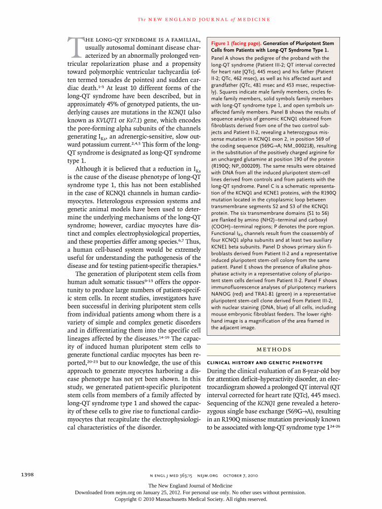
T h e n e w e ngl a nd j o u r na l o f m e dic i n e
n engl j med 363;15 nejm.org october 7, 20101398
The long-qt syndrome is a familial, usually autosomal dominant disease char-acterized by an abnormally prolonged ven-
tricular repolarization phase and a propensity toward polymorphic ventricular tachycardia (of-ten termed torsades de pointes) and sudden car-diac death.1-3 At least 10 different forms of the long-QT syndrome have been described, but in approximately 45% of genotyped patients, the un-derlying causes are mutations in the KCNQ1 (also known as KVLQT1 or Kv7.1) gene, which encodes the pore-forming alpha subunits of the channels generating IKs, an adrenergic-sensitive, slow out-ward potassium current.2,4,5 This form of the long-QT syndrome is designated as long-QT syndrome type 1.
Although it is believed that a reduction in IKs is the cause of the disease phenotype of long-QT syndrome type 1, this has not been established in the case of KCNQ1 channels in human cardio-myocytes. Heterologous expression systems and genetic animal models have been used to deter-mine the underlying mechanisms of the long-QT syndrome; however, cardiac myocytes have dis-tinct and complex electrophysiological properties, and these properties differ among species.6,7 Thus, a human cell-based system would be extremely useful for understanding the pathogenesis of the disease and for testing patient-specific therapies.8
The generation of pluripotent stem cells from human adult somatic tissues9-13 offers the oppor-tunity to produce large numbers of patient-specif-ic stem cells. In recent studies, investigators have been successful in deriving pluripotent stem cells from individual patients among whom there is a variety of simple and complex genetic disorders and in differentiating them into the specific cell lineages affected by the diseases.14-19 The capac-ity of induced human pluripotent stem cells to generate functional cardiac myocytes has been re-ported,20-23 but to our knowledge, the use of this approach to generate myocytes harboring a dis-ease phenotype has not yet been shown. In this study, we generated patient-specific pluripotent stem cells from members of a family affected by long-QT syndrome type 1 and showed the capac-ity of these cells to give rise to functional cardio-myocytes that recapitulate the electrophysiologi-cal characteristics of the disorder.
Me thods
Clinical History and Genetic Phenotype
During the clinical evaluation of an 8-year-old boy for attention deficit–hyperactivity disorder, an elec-trocardiogram showed a prolonged QT interval (QT interval corrected for heart rate [QTc], 445 msec). Sequencing of the KCNQ1 gene revealed a hetero-zygous single base exchange (569G→A), resulting in an R190Q missense mutation previously known to be associated with long-QT syndrome type 124-26
Figure 1 (facing page). Generation of Pluripotent Stem Cells from Patients with Long-QT Syndrome Type 1.
Panel A shows the pedigree of the proband with the long-QT syndrome (Patient III-2; QT interval corrected for heart rate [QTc], 445 msec) and his father (Patient II-2; QTc, 462 msec), as well as his affected aunt and grandfather (QTc, 481 msec and 453 msec, respective-ly). Squares indicate male family members, circles fe-male family members, solid symbols family members with long-QT syndrome type 1, and open symbols un-affected family members. Panel B shows the results of sequence analysis of genomic KCNQ1 obtained from fibroblasts derived from one of the two control sub-jects and Patient II-2, revealing a heterozygous mis-sense mutation in KCNQ1 exon 2, in position 569 of the coding sequence (569G→A; NM_000218), resulting in the substitution of the positively charged arginine for an uncharged glutamine at position 190 of the protein (R190Q; NP_000209). The same results were obtained with DNA from all the induced pluripotent stem-cell lines derived from controls and from patients with the long-QT syndrome. Panel C is a schematic representa-tion of the KCNQ1 and KCNE1 proteins, with the R190Q mutation located in the cytoplasmic loop between transmembrane segments S2 and S3 of the KCNQ1 protein. The six transmembrane domains (S1 to S6) are flanked by amino (NH2)–terminal and carboxyl (COOH)–terminal regions; P denotes the pore region. Functional IKs channels result from the coassembly of four KCNQ1 alpha subunits and at least two auxiliary KCNE1 beta subunits. Panel D shows primary skin fi-broblasts derived from Patient II-2 and a representative induced pluripotent stem-cell colony from the same patient. Panel E shows the presence of alkaline phos-phatase activity in a representative colony of pluripo-tent stem cells derived from Patient II-2. Panel F shows immunofluorescence analyses of pluripotency markers NANOG (red) and TRA1-81 (green) in a representative pluripotent stem-cell clone derived from Patient III-2, with nuclear staining (DNA, blue) of all cells, including mouse embryonic fibroblast feeders. The lower right-hand image is a magnification of the area framed in the adjacent image.
The New England Journal of Medicine Downloaded from nejm.org on January 25, 2012. For personal use only. No other uses without permission.
Copyright © 2010 Massachusetts Medical Society. All rights reserved.
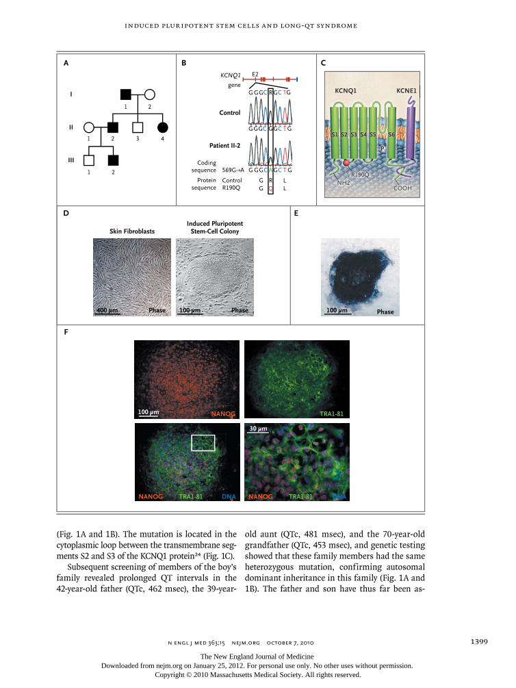
induced pluripotent stem cells and long-qt syndrome
n engl j med 363;15 nejm.org october 7, 2010 1399
(Fig. 1A and 1B). The mutation is located in the cytoplasmic loop between the transmembrane seg-ments S2 and S3 of the KCNQ1 protein24 (Fig. 1C).
Subsequent screening of members of the boy’s family revealed prolonged QT intervals in the 42-year-old father (QTc, 462 msec), the 39-year-
old aunt (QTc, 481 msec), and the 70-year-old grandfather (QTc, 453 msec), and genetic testing showed that these family members had the same heterozygous mutation, confirming autosomal dominant inheritance in this family (Fig. 1A and 1B). The father and son have thus far been as-
D
F
E
A B C
KCNQ1
S2 S2 S1 S3
R190Q
S4 S5 S6
P
KCNE1
R190Q NH2
COOH
I
II
III
3 4
1 2
21
21Protein
sequence
Codingsequence
E2
GGGC C TGG G
G GGC AGC T G
G GG GRC C TG
Control
569G→A
KCNQ1
gene
ControlR190Q
G RG Q L
L
Patient II-2
Skin Fibroblasts
400 µm Phase
Induced PluripotentStem-Cell Colony
100 µm Phase 100 µm Phase
TRA1-81
TRA1-81NANOG DNA
NANOG100 µm
30 µm
TRA1-81NANOG DNA
The New England Journal of Medicine Downloaded from nejm.org on January 25, 2012. For personal use only. No other uses without permission.
Copyright © 2010 Massachusetts Medical Society. All rights reserved.
T h e n e w e ngl a nd j o u r na l o f m e dic i n e
n engl j med 363;15 nejm.org october 7, 20101400
ymptomatic. The grandfather and aunt have re-ported periods of dizziness and palpitations. All the genetically affected family members are be-ing treated with beta-blockers.
Generation of Patient-Specific Pluripotent Stem Cells
For the generation of pluripotent stem cells, we recruited the father and son in the family affect-ed by long-QT syndrome type 1 along with two healthy control subjects. The protocols for re-search involving human subjects and for stem-cell research were approved by the institutional review board and the committee charged with oversight of embryonic stem-cell research at the Technical University of Munich. All the study participants provided written informed consent.
Dermal-biopsy specimens were minced and placed on culture dishes. Fibroblasts migrating out of the explants were passaged twice and then infected with a combination of retrovirus-es encoding the human transcription factors OCT3/4, SOX2, KLF4, and c-MYC.9 After 6 days, infected cells were seeded on murine embryonic fibroblast feeders and cultured in standard hu-man embryonic stem-cell medium until induced pluripotent stem-cell colonies appeared. Details of the study methods are provided in the Supple-mentary Appendix, available with the full text of this article at NEJM.org.
In Vitro Cardiac Differentiation
We differentiated induced pluripotent stem cells as embryoid bodies by detaching the stem-cell colonies from the feeder cells and maintaining them for 3 days in feeder-cell-conditioned human embryonic stem-cell medium in low attachment plates.27,28 At day 4, the medium was replaced with differentiation medium containing 20% fe-tal-calf serum. Embryoid bodies were plated on gelatin-coated dishes on day 7. Between days 20 and 30, areas that exhibited spontaneous contrac-tion (indicative of cardiac differentiation) were mi-crodissected, plated on fibronectin-coated plates, and maintained in culture in differentiation me-dium containing 2% fetal-calf serum. For single-cell analysis, microdissected areas were dissoci-ated with the use of type II collagenase. Single cells were plated on fibronectin-coated slides for immunohistochemical and electrophysiological analysis.
Immunohistochemical Assessments
Immunostaining was performed according to standard protocols with the use of antibodies specific for the following: Nanog (Abcam), TRA-1-81 (BD Pharmingen), cardiac troponin T (Lab Vi-sion), α-actinin (Sigma–Aldrich), myosin light chain 2a and myosin light chain 2v (Synaptic Sys-tems), KCNQ1 (Abcam), and protein disulfide isomerase (Abcam). Staining was also performed for F-actin with the use of fluorescence-labeled phalloidin (Invitrogen).
Polymerase-Chain-Reaction Assays
The polymerase chain reaction (PCR) was used to amplify the mutated region of the KCNQ1 gene for sequencing. Quantitative real-time PCR was used in allelic-discrimination assays and for the assessment of expression of pluripotency genes, retroviral transgenes, and cell-lineage markers. Reverse-transcriptase (RT)–PCR was used to assay cardiomyocyte phenotype markers in single cells. Detailed methods are provided in the Supple-mentary Appendix; primers are listed in Table 1 in the Supplementary Appendix.
Electrophysiological Assessments
Whole-cell recordings were obtained with the use of standard patch-clamp techniques.7,29 Cul-ture differentiation medium was used as the ex-ternal bath solution. Action potentials and cur-rents were recorded at approximately 35°C. All currents were normalized to cell capacitance.
Statistical Analysis
Data that passed tests for normality and equal variance were analyzed with the use of one-way analysis of variance followed by Tukey’s test, when appropriate. The Wilcoxon signed-rank test and the Kruskal–Wallis test followed by Dunn’s test were used to analyze the remaining data. Two-sided P values of less than 0.05 were considered to indicate statistical significance.
R esult s
Generation of Pluripotent Stem Cells
We generated pluripotent stem cells from pri-mary fibroblasts derived from two patients with long-QT syndrome type 1 and two control sub-jects after retroviral transduction of the repro-gramming factors. Starting at 3 weeks after viral
The New England Journal of Medicine Downloaded from nejm.org on January 25, 2012. For personal use only. No other uses without permission.
Copyright © 2010 Massachusetts Medical Society. All rights reserved.
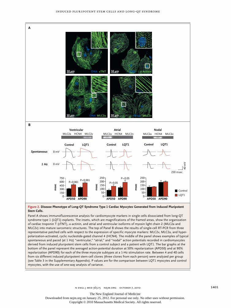
induced pluripotent stem cells and long-qt syndrome
n engl j med 363;15 nejm.org october 7, 2010 1401
B
A
Ventricular Atrial NodalMLC2a MLC2vHCN4 MLC2a MLC2vHCN4 MLC2a MLC2vHCN4
APD90APD50
mse
c
0
600450300150
750
mse
c
0
25020015010050
APD90APD50
mse
c
APD90APD500
25020015010050
Control
LQT1
P<0.001 P=0.05
P=0.05P<0.001
250 msec
40 m
V
Control LQT1 Control LQT1 Control LQT1
Spontaneous 0 mV
0 mV1 Hz
DNA cTNT30 µm DNA α-Actinin30 µm
DNA MLC2a30 µm DNA MLC2v30 µm
Figure 2. Disease Phenotype of Long-QT Syndrome Type 1 Cardiac Myocytes Generated from Induced Pluripotent Stem Cells.
Panel A shows immunofluorescence analysis for cardiomyocyte markers in single cells dissociated from long-QT syndrome type 1 (LQT1) explants. The insets, which are magnifications of the framed areas, show the organization of cardiac troponin T (cTNT), α-actinin, and atrial and ventricular isoforms of myosin light chain 2 (MLC2a and MLC2v) into mature sarcomeric structures. The top of Panel B shows the results of single-cell RT-PCR from three representative patched cells with respect to the expression of specific myocyte markers: MLC2v, MLC2a, and hyper-polarization-activated, cyclic nucleotide-gated channel 4 (HCN4). The middle of the panel shows examples of typical spontaneous and paced (at 1 Hz) “ventricular,” “atrial,” and “nodal” action potentials recorded in cardiomyocytes derived from induced pluripotent stem cells from a control subject and a patient with LQT1. The bar graphs at the bottom of the panel represent the averaged action-potential duration at 50% repolarization (APD50) and at 90% repolarization (APD90) for each of the three myocyte subtypes at a 1-Hz stimulation rate. Between 4 and 40 cells from six different induced pluripotent stem-cell clones (three clones from each person) were analyzed per group (see Table 3 in the Supplementary Appendix). P values are for the comparison between LQT1 myocytes and control myocytes, with the use of one-way analysis of variance.
The New England Journal of Medicine Downloaded from nejm.org on January 25, 2012. For personal use only. No other uses without permission.
Copyright © 2010 Massachusetts Medical Society. All rights reserved.
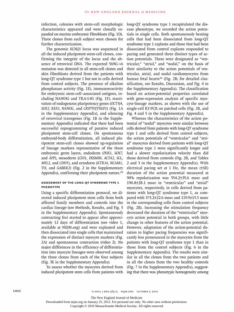
T h e n e w e ngl a nd j o u r na l o f m e dic i n e
n engl j med 363;15 nejm.org october 7, 20101402
infection, colonies with stem-cell morphologic characteristics appeared and were clonally ex-panded on murine embryonic fibroblasts (Fig. 1D). Three clones from each subject were chosen for further characterization.
The genomic KCNQ1 locus was sequenced in all the induced pluripotent stem-cell clones, con-firming the integrity of the locus and the ab-sence of retroviral DNA. The expected 569G→A mutation was detected in all stem-cell clones and skin fibroblasts derived from the patients with long-QT syndrome type 1 but not in cells derived from control subjects. The presence of alkaline phosphatase activity (Fig. 1E), immunoreactivity for embryonic stem-cell–associated antigens, in-cluding NANOG and TRA-1-81 (Fig. 1F), reacti-vation of endogenous pluripotency genes (OCT3/4, SOX2, REX1, NANOG, and CRIPTO/TDGF1) (Fig. 1A in the Supplementary Appendix), and silencing of retroviral transgenes (Fig. 1B in the Supple-mentary Appendix) indicated that there had been successful reprogramming of putative induced pluripotent stem-cell clones. On spontaneous embryoid-body differentiation, all induced plu-ripotent stem-cell clones showed up-regulation of lineage markers representative of the three embryonic germ layers, endoderm (PDX1, SOX7, and AFP), mesoderm (CD31, DESMIN, ACTA2, SCL, MYL2, and CDH5), and ectoderm (KTR14, NCAM1, TH, and GABRR2) (Fig. 2 in the Supplementary Appendix), confirming their pluripotent nature.28
Assessment of the Long-QT Syndrome Type 1 Phenotype
Using a specific differentiation protocol, we di-rected induced pluripotent stem cells from both affected family members and controls into the cardiac lineage (see Methods, Results, and Fig. 3 in the Supplementary Appendix). Spontaneously contracting foci started to appear after approxi-mately 12 days of differentiation (see video 1, available at NEJM.org) and were explanted and then dissociated into single cells that maintained the expression of distinct myocyte markers (Fig. 2A) and spontaneous contraction (video 2). No major differences in the efficiency of differentia-tion into myocyte lineages were observed among the three clones from each of the four subjects (Fig. 3E in the Supplementary Appendix).
To assess whether the myocytes derived from induced pluripotent stem cells from patients with
long-QT syndrome type 1 recapitulated the dis-ease phenotype, we recorded the action poten-tials in single cells. Both spontaneously beating cells that had been dissociated from long-QT syndrome type 1 explants and those that had been dissociated from control explants responded to pacing and generated three distinct types of ac-tion potentials. These were designated as “ven-tricular,” “atrial,” and “nodal,” on the basis of their similarity to the action potentials of ven-tricular, atrial, and nodal cardiomyocytes from human fetal hearts30 (Fig. 2B; for detailed clas-sification, see Results, Discussion, and Fig. 4 in the Supplementary Appendix). The classification based on action-potential properties correlated with gene-expression analysis of specific myo-cyte-lineage markers, as shown with the use of single-cell RT-PCR on patched cells (Fig. 2B, and Fig. 4 and 5 in the Supplementary Appendix).
Whereas the characteristics of the action po-tential of “nodal” myocytes were similar between cells derived from patients with long-QT syndrome type 1 and cells derived from control subjects, the action potentials of “ventricular” and “atri-al” myocytes derived from patients with long-QT syndrome type 1 were significantly longer and had a slower repolarization velocity than did those derived from controls (Fig. 2B, and Tables 2 and 3 in the Supplementary Appendix). With electrical pacing set at 1 Hz, the mean (±SE) duration of the action potential measured at 90% repolarization was 554.2±35.6 msec and 190.8±28.1 msec in “ventricular” and “atrial” myocytes, respectively, in cells derived from pa-tients with long-QT syndrome type 1, as com-pared with 373.2±22.6 msec and 119.9±15.5 msec in the corresponding cells from control subjects (Fig. 2B). Increasing the stimulation frequency decreased the duration of the “ventricular” myo-cyte action potential in both groups, with little change in other features of the action potential. However, adaptation of the action-potential du-ration to higher pacing frequencies was signifi-cantly less pronounced in the myocytes from the patients with long-QT syndrome type 1 than in those from the control subjects (Fig. 6 in the Supplementary Appendix). The results were sim-ilar in all the clones from the two patients and in all the clones from the two healthy controls (Fig. 7 in the Supplementary Appendix), suggest-ing that there was phenotypic homogeneity among
The New England Journal of Medicine Downloaded from nejm.org on January 25, 2012. For personal use only. No other uses without permission.
Copyright © 2010 Massachusetts Medical Society. All rights reserved.
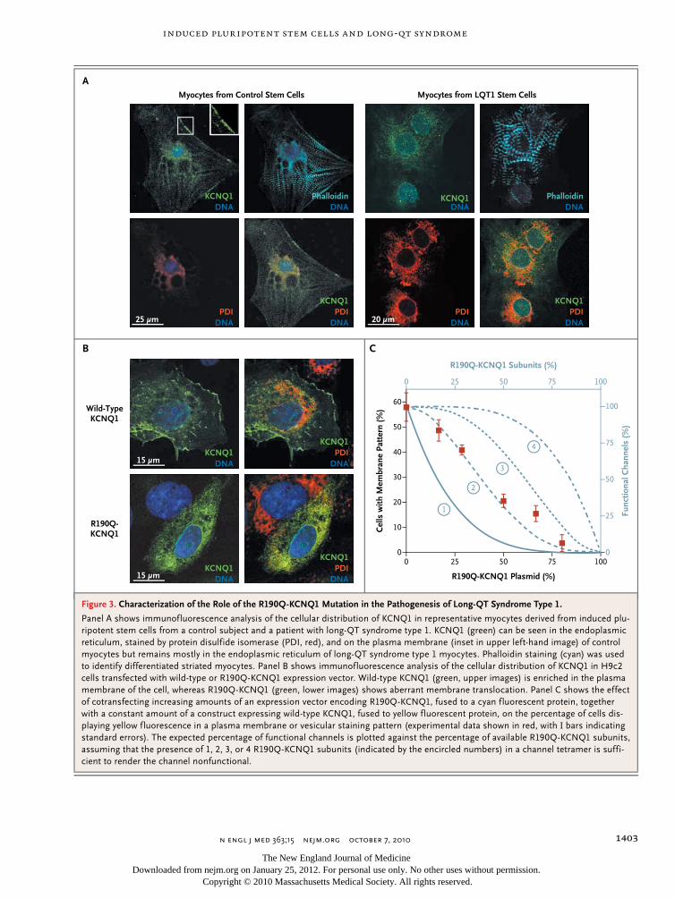
induced pluripotent stem cells and long-qt syndrome
n engl j med 363;15 nejm.org october 7, 2010 1403
Myocytes from Control Stem Cells Myocytes from LQT1 Stem Cells
15 µm
15 µmPDI
DNA
KCNQ1
PDIDNA
KCNQ1
KCNQ1DNA
KCNQ1DNA
20 µm
KCNQ1DNA
PDIDNA
PDIDNA
KCNQ1
25 µmPDI
DNAPDI
DNA
KCNQ1
DNAPhalloidinKCNQ1
DNA
B C
A
Cel
ls w
ith M
embr
ane
Patt
ern
(%)
Func
tiona
l Cha
nnel
s (%
)
60
40
30
10
50
20
0
100
50
75
25
00 25 50 75 100
0 25 50 75 100
R190Q-KCNQ1 Plasmid (%)
R190Q-KCNQ1 Subunits (%)
Wild-TypeKCNQ1
R190Q-KCNQ1
PhalloidinDNA
1
2
3
4
Figure 3. Characterization of the Role of the R190Q-KCNQ1 Mutation in the Pathogenesis of Long-QT Syndrome Type 1.
Panel A shows immunofluorescence analysis of the cellular distribution of KCNQ1 in representative myocytes derived from induced plu-ripotent stem cells from a control subject and a patient with long-QT syndrome type 1. KCNQ1 (green) can be seen in the endoplasmic reticulum, stained by protein disulfide isomerase (PDI, red), and on the plasma membrane (inset in upper left-hand image) of control myocytes but remains mostly in the endoplasmic reticulum of long-QT syndrome type 1 myocytes. Phalloidin staining (cyan) was used to identify differentiated striated myocytes. Panel B shows immunofluorescence analysis of the cellular distribution of KCNQ1 in H9c2 cells transfected with wild-type or R190Q-KCNQ1 expression vector. Wild-type KCNQ1 (green, upper images) is enriched in the plasma membrane of the cell, whereas R190Q-KCNQ1 (green, lower images) shows aberrant membrane translocation. Panel C shows the effect of cotransfecting increasing amounts of an expression vector encoding R190Q-KCNQ1, fused to a cyan fluorescent protein, together with a constant amount of a construct expressing wild-type KCNQ1, fused to yellow fluorescent protein, on the percentage of cells dis-playing yellow fluorescence in a plasma membrane or vesicular staining pattern (experimental data shown in red, with I bars indicating standard errors). The expected percentage of functional channels is plotted against the percentage of available R190Q-KCNQ1 subunits, assuming that the presence of 1, 2, 3, or 4 R190Q-KCNQ1 subunits (indicated by the encircled numbers) in a channel tetramer is suffi-cient to render the channel nonfunctional.
The New England Journal of Medicine Downloaded from nejm.org on January 25, 2012. For personal use only. No other uses without permission.
Copyright © 2010 Massachusetts Medical Society. All rights reserved.
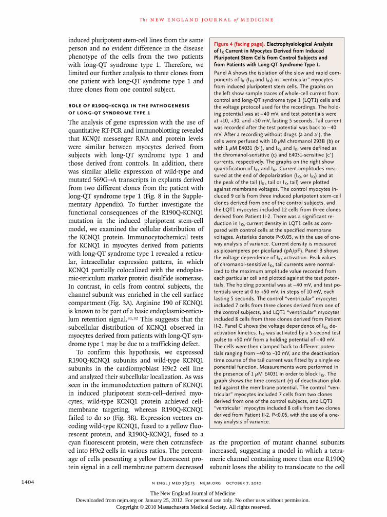
T h e n e w e ngl a nd j o u r na l o f m e dic i n e
n engl j med 363;15 nejm.org october 7, 20101404
induced pluripotent stem-cell lines from the same person and no evident difference in the disease phenotype of the cells from the two patients with long-QT syndrome type 1. Therefore, we limited our further analysis to three clones from one patient with long-QT syndrome type 1 and three clones from one control subject.
Role of R190Q-KCNQ1 in the Pathogenesis of Long-QT Syndrome Type 1
The analysis of gene expression with the use of quantitative RT-PCR and immunoblotting revealed that KCNQ1 messenger RNA and protein levels were similar between myocytes derived from subjects with long-QT syndrome type 1 and those derived from controls. In addition, there was similar allelic expression of wild-type and mutated 569G→A transcripts in explants derived from two different clones from the patient with long-QT syndrome type 1 (Fig. 8 in the Supple-mentary Appendix). To further investigate the functional consequences of the R190Q-KCNQ1 mutation in the induced pluripotent stem-cell model, we examined the cellular distribution of the KCNQ1 protein. Immunocytochemical tests for KCNQ1 in myocytes derived from patients with long-QT syndrome type 1 revealed a reticu-lar, intracellular expression pattern, in which KCNQ1 partially colocalized with the endoplas-mic-reticulum marker protein disulfide isomerase. In contrast, in cells from control subjects, the channel subunit was enriched in the cell surface compartment (Fig. 3A). Arginine 190 of KCNQ1 is known to be part of a basic endoplasmic-reticu-lum retention signal.31,32 This suggests that the subcellular distribution of KCNQ1 observed in myocytes derived from patients with long-QT syn-drome type 1 may be due to a trafficking defect.
To confirm this hypothesis, we expressed R190Q-KCNQ1 subunits and wild-type KCNQ1 subunits in the cardiomyoblast H9c2 cell line and analyzed their subcellular localization. As was seen in the immunodetection pattern of KCNQ1 in induced pluripotent stem-cell–derived myo-cytes, wild-type KCNQ1 protein achieved cell-membrane targeting, whereas R190Q-KCNQ1 failed to do so (Fig. 3B). Expression vectors en-coding wild-type KCNQ1, fused to a yellow fluo-rescent protein, and R190Q-KCNQ1, fused to a cyan fluorescent protein, were then cotransfect-ed into H9c2 cells in various ratios. The percent-age of cells presenting a yellow fluorescent pro-tein signal in a cell membrane pattern decreased
as the proportion of mutant channel subunits increased, suggesting a model in which a tetra-meric channel containing more than one R190Q subunit loses the ability to translocate to the cell
Figure 4 (facing page). Electrophysiological Analysis of IK Current in Myocytes Derived from Induced Pluripotent Stem Cells from Control Subjects and from Patients with Long-QT Syndrome Type 1.
Panel A shows the isolation of the slow and rapid com-ponents of IK (IKs and IKr) in “ventricular” myocytes from induced pluripotent stem cells. The graphs on the left show sample traces of whole-cell current from control and long-QT syndrome type 1 (LQT1) cells and the voltage protocol used for the recordings. The hold-ing potential was at –40 mV, and test potentials were at +10, +30, and +50 mV, lasting 5 seconds. Tail current was recorded after the test potential was back to –40 mV. After a recording without drugs (a and a′), the cells were perfused with 10 μM chromanol 293B (b) or with 1 μM E4031 (b′), and IKs and IKr were defined as the chromanol-sensitive (c) and E4031-sensitive (c′) currents, respectively. The graphs on the right show quantification of IKs and IKr. Current amplitudes mea-sured at the end of depolarization (IKs or IKr) and at the peak of the tail (IKs tail or IKr tail) were plotted against membrane voltages. The control myocytes in-cluded 9 cells from three induced pluripotent stem-cell clones derived from one of the control subjects, and the LQT1 myocytes included 12 cells from three clones derived from Patient II-2. There was a significant re-duction in IKs current density in LQT1 cells as com-pared with control cells at the specified membrane voltages. Asterisks denote P<0.05, with the use of one-way analysis of variance. Current density is measured as picoamperes per picofarad (pA/pF). Panel B shows the voltage dependence of IKs activation. Peak values of chromanol-sensitive IKs tail currents were normal-ized to the maximum amplitude value recorded from each particular cell and plotted against the test poten-tials. The holding potential was at –40 mV, and test po-tentials were at 0 to +50 mV, in steps of 10 mV, each lasting 5 seconds. The control “ventricular” myocytes included 7 cells from three clones derived from one of the control subjects, and LQT1 “ventricular” myocytes included 8 cells from three clones derived from Patient II-2. Panel C shows the voltage dependence of IKs de-activation kinetics. IKs was activated by a 5-second test pulse to +50 mV from a holding potential of –40 mV. The cells were then clamped back to different poten-tials ranging from −40 to −10 mV, and the deactivation time course of the tail current was fitted by a single ex-ponential function. Measurements were performed in the presence of 1 μM E4031 in order to block IKr. The graph shows the time constant (τ) of deactivation plot-ted against the membrane potential. The control “ven-tricular” myocytes included 7 cells from two clones derived from one of the control subjects, and LQT1 “ventricular” myocytes included 8 cells from two clones derived from Patient II-2. P<0.05, with the use of a one-way analysis of variance.
The New England Journal of Medicine Downloaded from nejm.org on January 25, 2012. For personal use only. No other uses without permission.
Copyright © 2010 Massachusetts Medical Society. All rights reserved.
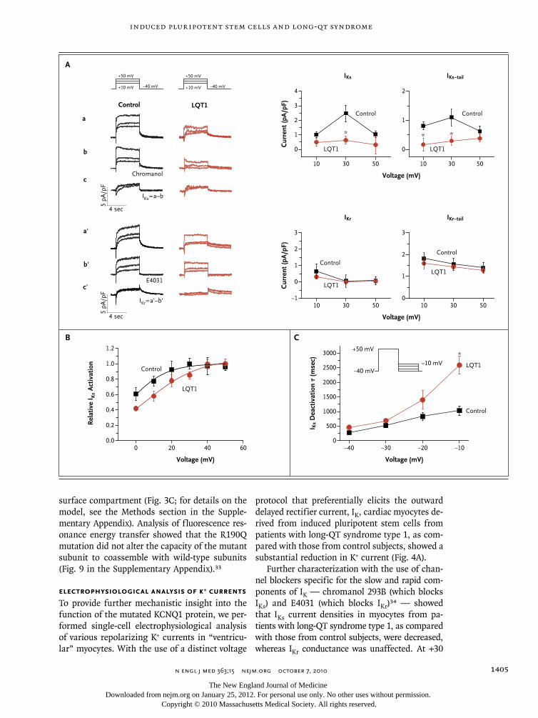
induced pluripotent stem cells and long-qt syndrome
n engl j med 363;15 nejm.org october 7, 2010 1405
surface compartment (Fig. 3C; for details on the model, see the Methods section in the Supple-mentary Appendix). Analysis of fluorescence res-onance energy transfer showed that the R190Q mutation did not alter the capacity of the mutant subunit to coassemble with wild-type subunits (Fig. 9 in the Supplementary Appendix).33
Electrophysiological Analysis of K+ Currents
To provide further mechanistic insight into the function of the mutated KCNQ1 protein, we per-formed single-cell electrophysiological analysis of various repolarizing K+ currents in “ventricu-lar” myocytes. With the use of a distinct voltage
protocol that preferentially elicits the outward delayed rectifier current, IK, cardiac myocytes de-rived from induced pluripotent stem cells from patients with long-QT syndrome type 1, as com-pared with those from control subjects, showed a substantial reduction in K+ current (Fig. 4A).
Further characterization with the use of chan-nel blockers specific for the slow and rapid com-ponents of IK — chromanol 293B (which blocks IKs) and E4031 (which blocks IKr)34 — showed that IKs current densities in myocytes from pa-tients with long-QT syndrome type 1, as compared with those from control subjects, were decreased, whereas IKr conductance was unaffected. At +30
Control
LQT1
B C
A
I Ks
Dea
ctiv
atio
n τ
(mse
c)
3000
2000
1500
500
2500
1000
0−40 −30 −20 −10
Voltage (mV)
Cur
rent
(pA
/pF)
3
2
1
010 30 50
3
2
1
−1
0
10 30 50
Voltage (mV)
IKr IKr−tail
Cur
rent
(pA
/pF)
2
1
0
10 30 50
4
3
2
1
0
10 30 50
Voltage (mV)
IKs IKs−tail
Rel
ativ
e I K
s A
ctiv
atio
n
1.2
0.8
0.6
0.2
1.0
0.4
0.00 20 40 60
Voltage (mV)
Control
Control
Control
Control Control
LQT1
LQT1
LQT1
LQT1
LQT1
a'
E4031
IKr=a'–b'
IKs=a–b
4 sec
5 pA
/pF
b'
c'
Chromanol
a
b
c
LQT1
−40 mV+10 mV
+50 mV
Control
4 sec
5 pA
/pF
−40 mV+10 mV
+50 mV
* * *
−40 mV
+50 mV
−10 mV*
The New England Journal of Medicine Downloaded from nejm.org on January 25, 2012. For personal use only. No other uses without permission.
Copyright © 2010 Massachusetts Medical Society. All rights reserved.
T h e n e w e ngl a nd j o u r na l o f m e dic i n e
n engl j med 363;15 nejm.org october 7, 20101406
mV, IKs and tail IKs were diminished by approxi-mately 75%, suggesting that in hetero zygous “ven-tricular” myocytes from patients with long-QT syndrome type 1, the mutant form of KCNQ1 interferes with the function of the wild-type sub-unit (Fig. 4A). Furthermore, both activation and deactivation properties of the tail IKs current in cells from patients with long-QT syndrome type 1, as compared with cells from controls, appeared to be altered, with activation being slightly shift-ed toward more positive voltages (Fig. 4B) and deactivation being decelerated (Fig. 4C). We next analyzed the transient outward (Ito) and inward currents that function in the hyperpolarization state. As with the results for IKr, Ito and diastolic current densities did not differ between “ventric-ular” myocytes from patients with long-QT syn-drome type 1 and those from control subjects (Fig. 10 in the Supplementary Appendix), show-ing that there was a specific genotype–pheno-type correlation.
Protective Action of Beta-Blockade
Since fatal arrhythmias are precipitated by in-creased sympathetic tone in patients with long-QT syndrome type 1,3,35 we tested whether ad-renergic stimulation can affect the phenotype of long-QT syndrome type 1 cardiomyocytes de-rived from pluripotent stem cells. First, we ana-lyzed the effect of catecholamines on the dura-tion of the action potential in paced “ventricular” myocytes. At 2 Hz, isoproterenol induced ap-proximately a 20% reduction in the interval be-tween 30% and 90% repolarization of the action potential in control myocytes, whereas in cells from patients with long-QT syndrome type 1 this interval was almost unaffected (Fig. 5A). Accord-ingly, IKs was markedly enhanced by adrenergic stimulation in “ventricular” myocytes from con-trol subjects, and the increase was significantly smaller in cells from patients with long-QT syn-drome type 1, suggesting that defective respon-siveness to adrenergic challenge is due to an ab-normal IKs current (Fig. 5B).36
We further investigated the effect of isopro-terenol on spontaneously beating myocytes. In cells from control subjects, isoproterenol had a positive chronotropic effect accompanied by a shortening of the duration of the action poten-tial, resulting in a 30% reduction in the ratio of the action-potential duration at 90% repolariza-
tion to the action-potential interval. In contrast, in myocytes from patients with long-QT syn-drome type 1, this ratio was increased by 15%, thereby exacerbating the long-QT syndrome type 1 phenotype and increasing the risk of arrhyth-mic events (Fig. 5C). In fact, under conditions of adrenergic stress, six of nine “ventricular” myo-cytes from patients with long-QT syndrome type 1 developed early afterdepolarizations (4.1±1.5 early
Figure 5 (facing page). Adrenergic Modulation and Protective Effect of Beta-Blockade in Control and Long-QT Syndrome Type 1 “Ventricular” Myocytes.
Panel A shows an overlay of single action potentials from a representative cell from a control subject and from a patient with long-QT syndrome type 1 (LQT1) at a 2-Hz stimulation rate before (basal) and after appli-cation of 100 nM isoproterenol. The bar graph represents the percent change induced by isoproterenol on the in-terval from the action-potential duration (APD) at 30% repolarization (APD30) to APD90 (APD30−APD90), on APD90, and on APD. Data are means ±SE for 10 cells in each group (from three clones derived from one of the control subjects and three clones derived from Pa-tient II-2). Panel B shows the effect of adrenaline on IKs tail current. On the left are representative overlapped IKs tail-current traces from a control and an LQT1 myo-cyte before and after treatment with 1 μM adrenaline recorded at +30 mV depolarization with the use of the voltage protocol depicted at the top of the panel. Mea-surements were performed in presence of 1 μM E4031 in order to block IKr. The bar graph represents the fac-tor increase in IKs tail current induced by 1 μM adrena-line at the specified depolarization voltages in control and LQT1 myocytes. Data are means ±SE for 8 cells in each group (from two clones derived from one of the control subjects and from two clones derived from Pa-tient II-2). Panel C shows representative recordings of single-cell spontaneous action potentials from a con-trol and an LQT1 “ventricular” myocyte before (base-line) and during exposure to a combination of 100 nM isoproterenol and 200 nM propranolol or to 100 nM isoproterenol alone. Cells were pretreated for 2 min-utes with 200 nM propranolol before application of the combination of 100 nM isoproterenol and 200 nM pro-pranolol (traces not shown). The bar graph represents the percent change in the ratio of APD90 to the action-potential interval (APD90/AP interval) induced by the combination of 100 nM isoproterenol and 200 nM pro-pranolol or by 100 nM isoproterenol alone. Action po-tentials affected by early afterdepolarizations (EAD) were excluded from the analysis. Data are means ±SE for 8 control cells (from three clones derived from one of the control subjects) and 9 LQT1 cells (from three clones derived from Patient II-2). P values in the three panels are for the comparison between LQT1 myocytes and control myocytes, with the use of the Wilcoxon signed-rank test.
The New England Journal of Medicine Downloaded from nejm.org on January 25, 2012. For personal use only. No other uses without permission.
Copyright © 2010 Massachusetts Medical Society. All rights reserved.
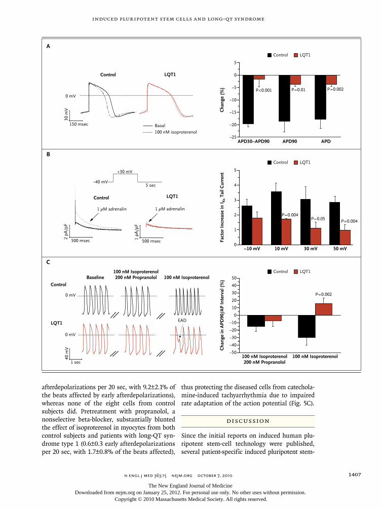
induced pluripotent stem cells and long-qt syndrome
n engl j med 363;15 nejm.org october 7, 2010 1407
afterdepolarizations per 20 sec, with 9.2±2.1% of the beats affected by early afterdepolarizations), whereas none of the eight cells from control subjects did. Pretreatment with propranolol, a nonselective beta-blocker, substantially blunted the effect of isoproterenol in myocytes from both control subjects and patients with long-QT syn-drome type 1 (0.6±0.3 early afterdepolarizations per 20 sec, with 1.7±0.8% of the beats affected),
thus protecting the diseased cells from catechola-mine-induced tachyarrhythmia due to impaired rate adaptation of the action potential (Fig. 5C).
Discussion
Since the initial reports on induced human plu-ripotent stem-cell technology were published, several patient-specific induced pluripotent stem-
150 msec
30 m
V
0 mV
Basal100 nM isoproterenol
Control LQT1
500 msec2 pA
/pF
1 µM adrenalin
Control
500 msec1 pA
/pF
1 µM adrenalin
LQT1
−40 mV
+30 mV
5 sec
Baseline100 nM Isoproterenol200 nM Propranolol 100 nM Isoproterenol
1 sec
40 m
V
Control
0 mV
LQT1
0 mV
EAD
B
A
5
4
3
0
2
1
5
0
−5
−10
−15
−20
−25APD30−APD90 APD90 APD
−10 mV 10 mV 30 mV 50 mV
C
50
−50
40
30
20
10
0
−10
−20
−30
−40
100 nM Isoproterenol200 nM Propranolol
100 nM Isoproterenol
Cha
nge
(%)
Fact
or In
crea
se in
I Ks
Tail
Cur
rent
Cha
nge
in A
PD90
/AP
Inte
rval
(%)
Control LQT1
Control LQT1
Control LQT1
P=0.002
P=0.004
P=0.002
P=0.05P=0.004
P=0.01P<0.001
The New England Journal of Medicine Downloaded from nejm.org on January 25, 2012. For personal use only. No other uses without permission.
Copyright © 2010 Massachusetts Medical Society. All rights reserved.
T h e n e w e ngl a nd j o u r na l o f m e dic i n e
n engl j med 363;15 nejm.org october 7, 20101408
cell lines for neurodegenerative and metabolic disorders have been developed.14,16,18,19,37 Estab-lishing reliable models of human disease in such cells has remained challenging, owing to diffi-culties in directing cell differentiation and in iden-tifying disease-related mechanisms. In this study, we reprogrammed fibroblasts derived from mem-bers of a family with autosomal-dominant long-QT syndrome type 1 and used these induced plu-ripotent stem cells to generate patient-specific cardiomyocytes. These myocytes showed expres-sion of specific markers and electrophysiological characteristics that suggested that the reprogram-ming process did not affect the ability of the cells to function normally. Furthermore, we observed disease-specific abnormalities in the duration of the action potential, the action-potential rate adaptation, and IKs currents, owing to an R190Q-KCNQ1 trafficking defect, as well as vulnerabili-ty to catecholaminergic stress.
To date, insights into the pathogenesis of the long-QT syndrome have come primarily from heterologous expression systems and genetic ani-mal models. Depending on the cell type used, both haploinsufficiency and dominant negative effects have been postulated as the mechanism of disease associated with the R190Q muta-tion.24-26 In our induced pluripotent stem-cell model of long-QT syndrome type 1, a reduction in IKs current density by approximately 75%, com-bined with subcellular localization, showed that the R190Q mutant suppresses channel traffick-ing to the plasma membrane in a dominant negative manner. Owing to differences among species in the channels that generate the main cardiac repolarizing currents, none of the avail-able mouse models of the long-QT syndrome fully emulate the human disease phenotype. Re-cently, transgenic rabbit models of long-QT syn-drome type 1 and long-QT syndrome type 2 have been engineered by means of overexpression of dominant negative pore mutants of the human genes KCNQ1 and KCNH2.38 In these animals, both transgenes caused a down-regulation of the complementary IKr and IKs currents. In contrast, no alterations in repolarizing currents other than in IKs were observed in patient-specific myo-cytes derived from persons with the R190Q-KCNQ1 mutation associated with long-QT syn-drome type 1. This discrepancy, which may be mutant-dependent or model-dependent, shows the importance of alternative systems in which hu-
man genetic disorders can be studied in the physiologic and disease-causing contexts on a patient-specific level.
Our data provide clear evidence that the patho-genesis of the R190Q mutation can be modeled in myocyte lineages generated from pluripotent stem cells derived from patients with long-QT syndrome type 1. Moreover, our findings sug-gest that there may be alternative approaches to the development of candidate drugs, such as com-pounds to promote the delivery of the mutant to the plasma membrane or IKs activators. The ob-served protective effects of beta-blockade show that it is possible to investigate the therapeutic action of medications for treating human cardiac disease in vitro with the use of patient-specific cells. This approach is particularly attractive because of the pluripotent nature of these cells and the potentially unlimited number of induced cardiomyocytes available for high-throughput drug development.
Even though the incidence of the long-QT syndrome is only 1 case per 2500 live births, this syndrome provides a platform for showing the suitability of induced pluripotent stem-cell tech-nology as a means of exploring disease mecha-nisms in human genetic cardiac disorders. Larger sets of long-QT syndrome cell lines harboring different channel mutations will be needed to fur-ther validate the disease phenotype and compare pathogenetic mechanisms in diverse forms of the disease. Clinically, the severity of manifesta-tions of the long-QT syndrome varies among fam-ily members, and incomplete penetrance exists.39 However, we did not observe any phenotypic dif-ferences in the prolongation of the action poten-tial between the myocytes from our two patients, a finding that is probably due to the similarity of the clinical phenotype in these cases.
In summary, we derived pluripotent stem cells from patients with long-QT syndrome type 1 and directed them to differentiate into cardiac myo-cytes. As compared with myocytes derived in a similar fashion from healthy controls, cells from patients with long-QT syndrome type 1 exhibit-ed prolongation of the action potential, altered IKs activation and deactivation properties, and an abnormal response to catecholamine stimulation, with a protective effect of beta-blockade, thus showing that induced pluripotent stem-cell mod-els can recapitulate aspects of genetic cardiac dis-eases.
The New England Journal of Medicine Downloaded from nejm.org on January 25, 2012. For personal use only. No other uses without permission.
Copyright © 2010 Massachusetts Medical Society. All rights reserved.
induced pluripotent stem cells and long-qt syndrome
n engl j med 363;15 nejm.org october 7, 2010 1409
Supported by grants from the European Research Council (Ma-rie Curie Excellence Team Grant, MEXT-23208), the German Re-search Foundation (Research Unit 923 and La 1238 3-1/4-1), and the German Ministry for Education and Research (01 GN 0826).
Disclosure forms provided by the authors are available with the full text of this article at NEJM.org
We thank Takashi Kitamura and Shinya Yamanaka for provid-ing viral vectors through Addgene; Jacques Barhanin for KCNE1
and wild-type and mutant (R190Q) KCNQ1 complementary DNAs; Stefan Engelhardt and Andrea Ahles for the fluorescent β1-receptor fusion construct; Diana Grewe, Christina Scherb, and Sabine Teuber for their technical assistance in cell culture and immunohistochemical assessments; and especially the members of the family affected by long-QT syndrome type 1 and the healthy volunteers who provided us with skin-biopsy speci-mens for the reprogramming.
References1. Priori SG, Bloise R, Crotti L. The long QT syndrome. Europace 2001;3:16-27.2. Moss AJ, Shimizu W, Wilde AA, et al. Clinical aspects of type-1 long-QT syn-drome by location, coding type, and bio-physical function of mutations involving the KCNQ1 gene. Circulation 2007;115: 2481-9.3. Roden DM. Long-QT syndrome. N Engl J Med 2008;358:169-76.4. Li GR, Feng J, Yue L, Carrier M, Nattel S. Evidence for two components of delayed rectifier K+ current in human ventricular myocytes. Circ Res 1996;78:689-96.5. Marx SO, Kurokawa J, Reiken S, et al. Requirement of a macromolecular signal-ing complex for beta adrenergic receptor modulation of the KCNQ1-KCNE1 potas-sium channel. Science 2002;295:496-9.6. Hofmann F, Lacinová L, Klugbauer N. Voltage-dependent calcium channels: from structure to function. Rev Physiol Bio-chem Pharmacol 1999;139:33-87.7. Ludwig A, Zong X, Jeglitsch M, Hof-mann F, Biel M. A family of hyperpolariza-tion-activated mammalian cation chan-nels. Nature 1998;393:587-91.8. Laugwitz KL, Moretti A, Caron L, Na-kano A, Chien KR. Islet1 cardiovascular progenitors: a single source for heart lin-eages? Development 2008;135:193-205.9. Takahashi K, Tanabe K, Ohnuki M, et al. Induction of pluripotent stem cells from adult human fibroblasts by defined factors. Cell 2007;131:861-72.10. Yu J, Vodyanik MA, Smuga-Otto K, et al. Induced pluripotent stem cell lines de-rived from human somatic cells. Science 2007;318:1917-20.11. Park IH, Zhao R, West JA, et al. Repro-gramming of human somatic cells to pluripotency with defined factors. Nature 2008;451:141-6.12. Lowry WE, Richter L, Yachechko R, et al. Generation of human induced pluripo-tent stem cells from dermal fibroblasts. Proc Natl Acad Sci U S A 2008;105:2883-8.13. Jaenisch R, Young R. Stem cells, the molecular circuitry of pluripotency and nuclear reprogramming. Cell 2008;132: 567-82.14. Dimos JT, Rodolfa KT, Niakan KK, et al. Induced pluripotent stem cells gener-ated from patients with ALS can be dif-ferentiated into motor neurons. Science 2008;321:1218-21.15. Park IH, Arora N, Huo H, et al. Dis-ease-specific induced pluripotent stem cells. Cell 2008;134:877-86.16. Ebert AD, Yu J, Rose FF Jr, et al. In-
duced pluripotent stem cells from a spinal muscular atrophy patient. Nature 2009; 457:277-80.17. Raya A, Rodríguez-Pizà I, Guenechea G, et al. Disease-corrected haematopoietic progenitors from Fanconi anaemia in-duced pluripotent stem cells. Nature 2009; 460:53-9.18. Soldner F, Hockemeyer D, Beard C, et al. Parkinson’s disease patient-derived in-duced pluripotent stem cells free of viral reprogramming factors. Cell 2009;136: 964-77.19. Maehr R, Chen S, Snitow M, et al. Generation of pluripotent stem cells from patients with type 1 diabetes. Proc Natl Acad Sci U S A 2009;106:15768-73.20. Zhang J, Wilson GF, Soerens AG, et al. Functional cardiomyocytes derived from human induced pluripotent stem cells. Circ Res 2009;104(4):e30-e41.21. Yokoo N, Baba S, Kaichi S, et al. The effects of cardioactive drugs on cardio-myocytes derived from human induced pluripotent stem cells. Biochem Biophys Res Commun 2009;387:482-8.22. Gai H, Leung EL, Costantino PD, et al. Generation and characterization of func-tional cardiomyocytes using induced plu-ripotent stem cells derived from human fibroblasts. Cell Biol Int 2009;33:1184-93.23. Freund C, Davis RP, Gkatzis K, Ward-van Oostwaard D, Mummery CL. The first reported generation of human induced pluripotent stem cells (iPS cells) and iPS cell-derived cardiomyocytes in the Neth-erlands. Neth Heart J 2010;18:51-4.24. Chouabe C, Neyroud N, Richard P, et al. Novel mutations in KvLQT1 that affect Iks activation through interactions with Isk. Cardiovasc Res 2000;45:971-80.25. Donger C, Denjoy I, Berthet M, et al. KVLQT1 C-terminal missense mutation causes a forme fruste long-QT syndrome. Circulation 1997;96:2778-81.26. Wang Z, Tristani-Firouzi M, Xu Q, Lin M, Keating MT, Sanguinetti MC. Func-tional effects of mutations in KvLQT1 that cause long QT syndrome. J Cardio-vasc Electrophysiol 1999;10:817-26.27. Moretti A, Caron L, Nakano A, et al. Multipotent embryonic isl1+ progenitor cells lead to cardiac, smooth muscle, and endothelial cell diversification. Cell 2006; 127:1151-65.28. Moretti A, Bellin M, Jung CB, et al. Mouse and human induced pluripotent stem cells as a source for multipotent Isl1+ cardiovascular progenitors. FASEB J 2010;24:700-11.
29. Laugwitz KL, Moretti A, Lam J, et al. Postnatal isl1+ cardioblasts enter fully dif-ferentiated cardiomyocyte lineages. Na-ture 2005;433:647-53. [Erratum, Nature 2007;446:934.]30. Mummery C, Ward-van Oostwaard D, Doevendans P, et al. Differentiation of human embryonic stem cells to cardio-myocytes: role of coculture with visceral endoderm-like cells. Circulation 2003;107: 2733-40.31. Pan N, Sun J, Lv C, Li H, Ding J. A hy-drophobicity-dependent motif responsi-ble for surface expression of cardiac po-tassium channel. Cell Signal 2009;21: 349-55.32. Wilson AJ, Quinn KV, Graves FM, Bitner-Glindzicz M, Tinker A. Abnormal KCNQ1 trafficking inf luences disease pathogenesis in hereditary long QT syn-dromes (LQT1). Cardiovasc Res 2005;67: 476-86.33. Voigt P, Dorner MB, Schaefer M. Characterization of p87PIKAP, a novel regulatory subunit of phosphoinositide 3-kinase gamma that is highly expressed in heart and interacts with PDE3B. J Biol Chem 2006;281:9977-86.34. Lerche C, Bruhova I, Lerche H, et al. Chromanol 293B binding in KCNQ1 (Kv7.1) channels involves electrostatic interac-tions with a potassium ion in the selectiv-ity filter. Mol Pharmacol 2007;71:1503-11. [Erratum, Mol Pharmacol 2007;72:796.]35. Vyas H, Hejlik J, Ackerman MJ. Epi-nephrine QT stress testing in the evalua-tion of congenital long-QT syndrome: diag-nostic accuracy of the paradoxical QT response. Circulation 2006;113:1385-92.36. Imredy JP, Penniman JR, Dech SJ, Irving WD, Salata JJ. Modeling of the adrener-gic response of the human IKs current (hKCNQ1/hKCNE1) stably expressed in HEK-293 cells. Am J Physiol Heart Circ Physiol 2008;295:H1867-81.37. Lee G, Papapetrou EP, Kim H, et al. Modelling pathogenesis and treatment of familial dysautonomia using patient-spe-cific iPSCs. Nature 2009;461:402-6.38. Brunner M, Peng X, Liu GX, et al. Mechanisms of cardiac arrhythmias and sudden death in transgenic rabbits with long QT syndrome. J Clin Invest 2008; 118:2246-59.39. Priori SG, Schwartz PJ, Napolitano C, et al. Risk stratification in the long-QT syndrome. N Engl J Med 2003;348:1866-74.Copyright © 2010 Massachusetts Medical Society.
The New England Journal of Medicine Downloaded from nejm.org on January 25, 2012. For personal use only. No other uses without permission.
Copyright © 2010 Massachusetts Medical Society. All rights reserved.
Dantrolene rescues arrhythmogenic RYR2defect in a patient-specific stem cell modelof catecholaminergic polymorphic ventriculartachycardia
Christian B. Jung1y, Alessandra Moretti1,2y, Michael Mederos y Schnitzler3y, Laura Iop1, Ursula Storch3,Milena Bellin1, Tatjana Dorn1, Sandra Ruppenthal4, Sarah Pfeiffer3, Alexander Goedel1,2,Ralf J. Dirschinger1,2, Melchior Seyfarth5, Jason T. Lam1, Daniel Sinnecker1,2, Thomas Gudermann3***,Peter Lipp4**, Karl-Ludwig Laugwitz1,2*
Keywords: CPVT; dantrolene; disease
modelling; induced pluripotent stem
cells; ryanodine receptor 2
DOI 10.1002/emmm.201100194
Received August 30, 2011
Revised December 01, 2011
Accepted December 05, 2011
Coordinated release of calcium (Ca2þ) from the sarcoplasmic reticulum (SR)
through cardiac ryanodine receptor (RYR2) channels is essential for cardiomyocyte
function. In catecholaminergic polymorphic ventricular tachycardia (CPVT), an
inherited disease characterized by stress-induced ventricular arrhythmias in
young patients with structurally normal hearts, autosomal dominant mutations
in RYR2 or recessive mutations in calsequestrin lead to aberrant diastolic Ca2þ
release from the SR causing arrhythmogenic delayed after depolarizations (DADs).
Here, we report the generation of induced pluripotent stem cells (iPSCs) from a
CPVT patient carrying a novel RYR2 S406L mutation. In patient iPSC-derived
cardiomyocytes, catecholaminergic stress led to elevated diastolic Ca2þ concen-
trations, a reduced SR Ca2þ content and an increased susceptibility to DADs and
arrhythmia as compared to control myocytes. This was due to increased frequency
and duration of elementary Ca2þ release events (Ca2þ sparks). Dantrolene, a drug
effective on malignant hyperthermia, restored normal Ca2þ spark properties and
rescued the arrhythmogenic phenotype. This suggests defective inter-domain
interactions within the RYR2 channel as the pathomechanism of the S406L
mutation. Our work provides a new in vitro model to study the pathogenesis of
human cardiac arrhythmias and develop novel therapies for CPVT.
Research ArticlePatient-specific stem cell model of CPVT1
(1) Klinikum rechts der Isar, Technische Universitat Munchen, I. Medizinische
Klinik, Kardiologie, Munchen, Germany
(2) Deutsches Herzzentrum, Technische Universitat Munchen, Erwachsenen-
kardiologie, Munchen, Germany
(3) Walther-Straub-Institut fur Pharmakologie and Toxikologie, Ludwig-
Maximilians Universitat Munchen, Goethetraße, Munchen, Germany
(4) Institut fur Molekulare Zellbiologie, Medizinische Fakultat,
Universitatsklinikum Homburg/Saar, Universitat des Saarlandes,
Homburg/Saar, Germany
(5) Helios Klinikum Wuppertal-Universitat Witten-Herdecke, III.
Medizinische Klinik, Kardiologie, Wuppertal, Germany
*Corresponding author: Tel: þ49 89 41402947; Fax: þ49 89 41404901;
E-mail: [email protected]
**Corresponding author: Tel: þ49 6841 1626103; Fax: þ49 6841 1626104;
E-mail: [email protected]
***Corresponding author: Tel: þ49 89 218075702; Fax: þ49 89 218075701;
E-mail: [email protected]
yThese authors contributed equally to this work.
180 � 2012 EMBO Molecular Medicine EMBO Mol Med 4, 180–191 www.embomolmed.org
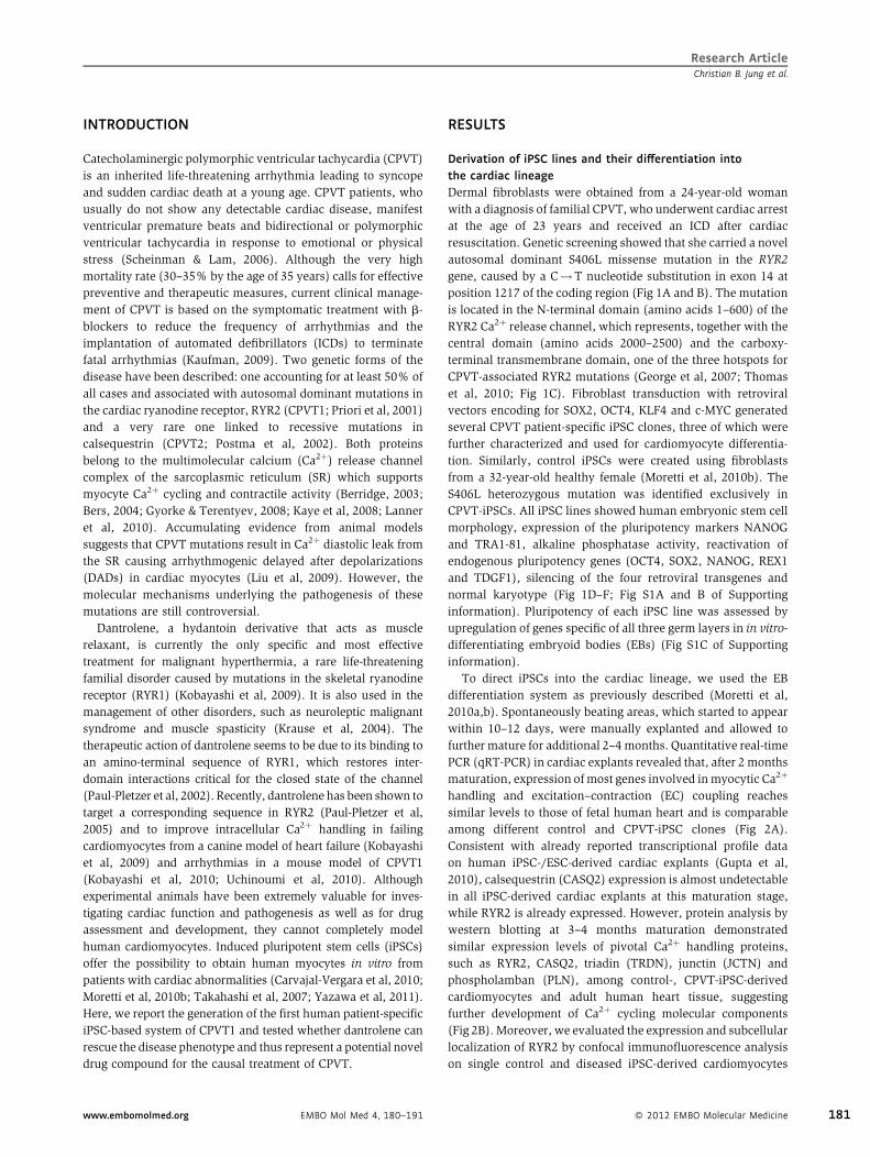
INTRODUCTION
Catecholaminergic polymorphic ventricular tachycardia (CPVT)
is an inherited life-threatening arrhythmia leading to syncope
and sudden cardiac death at a young age. CPVT patients, who
usually do not show any detectable cardiac disease, manifest
ventricular premature beats and bidirectional or polymorphic
ventricular tachycardia in response to emotional or physical
stress (Scheinman & Lam, 2006). Although the very high
mortality rate (30–35% by the age of 35 years) calls for effective
preventive and therapeutic measures, current clinical manage-
ment of CPVT is based on the symptomatic treatment with b-
blockers to reduce the frequency of arrhythmias and the
implantation of automated defibrillators (ICDs) to terminate
fatal arrhythmias (Kaufman, 2009). Two genetic forms of the
disease have been described: one accounting for at least 50% of
all cases and associated with autosomal dominant mutations in
the cardiac ryanodine receptor, RYR2 (CPVT1; Priori et al, 2001)
and a very rare one linked to recessive mutations in
calsequestrin (CPVT2; Postma et al, 2002). Both proteins
belong to the multimolecular calcium (Ca2þ) release channel
complex of the sarcoplasmic reticulum (SR) which supports
myocyte Ca2þ cycling and contractile activity (Berridge, 2003;
Bers, 2004; Gyorke & Terentyev, 2008; Kaye et al, 2008; Lanner
et al, 2010). Accumulating evidence from animal models
suggests that CPVT mutations result in Ca2þ diastolic leak from
the SR causing arrhythmogenic delayed after depolarizations
(DADs) in cardiac myocytes (Liu et al, 2009). However, the
molecular mechanisms underlying the pathogenesis of these
mutations are still controversial.
Dantrolene, a hydantoin derivative that acts as muscle
relaxant, is currently the only specific and most effective
treatment for malignant hyperthermia, a rare life-threatening
familial disorder caused by mutations in the skeletal ryanodine
receptor (RYR1) (Kobayashi et al, 2009). It is also used in the
management of other disorders, such as neuroleptic malignant
syndrome and muscle spasticity (Krause et al, 2004). The
therapeutic action of dantrolene seems to be due to its binding to
an amino-terminal sequence of RYR1, which restores inter-
domain interactions critical for the closed state of the channel
(Paul-Pletzer et al, 2002). Recently, dantrolene has been shown to
target a corresponding sequence in RYR2 (Paul-Pletzer et al,
2005) and to improve intracellular Ca2þ handling in failing
cardiomyocytes from a canine model of heart failure (Kobayashi
et al, 2009) and arrhythmias in a mouse model of CPVT1
(Kobayashi et al, 2010; Uchinoumi et al, 2010). Although
experimental animals have been extremely valuable for inves-
tigating cardiac function and pathogenesis as well as for drug
assessment and development, they cannot completely model
human cardiomyocytes. Induced pluripotent stem cells (iPSCs)
offer the possibility to obtain human myocytes in vitro from
patients with cardiac abnormalities (Carvajal-Vergara et al, 2010;
Moretti et al, 2010b; Takahashi et al, 2007; Yazawa et al, 2011).
Here, we report the generation of the first human patient-specific
iPSC-based system of CPVT1 and tested whether dantrolene can
rescue the disease phenotype and thus represent a potential novel
drug compound for the causal treatment of CPVT.
RESULTS
Derivation of iPSC lines and their differentiation into
the cardiac lineage
Dermal fibroblasts were obtained from a 24-year-old woman
with a diagnosis of familial CPVT, who underwent cardiac arrest
at the age of 23 years and received an ICD after cardiac
resuscitation. Genetic screening showed that she carried a novel
autosomal dominant S406L missense mutation in the RYR2
gene, caused by a C!T nucleotide substitution in exon 14 at
position 1217 of the coding region (Fig 1A and B). The mutation
is located in the N-terminal domain (amino acids 1–600) of the
RYR2 Ca2þ release channel, which represents, together with the
central domain (amino acids 2000–2500) and the carboxy-
terminal transmembrane domain, one of the three hotspots for
CPVT-associated RYR2 mutations (George et al, 2007; Thomas
et al, 2010; Fig 1C). Fibroblast transduction with retroviral
vectors encoding for SOX2, OCT4, KLF4 and c-MYC generated
several CPVT patient-specific iPSC clones, three of which were
further characterized and used for cardiomyocyte differentia-
tion. Similarly, control iPSCs were created using fibroblasts
from a 32-year-old healthy female (Moretti et al, 2010b). The
S406L heterozygous mutation was identified exclusively in
CPVT-iPSCs. All iPSC lines showed human embryonic stem cell
morphology, expression of the pluripotency markers NANOG
and TRA1-81, alkaline phosphatase activity, reactivation of
endogenous pluripotency genes (OCT4, SOX2, NANOG, REX1
and TDGF1), silencing of the four retroviral transgenes and
normal karyotype (Fig 1D–F; Fig S1A and B of Supporting
information). Pluripotency of each iPSC line was assessed by
upregulation of genes specific of all three germ layers in in vitro-
differentiating embryoid bodies (EBs) (Fig S1C of Supporting
information).
To direct iPSCs into the cardiac lineage, we used the EB
differentiation system as previously described (Moretti et al,
2010a,b). Spontaneously beating areas, which started to appear
within 10–12 days, were manually explanted and allowed to
further mature for additional 2–4 months. Quantitative real-time
PCR (qRT-PCR) in cardiac explants revealed that, after 2 months
maturation, expression of most genes involved in myocytic Ca2þ
handling and excitation–contraction (EC) coupling reaches
similar levels to those of fetal human heart and is comparable
among different control and CPVT-iPSC clones (Fig 2A).
Consistent with already reported transcriptional profile data
on human iPSC-/ESC-derived cardiac explants (Gupta et al,
2010), calsequestrin (CASQ2) expression is almost undetectable
in all iPSC-derived cardiac explants at this maturation stage,
while RYR2 is already expressed. However, protein analysis by
western blotting at 3–4 months maturation demonstrated
similar expression levels of pivotal Ca2þ handling proteins,
such as RYR2, CASQ2, triadin (TRDN), junctin (JCTN) and
phospholamban (PLN), among control-, CPVT-iPSC-derived
cardiomyocytes and adult human heart tissue, suggesting
further development of Ca2þ cycling molecular components
(Fig 2B). Moreover, we evaluated the expression and subcellular
localization of RYR2 by confocal immunofluorescence analysis
on single control and diseased iPSC-derived cardiomyocytes
Research ArticleChristian B. Jung et al.
www.embomolmed.org EMBO Mol Med 4, 180–191 � 2012 EMBO Molecular Medicine 181
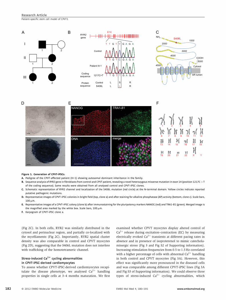
(Fig 2C). In both cells, RYR2 was similarly distributed in the
cytosol and perinuclear region, and partially co-localized with
the myofilaments (Fig 2C). Importantly, RYR2 spatial cluster
density was also comparable in control and CPVT myocytes
(Fig 2D), suggesting that the S406L mutation does not interfere
with trafficking of the homotetrameric channel.
Stress-induced Ca2R cycling abnormalities
in CPVT-iPSC-derived cardiomyocytes
To assess whether CPVT-iPSC-derived cardiomyocytes recapi-
tulate the disease phenotype, we analysed Ca2þ handling
properties in single cells at 3–4 months maturation. We first
examined whether CPVT myocytes display altered control of
Ca2þ release during excitation–contraction (EC) by measuring
electrically evoked Ca2þ transients at different pacing rates in
absence and in presence of isoproterenol to mimic catechola-
minergic stress (Fig 3 and Fig S2 of Supporting information).
Increasing stimulation frequencies from 0.5 to 1.5 Hz correlated
with a higher percentage of cells with abnormal Ca2þ handling
in both control and CPVT myocytes (Fig 3A). However, this
effect was significantly more pronounced in the diseased cells
and was comparable among different CPVT-iPSC lines (Fig 3A
and Fig S3 of Supporting information). We could observe three
types of stress-induced Ca2þ cycling abnormalities, which
Research ArticlePatient-specific stem cell model of CPVT1
Figure 1. Generation of CPVT-iPSCs.
A. Pedigree of the CPVT-affected patient (III-1) showing autosomal dominant inheritance in the family.
B. Sequence analysis of RYR2 gene in fibroblasts from control and CPVT patient, revealing a novel heterozygous missense mutation in exon 14 (position 1217C > T
of the coding sequence). Same results were obtained from all analysed control and CPVT-iPSC clones.
C. Schematic representation of RYR2 channel and localization of the S406L mutation (red circle) at the N-terminal domain. Yellow circles indicate reported
putative pathogenic mutations.
D. Representative images of CPVT-iPSC colonies in bright field (top, clone a) and after staining for alkaline phosphatase (AP) activity (bottom, clone c). Scale bars,
100 mm.
E. Representative images of a CPVT-iPSC colony (clone b) after immunostaining for the pluripotency markers NANOG (red) and TRA1-81 (green). Merged image is
the magnified area marked by the white box. Scale bars, 100mm.
F. Karyogram of CPVT-iPSC clone a.
182 � 2012 EMBO Molecular Medicine EMBO Mol Med 4, 180–191 www.embomolmed.org
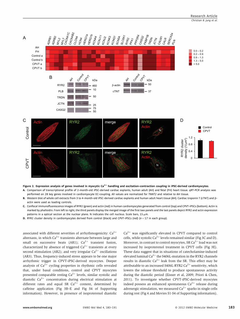
associated with different severities of arrhythmogenicity: Ca2þ
alternans, in which Ca2þ transients alternate between large and
small on successive beats (AR1); Ca2þ transient fusion,
characterized by absence of triggered Ca2þ transients at every
second stimulation (AR2); and very irregular Ca2þ oscillations
(AR3). Thus, frequency-induced stress appears to be one major
arrhythmic trigger in CPVT-iPSC-derived myocytes. Deeper
analysis of Ca2þ cycling properties in rhythmic cells revealed
that, under basal conditions, control and CPVT myocytes
presented comparable resting Ca2þ levels, similar systolic and
diastolic Ca2þ concentration during electrical stimulation at
different rates and equal SR Ca2þ content, determined by
caffeine application (Fig 3B–E and Fig S4 of Supporting
information). However, in presence of isoproterenol diastolic
Ca2þ was significantly elevated in CPVT compared to control
cells, while systolic Ca2þ levels remained similar (Fig 3C and D).
Moreover, in contrast to control myocytes, SR Ca2þ load was not
increased by isoproterenol treatment in CPVT cells (Fig 3E).
These data suggest that in situations of catecholamine-induced
elevated luminal Ca2þ the S406L-mutation in the RYR2 channels
results in diastolic Ca2þ leak from the SR. This effect may be
attributable to an increased S406L-RYR2 Ca2þ sensitivity, which
lowers the release threshold to produce spontaneous activity
during the diastolic period (Eisner et al, 2009; Priori & Chen,
2011). To investigate whether CPVT-iPSC-derived myocytes
indeed possess an enhanced spontaneous Ca2þ release during
adrenergic stimulation, we measured Ca2þ sparks in single cells
during rest (Fig 4 and Movies S1-S4 of Supporting information).
Research ArticleChristian B. Jung et al.
Figure 2. Expression analysis of genes involved in myocytic Ca2R handling and excitation–contraction coupling in iPSC-derived cardiomyocytes.
A. Comparison of transcriptional profile of 2-month-old iPSC-derived cardiac explants, human adult (AH) and fetal (FH) heart tissue. qRT-PCR analysis was
performed on 28 key genes involved in cardiomyocyte EC-coupling. All values are normalized for TNNT2 and relative to AH tissue.
B. Western blot of whole cell extracts from 3 to 4-month-old iPSC-derived cardiac explants and human adult heart tissue (AH). Cardiac troponin T (cTNT) and b-
actin were used as loading controls.
C. Confocal immunofluorescence images of RYR2 (green) and actin (red) in human cardiomyocytes generated from control (top) and CPVT-iPSCs (bottom). Actin is
marked by phalloidin. From left to right, the third panels display the merged image of the first two panels and the last panels depict RYR2 and actin expression
patterns in a optical section at the nuclear plane. N indicates the cell nucleus. Scale bars, 15 mm.
D. RYR2 cluster density in cardiomyocytes derived from control (black) and CPVT-iPSCs (red) (n¼ 17 in each group).
www.embomolmed.org EMBO Mol Med 4, 180–191 � 2012 EMBO Molecular Medicine 183
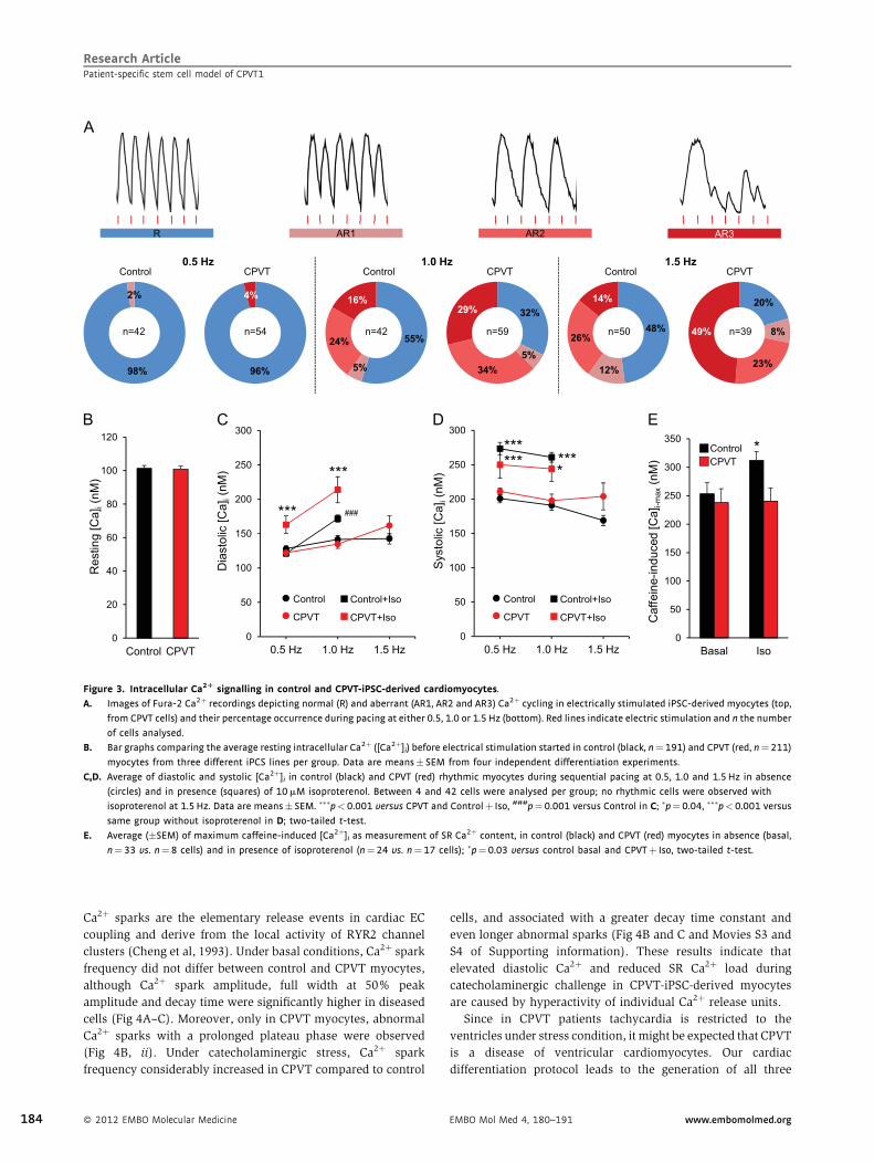
Ca2þ sparks are the elementary release events in cardiac EC
coupling and derive from the local activity of RYR2 channel
clusters (Cheng et al, 1993). Under basal conditions, Ca2þ spark
frequency did not differ between control and CPVT myocytes,
although Ca2þ spark amplitude, full width at 50% peak
amplitude and decay time were significantly higher in diseased
cells (Fig 4A–C). Moreover, only in CPVT myocytes, abnormal
Ca2þ sparks with a prolonged plateau phase were observed
(Fig 4B, ii). Under catecholaminergic stress, Ca2þ spark
frequency considerably increased in CPVT compared to control
cells, and associated with a greater decay time constant and
even longer abnormal sparks (Fig 4B and C and Movies S3 and
S4 of Supporting information). These results indicate that
elevated diastolic Ca2þ and reduced SR Ca2þ load during
catecholaminergic challenge in CPVT-iPSC-derived myocytes
are caused by hyperactivity of individual Ca2þ release units.
Since in CPVT patients tachycardia is restricted to the
ventricles under stress condition, it might be expected that CPVT
is a disease of ventricular cardiomyocytes. Our cardiac
differentiation protocol leads to the generation of all three
Research ArticlePatient-specific stem cell model of CPVT1
Figure 3. Intracellular Ca2R signalling in control and CPVT-iPSC-derived cardiomyocytes.
A. Images of Fura-2 Ca2þ recordings depicting normal (R) and aberrant (AR1, AR2 and AR3) Ca2þ cycling in electrically stimulated iPSC-derived myocytes (top,
from CPVT cells) and their percentage occurrence during pacing at either 0.5, 1.0 or 1.5 Hz (bottom). Red lines indicate electric stimulation and n the number
of cells analysed.
B. Bar graphs comparing the average resting intracellular Ca2þ ([Ca2þ]i) before electrical stimulation started in control (black, n¼ 191) and CPVT (red, n¼211)
myocytes from three different iPCS lines per group. Data are means � SEM from four independent differentiation experiments.
C,D. Average of diastolic and systolic [Ca2þ]i in control (black) and CPVT (red) rhythmic myocytes during sequential pacing at 0.5, 1.0 and 1.5 Hz in absence
(circles) and in presence (squares) of 10 mM isoproterenol. Between 4 and 42 cells were analysed per group; no rhythmic cells were observed with
isoproterenol at 1.5 Hz. Data are means � SEM. ���p<0.001 versus CPVT and Controlþ Iso, ###p¼0.001 versus Control in C; �p¼ 0.04, ���p<0.001 versus
same group without isoproterenol in D; two-tailed t-test.
E. Average (�SEM) of maximum caffeine-induced [Ca2þ]i as measurement of SR Ca2þ content, in control (black) and CPVT (red) myocytes in absence (basal,
n¼ 33 vs. n¼8 cells) and in presence of isoproterenol (n¼24 vs. n¼17 cells); �p¼0.03 versus control basal and CPVTþ Iso, two-tailed t-test.
184 � 2012 EMBO Molecular Medicine EMBO Mol Med 4, 180–191 www.embomolmed.org
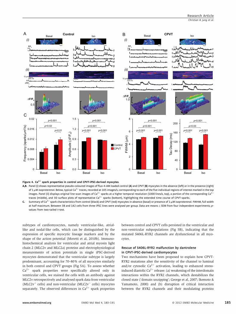
subtypes of cardiomyocytes, namely ventricular-like, atrial-
like and nodal-like cells, which can be distinguished by the
expression of specific myocytic lineage markers and by the
shape of the action potential (Moretti et al, 2010b). Immuno-
histochemical analysis for ventricular and atrial myosin light
chain 2 (MLC2v and MLC2a) proteins and electrophysiological
measurements of action potentials in single iPSC-derived
myocytes demonstrated that the ventricular subtype is largely
predominant, accounting for 70–80% of all myocytes similarly
in both control and CPVT groups (Fig 5A). To assess whether
Ca2þ spark properties were specifically altered only in
ventricular cells, we stained the cells with an antibody against
MLC2v retrospectively and analysed spark data from ventricular
(MLC2vþ cells) and non-ventricular (MLC2v� cells) myocytes
separately. The observed differences in Ca2þ spark properties
between control and CPVT cells persisted in the ventricular and
non-ventricular subpopulations (Fig 5B), indicating that the
mutated S406L-RYR2 channels are dysfunctional in all myo-
cytes.
Rescue of S406L-RYR2 malfunction by dantrolene
in CPVT-iPSC-derived cardiomyocytes
Two mechanisms have been proposed to explain how CPVT-
RYR2 mutations alter the sensitivity of the channel to luminal
and/or cytosolic Ca2þ activation, leading to enhanced stress-
induced diastolic Ca2þ release: (a) weakening of the interdomain
interactions within the RYR2 channels, which destabilizes the
closed state (‘domain unzipping’; George et al, 2007; Ikemoto &
Yamamoto, 2000) and (b) disruption of critical interaction
between the RYR2 channels and their modulating proteins
Research ArticleChristian B. Jung et al.
Figure 4. Ca2R spark properties in control and CPVT-iPSC-derived myocytes.
A,B. Panel (i) shows representative pseudo-coloured images of fluo-4-AM loaded control (A) and CPVT (B) myocytes in the absence (left) or in the presence (right)
of 1mM isoproterenol. Below, typical Ca2þ traces, recorded at 105 images/s, corresponding to each of the five individual regions of interest marked in the top
images. Panel (ii) displays original line-scan images of Ca2þ sparks at a higher temporal resolution (1000 lines/s, top), a portion of the corresponding Ca2þ
traces (middle), and 3D surface plots of representative Ca2þ sparks (bottom), highlighting the extended time course of CPVT-sparks.
C. Summary of Ca2þ spark characteristics from control (black) and CPVT (red) myocytes in absence (basal) or presence of 1 mM isoproterenol. FWHM, full width
at half maximum. Between 38 and 142 cells from three iPSC lines were analysed per group. Data are means � SEM from four independent experiments; p-
values from two-tailed t-test.
www.embomolmed.org EMBO Mol Med 4, 180–191 � 2012 EMBO Molecular Medicine 185
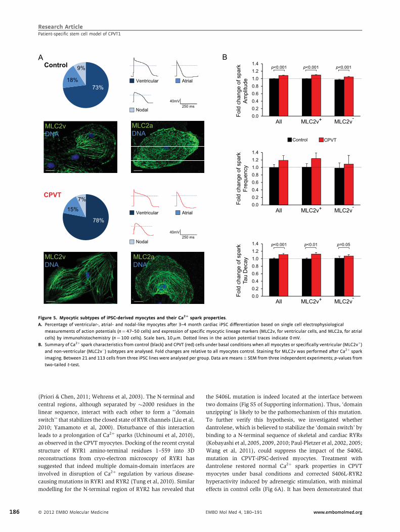
(Priori & Chen, 2011; Wehrens et al, 2003). The N-terminal and
central regions, although separated by �2000 residues in the
linear sequence, interact with each other to form a ‘‘domain
switch’’ that stabilizes the closed state of RYR channels (Liu et al,
2010; Yamamoto et al, 2000). Disturbance of this interaction
leads to a prolongation of Ca2þ sparks (Uchinoumi et al, 2010),
as observed in the CPVT myocytes. Docking of the recent crystal
structure of RYR1 amino-terminal residues 1–559 into 3D
reconstructions from cryo-electron microscopy of RYR1 has
suggested that indeed multiple domain-domain interfaces are
involved in disruption of Ca2þ regulation by various disease-
causing mutations in RYR1 and RYR2 (Tung et al, 2010). Similar
modelling for the N-terminal region of RYR2 has revealed that
the S406L mutation is indeed located at the interface between
two domains (Fig S5 of Supporting information). Thus, ‘domain
unzipping’ is likely to be the pathomechanism of this mutation.
To further verify this hypothesis, we investigated whether
dantrolene, which is believed to stabilize the ‘domain switch’ by
binding to a N-terminal sequence of skeletal and cardiac RYRs
(Kobayashi et al, 2005, 2009, 2010; Paul-Pletzer et al, 2002, 2005;
Wang et al, 2011), could suppress the impact of the S406L
mutation in CPVT-iPSC-derived myocytes. Treatment with
dantrolene restored normal Ca2þ spark properties in CPVT
myocytes under basal conditions and corrected S406L-RYR2
hyperactivity induced by adrenergic stimulation, with minimal
effects in control cells (Fig 6A). It has been demonstrated that
Research ArticlePatient-specific stem cell model of CPVT1
Figure 5. Myocytic subtypes of iPSC-derived myocytes and their Ca2R spark properties.
A. Percentage of ventricular-, atrial- and nodal-like myocytes after 3–4 month cardiac iPSC differentiation based on single cell electrophysiological
measurements of action potentials (n¼ 47–50 cells) and expression of specific myocytic lineage markers (MLC2v, for ventricular cells, and MLC2a, for atrial
cells) by immunohistochemistry (n¼100 cells). Scale bars, 10mm. Dotted lines in the action potential traces indicate 0 mV.
B. Summary of Ca2þ spark characteristics from control (black) and CPVT (red) cells under basal conditions when all myocytes or specifically ventricular (MLC2vþ)
and non-ventricular (MLC2v�) subtypes are analysed. Fold changes are relative to all myocytes control. Staining for MLC2v was performed after Ca2þ spark
imaging. Between 21 and 113 cells from three iPSC lines were analysed per group. Data are means � SEM from three independent experiments; p-values from
two-tailed t-test.
186 � 2012 EMBO Molecular Medicine EMBO Mol Med 4, 180–191 www.embomolmed.org
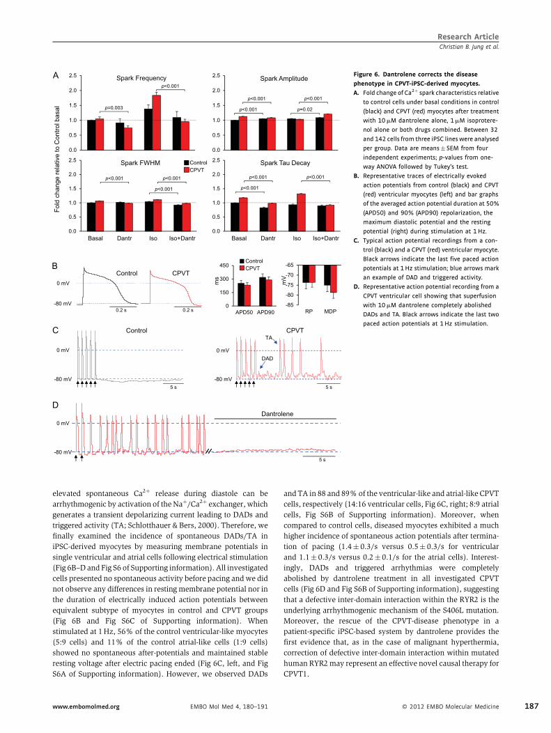
elevated spontaneous Ca2þ release during diastole can be
arrhythmogenic by activation of the Naþ/Ca2þ exchanger, which
generates a transient depolarizing current leading to DADs and
triggered activity (TA; Schlotthauer & Bers, 2000). Therefore, we
finally examined the incidence of spontaneous DADs/TA in
iPSC-derived myocytes by measuring membrane potentials in
single ventricular and atrial cells following electrical stimulation
(Fig 6B–D and Fig S6 of Supporting information). All investigated
cells presented no spontaneous activity before pacing and we did
not observe any differences in resting membrane potential nor in
the duration of electrically induced action potentials between
equivalent subtype of myocytes in control and CPVT groups
(Fig 6B and Fig S6C of Supporting information). When
stimulated at 1 Hz, 56% of the control ventricular-like myocytes
(5:9 cells) and 11% of the control atrial-like cells (1:9 cells)
showed no spontaneous after-potentials and maintained stable
resting voltage after electric pacing ended (Fig 6C, left, and Fig
S6A of Supporting information). However, we observed DADs
and TA in 88 and 89% of the ventricular-like and atrial-like CPVT
cells, respectively (14:16 ventricular cells, Fig 6C, right; 8:9 atrial
cells, Fig S6B of Supporting information). Moreover, when
compared to control cells, diseased myocytes exhibited a much
higher incidence of spontaneous action potentials after termina-
tion of pacing (1.4� 0.3/s versus 0.5� 0.3/s for ventricular
and 1.1� 0.3/s versus 0.2� 0.1/s for the atrial cells). Interest-
ingly, DADs and triggered arrhythmias were completely
abolished by dantrolene treatment in all investigated CPVT
cells (Fig 6D and Fig S6B of Supporting information), suggesting
that a defective inter-domain interaction within the RYR2 is the
underlying arrhythmogenic mechanism of the S406L mutation.
Moreover, the rescue of the CPVT-disease phenotype in a
patient-specific iPSC-based system by dantrolene provides the
first evidence that, as in the case of malignant hyperthermia,
correction of defective inter-domain interaction within mutated
human RYR2 may represent an effective novel causal therapy for
CPVT1.
Research ArticleChristian B. Jung et al.
Figure 6. Dantrolene corrects the disease
phenotype in CPVT-iPSC-derived myocytes.
A. Fold change of Ca2þ spark characteristics relative
to control cells under basal conditions in control
(black) and CPVT (red) myocytes after treatment
with 10 mM dantrolene alone, 1mM isoprotere-
nol alone or both drugs combined. Between 32
and 142 cells from three iPSC lines were analysed
per group. Data are means� SEM from four
independent experiments; p-values from one-
way ANOVA followed by Tukey’s test.
B. Representative traces of electrically evoked
action potentials from control (black) and CPVT
(red) ventricular myocytes (left) and bar graphs
of the averaged action potential duration at 50%
(APD50) and 90% (APD90) repolarization, the
maximum diastolic potential and the resting
potential (right) during stimulation at 1 Hz.
C. Typical action potential recordings from a con-
trol (black) and a CPVT (red) ventricular myocyte.
Black arrows indicate the last five paced action
potentials at 1 Hz stimulation; blue arrows mark
an example of DAD and triggered activity.
D. Representative action potential recording from a
CPVT ventricular cell showing that superfusion
with 10 mM dantrolene completely abolished
DADs and TA. Black arrows indicate the last two
paced action potentials at 1 Hz stimulation.
www.embomolmed.org EMBO Mol Med 4, 180–191 � 2012 EMBO Molecular Medicine 187
DISCUSSION
We have developed the first human stem cell-based model for
CPVT1, bearing a novel S406L missense mutation in RYR2, and
demonstrated its suitability to recapitulate molecular and
physiological aspects of the disease phenotype. Until now,
only heterologous expression systems and genetic mouse
models have been used to study the cellular and molecular
aspects of CPVT-linked RYR2 mutations. Based on these studies,
two mechanisms for RYR2-mediated CPVT have been proposed.
The first mechanism suggests that RYR2 mutants reduce the
binding affinity of the channel for its auxiliary stabilizing protein
FKBP12.6 and this is further aggravated in situation of
catecholamine-induced hyperphosphorylation of RYR2 with
consequential dissociation of FKBP12.6 and Ca2þ leakage from
the SR (Lehnart et al, 2008; Marx et al, 2000; Wehrens et al,
2003). Although it may be possible that selected mutations alter
FKBP12.6 binding to RYR2, this hypothesis has been recently
challenged and increasing body of evidence clearly demon-
strates that alterations in FKBP12.6-RYR2 interaction are
unlikely to be the common cause of CPVT1 (George et al,
2003; Guo et al, 2010; Jiang et al, 2005; Liu et al, 2006; Xiao et al,
2007). Alternatively, it has been proposed that RYR2 mutations
in the N-terminal and central regions of the protein weaken
interactions between these two domains that are critical in
stabilizing the closed state of the channel, resulting in an
increased open probability and enhanced spontaneous Ca2þ
release during stress-induced SR Ca2þ overload (Ikemoto &
Yamamoto, 2000; Tateishi et al, 2009). In support of this
‘domain unzipping’ mechanism, dantrolene has been shown to
suppress abnormal Ca2þ leak from mutated RYR1 and RYR2 by
binding to a N-terminal sequence and stabilizing domain–
domain contacts within the N-terminal and central regulatory
regions (Kobayashi et al, 2005, 2009; Paul-Pletzer et al, 2002,
2005). Interestingly, dantrolene binding to RYR2 seems to be
dependent on a particular conformational state of the channel
that takes place only in disease conditions (Kobayashi et al,
2009; Paul-Pletzer et al, 2005). Therefore, the results that this
drug rescues the disease phenotype in our patient-specific iPSC-
based CPVT model would indicate that ‘domain unzipping’ is
likely to be the pathomechanism of the novel N-terminal S406L-
RYR2 mutation. Evidence for unzipping of the interaction
between N-terminal and central domains raises the question of
whether ‘domain unzipping’ is present in other regions of RYR2
in which CPVT mutations are located. Thus, it would be of great
interest to investigate dantrolene effects in other patient-specific
iPSC-CPVT models bearing different RYR2 mutations situated
throughout the molecule. Our study on a human model of CPVT
provides valuable insights into the pathophysiology of the
disease and suggests dantrolene as a potential novel drug for the
causal treatment of cardiac arrhythmias in CPVT1 patients
carrying N-terminal mutations. Yet, clinical trials demonstrated
that current treatments with b-blockers and ICDs are not fully
protective in all CPVT patients and showed that these regimens
are less attractive in CPVT compared to other forms of inherited
ventricular tachycardia, such as long QT or Brugada syndrome
(Kaufman, 2009). Although the precise mechanistic basis of
CPVT most likely depends on the hotspot in which mutations
are residing and hence might call for the development of
location-specific drugs to address the functional heterogeneity,
our work highlights the potential of human iPSCs in the
emerging field of personalized medicine (Zhu et al, 2011) by
demonstrating the ability to screen the effects of potential
disease aggravators and novel customized treatment options.
MATERIALS AND METHODS
Human iPSC generation and cardiomyocyte differentiation
After approval by the institutional review board, we recruited a 24-year-
old caucasian female CPVT patient and a 32-year-old female caucasian
control without history of cardiac disease scheduled for plastic surgery
to undergo dermal biopsy after obtaining written informed consent.
Reprogramming of primary skin fibroblasts and differentiation into
cardiomyocytes were performed as described previously (Moretti et al,
2010a,b). Briefly, fibroblasts were infected with retroviruses encoding
OCT4, SOX2, KLF4 and c-MYC and cultured on murine embryonic feeder
cells until iPSC colonies could be picked. EB differentiation was achieved
by aggregating the cells on low-attachment plates and EBs were plated
on gelatin-coated dishes at day 7. Spontaneously contracting areas
were manually dissected and cultured further until day 90–130 of
differentiation. Cells for physiological experiments were collagenase-
dissociated into single cells, plated on fibronectin-coated glass cover-
slips, and analysed within 3–6 days.
Genomic sequencing and karyotype analysis
The presence of the RYR2-S406L mutation in the patient and its
absence in the control and in blood samples of 100 coronary artery
disease patients without CPVT was verified by polymerase chain
reaction-based sequencing of genomic DNA isolated from skin
fibroblasts, from iPSCs and from blood using a Genomic DNA
Purification Kit (Gentra Systems). Karyotyping of the iPSC lines was
performed at the Institute of Human Genetics of the Technical
University Munich using standard methodology.
Quantitative real-time PCR
Total mRNA was isolated from fibroblasts, iPSC clones, EBs, and
myocytic explants using the Stratagene Absolutely RNA kit. One
microgram of total RNA was used to synthesize cDNA from fibroblasts,
iPSC clones and EBs, using the High-Capacity cDNA Reverse
Transcription kit (Applied Biosystems). RNA from cardiomyocyte
explants and from human adult and fetal heart (Clontech) was
linearly amplified using the RNA Amplification RampUP Kit (Geni-
sphere) and subsequently 1mg of amplified RNA was used to
synthetize cDNA. Gene expression was quantified by qRT-PCR using
1ml of the RT reaction and the Power SYBR Green PCR Master Mix
(Applied Biosystems). Gene expression levels were normalized to
GAPDH or to TNNT2, as indicated in the dedicated figure legends.
Primer sequences are provided in Table S1 of Supporting information.
Phenotypic characterization of iPSC lines and their
differentiated progeny
Differential gene expression was assessed by qRT-PCR reaction as
described previously (Moretti et al, 2010a,b). For histochemistry, cells
Research ArticlePatient-specific stem cell model of CPVT1
188 � 2012 EMBO Molecular Medicine EMBO Mol Med 4, 180–191 www.embomolmed.org
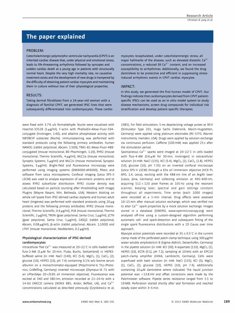
were fixed with 3.7% v/v formaldehyde. Nuclei were visualized with
Hoechst-33528 (1mg/ml), F-actin with Phalloidin-Alexa-Fluor-594-
conjugate (Invitrogen, 1:40), and alkaline phosphatase activity with
NBT/BCIP substrate (Roche). Immunostaining was performed with
standard protocols using the following primary antibodies: human
NANOG (rabbit polyclonal, Abcam, 1:500), TRA1-81-Alexa-Fluor-488-
conjugated (mouse monoclonal, BD Pharmingen, 1:20), RYR2 (mouse
monoclonal, Thermo Scientific, 4mg/ml), MLC2a (mouse monoclonal,
Synaptic Systems, 5mg/ml) and MLC2v (mouse monoclonal, Synaptic
Systems, 5mg/ml). Bright-field and fluorescence microscopy were
performed using imaging systems (DMI6000-AF6000), filters and
software from Leica microsystems. Confocal imaging (Leica SP5-II
LSCM) was used to analyse expression of sarcomeric proteins and to
assess RYR2 subcellular distribution. RYR2 cluster density was
calculated based on particle counting after thresholding with ImageJ
PlugIns (Wayne Rasant, NIH, Bethesda, USA). Western blotting on
whole cell lysate from iPSC-derived cardiac explants and human adult
heart (Imgenex) was performed with standard protocols using 20mg
proteins and the following primary antibodies: RYR2 (mouse mono-
clonal, Thermo Scientific, 0.4mg/ml), PLB (mouse monoclonal, Thermo
Scientific, 2mg/ml), TRDN (goat polyclonal, Santa Cruz, 1mg/ml), JCTN
(goat polyclonal, Santa Cruz, 1mg/ml), CASQ2 (rabbit polyclonal,
Abcam, 0.08mg/ml), b-actin (rabbit polyclonal, Abcam, 1:1000) and
cTNT (mouse monoclonal, NeoMarkers, 0.2mg/ml).
Physiological characterization of iPSC-derived
cardiomyocytes
Intracellular free Ca2þ was measured at 20–228C in cells loaded with
fura-2-AM (5mM for 20min; Fluka, Buchs, Switzerland) in HEPES-
buffered saline (in mM: NaCl (140), KCl (5.4), MgCl2 (1), CaCl2 (2),
glucose (10), HEPES (10), pH 7.4) containing 0.1% w/v bovine serum
albumin on a monochromator-equipped (Polychrome-V, TILL-Photo-
nics, Grafelfing, Germany) inverted microscope (Olympus-IX 71 with
an UPlanSApo 20�/0.85 oil immersion objective). Fluorescence was
excited at 340 and 380nm, emission recorded at 22–24Hz with a
14-bit EMCCD camera (iXON3 885, Andor, Belfast, UK), and Ca2þ
concentrations calculated as described previously (Grynkiewicz et al,
1985). For field stimulation, 5ms depolarizing voltage pulses at 90 V
(Stimulator Type 201, Hugo Sachs Elektronik, March-Hugstetten,
Germany) were applied using platinum electrodes (RC-37FS, Warner
Instruments, Hamden, USA). Drugs were applied by solution exchange
via continuous perfusion. Caffeine (100mM) was applied 25 s after
the stimulation period.
Spontaneous Ca2þ sparks were imaged at 20–228C in cells loaded
with fluo-4-AM (0.6mM for 30min, Invitrogen) in extracellular
solution (inmM: NaCl (135), KCl (5.4), MgCl2 (2), CaCl2 (1.8), HEPES
(10), glucose (10), pH 7.35) on an inverted confocal microscope
(Leica SP5-II LSCM) through a 63x oil immersion objective (HCX PL
APO, 1.4, Leica), exciting with the 488 nm line of an Arg/Kr laser
(Lasos, Jena, Germany) and collecting emission at 495–600 nm,
acquiring 512�120 pixel frames at 105Hz using the resonant
scanner, keeping laser, spectral and gain settings constant
throughout all experiments. Time series (each 1000 images)
were recorded at a 1-min interval. Drug effects were assessed
10–15min after manual solution exchange, which was verified not
to alter Ca2þ spark properties by a mock solution exchange. Images
stored in a database (OMERO, www.openmicroscopy.org) were
analysed off-line using a custom-designed algorithm performing
automatic cell- and spark-detection and subsequent fitting of the
single spark fluorescence distributions with a 2D Gauss over time
approach.
Myocyte action potentials were recorded at 35�0.58C in the current
clamp mode of the perforated patch-clamp technique using 300mg/ml
water-soluble amphotericin B (Sigma–Aldrich, Deisenhofen, Germany)
in the pipette solution (in mM: KCl (30), K-aspartate (110), MgCl2 (1),
HEPES (10), EGTA (0.1), pH 7.2), sampling at 10 kHz with an EPC10
patch-clamp amplifier (HEKA, Lambrecht, Germany). Cells were
superfused with bath solution (in mM: NaCl (135), KCl (5), MgCl2
(1), CaCl2 (2), glucose (10), HEPES (10), pH 7.4), additionally
containing 10mM dantrolene where indicated. The liquid junction
potential was þ13.8mV and offset corrections were made by the
Patchmaster software. Pipette series resistance ranged from 5.5 to
19MV. Perforation started shortly after seal formation and reached
steady state within 3–5min.
Research ArticleChristian B. Jung et al.
The paper explained
PROBLEM:
Catecholaminergic polymorphic ventricular tachycardia (CPVT) is an
inherited cardiac disease that, under physical and emotional stress,
leads to life-threatening arrhythmia followed by syncopes and
sudden cardiac death at a young age in patients with structurally
normal heart. Despite the very high mortality rate, no causative
treatment exists and the development of new drugs is hampered by
the difficulty of obtaining patient cardiac myocytes and maintaining
them in culture without loss of their physiological properties.
RESULTS:
Taking dermal fibroblasts from a 24-year-old woman with a
diagnosis of familial CPVT, we generated iPSC lines that were
subsequently differentiated into cardiomyocytes. These cardio-
myocytes recapitulated, under catecholaminergic stress, all
major hallmarks of the disease, such as elevated diastolic Ca2þ
concentrations, a reduced SR Ca2þ content, and an increased
susceptibility to arrhythmias. Additionally, we found the drug
dantrolene to be protective and efficient in suppressing stress-
induced arrhythmic events in CPVT cardiac myocytes.
IMPACT:
In this study, we generated the first human model of CPVT. Our
findings indicate that cardiomyocytes derived from CPVT patient-
specific iPSCs can be used as an in vitro model system to study
disease mechanisms, screen drug compounds for individual risk
stratification and develop patient-specific therapies.
www.embomolmed.org EMBO Mol Med 4, 180–191 � 2012 EMBO Molecular Medicine 189
All experiments and analysis were performed by investigators blinded
to the genotype of the cells.
Statistical analysis
Data that passed tests for normality and equal variance were analysed
with the use of Student’s t-test or one-way analysis of variance
followed by Tukey’s test, when appropriate. Two-sided p-values of
<0.05 were considered statistically significant. All data are shown as
means� SEM.
For more detailed Materials and Methods see the Supporting
information.
Author contributionsKLL, DS, TG and PL conceived the experiments; CBJ, AM, and MB
generated and characterized control and CPVT iPSCs; Generation
and characterization of human cardiomyocytes was performed
by CBJ, LI, MB, TD, AG and JTL; CBJ performed histochemistry/
immunostaining with LI and SR and western blotting with TD;
MMS, US and SP conducted Ca2þ imaging and electrophysiolo-
gical measurements and PL Ca2þ spark experiments; RJD and MS
recruited the CPVT patient family; DS performed docking
structure modelling; KLL, DS and AM wrote the manuscript.
AcknowledgementsWe thank T. Kitamura and S. Yamanaka for providing
viral vectors through Addgene, and especially the member
of the CPVT family and the healthy volunteers who provided
us with skin biopsies for the reprogramming. We would like
to acknowledge Diana Grewe and Christina Scherb for
their technical assistance in cell culture and Gabi Lederer
(Cytogenetic Department, TUM) for karyotyping. This work
was supported by grants from the European Research
Council (Marie Curie Excellence Team Grant MEXT-23208;
ERC 261053–CHD-iPS), the German Research Foundation
(Research Unit 923, Mo 2217/1-1 and La 1238 3-1/4-1) and
the German Ministry for Education and Research (01 GN 0826).
CBJ was supported by a scholarship (AFR-PHD-09-169) granted
by the National Research Fund, Luxembourg.
Supporting information is available at EMBO Molecular
Medicine online.
The authors declare that they have no conflict of interest.
ReferencesBerridge MJ (2003) Cardiac calcium signalling. Biochem Soc Trans 31: 930-
933
Bers DM (2004) Macromolecular complexes regulating cardiac ryanodine
receptor function. J Mol Cell Cardiol 37: 417-429
Carvajal-Vergara X, Sevilla A, D’Souza SL, Ang YS, Schaniel C, Lee DF, Yang L,
Kaplan AD, Adler ED, Rozov R, et al (2010) Patient-specific induced
pluripotent stem-cell-derived models of LEOPARD syndrome. Nature 465:
808-812
Cheng H, Lederer WJ, Cannell MB (1993) Calcium sparks: elementary events
underlying excitation–contraction coupling in heart muscle. Science 262:
740-744
Eisner DA, Kashimura T, Venetucci LA, Trafford AW (2009) From the ryanodine
receptor to cardiac arrhythmias. Circ J 73: 1561-1567
George CH, Higgs GV, Lai FA (2003) Ryanodine receptor mutations associated
with stress-induced ventricular tachycardia mediate increased calcium
release in stimulated cardiomyocytes. Circ Res 93: 531-540
George CH, Jundi H, Thomas NL, Fry DL, Lai FA (2007) Ryanodine receptors and
ventricular arrhythmias: emerging trends in mutations, mechanisms and
therapies. J Mol Cell Cardiol 42: 34-50
Grynkiewicz G, Poenie M, Tsien RY (1985) A new generation of Ca2þ
indicators with greatly improved fluorescence properties. J Biol Chem 260:
3440-3450
Guo T, Cornea RL, Huke S, Camors E, Yang Y, Picht E, Fruen BR, Bers DM (2010)
Kinetics of FKBP12.6 binding to ryanodine receptors in permeabilized
cardiac myocytes and effects on Ca sparks. Circ Res 106: 1743-1752
Gupta MK, Illich DJ, Gaarz A, Matzkies M, Nguemo F, Pfannkuche K,
Liang H, Classen S, Reppel M, Schultze JL, et al (2010) Global
transcriptional profiles of beating clusters derived from human induced
pluripotent stem cells and embryonic stem cells are highly similar. BMC Dev
Biol 10: 98
Gyorke S, Terentyev D (2008) Modulation of ryanodine receptor by luminal
calcium and accessory proteins in health and cardiac disease. Cardiovasc
Res 77: 245-255
Ikemoto N, Yamamoto T (2000) Postulated role of inter-domain interaction
within the ryanodine receptor in Ca(2þ) channel regulation. Trends
Cardiovasc Med 10: 310-316
Jiang D, Wang R, Xiao B, Kong H, Hunt DJ, Choi P, Zhang L, Chen SR (2005)
Enhanced store overload-induced Ca2þ release and channel sensitivity to
luminal Ca2þ activation are common defects of RyR2 mutations linked to
ventricular tachycardia and sudden death. Circ Res 97: 1173-1181
Kaufman ES (2009) Mechanisms and clinical management of inherited
channelopathies: long QT syndrome, Brugada syndrome, catecholaminergic
polymorphic ventricular tachycardia, and short QT syndrome. Heart
Rhythm 6: S51-S55
Kaye DM, Hoshijima M, Chien KR (2008) Reversing advanced heart failure by
targeting Ca2þ cycling. Annu Rev Med 59: 13-28
Kobayashi S, Bannister ML, Gangopadhyay JP, Hamada T, Parness J, Ikemoto N
(2005) Dantrolene stabilizes domain interactions within the ryanodine
receptor. J Biol Chem 280: 6580-6587
Kobayashi S, Yano M, Suetomi T, Ono M, Tateishi H, Mochizuki M, Xu X,
Uchinoumi H, Okuda S, Yamamoto T, et al (2009) Dantrolene, a therapeutic
agent for malignant hyperthermia, markedly improves the function of
failing cardiomyocytes by stabilizing interdomain interactions within the
ryanodine receptor. J Am Coll Cardiol 53: 1993-2005
Kobayashi S, Yano M, Uchinoumi H, Suetomi T, Susa T, Ono M, Xu X, Tateishi H,
Oda T, Okuda S, et al (2010) Dantrolene, a therapeutic agent for malignant
hyperthermia, inhibits catecholaminergic polymorphic ventricular
tachycardia in a RyR2(R2474S/þ) knock-in mouse model. Circ J 74: 2579-
2584
Krause T, Gerbershagen MU, Fiege M, Weisshorn R, Wappler F (2004)
Dantrolene— a review of its pharmacology, therapeutic use and new
developments. Anaesthesia 59: 364-373
Lanner JT, Georgiou DK, Joshi AD, Hamilton SL (2010) Ryanodine receptors:
structure, expression, molecular details, and function in calcium release.
Cold Spring Harb Perspect Biol 2: a003996
Lehnart SE, Mongillo M, Bellinger A, Lindegger N, Chen BX, Hsueh W, Reiken S,
Wronska A, Drew LJ, Ward CW, et al (2008) Leaky Ca2þ release channel/
ryanodine receptor 2 causes seizures and sudden cardiac death in mice.
J Clin Invest 118: 2230-2245
Liu N, Colombi B, Memmi M, Zissimopoulos S, Rizzi N, Negri S, Imbriani M,
Napolitano C, Lai FA, Priori SG (2006) Arrhythmogenesis in
catecholaminergic polymorphic ventricular tachycardia: insights from a
RyR2 R4496C knock-in mouse model. Circ Res 99: 292-298
Research ArticlePatient-specific stem cell model of CPVT1
190 � 2012 EMBO Molecular Medicine EMBO Mol Med 4, 180–191 www.embomolmed.org
Liu N, Rizzi N, Boveri L, Priori SG (2009) Ryanodine receptor and calsequestrin
in arrhythmogenesis: what we have learnt from genetic diseases and
transgenic mice. J Mol Cell Cardiol 46: 149-159
Liu Z, Wang R, Tian X, Zhong X, Gangopadhyay J, Cole R, Ikemoto N, Chen SR,
Wagenknecht T (2010) Dynamic, inter-subunit interactions between the N-
terminal and central mutation regions of cardiac ryanodine receptor. J Cell
Sci 123: 1775-1784
Marx SO, Reiken S, Hisamatsu Y, Jayaraman T, Burkhoff D, Rosemblit N, Marks
AR (2000) PKA phosphorylation dissociates FKBP12.6 from the calcium
release channel (ryanodine receptor): defective regulation in failing hearts.
Cell 101: 365-376
Moretti A, Bellin M, Jung CB, Thies TM, Takashima Y, Bernshausen A,
Schiemann M, Fischer S, Moosmang S, Smith AG, et al (2010a) Mouse and
human induced pluripotent stem cells as a source for multipotent Isl1þcardiovascular progenitors. FASEB J 24: 700-711
Moretti A, Bellin M, Welling A, Jung CB, Lam JT, Bott-Flugel L, Dorn T, Goedel A,
Hohnke C, Hofmann F, et al (2010b) Patient-specific induced pluripotent
stem-cell models for long-QT syndrome. N Engl J Med 363: 1397-1409
Paul-Pletzer K, Yamamoto T, Bhat MB, Ma J, Ikemoto N, Jimenez LS, Morimoto
H, Williams PG, Parness J (2002) Identification of a dantrolene-binding
sequence on the skeletal muscle ryanodine receptor. J Biol Chem 277:
34918-34923
Paul-Pletzer K, Yamamoto T, Ikemoto N, Jimenez LS, Morimoto H, Williams PG,
Ma J, Parness J (2005) Probing a putative dantrolene-binding site on the
cardiac ryanodine receptor. Biochem J 387: 905-909
Postma AV, Denjoy I, Hoorntje TM, Lupoglazoff JM, Da Costa A, Sebillon P,
Mannens MM, Wilde AA, Guicheney P (2002) Absence of calsequestrin 2
causes severe forms of catecholaminergic polymorphic ventricular
tachycardia. Circ Res 91: e21-e26
Priori SG, Chen SR (2011) Inherited dysfunction of sarcoplasmic reticulum
Ca2þ handling and arrhythmogenesis. Circ Res 108: 871-883
Priori SG, Napolitano C, Tiso N, Memmi M, Vignati G, Bloise R, Sorrentino V,
Danieli GA (2001) Mutations in the cardiac ryanodine receptor gene (hRyR2)
underlie catecholaminergic polymorphic ventricular tachycardia.
Circulation 103: 196-200
Scheinman MM, Lam J (2006) Exercise-induced ventricular arrhythmias in
patients with no structural cardiac disease. Annu Rev Med 57: 473-484
Schlotthauer K, Bers DM (2000) Sarcoplasmic reticulum Ca(2þ) release causes
myocyte depolarization. Underlying mechanism and threshold for triggered
action potentials. Circ Res 87: 774-780
Takahashi K, Tanabe K, Ohnuki M, Narita M, Ichisaka T, Tomoda K, Yamanaka S
(2007) Induction of pluripotent stem cells from adult human fibroblasts by
defined factors. Cell 131: 861-872
Tateishi H, Yano M, Mochizuki M, Suetomi T, Ono M, Xu X, Uchinoumi H, Okuda
S, Oda T, Kobayashi S, et al (2009) Defective domain–domain interactions
within the ryanodine receptor as a critical cause of diastolic Ca2þ leak in
failing hearts. Cardiovasc Res 81: 536-545
Thomas NL, Maxwell C, Mukherjee S, Williams AJ (2010) Ryanodine receptor
mutations in arrhythmia: the continuing mystery of channel dysfunction.
FEBS Lett 584: 2153-2160
Tung CC, Lobo PA, Kimlicka L, Van Petegem F (2010) The amino-terminal
disease hotspot of ryanodine receptors forms a cytoplasmic vestibule.
Nature 468: 585-588
Uchinoumi H, Yano M, Suetomi T, Ono M, Xu X, Tateishi H, Oda T, Okuda S, Doi
M, Kobayashi S, et al (2010) Catecholaminergic polymorphic ventricular
tachycardia is caused by mutation-linked defective conformational
regulation of the ryanodine receptor. Circ Res 106: 1413-1424
Wang R, Zhong X, Meng X, Koop A, Tian X, Jones PP, Fruen BR, Wagenknecht T,
Liu Z, Chen SR (2011) Localization of the dantrolene-binding sequence near
the FK506-binding protein-binding site in the three-dimensional
structure of the ryanodine receptor. J Biol Chem 286: 12202-
12212
Wehrens XH, Lehnart SE, Huang F, Vest JA, Reiken SR, Mohler PJ, Sun J,
Guatimosim S, Song LS, Rosemblit N, et al (2003) FKBP12.6 deficiency and
defective calcium release channel (ryanodine receptor) function linked to
exercise-induced sudden cardiac death. Cell 113: 829-840
Xiao J, Tian X, Jones PP, Bolstad J, Kong H, Wang R, Zhang L, Duff HJ, Gillis AM,
Fleischer S, et al (2007) Removal of FKBP12.6 does not alter the
conductance and activation of the cardiac ryanodine receptor or the
susceptibility to stress-induced ventricular arrhythmias. J Biol Chem 282:
34828-34838
Yamamoto T, El-Hayek R, Ikemoto N (2000) Postulated role of interdomain
interaction within the ryanodine receptor in Ca(2þ) channel regulation. J
Biol Chem 275: 11618-11625
Yazawa M, Hsueh B, Jia X, Pasca AM, Bernstein JA, Hallmayer J, Dolmetsch RE
(2011) Using induced pluripotent stem cells to investigate cardiac
phenotypes in Timothy syndrome. Nature 471: 230-234
Zhu H, Lensch MW, Cahan P, Daley GQ (2011) Investigating monogenic and
complex diseases with pluripotent stem cells. Nat Rev Genet 12: 266-
275
Research ArticleChristian B. Jung et al.
www.embomolmed.org EMBO Mol Med 4, 180–191 � 2012 EMBO Molecular Medicine 191