PROGRAMM UND TEILNEHMERLISTE DES 45. JAHRESTREFFENS … · PROGRAMM UND TEILNEHMERLISTE DES...
17
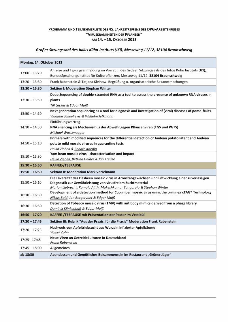
PROGRAMM UND TEILNEHMERLISTE DES 45. JAHRESTREFFENS DES DPG-ARBEITSKREISES "VIRUSKRANKHEITEN DER PFLANZEN" AM 14. + 15. OKTOBER 2013 Großer Sitzungssaal des Julius Kühn-Instituts (JKI), Messeweg 11/12, 38104 Braunschweig Montag, 14. Oktober 2013 13:00 – 13:20 Anreise und Tagungsanmeldung im Vorraum des Großen Sitzungssaals des Julius Kühn Instituts (JKI), Bundesforschungsinstitut für Kulturpflanzen, Messeweg 11/12, 38104 Braunschweig 13:20 – 13:30 Frank Rabenstein & Tatjana Kleinow: Begrüßung u. organisatorische Bekanntmachungen 13:30 – 15:30 Sektion I: Moderation Stephan Winter 13:30 – 13:50 Deep Sequencing of double-stranded RNA as a tool to assess the presence of unknown RNA viruses in plants Till Lesker & Edgar Maiß 13:50 – 14:10 Next generation sequencing as a tool for diagnosis and investigation of (viral) diseases of pome-fruits Vladimir Jakovljevic & Wilhelm Jelkmann 14:10 – 14:50 Einführungsvortrag RNA silencing als Mechanismus der Abwehr gegen Pflanzenviren (TGS und PGTS) Michael Wassenegger 14:50 – 15:10 Primers with modified sequences for the differential detection of Andean potato latent and Andean potato mild mosaic viruses in quarantine tests Heiko Ziebell & Renate Koenig 15:10 – 15.30 Yam bean mosaic virus - characterisation and impact Heiko Ziebell, Bettina Heider & Jan Kreuze 15:30 – 15:50 KAFFEE-/TEEPAUSE 15:50 – 16:50 Sektion II: Moderation Mark Varrelmann 15:50 – 16.10 Die Diversität des Dasheen mosaic virus in Aronstabgewächsen und Entwicklung einer zuverlässigen Diagnostik zur Gewährleistung von virusfreiem Zuchtmaterial Marion Liebrecht ; Kamala Ajith; Makeshkumar Tangaraju & Stephan Winter 16:10 – 16:30 Development of a detection method for Cucumber mosaic virus using the Luminex xTAG® Technology Niklas Bald , Jan Bergervoet & Edgar Maiß 16:30 – 16:50 Detection of Tobacco mosaic virus (TMV) with antibody mimics derived from a phage library Dominik Klinkenbuß & Edgar Maiß 16:50 – 17:20 KAFFEE-/TEEPAUSE mit Präsentation der Poster im Vestibül 17:20 – 17:45 Sektion III: Rubrik "Aus der Praxis, für die Praxis" Moderation Frank Rabenstein 17:20 – 17:25 Nachweis von Apfeltriebsucht aus Wurzeln infizierter Apfelbäume Volker Zahn 17:25– 17:45 Neue Viren an Getreidekulturen in Deutschland Frank Rabenstein 17:45 – 18:00 Allgemeines ab 18:30 Abendessen und Gemütliches Beisammensein im Restaurant „Grüner Jäger“
Transcript of PROGRAMM UND TEILNEHMERLISTE DES 45. JAHRESTREFFENS … · PROGRAMM UND TEILNEHMERLISTE DES...
PROGRAMM UND TEILNEHMERLISTE DES 45. JAHRESTREFFENS DES DPG-ARBEITSKREISES "VIRUSKRANKHEITEN DER PFLANZEN"
AM 14. + 15. OKTOBER 2013
Großer Sitzungssaal des Julius Kühn-Instituts (JKI), Messeweg 11/12, 38104 Braunschweig
Montag, 14. Oktober 2013
13:00 – 13:20 Anreise und Tagungsanmeldung im Vorraum des Großen Sitzungssaals des Julius Kühn Instituts (JKI),
Bundesforschungsinstitut für Kulturpflanzen, Messeweg 11/12, 38104 Braunschweig
13:20 – 13:30 Frank Rabenstein & Tatjana Kleinow: Begrüßung u. organisatorische Bekanntmachungen
13:30 – 15:30 Sektion I: Moderation Stephan Winter
13:30 – 13:50
Deep Sequencing of double-stranded RNA as a tool to assess the presence of unknown RNA viruses in
plants
Till Lesker & Edgar Maiß
13:50 – 14:10 Next generation sequencing as a tool for diagnosis and investigation of (viral) diseases of pome-fruits
Vladimir Jakovljevic & Wilhelm Jelkmann
14:10 – 14:50
Einführungsvortrag
RNA silencing als Mechanismus der Abwehr gegen Pflanzenviren (TGS und PGTS)
Michael Wassenegger
14:50 – 15:10
Primers with modified sequences for the differential detection of Andean potato latent and Andean
potato mild mosaic viruses in quarantine tests
Heiko Ziebell & Renate Koenig
15:10 – 15.30 Yam bean mosaic virus - characterisation and impact
Heiko Ziebell, Bettina Heider & Jan Kreuze
15:30 – 15:50 KAFFEE-/TEEPAUSE
15:50 – 16:50 Sektion II: Moderation Mark Varrelmann
15:50 – 16.10 Die Diversität des Dasheen mosaic virus in Aronstabgewächsen und Entwicklung einer zuverlässigen Diagnostik zur Gewährleistung von virusfreiem Zuchtmaterial Marion Liebrecht; Kamala Ajith; Makeshkumar Tangaraju & Stephan Winter
16:10 – 16:30 Development of a detection method for Cucumber mosaic virus using the Luminex xTAG® Technology
Niklas Bald, Jan Bergervoet & Edgar Maiß
16:30 – 16:50 Detection of Tobacco mosaic virus (TMV) with antibody mimics derived from a phage library
Dominik Klinkenbuß & Edgar Maiß
16:50 – 17:20 KAFFEE-/TEEPAUSE mit Präsentation der Poster im Vestibül
17:20 – 17:45 Sektion III: Rubrik "Aus der Praxis, für die Praxis" Moderation Frank Rabenstein
17:20 – 17:25 Nachweis von Apfeltriebsucht aus Wurzeln infizierter Apfelbäume Volker Zahn
17:25– 17:45 Neue Viren an Getreidekulturen in Deutschland Frank Rabenstein
17:45 – 18:00 Allgemeines
ab 18:30 Abendessen und Gemütliches Beisammensein im Restaurant „Grüner Jäger“
Dienstag, 15. Oktober 2013
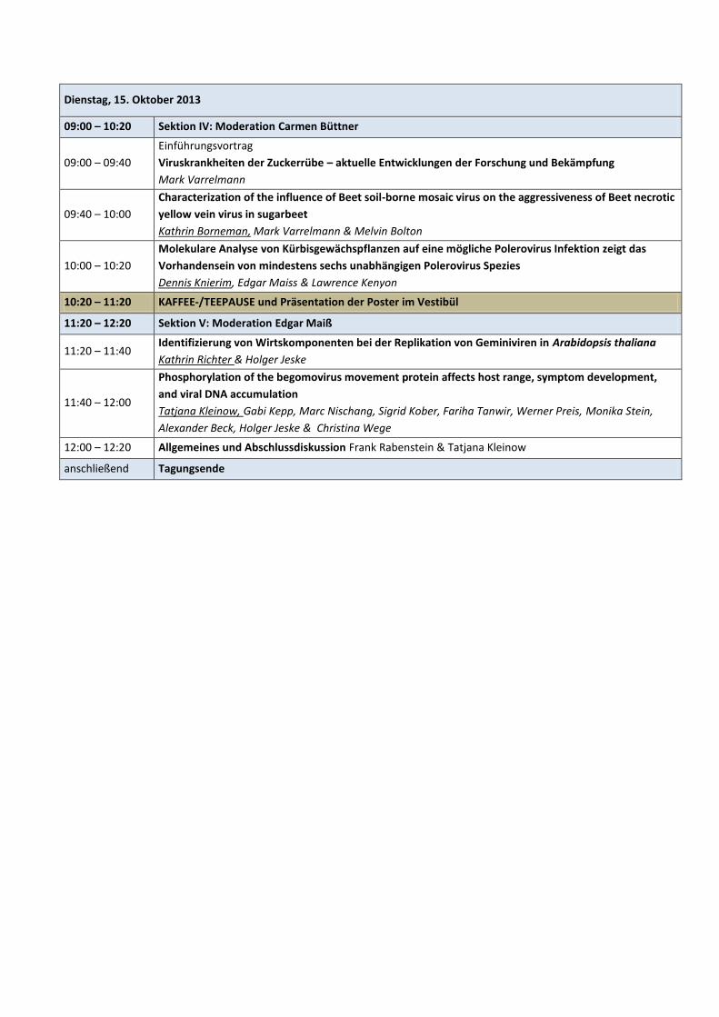
09:00 – 10:20 Sektion IV: Moderation Carmen Büttner
09:00 – 09:40
Einführungsvortrag
Viruskrankheiten der Zuckerrübe – aktuelle Entwicklungen der Forschung und Bekämpfung
Mark Varrelmann
09:40 – 10:00
Characterization of the influence of Beet soil-borne mosaic virus on the aggressiveness of Beet necrotic
yellow vein virus in sugarbeet
Kathrin Borneman, Mark Varrelmann & Melvin Bolton
10:00 – 10:20
Molekulare Analyse von Kürbisgewächspflanzen auf eine mögliche Polerovirus Infektion zeigt das
Vorhandensein von mindestens sechs unabhängigen Polerovirus Spezies
Dennis Knierim, Edgar Maiss & Lawrence Kenyon
10:20 – 11:20 KAFFEE-/TEEPAUSE und Präsentation der Poster im Vestibül
11:20 – 12:20 Sektion V: Moderation Edgar Maiß
11:20 – 11:40 Identifizierung von Wirtskomponenten bei der Replikation von Geminiviren in Arabidopsis thaliana
Kathrin Richter & Holger Jeske
11:40 – 12:00
Phosphorylation of the begomovirus movement protein affects host range, symptom development,
and viral DNA accumulation
Tatjana Kleinow, Gabi Kepp, Marc Nischang, Sigrid Kober, Fariha Tanwir, Werner Preis, Monika Stein,
Alexander Beck, Holger Jeske & Christina Wege
12:00 – 12:20 Allgemeines und Abschlussdiskussion Frank Rabenstein & Tatjana Kleinow
anschließend Tagungsende
Posterpräsentationen
1
Bestimmung der vollständigen Nukleotidsequenz des Asparagus virus 1
Blockus, Sebastian; Lesker, Till; Maiß, Edgar Leibniz Universität Hannover, Institut für Gartenbauliche Produktionssysteme / Abt. Phytomedizin / AG Pflanzenvirologie, Herrenhäuser Straße 2, 30419 Hannover
2 Processing and function of two proteins of the Mycovirus FgV-ch9 found in Fusarium graminearum
Blum, Christine; Garten, Hille; Heinze, Cornelia Universität Hamburg, AMP III, Ohnhorststr. 18, 22609 Hamburg
3
Lokalisation des Cherry leaf roll virus (CLRV) in Blütenständen der Hänge-Birke (Betula pendula)
Dierker, Luise; Von Bargen, Susanne; Büttner, Carmen Humboldt-Universität zu Berlin, Fachgebiet Phytomedizin, Lentzeallee 55/57, 14195 Berlin
4
Beating Begomoviruses
Götz, Monika1; Cuong, Ha Viet
2; Pissawan, Chiemsombat
3; Boopathi, N. Manikanda
4; Hansen, Peter
5; Kenyon,
Lawrence5; Hoang, Duong Vu Huy
1; Winter, Stephan
1
1Plant Virus Department, Leibniz-Institut DSMZ, German Collection of Microorganisms and Cell Cultures GmbH,
Braunschweig, Germany 2Department of Plant Pathology, Hanoi University of Agriculture, Ha Noi, Viet Nam
3Department of Plant Pathology, Kasetsart University, Nakhon Pathom, Thailand
4Department of Plant Molecular Biology and Bioinformatics, Tamil Nadu Agricultural University, Coimbatore,
India 5AVRDC - The World Vegetable Center, Tainan, Taiwan
5
Impact of silica supplementation on cucumber cultures
Holz, Sabine 1; Bartoszewski, Grzegorz
2; Kube, Michael
1; Büttner, Carmen
1
1Humboldt-Universität zu Berlin, Department of Crop and Animal Sciences, Division Phytomedicine, Lentzeallee
55/57, 14195 Berlin, Germany 2Warsaw University of Life Sciences, Department of Plant Genetics Breeding and Biotechnology, 159
Nowoursynowska Street , 02-776 Warsaw, Poland
6
Protein interactions of alphacryptoviruses and betacryptoviruses from white clover, red clover and dill by
bimolecular fluorescence complementation analysis
Lesker, Till; Maiss, Edgar
Institute of Horticultural Production Systems - Dept. Phytomedicine, Leibniz Universität Hannover, Herrenhäuser Str.
2, D-30419 Hannover, Germany
7
Charakterisierung eines neuen Aureusvirus aus Yam
Menzel, Wulf1; Thottappilly, George
2; Winter, Stephan
1
1Leibniz Institute DSMZ, German Collection of Microorganisms and Cell Cultures, Plant Virus Department,
Inhoffenstraße 7 B, 38124 Braunschweig, Germany 2Sahrdaya College of Engineering and Technology, Biotechnology, Kodakara, P.B.No.17, Thrissur Area, Pin 680684,
Kerala State, India
8
Genome features and particularities of acholeplasmas and phytoplasmas
Michael Kube1; Christin Siewert
1; Sabine Holz
1; Bojan Duduk
2; Carmen Büttner
1
1Department of Crop and Animal Sciences, Humboldt-Universität zu Berlin, Lentzeallee 55/57, 14195 Berlin,
Germany 2Department of Plant Pathology, Institute of Pesticides and Environmental Protection, Banatska 31b, P.O. Box
163, 11080 Belgrade, Serbia
9
Screening von Epiphyllum sp. mittels dsRNA-Isolation und auf Virusinfektionen
Paul Rentz1; Rosa Herbst
2; Anette Hohe
2; Ulrich Haage
3; Till Lesker
1; Edgar Maiss
1
1Leibniz Universität Hannover, Institut für Gartenbauliche Produktionssysteme, Abt. Phytomedizin, Arbeitsgruppe
Pflanzenvirologie / Molekulare Phytopathologie, Herrenhäuser Str. 2, 30419 Hannover 2Leibniz-Institut für Gemüse- und Zierpflanzenbau Großbeeren/Erfurt e.V., Theodor-Echtermeyer-Weg 1, 14979
Großbeeren 3Kakteen-Haage, Blumenstrasse 68, 99092 Erfurt, Deutschland
10
Nachweis des European mountain ash ringspot-associated virus in Sorbus aria und Sorbus intermedia
Robel, Jenny1; Büttner, Theresa
1; Mühlbach, Hans-Peter
2; von Bargen, Susanne
1; Büttner, Carmen
1
1Humboldt-Universität zu Berlin, Fachgebiet Phytomedizin, Lentzeallee 55/57, 14195 Berlin
2Universität Hamburg, Biozentrum Klein Flottbek, Ohnhorststr. 18, 22609 Hamburg
11
Nachweis des European mountain ash ringspot-associated virus in Ebereschen in Großbritannien
von Bargen, Susanne; Dieckmann, Luisa; Robel, Jenny; Büttner, Carmen
Humboldt-Universität zu Berlin, Fachgebiet Phytomedizin, Lentzeallee 55/57, 14195 Berlin
12
High genetic variability found among Cherry leaf roll virus variants from symptomatic birch trees in Rovaniemi
(Finland)
Rumbou Artemis1; von Bargen Susanne
1; Rott Markus
1; Jalkanen Risto
2; Buettner Carmen
1
1HU-Phytomedicine, Lentzeallee 55/57, 14195 Berlin, DE
2METLA, PL 16, FI 96301, Rovaniemi, Finland
13
Molecular characterization, infectivity and phylogenetic studies on begomoviruses infecting cotton in
Pakistan
Shuja, Malik Nawaz1; Tahir, Muhammad
2; Winter, Stephan
1
1Leibniz-Institut DSMZ-Deutsche Sammlung von Mikroorganismen und Zellkulturen GmbH,Braunschweig,
Germany 2Atta-ur-Rahman School of Applied Biosciences, National University of Sciences and Technology, Islamabad,
Pakistan
14
Labelling of Tobacco mosaic virus (strain OhioV) with gfp (green-fluorescent protein: GFP) for propagation
studies and mutational analyses in Nicotiana benthamiana
Rose, Hanna1; Heinze, Cornelia
2; Maiß, Edgar
1
1Leibniz University Hannover, Institute of Horticultural Production Systems, Dept. Phytomedicine, Herrenhäuser
Str. 2, 30419, Hannover, Germany 2Biocenter Klein Flottbeck, Ohnhorstrasse 18, 22609 Hamburg, Germany
15
Detection of siRNA using long, DIG-labelled DNA-probes
Weißhaar, Nina Kathrin
Universität Stuttgart, Biologisches Institut/ Abteilung für Molekularbiologie und Virologie der Pflanzen,
Pfaffenaring 57, 7069 Stuttgart ,Deutschland
16
Development of a dual enzyme reaction system on tobacco mosaic virus Wabbel, Katrin
1; Azucena, Carlos
2; Gliemann, Hartmut
2; Eiben, Sabine
1, Wege, Christina
1
1University of Stuttgart, Institute of Biology; Molecular Biology and Plant Virology, Pfaffenwaldring 57, 70550
Stuttgart, Germany. 2Karlsruhe Institute of Technology (KIT), Institute of Functional Interfaces (IFG), Hermann-von-Helmholtz-Platz 1,
76344 Eggenstein-Leopoldshafen
17
A satellite virus associated with Brome mosaic virus isolated from winter wheat and triticale in the Russian Central Cernozem region Rabenstein, Frank
1 ; Ziegler,Angelika
1; Wolf, Annegret
2; Mock Hans-Peter
2; Schlieter ,Bernd
3 ; Danilkin, Nikolay
4
1Julius Kuehn Institute, Federal Research Centre for Cultivated Plants (JKI), Institute of Epidemiology and Pathogen
Diagnostics, Erwin-Baur-Str. 27, 06484 Quedlinburg, 2Leibniz Institute of Plant Genetics and Crop Plant Research (IPK), OT Gatersleben, Corrensstrasse 3, 6466 Stadt
Seeland, Germany 3Deutsche Saatveredelung AG, Saatzuchtstation Leutewitz, OT Leutewitz Nr. 26, 01665 Käbschütztal, Germany
4GSA Agro LLC, 399921, Township Roshinskiy, Tchapliginsky district, Lipezk region, Russia
2
Abstracts Vorträge
Einführungsvortrag: RNA silencing als Mechanismus der Abwehr gegen Pflanzenviren (TGS und PGTS)
Michael Wassenegger
AgroScience GmbH, AlPlanta-Institute for Plant Research, Epigenetics, Breitenweg 71, 67435 Neustadt-Mußbach
Deep Sequencing of double-stranded RNA as a tool to assess the presence of unknown RNA viruses in plants
Till Lesker & Edgar Maiß Institute of Horticultural Production Systems - Dept. Phytomedicine, Leibniz Universität Hannover, Herrenhäuser Str. 2, D-30419 Hannover, Germany
Most plant infecting viruses use single-stranded RNA as their genome and generate double-stranded RNA (dsRNA) during replication. Moreover, a number of cultivars are known to contain high-molecular-weight dsRNA, for which a viral nature has been suggested, but without showing any symptoms. These dsRNAs are normally not found as part of ordinary plant cells. DsRNA can be purified from plant material by its ability of binding to cellulose in the presence of an appropriate concentration of ethanol. This offers an easy way for RNA-virus screening in plants without previous knowledge about the virus. However, additional and laborious work like RT-PCR, cloning and sequence determination of viruses is necessary for a complete characterization of the virus(es). Moreover the amount of dsRNA can vary between different viruses and might be even too low for the detection in electrophoresis. High throughput sequencing technologies can help to circumvent these difficulties by omitting time-consuming manual sequencing analyses. In addition it enables the detection of lowest target amounts even in mixed infections. DsRNA purifications obtain usually only very low contaminations with plant nucleic acids and single-stranded viral RNAs. Therefore, dsRNA purification in combination with deep sequencing analyses can be used on the one hand for a full sequence determination of viruses and allows on the other hand a pooling of many samples without losing a high detection sensitivity. In a recent study we combined dsRNA samples from white clover, red clover, hop trefoil and dill for a cost effective benchtop sequencing technology to detect latent and cryptic virus infections. Analyses of deep sequencing data revealed recently described cryptic viruses (Lesker et al., 2013) but also a couple of putative and so far unknown viral sequences. The power and possibilities but also the limitations of our approach will be discussed. Reference: Lesker, T.; Rabenstein, F.; Maiss, E. (2013): Molecular characterization of five betacryptoviruses infecting four clover species and dill. Archives of Virology 158, 1943-1952.
Next generation sequencing as a tool for diagnosis and investigation of (viral) diseases of pome-fruits
Vladimir Jakovljevic & Wilhelm Jelkmann JKI, Pflazenschutz im Obst- und Weinbau, Schwabenheimer Straße101, 69221 Dossenheim
During last several years next generation sequencing (NGS) has been widely used as a powerful method for sequencing whole genomes and transcriptomes. However application of NGS in plant pathology, in particular plant virology is in comparison to the other fiels of research (e.g. metagenomics, animal and clinical research etc.) still relatively underrepresented. Here we describe use of NGS for diagnosis and investigation of pome-fruit diseases. Examples include latent viruses of apples and pears and apple rubbery wood (ARW), a disease with unknown ethiology. Primers with modified sequences for the differential detection of Andean potato latent and Andean potato mild mosaic
viruses in quarantine tests
Heiko Ziebell & Renate Koenig JKI, Institut für Epidemiologie und Pathogendiagnostik, Messeweg 11, D3804 Braunschweig
Andean potato latent virus (APLV) is a Tymovirus which was first described by Gibbs et al. in 1966. It is widely spread in the Andean region and is a regulated pest in the European Union. In a joint study performed by scientists of the International Potato Center (CIP) in Lima and of the earlier Biologische Bundesanstalt we had found in the late 70ies that various isolates differ greatly in their serological and pathogenic properties (Koenig et al., Phytopathology 69: 748, 1979). The work in Braunschweig had been enabled by a grant of the Deutsche Forschungsgemeinschaft. The cooperation between scientists from CIP and JKI has recently been revived on the basis of molecular studies. In Lima, ‘Deep sequencing‘ was used to determine the complete sequence of two serologically rather distinct isolates (Col and Hu). In Braunschweig the coat protein gene sequences of twelve isolates were determined. These studies revealed that the genus APLV should be subdivided into two genera, i.e. APLV and Andean potato mottle virus (APMMV) which share less than 57 % coat protein amino acid sequence identity (Kreuze et al., Virus Research 173:431, 2013). To enable the differential RT PCR detection of APLV and APMMV strains in quarantine tests, broad specificity antisense primers are needed for the production of cDNAs for the wide range of strains of the two viruses which may be present in infected material. The sense primers to be used should also show in most parts of their sequences a broad specificity to allow their binding to a wide range of APLV or APMMV cDNAs, respectively. The 3’ ends of such primers should, however, be highly specific for nucleotides which are found in the respective position either exclusively in all APLV sequences, but in none of the APMMV sequences or vice versa. On the basis of the sequence information available so far, we have developed a PCR system which allows the separate detection of APLV and APMMV strains on the basis of primers which due to A->C or T->C exchanges in their 3’ terminal parts have a greatly increased differentiating power.
3
Yam bean mosaic virus - characterisation and impact
Heiko Ziebell1, Bettina Heider
2 & Jan Kreuze
3
1Julius Kühn-Institut, Institut für Epidemiologie und Pathogendiagnostik, Messeweg 11-12, 38106 Braunschweig, Deutschland 2International Potato Centre, Global Program Genetic Resources, Av La Molina 1895, La Molina, Lima, Peru 3International Potato Centre, Virology and Quarantine Units, Av La Molina 1895, La Molina, Lima, Peru
Towards the end 2010, viral disease symptoms (mosaic with or without leaf deformation) were observed in a yam bean (Pachyrhizus sp.) field trial in San Ramon, Junín, Peru. Small interfering RNA (siRNA) sequencing and assembly was performed on a sample showing severe mosaic and leaf deformation and the full genome sequence of a virus could be assembled. Based on molecular analysis (amino-acid and nucleotide sequence identity) it was confirmed to be a potyvirus, however clearly distinct from several bean viruses, including BCMV to which it is most closely related. We have designated this virus as yam bean mosaic virus-isolate SR (YBMV-SR). The sequence of YBMV-SR was highly similar to a partial potyvirus sequence (accession number AB289438) reported from yam bean in Indonesia (Damayanti et al. 2008) suggesting this virus might have a world-wide distribution. Aim of this joint project of JKI (Germany) and CIP (Peru) is the development of cheap and rapid detection methods specific to YBMV as well as investigations on host range, seed transmission studies and impact of YBMV-infection on yield. Die Diversität des Dasheen mosaic virus in Aronstabgewächsen und Entwicklung einer zuverlässigen Diagnostik zur Gewährleistung von virusfreiem Zuchtmaterial Marion Liebrecht; Kamala Ajith; Makeshkumar Tangaraju & Stephan Winter 1Leibniz-Institut DSMZ, Abteilung Pflanzenviren, Inhoffenstraße 7B, 38124 Braunschweig, Germany 2Central Tuber Crops Research Institute, India
Im Rahmen des International Network of Edible Aroids (INEA) Projektes “Adapting Clonally Propagated Crops to Climatic and Commercial Change” werden Pflanzenviren in Taro und anderen essbaren Aronstabgewächsen untersucht, da ein Virusbefall von Zuchtmaterial gravierende Auswirkungen auf den internationalen Austausch von Keimplasma zur Folge hat. Dasheen mosaic virus (DsMV) ist der bedeutendste virale Erreger in Aronstabgewächsen. Dieses Potyvirus ist in fast allen Anbaugebieten von Taro und Cocoyam verbreitet. DsMV infizierte Pflanzen weisen eine Vielfalt von schwachen bis starken Symptomen auf, was auf eine hohe Diversität und/oder das Vorkommen von Mischinfektionen hinweist. In unserer Untersuchung der genetischen Diversität von DsMV aus verschiedenen führenden Anbauregionen dieser Welt, wurden Gesamtgenome von zwei DsMV Isolaten aus Taro (Colocasia esculenta) und Amorphophallus (Amorphophallus ssp.) sequenziert. Darüber hinaus ergab der Vergleich der 3‘-terminalen Region des ssRNA Genoms von einer beträchtlichen Anzahl an Virusisolaten, dass die genetische Diversität der Viren eine zuverlässige Diagnostik für DsMV behindert. Zur Detektion von Viren wurden Antiseren auf Basis von rekombinant exprimiertem Hüllprotein von zwei DsMV Isolaten produziert und infolgedessen DAS-ELISA Tests zur Untersuchung von pflanzlichem Material auf eine Infektion mit DsMV entwickelt. Zusätzlich wurden aufgrund des Sequenzvergleichs Primer in konservierten Bereichen des DsMV Hüllproteins erstellt um ein RT-PCR Protokoll zur Detektion des Virus zu entwickeln. Beide diagnostischen Verfahren (DAS-ELISA und RT-PCR) wurden in unseren Laboren gemäß den EPPO Standards zur Detektion von Viren in pflanzlichem Material validiert und ein Standardverfahren zur Erkennung von DsMV Infektionen in Aronstabgewächsen entwickelt. Development of a detection method for Cucumber mosaic virus using the Luminex xTAG® Technology
Niklas Bald1, Jan Bergervoet
2 & Edgar Maiß
1
1Leibniz Universität Hannover, Institut für Gartenbauliche Produktionssysteme, Abteilung Phytomedizin, Herrenhäuser Straße 2, 30419 Hannover, Deutschland 2Wageningen UR, Plant Research International, Biointeractions and Plant Health, Droevendaalsesteeg 1, 6708PB Wageningen, Netherlands
A nucleic acid based test for the detection of the economically important plant virus Cucumber mosaic virus (CMV) based on the Luminex xTAG® Technology was developed. This technology has the advantage of allowing the simultaneous detection of various targets. Viral RNA is transcribed into DNA and then PCR-amplified (preamplification). In a 2
nd round of PCR (target
specific primer extension or TSPE) biotinylated dCTP is integrated into the DNA together with the other nucleotides and specific TSPE primers are used that have tags consisting of 24 nucleotides added to their 5’-ends. These tags are complementary to anti-tags which are attached to the surface of the MagPlex-TAG™ Microspheres. There are different sets of polystyrene microspheres or beads, each filled with a spectrally distinct dye and a corresponding anti-tag. The amplified target DNA will hybridize to a distinct microsphere through a tag to anti-tag interaction. Streptavidin-R-phycoerythrin, a reporter protein, binds to the incorporated biotin of the DNA. Experimental analysis will proceed in a manner similar to flow cytometry. In the analyzing instrument a red laser will excite the bead dye and a green laser will stimulate the streptavidin-R-phycoerythrin. The detected fluorescences of both the bead dye and the reporter protein are proof for the presence of the target virus nucleic acids. Using this technology we tested for the presence of CMV in general and started to differentiate between its two subgroups (I and II) for which significant differences concerning severity of symptoms, virulence and geographic distribution have been reported. Generic TSPE primers were designed which led to the detection of all 30 CMV isolates obtained from the DSMZ in inoculated plants. Subgroup specific TSPE primers for either CMV subgroup I or II were also created but still need to be tested. Nevertheless, classification of the 30 CMV isolates to either subgroup I or II was confirmed by an ELISA using specific antibodies. Eighteen isolates were allocated to subgroup I, 10 isolates were classified into subgroup II and two isolates could not be filed into either subgroup. This work is part of a project which aims to develop a test for the simultaneous detection of various (viral, bacterial and fungal) plant pathogens in plant material. It is funded by the EU INTERREG program Gezonde Kas.
4
Detection of Tobacco mosaic virus (TMV) with antibody mimics derived from a phage library
Dominik Klinkenbuß & Edgar Maiß Leibniz Universität Hannover, IGPS, Herrenhäuser Straße 2, 30419 Hannover, Deutschland
Worldwide, Tobacco mosaic virus (TMV) is known as a serious pathogen causing diseases in more than 200 plant species (Scholthof, 2004). TMV has a rod-like appearance with a modal length of 300-310 nm. The genome consists of a linear positive sense single-stranded RNA with a size of 6.3-6.6 kb, which is translated mainly into three proteins, namely RNA-dependent RNA polymerase, movement and coat protein (CP). According to the physical and molecular parameters TMV belongs to the genus Tobamovirus. For detection of TMV serological methods, e.g. enzyme-linked immune assay (ELISA) are routinely used. Beside several advantages (robustness, relatively simple handling and suitability for mass testing) ELISA procedures depend on finite resources since required antibodies for the test procedure have to be produced and validated consistently. To overcome this disadvantage, protein molecules binding to TMV target proteins, especially the CP, are created and used instead of common antibodies (Petrenko & Vodyanoy, 2003). The aim of this study was the production of “antibody mimics” directed towards plant viruses derived from a phage library. This phage library consists of millions of different phages, whereby each phage displays an artificial and unique peptide on its surface. In a screening procedure called “Biopanning” phages are selected binding tightly to target molecules, i.e. TMV CP. Here, the pannings with the commercially available phage libraries Ph.D.™-12 and Ph.D.™-C7C were carried out against purified TMV. After three rounds of biopanning, several phages were selected and tested in a “Phage-ELISA” with purified TMV and TMV infected plant material. Some phages showed a significant interaction with the purified TMV target in a “Phage-ELISA”, revealing high readings within a few minutes, whereas phages without the specific 12 amino acid motif failed in detection of TMV. On the other hand almost every selected phage clone showed an interaction with healthy plant material. After application of a short protocol for crude purification of plant viruses (Dr. Rene van der Vlugt, PRI of Wageningen; pers. comm.) phage clones were able to distinguish between infected and healthy plant material. To analyse if the ELISA-signal can be enhanced, the specific amino acid sequence was also used in another fusion protein construct (fibronectin type III domain (10Fn3)). Overall, the results indicate that antibody mimics based on phages can be a valuable tool to supplement and improve ELISAs for detection of plant viruses. References: Petrenko, V. A. & V. J. Vodyanoy (2003): Phage display for detection of biological threat agents. J. Microbiol Methods 53: 253-262. Scholthof, K. B. (2004): Tobacco mosaic virus: a model system for plant biology. Annun. Rev. Phytopathol. 42: 13-34.
Einführungsvortrag: Viruskrankheiten der Zuckerrübe – aktuelle Entwicklungen der Forschung und Bekämpfung
Mark Varrelmann Institut für Zuckerrübenforschung, Holtenser Landstr. 77, 37079 Göttingen
Characterization of the influence of Beet soil-borne mosaic virus on the aggressiveness of Beet necrotic yellow vein virus
in sugarbeet
Kathrin Borneman1, Mark Varrelmann
2 & Melvin Bolton
3
1North Dakota State University, Department of Plant Pathology, 306 Walster Hall, Fargo, ND, 58102, USA 2Institut fuer Zuckerruebenforschung, Holtenser Landstr. 77, 37079 Goettingen 3USDA Agricultural Research Service, Northern Crop Science Laboratory, 1605 Albrecht Blvd., Fargo, ND, 58102, US
Beet necrotic yellow vein virus (BNYVV) strains with different levels of aggressiveness have spread to sugarbeet growing areas worldwide. As the Rz1 resistance gene has been used in most of the commercial varieties since its identification, it had to be anticipated that resistance breaking variants would be selected. In the US and Europe, resistance breaking properties of some BNYVV strains causing high yield losses have been observed. The objective of the study was to identify the influence of (i) the sugarbeet genotype, and (ii) Beet soil-borne mosaic virus (BSBMV) on the aggressiveness of BNYVV strains. Both viruses are vectored by the plasmodiophoromycete Polymyxa betae and occur in mixed infections in the field. Competition experiments with different BNYVV strains from Europe (Germany, France and Italy), and the US were performed under standardized greenhouse conditions. Infected roots of different sugarbeet genotypes (susceptible and resistant) were analyzed after five weeks of cultivation to determine virus titer (ELISA), and amino acid composition by means of “deep sequencing” of the viral pathogenicity factor. In a second experiment, the same BNYVV strains and the same sugarbeet genotypes were used in mixed infections with BSBMV. Additionally, another experimental approach was chosen to compare the aggressiveness of resistance breaking and wild type BNYVV strains in single and mixed infections with BSBMV. Therefore, the same vector population was loaded with both viruses, which also showed that the ability to overcome resistance was independent of the vector population. The results of the first experiment showed that, depending on the sugar beet genotype, certain amino acids of the viral pathogenicity factor, which determine the resistance breaking properties of the virus strain, occur with a higher frequency. In Rz1 plants, the resistance breaking variants out-competed the wild type variants, and mostly vice versa in susceptible plants, demonstrating a relative fitness penalty of resistance breaking mutations. The strong genotype effect supports the hypothesized Rz1 resistance breaking strain selection with four RNAs suggesting a certain tetrad needs to become dominant in a population to influence its properties. Tetrad selection was not observed when a resistance breaking strain, with an additional P26 protein encoded by a fifth RNA, competed with a wild type strain supporting its role as second pathogenicity factor and suggesting reassortment of both types. At harvest, typical virus symptoms were observed in mixed infections with BNYVV and BSBMV. Further analysis remains to be done.
5
Molekulare Analyse von Kürbisgewächspflanzen auf eine mögliche Polerovirus Infektion zeigt das Vorhandensein von
mindestens sechs unabhängigen Polerovirus Spezies
Dennis Knierim1, Edgar Maiss
2 & Lawrence Kenyon
3
1Leibniz-Institut DSMZ, Abteilung Pflanzenviren, Inhoffenstraße 7 B, 38124 Braunschweig, Germany 2Leibniz University Hannover, Abteilung Phytomedizin, Herrenhäuser Strasse 2, 30419 Hannover, Germany 3AVRDC - The World Vegetable Center, Plant Virology, PO Box 42, Shanhua, Tainan 74199, Taiwa
Bei der Analyse von 66 Kürbisgewächspflanzen mit Vergilbungs-Symptomen von Feldern aus den Ländern Mali, den Philippinen, Thailand und Usbekistan konnte bei 21 von ihnen eine Polerovirus Infektion festgestellt werden. Der Nachweis erfolgte mit Hilfe der Reverse Transkriptase-Polymerase-Kettenreaktion (RT-PCR) mit generellen Polerovirus Primern. Diese 21 positiven Proben wurden mit spezifischen Primer weiter analysiert auf die bekannten Kürbispflanzen infizierenden Polerovirus Spezies Cucurbit aphid-borne yellows virus (CABYV), Melon aphid-borne yellows virus (MABYV) und Suakwa aphid-borne yellows virus (SABYV). In einen weiteren Schritt wurden wiederum die CABYV positiven Proben analysiert auf das vorhanden sein des gewöhnlichen „common“ (CABYV-C) oder des rekombinanten (CABYV-R) CABYV Stammes. Es konnte zum ersten Mal das SABYV und CABYV-R für die Länder Thailand und die Philippinen nachgewiesen werden. In sieben der als Polerovirus positiv identifizierten Proben konnte keines der drei oben genannten Polerovirus Spezies nachgewiesen werden, woraufhin für diese Proben ein generelles Polerovirus RT-PCR Produkt von 1.4 kb Länge amplifiziert und sequenziert wurde. Es konnten zwei zuvor nicht beschriebenen Polerovirus Spezies identifiziert werden. Die identifizierte Polerovirus Spezies aus Mali wurde vorläufig als Pepo aphid-borne yellows virus (PABYV) und die Spezies aus Thailand Luffa aphid-borne yellows virus (LABYV) beschrieben. Für beide neuen Spezies wurden spezifische RT-PCR Nachweisprotokolle entwickelt. Zudem konnte keine Übereinstimmung mit der vor kurzem veröffentlichten Sequenz des Cucumber aphid-borne yellows virus (CuABYV) festgestellt werden. Identifizierung von Wirtskomponenten bei der Replikation von Geminiviren in Arabidopsis thaliana
Kathrin Richter & Holger Jeske Universität Stuttgart, Biologisches Institut, Abteilung Molekularbiologie u. Virologie der Pflanzen, Pfaffenwaldring 57, 70550 Stuttgart
Geminiviren sind Phytopathogene, die wichtige Nutzpflanzen weltweit infizieren. Sie bestehen aus zirkulärer Einzelstrang-DNA (single-stranded, ssDNA), die in Zwillingskapside aus zwei unvollständigen Ikosaedern verpackt wird. Der erste Schritt zur Vermehrung der viralen DNA innerhalb des Zellkerns der Wirtspflanze ist die Komplementärstrangsynthese (complementary strand replication, CSR), wobei die ssDNA zum Doppelstrang (dsDNA) aufgefüllt wird. Die dsDNA wird in Nukleosomen verpackt und liegt in diesen Minichromosomen als kovalent geschlossen, zirkuläres Molekül vor (covalently closed circular, cccDNA). Dieses dient als Template für die weitere Replikation über rolling circle replication (RCR) und Rekombinationsabhängige Replikation (recombination-dependent replication, RDR). Geminiviren sind vollkommen abhängig von Wirts-DNA Polymerasen und anderen DNA-modifizierenden Enzymen für ihre Replikation, da sie für keine eigene DNA Polymerase kodieren. Ziel unserer Arbeit ist es, die von der Wirtspflanze genutzten Enzyme zur viralen DNA-Synthese und –Modifikation zu identifizieren. Hierfür werden Arabidopsis thaliana knock-out Linien verwendet, denen einzelne Komponenten der somatisch homologen Rekombinations- oder Reparaturmaschinerie fehlen. Nach Inokulation mit den Arabidopsis-infizierenden Geminiviren EuMV (Euphorbia mosaic virus) und ClLCrV (Cleome leaf crumple virus) wird der Infektionsverlauf durch das Vorhandensein der verschiedenen viralen DNA-Intermediate analysiert. Durch den Vergleich mit Wildtyp-Pflanzen können Rückschlüsse auf die Rolle der jeweiligen Wirtsenzyme bei der Virusreplikation gezogen werden.
Phosphorylation of the begomovirus movement protein affects host range, symptom development, and viral DNA
accumulation
Tatjana Kleinow, Gabi Kepp, Marc Nischang, Sigrid Kober, Fariha Tanwir, Werner Preis, Monika Stein, Alexander Beck2,
Holger Jeske & Christina Wege 1Universität Stuttgart, Institute of Biology, Molecular Biology and Plant Virology, Pfaffenwaldring 57, 70550 Stuttgart, Germany 2PANATecs GmbH, Vor dem Kreuzberg 17, 72070 Tübingen, German
The DNA-B component of bipartite begomoviruses (family Geminiviridae) encodes for a nuclear shuttle protein (NSP) and a movement protein (MP), which enable systemic spread within host plants and affect pathogenicity. The MPs mediate multiple functions during intra- and intercellular trafficking, such as binding of viral nucleoprotein complexes, targeting to and modification of plasmodesmata and release of the cargo after cell-to-cell transfer is completed. A phosphorylation of Abutilon mosaic virus (AbMV) MP was shown for bacteria-, yeast- and Nicotiana benthamiana-derived protein [1, 2]. Mass spectrometry analyses of yeast-expressed MP identified three phosphorylation sites (Thr-221, Ser-223 and Ser-250) located in the C-terminal oligomerization domain [1, 3]. To assess their functional relevance for the viral life cycle within plants, several point mutations were introduced into the MP gene of AbMV DNA-B, which lead to an exchange of Thr-221, Ser-223, and Ser-250, either singly or in various combinations, with either an uncharged alanine or a phosphorylation-mimicking aspartate residue [1]. When co-inoculated with a wild-type DNA-A, all mutated DNA-B variants give raise to a systemic infection in N. benthamiana. However, some mutations in MP abolished an AbMV-infection in other plant species of the families Solanaceae and Malvaceae. In systemically infected plants, symptoms and/or viral DNA accumulation were significantly altered for several of the tested DNA-Bs encoding MP mutants. The identification of three phosphorylation sites in AbMV MP, which have an impact on host range, symptom development, and/or viral DNA accumulation, indicates a regulation of the diverse MP functions by plant-derived posttranslational modification and underscores their importance for geminivirus/host plant interaction. References: [1] T. Kleinow, M. Nischang, A. Beck, U. Kratzer, F. Tanwir, W. Preiss, G. Kepp, H. Jeske (2009): Virology 390: 89.
[2] T. Kleinow, G. Holeiter, M. Nischang, M. Stein, M. Karayavuz, C. Wege, H. Jeske (2008). Virus Research 131: 86. [3] B. Krenz, V. Windeisen, C. Wege, H. Jeske, T. Kleinow (2010). Virology: 401, 6.
6
Beiträge zur Rubrik "Aus der Praxis, für die Praxis"
Nachweis von Apfeltriebsucht aus Wurzeln infizierter Apfelbäume Volker Zahn Pflanzenschutzamt der Landwirtschaftskammer Niedersachsen, Wunstorfer Landstr. 9, 30453 Hannover Apfeltriebsucht wird aufgrund der sich ändernden Klimabedingungen auch für nördliche Gebiete eine immer größere Herausforderung. Bei Untersuchungen an Wurzelstücken von infizierten Apfelpflanzen stellten wir fest, dass entgegen der bisher vertretenen Meinung, der Erreger nicht homogen in allen Wurzeln und sogar innerhalb einer Wurzel nicht homogen verteilt ist. Dies hat enorme Auswirkungen auf die Nachweisbarkeit von Apfeltriebsucht. Gibt es dazu von anderen Untersuchungsstellen ähnliche Ergebnisse?
Neue Viren an Getreidekulturen in Deutschland Frank Rabenstein Julius Kühn-Institut (JKI), Institut für Epidemiologie und Pathogendiagnostik, Erwin-Baur-Str. 27, 06484 Quedlinburg
Infolge des Klimawandels und den global steigenden Austausch von Saatgut und Pflanzen ist auch in Deutschland mit dem Auftreten neuer, bisher hier wirtschaftlich nicht relevanter Viren zu rechnen. Hinzu kommt, dass die Erschließung neuer Absatzmärkte in Osteuropa durch deutsche Züchtungsfirmen zu unerwarteten phytopathologischen Problemen führen kann. In diesen Regionen sind meist andere Viren und Vektoren vorhanden, gegen die hier gezüchteten Sorten weniger tolerant bzw. nicht resistent sind. An Getreidekulturen sind bei uns insbesondere die durch Insekten übertragenen Viren des Barley yellow dwarf- bzw. Wheat dwarf- Komplexes von Bedeutung sind. Neben den an Weizen, Triticale und Roggen auftretenden bodenbürtigen Viren sind vor allem die Gelbmosaikviren der Wintergerste wirtschaftlich relevant. In 2013 wurden zwei weitere Viren an Getreide in D entdeckt. Dies sind zum einen das an vielen Gräsern vorkommende Trespenmosaik-Virus (Brome mosaic virus, BMV), das bisher als wirtschaftlich wenig bedeutend angesehen wird sowie das Weizenstrichelmosaik-Virus (Wheat streak mosaic virus, WSMV), welches in den USA eines der ökonomisch bedeutendsten Viren am Weizen ist. Außer in D konnten wir erstmalig das WSMV 2013 auch in Weizenproben aus Österreich identifizieren. Als Virusvektor ist die Gallmilbenart Aceria tosichella bekannt, die man generell als einen Gewinner des Klimawandels beurteilen muss. Sowohl an mit WSMV infizierten Weizen- und Gerstenpflanzen (Ausfallgetreide) wurden Gallmilben beobachtet und mittels Rasterelektronenmikroskopie dem kryptischen Artenkomplex A. tosichella zugeordnet. Es wird international eingeschätzt, dass die von A. tosichella übertragen Viren weltweit eine zunehmende Gefahr für die Getreideproduktion darstellen (NAVIA et al. 2012). Das stützen auch neueste Befunde über das Vorkommen solcher Viren in Südamerika (Argentinien, Brasilien) und Australien. Es besteht dringender Forschungsbedarf bezüglich der Identifizierung und Charakterisierung von WSMV Isolaten aus D bzw. deren Vektoren sowie zu Resistenzquellen in europäischen Weizen- und Gerstensorten. Das BMV wurden 2013 nicht nur in Weizenparzellen von Züchtungsfirmen aus Deutschland und Österreich nachgewiesen, sondern es trat massiv auch im Gewächshaus an DH-Populationen eines Weizenzüchters auf, sodass das gesamte Material vernichtet werden musste. Das Virus ist sehr leicht durch mechanischen Kontakt übertragbar und hat einen extrem weiten Wirtspflanzenkreis. Zu möglichen Vektoren gibt es sehr widersprüchliche Angaben. Ein besonderes Problem stellt ein von uns kürzlich entdecktes Satellitenvirus dar, das 2012 zunächst nur in Material aus Russland auftrat (RABENSTEIN et al., 2013), in diesem Jahr aber auch in BMV infiziertem Weizen aus einem Zuchtbetrieb in D beobachtet werden konnte. Satellitenviren können die Eigenschaften der Helferviren verändern, sodass diese sich u. U. stärker vermehren und besser ausbreiten können. Die Bedeutung des Komplexes BMV + Satellit für den Weizenanbau ist derzeit völlig ungeklärt. Es besteht Forschungsbedarf hinsichtlich einer schnellen und verlässlichen Diagnostik sowie zur Epidemiologie des Krankheitskomplexes. Das Vorhandensein und Auffinden möglicher Resistenzen in Getreide muss geprüft werden. References: NAVIA, D., R. S. MENDONCA, A. SKORACKA, W. SZYDŁO, D. KNIHINICKI, G.L. HEIN, P.L. PEREIRA, G. TRUOL, L. DOUGLAS, L. (2012): Wheat curl mite, Aceria tosichella, and transmitted viruses: an expanding pest complex affecting cereal crops. Experimental & Applied Acarology 59, 95-143. RABENSTEIN, F.; ZIEGLER, A.; SCHLIETER, B., DANILKIN, N. (2013): A satellite virus associated with Brome mosaic virus isolated from winter wheat and triticale plants in the Russian Central Cernozem region. 12th International Symposium on Plant Virus Epidemiology, Evolution, Ecology and Control of Plant Viruses, Program and Book of Abstracts, 28 January - 1 February 2013, Arusha, Tanzania, p. 212. https://www.researchgate.net/publication/236051237_12th_IPVE_Symposium_Abstract_Book
7
Abstracts Poster
1
Bestimmung der vollständigen Nukleotidsequenz des Asparagus virus 1
Blockus, Sebastian; Lesker, Till; Maiß, Edgar Leibniz Universität Hannover, Institut für Gartenbauliche Produktionssysteme / Abt. Phytomedizin / AG Pflanzenvirologie, Herrenhäuser Straße 2, 30419 Hannover
Das Asparagus virus 1 (AV-1) wird dem Genus Potyvirus zugeordnet und besitzt ein einzelsträngiges RNA-Genom von etwa 10 kbp mit positiver Polarität. Erstmalig wurde es durch Hein (1960) beschrieben, dabei verursacht es keine optisch feststellbaren Symptome in Asparagus officinalis L. – Kulturen. Doppelinfektionen mit dem Asparagus virus 2 (Genus Ilarvirus) und dem Cucumber mosaic virus (Genus Cucumovirus) führen zu Ertragsverlusten von 20% beziehungsweise 49,5%. Zusätzlich ist das AV-1 mit dem „Asparagus decline Syndrom“ assoziiert, durch die kombinierte Wirkung verschiedener Fusarium spec. führt dieses zum Absterben der gesamten Pflanze (Wade et al., 1996). Bisher ist lediglich eine partielle Nukleotidsequenz des Hüllproteins verschiedener Isolate bekannt. Zur Bestimmung der Nukleotidsequenz wurde eine dsRNA- Extraktion aus niedersächsischen Feldproben (Sorte Gijmlin) sowie aus zwei von der DSMZ bereitgestellten Isolaten (PV-0954; PV-0955) durchgeführt. Die dsRNA wurde mittels randomisierter Hexamer-Oligonukleotiden revers transkribiert und die resultierende cDNA in einer PCR vervielfältigt. Die erhaltenen DNA-Fragmente dienten nach anschließender Sequenzierung zur Erstellung von spezifischen Oligonukleotiden mit denen die vollständigen Genome der Isolate PV-0954 und PV-0955 bestimmt werden konnten. Die ermittelte Nukleotidsequenz des Isolates PV-0954 besitzt eine Größe von 9762 Basenpaaren und weißt eine hohe Ähnlichkeit zu den Potyviren Plum pox virus (66%), Japanese yam mosaic virus (65%) und Sweet potato virus 2 (69%) auf. Mit dieser Identität unter 76% kann das AV-1 nach Adams et al. (2005) als neue Spezies innerhalb des Genus Potyvirus eingeordnet werden. Die Hüllproteinsequenz ist zu 98% identisch mit den bisher bekannten AV-1 Isolaten. Innerhalb von AV-1 tritt eine Variabilität in der Fähigkeit der systemischen Infektion bei Nicotiana benthamiana auf, die eine Einteilung in Typ I (keine systemisch Infektion) und Typ II (systemische Infektion) ermöglichen (Rabenstein et al., 2007; Tomassoli et al., 2008). Entsprechend der Symptomausprägung kann das Isolat PV-0954 zum Typ II gezählt werden. Auf Ebene der Nukleotidsequenz besitzt es eine Ähnlichkeit von 99,6% zu dem Isolat PV-0955, welches nicht in der Lage ist eine systemische Infektion zu erlangen (Typ I). Die Isolate unterscheiden sich in 37 Nukleotidpositionen, die zu 15 Aminosäure-austauschen führen. Die bestimmten Nukleotidsequenzen ermöglichen eine weitere Analyse der Ursachen für die unterschiedliche Symptomausprägung des AV-1 Typ I und Typ II und bilden weiterhin die Grundlage für die Erstellung von infektiösen Voll-Längen Klonen. Literatur: Adams MJ, Antoniw JF, Fauquet CM. (2005): Molecular criteria for genus and species discrimination within the family Potyviridae. Arch Virol. 150: 459-79. Hein A. (1960). Über das Vorkommen einer Virose an Spargel. Z. PflKrankh. 67: 217-219. Rabenstein F., Schubert J., Habekuß A. (2007). Identification and differentiation of viruses on asparagus in Germany. Plant Virus Epidemiology Symposium, 15-19 October 07, ICRISAT, India. Tomassoli L., Tiberini A., Zaccaria A., Vetten, J. H. (2008). Molecular and biological studies of Asparagus virus 1 (genus Potyvirus). J. Plant Pathol. 90 (Suppl. 2): 437. Wade H. Elmer, Dennis A. Johnson, Gaylord I. Mink, (1996) Epidemiology and management of the diseases causal to Asparagus decline. Plant Disease 80: 117-125
2 Processing and function of two proteins of the Mycovirus FgV-ch9 found in Fusarium graminearum
Blum, Christine; Garten, Hille; Heinze, Cornelia Universität Hamburg, AMP III, Ohnhorststr. 18, 22609 Hamburg
The putative Chrysovirus FgV-ch9 replicates in Fusarium graminearum and causes hypovirulence on wheat and maize. In radial growth assays infected Fusarium graminearum shows an irregular shape and an intense red pigmentation. The colony size is significantly reduced. The genome of FgV-ch9 consists of five dsRNA segments which each encodes one open reading frame (ORF). The RNA 1 encodes the RNA dependent RNA polymerase (RdRp) which is found in the particles. The segments RNA 2 and 3 encode capsid proteins which form a 40 nm isometric particle. With the exception of the RdRP no protein with the predicted molecular weight (MW) has been detected yet. A processing is most likely. The ORFs of RNA 4 and 5 encode non-structural proteins with unknown functions. The ORF of RNA 3 encodes one of the capsid proteins and has a predicted MW of 93 kDa. However only proteins smaller than 68 kDa are detected in the particles. Both heterologous expression in E. coli and in vitro transcription/translation assays of ORF 3 result in a N-terminal 70 kDa and a C-terminal 25 kDa protein. An antiserum, which was raised against purified particles, detects the N-terminal 68 kDa in particles as well as additional bands with sizes between 68 kDa and more than 55 kDa. An antiserum raised against the C-terminus of P3 detects the 25 kDa of the E. coli and in vitro expressed ORF, but does not detect any protein in purified particles or in infected mycelium. This result suggests an autoproteolytic processing in a first step followed by a second processing in an unknown manner. The ORF of RNA 5 encodes a non-structural protein P5 with a predicted MW of 79 kDa. This protein shows 12 zinc finger motifs. Heterologous expression in E. coli suggests a processing, since a smaller protein has been detected with a His-tag specific antiserum. This protein P5 shows gene silencing suppressor activity when tested in a transient Agrobacterium tumefaciens infection system.
8
3
Lokalisation des Cherry leaf roll virus (CLRV) in Blütenständen der Hänge-Birke (Betula pendula)
Dierker, Luise; Von Bargen, Susanne; Büttner, Carmen Humboldt-Universität zu Berlin, Fachgebiet Phytomedizin, Lentzeallee 55/57, 14195 Berlin
Das Cherry leaf roll virus (CLRV) der Gattung Nepovirus ist weltweit in einer Vielzahl krautiger und holziger Wirtspflanzenarten vertreten. Die natürliche Verbreitung kann vertikal durch Saatgut und horizontal durch Pollen erfolgen. Etwa 20 % aller Pflanzenviren sind durch Samen übertragbar (Maule und Wang, 1996), so dass dieser Übertragungsweg von großer epidemiologischer Relevanz ist. Untersuchungen aus dem Jahr 2009 an der Modellpflanze Arabidopsis thaliana konnten eine Übertragung von CLRV durch Samen über mehrere Generationen zeigen (Rumbou et al., 2009). Die Hänge-Birke (Betula pendula) ist als Wirtspflanze des CLRV ökonomisch und ökologisch bedeutend. Sie produziert große Mengen an Pollen bzw. Samen. Daher wurden initiale Studien zur Lokalisation des Virus in Blütenständen der Hänge-Birke mittels Tissue Printing durchgeführt. Männliche und weibliche Kätzchen wurden von CLRV-infizierten Birken vom Standort Berlin-Dahlem zu unterschiedlichen Zeitpunkten in den Vegetationsperioden 2012 und 2013 beprobt. Quer- und Längsschnitte wurden auf Nitrocellulose appliziert und das Virus mittels CLRV-spezifischer Antikörper detektiert. Für die Untersuchungen wurden solche Birken ausgewählt, die bereits positiv auf eine Infektion mit CLRV getestet wurden. Es erfolgte der Nachweis in Blattmaterial sowie Blütenständen über die Amplifikation eines 416 bp Fragments der 3´nicht-translatierten Region (3´NTR) des CLRV in der IC-RT-PCR (Werner et al., 1997). Die CLRV-Infektion der untersuchten Birken konnte in beiden Vegetationsperioden bestätigt werden. CLRV konnte durch Tissue Printing in beiden Jahren sowohl in männlichen als auch in weiblichen Blütenständen erfolgreich detektiert werden. In Kätzchen-Querschnitten war CLRV zentral in Leitgefäßen sowie der Peripherie der Blütenstände nachweisbar, wobei keine Unterschiede in der Signalstärke zwischen männlichen und weiblichen Kätzchen festgestellt werden konnten. Untersuchte Längsschnitte wiesen eine unregelmäßige Verteilung des Virus über die gesamte Fläche auf.
4
Beating Begomoviruses
Götz, Monika1; Cuong, Ha Viet
2; Pissawan, Chiemsombat
3; Boopathi, N. Manikanda
4; Hansen, Peter
5; Kenyon, Lawrence
5;
Hoang, Duong Vu Huy1; Winter, Stephan
1
1Plant Virus Department, Leibniz-Institut DSMZ, German Collection of Microorganisms and Cell Cultures GmbH, Braunschweig, Germany 2Department of Plant Pathology, Hanoi University of Agriculture, Ha Noi, Viet Nam 3Department of Plant Pathology, Kasetsart University, Nakhon Pathom, Thailand 4Department of Plant Molecular Biology and Bioinformatics, Tamil Nadu Agricultural University, Coimbatore, India 5AVRDC - The World Vegetable Center, Tainan, Taiwan
Begomoviruses present a serious threat for vegetable crop production in tropical and subtropical regions. The reasons for this are attributed to 1), the emergence of more competitive begomovirus species and strains and 2), the displacement of indigenous vector species by the invasive Bemisia tabaci Middle East-Asia Minor 1 and Mediterranean. The development of efficient and sustainable management strategies requires a comprehensive overview on the most abundant begomovirus species in the agro-ecological environment, their host range and whitefly vectors. This is especially significant when host plant resistance in newly developed crop cultivars with good resistance characters is to be employed. The AVRDC/DSMZ project funded by BMZ/GIZ with partners from Vietnam, Thailand and India is studying begomoviruses and whitefly species on tomato, hot peppers, mung bean and weeds. In all countries, the diversity of begomovirus found in surveys was considerably high, with Tomato leaf curl Yunnan virus, Pepper leaf curl Malaysia virus, Chilli leaf curl Pakistan virus and Tomato leaf curl New Delhi virus most predominantly found in Thailand. In Vietnam among others, Tomato leaf curl Vietnam Virus, Tomato leaf curl China virus and Tomato leaf curl Kanchanaburi virus played a major role in the tomato production of the country. Mungbean yellow mosaic virus was the most predominant virus in mung beans in India and was also found throughout Vietnam. Interestingly while major outbreaks around the world are attributed to MEAM1/Med infestations, presence of these whitefly species, albeit earlier reported, was not confirmed in India or in Thailand. Only in Vietnam an invasion of B. tabaci MEAM1 in northern Vietnam with north-south direction was found which probably originates in China. In Thailand and India only the indigenous Asia1 and AsiaII whitefly species were identified indicating that these areas are still not in the invasion line.
5
Impact of silica supplementation on cucumber cultures
Holz, Sabine 1; Bartoszewski, Grzegorz
2; Kube, Michael
1; Büttner, Carmen
1
1Humboldt-Universität zu Berlin, Department of Crop and Animal Sciences, Division Phytomedicine, Lentzeallee 55/57, 14195 Berlin, Germany 2Warsaw University of Life Sciences, Department of Plant Genetics Breeding and Biotechnology, 159 Nowoursynowska Street , 02-776 Warsaw, Poland
Silicon is taken up by plants in its soluble form, silicic acid (SA) [Si(OH)4] and finally polymerized in the leaves. Even not to be an essential element for plants, its beneficial effects on growth of crops such as rice, barley, wheat, cucumber etc. are reported. SA treatment decreases the effects caused by abiotic and biotic stresses and it is proposed that silicon also strengthens the mechanical barrier improving the protecting from mycosis. SA pretreated Cucumis sativus plants show improved rapid and extensive defense reaction. It still remains unclear, if such effects also have impact on virus infections. Our study aims to provide information on changes in gene expression caused by SA treatment of C. sativus cultivars. Subsequent studies will allow estimating putative beneficial effects on the prevention of virus infections on cucumber.
9
6
Protein interactions of alphacryptoviruses and betacryptoviruses from white clover, red clover and dill by bimolecular fluorescence complementation analysis
Lesker, Till; Maiss, Edgar Institute of Horticultural Production Systems - Dept. Phytomedicine, Leibniz Universität Hannover, Herrenhäuser Str. 2, D-30419 Hannover, Germany
The genera Alphacryptovirus and Betacryptovirus within the family Partitiviridae contain viruses infecting different plants. These so called plant cryptic viruses cause no visible effects on their hosts and are only transmitted in a passive way during cell division and through the gametes. The bipartite dsRNA genome encodes only a RNA-dependent RNA polymerase (RdRp) and a coat protein (CP). Our knowledge about the biology of these viruses like the influence on the host and molecular characteristics of their replication is limited. Most recent studies have put their focus on sequence determinations and the evolutionary relationship of fungal partitiviruses. Aside from structural analyses, the investigation of protein interactions is a next step towards virus characterization. After cloning and sequencing of White clover cryptic virus 1 and 2 (WCCV-1 and WCCV-2), the two type members of Alpha- and Betacryptovirus, as well as the related Red clover cryptic viruses 1 and 2 (RCCV-1 and RCCV-2) and the Dill cryptic virus 1 + 2 (DCV-1 and DCV-2) the ORFs corresponding to the RdRp and the CP, respectively, were introduced into binary expression vectors for bimolecular fluorescence complementation (BiFC) assays. A testing of CP – RdRp and self-interaction in all possible permutation fusion proteins where preformed for each virus. Furthermore self-interaction of proteins form different viruses of two genera was investigated. For the WCCV-1 CP and WCCV-2 CP we tested different deletion mutants to determine putative interaction sites in the proteins. All observed interactions were localized in epidermal leaf cells of Nicotiana benthamiana after agroinoculation. This is the first report of in vivo protein interaction analyses for the family Partitiviridae.
7
Charakterisierung eines neuen Aureusvirus aus Yam
Menzel, Wulf1; Thottappilly, George
2; Winter, Stephan
1
1Leibniz Institute DSMZ, German Collection of Microorganisms and Cell Cultures, Plant Virus Department, Inhoffenstraße 7 B, 38124 Braunschweig, Germany 2Sahrdaya College of Engineering and Technology, Biotechnology, Kodakara, P.B.No.17, Thrissur Area, Pin 680684, Kerala State, India
Yam ist eine bedeutende Nährstoffquelle in westafrikanischen Ländern. Nur wenige Viren sind bisher in Yam (Dioscorea sp.) beschrieben worden. Diese gehören alle zu den Gattungen Badnavirus und Potyvirus und besitzen bazilliforme oder filamentöse Virionen. In einer 1998 in Nigeria gesammelten Yam Probe wurde ein Virus mit isometrischen Virionen, im Folgenden als Yam spherical virus bezeichnet (YSV, DSMZ PV-0517), identifiziert. Das Virus wurde mechanisch auf Nicotiana benthamiana Pflanzen übertragen, in denen es zu systemischen Adernekrosen bis hin zum Absterben der Pflanzen führte. Die Gelelektrophorese der extrahierten viralen dsRNA ergab drei dsRNAs von ca. 4,5, 2,0 und 0,8 kbp. Mithilfe dieser dsRNA wurde die vollständige genomische Sequenz des Virus ermittelt (4.464 Nukleotide). Die anderen dsRNAs wurden als subgenomische dsRNAs von YSV identifiziert. Der Vergleich mit GenBank Sequenzen zeigte, dass das YSV die insgesamt höchsten nt Sequenzidentitäten zu Spezies der Gattung Aureusvirus (z.B. CLSV 74,1 % , POLV 71,3 %) hat. Diese Gattung gehört zu der umfassenden Virusfamilie Tombusviridae. Die aa-Sequenzidentitäten der Polymerase (ORF1) zu anderen Aureusviren reicht von 54,8 % (MWLMV) bis zu 86,3 % (CLSV), wobei letzterer Wert oberhalb der Spezies Abgrenzungschwelle von 80 % liegt. Das ORF2 codierte Hüllprotein zeigte die höchsten aa-Sequenzidentitäten zu SNMV (55,2 %) und JGCSMV (45,6 %), die beide oberhalb des Spezies Abgrenzungschwellenwertes von 40 % liegen. Die Sequenzidentitäten zu anderen Arten lagen alle unterhalb dieser Schwellenwerte. Darüber hinaus wurde ein Antiserum gegen gereinigte YSV Virionen (DSMZ AS-0517) hergestellt, welches einen sensitiven Nachweis des YSV im DAS-ELISA erlaubt. Eine serologische Kreuzreaktion mit anderen Mitgliedern der Gattung (CLSV PV-0880, JGCSMV PV-0605, POLV PV-0604) konnte nicht beobachtet werden. Aureusviren können effizient mechanisch übertragen werden. Die natürliche Übertragung erfolgt durch den Boden, Boden bewohnende Pilze (Olpidium) oder zirkulierende Bewässerungssysteme. Die natürliche Form der YSV Übertragung wurde nicht untersucht und ist somit unbekannt. Die Ergebnisse der Sequenzanalyse erlauben eine eindeutige Zuordnung zur Gattung Aureusvirus. Selbst wenn das YSV Sequenzidentitäten für das CP zum SNMV und JGCSMV und für die Polymerase zum CLSV zeigt, die über den jeweiligen Grenzwerten für die Speziesdemarkation liegen, sind die Identitäten der anderen Gene deutlich geringer. Dadurch, sowie durch die Wirtsspezifität und die serologischen Eigenschaften sollte das YSV als eigenständige Spezies und nicht als abweichendes Isolat einer der bekannten Spezies angesehen werden.
10
8
Genome features and particularities of acholeplasmas and phytoplasmas
Michael Kube1; Christin Siewert
1; Sabine Holz
1; Bojan Duduk
2; Carmen Büttner
1
1Department of Crop and Animal Sciences, Humboldt-Universität zu Berlin, Lentzeallee 55/57, 14195 Berlin, Germany 2Department of Plant Pathology, Institute of Pesticides and Environmental Protection, Banatska 31b, P.O. Box 163, 11080 Belgrade, Serbia
Here, we present the complete genome sequences of the plant-derived isolates Acholeplasma brassicae and A. palmae comprising circular chromosomes of 1.9 Mb and 1.6 Mb in size and a G + C content of 36% and 29%. Comparative analysis of these sequences and previously published genomes of the family Acholeplasmataceae highlight a limited shared basic genetic repertoire. The acholeplasma genomes are separated by a low number of re-arrangements, duplication and integration events from phytoplasmas. Rare exceptions are the unusual duplication of rRNA-operons in A. brassicae and an independently introduced second gene for ssb. In contrast to phytoplasmas, the acholeplasma genomes differ by encoding a wide variety of ABC transporters, the F0F1 ATP synthase, the Rnf-complex, a rich equipment for carbohydrate metabolism, fatty acid, isoprenoid and partial amino acid metabolism. Conserved metabolic proteins encoded in phytoplasma genomes such as the malate dehydrogenase SfcA, several transporters and proteins involved in host-interaction and virulence-associated effectors were not predicted for the acholeplasmas. In summary, minimal genomes of phytoplasmas reflect the adaption to the obligate parasitism. Additional genetic modules in the genomes of these phytopathogens are involved in host/vector interaction (encoded effectors and virulence factors) or as egoistic DNA. Genome condensation has not reached such a level in acholeplasmas, which depend on a regulated uptake of nutrient, genome stability and complex metabolism providing flexibility for successful colonization of different ecological niches.
9
Screening von Epiphyllum sp. mittels dsRNA-Isolation und auf Virusinfektionen
Paul Rentz1; Rosa Herbst
2; Anette Hohe
2; Ulrich Haage
3; Till Lesker
1; Edgar Maiss
1
1Leibniz Universität Hannover, Institut für Gartenbauliche Produktionssysteme, Abt. Phytomedizin, Arbeitsgruppe Pflanzenvirologie / Molekulare Phytopathologie, Herrenhäuser Str. 2, 30419 Hannover 2Leibniz-Institut für Gemüse- und Zierpflanzenbau Großbeeren/Erfurt e.V., Theodor-Echtermeyer-Weg 1, 14979 Großbeeren 3Kakteen-Haage, Blumenstrasse 68, 99092 Erfurt, Deutschland
In der Familie der Cactaceae (Kakteengewächse) sind aktuelle etwa 8 verschiedene Kakteen-infizierende Viren bekannt. Dazu gehören in der Gruppe der Potexviren das Cactus virus X (CVX), Schlumbergera virus X (ScVX), Opuntia virus X (OVX) und Zygocactus virus X (ZyVX). In der Gruppe der Tobamoviren Sammons‘ Opuntia virus (SOV), Cactus mild mottle virus (CMMoV) und das Rattail cactus necrosis associated virus. Weiter ist nur noch das Carmovirus Saguaro cactus vius (SCV) bekannt. Ziel der hier vorgestellten Arbeiten war die Erfassung des Virusstatus verschiedener Epiphyllum sp. Genotypen. Eine dsRNA-Isolation nach Morris und Dodds (1984) ermöglicht die Isolation von dsRNA aus dsRNA-Viren, aus (+) und (–) Einzelstrang RNA-Viren in der replikativen Übergangsform sowie von subgenomischen RNAs. Die isolierte dsRNA kann mit Random-Hexamer-Oligonukleotiden revers transkribiert und über PCR vermehrt werden. Die so erhaltenden Fragmente können anschließend kloniert und sequenziert werden. Eine dsRNA-Isolation wurde mit 50 verschiedenen Genotypen von Epiphyllum sp. durchgeführt, um so eine mögliche Virusinfektion festzustellen. DsRNAs von ausgewählten Genotypen wurde wie oben beschrieben und näher untersucht. Die ermittelten Sequenzen wiesen Ähnlichkeiten zu Viren aus 3 unterschiedlichen Virusgruppen auf – zur Familie der Closteroviridae und zu den Genera: Carla- und Potexvirus. Eine Sequenz zeigt mit 92% Identität auf Aminosäure-Ebene des Hüllproteins deutliche Ähnlichkeit zu CVX. Es bedarf hier aber noch weiterer Sequenz-Analysen der RNA-abhängigen RNA-Replikase (RdRp), um eindeutig zu klären, ob es sich um einen Stamm von CVX handelt oder um ein neues Potexvirus. Bei einer zweiten Sequenz finden sich Ähnlichkeiten zum Hüllprotein des Carlavirus Potato Virus S (PVS). Die Identität der Sequenz auf Aminosäureebene beträgt rund 88%. Weitere Sequenzen weisen Ähnlichkeiten zum Triple-Gene-Block (TGB) und zum ORF 6 von Carlaviren auf. Bei der dritten Sequenz finden sich Ähnlichkeiten zum Polyprotein des Grapevine leafroll-associated virus 5. Dieses Virus gehört zum Genus Ampelovirus in der Familie Closteroviridae. Da die Ähnlichkeiten mit 47 und 42% gering sind, bedarf es noch weiterer Analysen, um eine abschließende Einordnung vornehmen zu können. Die ermittelten Sequenzen erlauben die Entwicklung von RT-PCR Verfahren zum schnellen und sicheren Nachweis dieser Viren in den Epiphyllum sp. Genotypen.
11
10
Nachweis des European mountain ash ringspot-associated virus in Sorbus aria und Sorbus intermedia
Robel, Jenny1; Büttner, Theresa
1; Mühlbach, Hans-Peter
2; von Bargen, Susanne
1; Büttner, Carmen
1
1Humboldt-Universität zu Berlin, Fachgebiet Phytomedizin, Lentzeallee 55/57, 14195 Berlin 2Universität Hamburg, Biozentrum Klein Flottbek, Ohnhorststr. 18, 22609 Hamburg
Das European mountain ash ringspot-associated virus (EMARaV) ist vor allem in Nord- und Mitteleuropa in Ebereschen (Sorbus aucuparia) weit verbreitet und führt an Blättern zu Symptomen wie chlorotischen Ringflecken und Scheckungen (Büttner et al., 2013) sowie zur Degeneration der Pflanze. Bisher war ausschließlich S. aucuparia als Wirtspflanze von EMARaV bekannt. Mit den vorliegenden Untersuchungen konnte erstmals dieses Virus in Mehlbeere (Sorbus aria) und der Schwedischen Mehlbeere (Sorbus intermedia) nachgewiesen werden. Beide Arten besitzen einen hohen Zierwert für öffentliches Grün und werden aufgrund ihrer Robustheit häufig als Straßen- und Alleebäume gepflanzt. Bisher sind keine Infektionen dieser Spezies durch Pflanzenviren beschrieben. Durch die enge Verwandtschaft der Mehlbeere zur Eberesche liegt eine Infektion mit EMARaV nahe. In 2012 wurden Blätter einer Schwedischen Mehlbeere mit chlorotischen Ringflecken sowie eine Mehlbeere mit chlorotischen Linienmustern aus Västerås (Schweden) untersucht. Gesamt-RNA symptomtragender Blätter von S. aria und S. intermedia wurde nach Mielke und Muehlbach (2007) isoliert. Mittels RT-PCR wurden Fragmente von allen vier viralen RNAs amplifiziert und im Anschluss sequenziert. Die Amplifikation von Fragmenten der RNA1-RNA3 erfolgte analog zu Mielke et al. (2008). Die P4-kodierende Region der RNA4 wurde mit P4-spezifischen Oligonukleotiden vervielfältigt. Es konnten alle vier viralen RNAs von EMARaV in S. aria und S. intermedia nachgewiesen werden. Der nBLAST des 159 bp Fragments aus der 3´ nicht-translatierten Region (3´ UTR) der RNA3 führte zu hohen Identitäten (98 %) der EMARaV-Variante aus S. aria zu EMARaV-Varianten aus Schweden, Tschechien und Finnland. Auch der Vergleich des 3´ UTR Fragmentes aus S. intermedia mit 99 % Identität zu EMARaV-Varianten aus Schweden, Finnland und Deutschland bestätigte das Vorkommen von EMARaV in weiteren Sorbus-Spezies. Literatur: Mielke N, Muehlbach H-P. 2007. Journal of General Virology 88: 1337-1346. Mielke N, Weber M, Khan S, Mühlbach H-P. 2008. Forest Pathology 38: 371-380. Büttner C, von Bargen S, Bandte M, Mühlbach H-P. 2013. Forest diseases caused by viruses. In: Gonthier P, Nicolotti G. (eds) Infectious forest diseases: CABI, Oxfordshire. in press.
11
Nachweis des European mountain ash ringspot-associated virus in Ebereschen in Großbritannien
von Bargen, Susanne; Dieckmann, Luisa; Robel, Jenny; Büttner, Carmen Humboldt-Universität zu Berlin, Fachgebiet Phytomedizin, Lentzeallee 55/57, 14195 Berlin
Seit mehr als 50 Jahren wird von virusverdächtigen Symptomen wie Scheckungen und chlorotische Ringflecken an Ebereschen (Sorbus aucuparia L.) berichtet (Robel et al. 2013). Diese Symptome werden nach aktuellen Studien mit dem European mountain ash ringspot-associated virus (EMARaV) assoziiert. Dabei handelt es sich um ein (-)ssRNA Virus mit einem vier-geteiltem Genom, welches die Typspezies der neuen Virusgattung Emaravirus darstellt (Mühlbach & Mielke-Ehret, 2011). Die beschriebenen Krankheitssymptome wurden in Großbritannien vorher bereits beobachtet (Cooper, 1993). Dort sind Ebereschen aufgrund ihrer robusten Wuchseigenschaften zahlreich vertreten und tragen zur Diversität der regionalen Flora bei (Rapse et al., 2000). In der vorliegenden Untersuchung konnte in Ebereschen aus Schottland mit genannten Symptomen erstmals EMARaV nachgewiesen werden. In verschiedenen Regionen Schottlands, wurde im Juli 2011 an 23 Ebereschen Symptome wie chlorotische Flecken, chlorotische Ringflecken, Eichenblattmuster und chlorotische Scheckungen beobachtet. Bäume mit diesen Symptomen wuchsen sowohl in urbanen Bereichen (Dunvegan, Inverness, Killin, Lawers) als auch in ländlichen Gebieten, als Straßenbegleitgrün (Eilean Donan Castle, Killiecrankie, Loch Tummel), als Unterholz in Wäldern (Corrieshalloch Gorge, Falls of Bruar) und im Gebirge (Berge um Kinlochleven, Ullapool Hill). Blätter von Sorbus aucuparia mit diesem Symptomen wurden an vier verschiedenen Standorten entlang der nord-westlichen „Highlands“ gesammelt und in der RT-PCR mit Primerpaaren, die spezifisch für alle 4 EMARaV RNAs sind, untersucht. Fragmente der erwarteten Größe (300 bp, RNA2; 204 bp, RNA3; 699 bp, RNA4) wurden in 5 von 6 untersuchten Blattproben amplifiziert. Die vRNA1 ließ sich in Ebereschenblättern aus Kinlochleven nachweisen. Die Sequenzierung der erhaltenen PCR-Fragmente bestätigte die EMARaV-Infektion der erkrankten Ebereschen im Nordwesten Schottlands. In der phylogenetischen Analyse mittels Neighbour-joining Algorithmus des RNA2-Fragments, welches für einen möglichen Glycoproteinprecursor kodiert, bilden die Sequenz-Varianten aus Schottland eine eigenständige Gruppe. Referenzen: Cooper JI, 1993. Virus diseases of trees and shrubs. S. 149. Mühlbach HP, Mielke-Ehret N. 2011. In: King A, Lefkowitz E, Adams MJ, Carstens EB. Virus Taxonomy: IXth Report of the International Committee on Taxonomy of Viruses: 767-770. Raspé O, Findlay C, Jacquemart AL, 2000. Journal of Ecology 88: 910-930. Robel J, Bandte M, Mühlbach HP, von Bargen S, Büttner C. 2013. In. Dujesiefken D. Jahrbuch der Baumpflege: 47-53.

12
12
High genetic variability found among Cherry leaf roll virus variants from symptomatic birch trees in Rovaniemi (Finland)
Rumbou Artemis1; von Bargen Susanne
1; Rott Markus
1; Jalkanen Risto
2; Buettner Carmen
1
1HU-Phytomedicine, Lentzeallee 55/57, 14195 Berlin, DE 2METLA, PL 16, FI 96301, Rovaniemi, Finland
Cherry leaf roll virus in the birch forests in northern Finland has expanded widely during the last decade and rapidly turned to a severe epidemic. To investigate the genetic variability of the virus, 14 birch trees exhibiting strong symptoms (leaf discoloration and deformation as well as tree decline) were selected in Rovaniemi. After total RNA-isolation from leaf samples, RFLP-analysis and partial sequencing of the genome were performed. A 420 bp-fragment from the 3´-untranslated region (3´-UTR) was cut with three restriction enzymes revealing at least 8 different genotypes based on the RFLP-patterns. The high variability was confirmed by the sequence analysis. From each RNA isolation fragments from three genetic regions were cloned and sequenced; 420bp from the 3´-UTR, 627 bp from the coat protein region (CP) and 318 bp from the RNA-dependent-RNA polymerase region (RdRp). Genotype variability was found to be high, 3-7 different genotypes were identified in each region. The highest variability was found in the UTR region and the genotypes found were in accordance with the genotypes obtained by RFLP-analysis of the 420 bp UTR-fragment. In the majority of the trees two different genotypes were found in at least one genetic region, suggesting the presence of at least two different virus variants within those trees. The genotypic diversity of the virus among trees is significantly higher than the diversity found earlier in Germany and Great Britain and this may constitute an explanation for the high disease severity in Finland.
13
Molecular characterization, infectivity and phylogenetic studies on begomoviruses infecting cotton in Pakistan
Shuja, Malik Nawaz1; Tahir, Muhammad
2; Winter, Stephan
1
1Leibniz-Institut DSMZ-Deutsche Sammlung von Mikroorganismen und Zellkulturen GmbH,Braunschweig, Germany 2Atta-ur-Rahman School of Applied Biosciences, National University of Sciences and Technology, Islamabad, Pakistan
Cotton leaf curl disease (CLCD) is a major threat to cotton crop worldwide, which was reported to be associated with begomovirus satellite molecule called Betasatellite. Variations in strains and recombinations among them are known to introduce strains that combat host resistance creating outbreaks like Cotton leaf curl Burewala virus (CLCuBuV) in Pakistan from 1992-97 that caused losses of 5 Billions US$. Cotton plants showing begomovirus symptoms were collected from various regions in the cotton belt of Pakistan. Full-length genomes of viruses were cloned, sequenced and analyzed. One (C-49) out of five clones showed recombination in the viron strand, which upon sequencing showed to be a new complex of Cotton leaf curl Multan virus and Cotton leaf curl Kokhran virus. Furthermore, pairwise nucleotide identity for Isolate C-49 was determined to be 91.6% with CLCuBuV. Phylogenetic studies placed this strain as an out-group, which signifies that this is a new recombined CLCuBuV. Infectious clones developed from C-49 are being used for infectivity studies.
14
Labelling of Tobacco mosaic virus (strain OhioV) with gfp (green-fluorescent protein: GFP) for propagation studies and mutational analyses in Nicotiana benthamiana
Rose, Hanna1; Heinze, Cornelia
2; Maiß, Edgar
1
1Leibniz University Hannover, Institute of Horticultural Production Systems, Dept. Phytomedicine, Herrenhäuser Str. 2, 30419, Hannover, Germany 2Biocenter Klein Flottbeck, Ohnhorstrasse 18, 22609 Hamburg, Germany
The Tobacco mosaic virus OhioV (TMV) belongs to the genus Tobamovirus. Concerning the codon usage it is highly adapted to the hosts of Solanaceae [1]. The four open reading frames encode the viral proteins replicase (RdRP: methyltransferase, helicase, polymerase), movement protein (MP) and coat protein (CP). MP and CP are translated via formation of subgenomic RNAs and their translation afterwards [2]. The movement protein contains multiple functional domains and mediates the short-distance movement of the virus in the plant through the plasmodesmata of adjacent cells. Thus, the MP binds the viral RNA and forms a complex, which reaches the plasmodesmata by using components of the cellular cytoskeleton. After increasing the size exclusion limit, the complex is able to pass the plasmodesmata [2]. To study the replication and movement of the virus in the plants, visualization was achieved after labelling TMV OhioV with gfp. It was important to keep the ability of TMV to infect N. benthamiana systemically. Labelling was performed by using a special “cleavage” mechanism of the P2A-peptide of the Foot-and-mouth-disease virus [3]. Proteins linked by this peptide will be separated after translation [4]. As a consequence, GFP will exist as an individual protein. Labelled TMV was able to infect plants even systemically, however, symptoms were milder compared with the wild-type virus. In addition, GFP fluorescence was detected but “cleavage” of the GFP was not entirely achieved. Nevertheless, this system seems suitable for TMV replication and movement studies. For mutational studies several deletion mutants of the MP were constructed. To test whether the short-distance movement could be complemented by an intact MP of wild-type TMV OhioV, mutants with GFP and wild-type TMV OhioV were co-inoculated into N. benthamiana. None of the TMV mutants was able to infect a plant systemically. However, in a few cases a weak GFP fluorescence was detected in inoculated leaf areas but not in non-inoculated ones. Despite symptom development throughout the whole plant caused by wild-type TMV OhioV, GFP was slightly detected in inoculated leaf areas but not in systemically infected leaves, indicating that short- and long-distance movement was hampered by the mutations. [1] Körbelin, J., Willingmann, P., Adam, G., & Heinze, C. (2012). The complete sequence of tobacco mosaic virus isolate Ohio V reveals a high accumulation of silent mutations in all open reading frames. Archives of virology, 157(2), 387–389. [2] Scholthof, K-B G. (2004). Tobacco mosaic virus: a model system for plant biology. Annual review of phytopathology, 42, 13–34.
13
[3] Belsham, G. J. (1993). Distinctive features of foot-and-mouth disease virus, a member of the picornavirus family; aspects of virus protein synthesis, protein processing and structure. Progress in biophysics and molecular biology, 60(3), 241–260. [4] Donnelly, M. L., Gani, D., Flint, M., Monaghan, S., & Ryan, M. D. (1997). The cleavage activities of aphthovirus and cardiovirus 2A proteins. The Journal of general virology, 78(Pt 1), 13–21.
15
Detection of siRNA using long, DIG-labelled DNA-probes
Weißhaar, Nina Kathrin Universität Stuttgart, Biologisches Institut/ Abteilung für Molekularbiologie und Virologie der Pflanzen, Pfaffenaring 57, 7069 Stuttgart, Deutschland A Bachelor's thesis, dealing with the problem of the detection of small RNA molecules, e.g. siRNA. The detection was performed with Northern-Blot analysis, using DIG labelled DNA probes. Different lines of Nicotiana benthamiana were used as model-plants to harvest siRNA. As a result, the developed system allowed a detection of siRNA as separated bands on X-ray films.
16
Development of a dual enzyme reaction system on tobacco mosaic virus Wabbel, Katrin
1; Azucena, Carlos
2; Gliemann, Hartmut
2; Eiben, Sabine
1, Wege, Christina
1
1University of Stuttgart, Institute of Biology; Molecular Biology and Plant Virology, Pfaffenwaldring 57, 70550 Stuttgart, Germany. 2Karlsruhe Institute of Technology (KIT), Institute of Functional Interfaces (IFG), Hermann-von-Helmholtz-Platz 1, 76344 Eggenstein-Leopoldshafen
For lab on a chip applications enzymes have to be immoblized as active units at a small scale but at the same time with a high surface area. Using tobacco mosaic virus (TMV) we aim to realize both, a surface area enhancement and selective binding of the enzymes to a specific site on the virion. The enzyme system used is glucose oxidase and horseradish peroxidase, which are both coupled specifically to a cysteine coat protein mutant present in the TMV particles through a biotin-streptavidin-linker.
17
A satellite virus associated with Brome mosaic virus isolated from winter wheat and triticale in the Russian Central Cernozem region
Rabenstein, Frank1
; Ziegler,Angelika1; Wolf, Annegret
2; Mock Hans-Peter
2; Schlieter ,Bernd
3 ; Danilkin, Nikolay
4
1Julius Kuehn Institute, Federal Research Centre for Cultivated Plants (JKI), Institute of Epidemiology and Pathogen Diagnostics, Erwin-Baur-
Str. 27, 06484 Quedlinburg, 2Leibniz Institute of Plant Genetics and Crop Plant Research (IPK), OT Gatersleben, Corrensstrasse 3, 6466 Stadt Seeland, Germany 3Deutsche Saatveredelung AG, Saatzuchtstation Leutewitz, OT Leutewitz Nr. 26, 01665 Käbschütztal, Germany
4GSA Agro LLC, 399921, Township Roshinskiy, Tchapliginsky district, Lipezk region, Russia
Cereal plants displaying yellow dwarf symptoms contained high concentrations of Brome mosaic virus (BMV) as detected by DAS-ELISA and immunoelectron microscopically decoration tests. However, no luteoviruses or geminiviruses were detected in the material originating from a German affiliated breeding company located in the Central-Cernozem region of Russia. BMV isolates from winter wheat and triticale were obtained by mechanical inoculation of test plants. In addition to BMV particles of 28-30 nm in diameter both isolates contained smaller spherical particles of 18 nm in diameter. The wheat isolate (BMV-R-W) was selected for further investigation and propagated in winter barley. Virus particles were purified by density gradient centrifugation. A broad, more diffuse lower band was observed after ultracentrifugation containing typical BMV particles of approx. 30 nm and a minor upper band filled with smaller particles of ca 18 nm in size. This fraction was used for immunization of rabbits and an antiserum was obtained showing no cross-reaction in ELISA with plants infected with “normal” BMV. RNA extracted from purified BMV-R-W consisted of four RNAs characteristic for normal BMV isolates whereas the particles from the upper band contained RNA of approximately 1200 nucleotides. Sequencing revealed an open reading frame coding for a protein of 155 amino acids. Multiple alignments showed about 50 % identity to P20 protein encoded by Bamboo mosaic virus satellite RNA and to capsid protein of Panicum mosaic satellite virus. There are several identical amino acid motifs including an arginine-rich N-terminal domain. MALDI-TOF analysis indicates that the BMV-sat 155 amino acid protein is indeed the satellite virus capsid protein. Taking together, our results show that a hitherto unknown satellite virus of BMV was identified. Further studies are necessary to support the conclusion that among RNA viruses with jelly roll capsid proteins such a helper virus - satellite virus interaction exists not only for members of Tombusviridae (STNV, MWLMV, and PMV) and Virgaviridae (STMV) but also for BMV, the type member of the genus Bromovirus within the family Bromoviridae.