Review Article Cell Microenvironment Engineering and...
19
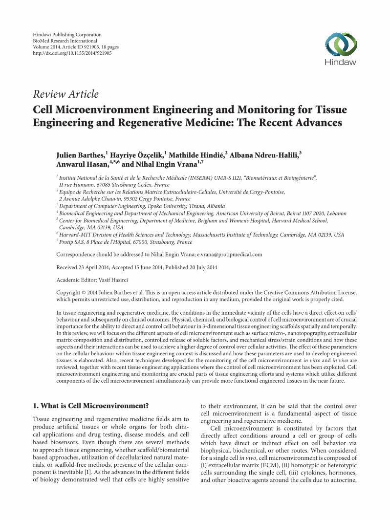
Review Article Cell Microenvironment Engineering and Monitoring for Tissue Engineering and Regenerative Medicine: The Recent Advances Julien Barthes, 1 Hayriye Özçelik, 1 Mathilde Hindié, 2 Albana Ndreu-Halili, 3 Anwarul Hasan, 4,5,6 and Nihal Engin Vrana 1,7 1 Institut National de la Sant´ e et de la Recherche M´ edicale (INSERM) UMR-S 1121, “Biomat´ eriaux et Bioing´ enierie”, 11 rue Humann, 67085 Strasbourg Cedex, France 2 Equipe de Recherche sur les Relations Matrice Extracellulaire-Cellules, Universit´ e de Cergy-Pontoise, 2 Avenue Adolphe Chauvin, 95302 Cergy Pontoise, France 3 Department of Computer Engineering, Epoka University, Tirana, Albania 4 Biomedical Engineering and Department of Mechanical Engineering, American University of Beirut, Beirut 1107 2020, Lebanon 5 Center for Biomedical Engineering, Department of Medicine, Brigham and Women’s Hospital, Harvard Medical School, Cambridge, MA 02139, USA 6 Harvard-MIT Division of Health Sciences and Technology, Massachusetts Institute of Technology, Cambridge, MA 02139, USA 7 Protip SAS, 8 Place de l’Hˆ opital, 67000, Strasbourg, France Correspondence should be addressed to Nihal Engin Vrana; [email protected] Received 23 April 2014; Accepted 15 June 2014; Published 20 July 2014 Academic Editor: Vasif Hasirci Copyright © 2014 Julien Barthes et al. is is an open access article distributed under the Creative Commons Attribution License, which permits unrestricted use, distribution, and reproduction in any medium, provided the original work is properly cited. In tissue engineering and regenerative medicine, the conditions in the immediate vicinity of the cells have a direct effect on cells’ behaviour and subsequently on clinical outcomes. Physical, chemical, and biological control of cell microenvironment are of crucial importance for the ability to direct and control cell behaviour in 3-dimensional tissue engineering scaffolds spatially and temporally. In this review, we will focus on the different aspects of cell microenvironment such as surface micro-, nanotopography, extracellular matrix composition and distribution, controlled release of soluble factors, and mechanical stress/strain conditions and how these aspects and their interactions can be used to achieve a higher degree of control over cellular activities. e effect of these parameters on the cellular behaviour within tissue engineering context is discussed and how these parameters are used to develop engineered tissues is elaborated. Also, recent techniques developed for the monitoring of the cell microenvironment in vitro and in vivo are reviewed, together with recent tissue engineering applications where the control of cell microenvironment has been exploited. Cell microenvironment engineering and monitoring are crucial parts of tissue engineering efforts and systems which utilize different components of the cell microenvironment simultaneously can provide more functional engineered tissues in the near future. 1. What is Cell Microenvironment? Tissue engineering and regenerative medicine fields aim to produce artificial tissues or whole organs for both clini- cal applications and drug testing, disease models, and cell based biosensors. Even though there are several methods to approach tissue engineering, whether scaffold/biomaterial based approaches, utilization of decellularized natural mate- rials, or scaffold-free methods, presence of the cellular com- ponent is inevitable [1]. As the advances in the different fields of biology demonstrated well that cells are highly sensitive to their environment, it can be said that the control over cell microenvironment is a fundamental aspect of tissue engineering and regenerative medicine. Cell microenvironment is constituted by factors that directly affect conditions around a cell or group of cells which have direct or indirect effect on cell behavior via biophysical, biochemical, or other routes. When considered for a single cell in vivo, cell microenvironment is composed of (i) extracellular matrix (ECM), (ii) homotypic or heterotypic cells surrounding the single cell, (iii) cytokines, hormones, and other bioactive agents around the cells due to autocrine, Hindawi Publishing Corporation BioMed Research International Volume 2014, Article ID 921905, 18 pages http://dx.doi.org/10.1155/2014/921905
Transcript of Review Article Cell Microenvironment Engineering and...
Review ArticleCell Microenvironment Engineering and Monitoring for TissueEngineering and Regenerative Medicine The Recent Advances
Julien Barthes1 Hayriye Oumlzccedilelik1 Mathilde Hindieacute2 Albana Ndreu-Halili3
Anwarul Hasan456 and Nihal Engin Vrana17
1 Institut National de la Sante et de la Recherche Medicale (INSERM) UMR-S 1121 ldquoBiomateriaux et Bioingenierierdquo11 rue Humann 67085 Strasbourg Cedex France
2 Equipe de Recherche sur les Relations Matrice Extracellulaire-Cellules Universite de Cergy-Pontoise2 Avenue Adolphe Chauvin 95302 Cergy Pontoise France
3 Department of Computer Engineering Epoka University Tirana Albania4 Biomedical Engineering and Department of Mechanical Engineering American University of Beirut Beirut 1107 2020 Lebanon5 Center for Biomedical Engineering Department of Medicine Brigham and Womenrsquos Hospital Harvard Medical SchoolCambridge MA 02139 USA
6Harvard-MIT Division of Health Sciences and Technology Massachusetts Institute of Technology Cambridge MA 02139 USA7 Protip SAS 8 Place de lrsquoHopital 67000 Strasbourg France
Correspondence should be addressed to Nihal Engin Vrana evranaprotipmedicalcom
Received 23 April 2014 Accepted 15 June 2014 Published 20 July 2014
Academic Editor Vasif Hasirci
Copyright copy 2014 Julien Barthes et al This is an open access article distributed under the Creative Commons Attribution Licensewhich permits unrestricted use distribution and reproduction in any medium provided the original work is properly cited
In tissue engineering and regenerative medicine the conditions in the immediate vicinity of the cells have a direct effect on cellsrsquobehaviour and subsequently on clinical outcomes Physical chemical and biological control of cell microenvironment are of crucialimportance for the ability to direct and control cell behaviour in 3-dimensional tissue engineering scaffolds spatially and temporallyIn this review we will focus on the different aspects of cell microenvironment such as surfacemicro- nanotopography extracellularmatrix composition and distribution controlled release of soluble factors and mechanical stressstrain conditions and how theseaspects and their interactions can be used to achieve a higher degree of control over cellular activitiesThe effect of these parameterson the cellular behaviour within tissue engineering context is discussed and how these parameters are used to develop engineeredtissues is elaborated Also recent techniques developed for the monitoring of the cell microenvironment in vitro and in vivo arereviewed together with recent tissue engineering applications where the control of cell microenvironment has been exploited Cellmicroenvironment engineering and monitoring are crucial parts of tissue engineering efforts and systems which utilize differentcomponents of the cell microenvironment simultaneously can provide more functional engineered tissues in the near future
1 What is Cell Microenvironment
Tissue engineering and regenerative medicine fields aim toproduce artificial tissues or whole organs for both clini-cal applications and drug testing disease models and cellbased biosensors Even though there are several methodsto approach tissue engineering whether scaffoldbiomaterialbased approaches utilization of decellularized natural mate-rials or scaffold-free methods presence of the cellular com-ponent is inevitable [1] As the advances in the different fieldsof biology demonstrated well that cells are highly sensitive
to their environment it can be said that the control overcell microenvironment is a fundamental aspect of tissueengineering and regenerative medicine
Cell microenvironment is constituted by factors thatdirectly affect conditions around a cell or group of cellswhich have direct or indirect effect on cell behavior viabiophysical biochemical or other routes When consideredfor a single cell in vivo cell microenvironment is composed of(i) extracellular matrix (ECM) (ii) homotypic or heterotypiccells surrounding the single cell (iii) cytokines hormonesand other bioactive agents around the cells due to autocrine
Hindawi Publishing CorporationBioMed Research InternationalVolume 2014 Article ID 921905 18 pageshttpdxdoiorg1011552014921905
2 BioMed Research International
QoutQin
(a)
MM
Papplied
(b)
PDMS variable E
15120583m
Seeding Confinement
Δy
Δy = 3120583m or 7120583mAttachment
PappliedE = 130kPa or 1MPa
(c)
Figure 1 The effect of microlevel mechanical confinement on the division of HeLa cells (a) and (b) show the macroscopic structure of themicrofluidic system and the cross-section of PDMS posts By the application of pressure on the posts cells can be confined within the areabetween the posts (the distance between the posts is 40120583m) (c)The confinement caused significant changes in the behavior of the cells duringmitosis such as delays in mitosis and led to daughter cells of different sizes and multidaughter cells following mitosis Reproduced from [4]
endocrine and paracrine secretions (iv) nanomicroscaletopography and physical properties of the adjuvant cells andthe ECM and (v) mechanical forces caused by the movementof the organism or the movement of the physiological fluidssuch as blood All these have a compound effect on the behav-ior of the cells where the relative importance of each com-ponent is tissue and cell type dependent and the next gen-eration of engineered tissues must imitate these effects asmuch as possible to be functional in their target areas as along term clinical solution
Cell microenvironment has many aspects and theircontrol can result in substantial changes in cell behaviorFor example Satyam et al showed that by macromolecularcrowding in the cell microenvironment the secretion of ECM
molecules can be significantly improved for corneal fibro-blasts [2] Another recent work shows that nanoscale micro-patterned and highly flexible membranes can be used todevelop retinal pigment epithelium layers forminimally inva-sive implantation within the eye [3] By mechanically con-fining cells in a microfluidic platform researchers were alsoable to control the mitotic processes (Figure 1) [4]
Microenvironment of stem cells is a particularly impor-tant topic in tissue engineering and regenerative medicine asthey are currently the most technically feasible source whichcan provide the large amount of cells needed for engineer-ing clinically relevant amounts of tissue Stem cell reservoirsare available for replenishment of the tissue in tissues inhuman body The microenvironmental control over how
BioMed Research International 3
these cells can keep their plasticity [5] that is how they canstay quiescent and be utilized by the body only in case ofnecessity under healthy conditions is a benchmark that needsto be met by engineered tissues Moreover failure to con-trol the microenvironment of stem cells can also havedeleterious effects such as dedifferentiation and subsequenttumor growth
Another important concept related to the mimickingof tissue microenvironment is multidimensionality as mostof the components of tissues have multidimensional orderand orientation which necessitates mimicry to achieve theirfunction [6] Multidimensionality is also an important aspectof other uses of tissue engineering namely model tissuesand organs for pharmaceutical testing and also fundamentalresearch These microorgan structures should match thedimensional properties of the tissue and the organ theyrepresent [7]
In this review we will focus on different aspects of cellmicroenvironment and their direct effects on tissue engineer-ing applications with particular focus on osteogenesis andangiogenesis Each component of the cell microenvironmentwill be discussed separately and also in conjunction with theother components
2 MicroNanopatterning forMicroenvironment Engineering
All the cells in the human body are surrounded by topograph-ical and biochemical signals The physical structures com-prise nanopores nanofibers and nanocrystals Some exam-ples of such structures in physiological settings are nanoporesin capillaries nanofibers in the basement membrane andnanocrystals in the formof hydroxyapatite in the bonemicro-structure Aligned cells are very prevalent in the tissue Forinstance maintenance of cell alignment is essential for mus-cle cardiovascularblood vessel and corneal and nerve tissueengineering inwhich the controlling tissuemicroarchitectureand biological function are tightly connected Various strate-gies have been developed to induce cell alignment includingtopographical patterning (eg micro- and nanogrooves andaligned nanofibers) chemical treatment (patterns with cell-adhesive or repellent chemistries) controlled stressstrainconditions (eg stretching fluid shear stress and compres-sion) and a combination of these methods
From topography point of view recent advances inmicro-andnanofabrication enabled development of complex surfacefeatures by controlling their pattern periodicity shape anddimensional properties Today design and construction ofsubstrateswithwell-controlled physical and chemical proper-ties andmicro- and nanoarchitecture have become an impor-tant tool in the construction of tissue engineered replace-ments Several top-down and bottom-up techniques such asphase separation self-assembly thin film deposition chem-ical vapor deposition chemical etching nanoimprintingphotolithography scanning probe lithography and electronbeam lithography [11] can be used in order to tailor micro-and nanoscale structured environments (scaffoldssurfaces)to stimulate cell growth and guide tissue regeneration inmuch the same way the extracellular matrix (ECM) does
It is well known that cells can align along micro- andnanosized parallel groovesridges patterns [12ndash19] Severalstudies indicate that alignment occurs when the periodicityand dimensions of the patterns are above a critical valueFor example Loesberg et al [20] have shown that thefibroblasts did not show noticeable alignment with groovedepths around 35 nm and ridges narrower than 100 nm Inanother study 100 nm depth was determined as a thresholdfor alignment of cardiomyocytes [21] osteoblast-like cells[16 22] and hepatoblastoma cells [23] Onmicrogrooved sur-faces groove depth is one of the most important parametersin defining cell alignment The degree of alignment of thecells along the microscale grooves is generally proportionalto groove depth and inversely proportional to grooveridgewidth if the other parameters are fixed [24] On the otherhand Glawe et al [25] studied that effect of high aspect ratio(aspect ratio = groove depthgroove width) microchannelswith varying widths (20ndash60120583m) on the alignment of smoothmuscle cells It was observed that alignment was dependenton the channel width and narrow microchannels (20120583mand 30 120583m) promote alignment of smooth muscle cells Onnanogrooved substrates cell orientation was also found tobe also less sensitive to groove width (90 to 500 nm) withMG-63 cells and C3A cells [16 22] When the ridges aresmaller than that of focal adhesions (025ndash05120583m wide and20ndash100 120583m long) cell alignment is inhibited Nanogrooveswere too narrow for the cells to descend into the bottom ofgrooves Thus the focal adhesions and actin filaments arelocalized on the ridges However for vascular smoothmusclecells channel widths as small as 3325 nm have been shownto induce alignment and subsequent mechanical propertyimprovement in the direction of alignment [26]
The lack of data on how height and groove width orquantitative interaction of these parameters which deter-mine the degree of cell orientation have forced researchersto establish aspect ratio dependent models For exampleKemkemer et al [27] developed a model for predicting thecell orientation for cases where the cell is larger than thegrooves According to this the square of the product ofgroove depth and spatial frequency or the aspect ratio forsymmetric grooves were found to be the important featuresfor alignment In another study Crouch et al [28] proposed asimple model to explain the relationship between aspect ratioand cell behavior on gratings with varying widths and depthsThey observed a direct relationship between the alignmentof human dermal fibroblasts and aspect ratios of the channeltype patterns While aspect ratios as small as 001 inducedsignificant alignment (60) 80 alignment was achievedwith an aspect ratio of 005 The maximum aspect ratiorequired for 95 alignment was 016 This study indicatesthat within a certain range the aspect ratio can be used forcontrolling cell response to substrate topography withoutdistinguishing the effects of width and depth However itis important to point out that when the grating surface iswider than cell width the probability of lateral cell spreadingis high Thus obtaining cell-type-specific contact guidancethresholds by the help of the abovementioned predictiontheories can be useful to tailor the cellular microenviron-ment
4 BioMed Research International
Cell alignment on physically patterned surfaces is awidely used strategy in some cases together with chemicalpatternsThe effect of chemical patterns [29 30] or synergisticeffects of physical and chemical patterning [31ndash34] were alsostudied intensely Generally in order to control cell adhesionand alignment molecules such as poly(-L-lysine) (PLL) pep-tides fibronectin laminin collagen bovine serum albuminand SAM (self-assembled monolayers) are patterned by softlithography techniques In some cases instead of synergisticeffects of chemical and physical patterning one of the cuescan overcome the other one For example when Charestet al [33] used grooves (4 120583m depth 8 120583m width) overlaidwith an orthogonal chemical pattern (10 120583m adhesive laneswith spacing ranged from 10 to 100 120583m) physical topographydetermined the alignment of osteoblast-like cells Anothermeans to produce patterned surfaces is to use thermorespon-sive polymers These ldquosmartrdquo polymers based for exampleon a poly(N-isopropylacrylamide) backbone with n-butylmethacrylate side chains are capable of a reversible transitionfrom hydrophilic to hydrophobic state when their temper-ature is lowered by a few degrees (around its low criticalsolution temperature of 32∘C) Thermoresponsive polymerscan be used for cell sheet engineering to treat a wide rangeof diseases from corneal dysfunction to oesophageal cancertracheal resection and cardiac failure as growth substrates forcells [35] By using patterned thermoresponsive surfaces it ispossible to pattern cell sheets when the application requirescellular alignment
Biological tissues are hierarchically organized fromnano-meter-to-centimeter scale For instance the average rough-ness of bone tissue is 32 nm and bone tissue has a hierarchicalstructure composed of collagen and hydroxyapatite [36]During bone mineralization the hydroxyapatite crystalsform micro- and nanocomposites with collagen fibers Thuspreparing biomimetic surfaces which present synergisticeffects of micro- and nanostructures is expected to provideadditional advantages Two separate studies have examinedthe behavior of osteoblasts derived from bone marrow stro-mal cells on micropit and nanonodule hybrid topography ofTiO2[37 38] They created nanonodules with diameters of
100 nm 300 nm and 500 nm by self-assembly technique anddemonstrated that 300 nm nanonodule containing substratescreated the most promising environment for osteoblastdifferentiation and bone-titanium integration These resultsare in agreement with more recent studies [39 40] whichhave indicated that the presence of micron and submicron-scale surface roughness on the same surface can acceleratebone differentiation In another study surface features whichare similar in scale to osteoclast resorption pits were used tostudy in vitro bone formation in basal medium [41] Here thepit dimensions were as follows depth 330 nm diameters 2030 and 40 120583m and centre-centre spacing 50 60 and 90120583mOsteopontin expression was relatively high in the humanosteoblasts grown on the larger diameter (30 and 40120583m)pits In addition to expression of osteogenic markers maturecalcium depositions were shown by alizarin red staining onthese substrates
In the last decade several studies reported that micro-and nanopatterned surfaces can be a valuable tool for directed
growth [15 42] and differentiation [19 32 43 44] of neuritesIn one of the recent studiesMigliorini et al [45] analyzed theeffect of nanopillars on differentiation of embryonic stem cellderived progenitors in the absence of biochemical factors andobserved an increase in the neuronal yield with increasingpillar height from 35 to 400 nm Pan et al [46] tested theeffects of nanograting substrates with different widths onhuman inducedpluripotent stemcells (hiPSCs)Gene expres-sion profiling by real-time PCR and immunostaining showedsignificant upregulation of neuronal markers on nanostruc-tured substrates either with solely topographical cues orcombined with preneuronal induction A width of 350 nm inparticular induced the highest neuronal marker expressionThe responsiveness of the cells to nanometer scale cues stemsfrom their specific interactions with extracellular matrix(ECM) which is covered in the following section
3 Microenvironmental Effects ofExtracellular Matrix
The ECM comprises of a wide range of molecules includingcollagen elastin laminin fibronectin various glycoproteinsproteoglycans and polysaccharides [47 48] Each ECM com-ponent provides different functionalities to this complex net-work made of structural proteins (eg collagen and elastin)adhesive proteins (eg fibronectin and vitronectin) and gly-cosaminoglycans (eg hyaluronic acid and heparin sulphate)by presenting different structural and biochemical properties[49] The microenvironment created by ECM surroundingadherent cells is essential to their survival ECM is a physicalsupport to physiological cells the signals for functional ori-entations such as migration proliferation and even survivalare transduced from ECM The absence of cell adhesion toECM induces cell death by apoptosis [50] This particulartype of apoptosis is named anoıkis (Greek word which meansldquohomelessnessrdquo or loss of home) This phenomenon was firstdescribed in epithelial cells [51] and contributes to maintaintissue homeostasis [52] In physiological conditions adherentcells are protected from anoıkis by the binding to ECM andthe resulting activation of intracellular survival signallingpathways The loss of anoikis induction signal constitutes ahallmark of cancerous cells and contributes to the formationofmetastasis [53 54]Thus presentation of an ECMmimic tothe cells in tissue engineering applications is important Thethree-dimensional organization of the ECM has a regulatoryeffect on cell cycle as seen in mammary epithelial cells as theECM suppresses apoptosis suggesting that ECM signaling isdefined by the organization of the cells within a tissue thatis cell shape intercellular spacing and 3D position Thesefactors determine cellular response to signals
The microenvironment created by ECM componentssuch as adhesive proteins or glycosaminoglycans maintainstissue stability and cell behavior Bone matrix for instancecontains 90 of collagen type I and only 5 of noncol-lagenous proteins like osteocalcin osteonectin fibronectinor hyaluronan and mineral compounds which are essentialto conserve osteoblasts phenotype [55] whereas culturingchondrocytes on type I collagen induces their dediffer-entiation [56] Furthermore ECM components selectively
BioMed Research International 5
influence cell adhesion and shape as described by Schlie-Wolter et al [57] Hence cell morphology directed bythe interaction with ECM induces modifications of theirbehaviour and subsequently their fate [58]
One of the main examples of cell signaling is integrin-mediated signaling for cell adhesion where the connectionrequires structures of focal adhesion that contain complexmixture of proteins Cell adhesion to ECM is led by trans-membrane heterodimeric integrin receptors During devel-opment integrins facilitate tissue morphogenesis by deter-mining which ECM components the cell would bind to Inte-grins are the major mediators of cell-ECM contacts and theyare essential to the outside-in transmission of signals fromcell microenvironment [59] Integrin and ligand bindingslead to the formation of focal adhesion complexes whichare linked to the intracellular actin cytoskeleton [60 61]Another example of this structure-dependent ECM signalingpathway is in tyrosine kinases [62] For cell binding andmigration integrin signaling modulates the cell signalingpathways of transmembrane protein kinases such as receptortyrosine kinases (RTK) RTK are high-affinity cell surfacereceptors for many polypeptide growth factors cytokinesand hormones The study of receptor tyrosine kinase (RTK)signaling led to the understanding of how an extracellularsignal is transmitted to the nucleus to induce a transcriptionalresponse [63] Other nonintegrin adhesion receptor familiesinclude selectins cadherins immunoglobulins proteogly-cans and some other laminin-binding proteins In shortthis mode of interaction conveys biochemical and positionalinformation by which the cell can know how and when itshould undertake a particular activity
ECM is coupled to cytoskeletal and signalling effec-tor elements which direct crucial downstream functionssuch as cell growth survival and transcriptional activity[64] Biochemical and biomechanical modifications of ECMmicroenvironment are transmitted to cells and induce theresulting changes in their behaviour [65] Cell mechanosens-ing is mediated by focal adhesion contacts [66] Indeedphysical and mechanical forces in cell microenvironmentlead to changes in cell morphology and differentiation Notonly the composition of the ECM has direct effects on cellbehaviour but also its physical properties Stiffness of boneECM is essential to maintain osteoblast phenotype whereaschondrocytes dedifferentiate where they are cultured on arigid matrix [67] Elasticity of ECM determines also thedifferentiation of progenitor cells [68] Furthermore physicalmodifications of an adhesive protein such as fibronectinare sufficient to influence cell activities The stretching offibronectin alters its binding to ligands [69] and moreimportantly fibronectin conformation regulates integrinsattachment which controls downstream cell behaviour [70]Another type of ECM component variation is biochemicalGlycosylation is one of the most abundant protein modifica-tions having a role in protein stability secretion and functionTheO-glycosylation in particular is essential in cell adhesionZhang and Hagen demonstrated that loss of glycosylationdisrupts adhesion of epithelial cells and more generallyinfluences cellular microenvironment [71] Moreover gly-cosylation of adhesive proteins like laminin or fibronectin
also stimulates cancerous cell proliferation and dissemination[72]
All differentiated cells have a cell type specific proteinexpression profilewithmultifunctional criteria that is respon-sible for development and protein regulation This proteinexpression creates an output signal to be used by cellsto control their roles Changes in the protein expressionprofile or mutations that result in down- or upregulation ofspecific proteins can be the causes of cardiac muscular ormental illnesses Hyper- and hyposensitivity responses to thestrength of stimuli can also cause sickness in the body Inorder to model such illnesses tissue engineering and bio-materials studies concentrated on producing artificial ECMnetworks that are made up of synthetic polypeptides orpeptide-conjugated synthetic polymers that present bioactiveligands and respond to cell-secreted signals to enable prote-olytic remodeling The production of such biomaterials canbe used in differentiating stem cells into neurons [73] Thegoal of this approach is to mimic the properties of ECMTheareas of biomimicry are specific cell adhesion degradationby proteolytic processes involved in cell migration and tissueremodeling and the ability to control cellular functions suchas ECM synthesis An example of such systems is the use ofphotopolymerizable polyethylene glycol (PEG) based hydro-gels as tissue engineering scaffolds This material showedwhen modified with necessary signals that it can interactwith cells in a manner similar to that of natural ECM especi-ally in transmitting bioactive signals that control tissue for-mation and cell phenotype
ECM microenvironment is not permanent changesduring aging were observed in different organs with variabletimes of onset Due to the specific interactions between dif-ferent tissues cells and their surroundings the cells modifytheir own environment by reshaping their ECM componentsinto the correct configuration that allows the growth of thefunctioning tissues which have specific architecture andcharacteristics ECM components are essential to stem cellmaintenance and subsequently to support tissue regeneration[74]
4 Phenotype Control andStem Cell Differentiation viaMicroenvironmental Cues
Tissue homeostasis requires a certain level of phenotypicplasticity from resident cells and also the involvement of cir-culating cellsThemost apparentmanifestation of this need isobserved upon injurywhere the inflammatory reactionmedi-ated by immune cells such as neutrophils and macrophagesdecides how an implant transplant or an engineered tissueintegrates with the bodyThe phenotypes of the immune cellsin the microenvironment have a significant effect on the finaloutcomeMoreover many tissues depend on several differentcell types with given phenotypes The quality of bone tissuedepends on the interaction between osteoblasts osteoclastsand osteocytes Respiratory epithelium not only has a ciliaryepithelium layer but also requires basal cells and glandularcell components
6 BioMed Research International
One of the new paradigms in tissue engineering is theutilization of developmental pathways for engineering tissues[75] In one of the recent demonstrations of ldquodevelopmentaltissue engineeringrdquo [76] Scotti et al were able to producea bone organ with functioning bone marrow by putting thehuman mesenchymal stem cells through an endochondralbone formation route that is formation of bone organ via acartilaginous tissue step [8]Thiswas achieved via productionof hypertrophic cartilage tissue by application of IL-1120573 andsubsequent subcutaneous implantation By applying bothphysical and chemical microenvironmental controls theywere able to push the initial hypertrophic cartilage structureto produce several cell types with their proper phenotypeswhich demonstrates the strength of the developmental tissueengineering methods (Figure 2) [8]
The most active literature concerning cell phenotype intissue engineering is the research on stem cells Stem cells(SCs) a subset of cells with replenishing ability and thepotential of differentiation into various types of mature cellsare categorized into two main groups namely embryonicstem cells (ESCs) and adult stem cells (ASCs) It has beenshown that the intrinsic genetic programs within these cellsand some extracellular regulatory signals control the abilityof SCs to proliferate and differentiate into different functionalmature cell types [77]
Stem cells reside in a specialized microenvironmentcalled stem cell niche which provides the stem cells withextracellular cues to allow their survival and identity Thisniche is a key regulator to the stem cell behavior becauseit ensures a quiescent and low metabolic environment toprevent exhaustion It is believed that microenvironmentalproperties of the niche provide a good balance betweenthe ability of SCs to renew themselves and the ability todifferentiate into mature cells so that continuous tissueregeneration occurs Amajor part of the cell niche is the ECM(extracellular matrix) which possesses the specific mechani-cal biochemical and biophysical properties for tissues andcontrols the overall cell behavior [78]The composition of theECM provides full support to the niche through its physicaland structural properties The main extrinsic signals thatregulate stem cell behavior are those coming from ECM
Given its three-dimensional organization the ECM pro-vides an environment that aids in the integration of the sig-nals derived from the cell-ECM interactions in order to allowproper ldquomaintenance of stem cell homeostasisrdquo [78]The cell-ECM interactions are basically triggered by receptors presenton the cell membrane like integrin as described beforeHowever studies found that the nonintegrin receptors are theones that contribute the most to stem cells homing duringtransplantation Novel techniques have been developed toobserve the interaction between stem cells and ECMproteinsand how this interaction influences their fate Among thefactors that influence stem cell fate are ECM adhesion itsstiffness and its topography [79] For example the effectof micro-nanotopography on stem cells has been recentlydemonstrated Oh et al [80] demonstrated that humanMSCscan differentiate into osteoblasts under the influence ofonly nanotopography of culture substrates Another exampleis micropatterned islands created with specific shapes to
observe cell behavior at single-cell levelThedegree of spread-ing of human epidermal stem cells was observed by Connellyet al [81]The authors stated that when the shape of the islandwas changed from elongated to circular epidermal stem cellsshowed an increase in their differentiation ability On theother hand humanMSCs revealed a dependence on the areaof the island that is while round cells favoured adipogenesiswhereas cell spreading resulted in osteogenesis [82]
In addition to topographical cues soluble factors likegrowth factors and cytokines are very important in initiationand control of SC differentiation [83] Tissue engineering hasbecome an important stem cell application field with the aimof increasing the quality of life Therefore researchers havefocused more on finding appropriate cues via utilization ofbiomaterials that could control the cellular environment andmonitoring complex cellular levels Both natural and syn-thetic materials based biomaterial scaffolds have served tounderstand the role of chemical cues in controlling stem cellbehaviour It is crucial to direct SCs to differentiate into theright cell type at the right time and location therefore spe-cific cues have been investigated in the in vivomicroenviron-ment and have been studied in the in vitro systems that mim-icked the natural conditions Controlled microenvironmentshave been designed to direct stem cell differentiation into thedesired mature cell type Stem cell researchers emphasize theneed of a 3D environment instead of 2D since differences havebeen observed in their self-renewal capacity differentiationadhesion and migration ability Cellular morphology hasbeen shown to vary depending on the biomaterials structure(2D or 3D) and material type Human mesenchymal stemcell shape was observed to be round when entrapped in 3Dhyaluronic acid hydrogel [84] and elongated when seededonto fibrous scaffolds or 2D biodegradable elastomer [85]There are other effects regarding the encapsulation of cellsas demonstrated by encapsulation of prostate cancer cells(LNCaP) in polyethylene glycol (PEG) hydrogels whichchanged their cell-cell contact formation and response toandrogen stimulation where these effects are also relevant tothe differentiation of stem cells within confined environments[9] (Figure 3)
5 Cell Microenvironment Control viaDelivery of Soluble Bioactive Agents
Another way to control the cell microenvironment is viadelivery of bioactive molecules such as drugs hormones orgrowth factors Variation in the signaling microenvironmentmight cause perturbations in the signaling processes whichare at the root of multiple pathologies including cancers dia-betes andmany other diseases [63] Growth factors can regu-late activation growth proliferationmigration and differen-tiation of cells which are crucial for events such as angio-genesis or osteogenesis [86] Recent studies have focusedon inserting signaling molecules such as growth factorsand cytokines into biomaterials (Figure 4) Some examplesof altering cell behaviour to such molecules are inducedvascularisation (new blood vessel formation from fibroblastgrowth factor 2mdashFGF-2) regeneration of neurons (from
BioMed Research International 7
Massonrsquos trichrome 3D120583CT
In v
itro
In v
ivo5
wIn
viv
o12
w
Safranin O
Figure 2 Bone formation via endochondral pathway An in vitro formed artificial cartilage successfully forms a bone containing bonemarrowwithin 12 weeksThe in vitro grown tissue is a cartilaginous one as evidenced by the extensive safranin O staining over time the cartilaginoustissue has been gradually replaced by bone tissue as can be seen by the extensive Massonrsquos Trichrome staining Micro-CT images also showedthe development of a bone like structure within 12 weeks Reproduced from [8]
nerve growth factor NGF) retention of stem cell pheno-type (from immobilization of cytokines to maleic anhydridecopolymer thin-film coating) and providing an environ-ment that helps cell survival and proliferation Epidermalgrowth factor (EGF) incorporated with the matrix mate-rial increases cell attachment to the implanted matrix andincreases the spreading of mesenchymal stem cells [10]
However these molecules have high instability and veryshort biological half time and can be enzymatically digestedor deactivated while in physiological fluids [87 88] Besidesgrowth factors or drugs need to reach specific location to beeffective thus their systemic introduction is not a viable wayto control their concentration in specific target areas [89]To overcome these limitations it was necessary to developdelivery vehicles with growth factors or drugs incorporatedwithin tissue engineering scaffolds To illustrate this factit has been shown that bolus injection of growth factorssuch as VEGF is less effective than a sustained and localizeddelivery via biodegradable hydrogels to achieve blood vesselformation [90] In the case of bolus injection VEGF was notlocalized in the target area and stayed only for 72 h whereaswith delivery from biodegradable alginate hydrogel 95 ofthe growth factor was at the ischemic site (improvement ofbiodistribution) and it stayed at that location for more than15 days and its bioactivity was higher (possibly due to theprotection from denaturation)
One way to achieve control over local bioactive moleculeconcentrations is the immobilization of growth or differenti-ation factors Mainly the ECM harbors a lot of growth factorbinding proteins This localization of growth factors by theECM and their signaling contributes to the establishment ofa gradient for the soluble diffusible morphogens which play
vital roles in shaping the developmental processes [91] Thebinding of growth factors to the ECM is regulated by theGAG side chains One important application of this is theregulation of specific gene expression which is done by usinggrowth factor-ECM interaction that is by controlling thegrowth factor presence via their interaction with ECM [92]
Incorporation of bioactive factors into scaffold can beachieved by two different ways mainly through covalentand noncovalent immobilization [93ndash95] The first approachis based on the covalent binding of the molecule to thescaffold via chemical reaction such as immobilization ofVEGF using N-(3-dimethylaminopropyl)-N1015840-ethylcarbodi-imide hydrochloride chemistry (EDC) [96]The noncovalentapproach is based on interaction of the molecule with thepolymer matrix such as electrostatic interaction hydrogenbonding or physical entrapment In the case of incorporationvia electrostatic interaction charged material is requiredsuch as polyelectrolytes or gelatin hydrogels In all thesedifferent approaches the release will be triggered by scaffolddegradation diffusion of the molecule through the materialor cleavage (enzymatic or hydrolysis) of the covalent bondbetween the molecule and the scaffold material For examplewith the addition of bioactive motifs (from bone morpho-genetic protein 2 or osteopontin) it was shown that osteoblastadhesion and the responsiveness to the protein were depen-dent on the cell adhesive motif from osteopontin The cellinteraction with the protein demonstrated in vitro boneformation in a month [97]
These delivery systems were developed with differentkind of materials Two main categories can be identified (i)natural materials such as collagen alginate gelatin and poly-L-lysine (ii) synthetic materials such as PLLA PEG and PCL
8 BioMed Research International
2D 3D
(a)
(b)
Figure 3 Manipulating the cell microenvironment in 3D via encapsulation within hydrogels Encapsulation of prostate cancer cells withinPEG hydrogels resulted in more pronounced cell-cell contacts as evidenced by E-cadherin staining (a) and also formation of a necrotic corewithin the cell aggregates as shown by pimonidazole staining (b) All scale bars are 75 120583m for (a) and 100120583m for (b) Reproduced from [9]
BioMed Research International 9
Signaling molecules
Neurotransmitters Cytokines
VEGF
Neuron outgrowthMaintenance of
undifferentiated stateVascularization
Growth factors
BMP-2
Boneformation
Dopamine LIF
Figure 4 The main types of soluble factors that have distincteffects on the cellular behaviour at both single cell and tissue levelControlled delivery of such factors and their regulated presence incell microenvironment is an indispensable tool in tissue engineeringresearch Reproduced from [10]
[95] To be an efficient carrier system these materials mustfulfill some requirements (i) biocompatibility (ii) biodegrad-ability and (iii) release of active factors in a controlled spatio-temporal way [98] Release of growth factors from scaffoldsis mainly governed by twomechanisms (i) diffusion throughthe material and (ii) degradation rate of the material [86 99]Release profile of bioactive molecules is a key parameter tocontrol cell microenvironment Depending on the applica-tion such as enhancement of angiogenesis stem cell dif-ferentiation or disease treatment bioactive factors need tobe released for specific time points at specific rates In thecase of degradable scaffolds the release of molecules can betuned by varying the degradation profile of the material orthe molecule diffusion The degradation rate of the scaffoldcan be changed by crosslinking to reinforce the structureand delay the release Gelatin is a biomaterial obtained bydenaturing collagen It is a good ECMmimickingmaterial forcells [100] Moreover gelatin is biodegradable and has beenused for a long time inmedical field Gelatin is also a very use-ful material for drug incorporation because it can be pos-itively (basic gelatin) or negatively charged (acidic gelatin)depending on collagen processing method (acid or alkalineprocess) so it can complex both positively or negativelycharged molecules [101 102] This provides a level of ver-satility which is not available with other commonly usedbiomaterials Gelatin hydrogels are systemically cross-linkedwith different agents such as genipin transglutaminase orEDCNHS because unless cross-linked the structure of thephysically gelled gelatin hydrogels or films is too weak andthe degradation is too fast [103ndash105] For other materialsdifferent techniques are available For example to controldegradation rate of alginate hydrogels a partial oxidationof the polymer chains rendered the hydrogel degradable byhydrolysis [90] This strategy has been used to create a deliv-ery vehicle for VEGF Synthetic hydrogels can also be used toencapsulate and release bioactive molecules Hyperbranchedpolyester hydrogels capable of encapsulating hydrophobicmolecules such as growth factors or specific drugs (egdexamethasone) have been developed These hydrogels arephotocross-linkable via incorporation ofmethacrylate groupNormally it is very difficult to entrap hydrophobic agents
in hydrogels In this case a sustained release of 8 days wasachieved mainly through hydrolysis of ester backbone [106]
Polyelectrolyte multilayer structures (L-b-L layer bylayer) are also used to design delivery systems because L-b-L films are easy to produce they can act as a reservoir forbioactivemolecules [107] and their properties such as perme-ability thickness and charge density can be easily changedand they can be easily coated on implants [108] The onlyproblem with these films for drug delivery application is thefast release of molecule due to the mobility of polyelectrolytechains within the film To solve this problem recently adouble entrapment system has been developed for VEGF toachieve a long term release [109] This strategy was basedon the twofold control over the release by VEGF containingPCL nanoparticles loaded in polyelectrolyte multilayer filmThe mechanism of the release was the following either PCLnanoparticles containing VEGF were hydrolyzed and thenVEGF diffused in the LBL film and then out or the particlewill diffuse out of the film and then hydrolyzed With thissystem a sustained release of 7 days was achieved [109] Toprevent the fast release of drug out of LBL film anothersystem was developed by adding a mechanosensitive cap asa barrier on top of LBL reservoir films A bioactive agentwas loaded in PLLHA film and a PAHPSS barrier wasbuilt on top of it Barrier is cracked under stretch whichenables the diffusion of an enzyme (trypsin) within thereservoir and the PLLHA is enzymatically degraded leadingto the release of drug [110] Layer by layer technique (LBL)with polyelectrolytes can also be used in particle form (ieparticles formed by polyelectrolyte multilayers) [111] Usingthis technique a stimuli-responsive controlled drug releasehas been developed in order to release bioactive agents Thissystem was based on the absorption of the agent on meso-porous silica sphere and then the deposition of a multilayercapping barrier PAHPSS The release of the encapsulatedmolecule was further triggered by change of pH (pH = 14)or by change of ionic strength through NaCl concentrationof the release media (10mM NaCl) At higher pH value orlower ionic strength the PAHPSS layer acted as a cappingbarrier since it does not allow bioactive agent diffusion andthat explains why this system is appropriate for a controlledand sustained release of bioactive molecules [112]
In some other applications the delivery of multiple bio-active factors with different release kinetics is required In tis-sue engineering for example angiogenesis and osteogenesisare regulated by the action of multiple growth factors andall of them need to be released in a specific temporal wayRichardson et al have investigated the dual delivery of VEGFand PDGF two growth factors necessary for blood vesselsformation PLG particle with lyophilized VEGF and PLGmicrospheres containing encapsulated PDGFwere used [113]All these particles were mixed together and a porous PLGscaffold was made using high pressure carbon dioxide fabri-cation process These growth factors release profiles werenot the same 17 pmolday for VEGF for the first sevendays mainly due to VEGF diffusion out of the scaffoldand from 010 to 47 pmolday for PDGF depending ondegradation of polymer particle using different formulations[113] In the field of regenerative periodontal therapy an
10 BioMed Research International
interconnected macroporous Dex-GMA (glycidyl methacry-late dextran)gelatin hydrogel scaffold was developed forthe dual delivery of two different growth factors BMP-2 and IGF-1 These growth factors were encapsulated inDex-GMAgelatin microparticles basic gelatin (negativelycharged) was used to encapsulate BMP-2 and acidic gelatin(positively charged) for IGF-1 encapsulation As Dex-GMA isenzymatically degradable but not hydrolytically degradableother Dex-GMA microparticles were prepared with neutralgelatin to encapsulate dextranase in order to further triggerthe release of growth factors by enzymatic degradation ofthe Dex-GMAmicroparticles All these particles were mixedtogether and cross-linked via irradiation in order to makethe interconnectedmacroporous scaffoldThismechanism ofdelivery based on the degradation of microparticle followedby degradation of the scaffold enables the sustained release ofgrowth factors for a period of 20 days [114] Some disease likeperiodontitis required themultiple deliveries of antibacterialanti-inflammatory antiresorptive and osteogenic agentsThese molecules must be delivered in a very specific orderto be effective To fulfill this requirement a multilayereddevice was designed This laminate structure is made by theassociation of cellulose acetate phthalate (CAP) and PluronicF-127(P) and these polymers can be further eroded Thedifferent drugs have been incorporated in a specific orderin every stratum by mixing them with the polymer solutionand every layer is separated by one or two CAPP blanklayers in order to slow the erosion of the structure and delaythe release of molecules This system was able to performthe release of four different drugs in a specific temporalsequence just by unidirectional erosion of the structure formore than 120 hours depending on the condition used [115]The other important cell microenvironment parameter thatneeds to be mimicked is the changes induced on cells viadynamic stimuli such as mechanical stimuli The spatial andtemporal variations of cell microenvironment play vital rolesin various paracrine and endocrine cell signaling 3D tissueremodeling alteration in stem cell niches cancer progressionandmigration of various cells For these reasons the dynamicnature of the cell microenvironment is vitally important asdiscussed below
6 Dynamic Aspects of Cell Microenvironment
The native ECM microenvironments of cells are highly het-erogeneous in three-dimensional space [116] and they gothrough continuous and dynamic remodelingwith time [117]The interactions of the ECM components with cells are ofreciprocal nature that is while cells constantly producebreak down rearrange and realign components of ECMto change its properties and any change in ECM in turnregulates the activities and the behavior of the cells [47]The cell-ECM interactions are highly dynamic and complexhence a detailed understanding of the dynamic aspects ofcellular microenvironment in terms of spatial and temporalvariations of different chemical mechanical and biologicalstimuli is highly important in tissue or organ engineering
Bioreactors are a crucial part of tissue engineeringresearch as they are the main means to exert mechanical
stimuli to the cells in tissue engineering constructs Biore-actors are systems which enable the continuous replacementof nutrients and gases either by constant agitation (as in thecase of spinner flasks) or via perfusionMost of the bioreactorsystems have several entry ports which enables the intro-duction of different bioactive agents in a controlled mannerMoreover many bioreactors have the capacity to directlyapply physiologically relevant mechanical stressstrain con-ditions (such as tensile compressive shear stress accordingto the target tissue) They mimic the mechanical microen-vironment for functional engineered tissues that undergomechanical loading (articular cartilage tendons heart valvesetc) under in vivo conditions They are designed to increasethe efficiency of exchange of metabolites oxygen nutrientsand waste removal within the cell microenvironment and asa result to enhance cellular penetration in 3D scaffolds and tohave a better and rapid cellular expansion [118] For this aimthere exist perfusion bioreactors for culturing encapsulatedstem cells and those for cell cultivation on 3D scaffolds
Mimicking the dynamic mechanical environment is alsoconsidered in designing functional bone implants in conjunc-tion with micro-nanotopographical features These studiespointed out that osteoblasts change their morphology geneexpression and matrix mineralization by either introducingsurface topography on biomaterials or mechanical stimula-tion [119 120] Prodanov et al [121] tested the simultaneouseffects of nanotextured surface (300 nmwide and 60 nmdeepgrooves) and mechanical stretching in terms of cell attach-ment ECM formation and osteoblast differentiation It wasshown that by dual stimulation (nanogrooved surface and 8of strain) the expression of fibronectin and Cfba synergisti-cally increased 2-fold in comparison to nanotextured surfacealone Such combined effects of topography and mechanicalstimulation were also observed by other groups [41 122ndash124]
Mechanical stimulus by cyclic stretching fluid flow [121]and hydrostatic compressive pressure has also been used toalign cells In several studies cardiac ligament and tendonderived fibroblasts myoblasts vascular smooth muscle cellsand osteoblasts were subjected to mechanical forces under invivo conditions Regardless of cell types cells cultured on a2D substrate that is subjected to uniaxial cyclic stretch tendto align perpendicular to the direction of principal cyclicstrain [122 125 126] There are several studies that have usedmechanical stimuli together with micronanopatterning forcell alignment It has been shown that when cells grownwhether on patterned surfaces with micron sized or nano-sized grooveridge patterns or on unpatterned [127] sub-strates subjected to cyclic stretching orient themselves in thedirection perpendicular to the applied strain It should benoted that applied strains in these examples were parallel tothe direction of the groove axis However there are studiesshowing that tendon fibroblasts and osteoblasts aligned alongthe direction of micro grooves regardless of the stretchingdirection which suggests that the topographical cues atthis scale might be a more important determinant for thecell alignment direction [122 128] For instance mechani-cal loading topographical patterning and surface chemicaltreatment can also be combined to engineer cell alignmentin vitro The combined effects of cyclic strain and substrate
BioMed Research International 11
microtopography on the alignment of bovine vascular SMCshave been investigated by Ahmed et al [124] where theyobserved that the organisation of actin fibers was dominatedby cyclic strain application and the shape of cell nuclei wascontrolled by the patterns
61 Dynamic Control of Cell Microenvironment Using Micro-fluidics Researchers have designed cell-ladenmatrices in 3Dspace to mimic functions of human tissues and organs invitro Many of these structures also change over time (4Dbiology) [117] Pioneeringwork by Petersen et al revealed thatmammary epithelial cells formed a normal acinus structurewhen encapsulated in a 3D material but aberrantly displayedcancerous phenotypes when cultured on a 2D substrate[129] Other examples [130 131] revealed that the materialsbased presentation and timed removal of the peptide RGDscan enhance differentiation of mesenchymal stem cells intochondrocytes Thus the spatial and temporal control ofmicroenvironment has been implemented in various studiesThe synergistic effects of chemical factor gradients cell-cell-interactions mechanical sensing and coordinated cellmovements in tissue formation can be achieved through var-ious microscale and microfluidic technologies Microfluidicdevices offer novel platforms for precise control and varia-tion of cellular microenvironments in dynamic automatedand reproducible ways The use of microfluidic systems incontrolling the cellular microenvironment offers numerousadvantages such as the following (i) they have the potentialto simulate real tissuemicroenvironments includingmultiplecell types and ECM proteins into a 3D structure (ii) theyuse a very small number of cells and small quantities ofreagents typically in the nanoliter to microliter range (iii)they allow precise control over cell density and cell shape aswell as environmental cues such as attachment matrices con-taining self-assembling proteins and gel based substances(iv) they provide the ability to precisely control the mechan-ical properties (eg elasticity rigidity and strain) chemicalproperties (eg ligand density and orientation) and topo-graphic properties (eg patterning of surfaces with sub-stances having different cell-substrate affinity) and (v) theyallow high throughput analysis and complete automation ofthe processes Due to these advantages recently microfluidicdevices have been widely used in controlling the celltissuemicroenvironment in tissue engineering applications Thevariation of the local mechanical properties [132] chemicalproperties and topographic features [133 134] has beenachieved usingmicrofluidic platformsThe control over loca-lized ECM [135ndash138] chemical gradient [139 140] and fluidflow [141 142] has also been achieved
The applications of microfluidic technologies in tissueengineering and biomedical engineering in general havebecome widespread such as for development of blood vesselsand 3D vascularized tissues [143] and use of microfluidicplatforms in controlling the cell microenvironment for genetherapies [144]
Gilmore et al [145] used an affinity capture techniquein a microfluidic chamber for capturing and maintainingrotavirus double-layered particles (DLPs) in a liquid environ-ment In another studyWalker et al [146] used a laminar flow
and diffusion mediated gradient based microfluidic deviceto infect the cells at many different concentrations of virussimultaneously within a single microfluidic channel Thelaminar flow and diffusion have been used for establishinggradient in many other studies as well [147ndash150]
Xu et al [151] used a three-layer microfluidic device for insitu monitoring of the infection process of cells by a recom-binant virus in real timeThey also performed drug screeningassays on the microfluidic chip with a tree-like concentrationgradient
Na et al [152] used soft lithography based techniqueto create cell adherent and repellent areas on a substratethereby depositing cells in desiredmicropatterns and formingplaques of controlled size shape and cells number Microflu-idic platforms have also been used as bioreactors [153]containing separate compartments for production preserva-tion and transduction of viruses or compounds on a singlemicrofluidic device Thus microfluidic systems and micro-scale technologies present novel platforms for controlling cellmicroenvironment for various cell and tissue engineeringapplications
7 Microenvironment Monitoring
The level of control overmicroenvironment is directly relatedto our level of understanding the mechanisms underlying thedynamic processes One of the challenges in tissue engineer-ing is continuous monitoring of cellular activities within 3Dgenerally opaque thick structures There are several excitingtechnologies that have been developed for visualisation of3D structures that are currently being applied to tissueengineered scaffold
For screening purposes of biomaterials microenviron-ment on cellsmicroelectromechanical systems (MEMS) havebeen utilized Features at length scales from 1120583m to 1 cm canbe controlled with this technique for stem cell analysis [154]Response of stem cells toward different microenvironmentalsignals has been studied by using robotic spotters whichcan test cell-matrix interactions with a very high throughput[155 156] Another possibility to monitor the cell behaviourat process level is the real-time imaging of cell microenviron-ment in microfluidic chambers [135]
For direct real-time monitoring of the processes withinengineered tissues one proposed method is the incorpora-tion of biosensors within the artificial tissues This is a directextension of implantable biosensors for clinical applicationswhich can be generalized under continuous monitoring ofmetabolites such as glucose [157] Currently such systems arenearly available and only hindered by the long-termproblemsof foreign body reaction and biofouling which impede theirreliability and precision [158] In addition to these problemsa remodellable tissue engineering scaffold provides a complexmicroenvironment which also has degradation byproductsof the scaffold material host cells implanted cells and theirsecretions Recently a three-parameter in vivo biosensor sys-temwas proposed by Kubon et al [159] which can simultane-ouslymeasure oxygen pH and electrical impedance to accessthe reaction to a given biomaterial Such a system wouldprovide the necessary information concerning oxygenation
12 BioMed Research International
levels infection and level of integration for a given volume ofthe engineered tissue microenvironment Although this sys-tem has not been used in vivo yet it has been validated in anex vivo chorioallantoic membrane assay (CAM assay) system[160]
Noninvasive visualisation techniques are another way tomonitor cell microenvironment Techniques such as opticalcoherence tomography [161] or nonlinear microscopy tech-niques [162 163] can provide relevant information about thescaffold microenvironment and its interaction with the cellsFor assessment of the implanted cell activity within the hostmodified signal producing cells can be utilised By usingfirefly luciferase (ffLuc-MSC) expressing MSCs Kidd et al[164] were able to monitor the dispersion of the cells in vivoand found out that theMSCs show a preference to accumulateif a tumor or an inflammation site is present in the hostmice This tropism is related to the presence of a cytokinemicroenvironment which is more permissive and chemoat-tractive for their incorporation which provides a guideline tounderstand how to control the interaction of the host tissuewith the implanted engineered tissue Aside from cellularlocalization another crucial information for thick engineeredtissues is the level of oxygenation particularly within thedepth of the structure where the lack of nutrients and oxygencan lead to necrosis Amethod to obtain relevant informationabout the cell microenvironment is to incorporate stimuli-responsive structures that would signal the relevant changesin the microenvironment Acosta et al [165] developed afluorescent microparticle based oxygen sensing system thatenables the monitoring of hypoxia and hyperoxia conditionswithin the 3D tissue engineering scaffolds A similar methodwith phosphorescent nanoparticles was used to detect oxygenlevels in vivo [166]
8 Future Directions
Despite the significant progress made during the last decadedesigning materials to control cellular microenvironmentremains an important goal Also challenges remain indynamically controlling the cell microenvironments tempo-rally and spatially Toward modulation of dynamics the useof stimulus-sensitive linkers protecting groups and expos-ing mechanisms may provide paths forward It may be possi-ble to exploit biomechanical and biochemical stimuli toexpose cryptic biomolecular signals in synthetic biomaterialsas also occurs in some natural ECM molecules [73] Micro-array based material development has received great atten-tion Materiomics which allows high throughput testing ofcomplex material surfaces for specific applications providesthe necessary information for producing more complex cellmicroenvironments [167] It allows researchers to place alarge collection of materials onto two-dimensional substratesin a spatially numberedmatrixThis way the effects of severaldifferent properties of materials on cells have been studiedsimultaneously The arrays are in the form of combinatorialpolymer microarrays [156 168 169] peptide microarray[170] combinatorial ECM protein microarrays [171ndash174]and topographical microarrays [175ndash178] These approachescould dramatically increase and accelerate discovery of next
generation biomaterials Moreover for regenerative medi-cine and tissue engineering applications understanding thebehaviours of cells in 3D is going to move the field forwardImmunomodulation via modulation of macrophage pheno-type or via design of biomaterials bottom-up techniques forproduction of multifunctional multicellular structures realtime biosensing and linked bioactive agent delivery systemswithin the engineered scaffolds will improve the control ofbiomedical engineers on artificial tissues further
Conflict of Interests
The authors declare that there is no conflict of interestsregarding the publication of this paper
Acknowledgments
This work has been supported by EuroTransBio BiMOTProject (ETB-2012-32) and has received funding fromthe European Unionrsquos Seventh Framework Programme forresearch and technological development and demonstrationunder Grant Agreement no 602694 (IMMODGEL)
References
[1] P Zorlutuna N E Vrana and A Khademhosseini ldquoTheexpanding world of tissue engineeringThe building blocks andnew applications of tissue engineered constructsrdquo IEEE Reviewsin Biomedical Engineering vol 6 pp 47ndash62 2013
[2] A Satyam P Kumar X Fan et al ldquoMacromolecular crowdingmeets tissue engineering by self-assembly a paradigm shift inregenerative medicinerdquo Advanced Materials vol 26 no 19 pp3024ndash3034 2014
[3] T Fujie Y Mori S Ito et al ldquoMicropatterned polymeric nano-sheets for local delivery of an engineered epithelial monolayerrdquoAdvanced Materials vol 26 pp 1699ndash1705 2014
[4] H TK TseWMWeaver andDCarlo ldquoIncreased asymmetricand multi-daughter cell division in mechanically confinedmicroenvironmentsrdquo PLoSONE vol 7 no 6 Article ID e389862012
[5] C M Metallo J C Mohr C J Detzel J J de Pablo B J vanWie and S P Palecek ldquoEngineering the stem cellmicroenviron-mentrdquo Biotechnology Progress vol 23 no 1 pp 18ndash23 2007
[6] MM Stevens and J H George ldquoExploring and engineering thecell surface interfacerdquo Science vol 310 no 5751 pp 1135ndash11382005
[7] K M Yamada and E Cukierman ldquoModeling tissue morpho-genesis and cancer in 3DrdquoCell vol 130 no 4 pp 601ndash610 2007
[8] C Scotti E Piccinini H Takizawa et al ldquoEngineering of afunctional bone organ through endochondral ossificationrdquo Pro-ceedings of the National Academy of Sciences of the United Statesof America vol 110 no 10 pp 3997ndash4002 2013
[9] S Sieh A V Taubenberger S C Rizzi et al ldquoPhenotypic cha-racterization of prostate cancer LNCaP cells cultured withina bioengineered microenvironmentrdquo PLoS ONE vol 7 no 9Article ID e40217 2012
[10] A J Mieszawska and D L Kaplan ldquoSmart biomaterialsmdashregu-lating cell behavior through signaling moleculesrdquo BMC Biologyvol 8 article 59 2010
[11] V Hasirci E Vrana P Zorlutuna et al ldquoNanobiomaterialsa review of the existing science and technology and new
BioMed Research International 13
approachesrdquo Journal of Biomaterials Science Polymer Editionvol 17 no 11 pp 1241ndash1268 2006
[12] A I Teixeira G A Abrams P J Bertics C J Murphy and PF Nealey ldquoEpithelial contact guidance on well-defined micro-and nanostructured substratesrdquo Journal of Cell Science vol 116no 10 pp 1881ndash1892 2003
[13] A S G Curtis N Gadegaard M J Dalby M O Riehle CD W Wilkinson and G Aitchison ldquoCells react to nanoscaleorder and symmetry in their surroundingsrdquo IEEE Transactionson Nanobioscience vol 3 no 1 pp 61ndash65 2004
[14] J B Recknor J C Recknor D S Sakaguchi and S KMallapra-gada ldquoOriented astroglial cell growth on micropatterned poly-styrene substratesrdquo Biomaterials vol 25 no 14 pp 2753ndash27672004
[15] A Tsuruma M Tanaka S Yamamoto N Fukushima H Yabuand M Shimomura ldquoTopographical control of neurite exten-sion on stripe-patterned polymer filmsrdquo Colloids and SurfacesA Physicochemical and Engineering Aspects vol 284-285 pp470ndash474 2006
[16] J Y Yang Y C Ting J Y Lai H L Liu H W Fang and W BTsai ldquoQuantitative analysis of osteoblast-like cells (MG63)mor-phology on nanogrooved substrata with various groove andridge dimensionsrdquo Journal of Biomedical Materials Research Avol 90 no 3 pp 629ndash640 2009
[17] E Lamers X F Walboomers M Domanski et al ldquoThe influ-ence of nanoscale grooved substrates on osteoblast behavior andextracellular matrix depositionrdquo Biomaterials vol 31 no 12 pp3307ndash3316 2010
[18] A Beduer C Vieu F Arnauduc J Sol I Loubinoux and LVaysse ldquoEngineering of adult human neural stem cells differen-tiation through surface micropatterningrdquo Biomaterials vol 33no 2 pp 504ndash514 2012
[19] M Mattotti Z Alvarez J A Ortega J A Planell E Engel andS Alcantara ldquoInducing functional radial glia-like progenitorsfrom cortical astrocyte cultures using micropatterned PMMArdquoBiomaterials vol 33 no 6 pp 1759ndash1770 2012
[20] W A Loesberg J te Riet F C M J M van Delft et alldquoThe threshold at which substrate nanogroove dimensions mayinfluence fibroblast alignment and adhesionrdquo Biomaterials vol28 no 27 pp 3944ndash3951 2007
[21] P Y Wang J Yu J H Lin and W B Tsai ldquoModulationof alignment elongation and contraction of cardiomyocytesthrough a combination of nanotopography and rigidity ofsubstratesrdquoActa Biomaterialia vol 7 no 9 pp 3285ndash3293 2011
[22] W Tsai Y Ting J Yang J Lai and H Liu ldquoFibronectin modu-lates the morphology of osteoblast-like cells (MG-63) on nano-grooved substratesrdquo Journal of Materials Science Materials inMedicine vol 20 no 6 pp 1367ndash1378 2009
[23] W-B Tsai and J-H Lin ldquoModulation of morphology and func-tions of human hepatoblastoma cells by nano-grooved sub-stratardquo Acta Biomaterialia vol 5 no 5 pp 1442ndash1454 2009
[24] M G Holthaus J Stolle L Treccani and K Rezwan ldquoOrienta-tion of human osteoblasts on hydroxyapatite-basedmicrochan-nelsrdquo Acta Biomaterialia vol 8 no 1 pp 394ndash403 2012
[25] J D Glawe J B Hill D K Mills andM J McShane ldquoInfluenceof channel width on alignment of smooth muscle cells by high-aspect-ratio microfabricated elastomeric cell culture scaffoldsrdquoJournal of Biomedical Materials Research A vol 75 no 1 pp106ndash114 2005
[26] P Zorlutuna A Elsheikh and V Hasirci ldquoNanopatterning ofcollagen scaffolds improve the mechanical properties of tissue
engineered vascular graftsrdquo Biomacromolecules vol 10 no 4pp 814ndash821 2009
[27] R Kemkemer S Jungbauer D Kaufmann and H Gruler ldquoCellorientation by a microgrooved substrate can be predicted byautomatic control theoryrdquo Biophysical Journal vol 90 no 12pp 4701ndash4711 2006
[28] A S Crouch D Miller K J Luebke and W Hu ldquoCorrelationof anisotropic cell behaviors with topographic aspect ratiordquoBio-materials vol 30 no 8 pp 1560ndash1567 2009
[29] B C Wheeler J M Corey G J Brewer and D W BranchldquoMicrocontact printing for precise control of nerve cell growthin culturerdquo Journal of Biomechanical Engineering vol 121 no 1pp 73ndash78 1999
[30] C D James R Davis M Meyer et al ldquoAligned microcontactprinting of micrometer-scale poly-L-lysine structures for con-trolled growth of cultured neurons on planar microelectrodearraysrdquo IEEE Transactions on Biomedical Engineering vol 47no 1 pp 17ndash21 2000
[31] AMagnani A PriamoD Pasqui andR Barbucci ldquoCell behav-iour on chemically microstructured surfacesrdquoMaterials Scienceand Engineering C vol 23 no 3 pp 315ndash328 2003
[32] J B RecknorD S Sakaguchi and S KMallapragada ldquoDirectedgrowth and selective differentiation of neural progenitor cells onmicropatterned polymer substratesrdquo Biomaterials vol 27 no22 pp 4098ndash4108 2006
[33] J L Charest M T Eliason A J Garcıa and W P King ldquoCom-binedmicroscalemechanical topography and chemical patternson polymer cell culture substratesrdquo Biomaterials vol 27 no 11pp 2487ndash2494 2006
[34] J Zhang S Venkataramani H Xu et al ldquoCombined topo-graphical and chemical micropatterns for templating neuronalnetworksrdquo Biomaterials vol 27 no 33 pp 5734ndash5739 2006
[35] J Yang M Yamato T Shimizu et al ldquoReconstruction of func-tional tissues with cell sheet engineeringrdquo Biomaterials vol 28no 34 pp 5033ndash5043 2007
[36] E Palin H Liu and T J Webster ldquoMimicking the nanofeaturesof bone increases bone-forming cell adhesion and prolifera-tionrdquo Nanotechnology vol 16 no 9 pp 1828ndash1835 2005
[37] K Kubo N Tsukimura F Iwasa et al ldquoCellular behavior onTiO2
nanonodular structures in amicro-to-nanoscale hierarchymodelrdquo Biomaterials vol 30 no 29 pp 5319ndash5329 2009
[38] T Ogawa L Saruwatari K Takeuchi H Aita and N OhnoldquoTi nano-nodular structuring for bone integration and regen-erationrdquo Journal of Dental Research vol 87 no 8 pp 751ndash7562008
[39] G Mendonca D B S Mendonca F J L Aragao and L FCooper ldquoThe combination of micron and nanotopography byH2
SO4
H2
O2
treatment and its effects on osteoblast-specificgene expression of hMSCsrdquo Journal of Biomedical MaterialsResearch A vol 94 no 1 pp 169ndash179 2010
[40] R A Gittens T McLachlan R Olivares-Navarrete et al ldquoTheeffects of combined micron-submicron-scale surface rough-ness and nanoscale features on cell proliferation and differen-tiationrdquo Biomaterials vol 32 no 13 pp 3395ndash3403 2011
[41] A Wilkinson R N Hewitt L E McNamara D McCloy R MDominicMeek andM J Dalby ldquoBiomimetic microtopographyto enhance osteogenesis in vitrordquo Acta Biomaterialia vol 7 no7 pp 2919ndash2925 2011
[42] M J Mahoney R R Chen J Tan andW Mark Saltzman ldquoTheinfluence ofmicrochannels on neurite growth and architecturerdquoBiomaterials vol 26 no 7 pp 771ndash778 2005
14 BioMed Research International
[43] E K F Yim S W Pang and K W Leong ldquoSynthetic nanos-tructures inducing differentiation of humanmesenchymal stemcells into neuronal lineagerdquo Experimental Cell Research vol 313no 9 pp 1820ndash1829 2007
[44] A Soslashrensen T Alekseeva K Katechia M Robertson M ORiehle and S C Barnett ldquoLong-term neurite orientation onastrocyte monolayers aligned by microtopographyrdquo Biomateri-als vol 28 no 36 pp 5498ndash5508 2007
[45] E Migliorini G Grenci J Ban et al ldquoAcceleration of neuronalprecursors differentiation induced by substrate nanotopogra-phyrdquoBiotechnology andBioengineering vol 108 no 11 pp 2736ndash2746 2011
[46] F Pan M Zhang G Wu et al ldquoTopographic effect on humaninduced pluripotent stem cells differentiation towards neuronallineagerdquo Biomaterials vol 34 no 33 pp 8131ndash8139 2013
[47] P Lu V M Weaver and Z Werb ldquoThe extracellular matrix adynamic niche in cancer progressionrdquo The Journal of Cell Bio-logy vol 196 no 4 pp 395ndash406 2012
[48] H Shin S Jo and A GMikos ldquoBiomimeticmaterials for tissueengineeringrdquo Biomaterials vol 24 no 24 pp 4353ndash4364 2003
[49] P Lu V M Weaver and Z Werb ldquoThe extracellular matrix adynamic niche in cancer progressionrdquo Journal of Cell Biologyvol 196 no 4 pp 395ndash406 2012
[50] J Gekas M Hindie N Faucheux et al ldquoThe inhibition ofcell spreading on a cellulose substrate (cuprophan) inducesan apoptotic process via a mitochondria-dependent pathwayrdquoFEBS Letters vol 563 no 1ndash3 pp 103ndash107 2004
[51] SM Frisch andH Francis ldquoDisruption of epithelial cell-matrixinteractions induces apoptosisrdquo Journal of Cell Biology vol 124no 4 pp 619ndash626 1994
[52] J Grossmann ldquoMolecular mechanisms of lsquodetachment-inducedapoptosismdashAnoikisrsquordquo Apoptosis vol 7 no 3 pp 247ndash260 2002
[53] P Paoli E Giannoni and P Chiarugi ldquoAnoikis molecular path-ways and its role in cancer progressionrdquo Biochimica et Biophys-ica Acta 2013
[54] A Usui S Y Ko N Barengo and H Naora ldquoP-cadherin pro-motes ovarian cancer dissemination through tumor cell aggre-gation and tumor-peritoneum interactionsrdquo Molecular CancerResearch vol 12 no 4 pp 504ndash513 2014
[55] K Anselme ldquoOsteoblast adhesion on biomaterialsrdquo Biomateri-als vol 21 no 7 pp 667ndash681 2000
[56] S Nehrer H A Breinan A Ramappa et al ldquoMatrix collagentype and pore size influence behaviour of seeded canine chon-drocytesrdquo Biomaterials vol 18 no 11 pp 769ndash776 1997
[57] S Schlie-Wolter A Ngezahayo and B N Chichkov ldquoThe selec-tive role of ECM components on cell adhesion morphologyproliferation and communication in vitrordquo Experimental CellResearch vol 319 no 10 pp 1553ndash1561 2013
[58] C S Chen M Mrksich S Huang G M Whitesides and D EIngber ldquoGeometric control of cell life and deathrdquo Science vol276 no 5317 pp 1425ndash1428 1997
[59] S H Kim J Turnbull and S Guimond ldquoExtracellular matrixand cell signalling the dynamic cooperation of integrin pro-teoglycan and growth factor receptorrdquo Journal of Endocrinologyvol 209 no 2 pp 139ndash151 2011
[60] K Burridge K Fath T Kelly G Nuckolls and C Turner ldquoFocaladhesions transmembrane junctions between the extracellularmatrix and the cytoskeletonrdquoAnnual Review of Cell Biology vol4 pp 487ndash525 1988
[61] R Zaidel-Bar S Itzkovitz AMarsquoayan R Iyengar and B GeigerldquoFunctional atlas of the integrin adhesomerdquoNature Cell Biologyvol 9 no 8 pp 858ndash867 2007
[62] M E Lukashev and Z Werb ldquoECM signalling orchestratingcell behaviour and misbehaviourrdquo Trends in Cell Biology vol 8no 11 pp 437ndash441 1998
[63] D Ashton-Beaucage and M Therrien ldquoThe greater RTKRASERK signalling pathway How genetics has helped piecetogether a signalling networkrdquo MedecineSciences vol 26 no12 pp 1067ndash1073 2010
[64] ROHynes ldquoIntegrins bidirectional allosteric signalingmach-inesrdquo Cell vol 110 no 6 pp 673ndash687 2002
[65] G F Weber M A Bjerke and D W DeSimone ldquoIntegrins andcadherins join forces to form adhesive networksrdquo Journal of CellScience vol 124 no 8 pp 1183ndash1193 2011
[66] D-H Kim S B Khatau Y Feng et al ldquoActin cap associatedfocal adhesions and their distinct role in cellular mechanosens-ingrdquo Scientific Reports vol 2 article 555 2012
[67] D H Rosenzweig S Solar-Cafaggi and T M Quinn ldquoFunc-tionalization of dynamic culture surfaces with a cartilageextracellular matrix extract enhances chondrocyte phenotypeagainst dedifferentiationrdquo Acta Biomaterialia vol 8 no 9 pp3333ndash3341 2012
[68] A J Engler S Sen H L Sweeney and D E Discher ldquoMatrixelasticity directs stem cell lineage specificationrdquo Cell vol 126no 4 pp 677ndash689 2006
[69] M J Bradshaw and M L Smith ldquoMultiscale relationshipsbetween fibronectin structure and functional propertiesrdquo ActaBiomaterialia vol 10 no 4 pp 1524ndash1531 2014
[70] S N Stephansson B A Byers and A J Garcıa ldquoEnhancedexpression of the osteoblastic phenotype on substrates thatmodulate fibronectin conformation and integrin receptor bind-ingrdquo Biomaterials vol 23 no 12 pp 2527ndash2534 2002
[71] L Zhang and K G T Hagen ldquoThe cellular microenvironmentand cell adhesion a role for O-glycosylationrdquo BiochemicalSociety Transactions vol 39 no 1 pp 378ndash382 2011
[72] A Akhavan O L Griffith L Soroceanu et al ldquoLoss of cell-sur-face laminin anchoring promotes tumor growth and is associ-ated with poor clinical outcomesrdquo Cancer Research vol 72 no10 pp 2578ndash2588 2012
[73] M P Lutolf and J A Hubbell ldquoSynthetic biomaterials asinstructive extracellular microenvironments for morphogene-sis in tissue engineeringrdquo Nature Biotechnology vol 23 no 1pp 47ndash55 2005
[74] A Kurtz and S J Oh ldquoAge related changes of the extracellularmatrix and stem cell maintenancerdquo PreventiveMedicine vol 54supplement pp S50ndashS56 2012
[75] P Lenas M Moos Jr and F P Luyten ldquoDevelopmental engi-neering a new paradigm for the design and manufacturing ofcell-based products Part I from three-dimensional cell growthto biomimetics of in vivo developmentrdquo Tissue Engineering BReviews vol 15 no 4 pp 381ndash394 2009
[76] D E Ingber V CMow D Butler et al ldquoTissue engineering anddevelopmental biology going biomimeticrdquo Tissue Engineeringvol 12 no 12 pp 3265ndash3283 2006
[77] L Li and T Xie ldquoStem cell niche Structure and functionrdquoAnnual Review of Cell and Developmental Biology vol 21 pp605ndash631 2005
[78] F Gattazzo A Urciuolo and P Bonaldo ldquoExtracellular matrixa dynamicmicroenvironment for stem cell nicherdquo Biochimica etBiophysica Acta vol 1840 no 8 pp 2506ndash2519 2014
[79] FMWatt andW T S Huck ldquoRole of the extracellularmatrix inregulating stem cell faterdquoNature ReviewsMolecular Cell Biologyvol 14 no 8 pp 467ndash473 2013
BioMed Research International 15
[80] S Oh K S Brammer Y S J Li et al ldquoStem cell fate dicta-ted solely by altered nanotube dimensionrdquo Proceedings of theNational Academy of Sciences of the United States of Americavol 106 no 7 pp 2130ndash2135 2009
[81] J T Connelly J E Gautrot B Trappmann et al ldquoActin andserum response factor transduce physical cues from the micro-environment to regulate epidermal stem cell fate decisionsrdquoNature Cell Biology vol 12 no 7 pp 711ndash718 2010
[82] R McBeath D M Pirone C M Nelson K Bhadriraju and CS Chen ldquoCell shape cytoskeletal tension and RhoA regulatestem cell lineage commitmentrdquo Developmental Cell vol 6 no4 pp 483ndash495 2004
[83] J A Burdick and G Vunjak-Novakovic ldquoEngineered microen-vironments for controlled stem cell differentiationrdquo TissueEngineering A vol 15 no 2 pp 205ndash219 2009
[84] I E Erickson A H Huang S Sengupta S Kestle J A Burdickand R L Mauck ldquoMacromer density influences mesenchymalstem cell chondrogenesis and maturation in photocrosslinkedhyaluronic acid hydrogelsrdquo Osteoarthritis and Cartilage vol 17no 12 pp 1639ndash1648 2009
[85] H Tian S Bharadwaj Y Liu et al ldquoMyogenic differentiationof human bone marrow mesenchymal stem cells on a 3D nanofibrous scaffold for bladder tissue engineeringrdquo Biomaterialsvol 31 no 5 pp 870ndash877 2010
[86] K Lee E A Silva and D J Mooney ldquoGrowth factor delivery-based tissue engineering general approaches and a review ofrecent developmentsrdquo Journal of the Royal Society Interface vol8 no 55 pp 153ndash170 2011
[87] S J Konturek W Pawlik W Mysh et al ldquoComparison oforgan uptake and disappearance half-time of human epidermalgrowth factor and insulinrdquo Regulatory Peptides vol 30 no 2pp 137ndash148 1990
[88] FMChen RM Shelton Y Jin and I L C Chapple ldquoLocalizeddelivery of growth factors for periodontal tissue regenerationrole strategies and perspectivesrdquo Medicinal Research Reviewsvol 29 no 3 pp 472ndash513 2009
[89] Y Bai G Yin Z Huang et al ldquoLocalized delivery of growth fac-tors for angiogenesis and bone formation in tissue engineeringrdquoInternational Immunopharmacology vol 16 no 2 pp 214ndash2232013
[90] EA Silva andD JMooney ldquoSpatiotemporal control of vascularendothelial growth factor delivery from injectable hydrogelsenhances angiogenesisrdquo Journal ofThrombosis andHaemostasisvol 5 no 3 pp 590ndash598 2007
[91] S Kim J Turnbull and S Guimond ldquoExtracellular matrix andcell signalling the dynamic cooperation of integrin proteogly-can and growth factor receptorrdquo Journal of Endocrinology vol209 no 2 pp 139ndash151 2011
[92] F Rosso A Giordano M Barbarisi and A Barbarisi ldquoFromcell-ECM interactions to tissue engineeringrdquo Journal of CellularPhysiology vol 199 no 2 pp 174ndash180 2004
[93] SMitragotri and J Lahann ldquoPhysical approaches to biomaterialdesignrdquo Nature Materials vol 8 no 1 pp 15ndash23 2009
[94] R Langer ldquoNewmethods of drug deliveryrdquo Science vol 249 no4976 pp 1527ndash1533 1990
[95] F M Chen M Zhang and Z FWu ldquoToward delivery of multi-ple growth factors in tissue engineeringrdquo Biomaterials vol 31no 24 pp 6279ndash6308 2010
[96] Y H Shen M S Shoichet and M Radisic ldquoVascular endothe-lial growth factor immobilized in collagen scaffold promotespenetration and proliferation of endothelial cellsrdquo Acta Bioma-terialia vol 4 no 3 pp 477ndash489 2008
[97] E A Mitchell B T Chaffey A W McCaskie J H Lakey andM A Birch ldquoControlled spatial and conformational display ofimmobilised bone morphogenetic protein-2 and osteopontinsignalling motifs regulates osteoblast adhesion and differenti-ation in vitrordquo BMC Biology vol 8 article 57 2010
[98] G Zhang and L J Suggs ldquoMatrices and scaffolds for drug deliv-ery in vascular tissue engineeringrdquo Advanced Drug DeliveryReviews vol 59 no 4-5 pp 360ndash373 2007
[99] K Y P LeeM C Anderson KWMooney and J David ldquoCon-trolled growth factor release from synthetic extracellular matri-cesrdquo Nature vol 408 no 6815 pp 998ndash1000 2000
[100] N E Vrana P A Cahill and G B McGuinness ldquoEndothelial-ization of PVAgelatin cryogels for vascular tissue engineeringeffect of disturbed shear stress conditionsrdquo Journal of BiomedicalMaterials Research A vol 94 no 4 pp 1080ndash1090 2010
[101] M Yamamoto Y Ikada and Y Tabata ldquoControlled release ofgrowth factors based on biodegradation of gelatin hydrogelrdquoJournal of Biomaterials Science vol 12 no 1 pp 77ndash88 2001
[102] Y Tabata and Y Ikada ldquoProtein release from gelatin matricesrdquoAdvanced Drug Delivery Reviews vol 31 no 3 pp 287ndash3011998
[103] M K McDermott T Chen CWilliams K M Markley and GF Payne ldquoMechanical properties of biomimetic tissue adhesivebased on themicrobial transglutaminase-catalyzed crosslinkingof gelatinrdquoBiomacromolecules vol 5 no 4 pp 1270ndash1279 2004
[104] J Chang J Lin C Yao J Chen T Lai and Y Chen ldquoIn vivoevaluation of a biodegradable EDCNHS-cross-linked gelatinperipheral nerve guide conduit materialrdquo Macromolecular Bio-science vol 7 no 4 pp 500ndash507 2007
[105] A Bigi G Cojazzi S Panzavolta N Roveri and K RubinildquoStabilization of gelatin films by crosslinking with genipinrdquoBiomaterials vol 23 no 24 pp 4827ndash4832 2002
[106] H Zhang A Patel A K Gaharwar et al ldquoHyperbranched poly-ester hydrogels with controlled drug release and cell adhesionpropertiesrdquo Biomacromolecules vol 14 no 5 pp 1299ndash13102013
[107] C Vodouhe E L Guen J M Garza et al ldquoControl of drugaccessibility on functional polyelectrolyte multilayer filmsrdquoBiomaterials vol 27 no 22 pp 4149ndash4156 2006
[108] M L Macdonald R E Samuel N J Shah R F Padera YM Beben and P T Hammond ldquoTissue integration of growthfactor-eluting layer-by-layer polyelectrolyte multilayer coatedimplantsrdquo Biomaterials vol 32 no 5 pp 1446ndash1453 2011
[109] N E Vrana O Erdemli G Francius et al ldquoDouble entrapmentof growth factors by nanoparticles loaded into polyelectrolytemultilayer filmsrdquo Journal of Materials Chemistry B vol 2 pp999ndash1008 2014
[110] J Barthes D Mertz C Bach et al ldquoStretch-induced biodegra-dation of polyelectrolyte multilayer films for drug releaserdquoLangmuir vol 28 no 38 pp 13550ndash13554 2012
[111] A P R Johnston C Cortez A S Angelatos and F CarusoldquoLayer-by-layer engineered capsules and their applicationsrdquoCurrent Opinion in Colloid amp Interface Science vol 11 no 4 pp203ndash209 2006
[112] Y Zhu J ShiW Shen et al ldquoStimuli-responsive controlled drugrelease from a hollow mesoporous silica spherepolyelectrolytemultilayer core-shell structurerdquo Angewandte Chemie vol 117pp 5213ndash5217 2005
[113] T P Richardson M C Peters A B Ennett and D J MooneyldquoPolymeric system for dual growth factor deliveryrdquo Nature Bio-technology vol 19 no 11 pp 1029ndash1034 2001
16 BioMed Research International
[114] F-M Chen R Chen X-J Wang H-H Sun and Z-F Wu ldquoInvitro cellular responses to scaffolds containing two microenca-pulated growth factorsrdquo Biomaterials vol 30 no 28 pp 5215ndash5224 2009
[115] S C SundararajMVThomas R Peyyala TDDziubla andDA Puleo ldquoDesign of a multiple drug delivery system directed atperiodontitisrdquoBiomaterials vol 34 no 34 pp 8835ndash8842 2013
[116] M A Hasan K Ragaert andW Swieszkowski ldquoBiomechanicalproperties of native and tissue engineered heart valve con-structsrdquo Journal of Biomechanics vol 47 no 9 pp 1949ndash19632014
[117] MW Tibbitt and K S Anseth ldquoDynamic microenvironmentsthe fourth dimensionrdquo Science Translational Medicine vol 4no 160 2012
[118] J A King and W M Miller ldquoBioreactor development for stemcell expansion and controlled differentiationrdquo Current Opinionin Chemical Biology vol 11 no 4 pp 394ndash398 2007
[119] G Song Y Ju X Shen Q Luo Y Shi and J Qin ldquoMechanicalstretch promotes proliferation of rat bone marrow mesenchy-mal stem cellsrdquo Colloids and Surfaces B Biointerfaces vol 58no 2 pp 271ndash277 2007
[120] V Dumas A Perrier L Malaval et al ldquoThe effect of dual fre-quency cyclic compression on matrix deposition by osteoblast-like cells grown in 3D scaffolds and on modulation of VEGFvariant expressionrdquo Biomaterials vol 30 no 19 pp 3279ndash32882009
[121] L Prodanov J te Riet E Lamers et al ldquoThe interaction betweennanoscale surface features andmechanical loading and its effecton osteoblast-like cells behaviorrdquo Biomaterials vol 31 no 30pp 7758ndash7765 2010
[122] J Wang G Yang Z Li and W Shen ldquoFibroblast responses tocyclic mechanical stretching depend on cell orientation to thestretching directionrdquo Journal of Biomechanics vol 37 no 4 pp573ndash576 2004
[123] W A Loesberg X F Walboomers J J W A van Loon and JA Jansen ldquoThe effect of combined cyclic mechanical stretchingandmicrogrooved surface topography on the behavior of fibro-blastsrdquo Journal of Biomedical Materials Research A vol 75 no3 pp 723ndash732 2005
[124] WW Ahmed TWolfram A M Goldyn et al ldquoMyoblast mor-phology and organization on biochemically micro-patternedhydrogel coatings under cyclicmechanical strainrdquoBiomaterialsvol 31 no 2 pp 250ndash258 2010
[125] R Kaunas P Nguyen S Usami and S Chien ldquoCooperativeeffects of Rho and mechanical stretch on stress fiber organi-zationrdquo Proceedings of the National Academy of Sciences of theUnited States of America vol 102 no 44 pp 15895ndash15900 2005
[126] C Neidlinger-Wilke E S Grood J H Wang R A Brand andL Claes ldquoCell alignment is induced by cyclic changes in celllength studies of cells grown in cyclically stretched substratesrdquoJournal of Orthopaedic Research vol 19 no 2 pp 286ndash293 2001
[127] G Houtchens M Foster T Desai E Morgan and J WongldquoCombined effects ofmicrotopography and cyclic strain on vas-cular smooth muscle cell orientationrdquo Journal of Biomechanicsvol 41 no 4 pp 762ndash769 2008
[128] J H Wang E S Grood J Florer and R Wenstrup ldquoAlignmentand proliferation of MC3T3-E1 osteoblasts in microgroovedsilicone substrata subjected to cyclic stretchingrdquo Journal ofBiomechanics vol 33 no 6 pp 729ndash735 2000
[129] O W Petersen L Ronnov-Jessen A R Howlett and M JBissell ldquoInteraction with basement membrane serves to rapidly
distinguish growth and differentiation pattern of normal andmalignant human breast epithelial cellsrdquo Proceedings of theNational Academy of Sciences of the United States of Americavol 89 no 19 pp 9064ndash9068 1992
[130] A M Kloxin A M Kasko C N Salinas and K S AnsethldquoPhotodegradable hydrogels for dynamic tuning of physical andchemical propertiesrdquo Science vol 324 no 5923 pp 59ndash63 2009
[131] C N Salinas and K S Anseth ldquoThe enhancement of chondro-genic differentiation of humanmesenchymal stem cells by enzy-matically regulated RGD functionalitiesrdquo Biomaterials vol 29no 15 pp 2370ndash2377 2008
[132] B Jiang T M Waller J C Larson A A Appel and E M BreyldquoFibrin-loaded porous poly(Ethylene Glycol) hydrogels as scaf-fold materials for vascularized tissue formationrdquo Tissue Engi-neering A vol 19 no 1-2 pp 224ndash234 2013
[133] M Nikkhah F Edalat S Manoucheri and A KhademhosseinildquoEngineering microscale topographies to control the cell-sub-strate interfacerdquo Biomaterials vol 33 no 21 pp 5230ndash52462012
[134] J J Moon M S Hahn I Kim B A Nsiah and J L WestldquoMicropatterning of poly(ethylene glycol) diacrylate hydrogelswith biomolecules to regulate and guide endothelial morpho-genesisrdquo Tissue Engineering A vol 15 no 3 pp 579ndash585 2009
[135] V Vickerman J Blundo S Chung and R Kamm ldquoDesignfabrication and implementation of a novel multi-parametercontrol microfluidic platform for three-dimensional cell cultureand real-time imagingrdquo Lab on a Chip Miniaturisation forChemistry and Biology vol 8 no 9 pp 1468ndash1477 2008
[136] J W Song D Bazou and L L Munn ldquoAnastomosis of endo-thelial sprouts forms new vessels in a tissue analogue of angio-genesisrdquo Integrative Biology vol 4 no 8 pp 857ndash862 2012
[137] C P Huang J Lu H Seon et al ldquoEngineering microscale cell-ular niches for three-dimensional multicellular co-culturesrdquoLab on a Chip Miniaturisation for Chemistry and Biology vol9 no 12 pp 1740ndash1748 2009
[138] K C Chaw M Manimaran F E H Tay and S Swami-nathan ldquoMatrigel coated polydimethylsiloxane based microflu-idic devices for studying metastatic and non-metastatic cancercell invasion andmigrationrdquoBiomedicalMicrodevices vol 9 no4 pp 597ndash602 2007
[139] A Shamloo and S C Heilshorn ldquoMatrix densitymediates pola-rization and lumen formation of endothelial sprouts in VEGFgradientsrdquo Lab on a Chip Miniaturisation for Chemistry andBiology vol 10 no 22 pp 3061ndash3068 2010
[140] B Mosadegh C Huango J W Park et al ldquoGeneration of stablecomplex gradients across two-dimensional surfaces and three-dimensional gelsrdquo Langmuir vol 23 no 22 pp 10910ndash109122007
[141] M A Unger H Chou TThorsen A Scherer and S R QuakeldquoMonolithic microfabricated valves and pumps by multilayersoft lithographyrdquo Science vol 288 no 5463 pp 113ndash116 2000
[142] W Gu X Zhu N Futai B S Cho and S Takayama ldquoComput-erized microfluidic cell culture using elastomeric channels andBraille displaysrdquo Proceedings of the National Academy of Sciencesof the United States of America vol 101 no 45 pp 15861ndash158662004
[143] A Hasan A Paul N E Vrana et al ldquoMicrofluidic techniquesfor development of 3D vascularized tissuerdquoBiomaterials vol 35no 26 pp 7308ndash7325 2014
[144] A Paul A Hasan L Rodes M Sangaralingam and S PrakashldquoBioengineered baculoviruses as new class of therapeutics
BioMed Research International 17
using micro and nanotechnologies principles prospects andchallengesrdquo Advanced Drug Delivery Reviews vol 71 pp 115ndash130 2014
[145] B L Gilmore S P Showalter M J Dukes et al ldquoVisualizingviral assemblies in a nanoscale biosphererdquo Lab on a Chip Mini-aturisation for Chemistry and Biology vol 13 no 2 pp 216ndash2192013
[146] GMWalkerM SOzers andD J Beebe ldquoCell infectionwithina microfluidic device using virus gradientsrdquo Sensors and Actu-ators B Chemical vol 98 no 2-3 pp 347ndash355 2004
[147] S K W Dertinger D T Chiu and G M Whitesides ldquoGenera-tion of gradients having complex shapes usingmicrofluidic net-worksrdquoAnalytical Chemistry vol 73 no 6 pp 1240ndash1246 2001
[148] S K W Dertinger X Jiang Z Li V N Murthy and GM Whitesides ldquoGradients of substrate-bound laminin orientaxonal specification of neuronsrdquo Proceedings of the NationalAcademy of Sciences of the United States of America vol 99 no20 pp 12542ndash12547 2002
[149] N L Jeon S K W Dertinger D T Chiu I S Choi A DStroock and G M Whitesides ldquoGeneration of solution andsurface gradients usingmicrofluidic systemsrdquo Langmuir vol 16no 22 pp 8311ndash8316 2000
[150] N Li Jeon H Baskaran S KWDertinger GMWhitesides LV deWater andM Toner ldquoNeutrophil chemotaxis in linear andcomplex gradients of interleukin-8 formed in a microfabricateddevicerdquo Nature Biotechnology vol 20 no 8 pp 826ndash830 2002
[151] N Xu Z F Zhang L Wang B Gao D W Pang and H ZWang ldquoA microfluidic platform for real-time and in situ moni-toring of virus infection processrdquoBiomicrofluidics vol 6 ArticleID 034122 2012
[152] K Na M Lee B Shin Y Je and J Hyun ldquoPolymer-templatedmicroarrays for highly reliable plaque purificationrdquo Biotechnol-ogy Progress vol 22 no 1 pp 285ndash287 2006
[153] H N Vu Y Li M Casali D Irimia Z Megeed and M LYarmush ldquoA microfluidic bioreactor for increased active retro-virus outputrdquo Lab on a Chip vol 8 no 1 pp 75ndash80 2007
[154] R S Kane S Takayama E Ostuni D E Ingber and G MWhitesides ldquoPatterning proteins and cells using soft lithogra-phyrdquo Biomaterials vol 20 no 23-24 pp 2363ndash2376 1999
[155] D G Anderson S Levenberg and R Langer ldquoNanoliter-scalesynthesis of arrayed biomaterials and application to humanembryonic stem cellsrdquo Nature Biotechnology vol 22 no 7 pp863ndash866 2004
[156] D G Anderson D Putnam E B Lavik T AMahmood and RLanger ldquoBiomaterial microarrays rapid microscale screeningof polymer-cell interactionrdquo Biomaterials vol 26 no 23 pp4892ndash4897 2005
[157] S Vaddiraju I Tomazos D J Burgess F C Jain and F Papadi-mitrakopoulos ldquoEmerging synergy between nanotechnologyand implantable biosensors a reviewrdquo Biosensors and Bioelec-tronics vol 25 no 7 pp 1553ndash1565 2010
[158] P Vadgama ldquoSensor biocompatibility final frontier in bioana-lytical measurementrdquoAnalyst vol 132 no 6 pp 495ndash499 2007
[159] M Kubon M Moschallski G Link et al ldquoA microsensor sys-tem to probe physiological environments and tissue responserdquoin Proceedings of the IEEE Sensors pp 2607ndash2611 Kona HawaiiUSA November 2010
[160] M Kubon M Moschallski T Ensslen et al ldquoTowards quan-tification of biocompatibility monitoring ingrowth behavior ofbiomaterials in tissue with a microsensor implantrdquo in Proceed-ings of the 16th International Solid-State Sensors Actuators and
Microsystems Conference (TRANSDUCERS rsquo11) pp 1935ndash1937June 2011
[161] Y Yang A Dubois X-P Qin J Li A E Haj and R K WangldquoInvestigation of optical coherence tomography as an imagingmodality in tissue engineeringrdquoPhysics inMedicine andBiologyvol 51 no 7 pp 1649ndash1659 2006
[162] P P Provenzano K W Eliceiri and P J Keely ldquoMultipho-ton microscopy and fluorescence lifetime imaging microscopy(FLIM) to monitor metastasis and the tumor microenviron-mentrdquo Clinical and Experimental Metastasis vol 26 no 4 pp357ndash370 2009
[163] M Vielreicher S Schurmann R Detsch et al ldquoTaking a deeplook modern microscopy technologies to optimize the designand functionality of biocompatible scaffolds for tissue engineer-ing in regenerative medicinerdquo Journal of the Royal Society Inter-face vol 10 no 86 article 0263 2013
[164] S Kidd E Spaeth J L Dembinski et al ldquoDirect evidenceof mesenchymal stem cell tropism for tumor and woundingmicroenvironments using in vivo bioluminescent imagingrdquoStem Cells vol 27 no 10 pp 2614ndash2623 2009
[165] M A Acosta P Ymele-Leki Y V Kostov and J B Leach ldquoFluo-rescent microparticles for sensing cell microenvironment oxy-gen levels within 3D scaffoldsrdquo Biomaterials vol 30 no 17 pp3068ndash3074 2009
[166] N W Choi S S Verbridge R M Williams et al ldquoPhospho-rescent nanoparticles for quantitative measurements of oxygenprofiles invitro and invivordquoBiomaterials vol 33 no 9 pp 2710ndash2722 2012
[167] G A A Saracino D Cigognini D Silva A Caprini and FGelain ldquoNanomaterials design and tests for neural tissue engi-neeringrdquo Chemical Society Reviews vol 42 no 1 pp 225ndash2622013
[168] K Saha Y Mei C M Reisterer et al ldquoSurface-engineered sub-strates for improved human pluripotent stem cell culture underfully defined conditionsrdquo Proceedings of the National Academyof Sciences of the United States of America vol 108 no 46 pp18714ndash18719 2011
[169] D C Hay S Pernagallo J J Diaz-Mochon et al ldquoUnbiasedscreening of polymer libraries to define novel substrates forfunctional hepatocytes with inducible drug metabolismrdquo StemCell Research vol 6 no 2 pp 92ndash102 2011
[170] R Derda S Musah B P Orner J R Klim N Li and LL Kiessling ldquoHigh-throughput discovery of synthetic surfacesthat support proliferation of pluripotent cellsrdquo Journal of theAmerican Chemical Society vol 132 no 4 pp 1289ndash1295 2010
[171] C J Flaim D Teng S Chien and S N Bhatia ldquoCombinatorialsignaling microenvironments for studying stem cell faterdquo StemCells and Development vol 17 no 1 pp 29ndash39 2008
[172] Y Soen A Mori T D Palmer and P O Brown ldquoExploring theregulation of human neural precursor cell differentiation usingarrays of signaling microenvironmentsrdquo Molecular SystemsBiology vol 2 2006
[173] D A Brafman S Chien and K Willert ldquoArrayed cellularmicroenvironments for identifying culture and differentiationconditions for stem primary and rare cell populationsrdquo NatureProtocols vol 7 no 4 pp 703ndash717 2012
[174] Y Mei C Cannizzaro H Park et al ldquoCell-compatible multi-component protein arrays with subcellular feature resolutionrdquoSmall vol 4 no 10 pp 1600ndash1604 2008
[175] H V Unadkat M Hulsman K Cornelissen et al ldquoAn algo-rithm-based topographical biomaterials library to instruct cell
18 BioMed Research International
faterdquo Proceedings of the National Academy of Sciences of theUnited States of America vol 108 no 40 pp 16565ndash16570 2011
[176] H N Kim Y Hong M S Kim S M Kim and K Suh ldquoEffectof orientation and density of nanotopography in dermal woundhealingrdquo Biomaterials vol 33 no 34 pp 8782ndash8792 2012
[177] D Rajput S W Crowder L Hofmeister L Costa H Sung andW Hofmeister ldquoCell interaction study method using novel 3Dsilica nanoneedle gradient arraysrdquo Colloids and Surfaces B Bio-interfaces vol 102 pp 111ndash116 2013
[178] P M Reynolds R H Pedersen J Stormonth-Darling M JDalby M O Riehle and N Gadegaard ldquoLabel-free segmen-tation of co-cultured cells on a nanotopographical gradientrdquoNano Letters vol 13 no 2 pp 570ndash576 2013
Submit your manuscripts athttpwwwhindawicom
Stem CellsInternational
Hindawi Publishing Corporationhttpwwwhindawicom Volume 2014
Hindawi Publishing Corporationhttpwwwhindawicom Volume 2014
MEDIATORSINFLAMMATION
of
Hindawi Publishing Corporationhttpwwwhindawicom Volume 2014
Behavioural Neurology
EndocrinologyInternational Journal of
Hindawi Publishing Corporationhttpwwwhindawicom Volume 2014
Hindawi Publishing Corporationhttpwwwhindawicom Volume 2014
Disease Markers
Hindawi Publishing Corporationhttpwwwhindawicom Volume 2014
BioMed Research International
OncologyJournal of
Hindawi Publishing Corporationhttpwwwhindawicom Volume 2014
Hindawi Publishing Corporationhttpwwwhindawicom Volume 2014
Oxidative Medicine and Cellular Longevity
Hindawi Publishing Corporationhttpwwwhindawicom Volume 2014
PPAR Research
The Scientific World JournalHindawi Publishing Corporation httpwwwhindawicom Volume 2014
Immunology ResearchHindawi Publishing Corporationhttpwwwhindawicom Volume 2014
Journal of
ObesityJournal of
Hindawi Publishing Corporationhttpwwwhindawicom Volume 2014
Hindawi Publishing Corporationhttpwwwhindawicom Volume 2014
Computational and Mathematical Methods in Medicine
OphthalmologyJournal of
Hindawi Publishing Corporationhttpwwwhindawicom Volume 2014
Diabetes ResearchJournal of
Hindawi Publishing Corporationhttpwwwhindawicom Volume 2014
Hindawi Publishing Corporationhttpwwwhindawicom Volume 2014
Research and TreatmentAIDS
Hindawi Publishing Corporationhttpwwwhindawicom Volume 2014
Gastroenterology Research and Practice
Hindawi Publishing Corporationhttpwwwhindawicom Volume 2014
Parkinsonrsquos Disease
Evidence-Based Complementary and Alternative Medicine
Volume 2014Hindawi Publishing Corporationhttpwwwhindawicom
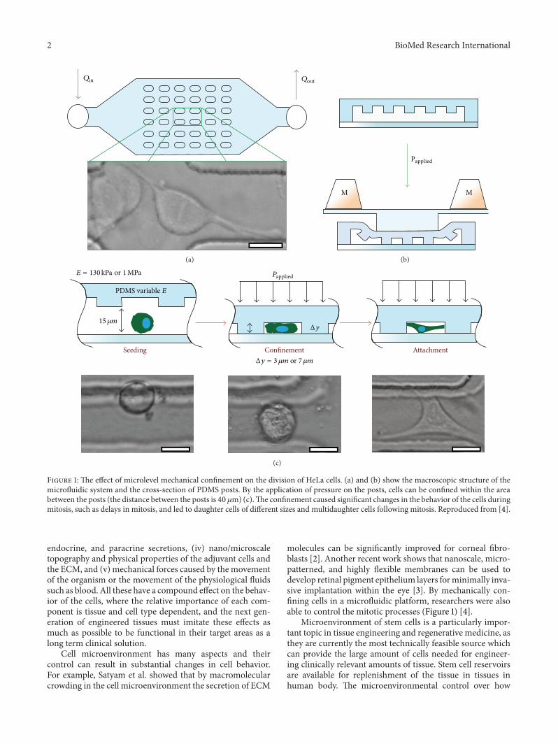
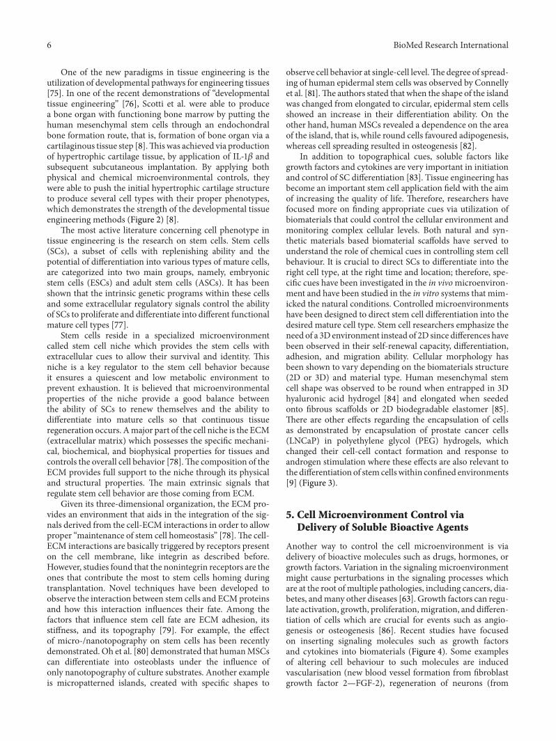
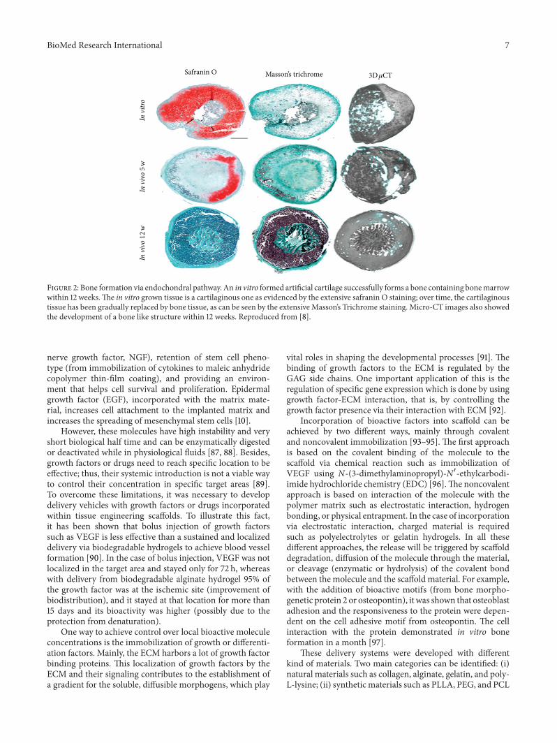
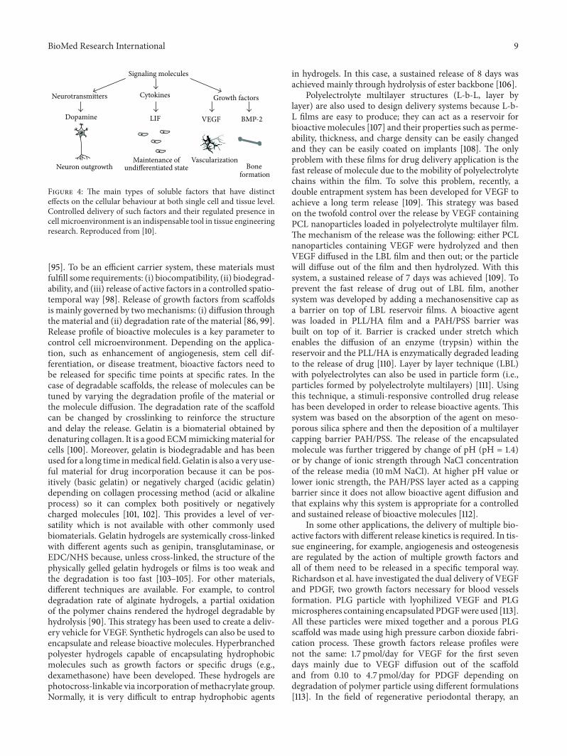
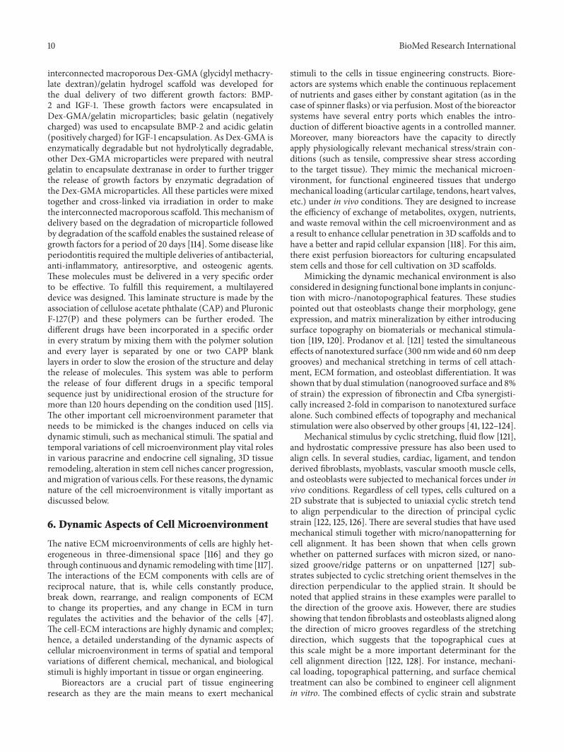