Role of Rab5 in Synaptic Vesicle Recyclingnbn:de:hebis:... · Role of Rab5 in Synaptic Vesicle...
153
Role of Rab5 in Synaptic Vesicle Recycling Dissertation Am Fachbereich Biologie, Chemie der Universität Kassel Tanja Wucherpfennig 2002
Transcript of Role of Rab5 in Synaptic Vesicle Recyclingnbn:de:hebis:... · Role of Rab5 in Synaptic Vesicle...

Role of Rab5 in Synaptic Vesicle
Recycling
Dissertation
Am Fachbereich Biologie, Chemieder
Universität Kassel
Tanja Wucherpfennig
2002

Als Dissertation genehmigtvon der Fakutät für Biologie/Chemie der Universität Kassel
Tag der mündlichen Prüfung: 21.06.2002Erstgutachterin: Prof. Dr. Mireille A. SchäferZweitgutachter: Prof. Dr. Markus Maniak

Acknowledgements
This study was performed in the laboratory of Dr. Marcos González-Gaitán atthe Max-Planck-Institute for Biophysical Chemistry in Göttingen, in thedepartment of Prof. Dr. Herbert Jäckle and at the Max-Planck-Institute forMolecular Cell Biology and Genetics in Dresden.
Foremost, I thank Dr. Marcos González-Gaitán. His critical advise, his respect,his guidence, his encouragement and his support made this study possible.
I thank Prof. Dr. Herbert Jäckle, for his support in Göttingen. I also thank Dr.Ulrich Nauber and countless members of the department of Prof. Jäckle.
I thank Prof. Dr. Mireille A. Schäfer and her colleagues at the University ofKassel for supervising this study and for accepting me as a PhD student inKassel.
I thank Anja Schwabedissen for her excellent technical support, in particular forthe cloning work.
I thank all members of my lab, Eugeni Entchev, Anja Schwabedissen, VeronicaDudu, Periklis Pantazis and Dana Backasch for scientific support, discussions,and a lot of fun.
I wish to thank Dr. Andreas Prokop for teaching me the electron microscopy, Dr.Michaela Wilsch-Bräuninger and Dieter Kötting for support in the electronmicroscopy.
I acknowledge Heike Taubert and Dana Backasch for the generation oftransgenic flies, Rocio Fernandéz de la Fuente and Dana Backasch for keepingthe fly stocks.
I want to thank several people from the electrical and mechanical workshops atthe Max-Planck-Institute in Göttingen, in particular D. Herzog, R. Schürkoetterand H. Scheede.
I thank my friend Christian Rosenmund for the introduction to the field ofelectrophysiology, for many scientific discussions and in particular for beeing inmy life.
I thank my parents for their love and support in whatever I did.

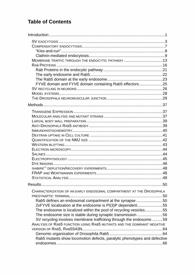
Table of Contents
Introduction .........................................................................................................1
SV EXOCYTOSIS ................................................................................................3COMPENSATORY ENDOCYTOSIS...........................................................................7
“Kiss-and-run” ..............................................................................................8Clathrin-mediated endocytosis.....................................................................9
MEMBRANE TRAFFIC THROUGH THE ENDOCYTIC PATHWAY ...................................13RAB PROTEINS ................................................................................................16
Rab Proteins in the endocytic pathway......................................................21The early endosome and Rab5..................................................................22The Rab5 domain at the early endosome..................................................23FYVE domain and FYVE domain containing Rab5 effectors .....................25
SV RECYCLING IN NEURONS..............................................................................26MODEL SYSTEMS..............................................................................................28THE DROSOPHILA NEUROMUSCULAR JUNCTION...................................................29
Methods ............................................................................................................37
TRANSGENE EXPRESSION.................................................................................37MOLECULAR ANALYSIS AND MUTANT STRAINS .....................................................37LARVAL BODY WALL PREPARATION .....................................................................39ANTI-DROSOPHILA RAB5 ANTIBODY...................................................................39IMMUNOHISTOCHEMISTRY..................................................................................40DEXTRAN UPTAKE IN CELL CULTURE ..................................................................41QUANTIFICATION OF THE NMJ SIZE ...................................................................42WESTERN BLOTTING.........................................................................................43ELECTRON MICROSCOPY...................................................................................44SALINES ..........................................................................................................44ELECTROPHYSIOLOGY ......................................................................................45DYE IMAGING...................................................................................................46SHIBIRETS DEPLETION/RECOVERY EXPERIMENTS...................................................48FRAP AND WORTMANNIN EXPERIMENTS............................................................48STATISTICAL ANALYSIS .....................................................................................48
Results ..............................................................................................................50
CHARACTERIZATION OF AN EARLY ENDOSOMAL COMPARTMENT AT THE DROSOPHILA
PRESYNAPTIC TERMINAL....................................................................................50Rab5 defines an endosomal compartment at the synapse ........................502xFYVE localization at the endosome is PI(3)P dependent.......................55The endosome is localized within the pool of recycling vesicles................55The endosome size is stable during synaptic transmission .......................56SV recycling involves membrane trafficking through the endosome..........59
ANALYSIS OF RAB5 FUNCTION USING RAB5 MUTANTS AND THE DOMINANT NEGATIVE
VERSION OF RAB5, RAB5S43N.........................................................................64Genomic organization of Drosophila Rab5 ................................................64Rab5 mutants show locomotion defects, paralytic phenotypes and defectiveendosomes ................................................................................................66

Specific interference of Rab5 during presynaptic physiology does not causea developmental phenotype.......................................................................69Endosomes are disrupted in Rab5S43N mutant presynaptic terminals .....72Endocytic intermediates accumulate in Rab5 mutant presynaptic terminals...................................................................................................................74Endocytic trafficking during SV recycling involves Rab5 function ..............78Rab5-dependent recycling determines the SV fusion efficacy ...................83
ANALYSIS OF RAB5 GAIN OF FUNCTION ..............................................................93Rab5-mediated endosomal trafficking is rate-limiting during SV recyclingand synaptic transmission..........................................................................93Overexpression of Rab5 does not cause a developmental phenotype of theNMJ but causes an enlargement of endosomes........................................94Rab5 overexpression enhances synaptic performance .............................94
Discussion.........................................................................................................97
SYNAPTIC VESICLE RECYCLING..........................................................................97SVs recycle through an endosomal compartment at the Drosophila NMJ .98Different pathways to recycle synaptic vesicles .........................................99Is endosomal trafficking activity-dependent? ...........................................100
THE ROLE OF RAB5 IN ENDOSOMAL TRAFFICKING..............................................102Structural phenotypes in Rab5 mutants ...................................................102SV quality control at the endosome and synaptic plasticity......................103
Summary.........................................................................................................107
Zusammenfassung .........................................................................................111
References......................................................................................................115

Abbreviations
A abdominalAP action potentialBSA bovine serum albuminCCV clathrin-coated vesicleCNS central nervous systemCSP Cystein string proteinD dorsalDRab5 Drosophila Rab5EEA1 early endosomal antigen 1EJP excitatory junction potentialER endoplasmic reticulumFRAP fluorescence recovery after photobleachingg gramGABA γ-aminobutyric acidGAP GTPase activating proteinGDI guanine dissociation inhibitorGEF guanine nucleotide exchange factorGFP green fluorescent proteinh hoursHRP horseradish peroxidaseHrs hepatocyte growth factor-regulated tyrosine kinase substrateHz hertzISN intersegmental nerve branchIU international unitskDa kilodaltonL lateralLPA lysophosphatidic acidLTR long terminal repeatM molarmEJP miniature excitatory junction potentialmg milligrammin minutesml millilitermm millimetermM millimolarmV millivoltMΩ megaohmnM nanomolarNMJ neuromuscular junctionNSF N-ethylmaleimide-sensitive factorNT neurotransmitterOD outer diameterON over nightORF open reading framePBS phosphate buffered saline

PCR polymerase chain reactionPEM PIPES-EGTA-MgCl2PFA paraformaldehydePI(3)P phosphatidylinositol-3-phosphatePI(4,5)P2 phosphatidylinositol-4,5-bisphosphatePIs inositolpolyphosphatesPM plasma membraneRab ras-like in rat brainREP Rab escort proteinrpm rotations per minuteRT room temperaturesec secondsSDS sodium-dodecyl-sulphateSLMV synaptic-like microvesicleSN segmental nerve branchSNAP soluble NSF attachment proteinSNAP-25 synaptosome-associated protein of 25 kDaSNARE soluble NSF attachment protein receptorSSR subsynaptic reticulumSV synaptic vesicleTGN trans-Golgi networkTN transverse nerveUAS upstream activator sequenceUTR untranslated regionV ventralVAMP vesicle associated membrane protein

1
Introduction
A major goal of neuroscience is to understand brain function. What is
consciousness? How does the brain perceive and initiate action, how does it
learn and remember? To understand how a complex nervous system works
requires knowledge at several levels. We need to know how large numbers of
neurons interact to produce the complex behavior of an organism. We need to
know the properties of individual cells within the nervous system. Finally, we
need to understand the molecular mechanisms by which signals are
communicated between nerve cells, which is the basis for learning and memory.
Neurons are highly specialized to receive, integrate, conduct and transmit
information. Signal transmission over long distances is achieved by an electrical
signal, the action potential (AP). Action potentials are invariant electrical signals
that are generated in the cell soma. From there, they propagate very fast and
without decrement along the axon. Communication between neurons or from
neurons to their target cells occurs at a specialized structure called synapse.
Two types of synapses are known: electrical and chemical synapses. At
electrical synapses, gap junctions connect the cytoplasm of the two cells,
allowing ionic currents to directly flow between them.
Chemical synapses consist of a specialized presynaptic part, the synaptic cleft
and a specialized postsynaptic part. The presynaptic part appears usually as a
swelling, termed bouton, at the nerve terminal. It is characterized by the
presence of numerous mitochondria and vesicles of around 40 nm in diameter,
the synaptic vesicles (SVs). SVs store a quantum of neurotransmitter (NT)
(Katz, 1969), which is released during Ca2+-regulated secretion. NTs are small
signaling molecules such as acetylcholine, glutamate, γ-aminobutyric acid
(GABA), glycine and the biogenic amines dopamine, noradrenalin and
serotonine. Exocytosis of SVs and release of NT is restricted to specialized
regions within the presynaptic terminal, called active zones. At the active zones,
SV docking sites and Ca2+-channels are clustered together (Burns and

2
Augustine, 1995; Pumplin et al., 1981; Robitaille et al., 1990). The synaptic cleft
between the pre– and the postsynaptic membrane is around 20 nm wide and
contains electron-dense extracellular matrix and linker proteins that keep pre-
and postsynaptic membranes precisely aligned (Cottrell et al., 2000). The
postsynaptic membrane is highly organized and specialized to receive
information. It contains clusters of neurotransmitter receptors that are directly
opposed to the presynaptic active zones where NT is released (Cottrell et al.,
2000; Ehlers et al., 1996; Kneussel and Betz, 2000). During synaptic
transmission, chemical synapses first convert the electrical signal into a
chemical signal. This conversion is achieved by the action potential induced
release of neurotransmitter from the presynaptic terminal into the synaptic cleft.
The NT then binds to specific receptors at the postsynaptic cell membrane
generating again an electrical signal.
Since neurons are generally elongated cells, their nerve terminal is located
distant from the cell soma. This distance can range from a few micrometers to
meters as in the case of motoneurons innervating e.g. the feet of giraffes. While
the nerve terminal receives electrical signals within milliseconds, transport of
components from the cell body is a slow process mediated by two different
systems fast and slow axonal anterograde transport (Vale et al., 1992). Fast
anterograde transport is mediated by the motor proteins Kinesin and Dynamin
moving mainly organelles along microtubuli. The speed of the fast axonal
transport system is in the range of 200 mm/day e.g. mitochondria travel around
50 mm/day. In contrast, along the slow axonal transport, most proteins travel
with few mm/day. The nerve terminal therefore needs to be independent in
many basic functions. It has mitochondria for the local production of energy as
well as enzymes and transporters for the synthesis of neurotransmitters. After
their synthesis, NTs are transported into SVs by specific transporters located in
the vesicle membrane. Each nerve terminal contains a reservoir of NT-filled
SVs. This pool of releasable vesicles is essential for synaptic function and
needs to be maintained. Since SVs are released by exocytosis during synaptic
transmission a mechanism for SV regeneration is required, because otherwise

3
an active nerve terminal would deplete itself from SVs. New SVs are not
delivered from the soma, because axonal transport is a slow process. Instead,
they are regenerated within the presynaptic terminal by a local recycling
process. After exocytosis, vesicle components are internalized and assembled
into new SVs.
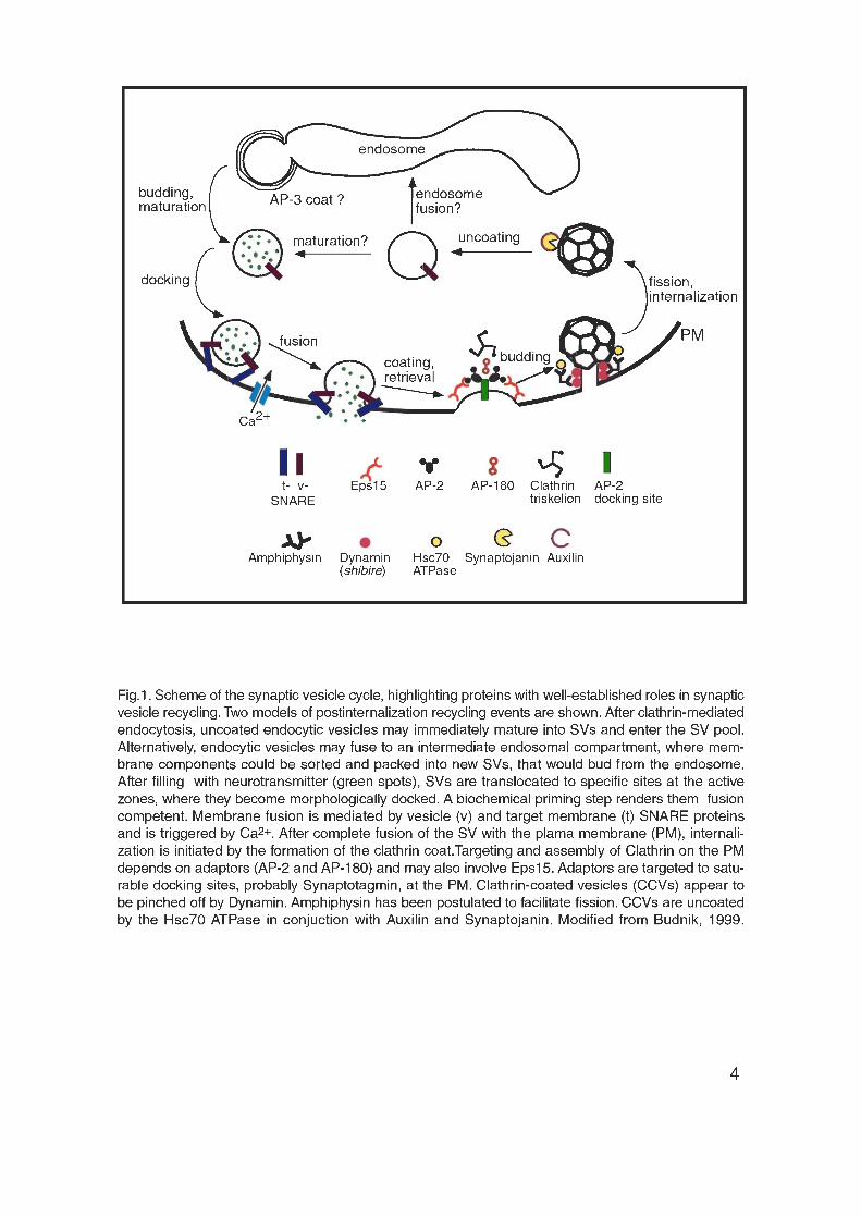
SV exocytosis
Within the presynaptic terminal, SVs undergo a cyclic process of docking,
priming, exocytosis and endocytosis followed by the maturation and transport of
the SVs to the docking sites (Fig. 1). NT exocytosis is a complex and tightly
regulated process that involves the sequential interaction of different synaptic
proteins. NT-filled SVs are first targeted to specific sites, the active zones,
where they become docked. Vesicle docking has been defined by
morphological and biochemical criteria. First, docked vesicles have been
defined at the ultrastructural level as vesicles that are closely opposed to the
plasma membrane (PM) (Donrunz and Stevens, 1999; Plattner et al., 1997;
Schikorski and Stevens, 1997). Second, using imaging techniques and labeled
vesicles it has been found that the docked vesicles are less mobile than the
cytoplasmic ones (Martin and Kowalchyk, 1997; Oheim et al., 1998; Zenisek et
al., 2000). Third, a large fraction of the morphologically docked vesicles remains
associated with plasma membrane fragments following homogenization (Martin
and Kowalchyk, 1997).

5
However, only a fraction of the morphologically docked vesicles is fusion-
competent, i.e. capable of undergoing rapid exocytosis in response to elevated
Ca2+-concentrations (Donrunz and Stevens, 1999; Rosenmund and Stevens,
1996; Schikorski and Stevens, 2001). The pool of fusion-competent SVs is
therefore called the readily releasable pool (Donrunz and Stevens, 1999;
Kuromi and Kidokoro, 1999; Kuromi and Kidokoro, 2000; Rosenmund and
Stevens, 1996). A biochemical priming step is required to render the docked
vesicles fusion-competent. Priming involves ATP, the proteins NSF (N-
ethylmaleimide-sensitive fusion protein), SNAP (soluble NSF attachment
protein) and Munc-13 as well as the synthesis of phosphatidylinositol-4,5-
bisphosphate (PI(4,5)P2), but the exact events are still largely unknown.
Therefore, the term priming is used to include all molecular rearrangements and
ATP-dependent protein and lipid modifications that occur after the initial docking
and before SV fusion. The final fusion of primed SVs with the PM is triggered by
Ca2+-influx through voltage gated Ca2+-channels. Exocytosis is extremely rapid,
following Ca2+-influx within milliseconds (Chad and Eckert, 1984; Fogelson and
Zucker, 1985; Lim et al., 1990; Lindau et al., 1992; Llinas et al., 1992; Llinas et
al., 1982; Mintz et al., 1995; Parsons et al., 1994; Schneggenburger and Neher,
2000; von Ruden and Neher, 1993), reviewed in (Brunger, 2000; Kelly, 1993;
Klenchin and Martin, 2000). The speed of exocytosis predicts that only a few
molecular rearrangements couple Ca2+-influx to membrane bilayer fusion.
The conserved family of SNARE (soluble NSF attachment protein receptor)
proteins has been implicated in all intracellular membrane fusion events (Hay
and Scheller, 1997; Jahn and Sudhof, 1999; Sollner et al., 1993b). In particular,
SV fusion is mediated by the target membrane SNAREs (t-SNAREs) Syntaxin
(Bennett et al., 1992) and SNAP-25 (synaptosomal associated protein of 25
kDa) (Oyler et al., 1989) and the vesicle membrane SNARE (v-SNARE)
Synaptobrevin, also called VAMP (vesicle associated membrane protein) (Oyler
et al., 1989). The specific cleavage of these SNAREs by clostridial neurotoxins
inhibits neurotransmission, supporting their fundamental role in SV fusion

6
(Hayashi et al., 1994; Jahn and Niemann, 1994; Montecucco and Schiavo,
1995; Schiavo et al., 1992).
As all SNAREs, Syntaxin, SNAP-25 and Synaptobrevin contain an amphipathic
α−helix close to their membrane anchor (Fasshauer et al., 1998b). The
α−helices of Syntaxin, SNAP-25 and Synaptobrevin twist around each other to
form an extremely stable ternary complex (Sollner et al., 1993b), in which the
hydrophobic side chains are buried in the center. The crystal structure of this
“core complex” revealed a four-helix coiled-coil structure (Sutton et al., 1998).
The “zipper model“ of SNARE function postulates that the SNARE complex
assembles by “zipping up” the SNARE α−helices from the membrane-distant N-
termini to the membrane-proximal C-termini. Thus, the formation of the stable
SNARE complex is proposed to bring SVs into intimate contact with the plasma
membrane (PM). This probably overcomes the energy barrier and drives bilayer
fusion (Hanson et al., 1997; Lin and Scheller, 1997).
However, the precise temporal interaction between the SNARE proteins and the
mechanism of Ca2+-regulation are unknown. The Ca2+-, phospholipid- and
SNARE-binding synaptic vesicle protein Synaptotagmin has been proposed to
serve as Ca2+-sensor that regulates exocytosis, (Brose et al., 1992; Desai et al.,
2000; Fernandez-Chacon et al., 2001; Geppert and Sudhof, 1998; Littleton and
Bellen, 1995; Littleton et al., 1999; Perin et al., 1990). Ca2+-binding to
Synaptotagmin causes its rapid insertion into membranes, occurring within
milliseconds (Brose et al., 1992; Davis et al., 1999; Li et al., 1995a). It has been
thereby speculated that Synaptotagmin causes membrane fusion by a Ca2+-
induced morphological change analogous to the mechanism of pH-induced,
hemagglutinin-mediated fusion of the influenza virus to its target cell (Kelly,
1993).
After the fusion reaction, v- and t-SNAREs are contained within the same
membrane forming cis-SNARE complexes that need to be disassembled prior
to the next fusion event. cis-SNARE disassembly is performed by the soluble
cofactors NSF and SNAP (Otto et al., 1997; Sollner et al., 1993a; Sollner et al.,
7
1993b; Swanton et al., 1998), but it is not yet known where and when this
reaction occurs. Furthermore, following SV exocytosis the vesicle membrane is
immediately endocytosed (Fasshauer et al., 1998a; Jahn and Niemann, 1994;
Littleton et al., 1998; Poirier et al., 1998). This membrane recapture requires
high specificity since SVs and the presynaptic plasma membrane have a
distinct membrane composition.
Compensatory endocytosis
Compensatory endocytosis is a process by which a cell retrieves membrane,
which has been added to the PM by regulated secretion. As described above,
the presynaptic nerve terminal is filled with SVs that are exocytosed during
neurotransmission. Synaptic function requires that the pool of SVs, competent
for NT release, is maintained even during sustained periods of high frequency
stimulation. The regeneration of SVs is achieved by rapid endocytosis of
synaptic vesicle components followed by a local recycling mechanism. In
addition, compensatory endocytosis is essential to keep the size of the
presynaptic terminal constant and to preserve the molecular diversity of SV
versus PM.
Recycling of SVs involves at least two distinct pathways, “kiss-and-run”
(Ceccarelli et al., 1973; Fesce et al., 1994; Palfrey and Artalejo, 1998) and
clathrin-mediated endocytosis (De Camilli and Takei, 1996; Heuser and Reese,
1973). These current endocytic models are based, with some modifications, on
observations made in the early 1970s by the groups of Ceccarelli and Heuser
(Ceccarel l i et al., 1973; Heuser and Reese, 1973). The two groups
independently investigated endocytosis at the frog neuromuscular junction
(NMJ) using electron-dense endocytic markers and electron microscopy.
Heuser and his group observed endocytosis of clathrin-coated vesicles (CCVs)
in regions outside the active zones. Ceccarelli and colleagues by contrast
observed clathrin-independent endocytosis at or near the active zone. This
mechanism was later attributed to the “kiss-and-run” vesicle cycle (Fesce et al.,
1994).

8
“Kiss-and-run”
“Kiss-and-run” (Fesce et al., 1994; Jarousse and Kelly, 2001) is believed to take
place at the active zone. According to the model, SVs make only a brief contact
with the PM and release their NT through a transient fusion pore (Albillos et al.,
1997; Almers and Tse, 1990; Ceccarelli et al., 1973; Klingauf et al., 1998; Pyle
et al., 2000). After closure of the fusion pore, the vesicle is simply refilled with
NT and can be used again. Thus, no coated intermediate is formed (Ceccarelli
et al., 1973; Palfrey and Artalejo, 1998) and the vesicle is recovered without
mixing its components with the PM. Therefore, SVs never change their
individual identity as defined by their size, protein and lipid-composition. SV
recycling through this pathway is thought to be very fast, in the range of 1 to 2
seconds.
There are several lines of evidence supporting the “kiss-and-run” mode. First,
the detection of uncoated vesicles, labeled with an endocytic tracer at the active
zone (Ceccarelli et al., 1973). Second, the discrepancy between the amounts of
FM-dye released from prelabeled nerve terminals with respect to the released
NT. In hippocampal neurons, less dye is released than expected from the
amount of NT released. As FM-dyes diffuse slower through a transient fusion
pore than the NT, it has been calculated that 20% of the SVs are recycled
through the “kiss-and-run” mechanism (Stevens and Williams, 2000). Third, two
kinetic time constants of endocytosis have been detected in nerve terminals.
The faster type of endocytosis is inhibited by prolonged stimulation and could
correspond to “kiss-and-run” (Neves and Lagnado, 1999). Fourth, Palfrey and
colleagues (Palfrey and Artalejo, 1998) observed a fast type of endocytosis that
is dynamin-dependent but clathrin-independent. Fifth, the opening and closing
of fusion pores gives rise to transient increases in the cell surface, which can be
detected in endocrine cells electrophysiologically as a quantal change in the
membrane capacitance, called “capacitance flicker” (Alvarez de Toledo and
Fernandez, 1990; Breckenridge and Almers, 1987; Spruce et al., 1990).
Therefore, it seems likely that the “kiss-and-run” mode of SV recycling coexists
with the classical pathway, which starts with clathrin-mediated endocytosis.
9
Clathrin-mediated endocytosis
The process of clathrin-mediated endocytosis (reviewed in (Brodin et al., 2000;
Hirst and Robinson, 1998; Jarousse and Kelly, 2001; Kirchhausen, 2000b;
Schmid, 1997)) is thought to be slower than the “kiss-and-run” pathway,
regenerating SVs within 30 to 60 seconds through coated intermediates. In
1964, coated pits and vesicles were discovered (Roth and Porter, 1964).
Clathrin-coated vesicles were first isolated from pig brain in 1969 (Kanaseki and
Kadota, 1969) and Clathrin itself was purified in 1975 (Pearse, 1975). Clathrin-
mediated endocytosis is involved in a variety of cellular functions such as the
uptake of nutrients, growth factors and antigens as well as the regulation of cell
surface receptors (Schmid, 1997). Endocytosis via clathrin-coated vesicles has
been shown to participate in the recycling of SVs (De Camilli and Takei, 1996;
González-Gaitán and Jäckle, 1997; Heuser, 1989; Heuser and Reese, 1973;
Shupliakov et al., 1997).
Following the complete collapse of the SV into the PM clathrin-mediated
endocytosis (Ceccarelli et al., 1979; Heuser and Reese, 1973; Matteoli et al.,
1992; Torri-Tarelli et al., 1987; Valtorta et al., 1988) ensures the specific
retrieval of SV components and their reassembly into new SVs (Maycox et al.,
1992; Takei et al., 1996) (Fig. 1). Within the presynaptic terminal, this process
takes place at specialized sites, the centers of endocytosis, which surround the
active zones where exocytosis occurs (González-Gaitán and Jäckle, 1997;
Jarousse and Kelly, 2001; Ringstad et al., 1999; Roos and Kelly, 1998; Roos
and Kelly, 1999; Teng and Wilkinson, 2000).
Clathrin-mediated endocytosis involves a number of highly coordinated
sequential steps controlled by different proteins. These steps include 1)
targeting of coat components to the PM, 2) the formation of clathrin-coated
membrane invaginations, termed pits, into which cargo-molecules are
concentrated, 3) the formation of clathrin-coated vesicles (CCVs) by pinching off
clathrin-coated pits from the PM and, finally, 4) the removal of the Clathrin coat,
called uncoating.

10
1) Coat assembly
The main coat constituents are Clathrin, the Clathrin adaptor protein complex
AP-2 and a synaptic protein called AP-180. Clathrin-mediated endocytosis is
initiated by the binding of AP-2 to the PM. AP-2 then recruits Clathrin to the
membrane and triggers its polymerization. The heterotetrameric AP-2 complex
is composed of four closely associated subunits called α, β2, µ2 and σ2. It has
two essential functions during clathrin-mediated endocytosis: First, recruitment
of the Clathrin coat to the PM and second, selection of specific cargo molecules
destined for internalization.
The α subunit also called α–Adaptin is responsible for targeting AP-2 to
specific, saturable docking sites at the PM (Gaidarov et al., 1996; Gaidarov and
Keen, 1999; Mahaffey et al., 1989; Mahaffey et al., 1990; Moore et al., 1987),
defined probably by Synaptotagmin (Chapman et al., 1998; Haucke et al., 2000;
Zhang et al., 1994). In addition, specific inositolpolyphosphates (PIs) in
particular PI(4,5)P2 is required to recruit AP-2 to the PM (Gaidarov et al., 1996;
Gaidarov and Keen, 1999). The σ2 subunit is involved in the selection of cargo
molecules to be internalized. It interacts with tyrosine- and dileucine-based
endocytic sorting signals present in the cytoplasmic domains of certain
transmembrane receptors (Boll et al., 1996; Ohno et al., 1996; Ohno et al.,
1995; Sorkin et al., 1995). Binding of the σ2 subunit to these sorting motifs
causes a concentration of cargo molecules at the sites of endocytosis. Finally,
the ß subunit of AP–2 recruits Clathrin (Ahle and Ungewickell, 1989; Gallusser
and Kirchhausen, 1993; Shih et al., 1995) and triggers its polymerization.
Clathrin was named in reference to the cage like structure it forms (Pearse,
1976). It is the major structural component of the Clathrin coat (Pearse, 1975)
and forms triskelions consisting of three heavy chains of around 180 kDa and
three light chains of around 30 kDa (reviewed in (Kirchhausen, 2000a)).
Triskelions can be viewed after negative staining in the electron microscope and
appear as three-legged structures (Ungewickell and Branton, 1981). They self-
assemble in vitro into lattices containing pentagons and hexagons. The Clathrin

11
coat consists of 12 pentagons and a variable number of hexagons (Crowther
and Pearse, 1981; Kanaseki and Kadota, 1969). 12 pentagons are required in
order to form a closed structure, whereas the number of hexagons determines
the size of the coat (Shraiman, 1997).
Two models for the assembly process have been proposed. According to Liu
(Liu et al., 1995), Clathrin triskelions first assemble into a flat network of
hexagons some of which are later converted into pentagons generating
membrane curvature. Alternatively, because of a certain membrane curvature,
determined by other factors, hexagons and pentagons are incorporated during
the assembly process (Cupers et al., 1994). Another factor involved in the
assembly of Clathrin coats is the monomeric protein AP-180. AP-180 binds to
both Clathrin and AP-2 (Ahle and Ungewickell, 1986; Morris et al., 1993) and
has been implicated in controlling the size of endocytic vesicles (Nonet et al.,
1999; Zhang et al., 1998).
2) Invagination
Much less is known about the process of invagination. Invagination is
accompanied by an increase in the negative membrane curvature. The
lysophosphatidic acid (LPA) acyl transferase Endophilin might be involved in the
process of invagination. This enzyme converts LPA, by addition of the fatty acid
arachidonate into phosphatidic acid, thereby increasing the negative membrane
curvature (Ringstad et al., 1999; Schmidt et al., 1999).
3) Fission
The most extensively studied protein involved in the fission of clathrin-coated
vesicles is Dynamin. Dynamin has been originally linked to endocytosis through
its temperature-sensitive Drosophila mutant shibirets. In shibirets, endocytosis is
inhibited at the restrictive temperature because clathrin-coated pits cannot be
pinched off from the PM (Kosaka and Ikeda, 1983b). Later, the mutation in
shibire was mapped to Dynamin (Chen et al., 1991; van der Bliek and

12
Meyerowitz, 1991). Because Dynamin forms rings and tubules in vitro (Carr and
Hinshaw, 1997; Hinshaw and Schmid, 1995) and is located at the neck of the
coated pits (Sever et al., 1999), it has been proposed to acts as a “pinchase”
that mechanically pinches off the clathrin-coated vesicle from the PM (Sweitzer
and Hinshaw, 1998). However, since Dynamin contains several protein-protein
interaction domains, it has been alternatively proposed to recruit the actual
severing activities around the neck of the budding vesicle (Kirchhausen, 1999;
Sever et al., 1999; Yang and Cerione, 1999).
Recently, two dynamin-binding proteins Amphiphysin (David et al., 1996) and
Endophilin (Ringstad et al., 1999) have been postulated to facilitate the fission
step (Barr and Shorter, 2000; Zimmerberg, 2000).
4) Uncoating
Uncoating of clathrin-coated vesicles is thought to occur rapidly, since free,
coated vesicles are rarely seen in stimulated synapses. The clathrin-binding
protein Auxilin (Holstein et al., 1996) recruits the uncoating ATPase Hsc70 to
clathrin-coated vesicles and stimulates its ATPase activity (Barouch et al., 1997;
Holstein et al., 1996; Schroder et al., 1995; Ungewickell et al., 1995). Hsc70
subsequently releases Clathrin triskelions (Kirchhausen and Harrison, 1981;
Ungewickell and Branton, 1981) and other coat proteins from the vesicles by
undergoing multiple cycles of Clathrin binding and ATP hydrolysis (Barouch et
al., 1994; Braell et al., 1984; Chappell et al., 1986; Schlossman et al., 1984).
Furthermore, the polyphosphoinositide phosphatase Synaptojanin (Guo et al.,
1999; McPherson et al., 1996) has also been implicated in the uncoating
reaction, since disruption of its function causes the accumulation of CCVs
(Cremona et al., 1999; Harris et al., 2000).
In the case of the synapse, little is known about the fate of uncoated endocytic
vesicles. In particular, the process of endocytic vesicle recycling and maturation
into NT-filled, releasable SVs is a matter of debate. In contrast, it is well
established that in cultured mammalian cells uncoated endocytic vesicles fuse

13
to an intermediate endosomal compartment, the early sorting endosome and
that recycling takes place through the endocytic pathway.
Membrane traffic through the endocytic pathway
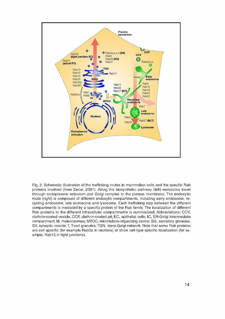
Eukaryotic cells contain an elaborate system of intracellular organelles and
membrane trafficking routes (Fig. 2). The biosynthetic pathway of eukaryotic
cells serves to deliver newly synthesized molecules to different intracellular
organelles. Along this route components usually travel through endoplasmic
reticulum (ER) and Golgi complex to their target destinations including
mitochondria, lysosomes and PM. The other major route, the endocytic
pathway, is responsible for the recycling of endocytosed components. The
endocytic pathway involves several distinct endocytic compartments and each
trafficking step between the different intracellular organelles is mediated by
small membrane carriers.
15
The first step in the formation of these transport vesicles is the assembly of a
coat at specific sites of the donor organelle (reviewed in (Hirst and Robinson,
1998; Robinson and Bonifacino, 2001; Schmid, 1997; Takei and Haucke, 2001;
Zhang et al., 1999)). The coat generally assembles from multiple hetero-
oligomeric, cytosolic protein complexes. Coats are believed to be involved in the
physical formation of transport vesicles as well as in the selective packaging of
their cargo. Several different coat complexes are known: 1) Clathrin with AP-2
driving endocytic vesicle formation at the PM. 2) Clathrin with AP-1 generating
transport vesicles at the trans-Golgi network (TGN). 3) The AP-3 complex
generates vesicles at the TGN and probably also at the endosome. 4) The AP-4
complex associated to the TGN. 5) The coatomer protein complex assembles
together with ARF-1 to form COPI-coated vesicles that mediate retrograde
transport within the Golgi and between Golgi and ER. 6) A protein complex
including sec23p/sec24p, sec13/31p and sar1p that assembles to form COPII
vesicles at the ER. Each type of transport vesicle mediates the flow of certain
cargo molecules to certain destination.
Membrane traffic requires high specificity and tight regulation because cargo
molecules need to be delivered to the correct acceptor compartments, while
organelle integrity and biochemical composition have to be maintained.
Furthermore, the compartment size needs to be stable, which requires that
fusion of vesicles with a given compartment is in balance with budding of
vesicles from the same compartment.
A typical transport reaction can be viewed as a four-step process. It consists of
first, the formation of a vesicular (Rothman and Orci, 1992) or tubular (Klausner
et al., 1992) transport intermediate from the donor compartment. This reaction
is controlled by different coat proteins (Kreis, 1992). Second, the movement of
the vesicle along microtubules (Brady, 1991; Kuznetsov et al., 1992; Mitchison,
1992) towards the target compartment. Third, tethering/docking of the vesicle
with the target compartment (Pfeffer, 1999) and fourth, finally the fusion of the
lipid bilayers. The specificity of these events is critical to preserve organelle
integrity and to control cargo flow within the cell. To achieve this, each

16
trafficking step is tightly regulated by a different protein of the Rab family of
small GTPases (Pfeffer, 1994).
Rab proteins have been proposed to determine vesicular transport specificity by
mediating in conjunction with their effector proteins the specific tethering of
vesicles to their target organelle. In addition, Rab proteins are thought to be
upstream modulators of the SNARE proteins, regulating the formation of a
complex between the v- and its cognate t-SNARE (Lian et al., 1994; Lupashin
and Waters, 1997; McBride et al., 1999; Sogaard et al., 1994). The family of
SNARE proteins is involved in the final event of membrane fusion (see below)
(McNew et al., 2000; Parlati et al., 2000; Rothman, 1994; Weber et al., 1998).
This view is supported by several recent studies reporting direct molecular
interactions between Rab effector proteins and components of the SNARE
machinery (McBride et al., 1999; Peterson et al., 1999; Price et al., 2000; Sato
et al., 2000; Tall et al., 1999). The interactions between Rab effectors and
components of the SNARE machinery may coordinate the Rab-dependent
membrane tethering and docking with the SNARE-dependent membrane fusion.
Rab Proteins
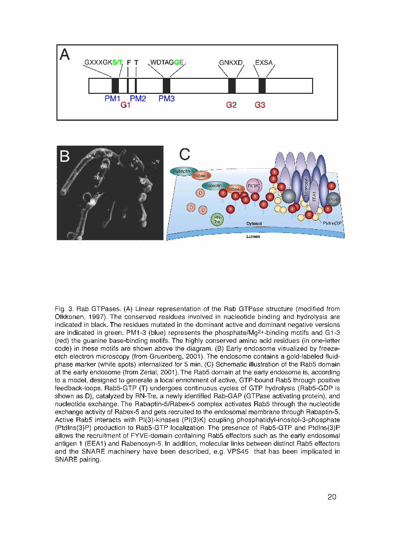
Rab proteins are small (21 – 25 kDa), monomeric GTPases (Fig. 3A) forming
the largest branch of the Ras superfamily of small GTPases. There are probably
63 different Rab proteins in humans (Bock et al., 2001; Zerial and McBride,
2001), 11 in yeast (Lazar et al., 1997) and around 30 in Drosophila (Littleton,
2000). The first Rab gene was identified in Saccharomyces cerevisiae in 1983
(Gallwitz et al., 1983). The first mammalian homologs were cloned in 1987 and
termed Rab (ras-like in rat brain) (Touchot et al., 1987).
Rab proteins regulate vesicle-mediated transport of proteins and lipids between
different organelles (Bucci et al., 1992; Huber et al., 1993; Lombardi et al.,
1993; Martinez et al., 1994; Pfeffer, 1996; Rothman, 1994; Salminen and Novik,
1987; Segev, 1991; Tisdale et al., 1992; van der Sluijs et al., 1992) (Fig. 2).
They directly or indirectly affect vesicle budding (Benli et al., 1996; McLauchlan
17
et al., 1998; Nuoffer and Balch, 1994; Riederer et al., 1994) and play important
roles in vesicle docking (Christoforidis et al., 1999a; Novick and Zerial, 1997;
Nuoffer and Balch, 1994; Pfeffer, 1994). In addition, some members have been
implicated in motility by interactions with the cell cytoskeleton (Nielsen et al.,
1999).
Each Rab protein is localized to the cytoplasmic surface of a distinct membrane
bound organelle (Ferro-Novick and Novick, 1993; Novick and Zerial, 1997;
Pfeffer, 1994; Takai et al., 1992; Zerial and McBride, 2001; Zerial and
Stenmark, 1993) (Fig. 2). Membrane attachment and function of Rab proteins
requires their isoprenylation. After their synthesis, Rab proteins are bound to a
Rab escort protein (REP) (Seabra et al., 1992a; Seabra et al., 1992b) that
presents the unprenylated Rab protein to the geranylgeranyl-transferase type II
(Andres et al., 1993). This heterodimeric protein geranylates Rab proteins, by
the addition of the C-20 isoprenyl lipid geranylgeranyl, to usually two cystein
residues at the C-terminus of Rab proteins (Marshall, 1993; Seabra et al.,
1992a). Geranyl groups render Rab proteins hydrophobic and are required for
their reversible membrane association (Alexandrov et al., 1994).
The double geranylated Rab protein is thought to remain associated with REP,
which delivers the GTPase to a specific organelle or transport vesicle. The
specificity of Rab localization is mediated by interactions between the
hypervariable, C-terminus of a Rab protein with distinct proteins on the
organelle surface (Chavrier et al., 1991; Soldati et al., 1994; Ullrich et al., 1994).
Rab proteins are predominantly localized to membranes of transport vesicles
and to their specific target compartments. In the steady state, Rab proteins
accumulate at their target compartment and have thereby been used as
markers for different organelles (Bucci et al., 1992; Chavrier et al., 1991;
Chavrier et al., 1990; Ullrich et al., 1996). Only a minor fraction of each Rab
protein is localized to the cytosol where it is complexed with a protein called
guanine dissociation inhibitor (GDI) (Garrett et al., 1993; Regazzi et al., 1992;
Sasaki et al., 1991; Sasaki et al., 1990; Soldati et al., 1993; Ullrich et al., 1993).
18
Rab proteins act as molecular switches that cycle between an active GTP-
bound, membrane associated and an inactive GDP-bound cytosolic
conformation. The GTP–GDP cycle is required for Rab function and is mediated
by the accessory proteins GDI, GDI displacement factor, guanine nucleotide
exchange factor (GEF) and GTPase activating protein (GAP). Within the
cytosol, the GDP-bound form of Rab proteins, Rab–GDP, is bound to Rab–GDI,
which masks the hydrophobic prenyl groups of Rab proteins (Pfeffer et al.,
1995). Upon membrane attachment, GDI is released by a GDI displacement
factor (Dirac-Svejstrup et al., 1997). Subsequently, a GEF catalyzes the
exchange of GDP against GTP (Burton et al., 1993; Burton et al., 1994; Moya et
al., 1993), thereby converting Rab proteins into their active, GTP-bound form
(Bourne, 1988; Goud and McCaffrey, 1991; Soldati et al., 1994; Ullrich et al.,
1994; Zerial and Stenmark, 1993). Activated Rab proteins recruit soluble factors
that act as specific effector molecules regulating downstream docking and
fusion events. Finally, GAPs stimulate GTP hydrolysis (Ferro-Novick and
Novick, 1993; Strom et al., 1993) converting Rab proteins into their inactive,
GDP-bound form. Rab-GDI then recognizes and extracts Rab-GDP from the
membrane and recycles them back to the appropriate membrane (Araki et al.,
1990; Garrett et al., 1993; Regazzi et al., 1992; Soldati et al., 1993; Takai et al.,
1992; Ullrich et al., 1993).
One of the key approaches to manipulate and investigate Rab protein function
was the mutagenesis of specific amino acids essential for the GTP/GDP cycle
of Rab proteins (Fig. 3A). The choice of the amino acids to be mutagenized was
based on the well-characterized mutations described in the Ras oncoprotein.
Mutations corresponding to the glutamine 61 to lysine (Q61L) mutation in Ras
cause a decreased intrinsic and GAP-stimulated GTPase activity, while the
ability to bind nucleotides is not changed (Adari et al., 1988; Der et al., 1986;
Stenmark et al., 1994; Tanigawa et al., 1993; Tisdale et al., 1992; Walworth et
al., 1992). Therefore, dominant active Rab proteins are stabilized in their active,
GTP-bound conformation (Adari et al., 1988; Der et al., 1986; Hoffenberg et al.,
1995; Stenmark et al., 1994). In contrast, mutants equivalent to the serine 17 to

19
asparagine (S17N) mutation of the Ras protein (Farnsworth and Feig, 1991;
Feig and Cooper, 1988), have a lower affinity for GTP than for GDP, causing a
dominant inhibitory effect by blocking the protein in its inactive, GDP-bound
conformation. Comparable mutations in Rab proteins have been discovered,
including for Rab3A (T36N) (Burstein et al., 1992), Rab1A (S25N) (Nuoffer et
al., 1994), Rab9 (Riederer et al., 1994) and Rab5 (Li and Stahl, 1993; Stenmark
et al., 1994). They all show dominant negative phenotypes, probably by binding
to, and titrating out the respective effector molecules, that are thereby not
available for the endogenous Rab protein (Burstein et al., 1992).

21
Rab Proteins in the endocytic pathway
The endocytic pathway mediates recycling and degradation of endocytosed
molecules. The endocytic pathway is composed of several biochemically and
morphologically distinct stations, (Hubbard, 1989; Rodman et al., 1990)
including early sorting endosomes, recycling endosomes, late endosomes and
lysosomes (Fig. 2). Membrane traffic between these compartments is mediated
by different proteins of the Rab family, in particular by Rab5, Rab4, Rab11 and
Rab7.
The first step along the endocytic pathway is the formation of endocytic vesicles
by clathrin-mediated endocytosis (see above). Endocytic vesicles subsequently
fuse to the first station within the endocytic pathway, the early sorting
endosome. The early sorting endosome is a complex and dynamic membrane
system (Fig. 3B) in which endocytosed components are sorted to their different
destinations (Dunn et al., 1989; Ghosh et al., 1994; Gruenberg and Kreis, 1995;
Gruenberg and Maxfield, 1995; Mellman, 1996).
Endocytosed components are directed from the early sorting endosome either
into the degradative or into the recycling pathway. The degradative pathway
leads to the late endosomes and lysosomes where degradation by acid
hydrolases occurs (Gruenberg and Maxfield, 1995; Mellman, 1996). Other
proteins such as the recycling receptors transferrin- or the LDL-receptor are
recycled back to the PM (Dunn et al., 1989; Ghosh and Maxfield, 1995). Two
recycling routes are known. The fast recycling pathway, which leads from the
early endosome directly back to the PM (Daro et al., 1996; Mayor et al., 1993;
Schmid et al., 1988; Sheff et al., 1999; van der Sluijs et al., 1992), whereas the
second route involves another compartment the perinuclear recycling
endosome (Hopkins, 1983; Prekeris et al., 2000; Schlierf et al., 2000; Ullrich et
al., 1996; Yamashiro et al., 1984). More recently, a transport route that
connects early endosomes and the TGN was discovered (Rohn et al., 2000;
Wilcke et al., 2000).
22
Each trafficking step along the endocytic pathway is regulated by a different
Rab protein (Chavrier et al., 1990; Lombardi et al., 1993; Olkkonen et al., 1993;
Ullrich et al., 1996; van der Sluijs et al., 1991). Rab5 mediates traffic from the
PM to the early endosome (Bucci et al., 1992), Rab7 the step from the early
sorting endosome to the degradative compartment (Bucci et al., 2000; Feng et
al., 1995; Méresse et al., 1995; Vitelli et al., 1997; Wichmann et al., 1992) and
Rab4 and Rab11 trafficking within the recycling pathway. In particular, Rab4
controls the fast recycling from the early endosome directly back to the PM
(Daro et al., 1996; Sheff et al., 1999; van der Sluijs et al., 1992), whereas
Rab11 recycling through the recycling endosome (Prekeris et al., 2000; Schlierf
et al., 2000; Ullrich et al., 1996). In the steady state, these Rab proteins are
localized to their target compartments and have thereby been used as markers
for the different endocytic compartments. Both Rab5 (Bucci et al., 1992;
Chavrier et al., 1991) and Rab4 (van der Sluijs et al., 1991) are associated to
early sorting endosomes. Rab7 serves as a marker for the degradative
compartment (Chavrier et al., 1990) and Rab 11 is localized to the recycling
endosome (Ullrich et al., 1996).
The early endosome and Rab5
The early endosome is the primary sorting station in the endocytic pathway
(Ghosh and Maxfield, 1995; Gruenberg and Kreis, 1995; Gruenberg and
Maxfield, 1995; Mellman, 1996) and the small GTPase Rab5 has been used as
an early endosomal marker (Bucci et al., 1992; Chavrier et al., 1991). Rab5
regulates the first step of the endocytic pathway between the PM and the early
sorting endosome. Thus, Rab5 has been implicated in the formation of clathrin-
coated endocytic vesicles at the PM (McLauchlan et al., 1998) as well as in the
fusion of endocytic vesicles with the early endosome (“heterotypic fusion”)
(Bucci et al., 1992). In addition, Rab5 regulates in a rate-limiting manner the
“homotypic fusion” between early endosomes (Barbieri et al., 1994; Gorvel et
al., 1991; Li et al., 1994; Roberts et al., 1999; Rybin et al., 1996). Furthermore,
it mediates attachment to, and motility of early endosomes towards the minus

23
end of microtubules and thereby the distribution of early endosome within the
cell (Nielsen et al., 1999).
Three Rab5 isoforms have been identified in mammals, Rab5a, Rab5b, Rab5c
(Bucci et al., 1995) and in yeast (Novick and Zerial, 1997), whereas only one
isoform is present in C. elegans (Grant and Hirsh, 1999). Rab5 function requires
the interaction with specific effector molecules. Using affinity-chromatography
more than 20 proteins have been purified form bovine brain, which directly or
indirectly interact specifically with the GTP-bound, active form of Rab5
(Christoforidis et al., 1999b). They include Rabaptin-5 (Stenmark et al., 1995),
Rabex-5 (Horiuchi et al., 1997), Rabaptin-5b (Gournier et al., 1998), EEA1
(Christoforidis et al., 1999a) and Rabenosyn-5 (Nielsen et al., 2000).
The Rab5 domain at the early endosome
The early endosome is composed of at least two functionally different
subdomains, visualized by the distinct localization of the two Rab proteins Rab4
and Rab5 (De Renzis et al., 2002; Sonnichsen et al., 2000). Endocytic vesicles
fuse to the endosome at the Rab5 domain, while components destined for the
fast recycling back to the PM are sorted in the Rab4 domain. Two effector
proteins, Rabaptin-5 (Vitale et al., 1998) and Rabenosyn-5 (De Renzis et al.,
2002) have been shown to bind both Rab5 and Rab4. It has therefore been
proposed that these divalent Rab effectors control the sub-compartmental
organization of early endosomes. They might connect the Rab5 and Rab4
domains and thereby regulate protein sorting and recycling.
The Rab5 domain is known to be required for the specificity of vesicle
tethering/docking. The Rab5 domain is enriched in activated, GTP-bound Rab5,
several different Rab5 effector proteins as well as the lipid phosphatidylinositol-
3-phosphate (PI(3)P) (Fig. 3C). The Rab5 effector proteins are recruited in a
cooperative fashion to the Rab5 domain. First, Rab5 is delivered to the
endosomal membrane complexed to GDI as described above. At the

24
membrane, a GDI displacement factor dissociates the Rab5-GDI complex
releasing GDI into the cytosol (Ayad et al., 1997; Dirac-Svejstrup et al., 1997).
Subsequently, Rab5 is activated by a complex composed of Rabaptin-5 and
Rabex-5 (Horiuchi et al., 1997; Lippe et al., 2001). Rabex-5 acts as a
specialized Rab5-GEF, catalyzing the exchange of GDP against GTP (Horiuchi
et al., 1997). Rabaptin-5, which was identified as the first Rab5 effector in a two-
hybrid screen (Stenmark et al., 1995), increases the activity of Rabex-5 on
Rab5 (Lippe et al., 2001). In addition, Rabaptin-5 stabilizes Rab5 in its active,
GTP-bound form by down-regulating GTP-hydrolysis, (Rybin et al., 1996).
Furthermore, as the C-terminal domain of Rabaptin-5 binds active Rab5 (Vitale
et al., 1998), activation of Rab5 at the endosomal membrane starts a positive
feedback mechanism: Rabaptin-5 binds and stabilizes active Rab5 at the
endosome and recruits Rabex-5, which in turn generates more active Rab5.
Active Rab5 directly recruits two PI(3)-kinases (Christoforidis et al., 1999b) and
PI(3)-kinase activity in turn is required for efficient endosome fusion (Jones and
Clague, 1995; Li et al., 1995b; Spiro et al., 1996). The PI(3)-kinase p85α/p110β
is a type I kinase that mainly phosphorylates phosphatidylinositol-4-phosphate
(PI(4)P) and PI(4,5)P2 generating phosphatidylinositol-3,4,5-trisphosphate
(PI(3,4,5)P3). This enzyme has been also implicated in signal transduction
pathways (Vanhaesebroeck et al., 1997). The other PI(3)-kinase is
hVPS34/p150 the mammalian homolog of the yeast Vps34p/Vps15 (Volinia et
al., 1995). It preferentially phosphorylates PI to PI(3)P. In summary, active Rab5
recruits PI(3)-kinases to the early endosome generating a domain enriched in
PI(3)P (Christoforidis et al., 1999b). Several Rab5 effector proteins specifically
bind PI(3)P through their FYVE protein domain (Gaullier et al., 2000; Lawe et
al., 2000; Nielsen et al., 2000; Raiborg et al., 2001b; Stenmark et al., 1996).
25
FYVE domain and FYVE domain containing Rab5 effectors
The FYVE domain is a zinc finger domain that coordinates two Zn2+ ions and
specifically binds PI(3)P (Burd and Emr, 1998; Gaullier et al., 1998; Lawe et al.,
2000; Patki et al., 1998; Stenmark and Aasland, 1999). It was named after the
first proteins shown to contain it, namely Fab1, YOTB/ZK632.12, Vac1 and
EEA1 (Stenmark et al., 1996). Later, a tandem repeat of the FYVE domain of
Hrs (hepatocyte growth factor-regulated tyrosine kinase substrate) called
2xFYVEHrs was shown to specifically localize to the early endosome (Gillooly et
al., 2000). The 2xFYVE domain can therefore be used as an independent
marker for the early endosome.
The best-studied FYVE-containing effector protein of Rab5 is EEA1. It has been
originally identified as an early endosomal antigen, hence the name, in a patient
with a subacute form of lupus erythrematosus (Mu et al., 1995). The specific
targeting of EEA1 to the endosomal membrane (Rubino et al., 2000) is
mediated via the cooperative binding of its C-terminal FYVE– and Rab5-binding
domains (Gaullier et al., 2000; Gaullier et al., 1998; Simonsen et al., 1998;
Stenmark et al., 1996). EEA1 is absent from CCVs and the PM (Mu et al., 1995;
Nielsen et al., 2000; Wilson et al., 2000), because they do not contain PI(3)P.
EEA1 may therefore provide directionality for the transport from the PM to the
early endosome. At the endosomal membrane, EEA1 is found in large
oligomeric structures, complexed with Rabaptin-5, Rabex-5 and NSF (McBride
et al., 1999). EEA1 has been implicated in the tethering/docking of endocytic
vesicles at the endosome (Christoforidis et al., 1999a). In addition, it interacts
with Syntaxin 13 (McBride et al., 1999), the t-SNARE involved in endosome
fusion. EEA1 could therefore connect tethering/docking with the final event of
membrane fusion mediated by the SNARE proteins.
Rabenosyn-5, another FYVE-containing Rab5 effector protein that is specifically
localized to the early endosome, interacts indirectly with the SNARE complex
(Nielsen et al., 2000; Wilson et al., 2000). Hrs has been shown to be targeted to
the endosome via its FYVE and coiled-coil domains (Raiborg et al., 2001b). In
26
addition, Hrs binds directly to Clathrin. It has thereby been suggested to play a
role in Clathrin recruitment to early endosomes and to be involved in trafficking
from early to late endosomes (Raiborg et al., 2001a).
In summary, the early endosome contains a highly specialized Rab5-domain,
enriched in activated Rab5, PI(3)P and Rab5 effector molecules. This domain
regulates the fusion of endocytic vesicles with the early endosome.
SV recycling in neurons
In contrast to the well-established endocytic pathway in cultured mammalian
cells, little is known about how neurons regenerate their SVs. Two main
recycling pathways, “kiss-and-run” and clathrin-mediated endocytosis have
been proposed and might act in parallel. However, the precise intracellular
steps in SV recycling are unknown. It has been suggested that fully equipped
SVs are directly regenerated at the PM after clathrin-mediated endocytosis (De
Camilli and Takei, 1996; Takei et al., 1996). Alternatively, internalized endocytic
vesicles could fuse to an endosomal compartment from which SVs are
subsequently regenerated. Tubules and cisternae, i.e. organelles with
morphological features of endosomes have been suggested to be involved as
intermediates of SV recycling, at least after strong stimulation (Heuser and
Reese, 1973; Holtzman et al., 1971). In addition, endosomal structures have
been described in the presynaptic terminal of different cultured neurons (Parton
et al., 1992; Sulzer and Holtzman, 1989; Teichberg and Holtzman, 1975).
In neuroendocrine PC12 cells, different endosomal subcompartments have
been observed using endocytic tracers and immunoelectron microscopy (de Wit
et al., 1999). In addition, synaptic-like microvesicles (SLMVs) have been shown
to bud from sorting endosomes in PC12 cells (de Wit et al., 1999). Furthermore,
a population of vesicles, in size and protein composition distinct from SVs, has
been characterized biochemically in PC12 cells (Provoda et al., 2000). The
authors suggested that these vesicles correspond to primary endocytic vesicles,
delivering SV proteins to the endosome. However, since PC12 cells are not

27
comparable to differentiated neurons, these structures might rather correspond
to the conventional endosomal pathway of cultured mammalian cells than to the
SV recycling route of differentiated neurons.
Endosomal trafficking during SV recycling in neurons has been suggested in a
study in which Rab5 was found on a subpopulation of SVs isolated from rat
brain (Fischer von Mollard et al., 1994). Therefore, the same mechanisms and
endosomal compartments as in cultured cells could possibly be involved during
SV recycling in neurons, with Rab5 regulating membrane influx into an
endosomal compartment. Recently, the AP-3 adaptor protein complex has been
implicated in vesicle budding from the endosome because AP-3 specifically
binds to SVs purified from rat brain and to SLMVs from PC12 cells (Faundez et
al., 1998). Furthermore, the neuron-specific isoform of the AP-3 complex
specifically binds to purified SLMVs and is required for SLMV-formation from
PC12 cell endosomes (Blumstein et al., 2001), suggesting that SV recycling
involves an intermediate endosomal compartment.
In contrast, SV recycling experiments in cultured hippocampal neurons
suggested that the SV membrane does not mix with an intracellular
compartment, arguing against SV recycling through endosomes (Murthy and
Stevens, 1998). However, it is not known whether SV membrane would mix with
the membrane of an intracellular compartment after fusing to it. Alternatively,
the SV membrane could travel through intracellular compartments as an intact
structure similar to membrane rafts (Ikonen, 2001). Furthermore, synapses of
hippocampal neurons are less than 1 µm in diameter and intracellular
compartments may therefore be rather small. In addition, the association
between SV membrane and compartment membrane may be transient and
thereby limiting the degree of membrane mixing. Finally, SV fusion to an
intermediate compartment may not be an obligatory step during the recycling
process or may occur only under certain conditions. Together, the pathway of
SV recycling in neurons is therefore still controversial.

28
Model systems
Synaptic function is studied in several different model systems. They include
large synapses such as the lamprey reticulospinal synapse, the Calyx of Held in
the mammalian auditory system or NMJs of frog and fly as well as rather small
central synapses in brain slices or in primary cell cultures. This work was
performed using the NMJ of Drosophila melanogaster third instar larvae. The
fruit fly Drosophila melanogaster has been used as a model organism for
research for almost a century because it is small, cheap, and easy to be kept in
large numbers and has a short life cycle of around 10 days. D. melanogaster
was originally used as a model organism in genetics by Thomas Hunt Morgan,
who discovered in 1910 the first spontaneous mutants, with white eye color.
Using Drosophila genetics, he also developed the chromosome theory of
heredity for which he got the Nobel Prize in 1933.
Later, the fruit fly served to study the development from an egg cell into a
multicellular organism. In the 80s, Christiane Nüsslein-Volhard and Eric
Wieschaus performed a systematic genome-wide mutational screen in
Drosophila and discovered genes controlling early embryonic development
(Nüsslein-Volhard and Wieschaus, 1980). Still today, Drosophila is one of the
most attractive model organisms. A large collection of mutants in any of several
thousand genes is available and large-scale genetic screens can be performed
to identify genes of unknown function. In addition, the genome can be easily
manipulated by standard genetic techniques including P-element-mediated
germ-line transformation (Rubin and Spradling, 1982; Spradling and Rubin,
1982). Enhancer traps can be used to screen for genes based on their pattern
of expression (O'Kane and Gehring, 1987) and site-specific recombination can
be induced to generate chromosomal rearrangements (Golic and Lindquist,
1989). Furthermore, the UAS/GAL4 technique allows to efficiently inactivate
known genes and to ectopically express target genes (Brand and Perrimon,
1993). Proteins can thereby be overexpressed, dominant negative, gain of
function or GFP-tagged versions of certain proteins can be expressed under the
control of tissue-specific promotors. Balancer chromosomes, which are
29
chromosomes bearing inversions allow the stable maintenance of lethal
mutations as heterozygotes without the need of selection. Double stranded
RNA interference has also emerged as a powerful tool for silencing gene
function (Brown et al., 1999; Carthew, 2001; Hunter, 1999; Kalidas and Smith,
2002; Schmid et al., 2002).
In March 2000, the entire Drosophila genome was sequenced and estimated to
contain only around 14.000 genes (Adams et al., 2000). A new annotation of the
Drosophila genome raises this number to around 20.000 genes (The Heidelberg
consortium, unpublished). Therefore, the fly has a relatively small genome size.
For comparison, the genome of the unicellular yeast Saccharomyces cerevisiae
has already half the size of the Drosophila genome. Nevertheless, many basic
cellular functions and processes are highly conserved from flies to mammals.
Consistently, most mammalian proteins have well conserved homologues in
Drosophila, e.g. 60% of the known human disease-causing genes were found in
D. melanogaster.
The Drosophila neuromuscular junction
The larval NMJ of Drosophila melanogaster has emerged as a powerful model
system to investigate the physiological significance of molecules involved in
synaptic development and synaptic function (Keshishian et al., 1996). Since
many of the molecules involved in synaptic transmission are conserved
between Drosophila and vertebrates, it is assumed that the basic function of
vertebrate and Drosophila synapses is identical. The larval dissection is
straightforward, the synapses are large (around 5 µm in diameter) and therefore
well accessible for various techniques such as laser-scanning confocal
microscopy and imaging including the use of fluorescent dyes. In addition, the
preparation is an established model system for conventional transmission
electron microscopy as well as for standard electrophysiological studies.
A major advantage of the Drosophila NMJ is that it is composed of a relatively
small number of muscles and motoneurons each of which is uniquely

30
identifiable (Fig. 4, 5). The body wall musculature is well characterized and
organized in a stereotyped and segmentally repeated pattern of multinucleated
muscle cells (Fig. 4). Each abdominal (A) hemisegment from A2 to A7 contains
a fixed set of 30 uniquely identifiable muscle fibers (Fig. 4) (Anderson et al.,
1988; Campos-Ortega and Hartenstein, 1997; Crossley, 1978). The pattern in
A1 is slightly different, and there are other specialized muscles in the more
anterior and posterior segments. Each muscle fiber has a characteristic
position, orientation, morphology, size, body wall insertion site, expression
pattern of molecular markers and innervation pattern (Bate, 1990; Budnik et al.,
1990; Campos-Ortega and Hartenstein, 1997; Chiba et al., 1993; Johansen et
al., 1989a; Johansen et al., 1989b; Keshishian et al., 1996; Schmid et al.,
1999). According to the nomenclature of Bate (Bate, 1993), the muscles in the
segments A2 to A7 are divided into dorsal (D), lateral (L) and ventral (V)
muscles and based on their orientation into longitudinal muscles (muscles
oriented in an anterior-posterior direction), “acute” muscles (from ventral-
anterior to dorsal-posterior) or “oblique” muscles (from dorsal-anterior to ventral-
posterior). Furthermore, the somatic muscles are organized into three layers,
the internal, intermediate and external layer (Fig. 4). A different nomenclature
simply numbers the muscles (Anderson et al., 1988).
All larval and adult muscle cells derive from a group of ventral blastoderm cells
that invaginate during embryonic gastrulation to form an internal layer of the
mesoderm (Bate, 1993). In Drosophila, the muscle development precedes the
differentiation of the central nervous system (CNS) (Broadie and Bate, 1993c;
Halpern et al., 1991; Johansen et al., 1989a; Johansen et al., 1989b; Sink and
Whitington, 1991a; Sink and Whitington, 1991b; Sink and Whitington, 1991c).
Consequently, innervation plays no role in the muscle patterning of the embryo
(Bate, 1990; Broadie and Bate, 1993b; Johansen et al., 1989b) and muscle
differentiation proceeds normally in the absence of innervation (Broadie and
Bate, 1993a). However, later stages of NMJ development require interactions
between motoneurons and muscles (Broadie and Bate, 1993a; Broadie and

31
Bate, 1993c; Guan et al., 1996; Keshishian et al., 1996; Keshishian et al., 1993;
Petersen et al., 1997; Prokop et al., 1996; Saitoe et al., 1997).
The somatic musculature is innervated by motoneuron axons that are grouped
into six major nerve branches: ISN (intersegmental nerve branch), SNa
(segmental nerve branch a), SNb (segmental nerve branch b), SNc (segmental
nerve branch c), SNd (segmental nerve branch d) and TN (transverse nerve)
(Fig. 5). Motoneurons derive from neuroblasts in the neuroectoderm. Their cell
bodies are located within the CNS and they project in a stereotypic manner to
the muscle fibers, generating a precise and invariant innervation pattern
(Broadie and Bate, 1993c; Halpern et al., 1991; Sink and Whitington, 1991b;
Sink and Whitington, 1991c). Approximately 40 motoneurons innervate the 30
muscle fibers in each abdominal hemisegment. Each hemisegment is
innervated by motoneurons from both its own and from the next anterior CNS
segment, with cell bodies of the motoneurons located on both, the ipsi- and the
contralateral sides. However there is no organized motoneuron topography in
the CNS with respect to the locations of the innervated muscles (Sink and
Whitington, 1991b).
Each motoneuron can be identified based on its specific contacts on particular
target muscles, the degree of terminal branching, the bouton morphology and
the cotransmitters (Johansen et al., 1989b). The entire motoneuron population
uses glutamate as the excitatory neurotransmitter, which is as well the main
excitatory neurotransmitter in the vertebrate brain (Jan and Jan, 1976b;
Johansen et al., 1989a; Johansen et al., 1989b). Different motoneuron subsets
express cotransmitters including octopamine (Monastirioti et al., 1995) and the
peptide neurotransmitters proctolin (Anderson et al., 1988), insulin-like peptide
(Gorczyca et al., 1993) and leukokinin I-like peptide (Cantera and Nassel,
1992).
The Drosophila body wall muscle fibers are polyinnervated (Atwood et al., 1993;
Budnik and Gorczyca, 1992; Jan and Jan, 1976a; Jan and Jan, 1976b; Jia et
al., 1993; Johansen et al., 1989a; Kurdyak et al., 1994). Some motoneurons

32
innervate only a single muscle fiber, whereas others project to muscle fiber
pairs or even to larger subsets of the body wall muscles (Halpern et al., 1991;
Keshishian et al., 1993; Sink and Whitington, 1991b). The axon endings can be
divided into 3 morphologically defined classes (reviewed in (Budnik, 1996)).
Type I boutons typically project onto one or two muscle fibers and innervate all
body wall muscles (Johansen et al., 1989a). The boutons are round in shape
and enclosed by a prominent subsynaptic reticulum (SSR), a postsynaptic
specialization made by the highly folded sarcolemma (Atwood et al., 1993; Jia
et al., 1993). Type I boutons are filled with SVs that contain glutamate (Atwood
et al., 1993; Jia et al., 1993) and may in addition contain vesicles with peptide
cotransmitters (Atwood et al., 1993; Jia et al., 1993). The active zones of type I
boutons are characterized by the presence of electron dense T-bars, where
SVs are morphologically docked (Atwood et al., 1993; Jia et al., 1993). Type I
boutons are further subdivided according to their size into I big (Ib) and I small
(Is) (Atwood et al., 1993; Budnik, 1996). Type Ib boutons are 3 to 6 µm in
diameter, whereas type Is boutons are 2 to 4 µm in diameter. In addition, type Is
boutons contain less SVs and are surrounded by a less developed SSR than
type Ib boutons.
Only two motoneurons per hemisegment form type II boutons. However, these
two motoneurons innervate as many as 24 muscles per hemisegment (Budnik
and Gorczyca, 1992; Monastirioti et al., 1995). Type II boutons are the smallest,
with a diameter of 1 to 2 µm, but the most numerous bouton type. Type II
boutons are formed from a thin axonal process, and extend over nearly the
entire length of the muscle (Johansen et al., 1989a). They are localized in
grooves on the muscle surface, with little or no surrounding SSR (Jia et al.,
1993). Type II boutons are filled with glutamate containing SVs and with peptide
containing dense-core vesicles (Gorczyca et al., 1993; Jia et al., 1993).
Type III boutons innervate only one muscle, VL1 (Gorczyca et al., 1993; Hoang
and Chiba, 2001). The boutons are elongated and have an intermediate size.
Similar to type II endings, they have a superficial localization and almost
completely lack SSR (Jia et al., 1993). Type III boutons are mostly filled with

33
insulin-like peptide containing large dense-core vesicles and only few small
translucent vesicles (Gorczyca et al., 1993; Jia et al., 1993).
Most studies focus on the NMJ of the ventral longitudinal abdominal muscles
VL3 (muscle 6) and VL4 (muscle 7) (see Fig. 4). They are innervated by two
glutamatergic motoneurons called MNSNb/d-Is (RP3) and MN6/7b-Ib, or axon 1
and axon 2 respectively (Hoang and Chiba, 2001; Lnenicka and Keshishian,
2000). The single motoneuron axon of MN6/7-Ib innervates the cleft between
muscle 6 and 7 and forms all of the type Ib boutons at this NMJ. In contrast,
MNSNb/d-Is is a multi-innervating motoneuron, innervating muscle 6 and 7 and
in addition 6 other ventral muscles. MNSNb/d-Is forms type Is boutons (Atwood
et al., 1993).
Electrophysiological studies are usually performed on muscle 6, because of its
prominent size and positioning in the internal muscle layer, which makes it well
accessible to recording electrodes. As described above muscle 6 is innervated
by two glutamatergic motoneurons, MNSNb/d-Is (RP3) and MN6/7b-Ib causing
a compound excitatory junction potential (EJP).
In this study the Drosophila NMJ of third instar larvae was used as a model
system to investigate the recycling pathway of SVs. Particularly, the questions
of whether the presynaptic terminal contains endosomal compartments and if
they are invovled in the process of SV recycling were addressed. First,
endosomal markers were used to visualize endosomes at the presynaptic
terminal of the larval NMJ. Second, it was addressed whether SVs traffic
through the endosome. SV recycling through the endosome was studied using
the thermosensitive Dynamin mutant shibirets to specifically block clathrin-
mediated endocytosis, uncoupling endo- from exocytosis. Third, the role of the
small GTPase Rab5 during endocytic trafficking and SV recycling was analyzed
using loss of function, dominant negative and gain of function mutants of Rab5.
The effects of interfering with Rab5 function were analyzed using laser-

34
scanning confocal microscopy as well as at the ultrastructural level. Fourth, the
function of endosomal trafficking during synaptic transmission was studied by
performing FM1-43 dye recycling experiments and standard
electrophysiological recordings on the mutant NMJs.

37
Methods
Transgene Expression
Fly stocks were raised on standard cornmeal food under non-crowded
conditions. Transgene expression specifically in the nervous system was driven
with elav-GAL4 (Lin and Goodman, 1994) using the UAS/GAL4 technique
(Brand and Perrimon, 1993). To manipulate the levels of transgene expression
we took advantage of the thermosensitivity of GAL4 (Brand and Perrimon,
1993; Entchev et al., 2000). Embryonic and early larval development took place
at 16˚C to achieve low expression levels during the development. Animals were
shifted to 25˚C only during the last two days of larval development to increase
levels of transgene expression (“25˚C protocol”). When 29˚C is indicated, the
last two days of larval development were at this temperature (“29˚C protocol”).
In some experiments, the whole development until the third larval stage took
place at 16˚C (“16˚C protocol”). The controls were submitted to the same
procedure. Levels of Rab5 expression controlled using the “25˚C protocol”
represented around 5 fold the levels of endogenous Rab5 as estimated in
Western blots using third instar larval CNS extracts. Oregon-R was used as the
wildtype strain. Transgene expression in all somatic muscles was performed
with the GAL4 enhancer-trap line 24B-GAL4 (flybase), (Baylies and Bate,
1996).
Molecular analysis and mutant strains
The exon/intron organization of Rab5 (Accession number AY081179) was
based on 11 cDNAs as well as on genomic sequence information from the
Berkeley Drosophila Genome Project (BDGP). We sequenced 2 cDNAs,
GM02432 and LD03788 (Accession numbers AY081180, AY081181), and used
5’ and 3’ sequence information from BDGP for 9 other cDNAs (LD39028,
GH28628, GH22603, LD05288, LD22469, GH26712, GH21777, GH15713, and
GH28615). Alternative splicing generates two major Rab5 mRNA size classes
of around 1.0 and 1.8 kb, consistent with two bands in Northern blot
38
experiments using the open reading frame (ORF) as a probe (Rocio Fernández
de la Fuente, personal communication). The genes flanking Rab5 are a zinc
finger transcription factor (CG4272) and a Heparansulphate proteoglycan
(CG7245) (Fig. 11A). One of the Rab5 splicing forms overlaps by 28 bp with the
5’ end of the CG4272 transcript (Fig. 11A).
shibirets1, UAS–DRab5 and UAS-DRab5S43N have been previously described
(Entchev et al., 2000). In UAS-GFP-Rab5, EGFP was cloned N–terminal to the
ORF of Drosophila Rab5 (DRab5). In the chimera, GFP remains attached to
Rab5 as shown by colocalization of anti-DRab5 and anti-GFP antibodies in
NMJs expressing GFP-Rab5. UAS-GFP-Rab5S43N was generated by in vitro
mutagenesis. The myc-2xFYVE sequence was PCR cloned from pGEM-myc-
2xFYVEHrs (Gillooly et al., 2000) into pUAST. GFP-myc-2xFYVE is an N-
terminal EGFP fusion to the same myc-2xFYVE chimera. The P(w+)DRab5+
rescue construct was generated by PCR cloning of a genomic fragment
amplified from a wildtype fly extract using primers from the 5’ and 3’ ends of the
CG4272 and CG7245 flanking genes into the pCaSpeR4 vector, respectively.
Rab51 is a P-element lethal insertion from the Kiss collection, PlacwRab5K08232.
If raised under “intensive care” conditions (Loewen et al., 2001) very few Rab51
larvae developed into adult flies, which display a flightless phenotype. Rab51
homozygous adults are extremely poor fertile. Rab51 phenotype and lethality
were reverted by precise excision of the P-element. Rab52, Rab53 and Rab54
were generated by imprecise excision of the P-element and their lesions
determined by PCR cloning and sequencing of the Rab5 gene in the different
mutants. Rab52 is a 4.0 kb deletion of Rab5. Although Rab52 also deletes parts
of the 5’ non-translated leader of CG4272, the lethality and phenotype of Rab52
are caused by loss of Rab5 function since both are rescued by P(w+)DRab5+, a
rescue construct spanning Rab5 and excluding the two flanking genes (Fig.
11A). Rab53 is an 18+210 bp insertion of sequences from the left and right long
terminal repeats (LTR) of the P-element, which remained after imprecise
excision of the transposon. Rab54 is a 14 bp insertion of the right LTR of the
P–element.

39
Larval body wall preparation
Preparation of the larval body wall was performed as described (Estes et al.,
1996) with some modifications. Wandering third instar larvae were picked from
the food and dissected to expose body wall muscles and innervating
motoneurons. For dissection, larvae were placed into a drop of ice-cold Ca2+-
free normal saline (Jan and Jan, 1976a) in a 35 mm diameter petridish covered
with a thin layer of transparent sylgard resign (RTV 615A + RTV 615B, GE
Bayer Silicones). The larval head and tail were pinned to the sylgard resign
using insect pins (Fine Science Tools) the body was stretched and dissected
open along the dorsal midline using a hypodermic needle (BD Microlance 3,
Becton Dickinson). Subsequently, the preparation was pinned out flat to the
sylgard resign and internal organs were removed to expose the body wall
muscles and the innervating motoneurons. If required, brain and ventral nerve
cord were carefully removed using micro-dissecting scissors (Fine Science
Tools). Analyses were restricted to synapses of muscles 6 and 7 in abdominal
segments 2 to 4 (A2 – A4). These synapses are formed by a pair of identified
motoneurons and have been used extensively in studies of synapse structure
and function (Littleton et al., 1999; Pallanck and Ganetsky, 1999).
Anti-Drosophila Rab5 antibody
The rabbit anti-Drosophila Rab5 immune serum was generated by Eurogentec
against a C-terminal peptide of Drosophila Rab5 with the following sequence:
H2N-TSIRPTGTETNRPTNN-CONH2 (Rab5-peptide). The immune serum was
affinity chromatography purified using the Rab5-peptide coupled to CNBr-
activated Sepharose 4B (Amersham Pharmacia Biotech). 1 g Sepharose was
first activated during 15 min with 200 ml of 1 mM HCl, washed once with
coupling buffer (0.1 M NaHCO3, 0.5 M NaCl, pH 8.3) and incubated with 30 mg
Rab5-peptide for 2 h at RT in order to couple the Rab5-peptide to the
Sepharose (Rab5-Sepharose). Subsequently, the Rab5-Sepharose was
washed with 20 ml coupling buffer. Remaining active groups were blocked with
15 ml blocking buffer (0.1 M Tris-HCl, pH 8.0) at 4˚C ON. The Rab5-Sepharose
40
was then washed during 3 cycles using 20 ml washing buffer 1 (0.1 M Acetate,
0.5 M NaCl, pH 4.0) followed by 20 ml coupling buffer. Then, the Rab5-
Sepharose was filled into a column and washed (1.5 ml/min) with 30 ml of 1) 10
mM Tris-HCl, pH 7.5; 2) 100 mM Glycine, pH 2.5; 3) 10 mM Tris-HCl, pH 8.8; 4)
100 mM Triethylamin, pH 11.5 and 5) 10 mM Tris-HCl, pH 7.5. Subsequently,
the immune serum was diluted 1:10 with 10 mM Tris-HCl, pH 7.5 and was
passed 3 times over the column (1 ml/min, at 4˚C). Prior to the elution, the
column was washed with 60 ml 10 mM Tris-HCl, pH 7.5 and 60 ml 10 mM Tris-
HCl, 0.5 M NaCl, pH 7.5. Elution of the anti-DRab5 antibody was achieved with
30 ml of 100 mM Glycine, pH 2.5 and 30 ml of 100 mM Triethylamine, pH 11.5.
This step was repeated twice. Eluates were immediately neutralized with 1 M
Tris-HCl, pH 8.0. Finally, the elution buffer was exchanged against PBS
containing protease inhibitor (Complete Mini, EDTA-free, protease inhibitor
cocktail tablets, Boehringer) and the anti-DRab5 containing eluates were
concentrated using centrifugal filter devices (Centriprep YM-10, Millipore)
according to the manufacturers instructions. The antibody detected a single
band of the expected size of 24 kDa in Western blot experiments using extracts
of Drosophila whole larvae, larval CNSs or embryos. The antibody specificity
was also tested by a peptide preincubation assay. The purified antibody was
incubated with 100 µg/ml Rab5-peptide for 30 min at room temperature (RT)
followed by a centrifugation for 10 min 13000 rpm. After this procedure, no
Rab5 immunostaining was detected in GFP-Rab5 overexpressing NMJs.
Preincubation with a control peptide did not affect the staining.
Immunohistochemistry
Immunofluorescence of third instar larvae NMJs was performed as described in
(González-Gaitán and Jäckle, 1997) with some modifications. Larvae were
dissected as described above and fixed in fresh 4% paraformaldehyde (PFA) in
PEM (80 mM PIPES, 5 mM EGTA, 1 mM MgCl2, pH 7.4) for 90 - 120 min at RT.
The subsequent incubation steps were performed on a rocking platform. The
preparation was first permeabilized in PEM containing 0.1% IGEPAL (Sigma)
for 3x20 min at RT. The incubation with the primary antibodies was over night at

41
4°C, with the secondary antibodies for 2 h at RT. Antibodies were diluted in
PEM containing 0.1% IGEPAL and 0.1% BSA (bovine serum albumin).
Specimens were embedded in MOWIOL antifade embedding medium
(Boehringer) and imaged on a Zeiss confocal microscope (LSM-510).
Polyclonal primary and all secondary antibodies were preabsorbed using an
excess of fixed Drosophila embryos prior to use in order to reduce the
background. Antibody preabsorption was performed as follows: Antibodies were
diluted 1:10 in BBT (10 mM Tris, 55 mM NaCl, 40 mM KCl, 7 mM MgCl2, 5 mM
CaCl2, 20 mM glucose, 50 mM sucrose, 0.1% Tween 20, 0.1% BSA, pH 6.95)
and were incubated at 4˚C ON with 200 µl embryos.
Antibodies were used in the following concentrations: Mouse anti-Cystein string
protein (CSP) 1:100 (Zinsmaier et al., 1994), rabbit anti-α-Adaptin 1:50
(González-Gaitán and Jäckle, 1997), rabbit anti-Dynamin 1:100, mouse anti-
Fasciclin II (1D4) 1:20 (Schuster et al., 1996), mouse anti-nc82 1:100
(Heimbeck et al., 1999) rabbit anti-HRP (Horseradish Peroxidase) 1:50 (Sigma),
rabbit anti-DRab5 1:50 (see above). Corresponding secondary Alexa 546-,
Alexa 488- or Cy5-conjugated antibodies were used 1:500 diluted (Molecular
Probes). Immunofluorescence of embryos was performed exactly as described
in (González-Gaitán and Jäckle, 1997). Antibody concentrations were used as
described above.
Dextran uptake in Cell culture
Stable lines of Drosophila S2 cells containing GFP-Rab5 or GFP-myc-2xFYVE
under the control of a metallothionine-inducible promotor were used. Cells were
grown for approximately 24 h in 12 well plates in complete Schneider medium
(GIBCO) (Schneider medium containing 10% fetal calf serum and 100 IU/ml
Penicillin/Streptomycin). GFP-Rab5 or GFP-myc-2xFYVE expression was
induced with 500 µM CuSO4 for 15 – 20 h prior to the dextran internalization
experiments. Dextran uptake was performed incubating the cells for 5 min at RT
with 375 µl per well of 0.5 mM texas red dextran (3000 MW, lysine fixable,
Molecular Probes) in complete Schneider medium. Subsequent washing steps

42
were performed on ice using ice-cold, complete Schneider medium and
incubating 6x2 min, followed by the fixation in 4% PFA in PEM (see above) for
60 min at RT. Specimens were embedded in MOWIOL antifade medium
(Boehringer) and imaged on a confocal microscope (TCS SP2, Leica).
Quantification of the NMJ size
For quantifications of the NMJ size only the NMJ of muscles 6/7 in the
abdominal segment A2 was used. For the quantification of the active zones only
synapses of the NMJ of muscles 6/7 in abdominal segments A2 – A4 were
used. For the quantification of the number of boutons per muscle area,
preparations were stained with the anti-CSP antibody (Zinsmaier et al., 1994).
Subsequently, 6 - 12 z-sections with a spacing of 1 µm were acquired per NMJ
with a 40x objective. Individual images were projected using the 3D
reconstruction feature of the MetaView software (Visitron Systems GmbH) in
order to visualize all boutons from different focal planes. Individual boutons
were counted at high magnification using NIH image software. Muscle length
and width were measured using NIH software on images acquired with
Normarski optics using a 10x objective. The total number of boutons was
normalized to the combined rectangular surface area of muscles 6 and 7. For
the quantification of the total synaptic area, projections of z-sections acquired
after anti-CSP staining as described above were thresholded using the
MetaView software. The thresholded area was measured. It corresponds to the
total synaptic surface area. The synaptic surface area was subsequently
normalized to the combined rectangular surface area of muscle 6 and 7. To
quantify the number of active zones per bouton area, preparations were stained
with mouse anti-nc82 to label active zones and with rabbit anti-Dynamin or
rabbit anti-α-Adaptin to label the centers of endocytosis (González-Gaitán and
Jäckle, 1997). Images of individual boutons were acquired with a 100x oil
objective on a confocal microscope (LSM-510, Zeiss). Active zones labeled by
the nc82 staining were counted on high magnifications and were normalized to
the area of the presynaptic terminal determined using the NIH software.

43
genotype synaptic area(µm2)
muscle area(µm2 x 104)
# ofboutons
bouton area(µm2)
# of activezones
WT 356.2±15.0 8.1±0.27 102.8±3.9 28.2±1.5 24.8±1.2R5SN 360.9±22.9 8.9±0.24 133.6±6.6 18.3±1.0 17.7±1.0R5 303.4± 7.9 7.7±0.23 109.7±3.9 22.0±1.1 20.5±1.0GFP-R5 317.0±18.5 8.1±0.34 109.3±4.4 19.4±0.8 19.0±0.8
Table 1. Data of the NMJ quantification. Summarized are the raw data of wildtype (WT), w;UAS-Rab5S43N/+; elav-Gal4/+ (R 5 S N ), w; UAS-Rab5/+; elav-Gal4/+ (R 5 ) and w; elav-Gal4/UAS-GFP-Rab5 (GFP-R5) determined as described above.
Western blotting
Western blot experiments were performed according to standard procedures
(Towbin et al., 1979). Third instar larvae were dissected in ice-cold Ca2+-free
normal saline. 5 CNSs, 3 whole animals or 100 embryos were homogenized in
120 µl HEMG buffer (25 mM HEPES-KOH, 0.1 mM EDTA, 12.5 mM MgCl2, 100
mM NaCl, 0.5% Triton X-100 10% glycerol) containing protease inhibitor
(Complete Mini, EDTA-free, protease inhibitor cocktail tablets, Boehringer)
using an Eppendorf pistill. For solubilization, homogenates were incubated for
30 min on ice, followed by a centrifugation at 13000 rpm for 10 min. The
supernatant was MeOH precipitated by adding 1280 µl 100% MeOH incubating
for 4 h or ON at –80˚C. After centrifugation at 13000 rpm for 15 min proteins
were redissolved in 1x sample buffer and boiled for 5 min. Samples were
electrophoresed using a 15% SDS polyacrylamid gel (Laemmli, 1970) with the
mini protean system (Bio-Rad). Transfer to a nitrocellulose membrane was
performed using the Trans-Blot semi-dry system (Bio-Rad) and 25 mM Tris, 192
mM glycine, 20% MeOH, 0.1% SDS as transfer buffer according to Towbin
(Towbin et al., 1979). Subsequently, the blot was blocked for 1 h with 10% BSA
in PBS (phosphate buffered saline) and incubated with anti-DRab5 (1:200) (see
above), anti-GFP (1:200) (SantaCruz), anti-Tubulin (1:400) (Developmental
Studies Hybridoma Bank) or anti-Actin antibody (1:200) (Sigma) and
subsequently for 1.5 h with HRP-conjugated goat anti-rabbit (1:15000) (Dako)
or goat anti-mouse (1:15000) (Jackson) secondary antibodies. The signal was
44
developed using the ECL+plus Western blot detection system (Amersham
Pharmacia Biotech) according to the manufacturers instructions. For the
quantifications, the image Gauge software was used (Fuji).
Electron microscopy
For electron microscopy, third instar larvae were dissected in normal saline (Jan
and Jan, 1976a), pinned onto sylgard-coated petridishes and processed with
some modifications as described (Wu et al., 1998). Preparations were fixed in
the pinned state for 15 - 30 min with 1% glutaraldehyde in 0.2 M phosphate
buffer (pH 7.2). Then, they were transferred into Eppendorf tubes and washed
shortly in 0.2 M phosphate buffer. Larvae were stained en bloc for 1 h at RT in
1% osmiumtetroxide in dH2O and for 30 min in 2% uranyl acetate in dH2O. After
a short wash they were dehydrated for 5 min in 70% ethanol, 3x5 min in 100%
ethanol, 2x3 min in propylene oxide, 30 min in a 1:3-mixture of propylene oxide
with araldite and finally overnight in araldite (Serva).
Blocks of muscles 6, 7, 12, 13 were cut out using a razor blade splinter and
were embedded for sectioning in araldite. 60 nm sections were cut with a Leica
ultramicrotome transferred onto a carbon/formvar-coated slot grid (Plano), post-
contrasted 20 min with 1% aqueous uranylacetate and 5 min with lead citrate in
a Leica ultrastain and examined on a Phillips TEM.
Salines
Normal saline (Jan and Jan, 1976a) in mM: NaCl 130, KCl 5, HEPES 5, MgCl2
2, CaCl2 2, sucrose 36, pH 7.3. High K+ in mM: NaCl 80, KCl 60, HEPES 5,
MgCl2 2, CaCl2 2, sucrose 36, pH 7.3. Haemolymph-like physiological saline
(HL3) (Stewart et al., 1994) in mM: NaCl 70, KCl 5, MgCl2 20, NaHCO3 10,
Trehalose 5, Sucrose 115, HEPES 5, CaCl2 0.75 or 1.5, pH 7.2. For Ca2+-free
normal saline or HL3 CaCl2 was exchanged against MgCl2.

45
Electrophysiology
Current clamp recordings were performed as previously described (Schuster et
al., 1996) with some modifications. Wandering third instar larvae were dissected
in ice-cold, Ca2+-free HL3 (Stewart et al., 1994). Brain and ventral nerve cord
were carefully removed using micro-dissecting scissors (Fine Science Tools).
Larval fillets were washed twice with fresh HL3 containing the Ca2+-
concentration used for the subsequent electrophysiological recording.
Intracellular recordings were performed at room temperature on muscle 6 in
abdominal segments 2 – 4 (A2 – A4). Muscle cells were impaled using sharp
microelectrodes (recording electrodes) pulled on a Flaming/Brown micropipette
puller P-97 (Sutter Instruments) from filament-containing borosilicate glass with
1 mm outer diameter (OD) (Science Products). The recording electrodes were
filled with 3 M KCl and had a resistance of 15 – 25 MΩ. To elicit excitatory
junction potentials (EJPs) the appropriate segmental nerve was drawn into a
fire-polished suction electrode pulled from filament-containing borosilicate glass
with 1.5 mm OD. Stimulation electrodes were filled with extracellular saline.
EJPs were evoked by a brief, 0.2 msec stimulation of the nerve with positive
current applied using a stimulation-isolation unit A360 (World Precision
Instruments). The stimulation strength necessary to recruit both motoneurons
innervating muscle 6 was determined by gradually increasing the stimulus
strength until the second threshold EJP was seen. Only recordings with resting
membrane potentials of at least –60 mV and input resistances of at least 6 MΩ
were used for data acquisition.
Voltage signals were amplified with a SEC-0.5L amplifier (npi), filtered at 2 kHz,
digitized using a DIGIDATA1320A (Axon Instruments), recorded and analyzed
with the Axograph Software (Axon Instruments). The mean EJP amplitude of a
muscle was determined by averaging 30 single EJPs evoked at a low frequency
of 0.5 Hz. The quantal content reflecting the number of vesicles fused to elicit a
certain EJP was calculated by dividing the mean EJP amplitude of a certain
muscle by the mean miniture EJP (mEJP) amplitude recorded in the same
muscle. Correction for nonlinear summation was performed according to Martin

46
(Martin, 1955) using a reversal potential of +12 mV as described (Nishikawa
and Kidokoro, 1995).
mEJPs were recorded for 1 min immediately after recording EJPs at 0.5 Hz.
They were analyzed using the event detection feature of the Axograph
Software. For comparison of the mEJPs between different mutant strains,
mEJPs recorded in HL3 containing 0.75 mM Ca2+ were used. The groups were
closely matched concerning their mean resting potentials (in mV: WT: 71.2 ±
1.5; R5SN: 66.3 ± 1.3; R5: 69.3 ± 1.8; GFP-R5: 68.1 ± 1.6; GFP-R5(29˚C): 67.9
± 1.8) and mean input resistances (in MΩ: WT: 7.3 ± 0.3; R5SN: 8.0 ± 0.5; R5:
7.6 ± 0.3; GFP-R5: 8.5 ± 0.5; GFP-R5(29˚C): 7.0 ± 0.3). Paired-pulse facilitation
experiments were performed in HL3 containing 0.75 mM Ca2+ by delivering two
paired stimuli with an interpulse interval of 20 ms. The paired pulses were
repeated 20 times separated by 5 sec rests. To measure the amplitude of the
second response the trace of the paired pulses was overlaid with the trace of
the basal stimulation at 0.5 Hz of the same muscle. The quantal content of the
first and second response was calculated as described above. The degree of
facilitation was calculated by dividing the quantal content of the second by the
quantal content of the first response (Q2/Q1). The Ca2+-dependence was
determined in a separate set of experiments recording EJPs at 0.5 Hz in HL 3
containing 0.4, 0.6, 1.0 and 1.5 mM Ca2+. More that 10 NMJ were analyzed for
each Ca2+-condition.
Dye Imaging
Wandering third instar larvae were dissected in ice-cold, Ca2+-free normal saline
or in Ca2+-free HL3, and pinned to a sylgard-coated chamber (1.3 mm inner
diameter). Brain and ventral cord were removed and the preparation was rinsed
with the same saline used for the dye uptake. Dye uptake was induced by
stimulating the segmental nerves at 3 or 30 Hz in the presence of 10 µM FM1-
43 (Molecular Probes) in either normal saline or HL3 containing 0.75 or 1.5 mM
Ca2+. Stimulation was performed using a fire-polished suction electrode
(filament-containing borosilicate glass, 1.5 mm OD) into which 6-10 segmental

47
nerves had been sucked in. Stimuli were generated using an isolated pulse
stimulator (Model 2100, A-M Systems). Immediately after the stimulation, the
preparation was washed in either Ca2+-free normal saline or Ca2+-free HL3 in
the dark, rinsing first thoroughly and then superfusing for 20 or 30 min
(1ml/min). The recycling pool of SVs was fully labeled after stimulation for 3 min
at 30 Hz in normal saline, as an increase in the stimulation time did not increase
the fluorescence within the terminals. Release experiments were performed at 3
Hz in normal saline after imaging a single fully loaded NMJ. Stimulation was
interrupted after 5, 10, 20, 30, 40 min and the NMJ was imaged again. Stained
boutons were visualized on a Zeiss Axioplan 2 microscope using a Zeiss long-
distance water objective (Achroplan 40x / 0.80 W) and a conventional FITC filter
set (Zeiss filter set 10 excitation: BP 450-490, beamsplitter: FT 510, emission:
BP 515-565). Digital images were acquired with an exposure time of 0.1 or 0.2
sec using a CCD camera (Spot RT Monochrome, Diagnostic Instruments inc.),
controlled by MetaView imaging software (Visitron Systems GmbH). To
guarantee that the illumination of the field was uniform and to control the
absolute fluorescence intensity, a homogeneously fluorescent calibration slide
was used (Applied Precision). For the quantitative measurements only
presynaptic terminals of the NMJ of muscle 6/7 that were clearly in focus were
used, measuring the average intensity of each terminal and subtracting
background fluorescence of the muscle near the terminal using the MetaView
imaging software.
For the FM5-95 uptake experiments, larvae were dissected as described above.
Brain and ventral nerve cord were removed for electrophysiological but not for
high K+ stimulation. Dye loading was performed using 15 µM FM5-95 (Molecular
Probes) either in high K+ solution for 1 min or in normal saline stimulating
segmental nerves 3 min at 30 Hz. The preparation was washed 30 min in Ca2+-
free saline prior imaging. In a set of control experiments, we checked that FM5-
95 was not internalized in shits1 at the restrictive temperature or in wildtype the
absence of stimulation.

48
shibirets depletion/recovery experiments
For the “shits depletion/recovery” experiments, shits third instar NMJs expressing
GFP-myc-2xFYVE were imaged in normal saline in resting conditions, or after
stimulation at 3 Hz for 30 min or 30 Hz for 10 min. Depletion of the endosome
was performed by stimulating the synapse at the restrictive temperature (around
33°C) at 3 or 30 Hz for different time intervals. Recovery was performed in
normal saline at RT. For the quantification, fluorescence measured near the
endosomes was subtracted.
FRAP and Wortmannin experiments
For FRAP (Fluorescence Recovery After Photobleaching) experiments, third
instar larvae expressing GFP-Rab5 in the nervous system with elav-GAL4 were
dissected and imaged in ice-cold Ca2+-free saline. The GFP fluorescence at the
endosome was bleached by specifically illuminating this region (around 2 µm in
diameter) with full laser power for 30 - 60 sec using the ROI-option of an LSM-
510 confocal microscope (Zeiss). Bleaching was performed in ice-cold Ca2+-free
saline to prevent movement. For recovery, the preparation was incubated at RT
in Ca2+-free saline, or in normal saline stimulating at 3 or 30 Hz for 5 min. The
recovery times were around 2 min for all conditions used. For the Wortmannin
treatment, GFP-Rab5 or GFP-2xFYVE expressing NMJs were imaged in vivo
using a confocal microscope (LSM-510, Zeiss). The preparations were then
incubated in 100 nM Wortmannin (Sigma) in Ca2+-free saline for 45 min at RT.
After fixation for 90 - 120 min in 4% paraformaldehyde in PEM, the same NMJs
were imaged again using the same imaging conditions as before. No change
was seen in the control containing DMSO without Wortmannin. The GFP-Rab5
pattern was not affected even if Wortmannin was used at 250 nM for 45 min as
previously reported for mammalian cells (Sonnichsen et al., 2000).
Statistical Analysis
The statistical data analysis was done with InStat 2.03 software. All data are
represented as mean ± SEM. In all figures statistical significance (compared to

49
wildtype controls) is indicated as one asteriks p < 0.05, two asteriks p < 0.01
and was determined using ANOVA.

50
Results
Characterization of an early endosomal compartment at theDrosophila presynaptic terminal
Rab5 defines an endosomal compartment at the synapse
In cultured mammalian cells, two markers have been used to define the early
endosomal compartment (Bucci et al., 1992; Gillooly et al., 2000). One is the
small GTPase Rab5 (Bucci et al., 1992; Chavrier et al., 1990). Rab5 regulates
the first step within the endosomal pathway, the trafficking from the PM to the
early endosome. Activated, GTP-bound Rab5 accumulates at the endosome
and initiates there the formation of a membrane domain, the Rab5 domain. This
domain is characterized by the accumulation of different Rab5 effector proteins
and by the presence of the lipid PI(3)P. The second endosomal marker is based
on the specific binding of the FYVE zinc finger protein domain to PI(3)P. It has
been found that a myc-tagged tandem repeat of the FYVE domain is specifically
localized to the early endosome in cultured fibroblasts (Gillooly et al., 2000).
Rab5 and myc-2xFYVE appear in cultured mammalian cells in a punctuate
pattern reflecting the distribution of early endosomes within the cell. Dye
internalization and transferrin uptake experiments have been established to
functionally define different endosomal compartments in cultured cells (Bucci et
al., 1992; Chavrier et al., 1990; Ullrich et al., 1996). In such experiments,
labeled transferrin or fluorescent dyes are internalized for different time
intervals. After 5 min, these probes accumulated in distinct punctuate
structures. The structures correspond to early endosomes because they can
also be labeled with Rab5 or 2xFYVE. Therefore, in cultured mammalian cells
the early endosome is defined as a structure where internalized probes
accumulate after 5 min and which is labeled by Rab5 and 2xFYVE.
In order to monitor endosomes within the presynaptic terminal, we generated
several probes: GFP-2xFYVE, myc-2xFYVE and GFP-Rab5 fusions and we
produced a specific anti-Drosophila Rab5 antibody (see methods). We first

51
compared the localization of these probes with respect to the functionally
defined early endosomes in Drosophila S2 cultured cells and developing wing
cells and (Fig. 6 and not shown). We found that endogenous Rab5 as well as
the GFP-Rab5 and GFP-2xFYVE fusions colocalize with texas-red dextran
internalized for 5 min to label early endosomes. Therefore, as in cultured
mammalian cells, in cultured Drosophila cells Rab5, GFP-Rab5 and GFP-
2xFYVE define the early endosomal compartment.
To address whether there is an endosomal compartment within the presynaptic
terminal of third instar Drosophila larvae, we first specifically expressed the
tagged 2xFYVE fusions in the CNS using the UAS/GAL4 technique (Brand and
Perrimon, 1993) and elav-GAL4 (Lin and Goodman, 1994) in transgenic flies
(Fig. 7, 8). Both GFP–2xFYVE and myc-2xFYVE appeared as punctuate
structures within the presynaptic terminal (Fig. 7A, C, D). At least one 2xFYVE-
labeled endosome was detected per presynaptic terminal (Fig. 7A, 8C). The
number of endosomes was dependent on the size of the presynaptic terminal
and up to 4 were detected in large terminals (Fig. 8C).
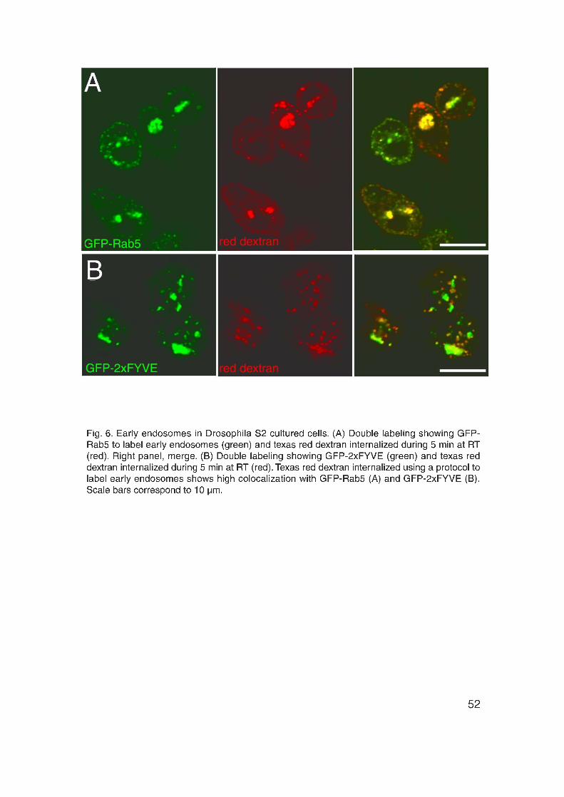
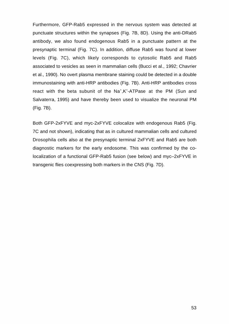
53
Furthermore, GFP-Rab5 expressed in the nervous system was detected at
punctuate structures within the synapses (Fig. 7B, 8D). Using the anti-DRab5
antibody, we also found endogenous Rab5 in a punctuate pattern at the
presynaptic terminal (Fig. 7C). In addition, diffuse Rab5 was found at lower
levels (Fig. 7C), which likely corresponds to cytosolic Rab5 and Rab5
associated to vesicles as seen in mammalian cells (Bucci et al., 1992; Chavrier
et al., 1990). No overt plasma membrane staining could be detected in a double
immunostaining with anti-HRP antibodies (Fig. 7B). Anti-HRP antibodies cross
react with the beta subunit of the Na+,K+-ATPase at the PM (Sun and
Salvaterra, 1995) and have thereby been used to visualize the neuronal PM
(Fig. 7B).
Both GFP-2xFYVE and myc-2xFYVE colocalize with endogenous Rab5 (Fig.
7C and not shown), indicating that as in cultured mammalian cells and cultured
Drosophila cells also at the presynaptic terminal 2xFYVE and Rab5 are both
diagnostic markers for the early endosome. This was confirmed by the co-
localization of a functional GFP-Rab5 fusion (see below) and myc–2xFYVE in
transgenic flies coexpressing both markers in the CNS (Fig. 7D).
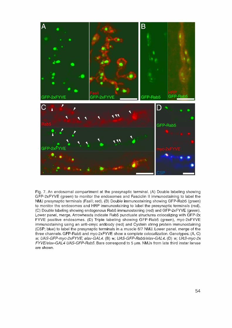

55
2xFYVE localization at the endosome is PI(3)P dependent
We next addressed whether at the presynaptic terminal the specific localization
of GFP-2xFYVE and myc-2xFYVE to the endosome was dependent on the
presence of PI(3)P as described in cultured mammalian cells (Gillooly et al.,
2000). We blocked PI(3)P-kinase activity with Wortmannin and monitored the
tagged 2xFYVE fusions. As in cultured mammalian cells, GFP-2xFYVE looses
its punctuate pattern and becomes dispersed into the cytosol upon blockage of
PI(3)-kinase activity (Fig. 8A). Therefore, as in cultured mammalian cells the
endosomal localization of 2xFYVE in the presynaptic terminal depends on
PI(3)P.
The endosome is localized within the pool of recycling vesicles
Where is the endosome localized with respect to the SVs within the presynaptic
terminal? To address this question recycling vesicles were labeled by the red
FM5-95 styryl dye (Fig. 8B). Like the green FM1-43, which was introduced in
1992 by Betz and colleagues (Betz and Bewick, 1992), FM5-95 is an
amphipathic dye containing a fluorescent head group and a lipid tail. Styryl dyes
have several features making them very useful for internalization experiments in
neurons. First, when present in the aqueous, extracellular solution surrounding
a cell the dye spontaneously inserts into the outer leaflet of the PM. Second,
dye inserted into the membrane shows a much stronger fluorescence than dye
in aqueous solution. Third, styryl dyes cannot cross the membrane, and are
therefore internalized only during compensatory endocytosis. Fourth, FM-dyes
can be washed off easily from the membrane.
In a typical experiment, exocytosis is stimulated in the presence of the FM-dye.
Dye is internalized together with the membrane during compensatory
endocytosis. The internalized dye therefore labels the pool of vesicles that were
internalized during synaptic activity, called the recycling pool of SVs. We
stimulated synaptic activity and thereby internalized the red FM5-95 into
presynaptic terminals of GFP-2xFYVE and GFP-Rab5 expressing NMJs (see

56
methods). Endosomes labeled with either GFP-2xFYVE (Fig. 8C) or GFP-Rab5
(Fig. 8D) are located within the pool of recycling vesicles. This indicates the
presence of a distinct endosomal compartment located within the pool of
recycling vesicles. The location of the endosome prompts the possibility that the
endocytic vesicles internalized during synaptic transmission traffic through the
endosome during their recycling.
The endosome size is stable during synaptic transmission
To study the endosome in vivo, we monitored GFP-Rab5 and GFP-2xFYVE in
resting terminals or while stimulating the synapse under basal, 3 Hz or more
demanding, tetanic, 30 Hz electrophysiological conditions (Fig. 9). Figures 9A
and 9B show that neither the location nor the size or the intensity of GFP-
2xFYVE was substantially changed under these conditions. This observation
indicates that the endosomal size is stable both in the resting terminal and
during synaptic transmission. Therefore, if SV recycling involves trafficking
through the endosome, then endocytic vesicle fusion to the endosome is
balanced with SV budding from the endosome to ensure the constant
endosomal size.


59
SV recycling involves membrane trafficking through the endosome
To address directly whether the endosome is involved in the recycling of SVs
we used the Drosophila mutant shibirets, named after the Japanese word
meaning “paralyzed”. Shibirets is a temperature-sensitive mutation in the
GTPase domain of Dynamin (Grant et al., 1998; van der Bliek and Meyerowitz,
1991) that allows to specifically and reversibly block endocytosis (Koenig and
Ikeda, 1989). At the restrictive temperature, Dynamin becomes nonfunctional
immediately causing the complete arrest of endocytosis at a very specific step
as demonstrated by Kosaka and Ikeda in 1983 (Kosaka and Ikeda, 1983a).
Nascent endocytic vesicles cannot be pinched off from the PM and therefore
accumulate in the form of “collared pits” (Estes et al., 1996; Kosaka and Ikeda,
1983a). These structures have distinct necks connected to the PM that are
surrounded by an electron-dense collar, presumably containing components of
the membrane fission machinery. Therefore, stimulating SV exocytosis in the
synapse while blocking endocytosis with shibirets causes a complete depletion
of SVs. SV depletion can be directly seen at the ultrastructural level (Koenig
and Ikeda, 1989; Koenig et al., 1989; Kosaka and Ikeda, 1983a; Poodry and
Edgar, 1979). In addition, measuring neurotransmitter release using standard
electrophysiological techniques shows the complete SV depletion by the
complete loss of NT release (Koenig and Ikeda, 1999). The phenotype in adult
flies is quite striking: within seconds after exposure to the restrictive
temperature they become completely paralyzed (Grigliatti et al., 1973).
Lowering the temperature releases the endocytosis block and leads within few
minutes to a complete recovery as observed by the recovery of paralyzed flies
and the reformation of the SV pool detected at the ultrastructural level and in
electrophysiological experiments (Koenig and Ikeda, 1999).
We used shibirets1 (shits) to uncouple exo- and endocytosis and thereby to
address three questions: First, do SVs bud from the endosome? Second, do
endocytic vesicles fuse to the endosome? Third, is endosomal trafficking
temporally coupled to synaptic activity? We first excluded a direct effect of
shibirets on endosome morphology. Endosomes visualized by GFP-2xFYVE

60
were monitored in shits mutant synapses in resting terminals at the permissive
and at the restrictive temperature (Fig. 9C and not shown) as well as at the
permissive temperature while stimulating at 3 or 30 Hz (not shown). Under
these conditions the endosomal size, intensity and position remained stable.
Therefore, as in the shi+ controls (Fig. 9A, B) the endosomal size is stable in
shibirets at the permissive temperature in resting terminals or during synaptic
activity as well as at the restrictive temperature when the terminal is not
stimulated, excluding a direct effect of shibirets on endosome morphology.
In order to address whether SVs bud from the endosome, we performed a
“shits/depletion” experiment in the following way: We blocked endocytosis with
shibirets1 at the restrictive temperature (Koenig and Ikeda, 1989; Koenig and
Ikeda, 1999) and depleted the SV pool by electrophysiologically stimulating SV
release for 5, 10 and 15 minutes as previously described (Koenig and Ikeda,
1999). At the same time, endosomes labeled by GFP–2xFYVE were monitored.
If SVs bud from the endosome, in shits there will be membrane output (budding)
from the endosome upon stimulation, but no membrane input (fusion) because
of the absence of newly formed endocytic vesicles that could fuse to the
endosome. Consequently, the endosome should be reduced in size or even be
depleted together with the SV pool in this “shits/depletion” experiment.
Figures 10A-C show, that the punctuate endosomal staining disappears during
the “shits/depletion” experiment: GFP-2xFYVE becomes diffuse. This suggests
that synaptic vesicles budded from the endosome, depleting the compartment.
The kinetics of endosome disappearance depends on the frequency of
stimulation with t1/2 of 5 minutes during the basal stimulation at 3 Hz (10D) and
approximately ten fold faster during the more demanding, tetanic stimulation at
30 Hz (Fig. 10A-C).
We next studied whether endocytic vesicles fuse to the endosome by
monitoring endosomal recovery after release of the shits endocytic block by
lowering the temperature. We first depleted the endosome by performing a
“shits/depletion” experiment as described above (Fig. 10E–G). Figure 10G

61
shows that in contrast to control synapses (Fig. 10J-L) the endosome is
depleted. Subsequently, larvae were returned to the permissive temperature to
release the shits endocytosis block. After 15 minutes, we observed fluorescence
recovery at the endosome (Fig. 10H, I), suggesting that recovery of the
endosome involves dynamin-dependent endocytosis. Furthermore, if we first
depleted the endosome by stimulating the synapse at the restrictive
temperature as described above (Fig. 10M, N) and then maintained the animals
at the restrictive temperature after stimulation no endosomal recovery was
observed (Fig. 10O). Afterwards, returning the animals to the permissive
temperature and thereby releasing the endocytosis block, led to the recovery of
the endosome (Fig. 10P). Therefore, the endosome recovers only when the shits
endocytosis block is released. This indicates that the endosome recovers at the
expense of endocytic vesicles that form after release of the endocytosis block.
In summary, these results show that endocytic vesicles fuse with the endosome
and that synaptic vesicles bud from the endosome. This in turn implies that SV
recycling indeed involves trafficking through the Rab5 endosomal compartment.
We next addressed the question of whether endosomal trafficking is coupled to
synaptic activity. The fact that the depletion of the endosome in shits only
occurred when the synapse was stimulated (compare Fig. 9C vs. Fig. 10A-C),
suggested that SV budding from the endosome only occurs during synaptic
activity and is therefore coupled to synaptic transmission. In contrast, when the
shits endocytic block was released, the endosome recovered in a resting
terminal (Fig. 10H, I, P). This indicates that endocytic vesicles can fuse to the
endosome also in the absence of synaptic activity.
To investigate whether endosomal trafficking is coupled to synaptic activity, we
performed FRAP experiments (Cole et al., 1996) in resting and stimulated GFP-
Rab5 expressing synapses (Fig. 10Q-S). We first bleached selectively the GFP
fluorescence at the endosome in GFP-Rab5 expressing terminals with a
confocal microscope (Fig. 10Q, R). Then, we monitored the recovery of the
GFP fluorescence at the endosome in resting (Fig. 10S), or in stimulated NMJs
(not shown). Recovery of fluorescence occurred within 2 minutes after

62
bleaching in both resting (Fig. 10Q-S) and stimulated terminals (not shown).
These results indicate that vesicles containing GFP-Rab5 can fuse with the
endosome also in the absence of synaptic activity.
In summary, SV recycling involves trafficking through an endosomal
compartment. Endocytic vesicles internalized after Ca2+-triggered exocytosis
fuse with the endosome independent of synaptic activity. Subsequently, SVs
bud from the endosome and are eventually released upon arrival of an action
potential. The fact that SV recycling involves traffic through a Rab5 positive
endosomal compartment prompted us to study the possible role of Rab5 during
the process of recycling using Rab5 mutants.


64
Analysis of Rab5 function using Rab5 mutants and thedominant negative version of Rab5, Rab5S43N
Genomic organization of Drosophila Rab5
In Drosophila, we found a single Rab5 gene with multiple splicing variants (Fig.
11A). The gene has 7 alternative 5’ leader exons and 2 alternative 3’
untranslated regions (UTRs) (Fig. 11A) and encodes for a single open reading
frame (Fig. 11A; see methods). Consistently, a single band of around 24 kDa
was detected in Western blot experiments using the anti-Drosophila Rab5
antibody against larval or embryonic extracts (Fig. 11B, C). The size of 24 kDa
is consistent with the predicted molecular weight of Rab5 containing 219 amino
acids. The presence of a single Rab5 gene and protein isoform in Drosophila is
in contrast to the situation in yeast and mammals where three Rab5 genes
coding for different isoforms were found (Novick and Zerial, 1997). As in
Drosophila, a single Rab5 gene has been described In C. elegans (Grant and
Hirsh, 1999). The genes flanking Rab5 are a zinc finger transcription factor
(CG4272) and a Heparansulphate proteoglycan (CG7245). One of the splicing
forms of Rab5 overlaps by 28 bp with the 5’ end of the CG4272 transcript (Fig.
11A).
We identified 2 P-element insertions from the Berkeley genome project inserted
in the Rab5 gene (Fig. 11A), P l a c w R a b 5 k 0 8 2 3 2 and P ( P Z+)00231.
PlacwRab5k08232 (Rab51) is a P-element insertion within the 5’ leader coding
region (Fig. 11A). Rab52, Rab53 and Rab54 were generated by imprecise
excision of Rab51. Rab52 is a 4 kb deletion of the promotor region, the 5’ non-
translated leader and the first exon of the open reading frame (Fig. 11A). This
exon encodes the PM1-3 Phosphate/Mg2+–binding motifs and the G1 Guanine
base-binding motif of the GTPase domain (Fig. 3A) (Olkkonen and Stenmark,
1997). Therefore, Rab52 is a Rab5 null mutation. Rab53 contains an insertion of
18 bp from the left and 210 bp from the right LTR of the P-element remaining
after imprecise excision of the transposon. In Rab54 14 bp from the right LTR
remained after the imprecise excision of the P-element.
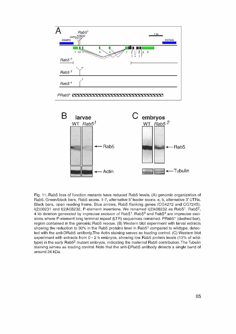

66
Rab5 mutants show locomotion defects, paralytic phenotypes anddefective endosomes
The level of Rab5 expression in the homozygous mutants was determined by
Western blot experiments using the anti-DRab5 antibody. In Rab51 mutants, the
level of Rab5 expression was reduced to around 30% compared to the Rab5
protein level in wildtype (Fig. 11B; see methods). Homozygous Rab51 mutant
animals die during late second and early third instar larval stages. They show
only a light locomotion phenotype when compared to wildtype larvae. When
Rab51 mutants were raised under “intensive care” conditions as described
(Loewen et al., 2001), very few homozygous adults were observed (around 14%
of the homozygous larvae developed to adults). They are extremely poor fertile
and show a flightless phenotype. However, no obvious physiological differences
were detected in Rab51 mutants when compared to wildtype: First, the recycling
SV pool size was determined by an FM1-43 internalization assay (see below
and methods), in which FM1-43 was internalized during 3 min of stimulation at
30 Hz in normal saline. The recycling SV pool size in Rab51 was 90.6 ± 3.3% (n
= 14 NMJs) compared to wildtype. Second, synaptic transmission monitored by
standard electrophysiological recordings (see below and methods) was normal
in the Rab51 mutant. We observed a mean EJP size of 115.2 ± 0.7% (n = 14
muscle 6) in HL3 containing 0.75 mM Ca2+ compared to wildtype.
Rab51 was rescued by P(w+)DRab5+, a genomic rescue construct spanning
Rab5 and excluding the two flanking genes (Fig. 11A; see methods) as well as
by the restricted expression of GFP-Rab5 in the nervous system using elav-
GAL4. Therefore, Rab51-lethality is caused by reduced Rab5 levels in the
nervous system. This also indicates that GFP-Rab5 is a functional Rab5 fusion
and that Rab5 function is essential during the physiology or development of the
nervous system. The two other weaker Rab5 alleles, Rab53 and Rab54 (Fig.
11A; see methods) displayed decreased Rab5 proteins levels of 46% and 35%
compared to wildtype respectively (not shown). Homozygous adults show poor
fertility and a flightless phenotype in 88% and 63% of adults, respectively.

67
The null mutant Rab52 shows a more severe phenotype. Embryos fail to hatch
and when dissected out of their eggshell they are unable to move and don’t
react to tactile stimuli, such as touching with a brush or poking with a needle.
Therefore, Rab52 causes embryonic lethality with a paralytic phenotype.
Western blot experiments with Rab52 homozygous mutant embryos (0 - 22 h)
however showed a faint Rab5 band corresponding to 13% of the wildtype Rab5
protein level (Fig. 11C). As Rab52 is a null mutation, the protein detected is
likely to correspond to Rab5 translated from maternal mRNA. In Drosophila,
mRNAs transcribed from the maternal genome, called maternal mRNAs, are
deposited into the egg and are transcribed during early development. Therefore,
during the early development most if not all proteins are translated form
maternal mRNAs. Consistently, the Rab5 protein was also detected by Western
blot experiments in early embryonic stages (0 - 2 hours) (not shown). This
implies that the zygotic loss of Rab5 is partially rescued by the maternal Rab5
contribution in Rab52 homozygous mutants.
We next investigated the effect of zygotic loss of Rab5 on the endosomes. We
expressed GFP-2xFYVE in the nervous system using elav-GAL4 and monitored
the GFP–2xFYVE labeled endosomes in the mutant background. In control
embryos the GFP–2xFYVE labeled endosomes appeared as punctuate
structures within the neurons of the CNS (Fig. 12A) and the peripheral nervous
system (PNS) (Fig. 12B). Zygotic loss of Rab5 caused the disruption of the
endosomes in the embryonic CNS and PNS (Fig. 12A, B) as shown by the
diffuse GFP fluorescence. Furthermore, at the presynaptic terminals of control
NMJs from late embryos (stage 17) GFP–2xFYVE-positive endosomal
punctuate structures were observed (Fig. 12C). In contrast, GFP–2xFYVE was
dispersed in the cytosol in the Rab52 mutant embryonic NMJs (Fig. 12D),
indicating that, at the Rab52 mutant NMJs, endosomes are disrupted. In
summary, these data indicate that Rab5 is essential for the integrity of the
endosomes and is required for the development and/or function of the nervous
system, since homozygous Rab52 mutants die as embryos, probably when the
maternally derived Rab5 is exhausted.


69
Specific interference of Rab5 during presynaptic physiology doesnot cause a developmental phenotype
The Rab5 mutants described above represent a deficit of Rab5 function both at
the presynaptic (neuron) and postsynaptic (muscle) side of the NMJ. We
wanted to study the specific role of Rab5 in the physiology of the presynaptic
terminal without affecting other tissues or the nervous system development. To
achieve this, we expressed a dominant negative, GDP-bound Rab5 mutant
(Rab5S43N) (Entchev et al., 2000; Stenmark et al., 1994) exclusively in the
nervous system with elav-GAL4 using the UAS/GAL4 technique (Brand and
Perrimon, 1993). As GAL4 shows thermosensitivity in Drosophila (Brand et al.,
1996; Entchev et al., 2000), this allows controlling the expression level using
different temperatures. To study the specific effect of the Rab5 mutant during
synaptic physiology, we kept expression low during neural development:
Rab5S43N was expressed at low levels during embryonic and early larval
stages at 16°C, and at higher levels at 25°C or 29˚C during the last two days of
larval development (see methods).
We first addressed whether mutant Rab5S43N has an effect on the
development of the nervous system. Figures 13A-E show that Rab5S43N
expressed in the CNS as described above did not cause a developmental
phenotype of the NMJ. The overall NMJ morphology was normal in the mutant
(Fig. 13A). Furthermore, the normalized synaptic surface, which represents the
percentage of synaptic surface with respect to the muscle surface (see
methods), was normal in Rab5S43N expressing NMJs (Fig. 13D).
We then looked in more detail at the number and distribution of active zones
and centers of endocytosis. Active zones are the regions within the presynaptic
terminal where exocytosis of the neurotransmitter glutamate occurs.
Postsynaptically, active zones are characterized by the accumulation of
glutamate receptor clusters.

70
The centers of endocytosis have been first described in Drosophila (González-
Gaitán and Jäckle, 1997) and were later found in organisms such as frog and
snake (Jarousse and Kelly, 2001; Ringstad et al., 1999; Roos and Kelly, 1998;
Roos and Kelly, 1999; Teng and Wilkinson, 2000). Centers of endocytosis
surround the active zones and represent specialized regions where clathrin-
mediated, compensatory endocytosis takes place. Therefore, proteins involved
in clathrin-mediated endocytosis, like α-Adaptin, Dynamin and Clathrin are
accumulated at and therefore define the centers of endocytosis. In order to label
the active zones, we used the nc82 antibody (Fig. 13B, C). The nc82 antibody
colocalizes with the postsynaptic glutamate receptors at the active zones (not
shown) and therefore recognizes an unknown antigen associated to the active
zones. As expected the nc82 labeling is surrounded by a staining of α-Adaptin
or Dynamin monitoring the centers of endocytosis (Fig. 13B, C and not shown).
Rab5S43N expressing motoneurons showed a normal number and morphology
of the centers of endocytosis and the active zones of exocytosis as visualized
by anti-α-Adaptin, anti-Dynamin (González-Gaitán and Jäckle, 1997; Roos and
Kelly, 1999) and nc82 antibodies (Heimbeck et al., 1999), respectively (Fig.
13C, E and not shown). Therefore, the development and morphology of NMJs
was normal in Rab5S43N expressing motoneurons. We next analyzed the role
of Rab5 during endosomal trafficking and SV recycling.

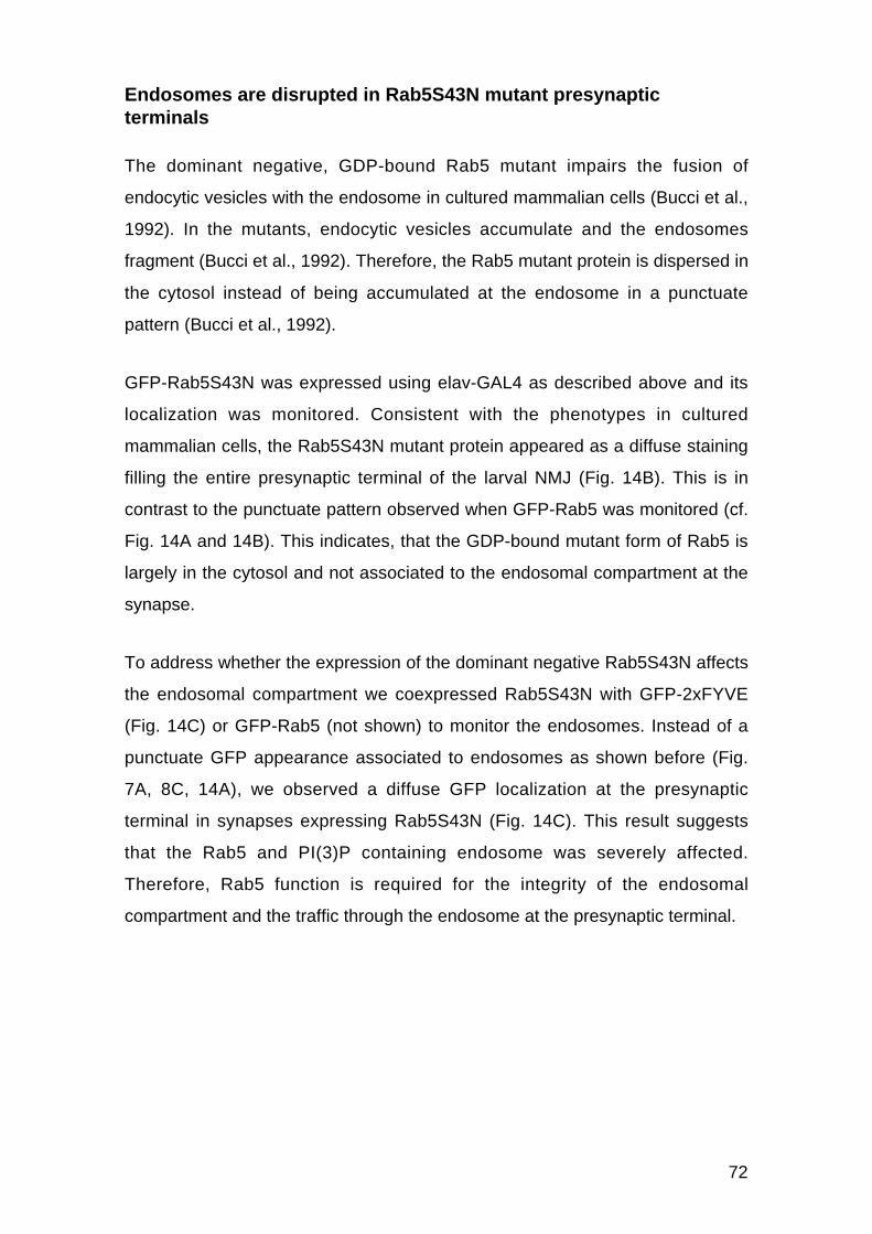
72
Endosomes are disrupted in Rab5S43N mutant presynapticterminals
The dominant negative, GDP-bound Rab5 mutant impairs the fusion of
endocytic vesicles with the endosome in cultured mammalian cells (Bucci et al.,
1992). In the mutants, endocytic vesicles accumulate and the endosomes
fragment (Bucci et al., 1992). Therefore, the Rab5 mutant protein is dispersed in
the cytosol instead of being accumulated at the endosome in a punctuate
pattern (Bucci et al., 1992).
GFP-Rab5S43N was expressed using elav-GAL4 as described above and its
localization was monitored. Consistent with the phenotypes in cultured
mammalian cells, the Rab5S43N mutant protein appeared as a diffuse staining
filling the entire presynaptic terminal of the larval NMJ (Fig. 14B). This is in
contrast to the punctuate pattern observed when GFP-Rab5 was monitored (cf.
Fig. 14A and 14B). This indicates, that the GDP-bound mutant form of Rab5 is
largely in the cytosol and not associated to the endosomal compartment at the
synapse.
To address whether the expression of the dominant negative Rab5S43N affects
the endosomal compartment we coexpressed Rab5S43N with GFP-2xFYVE
(Fig. 14C) or GFP-Rab5 (not shown) to monitor the endosomes. Instead of a
punctuate GFP appearance associated to endosomes as shown before (Fig.
7A, 8C, 14A), we observed a diffuse GFP localization at the presynaptic
terminal in synapses expressing Rab5S43N (Fig. 14C). This result suggests
that the Rab5 and PI(3)P containing endosome was severely affected.
Therefore, Rab5 function is required for the integrity of the endosomal
compartment and the traffic through the endosome at the presynaptic terminal.


74
Endocytic intermediates accumulate in Rab5 mutant presynapticterminals
We then analyzed the effect of Rab5S43N at the ultrastructural level of type I
boutons. In wildtype, type I boutons are surrounded by a prominent subsynaptic
reticulum (SSR), which corresponds to the highly folded muscle membrane (Fig.
15A). The presynaptic terminal of type I boutons is filled with small, clear SVs
(Fig. 15A-D). In addition, mitochondria required for local energy generation are
found in variable numbers (Fig. 15A). The active zones of neurotransmitter
release are characterized by an electron dense membrane and the presence of
an electron dense T-bar (Fig. 15A). T-bars are unique to Drosophila NMJs,
however their function is not yet known. We also observed bigger cisternal and
tubular structures of around 150 nm, which have been suggested to correspond
to endosomal structures (Koenig et al., 1993) (Fig. 15B-D).
In wildtype presynaptic terminals, we could distinguish the two types of vesicles
that have been previously reported (Fergestad et al., 1999; Kosaka and Ikeda,
1983a). Synaptic vesicles with a diameter of 35 nm (35.9 ± 0.11 nm, n = 1991
vesicles) (Fig. 16A) and a second type of vesicles with a diameter of 70 nm
(73.6 ± 2.5 nm, n = 36 vesicles) (Fig. 16A, solid arrowheads), previously
described as recycling intermediates (Fergestad et al., 1999; Kosaka and Ikeda,
1983a). In shits mutants, early stages of endocytosis are blocked, causing the
accumulation of nascent endocytic vesicles in the form of collared pits at the
plasma membrane. Since the 70 nm vesicles have a very similar diameter to
these collared pits (Kosaka and Ikeda, 1983a), we suggest that they represent
newly formed endocytic vesicles. In addition, endocytic vesicles are probably
transient structures since only 2.1 ± 0.5 (n = 26 sections) were observed in each
EM section.


76
Synapses expressing Rab5S43N, showed an accumulation of the 70 nm
vesicles (70.6 ± 1.1 nm, n = 148 vesicles). 5.9 ± 1.2 (n = 25 sections) were
observed on average per EM section (Fig. 16B, solid arrowheads). This
phenotype suggests that the recycling intermediates (Fergestad et al., 1999;
Kosaka and Ikeda, 1983a) indeed correspond to endocytic vesicles, which
accumulate in the mutant synapses where Rab5 function is impaired (Fig. 16B,
solid arrowheads). These results are consistent with the Rab5SN phenotype
reported in cultured mammalian cells, where endocytic vesicles accumulate due
to their inability to fuse efficiently with the early endosome (Bucci et al., 1992).
We did not observe major changes in other synaptic features in Rab5S43N
expressing terminals including the number and structure of the T-bars, the
number of docked vesicles as defined as vesicles touching the plasma
membrane at the T-bar (Table 2), the overall appearance of the presynaptic
terminal and the subsynaptic reticulum postsynaptically. In summary, blocking
Rab5 function at the presynaptic terminal using the dominant negative
Rab5S43N mutant causes the accumulation of endocytic vesicles, implying that
Rab5 regulates the fusion of endocytic vesicles with the endosome.
WT Rab5SN Rab5 GFP-Rab5docked vesicles 1.31 ± 0.24 1.67 ± 0.20 1.29 ± 0.22 1.22 ± 0.18n 17 30 14 10
Table 2. Quantification of docked vesicles at the T-bar in wildtype and the different mutants.Docked vesicles were defined as vesicles touching the PM at the T-bar. n corresponds to thenumber of T-bars analyzed. Genotypes: wildtype (WT), w; UAS–Rab5S43N/+; elav-GAL4/+(Rab5SN), w; UAS–Rab5/+; elav-GAL4/+ (Rab5), w; elav-GAL4/UAS–GFP-Rab5 (GFP-Rab5).


78
Endocytic trafficking during SV recycling involves Rab5 function
In order to study the role of Rab5 during SV recycling, we performed FM1-43
internalization and release experiments (Betz and Bewick, 1992) (Fig. 17, 18).
We monitored three parameters: first, the rate of endocytosis, second, the size
of the vesicle pool and third, the recycling rate. The styryl dye FM1-43 is
internalized only during endocytosis and the FM1-43 fluorescence is directly
proportional to the number of vesicles internalized. Therefore, FM1-43
fluorescence can be used to quantify endocytosis, recycling and the SV pool
size.
The rate of internalization was assayed first. FM1-43 uptake was monitored in
electrophysiologically stimulated synapse (see methods) at 3 Hz for 3, 5 and 10
minutes in HL3 containing 1.5 mM Ca2+ (Stewart et al., 1994) (Fig. 17A-C). In
wildtype synapses, FM1-43 fluorescence increased rapidly during the first 3
minutes (Fig. 17C). After the first 3 minutes, the fluorescent increase was
slowed down. This phenomenon has been previously reported and was
interpreted to be caused by the release of dye-loaded vesicles (Betz and
Bewick, 1992; Betz and Bewick, 1993; Kuromi and Kidokoro, 2000; Ryan and
Smith, 1995): During the first few minutes FM-dye is only internalized,
progressively filling the SV pool with FM1-43 labeled vesicles causing a rapid
increase in the fluorescence. Then, after few minutes a mixture of FM1-43
loaded and unloaded vesicles is released. Therefore, the increase in the
fluorescence is slowed down due to the release of previously internalized and
consequently dye-loaded vesicles. We therefore estimated the rate of dye-
internalization by the fluorescence uptake rate during the first 3 minutes. In
Rab5S43N expressing presynaptic terminals, the rate of FM1-43 internalization
was 2.5 fold slower compared to wildtype (Fig. 17B, C). This result indicates
that endocytosis is reduced when Rab5 function is impaired.
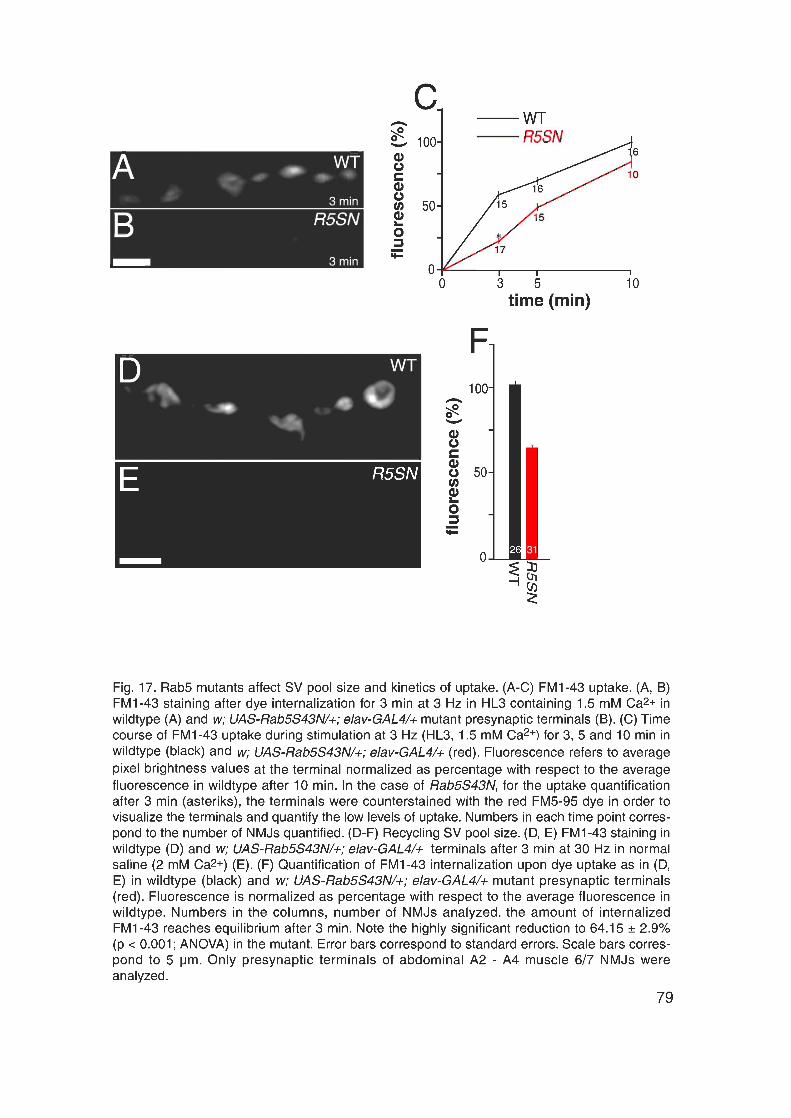

80
We next studied the size of the SV pool. The terminal was first loaded by
stimulating the synapse at 30 Hz for 3 minutes in normal saline (Jan and Jan,
1976a) (Fig. 17D-F). To test whether the entire recycling pool of vesicles was
labeled under these conditions, we increased the stimulation time to 5 minutes
at 30 Hz. This increase in the stimulation time did however not further increase
the amount of internalized fluorescence at the presynaptic terminal of wildtype
or Rab5S43N (not shown). This indicates that after 3 min of stimulation at 30 Hz
in normal saline, the entire pool of recycling vesicles is labeled with FM1-43, in
both wildtype and Rab5S43N mutant presynaptic terminals (Fig. 17D-F). We
therefore estimated the relative SV recycling pool size by maximally loading the
terminal using this protocol. Figure 17F shows that blocking Rab5 function with
Rab5S43N causes a significant decrease to 64.15 ± 2.9% (n = 31, p < 0.001,
ANOVA) in the SV recycling pool size.
We then studied the kinetics of FM1-43 release in the mutant synapses (Fig.
18). The terminal was first maximally loaded as described above and
subsequently stimulated at 3 Hz for various periods of time in normal saline.
The amount of dye released was measured by quantifying the fluorescence
remaining within the terminal at each time point (Fig. 18; see methods). We
could distinguish three phases during the FM1-43 release in wildtype (Fig.
18G): first, a fast release phase during the first 5 minutes, second, a slower
phase between 5 and 30 minutes and third, a third phase, after 30 minutes,
when no more dye could be released and 14.5 ± 1.2% of FM1-43 remained
unreleasable. The kinetics of dye release has been previously reported (Betz
and Bewick, 1993; Betz et al., 1992). It was shown that during the first phase
dye release exactly parallels the amount of neurotransmitter released, because
neurotransmitter and dye are released from the same SVs. However, during the
second phase significantly less FM-dye than neurotransmitter is released,
because the pool of fluorescently labeled SVs becomes increasingly diluted by
newly recycled vesicles that do not contain FM1-43.

81
In the Rab5S43N mutant synapses, dye release occurred 2.5 times slower
during the first 5 minutes (Fig. 18G; also cf. 18A, B vs. 18D, E), indicating that
impaired Rab5 function affects SV release. In addition, the fraction of
unreleasable dye was increased to 38.0 ± 3.3% in Rab5S34N expressing
synapses (Fig. 18G). In summary, these data indicate that SV recycling is
impaired in Rab5S43N mutant synapses. This is consistent with a role of Rab5
in the formation of clathrin-coated vesicles (McLauchlan et al., 1998) and their
subsequent fusion to the early endosome (Bucci et al., 1992; Stenmark et al.,
1994).


83
Rab5-dependent recycling determines the SV fusion efficacy
How does recycling through the endosome affect the performance of SVs
during Ca2+-triggered exocytosis? To address this question, we performed
current clamp electrophysiological recordings, a method to measure in the
muscle the postsynaptic response caused by presynaptic neurotransmitter
release (Fig.19). Under physiological conditions, in the absence of synaptic
activity, the membrane potential of the muscle is negative, around –60 mV. It
can be measured with a glass electrode (recording electrode) inserted into the
muscle. Synaptic activity is then induced by stimulating the segmental nerve
with a stimulation electrode. This is achieved by sucking a segmental nerve into
the stimulation electrode and applying a short, positive current to induce APs.
The APs travel along the axon to the presynaptic terminal. At the presynaptic
terminal, each action potential causes the opening of voltage-gated Ca2+-
channels. Ca2+-ions enter the presynaptic terminal and trigger the fusion of
primed SVs with the plasma membrane. Glutamate is released into the synaptic
cleft. Subsequently, the neurotransmitter binds and opens ligand-gated cation-
channels located within the postsynaptic membrane causing an influx of cations
into the muscle fiber. The influx of cations in turn elicits a very rapid and
transient increase in the muscle membrane potential. This increase can be
measured with the recording electrode as an excitatory junction potential (EJP).


85
SVs do not only fuse to the presynaptic membrane after the action potential
induced influx of Ca2+ into the presynaptic terminal. Single SVs also fuse
spontaneously with the membrane, however with a relatively low frequency. The
change in the muscle membrane potential caused by neurotransmitter released
from single vesicle is called spontaneous or miniature EJP (mEJP) (Katz, 1969).
Its size reflects the amount of neurotransmitter stored within a single vesicle,
called the quantal size. In addition, the mEJP size depends on the number and
sensitivity of the postsynaptic glutamate receptors. By knowing the EJP and the
mEJP amplitude it is possible to calculate the quantal content of the response,
which reflects the number of SVs that fused after a single action potential. The
quantal content is calculated by dividing the EJP amplitude by the mEJP
amplitude measured in the same muscle and correcting for nonlinear
summation according to Martin (Martin, 1955).
To address whether synaptic transmission is affected in Rab5S43N we
recorded mEJPs and nerve-evoked EJPs in muscle 6 of third instar larvae (Fig.
20, 21) raised under 3 different conditions (Table 3, 4). Changes in the mean
mEJP frequency reflect a change in the total number of fusion competent
vesicles. Alterations in the mean mEJP amplitude are indicative for changes in
the neurotransmitter content or an affected postsynaptic sensitivity to the
released neurotransmitter. mEJP recordings from Rab5S43N mutant NMJs
displayed no significant differences in mean amplitude, variability, frequency or
voltage decay kinetics compared to wildtype mEJPs (Table 3, Fig. 20).
This result indicates that presynaptic expression of Rab5S43N does not affect
the vesicular neurotransmitter content, the number of fusion competent vesicles
or the postsynaptic glutamate receptor function or density. This is consistent
with the normal synaptic ultrastructure (Fig. 16), mean number of active zones
(Fig. 13E) and docked vesicles at the T-bar (Table 2, Fig. 16) of the mutant
NMJs as shown above.

86
genotype 16˚C 25˚C 29˚Cfrequency(Hz)
amplitude(mV)
frequency(Hz)
amplitude(mV)
frequency(Hz)
amplitude(mV)
WT 4.4±0.17 1.5±0.05 3.7±0.17 1.3±0.06 3.1±0.17 1.4±0.06R5SN 3.9±0.24 1.5±0.06 3.3±0.23 1.3±0.06 2.8±0.19 1.3±0.06R5 3.4±0.20 1.3±0.05 2.8±0.17 1.3±0.07 1.9±0.16 1.3±0.09GFP-R5 3.3±0.20 1.2±0.05 3.1±0.18 1.4±0.06 3.0±0.14 1.2±0.05
Table 3. mEJP frequencies and amplitudes. Overview of mEJP frequencies and amplitudes inwildtype and the different mutants. Animals were raised using 3 different temperature protocols:First, animals were raised at 16˚C until the third larval stage (16˚C), second, animals wereraised at 16˚C until early third instar larvae, followed by 25˚C during the last two days of larvaldevelopment (25˚C), and third, animals were raised at 16˚C until early third instar larvae,followed by 29˚C during the last two days of larval development (29˚C). Note that there were nosignificant differences in the mEJP amplitudes. The mEJP frequencies were slightly higher inanimals raised at 16˚C until third instar larvae and were slightly lower in animals raised at 29˚Cduring the last two days of larval development compared to animals raised at 25˚C during thelast two days of larval development. Differences between different genotypes of animals raisedusing the same temperature protocol or between animals of the same genotype raised usingdifferent temperature protocols were not significant (p > 0.05, ANOVA). Between 16 and 37muscles 6 were analyzed per condition. Genotypes: wildtype (WT), w; UAS–Rab5S43N/+; elav-GAL4/+ (R5SN), w; UAS–Rab5/+; elav-GAL4/+ (R5), w; elav-GAL4/UAS–GFP-Rab5 (GFP-R5).


88
We next addressed the efficacy of the evoked synaptic transmission (Table 4,
Fig. 21). EJPs were evoked by stimulating the segmental nerve at 0.5 Hz and
the voltage response in the muscle was measured using standard intracellular
recording conditions (see methods).
genotype 16˚C 25˚C 29˚CEJP amplitude
(mV)% quantalcontent
amplitude(mV)
% quantalcontent
amplitude(mV)
% quantalcontent
WT 23.2±1.7 100.0±11.0 23.5±1.4 100.0± 7.2 19.0±0.8 100.0± 5.9R5SN 15.5±1.4 52.8± 6.1 12.9±1.2 49.8± 7.8 10.5±1.1 44.2± 6.5R5 29.9±1.7 170.7±12.2 28.0±1.5 137.6±12.1 25.5±1.1 132.7±11.9GFP-R5 31.3±1.9 194.3±19.9 30.0±1.1 137.3± 9.8 33.0±1.2 208.8±11.1
Table 4. Mean EJP amplitudes and mean quantal contents. Overview of the mean EJPamplitudes and mean quantal contents in wildtype and the different mutants measured in HL3containing 0.75 mM Ca2+. Animals were raised using 3 different temperature protocols: First,animals were raised at 16˚C until the third larval stage (“16˚C protocol”, 16˚C), second, animalswere raised at 16˚C until early third instar larvae, followed by 25˚C during the last two days oflarval development (“25˚C protocol”, 25˚C), and third, animals were raised at 16˚C until earlythird instar larvae, followed by 29˚C during the last two days of larval development (“29˚Cprotocol” 29˚C). Note that the reduction of the quantal content in Rab5S43N compared towildtype increases from a reduction to 52.8% using the 16˚C protocol to a reduction to 44.2%using the 29˚C protocol. Between 16 and 37 muscles 6 were analyzed per condition. Genotypesas in Table 3.
The EJP measured in HL3 containing 0.75 mM Ca2+ in wildtype was 23.5 ± 1.4
mV (n = 35) and was significantly reduced in Rab5S43N expressing NMJs to
12.9 ± 1.2 mV (n = 28) using the “25˚C protocol” (Table 4, Fig. 21A, B). We next
calculated the number of vesicles fusing upon arrival of a single action potential,
the quantal content, from the mean EJP amplitude and from the mean mEJP
amplitude measured in the same muscle (see methods; (Martin, 1955)). We
observed that the mean quantal content was drastically reduced to 49.8 ± 7.8%
(n = 28) in Rab5S43N expressing terminals when compared to wildtype larvae
using the “25˚C protocol” (Table 4, Fig. 21C), consistent with a decreased FM1-
43 release rate in the mutant (Fig. 18). Since the number of docked vesicles
(Table 2) and the spontaneous release rate were normal in Rab5S43N
expressing synapses (Table 3, Fig. 20), these data indicate that impaired Rab5
89
function led to a reduced probability of the Ca2+-triggered release of synaptic
vesicles.
We next studied the paired-pulse behavior, a form of synaptic short-term
plasticity (Fig. 22A) (Zucker, 1989; Zucker, 1999). This was performed by
applying two stimuli with only a short interpulse interval, the second stimulus
following the first stimulus within 20 msec. The paired-pulse behavior usually
correlates with the size of the first EJP. Synapses with a high release probability
and therefore with a large first EJP usually show depression. This is observed
as a decrease in the size of the second EJP compared to the first EJP. In
contrast, synapses with a low release probability and therefore with a small first
EJP usually show paired-pulse facilitation, as indicated by an increased EJP to
the second stimulus. According the to residual Ca2+-hypothesis, Ca2+ levels are
still elevated after the first stimulus, when the second AP arrives. This leads to a
further increase in the intracellular Ca2+-concentration (Zucker, 1999). Synapses
with low release probabilities respond to the further elevated Ca2+-levels with an
increased number of SVs fusing, causing an increased second EJP. In contrast,
synapses with high release probabilities, release most of their fusion competent
SVs already upon the first stimulus, depleting the pool of primed SVs.
Therefore, the second stimulus rather causes a decreased EJP as less fusion-
competent vesicles are available than during the first stimulus.
Consistent with the reduced release probability, Rab5S43N synapses show
stronger paired pulse facilitation than wildtype synapses (Fig. 22A). In wildtype
synapses, responses show a slightly facilitated paired-pulse ratio (Q2/Q1) of 1.3
± 0.1 (n = 26) at a 20 ms interpulse interval in 0.75 mM [Ca2+]e (Fig. 22A). In
contrast, Rab5S43N expressing terminals show a significantly stronger
facilitation and exhibited a Q2/Q1 ratio of 2.0 ± 0.2 (n = 15, p < 0.05). Therefore,
R5S43N expressing presynaptic terminals show an altered paired-pulse
behavior, consistent with a reduced release probability of the SVs.


91
Since the Ca2+-triggered release probability was reduced in R5S43N expressing
synapses, we next examined the Ca2+-sensitivity and Ca2+-cooperativity during
neurotransmitter release. The Ca2+-cooperativity reflects the Ca2+-binding to the
Ca2+-sensor of the release machinery, which likely corresponds to the vesicle
protein Synaptotagmin (Brose et al., 1992; Desai et al., 2000; Fernandez-
Chacon et al., 2001; Geppert and Sudhof, 1998; Littleton and Bellen, 1995;
Littleton et al., 1999; Perin et al., 1990). In order to study the Ca2+-sensitivity
and Ca2+-cooperativity we systematically examined the quantal content as a
function of the external Ca2+-concentration ([Ca2+]e) (Fig. 22B). The slope of the
Ca2+-dependency of the mean quantal content was not affected in the mutant
synapses (Fig. 22B), reflecting a normal Ca2+-cooperativity and indicating that
Ca2+-binding to the Ca2+-sensor was not affected.
However, for all [Ca2+]e examined, the release probability in Rab5S43N was
significantly reduced (Fig. 22B) indicating that the defect induced by the
impaired Rab5 function is likely to be Ca2+-independent and probably affects the
efficacy of the evoked SV fusion process itself. This can be either due to a
reduced number of fusion competent vesicles or due to a reduced efficacy of
vesicular release. The fact that the mEJP frequency (Table 3, Fig. 20D) as well
as the number of active zones and docked vesicles (Table 2, Fig. 13C, E, 16B)
is normal in Rab5S43N implies that it is the probability of SV fusion, which is
affected in the mutant. Furthermore, the change in the paired-pulse behavior
(Fig. 22A) also indicates that the effect is not simply due to a change in the
number of synapses, but is rather due to a change in synaptic performance. In
summary, Rab5-dependent endosomal trafficking determines the efficacy of
Ca2+-triggered exocytosis at the presynaptic terminal. Impaired Rab5 function
causes a reduction in the release probability of SVs.
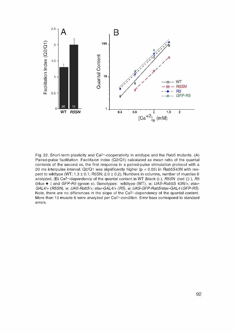

93
Analysis of Rab5 gain of function
Rab5-mediated endosomal trafficking is rate-limiting during SVrecycling and synaptic transmission
We have shown above that Rab5 is required during SV recycling and synaptic
transmission. Expression of the dominant negative version of Rab5, Rab5S43N
causes a reduction in both the rate of endocytosis and the size of the recycling
SV pool. Furthermore, Rab5S43N causes the accumulation of endocytic
vesicles and a decrease in the fusion efficacy of SVs, while synaptic
morphology was normal.
What happens if Rab5 is overexpressed? It has been shown in cultured
mammalian cells that Rab5 is rate-limiting in the regulation of the kinetics of
endocytic vesicle fusion with the early endosome (Bucci et al., 1992) and in the
homotypic fusion between early endosomes (Rybin et al., 1996). The level of
available active Rab5 regulates the degree of recruitment of tethering and
fusion factors to the Rab5 subdomain at the early endosome and thereby the
extent of the subsequent fusion. If Rab5-dependent trafficking is also rate-
limiting in the synapse, Rab5 overexpression should cause an increase in the
SV recycling efficacy and thereby an improved synaptic performance.
To address this issue we overexpressed Rab5 in the CNS and monitored
synaptic development, the kinetics of recycling, synaptic transmission and the
ultrastructure of the synapse. As with Rab5S43N, Rab5 and GFP-Rab5 were
expressed specifically in the CNS with elav-GAL4. The expression levels were
controlled using to the thermosensitivity of GAL4 in Drosophila (Brand et al.,
1996; Entchev et al., 2000) (see methods). Expression was kept low during
embryonic and early larval development at 16˚C and was increased during the
last to two days of larval development at either 25˚C or 29°C.

94
Overexpression of Rab5 does not cause a developmental phenotypeof the NMJ but causes an enlargement of endosomes
Rab5 or GFP-Rab5 expressed as described above did not cause a
developmental phenotype of the NMJ. The overall NMJ morphology was normal
(Fig. 13A). In addition, no change in the synaptic surface (Fig. 13D) and in the
density of active zones was detected (Fig. 13E). As the development was
normal, we next studied the ultrastructure of Rab5-overexpressing presynaptic
terminals (Fig. 16). We observed no difference in the overall synaptic
morphology or structure of the postsynaptic SSR. In addition, the number of
docked vesicles at the T-bar was normal (Table 2). In wildtype, we observed
1.31 ± 0.24 docked vesicles at the T-bar (n = 17 T-bars), in Rab5 1.29 ± 0.22 (n
= 14) and in GFP-Rab5 1.22 ± 0.18 (n = 10). However, endosomal structures
were enlarged in the Rab5 overexpressing terminals. In wildtype, cisternal
structures were only occasionally found. In contrast, in each random EM section
through the overexpressing terminals, we observed large tubular structures
(Fig. 16C; empty arrows), cisternal structures (Fig. 16C; solid arrows) and
multivesicular bodies (Fig. 16C; empty arrowheads). Therefore, as in cultured
mammalian cells (Bucci et al., 1992) Rab5 overexpression at the Drosophila
NMJ causes enlarged endosomal structures.
Rab5 overexpression enhances synaptic performance
Rab5 overexpression in cultured mammalian cells causes an increase in the
rates of endocytosis as measured by dye internalization and transferrin uptake
assays (Bucci et al., 1995; Bucci et al., 1992). Consistently, FM1-43
internalization rates were significantly increased up to 118.0 ± 4.4% (n = 14
NMJs; p < 0.05; 3 Hz 10 min) in HL3 containing 1.5 mM Ca2+ and up to 158.82 ±
4.5% (n = 16 NMJs, p < 0.0001; 3 Hz 15 min) in HL3 containing 0.75 mM Ca+2
in Rab5 overexpressing terminals with respect to control (not shown).
Therefore, like in mammalian cells (Bucci et al., 1992), Rab5 overexpression
causes an increase in the endocytic rate, as reflected by the internalization rate.
95
We next studied the size of the recycling SV pool by FM1-43 internalization for
3 min at 30 Hz in normal saline (as described above) and observed an average
fluorescence intensity in the Rab5 overexpressing terminals of 102 ± 2.6% (n =
25 NMJs) compared to wildtype. Using this dye uptake protocol the entire pool
of recycling SVs was labeled in Rab5 overexpressing synapses, because an
increase in the stimulation time to 5 min did not further increase the amount of
internalized fluorescence (as described above). Therefore, we observed no
difference between wildtype and Rab5 overexpressing terminals in the size of
the recycling SV pool, indicating that Rab5 overexpression did not increase the
size of the recycling SV pool at the synapse. We then studied SV recycling by
first loading the entire SV pool as described above and then stimulating at 3 Hz
for different periods of time in normal saline. We observed no differences in the
FM1-43 release kinetics when compared to wildtype (not shown). This indicates
that Rab5 overexpression does not affect SV release kinetics.
To address the effect of Rab5 overexpression on the efficacy of vesicular
release we recorded mEJPs and EJPs and calculated the quantal content upon
basal stimulation at 0.5 Hz as described above (Table 3, 4, Fig. 20, 21). mEJPs
of Rab5 overexpressing synapses displayed no significant difference in the
mean amplitude, variability, frequency or voltage decay kinetics compared to
wildtype (Table 3, Fig. 20). This indicates that the vesicular neurotransmitter
content, the number of fusion competent SVs and the postsynaptic glutamate
receptor function and density are normal in the overexpressing terminals. These
observations are consistent with the normal NMJ morphology (Fig. 13A, D, E)
and normal number of docked SVs as observed at the ultrastructural level
(Table 2, Fig. 16C).
The mean EJP was significantly increased from 23.5 ± 1.4 mV (n = 35 muscles
6) in wildtype to 28.0 ± 1.5 mV (n = 13 muscles 6) in Rab5 overexpressing
terminals and to 30.0 ± 1.1 mV (n = 35 muscles 6) in GFP-Rab5 overexpressing
terminals (Table 4, Fig. 21B). This phenotype was even further increase in
GFP-Rab5 overexpressing NMJs in animals raised at 29˚C during the last two
days of larval development (see methods) to an EJP of 33.0 ± 1.2 (n = 17

96
muscles 6) (Table 4, Fig. 21B). Consistently, the quantal content was
significantly increased up to 1.74 fold with respect to wildtype (Table 4, Fig.
21C). The phenotype caused by Rab5 and GFP-Rab5 is the same, supporting
the finding, that GFP-Rab5 is a functional Rab5 fusion. Since the number of
docked vesicles (Table 2) and the mEJP frequency were normal (Table 3, Fig.
20), the higher quantal content indicates that elevated levels of Rab5 function
led to an increased probability of the Ca2+-triggered SV release. Consistently,
Rab5 overexpressing synapses showed a slightly depressed paired-pulse
behavior. The Q2/Q1 ratio was 1.0 ± 0.4 (n = 17 muscle 6) for Rab5
overexpressing presynaptic terminals and 0.8 ± 0.3 (n = 19 muscle 6) in the
case of GFP-Rab5 overexpressing terminals compared to a Q2/Q1 ratio of 1.3
± 0.1 (n = 26 muscles 6) in wildtype (not shown).
We finally examined the Ca2+-cooperativity and the Ca2+-dependency of the
quantal content (Fig. 22B). We observed no change in the slope of the Ca2+-
dependency of the quantal content (Fig. 22B), indicating that the Ca2+-binding to
the Ca2+-sensor of the release machinery was normal. Therefore,
overexpression of Rab5 or GFP-Rab5 causes an increase in the efficacy of the
Ca2+-triggered SV exocytosis. We conclude that Rab5-mediated endosomal
trafficking affects in a rate-limiting manner a Ca2+-independent step during the
SV fusion process.
97
Discussion
Synaptic vesicle recycling
At the presynaptic terminal, SVs are regenerated through a local recycling
process after Ca2+-triggered exocytosis. However, the mechanism of SV
recycling is largely unknown. Two models for SV retrieval have been proposed:
“kiss-and-run” and clathrin-mediated endocytosis (Ceccarelli et al., 1973; Fesce
et al., 1994; Heuser and Reese, 1973; Valtorta et al., 2001). Recent evidence
suggests that both models are valid and that two or more recycling pathways
might operate simultaneously and/or in different systems. Why should a neuron
use different recycling mechanisms, and how does it control which pathway is
used? It has been proposed that the molecular composition of the vesicles
influences their competence for “kiss-and-run” (Burgoyne et al., 2001; Valtorta
et al., 2001). Furthermore, the “kiss-and-run” mode might be used under high
frequency stimulation conditions, when fast SV recycling is required (Alés et al.,
1999).
“kiss-and-run” recycling ensures a stable composition of SVs and PM, since no
mixing of the two membranes occurs. However, this also implies that the same
molecular SV components including proteins and lipids are used repeatedly.
This raises the question of how turned over proteins and lipids are sorted out
and are replaced. The second SV recycling pathway, clathrin-mediated
endocytosis, might provide a mechanism to control the SV membrane
composition: Endocytic vesicles derived by clathrin-mediated endocytosis could
fuse to early endosomes, where sorting can occur. Although endosome-like
structures have been observed in nerve terminals (de Hoop et al., 1994; Parton
et al., 1992; Sulzer and Holtzman, 1989; Teichberg and Holtzman, 1975), it is
controversial whether SV recycling involves trafficking through endosomal
compartments (de Wit et al., 1999; Hannah et al., 1999; Jarousse and Kelly,
2001; Murthy and Stevens, 1998; Takei et al., 1996; Zenisek et al., 2000).

98
This study established the presence of an early endosomal compartment
characterized by Rab5 and PI(3)P at the presynaptic terminal of the Drosophila
NMJ. Furthermore, it was shown that SV recycling involves trafficking through
the endosome and that this recycling pathway is controlled by the small
GTPase Rab5. Finally, endosomal recycling is relevant during neurophysiology:
it regulates the synaptic performance.
SVs recycle through an endosomal compartment at the DrosophilaNMJ
In cultured mammalian cells, the small GTPase Rab5 regulates the fusion of
endocytic vesicles to the early endosome (Bucci et al., 1992; Mukherjee et al.,
1997; Stenmark et al., 1994). Rab5 accumulates at the endosome (Bucci et al.,
1992; Fialka et al., 1999; Roberts et al., 1999; Sonnichsen et al., 2000) and
induces the formation of the Rab5 domain. The Rab5 domain is enriched in
Rab5-GTP and the lipid PI(3)P, to which several Rab5 effector molecules bind
through their FYVE domain. Therefore, Rab5 and a tandem repeat of the FYVE
domain have been used as markers to visualize the early endosomal
compartment in mammalian cultured cells (Bucci et al., 1992; De Renzis et al.,
2002; Fialka et al., 1999; Gillooly et al., 2000; Roberts et al., 1999; Sonnichsen
et al., 2000).
Using the same markers, we detected early endosomal compartments within
the presynaptic terminal at the Drosophila NMJ. As in cultured mammalian cells
(Bucci et al., 1992), they appear in a punctuate pattern and contain PI(3)P. In
addition, the early endosomes are located within the pool of SVs. Are early
endosomes involved in the recycling of SVs? We established that the SV
recycling pathway involves trafficking through early endosomes. First, SVs bud
from the endosome as shown using shibirets. Blocking endocytosis while
stimulating SV release causes endosomal depletion, observed by the
redistribution of the endosomal markers GFP-2xFYVE and GFP-Rab5 from the
punctuate endosome into the cytosol. Second, endocytic vesicles fuse to the
endosome, since endosomal recovery was observed only after releasing the

99
shibirets block. This indicates that newly formed endocytic vesicles reestablished
the endosomal compartment.
This result also raises the question of how the compartment is reestablished.
Are endosomes generated de novo, or from preexisting early endosomes? After
the depletion, no endosome could be observed at the light microscopical level.
This however, does not proof that the compartment disappeared completely
during the “shibirets/depletion” experiment. Thus, small endosomal fragments
might have remained, to which endocytic vesicles fuse to reestablish the
endosome. To ultimately determine whether endosomes disappear completely
or if small “seeds” remain will be addressed by immunoelectron microscopy.
Different pathways to recycle synaptic vesicles
Is endosomal recycling the only recycling pathway for vesicles derived by
clathrin-mediated endocytosis, or are there other routes to recycle SVs? This
study cannot exclude that two recycling pathways coexist within the presynaptic
terminal, the Rab5-dependent endosomal pathway and one bypassing this
compartment. To address the issue of endosome-independent SV regeneration,
it is necessary to study the recycling of clathrin-coated vesicles in a situation
where endosomal trafficking is completely blocked.
In this study, we have expressed the dominant negative mutant of Rab5
Rab5S43N to interfere with the Rab5-dependent, endosomal recycling pathway.
In the mutant presynaptic terminals, no endosome was observed. However, this
does not exclude that some Rab5 function and thereby residual endosomal
structures might remain in Rab5S43N expressing synapses. It would therefore
be interesting to study SV recycling in the complete absence of Rab5 function.
This experiment is however hampered by the maternal Rab5 contribution in the
Rab52 null mutant. Furthermore, the loss of maternal contribution for several
endocytic factors, including Dynamin (Swanson and Poodry, 1981), as well as
α-Adaptin and Rab5 (Marcos González-Gaitán, personal communication)
causes an early arrest of embryogenesis during cellularization, long before

100
nervous system differentiation. This makes it difficult, if not impossible, to study
SV recycling in a complete (maternal and zygotic) null situation for Rab5.
Another possibility to study Rab5-independent SV recycling is to use conditional
Rab5 mutants. In yeast, several Rab-homologs with distinct amino-acid
changes known to cause rapid thermosensitivity have been used (Jedd et al.,
1995; Salminen and Novik, 1987; Yoo et al., 1999). It should be thereby
possible to generate an analogous mutation in the Drosophila Rab5 gene,
designing a protein with a comparable thermosensitivity. This would allow
studying SV recycling immediately after Rab5 function has been completely
blocked by raising the temperature.
How important is endosomal SV recycling? Since we cannot determine how
many SV recycling pathways exist, the question about the importance of the
endosomal recycling route arises. For example, we don’t know what percentage
of SVs is regenerated through endosomal trafficking. However, this study
demonstrated that the Rab5-dependent endosomal SV recycling route is
relevant for synaptic transmission. Interfering with Rab5 function as well as
overexpression of Rab5 caused strong phenotypes both in synapse structure
and function. Therefore, Rab5-dependent endosomal SV recycling is important
for synapse function.
In summary, different SV recycling pathways might coexist in neurons.
Membrane retrieval might be achieved by “kiss-and-run” and/or by clathrin-
mediated endocytosis, generating endocytic vesicles. This study showed that
one SV recycling route involves trafficking through an intermediate endosomal
compartment. In addition, other SV recycling pathways bypassing the
endosome may exist.
Is endosomal trafficking activity-dependent?
A special feature of compensatory, clathrin-mediated endocytosis at the
synapse is its temporal coupling to the process of exocytosis, i.e. there is no
101
endocytosis without exocytosis. Exocytosis is triggered by the action potential-
induced Ca2+-influx. Endocytosis is also Ca2+-dependent, which might explain
the exo/endo temporal coupling. The Ca2+-activated phosphatase Calcineurin
has been suggested to be the endocytic Ca2+-sensor, initiating endocytosis by
the dephosphorylation of Dynamin, Amphiphysin 1 and 2 and Synaptojanin (Lai
et al., 1999; Marks and McMahon, 1998). Is the next step, namely endosomal
trafficking, also coupled to synaptic activity, or does it occur independent of
synaptic transmission?
In the “shibirets/depletion” experiment, disappearance of the endosome was only
observed when the synapse was stimulated at the restrictive temperature. This
prompts the possibility that vesicle budding from the endosome is activity-
dependent. This observation however does not exclude an alternative scenario,
in which vesicles bud constitutively (also in the resting terminal) from the
endosome but also fuse back to it again. This model requires a dynamic
equilibrium between vesicle budding from - and vesicle fusion to the endosome
in order to achieve the constant endosomal size observed in this study.
How can the “shibirets/depletion” experiment be explained by the “dynamic
equilibrium” model of the resting terminal? In the “shibiret s/depletion”
experiment, exocytosis was stimulated while endocytosis was blocked, first
causing the depletion of the SV pool. The depletion of the SV pool shifted the
dynamic equilibrium toward vesicle budding from the endosome. In addition,
because endocytosis was blocked, there was no membrane input into the
endosome by the fusion of endocytic vesicles. Consequently, the endosome
was depleted. Furthermore, the result from the FRAP experiment is also
consistent with the concept of a dynamic vesicle budding/fusion equilibrium at
the endosome in the resting terminal. After bleaching of the fluorescence
associated to the endosome, endosomal recovery was observed even in the
absence of synaptic activity, suggesting that vesicle fusion with the endosome
is independent of synaptic transmission. Therefore, it is possible that a resting
terminal is not “resting” but is rather “heavily active” to constantly improve its
SVs by repeated rounds of sorting and quality control at the endosome.

102
The role of Rab5 in endosomal trafficking
Structural phenotypes in Rab5 mutants
In cultured mammalian cells blocking Rab5 function by expressing the dominant
negative version, Rab5SN, causes the fragmentation of early endosomes and
the accumulation of endocytic vesicles in the cytosol (Bucci et al., 1992).
Consistently, we observed the disruption of the endosomes in presynaptic
terminals expressing Rab5S43N, indicated by the cytosolic appearance of the
endosomal markers Rab5 and GFP-2xFYVE. Furthermore, in Rab5S43N
expressing terminals larger vesicles accumulated as seen at the ultrastructural
level.
Several features indicate that the large vesicles correspond to endocytic
vesicles. First, they are not abnormal structures of Rab5S43N expressing
synapses, since they are also present in wildtype. Second, as in cultured
mammalian cells, they accumulate when Rab5 functions is impaired. Third, their
size is comparable to the size of collared pits, nascent endocytic vesicles that
accumulate in shibirets at the restrictive temperature when endocytosis is
blocked. However, the ultimate proof that the large vesicles are endocytic
vesicles requires a specific labeling of endocytic vesicles for visualization at the
ultrastructural level. There are two possibilities to achieve this. One is to shortly
internalize HRP, followed by a DAB (diaminobenzidine) reaction, which
produces an electron-dense precipitate visible at the electron microscope (de
Hoop et al., 1994; Ichimura et al., 1997). This approach might be difficult in the
case of the Drosophila NMJ, since HRP is a relatively large molecule (44 kDa)
(Welinder, 1979) and the presynaptic membrane is rather inaccessible due to
the surrounding SSR. Alternatively, internalized FM1-43 dye could be
photoconverted into an electron-dense precipitate. This method has been
successfully applied in a few preparations (Harata et al., 2001; Henkel et al.,
1996; Richards et al., 2000; Schikorski and Stevens, 2001), but has not yet
been established in Drosophila.
103
The overexpression of Rab5 in cultured cells leads to the formation of enlarged
endosomes (Barbieri et al., 1994; Bucci et al., 1992; Gorvel et al., 1991;
Roberts et al., 1999; Stenmark et al., 1994), a feature that we also observed at
the presynaptic terminal of Drosophila. Therefore, interfering with Rab5 function
causes structural phenotypes comparable to those observed in cultured
mammalian cells.
SV quality control at the endosome and synaptic plasticity
In addition to the structural phenotypes described above, this study showed that
the level of Rab5 function regulates synaptic performance. Thus, interfering with
Rab5 by expression of Rab5S43N, decreases synaptic efficacy as observed by
a reduction in the number of quanta released during synaptic transmission. In
contrast, elevated levels of Rab5 increase the quantal content. The size of the
quantal content is Ca2+-dependent. At a given Ca2+-concentration, the quantal
content is determined by two parameters: First, by the number of vesicles
available during Ca2+-triggered exocytosis i.e. the size of the readily releasable
pool (Delgado et al., 2000; Kuromi and Kidokoro, 1998; Neher and Zucker,
1993) and second, by properties of the SVs, which influence their fusion
efficacy.
In Drosophila larvae, the readily releasable pool size is determined by
measuring the mEJP frequency as well as by quantifying the number of docked
vesicles at the ultrastructural level. In preparations where whole cell patch-
clamp recordings are performed, e.g. cultured hippocampal neurons and the
Drosophila embryo, the application of hyperosmotic saline is a standard assay
to measure the readily releasable pool size (Aravamudan et al., 1999;
Rosenmund and Stevens, 1996; Stevens and Tsujimoto, 1995; Suzuki et al.,
2002). This assay however cannot be used in current clamp recordings as in
the case of Drosophila third instar larvae.
In this study, the morphological and electrophysiological analysis of the Rab5
mutants indicates that the changes in synaptic performance are not caused by

104
alterations in the readily releasable pool size: The overall NMJ morphology, the
synaptic area, the number of active zones and docked vesicles at the T-bar is
normal. Consistently, the frequency of mEJPs is not altered in Rab5S43N, or
Rab5 overexpressing presynaptic terminals. Therefore, since the size of the
readily releasable pool is normal in the mutants, differences in the quantal
content are likely caused by alterations in SV properties, changing their efficacy
during Ca2+-triggered exocytosis.
Which step during Ca2+-triggered release might be affected? SV exocytosis is
thought to be initiated by binding of Ca2+ to a putative Ca2+-sensor of the release
machinery. Ca2+-binding is known to occur in a cooperative fashion. The Ca2+-
and phospholipid-binding protein Synaptotagmin has been suggested as Ca2+-
sensor (Brose et al., 1992; Geppert and Sudhof, 1998; Littleton and Bellen,
1995; Littleton et al., 1999). However, the Ca2+-cooperativity was normal in the
Rab mutants, arguing against a change in the Ca2+-sensing step of exocytosis.
Therefore, the efficacy with which SVs fuse to the PM seems to be affected, i.e.
the release probability of SVs.
How can trafficking through the endosome affect the SV release probability? It
is well established that in cultured mammalian cells the Rab5 endosome is a
primary sorting organelle (Zerial and McBride, 2001). At the presynaptic
terminal, the endosome might be used as a general “quality control station”,
required to sort out “per default” any turned over protein and lipid. In addition,
we suggest that the endosome plays even a more specific role by actively
controlling and changing the protein and lipid composition of SV membranes.
The precise molecular architecture of the SVs in turn determines their release
probability during Ca2+-triggered exocytosis.
Which factors could be targets for the sorting? The Rab5-dependent changes in
the SV release probability prompt the possibility that components of the SV
release machinery such as Synaptotagmin or SNARE proteins might be sorted
at the endosome. In this respect, it has been shown that several Synaptotagmin
isoforms exist, 13 in mammals. The different isoforms can polymerize into
105
different hetero-oligomers with different efficacies during Ca2+-triggered
exocytosis (Chapman et al., 1998; Fukuda and Mikoshiba, 2000; Osborne et al.,
1999; Thomas et al., 1999). Consistently, it has recently been shown in
Drosophila that Synaptotagmin I/Synaptotagmin IV hetero-oligomers are less
efficient during evoked exocytosis and neurotransmission at the NMJ (Littleton
et al., 1999).
In summary, we propose a model in which synaptic performance can be
controlled by Rab5-dependent endosomal SV recycling (Fig. 23). Therefore, the
regulation of endosomal trafficking might be a new molecular mechanism for
synaptic plasticity. Since synaptic plasticity is involved in learning and memory
processes at the cellular level, it would be interesting to monitor endosomes in
vivo and to manipulate Rab5 function in well-established learning and memory
paradigms in Drosophila (Belvin and Yin, 1997; Waddell and Quinn, 2001).

107
Summary
During synaptic transmission, NT-filled synaptic vesicles are released by Ca2+-
triggered exocytosis at the active zone. Following exocytosis, SV membrane is
immediately re-internalized and SVs are regenerated. SV regeneration occurs
by a local recycling mechanism within the presynaptic terminal, independent of
membrane input from the soma.
Two models for SV recycling have been proposed: “kiss-and-run” and clathrin-
mediated endocytosis (Ceccarelli et al., 1973; De Camilli and Takei, 1996;
Fesce et al., 1994; Heuser and Reese, 1973; Palfrey and Artalejo, 1998). Both
models may be used by different synapses or may even work in parallel within
the same synapse. “Kiss-and-run” is thought to take place at the active zone, by
the release of NT from SVs through a transient fusion pore. After closure of the
fusion pore, SVs are refilled with NT, immediately being available for another
round of NT release. According to the other model, SV membrane is
internalized by clathrin-mediated endocytosis. At the synapse, clathrin-mediated
endocytosis takes place at specialized sites, the centers of endocytosis
(González-Gaitán and Jäckle, 1997; Jarousse and Kelly, 2001; Ringstad et al.,
1999; Roos and Kelly, 1998; Roos and Kelly, 1999; Teng and Wilkinson, 2000).
It is however not known how the SV membrane is subsequently reassembled
into SVs. In particular, there is some debate about whether or not an
intermediate endosomal compartment is present at the synapse and whether it
is involved in the SV recycling process (Blumstein et al., 2001; De Camilli and
Takei, 1996; de Wit et al., 1999; Faundez et al., 1998; Fischer von Mollard et
al., 1994; Heuser and Reese, 1973; Holtzman et al., 1971; Murthy and Stevens,
1998; Parton et al., 1992; Provoda et al., 2000; Sulzer and Holtzman, 1989;
Takei et al., 1996).
In contrast, it is well known from cultured mammalian cells, that endocytic
vesicles derived by clathrin-mediated endocytosis fuse to an intracellular
endocytic compartment, the early sorting endosome (Bucci et al., 1992; Zerial
and McBride, 2001; Zerial and Stenmark, 1993). The early endosome is a major

108
sorting station of the cell. From there, cargo is send into the degradative
pathway to late endosome and lysosome or towards recycling. There are two
recycling routes: directly from the early endosome back to the PM or via another
compartment the recycling endosome. Each trafficking step between the
different endocytic compartments is mediated by a certain protein of the Rab
family (Pfeffer, 1994; Rothman, 1994; Zerial and Stenmark, 1993).
Rab proteins are small GTPases belonging to the Ras superfamily. In the
steady state, Rab proteins accumulate at their target compartments and have
thereby been used as markers for the different endocytic organelles (Bucci et
al., 1992; Chavrier et al., 1990; Daro et al., 1996; Olkkonen et al., 1993; Pfeffer,
1994; Ullrich et al., 1996; van der Sluijs et al., 1992). Rab5 mediates the
trafficking step from the PM to the early sorting endosome (Bucci et al., 1992),
Rab7 regulates the degradative pathway (Bucci et al., 2000; Feng et al., 1995;
Méresse et al., 1995; Vitelli et al., 1997) and Rab4 and Rab11 the recycling
route (Daro et al., 1996; Prekeris et al., 2000; Schlierf et al., 2000; Sheff et al.,
1999; Ullrich et al., 1996; van der Sluijs et al., 1992; van der Sluijs et al., 1991).
In particular, Rab5 is involved in the formation of endocytic vesicles at the PM
(McLauchlan et al., 1998) and their subsequent fusion to the early endosome
(Bucci et al., 2000). In addition, Rab5 regulates the homotypic fusion between
early endosomes (Barbieri et al., 1994; Gorvel et al., 1991; Li et al., 1994;
Roberts et al., 1999; Rybin et al., 1996) and endosome motility along
microtubuli (Nielsen et al., 1999). Rab5 has been used as marker for the early
endosome in cultured mammalian cells (Bucci et al., 1992; Chavrier et al.,
1991). A second early endosomal marker is based on the specific binding of the
FYVE zinc finger protein domain to the lipid PI(3)P that is specifically generated
at the early endosomal membrane (Gillooly et al., 2000).
This study used the Drosophila NMJ as a model system to investigate the SV
recycling process. In particular, three questions were addressed: First, is an
endosomal compartment present at the synapse? Second, do SVs recycle
through an endosome? Third, is Rab5 involved in SV recycling?

109
We used GFP fusions of Rab5 and 2xFYVE to visualize endosomal
compartments at the presynaptic terminal of Drosophila third instar larval NMJs.
We observed an early endosomal compartment characterized by the
localization of Rab5 and the lipid PI(3)P, to which the FYVE zinc finger protein
domain binds specifically. Furthermore, the endosomes are located within the
pool of recycling SVs, labeled with the styryl-dye FM5-95. Using the
temperature-sensitive mutation in Dynamin, shibirets, to uncouple endo- from
exocytosis, we showed that endocytic vesicles derived by clathrin-mediated
endocytosis fuse to the endosome and that SVs bud from this compartment.
Therefore, SV recycling involves trafficking through an intermediate endosomal
compartment at the Drosophila NMJ.
In cultured mammalian cells, Rab5 is required for the integrity of the endosomal
compartment. Interfering with Rab5 function by expressing the dominant
negative version, Rab5SN causes the fragmentation of the endosome and the
accumulation of endocytic vesicles in the cytosol (Bucci et al., 1992). In
contrast, when Rab5 is overexpressed enlarged endosomal compartments were
observed (Bucci et al., 1992). In Drosophila, Rab5 is also required for
endosome integrity and endosomal SV recycling, as shown using loss of
function, dominant negative and gain of function Rab5 mutants. In the loss of
function and dominant negative mutants, the endosomal compartment was
disrupted. In addition, at the ultrastructural we observed an accumulation of
endocytic vesicles in Rab5S43N expressing terminals and enlarged endosomes
when Rab5 was overexpressed. Furthermore, interfering with Rab5 function
using the dominant negative Rab5S43N caused a decrease in the SV recycling
kinetics as shown by FM1-43 experiments. In contrast, overexpression of Rab5
or GFP-Rab5 caused an increase in the FM1-43 internalization rate.
Finally, we used standard electrophysiological techniques to measure synaptic
function. We found that the Rab5-mediated endosomal SV recycling pathway
generates vesicles with a higher fusion efficacy during Ca2+-triggered release,
compared to SVs recycled when Rab5 function was impaired. We therefore
suggest a model in which the endosome serves as organelle to control the SV

110
fusion efficacy and thereby the synaptic strength. Since changes in the synaptic
strength are occuring during learning and memory processes, controlling
endosomal SV recycling might be a new molecular mechanism involved in
learning and memory.
111
Zusammenfassung
Synaptische Vesikel werden während synaptischer Transmission durch Ca2+-
induzierte Exozytose an der aktiven Zone der Präsynapse freigesetzt. Sofort
nach der Exozytose wird die synaptische Vesikelmembran endozytiert. Ein sich
daran anschliessender Recyclingmechanismus regeneriert die synaptischen
Vesikel. Das Recycling synaptischer Vesikel findet somit lokal in der Synapse
statt und ist unabhängig von Membranzufuhr aus dem Zellsoma.
Derzeit gibt es zwei Modelle für den Mechanismus des Vesikelrecyclings,
genannt “kiss-and-run” und Clathrin-vermittelte Endozytose. (Ceccarelli et al.,
1973; De Camilli und Takei, 1996; Fesce et al., 1994; Heuser und Reese, 1973;
Palfrey und Artalejo, 1998). Möglicherweise verwenden verschiedene Synapsen
unterschiedliche Recyclingmechanismen, oder beide Modelle kommen
gleichzeitig innerhalb einer Synapse zum Zuge. Der “kiss-and-run”
Mechanismus soll an der aktiven Zone stattfinden. Dem Modell zufolge, wird
hier der Neurotransmitter duch eine transiente Fusionspore freigesetzt. Nach
dem Wiederverschliessen dieser Fusionspore sollen synaptische Vesikel erneut
mit Neurotransmitter befüllt und sofort für eine weitere Runde der Exozytose
bereitstehen. Das zweite Modell geht davon aus, dass die synaptische
Vesikelmembran durch Clathrin-vermittelte Endozytose reinternalisiert wird.
In der Synapse findet Clathrin-vermittelte Endozytose an spezialisierten
Regionen der präsynaptischen Membran statt, den Zentren für Endozytose
(González-Gaitán und Jäckle, 1997; Jarousse und Kelly, 2001; Ringstad et al.,
1999; Roos und Kelly, 1998; Roos und Kelly, 1999; Teng und Wilkinson, 2000).
Es ist jedoch unbekannt wie aus der endozytierten Vesikelmembran neue
synaptische Vesikel entstehen. Insbesondere ist unklar, ob endosomale
Kompartimente innerhalb der Präsynapse existieren, und wenn ja, ob diese als
Zwischenstation im Vesikelrecycling eine Rolle spielen (Blumstein et al., 2001;
De Camilli und Takei, 1996; de Wit et al., 1999; Faundez et al., 1998; Fischer
von Mollard et al., 1994; Heuser und Reese, 1973; Holtzman et al., 1971;

112
Murthy und Stevens, 1998; Parton et al., 1992; Provoda et al., 2000; Sulzer und
Holtzman, 1989; Takei et al., 1996).
Im Gegensatz dazu ist aus Zellkulturexperimenten bekannt, dass durch
Clathrin-vermittelte Endozytose entstandene, endozytische Vesikel mit einem
intrazellulären Organell, dem frühen Endosomen, verschmelzen. Der frühe
Endosom ist eine Hauptsortierstelle der Zelle (Bucci et al., 1992; Zerial und
McBride, 2001; Zerial und Stenmark, 1993). Internalisierte Moleküle werden
hier in den Degradatiosweg zu spätem Endosom und Lysosom geschickt, oder
in die Recyclingroute geleitet. Zwei Recyclingrouten sind bekannt: Die schnelle
führt direkt vom frühen Endosom zurück zur Plasmamembran. Der zweite Weg
geht durch ein weiteres endosomales Kompartiment, den Recyclingendosom
und erst von dort zur Plasmamembran. Jeder dieser unterschiedlichen
Transportwege zwischen den verschiedenen Organellen innerhalb der Zelle
wird von einem spezifischen Protein der Rab Familie kontrolliert (Pfeffer, 1994;
Rothman, 1994; Zerial und Stenmark, 1993).
Rab Proteine sind kleine GTPasen die der Überfamilie der Ras GTPasen
angehören. Da sich Rab Proteine an ihren Zielorganellen ansammeln werden
sie als Marker für verschiedene Kompartimente verwendet (Bucci et al., 1992;
Chavrier et al., 1990; Daro et al., 1996; Olkkonen et al., 1993; Pfeffer, 1994;
Ullrich et al., 1996; van der Sluijs et al., 1992). Rab5 kontrolliert den
Transportweg von der Plasmamembran zum frühen Endosom (Bucci et al.,
1992), Rab7 reguliert den Degradationsweg (Bucci et al., 2000; Feng et al.,
1995; Méresse et al., 1995; Vitelli et al., 1997), Rab4 und Rab11 überwachen
die Recyclingroute (Daro et al., 1996; Prekeris et al., 2000; Schlierf et al., 2000;
Sheff et al., 1999; Ullrich et al., 1996; van der Sluijs et al., 1992; van der Sluijs
et al., 1991).
Rab5 kontrolliert die Herstellung endozytischer Vesikel an der Plasmamembran
(McLauchlan et al., 1998), sowie deren Fusion mit dem frühen Endosom (Bucci
et al., 2000). Zusätzlich reguliert Rab5 die homotypische Fusion zwischen
frühen Endosomen (Barbieri et al., 1994; Gorvel et al., 1991; Li et al., 1994;

113
Roberts et al., 1999; Rybin et al., 1996), sowie die Bewegung von Endosomen
entlang der Mikrotubuli (Nielsen et al., 1999). In Zellkultur wird Rab5 als Marker
für frühe Endosomen verwendet (Bucci et al., 1992; Chavrier et al., 1991). Ein
zweiter endosomaler Marker beruht auf der spezifischen Bindung der FYVE
Zinkfinger Proteindomäne an das Lipid Phosphatidylinositol-3-phosphat
(PI(3)P), das am frühen Endosomen entsteht (Gillooly et al., 2000).
In dieser Arbeit diente die neuromuskuläre Synapse von Drosophila als
Modellsystem, um den Mechansimus des Vesikelrecyclings zu untersuchen.
Insbesondere wurden drei Fragestellungen bearbeitet. Erstens, gibt es in der
Präsynapse endosomale Kompartimente? Zweitens, haben Endosomen eine
Bedeutung beim Recycling synaptischer Vesikel? Drittens, spielt Rab5 eine
Rolle im Vesikelrecycling?
GFP-Fusionsproteine von Rab5 und 2xFYVE wurden verwendet, um
endosomale Kompartimente in der Synapse darzustellen. Auf diese Weise
dargestellte Endosomen waren durch Rab5 und das Lipid Phosphatidylinositol-
3-phosphat gekennzeichnet. Es wurde weiterhin gezeigt, dass sich die
Endosomen innerhalb des Pools synaptischer Vesikel befinden. Die
temperaturempfindliche Mutation in Dynamin, shibire, wurde eingesetzt, um
Endo- und Exozytose zu entkoppelt. So konnte gezeigt werden, dass
endozytische Vesikel die durch Clathrin-vermittelte Endozytose entstehen mit
Endosomen verschmelzen, und dass synaptische Vesikel von Endosomen
abknospen. Endosomale Kompartimente werden somit als Zwischenstufen
beim Recycling synaptischer Vesikel in der neuromuskulären Synapse von
Drosophila verwendet.
Aus Zellkulturexperimenten ist bekannt, dass Rab5 für die Intaktheit der
Endosomen notwendig ist. Eine Störung der Rab5 Funktion durch die
Expression der dominant negativen Form von Rab5, Rab5SN, führt zur
Fragmentation der Endosomen und zur Ansammlung endozytischer Vesikel im
Zellplasma (Bucci et al., 1992). Die Überexpression von Rab5 hingegen hat
eine Vergrösserung der Endosomen zur Folge (Bucci et al., 1992). Auch in

114
Drosophila ist Rab5 für die Intaktheit von Endosomen und für endosomales
Recycling synaptischer Vesikel notwendig. Dies wurde mit Hilfe von
Verlustmutationen, dominant negativen und Gewinnmutationen von Rab5
gezeigt. In den Verlustmutanten und dominant negativen Mutanten war das
endosomale Kompartiment zerstört. Des weiteren wurde in Rab5SN
exprimierenden Synapsen auf ultrastruktureller Ebene eine Ansammlung
endozytischer Vesikel beobachtet. In Rab5 überexprimierenden Synapsen
hingegen fanden wir vergrösserte endosomale Kompartimente. FM1-43
Experimente zeigten, dass die Inhibition von Rab5 eine Reduktion der
Recyclinggeschwindigkeit synaptischer Vesikel zur Folge hat. Im Gegensatz
dazu führte die Überexpression von Rab5 oder GFP-Rab5 zu einer erhöhten
FM1-43 Internalisationsrate.
Schliesslich wurde die synaptische Funktion mittels elektrophysiologischer
Methoden gemessen. Dabei zeigte sich, dass der durch Rab5 kontrollierte
endosomale Recyclingweg synaptische Vesikel erzeugt, die eine höhere
Fusionseffizienz während Ca2+-induzierter Exozytose aufweisen, als Vesikel die
entstehen wenn die Funktion von Rab5 gestört ist. Aus diesem Grund schlagen
wir ein Modell des Vesikelrecyclings vor, in dem Endosomen die
Fusionseffizienz synaptischer Vesikel und somit die Stärke synaptischer
Verbindungen kontrollieren. Da es auch während Lern- und
Gedächtnisvorgängen zur Modulation synaptischer Verbindungen kommt,
könnte das endosomale Vesikelrecycling einen neuen molekularen
Mechanismus darstellen, der bei der Gedächtnisbildung eine Rolle spielt.

115
References
Adams, M. D., Celniker, S. E., Holt, R. A., et al. (2000). The genome sequenceof Drosophila melanogaster. Science 287, 2185-2195.
Adari, H., Lowy, D. R., Willumsen, B. M., Der, C. J. and McCormick, F. (1988).Guanosine triphosphatase activating protein (GAP) interacts with the p21 raseffector binding domain. Science 240, 518-521.
Ahle, S. and Ungewickell, E. (1986). Purification and properties of a newClathrin assembly protein. EMBO J 5, 3143-3149.
Ahle, S. and Ungewickell, E. (1989). Identification of a Clathrin binding subunitin the AP-2 adaptor protein complex. J Biol Chem 264, 20089-20093.
Albillos, A., Dernick, G., Horstmann, H., Almers, W., Alvarez de Toledo, G. andLindau, M. (1997). The exocytotic event in chromaffin cells revealed by patchamperometry. Nature 389, 509-512.
Alés, E., Tabares, L., Poyato, J. M., Valero, V., Lindau, M. and Alvarez deToledo, G. (1999). High calcium concentrations shift the mode of exocytosisto the kiss-and-run mechanism. Nat Cell Biol 1, 40-44.
Alexandrov, K., Horiuchi, H., Steele-Mortimer, O., Seabra, M. C. and Zerial, M.(1994). Rab escort protein-1 is a multifunctional protein that accompaniesnewly prenylated Rab proteins to their target membranes. EMBO J 13, 5262-5273.
Almers, W. and Tse, F. W. (1990). Transmitter release from synapses: does apreassembled fusion pore initiate exocytosis? Neuron 4, 813-818.
Alvarez de Toledo, G. and Fernandez, J. M. (1990). Compound versusmultigranular exocytosis in peritoneal mast cells. J Gen Physiol 95, 397-409.
Anderson, M. S., Halpern, M. E. and Keshishian, H. (1988). Identification of theneuropeptide transmitter proctolin in Drosophila larvae: characterization ofmuscle fiber-specific neuromuscular endings. J Neurosci 8, 242-255.
Andres, D. A., Seabra, M. C., Brown, M. S., Armstrong, S. A., Smeland, T. E.,Cremers, F. P. and Goldstein, J. L. (1993). cDNA cloning of component A ofRab geranylgeranyl transferase and demonstration of its role as a Rab escorprotein. Cell 73, 1091-1099.
Araki, S., Kikuchi, A., Hata, Y., Isomura, M. and Takai, Y. (1990). Regulation ofreversible binding of smg p25A, a ras p21-like GTP- binding protein, tosynaptic plasma membranes and vesicles by its specific regulatory protein,GDP dissociation inhibitor. J Biol Chem 265, 13007-13015.
Aravamudan, B., Fergestad, T., Davis, W. S., Rodesch, C. K. and Broadie, K.(1999). Drosophila UNC-13 is essential for synaptic transmission. NatNeurosci 2, 965-971.
Atwood, H. L., Govind, C. K. and Wu, C. F. (1993). Differential ultrastructure ofsynaptic terminals on ventral longitudinal abdominal muscles in Drosophilalarvae. J Neurobiol 24, 1008-1024.

116
Ayad, N., Hull, M. and Mellman, I. (1997). Mitotic phosphorylation of Rab4prevents binding to a specific receptor on endosome membranes. EMBO J16, 4497-4507.
Barbieri, M. A., Li, G., Colombo, M. I. and Stahl, P. D. (1994). Rab5, an earlyacting endosomal GTPase, supports in vitro endosome fusion without GTPhydrolysis. J Biol Chem 269, 18720-18722.
Barouch, W., Prasad, K., Greene, L. and Eisenberg, E. (1997). Auxilin-inducedinteraction of the molecular chaperone Hsc70 with Clathrin baskets.Biochemistry 36, 4303-4308.
Barouch, W., Prasad, K., Greene, L. E. and Eisenberg, E. (1994). ATPaseactivity associated with the uncoating of Clathrin baskets by Hsp70. J BiolChem 269, 28563-28568.
Barr, F. A. and Shorter, J. (2000). Membrane traffic: do cones mark sites offission? Curr Biol 10, R141-R144.
Bate, M. (1990). The embryonic development of larval muscles in Drosophila.Development 110, 791-804.
Bate, M. (1993). The mesoderm and its derivatives. The development ofDrosophila melanogaster. (Cold Spring Harbor, NY., Cold Spring HarborLaboratory Press).
Baylies, M. K. and Bate, M. (1996). Twist: A myogenic switch in Drosophila.Science 272, 1481-1484.
Belvin, M. P. and Yin, J. C. (1997). Drosophila learning and memory: recentprogress and new approaches. Bioessays 19, 1083-1089.
Benli, M., Doring, F., Robinson, D. G., Yang, X. and Gallwitz, D. (1996). TwoGTPase isoforms, Ypt31p and Ypt32p, are essential for Golgi function inyeast. EMBO J 15, 6460-6475.
Bennett, M. K., Calakos, N. and Scheller, R. H. (1992). Syntaxin: a synapticprotein implicated in docking of synaptic vesicles at presynaptic active zones.Science 257, 255-259.
Betz, W. J. and Bewick, G. S. (1992). Optical analysis of synaptic vesiclerecycling at the frog neuromuscular junction. Science 255, 200-203.
Betz, W. J. and Bewick, G. S. (1993). Optical monitoring of transmitter releaseand synaptic vesicle recycling at the frog neuromuscular junction. J Physiol460, 287-309.
Betz, W. J., Mao, F. and Bewick, G. S. (1992). Activity-dependent fluorescentstaining and destaining of living vertebrate motor nerve terminals. J Neurosci12, 363-375.
Blumstein, J., Faundez, V., Nakatsu, F., Saito, T., Ohno, H. and Kelly, R. B.(2001). The neuronal form of adaptor protein-3 is required for synapticvesicle formation from endosomes. J Neurosci 21, 8034-8042.
Bock, J. B., Matern, H. T., Peden, A. A. and Scheller, R. H. (2001). A genomicperspective on membrane compartment organization. Nature 409, 839-841.
117
Boll, W., Ohno, H., Songyang, Z., Rapoport, I., Cantley, L. C., Bonifacino, J. S.and Kirchhausen, T. (1996). Sequence requirements for the recognition oftyrosine-based endocytic signals by Clathrin AP-2 complexes. EMBO J 15,5789-5795.
Bourne, H. R. (1988). Do GTPases direct membrane traffic in secretion? Cell53, 669-671.
Brady, S. T. (1991). Molecular motors in the nervous system. Neuron 7, 521-533.
Braell, W. A., Schlossman, D. M., Schmid, S. L. and Rothman, J. E. (1984).Dissociation of Clathrin coats coupled to the hydrolysis of ATP: role of anuncoating ATPase. J Cell Biol 99, 734-741.
Brand, A., Manoukian, A. S. and Perrimon, N. (1996). Ectopic expression inDrosophila. In Drosophila melanogaster: Practical Uses in Cell and MolecularBiology. L. S. B. Goldstein, and E. A. Fyrberg, eds. (San Diego, AcademicPress), pp. 635-654.
Brand, A. H. and Perrimon, N. (1993). Targeted gene expression as a means ofaltering cell fates and generating dominant phenotypes. Development 118,401-415.
Breckenridge, L. J. and Almers, W. (1987). Final steps in exocytosis observedin a cell with giant secretory granules. PNAS 84, 1945-1949.
Broadie, K. and Bate, M. (1993a). Innervation directs receptor synthesis andlocalization in Drosophila embryo synaptogenesis. Nature 361, 350-353.
Broadie, K. and Bate, M. (1993b). Muscle development is independent ofinnervation during Drosophila embryogenesis. Development 119, 533-543.
Broadie, K. S. and Bate, M. (1993c). Development of the embryonicneuromuscular synapse of Drosophila melanogaster. J Neurosci 13, 144-166.
Brodin, L., Low, P. and Shupliakov, O. (2000). Sequential steps in clathrin-mediated synaptic vesicle endocytosis. Curr Opin Neurobiol 10, 312-320.
Brose, N., Petrenko, A. G., Südhof, T. C. and Jahn, R. (1992). Synaptotagmin:a Ca2+-sensor on the synaptic vesicle surface. Science 256, 1021-1025.
Brown, S. J., Mahaffey, J. P., Lorenzen, M. D., Denell, R. E. and Mahaffey, J.W. (1999). Using RNAi to investigate orthologous homeotic gene functionduring development of distantly related insects. Evol Dev 1, 11-15.
Brunger, A. T. (2000). Structural insights into the molecular mechanism of Ca2+-dependent exocytosis. Curr Opin Neurobiol 10, 293-302.
Bucci, C., Lutcke, A., Steele-Mortimer, O., Olkkonen, V. M., Dupree, P.,Chiariello, M., Bruni, C. B., Simons, K. and Zerial, M. (1995). Co-operativeregulation of endocytosis by three Rab5 isoforms. FEBS Let 366, 65-71.
Bucci, C., Parton, R. G., Mather, I. H., Stunnenberg, H., Simons, K., Hoflack, B.and Zerial, M. (1992). The small GTPase Rab5 functions as a regulator factorin the early endocytic pathway. Cell 70, 715-728.

118
Bucci, C., Thomsen, P., Nicoziani, P., McCarthy, J. and van Deurs, B. (2000).Rab7: a key to lysosome biogenesis. Mol Biol Cell 11, 467-480.
Budnik, V. (1996). Synapse maturation and structural plasticity at Drosophilaneuromuscular junctions. Curr Opin Neurobiol 6, 858-867.
Budnik, V. and Gorczyca, M. (1992). SSB, an antigen that selectively labelsmorphologically distinct synaptic boutons at the Drosophila larvalneuromuscular junction. J Neurobiol 23, 1054-1065.
Budnik, V., Zhong, Y. and Wu, C. F. (1990). Morphological plasticity of motoraxons in Drosophila mutants with altered excitability. J Neurosci 10, 3754-3768.
Burd, C. G. and Emr, S. D. (1998). Phosphatidylinositol(3)-phosphate signalingmediated by specific binding to RING FYVE domains. Mol Cell 2, 157-162.
Burgoyne, R. D., Fisher, R. J. and Graham, M. E. (2001). Regulation of kiss-and-run exocytosis. Trends Cell Biol 11, 404-405.
Burns, M. E. and Augustine, G. J. (1995). Synaptic structure and function:dynamic organization yields architectural precision. Cell 83, 187-194.
Burstein, E. S., Brondyk, W. H. and Macara, I. G. (1992). Amino acid residuesin the Ras-like GTPase Rab3A that specify sensitivity to factors that regulatethe GTP/GDP cycling of Rab3A. J Biol Chem 267, 22715-22718.
Burton, J., Roberts, D., Montaldi, M., Novick, P. and De Camilli, P. (1993). Amammalian guanine-nucleotide-releasing protein enhances function of yeastsecretory protein Sec4. Nature 361, 464-467.
Burton, J. L., Burns, M. E., Gatti, E., Augustine, G. J. and De Camilli, P. (1994).Specific interactions of Mss4 with members of the Rab GTPase subfamily.EMBO J 13, 5547-5558.
Campos-Ortega, J. A. and Hartenstein, V. (1997). The embryonic developmentof Drosophila melanogaster, (Berlin, Springer Verlag).
Cantera, R. and Nassel, D. R. (1992). Segmental peptidergic innervation ofabdominal targets in larval and adult dipteran insects revealed with anantiserum against leucokinin I. Cell Tissue Res 269, 459-471.
Carr, J. F. and Hinshaw, J. E. (1997). Dynamin assembles into spirals underphysiological salt conditions upon the addition of GDP and gamma-phosphate analogues. J Biol Chem 272, 28030-28035.
Carthew, R. W. (2001). Gene silencing by double-stranded RNA. Curr Opin CellBiol 13, 244-248.
Ceccarelli, B., Grohovaz, F. and Hurlbut, W. P. (1979). Freeze-fracture studiesof frog neuromuscular junctions during intense release of neurotransmitter. II.Effects of electrical stimulation and high potassium. J Cell Biol 81, 178-192.
Ceccarelli, B., Hurlbut, W. P. and Mauro, A. (1973). Turnover of transmitter andsynaptic vesicles at the frog neuromuscular junction. J Cell Biol 57, 499-524.
119
Chad, J. E. and Eckert, R. (1984). Calcium domains associated with individualchannels can account for anomalous voltage relations of Ca2+-dependentresponses. Biophys J 45, 993-999.
Chapman, E. R., Desai, R. C., Davis, A. F. and Tornehl, C. K. (1998).Delineation of the oligomerization, AP-2 binding, and synprint binding regionof the C2B domain of Synaptotagmin. J Biol Chem 273, 32966-32972.
Chappell, T. G., Welch, W. J., Schlossman, D. M., Palter, K. B., Schlesinger, M.J. and Rothman, J. E. (1986). Uncoating ATPase is a member of the 70kilodalton family of stress proteins. Cell 45, 3-13.
Chavrier, P., Gorvel, J. P., Stelzer, E., Simons, K., Gruenberg, J. and Zerial, M.(1991). Hypervariable C-terminal domain of Rab proteins acts as a targetingsignal. Nature 353, 769-772.
Chavrier, P., Parton, R. G., Hauri, H. P., Simons, K. and Zerial, M. (1990).Localization of low molecular weight GTP binding proteins to exocytic andendocytic compartments. Cell 62, 317-329.
Chen, M. S., Obar, R. A., Schroeder, C. C., Austin, T. W., Poodry, C. A.,Wadsworth, S. C. and Vallee, R. B. (1991). Multiple forms of Dynamin areencoded by shibire, a Drosophila gene involved in endocytosis. Nature 351,583-586.
Chiba, A., Hing, H., Cash, S. and Keshishian, H. (1993). Growth cone choicesof Drosophila motoneurons in response to muscle fiber mismatch. J Neurosci13, 714-732.
Christoforidis, S., McBride, H. M., Burgoyne, R. D. and Zerial, M. (1999a). TheRab5 effector EEA1 is a core component of endosome docking. Nature 397,621-625.
Christoforidis, S., Miaczynska, M., Ashman, K., Wilm, M., Zhao, L. Y., Yip, S.C., Waterfield, M. D., Backer, J. M. and Zerial, M. (1999b).Phosphatidylinositol-3-OH kinases are Rab5 effectors. Nat Cell Biol 1, 249-252.
Cole, N. B., Smith, C. L., Sciaky, N., Terasaki, M., Edidin, M. and Lippincott-Schwartz, J. (1996). Diffusional mobility of Golgi proteins in membranes ofliving cells. Science 273, 797-801.
Cottrell, J. R., Dube, G. R., Egles, C. and Liu, G. (2000). Distribution, density,and clustering of functional glutamate receptors before and aftersynaptogenesis in hippocampal neurons. J Neurophysiol 84, 1573-1587.
Cremona, O., Di Paolo, G., Wenk, M. R., et al. (1999). Essential role ofphosphoinositide metabolism in synaptic vesicle recycling. Cell 99, 179-188.
Crossley, C. A. (1978). The morphology and development of the Drosophilamusculatur system. In:The Genetics and Biology of Drosophila. M.Ashburner, and T. R. F. Wright, eds. (Academic, New York).
Crowther, R. A. and Pearse, B. M. (1981). Assembly and packing of Clathrininto coats. J Cell Biol 91, 790-797.
120
Cupers, P., Veithen, A., Kiss, A., Baudhuin, P. and Courtoy, P. J. (1994).Clathrin polymerization is not required for bulk-phase endocytosis in rat fetalfibroblasts. J Cell Biol 127, 725-735.
Daro, E., van der Sluijs, P., Galli, T. and Mellman, I. (1996). Rab4 andCellubrevin define different early endosome populations on the pathway oftransferrin receptor recycling. PNAS 93, 9559-9564.
David, C., McPherson, P. S., Mundigl, O. and de Camilli, P. (1996). A role ofAmphiphysin in synaptic vesicle endocytosis suggested by its binding toDynamin in nerve terminals. PNAS 93, 331-335.
Davis, A. F., Bai, J., Fasshauer, D., Wolowick, M. J., Lewis, J. L. and Chapman,E. R. (1999). Kinetics of Synaptotagmin responses to Ca2+ and assembly withthe core SNARE complex onto membranes. Neuron 24, 363-376.
De Camilli, P. D. and Takei, K. (1996). Molecular mechanisms in synapticvesicle endocytosis and recycling. Neuron 16, 481-486.
de Hoop, M. J., Huber, L. A., Stenmark, H., Williamson, E., Parton, R. G. andDotti, C. (1994). The involvement of the small GTP-binding protein Rab5a inneuronal endocytosis. Neuron 13, 11-22.
De Renzis, S., Sonnichsen, B. and Zerial, M. (2002). Divalent Rab effectorsregulate the sub-compartmental organization and sorting of earlyendosomes. Nat Cell Biol 4, 124-133.
de Wit, H., Lichtenstein, Y., Geuze, H. Y., Kelly, R. B., van der Sluijs, P. andKlumperman, J. (1999). Synaptic vesicles form by budding from tubularextensions of sorting endosomes in PC12 cells. Mol Biol Cell 10, 4163-4176.
Delgado, R., Maureira, C., Oliva, C., Kidokoro, Y. and Labarca, P. (2000). Sizeof vesicle pools, rates of mobilization, and recycling at neuromuscularsynapses of a Drosophila mutant, shibire. Neuron 28, 941-953.
Der, C. J., Finkel, T. and Cooper, G. M. (1986). Biological and biochemicalproperties of human rasH genes mutated at codon 61. Cell 44, 167-176.
Desai, R. C., Vyas, B., Earles, C. A., Littleton, J. T., Kowalchyck, J. A., Martin,T. F. and Chapman, E. R. (2000). The C2B domain of Synaptotagmin is aCa2+-sensing module essential for exocytosis. J Cell Biol 150, 1125-1136.
Dirac-Svejstrup, A. B., Sumizawa, T. and Pfeffer, S. R. (1997). Identification of aGDI displacement factor that releases endosomal Rab GTPases from Rab-GDI. EMBO J 16, 465-472.
Donrunz, L. E. and Stevens, C. F. (1999). Heterogeneity of release probability,facilitation, and depletion at a central synapse. Neuron 18, 995-1008.
Dunn, K. W., McGraw, T. E. and Maxfield, F. R. (1989). Iterative fractionation ofrecycling receptors from lysosomally destined ligands in an early sortingendosome. J Cell Biol 109, 3303-3314.
Ehlers, M. D., Mammen, A. L., Lau, L. F. and Huganir, R. L. (1996). Synaptictargeting of glutamate receptors. Curr Opin Cell Biol 8, 484-489.
121
Entchev, E. V., Schwabedissen, A. and González-Gaitán, M. A. (2000).Gradient formation of the TGF-beta homolog Dpp. Cell 103, 981-991.
Estes, P. S., Roos, J., van der Bliek, A., Kelly, R. B., Krishnan, K. S. andRamaswami, M. (1996). Traffic of Dynamin within individual Drosophilasynaptic boutons relative to compartment-specific markers. J Neurosci 16,5443-5456.
Farnsworth, C. L. and Feig, L. A. (1991). Dominant inhibitory mutations in theMg2+-binding site of RasH prevent its activation by GTP. Mol Cell Biol 11,4822-4829.
Fasshauer, D., Eliason, W. K., Brunger, A. T. and Jahn, R. (1998a).Identification of a minimal core of the synaptic SNARE complex sufficient forreversible assembly and disassembly. Biochemistry 37, 10354-10362.
Fasshauer, D., Sutton, R. B., Brunger, A. T. and Jahn, R. (1998b). Conservedstructural features of the synaptic fusion complex: SNARE proteinsreclassified as Q- and R-SNAREs. PNAS 95, 15781-15786.
Faundez, V., Horng, J. T. and Kelly, R. B. (1998). A function for the AP-3 coatcomplex in synaptic vesicle formation from endosomes. Cell 93, 423-432.
Feig, L. A. and Cooper, G. M. (1988). Relationship among guanine nucleotideexchange, GTP hydrolysis, and transforming potential of mutated rasproteins. Mol Cell Biol 8, 2472-2478.
Feng, Y., Press, B. and Wandinger, N. A. (1995). Rab7: An important regulatorof late endocytic membrane traffic. J Cell Biol 131, 1435-1452.
Fergestad, T., Davis, W. S. and Broadie, K. (1999). The stoned proteinsregulate synaptic vesicle recycling in the presynaptic terminal. J Neurosci 19,5847-5860.
Fernandez-Chacon, R., Konigstorfer, A., Gerber, S. H., et al. (2001).Synaptotagmin I functions as a calcium regulator of release probability.Nature 410, 41-49.
Ferro-Novick, S. and Novick, P. (1993). The role of GTP-binding proteins intransport along the exocytic pathway. Annu Rev Cell Biol 9, 575-599.
Fesce, R., Grohovaz, F., Valtorta, F. and Meldolesi, J. (1994). Neurotransmitterrelease: fusion or "kiss-and-run"? Trends Cell Biol 4, 1-4.
Fialka, I., Steinlein, P., Ahorn, H., et al. (1999). Identification of Syntenin as aprotein of the apical early endocytic compartment in Madin-Darby caninekidney cells. J Biol Chem 274, 26233-26239.
Fischer von Mollard, G., Stahl, B., Walch-Solimena, C., Takei, K., Daniels, L.and Jahn, R. (1994). Localization of Rab5 to synaptic vesicles identifiesendosomal intermediates in synaptic vesicle recycling pathway. Eur J CellBiol 65, 319-326.
Fogelson, A. L. and Zucker, R. S. (1985). Presynaptic calcium diffusion fromvarious arrays of single channels. Implications for transmitter release andsynaptic facilitation. Biophys J 48, 1003-1017.

122
Fukuda, M. and Mikoshiba, K. (2000). Calcium-dependent and -independenthetero-oligomerization in the Synaptotagmin family. J Biochem 128, 637-645.
Gaidarov, I., Chen, Q., Falck, J. R., Reddy, K. K. and Keen, J. H. (1996). Afunctional phosphatidylinositol-3,4,5-trisphosphate/phosphoinositide bindingdomain in the Clathrin adaptor AP-2 alpha subunit. Implications for theendocytic pathway. J Biol Chem 271, 20922-20929.
Gaidarov, I. and Keen, J. H. (1999). Phosphoinositide-AP-2 interactionsrequired for targeting to plasma membrane clathrin-coated pits. J Cell Biol146, 755-764.
Gallusser, A. and Kirchhausen, T. (1993). The beta 1 and beta 2 subunits of theAP complexes are the Clathrin coat assembly components. EMBO J 12,5237-5244.
Gallwitz, D., Donath, C. and Sander, C. (1983). A yeast gene encoding aprotein homologous to the human c-has/bas proto-oncogene product. Nature306, 704-707.
Garrett, M. D., Kabcenell, A. K., Zahner, J. E., Kaibuchi, K., Sasaki, T., Takai,Y., Cheney, C. M. and Novick, P. J. (1993). Interaction of Sec4 with GDIproteins from bovine brain, Drosophila melanogaster and Saccharomycescerevisiae. Conservation of GDI membrane dissociation activity. FEBS Let331, 233-238.
Gaullier, J. M., Ronning, E., Gillooly, D. J. and Stenmark, H. (2000). Interactionof the EEA1 FYVE finger with phosphatidylinositol-3-phosphate and earlyendosomes. Role of conserved residues. J Biol Chem 275, 24595-24600.
Gaullier, J. M., Simonsen, A., D'Arrigo, A., Bremnes, B., Stenmark, H. andAasland, R. (1998). FYVE fingers bind PtdIns(3)P. Nature 394, 432-433.
Geppert, M. and Sudhof, T. C. (1998). Rab3 and Synaptotagmin: the yin andyang of synaptic membrane fusion. Annu Rev Neurosci 21, 75-95.
Ghosh, R. N., Gelman, D. L. and Maxfield, F. R. (1994). Quantification of lowdensity lipoprotein and transferrin endocytic sorting HEp2 cells usingconfocal microscopy. J Cell Sci 107, 2177-2189.
Ghosh, R. N. and Maxfield, F. R. (1995). Evidence for nonvectorial, retrogradetransferrin trafficking in the early endosomes of HEp2 cells. J Cell Biol 128,549-561.
Gillooly, D. J., Morrow, I. C., Lindsay, M., Gould, R., Bryant, N. J., Gaullier, J.M., Parton, R. G. and Stenmark, H. (2000). Localization ofphosphatidylinositol 3-phosphate in yeast and mammalian cells. EMBO J 19,4577-4588.
Golic, K. G. and Lindquist, S. (1989). The FLP recombinase of yeast catalyzessite-specific recombination in the Drosophila genome. Cell 59, 499-509.
González-Gaitán, M. A. and Jäckle, H. (1997). Role of Drosophila α-adaptinduring synaptic vesicle recycling. Cell 88, 767-776.

123
Gorczyca, M., Augart, C. and Budnik, V. (1993). Insulin-like receptor andinsulin-like peptide are localized at neuromuscular junctions in Drosophila. JNeurosci 13, 3692-3704.
Gorvel, J. P., Chavrier, P., Zerial, M. and Gruenberg, J. (1991). Rab5 controlsearly endosome fusion in vitro. Cell 64, 915-925.
Goud, B. and McCaffrey, M. (1991). Small GTP-binding proteins and their rolein transport. Curr Opin Cell Biol 3, 626-633.
Gournier, H., Stenmark, H., Rybin, V., Lippe, R. and Zerial, M. (1998). Twodistinct effectors of the small GTPase Rab5 cooperate in endocyticmembrane fusion. EMBO J 17, 1930-1940.
Grant, B. and Hirsh, D. (1999). Receptor-mediated endocytosis in theCaenorhabditis elegans oocyte. Mol Biol Cell 10, 4311-4326.
Grant, D., Unadkat, S., Katzen, A., Krishnan, K. S. and Ramaswami, N. (1998).Probable mechanisms underlying interallelic complementation andtemperature sensitivity of mutations at the shibire locus of Drosophilamelanogaster. Genetics 149, 1019-1030.
Grigliatti, T. A., Hall, L. M., Rosenbluth, R. and Suzuki, D. T. (1973).Temperature-sensitive mutations in Drosophila melanogaster - XIV. Aselection of immobile adults. Mol Gen Gen 120, 107-114.
Gruenberg, J. and Kreis, T. E. (1995). Membranes and sorting. Curr Opin CellBiol 7, 519-522.
Gruenberg, J. and Maxfield, F. R. (1995). Membrane transport in the endocyticpathway. Curr Opin Cell Biol 7, 552-563.
Guan, B., Hartmann, B., Kho, Y. H., Gorczyca, M. and Budnik, V. (1996). TheDrosophila tumor suppressor gene, dlg, is involved in structural plasticity at aglutamatergic synapse. Curr Biol 6, 695-706.
Guo, S., Stolz, L. E., Lemrow, S. M. and York, J. D. (1999). SAC1-like domainsof yeast SAC1, INP52, and INP53 and of human Synaptojanin encodepolyphosphoinositide phosphatases. J Biol Chem 274, 12990-12995.
Halpern, M. E., Chiba, A., Johansen, J. and Keshishian, H. (1991). Growth conebehavior underlying the development of stereotypic synaptic connections inDrosophila embryos. J Neurosci 11, 3227-3238.
Hannah, M. J., Schmidt, A. A. and Huttner, W. B. (1999). Synaptic vesiclebiogenesis. Annu Rev Cell Dev Biol 15, 733-798.
Hanson, P. I., Roth, R., Morisaki, H., Jahn, R. and Heuser, J. E. (1997).Structure and conformational changes in NSF and its membrane receptorcomplexes visualized by quick-freeze/deep-etch electron microscopy. Cell90, 523-535.
Harata, N., Ryan, T. A., Smith, S. J., Buchanan, J. and Tsien, R. W. (2001).Visualizing recycling synaptic vesicles in hippocampal neurons by FM1-43photoconversion. PNAS 98, 12748-12753.

124
Harris, T. W., Hartwieg, E., Horvitz, H. R. and Jorgensen, E. M. (2000).Mutations in Synaptojanin disrupt synaptic vesicle recycling. J Cell Biol 150,589-600.
Haucke, V., Wenk, M. R., Chapman, E. R., Farsad, K. and De Camilli, P.(2000). Dual interaction of Synaptotagmin with mu2- and alpha-adaptinfacilitates clathrin-coated pit nucleation. EMBO J 19, 6011-6019.
Hay, J. C. and Scheller, R. H. (1997). SNAREs and NSF in targeted membranefusion. Curr Opin Cell Biol 9, 505-512.
Hayashi, T., McMahon, H., Yamasaki, S., Binz, T., Hata, Y., Sudhof, T. C. andNiemann, H. (1994). Synaptic vesicle membrane fusion complex: action ofclostridial neurotoxins on assembly. EMBO J 13, 5051-5061.
Heimbeck, G., Bugnon, V., Gendre, N., Haberlin, C. and Stocker, R. F. (1999).Smell and taste perception in Drosophila melanogaster larva: toxinexpression studies in chemosensory neurons. J Neurosci 19, 6599-6609.
Henkel, A. W., Lubke, J. and Betz, W. J. (1996). FM1-43 dye ultrastructurallocalization in and release from frog motor nerve terminals. PNAS 93, 1918-1923.
Heuser, J. (1989). The role of coated vesicles in recycling of synaptic vesiclemembrane. Cell Biol Int Rep 13, 1063-1076.
Heuser, J. E. and Reese, T. E. (1973). Evidence for recycling of synapticvesicle membrane during transmitter release at the frog neuromuscularjunction. J Cell Biol 57, 315-344.
Hinshaw, J. E. and Schmid, S. L. (1995). Dynamin self-assembles into ringssuggesting a mechanism for coated vesicle budding. Nature 374, 190-192.
Hirst, J. and Robinson, M. S. (1998). Clathrin and adaptors. Biochim BiophysActa 1404, 173-193.
Hoang, B. and Chiba, A. (2001). Single-cell analysis of Drosophila larvalneuromuscular synapses. Dev Biol 229, 55-70.
Hoffenberg, S., Sanford, J. C., Liu, S., Daniel, D. S., Tuvin, M., Knoll, B. J.,Wessling-Resnick, M. and Dickey, B. F. (1995). Biochemical and functionalcharacterization of a recombinant GTPase, Rab5, and two of its mutants. JBiol Chem 270, 5048-5056.
Holstein, S. E., Ungewickell, H. and Ungewickell, E. (1996). Mechanism ofClathrin basket dissociation: separate functions of protein domains of theDnaJ homologue Auxilin. J Cell Biol 135, 925-937.
Holtzman, E., Freeman, A. R. and Kashner, L. A. (1971). Stimulation-dependentalterations in Peroxidase uptake at lobster neuromuscular junctions. Science173, 733-736.
Hopkins, C. R. (1983). Intracellular routing of Transferrin and Transferrinreceptors in epidermoid carcinoma A431 cells. Cell 35, 321-330.

125
Horiuchi, H., Lippe, R., McBride, H. M., et al. (1997). A novel Rab5 GDP/GTPexchange factor complexed to Rabaptin-5 links nucleotide exchange toeffector recruitment and function. Cell 90, 1149-1159.
Hubbard, A. L. (1989). Endocytosis. Curr Opin Cell Biol 1, 675-683.
Huber, L. A., Pimplikar, S., Parton, R. G., Virta, H., Zerial, M. and Simons, K.(1993). Rab8, a small GTPase involved in vesicular traffic between the TGNand the basolateral plasma membrane. J Cell Biol 123, 35-45.
Hunter, C. P. (1999). Genetics: a touch of elegance with RNAi. Curr Biol 9,R440-R442.
Ichimura, T., Hatae, T. and Ishida, T. (1997). Direct measurement of endosomalpH in living cells of the rat yolk sac epithelium by laser confocal microscopy.Eur J Cell Biol 74, 41-48.
Ikonen, E. (2001). Roles of lipid rafts in membrane transport. Curr Opin Cell Biol13, 470-477.
Jahn, R. and Niemann, H. (1994). Molecular mechanisms of clostridialneurotoxins. Ann N Y Acad Sci 733, 245-255.
Jahn, R. and Sudhof, T. C. (1999). Membrane fusion and exocytosis. Annu RevBiochem 68, 863-911.
Jan, L. Y. and Jan, J. N. (1976a). Properties of the larval neuromuscularjunction in Drosophila melanogaster. J Physiol 262, 189-214.
Jan, L. Y. and Jan, Y. N. (1976b). L-glutamate as an excitatory transmitter atthe Drosophila larval neuromuscular junction. J Physiol 262, 215-236.
Jarousse, N. and Kelly, R. B. (2001). Endocytotic mechanisms in synapses.Curr Opin Cell Biol 13, 461-469.
Jedd, G., Richardson, C., Litt, R. and Segev, N. (1995). The Ypt1 GTPase isessential for the first two steps of the yeast secretory pathway. J Cell Biol131, 583-590.
Jia, X. X., Gorczyca, M. and Budnik, V. (1993). Ultrastructure of neuromuscularjunctions in Drosophila: comparison of wildtype and mutants with increasedexcitability. J Neurobiol 24, 1025-1044.
Johansen, J., Halpern, M. E., Johansen, K. M. and Keshishian, H. (1989a).Stereotypic morphology of glutamatergic synapses on identified muscle cellsof Drosophila larvae. J Neurosci 9, 710-725.
Johansen, J., Halpern, M. E. and Keshishian, H. (1989b). Axonal guidance andthe development of muscle fiber-specific innervation in Drosophila embryos.J Neurosci 9, 4318-4332.
Jones, A. T. and Clague, M. J. (1995). Phosphatidylinositol 3-kinase activity isrequired for early endosome fusion. Biochem J 311, 31-34.
Kalidas, S. and Smith, D. P. (2002). Novel genomic cDNA hybrids produceeffective RNA interference in adult Drosophila. Neuron 33, 177-184.

126
Kanaseki, T. and Kadota, K. (1969). The "vesicle in a basket". A morphologicalstudy of the coated vesicle isolated from the nerve endings of the guinea pigbrain, with special reference to the mechanism of membrane movements. JCell Biol 42, 202-220.
Katz, B. (1969). The release of neural transmitter substances. Liverpool,England: Liverpool University.
Kelly, R. B. (1993). Storage and release of neurotransmitters. Cell 72 Suppl, 43-53.
Keshishian, H., Broadie, K., Chiba, A. and Bate, M. (1996). The Drosophilaneuromuscular junction: a model system for studying synaptic developmentand function. Annu Rev Neurosci 19, 545-575.
Keshishian, H., Chiba, A., Chang, T. N., et al. (1993). Cellular mechanismsgoverning synaptic development in Drosophila melanogaster. J Neurobiol 24,757-787.
Kirchhausen, T. (1999). Cell biology. Boa constrictor or rattlesnake? Nature398, 470-471.
Kirchhausen, T. (2000a). Clathrin. Annu Rev Biochem 69, 699-727.
Kirchhausen, T. (2000b). Three ways to make a vesicle. Nat Rev Mol Cell Biol1, 187-198.
Kirchhausen, T. and Harrison, S. C. (1981). Protein organization in Clathrintrimers. Cell 23, 755-761.
Klausner, R. D., Donaldson, J. G. and Lippincott-Schwartz, J. (1992). BrefeldinA: insights into the control of membrane traffic and organelle structure. J CellBiol 116, 1071-1080.
Klenchin, V. A. and Martin, T. F. (2000). Priming in exocytosis: attaining fusion-competence after vesicle docking. Biochimie 82, 399-407.
Klingauf, J., Kavalali, E. T. and Tsien, R. W. (1998). Kinetics and regulation offast endocytosis at hippocampal synapses. Nature 394, 581-585.
Kneussel, M. and Betz, H. (2000). Clustering of inhibitory neurotransmitterreceptors at developing postsynaptic sites: the membrane activation model.Trends Neurosci 23, 429-435.
Koenig, J. H. and Ikeda, K. (1989). Disappearance and reformation of synapticvesicle membrane upon transmitter release observed under reversibleblockage of membrane retrieval. J Neurosci 9, 3844-3860.
Koenig, J. H. and Ikeda, K. (1999). Contribution of active zone subpopulation ofvesicles to evoked and spontaneous release. J Neurophysiol 81, 1495-1505.
Koenig, J. H., Kosaka, T. and Ikeda, K. (1989). The relationship between thenumber of synaptic vesicles and the amount of transmitter released. JNeurosci 9, 1937-1942.
Koenig, J. H., Yamaoka, K. and Ikeda, K. (1993). Calcium-induced translocationof synaptic vesicles to the active site. J Neurosci 13, 2313-2322.

127
Kosaka, T. and Ikeda, K. (1983a). Possible temperature-dependent blockage ofsynaptic vesicle recycling induced by a single gene mutation in Drosophila. JNeurobiol 14, 207-225.
Kosaka, T. and Ikeda, K. (1983b). Reversible blockage of membrane retrievaland endocytosis in the garland cell of the temperature-sensitive mutant ofDrosophila melanogaster, shibirets1. J Cell Biol 97, 499-507.
Kreis, T. E. (1992). Regulation of vesicular and tubular membrane traffic of theGolgi complex by coat proteins. Curr Opin Cell Biol 4, 609-615.
Kurdyak, P., Atwood, H. L., Stewart, B. A. and Wu, C. F. (1994). Differentialphysiology and morphology of motor axons to ventral longitudinal muscles inlarval Drosophila. J Comp Neurol 350, 463-472.
Kuromi, H. and Kidokoro, Y. (1998). Two distinct pools of synaptic vesicles insingle presynaptic boutons in a temperature-sensitive Drosophila mutant,shibire. Neuron 20, 917-925.
Kuromi, H. and Kidokoro, Y. (1999). The optically determined size of exo/endocycling vesicle pool correlates with the quantal content at the neuromuscularjunction of Drosophila larvae. J Neurosci 19, 1557-1565.
Kuromi, H. and Kidokoro, Y. (2000). Tetanic stimulation recruits vesicles fromreserve pool via a cAMP-mediated process in Drosophila synapses. Neuron27, 133-143.
Kuznetsov, S. A., Langford, G. M. and Weiss, D. G. (1992). Actin-dependentorganelle movement in squid axoplasm. Nature 356, 722-725.
Laemmli, U. K. (1970). Cleavage of structural proteins during the assembly ofthe head of bacteriophage T4. Nature 227, 680-685.
Lai, M. M., Hong, J. J., Ruggiero, A. M., Burnett, P. E., Slepnev, V. I., deCamilli, P. and Snyder, S. H. (1999). The Calcineurin-Dynamin1 complex asa calcium sensor for synaptic vesicles endocytosis. J Biol Chem 274, 25963-25966.
Lawe, D. C., Patki, V., Heller-Harrison, R., Lambright, D. and Corvera, S.(2000). The FYVE domain of early endosome antigen 1 is required for bothphosphatidylinositol-3-phosphate and Rab5 binding - Critical role of this dualinteraction for endosomal localization. J Biol Chem 275, 3699-3705.
Lazar, T., Gotte, M. and Gallwitz, D. (1997). Vesicular transport: how manyYpt/Rab-GTPases make a eukaryotic cell? Trends Biochem Sci 22, 468-472.
Li, C., Ullrich, B., Zhang, J. Z., Anderson, R. G., Brose, N. and Sudhof, T. C.(1995a). Ca2+-dependent and -independent activities of neural and non-neural Synaptotagmins. Nature 375, 594-599.
Li, G., Barbieri, M. A., Colombo, M. I. and Stahl, P. D. (1994). Structuralfeatures of the GTP-binding defective Rab5 mutants required for theirinhibitory activity on endocytosis. J Biol Chem 269, 14631-14635.
Li, G., D'Souza-Schorey, C., Barbieri, M. A., Roberts, R. L., Klippel, A.,Williams, L. T. and Stahl, P. D. (1995b). Evidence for phosphatidylinositol-3-

128
kinase as a regulator of endocytosis via activation of Rab5. PNAS 92, 10207-10211.
Li, G. and Stahl, P. D. (1993). Structure-function relationship of the smallGTPase Rab5. J Biol Chem 268, 24475-24480.
Lian, J. P., Stone, S., Jiang, Y., Lyons, P. and Ferro-Novick, S. (1994). Ypt1pimplicated in v-SNARE activation. Nature 372, 698-701.
Lim, N. F., Nowycky, M. C. and Bookman, R. J. (1990). Direct measurement ofexocytosis and calcium currents in single vertebrate nerve terminals. Nature344, 449-451.
Lin, D. M. and Goodman, C. S. (1994). Ectopic and increased expression ofFasciclin II alters motoneuron growth cone guidance. Neuron 13, 507-523.
Lin, R. C. and Scheller, R. H. (1997). Structural organization of the synapticexocytosis core complex. Neuron 19, 1087-1094.
Lindau, M., Stuenkel, E. L. and Nordmann, J. J. (1992). Depolarization,intracellular calcium and exocytosis in single vertebrate nerve endings.Biophys J 61, 19-30.
Lippe, R., Miaczynska, M., Rybin, V., Runge, A. and Zerial, M. (2001).Functional synergy between Rab5 effector Rabaptin-5 and exchange factorRabex-5 when physically associated in a complex. Mol Biol Cell 12, 2219-2228.
Littleton, J. T. (2000). A genomic analysis of membrane trafficking andneurotransmitter release in Drosophila. J Cell Biol 150, F77-F82.
Littleton, J. T. and Bellen, H. J. (1995). Synaptotagmin controls and modulatessynaptic-vesicle fusion in a Ca2+-dependent manner. Trends Neurosci 18,177-183.
Littleton, J. T., Chapman, E. R., Kreber, R., Garment, M. B., Carlson, S. D. andGanetzky, B. (1998). Temperature-sensitive paralytic mutations demonstratethat synaptic exocytosis requires SNARE complex assembly anddisassembly. Neuron 21, 401-413.
Littleton, J. T., Serano, T. L., Rubin, G. M., Ganetzky, B. and Chapman, E. R.(1999). Synaptic function modulated by changes in the ratio ofSynaptotagmin I and IV. Nature 400, 757-760.
Liu, S. H., Wong, M. L., Craik, C. S. and Brodsky, F. M. (1995). Regulation ofClathrin assembly and trimerization defined using recombinant triskelionhubs. Cell 83, 257-267.
Llinas, R., Sugimori, M. and Silver, R. B. (1992). Microdomains of high calciumconcentration in a presynaptic terminal. Science 256, 677-679.
Llinas, R., Sugimori, M. and Simon, S. M. (1982). Transmission by presynapticspike-like depolarization in the squid giant synapse. PNAS 79, 2415-2419.
Lnenicka, G. A. and Keshishian, H. (2000). Identified motor terminals inDrosophila larvae show distinct differences in morphology and physiology. JNeurobiol 43, 186-197.

129
Loewen, C. A., Mackler, J. M. and Reist, N. E. (2001). DrosophilaSynaptotagmin/null mutants survive to early adulthood. Genesis 31, 30-36.
Lombardi, D., Soldati, T., Riederer, M. A., Goda, Y., Zerial, M. and Pfeffer, S. R.(1993). Rab9 functions in transport between late endosomes and the trans-Golgi network. EMBO J 12, 677-682.
Lupashin, V. V. and Waters, M. G. (1997). t-SNARE activation through transientinteraction with a rab-like guanosine triphosphatase. Science 276, 1255-1258.
Mahaffey, D. T., Moore, M. S., Brodsky, F. M. and Anderson, R. G. (1989). Coatproteins isolated from clathrin-coated vesicles can assemble into coated pits.J Cell Biol 108, 1615-1624.
Mahaffey, D. T., Peeler, J. S., Brodsky, F. M. and Anderson, R. G. W. (1990).Clathrin-coated pits contain an integral membrane protein that binds the AP-2subunit with high affinity. J Biol Chem 265, 16514-16520.
Marks, B. and McMahon, H. T. (1998). Calcium triggers calcineurin-dependentsynaptic vesicle recycling in mammalian nerve terminals. Curr Biol 8, 740-749.
Marshall, C. J. (1993). Protein prenylation: a mediator of protein-proteininteractions. Science 259, 1865-1866.
Martin, A. R. (1955). A further study of the statistical composition of the end-plate potential. J Physiol 130, 114-122.
Martin, T. F. J. and Kowalchyk, J. A. (1997). Docked secretoy vesicles undergoCa2+-activated exocytosis in a cell-free system. J Biol Chem 272, 14447-14453.
Martinez, O., Schmidt, A., Salamero, J., Hoflack, B., Roa, M. and Goud, B.(1994). The small GTP-binding protein Rab6 functions in intra-Golgitransport. J Cell Biol 127, 1575-1588.
Matteoli, M., Takei, K., Perin, M. S., Sudhof, T. C. and De Camilli, P. (1992).Exo-endocytotic recycling of synaptic vesicles in developing processes ofcultured hippocampal neurons. J Cell Biol 117, 849-861.
Maycox, P. R., Link, E., Reetz, A., Morris, S. A. and Jahn, R. (1992). Clathrin-coated vesicles in nervous tissue are involved primarily in synaptic vesiclerecycling. J Cell Biol 118, 1379-1388.
Mayor, S., Presley, J. F. and Maxfield, F. R. (1993). Sorting of membranecomponents from endosomes and subsequent recycling to the cell surfaceoccurs by a bulk flow process. J Cell Biol 121, 1257-1269.
McBride, H. M., Rybin, V., Murphy, C., Giner, A., Teasdale, R. and Zerial, M.(1999). Oligomeric complexes link Rab5 effectors with NSF and drivemembrane fusion via interactions between EEA1 and Syntaxin 13. Cell 98,377-386.

130
McLauchlan, H., Newell, J., Morrice, N., Osborne, A., West, M. and Smythe, E.(1998). A novel role for Rab5-GDI in ligand sequestration into clathrin-coatedpits. Curr Biol 8, 34-45.
McNew, J. A., Parlati, F., Fukuda, R., Johnston, R. J., Paz, K., Paumet, F.,Sollner, T. H. and Rothman, J. E. (2000). Compartmental specificity ofcellular membrane fusion encoded in SNARE proteins. Nature 407, 153-159.
McPherson, P. S., Garcia, E. P., Slepnev, V. I., et al. (1996). A presynapticinositol-5-phosphatase. Nature 379, 353-357.
Mellman, I. (1996). Endocytosis and molecular sorting. Annu Rev Cell Dev Biol12, 575-625.
Méresse, S., Gorvel, J. P. and Chavrier, P. (1995). The Rab7 GTPase resideson a vesicular compartment connected to lysosomes. J Cell Biol 108, 3349-3358.
Mintz, I. M., Sabatini, B. L. and Regehr, W. G. (1995). Calcium control oftransmitter release at a cerebellar synapse. Neuron 15, 675-688.
Mitchison, T. J. (1992). Compare and contrast actin filaments and microtubules.Mol Biol Cell 3, 1309-1315.
Monastirioti, M., Gorczyca, M., Rapus, J., Eckert, M., White, K. and Budnik, V.(1995). Octopamine immunoreactivity in the fruit fly Drosophila melanogaster.J Comp Neurol 356, 275-287.
Montecucco, C. and Schiavo, G. (1995). Structure and function of tetanus andbotulinum neurotoxins. Q Rev Biophys 28, 423-472.
Moore, M. S., Mahaffey, D. T., Brodsky, F. M. and Anderson, R. G. (1987).Assembly of clathrin-coated pits onto purified plasma membranes. Science236, 558-563.
Morris, S. A., Schroder, S., Plessmann, U., Weber, K. and Ungewickell, E.(1993). Clathrin assembly protein AP-180: primary structure, domainorganization and identification of a Clathrin binding site. EMBO J 12, 667-675.
Moya, M., Roberts, D. and Novick, P. (1993). DSS4-1 is a dominant suppressorof sec4-8 that encodes a nucleotide exchange protein that aids Sec4pfunction. Nature 361, 460-463.
Mu, F. T., Callaghan, J. M., Steele-Mortimer, O., et al. (1995). EEA1, an earlyendosome-associated protein. EEA1 is a conserved alpha-helical peripheralmembrane protein flanked by cysteine "fingers" and contains a calmodulin-binding IQ motif. J Biol Chem 270, 13503-13511.
Mukherjee, S., Ghosh, R. N. and Maxfield, F. R. (1997). Endocytosis. PhysiolRev 77, 759-803.
Murthy, V. N. and Stevens, C. F. (1998). Synaptic vesicles retain their identitythrough the endocytic cycle. Nature 392, 497-501.
Neher, E. and Zucker, R. S. (1993). Multiple calcium-dependent processesrelated to secretion in bovine chromaffin cells. Neuron 10, 21-30.

131
Neves, G. and Lagnado, L. (1999). The kinetics of exocytosis and endocytosisin the synaptic terminal of goldfish retinal bipolar cells. J Physiol 515, 181-202.
Nielsen, E., Christoforidis, S., Uttenweiler-Joseph, S., Miaczynska, M., Dewitte,F., Wilm, M., Hoflack, B. and Zerial, M. (2000). Rabenosyn-5, a novel Rab5effector, is complexed with hVPS45 and recruited to endosomes through aFYVE finger domain. J Cell Biol 151, 601-612.
Nielsen, E., Severin, F., Backer, J. M., Hyman, A. A. and Zerial, M. (1999).Rab5 regulates motility of early endosomes on microtubules. Nat Cell Biol 1,376-382.
Nishikawa, K. and Kidokoro, Y. (1995). Junctional and extrajunctional glutamatereceptor channels in Drosophila embryos and larvae. J Neurosci 15, 7905-7915.
Nonet, M. L., Holgado, A. M., Brewer, F., et al. (1999). UNC-11, aCaenorhabditis elegans AP-180 homologue, regulates the size and proteincomposition of synaptic vesicles. Mol Biol Cell 10, 2343-2360.
Novick, P. and Zerial, M. (1997). The diversity of Rab proteins in vesicletransport. Curr Opin Cell Biol 9, 496-504.
Nuoffer, C. and Balch, W. E. (1994). GTPases: multifunctional molecularswitches regulating vesicular traffic. Annu Rev Biochem 63, 949-990.
Nuoffer, C., Davidson, H. W., Matteson, J., Meinkoth, J. and Balch, W. E.(1994). A GDP-bound form of Rab1 inhibits protein export from theendoplasmic reticulum and transport between Golgi compartments. J CellBiol 125, 225-237.
Nüsslein-Volhard, C. and Wieschaus, E. (1980). Mutations affecting segmentnumber and polarity in Drosophila. Nature 287, 795-801.
O'Kane, C. J. and Gehring, W. J. (1987). Detection in situ of genomic regulatoryelements in Drosophila. PNAS 84, 9123-9127.
Oheim, M., Loerke, D., Stuhmer, W. and Chow, R. H. (1998). The last fewmilliseconds in the life of a secretory granule in live chromaffin cells. EurBiophys J 27, 83-98.
Ohno, H., Fournier, M. C., Poy, G. and Bonifacino, J. S. (1996). Structuraldeterminants of interaction of tyrosine-based sorting signals with the adaptormedium chains. J Biol Chem 271, 29009-29015.
Ohno, H., Stewart, J., Fournier, M. C., et al. (1995). Interaction of tyrosine-based sorting signals with clathrin-associated proteins. Science 269, 1872-1875.
Olkkonen, V. M., Dupree, P., Killisch, I., Lutcke, A., Zerial, M. and Simons, K.(1993). Molecular cloning and subcellular localization of three GTP-bindingproteins of the Rab subfamily. J Cell Sci 106, 1249-1261.
Olkkonen, V. M. and Stenmark, H. (1997). Role of Rab GTPases in membranetraffic. Int Rev Cytol 176, 1-85.
132
Osborne, S. L., Herreros, J., Bastiaens, P. I. and Schiavo, G. (1999). Calcium-dependent oligomerization of Synaptotagmins I and II. Synaptotagmins I andII are localized on the same synaptic vesicle and heterodimerize in thepresence of calcium. J Biol Chem 274, 59-66.
Otto, H., Hanson, P. I. and Jahn, R. (1997). Assembly and disassembly of aternary complex of Synaptobrevin, Syntaxin, and SNAP-25 in the membraneof synaptic vesicles. PNAS 94, 6197-6201.
Oyler, G. A., Higgins, G. A., Hart, R. A., Battenberg, E., Billingsley, M., Bloom,F. E. and Wilson, M. C. (1989). The identification of a novel synaptosomal-associated protein, SNAP-25, differentially expressed by neuronalsubpopulations. J Cell Biol 109, 3039-3052.
Palfrey, H. C. and Artalejo, C. R. (1998). Vesicle recycling revisited: rapidendocytosis may be the first step. Neurosci 83, 969-989.
Pallanck, L. and Ganetsky, B. (1999). Mechanisms of neurotransmitter release.In Neuromuscular junctions in Drosophila. V. Budnik, and L. S. Gramates,eds. (San Diego: Academic.), pp. 139-161.
Parlati, F., McNew, J. A., Fukuda, R., Miller, R., Sollner, T. H. and Rothman, J.E. (2000). Topological restriction of SNARE-dependent membrane fusion.Nature 407, 194-198.
Parsons, T. D., Lenzi, D., Almers, W. and Roberts, W. M. (1994). Calcium-triggered exocytosis and endocytosis in an isolated presynaptic cell:capacitance measurements in saccular hair cells. Neuron 13, 875-883.
Parton, R. G., Simons, K. and Dotti, C. G. (1992). Axonal and dendriticendocytic pathways in cultured neurons. J Cell Biol 119, 123-137.
Patki, V., Lawe, D. C., Corvera, S., Virbasius, J. V. and Chawla, A. (1998). Afunctional PtdIns(3)P-binding motif. Nature 394, 433-434.
Pearse, B. M. (1975). Coated vesicles from pig brain: purification andbiochemical characterization. J Mol Biol 97, 93-98.
Pearse, B. M. (1976). Clathrin: a unique protein associated with intracellulartransfer of membrane by coated vesicles. PNAS 73, 1255-1259.
Perin, M. S., Fried, V. A., Mignery, G. A., Jahn, R. and Sudhof, T. C. (1990).Phospholipid binding by a synaptic vesicle protein homologous to theregulatory region of protein kinase C. Nature 345, 260-263.
Petersen, S. A., Fetter, R. D., Noordermeer, J. N., Goodman, C. S. andDiAntonio, A. (1997). Genetic analysis of glutamate receptors in Drosophilareveals a retrograde signal regulating presynaptic transmitter release.Neuron 19, 1237-1248.
Peterson, M. R., Burd, C. G. and Emr, S. D. (1999). Vac1p coordinates Rab andphosphatidylinositol-3-kinase signaling in Vps45p-dependent vesicledocking/fusion at the endosome. Curr Biol 9, 159-162.
Pfeffer, S. R. (1994). Rab GTPases: master regulators of membrane trafficking.Curr Opin Cell Biol 6, 522-526.

133
Pfeffer, S. R. (1996). Transport vesicle docking: SNAREs and associates. AnnuRev Cell Dev Biol 12, 441-461.
Pfeffer, S. R. (1999). Transport vesicle targeting: tethers before SNAREs. NatCell Biol 1, E17-E22.
Pfeffer, S. R., Dirac-Svejstrup, A. B. and Soldati, T. (1995). Rab GDPdissociation inhibitor: putting Rab GTPases in the right place. J Biol Chem270, 17057-17959.
Plattner, H., Artalejo, A. R. and Neher, E. (1997). Ultrastructural organization ofbovine chromaffin cell cortex-analysis by cryofixation and morphometry ofaspects pertinent to exocytosis. J Cell Biol 139, 1709-1717.
Poirier, M. A., Hao, J. C., Malkus, P. N., Chan, C., Moore, M. F., King, D. S. andBennett, M. K. (1998). Protease resistance of Syntaxin, SNAP-25, VAMPcomplexes. Implications for assembly and structure. J Biol Chem 273, 11370-11377.
Poodry, C. A. and Edgar, L. (1979). Reversible alterations in the neuromuscularjunctions of Drosophila melanogaster bearing a temperature-sensitivemutation, shibire. J Cell Biol 81, 520-527.
Prekeris, R., Klumperman, J. and Scheller, R. H. (2000). A Rab11/Rip11 proteincomplex regulates apical membrane trafficking via recycling endosomes. MolCell 6, 1437-1448.
Price, A., Seals, D., Wickner, W. and Ungermann, C. (2000). The docking stageof yeast vacuole fusion requires the transfer of proteins from a cis-SNAREcomplex to a Rab/Ypt protein. J Cell Biol 148, 1231-1238.
Prokop, A., Landgraf, M., Rushton, E., Broadie, K. and Bate, M. (1996).Presynaptic development at the Drosophila neuromuscular junction:assembly and localization of presynaptic active zones. Neuron 17, 617-626.
Provoda, C. J., Waring, M. T. and Buckley, K. M. (2000). Evidence for a primaryendocytic vesicle involved in synaptic vesicle biogenesis. J Biol Chem 275,7004-7012.
Pumplin, D. W., Reese, T. S. and Llinas, R. (1981). Are the presynapticmembrane particles the calcium channels? PNAS 78, 7210-7213.
Pyle, J. L., Kavalali, E. T., Piedras-Renteria, E. S. and Tsien, R. W. (2000).Rapid reuse of readily releasable pool vesicles at hippocampal synapses.Neuron 28, 221-231.
Raiborg, C., Bache, K. G., Mehlum, A., Stang, E. and Stenmark, H. (2001a).Hrs recruits Clathrin to early endosomes. EMBO J 20, 5008-5021.
Raiborg, C., Bremnes, B., Mehlum, A., Gillooly, D. J., D'Arrigo, A., Stang, E. andStenmark, H. (2001b). FYVE and coiled-coil domains determine the specificlocalisation of Hrs to early endosomes. J Cell Sci 114, 2255-2263.
Regazzi, R., Kikuchi, A., Takai, Y. and Wollheim, C. B. (1992). The small GTP-binding proteins in the cytosol of insulin-secreting cells are complexed toGDP dissociation inhibitor proteins. J Biol Chem 267, 17512-17519.

134
Richards, D. A., Guatimosim, C. and Betz, W. J. (2000). Two endocyticrecycling routes selectively fill two vesicle pools in frog motor nerve terminals.Neuron 27, 551-559.
Riederer, M. A., Soldati, T., Shapiro, A. D., Lin, J. and Pfeffer, S. R. (1994).Lysosome biogenesis requires Rab9 function and receptor recycling fromendosomes to the trans-Golgi network. J Cell Biol 125, 573-582.
Ringstad, N., Gad, H., Low, P., Di Paolo, G., Brodin, L., Shupliakov, O. and DeCamilli, P. (1999). Endophilin/SH3p4 is required for the transition from earlyto late stages in clathrin-mediated synaptic vesicle endocytosis. Neuron 24,143-154.
Roberts, R. L., Barbieri, M. A., Pryse, K. M., Chua, M., Morisaki, J. H. and Stahl,P. D. (1999). Endosome fusion in living cells overexpressing GFP-Rab5. JCell Sci 112, 3667-3675.
Robinson, M. S. and Bonifacino, J. S. (2001). Adaptor-related proteins. CurrOpin Cell Biol 13, 444-453.
Robitaille, R., Adler, E. M. and Charlton, M. P. (1990). Strategic location ofcalcium channels at transmitter release sites of frog neuromuscularsynapses. Neuron 5, 773-779.
Rodman, J. S., Mercer, R. W. and Stahl, P. D. (1990). Endocytosis andtranscytosis. Curr Opin Cell Biol 2, 664-672.
Rohn, W. M., Rouille, Y., Waguri, S. and Hoflack, B. (2000). Bi-directionaltrafficking between the trans-Golgi network and the endosomal/lysosomalsystem. J Cell Sci 113, 2093-2101.
Roos, J. and Kelly, R. B. (1998). Dap160, a neural-specific Eps15 homologyand multiple SH3 domain-containing protein that interacts with DrosophilaDynamin. J Biol Chem 273, 19108-19119.
Roos, J. and Kelly, R. B. (1999). The endocytic machinery in nerve terminalssurrounds sites of exocytosis. Curr Biol 9, 1411-1414.
Rosenmund, C. and Stevens, C. F. (1996). Definition of the readily releasablepool of vesicles at hippocampal synapses. Neuron 16, 1197-1207.
Roth, T. F. and Porter, K. R. (1964). Yolk protein uptake in oocyte of mosquitoAedes aegypti. J Cell Biol 20, 313-324.
Rothman, J. E. (1994). Mechanisms of intracellular protein transport. Nature372, 55-63.
Rothman, J. E. and Orci, L. (1992). Molecular dissection of the secretorypathway. Nature 355, 409-415.
Rubin, G. M. and Spradling, A. C. (1982). Genetic transformation of Drosophilawith transposable element vectors. Science 218, 348-353.
Rubino, M., Miaczynska, M., Lippe, R. and Zerial, M. (2000). Selectivemembrane recruitment of EEA1 suggests a role in directional transport ofclathrin-coated vesicles to early endosomes. J Biol Chem 275, 3745-3748.
135
Ryan, T. A. and Smith, S. J. (1995). Vesicle pool mobilization during actionpotential firing at hippocampal synapses. Neuron 14, 983-989.
Rybin, V., Ullrich, O., Rubino, M., Alexandrov, K., Simon, I., Seabra, M. C.,Goody, R. and Zerial, M. (1996). GTPase activity of Rab5 acts as a timer forendocytic membrane fusion. Nature 383, 266-269.
Saitoe, M., Tanaka, S., Takata, K. and Kidokoro, Y. (1997). Neural activityaffects distribution of glutamate receptors during neuromuscular junctionformation in Drosophila embryos. Dev Biol 184, 48-60.
Salminen, A. and Novik, P. J. (1987). A Ras-like protein is required for a post-Golgi event in yeast secretion. Cell 49, 527-538.
Sasaki, T., Kaibuchi, K., Kabcenell, A. K., Novick, P. J. and Takai, Y. (1991). Amammalian inhibitory GDP/GTP exchange protein (GDP dissociationinhibitor) for smg p25A is active on the yeast SEC4 protein. Mol Cell Biol 11,2909-2912.
Sasaki, T., Kikuchi, A., Araki, S., Hata, Y., Isomura, M., Kuroda, S. and Takai,Y. (1990). Purification and characterization from bovine brain cytosol of aprotein that inhibits the dissociation of GDP from and the subsequent bindingof GTP to smg p25A, a ras p21-like GTP-binding protein. J Biol Chem 265,2333-2337.
Sato, T. K., Rehling, P., Peterson, M. R. and Emr, S. D. (2000). Class C Vpsprotein complex regulates vacuolar SNARE pairing and is required for vesicledocking/fusion. Mol Cell 6, 661-671.
Schiavo, G., Benfenati, F., Poulain, B., Rossetto, O., Polverino de Laureto, P.,DasGupta, B. R. and Montecucco, C. (1992). Tetanus and botulinum-Bneurotoxins block neurotransmitter release by proteolytic cleavage ofSynaptobrevin. Nature 359, 832-835.
Schikorski, T. and Stevens, C. F. (1997). Quantitative ultrastructural analysis ofhippocampal excitatory synapses. J Neurosci 17, 5858-5867.
Schikorski, T. and Stevens, C. F. (2001). Morphological correlates offunctionally defined synaptic vesicle populations. Nat Neurosci 4, 391-395.
Schlierf, B., Fey, G. H., Hauber, J., Hocke, G. M. and Rosorius, O. (2000).Rab11b is essential for recycling of Transferrin to the plasma membrane. ExpCell Res 259, 257-265.
Schlossman, D. M., Schmid, S. L., Braell, W. A. and Rothman, J. E. (1984). Anenzyme that removes Clathrin coats: purification of an uncoating ATPase. JCell Biol 99, 723-733.
Schmid, A., Chiba, A. and Doe, C. Q. (1999). Clonal analysis of Drosophilaembryonic neuroblasts: neural cell types, axon projections and muscletargets. Development 126, 4653-4689.
Schmid, A., Schindelholz, B. and Zinn, K. (2002). Combinatorial RNAi: amethod for evaluating the functions of gene families in Drosophila. TrendsNeurosci 25, 71-74.
136
Schmid, S. L. (1997). Clathrin-coated vesicle formation and protein sorting: anintegrated process. Annu Rev Biochem 66, 511-548.
Schmid, S. L., Fuchs, R., Male, P. and Mellman, I. (1988). Two distinctsubpopulations of endosomes involved in membrane recycling and transportto lysosomes. Cell 52, 73-83.
Schmidt, A., Wolde, M., Thiele, C., Fest, W., Kratzin, H., Podtelejnikov, A. V.,Witke, W., Huttner, W. B. and Soeling, H.-D. (1999). Endophilin I mediatessynaptic vesicle formation by transfer of arachidonate to lysophosphatidicacid. Nature 401, 133-149.
Schneggenburger, R. and Neher, E. (2000). Intracellular calcium dependence oftransmitter release rates at a fast central synapse. Nature 406, 889-893.
Schroder, S., Morris, S. A., Knorr, R., Plessmann, U., Weber, K., Nguyen, G. V.and Ungewickell, E. (1995). Primary structure of the neuronal clathrin-associated protein Auxilin and its expression in bacteria. Eur J Biochem 228,297-304.
Schuster, C. M., Davis, G. W., Fetter, R. D. and Goodman, C. S. (1996).Genetic dissection of structural and functional components of synapticplasticity. I. Fasciclin II controls synaptic stabilization and growth. Neuron 17,641-654.
Seabra, M. C., Brown, M. S., Slaughter, C. A., Sudhof, T. C. and Goldstein, J. L.(1992a). Purification of component A of Rab geranylgeranyl transferase:possible identity with the choroideremia gene product. Cell 70, 1049-1057.
Seabra, M. C., Goldstein, J. L., Sudhof, T. C. and Brown, M. S. (1992b). Rabgeranylgeranyl transferase. A multisubunit enzyme that prenylates GTP-binding proteins terminating in Cys-X-Cys or Cys-Cys. J Biol Chem 267,14497-14503.
Segev, N. (1991). Mediation of the attachment or fusion step in vesiculartransport by the GTP-binding Ypt1 protein. Science 252, 1553-1556.
Sever, S., Muhlberg, A. B. and Schmid, S. L. (1999). Impairment of Dynamin'sGAP domain stimulates receptor-mediated endocytosis. Nature 398, 481-486.
Sheff, D. R., Daro, E. A., Hull, M. and Mellman, I. (1999). The receptor recyclingpathway contains two distinct populations of early endosomes with differentsorting functions. J Cell Biol 145, 123-139.
Shih, W., Gallusser, A. and Kirchhausen, T. (1995). A clathrin-binding site in thehinge of the beta 2 chain of mammalian AP-2 complexes. J Biol Chem 270,31083-31090.
Shraiman, B. I. (1997). On the role of assembly kinetics in determining thestructure of Clathrin cages. Biophys J 72, 953-957.
Shupliakov, O., Low, P., Grabs, D., Gad, H., Chen, H., David, C., Takei, K., DeCamilli, P. and Brodin, L. (1997). Synaptic vesicle endocytosis impaired bydisruption of dynamin-SH3 domain interactions. Science 276, 259-263.

137
Simonsen, A., Lippe, R., Christoforidis, S., et al. (1998). EEA1 links PI(3)kinasefunction to Rab5 regulation of endosome fusion. Nature 394, 494-498.
Sink, H. and Whitington, P. M. (1991a). Early ablation of target musclesmodulates the arborisation pattern of an identified embryonic Drosophilamotor axon. Development 113, 701-707.
Sink, H. and Whitington, P. M. (1991b). Location and connectivity of abdominalmotoneurons in the embryo and larva of Drosophila melanogaster. JNeurobiol 22, 298-311.
Sink, H. and Whitington, P. M. (1991c). Pathfinding in the central nervoussystem and periphery by identified embryonic Drosophila motor axons.Development 112, 307-316.
Sogaard, M., Tani, K., Ye, R. R., Geromanos, S., Tempst, P., Kirchhausen, T.,Rothman, J. E. and Sollner, T. (1994). A Rab protein is required for theassembly of SNARE complexes in the docking of transport vesicles. Cell 78,937-948.
Soldati, T., Riederer, M. A. and Pfeffer, S. R. (1993). Rab GDI: a solubilizingand recycling factor for Rab9 protein. Mol Biol Cell 4, 425-434.
Soldati, T., Shapiro, A. D., Svejstrup, A. B. and Pfeffer, S. R. (1994). Membranetargeting of the small GTPase Rab9 is accompanied by nucleotide exchange.Nature 369, 76-78.
Sollner, T., Bennett, M. K., Whiteheart, S. W., Scheller, R. H. and Rothman, J.E. (1993a). A protein assembly-disassembly pathway in vitro that maycorrespond to sequential steps of synaptic vesicle docking, activation andfusion. Cell 75, 409-418.
Sollner, T., Whiteheart, S. W., Brunner, M., Erdjument-Bromage, H.,Geromanos, S., Tempst, P. and Rothman, J. E. (1993b). SNAP receptorsimplicated in vesicle targeting and fusion. Nature 362, 318-324.
Sonnichsen, B., De Renzis, S., Nielsen, E., Rietdorf, J. and Zerial, M. (2000).Distinct membrane domains on endosomes in the recycling pathwayvisualized by multicolor imaging of Rab4, Rab5, and Rab11. J Cell Biol 149,901-914.
Sorkin, A., McKinsey, T., Shih, W., Kirchhausen, T. and Carpenter, G. (1995).Stoichiometric interaction of the epidermal growth factor receptor with theclathrin-associated protein complex AP-2. J Biol Chem 270, 619-625.
Spiro, D. J., Boll, W., Kirchhausen, T. and Wessling-Resnick, M. (1996).Wortmannin alters the transferrin receptor endocytic pathway in vivo and invitro. Mol Biol Cell 7, 355-367.
Spradling, A. C. and Rubin, G. M. (1982). Transposition of cloned P-elementsinto Drosophila germ line chromosomes. Science 218, 341-347.
Spruce, A. E., Breckenridge, L. J., Lee, A. K. and Almers, W. (1990). Propertiesof the fusion pore that forms during exocytosis of a mast cell secretoryvesicle. Neuron 4, 643-654.

138
Stenmark, H. and Aasland, R. (1999). FYVE-finger proteins - effectors of aninositol lipid. J Cell Sci 112, 4175-4183.
Stenmark, H., Aasland, R., Toh, B. H. and D'Arrigo, A. (1996). Endosomallocalization of the autoantigen EEA1 is mediated by a zinc-binding FYVEfinger. J Biol Chem 271, 24048-24054.
Stenmark, H., Parton, R. G., Steele-Mortimer, O., Luetcke, A., Gruenberg, J.and Zerial, M. (1994). Inhibition of Rab5 GTPase activity stimulatesmembrane fusion in endocytosis. EMBO J 13, 1287-1296.
Stenmark, H., Vitale, G., Ullrich, O. and Zerial, M. (1995). Rabaptin-5 is a directeffector of the small GTPase Rab5 in endocytic membrane fusion. Cell 83,423-432.
Stevens, C. F. and Tsujimoto, T. (1995). Estimates for the pool size ofreleasable quanta at a single central synapse and for the time required torefill the pool. PNAS 92, 846-849.
Stevens, C. F. and Williams, J. H. (2000). "Kiss-and-run" exocytosis athippocampal synapses. PNAS 97, 12828-12833.
Stewart, B. A., Atwood, H. L., Renger, J. J., Wang, J. and Wu, C. F. (1994).Improved stability of Drosophila larval neuromuscular preparations inhaemolymph-like physiological solutions. J Comp Neurol 175, 179-191.
Strom, M., Vollmer, P., Tan, T. J. and Gallwitz, D. (1993). A yeast GTPase-activating protein that interacts specifically with a member of the Ypt/Rabfamily. Nature 361, 736-739.
Sulzer, D. and Holtzman, S. M. (1989). Acidification and endosome-likecompartments in the presynaptic terminal terminal of frog retinalphotoreceptors. J Neurocytol 18, 529-540.
Sun, B. and Salvaterra, P. M. (1995). Two Drosophila nervous system antigens,Nervana 1 and 2, are homologous to the beta subunit of Na+,K+-ATPase.PNAS 92, 5396-5400.
Sutton, R. B., Fasshauer, D., Jahn, R. and Brunger, A. T. (1998). Crystalstructure of a SNARE complex involved in synaptic exocytosis at 2.4 Åresolution. Nature 395, 347-353.
Suzuki, K., Grinnell, A. D. and Kidokoro, Y. (2002). Hypertonicity-inducedtransmitter release at Drosophila neuromuscular junctions is partly mediatedby integrins and cAMP/protein kinase A. J Physiol 538, 103-119.
Swanson, M. M. and Poodry, C. A. (1981). The shibirets mutant of Drosophila: aprobe for the study of embryonic development. Dev Biol 84, 465-470.
Swanton, E., Sheehan, J., Bishop, N., High, S. and Woodman, P. (1998).Formation and turnover of NSF- and SNAP-containing "fusion" complexesoccur on undocked, clathrin-coated vesicle-derived membranes. Mol Biol Cell9, 1633-1647.
Sweitzer, S. M. and Hinshaw, J. E. (1998). Dynamin undergoes a GTP-dependent conformational change causing vesiculation. Cell 93, 1021-1029.

139
Takai, Y., Kaibuchi, K., Kikuchi, A. and Kawata, M. (1992). Small GTP-bindingproteins. Int Rev Cytol 133, 187-230.
Takei, K. and Haucke, V. (2001). Clathrin-mediated endocytosis: membranefactors pull the trigger. Trends Cell Biol 11, 385-391.
Takei, K., Mundigl, O., Danielle, L. and De Camilli, P. (1996). The synapticvesicle cycle: A single vesicle budding step involving Clathrin and Dynamin. JCell Biol 133, 1237-1250.
Tall, G. G., Hama, H., DeWald, D. B. and Horazdovsky, B. F. (1999). Thephosphatidylinositol 3-phosphate binding protein Vac1p interacts with a RabGTPase and a Sec1p homologue to facilitate vesicle-mediated vacuolarprotein sorting. Mol Biol Cell 10, 1873-1889.
Tanigawa, G., Orci, L., Amherdt, M., Ravazzola, M., Helms, J. B. and Rothman,J. E. (1993). Hydrolysis of bound GTP by ARF protein triggers uncoating ofGolgi-derived COP-coated vesicles. J Cell Biol 123, 1365-1371.
Teichberg, S. E. and Holtzman, S. M. (1975). Circulation and turnover ofsynaptic vesicle membrane in cultured fetal mammalian spinal cord neurons.J Cell Biol 67, 215-230.
Teng, H. and Wilkinson, R. S. (2000). Clathrin-mediated endocytosis nearactive zones in snake motor boutons. J Neurosci 20, 7986-7993.
Thomas, D. M., Ferguson, G. D., Herschman, H. R. and Elferink, L. A. (1999).Functional and biochemical analysis of the C2 domains of Synaptotagmin IV.Mol Biol Cell 10, 2285-2295.
Tisdale, E. J., Bourne, J. R., Khosravi-Far, R., Der, C. J. and Balch, W. E.(1992). GTP-binding mutants of Rab1 and Rab2 are potent inhibitors ofvesicular transport from the endoplasmic reticulum to the Golgi complex. JCell Biol 119, 749-761.
Torri-Tarelli, F., Haimann, C. and Ceccarelli, B. (1987). Coated vesicles and pitsduring enhanced quantal release of acetylcholine at the neuromuscularjunction. J Neurocytol 16, 205-214.
Touchot, N., Chardin, P. and Tavitian, A. (1987). Four additional members ofthe Ras gene superfamily isolated by an oligonucleotide strategy: Molecularcloning of YPT-related cDNAs from a rat brain library. PNAS 84, 8210-8214.
Towbin, H., Staehelin, T. and Gordon, J. (1979). Electrophoretic transfer ofproteins from polyacrylamide gels to nitrocellulose sheets: procedure andsome applications. PNAS 76, 4350-4354.
Ullrich, O., Horiuchi, H., Bucci, C. and Zerial, M. (1994). Membrane associationof Rab5 mediated by GDP-dissociation inhibitor and accompanied byGDP/GTP exchange. Nature 368, 157-160.
Ullrich, O., Reinsch, S., Urbe, S., Zerial, M. and Parton, R. G. (1996). Rab11regulates recycling through the pericentriolar recycling endosome. J Cell Biol135, 913-924.

140
Ullrich, O., Stenmark, H., Alexandrov, K., Huber, L. A., Kaibuchi, K., Sasaki, T.,Takai, Y. and Zerial, M. (1993). Rab GDP dissociation inhibitor as a generalregulator for the membrane association of Rab proteins. J Biol Chem 268,18143-18150.
Ungewickell, E. and Branton, D. (1981). Assembly units of Clathrin coats.Nature 289, 420-422.
Ungewickell, E., Ungewickell, H., Holstein, S. E., Lindner, R., Prasad, K.,Barouch, W., Martin, B., Greene, L. E. and Eisenberg, E. (1995). Role ofAuxilin in uncoating clathrin-coated vesicles. Nature 378, 632-635.
Vale, R. D., Banker, G. and Hall, Z. W. (1992). The neuronal cytoskeleton. In Anintroduction to molecular neurobiology. Z. W. Hall, ed. (Sunderland,Massachusetts, Sinauer Associates, inc.), pp. 247-280.
Valtorta, F., Jahn, R., Fesce, R., Greengard, P. and Ceccarelli, B. (1988).Synaptophysin (p38) at the frog neuromuscular junction: its incorporation intothe axolemma and recycling after intense quantal secretion. J Cell Biol 107,2717-2727.
Valtorta, F., Meldolesi, J. and Fesce, R. (2001). Synaptic vesicles: Is kissing amatter of competence? Trends Cell Biol 11, 324-328.
van der Bliek, A. M. and Meyerowitz, E. M. (1991). Dynamin-like proteinencoded by the Drosophila shibire gene associated with vesicular traffic.Nature 351, 411-414.
van der Sluijs, P., Hull, M., Webster, P., Male, P., Goud, B. and Mellman, I.(1992). The small GTP-binding protein Rab4 controls an early sorting eventon the endocytic pathway. Cell 70, 729-740.
van der Sluijs, P., Hull, M., Zahraoui, A., Tavitian, A., Goud, B. and Mellman, I.(1991). The small GTP-binding protein Rab4 is associated with earlyendosomes. PNAS 88, 6313-6317.
Vanhaesebroeck, B., Leevers, S. J., Panayotou, G. and Waterfield, M. D.(1997). Phosphoinositide-3-kinases: a conserved family of signaltransducers. Trends Biochem Sci 22, 267-272.
Vitale, G., Rybin, V., Christoforidis, S., Thornqvist, P., McCaffrey, M., Stenmark,H. and Zerial, M. (1998). Distinct Rab-binding domains mediate theinteraction of Rabaptin-5 with GTP-bound Rab4 and Rab5. EMBO J 17,1941-1951.
Vitelli, R., Santillo, M., Lattero, D., Chiariello, M., Bifulco, M., Bruni, C. B. andBucci, C. (1997). Role of the small GTPase Rab7 in the late endocyticpathway. J Biol Chem 272, 4391-4397.
Volinia, S., Dhand, R., Vanhaesebroeck, B., MacDougall, L. K., Stein, R.,Zvelebil, M. J., Domin, J., Panaretou, C. and Waterfield, M. D. (1995). Ahuman phosphatidylinositol-3-kinase complex related to the yeast Vps34p-Vps15p protein sorting system. EMBO J 14, 3339-3348.
von Ruden, L. and Neher, E. (1993). A Ca2+-dependent early step in the releaseof catecholamines from adrenal chromaffin cells. Science 262, 1061-1065.
141
Waddell, S. and Quinn, W. G. (2001). Flies, genes, and learning. Annu RevNeurosci 24, 1283-1309.
Walworth, N. C., Brennwald, P., Kabcenell, A. K., Garrett, M. and Novick, P.(1992). Hydrolysis of GTP by Sec4 protein plays an important role invesicular transport and is stimulated by a GTPase-activating protein inSaccharomyces cerevisiae. Mol Cell Biol 12, 2017-2028.
Weber, T., Zemelman, B. V., McNew, J. A., Westermann, B., Gmachl, M.,Parlati, F., Sollner, T. H. and Rothman, J. E. (1998). SNAREpins: minimalmachinery for membrane fusion. Cell 92, 759-772.
Welinder, K. G. (1979). Amino-acid sequence studies of horseradish-peroxidase.4. Amino- and carboxyl-termini, cyanogen-bromide and trypticfragments, the complete sequence, and some structural characteristics ofhorseradish peroxidase-C. Eur J Biochem 96, 483-502.
Wichmann, H., Hengst, L. and Gallwitz, D. (1992). Endocytosis in yeast:Evidence for the involvement of a small GTP- binding protein (Ypt7p). Cell71, 1131-1142.
Wilcke, M., Johannes, L., Galli, T., Mayau, V., Goud, B. and Salamero, J.(2000). Rab11 regulates the compartmentalization of early endosomesrequired for efficient transport from early endosomes to the trans-Golginetwork. J Cell Biol 151, 1207-1220.
Wilson, J. M., de Hoop, M., Zorzi, N., Toh, B. H., Dotti, C. G. and Parton, R. G.(2000). EEA1, a tethering protein of the early sorting endosome, shows apolarized distribution in hippocampal neurons, epithelial cells, and fibroblasts.Mol Biol Cell 11, 2657-2671.
Wu, M. N., Littleton, J. T., Bhat, M. A., Prokop, A. and Bellen, H. J. (1998).ROP, the Drosophila Sec1 homolog, interacts with Syntaxin and regulatesneurotransmitter release in a dosage-dependent manner. EMBO J 17, 127-139.
Yamashiro, D. J., Tycko, B., Fluss, S. R. and Maxfield, F. R. (1984).Segregation of transferrin to a mildly acidic (pH 6.5) para-Golgi compartmentin the recycling pathway. Cell 37, 789-800.
Yang, W. and Cerione, R. A. (1999). Endocytosis: Is Dynamin a 'blue collar' or'white collar' worker? Curr Biol 9, R511-R514.
Yoo, J. S., Grabowski, Xing, L., Trepte, H. H., Schmitt, H. D. and Gallwitz, D.(1999). Functional implications of genetic interactions between genesencoding small GTPases involved in vesicular transport in yeast. Mol GenGen 261, 80-91.
Zenisek, D., Steyer, J. A. and Almers, W. (2000). Transport, capture andexocytosis of single synaptic vesicles at active zones. Nature 406, 849-854.
Zerial, M. and McBride, H. (2001). Rab proteins as membrane organizers. NatRev Mol Cell Biol 2, 107-216.
Zerial, M. and Stenmark, H. (1993). Rab GTPases in vesicular transport. CurrOpin Cell Biol 5, 613-620.
142
Zhang, B., Ganetzky, B., Bellen, H. J. and Murthy, V. N. (1999). Tailoringuniform coats for synaptic vesicles during endocytosis. Neuron 23, 419-422.
Zhang, B., Koh, Y. H., Beckstead, R. B., Budnik, V., Ganetzky, B. and Bellen, H.J. (1998). Synaptic vesicle size and number are regulated by a Clathrinadaptor protein required for endocytosis. Neuron 21, 1465-1475.
Zhang, J. Z., Davletov, B. A., Südhof, T. C. and Anderson, R. G. W. (1994).Synaptotagmin I is a high affinity receptor for Clathrin AP-2: implications formembrane recycling. Cell 78, 751-760.
Zimmerberg, J. (2000). Are the curves in all the right places? Traffic 1, 366-368.
Zinsmaier, K. E., Eberle, K. K., Buchner, E., Walter, N. and Benzer, S. (1994).Paralysis and early death in cysteine string protein mutants of Drosophila.Science 263, 977-980.
Zucker, R. S. (1989). Short-term synaptic plasticity. Annu Rev Neurosci 12, 13-31.
Zucker, R. S. (1999). Calcium- and activity-dependent synaptic plasticity. CurrOpin Neurobiol 9, 305-313.
Lebenslauf
Tanja Wucherpfennig
24.09.1971 geboren in Remagen am Rhein
Eltern: Dr. Wilhelm Wucherpfennig und Marianne Wucherpfennig,geborene Lenz
Nationalität: deutsch
1978-1982 Besuch der Christian-Bitter Grundschule in Melsungen
1982-1988 Besuch des Gymnasiums an der Gesamtschule in Melsungen
1988-1991 Besuch des Geschwister-Scholl Oberstufengymnasiums in Melsungen
1991 Abitur
1991-1993 Grundstudium der Biologie an der Ruprecht-Karls-Universität inHeidelberg
12.10.1993 Diplom-Vorprüfung in Biologie
1993-1996 Hauptstudium der Biologie an der Ruprecht-Karls-Universität inHeidelberg
1994-1995 Studium der Biologie an der University of Glasgow, Glasgow UK
1996-1997 Anfertigung der Diplomarbeit am Tropeninstitut der Universität inHeidelberg, in der Abteilung von Prof. Dr. E. Hinz. “Isolierung vonAntigenen aus Onchocerca volvulus und Bestimmung der gegen siegerichteten humoralen Immunantwort im Menschen”
15.04.1997 Diplom in Biolgie
1997-1998 Wissenschaftliche Mitarbeiterin in der Abteilung Neuropathologie, Prof.Dr. H. A. Kretzschmar, am Universitätsklinikum in Göttingen
1998-
seit 2000
Anfertigung der Dissertation im Labor von Dr. Marcos González-Gaitán,am Max-Planck-Institut für biophysikalische Chemie, in der Abteilungvon Prof. Dr. Herbert Jäckle, in Göttingen und
am Max-Planck-Institut für molekulare Zellbiologie und Genetik inDresden


Hiermit versichere ich, dass ich die vorliegende Dissertation selbständig undohne unerlaubte Hilfe angefertigt und andere als die in der Dissertationangegebenen Hilfsmittel benutzt habe. Alle Stellen, die wörtlich oder sinngemäßaus veröffentlichten oder unveröffentlichten Schriften entnommen sind, habe ichals solche kenntlich gemacht. Kein Teil dieser Arbeit ist in einem anderenPromotions- oder Habilitationsverfahren verwendet worden.
(Tanja Wucherpfennig)