(SCYLV) and its Effect on Sucrose Transporters in Sugarcane · 2014-04-25 · M.Sc. Abdelaleim...
124
Molecular Characterization of Sugarcane Yellow Leaf Virus (SCYLV) and its Effect on Sucrose Transporters in Sugarcane Saccharum spp. hybrids DISSERTATION zur Erlangung des akademischen Grades eines Doktor der Naturwissenschaften (Dr. rer. nat.) im Fach Biologie der Fakultät für Biologie, Chemie und Geowissenschaften der Universität Bayreuth vorgelegt von M.Sc. Abdelaleim Ismail Ibrahim ElSayed geborn in Sharkia/Ägypten Bayreuth, Germany, 2010
Transcript of (SCYLV) and its Effect on Sucrose Transporters in Sugarcane · 2014-04-25 · M.Sc. Abdelaleim...

Molecular Characterization of Sugarcane Yellow Leaf Virus
(SCYLV) and its Effect on Sucrose Transporters in Sugarcane
Saccharum spp. hybrids
DISSERTATION
zur Erlangung des akademischen Grades eines
Doktor der Naturwissenschaften (Dr. rer. nat.)
im Fach Biologie der Fakultät für Biologie, Chemie und Geowissenschaften
der Universität Bayreuth
vorgelegt von
M.Sc. Abdelaleim Ismail Ibrahim ElSayed
geborn in Sharkia/Ägypten
Bayreuth, Germany, 2010

Die vorliegende Arbeit wurde in der Zeit von Oktober 2006 bis Juli 2010 am Lehrstuhl Pflanzen
Physiologie / der Universität Bayreuth unter der Betreuung von Herrn Prof. Dr. Ewald Komor
angefertigt.
Vollständiger Abdruck der von der Fakultät für Biologie, Chemie und Geowissenschaften der
Universität Bayreuth genehmigten Dissertation zur Erlangung des akademischen Grades Doktor
der Naturwissenschaften (Dr. rer. nat.).
Amtierender Dekan: Prof. Dr. Stephan Clemens
Tag des Einreichens der Dissertation: 26.05.2010
Tag des wissenschaftlichen Kolloquiums: 15.07.2010
Prüfungsausschuß:
Prof. Dr. Ewald Komor (Erstgutachter)
Prof. Dr. Gerhard Rambold (Zweitgutachter)
Prof. Dr. Angelika Mustroph (Vorsitzender)
Prof. Dr. Bettina Engelbrecht
Prof. Dr. Birgitta Wöhrl
Freedom is not worth having if it does not include the freedom to
make mistakes.
Dedication
To my father, my mother and my brothers and sisters who give me unconditional support and
encouragement during all the steps of my life.
To my wife Faten
To my beloved son Mohamed who’s crying always gives me a reason to wake up early and stay
in the lab as much as possible to work

Table of Contents
1. Summary/Zusammenfassung 1
2. Introduction 5
a. Sugarcane yellow leaf virus 5
b. Sucrose transport in plants 8
c. Sucrose transport in sugarcane 10
3. Synopsis 17
3.1. Selection of susceptible and resistant cultivars for SCYLV 17
3.2. Maintenance of SCYLV-infection in sugarcane stalks (Seedling stage) 18
3.3. Molecular characterization of Hawaiian Sugarcane yellow leaf virus
genotypes and their genetic diversity
19
3.4 Sequence deletion in Sugarcane yellow leaf virus genome and their effect on the
diversity of virus population
24
3.5 Expression of sucrose transporter (ShSUT1) in a Hawaiian sugarcane cultivar
infected with Sugarcane yellow leaf virus (SCYLV)
26
3.6 Quantitative multiplexed gene expression 29
3.7 Individual Contribution to Joint Publications 30
4. Carbohydrate composition of Sugarcane cultivars that are resistant or
susceptible to sugarcane yellow leaf virus
33
Introduction 33
Materials and Methods 34
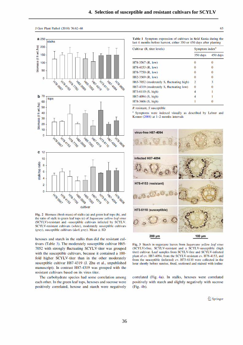
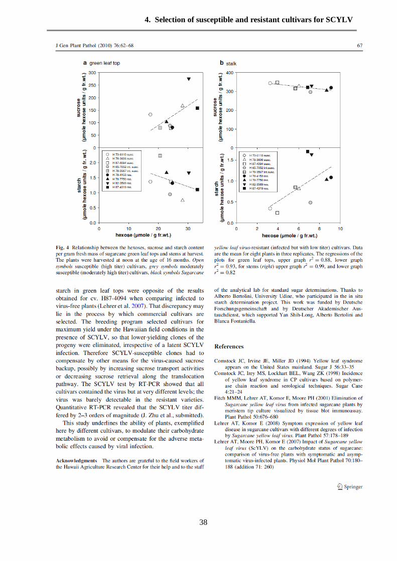
Results 35
Discussion 37
5. Sugarcane yellow leaf virus introduction and spread in Hawaiian sugarcane
industry: Retrospective epidemiological study of an unnoticed, mostly
asymptomatic plant disease
40
Introduction 40
Material and Methods 41

Results 42
Discussion 47
6. Molecular characterization of Hawaiian Sugarcane yellow leaf virus (SCYLV)
genotypes and their phylogenetic relationship to SCYLV-strains from other
sugarcane-growing countries
51
Introduction 51
Material and Methods 53
Results 55
Discussion 68
7. Expression of sucrose transporter (ShSUT1) in a Hawaiian sugarcane cultivar
infected with Sugarcane yellow leaf virus (SCYLV)
74
Introduction 74
Material and Methods 76
Results 80
Discussion 87
8. Simultaneous quantitative analysis of transcripts for Sugarcane yellow leaf
virus, sucrose transporters and sucrose phosphate synthase in Hawaiian
sugarcane cultivars by multiplex RT-PCR
92
Introduction 93
Material and Methods 94
Results 97
Discussion 108
9. List of publications 115

1. Summary/ Zusammenfassung
1
1. Summary
Sugarcane is an important crop plant and has served as a source of sugar for hundreds of years,
recently it is used to produce bioethanol, a renewable bio-fuel energy source. Sugarcane yellow
leaf virus (SCYLV) was detected in the late 1990s first in Hawaii as a causal agent of a
sugarcane disease (Yellow leaf) which leads to sugarcane yellow leaf syndrome and reduced
sugar yield.
The presence of Sugarcane yellow leaf virus was determined by RT-PCR in several sugarcane
cultivars, mostly from Hawaii. Interesting was the comparison of so-called susceptible versus
resistant cultivars. As expected, the susceptible Hawaiian cultivars H73-6110 and H87-4094
showed strong PCR amplification products of SCYLV, while the virus-free line H87-4094,
produced by tissue culture, showed no PCR product. The three resistant cultivars H87-4319,
H78-4153 and H78-7750 showed quite different amplification patterns. While H78-4153 and
H78-7750 expressed a weak but specific band of the correct size, unexpectedly H87-4319
showed strong amplification product. Three Cuban cultivars (C1051-73, JA-605 and CP52-43)
showed low titer of SCYLV. No PCR amplificate was obtained with the moderately susceptible
cultivar H65-7052. Aphids feeding on cv. H87-4094 contained sufficient virus to yield a
SCYLV-signal similar in strength as from preparations from resistant cultivars. Northern blot
analysis supported the results obtained from RT-PCR. The presence of SCYLV in the cultivars
with low amount of virus titer (H87-4319, H78-7750 and H78-4153) indicated that they should
better be called tolerant for the virus in the sense that they allow a low replication rate for
SCYLV.
Northern blots showed that RNA of SCYLV is divided into genomic RNA (gRNA) and two
subgenomic RNAs (sgRNAs). The estimated molecular size of the gRNA is 6.0 kb, the estimated
sizes of the sgRNAs are 1.0 and 2.4 kb. It is known that plant RNA viruses have evolved
numerous strategies for genome expression to invade host plants, such as divided genomes,
subgenomic messenger RNAs, overlapping reading frames or stop codon suppression. Virus
preparations from 3 Hawaiian cultivars (two susceptible and one resistant) were fully sequenced.
Quantitative analysis for four different genome regions of SCYLV covering the 6 ORFs has been
performed for these 3 cultivars using the GeXP analysis system. The transcript levels of the
different regions of SCYLV in these cultivars were present at very different quantities, for
example ORF0-1 transcripts were up to 10 times more frequent than transcripts of ORF3-4.
The SCYLV-sequences from the 3 Hawaiian cultivars were aligned to published full and partial
sequences. The phylograms corroborated previous findings that the so-called YLS-segment
coding for the coat protein shows the least genetic diversity, whereas the other sequence

1. Summary/ Zusammenfassung
2
fragments A-D, representing the ORFs 0-5, expressed a twofold higher diversity. The
phylograms of partial sequences and of the whole genome placed the Hawaiian SCYLV-strains
next to the Peru strain, apart from the BRA-strains and well apart from the REU-strains. It is
proposed that the Hawaiian SCYLV is considered as own group together with the Peru strain as
HAW-PER. The sequences from the two susceptible cultivars had a deletion of 48 to 54 nt in
ORF1, which codes for the gene silencing suppressor/RNA-dependent RNA-polymerase
complex. It is speculated that this deletion is important for the proliferation rate of the virus in
the plant.
Sucrose is the main product of sugarcane, which accumulates in the stalk internodes in excess of
50 % of the dry weight. To gain an overview of the physiological status of SCYLV-infected
sugarcane compared to virus-free plants, gene expression, transcript levels of sucrose transporter
and sugar contents were measured. Sucrose increased rapidly between internodes 3 and 7,
reaching a maximum in internodes 7. Sugars content in leaves, seedling and internodes were
increased as effect of the SCYLV-infection. Sucrose phosphate synthase (SPSII) transcript levels
were approximately the same in sink, source and internodes with a trend to be higher in the
mature internodes. A sucrose transporter of Hawaiian cultivar was isolated and sequenced and
classified as ShSUT1A. There is high variability among the SUT1 subfamily with identities of
70-97%. The identity between ShSUT1A and ShSUT1 was 97.4%. It is expressed in sink, source
and storage tissues. The ShSUT1A was expressed at approximately similar extent in SCYLV-
infected and virus-free sugarcane. In addition a partial sequence of a sucrose transporter
belonging to the SUT4 family was first obtained in sugarcane and its transcript levels in plant
organs were measured. Quantitative analysis for sucrose transporters (ShSUT1 and ShSUT4)
using the GeXP analysis system showed that sucrose transporter ShSUT1 was at a higher
transcript expression than ShSUT4 in sink and source leaves, but not in mature internodes.
In conclusion,
- SCYLV from Hawaiian cultivars was characterized as belonging to an own subgroup (HAW-
PER),
- A deletion of 48-54 nt was detected in the SCYLV-sequence from susceptible cultivars, which
may be correlated to virus proliferation, and
- large differences in transcript levels of the viral ORFs were found.
- Sucrose transporter transcripts and SPSII transcripts were not strictly correlated to SCYLV-
infection and do not explain the pathological effect of SCYLV on sugarcane.

1. Summary/ Zusammenfassung
3
Zusammenfassung
Zuckerrohr ist eine wichtige Weltwirtschaftspflanze, die seit Jahrhunderten als Zuckerquelle und
neuerdings als nachwachsende Energiequelle z. B. für Bio-Ethanol dient. In den 1990ern wurde
Zuckerrohr-Gelbblatt-Virus (Sugarcane yellow leaf virus, SCYLV) als Ursache für die
Gelbblatterkrankung von Zuckerrohr und der daraus erfolgten Ernteminderung entdeckt.
SCYLV wurde mittels RT-PCR in mehreren Zuckerrohrkultivaren, die meisten davon aus
Hawaii, nachgewiesen. Interessant war der Vergleich von sogenannten suszeptiblen und
resistenten Kultivaren. Erwartungsgemäß ergaben die suszeptiblen Kultivare H73-6110 und
H87-4094 mächtige PCR-Banden für SCYLV, während die virusfreie Linie von H87-4094, die
aus Gewebekultur gewonnen worden war, kein Amplifikat zeigte. Die 3 resistenten Kultivare
zeigten unterschiedliche Ergebnisse. Während H78-4153 und H78-7750 nur schwache Banden
erzeugten, wurde bei H87-4319 unerwarteterweise eine starke Amplifikation beobachtet. Drei
cubanische Kultivare (C1051-73, JA-605, CP52-43) zeigten einen niedrigen SCYLV-Titer. Das
gemäßigt suszeptible Kultivar H65-7052 erbrachte kein SCYLV-Amplifikat. Aphiden, die von
infiziertem H87-4094 entnommen wurden, ergaben ein Amplifikat in ähnlicher Stärke wie die
resistenten Zuckerrohrkultivare. Ergebnisse von Northern Blots unterstützten die Befunde aus
RT-PCR. Wegen der Tatsache, dass die resistenten Kultivare SCYLV, wenn auch in niedrigem
Titer, enthielten, sollten sie besser als virus-tolerant bezeichnet werden.
Die Northern Blots zeigten, dass die RNA von SCYLV als gesamtes Genom von 6,0 kb und als
(mindestens) 2 subgenomische Fragmente von 1,0 und 2,4 kb vorliegt. Es ist bekannt, dass
Pflanzenviren mehrere genetische Strategien entwickelt haben um ihre Wirte zu besiedeln, z. B.
geteilte Genome, subgenomische RNAs, überlappende reading frames oder stop-codon-
Unterdrückung. Viruspräparationen aus 3 hawaiianischen Kultivaren (2 suszeptible und 1
resistentes) wurden sequenziert. Die Menge viraler Transkripte von 4 Fragmenten, die die 6
ORFs abdeckten, wurde mittels GEXP in den 3 Kultivaren bestimmt. Die Transkripte dieser
SCYLV-Abschnitte waren zu sehr unterschiedlichem Ausmaß vorhanden, beispielsweise war
das Fragment zu ORF0-1 bis zu 10fach mehr vorhanden als das Fragment zu ORF3-4.
Die SCYLV-Sequenzen der 3 hawaiianischen Kultivare wurden mit publizierten Sequenzen
verglichen und phylogenetisch analysiert. Das sogenannte YLS-Segment zeigte sich als das
konservierteste, während die anderen Segmente eine doppelt so hohe Diversität zeigten. Das
Phylogram platzierte den hawaiianischen SCYLV-Stamm zusammen mit einem Stamm aus Peru
als separate Gruppe, genannt HAW-PER, abgetrennt von BRA-Stämmen und REU-Stämmen.
Die viralen Sequenzen aus den beiden suszeptiblen Kultivaren hatten eine 48-54 nt lange
Deletion in ORF1, welcher für ein gene silencing/RNA-abhängige RNA-Polymerase-Komplex

1. Summary/ Zusammenfassung
4
codiert. Es wird spekuliert, dass diese Deletion für die virale Vermehrung in der Pflanze wichtig
sein könnte.
Saccharose ist das hauptsächliche Speicherprodukt von Zuckerrohr und kann im Stamm über
50% des Trockengewichts ausmachen. Um den physiologischen Status der SCYLV-infizierten
versus virusfreien Pflanze zu erkunden wurden Zuckergehalt und Transkriptmenge für
Saccharosetransporter, Saccharose-Phosphat-Synthase II (SPSII) und die viralen Segmente
gemessen. Der Saccharosegehalt nahm von Internodium 3 zu 7 stark zu. SCYLV-Infektion
erhöhte den Zuckergehalt leicht in Blättern und Internodien. Die Transkriptmengen von SPSII
waren etwa gleich hoch in infizierten und virusfreien Pflanzen, mit einer leichten Erhöhung in
reifen Internodien. Ein Saccharosetransporter wurde aus einem hawaiianischem Kultivar isoliert
und als ShSUT1A klassifiziert. Die Variabilität zwischen den SUT1-Mitgliedern liegt bei 70-
97% Identität, ShSUT1 und ShSUT1A sind zu 97,4% identisch. ShSUT1 ist in sink, source und
Internodien exprimiert und findet sich etwa gleich stark in infizierten und virusfreien Pflanzen.
Ferner wurde eine Teilsequenz eines weiteren Saccharosetransporters in Zuckerrohr entdeckt,
welcher zur SUT4-Gruppe gehört. Die quantitative Transkriptanalyse mittels GEXP zeigte dass
ShSUT1 in sink und source Blättern deutlich stärker exprimiert ist als ShSUT4, nicht aber so in
reifen Internodien.
Die Ergebnisse können so zusammen gefasst werden:
- SCYLV aus hawaiianischen Zuckerrohrkultivaren gehört zu einer eigenen Gruppe (HAW-
PER),
- suszeptible Kultivare enthalten SCYLV mit einer 48-54 nt Deletion, welche mit der
Virusvermehrung in Zusammenhang stehen könnte, und
- es gibt große Unterschiede in der Transkription der viralen Genomteile.
- Saccharosetransporter-Transkripte und SPSII-Transkripte waren nicht deutlich unterschieden
zwischen infizierten und virusfreien Pflanzen und können deshalb nicht als kausale Erklärung
der SCYLV-Symptome dienen.

2. Introduction
5
2. Introduction
Sugarcane (Saccharum spp.) is an important tropical and subtropical crop and served as a source
of sugar for centuries. Sugarcane belongs to the grass family (Poaceae), an economically
important seed plant family that includes cereals such as maize, wheat, rice, and sorghum as well
as many forage crops. The commercial sugarcane cultivars are interspecific hybrids that, under
ideal conditions, are capable of storing sucrose in the parenchyma tissues of the stem up to 60%
of the dry weight (Moore, 1995). It is generally used to produce sugar and has recently gained
increased attention because ethanol derived from cane sugar represents an important renewable
bio-fuel energy source, which could turn it into global commodity and important energy source.
So far only the fibrous residual of sugar extraction, the so-called bagasse, is already used for
electricity generation and is providing surplus electricity in some tropical countries. There is
increased interest in this crop due to the impending need to decrease fossil fuel usage.
a. Sugarcane yellow leaf virus
There are several sugarcane diseases caused by bacteria, fungi and viruses. Concerning the viral
diseases, there are approximately seven viruses of international importance in sugarcane
production: Sugarcane mosaic virus (SCMV), Sugarcane streak virus (SSC), Peanut clump virus
(PCV), Sugarcane bacilliform virus (SCBV), Sugarcane mild mosaic virus (SCMMV), Fiji
disease virus (FDV) and Sugarcane yellow leaf virus (SCYLV). The latter, SCYLV, is the most
recently detected virus and is nowadays the only virus associated with Hawaiian sugarcane
industry. It is the causal agent of yellow leaf syndrome (YLS) (now named Yellow leaf, YL)
which was first reported from plantations on two Hawaiian Islands (Schenck, 1990). Few years
later similar symptoms were observed in several other countries (Comstock et al. 1994) and
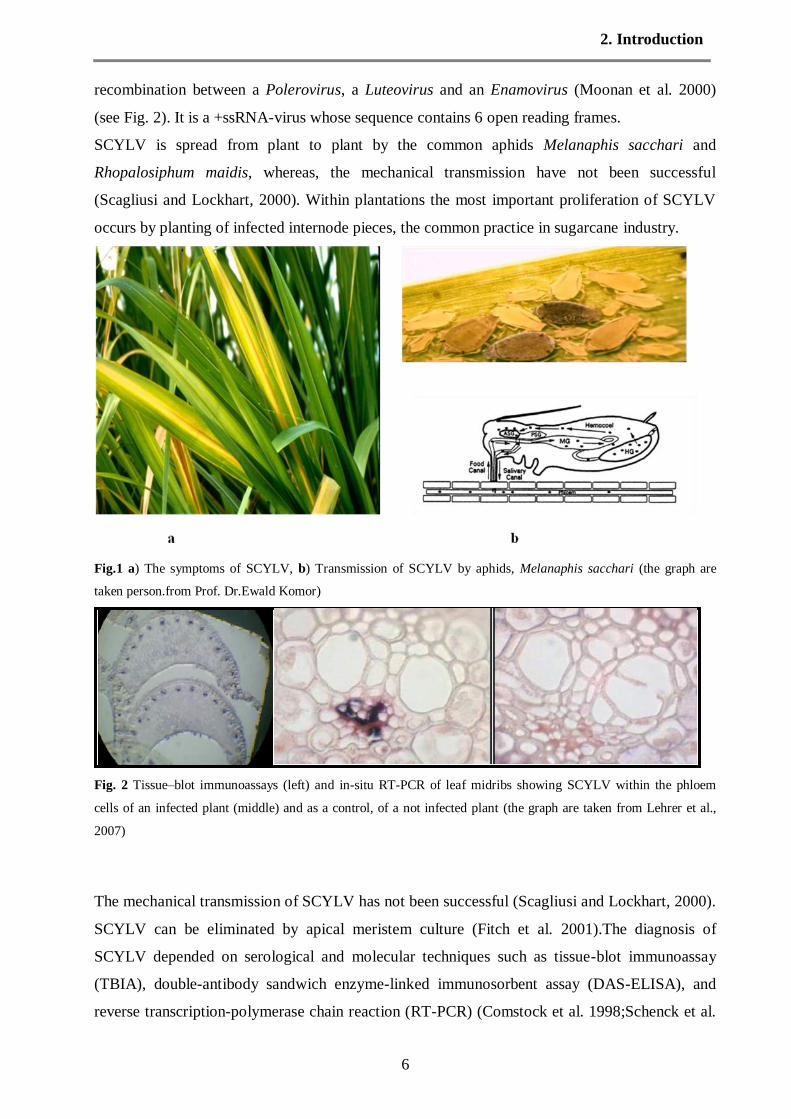
dramatic yield losses were reported in Brazil (Vega et al. 1997). The symptoms are characterized
by yellowing of leaf midribs followed by yellowing of the entire leaf blade (Fig.1a) and
internode shortening of the green leaf top. The midrib yellowing may be intense or in some
varieties may have a reddish tinge and is associated with sucrose accumulation in the midribs.
The symptoms are best expressed when the crop is subjected to stress. Nevertheless, the
pathogen can be present without the expression of symptoms. The virus particles were observed
in the cytoplasm of phloem companion cells of sugarcane. The detection of SCYLV by Tissue–
blot immunoassays (TBIA) also revealed that the sugarcane virus was associated with phloem
(Fig.1c). The viral pathogen was classified as a luteovirus and was termed sugarcane yellow leaf
virus (SCYLV) (Scaglisi and Lockhart, 2000). Todays analysis revealed that SCYLV belongs to
polerovirus which is a member of the luteoviridae family and has a apparently arisen through
2. Introduction
6
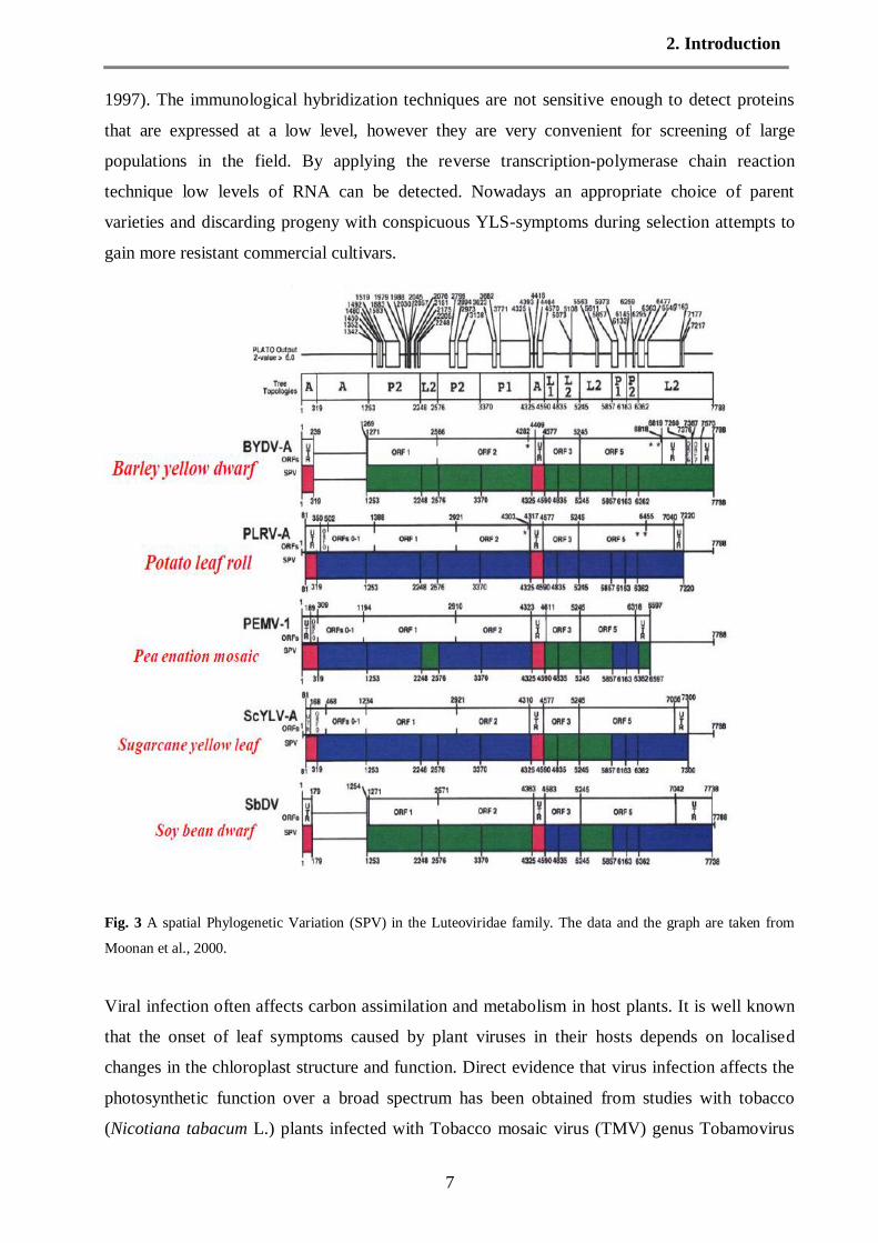
recombination between a Polerovirus, a Luteovirus and an Enamovirus (Moonan et al. 2000)
(see Fig. 2). It is a +ssRNA-virus whose sequence contains 6 open reading frames.
SCYLV is spread from plant to plant by the common aphids Melanaphis sacchari and
Rhopalosiphum maidis, whereas, the mechanical transmission have not been successful
(Scagliusi and Lockhart, 2000). Within plantations the most important proliferation of SCYLV
occurs by planting of infected internode pieces, the common practice in sugarcane industry.
Fig.1 a) The symptoms of SCYLV, b) Transmission of SCYLV by aphids, Melanaphis sacchari (the graph are
taken person.from Prof. Dr.Ewald Komor)
Fig. 2 Tissue–blot immunoassays (left) and in-situ RT-PCR of leaf midribs showing SCYLV within the phloem
cells of an infected plant (middle) and as a control, of a not infected plant (the graph are taken from Lehrer et al.,
2007)
The mechanical transmission of SCYLV has not been successful (Scagliusi and Lockhart, 2000).
SCYLV can be eliminated by apical meristem culture (Fitch et al. 2001).The diagnosis of
SCYLV depended on serological and molecular techniques such as tissue-blot immunoassay
(TBIA), double-antibody sandwich enzyme-linked immunosorbent assay (DAS-ELISA), and
reverse transcription-polymerase chain reaction (RT-PCR) (Comstock et al. 1998;Schenck et al.
2. Introduction
7
1997). The immunological hybridization techniques are not sensitive enough to detect proteins
that are expressed at a low level, however they are very convenient for screening of large
populations in the field. By applying the reverse transcription-polymerase chain reaction
technique low levels of RNA can be detected. Nowadays an appropriate choice of parent
varieties and discarding progeny with conspicuous YLS-symptoms during selection attempts to
gain more resistant commercial cultivars.
Fig. 3 A spatial Phylogenetic Variation (SPV) in the Luteoviridae family. The data and the graph are taken from
Moonan et al., 2000.
Viral infection often affects carbon assimilation and metabolism in host plants. It is well known
that the onset of leaf symptoms caused by plant viruses in their hosts depends on localised
changes in the chloroplast structure and function. Direct evidence that virus infection affects the
photosynthetic function over a broad spectrum has been obtained from studies with tobacco
(Nicotiana tabacum L.) plants infected with Tobacco mosaic virus (TMV) genus Tobamovirus

2. Introduction
8
(van Kooten et al. 1990 and Seo et al. 2000). Various results indicate that an increase in non-
photochemical quenching of fluorescence and reduction in the fraction of open reaction centres
leads to an increased reduction state of primary electron transport acceptor quinone A (QA). This
suggests pronounced photoinhibitory processes following viral infection and symptom
development. Photosynthesis reduction and chlorophyll degradation are however only the late
stages of symptoms and may be caused by previous viral effects on plant cell metabolism.
Studies with transgenic tobacco plants expressing the movement protein of TMV have shown
effects of movement protein (without virus) on carbon metabolism, altering carbohydrate
partitioning and plasmodesmal function between mesophyll cells (Balachandran et al. 1995;
Lucas et al. 1996 and Olesinski et al. 1996). The source leaves of transgenic plants expressing
the movement protein of Potato leafroll virus (PLRV), family Luteoviridae, genus Potyvirus
showed accumulation of carbohydrates leading to a decrease in photosynthetic capacity,
probably due to decreased expression of photosynthetic proteins (Herbers et al. 1997). These
effects were strong in plants expressing the luteoviral movement protein in plasmodesmata of the
phloem tissues, while in plasmodesmata of the mesophyll the effects were indistinguishable from
the wild-type. The changes in carbohydrate status and viral resistance followed a protein level-
dependent mechanism, whereas the plasmodesmal targeting and capacity of movement protein
was not influenced by protein amount (Hofius et al. 2001). Corroborating these findings, Herbers
et al. (2000) proposed a role for cell wall invertase in up-regulating the accumulation of soluble
sugars and down-regulating photosynthesis, thus strengthening defence responses against viral
attack.
b. Sucrose transport in plants
Although sucrose is commonly found in higher plant storage organs, it is generally at a low
concentration, and starch is the predominant storage carbohydrate (Komor, 2000). Additionally,
sucrose is the main transport molecule in most plants. Physicochemical properties of sucrose
may play a role as transport sugar, because the viscosity of sucrose is relatively low at high
concentrations such as in phloem sap, allowing high translocation rates (0.5 to 3 m×h-1
).
Furthermore, the disaccharide sucrose has a high chemical and biochemical stability due to its
acetal-bond which covers the reducing ends of the two monosaccharide. Sucrose creates a high
osmotic potential per carbon atom in the phloem sap, a key parameter for the mass transport
efficiency within long tubes (van Bel, 1996).
The transport of sucrose from source organs to sink organs may follow a symplasmic pathway,
moving from cell to cell via plasmodesmata. Alternatively, sucrose may move apoplasmically
through the cell walls and intercellular spaces of the tissue. In most plants, the pathway from

2. Introduction
9
source to sink is thought to involve a combination of both symplasmic and apoplasmic transport
steps, depending on the tissue type and stage of development (Patrick, 1997 and Lalonde et al.
2003). Sucrose is produced in photosynthesizing cells, passes through the plasma membrane of
these cells into the non-membrane bound area surrounding the mesophyll cells (apoplast) and is
then actively transported into the sieve element system of the phloem. Estimates of the sucrose
concentration in phloem of photosynthesizing leaves vary in the range of 0.3-0.8 M. While the
total sucrose concentration in the producing cells is often as low as a few millimolar. This
concentration step is consistent with some form of facilitated passage from the apoplast through
a semipermable membrane and obviously requires the expenditure of metabolic energy (Komor,
2000).
Sucrose is the major mobile carbohydrate in the majority of higher plants. Our knowledge of
sucrose translocation has increased considerably by the biochemical and molecular
characterization of sucrose transporter (SUT) family in the last decade. Plant sucrose transporters
(SUTs) belong to the glycoside-pentoside-hexuronide (GPH) cation symporter family (TC2.A.2)
that is part of the major facilitator superfamily (MFS) (Chang et al. 2004). Transporters in the
GPH family have the basic characteristics of MFS proteins: 12 transmembrane domains with N-
and C-terminus in the cytoplasm. The first six transmembrane domains display some sequence
similarities with the last six, supporting the idea that these transporters arose from at least one
ancient gene duplication (Saier, 2000). Corroborating these findings, Henderson (1990) and
Kaback (1992) described the hydrophobicity analysis of structure of an integral membrane
protein with 12 putative transmembrane domains, with a central hydrophilic loop. Meanwhile,
the GPH family contains members from bacteria, archaea and eukaryotes. Such as, melibiose
permease from E. coli (Naderi and Saier, 1996), the α-glucoside transporter SUT1p from
Schizosaccharomyces pombe (Reinders and Ward, 2001) and plantSUTs such as SUC2 from
Arabidopsis (Sauer and Stolz, 1994; Chandran et al. 2003). Transporters within the GPH family
that have been characterized so far transport glycosides by symport with a cation (H+
or Na+).
Plant sucrose transporters were mainly associated with phloem loading. From sugarcane only
one sucrose transporter (ShSUT1) was described. It is expressed in both leaves and stems, but
most highly in the stem tissue accumulating sucrose (Casu et al. 2003). The protein was mostly
localized at the layer of cells surrounding the bundle sheath but was absent from the phloem
itself. Based on these findings, the ShSUT1 may play a role in retrieval of sucrose leaking from
the storage parenchyma cells in the stem or alternatively in sucrose export into the storage
parenchyma rather than in phloem loading (Rae et al. 2005a).

2. Introduction
10
c. Sucrose transport in sugarcane
In sugarcane, the conducting cells of the leaf phloem are not connected to other cells of the leaf
by plasmodesmata (Robinson-Beers and Evert, 1991). This suggests that phloem loading occurs
from the apoplast in sugarcane. In phloem, sucrose moves out of the leaf and towards sink
tissues. The movement of sucrose through transport phloem is thought to be driven by
concentration gradients (VanBel, 2003). Sucrose transporters continue to be expressed in
transport phloem and may act in retrieval of sucrose lost to the apoplast by leakage (Lalonde et
al. 2003).
The role of transporters in the influx of sucrose during phloem loading has been well
documented in contrast to their role in unloading and post-phloem pathways (Rae et al. 2005a).
The gradient of sucrose concentrations suggests that post-phloem efflux from the symplast could
occur by facilitated diffusion, movement through transmembrane pores, which has specificity for
sucrose but which is driven solely by gradient of the substrate and not energized by direct or
indirect consumption. The expression of sucrose transporters in the petiole tissues suggests that
unloading involves an apoplastic step (Salmon et al. 1995). ShSUT1 was identified as sucrose
transporter in stem of sugarcane, which is localized to tissues surrounding the stem vascular
bundles (Rae et al. 2005b). Additionally, the sucrose transporter play a role in a tissue that
predominantly supports symplasmic transfer is most likely to be in the retrieval of sucrose lost
from the symplasmic continuum. This is analogous to the situation in the sieve elements of
transport phloem in leaves, in which sucrose transporters continue to be expressed even though
the sieve elements are connected by pores through the cell plates. The ShSUT1 sucrose
transporter may be an important component of the retrieval mechanism. The expression of
ShSUT1 in the cell layers at the boundary between these compartments may represent an
additional biochemical barrier to apoplasmic sucrose movement through these layers (Rae et al.
2005a). It is also possible that ShSUT1 is involved in efflux of sucrose from the symplasm to the
apoplasm at the boundary layer. It has been suggested that sucrose/H+ symporters may mediate
sucrose efflux by facilitated diffusion in some circumstances (Lalonde et al. 2003).
The structure of sugarcane stem plays a role in the movement of sucrose from phloem to the
storage parenchyma tissue. The vascular bundles of sugarcane stem are surrounded by a layer of
fiber cells that become progressively lignified with development (Rae et al. 2005a). It has been
suggested that, these layers can prevent and /or impede apoplastic movement of solutes during
the period of sucrose accumulation. In agreement with these suggestions, it was found that these
layers effectively form a barrier to apoplastic movement of water-soluble dyes during the period
of sucrose accumulation and internode ripening (Jacobsen et al. 1992). Thus sucrose probably
cannot reach the parenchyma cells from the phloem by apoplastic route. The presence of

2. Introduction
11
plasmodesmatal connections suggests that the storage parenchyma cells obtain sucrose from the
vascular bundle through symplastic passgae (Walsh et al. 1996). The pathway of sucrose into the
storage parenchyma in the sugarcane stem is depicted in Fig. 3.
Plasmodesmata play an important role in long distance transport. Most plant cells (but not all!)
are connected by plasmodesmata that allow small solutes and, under some conditions, macro-
molecules to move between cells. Plasmodesmata serve an especially important role in the
phloem. During the development of phloem, sieve elements (SE) and companion cells (CC) are
formed from a common parent cell and they remain tightly connected by plasmodesmata. The
plasmodesmata between sieve elements widen and form sieve pores in the sieve plates, thus
creating a living tube through which the phloem sap can move rapidly. The companion cells
retain the nucleus, vacuole and numerous mitochondria. There is evidence that specific
messenger RNAs and proteins are produced in CC and are delivered to SE through
plasmodesmata. It had been claimed that SUT1, sucrose/H+
cotransporer, is localized in the
plasma membrane of SE of Solanaceous plants, but the mRNA is made in CC (Kühn, et al.
1997). Thus SUT1 mRNA or protein and possibly other transporters has to traffic between the
two cells by receptor-mediated transport through plasmodesmata.
Fig. 3 Possible routes of sucrose into the storage parenchyma of the sugarcane stem. One way is symplastic
unloading through plasmodesmata by cell-to-cell connections without any apoplastic step. Another possibility is the
unloading of sucrose into the apoplast, followed by hydrolysis by acid invertase and subsequent uptake of the
resulting hexoses into the sink cells. This step would then be followed by resynthesis of sucrose in the cells. A third
possibility is the unloading of sucrose into the apoplast followed by uptake of intact sucrose into the cells. The data
and the graph are taken from Rae et al. (2005b).
Besides the pathway of sucrose from stem phloem to stem storage parenchyma, there is also a
metabolic cycling of sucrose. Sucrose is synthesized by two alternative ways. Sucrose-phosphate

2. Introduction
12
synthase synthesizes sucrose-phosphate from UDP-glucose and fructose-phosphate.The
following phosphatase step leading to sucrose shiftes this reaction sequence strongly towards
sucrose synthesis. The other way, sucrose synthesis from UDP-glucose and fructose by sucrose
synthase is relatively reversible and may be a means to provide sufficient levels of UDP-glucose
from sucrose for cell wall synthesis (e.g. for callose synthesis in sieve tubes, which are devoid of
invertase). SPS and SS are present in sugarcane storage parenchyma (Zhu et al. 1997). Invertase
is the major enzyme responsible for sucrose hydrolysis. There are several isozymes present in
storage parenchyma, a cell wall-bound acid invertase, a cytosolic neutral invertase and a
vacuolar acid invertase. The balance between these enzyme activities changes during internode
maturation and is thought to be an important factor in determining sucrose yield of sugarcane
varieties (Zhu et al. 1997 and Lingle,1989). Sugarcane industry is interested in increased
concentration of sucrose as the key objective for sugarcane improvement programmes.
Fig. 4. The cycle of sucrose in sugarcane, enzymes and metabolites: SPS; sucrose phosphate synthase, SS; sucrose
synthase, SAI; soluble acid invertase, NI; neutral invertase, PGI; phosphoglucomutase, UDPG-PPase; UDPglucose
pyrophosphorylase. All these enzymes are supposed to be cytosolic with exception of the soluble acid invertase,
which is vacuolar. In addition a cell wall bound acid invertase will hydrolyze apoplastic sucrose. The data and the
graph are taken from Komor (2000).
Aims of the present study
Sugarcane is an economically important crop species for targeted breeding and an interesting
model to study sucrose transport as well. The worldwide distribution of sugarcane yellow leaf
virus in sugarcane plantations makes it interesting to study the genetic diversity of sugarcane
yellow leaf virus. The best studied effects of SCYLV on the physiology of sugarcane were made
on Hawaiian cultivars, however a molecular characterization on the virus in Hawaii was lacking.
The objectives of this study were to determine and characterize SCYLV in Hawaiian varieties,

2. Introduction
13
resistant and susceptible ones, to investigate possible sequence divergences and the genetic
relationships between SCYLVs from susceptible and resistant Hawaiian cultivars. (In addition a
few cultivars from Middle-East, the home country of the author, were tested for SCYLV).
It had been suggested that the viral effects which ultimately lead to symptoms may be connected
to reduction of sucrose export from the leaves. ShSUT1, which is expressed in leaves and stems
may play an important role in the accumulation of sucrose in maturing stem. It should be tested,
whether it is affected by SCYLV-infection. ShSUT1 transcripts level in different tissues (shoots
of seedling stage, source leaves and storage tissues) of SCYLV-infected and not-infected
sugarcane plants were determined. In addition carbohydrate profiles were determined to evaluate
the physiological status of the infected plant.
References
Balachandran, S., Hull, R.J., Vaadiay, Y., Wolf, S. and Lucas, W.J. (1995) Alteration in
carbon partitioning induced by the movement protein of tobacco mosaic virus originates in the
mesophyll and is independent of change in the exclusion size limit.Plant Cell Environ., 18,
1301-1310.
Both, W., Hu, J. S., and Schenk, S. (1994) Double-stranded RNA associated with sugarcane
yellow leaf syndrome. Sugar Cane, 3, 5-8.
Casu, R.E., Grof, C.P., Rae, A.L., McIntyre, C.L., Dimmock, C.M. and Manners, J.M.
(2003) Identification of a novel sugar transporter homologue strongly expressed in maturing
stem vascular tissues of sugarcane by expressed sequence tag and microarray analysis. Plant
Mol Biol., 52, 371-386.
Chandran, D., Reinders, A. and ward, J.M. (2003) Substrate specificity of the Arabidopsis
thaliana sucrose transporter AtSUC2. Journal of Biological Chemistry, 278, 44320-44325.
Chang, AB., Lin, R., Keith, Studley, W., Tran, C.V. and Saier, M.H., Hr. (2004) Phylogeny
as a guide to structure and function of membrane transport proteins.Molecular Membrane
Biology, 21, 171-181.
Comstock, J.C., Irey, M.S., Lockhart, B.E.L. and Wang, Z.K. (1998) Incidence of yellow
leafsyndrome in CP cultivars based on polymerase chain reaction and serological techniques.
Sugar Cane, 4, 21-24.
Comstock, J.C., Irvine, J.E. and Miller, J.D. (1994) Yellow leaf syndrome appears on the
United States mainland. Sugar J., 56, 33-35.

2. Introduction
14
Fitch, M.M.M, Lehrer, A.T, Komor, E., Moore, P.,H. (2001) Elimination of Sugarcane
yellow leaf virus from infected sugarcane plants by meristem tip culture visualized by tissue blot
immunoassay. Plant Pathol 50: 676-680.
Henderson, P.J.F. (1990) Proton-linked sugar transport systems in bacteria. Journal of
Bioenergetics and Biomembranes, 22, 525-569.
Herbers, K., Tacke E., Hazirezaei, M., Krause, K.P., Melzer, M., Rohde, W. and
Sonnewald,U. (1997) Expression of a luteoviral movement protein in transgenic plants leads to
carbohydrate accumulation and reduced photosynthetic capacity in source leaves. Plant J., 12,
1045-1056.
Herbers, K., Takahata, Y., Melzer, M., Mock, H.P., Hajirezaei, M. and Sonnewald, U.
(2000) Regulation of carbohydrate partitioning during the interaction of potato virus Y with
tobacco. Mol. Plant Pathol., 1, 51-59.
Hofius, D., Herbers, K., Melzer, M., Omid, A., Tacke, E., Wolf, S. and Sonnewald, U.
(2001) Evidence for expression level dependent modulation of carbohydrate status and viral
resistance by the potato leafroll virus movement protein in transgenic tobacco plants. Plant J.,
28, 529-543.
Jacobsen, K.R., Fisher, D.G., Maretzki, A., Moore, P.H. (1992) Developmental changes in
the anatomy of the sugarcane stem in relation to phloem unloading and sucrose storage. Bot.
Acta. 105, 70-80.
Kaback, H.R. (1992) Β-galactoside transport in Escherichia coli: the ins and outs of lactose
permease. In Op den Kamp JAF, ed. Dynamics in memberane assembly. NATO ASI Series,
Berlin: Springer Verlag., 293-308.
Komor, E. (2000) The physiology of sucrose storage in sugarcane. In AK Gupta, N Kaur eds.
Carbohydrate reserves in plants-synthesis and regulation. Elsevier, Amsterdam, pp. 35-54.
Kühn, C, Franceschi, VR, Schulz, A, Lemoine, R, Frommer, WB. (1997) Localization and
turnover of sucrose transporters in enucleate sieve elements indicate macromolecular trafficking.
Science, 275, 1298-1300.
Lalonde, S., Tegeder, M., Throne-Holst, M., Frommer, W.B. and Patrick, J.W. (2003)
Phloem loading and unloading of sugars and amino acids. Plant Cell Environ, 26:37-56.
Lehrer, A.T, Schenck, S., Yan, SL., Komor, E. (2007) Movement of aphid-transmitted
sugarcane yellow leaf virus(ScYLV) within and between sugarcane plants. Plant Pathology
56,711-717.
Lingle, S.E. (1989) Evidence for the uptake of sucrose intact into sugarcane internodes. Plant
Cell Environ. 19: 1124-113.
2. Introduction
15
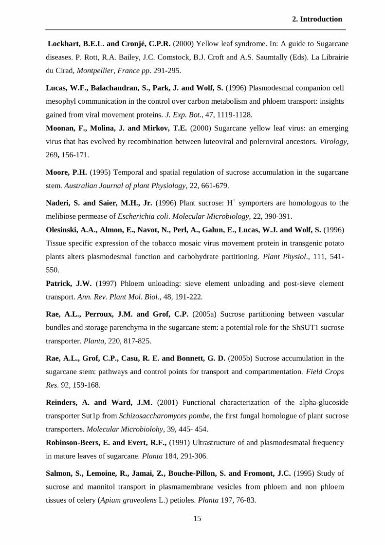
Lockhart, B.E.L. and Cronjé, C.P.R. (2000) Yellow leaf syndrome. In: A guide to Sugarcane
diseases. P. Rott, R.A. Bailey, J.C. Comstock, B.J. Croft and A.S. Saumtally (Eds). La Librairie
du Cirad, Montpellier, France pp. 291-295.
Lucas, W.F., Balachandran, S., Park, J. and Wolf, S. (1996) Plasmodesmal companion cell
mesophyl communication in the control over carbon metabolism and phloem transport: insights
gained from viral movement proteins. J. Exp. Bot., 47, 1119-1128.
Moonan, F., Molina, J. and Mirkov, T.E. (2000) Sugarcane yellow leaf virus: an emerging
virus that has evolved by recombination between luteoviral and poleroviral ancestors. Virology,
269, 156-171.
Moore, P.H. (1995) Temporal and spatial regulation of sucrose accumulation in the sugarcane
stem. Australian Journal of plant Physiology, 22, 661-679.
Naderi, S. and Saier, M.H., Jr. (1996) Plant sucrose: H+ symporters are homologous to the
melibiose permease of Escherichia coli. Molecular Microbiology, 22, 390-391.
Olesinski, A.A., Almon, E., Navot, N., Perl, A., Galun, E., Lucas, W.J. and Wolf, S. (1996)
Tissue specific expression of the tobacco mosaic virus movement protein in transgenic potato
plants alters plasmodesmal function and carbohydrate partitioning. Plant Physiol., 111, 541-
550.
Patrick, J.W. (1997) Phloem unloading: sieve element unloading and post-sieve element
transport. Ann. Rev. Plant Mol. Biol., 48, 191-222.
Rae, A.L., Perroux, J.M. and Grof, C.P. (2005a) Sucrose partitioning between vascular
bundles and storage parenchyma in the sugarcane stem: a potential role for the ShSUT1 sucrose
transporter. Planta, 220, 817-825.
Rae, A.L., Grof, C.P., Casu, R. E. and Bonnett, G. D. (2005b) Sucrose accumulation in the
sugarcane stem: pathways and control points for transport and compartmentation. Field Crops
Res. 92, 159-168.
Reinders, A. and Ward, J.M. (2001) Functional characterization of the alpha-glucoside
transporter Sut1p from Schizosaccharomyces pombe, the first fungal homologue of plant sucrose
transporters. Molecular Microbiolohy, 39, 445- 454.
Robinson-Beers, E. and Evert, R.F., (1991) Ultrastructure of and plasmodesmatal frequency
in mature leaves of sugarcane. Planta 184, 291-306.
Salmon, S., Lemoine, R., Jamai, Z., Bouche-Pillon, S. and Fromont, J.C. (1995) Study of
sucrose and mannitol transport in plasmamembrane vesicles from phloem and non phloem
tissues of celery (Apium graveolens L.) petioles. Planta 197, 76-83.
2. Introduction
16
Sauer, N, and Stolz, J. (1994) SUC1 and SUC2: Two sucrose transporters from Arabidopsis
thaliana; expression and characterization in baker’s yeast and identification of the histidine
tagged protein. The Plant Journal, 6, 67-77.
Scagliusi, S. M. M., and Lockhart, B. E. L. (2000) Transmission, characterization, and
serology of a luteovirus associatedwith yellow leaf syndrome of sugarcane. Phytopathology, 90,
120-124.
Schenck, S. (1990) Yellow leaf syndrome – a new sugarcane disease. Hawaiian Sugar Planters
Association: Annual Report: 38.
Schenck, S., Hu, J.S. and Lockhart, B.E.L. (1997) Use of a tissue blot immunoassay to
determine the distribution of sugarcane yellow leaf virus in Hawaii. Sugar Cane, 4, 5-8.
Seo, S., Okamoto, M., Iwai, T., Iwano, M., Fukui, K., Isogal, A., Nagajima, N. and
Ohashi,Y. (2000) Reduced levels of chloroplast FtsH protein in tobacco mosaic virus- infected
tobacco leaves accelerate the hypersensitive reaction. Plant Cell, 12, 917- 932.
van Bel, A.J.E. (1996) Interaction between sieve element and companion cell and the
consequences for photoassimilate distribution. Two structural hardware frames with associated
physiological software packages in dicotyledons. Journal of Experimental Botany, 47, 1129-
1140.
van Bel, A.J.E. and Gamalei, Y.V. (1992) Ecophysiology of phloem loading in source leaves.
Plant Cell Environ., 15, 265-270.
van Bel, A.J.E., (2003) The phloem, a miracle of ingenuity. Plant Cell Eviron., 26, 125-149.
van Kooten, O., Meurs, C. and van Loon, L.C. (1990) Photosynthetic electron transport
intobacco leaves infected with tobacco mosaic virus. Physiol. Plant., 80, 446-452.
Vega, J., Scagliusi, S.M.M. and Ulian, E.C. (1997) Sugarcane yellow leaf disease in Brazil:
evidence of association with a Luteovirus. Plant Dis., 81: 21-26.
Walsh, K.B., Sky, C. and Brown, S.M. (1996) Pathway of sucrose unloading from the phloem
in sugarcane stalk. In: Wilson, J.R., Hogarth, D.M., Campbell, J.A., Garside, A.L. (eds)
Sugarcane: research towards efficient and sustainable production. CSIRO Division of Tropical
Crops and Pastures, Brisbane, pp 105-107.
Zhu Y.J., E. Komor, P. Moore (1997) Sucrose accumulation in the sugarcane stem is
regulated by the difference between the activity of the soluble acid invertase and sucrose
phosphate synthase. Plant Physiol. 115, 609-616.

3. Synopsis
17
3. Synopsis
This thesis comprises five publications which are presented in chapters 4 to 8.
3.1. Selection of susceptible and resistant cultivars for SCYLV
SCYLV
50
100
150
200
250
Fig. 1 RT-PCR of RNA-derived cDNA from source leaves of different cultivars of sugarcane. RNA from
leaves of five representative cultivars was extracted, transcribed to cDNA and amplified by RT-PCR.
H73-6110 and H87-4094 are susceptible, H65-7052 is moderately susceptible, and H78-4153, H87-4319
and H78-7750 are resistant cultivars. The virus-free clone of H87-4094 was used as a negative control.
The amplified SCYLV was 165 bp long , the size standard was a 50-bp DNA ladder (left).
A previous survey using Tissue–blot immunoassays (TBIA) had identified SCYLV-susceptible
and SCYLV-resistant cultivars. Cultivars that expressed fluctuating levels of virus titer were
called moderately susceptible. Tests for SCYLV by RT-PCR partly confirmed the different titers
of virus in the different cultivars; however, the so-called resistant cultivars (H78-4153, H87-
4319, H78-7750) that had appeared without SCYLV in TBIA, had SCYLV (Fig. 1), though at a
much lower titer than, for example, H87-4094. H65-7052 appeared nearly virus-free, possibly
the leaf had been sampled in a virus-poor phase. Nine cultivars were selected for a test of
carbohydrate status of sugarcane plants at harvest time (16 months), the same cultivars that had
been used previously in an extended yield test in different Hawaiian fields.

3. Synopsis
18
3.2. Maintenance of SCYLV-infection in sugarcane stalks (seedling stage)
Fig. 2 RT-PCR for SCYLV in virus-free and infected cv. H87-4094 after 12-16 cycles of replanting, with
rRNA as loading control. Virus-free and infected plants of H87-4094 were grown in the greenhouse
outside of insect-tight cages. RNA from leaf samples was extracted and amplified with SCYLV-specific
primers by RT-PCR. The reaction products were separated on gels and stained with ethidium bromide. Length of amplified SCYLV was 165 bp, the size standard was a 50 bp DNA-ruler from Fermentas (St.
Leon Rot, Germany).
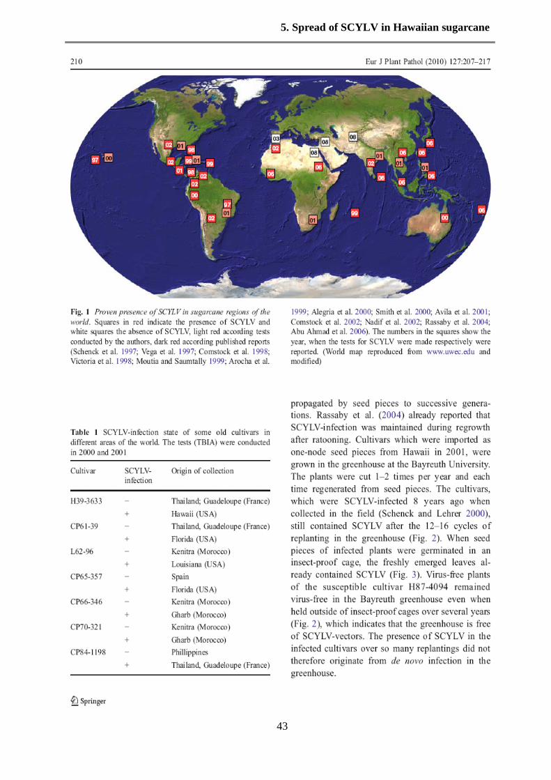
Sugarcane is propagated vegetatively by cuttings. It was important to show whether SCYLV is
propagated by seed pieces to successive generations. Cultivars which were imported as one-node
seed pieces from Hawaii in 2001, were grown in the greenhouse at the Bayreuth University. The
plants were cut 1-2 times per year and each time regenerated from seed pieces. The cultivars,
which were SCYLV-infected 8 years ago when collected in the field, still contained SCYLV
after the 12-16 cycles of replanting in the greenhouse (Fig. 2). When seed pieces of infected
plants were germinated in an insect-tight cage, the freshly emerged leaves already contained
SCYLV (Fig. 3). Virus-free plants of the susceptible cultivar H87-4094 remained virus-free in
the Bayreuth greenhouse even when outside of insect-tight cages over several years (Fig. 2),
which indicates that the greenhouse is free of SCYLV-vectors. The presence of SCYLV in the
infected cultivars over so many replanting did therefore not originate from de novo infection in
the greenhouse.
3. Synopsis
19
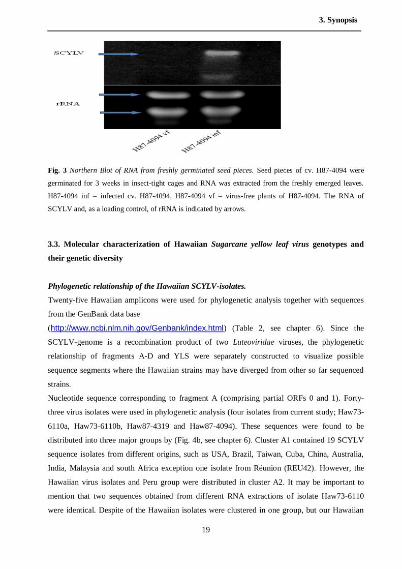
Fig. 3 Northern Blot of RNA from freshly germinated seed pieces. Seed pieces of cv. H87-4094 were
germinated for 3 weeks in insect-tight cages and RNA was extracted from the freshly emerged leaves.
H87-4094 inf = infected cv. H87-4094, H87-4094 vf = virus-free plants of H87-4094. The RNA of
SCYLV and, as a loading control, of rRNA is indicated by arrows.
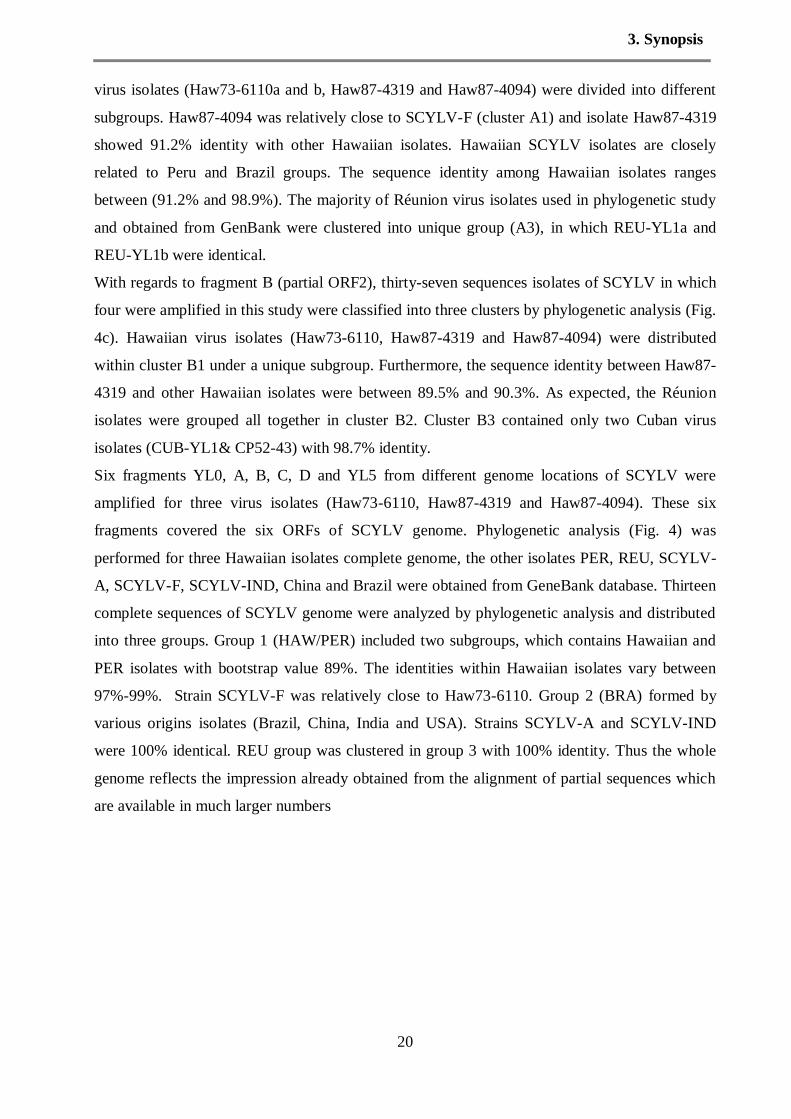
3.3. Molecular characterization of Hawaiian Sugarcane yellow leaf virus genotypes and
their genetic diversity
Phylogenetic relationship of the Hawaiian SCYLV-isolates.
Twenty-five Hawaiian amplicons were used for phylogenetic analysis together with sequences
from the GenBank data base
(http://www.ncbi.nlm.nih.gov/Genbank/index.html) (Table 2, see chapter 6). Since the
SCYLV-genome is a recombination product of two Luteoviridae viruses, the phylogenetic
relationship of fragments A-D and YLS were separately constructed to visualize possible
sequence segments where the Hawaiian strains may have diverged from other so far sequenced
strains.
Nucleotide sequence corresponding to fragment A (comprising partial ORFs 0 and 1). Forty-
three virus isolates were used in phylogenetic analysis (four isolates from current study; Haw73-
6110a, Haw73-6110b, Haw87-4319 and Haw87-4094). These sequences were found to be
distributed into three major groups by (Fig. 4b, see chapter 6). Cluster A1 contained 19 SCYLV
sequence isolates from different origins, such as USA, Brazil, Taiwan, Cuba, China, Australia,
India, Malaysia and south Africa exception one isolate from Réunion (REU42). However, the
Hawaiian virus isolates and Peru group were distributed in cluster A2. It may be important to
mention that two sequences obtained from different RNA extractions of isolate Haw73-6110
were identical. Despite of the Hawaiian isolates were clustered in one group, but our Hawaiian
3. Synopsis
20
virus isolates (Haw73-6110a and b, Haw87-4319 and Haw87-4094) were divided into different
subgroups. Haw87-4094 was relatively close to SCYLV-F (cluster A1) and isolate Haw87-4319
showed 91.2% identity with other Hawaiian isolates. Hawaiian SCYLV isolates are closely
related to Peru and Brazil groups. The sequence identity among Hawaiian isolates ranges
between (91.2% and 98.9%). The majority of Réunion virus isolates used in phylogenetic study
and obtained from GenBank were clustered into unique group (A3), in which REU-YL1a and
REU-YL1b were identical.
With regards to fragment B (partial ORF2), thirty-seven sequences isolates of SCYLV in which
four were amplified in this study were classified into three clusters by phylogenetic analysis (Fig.
4c). Hawaiian virus isolates (Haw73-6110, Haw87-4319 and Haw87-4094) were distributed
within cluster B1 under a unique subgroup. Furthermore, the sequence identity between Haw87-
4319 and other Hawaiian isolates were between 89.5% and 90.3%. As expected, the Réunion
isolates were grouped all together in cluster B2. Cluster B3 contained only two Cuban virus
isolates (CUB-YL1& CP52-43) with 98.7% identity.
Six fragments YL0, A, B, C, D and YL5 from different genome locations of SCYLV were
amplified for three virus isolates (Haw73-6110, Haw87-4319 and Haw87-4094). These six
fragments covered the six ORFs of SCYLV genome. Phylogenetic analysis (Fig. 4) was
performed for three Hawaiian isolates complete genome, the other isolates PER, REU, SCYLV-
A, SCYLV-F, SCYLV-IND, China and Brazil were obtained from GeneBank database. Thirteen
complete sequences of SCYLV genome were analyzed by phylogenetic analysis and distributed
into three groups. Group 1 (HAW/PER) included two subgroups, which contains Hawaiian and
PER isolates with bootstrap value 89%. The identities within Hawaiian isolates vary between
97%-99%. Strain SCYLV-F was relatively close to Haw73-6110. Group 2 (BRA) formed by
various origins isolates (Brazil, China, India and USA). Strains SCYLV-A and SCYLV-IND
were 100% identical. REU group was clustered in group 3 with 100% identity. Thus the whole
genome reflects the impression already obtained from the alignment of partial sequences which
are available in much larger numbers

3. Synopsis
21
Fig. 4 The genetic diversity of complete nucleotide sequences ORFs 0-5 of sugarcane yellow leaf virus
isolates from different geographical origin assessed with Geneious program, UPGMA phylogenetic tree.
Recombination analysis
To understand the taxonomic and evolutionary positions of isolates Haw73-6110, Haw87-4319
and Haw87-4094 within the family Luteoviridae, sequences of these three viruses and other
SCYLV isolates were compared to well-characterized Luteoviridae members. The 26 sequences
were analyzed and the results indicated the SCYLV isolates were clustered in a unique cluster
and more related to members of the genus Polerovirus than to Luteovirus (Fig. 5). This result
confirmed our assumption of the classification of SCYLV population as three groups. Sequence
alignment between genus Polerovirus, Enamovirus and Luteovirus revealed a 100% sequence
identity.
Taken together, the above results of phylogenetic analysis either in SCYLV isolates or included
the Luteoviridae family indicate that the Hawaiian virus isolates and other SCYLV isolates
should be considered as definitive members of the family Luteoviridae and genus Polerovirus.
Also, the recombination events may play an important role in generating genome diversity.
3. Synopsis
22
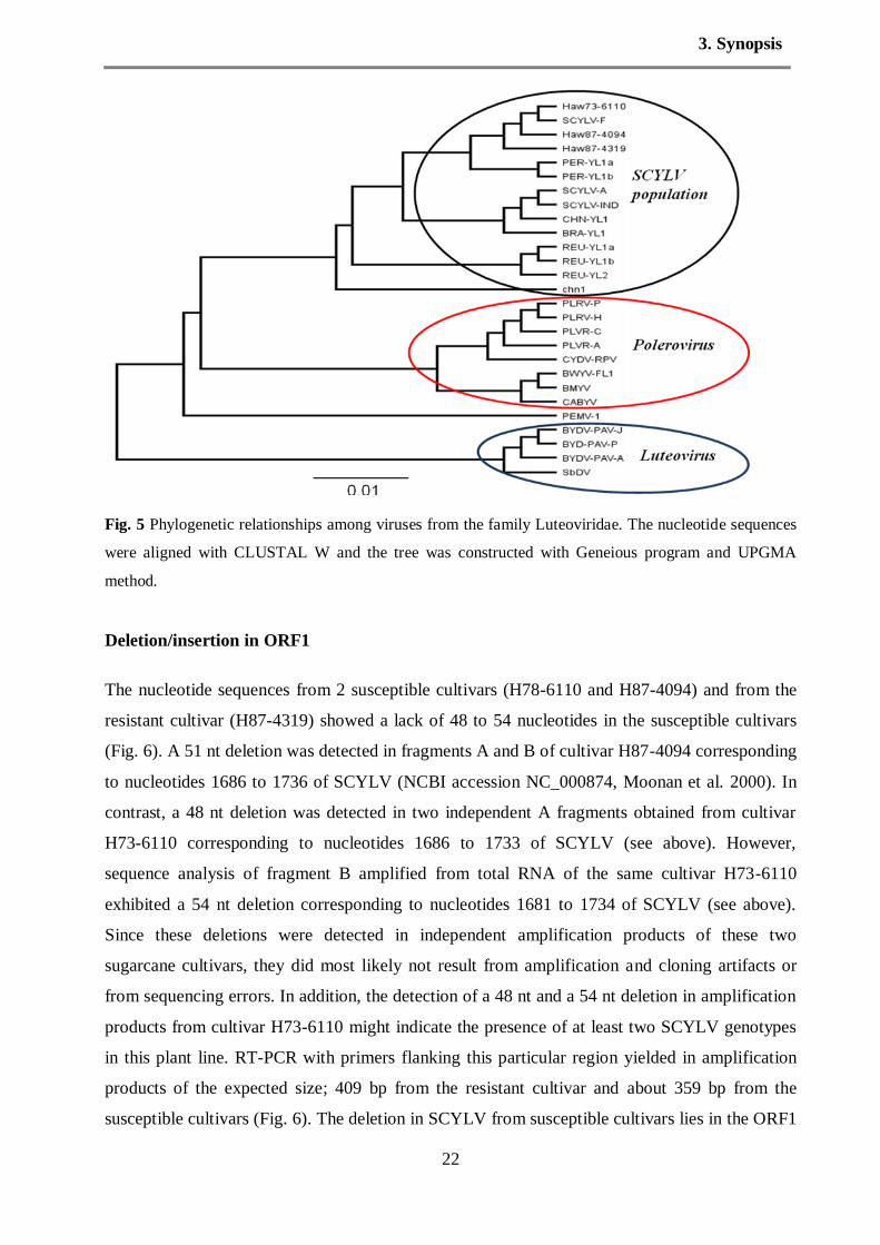
Fig. 5 Phylogenetic relationships among viruses from the family Luteoviridae. The nucleotide sequences
were aligned with CLUSTAL W and the tree was constructed with Geneious program and UPGMA
method.
Deletion/insertion in ORF1
The nucleotide sequences from 2 susceptible cultivars (H78-6110 and H87-4094) and from the
resistant cultivar (H87-4319) showed a lack of 48 to 54 nucleotides in the susceptible cultivars
(Fig. 6). A 51 nt deletion was detected in fragments A and B of cultivar H87-4094 corresponding
to nucleotides 1686 to 1736 of SCYLV (NCBI accession NC_000874, Moonan et al. 2000). In
contrast, a 48 nt deletion was detected in two independent A fragments obtained from cultivar
H73-6110 corresponding to nucleotides 1686 to 1733 of SCYLV (see above). However,
sequence analysis of fragment B amplified from total RNA of the same cultivar H73-6110
exhibited a 54 nt deletion corresponding to nucleotides 1681 to 1734 of SCYLV (see above).
Since these deletions were detected in independent amplification products of these two
sugarcane cultivars, they did most likely not result from amplification and cloning artifacts or
from sequencing errors. In addition, the detection of a 48 nt and a 54 nt deletion in amplification
products from cultivar H73-6110 might indicate the presence of at least two SCYLV genotypes
in this plant line. RT-PCR with primers flanking this particular region yielded in amplification
products of the expected size; 409 bp from the resistant cultivar and about 359 bp from the
susceptible cultivars (Fig. 6). The deletion in SCYLV from susceptible cultivars lies in the ORF1

3. Synopsis
23
for a “multifunctional protein” which is thought to be involved in suppression of gene silencing,
and at a cleavage point of RNA-dependent RNA polymerase (RdRp, ORF1 to ORF2).
Fig. 6 Sequence gap in SCYLV from susceptible cultivars (top) and RT-PCR of the sequence segment
containing the deletion (bottom). Top: Location of sequence gap in SCYLV from susceptible cultivars
versus SCYLV from resistant cultivar and ORFs for coded proteins. The gap was in overlap of fragments
A and B, the deletions were in susceptible cultivars only. Bottom: RT-PCR of the sequence segment
containing the deletion. Primers YL1FOR and YL1REV were designed to amplify the sequence
nucleotide. RNA-preparations from susceptible (H78-6110 and H87-4094) and resistant cultivars (H87-
4319) were used as templates. Lower panel loading control (25srRNA) 108bp (M: DNA size marker).
The amino acid sequences of RNA-dependent RNA polymerase (RdRp) from fully sequenced
SCYLV-strains showed lower sequence identities in the first half and high identity in the second
half of the protein (Fig. 6, see chapter 6). The 16 aa gap (48 nt deletion) and 17 aa gap (51 nt

3. Synopsis
24
deletion) of the two isolates Haw73-6110 and Haw87-4094 lies just in between of these two
halves (the 18 aa gap of the 54 nt deletion is not shown). The deduced amino acid sequences of
the capsid protein (CP) obtained from all the isolates expressed almost identical amino acid
sequences (97-100%, not shown).
3.4 Sequence deletion in Sugarcane yellow leaf virus genome and their effect on the
diversity of virus population
Significance of the deletion/lacking in ORF1
In order to understand the effect of deletion sequence in the replication of SCYLV genome, two
experiments have been designed. The first experiment was inculcated the sugarcane cultivars
susceptible and resistant with aphids Melanaphis sacchari, which is the transmission vector of
SCYLV. Interestingly, the results of RT-PCR revealed that, nine cultivars carried the sequence
deletion out of twelve, while three cultivars have a complete sequence/no stretch lacking. In
addition, the aphids carried also a deletion (Fig. 7a and b).
Fig. 7a RT-PCR of the sequence segment containing the deletion. Primers YL1FOR and YL1REV were
designed to amplify the sequence nucleotide from 1211 to 1620 nucleotide

3. Synopsis
25
Fig.7b RT-PCR of ORF1 sequence containing the deletion. YL1 primer was designed to cover the
deletion part sequence to investigate if the cultivars under study have a deletion or not.
Concerning to the second experiment, the cultivars H78-4153, H65-7052, Ph8013 and H87-
4094_virus free were inoculated with viruliferous aphids Melanaphis sacchari in an insect-tight
cage. The aphids have been fed on the cultivar have a complete sequence (no stretch lacking),
H87-4319. The results of RT-PCR showed that, the cultivar H87-4094_virus free was infected
by SCYLV strain contain a complete genome without deletion region, in addition the aphids also
has the same expression. In contrast, the cultivars H78-4153, H65-7052 and Ph8013 were carried
SCYLV with deletion about 50bp missing (Fig. 8). We mentioned that these cultivars has been
infected by the deletion virus strain before inoculation with the resistant cultivars and /or
complete genome. The results of the second experiment indicated that the aphids carried and
transmitted the virus particles as it is. Reasonable that the virus particles are not able to replicate
into the aphid organs, but there is other proposes that the virus could replicate in the plant.
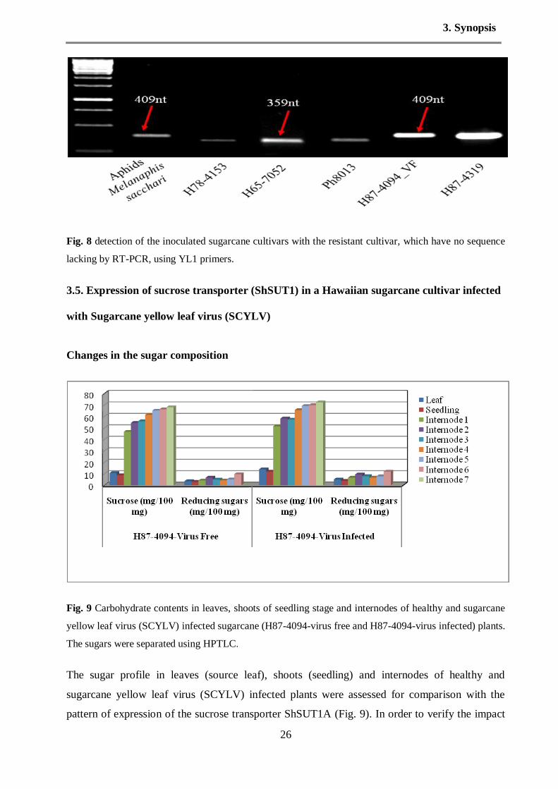
3. Synopsis
26
Fig. 8 detection of the inoculated sugarcane cultivars with the resistant cultivar, which have no sequence
lacking by RT-PCR, using YL1 primers.
3.5. Expression of sucrose transporter (ShSUT1) in a Hawaiian sugarcane cultivar infected
with Sugarcane yellow leaf virus (SCYLV)
Changes in the sugar composition
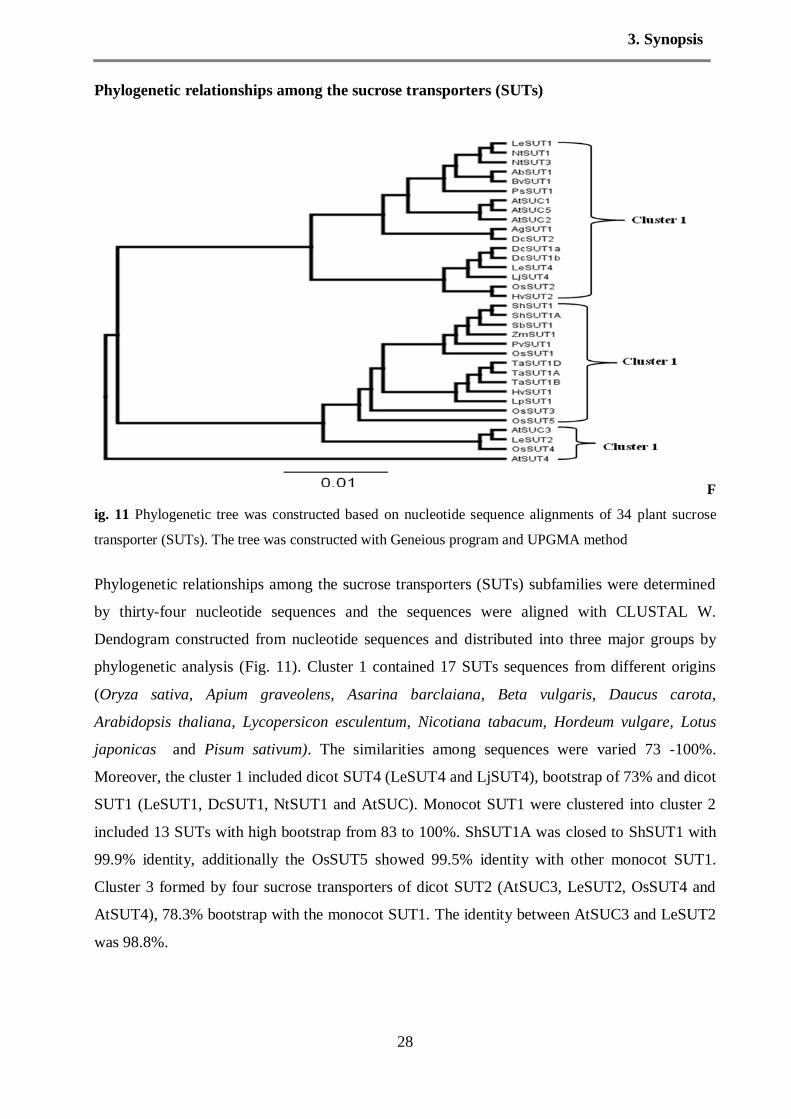
Fig. 9 Carbohydrate contents in leaves, shoots of seedling stage and internodes of healthy and sugarcane
yellow leaf virus (SCYLV) infected sugarcane (H87-4094-virus free and H87-4094-virus infected) plants.
The sugars were separated using HPTLC.
The sugar profile in leaves (source leaf), shoots (seedling) and internodes of healthy and
sugarcane yellow leaf virus (SCYLV) infected plants were assessed for comparison with the
pattern of expression of the sucrose transporter ShSUT1A (Fig. 9). In order to verify the impact

3. Synopsis
27
of SCYLV on the metabolism of carbohydrates in sugarcane tissues, the contents of sucrose and
reducing sugars were determined using HPTLC. Sucrose contents in leaves were increased by
SCYLV infection. Relative to the leaves of healthy plants, reducing sugars were the most
accumulated sugars in the leaves of infected plants. The reduction in sucrose in the shoots tissues
of seedling stage was found in the healthy plants, followed by reducing sugars. On the other
hand, the accumulation of sucrose in storage tissues (internodes) was increased by SCYLV
infection, compared with healthy plants. The high concentration of accumulated sucrose was
found between internodes 5-7, whereas, no accumulation for reducing sugars was found in
internodes 7.
Transcripts of ShSUT1 in different tissues of sugarcane
Fig. 10 Abundance of transcripts of ShSUT1A in sugarcane cv. H87-4094 virus free. RNA was extracted
from different internode tissues (from 1 to 7) and was hybridized to a probe of the ShSUT1A cDNA. The
lower panel shows the same membrane probed for ribosomal RNA to demonstrate RNA loading
The hybridization of RNA contained virus free sugarcane as seen in Fig. 10. It was noted from
the result of RNA hybridization that the transcript expression was relatively higher in the
maturing internodes (5-7) than in the younger internodes (immature).
3. Synopsis
28
Phylogenetic relationships among the sucrose transporters (SUTs)
F
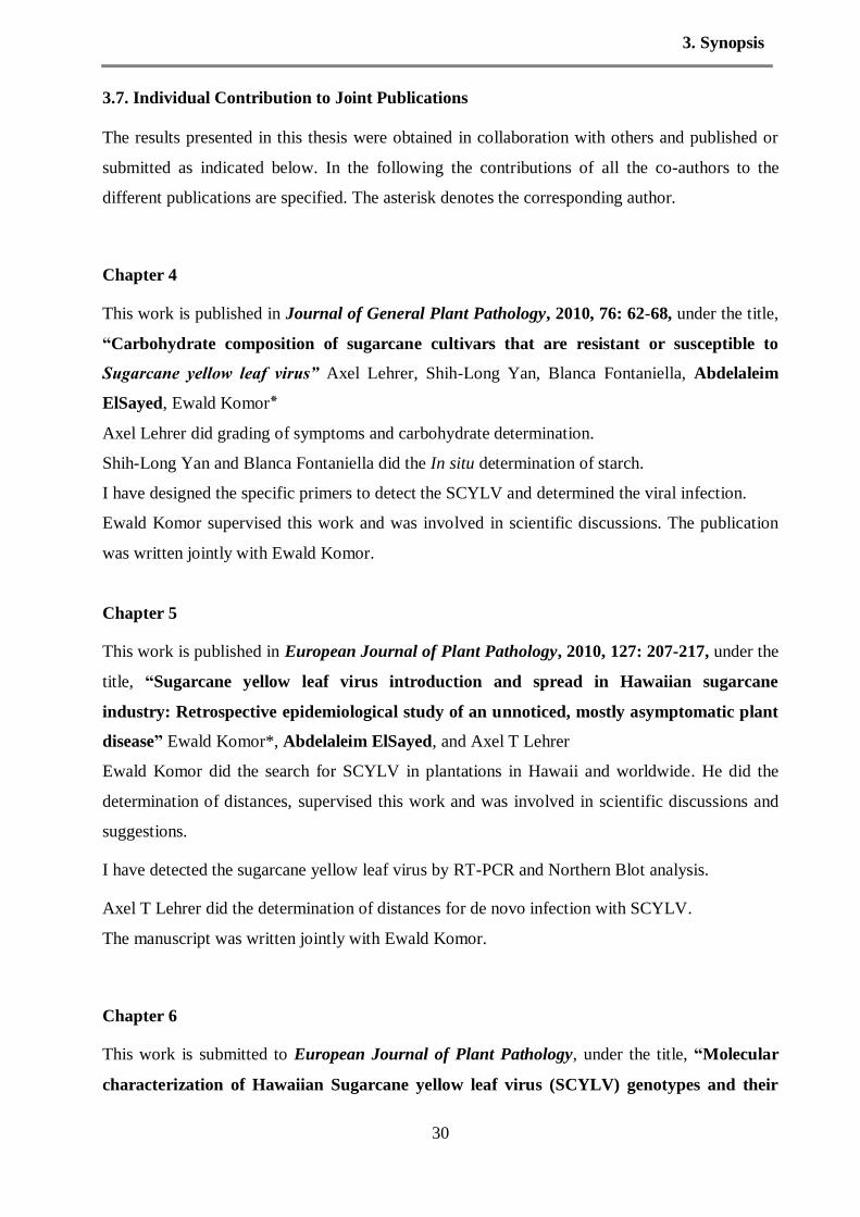
ig. 11 Phylogenetic tree was constructed based on nucleotide sequence alignments of 34 plant sucrose
transporter (SUTs). The tree was constructed with Geneious program and UPGMA method
Phylogenetic relationships among the sucrose transporters (SUTs) subfamilies were determined
by thirty-four nucleotide sequences and the sequences were aligned with CLUSTAL W.
Dendogram constructed from nucleotide sequences and distributed into three major groups by
phylogenetic analysis (Fig. 11). Cluster 1 contained 17 SUTs sequences from different origins
(Oryza sativa, Apium graveolens, Asarina barclaiana, Beta vulgaris, Daucus carota,
Arabidopsis thaliana, Lycopersicon esculentum, Nicotiana tabacum, Hordeum vulgare, Lotus
japonicas and Pisum sativum). The similarities among sequences were varied 73 -100%.
Moreover, the cluster 1 included dicot SUT4 (LeSUT4 and LjSUT4), bootstrap of 73% and dicot
SUT1 (LeSUT1, DcSUT1, NtSUT1 and AtSUC). Monocot SUT1 were clustered into cluster 2
included 13 SUTs with high bootstrap from 83 to 100%. ShSUT1A was closed to ShSUT1 with
99.9% identity, additionally the OsSUT5 showed 99.5% identity with other monocot SUT1.
Cluster 3 formed by four sucrose transporters of dicot SUT2 (AtSUC3, LeSUT2, OsSUT4 and
AtSUT4), 78.3% bootstrap with the monocot SUT1. The identity between AtSUC3 and LeSUT2
was 98.8%.

3. Synopsis
29
3.6. Quantitative multiplexed gene expression
Quantitative gene expression analysis would give more accurate relative quantitative information
on the ratios of virus titre and sucrose transporters (SUTs) in different cultivars, during different
plant stages. We determined four different genes of SCYLV, two genes of sucrose transporters
and one gene of sucrose phosphate synthase (SPSII) in one multiplex using GenomeLab GeXP
Genetic Analysis System. The plants under study were (H73-6110, H87-4094-vinf, H87-4319
and H87-4094-vf). The materials were taken from different plant tissues; sink leaves, source
leaves and mature internodes (8 to 9). The results of RT-qPCR by GeXP (Fig. 2, see chapter 8)
revealed that the four different genes of SCYLV were highly expressed in the sink leaves of
seedling stage, source leaves and mature internodes tissues (Fig. 2 see chapter 8). Furthermore,
the ORF0/1 was more highly transcript compared with other genes in all infected cultivars at
different plant stages. ORF0 is considered highly conserved region in SCYLV genome. In potato
leaf roll virus ORF0 was found to be effective in symptom development. Hence in SCYLV it
could be useful when ORF0 used as a diagnostic region. Additionally the ORF3/4 which related
to capsid protein and movement protein was slightly low expressed in sink and source leaves, for
unknown reasons. Whereas, the transcripts of ORF2 which encodes for RdRp were constant in
all infected cultivars at sink leaves of seedling stage. But the expression of RdRp was variable in
the source leaves and mature internodes.
The RT-qPCR using GeXP analysis showed that sucrose transporter (ShSUT1) was a higher
transcript expression than the sucrose transporter (ShSUT4) in the sink leaves and source leaves
with all tested cultivars (Fig. 4 see chapter 8). The highest levels of sucrose phosphate synthase
(SPSII) transcript expression were present in the mature internodes in all tested cultivars.
Furthermore, the SPSII was expressed in different plant tissues (photosynthetic and
nonphotosynthetic tissues) (Fig. 4 see chapter 8)
3. Synopsis
30
3.7. Individual Contribution to Joint Publications
The results presented in this thesis were obtained in collaboration with others and published or
submitted as indicated below. In the following the contributions of all the co-authors to the
different publications are specified. The asterisk denotes the corresponding author.
Chapter 4
This work is published in Journal of General Plant Pathology, 2010, 76: 62-68, under the title,
“Carbohydrate composition of sugarcane cultivars that are resistant or susceptible to
Sugarcane yellow leaf virus” Axel Lehrer, Shih-Long Yan, Blanca Fontaniella, Abdelaleim
ElSayed, Ewald Komor٭
Axel Lehrer did grading of symptoms and carbohydrate determination.
Shih-Long Yan and Blanca Fontaniella did the In situ determination of starch.
I have designed the specific primers to detect the SCYLV and determined the viral infection.
Ewald Komor supervised this work and was involved in scientific discussions. The publication
was written jointly with Ewald Komor.
Chapter 5
This work is published in European Journal of Plant Pathology, 2010, 127: 207-217, under the
title, “Sugarcane yellow leaf virus introduction and spread in Hawaiian sugarcane
industry: Retrospective epidemiological study of an unnoticed, mostly asymptomatic plant
disease” Ewald Komor*, Abdelaleim ElSayed, and Axel T Lehrer
Ewald Komor did the search for SCYLV in plantations in Hawaii and worldwide. He did the
determination of distances, supervised this work and was involved in scientific discussions and
suggestions.
I have detected the sugarcane yellow leaf virus by RT-PCR and Northern Blot analysis.
Axel T Lehrer did the determination of distances for de novo infection with SCYLV.
The manuscript was written jointly with Ewald Komor.
Chapter 6
This work is submitted to European Journal of Plant Pathology, under the title, “Molecular
characterization of Hawaiian Sugarcane yellow leaf virus (SCYLV) genotypes and their

3. Synopsis
31
phylogenetic relationship to SCYLV-strains from other sugarcane-growing countries”
Abdelaleim ElSayed, Alfons Weig and Ewald Komor*
I have done all experiments and characterized all the presented data here except the cleaning of
sequences. The manuscript was written by me.
Alfons Weig did cleaning of the sequences.
Ewald Komor supervised this work and was involved in scientific discussions, suggestions and
correction of the manuscript.
Chapter 7
This work is submitted to Physiological and Molecular Plant Pathology under the title,
“Expression of sucrose transporter (ShSUT1) in a Hawaiian sugarcane cultivar infected
with Sugarcane yellow leaf virus (SCYLV)” Abdelaleim Elsayed, Mohamed Fawzy Ramadan
and Ewald Komor*
I have done all experiments and characterized all the presented data here except the
determination of sugar content. The manuscript was written by me.
Mohamed Fawzy Ramadan did the determination of sugar content.
Ewald Komor supervised this work and was involved in scientific discussions, suggestions and
correction of the manuscript.
Chapter 8
This work is submitted to Plant Pathology under the title, “Simultaneous quantitative analysis
of transcripts for Sugarcane yellow leaf virus, sucrose transporters and sucrose phosphate
synthase in Hawaiian sugarcane cultivars by multiplex RT-PCR” Abdelaleim ElSayed,
Alfons Weig and Ewald Komor*
I have prepared the plant samples, all the presented data in this work have been characterized by
me. The manuscript was written by me.
Alfons Weig provided the GenomeLab Genetic Analysis System (GeXP) and was involved in
correction of the manuscript.

3. Synopsis
32
Ewald Komor supervised this work and was involved in scientific discussions, suggestions and
correction of the manuscript.

4. Selection of susceptible and resistant cultivars for SCYLV
33

4. Selection of susceptible and resistant cultivars for SCYLV
34

4. Selection of susceptible and resistant cultivars for SCYLV
35
4. Selection of susceptible and resistant cultivars for SCYLV
36

4. Selection of susceptible and resistant cultivars for SCYLV
37
4. Selection of susceptible and resistant cultivars for SCYLV
38
4. Selection of susceptible and resistant cultivars for SCYLV
39

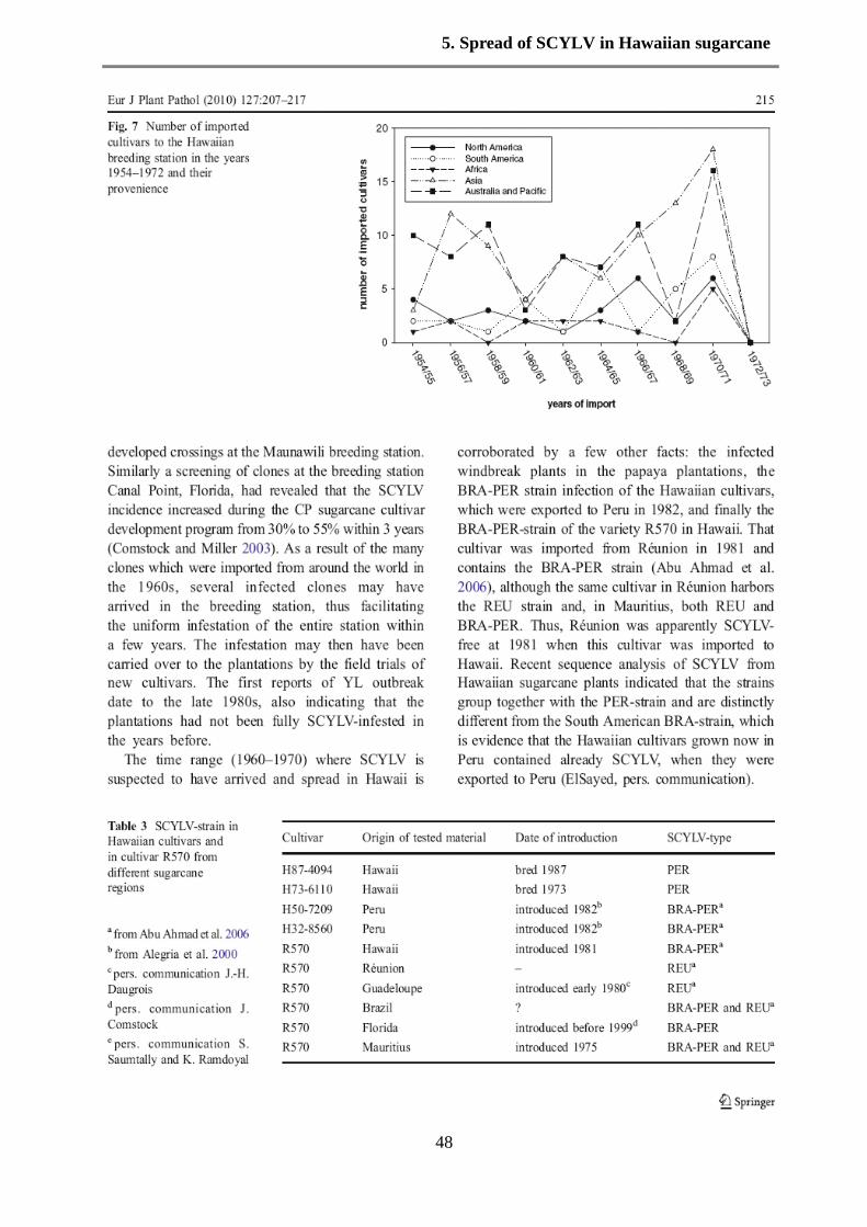
5. Spread of SCYLV in Hawaiian sugarcane
40

5. Spread of SCYLV in Hawaiian sugarcane
41

5. Spread of SCYLV in Hawaiian sugarcane
42
5. Spread of SCYLV in Hawaiian sugarcane
43

5. Spread of SCYLV in Hawaiian sugarcane
44

5. Spread of SCYLV in Hawaiian sugarcane
45

5. Spread of SCYLV in Hawaiian sugarcane
46

5. Spread of SCYLV in Hawaiian sugarcane
47
5. Spread of SCYLV in Hawaiian sugarcane
48

5. Spread of SCYLV in Hawaiian sugarcane
49

5. Spread of SCYLV in Hawaiian sugarcane
50

6. Genetic diversity of SCYLV
51
6. Molecular characterization of Hawaiian Sugarcane yellow leaf virus (SCYLV)
genotypes and their phylogenetic relationship to SCYLV-strains from other sugarcane-
growing countries
Abdeleim Ismail ElSayed1, Alfons R.Weig
2 and Ewald Komor
1*
1Plant physiology Department, Bayreuth University, D-95440 Bayreuth, Germany
2 DNA Analytics and Ecoinformatics, Bayreuth University, D-95440 Bayreuth, Germany
Keywords: Luteoviridae, phylogenetic analysis, resistant and susceptible cultivar, RNA virus,
Sugarcane yellow leaf virus (SCYLV), Yellow leaf
Submitted to European Journal of Plant Pathology
Abstract: Sugarcane yellow leaf virus (SCYLV) is the causal agent of the sugarcane disease
Yellow leaf (YL), which was first reported in Hawaii. The presence of SCYLV was detected by
tissue blot immunoassay and the Hawaiian sugarcane cultivars fell into susceptible cultivars
(with SCYLV) and resistant cultivars (without SCYLV). RT-PCR showed recently that also the
resistant cultivars contain the virus, however with a 100-fold lower virus titer than in the
susceptible cultivars. SCYLV is present as whole genome (6kb) and as two subgenomic
sequences of 2.4 and 1.0 kb. Virus preparations from three Hawaiian cultivars (two susceptible
and one resistant) were fully sequenced and the sequences were aligned to published full and
partial sequences. The phylograms corroborate previous findings that the so-called YLS-segment
coding for the coat protein shows the least genetic diversity, whereas the other sequence
fragments A-D, representing the ORFs 0-5, expressed a twofold higher diversity. The Hawaiian
SCYLV-strains clustered together next to the Peru strain, apart from the BRA-strains and well
apart from the REU-strains. We propose that the Hawaiian SCYLV should be considered as an
independent group together with the Peru strain as HAW-PER. The sequences from the two
susceptible cultivars had a deletion of 48 to 54 nt in ORF1, which codes for the gene silencing
suppressor and a RNA-dependent RNA-polymerase. It is speculated that this deletion is
important for the proliferation rate of the virus in the plant.
Introduction
The sugarcane disease Yellow leaf (YL) was first reported from plantations on two Hawaiian
islands (Schenck, 1990). Few years later similar symptoms were observed in mainland US

6. Genetic diversity of SCYLV
52
(Comstock et al. 1994) and Brazil accompanied by dramatic yield losses (Vega et al. 1997). The
symptoms are characterized by yellowing of leaf midribs followed by yellowing of the entire leaf
blade and shortening of internodes of the green leaf top. Borth and Hu (1994) reported a dsRNA-
virus in diseased plants. Later, a luteovirus (ss+RNA) could be unequivocally identified as causal
agent of Yellow leaf (Vega et al. 1997) and it was named Sugarcane yellow leaf virus (SCYLV).
Sequence analyses revealed that some regions of SCYLV genome are closely related to Barley
yellow dwarf virus and others similar to the Potato leaf roll virus, which suggested that SCYLV
may be a recombination product of a Polerovirus and an Enamovirus (Moonan et al. 2000, Smith
et al. 2000). SCYLV-strains from different American countries were characterized by
fingerprinting and partial sequence analysis and a Colombian strain was postulated as a founder
strain of SCYLV (Moonan and Mirkov 2002). Later AbuAhmad et al. (2006, 2007) compared 60
SCYLV-preparations from almost all sugarcane-growing countries (including Colombia) by
diagnostic PCR-reactions or by partial sequencing. SCYLV from Hawaiian cultivars were,
however, not among that study, although YLS and SCYLV were first detected in Hawaii and the
effect of SCYLV-infection on plant performance was already thoroughly studied for Hawaiian
cultivars. Yet some SCYLV-preparations had a relationship to Hawaii, for example a SCYLV
preparation from cultivar R570 which was grown in the collection of the Hawaiian sugarcane
breeding station, contained the BRA-strain and not the REU-strain, which exists in R570 grown
in Réunion (AbuAhmad et al. 2007). Similarly, the Hawaiian cultivars (H32-8560 and H50-
7209), which were exported to Peru in 1981, were found to be infected with the PER strain,
which is closely related to but not identical with the BRA-strain, the most common strain in
many South and North American cultivars (AbuAhmad et al. 2006). Therefore, it was reasonable
to assume that the sugarcane plantations of the Hawaiian Islands are infected by BRA and/or
PER strains of SCYLV, however, direct evidence for this assumption is lacking because
sequences of SCYLV from Hawaiian cultivars planted in Hawaii are not available so far. To date
eight complete sequences of SCYLV are available plus more than 30 partial sequences, none
from Hawaii. The Hawaiian cultivars were classified according to the presence of SCYLV into
susceptible and resistant cultivars (Schenck and Lehrer 2000), based on the observation that all
plants of susceptible cultivars contained SCYLV when tested by tissue blot immunoassay
(TBIA), whereas plants from resistant cultivars appeared virus-free. The strength of YL-
symptom expression was correlated (though not strictly) to the presence of SCYLV (Lehrer and
Komor 2008). Recent data obtained by PCR indicated that the resistant Hawaiian cultivars also
contained SCYLV although at very low titer (Zhu et al. 2010). The objective of this study was to
sequence SCYLV from susceptible and resistant Hawaiian cultivars and to determine their
phylogenetic relationship to SCYLV to already reported SCYLV clusters. In addition, although

6. Genetic diversity of SCYLV
53
the so-called YLS-segment of the SCYLV-sequence is considered as a valid diagnostic sequence
for all SCYLV-strains (Comstock et al. 1998, Abu Ahmad et al. 2006, 2007), a reliable and
accurate quantification of SCYLV in susceptible and resistant cultivars by RT-PCR or real-time
PCR (Zhu et al. 2010) requires the accurate knowledge of SCYLV-sequences.
Material and Methods
Plant material and aphids
Cultivars H73-6110, H87-4319, H78-4153, H65-7052, H78-7750, and H87-4094 were obtained
from the Hawaii Agriculture Research Center, Aiea, Hawaii, USA. A virus-free line of the
cultivar H87-4094 was produced by meristem tip tissue culture and was provided by Dr. A.
Lehrer, Honolulu. In addition, cultivars C1051-73, JA-605 and CP52-43, were obtained from
Cuba through Medina Borges, Habana. The plants were grown in the greenhouse at Bayreuth
University at 24°C with a 12-h photoperiod and propagated 1-2 times per year from cuttings.
Aphids Melanaphis sacchari were collected from sugarcane at Hawaiian Agriculture Research
Center, Aiea, Hawaii, USA
Isolation of RNA, RT-PCR and northern blot for detection of SCYLV
RNA was extracted and purified from the top visible dewlap leaf as previously described
(Comstock et al. 1998, Sambrook and Russell 2001, Lehrer et al. 2010).
RNA was extracted from aphids by the same protocol as described above with RNA extraction
buffer (4M guanidine thiocyanate (Sigma-Aldrich, Chemie GmbH, Munich, Germany), 25 mM
sodium citrate, pH 7.5% Sarkosyl (Sigma-Aldrich) and 2M sodium acetate pH 4.0).
RT-PCR was used to test the presence of SCYLV in the leaf samples of 9 Sugarcane cultivars
and one aphid sample using diagnostic primers YLS111 and YLS462 (Comstock et al., 1998).
The RNA was reverse transcribed using RevertAid H Minus First Strand cDNA Synthesis Kit
(Fermentas GmbH, Leon –Rot Germany), primed with 50 pmol of YLS462 by following the
manufacturer’s protocol in a PCR machine (PTC 100 Peltier Thermal Cycler, MJ Research,
Global Medical Instrumentation, Inc, Ramsey, Minnesota, USA.). The RT- PCR reaction was
performed in 25 µl containing 1 µl cDNA, 2.5 µl of 10x PCR buffer containing 15mM MgCl2,
0.5 µl of 10mM dNTP mix, 10 pmol each of forward and reverse primers (YLS111 and
YLS462), 1 unit of a polymerase with proofreading activity (Pfu ): Taq polymerase (5:1)
(Stratagene, Waldbronn, Germany), and sterile milliQ water added up to the final volume of 25
µl. This PCR programme was performed with initial denaturation at 94°C for 4 min, 10 cycles of

6. Genetic diversity of SCYLV
54
94°C for 30 sec, 62°C for 2 min, 72°C for 1.5 min, and 30 cycles of 94°C for 30 sec, 62°C for 30
sec, 72°C for 1.5 min with a final 72°C extension for 7 min. The primer pairs located in the
different ORFs of SCYLV genome are listed in Table 1 and Fig. 1.
Northern blots were prepared according Sambrook and Russell (2001) with 10 µg of intact RNA
isolated from sugarcane leaves.
Genome fragment amplification
Genome fragments A-D, YL0, YL1, YL5 and YLS were amplified from reverse-transcribed
RNA preparations as described above. The partial ORFs 0 and 1 (fragment A) was amplified
from three cultivars (H73-6110, H87-4319 and H87-4094). The PCR program for the
amplification of partial ORFs 0 and 1 with the primers ORF1START and 160R.640R was 94°C
for 5 min, 10 cycles of 94°C for 30 sec, 62°C for 2 min, 72°C for 4 min, and 30 cycles of 94°C
for 30 sec, 62°C for 30 sec, 72°C for 4 min with a final 72°C extension for 15 min. PCR program
performed with the primers oFM323 and oFM359 was 94◦C for 5 min, 10 cycles of 94°C for 30
sec, 58°C for 2 min, 72°C for 4 min, and 30 cycles of 94°C for 30 sec, 58°C for 30 sec, 72°C for
4 min with a final 72°C extension for 15 min. Partial sequence of ORF2, ORF5 and complete
sequence of ORF3 and ORF4 (Fragment C) were amplified with three cultivars (H73-6110, H87-
4319 and H87-4094). The RT-PCR program performed with primers B FOR and B REV was
94°C for 5 min, 10 cycles of 94°C for 30 sec, 62°C for 2 min, 72°C for 4 min, and 30 cycles of
94°C for 30 sec, 62°C for 30 sec, 72°C for 4 min with a final 72°C extension for 15 min.
The partial sequence of ORF5 (fragment D) was amplified from RNA isolated from the cultivars
H73-6110, H87-4319 and H87-4094 with 104R.623R and 3´PRIME2 primer pair. The RT-PCR
program was 94°C for 5 min, 10 cycles of 94°C for 30 sec, 62°C for 2 min, 72°C for 4 min, and
30 cycles of 94°C for 30 sec, 62°C for 30 sec, 72°C for 4 min with a final 72°C extension for 15
min.
PCR reaction for amplification the gap in ORF1 in SCYLV genome (fragment YL1) was
performed with the YL1FOR and YL1REV primer pair to cover the non-sequenced region. The
PCR programme was the same as for primers for YLS. The amplification of the region in ORF0
in SCYLV genome (fragment YL0) was performed with primer ORF0 FOR and ORF0 REV and
the PCR programme was the same as mentioned above with primer YLS. Additionally, the
fragment YL5 was amplified with primer ORF5 FOR and ORF5 REV and the PCR programme
was the same as mentioned above with primer YLS.

6. Genetic diversity of SCYLV
55
The RT-PCR was performed with internal control, 25srRNA, as a reference gene to normalize
gene expression level and to evaluate the integrity of cDNA. Furthermore, the primer sets were
optimized using semi qPCR with different numbers of PCR-cycles.
A 15 µl aliquot of each amplified product was analysed by electrophoresis on 1% agarose gels
stained with ethidium bromide.
Cloning and sequencing of RT-PCR products
Twenty-five amplicons of all viral fragments, derived from independent RT-PCR reactions were
cloned in the pGEM®-T Easy Vector System (Promega, Mannheim, Germany) and were
transformed into the competent E. coli DH5α strain. The recombinant DNA clones containing
the inserts were purified using the Pure yieldTM.
Plasmid Miniprep System (Promega, Mannheim,
Germany). The selected clones were sequenced by primer walking using M13 sequencing
primers and internal primers specific for each of the fragments A, B, C, and D in the DNA
Analytics Core Facility at the University of Bayreuth. One clone per amplicon was sequenced
and used for alignment and phylogenetic analyses.
Alignment of sequences and construction of phylogenetic trees
Multiple sequence alignments of nucleotide or deduced amino acid sequences were aligned using
CLUSTAL W applying the Dayhoff PAM 250 matrix (Thompson et al. 1994) and were
optimized manually. Phylogenetic reconstructions were performed using Geneious program,
version 4.7.5 (www.geneious.com). Trees were constructed by the UPGMA method. Data sets
were bootstrapped (1,000 replicates) to assess the confidence values of the phylogenetic trees,
and bootstrap values < 50% were omitted. The resulting sequences were compared with the
GenBank database (NCBI). The GenBank accession numbers of the sequences determined here
and those used for phylogenetic analysis are listed in Table 2.
Results
Detection of SCYLV in Hawaiian cultivars using RT-PCR and northern blot analysis
RNA was isolated from ten cultivars (seven from Hawaii and three from Cuba) and used for
cDNA synthesis and SCYLV detection. The diagnostic primers YLS111 and YLS462 were used
in the PCR reactions (AbuAhmad et al. 2006). A virus-free line of cultivar H87-4094 was
generated by meristem tip culture and used as negative control. As expected, two susceptible
Hawaiian cultivars (H73-6110 and H87-4094) showed strong amplification products of SCYLV
of the expected size, while the virus-free line H87-4094Vf showed no PCR product (Fig. 1). The
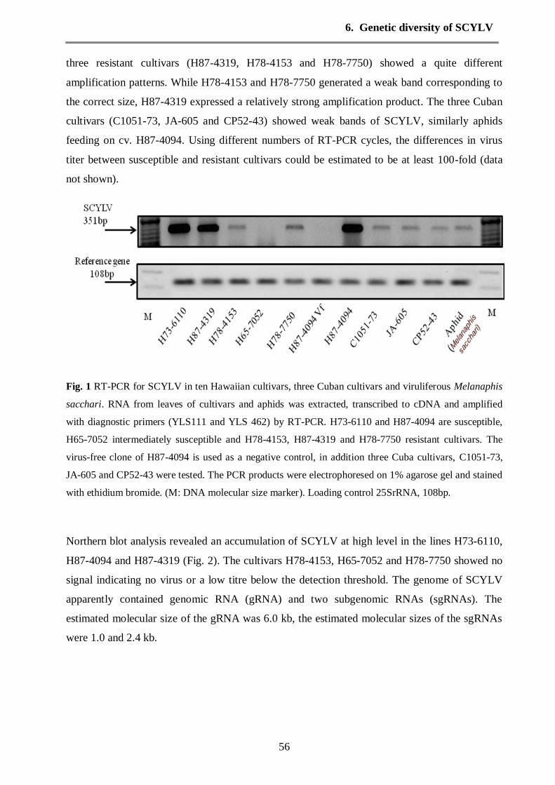
6. Genetic diversity of SCYLV
56
three resistant cultivars (H87-4319, H78-4153 and H78-7750) showed a quite different
amplification patterns. While H78-4153 and H78-7750 generated a weak band corresponding to
the correct size, H87-4319 expressed a relatively strong amplification product. The three Cuban
cultivars (C1051-73, JA-605 and CP52-43) showed weak bands of SCYLV, similarly aphids
feeding on cv. H87-4094. Using different numbers of RT-PCR cycles, the differences in virus
titer between susceptible and resistant cultivars could be estimated to be at least 100-fold (data
not shown).
Fig. 1 RT-PCR for SCYLV in ten Hawaiian cultivars, three Cuban cultivars and viruliferous Melanaphis
sacchari. RNA from leaves of cultivars and aphids was extracted, transcribed to cDNA and amplified
with diagnostic primers (YLS111 and YLS 462) by RT-PCR. H73-6110 and H87-4094 are susceptible,
H65-7052 intermediately susceptible and H78-4153, H87-4319 and H78-7750 resistant cultivars. The
virus-free clone of H87-4094 is used as a negative control, in addition three Cuba cultivars, C1051-73,
JA-605 and CP52-43 were tested. The PCR products were electrophoresed on 1% agarose gel and stained
with ethidium bromide. (M: DNA molecular size marker). Loading control 25SrRNA, 108bp.
Northern blot analysis revealed an accumulation of SCYLV at high level in the lines H73-6110,
H87-4094 and H87-4319 (Fig. 2). The cultivars H78-4153, H65-7052 and H78-7750 showed no
signal indicating no virus or a low titre below the detection threshold. The genome of SCYLV
apparently contained genomic RNA (gRNA) and two subgenomic RNAs (sgRNAs). The
estimated molecular size of the gRNA was 6.0 kb, the estimated molecular sizes of the sgRNAs
were 1.0 and 2.4 kb.

6. Genetic diversity of SCYLV
57
Fig. 2 Northern blot of SCYLV isolated from Hawaiian sugarcane. The RNA gel blot was probed with
DIG-labeled SCYLV probe covering the YLS sequence part and detected with anti-digoxigenin-AP and
CDP-Star ready-to-use and visualized with a chemilux CCD camera (Intas, Göttingen-Germany). The
apparent size of the hybridization signals was deduced from RNA molecular size markers (not shown),
loading control 25SrRNA visualized by probe hybridization.
Fragment amplification of SCYLV isolates and phylogenetic relationship to published
SCYLV-isolates
SCYLV is a ss+-RNA virus with 5895-5899 nucleotides organized in six open reading frames
(ORFs 0-5) (Fig. 3). Eight primer pairs for amplification of fragments YLS and A-D were
designed (Table 1) for the six open reading frames. Complete sequences of SCYLV from 3
Hawaiian cultivars and several partial sequences (accession numbers: GU570004, GU570005,
GU570006, GU570007, GU570008, GU570009, GU570010) were obtained.
The Hawaiian amplicons were used for phylogenetic analysis together with sequences from the
GenBank data base (http://www.ncbi.nlm.nih.gov) (Table 2). Since the SCYLV-genome is a
recombination product of two Luteoviridae viruses, the phylogenetic relationship of fragments
A-D and YLS were separately constructed to visualize possible sequence segments, where the
Hawaiian strains may have diverged from other strains.
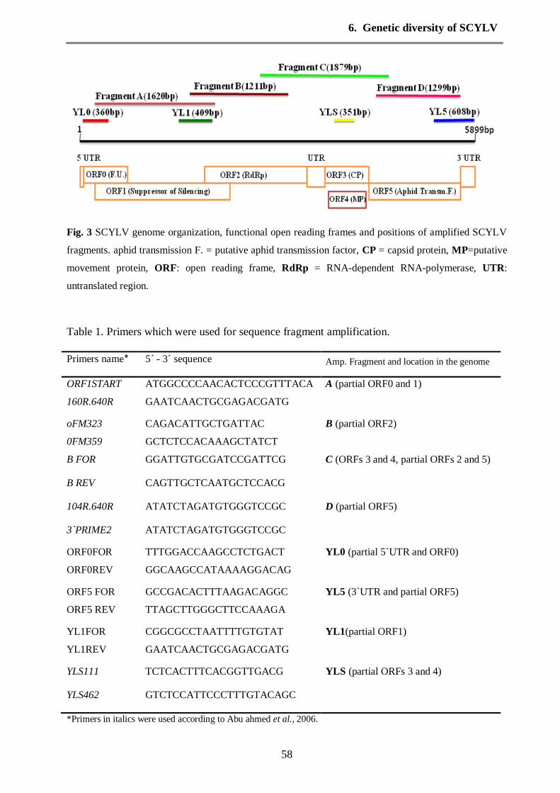
6. Genetic diversity of SCYLV
58
Fig. 3 SCYLV genome organization, functional open reading frames and positions of amplified SCYLV
fragments. aphid transmission F. = putative aphid transmission factor, CP = capsid protein, MP=putative
movement protein, ORF: open reading frame, RdRp = RNA-dependent RNA-polymerase, UTR:
untranslated region.
Table 1. Primers which were used for sequence fragment amplification.
Primers name5 ٭ - 3` sequence Amp. Fragment and location in the genome
ORF1START ATGGCCCCAACACTCCCGTTTACA A (partial ORF0 and 1)
160R.640R GAATCAACTGCGAGACGATG
oFM323 CAGACATTGCTGATTAC B (partial ORF2)
0FM359 GCTCTCCACAAAGCTATCT
B FOR GGATTGTGCGATCCGATTCG C (ORFs 3 and 4, partial ORFs 2 and 5)
B REV CAGTTGCTCAATGCTCCACG
104R.640R ATATCTAGATGTGGGTCCGC D (partial ORF5)
3`PRIME2 ATATCTAGATGTGGGTCCGC
ORF0FOR TTTGGACCAAGCCTCTGACT YL0 (partial 5`UTR and ORF0)
ORF0REV GGCAAGCCATAAAAGGACAG
ORF5 FOR GCCGACACTTTAAGACAGGC YL5 (3`UTR and partial ORF5)
ORF5 REV TTAGCTTGGGCTTCCAAAGA
YL1FOR CGGCGCCTAATTTTGTGTAT YL1(partial ORF1)
YL1REV GAATCAACTGCGAGACGATG
YLS111 TCTCACTTTCACGGTTGACG YLS (partial ORFs 3 and 4)
YLS462 GTCTCCATTCCCTTTGTACAGC
*Primers in italics were used according to Abu ahmed et al., 2006.

6. Genetic diversity of SCYLV
59
Table 2. SCYLV sequences used in the phylogenetic analysis.
Isolate name٭ Source of
SCYLV
Country/location of origin GenBank accession number
Aus1 VMC71-238 Australia AJ491255
Aus2 VMC71-238 Australia AJ491256
Beaz1 SP77-5181 Brazil AJ491274, AJ491257 BRA1 SP83-5073 Brazil AJ582772
BRA2 RB835054 Brazil AJ582779
BRA-YL1 SP71-6163 Brazil AM072750
CBB38192 B38192 India:Coimbatore EF635935 CB671 CoC671 India:Coimbatore, EU624499
CB86032 Co86010 India:Coimbatore EU624497
CB99016 Co99016 India:Coimbatore EU089687 CBAPHIDS Aphids India:Coimbatore EU089688
CHN-YL1 CGT63-167 China AM072751
COL4 SP71-6163 Colombia AM072624
CP52-43 CP43-62 Cuba GU570009 CP65-357 CP65-357 USA AJ249447
CPam1 CP65-357 Australia:Queensland AJ491271
CPaus1 CP65-357 Australia:Queensland AJ491270 CUB1 JA64-11 Cuba AJ621181
CUB2 C13-281 Cuba AJ582770
CUB-YL1 C132-81 Cuba AM083988 Haw1 H78-3606 USA:Hawaii AJ491261,AJ491118,AJ491278
Haw2 H87-4094 USA:Hawaii AJ491262,AJ491119,AJ491279
Haw3 H78-7750 USA:Hawaii AJ491120
Haw73-6110 H73-6110 Hawaii:USA GU570008 Haw73-6110b H73-6110 Hawaii:USA GU570005, GU570004
Haw87-4094 H87-4094 Hawaii:USA GU570006
Haw87-4319 H87-4319 Hawaii:USA GU570007 HawAphids Aphids Hawaii:USA GU570010
Hy-IND-1 Co 62399 India FJ430665
KER-IND-1 Co 7219 India FJ430661 KER-IND-2 Co C671 India FJ430662
Maur1 M1658-78 Mauritius AJ491280, AJ491263, AJ491140
Maur2 M2350-79 Mauritius AJ491264
MUS1 M99/48 Mauritius AJ606085 MYS1 TC4 Malaysia AJ606084
PER1 H32-8560 Peru AM072627, AJ621179,
AJ582767 PER-YL1a H50-7209 Peru AM072752
PER-YL1b H50-7209 Peru AM072753
PER-YL1b H50-7209 Peru AM072753
PHL1 VMC76-16 Philippines AJ582761 REU1 R570 Re′union Island:LeGol AJ621163
REU12 R569 Reunion Island (Vue-Belle) AJ606087, AJ582765, AJ582791
REU13 M1371/79 Reunion Island (Vue-Belle) AJ606088, AJ582769, AJ582783 REU15 R579 Reunion Island (St-Benoît) AJ621165, AM072633
REU17 R576 Reunion Island (St-Benoît) AJ606089
REU2 R577 Reunion Island (Le Gol) AJ621166, AJ582785 REU22 R570 Reunion Island (St-Benoît) AJ621168,AM072638
REU3 R577 Reunion Island (Le Gol) AJ606090, AJ582773, AJ582786
REU31 AY7 Reunion Island (La Mare) AJ621171,AJ582781,AJ582788,
AM072644

6. Genetic diversity of SCYLV
60
Table2 continued
Isolate name٭ Source of SCYLV
Country/location of origin GenBank accession number
REU32 R579 Reunion Island (La Mare) AJ606091
REU33 R490 Reunion Island (La Mare) AJ621172, AM072645
REU35 R577 Reunion Island (Vue-Belle) AJ582787, AM072647
REU37 R81-0834 Reunion Island (La Mare) AJ582775, AM072648 REU39 AY7 Reunion Island (La Mare) AJ621175, AM072650
REU40 SP71-6163 Reunion Island (La Mare) AJ621177, AJ582782, AJ582784
REU42 SP71-6163 Reunion Island (La Mare) AJ621159, AJ582762, AJ582792 REU48 S17 Reunion Island (La Mare) AM072656
REU5 M1371/79 Reunion Island (Vue-Belle) AJ582763
REU7 CP70-1133 Re′union Island:Vue-Belle AJ621161 REU9 R575 Reunion Island (Vue-Belle) AJ606092
Reun-1 R84-0408 Reunion AJ491282, AJ491265
Reun-2 R85-1102 Reunion AJ491283, AJ491266
REU-YL1a R570 Re′union Island :La Mare AM072754 REU-YL1b R570 Re′union Island :La Mare AM072755
REU-YL2 R490 Re′union Island :La Mare AM072756
REU-YL3 SP71-6163 Re′union Island :La Mare AM085306 SCYLV C3 CC85-964 Colombia: Cali AF369928
ScYLV-A CP65-357 USA:Florida AF157029
ScYLV-B1 SP71-6163 Brazil:Sao-Paulo AF369925 ScYLV-C1 SP71-6163 Colombia:Cali AF369927
ScYLV-C3 CC85-964 Colombia:Cali AF369928
ScYLV-C4 CC84-75 Colombia:Cali AF369929
ScYLV-F CP65-357 USA:Florida AJ249447 ScYLV-G2 CP92-1654 Guatemala:Santa-Lucia AF369924
ScYLV-IND - India:Gorakhpur AY236971
ScYLV-L1 LHo83-153 USA:Baton Rouge AF369923 ScYLV-N6 Q136 Argentina:Santa-Rosa AF369926
Taiw1 ROC11 Taiwan AJ491144, AJ491127
Taiw2 ROC12 Taiwan AJ491269
TN-IND-1 Co 86010 India FJ430663 TN-IND-2 Co 93009 India FJ430664
TWN1 ROC6 Taiwan AM072630
USA1 TCP87-3388 USA:Florida AM072631 USA1a CP81-1405 USA AJ491114
USA1b CP81-1405 USA AJ491115
USA2 CP85-1491 USA:Florida AJ621162 USA2a CP88-1409 USA AJ491276, AJ491116
USA2b CP88-1409 USA AJ491117, AJ491277
*Isolates in italics were amplified and sequenced in this study and other data from GenBank.
The nucleotide sequences of the YLS-region were obtained from 44 SCYLV isolates. The
Hawaiian sequences were assembled in cluster YLS1 (Fig. 4a). The resistant cultivar (Haw87-
4319) showed 100% sequence identity to YLS from Brazil, Florida, China and Réunion. the
YLS-sequence from the two susceptible Hawaiian cultivars (Haw73-6110 and Haw87-4094)
exhibited close sequence similarity to those from Australian and Peru. Only SCYLV from Indian
cultivars (and one Colombian) were clustered separately from all other YLS-sequences (cluster

6. Genetic diversity of SCYLV
61
YLS2 in Fig. 4a). The deduced amino acid sequences of the capsid protein (CP) obtained from
all the isolates expressed almost identical amino acid sequences (97-100%, data not shown).
Fig. 4a Phylogenetic trees based on nucleotide sequence alignments of SCYLV isolates. The trees were
constructed with Geneious program and UPGMA method. Numbers above the lines indicate the bootstrap
scores out of 1,000 replicates. a) Phylogram of fragment YLS (351 nt),
Forty-three virus isolates were used in phylogenetic analysis of fragment A (comprising partial
ORFs 0 and 1). The sequences were distributed into three major groups (Fig.4b). Cluster A1
contained 19 SCYLV sequence isolates from many origins (USA, Brazil, China, Australia etc.),
the Hawaiian isolates were together in cluster A2 and the Peru isolates (A3) appeared similarly
close to A1 (BRA) and Hawaiian isolates (A2). The sequence identity among Hawaiian isolates
ranges between (91.2% and 98.9%). The majority of Réunion virus strains were clustered into
group A3. (MUS1 seemed to be unique not fitting in any of the above groups.)
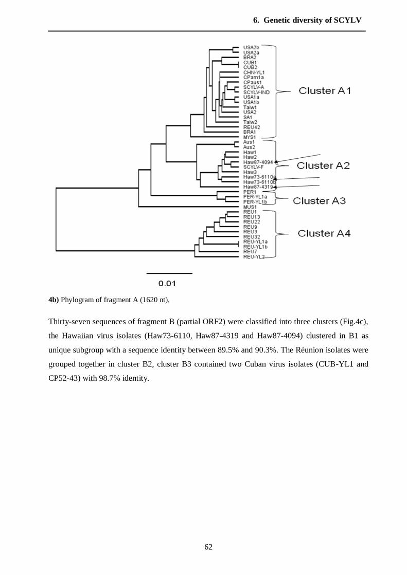
6. Genetic diversity of SCYLV
62
4b) Phylogram of fragment A (1620 nt),
Thirty-seven sequences of fragment B (partial ORF2) were classified into three clusters (Fig.4c),
the Hawaiian virus isolates (Haw73-6110, Haw87-4319 and Haw87-4094) clustered in B1 as
unique subgroup with a sequence identity between 89.5% and 90.3%. The Réunion isolates were
grouped together in cluster B2, cluster B3 contained two Cuban virus isolates (CUB-YL1 and
CP52-43) with 98.7% identity.

6. Genetic diversity of SCYLV
63
4c) Phylogram of fragment B (1211 nt),
The sequences of 21 isolates of fragment C, which covers ORFs 3 and 4 and parts of ORFs 2 and
5, were assembled into 4 clusters (Fig.4d). Nine SCYLV-isolates from different geographical
origins (Brazil, USA etc.) were grouped in cluster C1 together with the Hawaiian isolates, cluster
C2 contained the PER-isolates, REU-isolates were assembled into group C3 and cluster C4
included only Colombia isolates.

6. Genetic diversity of SCYLV
64
4d) Phylogram of fragment C (1879 nt),
Thirty-eight SCYLV isolates of fragment D which is related to the putative aphid transmission
factor, were grouped into three clusters (Fig. 4e). Cluster D1 contained subgroups from various
origins of SCYLV isolates (e.g. Florida, Brazil and India in one subgroup, isolates REU40 and
REU42 in another subgroup). Hawaiian isolates were assembled in cluster D2 as unique
subgroup, the majority of Réunion virus isolates was clustered in D3 together with two isolates
from Mauritius.

6. Genetic diversity of SCYLV
65
4e) Phylogram of fragment D (1299 nt),
The phylogenetic analysis of the SCYLV partial sequences constantly yielded 3 clusters: (1) a
big group comprising strains from Brazil, USA, China, India and several other countries
(tentatively group BRA according AbuAhmad et al. 2006), (2) a group exclusively for most
strains from Réunion (group REU) and (3) a group with the Hawaiian and the Peru strains. For
this group we propose the name HAW-PER. In fragment C a Colombian cluster, in fragment B a
Cuban cluster showed up, separated from the other strains. Phylogenetic analysis of 14 complete
sequences of SCYLV genome also exhibited three groups: Group HAW-PER included two
subgroups with the Hawaiian and Peru isolates with bootstrap value 89%., group 2 (BRA)
formed by isolates from various origins (Brazil, China, India and USA) and group 3 with the
REU strains. Thus the whole genome confirms the results already obtained from the alignment of
partial sequences which are available in much larger numbers.
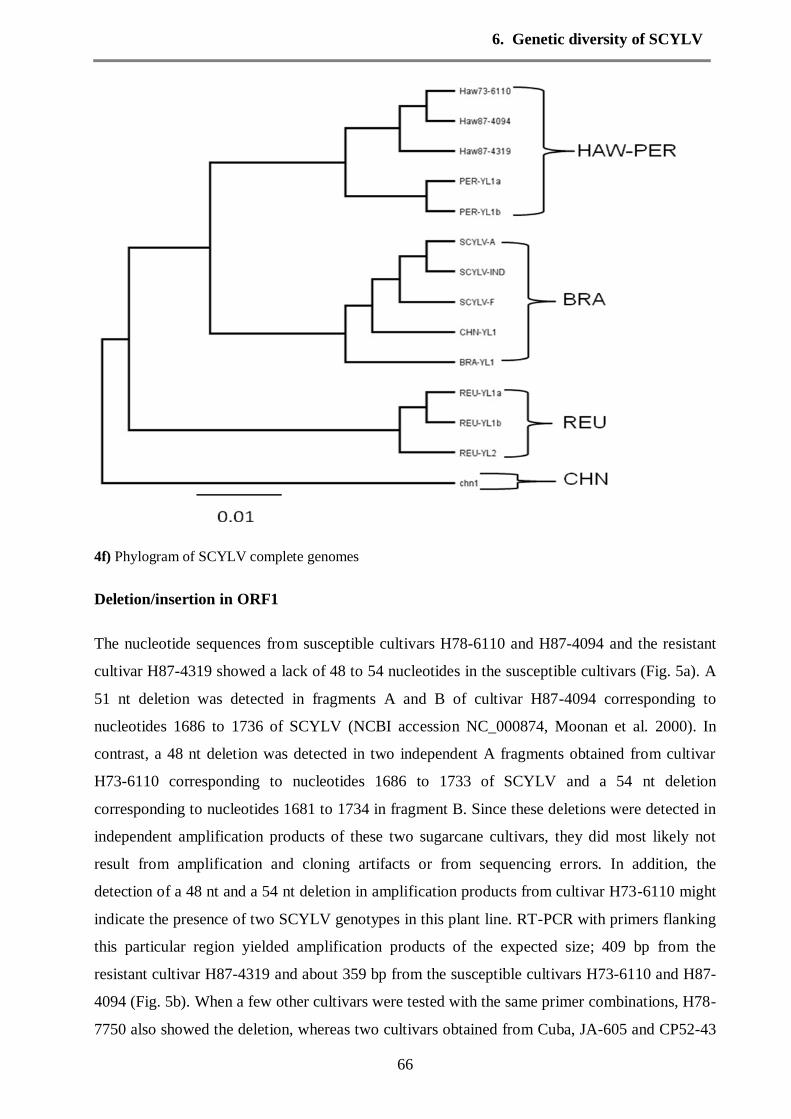
6. Genetic diversity of SCYLV
66
4f) Phylogram of SCYLV complete genomes
Deletion/insertion in ORF1
The nucleotide sequences from susceptible cultivars H78-6110 and H87-4094 and the resistant
cultivar H87-4319 showed a lack of 48 to 54 nucleotides in the susceptible cultivars (Fig. 5a). A
51 nt deletion was detected in fragments A and B of cultivar H87-4094 corresponding to
nucleotides 1686 to 1736 of SCYLV (NCBI accession NC_000874, Moonan et al. 2000). In
contrast, a 48 nt deletion was detected in two independent A fragments obtained from cultivar
H73-6110 corresponding to nucleotides 1686 to 1733 of SCYLV and a 54 nt deletion
corresponding to nucleotides 1681 to 1734 in fragment B. Since these deletions were detected in
independent amplification products of these two sugarcane cultivars, they did most likely not
result from amplification and cloning artifacts or from sequencing errors. In addition, the
detection of a 48 nt and a 54 nt deletion in amplification products from cultivar H73-6110 might
indicate the presence of two SCYLV genotypes in this plant line. RT-PCR with primers flanking
this particular region yielded amplification products of the expected size; 409 bp from the
resistant cultivar H87-4319 and about 359 bp from the susceptible cultivars H73-6110 and H87-
4094 (Fig. 5b). When a few other cultivars were tested with the same primer combinations, H78-
7750 also showed the deletion, whereas two cultivars obtained from Cuba, JA-605 and CP52-43

6. Genetic diversity of SCYLV
67
contained the 50nt stretch (Fig. 5b). The deletion in SCYLV from susceptible cultivars lies in the
ORF1 for a “multifunctional protein” which is thought to be involved in suppression of gene
silencing, and at a cleavage point of RNA-dependent RNA polymerase (RdRp, ORF1 to ORF2).
The amino acid sequences of RNA-dependent RNA polymerase (RdRp) from fully sequenced
SCYLV-strains showed lower sequence identities in the first half and high identity in the second
half of the protein (Fig 6). The 16-18 aa gap of the two isolates H73-6110 and H87-4094 lies just
in between of these two halves.
Fig. 5a Sequence gap in SCYLV from susceptible cultivars a: Location of sequence gap in SCYLV from susceptible
cultivars versus SCYLV from resistant cultivar and ORFs for coded proteins. The gap was in overlap of fragments
A and B, the deletions were in susceptible cultivars only.
Fig.5b: RT-PCR of the sequence segment in ORF1 which contains the deletion in some cultivars. Primers YL1FOR
and YL1REV were designed to amplify the sequence nt1211-1620 from RNA-preparations of sugarcane leaves as
templates. Cultivars H73-6110 and H87-4094 are susceptible, cvs. H87-4319 and H78-7750 are resistant. Cvs. H78-
7750, JA-605 and CP52-43 were infected with viruliferous Melanaphis sacchari. M: 1kb and 50bp DNA molecular
size markers (Fermentas, St. Leon Rot, Germany). The lower panel shows the transcription of ribosomal RNA in the
same preparation to demonstrate the activity of cDNA amplification (108bp from25srRNA). The PCR products
were electrophoresed on 1% agarose gel and stained with ethidium bromide.

6. Genetic diversity of SCYLV
68
Fig. 6 Alignment of the deduced amino acid sequences of the RdRp region of Hawaiian and several other
SCYLV-strains. Asterisks indicate perfect matches with all sequences. Columns denote amino acid
differences. The x denotes a place where a nucleotide identity was ambiguous.
Discussion
All tested Hawaiian cultivars contained SCYLV, even the so-called resistant cultivars which
previously had been thought to be virus-free based on TBIA (Schenck and Lehrer 2000).
Obviously the immunological assay is less sensitive than RT-PCR even for the highly conserved
YLS-segment. Only the cultivar line which had been generated by meristem tip culture was
indeed virus-free. Semi-quantitative RT-PCR indicated that the susceptible cultivars had a
hundredfold higher virus titer than the resistant cultivars. The clone of cultivar H65-7052 grown
in the Bayreuth greenhouse had a very low titer which was unexpected because the same cultivar
had been previously found to be moderately susceptible, i. e. containing SCYLV at a sufficient
concentration to be detected by TBIA, although the level was strongly fluctuating (Lehrer et al.
2007). Either it happened that the RNA-extract from leaves was accidentally made when the
fluctuating SCYLV-titer was at minimum or the particular clone in Bayreuth was a low-titer

6. Genetic diversity of SCYLV
69
clone. Recent tests of H65-7052 in Hawaii seemed to indicate that plants of the cultivar H65-
7052 with low SCYLV-titer “inherited” the low titer through vegetative seed pieces (Zhu et al.,
2010), thus possibly differently proliferate virus strains coexist in this cultivar. We found in
northern blots that RNA of Hawaiian SCYLV is divided into genomic RNA and two subgenomic
RNAs (Fig. 2) with estimated sizes of 6.0, 2.4 and 1.0 kb, similar to 6.0, 2.4 and 0.8-1.0 kb
reported previously (Borth et al., 1994; Moonan et al., 2000). Thus SCYLV may be similar to
other plant RNA viruses, which have evolved numerous strategies of genome expression to
invade host plants, for example, divided genomes, subgenomic messenger RNAs, frame shift ing,
overlapping reading frames or stop codon suppression (Zaccomer et al., 1995).
The fragments A-D of SCYLV from Hawaiian cultivars were amplified using the published
primer sequences (Abu Ahmad et al. 2006). Not all fragments could be amplified at the same
quantity and some fragments from some cultivars were not amplified at all. A similar result was
reported by Abu Ahmad et al. (2006). The reasons are unknown but it may be due to sequence
divergences in the primer binding regions. The fragment YLS was easily amplified in all
SCYLV-preparations and it turned out to be the most conserved region. Three Hawaiian
SCYLV-strains isolated from three different cultivars were fully sequenced in this study. We
found that the Hawaiian strains (including 3 previously published SCYLV-fragments from
Hawaiian cultivars, Haw1-3) are constantly grouped together and located next to or together with
strains from Peru, despite some differences. On the whole genome level the PER strains are next
to Hawaii isolates and apart from the BRA strains (Fig. 4f). This close relationship may be
explained by the fact that the Peru-strain was isolated from sugarcane cultivars, which were
developed in Hawaii and exported to Peru, probably already infected with SCYLV in Hawaii.
From the phylogenetic analysis we propose that a new SCYLV-group is defined namely a HAW-
PER group, or, alternatively, as a subgroup of the BRA-strains, the next relatives to the PER and
HAW strains. It would be interesting to analyse the SCYLV-strains from the same cultivars
which are currently grown in the Hawaiian breeding station to see, whether the small differences
to the Peru-strains were already present in Hawaii or whether they derived from sequence
changes or recombinations with BRA strains in the past 30 years in Peru. The REU strains and a
recently published genome from a Chinese strain (CHN) represent distinct groups each (Fig. 4f).
The phylogenetic distances between SCYLV-strains are insignificant for the fragment YLS,
which was already previously called a diagnostic sequence (Comstock et al. 1998). The coat
protein encoded by fragment YLS is extremely conserved, only five amino acid difference were
detected for all SCYLV-strains where CP deduced amino acid sequence is available. Therefore
the immunological test for SCYLV by an antibody directed against the coat protein (Lockhart et

6. Genetic diversity of SCYLV
70
al. 2000) is a valid diagnostic field test for the presence of the virus despite its limiting detection
threshold. The CP is directly associated with the success of infection, as it is involved in viral
transmission, particle packaging, and viral accumulation within the plant (Peiffer et al., 1997,
Brault et al., 2003). Thus, a high degree of conservation in the CP protein sequence is expected.
In contrast, the other fragments exhibit phylogenetic distances up to twice as large as fragment
YLS. The variations in RNA-sequence and deduced amino acid sequence were found to be
relatively high in RNA-dependent RNA-polymerase, as previously reported by Moonan and
Mirkov (2002). There is a phylogenetic inconsistency between fragments B and C concerning
ScYLV-C1 and –L1, which cluster together in fragment B, but are far apart in fragment C (Fig.
5c and d). Interestingly, the first half of fragment C was derived from Potato leaf roll virus, the
second half from Barley yellow dwarf virus (Moonan et al., 2000). Possibly these two sequence
parts may have diverged differently during evolution (or recombination) with the result of an
ambiguous. The SCYLV-C1 sequence had been taken previously as evidence for the Colombian
strain to belong to a progenitor population of the other SCYLV strains (Moonan and Mirkov,
2002).
Sequence comparison of SCYLV between two susceptible and one resistant cultivars showed a
48 to 54 nt long deletion in SCYLV isolated from susceptible cultivars. This deletion is located
in the RNA-dependent RNA-polymerase/silencing suppressor ORF1/2. The RNA-dependent
RNA polymerase (RdRp) plays a central role in the replication of RNA viruses and it is tempting
to speculate that theses deletions could play a role in controlling the proliferation rates of
SCYLV, thereby increasing the SCYLV titre in the susceptible cultivars. Previously, short
deletions of 3 and 1 nt in ORF2-3 have been reported in a Colombian strain and in cv. SP71-
6163 (Moonan and Mirkov 2002) and a 25 nt deletion in fragment C related to ORF3
(AbuAhmad et al. 2006). These reported gaps are obviously very different in size and position
from that found in our case. A similar gap at the same position as in H73-6110 and H87-4094
was found in GenBank entry AJ491131 derived from cultivar CP65-357 (Smith et al. 2000),
which is reported to be highly susceptible to YL (Lockhart and Cronje 2000). Amazingly, the
nucleotide sequence of many GenBank entries start around the first nucleotides after the
deletion, thus it is unknown whether the nucleotides before were absent or were eliminated.
Future analysis of other susceptible and resistant cultivars should show whether the susceptibility
for SCYLV can be correlated with the absence of the 48 to 54 nt stretches in ORF1/2. The reason
for the deletion in the sequence of SCYLV from some cultivars is unknown, it could indicate the
presence of two differently proliferate virus strains or a different splicing of the viral RNA by
sugarcane cultivars. In the first case one would expect mixed SCYLV-infections of the cultivars,

6. Genetic diversity of SCYLV
71
because the breeding station is highly infested by viruliferous aphids, which should result in a
strong interchange of the two (or more) SCYLV-strains. In the second case the splicing of viral
RNA by plant spliceosomes needs that the viral RNA has access to the nuclear space or to a
cytosolic spliceosome (König et al. 2007). However, the flanking regions of the deleted sequence
do not represent a general splicing signal. Future controlled infection experiments with
viruliferous aphids may cast more light on SCYLV-susceptibility, whether it is a virus or a plant
feature.
Acknowledgement
The help and advice by Dr. Trampczynska and S. Rentsch (Bayreuth, Germany) is gratefully
acknowledged. A. Elsayed is grateful to the Egyptian Government for the stipendium. We also
thank Bayreuth University for a special fund and Fonds der Chemischen Industrie.
References
Abu Ahmad, Y., Costet, L., Daugrois, J-H, Nibouche, S., Letourmy, P., Girard, J-C and
Rott, P. (2007) Variation in infection capacity and in virulence exists between genotypes of
Sugarcane yellow leaf virus. Plant Disease 91: 253-259.
Abu Ahmad, Y., Rassably, L., Royer, M., Borg, Z., Braithwaite, K S., Mirkov, T E., Irey,
MS, Perrier, X., Smith, GR. and Rott, P. (2006) Yellow leaf of sugarcane is caused by at least
three different genotypes of sugarcane yellow leaf virus, one of which predominates on the
Island of Réunion. Archive of Virology 151: 1355-1371.
Both, W., Hu, JS and Schenk, S. (1994) Double-stranded RNA associated with sugarcane
yellow leaf syndrome. Sugar Cane 3: 5-8.
Brault, V., Bergdoll, M., Mutterer, J., Prasad, V., Pfeffer, S., Erdinger, M., Richards, KE.
and Ziegler-Graff, V. (2003) Effects of point mutations in the major capsid protein of beet
western yellows virus on capsid formation, virus accumulation, and aphid transmission. Journal
of Virology 77: 3247-3256.
Comstock, JC., Irey, MS., Lockhart, BEL. and Wang, ZK. (1998) Incidence of yellow leaf
syndrome in CP cultivars based on polymerase chain reaction and serological techniques. Sugar
Cane 4: 21-24.
Comstock, JC., Irvine, JE. and Miller, JD. (1994) Yellow leaf syndrome appears on the
United States mainland. Sugar Journal 56: 33-35.

6. Genetic diversity of SCYLV
72
König, H., Matter, N., Bader, R., Thiele, W. and Müller, F. (2007) Splicing segregation: the
minor spliceosome acts outside the nucleus and controls cell proliferation. Cell 131: 718-729.
Lehrer, AT., Komor, E. (2008) Symptom expression of yellow leaf disease in sugarcane
cultivars with different degrees of infection by Sugarcane yellow leaf virus. Plant Pathology 57:
178-89.
Lehrer, AT., Schenck, S., Yan, S-L. and Komor, E. (2007) Movement of aphid-transmitted
sugarcane yellow leaf virus(ScYLV) within and between sugarcane plants. Plant Pathology
56:711-717.
Lehrer, AT., Wu, KK. and Komor, E. (2009) Impact of Sugarcane yellow leaf virus (SCYLV)
on growth and sugar yield of sugarcane. Journal of General Plant Pathology. 75: 288–296.
Lockhart, BEL. and Cronjé, CPR. (2000) Yellow leaf syndrome. In: A guide to sugarcane
diseases. P. Rott, R.A. Bailey, J.C. Comstock, B.J. Croft and A.S. Saumtally (Eds). La Librairie
du Cirad, Montpellier, France pp. 291-295.
Moonan, F. and Mirkov, TE. (2002) Analyses of genotypic diversity among North, South, and
Central American isolates of Sugarcane yellow leaf virus: Evidence for Colombian origins and
for intraspecific spatial phylogenetic variation. Journal of Virology 76: 1339-48.
Moonan, F., Molina, J. and Mirkov, TE. (2000) Sugarcane yellow leaf virus: an emerging
virus that has evolved by recombination between luteoviral and poleroviral ancestors.Virology
269: 156-171.
Peiffer, ML., Gildow, FE. and Gray, SM. (1997) Two distinct mechanisms regulate luteovirus
transmission efficiency and specificity at the aphid salivary gland. Journal of General Virology
78: 495-503.
Rassaby, L., Girard, JC., Letourmy, P., Chaume, J., Irey, MS., Lockhart, BE., Kodja, H.
and Rott, P. (2003) Impact of sugarcane yellow leaf virus on sugarcane yield and juice quality
in Reunion Island. European Journal of Plant Pathology 109: 459-466.
Sambrook, J. and Russell, DW. (2001) Molecular cloning: a laboratory manual, 3rd
edition.
Cold Spring Harbor Laboratory Press, Cold Spring Harbor.
Schenck, S. (1990) Yellow leaf syndrome – a new sugarcane disease. Hawaiian Sugar Planters
Association: Annual Report 38.
Schenck, S. and Lehrer, AT. (2000) Factors affecting the transmission and spread of Sugarcane
yellow leaf virus. Plant Disease 84: 1085-1088.

6. Genetic diversity of SCYLV
73
Schenck, S., Hu, JS. and Lockhart, BEL. (1997) Use of a tissue blot immunoassay to
determine the distribution of sugarcane yellow leaf virus in Hawaii. Sugar Cane 4: 5-8.
Smith, GR., Borg, Z., Lockhart, BEL., Braithwaite, KS. and Gibbs, MJ. (2000) Sugarcane
yellow leaf virus: a novel member of the Luteoviridae that probably arose by interspecies
recombination. Journal of General Virology 81: 1865-1869.
Thompson, JD., Higgins, DG. and Gibson, TJ. (1994) CLUSTAL W: improving the
sensitivity of progressive multiple sequence alignment through sequence weighting, position-
specific gap penalties and weight matrix choice. Nucleic Acids Research 22: 4673-4680.
Vega, J., Scagliusi, SMM. and Ulian, EC. (1997) Sugarcane yellow leaf disease in Brazil:
evidence of association with a Luteovirus. Plant Disease 81: 21-26.
Zaccomer, B., Haenni, A-L. and Macaya, G. (1995) The remarkable variety of plant RNA
virus genomes. Journal of General Virology 76: 231-147.
Zhu, YJ., Lim, STS., Schenck, S., Arcinas, A. and Komor, E. (2010) RT-PCR and
quantitative real-time RT-PCR detection of Sugarcane Yellow Leaf Virus (SCYLV) in
symptomatic and asymptomatic plants of Hawaiian sugarcane cultivars and the correlation of
SCYLV titre to yield. European Journal of Plant Pathology, 127, 263-273.
7. Sucrose transporter (ShSUT1A)
74
7. Expression of sucrose transporter (ShSUT1) in a Hawaiian sugarcane cultivar infected
with Sugarcane yellow leaf virus (SCYLV)
Abdelaleim Ismail ElSayed1, Mohamed Fawzy Ramadan
2 and Ewald Komor
1*
1Plant Physiology, University Bayreuth, D-95440 Bayreuth, Germany.
2Institute for Horticultural. Science, Humboldt University, Berlin, D-10115 Berlin, Germany,
now: Biochemistry Department, Faculty of Agriculture, Zagazig University, 44511 Zagazig,
Egypt.
Keywords: Saccharum spec. hybrid, ShSUT1A transcript, sucrose transporter, Sugarcane yellow
leaf virus
Abbreviations: cv.: cultivar, ShSUT1A: Saccharum hybrid cultivar sucrose transporter 1A,
SCYLV: Sugarcane yellow leaf virus
Submitted to Physiological and Molecular Plant Pathology
Abstract: A sugarcane disease was detected in Hawaii in the 1990s, which is caused by the
phloem-located Sugarcane yellow leaf virus (SCYLV). The sucrose transporter was isolated
from a Hawaiian cultivar and its distribution in the plant was determined. The transporter
belongs to the SUT1 group. It is expressed in leaves and stem internodes, in the latter the
transcript levels increase during maturation and sucrose storage. No significant differences of
transcript levels were found between SCYLV-infected and virus-free sugarcane. Therefore the
previously reported reduction of assimilate export in SCYLV-infected plants cannot be attributed
to a reduction of sucrose transporter expression.
Introduction
In light of the global needs for renewable energy, sugarcane is one of the most productive crops
as a source for sugar and bio-fuel. The mature stem can accumulate close to 700 mM sucrose
which is in excess of 50% of its dry weight (Moore, 1995). In the past decade a new viral
sugarcane disease was detected, Yellow Leaf, caused by the polerovirus Sugarcane yellow leaf
virus. The disease causes leaf yellowing and significant yield declines, is, however, not lethal.
Viral infection often affects carbon assimilation and metabolism in host plants and the onset of
leaf symptoms is caused by localised changes in the chloroplast structure and function (van

7. Sucrose transporter (ShSUT1A)
75
Kooten et al. 1990 and Yan et al. 2009). Analysis of carbohydrate levels, sugar metabolism and
chloroplast structure in SCYLV-infected leaves of Hawaiian sugarcane cultivars led to the
conclusion that these changes were secondary effects caused by inhibition of sucrose export from
the leaves to the stem (Yan et al. 2009 and Lehrer et al. 2007). A decrease of export may be
caused by plugging of sucrose transport paths or by a decrease of sucrose transporters. Source
leaves of transgenic tobacco plants expressing the movement protein of Potato leafroll virus
(PLRV) showed accumulation of carbohydrates and a decrease in photosynthetic capacity
(Herbers et al. 1997). Hofius et al. 2001 proposed a role for cell wall invertase in up-regulating
the accumulation of soluble sugars and down-regulating photosynthesis, thus strengthening
defense responses against viral attack. The plasmodesmata in the phloem of these plants were
altered compared to the wild-type. Plasmodesmata in SCYLV-infected sugarcane were, however,
not altered (Yan et al. 2009). A possible reason for decreased sugar export in infected sugarcane
may therefore lie in a decrease of sucrose transporter expression.
Sucrose transport in sugarcane can be divided in two parts, the phloem loading of sucrose,
mostly in the leaf veins, and the sucrose storage in the stem parenchyma. Principally symplastic
and apoplastic routes lead sucrose to the phloem. The phloem conducting cells in sugarcane
leaves are not connected to other leaf cells by plasmodesmata (Robinson-Beers and Evert, 1991),
so that the phloem loading into the sieve tube-companion cell complex is apoplastic. Movement
of sucrose into the leaf bundles through the bundle sheath is most likely symplastic, because the
bundle sheath is surrounded by a lignified membrane which probably prevents apoplastic solute
flow. A sucrose transporter (ShSUT1) was identified in sugarcane which is expressed in leaves
and stems (Rae et al. 2005a). Expression of SUT1-group transporter in yeast mutants has shown
that these transporters act as sucrose-proton symporters. The SUT1 subfamily is a high-affinity
low-capacity subfamily and has been identified in many plant species as essential for phloem
loading. The ShSUT1-protein was localized in the layer of cells surrounding the bundle sheath,
but was absent from the sugarcane phloem itself (Rae et al. 2005a and Casu et al. 2003). The
sucrose transport in storage tissue is possibly different from phloem loading. Sucrose
accumulation in sugarcane is the result of cycling and turnover of sucrose in cytosol and vacuole
and apoplastic compartments are possibly in kinetic equilibrium with cytosol (Moore, 1995 and
Komor, 2000). Whether these processes are directly affected by SCYLV-infection is unknown,
but it is known that the sucrose concentration in infected stems tends to be higher than in un-
infected stems (Lehrer et al. 2007).
In this study, we evaluated by RT-PCR and Northern blot the presence of sucrose transporter
from a Hawaiian cultivar in leaves and internodes of virus-free and SCYLV-infected sugarcane.
7. Sucrose transporter (ShSUT1A)
76
As prerequisite for this study, the sucrose transporter from Hawaiian sugarcane was isolated and
characterized, because it was experienced in the past that the large divergence of commercial
sugarcane cultivars, caused by incrossings of different Saccharum species (S. officinarum, S.
robustum, S. sinensis), had resulted in significant changes of genotypes between regional
sugarcane cultivars with the consequence that published gene sequences obtained from other
sugarcane progenies could not be used without proof for molecular studies.
Material and Methods
Plants
The plants were propagated from cuttings of virus free cultivar H87-4094 and of SCYLV-
infected cv. H87-4094, both obtained from the Hawaii Agriculture Research Centre (Aiea,
Hawaii, USA). The virus-free line of cv. H87-4094 was produced by meristem tip tissue culture
and provided by Dr. A. Lehrer (Honolulu, Hawaii, USA). The sugarcane plants were grown in
the greenhouse of Bayreuth University (Germany) at 24 ˚C with a 12 h photoperiod. The leaves
and internodes were numbered acording breeders´ practice, identifiying the uppermost leaf with
fully developed dewlap and the attached internode beneath as #1.
Isolation of RNA from plant tissues
RNA was extracted and purified from plant tissues (source leaf, sink leaf and internodes)
according a modified method of Sambrook and Russell 2001. Approximately 200 mg of frozen
sample were ground to a fine powder in liquid nitrogen and transferred to a snap-cap tube. RNA
extraction buffer (Triton X-100, 100 mM NaCl, 10 mM Tris-HCL, 1 mM EDTA and 1%SDS)
and acid phenol-chloroform were used for the extraction. The supernatant was treated with 5 µL
of DNAse buffer and 5 µL of DNAse I (1U/µL) were added and incubated at room temperature
for 15 min. The washing and centrifugations were done according to Sambrook and Russell
2001. The pellet was air dried and resuspended in 50 µL of DEPC-treated water. The
concentration of RNA was determined by Nanophotometer (IMPLEN, Munich, Germany). The
preparation was stored at -80˚C.

7. Sucrose transporter (ShSUT1A)
77
ShSUT1A cloning and sequencing
The primer for sequencing was designed according to the sequence of GenBank accession
number (AY780256). RT-PCR product was cloned with the pGEM®-T Easy Vector System
(Promega) using the manufacturer´s protocol, and was transformed into E. coli DH5α.The
recombinant DNA clones containing the inserts of expected size were purified using the Pure
yieldTM
Plasmid Miniprep System (Promega). The selected clone was sequenced by primer
walking using M13 sequencing primers and internal primer of ShSUT1A gene which were
designed to sequence the entire clone. The clone was sequenced (DNA Analytics Core Facility at
the University of Bayreuth) and used for alignment and phylogenetic analysis.
RT-PCR for expression of ShSUT1A sucrose transporter in sugarcane
RT-PCR was used to determine the expression of ShSUT1A in sink, source and internodes
tissues of sugarcane. RT-PCR was performed with primers: Fow.
5´GCTATGCGGTCCTATTGCTG3´and Rev. 5´AGATCTTGGGCAGCAGGAAC3´. The RNA
from all samples was reverse transcribed using RevertAid H Minus First Strand cDNA Synthesis
Kit (Fermentas, USA), primed with 50 pmol of reverse primer by following the manufacturer’s
protocol in a PCR machine (PTC 100 Peltier Thermal Cycler, MJ Research, Global Medical
Instrumentation, Inc, USA.). The RT- PCR reaction was performed in a volume of 25 µL
containing 1 µL cDNA, 2.5 µL of 10x PCR buffer (containing 15 mM MgCl2, 0.5 µL of 10 mM
dNTP mix), 10 pmol each of forward and reverse primers for ShSUT1A, 1 unit of polymerase
with proofreading activity (Pfu ) Taq polymerase (5:1) (Stratagene, Waldbronn, Germany), and
sterile milliQ water added up to the final volume. This PCR programm was performed with
initial denaturation at 94°C for 4 min, 10 cycles of 94°C for 30 sec, 60°C for 30 sec, 72°C for 1
min, and 30 cycles of 94°C for 30 sec, 60°C for 30 sec, 72°C for 1 min with a final 72°C
extension for 7 min.
A 10 µL aliquot of each amplified product was analysed by electrophoresis on 1% agarose gels
stained with ethidium bromide to confirm the presence of a PCR product of the expected size.
The RT-PCR was performed with internal control, housekeeping (25S rRNA) as a reference
gene to normalize patterns of gene expression and evaluate the integrity of cDNA. Furthermore,
the primer sets were optimized using semi qPCR with different numbers of PCR cycles.

7. Sucrose transporter (ShSUT1A)
78
Northern blot analysis
Ten micrograms of intact RNA isolated from sugarcane leaves, seedling and internodes were
fractionated on a 1.2% formaldehyde-agarose gel in MOPS buffer run at 80V for 2 h. The gel
was stained with ethidium bromide and photographs were taken. Downward blotting was set up
to transfer mRNA to a positively charged nylon membrane (0.45-µm pore size, Hybond N+,
Amersham GE Healthcare Amersham Place, Little Chalfont, Buckinghamshire HP7 9NA, UK),
by capillary transfer using 20xSSC for 16 h. Thereafter the membrane was UV crosslinked for 1
min and heat-treated at 80 °C for 2h. The membrane was briefly washed in 5xSSC for 1 min at
room temperature. The membrane was transferred to hybridization tube with 30 mL of
prehybridization buffer and incubated for 1 h at 68 °C under gentle agitation. The RNA probe of
ShSUT1A was produced by using PCR generated templates for in vitro transcription. The DNA
fragment was amplified by RT-PCR and cloned into pGEM®-T (Promega, Mannheim,
Germany). The orientation of the insert fragment was determined by sequencing. The
transcription of RNA anti-sense probe and the hybridization were performed as described in the
DIG System Users Guide (Roche Diagnostics GmbH, Mannheim, Germany). The blot was
labelled with anti-digoxigenin-AP and chemiluminescent detection (CDP-Star ready-to-use) and
visualized with a chemilux CCD camera (Intas, Göttingen-Germany).
In Situ Hybridization
Plant material was fixed with 4% paraformaldehyde (Sigma-Aldrich, Munich, Germany) in PBS
for 8 hr after vacuum infilteration. The tissue was dehydrated and embedded in Paraplast X-tra
(Sigma-Aldrich, Munich, Germany). Eight-micrometer sections were placed on SuperForst/Plus
slides (Menzel Gläser). Paraplast was removed by immersion in Histoclear. Sections were
rehydrated, incubated 10 min 0.125mg/ml Pronase (Sigma-Aldrich, Munich, Germany) in TE
(50mM Tris-HCl pH 7.5, 5mM EDTA), 10 min in 4% paraformaldehyde in PBS, and 10 min in
0.5% acetic anhydride in 0.1 M triethanolamine (pH 8). After dehydration by an ethanol series,
slides were air dried before application of the hybridization solution. Per slide, 50-200 ng probe
was applied in 80 µl hybridization solution. After incubation in a humid box at 50ºC overnight,
slides were washed twice in 0.2x SSC for 1 hr at 55ºC. After incubation with 20 µg/ml RNAse A
for 20 min at 37ºC, slides were again washed in 0.2x SSC for 1 hr at 55ºC. Slides were
incubation in 0.5% blocking reagent (Boehringer, Penzberg) in TBS (100 mM Tris [pH 7.5], 150
mM NaCl) and gently agitated for 45 min. Anti-Digoxigenin-alkaline-phosphatase-coupled
antibody (Boehringer) was diluted 1:1250 in BXT (1% BSA, 0.3% Triton X-100 in TBS), 120 µl
applied to each slide with a cover slip, and incubated for 2 hr. Slides were then washed 4x 20
7. Sucrose transporter (ShSUT1A)
79
min with BXT. One hundred microliters of fresh staining solution (220 µg/ml NBT and 80 µg/ml
BCIP in 100 mM Tris [pH 9.5], 50 mM MgCl2, 100mM NaCl) and cover slips were applied
daily for 14-18 hr. for microscopy, 50% glycerol and a cover slip were applied. Photographs
were made using a Zeiss Axioskop 2 plus with Zeiss Axiocam MRc camera. Probes were
labelled using Digoxigenin labelling mix (Boehringer) according to the manufacturer’s protocol.
An antisense probe from ShSUT1A cDNA clone was generated using T7 RNA polymerase, and
a sense probe was synthesized using SP6 RNA polymerase. An antisense probe from SCYLV
cDNA clone was generated using SP6 RNA polymerase, and a sense probe was synthesized
using T7 RNA polymerase.
Alignment of sequences and construction of phylogenetic trees
Multiple sequence alignments of nucleotide or deduced amino acid sequences were aligned using
CLUSTAL W applying the Dayhoff PAM 250 matrix (Thompson et al. 1994) and were
optimized manually. Phylogenetic reconstructions were performed using Geneious program,
version 4.7.5 (www.geneious.com). Trees were constructed by the UPGMA method. Data sets
were bootstrapped (1,000 replicates) to assess the confidence values of the phylogenetic trees,
and bootstrap values < 50% were omitted. The resulting sequences were compared with the
GenBank database (NCBI).
High performance thin layer chromatography (HPTLC) of sugars
Ten milligram of freeze dried samples were extracted with 5 mL of distilled water and co-
extracted protein was precipitated with 2 mL of cold acetone. The extract was filtered and
adjusted to 10 mL with methanol. Five microliters of sugar extract as well as 5 µL standard
material (sucrose, fructose and glucose) were applied using CAMAG (CAMAG, Muttenz,
Swizerland) automatic TLC sampler on TLC plates (Silica gel F254, 10 x 20 cm, thickness 0.25
mm, Merck, Darmstadt, Germany). Samples were developed in CAMAG Automated Multiple
Development (AMD) using the solvent system containing acetonitrile/water (85:15, v/v) wherein
the migration distance was 70 mm. For the detection of sugars, the thin-layer plates were sprayed
with diphenylamine reagent followed by heating at about 120 °C for 10 min. with CAMAG TLC
scanner and CATS software, individual bands were visualized under ultraviolet light and scaned
by absorbance at 620 nm.

7. Sucrose transporter (ShSUT1A)
80
Results
In situ analysis of SCYLV in sugarcane stem and leaf
Routine analysis for SCYLV is performed by either tissue blot immunoassay, in which a leaf
midrib is printed on membrane and the blot is then developed with antibody, or by RNA-extracts
of leaves. In situ hybridization of leaves had shown that SCYLV is confined to the phloem (Yan
et al. 2009). Tissue blots of stems and RNA extracts from the extremely sugar-rich internodes are
difficult to obtain. Therefore the presence of SCYLV in stem internodes was only suspected so
far but not shown. Storage parenchyma of maturing internodes was shown to be symplastically
connected to the internode phloem (Jacobsen et al. 1992) so that a transfer of SCYLV infection
from bundles into parenchyma cells could be imagined. In situ hybridization with SCYLV-
antisense probe, however, confirmed that SCYLV is confined to the companion cells of the
internode phloem (Fig. 1). SCYLV transcript levels appeared to be higher in mature internode
(#8) than in internode #5. In control experiments with a sense RNA probe, only weak
background colour was developed (Fig.1D).
Fig. 1 Localization of SCYLV by in situ hybridization on sections of sugarcane leaf (#1) and stem
(internodes #5 and #8). The sections were hybridized with the antisense probe (A-C) or a sense
probe (D). (mx: metaxylem; p: phloem companion cell; bar equals 100 µm).
Isolation of ShSUT1A and its phylogenetic relationship to the SUT sucrose transporters
ShSUT1A from Hawaiian cv. H87-4094 was isolated using primers deduced from the published
sequences (Casu et al. 2003). The amino acid sequence of the isolated transporter showed 97.1%
sequence identity with ShSUT1. A major difference was a seven amino acids stretch at position
203- 210 in the isolated clone which was absent in the published sugarcane sequence ShSUT1
and in other SUT1 sequences (Fig. 2). We therefore called the sequence ShSUT1A to

7. Sucrose transporter (ShSUT1A)
81
differentiate it from the published transporter ShSUT1. An alignment with other SUT1
sequences from 8 grass species and a few dicot species was performed by standard software. The
alignment revealed a monocot-specific box of 5 amino acids at position 60-65 (Fig. 2) which is
part of the loop between transmembrane helices 1 and 2. The dicot SUT1 were characterized by
a 2 amino acid stretch after position 159 and, in some cases, a 5 amino acid stretch at around
position 373.
Fig. 2 Alignment of the deduced amino acid sequence of the sucrose transporter gene ShSUT1A with
published SUT1 sucrose transporters. Asteriks indicate perfect matches within all sequences. Black boxes
denote residues that were identified as different regions and absent in some sequences. The sequences were
aligned using CLUSTAL W applying the Dayhoff PAM 250 matrix (Thompson et al. 1994). The sequences
are: TaSUT1A-D from Triticum aestivum, HvSUT1 from Hordeum vulgare, LpSUT1 from Lolium perenne,
ShSUT1,A from Saccharum spec. hybrid, ZmSUT1 from Zea mays, SbSUT1 from Sorghum bicolor,
PvSUT1 from Panicum virgatum, OsSUT1 from Oryza sativa, AbSUT1 from Asarina barclaiana, AmSUT1
from Alonsona meridionals, BvSUT1 from Beta vulgaris, LeSUT1 from Lycopersicon esculentum, AgSUT1
from Apium graveolens, and DcSUT1a,b from Daucus carota. Accession numbers see legend to Fig. 3.
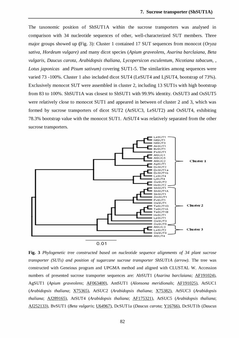
7. Sucrose transporter (ShSUT1A)
82
The taxonomic position of ShSUT1A within the sucrose transporters was analysed in
comparison with 34 nucleotide sequences of other, well-characterized SUT members. Three
major groups showed up (Fig. 3): Cluster 1 contained 17 SUT sequences from monocot (Oryza
sativa, Hordeum vulgare) and many dicot species (Apium graveolens, Asarina barclaiana, Beta
vulgaris, Daucus carota, Arabidopsis thaliana, Lycopersicon esculentum, Nicotiana tabacum, ,
Lotus japonicas and Pisum sativum) covering SUT1-5. The similarities among sequences were
varied 73 -100%. Cluster 1 also included dicot SUT4 (LeSUT4 and LjSUT4, bootstrap of 73%).
Exclusively monocot SUT were assembled in cluster 2, including 13 SUT1s with high bootstrap
from 83 to 100%. ShSUT1A was closest to ShSUT1 with 99.9% identity. OsSUT3 and OsSUT5
were relatively close to monocot SUT1 and appeared in between of cluster 2 and 3, which was
formed by sucrose transporters of dicot SUT2 (AtSUC3, LeSUT2) and OsSUT4, exhibiting
78.3% bootstrap value with the monocot SUT1. AtSUT4 was relatively separated from the other
sucrose transporters.
Fig. 3 Phylogenetic tree constructed based on nucleotide sequence alignments of 34 plant sucrose
transporter (SUTs) and position of sugarcane sucrose transporter ShSUT1A (arrow). The tree was
constructed with Geneious program and UPGMA method and aligned with CLUSTAL W. Accession
numbers of presented sucrose transporter sequences are: AbSUT1 (Asarina barclaiana; AF191024),
AgSUT1 (Apium graveolens; AF063400), AmSUT1 (Alonsona meridionals; AF191025), AtSUC1
(Arabidopsis thaliana; X75365), AtSUC2 (Arabidopsis thaliana; X75382), AtSUC3 (Arabidopsis
thaliana; AJ289165), AtSUT4 (Arabidopsis thaliana; AF175321), AtSUC5 (Arabidopsis thaliana;
AJ252133), BvSUT1 (Beta vulgaris; U64967), DcSUT1a (Daucus carota; Y16766), DcSUT1b (Daucus

7. Sucrose transporter (ShSUT1A)
83
carota; Y16767), DcSUT2 (Daucus carota; Y16768), HvSUT1 (Hordeum vulgare; AJ272309), HvSUT2
(Hordeum vulgare; AJ272308), LeSUT1 (Lycopersicon esculentum; X82275), LeSUT2 (Lycopersicon
esculentum; AF166498), LeSUT4 (Lycopersicon esculentum; AF176950), LjSUT4 (Lotus japonicas;
AJ538041), LpSUT1 (Lolium perenne; EU255258),NtSUT1 (Nicotiana tabacum; X82276), NtSUT3
(Nicotiana tabacum; AF149981), OsSUT1 (Oryza sativa; D87819), OsSUT2 (Oryza sativa; AB091672),
OsSUT3 (Oryza sativa; AB071809), OsSUT4 (Oryza sativa; AB091673), PsSUT1 (Pisum sativum;
AF109922),PvSUT1 (Panicum virgatum; FJ839440), SbSUT1 (Sorghum bicolor; XM_002467230),
ShSUT1 (Saccharum hybrid; AY780256), ShSUT1A (Saccharum hybrid; GU812864), TaSUT1A
(Triticum aestivum; AF408842), TaSUT1B (Triticum aestivum; AF408843), TaSUT1D (Triticum
aestivum; AF408844) ZmSUT1 (Zea mays; AB008464).
The alignment of deduced amino acid residues of ShSUT1 and ShSUT1A show very similar
distribution of the putative transmembrane helices, the extra sequence part of 7 amino acids at
position 203- 210 did not cause any apparent change of protein secondary structure, but the loop
between transmembrane helix 5 and 6 is a bit longer (data not shown).
Expression of sucrose transporter ShSUT1A in different tissues of sugarcane
The expression of sucrose transporter ShSUT1A was detected by RT-PCR and Northern blot in
sink leaves, source leaves and internode storage tissue of SCYLV-infected (Vinf) and virus-free
plants of cv. H87-4094 (Vf). It appears from RT-PCR that ShSUT1A is slightly higher expressed
in the leaves of the infected plants compared to virus free plants (Fig.4a). A strong expression of
ShSUT1A appears in seedling shoots, too. The hybridization of RNA with specific probe also
showed that ShSUT1A was slightly higher expressed in the leaves of the infected plant than in
the virus free plant (Fig. 4b). The transcript expression in the seedling shoot was strong in both
virus free and infected plants.

7. Sucrose transporter (ShSUT1A)
84
Fig. 4 Expression of ShSUT1A using RT-PCR (a) and Northern blot (b) in sugarcane leaves and
seedling shoost of virus free (Vf) and infected (Vinf) plants of cv. H87-4094. The lower panels in a and b
show the transcription of ribosomal RNA as control for cDNA amplification (25SrRNA, 108bp) or as
loading control, respectively. 50bp and 1kb DNA (Fermentas, St. Leon Rot, Germany) were used as
molecular size markers.
The expression of ShSUT1A in the intermodal tissues tested by RT-PCR showed a progressive
increase from internode number 1 (immature) to 7 (mature) (Fig. 5). Again, the expression of
ShSUT1A in the internodes of virus free sugarcane appeared to be less than in the infected
plants. The hybridization of RNA (Northern blot) also showed the progressively stronger
expression with ripening of the internodes, but there was no clear difference between infected
and virus-free plants (Fig. 6). The increase in transcript level in internodes was concomitant with
the increase of sucrose content in the stem, whereas the hexose level stayed relatively the same
in all internodes and was low compared to sucrose (Fig. 7).

7. Sucrose transporter (ShSUT1A)
85
Fig. 5 RT-PCR for sucrose transporter ShSUT1A in internodes (storage tissues) of SCYLV-infected or
virus-free sugarcane cv. H87-4094. Numbers identify the internodes from #1 (immature) to #7 (mature)
according their position on the stem. M: DNA molecular size markers 50bp and 1kb (Fermentas, St. Leon
Rot, Germany). The lower panel shows the transcription of ribosomal RNA (25SrRNA 108bp) as
transcription and amplification control.
Fig. 6 Northern blots of transcripts of ShSUT1A in in internodes of infected and virus-free sugarcane cv.
H87-4094. RNA was extracted from internodes #1 (beneath first dewlap leaf, immature) to #7 (mature)
and was hybridized to a probe of the ShSUT1A cDNA. The lower panel shows the same membrane
probed for ribosomal RNA as loading control.
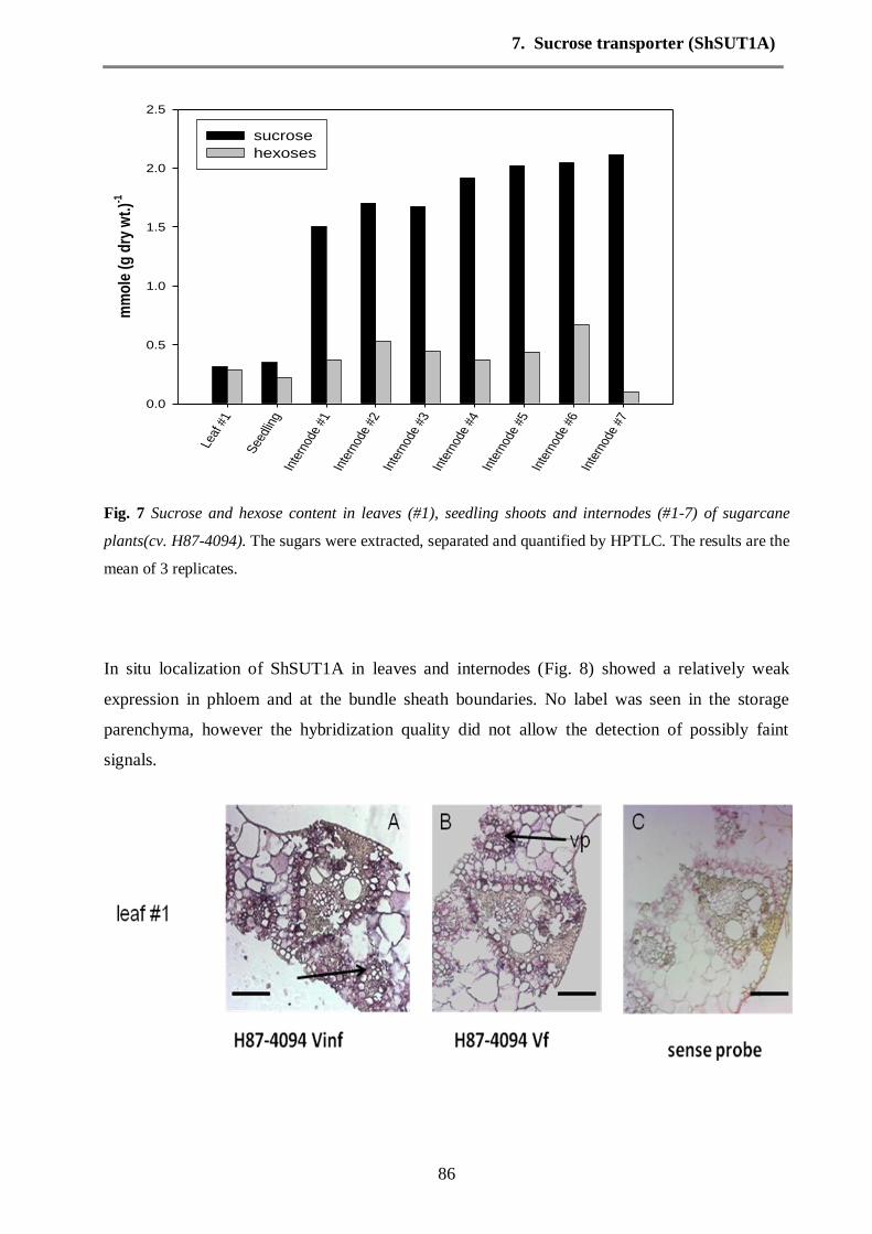
7. Sucrose transporter (ShSUT1A)
86
Fig. 7 Sucrose and hexose content in leaves (#1), seedling shoots and internodes (#1-7) of sugarcane
plants(cv. H87-4094). The sugars were extracted, separated and quantified by HPTLC. The results are the
mean of 3 replicates.
In situ localization of ShSUT1A in leaves and internodes (Fig. 8) showed a relatively weak
expression in phloem and at the bundle sheath boundaries. No label was seen in the storage
parenchyma, however the hybridization quality did not allow the detection of possibly faint
signals.
Leaf
#1
See
dlin
gIn
tern
ode
#1In
tern
ode
#2In
tern
ode
#3In
tern
ode
#4In
tern
ode
#5In
tern
ode
#6In
tern
ode
#7
mm
ole
(g
dry
wt.
)-1
0.0
0.5
1.0
1.5
2.0
2.5
sucrose
hexoses

7. Sucrose transporter (ShSUT1A)
87
Fig. 8 In situ localization of ShSUT1A in sugarcane leaf and internodes (#5 and #8) in virus free (Vf) and
infected (Vinf) plant. The sections were hybridized with antisense probe (A, B, D-E) or sense probe (C).
Faint transcript signals were present in the vascular parenchyma (vp) of leaves and in phloem (p) and
vascular bundles (vb) of internodes. (Mx: metaxylem; bar equals 100 µm)
Discussion
Since the first isolation of a sucrose transporter gene from spinach (Riesmeier et al., 1992),
sucrose transporter genes were cloned and sequenced from several species including sugarcane
(Casu et al. 2003). Three subfamilies, SUT1, SUT2 and SUT4 were clustered by phylogenetic
analysis (data not shown), whereby the SUT1 subfamily is the largest subfamily with high
similarity between their members. Despite of these similarities (83-100%), the monocot SUT1
cluster can be separated from a dicot cluster when the amino acid sequences are compared (data
not shown). We cloned and sequenced the sucrose transporter from a Hawaiian sugarcane
cultivar because there were indications that sugarcane cultivars of one region may be relatively
different from cultivars of another regions due to the species-bridging crosses made in the past
breeding efforts. The sequence obtained was indeed different from the previously published
sequence from an Australian cultivar, especially obvious in the additional stretch of 7 amino
acids after the 5th
transmembrane domain. Modelling of the molecule by standard software
(http://minnou.cchmc.org) did however not reveal a major change in the three-dimensional
structure compared to the previously published sequence (data not shown). Still, we felt justified
to give the sequence an own name, ShSUT1A, to distinguish it from ShSUT1 obtained from an
Australian sugarcane cultivar.
Transcripts of the sucrose transporter were found in all tested tissues, source leaf (leaf #1), sink
leaves of a seedling, very immature internodes (internode # 1-3), maturing internodes (internodes
#4-6) and mature internodes. The amount of transcripts seemed to increase with maturation (at

7. Sucrose transporter (ShSUT1A)
88
least up to internode #7). That is in parallel with the increase of sucrose in the internodes and in
parallel with the previously reported increase of sucrose transport into the storage parenchyma
cells at the expense of hexose transport (Komor 2000 and Riesmeier et al. 1992). However,
possibly sucrose transport and storage in maturing internodes becomes progressively symplastic
in parallel with the maturation process, in which a lignified barrier is developed around the
bundle sheath, preventing apoplastic transfer from phloem to storage parenchyma (Jacobsen et
al. 1992 and Rae et al. 2005b). In addition, the cytoplasmic membrane seems to loose its barrier
function with maturation leading to very high apoplastic sugar concentrations, possibly in
equilibrium with the cytosolic and vacuolar sucrose concentration (Hawker 1965 and Welbaum
et al. 1992). Rae et al. 2005a and b localized ShSUT1 predominantly in the bundle sheath, not in
the phloem. Our in situ studies localize ShSUT1A in the phloem of leaves and, although with
weak signal, in the phloem of internode bundles. Regrettably the hybridization was too weak to
decide whether ShSUT1A is also present in storage parenchyma. The role of sucrose transporters
in phloem loading are well documented including in retrieval of sucrose along the transport path
in the stem. The localization of ShSUT1A in phloem is therefore no exception, its role in storing
sucrose in the parenchyma is less clear. But, whatever the role of ShSUT1, there is definitely no
large difference in its transcript levels between SCYLV-infected and virus-free plants of the
cultivar, possibly there is a slightly higher transcript level in infected plants. Therefore, the
previous observations that SCYLV-infected plants seem to suffer under assimilate export
inhibition, cannot be traced back to a lower expression of sucrose transporter SUT1 in source
leaves. SCYLV-infected internodes, which definitely contain the virus in the companion cells of
the bundles, also seem to contain a higher transcript level than internodes of virus-free plants,
which at first sight would be conform with the slightly higher sucrose levels in stems of infected
plants (Lehre et al. 2007). However a higher sucrose level may also result from premature
maturation because of virus-caused inhibition apical growth, which occurs when infected plants
turn symptomatic (Lehrer and Komor 2008). Deficiencies in sucrose transporter expression
(assuming that the transcript levels mirror the protein levels) is not the cause for decreased
assimilate export in infected plants The remaining alternatives are either a mechanical plugging
of the sieve tubes of diseased plants by necrotic callose formation, as is the case in BYDV-
infected cereals (Esau, 1956), or a leak of turgor in the companion cell-sieve tube complex
because of expression of viral movement protein. Viral movement protein increases the size
exclusion limit of plasmodesmata and may thus inhibit phloem loading as was postulated for
transgenic plants expressing movement protein of potato leafroll luteovirus (Herbers et al. 1997).
However, there may be also indirect effects of viral infection on metabolism or growth such as

7. Sucrose transporter (ShSUT1A)
89
inhibiting sugar transport proteins, sugar signalling or metabolic network regulation (Hofius et
al. 2001 and Zhang et al. 2009).
Acknowledgements
The help and advice by Dr. S. Clemens and D. Peisker (University Bayreuth) is gratefully
acknowledged, also the hospitality of Dr. Thomas Laux and Steffen Knauer for help in in situ
analysis (University Freiburg). A. ElSayed is grateful to the Egyptian Government for the
stipendium. We also thank Bayreuth University for a special fund.
References
Casu, R.E., Grof, C.P., Rae A.L., McIntyre, C.L., Dimmock, C.M. , Manners, J.M. (2003)
Identification of a novel sugar transporter homologue strongly expressed in maturing stem
vascular tissues of sugarcane by expressed sequence tag and microarray analysis. Plant
molecular biology 52, 371-386.
Esau, K. (1956) An anatomist's view of virus diseases. American Journal of Botany, 43, 739-
748.
Hawker, JS. (1965) The sugar content of cell walls and intercellular spaces in sugarcane stems
and its regulation to sugar transport. Austr. J Biol Sci 18, 959-969.
Herbers, K., Tacke, E., Hazirezaei, M., Krause, K.P., Melzer, M., Rohde, W. and
Sonnewald, U. (1997) Expression of a luteoviral movement protein in transgenic plants leads
to carbohydrate accumulation and reduced photosynthetic capacity in source leaves. Plant
journal, 12 1045-1056.
Hofius, D., Herbers, K., Melzer, M., Omid, A., Tacke, E., Wolf, S. And Sonnewald, U.
(2001) Evidence for expression level dependent modulation of carbohydrate status and viral
resistance by the potato leafroll virus movement protein in transgenic tobacco plants. Plant
Journal, 28, 529-543.
Jacobsen, KR., Fisher, DG., Maretzki, A., Moore, PH. (1992) Developmental changes in the
anatomy of the sugarcane stem in relation to phloem unloading and sucrose storage. Bot. Acta
105, 70-80.
Komor, E. (2000) The physiology of sucrose storage in sugarcane. In AK Gupta, N Kaur eds.
Carbohydrate reserves in plants-synthesis and regulation. Elsevier, Amsterdam, 35-54.
Komor, E., Zingsheim, O., Sprügel, H. (1996) Cycles of sugar transport and sucrose
metabolism in sugarcane tissue: Quantitative determination. In: Sugarcane: Research towards
7. Sucrose transporter (ShSUT1A)
90
efficient and sustainable production. Eds. JR Wilson, DH Hogarth, JA Campbell, AL Garride.
CSIRO Brisbane, pp 89-91.
Lehrer, AT., Komor, E. (2008) Symptom expression of yellow leaf disease in sugarcane
cultivars with different degrees of infection by Sugarcane yellow leaf virus. Plant Pathology 57,
178-189.
Lehrer, AT., Moore, PH., Komor, E. (2007) Impact of Sugarcane Yellow Leaf Virus (ScYLV)
on the carbohydrate status of sugarcane: Comparison of virus-free plants with symptomatic and
asymptomatic virus-infected plants. Physiol. Molec. Plant Pathol 70, 180-188 and 71, 260.
Moore, P.H. (1995) Temporal and spatial regulation of sucrose accumulation in the sugarcane
stem. Australian Journal of plant Physiology 22, 661-679.
Rae, A.L., Grof, C.P., Casu, R. E., Bonnett, G. D. (2005b) Sucrose accumulation in the
sugarcane stem: pathways and control points for transport and compartmentation. Field Crops
Research 92, 159-168.
Rae, A.L., Perroux, J.M., Grof, C.P. (2005a) Sucrose partitioning between vascular bundles
and storage parenchyma in the sugarcane stem: a potential role for the ShSUT1 sucrose
transporter. Planta 220, 817-825.
Riesmeier, JW., Willmitzer, L., Frommer, W. (1992) Isolation and characterization of a
sucrose carrier cDNA from spinach by functional expression in yeast. EMBO J 11, 4705-4713.
Robinson-Beers, E., Evert, R.F. (1991) Ultrastructure of and plasmodesmatal frequency in
mature leaves of sugarcane. Planta 184, 291-306.
Sambrook, J. and Russell, D.W. (2001) Molecular cloning: a laboratory manual, 3rd
edition.
Cold Spring Harbor Laboratory Press, Cold Spring Harbor.
Seo, S., Okamoto, M., Iwai, T., Iwano, M., Fukui, K., Isogal A., Nagajima, N. and Ohashi,
Y. (2000) Reduced levels of chloroplast FtsH protein in tobacco mosaic virus-infected tobacco
leaves accelerate the hypersensitive reaction. Plant Cell, 12, 917-932.
Thompson, J.D., Higgins D.,G, Gibson T.J. (1994) CLUSTAL W: improving the sensitivity
of progressive multiple sequence alignment through sequence weighting, position-specific gap
penalties and weight matrix choice. Nucleic Acids Res 22, 4673-4680.
van Kooten, O., Meurs, C. And van Loon, L.C. (1990) Photosynthetic electron transport in
tobacco leaves infected with tobacco mosaic virus. Physiologia Plantarum, 80, 446-452.
Welbaum, GE., Meinzer, FC. (1990) Compartmentation of solutes and water in developing
sugarcane stalk tissue. Plant Physiol 93, 1147-1153.
7. Sucrose transporter (ShSUT1A)
91
Welbaum, GE., Meinzer, FC., Grayson, RL. and Thornham, KT. (1992) Evidence for the
Consequences of a Barrier to Solute Diffusion Between the Apoplast and Vascular Bundles in
Sugarcane Stalk Tissue. Australian Journal of Plant Physiology 19, 611 – 623.
Yan, S-L., Lehrer, AT., Hajirezaei, MR., Springer, A., Komor, E. (2009) Modulation of
carbohydrate metabolism and chloroplast structure in sugarcane leaves which were infected by
Sugarcane Yellow Leaf Virus (SCYLV). Physiological and Molecular Plant Pathology, 73,78-
87
Zhang ,Y., Primavesi, LF., Ihurreea, D., Andralojc, PJ., Mitchell, RAC., Powers, SJ.,
Schluepmann, H., Delatte, T., Wingler, A., Paul, MJ. (2009) Inhibition of SNF1-Related
protein kinase activity and regulation of metabolic pathways by trehalose-6-phosphate. Plant
Physiol 149, 1860-1871.
8. Quantitative multiplexed gene expression
92
8. Simultaneous quantitative analysis of transcripts for Sugarcane yellow leaf virus, sucrose
transporters and sucrose phosphate synthase in Hawaiian sugarcane cultivars by multiplex
RT-PCR
Abdelaleim Ismail ElSayed1, Alfons Weig
2 and Ewald Komor
1*
1Plant Physiology Department, University Bayreuth, D-95440 Bayreuth, Germany
2 DNA Analytics and Ecoinformatics, University Bayreuth, D-95440 Bayreuth, Germany
Keywords: GeXPS multiplex PCR, sucrose phosphate synthase II, sucrose transporters,
Sugarcane yellow leaf virus.
Submitted to Plant Pathology
Abstract: The transcript levels of the open reading frames (ORFs) 0-5 of the Sugarcane yellow
leaf virus (SCYLV) genome were determined in sink leaves, source leaves and mature
internodes of four Hawaiian sugarcane cultivars by GeXP multiplex RT-PCR. The cultivars had
been classified previously as SCYLV-susceptible or SCYLV-resistant, a virus-free line was used
as control. The transcript levels were normalized to 25S rRNA transcript levels. The transcripts
of the ORFs were present in very different quantities in the tested tissues and cultivars. ORF0-1
coding for silencing/suppressor protein had the highest transcript level, ORF3-4 coding for the
capsid protein the lowest. The ratio of ORF-transcripts was not constant, neither between tissue
types nor between cultivars. Therefore a forged RNA-amplification by the method appeared to
be an unlikely reason for the different amplificate quantities. The transcript levels of all ORFs
were higher in sink leaves than source leaves and in these higher than in mature internodes.
Quantitative multiplex RT-PCR was used to determine the transcript levels of genes which are
important for sucrose storage in sugarcane, namely sucrose phosphate synthase (SPS), sucrose
transporter SUT1 and sucrose transporter SUT4. Transcript levels for SPS and of SUT4 were
constant throughout the tested tissues and cultivars, SUT1 was highest in sink leaves and lowest
in internodes. Consequently SUT4 transcripts appeared to be increasingly important in the
sucrose storage process. No consistent differences between the susceptible and the resistant
cultivars and no differences between SCYLV-infected and virus-free plants were observed with
respect to SPS and sucrose transporter transcripts.

8. Quantitative multiplexed gene expression
93
Introduction
The sugarcane disease Yellow leaf was first noticed in Hawaiian plantations (Schenck, 1990)
and shortly later in many sugarcane regions of the world (summarized in Komor et al. 2010).
The search for the causal agent pointed to a RNA virus (Borth et al. 1994) which was then
identified as a luteovirus and named Sugarcane yellow leaf virus (SCYLV) (Vega et al. 1997,
Maia et al. 2000). The sequence revealed that it had obviously evolved as a recombination
product of Barley yellow dwarf virus, Potato leaf roll virus and Pea enation virus (Moonan et al.
2000 and Smith et al. 2000) and it was then classified as a member of the polerovirus group. The
RNA contains six open reading frames (ORFs) which were assigned to putative functions.
Analysis of SCYLV from North, Central and South American sources by comparison of partial
sequences and RFLP uncovered intraspecific, regional variation and genotype diversity (Moonan
and Mirkov, 2002). A worldwide survey and analysis of SCYLV strains (Abu Ahmad et al. 2006
finally identified 3 SCYLV genotypes, one typical for La Réunion (REU), one for South
America (BRA-PER), and one for Cuba (CUB). Hawaiian isolates were not among this study.
The genotypes were differently infectuous and virulent in their ability to evoke disease
symptoms (Abu Ahmad et al. 2007), for example the BRA strain caused much stronger
symptoms than the REU strain. A survey of Hawaiian sugarcane cultivars for SCYLV by tissue
blot immunoassay had shown that the majority of commercial cultivars contained the virus,
however a definite number of cultivars appeared SCYLV-free (Schenck and Lehrer, 2000). Since
the plantations and the breeding station is heavily infested by viruliferous aphids, which would
distribute the viral infection to all plants within short time, it was assumed that the SCYLV-free
cultivars were SCYLV-resistant in contrast to the other, the susceptible cultivars. Recent analysis
with quantitative real time RT-PCR detected SCYLV in the resistant cultivars, too, however at a
10-100 fold lower virus titre, which was apparently below the sensitivity threshold of the
immunological assay and therefore not detected in the previous screenings (Zhu et al. 2010).
This quantitative analysis had used a sequence part from ORF3-4, the so-called YLS-segment
which had been proposed to be a “diagnostic” part of the SCYLV-genome (Comstock et al.
1998). SCYLV from two susceptible and one resistant Hawaiian cultivar were isolated and
sequenced (ElSayed et al. submitted). The nucleotide sequence and the deduced amino acid
sequence classified these Hawaiian isolates as a separate genotype, tentatively called HAW-PER,
next to the BRA genotype. The sequences of SCYLV from the two susceptible cultivars were
unique in the sense that they lacked a 48-54 nt stretch in ORF1 which was present in SCYLV
from the resistant cultivar. ORF1 putatively codes for a suppressor/RNA-dependent RNA
polymerase. The sequence parts which were amplified to yield the whole viral genome were

8. Quantitative multiplexed gene expression
94
obtained in very different quantities, however, and in some cases no amplificates were obtained
at all. Thus the complete SCYLV sequences from several Hawaiian cultivars could not be
analysed in a first attempt. This failure was blamed on possibly small sequence variations in
some isolates which might have weakened the binding efficiencies of the primers used. We
wanted to know, if the variation in getting amplificates of certain viral genome parts were
because of inaccuracy of primers or a due property of the viral RNA. Therefore different viral
genome parts out of Hawaiian sugarcane cultivars from which SCYLV was successfully
sequenced were quantitatively determined in a multiplex RT-PCR with primers designed for the
appropriate sequence parts. In the past all SCYLV isolates were obtained from the uppermost
fully-developed source leaf. In a current study we also isolated SCYLV from other plant parts to
see, whether the different quantities in amplificate were the same throughout the plant or
different for certain plant organs. Also interesting was, whether the virus strain with the 48-54nt
longer genome from a so-called resistant cultivar showed another quantitative pattern of
transcripts than the shorter genome strain from a susceptible cultivar. The GenomeLab GeXP
Genetic Analysis System from Beckman Coulter offers multiplexed, quantitative gene
expression analysis capable of examining up to 30 genes in a single reaction from as little as 5 ng
total RNA. In this study we determined four different genes of the SCYLV genome related to
specific open reading frames (ORF0-1, ORF2, ORF3-4 and ORF5) from the Hawaiian
Sugarcane yellow leaf virus isolates.
A backup of carbohydrates, mostly starch, had been observed in source leaves of infected plants
in previous studies, which led to the conclusion that assimilate export is inhibited by SCYLV-
infection (Lehrer et al. 2008). Therefore the sucrose transporter and the SPS transcripts were
compared quantitatively by the same multiplex system to compare SCYLV-infected versus
SCYLV-free plants of the same cultivar and to compare transcripts between SCYLV-resistant
and SCYLV-susceptible cultivar.
Material and Methods
Plant material
Cultivars H73-6110, H87-4319 and H87-4094 were obtained from the Hawaii Agriculture
Research Center, Aiea, Hawaii, USA. A virus-free line of the cultivar H87-4094 was produced
by meristem tip tissue culture and was provided by Dr. A. Lehrer, Honolulu. The plants were
grown in the greenhouse at 24°C with a 12-h photoperiod and propagated 1-2 times per year
from cuttings.
8. Quantitative multiplexed gene expression
95
Isolation of RNA from plant tissues
Plant material (sink leaves, source leaves, mature internodes; #8 and #9) were ground in liquid
nitrogen to fine powder and total RNA was extracted using the MagMax 96 total RNA Isolation
Kit (Applied Biosystems, Darmstadt, Germany). About 100 mg were transferred to impact-
resistent tubes filled with ceramic beads (Precellys Ceramic Kit 1.4 mm; Peqlab Biotechnologie
GmbH, Erlangen, Germany) and overlaid with 200 µl lysis/binding solution including Plant
RNA isolation aid (Applied Biosystems, Darmstadt, Germany). Tissue samples were
homogenized in a FastPrep instrument (MP Biomedicals Europe, Illkirch, France) at a speed
setting of 6 m/sec for 40 sec. The tissue homogenate was cleared by centrifugation (1000 x g for
10 min.) and 50 µl of the clear supernatant were used to isolate genomic DNA using the
MagMax 96 total RNA Isolation Kit adapted to the KingFisher automated purification system
(Thermo Scientific, Langenselbold, Germany; for instrument settings using the MagMax 96
Total RNA Isolation Kit see the corresponding application note available at
(www.thermo.com). Total RNA was precipitated with ethanol / sodium chloride. The RNA
concentration was determined using a Nanodrop 1000 spectrophotometer (Thermo Scientific,
Langenselbold, Germany). The preparations were made in 3 repetitions, i.e. independent RNA
preparations from 3 plants of each cultivar.
GenomeLab eXpress GeXP primer design and RNA quantification
Oligonucleotide primers used for quantitative analysis of SCYLV RNA and selected sugarcane
mRNAs were designed using the GenomeLab GeXP eXpress Profiler software (Beckman-
Coulter, Krefeld, Germany), using published sequences from GeneBank (GU570004,
GU570005, GU570006, GU570007, GU570008 and GU570009), in addition sucrose
transporters genes (ShSUT1; AY780256 and ShSUT4; GQ485583) and sucrose phosphate
synthase (SPSII; EU269038) and reference genes (25S rRNA; BQ536525 and GAPDH;
CA254672) are given in Table 1. These chimeric primers consist of gene-specific sequences and
universal primer sequences at each 5’ ends.

8. Quantitative multiplexed gene expression
96
Table 1. Primer sequences and expected size of PCR products. The nucleotides in upper case are
the universal primer sequences, the nucleotides in lower case are the specific primer sequences.
Gene name Forward PCR primer
(5'-3')
Reverse PCR primer
(5'-3')
conc.
for RT
(nM)
product
size
(nt)
Sugarcane yellow leaf virus
ORF0-1 AGGTGACACTATAGAATA
atggtgcctattctgctcct
GTACGACTCACTATAGGGA
gcttggaacggcatctctta
125 173
ORF2 AGGTGACACTATAGAATA
agctcgtcattgatcgtgtg
GTACGACTCACTATAGGGA
caggaatttggggtcttcaa
500 249
ORF3-4 AGGTGACACTATAGAATA
tgctaggctcgagtctccat
GTACGACTCACTATAGGGA
caaacaacaacaggctccaa
500 193
ORF5 AGGTGACACTATAGAATA
gataatccggacccaaaggt
GTACGACTCACTATAGGGA
gtggaggagcataaatcgga
125 137
Sucrose transporters
ShSUT1 AGGTGACACTATAGAATA
tcccgttcatcctctacgac
GTACGACTCACTATAGGGA
atgcgcctactctgacacct
125 151
ShSUT4 AGGTGACACTATAGAATA
gctggacttgtatggggtgt
GTACGACTCACTATAGGGA
aacgataatgccagtcggag
500 166
Sucrose phosphate synthase
SPSII AGGTGACACTATAGAATA
taagtggccatcattgcgta
GTACGACTCACTATAGGGA
aatacaaaaccaacagcgcc
500 181
Reference genes
25S rRNA AGGTGACACTATAGAATA
cgtggcctatcgatccttta
GTACGACTCACTATAGGGA
aacctgtctcacgacggtct
62.5 263
GAPDH AGGTGACACTATAGAATA
gtggtgccaagaaggatgtt
GTACGACTCACTATAGGGA
gttgtgcagctagcattgga
125 158
Multiplex cDNA was synthesized using 50 ng total RNA and a reverse transcriptase reaction
(GenomeLab GeXP Start Kit, Beckman-Coulter) containing the gene-specific chimeric reverse
primer mix (Table 1). The reverse transcriptase reaction was performed in a thermal cycler with
the following program: 48°C for 1 min; 42°C for 60 min, 95°C for 5 min, and hold at 14°C. An
aliquot (9.3 µl) of the reverse transcriptase reaction was added to a PCR reaction mix containing
the gene-specific forward chimeric primer mix, fluorescently-labelled universal forward primer,
unlabelled universal reverse primer, MgCl2, and Thermo-Start DNA polymerase (Beckman-
Coulter) according to the GenomeLab GeXP Start Kit instructions. The reactions were
8. Quantitative multiplexed gene expression
97
transferred to a thermal cycler and run under the following program: initial polymerase
activation at 95°C for 10 minutes, followed by 35 cycles of 94°C for 30 sec, 55°C for 30 sec,
70°C for 1 min; after completion of the PCR cycles, the reactions were kept at 4°C.
PCR products were separated by capillary electrophoresis (GenomeLab GeXP Genetic Analysis
System, Beckman-Coulter) and quantified using the GenomLab eXpress Profiler software
(Beckman-Coulter).
During gene set panel development, attenuation, i.e., dilution of reverse primer concentration for
abundant RNA species (25S rRNA, ShSUT1, GAPDH, SCYLV ORF0-1 and ORF5), was
necessary as part of the panel optimization (Table 1). The goal of this attenuation was to have the
peak signal intensities of abundant RNA species within the mid-level of linearity for accurate
quantification of gene-specific PCR products. The rationale of the method is outlined in
document no. A54001AB available from Beckman-Coulter.
Statistical analysis
Paired t-tests were performed with SigmaPlot 9.0 (Systat Software Inc., Richmond,USA.
Results
Yield of SCYLV-genome fragments
Sugarcane yellow leaf virus has been isolated from Hawaiian sugarcane cultivars with the
purpose to analyse the nucleotide sequence and to reveal the phylogenetic relationship of the
Hawaiian strain(s) in the framework of the already known SCYLV strains. Sequence fragments
covering together the entire sequence were amplified using primers which bind to so far known
conserved nucleotide regions. However, some fragments from some cultivars were either not
obtained or obtained in such a small quantity that sequencing could not be performed. Those
cases in which successful amplification of all fragments was not achieved comprised
preparations from some susceptible and some resistant cultivars (Table 2). Successful
amplification of all sequence fragments was obtained from three Hawaiian cultivars, two
susceptible (H73-6110 and H87-4094) and one resistant cultivar (H87-4319). Their complete
sequence was deposited at NCBI (ElSayed et al. submitted). The question was now, whether
indeed different quantities of viral genome parts existed in the plant or whether small sequence
differences of viral strains, which prevail in certain cultivars, caused insufficient primer binding
and poor amplification.
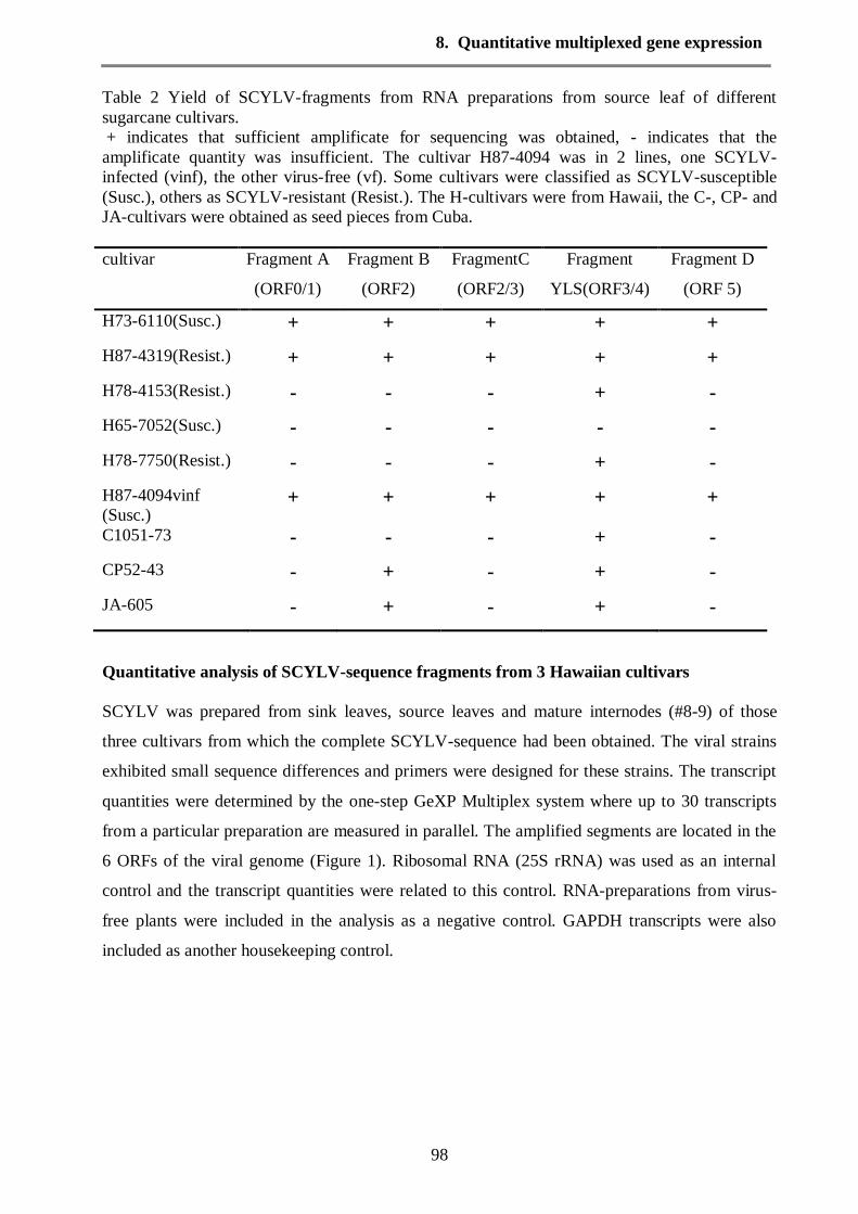
8. Quantitative multiplexed gene expression
98
Table 2 Yield of SCYLV-fragments from RNA preparations from source leaf of different
sugarcane cultivars.
+ indicates that sufficient amplificate for sequencing was obtained, - indicates that the
amplificate quantity was insufficient. The cultivar H87-4094 was in 2 lines, one SCYLV-
infected (vinf), the other virus-free (vf). Some cultivars were classified as SCYLV-susceptible
(Susc.), others as SCYLV-resistant (Resist.). The H-cultivars were from Hawaii, the C-, CP- and
JA-cultivars were obtained as seed pieces from Cuba.
cultivar Fragment A
(ORF0/1)
Fragment B
(ORF2)
FragmentC
(ORF2/3)
Fragment
YLS(ORF3/4)
Fragment D
(ORF 5)
H73-6110(Susc.) + + + + +
H87-4319(Resist.) + + + + +
H78-4153(Resist.) - - - + -
H65-7052(Susc.) - - - - -
H78-7750(Resist.) - - - + -
H87-4094vinf
(Susc.) + + + + +
C1051-73 - - - + -
CP52-43 - + - + -
JA-605 - + - + -
Quantitative analysis of SCYLV-sequence fragments from 3 Hawaiian cultivars
SCYLV was prepared from sink leaves, source leaves and mature internodes (#8-9) of those
three cultivars from which the complete SCYLV-sequence had been obtained. The viral strains
exhibited small sequence differences and primers were designed for these strains. The transcript
quantities were determined by the one-step GeXP Multiplex system where up to 30 transcripts
from a particular preparation are measured in parallel. The amplified segments are located in the
6 ORFs of the viral genome (Figure 1). Ribosomal RNA (25S rRNA) was used as an internal
control and the transcript quantities were related to this control. RNA-preparations from virus-
free plants were included in the analysis as a negative control. GAPDH transcripts were also
included as another housekeeping control.
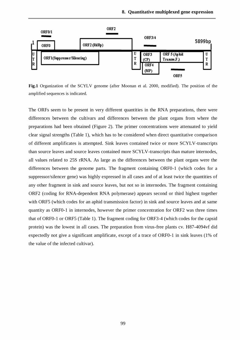
8. Quantitative multiplexed gene expression
99
Fig.1 Organization of the SCYLV genome (after Moonan et al. 2000, modified). The position of the
amplified sequences is indicated.
The ORFs seem to be present in very different quantities in the RNA preparations, there were
differences between the cultivars and differences between the plant organs from where the
preparations had been obtained (Figure 2). The primer concentrations were attenuated to yield
clear signal strengths (Table 1), which has to be considered when direct quantitative comparison
of different amplificates is attempted. Sink leaves contained twice or more SCYLV-transcripts
than source leaves and source leaves contained more SCYLV-transcripts than mature internodes,
all values related to 25S rRNA. As large as the differences between the plant organs were the
differences between the genome parts. The fragment containing ORF0-1 (which codes for a
suppressor/silencer gene) was highly expressed in all cases and of at least twice the quantities of
any other fragment in sink and source leaves, but not so in internodes. The fragment containing
ORF2 (coding for RNA-dependent RNA polymerase) appears second or third highest together
with ORF5 (which codes for an aphid transmission factor) in sink and source leaves and at same
quantity as ORF0-1 in internodes, however the primer concentration for ORF2 was three times
that of ORF0-1 or ORF5 (Table 1). The fragment coding for ORF3-4 (which codes for the capsid
protein) was the lowest in all cases. The preparation from virus-free plants cv. H87-4094vf did
expectedly not give a significant amplificate, except of a trace of ORF0-1 in sink leaves (1% of
the value of the infected cultivar).
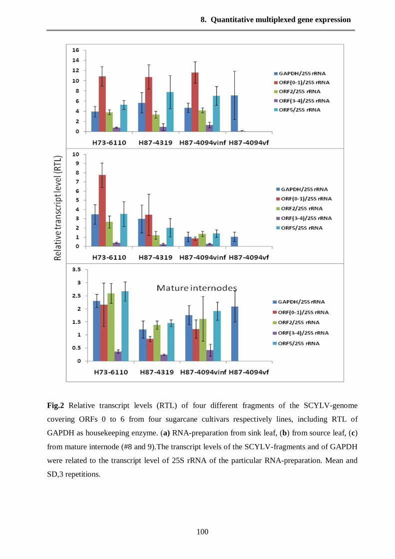
8. Quantitative multiplexed gene expression
100
Fig.2 Relative transcript levels (RTL) of four different fragments of the SCYLV-genome
covering ORFs 0 to 6 from four sugarcane cultivars respectively lines, including RTL of
GAPDH as housekeeping enzyme. (a) RNA-preparation from sink leaf, (b) from source leaf, (c)
from mature internode (#8 and 9).The transcript levels of the SCYLV-fragments and of GAPDH
were related to the transcript level of 25S rRNA of the particular RNA-preparation. Mean and
SD,3 repetitions.

8. Quantitative multiplexed gene expression
101
The transcript levels were the same in the sink leaves of the 3 infected cultivars, whereas the
transcript levels in source leaves and internodes from cv. H73-6110 were twice of those from
H87-4094 and H87-4319 (Figure 2). It was surprising that H87-4319 which had been classified
as SCYLV-resistant because of its negative response to tissue blot immunoassay (Schenck &
Lehrer, 2000) had as high transcript levels as the susceptible cultivars. The sum of the relative
transcript levels was calculated as a measure for virus titre showing the different values of viral
transcripts in sugarcane cultivars and their organs (Table 3); the values are dominated by the
transcripts ORF0-1 and ORF5 which were amplified with the same, low primer concentrations,
therefore no correction was attempted for the less amplified ORFs 2 and 3-4. The different
transcript levels for the ORFs are evident when the ratios between the amplificates were
calculated for each cultivar and each plant organ.
Table 3 Sum of ORF transcript levels as average measure of virus titre. The relative transcript
levels (RTL) of the 4 SCYLV-fragments were added up for each cultivar and plant organ. No
correction was made to account for the different primer concentrations (Table 1).
SCYLV from cultivar plant organ Sum of relative transcript levels
(ORF0-5)/25S rRNA
H73-6110 (susc.) Sink leaf 20.8
Source leaf 14.3
internode 7.9
H87-4319 (resist.) Sink leaf 23
Source leaf 6.9
internode 4.1
H87-4094 (susc.) Sink leaf 24.3
Source leaf 4
internode 5.1
H87-4094 virus-free Sink leaf 0.1
Source leaf 0
internode 0
There were different ratios between the ORF-transcripts in sink leaves and internodes, and also
different ratios between some cultivars (Table 4). The largest variations were seen in the ratio of
ORF0-1 to ORF2, especially in the source leaves. Although the primers for the four sequence
parts had been designed based on the nucleotide sequences of the SCYLV-strains from the three
Hawaiian cultivars, it may be speculated that three-dimensional conformations of the different

8. Quantitative multiplexed gene expression
102
parts of the viral genome as well as slightly different primer binding kinetics caused a differently
efficient amplification of sequence parts in the Multiplex system. If that is the case, then the
apparent transcript levels which were obtained by this method should be at a fixed ratio from
wherever the RNA-preparation was derived from. The average relative transcript level was
calculated for each sequence fragment and then the actual relative transcript level for each
cultivar was drawn (Figure 3). If there is a constant ratio of fragment transcript levels then the
graph should be parallel to the x-axis, i. e. a fixed proportion of expressions of the ORFs. This is
indeed the case for the preparations from sink leaves and from internodes, however there is
significant deviation from a fixed transcript ratio in source leaves (Figure 3), i. e. there is far
above average expression of ORF0-1 in H73-6110 and far below average expression of ORF0-1
and ORF5 in H87-4094. These differences were significant despite the low number of repetitions
(Table 5).
Fig. 3 Relative transcript levels (RTL) of the ORFs (ORF/25S rRNA) compared to the average RTLs of the ORFs in the organs of the 3 infected cultivars. The average for each RTL of the ORFs from the 3
cultivars H73-6110, H87-4319 and H87-4094 was formed for sink leaf, source leaf and internodes. Then
the actual transcript level of each cultivar in sink, source and internodes was calculated relative to this
average value. Stars indicate that the value is significantly different from the neighbouring value of the same cultivar (light star indicates a trend (P<0.1), full star (*) indicates significane (P<0.05) and double
star (**) high significance (P<0.01)).
internode
ORF0/1 ORF2 ORF3/4 ORF5
0.0
0.5
1.0
1.5
source leaf
0.0
0.5
1.0
1.5
sink leaf
tran
scrip
t lev
els
rela
tive
to a
vera
ge tr
ansc
ript l
evel
s
0.0
0.5
1.0
1.5
2.0
H73-6110
H87-4319
H87-4094
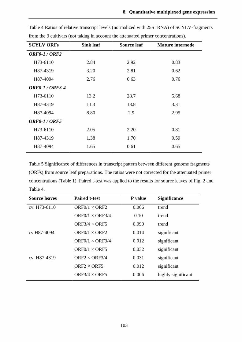
8. Quantitative multiplexed gene expression
103
Table 4 Ratios of relative transcript levels (normalized with 25S rRNA) of SCYLV-fragments
from the 3 cultivars (not taking in account the attenuated primer concentrations).
SCYLV ORFs Sink leaf Source leaf Mature internode
ORF0-1 / ORF2
H73-6110 2.84 2.92 0.83
H87-4319 3.20 2.81 0.62
H87-4094 2.76 0.63 0.76
ORF0-1 / ORF3-4
H73-6110 13.2 28.7 5.68
H87-4319 11.3 13.8 3.31
H87-4094 8.80 2.9 2.95
ORF0-1 / ORF5
H73-6110 2.05 2.20 0.81
H87-4319 1.38 1.70 0.59
H87-4094 1.65 0.61 0.65
Table 5 Significance of differences in transcript pattern between different genome fragments
(ORFs) from source leaf preparations. The ratios were not corrected for the attenuated primer
concentrations (Table 1). Paired t-test was applied to the results for source leaves of Fig. 2 and
Table 4.
Source leaves Paired t-test P value Significance
cv. H73-6110 ORF0/1 × ORF2 0.066 trend
ORF0/1 × ORF3/4 0.10 trend
ORF3/4 × ORF5 0.090 trend
cv H87-4094 ORF0/1 × ORF2 0.014 significant
ORF0/1 × ORF3/4 0.012 significant
ORF0/1 × ORF5 0.032 significant
cv. H87-4319 ORF2 × ORF3/4 0.031 significant
ORF2 × ORF5 0.012 significant
ORF3/4 × ORF5 0.006 highly significant

8. Quantitative multiplexed gene expression
104
Transcript levels of sucrose-storage related genes in sugarcane
Previous analysis had shown that SCYLV-infected plants were probably hindered in sugar
export from source leaves leading to carbohydrate backup in source leaves (Lehrer et al. 2007
and Yan et al. 2009) and stunted growth of internodes (Lehrer & Komor, 2008). The multiplex
analysis was used to quantify the transcript levels for sucrose synthesis (SPSII) and sucrose
transporters (ShSUT1 and ShSUT4) in parallel with the viral transcript levels in the same RNA
preparations. Also included was GAPDH, which had been used previously as a standard for
house-keeping transcript, because the expression levels of 25S rRNA and GAPDH were higher
and more consistent across sugarcane tissues than β-actin and β-tubulin (Iskander et al. 2004 ).
GAPDH-transcripts were not constant relative to the 25S rRNA transcripts. The values were
higher in sink leaves than in source leaves and especially in internodes. Furthermore there were
differences between cultivars, especially the values for cv. H87-4094 (both, infected and virus-
free) in source leaves were much lower than in H73-6110 and H87-4319. Thus there was no
indication that GAPDH is a better internal standard than 25S rRNA in sugarcane.
Sucrose-phosphate synthase (SPSII) transcript levels were approximately the same in sink
leaves, source leaves and internodes (Figure 4), it varied less in plant organs than GAPDH and
SCYLV-transcripts. SPSII was chosen because it is a key enzyme in sucrose synthesis in leaves
and sucrose storage in internodes (Huber and Huber, 1996; Komor et al. 1996; Zhu et al. 1997
and Casu et al. 2003). Two types of sucrose transporter were found as transcripts, one belonging
to the SUT1-group (Casu et al. 2003) and the other belonging to the SUT4-group. The latter had
not been described so far for sugarcane besides the publicly available NCBI Genbank entry, but
was clearly identified by alignment (Figure 5). Both transporter transcripts were measured.
ShSUT1 was highly transcribed in sink leaves (RTL 3-6), less so in source leaves (RTL 1-2) and
internodes (RTL 0.3-0.8) (Figure 4). ShSUT4 exhibited a constant transcript level in sink leaves,
source leaves and internodes, very much in contrast to ShSUT1, therefore the ratio of the
amplificates of ShSUT1/ShSUT4 changed strongly from above 6 to below 1 (Table 6).
There were no obvious differences in transcript quantities for sucrose transporters and SPSII in
sink leaves and source leaves between susceptible cultivars (H73-6110 and H87-4094) and the
resistant (H87-4319) or virus-free cultivar (H87-4094vf). The resistant cultivar had however a
significantly higher transcript level of ShSUT1 than the two susceptible cultivars (paired t-test:
P=0.02). The comparison of the virus-free with the infected plants of cv. H87-4094 gave the
impression that the infected plants had less GAPDH and ShSUT1 transcripts, however the
differences were insignificant (paired t-test: P=0.2-0.5).

8. Quantitative multiplexed gene expression
105
Fig. 4 Relative transcript levels (RTL) of GAPDH, SPSII, ShSUT1 and ShSUT4 from four
sugarcane cultivars respectively lines. (a) RNA-preparation from sink leaf, (b) from source leaf,
(c) from mature internode (#8 and 9). The transcript levels were related to the transcript level of
25S rRNA of the particular RNA-preparation. Mean and SD, 3 repetitions.

8. Quantitative multiplexed gene expression
106
Fig. 5a Alignment of the amino acid sequences of sugarcane sucrose transporters ShSUT1 and
ShSUT4 (in red) to published sucrose transporter sequences from monocots Asterisks indicate
perfect matches within all sequences. The sequences were aligned using CLUSTAL W.
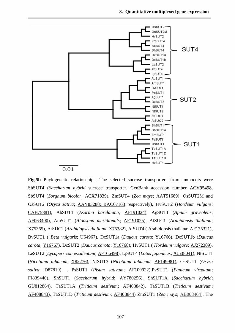
8. Quantitative multiplexed gene expression
107
Fig.5b Phylogenetic relationships. The selected sucrose transporters from monocots were
ShSUT4 (Saccharum hybrid sucrose transporter, GenBank accession number ACV95498,
SbSUT4 (Sorghum bicolor; ACX71839), ZmSUT4 (Zea mays; AAT51689), OsSUT2M and
OsSUT2 (Oryza sativa; AAY83288; BAC67163 respectively), HvSUT2 (Hordeum vulgare;
CAB75881), AbSUT1 (Asarina barclaiana; AF191024), AgSUT1 (Apium graveolens;
AF063400), AmSUT1 (Alonsona meridionals; AF191025), AtSUC1 (Arabidopsis thaliana;
X75365), AtSUC2 (Arabidopsis thaliana; X75382), AtSUT4 ( Arabidopsis thaliana; AF175321),
BvSUT1 ( Beta vulgaris; U64967), DcSUT1a (Daucus carota; Y16766), DcSUT1b (Daucus
carota; Y16767), DcSUT2 (Daucus carota; Y16768), HvSUT1 ( Hordeum vulgare; AJ272309),
LeSUT2 (Lycopersicon esculentum; AF166498), LjSUT4 (Lotus japonicas; AJ538041), NtSUT1
(Nicotiana tabacum; X82276), NtSUT3 (Nicotiana tabacum; AF149981), OsSUT1 (Oryza
sativa; D87819), , PsSUT1 (Pisum sativum; AF109922),PvSUT1 (Panicum virgatum;
FJ839440), ShSUT1 (Saccharum hybrid; AY780256), ShSUT1A (Saccharum hybrid;
GU812864), TaSUT1A (Triticum aestivum; AF408842), TaSUT1B (Triticum aestivum;
AF408843), TaSUT1D (Triticum aestivum; AF408844) ZmSUT1 (Zea mays; AB008464). The

8. Quantitative multiplexed gene expression
108
phylogenetic tree is based on the alignment of amino acid sequences. and was constructed with
Geneious program and UPGMA method, the scale bar represents the genetic distance.
Table 6 Ratio of SUT1 to SUT4 in the cultivars and plant organs. The attenuated primer
concentrations (Table 1) were not taken into account. Paired t-test was applied to the data to
show the significance of differences, t= trend with P<0.1, *= significant with P<0.05, the small
letters indicate which data pairs are significant, e. g. the data with a are not significantly different
to each other, but significantly different to data with b or c.
Cultivar Organ ShSUT1/ShSUT4 Significance
H73-6110 Sink 4.8±1.4
*a
Source 2.3±0.6
*b
Internode 0.39±0.25
*c
H87-4319 Sink 6.7±2.0
*a
Source 2.0±0.6
*b,+c
Internode 0.84±0.17
+c
H87-4094 inf Sink 6.6±3.6
+a
Source 0.93±0.52
+b
Internode 0.81±0.11 +b
H87-4094 vf Sink 6.2±0.6
*a
Source 2.3±0.7
*b,+c
Internode 1.09±0.09 +c
Discussion
The transcript levels of the viral genome were very different depending on which part of the
genome was considered. The differences are apparently not a methodological artefact of the
amplification process, since the ratio of transcripts was not constant but depended on plant organ
and plant cultivar from where the viral preparation had been obtained from. The transcript levels
(relative to 25S rRNA transcripts) of a particular gene can be quantitatively compared between
different preparations, organs or cultivars. The comparison of amplificates of different genes is

8. Quantitative multiplexed gene expression
109
complicated by the different, attenuated primer concentrations (Table 1) and possibly by
different primer binding efficiencies. The ratios of amplificates (e. g. in Tables 4-6) are still a
valid information showing whether differences are significant or not, but they may not represent
exactly the ratio of templates because of different primer concentrations. Data obtained with the
same primer concentrations (e. g. ORF0-1, ORF5 and ShSUT1) may be more likely
representative for the real ratio of templates in the RNA-preparation.
ORF0-1, which codes for a silencer/suppressor (Figure 1), was always the highest or among the
highest transcribed viral region and it is understandably of great importance for the viral
proliferation in the plant. ORF0 is expressed early during infection by Potato leaf roll virus and
was found to be responsible for symptom development (Van der Wilk et al. 1997). In addition,
the ORF0 protein may be involved in an interaction with a host specificity factor (Sadowy et al.
2001).The next ORF of SCYLV, ORF2 is less often transcribed and ORF3-4 even less. Possibly
the transcription of the viral genome by the RNA-dependent RNA-polymerase becomes less
efficient along the length of the viral RNA and falls off more frequently the longer it travels
along the viral genome. Amazingly then that transcripts of ORF5, the last ORF of the genome,
are much more frequent than transcripts of ORF3-4. Thus, either there is (or are) more initiation
sequences for the RNA-dependent RNA-polymerase, not only at the 5´-end of the genome, or
there are RNAses in the plant which selectively degrade parts of the viral transcripts. RdRp can
probably also produce transcription variation by replication slippage, which results from short
repeats. Although no direct evidence for this slippage exists, the presence of short repeats in
some viral genes may suggest it (Hancock et al. 1995). The amplification of ORF0-1 and ORF5
were achieved with one fourth of the primer concentration necessary for ORFs 2 and 3-4.
Therefore the differences observed, namely that ORF0-1 and ORF5 appear higher expressed than
ORF2 and especially ORF3-4 is expected to be even larger in the samples than it appears in
Figure 2.
The transcript levels of ORF2 varied between cultivars and plant tissues (sink, source, internodal
tissues). ORF2 codes for the RNA-dependent RNA polymerase (RdRp) of SCYLV (Figure 1)
and therefore directly determines the SCYLV-titre in the tissue. The ORF3 of SCYLV contains
also ORF4, but in a different frame (Smith et al. 2000), and thus codes for two proteins, the
capsid protein and a “genome–linked viral protein (VPg)”. This ORF3-4 has the lowest transcript
levels, which was unexpected, since it is considered as a diagnostic region for SCYLV. It indeed
exhibits the lowest diversity among the SCYLV-strains, only half of the diversity compared with
other genome parts (ElSayed et al. submitted), which may explain why it is more likely
amplified by conserved primers than other ORFs (Table 1), despite its low transcript levels.
ORF5 is produced by translational read-through of a (UAG) amber stop codon of ORF3 and
8. Quantitative multiplexed gene expression
110
codes for a putative “aphid transmission factor”. The transcript levels of ORF5 were always high
in all plant stages (Figure 2), which is obviously very important for the infection and propagation
of the virus from plant to plant. The very different transcript levels of the 6 ORFs makes it
difficult to quantify exactly the virus titre of plants or tissues, since it will depend which ORF is
selected for amplification by real-time RT-PCR. Thus the term virus titre may be ambiguous,
whether it means the titre of the best expressed ORF or the sum of expressed ORFs (Table 3).
The viral transcript levels were not only different with regard to the different ORFs but also with
regard to the plant tissue from where the RNA was prepared from. SCYLV is confined to the
phloem (Vega et al. 1997 and Yan et al. 2009) and a simple explanation would be that sink
leaves have a relatively higher phloem content than source leaves and these a higher phloem
content than internode tissues. There are no data on quantitative determination of phloem-located
RNA in sink, source and internodes, but the visual microscopic impression from leaves and
internodes of sugarcane does not support that conclusion. It appears more likely that viral
replication is more “successful” in sink leaves than in other tissues and least in mature
internodes.
The transcript levels of plant genes which are involved in sucrose storage, namely SPS and
sucrose transporters, were determined from the same RNA-preparations as the viral ORFs. SPS
had been found previously to be of decisive importance together with vacuolar acid invertase for
sucrose storage in sugarcane internodes (Zhu et al. 1997). The primers for sugarcane SPS were
designed from conserved regions after alignment of published sequences (not shown). SPS
transcripts were present at approximately the same concentration in all tissues and appear to be
less expressed than SUT1 in sink and source leaves, although the primer concentration for SPS
was fourfold of that for SUT1. The ratio of SUT1/SPS decreased in mature internodes because
they had relatively low SUT1-levels. SUT4, which had not been described so far in sugarcane, is
at constantly low transcript levels in all tissues. Because of the decreased level of SUT1 in
internodes, SUT4 becomes more important in this storage tissue (but it was amplified with
fourfold higher primer concentration than SUT1). Labelling experiments had previously shown
that the cycling of sucrose through synthesis and hydrolysis (futile cycle) decreases during
maturation of the internodes together with a stronger contribution of sucrose uptake at the
expense of hexose uptake (Komor et al. 1996). Thus sucrose synthesis appears at first glance to
proceed at similar rates in the three tissue types and at a rate smaller than sucrose transport.
However it has to be kept in mind that transcript levels may not be directly correlated with
enzyme activities, especially not in a highly regulated enzyme such as SPS (Huber and Huber,
1996).

8. Quantitative multiplexed gene expression
111
No significant differences in transcript levels were observed between the SCYLV-infected and
the virus-free line of cultivar H87-4094, except of course when the viral ORF-transcripts were
considered. The comparison of transcript levels in the two susceptible cultivars with those in the
resistant cultivar did not allow a reliable conclusion. Although the pattern of transcript levels
(GAPDH, SPS and SUTs) is different between the resistant and the susceptible cultivars in
internodes (Figure 4), the two susceptible cultivars show a strong difference between themselves
in source leaves (Figure 4). Thus obviously cultivar differences are overriding possible
differences in SCYLV-susceptibility. Reliable conclusions may be drawn only when a large
number of susceptible and resistant cultivars would be compared. The large genome of
sugarcane and the accompanied large redundancy of genes (Ming et al. 1998) may cause a large
cultivar-specific variation of gene expression, leading to virus resistance by several unrelated
paths in a regulatory network. Besides that the term SCYLV-resistance in sugarcane cultivars
has become questionable in front of the relatively high transcript levels of SCYLV-ORFs in the
“resistant” cultivar H87-4319. Possibly there are clone-specific differences or even a mixed
infection in some clones with more than one SCYLV and with variable transcription efficiencies.
When the original screenings of cultivars for SCYLV were performed using tissue blot
immunoassay (Schenck and Lehrer, 2000), 3 out of 244 tested plants tested positive for SCYLV.
The few positives were not taken serious and blamed on possible methodological mistakes. But it
may be that these positives were due to clonal variation or happened to stem from viral titre
fluctuations, which were found to be very strong in one commercial cultivar (H73-7052, Lehrer
& Komor, 2008). Future experiments need to concentrate on clonal variations of sugarcane
cultivars and mixed infections together with different virulence of SCYLV-strains in a cultivar as
possibilities to explain so far inconsistent observations.
Acknowledgements
This study was funded by the Bayreuth University. We thank Michaela Hochholzer for excellent
technical assistance. The help and advice by members of the plant physiology institute
(University Bayreuth, Germany) is gratefully acknowledged. A. Elsayed is grateful to the
Egyptian Government for the scholarship.

8. Quantitative multiplexed gene expression
112
References
Abu Ahmad, Y., Rassably, L., Royer, M., Borg, Z., Braithwaite, K S., Mirkov, T E., Irey,
MS., Perrier, X., Smith, GR. and Rott, P. (2006) Yellow leaf of sugarcane is caused by at least
three different genotypes of sugarcane yellow leaf virus, one of which predominates on the
Island of Réunion. Archive of Virology 151: 1355-1371.
Abu Ahmad, Y., Costet, L., Daugrois, J-H., Nibouche, S., Letourmy, P., Girard, J-C. And
Rott, P. (2007) Variation in infection capacity and in virulence exists between genotypes of
Sugarcane yellow leaf virus. Plant Disease 91: 253-259.
Borth, W., Hu, JS., Schenk, S. (1994) Double-stranded RNA associated with sugarcane yellow
leaf syndrome. Sugar Cane, 3, 5-8.
Casu, R.E., Grof, C.P., Rae, A.L., McIntyre, C.L., Dimmock, C.M. , Manners, J.M. (2003)
Identification of a novel sugar transporter homologue strongly expressed in maturing stem
vascular tissues of sugarcane by expressed sequence tag and microarray analysis. Plant
molecular biology 52, 371-386.
Comstock, JC., Irey, MS., Lockhart, BEL. and Wang, ZK. (1998) Incidence of yellow leaf
syndrome in CP cultivars based on polymerase chain reaction and serological techniques. Sugar
Cane 4: 21-24.
Hancock, J.M., Chaleeprom, W., Dale, J., Gibbs, A. (1995) Relplication slippage in the
evolution of potyviruses. Journal Gen. Virol., 76, 3229-3232.
Huber, SC., and Huber, JL. (1996) Role and regulation of sucrose-phosphate synthase in
higher plants. Annu. Rev. Plant Physiol. Plant Mol.Biol., 47, 431-444.
Iskander, HM., Simpson, RS., Casu, RE., Bonnett, GD., Maclean, DJ. and Manners, JM.
(2004) Comparison of reference genes for quantitative Real-Time polymerase chain reaction
anaylsis of gene expression in sugarcane. Plant Molecular Biology Reporter, 22, 325-337.
Komor, E., Zingsheim, O., Sprügel, H. (1996) Cycles of sugar transport and sucrose
metabolism in sugarcane tissue: Quantitative determination. In: Sugarcane: Research towards
efficient and sustainable production. Eds. JR Wilson, DH Hogarth, JA Campbell, AL Garride.
CSIRO Brisbane 89-91.
Komor, E., ElSayed, A., Lehrer, AT. (2010) Sugarcane yellow leaf virus introduction and
spread in Hawaiian sugarcane industry: Retrospective epidemiological study of an unnoticed,
mostly asymptomatic plant disease. Eur J Plant Pathology 127, 207-217.

8. Quantitative multiplexed gene expression
113
Lehrer, AT., Moore, PH., Komor, E. (2007) Impact of Sugarcane Yellow Leaf Virus (ScYLV)
on the carbohydrate status of sugarcane: Comparison of virus-free plants with symptomatic and
asymptomatic virus-infected plants. Physiol. Molec. Plant Pathol 70, 180-188.
Lehrer, AT., Komor, E. (2008) Symptom expression of yellow leaf disease in sugarcane
cultivars with different degrees of infection by Sugarcane yellow leaf virus. Plant Pathology 57,
178-189.
Maia, LG., Goncalves, MC., Arruda, P., Vega, J. (2000) Molecular evidence that sugarcane
yellow leaf virus (ScYLV) is a member of the Luteoviridae family. Archives of Virology, 45,
1009-1019.
Ming, R., Liu, S-C., Lin, Y-R., Braga, D., da Silva, J., van Deynze, A., Wenslaff, TF., Wu,
KK., Moore, PH., Burnquist, W., Sorrells, ME., Irvine, JE. and Paterson, AH.
(1998)
Alignment of Sorghum and Saccharum chromosomes: Comparative organization of closely-
related diploid and polyploid genomes. Genetics 150:1663-1882.
Moonan, F. and Mirkov, TE. (2002) Analyses of genotypic diversity among North, South, and
Central American isolates of Sugarcane yellow leaf virus: Evidence for Colombian origins and
for intraspecific spatial phylogenetic variation. Journal of Virology 76: 1339-48.
Moonan, F., Molina, J., Mirkov, TE. (2000) Sugarcane yellow leaf virus: an emerging virus
that has evolved by recombination between luteoviral and poleroviral ancestors. Virology, 269,
156-171.
Sadowy, E., Maasen, A., Juszczuk, M., David, C., Zagorski-Ostoja, W., Gronenborn, B.
and Hulanicka, M.D. (2001) The ORF0 product of Potato leafroll virus is indispensable for
virus accumulation. Journal of General Virology, 82, 1529-1532.
Schenck, S. (1990) Yellow leaf syndrome – a new sugarcane disease. Hawaiian Sugar Planters
Association: Annual Report, 38-39.
Schenck, S. and Lehrer, AT. (2000) Factors affecting the transmission and spread of Sugarcane
yellow leaf virus. Plant Disease 84: 1085-1088.
Smith, GR., Borg, Z., Lockhart, BEL., Braithwaite, KS., Gibbs, MJ. (2000) Sugarcane
yellow leaf virus: a novelmember of the Luteoviridae that probably arose by interspecies
recombination. Journal of General Virology 81, 1865-1869.
Van der Wilk, F., Verbeek, M., Dullemans, AM., and van den Heuvel, JF. (1997) The
genome linked protein of potato leafroll virus is located downstream of the putative protease
domain of the ORF1 product. Virology, 234, 300-303.
Vega, J., Scagliusi, SMM., Ulian, EC. (1997) Sugarcane yellow leaf disease in Brazil: evidence
of association with a luteovirus. Plant Disease, 81, 21-26.

8. Quantitative multiplexed gene expression
114
Yan, S-L., Lehrer, AT., Hajirezaei, M-R., Springer, A., Komor, E. (2009) Modulation of
carbohydrate metabolism and chloroplast structure in sugarcane leaves which were infected by
Sugarcane Yellow Leaf Virus (SCYLV). Physiological and Molecular Plant Pathology, 73. 78-
87.
Zhu, YJ., Komor, E., Moore, P. (1997) Sucrose accumulation in the sugarcane stem is
regulated by the difference between the activity of the soluble acid invertase and sucrose
phosphatesynthase. Plant Physiol. 115, 609-616.
Zhu, YJ., Lim, STS., Schenck, S., Arcinas, A., Komor, E. (2010) RT-PCR and quantitative
real-time RT-PCR detection of Sugarcane Yellow Leaf Virus (SCYLV) in symptomatic and
asymptomatic plants of Hawaiian sugarcane cultivars and the correlation of SCYLV titre to
yield, Eur J Plant Pathology 127, 263-273.

9. List of publications
115
9. List of publications:
The following papers have been published / submitted / to be submitted during the work on this
thesis:
1. Axel Lehrer, Shih-Long Yan, Blanca Fontaniella, Abdelaleim Elsayed, Ewald Komor, Journal
General Plant Pathology, 2010, 76: 62-68.“ Carbohydrate composition of sugarcane cultivars that
are resistant or susceptible to Sugarcane yellow leaf virus”.
2. Ewald Komor, Abdelaleim Elsayed, Axel Lehrer, European Journal Plant Pathology, 2010,
127, 207-217.“ Sugarcane yellow leaf virus introduction and spread in Hawaiian sugarcane
industry: Retrospective epidemiological study of an unnoticed, mostly asymptomatic plant
disease”.
3. Abdelaleim Elsayed, Alfons Weig, Ewald Komor, European Journal Plant Pathology
(submitted)“ Molecular characterization of Hawaiian Sugarcane yellow leaf virus genotypes and
their phylogenetic relationship to strains from other countries”.
4. Abdelaleim Elsayed, Mohamed F. Ramadan, Ewald Komor, Physiological and Molecular
Plant Pathology (submitted) “Expression of sucrose transporter (ShSUT1) in a Hawaiian
sugarcane cultivar infected with Sugarcane yellow leaf virus (SCYLV)”.
5. Abdelaleim Elsayed, Alfons Weig, Ewald Komor, Plant pathology (submitted)
“Simultaneous quantitative analysis of transcripts for Sugarcane yellow leaf virus, sucrose
transporters and sucrose phosphate synthase in Hawaiian sugarcane cultivars by multiplex RT-
PCR”.
6. Abdelaleim Elsayed, Ewald Komor, Archives of Virology (to be submitted) “Sequence deletion
in sugarcane yellow leaf virus genome and their effect on the diversity of virus population”.

116
ACKNOWLEDGMENTS
In the name of Allah, The Most Beneficent, the Most Merciful. I thank Allah for giving me
power to accomplish this PhD-project. I thank my family for the unconditional support and
encouragement during all the steps of the program. I thank the Egyptian Government,
Ministry of Higher Education, for sponsoring of my graduate studies abroad.
I want to express my gratitude to Prof. Dr. Ewald Komor, my supervisor, for his guidance,
patience and encouragement. I am thankful to him for the liberty and flexibility he gave me
during the time I spent in his Department. Without all these favors from him, accomplishment
of this work would have been extremely difficult. Many thanks also to Bayreuth University,
Prof. Dr. Stephen Clemens and to Prof. Dr. Ortwin Meyer, for their kind assistance to find
financial support to achieve and complete this work. I am very grateful to Dr. Alfons Weig for
guiding me in the field of molecular biology. During my academic and professional carrier I
benefited from the assistance of many people. I especially thank Dr. Sabastian Fettig, Dr.
Aleksandra Trampczynska, Dr. Katja Hartig, Dr. Michael Weber, Dr. Paul Ziegler, Mrs.
Susanne Bauer, Mr. Ulrich Deinlein, Mr. Daniel Peisker, Mr. Stefan Rensch, Mr. Tran Dang
Khoa, Mr. Daniel Eduardo Souza-Canada, Mr. Deepak Pant and technical assistances; Mrs.
Pia Schuster and Christiane Meinen for the limitless help and friendship.
I am deeply thankful to Prof.Dr. Thomas Laux, Freiburg University and Mr. Steffen Knauer
for providing the chemicals and devisors and helpful to achieve the In Situ hybridization
experiment.
I would like to thank to my friends for their support and encouragement in different ways: Dr.
Mohamed Ali, Dr. Salah Khalifa, Dr. Mohamed Abdelhamid, Prof. Dr. Kamel Shaker, Mr.
Ramzi Ben Amara, Mr. Amr Ismail, and Dr. Ilyes-Aissa Chahma.
Special thanks to my friend Dr. Mostafa Abdelkhalik for his brilliant advice and
encouragement and to Dr. Tarek Hewazi for the useful discussions and for critical reading of
my manuscript.
I am deeply thankful to Mrs. Ursula Ferrera the secretary of Department of Plant Physiology
for her kind assistance and support.
Dr. Mahmoud Dohiem, Dr. Mohamed Fawzy, Dr. Ahmed Omar and Dr. Mamdoh Kamel
taught me the fundamentals of biochemistry and plant molecular biology in Egypt. I really
appreciate their guidance and I am thankful to them for sharing their knowledge and
experience. Last but not least, special gratitude and thanks to my wife, Faten, for her
immeasurable love, constant and unconditional patience, and encouragement.

117
Declaration / Erklärung
Hereby I declare that this work has so far neither been submitted to the Faculty of Biology,
Chemistry and Earth Science at the University of Bayreuth nor to any scientific institution for
the purpose of doctorate. Furthermore, I declare that I have written this work by myself and
that I have not used any other sources, other than mentioned earlier in this work.
Hiermit erkläre ich, dass diese Arbeit von mir weder an der Fakultät für Biologie, Chemie and
Geowissenschaften der Universität Bayreuth noch einer anderen wissenschaftlichen
Einrichtung zum Zwecke der Promotion eingereicht wurde. Ferner erkläre ich, dass ich diese
Arbeit selbständig verfasst und keine anderen als die darin angegebenen Hilfsmittel benutzt
habe.
Bayreuth, den
Abdelaleim Elsayed