TECHNISCHE UNIVERSITÄT MÜNCHENmediatum.ub.tum.de/doc/1138162/1138162.pdf · TECHNISCHE...
153
TECHNISCHE UNIVERSITÄT MÜNCHEN Lehrstuhl für Biotechnologie der Nutztiere Derivation of porcine induced pluripotent stem cells and somatic cells gene targeting in pig Xinxin Cui Vollständiger Abdruck der von der Fakultät Wissenschaftszentrum Weihenstephan für Ernährung, Landnutzung und Umwelt der Technischen Universität München zur Erlangung des akademischen Grades eines Doktors der Naturwissenschaften genehmigten Dissertation. Vorsitzender: Univ.-Prof. Dr. E. Grill Prüfer der Dissertation: 1. Univ.-Prof. A. Schnieke, Ph. D. 2. Univ.-Prof. Dr. W. Windisch Die Dissertation wurde am 27. 03. 2013 bei der Technischen Universität München eingereicht und durch die Fakultät Wissenschaftszentrum Weihenstephan für Ernährung, Landnutzung und Umwelt am 10. 07. 2013 angenommen.
Transcript of TECHNISCHE UNIVERSITÄT MÜNCHENmediatum.ub.tum.de/doc/1138162/1138162.pdf · TECHNISCHE...

TECHNISCHE UNIVERSITÄT MÜNCHEN
Lehrstuhl für Biotechnologie der Nutztiere
Derivation of porcine induced pluripotent stem
cells and somatic cells gene targeting in pig
Xinxin Cui
Vollständiger Abdruck der von der Fakultät Wissenschaftszentrum Weihenstep han
für Ernährung, Landnutzung und Umwelt der Technischen Universität München zur
Erlangung des akademischen Grades eines
Doktors der Naturwissenschaften
genehmigten Dissertation.
Vorsitzender: Univ.-Prof. Dr. E. Grill
Prüfer der Dissertation: 1. Univ.-Prof. A. Schnieke, Ph. D.
2. Univ.-Prof. Dr. W. Windisch
Die Dissertation wurde am 27. 03. 2013 bei der Technischen Universität München
eingereicht und durch die Fakultät Wissenschaftszentrum Weihenstephan für
Ernährung, Landnutzung und Umwelt am 10. 07. 2013 angenommen.


Abstract
I
Abstract
Large animal models are important for medical and development researches, in
particular porcine models, due to their anatomical and physiological similarit y to
humans. However, the derivation of large animal models with precise genetic
modifications has proven difficult due to the lack of pluripotent stem cells. Because of
the failure to isolate porcine embryonic stem cells (ESCs), porcine induced
pluripotent stem cells (iPSCs) were considered as a possible alternative choice for
the generation of gene targeted pigs.
In this study, the isolation of porcine iPSCs was attempted by using several different
methods. Porcine somatic cells were transfect with combinations of the
reprogramming factors Oct4, Sox2, Klf4, cMyc, Nanog, N-Myc, and Lin28. Site-
specific recombination or episomal vectors were used to deliver the factors into cells.
Embryonic stem cell specific microRNAs were also tried to generate the iPSCs with
the help of valproic acid. Putative porcine iPSCs were tested for pluripotency. Cell
populations which differed in the expression of embryonic specific markers were
identified, separated and assessed for their differentiation potential. The results
showed that these putative iPSCs were partially reprogrammed with limited
pluripotency. They could be useful for gene targeting because of their fast and
extended proliferation. The achievements of this study provide a basis to develop
genuine porcine pluripotent stem cells.
As an alternative approach to gene targeting in pluripotent stem cells, somatic stem
cells were assessed. To improve targeting efficiency, synchronized and
unsynchronized mesenchymal stem cells (MSCs) were compared, and two different
gene loci were targeted, Adenomatous Polyposis Coli (APC) and the tumour
suppressor protein p53 (TP53). Correctly targeted cell clones were identified. APC
targeted cells were then used for somatic cell nuclear transfer (SCNT) and three
piglets were born. Comparison of targeting efficiency showed that the
synchronization did not lead to an improvement. Other synchronization methods
should be tested in the future.
Zussamenfassung
II
Zusammenfassung
Großtiermodelle sind wichtige Werkzeuge für die medizinische Forschung und
Entwicklung. Von besonderer Wichtigkeit sind hierbei aufgrund ihrer anatomischen
und physiologischen Ähnlichkeit zum Menschen Schweinemodelle. Allerdings
gestaltet sich die Gewinnung von Großtiermodellen mit genau definierten
genetischen Modifikationen wegen des Fehlens pluripotenter Stammzellen schwierig.
Da es nicht möglich ist, porcine embryonale Stammzellen (ESCs) zu isolieren,
wurden porcine induzierte pluripotente Stammzellen (iPSCs) als mögliche Alternative
für die Generierung von genmodifizierten Schweinen in Betracht gezogen.
In der vorliegenden Studie wurde die Isolierung von porcinen iPSCs mit
verschiedenen Methoden getestet. Porcine somatische Zellen wurden mit einer
Kombination der Reprogrammierungsfaktoren Oct4, Sox2, Klf4, cMyc, Nanog, Lin28
und N-Myc transfiziert. Site-spezifische Rekombination oder episomale Vektoren
wurden verwendet, um die Faktoren in die Zellen einzubringen. Weiterhin wurde
miRNAs, die spezifisch für embryonale Stammzellen sind, mit Hilfe von auf ihre
Fähigkeit zur Generierung von iPSCs getestet. Putative porcine iPSCs wurden auf
ihre Pluripotenz hin untersucht. Zellpopulation, deren Expression von spezifischen
embryonalen Markern auffällig war, wurden identifiziert, abgetrennt und ihr
Differenzierungspotential getestet. Die Ergebnisse zeigten, dass diese putative
iPSCs teilweise reprogrammiert waren und eine eingeschränkte Pluripotenz
aufwiesen. Sie könnten aufgrund ihrer schnellen und anhaltenden Prolifierung
nützlich für Gene Targeting-Experimente sein. Das in dieser Arbeit Erreichte kann als
Grundlage für die Entwicklung echter porciner iPSCs dienen.
Als Alternative zu Gene Targeting in pluripotenten Stammzellen wurden somatische
Zellen untersucht. Um die Targeting-Effizienz zu verbessern, wurden synchronisierte
und unsynchronisierte mesenchymale Stammzellen (MSCs) miteinander verglichen
und zwei verschiedene Loci getargetet, Adenomatous Polyposis Coli (APC) und das
Tumorsuppresor-Protein p53 (TP53). Korrekt getargete Zellklone wurden identifiziert
und APC-getargete Zellen anschließend für den Kerntransfer (SCNT) verwendet.
Daraus entstanden drei Ferkel. Vergleich der Targeting -Effizienzen zeigte, dass die
Synchronisierung nicht zu einer Verbesserung führte. Weitere
Synchronisierungsmethoden können in der Zukunft getestet werden.
Table of contents
III
Table of contents
1 Introduction ............................................................................................. 1
1.1 Genetically modified animal ........................................................................................ 1
1.2 Gene targeting ................................................................................................................. 2
1.2.1 Progress in gene targeting ........................................................................................... 2
1.2.2 Different cell types used for gene targeting............................................................... 4
1.3 Pluripotent stem cells.................................................................................................... 6
1.3.1 Embryonic stem cells (ESCs) ...................................................................................... 6
1.3.2 Induced pluripotent stem cells (iPSCs) ...................................................................... 6
1.4 The molecular mechanisms of pluripotency......................................................... 12
1.4.1 The transcriptional network of pluripotency and reprogramming factors ............ 12
1.4.2 The signalling pathway of pluripotency .................................................................... 14
1.4.3 MicroRNA (miRNA) and pluripotency....................................................................... 18
1.5 The developmental potential of iPSCs.................................................................... 19
1.6 Porcine pluripotent stem cells .................................................................................. 20
1.6.1 Porcine embryogenesis and ESCs ........................................................................... 20
1.6.2 Porcine iPSCs .............................................................................................................. 24
1.7 Cell synchronization and gene targeting ............................................................... 24
1.7.1 Cell synchronization .................................................................................................... 24
1.7.2 Synchronization and targeting ................................................................................... 25
1.8 Aim.................................................................................................................................... 25
2 Material and methods .......................................................................... 26
2.1 Material ............................................................................................................................ 26
2.1.1 Chemicals ..................................................................................................................... 26
2.1.2 Plastic wares and consumables ................................................................................ 27
2.1.3 Cell culture medium .................................................................................................... 28
2.1.4 Cell culture enzymes and supplements ................................................................... 28
2.1.5 Antibiotics...................................................................................................................... 29

Table of contents
IV
2.1.6 Softwares ...................................................................................................................... 30
2.1.7 Bacteria medium .......................................................................................................... 30
2.1.8 Equipments................................................................................................................... 30
2.1.9 Molecular cloning materials ....................................................................................... 32
2.1.9.1 Miscellaneous ........................................................................................................... 32
2.1.9.2 Enzymes .................................................................................................................... 33
2.1.9.3 Reverse transcriptase and polymerases .............................................................. 33
2.1.9.4 DNA isolation ............................................................................................................ 33
2.1.9.5 Plasmids .................................................................................................................... 34
2.1.10 Oligonucleotides ........................................................................................................ 35
2.1.10.1 Oligonucleotides for reprogramming vector construction ................................ 35
2.1.10.2 Oligonucleotides for microRNA............................................................................ 35
2.1.10.3 Oligonucleotides for gene expression ................................................................ 36
2.1.10.4 Oligonucleotides for gene targeting screening .................................................. 37
2.1.10.5 Oligonucleotides for amplification of the probe for southern blot ................... 37
2.1.11 Cells............................................................................................................................. 38
2.1.12 Antibodies and dilutions ........................................................................................... 39
2.2 Methods........................................................................................................................... 40
2.2.1 Microbiological methods ............................................................................................. 40
2.2.1.1 Bacteria culture......................................................................................................... 40
2.2.1.2 Storage of E. coli: ..................................................................................................... 40
2.2.1.3 Transformation of E. coli: ........................................................................................ 40
2.2.2 Molecular biological methods .................................................................................... 40
2.2.2.1 DNA Isolation ............................................................................................................ 40
2.2.2.2 DNA manipulation .................................................................................................... 42
2.2.2.3 DNA sequencing ...................................................................................................... 45
2.2.2.4 RNA isolation ............................................................................................................ 46
2.2.2.5 Assessing RNA integrity on agarose gels ............................................................ 46
2.2.2.6 Reverse transcriptase polymerase chain reaction (RT-PCR) ........................... 46
2.2.2.7 Southern blot analysis ............................................................................................. 48
2.2.3 Mammalian cell culture ............................................................................................... 49
2.2.3.1 Isolation and cultivation of porcine mesenchymal stem cells (pMSCs)........... 49
2.2.3.2 General cell culture .................................................................................................. 49
Table of contents
V
2.2.3.3 Transfection of mammalian cells (electroporation, nucleofection) ................... 51
2.2.3.4 Colony picking and cultivation of putative piPSCs .............................................. 52
2.2.3.5 Freezing and thawing of mammalian cells ........................................................... 52
2.2.3.6 Alkaline phosphatase (AP) staining ...................................................................... 53
2.2.3.7 Immunostaining of iPSCs........................................................................................ 53
2.2.3.8 Separation of cells with microbeads...................................................................... 53
2.2.3.9 Differentiation of putative porcine iPSCs .............................................................. 54
2.2.3.10 Cell synchronization .............................................................................................. 55
2.2.3.11 Selection, picking and screening of targeted colonies ..................................... 55
2.2.3.12 Preparation of cells for SCNT .............................................................................. 56
3 Results ................................................................................................... 57
3.1 Reprogramming of porcine somatic cells with different methods.................. 57
3.1.1 Constructs used for reprogramming ......................................................................... 58
3.1.1.1 Episomal vectors with mouse factors.................................................................... 58
3.1.1.2 Plasmid vectors with porcine factors..................................................................... 59
3.1.1.3 MicroRNA vector ...................................................................................................... 60
3.1.1.4 Other vectors used for the reprogramming .......................................................... 61
3.1.2 Reprogramming of porcine somatic cells and identification of putative
pluripotent stem cell ...................................................................................................... 61
3.1.2.1 Reprogramming of porcine Nanog-KFCs with factors........................................ 61
3.1.2.2 Reprogramming of porcine ADMSCs with Oct4, Klf4, Sox2, and N-Myc ........ 63
3.1.2.3 Oct4-GFP as a visible marker for reprogramming .............................................. 63
3.1.2.4 CMV-GFP as a visible marker for reprogramming.............................................. 65
3.1.2.5 Generation of piPSCs with microRNAs ................................................................ 65
3.1.2.6 Reprogramming of porcine ADMSCs with human and porcine factors ........... 67
3.1.2.7 Reprogramming of porcine ADMSCs with or without porcine CMYC .............. 67
3.1.2.8 Reprogramming of porcine BMMSCs with episomal vector .............................. 68
3.2 Assessment of pluripotency of reprogrammed cells ......................................... 69
3.2.1 Doxycycline dependency............................................................................................ 69
3.2.2 Detection of endogenous gene expressions by RT-PCR ..................................... 70
3.2.3 Karyotype ...................................................................................................................... 71
3.2.4 Immunostaining for pluripotency markers................................................................ 72
Table of contents
VI
3.2.5 Separation of the SSEA1+ and SSEA1- cells ......................................................... 73
3.2.6 Differentiation of putative piPSCs ............................................................................. 74
3.3 Cell synchronization and gene targeting ............................................................... 78
3.3.1 Gene targeting of APC and TP53 in synchronized pBMMSCs ............................ 78
3.3.2 Screening of the targeted colonies by PCR ............................................................ 78
3.3.3 Validation of targeted colonies by southern blot hybridization ............................. 80
3.3.4 Somatic cell nuclear transfer with gene-targeted clones ...................................... 82
4 Discussion............................................................................................. 83
4.1 Methods for generation of iPSCs ............................................................................. 85
4.2 Construction of reprogramming vectors ............................................................... 86
4.2.1 Reprogramming factors .............................................................................................. 86
4.2.2 The functions of cMyc and N-Myc in reprogramming ............................................ 88
4.2.3 Delivery system............................................................................................................ 89
4.2.4 The promoter and doxycycline dependent tet-on system ..................................... 91
4.2.5 MicroRNA vector.......................................................................................................... 92
4.3 Different cell types used for generation of iPSCs ............................................... 92
4.4 The medium and supplemental factors for iPSCs culture ................................ 94
4.5 MicroRNA reprogramming ......................................................................................... 95
4.6 Reprogramming of somatic cells ............................................................................. 95
4.6.1 Transfection methods ................................................................................................. 95
4.6.2 Colony screening, picking, passaging, and storage .............................................. 96
4.7 Identification of iPSCs................................................................................................. 97
4.7.1 Feeder dependence .................................................................................................... 98
4.7.2 Silencing event in pluripotent cells and doxycycline dependency ....................... 99
4.7.3 Gene expressions of iPSCs .....................................................................................101
4.7.4 The karyotype stability ..............................................................................................102
4.7.5 The specific markers of pluripotent stem cells ......................................................102
4.7.6 The differentiation ability of putative piPSCs ........................................................103
4.8 Gene targeting and synchronization .....................................................................105
4.8.1 Synchronization method ...........................................................................................106
Table of contents
VII
4.8.2 Validation of the targeted colonies..........................................................................106
4.8.3 Comparison of targeting efficiencies ......................................................................107
4.9 Outlook ..........................................................................................................................108
4.10 Concluding marks ....................................................................................................109
5 References .......................................................................................... 111
6 Abbreviation........................................................................................ 133
7 List of figures ...................................................................................... 137
8 List of tables ....................................................................................... 139
9 Appendix ............................................................................................. 140
10 Acknowledgement ........................................................................... 142
11 Curriculum Vitae............................................................................... 144

Introduction
1
1 Introduction
1.1 Genetically modified animal
Genetically modified animal, also known as transgenic animal, is an animal whose
genetic material has been altered. To do so, a number of different methods are
available. For example, a foreigner DNA sequence from different sources can be
introduced by homologous recombination into the host‘s genome (Thomas and
Capecchi, 1987).
Mouse has some good points to be used as an animal model, including defined
genetic background, easy to handle and control, high reproduction rate. As a popular
animal model, it has been used in human disease research for a long time and the
techniques are well developed and routine. Besides, the established mouse
embryonic stem cells also induced pluripotent stem cells have been used in
biomedical research. However, due to its distant genetic relationship with human and
difference in body size, the limitations of mouse model are obvious. More and more
large animals have been used in transgenic animal‘s researches (Zawada et al.,
1998; Imaizumi et al., 2000; Lai et al., 2002; Flisikowska et al., 2012). Compared to
mouse, the large animal models have several potential advantages: Pigs are more
closely related to human in genetic background and physiology, and have similar
organ size with human. It is an optimal choice to be used in human disease research
as an animal model and organ donor (Rudolph and Mohler, 1999). Up to now, pigs
are already used as models for human diabetes, arteriosclerosis, myocardial
infarction and familial adenomatous polyposis (FAP) (Turk and Laughlin, 2004;
Larsen and Rolin, 2004; Bellinger et al., 2006; Granada et al., 2009; Flisikowska et
al., 2012).
During the continuous studies in last decades, several methods were used to
generate genetically modified pigs: pronuclear microinjection of DNA, sperm
mediated gene transfer, retrolviral and lentiviral transgenesis, somatic cell nuclear
transfer (SCNT) with genetically modified cells. In 1974, scientists created the first
transgenic mouse by injecting DNA into the blastocyst (Jaenisch and Mintz, 1974).
The pronucleus of fertilized eggs was chose to be injected with DNA. The first
Introduction
2
transgenic large animals were also derived by the DNA microinjection method
(Hammer et al., 1985). Sperm mediated gene transfer was used a few years later
(Lavitrano et al., 1989). Using these two methods, many different transgenic pigs
have been reported (Hirabayashi et al., 2001; Niemann et al., 2001; Uchida et al.,
2001; Lavitrano et al., 2006; Manzini et al., 2006). But, these methods caused
random integration in most cases, and the copy number of integrated gene cannot be
controlled (Robl et al., 2007). In parallel, retroviral and lentviral transgenesis was
applied to generate transgenic animals. After successful transgenic mouse was
created with retroviral vector (Jaenisch, 1976), the transgenic mouse was also
generated by lentiviral transgenesis (Lois et al., 2002). Compared to retroviral vectors,
the lentiviral can infect non-dividing cells vectors and cannot be silenced during
embryo development (Pfeifer, 2004; Robl et al., 2007). However, the maximum DNA
capacity of the lentiviral vectors is about 10 kb and random integration of vector also
happens (Robl et al., 2007). To modify the large animals more precisely, somatic cell
nuclear transfer was established using in vitro modified cells. A sheep was
successfully cloned by somatic cell nuclear transfer (SCNT) (Wilmut et al., 1997).
Together with genetically modified cells, a transgenic sheep was generated with
SCNT (Schnieke et al., 1997). In the following several years, cloned pigs was
generated with SCNT (Polejaeva et al., 2000; Onishi et al., 2000; Betthauser et al.,
2000). Transgenic pigs were also derived by using the same method (Hyun et al.,
2003; Lee et al., 2005; Kurome et al., 2006; Brunetti et al., 2008; Cho et al., 2009;
Umeyama et al., 2009). Those achievements lead a common research approach: By
modifying cells with different methods, such as homologous recombination, a specific
fragment of genomic DNA can be added, deleted or replaced, resulting in knock-in,
knock-out or precise mutation of the genome.
1.2 Gene targeting
1.2.1 Progress in gene targeting
Gene targeting, used to change the endogenous gene, is a genetic technique in
modern scientific research. It presents a precise way to manipulate the genome.
Genes can be deleted, added, mutated or silenced by different methods, such as
traditional homologous recombination, the new more efficient zinc-finger nuclease
and transcription activator-like effector nucleases (TALENs) mediated methods
Introduction
3
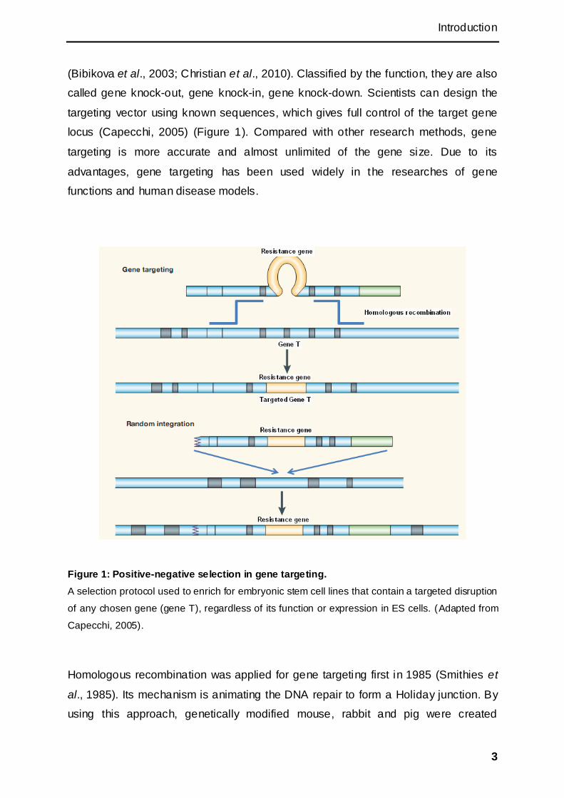
(Bibikova et al., 2003; Christian et al., 2010). Classified by the function, they are also
called gene knock-out, gene knock-in, gene knock-down. Scientists can design the
targeting vector using known sequences, which gives full control of the target gene
locus (Capecchi, 2005) (Figure 1). Compared with other research methods, gene
targeting is more accurate and almost unlimited of the gene size. Due to its
advantages, gene targeting has been used widely in the researches of gene
functions and human disease models.
Figure 1: Positive-negative selection in gene targeting.
A selection protocol used to enrich for embryonic stem cell lines that contain a targeted disruption
of any chosen gene (gene T), regardless of its function or expression in ES cells. (Adapted from
Capecchi, 2005).
Homologous recombination was applied for gene targeting first in 1985 (Smithies et
al., 1985). Its mechanism is animating the DNA repair to form a Holiday junction. By
using this approach, genetically modified mouse, rabbit and pig were created
Introduction
4
successfully (Fodor et al., 1994; Lai et al., 2002; Bosze et al., 2003). Recently,
nucleases which can bind to specific DNA sequences have been used to improve the
efficiency of gene targeting. Zinc-finger nucleases and TALENs are reported
(Bibikova et al., 2003; Christian et al., 2010). Combined with Cre-LoxP or other
System, conditional gene targeting provides accurate disease model for medicine
and development (Wirth et al., 2007).
1.2.2 Different cell types used for gene targeting
The efficiency of gene targeting depends on many things, main factors are the
targeting methods, the cell types and the properties of the DNA sequence. Targeting
of embryonic stem cells, followed by production of chimeric animals with germline
transmission, became the routine method for the mouse. The ability to clone animals
by nuclear transfer from cultured somatic cells (Campbell et al., 1996; Wilmut et al.,
1997; Polejaeva et al., 2000), offered an alternative route to germline modification
applicable to many species (Clark et al., 2000). However, up to date, the targeting
efficiency is insufficient for the method to be wildly applicable. Differences of cell
types have an influence on the gene targeting efficiency. Due to the requirement of
long time expansion, highly efficient transfection and selection, the cells capable for
gene targeting should have the ability to be passsaged more than 45 times in vitro
(Clark et al., 2000). Genetic modification and subsequent preparation for NT must be
accomplished before the cells senesce or enter crisis and transform (Denning et al.,
2001).
In previous studies, fetal fibroblasts, mesenchymal stem cells, embryonic stem cells,
induced pluripotent stem cells were used for gene targeting. In mouse, embryonic
stem cells were targeted directly and used for blastocyst injection subsequently. This
method is routinely used in mouse gene targeting research. However, since the lack
of ESC and iPSC in livestock, only primary type cells, like fetal fibroblasts and
mesenchymal stem cells (MSCs), were used for targeting with the following SCNT
(Denning and Priddle 2003; Flisikowska et al., 2012).
Fetal fibroblasts are isolated from a fetus and commonly used for gene targeting
(McCreath et al., 2000; Denning et al., 2001). Adult fibroblasts were also tried for
Introduction
5
gene targeting (Kubota et al., 2000), but their lifespan is limited to about 40
population doublings (Denning et al., 2001).
Mesenchymal stem cells are multipotent stromal cells, which can differentiate into
myoblasts, fibroblasts, osteoblasts, chondrocytes, and adipocytes. MSCs can be
isolated from bone marrow, adipose tissue, muscle, and umbilical cord. It was
considered as donor for gene targeting in large animals (Bosch et al., 2006). Cultured
with basic fibroblast growth factor, the lifespan of MSCs could increase up to more
than 70 population doublings (Bianchi et al., 2003). In addition the efficiency of
transfection can be as high as 67% in porcine MSCs (Colleoni et al., 2005).
To generate transgenic animals, embryonic stem cells (ESCs) are the best choice for
gene targeting (Suzuki et al., 2008). Because of the rapid proliferation in vitro, ESCs
provide an inexhaustible supply of cells capable of homologous recombination with a
newly introduced mutated DNA sequence (Capecchi, 1989). Pure targeted ESCs can
be injected into blastocysts directly and contribute to the germ cells in the chimeric
animal. In the next generation, pure transgenic animal inherited the modified genome
from the parents. The first successfully targeted gene in ESCs is hypoxanthine
phosphoribosyl transferase (Hprt). In this study, a specialized construct of the
neomycin resistance (neor) gene was introduced into an exon of a cloned fragment of
the Hprt gene and used to transfect mouse ESCs (mESCs) (Thomas and Capecchi,
1987).
Since the first induced pluripotent stem cells (iPSCs) were generated, they were
considered as a replacement of ESCs, though they are not considered to be fully
identical. They share the advantages with ESCs of fast proliferation, long lifespan,
and germline contribution. Hence, they brought a new possibility for targeting in those
species where ESCs isolation was failed. In human, iPSCs could be generated
directly from patient, made them potential donors for gene therapy by precise
modification through gene targeting (Ye et al., 2009).
Introduction
6
1.3 Pluripotent stem cells
1.3.1 Embryonic stem cells (ESCs)
Embryonic stem cells are a kind of pluripotent stem cells, which can differentiate into
all kinds of somatic cells under proper conditions and have the ability of self-renewal
(Rossant, 2008; Buecker et al., 2010). The fertilized eggs proliferate quickly into
morula cell cluster, then the outer layer of the cluster differentiates to trophectoderm
while the inner layer forms the inner cell mass. The ESCs were first isolated from
mouse inner cell mass of the blastocyst at day 3.5 (Evans and Kaufman, 1981). In
theory, they can be induced to differentiate into all lineages of cells which can be
used to rebuild the organism. If cultivated in conditioned medium, the cells may
proliferate forever without differentiation. These properties make them to be a
potential source for regenerative medicine and a powerful tool for developmental
research (Chen et al., 2008).
Later on, the ESCs were also isolated successfully from human, monkey and rat
(Thomson et al., 1995; Thomson et al., 1998; Ueda et al., 2008). In previous studies,
isolation from different species, such as hamster (Doetschman et al., 1988), rabbit
(Schoonjans et al., 1996), ovine (Piedrahita et al., 1990a), porcine (Evans et al.,
1990), bovine (Evans et al., 1990; Strelchenko et al., 1996), dog and cat (Hatoya et
al., 2006; Yu et al., 2008) were also attempted. Unfortunately, all of them can‘t be
verified as pluripotent stem cells entirely. Only rat, the relative of the mouse, was
another animal from which fully pluripotent stem cells were recently established (Li et
al., 2008).
1.3.2 Induced pluripotent stem cells (iPSCs)
Due to the limited source of ESCs and ethical reasons, scientists had been trying to
find another way to derive pluripotent stem cells. Three approaches for
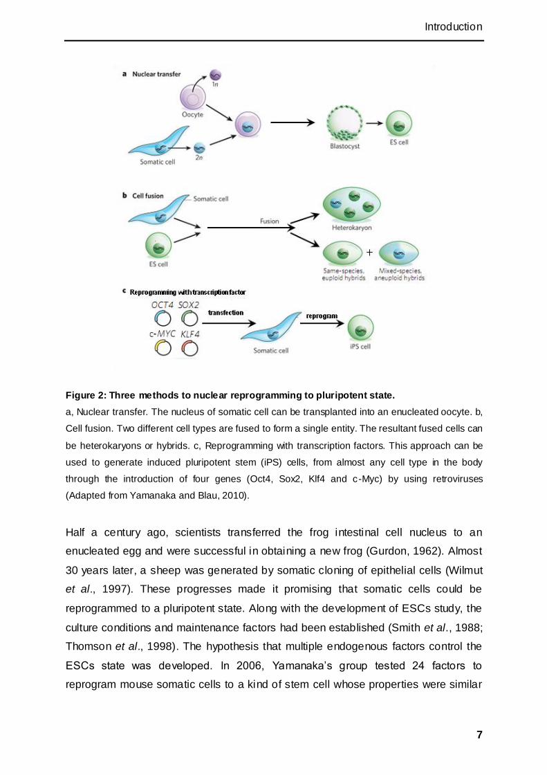
reprogramming to pluripotency were established (Figure 2).
Introduction
7
Figure 2: Three methods to nuclear reprogramming to pluripotent state.
a, Nuclear transfer. The nucleus of somatic cell can be transplanted into an enucleated oocyte. b,
Cell fusion. Two different cell types are fused to form a single entity. The resultant fused cells can
be heterokaryons or hybrids. c, Reprogramming with transcription factors. This approach can be
used to generate induced pluripotent stem (iPS) cells, from almost any cell type in the body
through the introduction of four genes (Oct4, Sox2, Klf4 and c-Myc) by using retroviruses
(Adapted from Yamanaka and Blau, 2010).
Half a century ago, scientists transferred the frog intestinal cell nucleus to an
enucleated egg and were successful in obtaining a new frog (Gurdon, 1962). Almost
30 years later, a sheep was generated by somatic cloning of epithelial cells (Wilmut
et al., 1997). These progresses made it promising that somatic cells could be
reprogrammed to a pluripotent state. Along with the development of ESCs study, the
culture conditions and maintenance factors had been established (Smith et al., 1988;
Thomson et al., 1998). The hypothesis that multiple endogenous factors control the
ESCs state was developed. In 2006, Yamanaka‘s group tested 24 factors to
reprogram mouse somatic cells to a kind of stem cell whose properties were similar

Introduction
8
to ESCs (Takahashi and Yamanaka, 2006). These cells were defined as induced
pluripotent stem cells and the 4 important reprogramming factors were: Oct4, Sox2,
Klf4 and cMyc. One year later, two different groups successfully reprogramed human
fibroblasts into iPSCs by the 4 factors respectively (Takahashi et al., 2007, Yu et al.,
2007). Soon after these reports, many achievements about iPSCs were published
(Maherali et al., 2007; Wernig et al., 2007; Lowry et al., 2008). Later on many
different types of cells, including terminally differentiated cells, were used to generate
iPSCs (Loh et al., 2009; Kim et al., 2011). But, the mechanisms of reprogramming
were not clear and the efficiency was low.
In initial iPSC researches, the reprogramming factors were delivered by retroviral or
lentiviral vectors, which may lead to insertional mutagenesis by integrating into the
genome. This side effect limits the application of iPSCs in gene therapy (Hacein-Bey-
Abina et al., 2003). Soon after new methods had been attempted to avoid the
integration problem, adenoviral vector, plasmids, RNAs, and proteins were
successfully used to generate iPSCs (Stadtfeld et al., 2008b; Okita et al. 2008; Kim
et al., 2009a; Warren et al., 2010). The episomal plasmid vector, which exists
independently from the genome and replicates during cell division, is widely used
now.
Although the iPSCs are similar to ESCs in morphology, gene expression, and
differentiation ability (Guenther et al., 2010; Hu et al., 2010; Newman et al., 2010),
they still have differences, for example in the DNA methylation pattern (Deng et al.,
2009; Doi et al., 2009). Microarray analysis also showed that a lot of genes were
differently expressed between human ESCs and iPSCs lines (Chin et al., 2009).
Furthermore, scientists found that the iPSCs had epigenetic memories from parent
cells (Kim et al., 2011; Lister et al., 2011; Ohi et al., 2011). Hence, different methods
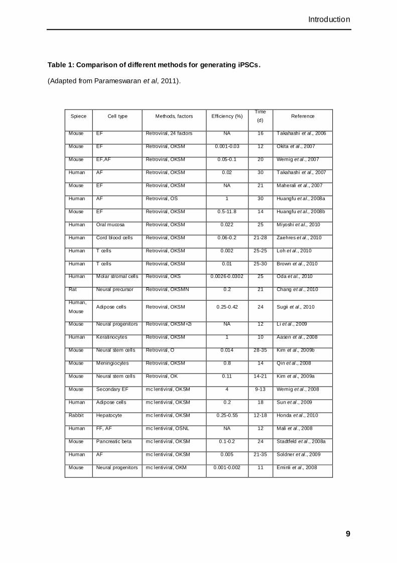
for generating iPSCs were developed in the last several years (Table1). Except the
adenovirus, all the viral delivery methods can cause permanent transgenes
integration or vector fragments in genome. Non-viral methods were developed for
transgene free iPSCs (Figure3).
Introduction
9
Table 1: Comparison of different methods for generating iPSCs.
(Adapted from Parameswaran et al, 2011).
Spiece Cell type Methods, factors Efficiency (%) Time
(d) Reference
Mouse EF Retroviral, 24 factors NA 16 Takahashi et al., 2006
Mouse EF Retroviral, OKSM 0.001-0.03 12 Okita et al ., 2007
Mouse EF,AF Retroviral, OKSM 0.05-0.1 20 Wernig et al ., 2007
Human AF Retroviral, OKSM 0.02 30 Takahashi et al., 2007
Mouse EF Retroviral, OKSM NA 21 Maherali et al ., 2007
Human AF Retroviral, OS 1 30 Huangfu et al., 2008a
Mouse EF Retroviral, OKSM 0.5-11.8 14 Huangfu et al., 2008b
Human Oral mucosa Retroviral, OKSM 0.022 25 Miyoshi et al., 2010
Human Cord blood cells Retroviral, OKSM 0.06-0.2 21-28 Zaehres et al ., 2010
Human T cells Retroviral, OKSM 0.002 25-25 Loh et al., 2010
Human T cells Retroviral, OKSM 0.01 25-30 Brown et al ., 2010
Human Molar stromal cells Retroviral, OKS 0.0026-0.0302 25 Oda et al., 2010
Rat Neural precursor Retroviral, OKSMN 0.2 21 Chang et al ., 2010
Human,
Mouse Adipose cells Retroviral, OKSM 0.25-0.42 24 Sugii et al ., 2010
Mouse Neural progenitors Retroviral, OKSM+2i NA 12 Li et al ., 2009
Human Keratinocytes Retroviral, OKSM 1 10 Aasen et al ., 2008
Mouse Neural stem cells Retroviral, O 0.014 28-35 Kim et al., 2009b
Mouse Meningiocytes Retroviral, OKSM 0.8 14 Qin et al., 2008
Mouse Neural stem cells Retroviral, OK 0.11 14-21 Kim et al., 2009a
Mouse Secondary EF mc lentiviral, OKSM 4 9-13 Wernig et al ., 2008
Human Adipose cells mc lentiviral, OKSM 0.2 18 Sun et al., 2009
Rabbit Hepatocyte mc lentiviral, OKSM 0.25-0.55 12-18 Honda et al ., 2010
Human FF, AF mc lentiviral, OSNL NA 12 Mali et al., 2008
Mouse Pancreatic beta mc lentiviral, OKSM 0.1-0.2 24 Stadtfeld et al., 2008a
Human AF mc lentiviral, OKSM 0.005 21-35 Soldner et al ., 2009
Mouse Neural progenitors mc lentiviral, OKM 0.001-0.002 11 Eminli et al ., 2008
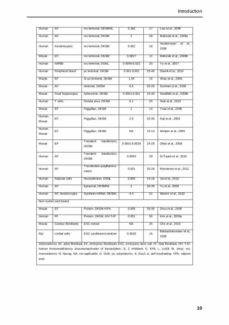
Introduction
10
Human AF mc lentiviral, OKSMNL 0.166 17 Liao et al., 2008
Human AF mc lentiviral, OKSM 2 28 Maherali et al., 2008a
Human Keratinocytes mc lentiviral, OKSM 0.002 18 Hockemeyer et al.,
2008
Mouse EF mc lentiviral, OKSM 0.0827 11 Maherali et al., 2008b
Human IMR90 mc lentiviral, OSNL 0.0095-0.022 20 Yu et al., 2007
Human Peripheral blood pc lentiviral, OKSM 0.001-0.002 25-40 Staerk et al., 2010
Mouse EF Si-pc lentiviral, OKSM 1.04 15 Shao et al ., 2009
Mouse AF lentiviral, OKSM 0.5 20-25 Sommer et al ., 2009
Mouse Fetal hepatocytes Adenoviral, OKSM 0.0001-0.001 24-30 Stadtfeld et al., 2008b
Human T cells Sendai virus, OKSM 0.1 25 Seki et al ., 2010
Mouse EF PiggyBac, OKSM 1 14 Yusa et al ., 2009
Human,
Mouse EF PiggyBac, OKSM 2.5 20-30 Kaji et al., 2009
Human,
Mouse EF PiggyBac, OKSM NA 10-14 Woltjen et al ., 2009
Mouse EF Transient transfection,
OKSM 0.0001-0.0029 24-25 Okita et al ., 2008
Human AF Transient transfection,
OKSM 0.0002 29 Si-Tayeb et al., 2010
Human AF Transfection-polyBamino
esters 0.001 20-28 Montserrat et al., 2011
Human Adipose cells Nucleofection, OSNL 0.005 14-16 Jia et al., 2010
Human AF Episomal, OKSMNL 1 30-35 Yu et al., 2009
Human AF, keratinocytes Synthetiv mRNA, OKSML 4.4 21 Warren et al ., 2010
Non-nucleic acid based
Mouse EF Protein, OKSM+VPA 0.006 30-35 Zhou et al ., 2009
Human FF Protein, OKSM, HIV-TAT 0.001 56 Kim et al., 2009a
Mouse Cardiac fibroblasts ESC extract NA 25 Cho et al., 2010
Rat Limbal cells ESC conditioned medium 0.0025 15 Balasubramanian et al.,
2009
Abbreviations: AF, adult fibroblast; EF, embryonic fibroblasts; ESC, embryonic stem cell; FF, fetal fibroblast; HIV -TAT,
human immunodeficiency virus-transactivator of transcription; 2i, 2 inhibitors; K, Klf4; L, Lin28; M, cmyc; mc,
monocistronic; N, Nanog; NA, not applicable; O, Oct4; pc, polycistronic; S, Sox2; si, self -inactivating; VPA, valproic
acid.

Introduction
11
Figure 3: Non-viral delivery methods.
A flow diagram summarizing the main non-viral delivery methods, with their advantages
described below. DNA-based delivery methods include those that do or do not involve integration
into the genome. For each of the methods, the design of the vector is shown at the top, followed
by the status of the cell after initial delivery of the vector. The coloured bars represent the
transgenes. The blue cells show the status of the vector in reprogrammed cells. The orange cells
show transgene-free cells after differentiation (Adapted from González et al., 2011).
Introduction
12
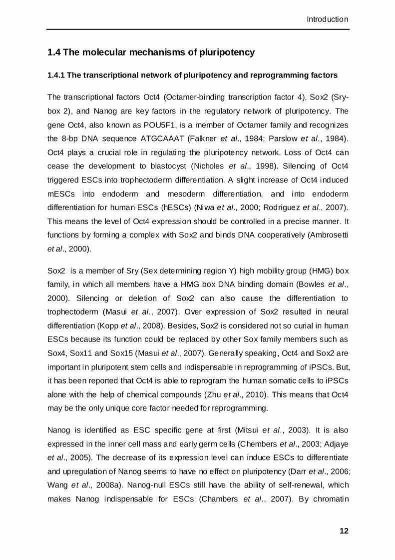
1.4 The molecular mechanisms of pluripotency
1.4.1 The transcriptional network of pluripotency and reprogramming factors
The transcriptional factors Oct4 (Octamer-binding transcription factor 4), Sox2 (Sry-
box 2), and Nanog are key factors in the regulatory network of pluripotency. The
gene Oct4, also known as POU5F1, is a member of Octamer family and recognizes
the 8-bp DNA sequence ATGCAAAT (Falkner et al., 1984; Parslow et al., 1984).
Oct4 plays a crucial role in regulating the pluripotency network. Loss of Oct4 can
cease the development to blastocyst (Nicholes et al., 1998). Silencing of Oct4
triggered ESCs into trophectoderm differentiation. A slight increase of Oct4 induced
mESCs into endoderm and mesoderm differentiation, and into endoderm
differentiation for human ESCs (hESCs) (Niwa et al., 2000; Rodriguez et al., 2007).
This means the level of Oct4 expression should be controlled in a precise manner. It
functions by forming a complex with Sox2 and binds DNA cooperatively (Ambrosetti
et al., 2000).
Sox2 is a member of Sry (Sex determining region Y) high mobility group (HMG) box
family, in which all members have a HMG box DNA binding domain (Bowles et al.,
2000). Silencing or deletion of Sox2 can also cause the differentiation to
trophectoderm (Masui et al., 2007). Over expression of Sox2 resulted in neural
differentiation (Kopp et al., 2008). Besides, Sox2 is considered not so curial in human
ESCs because its function could be replaced by other Sox family members such as
Sox4, Sox11 and Sox15 (Masui et al., 2007). Generally speaking, Oct4 and Sox2 are
important in pluripotent stem cells and indispensable in reprogramming of iPSCs. But,
it has been reported that Oct4 is able to reprogram the human somatic cells to iPSCs
alone with the help of chemical compounds (Zhu et al., 2010). This means that Oct4
may be the only unique core factor needed for reprogramming.
Nanog is identified as ESC specific gene at first (Mitsui et al., 2003). It is also
expressed in the inner cell mass and early germ cells (Chembers et al., 2003; Adjaye
et al., 2005). The decrease of its expression level can induce ESCs to differentiate
and upregulation of Nanog seems to have no effect on pluripotency (Darr et al., 2006;
Wang et al., 2008a). Nanog-null ESCs still have the ability of self-renewal, which
makes Nanog indispensable for ESCs (Chambers et al., 2007). By chromatin

Introduction
13
immunoprecipitation (ChIP), Oct4, Sox2 and Nanog were found closely on the
binding sites, which may indicate that they usually combine and bind to the target
gene together (Boyer et al., 2005; Loh et al., 2006). Later on, other factors were
found to share the binding sites with Oct4 and Sox2. These results support the Oct4 -
centric model, which includes Smad1, Stat3, and Tcf3 (Chen et al., 2008; Cole et al.,
2008; Kim et al., 2008). Through Smad1, Stat3, and Tcf3, which are involved in bone
morphogenetic protein (BMP4), leukemia inhibitory factor (LIF) and Wnt (Wingless/Int)
pathway respectively, the transcriptional network is connected with the extracellular
signals (Ng et al., 2011). Besides, the transcription factors interact and cross-regulate
with each other (Kim et al., 2010).
Kruppel-like factor 4 (Klf4) was considered dispensable for self-renewal maintenance
of ESCs (Nakatate et al., 2006). Depletion of Klf2, Klf4, and Klf5 lead ESC to
differentiation, which may indicate (Jiang et al., 2008). Klf4 is included in the core
factors for iPSCs. It interacts directly with Oct4 and Sox2 during reprogramming and
is required to active Nanog in mouse (Wei et al., 2009).
CMyc is a member of the myelocytomatosis oncogene (Myc) family which also
includes L-Myc and N-Myc (Brodeur et al., 1984). All three of them were shown to
promote cell proliferation. The Oct4 binding sites are often not near to the
transcription start site, compared to the cMyc binding site which is closer to the
transcription start site. This may indicate the Oct4-centric group works as the
enhancer. Three functionally separable modules were defined: core, polycomb and
Myc. It is assumed that the Myc module is the shared signature of embryonic stem
and cancer cells (Kim et al., 2010).
Constitutive targeted disruption of cMyc and N-Myc certified that both of them were
indispensable during embryogenesis (Stanton et al; 1992; Davis et al., 1993).
Conditionally knock-out of both cMyc and N-Myc in mouse ESCs showed that ESCs
lost the ability of self-renewal and pluripotency (Varlakhanova et al., 2010). CMyc is
not included in the core pluripotency network (Kim et al., 2010). Without cMyc, three
factors (Oct4, Sox2, and Klf4 or Nanog) also can generate iPSCs with or without
Lin28 (Nakagawa et al., 2007; Yu et al., 2007; Wernig et al., 2008). But, it is not as
efficient or fast as with cMyc. In some experiments it was even impossible to obtain
iPSCs without the participation of cMyc. A possible reason is that cMyc can repress
Introduction
14
fibroblast specific gene expression which is crucial at the beginning of the
reprogramming process (Sridharan et al., 2009).
1.4.2 The signalling pathway of pluripotency
Induced pluripotency depends on cooperation between expression of defined factors
and the culture environment. The latter also determines the pluripotent state, that is,
naïve or primed (Van Oosten et al., 2012). The extracellular factors cooperate with
the intercellular networks by different signalling pathways to determine the cell fate
(Figure 4), which makes signalling pathways play diverse, context-dependent roles in
vertebrate development.
Leukaemia inhibitory factor (LIF) can activate JAK-STAT (Janus kinase, signal
transducer and activator of transcription) signalling in mESCs and hESCs, and later
activate pluripotency genes through PI3K (phosphoinositide 3-kinase) and MAPK
(mitogen-activated protein kinase). The self renewal of mouse ESCs depends on the
LIF pathway, but this pathway does not maintain pluripotency of human ESCs. In
mouse ESCs, LIF plus serum defines the classic culture environment that enables
the infinite self-renewal of ESCs (Smith et al., 1988; Williams et al., 1988). LIF
contributes to this via the LIFRβ-GP130 signal transducer receptor complex that
activates JAK, which then phosphorylate latent transcription factor S tat3 (Niwa et al.,
1998; Matsuda et al., 1999). It also regulates Nanog activity by activating the T-box
transcription factor Tbx3 (Niwa et al., 2009). The action of LIF requires the presence
of serum, which can be replaced by BMPs (Silva and Smith, 2008).
Wnt/β-catenin signalling has been implicated in the maintenance of both mouse and
human ESCs in vitro (Sato et al., 2004). By activating Wnt signalling, β-catenin
accumulates in the cells in the nucleus, and binds to Tcf3 and other targets to
mediate the maintenance of self-renewal. Many studies showed that activating Wnt
signalling promotes self-renewal of mouse ESCs (Hao et al., 2006; Sato et al., 2004;
Miyabayashi et al., 2007; Berge et al., 2011). But some studies indicated that β-
catenin was required for multilineage differentiation and was dispensable for self-
renewal (Wagner et al., 2010, Soncin et al., 2009; Lyashenko et al., 2011). In
research of human ESCs, conflicting reports demonstrated that Wnt/β-catenin
signalling promotes either self-renewal or differentiation. Wnt3A and GSK3 inhibitor
Introduction
15
could activate Wnt signalling and maintain the self-renewal of human ESCs (Sato et
al., 2004). However, Wnt3A or GSK3 inhibitors leaded hESCs to differentiate to
primitive streak and definitive endoderm lineages (Nakanishi et al., 2009; Bone et al.,
2011). It was also reported that Wnt signalling promoted reprogramming of somatic
cells to induced pluripotent stem cells (Marson et al., 2008; Luis et al., 2008). Recent
research described a hypothesis that Wnt/β-catenin signalling was actively repressed
by Oct4 during self-renewal (Davidson et al., 2012). Whether Wnt signalling
promotes differentiation remains controversial.
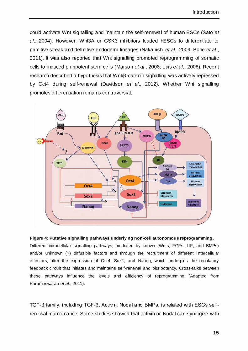
Figure 4: Putative signalling pathways underlying non-cell autonomous reprogramming.
Different intracellular signalling pathways, mediated by known (Wnts, FGFs, LIF, and BMPs)
and/or unknown (?) diffusible factors and through the recruitment of different intercellular
effectors, alter the expression of Oct4, Sox2, and Nanog, which underpins the regulatory
feedback circuit that initiates and maintains self-renewal and pluripotency. Cross-talks between
these pathways influence the levels and efficiency of reprogramming (Adapted from
Parameswaran et al., 2011).
TGF-β family, including TGF-β, Activin, Nodal and BMPs, is related with ESCs self-
renewal maintenance. Some studies showed that activin or Nodal can synergize with
Introduction
16
several other extracellular signalling proteins to promote self-renewal maintenance of
ESCs (James et al., 2005; Vallier et al., 2005; Greber et al., 2007). Activin could
cause the bFGF production when was added to hESCs in serum-free medium (Xiao
et al., 2006). This may indicate that activin does not directly maintain hESCs
undifferentiated. TGF-β can substitute for activin and/or nodal in hESCs maintenance,
and blockade of the protein kinase activity of the TGF-β receptor induces more rapid
differentiation of human ESCs than removal of exogenous TGF-β (Xu et al., 2008).
Besides, more factors were found related with the self-renewal. Shp2 promotes ES
cell differentiation, mainly through bi-directional modulation of Erk and Stat3
pathways. Deletion of Shp2 in mouse ES cells results in more efficient self -renewal
(Feng, 2007).
As described above, self-renewals of mouse ESCs and human ESCs depend on
different signalling pathways. In general, mouse ESCs work with LIF pathway
whereas human ESCs maintenance need bFGF and TGF-β/Activin/Nodal pathway
(Figure 5). The discovery of murine epiblast stem cells (EpiSCs) recently gave rise to
a new view of human ESCs (Brons et al., 2007; Tesar et al., 2007). EpiSCs are
derived from post implantation murine epiblast embryos under culture conditions
similar to hESC culture conditions. The mouse EpiSCs are similar to human ESCs.
They share not only the similar bFGF/Activin A signalling pathways, but also flattened
colony morphology, slower proliferation rate, and X-inactivation status (Buecker et al.,
2010) So some scientist suspected that the hESCs may be from the EpiSCs and they
proposed that the naïve hESCs should be like mESCs (Nichols and Smith, 2009; Hall
et al., 2009). Wnt signalling and inhibition of MEK/ERK signalling were shown to
promote the reprogramming to the naïve pluripotent state, while the FGF and Activin
signalling promote the reprogramming to a EpiSC-like state described as primed
pluripotency (Han et al., 2011). Primed and naïve pluripotent cells share some core
transcriptional regulators but are clearly distinct from each other in aspects including
epigenetic status, developmental capacity and culture requirements. It was reported
that JAK/STAT3 pathway was sufficient for reprogramming and dominant for the
establishment of naïve pluripotent state. In the presence of FGF and Activin,
JAK/STAT3 enforced naïve pluripotency in EpiSCs (van Oosten et al., 2012).

Introduction
17
Figure 5: Human and mouse embryonic stem cell (ESC) identity is sustained by mainly
distinct signalling networks.
BFGF (FGF2) is central mediators in the maintenance of undifferentiated hESCs, likely through
MEK/ERK and PI3K/Akt activation. BFGF was reported to induce the expression of hESC
maintenance factors such as TGF-β, SMAD2/3 indirectly. In contrast, LIF/Stat3 is required for
maintaining the undifferentiated state in mESCs. As long as the balance remains in favour of
Stat3, self-renewal is promoted at the expense of differentiation (MEK/ERK signalling pathway).
BMP4 can inhibit the MEK/ERK differentiation pathway resulting in mESC self-renewal. (Adapted
from Schnerch et al., 2010).
Thus, the differences between human and mouse ESCs could be a consequence of
species-specific differences in development, or it is possible that human and mouse
ESCs represent different stages of development (Pera and Tam, 2010).

Introduction
18
1.4.3 MicroRNA (miRNA) and pluripotency
MicroRNAs are post-transcriptional non-coding RNA regulators. By binding to target
mRNAs, they control the expression of downstream targets. One miRNA can
suppress hundreds of mRNAs, so they are very efficient to regulate the expressions
of cells thus the fate of cells (Subramanyam et al., 2011).
The transcripts are firstly cut by RNase Drosha and fold automatically into hairpin
structures that are cut precisely by another RNase Dicer in the next step. Then Ago2
binds to the miRNA. The mature miRNAs are about 22nt long (Figure 6). It combines
with the silencing complex and silences the mRNAs (Bartel, 2009).
Figure 6: Schematic diagram of microRNA biogenesis.
(Adapted from Lakshmipathy et al., 2010).
MiRNA can regulate the pluripotency by suppressing gene expression. Let-7 is a
miRNA expressed in differentiated cells. It suppresses several ESC specific genes
including Lin28 which suppresses Let-7 in another way (Rybak et al., 2008). Myc can
be negatively regulated by Let-7. MiR-145 suppresses the expression of Oct4, Sox2,
Introduction
19
and Klf4 in human ESCs thus causes the loss of self-renewal (Xu et al., 2009). Some
miRNAs were reported to be express specifically in ESCs, but their targets are often
unknown (Morin et al., 2008). By analyzing the expression of ESC specific miRNAs,
ESCs can be identified from the differentiated cells (Wang et al., 2008b). The
miRNAs can also be regulated by Oct4, Sox2, and Nanog, such as miR-302 and
miR-290 (Marson et al., 2008). Their promoters can be occupied by the core
pluripotency factors in mouse and human ESCs (Boyer et al., 2005; Loh et al., 2006;
Marson et al., 2008).
The miR-302-367 cluster is highly and specifically expressed in ESCs. The sequence
of this cluster in different species is highly conserved. It is a direct target of Oct4 and
Sox2. Five miRNAs are included in this cluster (miR-302a/b/c/d and miR-367) and
transcribed as a single polycistronic primary transcript (Card et al., 2008; Rosa et al.,
2009). In the presence of valproic acid, miR-302-367 cluster was reported to
reprogram the mouse fibroblasts into iPSCs (Anokye-Danso et al., 2011). It showed
that miR-367 was critical during the reprogramming. In this experiment, valproic acid
was indispensable.
1.5 The developmental potential of iPSCs
ESCs carry balanced parental imprints that are critical for normal development, so
they can contribute to the germline (Hochedlinger and Jaenisch, 2006). Although
embryonal carcinoma cells (ECCs), germline stem cells (GSCs), and embryonic
germ cells (EGCs) are pluripotent, only ESCs pass the most stringent developmental
assay: tetraploid embryo complementation (Stastfeld and Hochedlinger, 2010). To
confirm the pluripotency of pluripotent stem cells, full-scale identification methods are
necessary (Table 2).
Morphology is the first direct standard but by far not sufficient. The iPSCs share the
same assays with ESCs to evaluate their developmental potency. As iPSCs can be
generated from different cell sources by different methods, they require also some
special assays, like testing for retroviral silencing.

Introduction
20
Table 2: Assays to evaluate the developmental potential of iPSCs.
(Adapted from Stastfeld and Hochedlinger, 2010).
Assay Time Advantages Disadvantages
Molecular Morphology
AP staining
Pluripotency markers
Mintutes-1h
1-2 days
Rapid and simple
Straightforward colorimetric assay
Not specific to pluripotent cells
Retroviral silencing 1-2 days Hall mark of pluripotent state
Require retroviral
DNA demethylation 1-2 weeks Indicator of epigenetic remodeling
Somatic cells also show demethylation
Factor independency 4-7 days Indicator of fully reprogramming
Requires inducible system
Functional In vitro differentiation weeks Specific differentiation Limited cell types
Teratoma formation Weeks-months Give informations of In
vivo differentiation potential of three germ layers
Not quantitative,
Cannot detect abnormal cells
Chimeric development weeks Tests potential to contribute to normal tissues
Subtle abnormalities may be masked
Germline transmission months iPSC-derived offspring to form functional germ cells
Readout for single,
very specialized
Tetraploid complementation
weeks Measures potential to direct normal development of an entire mouse, including all cell types
Subtle development or postnatal phenotypes may be missed; does not assess the capacity of cells to form extraembryonic tissues
1.6 Porcine pluripotent stem cells
1.6.1 Porcine embryogenesis and ESCs
Mouse and human pluripotent stem cells are most commonly used in research, not
only due to the clearer genetic background compared to other species and known
genomic sequences, but also based on the established standardized research
methods and cell lines. For other species there are only few successful experiments
reported. However, the gap in ungulate is needed to be filled to help understand the
early embryo development of all mammalians. The large animal model also calls for
pluripotent stem cell for gene targeting and generation of transgenic animals. The
application of iPSCs and their transplantation needs reliable pre-clinical large animal
models.

Introduction
21
The development of porcine pluripotent stem cells is full of hardships. Two decades
ago, scientists already started to isolate ESCs from porcine inner cell mass, but the
isolated cells couldn‘t be passaged and had no pluripotency (Evans et al., 1990;
Piedrahita et al., 1990a; Piedrahita et al., 1990b; Notarianni et al., 1990; Notarianni et
al., 1991). Two groups carrying out the early work obtained some ESC-like cells from
day 7-9 blastocyst. However, no cells survived more than passage 10 (Evans et al.,
1990; Piedrahita et al., 1990a; Piedrahita et al., 1990b). Then other scientists tried to
isolate cells from, day 9-12 (Strojek et al., 1990), day 5-6 (Hochereau-de and
Perreau, 1993); day 6-10 (Anderson et al., 1994), and morula (Chen et al., 1999).
Some of the cell lines could be passaged more than 10 passages and showed ES-
like morphology. Some putative porcine ESCs, though they couldn‘t be cultivated for
a long time, could form embryoid bodies, teratomas (Hochereau-de and Perreau,
1993), and even chimera (Chen et al., 1999). Cells obtained from day 7 were SSEA1
positive (Wianny et al., 1997). Cells isolated on day 7-9 from minipigs showed mouse
ESC-like morphology and differentiated to neuron-like, smooth muscle, and
epithelium-like cells (Li et al., 2003). Oct4, Sox2, and Nanog were found in cells
isolated from day 6-8 (Blomberg et al., 2008). In recent years one group (Vassiliev et
al., 2010) isolated putative porcine ESCs from in vivo and in vitro embryos by using a
new method. They could passage some cell lines up to 14 passages. Oct4 and
Nanog were detected in these cells. The results showed that these cells could form
embryoid bodies, three germ layers and contribute to chimeric pigs.
Besides some progress with porcine ESCs, none of the isolated cells can be
maintained over longer periods in an undifferentiated state. The reasons for this are
still unclear. Special early development process of porcine embryo and the elusive
proper culture medium might be the explanation (Kuijk et al., 2008) (Figure 7).

Introduction
22
Figure 7: Early lineage segregation in mouse , human, pig, and cattle.
(Adapted from Kuijk et al., 2008; Alberio and Perez, 2012).
Introduction
23
In the early embryo development, the mouse embryo finishes the formation of early
blastocyst and forms trophectoderm and inner cell mass by day 3.5. By day 4.5, the
primitive endoderm has been formed and the inner cell mass becomes the early
epiblast and grows quite fast to create the real epiblast. Soon after this process, the
embryo differentiates forms the three germ layers . This process takes longer in
human and pig. In human, blastocyst forms by day 5, and the epiblast occurs by day
8-9. In pig, the embryo starts to hatch at days 7 to 8 and stays at epiblast stage for
longer time than mouse and human. Both human and mouse embryos start to
implant to the uterine walls invasively and part of their trophectoderms forms the
placenta. But the porcine embryo keeps the blastocyst for a longer time and
transforms to a filament before the non-invasive implantation (Enders and Carter,
2004) (Figure 7).
Given the differences of early embryogenesis, it is reasonable that the expressions of
Oct4, Sox2, and Nanog in porcine blastocyst are distinct from mouse and human
(Hall, 2008). In mouse morula stage, Oct4 and Cdx2 inhibit the expression of each
other, subsequently, the Cdx2 positive outer cells differentiate to trophectoderm
whereas the Oct4 expressing cells become the inner cell mass (Niwa et al., 2005). In
the porcine blastocyst, Oct4 was found not only in the inner cell mass but also in the
trophectoderm. In mouse, Nanog expression promotes some inner cell mass to
become epiblast and the cells which express Gata6 become primitive endoderm
(Chazaud et al., 2006). However, Nanog is difficult to detect in pig. The bovine inner
cell mass was reported to express Nanog. These previous results may indicate that
the procedure of porcine embryogenesis is neither like human nor like mouse (Keefer
et al., 2007; Blomberg et al., 2008). Recently, a study (du Puy et al., 2010) showed
some new discoveries of expression of porcine factors in the embryo. They used
whole mount in situ hybridization, qRT-PCR and whole mount immunofluorescence
to test the expression pattern of key factors at blastocyst stage by day 6.5-10.5. They
found the inner cell mass and the epiblast express Sox2 and Nanog. Oct4 was
detected and restricted in the epiblast by day 9.5. The in vitro undifferentiated
colonies expressed Oct4, Sox2, Nanog, and CK18 (Cytokeratin 18) which indicated
the cells were more like human ESCs and mouse EpiSCs than mouse ESCs
(Figure7).
Introduction
24
1.6.2 Porcine iPSCs
Because of the failure in isolating porcine ESCs from inner cell mass, other
possibilities have been tried in past. After the success of mouse and human iPSCs,
scientists also are focusing on porcine iPSCs. Several groups tried to generate
porcine iPSCs by using different methods (Esteban et al., 2009; Ezashi et al., 2009;
Wu et al., 2009; West et al., 2010). The first three reports described the generation of
porcine iPSCs from fetal fibroblasts, ear fibroblasts and primary bone marrow cells.
These porcine iPSCs expressed pluripotency markers and specific surface markers.
All of them could form teratoma and generated three germ layers and had high level
of telomerase activity. One group showed their cells shared hESC morphology and
were positive for hESC surface markers, SSEA3, SSEA4, Tra-1-60, and Tra-1-80
(Wu et al., 2009). Another group reported their iPSCs were positive for SSEA1 and
shared mouse ESCs morphology (Ezashi et al., 2009). But, the reprogrammed cells
still expressed exogenous gene and relied on them, which indicated that these cells
were not fully reprogrammed even if they had some pluripotent characterizations.
Another possibility is that the culture conditions are not sufficient for the
reprogrammed cells, and unknown supplement factors may be needed. Later on, one
group reported they generated chimeric offspring with their porcine iPSCs (West et al;
2010). They transduced porcine MSCs with human OCT4, SOX2, KLF4, cMYC,
NANOG, and LIN28 delivered by lentiviral vectors and finally got mESC like iPSCs
positive for SSEA1.
1.7 Cell synchronization and gene targeting
1.7.1 Cell synchronization
Cell synchronization is a process to halt cells at a single stage in the cell cycle. There
are several methods to synchronize cells. Based on their different mechanisms, they
can be classified as physical fractionation and chemical blockade.
Serum starvation is a commonly used method to arrest the cells at G0 Phase. Cells
need mitogen to pass the G1 phase, and later on, the mitogen is not necessary for
the cell cycle after the cells enter into the S-phase. When the cells suffer from serum
starvation, the lack of mitogen forces the cells to stay in G0 phase. Once the cells are
released from the serum starvation, they can complete the cell cycle synchronously.
Introduction
25
There are also chemical inhibitors which can block the cell cycle at different stages ,
such as thymidine, hydroxyurea, nocadozole, and colcimid (Davis et al., 2001).
1.7.2 Synchronization and targeting
Due to the finite lifespan of somatic cells, the advancements in gene targeting are
slow. The rate of homologous recombination is determined by DNA repair
mechanism and the balance between homologous recombination and non-
homologous end joining (Hanson and Sedivy, 1995).
The homologous recombination prefers to occur in late S/G2 phase. Enhancement of
gene targeting during S-phase is consistent with this phenomenon (Takata et al.,
1998). A potential explanation for this enhancement was that targeting construct
without nuclear localization signal cannot enter the nucleus and must wait when the
nuclear membrane breaks down (Mir and Piedrahita, 2004). To maximize the
efficiency of homologous recombination in targeting, it is necessary to synchronize
the cells to get most of the cells in late S/G2-phase during the transfection. Cell
synchrony by thymidine incorporation increased the ratio of homologous
recombination to non-homologous end joining 5-fold by reducing the overall rate of
non-homologous end joining (Zaunbrecher et al., 2008). Targeting efficiency
increased 7-fold by using cell synchronization and nuclear localization signals (Mir
and Piedrahita, 2004). These indicated that this approach might be useful to facilitate
targeting in somatic cells by reducing the numbers of colonies that need to be
analyzed before a targeting event could be identified.
1.8 Aim
The aim of this thesis was to isolate or derive porcine cell types which could be used
for the generation of gene targeted animals. As the derivation of porcine ESCs has
so far been unsuccessful, porcine iPSCs were hoped to provide a feasible alternative
for cell mediated transgenesis and gene targeting. Different methods for generating
porcine iPSCs should be assessed and their pluripotency as well as their ability to
produce gene targeted animals should be evaluated. At the same time an alternative
approach, gene targeting in somatic cells and derivation of cloned animals, should be
carried out. In particular it should be assessed if synchronization of somatic cells
improves gene targeting efficiency at the APC and TP53 loci.
Material and methods
26
2 Material and methods
2.1 Material
2.1.1 Chemicals
Bovine Serum Albumin (BSA) PAA, Pasching, Austria
Chloroform Sigma, Steinheim, Germany
Dimethylsulfoxid (DMSO) Sigma, Steinheim, Germany
DNA remover Minerva biolabs, Berlin, Germany
Ethanol absolute Riedel-de-Haen, Seelze, Germany
Ethidiumbromid solution Sigma, Steinheim, Germany
Ethylene diamine tetraacetic acid
(EDTA)
Sigma, Steinheim, Germany
Formalin Sigma, Steinheim, Germany
Formamide Sigma, Steinheim, Germany
GenAgarose L.E. Genaxxon Bioscience GmbH, Biberach,
Germany
Glycerol Carl Roth GmbH, Karlsruhe, Germany
Isopropanol (2-Propanol) Carl Roth GmbH, Karlsruhe, Germany
Methanol Sigma, Steinheim, Germany
Phenol:Chloroform:IsomylAlcohol
25:24:1
Sigma, Steinheim, Germany
poly-DL-ornithine Sigma, Steinheim, Germany
Rnase away Carl Roth GmbH, Karlsruhe, Germany
Sodium chloride Sigma, Steinheim, Germany
Sodium dodecyl sulfate (SDS) Sigma, Steinheim, Germany
Tris hydrochloride (Tris HCl) Sigma, Steinheim, Germany
Material and methods
27
Triton X-100 Sigma, Steinheim, Germany
Trizol Invitrogen GmbH, Darmstadt, Germany
Tween 20 Sigma, Steinheim, Germany
Valproic acid sodium salt Sigma, Steinheim, Germany
β-mecarptoethanol Sigma, Steinheim, Germany
2.1.2 Plastic wares and consumables
14ml polypropylene round bottom
tube
Becton Dickinson Company , Franklin
Lakes, USA
40 μm cell strainer BD biosciences, Heidelberg, Germany
Cell culture flasks (25, 75 and 150
cm2)
Corning Inc., New York, USA
Cell culture plates (6-, 12-, 24-, 48-,
96- well)
Corning Inc., New York, USA
Centrifugation tubes (15 and 50 ml) Corning Inc., New York, USA
Cryopreservation tube Corning Inc., New York, USA
Electroporation Cuvettes (2 and 4
mm)
Peqlab Biotechnologie GmbH, Erlangen,
Germany
Filter Stericup and Steritop (0.22 μm) Merck KGaA, Darmstadt, Germany
Glassware (bottles, flasks) Marienfeld GmbH, Lauda-Königshofen
Germany
Hybond-N+ nylon transfer membrane GE Healthcare Ltd., Little Chalfont,
United Kingdom
Petri dish (10 cm) Brand GmbH, Wertheim, Germany
Photometer Cuvette Eppendorf AG, Hamburg, Germany
Pipette tips with filter (20, 200 and Mettler Toledo GmbH, Germany
Material and methods
28
1000 μl)
Pipette tips without filter (20, 200 and
1000 μl)
Brand GmbH, Wertheim, Germany
Rainin pipette tips (20, 200 and 1000
μl)
Carl Roth GmbH, Karlsruhe, Germany
Reaction tubes (1.5 and 2.0 ml) Brand GmbH, Wertheim, Germany
Sterile Filter (0.22 μm) Sartorius AG, Göttingen, Germany
Sterile plastic pipettes (1, 2, 5, 10, 25
ml)
Corning Inc., New York, USA
2.1.3 Cell culture medium
Advanced Dulbecco's Modified
Eagle's Medium (Advanced DMEM)
Invitrogen GmbH, Darmstadt, Germany
Dulbecco's Modified Eagle's Medium
(DMEM)
PAA, Pasching, Austria
Neurobasal Medium Invitrogen GmbH, Darmstadt, Germany
Dulbecco's Modified Eagle's Medium
F12 (DMEM/F12)
Invitrogen GmbH, Darmstadt, Germany
Kockout DMEM Invitrogen GmbH, Darmstadt, Germany
2.1.4 Cell culture enzymes and supplements
Accutase PAA, Pasching, Austria
ALK-5 inhibitor (A 83-01) Biotrend GmbH, Cologne, Germany
B27 supplement minus Vitamin A Invitrogen GmbH, Darmstadt, Germany
Cell Culture Water, EP-grade PAA, Pasching, Austria
Dulbecco's PBS, w/o Ca & Mg PAA, Pasching, Austria
Material and methods
29
Fetal calf serum (FCS) PAA, Pasching, Austria
GlutaMAX Invitrogen GmbH, Darmstadt, Germany
GSK inhibitor (CHIR99021) AXON medchem, Groningen, Holland
Hank's Buffered Salt Solution PAA, Pasching, Austria
Human Fibroblast Growth Factor
(bFGF)
Genaxxon, Biberach, Germany
Human insulin solution Sigma, Steinheim, Germany
Hypoosmolar Buffer Eppendorf AG, Hamburg, Germany
Laminin Carl Roth GmbH, Karlsruhe, Germany
Leukemia inhibitory factor (LIF) Self-made
Lipofectamine 2000 Invitrogen GmbH, Darmstadt, Germany
MEK inhibitor (PD0325901) AXON medchem, Groningen, Holland
N2 supplement Invitrogen GmbH, Darmstadt, Germany
Non-essential amino acids (NEAA) PAA, Pasching, Austria
Opti-MEM Reduced Serum Invitrogen GmbH, Darmstadt, Germany
Progesterone Sigma, Steinheim, Germany
Putrescine Sigma, Steinheim, Germany
Retinoic acid Sigma, Steinheim, Germany
Sodium pyruvate PAA, Pasching, Austria
TGF-β1 PromoCell GmbH, Heidelberg, Germany
Trypsin powder Sigma, Steinheim, Germany
Trypsin-EDTA PAA, Pasching, Austria
2.1.5 Antibiotics
Blasticidin InvivoGen, San Diego, USA
G418 PAA, Pasching, Austria
Material and methods
30
Penicillin/Streptomycin PAA, Pasching, Austria
2.1.6 Softwares
Adobe reader Adobe, USA
AxioVision 3.1 Zeiss AG, Oberkochen, Germany
Basic local alignment search tool
(BLAST)
NCBI, Bethesda, USA
Finch TV Geospiza Inc., Seattle, USA
Microsoft office Microsoft, Seattle, USA
Primer 3 Whitehead Institute, Cambridge, USA
VectorNTI Invitrogen GmbH, Darmstadt, Germany
2.1.7 Bacteria medium
Difco Luria Bartani B Agar, Miller Becton Dickinson Company , Franklin
Lakes, USA
Difco Luria Broth Base Becton Dickinson Company , Franklin
Lakes, USA
S. O.C. medium Invitrogen GmbH, Darmstadt, Germany
2.1.8 Equipments
+4°C fridge Beko Technologies GmbH, Dresden,
Germany
-20°C freezer Liebherr-International Deutschland
GmbH, Biberach an der Riss, Germany
-80°C ultra-low temperature freezer Thermo Electron GmbH, Karlsruhe,
Germany
Material and methods
31
AxioCAM MRC camera Zeiss AG, Oberkochen, Germany
Axiovert 25 microscope Zeiss AG, Oberkochen, Germany
Bio Imaging System Gene Genius Synoptics Ltd, Cambridge, UK
BioPhotometer Eppendorf AG, Hamburg, Germany
Digital graphic printer UP-D895MD Synoptics Ltd, Cambrisge, UK
Electrophoresis chamber Perfect Blue
mini
Peqlab GmbH, Erlangen, Germany
Electrophoresis chamber HE 33 Mini GE Medical Systems GmbH, München,
Germany
Electrophoresis chamber CSSU1214 Thermo Electron GmbH, Karlsruhe,
Germany
Electrophoresis chamber CSSU78 Thermo Electron GmbH, Karlsruhe,
Germany
Electrophoresis Power Supply EPS
301
GE Medical Systems GmbH, München,
Germany
Handy Step multi pipette Brand GmbH, Wertheim, Germany
Ice maker Brice Italia S.r.l., Villa Cortese, Italy
Incubator Binder GmbH, Germany
Laboratory centrifuge 1-15 Sigma, Steinheim, Germany
Laboratory centrifuge 3-16 Sigma, Steinheim, Germany
Laboratory centrifuge 4K15C Sigma, Steinheim, Germany
Laboratory Centrifuge 5810 Eppendorf AG, Hamburg, Germany
Laminar Flow Hood HERAsafe Type
HSP
Heraeus Instruments, Germany
Microwave MDA MW12M706 Haushaltswaren GmbH, Germany
Microwave NN-E202W Panasonic, Kadoma, Japan
Minispin centrifuge Eppendorf AG, Hamburg, Germany
Material and methods
32
Multiporator Eppendorf AG, Hamburg, Germany
Nucleofactor II Lonza Amaxa, Cologne, Germany
PCR Unit MJ Research Inc., Waltham, USA
Pipette BioHit (10, 100, 1000μl) Biohit Group, Helsinki, Finland
Pipettes ( 20, 200 and 1000 μl) Gilson Inc., Middleton, USA
Rainin Pipet-Lite (2, 20, 200 and
1000 μl)
Mettler-Toledo GmbH, Giessen, Germany
Scale 440-33N Kern & Sohn GmbH, Balingen-Frommen,
Germany
Scale APX-1502 Denver Instruments GmbH, Göttingen,
Germany
Shaker, Forma orbital shaker Thermo Electron GmbH, Germany
Steri-Cycle CO2 incubator Thermo Electron GmbH, Germany
Stink cupboard Wesemann, Germany
Vortex Mixer VELP Scientifica, Italy
Waterbath Haake C10 Thermo Electron GmbH, Germany
2.1.9 Molecular cloning materials
2.1.9.1 Miscellaneous
dNTPs Biomers.net GmbH, Ulm, Germany
100bp Ladder New England Biolabs, Frankfurt, Germany
1kb Ladder New England Biolabs, Frankfurt, Germany
DNA remover Minerva biolabs, Berlin, Germany
TURBO DNA-free Applied Biosystems, Foster City, USA
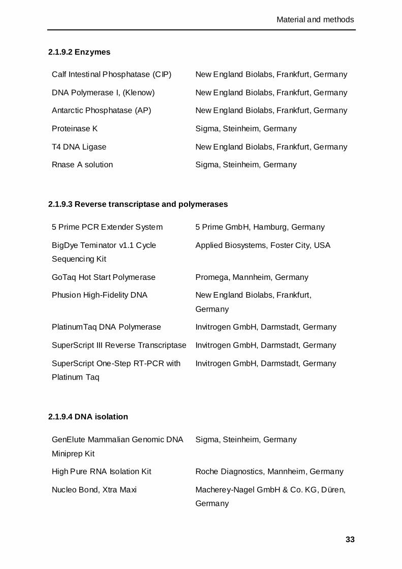
Material and methods
33
2.1.9.2 Enzymes
Calf Intestinal Phosphatase (CIP) New England Biolabs, Frankfurt, Germany
DNA Polymerase I, (Klenow) New England Biolabs, Frankfurt, Germany
Antarctic Phosphatase (AP) New England Biolabs, Frankfurt, Germany
Proteinase K Sigma, Steinheim, Germany
T4 DNA Ligase New England Biolabs, Frankfurt, Germany
Rnase A solution Sigma, Steinheim, Germany
2.1.9.3 Reverse transcriptase and polymerases
5 Prime PCR Extender System 5 Prime GmbH, Hamburg, Germany
BigDye Teminator v1.1 Cycle
Sequencing Kit
Applied Biosystems, Foster City, USA
GoTaq Hot Start Polymerase Promega, Mannheim, Germany
Phusion High-Fidelity DNA New England Biolabs, Frankfurt,
Germany
PlatinumTaq DNA Polymerase Invitrogen GmbH, Darmstadt, Germany
SuperScript III Reverse Transcriptase Invitrogen GmbH, Darmstadt, Germany
SuperScript One-Step RT-PCR with
Platinum Taq
Invitrogen GmbH, Darmstadt, Germany
2.1.9.4 DNA isolation
GenElute Mammalian Genomic DNA
Miniprep Kit
Sigma, Steinheim, Germany
High Pure RNA Isolation Kit Roche Diagnostics, Mannheim, Germany
Nucleo Bond, Xtra Maxi Macherey-Nagel GmbH & Co. KG, Düren,
Germany
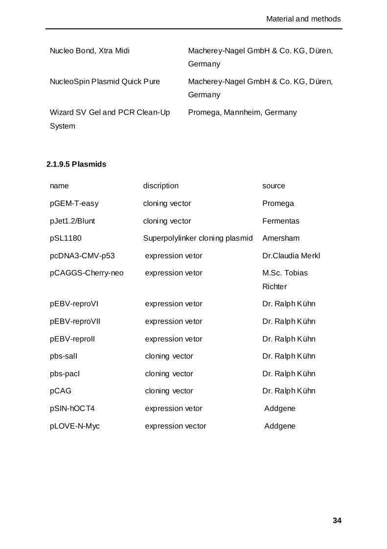
Material and methods
34
Nucleo Bond, Xtra Midi Macherey-Nagel GmbH & Co. KG, Düren,
Germany
NucleoSpin Plasmid Quick Pure Macherey-Nagel GmbH & Co. KG, Düren,
Germany
Wizard SV Gel and PCR Clean-Up
System
Promega, Mannheim, Germany
2.1.9.5 Plasmids
name discription source
pGEM-T-easy cloning vector Promega
pJet1.2/Blunt cloning vector Fermentas
pSL1180 Superpolylinker cloning plasmid Amersham
pcDNA3-CMV-p53 expression vetor Dr.Claudia Merkl
pCAGGS-Cherry-neo expression vetor M.Sc. Tobias
Richter
pEBV-reproVI expression vetor Dr. Ralph Kühn
pEBV-reproVII expression vetor Dr. Ralph Kühn
pEBV-reproII expression vetor Dr. Ralph Kühn
pbs-salI cloning vector Dr. Ralph Kühn
pbs-pacI cloning vector Dr. Ralph Kühn
pCAG cloning vector Dr. Ralph Kühn
pSIN-hOCT4 expression vetor Addgene
pLOVE-N-Myc expression vector Addgene
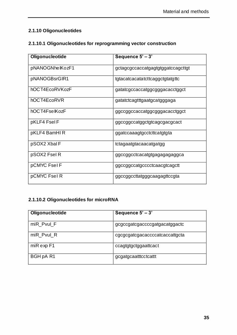
Material and methods
35
2.1.10 Oligonucleotides
2.1.10.1 Oligonucleotides for reprogramming vector construction
Oligonucleotide Sequence 5’ – 3’
pNANOGNheIKozF1 gctagcgccaccatgagtgtggatccagcttgt
pNANOGBsrGIR1 tgtacatcacatatcttcaggctgtatgttc
hOCT4EcoRVKozF gatatcgccaccatggcgggacacctggct
hOCT4EcoRVR gatatctcagtttgaatgcatgggaga
hOCT4FseIKozF ggccggccaccatggcgggacacctggct
pKLF4 FseI F ggccggccatggctgtcagcgacgcact
pKLF4 BamHI R ggatccaaagtgcctcttcatgtgta
pSOX2 XbaI F tctagaatgtacaacatgatgg
pSOX2 FseI R ggccggcctcacatgtgagagagaggca
pCMYC FseI F ggccggccatgcccctcaacgtcagctt
pCMYC FseI R ggccggccttatgggcaagagttccgta
2.1.10.2 Oligonucleotides for microRNA
Oligonucleotide Sequence 5’ – 3’
miR_PvuI_F gcgccgatcgaccccgatgacatggactc
miR_PvuI_R cgcgcgatcgacaccccatcaccattgcta
miR exp F1 ccagtgtgctggaattcact
BGH pA R1 gcgatgcaatttcctcattt
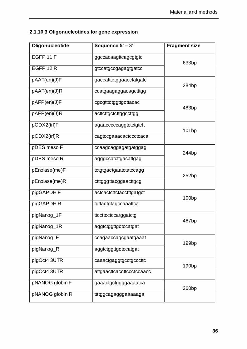
Material and methods
36
2.1.10.3 Oligonucleotides for gene expression
Oligonucleotide Sequence 5’ – 3’ Fragment size
EGFP 11 F ggccacaagttcagcgtgtc 633bp
EGFP 12 R gtccatgccgagagtgatcc
pAAT(en)(J)F gaccatttctggaacctatgatc 284bp
pAAT(en)(J)R ccatgaagaggacagctttgg
pAFP(en)(J)F cgcgtttctggttgcttacac 483bp
pAFP(en)(J)R acttcttgctcttggccttgg
pCDX2(trf)F agaacccccaggtctctgtctt 101bp
pCDX2(trf)R cagtccgaaacactccctcaca
pDES meso F ccaagcaggagatgatggag 244bp
pDES meso R agggccatcttgacattgag
pEnolase(me)F tctgtgactgaatctatccagg 252bp
pEnolase(me)R ctttgggttacggaacttgcg
pigGAPDH F actcactcttctacctttgatgct 100bp
pigGAPDH R tgttactgtagccaaattca
pigNanog_1F ttccttcctccatggatctg 467bp
pigNanog_1R aggtctggttgctccatgat
pigNanog_F ccagaaccagcgaatgaaat 199bp
pigNanog_R aggtctggttgctccatgat
pigOct4 3UTR caaactgaggtgcctgcccttc 190bp
pigOct4 3UTR attgaacttcaccttccctccaacc
pNANOG globin F gaaactgctggggaaaatca 260bp
pNANOG globin R ttttggcagagggaaaaaga
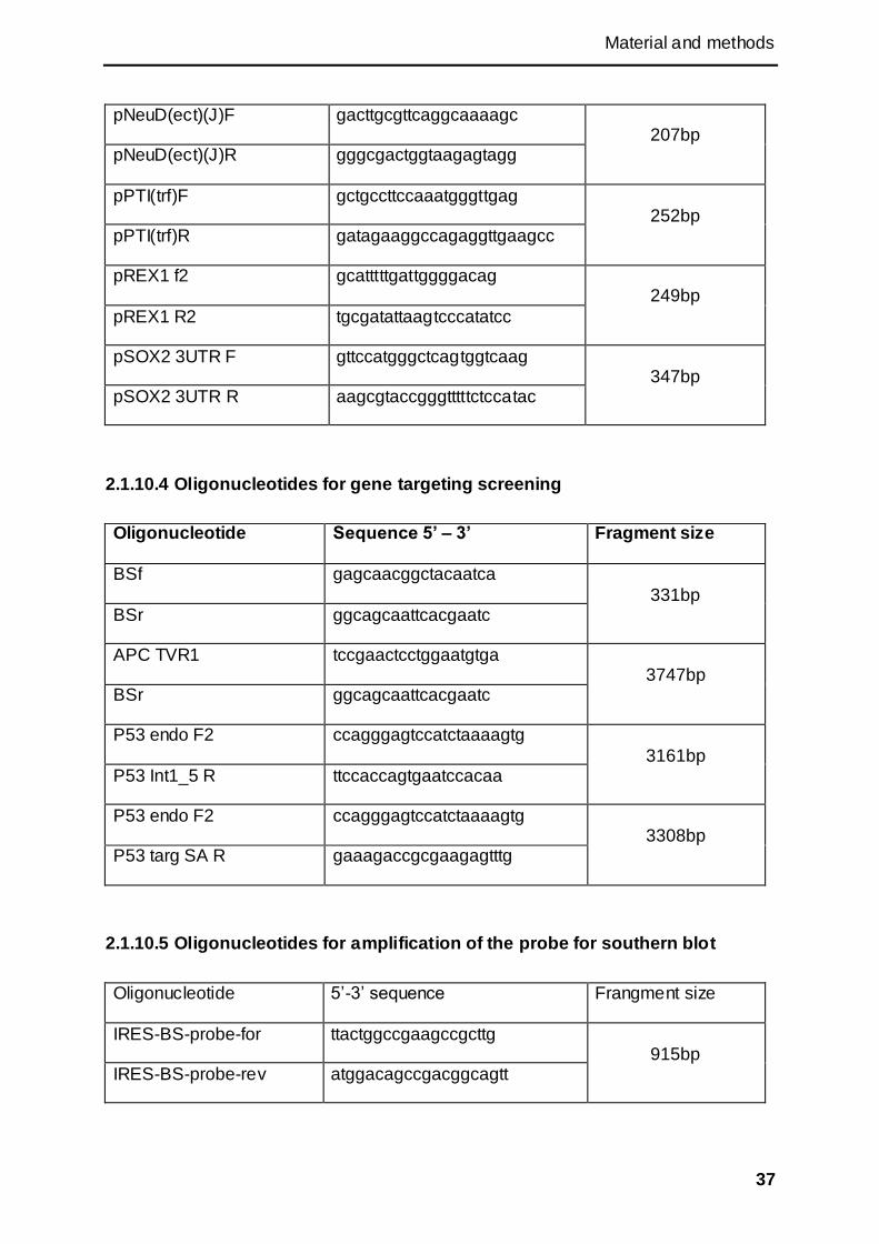
Material and methods
37
pNeuD(ect)(J)F gacttgcgttcaggcaaaagc 207bp
pNeuD(ect)(J)R gggcgactggtaagagtagg
pPTI(trf)F gctgccttccaaatgggttgag 252bp
pPTI(trf)R gatagaaggccagaggttgaagcc
pREX1 f2 gcatttttgattggggacag 249bp
pREX1 R2 tgcgatattaagtcccatatcc
pSOX2 3UTR F gttccatgggctcagtggtcaag 347bp
pSOX2 3UTR R aagcgtaccgggtttttctccatac
2.1.10.4 Oligonucleotides for gene targeting screening
Oligonucleotide Sequence 5’ – 3’ Fragment size
BSf gagcaacggctacaatca
331bp BSr ggcagcaattcacgaatc
APC TVR1 tccgaactcctggaatgtga 3747bp
BSr ggcagcaattcacgaatc
P53 endo F2 ccagggagtccatctaaaagtg
3161bp P53 Int1_5 R ttccaccagtgaatccacaa
P53 endo F2 ccagggagtccatctaaaagtg 3308bp
P53 targ SA R gaaagaccgcgaagagtttg
2.1.10.5 Oligonucleotides for amplification of the probe for southern blot
Oligonucleotide 5‘-3‘ sequence Frangment size
IRES-BS-probe-for ttactggccgaagccgcttg 915bp
IRES-BS-probe-rev atggacagccgacggcagtt
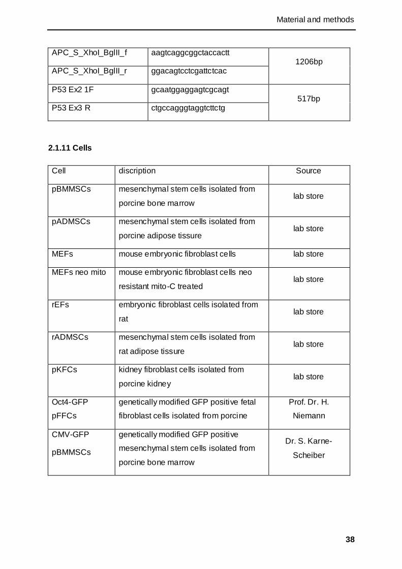
Material and methods
38
APC_S_XhoI_BglII_f aagtcaggcggctaccactt 1206bp
APC_S_XhoI_BglII_r ggacagtcctcgattctcac
P53 Ex2 1F gcaatggaggagtcgcagt 517bp
P53 Ex3 R ctgccagggtaggtcttctg
2.1.11 Cells
Cell discription Source
pBMMSCs mesenchymal stem cells isolated from
porcine bone marrow lab store
pADMSCs mesenchymal stem cells isolated from
porcine adipose tissure lab store
MEFs mouse embryonic fibroblast cells lab store
MEFs neo mito mouse embryonic fibroblast cells neo
resistant mito-C treated lab store
rEFs embryonic fibroblast cells isolated from
rat lab store
rADMSCs mesenchymal stem cells isolated from
rat adipose tissure lab store
pKFCs kidney fibroblast cells isolated from
porcine kidney lab store
Oct4-GFP
pFFCs
genetically modified GFP positive fetal
fibroblast cells isolated from porcine
Prof. Dr. H.
Niemann
CMV-GFP
pBMMSCs
genetically modified GFP positive
mesenchymal stem cells isolated from
porcine bone marrow
Dr. S. Karne-
Scheiber
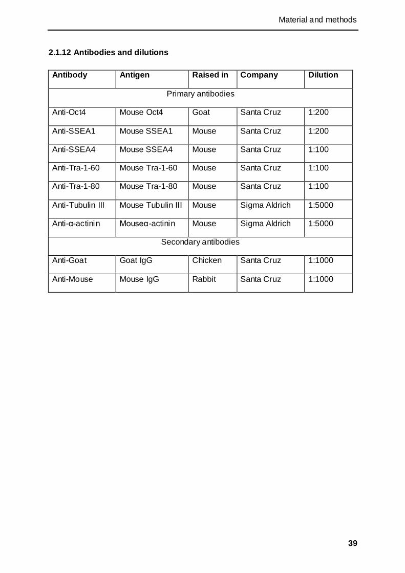
Material and methods
39
2.1.12 Antibodies and dilutions
Antibody Antigen Raised in Company Dilution
Primary antibodies
Anti-Oct4 Mouse Oct4 Goat Santa Cruz 1:200
Anti-SSEA1 Mouse SSEA1 Mouse Santa Cruz 1:200
Anti-SSEA4 Mouse SSEA4 Mouse Santa Cruz 1:100
Anti-Tra-1-60 Mouse Tra-1-60 Mouse Santa Cruz 1:100
Anti-Tra-1-80 Mouse Tra-1-80 Mouse Santa Cruz 1:100
Anti-Tubulin III Mouse Tubulin III Mouse Sigma Aldrich 1:5000
Anti-α-actinin Mouseα-actinin Mouse Sigma Aldrich 1:5000
Secondary antibodies
Anti-Goat Goat IgG Chicken Santa Cruz 1:1000
Anti-Mouse Mouse IgG Rabbit Santa Cruz 1:1000
Material and methods
40
2.2 Methods
2.2.1 Microbiological methods
2.2.1.1 Bacteria culture
For picking colonies and small amount DNA isolation, the Escherichia coli (E. coli)
were cultivated at 37 °C overnight either on agar plates or with 5 ml Luria Broth
medium (LB medium) in 14 ml culture tubes. For large amount DNA Isolation, 100-
300 ml of LB medium was used to cultivate the bacteria in 500 ml or 1000 ml conical
beaker. All of the media were supplemented with ampicillin at a final concentration of
100 g/ml.
2.2.1.2 Storage of E. coli:
For short-term storage (less than one month), the plates and media containing the
bacteria were stored at 4 °C. For long-term storage, 0.5-1 ml overnight culture
mixture from a single colony was carefully mixed with sterile 99% glycerol at a
volume ratio of 1 to 1 or 2 to 1 and the mixture finally was stored at -80 °C.
2.2.1.3 Transformation of E. coli:
1 ng of plasmid or 2 l ligation reaction was mixed with 50 l electro-competent cells,
and then the mixture was gently (without bubbles) added into pre-cooled
electroporation cuvette. The cells were electroporated by a pulse of 2500 V for 5 ms.
Immediately 600 l pre-warmed LB medium was added, and the mixture was
incubated at 37 °C and 230 rpm in a shaker for 45 to 50 min. Then 10 to 200 l of the
bacteria mixture was plated on agar plates supplemented with appropriate antibiotic
and incubated at 37 °C overnight.
2.2.2 Molecular biological methods
2.2.2.1 DNA Isolation
Plasmid DNA isolation from E. coli: For small amount plasmid preparation
(Miniprep), alkaline lysis method was used routinely. 5 ml LB medium supplemented
with appropriate antibiotics was seeded with pure single clone or stored bacteria and

Material and methods
41
incubated over night at 37 °C in an orbital shaker at 230 rpm. 2 ml bacteria culture
was collected into a 2 ml centrifugal tube and centrifuged at 10000 rpm for 2 min.
The pellet was resuspended in 100 l resuspension solution by vortexing, and then
150 l lysis buffer was added to the mixture. After 2 to 5 min incubated at room
temperature (RT), 200 l neutralization solution was added, and the mixture was
incubated on ice for about 30 min and centrifuged at 13000 rpm for 15 min. The
supernatant was transferred into a new 1.5 ml centrifuge tube and the pellet was
discarded. 1 ml 99.8 % ethanol was mixed with the supernatant by vortexing and the
mixture was centrifuged at 13000 rpm for 15 min. The pellet was washed with 0.5 ml
of 75% ethanol and centrifuged at 13000 rpm for 5 min. The DNA pellet was then air
dried at RT and dissolved in 50 μl water containing 20 μg/ml RNase A to remove
RNA.
For Large amount plasmid preparation (Midi- and Maxiprep), 100 to 300 ml LB
medium supplemented with appropriate antibiotic was seeded with pre -cultured
mixture or stored bacteria. The mixture was incubated overnight at 37 °C in an orbital
shaker at 230 rpm. Then the bacteria was collected and centrifuged at 5000 rpm for
10 min at 4 °C. The pellet was used to isolate plasmid DNA with the NucleoBond Xtra
Midi/Maxi kit according to the manual.
Genomic DNA isolation from mammalian cells: For small amount genomic DNA
isolation, the detached cells were centrifuged at 12000 rpm for 1 min after detaching
from the culture plate. The supernatant was removed carefully and the cell pellet was
resuspended in 50 µl Igepal buffer (50 mM Tris-HCl, 50 mM KCl, 3.15 mM MgCl2,
0.25% (v/v) Igepal, 0.5% (v/v) Tween 20, pH 8.0) containing 100 µg/ml proteinase K
Then the mixture was incubated at 65 °C for 60 min and followed by an incubation at
95 °C for 15 min. Then, the mixture was centrifuged at 16000×g for 15 min, and 5 µl
of the supernatant was used for one screening PCR.
For high quality genomic DNA isolation, GenElute Mammalian Genomic DNA
Miniprep Kit (Sigma) was used according to the manual.
For large amount genomic DNA isolation, cells were expanded till confluent on T-75
flask. The cells were detached with accutase and centrifuged at 324g for 5 min. The
pellet could be washed with PBS once if necessary. The pellet was resuspended in

Material and methods
42
500 µl cell lysis buffer (0.1 M Tris, 5mM EDTA, 0.2% M NaCl, 100 µg/ml Proteinase
K) and incubated at 37 °C overnight. Next day, the mixture was mixed well with
phenol-chloroform-isoamylalkohol at volume ratio 1 to 1 and incubated for 10 min at
RT followed with centrifuge at 14000 rpm for 10 min. The upper phase was then
mixed well with chloroform at volume ratio 1 to 1 in a new tube. The mixture was
centrifuged at 14000 rpm for 10 min. The upper phase was mixed with 0.7 volume of
Isopropanol and centrifuged at 14000 rpm for 10 min. The DNA pellet was washed
with 70% ethanol and dried at final. 50-100 µl water or buffer was added to dissolve
the DNA and stored at 4 °C.
2.2.2.2 DNA manipulation
Polymerase Chain Reaction (PCR): For PCR, depending on purpose, different
polymerases were used. For standard DNA fragments amplification, GoTaq
polymerase was used. When high accuracy DNA fragments amplification was
concerned, proofreading polymerase (5 Prime or Phusion) was used. The PCR
program depended on the polymerase kits and the annealing temperature of the
oligonucleotides. The programs of the mainly used polymerase kits are:
1. Go-Tag Polymerase kit (Promega):
Component Final concentration
5×Green buffer 1×
dNTPs 0.2mM each dNTP
Upstream primer 200-500 nM
Downstream primer 200-500 nM
Template DNA Up to 500 ng
GoTaq polymerase 1.25U/50µl
ddH2O Up to 50 µl

Material and methods
43
Pre-denaturation 95°C 5 min
Denaturation 95°C 30 s
30-40
cycles Annealing X °C 30 s
Extension 72°C 1kb/min
Final extention 72°C 5 min
Store at 8°C forever
2. 5Prime PCR extender system (5Prime):
Component Final concentration
10×High Fidelity buffer 1×
dNTPs 0.2mM each dNTP
Upstream primer 200-400 nM
Downstream primer 200-400 nM
Template DNA Genomic DNA10-100 ng
Plasmid 0.1-1 ng
Polymerase Mix 0.2U/50µl
ddH2O Up to 50 µl
Pre-denaturation 94°C 2 min
Denaturation 95°C 30 s
30-40
cycles Annealing X °C 30 s
Extension 72°C 1kb/min
Final extention 72°C 5 min
Store at 8°C forever
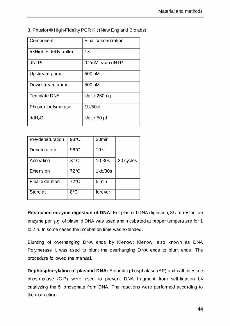
Material and methods
44
3. Phusion® High-Fidelity PCR Kit (New England Biolabs):
Component Final concentration
5×High Fidelity buffer 1×
dNTPs 0.2mM each dNTP
Upstream primer 500 nM
Downstream primer 500 nM
Template DNA Up to 250 ng
Phusion polymerase 1U/50µl
ddH2O Up to 50 µl
Pre-denaturation 98°C 30min
Denaturation 98°C 10 s
30 cycles Annealing X °C 10-30s
Extension 72°C 1kb/30s
Final extention 72°C 5 min
Store at 8°C forever
Restriction enzyme digestion of DNA: For plasmid DNA digestion, 3U of restriction
enzyme per g of plasmid DNA was used and incubated at proper temperature for 1
to 2 h. In some cases the incubation time was extended.
Blunting of overhanging DNA ends by Klenow: Klenow, also known as DNA
Polymerase I, was used to blunt the overhanging DNA ends to blunt ends. The
procedure followed the manual.
Dephosphorylation of plasmid DNA: Antarctic phosphatase (AP) and calf intestine
phosphatase (CIP) were used to prevent DNA fragment from self-ligation by
catalyzing the 5‘ phosphate from DNA. The reactions were performed according to
the instruction.

Material and methods
45
Agarose gel electrophoresis: Agarose gel electrophoresis was used to separate
DNA fragments according to their sizes. The agarose gel was supplemented with 0.1
μg/ml ethidium bromide to visualize the nucleotides under the UV light at 254 nm
excitation wavelength in the Gene Genius Bioimaging System. DNA samples were
mixed with 5 ×gel loading buffer and loaded on the gel. The gel was run at 80-120 V
for 0.5-5 h depending on DNA fragment size. The DNA fragment sizes and
concentration could be estimated by comparing the sizes and brightness levels of the
bands with the standard DNA marker. 0.8-1.0 % TAE/TBE agarose gels were used
for fragments bigger than 500bp. For fragments smaller than 500bp, 1.2%-1.5% gels
were used.
Extraction of DNA fragments from agarose gels: Wizard SV Gel and PCR Clean-
Up System were used to extract DNA from the TAE agarose gel according to the
manual.
Precipitation of DNA with sodium chloride and ethanol: Precipitation was used to
purify the DNA and concentrate or sterilize the DNA solution. After mixed with 0.1
volume of 3 M NaCl and 2 volumes of ice-cold 99.8% ethanol by vortexing, the DNA
solution was centrifuged at 13000 rpm for 15 min. The supernatant was discarded
and the pellet was washed with 70% ethanol and centrifuged at 13000 rpm for 5 min.
The DNA was dried under the hood and resuspended in sterilized or unsterilized
ddH2O.
Determination of DNA or RNA concentration: DNA or RNA concentration was
measured by using Eppendorf BioPhotometer according to the instruction.
Ligation of DNA fragments: The ligation of DNA frangments with pGEM-T-easy
vector was performed with promega pGEM T easy kit according to the manual.
The ligations of DNA fragments with other vectors were performed in a 20 μl reaction
system containing 50 ng backbone fragments, proper amount of insert fragments.
The molar ratio of vector to insert was in the range of 1:3 to1: 10. The ligation was
incubated at RT for 1-2 h or 4°C overnight.
2.2.2.3 DNA sequencing
DNA sequencing was performed with the BigDye Terminator v1.1 Cycle Sequencing
Material and methods
46
Kit. 100 to 200 ng plasmid DNA were mixed with 2 μl of BigDye Reaction mix, 2 μl
BigDye Sequencing Buffer, and 2.5 pmol sequencing primer and filled up to 10 μl
with nuclease free water.
Further processing of the sequencing samples was performed by Dr. K. Flisikowski.
Besides, some of the sequencings were performed by Eurofins MWG Company.
2.2.2.4 RNA isolation
Generally, Trizol method was used for RNA isolation from mammalian cells and
tissues. First, 1 ml of Trizol reagent per 1 well of 6-well plate was added to the cells
or tissues and mixed well. The lysed mixture was frozen in liquid nitrogen or -80 C if
the RNA isolation was not performed immediately. The mixture was incubated at RT
for 5 min, and then the chloroform was added to the mixture at a ratio of 1:5. After
mixed well, the mixture was incubated at RT for 2 min followed by a centrifugation for
15 min at 12000×g. The upper water phase was transferred to a new centrifuge tube
and mixed with pre-cooled isopropanol at a ratio of 1:1. The well-mixed mixture was
incubated at RT for 20-30 min, and then centrifuged at 7500 g for 10 min. The
supernatant was discarded and the pellet was washed with 500 l of pre-cooled 75%
ethanol. Finally, the RNA was air-dried on ice and dissolved in RNase-free water. To
remove the DNA contamination, the DNase Kit was used according to the manual.
For high quality RNA isolation, RNA isolation kit (Roche) was used according to the
instruction.
2.2.2.5 Assessing RNA integrity on agarose gels
To assess the integrity of total RNA, a denaturing agarose gel was run. Good quality
RNA was expected to run as two clear bands: 28S and 18S rRNA.
2.2.2.6 Reverse transcriptase polymerase chain reaction (RT-PCR)
Reverse tanscriptase polymerase chain reaction was used to amplify DNA fragments
from RNA. Two kinds of RT-PCR were used: one-step and two-step RT-PCR.
Two-step RT-PCR includes first strand reaction and PCR. The following PCR is the
same as the normal PCR, 2 µl cDNA product was used for one PCR reaction.

Material and methods
47
Two-step RT-PCR:
First-strand cDNA synthesis:
Component Final concentration
Oligo (dT)20 1 µM
total RNA 10 pg-5 µg
dNTPs 200 µM
ddH2O 13 µl
Heated at 65 °C for 5min, incubated on ice for 1 min
5 ×First-strand buffer 1×
DTT 5 mM
Revers transcriptase 20U/µl
Incubated at 55 °C for 45 min, and 70°C for 15 min
For one-step, a SuperScript™ One-Step RT-PCR kit from Invitrogen was used
accoding to the manual. For two-step RT-PCR, 100-500 ng RNA was reverse
transcribed to cDNA firstly. Then 2μl or more of the cDNA product was used as
template for the following PCR.
One-step RT-PCR:
Component Final concentration
2×Reaction Mix 1×
Upstream primer 200 nM
Downstream primer 200 nM
Template RNA 10 pg-1µg
RT/ Platimum Taq mix 1U/50µl
ddH2O Up to 50 µl
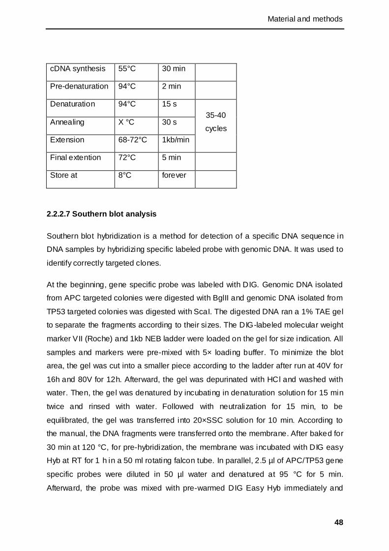
Material and methods
48
cDNA synthesis 55°C 30 min
Pre-denaturation 94°C 2 min
Denaturation 94°C 15 s
35-40
cycles Annealing X °C 30 s
Extension 68-72°C 1kb/min
Final extention 72°C 5 min
Store at 8°C forever
2.2.2.7 Southern blot analysis
Southern blot hybridization is a method for detection of a specific DNA sequence in
DNA samples by hybridizing specific labeled probe with genomic DNA. It was used to
identify correctly targeted clones.
At the beginning, gene specific probe was labeled with DIG. Genomic DNA isolated
from APC targeted colonies were digested with BglII and genomic DNA isolated from
TP53 targeted colonies was digested with ScaI. The digested DNA ran a 1% TAE gel
to separate the fragments according to their sizes. The DIG-labeled molecular weight
marker VII (Roche) and 1kb NEB ladder were loaded on the gel for size indication. All
samples and markers were pre-mixed with 5× loading buffer. To minimize the blot
area, the gel was cut into a smaller piece according to the ladder after run at 40V for
16h and 80V for 12h. Afterward, the gel was depurinated with HCl and washed with
water. Then, the gel was denatured by incubating in denaturation solution for 15 min
twice and rinsed with water. Followed with neutralization for 15 min, to be
equilibrated, the gel was transferred into 20×SSC solution for 10 min. According to
the manual, the DNA fragments were transferred onto the membrane. After baked for
30 min at 120 °C, for pre-hybridization, the membrane was incubated with DIG easy
Hyb at RT for 1 h in a 50 ml rotating falcon tube. In parallel, 2.5 µl of APC/TP53 gene
specific probes were diluted in 50 µl water and denatured at 95 °C for 5 min.
Afterward, the probe was mixed with pre-warmed DIG Easy Hyb immediately and
Material and methods
49
added to replace the pre-hybridization solution. The membrane was incubated at 37
°C overnight. And then the membrane was gentle shake in low stringency buffer for
15 min. Pre-heated at 54 °C high stringency buffer, was added to the membrane
twice. After washed with washing buffer for 2 min, the membrane was blocked by
blocking solution for 1 h with gentle shaking. The membrane reacted with the
antibody solution at RT for 30 min with shaking. Following this step, the membrane
was washed twice with washing buffer for 15 min and equilibrated in detection buffer
for 3 min. After sealing into plastic wrap, the membrane was exposed to
chemiluminescent substrate which was diluted at a ratio of 1:100. Five minutes later,
the liquid was carefully removed from the membrane. Finally, the membrane was
exposed to X-ray film for 5 min to 15 min.
2.2.3 Mammalian cell culture
2.2.3.1 Isolation and cultivation of porcine mesenchymal stem cells (pMSCs)
Porcine bone marrow mesenchymal stem cells were isolated from 6 month old
German Landrace (male) or German Landrace × Pietrain (male) pigs. The muscle
and other tissues were removed from the bones. Then the bones were cut by the
saws at the ends of two sides in a low contamination area to expose the bone
marrow. The HBSS (Hanks Balanced Salt Solution) –heparin solution was used to
flush the bone marrow into clean culture dishes. The harvested bone marrow solution
was added to 25 ml of lymphocyte separation medium and centrifuged at 1200 g
for 20 min. By centrifugation, the MSCs were separated from red blood cells and
plasma. The interphase containing the MSCs was washed with 20 ml HBSS and
centrifuged 5 min at 324 g. The pellet was resuspended in MSC medium containing
antibiotics (100 U/ml of Penicillin, 100 μg/ml of Streptomycin, 2.5 μg/ml of
Amphotericin B) and plated in T150 culture flask. After 2 or 3 days, the medium was
changed and the antibiotics were removed from the medium. The cells were split
and/or frozen in liquid nitrogen when confluent.
2.2.3.2 General cell culture
In general, cells were cultivated in cell culture flasks with ventilation lids at 37°C in a
humidified environment containing 5% CO2. The growth medium was replaced every
1 to 2 days with fresh pre-warmed to 37°C medium. If necessary, selection reagents
Material and methods
50
(Blasticidin or G418) or antibiotics were added. Cells were passaged when they were
confluent in the plate. First, the medium was removed and the cells were washed
once or twice with PBS. Then the cells were detached with accutase by incubation at
37°C in 5 min and the accutase was inactivated by medium supplemented with
serum. The cell suspension were centrifuged at 324g for 5 min, resuspended in
growth medium and plated in new plates or used for the following experiments.
Cell Counting: After detached, 10 µl of cell suspention was added carefully onto a
Neubauer counting chamber. After counting four large squares, the cell number per
ml was calculated according to the following formula:
Number of cells per ml = number of cells in 4 large squares / 4×dilution factor×104
Mediums for different cells:
DMEM+ medium MSC medium
DMEM (high glucose) Advanced DMEM
2 mM GlutaMAX 2 mM GlutaMAX
1× Non essential amino acids 1× Non essential amino acids
1 mM Sodium pyruvate 5 ng/ml bFGF
10% (v/v) FCS 10% (v/v) FCS
KFC medium Mouse ESC medium
DMEM (high glucose) DMEM (high glucose)
2 mM GlutaMAX 2 mM GlutaMAX
1 × Non essential amino acids 1 × Non essential amino acids
1 mM Sodium pyruvate 1 mM Sodium pyruvate
15% (v/v) FCS 0.1 mM beta-Mecaptoethanol
1 × LIF
15% (v/v) FCS
Material and methods
51
Porcine iPSC medium Rat ESC medium
1/2 Neurobasal Medium 1/2 Neurobasal Medium
1/2 DMEM/F12 medium 1/2 DMEM/F12 medium
0.1 mM beta-Mecaptoethanol 0.1 mM beta-Mecaptoethanol
2 mM GlutaMAX 2 mM GlutaMAX
1 × B27 supplement 1 × B27 supplement
1 × N2 supplement 1 × N2 supplement
1 × LIF 1 × LIF
0.375 M GSK inhibitor 3 M GSK inhibitor
0.8 M MEK inhibitor 0.5 M MEK inhibitor
10% (v/v) FCS 0.5 M ALK5 inhibitor
10% (v/v) FCS or 20% (v/v) KOSR
2.2.3.3 Transfection of mammalian cells (electroporation, nucleofection)
The cells were transfected with different vectors (depending on purpose) by
electroporation or nucleofection.
For electroporation, 1×10⁶ cells were mixed with 800 l buffer and maximum of 16 g
of pure circular or linear plasmid DNA, and then were pulsed at 1200 V for 5 ms.
After incubate at RT for 5 to 10 min, the cells were plated in a T-75 flask or 10 cm
dish.
For nucleofection, 5105 cells were mixed with 100 l buffer and 0-6 g of DNA, and
pulsed with the AMAXA nucleofector. The cells were immediately mixed with 500 l
medium and plated on a T-75 flask or 10 cm dish.
Material and methods
52
2.2.3.4 Colony picking and cultivation of putative piPSCs
When the putative piPSC colonies were dense and big enough, plastic rings were
used to isolate single cell clones with the use of accutase. The cell clusters were
sucked and incubate at 37°C with accutase in less than 5 min. Then the mixture was
transferred onto feeders with proper medium. To get more single colonies, they were
picked under the microscope with the lid open. The cell clusters were sucked out by
thin glass tube and transferred into pre-warmed accutase. The mixture was added
onto feeders with proper medium supplemented with penicillin and streptomycin
(Pen/Strep).
The cells were passaged on new feeders or gel matrix when they were almost
confluent or had a tendency to differentiate. The cells were seeded on 12 well, 6 well
plates or 10 cm dishes at a ration from 1:1 to 1:12. The culture medium was changed
every day and the cells were washed with PBS if necessary.
2.2.3.5 Freezing and thawing of mammalian cells
When the cells were not in culture, they were stored in -80 °C or liquid nitrogen
(about -160 °C to -130 °C).
Firstly, the cells were detached with either accutase or trypsin-EDTA and the
reactionwas inactivated by medium containing serum. After centrifugation, the cells
were resuspended in the freezing medium which normally contained 60% medium,
30% FCS and 10% DMSO. The aliquoted cells were added into the freezing vials
and placed in a freezing device (Mr. Frosty) at -80 °C immediately.
After at least 6h, the cells were taken out from the Mr. Frosty. For short-term storage,
the cells were stored at -80 °C; for long-term storage, to put the cells into liquid
nitrogen was necessary.
When the cells were needed in cell culture again, they would be thawed from the
storage. Firstly, the freezing tubes were transferred from the storage to the a 37 °C
water bath as soon as possible, and then the cells were mixed with warm medium
and centrifuged after defrosting. The cell pellet was resuspended in the medium and
plated into culture flasks or plates.
Material and methods
53
2.2.3.6 Alkaline phosphatase (AP) staining
Firstly, to prepare the AP solution, 1 Sigma fast BCIP/NBT tablet was dissolved in 10
ml cell culture grade water. The cells were washed twice with PBS and fixed with
fixation solution (90% methanol, 10% formalin) for 15 min at RT. Then the cells were
washed with Rinse buffer. After removing the Rinse buffer, the cells were stained by
the AP solution for 10 min. Pictures were taken after the staining.
2.2.3.7 Immunostaining of iPSCs
The purpose of the protocol is to detect the surface makers or intracellular factors in
iPSCs by immunochemistry.
The cells were seeded on 12-well or 6-well plate and cultivate to a 30% density.
When the cells were confluent, they were washed with PBS twice. The cells were
fixed with fixative for 15 min at RT and washed twice with Rinse buffer.
For intracellular antigens, the cells were permeabilized with 0.1% TritonX-100 at RT
for 20 min. After washed twice with Rinse buffer, the cells were blocked with blocking
solution for 1 h at RT. Then the blocking solution was replaced by first antibody
diluted in blocking solution and the cells were stored at 4°C overnight.
After the first antibody was removed, the cells were washed with Rinse buffer three
times and the diluted secondary antibody was added. Then the cells were incubated
at RT in dark for 1 h. The results were observed under fluorencence microscope after
the cells were washed three times with Rinse buffer.
The secondary antibody control was performed by skipping the first antibody. The
feeder cells were used as a negative control.
2.2.3.8 Separation of cells with microbeads
Microbeads are polymer particals which can be used to separate the molecules or
cells by bio-reactions. Anti-SSEA-1 microbeads and columns from Meltenyi Biotech
were used to separate the SSEA1 positive cells from the negative o nes. All the
procedures were performed accoding to the manual. After washed with three or four
times with PBS, 2 ml trypsin was added into 10 cm dish or 6-well plate and incubated
Material and methods
54
at 37 °C for less than 5 min. The putative piPSC clusters were digested into single
cells and the enzymatic reaction was inactivated by adding 8 ml medium containing
serum. The single-cell suspension was dissociated by pipetting up and down. The
cells were counted and the population for labeling was controlled up to 107. The
centrifuged pellet was resuspended in 80 l buffer. All the work should be done fast
and on ice to keep the cells cold. 20 l Anti-SSEA1 microbeads was mixed with the
cell suspension and incubated at 4 °C for 15 min. Then the cells were washed by
adding 1-2 ml buffer, and centrifuged at 300×g for 10 min. The pellet was
resuspended in 500 l buffer and the cell suspention was applied onto the prepared
magnet column. The flow-through containing unlabeled cells was collected. Then the
column was washed with 500 l buffer for 3 times. The column was removed from
separator and placed on a suitable collection tube. 1 ml buffer was applied on the
column, and the labeled cells were flushed out by firmly pushing the plunger into the
column.
2.2.3.9 Differentiation of putative porcine iPSCs
Embryoid bodies (EBs) formation: There are several methods for EBs formation.
The traditional method is suspension method, hanging drops method, and the newly
developed AggreWell (STEMCELL Technologies) method. For the suspension
method, the detached cell was separated to single cells by pipeting up and down,
and the single cells were resuspended in piPSC medium at a density of 4105 per ml.
Then 10 ml of cell suspension was cultivated in a 10 cm Petri dish at 37 °C in cell
incubator. The medium was changed gradiently into DMEM+ medium. One day post
the suspension, the medium was changed to differentiation medium I (75% piPSC
medium and 25% DMEM+ medium). Two days post the suspension, the medium was
changed to differentiation medium II (50% piPSC medium and 50% DMEM+
medium). Three days post the suspension, the medium was graduall changed to
differentiation medium III (25% piPSC medium and 75% DMEM+ medium). Finally,
the cells were cultivated in DMEM+ medium. Eight days post the suspensions , the
EBs were plated onto gelatin-coated plates.
Another method is hanging drop method. The detached cells were separated to
single cells by pipeting up and down and resuspended in either DMEM+ or piPSC
medium. The cells were counted and diluted to 3000 to 5000 cells per 40 μl of
Material and methods
55
medium. Hanging drops were dripped on the lid of 10 cm culture dishes at a density
of 20 to 30 drops per plate. The lid was turned over quickly and PBS was added into
the plate to form a humidity atmosphere. The drops were cultured for 2 days in the
incubator to form EBs.
AggreWell plate is a kind specific plate designed for EBs formation. Single cells were
added into plate at a density of 3000 to 5000 per well. By centrifuging the plate, the
cells aggregated and formed EBs.
Neural differentiation: EBs were generated with suspension method. During the
first three days, retinoic acid was not added in the differentiation medium. From day 4,
the EBs were cultured with 5μM retinoic acid for 4 days. On day 8, the EBs were
separated into single cells by trypsin and the cells were separated with the 40 μm cell
strainer to avoid the cell clusters. Then the cells were plated on laminin pre-coated
plates at a density of 2×10⁵ / cm2 in DMEM/F12 medium with N2 supplement and 25
μg/ml of insulin. The medium was changed at 2h and 4h post-plating. 48h post-
plating, the medium was changed to Neurobasal medium with B27 supplement. The
medium was changed on day 4, day 8, and day 12 post-plating. RNA isolation or
immunostaining was performed after day 12.
2.2.3.10 Cell synchronization
One day before starvation, 0.7-1.0×10⁶ cells were plated on T-75 flask. After washed
twice with PBS, the cells started to be starved in medium containing 0.5% serum. To
release the cells, the starvation medium was changed to medium containing 20%
serum.
2.2.3.11 Selection, picking and screening of targeted colonies
After transfection with the vector, the cells were passaged into the selection medium
after at least 24 h. For each cell types, a killing curve was performed to optimize
conditions for selection with selectable markers (BS or G418). The cells were
passaged into 150 mm dishes or 48 well plates at a ratio of 1:5.
About 10 to 15 days later, the selected colonies were visible under a microscope. To
pick the colonies from the 150 mm dishes, the plates were washed with PBS and the
Material and methods
56
colonies were picked either with sterilized filter wafers soaked in a warm accutase or
sticky rings.
The sticky rings were used to cover the colonies, and the accutase was added inside
the ring. The cells were transferred into 6 well plates (each colony separate).
PCR from cell culture: To detect correctly targeted colonies, targeting PCR across
the short arm was performed. The cells were detached with accutase and the
reaction was stoped with serum supplemented medium. After centrifuged, the cell
pellet was lysised by adding Igepal lysis buffer and heated at 95 C for 1 h. Then the
mixture was centrifuged at 12000×g for 5min and the supernatant was used for PCR.
2.2.3.12 Preparation of cells for SCNT
The positive colonies were seeded on 6 well plates in MSC medium. Two days
before nuclear transfer, MSC medium was replaced with serum starvation medium
(MSC medium containing 0.5% FCS).

Results
57
3 Results
To get higher targeting efficiency and generate transgenetic animals, pluripotent
stem cells were considered as the best choice, because of their long lifespan, high
frequency of homologous recombination and germline transmission capability. In
view of the lack of such cells for livestock species, we tried to identify or derive the
most appropriate cell type for gene targeting.
Pig, as a common large animal model for disease studies, plays a crucial role in
many areas. Generating porcine pluripotent stem cells and investigating their in vitro
characteristics could provide a much needed tool for genetic manipulation and the
derivation of genetically defined porcine models. So far neither stable porcine ESC
line has been established, nor have culture medium or surface markers been defined.
Since the success in isolating mouse and human iPSCs, several groups used similar
methods to reprogram porcine somatic cells to induced pluripotent stem cells but
without satisfying results. In the work presented here, several methods were tested to
generate porcine induced pluripotent cells from pMSCs, and the pluripotency was
evaluated by different methods.
As an alternative option, cell synchronization was also assessed to improve gene
targeting of porcine bone marrow mesenchymal stem cell (pBMMSCs) and to
produce cloned animals.
3.1 Reprogramming of porcine somatic cells with different methods
In this part, several constructs containing mouse, human or porcine reprogramming
factors were used for porcine iPSCs (piPSCs) generation. The newly found
pluripotent-related microRNAs were also tested for reprogramming. Porcine kidney
fibroblasts (pKFCs), Oct-GFP transgenic fetal fibroblasts, BMMSCs containing a
CMV-GFP transgene, normal BMMSCs and adipose derived mesenchymal stem
cells (ADMSCs) were used as starting cells. Finally, a putative piPSC line was
established and tested for developmental potential.

Results
58
3.1.1 Constructs used for reprogramming
3.1.1.1 Episomal vectors with mouse factors
Two episomal vectors (pEBV-reproVI and pEBV-reproVII), which were shown to
generate mouse iPSCs successfully, were provided by Dr. Ralf Kühn. PEBV-reproVI
contains mouse Oct4 (mOct4), mcMyc, mSox2, and mKlf4. MOct4 and mcMyc are on
one side of the promoter, and mSox2 and mKlf4 are on the other side. PEBV-reproVI
has similar structure with pEBV-reproVII. But instead of mKlf4 and mcMyc, mOct4
and mSox2 are one side of the promoter while mNanog and hLin28 are on the other
side. Besides, there is a red fluorescent protein (RFP) as a reporter to indicate the
expression level (Figure 8). To ensure protein translation T2A and P2A were used to
link the DNA sequences of the factors.
Figure 8: Episomal vectors pEBV-reproVI and pEBV-repro VII.
The pEBV-reproVI contains mouse Oct4, Sox2, Nanog, human LIN28 factors and a red
fluorescent protein gene. The pEBV-reproVII contains mouse Oct4, Sox2, Klf4 and cMyc.
These two vectors contain a tet-on system: tetracycline controlled transactivator (tTA)
expression cassette and the tTA responsive promoter directing the transcription of
interested gene. The tet-on system controls the expression of factors. The promoter
was turned on or off precisely according to the presence of doxycycline (Figure 9).
The vectors also contain the Epstein–Barr virus (EBV) episomal origin of replication
(oriP) and the Epstein-Barr nuclear antigen 1 (EBNA1) gene. The protein coded by
EBNA1 gene controls the oriP, which controls replication and keeps it as an episomal
vector and thus avoiding integration into the host genome.
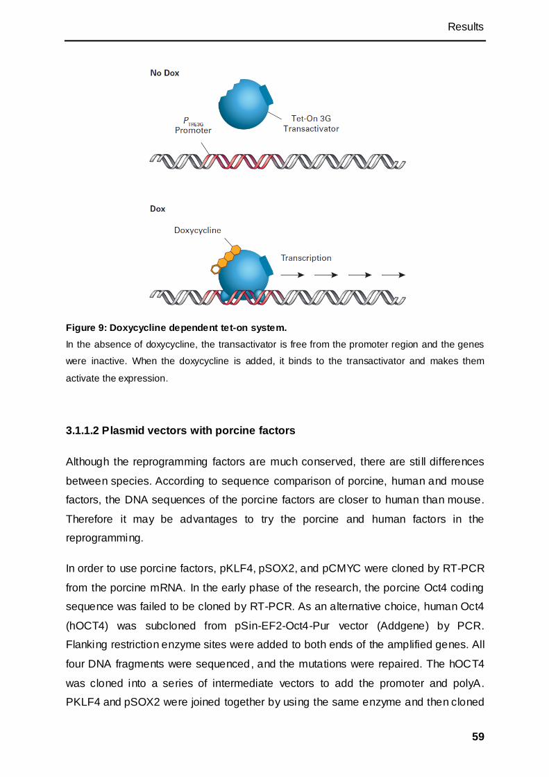
Results
59
Figure 9: Doxycycline dependent tet-on system.
In the absence of doxycycline, the transactivator is free from the promoter region and the genes
were inactive. When the doxycycline is added, it binds to the transactivator and makes them
activate the expression.
3.1.1.2 Plasmid vectors with porcine factors
Although the reprogramming factors are much conserved, there are sti ll differences
between species. According to sequence comparison of porcine, human and mouse
factors, the DNA sequences of the porcine factors are closer to human than mouse.
Therefore it may be advantages to try the porcine and human factors in the
reprogramming.
In order to use porcine factors, pKLF4, pSOX2, and pCMYC were cloned by RT-PCR
from the porcine mRNA. In the early phase of the research, the porcine Oct4 coding
sequence was failed to be cloned by RT-PCR. As an alternative choice, human Oct4
(hOCT4) was subcloned from pSin-EF2-Oct4-Pur vector (Addgene) by PCR.
Flanking restriction enzyme sites were added to both ends of the amplified genes. All
four DNA fragments were sequenced, and the mutations were repaired. The hOCT4
was cloned into a series of intermediate vectors to add the promoter and polyA.
PKLF4 and pSOX2 were joined together by using the same enzyme and then cloned
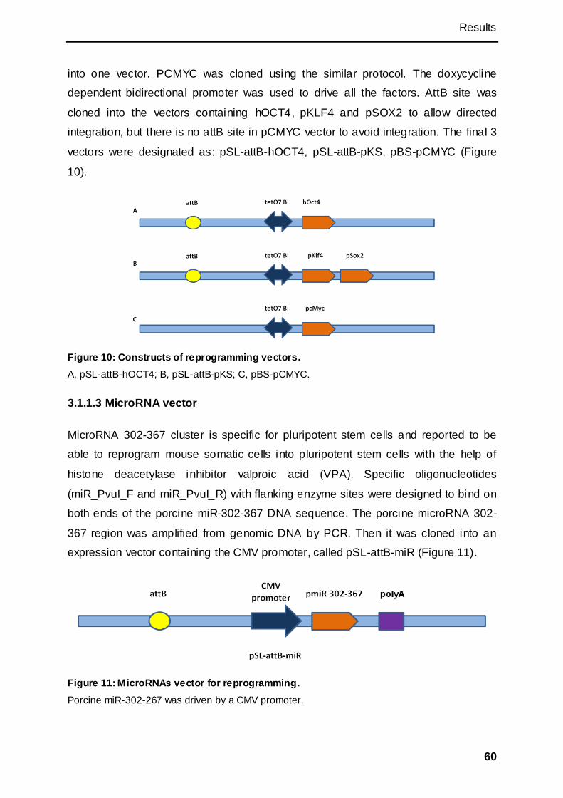
Results
60
into one vector. PCMYC was cloned using the similar protocol. The doxycycline
dependent bidirectional promoter was used to drive all the factors. AttB site was
cloned into the vectors containing hOCT4, pKLF4 and pSOX2 to allow directed
integration, but there is no attB site in pCMYC vector to avoid integration. The final 3
vectors were designated as: pSL-attB-hOCT4, pSL-attB-pKS, pBS-pCMYC (Figure
10).
Figure 10: Constructs of reprogramming vectors.
A, pSL-attB-hOCT4; B, pSL-attB-pKS; C, pBS-pCMYC.
3.1.1.3 MicroRNA vector
MicroRNA 302-367 cluster is specific for pluripotent stem cells and reported to be
able to reprogram mouse somatic cells into pluripotent stem cells with the help of
histone deacetylase inhibitor valproic acid (VPA). Specific oligonucleotides
(miR_PvuI_F and miR_PvuI_R) with flanking enzyme sites were designed to bind on
both ends of the porcine miR-302-367 DNA sequence. The porcine microRNA 302-
367 region was amplified from genomic DNA by PCR. Then it was cloned into an
expression vector containing the CMV promoter, called pSL-attB-miR (Figure 11).
Figure 11: MicroRNAs vector for reprogramming.
Porcine miR-302-267 was driven by a CMV promoter.

Results
61
3.1.1.4 Other vectors used for the reprogramming
PLOVE-N-Myc vector is a retroviral vector from Addgene. It contains an N-Myc
expression cassette driven by CMV promoter (Figure 12).
Figure 12: pLOVE-N-Myc vector.
The avian N-Myc gene was driven by a CMV promoter on the retroviral vector.
Porcine Nanog was cloned from the porcine ADMSCs by RT-PCR. After sequencing,
the pNANOG was cloned into an expression vector containing the CAGGS (CAG
promoter, chicken beta actin promoter/enhancer coupled with the cytomegalovirus
immediate-early enhancer). On the back bone, there is a neo resistance cassette
driven by SV40 early promoter which can be used for the selection (Figure 13). Pint-
ØC31 vector was used to express the integrase which catalyzes the integration
based on attB site.
Figure 13: pCAGGS-pNANOG-neo vector.
The porcine NANOG gene was driven by a CAGGS.
3.1.2 Reprogramming of porcine somatic cells and identification of putative
pluripotent stem cell
3.1.2.1 Reprogramming of porcine Nanog-KFCs with factors
Nanog has proven to be an important pluripotency marker in human and mouse
pluripotent cells. Over expression of Nanog may help the somatic cells to reprogram
into pluripotent state. 10 µg of sterile pCAGGS-pNANOG-neo was transfected into
1×10⁶ porcine kidney fibroblasts by electroporation and the cells were plated on a T-

Results
62
75 flask afterward. One day post transfection, the cells were passaged onto a new T-
75 at a ratio of 1:1. The medium was changed to the medium containing 300 µg/ml of
G418 to start selection. During the following two weeks, the cells were passaged for
3 passages at a ratio of 1:1 under selection. The morphology of the cells didn‘t
change during the selection. Subsequently, to confirm that exogenous pNANOG was
expressed. RT-PCR was carried out amplifying a 260bp fragment. The selected cell
clones (pNANOG-KFCs) were used for reprogramming in the next step.
The three porcine reprogramming factors, Oct4, Sox2, and Klf4 were used to
generate the iPSCs with the help of avian derived N-myc. N-myc is a member of the
Myc family. It also has the potential of tumorigenicity, but lower than cMyc. N-myc is
abundant in kidney cells (Malynn et al., 2000). The pLOVE-N-Myc vector is a
lentiviral vector, but in this experiment, it was used as plasmid for transfection. Two
transfections were performed. On day 0, the pSL-attB-hOCT4, pSL-attB-pKS,
pLOVE-N-Myc were nucleofected into 5×10⁵ of pNANOG positive cells at a ratio of
2:1:1. To get the vector integrated into the genomic DNA, the vector expressing the
PhiC31 integrase was cotransfected with the reprogramming vectors. The amount of
the integrase vector was equivalent to the sum of pSL-attB-hOCT4 and pSL-attB-
pKS. The total transfected DNA amount was limited to 3 µg. The cells were plated on
collagen pre-coated T-75 flask in the porcine KFC medium. In the following two days,
the medium was changed into fresh porcine iPSC medium every day. The medium
was supplemented with doxycycline at a final concentration of 1.5µg/ml. The
expressions of hOCT4, pKLF4 and pSOX2 were controlled by the doxycycline
dependent tet-on system, while N-Myc was expressed all the time. 3 days after the
first transfection, the cells were detached and counted. The same transfection was
repeated with the same vectors. This time, the vectors were nucleofected into all the
cells from a T-75 flask. The cells were re-plated on a collagen pre-coated T-75 flask.
During the following two days, the medium was changed. On day 6, the cells were
passaged onto two feeder coated 10 cm dishes (Figure 14), and cultivated in the
iPSCs medium for more than 4 weeks with doxycycline. The medium was changed
every other day and the cells were passaged on new feeders every 4-5 days. The
pNANOG-KFCs changed their morphology to long fibre shape and formed netlike
structure with their neighbour cells. The mesh became bigger and lots of cells died in
the first week. Several colonies were detected after 2 weeks. They were detached
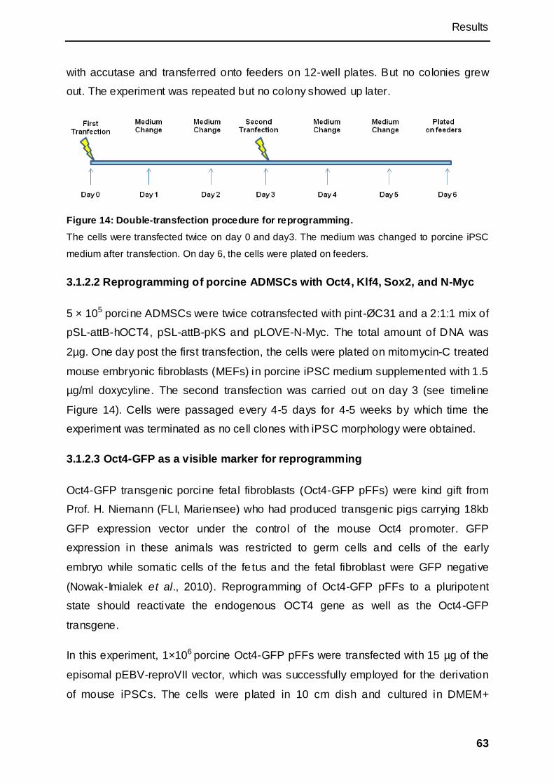
Results
63
with accutase and transferred onto feeders on 12-well plates. But no colonies grew
out. The experiment was repeated but no colony showed up later.
Figure 14: Double-transfection procedure for reprogramming.
The cells were transfected twice on day 0 and day3. The medium was changed to porcine iPSC
medium after transfection. On day 6, the cells were plated on feeders.
3.1.2.2 Reprogramming of porcine ADMSCs with Oct4, Klf4, Sox2, and N-Myc
5 × 10⁵ porcine ADMSCs were twice cotransfected with pint-ØC31 and a 2:1:1 mix of
pSL-attB-hOCT4, pSL-attB-pKS and pLOVE-N-Myc. The total amount of DNA was
2µg. One day post the first transfection, the cells were plated on mitomycin-C treated
mouse embryonic fibroblasts (MEFs) in porcine iPSC medium supplemented with 1.5
µg/ml doxycyline. The second transfection was carried out on day 3 (see timeline
Figure 14). Cells were passaged every 4-5 days for 4-5 weeks by which time the
experiment was terminated as no cell clones with iPSC morphology were obtained.
3.1.2.3 Oct4-GFP as a visible marker for reprogramming
Oct4-GFP transgenic porcine fetal fibroblasts (Oct4-GFP pFFs) were kind gift from
Prof. H. Niemann (FLI, Mariensee) who had produced transgenic pigs carrying 18kb
GFP expression vector under the control of the mouse Oct4 promoter. GFP
expression in these animals was restricted to germ cells and cells of the early
embryo while somatic cells of the fe tus and the fetal fibroblast were GFP negative
(Nowak-Imialek et al., 2010). Reprogramming of Oct4-GFP pFFs to a pluripotent
state should reactivate the endogenous OCT4 gene as well as the Oct4-GFP
transgene.
In this experiment, 1×10⁶ porcine Oct4-GFP pFFs were transfected with 15 µg of the
episomal pEBV-reproVII vector, which was successfully employed for the derivation
of mouse iPSCs. The cells were plated in 10 cm dish and cultured in DMEM+
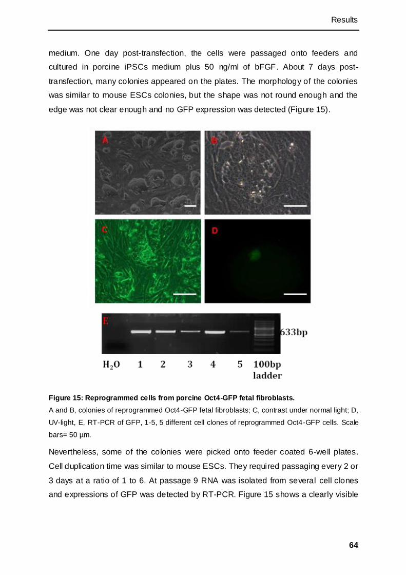
Results
64
medium. One day post-transfection, the cells were passaged onto feeders and
cultured in porcine iPSCs medium plus 50 ng/ml of bFGF. About 7 days post-
transfection, many colonies appeared on the plates. The morphology of the colonies
was similar to mouse ESCs colonies, but the shape was not round enough and the
edge was not clear enough and no GFP expression was detected (Figure 15).
Figure 15: Reprogrammed cells from porcine Oct4-GFP fetal fibroblasts.
A and B, colonies of reprogrammed Oct4-GFP fetal fibroblasts; C, contrast under normal light; D,
UV-light, E, RT-PCR of GFP, 1-5, 5 different cell clones of reprogrammed Oct4-GFP cells. Scale
bars= 50 µm.
Nevertheless, some of the colonies were picked onto feeder coated 6-well plates.
Cell duplication time was similar to mouse ESCs. They required passaging every 2 or
3 days at a ratio of 1 to 6. At passage 9 RNA was isolated from several cell clones
and expressions of GFP was detected by RT-PCR. Figure 15 shows a clearly visible

Results
65
amplification product, although only few fluorescent signals were observed (could be
from dead cells or unspecific background).
Although the morphology of the cells was similar to iPSCs, and the cells were GFP
positive according to the RT-PCR result, Oct4-GFP expression was not sufficient for
visualisation. This may be an indication that reprogramming neither resulted in full
reactivation of transgene, nor of the endogenous OCT4 gene.
3.1.2.4 CMV-GFP as a visible marker for reprogramming
Porcine CMV-GFP expressing BMMSCs were derived from 6 months old transgenic
animal in which the GFP gene is under the control of the CMV (cytomegalovirus)
promoter. Viral promoter such as the CMV promoter is generally si lenced in
pluripotent stem cells but is active in somatic cells (Liew et al., 2007; Wang et al.,
2008c). If the cells are reprogrammed to iPSCs, the CMV promoter should be
silenced and the green fluorescence should be turned off. In this study, 1×10⁶
porcine CMV-GFP BMMSCs were electroporated with 15 µg of pEBV-reproVII and
passaged into a new 10 cm dish one day post transfection. About one week later, the
colonies appeared. They were picked and cultured in porcine iPSC medium plus 50
ng/ml of bFGF on feeders. After several passages, all of the cells were still green
fluorescened. Compared to the untransfected CMV-GFP BMMSCs, the fluorescent
intensity of some cells was however reduced, which may indicate that the activity of
CMV promoter in these cells were down regulated by the reprogramming factors. But
the cells were not fully reprogrammed so the CMV couldn‘t be silenced.
3.1.2.5 Generation of piPSCs with microRNAs
The microRNA miR-302-367 construct was used to transfect porcine ADMSCs, rat
BMMSCs, rat ear fibroblasts and MEFs. The rat cells and mouse cells were designed
as controls for testing the function of the construct.
The double-transfection method was used. At first, 5×10⁵ porcine ADMSCs or rat
BMMSCs were nucleofected with 1 µg of pSL-attB-miR and 1 µg of pint-ØC31 with
the nucleofection program U-23. Rat ear fibroblasts were nucleofected with 3 µg of
pSL-attB-miR and 3 µg of pint-ØC31. One day post transfection, the porcine
ADMSCs, rat BMMSCs and rat ear fibroblasts were cultured in rat ESC medium
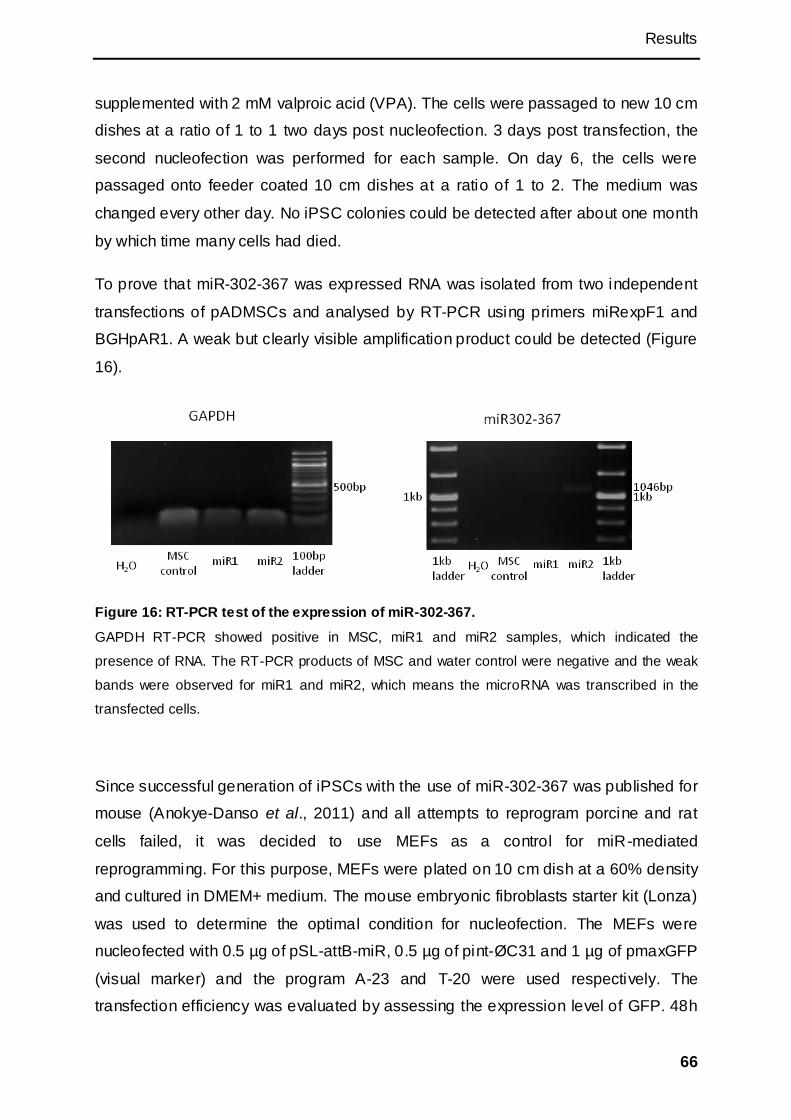
Results
66
supplemented with 2 mM valproic acid (VPA). The cells were passaged to new 10 cm
dishes at a ratio of 1 to 1 two days post nucleofection. 3 days post transfection, the
second nucleofection was performed for each sample. On day 6, the cells were
passaged onto feeder coated 10 cm dishes at a ratio of 1 to 2. The medium was
changed every other day. No iPSC colonies could be detected after about one month
by which time many cells had died.
To prove that miR-302-367 was expressed RNA was isolated from two independent
transfections of pADMSCs and analysed by RT-PCR using primers miRexpF1 and
BGHpAR1. A weak but clearly visible amplification product could be detected (Figure
16).
Figure 16: RT-PCR test of the expression of miR-302-367.
GAPDH RT-PCR showed positive in MSC, miR1 and miR2 samples, which indicated the
presence of RNA. The RT-PCR products of MSC and water control were negative and the weak
bands were observed for miR1 and miR2, which means the microRNA was transcribed in the
transfected cells.
Since successful generation of iPSCs with the use of miR-302-367 was published for
mouse (Anokye-Danso et al., 2011) and all attempts to reprogram porcine and rat
cells failed, it was decided to use MEFs as a control for miR-mediated
reprogramming. For this purpose, MEFs were plated on 10 cm dish at a 60% density
and cultured in DMEM+ medium. The mouse embryonic fibroblasts starter kit (Lonza)
was used to determine the optimal condition for nucleofection. The MEFs were
nucleofected with 0.5 µg of pSL-attB-miR, 0.5 µg of pint-ØC31 and 1 µg of pmaxGFP
(visual marker) and the program A-23 and T-20 were used respectively. The
transfection efficiency was evaluated by assessing the expression level of GFP. 48h
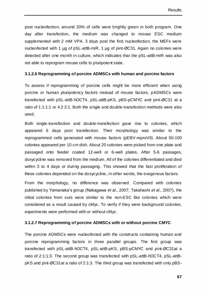
Results
67
post nucleofection, around 20% of cells were brightly green in both program. One
day after transfection, the medium was changed to mouse ESC medium
supplemented with 2 mM VPA. 3 days post the first nucleofection, the MEFs were
nucleofected with 1 µg of pSL-attB-miR, 1 µg of pint-ØC31. Again no colonies were
detected after one month in culture, which indicates that the pSL-attB-miR was also
not able to reprogram mouse cells to pluripotent state.
3.1.2.6 Reprogramming of porcine ADMSCs with human and porcine factors
To assess if reprogramming of porcine cells might be more efficient when using
porcine or human pluripotency factors instead of mouse factors, pADMSCs were
transfected with pSL-attB-hOCT4, pSL-attB-pKS, pBS-pCMYC and pint-ØC31 at a
ratio of 1:1:1:1 or 4:2:2:1. Both the single and double-transfection methods were also
used.
Both single-transfection and double-transfection gave rise to colonies, which
appeared 6 days post transfection. Their morphology was similar to the
reprogrammed cells generated with mouse factors (pEBV-reproVII). About 50-100
colonies appeared per 10 cm dish. About 20 colonies were picked from one plate and
passaged onto feeder coated 12-well or 6-well plates. After 5-6 passages,
doxycycline was removed from the medium. All of the colonies differentiated and died
within 3 to 4 days or during passaging. This showed that the fast proliferation of
these colonies depended on the doxycycline, in other words, the exogenous factors.
From the morphology, no difference was observed. Compared with colonies
published by Yamanaka‘s group (Nakagawa et al., 2007; Takahashi et al., 2007), the
initial colonies from ours were similar to the non-ESC like colonies which were
considered as a result caused by cMyc. To verify if they were background colonies,
experiments were performed with or without cMyc.
3.1.2.7 Reprogramming of porcine ADMSCs with or without porcine CMYC
The porcine ADMSCs were nucleofected with the constructs containing human and
porcine reprogramming factors in three parallel groups. The first group was
transfected with pSL-attB-hOCT4, pSL-attB-pKS, pBS-pCMYC and pint-ØC31at a
ratio of 2:1:1:3. The second group was transfected with pSL-attB-hOCT4, pSL-attB-
pKS and pint-ØC31at a ratio of 2:1:3. The third group was transfected with only pBS-
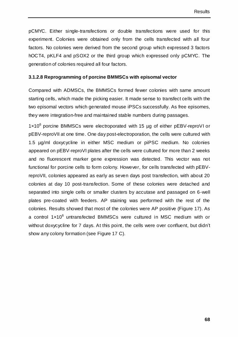
Results
68
pCMYC. Either single-transfections or double transfections were used for this
experiment. Colonies were obtained only from the cells transfected with all four
factors. No colonies were derived from the second group which expressed 3 factors
hOCT4, pKLF4 and pSOX2 or the third group which expressed only pCMYC. The
generation of colonies required all four factors.
3.1.2.8 Reprogramming of porcine BMMSCs with episomal vector
Compared with ADMSCs, the BMMSCs formed fewer colonies with same amount
starting cells, which made the picking easier. It made sense to transfect cells with the
two episomal vectors which generated mouse iPSCs successfully. As free episomes,
they were integration-free and maintained stable numbers during passages.
1×10⁶ porcine BMMSCs were electroporated with 15 µg of either pEBV-reproVI or
pEBV-reproVII at one time. One day post-electroporation, the cells were cultured with
1.5 µg/ml doxycycline in either MSC medium or piPSC medium. No colonies
appeared on pEBV-reproVI plates after the cells were cultured for more than 2 weeks
and no fluorescent marker gene expression was detected. This vector was not
functional for porcine cells to form colony. However, for cells transfected with pEBV-
reproVII, colonies appeared as early as seven days post transfection, with about 20
colonies at day 10 post-transfection. Some of these colonies were detached and
separated into single cells or smaller clusters by accutase and passaged on 6-well
plates pre-coated with feeders. AP staining was performed with the rest of the
colonies. Results showed that most of the colonies were AP positive (Figure 17). As
a control 1×10⁶ untransfected BMMSCs were cultured in MSC medium with or
without doxycycline for 7 days. At this point, the cells were over confluent, but didn‘t
show any colony formation (see Figure 17 C).

Results
69
Figure 17: Putative piPSC colony (7 days post transfection) and pBMMSCs.
A, outgrown colony on the plate over confluent with transfected pBMMSCs. Scale bars=100 µm.
B, AP staining of putative piPSC colony. Scale bars= 50 µm. C, left: untransfected pBMMSCs
cultured for 11 days as a control; right: transfected pBMMSCs cultured for 8 days without
doxycycline as a control. Scale bars= 50 µm.
3.2 Assessment of pluripotency of reprogrammed cells
To test the pluripotency of the putative iPSCs, several aspects were considered,
including doxycycline dependency, expression of endogenous pluripotency factors ,
and marker genes, karyotype and differentiation abilities of cells.
3.2.1 Doxycycline dependency
To identify if the cells have reactivated the endogenous pluripotency factors and
disengaged from the exogenous factors, doxycycline was removed from the culture
medium at different passages.

Results
70
At earlier passages (passage 8-15), removal of doxycycline resulted in changes of
the cell morphology within 24 hrs. The cells became flatter compared to their parental
cells. Many cells died before or after the next passage.
At later passages (passage 30-40), ESC-like morphology was retained for several
days after doxycycline removal, but again was ultimately lost and cell death occurred.
As cells could not survive without doxycycline induction of the exogenous factors , the
minimal amount of doxycycline required was then determined, ranging from 0.1 µg/ml
to 1.5 µg/ml. Even at the lowest amount cells proliferated fast and kept ESC-like
morphology, but did not survive after withdraw of the doxycycline.
3.2.2 Detection of endogenous gene expressions by RT-PCR
As the above experiment showed the ES-like morphology required continuous
expression of the exogenous transcription factors, this could indicate that the
endogenous pluripotency factors (porcine OCT4, SOX2, and NANOG, telomerase
reverse transcriptase (TERT), CMYC, and KLF4) have not been reactivated or their
expression levels were too low. Therefore RT-PCR analysis was carried out. As the
mouse and porcine reprogramming factors share high sequence similarity, primers
were designed to bind to the untranslated regions of the porcine genes. This enabled
us to distinguish the porcine endogenous expressions from mouse exogenous gene
expression. As a positive control RT-PCR analysis for pGAPDH was performed, RNA
samples from mouse feeder cells and porcine BMMSCs were also included in the
analysis as control. To compare the expressions with differentiated cells and
microRNA transfected pMSCs, two samples of EBs and one sample of miR-302-367
transfected pMSCs were analyzed by RT-PCR.
The putative piPSCs were divided into two groups: SSEA1 positive population and
SSEA1 negative population (for explanation see 3.2.5). RT-PCR products from
piPSC samples were purified and sequenced to verify the amplified fragments from
porcine RNAs.
The results showed that the water control was negative for all genes. The feeder
control was only positive for pGAPDH. The pMSC was positive for all genes. SSEA1
positive cell was positive for all genes except pCMYC. SSEA1 negative cell was
positive for pOCT4, pSOX2, pTERT and pGAPDH. EB1 was positive for all genes
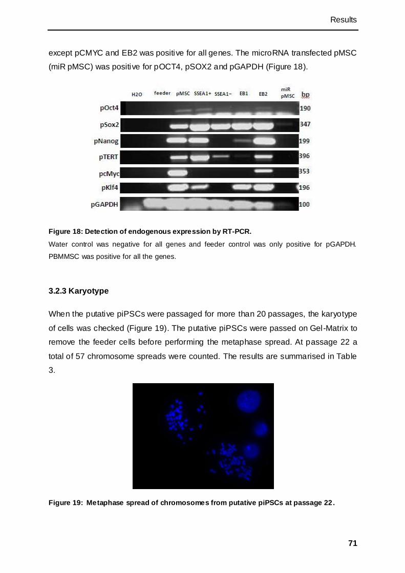
Results
71
except pCMYC and EB2 was positive for all genes. The microRNA transfected pMSC
(miR pMSC) was positive for pOCT4, pSOX2 and pGAPDH (Figure 18).
Figure 18: Detection of endogenous expression by RT-PCR.
Water control was negative for all genes and feeder control was only positive for pGAPDH.
PBMMSC was positive for all the genes.
3.2.3 Karyotype
When the putative piPSCs were passaged for more than 20 passages, the karyotype
of cells was checked (Figure 19). The putative piPSCs were passed on Gel-Matrix to
remove the feeder cells before performing the metaphase spread. At passage 22 a
total of 57 chromosome spreads were counted. The results are summarised in Table
3.
Figure 19: Metaphase spread of chromosomes from putative piPSCs at passage 22.
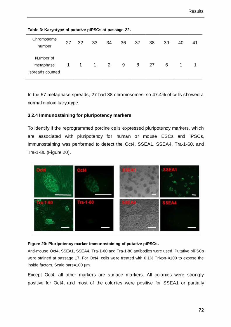
Results
72
Table 3: Karyotype of putative piPSCs at passage 22.
Chromosome
number 27 32 33 34 36 37 38 39 40 41
Number of
metaphase
spreads counted
1 1 1 2 9 8 27 6 1 1
In the 57 metaphase spreads, 27 had 38 chromosomes, so 47.4% of cells showed a
normal diploid karyotype.
3.2.4 Immunostaining for pluripotency markers
To identify if the reprogrammed porcine cells expressed pluripotency markers, which
are associated with pluripotency for human or mouse ESCs and iPSCs,
immunostaining was performed to detect the Oct4, SSEA1, SSEA4, Tra-1-60, and
Tra-1-80 (Figure 20).
Figure 20: Pluripotency marker immunostaining of putative piPSCs.
Anti-mouse Oct4, SSEA1, SSEA4, Tra-1-60 and Tra-1-80 antibodies were used. Putative piPSCs
were stained at passage 17. For Oct4, cells were treated with 0.1% Trixon-X100 to expose the
inside factors. Scale bars=100 µm.
Except Oct4, all other markers are surface markers. All colonies were strongly
positive for Oct4, and most of the colonies were positive for SSEA1 or partially
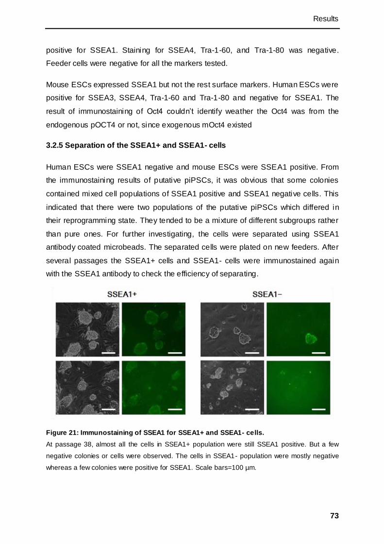
Results
73
positive for SSEA1. Staining for SSEA4, Tra-1-60, and Tra-1-80 was negative.
Feeder cells were negative for all the markers tested.
Mouse ESCs expressed SSEA1 but not the rest surface markers. Human ESCs were
positive for SSEA3, SSEA4, Tra-1-60 and Tra-1-80 and negative for SSEA1. The
result of immunostaining of Oct4 couldn‘t identify weather the Oct4 was from the
endogenous pOCT4 or not, since exogenous mOct4 existed
3.2.5 Separation of the SSEA1+ and SSEA1- cells
Human ESCs were SSEA1 negative and mouse ESCs were SSEA1 positive. From
the immunostaining results of putative piPSCs, it was obvious that some colonies
contained mixed cell populations of SSEA1 positive and SSEA1 negative cells. This
indicated that there were two populations of the putative piPSCs which differed in
their reprogramming state. They tended to be a mixture of different subgroups rather
than pure ones. For further investigating, the cells were separated using SSEA1
antibody coated microbeads. The separated cells were plated on new feeders. After
several passages the SSEA1+ cells and SSEA1- cells were immunostained again
with the SSEA1 antibody to check the efficiency of separating.
Figure 21: Immunostaining of SSEA1 for SSEA1+ and SSEA1- cells.
At passage 38, almost all the cells in SSEA1+ population were still SSEA1 positive. But a few
negative colonies or cells were observed. The cells in SSEA1- population were mostly negative
whereas a few colonies were positive for SSEA1. Scale bars=100 µm.
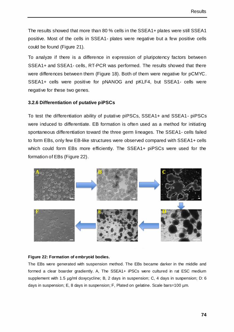
Results
74
The results showed that more than 80 % cells in the SSEA1+ plates were still SSEA1
positive. Most of the cells in SSEA1- plates were negative but a few positive cells
could be found (Figure 21).
To analyze if there is a difference in expression of pluripotency factors between
SSEA1+ and SSEA1- cells, RT-PCR was performed. The results showed that there
were differences between them (Figure 18). Both of them were negative for pCMYC.
SSEA1+ cells were positive for pNANOG and pKLF4, but SSEA1- cells were
negative for these two genes.
3.2.6 Differentiation of putative piPSCs
To test the differentiation ability of putative piPSCs, SSEA1+ and SSEA1- piPSCs
were induced to differentiate. EB formation is often used as a method for initiating
spontaneous differentiation toward the three germ lineages. The SSEA1- cells failed
to form EBs, only few EB-like structures were observed compared with SSEA1+ cells
which could form EBs more efficiently. The SSEA1+ piPSCs were used for the
formation of EBs (Figure 22).
Figure 22: Formation of embryoid bodies.
The EBs were generated with suspension method. The EBs became darker in the middle and
formed a clear boarder gradiently. A, The SSEA1+ iPSCs were cultured in rat ESC medium
supplement with 1.5 µg/ml doxycycline; B, 2 days in suspension; C, 4 days in suspension; D: 6
days in suspension; E, 8 days in suspension; F, Plated on gelatine. Scale bars=100 µm.
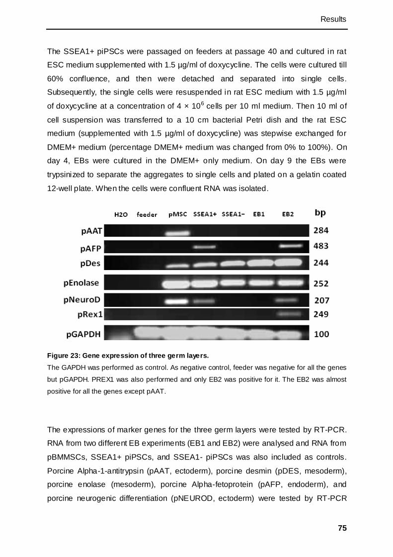
Results
75
The SSEA1+ piPSCs were passaged on feeders at passage 40 and cultured in rat
ESC medium supplemented with 1.5 µg/ml of doxycycline. The cells were cultured till
60% confluence, and then were detached and separated into single cells.
Subsequently, the single cells were resuspended in rat ESC medium with 1.5 µg/ml
of doxycycline at a concentration of 4 × 10⁶ cells per 10 ml medium. Then 10 ml of
cell suspension was transferred to a 10 cm bacterial Petri dish and the rat ESC
medium (supplemented with 1.5 µg/ml of doxycycline) was stepwise exchanged for
DMEM+ medium (percentage DMEM+ medium was changed from 0% to 100%). On
day 4, EBs were cultured in the DMEM+ only medium. On day 9 the EBs were
trypsinized to separate the aggregates to single cells and plated on a gelatin coated
12-well plate. When the cells were confluent RNA was isolated.
Figure 23: Gene expression of three germ layers.
The GAPDH was performed as control. As negative control, feeder was negative for all the genes
but pGAPDH. PREX1 was also performed and only EB2 was positive for it. The EB2 was almost
positive for all the genes except pAAT.
The expressions of marker genes for the three germ layers were tested by RT-PCR.
RNA from two different EB experiments (EB1 and EB2) were analysed and RNA from
pBMMSCs, SSEA1+ piPSCs, and SSEA1- piPSCs was also included as controls.
Porcine Alpha-1-antitrypsin (pAAT, ectoderm), porcine desmin (pDES, mesoderm),
porcine enolase (mesoderm), porcine Alpha-fetoprotein (pAFP, endoderm), and
porcine neurogenic differentiation (pNEUROD, ectoderm) were tested by RT-PCR

Results
76
amplification (Figure 23). Results showed that the EB1 RNA was positive for pDES
and pEnolase. The EB2 sample was positive for expression of all genes tested
except pAAT. In comparison, pBMMSCs were positive not only for pDES and
pEnolase (typical for mesoderm), but also for pAAT and pNEUROD. Both of SSEA1+
and SSEA1- cells were positive for pDES, pEnolase and pGAPDH. The SSEA1+
cells were positive for pAFP and pNEUROD. This may indicate the putative piPSCs
were not fully reprogrammed or there were differentiated cells in the putative piPSCs
population. Rex1 used to be a marker of undifferentiated ESCs, but now it‘s
considered not essential for pluripotency in mouse ES cells (Masui et al., 2008). It
was reported Rex1 positive cells primarily differentiate into primitive ectoderm
(Toyooka et al., 2008). Only EB2 was positive for pREX1.
Next directed differentiation towards neuron was attempted. For this, the culture
medium of EBs was supplemented with 5μM of retinoic acid from day 4 onwards. On
day 8, the EBs were separated into single cells by trypsin, and the mixture went
through the 40 μm cell strainer to avoid the cell clusters. Then the cells were plated
on laminin pre-coated plates at a density of 2×10⁵ per cm2 in DMEM/F12 medium
with N2 supplement and 25 μg/ml of Insulin. The medium was changed at 2h, 4h post
plating. 48h post-plating, the medium was changed to Neurobasal medium
supplemented with B27. And the medium was changed on day 4, day 8 , and day 12
post plating. Immunostaining was performed after day 12.
The results showed that there were a few neuron-like cells in the differentiated
population. They had long and thin axons with small bright dots. Their long and
branched axons crossed with the close neuron-like cells occasionally. Most of the
rest cells shared a smooth muscle-like morphology which was flat, stentering and full
of filaments.
Anti-beta-Tubulin III (mouse) antibody was used for identification of neurons and
Anti-α-actinin (mouse) antibody for the smooth muscle cell type. Almost all the cells
were positive for both Tubulin III andα-actinin (Figure 24).
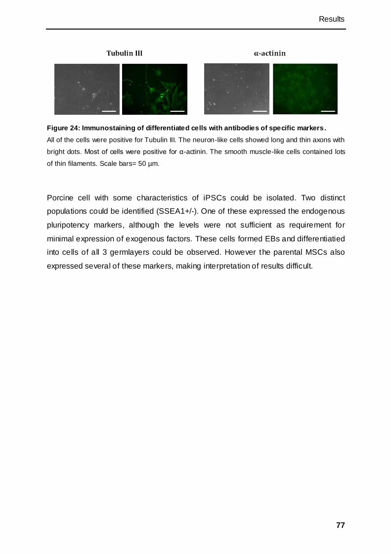
Results
77
Figure 24: Immunostaining of differentiated cells with antibodies of specific markers.
All of the cells were positive for Tubulin III. The neuron-like cells showed long and thin axons with
bright dots. Most of cells were positive for α-actinin. The smooth muscle-like cells contained lots
of thin filaments. Scale bars= 50 µm.
Porcine cell with some characteristics of iPSCs could be isolated. Two distinct
populations could be identified (SSEA1+/-). One of these expressed the endogenous
pluripotency markers, although the levels were not sufficient as requirement for
minimal expression of exogenous factors. These cells formed EBs and differentiatied
into cells of all 3 germlayers could be observed. However the parental MSCs also
expressed several of these markers, making interpretation of results difficult.

Results
78
3.3 Cell synchronization and gene targeting
As the overall goal of the project was to develope methods for improved genetic
manipulation of the pig genome and as it was not guaranteed that this could be
achieved by iPSC technology, an alternative approach was also attempted:
improving gene targeting in somatic cells, which compared to ESCs or iPSCs is
generally very low. As a result of low efficiency, few successful gene targeting
experiments have been reported for livestock.
As gene targeting by homologous recombinant is presumed to occur during the S-
phase of the cell cycle synchronization of cells might result in improved efficiency. To
test this, two tumor suppressor genes (APC and TP53) were chosen for targeting and
both targeting vectors were already available.
3.3.1 Gene targeting of APC and TP53 in synchronized pBMMSCs
A protocol to synchronize pBMMSCs was developed by M. Sc. Denise Nguyen, but
had never been employed for gene targeting experiments.
PBMMSCs were plated at a density of 60-70% confluence on T-75 flask and cultured
in normal MSC medium containing 10% FCS. 24h later, the medium was changed
into serum starvation medium containing 0.5% FCS. 14-16h post-starvation, the cells
were released from the serum starvation by changing the medium into MSC medium
containing 20% FCS.
24h post releasing, the cells were detached and collected for electroporation. 1×10⁶
pBMMSCs were electroporated with either 13 µg of linearized APC1061-BS targeting
construct or 10 µg of linearized p53-BS targeting construct. The cells were plated on
10cm dish and cultured in MSC medium (containing 20% FSC and 3 µg/ml
blasticidine). The transfection and selection of the unsynchronized groups were
performed with the same method.
3.3.2 Screening of the targeted colonies by PCR
After 10-12 days in selection, cell colonies become visible under the microscope. Cell
colonies were counted and subsequently single cell clones were picked and cultured
on 24-well plates and later expanded on 12-well plates. Once confluent, half of the

Results
79
cells were then collected for DNA isolation and PCR screening to determine if the
targeting vectors had integrated by homologous recombination. The rest of the cells
were frozen or expanded for additional analysis.
In the APC1061 PCR screening, the oligonucleotides (BSf and BSr) binding to BS
resistant cassette, were used to amplify a fragment of the BS gene as a positive
control. The primer pair BSf and TVR1 (Figure 25), which bind to the endogenous
APC gene and BS resistant gene, were used to screen for the targeting event. The
amplified fragments were 3.7 kb for the targeted cells.
A pair of TP53 endogenous primers (p53 endo F2 and p53 Int1_5R) was used to
amplify a 3.1 kb fragment as endogenous control. Another pair of primers (p53 endo
F2 and p53 targ R) binding to the TP53 gene and a sequence on the targeting
construct was used to screen for a TP53 targeting event (amplify a fragment of 3.3
kb). The targeting efficiencies as determined by the PCR screening are shown in
Table 4.
Figure 25: Oligonucleotides binding sites of APC.
BSf and BSr bind in the IRES-BS region. TVR1 binds in APC gene.
Table 4: Targeting efficiencies tested by the PCR screening.
APC1061 TP53
Selection
survived
PCR
positive %
Selection
survived
PCR
positive %
Synchronized 29 17 58.6% 64 1 1.6%
Unsynchronized 19 16 84.2% 76 5 6.6%
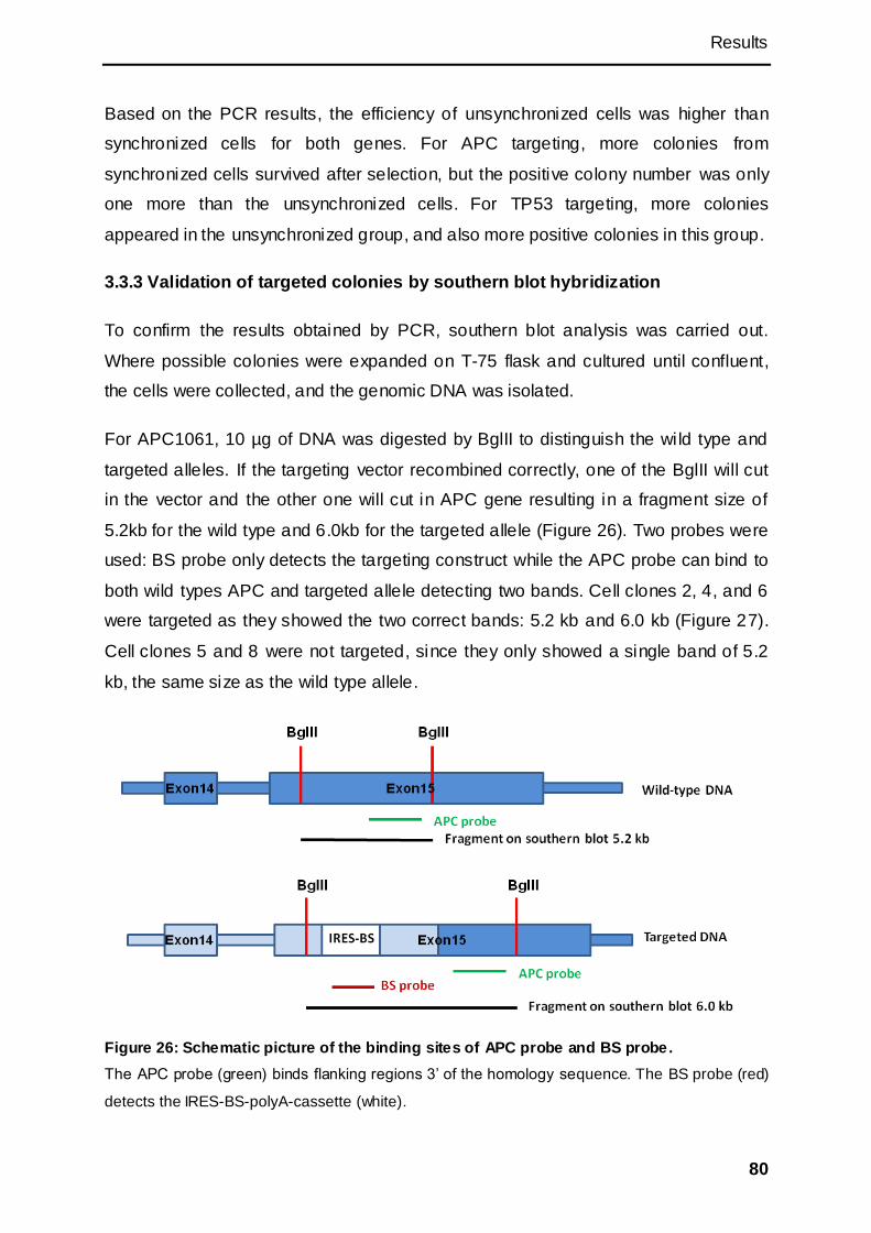
Results
80
Based on the PCR results, the efficiency of unsynchronized cells was higher than
synchronized cells for both genes. For APC targeting, more colonies from
synchronized cells survived after selection, but the positive colony number was only
one more than the unsynchronized cells. For TP53 targeting, more colonies
appeared in the unsynchronized group, and also more positive colonies in this group.
3.3.3 Validation of targeted colonies by southern blot hybridization
To confirm the results obtained by PCR, southern blot analysis was carried out.
Where possible colonies were expanded on T-75 flask and cultured until confluent,
the cells were collected, and the genomic DNA was isolated.
For APC1061, 10 µg of DNA was digested by BglII to distinguish the wild type and
targeted alleles. If the targeting vector recombined correctly, one of the BglII will cut
in the vector and the other one will cut in APC gene resulting in a fragment size of
5.2kb for the wild type and 6.0kb for the targeted allele (Figure 26). Two probes were
used: BS probe only detects the targeting construct while the APC probe can bind to
both wild types APC and targeted allele detecting two bands. Cell clones 2, 4, and 6
were targeted as they showed the two correct bands: 5.2 kb and 6.0 kb (Figure 27).
Cell clones 5 and 8 were not targeted, since they only showed a single band of 5.2
kb, the same size as the wild type allele.
Figure 26: Schematic picture of the binding sites of APC probe and BS probe.
The APC probe (green) binds flanking regions 3‘ of the homology sequence. The BS probe (red)
detects the IRES-BS-polyA-cassette (white).
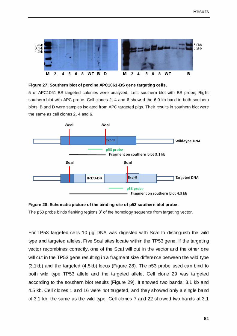
Results
81
Figure 27: Southern blot of porcine APC1061-BS gene targeting cells.
5 of APC1061-BS targeted colonies were analyzed. Left: southern blot with BS probe; Right:
southern blot with APC probe. Cell clones 2, 4 and 6 showed the 6.0 kb band in both southern
blots. B and D were samples isolated from APC targeted pigs. Their results in southern blot were
the same as cell clones 2, 4 and 6.
Figure 28: Schematic picture of the binding site of p53 southern blot probe.
The p53 probe binds flanking regions 3‘ of the homology sequence from targeting vector .
For TP53 targeted cells 10 µg DNA was digested with ScaI to distinguish the wild
type and targeted alleles. Five ScaI sites locate within the TP53 gene. If the targeting
vector recombines correctly, one of the ScaI will cut in the vector and the other one
will cut in the TP53 gene resulting in a fragment size difference between the wild type
(3.1kb) and the targeted (4.5kb) locus (Figure 28). The p53 probe used can bind to
both wild type TP53 allele and the targeted allele. Cell clone 29 was targeted
according to the southern blot results (Figure 29). It showed two bands: 3.1 kb and
4.5 kb. Cell clones 1 and 16 were not targeted, and they showed only a single band
of 3.1 kb, the same as the wild type. Cell clones 7 and 22 showed two bands at 3.1

Results
82
kb and 4.9kb. This indicated that cell clones 7 and 22 were not targeted. The 4.9 kb
bands could be from the random integration.
Figure 29: Southern blot of porcine p53-BS gene targeting cells.
Cell clone 1, 16 and wild type showed one band at 3.1 kb. Cell clone 29 showed two bands at 3.1
kb and 4.5 kb. Cell clone 7 and 22 showed two bands at 3.1 kb and 4.9 kb.
3.3.4 Somatic cell nuclear transfer with gene-targeted clones
Somatic cell nuclear transfer was carried out with three APC1061 targeted clones.
Three piglets were born. PCR and southern blot analysis (performed by Dr. Claudia
Merkl) confirmed that these pigs were derived from the targeted cells clones. The
results of APC1061 targeting were published in Gastroenterology (Flisikowska et al.,
2012).

Discussion
83
4 Discussion
For biotechnology and medical research, animal models are still irreplaceable, and
human disease models are widely used for basic research and the development of
new treatment strategies and for gene therapies.
Small animals, like zebra fish and mouse, have been studied for a long time and offer
good animal models. But due to their differences to human in their genetic affinity
and body size, their usefulness is limited. Now, genetic modification of livestock
species, such as pig, cow and sheep become possible and open the possibility to
use these as models for human diseases. In particular, the pig has been suggested
as the animal of choice as it has similar organ size and physiology to human, which
may make results obtained more predictable for the transfer into the clinic (Telugu et
al., 2010). Pigs are also deemed as the preferred donor animal for cell and organ
xenotransplantation (Rogers et al., 2008).
Gene targeting is a common research tool to genetically modified animals. It has
been used very successfully in the derivation of mouse models, which shows that it is
a powerful and promising tool in transgenic animal research. The optimal choice of
cells for gene targeting in the mouse is the ESCs (Capecchi, 2005). Though the
mouse ESCs and human ESCs have been established for a long time, there still
remains a large gap in our understanding how to isolate and cultivate genuine ESCs
from large animals. Up to now, all efforts to derive porcine or bovine ES cells have
been unsuccessful. Somatic cells in combination with nuclear transfer were used for
gene targeting to make up for the lack of ESCs and successfully generated
transgenic and targeted large animals (Laible and Alonso-González, 2009; Samiec
and Skrzyszowska, 2011) However, with a lower efficiency compared to mouse
ESCs. The somatic cells do not grow well as ESCs and often their lifespan is not long

Discussion
84
enough for expansion and analysis. Beside, somatic cells necessitate the use of
nuclear transfer to generate the transgenic animal, which again is inefficient.
The use of ESCs for genetic manipulation can save time and enhances the efficiency
dramatically. It is assumed that the frequency of homologous recombination is higher
in ESCs than somatic cells. Therefore gene targeting in the pluripotent stem cells is
easier, and targeted cells can be used directly to generate chimeric animals with
blastocyst injection. It has been shown that in mouse ESCs the targeting efficiency
was 10-fold higher than in somatic cells (Arbonés et al., 1994). However, a
comparison of targeting efficiencies between ESCs and somatic cells from livestock
species has so far not been possible, due to the lack of pluripotent stem cells.
Another question which has not been solved is the donor cells for blastocyst injection
in large animals. Induced pluripotent stem cells (iPSCs) could be the answer. After
the mouse iPSCs and human iPSCs were created, they were used successfully for
gene targeting (Zou et al., 2009).
Some notable results regarding porcine iPSCs have been achieved (Esteban et al.,
2009; Ezashi et al., 2009; Wu et al., 2009; West et al., 2010). Putative iPSCs could
be isolated and showed partial pluripotency, though all isolates differed from each
other. Even chimeric pigs were born as reported by one group, but there are still
queries if the cells are definitive iPSCs. Even if these putative iPSCs are not fully
reprogrammed, they have some properties of the ESCs and may be more efficient in
gene targeting than the somatic cells.
The purpose of this research was to search better cell types for transgenic animal
generation in particular large animals with precise genetic modifications. Porcine
somatic cells were reprogrammed to generate piPSCs. As none of the published
attempts to generate iPSCs were completely successful, other methods for their

Discussion
85
isolation were attempted in this work and results indicate that partial reprogramming
of somatic cells has been achieved. It provides a basis for further experiments.
As an alternative to iPSCs, in the second part of this thesis, cell synchronization
method to improve gene targeting efficiency in somatic cells was assessed. Two
different targeting constructs were used to modify different alleles in multipotent
MSCs. The synchronized and unsynchronized cells were compared in the efficiency
of genetic manipulation. The results showed that though targeting was performed
successfully, the synchronization did not result in higher efficiency. Targeted cells
were then used for somatic cell nuclear transfer and live piglets obtained. These
animals provide a model for the human FAP, since these patients have a strong
predisposition for colorectal cancer (Flisikowska et al. 2012).
4.1 Methods for generation of iPSCs
Following the first iPSCs report (Takahashi and Yamanaka, 2006), many groups tried
to isolate iPSCs from different species with different methods. By delivering the
reprogramming factors with lentiviral vectors (Yu et al., 2007), retroviral vectors
(Takahashi et al., 2007), adenoviral vectors (Stadtfeld et al., 2008b), episomal
vectors (Yu et al., 2009), transponsons (Kaji et al., 2009), and plasmid (Okita et al.,
2008), mouse or human iPSCs could be derived. It was investigated whether the 4 or
6 classic factors could be replaced (Nakagawa et al., 2007). Some groups generated
iPSCs with Oct4 and Sox2 or even only Oct4 (Kim et al., 2008; Kim et al., 2009a;
Giorgetti et al., 2010). To eliminate the potential mutation caused by integration of the
exogenous expression vectors, factors were delivered as mRNA or protein. However
the efficiency was generally lower and the methods were more complex (Warren et
al., 2010; Kim et al., 2009b). At the beginning of this project the generation of iPSCs
by delivering mRNA or protein into the cells had not been reported. For this reason
and because the use of expression vectors was reported to give the most consistent
results, this strategy was employed for the derivation of porcine iPSCs.
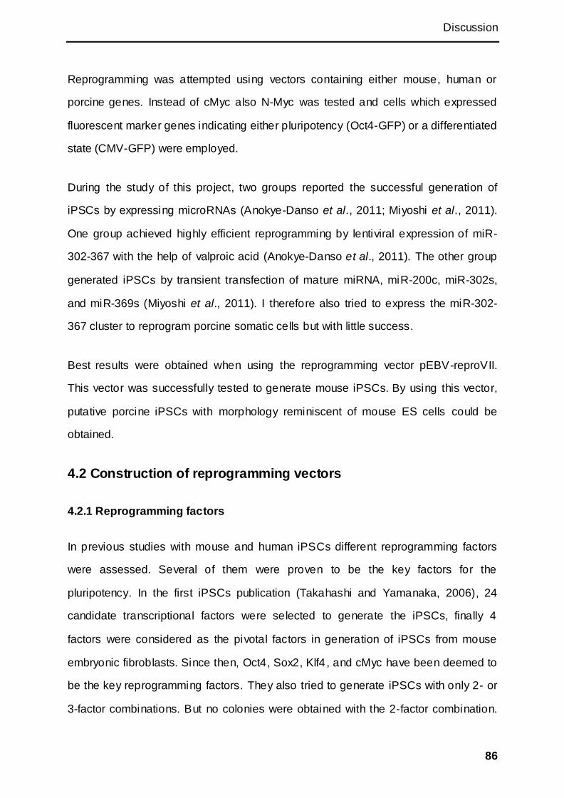
Discussion
86
Reprogramming was attempted using vectors containing either mouse, human or
porcine genes. Instead of cMyc also N-Myc was tested and cells which expressed
fluorescent marker genes indicating either pluripotency (Oct4-GFP) or a differentiated
state (CMV-GFP) were employed.
During the study of this project, two groups reported the successful generation of
iPSCs by expressing microRNAs (Anokye-Danso et al., 2011; Miyoshi et al., 2011).
One group achieved highly efficient reprogramming by lentiviral expression of miR-
302-367 with the help of valproic acid (Anokye-Danso et al., 2011). The other group
generated iPSCs by transient transfection of mature miRNA, miR-200c, miR-302s,
and miR-369s (Miyoshi et al., 2011). I therefore also tried to express the miR-302-
367 cluster to reprogram porcine somatic cells but with little success.
Best results were obtained when using the reprogramming vector pEBV-reproVII.
This vector was successfully tested to generate mouse iPSCs. By using this vector,
putative porcine iPSCs with morphology reminiscent of mouse ES cells could be
obtained.
4.2 Construction of reprogramming vectors
4.2.1 Reprogramming factors
In previous studies with mouse and human iPSCs different reprogramming factors
were assessed. Several of them were proven to be the key factors for the
pluripotency. In the first iPSCs publication (Takahashi and Yamanaka, 2006), 24
candidate transcriptional factors were selected to generate the iPSCs, finally 4
factors were considered as the pivotal factors in generation of iPSCs from mouse
embryonic fibroblasts. Since then, Oct4, Sox2, Klf4 , and cMyc have been deemed to
be the key reprogramming factors. They also tried to generate iPSCs with only 2- or
3-factor combinations. But no colonies were obtained with the 2-factor combination.
Discussion
87
With the 3-factor combination there were some colonies but none of them were
considered to be real iPSCs. However when other cell types were employed such as
neural stem cells, a single factor Oct4 could reprogram the cells although with low
efficiency (Kim et al., 2009b).
Unlike for human or mouse no porcine ESCs have yet been derived, in view of the
difficulties it was decided to begin with the classic 4-factor method, the ―OKSM
method‖ for reprogramming porcine primary cells. Nanog and Lin28 were also
considered for the generation of piPSCs based on their importance in ESCs and their
functions in reprogramming human somatic cells.
Klf4 and cMyc are tumour related genes. CMyc was considered to accelerate the
proliferation and transformation of the cells, which may be important in the early time
of the iPSC colonies emerging.
Considering that porcine pluripotent stem cells may have different signalling pathway
compared to mouse or human, other factors could be involved in their
reprogramming. For example Oct4 may not be the core factor for porcine cells, as
one group generated putative piPSCs with 3 factors omitting Oct4. The analysis of
the Oct4 expression of porcine early embryo also indicated that Oct4 might not be
the key factor in porcine embryogenesis (Kuijk et al., 2008). Other combinations of
reprogramming factors could be attempted in future research.
The established naïve iPSCs and ESCs in human showed a dependence of extra
Oct4 and Klf4 or Klf4 and Klf2 (Hall et al., 2009; Hanna et al., 2010). The similar
experiment could be done with putative porcine iPSCs to test if extra Oct4 and Klf4 or
Klf4 and Klf2 combinations can maintain the undifferentiated state in the absence of
doxycycline.
Discussion
88
It is know that there is a strict expression threshold for the reprogramming factors in
particular Oct4. Higher or lower levels of the factors may trigger the differentiation.
Only cells which express all four factors in the right level may become iPSCs. It may
be an explanation why there are so few cells transformed into pluripotent stem cells.
4.2.2 The functions of cMyc and N-Myc in reprogramming
As a proto-oncogene, cMyc greatly enhances reprogramming efficiency, although it is
dispensable for reprogramming (Nakagawa et al. 2007; Sridharan et al. 2009). The
negative role of cMyc in the self-renewal of hESCs was reported (Sumi et al., 2007),
and forced expression of cMyc induced differentiation and apoptosis of human ESCs.
This contrasts significantly to the positive role of cMyc in mouse ESCs (Cartwright et
al., 2005). During iPSC generation, transgenes delivered by retroviruses were
silenced when the transduced fibroblasts acquire ESC-like state. The role of cMyc in
establishing iPSCs could be as a booster of reprogramming rather than a controller
for the maintenance of pluripotency.
CMyc and N-myc are essential for completion of murine embryonic development. But
their roles in regulating pluripotency remain unclear. Previously, cMyc null mESCs
were found to retain the potential for self-renewal with slight changes in differentiation
capacity (Baudino et al., 2002), and N-myc can functionally replace cMyc in murine
development, cellular growth, and differentiation (Malynn et al., 2000). Investigation
of other Myc family members showed that both cMyc and N-Myc independently were
sufficient to maintain the pluripotency (Smith et al., 2010). L-myc seems to be
dispensable for embryonic development (Varlakhanova et al., 2010).
CMyc is an oncogene contributing to many human cancers (Beroukhim et al., 2010).
Defects in the Wnt-APC pathway found in human colon carcinoma result in enhanced
Tcf transcriptional activation of Myc (He et al., 1998). Retroviral insertional
mutagenesis further identified cMyc as a major murine oncogene (Akagi et al., 2004).
Discussion
89
These transgenic mouse studies provided the evidence that deregulated expression
of Myc is sufficient to drive tumorigenesis (Adams et al., 1985; Leder et al., 1986;
Chesi et al., 2008).
N-Myc showed lower tumorigenesis and could replace cMyc in an iPSCs cocktail
(Blelloch et al., 2007; Nakagawa et al., 2007). In this study we therefore tried to use
N-Myc to replace cMyc but no colony were obtained.
A previous study in human showed that cells formed colonies and proliferated fast
when only Oct4 and cMyc were transfected. But these colonies were not really
reprogrammed to pluripotent state. These colonies were considered to be a result of
cMyc expression (Lowry et al., 2008). We therefore wanted to test if this could be
true for our porcine cell clones. Transfections were carried out with or without cMyc.
If only Oct4, Sox2 and Klf4 were transfected no colonies appeared. If cMyc was
added to the cocktail ES like colonies did develop. To exclude that the change in cell
morphology was due to cMyc, cells were transfected with pCMYC only, no colonies
could be detected. This was a strong indication that only the combination of factors
affected cell morphology and growth behaviour.
When comparing reprogramming factor combination of Oct4, Sox2, Klf4, and cMyc
with combination of Oct4, Sox2, Nanog and Lin28 using episomal vector, the first
combination resulted in large number of piPSC colonies and the latter in none. In
combination with the above results it seems that cMyc is crucial in the
reprogramming process.
4.2.3 Delivery system
Viral transduction usually leads to higher efficiency than non-viral methods
(McMahon et al., 2006). Initial methods used to derive human iPSCs employed viral
vectors, in which both the vector backbone and transgenes were permanently

Discussion
90
integrated into the genome (Yu et al., 2007; Takahashi et al., 2007). Such vectors
can produce insertional mutations that interfere with the normal function of iPSC
derivatives, and residual transgene expression can influence differentiation into
specific lineages (Yu et al., 2007), or even result in tumorigenesis (Okita et al., 2007).
Integration free iPSCs were also generated successfully with adenoviral vectors or
repeated plasmid transfections (Stadtfeld et al., 2008b; Okita et al., 2008). But the
efficiency is quite low and not suitable if the cells need long exposure to the vectors.
Cre/LoxP was also used for removing the reprogramming factors, but the vector
backbone could still stay in the genome (Kaji et al., 2009; Soldner et al., 2009).
Transposon vectors are another option allowing removal of the integrated fragments
from the genomic DNA without leaving a trace (Woltjen et al., 2009).
Episomal vector was used widely because of their unique properties. Theoretically,
they don‘t integrate into the genome and remain independent of the genomic DNA.
They only duplicate when the cells start to split and separate into the daughter cells
equally.
In the research, presented here two different types of vectors were used : episomal
vectors and vectors which could integrate into pseudo attP sites (requires addition of
integrase) and could later be excised via the Cre/Lox system. They were delivered to
the cells by single or repeated plasmid transfections. However there is no guarantee
that the episomal vector could be excluded from genome even by using the
oriP/EBNA1 system integration.
If Oct4, Sox2, Klf4, and cMyc were used, the efficiency of colony generation was
same either episomal vector or attB-LoxP system. The difference was that the
episomal vector always contained all four factors, while different attB-LoxP vectors
were constructed containing between one to four dedifferentiation factors, which
could be transfected at different ratios, for example , increasing the amount of Oct4

Discussion
91
DNA or minimising the amount of cMyc DNA. After successful reprogramming, the
possible but unwanted integration of the episomal vector may have to be assessed.
For the attB-LoxP system the excision of the vector by Cre transduction sti ll need to
be tested. In most experiments, however these vectors should be transfected without
the addition of Phi integrase, in the hope that only transient expression occurs and no
vector integration. This still needs to be verified.
4.2.4 The promoter and doxycycline dependent tet-on system
Promoter that drives expression of the reprogramming factors is also of importance.
First of all, the promoter should allow for abundant expression of all factors. Second,
the promoter should be silenced or suppressed after the reprogramming process.
Comparison of several promoters used during ES cell differentiation showed that
CMV promoter drove the transgene expression only during the late stage. Similarly,
the CAGGS and phosphoglycerate kinase (PGK) promoter drove transgene
expression at a significant level only during late stages. Human elongation factor 1
alpha (EF1α) promoter directed robust transgene expression at every stage of mouse
ES cell differentiation (Hong et al., 2007) So far, the beta-actin-based promoter is the
only ubiquitous promoter that was reported to have little loss of gene expression
during human ES cell propagation and in vitro differentiation. The CAGGS is
comprised of the beta-actin promoter and may affect the behaviours of the
reprogrammed cells. In this work, a bidirectional promoter was used for expression.
This promoter contained two minimal CMV promoters. The tet-responsive element
(TRE) was between two promoters.
The tetracycline-dependent gene system allowed a strict control over the expression
of the pluripotency factors. Here a bidirectional promoter with an improved tet-on
system was used. By adjusting the concentration of the doxycycline , the expression
of the exogenous genes was controlled. In the absence of doxycycline the
Discussion
92
exogenous genes couldn‘t be expressed and the partially reprogrammed cells
changed their phenotype. This indicated that the tet-on system was very tight.
4.2.5 MicroRNA vector
Several reports demonstrated that specific miRNAs were highly expressed in ESCs
and played a critical role in the control of pluripotency related genes (Houbaviy et al.,
2003; Suh et al., 2004; Judson et al., 2009; Miyoshi et al., 2011).
One group transfected Nanog promoter-driven GFP mouse cells with mature miRNA
mir-200c, mir-302s, and mir-369s at 48 h intervals. After 15 days, they observed 5
colonies from 5×10⁴ cells and by 20 days post transfection they got 2 colonies from
1×10⁵ cells (Miyoshi et al., 2011).
The miR-302-367 cluster contains a short DNA sequence less than 1kb. Alignment
analysis showed that the miR-302-367 was highly conserved across species (Card et
al., 2008; Rosa et al., 2009). In combination with valproic acid, it was a promising
approach for porcine iPSC derivation and the experiment were based on those
published for mouse iPSCs isolation (Anokye-Danso et al., 2011). The porcine miR-
302-367 sequence was cloned into an expression vector driven by the CMV promoter.
Although the mature porcine miR-302-367 was expressed in the cells no iPS colonies
appeared.
4.3 Different cell types used for generation of iPSCs
Widely used cell types for iPSCs generation are fibroblasts and MSCs. Some
terminal differentiated cells were successfully reprogrammed to iPSCs, which
indicated that all of the somatic cells might be able to go back to the original
undifferentiated state. But the efficiency among different cell types varied
dramatically. In this study, considering the expected difficulties with the isolation of
porcine iPSCs, we decided to start with multipotent MSCs.
Discussion
93
It has been shown by several researchers that MSCs are a good choice for
generation of iPSCs. They are multipotent and keep the ability to differentiate into
several cell types, including chondrocytes, adipocytes , and oosteocyte. They can be
passaged up to about 50 passages in vitro. The porcine MSCs express Sox2, Klf4,
cMyc, which help the reprogramming process. In some MSC lines, the expression of
Oct4 and Nanog could be detected in early passages and negative in later passages.
Oct4 and Nanog expression was also found in human and rabbit MSCs (Lamoury et
al., 2006; Roche et al., 2007; Riekstina et al., 2009).
In our experiments, Oct4 expression couldn‘t be detected in the first isolates of
porcine MSCs. Lacking a source of Oct4 expressing cells, human Oct4 mRNA, which
has a high sequence similarity to pig, was used as a replacement. Later on i n newly
isolated pMSCs Oct4 was detected by PCR, but the expression level might have
been very low as all attempts to clone Oct4 failed again.
From mouse iPSCs research, we know that the efficiency of generation of iPSCs was
low even with mesenchymal stem cells. The results from Yamanaka‘s group showed
no evidence that the iPSCs are from the multipotent stem cells (Takahashi and
Yamanaka, 2006). In this research, we focused on porcine MSCs (including
BMMSCs and ADMSCs), and also tested other types of cells, like fetal fibroblasts
and kidney fibroblasts. ADMSCs were used preferably since they were considered as
an easily obtainable cell source and the better donor cells for iPSCs in human and
mouse (Sugii et al., 2010). Studies showed that ADMSCs generated the iPSCs more
efficiently with or without feeder. This may due to the high expression of FGF, LIF
and fibronectin in ADMSCs. It was found that ADMSCs could serve as feeder cells
for pluripotent stem cell lines (Sun et al., 2009; Sugii et al., 2011).
Discussion
94
4.4 The medium and supplemental factors for iPSCs culture
Since mouse and human ESCs have long been established, their culture conditions
have also been standardized. They have different signalling pathway for self-renewal.
Basically, the mouse ESC medium is based on DMEM and supplemented with LIF,
10-15% serum or 15-20% serum replacement. Conditioned medium collected from
medium used by MEFs was used for mouse ESCs in the early time. Human medium
is based on knock out DMEM and supplemented with 4-10 ng/ml bFGF, 10-15%
serum or 15-20% serum replacement. But different group may modify their medium
by adding inhibitors and changing the concentration of the factors and serum or
serum replacement. Since the components of serum varied by their source, the
serum replacement was considered as a more reliable supplement for the cells.
Some of the factors in serum may lead the cells to differentiate. The serum free
mediums were also widely used for the iPSCs. Normally, it is based on DMEM/F12
medium and Neurobasal medium, supplemented with B27, N2 supplement and other
factors and inhibitors.
During the reprogramming process, cells should be maintained in culture conditions
that support self-renewal. The most important is the culture environment, which is a
key determinant for the outcome of the pluripotent state (Van Oosten et al., 2012).
The four factors, however, could not induce human iPSCs when fibroblasts were kept
under the culture conditions for mESCs after retroviral transduction. These data
suggest that the fundamental transcriptional network governing pluripotency is
common in human and mice, but extrinsic factors and signals maintaining
pluripotency are unique for each species (Takahashi et al., 2007).
In our experiments the LIF was produced by MEFs and the unit couldn‘t be counted.
The results showed that the putative iPSCs needed a rich medium, which was based
on serum-free medium and supplement with LIF, GSK3 inhibitor and MEK inhibitor.

Discussion
95
Besides, the cells grew better with the addition of 10% serum. Withdraw of any
factors or inhibitors could lead cells lose their ESC-like morphology. The cells relied
on LIF and were independent on bFGF, which indicated that the putative piPSCs
were closer to mESCs.
4.5 MicroRNA reprogramming
Histone deacetylase (HDAC) inhibitor VPA was used in our experiments, as it was
reported to enhance the reprogramming efficiency. One group employed lentiviral
vector to express the miRNA driven by CMV promoter. Their results showed that the
efficiency of generating iPSCs was higher compared to the traditional 4 factors
method, but they showed no colony could be generated without VPA. We tried to
culture the transfected cells with VPA. Though the expression of the miRNA was
detected, there were no colonies. The same results were achieved from rat MSCs
and mouse MEFs. Increasing the number of repeated transfections could be tried in
the future. The classic reprogramming cocktail could a lso be used together with
microRNA.
4.6 Reprogramming of somatic cells
4.6.1 Transfection methods
Compared to nucleofection, the electroporation was less harmful for the cells and
they recovered soon after transfection. But the nucleofection had higher transfection
efficiency. And, in the nucleofection, the nucleic acid could be delivered directly into
the nucleus. In this research, both the nucleofection and electroporation were used
for cell transfection. Pre-experiments were performed to determine the optimal
conditions. In some experiments, the cells needed to be nucleofected twice, and the
cells weren‘t counted at the second time, which may lower the transfection efficiency
since the nucleofection requires fixed cell numbers.

Discussion
96
4.6.2 Colony screening, picking, passaging, and storage
Selection is not necessary for obtaining the stable iPSCs, but omission of selection
increased the number of false positive colonies, such as transformed cells or cells
that failed to receive all factors (Stadtfeld et al., 2008a). In mouse some scientists
used cells which had a resistant gene inserted at the endogenous Oct4 or Nanog
locus for positive selection (Wernig et al., 2007; Okita et al., 2007). Live cells imaging
was also used for distinguishing bona fide human iPSCs from the partially
reprogrammed cells (Chan et al., 2009). To screen the iPSC colonies, the easy way
is to visualize the signals of pluripotency. A knock-in GFP driven by the Oct4
promoter was used to indicate the re-expression of the endogenous factors and the
stable iPSCs were selected successfully by this system (Stadtfeld et al., 2008a).
In experiments described here, by using Oct4-GFP transgenic fetal fibroblasts, no
green colonies were observed, but the GFP expression was confirmed by the RT-
PCR. This indicated that the Oct4 promoter was activated but maybe not fully
demethylated. In the experiment of CMV-GFP MSCs, the CMV promoter should be
silenced in fully reprogrammed cells since it was suggested that CMV was inactive in
ESCs. Before transfection, the MSCs were brightly green. After transfection, the
putative piPSCs and the unreprogrammed cells were still green, while some cells in
the colonies were only faintly green. This phenomenon didn‘t change after several
passages. That suggested the CMV promoter was not silenced because the cel ls
were not fully reprogrammed.
Previous studies on human iPSCs showed that there were many non-ESC-like
colonies generated with OKSM (Oct4, Klf4, Sox2 and cMyc). These colonies had
different morphology compared with the real iPSC colonies and the positive selection
reporter didn‘t activate in these colonies. These colonies shared similar morphology
with our piPSC colonies which had irregular bright border and darker centre. The
nature of these colonies remains to be determined (Takahashi et al., 2007; Hotta et

Discussion
97
al., 2009). It was reported that some ―early colonies‖ were observed 14 days post
infection, which were highly proliferative and changed morphology. But further
characterization indicated that these colonies were only transfected with Oct4 and
cMyc and they were not real iPSCs. 21 days later, new colonies emerged. These
―late colonies‖ were reprogrammed to iPSCs (Lowry et al., 2008). In our study, the
putative piPSC colonies appeared at about day 7. These colonies shared the
morphology of those ―early colonies described‖ above. This may indicate that they
are not fully reprogrammed. But, no new colony appeared even after waiting for more
than one month.
For mouse and human cells the time for generation of iPSC colonies varied among
different research groups. Depending on different methods, the period varied from 9
days to 40 days (Table 1). In our research, after transfection, the putative porcine
iPSC colonies appeared and were picked 7-10 days. The putative iPSCs from
Tibetan miniature pig emerged by 8-10 days post-infection (Esteban et al., 2009).
Another group which got human iPSC-like colonies, found that the colonies were
visible 7 days post-infection and the colonies were large and round with clear
boundaries on day 13 (Wu et al., 2009). However, the mouse ESC-like colonies from
one group emerged 22 days post transduction (Ezashi et al., 2009). One group,
which generated chimeric offspring from their piPSCs, reported their initial colonies
appeared within 7 days.
4.7 Identification of iPSCs
In the definition of the pluripotent stem cells two characteristic are important. One is
the self-renewal, which is defined as the ability of cells to keep reproducing
themselves without differentiation under appropriate culture conditions. The other is
pluripotency the ability to differentiate into the three germ layers. The standards of
pluripotency of iPSCs are the same as established for mouse and human ESCs.

Discussion
98
They can differentiate into all the somatic cells and produce germ line cells. The new
developed porcine pluripotent stem cells were compared with them.
To evaluate the pluripotency, many aspects could be considered. The first is the
morphology of the putative iPSCs. Our putative piPSC colonies showed mouse ESC-
like compact morphology with three dimensional structure and clear border. These
cells displayed a high nuclear-to-cytoplasmic ratio and prominent nucleoli, though
some human ESC-like morphology which was flatter compared with mouse ESC was
also observed (Esteban et al., 2009; Wu et al., 2009).
Beside the morphology in vitro proliferation, feeder dependence, surface markers,
gene expression, promoter activities, telomerase activities, and embryoid formation
and differentiation should be considered. In vivo teratoma formation, chimeric
formation, and germline contribution are important for the final pluripotency
evaluation.
4.7.1 Feeder dependence
The pluripotent stem cells are generally maintained on a layer of inactive MEF feeder
cells. The feeder cells could affect the analysis of pluripotent stem cells, for example
the gene expression analysis by RT-PCR. It also influences the differentiation bias of
pluripotent stem cells in culture. In mouse ESCs the feeder layer could be replaced
by adding cytokine and LIF, but this didn‘t work for human ESCs. Gel-Matrix was
used to replace the feeder for human ESCs (Xu et al., 2001).
In our study, the feeder was not necessary during the iPSC derivation. With or
without feeder cells, the colonies were derived with the same efficiency. Similar
results were reported for human and mouse iPSCs (Sugii et al., 2011). But it is
crucial to maintain the putative piPSCs on feeder cells for proliferation. When plated
on Gel-Matrix, the cell colonies lost their dome-like morphology and became loose
and flat, but they still survived.

Discussion
99
4.7.2 Silencing event in pluripotent cells and doxycycline dependency
One of the differences between pluripotent stem cells and somatic cells is the ability
to silence retroviruses (Barkis et al., 1986; Teich et al., 1977). Fully reprogrammed
cells show silencing of the factors delivered by retroviral vectors (Maherali et al.,
2007; Okita et al., 2007; Wernig et al., 2007), whereas partially reprogrammed cells
show incomplete silencing and persistent expression of the viral factors (Takahashi
and Yamanaka, 2006). Further research in mouse showed that retroviral silencing is
a gradual process that is initiated early but completed late in the reprogramming
process, coinciding with the activation of epigenetic regulators and pluripotent genes
(Stadtfeld et al., 2008a).
In human ESCs, the lentiviral delivered genes were suppressed under a promoter
dependent manner, whereas the suppression of transient expressions weren‘t
observed (Xia et al., 2008). Their results indicated that the promoter-dependent
transgene expression and suppression in human ESCs is dependent on integration
of the transgenes into the host genome. In mouse, the fully reprogrammed iPSCs
were largely maintained by the activity of the endogenous pluripotency factors
(Wernig et al., 2007).
Wu et al (2009) described their cells depended on doxycycline to maintain the
undifferentiated state. The exogenous genes were silent with the absence of
doxycycline in differentiation in vitro. In other two groups, even though their cells
could differentiate into the three germ layers, the expression of retroviral transgenes
were still detected (Esteban et al., 2009; Ezashi et al., 2009). This means the
reprogrammed cells still depended on exogenous factors. In our research, the
exogenous factors were always expressed with the presence of doxycycline.
A major advantage of the inducible system over constitutive expression systems is
that it allows for the ‗‗self-selection‘‘ of reprogrammed cells in the absence of drug

Discussion
100
selection and obviates the need for ES cell expertise. After stably reprogrammed
cells have been generated and doxycycline has been withdrawn, cells that survived
go on to reactivate the endogenous pluripotency program, while unstable
reprogramming intermediates and transformed colonies disappear, likely through
differentiation or apoptosis.
Along with the theory of naïve pluripotent stem cells, the real embryonic stem cells in
human were defined (Nichols and Smith, 2009; Hanna et al., 2009). Naïve pluripotent
cells can differentiate into a primed EpiSC-like state in vitro by promoting the
signalling of TGF-b, Activin, and bFGF, EpiSCs can epigenetically revert back to
naïve pluripotency by a variety of genetic manipulations and culture conditions. The
conserved hESCs were considered as hEpiSCs that were slightly different from naïve
ESCs. HEpiSCs showed a flat morphology compared to the mouse ESCs (naïve
state). The naïve ESCs were considered as the ground state whereas the EpiSCs
were considered as the primed state. Though both of them can form teratomas, the
naïve ESCs can contribute to the chimaeras while the EpiSCs can‘t. The naïve ESCs
have no differentiation bias and the EpiSCs have variable differentiation bias. What‘s
more, the naïve ESCs respond to LIF/STAT3 pathway while the EpiSCs respond to
FGF pathway. ESCs need the pluripotency factors Oct4, Nanog, Sox2, Klf2 and K lf4
while EpiSCs only need Oct4, Nanog and Sox2 (Nichols and Smith, 2009). To
investigate the naïve state of human pluripotent stem cells, the doxycycline-control
system was used to generate hiPSCs. Unlike the result from mouse iPSCs, those
naïve hiPSCs depended on doxycycline and differentiated upon withdrawal of
doxycycline. With the expression of Oct4 and Klf4 or Klf4 and Klf2, the hiPSCs could
be released from the dependency of doxycycline (Hanna et al., 2010). When the
hESCs were transfected with Oct4 and Klf4 or Klf4 and Klf2, they changed their
flattened morphology to dome-shaped colonies and packed round cells morphology,
like mESCs. The generated hESC-like piPSCs also employed doxycycline-control
system, similar results showed that their piPSCs changed morphology and

Discussion
101
differentiated quickly without doxycycline (Wu et al., 2009). Many of our putative
piPSCs showed dome-like colonies and mESCs morphology, especially the SSEA1
positive cells. They also depended on doxycycline. This indicated that these cells
may also need extra factors, like additional Oct4 and Klf2, to maintain the
undifferentiated state.
Contrast to previous studies, the results from our piPSCs, indicated that the iPSCs in
large animals are not permissive as mouse, and they may need extra conditions to
stabilize the naïve state.
4.7.3 Gene expressions of iPSCs
After several passages the putative piPSCs were analyzed for expression of several
endogenous genes. The expression of pOCT4, pSOX2 and pNANOG as the most
important pluripotent genes, were detected. But without qPCR, we couldn‘t say if they
were up regulated, since our BMMSCs used for piPSCs expressed all the factors
(even Oct4) before reprogramming. The GAPDH, housekeeping gene, was used to
test if there was RNA or not. Though the primers were designed for porcine GAPDH,
it was not specific enough as they allowed amplification of mouse GAPDH. To avoid
amplification of both endogenous and exogenous fragments primers were designed
to bind to the 3‘ or 5‘ UTR of the porcine sequences, so only porcine were detected.
Sox2 and TERT expression was found in all samples. Two different piPSC
populations were obtained: SSEA1 positive and SSEA1 negative (see 3.2.5).
Interestingly both populations were negative for pCMYC expression, which might
indicate that the partially reprogrammed cells down regulated or silenced pCMYC
due to the presence of exogenous mcMyc. The SSEA1 negative cells were negative
for pNANOG and pKLF4, which was consistent with the differences in their loose
morphology and immuostaining results. As long as the doxycycline was present and
the exogenous genes were expressed cells could be cultured for many passages.

Discussion
102
4.7.4 The karyotype stability
The long-term karyotype stability is an important issue, especially for cells passaged
by enzymatic digestion (Trounson, 2006). In human ESCs there was a considerable
instability of some imprint genes in long-term culture (Rugg-Gunn et al., 2005). For
the porcine iPSCs even at passage 22 half of the cells showed a normal karyotype.
4.7.5 The specific markers of pluripotent stem cells
The immunostaing results in this work showed that the putative piPSCs carried the
SSEA1 surface marker. But they were negative for SSEA4, Tra-1-60 and Tra-1-80.
This result supported the point that the putative piPSCs were closer to mouse iPSCs
than human iPSCs. In previous studies, the putative iPSCs from different groups
showed different surface markers. Two groups showed their piPSCs were SSEA1
positive (Ezashi et al., 2009; Telugu et al., 2010), while one group found their piPSCs
positive for SSEA3, SSEA4, Tra-1-60 and Tra-1-80 (Wu et al., 2009), which is similar
to human iPSCs. The piPSCs reported by one group were SSEA4 positive, but no
other surface markers were tested (Esteban et al., 2009). The morphology also
seems to have a relationship with the surface marker. The morphology of SSEA1
positive cells had more similarity with mouse iPSCs, while the SSEA3 and SSEA4
positive cells were closer to human iPSCs which are flatter (Ezashi et al., 2009; West
et al., 2010).
In our study, not all of the cells were SSEA1 positive. Even in one colony, part of the
colony was positive whereas the rest was weakly positive or negative. This coincided
with the observation that the colonies of some putative piPSCs showed a mixed
morphology. In order to get a pure population, the cells were separated with the
SSEA1 marker (microbeads system). More than 80% of cells in the SSEA1 positive
population were SSEA1 positive after separation and this percentage was maintained
for several passages. The positive and negative populations showed different
Discussion
103
morphology: the positive one showed ―dome-like‖ compact colonies while the
negative one seemed flat and loose. The SSEA1 positive cells also performed better
in the following differentiation experiment.
4.7.6 The differentiation ability of putative piPSCs
Embryoid body-mediated differentiation was performed to test the differentiation
ability of putative piPSCs. In this work, different methods were used for the EBs
formation, including hanging drops, suspension and aggregate well. Even for the
same cell line, not all the time the cells could form embryoid bodies. Balls and
―sausages-like‖ EBs showed an undistinguished morphology compared to published
mouse or porcine EBs. RT-PCR of these differentiated cells was performed to
analyze the expression of differentiation markers. As shown in the results, the
expression patterns were confusing. AFP, an endoderm marker, was positive in
SSEA1 positive cells and embyoid body sample 2 (EB2). Another endoderm marker,
AAT, was only found in MSCs. Desmin and Enolase were positive in all the samples.
Since they are the markers for mesoderm, a possible explanation is that the cells
were only partially reprogrammed and still had the memory from the MSCs, whic h
are derived from mesoderm. NeuroD, as a marker for ectoderm, was found in MSCs,
SSEA1 positive cells and EB2. Rex1, considered as a marker for epiblast cells, was
found only in EB2. In all of the published works, piPSCs could differentiate into the
three germ layers by embryoid body-mediated differentiation. The markers of three
germ layers described above were also detected. Besides, they also checked the
expression of pluripotency markers, like Oct4 and Sox2. One group showed almost
silenced Oct4 and strongly down regulated Sox2 (Ezashi et al., 2009). The cells with
hiPSC morphology showed Oct4, Sox2 and Nanog decreased in EBs differentiation
(Wu et al., 2009). The group which generated the chimeric pigs reported that hOCT4
and pOCT4 were detected in EBs after 10 days of differentiation. This might be due
to the resistance to silencing caused by lentiviral integration (West et al., 2010). In

Discussion
104
this work, we found the presence of pOCT4, pSOX2, and pNANOG expression in the
EBs, which means the cells weren‘t fully reprogrammed and couldn‘t get the
pluripotency marker silenced during differentiation. Real time RT-PCR can be
performed to analyze if the factors were down regulated after differentiation or not in
the future.
By adding retinoic acid to EB culture neuronal differentiation can be induced in
human and mouse. The method was used to differentiate rat iPSCs successfully into
neurons and was then tested for piPSCs differentiation. Immunostaining was
performed to identify the cell surface markers. There were mainly two populations in
the differentiated cells: neuron-like cells and smooth muscle like cells. The mouse
beta-tubulin III antibody was applied in the immunostaining. A few neuron-like cells
were observed in the differentiated population. But the immunostaining result was not
specific enough. The antibody was designed specific for mouse. It was possible that
the antibody could bind to other things unspecifically in porcine or did not recognise
porcine proteins at all. Since there were many smooth muscle-like cells, another
antibody which can bind to alpha-actinin, was also used to identify smooth muscle
cells. Though the antibodies were tested with different dilutions and the negative
control was negative, all the cells showed a weak positive signal. Similar results were
shown in previous studies: human ESC lines H7 and H9 cultured on Gel-Matrix were
positive for the neuronal precursor marker beta-tubulin class III both in
undifferentiated cells and EBs (Carpenter et al., 2001). It was also found in some
cancer cells and was considered as a marker of angiogenic perivascular cell (Sève et
al., 2007; Chen et al., 2011; Stapor and Morfee, 2012). Specific anti-porcine
antibodies may be needed for further research.
To demonstrate multilineage differentiation, piPSCs could be injected into nude mice
and generated teratomas derived from three germ layers. All the published porcine
results showed that piPSCs could differentiate into three germ layers in vivo. We also
Discussion
105
tried to inject nude mice with our putative piPSCs, but no teratoma was obtained
successfully. Considering their doxycycline dependency, it was not surprising that
they may have stopped to proliferate in vivo without doxycycline.
However, germ line competence wasn‘t reported in the first three piPSC publications,
indicating that these cells do not possess the full arsenal of pluripotent properties. So
far, only one group described their piPSCs could generate chimeric offspring (West
et al., 2010). They got live chimeric fetus by injecting the piPSCs into embryos with
high efficiency of 85.3%. All of the chimeric fetuses developed normally, and the
young chimeric pigs developed without tumour. PCR analysis confirmed that the
piPSCs contributed not only to the chimeric offsprings but also to germline. But
germline transmission was very low and all offsprings derived from iPSCs only
survived for 3 days. This indicates that their iPSCs were not fully competent (West et
al., 2011).
4.8 Gene targeting and synchronization
Since the real piPSCs were failed to be generated, the alternative choice was
considered for gene targeting: cell synchronization. Somatic cells were successfully
applied in livestock gene targeting. Theoretically, the targeting efficiency could be
improved by arresting cells in S/G2 phase in which homologous recombination
occurs.
Serum starvation was used to synchronize the pMSCs at S phase in this work. By
comparing the targeting efficiency between synchronized and unsynchronized cells,
the result determined if synchronization could increase the rate of homologous
recombination or not. Two tumour related genes, APC and TP53 were studied in this
work. Porcine MSCs were successfully targeted with their targeting vectors. PCR and
southern blot were used to check if the cells were targeted.
Discussion
106
4.8.1 Synchronization method
Specific synchronization can favour the targeting efficiency by increasing the rate of
homologous recombination. To eliminate the side effect of chemical inhibitors, serum
starvation is a better choice to synchronize primary cells prior to gene targeting. The
starvation and release from cell cycle block were controlled by changing the serum
contents of the medium, which is easy to manipulate. According to the results from M.
Sc. Nguyen, the serum deprivation was sufficient to arrest the pMSCs while some
chemical inhibitors failed (hydroxyl urea and thymidine). So the serum starvation
protocol was used in this research. The starvation could last for up to 96h (Goissis et
al. 2007, Kues et al. 2000). To minimize the cellular stress, the starvation time was
limited to 24h. The starter population of pMSCs was limited to a confluence of 60-
70%, which is crucial for the synchronization in this protocol. The cells were arrested
at G0 phase after 24h of serum starvation. Then medium with 20% serum was used
to release the cells from the G0 phase. 16h post-releasing, most of the cells were
stuck at the S/G2 phase, meanwhile the cells were transfected in order to get the
highest efficiency, since homologous recombination occurred at S/G2 phase.
Cell populations from different isolations could have remarkable differences in cell
doubling time, which may affect the efficiency of synchronization. The cells which
used for establishing the serum starvation protocol may have a different growth
speed from those used for gene targeting, thereby decreasing the synchronize effect.
4.8.2 Validation of the targeted colonies
A promoter-trap strategy was used to enrich for targeted cell clones. Without
integration near a promoter the resistant gene couldn‘t be expressed. This strategy
helped to avoid the false positive clones caused by random integration. The
concentration of the selection antibiotics were determined by a killing curve. The final
Discussion
107
concentration should be just enough to kill the wild type cells but not hurt the cells
with the construct integrated.
After 10 to 15 days in selection the cells formed colonies on the plates. The single
colonies or minipools were expanded and screened by PCR. PCR screening was not
enough to confirm that the cells are truly targeted. Further analysis, such as Southern
blot analysis, were required, this should show predicted bands not only for the
targeted but also the wild type alleles and exclude cells with additional random
integration. In this work, the step of validation was very necessary before using the
cells for nuclear transfer.
Three of APC1061 targeted colonies were used for somatic cell nuclear transfer.
Piglets from these cells were born and analyzed and targeting confirmed. The result
was published on Gastroenterology (Flisikowska et al., 2012). These transgenic pigs
could provide a valuable resource for colorectal cancer research.
4.8.3 Comparison of targeting efficiencies
Even with the promoter-trap strategy, there were still false positive colonies caused
by different reasons. To calculate the efficiency, it is necessary to base on the
number of the colonies survived from the antibiotic selection. Different porcine
BMMSC lines were pre-cultured to assess their growth speed, and the cells with fast
proliferation were used for the targeting.
In order to compare targeting efficiency, all the protocols for transfection, the cells
used, and the screening methods were the same between synchronized and
unsynchronized cells. In previous studies in our group, high targeting efficiency of the
APC locus was obtained in pBMMSCs. In this work, the efficiency was up to 84.2%
based on PCR results. But not all colonies tested by southern blot were positive for
targeting. Half of the tested colonies were verified to be targeted. The rest of them
Discussion
108
were just random integrated. There were more colonies from the synchronized cells,
but the targeting efficiency was lower compared to the unsynchronized cells.
In this work, for TP53, unsynchronized cells resulted in 5 targeted colonies from 76
screened (6.6%). The southern blot analysis showed that only one colony was
correctly targeted (1.3%). Synchronized cells resulted in only one targeting positive
colony according to the PCR screening. This colony could not be expanded to allow
confirmation by southern blot analysis. In summary, the cell synchronization by
serum starvation didn‘t increase the efficiency of targeting as was expected, on the
contrary it decrease the efficiency in this study. The results indicated that
synchronizing the cell cycle at the S phase increased the frequency of random
integration of the targeting vector in the genome; at the same time it did not improve
gene targeting. Previous study with porcine fetal fibroblasts showed that
synchronizing the cell cycle at the S phase significantly increased the frequency of
random integration of the targeting vector in the genome; at the same time it did not
improve gene targeting (Lee, 2010).
Since the targeting efficiencies vary even in the same cell isolate, more repeat
experiments need to be performed to determine the accuracy of the results. In
general, the efficiency of APC1061 targeting is high according to the results obtained
here and by other member of our group.
4.9 Outlook
For the pursuing of porcine iPSCs, more attempts are needed. Since the
reprogramming factors are still unclear for pig, more factors may have to be involved
in the reprogramming process, such as Tbx3, Klf2. Other delivery methods can be
used, such as lentiviral, retroviral vectors or transposons, or a higher number of
repeated transfection could be used. The medium components could be modified
according to the published achievements of putative porcine ESCs culture conditions.

Discussion
109
The porcine Oct4 and Nanog promoters could be cloned used for expression of
florescent reporters or selection genes. The differentiation protocols could be
improved in further experiments.
The partially reprogrammed cells can be used for gene targeting. They grow as fast
as mouse ESC/iPSC and can be expanded over a prolonged period of time. Even if
they cannot be injected into the early embryo to produce chimeric animals, they may
be useful donor cells for nuclear transfer.
The serum starvation protocol should be modified in the future. Other synchronization
methods could be tried. For example, the flow cytometry can be used for collecting
the cells in same cell cycle phase.
4.10 Concluding marks
Large animal models are can support medical researchers. Pig, as an animal close to
human, is much more important than any other species. However, the wildly used
gene targeting method is limited in large animals because of the lack of pluripotent
stem cells.
In this project a suitable cell sources or improved technology for gene targeting in
pigs were developed. Several different methods were used for generating porcine
iPSCs which could be considered as the optimal choice for gene targeting. Several
reprogramming factors Oct4, Sox2, Klf4, cMyc, Nanog, N-Myc, and Lin28 were
constructed into different vectors. Site-specific recombination vectors or episomal
vectors were used to deliver the factors into the cells. ESC specific microRNAs were
also tried to generate the iPSCs with the help of va lproic acid. Some putative porcine
iPSCs were achieved and assessed for the pluripotency. SSEA1+ and SSEA1- cells
were identified and could be separated for further differentiation.

Discussion
110
The synchronized and unsynchronized MSCs were used for gene targeting. Two
different target loci APC and TP53 were studied. Results showed that the
synchronization was unable to increase the targeting efficiency in both experiments.
The targeted colonies were validated by southern blot. APC1061 targeted cells were
used for SCNT and three piglets were born.
References
111
5 References
Aasen, T., Raya, A., Barrero, M.J., Garreta, E., Consigio, A., Gonzalez, F., Vassena, R., Bilic, J.,
Pekarik, V., Tiscornia, G., et al. Efficient and rapid generation of induced pluripotent stem cells
from human keratinocytes. Nat. Biotechnol. 2008, 26:1276–1284
Adams, J.M., Harris, A.W., Pinkert, C.A., Corcoran, L.M., Alexander, W.S., Cory, S., Palmiter,
R.D., and Brinster, R.L. The c-myc oncogene driven by immunoglobulin enhancers induces
lymphoid malignancy in transgenic mice. Nature 1985, 318: 533–538.
Adjaye, J., Huntriss, J., Herwig, R., Benkahla, A., Brink, T.C., Wierling, C., Hultschig, C., Groth,
D., Yaspo, ML., Picton, H.M., et al. Primary Differentiation in the Human Blastocyst: Comparative
Molecular Portraits of Inner Cell Mass and Trophectoderm Cells. Stem Cells 2005, 23: 1514–
1525.
Alberio, R. and Perez, A.R. Recent advances in stem and germ cell research: implications for the
derivation of pig pluripotent cells. Reprod. Domest. Anim. 2012, 47(4): 98–106.
Akagi, K., Suzuki, T., Stephens, R.M., Jenkins, N.A., and Copeland, N.G. RTCGD: retroviral
tagged cancer gene database. Nucleic Acids Res. 2004, 32: D523–D527.
Ambrosetti, D. C., Scholer, H. R., Dailey, L., and Basilico, C. Modulation of the activity of multiple
transcriptional activation domains by the DNA binding domains mediates the synergistic action of
Sox2 and Oct-3 on the fibroblast growth factor-4 enhancer. J. Biol. Chem. 2004, 275: 23387-
23397.
Anderson, G. B., Choi, S. J., and Bondurant, R. H. Survival of porcine inner cell masses in culture
and after injection into blastocysts. Theriogenology. 1994, 42(1): 204-12.
Anokye-Danso, F., Trivedi, C. M., Juhr, D., Gupta, M., Cui, Z., Tian, Y., Zhang, Y., Yang, W.,
Gruber, P.J., Epstein, J.A., et al. Highly efficient miRNA-mediated reprogramming of mouse and
human somatic cells to pluripotency. Cell Stem Cell 2011, 8(4): 376–88.
Arbonés, M.L., Austin, H.A., Capon, D.J., and Greenburg, G. Gene targeting in normal somatic
cells: inac- tivation of the interferon-gamma receptor in myoblasts. Nat. Genet. 1994, 6: 90–97.
Balasubramanian, S., Babai, N., Chaudhuri, A., Qiu, F., Bhattacharya, S., Dave, B.J.,
Parameswaran, S., Carson, S.D., Thoreson, W.B., Sharp, J.G., et al. Non cell-autonomous
reprogramming of adult ocular progenitors: Generation of pluripotent stem cells without
exogenous transcription factors. Stem Cells 2009, 27:3053–3062.
Bartel, D.P. MicroRNAs: target recognition and regulatory functions. Cell 2009, 136(2):215–33
Baudino, T.A., McKay, C., Pendeville-Samain, H., Nilsson, J.A., Maclean, K.H., White, E.L., Davis,
A.C., Ihle, J.N., and Cleveland, J.L. c-Myc is essential for vasculogenesis and angiogenesis
during development and tumor progression. Genes Dev. 2002, 16: 2530– 2543.

References
112
Bellinger, D.A., Merricks, E.P., and Nichols, T.C., Swine models of type 2 diabetes mellitus:
insulin resistance, glucose tolerance, and cardiovascular complications, ILAR J, 2006, 47(3): 243-
258.
Berge, D.T., Kurek, D., Blauwkamp, T., Koole, W., Maas, A., Eroglu, E., Siu, R.K., and Nusse, R.
Embryonic stem cells require Wnt proteins to prevent differ- entiation to epiblast stem cells. Nat.
Cell Biol. 2011, 13:1070–1075.
Beroukhim, R., Mermel, C.H., Porter, D., Wei, G., Raychaudhur i, S., Donovan, J., Barretina, J.,
Boehm, J.S., Dobson, J., Urashima, M., et al. The landscape of somatic copy-number alteration
across human cancers. Nature 2010, 463: 899–905.
Betthauser, J., Forsberg, E., Augenstein, M., Childs, L., Eilertsen, K., Enos, J., Forsythe, T.,
Golueke, P., Jurgella, G., Koppang, R., et al., Production of cloned pigs from in vitro systems. Nat.
Biotechnol. 2000, 18: 1055-1059.
Bianchi, G., Banfi, A., Mastrogiacomo, M., Notaro, R., Luzzatto, L., Cancedda, R. , and Quarto, R.,
Ex vivo enrichment of mesenchymal cell progenitors by fibroblast growth factor 2. Exp. Cell Res.
2003, 287: 98-105.
Bibikova, M., Beumer, K., Trautman, J.K., and Carroll, D. Enhancing gene targeting with designed
zinc finger nucleases. Science 2003, 300: 764.
Blelloch, R., Venere, M., Yen, J., and Ramalho-Santos, M. Generation of induced pluripotent
stem cells in the absence of drug selection. Cell Stem Cell 2007, 1: 245–247.
Blomberg, L., Schreier, L.L., and Talbot, N.C. Expression Analysis of Pluripotency Factors in the
Undifferentiated Porcine Inner Cell Mass and Epiblast During In vitro Culture. Reproduction 2008,
463: 450–463.
Bone, H.K., Nelson, A.S., Goldring, C.E., Tosh, D., and Welham, M.J. A novel chemically di-
rected route for the generation of definitive endoderm from human embryonic stem cells based on
inhibition of GSK-3. J. Cell Sci. 2011, 124:1992–2000.
Bosch, P., Pratt, S. L., and Stice, S. L. Isolation, characterization, gene modification, and nuclear
reprogramming of porcine mesenchymal stem cells, Biol. Reprod. 2006, 74: 46-57.
Bosze, Z., Hiripi, L., Carnwath, J.W., and Niemann, H. The transgenic rabbit as model for human
diseases and as a source of biologically active recombinant proteins. Transgenic Res. 2003,
12(5): 541-53.
Bowles, J., Schepers, G., and Koopman, P. Phylogeny of the SOX family of developmental
transcription factors based on sequence and structural indicators. Dev. Biol. 2000, 227: 239-255.
Boyer, L. A., Lee, T. I., Cole, M. F., Johnstone, S. E., Levine, S. S., Zucker, J. P., Guenther, M. G.,
Kumar, R. M., Murray, H. L., Jenner, R. G. et al. Core transcriptional regulatory circuitry in human
embryonic stem cells. Cell 2005, 122: 947-956.
References
113
Brodeur, G.M., Seeger, R.C., Schwab, M., Varmus, H.E., and Bishop, J.M. Amplification of N-myc
in untreated human neuroblastomas correlates with advanced disease stage. Science 1984, 224:
1121–1124.
Brons, I.G., Smithers, L.E., Trotter, M.W., Rugg-Gunn, P., Sun, B., Chuva de Sousa Lopes, S.M.,
Howlett, S.K., Clarkson, A., Ahrlund-Richter, L., Pedersen, R.A., et al. Derivation of pluripotent
epiblast stem cells frommammalian embryos. Nature 2007, 448: 191–195.
Brown, M.E., Rondon, E., Rajesh, D., Mack, A., Lewis, R., Feng, X., Zitur, L.J., Learish, R.D.
Nuwaysir EF.Derivation of induced pluripo- tent stem cells from human peripheral blood T
lymphocytes. PLoS One 2010, 5:e11373.
Brunetti, D., Perota, A., Lagutina, I., Colleoni, S., Duchi, R., Calabrese, F., Seveso, M., Cozzi, E.,
Lazzari, G., Lucchini, F., et al. Transgene expression of green fluorescent protein and germ line
transmission in cloned pigs derived from in vitro transfected adult fibroblasts. Cloning Stem Cells.
2008, 10: 409-419.
Buecker, C., Chen, H.H., Polo, J. M., Daheron, L., Bu, L., Barakat, T. S., Okwieka, P., Porter, A.,
Gribnau, J., Hochedlinger, K.,et al. A murine ESC-like state facilitates transgenesis and
homologous recombination in human pluripotent stem cells. Cell Stem Cell 2010, 6(6): 535–546.
Campbell, K.H.S., McWhir, J., Ritchie, W.A., and Wilmut, I. Sheep cloned by nuclear transfer from
a cul- tured cell line. Nature 1996, 380: 64–66.
Capecchi, M.R. Gene targeting in mice: functional analysis of the mammalian genome for the
twenty-first century. Nature reviews Genetics 2005, 6(6): 507–12.
Capecchi, M.R. Altering the genome by homologous recombination. Science 1989, 16,
244(4910):1288-92.
Card, D.A., Hebbar, P.B., Li, L., Trotter, K.W., Komatsu, Y., Mishina, Y., and Archer, T.K.
Oct4/Sox2-regulated miR-302 targets cyclin D1 in human embryonic stem cells. Mol. Cell. Biol.
2008, 28: 6426–6438.
Carpenter, M.K., Inokuma, M.S., Denham, J., Mujtaba, T., Chiu, C.P., and Rao, M.S. Enrichment
of neurons and neural precursors from human embryonic stem cells. Exp. Neurol. 2001, 172(2):
383–397.
Cartwright, P., McLean, C., Sheppard, A., Rivett, D., Jones, K., and Dalton, S. LIF/STAT3
controls ES cell self-renewal and pluripotency by a Myc-dependent mechanism. Development
2005, 132: 885–896.
Chambers, I., Colby, D., Robertson, M., Nichols, J., Lee, S., Tweedie, S. , and Smith, A.
Functional expression cloning of Nanog, a pluripotency sustaining factor in embryonic stem cells.
Cell 2003, 113: 643-655.
Chambers, I., Silva, J., Colby, D., Nichols, J., Nijmeijer, B., Robertson, M., Vrana, J., Jones, K.,
Grotewold, L., and Smith, A. Nanog safeguards pluripotency and mediates germline development.
Nature 2007, 450: 1230-1234.
References
114
Chan, E.M., Ratanasirintrawoot, S., Park, I.H., Manos, P.D., Loh, Y.H., Huo, H., Miller, J.D.,
Hartung, O., Rho, J., Ince, T.A., et al. Live cell imaging distinguishes bona fide human iPS cells
from partially reprogrammed cells. Nat. biotechnol. 2009, 27(11):1033–7.
Chang, M.Y., Kim, D., Kim, C.H., Kang, H.C., Yang, E., Moon, J.I., Ko, S., Park, J., Park, K.S.,
Lee, K.A., et al. Direct reprogramming of rat neural precursor cells and fibroblasts into pluripotent
stem cells. PLoS One 2010, 5(3): e9838.
Chazaud, C., Yamanaka, Y., Pawson, T., and Rossant, J. Early lineage segregation between
epiblast and primitive endoderm in mouse blastocysts through the Grb2-MAPK pathway. Dev.
Cell 2006, 10: 615–624.
Chen, L.R., Shiue, Y.L., Bertolini, L., Medrano, J.F., Bon- Durant, R.H., and Anderson, G.B.
Establishment of pluripotent cell lines from porcine preimplantation embryos. Theriogenology
1999, 52: 195–212.
Chen, X., Wu, J., Lu, H., Huang, O., and Shen, K. Measuring β-tubulin III, Bcl-2, and ERCC1
improves pathological complete remission predictive accuracy in breast cancer. Cancer Sci. 2012,
103(2): 262-8.
Chen, X., Xu, H., Yuan, P., Fang, F., Huss, M., Vega, V. B., Wong, E., Orlov, Y. L., Zhang, W.,
Jiang, J. et al. Integration of external signaling pathways with the core transcriptional network in
embryonic stem cells. Cell 2008, 133: 1106-1117.
Chesi, M., Robbiani, D.F., Sebag, M., Chng, W.J., Affer, M., Tiedemann, R., Valdez, R., Palmer,
S.E., Haas, S.S., Stewart, A.K., et al. AID-dependent activation of a MYC transgene induces
multiple myeloma in a conditional mouse model of post-germinal center malignancies. Cancer
Cell 2008, 13: 167–180.
Chin, M.H., Mason, M.J., Xie, W., Volinia, S., Singer, M., Peterson, C., Ambart-sumyan, G.,
Aimiuwu, O., Richter, L., Zhang, J., et al. Induced pluripo- tent stemcells and embryonic stem
cells are distinguished by gene expression signatures. Cell Stem Cell 2009, 5: 111–123.
Cho, H.J., Lee, C.S., Kwon, Y.W. Paek, J.S., Lee, S.H., Hur, J., Lee, E.J., Roh, T.Y., Chu, I.S.,
Leem, S.H., et al. Induction of pluripotent stem cells from adult somatic cells by protein-based
reprogramming without genetic manipulation. Blood 2010,116: 386–395.
Cho, S.K., Hwang, K.C., Choi, Y.J., Bui, H.T., Nguyen, V.T., Park, C., Kim, J.H., Kim, J.H.
Production of transgenic pigs harboring the human erythropoietin (hEPO) gene using somatic cell
nuclear transfer. J Reprod Dev. 2009, 55: 128-136.
Christian, M., Cermak, T., Doyle, E.L., Schmidt, C., Zhang, F., Hummel, A., Bogdanove, A.J., and
Voytas, D.F. Targeting DNA double-strand breaks with TAL effector nucleases. Genetics 2010,
186(2): 757-61.
Clark, A.J., Burl, S., Denning, C., and Dickinson, P. Gene targeting in livestock: a preview.
Transgenic res. 2000, 9(4-5): 263–75.
References
115
Cole, M.F., Johnstone, S.E., Newman, J.J., Kagey, M.H., and Young, R.A. Tcf3 is an integral
component of the core regulatory circuitry of embryonic stem cells. Genes Dev. 2008, 22: 746-
755.
Colleoni, S., Donofrio, G., Lagutina, I., Duchi, R., Galli, C., and Lazzari, G. Establishment,
differentiation, electroporation, viral transduction, and nuclear transfer of bovine and porcine
mesenchymal stem cells, Cloning Stem Cells 2005, 7: 154-166.
Darr, H., Mayshar, Y., and Benvenisty, N. Overexpression of NANOG in human ES cells enables
feeder-free growth while inducing primitive ectoderm features. Development 2006, 133(6): 1193–
201.
Davidson, K. C., Adams, a. M., Goodson, J. M., McDonald, C. E., Potter, J. C., Bernd t, J. D.,
Biechele, T. L., Taylor, R.J., and Moon. R.T. Wnt/β-catenin signaling promotes differentiation, not
self-renewal, of human embryonic stem cells and is repressed by Oct4. Proc Natl Acad Sci. 2012,
109(12): 4485-90.
Davis, P. K., Ho, A., and Dowdy, S. F. Biological methods for cell-cycle synchronization of
mammalian cells. Biotechniques 2001, 30: 1322-1331.
Davis, A.C., Wims, M., Spotts, G.D., Hann, S.R., and Bradley, A. A null c-myc mutation causes
lethality before 10.5 days of gestation in homozygotes and reduced fertility in heterozygous
female mice. Genes and Dev. 1993, 7: 671–682.
Deng, J., Shoemaker, R., Xie, B., Gore, A., LeProust, E.M., Antosiewicz-Bour-get, J., Egli, D.,
Maherali, N., Park, I.H., Yu, J., et al. Targeted bisulfite sequencing reveals changes in DNA
methylation associated with nuclear reprogramming. Nat. Biotechnol. 2009, 27: 353–360.
Denning, C., Dickinson, P., Burl, S., Wylie, D., Fletcher, J., and Clark, A. J. Gene Targeting in
Primary Fetal Fibroblasts. Cloning Stem Cells 2001, 3(4): 221-31.
Denning, C. and Priddle, H., New frontiers in gene targeting and cloning: success, application
and challenges in domestic animals and human embryonic stem cells, Reproduction, 2003, 126:
1-11.
Doetschman, T., Williams, P., and Maeda, N. Establishment of hamster blastocyst-derived
embryonic stem (ES) cells. Dev. Biol. 1988, 127: 224–227.
Doi, A., Park, I.H., Wen, B., Murakami, P., Aryee, M.J., Irizarry, R., Herb, B., Ladd-Acosta, C.,
Rho, J., Loewer, S., et al. Differential methylation of tissue- and cancer-specific CpG island
shores distinguishes human induced pluripotent stem cells, embryonic stem cells and fibroblasts.
Nat. Genet. 2009, 41: 1350–1353.
Du Puy, L., Lopes, S.M., Haagsman, H.P., and Roelen, B.A. Analysis of co-expression of OCT4,
NANOG and SOX2 in pluripotent cells of the porcine embryo, in vivo and in vitro. Theriogenology
2010, 75(3): 513–26.
References
116
Eminli, S., Utikal J., Arnold, K., Jaenisch, R., and Hochedlinger, K. Reprogramming of neural
progenitor cells into induced pluripotent stem cells in the absence of exogenous Sox2 expression .
Stem Cells 2008, 26: 2467–2474.
Enders, A.C. and Carter, A.M. What Can Comparative Studies of Placental Structure Tell Us?—A
Review. Placenta 2004, 25: S3-S9.
Esteban, M.A., Xu, J., Yang, J., Peng, M., Qin, D., Li, W., Jiang, Z., Chen, J., Deng, K., Zhong, M.,
et al. Generation of induced pluripotent stem cell lines from Tibetan miniature pig. J. Biol. Chem.
2009, 284(26): 17634–40.
Evans, M. J., Notarianni, E., Laurie, S., and Moor, R. M. Derivation and preliminary
characterization of pluripotent cell lines from porcine and bovine blastocysts. Theriogenology
1990, 33: 125–128.
Ezashi, T., Telugu, B.P., Alexenko, A.P., Sachdev, S., Sinha, S., and Roberts, R.M. Derivation of
induced pluripotent stem cells from pig somatic cells. Proc. Natl. Acad. Sci. 2009, 106(27):10993–
8.
Falkner, F. G. and Zachau, H. G. Correct transcription of an immunoglobulin kappa gene requires
an upstream fragment containing conserved sequence elements. Nature 1984, 310: 71-74.
Feng, G.S. Shp2-mediated molecular signaling in control of embryonic stem cell self-renewal and
differentiation. Cell Res. 2007, 17(1): 37–41.
Flisikowska, T., Merkl, C., Landmann, M., Eser, S., Rezaei, N., Cui, X., Kurome, M.,
Zakhartchenko, V., Kessler, B., Wieland, H., et al. A porcine model of familial adenomatous
polyposis. Gastroenterology 2012, 143(5):1173-5
Fodor, W.L., Williams, B.L., Matis, L.A., Madri, J.A., Rollins, S.A., Knight, J.W., Velander, W., and
Squinto, S.P. Expression of a functional human complement inhibitor in a transgenic pig as a
model for the prevention of xenogeneic hyperacute organ rejection. Proc. Natl. Acad. Sci.1994, 8,
91(23): 11153-7.
Goissis, M.D., Caetano, H.V., Marques, M.G., de Barros, F.R., Feitosa, W.B., Milazzotto, M.P.,
Binelli, M., Assumpção, M.E., and Visintin, J.A. Effects of Serum Deprivation and Cycloheximide
on Cell Cycle of Low and High Passage Porcine Fetal Fibroblasts. Reprod Domest Anim. 2007,
42(6): 660-3.
González, F., Boué, S., and Izpisúa Belmonte, J. C. Methods for making induced pluripotent stem
cells: reprogramming à la carte. Nature reviews. Genetics 2011, 12(4): 231–42.
Granada, J. F., Kaluza, G. L., Wilensky, R. L., Biedermann, B. C., Schwartz, R. S. , and Falk, E.
Porcine models of coronary atherosclerosis and vulnerable plaque for imaging and interven tional
research. EuroIntervention, 2009, 5: 140-148.
Greber, B., Lehrach, H., and Adjaye, J. Fibroblast growth factor 2 modulates transforming growth
factor β signaling in mouse embryonic fibroblasts and human ESCs (hESCs) to support hESC
self-renewal. Stem Cells 2007, 25: 455–464.
References
117
Guenther, M.G., Frampton, G.M., Soldner, F., Hockemeyer, D., Mitalipova, M., Jaenisch, R., and
Young, R.A. Chromatin structure and gene expres- sion programs of human embryonic and
induced pluripotent stem cells. Cell Stem Cell 2010, 7: 249–257.
Gurdon, J.B. The transplantation of nuclei between two species of Xenopus. Dev. Biol. 1962, 5:
68–83.
Hacein-Bey-Abina, S., Von Kalle, C., Schmidt, M., McCormack, M.P., Wulf- fraat, N., Leboulch, P.,
Lim, A., Osborne, C.S., Pawliuk, R., Morillon, E., et al. LMO2-associated clonal T cell proliferation
in two patients after gene therapy for SCID-X1. Science 2003, 302: 415–419.
Hall, J., Guo, G., Wray, J., Eyres, I., Nichols, J., Grotewold, L., Morfopoulou, S., Humphreys, P.,
Mansfield, W., Walker, R., et al. Oct4 and LIF/Stat3 additively induce Krüppel factors to sustain
embryonic stem cell self-renewal. Cell Stem Cell. 2009, 5(6): 597–609.
Hall, V. Porcine embryonic stem cells: A possible source for cell replacement therapy. Stem Cell
Rev. 2008, 4: 275–282.
Hammer, R. E., Pursel, V. G., Rexroad, C. E., Wall, R. J., Bolt, D. J., Ebert, K. M., Palmiter, R. D.
and Brinster, R. L., Production of transgenic rabbits, sheep and pigs by microinjection, Nature
1985, 315: 680-683.
Han, D.W., Greber, B., Wu, G., Tapia, N., Araúzo-Bravo, M.J., Ko, K., Bernemann, C., Stehling,
M., and Schöler, H.R. Direct reprogramming of fibroblasts into epiblast stem cells. Nat. Cell
Biol. 2011, 13(1): 66-71.
Hanna, J., Cheng, A.W., Saha, K., Kim, J., Lengner, C.J., Soldner, F., Cassady, J.P., Muffat, J.,
Carey, B.W., and Jaenisch, R. Human embryonic stem cells with biological and epigenetic
characteristics similar to those of mouse ESCs. Proc. Natl. Acad. Sci. 2010, 107(20): 9222-7.
Hanna, J., Markoulaki, S., Mitalipova, M., Cheng, A.W., Cassady, J.P., Staerk, J., Carey, B.W.,
Lengner, C.J., Foreman, R., Love, J., et al. Metastable Pluripotent States in NOD Mouse Derived
ES Cells. Cell Stem Cell 2010, 4(6): 513–524.
Hanson, K.D. and Sedivy, J.M. Analysis of biological selections for high-efficieny gene targeting.
Mol. Cell Biol. 1995, 15: 45-51.
Hao, J., Li, T. G., Qi, X., Zhao, D. F., and Zhao, G. Q. WNT/beta-catenin pathway up-regulates
Stat3 and converges on LIF to prevent differentiation of mouse embryonic stem cells. Dev. Biol.
2006, 290: 81–91.
Hatoya, S., Torii, R., Kondo, Y., Okuno, T., Kobayashi, K., Wijewardana, V., Kawate, N., Tamada,
H., Sawada, T., Kumagai, D., et al. Isolation and characterization of embryonic stem-like cells
from canine blastocysts. Mol. Reprod. Dev. 2006, 73: 298–305.
He, T.C., Sparks, A.B., Rago, C., Hermeking, H., Zawel, L., da Costa, L.T., Morin, P.J.,
Vogelstein, B., and Kinzler, K.W. Identification of c-MYC as a target of the APC pathway. Science
1998, 281: 1509–1512.
References
118
Hirabayashi. M., Takahashi, R., Ito, K., Kashiwazaki, N., Hirao, M., Hirasawa, K., Hochi, S., Ueda,
M. A comparative study on the integration of exogenous DNA into mouse, rat, rabbit, and pig
genomes. Exp. Anim. 2001, 50: 125-131.
Hochedlinger, K. and Jaenisch, R. Nuclear reprogramming and pluripotency. Nature 2006, 441:
1061–1067.
Hochereau-de, R.M.T. and Perreau, C. In vitro culture of embryonic disc cells from porcine
blastocysts. Reprod. Nutr. Dev. 1993, 33: 475–483.
Honda, A., Hirose, M., Hatori, M., Matoba, S., Miyoshi, H., Inoue, K., and Ogura, A. Generation of
induced pluripotent stem cells in rabbits: Potential experimental models for human regenerative
medicine. J. Biol. Chem. 2010, 285: 31362–31369.
Hong, S., Hwang, D.Y., Yoon, S., Isacson, O., Ramezani, A., Hawley, R.G., and Kim, K.S.
Functional Analysis of Various Promoters in Lentiviral Vectors at Different Stages of In vitro
Differentiation of Mouse Embryonic Stem Cells. 2007. Mol. Ther. 2007, 15(9): 1630–1639.
Hotta, A., Cheung, A. Y. L., Farra, N., Garcha, K., Chang, W. Y., Pasceri, P., Stanford, W. L., Ellis,
J. EOS lentiviral vector selection system for human induced pluripotent stem cells. Nat. protoc.
2009, 4(12), 1828–44.
Houbaviy, H.B., Murray, M.F., and Sharp, P.A. Embryonic stem cell-specific MicroRNAs. Dev.
Cell 2003, 5: 351–358.
Hu, B.Y., Weick, J.P., Yu, J., Ma, L.X., Zhang, X.Q., Thomson, J.A., and Zhang, S.C. Neural
differentiation of human induced pluripotent stem cells follows developmental principles but with
variable potency. Proc. Natl. Acad. Sci. 2010, 107: 4335–4340.
Huangfu, D., Maehr, R., Guo, W., Eijkelenboom, A., Snitow, M., Chen, A.E., and Melton, D.A.
Induction of pluripotent stem cells by defined factors is greatly improved by small -molecule
compounds. Nat. Biotechnol. 2008b, 26: 795–797.
Huangfu, D., Osafune, K., Maehr, R., Guo, W., Eijkelenboom, A., Chen, S., Muhlestein, W. , and
Melton, D.A. Induction of pluripotent stem cells from primary human fibroblasts with only Oct4 and
Sox2. Nat. biotechnol. 2008a, 26(11): 1269–75.
Hyun, S., Lee, G., Kim, D., Kim, H., Lee, S., Nam, D., Jeong, Y., Kim, S., Yeom, S., Kang, S., et
al. Production of nuclear transfer-derived piglets using porcine fetal fibroblasts transfected with
the enhanced green fluorescent protein. Biol. Reprod. 2003, 69: 1060-1068.
Jaenisch, R. and Mintz, B., Simian virus 40 DNA sequences in DNA of healthy adult mice derived
from preimplantation blastocysts injected with viral DNA, Proc Natl Acad Sci U S A, 1974, 71,
1250-1254.
Jaenisch, R. Germ line integration and Mendelian transmission of the exogenous Moloney
leukemia virus. Proc Natl Acad Sci. 1976, 73: 1260-1264.
References
119
James, D., Levine, A. J., Besser, D., and Hemmati-Brivanlou, A. TGFβ/activin/nodal signaling is
necessary for the maintenance of pluripotency in human embryonic stem cells. Development
2005, 132: 1273–1282.
Jia, F., Wilson, K.D., Sun, N. Gupta, D.M., Huang, M., Li, Z., Panetta, N.J., Chen, Z.Y., Robbins,
R.C., Kay, M.A., et al. A nonviral minicircle vector for deriving human iPS cells. Nat. Methods
2010, 7: 197–199.
Jiang, J., Chan, Y. S., Loh, Y.H., Cai, J., Tong, G. Q., Lim, C. A., Robson, P., Zhong, S., Ng, H. H.
A core Klf circuitry regulates self-renewal of embryonic stem cells. Nat. Cell Biol. 2008, 10(3):
353-60.
Judson, R.L., Babiarz, J.E., Venere, M., and Blelloch, R. Embryonic stem cell-specific microRNAs
promote induced pluripotency. Nat. Biotechnol. 2009, 27: 459–461.
Kaji, K., Norrby, K., Paca, A. Mileikovsky, M., Mohseni, P., and Woltjen, K. Virus-free induction of
pluripotency and subsequent excision of reprogramming factors. Nature 2009, 458: 771–775.
Keefer, C.L., Pant, D., Blomberg, L., and Talbot, N.C. Challenges and prospects for the
establishment of embryonic stem cell lines of domesticated ungulates. Anim. Reprod. Sci. 2007,
98(1-2): 147–68.
Kim, D., Kim, C.-H., Moon, J.-I., Chung, Y.-G., Chang, M.-Y., Han, B.-S., Ko, S., et al. Generation
of human induced pluripotent stem cells by direct delivery of reprogramming proteins. Cell Stem
Cell 2009a, 4(6): 472–6.
Kim, D., Kim, C.H., Moon, J.I., Chung, Y.G., Chang, M.Y., Han, B.S., Ko, S., Yang, E., Cha, K.Y.,
Lanza, R., et al. Generation of human induced pluripotent stem cells by direct delivery of
reprogramming proteins. Cell Stem Cell 2009, 4: 472–476.
Kim, J., Chu, J., Shen, X., Wang, J., and Orkin, S.H. An extended transcriptional network for
pluripotency of embryonic stem cells. Cell 2008b, 132(6): 1049–61.
Kim, J., Lengner, C.J., Kirak, O., Hanna, J., Cassady, J.P., Lodato, M.A., Wu, S., Faddah, D.A.,
Steine, E.J., Gao, Q., et al. Reprogramming of postnatal neurons into induced pluripotent stem
cells by defined factors. Stem Cells 2011a, 29: 992–1000.
Kim, J., Woo, A.J., Chu, J., Snow, J.W., Fujiwara, Y., Kim, C.G., Cantor, A.B., and Orkin, S.H. A
Myc network accounts for similarities between embryonic stem and cancer cell transcription
programs. Cell 2010, 143: 313–324.
Kim, J.B., Sebastiano, V., Wu, G. Araúzo-Bravo, M.J., Sasse, P., Gentile, L., Ko, K., Ruau, D.,
Ehrich, M., van den Boom, D., et al. Oct4-induced pluripotency in adult neural stem cells. Cell
2009b, 136: 411–419.
Kim, J.B., Zaehres, H., Wu, G. et al. Pluripotent stem cells induced from adult neural stem cells
by reprogramming with two factors. Nature 2008a, 454: 646–650.
References
120
Kim, K., Zhao, R., Doi, A., Ng, K., Unternaehrer, J., Cahan, P., Huo, H., Loh, Y.H., Aryee, M.J.,
Lensch, M.W., et al. Donor cell type can influence the epigenome and differentiation potential of
human induced pluripotent stem cells. Nat. Biotechnol. 2011b, 29: 1117–1119.
Kopp, J. L., Ormsbee, B. D., Desler, M., and Rizzino, A. Small increases in the level of Sox2
trigger the differentiation of mouse embryonic stem cells. Stem Cells 2008, 26: 903-911.
Kubota, C., Yamakuchi, H., Todoroki, J., Mizoshita, K., Tabara, N., Barber, M., and Yang, X. Six
cloned calves produced from adult fibroblast cells after long-term culture. Proc. Natl. Acad. Sci.
2000, 97(3): 990–5.
Kues, W.A., Anger, M., Carnwath, J.W., Paul, D., Motlik, J., and Niemann, H. Cell cycle
synchronization of porcine fetal fibroblasts: effects of serum deprivation and reversible cell cycle
inhibitors. Biology of reproduction. 2000, 62(2): 412–9.
Kuijk, E.W., Du Puy, L., Van Tol, H.T., Oei, C.H., Haagsman, H.P., Colenbrander, B. , and Roelen,
B.A. Differences in early lineage segregation between mammals. Dev Dyn. 2008, 237(4): 918–27.
Kurome, M., Ueda, H., Tomii, R., Naruse, K., and Nagashima, H. Production of transgenic-clone
pigs by the combination of ICSI-mediated gene transfer with somatic cell nuclear transfer.
Transgenic Res. 2006, 15: 229-240.
Lai, L., Kolber-Simonds, D., Park, K-W., Cheong, H.T., Greenstein, J.L., Im, G.S., Samuel, M.,
Bonk, A., Rieke, A., Day, B.N., et al. Production of alpha-1,3-galactosyltransferase knockout pigs
by nuclear transfer cloning. Science 2002, 295(5557):1089–92.
Laible, G. and Alonso-González, L. Gene targeting from laboratory to livestock: current status and
emerging concepts. Biotechnol. J. 2009, 4(9): 1278–92.
Lakshmipathy, U., Davila, J., and Hart, R.P. MiRNA in pluripotent stem cells. Regen. Med. 2010,
5(4): 545–555.
Lamoury, F.M., Croitoru-Lamoury, J and Brew, B.J. Undifferentiated mouse mesenchymal stem
cells spontaneously express neural and stem cell markers Oct-4 and Rex-1. Cytotherapy 2006, 8:
228–242.
Larsen, M. O. and Rolin, B. Use of the Gottingen minipig as a model of diabetes, with special
focus on type 1 diabetes research, ILAR J. 2004, 45: 303-313.
Lavitrano, M., Busnelli, M., Cerrito, M.G., Giovannoni, R., Manzini, S., and Vargiolu, A. sperm-
mediated gene transfer. Reprod. Fertil. Dev. 2006, 18: 19-23.
Lavitrano, M., Camaioni, A., Fazio, V.M., Dolci, S., Farace, M. G., and Spadafora, C. Sperm cells
as vectors for introducing foreign DNA into eggs: genetic transformation of mice, Cell 1989, 57:
717-723.
Leder, A., Pattengale, P.K., Kuo, A., Stewart, T.A., and Leder , P. Consequences of widespread
deregulation of the c-myc gene in transgenic mice: multiple neoplasms and normal development.
Cell 1986, 45: 485–495.
References
121
Lee, G.S., Kim, H.S., Hyun, S.H., Lee, S.H., Jeon, H.Y., Nam, D.H., Jeong, Y.W., Kim, S., Kim,
J.H., Han, J.Y., et al. Production of transgenic cloned piglets from genetically transformed fetal
fibroblasts selected by green fluorescent protein. Theriogenology 2005, 63: 973-991.
Lee, K. Transgenesis in swine. Ph.D. Dissertation of Purdue University, 2010.
Li, M., Zhang, D., Hou, Y., Jiao, L., Zheng, X., and Wang, W.H. Isolation and culture of embryonic
stem cells from porcine blastocysts. Mol. Reprod. Dev. 2003, 65(4): 429-34.
Li, P., Tong, C., Mehrian-Shai, R., Jia, L., Wu, N., Yan, Y., Maxson, R.E., Schulze, E.N., Song, H.,
Hsieh, C.L., et al. Germline competent embryonic stem cells derived from rat blastocysts. Cell
2008, 135: 1299–1310.
Li, W., Wei, W., Zhu, S., Zhu, J., Shi, Y., Lin, T., Hao, E., Hayek, A., Deng, H. , and Ding, S.
Generation of rat and human induced pluripotent stem cells by combining genetic reprogramming
and chemical inhibitors. Cell Stem Cell 2009, 4: 16–19.
Liao, J., Wu, Z., Wang, Y., Cheng, L., Cui, C., Gao, Y., Chen, T., Rao, L., Chen, S., Jia, N., et al.
Enhanced efficiency of generating induced pluripotent stem (iPS) cells from human somatic cells
by a combination of six transcription factors. Cell Res. 2008, 18: 600–603.
Liew CG, Draper JS, Walsh J, Moore H, Andrews PW. Transient and stable transgene expression
in human embryonic stem cells. Stem Cells 2007, 25(6): 1521-8.
Lister, R., Pelizzola, M., Kida, Y.S., Hawkins, R.D., Nery, J.R., Hon, G., Antosie-wicz-Bourget, J.,
O‘Malley, R., Castanon, R., Klugman, S., et al. Hot-spots of aberrant epigenomic reprogramming
in human induced pluripotent stem cells. Nature 2011, 471: 68–73.
Loh, Y.H., Hartung, O., Li, H., Guo, C., Sahalie, J.M., Manos, P.D., Urbach, A., Heffner, G.C.,
Grskovic, M., Vigneault, F., et al. Reprogramming of T cells from human peripheral blood. Cell
Stem Cell 2010, 7: 15–19.
Loh, Y.H., Wu, Q., Chew, J.L., Vega, V.B., Zhang, W., Chen, X., Bourque, G., George, J., Leong,
B., Liu, J., et al. The Oct4 and Nanog transcription network regulates pluripotency in mouse
embryonic stem cells. Nat. Genet. 2006, 38(4): 431–440.
Loh, Y.H., Agarwal, S., Park, I.H., Urbach, A., Huo, H., Heffner, G.C., Kim, K., Miller, J.D., Ng, K.,
and Daley, G.Q. Generation of induced pluripotent stem cells from human blood.
Blood 2009, 113(22): 5476-9.
Lois, C., Hong, E.J., Pease, S., Brown, E.J., and Baltimore, D. Germline transmission and tissue-
specific expression of transgenes delivered by lentiviral vectors. Science 2002, 295: 868-872.
Lowry, W.E., Richter, L., Yachechko, R., Pyle, A.D., Tchieu, J., Sridharan, R., Clark, A.T. , and
Plath, K. Generation of human induced pluripotent stem cells from dermal fibroblasts. Proc. Natl.
Acad. Sci. 2008, 105(8): 2883–8.
Luis, F., Pedone, E., Pepe, S., and Cosma, M.P. Periodic activation of Wnt/beta-catenin signaling
enhances somatic cell reprogramming mediated by cell fusion. Cell Stem Cell 2008, 3: 493–507.
References
122
Lyashenko N, Winter, M., Migliorini, D., Biechele, T., Moon, R.T., and Hartmann, C. Differential
requirement for the dual functions of β-catenin in embryonic stem cell self-renewal and germ
layer formation. Nat. Cell Biol. 2011, 13: 753–761.
Maherali, N. and Hochedlinger, K. Guidelines and techniques for the generation o f induced
pluripotent stem cells. Cell Stem Cell 2008a, 3: 595–605.
Maherali, N., Ahfeldt, T., Rigamonti, A., Utikal, J., Cowan, C., and Hochedlinger, K. A high-
efficiency system for the generation and study of human induced pluripotent stem cells. Cell Stem
Cell 2008b, 3: 340–345.
Maherali, N., Sridharan, R., Xie, W., Utikal, J., Eminli, S., Arnold, K., Stadtfeld, M., Yachechko, R.,
Tchieu, J., and Jaenisch, R., et al. Directly reprogrammed fibroblasts show global epigenetic
remodeling and widespread tissue contribution. Cell Stem Cell 2007, 1(1): 55–70.
Mali, P., Ye, Z., Hommond, H.H., Yu, X., Lin, J., Chen, G., Zou, J., and Cheng, L. Improved
efficiency and pace of generating induced pluripotent stem cells from human adult and fetal
fibroblasts. Stem Cells 2008, 26: 1998–2005.
Malynn, B.A., de Alboran, I.M., O‘Hagan, R.C., Bronson, R., Davidson, L., DePinho, R.A. , and Alt,
F.W. N-myc can functionally replace c-myc in murine development, cellular growth, and
differentiation. Genes Dev. 2000, 14: 1390–1399.
Manzini, S., Vargiolu, A., Stehle, I.M., Bacci, M.L., Cerrito, M.G., Giovannoni, R., Zannoni, A.,
Bianco, M.R., Forni, M., Donini P., et al. Genetically modified pigs produced with a nonviral
episomal vector. Proc Natl Acad Sci. 2006, 103: 17672-17677.
Marson, A., Foreman, R., Chevalier, B., Bilodeau, S., Kahn, M., Young, R.A., and Jaenisch, R.
Wnt signaling promotes reprogramming of somatic cells to pluripotency. Cell Stem Cell 2008, 3:
132–135.
Marson, A., Levine, S.S., Cole, M.F., Frampton, G.M., Brambr ink, T., John-stone, S., Guenther,
M.G., Johnston, W.K., Wernig, M., Newman, J., et al. Connecting microRNA genes to the core
transcriptional regulatory circuitry of embryonic stem cells. Cell 2008, 134: 521–533.
Masui, S., Nakatake, Y., Toyooka, Y., Shimosato, D., Yagi, R., Takahashi, K., Okochi, H., Okuda,
A., Matoba, R., Sharov, A. A. et al. Pluripotency governed by Sox2 via regulation of Oct3/4
expression in mouse embryonic stem cells. Nat. Cell Biol. 2007, 9: 625-635.
Masui, S., Ohtsuka, S., Yagi, R., Takahashi, K., Ko, M.S., and Niwa, H. Rex1/Zfp42 is
dispensable for pluripotency in mouse ES cells. BMC Dev. Biol. 2008, 24, 8: 45.
Matsuda, T., Nakamura, T., Nakao, K., Arai, T., Katsuki, M., Heike, T., and Yokota, T. STAT3
activation is sufficient to maintain an undifferentiated state of mouse embryonic stem cells. EMBO
J. 1999, 18: 4261-4269.
McCreath, K. J., Howcroft, J., Campbell, K. H., Colman, a, Schnieke, A.E. , and Kind, A.J.
Production of gene-targeted sheep by nuclear transfer from cultured somatic cells. Nature 2000,
405(6790): 1066–9.
References
123
McMahon, J.M. Conroy, S., Lyons, M., Greiser, U., O'shea, C., Strappe, P., Howard, L., Murphy,
M., Barry, F., and O'brien, T. Stem Cells and Dev. 2006, 15(1): 87-96.
Mir, B. and Piedrahita, J.A. Nuclear localization and cells synchrony enhance gene targeting
efficiency in primary fetal fibroblasts. Nucl. Acids Res. 2004, 32:e25
Mitsui, K., Tokuzawa, Y., Itoh, H., Segawa, K., Murakami, M., Takahashi, K., Maruyama, M.,
Maeda, M., and Yamanaka, S. The homeoprotein Nanog is required for maintenance of
pluripotency in mouse epiblast and ES cells. Cell 2003, 113: 631-642.
Miyabayashi T, Teo, J.L., Yamamoto, M., McMillan, M., Nguyen, C., and Kahn, M. Wnt/beta-
catenin/CBP signaling maintains long-term murine embryonic stem cell pluripotency. Proc. Natl.
Acad. Sci. 2007, 104: 5668–5673.
Miyoshi, K., Tsuji, D., Kudoh, K. Satomura, K., Muto, T., Itoh, K., and Noma, T. Generation of
human induced pluripotent stem cells from oral mucosa. J. Biosci. Bioeng. 2010, 110: 345–350.
Miyoshi, N., Ishii, H., Nagano, H., Haraguchi, N., Dewi, D. L., Kano, Y., Nishikawa, S., Tanemura,
M., Mimori, K., Tanaka, F., et al. Reprogramming of mouse and human cells to pluripotency using
mature microRNAs. Cell Stem Cell 2011, 8(6): 633–8.
Montserrat, N., Garreta Bahima, E., Gonzalez, F., Gutiérrez, J., Eguizábal, C., Ramos, V., Borrós,
S., and Izpisua Belmonte, J.C. Simple generation of human induced pluripotent stem cells using
Poly-{beta}-amino esters as non-viral gene delivery system. J. Biol.Chem. 2011, 286: 12417–
12428.
Morin, R.D., O‘Connor, M.D., Griffith, M., Kuchenbauer, F., Delaney, A., Prabhu, A.L., Zhao, Y.,
McDonald, H., Zeng, T., Hirst, M., et al. Application of massively parallel sequencing to microRNA
profiling and discovery in human embryonic stem cells. Genome Res. 2008, 18: 610–621.
Nakagawa, M., Koyanagi, M., Tanabe, K., Takahashi, K., Ichisaka, T., Aoi, T., Okita, K.,
Mochiduki, Y., Takizawa, N., and Yamanaka, S.Generation of induced pluripotent stem cells
without Myc from mouse and human fibroblasts. Nat. biotechnol. 2008, 26(1): 101–6.
Nakanishi M, et al. Directed induction of anterior and posterior primitive streak by Wnt from
embryonic stem cells cultured in a chemically defined serum-free medium. FASEB J. 2009, 23:
114–122.
Nakatake, Y., Fukui, N., Iwamatsu, Y., Masui, S., Takahashi, K., Yagi, R., Yagi, K., Miyazaki, J.,
Matoba, R., Ko, M.S., et al. Klf4 Cooperates with Oct3/4 and Sox2 To Activate the Lefty1 Core
Promoter in Embryonic Stem Cells. Mol. Cell Biol. 2006, 26(20): 7772–7782.
Newman, A.M. and Cooper, J.B. Lab-specific gene expression signatures in pluripotent stem cells.
Cell Stem Cell 2010, 7: 258–262.
Ng, H-H. and Surani, M.A. The transcriptional and signalling networks of pluripotency. Nat. cell
biol. 2011, 13(5): 490–6.
Nichols, J. and Smith, A. Naive and primed pluripotent states. Cell Stem Cell 2009, 4(6): 487–92.

References
124
Nichols, J., Zevnik, B., Anastassiadis, K., Niwa, H., Klewe-Nebenius, D., Chambers, I., Scholer,
H., and Smith, A. Formation of pluripotent stem cells in the mammalian embryo depends on the
POU transcription factor Oct4. Cell 2008, 95: 379-391.
Niemann, H., Verhoeyen, E., Wonigeit, K., Lorenz, R., Hecker, J., Schwinzer, R., Hauser, H.,
Kues, W.A., Halter, R., Lemme, E., et al. Cytomegalovirus early promoter induced expression of
hCD59 in porcine organs provides protection against hyperacute rejection. Transplantation 2001,
72: 1898-1906.
Niwa, H., Burdon, T., Chambers, I., and Smith, A. Self-renewal of pluripotent embryonic stem
cells is mediated via activation of STAT3. Genes Dev. 1998, 12: 2048–2060.
Niwa, H., Miyazaki, J., and Smith, A. G. Quantitative expression of Oct-3/4 defines differentiation,
dedifferentiation or self-renewal of ES cells. Nat. Genet. 2000, 24: 372-376.
Niwa, H., Ogawa, K., Shimosato, D., and Adachi, K. A parallel circuit of LIF signalling pathways
maintains pluripotency of mouse ES cells. Nature 2009, 460: 118–122.
Niwa, H., Toyooka, Y., Shimosato, D., Strumpf, D., Takahashi, K., Yagi, R. , and Rossant, J.
Interaction between Oct3/4 and Cdx2 determines trophectoderm differentiation. Cell 2005, 123:
917-929.
Notarianni, E., Galli, C., Laurie, S., Moor, R.M., and Evans, M.J. Derivation of pluripotent,
embryonic cell lines fromthe pig and sheep. J. Reprod. Fertil. Suppl. 1991, 43: 255–260.
Notarianni, E., Laurie, S., Moor, R.M., and Evans, M.J. Maintenance and differentiation in culture
of pluripotential embryonic cell lines from pig blastocysts. J. Reprod. Fertil. Suppl. 1990, 41: 51–
56.
Nowak-Imialek, M., Kues, W.A., Petersen, B., Lucas-Hahn, A., Herrmann, D., Haridoss, S.,
Oropeza, M., Lemme, E., Schöler, H.R., Carnwath, J.W., et al., Oct4-enhanced green fluorescent
protein transgenic pigs: a new large animal model for reprogramming studies. Stem Cells Dev.
2010, 20(9): 1563-75.
Oda, Y., Yoshimura, Y., Ohnishi, H., Tadokoro, M., Katsube, Y., Sasao, M., Kubo, Y., Hattori, K.,
Saito, S., Horimoto, K., et al. Induction of pluripotent stem cells from human third molar
mesenchymal stromal cells. J. Biol. Chem. 2010, 285: 29270–29278.
Ohi, Y., Qin, H., Hong, C., Blouin, L., Polo, J.M., Guo, T., Qi, Z., Downey, S.L., Manos, P.D.,
Rossi, D.J., et al. Incomplete DNA methylation underlies a transcriptional memory of somatic cells
in human iPS cells. Nat. Cell Biol. 2011, 13: 541–549.
Okita, K., Ichisaka, T., and Yamanaka, S. Generation of germline-competent induced pluripotent
stem cells. Nature 2007, 448(7151): 313–7.
Okita, K., Nakagawa, M., Hyenjong, H., Ichisaka, T., and Yamanaka, S. Generation of mouse
induced pluripotent stem cells without viral vectors. Science 2008, 322(5903): 949–53.
References
125
Onishi, A., Iwamoto, M., Akita, T., Mikawa, S., Takeda, K., Awata, T., Hanada, H. , and Perry, A.C.
Pig cloning by microinjection of fetal fibroblast nuclei. Science 2000, 289: 1188-1190.
Parameswaran, S., Balasubramanian, S., Rao, M.S., and Ahmad, I. Concise Review: Non-cell
Autonomous Reprogramming: A Nucleic Acid-Free Approach to Induction of Pluripotency. Stem
cells 2011, 29(7): 1013–20.
Parslow, T. G., Blair, D. L., Murphy, W. J., and Granner, D. K. Structure of the 5 ends of
immunoglobulin genes: a novel conserved sequence. Proc. Natl. Acad. Sci. 1984, 81: 2650-2654.
Pera, M.F. and Tam, P.P.L. Extrinsic regulation of pluripotent stem cells. Nature 2010, 465(7299):
713–20.
Pfeifer, A. Lentiviral transgenesis. Transgenic Res. 2004, 13: 513-522.
Piedrahita, J.A., Anderson, G.B., and Bondurant, R.H. Influence of feeder layer type on the
efficiency of isolation of porcine embryo-derived cell lines. Theriogenology 1990b, 34, 865–877.
Piedrahita, J.A., Anderson, G.B., and Bondurant, R.H. On the isolation of embryonic stem cells:
comparative behavior of murine, porcine and ovineembryos. Theriogenology 1990a, 34: 879–901.
Polejaeva, I.A., Chen, S.H., Vaught, T.D., Page, R.L., Mullins, J., Ball, S., Dai, Y., Boone, J.,
Walker, S., Ayares, D.L., et al. Cloned pigs produced by nuclear transfer from adult somatic cells.
Nature 2000, 407: 86-90.
Qin, D., Gan, Y., Shao, K. et al. Mouse meningiocytes express Sox2 and yield high efficiency of
chimeras after nuclear reprogramming with exogenous factors. J. Biol. Chem. 2008, 283: 33730–
33735.
Riekstina, U., Cakstina, I., Parfejevs, V., Hoogduijn, M., Jan-kovskis, G., Muiznieks, I., Muceniece,
R. and Ancans, J. Embryonic stem cell marker expression pattern in human mesenchymal stem
cells derived from bone marrow, adipose tissue, heart and dermis. Stem Cell Rev. 2009, 5: 378–
386.
Robl, J.M., Wang, Z., Kasinathan, P., and Kuroiwa, Y. Transgenic animal production and animal
biotechnology. Theriogenology 2007, 67: 127-133.
Roche, S., Richard, M.J., and Favrot, M.C. Oct-4, Rex-1, and Gata-4 expression in human MSC
increase the differentiation efficiency but not hTERT expression. J. Cell Biochem. 2007, 101:
271–280.
Rodriguez, R. T., Velkey, J. M., Lutzko, C., Seerke, R., Kohn, D. B., O‘Shea , K. S., and Firpo, M.
T. Manipulation of OCT4 levels in human embryonic stem cells results in induction of differential
cell types. Exp. Biol. Med. 2007, 232(10): 1368–80.
Rogers, C. S., Stoltz, D. A., Meyerholz, D. K., Ostedgaard, L.S., Rokhlina, T., Ta ft, P.J., Rogan,
M.P., Pezzulo, A.A., Karp, P.H., Itani, O.A., et al. Disruption of the CFTR gene produces a model
of cystic fibrosis in newborn pigs. Science 2008, 321: 1837–1841.
References
126
Rosa, A., Spagnoli, F.M., and Brivanlou, A.H. The miR-430/427/302 family controls
mesendodermal fate specification via species-specific target selection. Dev. Cell 2009, 16: 517–
527.
Rossant, J. Stem cells and early lineage development. Cell 2008, 132(4): 527–31.
Rugg-Gunn, P.J., Ferguson-Smith, A.C., and Pedersen, R.A. Epigenetic status of human
embryonic stem cells. Nat. Genet. 2005, 37: 585-587.
Rybak, A., Fuchs, H., Smirnova, L., Brandt, C., Pohl, E.E., Nitsch, R., and Wulczyn, F.G. A
feedback loop comprising lin-28 and let-7 controls pre-let-7 maturation during neural stem-cell
commitment. Nat. Cell Biol. 2008, 10: 987-993.
Samiec, M. and Skrzyszowska, M. Transgenic mammalian species, generated by somatic cell
cloning, in biomedicine, biopharmaceutical industry and human nutrition/dietetics - recent
achievements. Polish J. Vete. Sci. 2011, 14(2): 317–328.
Sato, N., Meijer, L., Skaltsounis, L., Greengard, P., and Brivanlou, A.H. Maintenance of
pluripotency in human and mouse embryonic stem cells through activation of Wnt signaling by a
pharmacological GSK-3-specific inhibitor. Nat. Med. 2004, 10: 55–63.
Schnerch, A., Cerdan, C., and Bhatia, M. Distinguishing between mouse and human pluripotent
stem cell regulation: the best laid plans of mice and men. Stem cells 2010, 28(3): 419–30.
Schnieke, A.E., Kind, A.J., Ritchie, W.A., Mycock, K., Scott, A.R., Ritchie, M., Wilmut, I., Colman,
A., and Campbell, K.H. Human factor IX transgenic sheep produced by transfer of nuclei from
transfected fetal fibroblasts. Science 1997, 278: 2130-2133.
Schoonjans, L., Albright, G. M., Li, J. L., Collen, D., and Moreadith, R. W. Pluripotential rabbit
embryonic stem (ES) cells are capable of forming overt coat color chimeras following injection
into blastocysts. Mol. Reprod. Dev. 1996, 45: 439–443.
Seki, T., Yuasa, S., Oda, M., Egashira, T., Yae, K., Kusumoto, D., Nakata, H., Tohyama, S.,
Hashimoto, H., Kodaira, M., et al. Generation of induced pluripotent stem cells from human
terminally differentiated circulating T cells. Cell Stem Cell 2010, 7: 11–14.
Sève, P., Lai, R., Ding, K., Winton, T., Butts, C., Mackey, J., Dumontet, C., Dabbagh, L., Aviel-
Ronen, S., Seymour, L., et al. Class III beta-tubulin expression and benefit from adjuvant
cisplatin/vinorelbine chemotherapy in operable non-small cell lung cancer: analysis of NCIC
JBR.10. Clin. Cancer Res. 2007, 13(3): 994–9.
Shao, L., Feng, W., Sun, Y., Bai, H., Liu, J., Currie, C., Kim, J., Gama, R., Wang, Z., Qian, Z., et
al. Generation of iPS cells using defined factors linked via the self-cleaving 2A sequences in a
single open reading frame. Cell Res. 2009, 19: 296–306.
Silva, J. and Smith, A. Capturing pluripotency. Cell 2008, 132, 532–536.

References
127
Si-Tayeb, K., Noto, F.K., Sepac, A., Sedlic, F., Bosnjak, Z.J., Lough, J.W., and Duncan, S.A.
Generation of human induced pluripotent stem cells by simple transient transfection of plasmid
DNA encoding reprogramming factors. BMC Dev. Biol. 2010, 10: 81.
Smith, A.G., Heath, J.K., Donaldson, D.D., Wong, G.G., Moreau, J., Stahl, M., and Rogers, D.
Inhibition of pluripotential embryonic stem cell differentiation by purified polypeptides. Nature
1988, 15(336): 688-690.
Smith, K.N., Singh, A.M., and Dalton, S. Myc represses primitive endoderm differentiation in
pluripotent stem cells. Cell Stem Cell 2010, 7: 343–354.
Smithies, O., Gregg, R. G., Boggs, S. S., Koralewski, M. A., and Kucherlapati, R. S. Insertion of
DNA sequences into the human chromosomal beta-globin locus by homologous recombination,
Nature 1985, 317: 230-234.
Soldner, F., Hockemeyer, D., Beard, C., Gao, Q., Bell, G.W., Cook, E.G., Hargus, G., Blak, A.,
Cooper, O., Mitalipova, M., et al. Parkinson‘s Disease Patient-Derived Induced Pluripotent Stem
Cells Free of Viral Reprogramming Factors. Cell 2009, 136(5): 964–977.
Sommer CA, Stadtfeld M, Murphy GJ et al. Induced pluripotent stem cell generation using a
single lentiviral stem cell cassette. Stem Cells 2009, 27:543–549.
Soncin, F., Mohamet, L., Eckardt, D., Ritson, S., Eastham, A.M., Bobola, N., Russell, A., Davies,
S., Kemler, R., Merry, C.L., and Ward, C.M. Abrogation of E-cadherin-mediated cell-cell contact
in mouse embryonic stem cells results in reversible LIF-independent self-renewal. Stem Cells
2009, 27: 2069–2080.
Sridharan, R., Tchieu, J., Mason, M. J., Yachechko, R., and Kuoy, E. Resource Role of the
Murine Reprogramming Factors in the Induction of Pluripotency. Cell 2009, 136(2): 364–377.
Stadtfeld, M. and Hochedlinger, K. Induced pluripotency: history, mechanisms, and applications.
Genes and Dev. 2010, 24(20): 2239–63.
Stadtfeld, M., Brennand, K., and Hochedlinger, K. Reprogramming of pancreatic beta cells into
induced pluripotent stem cells. Curr. Biol. 2008a, 18: 890–894.
Stadtfeld, M., Nagaya, M., Utikal, J., Weir, G., and Hochedlinger, K. Induced pluripotent stem
cells generated without viral integration. Science 2008b, 322(5903): 945–9.
Staerk, J., Dawlaty, M.M., Gao, Q. Maetzel, D., Hanna, J., Sommer, C.A., Mostoslavsky, G. , and
Jaenisch, R. Reprogramming of human peripheral blood cells to induced pluripotent stem cells.
Cell Stem Cell 2010, 7: 20–24.
Stanton, B.R., Perkins, A.S., Tessarollo, L., Sassoon, D.A., and Parada, L.F. Loss of N-myc
function results in embryonic lethality and failure of the epithelial component of the embryo to
develop. Genes and Dev. 1992, 6: 2235-2247.
Stapor, P.C. and Murfee, W.L. Identification of class III β-tubulin as a marker of angiogenic
perivascular cells. Microvasc Res. 2012, 83(2): 257-62.
References
128
Strelchenko N. Bovine pluripotent stem cells. Theriogenology 1996, 45(1):131-140.
Strojek, R.M., Reed, M.A., Hoover, J.L., and Wagner, T.E. Amethod for cultivating
morphologically undifferentiated embryonic stem cells from porcine blastocysts. Theriogenology
1990, 33: 901–913.
Subramanyam, D., Lamouille, S., Judson, R. L., Liu, J. Y., Bucay, N., Derynck, R., and Blelloch,
R. Multiple targets of miR-302 and miR-372 promote reprogramming of human fibroblasts to
induced pluripotent stem cells. Nat. biotechnol. 2011, 29(5): 443–8.
Sugii, S., Kida, Y., Berggren, W.T., Evans, R.M. Feeder-independent iPS cell derivation from
human and mouse adipose stem cells. Nat. Protoc. 2011, 6(3): 346-58.
Sugii, S., Kida, Y., Kawamura, T., Suzuki, J., Vassena, R., Yin, Y.Q., Lutz, M.K., Berggren, W.T.,
Izpisúa Belmonte, J.C., and Evans, R.M. Human and mouse adipose-derived cells support
feeder-independent induction of pluripotent stem cells. Proc. Natl. Acad. Sci. 2010, 107: 3558–
3563.
Suh, M.R., Lee, Y., Kim, J.Y., Kim, S.K., Moon, S.H., Lee, J.Y., Cha, K.Y., Chung, H.M., Yoon,
H.S., Moon, S.Y., et al. Human embryonic stem cells express a unique set of microRNAs. Dev.
Biol. 2004, 270: 488–498.
Sumi, T., Tsuneyoshi, N., Nakatsuji, N., and Suemori, H. Apoptosis and differentiation of human
embryonic stem cells induced by sustained activation of c-Myc. Oncogene 2007, 26: 5564–5576.
Sun, N., Panetta, N.J., Gupta, D.M. Wilson, K.D., Lee, A., Jia, F., Hu, S., Cherry, A.M., Robbins,
R.C., Longaker, M.T., et al. Feeder-free derivation of induced pluripotent stem cells from adult
human adipose stem cells. Proc. Natl. Acad. Sci. 2009, 106: 15720–15725.
Suzuki, K., Mitsui, K., Aizawa, E., Hasegawa, K., Kawase, E., Yamagishi, T., Shimizu, Y.,
Suemori, H., Nakatsuji, N., and Mitani, K. Highly efficient transient gene expression and gene
targeting in primate embryonic stem cells with helper-dependent adenoviral vectors. Proc. Natl.
Acad. Sci. 2008, 105(37): 13781–6.
Takahashi, K. and Yamanaka, S. Induction of pluripotent stem cells from mouse embryonic and
adult fibroblast cultures by defined factors. Cell 2006, 126(4): 663–676.
Takahashi, K., Tanabe, K., Ohnuki, M., Narita, M., Ichisaka, T., Tomoda, K. , and Yamanaka, S.
Induction of pluripotent stem cells from adult human fibroblasts by defined factors. Cell 2007,
131(5): 861–72.
Takata, M., Sasaki, M. S., Sonoda, E., Morrison, C., Hashimoto, M., Utsumi, H., Yamaguchi-Iwai,
Y., Shinohara, A., and Takeda, S. Homologous recombination and non-homologous end-joining
parthways of DNA double-strand break repair have overlapping roles in the maintenance of
chromosomal integrity in vertebrate cells. ENBO J. 1998, 17: 5497-5508
Telugu, B.P., Ezashi, T., and Roberts, R.M. Porcine induced pluripotent stem cells analogous to
naïve and primed embryonic stem cells of the mouse. Int. J. Dev. Biol. 2010, 54(11-12): 1703–11.

References
129
Tesar, P.J., Chenoweth, J.G., Brook, F.A., Davies, T.J., Evans, E.P., Mack, D.L., Gardner, R.L.,
and McKay, R.D. New cell lines from mouse epiblast share defining features with human
embryonic stem cells. Nature 2007, 448: 196–199.
Thomas, K.R. and Capecchi, M.R. Site-directed mutagenesis by gene targeting in mouse
embryo-derived stem cells. Cell 1987, 51(3): 503–12.
Thomson, J. A. Embryonic stem cell lines derived from human blastocysts. Science 1998,
282(5391):1145–1147.
Thomson, J. A., Kalishman, J., Golos, T.G., Durning, M., Harris, C.P., Becker, R.A. , and Hearn,
J.P. Isolation of a primate embryonic stem cell line. Proc. Natl. Acad. Sci. 1995, 92(17): 7844–8.
Toyooka, Y., Shimosato, D., Murakami, K., Takahashi, K., and Niwa, H. Identification and
characterization of subpopulations in undifferentiated ES cell culture.Development 2008, 135(5):
909-18.
Trounson, A. The production and directed differentiation of human embryonic stem cells.
Endocrine reviews 2006, 27(2): 208–19.
Turk, J. R. and Laughlin, M. H. Physical activity and atherosclerosis: which animal model? Can. J.
Appl. Physiol. 2004, 29: 657-683.
Uchida, M., Shimatsu, Y., Onoe, K., Matsuyama, N., Niki, R., Ikeda, J.E., and Imai, H. Production
of transgenic miniature pigs by pronuclear microinjection. Transgenic Res. 2001, 10: 577-582.
Ueda, S., Kawamata, M., Teratani, T., Shimizu, T., Tamai, Y., Ogawa, H., Hayashi, K., Tsuda, H.,
and Ochiya, T. Establishment of rat embryonic stem cells and making of chimera rats. PLoS One
2008, 3: e2800.
Umeyama, K., Watanabe, M., Saito, H., Kurome, M., Tohi, S., Matsunari, H., Miki, K. , and
Nagashima H. Dominant-negative mutant hepatocyte nuclear factor 1alpha induces diabetes in
transgenic-cloned pigs. Transgenic Res. 2009, 18: 697-706.
Vallier, L., Alexander, M., and Pedersen, R. A. Activin/Nodal and FGF pathways cooperate to
maintain pluripotency of human embryonic stem cells. J. Cell Sci. 2005, 118, 4495–4509.
Van Oosten, A. L., Costa, Y., Smith, A., and Silva, J. C. R. JAK/STAT3 signalling is sufficient and
dominant over antagonistic cues for the establishment of naive pluripotency. Nat. Commun. 2012,
3: 817.
Varlakhanova, N.V., Cotterman, R.F., deVries, W.N., Morgan, J., Donahue, L.R., Murray, S.,
Knowles, B.B., and Knoepfler, P.S. Myc maintains embryonic stem cell pluripotency and self -
renewal. Differentiation 2010, 80: 9–19.
Vassiliev, I., Vassilieva, S., Beebe, L.F., Harrison, S.J., McIlfatrick, S.M., and Nottle, M.B. In vitro
and in vivo characterization of putative porcine embryonic stem cells. Cell. Reprogram. 2010,
12(2): 223–30.
References
130
Wagner, R.T., Xu, X., Yi, F., Merrill, B.J., and Cooney, A.J. Canonical Wnt/β-catenin regu- lation
of liver receptor homolog-1 mediates pluripotency gene expression. Stem Cells 2010, 28: 1794–
1804.
Wang, J., Levasseur, D. N., and Orkin, S. H. Requirement of Nanog dimerization for stem cell
self-renewal and pluripotency. Proc. Natl. Acad. Sci. 2008a, 105: 6326-6331.
Wang, R., Liang, J., Jiang, H., Qin, L.J., and Yang, H.T. Promoter-Dependent EGFP Expression
during Embryonic Stem Cell Propagation and Differentiation. Stem Cells and Dev. 2008c, 17(2):
279-290.
Wang, Y., Baskerville, S., Shenoy, A., Babiarz, J.E., Baehner, L., and Blelloch, R. Embryonic
stem cell-specific microRNAs regulate the G1–S transition and promote rapid proliferation. Nat.
Genet. 2008b, 40: 1478-1483.
Warren, L., Manos, P. D., Ahfeldt, T., Loh, Y.H., Li, H., Lau, F., Ebina, W., Mandal, P.K., Smith,
Z.D., Meissner, A., et al. Highly efficient reprogramming to pluripotency and directed
differentiation of human cells with synthetic modified mRNA. Cell Stem Cell 2010, 7(5), 618–30.
Wei, Z., Yang, Y., Zhang, P., Andrianakos, R., Hasegawa, K., Lyu, J., Chen, X., Bai, G., Liu, C.,
Pera, M., Lu, W. Klf4 interacts directly with Oct4 and Sox2 to promote reprogramming. Stem Cells
2009, 27(12): 2969-78.
Wells, D.N., Laible, G., Tucker, F.C., Miller, A.L., Oliver, J.E., Xiang, T., Forsyth, J.T., Berg, M.C.,
Cockrem, K.L., Huillier, P.J., et al. Coordination between donor cell type and cell cycle stage
improves nuclear cloning efficiency in cattle. Theriogenology 2003, 59(1): 45-59.
Wernig, M., Meissner, A., Cassady, J. P., and Jaenisch, R. Correspondence c-Myc is dispensable
for direct reprogramming of mouse fibroblasts. Cell Stem Cell 2008, 2(1): 10–12.
Wernig, M., Meissner, A., Foreman, R., Brambrink, T., Ku, M., Hochedlinger, K., Bernstein, B.E.
and Jaenisch, R. In vitro reprogramming of fibroblasts into a pluripotent ES-cell-like state. Nature
2007, 448(7151): 318–24.
West, F.D., Terlouw, S.L., Kwon, D.J., Mumaw, J.L., Dhara, S.K., Hasneen, K., Dobrinsky, J.R.
and Stice, S.L. Porcine induced pluripotent stem cells produce chimeric offspring. Stem Cells Dev.
2010, 19(8): 1211-20.
West, F. D., Uhl, E. W., Liu, Y., Stowe, H., Lu, Y., Yu, P., Gallegos-Cardenas, A., Pratt, S.L., and
Stice, S.L. Brief Report: Chimeric Pigs Produced from Induced Pluripotent Stem Cells
Demonstrate Germline Transmission and No Evidence of Tumor Formation in Young Pigs. Stem
Cells 2011, 29(10): 1640–1643.
Wianny, F., Perreau, C., and Hochereau de Reviers, M. T. Proliferation and differentiation of
porcine inner cell mass and epiblast in vitro. Biol. Reprod. 1997, 57(4): 756-64.
Williams, R.L., Hilton, D.J., Pease, S., Willson, T.A., Stewart, C.L., Gearing, D.P., Wagner, E.F.
, Metcalf, D., Nicola, N.A., and Gough, N.M. Myeloid leukaemia inhibitory factor maintains the dev
elopmental potential of embryonic stem cells. Nature 1988, 336: 684-7.
References
131
Wilmut, I., Schnieke, A.E., McWhir, J., Kind, A.J., and Campbell, K.H. Viable offspring derived
from fetal and adult mammalian cells. Nature 1997, 385: 810-813.
Wirth, D., Gama-Norton, L., Riemer, P., Sandhu, U., Schucht, R., and Hauser, H. Road to
precision: Recombinase-based targeting technologies for genome engineering. Curr. Opin.
Biotechnol. 2007, 18(5): 411–419.
Woltjen, K., Michael, I.P., Mohseni, P. Desai, R., Mileikovsky, M., Hämäläinen, R., Cowling, R.,
Wang, W., Liu, P., Gertsenstein, M., et al. piggyBac transposition reprograms fibroblasts to
induced pluripotent stem cells. Nature 2009, 458: 766–770.
Wu, Z., Chen, J., Ren, J., Bao, L, Liao, J, Cui, C, Rao, L, Li, H, Gu, Y, Dai, H., et al. Generation
of pig induced pluripotent stem cells with a drug-inducible system. J. Mol. Cell Biol. 2009, 1(1):
46-54.
Xia, X., Ayala, M., Thiede, B. R., and Zhang, S.C. In vitro-and in vivo-induced transgene
expression in human embryonic stem cells and derivatives. Stem Cells 2008, 26(2): 525–33.
Xiao, L., Yuan, X., and Sharkis, S. J. Activin A maintains self-renewal and regulates fibroblast
growth factor, Wnt, and bone morphogenic protein pathways in human embryonic stem cells.
Stem Cells 2006, 24: 1476–1486.
Xu, C., Inokuma, M. S., Denham, J., Golds, K., Kundu, P., Gold, J. D., and Carpenter, M. K.
Feeder-free growth of undifferentiated human embryonic stem cells. Nat. Biotechnol. 2001,
19(10): 971–4.
Xu, N., Papagiannakopoulos, T., Pan, G., Thomson, J.A., and Kosik, K.S. MicroRNA-145
regulates OCT4, SOX2, and KLF4 and represses pluripotency in human embryonic stem cells.
Cell 2009, 137: 647-658.
Xu, R. H. Sampsell-Barron, T.L., Gu, F., Root, S., Peck, R.M., Pan, G., Yu, J., Antosiewicz-
Bourget, J., Tian, S., Stewart, R., et al. NANOG is a direct target of TGFβ/activin-mediated SMAD
signaling in human ESCs. Cell Stem Cell 2008, 3: 196–206.
Yamanaka, S. and Blau, H.M. Nuclear reprogramming to a pluripotent state by three approaches.
Nature 2010, 465(7299): 704–12.
Ye, L., Chang, J. C., Lin, C., Sun, X., Yu, J., and Kan, Y. W. Induced pluripotent stem cells offer
new approach to therapy in thalassemia and sickle cell anemia and option in prenatal diagnosis
in genetic diseases. Proc. Natl. Acad. Sci. 2009, 106(24): 9826–30.
Yu, J., Hu, K., Smuga-Otto, K. Tian, S., Stewart, R., Slukvin, I.I., and Thomson, J.A. Human
induced pluripotent stem cells free of vector and transgene sequences. Science 2009, 324: 797–
801.
Yu, J., Vodyanik, M.A., Smuga-Otto, K., Antosiewicz-Bourget, J., Frane, J.L., Tian, S., Nie, J.,
Jonsdottir, G.A., Ruotti, V., Stewart, R., et al. Induced pluripotent stem cell lines derived from
human somatic cells. Science 2007, 318: 1917–1920.
References
132
Yu, X., Jin, G.,Yin, X., Cho, S., Jeon, J., Lee, S., and Kong, I. Isolation and characterization of
embryonic stem-like cells derived from in vivo-produced cat blastocysts. Mol. Reprod. Dev. 2008,
75: 1426–1432.
Zaehres, H., Kögler, G., Arauzo-Bravo, M.J., Bleidissel, M., Santourlidis, S., Weinhold, S., Greber,
B., Kim, J.B., Buchheiser, A., Liedtke, S., et al. Induction of pluripotency in human cord blood
unrestricted somatic stem cells. Exp. Hematol. 2010, 38: 809–818.
Zaunbrecher, G. M., Mir, B., Dunne, P. W., Breen, M., and Jorge, A. Enhancement of extra
chromosomal recombination in somatic cells by affecting the ratio of, homologous recombination
(HR) to non-homologous end joining (NHEJ). Anim. Biotechnol. 2008, 19(1): 6–21.
Zawada, W.M., Cibelli, J.B., Chei, P.K., Clarkson, E.D., Golueke, P.J., Witta, S.E., Beil, K.P.,
Kane, J., Abel Ponce de Leon, F., Jerry, D.J., et al. Somatic cell cloned transgenic bovine
neurons for transplantation in parkinson rats. Nat. Med. 1998, 4: 569–574.
Zhou, H., Wu, S., Joo, J.Y., Zhu, S., Han, D.W., Lin, T., Trauger, S., Bien, G., Yao, S., Zhu, Y., et
al. Generation of induced pluripotent stem cells using recombinant proteins. Cell Stem Cell 2009,
4: 381–384.
Zhu, S., Li, W., Zhou, H., Wei, W., Ambasudhan, R., Lin, T., Kim, J., Zhang, K. , and Ding, S.
Reprogramming of human primary somatic cells by OCT4 and chemical compounds. Cell Stem
Cell 2010, 7(6): 651–5.
Zou, J., Maeder, M.L., Mali, P., Pruett-Miller, S.M., Thibodeau-Beganny, S., Chou, B.K., Chen, G.,
Ye, Z., Park, I.H., Daley, G.Q., et al. Gene targeting of a disease-related gene in human induced
pluripotent stem and embryonic stem cells. Cell Stem Cell 2009, 5: 97–110.
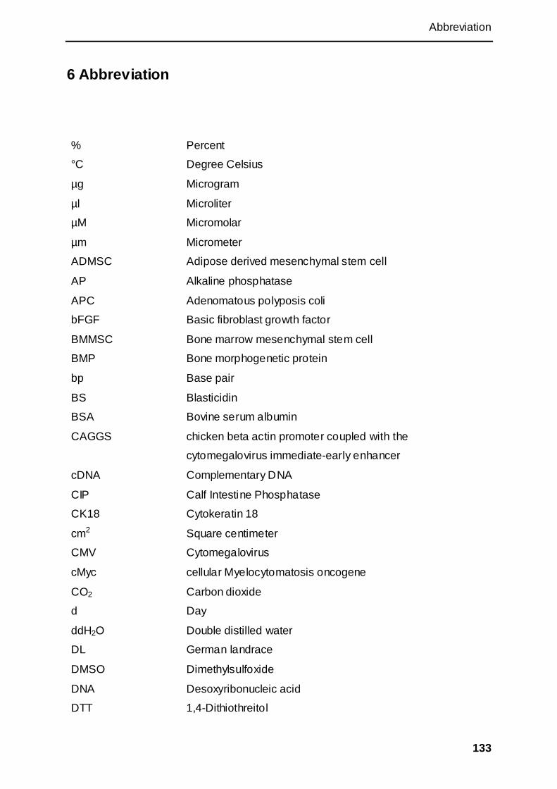
Abbreviation
133
6 Abbreviation
% Percent
°C Degree Celsius
µg Microgram
µl Microliter
µM Micromolar
µm Micrometer
ADMSC Adipose derived mesenchymal stem cell
AP Alkaline phosphatase
APC Adenomatous polyposis coli
bFGF Basic fibroblast growth factor
BMMSC Bone marrow mesenchymal stem cell
BMP Bone morphogenetic protein
bp Base pair
BS Blasticidin
BSA Bovine serum albumin
CAGGS chicken beta actin promoter coupled with the
cytomegalovirus immediate-early enhancer
cDNA Complementary DNA
CIP Calf Intestine Phosphatase
CK18 Cytokeratin 18
cm2 Square centimeter
CMV Cytomegalovirus
cMyc cellular Myelocytomatosis oncogene
CO2 Carbon dioxide
d Day
ddH2O Double distilled water
DL German landrace
DMSO Dimethylsulfoxide
DNA Desoxyribonucleic acid
DTT 1,4-Dithiothreitol
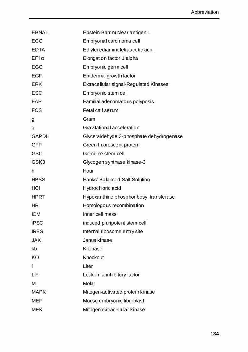
Abbreviation
134
EBNA1 Epstein-Barr nuclear antigen 1
ECC Embryonal carcinoma cell
EDTA Ethylenediaminetetraacetic acid
EF1α Elongation factor 1 alpha
EGC Embryonic germ cell
EGF Epidermal growth factor
ERK Extracellular signal-Regulated Kinases
ESC Embryonic stem cell
FAP Familial adenomatous polyposis
FCS Fetal calf serum
g Gram
g Gravitational acceleration
GAPDH Glyceraldehyde 3-phosphate dehydrogenase
GFP Green fluorescent protein
GSC Germline stem cell
GSK3 Glycogen synthase kinase-3
h Hour
HBSS Hanks‘ Balanced Salt Solution
HCl Hydrochloric acid
HPRT Hypoxanthine phosphoribosyl transferase
HR Homologous recombination
ICM Inner cell mass
iPSC induced pluripotent stem cell
IRES Internal ribosome entry site
JAK Janus kinase
kb Kilobase
KO Knockout
l Liter
LIF Leukemia inhibitory factor
M Molar
MAPK Mitogen-activated protein kinase
MEF Mouse embryonic fibroblast
MEK Mitogen extracellular kinase

Abbreviation
135
min Minute
miRNA MicroRNA
ml milliliter
mRNA Messenger RNA
MSC Mesenchymal stem cell
NaCl Sodium chloride
NEAA Non-essential amino acids
ng Nanogram
Oct4 Octamer-binding transcription factor 4
p53/TP53 Tumor suppressor protein p53
PBS Phosphate buffered saline
PCR Polymerase Chain Reaction
pFF Porcine fetal fibroblast
PGCs Primordial germ cells
PGK Phosphoglycerate kinase
PI3K Phosphoinositide 3-kinase
polyA polyadenylation site
POU5F1 POU domain, class 5, transcription factor 1
RNA Ribonucleic acid
rpm Rounds per minute
RT-PCR Reverse transcriptase polymerase chain reaction
s Second
SCNT Somatic cell nuclear transfer
SDS Sodium dodecyl sulfate
SDS-PAGE Sodium dodecyl sulfate polyacrylamide gel electrophoresis
Shp2 SH2 domains cytoplasmic tyrosine phosphatase
Smad Drosophila mothers against decapentaplegic protein
Sox2 Sex determining region Y-box 2
SSEA Stage Specific Embryonic Antigen
STAT Signal Transducer and Activator of Transcription
SV40 Simian virus 40
Tbx3 T-box transcription factor 3
Tcf T-cell factor

Abbreviation
136
TGF-β Transforming growth factor beta
U Unit
UTR Untranslated region
UV Ultraviolet
V Volt

List of figures
137
7 List of figures
Figure 1: Positive-negative selection in gene targeting. ............................................ 3
Figure 2: Three approaches to nuclear reprogramming to pluripotency. ............. 7
Figure 3: Non-viral delivery methods. ........................................................................... 11
Figure 4: Putative signalling pathways underlying non-cell autonomous
reprogramming. ............................................................................................... 15
Figure 5: Human and mouse embryonic stem cell (ESC) identity is sustained by
mainly distinct signalling networks. .......................................................... 17
Figure 6: Schematic diagram of microRNA biogenesis. .......................................... 18
Figure 7: Early lineage segregation in mouse, human, pig, and cattle. ............... 22
Figure 8: Episomal vectors pEBV-reproVI and pEBV-repro VII. ............................. 58
Figure 9: Doxycycline dependent tet-on system. ....................................................... 59
Figure 10: Constructs of reprogramming vectors. .................................................... 60
Figure 11: MicroRNAs vector for reprogramming...................................................... 60
Figure 12: pLOVE-N-Myc vector...................................................................................... 61
Figure 13: pCAGGS-pNANOG-neo vector. ................................................................... 61
Figure 14: Double-transfection procedure for reprogramming. ............................. 63
Figure 15: Reprogrammed cells from porcine Oct4-GFP fetal fibroblasts. ......... 64
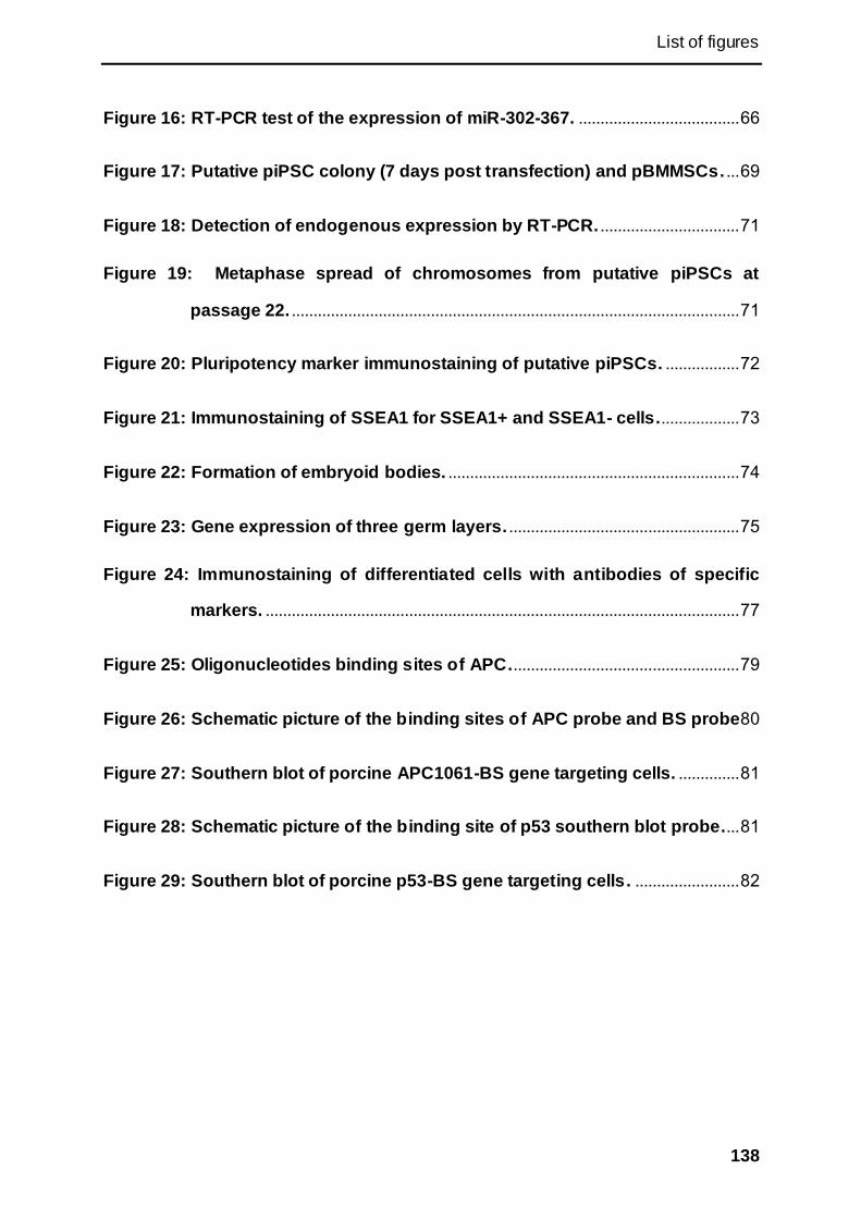
List of figures
138
Figure 16: RT-PCR test of the expression of miR-302-367. ..................................... 66
Figure 17: Putative piPSC colony (7 days post transfection) and pBMMSCs. ... 69
Figure 18: Detection of endogenous expression by RT-PCR. ................................ 71
Figure 19: Metaphase spread of chromosomes from putative piPSCs at
passage 22. ....................................................................................................... 71
Figure 20: Pluripotency marker immunostaining of putative piPSCs. ................. 72
Figure 21: Immunostaining of SSEA1 for SSEA1+ and SSEA1- cells................... 73
Figure 22: Formation of embryoid bodies. ................................................................... 74
Figure 23: Gene expression of three germ layers. ..................................................... 75
Figure 24: Immunostaining of differentiated cells with antibodies of specific
markers. ............................................................................................................. 77
Figure 25: Oligonucleotides binding sites of APC. .................................................... 79
Figure 26: Schematic picture of the binding sites of APC probe and BS probe 80
Figure 27: Southern blot of porcine APC1061-BS gene targeting cells. .............. 81
Figure 28: Schematic picture of the binding site of p53 southern blot probe.... 81
Figure 29: Southern blot of porcine p53-BS gene targeting cells. ........................ 82
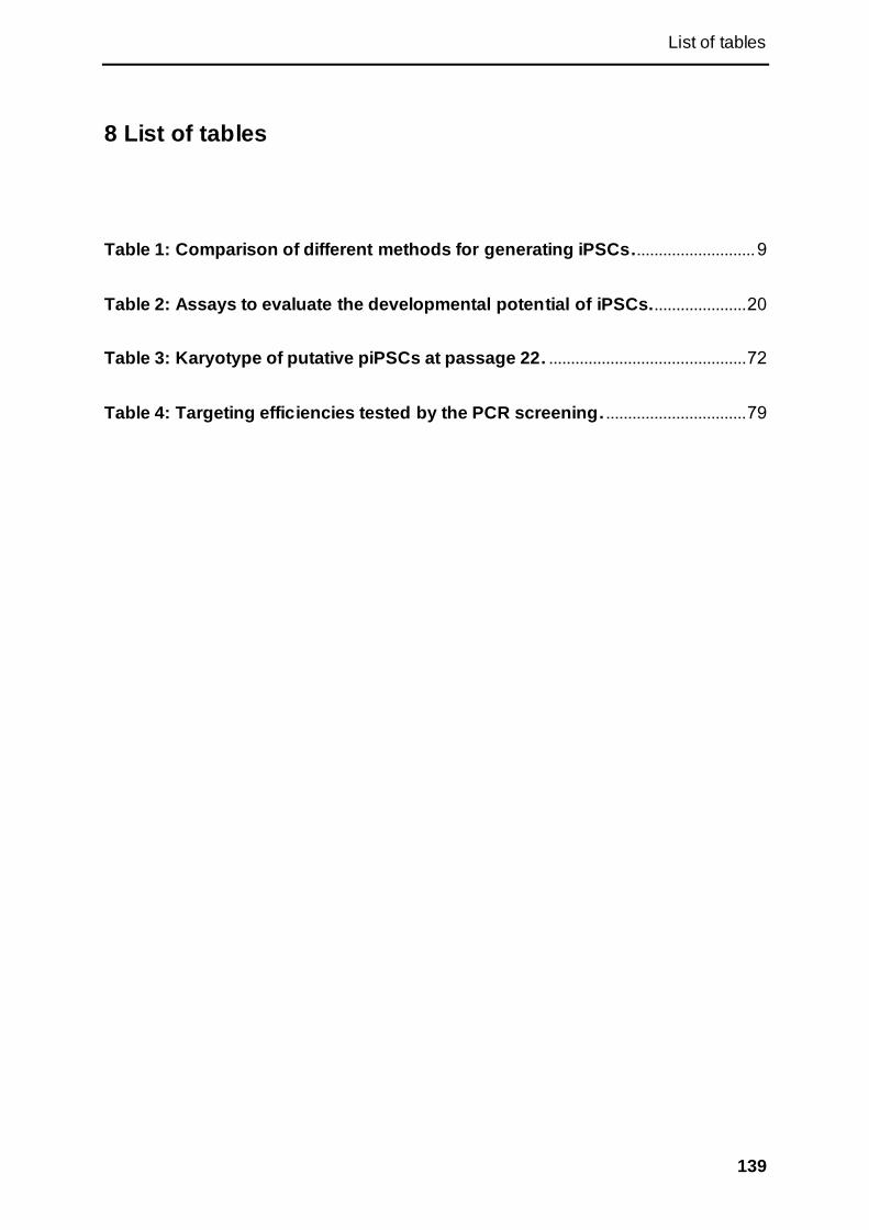
List of tables
139
8 List of tables
Table 1: Comparison of different methods for generating iPSCs............................ 9
Table 2: Assays to evaluate the developmental potential of iPSCs. ..................... 20
Table 3: Karyotype of putative piPSCs at passage 22. ............................................. 72
Table 4: Targeting efficiencies tested by the PCR screening. ................................ 79
Appendix
140
9 Appendix
Vector maps
pCAGGS-pNanog neo7263 bp
CMV IE enhancer
chicken b-actin intron
rb b-globin pA
Amp R
neo
SV40 polyA pNanog
rb intron
CAG promoter
SV40 Early Promoter
bla promoter
pBR322 ori
pSL-attB-hOct4
10538 bp
rtTA2s-M2
tetR B/E-KRAB
intron
IRES
SV40 late pA
CAG promoter
SA
seqprimer tTS
Amp
Pmin-tetO7-Pmin
attB
hOct4
bpA
pSL-attB-pKS
11926 bp
intron
attB
loxP
loxP
Pmin-te tO7-Pmin
T2A
pSox2
pKlf4
AmpR
bpA
bla
Rep_Origin_1
Rep_Origin_2
pBS-pcMyc10123 bp
rtTA2s-M2
tetR B/E-KRAB
intron
amp
IRES
SV40 late pA
CAG promoter
SA
primer CAG
seqprimer tTS
bpA
pcMyc
Pmin-tetO7-Pmin
Appendix
141
pLOVE-N-myc
8122 bp
CAG_enhancer
CMV_immearly_promoter
HIV-1_5_LTR
HIV-1_psi_pack
CMV_immearly_promoter
tetO
loxP
Ampicillin
AmpR_promoter
V5
WPRE
attR1
attR2
right
right
what?
N-myc
WPRE-R
pBR322_origin
pPhiC31Int
6576 bp
CAG Promoter
splice acceptor
C31Int(NLS)
bpA
Int_g457a_forw
pSL-attB-miR5686 bp
attB
loxP
loxP
porcine miR302-367
CMV
AmpR
BGH pA
bla
Rep_Origin_1
Rep_Origin_2
pGem-T-Easy
3015 bp
MCS
Amp
lac Z
lac Z

Acknowledgement
142
10 Acknowledgement
I would like to thank my supervisor Prof. A . Schnieke for giving me the opportunity to
work on this special project and for all her patient supervision, the guidance and the
support. In addition, I would like to thank her for giving me the possibilities to attend
conferences.
Further I would like to thank Prof. Dr. W. Windisch for evaluating this thesis as
second referee.
I would like to thank China Scholarship Council for funding me to study in Germany. I
would like to thank Technical University of Munich for providing me the facilities and
good study atmosphere.
Great thanks go to Dr. T. Flisikowska for her all-sided help, brilliant ideas, patient
guidance, experiment design, discussions, etc.
Also I would like to thank Dr. A. Kind for all the help he offered during my doctorate. I
would like to thank Prof. Dr. R. Kühn and Prof. H. Neimann for the outstanding
support with materials. I would like to thank Prof. Dr. O. Rottmann, Steffen and Viola
Löbnitz and everyone else at the animal research station in Thalhausen for the help
with the embryo transfer and the sourcing of porcine tissues.
I would like to thank Dr. S. Kraner-Scheiber for providing me the cells, all the helpful
advice and great time in the office. It‘s great to work with you. I would like to thank Dr.
K. Flisikowski for helping me with the sequencing, real time PCR and other help in
lab. I would thank Dr. N. Rezaei for the help in lab and interesting conversation.
Many thanks go to Dr. C. Merkl and Dr. T. Landmann for their help in lab, ideas,
discussion and encouragements. It‘s lucky to work with you.
I would like to thank my fellows Alex Tschukes, Anja Saalfrank, Benedikt Baumer,
Konrad Fischer, Marina Durkovic, and Simon Leuchs, for discussing with me and
help in lab.
I would like to give my special thanks to Denise Nguyen, Shun Li, and Tobias Richter
for supporting me all the time and helping both in lab and in life. I want to say: thank
you for wrting and translating for me again and again, and thank you for bringing me
so much fun.
Furthermore, I would like to thank Margret Bahnweg, Kristina Mosandl, Marlene
Edlinger, Sulith Christan, Peggy Müller, Angela Zaruba, Tobi Kranz, Simon Selleneit,

Acknowledgement
143
Andi Braun, especially for the technical and non-technical help and giving me the
family atmosphere.
Special thanks go to Babara Bauer. Thank you for helping me to deal with so many
documents and giving me warm help in life.
I would also thank all the master and bachelor students who had worked with me for
giving me help and nice conversations.
I thank all the kindhearted people who helped me in the past four years.
Many many thanks go to my parents for all their endless support and love.
Curriculum Vitae
144
11 Curriculum Vitae
Persönliche Daten
Name: Xinxin Cui
Geburtstag: 30. 07. 1986
Geburtsort: Shaanxi, China
Email: [email protected]
Ausbildungen
10/2008 – 03/2012 Promotion am Lehrstuhl für Biotechnoloie der Nutztiere,
Technische Universität München, Deutschland.
Thema der Doktorarbeit: Derivation of porcine induced
pluripotent stem cells and somatic cells gene targeting in
pig.
09/2007 – 09/2008 Master im Fach Zellbiology, im Institut der Tiermedizin,
Nordweste A&F Universität, China
Thema der Masterarbeit:Prokaryotic expression of mouse
Sox2 gene and preparation of its polyclonal antibodies
09/2004 – 07/2007 Bachelor im Fach Biotechnologie im Institut der
Lebenswissenschaften, Nordweste A&F Universität, China
Thema der Bachelorarbeit : Cloning, expression and
identification analysis of mouse Sox2 gene in prokaryotic
cells
09/2001 – 07/2004 Abitur, Erste Gymnasium der Tongchuan, Shaanxi, China
Publication
A Porcine Model of Familial Adenomatous Polyposis. Gastroenterology. 2012,
143(5):1173-5.