The expression of CPP fusion proteins in plastidsThe expression of CPP fusion proteins in plastids...
98
The expression of CPP fusion proteins in plastids Dissertation der Fakultät für Biologie der Ludwig-Maximilians-Universität München vorgelegt von Florian Mayer aus Bad Reichenhall im April 2012
Transcript of The expression of CPP fusion proteins in plastidsThe expression of CPP fusion proteins in plastids...

The expression of CPP fusion proteins in plastids
Dissertation der Fakultät für Biologie der
Ludwig-Maximilians-Universität München
vorgelegt von Florian Mayer
aus Bad Reichenhall
im April 2012


1. Gutachter: Prof. Dr. Hans-Ulrich Koop
2. Gutachter: Prof. Dr. Dario Leister
Tag der mündlichen Prüfung: 21.06.2012


Contents
1 Introduction .............................................................................................................. 9
1.1 The genetic transformation of plants ................................................................................. 9
1.2 Plastid transformation ........................................................................................................ 9
1.3 Current applied aspects of plastid transformation ........................................................... 11
1.4 Cell penetrating peptides (CPPs) ...................................................................................... 12
1.5 Artefact discovery and CPP mechanism ........................................................................... 13
1.6 Clinical trials and future challenges .................................................................................. 15
1.7 Recently: Plants and CPPs ................................................................................................. 15
1.8 Aim of this thesis............................................................................................................... 18
2 Results .................................................................................................................... 19
2.1 eGFP vector series: tracking CPP fusion proteins ............................................................. 19
2.2 Plastid transformation vector intermediate pUC18(C) ..................................................... 30
2.3 PAP1 vector series: providing a physiological CPP read-out ............................................ 31
2.4 PAH vectors series: CPP fusions for the clinic ................................................................... 36
3 Discussion ............................................................................................................... 41
3.1 Expression of CPP fusion proteins in tobacco plastids is feasible .................................... 41
3.2 CPP fusion proteins are entrapped in the organelle ........................................................ 45
3.3 Do plant-produced CPP-fusion proteins penetrate into protoplasts and human cells? .. 47
3.4 Do CPP-PAH fusion proteins expressed in plastids exert a positive effect in vivo? .......... 50
4 Summary ................................................................................................................ 53
5 Zusammenfassung .................................................................................................. 55
6 Material and Methods ............................................................................................. 57
6.1 Material............................................................................................................................. 57
6.1.1 Chemicals and Enzymes .................................................................................................... 57
6.1.2 Kits, Consumables, Equipment and Software ................................................................... 58
6.1.3 DNA and Organisms .......................................................................................................... 59
6.2 Methods ............................................................................................................................ 61
6.2.1 Vector cloning ................................................................................................................... 61
6.2.2 Transformation ................................................................................................................. 61
6.2.2.1 E. coli ............................................................................................................................. 61
6.2.2.2 Tobacco ......................................................................................................................... 62
6.2.3 Transgenic plants .............................................................................................................. 68
6.2.3.1 Transplastomic lines ..................................................................................................... 68
6.2.3.2 Nuclear transformants .................................................................................................. 68
6.2.4 Molecular analysis ............................................................................................................ 69
6.2.4.1 DNA isolation from plant tissue .................................................................................... 69
6.2.4.2 Polymerase chain reaction ............................................................................................ 70

6.2.4.3 Southern blot analysis................................................................................................... 71
6.2.5 Macroscopic analysis ........................................................................................................ 72
6.2.5.1 Vegetative and reproductive growth ............................................................................ 72
6.2.5.2 Seed assay ..................................................................................................................... 72
6.2.6 Microscopic analysis ......................................................................................................... 72
6.2.6.1 Fluorescence microscopy .............................................................................................. 72
6.2.6.2 Confocal laser scanning microscopy (CLSM) ................................................................. 72
6.2.7 Chromatographic analysis................................................................................................. 73
6.2.8 Biochemical analysis ......................................................................................................... 73
6.2.8.1 Extraction of total soluble protein (TSP) ....................................................................... 73
6.2.8.2 The Bradford assay ....................................................................................................... 74
6.2.8.3 Sodium dodecyl sulfate polyacrylamide gel electrophoresis (SDS-PAGE) .................... 74
6.2.8.4 Isolation of CPP fusion proteins .................................................................................... 74
6.2.8.5 Western blot ................................................................................................................. 75
6.2.9 Functional fusion protein assays ...................................................................................... 75
6.2.9.1 Transduction of CPP fusion proteins into plant cells .................................................... 75
6.2.9.2 Transduction of CPP fusion proteins into human cells ................................................. 76
7 Abbreviations.......................................................................................................... 77
8 Tables and Figures ................................................................................................... 79
9 References .............................................................................................................. 81
10 Acknowledgements ................................................................................................. 95
11 Erklärung ................................................................................................................ 97

1 Introduction
1.1 The genetic transformation of plants
A plant cell offers three genomes for genetic manipulation: the nuclear genome and the
organellar genomes in the mitochondria and in the plastids. Stable genetic manipulation of a
plant's nuclear genome was first reported in the early 1980s by Agrobacterium tumefaciens
mediated transformation (Herrera-Estrella et al., 1983, Bevan et al., 1983). Today it represents a
standard technique in many laboratories (Horsch et al., 1985, Clough and Bent, 1998).
The technique does not require a sequenced DNA template of the target species (non-
homologous insertion) and is broadly applicable to a wide range of species (for review: Meyers et
al., 2010). In contrast, the transformation of the organellar genomes is less established
(Butow and Fox, 1990). Transformation of the mitochondrial genome is only reported in Yeast
(Saccharomyces cerevisiae, Johnston et al., 1988) and Chlamydomonas reinhardtii, a unicellular
green alga (Randolph-Anderson et al., 1993), but not in higher plants (Ijaz, 2010). Stable genetic
transformation of a plastid genome (plastome) was first shown for Chlamydomonas reinhardtii
(Boynton et al., 1988) and was successfully applied to the higher plant Nicotiana tabacum L.
(tobacco) two years later (Svab et al., 1990). The technique of plastid transformation developed to
an exciting research field with both basic and applied aspects (Koop et al., 2007).
1.2 Plastid transformation
Stable transformation of the plastid genome as a technique of manipulating a plant's DNA is
considerably elegant. A key feature of the technology is the targeted insertion of transgenes in
the plastome. This is based on the plastid's prokaryotic origin (Chan and Bhattacharya, 2010),
which allows transformation vector integration based on homologous recombination.
DNA sequences to be integrated into the plastome are therefore flanked by sequences amplified
from the plastome of the target species. They are then subsequently incorporated at the specified
site upon transformation vector delivery (Figure 1A). Although the plastomes of many species are
sequenced (Verma et al., 2008) and substantial efforts are made towards stable transformation of
those candidates (Herrera-Diaz, 2011), tobacco still represents the main model species in the
field. For obvious candidates like the plant model Arabidopsis thaliana and monocot species of
huge economic relevance like corn, rice, wheat etc. reproducible protocols are still elusive.

10 Introduction
Transformation vectors are delivered to various explants by the biolistic method (Svab et al.,
1990) or to protoplasts by polyethylene glycol (Golds et al., 1993). Selection is commonly based
on antibiotics resistance genes. Plastids are highly polyploid. The plastome copy number depends
on plant species, tissue type and plastid number per cell – up to 10,000 plastome copies are
present in tobacco leaf cells (Maliga, 2004). A successful transformation event involves a single or
very few plastid DNA molecules initially which leads to cells with genetically different plastomes
(Koop et al., 2007). This status of the cells is called heteroplasmic and these cells give rise to
heteroplasmic shoots .This is the first regeneration event which emerges from tissue culture
(Figure 1B). To sort out wild-type (WT) plastome copies, repeated rounds of regeneration under
selection pressure are carried out. This leads to cells containing transformed plastome copies only
(Figure 1C). The status of the resulting cells is called homoplasmic. Homoplasmic cells give rise to
stably transformed transplastomic plants. Once homoplasmy is achieved, up to 10,000 copies of
the new genetic information are present, offering the possibility to yield high protein
accumulation levels.
Figure 1. Events from the biolistic transformation vector delivery to the regeneration of a homoplasmic plant. (A) Expression cassettes (PL_aadA_T) are flanked by homologous plastome sequences (INSR, INSL) for site-specific integration via homologous recombination. PL: promoter+leader (5’ regulatory region for trancription and translation), aadA: spectinomycin resistance cassette used for the selection of successful transformation events, T: terminator (3’ regulatory region). (B) Repeated rounds of regeneration under selection pressure lead to homoplasmic transformants. (C) Sorting out WT plastome copies under selection pressure (S) to obtain mutant copies only. Modified from Bock and Khan, 2004, and Cardi et al., 2010.

Introduction 11
Due to their prokaryotic origin, plastids can be used for the expression of polycistronic operons
(Staub and Maliga, 1995). Public concerns connected with genetic manipulation can be accounted
for by the use of inducible systems (Lössl et al., 2005, Mühlbauer and Koop, 2005, Verhounig et
al., 2010) and antibiotic selection marker removal strategies (for review: Day and Goldschmidt-
Clermont, 2011). Compared to nuclear transformants, transplastomic plants are save to grow in
the field since plastids are maternally inherited in most crops (Corriveau and Coleman, 1988).
It is therefore unlikely that the foreign sequence will escape to wild relatives. However, small
scale leakage was observed (Ruf et al., 2007, Svab and Maliga, 2007) and long-time effects of such
scenarios need to be investigated in the future.
1.3 Current applied aspects of plastid transformation
In basic science, plastid transformation is used to study plastid function e.g. by the mutation or
inactivation of plastid genes (Mühlbauer et al., 2002). Applied aspects cover the establishment of
novel traits in the plant. This may involve the introduction of new single traits e.g. herbicide
resistance (McBride et al., 1995), but also biosynthetic pathways can be modified or newly
introduced (Apel and Bock, 2009, Krichevsky et al., 2010). Recent reviews provide more
information about the current field (Koop et al., 2007, Verma and Daniell, 2007, Maliga and Bock,
2011). One major research branch of plastid transformation is based on the plastid's capacity to
accumulate large amounts of proteins: the expression of therapeutics for human health, like
antibodies, vaccines and more recently various antimicrobials (Daniell et al., 2009, Maliga and
Bock, 2011). Until now, most studies focused on the expression of viral and bacterial vaccine
antigens (Daniell et al., 2009, Bock and Warzecha, 2010, Cardi et al., 2010). Upon oral delivery,
these vaccine antigens can trigger an immune response via the mucosa (Bienenstock and Befus,
1980, Holmgren and Lycke, 1986) and adjuvants are used today to potentiate the response
(Lycke and Holmgren, 1986, Holmgren and Czerkinsky, 2005). In contrast to a high number of
reports focusing on the expression of antigens, there is a surprisingly limited number of reports
on the plastid-based expression and subsequent application of other groups of therapeutics like
small peptide drugs and human enzymes. Such therapeutic peptides / proteins ultimately need to
enter the cytoplasm (pass the highly selective cell membrane barrier) to exert their action in the
cytosol or organelles. Intracellular transport of membrane-impermeable biologically active
molecules is one of the key problems in drug delivery (Torchilin, 2006). Although a first promising
report about cellular delivery of plastid expressed proteins appeared in the field (Limaye et al.,
2006), the technology of cellular delivery is presently limited to the receptor-mediated route via
adjuvants (Ruhlman et al., 2007, Verma et al., 2010, Boyhan and Daniell, 2011).
12 Introduction
Despite the necessity to expand the advantages of plastid-based manufacture to new groups of
therapeutics, modes of cellular delivery and intracellular targets for disease treatment,
new carriers which offer the possibility for a receptor-independent passage across membranes,
the highly selective brain-blood-barrier and even the human skin were not suggested. A field that
offers extensive knowledge in this regard was established about twenty years ago with the
discovery of cell penetrating peptides (CPPs).
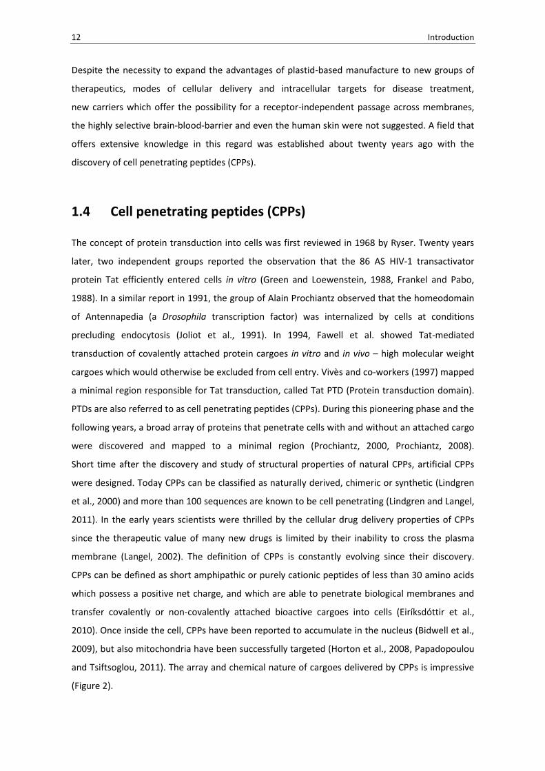
1.4 Cell penetrating peptides (CPPs)
The concept of protein transduction into cells was first reviewed in 1968 by Ryser. Twenty years
later, two independent groups reported the observation that the 86 AS HIV-1 transactivator
protein Tat efficiently entered cells in vitro (Green and Loewenstein, 1988, Frankel and Pabo,
1988). In a similar report in 1991, the group of Alain Prochiantz observed that the homeodomain
of Antennapedia (a Drosophila transcription factor) was internalized by cells at conditions
precluding endocytosis (Joliot et al., 1991). In 1994, Fawell et al. showed Tat-mediated
transduction of covalently attached protein cargoes in vitro and in vivo – high molecular weight
cargoes which would otherwise be excluded from cell entry. Vivès and co-workers (1997) mapped
a minimal region responsible for Tat transduction, called Tat PTD (Protein transduction domain).
PTDs are also referred to as cell penetrating peptides (CPPs). During this pioneering phase and the
following years, a broad array of proteins that penetrate cells with and without an attached cargo
were discovered and mapped to a minimal region (Prochiantz, 2000, Prochiantz, 2008).
Short time after the discovery and study of structural properties of natural CPPs, artificial CPPs
were designed. Today CPPs can be classified as naturally derived, chimeric or synthetic (Lindgren
et al., 2000) and more than 100 sequences are known to be cell penetrating (Lindgren and Langel,
2011). In the early years scientists were thrilled by the cellular drug delivery properties of CPPs
since the therapeutic value of many new drugs is limited by their inability to cross the plasma
membrane (Langel, 2002). The definition of CPPs is constantly evolving since their discovery.
CPPs can be defined as short amphipathic or purely cationic peptides of less than 30 amino acids
which possess a positive net charge, and which are able to penetrate biological membranes and
transfer covalently or non-covalently attached bioactive cargoes into cells (Eiríksdóttir et al.,
2010). Once inside the cell, CPPs have been reported to accumulate in the nucleus (Bidwell et al.,
2009), but also mitochondria have been successfully targeted (Horton et al., 2008, Papadopoulou
and Tsiftsoglou, 2011). The array and chemical nature of cargoes delivered by CPPs is impressive
(Figure 2).

Introduction 13
Figure 2. Schematic overview of possible applications where cell penetrating peptides have been shown to function well as delivery vehicles, both in vitro and in vivo. As presented in: Lindgren and Langel, 2011.
1.5 Artefact discovery and CPP mechanism
For the first decade data supported an energy-, temperature- and receptor-independent direct
mechanism of cell entry for Tat and other CPPs (Langel, 2002). This independence of energy-
dependent endocytosis, however, was reported to be an artefact when it was discovered that cell
fixation led to artificial CPP uptake (Lundberg and Johansson, 2001) and that the internalized
amount of CPP was overestimated due to membrane bound peptide (Lundberg and Johansson,
2002, Richard et al., 2003). In the following years a re-evaluation of CPP uptake started.
Work on unfixed cells with inhibitors of endocytosis (to exclude endocytic uptake) and trypsin
digest before FACS (to remove cell-bound CPPs), supported cellular uptake was due to different
types of endocytosis (Fittipaldi et al., 2003, Wadia et al., 2004, Richard et al., 2005, Lundin et al.,
2008). Yet, studies still provided evidence for direct CPP penetration (Terrone et al., 2003, Thorén
et al., 2003, Rothbard et al., 2004, Henriques et al., 2005, Deshayes et al., 2006, Fretz et al., 2007,
Herce et al., 2009, Ter-Avetisyan et al., 2009, Watkins et al., 2009, Ciobanasu et al., 2010, Liu et
al., 2011, Rydström et al., 2011, Hirose et al., 2012) and the discussions regarding the
translocation mechanism are ongoing. Today it seems established that multiple transduction
pathways are exploited in parallel by CPPs which depend on CPP concentration, size and nature of
cargo, tested cell type and experimental setup (van den Berg and Dowdy 2011).
Proposed pathways (Figure 3) for CPP transduction range from energy-independent direct
penetration modes to energy-dependent endocytic modes (reviewed in: Madani et al., 2011, van
den Berg and Dowdy, 2011). Historically, PTDs grouped under the name of CPPs are a very
heterogenous group, which suggests that more than one single mechansim is involved.
14 Introduction
It must be noted, however, that discussions about the mechansim are not only between but also
within single CPPs. This is why the underlying mechansim of cell transduction of a given CPP-cargo
complex needs to be investigated individually, and results from free CPPs cannot per se be
extrapolated to their cargo-coupled counterparts (Holm et al., 2011). Recent reviews may serve as
a source of further information (Magzoub and Gräslund, 2004, Ziegler et al., 2008, Järver, 2010,
Walrant et al., 2012).
A group of peptides with obvious structural and functional similarities to CPPs are antimicrobial
peptides (AMPs) which are known for pore formation and / or lysis of target cells (Gunaratna et
al., 2002, Henrigues et al., 2006). Some CPPs have been shown to act as antimicrobials
(Nekhotiaeva et al., 2004). A recent review highlights the similarities between CPPs and AMPs
(Splith and Neundorf, 2011). Lindgren and Langel (2011) mentioned in this regard that CPPs seem
to have a position between antimicrobial peptides and peptides which use classical receptor-
mediated endocytosis for cell entry. However, neither of these peptides have shown such a
potential for cargo and low-toxic delivery as CPPs (Lindgren and Langel, 2011).
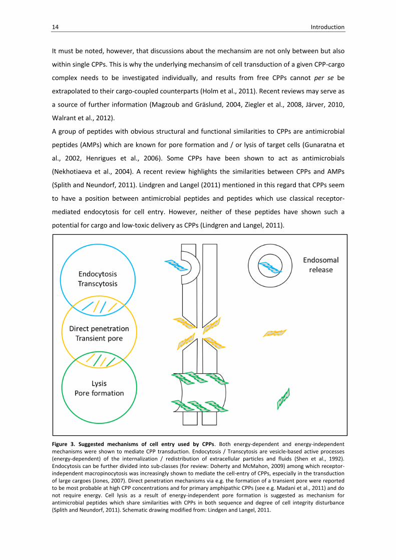
Figure 3. Suggested mechanisms of cell entry used by CPPs. Both energy-dependent and energy-independent mechanisms were shown to mediate CPP transduction. Endocytosis / Transcytosis are vesicle-based active processes (energy-dependent) of the internalization / redistribution of extracellular particles and fluids (Shen et al., 1992). Endocytosis can be further divided into sub-classes (for review: Doherty and McMahon, 2009) among which receptor-independent macropinocytosis was increasingly shown to mediate the cell-entry of CPPs, especially in the transduction of large cargoes (Jones, 2007). Direct penetration mechanisms via e.g. the formation of a transient pore were reported to be most probable at high CPP concentrations and for primary amphipathic CPPs (see e.g. Madani et al., 2011) and do not require energy. Cell lysis as a result of energy-independent pore formation is suggested as mechanism for antimicrobial peptides which share similarities with CPPs in both sequence and degree of cell integrity disturbance (Splith and Neundorf, 2011). Schematic drawing modified from: Lindgen and Langel, 2011.

Introduction 15
1.6 Clinical trials and future challenges
Despite the ongoing discussions concerning the mechanism the concept of CPP-mediated delivery
has proven successful with first phase II studies in 2003 (Rothbard et al., 2000, Chen and Harrison,
2007). Over the years, CPP's versatile delivery potential was transferred to chemically diverse
cargoes, targeting an impressive array of preclinical disease models (reviews documenting the
drug field: Dietz and Bähr, 2004, Gupta et al., 2005, Patel et al., 2007, Foged and Nielsen, 2008,
Heitz et al., 2009, Trabulo et al., 2010, Johnson et al., 2011). Today, over 20 phase I and phase II
clinical trials are documented, none of which has reported adverse effects for patients (van den
Berg and Dowdy, 2011).
One strategy in the CPP field is the expression, purification and subsequent delivery of CPPs fused
to therapeutic peptides / proteins. In 1999, a bacterial-expressed, fully-active 120 kDa
ß-galactosidase was delivered in vivo to virtually every tissue of mouse including the brain
(Schwarze et al., 1999). Since then, the CPP-mediated delivery of bioactive peptide / proteins was
impressively expanded (reviews focusing on CPP fusion peptide / protein delivery: Dietz and Bähr,
2005, Shi and Dowdy, 2007, Asoh and Ohta, 2008, Rapoport and Lorberboum-Galski, 2009,
Johansson et al., 2011).
In a recent work in CPP literature, attention is drawn to limitations in the current way of
producing therapeutic CPP fusion peptides / proteins (Asoh and Ohta, 2008). Today, CPP fusion
proteins are produced in bacterial cells, purified and then transduced to the disease model.
The authors highlight limitations of the currently used bacterial system for the manufacture of
CPP fusions, draw attention to endotoxin contamination and refer to unsolved problems with
high-scale and low-cost purification. Until now, no alternative platform for the manufacture of
CPP fusion proteins / peptides was suggested in the literature.
1.7 Recently: Plants and CPPs
Despite apparent mutual benefits for both research fields, the exchange of ideas, technology and
the initiation of joint projects between scientists in the CPP sector and modern plant science is
still at the beginning.
One plant-derived CPP mentioned in the CPP literature (Prochiantz, 2000) is the maize homeobox
transcription factor KNOTTED1 which was shown to pass plasmodesmata for cell-to-cell transport
(Lucas et al., 1995, Kim et al., 2002). Tassetto et al. (2005) however, added a new aspect to the
cell movement of KNOTTED1 with the observation of similarities between the cellular
transduction of KNOTTED1 in plant and animal cells. Consequently, the third helix of KNOTTED1

16 Introduction
showed the highest CPP properties, both with and without cargo when compared to the well-
studied CPPs Penetratin and (Arg)9 in a cell transduction study (Aussedat et al., 2006). Intercellular
movement of transcription factors through plasmodesmata is today widely accepted (Wu and
Gallagher, 2011).
Besides the third helix of KNOTTED1 and members of the previously mentioned group of
antimicrobial peptides (AMP members produced by the plants non-specific defense system; for
review: Benko-Iseppon et al., 2010, Pelegrini et al., 2011) there is not much focus on the
description of CPPs from plant origin. A recent publication suggests a new group of plant-derived
CPPs, cyclic cell penetrating peptides, which are characterised by a cyclic cystine knot motif
(Cascales et al., 2011).
Recently, plant scientists started to elaborate the use of CPPs in their research (Roberts, 2005).
In terms of CPP use for cargo delivery and manipulation of plant cells, the first study appeared in
2004 (Rosenbluh et al., 2004). The group reported the successful delivery of Rhodamine and 66
kDa BSA covalently attached to core histones into Petunia protoplasts. The authors refer to the
artefact in the CPP field (see 1.5), provide evidence for direct translocation and clearly state that
no endocytosis was involved.
Since then reports appeared addressing the use of CPPs in plant science. Both, covalent and non-
covalent strategies were employed for the CPP-mediated delivery of different cargoes.
Covalent strategies included the use of CPPs for the delivery of siRNA and silencing in tobacco
suspension cells (Unnamalai et al., 2004) and the internalization of fluorescein into tobacco
protoplasts (Mäe et al., 2005). CPP-GFP fusion protein was delivered to various root and
epidermal explants (Chang et al., 2005). Fluorescent dye was delivered to Triticale protoplasts
(Chugh and Eudes, 2007), to various Triticale explants and onion epidermal cells (Chugh and Eudes
2008a) and to Triticale microspores (Chugh et al., 2009). The same group reported covalent
(Chugh and Eudes, 2008b) and non-covalent (Chugh et al., 2009) delivery of GUS protein and 7.2
kb linear plasmid encoding GUS to Triticale. Mizuno et al. (2009) delivered FDA labeled CPPs to
tobacco suspension cells. It is interesting to note that the cell wall was not identified as a barrier
in this study.
Non-covalent strategies included the use of CPPs for the delivery of siRNA for gene silencing
(Wang et al., 2007) and the delivery of fluorescent protein and ß-galactosidase to various explants
(Wang et al., 2006, Chang et al., 2007). Transient GFP expression was achieved by the delivery of
plasmid DNA (Chen et al., 2007). Hydrolase was delivered for the inhibition of seed germination
(Liu et al., 2007). Liu and co-workers (2008) tested fluorescent protein transduction in several
organisms. CPP-mediated transduction was shown to work in archae bacteria, bacteria (both gram
+ and gram -), cyanobacteria and yeast, but not in green algae and fungi.

Introduction 17
While covalent delivery of GFP suggested an energy-independent direct pathway in plants (Chang
et al., 2005), non-covalent studies suggested energy-dependent macropinocytosis (Chang et al.,
2007, Chen et al., 2007, Chugh et al., 2009).
Combined covalent and non-covalent delivery and subsequent fluorescence resonance energy
transfer (FRET) in onion epidermal cells suggested multiple endocytic internalization pathways
(Lu et al., 2010). Three reviews appeared in the field of CPPs and plants (Roberts, 2005, Eudes
and Chugh, 2008, Chugh et al., 2010).

18 Introduction
1.8 Aim of this thesis
The present study aims at combining two exciting research areas which co-existed for more than
20 years – the field of plastid transformation and the field of CPPs.
The expression of CPP fusion proteins from the plastid attempts to answer the following
questions:
(1) CPPs were recently introduced to molecular plant science and some promising first reports in
the field have emerged. However, there are no studies about the expression of CPPs in plants.
This study addresses the following questions: Is it feasible to express fusions of nine classical CPPs
to different proteins in plastids? What consequences do result for the plant? Are plants healthy,
fertile, etc.? To which degree do CPP fusion proteins accumulate in the plant? Are there
differences between the selected CPPs?
(2) CPPs are reported to penetrate membranes by an array of suggested mechanisms.
Today it seems established that CPPs with high molecular weight cargoes use endocytic
transduction pathways (Edenhofer, 2008, Jones, 2010, Mäger et al., 2012), single studies,
however, still report on direct modes (Hariton-Gazal et al., 2003, Rosenbluh et al., 2004, Chang et
al., 2005, Cermenati et al., 2011). Which fusion protein localisation can be observed within the
plant cell upon expression in the plastid? Are CPP fusion proteins restricted to the plastid or can
any escape from the organelle to cytosol be observed?
(3) Chances of CPP-mediated cellular delivery are currently evaluated in a number of phase I and II
studies. Adverse effects are not reported so far (van den Berg and Dowdy, 2011). Recently, the
search for alternative expression platforms for the manufacture of CPP fusion proteins was
launched (Asoh and Ohta, 2008). Plants are a competitive platform for heterologous protein
expression (Raskin et al., 2002, Koop et al., 2007, Paul and Ma, 2011). Can plastids be used for the
expression of therapeutical CPP fusion proteins in a proof-of-principle study?

2 Results
The feasibility and benefits of CPP fusion protein expression in plastids was addressed in this
study. Both basic science and applied aspects were tested. The approach was based on three
plastid transformation vector series encoding for CPP fusions to three different proteins, stably
expressed from the tobacco plastid genome:
First, the enhanced green fluorescent protein (eGFP) vector series I (2.1) to track CPP fusion
proteins by optical means, second, the production of anthocyanin pigment 1-Dominant (PAP1)
vector series II (2.3) to provide a biological readout within the plant cell and, third, the
phenylalanine hydroxylase (PAH) vector series III (2.4) to introduce the expression of CPP fusions
to therapeutic proteins in plastids / plants. In section 2.2 the plastid transformation vector
intermediate pUC18(C) is introduced which was used in the vector series II and III.
2.1 eGFP vector series: tracking CPP fusion proteins
As a starting point in the evaluation of CPP fusion protein expression in plastids, His-tagged CPP
fusions with eGFP were expressed from the tobacco plastome. Such a system was supposed to
fulfill two criteria:
(1) eGFP fluorescence can be used as a convenient optical system to track CPP-eGFP fusion
proteins upon expression in the plastid / plant.
(2) CPP fusion proteins can be isolated via the His-tag for eGFP based transduction experiments
into plant and animal cells.
Nine prominent CPPs were selected for plastid transformation vector cloning (Table 1).
Table 1. Selected CPPs, CPP abbreviation / number used in the text, origin, sequence and original publication. (1): Futaki et al., 2001, (2): Ho et al., 2001, (3): Vivès et al., 1997, (4): Joliot et al., 1991, (5): Soomets et al., 2000, (6): Morris et al., 2001, (7): El-Andaloussi et al., 2007, (8): Pooga et al., 1998, (9): Elliott and O’Hare, 1997.
8x Arg CPP1 synthetic RRRRRRRR (1)
PTD-4 CPP2 synthetic, variant of CPP3 YARAAARQARA (2)
Tat CPP3 HIV-1 GRKKRRQRRRPPQ (3)
Penetratin CPP4 Drosophila RQIKIWFQNRRMKWKK (4)
Tp-10 CPP5 synthetic, variant of CPP8 AGYLLGKINLKALAALAKKIL (5)
Pep-1 CPP6 synthetic KETWWETWWTEWSQPKKKRKV (6)
M918 CPP7 synthetic MVTVLFRRLRIRRACGPPRVRV (7)
Transportan CPP8 synthetic GWTLNSAGYLLGKINLKALAALAKKIL (8)
VP22 CPP9 Herpes-Simplex Virus DAATATRGRSAASRPTERPRAPARSASRPRRPVE (9)

20 Results
Cloning was performed with codon-optimized parts (adapted to the tobacco plastid codon usage)
obtained by gene synthesis (GENEART). Ten expression cassettes were assembled in total and
cloned into chloroplast transformation vector pKCZglpk (Scharff, 2002) to give rise to the eGFP
vector series (Figure 4).
Figure 4. Codon-optimized expression cassette for CPP fusion protein expression and transformation vector assembly of the eGFP vector series. (A) Elements and amino acid sequence of the His-tagged CPPx_eGFP in-frame fusion cassette. Principle cassette design according to Han et al., 2000 - GENEART condon optimized for the expression in tobacco plastids. His-tag: affinity tag for fusion protein purification, T: thrombin site, cppx: one out of nine CPPs (see Table 1.), egfp: eGFP, codon-optimized for plastid gene expression. A control cassette was assembled without CPP between Pst I and Xho I. (B) Assembly of nine CPPx_eGFP and one control_eGFP plastid transformation vectors. GENEART provided 808 bp eGFP pre-cassette and 91 bp control pre-cassette were cloned via Nco I / Nhe I in working vector pPNG1014_MCS120 (Waheed et al., 2011). Single cppx coding regions were isolated from 100 bp fragments by digestion and were shotgun cloned via Pst I and Xho I upstream of egfp. Likewise, control cassette, 717 bp egfp was cloned in pPNG1014_MCS120_control via Xho I and Mlu I. Finally, the assembled ten expression cassettes were transferred via Xba I / EcoR V into tobacco transformation vector pKCZglpK (Scharff, 2002). pKCZglpK targets integration site trnN (INSL) / trnR (INSR) in the large inverted repeat (IR) (Zou et al., 2003). IRA: nucleotides 109,230–110,348 and 110,349–111,520, IRB: nucleotides 131,106–132,277 and 132,278–133,396 tobacco plastome sequence according Yukawa et al., 2005.
Results 21
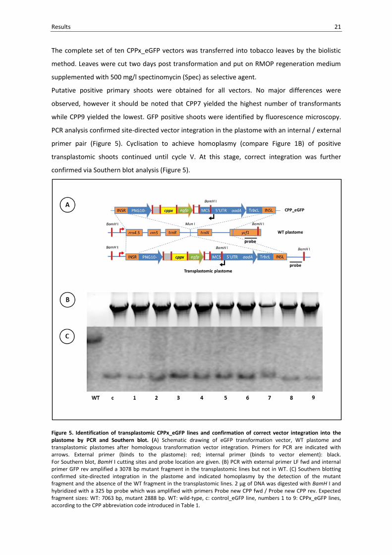
The complete set of ten CPPx_eGFP vectors was transferred into tobacco leaves by the biolistic
method. Leaves were cut two days post transformation and put on RMOP regeneration medium
supplemented with 500 mg/l spectinomycin (Spec) as selective agent.
Putative positive primary shoots were obtained for all vectors. No major differences were
observed, however it should be noted that CPP7 yielded the highest number of transformants
while CPP9 yielded the lowest. GFP positive shoots were identified by fluorescence microscopy.
PCR analysis confirmed site-directed vector integration in the plastome with an internal / external
primer pair (Figure 5). Cyclisation to achieve homoplasmy (compare Figure 1B) of positive
transplastomic shoots continued until cycle V. At this stage, correct integration was further
confirmed via Southern blot analysis (Figure 5).
Figure 5. Identification of transplastomic CPPx_eGFP lines and confirmation of correct vector integration into the plastome by PCR and Southern blot. (A) Schematic drawing of eGFP transformation vector, WT plastome and transplastomic plastomes after homologous transformation vector integration. Primers for PCR are indicated with arrows. External primer (binds to the plastome): red; internal primer (binds to vector element): black. For Southern blot, BamH I cutting sites and probe location are given. (B) PCR with external primer LF fwd and internal primer GFP rev amplified a 3078 bp mutant fragment in the transplastomic lines but not in WT. (C) Southern blotting confirmed site-directed integration in the plastome and indicated homoplasmy by the detection of the mutant fragment and the absence of the WT fragment in the transplastomic lines. 2 µg of DNA was digested with BamH I and hybridized with a 325 bp probe which was amplified with primers Probe new CPP fwd / Probe new CPP rev. Expected fragment sizes: WT: 7063 bp, mutant 2888 bp. WT: wild-type, c: control_eGFP line, numbers 1 to 9: CPPx_eGFP lines, according to the CPP abbreviation code introduced in Table 1.
22 Results
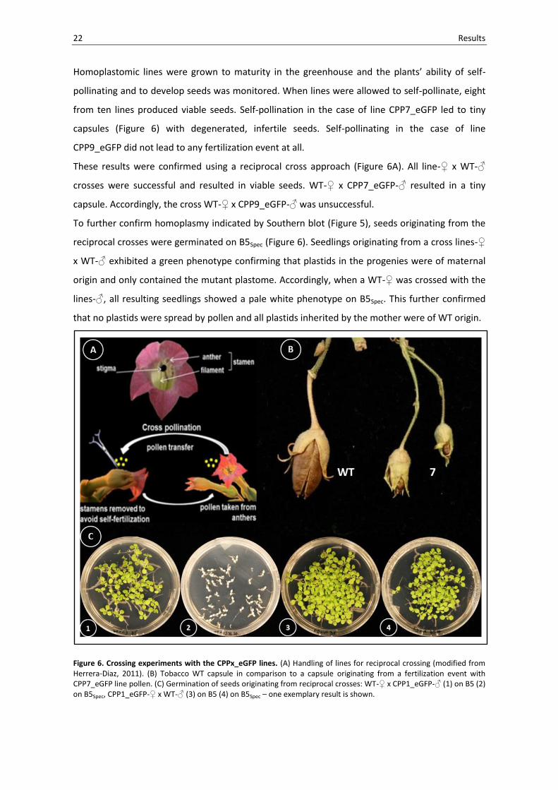
Homoplastomic lines were grown to maturity in the greenhouse and the plants’ ability of self-
pollinating and to develop seeds was monitored. When lines were allowed to self-pollinate, eight
from ten lines produced viable seeds. Self-pollination in the case of line CPP7_eGFP led to tiny
capsules (Figure 6) with degenerated, infertile seeds. Self-pollinating in the case of line
CPP9_eGFP did not lead to any fertilization event at all.
These results were confirmed using a reciprocal cross approach (Figure 6A). All line-♀ x WT-♂
crosses were successful and resulted in viable seeds. WT-♀ x CPP7_eGFP-♂ resulted in a tiny
capsule. Accordingly, the cross WT-♀ x CPP9_eGFP-♂ was unsuccessful.
To further confirm homoplasmy indicated by Southern blot (Figure 5), seeds originating from the
reciprocal crosses were germinated on B5Spec (Figure 6). Seedlings originating from a cross lines-♀
x WT-♂ exhibited a green phenotype confirming that plastids in the progenies were of maternal
origin and only contained the mutant plastome. Accordingly, when a WT-♀ was crossed with the
lines-♂, all resulting seedlings showed a pale white phenotype on B5Spec. This further confirmed
that no plastids were spread by pollen and all plastids inherited by the mother were of WT origin.
Figure 6. Crossing experiments with the CPPx_eGFP lines. (A) Handling of lines for reciprocal crossing (modified from Herrera-Diaz, 2011). (B) Tobacco WT capsule in comparison to a capsule originating from a fertilization event with CPP7_eGFP line pollen. (C) Germination of seeds originating from reciprocal crosses: WT-♀ x CPP1_eGFP-♂ (1) on B5 (2) on B5Spec, CPP1_eGFP-♀ x WT-♂ (3) on B5 (4) on B5Spec – one exemplary result is shown.
Results 23
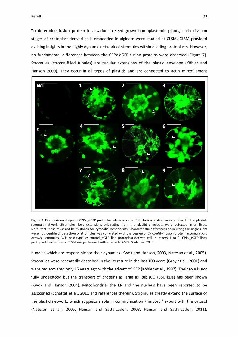
To determine fusion protein localisation in seed-grown homoplastomic plants, early division
stages of protoplast-derived cells embedded in alginate were studied at CLSM. CLSM provided
exciting insights in the highly dynamic network of stromules within dividing protoplasts. However,
no fundamental differences between the CPPx-eGFP fusion proteins were observed (Figure 7).
Stromules (stroma-filled tubules) are tubular extensions of the plastid envelope (Köhler and
Hanson 2000). They occur in all types of plastids and are connected to actin mircofilament
Figure 7. First division stages of CPPx_eGFP protoplast-derived cells. CPPx-fusion protein was contained in the plastid-stromule-network. Stromules, long extensions originating from the plastid envelope, were detected in all lines. Note, that these must not be mistaken for cytosolic components. Characteristic differences accounting for single CPPs were not identified. Detection of stromules was correlated with the degree of CPPx-eGFP fusion protein accumulation. Arrows: stromules. WT: wild-type, c: control_eGFP line protoplast-derived cell, numbers 1 to 9: CPPx_eGFP lines protoplast-derived cells. CLSM was performed with a Leica TCS-SP2. Scale bar: 20 µm.
bundles which are responsible for their dynamics (Kwok and Hanson, 2003, Natesan et al., 2005).
Stromules were repeatedly described in the literature in the last 100 years (Gray et al., 2001) and
were rediscovered only 15 years ago with the advent of GFP (Köhler et al., 1997). Their role is not
fully understood but the transport of proteins as large as RubisCO (550 kDa) has been shown
(Kwok and Hanson 2004). Mitochondria, the ER and the nucleus have been reported to be
associated (Schattat et al., 2011 and references therein). Stromules greatly extend the surface of
the plastid network, which suggests a role in communication / import / export with the cytosol
(Natesan et al., 2005, Hanson and Sattarzadeh, 2008, Hanson and Sattarzadeh, 2011).

24 Results
Recently, plastid DNA and ribosomes were shown not to travel through stromules which suggests
that the exchange of genetic information via this route is not common (Newell et al., 2012).
Work with the CPPx-eGFP system was repeatedly challenged by artefacts displaying cytosolic
fusion protein localisation. Initially, water-infiltrated leaves were studied. Water-infiltration
however turned out to cause damage to the leaf tissue, which resulted in fusion protein release to
the cytoplasm (Figure 8A). Accordingly, when scalpel cutting edges were studied, only cells in
proximity to the cutting edge revealed cytosolic fusion protein localisation (Figure 8B). Even the
most sensitive method to assess fusion protein localisation, the cultivation of protoplast-derived
cells, yielded low frequencies of cells with CPPx-eGFP fusion protein in the cytosol (Figure 8C).
In this regard it is important to note that in homoplasmic tissues the localisation of plastid-
Figure 8. Ease of artefact generation as a drawback of the CPPx-eGFP system. (A) Water-infiltration in leaves can disrupt the integrity of the cells which results in CPPx-eGFP fusion protein release from the plastid to the cytoplasm – shown here: CPP6_eGFP. Fusion protein is distributed over the cytoplasm of all cells in the tissue. Scale bar: 25 µM. (B) Scalpel cutting edges reveal the effect of disruptive methods on CPPx-eGFP fusion protein localisation within the plant tissues. Dashed line: cutting edge of CPP4_eGFP leaf. Cells in proximity of the cutting edge are filled with fusion protein which was released from the plastids. Scale bar: 25 µM. (C) Cytosolic fusion protein localisation as a rare event of studying dividing protoplast-derived cells – CPP7_eGFP. Fusion protein which is released from the plastids (P) stains the cytoplasm (arrows) which is distributed to border regions in the cell by vacuolar elements (VE). Scale bar 10 µm.

Results 25
expressed proteins should be identical. Cytosolic localisation in only a small fraction (less than 5%)
of the cells is therefor probably due to leakage from plastids caused by mechanical damage during
sample preparation. Challenged by these limitations and taken into account that small scale
escape from the plastid may not be detectable by visual means at all, we decided to highlight a
potential escape scenario from the plastid with a second, highly sensitive physiological system.
This system is described in section 2.4.
The degree of fusion protein accumulation in the transplastomic CPPx_eGFP lines was
determined. Total soluble protein (TSP) of mature plants before flowering was extracted and
quantified by the Bradford assay. Ten µg of TSP were separated by SDS-PAGE. Only in the case of
CPP7_eGFP a faint band of the expected ~ 30 kDa was detected (Figure 9).
Figure 9. Adult stages of CPPx_eGFP lines and SDS-PAGE of extracted total soluble protein (TSP). (A) TSP was extracted from seed-grown CPPx_eGFP lines before flowering. WT: wild-type, c: control_eGFP line, numbers: CPPx-eGFP lines. (B) Separation of 10 µg TSP by 10% SDS-PAGE. Faint band of CPP7-eGFP accumulation is marked with a star. Defined amounts of BSA were loaded for comparison. WT: wild-type, c: control_eGFP line, numbers: CPPx_eGFP lines, LSU: RubisCO large subunit, SSU: RubisCO small subunit.
26 Results
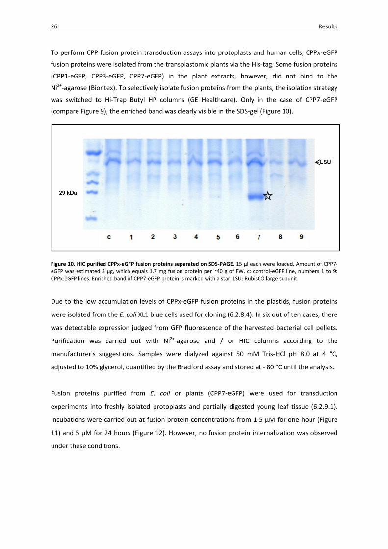
To perform CPP fusion protein transduction assays into protoplasts and human cells, CPPx-eGFP
fusion proteins were isolated from the transplastomic plants via the His-tag. Some fusion proteins
(CPP1-eGFP, CPP3-eGFP, CPP7-eGFP) in the plant extracts, however, did not bind to the
Ni2+-agarose (Biontex). To selectively isolate fusion proteins from the plants, the isolation strategy
was switched to Hi-Trap Butyl HP columns (GE Healthcare). Only in the case of CPP7-eGFP
(compare Figure 9), the enriched band was clearly visible in the SDS-gel (Figure 10).
Figure 10. HIC purified CPPx-eGFP fusion proteins separated on SDS-PAGE. 15 µl each were loaded. Amount of CPP7-eGFP was estimated 3 µg, which equals 1.7 mg fusion protein per ~40 g of FW. c: control-eGFP line, numbers 1 to 9: CPPx-eGFP lines. Enriched band of CPP7-eGFP protein is marked with a star. LSU: RubisCO large subunit.
Due to the low accumulation levels of CPPx-eGFP fusion proteins in the plastids, fusion proteins
were isolated from the E. coli XL1 blue cells used for cloning (6.2.8.4). In six out of ten cases, there
was detectable expression judged from GFP fluorescence of the harvested bacterial cell pellets.
Purification was carried out with Ni2+-agarose and / or HIC columns according to the
manufacturer's suggestions. Samples were dialyzed against 50 mM Tris-HCl pH 8.0 at 4 °C,
adjusted to 10% glycerol, quantified by the Bradford assay and stored at - 80 °C until the analysis.
Fusion proteins purified from E. coli or plants (CPP7-eGFP) were used for transduction
experiments into freshly isolated protoplasts and partially digested young leaf tissue (6.2.9.1).
Incubations were carried out at fusion protein concentrations from 1-5 µM for one hour (Figure
11) and 5 µM for 24 hours (Figure 12). However, no fusion protein internalization was observed
under these conditions.
USE THE SCANNED PICS

Results 27
Figure 11. Transduction of CPPx-eGFP fusion proteins into tobacco WT protoplasts. Freshly isolated protoplasts were incubated with 1-5 µM CPPx-eGFP fusion protein for 1 h. Gain for GFP fluorescence was normalised to zero with the help of WT protoplasts before studying the eGFP signal after incubation with cPPx-eGFP fusion protein. No differences were revealed. For cytoplasmatic localisation compare Figure 8. Left row: bright field and chlorophyll autofluorescence overlay, Right row: GFP fluorescence recorded from protoplasts after incubation with CPPx-eGFP fusion protein. +: stands for fusion protein addition to WT protoplasts, c: control-eGFP fusion protein, numbers: CPPx-eGFP fusion proteins. Scale bar: 10 µm.
In addition to this experimental setup, fusion proteins were heat denatured (95 °C for 10 min),
since it has been stressed that denaturation might facilitate the transduction of CPPs into cells
(Nagahara et al., 1998). However, heat denaturation did not alter the outcome of the experiments
(data not shown). It was later recognized that denaturation is not a prerequisite for transduction
(Han et al., 2000, Caron et al., 2001).

28 Results
Figure 12. Transduction of CPPx-eGFP fusion proteins into partially digested young WT tissues. Partially digested young tissues were incubated with 5 µM CPPx-eGFP fusion protein for 24 h. Gain for GFP fluorescence was normalised to zero with the help of WT tissue before studying the eGFP signal after incubation. No differences were observed. For cytoplasmatic localisation compare Figure 8. Upper row: bright field and chlorophyll autofluorescence overlay, Lower row: GFP fluorescence recorded after incubation with CPPx-eGFP fusion protein. Ø: untreated WT protoplasts, +: stands for fusion protein addition to WT protoplasts, c: control-eGFP fusion protein, numbers: CPPx-eGFP fusion proteins. Scale bar: 50 µm. Scale bar for Ø: 70 µm.
After CPPx-eGFP fusion proteins failed to penetrate plant cells, we were interested, if the isolated
fusion proteins were able to penetrate human cells. Three cell lines were selected for
transduction experiments: Jurkat T cells (suspension), HeLa cells (adherent) and Phoenix cells
(adherent). Transduction was tested in a range of CPPx-eGFP fusion protein concentrations from 1
to 10 µM with incubation times from 30 min to 24 h at 37 °C.

Results 29
To avoid artefacts, cells were trypsinized and quenched prior to FACS analysis. Interestingly,
experiments could not detect differences between untreated, control-eGFP-treated and CPPx-
eGFP-treated human cells. The result of a characteristic experiment is shown in Figure 13.
Figure 13. Transduction of CPPx-eGFP fusion proteins into human cells (FACS analysis). The read-out of the transduction of one exemplary CPPx-eGFP fusion protein (CPP3-eGFP = Tat-eGFP) in two cell lines is presented (fusion protein concentration in the experiment: 10 µM). FACS revealed an increase in GFP fluorescence in both the control-eGFP and the CPPx-eGFP-treated cells. Adherent Pheonix cells: (A) 1 h of incubation. (B) 24 h of incubation. Jurkat T suspension cells: (C) 1 h of incubation. (D) 24 h of incubation. HeLa cells were tested separately and gave comparable results (not shown).
30 Results
2.2 Plastid transformation vector intermediate pUC18(C)
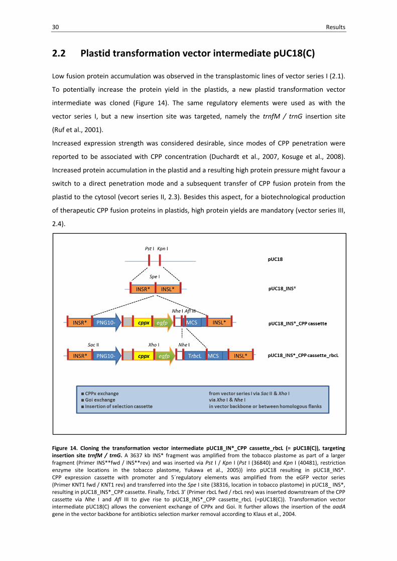
Low fusion protein accumulation was observed in the transplastomic lines of vector series I (2.1).
To potentially increase the protein yield in the plastids, a new plastid transformation vector
intermediate was cloned (Figure 14). The same regulatory elements were used as with the
vector series I, but a new insertion site was targeted, namely the trnfM / trnG insertion site
(Ruf et al., 2001).
Increased expression strength was considered desirable, since modes of CPP penetration were
reported to be associated with CPP concentration (Duchardt et al., 2007, Kosuge et al., 2008).
Increased protein accumulation in the plastid and a resulting high protein pressure might favour a
switch to a direct penetration mode and a subsequent transfer of CPP fusion protein from the
plastid to the cytosol (vecort series II, 2.3). Besides this aspect, for a biotechnological production
of therapeutic CPP fusion proteins in plastids, high protein yields are mandatory (vector series III,
2.4).
Figure 14. Cloning the transformation vector intermediate pUC18_IN*_CPP cassette_rbcL (= pUC18(C)), targeting insertion site trnfM / trnG. A 3637 kb INS* fragment was amplified from the tobacco plastome as part of a larger fragment (Primer INS**fwd / INS**rev) and was inserted via Pst I / Kpn I (Pst I (36840) and Kpn I (40481), restriction enzyme site locations in the tobacco plastome, Yukawa et al., 2005)) into pUC18 resulting in pUC18_INS*. CPP expression cassette with promoter and 5´regulatory elements was amplified from the eGFP vector series (Primer KNT1 fwd / KNT1 rev) and transferred into the Spe I site (38316, location in tobacco plastome) in pUC18_ INS*, resulting in pUC18_INS*_CPP cassette. Finally, TrbcL 3’ (Primer rbcL fwd / rbcL rev) was inserted downstream of the CPP cassette via Nhe I and Afl III to give rise to pUC18_INS*_CPP cassette_rbcL (=pUC18(C)). Transformation vector intermediate pUC18(C) allows the convenient exchange of CPPx and Goi. It further allows the insertion of the aadA gene in the vector backbone for antibiotics selection marker removal according to Klaus et al., 2004.

Results 31
2.3 PAP1 vector series: providing a physiological CPP read-out
To overcome the artefact susceptibility of the fluorescence based eGFP system (see 2.1) and to
increase the overall detection sensitivity, we were interested in a system which could provide a
physiological read-out induced by a potential CPP-mediated fusion protein transfer from the
plastid to the cytosol. Preferably such biological read-out could be detected by easy means
without any processing of the plant.
The purple Arabidopsis thaliana mutant Production of anthocyanin pigment 1-Dominant (pap1-D)
was identified by screening activation-tagged lines (Borevitz et al., 2000). The underlying
phenotype is caused by overexpression of a gene encoding MYB transcription factor PAP1.
When PAP1 was overexpressed in tobacco, PAP1 activated the anthocyanin pathway which
resulted in massive production of anthocyanins throughout the plant but no other morphological
phenotype (Borevitz et al., 2000, Xie et al., 2006). Such dominant phenotypic readout was
regarded suitable to test for CPP-mediated escape from the plastid. The following system was
proposed (Figure 15).
Figure 15. Proposed action of CPPx-PAP1 fusion proteins upon their production in the plastid. In a first step,
the CPPx-PAP1 fusion protein is translocated to the cytoplasm via CPP-mediated transfer from the plastid to the cytosol. This is followed by nuclear targeting sequence-mediated import into the nucleus. PAP1 activation of anthocyanin biosynthesis results in a high anthocyanin phenotype of the transplastomic plant. T: transformation vector delivery of the CPPx_PAP1 series.
Agrobacterium harboring PAP1 transformation vector pSB419 (Sharma and Dixon, 2005) was a
kind gift of Richard Dixon. Leave disc transformation (Horsch et al., 1985) was carried out to
generate a positive control (nuclear transformant) for the approach.

32 Results
Regeneration of nuclear transformants started from bleached leaf pieces with a purple callus
which subsequently gave rise to pap1 shoots (Figure 16). Pap1 transformants were grown to
maturity and allowed to self-pollinate in the greenhouse. Crossing was carried out until T2
generation.
Figure 16. Generation and phenotype of nuclear transformant pap1. Top left corner: high anthocyanin callus as the result of Agrobacterium tumefaciens-mediated tobacco leaf disc transformation with transformation vector pSB419. Other pictures: anthocyanin accumulation in pap1 compared to WT, pap1 left, WT right side.

Results 33
For stable transformation of the plastid genome, transformation vectors encoding fusion proteins
of all nine CPPs with PAP1 and one control vector without CPP were cloned (Figure 17). Vector
cloning was based on transformation vector intermediate pUC18 (C) introduced in 2.2 (Figure 14).
Figure 17. Cloning of the PAP1 transformation vector series. Briefly, the 747 bp PAP1 coding region was amplified (primer: Pap1 fwd / Pap1 rev) from Agrobacterium transformation vector pSB419 (Sharma and Dixon, 2005) and cloned via Xho I and Nhe I sites into pUC18_INS*_CPP cassette_rbcL (see 2.2, Figure 14). Next, the dicistronic operon was restored by exchanging TrbcL with aadA_TrbcL via Nhe I / Not I transfer from the eGFP vector series (see 2.1, Figure 4). Finally, cppx parts were exchanged via Sac II and Xho I to give rise to the PAP1 plastid transformation vector series.
The full set of CPPx_PAP1 vectors was transformed into tobacco by the biolistic method.
Transformation yielded transplastomic plants for all constructs, however, despite repeated
transformations attempts, no transplastomic line for transformation vector CPP5_PAP1 was
recovered. Positive lines were identified by PCR (Figure 18) and cyclization was carried out until
cycle II to IV. No enhanced anthocyanin phenotype was observed. In contrast, upon shoot
formation, lines showed a pale-green chlorotic phenotype. Southern blot analysis confirmed
correct integration and indicated homoplasmy state for all lines except for CPP8_PAP1 (Figure 18).

34 Results
Figure 18. Identification of transplastomic CPPx_PAP1 lines and confirmation of correct vector integration into the plastome by PCR and Southern blot. (A) Schematic drawing of PAP1 transformation vector, WT plastome and transplastomic plastomes. Primers for PCR are indicated with arrows. External primer (binds to the plastome): red; internal primer (binds to vector element): black. For Southern blot, Hind III cutting sites and probe location are given. (B) PCR with external primer INS**fwd and internal primer PAP1 proof rev amplified a 3719 bp mutant fragment in the transplastomic lines but not in WT. (C) Southern blotting confirmed site-directed integration in the plastome and indicated homoplasmy by the detection of the mutant fragment and the absence of the WT fragment in the transplastomic lines. 2 µg of DNA was digested with Hind III and hybridized with a 376 bp probe which was amplified with primers Probe v2 fwd / Probe v2 rev. Expected fragment sizes: WT: 4494 bp, mutant: 3120 bp. Line CPP8_PAP1 did show WT fragment. WT: wild-type, c°: control_PAP1 line, numbers 1° to 9°: CPPx_PAP1 lines.
Plants were transferred to the greenhouse for seed production. Growth under greenhouse
conditions did not suggest elevated anthocyanin contents (Figure 19). This was further confirmed
by thin-layer chromatography analysis of anthocyanin contents in leaves of greenhouse-grown
plants (not shown). All lines in the greenhouse were capable of self-pollination except for line 7°.
Line 8° and line 9° developed less and smaller capsules. Seeds obtained from pollinations with WT
were surface sterilized and sown on B5Spec to check seed-grown phenotype plants (not shown).

Results 35
Figure 19. Phenotype of transplastomic PAP1 lines in comparison to pap1 nuclear transformant. PAP1 lines were regenerated from tissue culture and Southern blot confirmed homoplasmy. In T1 grown from seeds, the chlorotic phenotype was established however the growth size phenotype was less pronounced. c°: control_PAP1 line, numbers 1° to 9°: CPPx_PAP1 lines, pap1: nuclear transformant (positive control).
To determine whether the observed chlorotic phenotype was due to high protein accumulation
levels in the lines, TSP was extracted and separated by SDS-PAGE. No prominent band resulting
from CPPx-PAP1 fusion protein accumulation was observed. Chlorosis, however, exhibited an
effect on the amount of RubisCO large subunit (Figure 20).
Figure 20. SDS-PAGE of extracted CPPx_PAP1 soluble protein (TSP). 10 µg TSP were separated, defined amounts of BSA were loaded for comparison. WT: wild-type, pap1: pap1 nuclear transformant, c°: control_PAP1 line, numbers 1° to 9°: CPPx_PAP1 lines. LSU: RubisCO large subunit, SSU: RubisCO small subunit.

36 Results
2.4 PAH vectors series: CPP fusions for the clinic
To date, CPP fusion proteins / peptides are expressed in bacterial cells, alternative expression
systems are missing (Asoh and Ohta, 2008). To introduce the expression of CPP fusions of clinical
value in plants, we aimed to express CPP fusions to a human enzyme from the plastid. Such a
plastid-expressed fusion protein could be used for the substitution of a non-functional protein in
the human body. Approaches like this are termed enzyme replacement therapy (ERT) (Fratantoni
et al., 1968, Neufeld, 2006). ERT is a promising field of disease treatment, especially in the
treatment of lysosomal storage diseases (Brady 2006, Goldblatt et al., 2011). ERT is challenged by
high costs for the development and subsequent production of the enzyme in question
(Wraight, 2006). In 2007, studies have shown the feasibility of plant-based production of
glucocerebrosidase for the ERT of Gaucher's disease (Shaaltiel et al., 2007, Aviezer et al., 2009)
and today plant-based production of pharmaceuticals has proven successful in several clinical
trials (for review: Paul and Ma, 2011).
Only recently, CPPs have entered the field of ERT with great success (Rapoport and Lorberboum-
Galski, 2009). CPP-mediated ERT in the case of phenylketonuria (PKU) (Eavri and Lorberboum-
Galski, 2007) is considered a very promising approach in the treatment of the disease (Sarkissian
et al., 2009). PKU is the most frequent inborn genetic disorder of amino acid catabolism (Online
Mendelian Inheritance in Man 261600). It is characterised by the body´s inability to convert food
supplied phenylalanine (Phe) to tyrosine, caused by deficiency of phenylalanine hydroxylase
(PAH). If untreated, resulting Phe accumulation leads to impaired postnatal development. PKUs
classical treatment includes a life-time vegan diet / dietary protein restriction combined with the
supplementation of tyrosine. BH4 supplementation, PAHs natural co-factor, was identified as an
effective treatment for mild forms of PKU (Muntau et al., 2002). This led to the first commercial
product on the market: Kuvan® (sapropterin dihydrochloride), the synthetic form of BH4.
Kuvan® is today widely applied, its effect, however, is limited in some genotypes and its chemical
synthesis is very expensive (Santos-Sierra et al., 2012).
Given the great promise of CPP-PAH fusions for the treatment of PKU, the diseases clear
connection to food and the classical dietary way of treatment, PKU was selected for the proof-of-
principle analysis of plastid expressed CPP fusions for the clinic.
Besides the CPP Tat (= CPP3) which was used by Eavri and Lorberboum-Galski (2007), synthetic
Tat variant PTD-4 (= CPP2) was chosen since PTD-4 showed 43-times the transduction ability when
compared with native Tat (Ho et al., 2001).

Results 37
Transformation vector cloning was based on the transformation vector intermediate pUC18(C),
which targets the trnfM / trnG insertion site in the plastid genome (see 2.2, Figure 14). Pah ORF
for cloning was a kind gift of Ania Muntau (Dr. von Hauner Children’s Hospital, LMU Munich).
Two vector categories were generated (Figure 21). One with the aadA gene (antibiotics selection
marker) between the homologous insertion flanks for conventional stable resistance marker
integration and one with aadA cassette in the vector backbone for cointegrate formation and
subsequent antibiotics resistance marker removal (Klaus et al., 2004).
Figure 21. Cloning of the PAH transformation vector series. (A) Cloning of the PAH transformation vector with aadA gene between the homologous insertion flanks for conventional resistance marker integration. First, the dicistronic operon was restored in pUC18_INS*_CPP cassette_rbcL (see 2.2, Figure 14) by exchanging TrbcL with aadA_rbcL via Nhe I / Not I transfer from eGFP vector series (see 2.1, Figure 4). Next, cppx parts were exchanged to cpp2, cpp3 and control via Sac II and Xho I. Finally, 1360 bp pah was inserted via Xho I / Nhe I into all three constructs to get final plastid transformation vectors. (B) Cloning of the PAH transformation vector with aadA cassette in vector backbone. Sac II / Nhe I part from pUC18_INS*_CPP cassette_rbcL_PAP1_aadA_extern (unpublished, aadA cassette was inserted in the Sca I site, position: 2108, in pUC18 vector backbone) was exchanged with Sac II / Nhe I part from the PAH transformation vector series with the aadA gene between the homologous insertion flanks (see Figure 21 A).

38 Results
Vectors of the CPPx_PAH series were transformed into tobacco plastids by the biolistic method.
Plants were regenerated for both vector categories. Despite several transformation attempts, no
transplastomic plant was identified for the control transformation vector.
This happened to be the case for both the construct with aadA in the vector backbone and for the
one with aadA between the homologous insertion flanks. For CPP2 and CPP3, however, PCR and
Southern blotting identified and confirmed successful transformation events. One plant per
category was studied in greater detail (Figure 22).
Figure 22. Identification of transplastomic CPPx_PAH lines and confirmation of correct vector integration into the plastome by PCR and Southern blot. (A) Schematic drawing of PAH transformation vector (internal aadA), WT plastome and transplastomic plastomes. Primers for PCR are indicated with arrows. External primer (binds to the plastome): red; internal primer (binds to vector element): black. For Southern blot, Hind III cutting sites and probe location are given. Note: PAH transformation vector (external aadA) is also presented. PCR and Southern blot for this construct resulted in the same fragment sizes as for the PAH transformation vector with internal aadA. (B) PCR with external primer INS**fwd and internal PAH Nhe I rev amplified a 4410 bp mutant fragment in the transplastomic lines but not in WT. (C) Southern blotting confirmed site-directed integration in the plastome and indicated homoplasmy by the detection of the mutant fragment and the absence of the WT fragment in the transplastomic lines. 2 µg DNA was digested with Hind III and hybridized with a 376 bp probe which was amplified with primers Probe v2 fwd / Probe v2 rev. Expected fragment sizes: WT: 4494 bp, mutant: 3120 bp. WT: wild-type, 2*: CPP2_PAH (external aadA) line, 3*: CPP3_PAH (internal aadA) line.
Results 39
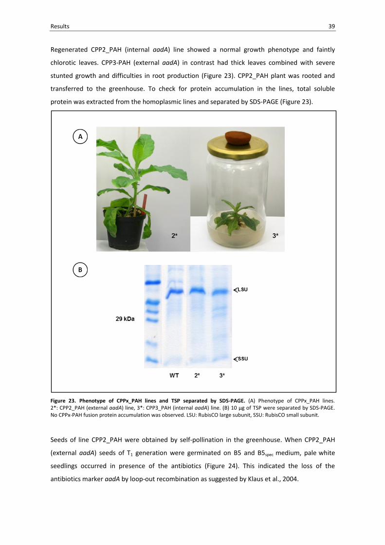
Regenerated CPP2_PAH (internal aadA) line showed a normal growth phenotype and faintly
chlorotic leaves. CPP3-PAH (external aadA) in contrast had thick leaves combined with severe
stunted growth and difficulties in root production (Figure 23). CPP2_PAH plant was rooted and
transferred to the greenhouse. To check for protein accumulation in the lines, total soluble
protein was extracted from the homoplasmic lines and separated by SDS-PAGE (Figure 23).
Figure 23. Phenotype of CPPx_PAH lines and TSP separated by SDS-PAGE. (A) Phenotype of CPPx_PAH lines. 2*: CPP2_PAH (external aadA) line, 3*: CPP3_PAH (internal aadA) line. (B) 10 µg of TSP were separated by SDS-PAGE. No CPPx-PAH fusion protein accumulation was observed. LSU: RubisCO large subunit, SSU: RubisCO small subunit.
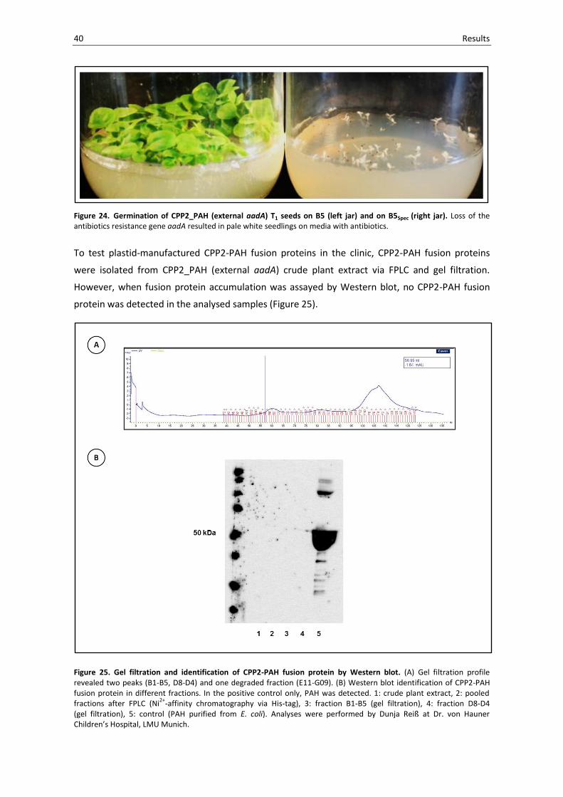
Seeds of line CPP2_PAH were obtained by self-pollination in the greenhouse. When CPP2_PAH
(external aadA) seeds of T1 generation were germinated on B5 and B5spec medium, pale white
seedlings occurred in presence of the antibiotics (Figure 24). This indicated the loss of the
antibiotics marker aadA by loop-out recombination as suggested by Klaus et al., 2004.
40 Results
Figure 24. Germination of CPP2_PAH (external aadA) T1 seeds on B5 (left jar) and on B5Spec (right jar). Loss of the antibiotics resistance gene aadA resulted in pale white seedlings on media with antibiotics.
To test plastid-manufactured CPP2-PAH fusion proteins in the clinic, CPP2-PAH fusion proteins
were isolated from CPP2_PAH (external aadA) crude plant extract via FPLC and gel filtration.
However, when fusion protein accumulation was assayed by Western blot, no CPP2-PAH fusion
protein was detected in the analysed samples (Figure 25).
Figure 25. Gel filtration and identification of CPP2-PAH fusion protein by Western blot. (A) Gel filtration profile revealed two peaks (B1-B5, D8-D4) and one degraded fraction (E11-G09). (B) Western blot identification of CPP2-PAH fusion protein in different fractions. In the positive control only, PAH was detected. 1: crude plant extract, 2: pooled fractions after FPLC (Ni
2+-affinity chromatography via His-tag), 3: fraction B1-B5 (gel filtration), 4: fraction D8-D4
(gel filtration), 5: control (PAH purified from E. coli). Analyses were performed by Dunja Reiß at Dr. von Hauner Children’s Hospital, LMU Munich.

3 Discussion
This study was designed in view of the high number of reports about the expression of vaccine
antigens and the comparably limited number of studies on the expression of other therapeutic
peptides / proteins in plastids. Today, many promising therapeutics are limited in their clinical
application due to limitations in their cellular delivery. Size and chemical nature of these drug
candidates do not allow free diffusion across the plasma membrane and consequently demand a
suitable carrier system. A group of versatile carrier peptides mediating cellular delivery are cell
penetrating peptides (CPPs). Despite a long history of uncertainties concerning the exact CPP
mechanism, clinical trials of CPP-mediated cellular delivery were launched only short time after
CPP discovery. To date, CPP fusions for the clinic are prepared from bacterial cells with associated
bottlenecks like endotoxin contamination, scale up etc. From the perspective of scientists working
in the CPP field (Asoh and Otha, 2008), alternative expression platforms are highly desirable.
To date, no reports about the expression of CPP fusion proteins in plants have emerged in the
literature. This study aimed to fill this gap. Three approaches were employed in a proof-of-
principle study.
First, the general feasibility of CPP fusion protein expression in the plant was assessed. Second, it
was addressed which fusion protein localisation results upon expression in the plastid. Third, it
was asked whether plastids can be used for the expression of therapeutical CPP fusion proteins in
a proof-of-principle study.
The results of this study suggest that the production of CPP fusion proteins is feasible in general.
Leakage of CPP fusion protein from the plastid was not observed. The clinical proof-of-principle
was not conducted due to low CPP fusion protein expression levels. Limitations of the current
study are identified and discussed to provide a framework for further approaches.
3.1 Expression of CPP fusion proteins in tobacco plastids is feasible
Twenty transplastomic lines were established in this work, expressing an array of cell penetrating
peptide fusions to proteins in the tobacco plastid. Up to nine different CPPs were fused to the
fluorescent protein eGFP (vector series I, see 2.1), the transcription factor of anthocyanin
biosynthesis PAP1 (vector series II, see 2.3) and the human enzyme of phenylalanine metabolism
PAH (vector series III, see 2.4).

42 Discussion
CPP fusion proteins have never been expressed in the plastid / plant before. Judging from the
experiences made in this study, the expression of CPP fusion proteins in the organelle is feasible.
Transplastomic plants reached homoplasmy, produced viable seeds and stably inherited the
desired trait to their progenies in a maternal fashion. Transplastomic plants also provided an
insight into pleiotropic effects, which can result from the expression of heterologous proteins in
plastids (reviewed in Ruiz and Daniell, 2005, Lössl and Waheed, 2011).
A chlorotic phenotype was observed in some CPP_PAP1 lines (Figure 19). This phenotype was also
reflected by the lower amount of RubisCO large subunit detected by SDS-PAGE (Figure 20).
In addition to chlorosis, small growth occurred in this vector series. Severe stunted growth was
observed in the case of CPP3_PAH (Figure 23). In this line, the observed dwarf phenotype was
combined with impaired root development and late flowering (not shown). Reproducible traits,
like male sterility and reduced seed capsule size were observed in both eGFP and PAP1 vector
series. No plant was regenerated for CPP5_PAP1. No plant could also be regenerated for the
control construct of the PAH vector series.
For all lines SDS-PAGE revealed low levels of fusion protein accumulation in adult plants.
This suggests that the mentioned phenotypic effects are rather a consequence of the nature of
the CPP fusion protein itself than of the high degree of fusion protein accumulation.
As introduced by Tregoning et al. (2003), high amounts of accumulated protein in the plastid can
result in unwanted phenotypic effects, which can lead to even more severe pleiotropic effects if
protein accumulation in the plastid increases (Oey et al., 2009). Recently, the group of Daniell
reported protein accumulation up to 70% total leaf protein without negative effects on the plant
(Ruhlman et al., 2010). Our results, however, are in agreement with studies which showed that
comparably small amounts of heterologous protein accumulation can result in pleiotropic effects
in the plant (Lössl et al., 2003, Magee et al., 2004, Waheed et al., 2011).
The expression of eGFP in tobacco plastids was reported before without any mentioned
phenotypic effects (Newell et al., 2003). This was confirmed by our control plant expressing eGFP
only. When CPPs were coupled to eGFP in our study, the transplastomic plants’ phenotype was
not greatly altered. However, effects like male sterility and impaired capsule development
occurred in two out of nine lines (CPP7_eGFP and CPP9_eGFP). The underlying mechanism is
currently not known. It seems however that the observed pleiotropic effects do result from a
combination of the single CPP and the respective protein, which phenotypic outcome cannot be
predicted. This is supported by the PAP1 lines. Expression of the Arabidopsis thaliana MYB
transcription factor PAP1 resulted in chlorosis in the control line (PAP1 without CPP).
Interestingly, when CPPs were coupled to PAP1, resulting fusion protein expressing lines either
resembled WT (CPP1_PAP1) or showed even more pronounced pleiotropic effects (CPP3_PAP1).

Discussion 43
Plants aimed for the clinical trial, CPP_PAH (vector series III), exhibited the most profound
consequences of fusion protein expression, spanning from normal growth (CPP2_PAH), to a dwarf
phenotype (CPP3_PAH) and no regeneration at all (control_PAH without CPP). For this group, an
interference of the heterologous protein with the amino acid metabolism of the organelle cannot
be excluded. Although PAH was recently reported only to be present in gymnosperms, mosses
and Chlamydomonas (Pribat et al., 2010), one might speculate that human PAH is functional when
it is expressed in the plastid of a higher plant. Although the human co-factor BH4 is missing,
plastids were reported to be rich in 10-formyltetrahydrofolate (10-formyl THF, Orsomando et al.,
2005). 10-formyl THF was identified as a co-factor for the enzyme in non-flowering plants
(Pribat et al., 2010) and is also present in higher plants (Collakova et al., 2008). The observed
phenotypes may therefore possibly result from a functional PAH enzyme in the plastid, in which
the N-terminal fused CPPs attenuate PAH action. According to this hypothetical model,
CPP2 hinders the function of the enzyme in the plastid which results in a WT phenotype.
Accordingly, CPP3 does not completely interfere with PAH action and the plant shows the
observed stunted bulky growth. A control plant, PAH without CPP, cannot be regenerated.
Phenylalanine is converted to tyrosine which leads to the inability to regenerate the control PAH
plant. Such explanation for the phenotypic consequences of CPPx_PAH lines is supported by the
observation that CPPs can interfere with the function of their cargo (Dowdy, 2006).
Although the expression of CPP fusion proteins turned out to be feasible in the plant, an issue that
still needs to be optimized is the level of protein accumulation in the transplastomic lines.
In principle, and in contrast to nuclear transformants which, with some exception, commonly
accumulate as little as 0.01-0.4% TSP (Molina et al., 2004), the expression of transgenes from the
plastid today enables to achieve protein accumulation up to 70 % TSP (Oye et al., 2009).
To maximize expression levels of CPP fusion proteins in our study, we optimized the CCPx_eGFP
expression cassettes in terms of codon-usage and structural elements for the expression in
tobacco plastids (GENEART). In addition, expression cassette elements were chosen that were
reported to confer high protein expression in the plastid (Scharff, 2002). However, despite this
optimisation, protein accumulation levels were below what we had expected.
In vector series I, CPPx_eGFP, high CPP fusion protein accumulation was probably hindered due to
a directed repeat the underlying transformation vector pKCZglpk forms with the tobacco
plastome (Scharff, 2002, Zou et al., 2003). This direct repeat is enabled through the use of tobacco
rrn promoter (Prrn, plastid 16S rRNA promoter) in the transformation vector pKCZglpk
(Zou et al., 2003). Upon site-specific integration, a 91 bp Prrn direct repeat is established with the
native Prrn in the plastome and consequently recognized by plastid recombination machinery.
This can lead to loop-out recombination events (Koop et al., 2007), which were confirmed for

44 Discussion
vector series I by the detection of the recombined transcript (not shown). Although no vector
elements are affected, the recombination affects the coding regions for 16S rRNA, 23S rRNA,
4.5S rRNA, 5S rRNA, tRNA-Isoleucine, tRNA-Alanine. Despite this unfavorable condition,
CPP7_eGFP showed detectable CPP fusion protein accumulation in the Coomassie gel (Figure 9)
and at the whole plant level by the bare eye upon UV illumination (not shown).
To overcome this structural limitation, the insertion site for homologous transformation vector
integration was changed from trnN / trnR (Zou et al., 2003) to trnfM / trnG (Ruf et al., 2001) in the
transformation vector intermediate pUC18(C) (Figure 14). However, for both resulting vector
series (series II and series III), protein accumulation levels were not increased. Low fusion protein
yields were observed for both CPPx-PAP1 (Figure 20) and CPPx-PAH (Figures 23, 25).
This raises the question, whether low protein accumulation levels are common feature of CPP-
fusions protein expression in plastids or if rather structural determinants of the expression
cassette are more important for the observed low yields. On which level the different CPPs / CCP
fusion proteins or the vector design (Figure 4) exert an effect on the final level of protein
accumulation awaits to be determined. Codon-optimisation was reported to increase protein
accumulation only a few-fold in higher plants (Ye et al., 2001, Tregoning et al., 2003), whereas
optimisation of translation control signals affect protein accumulation in a 10,000-fold range
(Maliga, 2002). Bock and Warzecha (2010) conclude that not accumulation of stable transcript,
but rather translational efficiency and protein stability limit protein accumulation. In a systematic
approach to identify determinants of protein stability in chloroplasts, the importance of
N-terminal amino acid residues after the start codon was highlighted (Apel et al., 2010).
Among these are N-terminal determinants of stability are targeting sequences and purification
tags. These elements consequently affect protein stability and subsequent protein accumulation
in transplastomic plants (Elghabi et al., 2011). One might speculate that the combination of a His-
tag with the varying CPPs upstream of the respective fusion protein partner decreases protein
stability and largely accounts for the low protein accumulation in the vector series I to III.
Besides the need to increase CPP fusion protein accumulation, the phenotypic effects, which
already resulted from low CPP fusion protein accumulation levels, suggest the use of an inducible
system for future approaches. Although previous reports of inducible systems were still
challenged by unsolved problems like low expression levels and promoter leakage in the non-
induced state (Koop et al., 2007), inducibility represents a key achievement when it comes to
heterologous protein expression in plants. Equally important for the plants to accept foreign
protein expression in their organelle is public acceptance in this regard. A functional inducible
system could account for public concerns which are connected with the constitutive production of
heterologous proteins in plants. Respective main concerns are not connected with a plants’

Discussion 45
overall gene content but rather if a gene in question is expressed or not. The use of an inducible
system has the potential to account for those demands. In addition it could help studying the
detailed effect of PAH and CPPx-PAH fusions on the plastid.
3.2 CPP fusion proteins are entrapped in the organelle
The current study marks the first expression of CPP fusion proteins in the plastid. Performing such
a study is not only exciting from the perspective, that two fields, co-existing now for more than
twenty years, get to know the chances and benefits the other field offers. We, from a plant
biology point of view were interested in the effect of CPP fusion protein expression in the
organelle, especially in terms of CPP fusion protein distribution.
A number of mechanisms were proposed for CPP transduction spanning from direct modes
(energy-independent) to endocytic modes (based on endocytosis, energy-dependent),
both between and within single CPPs (see introduction 1.5). Which mechanism is exactly exploited
depends on a number of factors (van den Berg and Dowdy, 2011) and the read-out of an
experiment is not per se predictable (Holm et al., 2011). Although the majority of recent data
after artefact discovery in the year 2001 (Lundberg and Johansson, 2001) supported an endocytic
mode of uptake especially for CPP fusion proteins (Trabulo et al., 2010), there are still reports
providing evidence for direct penetration modes of CPP fusions (Hariton-Gazal et al., 2003,
Rosenbluh et al., 2004, Chang et al., 2005, Cermenati et al., 2011). Such a direct mode of
transduction could result, in terms of plastid expressed CPP fusion proteins, in a transfer of CPP
fusion protein from the organelle to the cytosol.
In the past, endocytosis-free models like the transduction in organism not capable of endocytosis
(Nekhotiaeva et al., 2004, Geueke et al., 2005, Liu et al., 2008) and CPP escape / transduction
experiments with artificial lipid vesicles (Magzoub et al., 2005, Björklund et al., 2006, Bárány-
Wallje et al., 2007, Säälik et al., 2011) were employed in CPP research to shed light on CPP
mechanism. The expression of CPP fusions and studying their potential escape from the plastid as
enclosed organelle is in line with such approaches.
Recently, the transfer of dually located proteins (proteins with dual location in different
compartments of cell) from mitochondria and chloroplasts to other cellular compartments has
gained increased attention (for review: Krause and Krupinska, 2009, Pfannschmidt, 2010).
One major group of these dually located proteins has DNA binding ability (transcription factors,
telomere binding proteins) - a feature which is also characteristic for many CPPs, which are often
derived from transcription factors (Heitz et al., 2009). The association of CPPs with DNA is
mediated by the net positive charge of their cationic sequence.
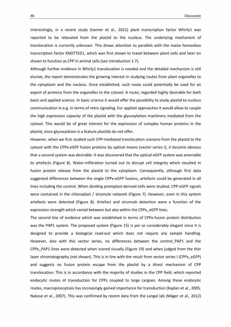
46 Discussion
Interestingly, in a recent study (Isemer et al., 2012) plant transcription factor Whirly1 was
reported to be relocated from the plastid to the nucleus. The underlying mechanism of
translocation is currently unknown. This draws attention to parallels with the maize homeobox
transcription factor KNOTTED1, which was first shown to travel between plant cells and later on
shown to function as CPP in animal cells (see introduction 1.7).
Although further evidence in Whirly1 translocation is needed and the detailed mechanism is still
elusive, the report demonstrates the growing interest in studying routes from plant organelles to
the cytoplasm and the nucleus. Once established, such route could potentially be used for an
export of proteins from the organelles to the cytosol. A route, regarded highly desirable for both
basic and applied science. In basic science it would offer the possibility to study plastid to nucleus
communication in e.g. in terms of retro signaling. For applied approaches it would allow to couple
the high expression capacity of the plastid with the glycosylation machinery mediated from the
cytosol. This would be of great interest for the expression of complex human proteins in the
plastid, since glycosylation is a feature plastids do not offer.
However, when we first studied such CPP-mediated translocation scenario from the plastid to the
cytosol with the CPPx-eGFP fusion proteins by optical means (vector series I), it became obvious
that a second system was desirable. It was discovered that the optical eGFP system was amenable
to artefacts (Figure 8). Water-infiltration turned out to disrupt cell integrity which resulted in
fusion protein release from the plastid to the cytoplasm. Consequently, although first data
suggested differences between the single CPPx-eGFP fusions, artefacts could be generated in all
lines including the control. When dividing protoplast-derived cells were studied, CPP-eGFP signals
were contained in the chloroplast / stromule network (Figure 7). However, even in this system
artefacts were detected (Figure 8). Artefact and stromule detection were a function of the
expression strength which varied between but also within the CPPx_eGFP lines.
The second line of evidence which was established in terms of CPPx-fusion protein distribution
was the PAP1 system. The proposed system (Figure 15) is per se considerably elegant since it is
designed to provide a biological read-out which does not require any sample handling.
However, also with this vector series, no differences between the control_PAP1 and the
CPPx_PAP1 lines were detected when scored visually (Figure 19) and when judged from the thin
layer chromatography (not shown). This is in line with the result from vector series I (CPPx_eGFP)
and suggests no fusion protein escape from the plastid by a direct mechanism of CPP
translocation. This is in accordance with the majority of studies in the CPP field, which reported
endocytic routes of transduction for CPPs coupled to large cargoes. Among these endocytic
routes, macropinocytosis has increasingly gained importance for transduction (Kaplan et al., 2005,
Nakase et al., 2007). This was confirmed by recent data from the Langel lab (Mäger et al., 2012)
Discussion 47
and is also supported by the majority of contributions from the plant sector (Chugh et al., 2010,
Qi et al., 2011). It seems that CPP definition keeps evolving and an updated version was reported
to be suggested in the near future (Mäger et al., 2012).
Taking provided data into account, one could argue, that the general functionality of the
physiological PAP1 system remains to be proven. If there is leakage from the organelle, and the
fusion proteins successfully access the cytosol, CPPx-PAP1 proteins still have to enter the nucleus
to provide a positive read-out. Cermenati et al. (2011) reported FITC labeled Tat (CPP3)
accumulation in the nucleus of insect cells, however no Tat-eGFP fusion protein transduction in
the nucleus. The authors state structural reasons of eGFP for this phenomenon, which, however,
do not necessarily apply to the transcription factor PAP1.
For future studies, it could be interesting to test if protein fusions to lytic peptides, the class of
peptides with even more disruptive effect on membranes, offer a tool to deliver cargoes to and
from compartments. In addition, exciting candidates like plant transcription factors should be
studied as fusions to GFP, most importantly in the case of Whirly1.
Taken together, from the current perspective, combined results from the eGFP and the PAP1
vector series suggest that none of the CPP fusion protein combinations was leaking from the
plastid. However, when we tested for “classical” membrane transduction we were surprised that
we found no transduction properties at all.
3.3 Do plant-produced CPP-fusion proteins penetrate into protoplasts and human cells?
The use of CPPs in plant biology marks a very recent expansion of the CPP field. CPP-mediated
manipulations carried out in molecular plant science were so far performed from the “outside”,
trying to manipulate plant cells in terms of protein, RNA and more recently DNA delivery.
The benefits of CPP use in plant science have been shown in many cases and a first
comprehensive review in the field provides an overview about the achievements (Chugh et al.,
2010).
Besides introducing the concept of CPP fusion protein expression in plants, we were interested
whether or not our plastid-expressed CPPx-eGFP fusion proteins were able to penetrate into plant
and human cells – the characterising feature of CPPs which is described throughout the CPP
literature.
However, the low CPPx-eGFP fusion protein accumulation in the plastids did not yield sufficient
fusion protein for transduction experiments except for CPP7-eGFP (Figure 10).
Consequently, recombinant CPP-eGFP fusion proteins were isolated from E. coli. This enabled us

48 Discussion
to study the transduction of one plastid-produced and six bacterial-produced CPPx-eGFP fusion
proteins.
To our great surprise, we could not observe any transduction. There was no CPPx-eGFP
penetration into tobacco protoplasts (Figure 11) and young tobacco leaf tissue (Figure 12).
When CPPx-eGFP fusion protein transduction into human cell lines was studied, the same
observation was made (Figure 13). Even altered experimental conditions (incubation time,
concentration of fusion protein and buffer regime) did not change this result.
This is not consistent with the majority of reports in the CPP literature in which fast,
concentration dependent CPP uptake is reported. If one only refers to studies of CPP-(e)GFP
fusion protein delivery, transduction was shown into human cells (Park et al., 2002, Yang et al.,
2002, Cao et al., 2002, Ryu et al., 2003, del Gaizo and Payne, 2003, Ferrari et al., 2003, Fittipaldi et
al., 2003, Sengoku et al., 2004), into plants (Chang et al., 2005, Liu et al., 2008, Lu et al., 2010, Qi
et al., 2011) and into insect cells (Cermenati et al., 2011).
In our experiments, transduction experiments were carried out at concentrations of 1-10 µM with
incubation times from 30 min to 24 h, at room temperature for plant cells and 37 °C for human
cells – experimental conditions under which transduction is expected to occur according to the
above mentioned literature. In addition, well studied CPPs were chosen for our approach
(Table 1). Even if one takes into account that experimental CPP transduction results cannot be
predicted (Holm et al., 2011) and that CPP read-outs can differ despite of the same experimental
setup (Cai et al., 2006, Dupont et al., 2011), the results obtained raise serious questions and
consequently draw the attention to studies where transduction failed. Reports on the
unsuccessful transduction of CPPs are not dominant in the CPP literature. Leifert and Whitton
(2003) review discrepancies in the transduction of Tat (CPP3), Penetratin (CPP4) and VP22 (CPP9)
and attempt to explain transduction by well-established biological principles. Factors leading to
contradictory experimental read-outs in CPP experiments were presented in a nice work by
Fischer and co-workers (compare Figure 3 in: Fischer et al., 2005). Chauhan and co-workers (2007)
review the CPP literature and identify factors for discrepancies in CPP research among which are:
cell age, proteolytic degradation within the cell, permeability of cell type, conformational reasons,
effects of time, nature of CPP-cargo linkage. Foerg and Merckle (2008) finally challenge the
hypothesis of CPPs as unrestricted delivery tools and come to the conclusion that “the concept of
CPPs as a universal tool in drug delivery needs to be abandoned”. Recently, Simon et al., 2010
shed light on the on-going discrepancy why some groups reported successful transduction across
brain-blood-barrier (BBB) and others did not. The authors observed efficient transduction across
BBB was correlated with a disrupted state of the barrier following ischemic injury. For an intact
BBB in their hands and others, Tat (CPP3) was not identified as an effective vehicle for trans-BBB

Discussion 49
delivery. The achievements in the application of CPPS in hypoxia and ischemia were recently
reviewed (Dietz, 2011).
Why all CPPx-eGFP fusion proteins expressed in our hands were unable to penetrate protoplasts
and human cells under all tested experimental conditions is currently beyond our knowledge.
It is interesting to note that although the combination of CPP with eGFP caused pleiotropic effects
in the plant upon expression in the plastid (3.1), no differences in transduction behaviour were
observed. However, these pleiotropic effects do not necessarily account for a functional fusion
protein. To which degree the low expression levels are correlated to the absent transduction
ability remains as a further question. To confirm the integrity of our CPP-eGFP fusions, proteins
used in our transduction studies were sequenced. Although trypsin digest led to tiny fragments
and e.g. an inability to detect CPP1, which consists of eight arginine residues, data confirmed that
CPPs were present and correctly fused to eGFP.
Essential elements of the CPP fusion cassette in our study were arranged according to the design
of Han et al. (2000) and Kwon et al. (2000). Reported vector series was used by this group for the
successful transduction of proteins fused to different CPPs in a number of studies (Jin et al., 2001,
Park et al., 2002, Ryu et al., 2004, Eum et al, 2004, Kim et al., 2005, Song et al., 2008).
Most importantly the group worked on unfixed cells and included trypsin-EDTA washings to avoid
artefacts in their studies. In addition, the N-terminal His-tag did not interfere with the
transduction ability of fusion proteins.
The alternative fusion protein vector system which does not require cleavage of the N-terminal
His-tag is pTAT-HA, provided by the Dowdy lab (Nagahara et al., 1998). This vector was used in the
majority of CPP fusion protein studies but does solely rely on Tat (=CPP3) for transduction.
For future approaches and to identify possible bottle-necks in our system, it would be interesting
to integrate the pTAT-HA system in a chloroplast transformation vector and to test different
combinations of CPPs fused to proteins.
In plant biology, a potential application is CPP-mediated transformation vector delivery.
It has been shown that CPPs can be used for the delivery and transient expression of DNA
(Chugh and Eudes, 2008b, Chugh et al., 2009), stable transformation however was not achieved.
Experiments in our laboratory towards the non-covalent CPP-mediated delivery of nuclear
genome and plastid transformation vectors with company-synthesized CPPs (MPG, Morris et al.,
1999, MPGΔNLS, Simeoni et al., 2003) did not yield transplastomic plants. Recently, Ziemienowicz
et al. (2012) reported the successful dimer-Tat-mediated delivery of a T-DNA, VirD2 and RecA
nano-complex for the integration of transgenes into the genome of triticale.

50 Discussion
3.4 Do CPP-PAH fusion proteins expressed in plastids exert a positive effect in vivo?
The field of plastid transformation is characterised by approaches of both basic and applied
aspects. Applied research for human health in the past focused mainly on the expression of
vaccine antigens in the plastid. Oral application for the delivery of these plastid-expressed vaccine
antigens and targeting to the mucosa via adjuvants is one of the main strategies for their
application. Reports of other protein therapeutics are rare, especially since therapeutics like
human enzymes require targeting to tissues and compartments beyond the mucosa.
Besides reports by the Daniell lab making use of adjuvants like CTB and LTB for trans-mucosal
delivery (Limaye et al., 2006, Ruhlman et al., 2007, Verma et al., 2010, Boyhan and Daniell, 2011),
there are no reports of versatile carrier system which allows for cellular delivery in the
transplastomic field.
CPPs introduced in the present study offer a unique potential for cellular delivery of peptides and
proteins and the huge, highly established field of CPPs can easily be connected to the plastid
technology.
To finally introduce CPP fusion protein expression in plastids / plants with a concrete example,
we focused on the expression of CPP fusions to a human enzyme in the plastid. The ultimate goal
was to show the value of such a plastid-expressed CPP fusion protein in the substitution of a
non-functional enzyme in a clinical mouse model. The mendelian disorder phenylketonuria (PKU)
and its committing enzyme phenylalanine hydroxylase (PAH) was selected due to its metabolic
character, its clear connection to nutrition and the resulting long history of dietary treatment of
the disease. The approach was coordinated with the help of Ania Muntau’s lab at Dr. von Hauner
Children’s Hospital at LMU Munich, which has expert knowledge in the characterization and
treatment of PKU. Vector cloning, plant regeneration and characterization were carried out in our
laboratory, purification and testing of fusion proteins in the mouse model was thought to take
place at the Muntau lab.
However, when we tried to isolate CPP2-PAH fusion protein from greenhouse grown plants,
no PAH fusion protein was detected in the analysed samples including the raw extract (Figure 25).
The second transplastomic line, CPP3_PAH, which was established in the approach, showed a
dwarf phenotype and consequently did not yield sufficient amounts of fusion protein for
extraction. In addition, when this line was tested for TSP banding pattern, no CPP3-PAH-specific
band was observed, which suggests fusion protein accumulation to less than 1% TSP also for this
line (Figure 23). As discussed in 3.1, no transformant was obtained for the control vector (PAH
without CPP).

Discussion 51
At the moment, not much can be said about the value of the PKU system since CPP-PAH fusion
proteins for testing are still elusive. Compared to the protein accumulation levels observed for the
eGFP and PAP1 vector series, even lower CPP fusion protein levels were provided by the plant for
PAH. It is established in the literature that PAH stability can be negatively affected in the presence
of a His-tag. For this reason an MBP tag is preferred for purification (Dunja Reiß, personal
communication). This might affect the per se low accumulation level in the plastid and lead to the
protein degradation products which were observed in the gel filtration profile (Figure 25).
This explanation is supported by the effect N-terminal elements exert on the stability of fusion
proteins in plastids as suggested in 3.1 (Apel et al., 2010, Bock and Warzecha, 2010).
In contrast to this are the results in the report which served as guideline for our approach.
In this study, a His-tagged CPP3-PAH fusion was successfully expressed (in E. coli) and applied
without constraints of degradation (Eavri and Lorberboum-Galski, 2007). Similar to our results and
promoting rather an effect of the protein itself than of N-terminal elements, Belluci and co-
workers (2005) were unable to detect ß-zein accumulation in transplastomic tobacco after stably
inserting the ß-zein gene in the plastome.
Currently, the low protein accumulation levels of CPPx-PAH fusion proteins in this study are not
promoting the manufacture of CPP-PAH fusions in plastids. As mentioned before, also for this
vector series, inducible expression again turned out to be desirable since this could ensure plant
regeneration upon vector delivery (PAH control) and a higher biomass in terms of the CPP3-PAH.
Marker removal by the formation of a transient cointegrate (Klaus et al., 2004) was nicely shown
in our study by the incorporation of the antibiotics selection cassette in the backbone of
transformation vector pUC18(C) (Figures 21, 24). Towards a potential application, however, more
studies are needed to address further safety related issues. We have observed substantial
changes in CPP fusion protein expression levels within single lines as was concluded from CLSM
microscopy (not shown). For the favoured oral application of fusion proteins in edible plants
(Daniell et al., 2001), highly standardized doses of the active proteins are required.
It is questionable if plants can meet these requirements, especially when grown in the field.
In addition, more future focus must be drawn on the effects of the raw consumption of transgenic
plant material. Lacking knowledge in terms of these questions currently favours the contained
(facility-based) expression in plants with subsequent product purification, although higher costs
do result from such procedure.
As soon as these limitations are solved and requirements are met, contained expression of CPP
fusions in plants will have the chance to become reality. The established field of CPP technology
and the large number of clinical approaches suggest that CPPs could play a vital in plastid
engineering in the future.

52 Discussion
So far our study may serve as a starting point for future collaborations between the two fields.
For future directions, reviews listed in the introduction (1.6) of this study suggest an impressive
number of potential targets for the use of CPPs in the transplastomic field. To start with, a study
addressing a direct comparison between adjuvant- and CPP-mediated delivery is of interest, since
CPPs can be used for the delivery of antigens (for review: Brooks et al., 2010),
a field classically targeted with adjuvants. A recent review highlights aspects of CPP-mediated oral
delivery (Khafagy and Morishita, 2012). An additional route to test with extracts made of plants
could be CPP-mediated transdermal delivery (Rothbard et al., 2000, Song et al., 2008,
Lohcharoenkal et al., 2011). This could be of interest for the treatment of rare skin diseases like
ichtyosis which suffer from insufficient treatment (Huber et al., 1995, Smith et al., 2006).
In conclusion we have introduced CPP fusion protein expression in plants, more specifically in the
plastid. Further work is needed to fine-tune the technique and to study long-term effects. If these
requirements will be met and if the expression of CPP fusion proteins in plants will increasingly
gain importance remains to be determined.

4 Summary
Plastid transformation is a valuable technique for both basic and applied science. In basic science
the technique is used to study chloroplast function. Applied approaches deal with the potential of
the plastid for the production of medicinal therapeutics, in most cases vaccine antigens coupled
to adjuvants. Adjuvants are used for the trans-mucosal delivery of attached cargoes.
Cell penetrating peptides (CPPs) emerged as valuable tools for the delivery of cell-impermeable
cargoes across cell barriers more than twenty years ago. Although the exact mechanism of CPP
penetration of cells is still discussed, the applied value of CPPs is documented in a number of
clinical studies. Recently, scientists working in the CPP field launched a call for an alternative
expression platform for CPP fusion peptides / proteins. Only a short time before, CPPs were
introduced into plant science and some impressive first results, manipulating plant cells from the
“outside”, were achieved.
The present study aimed at combining the fields of plastid transformation and CPPs from the
“inside”. We report the first expression of CPP fusion proteins in a plant, more precisely in the
plastid. The approach focused on three aspects of CPP fusion protein expression in the organelle:
(A) the principal feasibility of CPP-fusion protein expression in the plastid, (B) the location of CPP
fusion proteins in the plant cell upon plastid-based expression and (C) the use of plastids for the
manufacture of CPP fusions to provide an alternative to the bacterial expression system.
Nine prominent CPPs were employed in three vector series to investigate these aspects. In vector
series I the selected CPPs were fused to the fluorescent protein eGFP to provide an optical read-
out; in vector series II, the CPPs were fused to Arabidopsis MYB transcription factor PAP1 to
provide a biological read-out and in vector series III, two CPPs were fused to the human enzyme
PAH to introduce plant-based CPP fusion protein expression.
Taken together, the expression of CPP fusion in the plastid turned out to be feasible.
Transplastomic plants reached homoplasmy, produced viable seeds and stably inherited the
desired trait to their progenies in a maternal fashion. Only low protein accumulation levels were
detected. Pleiotropic effects occured at the low protein accumulation levels observed.
Localisation of CPP fusion proteins was shown to be restricted to the plastid. An inability of CPP
fusion proteins isolated from vector series I to penetrate protoplasts, young plant tissue and
human cell lines was revealed. The value of a plastid-based manufacture of CPP fusion proteins
for clinical approaches failed to be demonstrated due to low fusion protein accumulation levels.
Bottlenecks of the current study are discussed and suggestions are made to provide a framework
for future efforts.


5 Zusammenfassung
Plastidentransformation ist eine wertvolle Technik sowohl für die Grundlagenforschung als auch
für die angewandte Wissenschaft. In der Grundlagenforschung wird die Technik verwendet um
die Funktion von Chloroplasten zu studieren. Angewandte Aspekte der Technologie beschäftigen
sich mit der Produktion von Therapeutika für die menschliche Gesundheit. Dabei handelt sich in
den meisten Fällen um Impfstoff-Antigene, die an Adjuvantien gekoppelt sind. Adjuvantien
werden zur Vermittlung der Antigene über die Mucosa eingesetzt. Vor mehr als zwanzig Jahren
wurden mit zell-penetrierenden Peptiden (cell penetrating peptide, CPP) wertvolle Tools
eingeführt um membran-inpermeable Substanzen in die Zelle zu vermitteln. Obwohl der genaue
Mechanismus, wie CPPs dies bewerkstelligen, immer noch diskutiert wird, ist der Wert von CPPs
in einer Reihe von klinischen Studien dokumentiert. Vor kurzem äußerten Wissenschaftler in der
CPP Forschung den Wunsch nach alternativen Plattformen für die Produktion von CPP-
Fusionspeptiden und -proteinen. Nur kurze Zeit zuvor wurden CPPs das erste Mal in den
modernen Pflanzenwissenschaften eingesetzt. Dies umfaßte Manipulationen von Pflanzenzellen
von „außen“ und welche in beeindruckenden ersten Ergebnissen dokumentiert wurden.
Die vorliegende Studie konzentrierte sich auf eine Kombination der Felder der
Plastidentransformation und der CPPs von "innen". Wir berichten die erste Expression von CPP-
Fusionsproteinen in einer Pflanze, genauer gesagt in den Plastiden. Drei Aspekte der Expression
von CPP-Fusionsproteinen in den Organellen wurden untersucht: (A) Die prinzipielle Machbarkeit,
(B) die Verteilung von CPP-Fusionsproteinen in der Pflanzenzelle bei Expression in den Plastiden,
und (C) die Eignung von Plastiden zur Herstellung von CPP-Fusionsproteinen als eine Alternative
zu dem bakteriellen Expressionssystem.
Um diese Aspekte zu untersuchen wurden neun prominente CPPs in drei Vektor-Serien an
verschiedene Proteine gekoppelt und in den Plastiden produziert. In Vektor-Serie I, wurden die
CPPs an das fluoreszierende Protein eGFP fusioniert, um eine optische Nachverfolgung zu
ermöglichen. In Vektor-Serie II wurden die CPPs an den Arabidopsis MYB Transkriptionsfaktors
PAP1 fusioniert, um eine biologische Auslese zu ermöglichen. In Serie III wurden zwei CPPs an das
menschliche Enzym PAH gekoppelt um auf die prinzipielle Möglichkeit einer pflanzen-basierten
Produktion von CPP Fusionsproteinen hinzuweisen.
Zusammengefasst, stellte sich die Expression von CPP-Fusionsproteinen in Plastiden als machbar
heraus. Transplastomische Pflanzen erreichten Homoplastie, produzierten keimfähige Samen und
vererbten die gewünschte Eigenschaft stabil an ihre Nachkommen. Nur geringe Ausbeuten von
Fusionsproteinen wurden erzielt wobei pleiotrophe Effekte auftraten. Die Akkumulation von CPP-

56 Zusammenfassung
Fusionsproteinen war auf die Plastide beschränkt. Die Unfähigkeit von isolierten CPP-
Fusionsproteinen der eGFP Vektor-Serie in Protoplasten, junges Pflanzengewebe und humane
Zelllinien einzudringen, wurde aufgezeigt. Der Wert einer plastiden-basierten Herstellung von
CPP-Fusionsproteinen für klinische Ansätze konnte wegen einer zu geringen Ausbeute an
Fusionsprotein nicht aufgezeigt werden. Einschränkungen der aktuellen Studie werden diskutiert
und Vorschläge gemacht, um einen Rahmen für zukünftige Anstrengungen zu geben.

6 Material and Methods
6.1 Material
Experimental work was conducted in molecular laboratories equipped with standard laboratory
equipment, chemicals and consumables for cell and plant culture and analysis. Critical chemicals,
additional equipment and the software used for this work are mentioned in the following.
6.1.1 Chemicals and Enzymes
Water was deionized by absorption and ultrafiltration (SG ultra-Clear, Barsbüttel, Germany) and
autoclaved if necessary. Standard analytical (analytical grade) and tissue culture (for in vitro
culture) chemicals were purchased from AppliChem (Darmstadt, Germany), Bayer Vital
(Leverkusen, Germany), Duchefa (Harlem, Netherlands), Merck (Darmstadt, Germany), Serva
(Heidelberg, Germay), Sigma-Aldrich (Taufkirchen, Germany). Critical compounds used in this
work for cell / tissue culture sorted by supplier:
AppliChem Ampicillin, sodium salt
Ca-Panthotenate
D-Glucose
Bayer Vital Dimanin C
Duchefa B5 salts
Cellullase R10
D(+)-Biotin
Macerozyme R10
MS salts
Phyto agar
Pyridoxine-HCl
Spectinomycin dihydrochlorid
Sucrose
Merck Thiamine-HCl
Serva Insosit
Sigma-Aldrich Alginic acid
Benzylaminopurine
Naphthalene acetic
Nicotinic acid

58 Material and Methods
DNA and protein markers, DNA modifying enzymes and polymerases were purchased from MBI
Fermentas (St. Leon-Rot, Germany), Finnzymes (Espoo, Finland), New England Biolabs (Frankfurt /
Main, Germany), Promega (Mannheim, Germany), and Roth (Karlsruhe, Germany).
6.1.2 Kits, Consumables, Equipment and Software
Kits
Clone Jet MBI-Fermentas, St. Leon-Rot, Germany
Nucleo spin extract II Machery and Nagel, Düren, Germany
Nucleo spin plasmid Machery and Nagel, Düren, Germany
Prime-a-gene labelling system Promega, Mannheim, Germany
Plasmid Maxi Kit Qiagen, Hilden, Germany
Consumables
AB mouse anti-PAH Calbiochem, Darmstadt, Germany
AB Goat anti-mouse IgG HRP Santa Cruz Biotechnology, Heidelberg, Germany
DMEM (Gibco) Life Technologies, Darmstadt, Germany
Doppelseitiges Klebeband Tesa, Hamburg, Germany
Gene gun consumables Bio-Rad, München, Germany
HiLoad 16/60 Superdex 200 column GE Healthcare, München, Germany
HisTrap affinity column GE Healthcare, München, Germany
HiTrap Butyl HP column GE Healthcare, München, Germany
MicroSpin-Columns S-200 HR GE Healthcare, München, Germany
N+-Nylonmembran Amersham Biosciences, UK
Nescofilm Roth, Karlsruhe, Germany
Ni2+- agarose Biontex, Martinsried, Germany
Nitrocellulose membrane BA-S 85 Schleicher & Schuell, Dassel, Germany
Novex NuPAGE Bis-Tris gel system Life Technologies, Darmstadt, Germany
Optimem (Gibco) Life Technologies, Darmstadt, Germany
Polypropylene grids Scrynel PP2000 K.H.Büttner, Wasserburg, Germany
RPMI Media 1640 (Gibco) Life Technologies, Darmstadt, Germany
Steril filter PES 0.22µm Serva, Heidelberg, Germany
Steril filter PES 0.22µm, 250 ml Serva, Heidelberg, Germany
Tissue culture tube 163160 Greiner, Solingen, Germany

Material and Methods 59
Equipment
ÄKTApurifier GE Healthcare, München, Germany
Canon EOS 400D Canon GmbH, Krefeld, Germany
Centrifuge Universal II Hettich, Tuttlingen, Germany
Centrifuge Z323K Hermle, Wehingen, Germany
Clean bench HERA safe KS12 Thermo Scientific, Langenselbold, Germany
Confocal microscope TCS SP5 Leica Microsystems, Wetzlar, Germany
DIANA III imaging system Raytest GmbH, Sprockhövel, Germany
FACS Canto II BD Biosciences, Heidelberg, Germany
Leica DM 100 Leica Microsystems, Wetzlar, Germany
Microcentrifuge Fresco 17 Heraeus, Hanau, Germany
Microcentrifuge 5415 Eppendorf, Hamburg, Germany
Osmomat O30 Gonotech GmbH, Berlin, Germay
Particle-gun PDS-1000/He Bio-Rad, München, Germany
Sterilbank Hera Safe Typ KS12 Thermo Scientific, Langenselbold, Germany
Sorvall RC6 Thermo Scientific, Langenselbold, Germany
Software
Clone Manager Professional Sci-Ed Software, Cary NC, USA
Double digest http://www.fermentas.com/en/tools/doubledigest
Google scholar http://scholar.google.de/
ImageJ 1.45 http://rsbweb.nih.gov/ij/
NCBI http://www.ncbi.nlm.nih.gov/
Office 2010 Microsoft GmbH, Unterschleißheim, Germany
Vector NTI 7.0 InforMax, Oxford, UK
Windows 7 Microsoft GmbH, Unterschleißheim, Germany
6.1.3 DNA and Organisms
Vectors
o Custom gene synthesis GENEART, Regensburg, Germany
o pKCZglpK Scharff, 2002
o pPNG1014_MCS120 Waheed et al., 2011

60 Material and Methods
o PSB419 Sharma and Dixon, 2005
o pUC18 MBI-Fermentas, St. Leon-Rot, Germany
o Oligos Metabion, Martinsried, Germany
Primer name Sequence 5' -> 3' (restriction sites underlined if present)
GFP rev GACGTCATTCCGGATCCAAGG
INS**fwd TTAGCTGGTCGTGACCAAG
INS**rev CACAGGCATCCCAGGTAATTC
KNT1 fwd AACTAGTCCGCGGGCCGTCGTTC
KNT1 rev TACAACTAGTGATATCTTAGAGCTCGT
LF fwd CTACCTGCACCTGGACAGAAAG
Pah fwd TTCTCGAGATGTCCACTGCGGTCCTG
Pah NheI rev TTTTGCTAGCTTACTTTATTTTCTGGAGGGCACTG
Pap1 fwd CTACCTGCACCTGGACAGAAAG
Pap1 rev GACGTCATTCCGGATCCAAGG
Pap1 proof rev GACGTCATTCCGGATCCAAGG
Probe new CPP fwd GGTTTTGTTGGTTGGTTAATTG
Probe new CPP rev CCTGTTTAGTCCCCTTCATTTC
Probe v2 fwd GGTGATGGGCAATATCAG
Probe v2 rev GCAGGTTGGTTACACCTA
rbcL fwd AATTGCTAGCCTGCAGGCATGCAAG
rbcL rev TTAGTACTTCCCGAGAGGTCACAATTCC
Organisms
o E. coli
XL1 blue (genotype: recA1 endA1 gyrA96 thi-1 hsdR17 supE44 relA1 lac [F´ proAB lacIqZ∆M15 Tn10
(Tetr)]
o Agrobacterium tumefaciens
GV3101 (pMP90) (Koncz and Schell, 1986)
o Tobacco
Nicotiana tabacum L-cv. Petite Havana (wild-type, WT)
o Human cell lines
HeLa cells, Jurkat T cells, Pheonix cells (293 derived cell line)
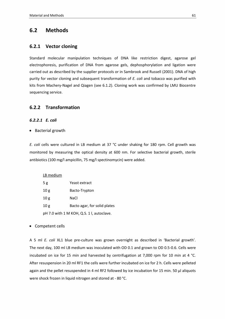
Material and Methods 61
6.2 Methods
6.2.1 Vector cloning
Standard molecular manipulation techniques of DNA like restriction digest, agarose gel
electrophoresis, purification of DNA from agarose gels, dephosphorylation and ligation were
carried out as described by the supplier protocols or in Sambrook and Russell (2001). DNA of high
purity for vector cloning and subsequent transformation of E. coli and tobacco was purified with
kits from Machery-Nagel and Qiagen (see 6.1.2). Cloning work was confirmed by LMU Biocentre
sequencing service.
6.2.2 Transformation
6.2.2.1 E. coli
Bacterial growth
E. coli cells were cultured in LB medium at 37 °C under shaking for 180 rpm. Cell growth was
monitored by measuring the optical density at 600 nm. For selective bacterial growth, sterile
antibiotics (100 mg/l ampicillin, 75 mg/l spectinomycin) were added.
LB medium
5 g Yeast extract
10 g Bacto-Trypton
10 g NaCl
10 g Bacto agar, for solid plates
pH 7.0 with 1 M KOH, Q.S. 1 l, autoclave.
Competent cells
A 5 ml E. coli XL1 blue pre-culture was grown overnight as described in ‘Bacterial growth’.
The next day, 100 ml LB medium was inoculated with OD 0.1 and grown to OD 0.5-0.6. Cells were
incubated on ice for 15 min and harvested by centrifugation at 7,000 rpm for 10 min at 4 °C.
After resuspension in 20 ml RF1 the cells were further incubated on ice for 2 h. Cells were pelleted
again and the pellet resuspended in 4 ml RF2 followed by ice incubation for 15 min. 50 µl aliquots
were shock frozen in liquid nitrogen and stored at - 80 °C.
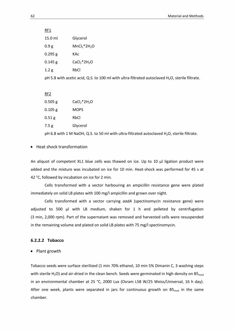
62 Material and Methods
RF1
15.0 ml Glycerol
0.9 g MnCl2*2H2O
0.295 g KAc
0.145 g CaCl2*2H2O
1.2 g RbCl
pH 5.8 with acetic acid, Q.S. to 100 ml with ultra-filtrated autoclaved H2O, sterile filtrate.
RF2
0.505 g CaCl2*2H2O
0.105 g MOPS
0.51 g RbCl
7.5 g Glycerol
pH 6.8 with 1 M NaOH, Q.S. to 50 ml with ultra-filtrated autoclaved H2O, sterile filtrate.
Heat shock transformation
An aliquot of competent XL1 blue cells was thawed on ice. Up to 10 µl ligation product were
added and the mixture was incubated on ice for 10 min. Heat-shock was performed for 45 s at
42 °C, followed by incubation on ice for 2 min.
Cells transformed with a vector harbouring an ampicillin resistance gene were plated
immediately on solid LB plates with 100 mg/l ampicillin and grown over night.
Cells transformed with a vector carrying aadA (spectinomycin resistance gene) were
adjusted to 500 µl with LB medium, shaken for 1 h and pelleted by centrifugation
(3 min, 2,000 rpm). Part of the supernatant was removed and harvested cells were resuspended
in the remaining volume and plated on solid LB plates with 75 mg/l spectinomycin.
6.2.2.2 Tobacco
Plant growth
Tobacco seeds were surface sterilized (1 min 70% ethanol, 10 min 5% Dimanin C, 3 washing steps
with sterile H2O) and air-dried in the clean bench. Seeds were germinated in high density on B5mod
in an environmental chamber at 25 °C, 2000 Lux (Osram L58 W/25 Weiss/Universal, 16 h day).
After one week, plants were separated in jars for continuous growth on B5mod in the same
chamber.

Material and Methods 63
For transformation by the biolistic method and for protoplast isolation, four to five week old
wild-type (WT) donor plants were used.
At the same age, WT and transplastomic lines were routinely transferred to the greenhouse for
prolonged growth, flowering and seed development.
B5mod (Dovzhenko et al., 1998)
3.1 g B5-salts
10 ml 100x B5-vitamins (Gamborg et al., 1968)
10 g Inosit
100 mg Pyridoxine-HCl
1 g Thiamine-HCl
100 mg Nicotinic acid
Q.S. to 1 l with ultra-filtrated H2O, store at -20 °C.
0.983 g MgSO4*7 H2O
20 g Sucrose
5 g Phyto agar
pH 5.8 with 1 M KOH, Q.S. to 1 l with ultra-filtrated H2O, autoclave.
Transformation of the plastid genome
Transformation of the plastid WT genome was performed by the biolistic method (BioRad
PDS1000/He Biolistic Gene Gun) – either by the bombardment of tobacco leaves or by the
bombardment of protoplast-derived microcolonies.
o Leaves
True leaves of aseptically grown four to five week old tobacco WT plants were placed with the
adaxial side up on petri dishes with simple RMOP (composition see page 68) the day before the
experiment and stored in the environmental chamber used for plant growth until the
bombardment.
o Protoplast-derived microcolonies
For the bombardment of protoplast-derived microcolonies tobacco protoplasts were isolated and
embedded in grids with alginate.

64 Material and Methods
Protoplast isolation
Sterile grown true leaves of four to five week old tobacco WT plants were digested enzymatically
for protoplast isolation. Leaves were cut in 1-3 mm stripes in 10 ml F-PIN for pre-plasmolysis (1 h).
Medium was exchanged to the digestion medium (9.5 ml F-PIN + 250 µl 10% Macerozyme
solution + 250 µl 10% Cellulase solution) and leaves were incubated at 25 °C in darkness
for 14-16 h. The next day, protoplasts released from the digested leaf tissue were separated by
filtration (Ø 100 µm mesh size), gently overlaid with 2 ml F-PCN and centrifuged for 10 min at 70g.
The interphase (intact protoplasts) was transferred to a new tube and was adjusted to 10 ml with
MMM. Protoplasts were counted and the desired number was pelleted (10 min at 50 g) for
further processing (embedding, CLSM).
F-PIN Fast Protoplast Isolation Medium Nicotiana (Dovzhenko et al., 1998)
100 ml 10x Macro MS-modified
10.12 g KNO3
4.4 g CaCl2* 2H2O
3.7 g MgSO4*7H2O
1.7 g KH2PO4
Q.S. to 1 l with ultra-filtrated autoclaved H2O, store at - 20 °C.
10 ml 2 M Ammoniumsuccinate
23.6 g Succinic acid
10.6 g NH4Cl
~ 22.4 g KOH
pH 5.8 (KOH), Q.S. to 100 ml with ultra-filtrated autoclaved H2O, sterile
filtrate.
10 ml 100x Micro MS
4 g Fe(III)-EDTA
83 mg KJ
620 mg H3BO3
2.33 g MnSO4* H2O
860 mg ZnSO4*7H2O
25 mg Na2MoO4*2H2O
2.5 mg CuSO4*5H2O
2.5 mg CoCl2*6H2O
Q.S. to 1 l with ultra-filtrated autoclaved H2O, store at - 20 °C.

Material and Methods 65
10 ml 100x PC-Vitamins
20 g CaCl2*2H2O
20 g Inosit
200 mg Pyridoxine-HCl
100 mg Thiamine-HCl
2 mg Biotin
200 mg Ca-Panthotenate
200 mg Nicotinic acid
Q.S. to 1 l with ultra-filtrated autoclaved H2O, store at -20° C.
1.952 g MES
1 ml Benzylaminopurine (1 mg/ml)
100 µl Naphthalene acetic acid (1 mg/ml)
~ 130 g Sucrose (to 550 mOsm)
pH 5.8 with 1 M KOH, Q.S. to 1 l with ultra-filtrated autoclaved H2O, sterile filtrate.
10% Macerozyme solution
1 g Macerozym R-10
1.37 g Sucrose
Q.S. to 10 ml with autoclaved ultra-filtrated H2O, centrifuge full-speed, sterile filtrate
supernatant.
10% Cellulase solution
1 g Cellulase R-10
1.37 g Sucrose
Q.S. to 10 ml with autoclaved ultra-filtrated H2O, centrifuge full-speed, sterile filtrate
supernatant.
F-PCN Fast Protoplast Culture Medium Nicotiana (Dovzhenko et al., 1998)
same basic composition as F-PIN, instead of 130 g sucrose, use:
20 g Sucrose
~ 65 g Glucose (to 550 mOsm)
pH 5.8 with 1 M KOH, Q.S. to 1 l with ultra-filtrated autoclaved H2O, sterile filtrate.
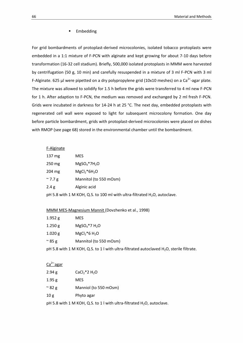
66 Material and Methods
Embedding
For grid bombardments of protoplast-derived microcolonies, isolated tobacco protoplasts were
embedded in a 1:1 mixture of F-PCN with alginate and kept growing for about 7-10 days before
transformation (16-32 cell stadium). Briefly, 500,000 isolated protoplasts in MMM were harvested
by centrifugation (50 g, 10 min) and carefully resuspended in a mixture of 3 ml F-PCN with 3 ml
F-Alginate. 625 µl were pipetted on a dry polypropylene grid (10x10 meshes) on a Ca2+-agar plate.
The mixture was allowed to solidify for 1.5 h before the grids were transferred to 4 ml new F-PCN
for 1 h. After adaption to F-PCN, the medium was removed and exchanged by 2 ml fresh F-PCN.
Grids were incubated in darkness for 14-24 h at 25 °C. The next day, embedded protoplasts with
regenerated cell wall were exposed to light for subsequent microcolony formation. One day
before particle bombardment, grids with protoplast-derived microcolonies were placed on dishes
with RMOP (see page 68) stored in the environmental chamber until the bombardment.
F-Alginate
137 mg MES
250 mg MgSO4*7H2O
204 mg MgCl2*6H2O
~ 7.7 g Mannitol (to 550 mOsm)
2.4 g Alginic acid
pH 5.8 with 1 M KOH, Q.S. to 100 ml with ultra-filtrated H2O, autoclave.
MMM MES-Magnesium Mannit (Dovzhenko et al., 1998)
1.952 g MES
1.250 g MgSO4*7 H2O
1.020 g MgCl2*6 H2O
~ 85 g Mannitol (to 550 mOsm)
pH 5.8 with 1 M KOH, Q.S. to 1 l with ultra-filtrated autoclaved H2O, sterile filtrate.
Ca2+-agar
2.94 g CaCl2*2 H2O
1.95 g MES
~ 82 g Manniol (to 550 mOsm)
10 g Phyto agar
pH 5.8 with 1 M KOH, Q.S. to 1 l with ultra-filtrated H2O, autoclave.

Material and Methods 67
o Particle gun-mediated transformation
Prior to transformation by the biolistic method, gene gun, parts and consumables were surface
sterilized and air-dried in the clean bench.
Next, transformation vector DNA was coated on gold particles. A stock of 0.6 µm gold particles
(Biorad) was suspended in 100% enthanol (60 mg/ml) and stored at - 20 °C. 35 µl of the gold stock
were transferred into an Eppendorf tube, and pelleted at 14,000 rpm for 10 s. The supernatant
was removed, the gold pellet resuspended in 1 ml sterile water and pelleted again (14,000 rpm,
1 min). The following steps were carried out on ice.
The washed pellet was resuspended in a mixture of 230 µl water and 250 µl 2.5 M CaCl2, and
25 µg of DNA (in water) were added. DNA was coated on gold particles by the addition of
50 µl 0.1 M spermidine and incubation on ice for 10 min, while gently vortexing every 2-3 min.
The stabilized complex was centrifuged for 1 min at 10,000 rpm and the supernatant was
removed. The gold-DNA complex was washed two times with 600 µl of 100% ethanol by
resuspension and centrifugation at 10,000 rpm for 1 min. The complex was taken up in 72 µl
ethanol and 5.4 µl were pipetted on each macrocarrier placed in the macrocarrier holder and
allowed to dry for a few minutes. Transformation was carried out using the following parameters:
Pressure of helium: 1100 psi
Rupture discs: 900 psi
Distance rupture disc to macrocarrier: 8-10 mm
Distance macrocarrier to stopping screen: 10 mm
Distance stopping plate to leaf: 7 cm
Vacuum: 27-28 inHg
o Tissue culture conditions
2-3 days post bombardment, leaves were cut into small pieces and placed on RMOP (regeneration
medium) with 500 mg/l spectinomycin as selective agents. After two weeks, enlarged leaf pieces
were cut again and recultured on fresh RMOP selection plates. Cultivation continued
(from now every 3-4 weeks transferred to a new plate) until green, putative transplastomic,
primary shoots emerged from the bleached leaf pieces. Bombarded protoplast-derived
microcolonies were subcultured according to the same regime.

68 Material and Methods
RMOP (Svab et al., 1990)
4.4 g MS-salts
10 ml 100x NT-Vitamins (Nagata und Takebe, 1971)
10 g Inosit
100 mg Thiamin-HCl
Q.S. to 1 l with ultra-filtrated H2O, store at - 20 °C.
1.0 ml Benzylaminopurine (1 mg/ ml)
100 µl Naphthalene acetic acid (1 mg/ ml)
30.0 g Sucrose
5.5 g Phyto agar
pH 5.8 with 1 M KOH, Q.S. to 1 l with ultra-filtrated H2O, autoclave.
Transformation of the nuclear genome
Transformation of the nuclear genome was performed according to Horsch et al. (1985).
Agrobacterium GV3101 harboring plasmid PSB419 was a kind gift from Richard Dixon (Sharma and
Dixon, 2005).
6.2.3 Transgenic plants
6.2.3.1 Transplastomic lines
Primary shoots emerging from bombarded leaves or microcolonies were isolated and transferred
to fresh RMOP medium (6.2.2.2) for cyclization. Cyclization, i.e. the repeated regeneration of
heteroplasmic leaf material on RMOP plates with selection pressure (500 mg/l spectinomycin),
was carried until homoplasmy was achieved.
6.2.3.2 Nuclear transformants
Nuclear transformants (T0) obtained by Agrobacterium-mediated transformation were allowed to
self in the greenhouse. Resulting seeds (T1) were sterilized and germinated on B5mod.
Transformants were separated and grown in the greenhouse to produce T2.

Material and Methods 69
6.2.4 Molecular analysis
6.2.4.1 DNA isolation from plant tissue
Quick protocol
About 100 mg of leaf tissue was ground in a screw cap tube with glass beads and 400 µl extraction
buffer. Tubes were centrifuged for 15 min at 15,000 rpm and 330 µl of the supernatant were
transferred to a new tube with an equal volume of isopropanol. After mixing and 10 min
incubation at -20 °C, the DNA was pelleted by centrifugation (30 min, 15,000 rpm).
The supernatant was removed and the pellets air-dried. A final volume of 40 µl sterile water was
added to the tubes without disturbing the pellet. Tubes were stored at 4 °C overnight prior to
PCR. For immediate analyses, about 1 h is sufficient for resolving the DNA from the pellet.
Extraction buffer (Quick and Dirty)
0.2 M Tris-HCl (pH 7.5)
0.25 M NaCl
0.025 M EDTA
0.5% (w/v) SDS
Use ultra-filtrated H2O, autoclave.
2x CTAB
400 mg of fresh leaves were ground in liquid nitrogen. 2x CTAB (1 equal volume) was added to the
ground powder in a 2 ml Eppendorf tube and the frozen samples were allowed to thaw (aided by
short vortexing). Cells were lysed in a water bath at 65 °C for at least 1 h. One volume of
chloroform was added and the tubes were rotated 30 min at RT. Samples were centrifuged for
20 min at 15,000 rpm and the RNA in the supernatant was digested (10 µl RNAse; 10 µg/ml,
30 min, 42 °C), After a second chloroform extraction step, the DNA in the supernatant was
precipitated by incubation with 0.7 vol isopropanol (30 min, - 20 °C) and centrifugation (30 min at
15,000 rpm, 4 °C). DNA was washed two times with ethanol, air-dried, redissolved in 60 µl sterile
water and frozen at - 20 °C.

70 Material and Methods
2x CTAB-Extractionbuffer
200 mM Tris-HCl (pH 8.0)
2% (w/v) CTAB
20 mM EDTA
1400 mM NaCl
1% (w/v) Polyvinylpyrrolidon (40 kDa)
Use ultra-filtrated H2O, autoclave.
6.2.4.2 Polymerase chain reaction
DNA fragments for cloning purposes were amplified with Phusion polymerase (Finnzyme), while
Taq polymerase was used for colony PCR and for screening transplastomic lines (MBI Fermentas).
Reaction conditions were adapted to the oligos used for amplification, the length of the PCR
product and the polymerase. PCR products were analysed on agarose gels.
Taq Polymerase
2.0 µl (10x)
1.6 µl
0.4 µl
0.4 µl
0.4 µl
0.2 µl
100 ng
Q.S. 20 µl
Heated Lid 112.0 °C
95.0 °C for 5 min
30-35x
95.0 °C for 30 s
Anneal for 30 s
72.0 °C for elongation
End
72 °C for 5 min
Store at 4 °C
Phusion Polymerase
10 µl (5x)
/
1 µl
2.5 µl
2.5 µl
0.5 µl
100 ng
Q.S. 50 µl
Heated Lid 112.0 °C
98.0 °C for 30 s
30-35x
98.0 °C for 10 s
Anneal for 30 s
72.0 °C for elongation
End
72 °C for 10 min
Store at 4 °C
Buffer
MgCl2
dNTP (10 mM)
Oligo fwd (10 pmol/ul)
Oligo rev (10 pmol/ul)
Polymerase
Template
Water

Material and Methods 71
6.2.4.3 Southern blot analysis
2 µg of CTAB-extracted DNA was digested with an appropriate restriction enzyme for 4 h and
separated on an agarose gel at 10-30 Volt overnight. The next day, DNA was transferred to a
positive charged nylon membrane via blotting the gel in an alkali environment (0.4 M NaOH). The
gel was placed on the blotting aperture, followed by the membrane, two layers of Whatmann
paper, several layers of tissue paper and a weight on top to apply pressure. After blotting, the gel
was checked for complete transfer using UV illumination. The membrane was washed with 2x SSC
until a neutral pH was achieved, air-dried, UV-cross linked and stored at RT until hybridization.
For hybridisation probes were PCR amplified, kit extracted and adjusted to 5 ng/µl. Radioactive
labeling was performed with α-32-P-dCTP using the prime-a-gene labeling system (Promega) as
indicated by the manufacturer. Radioactively labeled probes were purified via MicroSpin-Columns
S-200 HR (GE Healthcare), denatured for 3 min at 100 °C and put on ice. After pre-hybridization
for 1 h at 65 °C in hybridization buffer, the membrane was incubated with the labeled probe in
7.5 ml hybridization buffer at 65 °C overnight.
Unspecifically bound probe was removed the next day by washing steps with increasing
stringency (1x 30 minutes with 0.5x SSC-buffer, 0.1% (w/v) SDS at 60 °C, 2x 30 minutes with
0.1x SSC-Puffer und 0.1% (w/v) SDS at 65 °C). The membrane was exposed to a phoshoimager
screen and signals were detected with a BAS 1500-Phosphoimager (Fujifilm).
20 x SSC
3.0 M NaCl
0.3 M Sodium citrate (pH 7.0)
pH 7.0 with 1 M HCl, use ultra-filtrated H2O, autoclave.
Hybridization buffer (Church and Gilbert, 1984)
250 mM Na2HPO4 / NaH2PO4 (pH 7.5)
7% (w/v) SDS
Store at - 20 °C.

72 Material and Methods
6.2.5 Macroscopic analysis
6.2.5.1 Vegetative and reproductive growth
Sterile seed-grown homoplastomic plants at an age of 3-4 weeks were transferred to the
greenhouse and the growth was documented. At the time of flowering, floral buds were covered
with paper bags to avoid cross pollination and the plants ability to self-pollinate and to develop
seeds was monitored.
6.2.5.2 Seed assay
Seed-grown plants were crossed with and to WT plants. Resulting seeds were surface sterilized
and germinated on B5mod medium (6.2.2.2) with and without 100 mg/l Spec. After 14 days,
germination rates and seedlings phenotypes were documented.
6.2.6 Microscopic analysis
6.2.6.1 Fluorescence microscopy
Expression strength of plant lines were routinely checked with a DM 1000 (Leica Microsystems).
6.2.6.2 Confocal laser scanning microscopy (CLSM)
Confocal laser scanning microscopy was performed on a TCS SP5 (Leica Microsystems), with an
argon laser excitation at 488 nm (Leica EGFP) and detection at 496 nm-559 nm.
Leaves
Leaves of seed-grown homoplastomic plants were water-infiltrated for microscopy. Water-
infiltration was carried out with a 50 ml syringe by first applying a vacuum to the leaf in the water-
filled syringe, followed by release of the vacuum and subsequent water in-flow into the leaf. An
explant of the water-infiltrated leaf was cut, carefully mounted on slides with custom prepared
spacers to avoid cover glass damage to the explant and studied by CLSM.

Material and Methods 73
Protoplast-derived cells
Protoplasts of seed-grown homoplastomic plants were isolated and embedded in grids as
described in 6.2.2.2. For the microscopy of early division stages of protoplast-derived cells, the
grid was carefully removed from the cells embedded in alginate. Released alginate embedded
cells were mounted on custom prepared slides and studied by CLSM.
6.2.7 Chromatographic analysis
Thin-layer chromatography (TLC)
200 mg leaf tissue from greenhouse-grown plants before flowering were shock frozen in liquid
nitrogen and ground with glass beads in a screw-cap tube for 40 s at 5,000 rpm in darkness. 1 ml
methanol:HCl [99.5:0.5 (v/v)] was added and the extraction was repeated. The extract was
centrifuged for 5 min at 5,000 rpm and the supernatant was transferred to new tube. For analysis
of anthocyanin content, cellulose analytic TLC plates (Sigma) were developed in concentrated
HCl:formic acid:water (19.0:39.6:41.4).
6.2.8 Biochemical analysis
6.2.8.1 Extraction of total soluble protein (TSP)
Total soluble protein was extracted from 100 mg leaf tissue of adult plants before flowering.
Leaves were ground with glass beads in 500 ml ice-cold TSP extraction buffer in a bead mill for
3 min at 250 rpm. The extract was placed on ice and centrifuged at 15,000 rpm for 30 min at 4 °C.
The supernatant was taken as total soluble protein.
TSP extraction buffer:
50 mM NaH2PO 4 / Na2HPO 4 (pH 7.0)
10 mM EDTA
10 mM 2-mercaptoethanol
0.1% (v/v) Triton X-100
1 Tablet Complete Protease Inhibitor Cocktail (Roche) per 10 ml

74 Material and Methods
6.2.8.2 The Bradford assay
5 µl of TSP was added to 1 ml 1 : 5 diluted Bradford solution in PBS. Samples were mixed and
extinction at 595 nm was measured after 10 min. Protein content was calculated using a standard
curve of known BSA concentrations.
6.2.8.3 Sodium dodecyl sulfate polyacrylamide gel electrophoresis (SDS-PAGE)
Total soluble proteins were separated on discontinuous 10% denaturing polyacrylamide gels
(with 20 mA for each mini gel) according to Laemmli. Gels were stained with Coomassie Brilliant
Blue R-250 (Serva) on a shaker for 1 h. Background staining was removed with washing solution
(10% acetic acid) and the gels scanned.
6.2.8.4 Isolation of CPP fusion proteins
CPP fusion proteins were isolated from E. coli cells and tobacco leaves.
from E. coli
E. coli XL1 blue harbouring CPP_eGFP plastid transformation vectors were grown in 1-1.5 l LBAmp
(see 6.2.2.1) cultures for fusion protein isolation. Cells were harvested (10 min, 4,000 rpm, 4 °C)
and lysed in binding buffer by sonification. Purification was carried out by binding to Ni2+-agarose
(Biontex) according to the manufacturer’s protocol. Isolated proteins were dialysed overnight
50 mM Tris-HCl pH 8.0 at 4 °C, adjusted to 10% glycerol, shock-frozen and stored at - 80 °C.
from Nicotiana tabacum L.
CPPx-eGFP fusion proteins were isolated from greenhouse-grown tobacco plants before
flowering. About 50 g of FW plant material was ground in liquid nitrogen to a fine powder and
total proteins extracted in ice-cold Tris-HCl pH 8.0. The extract was centrifuged at 15,000 rpm for
20 min at 4 °C and the supernatant was vacuum filtrated (0.45 µM filter pore size).
Prior to HIC column purification, ammonium sulfate precipitation was carried out. Unwanted
proteins were precipitated by adjusting to 30% (w/v) ammonium sulfate. The eGFP fusion
proteins were finally precipitated with 80% ammonium sulfate. The pellet was resuspended in

Material and Methods 75
high salt buffer (50 mM Tris-HCl pH 8.0, 3 M NaCl) and applied to a HiTrap Butyl HP column
(GE Healthcare). Binding and washing was carried out as suggested in application note 28-9192-12
AB (GE Healthcare). Proteins were adjusted to 50 % glycerol and stored short-time at - 20 °C.
Before transduction assays (6.2.9), purified proteins were dialyzed against 50 mM Tris-HCl pH 8.0
at 4 °C overnight and quantified using the Bradford assay.
Fast protein liquid chromatography (FPLC)
CPP-PAH fusion proteins were isolated from adult greenhouse plants. 20 g FW were ground to a
fine powder and extracted in 40 ml sodium phosphate buffer (20 mM NaH2PO 4 / Na2HPO 4 pH 7.4,
100 mM NaCl, 20 mM Imidazol). The extract was centrifuged at 15,000 rpm for 20 min at 4 °C and
the supernatant was vacuum filtrated (0.45 µM pore size). The recombinant fusion proteins were
purified on ÄKTApurifier (GE Healthcare) at 4 °C by loading crude extract on a HisTrap affinity
chromatography column (GE Healthcare) equilibrated with sodium phosphate buffer and
subsequent step-wise gradient elution was performed with the same buffer supplemented with
500 mM imidazol. The fractions from affinity chromatography were analysed by gel filtration on a
HiLoad 16/60 Superdex 200 column (GE Healthcare) equilibrated with 20 mM NaHEPES, pH 7.0
containing 200 mM NaCl.
6.2.8.5 Western blot
Proteins were separated by SDS-PAGE (4-12% Novex NuPAGE Bis-Tris gel system, Invitrogen) at a
constant voltage of 200 Volt for 90 min and subsequently blotted onto nitrocellulose membrane
(BA-S 85; Schleicher & Schuell). The blocked membrane (5% milk powder in TBS-Tween) was
incubated for 1 h with the primary antibody, mouse anti-PAH (1:1,000 dilution; Calbiochem), and
1 h with the secondary, goat anti-mouse IgG HRP conjugated antibody (1:10,000 dilution; Santa
Cruz Biotechnology). The blot was visualized with SuperSignal West Femto Substrate (Thermo
Scientific) and chemiluminescence was monitored with a DIANA III imaging system (Raytest).
6.2.9 Functional fusion protein assays
6.2.9.1 Transduction of CPP fusion proteins into plant cells
For the transduction of fusion proteins into protoplasts, 2.5 x 10⁵ freshly isolated protoplasts
(6.2.2.2) were incubated with protein concentrations from 1-5 µM in F-PCN for 1 h in darkness.

76 Material and Methods
After incubation, protoplasts were washed two times with 10 ml F-PCN (10 min at 50 g) and
studied by CLSM.
In addition, transduction in partially digested young leaves was studied. Young leaves of 4-5 week
old tobacco leaves were digested for 20 h. The resulting partially digested young leaves were
washed two times in F-PCN and adjusted to 5 µM peptide in F-PCN. Incubation was carried out for
24 h at RT in darkness. For analysis, leaf tissue was washed two times with F-PCN and studied by
CLSM.
6.2.9.2 Transduction of CPP fusion proteins into human cells
To test transduction into human cells lines, suspension cells were kept in RPMI medium 1640
(Gibco) with 10% FCS (fetal calf serum), 5% penicillin / streptomycin, 5% L- glutamin and 0.1%
beta-mercaptoethanol at standard cell culture conditions (37 °C, 5% CO2) in cell culture flasks.
For transduction assay 2 x 10⁶ cells were washed twice with PBS, resuspended in Optimem
medium (reduced serum medium, Gibco) and transferred into a 6-well plate. The fusion protein
was added and incubated with the cells (37 °C, 5% CO2). At selected time-points, cells were
washed twice with PBS and treated with trypsin for 15 min to remove membrane-bound fusion
protein. Reaction was stopped by adding complete medium, in which cells were washed again.
In order to quench the residual signal coming from unspecific membrane attachment, cells were
resuspended in trypan blue (0.005% in PBS). Flow cytometric analysis was performed on a FACS
Canto II (BD Biosciences).
Adherent Phoenix (293 derived cell line) cells were kept in DMEM medium (Gibco) containing 10%
FCS, 5% penicillin / streptomycin, 5% L-glutamine and 0.1% beta-mercaptoethanol at standard cell
culture conditions in cell culture dishes. The day before the transduction assay 5 x 10⁵ cells per
well were seeded out in 6-well plates. Prior to transduction, cells were washed twice with PBS and
incubated with Optimem for 1 h. The fusion protein was added and incubated with the cells
(37 °C, 5% CO2). At selected time-points, cells were washed twice with PBS and treated with
trypsin. The reaction was stopped with complete medium, in which cells were washed again.
Finally cells were resuspended trypan blue (0.005% in PBS) and analysed by FACS.

7 Abbreviations
5’ UTR 5’-untranslated region
aadA 3’ aminoglycoside-adenyltransferase, antibiotics selection gene
AB Antibody
(Arg)9 Nonaarginine, CPP
Amp Ampicillin
Arg Arginine
AS Amino acid(s)
bp/kb Base pair(s)/ Kilo base pairs
BH4 6-R-L-erythro-5,6,7,8-tetrahydrobiopterin
BSA Bovine serum albumin
B5 Gamborg´s medium
c Centi
CLSM Confocal laser scanning microscopy
CPP(x) Cell penetrating peptide (x represents a CPP/number according to Table 1)
CPPx-XXX Fusion protein of given vector series
CPPx_XXX Vector or resulting plant line of given vector series
CTAB Cetyl trimethyl ammonium bromide
CTB Mucosa-binding subunit B of cholera toxin
Da Dalton
DNA Deoxyribonucleic acid
E. coli Escherichia coli
EDTA Ethylenediaminetetraacetic acid
e.g. Exempli gratia, Latin: "for example"
(e)GFP (Enhanced) green fluorescent protein
ER Endoplasmic reticulum
ERT Enzyme replacement therapy
FACS Fluorescence activated cell sorting
FDA Fluorescein diacetate
FPLC Fast protein liquid chromatography
FW Fresh weight
g Centrifugal force or gram
G10L RBS Ribosomal binding site from gene 10 leader sequence
gfp14 First 14 amino acids of GFP
Goi Gene of interest
GUS β-glucuronidase
h Hour(s)
HIC Hydrophobic interaction chromatography
His-tag Polyhistidine-tag
HIV-1 Human immunodeficiency virus type 1
inHg Inches of mercury
INSR Right insertion site
INSL Left insertion site
k Kilo

78 Abbreviations
l Litre
Lab Laboratory
LB Luria-Bertani
LMU Ludwig Maximilian University of Munich
LTB Escherichia coli heat-labile enterotoxin subunit B
m Milli or metre
MCS Multiple cloning site
MES 2-(N-morpholino)ethanesulfonic acid
min Minute(s)
(m)M (milli)Molar = (milli)moles/l
MOPS 3-(N-morpholino)propanesulfonic acid
mOsm Milliosmole
n Nano
NEP Nuclear encoded polymerase
numbers 1 to 9 CPP abbreviation / number used in the text (compare Table 1.)
OD Optical density
ORF Open reading frame
PAH Phenylalanine hydroxylase
PAP1 Production of anthocyanin pigment 1
PBS Phosphate buffered saline
PCR Polymerase chain reaction
PEP Plastid-encoded polymerase from rrn 16
PKU Phenylketonuria
PNG10- Fusion of PEP, NEP and G10L RBS
psi Pounds per square inch
Q.S. Quantity sufficient
rbcL Large subunit of the ribulose-bisphosphate carboxylase
rpm Revolutions per minute
RT Room temperature
RNA Ribonucleic acid
SDS-PAGE Sodium dodecyl sulfate polyacrylamide gel electrophoresis
siRNA Small interfering RNA
Spec Spectinomycin
T Thymus or trangenic plant generation or Thrombin site
Tat Transactivator of transcription (HIV-1), CPP
TrbcL rbcL terminator from C. reinhardtii
tRNA Transfer RNA
TSP Total soluble protein
UV Ultraviolet light
WT Wild-type
w/v Weigth per volume
v/v Volume per volume
♀/♂ Female / male
°C Degree Celsius
µ Micro o Symbol for PAP1 vector series
* Symbol for PAH vector series or insertion site: trnfM / trnG

8 Tables and Figures
Table 1. Selected CPPs, CPP abbreviation / number used in the text, origin, sequence and original
publication. .................................................................................................................. 19
Figure 1. Events from the biolistic transformation vector delivery to the regeneration of a
homoplastomic plant. ................................................................................................. 10
Figure 2. Schematic overview of possible applications where cell-penetrating peptides have been
shown to function well as delivery vehicles, both in vitro and in vivo. ....................... 13
Figure 3. Suggested mechanisms of cell entry used by CPPs. ......................................................... 14
Figure 4. Codon-optimized expression cassette for CPP fusion protein expression and
transformation vector assembly of the eGFP vector series. ....................................... 20
Figure 5. Identification of transplastomic CPPx_eGFP lines and confirmation of correct vector
integration into the plastome by PCR and Southern blot. .......................................... 21
Figure 6. Crossing CPPx_GFP lines. .................................................................................................. 22
Figure 7. First division stages of CPPx_eGFP protoplast-derived cells. ........................................... 23
Figure 8. Ease of artefact generation as a drawback of the CPPx-eGFP system. ............................ 24
Figure 9. Adult stages of CPPx_eGFP lines and SDS-PAGE of extracted total soluble protein (TSP) 25
Figure 10. HIC purified CPPx-eGFP fusion proteins separated on SDS-PAGE. ................................. 26
Figure 11. Transduction of CPPx-eGFP fusion proteins into tobacco WT protoplasts. ................... 27
Figure 12. Transduction of CPPx-eGFP fusion proteins into partially digested young WT tissues. . 28
Figure 13. Transduction of CPPx-eGFP fusion proteins into human cells. ...................................... 29
Figure 14. Cloning the transformation vector intermediate pUC18_INS*_CPP cassette_rbcL
(= pUC18(C)), targeting insertion site trnfM / trnG. .................................................... 30
Figure 15. Proposed action of CPPx-PAP1 fusion proteins upontheir production in the plastid ..... 31
Figure 16. Generation and phenotype of nuclear transformant pap1. ........................................... 32
Figure 17. Cloning of the PAP1 transformation vector series. ........................................................ 33
Figure 18. Identification of transplastomic CPPx_PAP1 lines and confirmation of correct vector
integration into the plastome by PCR and Southern blot. .......................................... 34
Figure 19. Phenotype of transplastomic PAP1 lines in comparison to pap1 nuclear transformant.
...................................................................................................................................... 35
Figure 20. SDS-PAGE of extracted CPPx_PAP1 soluble protein (TSP). ............................................. 35
Figure 21. Cloning of the PAH transformation vector series. .......................................................... 37
Figure 22. Identification of transplastomic CPPx_PAH lines and confirmation of correct integration
into the plastome. ....................................................................................................... 38
Figure 23. Phenotype of CPPx_PAH lines and TSP separated by SDS-PAGE. ................................... 39
Figure 24. Germination of CPP2_PAH (external aadA) seeds on (A) on B5 (B) on B5Spec. ................ 40
Figure 25. Gel filtration and identification of CPP2-PAH fusion protein by Western blot. .............. 40


9 References
Apel W and Bock R (2009) Enhancement of carotenoid biosynthesis in transplastomic tomatoes by induced lycopene-to-provitamin A conversion. Plant Physiol 151: 59-66. Apel W, Schulze WX and Bock R (2010) Identification of protein stability determinants in chloroplasts. Plant J 63: 636-650. Asoh S and Ohta S (2008) PTD-mediated delivery of anti-cell death proteins/peptides and therapeutic enzymes. Adv Drug Deliv Rev 60: 499-516. Aussedat B, Sagan S, Chassaing G, Bolbach G and Burlina F (2006) Quantification of the efficiency of cargo delivery by peptidic and pseudo-peptidic Trojan carriers using MALDI-TOF mass spectrometry. Biochim Biophys Acta 1758: 375-383. Aviezer D, Brill-Almon E, Shaaltiel Y, Hashmueli S, Bartfeld D, Mizrachi S, Liberman Y, Freeman A, Zimran A and Galun E (2009) A plant-derived recombinant human glucocerebrosidase enzyme-a preclinical and phase I investigation. PLoS One 4: e4792. Bárány-Wallje E, Gaur J, Lundberg P, Langel Ü and Gräslund A (2007) Differential membrane perturbation caused by the cell-penetrating peptide Tp10 depending on attached cargo. FEBS Lett 581: 2389-2393. Bellucci M, de Marchis F, Mannucci R, Bock R and Arcioni S (2005) Cytoplasm and chloroplasts are not suitable subcellular locations for ß-zein accumulation in transgenic plants. J Exp Bot 56: 1205-1212. Benko-Iseppon AM, Galdino SL, Calsa T Jr, Kido EA, Tossi A, Belarmino LC and Crovella S (2010) Overview on plant antimicrobial peptides. Curr Protein and Pept Sci 11: 181-188. Bevan MW, Flavell RB and Chilton M-D (1983) A chimaeric antibiotic resistance gene as a selectable marker for plant cell transformation. Nature 304: 184-187. Bidwell III GL, Davis AN and Raucher D (2009) Targeting a c-Myc inhibitory polypeptide to specific intracellular compartments using cell penetrating peptides. J Control Release 135: 2-10. Bienenstock J and Befus AD (1980) Mucosal immunology. Immunology 41: 249-270. Björklund J, Biverståhl H, Gräslund A, Mäler L and Brzezinski P (2006) Real-time transmembrane translocation of penetratin driven by light-generated proton pumping. Biophys J 91: L29-L31. Bock R and Khan MS (2004) Taming plastids for a green future. Trends Biotechnol 22: 311-318. Bock R and Warzecha H (2010) Solar-powered factories for new vaccines and antibiotics. Trends Biotechnol 28: 246-252. Borevitz J, Xia Y, Blount JW, Dixon RA and Lamb C (2000) Activation tagging identifies a conserved MYB regulator of phenylpropanoid biosynthesis. Plant Cell 12: 2383-2393. Boyhan D and Daniell H (2011) Low-cost production of proinsulin in tobacco and lettuce plastids for injectable or oral delivery of functional insulin and C-peptide. Plant Biotechnol J 9: 585-598. Boynton JE, Gillham NW, Harris EH, Hosler JP, Johnson AM, Jones AR, Randolph-Anderson BL, Robertson D, Klein TM, Shark KB and Sanford JC (1988) Plastid transformation in Chlamydomonas with high velocity microprojectiles. Science 240: 1534-1538. Brady RO (2006) Enzyme replacement for lysosomal diseases. Annu Rev Med 57: 283-296. Brooks NA, Pouniotis DS, Tang CK, Apostolopoulos V and Pietersz GA (2010) Cell-penetrating peptides: application in vaccine delivery. Biochim Biophys Acta 1805: 25-34.

82 References
Butow RA and Fox TD (1990) Organelle transformation: shoot first, ask questions later. Trends in Biochemical Sciences 15: 465-468. Cai SR, Xu G, Becker-Hapak M, Ma M, Dowdy SF and McLeod HL (2006) The kinetics and tissue distribution of protein transduction in mice. Eur J Pharm Sci 27: 311-319. Cao G, Pei W, Ge H, Liang Q, Luo Y, Sharp FR, Lu A, Ran R, Graham SH and Chen J (2002) In vivo delivery of a Bcl-xL fusion protein containing the TAT protein transduction domain protects against ischemic brain injury and neuronal apoptosis. J Neurosci 22: 5423-5431. Cardi T, Lenzi P and Maliga P (2010) Plastids as expression platforms for plant-produced vaccines. Expert Rev Vaccines 9: 893-911. Caron NJ, Torrente Y, Camirand G, Bujold M, Chapdelaine P, Leriche K, Bresolin N and Tremblay JP (2001) Intracellular delivery of a Tat–eGFP fusion protein into muscle cells. Mol Therapy 3: 310-318. Cascales L, Henriques ST, Kerr MC, Huang Y-H, Sweet MJ, Daly NL and Craik DJ (2011) Identification and characterization of a new family of cell penetrating peptides: Cyclic cell penetrating peptides. J Biol Chem 286: 36932-36943. Cermenati G, Terracciano I, Castelli I, Giordana B, Rao R, Pennacchio F and Casartelli M (2011) The CPP Tat enhances eGFP cell internalization and transepithelial transpor t by the larval midgut of Bombyx mori (Lepidoptera, Bombycidae). J Insect Physiol 57: 1689-1697. Chan CX and Bhattacharya D (2010) The origin of plastids. Nat Education 3: 84. Chang M, Chou J-C, Chen C-P, Liu BR and Lee H-J (2007) Noncovalent protein transduction in plant cells by macropinocytosis. New Phytol 174: 46-56. Chang M, Chou J-C and Lee H-J (2005) Cellular internalization of fluorescent proteins via arginine-rich intracellular delivery peptide in plant cells. Plant Cell Physiol 46: 482-488. Chauhan A, Tikoo A, Kapur AK and Singh M (2007) The taming of the cell penetrating domain of the HIV Tat: myths and realities. J Control Release 117: 148-162. Chen C-P, Chou J-C, Liu BR, Chang M and Lee H-J (2007) Transfection and expression of plasmid DNA in plant cells by arginine-rich intracellular delivery peptide without protoplast preparation. FEBS Lett 581: 1891-1897. Chen L and Harrison SD (2007) Cell-penetrating peptides in drug development: enabling intracellular targets. Biochem Soc Trans 35: 821-825. Chugh A, Amundsen E and Eudes F (2009) Translocation of cell-penetrating peptides and delivery of their cargoes in triticale micro-spores. Plant Cell Rep 28: 801-810. Chugh A and Eudes F (2007) Translocation and nuclear accumulation of monomer and dimmer of HIV-1 Tat basic domain in triticale mesophyll protoplasts. Biochim Biophys Acta Biomembr 1768: 419-426. Chugh A and Eudes F (2008a) Cellular uptake of cell-penetrating peptides pVEC and transportan in plants. J Pept Sci 14: 477-481. Chugh A and Eudes F (2008b) Studies of uptake of cell-penetrating peptides and their cargoes in wheat immature embryos. FEBS J 275: 2403-2414. Chugh A, Eudes F and Shim YS (2010) Cell-penetrating peptides: Nanocarrier for macromolecule delivery in living cells. IUBMB Life 62: 183-193. Church GM and Gilbert W (1984) Genomic sequencing. Proc Natl Acad Sci USA 81: 1991-1995. Clough SJ and Bent AF (1998) Floral dip: a simplified method for Agrobacterium mediated transformation of Arabidopsis thaliana. Plant J 16: 735-743.
References 83
Ciobanasu C, Siebrasse JP and Kubitscheck U (2010) Cell-penetrating HIV1 Tat peptides can generate pores in model membranes. Biophys J 99: 153-162. Collakova E, Goyer A, Naponelli V, Krassovskaya I, Gregory JF, Hanson III AD and Shachar-Hill Y (2008) Arabidopsis 10-formyl tetrahydrofolate deformylases are essential for photorespiration. Plant Cell 208: 1818-1832. Corriveau JL and Coleman AW (1988) Rapid screening method to detect potential biparental inheritance of plastid DNA and results for over 200 angiosperm species. Am J Bot 75: 1443-1458. Daniell H, Singh ND, Mason H and Streatfield SJ (2009) Plant-made vaccine antigens and biopharmaceuticals. Trends Plant Sci 14: 669-679. Daniell H, Streatfield SJ and Wycoff K (2001) Medical molecular farming: production of antibodies, biopharmaceuticals and edible vaccines in plants. Trends Plant Sci 6: 219-226. Day A and Goldschmidt-Clermont M (2011) The plastid transformation toolbox: selectable markers and marker removal. Plant Biotech J 9: 540-553. Del Gaizo V and Mark Payne R (2003) A novel TAT-mitochondrial signal sequence fusion protein is processed, stays in mitochondria, and crosses the placenta. Molec Ther 7: 720-730. Deshayes S, Plénat T, Charnet P, Divita G, Molle G and Heitz F (2006) Formation of transmembrane ionic channels of primary amphipathic cell-penetrating peptides. Consequences on the mechanism of cell penetration. Biochim Biophys Acta 1758: 1846-1851. Dietz GPH and Bähr M (2004) Delivery of bioactive molecules into the cell: the Trojan horse approach. Mol Cell Neurosci 27: 85-131. Dietz GPH and Bähr M (2005) Peptide-enhanced cellular internalization of proteins in neuroscience. Brain Res Bull 68: 103-114. Dietz GPH (2011) Protection by neuroglobin and cell-penetrating peptide-mediated delivery in vivo: a decade of research. Comment on Cai et al.: TAT-mediated delivery of neuroglobin protects against focal cerebral ischemia in mice. Exp Neurol. 2011; 227(1): 224-31. Experimental neurology 231: 1-10. Doherty GJ and McMahon HT (2009) Mechanisms of endocytosis. Annu Rev Biochem 78: 857-902. Dovzhenko A, Bergen U and Koop H-U (1998) Thin-alginate-layer technique for protoplast culture of tobacco leaf protoplasts: Shoot formation in less than two weeks. Protoplasma 204: 114-118. Dowdy SF (2006) Applications of cell-penetrating peptides in gene modulation and protein transport. In: Langel Ü (eds) Handbook of cell-penetrating peptides, Second Edition. CRC Press: Boca Raton, FL, pp 309-311. Duchardt F, Fotin-Mleczek M, Schwarz H, Fischer R and Brock R (2007) A comprehensive model for the cellular uptake of cationic cell-penetrating peptides. Traffic 8: 848-866. Dupont E, Prochiantz A and Joliot A (2011) Penetratin story: an overview. Methods Mol Biol 683: 21-29. Eavri R and Lorberboum-Galski H (2007) A novel approach for enzyme replacement therapy. The use of phenylalanine hydroxylase-based fusion proteins for the treatment of phenylketonuria. J Biol Chem 282: 23402-23409. Edenhofer F (2008) Protein transduction revisited: Novel insights into the mechanism underlying intracellular delivery of proteins. Curr Pharm Des 14: 3628-3636. Eiríksdóttir E, Konate K, Langel U, Divita G and Deshayes S (2010) Secondary structure of cell-penetrating peptides controls membrane interaction and insertion. Biochim Biophys Acta 1798: 1119-1128.
84 References
El-Andaloussi S, Johansson HJ, Holm T and Langel Ü (2007) A novel cell-penetrating peptide, M918, for efficient delivery of proteins and peptide nucleic acids. Mol Ther 15: 1820-1826. Elghabi Z, Karcher D, Zhou F, Ruf S and Bock R (2011) Optimization of the expression of the HIV fusion inhibitor cyanovirin-N from the tobacco plastid genome. Plant Biotechnol J 9: 599-608. Elliott G and O’Hare P (1997) Intercellular trafficking and protein delivery by a herpes virus structural protein. Cell 88: 223-233. Eudes F and Chugh A (2008) Cell-penetrating peptides: From mammalian to plant cells. Plant Signal and Behavior 3: 549-550. Eum WS, Choung IS, Li MZ, Kang JH, Kim DW, Park J, Kwon HY and Choi SY (2004) HIV-1 Tat mediated protein transduction of Cu,Zn-superoxide dismutase into pancreatic β cells in vitro and in vivo. Free Radic Biol Med 37: 339-349. Fawell S, Seery J, Daikh Y, Moore C, Chen LL, Pepinsky B and Barsoum J (1994) Tat-mediated delivery of heterologous proteins into cells. Proc Natl Acad Sci USA 91: 664-668. Ferrari A, Pellegrini V, Arcangeli C, Fittipaldi A, Giacca M and Beltram F (2003) Caveolae-mediated internalization of extracellular HIV-1 Tat fusion proteins visualized in real time. Molec Ther 8: 284-294. Fischer R, Fotin-Mleczek M, Hufnagel H and Brock R (2005). Break on through to the other side-biophysics and cell biology shed light on cell-penetrating peptides. Chembiochem 6: 2126-2142. Fittipaldi A, Ferrari A, Zoppe M, Arcangeli C, Pellegrini V, Beltram F and Giacca M (2003) Cell membrane lipid rafts mediate caveolar endocytosis of HIV-1 Tat fusion proteins. J Biol Chem 278: 34141-34149. Foerg C and Merkle HP (2008) On the biomedical promise of cell penetrating peptides: limits versus prospects. J Pharm Sci 97: 144-162. Foged C and Nielsen HM (2008) Cell-penetrating peptides for drug delivery across membrane barriers. Expert Opin Drug Deliv 5: 105-117. Smith FJD, Irvine AD, Terron-Kwiatkowski A, et al. (2006) Loss-of-function mutations in the gene encoding filaggrin cause ichthyosis vulgaris. Nat Genetics 38: 337-342. Frankel AD and Pabo CO (1988) Cellular uptake of the tat protein from human immunodeficiency virus. Cell 55: 1189-1193. Fratantoni JC, Hall CW and Neufeld EF (1968) Hurler and Hunter syndromes: mutual correction of the defect in cultured fibroblasts. Science 162: 570-572. Fretz MM, Penning NA, Taei SA, Futaki S, Takeuchi T, Nakase I, Storm G and Jones AT (2007) Temperature, concentration and cholesterol dependent translocation of L- and D-octa-arginine across the plasma and nuclear membrane of CD34+ leukaemia cells. Biochem J 403: 335-342. Futaki S, Suzuki T, Ohashi W, Yagami T, Tanaka S, Ueda K and Sugiura Y (2001) Arginine-rich peptides. An abundant source of membrane-permeable peptides having potential as carriers for intracellµlar protein delivery. J Biol Chem 276: 5836-5840. Gamborg OL, Miller RA and Ojima K (1968) Nutrient requirements of suspension cultures of soybean root cells. Exp Cell Res 50, 151-158. Geueke B, Namoto K, Agarkova I, Perriard JC, Kohler H-PE and Seebach D (2005) Bacterial cell penetration by β3-oligo-homoarginines: indication for passive transfer through the lipid bilayer. Chembiochem 6: 982-985. Goldblatt J, Fletcher JM, McGill J, Szer J and Wilson M (2011) Enzyme replacement therapy “drug holiday”: results from an unexpected shortage of an orphan drug supply in Australia. Blood Cells Mol Dis 46: 107-110.

References 85
Golds TJ, Maliga P and Koop H-U (1993) Stable plastid transformation in PEG-treated protoplasts of Nicotiana tabacum. Nat Biotechnol 11: 95-97. Gray JC, Sullivan JA, Hibberd JM and Hansen MR (2001) Stromules: mobile protrusions and interconnections between plastids. Plant Biology 3: 223-233. Green M and Loewenstein PM (1988) Autonomous functional domains of chemically synthesized human immunodeficiency virus tat trans-activator protein. Cell 55: 1179-1188. Gunaratna KR, Andersson M and Good L (2002) Microbial membrane-permeating peptides and their applications. In: Langel Ü (eds) Cell penetrating peptides: processes and applications. CRC Press: Boca Raton, FL, pp. 377-396. Gupta B, Levchenko TS and Torchilin VP (2005) Intracellular delivery of large molecules and small particles by cell-penetrating proteins and peptides. Adv Drug Deliv Rev 57: 637-651. Han K, Jeon MJ, Kim KA, Park J and Choi SY (2000) Efficient intracellular delivery of GFP by homeodomains of Drosophila Fushi-tarazu and engrailed proteins. Mol Cells 10: 728-732. Hanson MR and Sattarzadeh A (2008) Dynamic morphology of plastids and stromules in angiosperm plants. Plant Cell Environ 31: 646-657. Hanson MR and Sattarzadeh A (2011) Stromules: recent insights into a long neglected feature of plastid morphology and function. Plant Physiol 155: 1486-1492. Hariton-Gazal E, Rosenbluh J, Graessmann A, Gilon C and Loyter A (2003) Direct translocation of histone molecules across cell membranes. J Cell Sci 116: 4577-4586. Heitz F, Morris MC and Divita G (2009) Twenty years of cell-penetrating peptides: from molecular mechanisms to therapeutics. Br J Pharmacol 157: 195-206. Henriques ST, Costa J and Castanho MA (2005) Translocation of beta-galactosidase mediated by the cell-penetrating peptide Pep-1 into lipid vesicles and human HeLa cells is driven by membrane electrostatic potential. Biochemistry 44: 10189-10198. Henriques ST, Melo MN and Castanho MA (2006) Cell-penetrating peptides and antimicrobial peptides: how different are they? Biochem J 399: 1-7. Herce HD, Garcia AE, Litt J, Kane RS, Martin P, Enrique N, Rebolledo A and Milesi V (2009) Arginine-rich peptides destabilize the plasma membrane, consistent with a pore formation translocation mechanism of cell-penetrating peptides. Biophysical Journal 97: 1917-1925. Herrera Díaz A (2011) Regeneration and plastid transformation approaches in Arabidopsis thaliana and Rapid-Cycling Brassica rapa. Dissertation, LMU München Herrera-Estrella L, Depicker A, Van Montagu M and Schell J (1983) Expression of chimaeric genes transferred into plant cells using a Ti-plasmid-derived vector. Nature 303: 209-213. Hirose H, Takeuchi T, Osakada H, Pujals S, Katayama S, Nakase I, Kobayashi S, Haraguchi T and Futaki S (2012) Transient focal membrane deformation induced by arginine-rich peptides leads to their direct penetration into cells. Molecular Therapy: in press (doi:10.1038/mt.2011.313). Ho A, Schwarze SR, Mermelstein SJ, Waksman G and Dowdy SF (2001) Synthetic protein transduction domains: enhanced transduction potential in vitro and in vivo. Cancer Res 61: 474-477. Holm T, Andaloussi SE and Langel U (2011) Comparison of CPP uptake methods. Methods Mol Biol 683: 207-217. Holmgren J and Czerkinsky C (2005) Mucosal immunity and vaccines. Nat Medicine 11: S45-S53. Holmgren J and Lycke N (1986) Immune mechanisms in enteric infections. In: Holmgren J, Lindberg A and Möllby R (eds) Development of vaccine and drugs against diarrhea, Studentlitteratur/Chartwell-Bratt Ltd: Lund, Sweden, 9 p.

86 References
Horsch RB, Fraley RT, Rogers SG, Sanders PR and Lloyd A (1985) A simple and general method for transferring genes into plants. Science 227: 1229-1231. Horton KL, Stewart KM, Fonseca SB, Guo Q and Kelley SO (2008) Mitochondria-penetrating peptides. Chem Biol 15: 375-382. Huber M, Rettler I, Bernasconi K, Frenk E, Lavrijsen SP, Ponec M, Bon A, Lautenschlager S, Schorderet DF and Hohl D (1995) Mutations of keratinocyte transglutaminase in lamellar ichthyosis. Science 267: 525-528. Ijaz S (2010) Plant mitochondrial genome: “A sweet and safe home’’ for transgene. African Journal of Biotechnology 9: 9196-9199. Isemer R, Mulisch M, Schäfer A, Kirchner S, Koop H-U and Krupinska K (2012) Plastid encoded Whirly1 is translocated to the nucleus. FEBS Lett 586: 85-88. Järver P, Mäger I and Langel Ü (2010) In vivo biodistribution and efficacy of peptide mediated delivery. Trends Pharmacol Sci 31: 528-535. Jin LH, Bahn JH, Eum WS, Kwon HY, Jang SH, Han KH, Kang TC, Won MH, Kang JH, Cho SW, Park J and Choi SY (2001) Transduction of human catalase mediated by an HIV-1 TAT protein basic domain and arginine-rich peptides into mammalian cells. Free Radical Biol Med 31: 1509-1519. Johansson HJ, EL Andaloussi S and Langel Ü (2011) Mimicry of protein function with cell-penetrating peptides. Methods Mol Biol 683: 233-247. Johnson RM, Harrison SD and Maclean D (2011) Therapeutic applications of cell-penetrating peptides. Methods Mol Biol 683: 535-551. Johnston SA, Anziano PQ, Shark K, Sanford JC and Butow RA (1988) Mitochondrial transformation in yeast by bombardment with microprojectiles. Science 240: 1538-1541. Joliot A, Pernelle C, Deagostini-Bazin H and Prochiantz A (1991) Antennapedia homeobox peptide regulates neural morphogenesis. Proc Natl Acad Sci USA 88: 1864-1868. Jones AT (2007) Macropinocytosis: searching for an endocytic identity and role in the uptake of cell penetrating peptides. J Cell Mol Med 11: 670-684. Jones AT (2010) Uptake and intracellular dynamics of proteins internalized by cell-penetrating peptides, in organelle-specific pharmaceutical nanotechnology. In: Weissig V and D'Souza GGM (eds) Organelle-specific pharmaceutical nanotechnology. John Wiley & Sons Inc: Hoboken, NJ, Chapter 15. Kaplan IM, Wadia JS and Dowdy SF (2005) Cationic TAT peptide transduction domain enters cells by macropinocytosis. J Control Release 102: 247-253. Khafagy E-S and Morishita M (2012) Oral biodrug delivery using cell-penetrating peptide. Adv Drug Deliv Rev: in press (doi: 10.1016/j.bbr.2011.03.031). Kim DW, Eum WS, Jang SH, Kim SY, Choi HS, Choi SH, et al. (2005) Transduced Tat-SOD fusion protein protects against ischemic brain injury. Mol Cells 19: 88-96. Kim JY, Yuan Z, Cilia M, Khalfan-Jagani Z and Jackson D (2002) Intercellular trafficking of a KNOTTED1 green fluorescent protein fusion in the leaf and shoot meristem of Arabidopsis. Proc Natl Acad Sci USA 99: 4103-4108. Klaus SM, Huang FC, Golds TJ and Koop H-U (2004) Generation of marker-free plastid transformants using a transiently cointegrated selection gene. Nat Biotechnol 22: 225-229. Koncz C and Schell J (1986) The promoter of TL-DNA gene 5 controls the tissue-specific expression of chimaeric genes carried by a novel type of Agrobacterium binary vector. Mol. Gen. Genet. 204: 383-396.
References 87
Koop H-U, Herz S, Golds T and Nickelsen J (2007) The genetic transformation of plastids. In: Bock R (eds) Cell and molecular biology of plastids. Springer: Berlin / Heidelberg, pp 457-510. Kosuge M, Takeuchi T, Nakase K, Jones AT and Futaki S (2008) Cellular internalization and distribution of arginine-rich peptides as a function of extracellular peptide concentration, serum, and plasma membrane-associated proteoglycans. Bioconjugate Chem 19: 656-664. Köhler RH, Cao J, Zipfel WR, Webb WW and Hanson MR (1997) Exchange of protein molecules through connections between higher plant plastids. Science 276: 2039-2042. Köhler RH and Hanson MR (2000) Plastid tubules of higher plants are tissue-specific and developmentally regulated. J Cell Sci 113: 81-89. Krause K and Krupinska K (2009) Nuclear regulators with a second home in organelles. Trends Plant Sci 14: 194-199. Krichevsky A , Meyers B, Vainstein A , Maliga P and Citovsky V (2010) Autoluminescent plants. PLoS ONE 5: e15461. Kurata T, Okada K and Wada T (2005) Intercellular movement of transcription factors. Curr Opin Plant Biol 8: 600-605. Kwok EY and Hanson MR (2003) Microfilaments and microtubules control the morphology and movement of non-green plastids and stromules in Nicotiana tabacum. Plant J 35: 16-26. Kwok EY and Hanson MR (2004) GFP-labeled RubisCO and aspartate aminotransferase are present in plastid stromules and traffic between plastids. J Exp Bot 55: 595-604. Kwon HY, Eum WS, Jang HW, Kang JH, Ryu J, Ryong Lee B, Jin LH, Park J and Choi SY (2000) Transduction of Cu,Zn-superoxide dismutase mediated by an HIV-1 Tat protein basic domain into mammalian cells. FEBS Lett 485: 163-167. Langel Ü (2002) Preface In: Langel Ü (eds) Cell penetrating peptides: processes and applications. CRC Press: Boca Raton, FL. Leifert JA and Whitton JL (2003) “Translocatory proteins” and “Protein transduction domains”: a critical analysis of their biological effects and the underlying mechanisms. Mol Ther 8: 13-20. Limaye A, Koya V, Samsam M and Daniell H (2006) Receptor-mediated oral delivery of a bioencapsulated green fluorescent protein expressed in transgenic plastids into the mouse circulatory system. FASEB J 20: 959-961. Lindgren M, Hallbrink M, Prochiantz A and Langel Ü (2000) Cell-penetrating peptides. Trends Pharmacol Sci 21: 99-103. Lindgren M and Langel Ü (2011) Classes and prediction of cell-penetrating peptides. Methods Mol Biol 683: 3-19. Liu BR, Chou J-C and Lee H-J (2008) Cell membrane diversity in noncovalent protein transduction. J Membr Biol 222: 1-15. Liu BR, Huang YW, Winiarz JG, Chiang HJ and Lee H-J (2011) Intracellular delivery of quantum dots mediated by a histidine- and arginine-rich HR9 cell-penetrating peptide through the direct membrane translocation mechanism. Biomaterials 32: 3520-3537. Liu K, Lee H-J, Leong SS, Liu C-L and Chou J-C (2007) A bacterial indole-3-acetyl-L-aspartic acid hydrolase inhibits mung bean (Vigna radiata L.) seed germination through arginine-rich intracellular delivery. J Plant Growth Regul 26: 278-284. Lohcharoenkal W, Manosaroi A, Götz F, Werner RG, Manosroi W and Manosaroi J (2011) Potent enhancement of GFP uptake into HT-29 cells and rat skin permeation by coincubation with tat peptide. J Pharmaceu Sci 100: 4766-4773.

88 References
Lössl A, Eibl C, Harloff HJ, Jung C and Koop H-U (2003) Polyester synthesis in transplastomic tobacco (Nicotiana tabacum L.): Significant contents of polyhydroxybutyrate are associated with growth reduction. Plant Cell Rep 21: 891-899. Lössl A, Bohmert K, Harloff H, Eibl C, Mühlbauer S and Koop H-U (2005) Inducible trans-activation of plastid transgenes: expression of the R. eutropha phb operon in transplastomic tobacco. Plant Cell Physiol 46: 1462-1471. Lössl A and Waheed MT (2011) Chloroplast-derived vaccines against human diseases: achievements, challenges and scopes. Plant Biotechnol 9: 527-539. Lu SW, Hu JW, Liu BR, Lee CY, Li JF, Chou JC and Lee H-J (2010) Arginine-rich intracellular delivery peptides synchronously deliver covalently and noncovalently linked proteins into plant cells. J Agric Food Chem 58: 2288-2294. Lucas WJ, Bouché-Pillon S, Jackson DP, Nguyen L, Baker L, Ding B and Hake S (1995) Selective trafficking of KNOTTED1 homeodomain protein and its mRNA through plasmodesmata. Science 270: 1980-1983. Lundberg M and Johansson M (2001) Is VP22 nuclear homing an artifact? Nat Biotechnol 19: 713. Lundberg M and Johansson M (2002) Positively charged DNA-binding proteins cause apparent cell membrane translocation. Biochem Biophys Res Commun 291: 367-371. Lundin P, Johansson H, Guterstam P, Holm T, Hansen M, Langel Ü and EL Andaloussi S (2008) Distinct uptake routes of cell-penetrating peptide conjugates. Bioconjug Chem 19: 2535-2542. Lycke N and Holmgren J (1986) Strong adjuvant properties of cholera toxin on gut mucosal immune responses to orally presented antigens. Immunology 59: 301. Madani F, Lindberg S, Langel Ü, Futaki S and Gräslund A (2011) Mechanisms of cellular uptake of cell-penetrating peptides. J Biophys 2011: ID 414729. Mäe M, Myrberg H, Jiang Y, Paves H, Valkna A and Langel Ü (2005) Internalisation of cell-penetrating peptides into tobacco protoplasts. Biochim Biophys Acta 1669: 101-107. Magee AM, Coyne S, Murphy D, Horvath EM, Medgyesy P and Kavanagh TA (2004) T7 RNA polymerase-directed expression of an antibody fragment transgene in plastids causes a semi-lethal pale-green seedling phenotype. Transgenic Res 13: 325-337. Magzoub M and Gräslund A (2004) Cell-penetrating peptides: small from inception to application. Quart Rev Biophy 37: 147-195. Magzoub M, Pramanik A and Gräslund A (2005) Modeling the endosomal escape of cell-penetrating peptides: transmembrane pH gradient driven translocation across phospholipid bilayers. Biochemistry 44: 14890-14897. Maliga P (2002) Engineering the plastid genome of higher plants. Curr Opin Plant Biol 5: 164-172. Maliga P (2004) Plastid transformation in higher plants. Annu Rev Plant Biol 55: 289-313. Maliga P and Bock R (2011) Plastid biotechnology: Food, fuel, and medicine for the 21st century. Plant Physiol 155: 1501-1510. Mäger I, Langel K, Lehto T, Eiríksdóttir E and Langel Ü (2012) The role of endocytosis on the uptake kinetics of luciferin-conjugated cell-penetrating peptides. Biochim Biophys Acta 1818: 502-511. McBride KE, Svab Z, Schaaf DJ, Hogan PS, Stalker DM and Maliga P (1995) Amplification of a chimeric Bacillus gene in plastids leads to an extraordinary level of an insecticidal protein in tobacco. Biotechnology 13: 362-365.

References 89
Meyers B, Zaltsman A, Lacroix B, Kozlovsky SV and Krichevsky A (2010) Nuclear and plastid genetic engineering of plants: Comparison of opportunities and challenges. Biotechnology Advances 28: 747-756. Mizuno T, Miyashita M and Miyagawa H (2009) Cellular internalization of arginine-rich peptides into tobacco suspension cells: a structure-activity relationship study. J Pept Sci. 15: 259-263. Molina A, Hervás-Stubbs S, Daniell H, Mingo-Castel A and Veramendi J (2004) High-yield expression of a viral peptide animal vaccine in transgenic tobacco chloroplasts. Plant Biotechnol J 2: 141-153. Morris MC, Chaloin L, Mery J, Heitz F, Divita G (1999) A novel potent strategy for gene delivery using a single peptide vector as a carrier. Nucleic Acids Res 27: 3510-3517. Morris MC, Depollier J, Mery J, Heitz F and Divita G (2001) A peptide carrier for the delivery of biologically active proteins into mammalian cells. Nat Biotechnol 19: 1173-1176. Muntau AC, Roschinger W, Habich M, Demmelmair H, Hoffmann B, Sommerhoff CP and Roscher AA (2002) Tetrahydrobiopterin as an alternative treatment for mild phenylketonuria. N Engl J Med 347: 2122-2132. Mühlbauer SK and Koop H-U (2005) External control of transgene expression in tobacco plastids using the bacterial lac repressor. Plant J 43: 941-946. Mühlbauer SK, Lössl A, Tzekova L, Zou Z and Koop H-U (2002) Functional analysis of plastid DNA replication origins in tobacco by targeted inactivation. Plant J 32: 175-184. Nagahara H, Vocero-Akbani AM, Snyder EL, Ho A, Latham DG, Lissy NA, Becker-Hapak M, Ezhevsky SA and Dowdy SF (1998) Transduction of full-length TAT fusion proteins into mammalian cells: TAT-p27Kip1 induces cell migration. Nat Med 4: 1449-1452. Nagata T and Takebe I (1971) Plating of isolated tobacco mesophyll protoplasts on agar medium. Planta 99: 12-20. Nakase I, Tadokoro A, Kawabata N, Takeuchi T, Katoh H, Hiramoto K, Negishi M, Nomizu M, Sugiura Y and Futaki S (2007) Interaction of arginine-rich peptides with membrane-associated proteoglycans is crucial for induction of actin organization and macropinocytosis. Biochemistry 46: 492-501. Natesan SKA, Sullivan JA and Gray JC (2005) Stromules: a characteristic cell-specific feature of plastid morphology. J Exp Bot 56: 787-797. Nekhotiaeva N, Elmquist A, Rajarao GK, Hallbrink M, Langel Ü and Good L (2004) Cell entry and antimicrobial properties of eukaryotic cell-penetrating peptides. FASEB J 18: 394-396. Neufeld EF (2006) Enzyme replacement therapy – a brief history. In: Mehta A, Beck M, Sunder-Plassmann G (eds) Fabry Disease: Perspectives from 5 Years of FOS. Oxford PharmaGenesis: Oxford, Chapter 10. Newell CA, Birch-Machin I, Hibberd JM and Gray JC (2003) Expression of green fluorescent protein from bacterial and plastid promoters in tobacco chloroplasts. Transgenic Res 12: 631-634. Newell CA, Natesan SKA, Sullivan JA, Jouhet J, Kavanagh TA and Gray JC (2012) Exclusion of plastid nucleoids and ribosomes from stromules in tobacco and Arabidopsis. Plant J 69: 399-410. Oey M, Lohse M, Kreikemeyer B and Bock R (2009) Exhaustion of the chloroplast protein synthesis capacity by massive expression of a highly stable protein antibiotic. Plant J 57: 436-445. Orsomando G, de la Garza RD, Green BJ, Peng M, Rea PA, Ryan TJ, Gregory III JF and Hanson AD (2005) Plant γ-glutamyl hydrolases and folate polyglutamates: characterization, compartmentation, and co-occurrence in vacuoles. J Biol Chem 280: 28877-28884.

90 References
Papadopoulou LC and Tsiftsoglou AS (2011) Transduction of Human Recombinant Proteins into Mitochondria as a Protein Therapeutic Approach for Mitochondrial Disorders. Pharmaceu Res 28: 2639-2656. Park J, Ryu J, Jin LH, Bahn JH, Kim JA, Yoon CS, Kim DW, Han KH, Eum WS, Kwon HY, Kang TC, Won MH, Kang JH, Cho SW and Choi SY (2002) 9-Polylysine protein transduction domain: enhanced penetration efficiency of superoxide dismutase into mammalian cells and skin. Mol Cells 13: 202-208. Patel LN, Zaro JL and Shen WC (2007) Cell penetrating peptides: intracellular pathways and pharmaceutical perspectives. Pharm Res 24: 1977-1992. Paul M and Ma JK (2011) Plant-made pharmaceuticals: leading products and production platforms. Biotechnol Appl Biochem 58: 58-67. Pelegrini PB, Perseghini del Sarto R, Silva ON, Franco OL and Grossi-de-Sa MF (2011) Antibacterial peptides from plants: What they are and how they probably work. Biochemistry Research International: 2011: ID 250349. Pfannschmidt T (2010) Plastidial retrograde signaling - A true “plastid factor” or just metabolite signatures? Trends Plant Sci 15: 427-435. Pooga M, Hällbrink M, Zorko M and Langel Ü (1998) Cell penetration by transportan. FASEB J 12: 67-77. Pribat A, Noiriel A, Morse AM, Davis JM, Fouquet R, Loizeau K, Ravanel S, Frank W, Haas R, Reski R, Bedair M, Sumner LW and Hanson AD (2010) Nonflowering plants possess a unique folate-dependent phenylalanine hydroxylase that is localized in chloroplasts. Plant Cell 22: 3410-3422. Ruiz ON, Daniell H (2005) Engineering cytoplasmic male sterility via the chloroplast genome by expression of β-ketothiolase. Plant Physiol 138:1232-1246. Prochiantz A (2000) Messenger proteins: homeoproteins, TAT and others. Curr Opin Cell Biol 12: 400-406. Prochiantz A (2008) Protein and peptide transduction, twenty years later a happy birthday. Adv Drug Deliv Rev 60: 448-451. Qi X, Droste T and Kao CC (2011) Cell-penetrating peptides derived from viral capsid proteins. Mol Plant Microbe Interact 24: 25-36. Randolph-Anderson BL, Boynton JE, Gillham NW, Harris EH, Johnson AM, Dorthu M-P and Matagne RF (1993) Further characterization of the respiratory deficient dum-1 mutation of Chlamydomonas reinhardtii and its use as a recipient for mitochondrial transformation. Molecular and General Genetics 236: 235-244. Rapoport M and Lorberboum-Galski H (2009) TAT-based drug delivery system - new directions in protein delivery for new hopes? Expert Opin Drug Deliv 6: 453-463. Raskin I, Ribnicky DM, Komarnytsky S, Ilic N, Poulev A, Borisjuk N, Brinker A, Moreno DA, Ripoll C, Yakoby N, O'Neal JM, Cornwell T, Pastor I and Fridlender B (2002) Plants and human health in the twenty-first century. Trends Biotechnol 20: 522-531. Richard JP, Melikov K, Vives E, Ramos C, Verbeure B, Gait MJ, Chernomordik LV and Lebleu B (2003) Cell-penetrating peptides. A reevaluation of the mechanism of cellular uptake. J Biol Chem 278: 585-590. Richard JP, Melikov K, Brooks H, Prevot P, Lebleu B and Chernomordik LV (2005) Cellular uptake of unconjugated TAT peptide involves clathrin-dependent endocytosis and heparin sulfate receptors. J Biol Chem 280: 15300-15306. Roberts MR (2005) Fast-track applications: the potential for direct delivery of proteins and nucleic acids to plant cells for the discovery of gene function. Plant Methods 1: 12.

References 91
Rosenbluh J, Singh SK, Gafni Y, Graessmann A and Loyter A (2004) Non-endocytic penetration of core histones into petunia protoplasts and cultured cells: a novel mechanism for the introduction of macromolecules into plant cells. Biochim Biophys Acta 1664: 230-240. Rothbard JB, Garlington S, Lin Q, Kirschberg T, Kreider E, McGrane PL, Wender PA and Khavari PA (2000) Conjugation of arginine oligomers to cyclosporin A facilitates topical delivery and inhibition of inflammation. Nat Med 6: 1253-1257. Rothbard JB, Jessop TC, Lewis RS, Murray BA and Wender PA (2004) Role of membrane potential and hydrogen bonding in the mechanism of translocation of guanidinium-rich peptides into cells. J Am Chem Soc 126: 9506-9507. Ruf S, Hermann M, Berger IJ, Carrer H and Bock R (2001) Stable genetic transformation of tomato plastids and expression of a foreign protein in fruit. Nat Biotechnol 19: 870-875. Ruf S, Karcher D and Bock R (2007) Determining the transgene containment level provided by plastid transformation. Proc Natl Acad Sci USA 104: 6998-7002. Ruhlman, T, Ahangari R, Devine A, Samsam M and Daniell H (2007) Expression of cholera toxin B-proinsulin fusion protein in lettuce and tobacco plastids - oral administration protects against development of insulitis in non-obese diabetic mice. Plant Biotechnol J 5: 495-510. Ruhlman T, Verma D, Samson, N and Daniell H (2010) The role of heterologous chloroplast sequence elements in transgene integration and expression. Plant Physiol 152: 2088-2104. Rydström A, Deshayes S, Konate K, Crombez L, Padari K, Boukhaddaoui H, Aldrian G, Pooga M and Divita G (2011) Direct translocation as major cellular uptake for CADY self-assembling peptide-based nanoparticles. PLoS ONE 6: e25924. Ryser HJP (1968) Uptake of protein by mammalian cells: an underdeveloped area. Science 159: 390-396. Ryu J, Han K, Park J and Choi SY (2003) Enhanced uptake of a heterologous protein with an HIV-1 Tat protein transduction domain (PTD) at both termini. Mol Cells 16: 385-391. Ryu J, Lee H-J, Kim KA, Lee JY, Lee KS, Park J and Choi SY (2004) Intracellular delivery of p53 fused to the basic domain of HIV-1 Tat. Mol Cells 17: 353-359. Sambrook J and Russell DV (2001) Molecular cloning, a laboratory manual, Third edition. Cold Spring Harbor Laboratory Press: Cold Spring Harbor, NY, 2344 p. Santos-Sierra S, Kirchmair J, Perna AM, Reiß D, Kemter K, Röschinger W, Glossmann H, Gersting SW, Muntau AC, Wolber G and Lagler FB (2012) Novel pharmacological chaperones that correct phenylketonuria in mice. Hum Mol Genet 21: 1877-1887. Sarkissian CN, Gamez A and Scriver CR (2009) What we know that could influence future treatment of phenylketonuria. J Inherit Metab Dis 32: 3-9. Säälik P, Niinep A, Pae J, Hansen M, Lubenets D, Langel Ü and Pooga M (2011) Penetration without cells: membrane translocation of cell-penetrating peptides in the model giant plasma membrane vesicles. J Control Release 153: 117-125. Scharff LB (2002) Die Eignung der Glycerin-Kinase für die positive Selektion transgener Pflanzen. Diplomarbeit LMU München Schattat MH, Barton K, Baudisch B, Klösgen RB and Mathur J (2011) Plastid stromule branching coincides with contiguous endoplasmic reticulum dynamics. Plant Physiol 155: 1-11. Schwarze SR, Ho A, Vocero-Akbani A and Dowdy SF (1999) In vivo protein transduction: delivery of a biologically active protein into the mouse. Science 285: 1569-1572. Sengoku T, Bondada V, Hassane D, Dubal S and Geddes JW (2004) Tat-calpastatin fusion proteins transduce primary rat cortical neurons but do not inhibit cellular calpain activity. Exp Neurol 188: 161-170.

92 References
Shaaltiel Y, Bartfeld D, Hashmueli S, Baum G, Brill-Almon E, Galili G, Dym O, Boldin Adamsky SA, Silman I, Sussman JL, Futerman AH and Aviezer D (2007) Production of glucocerebrosidase with terminal mannose glycans for enzyme replacement therapy of Gaucher's disease using a plant cell system. Plant Biotechnol J 5: 579-590. Sharma SB and Dixon RA (2005) Metabolic engineering of proanthocyanidins by ectopic expression of transcription factors in Arabidopsis thaliana. Plant J 44: 62-75. Shen W-C, Wan J and Ekrami H (1992) Means to enhance penetration. Enhancement of polypeptide and protein absorption by macromolecular carriers via endocytosis and transcytosis. Adv Drug Deliv Rev 8: 93-113. Shi W and Dowdy SF (2006) Tat-mediated peptide/protein transduction in vivo. In: Ülo Langel (eds) Handbook of cell-penetrating peptides, Second Edition. CRC Press: Boca Raton, FL, pp 201-217. Simeoni F, Morris MC, Heitz F and Divita G (2003) Insight into the mechanism of the peptide-based gene delivery system MPG: implications for delivery of siRNA into mammalian cells. Nucleic Acids Res 31: 2717-2724. Simon MJ, Kang WH, Gao S, Banta S and Morrison III B (2010) Increased delivery of TAT across an endothelial monolayer following ischemic injury. Neurosci Lett 486: 1-4. Soomets U, Lindgren M, Gallet X, Hällbrink M, Elmquist A, Balaspiri L, Zorko M, Pooga M, Brasseur R and Langel Ü (2000) Deletion analogues of transportan. Biochim Biophys Acta 1467: 165-176. Song HY, Lee JA, Ju SM, Yoo KY, Won MH, Kwon HJ, Eum WS, Jang SH, Choi SY and Park J (2008) Topical transduction of superoxide dismutase mediated by HIV-1 Tat protein transduction domain ameliorates 12-O-tetradecanoylphorbol-13-acetate (TPA)-induced inflammation in mice. Biochem Pharmacol 75: 1348-1357. Splith K and Neundorf I (2011) Antimicrobial peptides with cell-penetrating peptide properties and vice versa. Eur Biophys J 40: 387-397. Staub JM and Maliga P (1995) Expression of a chimeric uidA gene indicates that polycistronic mRNAs are efficiently translated in tobacco plastids. Plant J 7: 845-848. Svab Z, Hajdukiewicz P and Maliga P (1990) Stable transformation of plastids in higher plants. Proc Natl Acad Sci USA 87: 8526-8530. Svab Z and Maliga P (2007) Exceptional transmission of plastids and mitochondria from the transplastomic pollen parent and its impact on transgene containment. Proc Natl Acad Sci USA 104: 7003-7008. Tassetto M, Maizel A, Osorio J and Joliot A (2005) Plant and animal homeodomains use convergent mechanisms for intercellular transfer. EMBO Rep 6: 885-890. Ter-Avetisyan G, Tünnemann G, Nowak D, Nitschke M, Herrmann A, Drab M and Cardoso MC (2009) Cell entry of arginine- rich peptides is independent of endocytosis. J Biol Chem 284: 3370-3378. Terrone D, Sang SL, Roudaia L and Silvius JR (2003) Penetratin and related cell-penetrating cationic peptides can translocate across lipid bilayers in the presence of a transbilayer potential. Biochemistry 42: 13787-13799. Thorén PEG, Persson D, Isakson P, Goksör M, Önfelt A and Nordén B (2003) Uptake of analogs of penetratin, Tat (48–60) and oligoarginine in live cells. Biochem Biophys Res Commun 307: 100-107. Torchilin VP (2006) Recent approaches to intracellular delivery of drugs and DNA and organellee targeting. Annual Review of Biomedical Engineering 8: 343-375.
References 93
Trabulo S, Cardoso AL, Mano M and Pedroso de Lima MC (2010) Cell-penetrating peptides - Mechanisms of cellular uptake and generation of delivery systems. Pharmaceuticals 3: 961-993. Tregoning JS, Nixon P, Kuroda H, Svab Z, Clare S, Bowe F, Fairweather N, Ytterberg J, van Wijk KJ, Dougan G and Maliga P (2003) Expression of tetanus toxin Fragment C in tobacco chloroplasts. Nucleic Acids Res 31: 1174-1179. Unnamalai, N., Kang, BG and Lee WS (2004) Cationic oligopeptide-mediated delivery of dsRNA for post-transcriptional gene silencingin plant cells. FEBS Lett 566: 307-319. van den Berg A and Dowdy SF (2011) Protein transduction domain delivery of therapeutic macromolecules. Curr Opin Biotechnol 22: 888-893. Verhounig A, Karcher D and Bock R (2010) Inducible gene expression from the plastid genome by a synthetic riboswitch. Proc Natl Acad Sci USA 107: 6204-6209. Verma D and Daniell H (2007) Plastid vector systems for biotechnology applications. Plant Physiol 145: 1129-1143. Verma D, Samson NP, Koya V and Daniell H (2008) A protocol for expression of foreign genes in chloroplasts. Nat Protoc 3: 739-758. Verma D, Moghimi B, LoDuca PA, Singh HD, Hoffman BE, Herzog RW and Daniell H (2010) Oral delivery of bioencapsulated coagulation factor IX prevents inhibitor formation and fatal anaphylaxis in hemophilia B mice. Proc Natl Acad Sci USA 107: 7101-7106. Vivès E, Brodin P and Lebleu B (1997) A truncated HIV-1 Tat protein basic domain rapidly translocates through the plasmamembrane and accumulates in the cell nucleus. J Biol Chem 272: 16010-16017. Wadia JS, Stan RV and Dowdy SF (2004) Transducible TAT-HA fusogenic peptide enhances escape of TAT-fusion proteins after lipid raft macropinocytosis. Nat Med 10: 310-315. Waheed MT, Thönes N, Müller M, Hassan SW, Gottschamel J, Lössl E, Kaul HP and Lössl A (2011) Plastid expression of a double-pentameric vaccine candidate containing human papillomavirus-16 L1 antigen fused with LTB as adjuvant: transplastomic plants show pleiotropic phenotypes. Plant Biotechnol J 9: 651-660. Walrant A, Bechara C, Alves ID and Sagan S (2012) Molecular partners for interaction and cell internalization of cell-penetrating peptides: How identical are they? Nanomed 7: 133-143. Wang Y-H, Chen C-P, Chan M-H, Chang M, Hou Y-W, Chen H-H, Hsu H-R, Liu K and Lee H-J (2006) Arginine-rich intracellular delivery peptides noncovalently transport protein into living cells. Biochem Biol Res Commun 346: 758-767. Wang Y-H, Hou Y-W and Lee H-J (2007) An intracellular delivery method for siRNA by an arginine-rich peptide. Journal of Biochemical and Biophysical Methods 70: 579-586. Watkins CL, Schmaljohann D, Futaki S and Jones AT (2009) Low concentration thresholds of plasma membranes for rapid energy - independent translocation of a cell penetrating peptide. Biochem J 420: 179-189. Wraith JE (2006) Limitations of enzyme replacement therapy: current and future. J Inher Metab Dis 29: 442-447. Wu S and Gallagher KL (2011) Mobile protein signals in plant development. Curr Opin Plant Biol 14: 563-570. Xie DY, Sharma SB, Wright E, Wang ZY and Dixon RA (2006) Metabolic engineering of proanthocyanidins through co-expression of anthocyanidin reductase and the PAP1 MYB transcription factor. Plant J 45: 895-907. Yang Y, Ma J, Song Z and Wu M (2002) HIV-1 TAT-mediated protein transduction and subcellular localisation using novel expression vectors. FEBS Lett 532: 36-44.

94 References
Ye GN, Hajdukiewicz PTJ, Broyles D, Rodriquez D, Xu CW, Nehra N and Staub JM (2001) Plastid-expressed 5-enolpyruvylshikimate-3-phosphate synthase genes provide high level glyphosate tolerance in tobacco. Plant J 25: 261-270. Yukawa M, Tsudzuki T and Sugiura M (2005) The 2005 version of the chloroplast DNA sequence from Tobacco (Nicotiana tabacum). Plant Mol Biol Rep 23: 359-365. Ziegler A (2008) Thermodynamic studies and binding mechanisms of cell-penetrating peptides with lipids and glycosaminoglycans. Advanced Drug Delivery Reviews 60: 580-597. Ziemienowicz A, Shim Y-S, Matsuoka A, Eudes F and Kovalchuk I (2012) A novel method of transgene delivery into triticale plants using the Agrobacterium T-DNA-derived nano-complex. Plant Physiol 158: 1503-1513. Zou Z, Eibl C and Koop H-U (2003) The stem-loop region of the tobacco psbA 5'UTR is an important determinant of mRNA stability and translation efficiency. Mol Genet Genomics 269: 340-349.
10 Acknowledgements
Ulrich Koop, Dario Leister and Cordelia Bolle for making this study possible. Ulrich`s supervision
was probably the best I ever had.
Elisabeth Weiss, Angelika Böttger, Cordelia Bolle and Heinrich Jung for reading the manuscript.
Our great team: Christian, Stefan, Caro, Lena and Areli – a hard to beat crew inside and outside
the lab.
The AG Vothknecht, especially Norbert, Claudia, Monika, Geraldine for advice, equipment and
bright moments.
Ania Muntau for the collaboration in the PAH project (Christineh N. Sarkissian for her kind advice)
and especially Dunja Reiß for the joint efforts in getting the project working.
Ulrich Wißnet and Ariel Rosenberger for taking care of my plants in the greenhouse.
Mattias Pribil and Ulrike Oster for protein sequencing.
Ykä Helariutta, Ove Lindgren, Hannes Kollist, Günter Brader and Hans-Peter Kaul for their support.
Stefan Wanninger, Verena Spitzauer, Felix Armbruster, Paul Seidler, Christian Fraunhofer, Verena
Kunz, Susanne Gschwendner, Eva Kammergruber, Michi Brand, Jonas Grube, Alarich Reiter,
Martin Bergbauer, Oliver Gorka, Sabine Nick, Martina Köstinger, Stefanie Schweizer for friendship,
hostage, support, comments on the manuscript & leisure time.
My Family for all their support.
Christine for being so wonderful.


11 Erklärung
Ich versichere hiermit an Eides statt, dass die vorgelegte Dissertation von mir selbständig und ohne unerlaubte Hilfe angefertigt ist. Ich habe weder an anderer Stelle versucht eine Dissertation oder Teile einer solchen einzureichen bzw. einer Prüfungskommission vorzulegen, noch eine Doktorprüfung zu absolvieren.
_______________________ _______________________ Datum, Ort Unterschrift