The function of Mim1 in the biogenesis of the ... · PDF fileThe function of Mim1 in the...
99
The function of Mim1 in the biogenesis of the mitochondrial TOM complex Dissertation zur Erlangung des Doktorgrades der Fakultät für Biologie der Ludwig-Maximilians-Universität München von Jelena Popov-Čeleketić aus Jagodina, Serbien München 2008
Transcript of The function of Mim1 in the biogenesis of the ... · PDF fileThe function of Mim1 in the...
The function of Mim1 in the biogenesis of the mitochondrial TOM complex
Dissertation
zur Erlangung des Doktorgrades der Fakultät für Biologie
der Ludwig-Maximilians-Universität München
von Jelena Popov-Čeleketić
aus Jagodina, Serbien
München 2008
Ehrenwörtliche Versicherung Diese Dissertation wurde selbstständig, ohne unerlaubte Hilfe erarbeitet. München, den 14. Januar 2008
Tag der mündlichen Prüfung: 25. April 2008
1. Gutachter: Prof. Dr. Jürgen Soll 2. Gutachter: Prof. Dr. Ute Vothknecht Sondergutachter: Prof. Dr. Dr. Walter Neupert
Мојој малој породици
Table of contents
1. INTRODUCTION .................................................................................... - 1 -
1.1. Discovery, origin, structure and function of mitochondria...................................... - 1 -
1.2. Protein translocation into mitochondria .................................................................... - 3 -
1.3. The inner membrane translocases ............................................................................. - 6 - 1.3.1. The TIM23 translocase ........................................................................................... - 6 - 1.3.2. The TIM22 translocase ........................................................................................... - 8 - 1.3.3. The Oxa1 translocase ............................................................................................. - 9 -
1.4. The outer membrane translocases........................................................................... - 10 - 1.4.1. The TOM complex................................................................................................. - 10 - 1.4.2. The TOB complex ................................................................................................. - 12 - 1.4.3. Biogenesis of the TOM complex ........................................................................... - 13 -
1.5. Aim of the present study ........................................................................................... - 16 -
2. MATERIAL AND METHODS................................................................ - 17 -
2.1. Molecular biology methods....................................................................................... - 17 - 2.1.1. Standard polymerase chain reaction (PCR) ......................................................... - 17 - 2.1.2. Site directed mutagenesis..................................................................................... - 18 - 2.1.3. Analytical and preparative gel electrophoresis ..................................................... - 19 - 2.1.4. DNA and RNA concentration measurement ......................................................... - 19 - 2.1.5. Enzymatic manipulation of DNA: restriction and ligation reactions....................... - 19 - 2.1.6. Preparation of E. coli competent cells................................................................... - 20 - 2.1.7. Transformation of E. coli ....................................................................................... - 20 - 2.1.8. Small and large scale isolation of plasmid DNA from E. coli ................................ - 21 - 2.1.9. Overview of used plasmids ................................................................................... - 22 - 2.1.10. Cloning strategies: .............................................................................................. - 23 - 2.1.11. Used yeast strains:.............................................................................................. - 28 -
2.2. Cell biology methods ................................................................................................. - 29 - 2.2.1. E. Coli – media and growth ................................................................................... - 29 - 2.2.2. Preparation of yeast DNA ..................................................................................... - 29 - 2.2.3. Cultivation of S. cerevisiae strains ........................................................................ - 30 - 2.2.4. Transformation of S .cerevisiae (lithium acetate method) .................................... - 31 - 2.2.5. Large scale isolation of yeast mitochondria .......................................................... - 31 - 2.2.6. Isolation of crude yeast mitochondria (“fast mito prep”)........................................ - 32 - 2.2.7. Dilution assay........................................................................................................ - 33 - 2.2.8. Immunofluorescence microscopy.......................................................................... - 33 -
2.3. Biochemical methods ................................................................................................ - 34 - 2.3.1. Pull-down experiments.......................................................................................... - 34 - 2.3.2. Chemical crosslinking experiments....................................................................... - 34 - 2.3.3. In vitro synthesis of radioactive labeled proteins .................................................. - 35 - 2.3.4. Import of radiolabeled preproteins into mitochondria............................................ - 35 - 2.3.5 Purification of recombinant proteins expressed in E. coli ...................................... - 36 - 2.3.6. Determination of protein concentration. ................................................................ - 37 - 2.3.7. Protein precipitation with trichloroacetic acid (TCA) ............................................. - 37 - 2.3.8. SDS-Polyacrylamide gel electrophoresis (SDS-PAGE)........................................ - 37 - 2.3.9. Blue-Native gel electrophoresis (BNGE)............................................................... - 38 - 2.3.10. Transfer of proteins onto nitrocellulose or PVDF membrane (Western-blot)...... - 39 - 2.3.11. Autoradiography and quantification .................................................................... - 40 -
2.4. Immunological methods ............................................................................................ - 40 - 2.4.1. Immunodecoration ................................................................................................ - 40 - 2.4.2. Co-immunoprecipitation ........................................................................................ - 41 - 2.4.3. Affinity purification of antibodies against Mim1..................................................... - 42 -
3. RESULTS ............................................................................................. - 44 -
3.1. Antibodies against Mim1 ........................................................................................... - 44 -
3.2. The transmembrane domain of Mim1 is highly conserved among fungi ............. - 45 - 3.2.1. Mim1 from N. crassa and S. pombe can complement the deletion of Mim1 in S. cerevisiae. ....................................................................................................................... - 46 - 3.2.2. The N- and the C-terminal domains of Mim1 are not crucial for its function......... - 47 - 3.2.3. The truncated versions of Mim1 can support assembly of the TOM complex...... - 48 -
3.3. Mim1 forms dimers or homooligomers in the mitochondrial outer membrane... - 50 - 3.3.1. Two dimerization motifs in the transmembrane segment of Mim1 are crucial for homooligomerization and function. ................................................................................. - 53 - 3.3.2. Mim1 forms punctuate structures in the outer mitochondrial membrane.............. - 56 -
3.5. Deletion of Mim1 leads to a defective assembly of the TOM complex ................. - 57 -
3.6. Mim1 is required for optimal import but not assembly of Tom20 ......................... - 59 -
3.7. Mim1 requires import receptors but not the Tom40 channel for its import into outer mitochondrial membrane.................................................................................................. - 61 -
4. DISCUSSION........................................................................................ - 64 -
4.1. The transmembrane segment of Mim1 is the functional domain of the protein.. - 64 -
4.2. Structural organization of Mim1 molecules............................................................. - 66 -
4.3. The function of Mim1 ................................................................................................. - 68 -
5. SUMMARY ........................................................................................... - 72 -
6. LITERATURE ....................................................................................... - 74 -
Abbreviations..................................................................................................................... - 90 -
Publications resulting from this thesis ........................................................................... - 92 -
Curriculum Vitae................................................................................................................ - 93 -
- 1 -
1. INTRODUCTION
1.1. Discovery, origin, structure and function of mitochondria
A typical cell of the human body has between hundred and several
thousands mitochondria which are able to fuse and form a continuous
network that permeates the entire cell. They were first described in 1857 by
Swiss anatomist Rudolf Albrecht von Kölliker, and in 1890 Richard Altman
proposed they were intracellular parasites. Eight years later German Carl
Benda named them "mitochondria" (from the Greek mitos-thread and
khondrion-granule), but it took almost another fifty years to isolate
mitochondria from disrupted cells and show that they catalyze respiration.
This work was done by Belgian biochemist Albert Claude who said that the
mitochondria may be "considered as the real power plants of the cell"
(Schatz, 2007). After Claude's remarkable discovery, the biochemistry of
mitochondria became the focus of intense scientific investigation.
From a structural perspective, mitochondria and chloroplasts are unusual,
compared to other membrane-bound organelles since they are bordered by
two membranes. According to the endosymbiotic theory these organelles
originated as separate prokaryotic organisms which were taken inside the
eukaryotic cell as endosymbionts (Margulis, 1970). During the large time
span that the mitochondria have co-existed with their hosts, genes and
systems which were no longer necessary, were deleted, or transferred into
the host genome instead. These transfers constitute an important way for
the cell to regulate mitochondrial activity. Today, the vast majority of the
mitochondrial proteins have to be imported into mitochondria since they are
encoded by nuclear genes and synthesized in the cytosol (Lang et al.,
1999). In the yeast S. cerevisiae, for example, out of 600-800 different
- 2 -
mitochondrial proteins only eight are encoded and synthesized in the
mitochondria (Lithgow, 2000).
Each mitochondrion contains two membranes that define four distinct
compartments: the outer membrane, the intermembrane space, the inner
membrane, and the matrix. The outer mitochondrial membrane, which
encloses the entire organelle, has a protein to phospholipid ratio similar to
that of the eukaryotic plasma membrane (about 1:1 by weight). It contains
highly abundant porins (also called voltage-dependent anion channels,
VDACs) which form large channels (about 2-3 nm in diameter) that make
outer mitochondrial membrane permeable to all molecules of 5000 Da or
less. The outer membrane also contains the enzymes involved in metabolic
activities and the protein complexes involved in translocation of newly
synthesized proteins.
The inner membrane is highly folded into cristae carrying more then one fifth
of the total mitochondrial protein. It is composed of approximately 20% lipids
and 80% proteins, which makes the highest protein to lipid ratio in cellular
membranes. Two topologically continuous inner membrane domains can be
distinguished: the inner boundary membrane which together with the outer
membrane forms the mitochondrial envelope, and cristae membranes,
invaginations of the inner membrane that protrude into the matrix (Reichert
and Neupert, 2002). Morphology of the cristae varies from tubular, lamellar
to triangle-shaped depending on different mitochondrial activities. The inner
mitochondrial membrane accommodates the MDa complexes of the electron
transport chain, ATP synthase that control the basal rate of cellular
metabolism, the protein import machinery, and the specific transport
proteins that regulate the passage of metabolites into and out of the matrix.
Only around 5% of total mitochondrial proteins reside in the intermembrane
space subcompartment. Those proteins are involved in the maintenance of
mitochondrial morphology (like Mgm1p; (Herlan et al., 2003)), electron
transport (cytochrome c; (Maneg et al., 2004)), apoptosis (Smac, AIF,
cytochrome c; (Brdiczka et al., 2006)), copper transport (Cox17p; (Beers et
al., 1997)) iron-sulfur cluster biogenesis (Erv1p, (Lange et al., 2001)), and
protein translocation (small Tim proteins; (Neupert and Herrmann, 2007)).
- 3 -
Mitochondrial matrix is the site of many metabolic processes (oxidation of
pyruvate and fatty acids and the citric acid cycle). The matrix contains a
mixture of hundreds of enzymes, the mitochondrial ribosomes, tRNAs, and
several copies of the mitochondrial DNA genome.
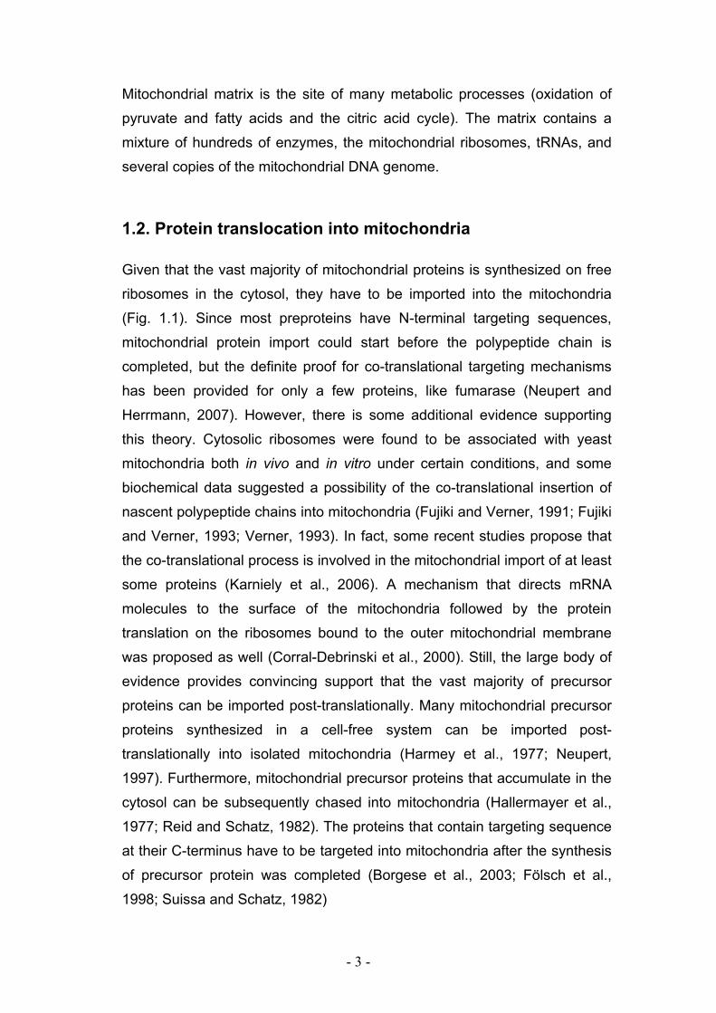
1.2. Protein translocation into mitochondria
Given that the vast majority of mitochondrial proteins is synthesized on free
ribosomes in the cytosol, they have to be imported into the mitochondria
(Fig. 1.1). Since most preproteins have N-terminal targeting sequences,
mitochondrial protein import could start before the polypeptide chain is
completed, but the definite proof for co-translational targeting mechanisms
has been provided for only a few proteins, like fumarase (Neupert and
Herrmann, 2007). However, there is some additional evidence supporting
this theory. Cytosolic ribosomes were found to be associated with yeast
mitochondria both in vivo and in vitro under certain conditions, and some
biochemical data suggested a possibility of the co-translational insertion of
nascent polypeptide chains into mitochondria (Fujiki and Verner, 1991; Fujiki
and Verner, 1993; Verner, 1993). In fact, some recent studies propose that
the co-translational process is involved in the mitochondrial import of at least
some proteins (Karniely et al., 2006). A mechanism that directs mRNA
molecules to the surface of the mitochondria followed by the protein
translation on the ribosomes bound to the outer mitochondrial membrane
was proposed as well (Corral-Debrinski et al., 2000). Still, the large body of
evidence provides convincing support that the vast majority of precursor
proteins can be imported post-translationally. Many mitochondrial precursor
proteins synthesized in a cell-free system can be imported post-
translationally into isolated mitochondria (Harmey et al., 1977; Neupert,
1997). Furthermore, mitochondrial precursor proteins that accumulate in the
cytosol can be subsequently chased into mitochondria (Hallermayer et al.,
1977; Reid and Schatz, 1982). The proteins that contain targeting sequence
at their C-terminus have to be targeted into mitochondria after the synthesis
of precursor protein was completed (Borgese et al., 2003; Fölsch et al.,
1998; Suissa and Schatz, 1982)
- 4 -
Figure 1.1. Protein translocation into mitochondria. Precursor proteins
containing different targeting signals are imported into mitochondria and sorted into
different mitochondrial compartments through the concerted action of protein
translocases. OM - outer membrane, IMS - intermembrane space, IM - inner
membrane, ∆Ψ - membrane potential across the inner membrane.
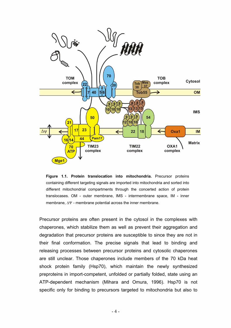
Precursor proteins are often present in the cytosol in the complexes with
chaperones, which stabilize them as well as prevent their aggregation and
degradation that precursor proteins are susceptible to since they are not in
their final conformation. The precise signals that lead to binding and
releasing processes between precursor proteins and cytosolic chaperones
are still unclear. Those chaperones include members of the 70 kDa heat
shock protein family (Hsp70), which maintain the newly synthesized
preproteins in import-competent, unfolded or partially folded, state using an
ATP-dependent mechanism (Mihara and Omura, 1996). Hsp70 is not
specific only for binding to precursors targeted to mitochondria but also to
- 5 -
other organelles. It was shown that the chaperone Hsp90 in cooperation
with Hsp70 mediate the targeting of a subset of mitochondrial preproteins in
mammals (Young et al., 2003).
Cytosolic precursors of mitochondrial proteins contain the targeting and
sorting sequences that determine the final destinations of the proteins within
mitochondria. Matrix destined preproteins contain the N-terminal cleavable
presequences also called matrix-targeting sequences (MTSs). The general
properties of these presequences are conserved but there is no consensus
in the primary structure even between closely related orthologs.
Presequence usually consists of about 10 to 70 amino acid residues that
have potential to form an amphipathic helix with one positively charged and
one hydrophobic face (Roise, 1992; Roise and Schatz, 1988; Von Heijne,
1986; von Heijne et al., 1989). In most cases, the presequence is cleaved
from precursors by the mitochondrial-processing peptidase (MPP) residing
in the mitochondrial matrix as soon as the cleavage site reaches the matrix
(Braun et al., 1992; Gakh et al., 2002).
Many mitochondrial precursors destined to all of the mitochondrial
subcompartments contain the internal targeting sequences. Precursors for
all proteins targeted to the outer membrane have internal signals. Those
with single transmembrane domains (TMDs) contain mitochondrial targeting
information in their hydrophobic anchors and the flanking positively charged
residues (Rapaport, 2002), but internal targeting signals for β-barrel proteins
remain unidentified up to date.
Some matrix destined proteins like rhodanese, 3-oxo-CoAthiolase, and
chaperonin 10 (Hsp10) are synthesized with a non-cleavable N-terminal
targeting signal, which has characteristics very similar to those of the
cleavable signals (Hammen et al., 1996; Jarvis et al., 1995; Waltner and
Weiner, 1995). Another matrix protein, DNA helicase Hmil, has a
presequence-like targeting signal at its C-terminus suggesting that this
precursor protein has to be imported in the reverse orientation (Lee et al.,
1999).
Some intermembrane space (IMS) proteins have canonical targeting
presequences, followed by a hydrophobic sorting sequence. Their import
- 6 -
depends on ATP and membrane potential across the inner membrane.
These bipartite presequences are cleaved off at the outer surface of the
inner membrane by the heterodimeric inner membrane peptidase (Imp1-
Imp2) and the mature proteins are released into the IMS (Glick et al., 1992).
The inner membrane proteins Tim23, Tim17, Tim22 and members of the
carrier family contain several internal targeting and sorting signals.
1.3. The inner membrane translocases
1.3.1. The TIM23 translocase
The TIM23 complex is the major translocase in the inner mitochondrial
membrane. It is involved in the import of all precursors of matrix proteins,
most of the proteins destined to the inner membrane, and many proteins of
the IMS. The translocation by the TIM23 complex requires both membrane
potential across the inner membrane and energy obtained from ATP
hydrolysis. The complex is composed of two cooperating subcompartments׃
the membrane sector (protein conducting channel) and the import motor.
The membrane sector is composed of three essential subunits Tim50,
Tim23, and Tim17, and two non-essential ones, Tim21 and Pam17, which
have regulatory functions. Tim23 and Tim17 form the 90 kDa core of the
TIM23 translocase. These two proteins have phylogenetically related
transmembrane domains with four predicted transmembrane segments,
which, though being homologs, cannot substitute for each other (Emtage
and Jensen, 1993; Kübrich et al., 1994; Maarse et al., 1994). Tim23
additionally exposes a hydrophilic amino terminal domain to the IMS. This
region consisting of 100 amino acid residues can be divided into two parts.
The N-terminal part was found to span the outer membrane and might have
a role in the positioning of the TIM23 translocase in proximity to the TOM
complex, thereby increasing the efficiency of protein import (Donzeau et al.,
2000). The second part of the N-terminal domain (residues 50-100) contains
an essential coiled-coil domain specific for dimerization of Tim23 and
substrate binding in the IMS (Bauer et al., 1996; Geissler et al., 2002;
- 7 -
Yamamoto et al., 2002). Tim17 has very short N-terminal domain exposed
to the IMS. Even though it is only 11 to 14 residues long, it contains two
conserved negative charges crucial for protein import. The function of Tim17
is not clear yet, but it was suggested that it plays a role in gating of the
TIM23 pore (Meier et al., 2005).
Tim50 is a receptor of the TIM23 translocase, anchored by its N-terminus
into the inner mitochondrial membrane exposing a large domain to the IMS
(Geissler et al., 2002, Yamamoto et al., 2002). It interacts with presequence-
containing proteins when they reach the trans site of the TOM complex and
directs them to the TIM23 translocase (Mokranjac et al., 2003a; Geissler et
al., 2002; Yamamoto et al., 2002).
Tim21 and Pam17, recently discovered components of the TIM23 complex,
seem to be involved in the regulation of the translocase during protein
import. It was observed that Tim21 interacts with IMS domain of Tom22
suggesting that it might play a role in interaction between the TOM complex
and the TIM23 complex (Chacinska et al., 2005; Mokranjac et al., 2005).
The membrane sector of the TIM23 complex translocates the presequence
to the matrix side of the inner membrane in a process, which is dependent
on membrane potential. Then, the import motor takes over and mediates
further translocation steps of preproteins. This part of the import pathway
requires ATP. The components of the import motor are Tim44, Tim14
(Pam17), Tim16 (Pam16), mitochondrial heat shock protein mtHsp70, and
the co-chaperone Mge1. Tim44 is a hydrophilic matrix protein, which in fungi
is fully attached to the inner membrane. It contains one hydrophobic pocket
believed to be a membrane binding site (Josyula et al., 2006). Tim44
functions as a docking site for other import motor components and binds the
incoming preproteins before it passes them to mtHsp70 in the ATP bound
state. MtHsp70 has two domains – an N-terminal ATPase domain and a C-
terminal peptide binding domain. When ATP is bound, the substrate binding
pocket is opened and Hsp70 is ready to grasp the arriving polypeptide, while
when ADP is bound, the pocket is closed and mtHsp70 loses affinity for
Tim44. It seems that upon ATP hydrolysis Hsp70 is released from Tim44
(Mokranjac et al., 2003b; Schneider et al., 1996; Liu et al., 2003). The
- 8 -
exchange of ATP and ADP requires the action of the nucleotide exchange
protein Mge1.
Binding of incoming preproteins to Hsp70 is regulated by two import motor
subunits with DnaJ-like structures, Tim14 (Pam18) and Tim16 (Pam16).
Two of them are believed to form a complex (Mokranjac et al., 2006).
Whereas Tim14 stimulates hydrolysis of ATP in the mtHsp70, Tim16 does
not influence ATPase activity in vitro. Tim16 is not a functional DnaJ protein
because it does not contain HPD motif important for interaction with Hsp70.
Recently published crystal structure of Tim14-Tim16 complex suggested
that Tim16 bound to Tim14΄s HPD motif and therefore functioned as a
negative regulator of Tim14 function by physically blocking the contact site
of Tim14 and Hsp70.
To summarize, precursor proteins after passing through the TOM complex
are directed to the TIM23 translocase by binding to IMS domains of Tim50
and Tim23. When MTS is translocated across the import channel of the
TIM23 translocase, Tim44 binds it and passes it to mtHsp70 in ATP bound
state. Tim14 stimulates ATP hydrolysis which leads to the tight binding of
Hsp70 to the preprotein and to dissociation of Hsp70 from Tim44. From this
moment on, preprotein can only slip into the matrix because backsliding is
prevented by bound Hsp70.
1.3.2. The TIM22 translocase
The TIM22 complex is involved in the insertion pathway of multiple
membrane-spanning domain proteins like Tim23, Tim17, Tim22, and the
metabolite carrier proteins family. This 300 kDa complex is composed of
three membrane proteins, Tim22, Tim 54, and Tim18; and three associated
small Tim proteins, Tim9, Tim10, and Tim12. While the exact functions of
Tim54 and Tim18 are not known, Tim22 is essential, and is the pore forming
subunit of the complex. It can support import of carrier proteins, although at
reduced levels, even in the absence of the two other membrane
components of the translocase (Kovermann et al., 2002). The TIM22
translocase inserts the proteins into the lipid bilayer of the inner membrane
in a membrane potential-dependent manner (Kerscher et al., 1997; Kerscher
- 9 -
et al., 2000; Sirrenberg et al., 1996). Small Tim proteins bind to the
precursor proteins when they reach IMS after passing through the TOM
complex. They function in a chaperone-like manner preventing aggregation
of the imported precursors and are required for further translocation from the
outer membrane to the TIM22 complex. The essential 70 kDa Tim9-Tim10
complex is required for the transport of carrier proteins and specifically binds
to their hydrophobic loops. Non-essential Tim8-Tim13 complex of the same
size was found to specifically interact with precursors that contain
hydrophilic extensions like Tim23 when it binds to the N-terminal part of
Tim23 whereas Tim9-10 complex interacts with the hydrophobic loop of the
membrane embedded region (Bauer et al., 2000; Neupert and Herrmann,
2007). It is believed that Tim9-10 complex can functionally replace the
nonessential Tim8-13 complex to some extent.
1.3.3. The Oxa1 translocase
The OXA1 translocase of the inner mitochondrial membrane facilitates the
insertion of both mitochondrial and nuclear-encoded proteins from the matrix
into the inner membrane. This process is called mitochondrial protein export.
Eight mitochondrial proteins in yeast, seven of which are highly hydrophobic,
are encoded by mitochondrial genome. These are cytochrome b, Cox1,
Cox2, Cox3, Atp6, Atp8, and Atp9.
Oxa1 is an evolutionarily conserved protein and its homologues are found
throughout prokaryotes and eukaryotes (Kuhn et al., 2003). It spans the
inner membrane five times, exposing a long α-helical C-terminal domain to
the matrix. This domain forms α-helical coiled-coil domain that binds
mitochondrial ribosomes (Szyrach et al., 2003). In addition, Oxa1 was
reported to interact with newly synthesized mitochondrial proteins (Hell et
al., 2001). Taken together, these data indicate that OXA1 translocase can
insert proteins into the inner membrane in a co-translational manner.
Several proteins, including Oxa1 itself, that are synthesized in the cytosol
and imported into the matrix via the TIM23 translocase, have to be inserted
into the inner membrane using the export machinery (Hell et al., 2001). This
- 10 -
pathway resembles insertion reactions of polytopic membrane proteins of
bacterial origin and is called the conservative sorting pathway (Stuart, 2002).
Mba1 is an additional component of the mitochondrial export machinery and
it is also found to bind mitochondrial ribosomes (Ott et al., 2006). It shares
substrate specificity with Oxa1 but it either cooperates with or functions
independently of Oxa1 (Preuss et al., 2001).
1.4. The outer membrane translocases
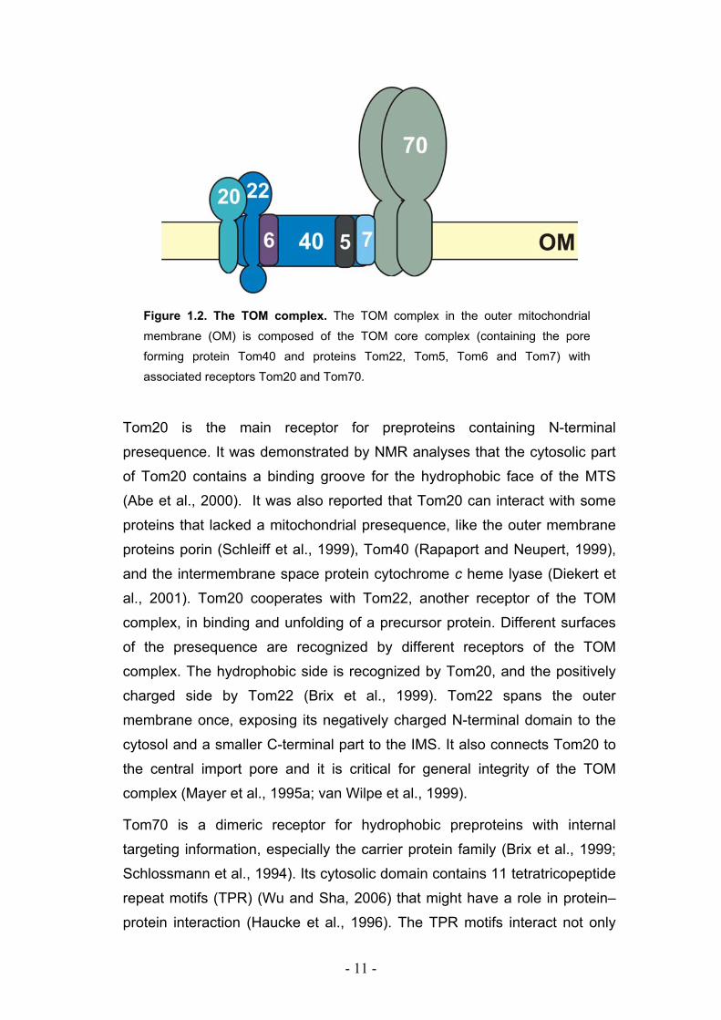
1.4.1. The TOM complex
The translocase of the outer membrane (TOM complex) mediates the import
of almost all nuclear encoded mitochondrial proteins. The composition of the
TOM complex was studied in detail in the fungi, N. crassa and S. cerevisiae.
The structure and function of the TOM complex in other organisms, like
plants or animals, is very comparable to that in fungi. The translocase is a
multi-subunit complex of 600 kDa composed of seven subunits: Tom70,
Tom40, Tom22, Tom20, Tom7, Tom6, and Tom5 (Neupert, 1997; Pfanner
and Geissler, 2001) which are grouped according to their function into
receptor and pore components (Fig. 1.2). The TOM complex has several
binding sites for precursor proteins. Cytosolic domains of Tom20, Tom70,
and Tom22 represent cis-binding site while IMS domains of Tom22, Tom40,
and Tom7 seam to contribute in binding of precursor proteins in IMS and
they are referred to as trans-binding site (Bolliger et al., 1995; Esaki et al.,
2004; Mayer et al., 1995b).
The two major receptors are Tom20 and Tom70. Both are anchored to the
outer membrane with their N-terminal transmembrane segments exposing
hydrophilic C-terminal domains to the cytosol. These two receptors show
different substrate specificities but partially overlap in function, therefore
they can partially substitute for each other (Lithgow and Schatz, 1995).
Single deletion of either receptor can be tolerated, but double deletion is
lethal (Ramage et al., 1993).
- 11 -
Figure 1.2. The TOM complex. The TOM complex in the outer mitochondrial
membrane (OM) is composed of the TOM core complex (containing the pore
forming protein Tom40 and proteins Tom22, Tom5, Tom6 and Tom7) with
associated receptors Tom20 and Tom70.
Tom20 is the main receptor for preproteins containing N-terminal
presequence. It was demonstrated by NMR analyses that the cytosolic part
of Tom20 contains a binding groove for the hydrophobic face of the MTS
(Abe et al., 2000). It was also reported that Tom20 can interact with some
proteins that lacked a mitochondrial presequence, like the outer membrane
proteins porin (Schleiff et al., 1999), Tom40 (Rapaport and Neupert, 1999),
and the intermembrane space protein cytochrome c heme lyase (Diekert et
al., 2001). Tom20 cooperates with Tom22, another receptor of the TOM
complex, in binding and unfolding of a precursor protein. Different surfaces
of the presequence are recognized by different receptors of the TOM
complex. The hydrophobic side is recognized by Tom20, and the positively
charged side by Tom22 (Brix et al., 1999). Tom22 spans the outer
membrane once, exposing its negatively charged N-terminal domain to the
cytosol and a smaller C-terminal part to the IMS. It also connects Tom20 to
the central import pore and it is critical for general integrity of the TOM
complex (Mayer et al., 1995a; van Wilpe et al., 1999).
Tom70 is a dimeric receptor for hydrophobic preproteins with internal
targeting information, especially the carrier protein family (Brix et al., 1999;
Schlossmann et al., 1994). Its cytosolic domain contains 11 tetratricopeptide
repeat motifs (TPR) (Wu and Sha, 2006) that might have a role in protein–
protein interaction (Haucke et al., 1996). The TPR motifs interact not only
- 12 -
with precursor proteins but also with cytosolic chaperones, like Hsp70 and,
in animals, Hsp90 (Young et al., 2003).
The TOM core complex, also called general import pore (GIP), is composed
of the central, pore-forming component, Tom40, three small associated
subunits Tom5, Tom6, and Tom7, and the receptor protein Tom22. Its size,
as estimated by size-exclusion chromatography, is approximately 400 kDa.
Tom40 is the only component of the TOM complex essential for yeast
viability. It is a membrane embedded protein composed of series of
antiparallel β-strands forming a β-barrel. Purified Tom40 is able to form ion
channels in artificial membranes (Ahting et al., 2001). However, it is still not
clear whether the pore of the TOM complex is formed by one or more
Tom40 molecules. Single particle imaging of negatively-stained isolated
TOM holo complex showed particles with two or three pores like structures
while TOM core complex contains two pores.
Small Tom proteins are all tail-anchored, composed of 50 to 70 amino acid
residues. They have one α-helical TM domain with very few residues
exposed to the IMS. Deletion of either of small Tom proteins shows only
minor effects but deletion of all three proteins is lethal in yeast (Dekker et al.,
1998; Dietmeier et al., 1997; Sherman et al., 2005). Their individual
functions remained unclear up to date but they appear to be involved in
stabilization of the TOM complex.
For the import of the β-barrel outer membrane proteins, the TOM complex
cooperates with the other outer membrane protein translocation machinery,
the TOB complex (for topogenesis of mitochondrial outer membrane beta-
barrel proteins, also known as the SAM complex (sorting and assembly
machinery) (Paschen et al., 2003; Wiedemann et al., 2003).
1.4.2. The TOB complex
The precursors of β-barrel proteins use the TOM complex in the first step of
their import pathway and also require the TOB complex in order to get
inserted into the outer mitochondrial membrane.
- 13 -
This 250 kDa complex is composed of one membrane embedded
component Tob55 and two hydrophilic proteins, Tob38 and Mas37, which
are peripherally associated with the outer membrane. The main component
of the TOB complex is Tob55, also called Sam50, which together with
Tob38 forms functional TOB core complex. Both proteins are essential for
cell viability in yeast and N. crassa. Tob55 is composed of two domains׃ a
hydrophilic N-terminal part facing the IMS and a membrane embedded C-
terminal domain that forms β-barrel structure with 14-16 transmembrane β-
sheets.
The N-terminal part forms characteristic structure called the POTRA domain
(polypeptide-transport-associated domain) which is supposed to have a
chaperone-like function (Gentle et al., 2005; Sanchez-Pulido et al., 2003).
Therefore, this domain was proposed to present the interaction site for β-
barrel precursors with the TOB complex after they were imported via the
TOM complex into IMS (Habib et al., 2007; Pfanner et al., 2004). Both
domains are conserved among Tob55 bacterial (Omp85) and eukaryotic
homologous. In yeast, depletion of both Tob55 and Tob38 leads to impaired
insertion and assembly of newly imported β-barrel proteins (Kozjak et al.,
2003; Paschen et al., 2003; Waizenegger et al., 2004). The exact functions
of three components of the TOB complex are still not clear but according to
high conservation of Tob55 one could assume that this protein plays the
most important role in the β-barrel assembly pathway. It might be that the
two other proteins, Tob38 and Mas37, have somewhat of an accessory
function.
1.4.3. Biogenesis of the TOM complex
The biogenesis of membrane proteins and especially of multisubunit
complexes that reside in membranes is a fascinating process. Several
studies addressed the biogenesis of the TOM complex (Fig.1.3). The
interesting point is that the TOM complex is also involved in its own
biogenesis.
Components of the TOM complex, like other outer membrane proteins, do
not contain cleavable presequences. Their targeting information is contained
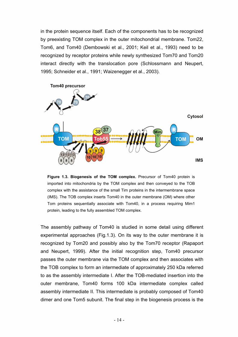
- 14 -
in the protein sequence itself. Each of the components has to be recognized
by preexisting TOM complex in the outer mitochondrial membrane. Tom22,
Tom6, and Tom40 (Dembowski et al., 2001; Keil et al., 1993) need to be
recognized by receptor proteins while newly synthesized Tom70 and Tom20
interact directly with the translocation pore (Schlossmann and Neupert,
1995; Schneider et al., 1991; Waizenegger et al., 2003).
Figure 1.3. Biogenesis of the TOM complex. Precursor of Tom40 protein is
imported into mitochondria by the TOM complex and then conveyed to the TOB
complex with the assistance of the small Tim proteins in the intermembrane space
(IMS). The TOB complex inserts Tom40 in the outer membrane (OM) where other
Tom proteins sequentially associate with Tom40, in a process requiring Mim1
protein, leading to the fully assembled TOM complex.
The assembly pathway of Tom40 is studied in some detail using different
experimental approaches (Fig.1.3). On its way to the outer membrane it is
recognized by Tom20 and possibly also by the Tom70 receptor (Rapaport
and Neupert, 1999). After the initial recognition step, Tom40 precursor
passes the outer membrane via the TOM complex and then associates with
the TOB complex to form an intermediate of approximately 250 kDa referred
to as the assembly intermediate I. After the TOB-mediated insertion into the
outer membrane, Tom40 forms 100 kDa intermediate complex called
assembly intermediate II. This intermediate is probably composed of Tom40
dimer and one Tom5 subunit. The final step in the biogenesis process is the
- 15 -
formation of the 400 kDa, mature TOM core complex by the sequential
addition of Tom6, Tom7, and Tom22 to the 100 kDa complex.
Recently, a novel 14 kDa outer membrane protein was identified and named
Mim1 (for mitochondrial import) (Mnaimneh et al., 2004). This protein, also
known as Tom13, was characterised as an additional assembly factor of the
TOM complex (Ishikawa et al., 2004; Waizenegger et al., 2005). Deletion of
Mim1 resulted in accumulation of non-imported mitochondrial precursor
proteins (Mnaimneh et al., 2004), but also in changes in mitochondrial
morphology (Altmann and Westermann, 2005). These effects are believed
to be secondary to the main function of Mim1 in the biogenesis of the TOM
complex. Mim1 has one putative transmembrane segment (TMS), highly
conserved among fungi. Its N-terminal domain faces the cytosol and its C-
terminal domain is exposed to the IMS. Depletion of Mim1 abrogates
assembly of the TOM complex and results in accumulation of Tom40, the
major constituent of the TOM complex, as a low molecular mass species.
Mim1 is not a component of the TOM complex or of the TOB complex
(Ishikawa et al., 2004; Waizenegger et al., 2005, Meisinger et al., 2007); but
rather is a subunit of another, yet unidentified, complex in the outer
mitochondrial membrane.
- 16 -
1.5. Aim of the present study
Recently discovered protein named Mim1 has been characterized as an
assembly factor of the TOM complex. Specifically, it was found to play a role
in the assembly of Tom40 into the TOM complex in the step after the Tom40
precursor interacted with the TOB complex.
The aim of this study was to investigate the structural and functional
characteristics of Mim1. Several questions were addressed׃
i) What are the domains that are crucial for the function of Mim1?
ii) What are the interaction partners of Mim1?
Iii) How is Mim1 inserted by itself into the outer membrane?
iv) How does Mim1 promote the assembly of the TOM complex?
- 17 -
2. MATERIAL AND METHODS
2.1. Molecular biology methods
2.1.1. Standard polymerase chain reaction (PCR)
DNA sequences were amplified by polymerase chain reaction (PCR), using
thermostable DNA polymerase as described previously (Sambrook et al.,
1989). Taq (isolated from Thermus aquaticus), and Pfu (isolated from
Pyrococcus furiosus polymerases were used. Taq DNA polymerase has no
proofreading ability, and therefore Pfu DNA polymerase was added when
the PCR product needed to be used for subsequent cloning.
PCR mix contained (total 100 µl): 1-2 U DNA polymerase (Taq-polymerase
and/or Pfu-polymerase), 10 µl PCR-buffer (1% Triton X-100, 100 mM Tris-
HCl, 500 mM KCl, 15 mM MgCl2, pH 8.8), 2 µl dNTPs (10 mM stock), 50 pM
primers and 200 ng plasmid DNA template or 1 µg genomic DNA template.
The following program was used:
1) 94°C, 5 min Nuclease inactivation and complete DNA
denaturation
DNA amplification:
94°C, 1 min DNA denaturation
45-65°C, 1 min Annealing of primers
2) 30-35 cycles
72°C, 1-6 min DNA synthesis*
3) 72°C, 5-20 min Completion of the last reaction
- 18 -
The duration of this step is determined by the length of the DNA fragment to
be amplified and DNA polymerase used (Taq polymerase 1 min/1kb; Pfu
polymerase 2.5 min/1kb).
The amplified DNA fragments were analyzed by agarose gel
electrophoresis.
2.1.2. Site directed mutagenesis
For inserting point mutations in DNA sequence QuickChange Site-Directed
Mutagenesis Kit (Stratagene) was used.
PCR conditions:
Sample reaction:
5 µl of 10x reaction buffer
5-50 ng of dsDNA template
125 ng primer #1
125 ng primer #2
1 µl of dNTP mix
ddH2O to a final volume of 50 µl
1 µl of PfuTurbo DNA polymerase (2.5 U/ µl)
The following program was used:
Cycles Temperature Time
1 95°C 30 sec
95°C 30 sec
55°C 1 min
12-18
68°C 1 min/ kb of plasmid length
Upon the termination of PCR, 1 µl of the DpnI restriction enzyme was added
to the reaction and incubated at 37°C for 1 h to digest the parental
- 19 -
supercoiled dsDNA. Then, MH1 E. coli cells were transformed with 1 µl of
the DpnI treated DNA.
2.1.3. Analytical and preparative gel electrophoresis
DNA fragments were separated by electrophoresis in a horizontal agarose
gel (0.8-2%) according to their molecular mass. Samples were mixed with
loading buffer (6% (v/v) glycerol, 0.05% bromphenolblue, 0.05%
xylencyanol) and electrophoresis was performed in TAE-buffer (4.84 g/l Tris-
Base, 1.14 ml/l acetic acid, 1 mM EDTA, pH 8.0). The agarose solution
contained 0.5 µg/ml ethidium bromide to allow visualization of DNA in gel
under UV light. The agarose was stored at 65°C until use. The 1 kb and the
100 bp DNA markers were used (New England Biolabs, Beverly, USA). DNA
fragments to be further processed were excised from the gel with a sterile
scalpel under UV light and the DNA extracted from the gel using the “Gel
extraction kit” (Qiagen). Extracted DNA was routinely stored at –20ºC.
2.1.4. DNA and RNA concentration measurement
For DNA concentration measurements the absorption of DNA solutions was
measured at 260 nm. One optical unit (OD) corresponds to a concentration
of 50 µg/ml of double stranded DNA, 33 µg/ml single stranded DNA, 40
µg/ml RNA or 20 µg/ml oligonucleotides.
2.1.5. Enzymatic manipulation of DNA: restriction and ligation reactions
Digestion of DNA with restriction endonucleases
For analytical and preparative purposes PCR product and plasmid DNA
were digested with specific restriction endonucleases (up to 5 U of enzyme
for 1 µg DNA). The buffer, temperature (usually 37ºC) and incubation time
(1-3 h) for every reaction were chosen according to the manufacturer’s
recommendations. The digested fragments were analyzed by agarose gel
electrophoresis or directly isolated using anion-exchange chromatography
- 20 -
(Qiagen). For preparative purposes, digested DNA fragments were
extracted from gels using “Gel extraction kit” (Qiagen).
When plasmid DNA was cut with a single restriction enzyme, it was treated
with shrimp alkaline phosphatase (SAP) (Roche). This enzyme removes 5’-
phosphate groups on linearized plasmid DNAs thereby preventing
recircularization of the vector.
Ligation
Linearized DNA vector (50-200 ng) and a 5 fold molar excess of DNA
fragment to be inserted, were incubated in a 10 µl reaction mixture
containing 1 µl of 10x ligation buffer (10 mM MgCl2, 5% (w/v) PEG-8000, 1
mM DTT, 1 mM ATP, 50 mM Tris-HCl, pH 7.6), and 0.5 µl (1 U) T4 DNA
ligase (Gibco-BRL). Reactions were incubated at 14ºC overnight or at RT for
5 h and 1 µl of the reaction mixture was used for transformation of E. coli
cells.
2.1.6. Preparation of E. coli competent cells
A single colony of E. coli strain (MH1 or XL-1 Blue) was inoculated in 50 ml
of LB-medium containing Ampicillin (LBamp) and grown overnight at 37ºC
under moderate shaking conditions. The following day, 1 l of liquid LBamp
medium was inoculated with the overnight culture. The bacterial cells were
grown further until they reached OD600 ~ 0.5. Then, they were incubated on
ice for 30 min, harvested by centrifugation (4,400 x g, 5 min, 4ºC) and
washed sequentially with 500 ml, 250 ml, and 50 ml of 10% (v/v) glycerol.
The competent cells were finally resuspended in 500 µl 10% (v/v) glycerol,
aliquoted and stored at –80ºC.
2.1.7. Transformation of E. coli
Ligation reaction mixture (1 µl) was added on ice to 50 µl of E. coli
competent cells. The mixture was transferred to an ice-cold cuvette and the
cuvette was introduced into the electroporation apparatus (Gene Pulser,
BioRad). The instrument was set at 2.5 kV, 400 Ω, and 25 µF. The obtained
time constant was 7-8 ms. After a short application of a high electric voltage
- 21 -
to the cells, the suspension was diluted with 1 ml LB-medium, and incubated
for 45 min at 37°C under moderate shaking to allow cell recovery. The
transformed cells were harvested by centrifugation (10,000 x g, 15 sec, RT)
and plated on LB-amp medium. The plates were incubated overnight at
37°C.
2.1.8. Small and large scale isolation of plasmid DNA from E. coli
Small scale preparation of plasmid DNA was performed through alkaline
lysis according to a published procedure (Birnboim and Doly, 1979). LB-amp
medium (2.5 ml) was inoculated with a single bacterial colony picked out
from a Petri dish, and incubated overnight at 37ºC, while shaking (140 rpm).
The next day bacteria were harvested by centrifugation (8,000 x g, 30 sec,
RT) and the pellet was resuspended in 300 µl of buffer E1 (10 mM EDTA, 50
mM Tris-HCl, pH 8.0) containing 100 mg/ml RNase. Cells were lysed by
adding of 300 µl of buffer E2 (0.2 M NaOH, 1% SDS). Samples were mixed
by inverting the tubes 5 times and incubated 5 min at RT. Neutralization step
was performed by adding 300 µl of buffer E3 (3.1 M K-acetate, pH 5.5) and
mixing the samples immediately afterwards. In the next step samples were
centrifuged (10,000 x g, 10 min, 2ºC), the DNA-containing supernatant was
transferred to new tubes and the DNA was precipitated by the addition of
600 µl of 96% isopropanol. Samples were then centrifuged again (10,000 x
g, 40 min, 2ºC) and the resulting pellets were washed with 85% cold ethanol,
dried at RT, resuspended in 30 µl water and stored at –20ºC.
For large scale preparation of plasmid DNA a “PureYield” Plasmid Midiprep
System (Promega) was used. LB-medium (50 ml) supplemented with
ampicillin was inoculated with bacteria carrying the plasmid of interest and
incubated overnight at 37ºC while shaking at 140 rpm. The bacteria were
harvested the next day by centrifugation (10000 x g, 10 min, RT) and
resuspended in 6 ml of Cell Resuspension Solution. Cells lysis was
performed by adding 6 ml of Cell Lysis Solution. Tubes were inverted 5
times and left for 3 min at RT. After neutralization with 10 ml of
Neutralization Solution, samples were mixed by inverting the tubes 5 times
and incubated for 3 min at RT to ensure thorough clearing. Samples were
- 22 -
centrifuged (10000 x g, 10 min, 4ºC), and then the supernatants were
applied onto a clarifying column standing on top of an anion-exchange
column placed onto a vacuum manifold. When the entire volume of the
sample passed under vacuum through column stack, the clarifying column
was removed. The anion-exchange column was washed with 5 ml of
Endotoxin Removal Wash and then with 20 ml of the Column Wash
Solution. The column was left to dry for 30 sec under vacuum. Plasmid DNA
was eluted from the column with 500 µl of sterile deionized water (ddH20).
DNA isolated this way was stored at –20ºC.
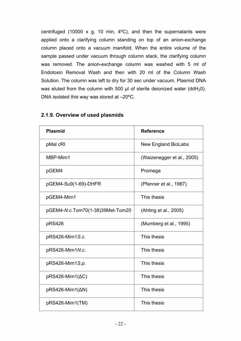
2.1.9. Overview of used plasmids
Reference Plasmid
New England BioLabs pMal cRI
(Waizenegger et al., 2005) MBP-Mim1
Promega pGEM4
(Pfanner et al., 1987) pGEM4-Su9(1-69)-DHFR
This thesis pGEM4-Mim1
(Ahting et al., 2005) pGEM4-N.c.Tom70(1-38)39Met-Tom20
(Mumberg et al., 1995) pRS426
This thesis pRS426-Mim1S.c.
This thesis pRS426-Mim1N.c.
This thesis pRS426-Mim1S.p.
This thesis pRS426-Mim1(∆C)
This thesis pRS426-Mim1(∆N)
This thesis pRS426-Mim1(TM)
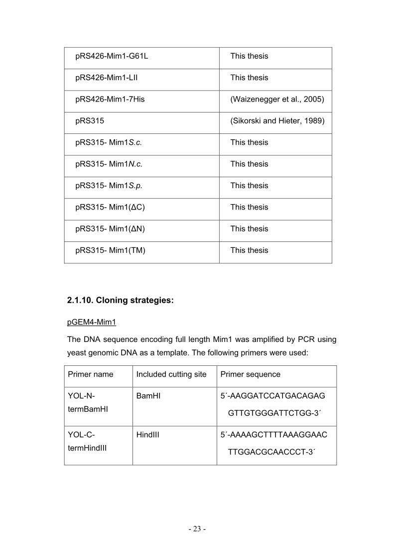
- 23 -
This thesis pRS426-Mim1-G61L
This thesis pRS426-Mim1-LII
(Waizenegger et al., 2005) pRS426-Mim1-7His
(Sikorski and Hieter, 1989) pRS315
This thesis pRS315- Mim1S.c.
This thesis pRS315- Mim1N.c.
This thesis pRS315- Mim1S.p.
This thesis pRS315- Mim1(∆C)
This thesis pRS315- Mim1(∆N)
This thesis pRS315- Mim1(TM)
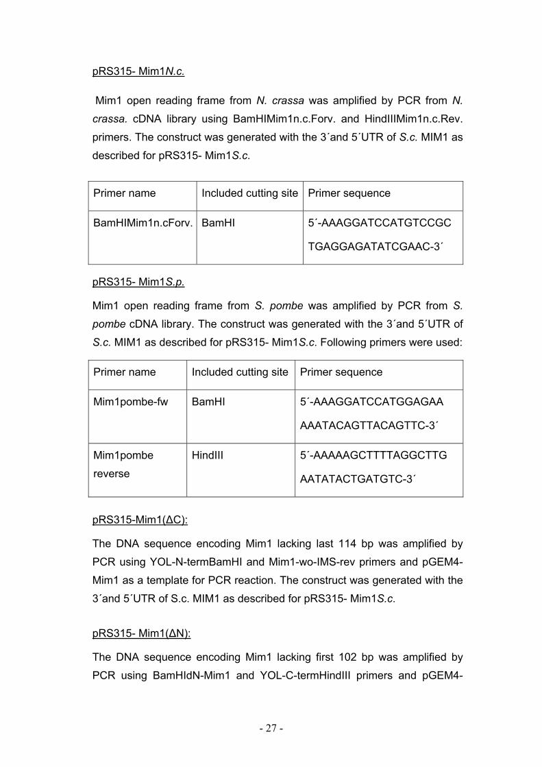
2.1.10. Cloning strategies:
pGEM4-Mim1
The DNA sequence encoding full length Mim1 was amplified by PCR using
yeast genomic DNA as a template. The following primers were used:
Primer name Included cutting site Primer sequence
YOL-N-
termBamHI
BamHI 5΄-AAGGATCCATGACAGAG
GTTGTGGGATTCTGG-3΄
YOL-C-
termHindIII
HindIII 5΄-AAAAGCTTTTAAAGGAAC
TTGGACGCAACCCT-3΄
- 24 -
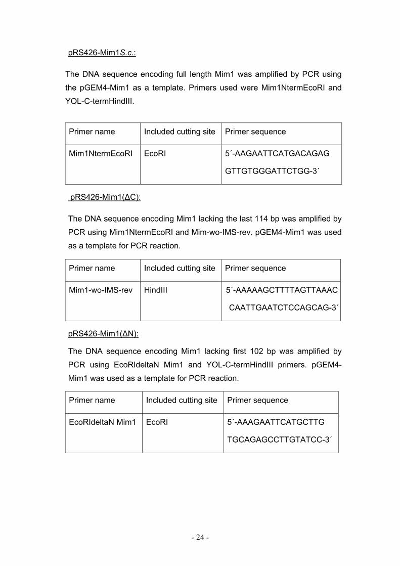
pRS426-Mim1S.c.:
The DNA sequence encoding full length Mim1 was amplified by PCR using
the pGEM4-Mim1 as a template. Primers used were Mim1NtermEcoRI and
YOL-C-termHindIII.
pRS426-Mim1(∆C):
The DNA sequence encoding Mim1 lacking the last 114 bp was amplified by
PCR using Mim1NtermEcoRI and Mim-wo-IMS-rev. pGEM4-Mim1 was used
as a template for PCR reaction.
pRS426-Mim1(∆N):
The DNA sequence encoding Mim1 lacking first 102 bp was amplified by
PCR using EcoRIdeltaN Mim1 and YOL-C-termHindIII primers. pGEM4-
Mim1 was used as a template for PCR reaction.
Primer name Included cutting site Primer sequence
EcoRIdeltaN Mim1 EcoRI 5΄-AAAGAATTCATGCTTG
TGCAGAGCCTTGTATCC-3΄
Primer name Included cutting site Primer sequence
Mim1NtermEcoRI EcoRI 5΄-AAGAATTCATGACAGAG
GTTGTGGGATTCTGG-3΄
Primer name Included cutting site Primer sequence
Mim1-wo-IMS-rev HindIII 5΄-AAAAAGCTTTTAGTTAAAC
CAATTGAATCTCCAGCAG-3΄
- 25 -
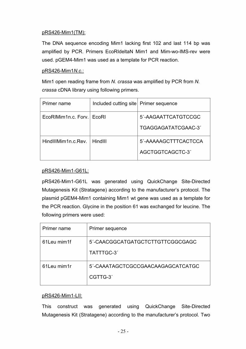
pRS426-Mim1(TM):
The DNA sequence encoding Mim1 lacking first 102 and last 114 bp was
amplified by PCR. Primers EcoRIdeltaN Mim1 and Mim-wo-IMS-rev were
used. pGEM4-Mim1 was used as a template for PCR reaction.
pRS426-Mim1N.c.:
Mim1 open reading frame from N. crassa was amplified by PCR from N.
crassa cDNA library using following primers.
Primer name Included cutting site Primer sequence
EcoRIMim1n.c. Forv. EcoRI 5΄-AAGAATTCATGTCCGC
TGAGGAGATATCGAAC-3΄
HindIIIMim1n.c.Rev. HindIII 5΄-AAAAAGCTTTCACTCCA
AGCTGGTCAGCTC-3΄
pRS426-Mim1-G61L:
pRS426-Mim1-G61L was generated using QuickChange Site-Directed
Mutagenesis Kit (Stratagene) according to the manufacturer’s protocol. The
plasmid pGEM4-Mim1 containing Mim1 wt gene was used as a template for
the PCR reaction. Glycine in the position 61 was exchanged for leucine. The
following primers were used:
Primer name Primer sequence
61Leu mim1f 5΄-CAACGGCATGATGCTCTTGTTCGGCGAGC
TATTTGC-3΄
61Leu mim1r 5΄-CAAATAGCTCGCCGAACAAGAGCATCATGC
CGTTG-3΄
pRS426-Mim1-LII:
This construct was generated using QuickChange Site-Directed
Mutagenesis Kit (Stratagene) according to the manufacturer’s protocol. Two
- 26 -
amino acids were changed, glycine in the position 63 into isoleucine and
alanin in the position 67 into isoleucine. The plasmid pGEM4-Mim1
containing Mim1-G61L gene was used as a template for the PCR reaction to
generate Mim1 construct carrying point mutations in positions 61 and 63.
This construct was then used as a template for the PCR to generate Mim1-
LII. The following primers were used:
Primer name Primer sequence
63 F 5΄-GGCATGATGCTCCTCTTCATCGAGCTATTTGCTC
ACGAGC-3΄
63 R 5΄-GCTCGTGAGCAAATAGCTCGATGAAGAGGAGCA
TCATGCC-3΄
63.67 F 5΄-CTCTTCATCGAGCTATTTATTCACGAGCTCTGCTG
GAGATTC-3΄
63,67 R 5΄-GAATCTCCAGCAGAGCTCGTGAATAAATAGCTCG
ATGAAGAG-3΄
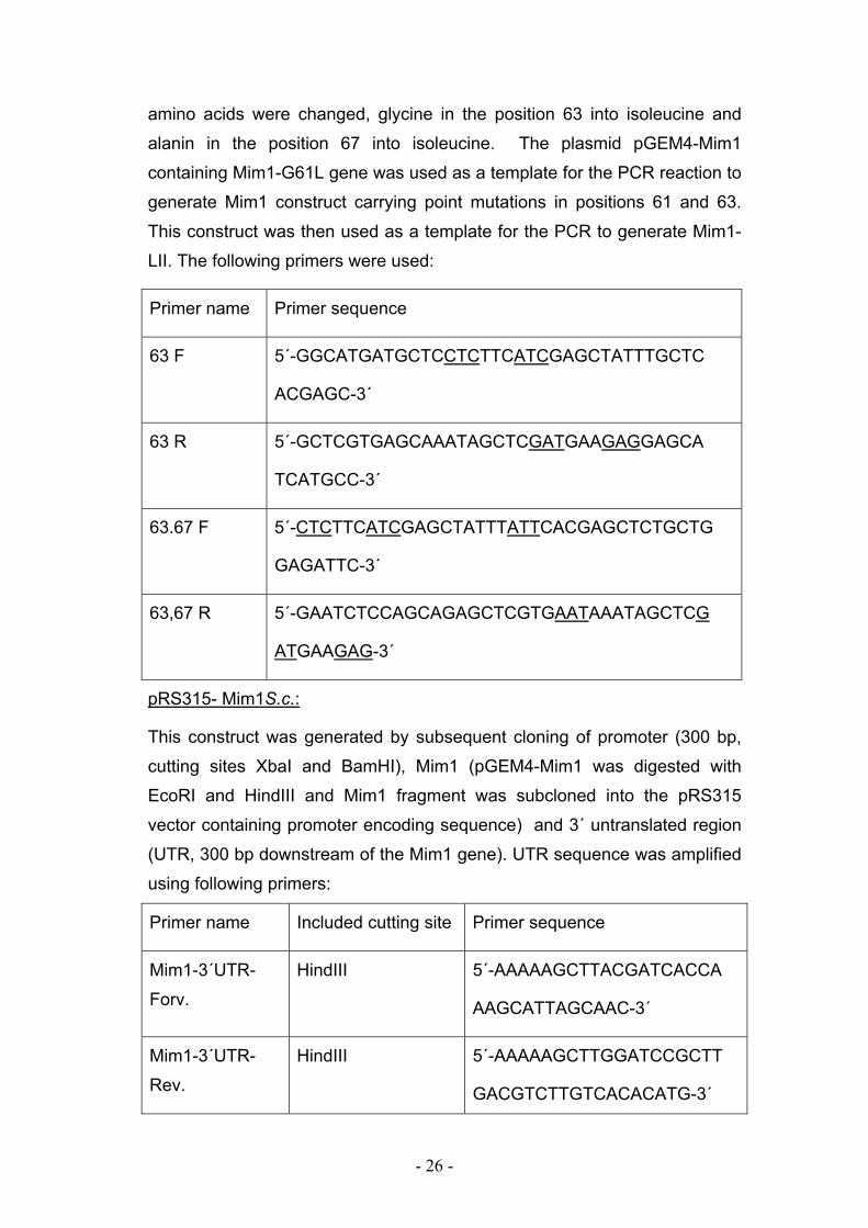
pRS315- Mim1S.c.:
This construct was generated by subsequent cloning of promoter (300 bp,
cutting sites XbaI and BamHI), Mim1 (pGEM4-Mim1 was digested with
EcoRI and HindIII and Mim1 fragment was subcloned into the pRS315
vector containing promoter encoding sequence) and 3´ untranslated region
(UTR, 300 bp downstream of the Mim1 gene). UTR sequence was amplified
using following primers:
Primer name Included cutting site Primer sequence
Mim1-3΄UTR-
Forv.
HindIII 5΄-AAAAAGCTTACGATCACCA
AAGCATTAGCAAC-3΄
Mim1-3΄UTR-
Rev.
HindIII 5΄-AAAAAGCTTGGATCCGCTT
GACGTCTTGTCACACATG-3΄
- 27 -
pRS315- Mim1N.c.
Mim1 open reading frame from N. crassa was amplified by PCR from N.
crassa. cDNA library using BamHIMim1n.c.Forv. and HindIIIMim1n.c.Rev.
primers. The construct was generated with the 3´and 5´UTR of S.c. MIM1 as
described for pRS315- Mim1S.c.
pRS315- Mim1S.p.
Mim1 open reading frame from S. pombe was amplified by PCR from S.
pombe cDNA library. The construct was generated with the 3´and 5´UTR of
S.c. MIM1 as described for pRS315- Mim1S.c. Following primers were used:
Primer name Included cutting site Primer sequence
Mim1pombe-fw BamHI 5΄-AAAGGATCCATGGAGAA
AAATACAGTTACAGTTC-3΄
Mim1pombe
reverse
HindIII 5΄-AAAAAGCTTTTAGGCTTG
AATATACTGATGTC-3΄
pRS315-Mim1(∆C):
The DNA sequence encoding Mim1 lacking last 114 bp was amplified by
PCR using YOL-N-termBamHI and Mim1-wo-IMS-rev primers and pGEM4-
Mim1 as a template for PCR reaction. The construct was generated with the
3´and 5´UTR of S.c. MIM1 as described for pRS315- Mim1S.c.
pRS315- Mim1(∆N):
The DNA sequence encoding Mim1 lacking first 102 bp was amplified by
PCR using BamHIdN-Mim1 and YOL-C-termHindIII primers and pGEM4-
Primer name Included cutting site Primer sequence
BamHIMim1n.cForv. BamHI 5΄-AAAGGATCCATGTCCGC
TGAGGAGATATCGAAC-3΄
- 28 -
Mim1 as a template for PCR reaction. The construct was generated with the
3´and 5´UTR of S.c. MIM1 as described for pRS315- Mim1S.c.
Primer name Included cutting site Primer sequence
BamHIdN-Mim1 Bam HI 5΄-AAAGGATCCATGCTTG
TGCAGAGCCTTGTATCC
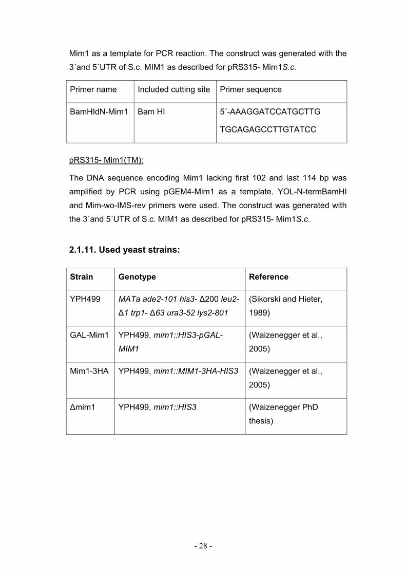
pRS315- Mim1(TM):
The DNA sequence encoding Mim1 lacking first 102 and last 114 bp was
amplified by PCR using pGEM4-Mim1 as a template. YOL-N-termBamHI
and Mim-wo-IMS-rev primers were used. The construct was generated with
the 3´and 5´UTR of S.c. MIM1 as described for pRS315- Mim1S.c.
2.1.11. Used yeast strains:
Strain Genotype Reference
YPH499 MATa ade2-101 his3- ∆200 leu2-
∆1 trp1- ∆63 ura3-52 lys2-801
(Sikorski and Hieter,
1989)
GAL-Mim1 YPH499, mim1::HIS3-pGAL-
MIM1
(Waizenegger et al.,
2005)
Mim1-3HA YPH499, mim1::MIM1-3HA-HIS3 (Waizenegger et al.,
2005)
∆mim1 YPH499, mim1::HIS3 (Waizenegger PhD
thesis)
- 29 -
2.2. Cell biology methods
2.2.1. E. Coli – media and growth
Media for E. coli
LB-medium: 0.5% (w/v) yeast extract, 1% (w/v) bacto-tryptone, 1% (w/v)
NaCl.
LB-Amp medium: LB-medium supplemented with 100 µg/ml of ampicillin.
Described media were used for preparing the liquid cultures. For the
preparation of LB or LB-Amp plates, 2% (w/v) bacto-agar was added to the
liquid media solutions. Bacto-agar, glucose and liquid media were
autoclaved separately (120ºC, 20 min) and subsequently mixed. The
ampicillin was added after media cooled down to 50ºC.
Cultivation of E. coli
LB-Amp liquid medium (50ml) was inoculated with the single colony from the
plate and incubated overnight at 37°C while shaking at 140 rpm. If
necessary, cells were grown for longer time (24h) at lower temperatures (30
or 24°C).
2.2.2. Preparation of yeast DNA
The isolation of yeast DNA was performed as described previously by Rose
et al., 1990. S. cerevisiae was inoculated in 10ml YPD medium and
incubated over night at 30ºC while shaking (140rpm). Cells were harvested
by centrifugation, washed with 25 ml of sterile water and resuspended in
200 µl of breaking buffer (2% Triton-X100, 1% SDS, 100 mM NaCl, 1mM
EDTA, 10 mM Tris-HCl, pH 8.0). In the next step, 200µl
phenol/chloroform/isoamyl alcohol (25:24:1) mix and 0.3 g glass beads were
added, and the samples vortexed for 2 min. The probes were then
centrifuged (36,670 x g, 5 min, RT) and the aqueous phase of the
supernatant was transferred to new tubes. DNA was precipitated by adding
2.5 vol. of cold 100% ethanol. Samples were than incubated for 10 min at –
- 30 -
20°C, centrifuged (36,670 x g, 10 min, 2°C), and washed with 70% ethanol.
Pellets were dried at RT, resuspended in 30 µl ddH2O and stored at –20°C.
2.2.3. Cultivation of S. cerevisiae strains
Media for S. cerevisiae
YP-medium: 10 g yeast extract, 20 g bacto-pepton, H2O to 930 ml, pH 5.5.
After autoclaving YP medium was usually supplemented with 2% glucose
(YPD), 2% galactose (YPGal) or 3% glycerol (YPG).
Lactate medium: 3 g yeast extract, 1 g KH2PO4, 1 g NH4Cl, 0.5 g CaCl2 x 2
H2O, 0.5 g NaCl, 1.1 g MgSO4 x 6 H2O, 0.3 ml 1% FeCl3, 22 ml 90% lactic
acid, H2O to 1 l, pH 5.5 (adjusted with 10 M KOH). The medium was usually
supplemented with 0.1% glucose or 0.1% galactose.
S-medium: 1.7 g yeast nitrogen base, 5 g ammonium sulfate, 1.5 g “Dropout
mix” powder” (mix containing equal weight of all amino acids; for selecting
one auxotrophic marker, the corresponding amino acid was left out), H2O to
900 ml. After autoclaving 66.6 ml 30% galactose (SGal) or 50 ml 40%
glucose (SD) or 100 ml 30% glycerol (SG) was added. The total volume was
complemented to 1000 ml.
To prepare plates with solid media, 2% (w/v) agar was added before
autoclaving. For selective media, amino acids solutions (His, Leu, Lys, all 10
mg/ml) and uracil and adenine solutions (both 2 mg/ml) were separately
autoclaved; with the exception of tryptophan (10 mg/ml) which was filter
sterilized. The amino acids were added to the mixture before pouring the
plates.
S. cerevisiae growth
S. cerevisiae growth was performed as described in Sambrook et al., 1989,
in YPD, YPGal or lactate medium supplemented with 0,1% glucose. The
cells were grown on SD medium when a selection on the auxotrophic
marker was necessary. The cells were incubated at 30ºC, under shaking
conditions (140 rpm). For isolation of mitochondria, cells were propagated
for 3 days while the OD600 never exceeded 1. For depletion of Mim1, yeast
strain harboring the corresponding gene under GAL promoter was grown for
- 31 -
2 days on lactate media supplemented with 0.5% galactose. Cells were then
collected, washed with sterile water and resuspended in lactate medium
supplemented with 0.1% glucose. The cells were then grown in the latter
medium for 15 h till Mim1 was hardly detectable.
2.2.4. Transformation of S .cerevisiae (lithium acetate method)
The corresponding yeast strain was grown overnight in YPD-medium and
diluted in the morning to 50 ml medium with an OD600 of 0.2. Cells were
grown further, till they reached an OD600 of 0.5. The yeast culture was then
transferred to a sterile centrifuge tube and cells were harvested by
centrifugation (1,000 x g, 3 min, RT). Pellet was washed with 25 ml of sterile
water and then the cells were recollected by centrifugation, resuspended in
400 µl of 100 mM lithium acetate solution and transferred to an Eppendorf
tube. For each transformation 50 µl of the cell suspension was centrifuged
(7,500 x g, 5 min, RT) and the supernatant removed. The following mixture
was added to the cells in this order: 240 µl PEG 3350 (50% v/v), 36 µl 1 M
lithium acetate, 5 µl single stranded salmon sperm DNA (10 mg/ml;
previously incubated for 5 min at 95ºC), 70 µl H2O containing 0.1-10 µg of
DNA to be transformed. The mixture was vortexed for 1 min and incubated
for 20-30 min at 30ºC and then 20-25 min at 42ºC with moderate shaking.
The cells were harvested by centrifugation (7,000 x g, 15 sec, RT), washed
with sterile water, resuspended in 100 µl of sterile water and spread on
plates with the appropriate selective media. The plates were incubated for 3-
5 days at 30ºC to recover transformants.
2.2.5. Large scale isolation of yeast mitochondria
Isolation of mitochondria from S. cerevisiae was performed following a
previously described method (Daum et al., 1982). Yeast cells were grown to
OD600 of 0.8-1.2, collected by centrifugation (4,400 x g, 5 min, RT) and
washed with water. Pellets were then resuspended in a buffer containing 10
mM dithiotreitol (DTT), 100 mM Tris, pH unadjusted, to a final concentration
of 0.5 g/ml. Cell suspension was incubated for 15 min at 30°C with moderate
shaking, followed by a repeated centrifugation step and resuspended in 100
- 32 -
ml of 1.2 M sorbitol. To digest the cell wall and to obtain spheroplasts, cells
were collected by another centrifugation step and resuspended to a
concentration of 0.15 g/ml in buffer containing 1.2 M sorbitol, 20 mM
KH2PO4·KOH, pH 7.4 and 4 mg zymolyase per 1 g cell wet weight. The cell
suspension was shaken at 140 rpm for 30-60 min at 30ºC. Efficiency of
spheroplasts generation was checked after 30 min by diluting 25 µl of
suspension in either 1 ml water or 1 ml 1.2 M sorbitol. Formation of
spheroplasts was stopped if the OD578 of the water suspension was 10-20%
of the sorbitol one. All subsequent steps were performed at 4ºC.
The spheroplasts were isolated by centrifugation (3,000 x g, 5 min),
resuspended (0.15 g/ml) in homogenization buffer (0.6 M sorbitol, 10 mM
Tris-HCl, 1 mM EDTA, 0.2% (w/v) fatty acid free BSA, 1 mM PMSF, pH 7.4),
and dounced 10 times in a cooled douncer (homogenizer) on ice. The cell
remnants and unopened cells were sedimented by centrifugation performed
twice (2,000 x g, 5 min). The supernatant was centrifuged (17,400 x g, 12
min, 4ºC) to pellet down mitochondria. Sedimented mitochondria were
resuspended in SEM buffer (250 mM sucrose, 1 mM EDTA, 10 mM
MOPS·KOH, pH 7.4). After two centrifugation steps at 2,000 x g for 5 min,
mitochondria were separated from the supernatant by centrifugation at
17,400 x g for 12 min. Final mitochondrial pellet was resuspended in 0.5-1
ml SEM buffer. Protein concentration was determined by Bradford assay.
Mitochondria were usually diluted to 10 mg/ml, aliquoted (300 µg per
aliquot), frozen in liquid nitrogen and stored at -80°C till use.
2.2.6. Isolation of crude yeast mitochondria (“fast mito prep”)
The yeast strains were inoculated in 50 ml YPD or selective medium and
incubated overnight at 30°C while shaking at 140 rpm. The cells
corresponding to 10-20 OD units were harvested by centrifugation (3,000 x
g, 5 min, RT), washed with water and resuspended in 400 µl SEM buffer
containing 1 mM PMSF. Upon addition of 0.3 g glass beads (diameter 0.3
mm) the samples were vortexed four times for 30 sec each, with 30 sec
break intervals on ice. After centrifugation (1,000 x g, 3 min, 4ºC), the
supernatants were transferred to a new tube and the protein concentration
- 33 -
was determined. Mitochondria were sedimented by centrifugation (17,400 x
g, 10 min, 4ºC) and cytosolic proteins from the supernatants (50 µl) were
precipitated using trichloroacetic acid. Crude mitochondrial pellets were
resuspended in 30 µl 2 x sample (Laemmli) buffer, shaken for 5 min at 95°C,
and analyzed by SDS-PAGE and immunodecoration.
2.2.7. Dilution assay
Dilutions assay was performed to determine the growth characteristics of
yeast strains. Cells were grown to exponential phase in synthetic medium
lacking either leucine (SD-Leu) or uracil (SD-Ura) and diluted in sterile water
to an OD600 of 0.5. Cells were then diluted in water in 10-fold increments,
and 3 µl of each dilution was spotted onto the indicated solid media. Plates
were incubated at 30°C and 37°C for 2-5 days.
2.2.8. Immunofluorescence microscopy
WT and ∆mim1 cells were grown to exponential phase in liquid YPD
medium at 30°C. Formaldehyde was added to 10 ml of culture to final
concentration of 3.7% and then the mixture was incubated at 30°C while
shaking. After 1 h the cells were spin down and resuspended in
spheroplasting solution (1.2 M sorbitol, 0.1 M K-phosphate buffer pH 7.4, 0.5
mM MgCl2, 2 µl mercaptoethanol/ml, 100 µg previously freeze dried and
aliquoted zymolyase 100T/ml). After 15-60 min (until spheroplasting was
sufficient) cells were centrifuged at 3,000 rpm and pellets were gently
washed with spheroplasting premix (1.2 M sorbitol, 0.1 M K-phosphate
buffer pH 7.4, 0.5 mM MgCl2), resuspended in 300 µl of the same solution
and frozen at -80°C.
A slide containing 15 wells was coated with 0.02% polylysine (400K, Sigma)
by adding 5 µl of solution to each well and then washing off with distilled
water. After wells were dried, drops of desired cell solutions were added and
after 5 min they were aspirated and wells were washed with BSA-PBS-
NaAzid solution (1% BSA, 0.04 M K2HPO4, 0.01 M KH2PO4, 0.15 M NaCl,
0.015 M NaN3). In the next step the primary antibodies were added (diluted
- 34 -
in BSA-PBS-NaAzid solution) and incubated for 2 h in a moist and 500 ׃1
dark place at RT. After the primary antibodies were washed off (3x with
BSA-PBS-NaAzid-Triton-X100 solution) the secondary antibodies
(fluorescein- isothiocyanate conjugates, Sigma) in a dilution 1:150׃ were
added and incubated for 1 h at moist and dark place. After washing step (as
described for primary antibodies), DAPI (10 µg/ml in PBS) was shortly
added to the wells (DAPI enables visualization of DNA) and then washed
off. Slide was mounted with 80% glycerol and coverslip sealed with nail
polish.
The samples were analyzed by Olympus Bx-60 microscope with camera
Hamamatsus Photonics.
2.3. Biochemical methods
2.3.1. Pull-down experiments
For pull-down assays, isolated mitochondria were centrifuged (36,600 x g,
10 min, 4°C) and the mitochondrial pellet was solubilized in lysis buffer (20
mM Tris-HCl, 20 mM KCl, 1 mM PMSF, 10 mM imidazole, 1% digitonin, pH
8). After a clarifying spin (20 min, 125,000 x g, 4°C), the supernatants were
incubated with Ni-NTA beads. The beads (30 µl) were previously washed
with 3 x 1 ml TBS (150 mM NaCl, 10 mM Tris-HCl, pH 7.5) and in the final
washing step with 400 µl solubilization buffer containing 0.05% instead of
1% digitonin. Mitochondrial extract was incubated with the beads for 1 h at
4°C. Then the beads were washed three times with 400 µl solubilization
buffer containing 0.05% digitonin. Bound proteins were eluted with sample
buffer containing 300 mM imidazole. Samples were incubated at 95°C for 5
min, and then analyzed by SDS-PAGE and immunodecoration.
2.3.2. Chemical crosslinking experiments
For chemical crosslinking experiments, mitochondria were resuspended in
import buffer (250 mM sucrose, 80 mM KCl, 5 mM MgCl2, 10 mM MOPS-
KOH, 2 mM NADH, 2 mM ATP, pH 7.2.) with addition of 2 mM NADH, 1 mM
- 35 -
ATP, 10 mM creatine phosphate and 100 µg/ml creatine kinase and then
incubated with the chemical crosslinkers disuccinimidyl glutarate (DSG),
disuccinimidyl suberate (DSS), or 1,5-Difluoro-2,4-dinitrobenzene (DFDNB)
on ice. The crosslinking reagents were added from 100-fold stock solution in
DMSO. After 30 min of incubation glycine (0.1 M, pH 8.8) was added to
quench excess of crosslinker and mitochondria were reisolated and
analysed by SDS-PAGE and immunodecoration.
2.3.3. In vitro synthesis of radioactive labeled proteins
For in vitro synthesis of 35S labeled proteins, the constructs cloned into
pGEM4 (Promega) plasmids first had to be transcribed into mRNA using
SP6-RNA-polymerase (Melton et al., 1984; Sambrook et al., 1989).
Transcription mixture (100 µl) contained: 20 µl 5 x transcription buffer (200
mM Tris-HCl, 50 mM MgCl2, 10 mM spermidine, pH 7.5), 10 µl 0.1 M DTT, 4
µl RNasin (40 U/µl), 20 µl 2.5 mM rNTP, 5.2 µl 2.5 mM m7G(5’)ppp(5’)G, 3
µl of SP6-Polymerase (25 U/ml) and 10-20 µg DNA. The mixture was
incubated at 37°C for 1 h. The RNA was precipitated by adding 10 µl of 10
M LiCl and 300 µl of absolute ethanol, centrifuged, and subsequently
washed with 70% ethanol. RNA pellets were dried at room temperature and
then were resuspended in sterile water supplemented with 1 µl RNasin (40
U/µl), aliquoted and kept at – 80°C till use.
For in vitro protein translation rabbit reticulocyte lysate was used. The mix
containing 25 µl RNA, 3.5 µl amino acid mix (without methionine), 7 µl 15
mM Mg-acetate, 12 µl 35S (10 mCi/ml) and 100 µl rabbit reticulocyte lysate
(Promega) was incubated at 30ºC for 1 h. At the end of the translation
reaction 5 mM of cold methionine and 250 mM sucrose were added. The
probe was then centrifuged (90,700 x g, 45 min, 2ºC) to pellet down
ribosomes, and 30 µl aliquots of the supernatant were frozen at –80ºC.
2.3.4. Import of radiolabeled preproteins into mitochondria
Mitochondria were resuspended at 0.5 mg/ml in F5 import buffer containing
0.03-3% (w/v) fatty acid-free BSA, 250 mM sucrose, 80 mM KCl, 5 mM
- 36 -
MgCl2, 10 mM MOPS-KOH, 2 mM NADH, 2 mM ATP, pH 7.2. Upon addition
of lysate (1-3% (v/v)) import reactions were incubated for various time
periods at different temperatures (15°C-25°C). Import was stopped by
adding ice cold SEM buffer (1:10 dilution) with or without proteinase K (100-
400 µg/ml). Protease treatment was stopped after 15 min of incubation on
ice by addition of 2 mM PMSF. After centrifugation step (36,600 x g, 12 min,
4°C), mitochondrial pellets were resuspended in 30 µl 2 x sample buffer,
shaken for 5 min at 95°C and then analyzed by SDS-PAGE and
autoradiography.
2.3.5 Purification of recombinant proteins expressed in E. coli
Purification of recombinant maltose binding protein (MBP, MW = 42 kDa)
fused to Mim1 (MBP-Mim1) from E. coli was performed as described before
(Guan et al., 1987). The MH1 E. coli colony containing the MBP-Mim1 fusion
protein cloned into pMalcRI vector was inoculated in up to 50 ml of liquid LB
medium supplemented with ampicillin and incubated overnight at 37°C with
moderate shaking. The next morning, 5 ml of the overnight culture was
diluted into 500 ml of the same medium. The culture was further shaken until
it reached an OD600 of 0.5. At this stage, 1 ml of the culture was taken for
analysis of uninduced cells. The cells were pelleted (10,000 x g, 15 sec, RT)
and resuspended in 100 µl of sample buffer to a concentration of 1 OD
unit/ml. The rest of the cells were induced by adding isopropyl-β-D-
thiogalactoside (IPTG) to a final concentration of 1 mM. Bacteria were grown
further for 2-3 hours, OD600 was measured again and 1 ml was taken for
analysis of the induced cells. Further treatment was as described above.
The rest of the bacterial cells were harvested by centrifugation (3,000 x g,
10 min, 4ºC), washed with H2O, and resuspended in 15 ml of column buffer
(200 mM NaCl, 1 mM EDTA, 1 mM EGTA, 10 mM β-mercaptoethanol, 1 mM
PMSF, 20 mM HEPES-NaOH, pH 7.4). To degrade the cell walls lysozyme
was added to the bacterial suspension to a final concentration of 1 mg/ml
and then the mixture was incubated at 0ºC for 30 min, while rolling. The
obtained spheroplasts were sonicated on ice, 10 times for 12 sec, with 48
- 37 -
sec breaks in between, utilizing Branson sonicator 450 (settings: timer: hold;
output control: 4; duty cycle: 80 %).
A column was packed with 5-10 ml of amylose resin (New England Biolabs)
depending on the expression levels of the protein, washed with several
column volumes (CV) of water, and then with 7 CV of column buffer. The
sonicated suspension was centrifuged (39,000 x g, 25 min, 4ºC) and the
supernatant was applied onto the equilibrated amylose column with a flow
rate of 1 ml/min. Flow-through was collected, column washed with 10 CV of
column buffer and the bound proteins eluted with 2 CV of elution buffer (10
mM maltose in column buffer). Fractions of 1 ml were collected and protein
concentration was determined in all the fractions before freezing at –80ºC.
2.3.6. Determination of protein concentration.
Protein concentrations were determined using the Bradford assay (Bradford,
1976). Protein solutions (1-10 µl) were diluted with 1 ml of 1:5 dilution of
commercially available “Bio-Rad-Protein assay” reagent and incubated for
10 min at RT. The absorbance was measured at 595 nm using a 1 cm-path
length microcuvette. Protein concentration was calculated according to a
standard curve obtained using known amounts of the bovine IgG proteins
(BioRad) as a standard.
2.3.7. Protein precipitation with trichloroacetic acid (TCA)
Proteins from aqueous solutions were precipitated by adding 72% TCA to a
final concentration of 12% (w/v). The samples were incubated for 20-30 min
on ice or at -20ºC, and then centrifuged (36,700 x g, 20 min, 2ºC). The
precipitated proteins were washed with cold acetone (–20ºC), and re-
centrifuged (36,700 x g, 10 min, 2ºC). Protein pellet was dried for 5-10 min
at RT and dissolved in 2 x sample buffer.
2.3.8. SDS-Polyacrylamide gel electrophoresis (SDS-PAGE)
Proteins were separated under denaturing conditions according to their
molecular weights via one-dimensional vertical slab SDS-Polyacrylamide gel

- 38 -
electrophoresis (SDS-PAGE) (Laemmli, 1970). The concentrations of
acrylamide and bis-acrylamide in the separating gel were chosen according
to the molecular sizes of proteins to be separated. The amount of the loaded
protein was between 10 and 100 µg per lane. The samples were
resuspended in 20-30 µl 1 x sample buffer and incubated at 95ºC for 5 min
before loading.
The electrophoresis was performed at 30-35 mA for 90-120 min for the gels
of dimensions of approximately 14 cm x 9 cm x 0.1 cm. Protein molecular
mass markers of 116, 66, 45, 35, 25, 18, and 14 kDa (Peqlab) were usually
used.
Buffers for SDS-PAGE:
Bottom gel: 2% (w/v) agar in running buffer
Running gel: 8-16% (w/v) acrylamide, 0.16-0.33% (w/v) bis-acrylamide, 375
mM Tris-HCl (pH 8.8), 0.1% (w/v) SDS, 0.05% (w/v) APS, 0.05% (v/v)
TEMED.
Stacking gel: 5% (w/v) acrylamide, 0.1% (w/v) bis-acrylamide, 60 mM Tris-
HCl (pH 6.8), 0.1% (w/v) SDS, 0.05% (w/v) APS, 0.05% (v/v) TEMED.
Electrophoresis buffer: 50 mM Tris-Base, 384 mM glycine, 0.1% (w/v) SDS,
pH 8.3 without adjustment.
1 x sample (Laemmli) buffer: 60 mM Tris-HCl, pH 6.8, 2% (w/v) SDS, 10%
glycerol, 5% (v/v) β-mercaptoethanol, 0.05% (w/v) bromphenol-blue.
2.3.9. Blue-Native gel electrophoresis (BNGE)
Blue-Native gel electrophoresis (BNGE) was used for separation of proteins
under native (non-denaturing) conditions (Schägger et al., 1994). For this
purpose, 50-100 µg of pelleted mitochondria were resuspended in 30 µl of
buffer N (20 mM Tris-HCl, 80 mM KCl, pH 8.0) containing 1-1.5% digitonin
and 1 mM PMSF. The solubilization was performed at 4ºC for 15 min. After
a clarifying spin (36,670 x g, 10 min, 4°C), 5 µl of sample buffer (5% (w/v)
Coomassie-Brilliant-blue G-250, 100 mM bis-Tris, 500 mM 6-amino-n-
capronic acid, pH 7.0) were added to the soluble fraction and loaded onto a
6-13% gel. The electrophoresis was performed at 4ºC. For the first step of
- 39 -
electrophoresis, cathode buffer containing Coomassie-Brilliant-blue and
anode buffer were used. The voltage was set to 100 V and after 1 h shifted
to 500 V. When the blue front had migrated to about half of the separation
distance, the cathode buffer was replaced by a cathode buffer without
Coomassie-Brilliant-blue. Marker proteins like apoferritin (440 kDa), alcohol
dehydrogenase (ADH) (monomer: 50 kDa, dimer: 100 kDa, trimer: 150 kDa),
and bovine serum albumin (BSA) (monomer: 66 kDa, dimer: 136 kDa) were
used.
Buffers for BN-PAGE:
Bottom gel: 20% (w/v) acrylamide, 0.13% (w/v) bis-acrylamide, 0.5 M 6-
amino-n-capronic acid, 50 mM bis-Tris-HCl, pH 7.0, 0.05% (w/v) APS,
0.025% (v/v) TEMED.
Stacking gel: 3.8% (w/v) acrylamide, 0.12% (w/v) bis-acrylamide, 0.5 M 6-
amino-n-capronic acid, 50 mM bis-Tris-HCl, pH 7.0, 0.08% (w/v) APS,
0.08% (v/v) TEMED.
Running gel: 0.15-0.3% (w/v) bis-acrylamide, 5% (v/v) glycerol, 0.5 M 6-
amino-n-capronic acid, 50 mM bis-Tris-HCl, pH 7.0, 0.04% (w/v) APS,
0.04% (v/v) TEMED.
Cathode buffer: 15 mM bis-Tris, pH 7.0, 50 mM Tricine, with or without
0.02% Coomassie-Brilliant-blue G250
Anode buffer: 50 mM bis-Tris, pH 7.0
2.3.10. Transfer of proteins onto nitrocellulose or PVDF membrane (Western-blot)
Proteins separated by SDS-PAGE were transferred onto nitrocellulose
membranes and the ones separated by BNGE were transferred onto PVDF
membranes. Modified semi-dry method was used (Kyhse-Andersen, 1984).
The membrane, six sheets of Whatman filter paper (3mm) and the gel were
incubated in transfer buffer (20 mM Tris, 150 mM glycine, 20% (v/v)
methanol, 0.08% (w/v) SDS) for few minutes prior to the transfer procedure.
The membrane was placed onto three sheets of Whatman filter paper lying
on the graphite anode electrode. The gel was placed on the membrane and
- 40 -
then covered with another three filter papers and the cathode electrode. The
electro transfer was performed at 250 mA for 1 h. After protein transfer the
nitrocellulose membranes were stained with Ponceau S solution (0.2% (w/v)
Ponceau S in 3% (w/v) TCA) to verify transfer efficiency and visualize the
marker proteins’ bands. The membranes were then immunodecorated, or
the radioactive material visualized by autoradiography.
PVDF membrane had to be activated before blotting by short incubation
(couple of min) in methanol. It was then shortly washed in water, and 5 min
(or longer) in transfer buffer. Prior to blotting, separating part of the blue
native gel was equilibrated by shaking for 2-5 minutes in transfer buffer.
Electro transfer onto PVDF membranes was performed at 220 mA for 1 h at
4ºC. Before immunodecoration, the PVDF membrane were destained with
methanol and then washed in TBS buffer (150 mM NaCl, 10 mM Tris-HCl,
pH 7.5).
2.3.11. Autoradiography and quantification
Radiolabeled proteins were detected by autoradiography. Dry nitrocellulose
membranes were exposed to X-ray films (Kodak Bio Max MM) for different
time periods depending on signal intensities (few hours to few weeks). The
films were developed in a developing machine (Gevamatic 60,
AGFAGevaert), then scanned and the intensity of bands of interest were
quantified by densitometry using Image Master 1D Elite software
(Amersham).
2.4. Immunological methods
2.4.1. Immunodecoration
Proteins immobilized on nitrocellulose or PVDF membranes were visualized
by immunodecoration with specific antibodies. Subsequently to blotting,
membranes were first incubated for 45 min in 5% (w/v) milk powder in TBS
to block all non-specific binding sites. The immunodecoration was done for 1
h at RT or over night at 4°C, with specific primary antibody (1:200 to
- 41 -
1:10.000 dilutions in 5% milk in TBS). The membrane was then washed with
TBS (5 min), TBS/0.05% (w/v) Triton X-100 (10 min) and again with TBS (5
min), and subsequently incubated with goat anti-rabbit antibodies coupled to
horseradish peroxidase (diluted 1:10.000 in 5% milk in TBS) for 1 h at room
temperature. The membrane was again washed (as above) and treated with
the chemiluminescent substrate of peroxidase: luminol (2.5 mM 3-
aminophtalhydrazide and 0.4 mM p-coumaric acid in 0.1 M Tris-HCl, pH
8.5), mixed with equal volume of H2O2 (0.018% (v/v) in 0.1 M Tris-HCl, pH
8.5). The luminescence reactions were detected with Roentgen films
(FujiNewRX).
For detection of HA-tagged Mim1 and His-tagged Mim1 commercially
available anti-HA antibody or anti-His antibody (Santa Cruz, Roche) were
used. Blots were blocked in the supplied blocking solution and secondary
antibody (goat anti-mouse) was diluted 1:5000 in the blocking reagent in
TBS. Further treatment was performed as described above.
2.4.2. Co-immunoprecipitation
The desired amount of Protein A Sepharose CL-4B (PAS) (Amersham
Biosciences) beads slurry was washed with TBS (3 x 5 min x 1 ml ) and then
the appropriate amounts of purified antibodies or antisera were added
(enough antibodies to immunodeplete the corresponding antigen from the
extract) and incubated for 1.5 h at 4ºC, while rotating the cups overhead.
Beads were then washed from the unbound antibodies (3 x 5 min x 1 ml
TBS). Isolated mitochondria were centrifuged (17,400 x g, 10 min, 4°C) and
the mitochondrial pellet was resuspended at concentration of 2 mg/ml in
solubilization buffer (20 mM Tris, 80 mM NaCl, pH 8.0 containing 10% (v/v)
glycerol, 1% (w/v) digitonin and 1 mM PMSF) for 30 min at 4°C. After a
clarifying spin (90,700 x g, 20 min, 2ºC), mitochondrial extract was added to
antibodies prebound to PAS and the mixture was incubated overhead for
1.5-2 h at 4°C. Beads were washed twice with 20 mM Tris, 80 mM NaCl, pH
8.0 containing 10% (v/v) glycerol, 0.05% (w/v) digitonin and 1 mM PMSF.
Bound proteins were eluted with sample buffer without β-mercaptoethanol
- 42 -
and incubated for 5 min at 37°C. Samples were analyzed by SDS-PAGE
and immunodecoration.
2.4.3. Affinity purification of antibodies against Mim1
Affinity purification of Mim1 antibodies was performed in order to reduce the
background of the antisera. The affinity column was made by coupling the
purified MBP-Mim1 protein which served as an antigen to a CNBr-activated
Sepharose 4B (Amersham). Prior to coupling the MBP-elution buffer was
exchanged with a bicarbonate one on a PD-10 column in order to remove all
substances which contain amino groups from the solution. The PD-10
column was equilibrated with 30 ml 0.1 M NaHCO3, 0.5 M NaCl, pH 8.3 and
2.5 ml of solution containing 5 mg protein was loaded on the column by the
gravity flow. First 2.5 ml of the eluate was discarded and the protein was
collected from the column in the following 3.5 ml. CNBr-Sepharose was
prepared by placing 0.3 g of the beads in 5 ml 1 mM HCl, pH 2.8. The
suspension was left 1h to swell and then was mixed again and poured onto
a sintered glass filter connected to a vacuum-pump and then was washed
with 100 ml of 1 mM HCl. The matrix was transferred into a disposable 10 ml
plastic column (Biorad) and, while taking care not to get dry, drained from
HCl. The column outlet was then closed with the supplied cap. MBP-fusion
protein in carbonate buffer was added, the column closed with parafilm and
incubated for 1 h at RT, while rolling. It was then positioned into a vertical
position; buffer was allowed to pass through and than analyzed for protein
content with the Bradford method to check the efficiency of coupling.
Remaining active groups were blocked by loading 6 ml of 0.1 M
ethanolamine, pH 8.0. When 4 ml of ethanolamine buffer has passed
through, the outlet was closed and the column left for 2 h at RT.
Subsequently, ethanolamine was allowed to pass through and all
nonspecifically bound proteins were removed by 3 washing cycles of
alternating pH. Each cycle consisted of 6 ml 0.1 M Na-acetate, 0.5 M NaCl,
pH 4.5 followed by 6 ml 0.1 M Tris-HCl, 0.5 M NaCl, pH 8.0. Finally, the
column was washed with 10 ml of 10 mM Tris-HCl, pH 7.5, and it was ready
for affinity purification of antibodies. If the antibodies were not purified the
- 43 -
same day, 3 ml of 0.05% NaN3 solution was added to the column before it
was stored at 4°C.
The next day, the column was left at RT for 1 h and then equilibrated with 10
ml of 10 mM Tris-HCl, pH 7.5. Antiserum (18 ml) were diluted up to 90 ml
with 10 mM Tris-HCl, pH 7.5 and loaded on the corresponding affinity
column under gravity flow. The column was washed with 10 ml 10 mM Tris-
HCl, pH 7.5 followed by 10 ml 10 mM Tris-HCl, 0.5 M NaCl, and pH 7.5. For
the elution, column was subjected to alternating pH through application of
the following buffers (10 ml of each) in the given order: 10 mM Na-citrate,
pH 4.0, 100 mM glycine-HCl, pH 2.5 and 100 mM Na2HPO4, pH 11.5.
Fractions of 1 ml were collected and neutralized immediately with 200 µl 1 M
Tris-HCl, pH 8.8 in the case of the first two buffers, and with 100 µl glycine,
pH 2.2 in the case of the phosphate one. The column was equilibrated to
alkaline pH with 10 ml of 10 mM Tris-HCl, pH 8.8, between the glycine and
phosphate buffer elutions. It was then washed with 10 mM Tris-HCl, pH 7.5,
and left in 0.05% NaN3 at 4ºC. The collected fractions were checked for
specificity by immunodecoration on nitrocellulose membrane carrying yeast
mitochondrial proteins. The specific Mim1 antibodies were eluted with the
glycine buffer in fractions 2-4. These fractions were aliquoted and stored at -
20°C.
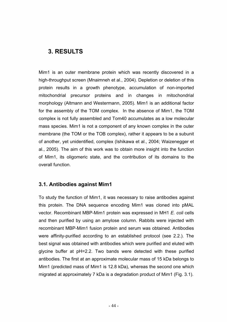
- 44 -
3. RESULTS
Mim1 is an outer membrane protein which was recently discovered in a
high-throughput screen (Mnaimneh et al., 2004). Depletion or deletion of this
protein results in a growth phenotype, accumulation of non-imported
mitochondrial precursor proteins and in changes in mitochondrial
morphology (Altmann and Westermann, 2005). Mim1 is an additional factor
for the assembly of the TOM complex. In the absence of Mim1, the TOM
complex is not fully assembled and Tom40 accumulates as a low molecular
mass species. Mim1 is not a component of any known complex in the outer
membrane (the TOM or the TOB complex), rather it appears to be a subunit
of another, yet unidentified, complex (Ishikawa et al., 2004; Waizenegger et
al., 2005). The aim of this work was to obtain more insight into the function
of Mim1, its oligomeric state, and the contribution of its domains to the
overall function.
3.1. Antibodies against Mim1
To study the function of Mim1, it was necessary to raise antibodies against
this protein. The DNA sequence encoding Mim1 was cloned into pMAL
vector. Recombinant MBP-Mim1 protein was expressed in MH1 E. coli cells
and then purified by using an amylose column. Rabbits were injected with
recombinant MBP-Mim1 fusion protein and serum was obtained. Antibodies
were affinity-purified according to an established protocol (see 2.2.). The
best signal was obtained with antibodies which were purified and eluted with
glycine buffer at pH=2.2. Two bands were detected with these purified
antibodies. The first at an approximate molecular mass of 15 kDa belongs to
Mim1 (predicted mass of Mim1 is 12.8 kDa), whereas the second one which
migrated at approximately 7 kDa is a degradation product of Mim1 (Fig. 3.1).
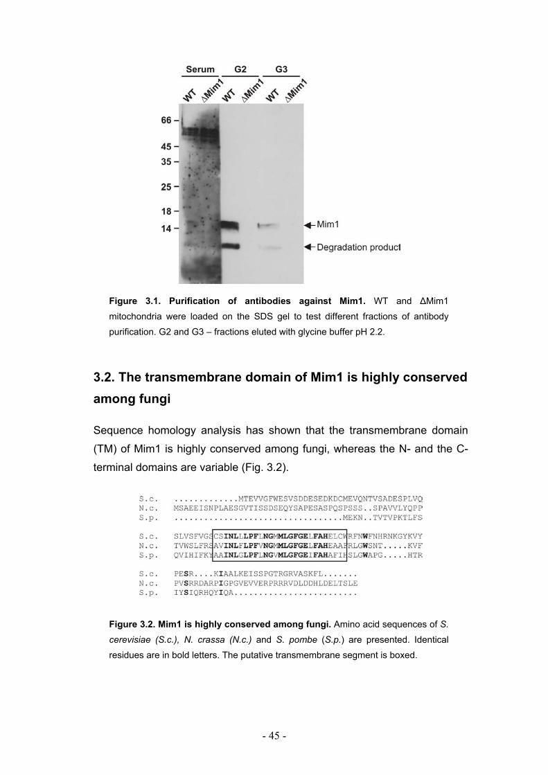
- 45 -
Figure 3.1. Purification of antibodies against Mim1. WT and ∆Mim1
mitochondria were loaded on the SDS gel to test different fractions of antibody
purification. G2 and G3 – fractions eluted with glycine buffer pH 2.2.
3.2. The transmembrane domain of Mim1 is highly conserved among fungi
Sequence homology analysis has shown that the transmembrane domain
(TM) of Mim1 is highly conserved among fungi, whereas the N- and the C-
terminal domains are variable (Fig. 3.2).
Figure 3.2. Mim1 is highly conserved among fungi. Amino acid sequences of S.
cerevisiae (S.c.), N. crassa (N.c.) and S. pombe (S.p.) are presented. Identical
residues are in bold letters. The putative transmembrane segment is boxed.
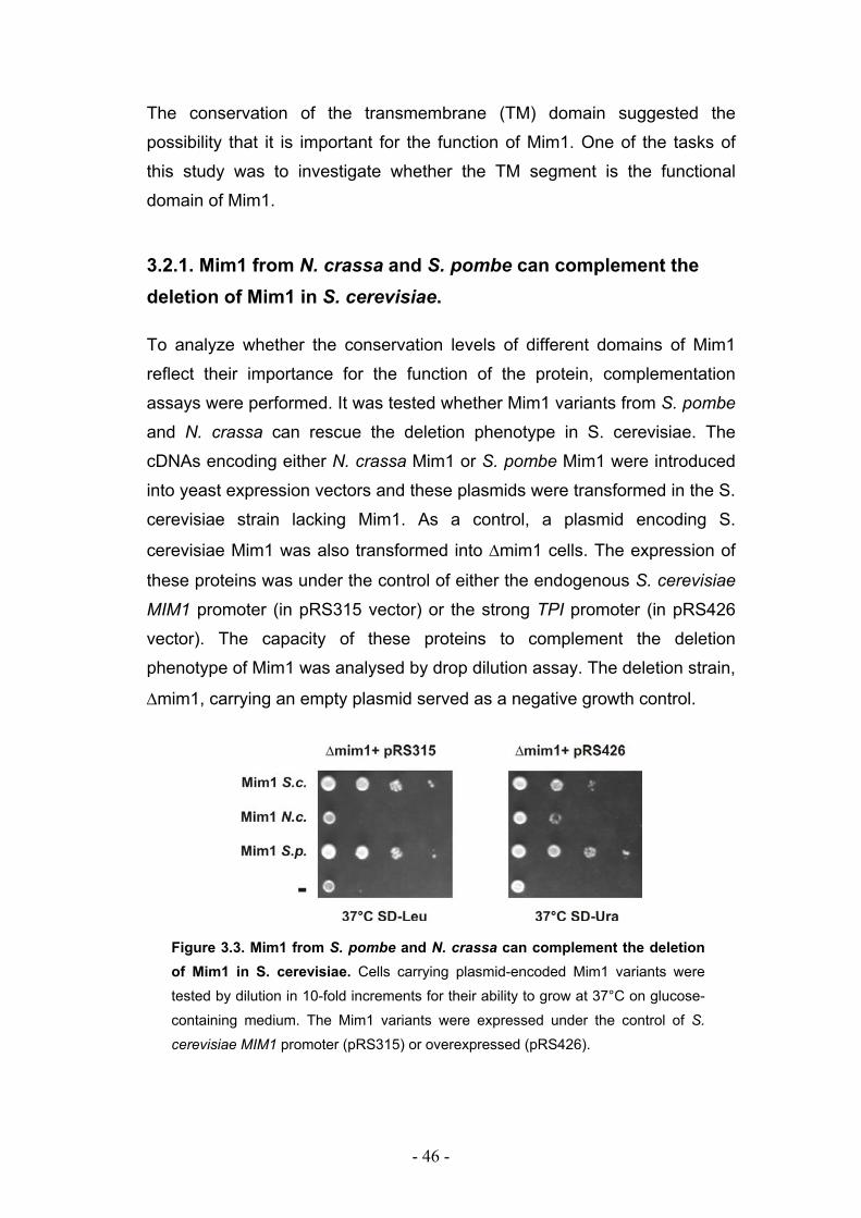
- 46 -
The conservation of the transmembrane (TM) domain suggested the
possibility that it is important for the function of Mim1. One of the tasks of
this study was to investigate whether the TM segment is the functional
domain of Mim1.
3.2.1. Mim1 from N. crassa and S. pombe can complement the deletion of Mim1 in S. cerevisiae.
To analyze whether the conservation levels of different domains of Mim1
reflect their importance for the function of the protein, complementation
assays were performed. It was tested whether Mim1 variants from S. pombe
and N. crassa can rescue the deletion phenotype in S. cerevisiae. The
cDNAs encoding either N. crassa Mim1 or S. pombe Mim1 were introduced
into yeast expression vectors and these plasmids were transformed in the S.
cerevisiae strain lacking Mim1. As a control, a plasmid encoding S.
cerevisiae Mim1 was also transformed into ∆mim1 cells. The expression of
these proteins was under the control of either the endogenous S. cerevisiae
MIM1 promoter (in pRS315 vector) or the strong TPI promoter (in pRS426
vector). The capacity of these proteins to complement the deletion
phenotype of Mim1 was analysed by drop dilution assay. The deletion strain,
∆mim1, carrying an empty plasmid served as a negative growth control.
Figure 3.3. Mim1 from S. pombe and N. crassa can complement the deletion of Mim1 in S. cerevisiae. Cells carrying plasmid-encoded Mim1 variants were
tested by dilution in 10-fold increments for their ability to grow at 37°C on glucose-
containing medium. The Mim1 variants were expressed under the control of S.
cerevisiae MIM1 promoter (pRS315) or overexpressed (pRS426).
- 47 -
Mim1 from S. pombe fully complemented the deletion of Mim1 under all
conditions tested, whereas N. crassa Mim1 complemented rather partially
and only if it was overexpressed (Fig. 3.3). The partial complementation of
N. crassa Mim1 was not due to lower expression levels since expression
was confirmed by western blotting (data not shown).
3.2.2. The N- and the C-terminal domains of Mim1 are not crucial for its function.
To investigate in more detail the importance of different domains of Mim1
three different truncated Mim1 variants where constructed. In the first two
truncated versions amino acid residues 1-34 (Mim1∆N) or 76-113 (Mim1∆C)
were deleted. The third variant was constructed by deleting both the N- and
the C- terminal domains, leaving only the TM segment of the protein
(Mim1(TM)) (Fig. 3.4A).
These truncated versions of Mim1 were tested by drop dilution assay for
their capacity to complement the deletion of native Mim1. Under all tested
conditions, Mim1 lacking N- or C- terminus did not show any growth
phenotype suggesting that Mim1 lacking either of these domains alone was
fully functional. The complementation of function by these truncated
versions of Mim1 suggests that they were properly inserted into the outer
membrane. The Mim1 variant containing only the TM domain could not
complement the function when expressed under the endogenous promoter
but could do so when overexpressed (Fig. 3.4B).
Taken together it seems that neither the N- nor the C-terminal domains of
Mim1 are crucial for the function of the protein and for its targeting to and
insertion into the mitochondrial outer membrane.
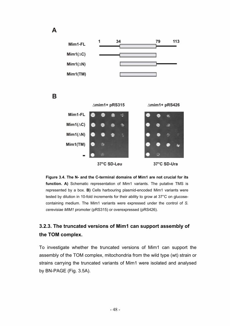
- 48 -
Figure 3.4. The N- and the C-terminal domains of Mim1 are not crucial for its function. A) Schematic representation of Mim1 variants. The putative TMS is
represented by a box. B) Cells harbouring plasmid-encoded Mim1 variants were
tested by dilution in 10-fold increments for their ability to grow at 37°C on glucose-
containing medium. The Mim1 variants were expressed under the control of S.
cerevisiae MIM1 promoter (pRS315) or overexpressed (pRS426).
3.2.3. The truncated versions of Mim1 can support assembly of the TOM complex.
To investigate whether the truncated versions of Mim1 can support the
assembly of the TOM complex, mitochondria from the wild type (wt) strain or
strains carrying the truncated variants of Mim1 were isolated and analysed
by BN-PAGE (Fig. 3.5A).
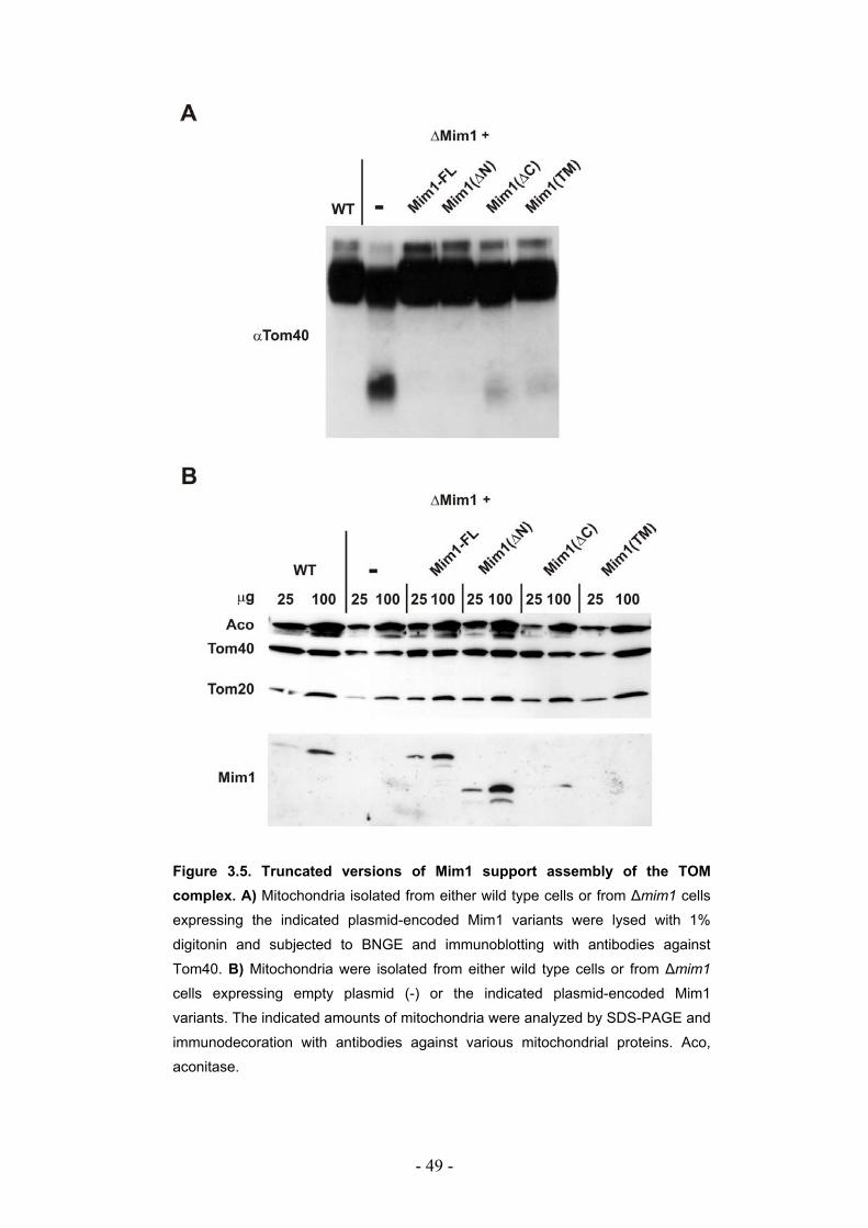
- 49 -
Figure 3.5. Truncated versions of Mim1 support assembly of the TOM complex. A) Mitochondria isolated from either wild type cells or from ∆mim1 cells expressing the indicated plasmid-encoded Mim1 variants were lysed with 1%
digitonin and subjected to BNGE and immunoblotting with antibodies against
Tom40. B) Mitochondria were isolated from either wild type cells or from ∆mim1
cells expressing empty plasmid (-) or the indicated plasmid-encoded Mim1
variants. The indicated amounts of mitochondria were analyzed by SDS-PAGE and
immunodecoration with antibodies against various mitochondrial proteins. Aco,
aconitase.
- 50 -
In mitochondria lacking Mim1, Tom40 species of lower molecular weight
were observed as published previously (Waizenegger et al., 2005). In
contrast, upon overexpression of the Mim1 truncated variants unassembled
species of Tom40 were observed at very low levels or not at all, suggesting
that expression of shorter Mim1 forms is sufficient for the correct assembly
of the TOM complex. This result was in agreement with the fact that shorter
forms of Mim1 could support growth. Similarly, the steady state levels of
Tom20 and Tom40 in mitochondria harbouring the truncated version were
similar to those in wild type mitochondria (Fig. 3.5B).
Although antibodies were raised against full-length Mim1, most of the
epitopes reside in the C-terminal domain of Mim1 (data not shown). The
transmembrane domain of Mim1 is very hydrophobic and probably does not
contain any epitopes. Thus, a detection of Mim1(∆C) and Mim1(TM) in this
study was very limited.
3.3. Mim1 forms dimers or homooligomers in the mitochondrial outer membrane.
It was already shown by BN-PAGE and gel-filtration, that Mim1 is neither a
subunit of the TOM complex nor of the TOB complex but rather of new
complex with an approximate molecular mass of 200 kDa (Waizenegger et
al., 2005). To study the composition of this complex a chemical crosslinking
approach was applied. Intact mitochondria were treated with the crosslinking
reagents disuccinimidyl glutarate (DSG), disuccinimidyl suberate (DSS), or
1,5-difluoro-2,4-dinitrobenzene (DFDNB). After the treatment with
crosslinkers mitochondria were analyzed by SDS-PAGE and
immunodecoration with antibodies against Mim1. One major crosslinking
adduct band of apparent molecular mass of 28 kDa was detected after the
treatment of mitochondria with all tested crosslinkers (Fig. 3.6A). The mass
of 28 kDa suggested that this band corresponds to a homodimer of Mim1
since the predicted mass of Mim1 is 12.8 kDa. Another detected band of
approximate mass of 40 kDa might reflect a trimer of Mim1.
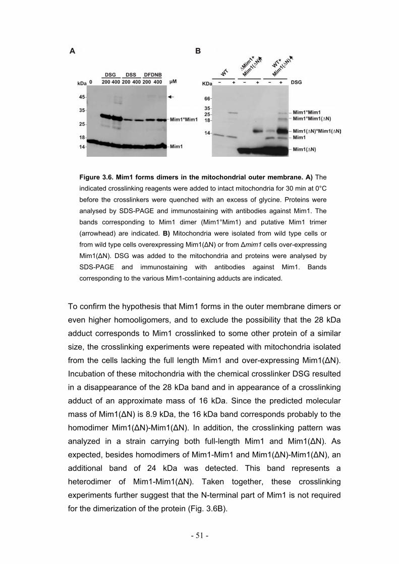
- 51 -
Figure 3.6. Mim1 forms dimers in the mitochondrial outer membrane. A) The
indicated crosslinking reagents were added to intact mitochondria for 30 min at 0°C
before the crosslinkers were quenched with an excess of glycine. Proteins were
analysed by SDS-PAGE and immunostaining with antibodies against Mim1. The
bands corresponding to Mim1 dimer (Mim1°Mim1) and putative Mim1 trimer
(arrowhead) are indicated. B) Mitochondria were isolated from wild type cells or
from wild type cells overexpressing Mim1(∆N) or from ∆mim1 cells over-expressing
Mim1(∆N). DSG was added to the mitochondria and proteins were analysed by
SDS-PAGE and immunostaining with antibodies against Mim1. Bands
corresponding to the various Mim1-containing adducts are indicated.
To confirm the hypothesis that Mim1 forms in the outer membrane dimers or
even higher homooligomers, and to exclude the possibility that the 28 kDa
adduct corresponds to Mim1 crosslinked to some other protein of a similar
size, the crosslinking experiments were repeated with mitochondria isolated
from the cells lacking the full length Mim1 and over-expressing Mim1(∆N).
Incubation of these mitochondria with the chemical crosslinker DSG resulted
in a disappearance of the 28 kDa band and in appearance of a crosslinking
adduct of an approximate mass of 16 kDa. Since the predicted molecular
mass of Mim1(∆N) is 8.9 kDa, the 16 kDa band corresponds probably to the
homodimer Mim1(∆N)-Mim1(∆N). In addition, the crosslinking pattern was
analyzed in a strain carrying both full-length Mim1 and Mim1(∆N). As
expected, besides homodimers of Mim1-Mim1 and Mim1(∆N)-Mim1(∆N), an
additional band of 24 kDa was detected. This band represents a
heterodimer of Mim1-Mim1(∆N). Taken together, these crosslinking
experiments further suggest that the N-terminal part of Mim1 is not required
for the dimerization of the protein (Fig. 3.6B).
- 52 -
To confirm the homophilic interactions between two or more Mim1
molecules, pull-down experiments were performed with mitochondria from
yeast strains expressing two different versions of Mim1, a heptahistidinyl-
tagged version Mim1-His7 and an HA-tagged version Mim1-HA3.
Mitochondria were also isolated from a control strain carrying only Mim1-
HA3. These cells grew as well as the wild type cells (Waizenegger et al.,
2005 and data not shown), therefore the proteins are fully functional.
Mitochondria were lysed and pull-down experiments with Ni-NTA beads
were performed. Mim1-HA3 was co-purified with the His tagged version of
Mim1 while such interaction was not observed for two other outer membrane
proteins, Tom40 and Tom70. To exclude the possibility that Mim1-HA3 could
interact with Ni-NTA beads in the absence of Mim1-His7 in a non specific
manner, this experiment was also performed using yeast strain expressing
only Mim1-HA3 (Fig. 3.7).
Figure 3.7. Two or more Mim1 molecules interact with each other in the outer mitochondrial membrane. Mitochondria were isolated from yeast strains
expressing an HA-tagged Mim1 (Mim1-HA3) or co-expressing Mim1-HA3 and
heptahistidinyl-tagged version of Mim1 (Mim1-His7). The organelles were
solubilized in buffer containing 1% digitonin. The total extract (T) was applied to a
Ni-NTA affinity resin. Bound (pellet - P) and unbound (supernatant - SN) material
were analysed by immunostaining using antibodies against Tom70, Tom20, HA
and the His tags.
To summarize, results of crosslinking and pull-down experiments show that
Mim1 complex in the outer mitochondrial membrane harbors two or more
Mim1 molecules.
- 53 -
3.3.1. Two dimerization motifs in the transmembrane segment of Mim1 are crucial for homooligomerization and function.
Detailed sequence analysis revealed two GXXXG/A dimerization motifs in
the TM domain of Mim1. The first motif includes residues from 57 to 61 and
the second residues from 63 to 67. GXXXG and G/AXXXG/A motifs are
known to mediate TM helix–helix interactions within biological membranes
(Russ and Engelman, 2000; Senes et al., 2004). Considering the
observation that Mim1 has the tendency to dimerize or homooligomerize,
these sequences were tested for their role in the above dimerization. For
that goal, mutations were introduced in these GXXXG motifs and the
resulting strains were analyzed (Fig. 3.8).
A
B
Figure 3.8. The double GXXXG⁄A motifs within the TMS are crucial for growth. A) Schematic representation of the mutations performed in the TMS of Mim1. B) Cells harbouring the indicated plasmid-encoded Mim1 variants or empty (“-“)
plasmid were tested by dilution in 10-fold increments for their ability to grow at
37°C on glucose-containing medium.
In the first mutant, only one amino acid (Gly) in position 61 was replaced by
the bulky residue leucine (Mim1 G61L). This change did not show any effect
on the yeast growth and did not affect the ability of Mim1 to dimerize. This
can be explained by another dimerization motif sufficient to support
- 54 -
homooligomerization. To exclude this possibility, in addition to Mim1 G61L,
two more amino acid residues Gly63 and Ala67 were replaced with Ile
residues (Mim1-LII). This Mim1 mutant was not able to complement deletion
of wt Mim1 under all conditions tested.
Mitochondria isolated from yeast carrying Mim1-LII instead of native Mim1
had significantly reduced steady state levels of Tom20 and moderately
reduced amounts of Tom40 and porin (Fig. 3.9). The reduction levels were
similar to the ones previously observed in the ∆mim1 strain. This result
together with the fact that Mim1-LII could not complement the deletion
phenotype of ∆mim1, suggested that this mutatant Mim1 had an impaired
function. To investigate whether this reduced function could be correlated to
the altered oligomeric structure of Mim1, crosslinking experiments were
performed. Mitochondria was isolated from the strain expressing Mim1-LII
instead of wt Mim1 and DSG was added. The 28 kDa crosslinking adduct
which corresponds to Mim1-Mim1 dimer completely disappeared suggesting
that either Mim1-LII is not able to dimerize, or that two Mim1-LII molecules
are positioned in a manner that they cannot be crosslinked.
To investigate whether the Mim1-LII variant was able to build oligomeric
structures, mitochondria carrying this variant were analyzed by BN-PAGE.
This analysis revealed that mutating the dimerization motifs resulted in the
absence of the Mim1-containing complexes (Fig. 3.10). Thus, these motifs
are indeed crucial for the ability of Mim1 to form oligomeric structures. In
these mitochondria Tom40 lower molecular species were also observed by
BN-PAGE as it was already shown for ∆Mim1 mitochondria (see above).
Thus, homooligomerization is essential for the function of Mim1 in the
biogenesis of the TOM complex.
- 55 -
Figure 3.9. The double GXXXG⁄A motifs are essential for dimerization and function of Mim1. A) Mitochondria were isolated from ∆mim1 cells carrying empty
plasmid or expressing Mim1-WT or Mim1-LII. Mitochondria were analyzed by SDS-
PAGE and immunodecoration with antibodies against the indicated mitochondrial
proteins. Mim1*, proteolytic fragment of Mim1. Lower panel: the bands from the
loading of 10 µg mitochondria were quantified and the amount of each protein is
presented as percentage of the amount in wild type mitochondria. B) Mitochondria
were isolated from ∆mim1 cells overexpressing wt Mim1 or the indicated Mim1
variant. DSG was added to the mitochondria and proteins were analysed by SDS-
PAGE and immunodecoration using antibodies against Mim1.
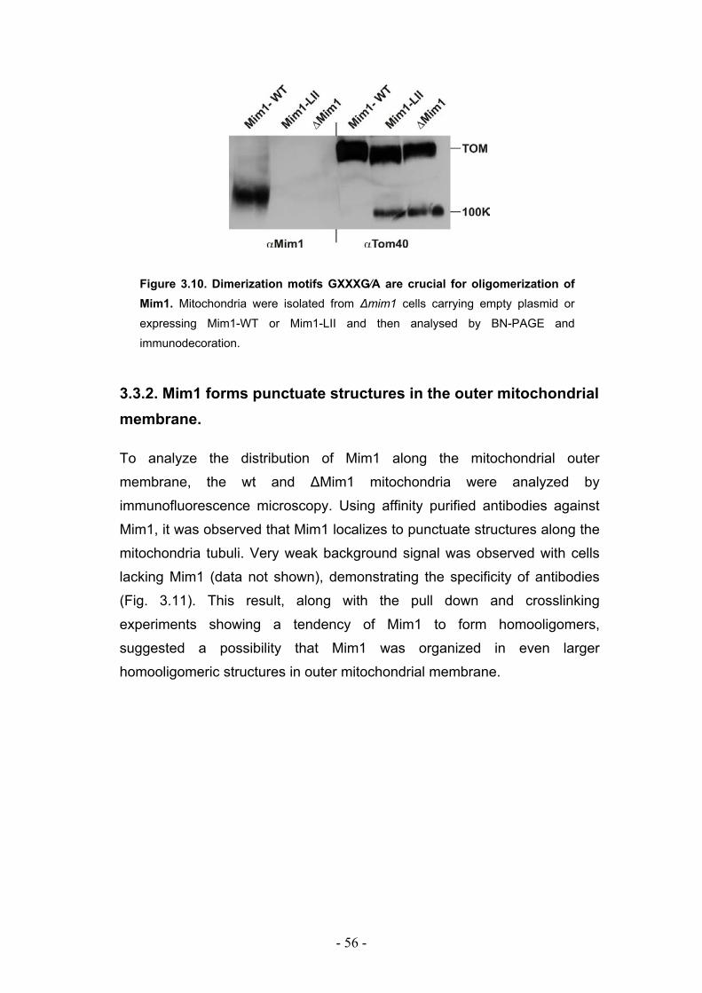
- 56 -
Figure 3.10. Dimerization motifs GXXXG⁄A are crucial for oligomerization of Mim1. Mitochondria were isolated from ∆mim1 cells carrying empty plasmid or expressing Mim1-WT or Mim1-LII and then analysed by BN-PAGE and
immunodecoration.
3.3.2. Mim1 forms punctuate structures in the outer mitochondrial membrane.
To analyze the distribution of Mim1 along the mitochondrial outer
membrane, the wt and ∆Mim1 mitochondria were analyzed by
immunofluorescence microscopy. Using affinity purified antibodies against
Mim1, it was observed that Mim1 localizes to punctuate structures along the
mitochondria tubuli. Very weak background signal was observed with cells
lacking Mim1 (data not shown), demonstrating the specificity of antibodies
(Fig. 3.11). This result, along with the pull down and crosslinking
experiments showing a tendency of Mim1 to form homooligomers,
suggested a possibility that Mim1 was organized in even larger
homooligomeric structures in outer mitochondrial membrane.
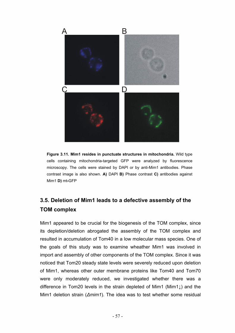
- 57 -
Figure 3.11. Mim1 resides in punctuate structures in mitochondria. Wild type
cells containing mitochondria-targeted GFP were analyzed by fluorescence
microscopy. The cells were stained by DAPI or by anti-Mim1 antibodies. Phase
contrast image is also shown. A) DAPI B) Phase contrast C) antibodies against
Mim1 D) mt-GFP
3.5. Deletion of Mim1 leads to a defective assembly of the TOM complex
Mim1 appeared to be crucial for the biogenesis of the TOM complex, since
its depletion/deletion abrogated the assembly of the TOM complex and
resulted in accumulation of Tom40 in a low molecular mass species. One of
the goals of this study was to examine wheather Mim1 was involved in
import and assembly of other components of the TOM complex. Since it was
noticed that Tom20 steady state levels were severely reduced upon deletion
of Mim1, whereas other outer membrane proteins like Tom40 and Tom70
were only moderately reduced, we investigated whether there was a
difference in Tom20 levels in the strain depleted of Mim1 (Mim1↓) and the
Mim1 deletion strain (∆mim1). The idea was to test whether some residual
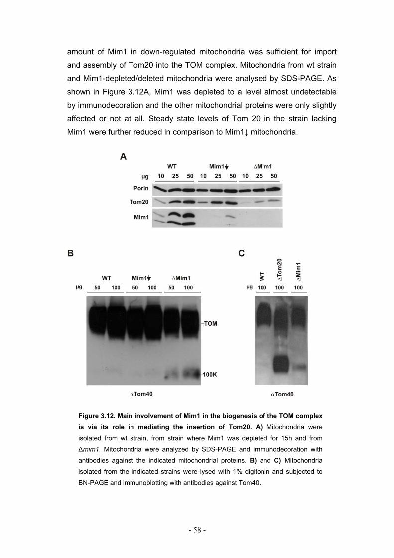
- 58 -
amount of Mim1 in down-regulated mitochondria was sufficient for import
and assembly of Tom20 into the TOM complex. Mitochondria from wt strain
and Mim1-depleted/deleted mitochondria were analysed by SDS-PAGE. As
shown in Figure 3.12A, Mim1 was depleted to a level almost undetectable
by immunodecoration and the other mitochondrial proteins were only slightly
affected or not at all. Steady state levels of Tom 20 in the strain lacking
Mim1 were further reduced in comparison to Mim1↓ mitochondria.
Figure 3.12. Main involvement of Mim1 in the biogenesis of the TOM complex is via its role in mediating the insertion of Tom20. A) Mitochondria were
isolated from wt strain, from strain where Mim1 was depleted for 15h and from
∆mim1. Mitochondria were analyzed by SDS-PAGE and immunodecoration with
antibodies against the indicated mitochondrial proteins. B) and C) Mitochondria
isolated from the indicated strains were lysed with 1% digitonin and subjected to
BN-PAGE and immunoblotting with antibodies against Tom40.
- 59 -
To analyze the assembly of the TOM complex in these strains, mitochondria
were analyzed by BN-PAGE and immunodecoration using antibodies
against Tom40. The difference in the assembly of the TOM complex in wt
and Mim1↓ mitochondria was not significant and the TOM complex was fully
assembled. In contrast, accumulation of non-assembled Tom40 species was
observed in mitochondria lacking Mim1 suggesting that residual Mim1 in
mitochondria depleted of Mim1 was sufficient for the full assembly of the
TOM complex.
It has been shown (Model et al., 2002) that Tom20 is crucial for stable
organization of Tom40 channel units into larger assemblies. Thus, BN-
PAGE analysis of mitochondria lacking Tom20 was compared to ∆Mim1
mitochondria. Figure 3.12.C shows that a larger portion of the Tom40
molecules migrated in a low molecular mass species in the absence of
Tom20 as compared to the absence of Mim1.
It seems that the main involvement of Mim1 in the biogenesis of the TOM
complex is via its role in mediating the insertion of Tom20 into the outer
membrane and/or facilitating the assembly of the latter into the TOM
complex.
3.6. Mim1 is required for optimal import but not assembly of Tom20
To test whether Mim1 was involved only in import of the receptor protein
Tom20 or also in its assembly into the mature TOM complex, the import and
co-immunoprecipitation experiments were performed. In vitro insertion of the
Tom20 into the outer membrane of mitochondria depleted of Mim1 and
lacking Mim1 was tested. To monitor the insertion of Tom20, a specific
proteolytic assay was used. For this assay a fusion protein was constructed,
with the first 38 amino acids of Tom70 from N. crassa fused in front of yeast
Tom20, so that a protease protected fragment of properly inserted protein
was 7-8 kDa which is easily detectable by SDS-PAGE (Ahting et al., 2005).
As seen in Fig. 3.13, both the depletion and deletion of Mim1 resulted in
decrease of the insertion of Tom20, but in the Mim1-deficient mitochondria,
the reduction was more significant. In contrast, only minor reduction in the
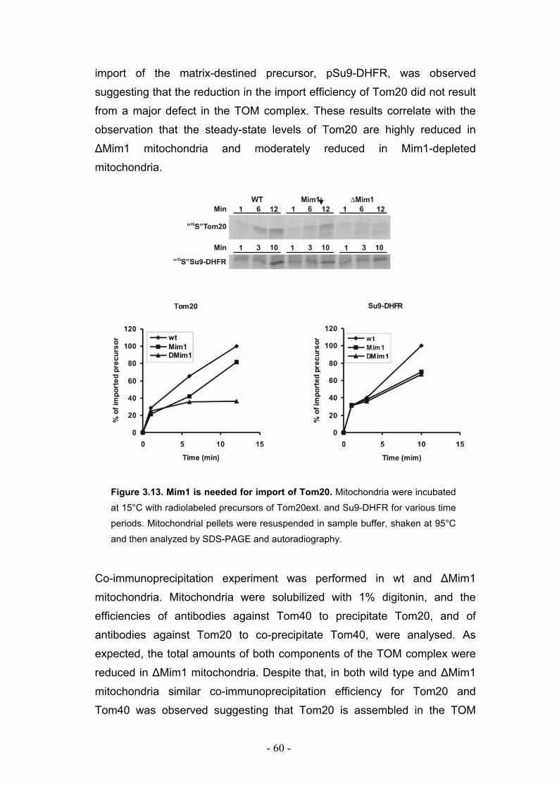
- 60 -
import of the matrix-destined precursor, pSu9-DHFR, was observed
suggesting that the reduction in the import efficiency of Tom20 did not result
from a major defect in the TOM complex. These results correlate with the
observation that the steady-state levels of Tom20 are highly reduced in
∆Mim1 mitochondria and moderately reduced in Mim1-depleted
mitochondria.
Figure 3.13. Mim1 is needed for import of Tom20. Mitochondria were incubated
at 15°C with radiolabeled precursors of Tom20ext. and Su9-DHFR for various time
periods. Mitochondrial pellets were resuspended in sample buffer, shaken at 95°C
and then analyzed by SDS-PAGE and autoradiography.
Co-immunoprecipitation experiment was performed in wt and ∆Mim1
mitochondria. Mitochondria were solubilized with 1% digitonin, and the
efficiencies of antibodies against Tom40 to precipitate Tom20, and of
antibodies against Tom20 to co-precipitate Tom40, were analysed. As
expected, the total amounts of both components of the TOM complex were
reduced in ∆Mim1 mitochondria. Despite that, in both wild type and ∆Mim1
mitochondria similar co-immunoprecipitation efficiency for Tom20 and
Tom40 was observed suggesting that Tom20 is assembled in the TOM
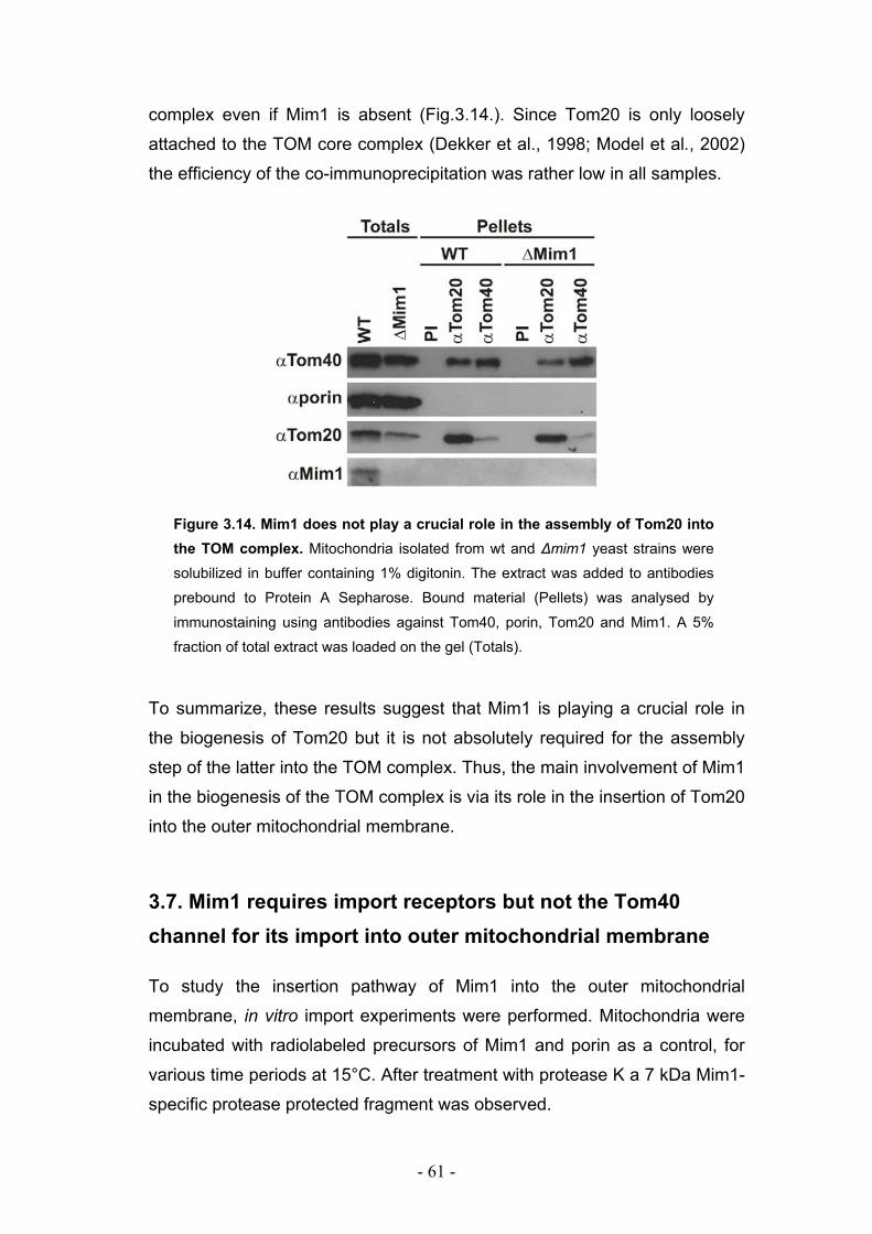
- 61 -
complex even if Mim1 is absent (Fig.3.14.). Since Tom20 is only loosely
attached to the TOM core complex (Dekker et al., 1998; Model et al., 2002)
the efficiency of the co-immunoprecipitation was rather low in all samples.
Figure 3.14. Mim1 does not play a crucial role in the assembly of Tom20 into the TOM complex. Mitochondria isolated from wt and ∆mim1 yeast strains were
solubilized in buffer containing 1% digitonin. The extract was added to antibodies
prebound to Protein A Sepharose. Bound material (Pellets) was analysed by
immunostaining using antibodies against Tom40, porin, Tom20 and Mim1. A 5%
fraction of total extract was loaded on the gel (Totals).
To summarize, these results suggest that Mim1 is playing a crucial role in
the biogenesis of Tom20 but it is not absolutely required for the assembly
step of the latter into the TOM complex. Thus, the main involvement of Mim1
in the biogenesis of the TOM complex is via its role in the insertion of Tom20
into the outer mitochondrial membrane.
3.7. Mim1 requires import receptors but not the Tom40 channel for its import into outer mitochondrial membrane
To study the insertion pathway of Mim1 into the outer mitochondrial
membrane, in vitro import experiments were performed. Mitochondria were
incubated with radiolabeled precursors of Mim1 and porin as a control, for
various time periods at 15°C. After treatment with protease K a 7 kDa Mim1-
specific protease protected fragment was observed.
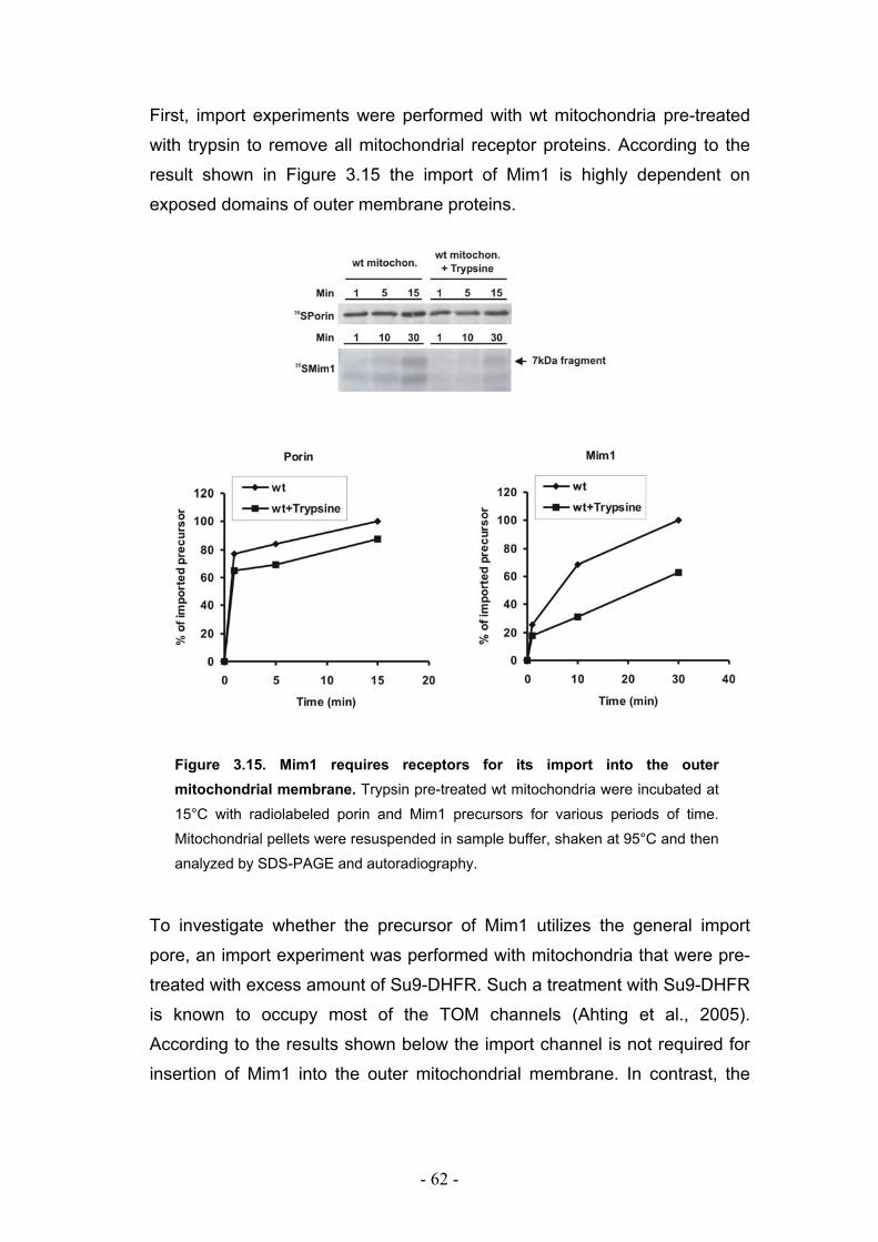
- 62 -
First, import experiments were performed with wt mitochondria pre-treated
with trypsin to remove all mitochondrial receptor proteins. According to the
result shown in Figure 3.15 the import of Mim1 is highly dependent on
exposed domains of outer membrane proteins.
Figure 3.15. Mim1 requires receptors for its import into the outer mitochondrial membrane. Trypsin pre-treated wt mitochondria were incubated at
15°C with radiolabeled porin and Mim1 precursors for various periods of time.
Mitochondrial pellets were resuspended in sample buffer, shaken at 95°C and then
analyzed by SDS-PAGE and autoradiography.
To investigate whether the precursor of Mim1 utilizes the general import
pore, an import experiment was performed with mitochondria that were pre-
treated with excess amount of Su9-DHFR. Such a treatment with Su9-DHFR
is known to occupy most of the TOM channels (Ahting et al., 2005).
According to the results shown below the import channel is not required for
insertion of Mim1 into the outer mitochondrial membrane. In contrast, the
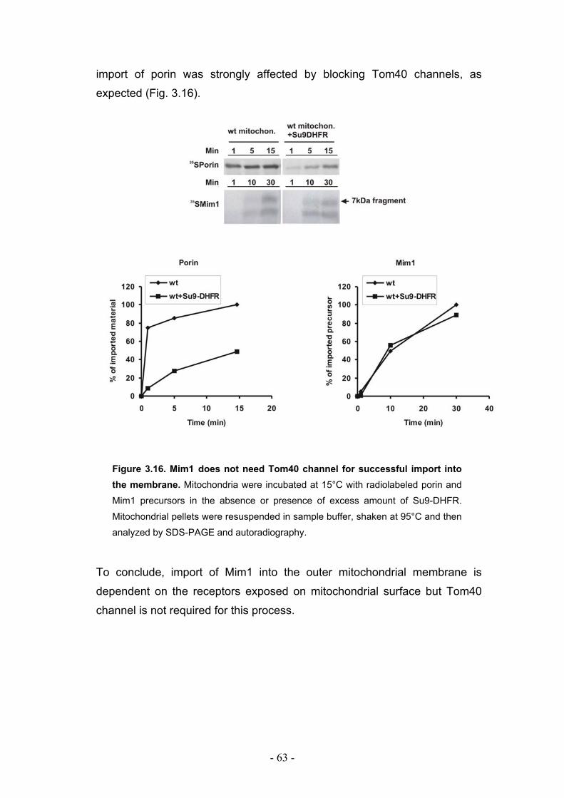
- 63 -
import of porin was strongly affected by blocking Tom40 channels, as
expected (Fig. 3.16).
Figure 3.16. Mim1 does not need Tom40 channel for successful import into the membrane. Mitochondria were incubated at 15°C with radiolabeled porin and
Mim1 precursors in the absence or presence of excess amount of Su9-DHFR.
Mitochondrial pellets were resuspended in sample buffer, shaken at 95°C and then
analyzed by SDS-PAGE and autoradiography.
To conclude, import of Mim1 into the outer mitochondrial membrane is
dependent on the receptors exposed on mitochondrial surface but Tom40
channel is not required for this process.
- 64 -
4. DISCUSSION
Proteins residing in the outer mitochondrial membrane can be classified into
several categories based on their topological organizations: the “signal-
anchored” proteins with a C-terminal hydrophilic domain facing the cytosol,
the “tail-anchored” proteins with N-terminal domains exposed on the
mitochondrial surface, β-barrel proteins, and the proteins that span the outer
membrane with two or more helices. Tom22 and Mim1 represent yet
another small group of proteins with a single transmembrane domain and
two hydrophilic domains, one present in the cytosol and another in the
intermembrane space. Mim1 is the most recently identified protein of the
latter group. It was first identified in a high throughput analysis of 200
proteins annotated in the yeast data base as essential for yeast viability
(Mnaimneh et al., 2004). The authors of this study named the protein Mim1
for its involvment in mitochondrial import, and provided the initial evidence
that Mim1 was essential for yeast cell viability. Shortly afterwards, Mim1 was
characterized in more detail by our group. Simultanously, Mim1 was studied
by Endo and co-workers who introduced the alternative name for it, Tom13
(Ishikawa et al., 2004; Waizenegger et al., 2005). Both groups characterized
Mim1 as a protein with an undefined function in the biogenesis of the TOM
complex. Interestingly, these studies also revealed that Mim1 was not
essential for yeast cell viability even though the yeast cells lacking Mim1
exhibited severely impaired growth.
4.1. The transmembrane segment of Mim1 is the functional domain of the protein
Mim1 is composed of three domains: N-terminal domain present in the
cytosol, C-terminal domain facing the intermembrane space (IMS), and a
single putative α-helical transmembrane domain (TM) spanning the outer
mitochondrial membrane. Until the identification of Mim1, the only known
- 65 -
protein in the outer mitochondrial membrane with such a topology was
Tom22 (Kiebler et al., 1993; Lithgow et al., 1994; Nakai and Endo, 1995).
However, Mim1 and Tom22 do not seem to have functional similarities.
Tom22 is a component of the TOM core complex and it functions as a
receptor for mitochondrial precursor proteins. Both the cytosolic and the IMS
domains of Tom22 are important for the receptor function of Tom22 as it
interacts with precursor proteins not only on the cis, but also on the trans
side of the TOM complex (Court et al., 1996; Moczko et al., 1997). In
addition, Tom22 was also reported to be involved in regulation of gating of
the general import pore (GIP) (van Wilpe et al., 1999). In contrast, Mim1 is
not a component of any known complex in the outer mitochondrial
membrane (Waizenegger et al., 2005 and data shown here).
To better understand the function of Mim1, we investigated the structural
characteristics of the protein. The results presented here demonstrate that
the TM domain is the only functional part of the protein, while the exposed
domains of Mim1 on both sides of the outer membrane are not necessary
for its function. Additionally, these exposed domains appear to be crucial
neither for the targeting of Mim1 to mitochondria nor for its insertion into the
outer membrane. The truncated versions of Mim1 could complement the
growth phenotype of ∆mim1 cells indicating that the protein lacking either of
the hydrophilic domains is functional and thus properly inserted in the outer
membrane. Furthermore, the truncated versions of Mim1 were able to
support a full assembly of Tom40 into the TOM complex, suggesting that
only the TM domain is crucial for the role of Mim1 in the biogenesis of the
TOM complex. The experimental data showing that the TM domain alone,
when over-expressed, was sufficient to complement the function of the full-
length protein further confirmed that the membrane embedded segment of
Mim1 is the functional part of the protein. Taken together, as the TM domain
of Mim1 is the sole functional part of the protein. Mim1 does not act as a
classical receptor for precursors of the TOM components, but rather
performs its function within the membrane.
Mim1 proteins from S.pombe (Sp) and N. crassa (Nc) share sequence
similarity to S. cerevisiae (Sc) Mim1 only in the transmembrane domain, but
not in the N- and the C-terminal parts of the protein. These homologs could
- 66 -
complement deletion of Mim1 in yeast S. cerevisiae but not to the same
extent. The S. cerevisiae yeast strain carrying S. pombe Mim1 grew as well
as the wild type (wt) strain both when it was under the regulation of yeast
MIM1 promoter and when it was overexpressed. In contrast, N. crassa Mim1
could not provide full, but only partial complementation, and only when it
was overexpressed. Since the expression of N. crassa Mim1 protein in yeast
was confirmed by western blot analysis, the reason for the different
complementation capacities of the two fungal homologs probably lies in the
difference in size and/or charge of their pertinent hydrophilic domains. The
exposed N- and C-terminal domains of N. crassa Mim1 (56 and 47 amino
acid residues, respectively) are larger then those in both S. pombe (22 and
22 residues) and S. cerevisiae (43 and 41 residues) homologs. One could
assume that despite clear sequence similarity in the TM domains of N.
crassa and S. cerevisiae Mim1, the Nc version of the protein when
expressed in yeast is either not properly inserted in the outer membrane or
is not able to form functional structures within the membrane due to the
larger size of the N- and the C- terminal domains. In addition, the C-terminal
domain of N. crassa Mim1 contains significantly more both positively and
negatively charged residues than the pertinent corresponding domain of
yeast S. cerevisiae Mim1 and that may be yet another reason why N. crassa
Mim1 could not fully complement deletion of Mim1 in S. cerevisiae. On the
other hand, the exposed domains of S. pombe Mim1 are smaller and less
charged than the pertinent equivalent domains of both S. cerevisiae and N.
crassa homologs. Hence, the repulsive steric forces between the hydrophilic
domains in a putative dimer or oligomer of Mim1 (see below) are less likely
to occur in the case of S. pombe Mim1, than in the case of N. crassa
version.
4.2. Structural organization of Mim1 molecules
Initial gel filtration and BN-PAGE analyses suggested that Mim1 is a
component of a novel, yet unidentified, complex of approximate size of 200
kDa (Waizenegger et al., 2005). Pull-down and crosslinking experiments
presented in this study demonstrated that Mim1 molecules interact with
- 67 -
each other in the outer membrane forming homodimers and possibly even
homooligomers. The experiments performed with various chemical
crosslinkers gave similar crosslinking pattern of Mim1 indicating that this
protein is present in the membrane as a homodimer. The most efficient
crosslinking was observed with the DSG crosslinker. In this case, it was
possible to detect a band corresponding to Mim1 homotrimer. Since it is
very difficult to detect crosslinking adducts bigger than a trimer, one may
speculate that the basic structural unit of Mim1 is a homodimer that may
congregate and form a higher structural assembly.
Next, the mechanism of Mim1 dimerization/oligomerization was tested. In
addition to being the functional domain of Mim1, its TM domain also
mediates the molecular organization of the protein, namely Mim1 forms
dimers via its TM domains. The oligomerization element of Mim1 within the
TM domain was identified. In silico analysis revealed two sequential
GXXXG/A motifs, which are essential for the oligomerization. GXXXG is
known to be a strong transmembrane helix-packing motif. This motif is found
in various cellular membranes where it is frequently involved in high-affinity
homo-oligomerization (Russ and Engelman, 2000). For example, GXXXG
motifs within the TM segments of subunits e and g are involved in the
dimerization/oligomerization of the yeast mitochondrial ATP synthase
complex (Arselin et al., 2003; Bustos and Velours, 2005; Saddar et al.,
2005), and such motifs in the TM domain of the amyloid precursor protein
are critical for the etiology of the Aβ42 peptide (Munter et al., 2007). These
two motifs are highly conserved among Mim1 homologs. Mim1 provides the
first example of such a motif in a protein from the outer mitochondrial
membrane and offers a special example of two sequential motifs where both
appear to be important for dimerization. Moreover, Mim1 TM domain
contains highly conserved Phe, which is known to be frequently associated
with GXXXG motifs in position -3 resulting in FXXGXXXG domain. Phe in
this position upstream of the dimerization motif was shown to stabilize
membrane-spanning GXXXG motifs (Unterreitmeier et al., 2007). Mutation
in only one dimerization motif did not affect the dimerization or the activity of
Mim1. However, replacing Ala and Gly residues by the bulky Leu or Ile
residues in both motifs rendered Mim1 inactive and prevented its
oligomerization. Crosslinking experiments clearly demonstrated that upon
- 68 -
inserting point mutations in both GXXXG/A domains the crosslinking
adducts corresponding to homodimer and homotrimer disappeared.
Likewise, it was shown by BN-PAGE that the 200 kDa Mim1 complex was
disassembled in the strain containing mutational alterations in both
dimerization motifs (LII mutant). Moreover, in cells expressing Mim1-LII,
Tom40 was dissociated from the TOM complex to the same extent as
observed in cells lacking Mim1 (∆mim1 strain). In addition, drop dilution
assay showed that LII cells harbouring the Mim1-LII variant exhibited the
same growth phenotype as the cells lacking Mim1. Taken together, these
experimental evidence suggest that dimerization/homooligomerization of
Mim1 are crucial for the function of the protein.
It remains unclear if the complex containing Mim1 harbours some additional
proteins. Crosslinking and pull-down experiments showed no direct protein-
protein interaction between Mim1 and any other outer membrane protein. In
addition, genetic screens designed to identify interacting partners of Mim1
(data not shown) suggest that these complexes may contain solely Mim1
molecules (data not shown). In a screen for multicopy suppressors of the
∆mim1 phenotype, a genomic library of the yeast genome on a multicopy
plasmid was transformed into ∆mim1 cells. More specifically, when genetic
library was transformed into ∆mim1 strain all 17 positive clones carried a
plasmid encoding Mim1. This suggests that Mim1 is nonredundant since its
deletion could be complemented only by Mim1 itself.
4.3. The function of Mim1
Next, I asked what the exact role of Mim1 in the biogenesis of the TOM
complex was. We previously observed that the deletion of Mim1 led to a
significant reduction of the steady state levels of Tom20, whereas only a
moderate reduction of steady state levels of Tom40 and Tom70 was
observed (Waizenegger et al., 2005). It is also known that although Tom20
is not a subunit of the TOM core complex (Dekker et al., 1998), the
structural arrangement of the latter depends on the presence of Tom20
(Model et al., 2002). Furthermore, it has been previously reported that
Tom20 is required for efficient biogenesis of the TOM core components,
- 69 -
Tom40 and Tom22 (Harkness et al., 1994; Lithgow et al., 1994; Rapaport
and Neupert, 1999). In this study, I propose that the observed phenotypes of
∆mim1 cells like altered mitochondrial morphology, import inhibition of
precursor proteins, and hampered assembly of newly synthesized subunits
of the TOM complex, are secondary to the primary effect on the integration
of Tom20 into the outer membrane. It is shown here that even low, almost
undetectable by immunodecoration levels of Mim1 in the outer membrane,
were sufficient to maintain wt levels of Tom20 and Tom 40 that were fully
assembled in the TOM complex. Despite extensive efforts, I was unable to
detect any stable interaction between Mim1 and Tom20 (data not shown).
Therefore, Mim1 appears to exert its role via an indirect interaction with
precursor molecules of Tom20.
A successful integration of Tom20 into the outer membrane requires initial
recognition, insertion into the membrane, and assembly into the TOM
complex. As Mim1 does not function as a classical receptor for precursor
proteins but rather within the membrane, its involvement in the initial
recognition at the cytosolic surface of the organelle can be excluded. The
current results demonstrate that the insertion of Tom20 molecules into the
outer membrane is hampered by the absence of Mim1. However, the
assembly of Tom20 into the TOM complex was not affected in mitochondria
lacking Mim1. Therefore, Mim1 is involved mainly in the membrane
integration of Tom20 precursors, but not in its assembly into the mature
TOM complex.
How does Mim1 perform its function? Mim1 homologs are found in fungal
mitochondria but not in other eukaryotes like plant or mammalian
mitochondria. Thus, Mim1 appears to be involved in a pathway specific for
fungi. Tom20 counterpart in mammalian mitochondria may, therefore, have
different import and assembly pathway which does not require a protein with
a Mim1-like function. As Mim1 was found to form punctuate structures in the
outer membrane and biochemical studies revealed its propensity to form
homooligomers, I propose that this structural organization creates a micro-
environment in the outer membrane which facilitates the import of Tom20. A
specific characteristic of the outer mitochondrial membrane in fungi is the
presence of ergosterol. It is tempting to speculate that Mim1, in an unknown
manner, organizes lipid environment needed for a successful import of
- 70 -
Tom20. In this case, Mim1 affects the lipid organization via the interplay of
its highly conserved TM domain with ergosterol in the membrane. One
cannot exclude the possibility that there is a protein in plants or mammals
with a similar function, which does not share sequence similarity with Mim1
found in fungi.
The mechanism by which Mim1 is imported and embedded in the outer
membrane has not been described. According to preliminary data presented
in this study, it seems that Mim1 does not require Tom 40 channel to be
successfully imported but it is highly dependent on receptors exposed on
mitochondrial surface. This process remains to be analyzed and described
in more detail.
On the basis of the results presented above, I suggest the following working
model of structural organization and function of Mim1 (Figure 4.1). Mim1
precursor protein is recognized by the TOM complex receptors on
mitochondrial surface and then inserted directly into the outer mitochondrial
membrane without passing through the Tom40 channel (step 1). Upon its
proper insertion in the membrane, Mim1 first forms homodimers via two
GXXXG/A dimerization motifs (cyan) present in its transmembrane domain
(magenta) (step 2). This domain is the sole element responsible for
dimerization of Mim1. Homodimers, as basic structural units, are further
organized into 200 kDa complex composed of several Mim1 molecules (step
3). This homooligomeric structure possibly organizes lipid environment
(orange) (step 4) needed for one of the stages in import of Tom20 (step 5).
Finally, upon its insertion in the outer membrane, Tom 20 is assembled into
mature TOM complex in a process which is not dependent of Mim1 (steps 6
and 7).
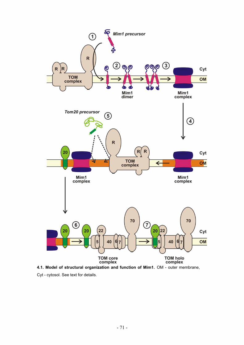
- 71 -
4.1. Model of structural organization and function of Mim1. OM - outer membrane,
Cyt - cytosol. See text for details.
- 72 -
5. Summary
The translocase of the outer mitochondrial membrane (TOM complex) is the
general entry site for newly synthesized proteins into the organelle. The
translocase is a multi-subunit complex composed of seven subunits: two
receptor proteins, Tom70 and Tom20, and five components which form the
core complex, Tom40, Tom22, Tom7, Tom6, and Tom5. All these subunits
have to be imported from the cytosol into the outer membrane. The
mechanisms by which the precursors of the Tom subunits are targeted to
mitochondria, integrated into the outer membrane and assembled into a
functional TOM complex is only partially understood.
Recently, an outer membrane protein named Mim1 was identified as an
assembly factor of the TOM complex. Specifically, it was observed to play a
role in the assembly of Tom40 into the TOM complex in the step after the
interaction of the Tom40 precursor with the TOB complex. Mim1 is
composed of an N-terminal cytosolic domain, a central putative
transmembrane segment (TMS), and a C-terminal domain facing the
intermembrane space. The contribution of the various domains to the overall
function of the protein, as well as the molecular function of Mim1 remained
unresolved.
In the present work I addressed some of these questions. In this thesis it is
shown that Mim1 is required for the integration of the import receptor Tom20
into the outer membrane but not for its assembly into the TOM complex.
Despite extensive efforts, a stable interaction between Mim1 and Tom20
was not detected. Thus, it is currently unclear whether Mim1 exerts its
function via a direct or indirect interaction with Tom20 precursor. Tom20 is
crucial for stable organization of Tom40 channel units into larger
assemblies. Therefore, I propose that Mim1 can affect the assembly
pathway of the TOM complex by acting at the Tom20 import level.
- 73 -
Structural characteristics of Mim1 required for its function were studied in
detail. The N- and the C-terminal domains of Mim1 are crucial neither for the
function of the protein nor for its biogenesis. Thus, the transmembrane
segment of Mim1 seems to be the minimal functional domain of the protein.
It is further shown that Mim1 forms homooligomeric structures via its TMS
which contains two helix-dimerization GXXXG⁄A motifs. Mim1 variant
mutated in both dimerization motifs is not able to form oligomeric structures
and is inactive. Taken together, the homo-oligomerization is a prerogative
for the function of Mim1 in mediating the integration of Tom20 into the
mitochondrial outer membrane.
- 74 -
6. Literature
Abe, Y., T. Shodai, T. Muto, K. Mihara, H. Torii, S. Nishikawa, T. Endo, and
D. Kohda. 2000. Structural Basis of Presequence Recognition by the
Mitochondrial Protein Import Receptor Tom20. Cell. 100:551-560.
Ahting, U., M. Thieffry, H. Engelhardt, R. Hegerl, W. Neupert, and S.
Nussberger. 2001. Tom40, the Pore-forming Component of the
Protein-conducting TOM Channel in the Outer Membrane of
Mitochondria. J. Cell Biol. 153:1151-1160.
Ahting, U., T. Waizenegger, W. Neupert, and D. Rapaport. 2005. Signal-
anchored Proteins Follow a Unique Insertion Pathway into the Outer
Membrane of Mitochondria. J. Biol. Chem. 280:48-53.
Altmann, K., and B. Westermann. 2005. Role of essential genes in
mitochondrial morphogenesis in Saccharomyces cerevisiae. Mol.
Biol. Cell. 16:5410-5417.
Arselin, G., M.F. Giraud, A. Dautant, J. Vaillier, D. Brethes, B. Coulary-
Salin, J. Schaeffer, and J. Velours. 2003. The GxxxG motif of the
transmembrane domain of subunit e is involved in the
dimerization/oligomerization of the yeast ATP synthase complex in
the mitochondrial membrane. Eur. J. Biochem. 270:1875-1884.
Bauer, M.F., S. Hofmann, W. Neupert, and M. Brunner. 2000. Protein
translocation into mitochondria: role of TIM complexes. Trends Cell
Biol. 10:25-31.
Bauer, M.F., C. Sirrenberg, W. Neupert, and M. Brunner. 1996. Role of
Tim23 as voltage sensor and presequence receptor in protein import
into mitochondria. Cell. 87:33-41.
- 75 -
Beers, J., D.M. Glerum, and A. Tzagoloff. 1997. Purification,
characterization, and localization of yeast Cox17p, a mitochondrial
copper shuttle. J Biol Chem. 272:33191-6.
Birnboim, H.C., and J. Doly. 1979. A rapid alkaline extraction procedure for
screening recombinant plasmid DNA. Nucleic Acids Res. 7:1513-23.
Bolliger, L., T. Junne, G. Schatz, and T. Lithgow. 1995. Acidic receptor
domains on both sides of the outer membrane mediate translocation
of precursor proteins into yeast mitochondria. Embo J. 14:6318-
6326.
Borgese, N., S. Colombo, and E. Pedrazzini. 2003. The tale of tail-
anchored proteins: coming from the cytosol and looking for a
membrane. J. Cell Biol. 161:1013-1019.
Bradford, M.M. 1976. A rapid and sensitive method for quantitation of
microgram quantities of protein utilising the principle of protein-dye
binding. Anal. Biochem. 72:248-254.
Braun, H.P., M. Emmermann, V. Kruft, and S.U. K. 1992. The general
mitochondrial processing peptidase from potato is an integral part of
cytochrome c reductase of the respiratory chain. EMBO J. 11:3219-
3227.
Brdiczka, D.G., D.B. Zorov, and S.S. Sheu. 2006. Mitochondrial contact
sites: their role in energy metabolism and apoptosis. Biochim
Biophys Acta. 1762:148-63.
Brix, J., S. Rudiger, B. Bukau, J. Schneider-Mergener, and N. Pfanner.
1999. Distribution of binding sequences for the mitochondrial import
receptors Tom20, Tom22, and Tom70 in a presequence-carrying
preprotein and a non-cleavable preprotein. J. Biol. Chem.
274:16522-16530.
Bustos, D.M., and J. Velours. 2005. The modification of the conserved
GXXXG motif of the membrane-spanning segment of subunit g
- 76 -
destabilizes the supramolecular species of yeast ATP synthase. J.
Biol. Chem. 280:29004-29010.
Chacinska, A., M. Lind, A.E. Frazier, J. Dudek, C. Meisinger, A. Geissler, A.
Sickmann, H.E. Meyer, K.N. Truscott, B. Guiard, N. Pfanner, and P.
Rehling. 2005. Mitochondrial presequence translocase: switching
between TOM tethering and motor recruitment involves Tim21 and
Tim17. Cell. 120:817-29.
Corral-Debrinski, M., C. Blugeon, and C. Jacq. 2000. In yeast, the 3'
untranslated region or the presequence of ATM1 is required for the
exclusive localization of its mRNA to the vicinity of mitochondria. Mol
Cell Biol. 20:7881-92.
Court, D.A., F.E. Nargang, H. Steiner, R.S. Hodges, W. Neupert, and R.
Lill. 1996. Role of the intermembrane space domain of the preprotein
receptor Tom22 in protein import into mitochondria. Mol. Cell. Biol.
16:4035-4042.
Daum, G., P.C. Böhni, and G. Schatz. 1982. Import of proteins into
mitochondria: cytochrome b2 and cytochrome c peroxidase are
located in the intermembrane space of yeast mitochondria. J. Biol.
Chem. 257:13028-13033.
Dekker, P.J.T., M.T. Ryan, J. Brix, H. Müller, A. Hönlinger, and N. Pfanner.
1998. Preprotein translocase of the outer mitochondrial membrane:
Molecular dissection and assembly of the general import pore
complex. Mol. Cell. Biol. 18:6515-6524.
Dembowski, M., K.-P. Künkele, F.E. Nargang, W. Neupert, and D.
Rapaport. 2001. Assembly of Tom6 and Tom7 into the TOM Core
Complex of Neurospora crassa. J. Biol. Chem. 276:17679-17685.
Diekert, K., A.I.P.M. de Kroon, U. Ahting, B. Niggemeyer, W. Neupert, B. de
Kruijff, and R. Lill. 2001. Apocytochrome c requires the TOM
complex for translocation across the mitochondrial outer membrane.
EMBO J. 20:5626-5635.
- 77 -
Dietmeier, K., A. Hönlinger, U. Bömer, P.J.T. Dekker, C. Eckerskorn, F.
Lottspeich, M. Kübrich, and N. Pfanner. 1997. Tom5 Functionally
Links Mitochondrial Preprotein Receptors to the General Import
Pore. Nature. 388:195-200.
Donzeau, M., K. Kaldi, A. Adam, S. Paschen, G. Wanner, B. Guiard, M.F.
Bauer, W. Neupert, and M. Brunner. 2000. Tim23 links the inner and
outer mitochondrial membranes. Cell. 101:401-412.
Emtage, J.L., and R.E. Jensen. 1993. MAS6 encodes an essential inner
membrane component of the yeast mitochondrial protein import
pathway. J Cell Biol. 122:1003-12.
Esaki, M., H. Shimizu, T. Ono, H. Yamamoto, T. Kanamori, S.I. Nishikawa,
and T. Endo. 2004. Mitochondrial protein import: Requirement of the
presequence elements and TOM components for precursor binding
to the TOM complex. J. Biol. Chem. in press.
Fölsch, H., B. Gaume, M. Brunner, W. Neupert, and R.A. Stuart. 1998. C-
to N-terminal translocation of preproteins into mitochondria. EMBO
J. 17:6508-6515.
Fujiki, M., and K. Verner. 1991. Coupling of protein synthesis and
mitochondrial protein import in a homologous yeast in vitro system.
J. Biol. Chem. 266:6841-6847.
Fujiki, M., and K. Verner. 1993. Coupling of cytosolic protein synthesis and
mitochondrial protein import in yeast: Evidence for cotranslational
import in vivo. J. Biol. Chem. 268:1914-1920.
Gakh, O., P. Cavadini, and G. Isaya. 2002. Mitochondrial processing
peptidases. Biochim. Biophys. Acta. 1592:63-77.
Geissler, A., A. Chacinska, K.N. Truscott, N. Wiedemann, K. Brandner, A.
Sickmann, H.E. Meyer, C. Meisinger, N. Pfanner, and P. Rehling.
2002. The mitochondrial presequence translocase: an essential role
of Tim50 in directing preproteins to the import channel. Cell.
111:507-518.
- 78 -
Gentle, I.E., L. Burri, and T. Lithgow. 2005. Molecular architecture and
function of the Omp85 family of proteins. Mol. Microbiol. 58:1216-
1225.
Glick, B.S., A. Brandt, K. Cunningham, S. Muller, R.L. Hallberg, and G.
Schatz. 1992. Cytochromes c1 and b2 are sorted to the
intermembrane space of yeast mitochondria by a stop-transfer
mechanism. Cell. 69:809-22.
Guan,C., P. Li, P.D.Riggs, and H. Inouye. 1987. Vectors that facilitate the
expression and purification of foreign peptides in Esherichia coli by
fusion to maltose-binding protein. Gene. 67: 21-30.
Habib, S.J., T. Waizenegger, A. Niewienda, S.A. Paschen, W. Neupert, and
D. Rapaport. 2007. The N-terminal domain of Tob55 has a receptor-
like function in the biogenesis of mitochondrial beta-barrel proteins.
J. Cell Biol. 176:77-88.
Hallermayer, G., R. Zimmermann, and W. Neupert. 1977. Kinetic studies on
the transport of cytoplasmically synthesized proteins into the
mitochondria in intact cells of Neurospora crassa. Eur. J. Biochem.
81:523-532.
Hammen, P.K., D.G. Gorenstein, and K. Weiner. 1996. Amphiphilicity
determines binding properties of three mitochondrial presequences
to lipid surface. Biochemistry. 35:3772-3781.
Harkness, T.A., F.E. Nargang, I. van der Klei, W. Neupert, and R. Lill. 1994.
A crucial role of the mitochondrial protein import receptor MOM19 for
the biogenesis of mitochondria. J. Cell Biol. 124:637-648.
Harmey, M.A., G. Hallermayer, H. Korb, and W. Neupert. 1977. Transport
of cytoplasmically synthesized proteins into the mitochondria in a cell
free system from Neurospora crassa. Eur. J. Biochem. 81:533-544.
Haucke, V., M. Horst, G. Schatz, and T. Lithgow. 1996. The Mas20p and
Mas70p subunits of the protein import receptor of yeast mitochondria
- 79 -
interact via the tetratricopeptide repeat motif in Mas20p: evidence for
a single hetero-oligomeric receptor. EMBO J. 15:1231-1237.
Hell, K., W. Neupert, and R.A. Stuart. 2001. Oxa1p acts as a general
membrane insertion machinery for proteins encoded by
mitochondrial DNA. EMBO J. 20:1281-1288.
Herlan, M., F. Vogel, C. Bornhovd, W. Neupert, and A.S. Reichert. 2003.
Processing of Mgm1 by the rhomboid-type protease Pcp1 is required
for maintenance of mitochondrial morphology and of mitochondrial
DNA. J. Biol. Chem. 278:27781-27788.
Ishikawa, D., H. Yamamoto, Y. Tamura, K. Moritoh, and T. Endo. 2004.
Two novel proteins in the mitochondrial outer membrane mediate
beta-barrel protein assembly. J. Cell Biol. 166:621-627.
Jarvis, J.A., M.T. Ryan, N.J. Hoogenraad, D.J. Craik, and R.B. Hoj. 1995.
Solution structure of the acetylated and noncleavable mitochondrial
targeting signal of rat chaperonin 10. J Biol Chem. 270:1323-1331.
Josyula, R., Z. Jin, Z. Fu, and B. Sha. 2006. Crystal structure of yeast
mitochondrial peripheral membrane protein Tim44p C-terminal
domain. J Mol Biol. 359:798-804.
Karniely, S., N. Regev-Rudzki, and O. Pines. 2006. The presequence of
fumarase is exposed to the cytosol during import into mitochondria.
J. Mol. Biol. 358:396-405.
Keil, P., A. Weinzierl, M. Kiebler, K. Dietmeier, T. Söllner, and N. Pfanner.
1993. Biogenesis of the mitochondrial receptor complex. Two
receptors are required for binding of MOM38 to the outer membrane
surface. J. Biol. Chem. 268:19177-19180.
Kerscher, O., J. Holder, M. Srinivasan, R.S. Leung, and R.E. Jensen. 1997.
The Tim54p-Tim22p complex mediates insertion of proteins into the
mitochondrial inner membrane. J. Cell Biol. 139:1663-1675.
- 80 -
Kerscher, O., N.B. Sepuri, and R.E. Jensen. 2000. Tim18p is a new
component of the Tim54p-Tim22p translocon in the mitochondrial
inner membrane. Mol. Biol. Cell. 11:103-116.
Kiebler, M., P. Keil, H. Schneider, I. van der Klei, N. Pfanner, and W.
Neupert. 1993. The mitochondrial receptor complex: a central role of
MOM22 in mediating preprotein transfer from receptors to the
general insertion pore. Cell. 74:483-492.
Kovermann, P., K.N. Truscott, B. Guiard, P. Rehling, N.B. Sepuri, H. Müller,
R.E. Jensen, R. Wagner, and N. Pfanner. 2002. Tim22, the Essential
Core of the Mitochondrial Protein Insertion Complex, Forms a
Voltage-Activated and Signal-Gated Channel. Mol. Cell. 9:363-373.
Kozjak, V., N. Wiedemann, D. Milenkovic, C. Lohaus, H.E. Meyer, B.
Guiard, C. Meisinger, and N. Pfanner. 2003. An Essential Role of
Sam50 in the Protein Sorting and Assembly Machinery of the
Mitochondrial Outer Membrane. J. Biol. Chem. 278:48520-48523.
Kübrich, M., P. Keil, J. Rassow, P.J. Dekker, J. Blom, M. Meijer, and N.
Pfanner. 1994. The polytopic mitochondrial inner membrane proteins
MIM17 and MIM23 operate at the same preprotein import site. Febs
Lett. 349:222-8.
Kuhn, A., R. Stuart, R. Henry, and R.E. Dalbey. 2003. The Alb3/Oxa1/YidC
protein family: membrane-localized chaperones facilitating
membrane protein insertion? Trends Cell Biol. 13:510-6.
Kyhse-Andersen, J. 1984. Electroblotting from multiple gels: A simple
apparatus without buffer tank for rapid transfer of proteins to
nitrocellulose membranes. .J Biochem. Biophys. Methods. 10, 203-
209.
Laemmli, U.K. 1970. Cleavage of structural proteins during the assembly of
the head of bacteriophage T4. Nature. 227:680-685.
Lang, B.F., M.W. Gray, and G. Burger. 1999. Mitochondrial genome
evolution and the origin of eukaryotes. Annu Rev Genet. 33:351-97.
- 81 -
Lange, H., T. Lisowsky, J. Gerber, U. Muhlenhoff, G. Kispal, and R. Lill.
2001. An essential function of the mitochondrial sulfhydryl oxidase
Erv1p/ALR in the maturation of cytosolic Fe/S proteins. EMBO Rep.
2:715-20.
Lee, C.M., J. Sedman, W. Neupert, and R.A. Stuart. 1999. The DNA
helicase, Hmi1p, is transported into mitochondria by a C-terminal
cleavable targeting signal. J. Biol. Chem. 274:20937-20942.
Lithgow, T. 2000. Targeting of proteins to mitochondria. FEBS Lett. 476:22-
26.
Lithgow, T., T. Junne, K. Suda, S. Gratzer, and G. Schatz. 1994. The
mitochondrial outer membrane protein Mas22p is essential for
protein import and viability of yeast. Proc. Natl. Acad. Sci. USA.
91:11973-7.
Lithgow, T., and G. Schatz. 1995. Import of the cytochrome oxidase subunit
Va precursor into yeast mitochondria is mediated by the outer
membrane receptor Mas20p. J Biol Chem. 270:14267-14269.
Liu, Q., P. D'Silva, W. Walter, J. Marszalek, and E.A. Craig. 2003.
Regulated cycling of mitochondrial Hsp70 at the protein import
channel. Science. 300:139-41.
Maarse, A.C., J. Blom, P. Keil, N. Pfanner, and M. Meijer. 1994.
Identification of the essential yeast protein MIM17, an integral
mitochondrial inner membrane protein involved in protein import.
Febs Lett. 349:215-21.
Maneg, O., F. Malatesta, B. Ludwig, and V. Drosou. 2004. Interaction of
cytochrome c with cytochrome oxidase: two different docking
scenarios. Biochim Biophys Acta. 1655:274-81.
Margulis, L. 1970. Aerobiosis and the mitochondrion. Origin of Eukaryotic Cells;
Evidence and Research Implications for a Theory of the Origin and
Evolution of Microbial, Plant, and Animal Cells on the Precambrian Earth.
Yale University Press, New Haven, CT. 178–207.
- 82 -
Mayer, A., F.E. Nargang, W. Neupert, and R. Lill. 1995a. MOM22 is a
receptor for mitochondrial targeting sequences and cooperates with
MOM19. EMBO J. 14:4204-4211.
Mayer, A., W. Neupert, and R. Lill. 1995b. Mitochondrial protein import:
Reversible binding of the presequence at the trans side of the outer
membrane drives partial translocation and unfolding. Cell. 80:127-
137.
Meier, S., W. Neupert, and J.M. Herrmann. 2005. Conserved N-terminal
negative charges in the Tim17 subunit of the TIM23 translocase play
a critical role in the import of preproteins into mitochondria. J. Biol.
Chem. 280:7777-7785.
Meisinger, C., S. Pfannschmidt, M. Rissler, D. Milenkovic, T. Becker, D.
Stojanovski, M.J. Youngman, R.E. Jensen, A. Chacinska, B. Guiard,
N. Pfanner, and N. Wiedemann. 2007. The morphology proteins
Mdm12/Mmm1 function in the major beta-barrel assembly pathway
of mitochondria. EMBO J. 26:2229-2239.
Melton, D.A., P.A Krieg, M.R.Rebagliati, T. Maniatis, K. Zinn, and M.R.
Green. 1984. Efficient in vitro synthesis of biologically active RNA
and RNA hybridization probes from plasmids containing a
bacteriophage SP6 promoter. Nucleic Acids Res. 12, 7035-7056.
Mihara, K., and T. Omura. 1996. Cytoplasmic chaperones in precursor
targeting to mitochondria - the role of MSF and hsp70. Trends Cell
Biol. 6:104-108.
Mnaimneh, S., A.P. Davierwala, J. Haynes, J. Moffat, W.T. Peng, W.
Zhang, X. Yang, J. Pootoolal, G. Chua, A. Lopez, M. Trochesset, D.
Morse, N.J. Krogan, S.L. Hiley, Z. Li, Q. Morris, J. Grigull, N.
Mitsakakis, C.J. Roberts, J.F. Greenblatt, C. Boone, C.A. Kaiser,
B.J. Andrews, and T.R. Hughes. 2004. Exploration of essential gene
functions via titratable promoter alleles. Cell. 118:31-44.
- 83 -
Moczko, M., U. Bömer, M. Kübrich, N. Zufall, A. Hönlinger, and N. Pfanner.
1997. The intermembrane space domain of mitochondrial Tom22
functions as a trans binding site for preproteins with N-terminal
targeting sequences. Mol. Cell. Biol. 17:6574-6584.
Model, K., T. Prinz, T. Ruiz, M. Radermacher, T. Krimmer, W. Kuhlbrandt,
N. Pfanner, and C. Meisinger. 2002. Protein translocase of the outer
mitochondrial membrane: role of import receptors in the structural
organization of the TOM complex. J. Mol. Biol. 316:657-666.
Mokranjac, D., G. Bourenkov, K. Hell, W. Neupert, and M. Groll. 2006.
Structure and function of Tim14 and Tim16, the J and J-like
components of the mitochondrial protein import motor. Embo J.
25:4675-85.
Mokranjac, D., S.A. Paschen, C. Kozany, H. Prokisch, S.C. Hoppins, F.E.
Nargang, W. Neupert, and K. Hell. 2003a. Tim50, a novel
component of the TIM23 preprotein translocase of mitochondria.
EMBO J. 22:816-825.
Mokranjac, D., D. Popov-Celeketic, K. Hell, and W. Neupert. 2005. Role of
Tim21 in mitochondrial translocation contact sites. J Biol Chem.
280:23437-40.
Mokranjac, D., M. Sichting, W. Neupert, and K. Hell. 2003b. Tim14, a novel
key component of the import motor of the TIM23 protein translocase
of mitochondria. EMBO J. 22:4945-4956.
Mumberg, D., R. Muller, and M. Funk. 1995. Yeast vectors for the
controlled expression of heterologous proteins in different genetic
backgrounds. Gene. 156:119-22.
Munter, L.M., P. Voigt, A. Harmeier, D. Kaden, K.E. Gottschalk, C. Weise,
R. Pipkorn, M. Schaefer, D. Langosch, and G. Multhaup. 2007.
GxxxG motifs within the amyloid precursor protein transmembrane
sequence are critical for the etiology of Abeta42. EMBO J. 26:1702-
1712.
- 84 -
Nakai, M., and T. Endo. 1995. Identification of yeast MAS17 encoding the
functional counterpart of the mitochondrial receptor complex protein
MOM22 of Neurospora crassa. FEBS Lett. 357:202-6.
Neupert, W. 1997. Protein import into mitochondria. Annu. Rev. Biochem.
66:863-917.
Neupert, W., and J.M. Herrmann. 2007. Translocation of proteins into
mitochondria. Annu. Rev. Biochem. 76:723-749.
Ott, M., M. Prestele, H. Bauerschmitt, S. Funes, N. Bonnefoy, and J.M.
Herrmann. 2006. Mba1, a membrane-associated ribosome receptor
in mitochondria. Embo J. 25:1603-10.
Paschen, S.A., T. Waizenegger, T. Stan, M. Preuss, M. Cyrklaff, K. Hell, D.
Rapaport, and W. Neupert. 2003. Evolutionary conservation of
biogenesis of b-barrel membrane proteins. Nature. 426:862-866.
Pfanner, N., and A. Geissler. 2001. Versatility of the mitochondrial protein
import machinery. Nat. Rev. Mol. Cell Biol. 2:339-349.
Pfanner, N., P. Hoeben, M. Tropschug, and W. Neupert. 1987. The
carboxyterminal two-thirds of the ADP/ATP carrier polypeptide
contains sufficient information to direct translocation into
mitochondria. J. Biol. Chem. 262:14851-14854.
Pfanner, N., N. Wiedemann, C. Meisinger, and T. Lithgow. 2004.
Assembling the mitochondrial outer membrane. Nat. Struct. Mol.
Biol. 11:1044-1048.
Preuss, M., K. Leonhard, K. Hell, R.A. Stuart, W. Neupert, and J.M.
Herrmann. 2001. Mba1, a novel component of the mitochondrial
protein export machinery of the yeast Saccharomyces cerevisiae. J
Cell Biol. 153:1085-96.
Ramage, L., T. Junne, K. Hahne, T. Lithgow, and G. Schatz. 1993.
Functional cooperation of mitochondrial protein import receptors in
yeast. EMBO J. 12:4115-4123.
- 85 -
Rapaport, D. 2002. Biogenesis of the mitochondrial TOM complex. Trends
Biochem. Sci. 27:191-197.
Rapaport, D., and W. Neupert. 1999. Biogenesis of Tom40, core
component of the TOM complex of mitochondria. J. Cell Biol.
146:321-331.
Reichert, A.S., and W. Neupert. 2002. Contact sites between the outer and
inner membrane of mitochondria-role in protein transport. Biochim.
Biophys. Acta. 1592:41-49.
Reid, G.A., and G. Schatz. 1982. Import of proteins into mitochondria:
extramitochondrial pools and posttranslational import of
mitochondrial protein precursors in vivo. J. Biol. Chem. 257:13062-
13067.
Roise, D. 1992. Interaction of a synthetic mitochondrial presequence with
isolated yeast mitochondria: mechanism of binding and kinetics of
import. Proc. Natl. Acad. Sci. USA. 89:608-612.
Roise, D., and G. Schatz. 1988. Mitochondrial presequences. J. Biol.
Chem. 263:4509-4511.
Rose, M.D., F. Winston, and P. Hieter. 1990. Methods in Yeast Genetics.
CSH Laboratory Press. Cold Spring Harbor.
Russ, W.P., and D.M. Engelman. 2000. The GxxxG motif: a framework for
transmembrane helix-helix association. J. Mol. Biol. 296:911-919.
Saddar, S., and R.A. Stuart. 2005. The yeast F(1)F(0)-ATP synthase:
analysis of the molecular organization of subunit g and the
importance of a conserved GXXXG motif. J. Biol. Chem. 280:24435-
24442.
Sambrook, J., E.F. Fritsch, and T. Maniatis. 1989. Molecular cloning. CSH
Laboratory Press. Cold Spring Harbor.
Sanchez-Pulido, L., D. Devos, S. Genevrois, M. Vicente, and A. Valencia.
2003. POTRA: a conserved domain in the FtsQ family and a class of
- 86 -
beta-barrel outer membrane proteins. Trends Biochem. Sci. 28:523-
526.
Schägger, H., W.A. Cramer, and G. von Jagow. 1994. Analysis of
molecular masses and oligomeric states of protein complexes by
blue native electrophoresis and isolation of membrane protein
complexes by two-dimensional native electrophoresis. Anal.
Biochem. 217:220-230.
Schatz, G. 2007. The magic garden. Annu Rev Biochem. 76:673-8.
Schleiff, E., J.R. Silvius, and G.C. Shore. 1999. Direct membrane insertion
of voltage-dependent anion-selective channel protein catalyzed by
mitochondrial Tom20. J. Cell Biol. 145:973-978.
Schlossmann, J., K. Dietmeier, N. Pfanner, and W. Neupert. 1994. Specific
recognition of mitochondrial preproteins by the cytosolic domain of
the import receptor MOM72. J. Biol. Chem. 269:11893-11901.
Schlossmann, J., and W. Neupert. 1995. Assembly of the preprotein
receptor MOM72/MAS70 into the protein import complex of the outer
membrane of mitochondria. J. Biol. Chem. 270:27116-27121.
Schneider, H.-C., J. Berthold, M.F. Bauer, C. Klaus, W. Neupert, and M.
Brunner. 1996. Protein import across the inner mitochondrial
membrane. In Molecular Dynamics of Biomembranes. Vol. H 96. e.
J.A.F. op den Kamp, editor. Springer-Verlag, Berlin-Heidelberg. 157-
165.
Schneider, H., T. Sollner, K. Dietmeier, C. Eckerskorn, F. Lottspeich, B.
Trulzsch, W. Neupert, and N. Pfanner. 1991. Targeting of the master
receptor MOM19 to mitochondria. Science. 254:1659-62.
Senes, A., D.E. Engel, and W.F. DeGrado. 2004. Folding of helical
membrane proteins: the role of polar, GxxxG-like and proline motifs.
Curr. Opin. Struct. Biol. 14:465-479.
- 87 -
Sherman, E.L., N.E. Go, and F.E. Nargang. 2005. Functions of the small
proteins in the TOM complex of Neurospora crasssa. Mol Biol Cell.
16:4172-82.
Sikorski, R.S., and P. Hieter. 1989. A system of shuttle vectors and host
strains designed for efficient manipulation of DNA in Saccharomyces
cerevisiae. Genetics. 122:19-27.
Sirrenberg, C., M.F. Bauer, B. Guiard, W. Neupert, and M. Brunner. 1996.
Import of carrier proteins into the mitochondrial inner membrane
mediated by Tim22. Nature. 384:582-585.
Stuart, R. 2002. Insertion of proteins into the inner membrane of
mitochondria: the role of the Oxa1 complex. Biochim. Biophys. Acta.
1592:79-87.
Suissa, M., and G. Schatz. 1982. Import of proteins into mitochondria:
translatable mRNAs for imported mitochondrial proteins are present
in free as well as mitochondria-bound cytoplasmic polysomes. J.
Biol. Chem. 257:13048-13055.
Szyrach, G., M. Ott, N. Bonnefoy, W. Neupert, and J.M. Herrmann. 2003.
Ribosome binding to the Oxa1 complex facilitates co-translational
protein insertion in mitochondria. Embo J. 22:6448-57.
Unterreitmeier, S., A. Fuchs, T. Schaffler, R.G. Heym, D. Frishman, and D.
Langosch. 2007. Phenylalanine promotes interaction of
transmembrane domains via GxxxG motifs. J Mol Biol. 374:705-18.
van Wilpe, S., M.T. Ryan, K. Hill, A.C. Maarse, C. Meisinger, J. Brix, P.J.
Dekker, M. Moczko, R. Wagner, M. Meijer, B. Guiard, A. Hönlinger,
and N. Pfanner. 1999. Tom22 is a multifunctional organizer of the
mitochondrial preprotein translocase. Nature. 401:485-489.
Verner, K. 1993. Co-translational protein import into mitochondria: an
alternative view. Trends Biochem. Sci. 18:366-371.
- 88 -
Von Heijne, G. 1986. Mitochondrial targeting sequences may form
amphiphilic helices. EMBO J. 5:1335-1342.
von Heijne, G., J. Stepphun, and R.G. Herrmann. 1989. Domain structure
of mitochondrial and chloroplast targeting peptides. Eur. J. Biochem.
180:535-545.
Waizenegger, T., S.J. Habib, M. Lech, D. Mokranjac, S.A. Paschen, K. Hell,
W. Neupert, and D. Rapaport. 2004. Tob38, a novel essential
component in the biogenesis of beta-barrel proteins of mitochondria.
EMBO Rep. 5:704-709.
Waizenegger, T. 2005: Biogenese mitochondrialer
Außenmembranproteine. Dissertation. LMU München: Faculty of
Biology.
Waizenegger, T., S. Schmitt, J. Zivkovic, W. Neupert, and D. Rapaport.
2005. Mim1, a protein required for the assembly of the TOM
complex of mitochondria. EMBO Rep. 6:57-62.
Waizenegger, T., T. Stan, W. Neupert, and D. Rapaport. 2003. Signal-
anchor domains of proteins of the outer membrane of mitochondria:
structural and functional characteristics. J. Biol. Chem. 278:42064-
42071.
Waltner, M., and H. Weiner. 1995. Conversion of a nonprocessed
mitochondrial precursor protein into one that is processed by the
mitochondrial processing peptidase. J Biol Chem. 270:26311-26317.
Wiedemann, N., V. Kozjak, A. Chacinska, B. Schönfish, S. Rospert, M.T.
Ryan, N. Pfanner, and C. Meisinger. 2003. Machinery for protein
sorting and assembly in the mitochondrial outer membrane. Nature.
424:565-571.
Wu, Y., and B. Sha. 2006. Crystal structure of yeast mitochondrial outer
membrane translocon member Tom70p. Nat. Struct. Mol. Biol.
13:589-593.
- 89 -
Yamamoto, H., M. Esaki, T. Kanamori, Y. Tamura, S. Nishikawa, and T.
Endo. 2002. Tim50 is a subunit of the TIM23 complex that links
protein translocation across the outer and inner mitochondrial
membranes. Cell. 111:519-28.
Young, J.C., N.J. Hoogenraad, and F.-U. Hartl. 2003. Molecular
Chaperones Hsp90 and Hsp70 Deliver Preproteins to the
Mitochondrial Import Receptor Tom70. Cell. 112:41-50.
- 90 -
Abbreviations
α antibody AAC ADP/ATP carrier Ab antibody ADP adenosine diphosphate Amp ampicillin APS ammonium peroxodisulfate ATP adenosine triphosphate ATPase adenosine triphosphatase BN-PAGE blue native polyacrylamide gel electrophoresis BSA bovine serum albumin C- carboxy- CBB coomassie brilliant blue cDNA complementary DNA CNBr cyanogen bromide CV column volume DFDNB 1,5-Difluoro-2,4-dinitrobenzene DHFR dihydrofolate reductase DMSO dimethylsulfoxid DNA deoxyribonucleic acid DTT dithiotreitol dNTP deoxyribonucleoside triphosphate DSG disuccinimidyl glutarate DSS disuccinimidyl suberate E. coli Escherichia coli EDTA ethylendiamine tetraacetate gDNA genomic DNA GIP general import pore HA Haemagglutinin HEPES N-2 hydroxyl piperazine-N´-2-ethane sulphonic acid His histidine Hsp heat shock protein IgG immunoglobuline G IM inner membrane Imp inner membrane peptidase IMS intermembrane space IPTG isopropyl-β,D-thiogalactopyranoside KAN kanamycin kDa kilodalton LB Luria Bertani MBP maltose binding protein MOPS N-morpholinopropane sulphonic acid MPP mitochondrial processing peptidase MTS matrix targeting signal N- amino- N. crassa Neurospora crassa NADH nicotine amide adenine dinucleotide Ni-NTA nickel-nitrilo triacetic acid NMR nuclear magnetic resonance
- 91 -
OM outer membrane PAGE polyacrylamide gel electrophoresis PAS protein A-Sepharose PCR polymerase chain reaction PEG polyethylene glycol PI preimmune serum PK proteinase K PMSF phenylmethylsulfonyfluoride Preprotein precursor protein ProtA Protein A PVDF polyvinylidene difluoride RNA ribonucleic acid RNasin ribonuclease inhibitor RT room temperature S. cerevisiae Saccharomyces cerevisiae S. pombe Schizosaccharomyces pombe SDS sodium dodecyl sulfate TBS TRIS buffered saline TCA trichloroacetic acid TEMED N,N,N‘,N‘-tetramethylene diamine TIM translocase of the inner mitochondrial membrane TMD transmembrane domain TOB translocase of outer membrane β-barrel proteins TOM translocase of the outer mitochondrial membrane TPR tetratricopeptide repeat motifs Tris tris-(hydroxymethyl)-aminomethane TX-100 Triton X-100 UTR 3´ untranslated region v/v volume per volume w/v weight per volume WT wild type
- 92 -
Publications resulting from this thesis
Jelena Popov-Čeleketić, Thomas Waizenegger and Doron Rapaport. Mim1 functions in an oligomeric form to mediate the integration of Tom20 into the mitochondrial outer membrane. 2007. J. Mol. Biol. In Press
Thomas Waizenegger, Simone Schmitt, Jelena Zivkovic, Walter Neupert and
Doron Rapaport. Mim1, a protein required for the assembly of the TOMcomplex of mitochondria. 2005. EMBO rep. 6: 57-62.
- 93 -
Curriculum Vitae
Jelena Popov-Čeleketić
23.06.1976 Born in Jagodina, Serbia
1983-1991 Elementary school "Rada Miljkovic" Jagodina
1991-1995 Gimnasium “Svetozar Markovic”, Jagodina
1995-2002 Studies of Biochemistry at the Faculty of Chemistry,
University of Belgrade
2003-2004 Research Assistant in the Institute of Nuclear Sciences
"Vinča", Belgrade
2004-2008 PhD research in the laboratory of Prof. Dr. Dr. Walter
Neupert at the Adolf-Butenandt-Institut für Physiologische
Chemie, Ludwig- Maximilians-Universität München
Acknowledgements
First and foremost, I would like to thank Prof. Walter Neupert for giving me the
opportunity to work in his laboratory and for his support and guidance during my
PhD studies.
I am especially grateful to Prof. Doron Rapaport who was supervising my project for
his detailed comments and suggestions on experiments. I would also like to thank
him for understanding and kind advices.
My gratitude goes also to my bench-friends Shukry and Andreja for their support and
time, problems and laughter that we shared, to Thomas from whom I learned a lot, to
Macek, Christian and Astrid who also contributed fostering a stress-free working
environment.
Special thanks goes to Petra and Aghathe for their excellent technical assistance. I
am deeply grateful to Frau Doege, a very special women whom I will never forget,
for solving all organizational problems one foreign PhD student can face in
Germany. Thanks goes to Frau Werner for her kindness, for being always there
when I needed answers to many administrative questions and to Frau Hauck for
taking care of my contract prolongations.
I would also like to thank to Marica and Zdenka for isolating the best mitochondria
and their ever friendly faces.
One big "Thank you" goes to my friends Ana, for critical review and revision of this
thesis, and Talija, for being by my side when it was difficult.
My deepest appreciation goes to my parents Andjelka i Momčilo and my
grandparents Ljubica and Marko for their continuous support and love.
At the end I would like to mention that this work is devoted to the most important
people in my life, to my husband Dušan and to our son who should be born every
day now.
Jelena
Munich, 14.1.2008