UHP D-E 07 - kebomed.dk · rotation (radius on ulna, Fig. 1), which is essential for normal...
24
www.klsmartin.com UHP Herbert-Ulnakopfprothese ® Herbert Ulnar Head Prosthesis ®
-
Upload
phungthuan -
Category
Documents
-
view
214 -
download
0
Transcript of UHP D-E 07 - kebomed.dk · rotation (radius on ulna, Fig. 1), which is essential for normal...
www.klsmartin.com
UHPHerbert-Ulnakopfprothese®
Herbert Ulnar Head Prosthesis®
2
Einleitung
Störungen des distalen Radioulnargelenkes sind häufig. Klinisch steht eine schmerzhafte Einschränkung der Unter-armdrehung mit Kraftverlust und möglicher In stabilität desdistalen Radioulnargelenkes im Vordergrund. Ziel der chirurgi-schen Behandlung muss die Wiederherstellung einer schmerz -freien Unterarmdrehung bei gleichzeitiger Stabilität desdistalen Radioulnargelenkes sowie des ulnaren Handgelenk-kompartimentes sein. Bis heute gibt es kein einziges chirur gi -sches Verfahren, das sowohl eine schmerzfreie Unterarmrotati-on garantiert als auch gleichzeitig eine Längendiskrepanz vonRadius und Ulna oder eine Instabilität behebt. Aus diesemGrunde ist die Behandlung des distalen Radioulnargelenkesbislang umstritten.
Die Ulnakopfprothese wurde entwickelt, um eine schmerz -freie Beweglichkeit wiederherzustellen. Zur selben Zeit kannunter Verwendung der Ulnakopfprothese das Längenverhält-nis von Radius und Ulna wiederhergestellt werden. Die Stabi-lität des distalen Radioulnargelenkes wird durch eine Weich-teilrekonstruktion unter Verwendung eines speziellen ulnarge-stielten Weichteillappens mit gleichzeitiger Rekonstruktiondes ulnokarpalen Komplexes (TFCC) erreicht.
Anatomie
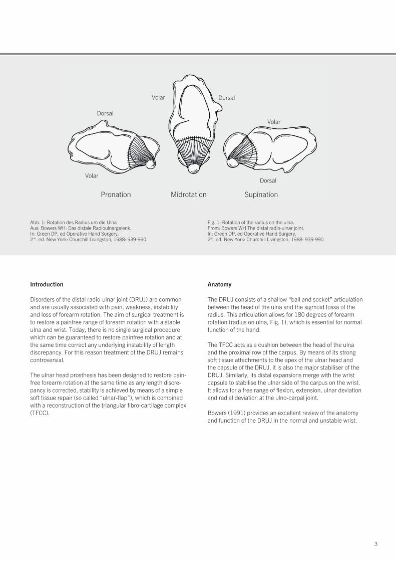
Das distale Radioulnargelenk ist ein flachschaliges Kugel -gelenk zwischen dem Ulnakopf und der Incisura ulnaris desRadius. Dieses Gelenk ermöglicht eine Unterarmrotationvon 180° (Radius um Ulna, Abb. 1), die für den normalen,uneingeschränkten Gebrauch der Hand entscheidend ist.
Der ulnokarpale Bandkomplex fungiert als Puffer zwischenUlnakopf und proximaler Handwurzelreihe. Seine starkenAnsatzstellen an der Basis des Processus styloideus ulnaeund der Kapsel des distalen Radioulnargelenkes machen ihn zusätzlich zum Hauptstabilisator des distalen Radio -ulnargelenkes. In ähnlicher Weise gehen seine distalen Aus-läufer in die Handgelenkskapsel über und bewirken so eineulnarseitige Stabilisierung der Handwurzel am Handgelenk.Dies ermöglicht am ulnokarpalen Gelenk eine freie Beugung,Streckung, ulnare und radiale Deviation.
Bowers (1991) gibt einen hervorragenden Überblick über die anatomischen Verhältnisse mit einer Beschreibung derFunktion des distalen Radioulnargelenkes bei normalemsowie instabilem Handgelenk.
Störungen des distalen Radioulnargelenkes und Notwendigkeit einer UlnakopfprotheseTimothy -J. Herbert/Jörg van Schoonhoven
Disorders of the Distal Radio-Ulnar Joint and the Need for a New Ulnar Head ProsthesisTimothy -J. Herbert/Jörg van Schoonhoven
UHP: Herbert-Ulnakopfprothese®UHP: Herbert Ulnar Head Prosthesis®
3
Dorsal
Dorsal
Volar
Volar
DorsalVolar
Pronation Midrotation Supination
Abb. 1: Rotation des Radius um die UlnaAus: Bowers WH: Das distale Radioulnargelenk. In: Green DP, ed Operative Hand Surgery. 2nd. ed. New York: Churchill Livingston, 1988: 939-990.
Fig. 1: Rotation of the radius on the ulna. From: Bowers WH The distal radio-ulnar joint. In: Green DP, ed Operative Hand Surgery. 2nd. ed. New York: Churchill Livingston, 1988: 939-990.
Introduction
Disorders of the distal radio-ulnar joint (DRUJ) are commonand are usually associated with pain, weakness, instabilityand loss of forearm rotation. The aim of surgical treatment isto restore a painfree range of forearm rotation with a stableulna and wrist. Today, there is no single surgical procedurewhich can be guaranteed to restore painfree rotation and atthe same time correct any underlying instability of lengthdiscrepancy. For this reason treatment of the DRUJ remainscontroversial.
The ulnar head prosthesis has been designed to restore pain-free forearm rotation at the same time as any length discre-pancy is corrected, stability is achieved by means of a simplesoft tissue repair (so called “ulnar-flap”), which is combinedwith a reconstruction of the triangular fibro-cartilage complex(TFCC).
Anatomy
The DRUJ consists of a shallow “ball and socket” articulationbetween the head of the ulna and the sigmoid fossa of theradius. This articulation allows for 180 degrees of forearmrotation (radius on ulna, Fig. 1), which is essential for normalfunction of the hand.
The TFCC acts as a cushion between the head of the ulna and the proximal row of the carpus. By means of its strongsoft tissue attachments to the apex of the ulnar head and the capsule of the DRUJ, it is also the major stabiliser of theDRUJ. Similarly, its distal expansions merge with the wristcapsule to stabilise the ulnar side of the carpus on the wrist. It allows for a free range of flexion, extension, ulnar deviationand radial deviation at the ulno-carpal joint.
Bowers (1991) provides an excellent review of the anatomyand function of the DRUJ in the normal and unstable wrist.
4
UHP: Herbert-Ulnakopfprothese®UHP: Herbert Ulnar Head Prosthesis®
Erkrankungen des distalen Radio ulnargelenkes
Jeder Kongruitätsverlust zwischen Incisura ulnaris des Radiusund Ulnakopf hat eine schmerzhafte Einschränkung derUnterarmdrehung zur Folge und führt in der Regel langfristigzur Arthrose des distalen Radioulnargelenkes. Dies kann verschiedene Ursachen haben: angeborene Deformi täten wiedie Madelung’sche Deformität, Radiusfrakturen, rheumatoideArthritis oder degenerative Arthrose als Folge von Einrissenim ulnokarpalen Bandkomplex.
Einrisse im ulnokarpalen Bandkomplex treten extrem häufigauf, vor allem bei Patienten mit einer Ulna-Plus-Situation(81 %, Palmer und Werner 1981). Solche Einrisse können von einem akuten oder chronischen Trauma herrühren undverursachen Schmerzen und Bewegungsverlust am ulnarenHandgelenk.Ein Kontinuitätsverlust des ulnokarpalen Bandkomplexes führtzu einer Instabilität sowohl des distalen Radioulnargelenkesals auch des ulnokarpalen Gelenkes. Im Laufe der Zeit ent-wickelt sich daraus zumeist eine fixierte Deformität mit derKonsequenz einer sekundären Arthrose.
Mit zunehmendem Funktionsverlust des ulnokarpalen Band-komplexes geht das normale Alignement von Radius undUlna verloren. Die dadurch bedingte radiologisch zunehmen-de Ulna-Plus-Situation am ulnokarpalen Handgelenk führtzum Anschlagen des Ulnakopfes gegen den Os triquetrumund Os lunatum. Dieses sogenannte Impaction-Syndrom kanneine lunotriquetrale Instabilität mit ulnokarpaler Arthrose hervorrufen. Die häufigste Ursache für diesen Prozess stelltdie distale Radiusfraktur dar. Die gleichzeitige Abrissfrakturdes Processus styloideus ulnae kann zu einer funktionellenInstabilität des ulnokarpalen Bandkomplexes führen. Am häufigsten führt jedoch eine in Fehlstellung verheilte distaleRadiusfraktur zu einer sekundären Ulna-Plus-Deformität.Gelegentlich kann zusätzlich die direkte Be schä digung der Incisura ulnaris zur Entwicklung einer sekundären Arthrosedes distalen Radioulnargelenkes führen.
Eine weitere Ursache ist die traumatische Luxation desdistalen Radioulnargelenkes, gelegentlich in Verbindung miteiner Radiusköpfchen-Fraktur und Verletzung der Membranainter-ossea (Essex-Lopresti-Läsion).
Andere Ursachen, die die Funktion des distalen Radioulnar-gelenkes und ulnokarpalen Bandkomplexes betreffen, sindWachstumsstörungen (Madelung’sche Deformität), primäreArthrose des distalen Radioulnargelenkes, Stoffwechsel -störungen (z.B. Gicht) oder die rheumatoide Arthritis.
Zudem zeigt sich bei vielen Patienten eine schmerzhafteIn stabilität infolge vorausgegangener Operationen am distalenRadioulnargelenk.
Disorders of the DRUJ
Any loss of congruity between the sigmoid fossa and the ulnar head will result in painful loss of forearm rotation. Causes include congenital abnormalities such as Madelung’sdeformity, radial fractures, inflammatory arthritis or degene-rative arthrosis as a consequence of tears of the TFCC.
Tears of the TFCC are extremely common, particularly in patients with ulnar plus variance (81%, Palmer and Werner1981). Tears may be due to acute or chronic trauma, andlead to pain and loss of motion in the ulnar side of the wrist;loss of integrity of the TFCC results in instability of both theDRUJ and the ulno-carpal joint. In time, this is likely to become a fixed deformity leading to secondary osteoarthritis.
As the TFCC fails, the normal alignment between the distalradius and ulna is lost and x-rays will show evidence of increa-sing ulnar plus deformity to the extent that the ulna starts toimpact on the lunate and/or triquetrum. This impaction mayresult in triquetro-lunate instability and ulno-carpal osteoar-thritis.The commonest cause of these problems is fracture of thedistal radius. This may be associated with avulsion fracture of the ulnar styloid process, leading to functional instability of the TFCC. More commonly, radial malunion results in asecondary ulnar plus deformity. Occasionally direct damage to the sigmoid fossa may lead to the development of secon-dary osteo-arthritis.
Furthermore trauma may result in acute dislocation of theDRUJ itself, often in association with radial head fracture and tear of the interosseous membrane (Essex-Lopresti lesion).
Other conditions affecting the DRUJ and TFCC include growth disorders (Madelung’s deformity), primary osteo -arthritis, metabolic disorders (e.g. gout), rheumatoid arthritis.Finally, many patients present with painful instability follo-wing previous surgical procedures on the DRUJ.
5
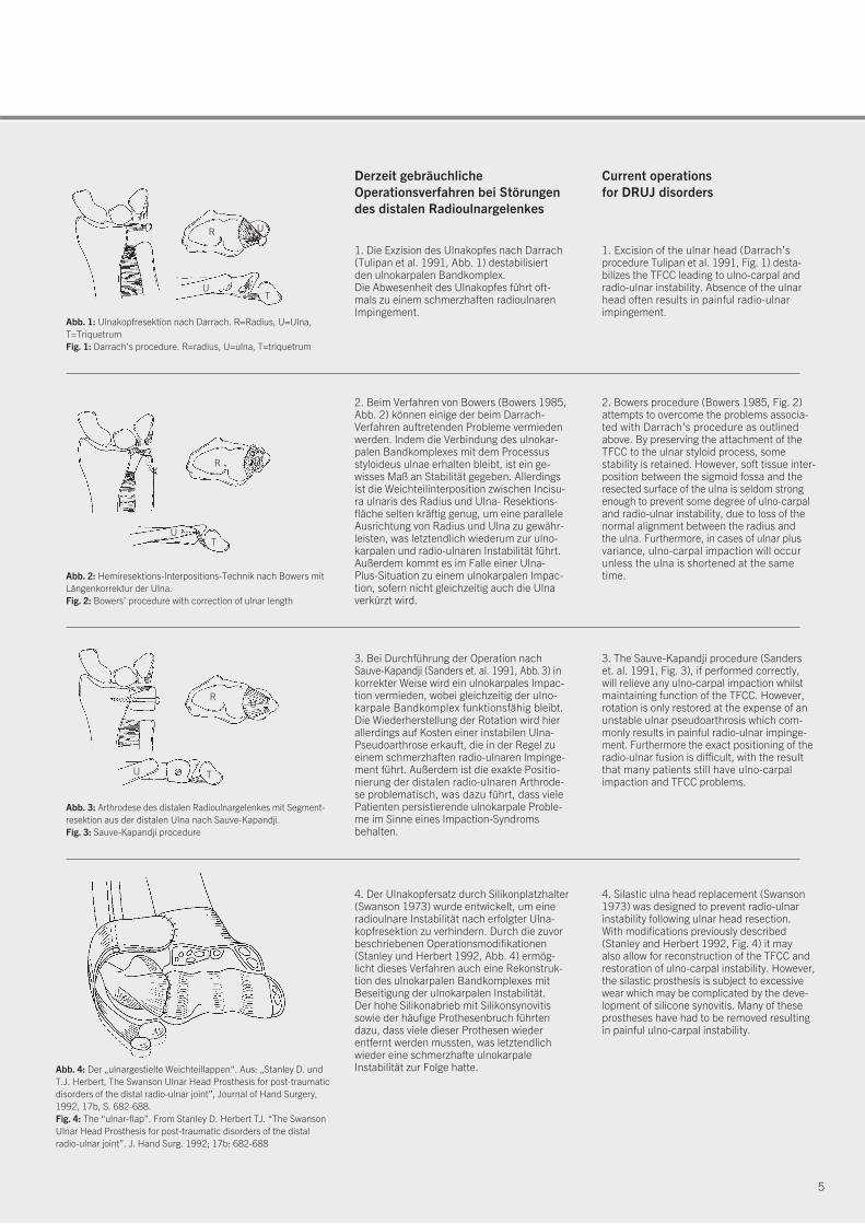
UU
1. Die Exzision des Ulnakopfes nach Darrach(Tulipan et al. 1991, Abb. 1) destabilisiert den ulnokarpalen Bandkomplex. Die Abwesenheit des Ulnakopfes führt oft-mals zu einem schmerzhaften radioulnarenImpingement.
Derzeit gebräuchliche Operationsverfahren bei Störungen des distalen Radioulnargelenkes
2. Beim Verfahren von Bowers (Bowers 1985,Abb. 2) können einige der beim Darrach-Verfahren auftretenden Probleme vermiedenwerden. Indem die Verbindung des ulnokar-palen Bandkomplexes mit dem Processus styloideus ulnae erhalten bleibt, ist ein ge-wisses Maß an Stabilität gegeben. Allerdingsist die Weichteilinterposition zwischen Incisu-ra ulnaris des Radius und Ulna- Resektions-fläche selten kräftig genug, um eine paralleleAusrichtung von Radius und Ulna zu gewähr-leisten, was letztendlich wiederum zur ulno-karpalen und radio-ulnaren Instabilität führt.Außerdem kommt es im Falle einer Ulna-Plus-Situation zu einem ulnokarpalen Impac-tion, sofern nicht gleichzeitig auch die Ulnaverkürzt wird.
3. Bei Durchführung der Operation nach Sauve-Kapandji (Sanders et. al. 1991, Abb.3) inkorrekter Weise wird ein ulnokarpales Impac-tion vermieden, wobei gleichzeitig der ulno-karpale Bandkomplex funktionsfähig bleibt.Die Wiederherstellung der Rotation wird hierallerdings auf Kosten einer instabilen Ulna-Pseudoarthrose erkauft, die in der Regel zueinem schmerzhaften radio-ulnaren Impinge-ment führt. Außerdem ist die exakte Positio-nierung der distalen radio-ulnaren Arthrode-se problematisch, was dazu führt, dass vielePatienten persistierende ulnokarpale Proble-me im Sinne eines Impaction-Syndromsbehalten.
4. Der Ulnakopfersatz durch Silikonplatzhalter(Swanson 1973) wurde entwickelt, um eineradioulnare Instabilität nach erfolgter Ulna-kopfresektion zu verhindern. Durch die zuvorbeschriebenen Operationsmodifikationen(Stanley und Herbert 1992, Abb. 4) ermög-licht dieses Verfahren auch eine Re konstruk-tion des ulnokarpalen Bandkomplexes mitBeseitigung der ulnokarpalen Instabilität. Der hohe Silikonabrieb mit Silikon synovitissowie der häufige Prothesenbruch führtendazu, dass viele dieser Prothesen wieder entfernt werden mussten, was letzt endlichwieder eine schmerzhafte ulnokarpale Instabilität zur Folge hatte.
Abb. 1: Ulnakopfresektion nach Darrach. R=Radius, U=Ulna,T=TriquetrumFig. 1: Darrach’s procedure. R=radius, U=ulna, T=triquetrum
R
TU
U
Abb. 2: Hemiresektions-Interpositions-Technik nach Bowers mitLängenkorrektur der Ulna.Fig. 2: Bowers’ procedure with correction of ulnar length
R
UT
UU
Abb. 3: Arthrodese des distalen Radioulnargelenkes mit Segment -resektion aus der distalen Ulna nach Sauve-Kapandji.Fig. 3: Sauve-Kapandji procedure
R
U T
Abb. 4: Der „ulnargestielte Weichteillappen“. Aus: „Stanley D. undT.J. Herbert, The Swanson Ulnar Head Prosthesis for post-traumaticdisorders of the distal radio-ulnar joint”, Journal of Hand Surgery,1992, 17b, S. 682-688.Fig. 4: The “ulnar-flap”. From Stanley D. Herbert TJ. “The SwansonUlnar Head Prosthesis for post-traumatic disorders of the distalradio-ulnar joint”. J. Hand Surg. 1992; 17b: 682-688
1. Excision of the ulnar head (Darrach’s procedure Tulipan et al. 1991, Fig. 1) desta-bilizes the TFCC leading to ulno-carpal andradio-ulnar instability. Absence of the ulnarhead often results in painful radio-ulnarimpingement.
Current operations for DRUJ disorders
2. Bowers procedure (Bowers 1985, Fig. 2)attempts to overcome the problems associa-ted with Darrach’s procedure as outlinedabove. By preserving the attachment of theTFCC to the ulnar styloid process, some stability is retained. However, soft tissue inter-position between the sigmoid fossa and theresected surface of the ulna is seldom strongenough to prevent some degree of ulno-carpaland radio-ulnar instability, due to loss of thenormal alignment between the radius and the ulna. Furthermore, in cases of ulnar plusvariance, ulno-carpal impaction will occurunless the ulna is shortened at the sametime.
3. The Sauve-Kapandji procedure (Sanderset. al. 1991, Fig. 3), if performed correctly,will relieve any ulno-carpal impaction whilstmaintaining function of the TFCC. However,rotation is only restored at the expense of anunstable ulnar pseudoarthrosis which com -mon ly results in painful radio-ulnar impinge -ment. Furthermore the exact positioning of theradio-ulnar fusion is difficult, with the resultthat many patients still have ulno- carpalimpaction and TFCC problems.
4. Silastic ulna head replacement (Swanson1973) was designed to prevent radio-ulnarinstability following ulnar head resection. With modifications previously described (Stanley and Herbert 1992, Fig. 4) it may also allow for reconstruction of the TFCC andrestoration of ulno-carpal instability. However,the silastic prosthesis is subject to excessivewear which may be complicated by the deve-lopment of silicone synovitis. Many of theseprostheses have had to be removed resultingin painful ulno-carpal instability.
6
UHP: Herbert-Ulnakopfprothese®UHP: Herbert Ulnar Head Prosthesis®
Die zur Zeit von den meisten Chirurgen favorisierten Verfahren sindsicherlich die HIT nach Bowers sowie die Arthrodese des distalenRadioulnargelenkes mit Segmentresektion aus der Ulna nach Sauve-Kapandji. Die Indikation zur Ulnakopfresektion nach Darrach ist ledig-lich noch für Patienten mit geringen Anforderungen an das Handgelenkgegeben (z.B. bei rheumatoider Arthritis). Die Swanson-Prothese findetaufgrund der zuvor geschilderten Problematik keine Anwendung mehr.
Bei korrekter Ausführung und bei richtiger Patientenselektion könnensicherlich alle der vorher beschriebenen Verfahren zufriedenstellendeErgebnisse erbringen. Viele Patienten sind jedoch mit den Operations-ergebnissen unzufrieden und erwarten entsprechend ihren Beschwer-den eine Lösung, welche die Stabilität und Kraft wiederherstellt undeine schmerzfreie Unterarmdrehung ermöglicht. Aus diesen Gründenhaben wir eine neue Ulnakopfprothese mit einem rekonstruktivenOperationsverfahren entwickelt, welches die Revision unbefriedigen-der Ergebnisse nach vorangegangenen Eingriffen am distalen Radioul-nar-gelenk ermöglicht und die Problematik bei Verwendung der bisher zur Geltung kommenden Operationsverfahren vermeidet. Bei diesemVerfahren wird die Funktion des ulnokarpalen Bandkomplexes wieder-hergestellt und die Prothese wird über einen ulnargestielten Kapsel-Retinakulum-Weichteillappen stabilisiert.
Entwicklung der Prothese
Zunächst wurden vom Autor in Zusammenarbeit mit dem Department of Biomechanical Engineering der Universität von New South Wales bio-mechanische Studien durchgeführt. Anschließend wurde anhand vonröntgenologischen und computertomographischen Messungen an 100normalen Handgelenken die notwendigen Prothesenschaft- und Kopf-größen ermittelt. Dabei ergab sich, dass drei unterschiedliche Schaft-durchmesser und drei verschiedene Kopfgrößen genügen, um die gesamte Bandbreite abzudecken. Im Rahmen dieser Untersuchungen ergab sich die Notwendigkeit für eine Revisionsprothese mit aufgebauterProthesen-Halslänge, um eine durch vorangegangene Operationen starke Verkürzung der Ulna auszugleichen.
Als beste Lösung erschien eine zementfreie Verankerung des Prothesen-schaftes in der Ulna. Die Schaftoberfläche wurde durch eine aufgerauhteReintitanbeschichtung vergrößert, so dass eine Osteointegration geför-dert wird. Um eine beliebige Kombination der verschiedenen Schaft- undKopfgrößen zu ermöglichen, können die beiden Kompo-nenten aufgrundder Konusverbindung wahlweise zusammengesetzt werden. Der Kopf istgrößenmäßig der Incisura ulnaris angepasst und stellt die parallele Aus-richtung von Radius und Ulna wieder her. Gleichzeitig verschafft der Ulna-kopf dem rekonstruierten Weichteillappen die notwendige Spannung,damit dieser seine stabilisierende Funktion wahrnehmen kann. Dasdistale Kopfende ist konkav geformt, um übermäßigen Druck auf dieUnterseite des ulnokarpalen Bandkomplexes zu vermeiden. Als Materialfür den Prothesenkopf wurde Keramik gewählt, weil es die größte Biover-träglichkeit und für eine Hemiresektionsarthroplastik die besten biome-chanischen Voraussetzungen erfüllt.
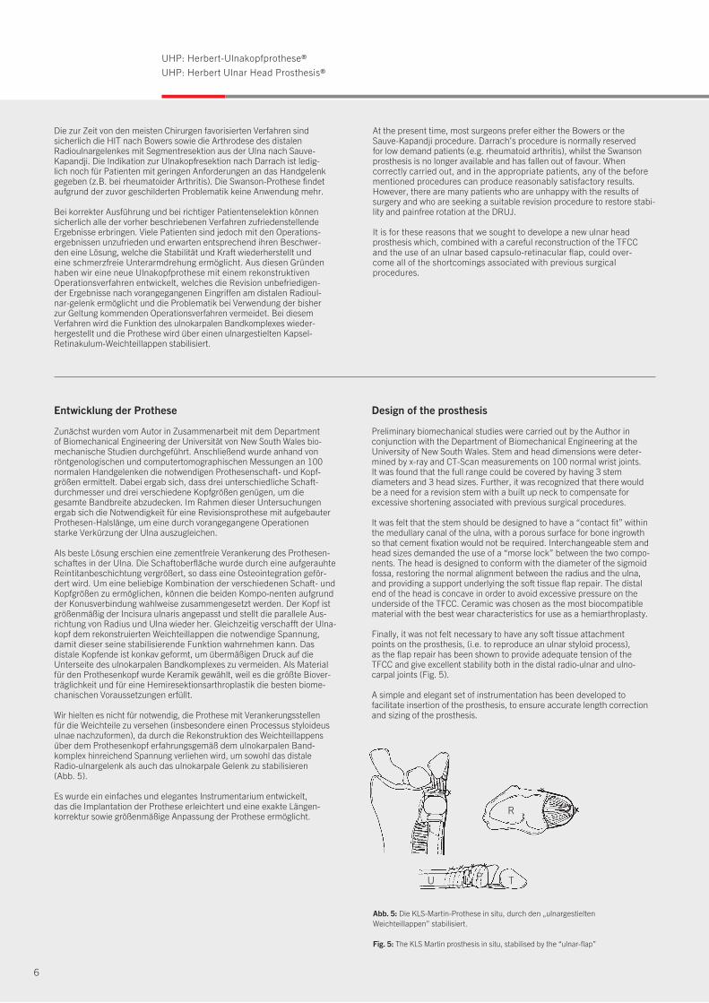
Wir hielten es nicht für notwendig, die Prothese mit Verankerungsstellen für die Weichteile zu versehen (insbesondere einen Processus styloideusulnae nachzuformen), da durch die Rekonstruktion des Weichteillappensüber dem Prothesenkopf erfahrungsgemäß dem ulnokarpalen Band-komplex hinreichend Spannung verliehen wird, um sowohl das distaleRadio-ulnargelenk als auch das ulnokarpale Gelenk zu stabilisieren (Abb. 5).
Es wurde ein einfaches und elegantes Instrumentarium entwickelt, das die Implantation der Prothese erleichtert und eine exakte Längen-korrektur sowie größenmäßige Anpassung der Prothese ermöglicht.
Abb. 5: Die KLS-Martin-Prothese in situ, durch den „ulnargestielten Weichteillappen” stabilisiert.
Fig. 5: The KLS Martin prosthesis in situ, stabilised by the “ulnar-flap”
R
U TPP
At the present time, most surgeons prefer either the Bowers or the Sauve-Kapandji procedure. Darrach’s procedure is normally reserved for low demand patients (e.g. rheumatoid arthritis), whilst the Swansonprosthesis is no longer available and has fallen out of favour. When correctly carried out, and in the appropriate patients, any of the beforementioned procedures can produce reasonably satisfactory results.However, there are many patients who are unhappy with the results ofsurgery and who are seeking a suitable revision procedure to restore stabi-lity and painfree rotation at the DRUJ.
It is for these reasons that we sought to develope a new ulnar headprosthesis which, combined with a careful reconstruction of the TFCC and the use of an ulnar based capsulo-retinacular flap, could over-come all of the shortcomings associated with previous surgical procedures.
Design of the prosthesis
Preliminary biomechanical studies were carried out by the Author in conjunction with the Department of Biomechanical Engineering at the University of New South Wales. Stem and head dimensions were deter-mined by x-ray and CT-Scan measurements on 100 normal wrist joints.It was found that the full range could be covered by having 3 stemdiameters and 3 head sizes. Further, it was recognized that there would be a need for a revision stem with a built up neck to compensate forexcessive shortening associated with previous surgical procedures.
It was felt that the stem should be designed to have a “contact fit” withinthe medullary canal of the ulna, with a porous surface for bone ingrowth so that cement fixation would not be required. Interchangeable stem andhead sizes demanded the use of a “morse lock” between the two compo-nents. The head is designed to conform with the diameter of the sigmoidfossa, restoring the normal alignment between the radius and the ulna,and providing a support underlying the soft tissue flap repair. The distalend of the head is concave in order to avoid excessive pressure on theunderside of the TFCC. Ceramic was chosen as the most biocompatiblematerial with the best wear characteristics for use as a hemiarthroplasty.
Finally, it was not felt necessary to have any soft tissue attachment points on the prosthesis, (i.e. to reproduce an ulnar styloid process), as the flap repair has been shown to provide adequate tension of the TFCC and give excellent stability both in the distal radio-ulnar and ulno-carpal joints (Fig. 5).
A simple and elegant set of instrumentation has been developed to facilitate insertion of the prosthesis, to ensure accurate length correctionand sizing of the prosthesis.
7
Van Schoonhoven J, Lanz U:Rettungsoperationen und deren Differenzialindikation am distalen RadioulnargelenkOrthopäde 2004; 33: 704-714
Van Schoonhoven J, Herbert TJ, Fernandez DL, Prommersberger KJ, Krimmer H:UlnakopfprotheseOrthopäde 2003; 32: 809-815
Pillukat T, Stütz N, Van Schoonhoven J, Krimmer H: Hangelenk und FingergelenkeOP-JOURNAL 2003; 19: 210-215
Barisani G, Schwendenwein E, Vécsei V:Die Ulnakopfprothese nach Timothy J. Herbert.Osteosynthese International 2001; 9: 124-127.
Van Schoonhoven J, Herbert TJ, Krimmer H:Neue Konzepte der Endoprothetik des distalen RadioulnargelenksHandchir. Mikrochir. Plast. Chir. 30 1998; 387-392
■
■
■
■
■
Herbert TJ, van Schoonhoven J: Ulnar Head Replacement Techniques in Hand and Upper Extremity Surgery 2007;11(1):98-108
Van Schoonhoven, Herbert TJ:The Dorsal Approach to the Distal Radioulnar JointTechniques in Hand and Upper Extremity Surgery 2004; 8(1): 11-15
Grechening W, Peicha G, Fellinger M:Primary Ulnar Head Prosthesis for Treatment of an Irreparable Ulnar Head Fracture Dislocation.Journal of Hand Surgery 2001; 26B: 3: 269-271.
Herbert TJ, van Schoonhoven J:Ulnar Head Prosthesis: A new solution for problems at the distal radioulnar joint.Hand Anthroplasties 2000; 145-149.
van Schoonhoven J, Fernandez DL, Bowers WH, Herbert TJ:Salvage of Failed Resection Arthroplasties of the Distal Radio-ulnar Joint using a new Ulnar Head Prosthesis.Journal of Hand Surgery 2000; 25-A: 3: 438-446.
Fernandez DL:Acute and Chronic Derangement of the Distal Radio Ulnar Joint after Fractures of the Radius.EFORT 1999; 41-53
Stanley D, Herbert TJ: The Swanson ulnar head prosthesis for post-traumatic disorders of the distal radio-ulnar joint. Journal of Hand Surgery 1992; 17B: 682-688.
Bowers WH: Instability of the distal radio-ulnar articulation. Hand Clinics 1991; 7: 311-327.
Sanders RA, Frederick HA, Hontas RB: The Sauve-Kapandji procedure: A salvage operation for the distal radio-ulnar joint. Journal of Hand Surgery 1991; 1125-1129.
Tulipan DJ, Eaton RG, Eberhart RE.: The Darrach procedure defended: Technique redefined and long-term follow up. Journal of Hand Surgery 1991; 16A: 438-444.
Bowers WH: Distal radio-ulnar joint arthroplasty: The hemiresection-interposition technique. Journal of Hand Surgery 1985; 10A: 169-178.
Palmer AK, Werner FW: The triangular fibrocartilage complex of the wrist – Anatomy and function. Journal of Hand Surgery 1981; 6: 153-162.
Swanson AB: Implant arthroplasty for disabilities of the distal radio-ulnarjoint: Use of a silicone rubber capping implant following resection of the ulnar head. Orthop. Clinics 1973; 4: 373-382.
■
■
■
■
■
■
■
■
■
■
■
■
■
Literatur Reference List
8
OperationsverfahrenOperative Technique
Ulnakopfprothese bei Störungendes distalen Radioulnargelenkes
Indikationen
■ Revision bei unbefriedigenden Resultaten nach: - Darrach-Operation - Bowers-Operation - Sauve-Kapandji-Operation
■ Primäre Arthrose
■ Posttraumatische Arthrose nach: - Radiusfrakturen - Einrissen im ulnokarpalen Bandkomplex - Ulna-Impaction-Syndrom
■ Rheumatoide Arthritis
■ Tumore
Kontraindikationen
■ Ausgeprägte Radiusdeformität
■ Ungenügende Knochenstruktur
■ Insuffiziente Weichteilverhältnisse
Präoperative Untersuchung
Es ist eine sorgfältige klinische Untersuchung erforderlich, um das Ausmaß der Ulna- und/oder Karpusinstabilität exaktfestzustellen. Eine fixierte Deformität oder Dislokation musserkannt werden, da diese bei der Operation korrigiert werdenmuss. Bei Vorliegen einer dorsalen oder palmaren Subluxationder Ulna aufgrund einer Radiusfehlstellung oder Radius-deformität muss diese mit einer entsprechenden Radius-korrekturosteotomie korrigiert werden.
Vor der Operation sind normale 90/90-Röntgenaufnahmenvon beiden Unterarmen und Handgelenken anzufertigen.Anhand der geeigneten Röntgenschablone (unter Berück-sichtigung eines Vergrößerungsfaktors von 1 : 1,1) solltendann das optimale Resektionsniveau sowie die vermutlichbenötigten Kopf- und Schaftgrößen ermittelt werden. Derdirekte Vergleich mit dem gegenseitigen, gesunden Hand-gelenk ist bei der Ermittlung des exakten Resektionsniveaushilfreich.
Ulnar Head Prosthesis for Disorders of the Distal Radio-Ulnar Joint
Indications
■ Salvage of: - Darrach - Bowers - Sauve-Kapandji
■ Primary osteoarthritis
■ Post-traumatic osteoarthritis: - Radial fractures - TFCC tears - Ulnar impingement
■ Rheumatoid Arthritis
■ Tumors
Contraindications
■ Excess radial deformity
■ Inadequate bone stock
■ Inadequate soft tissues
Pre-operative assessment
Careful clinical examination is required in order to assess acutely the extent of instability of the ulna and/or the carpus.Similarly it is important to recognize any fixed deformity or dislocation, as this will need to be corrected at the time of surgery. If dorsal or volar dislocation of the ulnar head is due to an underlying radial deformity, this should be corrected bymeans of an appropriate radial osteotomy before proceeding to ulnar head replacement.
Standard 90/90 degree x-rays of both forearms and wrists have to be obtained preoperatively. The appropriate x-ray template, (allow for magnification factor, normally 1 : 1.1),should then be used to assess optimum resection level and likely size of head and stem required. Comparison with theopposite normal wrist is the most helpful way to determine the appropriate resection level.
9
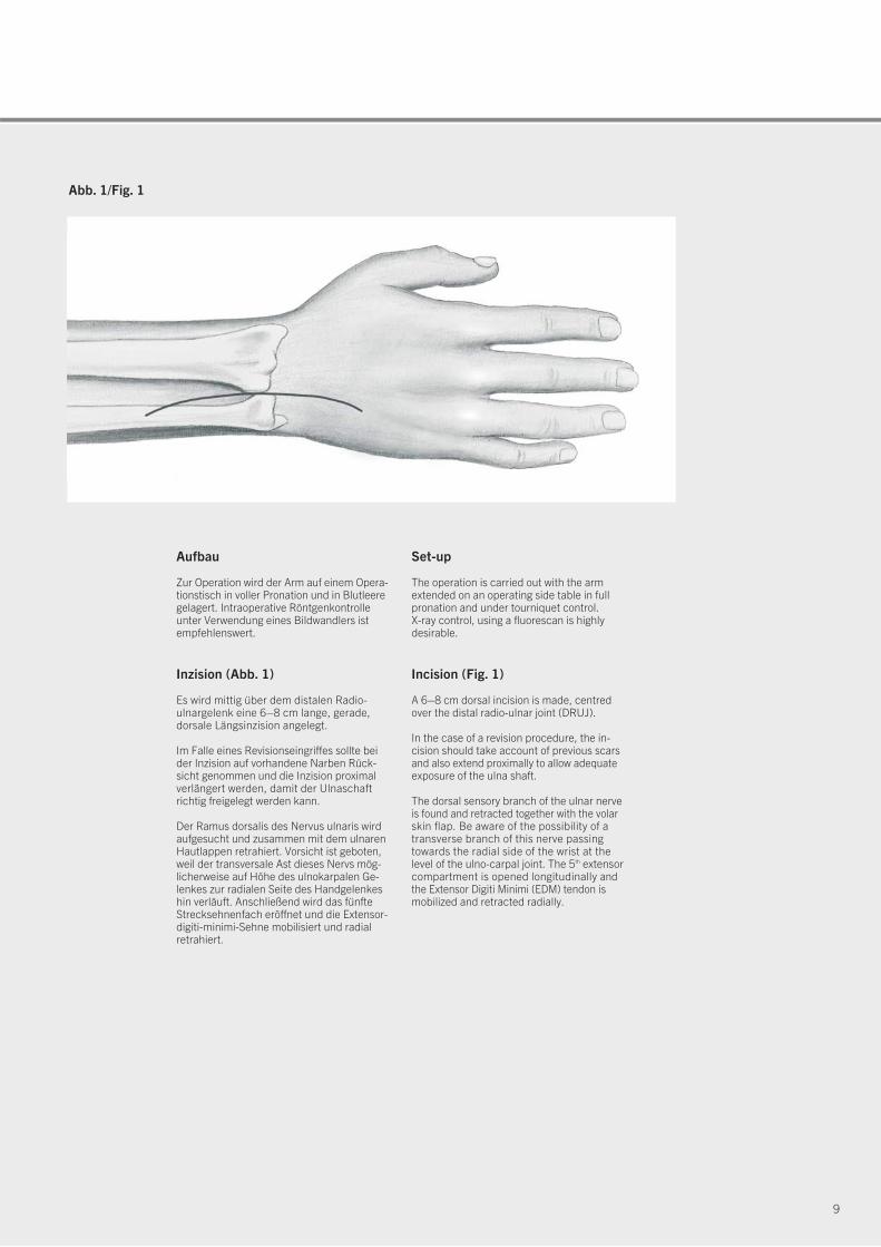
Abb. 1/Fig. 1
Aufbau
Zur Operation wird der Arm auf einem Opera-tionstisch in voller Pronation und in Blutleeregelagert. Intraoperative Röntgenkontrolleunter Verwendung eines Bildwandlers istempfehlenswert.
Inzision (Abb. 1)
Es wird mittig über dem distalen Radio-ulnargelenk eine 6–8 cm lange, gerade, dorsale Längsinzision angelegt.
Im Falle eines Revisionseingriffes sollte beider Inzision auf vorhandene Narben Rück-sicht genommen und die Inzision proximalverlängert werden, damit der Ulnaschaftrichtig freigelegt werden kann.
Der Ramus dorsalis des Nervus ulnaris wirdaufgesucht und zusammen mit dem ulnarenHautlappen retrahiert. Vorsicht ist geboten,weil der transversale Ast dieses Nervs mög -licherweise auf Höhe des ulnokarpalen Ge -lenkes zur radialen Seite des Handgelenkeshin verläuft. Anschließend wird das fünfteStrecksehnenfach eröffnet und die Extensor-digiti-minimi-Sehne mobilisiert und radialretrahiert.
Set-up
The operation is carried out with the armextended on an operating side table in fullpronation and under tourniquet control. X-ray control, using a fluorescan is highlydesirable.
Incision (Fig. 1)
A 6–8 cm dorsal incision is made, centredover the distal radio-ulnar joint (DRUJ).
In the case of a revision procedure, the in -cision should take account of previous scarsand also extend proximally to allow adequateexposure of the ulna shaft.
The dorsal sensory branch of the ulnar nerveis found and retracted together with the volarskin flap. Be aware of the possibility of atrans verse branch of this nerve passingtowards the radial side of the wrist at thelevel of the ulno-carpal joint. The 5th extensorcompartment is opened longitudinally andthe Extensor Digiti Minimi (EDM) tendon ismobilized and retracted radially.
10
OperationsverfahrenOperative Technique
Hebung des Weichteillappens(Abb. 2)
Es wird nun ein Kapsel-Retinakulum-Weich-teilllappen markiert wie in Abb. 2 gezeigt. Die Basis des Lappens liegt über dem Ospisiforme, die Spitze im Mittelpunkt des distalen Radioulnargelenkes. Danach kannmit der Hebung des Lappens begonnen werden, indem zunächst am Rand der Incisura ulnaris die dorsale Kapsel einge-schnitten wird. Der Lappen wird dann distal vom dorsalen Anteil des ulnokarpalenBandkomplexes scharf abpräpariert und proximal über dem distalen Ulnaschaft abgehoben.
Extensor carpi ulnaris
Extensor digiti minimi
Abb. 2/Fig. 2
Der Weichteillappen wird nun zurückgehaltenund mit Haltenähten befestigt, so dass sowohldas distale Radioulnargelenk als auch dasulnokarpale Gelenk offen liegen. Zu beachtenist, dass das sechste Streck sehnenfach(Extensor-carpi-ulnaris-Sehne) nicht eröffnetwerden sollte, da dieses einen wesentlichenAnteil der Lappenbasis bildet (Abb. 3).
Sollte der Boden des sechsten Strecksehnen-faches in Folge degenerativer Prozesse odervorangegangener Operationen eröffnet sein,muss die Kontinuität des Strecksehnenfachesdurch feine Einzelknopfnähte wiederherge-stellt werden, so dass die Extensor-carpi-ulna-ris-Sehne nicht mehr sichtbar in der Lappen-basis verläuft.
Bei Revisionseingriffen werden die Weichteilezwischen der Ulna proximal und dem distalenLappenende in Längsrichtung eröffnet und als ein Narbenweichteilblock nach ulnar ab-präpariert, um das distale Ulnaende freizu-legen.
Stay sutures are used to retract the flap, allowing exposure of both the DRUJ and ulno-carpal joints. Note that the 6th extensorcompartment (Extensor Carpi Ulnaris – ECU)should not be opened as this forms an integral part of the base of the flap (Fig. 3).
If the floor of the 6th compartment is deficient(degenerative process or previous surgery),this should be repaired using fine interruptedmattress sutures so that the ECU tendon is nolonger visible but is contained within the baseof the flap.
For revision procedures the soft tissuesbe tween the ulna proximally and the nearedge of the flap distally, are incised longi-udinally and reflected as one layer to exposethe ulnar stump.
Extensor carpi ulnaris
Extensor digiti minimi
Abb. 3/Fig. 3
Raising the flap (Figure 2)
A capsulo-retinacular flap is marked outas shown in Fig. 2. The base of the flap iscentred on the pisiform bone with its apexat the mid-point of the DRUJ. Start raisingthe flap by incising through the capsulewhere it attaches to the rim of the sigmoidfossa. The flap is then carefully raised bysharp dissection off the dorsal part of theTFCC distally and off the neck of the ulnaproximally.
11
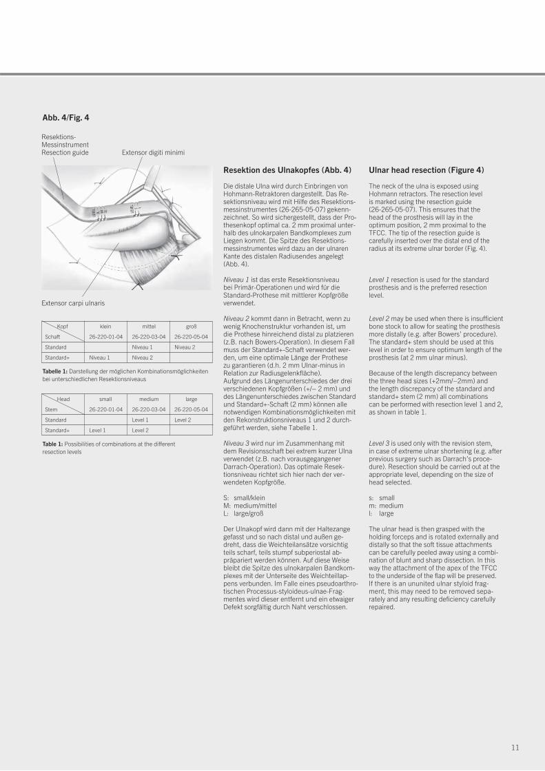
Resektion des Ulnakopfes (Abb. 4)
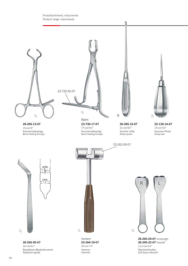
Die distale Ulna wird durch Einbringen vonHohmann-Retraktoren dargestellt. Das Re-sektionsniveau wird mit Hilfe des Resektions-messinstrumentes (26-265-05-07) gekenn-zeichnet. So wird sichergestellt, dass der Pro-thesenkopf optimal ca. 2 mm proximal unter-halb des ulnokarpalen Bandkomplexes zumLiegen kommt. Die Spitze des Resektions-messinstrumentes wird dazu an der ulnarenKante des distalen Radiusendes angelegt(Abb. 4).
Niveau 1 ist das erste Resektionsniveaubei Primär-Operationen und wird für die Standard-Prothese mit mittlerer Kopfgrößeverwendet.
Niveau 2 kommt dann in Betracht, wenn zuwenig Knochenstruktur vorhanden ist, um die Prothese hinreichend distal zu platzieren(z.B. nach Bowers-Operation). In diesem Fallmuss der Standard+-Schaft verwendet wer-den, um eine optimale Länge der Prothese zu garantieren (d.h. 2 mm Ulnar-minus inRelation zur Radiusgelenkfläche).Aufgrund des Längenunterschiedes der dreiverschiedenen Kopfgrößen (+/– 2 mm) unddes Längenunterschiedes zwischen Standardund Standard+-Schaft (2 mm) können allenotwendigen Kombinationsmöglichkeiten mitden Rekonstruktionsniveaus 1 und 2 durch-geführt werden, siehe Tabelle 1.
Niveau 3 wird nur im Zusammenhang mitdem Revisionsschaft bei extrem kurzer Ulnaverwendet (z.B. nach vorausgegangener Darrach-Operation). Das optimale Resek-tionsniveau richtet sich hier nach der ver-wendeten Kopfgröße.
S: small/kleinM: medium/mittelL: large/groß
Der Ulnakopf wird dann mit der Haltezangegefasst und so nach distal und außen ge -dreht, dass die Weichteilansätze vorsichtigteils scharf, teils stumpf subperiostal ab-präpariert werden können. Auf diese Weisebleibt die Spitze des ulnokarpalen Bandkom-plexes mit der Unterseite des Weichteillap-pens verbunden. Im Falle eines pseudoarthro-tischen Processus-styloideus-ulnae-Frag-mentes wird dieser entfernt und ein etwaigerDefekt sorgfältig durch Naht verschlossen.
Ulnar head resection (Figure 4)
The neck of the ulna is exposed using Hohmann retractors. The resection level is marked using the resection guide (26-265-05-07). This ensures that the head of the prosthesis will lay in the optimum position, 2 mm proximal to theTFCC. The tip of the resection guide is carefully inserted over the distal end of theradius at its extreme ulnar border (Fig. 4).
Level 1 resection is used for the standardprosthesis and is the preferred resection level.
Level 2 may be used when there is insufficientbone stock to allow for seating the prosthesismore distally (e.g. after Bowers’ procedure).The standard+ stem should be used at thislevel in order to ensure optimum length of theprosthesis (at 2 mm ulnar minus).
Because of the length discrepancy betweenthe three head sizes (+2mm/–2mm) and the length discrepancy of the standard andstandard+ stem (2 mm) all combinations can be performed with resection level 1 and 2,as shown in table 1.
Level 3 is used only with the revision stem, in case of extreme ulnar shortening (e.g. afterprevious surgery such as Darrach’s proce-dure). Resection should be carried out at theappropriate level, depending on the size ofhead selected.
s: smallm: mediuml: large
The ulnar head is then grasped with the holding forceps and is rotated externally anddistally so that the soft tissue attachmentscan be carefully peeled away using a combi-nation of blunt and sharp dissection. In thisway the attachment of the apex of the TFCCto the underside of the flap will be preserved.If there is an ununited ulnar styloid frag-ment, this may need to be removed sepa -rately and any resulting deficiency carefullyrepaired.
Extensor carpi ulnaris
Extensor digiti minimi
Abb. 4/Fig. 4
Resektions-MessinstrumentResection guide
Tabelle 1: Darstellung der möglichen Kombinationsmöglichkeitenbei unterschiedlichen Resektionsniveaus
Kopf klein mittel groß
Schaft 26-220-01-04 26-220-03-04 26-220-05-04
Standard Niveau 1 Niveau 2
Standard+ Niveau 1 Niveau 2
Table 1: Possibilities of combinations at the different resection levels
Head small medium large
Stem 26-220-01-04 26-220-03-04 26-220-05-04
Standard Level 1 Level 2
Standard+ Level 1 Level 2
12
OperationsverfahrenOperative Technique
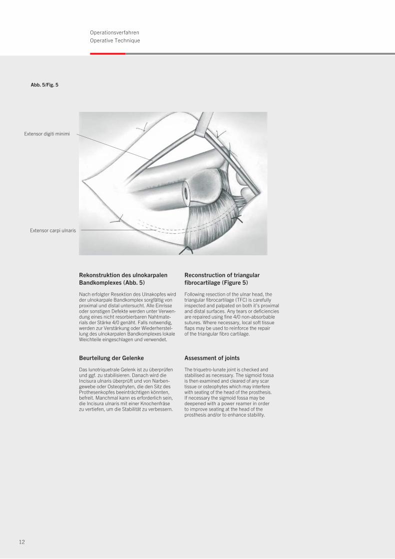
Rekonstruktion des ulnokarpalenBandkomplexes (Abb. 5)
Nach erfolgter Resektion des Ulnakopfes wirdder ulnokarpale Bandkomplex sorgfältig vonproximal und distal untersucht. Alle Einrisseoder sonstigen Defekte werden unter Verwen-dung eines nicht resorbierbaren Nahtmate-rials der Stärke 4/0 genäht. Falls notwendig,werden zur Verstärkung oder Wiederherstel-lung des ulnokarpalen Bandkomplexes lokaleWeichteile eingeschlagen und verwendet.
Beurteilung der Gelenke
Das lunotriquetrale Gelenk ist zu überprüfenund ggf. zu stabilisieren. Danach wird dieIncisura ulnaris überprüft und von Narben-gewebe oder Osteophyten, die den Sitz desProthesenkopfes beeinträchtigen könnten,befreit. Manchmal kann es erforderlich sein,die Incisura ulnaris mit einer Knochenfräse zu vertiefen, um die Stabilität zu verbessern.
Reconstruction of triangular fibrocartilage (Figure 5)
Following resection of the ulnar head, the triangular fibrocartilage (TFC) is carefully inspected and palpated on both it’s proximaland distal surfaces. Any tears or deficienciesare repaired using fine 4/0 non-absorbablesutures. Where necessary, local soft tissueflaps may be used to reinforce the repairof the triangular fibro cartilage.
Assessment of joints
The triquetro-lunate joint is checked and sta bilised as necessary. The sigmoid fossais then examined and cleared of any scar tissue or osteophytes which may interfere with seat ing of the head of the prosthesis. If necessary the sigmoid fossa may be deepened with a power reamer in orderto improve seating at the head of the prosthesis and/or to enhance stability.
Extensor carpi ulnaris
Abb. 5/Fig. 5
Extensor digiti minimi
13
Abb. 6/Fig. 6
transossäreFixierungslöcherperrosseous suture holes
Extensor carpi ulnaris
Extensor digiti minimi
WeichteilretraktorSoft tissue retractor
RaspelReamer
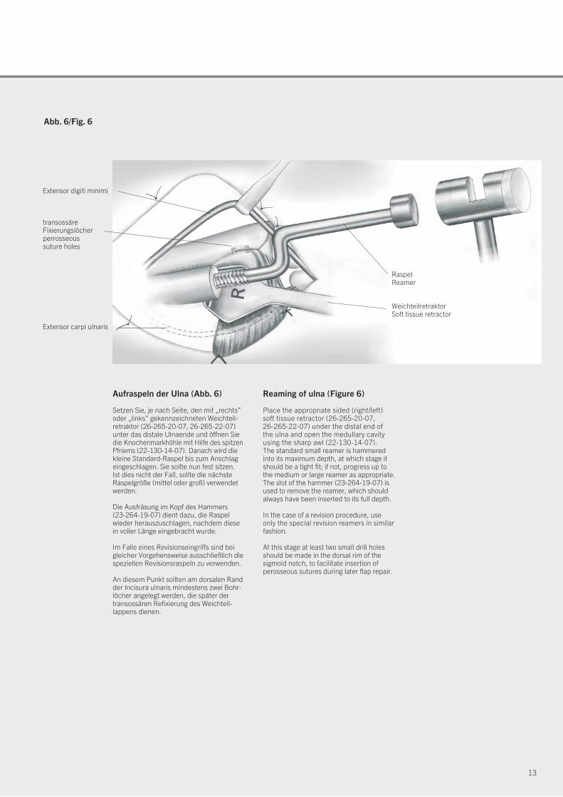
Aufraspeln der Ulna (Abb. 6)
Setzen Sie, je nach Seite, den mit „rechts”oder „links” gekennzeichneten Weichteil-retraktor (26-265-20-07, 26-265-22-07) unter das distale Ulnaende und öffnen Siedie Kno chen markhöhle mit Hilfe des spitzenPfriems (22-130-14-07). Danach wird die kleine Standard-Raspel bis zum Anschlag eingeschlagen. Sie sollte nun fest sitzen. Ist dies nicht der Fall, sollte die nächsteRaspelgröße (mittel oder groß) verwendet werden.
Die Ausfräsung im Kopf des Hammers (23-264-19-07) dient dazu, die Raspel wieder herauszuschlagen, nachdem diese in voller Länge eingebracht wurde.
Im Falle eines Revisionseingriffs sind beigleicher Vorgehensweise ausschließlich diespeziellen Revisionsraspeln zu verwenden.
An diesem Punkt sollten am dorsalen Randder Incisura ulnaris mindestens zwei Bohr-löcher angelegt werden, die später der transossären Refixierung des Weichteil-lappens dienen.
Reaming of ulna (Figure 6)
Place the appropriate sided (right/left) soft tissue retractor (26-265-20-07, 26-265-22-07) under the distal end of the ulna and open the medullary cavityusing the sharp awl (22-130-14-07). The standard small reamer is hammered into its maximum depth, at which stage itshould be a tight fit; if not, progress up tothe medium or large reamer as appropriate.The slot of the hammer (23-264-19-07) isused to remove the reamer, which shouldalways have been inserted to its full depth.
In the case of a revision procedure, use only the special revision reamers in similarfashion.
At this stage at least two small drill holesshould be made in the dorsal rim of the sigmoid notch, to facilitate insertion of perosseous sutures during later flap repair.
14
OperationsverfahrenOperative Technique
Reposition der Probeprothese und Beurteilung
Setzen Sie den geeigneten Probeschaft und -kopf ein. PrüfenSie, ob der Schaft gut sitzt und der Kopf sich in korrekterPosition (etwa 2 mm unterhalb des ulnokarpalen Band -komplexes) befindet. Diese Überprüfung sollte radiologischunter Verwendung des Bildwandlers bei maximaler Pronationdes Handgelenkes verifiziert werden.
Führen Sie dann den Weichteillappen radial über den Ulna-kopf, um zu prüfen, ob er zum einen wieder gut am Radiusbefestigt werden kann und zum anderen für genügend Stabi-lität sorgt. Erscheint der Lappen zu straff, sollte der kleine Kopf auspro-biert werden. Bei Verwendung des kleinen Kopfes muss derStandard-Schaft gegen einen Standard+-Schaft ausgetauschtwerden, damit die Länge der Prothese wieder stimmt.Erscheint der Lappen hingegen zu locker, sollte der großeKopf verwendet werden. In diesem Fall muss dann die Ulnaauf Niveau 2 verkürzt werden, damit eine optimale Prothesen -länge garantiert ist (2 mm Ulnar-minus).
Nun ist die Unterarmrotation zu überprüfen. Findet sich eine Limitierung, muss die Ursache analysiert und beseitigtwerden (z.B. Blockierung durch Osteophyten, kontrakteMembrana interossea, etc.).
Die Probeprothese wird nun, ggf. unter Zuhilfenahme des Explantationsmeißels (26-265-35-07, 26-265-37-07) entfernt und die Wunde und der Knochenmarkkanal gutgespült.
Einbringen der endgültigen Prothese
Nach Auswahl der geeigneten Schaft- (Standard, Standard+oder Revision) und Kopfgröße (klein, mittel oder groß) wirdnun der Schaft mit Hilfe des konischen Einschlaginstrumenta-riums (26-265-30-07) vorsichtig eingebracht. Das konischeEnde des Prothesenhalses sollte sauber und trocken sein,bevor der Keramikkopf durch einen leichten Schlag ange-bracht wird.
An diesem Punkt der Operation ist dann anhand von Röntgen-aufnahmen sicherzustellen, dass die Prothese optimal sitzt.
Warnung:Beim Einschlagen der Prothese sollte niemals übermäßigeKraft angewendet werden. Wenn der Schaft beim Einschla genzu klemmen scheint, sollte er noch einmal entfernt und derMarkraum der Ulna weiter aufgeraspelt werden.
Entfernen der Prothese
Falls die Prothese in Sonderfällen noch einmal herausge-nommen werden muss, kann dazu der im Operationsset vor-handene Schaftextraktor (26-265-07-07) verwendet werden.
Trial reduction and assessment
Insert the appropriate trial stem and head. Check that thestem fits snugly and that the head lies at the appropriatelevel (approx. 2 mm) below the TFCC. This should be checkedfluorescopically with the wrist in full pronation.
Advance the flap radially over the ulnar head to check thatthis can be re-attached to the radius and provide adequatestability. If the flap is too tight, a smaller head should be tried. If this is selected, then the standard stem should be changed to astandard+ stem in order to ensure optimum length of theprosthesis.If the flap is too loose, a larger head may be selected. In thiscase, the ulna must be shortened back to level 2 in order toensure optimum length of the prosthesis (2 mm ulnar minus).
Range of forearm rotation is assessed at this stage, andif it remains limited, the cause should be sought and dealtwith as appropriate (e.g. osteophyte block; contracture ofinterosseous membrane, etc.).
The trial prosthesis is removed, using the explantation chisels (26-265-35-07, 26-265-37-07) and the wound and medullary canal are irrigated.
Inserting the definitive prosthesis
The appropriate stem (standard, standard+ or revision) andhead (small, medium or large) are selected. The stem iscarefully hammered into position using the conical impactor(26-265-30-07). The conical end of the stem should be cleanand dry before the ceramic head is impacted.
At this stage, intra-operative x-rays are used to confirm thatthe prosthesis is in optimum position.
Warning:Excessive force should never be used during insertion of the definitive prosthesis. If the stem appears to “lock” during insertion, it should be removed and further reamingcarried out.
Removal of prosthesis
If for any reason it is necessary to remove the prosthesis,this can be done using the stem extractor (26-265-07-07)
provided in the set.
15
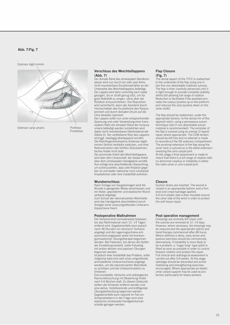
Verschluss des Weichteillappens(Abb. 7)Der dorsale Rand des ulnokarpalen Bandkom-plexes wird nun durch ein oder zwei feine,nicht resorbierbare Einzelknopfnähte an derUnterseite des Weichteillappens befestigt. Der Lappen wird dann vorsichtig nach radialgezogen, bis er straff genug sitzt, um fürgute Stabilität zu sorgen, ohne aber dieRotation einzuschränken. Die Repositionwird vereinfacht, wenn der Assistent durchHochschieben des Os pisiforme den Karpusproniert und durch dorsalen Druck auf dieUlna dieselbe reponiert.Der Lappen sollte nun unter entsprechenderSpannung und unter Verwendung einer trans -ossären Naht am dorsalen Rand der Incisuraulnaris befestigt werden (empfohlen wirddabei nicht resorbierbares Nahtmaterial derStärke 0). Der verbliebene Rest des Lappenswird ggf. zweilagig überlappend vernäht. Die Kleinfingerstrecksehne (Extensor-digiti-minimi-Sehne) verbleibt subkutan, und eineRekonstruktion des fünften Strecksehnen -faches findet nicht statt. Der proximale Anteil des Weichteillappenswird über dem Ulnarschaft, der distale An teilüber dem ulnokarpalen Handgelenk vernäht.Nun erfolgt eine abschließende Überprüfung,um sicherzustellen, dass volle Rotation gege-ben ist und weder radioulnar noch ulnokarpalKrepitationen oder eine Instabilität auftreten.
WundverschlussNach Einlage von Saugdrainagen wird dieWunde in geeigneter Weise verschlossen undein fester, gepolsterter und elastischer Wund-verband angelegt.Zum Schutz der rekonstruierten Weichteilewird das Handgelenk abschließend durchAnlegen einer ulnarumgreifenden Unterarm-Gipsschiene fixiert.
Postoperative MaßnahmenDer Verband wird normalerweise belassen,bis das Nahtmaterial nach 12–14 Tagenentfernt wird. Gegebenenfalls kann jedochnach 48 Stunden ein dünnerer Verbandangelegt und die Lagerungsschiene ent-sprechend angepasst sowie mit kranken-gymnastischer Übungstherapie begonnenwerden. Bei Patienten, bei denen die Gefahrder Einsteifung besteht, sollte frühzeitig mit ersten aktiven und passiven Übungen begonnen werden. Ist jedoch eher Instabilität das Problem, solltemöglichst bald eine weit ulnar-umgreifende,anmodellierte Unterarmschiene angelegt werden, um die rekonstruierten Weichteile zu schützen und die Unterarmrotation zu limitieren.Eine komplette, klinische und radiologischeNachuntersuchung mit Bewertung findetnach 5-6 Wochen statt. Zu diesem Zeitpunktsollten die Schienen entfernt werden und eine aktive, mobilisierende und kräftigendeÜbungsbehandlung begonnen werden. Gege benenfalls kann (speziell im Fall vonSchwer arbeitern) in der Folge noch eine elas tische ulnokarpale Handgelenksman-schette getragen werden.
Flap Closure (Fig. 7)The dorsal aspect of the TFCC is reattachedto the underside of the flap using one or two fine non-absorbable mattress sutures.The flap is then carefully advanced until it is tight enough to provide complete stabilitywhilst still allowing full range of rotation.Reduction is facilitated if the assistant pro-nates the carpus (pushes up on the pisiform)and reduces the ulna (pushes down on theulnar shaft).
The flap should be reattached, under theappropriate tension, to the dorsal rim of thesigmoid notch, using a perosseous suturetechnique (size 0 non-absorbable suturematerial is recommended). The reminder ofthe flap is sutured using an overlap (2 layers)repair where appropriate. The EDM tendonshould be left free and no attempt is made to reconstruct the 5th extensor compartment.The proximal extension of the flap along theulnar neck is sutured as is the distal extensionoverlying the ulno-carpal joint.At this stage a final assessment is made tocheck that there is a full range of rotation withno abnormal crepitus or instability in eitherthe radio-ulnar or ulno-carpal joint.
ClosureSuction drains are inserted. The wound is closed in an appropriate fashion and a firmwool and crepe bandage applied.A 6-inch plaster slab is then moulded aroundthe ulnar side of the wrist in order to protectthe soft tissue repair.
Post operative managementDressings are normally left intact untilthe sutures are removed at 12–14 days.However, where necessary, the dressings maybe reduced and the appropriate splints andhand therapy commenced after 48 hours.Where stiffness is likely, early active and passive exercises should be commenced,alternatively, if instability is more likely to be a problem, a “sugar tong” type splint is fitted as soon as possible in order to controlforearm rotation and protect the repair.Full clinical and radiological assessment iscarried out after 5-6 weeks. At this stagesplintage should be discarded and activemobilising and strengthening exercisesencouraged. Where appropriate an elasticulnar carpal support may be used as pro-tection particularly for heavy workers.
Extensor carpi ulnaris
Extensor digiti minimi
Abb. 7/Fig. 7
ProtheseProsthesis
16
Klinische Beispiele: UHPClinical Examples: UHP
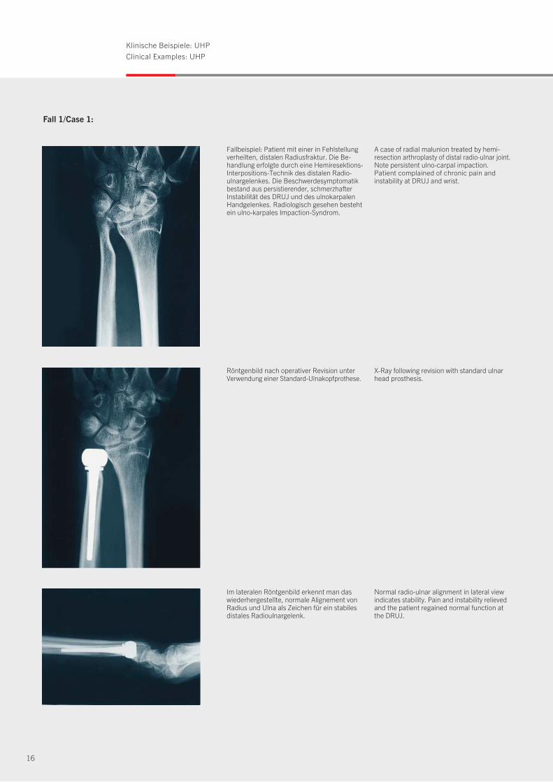
Fallbeispiel: Patient mit einer in Fehlstellungverheilten, distalen Radiusfraktur. Die Be-handlung erfolgte durch eine Hemiresektions-Interpositions-Technik des distalen Radio-ulnargelenkes. Die Beschwerde sympto matikbestand aus persistierender, schmerz hafterInstabilität des DRUJ und des ulnokarpalenHandgelenkes. Radiologisch gesehen bestehtein ulno-karpales Impaction-Syndrom.
Röntgenbild nach operativer Revision unterVerwendung einer Standard-Ulnakopfprothese.
Fall 1/Case 1:
Im lateralen Röntgenbild erkennt man daswiederhergestellte, normale Alignement vonRadius und Ulna als Zeichen für ein stabilesdistales Radioulnargelenk.
A case of radial malunion treated by hemi-resection arthroplasty of distal radio-ulnar joint.Note persistent ulno-carpal impaction.Patient complained of chronic pain andinstability at DRUJ and wrist.
X-Ray following revision with standard ulnar head prosthesis.
Normal radio-ulnar alignment in lateral viewindicates stability. Pain and instability relievedand the patient regained normal function atthe DRUJ.
17
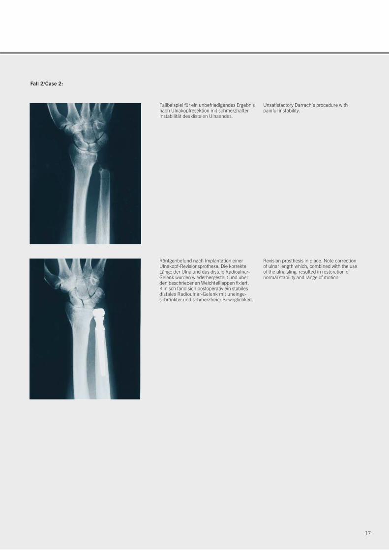
Fall 2/Case 2:
Fallbeispiel für ein unbefriedigendes Ergebnisnach Ulnakopfresektion mit schmerzhafterInstabilität des distalen Ulnaendes.
Röntgenbefund nach Implantation einer Ulnakopf-Revisionsprothese. Die korrekteLänge der Ulna und das distale Radioulnar-Gelenk wurden wiederhergestellt und überden beschriebenen Weichteillappen fixiert.Klinisch fand sich postoperativ ein stabilesdistales Radioulnar-Gelenk mit uneinge-schränkter und schmerzfreier Beweglichkeit.
Unsatisfactory Darrach’s procedure with painful instability.
Revision prosthesis in place. Note correctionof ulnar length which, combined with the useof the ulna sling, resulted in restoration of normal stability and range of motion.
18
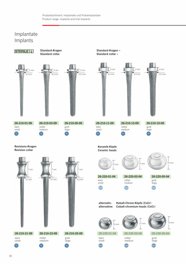
Produktsortiment: Implantate und ProbeimplantateProduct range: Implants and trial implants
10 mm 12 mm 14 mm
Keramik-KöpfeCeramic heads
ImplantateImplants
26-210-21-09
kleinsmall
26-210-23-09
mittelmedium
26-210-25-09
großlarge
26-220-01-04kleinsmall
26-220-03-04mittelmedium
26-220-05-04großlarge
17 mm17 mm17 mm
4,7 mm
10 mm 12 mm 14 mm
Revisions-KragenRevision collar
26-210-01-09kleinsmall
26-210-03-09mittelmedium
26-210-05-09großlarge
26-210-11-09kleinsmall
26-210-13-09mittelmedium
26-210-15-09großlarge
2 mm2 mm2 mm 4 mm4 mm4 mm
Standard-KragenStandard collar
Standard-Kragen +Standard collar +
alternativ: Kobalt-Chrom-Köpfe (CoCr)alternative: Cobalt-chromium heads (CoCr)
26-230-01-04kleinsmall
26-230-03-04mittelmedium
26-230-05-04großlarge
6,7 mm 9,2 mm
6,2 mm 8,2 mm 10,2 mm 6,2 mm 8,2 mm 10,2 mm
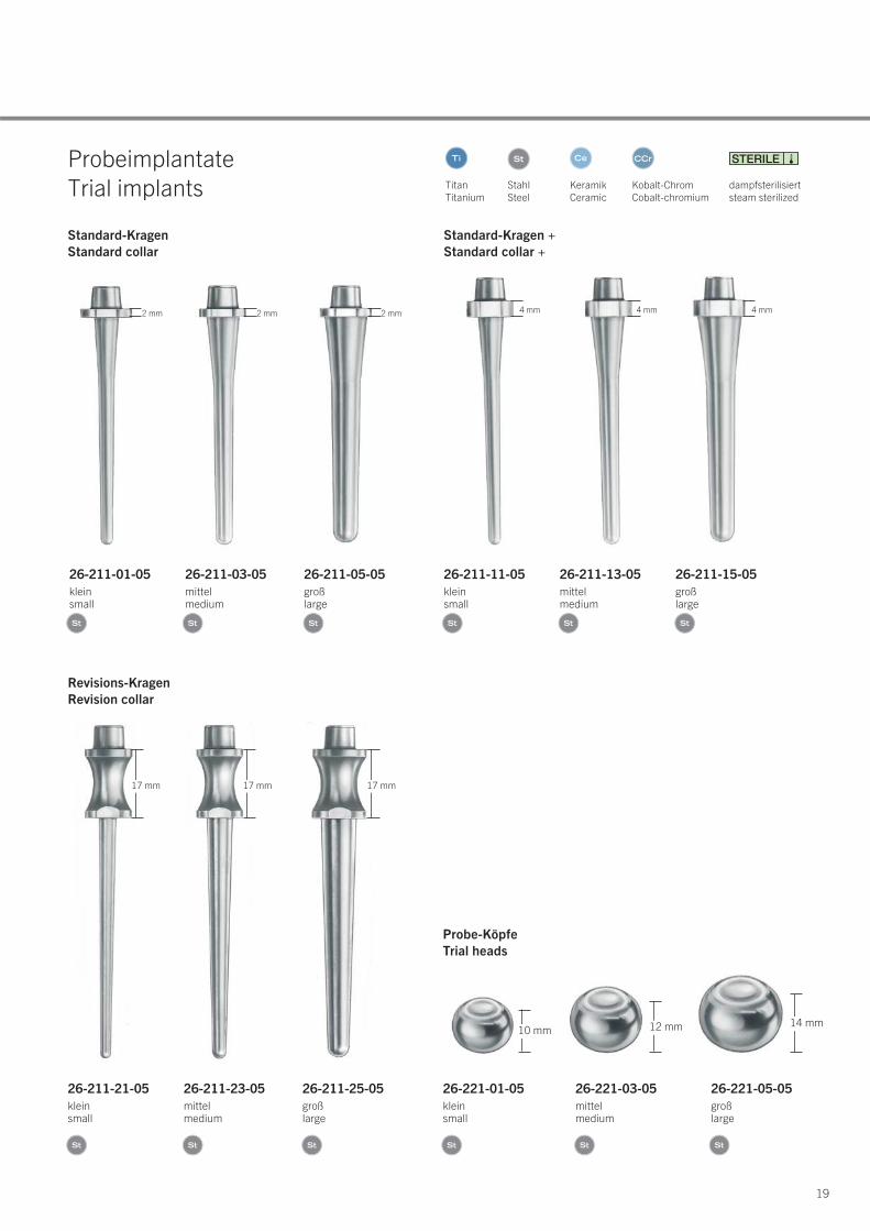
19
Probe-KöpfeTrial heads
26-211-21-05kleinsmall
26-211-23-05mittelmedium
26-211-25-05großlarge
26-221-01-05kleinsmall
26-221-03-05mittelmedium
26-221-05-05großlarge
ProbeimplantateTrial implants
10 mm 12 mm 14 mm
17 mm17 mm17 mm
Revisions-KragenRevision collar
26-211-01-05kleinsmall
26-211-03-05mittelmedium
26-211-05-05großlarge
26-211-11-05kleinsmall
26-211-13-05mittelmedium
26-211-15-05großlarge
2 mm2 mm2 mm 4 mm4 mm4 mm
Standard-KragenStandard collar
Standard-Kragen +Standard collar +
TitanTitanium
StahlSteel
KeramikCeramic
Kobalt-ChromCobalt-chromium
dampfsterilisiertsteam sterilized
20
Produktsortiment: InstrumenteProduct range: Instruments
1⁄2
Herbert23-264-19-0719 cm/ 73⁄4"HammerHammer
1⁄2
22-130-14-0714 cm/ 51⁄2"Knochen-PfriemSharp awl
1⁄2
Kern23-730-17-0717 cm/ 63⁄4"KnochenhaltezangeBone holding forceps
23-730-92-07
1⁄2
26-265-05-0716 cm/ 61⁄4"Resektions-MessinstrumentResection guide
26-265-13-0715 cm/ 6"KnochenhaltezangeBone holding forceps
1⁄21⁄2
26-265-15-0721 cm/ 81⁄4"Scharfer LöffelSharp spoon
1⁄2
26-265-20-07 rechts/right26-265-22-07 links/left11,5 cm/ 43⁄4"WeichteilretraktorSoft tissue retractor
R L
23-262-90-07

21
1⁄2 1⁄2
1⁄2
26-265-07-0722 cm/ 83⁄4"ExtraktorExtractor
Standard-RaspelStandard reamer
Revisions-RaspelRevision reamer
26-250-01-07 kleinsmall
26-250-05-07 großlarge
26-250-03-07mittelmedium
26-260-01-07 kleinsmall
26-260-05-07 großlarge
26-260-03-07mittelmedium
1⁄2
26-265-30-079,5 cm/ 33⁄4"EinschlaginstrumentConical impactor
1⁄2
8 mm10 mm
L S
26-265-35-07 klein/small26-265-37-07 groß/large24,5 cm/ 91⁄2"ExplantationsmeißelExplantation chisel
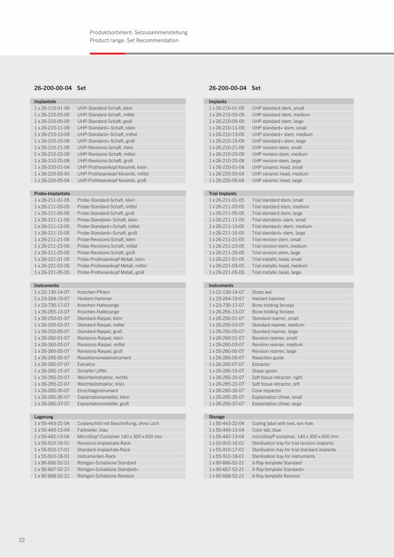
26-200-00-04 Set 26-200-00-04 Set
22
Produktsortiment: SetzusammenstellungProduct range: Set Recommendation
Implantate1 x 26-210-01-09 UHP-Standard-Schaft, klein1 x 26-210-03-09 UHP-Standard-Schaft, mittel1 x 26-210-05-09 UHP-Standard-Schaft, groß1 x 26-210-11-09 UHP-Standard+-Schaft, klein1 x 26-210-13-09 UHP-Standard+-Schaft, mittel1 x 26-210-15-09 UHP-Standard+-Schaft, groß1 x 26-210-21-09 UHP-Revisions-Schaft, klein1 x 26-210-23-09 UHP-Revisions-Schaft, mittel1 x 26-210-25-09 UHP-Revisions-Schaft, groß1 x 26-220-01-04 UHP-Prothesenkopf Keramik, klein1 x 26-220-03-04 UHP-Prothesenkopf Keramik, mittel1 x 26-220-05-04 UHP-Prothesenkopf Keramik, groß
Probe-Implantate1 x 26-211-01-05 Probe-Standard-Schaft, klein1 x 26-211-03-05 Probe-Standard-Schaft, mittel1 x 26-211-05-05 Probe-Standard-Schaft, groß1 x 26-211-11-05 Probe-Standard+-Schaft, klein1 x 26-211-13-05 Probe-Standard+-Schaft, mittel1 x 26-211-15-05 Probe-Standard+-Schaft, groß1 x 26-211-21-05 Probe-Revisions-Schaft, klein1 x 26-211-23-05 Probe-Revisions-Schaft, mittel1 x 26-211-25-05 Probe-Revisions-Schaft, groß1 x 26-221-01-05 Probe-Prothesenkopf Metall, klein1 x 26-221-03-05 Probe-Prothesenkopf Metall, mittel1 x 26-221-05-05 Probe-Prothesenkopf Metall, groß
Instrumente1 x 22-130-14-07 Knochen-Pfriem1 x 23-264-19-07 Herbert-Hammer1 x 23-730-17-07 Knochen-Haltezange1 x 26-265-13-07 Knochen-Haltezange1 x 26-250-01-07 Standard-Raspel, klein1 x 26-250-03-07 Standard-Raspel, mittel1 x 26-250-05-07 Standard-Raspel, groß1 x 26-260-01-07 Revisions-Raspel, klein1 x 26-260-03-07 Revisions-Raspel, mittel1 x 26-260-05-07 Revisions-Raspel, groß1 x 26-265-05-07 Resektionsmessinstrument1 x 26-265-07-07 Extraktor1 x 26-265-15-07 Scharfer Löffel1 x 26-265-20-07 Weichteilretraktor, rechts1 x 26-265-22-07 Weichteilretraktor, links1 x 26-265-30-07 Einschlaginstrument1 x 26-265-35-07 Explantationsmeißel, klein1 x 26-265-37-07 Explantationsmeißel, groß
Lagerung1 x 55-443-22-04 Codierschild mit Beschriftung, ohne Loch1 x 55-443-13-04 Farbreiter, blau1 x 55-442-13-04 MicroStop®-Container 140 x 300 x 600 mm1 x 55-910-16-01 Revisions-Implantate-Rack1 x 55-910-17-01 Standard-Implantate-Rack1 x 55-910-18-01 Instrumenten-Rack1 x 90-666-52-21 Röntgen-Schablone Standard1 x 90-667-52-21 Röntgen-Schablone Standard+1 x 90-668-52-21 Röntgen-Schablone Revision
Implants1 x 26-210-01-09 UHP standard stem, small1 x 26-210-03-09 UHP standard stem, medium1 x 26-210-05-09 UHP standard stem, large1 x 26-210-11-09 UHP standard+ stem, small1 x 26-210-13-09 UHP standard+ stem, medium1 x 26-210-15-09 UHP standard+ stem, large1 x 26-210-21-09 UHP revision stem, small1 x 26-210-23-09 UHP revision stem, medium1 x 26-210-25-09 UHP revision stem, large1 x 26-220-01-04 UHP ceramic head, small1 x 26-220-03-04 UHP ceramic head, medium1 x 26-220-05-04 UHP ceramic head, large
Trial Implants1 x 26-211-01-05 Trial standard stem, small1 x 26-211-03-05 Trial standard stem, medium1 x 26-211-05-05 Trial standard stem, large1 x 26-211-11-05 Trial standard+ stem, small1 x 26-211-13-05 Trial standard+ stem, medium1 x 26-211-15-05 Trial standard+ stem, large1 x 26-211-21-05 Trial revision stem, small1 x 26-211-23-05 Trial revision stem, medium1 x 26-211-25-05 Trial revision stem, large1 x 26-221-01-05 Trial metallic head, small1 x 26-221-03-05 Trial metallic head, medium1 x 26-221-05-05 Trial metallic head, large
Instruments1 x 22-130-14-07 Sharp awl1 x 23-264-19-07 Herbert hammer1 x 23-730-17-07 Bone holding forceps1 x 26-265-13-07 Bone holding forceps1 x 26-250-01-07 Standard reamer, small1 x 26-250-03-07 Standard reamer, medium1 x 26-250-05-07 Standard reamer, large1 x 26-260-01-07 Revision reamer, small1 x 26-260-03-07 Revision reamer, medium1 x 26-260-05-07 Revision reamer, large1 x 26-265-05-07 Resection guide1 x 26-265-07-07 Extractor1 x 26-265-15-07 Sharp spoon1 x 26-265-20-07 Soft tissue retractor, right1 x 26-265-22-07 Soft tissue retractor, left1 x 26-265-30-07 Cone impactor1 x 26-265-35-07 Explantation chisel, small1 x 26-265-37-07 Explantation chisel, large
Storage1 x 55-443-22-04 Coding label with text, w/o hole1 x 55-443-13-04 Color tab, blue1 x 55-442-13-04 microStop® container, 140 x 300 x 600 mm1 x 55-910-16-01 Sterilisation tray for trial revision implants1 x 55-910-17-01 Sterilisation tray for trial standard implants 1 x 55-910-18-01 Sterilisation tray for instruments 1 x 90-666-52-21 X-Ray template Standard1 x 90-667-52-21 X-Ray template Standard+1 x 90-668-52-21 X-Ray template Revision
23
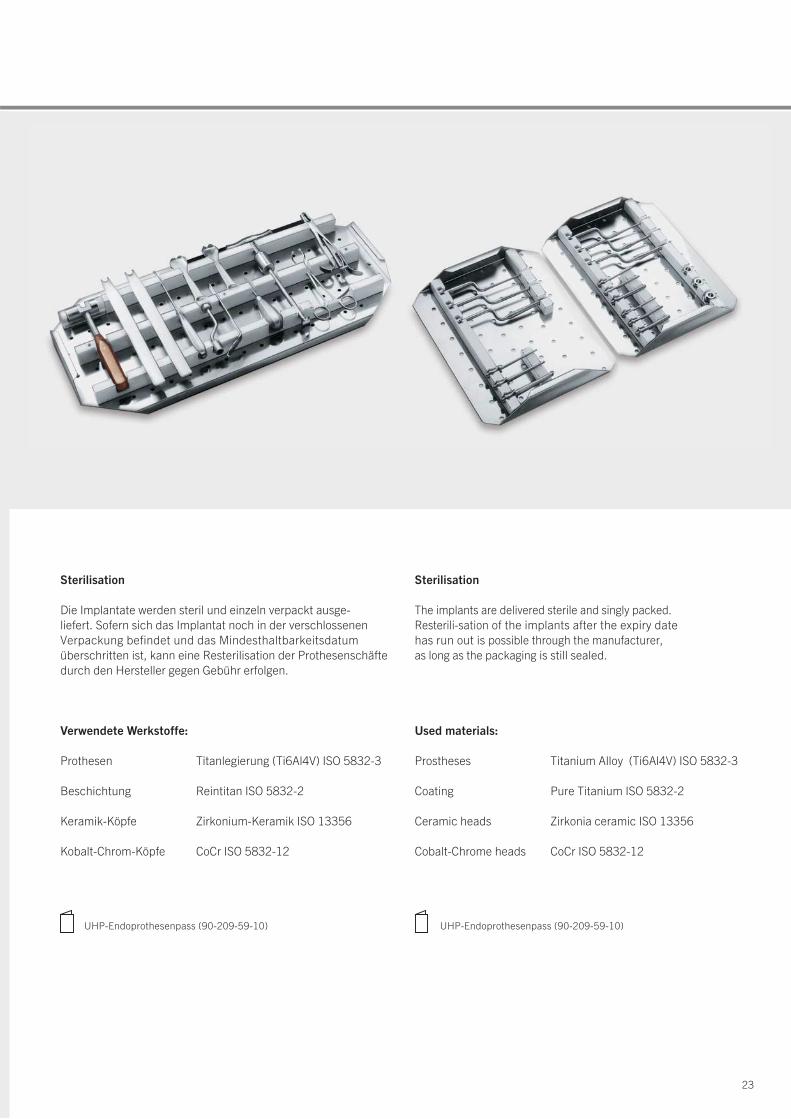
Sterilisation
Die Implantate werden steril und einzeln verpackt ausge-liefert. Sofern sich das Implantat noch in der verschlossenenVerpackung befindet und das Mindesthaltbarkeitsdatumüberschritten ist, kann eine Resterilisation der Prothesenschäftedurch den Hersteller gegen Gebühr erfolgen.
Verwendete Werkstoffe:
Prothesen Titanlegierung (Ti6Al4V) ISO 5832-3
Beschichtung Reintitan ISO 5832-2
Keramik-Köpfe Zirkonium-Keramik ISO 13356
Kobalt-Chrom-Köpfe CoCr ISO 5832-12
UHP-Endoprothesenpass (90-209-59-10)
Sterilisation
The implants are delivered sterile and singly packed. Resterili-sation of the implants after the expiry date has run out is possible through the manufacturer, as long as the packaging is still sealed.
Used materials:
Prostheses Titanium Alloy (Ti6Al4V) ISO 5832-3
Coating Pure Titanium ISO 5832-2
Ceramic heads Zirkonia ceramic ISO 13356
Cobalt-Chrome heads CoCr ISO 5832-12
UHP-Endoprothesenpass (90-209-59-10)
07.17 · 90-665-16-10 · Printed in Germany · Copyright by Gebrüder Martin GmbH & Co. KG · Alle Rechte vorbehalten · Technische Änderungen vorbehaltenWe reserve the right to make alterations · Cambios técnicos reservados · Sous réserve de modifications techniques · Ci riserviamo il diritto di modifiche tecniche
Gebrüder Martin GmbH & Co. KGA company of the KLS Martin GroupKLS Martin Platz 1 · 78532 Tuttlingen · Germany P.O. Box 60 · 78501 Tuttlingen · GermanyTel. +49 7461 706-0 · Fax +49 7461 [email protected] · www.klsmartin.com
KLS Martin Group
KLS Martin Australia Pty Ltd.Sydney · AustraliaTel. +61 2 9439 [email protected]
KLS Martin do Brasil Ltda.São Paulo · BrazilTel. +55 11 3554 [email protected]
Gebrüder Martin GmbH & Co. KGShanghai · ChinaTel. +86 21 5820 [email protected]
KLS Martin India Pvt Ltd.Chennai · India Tel. +91 44 66 442 [email protected]
Martin Italia S.r.l.Milan · ItalyTel. +39 039 605 67 [email protected]
Nippon Martin K.K.Tokyo · JapanTel. +81 3 3814 [email protected]
KLS Martin SE Asia Sdn. Bhd.Penang · Malaysia Tel. +604 505 [email protected]
Martin Nederland/Marned B.V.Huizen · The Netherlands Tel. +31 35 523 45 [email protected]
Gebrüder Martin GmbH & Co. KGMoscow · RussiaTel. +7 499 [email protected]
Gebrüder Martin GmbH & Co. KGDubai · United Arab Emirates Tel. +971 4 454 16 [email protected]
KLS Martin UK Ltd.London · United Kingdom Tel. +44 1189 000 [email protected]
KLS Martin LP Jacksonville · Florida, USA Tel. +1 904 641 77 [email protected]