Ultrastructure of pollen of Vitis vinifera L. cv. Picolit ...
9
Vitis 15, 73-Bi (1976) Istituto di Botanica dell'Università di Milano Istituto Sperimentale di Viticoltura di Conegliano Centro di Microscopia Elettronica del Politecnico di Milano, Italia Ultrastructure of pollen of Vitis vinifera L. cv. "Picolit giallo" and its behaviour in experiments of self- and cross-pollination by GIULIANA LOMBARDO, LUISA CARRARO, G. CARGNELLO and MARIA BASSI Die Feinstruktur der Pollenkorner von Vitis vinifera L. cv. Picolit giallo und ihr Verhalten bei Versuchen zur Selbst- und Fremdbestaubung Z u s a mm e n f a s s u n g . - Die Vitis-vinifera-Sorte Pìcolit giallo liefert nur geringe Ertrage. Um hierfir verantwortliche Abnormitaten aufzufinden, wurde die Fein- struktur van Pollenkrnern und Narbe mit Hilfe der Raster- und Transmissionselektro- nenmikroskopie untersucht. Der Protop!ast der Pollenkrner wirkt normai, aber die Pollenwand ist van kontinuierlicher Beschaffenheit und zeigt keine Furchen oder Keim- poren wie bei typischen Pollenkrnern. Die Narbe macht einen normalen Eindruck. Sehr wahrscheinlich verhindert die nicht unterbrochene Pollenwand die Pollenkeimung und ist somit eine mg!iche Ursache der geringen Fertilitat van Picolit giallo. Diese Vermu- tung wird durch Versuche zur Selbst- und Fremdbestaubung gestitzt, wobei Picolit- Potlen nie keimte, auch dann nicht, wenn er auf Narben anderer Rebensorten gebracht wurde; umgekehrt keimte der Pollen solcher Sorten regelmaig, wenn er auf Picolit- Narben ibertragen wurde. Introduction The productivity of the different varieties of grape vines is highly variable, and among the varieties with low productivity "Picolit giallo" is one with the lowest. In fact, generally only a few berries develop out of an inflorescence. This extremely low yield is due to the fact that fertilization occurs in a very few num- ber of flowers (CosMo and SARDI 1962, CANouss10 1966-69). The low productivity of "Picolit giallo" was experimentally increased by pollinating its female flowers with pollen from other cultivars, such as "Pinot grigio" or "Verduzzo friulano", but ex- periments of self-pollination always gave negative results (CARGNELLO 1976). This suggested that the low yield of Picolit might be due to some impediment in pollen germination. Since some of the mechanisms whi plants possess to contro! pollination (BATE- MAN 1952, LEWIS 1954, BREWBAKER 1957, VALDEYRON 1972) are based on pure mor- phological grounds, we wanted to see if at the basis of the low incidence of fertiliza- tion of "Picolit giallo" there were alterations of either pollen or stigma structures. We confronted these structures with those of the pollen and stigma of other varieties with normai incidence of fertilization ("Pinot grigio" and "Verduzzo friulano"). Besides, we studied pollen behaviour in experiments of self-pollination and cross- pollination.
Transcript of Ultrastructure of pollen of Vitis vinifera L. cv. Picolit ...

Vitis 15, 73-Bi (1976)
Istituto di Botanica dell'Università di Milano
Istituto Sperimentale di Viticoltura di Conegliano
Centro di Microscopia Elettronica del Politecnico di Milano, Italia
Ultrastructure of pollen of Vitis vinifera L. cv. "Picolit giallo"
and its behaviour in experiments of self- and cross-pollination
by
GIULIANA LOMBARDO, LUISA CARRARO, G. CARGNELLO and MARIA BASSI
Die Feinstruktur der Pollenkorner von Vitis vinifera L. cv. Picolit giallo und ihr
Verhalten bei Versuchen zur Selbst- und Fremdbestaubung
Z u s a mm e n f a s s u n g . - Die Vitis-vinifera-Sorte Pìcolit giallo liefert nur geringe Ertrage. Um hierfi.ir verantwortliche Abnormitaten aufzufinden, wurde die Feinstruktur van Pollenkéirnern und Narbe mit Hilfe der Raster- und Transmissionselektronenmikroskopie untersucht. Der Protop!ast der Pollenkéirner wirkt normai, aber die Pollenwand ist van kontinuierlicher Beschaffenheit und zeigt keine Furchen oder Keimporen wie bei typischen Pollenkéirnern. Die Narbe macht einen normalen Eindruck. Sehr wahrscheinlich verhindert die nicht unterbrochene Pollenwand die Pollenkeimung und ist somit eine mèig!iche Ursache der geringen Fertilitat van Picolit giallo. Diese Vermutung wird durch Versuche zur Selbst- und Fremdbestaubung gesti.itzt, wobei PicolitPotlen nie keimte, auch dann nicht, wenn er auf Narben anderer Rebensorten gebracht wurde; umgekehrt keimte der Pollen solcher Sorten regelmal3ig, wenn er auf PicolitNarben i.ibertragen wurde.
Introduction
The productivity of the different varieties of grape vines is highly variable,
and among the varieties with low productivity "Picolit giallo" is one with the
lowest. In fact, generally only a few berries develop out of an inflorescence. This
extremely low yield is due to the fact that fertilization occurs in a very few num
ber of flowers (CosMo and SARDI 1962, CANouss10 1966-69). The low productivity of
"Picolit giallo" was experimentally increased by pollinating its female flowers with
pollen from other cultivars, such as "Pinot grigio" or "Verduzzo friulano", but ex
periments of self-pollination always gave negative results (CARGNELLO 1976). This
suggested that the low yield of Picolit might be due to some impediment in pollen
germinati on.
Since some of the mechanisms which plants possess to contro! pollination (BATE
MAN 1952, LEWIS 1954, BREWBAKER 1957, VALDEYRON 1972) are based on pure mor
phological grounds, we wanted to see if at the basis of the low incidence of fertiliza
tion of "Picolit giallo" there were alterations of either pollen or stigma structures.
We confronted these structures with those of the pollen and stigma of other varieties
with normai incidence of fertilization ("Pinot grigio" and "Verduzzo friulano").
Besides, we studied pollen behaviour in experiments of self-pollination and cross
pollination.

74 GIULIANA Lo�rnAnoo, LUISA CARRAno, G. CAHGNH1.o and MAHIA BAss1
Fig. 1: TEM micrograph of a Picolit pollen grain. The sporopollenin wall (sp) is continuous and compact; the cytoplasm is dense and full of organelles. The proplastids have a dark matrix (arrows) and often contain starch granules (double-arrows). c = callose
layer. X5,250.
Fig. 2: TEM micrograph of a Picolit pollen grain. The sporopollenin wall (sp) shows a rarefied area (arrow); the underlying callose layer (c) forms a lenticular thickening.
X5,625.
Fig. 3: TEM micrograph of a Picolit pollen grain, showing the generative celi delimited by a layer made presumably of callose. The generative nucleus (N) is clearly visible.
n = nucleolus. X7,500.
Abb. 1: TEM-Aufnahme eines Pollenkornes van Picolit. Die Sporopolleninwand (sp) ist kompakt und ohne Unterbrechungen; das Cytoplasma ist dicht und van Organellen erfilllt. Die Proplastiden besitzen eine elektronendichte Matrix (Pfeile) und enthalten
oft Starkekorner (Doppelpfeile). c = Kalloseschicht. 5.250X.

Ultrastructure of pollen 75
Materials and Methods
T r a n s m i s s i o n e 1 e c t r o n m i c r o s c o py ( T E M ) . - Fully developed pollen grains of "Picolit giallo", "Pinot grigio" and "Verduzzo friulano", taken from the inflorescences of the main branches, were fixed in cacodilate-buffered 3% glutaraldehyde, pH 6.9, at 4 °c far 24 h. After washing in 0.1 M cacodilate buffer, the specimens were postfixed in cacodilate-buffered 1 % osmium tetroxide far 2 h, at 4 °c, dehydrated in ethanol, and embedded in araldite. When in 75% ethanol, the samples were impregnated with uranyl acetate in semisaturated solution.
Fully developed stigmas of the three varieties after experimental self- and cross-pollination were treated in the same way.
Ultrathin sections were cut with an LKB Ultrotome III, stained with lead citrate and examined in a Hitachi HllB electron microscope.
Li g h t m i c r o sco p y . Semi-thin (1 pm) sections of the araldite-embedded stigmas were cut with a glass knife, stained with toluidine blue and viewed with a light microscope.
S c a n n i n g e 1 e c t r o n m i c r o s c o p y ( S E M ) . - Both the mature anthers and the pollinated stigmas were fixed immediately after sampling in phosphate-buffered 3% glutaraldehyde, pH 6.9, far 24 h. They were then rinsed and exposed to vapours of 1 % osmium tetroxide far further 24 h, dehydrated in ethanol and critica! point dried with C02 • The samples were coated with carbon and gold in a Balzers BAE 121 coating unit provided with a tilting-rotating stage, and examined with a Jeol JSM U3.
Results
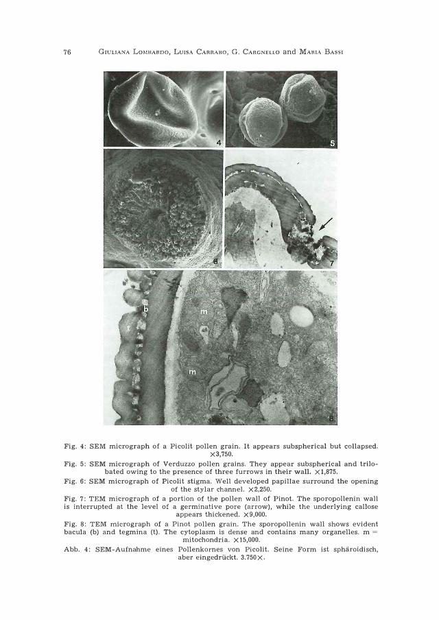
M or p h o 1 o g y o f Pico 1 i t p o 11 e n a n d s t i g m a . - When observed by SEM, pollen grains appeared subspherical but more or less collapsed, and did not show the three furrows typical of Viti,s pollen grains (Fig. 4). In ultrathin sections, they appeared spherical. Their sporopollenin wall was continuous, compact, with a limited number of sculptures (Fig. 1). It showed no thinnings, such as are generally seen at the leve! of the furrows. In one case only, did it present a rarefied area (Fig. 2). The callose layer was generally of unifarm thickness, with the exception of rare cases in which it farmed lenticular thickenings (Fig. 2).
The vegetative celi showed a very dense cytoplasm containing an extraordinary great number of mitochondria and proplastids (Fig. 1). The latter had a dark stroma and often contained starch granules. The generative celi was always visible, delimited by a callose wall (Fig. 3).
The stigma had a normai aspect, with well developed papillae (Fig. 6).
Mo r p h ology of p o lle n a n d stig ma of Pi n ot and V erduzzo . - When observed by SEM, the pollen grains appeared subspherical but tri-
Abb. 2: TEM-Aufnahme eines Pollenkornes von Picolit. Die Sporopolleninwand (sp) besitzt eine verdilnnte Zone (Pfeil); darunter bildet die Kalloseschicht (e) eine linsen
formige Verdickung. 5.625 X.
Abb. 3: TEM-Aufnahme eines Pollenkornes von Picolit. Die generative Zelle wird von einer Schicht umhiillt, die vermutlich aus Kallose besteht. Der generative Kern (N) ìst
deutlich sichtbar. n = Nucleolus. 7.500X.
76 GIULIANA LOMIJARDO, LUISA CARRARO, G. CARGNELLO and MARIA BASSI
Fig. 4: SEM micrograph of a Picolit pollen grain. It appears subspherical but collapsed. X3,750.
Fig. 5: SEM micrograph of Verduzzo pollen grains. They appear subspherical and trilobated owing to the presence of three furrows in their wall. X 1,875.
Fig. 6: SEM micrograph of Picolit stigma. Well developed papillae surround the opening of the stylar channel. X2,250.
Fig. 7: TEM micrograph of a portion of the pollen wall of Pinot. The sporopollenin wall is interrupted at the leve! of a germinative pore (arrow), while the underlying callose
appears thickened. X9,000.
Fig. 8: TEM micrograph of a Pinot pollen grain. The sporopollenin wall shows evident bacula (b) and tegmina (t). The cytoplasm is dense and contains many organelles. m =
mitochondria. X 15,000.
Abb. 4: SEM-Aufnahme eines Pollenkornes von Picolit. Seine Form ist sphiiroidisch, a ber eingedriickt. 3. 750 X.
,,
Ultrastructure of pollen '/7
lobated due to the presence of three furrows on their surface (Fig. 5). In ultrathin
sections, the wall appeared thick, with bacula and tegmina well evident (Fig. 8),
but often interrupted at the level of the furrows (Fig. 7). The callose layer was con
tinuous and particularly thick at the leve! of the wall interruptions. The cytoplasm
of the vegetative cell appeared less dense and contained fewer organelles than
that of the vegetative cell of Picolit pollen. The generative cell was always visible,
delimited by a callose wall.
The stigma aspect was identica! to that of Picolit.
S e 1 f - p o 11 in a ti on . - In the case of Picolit, no germination of pollen
grains was observed. In the other two cases (Pinot and Verduzzo), the emission of
pollen tubes was frequently seen both in semi-thin sections and in SEM or TEM
preparations (Figs. 9, 10, 11 and 12). In ultrathin sections of pollinated stigmas, pol
len tubes were seen among the stigma cells (Fig. 12). The cytoplasm of the pollen
tubes was dense, with many organelles (Figs. 11 and 12).
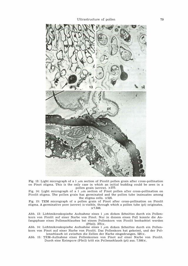
C r o s s - p o 11 i n a t i o n . - When Pinot pollen grains were deposited on
Picolit stigmas, they germinated regularly, by emission of a pollen tube clearly
visible both in semi-thin sections (Fig. 14) and in TEM preparations (Fig. 15).
When Picolit pollen was deposited on Pinot stigmas, in one case a small pro
tuberance was detected in one grain (Fig. 13), but true pollen tubes were never
seen. In ultrathin sections the aspect of the pollen grains was similar to that de
scribed for the pollen grains in the anthers.
Discussion
The content of Picolit pollen grains is apparently normai, and, in any case, if
a slight difference is found between the ultrastructure of their vegetative celi and
that of the vegetative cell of Pinot and Verduzzo, it consists in a higher content of
cytoplasmic organelles in the former. The presence of a generative cell is always
recognizable, so that it seems that we are in the presence of a normai male gameto
phyte, at least as regards its protoplast. Actually, when Picolit pollen grains are
stained with Alexander method to check their vitality, they stain normally (CAn
GNELLO and CANouss1 1976). On the contrary, the wall does not seem perfectly nor
mai, because it is of uniform thickness and aspect along the whole surface of the
pqllen grains, without those areas of rarefaction that are normally seen in pollen
grains and that generally correspond to the sites of emission of pollen tubes (ger
minative pores). The only visible structure that may suggest the formation of ger
minative pores is a thickening of the callose layer in restricted areas, which, how-
Abb. 5: SEM-Aufnahme von Pollenkornern der Sorte Verduzzo. Sie sind spharoidisch und besitzen in ihrer Wand drei Furchen. 1.875X.
Abb. 6: SEM-Aufnahme einer Narbe von Picolit. Die Offnung des Griffelkanals ist von gut entwickelten Papilla e umgeben. 2.250 X.
Abb. 7: TEM-Aufnahme eines Ausschnittes aus der Pollenwand von Pinot. Die sporopolleninwand ist in Hohe einer Keimpore unterbrochen (Pfeil), wahrend die darunterlie
gende Kalloseschicht verdickt ist. 9.ooox.
Abb. 8: TEM-Aufnahme eines Pollenkorns von Pinot. Die Sporopolleninwand zeigt deutliche Bacula (b) und Tegmina (t). Das Cytoplasma ist dicht und enthalt zahlreiche Orga
nellen. m = Mitochondrien. 15.000X,

78 GIULIANA LOMBARDO, LUISA CARRARO, G. CARGNELLO and MARIA BASSI
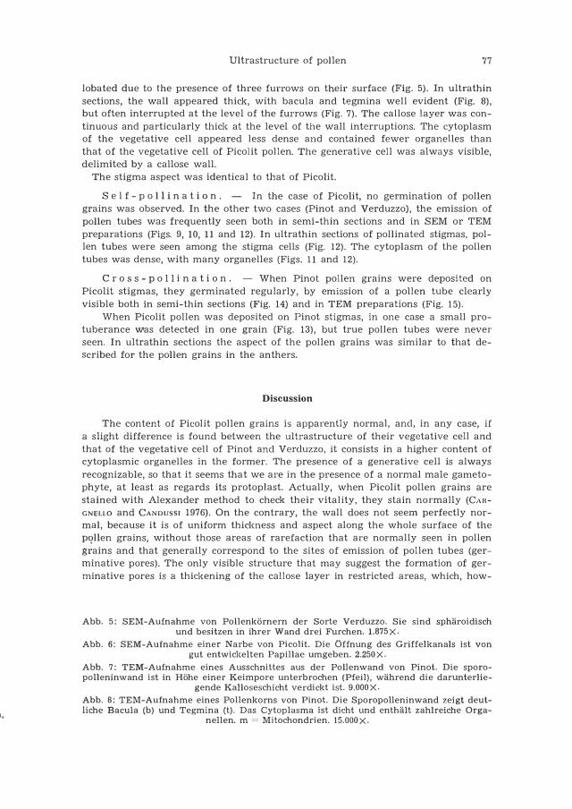
Fig. 9: SEM micrograph of germinating pollen grains of Pinot after self-pollination. Pollen tubes (arrows) are clearly visible. gp = germinative pare. X3,750.
Fig. 10: Light micrograph of a 1 11m section of Pinot stigma after self-pollination. Marty pollen grains (p) have germinated and the pollen tubes (arrows) are visible among the
stigma papillae. X240. Fig. 11: TEM micrograph of a pollen grain of Pinot after self-pollination. The emission of a pollen tube is clearly visible at the leve! of a germinative pare (arrow). p = pollen
grain; pt = pollen tube. X5,250. Fig. 12: TEM micrograph of a pollen tube insinuating among the stigma cells. The tube
cytoplasm is very dense. X5,250.
Abb. 9: SEM-Aufnahme keimender Pollenki:irner van Pinot nach Selbstbestii.ubung. Die Pollenschlii.uche sind deutlich sichtbar (Pfeile). gp = Keimpore. 3.750X.
Abb. 10: Lichtmikroskopische Aufnahme eines 1 ,,m dicken Schnittes durch eine Narbe van Pinot nach Selbstbestii.ubung. Zahlreiche Pollenki:irner (p) haben gekeimt, und die
Pollenschlii.uche (Pfeile) sind zwischen den Narbenpapillen sichtbar. 240X. Abb. 11: TEM-Aufnahme eines Pollenkornes van Pinot nach Selbstbestii.ubung. Der Austritt eines Pollenschlauches durch eine Keimpore ist deutlich sichtbar (Pfeil). p = Pol
lenkorn; pt = Pollenschlauch. 5.250X, Abb. 12: TEM-Aufnahme eines Pollenschlauches, der zwischen die Zellen der Narbe
eindringt. Das Schlauchcytoplasma ist sehr dicht. 5.250X.
Ultrastructure of pollen 79
Fi.g. 13: Light micrograph of a 1 ,um section of Picolit pol!en grain after cross-pol!ination on Pinot stigma. This is the only case in which an initial budding could be seen in a
pollen grain (arrow). X375. Fig. 14: Light micrograph of a 1 pm section of Pinot pol!en after cross-pollination on Picolit stigma. The pollen grain has germinated and the pollen tube insinuates among
the stigma cells. X525. Fig. 15: TEM micrograph of a pollen grain of Pinot after cross-pollination on Picolit stigma. A germinative pore (arrow) is visible, through which a pollen tube (pt) originates.
X7.500.
Abb. 13: Lichtmikroskopische Aufnahme eines 1 pm dicken Schnittes durch ein Pollenkorn von Picolit auf einer Narbe von Pinot. Nur in diesem einen Fall konnte die Anfangsphase eines Pollenschlauches bei einem Pollenkorn von Picolìt beobachtet werden
(Pfeil). 375 X. Abb. 14: Lichtmikroskopische Aufnahme eines 1 ftm dicken Schnittes durch ein Pollenkorn von Pinot auf einer Narbe von Pico!it. Das Pollenkorn hat gekeimt, und der Pol
lenschlauch ist zwischen die Zellen der Narbe eingedrungen. 525X, Abb. 15: TEM-Aufnahme eines Pollenkornes von Pinot auf einer Narbe von Pico!it.
Durch eine Keimpore (Pfeil) tritt ein Pollenschlauch (pt) aus. 7.500X.

80 GIULIANA Lo�rnARoo, Lu1sA CARRARo, G. CARGNELLO and MARIA BASSI
ever, is not accompanied by an in�erruption or thinning of the sporopollenin wall. Besides, when examined by SEM, Picolit pollen grains do not show the longitudinal furrows which are a normal feature of Vitis pollen and represent the sites along which germination occurs. Actually, when Picolit pollen grains, taken from different plants grown in different environmental conditions, were put to germinate in vitro on different substrates, no germination was obtained, but bursting of many grains was observed (CARGNELLO et al. 1976). Therefore, it seems that one of the causes that contribute to the failure of fertilization is intrinsic to the pollen and consists in a difficulty in the emission of pollen tubes due to mechanical impediment. Also the fact that Picolit pollen grains collapse when subjected to the usual SEM preparation procedure, while the others do not, suggests a difference in their wall structure. Most probably, the presence of an uninterrupted sporopollenin wall creates a barrier to a thorough evaporation of the internal fluids during critical point drying, with the result that the internal structure of the pollen grains is not completely stabilized and collapses under vacuum.
The hypothesis that the failure in fertilization might be due to autoincomp�tibility has to be discarded; in fact, Picolit pollen grains were unable to germinate also on stigmas of Pinot or Verduzzo. On the other hand, the Jack of germination of Picolit pollen on Pìcolit stigmas cannot be attributed to some abnormality of the latter, because the pollen of Pinot and Verduzzo germinated regularly on them.
In any case, other causes may contribute to the failure of Picolit pollen grains to germinate, and further studies are needed on this subject.
Summary
The ultrastructure of the pollen and stigma of Vitis vinifera L. cv. "Picolit giallo" was studied by scanning and transmission electron microscopy, with the aim of finding out if they presented abnormalities which could account far the low procluctivity of this vine variety. The pollen protoplast has a normai aspect, but its wall is continuous and does not present furrows and germinative pores as pollen grains generally do. The stigma appeared normai. The presence of an uninterrupted wall most probably constitutes an obstacle to pollen germination and is likely to be one of the causes of the low fertilization incidence of "Picolit giallo". This hypothesis is supported also by the results of the experiments of self- and cross-pollination, because Picolit pollen always failed to germinate, even if put on stigmas of other vine varieties, while the pollen of these varieties germinated regulariy when put on Picolit stigmas.
Literature Cited
BATE>IAN, A. J., 1952: Self-incompatibllity systems In anglosperms. I. Theory. Heredlty 6, 285-
310.
Banwo,\lrnn, J. L., 1957: Pollen cytology and self-incompatiblllty systems In plants. J. Hered. 48,
271-277.
CANuuss10, R., 1966---09: li problema della scarsa produttività delle viti di Pico lit. Atti Ace.
Udine 1966---09, 8 (7).
CAnGNDLLO, G., 1976: Autoimpollinazione ed etel'oimpollinazione in Vitis vinifera L. Indagine
sul Plcolit. Riv. Vlticolt. Enol. (Conegliano). In press.
- - e CANouss,, F., 1976: Indagine sulla vitalità del polllne in Vitis vinifera L. cv. Picollt secon
do la colorazione di Alexander. Rlv. Vlticolt. Enol. (Conegliano). In press.
Ultrastructure of pollen 81
- - e G1onGEss1, F., 1976: Contributo alla conoscenza della germinabilltà del polline di
Vitis vinifera L. Studio sulla varietà Picolit. Riv. Viticolt. Enol. (Conegliano). In press.
CosMo, I. e SAno,, F., 1962: Picolit. Ministero dell'agricoltura e delle foreste, commissione per
lo studio ampelografico dei principali vitigni ad uve da vino coltivati in Italia. 2 (43).
Lew,s, D., 1954: Incompatibility in flowering plants. Adv. Genet. 6, 235-285.
VALoarnoN, G., 1972: On the development of incompatibility and sex systems in higher plants
and their evolutive meaning. Symp. Biol. Hung. 12, 83-91.
Eingegangen am 20. 2. 1976 Dr. GIULIANA Lo�rnAnoo
Istituto di Scienze Botaniche
Università degli Studi
Via G. Colombo 60
20133 Milano
Italia