Zur Wirkung des Seeanemonen Toxins II (ATX-II) auf ... · Aus dem Veterinärwissenschaftlichen...
79
Aus dem Veterinärwissenschaftlichen Department der Tierärztlichen Fakultät der Ludwig-Maximilians-Universität München Arbeit angefertigt unter der Leitung von Prof. Dr. med. vet. Manfred Stangassinger Angefertigt am Institut für Physiologie und Pathophysiologie der Friedrich-Alexander- Universität Erlangen-Nürnberg (PD Dr. med. Angelika Lampert) "Zur Wirkung des Seeanemonen Toxins II (ATX-II) auf spannungsgesteuerte Natriumionenkanäle neuronaler Zellen und zu den dadurch erreichten Änderungen im Schmerzempfinden" Inaugural-Dissertation zur Erlangung der tiermedizinischen Doktorwürde der Tierärztlichen Fakultät der Ludwig-Maximilians-Universität München von Alexandra Birgit Klinger aus München München 2013
Transcript of Zur Wirkung des Seeanemonen Toxins II (ATX-II) auf ... · Aus dem Veterinärwissenschaftlichen...

Aus dem Veterinärwissenschaftlichen Department der Tierärztlichen Fakultät der Ludwig-Maximilians-Universität München
Arbeit angefertigt unter der Leitung von Prof. Dr. med. vet. Manfred Stangassinger
Angefertigt am Institut für Physiologie und Pathophysiologie der Friedrich-Alexander-Universität Erlangen-Nürnberg
(PD Dr. med. Angelika Lampert)
"Zur Wirkung des Seeanemonen Toxins II (ATX-II) auf
spannungsgesteuerte Natriumionenkanäle neuronaler Zellen und zu den dadurch erreichten Änderungen im Schmerzempfinden"
Inaugural-Dissertation zur Erlangung der tiermedizinischen Doktorwürde der Tierärztlichen Fakultät
der Ludwig-Maximilians-Universität München
von Alexandra Birgit Klinger
aus München
München 2013

Gedruckt mit Genehmigung der Tierärztlichen Fakultät der Ludwig-Maximilians-Universität München
Dekan: Prof. Dr. Joachim Braun
Referent: Prof. Dr. Manfred Stangassinger
Korreferent: Prof. Dr. Andrea Fischer
Tag der Promotion: 09.02.2013

Die vorliegende Arbeit ist nach § 6 Abs. 2 der Promotionsordnung der Tierärztlichen Fakultät der Ludwig-Maximilians-Universität München in der geänderten Fassung vom 15.01.2007 als kumulative Dissertation gestaltet. Die im Abschnitt V.-Veröffentlichung dargestellten Ergebnisse sind in der englischsprachigen, wissenschaftlichen, mit Gutachtersystem ausgestatteten Zeitschrift “Molecular Pain” publiziert (Klinger et al. Molecular Pain 2012, 8:69; http://www.molecularpain.com/content/8/1/69).

meiner Familie

Inhaltsverzeichnis V
INHALTSVERZEICHNIS
I. EINLEITUNG .................................................................................................. 1
II. ZIEL DIESER ARBEIT .................................................................................. 3
III. DARSTELLUNG WICHTIGER GRUNDLAGEN ........................................ 5
1 Natriumionenkanäle ......................................................................................... 5
1.1 Aufbau ............................................................................................................... 5
1.2 Einteilung .......................................................................................................... 6
1.3 Funktion............................................................................................................. 7
2 Resurgent current ............................................................................................ 9
2.1 Entstehung ......................................................................................................... 9
2.2 Bedeutung ........................................................................................................ 10
3 Schmerzfasern ................................................................................................ 11
3.1 Einteilung ........................................................................................................ 11
3.2 Eigenschaften ................................................................................................... 11
4 ATX-II ............................................................................................................ 12
IV. MATERIAL UND METHODEN .................................................................. 13
1 Patch-Clamp Technik .................................................................................... 13
1.1 Historie ............................................................................................................ 13
1.2 Patch-Clamp Aufbau ........................................................................................ 16
1.3 Durchführung und Untersuchungsmaterial ........................................................ 21
2 Psychophysik .................................................................................................. 25
3 Zellvorbereitung für RT-qPCR-Experimente ............................................... 28
V. VERÖFFENTLICHUNG............................................................................... 29
VI. DISKUSSION ................................................................................................. 62
VII. ZUSAMMENFASSUNG ................................................................................ 65
VIII. SUMMARY .................................................................................................... 67
IX. LITERATURVERZEICHNIS ....................................................................... 69
X. ANHANG ....................................................................................................... 72
XI. DANKSAGUNG ............................................................................................. 73

Abkürzungsverzeichnis VI
ABKÜRZUNGSVERZEICHNIS
∅ Durchmesser α Alpha β Beta
τ Tau, Zeitkonstante Abb. Abbildung AD-Wandler Analog-Digital Wandler AP Aktionspotenzial AS Aminosäure ATX-II Seeanemonen Toxin II (rekombinant) DI - IV Domäne eins bis vier (eines Natriumionenkanals) DA-Wandler Digital-Analog Wandler DNA desoxyribonucleic acid / Desoxyribonukleinsäure DMEM Dulbecco’s Modified Eagle Medium DRG(s) dorsal root ganglia neuron(s) / Hinterwurzelganglionneuron(e) HEK 293 human Embryonic Kidney cells / menschliche, embryonale Nierenzelllinie kDa kilo-Dalton M.W. molecular weight / Molekulargewicht N1E-115 Neuroblastomzellen (Zelllinie) Nav(s) spannungsgesteuerter Natriumionenkanal (Pl.) / voltage-gated sodium channel(s) NRS numeric rating scale / numerische Bewertungsskala PBS phosphate buffered saline / Phosphat gepufferte Lösung RNA ribonucleic acid / Ribonukleinsäure RT-qPCR Reverse Transkription- quantitative PCR S1-S6 Segment eins bis sechs (eines Natriumionenkanals) SCN8amed Gen des Nav1.6 mit einer Mutation für die motorische-Endplatten-Krankheit TTXs/r Tetrodotoxin sensitiv/resistent
I. Einleitung 1
I. EINLEITUNG
Schmerz ist für Mensch und Tier gleichermaßen belastend und mindert mitunter
erheblich die Lebensqualität. Dies ist insbesondere für Tierärzte relevant, da nicht der
Lebenserhalt um jeden Preis, sondern in erster Linie eine akzeptable Lebensqualität Ziel
unserer Bemühungen sein sollte.
Umso wichtiger ist es die Grundlagen, die zu Schmerz führen, zu erforschen, um mit
Hilfe dieser Erkenntnisse möglichst spezifische Therapiemethoden entwickeln zu
können. Um die Information über ein schmerzhaftes Ereignis weiterleiten zu können,
sind Natriumionenkanäle (Navs) und die von ihnen generierten Aktionspotentiale (APs)
essentiell. Über APs kann dem Gehirn via Nervenfasern die Information über noxische
Ereignisse aus der Peripherie übermittelt werden. Dies ist ein überlebensnotwendiger
Mechanismus, um den Körper vor schädigenden Einflüssen zu bewahren. Was aber,
wenn dieser Mechanismus fehlerhaft ist? So gibt es Erkrankungen, denen eine Mutation
eines Nav zu Grunde liegt und die mit kontinuierlichen, unphysiologischen Schmerzen
einhergehen. Eine andere Mutation führt zu völliger Schmerzlosigkeit, ein Zustand, der
nur auf den ersten Blick paradiesisch erscheint, denn den Betroffenen fehlt ein
natürlicher Schutzreflex, der Schäden des Körpers verhindern kann. Doch manchmal ist
Schmerz auch hinderlich. Leben Tiere wie Nacktmulle oder Mausohrfledermäuse in
großen, unterirdischen Kolonien mit hohem CO2-Druck, so führt ein durch
Nav-Mutation hervorgerufener, fehlender "Säureschmerz" durchaus zu
Überlebensvorteilen. Auch führt eine schmerzhafte Erfahrung eines Lebewesens zu
künftigem Meideverhalten, so dass Schmerz auch eine mittelbar protektive Wirkung
besitzt. Wer je beim Tauchen Bekanntschaft mit den Nesselfäden einer Seeanemone
gemacht hat, wird künftig sicherlich Abstand halten. So schützt sich die Anemone auch
vor potentiellen Fressfeinden oder erlegt auf diese Weise ihrerseits Beute. Der

I. Einleitung 2
Anemonenfisch hingegen scheint sich nicht an den Giften der Blumentiere zu stören
und lebt in Symbiose inmitten der Tentakel mit den pfeilschnellen Nesselprojektilen,
die anderen Fischen das Leben kosten (s. Abb.1). Nicht nur für Meeresbiologen sind
Seeanemonen also ein interessantes Forschungsobjekt, auch im Bereich der
Schmerzforschung sind ihr Gift und dessen Wirkung auf die Schmerzsignal-
generierenden Navs durchaus von Bedeutung. Über welche Mechanismen führt das Gift
zu Schmerz und können wir daraus Schlüsse auf die Funktionsweise der Navs ziehen?
Kann es darüber hinaus vielleicht sogar zur Diagnostik oder Therapie bei
Schmerzereignissen, wie wir dies bereits von anderen Tiergiften (z. B. dem
ω−Conotoxin der Kegelschnecke) kennen, eingesetzt werden?
Abb.1: Seeanemone mit Anemonenfischen
(Quelle: http://www.fisch-fotos.de/wallpapers)
II. Ziel dieser Arbeit 3
II. ZIEL DIESER ARBEIT
Das Ziel der vorliegenden Arbeit ist es, mit Hilfe von elektrophysiologischen
Patch-Clamp Messungen zu untersuchen, ob und in welcher Weise das Seeanemonen-
Toxin ATX-II Ströme durch Navs in sensorischen Hinterwurzelganglionneuronen (kurz:
DRGs; engl. dorsal root ganglia neurons) modifiziert. Von besonderem Interesse für
diese Arbeit ist ein Na+-Strom, der aus der durch das Toxin modifizierten, schnellen
Inaktivierung der Navs entstehen kann; der sog. "resurgent current" (sinngemäß "wieder
auflebender Strom"; Erläuterung hierzu s. Abschnitt III.2). Bisher konnte noch nicht
gezeigt werden, dass ATX-II in sensorischen Neuronen resurgent currents induzieren
kann. Auch die pathophysiologische Bedeutung dieser Ströme ist nicht geklärt. Die
vorliegende Arbeit untersucht, in wie weit die durch das Toxin induzierten resurgent
currents zu Änderungen im Schmerzempfinden beitragen können.
Säugetiere haben für die Schmerzwahrnehmung zwei Nervenfasertypen: langsam
leitende C-Schmerzfasern, die mit kleinen DRGs verbunden sind und schnell leitende
A-Schmerzfasern, die in den großen DRGs ihre Entsprechung finden. Im Rahmen dieser
Arbeit wird untersucht, ob beide Fasertypen unterschiedlich auf ATX-II reagieren und
gesetzt den Fall, ob dies auf die zugrundeliegende Nav-Subtypzusammensetzung zurück
zu führen ist. Um dies zu beleuchten, werden geringe Mengen ATX-II auf kultivierte,
vereinzelte DRGs der Maus appliziert und die Auswirkungen des Toxins auf die
Na+-Ströme mit der Patch-Clamp Methode untersucht.
Um die Befunde auf molekularer Ebene eingehender zu untersuchen, sollen im Rahmen
dieser Arbeit die wesentlichen Nav-Untereinheiten (Nav1.6 und Nav1.7) jeweils in
Zelllinien zur Expression gebracht und dort mit der Patch-Clamp Methode auf ihre
ATX-II Empfindlichkeit untersucht werden.

II. Ziel dieser Arbeit 4
Ein weiteres Ziel dieser Arbeit ist es, festzustellen, ob die bei der Maus auf molekularer
Ebene untersuchten Effekte von ATX-II tatsächlich Auswirkungen auf das
Schmerzempfinden haben können. Da Menschen zu der Qualität der Wahrnehmung
befragt werden können und so ein größtmöglicher Erkenntnisgewinn möglich ist, wird
dieser Teil der Arbeit an humanen Probanden durchgeführt. Hierfür wird gesunden,
adulten Probanden eine geringe Menge des Toxins intrakutan injiziert und nachfolgend
werden die je nach vermittelndem Schmerzfasertyp unterschiedlichen
Empfindungsqualitäten (z. B. stechende, brennende, dumpfe Schmerzen) ebenso wie die
Intensität der Empfindungen (mittels einer numerischen Bewertungsskala von 0-10) im
Rahmen psychophysikalischer Tests untersucht.
Die im Rahmen der vorliegenden Arbeit durchgeführten Untersuchungen sollen einen
Beitrag leisten zur Aufklärung resurgent current-induzierter Schmerzen bei Tier und
Mensch und langfristig die Möglichkeit eröffnen, u. a. auf der Basis dieser Ergebnisse,
spezifische, schmerzhemmende Medikamente zu entwickeln.
Wichtige Ergebnisse aus diesen Untersuchungen sind publiziert (Abschnitt V dieser
Dissertation) sowie im Rahmen einer Posterpräsentation vorgestellt worden
(Abschnitt X der vorliegenden Arbeit).

III. Darstellung wichtiger Grundlagen 5
III. DARSTELLUNG WICHTIGER GRUNDLAGEN
1 Natriumionenkanäle
Für die Entstehung von AP's auf erregbaren Zellmembranen sind Navs von zentraler
Bedeutung.
1.1 Aufbau
Navs sind Proteine und werden eingeteilt in α− (Nav1.1 - 1.9) und beigeordnete β− (β1 -
β4) Untereinheiten. Die α-Untereinheiten bestehen aus vier Domänen (DI - DIV,
Abb.2), die aus je sechs Transmembransegmenten (S1-S6) aufgebaut sind
(CATTERALL, 2000) und bilden bereits ein funktionierendes Kanalprotein. Die vier
Domänen lagern sich derart aneinander, dass in der Mitte eine Pore entsteht, die von S5
und S6 aller vier Domänen gebildet wird. Durch diese Pore gelangen bei entsprechender
Kanalkonfiguration Natriumionen (Na+) ins Zellinnere, andere Ionen werden auf Grund
ihrer Ladung, Größe und ihrem Hydratisierungsgrad durch den Selektivitätsfilter
(s. Abb.2, rote Schleifen) an der Kanalpassage gehindert. Die S4 (s. Abb.2, rosa
hinterlegte Segmente) bilden die Spannungssensoren und zwischen DIII und DIV
befindet sich das aus den Aminosäuren Isoleucin, Phenylalanin und Methionin gebildete
IFM-Motif (s. Abb.2, gelber Kreis), das als Inaktivierungspartikel fungiert. Die
β−Untereinheiten nehmen Einfluss auf die Kanaleigenschaften und -expression der
α−Untereinheiten und treten in den unterschiedlichsten Kombinationen mit diesen auf.
Von besonderem Interesse ist die β4-Untereinheit, da sie oder Teile von ihr nach
heutigem Wissensstand das "blocking particle" (den Nav blockierender Partikel)
darstellen, das für den resurgent current eine wichtige Rolle spielt.

III. Darstellung wichtiger Grundlagen 6
Abb.2: Schematisierte Darstellung eines Nav; modifiziert aus (BEAR et al., 2007)
1.2 Einteilung
Nach ihrer Empfindlichkeit dem Gift Tetrodotoxin (TTX) des Kugelfisches
(Tetraodontoideus) gegenüber werden die Navs in TTX-resistente (TTXr) und
TTX-sensitive (TTXs) Kanäle unterteilt. Die Kanalsubtypen zeigen bei verschiedenen
Membranspannungen unterschiedliche Kinetiken, was auch zu differierenden
Erregungseigenschaften der jeweiligen Zellen führt (CATTERALL et al., 2005). In
sensorischen Neuronen findet man die TTXs Kanäle Nav1.1, 1.2, 1.3, 1.6 und 1.7 sowie
die TTX-r Kanäle Nav1.8 und 1.9. Dabei enthält ein Neuron stets mehrere
Kanalsubtypen. Bei den mit großen sensorischen Neuronen assoziierten A-Fasern ist
hauptsächlich Nav1.6, bei C-Fasern, die mit kleinen Neuronen in Verbindung stehen,
Nav1.7 für die AP-Weiterleitung notwendig (WILSON et al., 2011). Diese beiden TTXs
Kanäle scheinen also eine besondere Rolle in der Schmerzleitung zu spielen.
III. Darstellung wichtiger Grundlagen 7
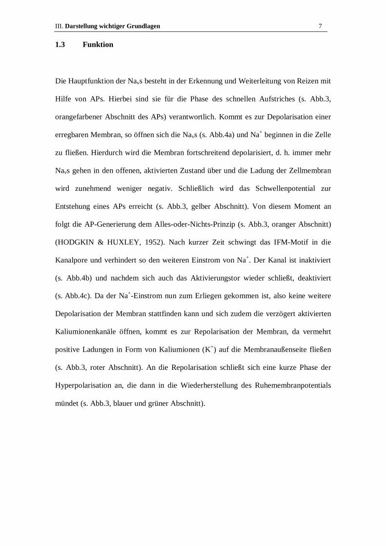
1.3 Funktion
Die Hauptfunktion der Navs besteht in der Erkennung und Weiterleitung von Reizen mit
Hilfe von APs. Hierbei sind sie für die Phase des schnellen Aufstriches (s. Abb.3,
orangefarbener Abschnitt des APs) verantwortlich. Kommt es zur Depolarisation einer
erregbaren Membran, so öffnen sich die Navs (s. Abb.4a) und Na+ beginnen in die Zelle
zu fließen. Hierdurch wird die Membran fortschreitend depolarisiert, d. h. immer mehr
Navs gehen in den offenen, aktivierten Zustand über und die Ladung der Zellmembran
wird zunehmend weniger negativ. Schließlich wird das Schwellenpotential zur
Entstehung eines APs erreicht (s. Abb.3, gelber Abschnitt). Von diesem Moment an
folgt die AP-Generierung dem Alles-oder-Nichts-Prinzip (s. Abb.3, oranger Abschnitt)
(HODGKIN & HUXLEY, 1952). Nach kurzer Zeit schwingt das IFM-Motif in die
Kanalpore und verhindert so den weiteren Einstrom von Na+. Der Kanal ist inaktiviert
(s. Abb.4b) und nachdem sich auch das Aktivierungstor wieder schließt, deaktiviert
(s. Abb.4c). Da der Na+-Einstrom nun zum Erliegen gekommen ist, also keine weitere
Depolarisation der Membran stattfinden kann und sich zudem die verzögert aktivierten
Kaliumionenkanäle öffnen, kommt es zur Repolarisation der Membran, da vermehrt
positive Ladungen in Form von Kaliumionen (K+) auf die Membranaußenseite fließen
(s. Abb.3, roter Abschnitt). An die Repolarisation schließt sich eine kurze Phase der
Hyperpolarisation an, die dann in die Wiederherstellung des Ruhemembranpotentials
mündet (s. Abb.3, blauer und grüner Abschnitt).

III. Darstellung wichtiger Grundlagen 8
Abb.3: Schema eines APs im zeitlichen Verlauf; nach Dr. Kay-Uwe Jagemann; www.jagemann-net.de
Abb.4: Schema der verschiedenen Navkonfigurationen; modifiziert nach (SITTL et al., 2012)

III. Darstellung wichtiger Grundlagen 9
2 Resurgent current
2.1 Entstehung
Eine Sonderform der Nav-Inaktivierung stellt der 1997 erstmals von Raman und Bean
(RAMAN & BEAN, 1997) beschriebene resurgent current dar. Dieser alternative
Inaktivierungsmechanismus tritt in Neuronen wie Purkinjezellen (inhibitorische und
einzige efferente Nervenzellen aus dem Cortex des Cerebellums) oder DRGs
natürlicherweise auf. In Zelllinien, in die Nav-Untereinheiten heterolog transfiziert und
die in Isolation untersucht wurden, konnte dieser "wieder auflebende Strom" (engl.
resurgent current) lange nicht nachgewiesen werden, was die Suche nach den genauen
Ursprüngen dieses Stromes erheblich erschwerte. Der resurgent current entsteht wenn
statt des eigentlichen Inaktivierungspartikels das blocking particle, das vermutlich von
der β4-Untereinheit oder durch Teile dieser gebildet wird (GRIECO et al., 2005), in
den für Na+ noch offenen Kanal wandert (s. Abb.4d) und diesen zunächst für Na+
unpassierbar macht (offener Kanalblock). Im Verlauf der Repolarisation der Membran
löst sich das blocking particle vom Kanal und dieser ist kurzfristig wieder für Na+
passierbar. Der nun wieder auflebende Strom ist der resurgent current (s. Abb.4e). Unter
bestimmten Voraussetzungen lässt sich der resurgent current auch in heterolog
transfizierten Navs nachweisen (WANG et al., 2006). Um diesen Strom in heterologen
Expressionssystemen untersuchen zu können, muss der C-Terminus der
β4-Untereinheit, das sogenannte β4-Peptid, künstlich über die Intrazellulärlösung
zugeführt werden. Je nach Nav-Subtyp kann dann auch hier resurgent current induziert
werden.

III. Darstellung wichtiger Grundlagen 10
2.2 Bedeutung
Die Stromamplitude des resurgent current ist weit weniger ausgeprägt als die des
transienten schnellen Na+-Stromes. Allerdings depolarisiert er die Zelle am Ende eines
abgelaufenen APs und kann so dazu führen, dass die Schwelle für das Auslösen eines
erneuten APs frühzeitiger wieder erreicht wird. Somit wird die Erregbarkeit erhöht, was
durchaus Folgen für den gesamten Organismus haben kann. So kommt es
beispielsweise in der Krebstherapie beim Menschen durch Anwendung des
Chemotherapeutikums Oxaliplatin zu schwerwiegenden, dosislimitierenden
Nebenwirkungen (z. B. Neuropathien, die sich insbesondere in Verbindung mit Kälte
verschlimmern). Diese werden durch vermehrten resurgent current hervorgerufen
(SITTL et al., 2012). Auf der anderen Seite können pathologische Nav-Mutationen, wie
sie beispielsweise beim Krankheitsbild der paroxysmal extreme pain disorder (PEPD)
im Nav1.7 vorkommen, zu verstärktem Auftreten von resurgent current führen
(JARECKI et al., 2010). Diese Beispiele zeigen, dass der resurgent current von großer
Relevanz in der Schmerzforschung ist und deshalb wird u. a. in unserer Arbeitsgruppe
(AG Dr. Lampert) daran gearbeitet, den Mechanismus seines Entstehens vollständig
aufzuklären.

III. Darstellung wichtiger Grundlagen 11
3 Schmerzfasern
3.1 Einteilung
Die Einteilung der somatosensiblen Schmerzfasern wird vorwiegend nach ihrer
Leitungsgeschwindigkeit und nach ihrer Myelinisierung vorgenommen. So werden
myelinisierte, schnell leitende, hellen Schmerz vermittelnde Aδ- von unmyelinisierten,
langsam leitenden, dumpfen Schmerz vermittelnden C-Fasern unterschieden. Die mit
einer Leitungsgeschwindigkeit von < 2m/s deutlich langsameren C-Fasern sind im DRG
mit kleinen Neuronen verbunden, wohingegen die über saltatorische Erregungsleitung
verfügenden Aδ−Fasern mit den großen sensorischen Neuronen in Verbindung stehen
(LAWSON, 2002).
3.2 Eigenschaften
Bei einer Aktivierung von C-Fasern (z. B. durch einen Schmerzreiz) kann ein
sogenanntes "Axon Reflex Erythem" auftreten (s. Abb.8c). Dieses ist makroskopisch
durch eine sich konzentrisch um den Reiz ausbreitende Rötung definiert (sog. Erythem).
Verantwortlich hierfür ist die antidrome Reizausbreitung mit großflächiger
Vasodilatation und Permeabilitätssteigerung durch die Ausschüttung zahlreicher
Neuropeptide, u. a. CGRP und Substanz P (DISCLAFANI & WILKIN, 1983).
Bei mechanischem Druck auf einen Nerv reagieren die hierfür empfindlicheren
A-Fasern frühzeitiger mit einem Ausfall der Reizleitung als C-Fasern. So kann man mit
Hilfe eines Nervenkompressionsblockes relativ selektiv die A-Faser-Antwort
unterdrücken, während die C-Fasern zunächst weitgehend unbeeinflusst bleiben
(TOREBJORK & HALLIN, 1973). Dies ist überaus nützlich um Effekte auf Faserebene
zu differenzieren.
III. Darstellung wichtiger Grundlagen 12
4 ATX-II
Das basische, neurotoxische, 47 Aminosäuren (AS) lange Polypeptid ATX-II wurde
ursprünglich in einem aufwendigen Verfahren aus der Seeanemone Anemonia sulcata
gewonnen. Hierzu wurden ganze Anemonentiere in Alkohol homogenisiert, die Toxine
an Kationenaustauscher adsorbiert, gel-filtriert und durch Chromatographie getrennt
(BERESS et al., 1975). Seit einiger Zeit wird dieses Toxin rekombinant durch
Escherichia coli hergestellt, womit ein gleichbleibend hoher Reinheitsgrad gewährleistet
ist.
ATX-II ist ein potenter Nav-Modulator, der die Inaktivierung der Kanäle verzögert
(BERGMAN et al., 1976). So könnte es ein Ziel für die Entwicklung neuer
pharmakologischer Substanzen sein, denn der damit verbundene verlängerte
Na+-Einstrom führt beispielsweise am Herzen zu verlängerten Aktionspotentialen und
hat somit vermutlich eine positiv inotrope Wirkung auf den Herzmuskel (ISENBERG &
RAVENS, 1984). Eine Hemmung des resurgent current im Bereich reizleitender Fasern
vermindert deren Erregbarkeit und könnte sich so beispielsweise bei der Behandlung
von Tieren mit pathologischen Spontanaktivitäten, die u. a. bei Diabetes mellitus
auftreten, als nützlich erweisen.

IV. Material und Methoden 13
IV. MATERIAL UND METHODEN
1 Patch-Clamp Technik
1.1 Historie
Zu den häufig verwendeten Messmethoden in der Elektrophysiologie zählt die
Patch-Clamp Technik. Sie wurde in den 1970er Jahren von den Nobelpreisträgern
Erwin Neher und Bernd Sakmann entwickelt. Von den ursprünglichen "loose
Patch-Clamp" Experimenten, bei denen mehrmals verwendete Glaspipetten lose
(engl. "loose") auf die Zellmembran aufgesetzt und so Einzelkanäle untersucht wurden
(NEHER & SAKMANN, 1976), entwickelte die Wissenschaftlergruppe um Neher und
Sakmann die Technik in den Folgejahren stetig weiter. 1980 erkannte Neher, dass die
gelegentlich vermeintlich verstopften Pipetten, die einen erhöhten Abdichtwiderstand
verursachten (heute wegen dem über einem GΩ großen Widerstand als "Gigaseal"
bezeichnet), verringertes Rauschen und deutlich verbesserte Ableitbedingungen zur
Folge hatten. Dies führte zur Einmalverwendung der Pipetten und zum Einsatz von
Über- und Unterdruck beim Patchvorgang und machte die Entwicklung der "tight seal
patch-clamp" Methode möglich (HAMILL et al., 1981).
Der verbesserten Ableitmethode folgte die Entwicklung des ersten kommerziellen
Patch-Clamp Verstärkers EPC-5 durch Fred Sigworth um den gestiegenen Ansprüchen
an die elektrische Rauschunterdrückung gerecht zu werden, wodurch sich immer mehr
Anwendungsgebiete dieser Methode ergaben (SIGWORTH, 1986).
Es gibt verschiedene Patch-Clamp Messkonfigurationen, die in Abb.5a-d dargestellt
sind (HAMILL et al., 1981). Bei der "cell-attached" Konfiguration (s. Abb.5a) wird,
nachdem durch Unterdruck eine Verbindung zur Zelloberfläche (engl. cell-attached)
IV. Material und Methoden 14
hergestellt wurde, der durch den Zellmembranabschnitt unterhalb der Pipettenöffnung
fließende Strom gemessen. Eine direkte Verbindung zum Zellinneren besteht nicht.
Bei der Ganzzellkonfiguration (engl. whole-cell configuration, s. Abb.5b) wird zunächst
eine dichte Verbindung mit der Zellmembran hergestellt (engl. "Seal"). Dann wird
durch einen zweiten kurzen, kräftigen Saugstoß das Zellmembranstück unter der
Pipettenöffnung aus der übrigen Zellmembran gerissen und so der Zugang zum
gesamten Zellinneren gewährt. Es werden somit alle in der Zellmembran befindlichen
Kanäle gemessen.
Bei der "inside-out" Konfiguration (s. Abb.5c) wird die Pipette, nachdem eine
Verbindung zur Zellmembran besteht, ein Stück zurückgezogen, so dass ein
Zellmembran-Teilstück ("patch") aus der verbleibenden Zellmembran gerissen wird.
Die ursprüngliche Zellmembran-Innenseite zeigt nun "nach außen", d. h. zur
Extrazellulärlösung (auch Badlösung genannt; Zusammensetzung s. S. 50, Abschnitt V.
Veröffentlichung), daher stammt der Name inside-out Konfiguration. Gemessen werden
die Kanäle, die sich im Zellmembran-Teilstück befinden.
Eine weitere Messkonfiguration ist die "outside-out" Einstellung (s. Abb.5d). Hier wird
nach Erreichen eines Seals und Durchbrechen der Zellmembran die Pipette langsam
zurück gezogen, wodurch an der Pipette haftende Zellmembran-Teilstücke aus ihrer
Verbindung zur restlichen Zellmembran gerissen werden. Diese Zellmembranstücke
schwingen mit ihren freien Enden zur Pipettenspitzenmitte und verbinden sich dort
wieder miteinander. Somit ist die ursprüngliche Zellmembranaußenseite auch wieder
"nach außen" zur Badlösung gerichtet.

IV. Material und Methoden 15
Abb.5: Schema der unterschiedlichen Patch-Clamp Konfigurationen.
Man unterscheidet die Modi cell-attached (s. Abb.5a), whole-cell (s. Abb.5b), inside-out
(s. Abb.5c) und outside-out (s. Abb.5d).
Patch-Clamp Experimente werden oftmals durchgeführt, um Schalteigenschaften,
Leitfähigkeiten und deren Veränderung durch Toxine, Pharmaka und Mutationen eines
Kanals zu untersuchen. Bei "whole-cell voltage-clamp" Messungen
(Ganzzell-Strommessungen bei geklemmter Spannung; s. Abb. 5b) an Navs, wird der
Gesamt-Na+-Strom einer Zelle, also die Summe aller Einzelströme der in der
Zellmembran befindlichen Navs, gemessen. Das Verhalten dieses Stromes bei
verschiedenen Spannungen und unter Einfluss von ATX-II wurde mit whole-cell
Messungen im Rahmen der vorliegenden Arbeit untersucht.

IV. Material und Methoden 16
1.2 Patch-Clamp Aufbau
Neben den allgemeinen Bestandteilen besitzt jeder Patch-Clamp Aufbau spezielle, auf
die Bedürfnisse des Experimentes abgestimmte Komponenten. Im Folgenden wird der
Aufbau beschrieben, welcher im Rahmen der vorliegenden Dissertation verwendet
wurde.
Mechanische Schwingungen werden durch einen schwingungsgedämpften Tisch
abgeschwächt (s. Abb.6, Nr.1). Dies ermöglicht erst die hochsensiblen, störanfälligen
Messungen an den wenige µm kleinen Zellen. Der in Abb.6 dargestellte Tisch ist mit
einer Druckluftfederung ausgestattet. Neben den mechanischen stellen in erster Linie
die elektromagnetischen Schwingungen (v. a. das sogenannte Netzbrummen) ein großes
Problem für die Messungen dar. Um diese Rauschquellen zu minimieren, ist um den
Messstand ein Faraday-Käfig installiert (s. Abb.6, Nr.2).
Das Mikroskop ist unabdingbar für jeden Patch-Clamp Aufbau. Hier unterscheidet man
aufrechte von inversen Mikroskopen. Beim aufrechten Mikroskop befindet sich der
Objektivrevolver oberhalb des Objekttisches, was insbesondere für die Untersuchung
dicker Präparate notwendig sein kann. Das inverse Mikroskop (hier verwendet;
s. Abb.6, Nr.4) zeichnet sich dadurch aus, dass der Bereich oberhalb des Objekttisches
nicht vom Objektivrevolver eingenommen wird und somit reichlich Arbeitsabstand für
Messkammer mit Präparat, Patchpipette, Perfusion etc. vorhanden ist. Das in Abb.6
gezeigte Mikroskop besitzt vier Objektive mit den Vergrößerungen 5fach, 20fach,
40fach und 63fach. So wird sowohl eine übersichtliche Grobpositionierung von Pipette
und Perfusionsfilament (s. Abb.6, Nr.5) als auch eine µm genaue Zellannäherung unter
Sichtkontrolle möglich. Am Mikroskop befindet sich überdies eine Kamera (s. Abb.6,
Nr.6), die das Bild des Binokulars auf einen Monitor überträgt. Hierdurch ist eine
Sichtkontrolle ohne die Gefahr ungewollter Berührungen des Mikroskops ebenso
IV. Material und Methoden 17
möglich, wie mit entsprechender Software Zellbilder zu erstellen, die nachfolgend zur
Größenabmessung genutzt werden können.
Die Pipetten werden von einem Pipettenziehgerät (s. Abb.6, Nr.7) aus
Borosilikatglaskapillaren durch Erhitzen mittels Heizelement und nachfolgendem
Auseinanderziehen hergestellt. Um sie für die Messung nutzen zu können, sind diese
über einen Pipettenhalter (s. Abb.6, Nr.8) mit dem Vorverstärker (s. Abb.6, Nr.9)
verbunden. Die chlorierte Silberdrahtelektrode des Pipettenhalters (s. Abb.7, Nr.2) steht
über einen elektrischen Anschluss (s. Abb.7, Nr.1) ebenfalls mit dem Vorverstärker in
Verbindung. Die Glaspipette wird durch Dichtringe (s. Abb. 7, Nr.3), die beim
Zuschrauben des unteren Teiles des Pipettenhalters an die Pipettenwand gedrückt
werden, luftdicht mit dem Pipettenhalter verbunden. Während des Patchvorgangs wird
zunächst ein Über- und im weiteren Verlauf ein Unterdruck im Pipetteninneren erzeugt.
Um diesen Druck zu erzeugen, wird ein Mundstück (s. Abb.7, Nr.6) über Schläuche mit
der seitlichen Öffnung (s. Abb.7, Nr.4) des Pipettenhalters verbunden. Über einen
Dreiwegehahn (s. Abb.7, Nr.5) wird das Pipetteninnere mit einem Druckmesser
(s. Abb.7, Nr.7) verbunden und letztlich der Druck in der Pipette gehalten.
Zur Bewegung von Pipettenhalter samt Pipette werden ein Grob- und ein
Mikromanipulator benötigt. Die Annäherung an die wenige µm kleinen Zellen muss in
feinsten Schritten möglich sein. Der Mikromanipulator aus Abb.6, Nr.10 ist eine
fernsteuerbare, motorgetriebene Variante (Bedienpanel s. Abb. 6, Nr. 10b, Kontrollbox
s. Abb. 6, Nr.10c), die sich in drei Achsen und verschiedenen Geschwindigkeitsstufen
zur Grob- und Feinannäherung an die Zelle bewegen lässt.

IV. Material und Methoden 18
Abb.6: Darstellung des verwendeten Patch-Clamp Aufbaus.
9: Vorverstärker; 10: Mirkomanipulator mit Bedienpanel (b) und Kontrollbox (c); 11: Messkammer; 12: Temperaturkontrollgerät; 13: Oszilloskop; 14: Datenverarbeitungscomputer, Bildschirm; 15: Hauptverstärker
1: Schwingungsgedämpfter Tisch; 2: Faraday-Käfig; 3: Perfusion; 4: inverses Mikroskop; 5: Perfusionsfilament; 6: Kamera; 7: Pipettenziehgerät; 8: Pipettenhalter;

IV. Material und Methoden 19
Je nach Experiment werden unterschiedliche Messkammern verwendet. Zelllinien, die
in Petrischalen mit einem Durchmesser (∅) von 35mm kultiviert werden, können direkt
mit diesen in der entsprechenden Halterung auf dem Objekttisch platziert werden. Für
Zellen die auf "coverslips" (runde Deckgläschen, ∅ 10mm) kultiviert werden und die
während der Messung zum einen unterschiedlichen Substanzen, zum anderen
bestimmten Temperaturen ausgesetzt werden, sind spezielle Messkammern von Nöten.
In Abb.6 Nr.11 ist die zentrale Vertiefung zur Aufnahme des coverslips (gelber Pfeil) zu
sehen. Außerdem ist der Zufluss (grüner Pfeil) zu erkennen, über den bei Bedarf durch
ein Peltierelement vortemperierte Lösungen eingeleitet werden können, die dann auf der
gegenüberliegenden Seite der Messkammer mit Hilfe einer Vakuumsaugpumpe
abgesaugt werden (roter Pfeil). Temperaturfühler (blaue Pfeile) melden die aktuelle
Temperatur an das automatische Temperaturkontrollgerät (s. Abb.6, Nr.12), welches
diese über einen Rückkopplungsmechanismus anpasst.
Um verschiedene Substanzen applizieren zu können, ist eine schwerkraftbetriebene
Perfusion am Faraday-Käfig befestigt (s. Abb.6, Nr.3). Der jeweilige Kanal wird über
einen Dreiwegehahn geöffnet bzw. geschlossen und die Substanz über ein
Schlauchsystem, das in ein Perfusionsfilament mündet, appliziert.
Auf dem Oszilloskop (s. Abb.6, Nr.13) wird in Kanal eins das Pulsprotokoll und in
Kanal zwei die Stromantwort sichtbar. Als besonders nützlich erweist sich das
Oszilloskop bei der Suche nach Artefakt- und Rauschquellen.
Zur Steuerung des Verstärkers sowie zur Datenspeicherung sind Verstärker und Kamera
an einen PC mit Monitoren (s. Abb.6, Nr.14) angeschlossen.
Eines der zentralen Elemente des Patch-Clamp Aufbaus ist der hochempfindliche
Vorverstärker, auch "headstage" genannt (s. Abb.6, Nr.9). Dieser ist auf dem
Mikromanipulator befestigt und mit dem Pipettenhalter verbunden. Er hat die Aufgabe

IV. Material und Methoden 20
das Stromsignal zu messen und gibt diese Information an den Hauptverstärker
(s. Abb.6, Nr.15) weiter. Letzterer besteht aus einer Hard- und einer
Softwarekomponente. Die Hardware dient der Filterung und Verstärkung des Signals,
die Software (s. Abb.6, Nr.14, zeigt die Bedienoberfläche der HEKA Software
"PatchMaster") der Steuerung des Verstärkers. So kann über die Software das
gewünschte Pulsprotokoll an den Verstärker weitergeleitet werden, der das digitale
Signal mittels eines integrierten DA-Wandlers in Spannungskommandos umwandelt.
Diese werden dann über den Vorverstärker auf die Zelle übertragen und die
resultierenden Signale werden über den ebenfalls im Verstärker integrierten
AD-Wandler an die Datenerfassungssoftware im Computer weitergegeben. Auch der
sogenannte "low-pass" (dt. Tiefpass) Bessel Filter ist in den Verstärker integriert. Er
dient dazu hochfrequentes Rauschen bestmöglich zu reduzieren.
Abb.7: Schema eines Pipettenhalters mit Druckanzeige und Druckmodulation.
1: Elektrischer Anschluss; 2: Chlorierte Silberdrahtelektrode; 3: Dichtringe; 4: Öffnung für Luftstrom; 5: Drei-Wege-Hahn; 6: Mundstück; 7: Druckmesser

IV. Material und Methoden 21
1.3 Durchführung und Untersuchungsmaterial
Im Rahmen dieser Arbeit wurden neben murinen Neuronen auch heterologe Zellen für
die elektrophysiologischen Messungen herangezogen. Für die Messungen des Nav1.7
stand eine HEK293 Zelllinie zur Verfügung, die stabil den gewünschten Nav1.7
exprimiert. Das Gen für den Nav ist hierbei an eine Antibiotikaresistenz gekoppelt. Dem
Medium (Dulbecco's Modified Eagle Medium; kurz: DMEM, Gibco-Life technologies,
New York, USA), welches die Zellen umgibt, ist das entsprechende selektierende
Antibiotikum Geneticin (kurz: G418, Sigma-Aldrich GmbH, Steinheim, Deutschland)
in einer Konzentration von 500mg/l zugesetzt. Somit überleben nur jene Zellen, die das
Plasmid mit dem Ionenkanal- und Antibiotikaresistenzgen in sich tragen. Um den
murinen Nav1.6 untersuchen zu können, musste die TTX-resistent mutierte Ionenkanal-
DNA (mNav1.6r) in Neuroblastomzellen (kurz: N1E-115 Zellen) transfiziert werden.
Hierzu wurde der Transfektionssatz "Nanofectin" (PAA Laboratories GmbH, Pasching,
Österreich) verwendet. Dieser besteht aus zwei Komponenten: zum einen aus einem
DNA-bindenden, positiv geladenen Polymer, zum anderen aus der das Polymer
umgebenden Kapsel. Die mNav1.6r DNA wird in einer Konzentration von 1µg
zusammen mit 0,5µg des grün fluoreszierenden Proteins EGFP (Clontech Laboratories,
Mountain View, USA) der Nanofectin-Substanz hinzugefügt. Die DNA wird von dem
Polymer gebunden und durch die Kapsel vor Nucleasen geschützt. Der entstandene
DNA-Nanopartikel-Komplex kann von den N1E-115 Zellen aufgenommen und die
DNA abgelesen werden. Nach 48 bis 72 Stunden ist das Nav1.6 Kanalprotein in die
Zellmembran eingebaut und die unter ultraviolettem Licht grün fluoreszierenden Zellen
können mit der Patch-Clamp Methode gemessen werden.
Die im Rahmen dieser Arbeit untersuchten murinen DRGs wurden aus Wildtypmäusen
des Stammes SCN8amed (SCN: "sodium channel"; 8a: Gen-Nummer der α-Untereinheit
IV. Material und Methoden 22
Nav1.6; med: "motor endplate disease") gewonnen, welcher auf dem Black6
Hintergrund gezüchtet wurde. Daher sind diese Wildtypen als den Black6-Mäusen
vergleichbar anzusehen. SCN8amed-Mäuse besitzen eine Mutation in dem Gen welches
für Nav1.6 kodiert und die dieses Gen funktionsunfähig macht. Homozygote Mäuse
produzieren demnach keine funktionsfähigen Nav1.6-Proteine. Für diese Arbeit wurden
die DRGs von SCN8amed-Wildtypmäusen verwendet. In folgenden Projekten sollen
auch die DRGs homozygoter SCN8amed-Mäuse untersucht werden. Um die DRGs
isolieren zu können muss zunächst eine Maus in Halothannarkose (Sigma-Aldrich
GmbH, Steinheim, Deutschland) dekapitiert werden. Dann wird mit möglichst geringem
Zeitverlust die Wirbelsäule entnommen und in eine Petrischale mit eisgekühlter
Pufferlösung (PBS; engl. phosphate buffered saline, PAA Laboratories GmbH,
Pasching, Österreich) verbracht. Nach Längsteilung der Wirbelsäule und vorsichtiger
Entfernung des Rückenmarks sind die Ganglien unter dem Mikroskop in den lateralen
Vertiefungen des Wirbelkanals zu erkennen. Nun werden zwischen 20 und 30 Ganglien
mit Hilfe zweier Pinzetten aus den Vertiefungen entfernt und in eine mit DMEM
gefüllte Petrischale überführt. Anschließend wird das DMEM durch eine
Enzymkombination aus Collagenase und Protease ersetzt und der Zellverband der
Ganglien in den folgenden 45min bei 37°C und 5% CO2 im Brutschrank enzymatisch
angedaut. Die so vorbehandelten Ganglien lassen sich mechanisch mit einer
Pasteurpipette vereinzeln. Die entstandene DRG Suspension wird auf Poly-D-Lysin
(Sigma-Aldrich GmbH, München, Deutschland) beschichtete coverslips pipettiert
(jeweils 30µl). Schließlich werden die Zellen mit speziellem Nährmedium für primäre,
neuronale Zellen (500µl TNB Medium, Biochrom AG, Berlin, Deutschland) bedeckt
und erneut in den Inkubator (37°C, 5% CO2) verbracht. Die Beschichtung mit dem
polykationischen Protein dient dazu den Zellen die Adhäsion an die Glasoberfläche des
Deckglases zu erleichtern. Die einzelnen DRGs setzen sich innerhalb von

IV. Material und Methoden 23
12-16 Stunden auf den coverslips ab und können dann elektrophysiologisch untersucht
werden.
Die zu untersuchenden Zellen werden in der Messkammer von der jeweils benötigten
Badlösung umgeben und die chlorierte Silberdraht-Badelektrode wird in die
Messkammer gesetzt.
Jetzt wird mit Hilfe des Pipettenziehgerätes aus einer Borosilikatglaskapillare eine
Pipette mit einem Spitzenwiderstand von 1,7 bis 2,0MΩ hergestellt.
Im Mikroskop wird eine für die Messung geeignete, vitale Zelle gesucht. Sollte eine
Perfusion benötigt werden, wird diese in einem Abstand zur Zelle von < 800µm
positioniert, um zu garantieren, dass die Substanzen korrekt die Zelle umspülen.
Eine geeignete Pipette wird mittels Filament luftblasenfrei mit der Intrazellulärlösung
befüllt.
Die befüllte Borosilikatglaspipette wird vorsichtig über die Pipettenelektrode in den
Pipettenhalter geschoben und dort befestigt. Es wird etwas Überdruck auf die Pipette
gegeben, diese in die Badlösung gebracht und unter mikroskopischer Sichtkontrolle mit
Hilfe des Mikromanipulators dicht über der Zelle positioniert. Nachdem mit der am
Mikroskop befindlichen Kamera ein Bild der Zelle erstellt wurde, wird die
Pipettenspitze mit der Zellmembran in Kontakt gebracht und der Überdruck zügig
abgelassen. Um die Sealbildung zu begünstigen, wird ein Unterdruck auf die Pipette
gelegt. Ist ein stabiler Seal entstanden, wird die Kapazität der Pipette und des
Pipettenhalters über C-fast kompensiert. Nach Herstellung der whole-cell Konfiguration
kann die Intrazellulärflüssigkeit aus dem Pipetteninneren ins Zellinnere fließen, was
durch das darin enthaltene Fluorid zur weiteren Stabilisierung des Seals beiträgt. Über
die C-slow Kompensation werden die Kapazität der Zelle und der Zugangswiderstand
ermittelt. Letzerer wird nun mit der Rs-Kompensation soweit wie möglich minimiert,

IV. Material und Methoden 24
wobei darauf geachtet wird, dass im Oszilloskop kein Schwingen als Zeichen einer
Überkompensation zu erkennen ist. Sobald stabile Messbedingungen herrschen, wird
zum wiederholten Male ein gleichbleibend depolarisierender Rechteckpuls appliziert,
um möglichst alle Kanäle der Zelle zu aktivieren. Idealerweise zeigt die Zelle einen
konstanten Strom, der nicht weiter anwächst und die Strommessungen werden gestartet
(zu den Messprotokollen s. u. a. S. 32f im Abschnitt V. Veröffentlichung).
IV. Material und Methoden 25
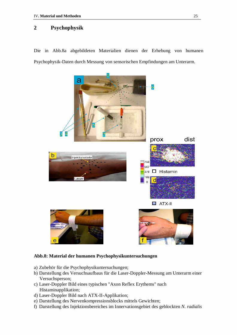
2 Psychophysik
Die in Abb.8a abgebildeten Materialien dienen der Erhebung von humanen
Psychophysik-Daten durch Messung von sensorischen Empfindungen am Unterarm.
Abb.8: Material der humanen Psychophysikuntersuchungen
a) Zubehör für die Psychophysikuntersuchungen; b) Darstellung des Versuchsaufbaus für die Laser-Doppler-Messung am Unterarm einer
Versuchsperson; c) Laser-Doppler Bild eines typischen "Axon Reflex Erythems" nach
Histaminapplikation; d) Laser-Doppler Bild nach ATX-II-Applikation; e) Darstellung des Nervenkompressionsblocks mittels Gewichten; f) Darstellung des Injektionsbereiches im Innervationsgebiet des geblockten N. radialis

IV. Material und Methoden 26
Als Probanden stellten sich neun gesunde, adulte Freiwillige im Alter zwischen 25 und
45 Jahren zur Verfügung. Die drei männlichen und sechs weiblichen Probanden wurden
genau über den Ablauf der Versuche sowie die Möglichkeit diese jederzeit, ohne
Angabe von Gründen abbrechen zu können, informiert. Alle Probanden erklärten
anschließend schriftlich ihr Einverständnis zu den Versuchen, die stets unter der
Aufsicht eines Humanmediziners durchgeführt wurden. Mit Hilfe der
Spinnennetz-Schablone (s. Abb.8a, Nr.1) wurden zunächst Punkte in regelmäßigem
Abstand auf die Palmarseite des Unterarmes übertragen (s. Abb.8b). Dieses Netz dient
dazu eventuell auftretende Rötungen und Missempfindungen in ihrer Ausbreitung
quantifizieren zu können. Im Zentrum des Netzes wurden mit einer Insulinspritze (s.
Abb.8a, Nr.2) 70µl einer 100nM ATX-II Lösung oder die gleiche Menge einer
Kontrolllösung intrakutan injiziert. In den folgenden zehn Minuten bewerteten die
Probanden Schmerz und Juckreiz alle 15s auf einer Skala von null bis zehn, wobei eine
Bemessung (engl. rating) von null keinerlei Schmerz bzw. Juckreiz und eine Bewertung
von zehn größten vorstellbaren Schmerz bzw. größten vorstellbaren Juckreiz bedeutete
(sog. numeric rating scale - NRS). Parallel wurde alle zwei min der relevante
Unterarmbereich auf einer Fläche von 7cm mal 10,8cm von einem Laser abgetastet (s.
Abb.8b). Dieses sogenannte "Laser-Doppler Imaging" dient dazu vermehrte
oberflächliche Hautdurchblutung, das sogenannte "Axon Reflex Erythem", wie es
beispielsweise nach Histaminapplikation auftritt (s. Abb.8c), zu detektieren. In Abb.8d
ist das Scanbild unter ATX-II Einfluss zu sehen. Die gleichbleibend blaue
Farbcodierung deutet auf ein Ausbleiben eines Axon Reflex Erythems hin. Auf
mechanische Allodynie wurde mit einem Baumwolltupfer sowie einem Pinsel getestet
(s. Abb.8a, Nr.3) und Kältemissempfindung mittels eines in Eiswasser auf 0°C
gekühlten Metallstabes (s. Abb.8a, Nr.5) untersucht. Um eine Differenzierung zwischen
A-Faser vermittelten und C-Faser vermittelten Empfindungen durchführen zu können,

IV. Material und Methoden 27
wurde ein sogenannter Nervenkompressionsblock angelegt. Hierfür wurde ein 2,5kg
schweres Gewicht (s. Abb.8a, Nr.6) über eine gepolsterte Schlinge am Handgelenk
oberhalb des N. radialis angebracht (s. Abb.8e). Nun wurde mit Hilfe des gekühlten
Metallstabes auf den Verlust der Kälteempfindung hin untersucht. Sobald der kalte
Metallstab lediglich als Druckempfindung wahrgenommen wird, ist von einer
ausreichenden Blockade der A-Fasern auszugehen und ATX-II wurde im
Innervationsgebiet des N. radialis (s. Abb.8f) intrakutan injiziert. Wiederum bewertete
der Proband über einen Zeitraum von zehn min Schmerz und Juckreiz auf der NRS von
null bis zehn. Während der gesamten Zeit wurde in regelmäßigen Abständen mit Hilfe
der in Abb.8a, Nr.4 gezeigte Metallspitze (sog. pinprick) sichergestellt, dass die C-Faser
Funktionalität noch erhalten ist. Hierbei wird die Metallspitze auf die Hautoberfläche
gesetzt und überprüft ob die Empfindung noch wahrgenommen wird. Anschließend
wurde der Block entfernt und noch einmal für zehn und nach 20min Schmerz und
Juckreiz auf der NRS bewertet. Die zunächst geblockten A-Fasern erholen sich während
dieser Zeitspanne und erlangen ihre Funktionalität zurück. So kann die Aussage über
faserspezifische Antworten noch einmal bestätigt werden, da Effekte die vor dem Block
vorhanden, währenddessen jedoch verschwunden waren nun wieder auftreten.

IV. Material und Methoden 28
3 Zellvorbereitung für RT-qPCR-Experimente
Nach der Entnahme der DRGs aus der Wirbelsäule der Maus werden sie mittels einer
Pasteurpipette vereinzelt. Die in Pufferlösung befindlichen Zellen werden mit dem
Zellkern-Markerfarbstoff Hoechst 33258 (Invitrogen, Life technologies, Darmstadt,
Deutschland) sowie dem Avital-Markerfarbstoff 7AAD (Invitrogen) für mindestens
30min auf Eis gekühlt inkubiert. Die so gefärbten Neurone werden in einen sog.
"FACS" Zellsortierer (Fluoreszenz aktivierte Zellsortierung, engl. fluorescence
activated cell sorting) der Firma BD science (FACS Aria II) überführt, der die
Zellsuspension aufnimmt und die Neurone entsprechend ihrer Größe sortiert. Die als
"klein" eingestuften DRGs wiesen durchschnittlich einen ∅ von 18µm auf, die Gruppe
der "großen" Neurone einen durchschnittlichen ∅ von 35µm. Nachdem die
verschiedenen Gruppen in Eppendorf-Tubes (Eppendorf, Hamburg, Deutschland)
sortiert waren, wurde ihre RNA mit Trizol ("Qiazol", Qiagen, Hilden, Deutschland)
isoliert und schließlich mit RT-qPCR Experimenten (in Kooperation im Labor
Alzheimer durchgeführt) die mRNA (messenger RNA; Boten-RNS) Menge der
größensortierten DRGs bestimmt. Diese erlaubt es, Rückschlüsse auf das
Expressionslevel von Nav1.6, Nav1.7 und β4 zu ziehen.
V. Veröffentlichung 29
V. VERÖFFENTLICHUNG

V. Veröffentlichung 30

V. Veröffentlichung 31
V. Veröffentlichung 32
Background The voltage-gated sodium channel subtype Nav1.7 plays a major role in human pain perception: Patients who lack functional Nav1.7 due to loss of function mutations are incapable of feeling pain [1]. Patients carrying mutations that lead to a gain of function of Nav1.7, on the other hand, suffer from inherited pain syndromes, such as the paroxysmal extreme pain disorder (PEPD, [2,3]). In humans nine different subtypes of sodium channels are expressed (Nav1.1 to Nav1.9), and six of them can be found in sensory neurons (the tetrodotoxin sensitive (TTXs) channels Nav1.1, 1.2, 1.3, 1.6 and 1.7, and the TTX resistant (TTXr) channels Nav1.8 and 1.9).
Voltage-gated sodium channels are responsible for action potential (AP) initiation in neurons and propagation along axons [4]. Upon depolarization, sodium channels open rapidly and inactivate within milliseconds, supporting membrane repolarization. On a molecular level, the inactivation gate, which is situated on the linker between the channel’s domains III and IV, swings into the open pore and thereby blocks the permeation pathway for sodium ions [5]. An endogenous blocking particle, most probably the C-terminus of the β4-subunit, may interfere with this process, allowing the induction of resurgent currents [6], that increase neuronal excitability. Resurgent currents are enhanced by conditions that slow fast inactivation, as this increases the possibility for the blocking particle to bind to the open channel. PEPD mutations have a slowed fast inactivation and exhibit resurgent currents when expressed in DRGs [7]. This also holds for HEK293 cells, provided that parts of the β4-subunit are present in the intracellular solution [8,9]. Several toxins are known to interact with the gating properties of voltage-gated sodium channels. ATX-II from the sea anemone Anemonia sulcata was shown to slow fast inactivation [10-12], and is therefore likely to induce resurgent currents. When divers get in contact with sea anemone, they report symptoms such as pain and itch. In order to learn more about the potentially painful effects of ATX-II on nociceptive sodium channel gating, we investigated small and large diameter DRGs with the whole-cell patch-clamp method. We can indeed show that ATX-II enhances persistent and resurgent currents in large diameter sensory neurons of the dorsal root ganglia (DRGs), which are thought to be linked to A-fibers of peripheral nerves [13]. Small DRGs on the other hand, which give rise to C-fibers, were not reported to display any endogenous resurgent currents [14] and also application of ATX-II failed to induce them. In order to correlate our findings with human sensations, we injected small amounts of ATX-II intradermally and examined the evoked sensations in healthy human subjects. Our results suggest that ATX-II may selectively activate A-fibers and thereby mediate itch-like sensations and pain.
Results
ATX-II increases resurgent and persistent currents in large diameter DRGs
Large DRGs are known to display resurgent currents [14]. As ATX-II impairs fast inactivation of sodium channels [10,15,16], we set out to test whether it might favor binding of the blocking particle and therefore enhance resurgent currents in DRGs. Upon repolarization following a strong depolarizing pulse (to +30 mV) we evoked resurgent currents in large DRGs that are clearly distinguishable from tail currents by their slower activation and decay kinetics (Figure 1). At the end of the 500 ms repolarizing pulse, a persistent current component was obvious (Figure 2a).

V. Veröffentlichung 33
Figure 1 Example traces for the isolation of ATX-II enhanced TTXs resurgent currents.
Representative recordings from one large diameter DRG neuron using the protocol shown in the upper panel in a. (a) Traces recorded under control conditions without any toxin present. (b) Traces recorded with 5nM ATX-II in the extracellular recording solution. (c) shows recordings in the presence of 5 nM ATX-II and TTX with no resurgent currents present. This trace was subsequently subtracted from the ones shown in (a) and (b), revealing the TTXs resurgent current pre (d) and post (e) application of ATX-II, respectively. Eight out of eight large DRGs recorded at 22 °C showed resurgent currents.
TTXs post TTXs pre

V. Veröffentlichung 34
Figure 2 ATX-II induces resurgent currents in large diameter DRGs. (a) Voltage protocol and representative TTXs resurgent current traces (black traces represent recordings at −45 mV) pre and post application of ATX-II in large diameter DRGs. Lower lane shows an overlay of traces recorded pre and post ATX-II at −45 mV on a longer time scale. Arrows illustrate resurgent current and the region of mean persistent current measurements. (b) Peak resurgent current as a function of voltage recorded at 22 °C (n = 8). Total current (TTXs and TTXr, black circles), peaked around −40 mV. Absolute total resurgent current (black circles) increased following application of 5 nM ATX-II (pink circles), and is mostly carried by TTXs sodium currents (square symbols). The overall resurgent current was dramatically reduced by application of TTX (grey circles). (c) TTXs resurgent current at 22 °C (filled squares, n = 8) and 30 °C (open squares, n = 14) is increased by application of ATX-II. Data points for 22 °C are the same as in (b) and shown for better comparison. (d) Mean persistent TTXs current (black squares, determined as shown in (a) lower panel) is increased by ATX-II exposure (pink squares), whereas an increase in temperature has a smaller effect (22 °C: filled squares, n = 8; 30 °C: open squares, n = 14). (e) Corrected TTXs resurgent current amplitudes as a function of voltage. Corrected traces were obtained by subtraction of TTXs persistent current (shown in d) from TTXs peak resurgent current (shown in c) of each trace. At 22 °C (filled symbols, n = 8) as well as at 30 °C (open symbols, n = 14) corrected resurgent currents are increased by ATX-II application. * p < 0.05, paired-sample T-test.

V. Veröffentlichung 35
Resurgent currents were clearly increased in large DRGs by addition of 5 nM ATX-II to the bath solution (Figures 1 and 2a and b). As expected for a fast binding toxin like ATX-II, the effect was clearly visible after a few seconds, and we started recordings 1.5 min after toxin application. Although ATX-II most likely increases tail current as well, the slow kinetics of the ATX-II induced current strongly suggests that ATX-II affects resurgent currents. TTX application reduced the total inward current, and resurgent currents were abolished, leaving only a marginal component (Figure 2b, grey circles). This indicates that resurgent currents are mainly mediated by TTXs channel-subtypes. Therefore in the following we isolated the TTXs sodium current by subtracting the TTXr component (Figure 1).
We have previously shown that resurgent currents are modified by the anticancer agent oxaliplatin in a temperature dependent manner [17], and therefore tested the effect of ATX-II on large diameter DRGs at 22 °C and 30 °C. Native TTXs resurgent or persistent currents were not affected by temperature (Figure 2c and d, black and white symbols). In the presence of ATX-II resurgent and persistent currents tended to be larger at 30 °C compared to 22 °C (Figure 2c and d, statistically not significant, peak current densities: At 30 °C, resurgent 92.2 ± 8.8 pA/pF, persistent 37.4 ± 5.9 pA/pF, n = 14. At 22 °C, resurgent 67.6 ± 9.9 pA/pF, persistent 20.4 ± 3.4 pA/pF, n = 8). From Figure 2c the activation of the TTXs resurgent current seems to be shifted to more negative potentials. However, when calculated as relative conductance, this shift is no longer detectable, suggesting that ATX-II solely enhances resurgent currents, and does not alter its voltage-dependence (Additional file 1: Figure S1). ATX-II induces a prominent persistent current component (Figure 2d), which may also affect the absolute resurgent current amplitude. In order to evaluate the ATX-II induced resurgent current amplitude in isolation, we subtracted the mean persistent currents measured at the end of the hyperpolarizing pulse from the peak inward resurgent currents (see Figure 2a, lower traces). It is evident, that a large component of the ATX-II effect is due to an increase in persistent currents (Figure 2e compared to Figure 2c). Nonetheless, corrected resurgent currents (Figure 2e) are enhanced by ATX-II at both temperatures tested (corrected resurgent current densities: 22 °C pre: 35.5 ± 7.7 pA/pF; 22 °C post: 57.0 ± 9.7 pA/pF, n = 8. 30 °C pre: 47.6 ± 5.4 pA/pF; 30 °C post: 106.9 ± 9.2 pA/pF, n = 14).
Steady-state fast inactivation of TTXs Navs in large DRGs was shifted to more hyperpolarized potentials by application of 5 nM ATX-II (Vhalf pre: -62.5 ± 1.2 mV, post: -66.8 ± 1.2 mV, p < 0.001), as was the voltage dependence of activation (Vhalf pre: -42.6 ± 1.1 mV, post: -45.0 ± 0.9 mV, p < 0.01, Additional file 2: Figure S2a). Surprisingly, a double-exponential fit to current decay did not reveal significant changes when cells were exposed to 5 nM ATX-II (Additional file 2: Figure S2b and 2c). It may be that the changes remained small and under our detection level, or that the underlying TTXs channel subtypes are affected to a different extent.
V. Veröffentlichung 36
ATX-II is unable to induce resurgent currents in small DRGs
Up to now, no resurgent currents were described in small diameter DRGs [14], and accordingly, we were unable to detect any significant resurgent or persistent sodium currents in sensory neurons <25 μm (Figure 3a, upper traces, for criteria for resurgent current detection see Methods section). Addition of 5 nM ATX-II at 22 °C or 30 °C did not induce any detectable resurgent or persistent sodium currents (Figure 3b and c), although application of higher concentrations, such as 25 nM induced a significant persistent current in small DRG neurons (Additional file 3: Figure S3). In contrast, voltage dependence of activation and steady-state fast inactivation were shifted to more negative potentials by 5 nM ATX-II (Activation: Vhalf pre: -34.9 ± 1.0 mV, post: -40.7 ± 0.8 mV, steady-state fast inactivation: Vhalf pre: -74.7 ± 2.4 mV, post: -80.5 ± 2.0 mV, both p < 0.001, Additional file 2: Figure S2a). A double-exponential fit of current decay did not reveal any significant changes when 5 nM ATX-II was applied to small DRGs (see Additional file 2: Figure S2b and c). Our results suggest that either small DRGs possess a set of sodium channels less sensitive to ATX-II, or that they express insufficient amounts of the key components necessary for the generation of resurgent currents. Resurgent currents are thought to rely on the presence of an endogenous blocking particle, which is most likely formed by the β4-subunit, or at least parts of it [9].
Different types of DRGs were suggested to express separate sets of sodium channel α- and β-subunits. In situ hybridization and functional studies suggest that Nav1.6 is predominantly found in large DRGs, whereas Nav1.7 is reported to be functionally responsible for AP propagation in C-fibers [18-20], which are the neurites of small DRGs [13]. β4 was described to be expressed at a higher level in large diameter DRGs than in small [21]. In order to assess the expression of β4, Nav1.6 and Nav1.7 in our preparation, we sorted dissociated DRGs according to their size using FACS (see Methods). The mRNA of size-sorted cells was isolated and we confirmed by RT-qPCR that in our preparation mRNA of the β4-subunit is twice as strongly expressed in large compared to small DRGs (Figure 3d). Nav1.6 mRNA levels were more prevalent in large DRGs compared to small DRGs whereas Nav1.7 mRNA was expressed at comparable levels between the two groups. Thus, the lack of resurgent currents in small DRGs might be due to insufficient amounts of β4-subunits, as there is evidence that the β4-subunit plays an important role in the generation of resurgent currents [9,22].
We added a 14 amino acid peptide of the β4-subunit C-terminus to the pipette solution, which enables the recording of resurgent currents in heterologously expressed sodium channels [8,23,24]. With the β4-peptide added to the pipette solution, we were able to record very small, hardly detectable, resurgent currents in one small diameter DRG out of four (example trace in Figure 4a, upper lane). When 5 nM ATX-II was present in the bath solution, resurgent currents were recorded from four out of six cells. The amount of induced persistent current was small, and correction for it (compare to Figure 2e) still revealed a robust enhancement of resurgent currents in small diameter DRGs by ATX-II when the β4-peptide was added to the pipette solution (Figure 4c).

V. Veröffentlichung 37
Figure 3 Small diameter DRGs are unaffected by ATX-II or increased temperature. (a) The same stimulation protocol as used in large diameter DRGs was applied to small DRGs (upper lane). Example traces of TTXs sodium currents pre and post 5 nM ATX-II application are shown. Lower lane displays an overlay of pre and post ATX-II traces at a voltage of −45 mV. (b) TTXs peak current at the time point at which resurgent currents would be expected as a function of voltage in small diameter DRGs. Neither at 22 °C (filled symbols, n = 7) nor at 30 °C (open symbols, n = 4) a significant ATX-II-induced resurgent current is evident (paired-sample T-test). (c) Mean persistent current as a function of voltage pre (black symbols) and post (pink symbols) ATX-II application. In small diameter DRGs there is no significant increase in persistent current detectable due to ATX-II exposure or heating to 30 °C (paired-sample T-test). (d) Expression levels of β4, Nav1.6 and Nav1.7 mRNA normalized to the mean of two house-keeping genes in FACS-sorted small (white bars) and large (grey bars) DRGs determined by RT-qPCR. In large DRGs β4 and Nav1.6 expression is twice as high compared to small neurons. On the contrary, Nav1.7 mRNA is evenly expressed in small and large DRGs (n = 3; * p < 0.05).

V. Veröffentlichung 38
Figure 4 ATX-II induces resurgent currents in small diameter DRGs with the β4-C-terminus peptide in the pipette solution.
(a) Voltage protocol (top panel) and representative current traces of recordings from small diameter DRGs with the β4-peptide added to the pipette solution. Lower lane shows an overlay of recordings from control and ATX-II (5 nM) treated cells at −45 mV. (b) TTXs peak current within the first 10 ms after repolarization of control (black symbols, n = 4) and ATX-II exposed (pink symbols, n = 6) small DRGs. Under ATX-II exposure a distinct resurgent current can be recorded, when the β4-peptide is present in the pipette solution. (c) The increase of resurgent current is not due to a persistent current component, as subtraction of the persistent current (determined at the end of the hyperpolarizing pulse, see lower panel in a) from peak current reveals a clear corrected TTXs resurgent current component. * p < 0.05, independent-sample T-test.

V. Veröffentlichung 39
Nav1.7 and Nav1.6-mediated resurgent currents are enhanced by extracellular ATX-II
Our results suggest that it is the amount of β4 present in the cell that determines whether ATX-II can increase resurgent currents in DRGs or not. mRNA of Nav1.7 in small DRGs is three to seven times more strongly expressed than Nav1.6, and its expression level does not vary much between large and small DRGs (Figure 3d, [18]). We used a HEK293 cell line stably expressing Nav1.7 and tested for resurgent currents (Figure 5, left column). Up to date no resurgent currents could be evoked when Nav1.7 was transfected either alone or in combination with the β4-subunit, also shown before for Nav1.1 and Nav1.6 [17,23,25]. We were able to record very small resurgent currents in three out of 15 Nav1.7 expressing cells (20.0 %) when the β4-peptide was added to the pipette solution. With ATX-II in the recording solution, about half of the cells exhibited resurgent currents (17 out of 32, 53.1 %), and their mean resurgent currents were larger than that without ATX-II (Figure 5). ATX-II also increased persistent currents of these cells, but correction for it did not alter the ATX-II effect on Nav1.7 mediated resurgent currents much (Figure 5c, d). Our RT-qPCR data indicate that Nav1.6 shares its expression pattern between large and small DRGs with β4 and this Nav subtype is linked to resurgent currents in the literature [26-28]. We heterologously expressed Nav1.6 in N1E115 cells and tested for its ATX-II sensitivity with the β4-peptide present in the pipette (Figure 5, right column). In seven out of 17 cells (41.2 %) a resurgent current was detectable without ATX-II, which increased significantly when the toxin was applied (twelve cells out of 17, 70.6 %). Although the persistent current was enhanced in the presence of ATX-II, too, correction for it still revealed a significant increase of ATX-II induced resurgent currents in Nav1.6 (Figure 5d), which is in accordance with our findings in large DRGs (Figure 2).

V. Veröffentlichung 40
Figure 5 ATX-II induces resurgent currents in Nav1.7 and Nav1.6 expressing cells when β4-peptide is present in the pipette solution. (a) Voltage protocol (top panel) and representative current recordings with the β4-peptide added to the pipette solution (lower panel): left: stable HEK cell line expressing Nav1.7; right: Nav1.6 transfected N1E115 cells. An overlay of recordings under control conditions and ATX-II (5 nM) exposure at −20 mV (for Nav1.7) and −10 mV (for Nav1.6) is shown. (b) Peak current within the first 10 ms after repolarization of control (black symbols) and ATX-II exposed cells (pink symbols, left: Nav1.7, n = 15,17; right: Nav1.6, n = 11,11 for control and ATX-II, respectively). Resurgent currents are enhanced by the presence of ATX-II, when the β4-peptide is present in the pipette solution. (c) Mean persistent currents at the end of the repolarizing pulse are affected in Nav1.6 (right) and in Nav1.7 (left) by addition of extracellular ATX-II to the bath solution (pink symbols). (d) The increase of resurgent current is not due to a persistent current component, as subtraction of the persistent current from peak current reveals a clear corrected resurgent current component in both Nav1.7 (left) and Nav1.6 (right) expressing cells. * p < 0.05, independent-sample T-test for Nav1.7, paired-sample T-test for Nav1.6.

V. Veröffentlichung 41
Like the resurgent currents, the inactivation kinetics of Nav1.6 seem to be more sensitive to ATX-II than those of Nav1.7 (Figure 6a). A double-exponential fit of current decline revealed that in both channels τ 2, the slower time constant is affected by application of 5 nM ATX-II (Figure 6c). Unlike Nav1.7, the relative A1 of Nav1.6 is significantly decreased (Figure 6b), thereby increasing the importance of the slow component in current decay. Slowing the process of inactivation may increase the likelihood of the β4-peptide to bind to the open channel.
Figure 6 ATX-II prolongs slow time constant of inactivation of Nav1.7 and Nav1.6. (a) Voltage-dependence of activation and steady-state fast inactivation of heterologously expressed Nav1.7 (left, control n = 8, ATX-II n = 22) and Nav1.6 (right, n = 17), both recorded with the β4-peptide in the pipette. For results of Boltzmann-fits see text. (b) and (c): Results from a double-exponential fit to current decay of traces evoked by the activation protocol from cells expressing Nav1.7 (left, n = 4-22) or Nav1.6 (right, n = 7–17). (b) Fractional A1 (A1/(A1 + A2)) is shown as a function of voltage. (c) Fast and slow time constants τ of current decay are shown as a function of voltage. In both channel subtypes (Nav1.7 left column, Nav1.6 right column) only slow time constants (circles) are significantly prolonged, whereas fast time constants (squares) are unaffected by ATX-II. Independent-sample T-test for Nav1.7, paired-sample T-test for Nav1.6, * p < 0.05, ** p < 0.01, *** p < 0.001.

V. Veröffentlichung 42
In the presence of the β4-peptide, ATX-II shifts the activation of Nav1.6 and Nav1.7 in opposite directions (Vhalf for Nav1.6 pre: -18.2 ± 1.1 mV, post: -20.6 ± 1.2 mV, n = 17; for Nav1.7 control: -19.4 ± 1.6 mV, n = 8, ATX-II: -14.8 ± 1.1 mV, n = 22, p < 0.05, Figure 6a). This opposite effect of ATX-II on Navs suggests that the shift of activation that we see in small DRGs (Additional file 2: Figure S2a) may be due to ATX-II effects on other Nav subtypes than Nav1.7, or due to the absence of the interacting β4-subunit. The slope of activation remained unchanged (for Nav1.7 control: 6.75 ± 0.41, ATX-II: 7.4 ± 0.29, for Nav1.6 control: 6.3 ± 0.2 ATX-II: 5.9 ± 0.3, both not significantly different). Steady-state fast inactivation of Nav1.6 and Nav1.7 remained largely unaffected by 5 nM ATX-II, and only the slope of Nav1.6 fast inactivation became marginally less steep (Vhalf for Nav1.7 control: -75.3 ± 2.9 mV, ATX-II: -70.8 ± 1.5 mV, for Nav1.6 control: -64.9 ± 1.2 mV, ATX-II: -64.5 ± 1.2 mV, both not significantly different. Slope for Nav1.7 control: 7.0 ± 0.6, ATX-II: 6.6 ± 0.5, n = 3–8, for Nav1.6 control: 6.3 ± 0.2, ATX-II: 5.9 ± 0.3, n = 17, Figure 6a). Small DRGs give rise to C-fibers, whereas large DRGs were reported to be linked to A-fibers [13]. As 5 nM ATX-II affected large DRGs and did not induce a resurgent current in small DRGs, we predict that in humans ATX-II should preferentially affect A-fiber-mediated somatosensation, while leaving C-fiber-dependent sensations largely intact. In order to test for ATX-II evoked sensations, we performed a psychophysical trial with healthy volunteers.
Human sensory response to intradermally injected ATX-II is mediated by A-fibers
Upon intradermal injection of ATX-II on the forearm, all subjects reported unpleasant and painful prickling or tingling sensations, which were not of burning character. These sensations were described to be different to C-fiber mediated pain as it is for example reported following capsaicin injection. The pain was also described as "pulsating" or “cold snowflakes which are hitting the skin”. In parallel to pain subjects reported itch-like sensations, which were characterized as "if the skin was tickled with a thin hair”, “an insect walked on the skin”, or “liquids dropped on the skin”. Subjects were asked to rate their sensations on a numerical rating scale (NRS, 0–10, with 0 being no pain and 10 the worst imaginable pain) and mean NRS ratings were 1.3 ± 0.1 for both, pain and itch-like sensations in the first 10 min after the injection (Figure 7a and b show representative examples each), and declined within ~10 min after the end of the experiment. A control group was injected with ATX-II-free buffer solution and did not report any itch or pain, apart from a small immediate short lasting pain which was most likely evoked by mechanical irritation due to the injection needle (mean < 0.3 on the NRS, n = 9, 3 male and 6 female). The AUCs of pain and itch ratings were significantly higher in the ATX-II-injected group compared to the controls (Figure 7a and b, n = 6 each, p < 0.004; p < 0.008, respectively, Mann Whitney U-test). Static or mechanical allodynia was absent (tested by gentle pressure with a cotton swab and stroking with a fine brush 10 min after injection of ATX-II, not shown). NRS pain ratings (0–10) to a 7 s lasting heat stimulus of 47 °C were unchanged compared to preinjection conditions (n = 6, not shown).

V. Veröffentlichung 43
Figure 7 Intradermal injections of ATX-II induces pain and itch-like sensations in human volunteers. (a) Pain assessment (on a numerical rating scale, NRS 0–10) as a function of time for a subject following injection of saline solution without (control) or with ATX-II. Under control conditions (white symbols) only a short lasting pain is evoked immediately after the injection in agreement with mechanical irritation, whereas ATX-II injection (blue symbols) leads to distinctly augmented ratings. The right panel shows the mean area under the curve (AUC) for n = 6 volunteers (* p < 0.05). (b) Assessment of itch-like sensations on a NRS ranging from 0–10 following injection (sensation rated as 3 is equivalent to desire to scratch) of one representative subject. While under control conditions (white symbols) no itch is induced following injection, ATX-II (red symbols) evoked itch-like sensations, also reflected in a larger AUC (right, * p < 0.05). (c) and (d) Pain (blue squares and bar) and itch (red squares and bar) ratings as a function of time following compression induced nerve block. After 30–40 min of A-fiber block ATX-II was injected and sensations were rated during 10 min (hatched squares). While no pain and itch is evoked by ATX-II under nerve block conditions, pain (blue symbols) and itch (red symbols) recur fast after removal of the block. AUCs of pain ratings increased significantly after removal of the block (* p < 0.05). (e) Area of increased blood flow measured by laser doppler imaging every 2 min following ATX-II injection (blue squares, n = 6) shows hardly any increase in superficial blood flow. For comparison, a typical histamine-induced axon reflex erythema mediated by activation of C-fibers and concomitant CGRP release is shown (white squares). Right: Example images taken 4 min after application of substances. (f) Assessment of cold-evoked (0 °C, 7 s) pain (NRS 0–10) before (white bar) and during nerve block (hatched bars). 10 min after injection of ATX-II cold-evoked pain is rated and the nerve block removed. Cold-evoked pain 20 and 30 min after injection of ATX-II significantly increases following removal of the nerve block (* p < 0.05).
basal time [min]
V. Veröffentlichung 44
A typical symptom of C-fiber activation is the axon reflex flare [29]. Following application of ATX-II, laser Doppler scanning detected a small increase in local blood flow due to the injection itself, but there was no widespread axon reflex flare (n = 6, Figure 7e, blue squares). A classical axon reflex erythema evoked by histamine (1 % applied by iontophoresis) is shown for comparison (Figure 7e, white squares). In humans, histamine activates the group of mechano-insensitive C-fibers, which release calcitonine gene-related peptide (CGRP) upon activation causing widespread vasodilatation [30] when evoked action potentials are antidromically propagated into branching nerve endings.
Our patch-clamp experiments suggest that ATX-II preferably activates A-fibers (connected to large diameter DRGs), whereas its effect on C-fibers (linked to small DRGs) is small or negligible. For further validation of this hypothesis a mechanical differential nerve block was applied prior to ATX-II injection, which mainly affects A-fibers [31,32]. During differential nerve block, no pain (apart from a brief injection pain) or itch-like sensations were reported, revealing a mean AUC during nerve block of 1.3 ± 0.9 and 0.0 ± 0.0, respectively (hatched squares Figure 7c and d). After removal of the nerve block pain and itch-like sensations immediately arose within seconds and were described as “identical” to those following ATX-II injection without a nerve block by volunteers who had undergone this procedure before. This indicates that mechanical block successfully interfered with ATX-II evoked sensations.
NRS curves for pain and itch were recorded for 10 min following removal of the differential nerve block, and subjects reported spontaneous pain (Figure 7c, blue squares). AUC values for pain ratings post nerve block were significantly higher than during nerve block (n = 6, p < 0.03; Wilcoxon matched pairs test, Figure 7c, bar graph). However, no itch-like sensations were reported by 50 % of the volunteers following ATX-II injection after removal of the nerve block, indicative of a faster decline of the itch-like component of ATX-II evoked responses over time compared to pain. This further suggests that ATX-II mediates its effects in humans via A-fibers.
NRS pain ratings in response to a cold stimulus (7 s, 0 °C) were used to determine progression of the differential nerve block. Mean ratings before treatment were 0.1 ± 0.1, and cold sensation was lost under nerve compression before injection of ATX-II (Figure 7f, white bar). During the differential nerve block only very little cold evoked pain was recorded after ATX-II injection (Figure 7f, blue hatched bar). Removal of block, however, lead to a significantly increased cold evoked pain rating and discomforting mechanical sensations 20 and 30 min after ATX-II injection compared to basal ratings (both p < 0.002*, ANOVA (p < 0.001), following HSD post hoc test). This phenomenon was limited to the site of ATX-II injection within the area of the differential nerve block. The strongly enhanced cold evoked pain following ATX-II injection most likely depends on A-fiber activation, as it was abolished by differential nerve compression.

V. Veröffentlichung 45
Discussion Here we show, that ATX-II enhances persistent and resurgent currents in large diameter DRGs (Figure 2), but fails to do so in small DRGs (Figure 3). This is most likely due to a lack of sufficient β4-subunit expression in these small neurons (Figure 3d), which is thought to be essential for the generation of resurgent currents. Addition of the β4-peptide to the pipette solution enabled us to record ATX-II-inducible resurgent currents in small DRGs and HEK cells stably expressing Nav1.7. Large DRGs are linked to A-fibers, and supporting our in vitro findings, injection of small amounts of ATX-II into the skin of human volunteers evoked responses in accordance with A-fiber activation.
Large DRGs exhibit endogenous resurgent currents that can be enhanced by ATX-II. This is not the case for small DRGs, even though small DRGs are known to express ATX-II sensitive sodium channels (such as Nav1.1, Nav1.2 and Nav1.6 [16,18,20,33]). Mice lacking functional Nav1.6 (med mice) display reduced resurgent currents [34], and there is evidence, that the interaction of the β4-subunit with sodium channels is a major precondition for resurgent currents to occur (e.g. [35]). Our RT-qPCR data of the FACS sorted DRGs show that large DRGs have higher levels of β4 mRNA than small neurons (Figure 3d and [21,34]). Considering the primer efficiencies, DRGs of both sizes have ~5 times more β4 than Nav1.6 (5 and 4.5 times for small and large DRGs, respectively), which would be ample for a simple 1:1 interaction. In small DRGs Nav1.7 is expressed ~7 times stronger than Nav1.6, making it more likely for the β4-subunit to interact with Nav1.7 than with Nav1.6 in small DRGs. It may be possible that Nav1.7 has a lower affinity for β4, and therefore needs higher concentrations for a successful interaction, which could be provided by addition of β4-peptide to the recording solution (Figures 4 and 5). Compared to Nav1.7, Nav1.6 shows resurgent currents in a larger population of cells under control condition with the peptide in the pipette (53.1 % vs. 26.7 %, respectively). Nevertheless it might be hard to compare channels expressed in a cell line and draw conclusions to the extent of its modification in its native environment as for example in DRGs. If the peptide was present, seven out of 17 cells transfected with Nav1.6 show resurgent currents, whereas only three out of 15 cells expressing Nav1.7 showed these currents by themselves. Although the cellular background may play a role, these data suggest that Nav1.6 is more prone to interact with the β4-peptide and to produce resurgent currents. It may be possible that the amount of endogenous β4-subunit in small DRGs is simply not sufficient for a robust interaction with any endogenous TTXs sodium channel expressed in these cells and therefore no resurgent currents can be recorded. An increase in ATX-II concentration up to 25 nM failed to induce resurgent currents in small DRGs (Additional file 3: Figure S3), but only enhanced persistent currents. This finding is comparable to that of the application of the wasp venom β-Pompilidotoxin to CA3 neurons [36], which induced tail and persistent, but no resurgent currents unless the β4-peptide was present in the recording pipette. Our data suggest that small DRGs share this characteristic with CA3 neurons. An enhanced persistent current may very well increase excitability by its own, and indeed application of 1 μM ATX-II to IB4 negative small DRG neurons resulted in a broader AP [37]. Interestingly, ATX-II sensitivity seems to be distinct between the different subsets of small DRG neurons, as IB4 positive small DRGs did not change their AP properties in response to ATX-II application. The toxin concentrations that we used in our study induced a shift of TTXs current activation and steady-state fast inactivation (Additional file 2: Figure S2), but failed to have effects on persistent or resurgent currents (Figure 3). Therefore, it is likely that the toxin concentrations used in our experiments remained below threshold for an effect on persistent or resurgent currents in small DRGs, and that in small DRGs persistent and resurgent currents are less sensitive to small amounts of ATX-II than activation and steady-state fast inactivation.

V. Veröffentlichung 46
Surprisingly, mRNA for Nav1.7 is expressed in large and small DRGs at comparable levels (Figure 3d, [18]). Patients with the inherited pain syndromes erythromelalgia and PEPD, both due to mutations in Nav1.7, describe their pain mainly as burning - a typical C-fiber associated characteristic. As Nav1.7 seems to be evenly distributed between large and small DRGs, it is likely that associated proteins, such as the β4-subunit, or cell-background specific channel modifications, may play a major role in the generation of Nav1.7 related pain. The discrepancy may also be due to species differences: We investigated DRGs from mice, but erythromelalgia and PEPD manifest themselves in humans. Knowledge about the developmental regulation of β4 expression is still lacking. PEPD mutations of Nav1.7 were shown to induce resurgent currents by slowing fast inactivation [7,8]. We show in this study, however, that even in the presence of ATX-II no resurgent current could be evoked in small DRGs or Nav1.7 expressed in HEK293 cells unless the β4-peptide is present. Patients with PEPD are severely affected during their first year of life but symptoms become somewhat milder later in life. It remains speculative if this might be due to a down regulation of β4 with age, for which data are currently not available.
ATX-II alone was unable to induce resurgent currents in heterologously transfected Nav1.7 or Nav1.6, which was reported before [38]. Up to now, only one toxin, the β-scorpion peptide Cn2, was able to induce resurgent currents in a heterologous expression system without the addition of an intracellular peptide in Nav1.6 [38]. These currents were enhanced by ATX-II. This finding suggests that sodium channels need some kind of modification, like treatment with Cn2 or potentially a disease causing mutation, such as PEPD, in order to be capable to produce resurgent currents.
In the 70s and early 80s of the last century, it was observed that ATX-II broadens the AP of crustea peripheral axons, and not much later it became evident that this was due to a slowing of inactivation properties of voltage-gated sodium channels [10,15,39,40]. When applied to large DRGs in high concentrations (10 μM), it significantly slowed τh of Nav current inactivation [41]. Here, we used concentrations that are an order of magnitude lower and although we used recombinant ATX-II (purity >98 %), which may be more specific in its effects than purified ATX-II, the effect on the peak current decay time constants was no longer detectable (Additional file 2: Figure S2 b,c), but nevertheless resurgent currents were altered significantly. Also, we observed shifts in activation and steady-state fast inactivation. This suggests that even though the effects on inactivation kinetics were small, the concentration of ATX was high enough to enhance resurgent currents, which in large DRGs seem very sensitive to even small channel alterations. Higher concentrations of ATX-II evoked resurgent currents in large DRGs, which were too large to be properly recorded under our conditions. This suggests that 5 nM ATX-II is a concentration at which effects may be evoked in large, but not in small DRGs, thereby allowing a separation of the two groups.
Molecular studies discriminated between sodium channel subtypes in heterologous expression systems for their sensitivity to ATX-II, showing that mainly Nav1.1, Nav1.2 and Nav1.6 are modified at very low concentrations [16,33,38]. These three channels can be found in large and small DRGs [18,20] and in large, but not small DRGs their modification by ATX-II enhances resurgent currents.
ATX-II slows inactivation kinetics for both channels investigated in this study: Nav1.6, which is supposed to be the main carrier of the AP in large DRGs and Nav1.7, which is suggested to play this role in small DRGs [19]. The slower fast inactivation occurs the higher the probability is for a potential open channel blocker to interact with the pore. Consequently, in the presence of the β4-peptide, we were able to significantly increase resurgent currents in both channel subtypes (Figure 5). These experiments suggest that two requirements are necessary for ATX-II to increase resurgent currents: I) an ATX-II sensitive sodium channel (such as Nav1.7 or Nav1.6, but also other subtypes, [16,33,38]) and II) the availability of an intracellular open channel blocker. Both seem to be present in large DRGs, whereas our experiments suggest that small DRGs are missing at least one of the components. As the TTXs sodium channels that were shown to be expressed in small DRGs are almost all sensitive to ATX-II (Nav1.1, 2, 6 and 7, [16,18,20], Figures 5 and 6), even at the low concentrations that we used here, it is quite likely that

V. Veröffentlichung 47
small DRGs are lacking sufficient open channel blocker. Accordingly, when we added the β4-peptide to the intracellular recording solution, resurgent currents were present in small DRGs (Figure 4).
Recently we showed that the anticancer agent oxaliplatin selectively enhances persistent and resurgent currents in large diameter DRGs at low temperatures [17]. Oxaliplatin is highly effective but unfortunately has dose limiting side-effects: acute and chronic neuropathies, which are both characterized by cold-evoked very unpleasant dysaesthesias. These sensations are similar to those experienced by the subjects of the present study following intradermal ATX-II injection. Besides an increased cold perception 20 and 30 min after ATX-II injection a pricking sensation outlasting the cold stimulus was observed (see Figure 6f), indicating ongoing activity in the affected nerve fibers.
ATX-II did not display a marked temperature dependence in its enhancement of resurgent or persistent currents, which is in contrast to oxaliplatin (Figure 2 and [17]). Low temperatures induce a general increase in membrane resistance, which would then allow a small depolarizing current, like resurgent currents, to be potentially sufficient to initiate a following AP. This may explain why the subjects report cold-enhanced sensations after ATX-II injection (Figure 7f); even though resurgent currents were not significantly different at 22 °C compared to 30 °C (Figure 2).
Oxaliplatin failed to affect C-fibers or small diameter DRGs, suggesting that this drug, too, relies on the presence of the β4-subunit for its effects, or displays some degree of subtype specificity. The presence or absence of the endogenous blocking particle might thus render certain types of neurons susceptible for activation by toxins or drugs which act via voltage-gated sodium channels. Thus, focusing only on specific sodium channel subtypes for the treatment e.g. of pain might not be reaching far enough, but the channels functions in their cellular context need to be considered.
Pain sensations in vivo may be transmitted via two main types of fibers: the myelinated fast conducting A-fibers, which are connected to large DRGs, and the slower and unmyelinated C-fibers which are linked to small diameter DRGs [13]. We have shown that low concentrations of ATX-II activate large DRGs suggesting that A-fibers convey the ATX-II evoked sensations of the volunteers. Accordingly, mechanical nerve fiber block abolished the ATX-II evoked sensations. Most likely Aδ-fibers mediate the ATX-II induced pain symptoms but may also participate in the itch-like sensations. Recently it was shown that the effects of intradermal insertion of spicules from the pods of a cowhage plant induce intense itching. Aδ-fibers seem to be involved in transmitting this itch [42]. In some subjects, these sensations were markedly reduced by mechanical nerve block. Intradermal injection of ATX-II, on the other hand, evoked itch-like sensations, that were not described as common itch, but unpleasant sensation, for which there is no adequate word. “Tingling” was used by some subjects, which may be an overlapping sensation induced by ATX-II and cowhage spicules. Apart from Aδ-fibers it is likely that Aβ-fibers are activated by ATX-II since sensations of mechanical character were described, such as "like punctured by many thin needles". Possibly, the uncommon simultaneous activation of Aδ- and Aβ-fibers may generate the unique ATX-II mediated perception.
In our in vivo experiments the A-fiber block may induce paraesthesias itself, both during and upon recovering from the block. However no paraesthesias were described prior to and after ATX-II injection. Nonetheless, the reported sensations after ATX-II injections and relief of block were comparable to those described when no prior block was applied. The fact that cold evoked pain, which strongly increased after removal of the block, was closely restricted to the injection site (Figure 7f) argues against a general block-induced fiber irritation. Nevertheless, an irritation of the fibers cannot be ruled out, but we estimate it to be rather small in comparison to the effects evoked by ATX-II alone.
Intradermal injection of ATX-II did not evoke a large axon reflex flare (Figure 7e), arguing against a major activation of mechano-insensitive C-fibers [29]. Nevertheless, mechanosensitive C-fibers may mediate itch without the induction of a major axon reflex flare [43,44]. Subjects tested with cowhage spicules, which activate only mechano-sensitive C-fibers vigorously and Aδ-fibers, report quite different

V. Veröffentlichung 48
sensations than those injected with ATX-II (burning pain, severe itch, which was also clearly labeled as such). As mechano-sensitive C-fibers are also resistant to differential nerve block and should therefore have transmitted the ATX-II-mediated sensations, we assume that mechano-sensitive C-fibers are not a major target of ATX-II in humans.
Conclusions ATX-II enhances persistent and resurgent sodium currents in large diameter DRGs, whereas small DRGs depend on the addition of β4-peptide to the pipette recording solution for ATX-II to affect resurgent currents. Mechanical A-fiber blockade abolishes all ATX-II effects in human skin (e.g. painful and itch-like sensations), suggesting that it mediates its effects mainly via activation of large diameter A-fibers.
We thus established low concentrations of ATX-II as a tool for A-fiber activation, which induces pain and itch-like symptoms in humans. We show that activation mainly of A-fibers can be crucial for unpleasant pain and itch-like sensations, which are not extremely painful but could be distressing and reducing quality of life. We also showed for the first time, that this toxin is able to enhance resurgent currents of large DRGs, and of small DRGs or heterologously expressed Nav1.7, when the β4-peptide is present.
Methods
Animals and cell culture
Adult C3HeB/FeJ mice were anaesthetized with halothane (Sigma-Aldrich GmbH, Steinheim, Germany) and decapitated in accordance with ethical guidelines established by German animal protection law, approved by the animal protection committee of Regierung Mittelfranken, Germany. After decapitation, the backbone was removed rapidly and bisected longitudinally in ice-cold PBS (PAA Laboratories GmBH, Pasching, Austria). DRGs were removed, desheated, cleaned and enzymatically dissociated in Dulbecco’s modified Eagle's Medium (DMEM, Gibco-Life technologies, New York, USA) supplemented with collagenase/protease for 45 min at 37 °C. Cells were mechanically triturated with small diameter pasteur pipettes in TNB medium (Biochrom AG, Berlin, Germany). Poly-D-Lysin (Sigma-Aldrich GmbH, Munich, Germany) coated cover slips were loaded with the cell suspension and after cells settled for 5–10 min TNB medium was added. DRGs were incubated at 37 °C and 5 % CO2 until experiments were performed at the following day. A stable HEK293 cell line expressing hNav1.7 was cultured under same conditions as DRGs in DMEM containing 500 mg/l G418 (Sigma-Aldrich GmbH). N1E115 neuroblastoma cells were maintained in DMEM medium (Gibco-Life technologies) including 4.5 g/l Glucose, 10 % fetal bovine serum (Biochrom AG) and 1 % Penicillin/Streptomycin (PAA Laboratories GmbH). One day before transfection N1E115 cells were plated on 3.5 cm dishes (Falcon Corning, IBD) and transfected with Nanofectin (PAA Laboratories GmbH) according to the manufacturers protocol using 1 μg mNav1.6R (TTX resistant [45,46], in pCDNA3) and 0.5 μg EGFP-C1 (Clontech Laboratories,

V. Veröffentlichung 49
Inc., Mountain View, USA) in order to detect transfected cells via green fluorescence. Cells were recorded 2 to 3 days after transfection. Whole-cell patch-clamp recordings and electrophysiology
Whole-cell voltage clamp recordings were performed using glass electrodes with tip resistances of 1.5-2.0 MΩ, manufactured with a DMZ puller (Zeitz Instruments GmbH, Martinsried, Germany) and filled with an internal solution comprising for DRGs (in mM): 140 CsF, 10 NaCl, 10 HEPES, 1 EGTA and 5 TEA-Cl (adjusted to pH 7.4 with CsOH), for HEK cells (in mM): 135 CsCl, 5 NaCl, 2 MgCl2ATP, 10 HEPES and 5 EGTA (adjusted to pH 7.4 with CsOH) and for N1E115 cells (in mM): 125 CsCl, 5 NaCl, 2 Mg2ATP, 10 HEPES and 5 EGTA (adjusted to pH 7.4 with CsOH). For recordings of activation and steady-state fast inactivation of DRGs, an internal solution with reduced sodium chloride (2 mM NaCl) was used. The external bathing solution contained for DRGs (in mM): 140 NaCl, 3 KCl, 1 MgCl2, 1 CaCl2, 10 HEPES, 5 Glucose, 20 TEA-Cl, 0.1 CdCl2 (adjusted to pH 7.4 with NaOH), except for recordings of activation and steady-state fast inactivation, where NaCl was reduced to 10 mM, substituted with CholinCl, for HEK cells (in mM): 150 NaCl, 3 KCl, 2 MgCl2, 1.6 CaCl2, 0.4 CdCl2,
10 HEPES, 10 Glucose, 10 TEA-Cl (pH = 7.4, adjusted with NaOH) and for N1E115 cells (in mM): 145 NaCl, 4 KCl, 2 MgCl2, 2 CaCl2, 10 HEPES, 10 Glucose, 10 TEA (adjusted to pH 7.4 with NaOH).
DRGs were classified according to their cell size as large (> 35 μm) or small (< 25 μm). Size was measured as the maximal diameter of the cells on a digital image taken with an EC3 Camera (Leica, Wetzlar, Germany) connected to a Leica-DCMI3000B microscope with a 64 x objective. Recordings were performed at either room temperature (22 ± 1 °C) or 30 ± 1 °C, controlled by an inline solution heater (Warner Instruments, Hamden, USA) with a feedback thermocouple positioned less than 800 μm from the recorded cell. A HEKA EPC-10USB (Lambrecht, Germany) was used for signal recording, low-pass (10 kHz) filtering and digitizing (100 kHz). For substance application the tip of a perfusion system was positioned within 800 μm of the recorded DRG neuron. Pipette potential was zeroed prior to seal formation and capacitive transients were compensated using C-fast for pipette-capacity correction and subsequently C-slow for cell-capacity compensation (PatchMaster, HEKA, Lambrecht, Germany), the series resistance was compensated by about 50 % leaving a mean resistance of 3.8 ± 0.4 MΩ. The absolute peak current (pA) was divided by the capacitance as determined by C-slow compensation to obtain current densities. Leak pulses were applied following the test pulse, and mean leak current was subtracted digitally online corresponding to the P/4 test pulse procedure. To measure resurgent currents in DRGs an initial 20 ms voltage step from a holding potential of −120 mV to +30 mV was applied followed by 500 ms steps from −80 mV to +25 mV (steps of 5 mV). In HEK293 cells a 12.5 ms depolarizing voltage step from −90 mV to + 30 mV preceded 50 ms steps from −80 mV to +20 mV (steps of 10 mV). In N1E115 cells a 12.5 ms depolarizing voltage step from −100 mV to +30 mV preceded 150 ms steps from −90 mV to 0 mV (steps of 10 mV). The resurgent current quantity was measured by detecting the peak inward current following the pre-pulse. Persistent current was determined as the mean current between 400 ms and 515 ms of the voltage protocol. To show resurgent current in isolation, not affected by the persistent current, the latter was subtracted from the peak inward resurgent current to obtain the isolated resurgent current. Persistent currents give rise to tail currents upon repolarization. Special care was taken in order not to confound the slower activating and decaying resurgent currents with tail currents. Especially its different voltage dependence and kinetics allowed us to clearly discriminate between the

V. Veröffentlichung 50
two. Current trajectories were only accepted as resurgent currents, if they clearly showed slowly rising and falling kinetics. In small diameter DRGs only tail currents were visible under control conditions. By choosing the relevant area for analysis, the peak inward resurgent currents could reliably be distinguished from tail currents, although a small contamination cannot be excluded. By subtracting the TTXr component (Figure 1) we eliminated all TTXr currents including potassium and calcium currents, which may also have tail currents. In accordance with the literature, we were unable to detect any TTXr resurgent currents ([14,17]). Protocols were recorded while the bath perfusion was constantly running, and repeated with ATX-II and TTX in the external solution. TTXs components were isolated by subtraction using Igor Pro software (Wavemetrics, Lake Oswego, USA). For recordings of DRGs and HEK293 cells with β4-peptide in the internal pipette solution, cover slips were incubated either in bath solution (control) or in bath solution containing 5 nM ATX-II for 7 to 10 min before begin of the protocol.
N1E115 cells intrinsically express TTX-sensitive Nav1.1 and Nav1.2 (Benzinger et al., 1997), which were suppressed in our experiments by adding 1 μM TTX (Biotrend, Wangen/Zurich, Switzerland) to the bath solution. Cells were mounted on the stage of Axiovert 40 fluorescense microscope (Zeiss, Jena, Germany). TTX and ATX-II were applied using a gravity driven Y-filament. Current signals were recorded in whole-cell voltage-clamp mode at room temperature (22 ± 1 °C). Recordings were sampled at 20 kHz (low-pass filter 6 kHz) using an Multiclamp 700B amplifier in conjunction with a Digidata 1322A interface and pClamp10 software (all from Molecular Devices, Sunnyvale, USA). Electrodes were made as stated above. Access resistance in whole-cell configuration was <4 MΩ before series resistance compensation (≥75 %).
FACS sorting and RT-qPCR
For each experiment 20–25 DRGs from two adult mice each were pooled, filtered using a 100 μm cell strainer (BD Falcon) and incubated for at least 30 min on ice with both the nuclei labeling agent Hoechst 33258 (Invitrogen) and the avital marker 7AAD (Invitrogen, labels all dead cells). Using a BD-FACSAria-II cell sorter (Special Order System, BD Biosciences, Core Unit Cell Sorting and Immunomonitoring Erlangen), neurons were sorted for viability and size via their fluorescence and the forward-side-scatter plot. In order to correlate the categories retrieved from the forward-side-scatter plots with the actual cell size, sorted DRGs were cultured for 2–3 h and the mean size of cells in each group was determined via digital images taken with an EC3 Camera (Leica, Wetzlar, Germany) connected to a LeicaDCMI3000B microscope. Cells survived sorting well and had a healthy appearance still several hours after FACS sorting. Cells categorized as small had diameters ranging from 13 to 27 μm (the majority around 18 μm), whereas those categorized as large ranged from 27 μm to 43 μm (the majority around 35 μm). Overall DRGs appeared slightly smaller after FACS sorting compared to those cultivated for 24 h, which might also be due to better attachment of the cells to the cover slip in the latter case. DRGs for each size-category were collected in PBS, centrifuged and dissolved in Qiazol (Qiagen, Hilden, Germany) for RNA isolation according to manufacturer’s protocol.
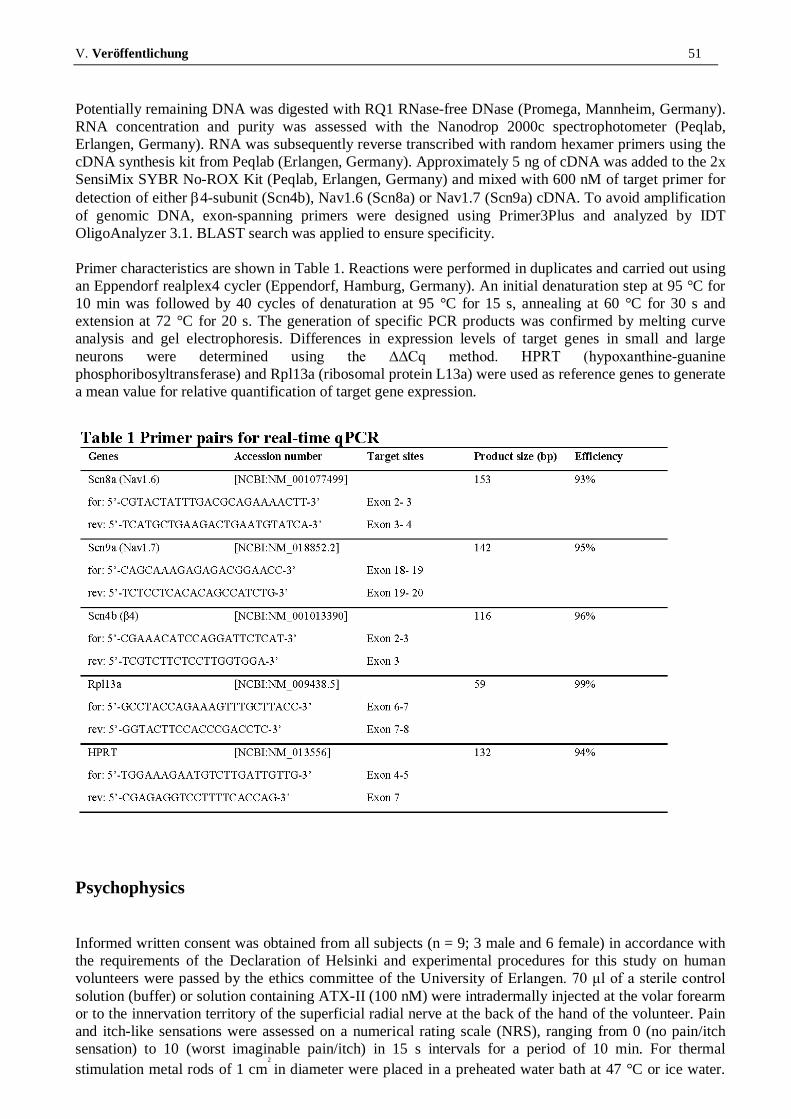
V. Veröffentlichung 51
Potentially remaining DNA was digested with RQ1 RNase-free DNase (Promega, Mannheim, Germany). RNA concentration and purity was assessed with the Nanodrop 2000c spectrophotometer (Peqlab, Erlangen, Germany). RNA was subsequently reverse transcribed with random hexamer primers using the cDNA synthesis kit from Peqlab (Erlangen, Germany). Approximately 5 ng of cDNA was added to the 2x SensiMix SYBR No-ROX Kit (Peqlab, Erlangen, Germany) and mixed with 600 nM of target primer for detection of either β4-subunit (Scn4b), Nav1.6 (Scn8a) or Nav1.7 (Scn9a) cDNA. To avoid amplification of genomic DNA, exon-spanning primers were designed using Primer3Plus and analyzed by IDT OligoAnalyzer 3.1. BLAST search was applied to ensure specificity. Primer characteristics are shown in Table 1. Reactions were performed in duplicates and carried out using an Eppendorf realplex4 cycler (Eppendorf, Hamburg, Germany). An initial denaturation step at 95 °C for 10 min was followed by 40 cycles of denaturation at 95 °C for 15 s, annealing at 60 °C for 30 s and extension at 72 °C for 20 s. The generation of specific PCR products was confirmed by melting curve analysis and gel electrophoresis. Differences in expression levels of target genes in small and large neurons were determined using the ∆∆Cq method. HPRT (hypoxanthine-guanine phosphoribosyltransferase) and Rpl13a (ribosomal protein L13a) were used as reference genes to generate a mean value for relative quantification of target gene expression.
Psychophysics
Informed written consent was obtained from all subjects (n = 9; 3 male and 6 female) in accordance with the requirements of the Declaration of Helsinki and experimental procedures for this study on human volunteers were passed by the ethics committee of the University of Erlangen. 70 μl of a sterile control solution (buffer) or solution containing ATX-II (100 nM) were intradermally injected at the volar forearm or to the innervation territory of the superficial radial nerve at the back of the hand of the volunteer. Pain and itch-like sensations were assessed on a numerical rating scale (NRS), ranging from 0 (no pain/itch sensation) to 10 (worst imaginable pain/itch) in 15 s intervals for a period of 10 min. For thermal stimulation metal rods of 1 cm
2
in diameter were placed in a preheated water bath at 47 °C or ice water.

V. Veröffentlichung 52
Numerical ratings to a 7 s lasting heat stimulus of 47 °C and to a 7 s lasting cold stimulus of 0 °C were compared before and after application of substances. The skin was tested for static and dynamic mechanical allodynia by applying gentle pressure with a cotton swab and stroking with a fine brush. Laser-Doppler imaging (LDI, Moore, London, UK) was used to record changes in superficial skin blood flow. Two baseline scans of 0.5 mm spatial resolution on a 10.8 cm x 7.0 cm area were taken, followed by scans every 2.0 min starting right after the injections. The skin blood flow was analyzed with MLDImg 3.08 software (LDI, Moore, London, UK) and the area of vasodilatation was defined as those pixels in which intensity exceeded the mean of basal values plus two standard deviations [47]. Differential conduction blockade of myelinated nerve fibers was achieved by placing a padded sling connected to a 2.5 kg weight at the proximal wrist of the volunteers for 30–40 min to apply pressure to the superficial radial nerve (also see [48]). Preferential A-fiber blockade induced by nerve compression has been shown by Torebjork et al. [31] and Mackenzie et al. [32] in microneurography studies. In the case of the superficial radial nerve the impaired A-fiber function with preserved C-fiber function is limited to the autonomous zone of the superficial radial nerve distal the metacarpophalangeal joints I + II. The progress of the differential block following compression of the N. radialis superficialis was monitored by decrease of touch and cold detection and was considered to be sufficient when a cold stimulus applied by a metal rod immersed in ice water (7 s, 0 °C) was not to be distinguished from a mechanical stimulus. During the whole duration of the block subjects were able to feel pin-prick stimulation as painful sharp pricking, which was interpreted as maintained C-fiber function [49]. When nerve block was established 70 μl of a 100 nM ATX-II containing sterile buffer solution was injected and subjects were asked to rate pain-like and itch-like sensations for 10 min before removal of the block and 10 and 20 min thereafter together with ratings of cold-evoked pain (0 °C, 7 s, Figure 7). For the NRS curves of each subject the area under the curve (AUC) was calculated (Figure 7a + b, bar graphs).
Chemicals
A 5, 25 or 100 nM recombinant ATX-II (Anemonia sulcata toxin, Alomone Labs, Jerusalem, Israel or Sigma-Aldrich, USA) solution was prepared in saline solution (either extracellular recording solution or sterile buffer for injections, which was additionally sterile filtered). TTX (Tetrodotoxin, Biotrend AG, Wangen, Switzerland) was diluted to a final concentration of 500 nM in the extracellular recording solution. For recordings with β4-peptide an internal solution containing 100 μM β4-peptide (KKLITFILKKTREK, PSL GmbH, Heidelberg, Germany, [50]) was prepared and stored on ice during the process of the experiments
Statistics
All data are presented as mean ± standard error of the mean (SEM). Paired-sample T-test was used for statistical analysis of DRG paired patch clamp recordings and for N1E115 and DRG activation and steady-state fast inactivation analysis. For temperature effect analysis nonpaired Mann Whitney U-test was used for group comparison. For experiments performed on HEK cells and preincubated DRGs independent-sample T-test was used to test for significance. For psychophysics experiments a Wilcoxon matched pairs test was used for intra-individual comparisons and Mann Whitney U-test for the comparison of groups. For RT-qPCR one-tailed Mann Whitney U-test was used for group comparison. Statistical significances are indicated with * for p < 0.05, ** for p < 0.01 and *** for p < 0.001.

V. Veröffentlichung 53
Abbreviations Nav: Voltage-gated sodium channel; DRGs: Dorsal root ganglion sensory neurons; TTX: Tetrodotoxin; PEPD: Paroxysmal extreme pain disorder; RT-qPCR: Real time quantitative polymerase chain reaction; ATX-II: Sea anemone toxin II; AUC: Area under the curve; CGRP: Calcitonine gene-related peptide; AP: Action potential; ns: Statistically not significant
Competing interests The authors declare that they have no competing interests.
Authors’ contributions ABK carried out the patch-clamp recordings and analysis, participated in the acquisition and coordination of psychophysical experiments, carried out FACS sorting data acquisition and interpretation and wrote the manuscript draft. ME planned, coordinated and acquired psychophysical studies, analyzed psychophysical data and participated in writing the manuscript. ASL designed, performed and analyzed the RT-qPCR experiments and critically revised the manuscript. BN planned and participated in acquiring and analyzing psychophysical data and critically revised the manuscript. LKK carried out patch-clamp recordings of Nav1.6 and data analysis. ETS contributed to conception and design and carried out patch-clamp recordings and analysis. RS contributed to conception and design of the study, participated in data acquisition and critically revised the manuscript. THo participated in planning the experiments and critically revised the manuscript. CA participated in analyzing the patch-clamp data. THu contributed to conception and design, participated in analyzing the patch-clamp data. RWC contributed to conception and design of the study. AL conceived and designed the patch-clamp recordings, participated in acquisition and coordination of patch-clamp, FACS sorting and psychophysical data and their analysis and interpretation, and wrote the manuscript. All authors read and approved the final manuscript.
Acknowledgements The authors thank C. Forster for help analyzing the psychophysical data, K. Kistner and R. DeCol for helpful discussions, A. Wirth-Hücking and I. Izydorczyk for technical assistance. The FACS analysis was performed at the Core Unit Cell Sorting and Immunomonitoring Erlangen. Research was supported by the German Research Council (DFG, NA 970/1-1 to BN, CA853/1-2 to RWC, LA2740/2-1 to AL), Ernst-und-Anita-Bauer-Stiftung (to ME), Jürgen Manchot Foundation (to ASL), ELAN of the FAU Erlangen-Nürnberg (11.05.30.1) and the German Israeli Foundation (GIF, 1091–27.1/2010 to AL).
References
1. Cox JJ, Reimann F, Nicholas AK, Thornton G, Roberts E, Springell K, Karbani G, Jafri H, Mannan J, Raashid Y, et al: An SCN9A channelopathy causes congenital inability to experience pain. Nature 2006, 444:894–898.

V. Veröffentlichung 54
V. Veröffentlichung 55
V. Veröffentlichung 56

V. Veröffentlichung 57

V. Veröffentlichung 58

V. Veröffentlichung 59

V. Veröffentlichung 60
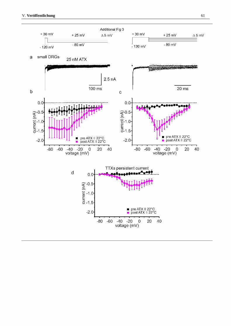
V. Veröffentlichung 61

VI. Diskussion 62
VI. DISKUSSION
Dieser Abschnitt stellt die Schwerpunkte der vorliegenden Arbeit heraus und soll die
Anwendbarkeit der Ergebnisse im Bereich der Tiermedizin erläutern.
Einer der Hauptbefunde dieser Arbeit ist die Erkenntnis, dass ATX-II den resurgent
current in murinen Neuronen verstärkt. Dies findet jedoch nur in großen DRGs, nicht in
kleinen statt. Ein verstärkter resurgent current kann zu Schmerzen und verminderter
Lebensqualität führen. Dies zeigen uns Neuropathien beim Menschen, sei es als
Reaktion auf Medikamente wie Oxaliplatin (SITTL et al., 2012), oder wegen zugrunde
liegender Nav-Mutationen (LAMPERT, 2011). Umso bedeutender ist es diese
Mechanismen genau zu verstehen. Wieso wirkt ATX-II nur auf große DRGs? Sowohl
große als auch kleine DRGs sind per se mit ATX-II sensiblen Kanälen (Nav1.1, Nav1.2,
Nav1.6 und Nav1.7) ausgestattet (OLIVEIRA et al., 2004; WANKE et al., 2009). Daher
kann die "falsche" Kanalausstattung nicht der Grund für die fehlenden Ströme in
kleinen DRGs sein. Vielmehr ist vermutlich das Fehlen an ausreichenden Mengen
blocking particle in kleinen Neuronen ursächlich für den nicht vorhandenen resurgent
current. Kleine DRGs besitzen weniger β4 mRNA als große Neurone (s. Abb.3d der
Veröffentlichung). Wird das β4-Peptid sowohl in kleinen Neuronen, als auch bei
Messungen an heterolog exprimierten Nav1.6 und 1.7 über die Intrazellulärlösung
hinzugefügt, kann resurgent current induziert werden (s. Abb.4 und 5 der
Veröffentlichung). Auch wenn Nav1.6 häufiger resurgent current zeigt als Nav1.7, was
für eine bessere Interaktion des vor allem in großen Neuronen zu findenden Nav1.6 (HO
& O'LEARY, 2011) mit dem Peptid spricht, fehlt es den kleinen DRGs
höchstwahrscheinlich an ausreichend bzw. entsprechend prozessierter β4-Untereinheit.
Die β-Untereinheiten können folglich eine wichtige Rolle für die Aktivität erregbarer
Zellen spielen.

VI. Diskussion 63
Welche Relevanz haben diese Befunde für das Schmerzempfinden von Säugetieren?
Unterschiedlich große DRGs sind mit verschiedenen Schmerzfasern assoziiert. Die
Erkenntnisse dieser Arbeit auf Zell-Ebene korrelieren mit Empfindungen auf
Schmerzfaserebene. Dies zeigen die im Rahmen der Dissertation durchgeführten
Humanversuche (s. Abb.7 der Veröffentlichung). Oftmals dient in der Forschung das
Tier, insbesondere die Maus, als Modell für den Menschen. In dem vorliegenden Fall
ermöglichen uns die Ergebnisse der Humanversuche Rückschlüsse auf die Wirkung des
ATX-II Toxins zu ziehen, die auch für das Tier relevant sind. Da im Rahmen dieser
Arbeit untersucht werden sollte, ob das Toxin selektiv bestimmte Schmerzfasern
beeinflusst und hierfür eine Aussage über die Qualität der Empfindung (z. B. stechend,
brennend, dumpf, juckend) notwendig ist, wurde von Tierversuchen abgesehen. Diese
hätten uns zwar die Aussage ermöglicht, ob durch das Toxin Schmerz induziert wird,
nicht jedoch auf welche Schmerzfaserart dieser zurückzuführen ist, da Tiere keine
Aussage über die Qualität des Schmerzes machen können. Durch den mechanischen
Block der mit den großen DRGs assoziierten A-Fasern kann die durch ATX-II
induzierte schmerzhafte und Juckreiz-ähnliche Empfindung nahezu vollständig
gehemmt werden. Ein bedeutendes Ergebnis der vorliegenden Arbeit besteht in der
Erkenntnis, dass mit niedrigen ATX-II Konzentrationen spezifisch A-Fasern aktiviert
werden können. Dies und die Feststellung, dass es nicht zufriedenstellend sein kann bei
der künftigen Suche nach spezifischeren Schmerzmitteln für Tier und Mensch die Nav-
assoziierten β-Untereinheiten außer Acht zu lassen, sind besonders hervorzuhebende
Befunde dieser Dissertation.
Ein vermehrter resurgent current muss nicht ausschließlich pathologische
Auswirkungen für den Organismus haben. Im Herzen beispielsweise finden wir Nav1.5
und die β4-Untereinheit, also alle Voraussetzungen für das Auftreten von resurgent
current. Es ist möglich, dass die von Isenberg und Ravens (ISENBERG & RAVENS,
VI. Diskussion 64
1984) in Rinder und Meerschweinchenmyozyten beobachtete, durch ATX-II
vermittelte, gesteigerte Kontraktilität auf einer Verstärkung des resurgent current
beruht. Wäre man nun durch die Gabe niedriger ATX-II Konzentrationen in der Lage
diesen resurgent current lokal im Herzen zu verstärken, könnte man damit einer
verminderten Kontraktilität entgegenwirken.
Insbesondere die Erkenntnisse aus den im Rahmen dieser Arbeit durchgeführten in vitro
Versuchen leisten einen essentiellen Beitrag zur Entwicklung neuer veterinär- und
humanmedizinischer Medikamente beispielsweise im Bereich der Kardiologie oder
Neurologie.

VII. Zusammenfassung 65
VII. ZUSAMMENFASSUNG
Zur Wirkung des Seeanemonen Toxins II (ATX-II) auf spannungsgesteuerte
Natriumionenkanäle neuronaler Zellen und zu den dadurch erreichten
Änderungen im Schmerzempfinden
ATX-II wurde ursprünglich aus dem Gift der Seeanemone Anemonia sulcata isoliert
und ist als potenter Aktivator spannungsgesteuerter Natriumionenkanäle (Navs) bekannt,
da es einen persistierenden Na+-Strom induziert. Vor Kurzem wurden bestimmte
Nav-Subtypen mit menschlichem Schmerz in Verbindung gebracht. Somit liegt es nahe,
dass detaillierte Kenntnisse über die Regulierung von Navs für die Entwicklung neuer
Pharmaka im Bereich der Schmerztherapie von Vorteil sind. Auch der durch einen
alternativen Inaktivierungsmechanismus entstehende resurgent current spielt nach
Erkenntnissen der jüngsten Zeit eine Rolle im Schmerzgeschehen. Er kann einen
Angriffspunkt für neue Analgetika darstellen, so man seine Entstehung und mögliche
Modifikationen entschlüsseln kann.
Das Ziel dieser Arbeit war es, zu untersuchen, ob ATX-II resurgent currents von Navs in
großen und kleinen murinen DRGs verändern bzw. induzieren kann. Zudem sollte
beleuchtet werden, ob und wenn ja, welche Wirkung das Toxin auf periphere A- und
C-Schmerzfasern hat.
Die Auswirkungen von ATX-II auf Navs wurden in großen und kleinen DRGs der Maus
sowie in Zelllinien mit whole-cell voltage-clamp Messungen untersucht. Gesunde
humane Probanden bewerteten Empfindungsänderungen (Schmerz und Juckreiz) nach
intrakutaner Injektion von ATX-II. Mit Hilfe des "Laser Doppler Imagers" wurde das
mögliche Auftreten eines Axon Reflex Erythems untersucht. Die mittels des FACS
Zellsortierers nach Zellgröße sortierten DRGs wurden mit RT-qPCR auf ihren Gehalt an
Nav1.6, Nav1.7 und β4 mRNA untersucht.

VII. Zusammenfassung 66
ATX-II verstärkte resurgent currents in großen DRGs, zeigte in kleinen jedoch keinen
Effekt. Im heterologen Expressionssystem trat ein resurgent current erst auf, wenn
β4-Peptid über die Intrazellulärflüssigkeit zur Verfügung stand. Korrelierend zu den
Befunden auf Zellebene rief ATX-II bei intrakutaner Injektion im Menschen nur
solange stechenden Schmerz und veränderte mechanische Empfindung hervor, wie die
A-Fasern für die Schmerzleitung verfügbar waren. Nachdem ein
Nervenkompressionsblock angelegt und die Leitung via A-Fasern blockiert war, waren
die ATX-II vermittelten Effekte verschwunden. Zudem konnte kein Axon Reflex
Erythem gefunden werden, das ein Zeichen für eine C-Schmerzfaser-Aktivierung wäre.
Die Ergebnisse dieser Arbeit legen nahe, dass geringe Konzentrationen von ATX-II eine
selektive Wirkung auf große DRGs haben. Erst durch Hinzufügen von β4-Peptid ist es
möglich mit dem Toxin resurgent current in kleinen DRGs sowie Zelllinien mit
heterolog exprimierten Nav1.6 und Nav1.7 zu induzieren. Dies lässt darauf schließen,
dass es kleinen DRGs an β4 in ausreichender Menge mangelt und deshalb kein
resurgent current sichtbar ist. Ebenso übt ATX-II einen Effekt auf A-Fasern aus, die mit
großen DRGs verbunden sind, wohingegen C-Fasern weitgehend unbeeinflusst bleiben.
Die intrakutane Injektion von ATX-II führt zu stechendem Schmerz und einer
Juckreiz-ähnlichen Empfindung, die sich durch A-Faser-Block verhindern lässt. Daraus
kann man schließen, dass ATX-II in niedrigen Konzentrationen als spezifischer A-Faser
Aktivator fungiert.
VIII. Summary 67
VIII. SUMMARY
On the effect of the sea anemone Toxin II (ATX-II) on sodium ion channels of
neuronal cells and on the resulting variations in pain sensation
ATX-II was originally isolated from the venom of the sea anemone Anemonia sulcata
and is characterized as a potent voltage-gated sodium channel (Nav) activator, as it
induces a persistent sodium current. Recently specific sodium channel subtypes have
been linked to human pain. Thus, detailed knowledge of Nav modulation may prove
advantageous for the development of new pain treatments. As recently published
resurgent current, as a result of an alternative inactivation, also plays a role in pain
perception. It could represent a target for new analgesics, if we are able to elicit its
origin and possible modifications.
The aim of this work was to examine whether ATX-II can induce or change resurgent
currents of Navs of large and small murine, sensory dorsal root ganglia neurons (DRGs).
It was also to be determined if the toxin has an effect on peripheral A- and
C-pain fibers.
The effects of ATX-II on Navs have been studied in large and small DRGs of mice and
in cell lines with whole-cell voltage-clamp measurements. Healthy subjects rated
sensation changes (pain and itch) after intracutaneous injection of ATX-II. A Laser
Doppler Imager was used for analysis of the occurrence of an axon reflex erythema.
Using a FACS sorter DRGs were sorted according to their cell size and were examined
by RT-qPCR for their content of 1.6, 1.7 and β4 mRNA.
ATX-II increased resurgent currents in large DRGs, but failed to influence small DRGs.
In heterologous expression systems, resurgent currents only occurred if the β4-peptide
was available intracellularly. ATX-II accordingly produced sharp pain and altered
mechanical sensitivity in intracutaneously injected humans only as long as A-fibers
VIII. Summary 68
were available for the transmission of pain. After a nerve compression block was
applied and the A-fibers were blocked, this effect disappeared. In addition, no axon
reflex erythema was observed, which would have been a sign for C-fiber activation.
The results of this work suggest that low concentrations of ATX-II have a selective
effect on large DRGs. Only by adding β4-peptide to the intracellular solution, it is
possible to induce resurgent current in small DRGs and cell lines heterologously
expressing Nav1.6 and Nav1.7. This suggests that small DRGs lack sufficient amounts
of β4 and therefore no resurgent current is detectable. Likewise, ATX-II exerts an effect
in large DRG-related A-fibers, whereas C-fibers remain largely unaffected. The
intradermal injection of ATX-II causes a sharp pain and itch-like sensation, which can
be prevented by selective A-fiber block. This suggests that ATX-II at low
concentrations acts as a specific A-fiber activator.

IX. Literaturverzeichnis 69
IX. LITERATURVERZEICHNIS
Bear M, Connors B, Paradiso M (2007) Neuroscience - Exploring the brain. Lippincott
Williams & Wilkins
Beress L, Beress R, Wunderer G. Isolation and characterisation of three polypeptides
with neurotoxic activity from Anemonia sulcata. FEBS Lett 1975; 50: 311-4.
Bergman C, Dubois JM, Rojas E, Rathmayer W. Decreased rate of sodium conductance
inactivation in the node of Ranvier induced by a polypeptide toxin from sea anemone.
Biochim Biophys Acta 1976; 455: 173-84.
Catterall WA. From Ionic Currents to Molecular Mechanisms: The Structure and
Function of Voltage-Gated Sodium Channels. Neuron 2000; 26: 13-25.
Catterall WA, Goldin AL, Waxman SG. International Union of Pharmacology. XLVII.
Nomenclature and structure-function relationships of voltage-gated sodium channels.
Pharmacol Rev 2005; 57: 397-409.
DiSclafani A, Wilkin JK. The axon reflex flare. Cutis 1983; 31: 523-30.
Grieco TM, Malhotra JD, Chen C, Isom LL, Raman IM. Open-channel block by the
cytoplasmic tail of sodium channel beta4 as a mechanism for resurgent sodium current.
Neuron 2005; 45: 233-44.
Hamill OP, Marty A, Neher E, Sakmann B, Sigworth FJ. Improved patch-clamp
techniques for high-resolution current recording from cells and cell-free membrane
patches. Pflugers Arch 1981; 391: 85-100.
Ho C, O'Leary ME. Single-cell analysis of sodium channel expression in dorsal root
ganglion neurons. Mol Cell Neurosci 2011; 46: 159-66.
Hodgkin AL, Huxley AF. A quantitative description of membrane current and its
application to conduction and excitation in nerve. J Physiol 1952; 117: 500-44.

IX. Literaturverzeichnis 70
Isenberg G, Ravens U. The effects of the Anemonia sulcata toxin (ATX II) on
membrane currents of isolated mammalian myocytes. J Physiol 1984; 357: 127-49.
Jarecki BW, Piekarz AD, Jackson JO, 2nd, Cummins TR. Human voltage-gated sodium
channel mutations that cause inherited neuronal and muscle channelopathies increase
resurgent sodium currents. J Clin Invest 2010; 120: 369-78.
Lampert A. Resurgent currents turn painfully exciting. J Physiol 2011; 589: 773-4.
Lawson SN. Phenotype and function of somatic primary afferent nociceptive neurones
with C-, Adelta- or Aalpha/beta-fibres. Exp Physiol 2002; 87: 239-44.
Neher E, Sakmann B. Single-channel currents recorded from membrane of denervated
frog muscle fibres. Nature 1976; 260: 799-802.
Oliveira JS, Redaelli E, Zaharenko AJ, Cassulini RR, Konno K, Pimenta DC, Freitas
JC, Clare JJ, Wanke E. Binding specificity of sea anemone toxins to Nav 1.1-1.6
sodium channels: unexpected contributions from differences in the IV/S3-S4 outer loop.
J Biol Chem 2004; 279: 33323-35.
Raman IM, Bean BP. Resurgent sodium current and action potential formation in
dissociated cerebellar Purkinje neurons. J Neurosci 1997; 17: 4517-26.
Sigworth FJ. The patch clamp is more useful than anyone had expected. Fed Proc 1986;
45: 2673-7.
Sittl R, Lampert A, Huth T, Schuy ET, Link AS, Fleckenstein J, Alzheimer C, Grafe P,
Carr RW. Anticancer drug oxaliplatin induces acute cooling-aggravated neuropathy via
sodium channel subtype Na(V)1.6-resurgent and persistent current. Proc Natl Acad Sci
U S A 2012; 109: 6704-9.
Torebjork HE, Hallin RG. Perceptual changes accompanying controlled preferential
blocking of A and C fibre responses in intact human skin nerves. Exp Brain Res 1973;
16: 321-32.

IX. Literaturverzeichnis 71
Wang GK, Edrich T, Wang SY. Time-dependent block and resurgent tail currents
induced by mouse beta4(154-167) peptide in cardiac Na+ channels. J Gen Physiol 2006;
127: 277-89.
Wanke E, Zaharenko AJ, Redaelli E, Schiavon E. Actions of sea anemone type 1
neurotoxins on voltage-gated sodium channel isoforms. Toxicon 2009; 54: 1102-11.
Wilson MJ, Yoshikami D, Azam L, Gajewiak J, Olivera BM, Bulaj G, Zhang MM. mu-
Conotoxins that differentially block sodium channels NaV1.1 through 1.8 identify those
responsible for action potentials in sciatic nerve. Proc Natl Acad Sci U S A 2011; 108:
10302-7.

X. Anhang 72
X. ANHANG
Vorveröffentlichung: Poster präsentiert auf der Tagung der Deutschen Physiologischen
Gesellschaft im März 2012, ausgezeichnet mit dem Posterpreis.
Abb.10: Urkunde für den Posterpreis der DPG.

XI. Danksagung 73
XI. DANKSAGUNG
Mein besonderer Dank gilt PD Angelika Lampert, die meine Betreuung in Erlangen
übernommen hat und so die Anfertigung dieser Arbeit auch durch die Bereitstellung
einer Finanzierung erst möglich machte. Sie koordinierte meine Projekte und gab
hilfreiche Hinweise zu wissenschaftlichen Arbeitsmethoden. Sie war jedoch nicht nur
meine Betreuerin, sondern hatte stets ein offenes Ohr für persönliche Belange.
Ebenfalls möchte ich Herrn Prof. Stangassinger besonders danken. Zum einen für die
Möglichkeit, die Dissertation über ihn an der veterinärmedizinischen Fakultät der
Universität München einreichen zu können, zum anderen dafür, dass er durch seine
Arbeit in der Tierphysiologie den Anstoß für diese Dissertation gab. Auch für seine
Unterstützung bei der Anfertigung der Arbeit meinen herzlichen Dank.
Bei Esther und Mirjam möchte ich mich für die Tipps und Hilfestellungen, die
Unterstützung und die zahlreichen gemeinsamen, bereichernden Stunden inner- und
außerhalb der Physiologie Erlangen herzlich bedanken.
Andreas, Martin und Andy danke ich für das beste Arbeitsklima, dass man sich
wünschen kann.
Bei Anette, Iwona und Birgit möchte ich mich für die besonders erfolgreiche
Zusammenarbeit bedanken.
Allen Kooperationspartnern danke ich für die inspirierenden Projektgespräche und die
gute Zusammenarbeit.
Bei den lieben Mitarbeitern und Professoren der Physiologie Erlangen, insbesondere bei
Herrn Prof. Alzheimer, möchte ich mich für ein hervorragendes Arbeitsklima und viele
neue wissenschaftliche Eindrücke herzlich bedanken.
Aus tiefstem Herzen möchte ich mich bei meiner Schwester Gabi, meinen Eltern Maria
und Roland für die immerwährende Bestärkung bedanken. Desweiteren gilt mein
allerbester Dank Philipp und Monik für ihre allgegenwärtige Unterstützung. Danke,
dass ihr immer für mich da seid.
Danke an meine Verwandten und Freunde, die mich auf diesem Weg begleitet und stets
unterstützt haben.
Zum Schluss möchte ich mich bei Laika, Abbie und Duke für ihr
nichtwissenschaftliches Ausgleichsprogramm bedanken.