


Sprachen
Seiten
Rechtliche


MASTERARBEIT
Titel der Masterarbeit
„Extracts of anti-malarial and anti-inflammatory healing plants as oncolytic concept“
Verfasserin
Mag. Christine Unger, Bakk.
angestrebter akademischer Grad
Master of Science (MSc)
Wien, Oktober 2010
Studienkennzahl lt. Studienblatt: A 066 838
Studienrichtung lt. Studienblatt: Ernährungswissenschaften
Betreuerin / Betreuer: ao. Univ.-Prof. Dr. Georg Krupitza


I
TABLE OF CONTENTS
1 INTRODUCTION .............................................................................................................. 1
2 LITERATURE SURVEY ...................................................................................................... 3
2.1. Cell cycle and cancer ............................................................................................... 3
2.1.1. Basic regulation of the cell cycle ........................................................................... 3
2.1.2. DNA damage checkpoints ..................................................................................... 5
2.2. Cell death programs ................................................................................................ 7
2.2.1. Apoptosis ............................................................................................................... 7
2.2.2. Necrosis ................................................................................................................. 8
2.2.3. Autophagy ............................................................................................................. 8
2.3. Carcinogenesis ........................................................................................................ 9
2.3.1. The multistep model ............................................................................................. 9
2.3.2. The Hallmarks of Cancer ..................................................................................... 10
2.4. Leukemia .............................................................................................................. 16
2.5. Lymphoma ........................................................................................................... 17
2.6. Natural products in drug discovery ....................................................................... 18
2.6.1. Plants as source of anti‐cancer agents ................................................................ 19
2.6.2. Example: Vincristine ............................................................................................ 21
2.6.3. Potential anti‐neoplastic activity of two ethnomedical plants from Guatemala 22
3 MATERIAL AND METHODS ........................................................................................... 24
3.1. Plant Material ....................................................................................................... 24
3.1.1. Critonia morifolia (Petén, Guatemala) ................................................................ 25
3.1.2. Neurolaena lobata (Guatemala, Petén) .............................................................. 26
3.2. Plant extraction .................................................................................................... 28
3.2.1. C. morifolia – Accelerated Solvent Extraction (ASE 2000) .................................. 28
3.2.2. N. lobata – reflux‐water bath extraction ............................................................ 29
3.3. Cell culture ........................................................................................................... 30
3.4. Proliferation and cytotoxicity assays ..................................................................... 30
3.5. Apoptosis assay – Hoechst 33258 and propidium iodide double staining .............. 32
II
3.6. Cell cycle distribution (FACS) ................................................................................ 33
3.7. Western blotting .................................................................................................. 33
3.8. Quantitative RT‐PCR ............................................................................................. 36
3.9. Statistical analysis ................................................................................................ 37
4 RESULTS ....................................................................................................................... 38
4.1. Critonia morifolia ................................................................................................. 38
4.1.1. Extract yields and stock calculation ..................................................................... 38
4.1.2. Anti‐proliferative activity of C. morifolia extracts in HL‐60 cells ......................... 39
4.1.3. Induction of apoptosis in HL‐60 cells by extracts of C. morifolia ........................ 41
4.1.4. The petroleum ether extract represses c‐Myc and cyclin D1 expression in
HL‐60 cells ............................................................................................................ 42
4.1.5. After a transient G2‐M cell cycle inhibition, the petroleum ether extract of
C. morifolia induces S‐phase arrest in HL‐60 cells after 24 hours of incubation 43
4.1.6. Modulations of cell regulatory proteins induced upon extract treatment ......... 45
4.1.7. The petroleum ether extract induces apoptosis in HL‐60 cells mediated by
caspase‐3 ............................................................................................................. 47
4.1.8. Early onset of apoptosis, not genotoxicity, leads to checkpoint kinase
activation in extract treated HL‐60 cells .............................................................. 48
4.2. Neurolaena lobata ............................................................................................... 50
4.2.1. Extract yields and stock calculation ..................................................................... 50
4.2.2. Anti‐proliferative activity of N. lobata extracts in HL‐60 cells ............................. 51
4.2.3. Induction of apoptosis in HL‐60 cells by extracts of N.lobata ............................. 53
4.2.4. Anti‐proliferative effects of N. lobata dichloromethane extract in human ALCL
SR‐786 cells with a NPM‐ALK translocation ........................................................ 54
4.2.5. Inhibition of NPM‐ALK, induction of apoptosis and caspase‐3 in SR‐786 cells ... 55
4.2.6. Reduction of NPM‐ALK levels is a decisive property of the dichloromethane
extract of N. lobata and is caused at transcriptional level .................................. 57
4.2.7. The dichloromethane extract induces G2‐M arrest and activates Chk1 in
SR‐786 cells .......................................................................................................... 60
4.2.8. N. lobata dichloromethane extract modulates oncogenes and tumor
suppressor protein expression ............................................................................ 64
4.2.9. Proliferation inhibition in murine ALCL cells with a NPM‐ALK translocation ...... 66
III
4.2.10. Cell cycle inhibitory effects in G2‐M of the dichloromethane extract in
417 cells ............................................................................................................... 67
4.2.11. Induction of apoptosis is specific in 417 cells and mediated by caspase‐3 ........ 70
4.2.12. N. lobata dichloromethane extract, but not vincristine, substantially
decreases levels of NPM‐ALK in 417 cells ........................................................... 73
5 DISCUSSION ................................................................................................................. 76
5.1. C. morifolia ........................................................................................................... 76
5.2. N. lobata ............................................................................................................... 79
SUMMARY ........................................................................................................................... 83
ZUSAMMENFASSUNG .......................................................................................................... 84
REFERENCES ........................................................................................................................ 85
DANKSAGUNG ..................................................................................................................... 93
LEBENSLAUF ........................................................................................................................ 94
PAPERS IN PREPARATION .................................................................................................... 95

IV
ABBREVIATIONS
417
ATP
ALL
AML
ALCL
ALK
ASE
ATCC
ATM
ATR
Bcl‐2
BRCA1
CAM
Cdc 2
Cdk
Cip/Kip
CLL
CML
DMSO
DNA
DNAse
E2F
ECL
EDTA
Erk
FACS
FCS
FDA
G0, G1, G2
HL‐60
HLF
INK4
murine NPM‐ALK positive ALCL cell line
adenosine triphosphate
acute lymphoblastic leukemia
acute myeloid leukemia
anaplastic large cell lymphoma
anaplastic lymphoma kinase
accelerated solvent extraction
American type culture collection
ataxia‐telangiectasia mutated
ataxia‐telangiectasia mutated and Rad3 related
B‐cell lymphoma 2
breast cancer 1, early onset
cell adhesion molecules
cell division control protein 2
cyclin‐dependent kinase protein
cyclin dependent kinase inhibitor proteins
chronic lymphoblastic leukemia
chronic myeloid leukemia
dimethyl sulfoxide
deoxyribonucleic acid
deoxyribonuclease
a group of genes that codifies a family of transcription factors
enhanced chemiluminescence
ethylenediaminetetraacetic acid
extracellular –signal‐regulated kinases
fluorescence activated cell sorting
fetal calf serum
US Food and Drug Administration
gap phases of the cell cycle
human promyelocytic leukemia cell line
human lung fibroblasts
inhibitor of cyclin‐dependent kinase 4

V
M‐phase
NPM
p21
p53
PAGE
PARP
PBS
PIC
PI3K
PMSF
pRb
PDGFR‐β
PVDF
RPMI
S‐phase
SDS
SR‐786
TBS
mitosis
nucleophosmin
cyclin‐depenedent kinase inhibitor 1
tumor suppressor protein
polyacrylamide gel electrophoresis
poly(ADP‐ribose)polymerase
phosphate buffered saline
protease inhibitor cocktail
phosphatidylinositol 3‐kinase
phenylmethylsufonyl fluoride
retinoblastoma protein
platelet‐derived growth factor β
polyvinylidene fluoride
cell culture medium (Rosewell Park Memorial Institute)
DNA synthesis during cell cycle
sodium dodecyl sulfate
human NPM‐ALK positive ALCL cell line
Tris buffered saline


1
1 INTRODUCTION
Cancer is one of the leading causes of death worldwide. For 2010, the United States National
Institute of Cancer estimates about 1.5 million new cases of cancer and more than
500 thousands of deaths from cancer in the USA (www.cancer.gov). Apart from avoidable risks
such as smoking, the most important risk factor for cancer is supposed to be the increase in life
expectancy since most cancers occur in people over the age of 65, however, also younger
adults and children are diagnosed with cancer. Thus, there exists a constant need for the
development of novel anti‐neoplastic agents.
Natural products represent a vital resource for therapeutic principles as about 60 % of all
anti‐neoplastic drugs used in Western medicine originate from natural sources including
plants, microorganisms and marine organisms (Cragg and Newman 2007). Moreover,
approximately 80 % of the world’s population relies on medical plants for their primary health
care, especially in less‐developed countries. As a result, numerous traditional healing plants
successfully passed hundreds and even thousands of years of application in a variety of
diseases. Two popular examples of plant derived drugs are vincristine, a vinca alkaloid from
Catharantus rosea (formerly known as Vinca roseus), and paclitaxel, which naturally occurs in
the bark of Taxus brevifolia. Vincristine and paclitaxel, which are main agents applied in
chemotherapy, demonstrate the pivotal role of plants in the discovery of new lead
compounds. In both cases, interest in pharmacological research was triggered by the
traditional uses of the plants as home remedy.
To discover new potential lead compounds against cancer, the present work focussed on the
ethnomedical knowledge of the ancient civilization of the Central American Mayas. Their
traditional medicine is based on the rich biodiversity of the rain forest, and is still practiced
effectively to cure a variety of diseases. In this manner, we made use of the long‐lasting
medical experience of the Mayas in plant selection for detailed screening.
The first plant investigated, Critonia morifolia (Asteraceae), was selected based on its
traditional anti‐inflammatory uses, as similar signaling pathways are commonly upregulated
both in inflammatory conditions and cancer. Despite of its curative uses as home remedy only
limited phytochemical and no pharmacological research results were published. Neurolaena
lobata (Asteraceae), the second plant, is described as effective home remedy to cure protozoal
ailments and malaria, in particular. Its anti‐protozoal activity is already scientifically

2
documented. Furthermore, first hints on potential cytotoxicity were published (François et al.
1996), however, more detailed investigations are still missing.
The potential anti‐carcinogenic properties of these two ethno‐pharmacological healing plants
from Guatemala were tested in human HL‐60 promyelocytic leukemia cells to assess their
anti‐proliferative and pro‐apoptotic activity. For each plant, the most active extract out of five,
obtained through serial extraction using solvents of increasing polarity, was studied in more
detail. Western blots and FACS analyses were applied to gain further insights into the
underlying mechanisms of growth inhibition and apoptotic trigger. Investigations on
C. morifolia were limited to HL‐60 cells only. In case of N. lobata, all experiments, apart from
initial screening in HL‐60 cells, were performed in either human and/or murine NPM‐ALK
positive ALCL (anaplastic large cell lymphoma) cell lines SR‐786 and 417, respectively.
Moreover, the N. lobata extract was tested in normal human lung fibroblasts (HLF) to rule out
unspecific cytotoxicity.

3
2 LITERATURE SURVEY
2.1. Cell cycle and cancer
The cell cycle is an ordered series of events that is required for duplication of an eukaryotic cell
and subsequent division into two identical daughter cells. Complex networks of regulatory
factors influence whether a cell proliferates, stays in a quiescent state or dies. During cell cycle
progression, cells go through numerous internal checkpoints to verify proper completion of
the previous step prior proceeding to the next step. Disorders in cell cycle regulation are
associated with a variety of diseases including cancer (Meeran and Katiyar 2008).
2.1.1. Basic regulation of the cell cycle
The duration of cell cycle of eukaryotic cells has been defined as the interval between the
completion of mitosis by a cell and completion of mitosis by at least one of its daughter cells
(Meeran and Katiyar 2008). Strict regulation of the cell cycle is essential to provide a correct
duplication of genetic information as well as its correct segregation during mitosis.
The cell cycle of eukaryotic cells comprises four distinct phases (Figure 1):
G1 (Gap phase 1) cellular growth, preparing for DNA synthesis
S (S‐phase) DNA synthesis and replication
G2 (Gap phase 2) preparation for mitosis
M (mitosis) cell division
G1‐, S‐ and G2‐phase together are also referred to as interphase. Additionally, cells in
G1‐phase may leave cell cycle and enter a temporarily or even permanently quiescent state
termed G0 in dependence on environmental and developmental signals (van den Heuvel
2005).

4
Figure 1. Mammalian cell cycle (simplified). Shapes outside the cycle indicate approximate time and activity of different combinations of cyclins and Cdks (van den Heuvel 2005).
Cell cycle phases are tightly regulated by cyclin‐dependent kinases (Cdks). Even though Cdk
protein levels are constant throughout the cell cycle, Cdk activity requires forming of
complexes with accessory subunits known as cyclins (Murray 2004). Cell cycle related cyclins
are synthesized and destroyed at specific times during the cell cycle, thereby modulating Cdks
kinase activity (Figure 1).
Cdk/cyclin complexes include three interphase Cdks (Cdk2, Cdk4 and Cdk6), the mitotic Cdk1
(also known as cell division control protein 2 (cdc2)), and ten cyclins that belong to four
distinct classes (A, B, D and E) (Malumbres and Barbacid 2009). However, only certain
Cdk/cyclin complexes are supposed to control cell cycle progression. For example, D‐type
cyclins that bind preferably to Cdk4 and Cdk6 play an important role in the transition from G1
to S‐phase upon mitogenic stimuli. The activation of these complexes allows the expression of
E‐type cyclins which bind to Cdk2. Cdk2/cyclin E further promotes G1/S transition and initiates
DNA replication (Meeran and Katiyar 2008). Subsequently, Cdk2 is activated by association to
cyclin A in the late stage of S‐phase driving progression into G2‐phase. At the end of
interphase, Cdk1 associates with cyclin A to facilitate the onset of mitosis. Subsequently,
cyclin A degradation enables the formation of Cdk1/cyclin B complexes which finally drives
cells through mitosis (Malumbres and Barbacid 2009).

5
In addition to regulation by cyclins, Cdk activity is modulated by two classes of Cdk inhibitors.
The first family is identified as the Cip/Kip family including p21Cip1, p27Kip1 and p57Kip2. These
proteins are able to interact with multiple Cdk/cyclin complexes, thereby inhibiting Cdk activity
throughout the cell cycle. The members of the INK4 family, composed of p15INK4B, p16INK4A,
p18INK4C and p19INK4D, specifically block the association of Cdk4/6 and cyclin D, thereby
inhibiting G1/S transition (Meeran and Katiyar 2008). Commonly, Cdk inhibitor proteins are
upregulated in response to anti‐proliferative signals.
In many cancers certain Cdk‐cyclin complexes are deregulated, resulting in continued
proliferation and unscheduled re‐entry into the cell cycle.
2.1.2. DNA damage checkpoints
The cellular DNA integrity of every mammalian cell is constantly exposed to both intrinsic (e.g.
byproducts from oxidative respiration) and external sources (e.g. chemicals, radiation,
cigarette smoke) of DNA‐damaging agents as well as occasional DNA mismatch (Jackson and
Bartek 2009). Therefore, cells have evolved several mechanisms to cope with the constant
attack on their DNA. Dependent on the type of DNA lesion, a variety of repair mechanisms
exists. Cells response to DNA‐damage ranges from direct repair to halting of the cell cycle and
undergoing of programmed cell death (i.e. apoptosis).
Mammalian cells may only withdraw from the cell cycle when they experience growth‐factor
deprivation and/or inhibitory signals in early to mid G1‐phase. If cells pass through the pRb
(retinoblastoma protein)/E2F (transcription factor)‐controlled restriction point, they are
committed to complete the cell cycle and cell division. However, in response to genotoxic
stress, checkpoint networks can delay cell cycle progression in G1, S or G2‐phase (Kastan and
Bartek 2004). These DNA damage checkpoints refer to signal transduction pathways induced
by DNA damage that halt the cell cycle until DNA is repaired (Malumbres and Barbacid 2009).
This mechanism is supposed to ensure the maintenance of genomic integrity and the
prevention of cancer.
G1 and G1/S checkpoint
The acquisition of abnormalities during G1/S phase appears to be a crucial step in the
development of cancer. Hence, the G1/S checkpoint prevents the replication of damaged DNA.
The most important checkpoint response to DNA damage in late G1 is mediated through the
ATM/ATR‐Chk1/Chk2‐Cdc25A pathway. Depending on the type of DNA‐damage, ATM/ATR
6
phosphorylate Chk2/Chk1 which in turn phosphorylate Cdc25A on its several serine residues,
triggering its degradation. This results in the failure of Cdk2 activation, which is crucial for
transition from G1 to S‐phase, and consequently an accumulation of cells in G1 (Kastan and
Bartek 2004).
Moreover,the cell cycle protein p53 is one of the most important regulatory proteins in G1
checkpoint. Phosphorylation of p53 at Ser15 by ATM/ATR and at Ser20 by Chk1/Chk2 leads to
a maintained G1/S arrest. A key transcriptional target of p53 is the Cdk‐inhibitor protein p21,
which silences the G1/S‐promoting Cdk2/cyclin E kinase, thereby causing G1 arrest (Kastan and
Bartek 2004). Additionally, p21 binds to the Cdk4/cyclin D complex resulting in a
hypophosphorylation of pRb, thereby suppressing the pRb/E2F pathway and causing cell cycle
arrest (Meeran and Katiyar 2008). p21 can also be regulated via p53‐independent pathways,
e.g. c‐Myc and BRCA1 (Abukhdeir and Park 2008).
Notably, the Chk1/Chk2‐Cdc25A checkpoint is considered to be implemented more rapidly and
independently of p53 whereas p53 and p21 appear to play a role in sustained G1 arrest
(Kastan and Bartek 2004).
S‐phase checkpoint
In general, the S‐phase checkpoint network is activated to ensure appropriate DNA duplication
in case of DNA damage. At least two parallel branches are involved in slowing down ongoing
DNA synthesis to facilitate DNA repair prior to synthesis (Falck et al. 2002).
An important mechanism includes the activation of the ATM/ATR‐Chk1/Chk2‐Cdc25A pathway
after DNA damage. Both Chk1 and Chk2 are associated with inhibitory phosphorylation of its
substrate Cdc25A, targeting it for degradation (Mailand et al. 2000; Madlener et al. 2009). In
return, the downregulation of Cdc25A inhibits the further activation of the Cdk2/cyclin E
complex which is needed to proceed in S‐phase (Falck et al. 2002). In addition, the
ATM‐Nbs1‐SMC1 pathway defines a separate branch of the intra‐S‐phase checkpoint (Yazdi et
al. 2002).
Both branches of intra‐S‐checkpoint have to be interfered concomitantly to abrogate inhibition
of DNA replication (Falck et al. 2002).

7
G2‐M checkpoint
The G2‐M checkpoint prevents cells from initiating mitosis when they either experience DNA
damage during G2 and/or progressed into G2 without proper repair in previous phases or
inappropriate replication of DNA in S‐phases.
The essential mitosis‐promoting activity of the Cdk1/cyclin B complex makes it the critical
target of the G2 checkpoint. Upon various stresses, Cdk1/cyclin B activity is inhibited by
ATM/ATR‐Chk1/Chk2 and/or p38 via regulating degradation and inhibition of the Cdc25
phosphatase family that is normally responsible for Cdk1 activation (Kastan and Bartek 2004).
p53 and BRCA1 are reported to play an important role in maintaining G2 arrest. By regulating
transcriptional programs, p53 and BRCA1 lead to an upregulation of cell cycle inhibitors such
as Cdk‐inhibitor p21, GADD45a (growth arrest and DNA‐damage‐inducible 45 alpha) and
14‐3‐3 sigma proteins (Meeran and Katiyar 2008). Besides, these p53 downstream effectors
can also be regulated in a p53‐independent manner as tumor cells that are defective in p53
still tend to selectively accumulate in G2 phase after DNA damage. Moreover, other upstream
regulators of Cdc25 phosphatases and/or Cdk1/cyclin B seem to be targeted by DNA‐damage
induced mechanisms (Kastan and Bartek 2004).
2.2. Cell death programs
Cell death is a pivotal process during development, immune regulation and homeostasis in
multicellular organisms. Numerous human pathologies are associated with its dysregulation
(Duprez et al. 2009). Due to morphological criteria, three main types of cell death are classified
as apoptotic, necrotic, or to be associated with autophagy.
2.2.1. Apoptosis
Morphological features of apoptosis are cell shrinkage, chromatin condensation, and
membrane blebbing. Apoptosis is referred to as intrinsic programmed cell death mechanism
that results in controlled breakdown of the cell into apoptotic bodies, which are subsequently
engulfed by surrounding cells and phagocytes (Duprez et al. 2009).
Two protein families are mainly involved in apoptosis, namely caspases (cysteinyl
aspartate‐specific proteases), which mediate the execution, and the Bcl‐2 family, which control
mitochondrial integrity. Caspases can be subdivided into initiator (caspases‐2, ‐8, ‐9, and ‐10)
and effector (caspases‐3, ‐6, and ‐7) caspases. Initially, all of them are expressed in their
inactive proenzyme form and require proteolytic cleavage to be activated. Cleavage may be

8
mediated by intrinsic and extrinsic pathways. Among others, the intrinsic pathway is regulated
by antagonizing anti‐apoptotic and pro‐apoptotic (during cellular stress) members of the Bcl‐2
family. Upon mitochondrial damage, cytochrome c is released from the mitochondria into the
cytosol where it associates with Apaf (apoptotic protease activating factor) and ATP
(adenosine triphosphate), activating procaspase‐9. The extrinsic pathway of apoptosis is
mainly mediated by stimulation of receptors of the TNFR (tumor necrosis factor receptors)
family, such as Fas (Duprez et al. 2009).
It is generally believed that apoptosis does not induce immunological response and therefore
does not provoke inflammation (Duprez et al. 2009).
2.2.2. Necrosis
Necrosis is morphologically characterized by cytoplasmatic and organelle swelling and the final
loss of cell membrane integrity. Thus, cellular contents are released into the surrounding
extracellular matrix provoking immunological response and inflammation (Duprez et al. 2009).
Typically, necrosis is characterized in negative terms by the absence of caspase activation,
cyctochrome c release and DNA oligonucleosomal fragmentation (Krysko et al. 2008). As
necrotic cell death generally results from a severe physical damage, such as hyperthermia and
ischemia, it has been described an uncontrolled cell death that lacks underlying signaling
events. However, evidence emerged that in certain conditions, necrosis is guided by strictly
regulated signaling pathways, which are initiated by diverse stimuli. Moreover, in some
conditions when apoptosis is hampered, necrosis might acts as a kind of back‐up for cell death
(Duprez et al. 2009).
2.2.3. Autophagy
Cell death associated with autophagy represents a catabolic pathway that allows cells to
degrade and recycle cellular components. Morphologically, autophagy is characterized by the
presence of double‐membrane vesicles, which contain sequestered proteins and organelles.
Autophagy at basal levels helps cells to maintain intracellular homeostasis and serves as cell
survival mechanism during nutrient deprivation (Jin and White 2007). However, massive
autophagy is suggested to play a role in cell death, often associated with features of apoptotic
or necrotic cell death (Duprez et al. 2009).

9
2.3. Carcinogenesis
The process of the transformation of normal cells into cancer cells is referred to as
carcinogenesis. Usually, it takes years to decades from initial genomic changes within a
“cancer cell” to the clinical outcome of cancer.
For a long time, a multistep model of cancer development has been accepted. By now, it is
evident that this model does not meet the complexity of the process, however, it still has its
eligibility. In the following section, two distinct ways of characterizing the development of
cancer are described. The multistep model functionally groups carcinogenesis into three
distinct phases, whereas the other way is by listing acquired features of mutant cells during
carcinogenesis, referred to as “hallmarks of cancer” defined by Hanahan and Weinberg (2000).
In general, the two models do not exclude but can rather be considered as supplementing
each other.
2.3.1. The multistep model
Traditionally, the development of cancer is operationally divided into three phases: initiation,
promotion and progression (Figure 2).
Initiation is characterized by DNA mutations in a cell such as single nucleotide polymorphisms,
gene depletion or amplification, and chromosomal translocations leading to irreversible
genomic changes. Either extrinsic (e.g. chemicals, radiation, cigarette smoke) or intrinsic
agents produced during normal physiological processes within a cell may cause initiation.
Usually, multiple mutations must occur for a tumor cell to arise (Barrett 1993), which explains
that an initiated cell may stay in a quiescent state for several years.
The process of promotion refers to the influence of non‐carcinogenic substances on the clonal
expansion of initiated cells and is supposed to be substance as well as tissue specific. Effects of
promoting agents seem to be reversible, which suggests an epigenetic mechanism (Hennings
et al. 1993).The end product of promotion is commonly a benign foci of pre‐neoplastic cells
(Barrett 1993).
These pre‐neoplastic cells must undergo an additional step to convert to malignant neoplasms.
This transformation from benign lesions to malignant cancers is termed progression. Malignant
neoplasms are distinct from benign tumors regarding their cellular morphology, growth,
differentiation, and invasiveness. Moreover, neoplasms differ in their responsiveness to
certain chemical treatments (Barrett 1993).

10
Figure 2. Traditional multistep model of carcinogenesis (using the example of chemical induction) (Oliveira et al. 2007).
2.3.2. The Hallmarks of Cancer
By now, more than hundred distinct types of cancer, and subtypes of tumors can be found
within specific organs. Although specific characteristics among cancer cells may vary
substantially, Hanahan and Weinberg (2000) defined six essential alterations in cell physiology
that are collectively responsible for the malignant growth: self‐sufficiency in growth signals,
insensitivity to anti‐growth signals, evading apoptosis, limitless replicative potential, sustained
angiogenesis and tissue invasion and metastasis. These capabilities are acquired during tumor
development and represent a successful breaching of the anti‐cancer mechanisms hardwired
into cells.

11
Figure 3. Manifestation of six essential alterations in cell physiology collectively dictating malignant growth (Hanahan and Weinberg 2000).
Self‐sufficiency in growth signals
Normal cells remain in a quiescent state unless they are stimulated by mitogenic growth
stimuli. These exogenous signals are transmitted into the cell by transmembrane receptors and
induce proliferation. Three distinct mechanisms leading to growth signaling autonomy are
described below.
Most importantly, many cancer cells reduce their dependence on exogenously derived signals
by generating their own growth signals, creating a positive feedback loop. This property
inhibits homeostatic regulation that normally ensures a proper behavior of the cells in a tissue.
An overexpression of growth factor receptors, often carrying tyrosine kinase activity, is also
found in several cancers. Receptor overexpression may increase responsiveness to growth
signals enabling cells to proliferate also at ambient levels of growth factors that normally
would not trigger cell cycle.

12
A third mechanism by which cells become less dependent on growth stimuli is a modulation in
the expression levels of extracellular matrix receptors (integrins), favoring those which act
pro‐mitotic. Integrins physically link cells to the extracellular matrix and transduce signals into
the cytoplasm which influence cell behavior, ranging from quiescence, activating proliferation
and resistance to apoptosis.
Abandoning the reductionist type of view focusing solely on cancer cells, heterotypic signaling
between the diverse cell types within a tumor is proven to contribute to unscheduled tumor
cell proliferation. Within normal tissue, cells are known to largely influence their neighbors to
grow. Cancer cells may acquire the ability to co‐opt their normal nearby cells by inducing them
to release growth signals. These complex interactions need to be considered when trying to
understand the development of cancer.
Insensitivity to anti‐growth signals
Anti‐growth signals include both soluble and immobilized inhibitors embedded in the surfaces
of neighbor cells and in the extracellular matrix. These signals contribute to the quiescent state
of cell and tissue homeostasis by two distinct mechanisms. First, cells can be forced into a
resting phase (G0) in which they may remain for a long period of time unless inhibitory signals
vanish or growth signals induce proliferation. Alternatively, cells may be induced to
permanently stop proliferation activity, usually associated with specific differentiation. Both
strategies are described in further detail below.
During G1 phase of cell cycle, the so‐called growth phase, normal cells monitor their external
environment. Based on the sensed signals, cells decide whether to proceed to S‐phase and
therefore proliferate or to enter a quiescent state. Thus, it is essential for cancer cells to evade
these anti‐growth signals. Evidence is mounting that many if not all growth‐inhibiting signals
are funneled by retinoblastoma protein (pRb) and its relatives, p107 and p130.
Hypophosphorylation of pRb blocks proliferation by targeting E2F transcription factors that
modulate the expression of numerous genes crucial for progression from G1 into S‐phase. In
the absence of pRb or disruption of the pRb pathway, pro‐mitotic E2Fs are liberated and
facilitate cell proliferation. Among other anti‐growth factors, effects of TGFβ (transforming
growth factor β) on the pRb pathway are the best documented. Preventing the
phosphorylation that inactivates pRb, TGFβ blocks the transition into S‐phase. A variety of
strategies evading the pRb circuit are found in tumor tissues e.g. down‐regulation of TGFβ
receptors, displaying mutant receptors or mutation of downstream signaling proteins.
13
Additionally, cancer cells may also reduce expression of integrins and other cell adhesion
molecules that send growth‐inhibitory signals, favoring those that transmit growth stimuli.
Apart from temporarily halted cell cycle mediated by the pRb pathway, normal tissues
indefinitely constrain cell multiplication by promoting cell differentiation. Apparently, cancer
cells use various strategies to avoid terminal differentiation. One of them involves the c‐Myc
oncogene, over‐expressed in many tumors that impairs differentiation and promotes growth.
Evading apoptosis
Within a normal tissue, cell number is determined not only by proliferation but also by the rate
of cell death. Typically, programmed cell death in terms of apoptosis represents a major
regulator of cell population. Thus, acquisition of resistance towards apoptosis is characteristic
of most and perhaps all types of cancer.
Apoptosis can be roughly divided into two stages. In the first step, sensors permanently
monitor the extracellular and intracellular environment for detecting abnormalities influencing
the cells’ well‐being. In response to detrimental conditions, these signals initiate the activation
of components of the second level, which function as executioners of programmed cell death.
Trigger of apoptosis include DNA damage, signaling imbalance provoked by oncogenes,
survival factor insufficiency, and hypoxia.
Numerous signal cascades may lead to apoptosis by converging to the ultimate effectors of
programmed cell death. Intracellular proteases termed caspases or cytochrome C released by
mitochondria trigger downstream activation of more effector caspases that finally execute
apoptosis (see also 2.2.1).
Circumvention of apoptosis may be acquired by a variety of strategies, including the loss of
pro‐apoptotic regulators (e.g. p53) and/or trigger of anti‐apoptitic components of the signaling
circuitry. Since most regulatory and effector components are present in redundancy, tumor
cells that have lost a certain pro‐apoptotic component are likely to still own intact components
of similar apoptotic pathways. Thus, this fact has to be considered when trying to target a
specific type of cancer by chemotherapy.
Limitless replicative potential
The three capabilities mentioned above, growth signal autonomy, insensitivity to
growth‐inhibitory signals and resistance to apoptosis, lead to independence of a cell’s
proliferation behavior from signals in its environment. However, recent research indicates that

14
this acquired disruption in cell‐to‐cell signaling is not sufficient to ensure expansive tumor
growth. Many and maybe all mammalian cells appear to possess an intrinsic program that
limits their multiplication. This program is regarded as acting autonomously of cell‐to‐cell
interactions.
Normal cells in culture are demonstrated to have a finite replicative potential whereas tumor
cells appear to be immortalized, suggesting that the ability of unlimited multiplication was
acquired during tumor progression in vivo. Thus, the generational limit of normal somatic cells
acts as a barrier to cancer.
This phenomenon is caused by the progressive erosion of telomeres during DNA replication
cycles. Telomeres, the end of chromosomes, are composed of several thousand repeats of a
6 bp sequence element and function as stabilizers of chromosomal DNA. During each cell cycle,
replicative generations show a loss of 50‐100 bp of telomeric DNA from the ends of every
chromosome. As a result, telomeres lose their ability to protect the chromosomal DNA
through successive cycles of replication. The unprotected ends participate in chromosomal
fusions, typically yielding in the death of the affected cell. Of malignant cells, 85‐90 % succeed
in maintaining telomeres during S‐phase by upregulating expression of the telomerase
enzyme, which adds 6 bp repeats onto the ends of telomeric DNA. The second mechanism
helps to maintain telomeres through recombination of interchromosomal exchanges of
sequence information. Normal tissue cells lack of telomere maintenance mechanisms. While
the absence of telomerase is one cause for cellular aging on the one hand, it also acts as
anti‐cancer mechanism on the other. Hence, ensuring telomere length above critical threshold
is a key component of the capability for unlimited replication.
Sustained angiogenesis
The adequate supply with oxygen and nutrients is crucial for cell function and survival.
Coordinated growth of blood vessels during organogenesis ensures that virtually all cells in a
tissue reside within 100 µm of a capillary blood vessel. The formation of new blood vessels
within a tissue, the process of angiogenesis, is carefully regulated. Proliferating cells do not
possess an intrinsically angiogenic ability.
Angiogenesis is encouraged by the counterbalance of positive and negative signals. Like
growth signals, these stimuli encompass soluble and immobilized factors. Important examples
of angiogenesis‐initiating signals are the vascular endothelial growth factor (VEGF) and

15
fibroblast growth factor (FGF 1/2), each binding to transmembrane tyrosine kinase receptors
displayed by endothelial cells. Integrins are also part of the regulatory process.
Malignant cells appear to acquire the ability to induce angiogenesis in an early to midstage
event during tumor development. Tumors evidence a changed balance of angiogenesis
inducers and inhibitors. For example, many tumors reveal an increased expression of VEGF
and/or FGFs. In others, expression of endogenous inhibitory factors is downregulated.
Furthermore, proteases are emerging as another dimension of regulation by modulating pro‐
and anti‐angiogenic molecules.
Due to high metabolic activity and expansive growth of tumor tissue, sufficient supply with
oxygen and nutrients can only be guaranteed by angiogenesis. Thus, mechanisms of sustained
angiogenesis are considered an attractive therapeutic target.
Tissue invasion and metastasis
An important property of tumor cells is their ability to invade adjacent tissues, which happens
sooner or later during cancer development. This process of invasion and metastasis enables
cells to escape from primary tumors and may colonize in distant settlements. Like in the host
tumor tissue, successful growth depends upon the other five hallmarks of cancer. Although the
complexity of invasion and metastasis remains incompletely understood, various contributing
strategies are broadly identified.
Several classes of proteins involved in the adherence of cells to their surroundings in tissues
are altered in invasive and metastatic cells. Notably, molecules of cell‐to‐cell adhesion (CAMs)
and integrins, which link cells to the extracellular matrix, are affected. A well‐documented
example of alteration in cell‐to‐environment interactions involves the adhesion molecule
E‐cadherin, which is ubiquitously expressed on epithelial cells. Coupling between adjacent cells
by E‐cadherin transmits anti‐growth signals. Apparently, the function of E‐cadherin is lost in
most epithelial cancers by mechanisms including mutational inactivation, transcriptional
repression and increased proteolysis.
Another parameter influencing the invasive and metastatic potential of cancer cells are the
alterations in extracellular proteases expression. In general, protease genes are upregulated
whereas protease inhibitors are downregulated, not only in malignant cells but rather in
conscripted stromal cells.
16
Nevertheless, further insights into the regulatory circuits and molecular mechanisms that
facilitate tissue invasion and metastasis are required for the development of effective
therapeutic strategies.
2.4. Leukemia
Leukemia is a cancer of the blood or bone marrow that is characterized by an abnormal
increase in blood cells, typically leukocytes. The term leukemia encompasses a heterogeneous
spectrum of hematopoietic malignancies. It is subdivided into four major groups, including
acute lymphoblastic leukemia (ALL), acute myeloid leukemia (AML), chronic lymphoblastic
leukemia (CLL), and chronic myeloid leukemia (CML). Radiation, genetic and congenital factors,
chemicals, drugs and viruses are discussed as possible causes of leukemia (www.cancer.gov).
Leukemia is the most common cancer diagnosed in children aged younger than 15 in the
United States. In this age group, acute lymphocytic leukemia comprises approximately 75 % of
all cases. Conversely, AML respresents only 15‐20 % of all childhood leukemia diagnoses
(Deschler and Lübbert 2006).
Symptoms of leukemia include infection, anemia, easy bleeding and bruising, shortness of
breath, petechia, overall weakness and weight loss or loss of appetite. Leukemia is diagnosed
by extensive examination of the blood and the bone marrow (www.cancer.gov).
In the present study, preliminary screenings were performed in acute promyelocytic HL‐60
cells, presenting a subtype of AML.
Acute myeloid leukemia
AML represents a highly malignant neoplasm which constitutes the leading cause of death due
to cancer in children and young adults (Deschler and Lübbert 2006). The disease is
characterized by an increase in immature blood cells that fail to differentiate.
Acute promyelocytic leukemia (APL) accounts for approximately 10 % of all acute myeloid
leukemias and is associated with blocked granulocytic differentiation. In over 98 % of APL, the
retinoic acid receptor alpha (RARα) gene is fused to the promyelocytic leukemia (PML) gene via
t(15;17)(q21;q22) translocation. This chromosomal rearrangement results in the oncogenic
promyelocytic leukemia‐retinoid acid receptor α (PML‐RARα) fusion protein (Vitoux et al.
2007). Currently, all‐trans retinoic acid (RA) and arsenic trioxide are applied in clinical practice
to successfully treat the disease. Moreover, new compounds that appear to directly or
indirectly target the PML‐RARα function are in preclinical development (Vitoux et al. 2007).

17
2.5. Lymphoma
Lymphoma is a type of cancer beginning in the cells of the immune system, i.e. B‐ and
T‐lymphocytes, and natural killer cells. According to the histological morphology, lymphomas
are classified into two main types: Hodgkin lymphoma (HL), which is characterized by the
presence of malignant Reed‐Sternberg cells, and non‐Hodgkin (NHL) lymphomas. Several risk
factors, including genetic disposition, have been linked to the development of lymphoma but
exact etiology is still unknown (www.cancer.gov).
Symptoms caused by lymphomas are nonspecific and therefore can be caused by numerous
conditions unrelated to cancer. Typical symptoms are enlarged lymph nodes, fever, chills, night
sweats and fatigue as well as unexplained weight loss. Diagnosis is made by blood test, biopsy
of a swollen lymph node and/or bone marrow and imaging studies like X‐ray
(www.cancer.gov).
For N. lobata, more detailed examinations on anti‐neoplastic activity of the dichloromethane
extract were performed in human (SR‐786) and murine (417) anaplastic large cell lymphoma
cell lines.
Anaplastic large cell lymphoma
Anaplastic large cell lymphoma (ALCL) accounts for less than 2 % of all lymphomas, but
represents about 10‐15 % of childhood lymphoma. ALCL was initially described by Stein et al.
in 1985 and is classified as a unique entity among non‐Hodgkin’s lymphoma arising from T/null
cells. ALCL is morphologically characterized by large neoplastic lymphoid cells with high
expression levels of the cytokine receptor CD30 (initially termed Ki‐1 antigen) at a membrane
and Golgi pattern (Stein et al. 1985). Malignant cells tend to grow cohesively and invade lymph
node sinuses. However, also extranodal involvement is observed as ALCL often presents with
infiltration of the bone, skin and lung.
The WHO lymphoma classification distinguishes systemic from cutaneous ALCL (Swerdlow et
al. 2008). Two distinct entities are listed among systemic ALCL: the anaplastic lymphoma
kinase ALK‐positive (ALK+) ALCL comprising about 50‐85 % of all cases, and ALK‐negative (ALK‐)
ALCL which resembles the ALK+ ALCL variant, but lacks the expression of ALK protein. The third
type, a cutaneous ALCL (cALCL), is also ALK deficient (Stein et al. 2000). Predominance in
ALK+ ALCL is observed in children, which accounts for approximately 90 % of all ALCL cases.

18
The role of ALK in ALCL
The transmembrane receptor tyrosine kinase ALK was first described as NPM
(nucleophosmin)‐ALK fusion protein in ALCL in 1994. The hybrid protein NPM‐ALK is caused by
the chromosomal rearrangement t(2;5) of the NPM gene, located on 5q35 and the ALK gene,
located on 2p23 (Morris et al. 1994). Investigations in vivo, using transgenic mouse models,
established NPM‐ALK as a causative protein in the development of ALCL (Chiarle et al. 2003).
ALK fusion proteins in ALCL are not limited to NPM‐ALK, but currently it is considered the most
important and definitively best investigated (Palmer et al. 2010).
In systemic ALCL, the expression of ALK turned out to be an important prognostic factor. Thus,
patients diagnosed with ALK+ ALCL are reported to have a more favorable clinical outcome in
terms of 5‐year survival rate compared to those diagnosed with ALK‐ ALCL. However, the
frequency of ALK expression in ALCL varies among age groups with a higher prevalence in
pediatric and adolescent patients and exhibiting predominance in male. As age‐adjusted
clinical outcome did not exhibit a superior prognosis in dependence of ALK expression, the
favorable prognosis of ALK+ patients may be largely dictated by the younger age (Savage et al.
2008).
Currently, combinatorial chemotherapy (CHOP: cyclophoshamide, hydroxydaunorubicin
(doxorubicin), oncovin (vincristine), prednisone) is applied in the first treatment approach of
ALCL patients, sometimes combined with radiotherapy. A lot of ongoing research tries to
discover a treatment directly targeting ALK (Palmer et al. 2010).
Most ALK+ ALCL patients respond with complete remission upon first‐line treatment. However,
high relapse rates as well as long term effects of chemotherapy and radiation therapy have to
be considered, particularly in pediatric and adolescent patients, as both treatments potentially
damage normal cells which might turn into secondary malignancies within decades of life time
(Meadows et al. 2009; Reiter 2009; Freed and Kelly 2010).
2.6. Natural products in drug discovery
Currently, the process of drug development from initial discovery of a potential therapeutic
agent to subsequent market launch of a drug takes approximately ten years upwards and costs
about one billion dollars (Balunas and Kinghorn 2005; Li and Vederas 2009).
Although 50 % of the best‐selling pharmaceuticals in use today are derived from natural
sources (Schuster 2001), reduced emphasis in the pharmaceutical industry on the drug

19
discovery from natural products was observed during the past decade (Koehn and Carter
2005). This development can be attributed to several factors, whereby the introduction of
high‐throughput screening (HTS) against defined molecular targets probably represents the
most important one. This trend is enhanced by advances in molecular biology, cellular biology
and genomics, which lead to increased numbers of identified molecular targets.
In HTS, preferably synthetic chemical libraries are utilized which shortens drug discovery
timelines and money, as HTS of natural sources faces a number of difficulties (Li and Vederas
2009). A major hurdle to overcome is that initial extracts of natural material consist of a
complex mixture. This makes it difficult and time consuming to isolate the active principles and
elucidate their structures. The key compound may be unstable, or the activity is based on two
or more synergistic constituents that may disappear upon separation. Additionally, the
screening of natural products encompasses the high probability of duplication, i.e. the isolated
active compound might be already known and thus cannot be patented (Li and Vederas 2009).
Furthermore, the problem of reliable access and supply, especially with respect to
intellectuality property concerns of local governments and the United Nation (UN) Rio
Convention on Biodiversity that redefined biodiversity ownership (Schuster 2001) makes drug
development from natural sources less attractive to pharmaceutical industries. According to
the convention, genetic resources have a potential value and thus belong to the country of
origin.
However, the big advantage of natural products is that they comprise a vast diversity of
complex structures and new chemical entities, whereas synthetic libraries typically show
considerably less diversity (Koehn and Carter 2005). Therefore, natural products will remain an
important source of new drugs in the long term.
2.6.1. Plants as source of anti‐cancer agents
Of natural products, plants represent a particularly vital source of novel drugs, with a vast
diversity of complex structures and new chemical entities. Typically, bioactive plant
compounds provide an evolutionary advantage to the plant, as they play a role in defense
mechanisms against e.g. bacteria, fungi, viruses and grazing. These plant constituents may
offer the potential to cure a variety of conditions in humans.
In 1955, the National Institute of Cancer, through the Cancer Chemotherapy National Service
Center, started an intensive plant screening project with the aim of identifying natural

20
products with anti‐cancer activity. In course of the screening program more than 100,000 plant
extracts were studied. A popular outcome of this program is paclitaxel (Taxol®), originally
derived from the bark of Taxus brevifolia (Pacific yew tree), which is commonly used in the
treatment of breast cancer (Cragg 1998).
Two main approaches can be applied in the discovery of new active plant compounds. On the
one hand, plants can be randomly selected. The other one applies ethno‐pharmacological
knowledge in the selection. The latter offers the advantage that it usually saves from the high
try‐and‐error rate revealed by broad spectrum screening. Moreover, side effects may be
minimized according to the long history of successful usage as home remedy.
The schematic overview of drug development based on traditional uses of a plant in folk
medicine is illustrated in Figure 4.
Figure 4. Schematic overview of drug discovery from medical plants (Balunas and Kinghorn 2005).
After a potential drug passed pre‐clinical in vitro and in vivo bio‐assays, the US Food and Drug
Administration (FDA) for the USA and the European Medicines Agency (EMEA) for Europe must
approve the investigational application of a new drug before clinical trial can commence
(Butler 2008). Once all clinical trials have been successfully completed, the FDA and EMEA will
approve the drugs market launch in the USA and Europe, respectively.
21
2.6.2. Example: Vincristine
Vincristine is a plant alkaloid derived from the leaves of Catharantus rosea (rosy prewinkle),
formerly known as Vinca roseus, of the family Apocynaceae. Its medical properties were
already described in the 17th century. Extracts from the plant, originating from Madagascar,
were effectively used to treat conditions like hemorrhage, scurvy, toothache, wounds, diabetic
ulcers and hyperglycemia (Gidding et al. 1999).
The indigenous reputation of the plant as an oral hypoglycemic agent motivated its
phytochemical investigation. Although researchers failed to substantiate the hypoglycemic
activity, certain extracts produced a prolongation of life in mice with a type of acute
lymphocytic leukemia. Accordingly, these extracts were subjected to detailed fractionation.
Numerous alkaloids were isolated of which vincristine and structurally similar vinblastine were
identified the most effective anti‐neoplastic agents (Noble 1990).
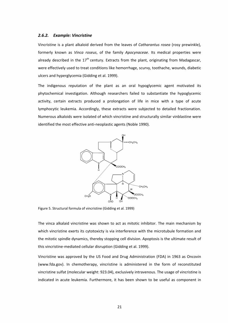
Figure 5. Structural formula of vincristine (Gidding et al. 1999)
The vinca alkalaid vincristine was shown to act as mitotic inhibitor. The main mechanism by
which vincristine exerts its cytotoxicty is via interference with the microtubule formation and
the mitotic spindle dynamics, thereby stopping cell division. Apoptosis is the ultimate result of
this vincristine‐mediated cellular disruption (Gidding et al. 1999).
Vincristine was approved by the US Food and Drug Administration (FDA) in 1963 as Oncovin
(www.fda.gov). In chemotherapy, vincristine is administered in the form of reconstituted
vincristine sulfat (molecular weight: 923.04), exclusively intravenous. The usage of vincristine is
indicated in acute leukemia. Furthermore, it has been shown to be useful as component in
22
combinatorial chemotherapy in several types of cancer, including Hodgkin disease,
non‐Hodgkin lymphoma and neuroblastoma (Gidding et al. 1999).
The most frequent and clinical important side‐effect of vincristine is neurotoxicity (Moore and
Pinkerton 2009). By now, neuropathy represents the dose‐limiting toxicity of vincristine,
especially in children.
2.6.3. Potential anti‐neoplastic activity of two ethnomedical plants from
Guatemala
Like vincristine, more than 60 % of applied anti‐cancer drugs are derived from natural sources,
i.e. plants, microorganisms and marine organisms (Cragg and Newman, 2007). In
less‐developed countries, a majority of the population still relies heavily or even entirely on
traditional healing plants in treating various conditions. Therefore, the applications in folk
medicine are well delivered and may act as first hint in the new drug discovery process.
In the present work, two Guatemalan ethnomedical plants (Neurolaena lobata and Critonia
morifolia) were selected for investigations of their oncolytic potential based on their long
history of traditional uses.
Critonia morifolia ‐ Traditional uses
Several traditional applications of C. morifolia are known with limitation to the usage of the
plants leaves. The leaves are applied as steam bath in cases of swelling, retention of fluids,
rheumatism, arthritis, paralysis and muscle spasms. Baths of boiled leaves alone or in
combination with other healing plants are used to cure skin conditions, wounds, insomnia, flu
and aches. Boils, cysts, pus‐filled sores and even cancer are treated with leaves that were
heated in oil prior to application directly on the swelling.
Critonia morifolia ‐ Research results
Various sesquiterpene lactones and pyrrolizidine alkaloids, including a completely new one
named morifoline (Wiedenfeld and Cetto 1998) were isolated of C. morifolia (Herz 2004).
Despite of its manifold curative usage in folk medicine, only little pharmacological research is
published on Critonia morifolia.
Neurolaena lobata‐ Traditional uses
N. lobata is widely used as home remedy to cure a variety of diseases, particularly malaria and
amoebiasis. Traditionally, tea is taken to treat and prevent a variety of parasitic ailments such
as malaria, fungus, ringworm, amoebas and intestinal parasites. It is prepared by boiling one
23
fresh leaf per cup for ten minutes. Up to three cups are consumed daily. Tea prepared of the
leaves is also used to bathe wounds and infections, and applied to douche conditions of
leukorrhea and vaginal itching. Wounds, skin conditions and sores may be treated by fresh
juice from crushed leaves. The water from boiled leaves is also used as insecticide and
fungicide in home and garden, and as hair wash to get rid of lice. In case of sores, fungus and
infections, a powder of toasted leaves is directly applied. Also the roots are a respected
remedy. The water of boiled roots is drunk as a blood cleanser (Arvigo and Balick 1998).
Neurolaena lobata ‐ Research results
A variety of characteristic sesquiterpene lactones were isolated of N. lobata, among others
neurolenin A, B, C, D, E and F and lobatin A, B and C (Passreiter et al. 1995). Another class of
chemical found are pyrrolizidine alkaloids (Passreiter 1998).
Various studies approved the anti‐parasitic properties of N. lobata. Extracts and pure
sesquiterpene lactones of N. lobata were found to be active against Plasmodium falciparum,
the clinically most important malaria pathogen, and Plasmodium berghei (François et al. 1996).
The ethanol extract of N. lobata was reported to inhibit parasite growth of Leishmania
mexicana, Trypanosoma cruzi and Trichomonas vaginalis (Berger et al. 2001).
Only few studies are available on cytotoxicity of N. lobata. François et al. (1996) discovered
cytotoxic effects of sesquiterpene lactones in GLC4 and COLO 320 tumor cell lines. An assay
performed on brine shrimp larvae Artemia salina exhibited only weak toxicity (Berger at al.
1998). In mice, oral and intraperitoneal administration of 500 mg/kg of the water, ethanol and
dichloromethane extract every 48 hours for three weeks did not exhibit subacute toxicity and
oral dosages up to 5 g/kg did not exhibit acute toxicity (Cáceres et al. 1998).
Furthermore, hypoglycemic activity of the ethanol extract was demonstrated in vivo (Gupta et
al. 1984). A recent study reported an inhibitory effect of Neurolaena lobata extracts on the
transfer of HIV from dendritic cells to lymphocytes in vitro (Bedoya et al. 2008).
24
3 MATERIAL AND METHODS
3.1. Plant Material
Plant material of Neurolaena lobata and Critonia morifolia was collected in Guatemala (Petén).
Fresh material was stored at ‐80°C before it was lyophilized and ground. Freeze‐drying was
preferred over air‐drying, as it is known to be more convenient in preserving volatile
substances.
Of N. lobata, also air‐dried material was available. Thus, its effectiveness was compared to that
of freeze‐dried plant material to consider the loss of potentially effective compounds at a
higher dehydration temperature. Investigations on air‐dried material were limited to the
extract type of lyophilized plant that exhibited the strongest effects in previous cell culture
assays.
25
3.1.1. Critonia morifolia (Petén, Guatemala)
Figure 6 Critonia morifolia, Guatemala
Kingdom: Plantae
Division: Magnoliophyta
Class: Magnoliopsida
Order: Asterales
Family: Asteraceae
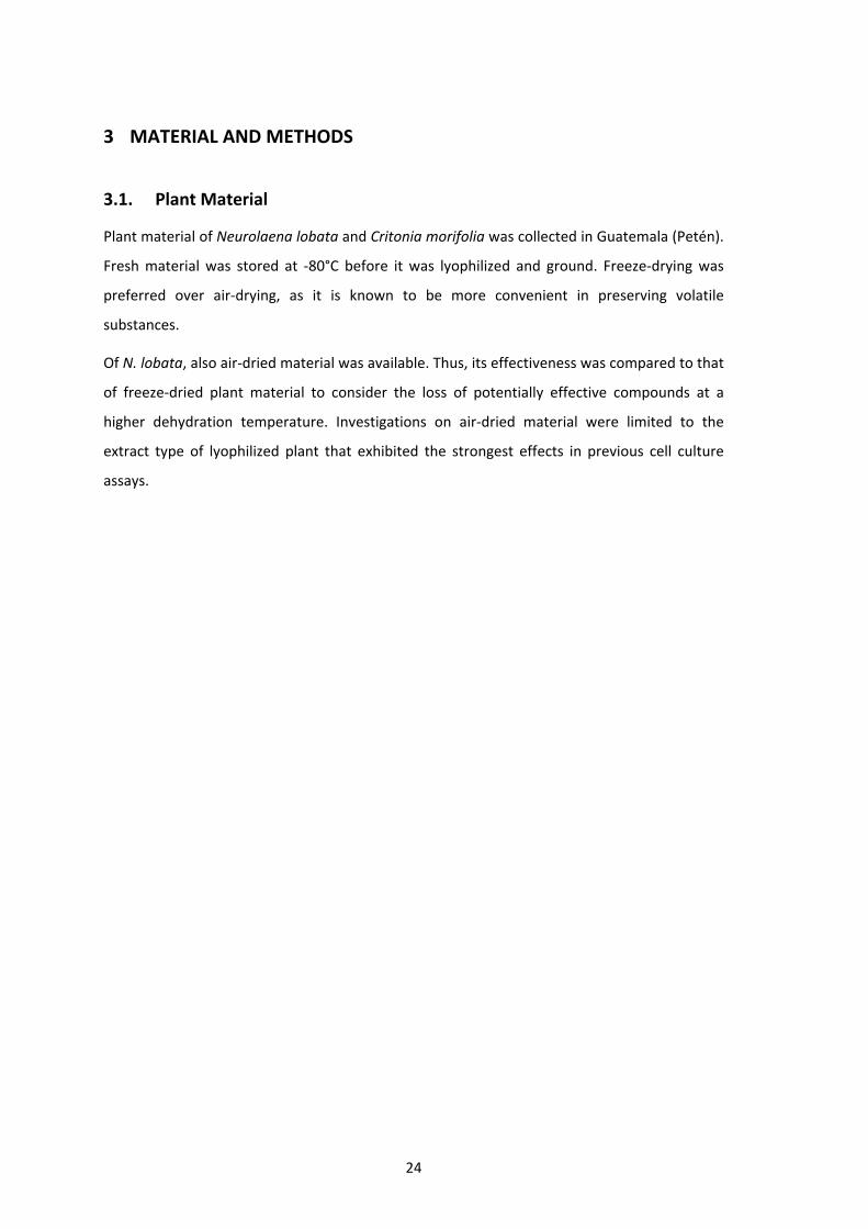
Genus: Critonia
Species: Critonia morifolia
Critonia morifolia, like N. lobata, belongs to the family Asteraceae. It was formerly known as
Eupatorium morifolia. The genus Critonia comprises 43 species and is spread from Mexico to
Argentina (Herz 2004).
The plant grows as herbaceous shrub up to four meters tall. The indigenous name palo verde
(green stick) refers to its characteristically thick green, often woody, stems. The leaves are
26
deep green with toothed margins and vary from 10 to 40 cm in length. The colour of
florescence ranges from greenish‐yellow and turns into straw‐brown when dry. C. morifolia is
found in forests and at the edges of forests, riversides and roadsides.
3.1.2. Neurolaena lobata (Guatemala, Petén)
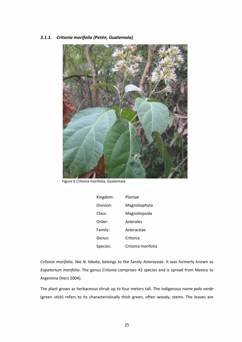
Figure 7. Neurolaena lobata (www.nybg.org/bsci/belize/Neurolaena_lobata.jpg)
Figure 8. Neurolaena lobata (www.phytobokaz.fr/images_galerie/799959083%20.jpg)
Kingdom: Plantae
Division: Magnoliophyta
Class: Magnoliopsida
Order: Asterales
Family: Asteraceae
Subfamily: Asteroideae
Genus: Neurolaena
Species: Neurolaena lobata
27
Asteracea is the largest family of flowering plants and spread worldwide. It is divided into 11
subfamilies of which the subfamily Asteroideae comprises approximately 70 % of specific
diversity of the whole family. Since 2004, this subfamily is divided into three supertribes
(Robinson 2004) of which Helianthodae comprises the species Neurolaena lobata. This plant is
spread in Latin American countries and can be found especially in Guatemala and Costa Rica.
Neurolaena lobata is an herb growing from 1 to 4 meters tall. It has only a few main stems
with numerous branches and yellow blooming florescence. The most common names are tres
puntas, referring to the leaves shape with three distinctive points, and Jackass bitters,
referring to the extremely bitter taste of the plants leaves. Its natural habitat in the rainforest
encompasses clearings, roadsides, fields and pastures (Arvigo and Balick 1998).

28
3.2. Plant extraction
The plant powder was consecutively extracted with petroleum ether (PE), dichloromethane
(CH2Cl2), ethyl acetate (EA), methanol (MeOH) and water (H2O), leading to five extracts of
distinct polarity. Starting with the least polar solvent, this method provides an initial
separation of non‐polar and polar bioactive plant constituents at minimal plant expense.
For further use in cell culture experiments only small amounts of the gained extracts were
transferred into a 1.5 ml tube and dissolved in DMSO (dimethyl sulfoxide). DMSO dissolves
both polar and non‐polar compounds and is miscible with a wide range of solvents. To account
for detrimental effects of DMSO on cell proliferation, apoptosis and cell cycle, controls were
treated with the respective concentrations of DMSO used for sample treatment. Maximum
concentration of DMSO was limited to 0.5 % to avoid cell damage due to toxicity of DMSO.
3.2.1. C. morifolia – Accelerated Solvent Extraction (ASE 2000)
Accelerated Solvent Extraction (ASE 2000, Dionex) is a fully automated technique to rapidly
extract solid and semi‐solid sample matrices with organic solvents as well as water. ASE allows
extraction at temperatures above the solvents boiling point, using high pressure to keep the
solvent liquid. Advantages to traditional extraction methods are the reduction of solvent
needed, time savings and better solubility of constituents at elevated temperatures, though
thermal degradation of some target analytes has to be considered.
Powdered leaves of C. morifolia were mixed 2:1 with diatomaceous earth, which acts as a
dispersant and drying agent, prior to adding the sample to the extraction cells. Schedule
(Table 1) was programmed and started. One by one, cells were filled with the solvent via HPLC‐
pump and heated followed by static extraction. Solvent containing the dissolved plant
compounds was collected in vials. The solid plant material that remained in the cells was
flushed with cold solvent (also collected in vials) and dried with nitrogen (N2) automatically.
For each solvent and cell, the cycle was repeated before moving to the next solvent. All vials
containing the same solvent were transferred into a flask and subjected to rotary evaporation
(Table 2).

29
Table 1. ASE conditions for extraction of C. morifolia and air‐dried N. lobata.
Preheat
Heat 5 min Temperature 40°C
Static extraction 2 min Pressure 150 bar
Purge time 60 s Cycles 2
Flush volume (of extraction volume)
10 % Solvent (in order of application)
PE, CH2Cl2, MeOH, EA, H2O
3.2.2. N. lobata – reflux‐water bath extraction
First, plant powder consisting of lyophilized leaves, florescence and stipes of N. lobata, was
mixed at a concentration of 1:10 with solvent in a flask, e.g. to 20 g plant material 200 ml
solvent was added. In the next step the flask was sealed with Parafilm® and treated in an ultra
sonic bath for ten minutes to burst the plant cells and therefore making greater amounts of
compounds available. Then the flask was put on a reflux‐water bath at a temperature
depending on the solvents boiling point (Table 2) for one hour. Afterwards the content of the
flask was filtered through a round filter with a pore diameter of 150 nm to separate solid and
dissolved plant material. The solid plant residue was air‐dried over night and reused for the
following extraction step with the next, more polar solvent. The extracted material dissolved in
the liquid phase was dried by rotary evaporation at a water bath temperature of 40°C and a
pressure according to the solvents characteristics (Table 2) until complete dryness. Flasks
containing the dried extracts were stored in a vacuum desiccator.
For air‐dired N. lobata, the extraction was conducted as described for C. morifolia applying
ASE 2000 (Table 1) but was limited to the first two solvents (petroleum ether and
dichloromethane). Only the dichloromethane extract was used in cell culture experiments.
Table 2. Solvents (in order of extraction), corresponding temperatures for water bath extraction and pressure used for solvent evaporation.
Solvent Reflux – water bath (°C) Rotary evaporator (mbar)
Petroleum ether 60 600
Dichloromethane 40 850
Ethyl acetate 92 250
Methanol 80 300
Water 100 75

30
3.3. Cell culture
HL‐60, human promyelocytic leukemia cells, were purchased from ATCC (American Type
Culture Collection). SR‐786, NPM‐ALK positive human ALCL (anaplastic large cell lymphoma)
cells, and 417, NPM‐ALK positive mouse ALCL cells, were kindly provided by Prof. Lukas
Kenner. HL‐60, SR‐786 and 417 cells were grown in RPMI 1640 medium supplemented with
10 % heat inactivated fetal calf serum (FCS), 1 % L‐Glutamine and 1 % Penicillin/Streptomycin.
Primary human lung fibroblasts (HLFs), were obtained from ATCC (designation LL 47 (MaDo))
and cultivated in DMEM medium supplemented with 10 % FCS and 1 %
Penicillin/Streptomycin. Media and supplements were purchased from Life Technologies. All
cells were maintained in humidified atmosphere containing 5 % CO2 at 37°C.
3.4. Proliferation and cytotoxicity assays
Proliferation inhibition (HL‐60)
Initially, HL‐60 cells were treated with all of the plant extracts to determine their
anti‐proliferative effects and to figure out the most promising ones. Therefore, HL‐60 cells
were seeded in 24‐well plates at a concentration of 1 x 10⁵ cells/ml allowing logarithmic
growth within 72 hours. Afterwards cells were incubated with increasing concentrations of
plant extracts (5 µg/ml, 15 µg/ml, 30 µg/ml, 60 µg/ml) for 72 hours. The dichloromethane
extract of N. lobata was also applied in 2.5 and 10 µg/ml. After 24 hours and 72 hours cell
number was determined using a KX‐21 N microcell counter (Sysmex Coporation, Kobe, Japan).
All experiments were carried out in triplicates. Anti‐proliferative effects were calculated by
applying the following formula:
C72h + drug C24h + drug
C72h ‐ drug C24h ‐ drug x 100 = % of cell division
C72h+drug cell number after 72 h of drug treatment
C24h+drug cell number after 24 h of drug treatment
C72h‐drug cell number after 72 h without drug treatment
C24h+drug cell number after 24 h without drug treatment
Only the extract of Neurolaena lobata showing the best anti‐proliferative effect, the
dichloromethane extract, was tested on other cell systems including SR‐786, 417 and HLF,
respectively. Experiments with extracts of Critonia morifolia were restricted to HL‐60 cells only.
31
Proliferation inhibition (SR‐786, 417)
Effects of the dichloromethane extract of N. lobata were evaluated in the cell lines SR‐786
(human) and 417 (mouse) using a Casy® cell counter (Invitrogen, Life Technologies Inc.).
SR‐786 cells were seeded in a 48‐well plate at a concentration of 2 x 10⁵ cells/ml. Then, the
cells were incubated with 2.5 µg/ml, 5 µg/ml, 10 µg/ml, 15 µg/ml and 20 µg/ml of the extract
for 48 hours. 417 cells were seeded in 48‐well plates at a concentration of 1 x 10⁶ cells/ml and
incubated with 5 µg/ml, 10 µg/ml and 15 µg/ml extract for 72 hours. The same formula as for
HL‐60 cells was used for calculations. In case of SR‐786 cells, cell number after 48 hours was
used instead of 72 hours
AlamarBlue® assay (417, HLF)
Additionally to cell number determination of 417 cell line, the alamarBlue® assay (Invitrogen,
Life Technologies) was applied to measure cytotoxicity. The active component of alamarBlue®
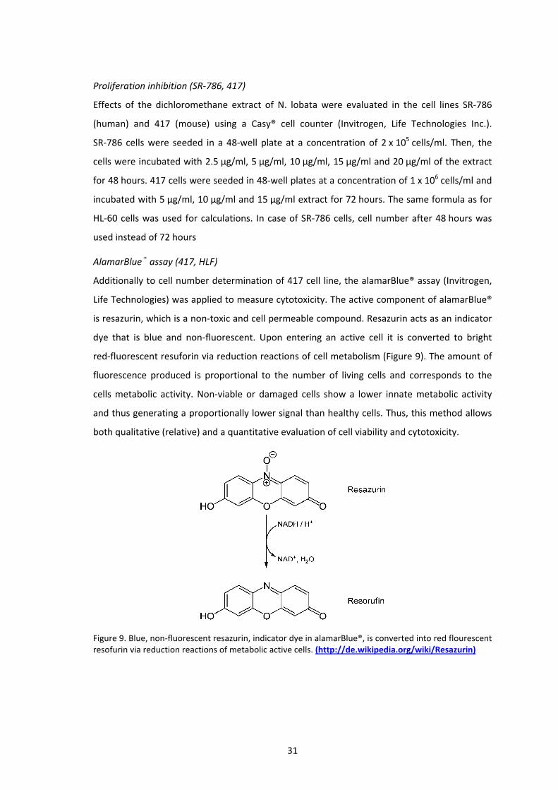
is resazurin, which is a non‐toxic and cell permeable compound. Resazurin acts as an indicator
dye that is blue and non‐fluorescent. Upon entering an active cell it is converted to bright
red‐fluorescent resuforin via reduction reactions of cell metabolism (Figure 9). The amount of
fluorescence produced is proportional to the number of living cells and corresponds to the
cells metabolic activity. Non‐viable or damaged cells show a lower innate metabolic activity
and thus generating a proportionally lower signal than healthy cells. Thus, this method allows
both qualitative (relative) and a quantitative evaluation of cell viability and cytotoxicity.
Figure 9. Blue, non‐fluorescent resazurin, indicator dye in alamarBlue®, is converted into red flourescent resofurin via reduction reactions of metabolic active cells. (http://de.wikipedia.org/wiki/Resazurin)

32
HLF cells were seeded into 48‐well plates at a concentration providing confluence within the
wells. Together with 500 µl medium, HLF cells were grown for 24 hours. 417 cells were seeded
into 48‐well plates at a concentration of 1 x 10⁶ cells/ml. Each well was filled with 500 µl cell
suspension. Dichloromethane extract was added at concentrations of 2.5 µg/ml, 5 µg/ml and
10 µg/ml, HLFs were additionally treated with 15 µg/ml. An untreated control and pure
medium were also assessed to determine differences in cell viability depending on drug
treatment. After 24 hours, and 48 hours respectively, 50 µl alamarBlue® reagent was added to
each well and incubated for approximately 90 minutes at 37°C until colour changed from blue
to red. Afterwards the 48‐well plate was placed into a multi‐detection reader for fluorescence
and absorbance (Synergy HT, Bio‐Tek Instrument, Inc., Vermont, USA). Plate reader software
KC‐4 (Bio‐Tek) was used to determine absorption at 570 nm. Experiments were done in
triplicates. To calculate the differences in cell viability, mean blank value (only medium) was
subtracted from all other measurement values to take fluorescence of the medium into
account. Mean value of the control samples was considered as 100 % cell viability. The mean
values of treated samples were described as percentage of control sample viability.
Calculations were performed in Excel (Microsoft).
3.5. Apoptosis assay – Hoechst 33258 and propidium iodide double staining
Hoechst 33258 (HO) and propidium iodide (PI) double staining (both Sigma, St. Louis, MO)
allows the determination of the type of death the cell is undergoing, i.e. apoptosis (early or
late) or necrosis (Grusch et al. 2002). HL‐60 cells were seeded in a 24‐well plate at a
concentration of 1 x 10⁵ cells/ml and treated with increasing concentrations of the specified
extracts (Table 3). After 24 hours of incubation, 100 µl cell suspension of each well was
transferred into separate wells of a 96‐well plate and Hoechst 33285 and propidium iodide
were added at final concentrations of 5 µg/ml and 2 µg/ml, respectively. After one hour of
incubation at 37°C, stained cells were examined and photographed on a fluorescence
microscope (Axiovert, Zeiss) equipped with a DAPI filter. Type and number of cell deaths was
evaluated by visual examination of the photographs according to the morphological
characteristics revealed by HOPI staining. Experiments were performed in triplicates.

33
Table 3.Extracts and corresponding concentrations applied in apoptosis assay
Neurolaena lobata µg/ml Critonia morifolia µg/ml
PE extract 5, 15, 30 PE extract 5, 15, 30
CH2Cl2 extract 5, 10, 15 CH2Cl2 extract 5, 15, 30
EA extract 5, 10, 15
MeOH extract 15, 30, 60
3.6. Cell cycle distribution (FACS)
417 cells were seeded in a 6‐well plate at a concentration of 1 x 10⁶ cells/ml. Then
dichloromethane extract of N. lobata was added to a final concentration of 5 µg/ml, 10 µg/ml
and 15 µg/ml. HL‐60 cells were seeded in T‐75 tissue culture flasks at a concentration of
2 x 10⁵ cells/ml and treated with 10 µg/ml and 15 µg/ml of the petroleum ether extract of
C. morifolia. After 24 hours (for HL‐60 cells also after 8 hours) of incubation at 37°C, cells were
harvested, transferred into 15 ml tubes and centrifuged (4°C, 800 rpm, 5 min). The
supernatant was discarded and the cell pellet was washed with cold PBS, centrifuged (4°C,
800 rpm, 5 min), resuspended in 1 ml cold ethanol (70 %), and either fixed for 30 minutes at
4°C or stored at ‐20°C prior further handling. After two washing steps with cold PBS, the cell
pellet was resuspended in 500 µl cold PBS and transferred into a 5 ml polystyrene round
bottom tube. RNAse A and propidium iodide were added to a final concentration of 50 µg/ml
each and incubated for one hour at 4°C. The final cell number was adjusted between 0.5 and
1 x 10⁶ cells in 500 µl. Cells were analyzed by FACS Calibur flow cytometer (BD Bioscience, San
Jose, CA, USA). Cell cycle distribution was calculated with ModFid LT software (Verity Software
House, Topsham, ME, USA).
3.7. Western blotting
Lysate preparation
417 cells were seeded in a 6‐well plate at a concentration of 106 cells/ml and treated with
10 µg/ml of the dichloromethane extract of N. lobata. After 0.5, 2, 4, 8 and 24 hours
4 x 10⁶ cells were harvested. Vincristine, a common plant based anti‐cancer drug, was added at
final concentrations of 1, 2.5 and 5 µM. Cells were harvested after 8 and 24 hours.
SR‐786 cells were seeded in a T‐75 tissue culture flask at a concentration of 2.5 x 10⁵ cells/ml
and incubated with 15 µg/ml dichloromethane extract of N. lobata. 106 cells were harvested

34
after 4, 8 and 24 hours. Additionally, Proteasome Inhibitor IV (Cat.No. 539175, Merck) was
added to a final concentration of 50 µM in a single experiment. In another experiment
lysosome inhibitor ammonium chloride (NH4Cl) was added at a concentration of 20 mM.
HLFs were grown in a 6‐well plate to confluence and incubated with 10 µg/ml and 15 µg/ml
dichloromethane extract of N lobata. After 4, 8 and 24 hours, cells were harvested.
HL‐60 were seeded in T‐75 tissue culture flasks at a concentration of 1.8 x10⁵ cells/ml and
incubated with 15 µg/ml petroleum ether extract of C. morifolia. Cells were harvested after
0.5, 2, 4, 8 and 24 hours.
All cells were washed with cold PBS twice and centrifuged at 1000 rpm for 5 min at 4°C. Cell
pellet was lysed in a buffer containing 150 mM NaCl, 50 mM Tris pH 8.0, 1 % Triton‐X‐100,
1 mM phenylmethylsulfonyl fluoride (PMSF) and 1 mM Protease Inhibitor Cocktail (PIC). The
lysate was centrifuged at 12000 rpm for 20 min at 4°C. Supernatant was transferred into a
1.5 ml tube and stored at ‐20°C until further analysis.
Gel electrophoresis (SDS‐PAGE) and blotting
Equal amounts of protein samples (lysate) were mixed with SDS (sodium dodecyl sulfate)
sample buffer and loaded onto a 10 % polyacrylamide gel. Proteins were separated by
polyacrylamide gel electrophoresis (PAGE) at 120 Volt.
To make proteins accessible to antibody detection, they were electrotransferred from within
the gel onto a PVDF (polyvinylidene difluoride) membrane (Hybond, Amersham, UK) at 95 Volt
for 80 minutes. Membranes were allowed to dry for at least 30 minutes up to 2 hours to
provide fixing of the proteins on the membrane. Methanol was used to remoisten the
membranes. Equal sample loading was checked by staining the membrane with Ponceau S
(Sigma‐Aldrich).
Protein detection
After washing with PBS (Phosphate buffered saline, pH 7.2) or TBS (Tris buffered saline,
pH 7.6), membranes were blocked in PBS‐ or TBS‐milk (5 % non‐fat dry milk in PBS containing
0.5 % Tween 20 or TBS containing 0.1 % Tween 20) for one hour. Then, membranes were
washed with PBS/T (PBS containing 0.5 % Tween 20) or TBS/T (TBS containing 0.1 % Tween 20),
changing the washing solution 4‐5 times, for at least 20 min. Next, membranes were incubated
with the primary antibody in blocking solution (according to the data sheet TBS‐, PBS‐milk or
TBS‐, PBS‐BSA) diluted 1:500 – 1:1000, gently shaking at 4°C over night. Thereafter,
35
membranes were washed again with PBS/T or TBS/T and incubated with the secondary
antibody (peroxidase conjugated anti‐rabbit IgG or anti‐mouse IgG) diluted 1:2000 in PBS‐ or
TBS‐milk at room temperature for one hour. Chemiluminescence was developed by ECL
detection kit (Amersham, UK) and membranes were exposed to Amersham Hyperfilms.
Antibodies
CD246, ALK protein, monoclonal mouse, clone ALK1, code M7195 (DakoCytomation)
Nucleophosmin, monoclonal mouse, clone 376, code M7305 (DakoCytomation)
PDGF Receptor β (28E1) Rabbit mAB , #3169 (Cell Signaling)
Chk1 (2G1D5) Mouse mAb, #2360 (Cell Signaling)
Phospho‐Chk1 (Ser345) Antibody, #2341 (Cell Signaling)
Chk2 Antibody, #2662 (Cell Signaling)
Phospho‐Chk2 (Thr68) Antibody, #2661 (Cell Signaling)
Cleaved PARP (Asp214) Antibody (Mouse Specific), #9544 (Cell Signaling)
PARP‐1 (F‐2): sc‐8007, mouse monoclonal (Santa Cruz Biotechnology, Inc.)
Cleaved Caspase3 (Asp175) Antibody, #9661 (Cell Signaling)
Caspase3 Antibody, #9662 (Cell Signaling)
PhosphoDetect Anti‐H2AX (pSer139), DR 1017 (EMD4Biosciences)
Cdc25A (phospho S75) antibody, ab47279, rabbit polyclonal (abcam)
Phospho‐Cdc25A‐S177 Antibody, rabbit polyclonal, Cat. #AP3046a (Abgent)
Cdc25A (F‐6): sc‐7389, mouse monoclonal (Santa Cruz Biotechnology, Inc.)
Cdc25B (C‐20): sc‐326, rabbit polyclonal (Santa Cruz Biotechnology, Inc.)
Cdc25C (C‐20): sc‐327, rabbit polyclonal (Santa Cruz Biotechnology, Inc.)
Phospho‐cdc2 (Tyr15)(10A11) Rabbit mAb, #4539 (Cell Signaling)
Cdc2 p34 (17): sc‐54, mouse monoclonal (Santa Cruz Biotechnology, Inc.)
Phospho‐p53 (Ser20) Antibody, #9287 (Cell Signaling)
p53, mouse monoclonal, Cat. No. 1767 (Immunotech, Coulter Company)
JunB (120): sc‐73, rabbit polyclonal (Santa Cruz Biotechnology, Inc.)
c‐Jun (H‐79): sc‐1694, rabbit polyclonal (Santa Cruz Biotechnology, Inc.)
Phospho‐Akt (Ser473)(587F11) Mouse mAb, #4051 (Cell Signaling)
Akt Antibody, #9272 (Cell Signaling)
Cyclin D1 (M‐20): sc‐718, rabbit polyclonal (Santa Cruz Biotchnology, Inc.)
p21 (C‐19): sc‐397, rabbit polyclonal (Santa Cruz Biotechnology, Inc.)
c‐myc Ab‐2 (9E10.3), #MS‐139‐P1, mouse monoclonal (Thermo Fisher Scientific, Inc.)

36
Wee1 Antibody, #4936 (Cell Signaling)
Phospho‐Wee1 (Ser462)(D47G5) Rabbit mAb, #4910 (Cell Signaling)
β‐actin, monoclonal mouse ascites fluid, clone AC‐15, Cat. No. A5441 (Sigma)
3.8. Quantitative RT‐PCR
RNA preparation
SR‐786 cells were seeded in T‐75 tissue culture flasks at a concentration of 2.5 x 10⁵ cells/ml
and incubated with 15 µg/ml dichloromethane extract of N. lobata. After 4 hours and 8 hours
cells were harvested and homogenized using QIAshredder (50) (Cat. No. 79654, QIAGEN). The
cells were further processed according to the instructions of RNeasy Mini Kit (50) (Cat. No.
74104, QIAGEN). Final RNA concentration was measured using a NanoDrop
Fluorospectrometer (Thermo Fisher Scientific, Inc.). RNA was stored at ‐80°C until further
progression.
cDNA synthesis
First‐strand cDNA synthesis from 1000 ng RNA was performed using SuperScript®First‐Strand
Synthesis Systems for RT‐PCR (Cat. No. 11904‐018, Invitrogen). The enzyme reverse
transcriptase enables the transcription of mRNA into cDNA. cDNA synthesis reaction was
primed using the most nonspecific primers, random hexamers. With this method, all RNAs are
templates for cDNA synthesis. Desired mRNA was obtained by choosing specific primers in the
PCR (polymerase chain reaction). Finished cDNA was stored at ‐80°C.
cDNA synthesis
8 µl RNA (1 µg RNA + H2O)
1 µl Random Hexamers 65°C for
1 µl dNTPs 5 min
add room temperature 25°C for 10 min
2 µl 10x Buffer for 2 min 42°C for 50 min 37°C for
4 µl MgCl2 70°C for 15 min 20 min
2 µl DTT
1 µl RNase OUT add
1 µl Superscript II RT add
1 µl RNase H Figure 10. cDNA preparation from RNA samples using SuperScript®First‐Strand Synthesis Systems for RT‐PCR (Invitrogen).

37
Real time‐PCR
The alteration in transcription levels of NPM‐ALK in SR‐786 cells after treatment with the
dichloromethane extract of N. lobata was investigated by real‐time PCR using TaqMan®
detection system. The housekeeping‐gene glyceralaldehyde 3‐phosphat dehydrogenase
(GAPDH), which is stably and constitutively expressed at high levels in most tissues and cells,
served as control gene. For each sample, 7 µl H2O, 10 µl TaqMan® Universal PCR Master Mix
(Applied Biosystems), 1 µl primer+probe and 2 µl cDNA, or 2 µl H2O for negative controls, were
filled into a tube of a 96‐well optical reaction plate. In case of GAPDH, the primer and probe
mixture from TaqMan® Gene Expression Kit (Applied Biosystems) was used. To detect
NPM‐ALK transcripts, forward primer (GTG GTC TTA AGG TTG AAG TGT GGT T) and reverse
primer (GCT TCC GGC GGT ACA CTA CTA A) were mixed with the probe (TGC TGT CCA CTA ATA
TGC ACT GGC CCT) prior adding to the rest. Final concentration of primers and probe in the
sample mixtures were 0.5 µM and 0.25 µM, respectively. Cycle program (95°C for 10 min to
activate polymerase followed by 40 cycles of 95°C for 15 s and 60°C for 1 min) was started on
Abi Prism 7000 Sequence Detection System (Applied Biosystems). Real time‐PCR was
performed in duplicates for each cDNA template and gene investigated. Negative controls,
containing water instead of cDNA, confirmed the absence of RNA/DNA in all reagents applied
in the assay.
Analysis of results
Comparative CT (ΔΔCT) method (Livak and Schmittgen 2001) was used for calculating relative
quantitation of gene expression. The CT value is determined as the number of PCR cycles that is
needed to reach a defined level of fluorescence and therefore newly synthesized DNA. The
following formula was applied to quantify relative expression of the target gene NPM‐ALK:
Ra o = 2‐ΔΔCT
ΔCT = CT target gene (NPM‐ALK) – CT control gene (GAPDH)
ΔΔCT = ΔCT drug treatment – ΔCT control sample
3.9. Statistical analysis
Values were expressed as mean ± SD. Student’s t‐test was applied to compare differences
between control samples and treatment groups. Statistical significance level was set to
p < 0.05.

38
4 RESULTS
4.1. Critonia morifolia
Lyophilized leaves of Critonia morifolia were subjected to sequential extraction with five
solvents of increasing polarity applying ASE 2000 (Dionex). The obtained extracts were
investigated for their anti‐carcinogenic potential in HL‐60 cells. Proliferation and apoptosis
assays were performed to identify the most promising extract which effects were
subsequently studied in more detail by western blot analysis.
4.1.1. Extract yields and stock calculation
The extract weights obtained from serial extraction of 22.52 g lyophilized leaves of C. morifolia
with five solvents of increasing polarity are presented in Table 4.
Table 4. Extract weights corresponding to lyophilized plant material of C. morifolia.
Solvent Extract weight (g) corresponding to 22.52 g dried plant
Extract weight (µg) corresponding to 1 mg dried plant
Petroleum ether 1.3481 59.86
Dichloromethane 0.6347 28.18
Ethyl acetate 0.3223 14.31
Methanol 3.1708 140.80
Water 4.1483 184.20
To determine anti‐proliferative effects in HL‐60 cells, extracts were applied at increasing
concentrations (5 µg/ml, 15 µg/ml, 30 µg/ml, 60 µg/ml). Concentrated stock solutions of all
extracts were prepared in DMSO and stored at ‐20°C. Control samples were treated with the
amount of DMSO used in the highest concentration to take effects of DMSO into
consideration. For all other samples, the DMSO concentration was adjusted accordingly
(max. 0.5 % DMSO final concentration).

39
Table 5. Extract weights of C. moriolia applied in HL‐60 proliferation assays.
Extract type Extract stock concentration (µg/µl DMSO)
Extract final concentration (µg/ml medium)
corresponding dried plant weight (µg)
Petroleum ether 12.07 5 83.5315 250.5830 501.1760 1002.34
Dichloromethane 12.00 5 177.4015 532.2130 1064.4260 2128.85
Ethyl acetate 12.15 5 349.4215 1048.2530 2096.5160 4193.02
Methanol 15.55 5 35.5115 106.5430 213.0760 426.14
Water 43.80 5 27.1415 81.4330 162.8660 325.73
4.1.2. Anti‐proliferative activity of C. morifolia extracts in HL‐60 cells
The extracts of C. morifolia were tested in logarithmically growing HL‐60 cells at increasing
concentrations. Proliferation inhibition of extract treated samples compared to solvent
adjusted controls is illustrated in Figure 11. The petroleum ether extract exhibited a growth
inhibition of more than 50 % at 5 µg/ml and 100 % at 15 µg/ml. Further, the dichloromethane
and the ethyl acetate extract showed a dose‐dependent response. When taking the
corresponding dried plant weight into account (Table 5), the petroleum ether extract and the
dichloromethane extract turned out to be the most effectives. Therefore, an apoptosis analysis
was conducted for these two extracts, but not for ethyl acetate extract. The methanol and the
water extract were not taken into consideration for further experiments since the effects on
proliferation in HL‐60 cells were mostly insignificant.

40
Figure 11. Effects of different extracts of C. morifolia on the proliferation of HL‐60 cells. Logarithmically growing cells were seeded at a concentration of 105 cells/ml and treated with increasing concentrations (5, 15, 30 and 60 µg/ml) of extracts (petroleum ether, dichloromethane, ethyl acetate, methanol, water). Experiments were done in triplicate. Error bars indicate ±SD. Asterisks indicate significant alterations in proliferation compared to control (p < 0.05).

41
4.1.3. Induction of apoptosis in HL‐60 cells by extracts of C. morifolia
Based on the results of the proliferation assay, the petroleum ether extract and the
dichloromethane extract were investigated in more detail regarding their apoptotic potential.
HL‐60 cells were subjected to increasing concentrations of the specified extracts (Figure 12) for
24 hours. Afterwards, propidium iodide and Hoechst 33258 were added to the cells facilitating
the distinction between viable, apoptotic and necrotic cells via visual examination.
Figure 12. Induction of apoptosis in HL‐60 by the petroleum ether and the dichloromethane extract of C. morifolia. HL‐60 cells were seeded at 105 cells/ml and incubated with indicated concentrations of extracts (petroleum ether and dichloromethane) for 24 h. Afterwards cells were stained with propidium iodide (2 µg/ml) and Hoechst 33258 (5 µg/ml) and incubated for 1 h. Viable and apoptotic cells were photographed on a fluorescence microscope equipped with a DAPI filter and examined visually. Experiments were performed in triplicate. Error bars indicate ±SD. Asterisks indicate significant alterations in number of apoptotic cells compared to control (p < 0.05).
After 24 hours of incubation, the petroleum ether extract already exhibited a slight induction
of apoptosis in HL‐60 at 5 µg/ml. At 15 µg/ml, the petroleum ether extract induced cell death
in almost 100% of the cells. Comparatively, 30 µg/ml of the dichloromethane extract were
necessary to achieve similar results. Taking the corresponding plant weight into account,
250.58 µg/ml dried plant was needed to induce apoptosis in 100 % of the cells in case of the
petroleum ether extract, whereas 1064.42 µg/ml were required in case of the
dichloromethane extract.
Thus, the petroleum ether extract of C. morifolia was identified the most active one. Its effects
in HL‐60 cells were studied in further detail by FACS and western blot analysis.

42
4.1.4. The petroleum ether extract represses c‐Myc and cyclin D1 expression in
HL‐60 cells
Cyclin‐dependent kinase (Cdk) inhibitor p21, is considered a tumor suppressor protein which
acts as inhibitor of cell cycle progression in G1/S. One important inducer of p21 transcription is
p53. As HL‐60 cells are proven to be p53 deficient (Wolf and Rotter 1985), this pathway is
excluded. Despite, p21 can be regulated by p53‐independent pathways (Abukhdeir and Park
2008). At transcriptional level, for example, it can be regulated by the oncogene c‐Myc (Coller
et al. 2000).
c‐Myc has a profound impact on cell proliferation, differentiation and apoptosis
(Dominguez‐Sola et al. 2007). Upregulation of c‐Myc levels is common in many tumor types
contributing to an abnormal proliferation rate.
Cyclin D1 promotes transition from G1 to S‐phase in cell cycle in dependence on Cdks. In
addition, cyclin D1 regulates and associates with transcription factors in a Cdk‐independent
manner. As the protein has impact on cell growth, metabolism, and cellular differentiation,
overexpression of cyclin D1 plays a key role in the development of several cancers (Fu et al.
2004), and is classified, such as c‐Myc, as a proto‐oncongene.
Incubation of HL‐60 cells with 15 µg/ml petroleum ether extract of C. morifolia caused a
simultaneous repression of c‐Myc and cyclin D1 after 30 minutes which intensified during the
time course. However, HL‐60 cells remained deficient in p21 expression during 24 hours of
extract treatment.

43
Figure 13. Effects of the petroleum ether extract on the proto‐oncogenes c‐Myc and cyclin D1 and tumor suppressor protein p21 in HL‐60 cells. Cells (1.8 x 10⁵ cells/ml) were incubated with 15 µg/ml petroleum ether extract of C. morifolia and harvested after 0.5, 2, 4, 8 and 24 h of treatment. Cells were lysed and obtained proteins samples subjected to SDS‐PAGE followed by western blot analysis with the indicated antibodies. Equal sample loading was confirmed by Ponceau S staining and β‐actin analysis.
4.1.5. After a transient G2‐M cell cycle inhibition, the petroleum ether extract of
C. morifolia induces S‐phase arrest in HL‐60 cells after 24 hours of incubation
HL‐60 cells were incubated with 10 µg/ml and 15 µg/ml petroleum ether extract of C. morifolia
for 8 hours and 24 hours, respectively. Afterwards, cells were subjected to fluorescence
activated cell sorting (FACS). Interestingly, the extract exhibited distinct effects depending on
the duration of treatment.
After 8 hours, the cell population in G2‐M phase increased from 15.9 % to 32.3 % (10 µg/ml)
and up to 61.3 % (15 µg/ml; Figure 14a) upon extract treatment. The dose‐dependent
accumulation of HL‐60 cells in G2‐M was caused at the expense of both G0‐G1 cells (from
32.0 % to 24.2 % and 10.0 %) and S‐phase cells (from 52.1 % to 43.5 % and 28.4 %).
By contrast, FACS analysis after 24 hours of incubation with the petroleum ether extract
revealed a cell cycle inhibition in S‐phase at 15 µg/ml (Figure 14b). The treatment at this
concentration resulted in a substantial increase of cells in S‐phase from 38.8 % to 62.4 %,
mainly at the expense of G0‐G1 phase cells, which was reduced from 42.6 % to 23.3 %. The
incubation of HL‐60 cells with 10 µg/ml extract exhibited only a slight shift of cell population
from G0‐G1 to G2‐M whereas S‐phase remained unchanged. Therefore, the higher
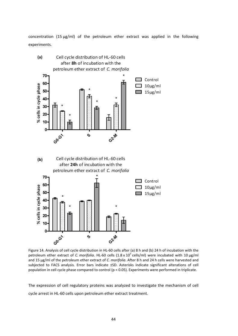
44
concentration (15 µg/ml) of the petroleum ether extract was applied in the following
experiments.
Figure 14. Analysis of cell cycle distribution in HL‐60 cells after (a) 8 h and (b) 24 h of incubation with the petroleum ether extract of C. morifolia. HL‐60 cells (1.8 x 10⁵ cells/ml) were incubated with 10 µg/ml and 15 µg/ml of the petroleum ether extract of C. morifolia. After 8 h and 24 h cells were harvested and subjected to FACS analysis. Error bars indicate ±SD. Asterisks indicate significant alterations of cell population in cell cycle phase compared to control (p < 0.05). Experiments were performed in triplicate.
The expression of cell regulatory proteins was analyzed to investigate the mechanism of cell
cycle arrest in HL‐60 cells upon petroleum ether extract treatment.
(a)
(b)

45
4.1.6. Modulations of cell regulatory proteins induced upon extract treatment
The transient G2‐M cell arrest after 8 hours and the final arrest in S‐phase caused by the
petroleum ether extract of C. morifolia were studied in further detail by investigating
modulations of key proteins of the cell cycle. For this reason, HL‐60 cells were treated with
15 µg/ml extract, harvested after 0.5, 2, 4, 8 and 24 hours and subjected to western blot
analysis.
Figure 15. Alteration in the expression of cell cycle regulatory proteins in HL‐60 cells. HL‐60 cells (1.8 x 10⁵ cells/ml) were treated with 15 µg/ml petroleum ether extract of C. morifolia. After 0.5, 2, 4, 8 and 24 h, cells were harvested and lysed. Isolated protein samples were subjected to western blot analysis investigating the protein expression levels of the indicated antibodies. Equal sample loading was confirmed by Ponceau S staining and β‐actin analysis.
The entry of eukaryotic cells into mitosis is regulated by cyclin‐dependant kinase 1 (Cdk1).
Phosphorylation of Cdk1 at tyrosine 15 leads to an inactivation of Cdk1 (Norbury et al. 1991).
The Cdc25 phosphatases, including Cdc25A, B and C, are pivotal triggers of the cell cycle by
46
activation of cylin‐dependent kinases (Cdks) via dephosphorylation (Hunter 1995). Despite the
presence of all Cdc25s throughout the cell cycle, Cdc25A predominantly controls transition
into S‐phase via activation of Cdk2 (Hoffmann et al., 1994), but is also able to regulate G2‐M
progression (Kiyokawa and Ray 2008). On the other and, Cdc25B and Cdc25C play a major role
in G2‐M transition via activation of the Cdk1/cyclin B complex (Hunter 1995). However, recent
studies suggest an undefined level of functional redundancy among Cdc25 phospatases
(Kiyokawa and Ray 2008)
Antagonizing the Cdc25 dephosporylation activity, Wee1 inhibits entry into mitosis by the
phosphorylation of Cdk1 at the tyrosine 15 site (McGowan and Russel 1993). Wee1
phosphorylation at serine 642 leads to the nuclear‐cytoplasmatic translocation. Although
Wee1 activity is not altered by phosphorylation, p(642)Wee1 delocalization into cytoplasm
prevents contact to its nuclear substrate Cdk1. Therefore, phosphorylation of Wee1 at Ser642
may promote the G2‐M progression (Katayama et al. 2005).
The phosphatases Cdc25A and Cdc25B were upregulated between 2 and 8 hours of incubation
with the petroleum ether extract prior to substantial downregulation thereafter. In contrast,
Cdc25C levels continuously decreased during the time course, which correlated with the G2‐M
arrest revealed by FACS analysis after 8 hours.
Upon extract treatment, Cdk1 was partly dephosporylated at Tyr15 after 8 hours, and
completely after 24 hours which is indicative for its activation. As all three Cdc25 phosphatases
were also downregulated within 24 hours, the decrease in kinase Wee1 was supposed to be
responsible for Cdk1 activation that may have helped to overcome the initial G2‐M arrest.
Thus, the downregulation of Cdc25A and Cdc25B was most likely responsible for S‐phase arrest
after 24 hours.
These temporally separated and phase specific inhibitory properties on cell cycle arrest are
probably due to distinct anti‐neoplastic compounds that are contained in the petroleum ether
extract of C. morifolia.

47
4.1.7. The petroleum ether extract induces apoptosis in HL‐60 cells mediated by
caspase‐3
Caspase‐3, a critical executioner of apoptosis, plays a major role in proteolytic cleavage of key
proteins (Fernandes‐Alnemri et al. 1994). Activation of caspase‐3 requires cleavage into its
17 kD fragment.
One of the main targets of proteolytic activity of cleaved caspase‐3 is the poly (ADP‐ribose)
polymerase (PARP). PARP, a 116 kD nuclear enzyme, appears to be involved in DNA repair in
response to environmental stress (Satoh and Lindahl 1992). Full length PARP supports cells in
maintaining viability whereas cleaved PARP (85 kD) facilitates cellular degradation (Beneke et
al. 2000). Thus, cleavage of PARP serves as a marker of cells undergoing apoptosis.
An important marker of genotoxicity is the phosphorylated form of H2AX at serine 139, named
γ‐H2AX. Within minutes following DNA double‐strand breaks, H2AX undergoes this specific
phosphorylation by ATM (Rogakou et al. 1998) and localizes to sites of damage at subnuclear
foci (Rogakou et al. 1999).
HL‐60 cells treated with 15 µg/ml petroleum ether extract of C. morifolia exhibited an initial
cleavage of caspase‐3 within 2 hours which intensified during the time course. The cleavage of
PARP after 4 hours correlates with the caspase‐3 activation. In response to DNA double strand
breaks initiated by apoptosis, H2AX was phosphorylated at Ser139 after 4 hours.
Additionally, Akt is known to play a critical role in apoptosis. This protein kinase represents an
important downstream effector of PI3K (phospatidylinisitol 3‐kinase) which promotes cell
survival and prevents apoptosis when activated by phosphorylation at the serine 473 residue
(Franke et al. 1997). As illustrated in Figure 16, untreated HL‐60 did not express p(Ser473)Akt,
neither it was induced by extract treatment whereas Akt was downregulated within 24 hours.
These findings suggest an induction of apoptosis by the petroleum ether extract of C. morifolia
mediated by caspase‐3. The later onset of H2AX phosphorylation, compared to cleavage of
caspase‐3, suggests that DNA double strandbreaks in extract treated HL‐60 cells are not a
cause of the extract’s genotoxicity but a result of induced apoptosis. Therefore, the activation
of downstream effectors of DNA damage, checkpoint kinases Chk1 and Chk2, were
investigated.

48
Figure 16. Induction of pro‐apoptotic key proteins in HL‐60 cells after incubation with the petroleum ether extract of C. morifolia. HL‐60 cells (1.8 x 10⁵ cells/ml) were incubated with 15 µg/ml extract and harvested after 0.5, 2, 4, 8 and 24 h, lysed and isolated protein samples were subjected to western blot analysis using the indicated antibodies. Equal sample loading was confirmed by Ponceau S staining and β‐actin analysis.
4.1.8. Early onset of apoptosis, not genotoxicity, leads to checkpoint kinase
activation in extract treated HL‐60 cells
To preserve a cell’s DNA integrity, a network of proteins communicates throughout the cell
cycle with each other. In response to DNA damage, checkpoint kinases 1 and 2 (Chk1 and
Chk2) are main mediators of cell cycle arrest to allow DNA repair prior to mitosis, or apoptosis
in case of an irreparable damage (Zhou and Elledge 2000). Total expression level of Chk1 and
Chk2 remained unchanged during 24 hours of extract treatment. The slight activation of Chk1
by phosphorylation at serine345 after 24 hours did not correlate with the observed S‐phase
arrest as Chk1 typically induces G2‐M arrest (Liu et al. 2000). Chk2 was phosphorylated at the
activating threonine 68 site within 4 hours of extract incubation (Figure 17). In general, this
activation is caused by the ATM protein kinase after DNA damage (Ahn et al., 2000). The
correlation of Chk2 activation, which was strongest after 24 hours, and the S‐phase arrest
observed at that timepoint is in line with previous findings (Koczor et al. 2009; Mazzacurati et
al. 2004). Activated Chk2 was reported to phosphorylate the Cdc25A phosphatase on the
serine 177 residue, targeting it for proteasomal degradation (Madlener et al., 2009). This

49
finding may explain the downregulation of Cdc25A after 24 hours of drug treatment, since the
levels of p(S177)Cdc25A were increased (Figure 15).
Chk2 activation was probably due to DNA cleavage in the course of apoptosis, because
caspase‐3 activation occurred before Chk2 phosphorylation. Therefore, C. morifolia petroleum
ether extract is not considered to damage DNA.
Figure 17. Activation of checkpoint kinases 1 and 2 after incubation of the petroleum ether extract of C. morifolia. HL‐60 cells (1.8 x 10⁵ cells/ml) were treated with 15 µg/ml of the petroleum ether extract of C. morifolia. After 0.5, 2, 4, 8 and 24 h, cells were harvested and lysed. Isolated protein samples were subjected to western blot analysis investigating the protein expression levels of the indicated antibodies. Equal sample loading was confirmed by Ponceau S staining and β‐actin analysis.

50
4.2. Neurolaena lobata
Lyophilized aerial plant parts (leaves, florescence and stipes) of N. lobata were sequentially
extracted by reflux‐water bath extraction using five solvents of increasing polarity. The
obtained extracts were tested for their anti‐carcinogenic activity in vitro. Proliferation and
apoptosis assays were performed to identify the extract containing the most active
constituents.
4.2.1. Extract yields and stock calculation
Extract weights obtained from serial extraction of 20.04 g lyophilized plant material with five
solvents of different polarity are shown in Table 6.
Table 6. Extract weights corresponding to lyophilized plant material of N. lobata
Solvent Extract weight (g) corresponding to 20.04 g dried plant
Extract weight (µg) corresponding to 1 mg dried plant
Petroleum ether 0.8863 44.23
Dichloromethane 0.5931 29.60
Ethyl acetate 0.2935 14.65
Methanol 1.0840 54.09
Water 2.4096 12.24
To investigate the anti‐proliferative effects in HL‐60 cells, extracts were applied at four
increasing concentrations (5 µg/ml, 15 µg/ml, 30 µg/ml, 60 µg/ml). Concentrated stock
solutions of all extracts were prepared in DMSO and stored at ‐20°C. Control samples were
treated with the amount of DMSO used in the highest concentration to take effects of DMSO
into consideration. For all other samples the DMSO concentration was adjusted accordingly
(max. 0.5 % DMSO final concentration).

51
Table 7. Extract weights of N. lobata applied in HL‐60 proliferation assays.
Extract type Extract stock concentration (µg/µl DMSO)
Extract final concentration (µg/ml medium)
corresponding dried plant weight (µg)
Petroleum ether 12.29 5 113.0515 339.1630 678.3360 1356.65
Dichloromethane 13.50 5 168.9415 506.8330 1013.6660 2027.31
Ethyl acetate 16.00 5 341.4015 1024.1930 2048.3860 4096.76
Methanol 34.00 5 92.4415 277.3130 554.6160 1109.23
Water 142.50 5 41.5815 124.7530 249.5060 499.00
4.2.2. Anti‐proliferative activity of N. lobata extracts in HL‐60 cells
All extracts of N. lobata were tested in logarithmically growing HL‐60 cells at increasing
concentrations. Figure 18 illustrates the proliferation inhibition of extract treated samples
compared to the control samples only adjusted by the solvent (DMSO). The petroleum ether,
dichloromethane and ethyl acetate extract showed a growth inhibition up to 100 % even at
low concentrations (5 µg/ml and 15 µg/ml). The methanol extract exhibited also a
dose‐dependent response, but at higher concentrations. Especially when taking the
corresponding dried plant material into account (Table 7), the proliferation assay did not allow
a clear decision on the most effective extract. Therefore, an apoptosis analysis was conducted
for the four extracts exhibiting the strongest effects in the proliferation assay (see 4.2.3). Since
the water extract seemed to enhance proliferation at lower concentrations before showing
slightly inhibitory effects at higher concentrations, it was not taken into consideration for
further experiments.

52
Figure 18. Effects of different extracts of N. lobata on the proliferation of HL‐60 cells. Logarithmically growing cells were seeded at a concentration of 105 cells/ml and treated with increasing concentrations (5, 15, 30 and 60 µg/ml) of extracts (petroleum ether, dichloromethane, ethyl acetate, methanol, water). Experiments were performed in triplicate. Error bars indicate ±SD. Asterisks indicate significant alteration in proliferation compared to control (p < 0.05).
N.lobataPetroleum ether extract
Co 5 15 30 600
20
40
60
80
100
120
*
* * *
µg/ml
% HL‐60
proliferation
N.lobataDichloromethane extract
Co 5 15 30 600
20
40
60
80
100
120
*
* * *
µg/ml
% HL‐60
proliferation
N.lobataEthyl acetate extract
Co 5 15 30 600
20
40
60
80
100
120
** * *
µg/ml
% HL‐60
proliferation
N.lobataMethanol extract
Co 5 15 30 600
20
40
60
80
100
120
*
*
*
µg/ml
% HL‐60
proliferation
N.lobataWater extract
Co 5 15 30 600
20
40
60
80
100
120
*
µg/ml
% HL‐60
proliferation
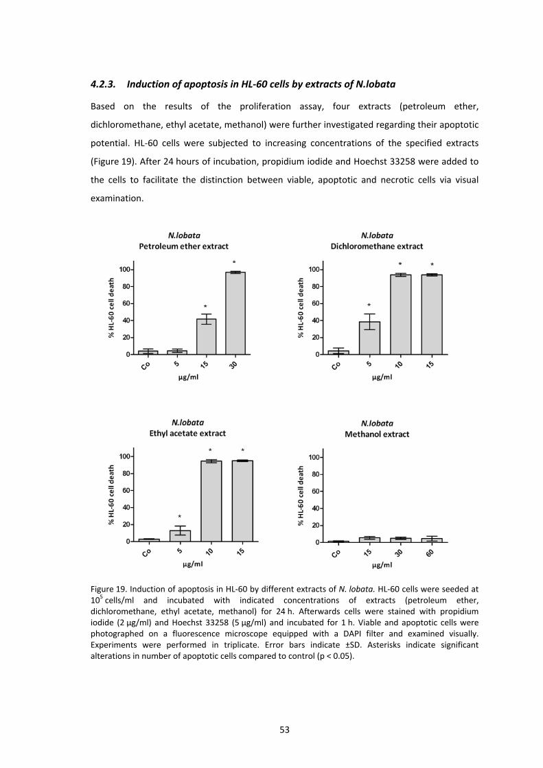
53
4.2.3. Induction of apoptosis in HL‐60 cells by extracts of N.lobata
Based on the results of the proliferation assay, four extracts (petroleum ether,
dichloromethane, ethyl acetate, methanol) were further investigated regarding their apoptotic
potential. HL‐60 cells were subjected to increasing concentrations of the specified extracts
(Figure 19). After 24 hours of incubation, propidium iodide and Hoechst 33258 were added to
the cells to facilitate the distinction between viable, apoptotic and necrotic cells via visual
examination.
Figure 19. Induction of apoptosis in HL‐60 by different extracts of N. lobata. HL‐60 cells were seeded at 105 cells/ml and incubated with indicated concentrations of extracts (petroleum ether, dichloromethane, ethyl acetate, methanol) for 24 h. Afterwards cells were stained with propidium iodide (2 µg/ml) and Hoechst 33258 (5 µg/ml) and incubated for 1 h. Viable and apoptotic cells were photographed on a fluorescence microscope equipped with a DAPI filter and examined visually. Experiments were performed in triplicate. Error bars indicate ±SD. Asterisks indicate significant alterations in number of apoptotic cells compared to control (p < 0.05).

54
Of the four extracts, the dichloromethane extract exhibited the highest induction of apoptosis
at the lowest concentration of 5 µg/ml after 24 hours of incubation. To achieve a similar
number of cell deaths, 15 µg/ml of the petroleum ether extract were necessary. The ethyl
acetate extracts induced apoptosis in almost 100 % of the cells at a concentration of 10 µg/ml,
which was similar to the dichloromethane extract. The difference in effectiveness between the
dichloromethane and the ethyl acetate extract at 5 µg/ml was more obvious when taking the
corresponding dried plant weight into account (168.94 µg/ml for dichloromethane and
341.40 µg/ml for ethyl acetate, respectively).
Therefore, the dichloromethane extract of N. lobata was identified as the most effective one
and was subjected to more detailed investigations. For all upcoming experiments a less
concentrated stock of 3.7 mg extract per ml DMSO was used.
Due to the promising results of the proliferation assay and cell death analysis in HL‐60 cells,
the dichloromethane extract of N. lobata was chosen for further investigations in human
(SR‐786) and murine (417) NPM‐ALK positive ALCL (anaplastic large cell lymphoma) cell lines as
well as human lung fibrobalsts (HLF).
4.2.4. Anti‐proliferative effects of N. lobata dichloromethane extract in human ALCL
SR‐786 cells with a NPM‐ALK translocation
SR‐786 cells were seeded at 2 x 10⁵ cells/ml and treated with increasing concentrations of
N. lobata dichloromethane extract. Due to growth characteristics of this cell line, assessment
of cell proliferation was limited to 48 hours instead of 72 hours. Therefore, proliferation was
considered as the difference in cell number between 24 hours and 48 hours of incubation.
Results, illustrated in Figure 20, indicated a similar activity of the extract in SR‐786 cells as in
HL‐60 cells (Figure 29).

55
Figure 20. Anti‐proliferative effects of the dichloromethane extract of N. lobata in SR‐786 cells. Cells were seeded at 2 x 10⁵ cells/ml and incubated with the dichloromethane extract of N. lobata at the indicated concentrations. Cell number was determined after 24 hours and 48 hours of treatment using a Casy cell counter (Inovartis). Proliferation of control samples was defined as 100 % and treated samples were set in relation. Error bars indicate ±SD. Asterisks indicate significant alterations in proliferation compared to control (p < 0.05). Experiments were performed in triplicate.
As the proliferation assay indicated “negative” proliferation in SR‐786 cells at higher
concentrations of the N. lobata dichloromethane extract, the mechanism of cell death was
investigated by western blot analysis.
4.2.5. Inhibition of NPM‐ALK, induction of apoptosis and caspase‐3 in SR‐786 cells
First, one of the most critical executioners of programmed cell death, caspase‐3, and its target
PARP were investigated for their cleavage. The exposition to 15 µg/ml dichloromethane
extract of N. lobata triggered apoptotic response in SR‐786 cells, which was confirmed by
cleavage of caspase‐3 as well as cleavage of PARP after 24 hors of incubation (Figure 21).
To examine the genotoxic potential of the extract, the levels of γ‐H2AX were analysed. Like
caspase‐3, H2AX was not phosphorylated at the serine 139 site until 24 hours of incubation
(Figure 21).
Therefore, the dichloromethane extract induced apoptosis mediated by caspase‐3 and is
supposed not to induce genotoxicity in SR‐786 cells.
N.lobataDichloromethane extract
Co 2.5 5 10 15 20-20
0
20
40
60
80
100
120
µg/ml
% SR‐786
proliferation
*
*
* * *

56
Figure 21. Induction of pro‐apoptotic key proteins in SR‐786 cells after incubation with the dichloromethane extract of N. lobata. SR‐786 cells (2.5 x 10⁵ cells/ml) were incubated with 15 µg/ml extract, harvested after 4, 8 and 24 h, lysed and isolated protein samples were subjected to western blot analysis using the indicated antibodies. Equal sample loading was confirmed by Ponceau S staining and β‐actin analysis.
In most ALK‐positive ALCL cells (like in SR‐786 cells), the kinase domain of ALK is fused to NPM
creating the constitutively expressed oncogenic fusion protein NPM‐ALK (Morris et al., 1994).
After 4 hours of drug treatment, the NPM‐ALK protein was substantially downregulated.
Furthermore, both PI3K and Akt are reported to be permanently activated by NPM‐ALK in
NPM‐ALK positive ALCL cells (Slupianek et al. 2001). Western blot analysis of untreated cells
showed a low constitutive p(Ser473)Akt level, which further decreased upon treatment with
the dichloromethane extract. The expression of Akt protein remained almost unchanged.

57
In the following investigations, the mechanism causing the reduction of the oncogenic
NPM‐ALK fusion protein by the dichloromethane extract was studied in more detail.
4.2.6. Reduction of NPM‐ALK levels is a decisive property of the dichloromethane
extract of N. lobata and is caused at transcriptional level
As illustrated in Figure 21, NPM‐ALK was rigorously down regulated within 24 hours of
treatment with the dichloromethane extract of N. lobata. To obtain further knowledge about
the specificity of NPM‐ALK degradation by the N. lobata extract, the most active extract of
Critonia morifolia was tested as well. According to cytotoxicity indicated in HL‐60 cells, SR‐786
cells were incubated with the petroleum ether extract of C. morifolia at a concentration of
20 µg/ml. Subjecting protein samples to western blot analysis revealed that NPM‐ALK levels
were not affected upon treatment by this plant extract (Figure 22). Therefore, the effect of the
N. lobata dichloromethane extract on NPM‐ALK repression was considered a specific property.
Figure 22. Effects of the petroleum ether extract of C. morifolia on NPM‐ALK levels in SR‐786 cells. SR‐786 cells were incubated with 20 µg/ml petroleum ether extract of C. morifolia and harvested after 4, 8 and 24 h. Cells were lysed and isolated protein samples were subjected to SDS‐PAGE followed by western blot analysis of NPM‐ALK and NPM. Equal sample loading was confirmed by Ponceau S staining and β‐actin analysis.
Experiments applying a proteasome inhibitor and a lysosome inhibitor were performed to
investigate whether the reduction in NPM‐ALK protein level was due to increased protein
degradation. For this reason, SR‐786 cells (2.5 x 10⁵ cells/ml) were incubated with 15 µg/ml of
the extract and either Proteasome Inhibitor IV was added to a final concentration of 50 µM or
ammonium chloride (NH4Cl) at a final concentration of 20 mM (Cockle and Dean, 1982). After
8 hours cells were harvested, lysed and isolated protein was subjected to SDS‐PAGE and
western blot analysis for NPM‐ALK. As illustrated in Figure 23, both inhibitors failed to prevent

58
the decrease in NPM‐ALK protein levels in extract treated cells. The control samples solely
exposed to either the proteasome inhibitor or lysosome inhibitor ensured that no toxicity was
induced by the inhibitors themselves.
Figure 23. Investigation of NPM‐ALK degradation mechanisms by the dichloromethane extract of N. lobata. SR‐786 cells (2.5 x 10⁵ cells/ml) were incubated with 15 µg/ml dichloromethane extract of N. lobata together with either (a) 50 µM proteasome inhibitor (Proteasome Inhibitor IV, Merck) or (b) 20 mM lysosome inhibitor (ammonium chloride NH4Cl). Potential toxicity of inhibitors was considered by treating control samples with the respective inhibitor only. After 8 h cells were harvested and lysed. Protein samples were subjected to SDS‐PAGE followed by western blot analysis. Equal sample loading was confirmed by Ponceau S staining and β‐actin analysis.
In the next step, NPM‐ALK expression was investigated at the transcriptional level. RNA
samples of SR‐786 cells were taken after 4 hours and 8 hours of incubation with 15 µg/ml
extract. Although the same cell number was taken, the total amount of RNA obtained
decreased from 545 ng/µl to 450 ng/µl after 4 hours and 420 ng/µl after 8 hours of treatment.
Equal amounts of RNA samples were transcribed into cDNA and subjected to real‐time PCR
applying specific primers for the NPM‐ALK transcript and GAPDH as internal control. The
NPM‐ALK transcriptional level was set into relation to the constitutively expressed GAPDH
using the comparative CT (ΔΔCT) method (Livak and Schmittgen 2001). NPM‐ALK transcription
was downregulated by 37 % after 4 hours and by 62 % after 8 hours of incubation. This result
confirms the substantial reduction in NPM‐ALK protein expression detected by western blot
analysis after 24 hours of extract treatment and explains the mechanism by which NPM‐ALK
becomes repressed (Figure 24).
(b)(a)
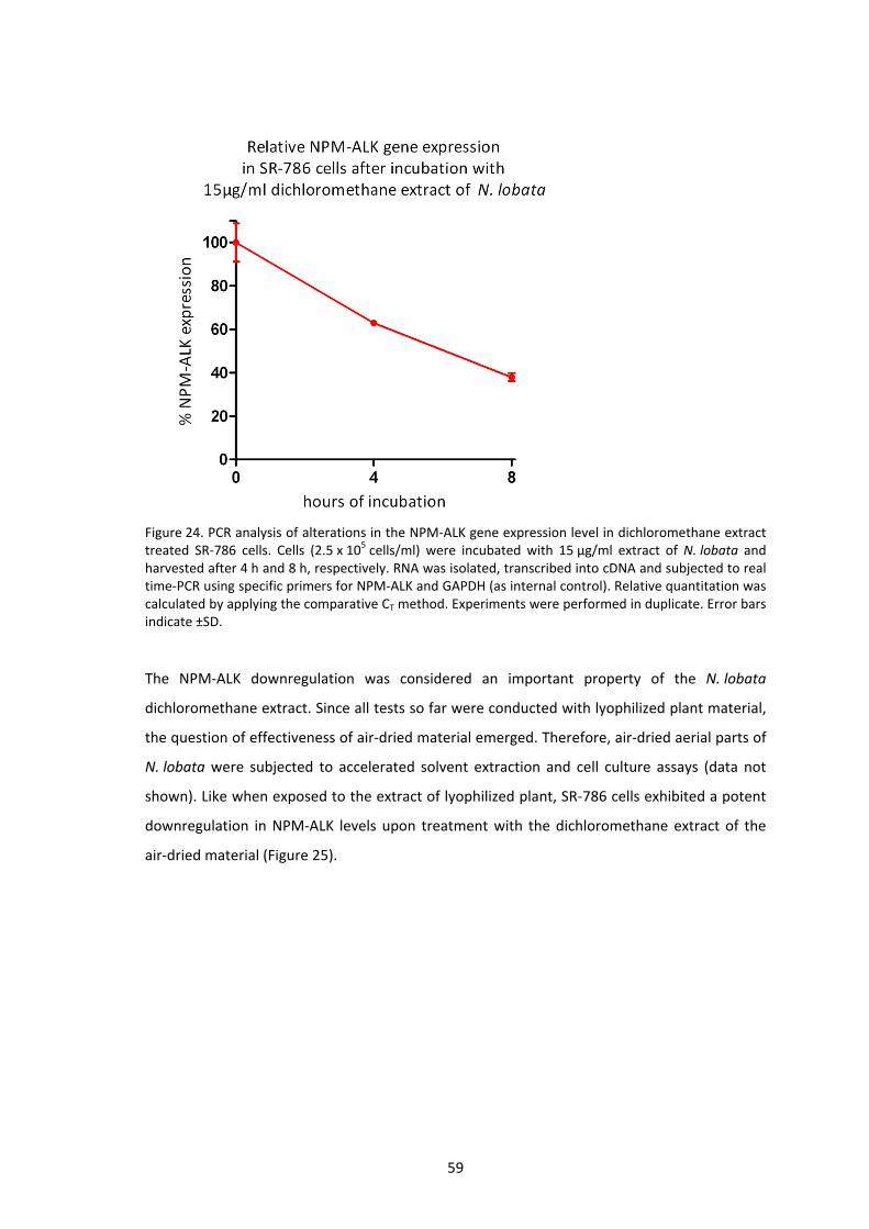
59
Figure 24. PCR analysis of alterations in the NPM‐ALK gene expression level in dichloromethane extract treated SR‐786 cells. Cells (2.5 x 10⁵ cells/ml) were incubated with 15 µg/ml extract of N. lobata and harvested after 4 h and 8 h, respectively. RNA was isolated, transcribed into cDNA and subjected to real time‐PCR using specific primers for NPM‐ALK and GAPDH (as internal control). Relative quantitation was calculated by applying the comparative CT method. Experiments were performed in duplicate. Error bars indicate ±SD.
The NPM‐ALK downregulation was considered an important property of the N. lobata
dichloromethane extract. Since all tests so far were conducted with lyophilized plant material,
the question of effectiveness of air‐dried material emerged. Therefore, air‐dried aerial parts of
N. lobata were subjected to accelerated solvent extraction and cell culture assays (data not
shown). Like when exposed to the extract of lyophilized plant, SR‐786 cells exhibited a potent
downregulation in NPM‐ALK levels upon treatment with the dichloromethane extract of the
air‐dried material (Figure 25).
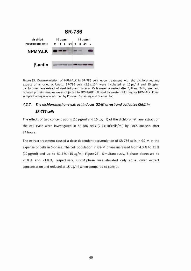
60
Figure 25. Downregulation of NPM‐ALK in SR‐786 cells upon treatment with the dichloromethane extract of air‐dried N. lobata. SR‐786 cells (2.5 x 10⁵) were incubated at 10 µg/ml and 15 µg/ml dichloromethane extract of air‐dried plant material. Cells were harvested after 4, 8 and 24 h, lysed and isolated protein samples were subjected to SDS‐PAGE followed by western blotting for NPM‐ALK. Equal sample loading was confirmed by Ponceau S staining and β‐actin blot.
4.2.7. The dichloromethane extract induces G2‐M arrest and activates Chk1 in
SR‐786 cells
The effects of two concentrations (10 µg/ml and 15 µg/ml) of the dichloromethane extract on
the cell cycle were investigated in SR‐786 cells (2.5 x 10⁵cells/ml) by FACS analysis after
24 hours.
The extract treatment caused a dose‐dependent accumulation of SR‐786 cells in G2‐M at the
expense of cells in S‐phase. The cell population in G2‐M phase increased from 4.3 % to 31 %
(10 µg/ml) and up to 51.5 % (15 µg/ml; Figure 26). Simultaneously, S‐phase decreased to
26.8 % and 21.8 %, respectively. G0‐G1 phase was elevated only at a lower extract
concentration and reduced at 15 µg/ml when compared to control.

61
Figure 26. Analysis of cell cycle distribution in SR‐786 cells after 24 h of incubation with the dichloromethane extract of N. lobata. SR‐786 cells (2.5 x 10⁵ cells/ml) were incubated with 10 µg/ml and 15 µg/ml of the dichloromethane extract of N. lobata. After 24 h cells were harvested and subjected to FACS analysis. Error bars indicate ±SD. Asterisks indicate significant alterations of cells in cell cycle phase compared to control (p < 0.05). Experiments were performed in triplicate.
To investigate which regulatory key proteins might have caused G2‐M arrest, SR‐786 cells were
treated with 15 µg dichloromethane extract per ml medium for 4, 8 and 24 hours and
subjected to western blot analysis.
Checkpoint kinase 1 (Chk1) is probably the most important regulatory cell cycle protein in
G2‐M phase arrest. In case of DNA damage, Chk1 is activated rapidly by the ATR pathway and
mediates cell cycle arrest in G2‐M phase to allow DNA repair prior to mitosis (Liu et al., 2000).
The activating phosphorylation of Chk1 kinase occurs at the serine 345 residue (Shibata et al.,
2010). Besides others, Chk1 has been identified to phosphorylate serine 75 of Cdc25A
targeting it for degradation (Kiyokawa and Ray, 2008; Mailand, et al. 2000). In accordance with
the G2‐M arrest identified by FACS analysis, Chk1 was activated by phosporylation at serine
345 residue, after 8 hours of incubation while total expression of Chk1 remained unchanged
(Figure 27). Additionally, the activation level of Chk2 was examined and only a weak induction
of p(Thr68)Chk2 was observed after 24 hours of extract treatment.

62
Confirming the failure of entry into mitosis by G2‐M arrest, Cdk1 (Cdc2) showed increased
phosphorylation at tyrosine 15 within 8 hours. However, substantial dephosphorylation was
found after 24 hours. In general, activation (i.e. dephosphorylation at Tyr15) of Cdk1 can be
attributed to the modulated expression of the Cdc25 phosphatases. As the level of Cdc25A was
not altered upon drug treatment, Cdc25B and Cdc25C were analyzed. Although acting through
distinct mechanisms, Cdc25B and Cdc25C primarily control entry into mitosis by targeting the
Cdk1/cyclinB complex (Karlsson‐Rosenthal and Millar, 2006). Western blot analysis of the
proteins suggested that the down regulation of Cdc25C after 4 hours and 8 hours was
compensated by increased levels of Cdc25B and vice versa after 24 hours of incubation
(Figure 27). Thus, Cdc25 phosphatases were not supposed to be responsible for activation of
Cdk1 after 24 hours, and a downregulation of the kinase Wee1 (not investigated), which is
targeting Cdk1, seems to be more likely.
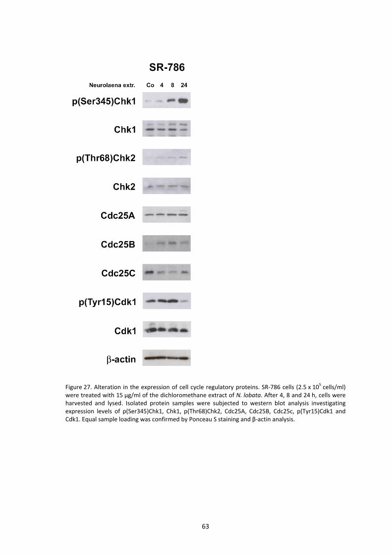
63
Figure 27. Alteration in the expression of cell cycle regulatory proteins. SR‐786 cells (2.5 x 10⁵ cells/ml) were treated with 15 µg/ml of the dichloromethane extract of N. lobata. After 4, 8 and 24 h, cells were harvested and lysed. Isolated protein samples were subjected to western blot analysis investigating expression levels of p(Ser345)Chk1, Chk1, p(Thr68)Chk2, Cdc25A, Cdc25B, Cdc25c, p(Tyr15)Cdk1 and Cdk1. Equal sample loading was confirmed by Ponceau S staining and β‐actin analysis.

64
4.2.8. N. lobata dichloromethane extract modulates oncogenes and tumor
suppressor protein expression
To obtain a better insight into the mechanism of growth arrest that was induced by the
dichloromethane extract of N. lobata, the expression of relevant oncogenes was studied.
Overexpression of certain cell growth and proliferation enhancing proteins are an important
property of most cancer cells. So called oncogenes may prevent abnormal cells from cell death
and promote uncontrolled growth when activated. On the other hand, proteins coded by
tumor suppressor genes are able to repress cell cycle and/or facilitate apoptosis in abnormal
cells. Both can be targets of anti‐cancer drugs, namely suppressing oncogenes and promoting
tumor suppressor genes.
One of the best investigated tumor suppressor genes is p53. Most cancer cells reveal a
deregulation of p53 expression. In response to DNA damage p53 becomes phosphorylated at
the serine 15 and serine 20 residues, typically phosphorylation at Ser20 is mediated by Chk1
and Chk2. This activation may lead to cell cycle arrest and DNA repair or apoptosis (Shieh et al.,
1999). Upon treatment with the dichloromethane extract, p(Ser20)p53 levels increased only
transiently within 4 hours whereas total p53 remained unchanged during the time course.
Since activation of Chk1 happened after 8 hours (Figure 27), the activation of p53 can not be
attributed to Chk1 kinase activity.
The transcription of Cdk‐inibitor p21 is known to be directly driven by p53. Despite, p21 can be
regulated by many p53‐independent pathways (Abukhdeir and Park 2008). At transcriptional
level, for example, it can be regulated by the oncogene c‐Myc (Coller et al. 2000). c‐Myc is
proven to have a profound impact on cell proliferation, differentiation and apoptosis.
Upregulation of c‐Myc expression is common in many tumor types. This contributes to an
abnormal proliferation rate. Among others, c‐Myc was discovered to downregulate p21 (Coller
et al. 2000), and conversely c‐Myc downregulation may result in p21 induction, as observed in
SR‐786 cells upon treatment with the N. lobata dichloromethane extract after 4 hours
(Figure 28).

65
Figure 28. Effects of the dichloromethane extract on oncogenes and tumor suppressor proteins in SR‐786 cells. SR‐786 cells (2.5 x 10⁵ cells/ml) were incubated with 15 µg/ml dichloromethane extract of N. lobata and harvested after 4, 8 and 24 h of treatment. Cells were lysed and obtained proteins samples subjected to SDS‐PAGE and western blot analysis with the indicated antibodies. Equal sample loading was confirmed by Ponceau S staining and β‐actin analysis.
The Jun family encompasses c‐Jun, JunB and JunD. They are components of the transcriptional
factor AP‐1. Studies in knock‐out mice have demonstrated roles for c‐Jun in various tissues and
developmental processes including T cell development (Riera‐Sans and Behrens 2007). JunB
and c‐Jun are reported to be over‐expresssed in anaplastic large cell lymphoma favoring cancer
progression (Mathas et al., 2002). Nevertheless, JunB and c‐Jun are controversially discussed.
Inconsistent reports in recent literature support the idea that both proteins may act as either
an oncogene or a tumor suppressor in a cell‐context‐dependent manner (Shaulian 2010). The
incubation of SR‐786 cells with the extract transiently increased c‐Jun expression whereas JunB
was continuously downregulated (Figure 28).

66
Taking together, the activation of p53, induction of p21, and inhibition of c‐Myc and JunB
explain the growth arrest in SR‐786 cells observed after incubation with the N. lobata
dichloromethane extract.
4.2.9. Proliferation inhibition in murine ALCL cells with a NPM‐ALK translocation
In preliminary studies, it was found that human ALCL cell lines SR‐786 and Karpas 299 are
deficient in membrane protein platelet‐derived growth factor receptor β (PDGFR‐β), although
PDGFR‐β is found in primary human ALCL specimens proven by immunohistochemical analysis
(Laimer et al. 2010, in preparation). PDGF, and therefore also PDGFR‐β, are recognized as
important factors in cell proliferation, differentiation and growth (Gotzmann et al. 2006). This
makes PDGF receptor an attractive target of anti‐cancer drugs. To analyze potential effects of
the extract on PDGFR‐β, it was also tested in the murine ALCL cell line 417 which expresses
high levels of PDGFR‐β (Laimer et al. 2010, in preparation).
Two tests, on proliferation and cytotoxicity (see 4.2.11), were performed to investigate the
effects of N. lobata dichloromethane extract in the NPM‐ALK positive murine ALCL cell line
417. To measure proliferation, 106 cells/ml were treated with increasing amounts of extract
and incubated for 72 hours. 417 cells showed “negative” proliferation, i.e. the induction of cell
death, at 10 µg/ml (Figure 29a) indicating a similar sensitivity to the drug in 417 compared to
human HL‐60 cells and SR‐786 ALCL cells.

67
Figure 29. Anti‐proliferative/apoptotic effects of dichloromethane extract of N. lobata in 417 cells. Cells (106 cells/ml) were incubated at indicated concentrations of the dichloromethane extract for 72 h. Cell number was determined after 24 and 72 h using a Casy® cell counter. Proliferation of control samples was defined as 100 % and all other conditions were set in relation. Experiments were performed in triplicate. Error bars indicate ±SD. Asterisks indicate significant alterations in proliferation compared to control (p < 0.05).
Analysis of cell cycle distribution and protein expression patterns were conducted to
determine at which stage and by which mechanisms cell proliferation was inhibited.
4.2.10. Cell cycle inhibitory effects in G2‐M of the dichloromethane extract in 417
cells
Human NPM‐ALK positive ALCL cell line SR‐786 was proven to be arrested in G2‐M phase upon
treatment with the dichloromethane extract (Figure 26). Hence, murine 417 cells (106 cells/ml)
were incubated with 5 µg/ml, 10 µg/ml and 15 µg/ml of the dichloromethane extract for
24 hours and afterwards subjected to fluorescence activated cell sorting (FACS). Treatment of
417 cells with 5 µg/ml did not alter cell cycle distribution significantly. By contrast, application
of 10 µg/ml resulted in an increase of 10 % in G0‐G1 (from 46.1 % to 56.9 %) and from 7.7 % to
23 % in G2‐M. Increases of cell population in G0‐G1 and G2‐M were at the expense of cells in
S‐phase which were reduced from 47.2 % to 20.1% (Figure 30). This finding was consistent with
the data obtained with SR‐786 cells. At an extract concentration of 15 µg/ml, toxicity led to
exorbitant cell death which did not allow FACS analysis for all three samples. Thus, this data
was only assessed in duplicate not enabling for calculation of significance. Nevertheless, cells
treated with 15 µg/ml extract followed the trend explored in cells at 10 µg/ml, namely an
increase in cell population in G2‐M phase at the expense of S‐phase (Figure 30).
N.lobataDichloromethane extract
Co 1 5 10 20-40
-20
0
20
40
60
80
100
µg/ml
% 417
proliferation
** *

68
Figure 30. Cell cycle distribution of 417 cells upon treatment with the N. lobata dichloromethane extract. 417 (106 cells/ml) were incubated at indicated concentrations of the dichloromethane extract of N. lobata for 24 h and then subjected to FACS analysis. Error bars indicate ±SD. Experiments were performed in triplicate, except for the concentration of 15 µg/ml, which was assessed in duplicate.
To investigate whether the same regulatory key proteins caused G2‐M arrest in 417 cells like in
SR‐786 cells, 417 cells were incubated with 10 µg/ml of the dichloromethane extract and
harvested after 0.5, 2, 4, 8 and 24 hours. Protein was isolated and subjected to western blot
analysis (Figure 31).

69
Figure 31. Analysis of activation and deactivation of cell cycle regulatory proteins in 417 cells induced by the N. lobata dichloromethane extract. 417 cells (106 cells/ml) were incubated at a concentration of 10 µg/ml dichloromethane extract and harvested at times indicated. Cells were lysed in a buffer containing 150 mM NaCl, 50 mM Tris‐buffered saline (Tris pH 8), 1 % Triton X, 1 mM PMSF and 1 % PIC. The isolated protein was centrifuged and the supernatant was subjected to western blot analysis using the indicated antibodies. Equal sample loading was confirmed by Ponceau S staining and β‐actin analysis.
Like in SR‐786 cells, the dichloromethane extract of N. lobata induced phosphorylation at
Ser345 of Chk1 after 24 hours which confirms the G2‐M arrest revealed by FACS analysis. In
accordance, Cdc25A was degraded after 24 hours.
As shown in Figure 31, phosphorylation of Cdk1 was upregulated from 0.5 to 8 hours, thus
blocking transition into mitosis. After 24 hours p(Tyr15)Cdk1 expression decreased, while total
Cdk1 remained unaffected during 24 hours of treatment. In this case, the drug treatment
decreased p(Tyr15)Cdk1 as well as Cdc25A in 417 cells after 24 hours. Thus, dephosphorylation
was not attributable to increased phosphatase activity of Cdc25A but might was a result of
decreased kinase activity of Wee1 (McGowan and Russel, 1993). Therefore, the effects
observed in Cdk1 did not correlate with the growth phenotype.

70
In the next step, cytotoxic and apoptotic potential of the extract was investigated. Human lung
fibroblasts were treated simultaneously to take possible cytotoxic effects in normal cells into
account.
4.2.11. Induction of apoptosis is specific in 417 cells and mediated by caspase‐3
Many anti‐cancer drugs act by killing cells that divide rapidly, which is an important property of
most cancer cells. However, those drugs are also affecting cells that proliferative rapidly under
normal circumstances, i.e. cells in the digestive tract, bone marrow and hair follicle. Other cells
in mammals replicate at a considerably lower rate which makes them less vulnerable to
chemotherapy. Since the dichloromethane extract exhibited strong cytotoxic effects in HL‐60,
SR‐786 and 417 cancer cells lines, the question of general toxicity in cells of mammalian origin
emerged. To gain further insight into the specificity of cytotoxicity of the extract, normal
human lung fibroblast (HLF) were subjected to extract treatment and analysis by alamarBlue®
assay (Figure 32b). As 1 µg/ml extract did not exhibit a significant alteration in the proliferation
of 417 cells, 2.5 µg/ml were used instead (Figure 32a). HLFs were seeded and allowed to attach
for 24 hours prior treatment. At confluence, HLFs decelerate proliferation substantially while
cells are still metabolically active. Thus, this system was used to simulate effects in normal cells
(Li W et al. 2006). Additionally, apoptotic key proteins were investigated by western blot
analysis in both 417 and HLFs.

71
Figure 32. AlamarBlue assay (a) in cell line 417 and (b) primary human lung fibroblasts after 48 h of incubation with the dichloromethane extract of N. lobata at indicated concentrations. 417 cells were seeded at 106 cells/ml. HLFs were seeded at confluence and allowed to attach for 24 h prior treatment. After 48 h of incubation with the dichloromethane extract, 50 µl alamarBlue reagent (containing resazurin) was added to each well and cells were incubated for another 90 min. Metabolic active cells converted blue resazurin to red‐fluorescent resofurin. Absorption was measured by a multi‐detection reader at 570 nm. Calculated data based on the measurement (c) for 417 cells and (d) HLFs. Experiments were performed in triplicate. Error bars indicate ±SD. Asterisks indicate significant alterations in proliferation compared to control (p < 0.5).
By visual evaluation, HLFs seemed to be unaffected by the extract after 48 hours of incubation
whereas 417 cells exhibited a substantial dose dependent response in metabolic activity
indicated by the conversion of blue resazurin into red‐fluorescent resofurin (Figure 32). This
impression was validated by calculating differences in fluorescence based on the exact
(c) (d)
72
measurement data. Thus, fluorescence produced by 417 cells reduced absorption by 10.3 %
(not significant, p < 0.5), 26.7 % and 81.4 % with increasing concentrations of the
dichloromethane extract (corresponding to 2.5 µg/ml, 5 µg/ml and 10 µg/ml, respectively;
Figure 32c). In HLFs, the extract slightly attenuated the metabolism at 10 µg/ml and to 85.6 %
at 15 µg/ml (both not significant, p < 0.05; Figure 32d). Western blot analysis of pro‐apoptotic
proteins was applied to confirm the findings stated above.
The dichloromethane extract of N. lobata exhibited strong apoptotic potential mediated by
caspase‐3 in SR‐786 cells. The antibody used for detection of cleavage in 417 cells detects only
activated large fragments (17/19 kD) but not full length caspase‐3. Caspase‐3 antibody
detecting endogenous levels of full length caspase‐3 (35 kD) as well as large fragments was
applied for detection in HLF cells. The treatment of 417 cells with 10 µg/ml dichloromethane
extract resulted in an activation of caspase‐3 after 8 hours (Figure 33a) whereas caspase‐3
remained full length (35 kD) in HLF cells exposed to 10 µg/ml and even 15 µg/ml extract after
24 hours (Figure 33b).
As target of activated caspase‐3, cleaved PARP antibody, detecting the large fragment but not
full length PARP, was used to indicate DNA damage in 417 cells. Cleavage of PARP already
started after 4 hours of incubation with the extract and increased substantially within the
following 20 hours (Figure 33a).
The dichloromethane extract of N. lobata induced phosphorylation of H2AX at Ser139 in 417
cells after 4 hours of incubation which intensified continuously until 24 hours. Like in human
SR‐786 cells, this correlated with caspase‐3 activity and therefore, did not indicate
genotoxicity.
In addition to the common pro‐apoptotic proteins, two receptor tyrosine kinases (RTK) were
investigated, the oncogenic NPM‐ALK fusion protein and the cell growth stimulating protein
PDGFR‐β. The expression of both RTKs was considerably downregulated in 417 cells upon drug
treatment, showing an earlier onset in NPM‐ALK degradation (Figure 33a). In HLFs, the effect
on PDGFR‐β was less incisive and noticeable only after 24 hours of incubation. As proven by
western blot analysis, HLFs are deficient in ALK expression. NPM expression was not affected
by the extract in HLFs (Figure 33b).

73
Figure 33. Effects of the dichloromethane extract on pro‐apoptotic proteins in cell line 417 and primary HLFs. 417 cells were seeded at 106 cells/ml and incubated with 10 µg/ml of the dichloromethane extract of N. lobata. HLFs were seeded confluent and allowed to attach for 24 h prior incubation with 10 µg/ml and 15 µg/ml of the extract. At each time point indicated, cells were harvested and received protein samples (lysates) were subjected to western blot analysis to investigat cleavage of caspase‐3 and PARP. Equal sample loading was confirmed by Ponceau S staining and β‐actin analysis.
So far, there exists no report about an anti‐cancer drug that efficiently targets NPM‐ALK. In the
next step, the effect of the dichloromethane extract on NPM‐ALK was compared to those of
vincristine, a plant derived drug that is commonly used as part of the chemotherapy regimen
in non‐Hodgkin’s lyhmphoma such as ALCL.
4.2.12. N. lobata dichloromethane extract, but not vincristine, substantially
decreases levels of NPM‐ALK in 417 cells
As previously shown in this work, the NPM‐ALK fusion protein was rigorously downregulated
upon treatment with the dichloromethane of N. lobata but not the petroleum ether extract of
C. morifolia. Therefore, this property was supposed to be characteristic for the N. lobata
dichloromethane extract. To substantiate this hypothesis, its effects in 417 cells were
compared to those of the chemotherapeutic agent vincristine.
(a) (b)

74
During the dichloromethane extract treatment (10 µg/ml) decreased the expression of
NPM‐ALK in 417 cells within 4 hours and was continuously downregulated throughout the rest
of the time course (Figure 33a). Vincristine, a plant derived alkaloid used in chemotherapy that
acts as a mitotic inhibitor and leads to cell death, was used to treat 417 cells (106 cells/ml) at
increasing concentrations (1 µM, 2.5 µM and 5 µM). Protein samples were taken after 8 hours
and 24 hours of incubation and subjected to western blot analysis. Compared to the
dichloromethane extract of N. lobata, vincristine exhibited minimal cleavage of caspase‐3 and
no decrease in NPM‐ALK expression after 8 hours. Prolonged exposure time to vincristine led
to substantial activation of caspase‐3 at all concentrations while protein levels of NPM‐ALK
were dose‐independently reduced (Figure 34).
Furthermore, changes in the expression levels of PDGFR‐β were assessed for both agents. The
dichloromethane extract exhibited downregulating properties in PDGFR‐β protein levels within
8 hours of incubation (Figure 33a). Vincristine treatment decreased PDGFR‐β expression
equally effective after 24 hours (Figure 34).
Figure 34. Western blot analysis of effects of vincristine on NPM‐ALK, PDGFR‐β and caspase‐3 levels in 417 cells. Cells (106 cells/ml) were incubated with vincristine at concentrations of 1 µM, 2.5µM and 5 µM. Protein samples were taken after 8 h and 24 h. Isolated protein was subjected to western blot analysis applying antibodies against ALK (also detecting NPM‐ALK fusion protein), PDGFR‐β and caspase‐3. Equal sample loading was confirmed by Ponceau S staining and β‐actin analysis.

75
Direct comparison to vincristine leads to the assumption that the downregulation of NPM‐ALK
might be a decisive property of the N. lobata dichloromethane extract.

76
5 DISCUSSION
Based on the traditional uses in Mayan medicine for over thousand years, two ethnomedical
plants from Central America, C. morifolia and N. lobata, were investigated for their potential
anti‐carcinogenic activity. Conditions treated by distinct preparations of aerial parts of these
plants range from parasitic ailments to various states of inflammation and neoplasms (Arvigo
and Balick 1998). Additionally, promising phytochemical and pharmacological research results
have been published, particularly on N. lobata.
For investigational reasons, the collected fresh plant material from Guatemala was lyophilized
to preserve also volatile compounds, and subjected to sequential extraction using five solvents
of increasing polarity. This method facilitated an initial separation of plant constituents
according to their polarity, and the obtained extracts were tested in vitro for their
anti‐neoplastic potential.
5.1. C. morifolia
Apart from inflammatory conditions, C. morifolia leaves are utilized as home remedy to cure
boils, swellings and even “cancer” (severe skin damages with uncontrolled wound tissue
growth: Arvigo and Balick 1998). Investigations in human promyelocytic leukemia HL‐60 cells
revealed the petroleum ether extract as the extract possessing the strongest anti‐carcinogenic
potential. Thus, the anti‐proliferative and pro‐apoptotic effects were due to the activity of
apolar plant constituents. Besides, this finding is consistent with the traditional uses as the
leaves are preheated in oil prior to application directly on the neoplastic tissue (Arvigo and
Balick 1998). As it turned out during investigations, the activity of C. morifolia petroleum ether
extract deteriorated when dissolved in DMSO and stored at ‐20°C within two weeks.
Accordingly, fresh stock solutions were prepared regularly. Hence, active compounds
contained in the extract are instable.
Like many human cancers, HL‐60 show increased expression levels of the proto‐oncogenes
cyclin D1 and c‐Myc, and lack of the tumor suppressor protein p53. These proteins are known
to have impact on cell proliferation, differentiation and apoptosis, and thereby play an
important role in the development of cancer (Fu et al. 2004; Dominguez‐Sola et al. 2007). The
C. morifolia petroleum ether extract simultaneously suppressed cyclin D1 and c‐Myc within
30 minutes which intensified during the time course. In early G1 phase, D‐cyclins bind to Cdk4
77
and Cdk6, thereby promoting the transition into S‐phase (Pines 1991; Lingfei et al. 1998).
Hence, the reduction in cyclin D1 would suggest a cell cycle inhibition in G1 phase, which
contrasts both the G2‐M and the S‐phase arrest observed upon extract treatment after 8 hours
and 24 hours, respectively. Therefore, the extract blocked also another cell cycle regulatory
mechanism even before cyclin D1 suppression became effective. But as cyclin D1 also
regulates and associates with transcription factors in a Cdk‐independent manner (Fu et al.
2004), the influence of decreased cyclin D1 levels must have additively contributed to growth
inhibition. HL‐60 cells are p53 deficient (Wolf and Rotter 1985), nevertheless the expression of
Cdk‐inhibitor protein p21, which acts tumor suppressive and is (among others) induced by p53
and also Erk ½, or repressed at transcriptional level e.g. by c‐Myc (Abukhdeir and Park 2008;
Coller et al. 2000), was not affected by the extract.
The inhibition of uncontrolled cell proliferation is an essential property of anti‐cancer drugs.
Transiently, C. morifolia petroleum ether extract dose‐dependently arrested the cell cycle in
G2‐M after 8 hours. This effect correlated with the continuous downregulation of phosphatase
Cdc25C, which plays a major role in G2‐M transition via activation of the Cdk1/cyclin B
complex (Hunter 1995). The activation of Cdk1/cyclin B by dephosphorylation at Tyr15 is a
crucial event to enable a cell to enter mitosis (Norbury et al. 1991). Notably, the initial G2‐M
arrest after 8 hours was shifted to inhibition of cell cycle progression in S‐phase after 24 hours,
which was confirmed by substantial dephosphorylation of Cdk1 from 8 to 24 hours, thus
enabling cells to overcome the initial G2‐M arrest. As Cdc25A, Cdc25B and Cdc25C levels were
all downregulated at this time point, the activated state of Cdk1 was supposed to be
attributable to the decrease in Wee1 levels as kinase Wee1 antagonizes the phosphatase
activity of the Cdc25 family by phosphorylating Cdk1 at Tyr15 (Katayama et al. 2005). The
S‐phase arrest after 24 hours might have been due to the activation of Chk2, a downstream
effector of ATM kinase in case of DNA damage (Ahn et al. 2000), which was strongest at that
timepoint. This observation was paralleled by the phosphorylation of its substrate Cdc25A at
the serine 177 residue. Phosphorylation of Cdc25A by a checkpoint kinase (Chk1, Chk2) targets
it for rapid degradation (Mailand et al. 2000; Madlener et al. 2009). By dephosphorylating
Cdk2, Cdc25A enables cells to transit from G1 to S‐phase (Hoffmann et al. 2004) and therefore,
the highest levels of Cdc25A and activated Cdk2 were reported at late S‐phase (Molinari et al.
2000) indicating its need for proper DNA synthesis. Consequently, the degradation of Cdc25A
might explain the arrest of cell population in S‐phase as observed upon extract treatment after
24 hours. It remains to be established however, why cells did not arrest already in G1, since

78
cyclin D1 was completely downregulated at this time point ,and it is likely that another cyclin
may have substituted for cyclin D1 (i.e. cyclin D2 or D3).
The temporally separated cell cycle phase‐specific inhibitory properties of the extract were
probably due to distinct anti‐neoplastic compounds that are contained therein.
Besides the inhibition of growth, another common feature of anti‐cancer agents is their
pro‐apoptotic property like it was observed by the petroleum ether extract of C. morifolia and
which was verified by the preceding activation of caspase‐3 starting within 2 hours. Caspase‐3
acts as critical executioner of apoptosis (Fernandes‐Alnemri et al. 1994) and its main target
PARP was signature‐type cleaved after 4 hours, facilitating cellular degradation (Beneke et al.
2000). DNA double strand breaks were indicated by γ‐H2AX after 4 hours. In general,
phosphorylation of H2AX at Ser139 residue is considered a marker of genotoxicity (Rogakou et
al. 1998). However, in this case, the later onset of H2AX phosphorylation compared to
activation of caspase‐3 suggested that DNA double strand breaks were not attributable to a
genotoxic property of the extract but a result of the ongoing apoptotic degradation of the
chromatin. Consistently, the activation of Chk2 occurred after the initiation of apoptosis as
indicated by caspase‐3 cleavage. This theory suggests an activation of Chk2 by ATM upon DNA
cleavage initiated by the foregoing onset of apoptosis.
Thus, according to the early onset of apoptosis, the above discussed anti‐proliferative effects
of the C. morifolia petroleum ether extract found in HL‐60 cells are probably attributable to
the induction of cell death rather than growth inhibition, particularly for the 24 hours time
point.
As a concluding remark, the C. morifolia petroleum ether extract has been demonstrated to
exert strong anti‐carcinogenic activity, especially in terms of pro‐apoptotic effects.
Nevertheless, the instability of active constituents and the difficult access to the plant may
limit further investigations and applicability.
79
5.2. N. lobata
Aqueous preparations of N. lobata are widely used in folk medicine, especially to treat and
prevent a variety of parasitic ailments such as malaria and amoebiasis (Arvigo and Balick 1998).
In phytochemical research a variety of characteristic sesquiterpene lactones were isolated of
N. lobata including neurolenin A, B, C, D, E and F, and lobatin A, B, and C (Passreiter et al.
1995) as well as pyrrolizidine alkaloids (Passreiter 1998). Pharmacological studies approved
anti‐parasitic properties of distinct N. lobata extracts and plant constituents, i.e.: the
dichloromethane extract and sesquiterpene lactones were reported to be active against the
clinical most important malaria pathogen Plasmodium falciparum and Plasmodium berghei
(François et al. 1996); and the ethanol extract was found to inhibit growth of Leishmania
mexicana, Trypanosoma cruzi and Trichomonas vaginalis (Berger et al. 2001).
In addition, François et al. (1996) compared anti‐plasmodial activities with cytotoxicity of
sesquiterpene lactones isolated from the dichloromethane extract. Whereas neurolenin B
exhibited the highest activity in P. falciparum, lobatin B was the compound with the strongest
cytotoxic activity in GLC4 and COLO 320 tumor cell lines. The IC50 values against both tumor
cell lines were much higher than those in the anti‐plasmodial assay.
Consistent with previous investigations, the present study identified the dichloromethane
extract of N. lobata to possess the highest anti‐proliferative and pro‐apoptotic potential in
HL‐60 cells. Therefore, this extract was chosen for further investigations in NPM‐ALK positive
anaplastic large cell lymphoma (ALCL) human cell line SR‐786 and murine cell line 417. ALCL is
a subtype of non‐Hodgkin lymphoma classified among T/null cell lymphomas with aberrant cell
surface expression of CD30. It is named after the typical morphology of the cells (Ambrogio et
al. 2008). The majority of them carry chromosomal aberrations involving the anaplastic
lymphoma kinase (ALK) of which the t(2;5)(p23;q35) translocation, which fuses the ALK gene to
the nucleophosmin (NPM) gene, represents the most frequent one (Stein et al. 1985). The
resulting NPM‐ALK fusion protein with constitutive tyrosine kinase activity is considered to
play an essential role in the pathogenesis of ALCL through its impact on proliferation,
differentiation and apoptosis (Piva et al. 2006). Therefore, ALK is suggested as viable target for
therapeutic intervention.

80
N. lobata dose‐dependently arrested SR‐786 cells in G2‐M phase of the cell cycle,
predominantly at the expense of S‐phase cells. This effect was observed in both human SR‐786
and murine 417 cells. Apart from the role of NPM‐ALK, at least three independent mechanisms
may have contributed to the accumulation of cells in this phase as revealed by western blot
analyses:
(i) N. lobata extract induced checkpoint kinase 1 (Chk1), which is activated in response to
DNA damage by ATR kinase (Liu et al. 2000) halting the cell cycle in G2‐M phase to
allow DNA repair prior to mitosis. A downstream target of activated Chk1 (and Chk2) is
the inhibition of the Cdc25 phosphatase family. These proteins are required for cell
cycle transit by activating cyclin‐dependent kinases (Cdks) through dephosphorylation.
Cdc25B and Cdc25C primarily control the entry into mitosis by activating the
Cdk1/cyclin B complex (Karlsson and Millar 2006). The proteins seemed to compensate
for each other as both proteins were inversely up‐ and downregulated during the time
course of extract treatment. Thus, the alleged activation of Cdk1 after 24 hours of
treatment was supposed to be the result of reduced kinase Wee1 activity targeting
Cdk1 rather than increased Cdc25 phosphatase activity.
(ii) SR‐786 cells exhibited dramatic alterations in their expression of oncogenic and tumor
suppressor proteins upon incubation with the dichloromethane extract. The
proto‐oncogenic protein c‐Myc, a main driver of unscheduled proliferation and
overexpressed in many tumors, was completely downregulated within 4 hours.
Moreover, the Cdk‐inhibitor and tumor suppressor protein p21 was upregulated
simultaneously. The main transcriptional regulator of p21 is p53 and in fact, p53
became activated within 4 hours of extract treatment. Whether this was the
p21‐inducing event remains obscure, since more than 50 % of all cancers harbor a
mutated and largely inactive p53 gene. As p21 was upregulted and temporally inverse
to c‐Myc suppression, this may have been the causal p21‐regulatory mechanism
(Coller et al. 2009).
(iii) Two major constituents of the transcription factor AP‐1 (activating protein 1), JunB and
c‐Jun, were affected by the N. lobata extract. AP‐1 is reported to play an essential role
in certain cancers. In case of anaplastic large cell lymphoma, AP‐1 is suggested to
prevent cells from undergoing apoptosis, therefore promoting uncontrolled growth
(Mathas et al. 2002). The dichloromethane extract induced a transient increase in c‐jun
expression after 4 and 8 hours whereas JunB was continuously downregulated in

81
SR‐786 cells. In NPM‐ALK positive ALCL, especially the JunB level has been reported to
be associated with carcinogenic transformation and may be regulated by NPM‐ALK
(Hsu et al. 2006), which is in line with the present findings as the decrease in NPM‐ALK
was paralleled by downregulation of JunB.
The major pathways engaged by NPM‐ALK are the Jak/Stat, Ras/Mapk and PI3K pathways
(Simonitsch et al. 2001; Polgar et al. 2005; Palmer et al. 2009). As proved by western blot
analysis, treatment with the dichloromethane extract of N. lobata led to a substantial decrease
in NPM‐ALK protein expression that was caused at transcriptional level rather than protein
degradation. The decrease in NPM‐ALK protein was observed in both human SR‐786 and
murine 417 ALCL cell line. In contrast, the C. morifolia petroleum ether extract, which was
tested in SR‐786 cells, as well as the common plant derived chemotherapeutic agent
vincristine used in ALCL therapy and tested in 417 cells, did not exhibit a downregulation of
NPM‐ALK levels upon treatment. Thus, the effect of N. lobata dichloromethane extract on
NPM‐ALK protein expression is proposed to be a specific property. However, because
NPM‐ALK plays a role in proliferation and apoptosis via several pathways its exact influence on
proliferation and apoptosis is hard to determine.
PDGFR‐β, like NPM‐ALK, is a receptor tyrosine kinase playing an important role in proliferation,
differentiation and growth (Gotzmann et al. 2006). Although primary human ALCL specimens
express PDGFR‐β as proven by immunohistochemical analysis, SR‐786 and other human
NPM‐ALK positive ALCL cell lines are PDGFR‐β deficient (Laimer et al. 2010, in preparation).
Hence, murine cell line 417 was selected to examine the effects of N. lobata on this receptor
tyrosine kinase. As for NPM‐ALK, a substantial decrease in PDGFR‐β protein expression was
observed during the time course, leading to the assumption that N. lobata might contain an
active principle which functions as a receptor tyrosine kinase inhibitor.
Apart from the downregulation of NPM‐ALK, N. lobata dichloromethane extract triggered
apoptosis in both SR‐786 and 417 cell lines, which was associated with the cleavage of
caspase‐3, followed by phosphorylation of H2AX at Ser139 (γH2AX), as marker of double
strand breaks caused by apoptosis. Cell death was induced at considerably low concentrations
of 10 µg/ml extract (corresponding to 0.34 mg/ml dried plant weight) in murine and 15 µg/ml
extract (corresponding to 0.5 mg/ml dried plant material) human ALCL cells, respectively.
82
To gain further insight into the specificity of cytotoxicity and rule out overall toxicity, the
extract was also tested in human lung fibroblasts (HLF). Prior treatment, HLFs were grown to
confluence where they substantially decrease their proliferative activity while still being
metabolically active simulating the homeostatic condition of normal cells, which generally
proliferate at considerably lower rates than tumor cells. Indeed, the metabolism of HLFs was
only insignificantly attenuated even at an extract concentration of 20 µg/ml. Furthermore,
15 µg/ml extract did not induce activation of caspase‐3 in HLFs. According to these results, the
N. lobata dichloromethane extract exhibited specificity towards cancer cells, which is
confirmed by reports that only weak toxicity is observed in brine shrimp larvae Artemis salina
and mice treated with a dichloromethane extract of N. lobata (Berger et al. 1998; Cáceres et
al. 1998).
Whereas the C. morifolia extract is rather labile, the anti‐neoplastic properties of the N. lobata
are remarkably stable, which will facilitate further studies. Especially the contained
sesquiterpene lactones could be of major interest since drugs derived from this substance class
have already reached cancer clinical trials, e.g. artemisinin, thapsigargin, and parthenolide. The
most promising feature of these drug candidates is their preferential selectivity toward tumor
and cancer stem cells while sparing normal counterparts, which makes them superior to many
conventional agents commonly used in chemotherapy (Ghantous et al. 2010; Zhang et al.
2005).
Concluding, there is sufficient evidence that N. lobata extract possesses specific anti‐neoplastic
properties downregulating oncogenes and oncogenic receptor‐type tyrosine kinases with
limited side toxicity and should therefore undergo testing in vivo.

83
SUMMARY
Although many cancers can be treated efficiently in first line chemotherapy, relapse and
refractory cancers require the discovery and development of new anti‐cancer agents. The
present work analyzed the potential anti‐neoplastic activity of two rainforest species of the
Asteraceae family, Critonia morifolia and Neurolaena lobata, that successfully passed
hundreds of years of usage as home remedies in the long lasting medical tradition of the
Central American Mayas. The plant material, collected in Guatemala, was lyophilized and
sequentially extracted with five solvents of increasing polarity to provide an initial separation
of plant constituents. For each plant, the extract exhibiting the most potent anti‐proliferative
and pro‐apoptotic effects in HL‐60 cells was subjected to more detailed investigations
including western blot and FACS analyses.
The anti‐proliferative properties of the C. morifolia petroleum ether extract turned out to be
attributable to the induction of cell death as the apoptotic executioner protein caspase‐3 was
already activated within 2 hours of incubation. Changes in the levels of cell regulatory proteins
were observed thereafter, in particular, Chk2 was activated upon DNA cleavage initiated by
the foregoing onset of apoptosis,and this correlated with the S‐phase cell arrest after 24 hours.
The loss in bio‐activity of this extract indicated that considerably unstable compounds
triggered the anti‐neoplastic effects, which may limit its potential use as anti‐cancer remedy.
The other plant, N. lobata, tested in human SR‐786 and murine 417 NPM‐ALK positive ALCL cell
lines, was more promising. In both cell lines, 10‐15 µg/ml of the dichloromethane extract
inhibited the cell cycle in G2‐M phase, which correlated with the activation of Chk1 and the
depletion of the proto‐oncogene c‐Myc paralleled by the upregulation of the p21 tumor
suppressor protein. The extract strongly triggered apoptosis, inducing caspase‐3 followed by
phosphorylation of H2AX. Overall toxicity was low since normal human lung fibroblasts
remained viable even upon exposure to extract concentrations beyond 15 µg/ml. The extract
led to a substantial decrease in oncogenic NPM‐ALK transcript and protein expression, but not
in that of nucleophosmin. This suggested that the extract affected a post‐transcriptional
mechanism, i.e. mRNA stability rather that mRNA transcription. The chemotherapeutic agent
vincristine and the C. morifolia extract did not affect NPM‐ALK expression. Apart from
NPM‐ALK, a second tyrosine kinase, namely PDGFR‐β, was also downregulated by the N. lobata
extract. Taken together, the N. lobata extract blocked the expression of several
proto‐oncogenes by a mechanism that needs to be determined in future investigations.

84
ZUSAMMENFASSUNG
Obwohl viele Krebserkrankungen initial erfolgreich behandelt werden können, verlangen
rezidive und refraktäre Tumore die Entwicklung neuer Chemotherapeutika. Die vorliegende
Masterarbeit untersuchte hierzu zwei ethnomedizinischen Pflanzen aus Guatemala, Critonia
morifolia und Neurolaena lobata, die bereits seit hunderten Jahren traditionell in der Medizin
der Mayas Anwendung finden, als potentielle Quellen zukünftiger Chemotherapeutika. Aus
dem Pflanzenmaterial wurden jeweils fünf Extrakte unterschiedlicher Polaritäten hergestellt
und im Zellversuch in HL‐60 Zellen auf ihre anti‐kanzerogene Wirkung getestet. Für jede der
Pflanzen wurde das Extrakt mit den stärksten anti‐proliferativen und pro‐apoptischen Effekten
ausgewählt und mittels weiterführender Untersuchungen, u.a. Western Blot und FACS
Analyse, versucht zugrundeliegende Mechanismen zu ermitteln.
Die Proliferationshemmung des C. morifolia Petroleumetherextraktes beruhte vermutlich eher
auf der frühen Induktion der Apoptose vermittelt durch aktive Caspase‐3. Die Aktivierung des
zellregulatorischen Proteins Chk2 und der damit korrelierende S‐Phase Arrest nach 24 Stunden
wäre demnach durch DNA‐Schäden aufgrund des vorschreitenden Zelltod verursacht. Der
rasche Verlust an Bioaktivität des Extraktes lässt auf in hohem Maße instabile Verbindungen
schließen, die einen zukünftigen Einsatz als Chemotherapeutikum limitieren könnten.
Im Fall von N. lobata wurde das Dichlormethanextrakt (10‐15 µg/ml) in humanen SR‐786 und
murinen 417 NPM‐ALK positiven ALCL Zelllinien getestet. In beiden war ein Zellzyklusarrest in
G2‐M zu beobachten, der mit der Aktivierung von Chk1, der Abnahme an c‐Myc und der
Induktion des Tumorsupressor‐Proteins p21 korrelierte. Zusätzlich induzierte das Extrakt
Apoptose vermittelt durch Caspase‐3 Aktivierung und gefolgt von H2AX‐Phopsphorylierung.
Die Exposition humaner Lungenfibroblasten gegenüber 20 µg/ml Extrakt ließ auf eine geringe
Toxizität gegenüber normalen Zellen schließen. Desweiteren führte das Extrakt zu einer
deutlichen Abnahme des onkogenen NPM‐ALK Transkriptes und Proteins. Da die
NPM Proteinexpression unverändert blieb, wird ein post‐transkriptioneller Mechanismus
hinter dieser Beobachtung vermutet. Im Gegensatz zu N. lobata nahmen Behandlungen mit
Vincristine und C. morifolia Extrakt keinen Einfluss auf die NPM‐ALK Proteinexpression. Mit
PDGFR‐β wurde, neben NPM‐ALK, eine weitere Rezeptor‐Tyrosinekinase durch N. lobata
herunterreguliert. Zusammengefasst verhinderte das N. lobata Extrakt die Expression
wichtiger Proto‐Onkogene, dessen exakten Mechanismus es noch zu klären gilt.
85
REFERENCES
Abukhdeir AM, Park BH. P21 and p27: roles in carcinogenesis and drug resistance. Expert Rev
Mol Med. 10:e19, 2008.
Ahn JY, Schwarz JK, Piwnica‐Worms H, Canman CE. Threonine 68 phosphorylation by ataxia
telangiectasia mutated is required for efficient activation of Chk2 in response to ionizing
radiation. Cancer Res. 60(21):5934‐5936, 2000.
Ambrogio C, Voena C, Manazza AD, Martinengo C, Costa C, Kirchhausen T, Hirsch E, Inghirami
G, Chiarle R. The anaplastic lymphoma kinase controls cell shape and growth of anaplastic
large cell lymphoma through Cdc42 activation. Cancer Res. 68(21):8899‐8907, 2008.
Amin HM, Lai R. Pathobiology of ALK+ anaplastic large‐cell lymphoma. Blood. 110:2259–2267,
2007.
Arvigo R, Balick M. Rainforest Remedies. 2nd edn, Lotus Press, Twin Lakes, WI, 1998.
Balunas MJ, Kinghorn AD. Drug discovery from medicinal plants. Life Sci. 78(5):431‐441, 2005.
Barrett JC. Mechanisms of multistep carcinogenesis and carcinogen risk assessment. Environ
Health Perspect. 100:9‐20, 1993.
Bedoya LM, Alvarez A, Bermejo M, González N, Beltrán M, Sánchez‐Palomino S, Cruz SM,
Gaitán I, del Olmo E, Escarcena R, García PA, Cáceres A, San Feliciano A, Alcamí J. Guatemalan
plants extracts as virucides against HIV‐1 infection. Phytomedicine. 15(6‐7):520‐524, 2008.
Beneke R, Geisen C, Zevnik B, Bauch T, Müller WU, Küpper JH, Möröy T. DNA excision repair
and DNA damage‐induced apoptosis are linked to Poly(ADP‐ribosyl)ation but have different
requirements for p53. Mol Cell Biol. 20(18):6695‐6703, 2000.
Berger I, Passreiter CM, Cáceres A, Kubelka W. Antiprotozoal activity of Neurolaena lobata.
Phytother Res. 15(4):327‐330, 2001.
Berger I, Barrientos AC, Cáceres A, Hernández M, Rastrelli L, Passreiter CM, Kubelka W. Plants
used in Guatemala for the treatment of protozoal infections: II. Activity of extracts and
fractions of five Guatemalan plants against Trypanosoma cruzi. J Ethnopharmacol. 62(2):107‐
115, 1998.
86
Cáceres A, López B, González S, Berger I, Tada I, Maki J. Plants used in Guatemala for the
treatment of protozoal infections. I. Screening of activity to bacteria, fungi and American
trypanosomes of 13 native plants. J Ethnopharmacol. 62(3):195‐202, 1998.
Cheson BD. Staging and evaluation of the patient with lymphoma. Hematol Oncol Clin North
Am. 22(5):825‐837, vii‐viii, 2008.
Chiarle R, Gong JZ, Guasparri I, Pesci A, Cai J, Liu J, Simmons WJ, Dhall G, Howes J, Piva R,
Inghirami G. NPM‐ALK transgenic mice spontaneously develop T‐cell lymphomas and plasma
cell tumors. Blood. 101(5):1919‐1927, 2003.
Cockle SM, Dean RT. The regulation of proteolysis in normal fibroblasts as they approach
confluence. Evidence for the participation of the lysosomal system. Biochem J. 208(3):795‐800,
1982.
Coller HA, Grandori C, Tamayo P, Colbert T, Lander ES, Eisenman RN, Golub TR. Expression
analysis with oligonucleotide microarrays reveals that MYC regulates genes involved in growth,
cell cycle, signaling, and adhesion. Proc Natl Acad Sci U S A. 97(7):3260‐3265, 2000.
Cragg GM. Paclitaxel (Taxol): a success story with valuable lessons for natural product drug
discovery and development. Med Res Rev. 18(5):315‐331, 1998.
Cragg GM,Newman DJ. Natural Product Sources of Drugs: Plants, Microbes, Marine Organisms,
and Animals. Comprehensive Medicinal Chemistry II, Chapter 1.08:355‐403, 2007.
Deschler B, Lübbert M. Acute myeloid leukemia: epidemiology and etiology. Cancer.
107(9):2099‐2107, 2006.
Dominguez‐Sola D, Ying CY, Grandori C, Ruggiero L, Chen B, Li M, Galloway DA, Gu W, Gautier
J, Dalla‐Favera R. Non‐transcriptional control of DNA replication by c‐Myc. Nature.
448(7152):445‐451, 2007.
Donzelli M, Squatrito M, Ganoth D, Hershko A, Pagano M, Draetta GF. Dual mode of
degradation of Cdc25 A phosphatase. EMBO J. 21(18):4875‐4884, 2002.
Drakos E, Rassidakis GZ, Lai R, Herling M, O'Connor SL, Schmitt‐Graeff A, McDonnell TJ,
Medeiros LJ. Caspase‐3 activation in systemic anaplastic large‐cell lymphoma. Mod Pathol.
17(1):109‐116, 2004.
Falck J, Petrini JH, Williams BR, Lukas J, Bartek J. The DNA damage‐dependent intra‐S phase
checkpoint is regulated by parallel pathways. Nat Genet. 30(3):290‐4, 2002.

87
Fernandes‐Alnemri T, Litwack G, Alnemri ES. CPP32, a novel human apoptotic protein with
homology to Caenorhabditis elegans cell death protein Ced‐3 and mammalian interleukin‐1
beta‐converting enzyme. J Biol Chem. 269(49):30761‐30764, 1994.
François G, Passreiter C, Woerdenbag H, van Looveren M. Antiplasmodial activities and
cytotoxic effects of aqueous extracts and sesquiterpene lactones from Neurolaena lobata.
Planta Med. 62:126–129, 1996.
Franke TF, Kaplan DR, Cantley LC. PI3K: downstream AKTion blocks apoptosis. Cell. 88(4):435‐
437, 1997.
Freed J, Kelly KM. Current approaches to the management of pediatric Hodgkin lymphoma.
Paediatr Drugs. 12(2):85‐98, 2010.
Fu M, Wang C, Li Z, Sakamaki T, Pestell RG. Minireview: Cyclin D1: normal and abnormal
functions. Endocrinology. 145(12):5439‐5447, 2004.
Ghantous A, Gali‐Muhtasib H, Vuorela H, Saliba NA, Darwiche N. What made sesquiterpene
lactones reach cancer clinical trials? Drug Discov Today. 15(15‐16):668‐78, 2010.
Gidding CE, Kellie SJ, Kamps WA, de Graaf SS. Vincristine revisited. Crit Rev Oncol Hematol.
29(3):267‐287, 1999.
Gotzmann J, Fischer AN, Zojer M, Mikula M, Proell V, Huber H, Jechlinger M, Waerner T, Weith
A, Beug H, Mikulits W. A crucial function of PDGF in TGF‐beta‐mediated cancer progression of
hepatocytes. Oncogene. 25(22):3170‐85, 2006.
Grusch M, Polgar D, Gfatter S, Leuhuber K, Huettenbrenner S, Leisser C, Fuhrmann G, Kassie F,
Steinkellner H, Smid K, Peters GJ, Jayaram HN, Klepal W, Szekeres T, Knasmüller S, Krupitza G.
Maintenance of ATP favours apoptosis over necrosis triggered by benzamide riboside. Cell
Death Differ. 9(2):169‐78, 2002.
Gupta MP, Solis NG, Avella ME, Sanchez C. Hypoglycemic activity of Neurolaena lobata (L.) R.
Br J Ethnopharmacol. 10(3):323‐327, 1984.
Hanahan D, Weinberg RA. The Hallmarks of Cancer. Cell. 100(1):57‐70, 2000.
Hennings H, Glick AB, Greenhalgh DA, Morgan DL, Strickland JE, Tennenbaum T, Yuspa SH.
Critical aspects of initiation, promotion, and progression in multistage epidermal
carcinogenesis. Proc Soc Exp Biol Med. 202(1):1‐8, 1993.
Herz W. Chemistry of the Critoniinae. Biochem Syst and Ecology. 32(12):1159‐1185, 2004.

88
Hoffmann I, Draetta G, Karsenti E. Activation of the phosphatase activity of human cdc25A by a
cdk2‐cyclin E dependent phosphorylation at the G1/S transition. EMBO J. 13(18):4302‐4310,
1994.
Hsu FY, Johnston PB, Burke KA, Zhao Y. The expression of CD30 in anaplastic large cell
lymphoma is regulated by nucleophosmin‐anaplastic lymphoma kinase‐mediated JunB level in
a cell type‐specific manner. Cancer Res. 66(18):9002‐9008, 2006.
Hunter T. Protein kinases and phosphatases: the yin and yang of protein phosphorylation and
signaling. Cell. 80(2):225‐236, 1995.
Jaffe ES. The 2008 WHO classification of lymphomas: implications for clinical practice and
translational research. Hematology Am Soc Hematol Educ Program. 523‐531, 2009.
Jin S, White E. Role of autophagy in cancer: management of metabolic stress. Autophagy.
3(1):28‐31, 2007.
Karlsson‐Rosenthal C, Millar JB. Cdc25: mechanisms of checkpoint inhibition and recovery.
Trends Cell Biol. 16(6):285‐292, 2006.
Kastan MB, Bartek J. Cell‐cycle checkpoints and cancer. Nature. 432(7015):316‐323, 2004.
Katayama K, Fujita N, Tsuruo T. Akt/protein kinase B‐dependent phosphorylation and
inactivation of WEE1Hu promote cell cycle progression at G2/M transition. Mol Cell Biol.
25(13):5725‐5737, 2005.
Kiyokawa H, Ray D. In vivo roles of CDC25 phosphatases: biological insight into the anti‐cancer
therapeutic targets. Anticancer Agents Med Chem. 8(8):832‐6, 2008.
Koczor CA, Shokolenko IN, Boyd AK, Balk SP, Wilson GL, Ledoux SP. Mitochondrial DNA damage
initiates a cell cycle arrest by a Chk2‐associated mechanism in mammalian cells. J Biol Chem.
284(52):36191‐36201, 2009.
Koehn FE, Carter GT. The evolving role of natural products in drug discovery. Nat Rev Drug
Discov. 4(3):206‐20, 2005.
Krysko DV, Vanden Berghe T, D'Herde K, Vandenabeele P. Apoptosis and necrosis: detection,
discrimination and phagocytosis. Methods. 44(3):205‐221, 2008.
Li JW, Vederas JC. Drug discovery and natural products: end of an era or an endless frontier?
Science. 325(5937):161‐165, 2009.

89
Li W, Lam MS, Birkeland A, Riffel A, Montana L, Sullivan ME, Post JM. Cell‐based assays for
profiling activity and safety properties of cancer drugs. J Pharmacol Toxicol Methods.
54(3):313‐9, 2006.
Lingfei K, Pingzhang Y, Zhengguo L, Jianhua G, Yaowu Z. A study on p16, pRb, cdk4 and cyclinD1
expression in non‐small cell lung cancers. Cancer Lett. 130(1‐2):93‐101, 1998.
Liu Q, Guntuku S, Cui XS, Matsuoka S, Cortez D, Tamai K, Luo G, Carattini‐Rivera S, DeMayo F,
Bradley A, Donehower LA, Elledge SJ. Chk1 is an essential kinase that is regulated by Atr and
required for the G(2)/M DNA damage checkpoint. Genes Dev. 14(12):1448‐1459, 2000.
Livak KJ, Schmittgen TD. Analysis of relative gene expression data using real‐time quantitative
PCR and the 2^[‐delta delta C(T)] method. Methods. 25(4): 402‐408, 2001.
Madlener S, Rosner M, Krieger S, Giessrigl B, Gridling M, Vo TP, Leisser C, Lackner A, Raab I,
Grusch M, Hengstschläger M, Dolznig H, Krupitza G. Short 42 degrees C heat shock induces
phosphorylation and degradation of Cdc25A which depends on p38MAPK, Chk2 and 14.3.3.
Hum Mol Genet. 18(11):1990‐2000, 2009.
Mailand N, Falck J, Lukas C, Syljuâsen RG, Welcker M, Bartek J, Lukas J. Rapid destruction of
human Cdc25A in response to DNA damage. Science. 288(5470):1425‐1429, 2000.
Malumbres M, Barbacid M. Cell cycle, CDKs and cancer: a changing paradigm. Nat Rev Cancer.
9(3):153‐166, 2009.
Mathas S, Hinz M, Anagnostopoulos I, Krappmann D, Lietz A, Jundt F, Bommert K, Mechta‐
Grigoriou F, Stein H, Dörken B, Scheidereit C. Aberrantly expressed c‐Jun and JunB are a
hallmark of Hodgkin lymphoma cells, stimulate proliferation and synergize with NF‐kappa B.
EMBO J. 21(15):4104‐4113, 2002.
Mazzacurati L, Pattacini L, Brusa G, Mancini M, Benvenuti M, Barbieri E, Martinelli G, Baccarani
M, Greenberger JS, Santucci MA. Chk2 drives late G1/early S phase arrest of clonal myeloid
progenitors expressing the p210 BCR‐ABL tyrosine kinase in response to STI571. Hematol J.
5(2):168‐177, 2004.
McGowan CH, Russell P. Human Wee1 kinase inhibits cell division by phosphorylating p34cdc2
exclusively on Tyr15. EMBO J. 12(1):75‐85, 1993.

90
Meadows AT, Friedman DL, Neglia JP, Mertens AC, Donaldson SS, Stovall M, Hammond S, Yasui
Y, Inskip PD. Second neoplasms in survivors of childhood cancer: findings from the Childhood
Cancer Survivor Study cohort. J Clin Oncol. 27(14):2356‐62, 2009.
Molinari M, Mercurio C, Dominguez J, Goubin F, Draetta GF. Human Cdc25 A inactivation in
response to S phase inhibition and its role in preventing premature mitosis. EMBO Rep 1(1):71‐
9, 2000.
Moore A, Pinkerton R. Vincristine: Can its therapeutic index be enhanced? Pediatr Blood
Cancer. 53(7):1180‐1187, 2009.
Morris SW, Kirstein MN, Valentine MB, Dittmer KG, Shapiro DN, Saltman DL, Look AT. Fusion of
a kinase gene, ALK, to a nucleolar protein gene, NPM, in non‐Hodgkin's lymphoma. Science.
263(5151):1281‐1284, 1994.
Murray AW. Recycling the cell cycle: cyclins revisited. Cell. 116(2):221‐234, 2004.
Noble RL. The discovery of the vinca alkaloids‐‐chemotherapeutic agents against cancer.
Biochem Cell Biol. 68(12):1344‐1351, 1990.
Norbury C, Blow J, Nurse P. Regulatory phosphorylation of the p34cdc2 protein kinase in
vertebrates. EMBO J. 10(11):3321‐3329, 1991.
Oliveira PA, Colaço A, Chaves R, Guedes‐Pinto H, De‐La‐Cruz P LF, Lopes C. Chemical
carcinogenesis. An Acad Bras Cienc. 79(4):593‐616, 2007.
Ouyang G, Yao L, Ruan K, Song G, Mao Y, Bao S. Genistein induces G2/M cell cycle arrest and
apoptosis of human ovarian cancer cells via activation of DNA damage checkpoint pathways.
Cell Biol Int. 33(12):1237‐1244, 2009.
Passreiter CM, Wendisch D, Gondol D. Sesquiterpene lactones from Neurolaena lobata.
Phytochem. 39(1):133‐137, 1995.
Passreiter CM. Pyrrolizidine alkaloids from Neurolaena lobata. Biochem Sys Ecol. 26:839‐843,
1998.
Pines J. Cyclins: wheels within wheels. Cell Growth Differ. 2(6):305‐310, 1991.
Piva R, Chiarle R, Manazza AD, Taulli R, Simmons W, Ambrogio C, D'Escamard V, Pellegrino E,
Ponzetto C, Palestro G, Inghirami G. Ablation of oncogenic ALK is a viable therapeutic approach
for anaplastic large‐cell lymphomas. Blood. 107(2):689‐697, 2006.

91
Polgar D, Leisser C, Maier S, Strasser S, Rüger B, Dettke M, Khorchide M, Simonitsch I, Cerni C,
Krupitza G. Truncated ALK derived from chromosomal translocation t(2;5)(p23;q35) binds to
the SH3 domain of p85‐PI3K. Mutat Res. 570(1):9‐15, 2005.
Reiter A. Diagnosis and treatment of childhood non‐hodgkin lymphoma. Hematology Am Soc
Hematol Educ Program. 285‐296, 2007.
Riera‐Sans L, Behrens A. Regulation of alphabeta/gammadelta T cell development by the
activator protein 1 transcription factor c‐Jun. J Immunol. 178(9):5690‐700, 2007.
Robinson H. New supertribes Helianthodae and Senecionodae, for the subfamily Asteroideae
(Asteraceae). Phytologia. 86: 116‐120, 2004.
Rogakou EP, Boon C, Redon C, Bonner WM. Megabase chromatin domains involved in DNA
double‐strand breaks in vivo. J Cell Biol. 146(5):905‐916, 1999.
Rogakou EP, Pilch DR, Orr AH, Ivanova VS, Bonner WM. DNA double‐stranded breaks induce
histone H2AX phosphorylation on serine 139. J Biol Chem. 273(10):5858‐5868, 1998.
Satoh MS, Lindahl T. Role of poly(ADP‐ribose) formation in DNA repair. Nature. 356(6367):356‐
358, 1992.
Savage KJ, Harris NL, Vose JM, et al. ALK‐ anaplastic large‐cell lymphoma is clinically and
immunophenotypically different from both ALK+ ALCL and peripheral T‐cell lymphoma, not
otherwise specified: report from the International Peripheral T‐Cell Lymphoma Project. Blood.
111:5496–5504, 2008.
Schuster BG. A new integrated program for natural product development and the value of an
ethnomedical approach. J Altern Complement Med. 7 Suppl 1:S61‐72, 2001.
Shaulian E. AP‐1‐‐The Jun proteins: Oncogenes or tumor suppressors in disguise? Cell Signal.
22(6):894‐9, 2010.
Shibata A, Barton O, Noon AT, Dahm K, Deckbar D, Goodarzi AA, Löbrich M, Jeggo PA. The role
of ATM and the damage response mediator proteins, 53BP1 and MDC1, in the maintenance of
G2/M checkpoint arrest. Mol Cell Biol. 30(13):3371‐3383, 2010.
Shieh SY, Taya Y, Prives C. DNA damage‐inducible phosphorylation of p53 at N‐terminal sites
including a novel site, Ser20, requires tetramerization. EMBO J. 18(7):1815‐1823, 1999.

92
Simonitsch I, Polgar D, Hajek M, Duchek P, Skrzypek B, Fassl S, Lamprecht A, Schmidt G,
Krupitza G, Cerni C. The cytoplasmic truncated receptor tyrosine kinase ALK homodimer
immortalizes and cooperates with ras in cellular transformation. FASEB J. 15(8):1416‐8, 2001.
Slupianek A, Nieborowska‐Skorska M, Hoser G, Morrione A, Majewski M, Xue L, Morris SW,
Wasik MA, Skorski T. Role of phosphatidylinositol 3‐kinase‐Akt pathway in
nucleophosmin/anaplastic lymphoma kinase‐mediated lymphomagenesis. Cancer Res. 2001
61(5):2194‐2199, 2001.
Stein H, Foss HD, Dürkop H, Marafioti T, Delsol G, Pulford K, Pileri S, Falini B. CD30(+) anaplastic
large cell lymphoma: a review of its histopathologic, genetic, and clinical features. Blood.
96(12):3681‐3695, 2000.
Swerdlow SH, Campo E, Harris NL, Jaffe ES, Pileri SA, Stein H et al., editors WHO classification
of tumours of haematopoietic and lymphoid tissues. 4nd edn IARC Press: Lyon, 2008.
van den Heuvel S. Cell‐cycle regulation. WormBook. 21:1‐16, 2005.
Vitoux D, Nasr R, de The H. Acute promyelocytic leukemia: new issues on pathogenesis and
treatment response. Int J Biochem Cell Biol. 39(6):1063‐1070, 2007.
Wan W, Albom MS, Lu L, Quail MR, Becknell NC, Weinberg LR, Reddy DR, Holskin BP, Angeles
TS, Underiner TL, Meyer SL, Hudkins RL, Dorsey BD, Ator MA, Ruggeri BA, Cheng M. Anaplastic
lymphoma kinase activity is essential for the proliferation and survival of anaplastic large‐cell
lymphoma cells. Blood. 107(4):1617‐23, 2006.
Wiedenfeld H, Cetto A. Pyrrolizidine alkaloids from critonia morifolia. Phytochemistry
49(5):1463‐1465, 1998.
Yazdi PT, Wang Y, Zhao S, Patel N, Lee EY, Qin J. SMC1 is a downstream effector in the
ATM/NBS1 branch of the human S‐phase checkpoint. Genes Dev. 16(5):571‐582, 2002.
Zhang S, Won YK, Ong CN, Shen HM. Anti‐cancer potential of sesquiterpene lactones:
bioactivity and molecular mechanisms. Curr Med Chem Anticancer Agents. 5(3):239‐49, 2005.
Zhou BB, Elledge SJ. The DNA damage response: putting checkpoints in perspective. Nature.
408(6811):433–439, 2000.

93
DANKSAGUNG
Mein besonderer Dank gilt meinem Betreuer ao. Univ.‐Prof. Dr. Georg Krupitza für die
interessante Themenstellung, die hervorragende Betreuung während des gesamten Jahres und
seine engagierte Unterstützung in wissenschaftlichen als auch persönlichen Angelegenheiten.
Bei meinen Kollegen, insbesondere Mag. Benedikt Giessrigl, MMag. Katharina Viola und Nicole
Kretschy, bedanke ich mich für die ausgezeichnete Zusammenarbeit und die stets angenehme
Arbeitsatmosphäre innerhalb unserer Arbeitsgruppe.
Desweiteren möchte ich mich bei ao. Univ.‐Prof. Dr. Lukas Kenner und dem Team von
Ebene 00 für die gute Zusammenarbeit danken. Ebenfalls bedanke ich mich bei den
Mitarbeitern des Instituts für Pharmakognosie für Ihre kompetente Unterstützung bei der
Herstellung der Pflanzenextrakte.
Abschließend möchte ich mich noch bei meinen Eltern bedanken, die mich während meiner
gesamten Studienzeit unterstützt haben.

94
LEBENSLAUF
Name Christine Unger
Geburtsdatum, ‐ort 15. Dezember 1983, Wien
Staatsbürgerschaft Österreich
Schulbildung
1994 – 2002 Bundesrealgymnasium, Wien
1990 – 1994 Volksschule, Wien
Studium
seit 03/2009 Masterstudium Ernährungswissenschaften, Universität Wien
07/2009 – 07/2010 Masterarbeit am Institut für klinische Pathologie, Medizinische Universität Wien
Betreuer: ao. Univ.‐Prof. Dr. Georg Krupitza
Titel: Extracts of anti‐malarial and anti‐inflammatory healing plants as oncolytic concept
10/2006 – 02/2009
02/2009
Bachelorstudium Ernährungswissenchaften, Universität Wien
Bachelorarbeit: Laktoseintoleranz
Abschluss: Bakk. rer. nat.
10/2002 – 09/2007
09/2007
Studium Wirtschaftswissenschaften, Wirtschaftsuniversität Wien
Studienzweig: Management Science
Diplomarbeit: Ansätze zur Gestaltung robuster Supply Chains unter Disruption Risks
Abschluss: Mag. rer. soc. oec.
Auslandsaufenthalte
01 – 06/2006 Auslandssemester in den USA, University of Illinois at Urbana‐Champaign, Illinois
09/2005 Sommeruniversität in Costa Rica, ULACIT San José

95
PAPERS IN PREPARATION
Unger C, Popescu R, Giessrigl B, Seeliger M, Wallnöfer B, Karl‐Heinz Wagner KH, Fritzer‐
Szekeres M, Szekeres T, Diaz R, Tut FM, Frisch R, Kopp B, Kenner L, Laimer D, Haider S, Egger G,
Hassler M. The dichloromethane extract of the anti‐malarial plant Neurolena lobata specifically
down‐regulates the lymphoma‐inducing NPM‐ALK fusion product.
Unger C, Popescu R, Giessrigl B, Seeliger M, Wallnöfer B, Wagner K‐H, Fritzer‐Szekeres M,
Szekeres T, Diaz R, Tut FM, Frisch R, Kopp B et al. Critonia extracts exert strong anti‐leukemic
activity.
Bauer S, Singhuber J, Seelinger M, Unger C, Viola K, Caroline Vonach C, Benedikt Giessrigl B,
Madlener S, Stark N, Wallnöfer B, Wagner K‐H, Fritzer‐Szekeres M, Szekeres T, Rene Diaz R, Tut
FM, Frisch R, Feistel B, Kopp B, Krupitza G, Popescu R. Separation of anti‐neoplastic activities
by fractionation of a Pluchea odorata extract. Front. Biosci. (submitted, IF: 3.736)
Vonach C, Viola K, Giessrigl B, Huttary N, Raab I, Kalt R, Sigurd Krieger S, Vo TP, Madlener S,
Bauer S, Marian B, Hämmerle M, Hantusch B, Stary S, Unger C, Seelinger M, Eger A, Mader R,
Jäger W, Dolznig H, Mikulits W, Krupitza G. 12(S)‐HETE‐induced lymphendothelial cell motility
is regulated by VE‐Cadherin, ZEB1, and NF‐κB.
Seeliger M, Popescu R, Giessrigl B, Unger C, Wallnöfer B, Wagner K‐H, Fritzer‐Szekeres M,
Szekeres T, Diaz R, Tut FM, Frisch R, Kopp B et al. Smilax spinosa extract with anti‐neoplastic
properties.
Seeliger M, Popescu R, Giessrigl B, Unger C, Bauer S, Wagner K‐H, Fritzer‐Szekeres M, Szekeres
T, Diaz R, Foster M. Tut FM, Frisch R, Feistel B, Kopp B et al. The fractionation and purification
of an apoptotic principle in the dichloromethane extract of Pluchea odorata.
Top Related