26. Hom eeepoagg - Congress-Organisation Gerling … · Vortragszeiten Referat 15 Min. + 2 Min....
35
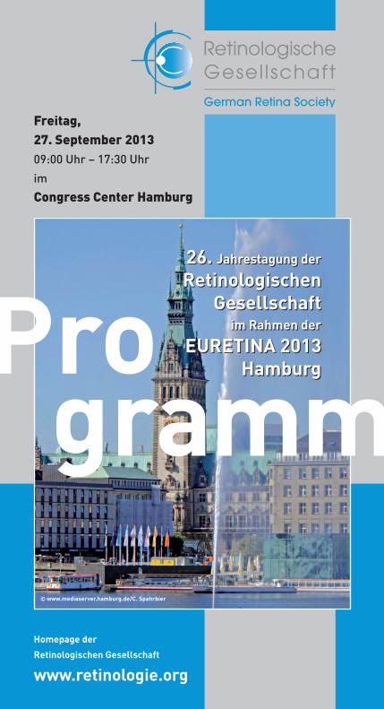
Homepage der Retinologischen Gesellschaft www.retinologie.org Freitag, 27. September 2013 09:00 Uhr – 17:30 Uhr im Congress Center Hamburg 26. Jahrestagung der Retinologischen Gesellschaft im Rahmen der EURETINA 2013 Hamburg © www.mediaserver.hamburg.de/C. Spahrbier 26. Jahrestagung der Retinologischen Gesellschaft im Rahmen der EURETINA 2013 Hamburg Pro gramm
Transcript of 26. Hom eeepoagg - Congress-Organisation Gerling … · Vortragszeiten Referat 15 Min. + 2 Min....
Homepage der
Retinologischen Gesellschaft
www.retinologie.org
Freitag, 27. September 2013 09:00 Uhr – 17:30 Uhr
im
Congress Center Hamburg
26. Jahrestagung der
RetinologischenGesellschaft
im Rahmen der
EURETINA 2013 Hamburg
© www.mediaserver.hamburg.de/C. Spahrbier
26. Jahrestagung der
RetinologischenGesellschaft
im Rahmen der
EURETINA 2013 HamburgPro
gramm
Für die großzügige Unterstützung in der Vorbereitung und Durchführung der 26. Jahrestagung der Retinologischen Gesellschaft in Hamburg dürfen wir uns bei folgenden Firmen bedanken:
Hauptsponsoren:
Novartis Pharma GmbHRoonstraße 2590429 Nürnbergwww.novartis.deSponsoringleistung: 20.000 ,– D
Bayer HealthCareKaiser-Wilhelm-Allee 7051366 Leverkusenwww.bayer.comSponsoringleistung: 15.000 ,– D
Weitere Sponsoren:
Heidelberg Engineering GmbHTiergartenstraße 1569121 Heidelbergwww.HeidelbergEngineering.de
Pharm-Allergan GmbHPforzheimer Straße 16076275 Ettlingenwww.allergan.de
Bausch + Lomb Brunsbütteler Damm 165-17313581 Berlinwww.bausch-lomb.de
D.O.R.C. Deutschland GmbHSchießstraße 5540549 Düsseldorfwww.dorc.eu
Sponsoren RG 2013
1
Vorwort RG 2013
Liebe Kolleginnen und Kollegen,wie schon mehrfach angekündigt, findet in diesem Jahr der
13th EURETINA CONGRESSvom 26. bis 29. September 2013 im CongressCentrum HAMBURG
in Deutschland statt. Diese Veranstaltung hat sich in den letzten Jahren zu dem führenden europäischen retinologischen Kongress mit einer Vielzahl hervor-ragender internationaler Referenten und ständig zunehmender Teilnehmer-zahl entwickelt. Da in diesem Jahr Prof. Dr. Gisbert Richard als Präsident der EURETINA die Tagung in Hamburg organisiert, besteht für alle deutschen Retino-logen die hervorragende Gelegenheit, sich wissenschaftlich, wie klinisch auf internationalem Niveau über alle aktuellen Fragen der Retinologie zu informieren.
Deshalb haben Vorstand und Mitgliederplenum der Retinologischen Gesellschaft schon im Vorfeld beschlossen, gemeinsam mit dem Organisationskomitee von EURETINA die 26. Jahrestagung der Retinologischen Gesellschaft innerhalb des EURETINA-Kongresses zu veranstalten, und so in diesem Jahr die netzhaut-orientierten Kongresse in Deutschland zu bündeln.
Dazu werden am Freitag, den 27. September 2013 in einem separaten Saal des CongressCentrums Hamburg vier englischsprachige Sitzungen zu zentralen retinologischen Fragestellungen stattfinden. Jede Sitzung besteht aus drei Übersichtsreferaten renommierter eingeladener Grundlagen-Wissenschaftlern oder klinischer Retinologen bzw. einer Podiumsdiskussion zur Netzhautchirurgie. Dazu wurden aus den Vortragsanmeldungen freie Vorträge zur Vervollständi-gung jeder Sitzung hinzugefügt. Hierdurch gelang es, ein hochinteressantes Gesamtprogramm aus vier sehr unterschiedlichen thematischen Blöcken zusammenzustellen.
Da sowohl dieses Programm unserer Jahrestagung als auch das Gesamtpro-gramm der EURETINA extrem vielversprechend ist, würden wir uns sehr freuen, wenn die deutsche Retinologie bei diesem Kongress eine große Präsenz zeigen könnte. Daher möchten wir Sie im Namen des Vorstandes der Retinologischen Gesellschaft und des Präsidiums der EURETINA nochmals ganz herzlich nach Hamburg einladen. Melden Sie sich bitte über EURETINA an. Es wird von Seiten der Veranstalter finanzielle Vergünstigungen speziell für uns deutsche Retino-logen geben (gemeinsamer Jahresbeitrag mit reduzierter Teilnehmergebühr), die die Entscheidung zum Besuch noch weiter erleichtern.
In diesem Sinne freuen wir uns alle auf einen großartigen Kongress in Hamburg als Kombination aus EURETINA und RG-Jahrestagung, zu der wir Sie gerne begrüßen möchten.
Prof. Dr. Daniel Pauleikhoff Priv.-Doz. Dr. Klaus Dieter LemmenPräsident Sekretär
als Gastgeber in Hamburg Prof. Dr. Gisbert Richard Präsident der EURETINA
2
Allgemeine Hinweise RG 2013
Wissenschaftliche Prof. Dr. Gisbert RichardLeitung Universitäts-Augenklinik Hamburg Martinistraße 52, 20246 Hamburg Tel.: 040 / 74 10 - 523 01; Fax 040 / 74 10 - 549 06 E-Mail: [email protected]
RG-Tagungsort CCH – Congress Center Hamburg Am Dammtor / Marseiller Straße 20355 Hamburg www.cch.de
Saal G1 – Level 2
Tagungsanmeldung www.euretina.org
Kongresssprache Englisch
Dauer der Freitag, 27. September 2013 09:00 Uhr bis 17:30 UhrRG-Tagung
EURETINA-Kongressgebühren* (auch für freie Vortragende)
Die Anmeldung zur EURETINA 2013 in Hamburg ist für RG-Mitglieder vergünstigt (s.u.).Sie zahlen denselben Preis wie Mitglieder der EURETINA.
*) Der Nachweis für die Inanspruchnahme ermäßigter Teilnahmegebühren muss ggf. durch eine der Anmeldung beiliegende Bescheinigung nachgewiesen werden.
Tagungsanmeldung Die Teilnahme an der diesjährigen RG-Tagung ist abhängig vonzur RG-Tagung der Teilnahme an der EURETINA 2013 in Hamburg. Bitte melden Sie sich über die Homepage www.euretina.org zur
Tagung an. Weitere Informationen wie Zahlungsmodalitäten etc. finden sie dort.
EACCME-Zertifizierung Die EACCME-Zertifizierung erfolgt durch die EURETINA.
Anmeldung
Mitglied der EURETINAFacharztArzt in Weiterbildung
Nichtmitglied der EURETINAFacharztArzt in Weiterbildung
vor Ort
J 400,–J 240,–
J 500,–J 260,–
bis zum 30. Juni 2013
J 300,–J 145,–
J 400,–J 170,–
bis zum 24. Sept 2013
J 350,–J 190,–
J 450,–J 210,–
3
Neu!!!
Teilnahme für In diesem Jahr wird Mitgliedern der RG eine vergünstigteRG-Mitglieder Teilnahme und Mitgliedschaft der EURETINA angeboten!
RG-Mitglieder können für das Jahr 2013 befristet in die EURETINA eintreten, sind im Jahr 2013 beitragsfrei und zah len als Mitglieder lediglich eine reduzierte Kongressgebühr zur EURETINA in Hamburg, können die Ophthalmologica kostenfrei beziehen u.a.
Diese befristete Mitgliedschaft in der EURETINA endet automatisch am Ende des Jahres 2013!
Sie können jedoch bei Anmeldung zur EURETINA angeben, dass Sie Mitglied bleiben möchten und werden dann ab dem Jahr 2014 zahlendes Mitglied der EURETINA.
RG-Mitglieder, die von der Möglichkeit des befristeten oder permanenten Beitritts zur EURETINA keinen Gebrauch machen, zahlen den Normalpreis zum EURETINA-Kongress in Hamburg.
Neumitgliedschaft Für eine Reduzierung der Teilnahmegebühr müssen diein der RG Beitrittsmodalitäten v o r Beginn der diesjährigen Tagung
abgeschlossen sein. Bitte fordern Sie rechtzeitig eine Beitrittserklärung an
(www.retinologie.org).
Vortragszeiten Referat 15 Min. + 2 Min. Diskussion Vortrag 5 Min. + 1 Min. Diskussion
WICHTIG Vortragende und Co-Referenten müssen sich selbst bei der EURETINA anmelden! Anmeldemodalitäten s.o.
Sie werden nicht automatisch angemeldet!
Änderungen, Irrtümer, Satz- und Druckfehler vorbehalten.
Allgemeine Hinweise RG 2013
Wichtige Hinweise für Vortragende RG 2013
5
Programm RG 2013
Program committee: Heinrich Heimann (Liverpool/UK) Frank G. Holz (Bonn/D) Daniel Pauleikhoff (Münster/D) Gisbert Richard (Hamburg/D)
Friday, 27th September 2013
09:00 h Welcome Address Daniel Pauleikhoff President of the German Retina Society (Münster/D)
09:05 h I. Scientific Session Saal G1 ▼ Degeneration and Regeneration of retinal cells10:30h
01 R Marius Ueffing1,2, C. Kiel3, K. Boldt1, A. Vogt1, A, Campagna3, D. Mans4, J. Won5, J. van Reeuwijk4, P. Nishina5, R. Roepman4, G. Cesareni6, L. Serrano3, and the SYSCILIA consortium7 (1University of Tuebingen, Centre for Ophthalmology, Tuebingen/D, 2Helmholtz Center Muenchen, Research Unit of Protein Science, Munich/D, 3CRG-EMBL System Biology Program, Centre de Regulació Genòmica (CRG) Barcelona/E, 4Department of Human Genetics, Radboud University Nijmegen Medical Centre, Nijmegen/NL, 5The Jackson Laboratory, Bar Harbor, Maine/USA, 6University of Rome Tor Vergata, Rome/I, 7the SYSCILIA consortium)
Photoreceptors in Retinal Diseases
02 R Thomas Langmann (Cologne/D) Inflammation and degenerative retinal diseases
03 R Tim U. Krohne (Bonn/D) Perspectives of stem cell therapy
04 V Mahdy Ranjbar, S. Grisanti (Lübeck/D) Stem cell therapy of age-related macular degeneration
05 V Matthias Lüke1, M. Ranjbar1, A. Tapenbayeva1, A. Tura1, S. Grisanti1, T. Schneider2,3, J. Lüke1 (1University Eye Hospital, University of Lübeck/D, 2Institute of Neurophysiology, University of Cologne/D, 3Center of Molecular Medicine Cologne, University of Cologne/D)
The concentration dependent effects of indocyanine green on retinal function in the electrophysiological ex-vivo model of the isolated perfused vertebrate retina
Z S O K N 0.40.4
P C K Z OR H S D K
O V H RC Z R H S
O N H R CD K S N V
0.7
0.8
0.9
1.0
0.6
0.5
0.7
0.8
0.9
0.6
0.5
P
CH R
SKC Z
KHO H
HZPP
D
RR
O1.0
© Bayer Vital GmbH, 51366 Leverkusen L.DE.SM.01.2013.1317
6BVEYA12079_Anzeige_114x210_3K.indd 1 22.04.13 14:18
7
Programm RG 2013
06 V Andreas Stahl1, A. Buehler1, F. Bucher1, S. Berger1, A. Pielen1, G. Martin1, G. Schlunck1, H.T. Agostini1, P. Sapieha2, M. Klags-brun3, L.E.H. Smith4 (1Freiburg/D, 2Montreal/CND, 3Vascular Biology Program, Harvard Medical School, Children’s Hospital Boston/USA, 4Department of Ophthalmology, Harvard Medical School, Children’s Hospital Boston/USA)
Semaphorin 3F forms an anti-angiogenic barrier in the outer retina
07 V Clemens Lange1,3, A. Fantin2, L. Denti2, A. J. Smith3, R.R. Ali3, C. Ruhrberg2, J.W. Bainbridge3 (1Department of Genetics, Institute of Ophthalmology, UCL, NIHR Biomedical Research Centre for Ophthalmology, London/UK, 2Department of Cell Biology, UCL Institute of Ophthalmology, London/UK, 3University Eye Hospital Freiburg/D)
Inactivation of HIF1a and VEGFA in myeloid cells reduces retinal and choroidal neovascularisation
08 V Yoko Miura, I. Fritz, S. Grisanti, M. Rudolf (Lübeck/D) Effect of apolipoprotein A1-mimetic peptide on cultured
retinal pigment epithelial (RPE) cells
09 V Armin Mohi, S. Grisanti, C. Örün, M. Rudolf (Lübeck/D) Apolipoprotein mimetic Dual Domain Peptide reduces Neutral
Lipid Deposits in murine Bruch’s Membrane
10:30 h Coffee Break
11:00 h II. Scientific Session Saal G1 ▼ Genes and the retina – what matters?12:30 h
10 R Hanno J. Bolz (Center for Human Genetics, Bioscientia, Ingelheim, Germany/D)
Next-generation sequencing: A new era in research and diagnostics of retinal dystrophies.
11 R Bart P. Leroy (Dept of Ophthalmology & Ctr for Medical Genetics, Ghent University Hospital & Ghent University, Gent/B)
Retinal dystrophies – fundamental aspects of gene therapy
12 R Christina Zeitz1,2,3, S. El Shamieh1,2,3, I. Audo1,2,3,4,5, J.-A. Sahel1,2,3,4,5,6,7 (1INSERM, UMR_S968, Paris/F, 2CNRS, UMR_7210, Paris/F, 3UPMC Univ Paris 06, UMR_S 968, Institut de la Vision, Paris/F, 4Centre Hospitalier National d’Ophtalmologie des Quinze-Vingts, INSERM-DHOS CIC 503, Paris/F, 5Institute of Ophthalmology, University College of London, London/UK, 6Fondation Ophtal-mologique Adolphe de Rothschild, Paris/F, 7Académie des Sciences–Institut de France, Paris/F)
Methods in Genetics in inherited retinal disorders and Gene therapy trials
Mit MultiColorTM Laser-Fundus-bildern erkennen Sie mehr. Die spezifi schen Laserfarben dringen unterschiedlich tief in die Netzhaut ein und ergeben plastische, kontrastreiche und detailgetreue Fundusbilder.
Simultan mit SD-OCT aufgenom-men ergibt sich durch SPECTRALIS mit MultiColor eine ganz neue diagnostische Dimension.
MultiColor ist für alle SPECTRALIS-Modelle optional verfügbar. Mit dem Advanced Upgrade Package können Sie MultiColor auch späternachrüsten.
Mehr Informationen unterwww.SPECTRALIS-MultiColor.de
9361
5-00
2 IN
T.D
13 ©
Hei
delb
erg
Engi
neer
ing
Gm
bH
Farbe in neuem Licht
9361
5-00
2 IN
T.D
13
Zukunft sichern mit
Advanced Upgrade Package
93615-001 INT.D13 SPECTRALIS_MultiColor-114x210+3 + AUP_VNDA.indd 1 24.04.2013 12:05:45
9
13 V Mihaela Reinsberg1, R.-D. Hilgers2, J. Lüke1, K. Nassar1, S. Grisanti1, I. Lüdeke1, M. Lüke1 (1University of Lübeck, Depart-ment of Ophthalmology, Lübeck/D, 2RWTH Aachen University, Department of Medical Statistics, Aachen/D)
Evaluation of the effects of ranibizumab (Lucentis®) therapy on the macular function in patients with wet age-related macular degeneration using multifocal-electroretinogram and microperimetry
14 V Christian Simader1,2, S. Waldstein1,2, M. Kundi3, U. Schmidt- Erfurth1,2 (1Department of Ophthalmology, Medizinische Universität Wien/A, 2Vienna Reading Center, Medizinische Universität Wien/A, 3Insti-tute of Environmental Health, Medizinische Universität Wien/A)
Standardized analysis of morphological OCT parameters and correlation with best-corrected visual acuity (BCVA) in antiangiogenic therapy of neovascular age-related macular degeneration (AMD)
15 V Werner Inhoffen, U. Hagemann, M. Feucht, S. Aisenbrey, K.U. Bartz-Schmidt (Tübingen/D)
Choroidal neovascularisation (CNV) during anti-VEGF therapy: Changes in microperimetric visual field defects are similar to changes in optical coherence tomography (OCT) findings in most cases.
16 V Ursula Schmidt-Erfurth, S. Waldstein, M. Kundi, C. Simader (Vienna Reading Center, Univ. Klinik für Augenheilkunde und Optometrie; Institut für Umwelthygiene, Department für Public Health, Medizinische Universität Wien/A)
Identification of clinically relevant OCT parameters over two years of treatment in neovascular AMD
17 V Hansjürgen Agostini1, S. Mueller2, C.Ehlken1, U. Bauer-Steinhusen3, Z. Hasanbasic3, T. Wilke2 (1Freiburg/D, 2Institut für Pharmakoökonomie und Arzneimittellogistik, Wismar/D, 3Bayer Vital GmbH, Leverkusen/D)
Treatment of wAMD patients with VEGF inhibitors: Insights from the routine clinical practice in Germany – the PONS Study
18 V Wolfgang F Schrader, A. Bernhard, C. Dietz, K. Sommer, E. Panidou (Nuremberg/D)
The potential of the anti VEGF drugs cannot be used under real life conditions in Germany: Results from a 4 year follow up of 77 patients
19 V Heinrich Gerding, L. Hefner, M. Timmermann (Olten/CH) “Real-Life“-Data on Treatment of Diabetic Macular Oedema
with Ranibizumab: Effect on Visual Acuity, OCT Development and the Number of Retinal Microaneurysms.
12:30 h Lunch Break
Programm RG 2013
Nachweislich glisteningfreies Material (FDA approved)
Asphärisch, aberrationsfreies Design für mehr Kontrast und Schärfentiefe
2,2 mm Inzision für sichere und einfache Implantation
Minimierte PCO durch 360° scharfe Kante
Der neue Standard leistungsfähiger IOL aus hydrophobem Acryl
Weitere Informationen unter:
Tel.: 0800 5893 114 Fax: 01805 90 94 90 [email protected]
©2013 Bausch + Lomb Incorporated. ™/® bezeichnen Marken von Bausch + Lomb Incorporated.
Prob
e-Im
plan
tatio
n!
Scan
nen
Sie
den
QR-
Cod
e
Glisteningfrei
Anz.enVista114x210-0613.indd 1 10.06.13 13:11
11
Programm RG 2013
14:00 h III. Scientific Session Saal G1 ▼ The choroid and retinal diseases15:30 h
20 R Richard F. Spaide (New York/USA) The Choroid in Health and Disease
21 R Steffen Schmitz-Valckenberg (Bonn/D) The choroid in AMD
22 R Marc de Smet (Antwerp/B) The choroid in inflammatory diseases
23 V Florian Alten, C.R. Clemens, P. Heiduschka, N. Eter (Münster/D) Localized reticular pseudodrusen and their topographic
relation to choroidal watershed zones and changes in choroidal volumes
24 V Britta Heimes, M. Ziegler, B. Book, D. Breiter, A. Lommatzsch, M. Gutfleisch, M. Dietzel, G. Spital, M. Zeimer, D. Pauleikhoff (Münster/D)
Choroidal thickness under Anti-VEGF-therapy in exudative AMD
25 V Inger Lüdeke, S. Grisanti (Lübeck/D) “How much RPE is needed?” – Casereport of a patient with
retinal pigment epithelial tear
26 V Bianca Gerendas1, S.M. Waldstein1, B. Hajnajeeb1, L. Zhang2, H. Bogunovic2, M. Abramoff3, C. Simader1, M. Sonka2,3, U. Schmidt-Erfurth1 (1Medizinische Universtität Wien/A, 2Department of Electrical and Computer Engineering, University of Iowa, Iowa City/USA, 3Department of Ophthalmology & Visual Sciences, University of Iowa Hospital & Clinics, Iowa City/USA)
Posterior Pole Choroidal Vasculature Assessed By Automated Choroidal Vessel Segmentation In Standard Clinical Spectral-Domain Optical Coherence Tomography In Patients With Diabetic Macular Edema
27 V Salvatore Grisanti, J. Lüke, M. Reinsberg, A. Tura (Lübeck/D) Circulating Melanoma Cells:
An evolving instrument in the diagnostic of uveal melanoma
28 V Julia Lüke, D. Wildner, M. Lüke, S. Grisanti, A. Tura (Lübeck/D) Everolimus reduces the metastatic potential of human
uveal melanoma cells
15:30 h Coffee Break
12
Programm RG 2013
16:00 h IV. Scientific Session Saal G1 ▼ My coolest surgical video17:30 h Moderators: Heinrich Heimann (Liverpool/UK)
Albrecht Lommatzsch (Münster/D)
Panel: Karl Ulrich Bartz-Schmidt (Tübingen/D)Claus Eckardt (Frankfurt/Main-D)Bernd Kirchhof (Köln/D)Christos Haritoglou (München/D)Horst Helbig (Regensburg/D)Thomas J. Wolfensberger (Lausanne/CH)
29 V Mitrofanis Pavlidis (Cologne/D) 27Gauge Vitrectomy new perspectives
30 V Evangelos Chatzizisis, B.Jurklies (Wuppertal/D) Treatment of subretinal hemorrhage with subretinal Injection
of recombinant tissue plasminogen activator
31 V Hakan Kaymak, D. Breyer, K. Klabe, C. Pohl (Düsseldorf/D) Rotational stability of toric lenses with 23-gauge pars plana
vitrectomy (PPV)
32 V Ulrich Spandau (Uppsala/S) Episcleral buckling with the BIOM: A novel technique
33 V Eva Biewald1, M. Gök1, M. Freistühler1, K. Metz2, N. Bornfeld1 (1Abteilung für Erkrankungen des hinteren Augenabschnitts, Universitätsaugenklinik Essen/D, 2Institut für Pathologie und Neuropathologie, Universitätsklinikum Essen/D)
Complications following transretinal 23G tumourbiopsy
34 V Vinodh Kakkassery1, M Elling1, D.S. Loos1, H.B. Dick1, U. Schlegel2, R. Schroers3, A. Baraniskin3 (1Department of Ophthalmology, Ruhr-Universität Bochum/D, 2Department of Neurology, Ruhr-Universität Bochum/D, 3Department of Medicine, Hematology, and Oncology, Ruhr-Universität Bochum/D)
Upregulation of miRNA in a single primary vitreoretinal lymphoma specimen: A new diagnostic approach?
17:30 h End of the Annual Meeting of the German Retina
13
Zur27. Jahrestagung
der
RetinologischenGesellschaft
am13. und 14. Juni 2014
in
DüSSELDORF
lädt Sie bereits heuteHerr Priv.-Doz. Dr. Klaus Dieter Lemmen
ein.
Vorankündigung RG 2014
Bilder: © Düsseldorf Marketing & Tourismus GmbH – Fotograf U. Otte
Bilder: © Düsseldorf Marketing & Tourismus GmbH – Fotograf U. Otte
14
Prof. Dr. Hansjürgen Agostini Universitäts-Augenklinik Killianstraße 5 79106 Freiburg i. Breisgau
Dr. Florian Alten Universitäts-Augenklinik Domagkstraße 15 48149 Münster
Prof. Dr. Karl Ulrich Bartz-Schmidt Universitäts-Augenklinik Schleichstraße 12-16 72076 Tübingen
Dr. Eva Biewald Universitäts-Augenklinik Hufelandstraße 55 45122 Essen
Prof. Dr. Hanno J. Bolz Bioscientia Institut für Medizinische Diagnostik GmbH Labor Ingelheim – FB Humangenetik Konrad-Adenauer-Straße 17 55218 Ingelheim
Evangelos Chatzizisis HELIOS Klinikum Wuppertal Augenklinik Heusnerstraße 40 42283 Wuppertal
Prof. Dr. Marc D. de Smet Academic Medical Center Amsterdam Dept. of Ophthalmology Meibergdreef 9 1105 AZ Amsterdam Niederlande
Prof. Dr. Claus Eckardt Städt. Kliniken-FFM-Höchst Augenabteilung Gotenstraße 6-8 65929 Frankfurt
Prof. Dr. Heinrich Gerding Klinik Pallas Abteilung für Retinologie Louis-Giroud-Strasse 20 4600 Olten Schweiz
Dr. Bianca Gerendas Universitäts-Augenklinik Währinger Gürtel 18-20 1090 Wien Österreich
Prof. Dr. Salvatore Grisanti Universitätsklinikum Schleswig-Holstein Campus Lübeck Augenklinik Ratzeburger Allee 160 23538 Lübeck
Prof. Dr. Christos Haritoglou Augenklinik der LMU Mathildenstraße 8 80336 München
Prof. Dr. Arnd Heiligenhaus St. Franziskus-Hospital Augenabteilung Hohenzollernring 74 48145 Münster
Prof. Dr. Heinrich Heimann Royal Liverpool University Hospital St Paul‘s Eye Unit Prescot Street L7 8XP Liverpool Grossbritannien
Dr. Britta Heimes St. Franziskus-Hospital Augenabteilung Hohenzollernring 74 48145 Münster
Prof. Dr. Horst Helbig Universitäts-Augenklinik Franz-Josef-Strauß-Allee 11 93053 Regensburg
Prof. Dr. Frank G. Holz Universitäts-Augenklinik Ernst-Abbe-Straße 2 53127 Bonn
Dr. rer. nat. Werner Inhoffen Universitäts-Augenklinik Schleichstraße 12-16 72076 Tübingen
Dr. Vinodh Kakkassery Universitäts-Augenklinik In der Schornau 23-25 44892 Bochum
Dr. Hakan Kaymak Breyer & Kaymak Augenchirurgie Internationale Innovative Ophthalmochirurgie I.I.O. Berliner Allee 15 40212 Düsseldorf
Prof. Dr. Bernd Kirchhof Universitäts-Augenklinik Kerpener Straße 62 50924 Köln
Priv.-Doz. Dr. Tim U. Krohne Universitäts-Augenklinik Ernst-Abbe-Straße 2 53127 Bonn
Dr. Clemens Lange Universitäts-Augenklinik Killianstraße 5 79106 Freiburg i. Breisgau
Prof. Dr. Thomas Langmann Universitätsklinikum Köln Zentrum für Augenheilkunde Experimentelle Immuno-logie des Auges Joseph-Stelzmann-Straße 9 50931 Köln
Autoren RG 2013

Autoren RG 2013
15
Dr. Bart P. Leroy
Ghent University Hospital
Dept. of Ophthalmology
De Pintelaan 185
9000 Ghent
Belgien
Priv.-Doz. Dr.
Albrecht Lommatzsch
St. Franziskus-Hospital
Augenabteilung
Hohenzollernring 74
48145 Münster
Dr. Inger Lüdeke
Universitätsklinikum
Schleswig-Holstein
Campus Lübeck
Augenklinik
Ratzeburger Allee 160
23538 Lübeck
Priv.-Doz. Dr.
Matthias Lüke
Universitätsklinikum
Schleswig-Holstein
Campus Lübeck
Augenklinik
Ratzeburger Allee 160
23538 Lübeck
Priv.-Doz. Dr. Julia Lüke
Universitätsklinikum
Schleswig-Holstein
Campus Lübeck
Augenklinik
Ratzeburger Allee 160
23538 Lübeck
Dr. Yoko Miura
Universitätsklinikum
Schleswig-Holstein
Campus Lübeck
Augenklinik
Ratzeburger Allee 160
23538 Lübeck
Armin Mohi Universitätsklinikum Schleswig-Holstein Campus Lübeck Augenklinik Ratzeburger Allee 160 23538 Lübeck
Prof. Dr. Daniel Pauleikhoff St. Franziskus-Hospital Augenabteilung Hohenzollernring 74 48145 Münster
Priv.-Doz. Dr. Mitrofanis Pavlidis Augencentrum Köln-Porz Josefstraße 14 51143 Köln
Dr. Mahdy Ranjbar Universitätsklinikum Schleswig-Holstein Campus Lübeck Augenklinik Ratzeburger Allee 160 23538 Lübeck
Mihaela Reinsberg Universitätsklinikum Schleswig-Holstein Campus Lübeck Augenklinik Ratzeburger Allee 160 23538 Lübeck
Prof. Dr. Gisbert Richard Universitäts-Augenklinik Martinistraße 52 20246 Hamburg
Prof. Dr. Ursula Schmidt-Erfurth Universitäts-Augenklinik Währinger Gürtel 18-20 1090 Wien Österreich
Priv.-Doz. Dr. Steffen Schmitz-Valckenberg Universitäts-Augenklinik Ernst-Abbe-Straße 2 53127 Bonn
Prof. Dr. Wolfgang F. Schrader Maximilians-Augenklinik gemeinnützige GmbH Erlenstegenstraße 30 90491 Nürnberg
Dr. Christian Simader Universitäts-Augenklinik Währinger Gürtel 18-20 1090 Wien Österreich
Dr. Richard F. Spaide Vitreous-Retina-Macula Consultants of New York 460 Park Avenue, 5th FloorNY 10022 New York USA
Priv.-Doz. Dr. Ulrich H. M. Spandau Uppsala University Hospital Dept. of Neurosience / Ophthalmology 751 85 Uppsala Schweden
Dr. Andreas Stahl Universitäts-Augenklinik Killianstraße 5 79106 Freiburg i. Breisgau
Prof. Dr. Marius Ueffing Universitätsklinikum Tübingen Forschungsinstitut für Augenheilkunde Röntgenweg 11 72076 Tübingen
Prof. Dr. Thomas J. Wolfensberger Université de Lausanne Hôpital Ophtalmique Jules Gonin 15, Avenue de France 1004 Lausanne Schweiz
Dr. Christina Zeitz Institut de la Vision Dept. of Genetics 17, rue Moreau 75012 Paris Frankreich
Abstracts RG 2013
I. Scientific Session Degeneration and Regeneration of retinal cells
01 R Photoreceptors in Retinal Diseases Marius Ueffing1,2, C. Kiel3, K. Boldt1, A. Vogt1, A, Campagna3, D.
Mans4, J. Won5, J. van Reeuwijk4, P. Nishina5, R. Roepman4, G. Ce-sareni6, L. Serrano3, and the SYSCILIA consortium7 (1University of Tuebingen, Centre for Ophthalmology, Tuebingen/D, 2Helmholtz Center Muenchen, Research Unit of Protein Science, Munich/D, 3CRG-EMBL System Biology Program, Centre de Regulació Genò-mica (CRG) Barcelona/E, 4Department of Human Genetics, Radboud University Nijmegen Medical Centre, Nijmegen/NL, 5The Jackson Laboratory, Bar Harbor, Maine/USA, 6University of Rome Tor Ver-gata, Rome/I, 7the SYSCILIA consortium, a European Community‘s Seventh Framework Programme FP7/2009 under grant agreement no: 241955, SYSCILIA; http://syscilia.org)
Photoreceptors are high-end neurosensory cells, able to detect light on a single photon level, discriminate colors, and turn light into an electrical signal, transmitted to the brain. Photoreceptors realize this performance through their outer segments that contain para-cristaline arrays of light receptive visual pigments compart-mentalized in membrane staples called outer segment disks. Outer segments as well as disks are generated and maintained via motor driven, directional ciliary transport from the inner- to the light sen-sitive outer segments, which enable us to see. Mutations that criti-cally affect outer segment formation and function impair eyesight and eventually lead to photoreceptor degeneration.
Combining and integrating several layers of information gathering from proteome centric biochemistry, in silico network analysis as well as structural biology we have developed an analytical approach to determine components of the molecular machinery exerting these functions as well as to identify functional constraints in the corresponding protein networks that drive them and - when hit by mutation – result in disease. As a result of this approach, we have generated a multiscale model that defines key elements of photore-ceptor outer segment function on the molecular level.
We have spotlighted protein transport from the inner- to the outer segment as an essential process generating the light receptive ou-ter segment. Zooming in on a protein mutated in an early childhood blinding disease, LCA5, we have identified the interflagellar trans-port machinery as the physical entity driving vesicular trafficking through the interconnecting cilium that bridges the inner and the outer segment. We have modeled the impact of mutation on the level of functional protein networks and can show, that specific physical interactions affecting the functionality of discrete molecular machi-nes are rendered dysfunctional by these mutations. The resulting information not only offers a deeper understanding on the impact of mutations, but allows to gain a comprehensive view on the mo-lecular mechanisms of photoreceptor outer segment formation and renewal. Further, it suggests novel signaling routes guiding these activities. Our model demonstrates a specific disease susceptibility
16
online www.egms.de
17
Abstracts RG 2013
of the core visual pathway as well as the transport machinery and a relative robustness of other functional modules. This information can serve as a basis for elucidating physiological principles as well as to enable targeted molecular medicine and rational pharmaco-logy to treat retinal degenerative diseases in the future.
02 R Inflammation and degenerative retinal diseases Thomas Langmann (Cologne/D)
Hereditary retinal dystrophies are characterized by progressive re-tinal degeneration. Similar degenerative processes are evident in multifactorial and complex retinal disorders including age-related macular degeneration. To understand early triggers of these me-chanisms, genetic and experimental mouse models have been de-veloped that mimic human retinal degeneration. In most of these models, early chronic activation of the innate immune system has been documented. This process involves the complement cascade and microglial cells as sensors and effectors of the retinal immune response. Current evidence suggests that the genetic predisposition and individual factors like age or diet critically influence the immu-ne homeostasis in the retina. Based on their effectiveness, innate immune mechanisms are thought to support or even provoke reti-nal degeneration. This talk will summarize the recent progress in understanding the role of innate immunity in retinal degenerative diseases and highlights the retinal immunopathology as potential therapeutic target to prevent vision loss.
03 R Perspectives of stem cell therapy Tim U. Krohne (Bonn/D)
Stem cell medicine has recently entered the phase of first clinical trials. The eye possesses unique properties that may make it an ideal target for stem cell treatments. This is reflected by the fact that all current pluripotent stem cell trials worldwide are in the field of ophthalmology, mainly for the treatment of age-related macu-lar degeneration and Stargardt’s disease. While most of these trials employ embryonic stem cells, the recently developed technique of deriving induced pluripotent stem cells (iPS cells) from the patient’s own skin or blood cells offers the intriguing prospect of generating autologous retinal cell grafts for transplantation, thus minimizing the risk of graft rejection. In the wake of these scientific clinical re-search efforts and their increasing media coverage, a growing num-ber of commercial providers offer unproven and costly stem cell therapies for ophthalmological diseases. Knowledge of the current state of preclinical and clinical research on stem cells facilitates in-formed patient counseling regarding current and future therapeutic applications of stem cells in ophthalmology.
online www.egms.de

18
Abstracts RG 2013
04 V Stem cell therapy of age-related macular degeneration Mahdy Ranjbar, S. Grisanti (Lübeck/D)
Introduction: Age-related macular degeneration (AMD) is the lea-ding cause of blindness in people over age 55 in the developed world. Loss of retinal pigmented epithelium (RPE) plays a major pa-thophysiologic role in the disease. Stem cells are currently conside-red for RPEreplacement. Based on their level of differentiation and their potential they are classified in embryonal stem cells (ESC) and adult stem cells (ASC), while the latter group can be subclassified in induced pluripotent stem cells (iPSC) and natural pluripotent stem cells (nPSC).
Methods: On basis of the published literature these stem cell groups are evaluated with regard to their potential and feasibility of clinical application in the treatment of age-related macular degeneration.
Results: All these mentioned stem cell populations are able to ge-nerate RPE-like cells in terms of morphology and function. These findings have been demonstrated in vitro and in preclinical animal studies. But with reference to ESC and iPSC several methodical and ethical controversies and disadvantages exist, which might favour the clinical application of nPSC.
Conclusion: Stem cells are going to prove beneficial in the treat-ment of age-related macular degeneration. Different stem cell clas-ses are considered of which the natural pluripotent stem cells might be the most feasible option for clinical application.
05 V The concentration dependent effects of indocyanine green on reti-nal function in the electrophysiological ex-vivo model of the isola-ted perfused vertebrate retina
Matthias Lüke1, M. Ranjbar1, A. Tapenbayeva1, A. Tura1, S. Grisanti1, T. Schneider2,3, J. Lüke1 (1University Eye Hospital, University of Lübeck/D, 2Institute of Neurophysiology, University of Cologne/D, 3Center of Molecular Medicine Cologne, University of Cologne/D)
Background: Dye solutions such as indocyanine green (ICG) are used for intraoperative staining of epiretinal membranes and the internal limiting membrane to enhance the visualization and to fa-cilitate their removal. The aim of the presented study was to inves-tigate the effects of ICG on bovine retinal function using different concentrations of ICG.
Methods: Bovine retina preparations were perfused with a standard solution and the electroretinogram (ERG) was recorded. After recor-ding stable ERG-amplitudes the nutrient solution was substituted by an ICG solution with varying concentrations (0.000025%, 0.00025%, 0.0025% & 0.025%) for 45 minutes. Afterwards the preparations were reperfused with standard solution for at least 85 minutes. The percentage of b-wave reduction was measured after exposure with ICG and at the end of the washout.
Results: Significant reductions (p<0.05) of the b-wave amplitude were found for concentrations of 0.0025% (p=0.00985) and 0.025% (p=0.03775) ICG after exposure. But while for the concentration of
online www.egms.de
19
Abstracts RG 2013
0.025% ICG the b-wave amplitude remained significantly decreased (p=0.0082) after the observation period, a full recovery of the b-wave was observed for the concentration of 0.0025% (p=0.19171). The lo-wer tested concentrations of ICG did not significantly affect the b-wave amplitude during the exposure as well as at the washout.
Conclusion: The effects of ICG after retinal exposure depend inter alia on the applied concentrations. The intraocular application of sufficient ICG concentrations for ILM-staining seems not possible without interfering with retinal function.
06 V Semaphorin 3F forms an anti-angiogenic barrier in the outer retina Andreas Stahl1, A. Buehler1, F. Bucher1, S. Berger1, A. Pielen1, G.
Martin1, G. Schlunck1, H.T. Agostini1, P. Sapieha2, M.Klagsbrun3, L.E.H. Smith4 (1Freiburg/D, 2Montreal/CND, 3Vascular Biology Pro-gram, Harvard Medical School, Children’s Hospital Boston/USA, 4Department of Ophthalmology, Harvard Medical School, Children’s Hospital Boston/USA)
Background: Semaphorins are known modulators of neuronal path-finding and angiogenesis. In the neuro-retinal tissue of the retina, semaphorins exert important functional roles on tissue homeosta-sis and architecture by guiding axons and vessels to their appropri-ate target and steering them away from unwanted compartments.
Methods and Results: Using RNA and protein expression analysis on murine and human retinas, we identified Semaphorin 3F (Se-ma3F) to be expressed exclusively in the avascular layers of the ou-ter retina (outer nuclear layer and retinal pigment epithelium; RPE). Using functional in vitro models, we found potent anti-angiogenic effects of Sema3F on human retinal endothelial cells in a spheroid assay as well as on choroidal endothelial cells in a choroidal explant assay. In addition, Sema3F expression in human RPE from active CNV membranes revealed a reduced expression of Sema3F in CNV-RPE compared to normal RPE.
Conclusion: Our data demonstrate an exclusive expression of anti-angiogenic Sema3F in the outer retinal layers. Physiologically, Se-ma3F may contribute to the avascularity of these retinal layers. In pathologic conditions like exudative AMD, reduced Sema3F expres-sion may contibute to invasion of blood vessels into these normally avascular outer retinal layers.
07 V Inactivation of HIF1a and VEGFA in myeloid cells reduces retinal and choroidal neovascularisation
Clemens Lange1,3, A. Fantin2, L. Denti2, A.J. Smith1, R.R. Ali1, C. Ruhrberg2, J.W. Bainbridge1 (1Department of Genetics, Institute of Ophthalmology, UCL, NIHR Biomedical Research Centre for Oph-thalmology, London/UK, 2Department of Cell Biology, UCL Institute of Ophthalmology, London/UK, 3University Eye Hospital Freiburg/D)
Myeloid cells have been implicated in retinal and choroidal neova-scularization in sight-threatening neovascular eye diseases such
online www.egms.de
Abstracts RG 2013
as diabetic retinopathy and age-related macular degeneration. The hypoxia-inducible transcription factor 1 alpha (HIF1A) is activated by inflammatory stimuli and hypoxia and is a key driver of vascu-lar endothelial growth factor (VEGFA) gene expression. This study explores the role of HIF1a and VEGFA in the development of retinal neovascularisation (RNV) after oxygen-induced retinopathy (OIR) and choroidal neovascularisation (CNV) after laser injury of Bruch’s membrane in mice with defective HIF1A and VEGFA expression in myeloid cells. We show that myeloid cells accumulate in areas of RNV or CNV at the onset of neovascularization and that both HI-F1a activation and VEGFA expression in myeloid cells contribute substantially to the development of retinal and choroidal neovascu-larisation. These findings provide novel insight into the pathogenic mechanisms of neovascular eye disease and raise the possibility that immunomodulation of myeloid cells may provide a therapeutic approach to inhibit pathological angiogenesis.
08 V Effect of apolipoprotein A1-mimetic peptide on cultured retinal pigment epithelial (RPE) cells
Yoko Miura, I. Fritz, S. Grisanti, M. Rudolf (Lübeck/D)
Background: Our previous studies suggested that apolipoprotein (Apo) A-I mimetic peptides, as D-4F and L-4F, reduce pathological lipid deposits in the bruch‘s membrane (BRM), and in the sub-RPE space. In this study we investigated the effect of D-4F on RPE cell proliferation and oxidative stress-induced lipid peroxidation in RPE cells in vitro.
Materials and Methods: The RPE cells in third passage culture were treated with different concentrations of D-4F (0-4000 μg/ml) and after 48hrs cell proliferation was examined with MTT assay. Lipid peroxidation was induced by addition of hydrogen peroxide (H2O2) (1 mM) in the confluent RPE cells, which were exposed to D-4F (0, 1, 10, 100 μg/ml) for the previous 24 hrs. After 24 hrs, intracellular 4-HNE (4-hydroxy-2-nonenal), a lipid peroxidation end-product, was measured with 4-HNE adduct Elisa kit.
Results: D-4F concentrations between 10 and 100 μg/ml increased, and the concentrations ≥250 μg/ml reduced RPE cell proliferation signicantly. Intracellular 4-HNE (μg/mg protein) was reduced in the cells treated with 10 and 100 μg/ml of D4F (93% and 78%, respec-tively, p<0.05). H2O2 increased intracellular 4-HNE by 11%, and this effect was partially suppressed by D4F-pretreatment.
Conclusion: The results show that D-4F concentration between 10 and 100 μg/ml might have the effects of activating cell proliferation and reducing intracellular lipid peroxidation. Since lipid peroxidation end-products such as 4-HNE could lead to the additional oxidative stress, D-4F may have a protective effect against oxidative stress in RPE cells.
20
online www.egms.de
21
Abstracts RG 2013
09 V Apolipoprotein mimetic Dual Domain Peptide reduces Neutral Li-pid Deposits in murine Bruch’s Membrane
Armin Mir Mohi Sefat, S. Grisanti, C. Örün, M. Rudolf (Lübeck/D)
Purpose: Massive accumulation of neutral lipids in Bruch’s mem-brane (BrM) with concomitantly increased hydraulic resistivity and oxidative stress induction is a major age change in the eye and an important pathogenic factor for the development and progression of age-related macular degeneration. Apolipoprotein (Apo) mimetic peptides are highly effective and well tolerated lipid acceptors that might be used to remove these pathogenic deposits. We evaluated the effect of an intravitreally injected dual domain peptide (DDP) containing domain properties of ApoE and ApoA1 in an established mouse model of age-related BrM lipid deposition (ApoEnull).
Methods: Four groups (4x; n=6) of 10 month-old ApoEnull mice re-ceived a single intravitreal injection of DDP with different dosages (0 μg; 0.6 μg; 1.2 μg; 2.4 μg). The second untreated eye served as an intraindividual control. Thirty days after injection all animals were sacrificed and eyes were enucleated and processed to BrM who-lemounts. The specimens were stained with a protocol for BrM spe-cific esterified cholesterol using the fluorescent cholesterol specific dye PFO/D4-GFP (Dettbarn et al. ARVO 2010). The treatment effect on BrM lipid deposition was evaluated semiquantitatively by fluores-cence measurements. We calculated for each dose group across all treated vs. untreated eyes the relative difference (%) and tested for significance using the student-t-test.
Results: The vehicle injection (0.9% NaCl, 0 μg DDP) caused no change in BrM esterified cholesterol content (p=0.29). In all other groups we could observe a significant decrease of cholesterol con-tent depending on the DDP dose administered. The lowest dose showed a mild but significant effect (p<0.001) of -16.2% choleste-rol reduction in the treated eyes vs. untreated eyes. The medium and highest DDP dose showed also significant effects (p<0.001) with a cholesterol reduction of -41.7% in the medium dose group and -34.2% with the highest dose.
Conclusions: Our data demonstrated a significant and dose depen-dant reduction of BrM lipid deposits by DDP with ApoA1- and ApoE domain characteristics. Nevertheless, the treatment effect is lower than with our initially tested ApoA1 mimetic peptides (L-4F, D-4F, ARVO 2011 Rudolf et al. Mohi et al.). A neutral lipid reduction of ca. 70% with a single intravitreal injection of 2.4 μg D-4F shows almost double the treatment effect of the here tested DDP.
online www.egms.de
22
Abstracts RG 2013
II. Scientific Session Genes and the retina – what matters?
10 R Next-generation sequencing: A new era in research and diag-nostics of retinal dystrophies.
Hanno J. Bolz (Center for Human Genetics, Bioscientia, Ingelheim, Germany /D)
Most inherited retinal diseases (e.g. retinitis pigmentosa, Leber congenital amaurosis, cone/cone-rod dystrophies, congenital stati-onary night blindness) result from mutations in many genes which has long hampered comprehensive genetic analysis. However, pin-pointing the disease-causing mutations is a prerequisite for perso-nalized genetic counseling, specification of recurrence risks, pre-diction of the clinical course and will be essential for gene-specific therapies. With the advent of next-generation sequencing (NGS), allowing for rapid and cost-effective simultaneous analysis of the known disease genes, these genetically extremely heterogeneous disease groups have become accessible to comprehensive analysis, truly revolutionizing genetic diagnostics. The diagnostic yield has in-creased dramatically, and most patients can now be provided with a genetic diagnosis. Moreover, quantitative readout of high-coverage NGS data may identify disease-causing copy number variations that would escape mere sequence analysis. Another type of “hidden mutations” localizes in non-coding exons with regulatory roles for gene expression, and these largely unstudied gene regions should therefore be considered in NGS targeting retinal diseases genes. Si-multaneous analysis of multiple genes can result in many candidate variants; however, single-gene testing in genetically heterogeneous conditions is error-prone, and sound interpretation requires con-sideration of the full variant load in all genes. For patients whose genetic basis of disease remains elusive even after NGS of a gene panel, whole-exome sequencing (targeting virtually all exons of all human genes) offers the opportunity to identify yet unknown retinal disease genes. Examples from our studies for the aforementioned scenarios will be demonstrated.
11 R Retinal dystrophies – fundamental aspects of gene therapy Bart P. Leroy (Dept of Ophthalmology & Ctr for Medical Genetics,
Ghent University Hospital & Ghent University, Gent/B)
Purpose: The talk will focus on an overview of gene therapy trials for inherited retinal disease, which are currently ongoing or have been finalized. In addition, successful trials in animal models will be mentioned.
Methods: Systematic review. Results: Currently there are 6 trials around the World focusing on
gene therapy for RPE65-related Leber congenital amaurosis, with good safety outcomes and considerable success in restoring some visual function. In addition, gene therapy trials are ongoing for Star-gardt macular dystrophy (ABCA4), Usher type 1B (MYO7A), MERTK-
online www.egms.de
23
Abstracts RG 2013
related early-onset retinal dystrophy and choroideraemia (CHM1). So far, no safety issues have been encountered in these latter trials, whereas it is too early to evaluate restoration and/or stabilization of function.
Success in animal models has been obtained for X-linked retino-schisis, as well as models of CNGA3- and CNGB3-related achroma-topsia.
Conclusions: Gene therapy trials for inherited retinal dystrophies are safe and somewhat successful in restoring and/or stabilizing re-tinal function.
12 R Methods in Genetics in inherited retinal disorders and Gene thera-py trials
Christina Zeitz1,2,3, S. El Shamieh1,2,3, I. Audo1,2,3,4,5, J.-A. Sahel1,2,3,4,5,6,7 (1INSERM, UMR_S968, Paris/F, 2CNRS, UMR_7210, Paris/F, 3UPMC Univ Paris 06, UMR_S 968, Institut de la Vision, Paris/F, 4Centre Hospitalier National d’Ophtalmologie des Quinze-Vingts, INSERM-DHOS CIC 503, Paris/F, 5Institute of Ophthalmology, University Col-lege of London, London/UK, 6Fondation Ophtalmologique Adolphe de Rothschild, Paris/F, 7Académie des Sciences–Institut de France, Paris/F)
Inherited retinal disorders (IRD) are clinically and genetically he-terogeneous with more than 150 gene defects accounting for the diversity of disease phenotypes. Over the past two decades, three main approaches including Sanger sequencing, arrayed primer extension (APEX) chip technology and homozygosity mapping were used to investigate the genetics of IRD. Despite many advantages, the above-mentioned technologies have numerous limitations, an example of such limitation is the diagnostic value provided by APEX that remained low (10%-20%) for rod-cone dystrophy patients. Ap-plying Sanger sequencing to IRD patients resulted in a higher detec-tion rate, however analyzing hundreds of exons across the genome is an enormous effort in terms of time and cost. Homozygosity map-ping is also considered as a powerful tool in identifying novel and known gene defects, but such an approach is only successful when applied to consanguineous families or inbred populations. With the emergence of novel sequencing techniques (targeted and who-le exome), great efforts are currently conducted in order to report novel gene defects implicated in the aetiology of human diseases. This technology has provided accurate qualitative and quantitative information about any type of nucleic acid in a given sample at a high-throughput while incurring relatively limited costs. We have previously developed, in collaboration with a company (Integragen, France) an unbiased and time-efficient retinal gene array for next generation sequencing (NGS), which targets in total 254 genes. Sub-sequently, we have validated and then enhanced our NGS retinal panel by improving the coverage of target genomic regions and re-stricting it to the most relevant genes underlying progressive inheri-ted retinal disorders, thus reducing the number of targeted genes to 121. Herein we present different filtering approaches that we apply
online www.egms.de
24
Abstracts RG 2013
to identify the genetic defects and their prevalence in our cohort. Subsequent Sanger sequencing, co-segregation analysis and re-in-vestigation of the phenotype to validate the gene defects are crucial to prepare patients for gene therapy approaches. During the last de-cade, gene replacement therapies achieved significant success not only in animal models of retinal degeneration, but also in humans. Pioneering clinical studies are now under way to evaluate their safe-ty and efficacy in inherited monogenic eye diseases. Thus we intend to give an update of ongoing gene therapy trials for Leber Congenital Amaurosis, Stargardt disease, Choroideremia Usher syndrome and rod-cone dystrophy.
13 V Evaluation of the effects of ranibizumab (Lucentis®) therapy on the macular function in patients with wet age-related macular degeneration using multifocal-electroretinogram and microperi-metry
Mihaela Reinsberg1, R.-D. Hilgers2, J. Lüke1, K. Nassar1, S. Gri-santi1, I. Lüdeke1, M. Lüke1 (1University of Lübeck, Department of Ophthalmology, Lübeck/D, 2RWTH Aachen University, Department of Medical Statistics, Aachen/D)
Purpose: The purpose of this study was to evaluate the macular function under the treatment with ranibizumab (Lucentis®) in wet age-related macular degeneration (AMD) observed by multifocal-electroretinogram (ERG) and microperimetry.
Methods: Twenty subjects with neovascluar AMD meeting the in-clusion criteria were assigned to ranibizumab treatment. The treat-ment was adapted to the Pronto-study. During a twelve months interval best corrected visual acuity (BCVA), standard ophthalmic examination, retinal thickness observed by OCT, multifocal-ERG and microperimetry has been performed monthly. The decision for reinjections has been made based on the results of patients’ exami-nation. The study has been finished in March 2013.
Results: Descriptive statistics will be provided for patient demo-graphics and all baseline characteristics. Relevant medical history (ocular and non-ocular) and current medical conditions as well as other relevant baseline information will be listed and summarized by appropriate descriptive statistics. Our primary interest will be to report the course of the multifocal-ERG and microperimetry mea-surements during the twelve months follow-up period and to ana-lyze the relations to the clinical outcome by means of BCVA (ETDRS score) and the fovea thickness (μm).
Conclusions: This study will provide a new insight into the deve-lopment of the macular function during the treatment with ranibi-zumab (Lucentis®) and about the significance of multifocal-electro-retinogram and microperimetry in the course of wet AMD
online www.egms.de
25
Abstracts RG 2013
14 V Standardized analysis of morphological OCT parameters and cor-relation with best-corrected visual acuity (BCVA) in antiangiogenic therapy of neovascular age-related macular degeneration (AMD)
Christian Simader1,2, S. Waldstein1,2, M. Kundi3, U. Schmidt-Erfurth1,2 (1Department of Ophthalmology, Medizinische Universität Wien/A, 2Vienna Reading Center, Medizinische Universität Wien/A, 3Institute of Environmental Health, Medizinische Universität Wien/A)
Methods: 1240 patients were treated using ranibizumab/aflibercept in a fixed (year one) and a PRN (year two) regimen in a prospective randomized trial (VIEW2). OCT data were obtained monthly by a stan-dardized protocol and analyzed by an independant reading center.
Results: At baseline, 64% of eyes demonstrated intraretinal cysts (IRC), 80% pigment epithelial detachment (PED) and 84% subretinal fluid (SRF). IRC (p<0.0001) and PED (p=0.028) were associated with a lower initial BCVA. During the loading dose, 75% of SRF, 70% of IRC and 25% of PED resolved under treatment. Functional and morpho-logic benefit were maintained throughout the fixed regimen. At one year, best visual outcomes with a mean improvement of + 12 letters were achieved when only SRF was present at baseline, results were least favourable in IRC+PED with +6 letters only. Switching to a PRN regimen introduced BCVA loss associated with occurrence of novel IRC, while eyes without IRC maintained vision in all groups despite fewer treatments. IRC had a negative impact on final BCVA, with a combination of IRC and PED showing the worst prognosis.
Conclusion: Correlation of OCT and BCVA values revealed a signi-ficant negative impact of intraretinal cysts on baseline as well as final BCVA. Occurrence of novel IRC was associated with BCVA loss during the PRN regimen. Intraretinal cystic changes may indicate progression of degenerative disease. Qualitative morphological pa-rameters obtained by OCT correlate with vision outcome and offer adequate guidance for optimal management of antiangiogenic the-rapy in neovascular AMD.
15 V Choroidal neovascularisation (CNV) during anti-VEGF therapy: Changes in microperimetric visual field defects are similar to changes in optical coherence tomography (OCT) findings in most cases.
Werner Inhoffen, U. Hagemann, M. Feucht, S. Aisenbrey, K.U. Bartz-Schmidt (Tübingen/D)
Introduction: Using modified microperimetry (MAIA, Padua), retinal sensitivity (RS) can be measured with high density and visualized (color coded areas) in the region of CNVs. Visual fields are beco-ming better/worse if areas with scotoma become smaller/bigger or less/more deep. This functional development can be compared with changes in anatomic OCT findings over time (edema, neurosensory detachments(NSD)).
Methods: We examined 22 patients with CNV during anti-VEGF the-rapy within 6 to 14 months after first injection (control interval with both examinations: 5-8 weeks), using MAIA microperimetry (60-140
online www.egms.de
26
Abstracts RG 2013
measurement pointscorresponding to CNV area, Goldman 3 and 4-2 strategy) and Spectralis OCT (Heidelberg Engineering, Dossenheim) for anatomic development (edema, cysts, NSD). The resulting 123 examinations were categorized: visual field and OCT findings better/ unchanged/ worse (separately in comparison to the findings at last examination).
Results: In 77/123 measurements both methods gave identical re-sults (33 better, 32 unchanged, 12 worse), whereas different results were found in 46/123 measurements: better results in visual field in 32/46 (long lasting positive reaction to therapy when NSD vani-shed quickly), worse in 14/46 ( only very small changes over time or delayed reaction to therapy). Thus, in more than 109/123 (89%) changes in visual field and anatomic findings were correlated.
Conclusion: In most cases anatomic and functional changes during anti-VEGF therapy are correlated. Approximately 37% of visual field changes showed prolonged or delayed reaction to therapy in cont-rast to OCT findings.
16 V Identification of clinically relevant OCT parameters over two years of treatment in neovascular AMD
Ursula Schmidt-Erfurth, S. Waldstein, M. Kundi, C. Simader (Vienna Reading Center, Univ. Klinik für Augenheilkunde und Optometrie, Institut für Umwelthygiene, Department für Public Health, Medizi-nische Universität Wien/A)
Purpose: Standardized analysis of morphological OCT parameters and correlation with best-corrected visual acuity (BCVA) in antian-giogenic therapy of neovascular age-related macular degeneration (AMD).
Methods: 1240 patients were treated using ranibizumab/aflibercept in a fixed (year one) and a PRN (year two) regimen in a prospecti-ve randomized trial (VIEW2). OCT data were obtained monthly by a standardized protocol and analyzed by an independant reading cen-ter.
Results: At baseline, 64% of eyes demonstrated intraretinal cysts (IRC), 80% pigment epithelial detachment (PED) and 84% subretinal fluid (SRF). IRC (p<0.0001) and PED (p=0.028) were associated with a lower initial BCVA. During the loading dose, 75% of SRF, 70% of IRC and 25% of PED resolved under treatment. Functional and mor-phologic benefit were maintained throughout the fixed regimen. At one year, best outcomes were achieved in SRF only with a mean im-provement of + 12 letters, results were least favourable in IRC+PED with +6 letters only. Switching to a PRN regimen introduced BCVA loss associated with occurrence of novel IRC, while eyes without IRC maintained vision in all groups despite fewer treatments. IRC had a negative impact on final BCVA, with a combination of IRC and PED showing the worst prognosis.
Conclusion: Correlation of OCT and BCVA values revealed a signi-ficant negative impact of intraretinal cysts on baseline as well as final BCVA. Occurrence of novel IRC was associated with BCVA loss during the PRN regimen. Intraretinal cystic changes may indicate
online www.egms.de
27
Abstracts RG 2013
progression of degenerative disease. Qualitative morphological pa-rameters obtained by OCT correlate with vision outcome and offer adequate guidance for optimal management of antiangiogenic the-rapy in neovascular AMD.
17 V Treatment of wAMD patients with VEGF inhibitors: Insights from the routine clinical practice in Germany – the PONS Study
Hansjürgen Agostini1, S. Mueller2, C. Ehlken1, U. Bauer-Steinhusen3, Z. Hasanbasic3, T. Wilke2 (1Freiburg/D, 2Institut für Pharmakoöko-nomie und Arzneimittellogistik, Wismar/D, 3Bayer Vital GmbH, Leverkusen/D)
Background: In Germany neovascular AMD is the leading cause for severe visual impairment. In 2/3 of patients visual acuity can be maintained by intravitreal injection of VEGF inhibitors. Based on clinical studies at least 7-8 injections are required in the first year. The PONS study is evaluating the management of wAMD patients receiving anti-VEGF therapy in routine clinical practice both from the physicians and patients perspective.
Methods: wAMD patients receiving anti-VEGF therapy were recrui-ted into a cohort study in university and office based centers. Clinical documentation is performed retrospectively (max. 24 months) and prospectively (max. 12 months). In parallel patients are interviewed during structured telephone calls concerning their treatment condi-tion.
Results: 480 patients were recruited by 23 participating centers (61.9% female; Ø 77.2 years). So far retrospective clinical data of 360 patients with variable duration of the anti-VEGF treatment are available (20.3% started treatment < 3 months, 25.3% started treat-ment ≥ 3 months up to 12 months, 54.4% started treatment > 12 months). The mean duration of treatment was 1.65 years. To date 404 patients were interviewed by telephone (61.4% female, Ø 77.0 years). 62.7% reported difficulties in organizing their travel and company for their scheduled visits, mainly due to the need for sup-port from relatives (as stated by 58.3%).
Conclusions: Injection frequencies observed in routine clinical practice will be analyzed in comparison to data from clinical studies. Analysis of potential differences and identification of causes for de-ficits in patient care are in scope of the ongoing PONS study.
online www.egms.de
28
Abstracts RG 2013
18 V The potential of the anti VEGF drugs cannot be used under real life conditions in Germany: Results from a 4 year follow up of 77 patients
Wolfgang F Schrader, A. Bernhard, C. Dietz, K. Sommer, E. Panidou (Nuremberg/D)
Background: Visual acuity (VA) can be preserved by 4-weekly int-ravitreal injections (IVI) of ranibizumab over >2 years (yrs). The CATT-trial showed that a PRN (pro re nata) treatment (TX) based on a monthly follow up (FU) is not inferior to a monthly IVI TX. In Germany, most insurance companies do not reimburse a monthly FU based on OCT. However, a PRN-TX based on visual acuity (VA) changes rather than OCT changes has less favourable results. The aim of this study is to investigate the long term development of wet AMD under real life conditions.
Methods: Retrospective analysis of 77 patients with wet AMD, initi-ally treated with either 3 IVI of bevacizumab (1.5mg) or ranibizumab (0.5mg), followed by PRN-IVI. Main outcome parameters were the VA change after a follow up (FU) of >4 yrs, the total number of IVI needed and the frequency of IVI in the 1st, 2nd , 3rd and 4th yr of TX.
Results: 88 eyes (77 patients) had a FU of >4yrs. Mean FU time was 4.33yrs. 12.6±7.3 IVI were performed within the FU-time. Mean no. of IVIs in 1st yr was 4.6±1.5 (median 4.5, range 3-8), in 2nd year 2.8±2.2 (median 3, range 0-8), in 3rd yr 2.7±2.5 (median 3, range 0-10) and in 4th yr 2.2±2.8 (median 0, range 0-10), and beyond 4th year 2.27±2.72 (median 1, range 0-9). 51/88 (58%) required 3 or more IVIs in yr 3, and 37/88 (42%) in yr 4 (Fig. 5). No significant increase of VA could be registered, as compared to a strict monthly OCT guided monitoring system. At least, VA remained stable over 2 yrs (LogMAR 0,64 at baseline, at 2 yrs 0.68), but then deteriorated to 0.84 at 4yrs. Patients younger than 78 yrs (mean 70.4±6.8 yrs) had a better base-line VA (LogMAR 0,54) and a better final VA (LogMAR 0,76) at 4yrs than those older than 77 (mean 82,4±3.1yrs, baseline VA 0.73, at 4yrs 0.92).
Conclusions: As in Germany neither monthly injections nor a monthly FU, based on OCT, was reimbursed by most insurance com-panies, our results based on a VA guided PRN TX were inferior to a TX based on monthly OCT guided FU at 2 yrs (as performed in the CATT-trial). Younger patients have a more favourable outcome of VA after a 4yr TX of wet AMD. In both age groups, those younger and those older than 78 years, we noted a drop of VA in the 3rd year of follow up, indicating that the timing of intervention was less strict than within the first two years of follow up. As a significant part of the patients require 3+ IVIs even in yr 3 and 4, we have to realize, that a close (monthly) monitoring is still necessary beyond two years of TX, possibly for the whole life, for a successful PRN-TX.
online www.egms.de
29
Abstracts RG 2013
19 V “Real-Life“-Data on Treatment of Diabetic Macular Oedema with Ranibizumab: Effect on Visual Acuity, OCT Development and the Number of Retinal Microaneurysms.
Heinrich Gerding, L. Hefner, M. Timmermann (Olten/CH)
Background: The present label of ranibizumab treatment for dia-betic macular oedema is focused on the stability of visual acuity. “Real-life” efficacy data on this treatment regimen is so far not available.
Methods: A 9-months follow-up analysis was performed including 18 treatment naïve eyes of 16 patients. All eyes received initially 3 monthly loading injections and were treated PRN according to the criteria of stability of visual acuity.
Results: Mean visual acuity improved until month 9 by 1.3 +/- 0.32 (mean +/- SD) lines. Central foveal retinal thickness (OCT-CFT) de-creased from initially 398 +/- 23 μm (mean +/- SD) to 343 +/- 22 μm. The average number of injections was 4.4 +/- 1.2 (mean +/- SD). The first fluoresceine angiographic follow-up analysis was performed on average after 4.3 months. Patients had received a mean of 3.1 +/- 0.6 ranibizumab injections until that time point of analysis. The number of microaneurysms within the ETDRS standard grid was reduced until then by 44 %.
Conclusions: The visual acuity based regimen of diabetic macu-lar oedema treatment is functionally effective. OCT results clearly indicate that, though functional improvement occured, eyes were under-treated with regard to structural findings. Results of mic-roaneurysm count indicate that a relatively small number of rani-bizumab injections is capable to achieve a considerable structural improvement of the retina.
online www.egms.de
30
Abstracts RG 2013
III. Scientific Session The choroid and retinal diseases
20 R The Choroid in Health and Disease Richard F. Spaide (New York/USA)
Seventy percent of the blood flow to the eye goes to the choroid, a structure that is vitally important to the function of the retina. The in vivo structure of the choroid in health and disease is incompletely vi-sualized with traditional imaging modalities, including indocyanine green angiography, ultrasonography, and spectral domain optical coherence tomography (OCT). Use of new OCT modalities including enhanced depth imaging OCT, image averaging, and swept-source OCT have led to increased visualization of the choroidal anatomy. The correlation of these new anatomical findings with other ima-ging modalities results has led to increased understanding of many eye diseases and recognition of new ones. The status of the choroid appears to be a crucial determinant in the pathogenesis of many di-seases such as age-related choroidal atrophy, myopic chorioretinal atrophy, central serous chorioretinopathy, chorioretinal inflamm-atory diseases, and tumors. Extension of these imaging techniques has provided insights into abnormalities of the sclera and optic ner-ve. Future developments will include blood flow information, 3 di-mensional rendering of various ocular structures, and the ability to evaluate changes in 3D structural information over time.
21 R The choroid in AMD Steffen Schmitz-Valckenberg (Bonn/D)
The role of the choroid in AMD is controversial. Whether any visible degenerative changes may have a major impact in the disease pro-cess or are rather secondary to initial disease mechanisms is still matter to debate. Various innovative retinal in-vivo imaging methods allow for a better assessment of the choroid and its function. Phe-notyping may be particularly helpful to identify specific AMD subty-pes in which choroidal malperfusion may be key to the pathogenesis while outer retinal changes such as degeneration of the retinal pig-ment epithelium or the photoreceptors would be only attributable to epiphenomena.
22 R The choroid in inflammatory diseases Marc D. de Smet (Antwerp/B)
online www.egms.de
31
Abstracts RG 2013
23 V Localized reticular pseudodrusen and their topographic relation to choroidal watershed zones and changes in choroidal volumes
Florian Alten, C.R. Clemens, P. Heiduschka, N. Eter (Münster/D)
Purpose: To identify a topographic relation of localized reticular pseudodrusen (RPD) to choroidal watershed zones (CWZ) and to changes in choroidal volumes (CV).
Methods: 30 eyes of 30 patients with RPD in an area < 10 mm2 and no other retinal alteration were included (76.7 ± 6.9 years). Patients un-derwent spectral-domain optical coherence tomography (SD-OCT), enhanced depth imaging (EDI) OCT, fluorescein video-angiography (vFA), indocyanine green video-angiography (vICG), confocal scan-ning laser ophthalmoscopy (cSLO). vICG was evaluated for the pre-sence, localization and configuration of CWZ. Retinal areas affected by RPD were measured, and their localization was determined in relation to CWZ. CV was measured using a choroidal thickness map of the posterior pole and the ETDRS grid.
Results: In all study eyes, RPD could be clearly demonstrated in SD-OCT, EDI-OCT, FA, ICG and cSLO. CWZ were identified in 25 eyes (83.3%). The area affected by RPD was 7.45 ± 2.25 mm2. RPD area was fully or partly located within the CWZ in 22 eyes (88.0%). Mean CV in the full ETDRS grid area was significantly reduced (4.49 ± 1.44 mm3). Foveal CV and CV in the grid sector mostly affected by RPD were significantly diminished to 0.14 ± 0.05 mm3 and 0.85 ± 0.38 mm3, respectively.
Conclusions: The site of localized RPD seems to be related to pre-sence and site of CWZ, suggesting that choroidal hypoxia may play a role in RPD pathogenesis. Reduced CV in eyes with localized RPD could be demonstrated and may be cause or consequence of RPD development.
24 V Choroidal thickness under Anti-VEGF-therapy in exudative AMD Britta Heimes, M. Ziegler, B. Book, D. Breiter, A. Lommatzsch,
M. Gutfleisch, M. Dietzel, G. Spital, M. Zeimer, D. Pauleikhoff (Münster/D)
Background: Intravitreal anti-VEGF therapy represents the stan-dard treatment of exudative AMD. So far the course of choroidal thickness during Anti-VEGF-therapy in AMD has not been reported extensively.
Methods: We observed 44 eyes with a mean follow up of 94 weeks under treatment with intravitreal ranibizumab (Lucentis®), who had at least a SD-OCT before treatment, and after 12, 24, and 36 weeks. In addition to fundoscopy, best corrected visual acuity (VA) and spec-tral domain OCT (Spectralis HRA III) were performed before and af-ter treatment.
Results: After 12 weeks choroidal thickness decreased by 13 mi-crons, which was not statistically significant. Despite subsequent pause of injections, the choroid thickness further decreased to a total decrease of 19 microns in week 24, which was statistically significant decrease compared to the choroidal thickness before
online www.egms.de
32
Abstracts RG 2013
treatment (p = 0.0129, Bonferroni corrected). After that, the choroi-dal thickness increased again and showed no significant difference after 36 weeks compared to pre-treatment again.
Conclusions: Choroidal thickness decreased under anti-VEGF the-rapy in exudative AMD. Although the initial treatment was stopped in week 12, the lowest choroidal thickness has been measured in week 24 and increased subsequently.
25 V “How much RPE is needed?” – Casereport of a patient with retinal pigment epithelial tear
Inger Lüdeke, S. Grisanti (Lübeck/D)
Background: Retinal pigment epithelial tears (RIP) are usually pre-ceded by pigment epithelial detachments and may occur spontane-ously or under therapy in cases of age-related macula degenerati-on. The visual function is usually decreased significantly, when the foveal area is involved.
Methods: Herein we describe a case with subfoveal RPE tear and central serous detachment retaining a good visual acuity over 3 ye-ars. The intravitreal injection of anti-VEGF was continued. In additi-on to the morphological examination via SD-OCT, a Microperimetry and a multifocal ERG were performed to analyze the retinal function.
Results: Despite a denudation of the subfoveal retinal pigment epi-thelium and persistent subretinal fluid, BCVA was stabilised bet-ween 0,4-0,5 under continued anti-VEGF-therapy.
Conclusion: In case of a retinal pigment epithelial tear, therapy is often discontinued because of the reduced prognosis. The fairly good visual acuity retained for three years supports the notion that continued anti-VEGF therapy may be useful also in RIP-cases with a denuded foveal area.
26 V Posterior Pole Choroidal Vasculature Assessed By Automated Choroidal Vessel Segmentation In Standard Clinical Spectral-Domain Optical Coherence Tomography In Patients With Diabetic Macular Edema
Bianca Gerendas1, S.M. Waldstein1, B. Hajnajeeb1, L. Zhang2, H. Bogunovic2, M. Abramoff3, C. Simader1, M. Sonka2,3, U. Schmidt-Erfurth1 (1Medizinische Universtität Wien/A, 2Department of Electri-cal and Computer Engineering, University of Iowa, Iowa City/USA, 3Department of Ophthalmology & Visual Sciences, University of Iowa Hospital & Clinics, Iowa City/USA)
The ability to image the choroid by OCT using enhanced depth ima-ging or long-wavelength light sources has triggered substantial sci-entific interest in the role of the choroidal vasculature (CV) in ocular pathology, especially in retinal vascular diseases such as diabetic macular edema (DME). However, the need for specific scanning protocols or equipment, and the laborious and variable process of manual evaluation limit the clinical application of quantitative cho-roidal analysis. In this study, we investigated the CV using an auto-
online www.egms.de
33
Abstracts RG 2013
mated validated segmentation in standard spectral-domain optical coherence tomography (SD-OCT), and correlated the extent of CV pathology with retinal vascular damage.
284 standardized Cirrus SD-OCT 512x128 scans of 142 patients with treatment naïve DME in one eye were analyzed by certified graders of the Vienna Reading Center.After automated detection of the retinal layers, the entire CV was segmented using Hessian analysis-based object detection followed by classic region-growing segmentation to quantify choroidal thickness (CT) by fitting a thin-plate spline on both sides and determining local surface-to-surface distances. CT was calculated after early treatment diabetic retinopathy study grid centering at the fovea and at the peak of edema. Data were com-pared to fellow eyes without DME and to a healthy population. Fur-thermore, CT was compared to the maximum area of leakage (LA) measured on standard late-phase fluorescein angiography images.
Mean CT was 175μm±23μm in DME patients (A), 190μm±23μm in healthy controls (B) and 177μm±20μm in non-DME fellow eyes (C) (ANOVA ABC: p=0.03, t-test AC: p=0.59). A trend for a correlation between retinal thickness (RT) and CT at the foveal central milli-meter was observed (r2=0.104, p=0.14). No significant correlation between RT and CT at the center or at the peak of edema could be detected. In eyes with DME, the mean LA was 23mm2. A minimal negative correlation was found between LA and CT (r=-0.16, p=0.02).
CV segmentation and assessment of CT demonstrated consistent, significant thinning of the choroidal vascular compartment across the entire macula in patients with diabetes regardless of the pre-sence of DME. Further comparison between the retinal and choroi-dal vascular disease state revealed no significant correlation of the individual degree of vasculopathy.
27 V Circulating Melanoma Cells: An evolving instrument in the diag-nostic of uveal melanoma
Salvatore Grisanti, J. Lüke, M. Reinsberg, A. Tura (Lübeck/D)
We previously determined the presence of circulating melanoma cells (CMCs) in uveal melanoma patients without metastatic disease and correlated the presence with other prognostic risk factors. The aim of this study was to further evaluate CMCs in uveal melanoma patients as diagnostic and prognostic tool. Successful identification of circulating tumor cells was correlated with characteristics of the primary tumor and cultured cells. Different markers were evaluated both in the primary tissues as well as in the propagated cultured cells. A good correlation between the examined tissue and cell sam-ples was shown. The detection of CMCs is a useful tool for prog-nostic evaluation and determination of therapeutic strategies.
online www.egms.de
34
Abstracts RG 2013
28 V Everolimus reduces the metastatic potential of human uveal me-lanoma cells
Julia Lüke, D. Wildner, M. Lüke, S. Grisanti, A. Tura (Lübeck/D)
Aim: Evaluation of the effect of Everolimus on the viability, prolife-ration, and migration of the uveal melanoma cell line 92.1 as well as the expression of several metastatic proteins.
Methods: The 92.1 uveal melanoma cells were cultured in RPMI-1640 supplemented with 10% serum and treated with Everolimus alone or combined with Rho-Kinase inhibitor H1152. Viability and proliferation were assessed by immunostaining (Ki-67, Vimentin, beta-Catenin) und des MTT-Assays. Migration was analyzed by the scratch assay on the confluent monolayers of cells grown in 6-well plates.
Results: The MTT-tests revealed a dosage-dependent decrease of proliferation of 92.1 cells after the treatment with Everolimus es-pecially with a concentration of 100 nM. The expression of Ki-67, Vimentin and beta-Catenin as well as the cell migration into the wound area were reduced after the treatment with Everolimus.
Conclusions: Everolimus (0.1-1 μM) induces a reduction of cell sur-vival, proliferation and migration of human uveal melanoma cells.
IV. Scientific Session My coolest surgical video
29 V 27Gauge Vitrectomy new perspectives Mitrofanis Pavlidis (Cologne/D, Münster/D) Purpose: The evaluation of efficiency of the new generation of
27gauge pars plana vitrectomy (PPV) system in different pathology’s. Methods: PPV with the new generation 27g system DORC® (Ducth
Ophthalmic Reserch Center) on 100 eyes (100 patients) in Augen-centrum Köln. Indications: epiretinal membrane (n=68), idiopathic macula hole (n=18), diabetic vitreous hemorrhage (n=4), retinal detachement (4), diabetic proliferative tractive retinopathy (n=2), mouches volantes (n=4). Intra- and postoperative evaluation inclu-ded anatomic reconstruction, visual rehabilitation, vitrectomy time, flow charts, intraoperative and postoperative complications’.
Results: The anatomical reconstruction success could be achieved for all indication groups. (100%). 76 eyes (70%) had a visual recovery of 3 and more lines. In none of the 100 cases a conversion in bigger gauge sizes has been necessary. All sclerotomies has been closed immediately after removal of the trockars. No hypotony (< 10mmHg) and no other intraoperative or postoperative complications have been detected.
Discussion: The flow and cutting effitiancy of the new 27g high flow cutter system is high enough for secure and fast PPV for most in-dications. Main advantage of the 27g system is the small scleral wound and most important the absolute secure closure of the sc-lerotomies immediately after end of the case. This could improve patient comfort, visual recovery, reduce postoperative irritation, he-morrhages, and even reduce endophthalmitis risk, which has to be
online www.egms.de
35
Abstracts RG 2013
evaluated in further studies. At the moment new designe of 27g PPV cutters with improved flow are in the final evaluation for significant faster 27g vitrectomy in the near future.
30 V Treatment of subretinal hemorrhage with subretinal Injection of recombinant tissue plasminogen activator
Evangelos Chatzizisis, B.Jurklies (Wuppertal/D) The subretinal Injection of recombinant tissue Plasminogen Acti-
vator (rtPA) in combination with a Vitrectomy and Gas Tamponade is the primary therapy option in many cases for the treatment of a subretinal hemorrhage.
The target our analysis was the evaluation (visual visual acquity,lesion size, fluorescein angiography, ocular coherence tomography) of the therapeutic response in Patients with subretinal hemorrhage.
20 eyes of 20 patients with submacular hemorrhage due to exudati-ve AMD were included in the study. The hemorrhage lesion size was at least 2 diameter papillae, existing of subfoveal hemorrhages.
RtPA was applied using a 23Gauge ppVitrectomy (30μgr) followed by a Gas Tamponade (Sulphur Hexafluoride, SF6). Postoperatively was performed an anti-VEGF therapy according to the PRONTO criteria.
The anatomical and functional results and postoperative complica-tions were evaluated 4 weeks, 12 weeks and 4 months after the fokal treatment.
31 V Rotational stability of toric lenses with 23-gauge pars plana vitrectomy (PPV)
Hakan Kaymak, K. Klabe, D. Breyer, C. Pohl (Düsseldorf/D)
PPV surgeries are often combined with cataract surgery. Since the exact positioning of toric lenses is a delicate matter to good visual outcome, simultaneous as well as temporal closely adjacent PPV surgery represents an additional factor to be considered in functio-nal outcome of toric lens implantation. The current study was con-ducted in order to evaluate the impact of a PPV on the alteration of toric lens positioning in simultaneous and subsequent cataract surgery in comparison to solely implantation of toric lenses.
Setting/Venue: Breyer Kaymak Augenchirurgie, Dusseldorf/Ger-many, Augenambulanz Marienhospital, Dusseldorf/Germany
Methods: In our retrospective study we analyzed the functional out-come of three different populations: simultaneous (A) as well as se-quential (B) combined surgeries in comparison to implantation of a toric lens only (C). Populations comprised 6 eyes (A), 8 eyes (B) and 40 eyes (C). Analysis included visual acuity via ETDRS chart, subjec-tive refraction and wave front analysis (KR1-W™, Topcon).
Results: Postoperative cylindric outcome was -0.50±0.27 (A) vs. -0.75±0.50 (B) vs. -0.34±0.39 (C) Diopters. Best corrected visual acuity (BCVA) increased from 0.50±0.33 to 0.80±0.18 (A) vs. from 0.36±0.17 to 0.76±0.21 (B) vs. from 0.53±0.22 to 0.97±0.12 (C) in deci-mal visus. Wave front analysis showed in general a low extent of ab-errations with no visible differences between the two combinatorial methods.
online www.egms.de
36
Abstracts RG 2013
Conclusion: Functional outcome after a combination of astigmatism correcting cataract surgery and pars plana vitrectomy is well in ac-cordance with cataract surgery only. In both combined surgery pro-cedures we observed a stable lens positioning. Rotation, if occured, was not detectable with the applied methods. Thus we conclude, that additional PPV does not interfere with lens positioning given that the surgery is performed carefully. Future studies have to prove our results by inceased sample sizes. Additional measurement of lens tilt via Purkinjemeter allows a more fine tuned analysis of the procedures and could underline an earlier trend.
Financial disclosure: We, the authors, declare, that we do not have a financial interest of the outcome of the current study. Dr. Kaymak, Dr. Klabe and Dr. Breyer are further consultants to the following companies: Alcon, AMO, CZM, Domilens, Fluoron, Geuder, Oculen-tis, Oertli, Rayner Surgical, RVO, Topcon.
32 V Episcleral buckling with the BIOM: A novel technique Ulrich Spandau (Uppsala/S)
Background: The main difficulty of episcleral buckling surgery is the mastering of indirect binocular ophthalmoscopy.
Purpose: To introduce a new technique to visualize the retina for buckling surgery.
Patients and Methods: A 25G chandelier light fiber was inserted at pars plana into the sclera. The retina was visualized with a regular Biom system. This technique was compared with indirect binocular opthalmoscopy.
Results: Using the Biom system the posterior segment was easier to examine and retinal tears were faster found than compared with indirect opthalmoscopy. All small holes were found with the Biom system. In contrast, a few times (n=2) small holes were missed with indirect opthalmoscopy. In addition, the suturing of the sponge was easier with the microsope than with the helmet.
Conclusions: Retinal tears are easier and faster to detect with the Biom system than with indirect opthalmoscopy. In addition, the Biom system is easier to master than indirect binocular opthalmo-scopy. This novel technique facilitates episleral buckling surgery and improves the surgical outcome.
33 V Complications following transretinal 23G tumourbiopsy Eva Biewald1, M. Gök1, M. Freistühler1, K. Metz2, N. Bornfeld1 (1Ab-
teilung für Erkrankungen des hinteren Augenabschnitts, Univer-sitätsaugenklinik Essen/D, 2Institut für Pathologie und Neuropatho-logie, Universitätsklinikum Essen/D)
Background: The transretinal tumor biopsy plays a decisive role as a diagnostic tool in ocular oncology. Biopsyis indicated to confirm a diagnosis before treatment and there is an increasinginterest to ob-tain a prognosis of micrometastasis in the case of uveal melanoma. This paper will specify on the complications following trensretinal tumour biopsy.
online www.egms.de
Abstracts RG 2013
Methods: All in all 179 patients were included in this retrospective analysis. Biopsies were obtained from January 2010 to December 2012. Follow up was 3 to 25 months. All patients underwent a 23G vitrectomy followed by a transretinal biopsy using a special designed 23G forceps. The distinctive feature of this forceps is the inner surface of the fle-xible tip which garanties a precise and minimal invasive removal of the tissue. Indications for the biopsy were molecular classification in 45%, unknown tumour entitiy in 35%, metastatic disease in 9,5% and suspicion of a recurrence following brachytherapy in 5%.
Results: We noticed the following complications: In 10,6% there was a vitreous bleeding with the need for re-vitrecomy due to non-resorption in 5%. A retinal detachment occured in 5,6% of the pati-ents, 2 patients suffered from a subretinal bleeding and one from a CNV in the biopsy area. There was the need for an anterior chamber revision in 2,2% and in 5,6% we performed a re-biopsy due to absent histopathological diagnosis. A diversion of tumourcells was not no-ticed in our series.
Conclusion: The transretinal tumour biopsy seems to be a save and efficient procedure with a low rate of complications. This technique allows the histopathological and cytological reprocessing of un-known tumour findings with the possibility to initiate an appropriate treatment.
34 V Upregulation of miRNA in a single primary vitreoretinal lymphoma specimen: A new diagnostic approach?
Vinodh Kakkassery1, M Elling1, D.S. Loos1, H.B. Dick1, U. Schlegel2, R. Schroers3, A. Baraniskin3 (1Department of Ophthalmology, Ruhr-Universität Bochum/D, 2Department of Neurology, Ruhr-Universität Bochum/D, 3Department of Medicine, Hematology, and Oncology, Ruhr-Universität Bochum/D)
Background: Primary vitreoretinal lymphoma (PVRL) is a rare, but high malignant intraocular tumor with limited prognosis. Clinical findings of PVRL may simulate uveitis. Difficulties to confirm dia-gnosis, even after cytological and immunhistochemical analyses of vitreal specimens, are often. MiRNAs in cerebrospinal fluid have been demonstrated as a reliable marker for primary CNS lympho-ma. Objectiv of this analysis was to evalute preliminary miRNA data in a PVRL specimen.
Methods: A single vitreal specimen from a 53 year old female pati-ent with confirmed PVRL (analysed by the Coupland laboratory/Li-verpool) was analysed for miRNAs by Real-Time PCR and compared to controll specimens (7 normal and a single uveitis specimen).
Results: At least 16 fold upregulation of three marker miRNAs has been seen in the PVRL specimen in comparison to the control group of 8 specimens. These differences have also been seen between the PVRL and the uveitis specimen.
Discussion: MiRNAs have the potential to be a valuable diagnostic tool for PRVL and especially to differentiate between PVRL and uvei-tis. Further analysis with a larger number of patients are necessary to increase first evidence.
online www.egms.de