28.$Endoskopietag Hepatologie#2015 Iatrogene … · PROGRAMM ... the duodenum; these perforations...
17
Iatrogene Perforation – Wann Chirurgie? Matthias Biebl Klinik für Allgemein-, Viszeral- und Transplantationschirurgie Charité – Campus Virchow, Universitätsmedizin Berlin Direktor: Univ.-Prof- Dr. med. J. Pratschke 9. Jahrestagung der Gesellschaft für Gastroenterologie und Hepatologie in Berlin und Brandenburg
Transcript of 28.$Endoskopietag Hepatologie#2015 Iatrogene … · PROGRAMM ... the duodenum; these perforations...

Iatrogene Perforation – Wann Chirurgie?
Matthias Biebl
Klinik für Allgemein-, Viszeral- und Transplantationschirurgie Charité – Campus Virchow, Universitätsmedizin Berlin Direktor: Univ.-Prof- Dr. med. J. Pratschke
9.#Jahrestagung#der#Gesellschaft#für#Gastroenterologie#und#Hepatologie#in#Berlin#und#Brandenburg
Gastroenterologie#undHepatologie#201528.$Endoskopietag46.$Lebertag15.$Onkotag
6.#Symposiumfür#EndoskopieDAssistenzpersonal
23.#D#24.#Januar#2015#im#LangenbeckDVirchowDHaus,#Berlin
www.gghbb.de
PROGRAMM
����������������������������������������������������� ����������������������������������������������� ������
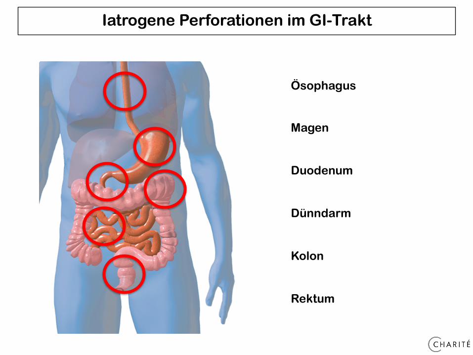
Iatrogene Perforationen im GI-Trakt
Ösophagus Magen Duodenum Dünndarm Kolon Rektum
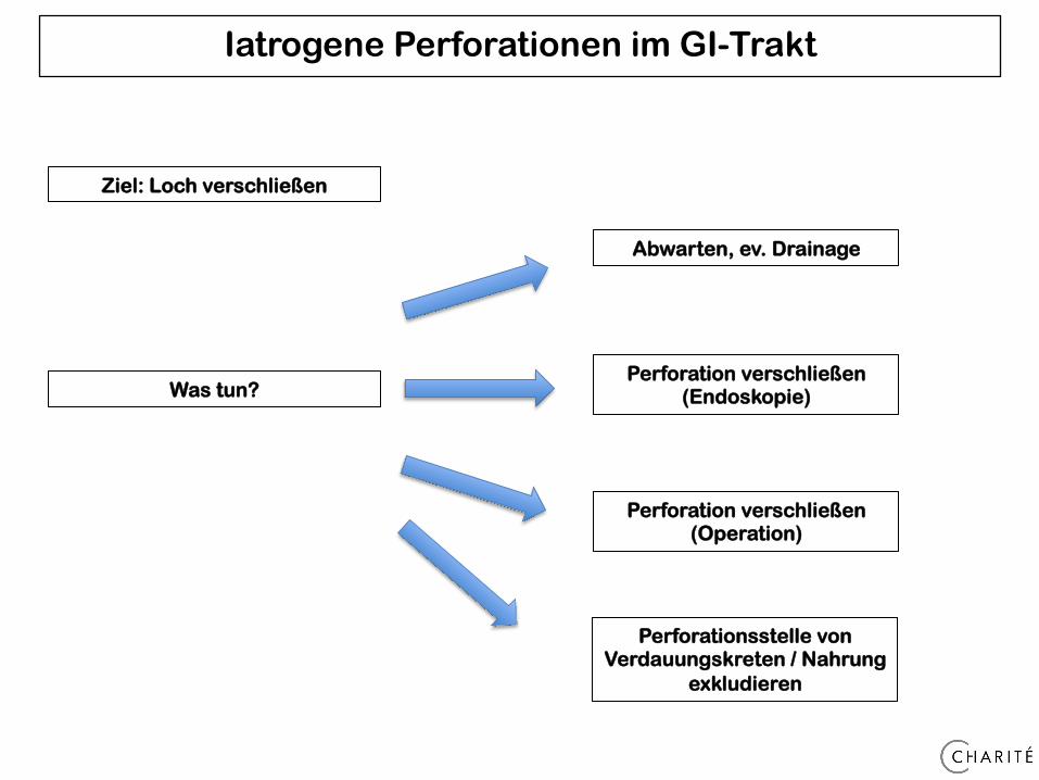
Iatrogene Perforationen im GI-Trakt
Abwarten, ev. Drainage
Perforation verschließen (Operation)
Perforation verschließen (Endoskopie) Was tun?
Ziel: Loch verschließen
Perforationsstelle von Verdauungskreten / Nahrung
exkludieren
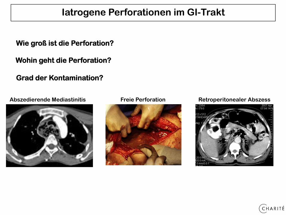
Iatrogene Perforationen im GI-Trakt
Wohin geht die Perforation?
Wie groß ist die Perforation?
Grad der Kontamination?
Abszedierende Mediastinitis Freie Perforation Retroperitonealer Abszess
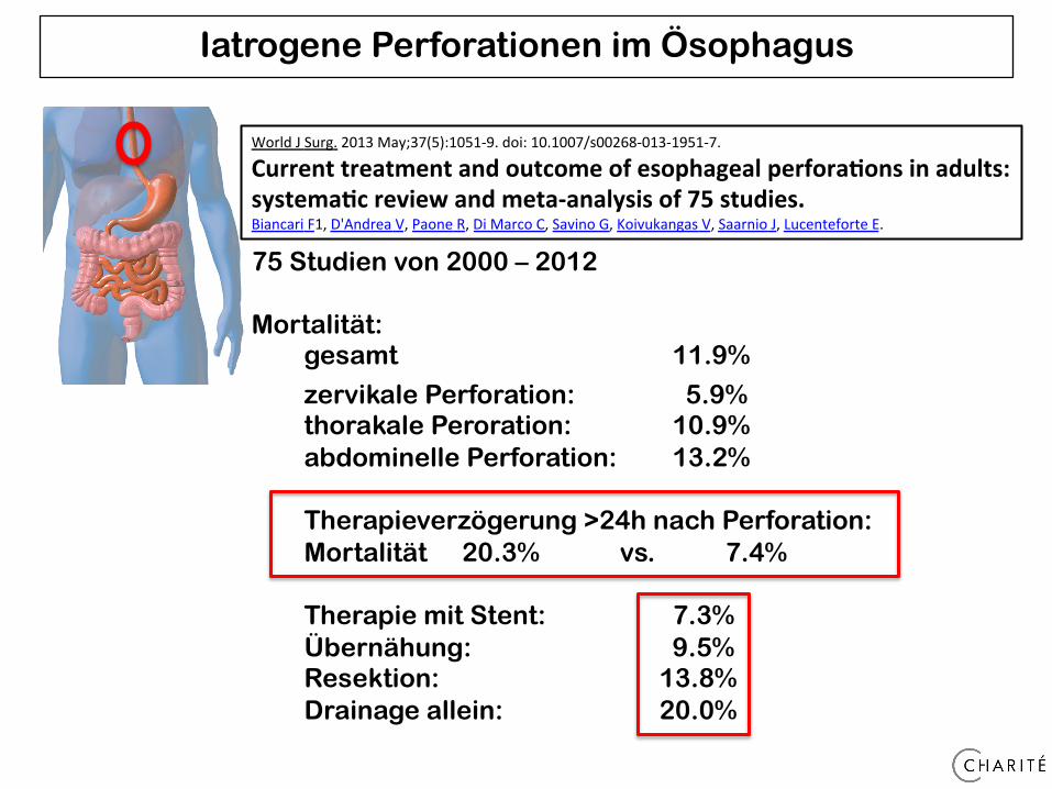
World J Surg. 2013 May;37(5):1051-‐9. doi: 10.1007/s00268-‐013-‐1951-‐7.
Current treatment and outcome of esophageal perfora3ons in adults: systema3c review and meta-‐analysis of 75 studies. Biancari F1, D'Andrea V, Paone R, Di Marco C, Savino G, Koivukangas V, Saarnio J, Lucenteforte E.
75 Studien von 2000 – 2012 Mortalität:
gesamt 11.9%
zervikale Perforation: 5.9% thorakale Peroration: 10.9% abdominelle Perforation: 13.2%
Therapieverzögerung >24h nach Perforation: Mortalität 20.3% vs. 7.4%
Therapie mit Stent: 7.3% Übernähung: 9.5%
Resektion: 13.8% Drainage allein: 20.0%
Iatrogene Perforationen im Ösophagus
Iatrogene Perforationen im Ösophagus
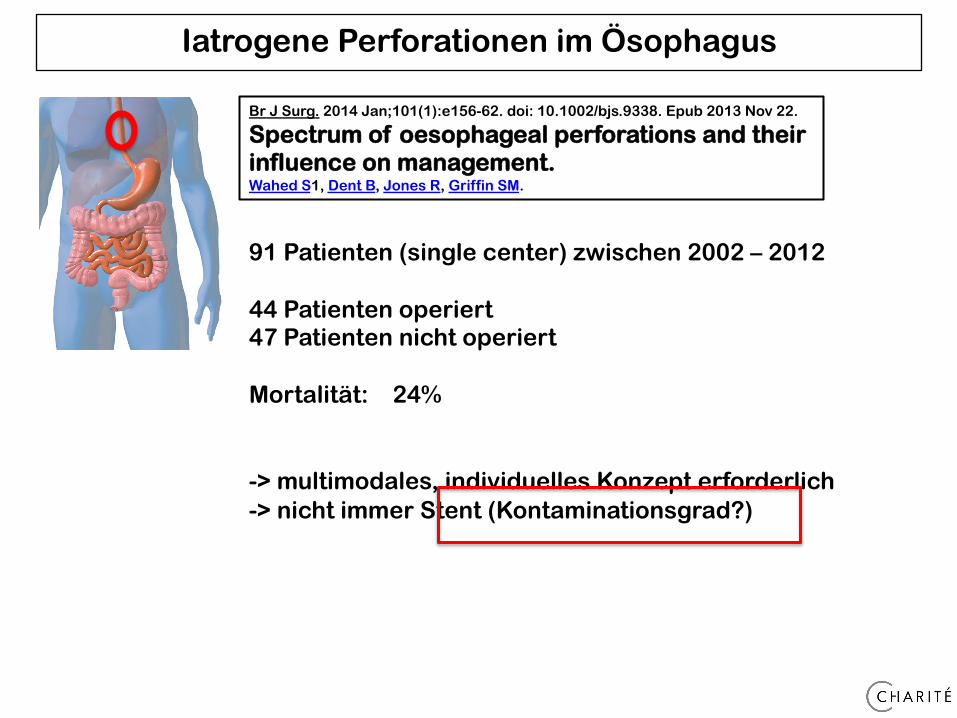
91 Patienten (single center) zwischen 2002 – 2012 44 Patienten operiert 47 Patienten nicht operiert Mortalität: 24% -> multimodales, individuelles Konzept erforderlich -> nicht immer Stent (Kontaminationsgrad?)
Br J Surg. 2014 Jan;101(1):e156-62. doi: 10.1002/bjs.9338. Epub 2013 Nov 22.
Spectrum of oesophageal perforations and their influence on management. Wahed S1, Dent B, Jones R, Griffin SM.
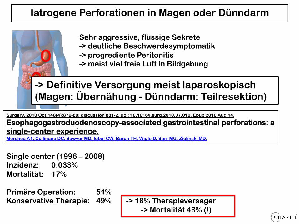
Iatrogene Perforationen in Magen oder Dünndarm
Sehr aggressive, flüssige Sekrete -> deutliche Beschwerdesymptomatik -> progrediente Peritonitis -> meist viel freie Luft in Bildgebung
Surgery. 2010 Oct;148(4):876-80; discussion 881-2. doi: 10.1016/j.surg.2010.07.010. Epub 2010 Aug 14.
Esophagogastroduodenoscopy-associated gastrointestinal perforations: a single-center experience. Merchea A1, Cullinane DC, Sawyer MD, Iqbal CW, Baron TH, Wigle D, Sarr MG, Zielinski MD.
Single center (1996 – 2008) Inzidenz: 0.033% Mortalität: 17% Primäre Operation: 51% Konservative Therapie: 49% -> 18% Therapieversager
-> Mortalität 43% (!)
-> Definitive Versorgung meist laparoskopisch (Magen: Übernähung - Dünndarm: Teilresektion)
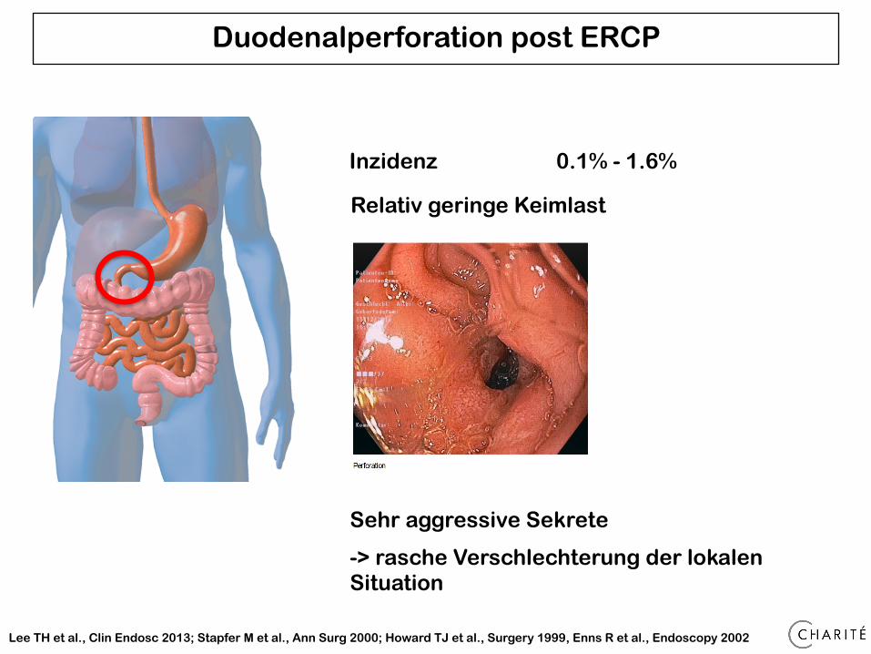
Inzidenz 0.1% - 1.6%
Lee TH et al., Clin Endosc 2013; Stapfer M et al., Ann Surg 2000; Howard TJ et al., Surgery 1999, Enns R et al., Endoscopy 2002
Duodenalperforation post ERCP
Relativ geringe Keimlast
Sehr aggressive Sekrete
-> rasche Verschlechterung der lokalen Situation
�������!�����
�����
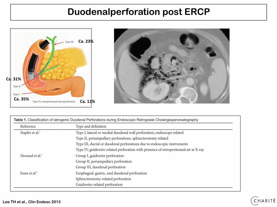
sing order of severity, with implications for management. Type I perforations are perforations of the lateral or medial wall of the duodenum; these perforations involve large rents, are re-mote from the ampulla, and are caused by the endoscope itself or by the stent. These perforations cause considerable spillage, either retroperitoneally or intraperitoneally, necessitating ag-gressive surgical intervention. Type II perforations are perfo-rations of the medial wall of the duodenum; these perforations are peri-Vaterian, are generally retroperitoneal, and occur dur�ing sphincterotomies. Type II perforations tend to be treated with conservative or minimally invasive therapies. Type III in�juries are distal bile duct injuries caused by instrumentation and/or stenting in the proximity of an obstruction. These perfo�rations are small and amenable to conservative management.
Type IV perforations are tiny retroperitoneal perforations cau�sed by the use of compressed air during endoscopy and can be managed conservatively. Howard et al.2 classified perforations into three types according to the mechanism of injury. Group I refers to guidewire-induced perforations; group II, periam-pullary perforations; and group III, duodenal perforations. Group III perforations require immediate surgery. Enns et al.3 suggested classification into three categories. Esophageal, gas�tric, and duodenal perforations require surgical management. Sphincterotomy-related perforation or guidewire-related per�foration usually requires conservative management.
CLINICAL FEATURES
Known risk factors for ERCP-related duodenal perforation include old age, suspected dysfunction of the sphincter of Oddi, dilated bile duct, papillary stenosis, Billroth-II recon-struction, precut sphincterotomy, and long procedure dura-tion.7-9 The classic presentation of duodenal perforation, with severe epigastric pain, vomiting, and epigastric tenderness progressing to generalized board-like rigidity, is seen only in a minority of cases. The symptoms and signs of ERCP-related perforations are often mild when this complication is recog-nized early.1,10-12 Therefore, the initial clinical presentation of patients with perforation is nonspecific. Duodenal perforation secondary to placement of a biliary endoprosthesis should be considered in all patients presenting with abdominal pain after such a placement. If a perforation is not recognized or suspec�ted during ERCP, early diagnosis is difficult. Moreover, diagno�sis is likely to be delayed if patients have concurrent elevated amylase level and the pain is attributed to post-ERCP pancrea�titis. Clinical suspicion and diagnosis of a procedure-related perforation can be facilitated greatly by clinical findings and particularly by radiographic imaging with contrast studies, computed tomography (CT), and even magnetic resonance imaging. A multi-slice CT scan can provide an exact diagno-
���������� �%��!���������!���������"������� ������!��� ��"�������� ��������!������������������������!������$
Reference Type and definitionStapfer et al.1 Type I, lateral or medial duodenal wall perforation, endoscope related
Type II, periampullary perforations, sphincterotomy relatedType III, ductal or duodenal perforations due to endoscopic instrumentsType IV, guidewire-related perforation with presence of retroperitoneal air at X-ray
Howard et al.2 Group I, guidewire perforationGroup II, periampullary perforationGroup III, duodenal perforation
Enns et al.3 Esophageal, gastric, and duodenal perforationSphincterotomy-related perforationGuidewire-related perforation
� ��������� �����!$�� ������!���������"��������������!��� ��!$���������� ���������!��� ��!����������������"�������#�����������!�����!$������� �����!���!��$�����!����������"����$��������!��� �� !$���������"�!�������"��������������!��� ��"��!������ �������� !�"���! � "���� ����"���#�����!$���������!������!�����������������!������" ����$������� ��������"��������� ���$�
Type III
Type II
Type I
Type IV, retroperitoneal microperforation
�������!�����
�����
sing order of severity, with implications for management. Type I perforations are perforations of the lateral or medial wall of the duodenum; these perforations involve large rents, are re-mote from the ampulla, and are caused by the endoscope itself or by the stent. These perforations cause considerable spillage, either retroperitoneally or intraperitoneally, necessitating ag-gressive surgical intervention. Type II perforations are perfo-rations of the medial wall of the duodenum; these perforations are peri-Vaterian, are generally retroperitoneal, and occur dur�ing sphincterotomies. Type II perforations tend to be treated with conservative or minimally invasive therapies. Type III in�juries are distal bile duct injuries caused by instrumentation and/or stenting in the proximity of an obstruction. These perfo�rations are small and amenable to conservative management.
Type IV perforations are tiny retroperitoneal perforations cau�sed by the use of compressed air during endoscopy and can be managed conservatively. Howard et al.2 classified perforations into three types according to the mechanism of injury. Group I refers to guidewire-induced perforations; group II, periam-pullary perforations; and group III, duodenal perforations. Group III perforations require immediate surgery. Enns et al.3 suggested classification into three categories. Esophageal, gas�tric, and duodenal perforations require surgical management. Sphincterotomy-related perforation or guidewire-related per�foration usually requires conservative management.
CLINICAL FEATURES
Known risk factors for ERCP-related duodenal perforation include old age, suspected dysfunction of the sphincter of Oddi, dilated bile duct, papillary stenosis, Billroth-II recon-struction, precut sphincterotomy, and long procedure dura-tion.7-9 The classic presentation of duodenal perforation, with severe epigastric pain, vomiting, and epigastric tenderness progressing to generalized board-like rigidity, is seen only in a minority of cases. The symptoms and signs of ERCP-related perforations are often mild when this complication is recog-nized early.1,10-12 Therefore, the initial clinical presentation of patients with perforation is nonspecific. Duodenal perforation secondary to placement of a biliary endoprosthesis should be considered in all patients presenting with abdominal pain after such a placement. If a perforation is not recognized or suspec�ted during ERCP, early diagnosis is difficult. Moreover, diagno�sis is likely to be delayed if patients have concurrent elevated amylase level and the pain is attributed to post-ERCP pancrea�titis. Clinical suspicion and diagnosis of a procedure-related perforation can be facilitated greatly by clinical findings and particularly by radiographic imaging with contrast studies, computed tomography (CT), and even magnetic resonance imaging. A multi-slice CT scan can provide an exact diagno-
���������� �%��!���������!���������"������� ������!��� ��"�������� ��������!������������������������!������$
Reference Type and definitionStapfer et al.1 Type I, lateral or medial duodenal wall perforation, endoscope related
Type II, periampullary perforations, sphincterotomy relatedType III, ductal or duodenal perforations due to endoscopic instrumentsType IV, guidewire-related perforation with presence of retroperitoneal air at X-ray
Howard et al.2 Group I, guidewire perforationGroup II, periampullary perforationGroup III, duodenal perforation
Enns et al.3 Esophageal, gastric, and duodenal perforationSphincterotomy-related perforationGuidewire-related perforation
� ��������� �����!$�� ������!���������"��������������!��� ��!$���������� ���������!��� ��!����������������"�������#�����������!�����!$������� �����!���!��$�����!����������"����$��������!��� �� !$���������"�!�������"��������������!��� ��"��!������ �������� !�"���! � "���� ����"���#�����!$���������!������!�����������������!������" ����$������� ��������"��������� ���$�
Type III
Type II
Type I
Type IV, retroperitoneal microperforation
Lee TH et al., Clin Endosc 2013
Duodenalperforation post ERCP
Ca. 23%
Ca. 31%
Ca. 35% Ca. 11%
Duodenalperforation post ERCP
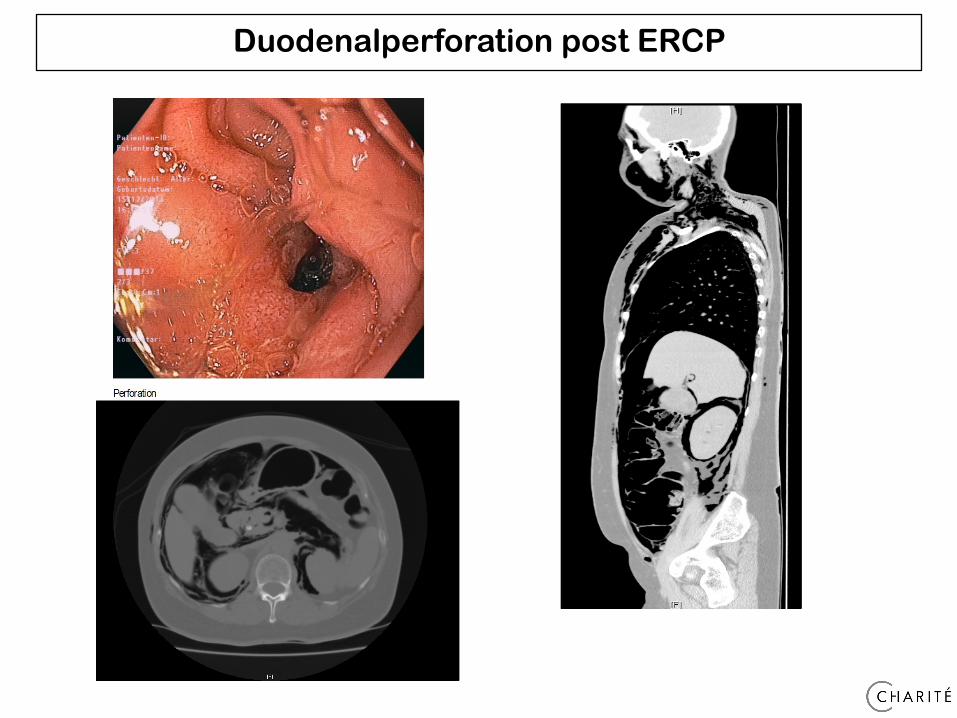
Duodenalperforation post ERCP
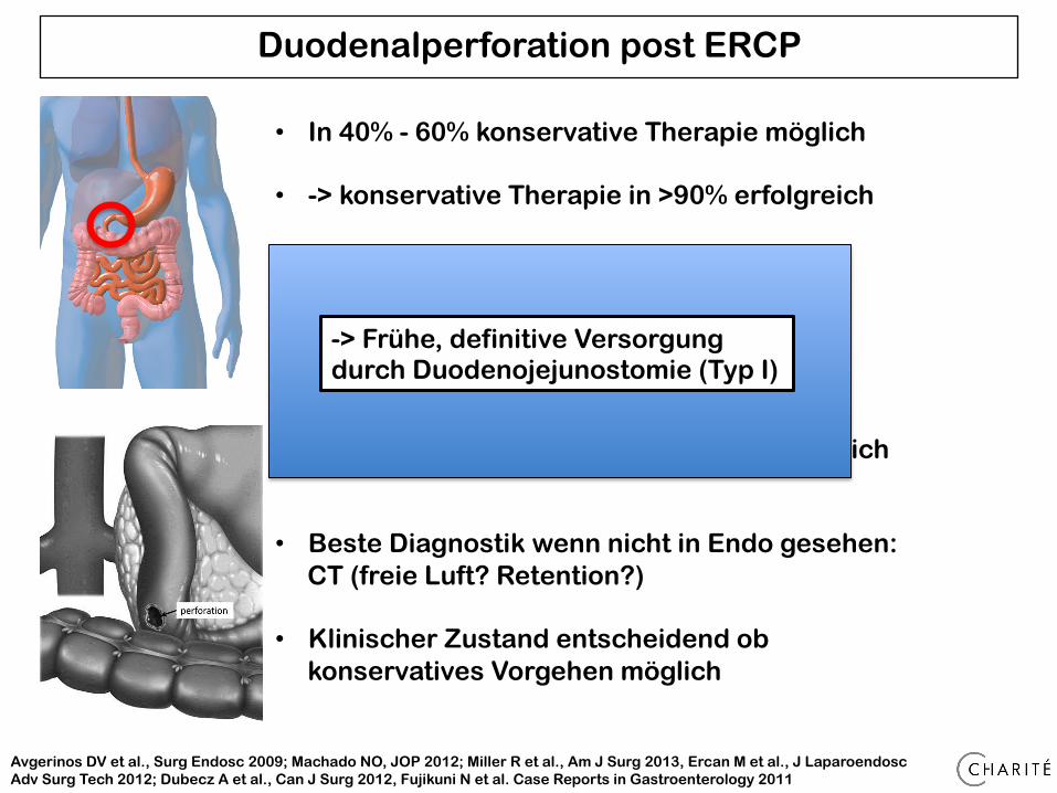
Avgerinos DV et al., Surg Endosc 2009; Machado NO, JOP 2012; Miller R et al., Am J Surg 2013, Ercan M et al., J Laparoendosc Adv Surg Tech 2012; Dubecz A et al., Can J Surg 2012, Fujikuni N et al. Case Reports in Gastroenterology 2011
• In 40% - 60% konservative Therapie möglich
• -> konservative Therapie in >90% erfolgreich
• ABER: Mortalität bis zu 37.5% (!)
• - Spät entdeckte Typ I fatal
• - Typ II nur Drainage = nicht erfolgreich
• - Typ III und Typ IV eher konservativ möglich
• Beste Diagnostik wenn nicht in Endo gesehen: CT (freie Luft? Retention?)
• Klinischer Zustand entscheidend ob konservatives Vorgehen möglich
-> Frühe, definitive Versorgung durch Duodenojejunostomie (Typ I)
!"#$%&$'%("#)*+$,)$*+-%./0012345.�456%7893%0/:0026;///<<25=.%
>?@-A#B$C%+,-A,$3%7$D$E@$*%</F%./00%
G%./00%H:%I"*J$*%K(F%L"#$-%9HHM%044.�/4<0%NNN:O"*J$*:D+E;D*J%
%
!
!
"##!
!H#;G1IG1<5&!4&0710.)&2!/6)&!9./!6(!)5&!.(.*!/62&!17!)5&!4.46**.!17!_.)&0!.)!)5&!41/)&0610!9.**!17!)5&!2312&(38;!
!!
Dow
nloa
ded
by:
87.1
62.8
4.82
- 1/
11/2
015
10:4
6:59
PM
Iatrogene Kolonperforation
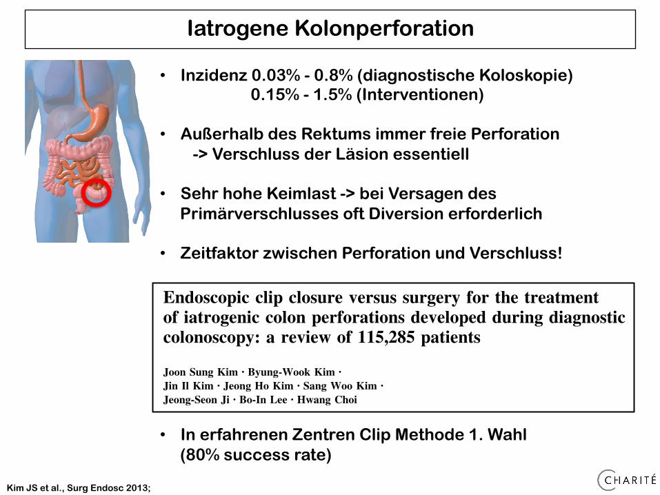
Endoscopic clip closure versus surgery for the treatmentof iatrogenic colon perforations developed during diagnosticcolonoscopy: a review of 115,285 patients
Joon Sung Kim • Byung-Wook Kim •
Jin Il Kim • Jeong Ho Kim • Sang Woo Kim •
Jeong-Seon Ji • Bo-In Lee • Hwang Choi
Received: 17 April 2012 / Accepted: 11 June 2012 / Published online: 7 July 2012! Springer Science+Business Media, LLC 2012
AbstractBackground Although the incidence of perforation after
endoscopic procedures of the colon is low, the rising
number of diagnostic colonoscopies could pose relevanthealth problems. Optimizing treatment may reduce the
probability of severe complications. This study aimed to
determine perforation frequency and the management ofperforations that occurred during diagnostic colonoscopy.
Methods A retrospective review of patient records was
performed for all patients with iatrogenic colonic perfora-tions after sigmoidoscopy/colonoscopy from 2000 to 2011
in three institutions of The Catholic University of Korea.
The patients’ demographic data, endoscopic procedureinformation, perforation location, therapy, and outcomes
along with different therapeutic strategies were recorded.
Results In the 12-year period, a total of 115,285 diag-nostic sigmoidoscopic/colonoscopic procedures were per-
formed. A total of 27 perforations occurred. Sixteen
patients underwent endoscopic clipping, of which threepatients failed and were referred for surgery. Fourteen
patients in total underwent surgery for perforation. Endo-scopic clip closure was successful in 81 % of the patients.
No perforation-related major morbidity or mortalityoccurred.
Conclusion Endoscopic repair using clips can be effec-
tive for the treatment of colon perforations that occurduring diagnostic colonoscopy.
Keywords Colonoscopy ! Iatrogenic ! Perforation ! Clip
Iatrogenic perforation resulting from colonoscopic and
sigmoidoscopic procedures is a rare but serious complica-
tion with high rates of morbidity and mortality. The fre-quency of perforations from colonoscopy is estimated to be
0.03–0.8 % for diagnostic colonoscopy and 0.15–3 % for
therapeutic colonoscopy [1]. With an increasing number ofcolonoscopies being performed for screening purposes, this
small possibility still may cause a high number of clinical
problems. The optimal treatment for perforations is con-troversial because no randomized trial has ever been con-
ducted. Recent studies have acquired evidence for
endoscopic clip closure [2–6]. However, most of thesestudies included perforations mainly from therapeutic
procedures and much less from diagnostic procedures.Furthermore, the efficacy and complications of endoscopic
clip closure compared to surgery has not been fully
elucidated.Perforation mechanism, size, and location are different
between diagnostic colonoscopy-associated perforations
and therapeutic colonoscopy-associated perforations [7].The proper instruments and personnel to perform endo-
scopic closure of therapeutic colonoscopy-associated per-
forations may be more readily available than for diagnosticperforations. The prompt and effective choice of a thera-
peutic closure method may prevent both unnecessary and
more invasive surgery, including colon resection with
J. S. Kim ! B.-W. Kim ! J. I. Kim ! J. H. Kim !S. W. Kim ! J.-S. Ji ! B.-I. Lee ! H. ChoiDivision of Gastroenterology, Department of Internal Medicine,College of Medicine, The Catholic University of Korea, Seoul,Republic of Korea
B.-W. Kim (&)Division of Gastroenterology, Department of Internal Medicine,Incheon St. Mary’s Hospital, The Catholic University of Korea,56, Dongsu-Ro, Bupyeong-gu, Incheon 403-720,Republic of Koreae-mail: [email protected]
123
Surg Endosc (2013) 27:501–504
DOI 10.1007/s00464-012-2465-3
and Other Interventional Techniques
• In erfahrenen Zentren Clip Methode 1. Wahl
(80% success rate)
Kim JS et al., Surg Endosc 2013;
• Inzidenz 0.03% - 0.8% (diagnostische Koloskopie) 0.15% - 1.5% (Interventionen)
• Außerhalb des Rektums immer freie Perforation -> Verschluss der Läsion essentiell
• Sehr hohe Keimlast -> bei Versagen des
Primärverschlusses oft Diversion erforderlich
• Zeitfaktor zwischen Perforation und Verschluss!
������#"%��%�&)�������� ��
�&#&%&)�&'"����%� �$�%*�&����(�&(�*"&%
MECHANISM OF PERFORATION
Several mechanisms may be involved in colonoscopy-asso�ciated colonic perforation, which include blunt trauma on the colonic wall, unintentional endoscopic resection, and excessive thermal injury. In general, blunt trauma is the main cause of diagnostic colonoscopy-associated perforation. Perforations from blunt trauma are usually large in size. They occur commo�nly in rectosigmoid area because they develop when colonosco�pe is pushed without resolution of looping at the rectosigmoid colon or when colonoscope is retroflexed immoderately.5
Unintentional endoscopic resection and excessive thermal injury are related to perforations during therapeutic colonos-copy such as endoscopic mucosal resection or ESD. Perfora-tions resulted from unintentional endoscopic resection are generally small and more common in the right colon.5 Ther-mal injury-related perforations, in general, are also small.5 Thermal injury-related perforations may not be detected often during colonoscopic procedure because only excessive trans-mural burn is evident without overt perforation right after endoscopic mucosal resection. Therefore, thermal injury-rela�ted perforations are usually diagnosed after the completion of colonoscopic procedure.
DIAGNOSIS OF PERFORATION
Colonoscopy-associated perforations may be classified into an endoscopically proven perforation and a radiologically pro�ven perforation based on the diagnostic process.16 An endosco�pically proven perforation refers to colonic mural defect detec�ted during colonoscopy procedure (Fig. 1). It may be accom�panied by observable intraabdominal organ or fat tissue through the mural defect if the perforation is large enough. A target sign, white center (muscularis propria and/or serosa)
with surrounding blue area (indigo carmine stained submuco�sa), at the postresection colon ulcer site or at the resection side of resected specimen, may be helpful in determining the pro�bability of small perforation.17
A radiologically proven perforation is defined as a pneumo�peritoneum or a pneumoretroperitoneum shown on a simple abdominal X-ray or as extraluminal air density or abscess at the site of the therapeutic procedure (Fig. 2). Endoscopically proven perforations may be accompanied by radiological ev-idence of perforation. However, some radiologically proven perforations do not show endoscopic evidence of perforation. Therefore, they may be diagnosed only after the completion of colonoscopy procedure.
MANAGEMENT OF PERFORATION
Surgical managementSurgery has been the mainstay of management of colonic
perforation. Recently, endoscopic clipping has been introdu�ced and conservative management has been feasible in many perforation cases. However, surgery is still indicated in cases of large perforations, generalized peritonitis, aggravating per�itonitis, ongoing sepsis, and concomitant colorectal pathology such as large advanced neoplasm which is difficult to resect by endoscopic techniques.5 One study showed diagnostic colo-noscopy-associated perforation or large perforation, leuko-cytosis over 10,000/mm3, fever �37˚C, severe abdominal pain, and large amount of free air in peritoneal cavity �3 cm might be risk factors for surgery within 24 hours after colo-noscopic clip closure trial.18 Therefore, patients with these fac�tors should be observed closely after the initiation of medical management by clipping and emergency surgery should be considered when patients show clinical deterioration such as generalized peritonitis.
�������%�&)�&'"��##/�'(&,�%�'�(�&(�*"&%�������"� %&)*"���&#&%&)�&'/��))&�"�*���'�(�&(�*"&%���!��'�(�&(�*"&%�&��+((����+("% ��.��))",��'+)!"% �&��*!���&#&%&)�&'����*�")�(�#�*",�#/�#�( ��������!�(�'�+*"���&#&%&)�&'/��))&�"�*���'�(�&(�*"&%���!��'�(�&(�*"&%���,�#&'����+("% ��%��&)�&'"��)+�$+�&)�#��"))��*"&%�������&���&#&%"�����%&$����*�")�(�#�*",�#/�)$�##��%��*!��)+((&+%�"% ��(���)!&-)�����+#��(�
� �
�%������
�����
Endoscopic managementEndoscopic management of colonic perforation has pro-
gressed significantly since the first report of clip application by Yoshikane et al.19 Through the scope clips have been used in clinical practice for decades of years with satisfactory success rate for the management of colonoscopy-associated perfora-tion (Fig. 3).16 They are especially useful in the closure of small perforations such as those developing after endoscopic mu-cosal resection or ESD of colorectal tumors. Large perforations may not be closed by through the scope clips only. Because dia�gnostic colonoscopy-associated perforation is usually larger than the therapeutic colonoscopy-associated perforation, over�all clip success rate in the management of colon perforation appears to be higher in the therapeutic colonoscopy-associ-
ated perforation (Tables 1, 2).18,20,21 In case of closure failure with clipping, combination of clips with detachable snare (en-doloop) can be useful for endoscopic closure.22 Recently, over the scope clips were reported to be useful for the management of large gastrointestinal perforations.23 A case series which in�vestigated the usefulness of over-the-scope clip in the manage�ment of nine colon perforation cases showed successful depl�oyment and closure in all of the nine perforations up to 30 mm in size. Out of those nine cases, six cases improved conserva�tively. The other three cases required laparoscopy for the in-spection of possible peritonitis, but none of the three cases sho�wed leakage or peritonitis. Therefore, the authors concluded that the over the scope clip may be useful for the endoscopic management of colon perforation up to 30 mm in size.24
������� ������������%����#����������!���������������� ������ "��"�� ����� ��!��������"����� ��$ ������&��!���#�������������� ���������%����#����������!��������������$�"������%�!���������!���!���!���������!��������� ��$ ������������"�!�������"������!���"���$��������� �!������ ��������������������!�����!�!�����"���������
�
���������� �����������������������"���������!���#���������!������� �������"�� ����� ��!����������������%���������#������ �$����������������!����������!����$� ���� ���������!��%�
�
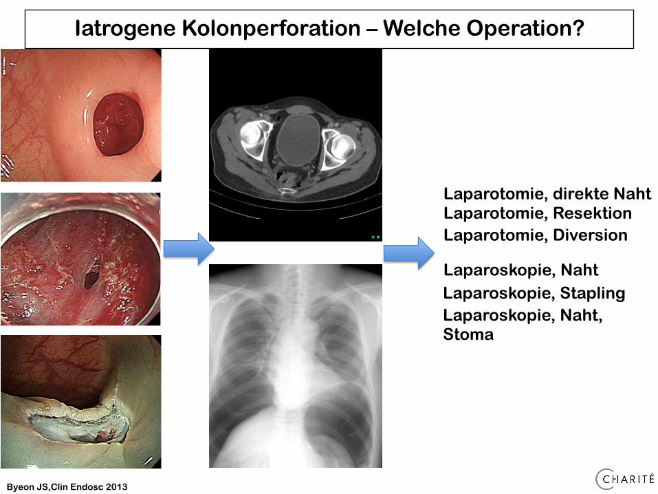
Byeon JS,Clin Endosc 2013
Iatrogene Kolonperforation – Welche Operation?
�%������
�����
Endoscopic managementEndoscopic management of colonic perforation has pro-
gressed significantly since the first report of clip application by Yoshikane et al.19 Through the scope clips have been used in clinical practice for decades of years with satisfactory success rate for the management of colonoscopy-associated perfora-tion (Fig. 3).16 They are especially useful in the closure of small perforations such as those developing after endoscopic mu-cosal resection or ESD of colorectal tumors. Large perforations may not be closed by through the scope clips only. Because dia�gnostic colonoscopy-associated perforation is usually larger than the therapeutic colonoscopy-associated perforation, over�all clip success rate in the management of colon perforation appears to be higher in the therapeutic colonoscopy-associ-
ated perforation (Tables 1, 2).18,20,21 In case of closure failure with clipping, combination of clips with detachable snare (en-doloop) can be useful for endoscopic closure.22 Recently, over the scope clips were reported to be useful for the management of large gastrointestinal perforations.23 A case series which in�vestigated the usefulness of over-the-scope clip in the manage�ment of nine colon perforation cases showed successful depl�oyment and closure in all of the nine perforations up to 30 mm in size. Out of those nine cases, six cases improved conserva�tively. The other three cases required laparoscopy for the in-spection of possible peritonitis, but none of the three cases sho�wed leakage or peritonitis. Therefore, the authors concluded that the over the scope clip may be useful for the endoscopic management of colon perforation up to 30 mm in size.24
������� ������������%����#����������!���������������� ������ "��"�� ����� ��!��������"����� ��$ ������&��!���#�������������� ���������%����#����������!��������������$�"������%�!���������!���!���!���������!��������� ��$ ������������"�!�������"������!���"���$��������� �!������ ��������������������!�����!�!�����"���������
�
���������� �����������������������"���������!���#���������!������� �������"�� ����� ��!����������������%���������#������ �$����������������!����������!����$� ���� ���������!��%�
�
Laparotomie, direkte Naht Laparotomie, Resektion Laparotomie, Diversion
Laparoskopie, Naht Laparoskopie, Stapling Laparoskopie, Naht, Stoma
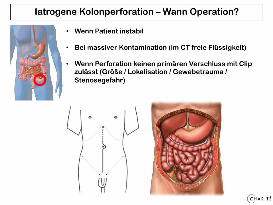
Iatrogene Kolonperforation – Wann Operation?
• Wenn Patient instabil
• Bei massiver Kontamination (im CT freie Flüssigkeit) • Wenn Perforation keinen primären Verschluss mit Clip
zulässt (Größe / Lokalisation / Gewebetrauma / Stenosegefahr)
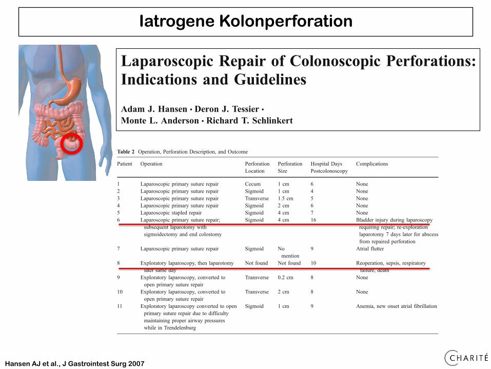
Iatrogene Kolonperforation
positioning. The third case was converted to appropriatelymanage a large segment of small bowel that appearedhyperemic and inflamed from fecal soilage. Of this group,the mean perforation size was 1.1 cm (range 0.2–2 cm) andthe mean hospital stay was 8.3 days (range 8–9 days).
The final case was a patient with severe obstructivepulmonary disease admitted to the hospital 10 days pre-viously for hematochezia. Following therapeutic colonos-copy, the diagnosis of colonic perforation was made, basedupon radiographic demonstration of free intra-abdominalair. Exploratory laparoscopy performed by a highly expe-rienced colorectal surgeon was unrevealing. Postoperative-ly, the patient developed sepsis and was returned to theoperating room later the same day for open laparotomy.Again, no perforation was found and no specific repair wasperformed. The patient continued to decline postoperativelyand life support was eventually withdrawn on postoperativeday 10 in accordance with the family’s wishes. The diag-nosis of colonic perforation remains in doubt.
Discussion
One of the most devastating complications of colonoscopyis perforation of the colon, which may result in significantmorbidity and even mortality. Suggested treatments ofcolonic perforations range from observation to segmentalresection and diversion of the fecal stream to the exterior.
Most surgeons prefer exploration, while some authors13,14
prefer nonoperative management of select cases. Visualiza-tion of the peritoneal cavity by the endoscopist and thedevelopment of signs of peritoneal irritation14 are absoluteindications for surgery. The timely application of exploratorylaparoscopy may prevent the development of inflammationand further injury that would make more invasive measures,such as open laparotomy or colonic diversion, necessary.
A total of 21 cases of laparoscopically attempted repair, notincluding our previous data, were found in the literature.Detailed data are summarized in Table 3. In 17 patients(81%), the repair was accomplished laparoscopically withoutconversion to open laparotomy. The largest series, publishedby Wullstein et al.,11 included seven patients in whomexploratory laparoscopy was performed. Four of their sevencases were performed completely laparoscopically. Twoothers had extensive injuries, necessitating open conversion.Their first patient had an intraoperative technical complica-tion that also led to open conversion after laparoscopicrepair. Allam et al.4 described a single case in which theyused a laparoscopically assisted approach to perform aminilaparotomy for repair of a perforation.
Perforation size was described in 17 of the reportedcases. Only three patients in whom perforation size wasgreater than 2.5 cm underwent full laparoscopic repair(largest 5 cm). Thirteen (62%) perforations were in thesigmoid colon, two (10%) in the rectum, three (14%) in thececum, and three (14%) in the transverse colon or left
Table 2 Operation, Perforation Description, and Outcome
Patient Operation PerforationLocation
PerforationSize
Hospital DaysPostcolonoscopy
Complications
1 Laparoscopic primary suture repair Cecum 1 cm 6 None2 Laparoscopic primary suture repair Sigmoid 1 cm 4 None3 Laparoscopic primary suture repair Transverse 1.5 cm 5 None4 Laparoscopic primary suture repair Sigmoid 2 cm 6 None5 Laparoscopic stapled repair Sigmoid 4 cm 7 None6 Laparoscopic primary suture repair;
subsequent laparotomy withsigmoidectomy and end colostomy
Sigmoid 4 cm 16 Bladder injury during laparoscopyrequiring repair; re-explorationlaparotomy 7 days later for abscessfrom repaired perforation
7 Laparoscopic primary suture repair Sigmoid Nomention
9 Atrial flutter
8 Exploratory laparoscopy, then laparotomylater same day
Not found Not found 10 Reoperation, sepsis, respiratoryfailure, death
9 Exploratory laparoscopy, converted toopen primary suture repair
Transverse 0.2 cm 8 None
10 Exploratory laparoscopy, converted toopen primary suture repair
Transverse 2 cm 8 None
11 Exploratory laparoscopy converted to openprimary suture repair due to difficultymaintaining proper airway pressureswhile in Trendelenburg
Sigmoid 1 cm 9 Anemia, new onset atrial fibrillation
J Gastrointest Surg (2007) 11:655–659 657657
Laparoscopic Repair of Colonoscopic Perforations:Indications and Guidelines
Adam J. Hansen & Deron J. Tessier &
Monte L. Anderson & Richard T. Schlinkert
Published online: 27 March 2007# 2007 The Society for Surgery of the Alimentary Tract
Abstract Iatrogenic colonic perforation is one of the most serious potential complications of colonoscopy. Standardmanagement is surgical repair. No prospective data exist to clearly define the indications for laparoscopic repair. We reportthe largest case series to date of laparoscopic repair of colonoscopic perforations. A retrospective review was performed ofall patients undergoing either exploratory laparoscopy with conversion to open repair, or laparoscopic repair ofcolonoscopic perforation. Exploratory laparoscopy for the attempted repair of colonoscopic perforations was performedin 11 patients at our institution. The mean colonic perforation size was 2.7 cm. Three cases were converted immediately toopen laparotomy. A fourth patient that underwent primary laparoscopic repair of a 4-cm tear developed a leak at the repairsite, necessitating reoperation. A fifth patient in whom exploratory laparoscopy was unrevealing underwent separatelaparotomy for continued sepsis. Six patients underwent successful laparoscopic repair. Most perforations secondary tocolonoscopy warrant rapid exploratory laparoscopy. Extensive inflammation or fecal soilage may require colonic diversion.Inability to laparoscopically localize the area of perforation or doubt regarding the security of the repair should promptconversion to laparotomy. Laparoscopic repair of colonic perforations in experienced hands is a viable alternative to theopen approach.
Keywords Colonoscopy . Colonic perforation .
Laparoscopy . Laparoscopic surgery
Introduction
Colonoscopy is an effective tool for both diagnosis and ther-apy of colonic lesions but carries a small risk of compli-cations, the most serious and feared of which is colonicperforation. Perforation can rapidly progress to peritonitis andsepsis, carrying significant morbidity and mortality.
Perforation of the colon during colonoscopy may occurdue to mechanical or thermal injury. Excessive mechanicalpressure may be exerted along the shaft of the colonoscopeduring advancement or rotation, or at the instrument tip. Inaddition, pneumatic pressure from excessive insufflation canlead to tearing and perforation. Thermal injury occurs during“hot” biopsy or polypectomy. In contrast to perforationsfrom mechanical forces, these injuries are often smaller withless peritoneal contamination.1,2
Controversy exists over the ideal management of colo-noscopic perforation. Treatment strategies range from non-operative management to open colonic diversion. In orderto avoid further patient trauma, minimally invasive meth-ods, such as laparoscopic repair, have been developed.However, colonic perforation secondary to colonoscopy isso infrequent that no single institution has been able togather sufficient data to definitively state the circumstancesunder which minimally invasive treatment is appropriate. Noprospective studies exist, and retrospective studies rangefrom single-case reports to a handful of patients treated withattempted laparoscopic repair.2–12 We initially reported our
J Gastrointest Surg (2007) 11:655–659DOI 10.1007/s11605-007-0137-8
A. J. Hansen :D. J. Tessier :R. T. Schlinkert (*)Department of Surgery, Mayo Clinic Scottsdale,13400 E. Shea Boulevard,Scottsdale, AZ 85259, USAe-mail: [email protected]
M. L. AndersonDivision of Gastroenterology, Mayo Clinic Scottsdale,Scottsdale, AZ, USA
Hansen AJ et al., J Gastrointest Surg 2007
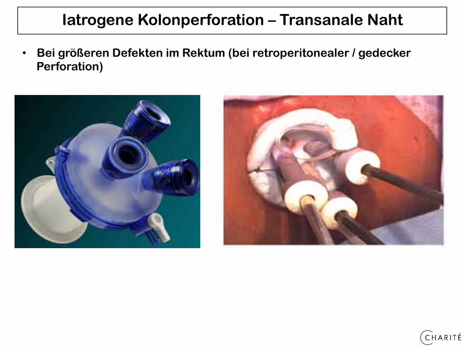
Iatrogene Kolonperforation – Transanale Naht
• Bei größeren Defekten im Rektum (bei retroperitonealer / gedecker Perforation)
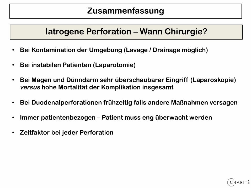
Zusammenfassung
Iatrogene Perforation – Wann Chirurgie?
• Bei Kontamination der Umgebung (Lavage / Drainage möglich)
• Bei instabilen Patienten (Laparotomie)
• Bei Magen und Dünndarm sehr überschaubarer Eingriff (Laparoskopie) versus hohe Mortalität der Komplikation insgesamt
• Bei Duodenalperforationen frühzeitig falls andere Maßnahmen versagen
• Immer patientenbezogen – Patient muss eng überwacht werden
• Zeitfaktor bei jeder Perforation