Analysis of the diversity of water kefir microbiota by culture
138
TECHNISCHE UNIVERSITÄT MÜNCHEN Lehrstuhl für Technische Mikrobiologie Analysis of the diversity of water kefir microbiota by culture-dependent and -independent approaches Anna Jana Gulitz Vollständiger Abdruck der von der Fakultät Wissenschaftszentrum Weihenstephan für Ernährung, Landnutzung und Umwelt der Technischen Universität München zur Erlangung des akademischen Grades eines Doktors der Naturwissenschaften genehmigten Dissertation. Vorsitzender: Univ-Prof. Dr. S. Scherer Prüfer der Dissertation: 1. Univ.-Prof. Dr. R. F. Vogel 2. Univ.-Prof Dr. W. Liebl Die Dissertation wurde am 27.06.2013 bei der Technischen Universität München eingereicht und für die Fakultät Wissenschaftszentrum Weihenstephan für Ernährung, Landnutzung und Umwelt der Technischen Universität München am 23.10.2013 angenommen.
Transcript of Analysis of the diversity of water kefir microbiota by culture
TECHNISCHE UNIVERSITÄT MÜNCHEN
Lehrstuhl für Technische Mikrobiologie
Analysis of the diversity of water kefir microbiota by culture-dependent
and -independent approaches
Anna Jana Gulitz
Vollständiger Abdruck der von der Fakultät Wissenschaftszentrum Weihenstephan
für Ernährung, Landnutzung und Umwelt der Technischen Universität München zur
Erlangung des akademischen Grades eines
Doktors der Naturwissenschaften
genehmigten Dissertation.
Vorsitzender: Univ-Prof. Dr. S. Scherer
Prüfer der Dissertation:
1. Univ.-Prof. Dr. R. F. Vogel
2. Univ.-Prof Dr. W. Liebl
Die Dissertation wurde am 27.06.2013 bei der Technischen Universität München
eingereicht und für die Fakultät Wissenschaftszentrum Weihenstephan für
Ernährung, Landnutzung und Umwelt der Technischen Universität München am
23.10.2013 angenommen.
Vorwort und Danksagung
Vorwort und Danksagung
Die vorliegende Arbeit wurde ermöglicht durch die Finanzierung aus Haushaltsmitteln
des BMWi’s, über die Arbeitsgemeinschaft industrieller Forschungsvereinigungen,
Otto von Guericke Stiftung und der Wissenschaftsförderung der Deutschen
Brauwirtschaft (Projekt: AiF Vorhaben Nr.: 16454N).
Ein ganz besonderes Dankeschön möchte ich an meinen Doktorvater Herrn Prof. Dr.
R. Vogel richten, der es mir ermöglicht hat an diesem sehr interessanten Thema zu
arbeiten. Für die stetige Bereitschaft Diskussionen zu führen, neue Anregungen zu
geben und das Vertrauen die Arbeit in großer Selbständigkeit durchführen zu können
möchte ich mich ganz besonders bedanken.
Ein weiteres Dankeschön gilt meinem Betreuer Dr. Matthias Ehrmann, der meine
Arbeit immer unterstützt hat und für jegliche Diskussionen bereit war.
Für die Übernahme des Prüfungsvorsitzes bedanke ich mich bei Herrn Prof. Dr.
Scherer, sowie bei Herrn Prof Dr. Liebel für die Begutachtung der Arbeit.
Meinen Studenten Andreas, Anna und Natalie danke ich für die Bearbeitung ihrer
Themen und die gute Zusammenarbeit.
Unserer Sekretärin Angela Seppeur möchte ich danken für die Unterstützung in allen
Dingen fern ab des Labors.
Des Weiteren danke ich Monika Hadek, Sabine Dummert und Maggie Schreiber für
ein immer offenes Ohr und Unterstützung beim Durchführen meiner praktischen
Arbeiten.
Dr. Mareike Wenning danke ich für das problemlose Durchführen von FTIR-
Messungen und die stete Bereitschaft über die Ergebnisse zu diskutieren.
Vorwort und Danksagung
Dr. Natuschka Lee gilt ein großer Dank für die etlichen Diskussionen und ihrer
Bereitschaft Lösungswege zu finden, außerdem möchte ich mich für die Möglichkeit
an ihrem FISH-Kurs teilzunehmen ganz herzlich bedanken.
Dem kompletten Büro Preissler mit Carola Kern, Julia Usbeck, Patrick Preissler und
Jasmin Stadie, sowie Juliane Schnabel gilt ein besonderer Dank vor allem für die
moralische Unterstützung während der kompletten Zeit und der Zusammenhalt
gerade in Zeiten des Zweifelns, insbesondere aber in der Zeit des
Zusammenschreibens.
Für ein tolles Arbeitsklima und Hilfsbereitschaft möchte ich mich bei allen
Arbeitskollegen des Lehrstuhls bedanken.
Für die bedingungslose Unterstützung meines kompletten Werdegangs danke ich
aus ganzem Herzen meinen Eltern. Sie haben mich nicht nur finanziell unterstützt,
sondern vor allem moralisch, sie haben immer an mich und meine Fähigkeiten
geglaubt, selbst wenn ich oft Zweifel daran hatte. Die Auszeiten innerhalb und mit der
Familie waren für mich wertvolle Stunden, durch die ich abschalten und neue Kraft
tanken konnte. Deshalb geht auch ein dickes Dankeschön an meine Geschwister
und deren Anhänge.
Contents II
LIST OF TABLES ...................................................................................................... VI
LIST OF FIGURES .................................................................................................. VIII
LIST OF ABBREVIATIONS ........................................................................................ X
1 INTRODUCTION ............................................................................................... 13
1.1 Water kefir ...................................................................................................... 13
1.1.1 Origin of water kefir and traditional preparation .......................................... 13
1.1.2 Microbiota and appearance of water kefir .................................................. 14
1.1.3 Composition of water kefir grains ............................................................... 16
1.1.4 Comparison to milk kefir ............................................................................. 16
1.2 Role of fermented food in human nutrition ..................................................... 18
1.2.1 Fermented beverages ................................................................................ 19
1.3 Application of culture-dependent and- independent analyses of microbiota .. 20
1.3.1 Advantages and disadvantages of culture-dependent analyses ................ 20
1.3.2 High through-put sequencing as a tool for characterizing microbiota in
food fermentations ..................................................................................... 21
1.4 Preservation of food, beverages and starter cultures used for production ..... 23
1.4.1 Biopreservation .......................................................................................... 24
1.4.2 Stress tolerance of microorganisms used as starter cultures regarding
freeze-drying technology............................................................................ 25
1.5 Bifidbacteriaceae in food ................................................................................ 27
1.6 Objectives of the work .................................................................................... 29
Contents III
2 MATERIAL AND METHODS ............................................................................. 30
2.1 Material .......................................................................................................... 30
2.1.1 Equipment .................................................................................................. 30
2.1.2 Chemicals .................................................................................................. 32
2.1.3 Equipment for water kefir preparation ........................................................ 34
2.1.4 Consumables ............................................................................................. 34
2.1.5 Molecular biological kits ............................................................................. 34
2.1.6 Bacterial and yeast strains ......................................................................... 35
2.2 Methods ......................................................................................................... 37
2.2.1 Microbial methods ...................................................................................... 37
2.2.2 Molecular biological methods ..................................................................... 40
2.2.3 Data processing and 16S rDNA sequence analysis ................................... 48
2.2.4 MALDI-TOF MS analysis ............................................................................ 50
2.2.5 Fluorescence in situ hybridization (FISH) analysis ..................................... 51
3 RESULTS .......................................................................................................... 52
3.1 Microbial diversity of water kefir grains analyzed with culture- dependent
procedures ..................................................................................................... 52
3.1.1 Quantification of bacteria and yeasts ......................................................... 52
3.1.2 Identification of microorganisms ................................................................. 53
3.1.3 Differences in the bacterial composition of water kefir microbiota .............. 55
3.1.4 Differences in the yeast composition of water kefir microbiota ................... 56
3.2 Bacterial diversity of water kefir grains analyzed with culture-independent
procedures ..................................................................................................... 57
Contents IV
3.2.1 Bacterial identification and distribution determined by high-throughput
sequence-based analysis .......................................................................... 57
3.2.2 ARDRA analysis ......................................................................................... 62
3.2.3 Cultivation and quantification of bifidobacteria ........................................... 63
3.2.4 Phylogeny of Bifidobacterium sp. ............................................................... 64
3.3 Influence of the composition of water kefir microbiota by changing growth
conditions ....................................................................................................... 64
3.3.1 Determination of the wet mass of water kefir grains ................................... 64
3.3.2 Detection of the viable cell count ................................................................ 66
3.3.3 Ribosomal intergenic spacer analysis (RISA) ............................................ 67
3.3.4 High-throughput sequencing of the water kefir samples grown under
different conditions ..................................................................................... 73
3.4 Fluorescence in situ hybridization (FISH) of water kefir grains and the
supernatant .................................................................................................... 79
3.5 New bifidobacteria sequences ....................................................................... 82
3.6 Freeze-drying of water kefir grains ................................................................. 82
3.6.1 Analysis of the revitalization of water kefir grains after freeze-drying ......... 82
3.6.2 Identification of bacteria and yeast species after freeze-drying and
recultivation of water kefir grains ............................................................... 86
Contents V
4 DISCUSSION .................................................................................................... 91
4.1 Microbial diversity of water kefir grains analyzed by culture-dependent
procedures ..................................................................................................... 92
4.2 Microbial diversity of water kefir grains analyzed by culture-independent
procedures ..................................................................................................... 96
4.2.1 Identification and characterization of bifidobacteria found in water kefir ..... 98
4.2.2 Characterization of the novel bifidobacteria species found in
water kefir ................................................................................................ 100
4.3 Influence of the composition of water kefir microbiota by changing growth
conditions ..................................................................................................... 103
4.3.1 Measurement of the wet mass of water kefir grains ................................. 103
4.3.2 Constitutions of the water kefir grain and supernatant grown under different
conditions analyzed by high-through put sequencing with the primer 27 f
and 519 R ................................................................................................ 105
4.4 Comparison of culture-dependent and culture-independent method for
detecting and identifying bacteria in microbiota ........................................... 109
4.4.1 Advantages and disadvantages of culture-dependent procedures ........... 109
4.5 Characterization of the water kefir microbiota by Fluorescence in situ
hybridization (FISH) analysis ....................................................................... 113
4.6 Survival of water kefir microorganisms upon freeze-drying .......................... 115
4.6.1 Possible effects of freeze-drying on the stability of water kefir grain
formation .................................................................................................. 116
List of tables VI
List of tables
Table 1: Organisms isolated from water kefir (Waldherr et al., 2010) ............................ 15
Table 2: Overview of devices used ................................................................................ 30
Table 3: Overview of chemical used .............................................................................. 32
Table 4: Equipment for water kefir preparation .............................................................. 34
Table 5: Overview of consumables ................................................................................ 34
Table 6: Overview of molecular biological kits used ...................................................... 34
Table 7: Overview of bacterial strains ............................................................................ 35
Table 8: Overview of yeast species ............................................................................... 36
Table 9: Composition of mMRS medium used for lactobacilli ........................................ 38
Table 10: Composition of GM medium for cultivation of acetic acid bacteria ................. 38
Table 11: Composition of YPG medium for yeast cultivation ......................................... 39
Table 12: Composition of TP medium for the cultivation of Bifidobacteriaceae ............. 40
Table 13: Composition of LB medium for E. coli growth ................................................ 40
Table 14: Different culture conditions for water kefir grains. .......................................... 46
Table 15: Probes used for the FISH analysis of water kefir ........................................... 51
Table 16: Viable cell counts (cfu/g) of the three water kefirs obtained on different
media .............................................................................................................. 52
Table 17: Sequence similarity of isolated strains with the reference sequence found in
gene bank ....................................................................................................... 55
Table 18: Overview of percentages of bacteria species in the three water kefirs .......... 56
Table 19: Taxonomic assignment of 99328 analyzed sequences of four water kefir
grains to genus, species group or species. ..................................................... 60
Table 20: Determination of the viable cell count of water kefir grown under different
conditions. ....................................................................................................... 66
List of tables VII
Table 21: Identification and enumeration of lactic acid bacteria on mMRS media directly
after freeze-drying and after recultivation of water kefir grains. ...................... 88
Table 22: Identification and enumeration of acetic acid bacteria on GM media directly
after freeze-drying and after recultivation of water kefir grains. ...................... 89
Table 23: Identification and enumeration of bifidobacteria on TP media directly after
freeze-drying and after recultivation of water kefir grains................................ 89
Table 24: Identification and enumeration of yeast species on YPG media directly after
freeze-drying and after recultivation of water kefir grains................................ 90
List of figures VIII
List of figures
Figure 1: Components for the preparation of water kefir........................................... 14
Figure 2: UPGMA cluster analysis of RAPD fingerprint patterns of the bacteria
isolated from three water kefirs (A, F and W) of different origin which were
grown identically. ....................................................................................... 54
Figure 3: Microbial diversity of the four water kefirs on family level. ......................... 58
Figure 4: Agarose gel electrophoresis of 16S rRNA ARDRA profiles after restriction of
16S amplicons with mboI of Bifidobacterium species. ............................... 63
Figure 5: Measurement of the wet mass of water kefir grains. ................................. 65
Figure 6: RISA patterns of the water kefir grains fermented with cranberry extract or
apricot extract. ........................................................................................... 68
Figure 7: RISA patterns of the water kefir supernatant fermented with cranberry
extract or apricot extract. ........................................................................... 69
Figure 8: RISA patterns of water kefir grains fermented under standardized
conditions and with a fermentation temperature of 37 °C. ......................... 70
Figure 9: RISA patterns of the water kefir supernatant fermented under standardized
conditions and with a fermentation temperature of 37 °C. ......................... 71
Figure 10: RISA patterns of the water kefir grains and the supernatant fermented at
12 °C. ....................................................................................................... 72
Figure 11: Microbial diversity of one water kefir grown with cranberry and apricot
extract on family scale. ............................................................................ 75
Figure 12: Microbial diversity of one water kefir grown under standard condition, at
37 °C and at 12 °C on family scale. ......................................................... 76
Figure 13: Microbial diversity of one water kefir grown with cranberry extract and
apricot extract on family scale. ................................................................. 78
List of figures IX
Figure 14: Microbial diversity of one water kefir grown under standard conditions, at
37 °C and 12 °C on family scale. ............................................................. 79
Figure 15: Microscopic image of FISH analysis of the water kefir grain with the EUB
338-I probe labeled with FLUOS dye. ...................................................... 80
Figure 16: Microscopic image of FISH analysis of the water kefir supernatant with the
actinobacteria probe HGC 69a labeled with FLUOS. ............................... 81
Figure 17: Falkon tubes with water kefir grains. ....................................................... 83
Figure 18 .Reproduction of water kefir grain mass after different freeze-drying
experiments and in comparison to the standard water kefir. .................... 84
Figure 19: Strained water kefir grains after growth. .................................................. 85
Figure 20: Reproduction of water kefir grain mass after different freeze-drying
experiments. ............................................................................................ 86
Figure 21: Neighbor-joining tree based on 16S rRNA gene sequence comparison,
showing the phylogenetic relationships of the B. sp. nov. water kefir A_1
(HE804184), B. sp. nov. water kefir W_1, B. sp. nov. water kefir I_I and B.
spec. “63”, and reference type strains of other bifidobacterial taxa. ....... 102
List of abbreviations X
List of abbreviations
Ac. Acetobacter
ARDRA amplified ribosomal DNA restriction analysis
AE apricot extract
ATP adenosintriphosphate
B. Bifidobacterium
BC Before Christ
BLAST basic local alignment search tool
Bp base pair
C. Clostridium
CE cranberry extract
Cfu colony forming unit
Cy5 hydrophilic sulphoindocyanine dye
DDBJ DNA Data Base of Japan
DGGE denaturating gradient gel electrophoresis
DNA desoxyribonucleic acid
DSM Deutsche Sammlung von Mikroorganismen
E. Escherichia
EDTA ethylenediaminetetraacetic acid
EPS exopolysaccharide
FISH fluorescent in situ hybridization
FLUOS 5(6)-carboxyfluorescein-N-hydroxysuccinimde ester
FTIR Fourier transform infrared spectroscopy
G. Gluconobacter
G gram
GM Gluconobacter Medium
List of abbreviations XI
h hour
HGC probe probe for bacteria with high GC content in DNA
IPTG isopropyl ß-D-1-thiogalactopyranoside
ITS internal transcribed spacer
l liter
LAB lactic acid bacteria
Lb. Lactobacillus
Lc. Leuconostoc
Lcc. Lactococcus
M mega (106), molar
MALDI-TOF MS matrix-assisted laser desorption/ ionization time of flight mass spectrometry
min minute
NCBI National center for Biotechnology Information
NGS next generation sequencing
nd not detectable
OD optical density
PAGE polyacrylamide gel electrophoresis
PCR polymerase chain reaction
RAPD random amplified polymorphic DNA analysis
RDP Ribocomal Database Project
RISA ribosomal intergenic spacer analysis
rRNA ribosomal ribonucleic acid
s second
S. Saccharomyces
TE Tris, EDTA
List of abbreviations XII
TEMED tetramethylethylendiamin
TMW Technische Mikrobiologie Weihenstephan
TRFLP terminal restriction fragment length polymorphism
Tris tris (hydroxy) aminomethan
UPGMA Unweighted Pair Group Method with Arithmetic Mean
V volt
v/ v volume/ volume
w/ v weight/ volume
Wk water kefir
X-Gal 5-bromo-4-chloro-3-indolyl-ß-D-galactopyranoside
µ micro
Z. Zygotorulaspora
1 Introduction 13
1 Introduction
1.1 Water kefir
1.1.1 Origin of water kefir and traditional preparation
Water kefir is a homemade fermented beverage based on a sucrose solution with
different dried and fresh fruits. The origin of water kefir remains unclear. There are
some descriptions of similar grains called “gingerbeer plants”, that English soldiers
brought back from the Crimean war in 1855 (Ward, 1892) or “Tibi grains” (Lutz,
1899), that are known to originate from a Mexican cactus (Optunia) where they were
taken off the leaves. The natives already used them for preparing a beverage. Also
other names are collected by Kebler: e.g. “California bees”, “African bees”, “Ale nuts”,
“Balm of Gilead” and “Japanese Beer Seeds” (Kebler, 1921). Pidoux called them
“Sugary kefir grains” in order to differentiate them from the grains used for fermenting
milk (Pidoux, 1989; Pidoux et al., 1990). The different speculations of the origin of
“water kefir” indicates that they could not be attributed to one specific source of this
special microbiota (Lutz, 1899).
In the traditional household process of kefir preparation, the kefir grains are put into a
solution containing 8 % sucrose, different dried fruits and some slices of lemon.
Typically dried figs are used but plums, apricot, cranberry and raisins could also be
added. Fermentation induced by water kefir grains for one or two days at room
temperature results in a cloudy, carbonated and straw coloured drink, which is acidic,
poor in sugar and slightly alcoholic. The water kefir grains are removed by filtration
and could be used for the next fermentation with fresh medium, the supernatant is
qualified for consumption (see Figure 1). The consumption of water kefir is
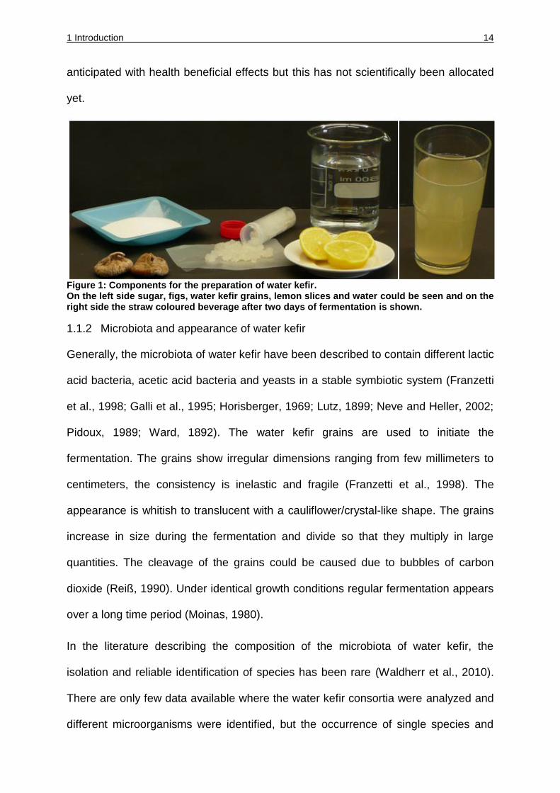
1 Introduction 14
anticipated with health beneficial effects but this has not scientifically been allocated
yet.
Figure 1: Components for the preparation of water kefir. On the left side sugar, figs, water kefir grains, lemon slices and water could be seen and on the right side the straw coloured beverage after two days of fermentation is shown.
1.1.2 Microbiota and appearance of water kefir
Generally, the microbiota of water kefir have been described to contain different lactic
acid bacteria, acetic acid bacteria and yeasts in a stable symbiotic system (Franzetti
et al., 1998; Galli et al., 1995; Horisberger, 1969; Lutz, 1899; Neve and Heller, 2002;
Pidoux, 1989; Ward, 1892). The water kefir grains are used to initiate the
fermentation. The grains show irregular dimensions ranging from few millimeters to
centimeters, the consistency is inelastic and fragile (Franzetti et al., 1998). The
appearance is whitish to translucent with a cauliflower/crystal-like shape. The grains
increase in size during the fermentation and divide so that they multiply in large
quantities. The cleavage of the grains could be caused due to bubbles of carbon
dioxide (Reiß, 1990). Under identical growth conditions regular fermentation appears
over a long time period (Moinas, 1980).
In the literature describing the composition of the microbiota of water kefir, the
isolation and reliable identification of species has been rare (Waldherr et al., 2010).
There are only few data available where the water kefir consortia were analyzed and
different microorganisms were identified, but the occurrence of single species and
1 Introduction 15
their percentages in the consortia were not determined. Bacteria and yeast species
isolated from the water kefir microbiota before this work are shown in Table 1. The
molecular background for the formation of a stable consortium is unknown and the
comprehensive composition of the microbiota is not scientifically defined yet.
Table 1: Organisms isolated from water kefir (Waldherr et al., 2010)
species literature
Bacteria
Lb. brevis Moinas et al., 1980
Lb. hilgardii Pidoux, 1989; Waldherr et al., 2010
Lb. casei subsp. casei Pidoux, 1989; Galli et al., 1995
Lb. casei subsp. rhamnosus Pidoux 1989
Lb. casei subsp. pseudoplantarum Galli et al., 1995
Lb. plantarum Pidoux, 1989
Lb. buchneri Galli et al., 1995
Lb. fructiovorans Galli et al., 1995
Lb. collinoides Galli et al., 1995
Lcc. lactis supbsp. lactis Moinas et al., 1980; Pidoux, 1989;
Waldherr et al., 2010
Lcc. lactis supbsp. cremoris Pidoux, 1989
Lc. mesenteroides subsp. mesenteroides Galli et al., 1995; Waldherr et al., 2010
Lc. mesenteroides subsp. dextranicum Pidoux, 1989
Enterobacter hormachei Waldherr et al., 2010
Gb. frateuri Waldherr et al., 2010
Yeasts
Saccharomyces bayanus Waldherr et al., 2010
Saccharomyces cerevisiae Moinas et al., 1980; Galli et al., 1995;
Franzetti et al., 1998
Saccharomyces pretoriensis Galli et al., 1995
Zygosaccharomyces florentinus Pidoux, 1989; Neve and Heller, 2001, Galli et al.,
1995
Hanseniaspora valbyensis Pidoux, 1989; Galli et al., 1995;
Neve and Heller, 2001
Hanseniaspora vinae Pidoux, 1989; Galli et al., 1995
Hanseniaspora yalbensis Franzetti et al., 1998
Kloeckera apiculata Pidoux, 1989; Franzetti et al., 1998
Candida lambica Pidoux, 1989
Candida valida Pidoux, 1989
1 Introduction 16
1.1.3 Composition of water kefir grains
The unique structure of the water kefir grain shows a gelatinous consistency and they
are described to contain dextran, an α 1-6 linked glucose polymer (Galli et al., 1995,
Horisberger, 1969, Pidoux, 1989). Lactobacillus (Lb.) hilgardii was described by
Pidoux et al., 1988 as an important organism for the stability of the water kefir grain
by producing the polysaccharide dextran. Dextran is a sugar consisting of different
amounts of the subunit glucose. Horisberger described the dextran produced by “tibi
grains” as a single insoluble polysaccharide containing only D-glucose (Horisberger,
1969). The strain Lb. hilgardii isolated from water kefir by Waldherr was described as
a granule-forming bacterium by producing large amounts of dextran (Waldherr et al.,
2010).
1.1.4 Comparison to milk kefir
Water kefir is not studied in detail like other food fermentations. The microbiota of
milk kefir is an example for a better characterized community and could be compared
to the symbiosis of water kefir. Milk kefir is a beverage of fermented milk, which
results in a carbonated slightly alcoholic and sour taste with a creamy consistency
(Kok-Tas et al., 2013; Lopitz-Otsoa et al., 2006). A lot of investigations have been
made to analyze milk kefir reviewed by Lopitz-Otsoa et al., 2006. The origin of milk
kefir is also not defined but believed that it was found in the Caucasian mountains.
Milk kefir is also known under different names like kephir, kiaphur, kefer, knapon,
kepi and kippi (Farnworth, 2005). Milk kefir also is a natural starter culture for
producing kefir in widespread countries like Argentina, Taiwan, Portugal, Turkey and
France. Like water kefir it is improbable that the milk kefir originates from one single
original starter culture as microbial investigations of milk kefirs taken from several
sample sides showed differences in their microbiota (Farnworth, 2005).
1 Introduction 17
The milk kefir grains share some characteristics with the water kefir grains as they
also show gelatinous grain structure which consist of a various mixture of
microorganisms mostly lactobacilli, lactococci and yeasts casually acetic acid
bacteria and bifidobacteria (Dobson et al., 2011). Under traditional treatment of the
milk kefir the starter grain could be used again after approximately 24h of
fermentation in milk. The milk is then removed and appropriate for consumption
(Lopitz-Otsoa et al., 2006). The microbiota of milk kefir grains is also very stable
when incubated under suitable conditions. The polysaccharide produced from
microorganisms of the milk kefir grains are called kefiran which is a water-soluble
polysaccharide consisting of branched glucogalactan with equal amounts of the
monomers D-glucose and D-galactose produced by Lb. kefiranofaciens (Kok-Tas et
al., 2013). The milk kefir grains consist of a polysaccharide-protein matrix where
kefiran is the main component. The viscosity of kefiran decreases with higher
concentrations showing a pseudoplastic behavior (Piermaria et al., 2008). Milk kefir
has been a source for isolation and description of new species like Lb. kefiri, Lb.
kefirgranum, Lb. parakefir, Candida kefyr and Saccharomyces turicensis (Lopitz-
Otsoa et al., 2006). Milk kefir could be produced commercially but only by using
starter cultures with specific groups of microorganisms (lactic acid bacteria, acetic
acid bacteria and yeasts) as the kefir grains show differences in their composition
during the fermentation procedure resulting in deficient organoleptic characteristics
(Lopitz-Otsoa et al., 2006). The consumption of traditional and industrial kefir shows
a possible beneficial effect on health, especially kefiran might show therapeutic
immunostimulatory, antimutagenic, antiallergic and antiulcer activity (Kok-Tas et al.,
2013). Kefiran could also modulate the gut immune system and epithelial cells are
protected against Bacillus cereus exocellular factors (Piermaria et al., 2008).
1 Introduction 18
The milk kefir is an example for a symbiotic coexistence of bacteria and yeast
dependent on each other with beneficial effects.
1.2 Role of fermented food in human nutrition
The history of the usage of fermented food is very old (Tamang and Kasipathy,
2010). The food processing technology regarding the production of food fermentation
is one of the oldest known to human kind (Caplice and Fitzgerald, 1999). The oldest
record of food fermentation goes back to 6000 BC in the Fertile Crescent (Blandino et
al., 2003). The knowledge of food preparations was transferred from generation to
generation producing small amounts of the traditional product for consumption.
Traditionally, the way for obtaining fermented food in former times was done by
indigenous knowledge but without understanding the meaning of microbial
mechanisms. With the industrialization and growth of towns or cities the requirements
of foods raised. The microbial understanding of communities immersed from 1850s
onwards (Caplice and Fitzgerald, 1999). The products originating from fermented
foods show a huge variety around the world. Different substrates are metabolized by
different microorganisms resulting in unique foods with typical characteristics
(Caplice and Fitzgerald, 1999). Fermented foods include a huge variability of
substrates ranging from vegetables, cereals, milks, legumes, meat and fish products
and grains (Tamang and Kasipathy, 2010; van Hijum et al., 2013). The food
fermentation could occur as natural (spontaneous) or controlled fermentation by
using starter cultures (monoculture or multiculture microorganisms) (Tamang and
Kasipathy, 2010). With respect to the increasing consumption of fermented food and
industrial preparation the need of understanding the interaction and identifying the
microorganisms responsible for the characteristics is an irreplaceable requirement.
1 Introduction 19
Nowadays, it is assumed that 5-40 % of the total daily food consumption is coped by
fermented food and beverages and the importance of fermented food consumed
globally is rising (Tamang and Kasipathy, 2010).
The microbial stability has to be proven for ensuring food safety and typical
organoleptic characteristic which is nearly impossible when the mechanisms of the
fermentation are rarely understood. Therefore the interest of the characterization of
the microbial composition of a fermented food is increasing constantly. Often
fermented foods are providing health benefits by enhanced nutritional content like
bioavailability of minerals and production of antioxidants, improving digestibility and
could reduce toxicity (Bokulich and Mills, 2012). The diversity of microbiota in
fermented foods and their functional microorganisms also provide several novel
properties like enzyme and alcohol producing bacteria and yeasts which can be used
industrially.
1.2.1 Fermented beverages
A lot of investigations examined alcoholic fermentation, such as wine and beer. The
predominant and most important species in these fermentations is the yeast
Saccharomyces cerevisiae. At this juncture the fermentation process is an alcoholic
fermentation resulting in the formation of ethanol. Alcohol fermentation is not the only
procedure taking place in beverage fermentations. Milk and cereals are fermented by
lactic acid fermentation due to lactic acid bacteria. The metabolic characteristics of
this group can be divided into homofermentative and heterofermentative species
whereas homofermentative bacteria produce lactic acid as major end product.
Species representing the homofermentative pathway are members of the genera
Pediococcus, Streptococcus, Lactococcus and Lactobacillus. Heterofermentative
bacteria produce same amount of lactate, ethanol and CO2. Species representing
1 Introduction 20
this group belong to the genera Weissella, Leuconostoc and Lactobacillus (Blandino
et al., 2003). The interaction between the different organisms is not analyzed in depth
but the technology of beer and wine making is well documented in literature. The
mechanisms of other fermented beverages are not completely understood and the
interest of analyzing their microbiota, due to their probably health benefits and for
development of possible starter culture is increasing fast. One example is water kefir.
There are different molecular procedures to investigate the microbiota ranging from
culture-dependent to culture-independent methods both with advantages and
disadvantages.
1.3 Application of culture-dependent and- independent analyses of microbiota
1.3.1 Advantages and disadvantages of culture-dependent analyses
The conventional techniques of a microbiota analyses relies on culture-dependent
methods where bacteria, yeasts or fungi are cultured with or in nutrient media (Gong
and Yang, 2012). The identification of microorganisms is done regarding their
morphology, biochemical, physiological and genetic characteristics after growing on
suitable media (Jany and Barbier, 2008). One of the most important advantage of
culture-dependent analysis is the ability to obtain pure cultures and to characterize
these species in detail. Especially in the field of food microbiology it is necessary to
correctly profile bacterial or yeast species like in fermented foods for obtaining the
food with wanted texture, flavor and possible health benefit properties (Kesmen et al.,
2012).
The difficulty in culturing microorganisms from food environments is that only a small
percentage of food-associated microorganisms can be cultured under standard
1 Introduction 21
laboratory procedures (Giraffa and Neviani, 2001). It is very difficult to simulate the
natural environment or habitat of the microorganisms within a microbiota under
laboratory conditions. Not only the nutritional requirements are often unknown but
also the interaction and symbiosis of microorganisms is nearly impossible to simulate
(Gong and Yang, 2012). The culture-dependent analyses of complex microbiota
often results in a misleading interpretation of the diversity and composition of the
microbes (van Hijum et al., 2013). Enrichment cultures used for growth of
microorganisms within microbiota could influence the community structure by
inserting new selective conditions (Giraffa and Neviani, 2001). Species appearing in
low cell numbers in the microbiota could be missed by culturing procedures due to
the competiveness of numerically more abundant species (Hugenholtz et al., 1998).
Predominant species might overgrow less abundant species which could also have
an important impact in the stability of the community. The culture-dependent
analyses are very time-consuming resulting from long culture periods and complex
culture techniques like isolating single pure cultures (Gong and Yang, 2012).
1.3.2 High through-put sequencing as a tool for characterizing microbiota in food
fermentations
Molecular techniques for analyzing microbiota like detection, identification and
characterization of microorganisms within these environmental habitats exhibit an
outstanding tool (Giraffa and Neviani, 2001). Therefore technologies that do not
require cultivation have proven useful to identify non-culturable microorganisms.
Technologies without the need of culturing microorganisms established over the last
decades (van Hijum et al., 2013). There are several culture-independent molecular
methods applied analyzing microbial food community each with its own limits like
Denaturating/Thermal Gradient Gel Electrophoresis (DGGE/TGGE), terminal
restriction fragment length polymorphism (TRFLP), fluorescence in situ hybridization
1 Introduction 22
(FISH), quantitative PCR (QPCR) and next generation sequencing (NGS) (van Hijum
et al., 2013). The technology of next generation sequencing was firstly introduced to
the market in 2005 (Morozova and Marra, 2008). This relatively new technique
provides high speed and high-throughput sequencing without the need of any culture
step or cloning of DNA fragments (Ansorge, 2009).
To overcome misinterpretation of a microbial community resulting from growth bias,
molecular techniques have been established over the past decades. In the field of
food fermentation next generating high through-put sequencing such as 454 FLX
Titanium sequencing are state of the art techniques for monitoring the microbial flora
and/or changes within a microbial community (Bokulich and Mills, 2012; van Hijum et
al., 2013). The 454 high through-put sequencing is a sequencing by synthesis
technique and chemiluminescence is measured when inorganic pyrophosphate is
released. For detailed information of the procedure see Morozova and Marra, 2008.
Universal primers are used to amplify the DNA template from microbial communities;
normally the 16S rRNA genes in bacteria and ITS genes in yeast and fungi are
amplified. The length of sequence in 454 Pyrosequencing could be up to 700 bp (van
Hijum et al., 2013) providing greater taxonomic information than shorter sequences
(Bokulich and Mills, 2012). The length of sequences is an important factor for the
analysis of a community. The identification of organisms on species level is only
possible when sequence length is long enough and a good quality of the sequences
is given. The term pyrosequencing implies different technologies for analyzing a
community, including genome/metagenome, transcriptome and epigenome
characterization.
The 16S rRNA amplicon sequencing is a part of the metagenomic high through-put
technology whereas metagenomic analysis studies the collective set of mixed
1 Introduction 23
microbial communities (Petrosino et al., 2009). With the amplicon sequencing
organisms are identified on either family or species level but no information about
their metabolic activity are obtained.
Several fermented foods were analyzed by high-throughput sequencing such as
pearl millet fermentation (Humblot and Guyot, 2009), fermented fish/rice called
narezushi (Kiyohara et al., 2012; Koyanagi et al., 2012), nukadoko (rice bran)
(Sakamoto et al., 2011), Chinese liquor fermentations (Li et al., 2011) and cheese
fermentation like Polish Oscypek cheese (Alegria et al., 2012). A lot of investigations
have been done regarding Korean food fermentation like fermented seafood (Roh et
al., 2010), soybean pastes like meju (Kim et al., 2011) and doenjang (Nam et al.,
2012a), rice beer called Makgeolli (Jung et al., 2012), kochujang (red pepper, rice,
soybean mix) (Nam et al., 2012b) and fermented raddish/cabbage called kimchi
(Park et al., 2012) reviewed recently by Bokulich and Mills, 2012; van Hijum et al.,
2013.
High-throughput sequencing offers a new view of different environmental microbiota
and changes of a community could be measured when altering growth conditions.
When detecting new species in a community the step of cultivation is needed for
characterizing these microorganisms, but needed growth parameters could be
applied in laboratory scale more easily.
1.4 Preservation of food, beverages and starter cultures used for production
The preservation of food and beverage is very important for stability and microbial
safety of these products around the world. For ensuring the food safety combined
preservation factors are used, called hurdles (Leistner, 2000). Applied hurdles are the
control of temperature (low/high), water activity, acidity (pH), redox potential, addition
1 Introduction 24
of preservatives like nitrite, sorbate or bacteriocines and the usage of competitive
microorganisms such as lactic acid bacteria (Leistner, 2000). The organoleptic
characteristics of each end-product have to be guaranteed. Therefore it is necessary
to evaluate suitable mix of hurdles for each product. Nowadays the focus of hurdle
techniques is to generate a multitarget preservation with applied gentle hurdles.
These hurdles are mixed in a way that they show synergistic effects for gaining food
and beverages with wanted characteristics (Leistner, 2000). The globalization of food
market and the introduction of novel food and beverages are linked to new demands
for preservation techniques. The demands of the consumers are changing towards
products with natural characteristics and minimally processed food like preservation
by microorganisms or other biopreservation techniques.
1.4.1 Biopreservation
One of the oldest biopreservation is the technology of fermentation (Ross et al.,
2002). Over the past few years the role of microorganisms in preservation of food
and beverage raised. The process of preservation due to microorganisms is
dependent o their biological activity and the formation of metabolites which could
suppress unwanted microbiota in foods and beverages (Ross et al., 2002).
Nowadays yeasts are widely used in the production of alcoholic beverages like beer,
wine and spirits. Lactic acid bacteria are important microorganisms for the production
of different fermentation processes like dairy, meat and vegetable fermentation.
Genera belonging to LAB are Lactococcus, Lactobacillus, Enterococcus,
Streptococcus, Leuconostoc and Pediococcus (Ross et al., 2002). The raw material
of each food or beverage exhibits substrates for different mixture of microorganisms
normally a defined mix of starter culture (Ross et al., 2002). LAB play an important
role in the food industry not only because of contributing in flavor, texture and in
enhancing the nutritional value in foods and beverages, they also prevent spoilage
1 Introduction 25
and extent shelf life of these products (Topisirovic et al., 2006). The production of
antimicrobial substances as metabolites could be important in several fermentations,
but LAB are also known to produce antimicrobial proteinaceous substances, called
bacteriocins. Substances showing antimicrobial effects toward pathogenic organisms
are several types of acids like organic and fatty acids, alcohol production and carbon
dioxide (Ohlsson and Bengtsson, 2002; Ross et al., 2002). The antimicrobial effects
often are attended by lowering the pH, or negatively affecting the cell membrane
potential, preventing active transport and influencing different metabolic functions
(Ross et al., 2002).
The interest in using substances which can be marketed as natural is rising and
therefore the interest of microorganisms isolated from natural niches with potential of
new variants of antimicrobial, antifungal or antioxidantal effects is also increasing
(Ohlsson and Bengtsson, 2002; Topisirovic et al., 2006).
1.4.2 Stress tolerance of microorganisms used as starter cultures regarding freeze-
drying technology
The procedure of freezing and freeze-drying are standard methods for the long-term
storage of bacteria used as starter cultures. The difficulty of bacterial storage is to
reduce damages resulting from freeze-drying technology and rehydration with a
maximal cell survival.
The viability and functional activity of preserved starter cultures are dependent on the
preservation technologies used, but starter cultures used industrially should maintain
high levels of viability during freeze-drying and after rehydration (Carvalho et al.,
2002a). Although this technology is commonly used, undesirable side-effects often
occur and hamper the viability of these organisms (Carvalho et al., 2002b). During
the freeze-drying procedure cells are exposed to extreme stress situations including
1 Introduction 26
high concentrations of solutes, extremes in pH, low temperature, ice crystal
formation, water removement from within the cell (Carvalho et al., 2002a). The loss of
viability of dried cultures is a result of cell damage at several target sites, like cell
membrane and cell wall damage, DNA denaturation and on membrane lipid oxidation
(Carvalho et al., 2004; Zhao and Zhang, 2005). Cell damage during freezing is
depending on the cooling rate. Slow cooling could result in extracellularly ice crystal
formation due to osmotic water flow. The solute concentration outside the cell
increases resulting from ice crystal formation and osmotic imbalance occurs. But if
the cooling rate is too fast ice crystals could be formed intracellularly leading to lethal
cell damage (Zhao and Zhang, 2005). The mechanisms of desiccation tolerance are
not completely understood. Gram positive bacteria are more stress tolerant than
gram negative bacteria. Investigations of Fredrickson et al. showed a correlation of
Mn2+ accumulation and high Mn2+/Fe2+ ratio in the cell and stress tolerance. Mn2+
might protect proteins from oxidative damage after desiccation, the intracellular
Mn2+/Fe2+ concentration is related to desiccation resistance although the
mechanisms behind it are not understood completely (Fredrickson et al., 2008). For
Gram negative bacteria on the other hand desiccation tolerance has to be improved
by accumulation of intracellular protective agents such as non-reducing
disaccharides like sucrose and trehalose.
Cells are protected by the replacement of water due to formation of hydrogen bonds
with the sugars and the saccharides are involved in vitreous cytoplasmatic matrix
formation. Not only intracellular but also extracellular protective agents could be
used. Gram negative cells could also be trained for stress tolerance by multiple
exposure and the physiology/metabolism prior to stress could be altered (Fredrickson
et al., 2008).
1 Introduction 27
For cell survival during and after freeze-drying many factors have to be considered
like growth conditions, protective medium, initial cell concentration, freezing
temperature and rehydration conditions. The rehydration of desiccated cells is an
important factor and the survival of the cells is depending on the rehydration medium.
Complex rehydration media might show higher survival rates of microorganisms due
to the finding of nutrients for repairing damaged cells. The osmotic pressure of
complex media is higher and therefore the effect of the osmotic shock in desiccated
cells could be minimized (Morgan et al., 2006).
The optimal application freezing temperature, freeze-drying procedure and the
rehydration process is highly depending on genera (Zhao and Zhang, 2005).
1.5 Bifidbacteriaceae in food
Bifidobacteriaceae are heterofermentative Gram-positive bacteria (Pokusaeva et al.,
2011). The family of bifidobacteria have different habitats mostly in the human and/or
animal gut (Biavati and Mattarelli, 2006). Normally they are not pathogenic with the
exception of bifidobacteria isolated from dental caries and Gardnerella vaginalis
which might cause urogenetal tract infections. Most of them normally show probiotic
and or prebiotic characteristics.
Bifidobacteriaceae are very important in the field of treatment and prevention of
diseases either in medicine as well as supplement or natural occurring in food
(Arunachalam, 1999). In humans bifidobacteria are naturally found in the
gastrointestinal gut showing beneficial health effects (Pokusaeva et al., 2011).
The health benefits of regularly intake of bifidobacteria are reported as suppression
of putrefactive bacteria and intestinal putrefaction, prevention of constipation and
1 Introduction 28
geriatric diseases including cancer, prevention and treatment of antibiotic-associated
diarrhea and stimulation of immune response resulting in greater resistance to
infection (Mitsuoka, 1990). The species of bifidobacteria often are difficult to isolate
due to their special growth requirements and extreme habitats (Arunachalam, 1999).
Their ability to survive and live in extreme habitats could be explained by their
physiological capability to metabolize different oligosaccharides (Pokusaeva et al.,
2011). Fermented products are claimed to have health promoting benefits like
yoghurt and milk kefir, especially in the human digestive tract. For manufacturing
health promoting fermented milk products five species are mostly used namely B.
adolescentis, B. bifidum, B. breve, B. infantis and B. longum (Arunachalam, 1999).
Some species of Bifidobacteriaceae are able to form different vitamins like thiamin,
folic acid, ascorbic acid, pyridoxine, nicotine etc. (Arunachalam, 1999).
The health promoting effect in human is due to production of antimicrobial
substances suppressing pathogens by competition for nutrients and adhesion
receptors and stimulating immunity (Kailasapathy and Chin, 2000). In the past few
years the public awareness of probiotic food raised and so did the demands and so
some strains have been introduced to the commercially market, especially as
functional substances of dairy- based probiotic drinks (Pokusaeva et al., 2011). In the
industry prebiotics have become a very important role, especially in the functional
food market (Pokusaeva et al., 2011). Therefore it is interesting to find new natural
habitats and/or new bifidobacteria species and characterize them in terms of a
potential prebiotic or probiotic effect.
1 Introduction 29
1.6 Objectives of the work
The aim of this thesis was to elucidate the composition and dynamic of the water kefir
consortium to enable a stable and reproducible fermentation of water kefir with a
constant end product showing desired characteristics. Therefore the microorganisms
of the water kefir microbiota should be isolated and identified. The identification
should be done by culture-dependent procedures like RAPD pattern comparison
followed by 16S rRNA analysis. As a second approach for identifying bacteria in the
water kefir community culture-independent analyses such as 16S rRNA gene
amplicon sequencing, ARDRA and RISA should be applied. The isolates should be
characterized regarding their possibility of EPS production. The composition of
different water kefir microbiota grown under standardized conditions should be
determined for indentifying the core microorganisms within this community. The
alteration of the water kefir microbiota resulting from different growth conditions
should be determined by convenient procedures. A possible conservation of the
water kefir grains by freeze-drying technology should be analyzed with respect of
industrial handling.
As the overall aim possible isolates of the water kefir should be identified, which
could be used as starter cultures for a water kefir based beverage.
2 Material and Methods 30
2 Material and Methods
2.1 Material
2.1.1 Equipment
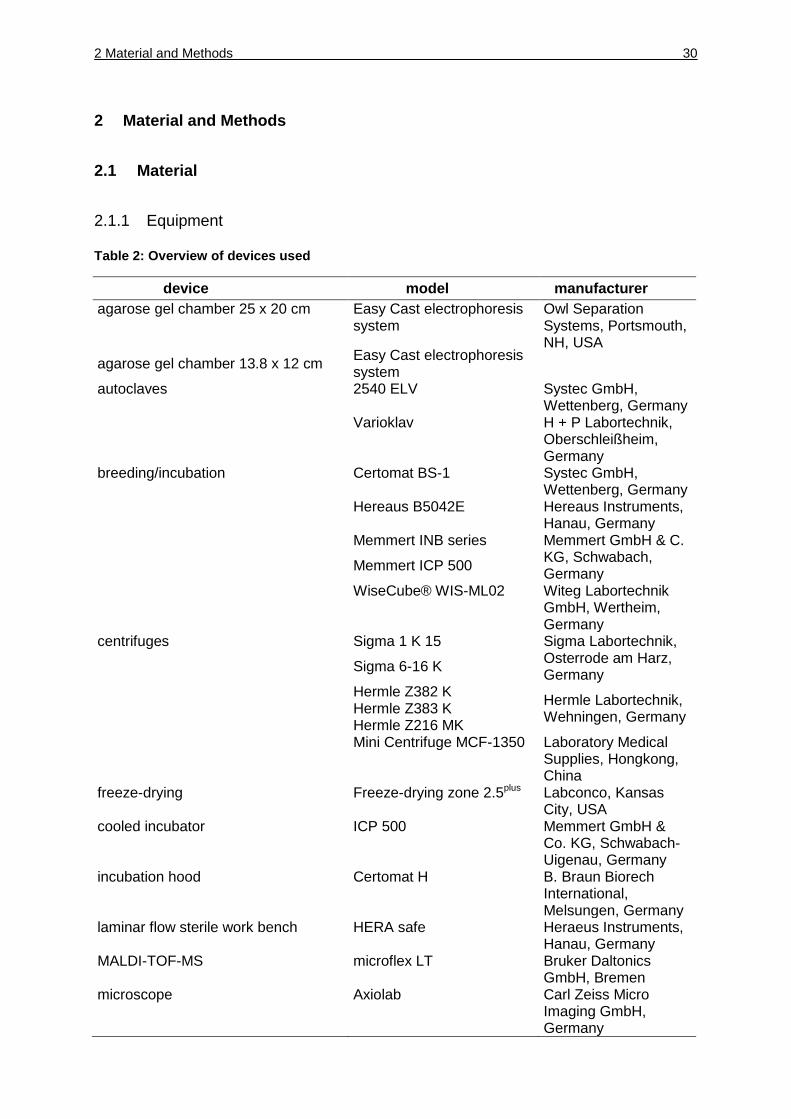
Table 2: Overview of devices used
device model manufacturer
agarose gel chamber 25 x 20 cm Easy Cast electrophoresis system
Owl Separation Systems, Portsmouth, NH, USA
agarose gel chamber 13.8 x 12 cm Easy Cast electrophoresis system
autoclaves 2540 ELV Systec GmbH, Wettenberg, Germany
Varioklav H + P Labortechnik, Oberschleißheim, Germany
breeding/incubation Certomat BS-1 Systec GmbH, Wettenberg, Germany
Hereaus B5042E Hereaus Instruments, Hanau, Germany
Memmert INB series Memmert GmbH & C.
KG, Schwabach, Germany
Memmert ICP 500
WiseCube® WIS-ML02 Witeg Labortechnik GmbH, Wertheim, Germany
centrifuges Sigma 1 K 15 Sigma Labortechnik, Osterrode am Harz, Germany
Sigma 6-16 K
Hermle Z382 K
Hermle Labortechnik, Wehningen, Germany
Hermle Z383 K
Hermle Z216 MK
Mini Centrifuge MCF-1350 Laboratory Medical Supplies, Hongkong, China
freeze-drying Freeze-drying zone 2.5plus Labconco, Kansas City, USA
cooled incubator ICP 500 Memmert GmbH & Co. KG, Schwabach-Uigenau, Germany
incubation hood Certomat H B. Braun Biorech International, Melsungen, Germany
laminar flow sterile work bench HERA safe Heraeus Instruments, Hanau, Germany
MALDI-TOF-MS microflex LT Bruker Daltonics GmbH, Bremen
microscope Axiolab Carl Zeiss Micro Imaging GmbH, Germany
2 Material and Methods 31
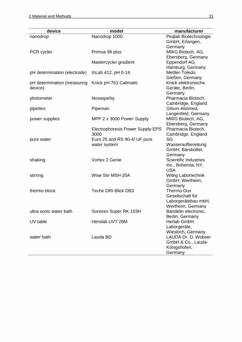
device model manufacturer nanodrop Nanodrop 1000 Peqlab Biotechnologie
GmbH, Erlangen, Germany
PCR cycler Primua 96 plus MWG Biotech, AG, Ebersberg, Germany
Mastercycler gradient Eppendorf AG, Hamburg, Germany
pH determination (electrode) InLab 412, pH 0-14 Mettler-Toledo, Gießen, Germany
pH determination (measuring device)
Knick pH 761 Calimatic Knick elektronische Geräte, Berlin, Germany
photometer NovaspeIIq Pharmacia Biotech, Cambridge, England
pipettes Pipeman Gilson-Abomed, Langenfeld, Germany
power supplies MPP 2 x 3000 Power Supply MWG Biotech, AG, Ebersberg, Germany
Electrophoresis Power Supply EPS 3000
Pharmacia Biotech, Cambridge, England
pure water Euro 25 and RS 90-4/ UF pure water system
SG Wasseraufbereitung GmbH, Barsbüttel, Germany
shaking Vortex 2 Genie Scientific Industries Inc., Bohemia, NY, USA
stirring Wise Stir MSH-20A Witeg Labortechnik GmbH, Wertheim, Germany
thermo block Teche DRI-Blick DB3 Thermo-Dux Gesellschaft für Laborgerätebau mbH, Wertheim, Germany
ultra sonic water bath Sonorex Super RK 103H Bandelin electronic, Berlin, Germany
UV table Herolab UVT 28M Herlab GmbH Laborgeräte, Wiesloch, Germany
water bath Lauda BD LAUDA Dr. D. Wobser GmbH & Co., Lauda-Königshofen, Germany
2 Material and Methods 32
2.1.2 Chemicals
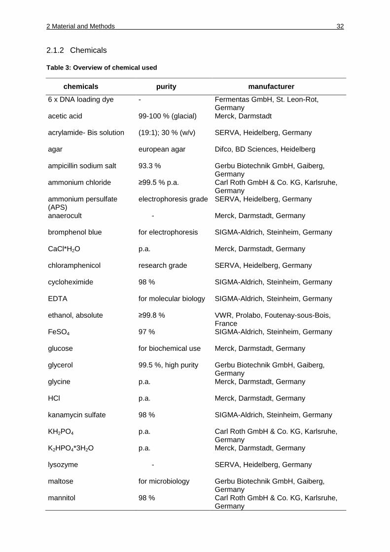
Table 3: Overview of chemical used
chemicals purity manufacturer
6 x DNA loading dye - Fermentas GmbH, St. Leon-Rot, Germany
acetic acid 99-100 % (glacial) Merck, Darmstadt
acrylamide- Bis solution (19:1); 30 % (w/v) SERVA, Heidelberg, Germany
agar european agar Difco, BD Sciences, Heidelberg
ampicillin sodium salt 93.3 % Gerbu Biotechnik GmbH, Gaiberg, Germany
ammonium chloride ≥99.5 % p.a. Carl Roth GmbH & Co. KG, Karlsruhe, Germany
ammonium persulfate (APS)
electrophoresis grade SERVA, Heidelberg, Germany
anaerocult - Merck, Darmstadt, Germany
bromphenol blue for electrophoresis SIGMA-Aldrich, Steinheim, Germany
CaCl*H2O p.a. Merck, Darmstadt, Germany
chloramphenicol research grade SERVA, Heidelberg, Germany
cycloheximide 98 % SIGMA-Aldrich, Steinheim, Germany
EDTA for molecular biology SIGMA-Aldrich, Steinheim, Germany
ethanol, absolute ≥99.8 % VWR, Prolabo, Foutenay-sous-Bois, France
FeSO4 97 % SIGMA-Aldrich, Steinheim, Germany
glucose for biochemical use Merck, Darmstadt, Germany
glycerol 99.5 %, high purity Gerbu Biotechnik GmbH, Gaiberg, Germany
glycine p.a. Merck, Darmstadt, Germany
HCl p.a. Merck, Darmstadt, Germany
kanamycin sulfate 98 % SIGMA-Aldrich, Steinheim, Germany
KH2PO4 p.a. Carl Roth GmbH & Co. KG, Karlsruhe, Germany
K2HPO4*3H2O p.a. Merck, Darmstadt, Germany
lysozyme - SERVA, Heidelberg, Germany
maltose for microbiology Gerbu Biotechnik GmbH, Gaiberg, Germany
mannitol 98 % Carl Roth GmbH & Co. KG, Karlsruhe, Germany
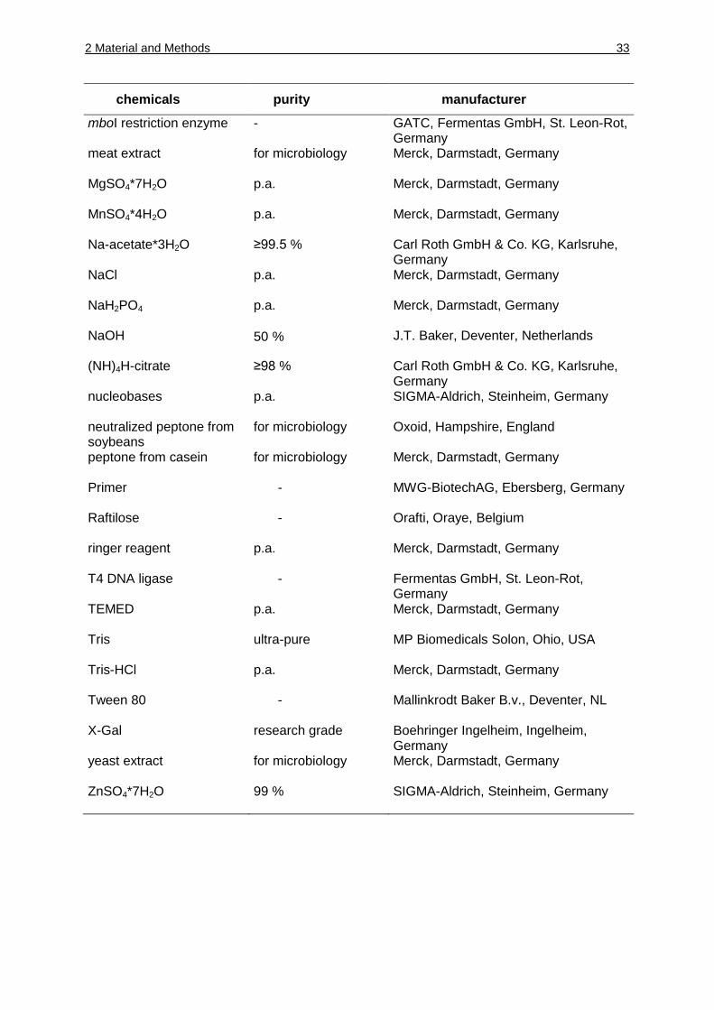
2 Material and Methods 33
chemicals purity manufacturer
mboI restriction enzyme - GATC, Fermentas GmbH, St. Leon-Rot, Germany
meat extract for microbiology Merck, Darmstadt, Germany
MgSO4*7H2O p.a. Merck, Darmstadt, Germany
MnSO4*4H2O p.a. Merck, Darmstadt, Germany
Na-acetate*3H2O ≥99.5 % Carl Roth GmbH & Co. KG, Karlsruhe, Germany
NaCl p.a. Merck, Darmstadt, Germany
NaH2PO4 p.a. Merck, Darmstadt, Germany
NaOH 50 % J.T. Baker, Deventer, Netherlands
(NH)4H-citrate ≥98 % Carl Roth GmbH & Co. KG, Karlsruhe, Germany
nucleobases p.a. SIGMA-Aldrich, Steinheim, Germany
neutralized peptone from soybeans
for microbiology Oxoid, Hampshire, England
peptone from casein for microbiology Merck, Darmstadt, Germany
Primer - MWG-BiotechAG, Ebersberg, Germany
Raftilose - Orafti, Oraye, Belgium
ringer reagent p.a. Merck, Darmstadt, Germany
T4 DNA ligase - Fermentas GmbH, St. Leon-Rot, Germany
TEMED p.a. Merck, Darmstadt, Germany
Tris ultra-pure MP Biomedicals Solon, Ohio, USA
Tris-HCl p.a. Merck, Darmstadt, Germany
Tween 80 - Mallinkrodt Baker B.v., Deventer, NL
X-Gal research grade Boehringer Ingelheim, Ingelheim, Germany
yeast extract for microbiology Merck, Darmstadt, Germany
ZnSO4*7H2O 99 % SIGMA-Aldrich, Steinheim, Germany
2 Material and Methods 34
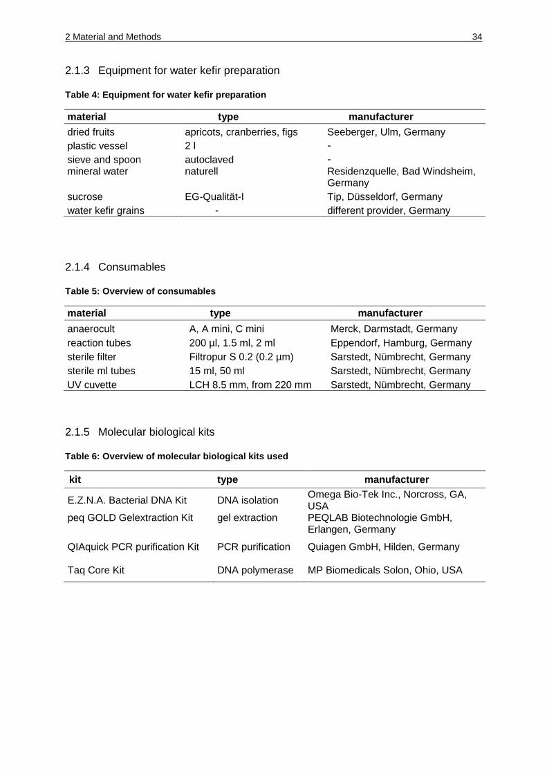
2.1.3 Equipment for water kefir preparation
Table 4: Equipment for water kefir preparation
material type manufacturer
dried fruits apricots, cranberries, figs Seeberger, Ulm, Germany
plastic vessel 2 l -
sieve and spoon autoclaved - mineral water naturell Residenzquelle, Bad Windsheim,
Germany
sucrose EG-Qualität-I Tip, Düsseldorf, Germany
water kefir grains - different provider, Germany
2.1.4 Consumables
Table 5: Overview of consumables
material type manufacturer
anaerocult A, A mini, C mini Merck, Darmstadt, Germany
reaction tubes 200 µl, 1.5 ml, 2 ml Eppendorf, Hamburg, Germany
sterile filter Filtropur S 0.2 (0.2 µm) Sarstedt, Nümbrecht, Germany
sterile ml tubes 15 ml, 50 ml Sarstedt, Nümbrecht, Germany
UV cuvette LCH 8.5 mm, from 220 mm Sarstedt, Nümbrecht, Germany
2.1.5 Molecular biological kits
Table 6: Overview of molecular biological kits used
kit type manufacturer
E.Z.N.A. Bacterial DNA Kit DNA isolation Omega Bio-Tek Inc., Norcross, GA, USA
peq GOLD Gelextraction Kit gel extraction PEQLAB Biotechnologie GmbH, Erlangen, Germany
QIAquick PCR purification Kit PCR purification Quiagen GmbH, Hilden, Germany
Taq Core Kit DNA polymerase MP Biomedicals Solon, Ohio, USA
2 Material and Methods 35
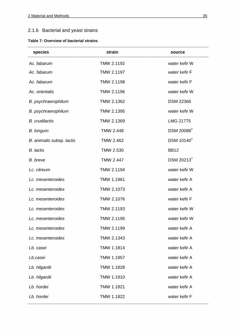
2.1.6 Bacterial and yeast strains
Table 7: Overview of bacterial strains
species strain source
Ac. fabarum TMW 2.1192 water kefir W
Ac. fabarum TMW 2.1197 water kefir F
Ac. fabarum TMW 2.1198 water kefir F
Ac. orientalis TMW 2.1196 water kefir W
B. psychraerophilum TMW 2.1362 DSM 22366
B. psychraerophilum TMW 2.1395 water kefir W
B. crudilactis TMW 2.1369 LMG 21775
B. longum TMW 2.448 DSM 20088T
B. animalis subsp. lactis TMW 2.462 DSM 10140T
B. lactis TMW 2.530 BB12
B. breve TMW 2.447 DSM 20213T
Lc. citreum TMW 2.1194 water kefir W
Lc. mesenteroides TMW 1.1961 water kefir A
Lc. mesenteroides TMW 2.1073 water kefir A
Lc. mesenteroides TMW 2.1076 water kefir F
Lc. mesenteroides TMW 2.1193 water kefir W
Lc. mesenteroides TMW 2.1195 water kefir W
Lc. mesenteroides TMW 2.1199 water kefir A
Lc. mesenteroides TMW 2.1343 water kefir A
Lb. casei TMW 1.1814 water kefir A
Lb.casei TMW 1.1957 water kefir A
Lb. hilgardii TMW 1.1828 water kefir A
Lb. hilgardii TMW 1.1910 water kefir A
Lb. hordei TMW 1.1821 water kefir A
Lb. hordei TMW 1.1822 water kefir F
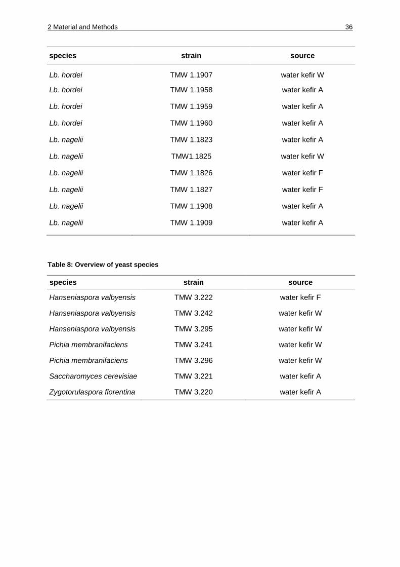
2 Material and Methods 36
species strain source
Lb. hordei TMW 1.1907 water kefir W
Lb. hordei TMW 1.1958 water kefir A
Lb. hordei TMW 1.1959 water kefir A
Lb. hordei TMW 1.1960 water kefir A
Lb. nagelii TMW 1.1823 water kefir A
Lb. nagelii TMW1.1825 water kefir W
Lb. nagelii TMW 1.1826 water kefir F
Lb. nagelii TMW 1.1827 water kefir F
Lb. nagelii TMW 1.1908 water kefir A
Lb. nagelii TMW 1.1909 water kefir A
Table 8: Overview of yeast species
species strain source
Hanseniaspora valbyensis TMW 3.222 water kefir F
Hanseniaspora valbyensis TMW 3.242 water kefir W
Hanseniaspora valbyensis TMW 3.295 water kefir W
Pichia membranifaciens TMW 3.241 water kefir W
Pichia membranifaciens TMW 3.296 water kefir W
Saccharomyces cerevisiae TMW 3.221 water kefir A
Zygotorulaspora florentina TMW 3.220 water kefir A
2 Material and Methods 37
2.2 Methods
2.2.1 Microbial methods
2.2.1.1 Media and growth conditions
2.2.1.1.1 Preparation of water kefir
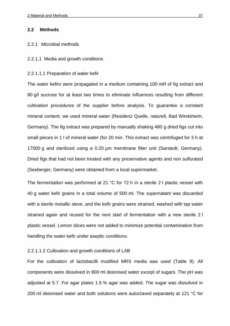
The water kefirs were propagated in a medium containing 100 ml/l of fig extract and
80 g/l sucrose for at least two times to eliminate influences resulting from different
cultivation procedures of the supplier before analysis. To guarantee a constant
mineral content, we used mineral water (Residenz Quelle, naturell, Bad Windsheim,
Germany). The fig extract was prepared by manually shaking 480 g dried figs cut into
small pieces in 1 l of mineral water (for 20 min. This extract was centrifuged for 3 h at
17000 g and sterilized using a 0.20 µm membrane filter unit (Sarstedt, Germany).
Dried figs that had not been treated with any preservative agents and non sulfurated
(Seeberger, Germany) were obtained from a local supermarket.
The fermentation was performed at 21 °C for 72 h in a sterile 2 l plastic vessel with
40 g water kefir grains in a total volume of 500 ml. The supernatant was discarded
with a sterile metallic sieve, and the kefir grains were strained, washed with tap water
strained again and reused for the next start of fermentation with a new sterile 2 l
plastic vessel. Lemon slices were not added to minimize potential contamination from
handling the water kefir under aseptic conditions.
2.2.1.1.2 Cultivation and growth conditions of LAB
For the cultivation of lactobacilli modified MRS media was used (Table 9). All
components were dissolved in 800 ml deionised water except of sugars. The pH was
adjusted at 5.7. For agar plates 1.5 % agar was added. The sugar was dissolved in
200 ml deionised water and both solutions were autoclaved separately at 121 °C for
2 Material and Methods 38
20 min to avoid Maillard products. Both solutions were mixed after autoclaving. After
cooling the solution below 50 °C cycloheximide (150 µg/ml) was added to inhibit the
growth of yeasts. The agar plates were incubated anaerobically at 30 °C for three
days. Liquid cultures were incubated anaerobically in 15 ml Falcon tubes over night.
Table 9: Composition of mMRS medium used for lactobacilli
compound concentration [g/l]
yeast extract 4
meat extract 2
peptone from casein 10
tween 80 1
K2HPO4*3 H2O 2.5
Na-acetate*3 H2O 5
(NH4)2 H citrate 2
MgSO4*7 H2O 0.2
MnSO4*H2O 0.038
glucose 20
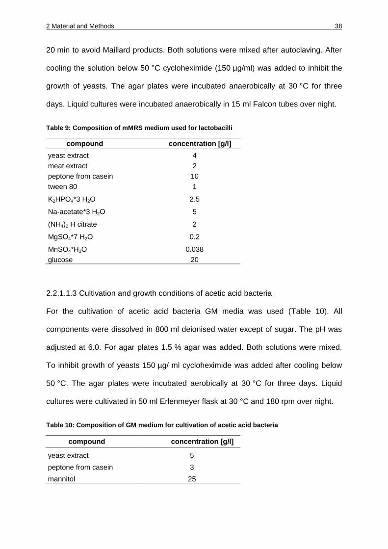
2.2.1.1.3 Cultivation and growth conditions of acetic acid bacteria
For the cultivation of acetic acid bacteria GM media was used (Table 10). All
components were dissolved in 800 ml deionised water except of sugar. The pH was
adjusted at 6.0. For agar plates 1.5 % agar was added. Both solutions were mixed.
To inhibit growth of yeasts 150 µg/ ml cycloheximide was added after cooling below
50 °C. The agar plates were incubated aerobically at 30 °C for three days. Liquid
cultures were cultivated in 50 ml Erlenmeyer flask at 30 °C and 180 rpm over night.
Table 10: Composition of GM medium for cultivation of acetic acid bacteria
compound concentration [g/l]
yeast extract 5
peptone from casein 3
mannitol 25
2 Material and Methods 39
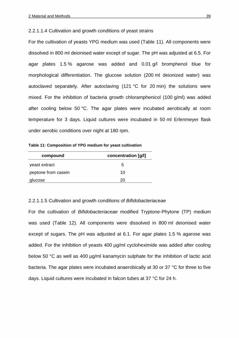
2.2.1.1.4 Cultivation and growth conditions of yeast strains
For the cultivation of yeasts YPG medium was used (Table 11). All components were
dissolved in 800 ml deionised water except of sugar. The pH was adjusted at 6.5. For
agar plates 1.5 % agarose was added and 0.01 g/l bromphenol blue for
morphological differentiation. The glucose solution (200 ml deionized water) was
autoclaved separately. After autoclaving (121 °C for 20 min) the solutions were
mixed. For the inhibition of bacteria growth chloramphenicol (100 g/ml) was added
after cooling below 50 °C. The agar plates were incubated aerobically at room
temperature for 3 days. Liquid cultures were incubated in 50 ml Erlenmeyer flask
under aerobic conditions over night at 180 rpm.
Table 11: Composition of YPG medium for yeast cultivation
compound concentration [g/l]
yeast extract 5
peptone from casein 10
glucose 20
2.2.1.1.5 Cultivation and growth conditions of Bifidobacteriaceae
For the cultivation of Bifidobacteriaceae modified Tryptone-Phytone (TP) medium
was used (Table 12). All components were dissolved in 800 ml deionised water
except of sugars. The pH was adjusted at 6.1. For agar plates 1.5 % agarose was
added. For the inhibition of yeasts 400 µg/ml cycloheximide was added after cooling
below 50 °C as well as 400 µg/ml kanamycin sulphate for the inhibition of lactic acid
bacteria. The agar plates were incubated anaerobically at 30 or 37 °C for three to five
days. Liquid cultures were incubated in falcon tubes at 37 °C for 24 h.
2 Material and Methods 40
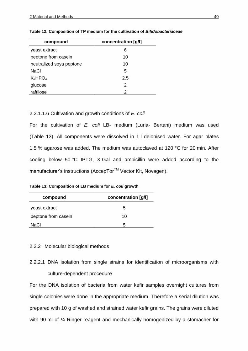
Table 12: Composition of TP medium for the cultivation of Bifidobacteriaceae
compound concentration [g/l]
yeast extract 6
peptone from casein 10
neutralized soya peptone 10
NaCl 5
K2HPO4 2.5
glucose 2
raftilose 2
2.2.1.1.6 Cultivation and growth conditions of E. coli
For the cultivation of E. coli LB- medium (Luria- Bertani) medium was used
(Table 13). All components were dissolved in 1 l deionised water. For agar plates
1.5 % agarose was added. The medium was autoclaved at 120 °C for 20 min. After
cooling below 50 °C IPTG, X-Gal and ampicillin were added according to the
manufacturer’s instructions (AccepTorTM Vector Kit, Novagen).
Table 13: Composition of LB medium for E. coli growth
compound concentration [g/l]
yeast extract 5
peptone from casein 10
NaCl 5
2.2.2 Molecular biological methods
2.2.2.1 DNA isolation from single strains for identification of microorganisms with
culture-dependent procedure
For the DNA isolation of bacteria from water kefir samples overnight cultures from
single colonies were done in the appropriate medium. Therefore a serial dilution was
prepared with 10 g of washed and strained water kefir grains. The grains were diluted
with 90 ml of ¼ Ringer reagent and mechanically homogenized by a stomacher for
2 Material and Methods 41
60 s. A serial dilution was prepared by mixing 1 ml of the grain suspension with 9 ml
of ¼ Ringer reagent. The different serial dilutions were plated on the different media
and incubated as described before. After the incubation the viable cell count was
enumerated on a proper dilution and the single colonies were picked from each
different plate. From every suitable serial dilution half of the colonies were picked and
first grown on the appropriate plate for 3 days. For the growth of lactobacilli liquid
MRS medium was used and for acetic acid bacteria liquid GM medium. The
lactobacilli were grown in 2 ml reaction tubes at 30 °C over night. The acetic acid
bacteria were grown in 50 ml Erlenmeyer flasks at 180 rpm and 30 °C over night. The
overnight cultures were centrifuged at 5000 g for 10 min. The pellet was washed with
1 ml TE buffer (1 mM EDTA, 10 mM Tris, pH 8) and centrifuged again. The pellets
were stored at -20 °C until further use. The DNA isolation was done with the Bacterial
DNA Kit according to the instructions. The pellets were resuspended with 200 µl TE
buffer containing lysozyme (10 mg/ml). The mixture was incubated at 37 °C for 1 h.
The DNA was eluted with two times 50 µl of elution buffer. Quantification of the
genomic DNA was done by agarose gel electrophoresis comparing band intensities
with known DNA ladders.
2.2.2.2 RAPD PCR
For the RAPD PCR isolated genomic DNA was used as template. The primer used
was the oligonucleotide primer M13V (5’-GTTTTCCCAGTCACGAC-3’). The PCR
reaction (25 µl) contained 25 pmol primer, 0.2 mM each deoxyribonucleoside
triphosphate, 3.5 mM MgCl2, reaction buffer, 0.75 U Taq polymerase and 1 µl of DNA
solution. Approximately the same amount of DNA (50-100 ng) was used. PCR was
carried out by using a Primus 96plus cycler. The amplification program was 94 °C for
45 s, 3 cycles of 94 °C for 3 min, 40 °C for 5 min, 72 °C for 5 min and 32 cycles 94 °C
for 1 min, 60 °C for 2 min, 72 °C for 3 min. All PCR products were mixed with 5 µl 6 x
2 Material and Methods 42
Loading dye (Fermentas) and then electrophoretically separated in a 1.3 % (w/ v)
agarose gel. Registration of the PCR patterns, normalization of the densitometric
traces, pattern storage, grouping of the strains using the Pearson product moment
correlation coefficient and UPGMA cluster analysis were performed using
BioNumcerics Version 6.50.
2.2.2.2.1 Identification of bacteria isolated with culture-dependent procedures
Isolated strains from water kefir samples showing different RAPD patterns were
analyzed by comparative 16S rDNA sequencing. The 16S rDNA was amplified with
the universal primer 616V (5’-AGAGTTTGATYMTGGCTCAG-3’) with a binding
position 7 according to (Brosius et al., 1981) and 609R
(5’-ACTACYVGGGTATCTAAKCC-3’) with a binding position 1099 according to
(Brosius et al., 1981). The PCR program was 94 °C for 2 min, 32 cycles of 94 °C for
45 s, 52 °C for 90 s, 72 °C for 2 min and a last step at 72 °C for 5 min. The reaction
mixture (50 µl) consisted of 0.1 mM of each deoxynucleoside triphosphate, 0.75 U
Taq polymerase, 5 pmol of each primer and 1 µl of the genomic DNA. The amplified
DNA had a length of 800 bp and was purified with the cycle pure kit (Omega bio-tek)
according to manufacturer’s instructions.
The sequencing was done by a commercial provider (GATC Biotech, Germany). The
identification of the bacteria was done with the BLAST program.
2.2.2.2.2 Yeast identification isolated with culture-dependent procedures
The isolated yeasts were identified by FTIR as described by (Kümmerle et al., 1998).
In the case of ambiguous FTIR results strains were identified by partial sequencing of
the 26S rDNA as described by (Kurtzman and and Robnett, 2003). For the
identification the GenBank/ EMBL/DDBJ accession numbers used were U72165
(Zygotorulaspora florentina) and U73596 (Hanseniaspora valbyensis).
2 Material and Methods 43
2.2.2.3 DNA isolation from water kefir grains for culture-independent procedures
The water kefir grains of three water kefirs (A, F and W) were propagated under
standardized conditions at least two times prior to analysis. One water kefir (water
kefir I) was analyzed directly after arrival from the supplier by post.
For the DNA isolation of water kefir grains 10 g of each water kefir grain was diluted
with 90 ml ¼ Ringer’s reagent and mechanically homogenized with a bag mixer for
60 s. Then, 5 ml of this solution was centrifuged at 5000 g for 10 min. The pellet was
washed with 2 ml TE buffer and centrifuged again.
For the DNA isolation of the supernatant of the water kefir 50 ml were centrifuged as
described above and the pellet was washed with 5 ml TE buffer and then treated the
same way as above described.
The pellets were stored at -20 °C until further use.
The DNA isolation was performed with the E.Z.N.A.TM Bacterial DNA kit according to
manufacturer’s instruction (also see 2.2.2.1).
2.2.2.3.1 16S rRNA gene amplification for high-throughput Pyrosequencing
The DNA isolated from each water kefir was used as a template for the amplification
of the V1 to V4 hyper-variable regions of the bacterial 16S rRNA gene with the Ba27 f
(Lane, 1991), (5’- AGAGTTTGATYMTGGCTCAG-3’) with a 16S binding position 8-27
according to Brimacombe et al., 1990 and Ba519 r primers (Lane, 1991),
(5’- TATTACCGCGGCKGCTG-3’) with a 16S binding position 519-534 according to
Brimacombe et al., 1990. The reaction mixture (50 µl) consisted of 0.1 mmol l-1 of
each deoxynucleoside triphosphate, 0.75 U Taq polymerase, 5 pmol of each primer
and 1 µl of the genomic DNA, and amplification was carried out using a PCR
2 Material and Methods 44
program of 94 °C for 2 min; 32 cycles of 94 °C for 45 s, 52 °C for 90 s and 72 °C for
2 min; and a final step at 72 °C for 5 min.
After the PCR reaction, the quality of the amplified PCR products (approximate
length: 520 nt) was confirmed by electrophoresis, and the products were purified
using a Cycle Pure Kit (Omega Bio-Tek) according to the manufacturer`s instructions.
A 20 µg sample of each amplicon was sequenced in a 454 Genome Sequencer FLX
Titanium by LGC Genomics (Berlin, Germany).
2.2.2.3.2 ARDRA analysis
DNA samples from the four water kefirs (see 2.2.2.3) were used as templates for
amplification with the specific bifidobacteria primers Bif164_mod_for (Langendijk et
al., 1995) with the sequence 5’- GGGTGGTAATGCCGRATG-3’ and a 16S binding
position of 164-181 according to Brimacombe et al., 1990 and LM3_mod_rev
(Kaufmann et al., 1997) with the sequence 5’- GGTGCTNCCCACTTTCATG-3’ and a
16S binding position of 1412-1432 according to Brimacombe et al., 1990.
The reaction mixture (50 µl) consisted of 0.1 mM of each deoxynucleoside
triphosphate, 0.75 U Taq polymerase, 5 pmol of each primer and 1 µl of the genomic
DNA, and the amplification was carried out using a PCR program of 94 °C for 5 min;
32 cycles of 94 °C for 1 min, 45 s of 60 °C and 72 °C for 2 min; and a final step at
72 °C for 5 min. The amplified DNA had a length of approx. 1400 nt and was purified
using the PeqGOLD Gel Extraction Kit (PeqLab, Germany) according to the
manufacturer’s instructions. The DNA was eluted in 30 µl elution buffer and stored at
4 °C until further use.
Amplicons were cloned in E. coli Top 10 using the AccepTorTM Vector Kit (Novagen).
Restriction analysis was performed on 100 clones from each kefir sample after
colony PCR using the above primers with mboI (GATC, Fermentas) according to the
2 Material and Methods 45
manufacturer’s instructions. Fragments were electrophoresed in a 1.3 % (w/ v)
agarose gel (0.5 x Tris-borate-EDTA buffer [45 mmol/ l Tris-borate, 1 mmol/ l EDTA]),
followed by staining with dimidium bromide.
Amplicons from the DNA of “B. crudilactis” LMG 21775, B. psychraerophilum DSM
22366T, B. longum DSM 20088T, B. animalis subsp. lactis DSM 10140T, B. lactis
BB12 and B. breve DSM 20213T were also included in this analysis.
2.2.2.3.3 16S rDNA analysis of Bifidobacteriaceae
For the analysis of the full 16S rDNA sequence of the novel uncultivated
Bifidobacterium species the universal 16S primers 616V with the sequence
5’-AGAGTTTGATYMTGGCTCAG-3’ (Brosius et al., 1981), 630R with the sequence
5’-CAKAAAGGAGGTGATCC-3’ (Juretschko et al., 2002) with the binding position of
7 and 15282 according to the 16S genes of E. coli respectively were combined with
newly designed specific primers Bif_spec_for (5’-GGATGTGGGACCCATTC-3’) and
Bif_spec_rev (5’-GAACCCGTGGAATGGGTC-3’) with the binding position 829-845
and 837-856 according to E. coli. The DNA from water kefir A was used as a
template. From this amplicon, 1525 nt were sequenced and submitted to the EMBL
database under the accession number HE804184.
For the identification of B. psychraerophilum the primers LM3_mod_rev with the
sequence 5’-GGTGCTNCCCACTTTCATG-3’ (Kaufmann et al., 1997) and
Bif164_mod_for with the sequence 5’- GGGTGGTAATGCCGRATG-3’ (Langendijk et
al., 1995) were used with the binding position 1412-1432 and 164-181 according to
the 16S gene of E. coli respectively.
2 Material and Methods 46
2.2.2.4 Influence of the composition of water kefir microbiota by changing growth
conditions
The water kefir grain (W) was grown under standardized conditions so that the
procedure could be done from one batch water kefir grain. These water kefir grains
were cultured under five different conditions (Table 14). The different extracts were
prepared by mixing 480 g of the dried fruit in 1 l of mineral water (Residenzquelle,
naturell). The fruits were cut into small pieces and then manually shaken for 20 min.
The extracts were centrifuged at 17 000 g for 3 h and sterilized using a 0.20 µm
membrane filter unit (Sarstedt, Germany). The extracts were stored at -20 °C until
further use. The concentration of the extract was 100 ml/l. The amount of kefir grain
was 29 g for each preparation. The different kefir samples were incubated at the
different temperatures whereas the standard, cranberry and the apricot sample was
incubated at 21 °C, one sample was incubated at 37 °C and one sample at 12 °C.
The grains fermented three days and every third day the decrease or increase of the
wet grain masse was measured. Therefore the grains were washed with tap water,
strained and weighed. This procedure was repeated over 24 days.
After 24 days the cfu was enumerated by plating the different samples on MRS, GM,
YPG and TP agar plates with appropriate serial dilutions.
Table 14: Different culture conditions for water kefir grains.
standard *CE **AE 37 °C 12 °C
8 % sucrose solution
8 % sucrose solution
8 % sucrose solution
8 % sucrose solution
8 % sucrose solution
fig extract cranberry extract apricot extract fig extract fig extract
grains grains grains grains grains
21 °C 21 °C 21 °C 37 °C 12 °C *CE= cranberry extract, ** AE= apricot extract
2 Material and Methods 47
2.2.2.4.1 Ribosomal intergenic spacer analysis (RISA)
At the end of the fermentation (after three days) the DNA was isolated from the
grains and the supernatant of the different samples. DNA isolation see 2.2.2.3. With
the DNA a PCR was performed with the universal primer sets 23S r
(5’-GGGTTBCCCCATTCRG-3’) and 1406 f (5’-TGYACACACCGCCCGT-3’). The
primers were respectively complementary to positions 1320 and 1336 of the
16S rRNA and131 and 115 of the 23S rRNA genes of E. coli (Cardinale et al., 2004).
The reaction mixture (50 µl) consisted of 0.1 mM of each deoxynucleoside
triphosphate, 0.75 U Taq polymerase, 5 pmol of each primer and 1 µl of the genomic
DNA. The PCR program was 94 °C for 2 min and 30 cycles of 94 °C for 15 s, 55 °C
for 15 s and 75 °C for 45 s followed by a final step at 75 °C for 5 min.
With the PCR solution a 15 % acrylamide gel was performed with the components
5 xTBE, deionized water and acrylamide. The amount of 16 µl PCR product was
mixed with 6 µl 10 x loading dye. The analysis of the band patterns was done after
running the gel 5 h at 100 V. The gel was stained with dimidium bromide.
2.2.2.5 Freeze-drying experiment with water kefir grains
For the freeze-drying experiment water kefir grains were cultured under standardized
conditions to guarantee that the different set up of the experiment were done from
one batch of water kefir grains.
The water kefir grains grown under standardized conditions were frozen over night at
different temperatures at -12 °C, -20 °C and -80 °C respectively. One sample was
quick-frozen by liquid nitrogen directly before the freeze-drying procedure. The water
kefir grains were put into 50 ml Falcon tubes and in the caps holes were punctured
with sterile canula to ensure sublimation. The freeze-drying took place over 24 h with
FreeZone Plus 2.5 l Cascade Benchtop Freeze Dry System (Labconco) with a
2 Material and Methods 48
vacuum set point of 0.0 mBar and a drying chamber set point at -40 °C with a
transmission rate of 10 s.
After the freeze-drying the viable cell count was determined by plating serial dilutions
of water kefir grains on different media (MRS, GM, YPG and TP) and incubate them.
The rest of the water kefir grains were regrown under standardized conditions with
the same amount of grains (40 g). Every third day the increase or decrease of the
grain amount was measured and with the grains a new fermentation was started.
After a few fermentation steps the viable cell count was determined again.
From each medium approximately 50 single colonies were picked and streaked on
new plates for obtaining pure single cultures. The identification of the microorganisms
was done by MALDI-TOF MS see 2.2.4.
2.2.3 Data processing and 16S rDNA sequence analysis
The data obtained from high-throughput pyrosequecing was analyzed as follows.
The initial sequence analysis was performed using the classifier software
(RDP Naïve Bayesian rRNA Classifier Version 2.4 (Wang et al., 2007)) provided in
Ribosomal Database project II (Cole et al., 2009) to estimate the microbial diversity at
the genus/family level. Sequences shorter than 150 nt were removed. The
confidence threshold was at least 80 %. Sequences with less than 97 % similarity to
the sequences deposited in RDP II were classified as unidentified.
Taxon assignment for the 16S rDNA sequences was performed in the ARB software
package (http://www.arb-home.de; (Ludwig et al., 2004; Westram et al., 2011)and
using the SILVA (http://www.arb-silva.de; (Pruesse et al., 2007) and LTP (all-species
Living Tree Project; http://www. arb-silva.de/projects/living-tree;(Munoz et al., 2011;
Yarza et al., 2008)) databases. Only sequence reads of at least 400 nucleotides were
2 Material and Methods 49
included for comparative data analysis. Preliminary taxon assignment above the
species level was roughly achieved by applying the classifier software (Wang et al.,
2007) provided by the RDP (Ribosomal Database Project; (Cole et al., 2009)and the
PT-server (Positional Tree server;(Ludwig et al., 2004; Westram et al., 2011))-based
‘next relative search’ of the ARB package.
For a more detailed taxon assignment, the species differentiation capacity of the
16S rRNA marker for the relevant taxonomic groups extracted from the LTP
database was evaluated. The current species threshold is set at 98.7 % identity over
the entire 16S rRNA sequence (Stackebrandt and Ebers, 2006). As there are many
type strains that share higher similarities, 16S rRNA-based assignment is only
possible for closely related species groups in such cases. The species threshold
value cannot be applied directly to analyse partial rDNA sequence data, such as that
derived in the present study. This analysis required that the (non-linear) correlation of
full and partial sequence-derived similarities be determined for the individual type
strain groups and that the respective species-(group) thresholds be delineated.
These similarity-based thresholds were used to define the respective score value
ranges obtained by applying the ARB PT-server approach (Ludwig et al., 2004;
Westram et al., 2011). This suffix tree-based search for common heptanucleotide
occurrences in the source (pyrosequencing) and reference (type strain) data allows a
phylogenetic similarity analysis without the need for sequence alignment. The
underlying principle is based on the finding that multiple occurrences of identical
heptanucleotide stretches are highly unlikely in 16S rRNA primary structures (our
unpublished data). Thus, sequences from diverse sources that contain identical
heptanucleotides can be regarded as homologous. PT-server score thresholds were
defined for two levels: the species or (lowest) species-group level and the lowest
level that clearly separates the respective species (group) from all other type species.
2 Material and Methods 50
The resulting three score ranges allow a sequence to be assigned to its respective
species or species-group, its phylogenetic lineage, or to higher taxa, respectively
(Table 19). The high error rate in sequence data obtained by high-throughput
methods (Balzer et al., 2010) must be taken into account when interpreting these
score ranges. Lower scores may represent as-yet undescribed species or simply low-
quality data.
2.2.4 MALDI-TOF MS analysis
2.2.4.1 Sample preparation
For the identification of microorganisms single colonies on agar plates were needed.
The bacteria or yeasts were grown over night on the appropriate agar plate in the
way that single colonies were obtained. These single colonies were analyzed by
MALDI-TOF MS.
2.2.4.2 Preparation of MALDI matrix
For the preparation of the MALDI matrix A-Cyano-4-hydroxy-cinnamic acid (sigma-
Aldrich Chemie GmbH), acetonitrile (ChemLab GmbH), trifluoroacetic acid (Merck
KGaA and deionzied water were used.
2.2.4.3 Sample application onto MALDI target
The preparation of the MALDI target was done under sterile work flow. Single
colonies were picked from the plates and streaked on the target. This sample was
overlaid with 1 µl of acetic acid and dried. After that the sample was overlaid with 1 µl
matrix and dried again for co-crystallization.
2.2.4.4 Cleaning of MALDI target
The cleaning of the MALDI target was done according to manufacturer’s instructions.
The target was overlaid with 70 % ethanol for 5 min, then washed with hot tap water
2 Material and Methods 51
and wiped with 70 % ethanol subsequently. The target was overlaid with 100 µl 80 %
trifluoroacetic acid and wiped again. A final cleaning was done with deionized water
and dried by wiping.
2.2.5 Fluorescence in situ hybridization (FISH) analysis
For the analysis the water kefir was grown under standardized conditions. The water
kefir grain and the supernatant were analyzed with FISH.
The whole water kefir grain and the supernatant were and fixed with Ethanol (50 %)
and 4 % PFA for 12 h separately. The following steps were done according to
(Pavlekovic et al., 2009). The water kefir grain was smashed onto a microscopic slide
for hybridization.
The probes used are shown in Table 15. The probe EUB338-I for the water kefir
grain was labeled with FLUOS (5(6)-carboxyfluorescein-N-hydroxysuccinimde ester),
the probe HGC 69a with the hydrophilic sulphoindocyanine dye Cy5. The
supernatant was analyzed with the probe probe HGC 69a labeled with the hydrophilic
sulphoindocyanine dye CY3. The probes were ordered from Thermo Electron GmbH,
Ulm, Germany.
Table 15: Probes used for the FISH analysis of water kefir
probe target sequence (5'-3') reference
EUB 338-I all bacteria GCTGCCTCCCGTAGGAGT (Amann et al., 1990a)
HGC 69a actinobacteria TATAGTTACCACCGCCGT (Roller et al., 1994)
For the visualization a Zeiss LSM 510 laser scanning confocal microscope (Carl
Zeiss, Göttingen, Germany) was used equipped with two Helium/ Neon Lasers, one
Argon-ion Laser and one UV-Laser.
The experiment was done on the Department of Microbiology (TU München) at the
Lab of Microbial Systems Ecology under the guidance of Dr. NM Lee.

3 Results 52
3 Results
3.1 Microbial diversity of water kefir grains analyzed with culture- dependent
procedures
3.1.1 Quantification of bacteria and yeasts
The microbiota of three independent water kefir grains (water kefir A, F and W) from
different origin were analyzed. The bacteria grown on the MRS and GM agar were
enumerated and the colony forming units were determined.
The viable cell numbers (cfu/g) of the bacteria in the three water kefirs ranged from
1.2*106 to 5.6*108, the cell count on MRS medium was in the same range in all three
water kefirs (1.6*108 in water kefir A, 1.3*108 in water kefir F and 1.3*108 in water
kefir W) whereas the cell count on GM showed differences, water kefir A and F had a
cell count of 3.7*106 and 1.2*106 but the cell count of water kefir W was higher
(5.6*108) (Table 16).
The viable yeast cells counted on water kefir A and F were similar with a cell count of
6.4*106 in water kefir A and 5.8*106 in water kefir F. The viable yeast cell count in
water kefir W was also higher with 2.7*107 cells.
Table 16: Viable cell counts (cfu/ g) of the three water kefirs obtained on different media
media viable cell count wk A
viable cell count wk F
viable cell count wk W
MRS 1.6*108 1.3*108 1.3*108
GM 3.7*106 1.2*106 5.6*108
YPG 6.4*106 5.8*106 2.7*107
3 Results 53
3.1.2 Identification of microorganisms
The microbiota of the three different water kefir grains were analyzed, therefore 453
bacterial isolates were obtained and the bacteria showing differences in their RAPD
patterns were identified by 16S rDNA. The 453 bacterial isolates showed 28 different
RAPD patterns and eight different bacterial species could be identified as
Lactobacillus casei, Lb. hordei, Lb. nagelii, Lb. hilgardii, Leuconostoc mesenteroides,
Lc. citreum, Acetobacter fabarum and Ac. orientalis. These species showed
differences in their RAPD patterns, and differences in the patterns could be detected
also among the identified single strains. These differences were mostly not very
pronounced and might represent subspecies or biotypes of the identified species
(Figure 2) but there were strains of the same species, which showed completely
different patterns like Lc. mesenteroides TMW 2.1076 and TMW 2.1073, Lb. nagelii
TMW 1.1826 and 1.1825, Lb. hordei 1.1907 and TMW 1.1959 and Ac. fabarum
2.1192, 2.1197 and 2.1198.
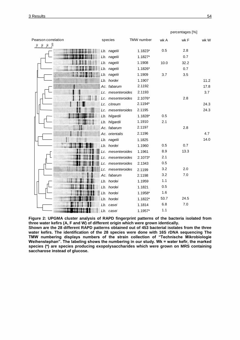
3 Results 54
Figure 2: UPGMA cluster analysis of RAPD fingerprint patterns of the bacteria isolated from three water kefirs (A, F and W) of different origin which were grown identically. Shown are the 28 different RAPD patterns obtained out of 453 bacterial isolates from the three water kefirs. The identification of the 28 species were done with 16S rDNA sequencing The TMW numbering displays numbers of the strain collection of “Technische Mikrobiologie Weihenstephan”. The labeling shows the numbering in our study. Wk = water kefir, the marked species (*) are species producing exopolysaccharides which were grown on MRS containing saccharose instead of glucose.
0.5 2.8
0.7
10.0 32.2
0.7
3.7 3.5
11.2
17.8
3.7
2.8
24.3
24.3
0.5
2.1
2.8
4.7
14.0
0.5 0.7
8.9 13.3
2.1
0.5
3.2 2.0
3.2 7.0
1.1
0.5
1.6
53.7 24.5
6.8 7.0
1.1
percentages [%]
wk A wk F wk W
Lb.
Lb.
Lb.
Lb.
Lb.
Lb.
Ac.
Lb.
Lb.
Ac.
Ac.
Lb.
Lb.
.
.
.
.
.
.
.
.
.
.
.
.
.
.
.
.
.
.
.
.
.
.
.
.
.
.
.
.
Lc.
Lc.
Lc.
Lc.
Lc.
Lc.
Lc.
Lc.
Ac.
Lb.
Lb.
Lb.
Lb.
Lb.
Lb.
nagelii
nagelii
nagelii
nagelii
nagelii
hordei
fabarum
mesenteroides
mesenteroides
citreum
mesenteroides
hilgardii
hilgardii
fabarum
orientalis
nagelii
hordei
mesenteroides
mesenteroides
mesenteroides
mesenteroides
fabarum
hordei
hordei
hordei
hordei
casei
casei
Pearson correlation species
80
60
40
100
TMW number
1.1823*
1.1827*
1.1826*
2.1076*
1.1828*
1.1960
1.1961
2.1073*
2.1343
1.1959
1.1821
1.1958*
1.1822*
1.1814
1.1957*
1.1908
1.1909
1.1907
2.1192
2.1193
2.1194*
2.1195
1.1910
2.1197
2.1196
1.1825
2.1199
2.1198
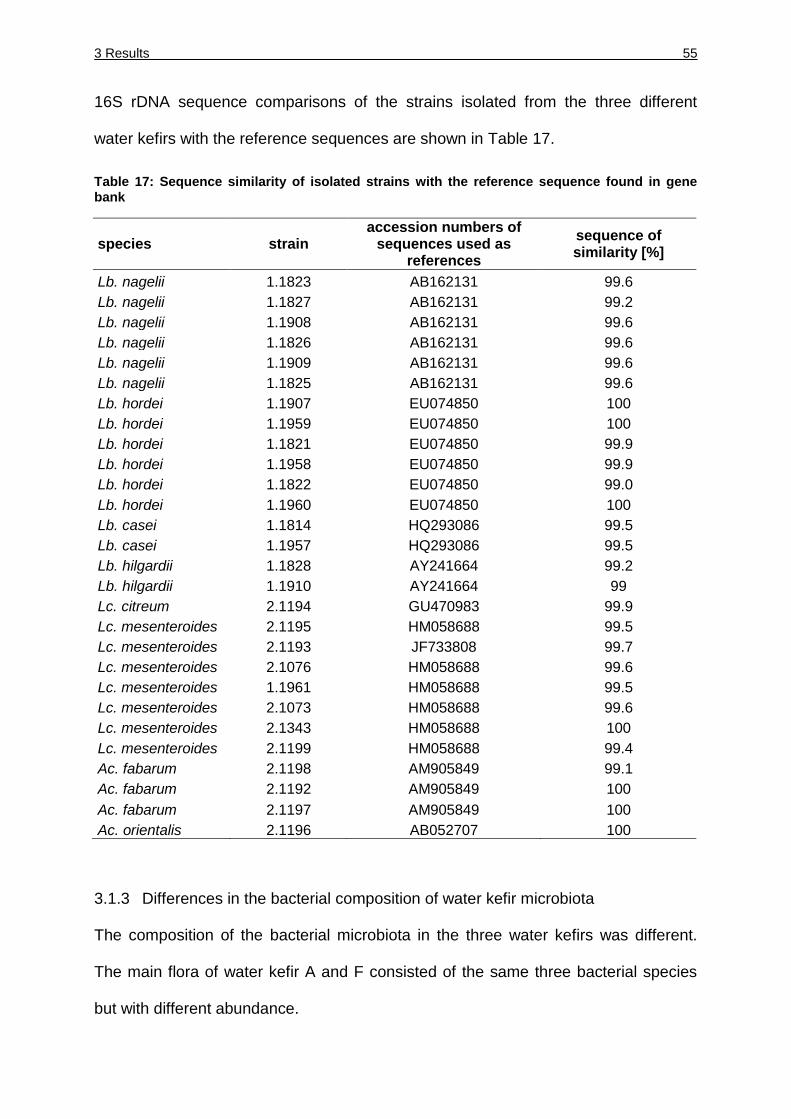
3 Results 55
16S rDNA sequence comparisons of the strains isolated from the three different
water kefirs with the reference sequences are shown in Table 17.
Table 17: Sequence similarity of isolated strains with the reference sequence found in gene bank
species strain accession numbers of
sequences used as references
sequence of similarity [%]
Lb. nagelii 1.1823 AB162131 99.6
Lb. nagelii 1.1827 AB162131 99.2
Lb. nagelii 1.1908 AB162131 99.6
Lb. nagelii 1.1826 AB162131 99.6
Lb. nagelii 1.1909 AB162131 99.6
Lb. nagelii 1.1825 AB162131 99.6
Lb. hordei 1.1907 EU074850 100
Lb. hordei 1.1959 EU074850 100
Lb. hordei 1.1821 EU074850 99.9
Lb. hordei 1.1958 EU074850 99.9
Lb. hordei 1.1822 EU074850 99.0
Lb. hordei 1.1960 EU074850 100
Lb. casei 1.1814 HQ293086 99.5
Lb. casei 1.1957 HQ293086 99.5
Lb. hilgardii 1.1828 AY241664 99.2
Lb. hilgardii 1.1910 AY241664 99
Lc. citreum 2.1194 GU470983 99.9
Lc. mesenteroides 2.1195 HM058688 99.5
Lc. mesenteroides 2.1193 JF733808 99.7
Lc. mesenteroides 2.1076 HM058688 99.6
Lc. mesenteroides 1.1961 HM058688 99.5
Lc. mesenteroides 2.1073 HM058688 99.6
Lc. mesenteroides 2.1343 HM058688 100
Lc. mesenteroides 2.1199 HM058688 99.4
Ac. fabarum 2.1198 AM905849 99.1
Ac. fabarum 2.1192 AM905849 100
Ac. fabarum 2.1197 AM905849 100
Ac. orientalis 2.1196 AB052707 100
3.1.3 Differences in the bacterial composition of water kefir microbiota
The composition of the bacterial microbiota in the three water kefirs was different.
The main flora of water kefir A and F consisted of the same three bacterial species
but with different abundance.
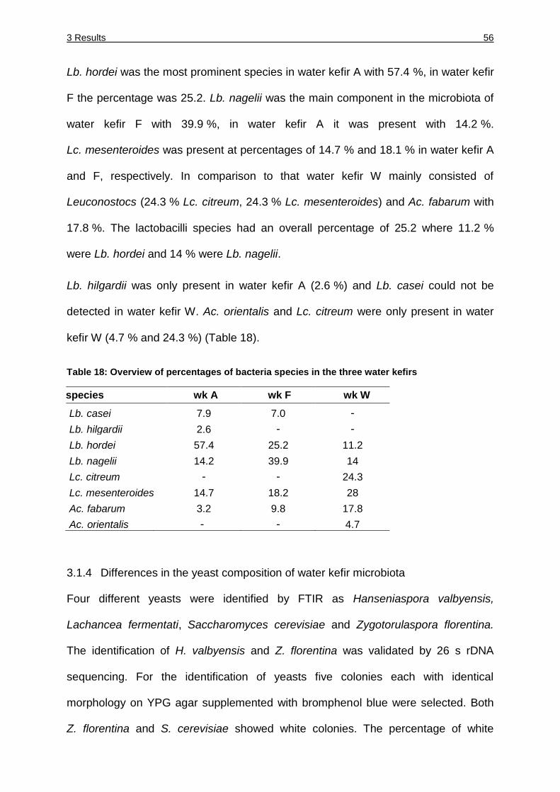
3 Results 56
Lb. hordei was the most prominent species in water kefir A with 57.4 %, in water kefir
F the percentage was 25.2. Lb. nagelii was the main component in the microbiota of
water kefir F with 39.9 %, in water kefir A it was present with 14.2 %.
Lc. mesenteroides was present at percentages of 14.7 % and 18.1 % in water kefir A
and F, respectively. In comparison to that water kefir W mainly consisted of
Leuconostocs (24.3 % Lc. citreum, 24.3 % Lc. mesenteroides) and Ac. fabarum with
17.8 %. The lactobacilli species had an overall percentage of 25.2 where 11.2 %
were Lb. hordei and 14 % were Lb. nagelii.
Lb. hilgardii was only present in water kefir A (2.6 %) and Lb. casei could not be
detected in water kefir W. Ac. orientalis and Lc. citreum were only present in water
kefir W (4.7 % and 24.3 %) (Table 18).
Table 18: Overview of percentages of bacteria species in the three water kefirs
sspecies wk A wk F wk W
Lb. casei 7.9 7.0 -
Lb. hilgardii 2.6 - -
Lb. hordei 57.4 25.2 11.2
Lb. nagelii 14.2 39.9 14
Lc. citreum - - 24.3
Lc. mesenteroides 14.7 18.2 28
Ac. fabarum 3.2 9.8 17.8
Ac. orientalis - - 4.7
3.1.4 Differences in the yeast composition of water kefir microbiota
Four different yeasts were identified by FTIR as Hanseniaspora valbyensis,
Lachancea fermentati, Saccharomyces cerevisiae and Zygotorulaspora florentina.
The identification of H. valbyensis and Z. florentina was validated by 26 s rDNA
sequencing. For the identification of yeasts five colonies each with identical
morphology on YPG agar supplemented with bromphenol blue were selected. Both
Z. florentina and S. cerevisiae showed white colonies. The percentage of white
3 Results 57
colonies was 94, 92 and 93 % of all yeast colonies, in water kefir A, F and W,
respectively. S. cerevisiae was identified by FTIR in all three water kefirs but
indistinguishable from Z. florentina on plates due to the same morphology, and
therefore no precise quantification of each species could be achieved. Water kefir W
was studied in a more detailed analysis where 44 white colonies were identified by
FTIR and 16 colonies were identified as S. cerevisiae and 25 colonies were
Z. florentina.
L. fermentati formed a white colony with a blue center and was present in water kefirs
A and F with 6 % and 8 %, respectively. H. valbyensis formed a white colony with a
blue center and a blue circle and was present to 7 % in water kefir W.
3.2 Bacterial diversity of water kefir grains analyzed with culture-independent
procedures
3.2.1 Bacterial identification and distribution determined by high-throughput
sequence-based analysis
The bacterial microbiota of four independently grown water kefir grains from different
origins were analyzed by high-throughput sequence-based analysis. Three water
kefirs (A, F and W) were grown at least two times under standardized conditions to
ensure that they all are propagated under identical conditions and to eliminate
possible influences of the microbiota resulting from cultivation procedures of the
supplier. Water kefir I was analyzed directly after arrival from the supplier to detect
any kind of differences in the microbiota resulting from growing conditions. A total of
43293 (kefir A), 33079 (kefir F), 25951 (kefir W) and 41404 (kefir I) reads of the V4
region of the 16S rRNA were analyzed.
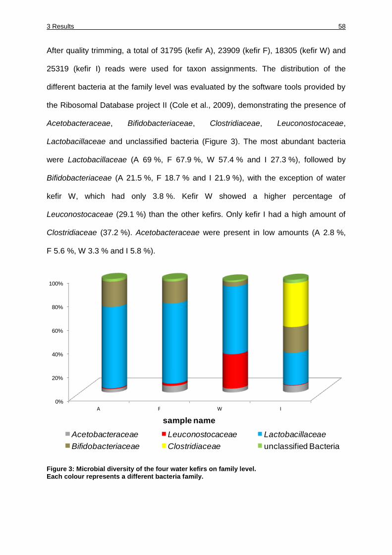
3 Results 58
After quality trimming, a total of 31795 (kefir A), 23909 (kefir F), 18305 (kefir W) and
25319 (kefir I) reads were used for taxon assignments. The distribution of the
different bacteria at the family level was evaluated by the software tools provided by
the Ribosomal Database project II (Cole et al., 2009), demonstrating the presence of
Acetobacteraceae, Bifidobacteriaceae, Clostridiaceae, Leuconostocaceae,
Lactobacillaceae and unclassified bacteria (Figure 3). The most abundant bacteria
were Lactobacillaceae (A 69 %, F 67.9 %, W 57.4 % and I 27.3 %), followed by
Bifidobacteriaceae (A 21.5 %, F 18.7 % and I 21.9 %), with the exception of water
kefir W, which had only 3.8 %. Kefir W showed a higher percentage of
Leuconostocaceae (29.1 %) than the other kefirs. Only kefir I had a high amount of
Clostridiaceae (37.2 %). Acetobacteraceae were present in low amounts (A 2.8 %,
F 5.6 %, W 3.3 % and I 5.8 %).
Figure 3: Microbial diversity of the four water kefirs on family level. Each colour represents a different bacteria family.
0%
20%
40%
60%
80%
100%
A F W I
sample name
Acetobacteraceae Leuconostocaceae Lactobacillaceae
Bifidobacteriaceae Clostridiaceae unclassified Bacteria
3 Results 59
The suffix tree-based search for the most similar type strain sequences was used to
assign the sequences to genera, species groups or species (Table 19). With this
method, we were able to identify 12 bacterial species and 12 bacterial groups. The
main species of water kefirs A and F consisted of members of the Lb. hordei group,
Lb. nagelii, and Bifidobacterium spp.; water kefir A also contained species from the
B. psychraerophilum group; and water kefir F had a greater amount of Ac. fabarum
group members. The most abundant sequences in water kefir I were from members
of the B. psychraerophilum group, Lb. hordei group, Lb. hilgardii, Lb. satsumensis,
Ac. orientalis group and C. tyrobutyricum. The core species of water kefir W were
from the Lb. hordei group, Lb. nagelii, Lb. hilgardii, Lc. citreum group and Lc.
mesenteroides group.
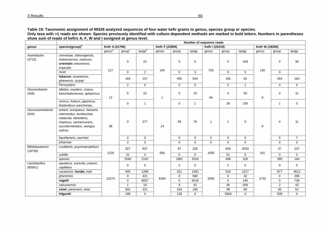
3 Results 60
Table 19: Taxonomic assignment of 99328 analyzed sequences of four water kefir grains to genus, species group or species. Only taxa with >1 reads are shown. Species previously identified with culture-dependent methods are marked in bold letters. Numbers in parentheses show sum of reads of kefirs A, F, W and I assigned at genus level. Number of sequence reads
genus species(group)a Kefir A (31795) Kefir F (23909) Kefir I (25319) Kefir W (18305)
genusb group
c sp/gp
d genus group sp/gp genus group sp/gp genus group sp/gp
Acetobacter
(3715)
cerevisiae, cibinongensis,
indonesiensis, malorum,
orientalis orleanensis,
tropicalis 127
0 21
145
0 5
726
0 428
130
0 36
Aceti 0 2 0 3 0 0 0
fabarum, iovaniensis,
ghanensis, syzygii 183 157 492 544 185 92 264 163
Peroxydans 2 0 0 0 5 1 4 0
Gluconobacter
(359)
albidus, oxydans, roseus,
kanchanburiensis, sphaericus
12
5 22
1
0 10
84
4 50
9
4 11
cerinus, frateuri, japonicus,
thailandicus wancheriae, 0 1 0 1 39 105 1 0
Gluconacetobacter
(504)
entanii, europaeus, hansenii,
intermedius, kombuchae,
nataicola, oboediens,
rhaeticus, sacharivorans,
sucrofermentans, swingsii,
xylinus
38
0 277
14
59 76 1 1 0
9
4 11
liquefaciens, sacchari 0 0 0 0 0 0 0 5 7
johannae 2 0 0 0 0 0 0 0 0
Bifidobacterium
(18759)
crudilactis, psychraerophilum
1220
327 837
556
97 226
1655
659 3033
161
37 107
subtile 31 0 0 0 51 0 0 0
species 2540 2142 1881 2016 408 326 285 164
Lactobacillus
(60561)
aquaticus, sucicola, uvarum,
capillatus
12574
0 0
9184
2 0
3266
2 0
2732
9 0
cacaonum, hordei, mali 405 1266 421 1492 316 1217 877 4612
ghanensis 0 421 0 580 0 32 0 289
nagelii 0 8057 0 5018 0 145 0 738
satsumensis 1 19 8 91 36 509 2 43
casei, paracasei, zeae 502 221 319 166 98 60 65 52
hilgardii 188 0 128 0 3559 0 839 0
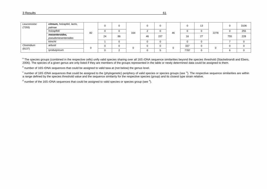
3 Results 61
Leuconostoc
(7293)
citreum, holzapfeli, lactis,
palmae
82
0 0
164
0 0
46
0 13
2278
0 3106
holzapfelii 0 0 2 0 0 0 0 255
mesenteroides,
pseudomesenteroides 24 86 46 157 16 27 755 228
kimchii 1 0 0 0 0 0 7 0
Clostridium
(8137)
arbusti 0
0 0 0
0 0 0
337 0 0
0 0
tyrobutyricum 0 2 0 5 7787 0 6 0
a The species groups (combined in the respective cells) unify valid species sharing over all 16S rDNA sequence similarities beyond the species threshold (Stackebrandt and Ebers,
2006). The species of a given genus are only listed if they are members of the groups represented in the table or newly determined data could be assigned to them.
b number of 16S rDNA sequences that could be assigned to valid taxa at (not below) the genus level.
c number of 16S rDNA sequences that could be assigned to the (phylogenetic) periphery of valid species or species groups (see
a). The respective sequence similarities are within
a range defined by the species threshold value and the sequence similarity for the respective species (group) and its closest type strain relative.
d number of the 16S rDNA sequences that could be assigned to valid species or species group (see
a).
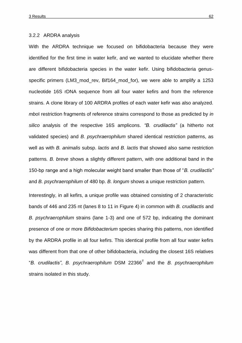
3 Results 62
3.2.2 ARDRA analysis
With the ARDRA technique we focused on bifidobacteria because they were
identified for the first time in water kefir, and we wanted to elucidate whether there
are different bifidobacteria species in the water kefir. Using bifidobacteria genus-
specific primers (LM3_mod_rev, Bif164_mod_for), we were able to amplify a 1253
nucleotide 16S rDNA sequence from all four water kefirs and from the reference
strains. A clone library of 100 ARDRA profiles of each water kefir was also analyzed.
mboI restriction fragments of reference strains correspond to those as predicted by in
silico analysis of the respective 16S amplicons. “B. crudilactis” (a hitherto not
validated species) and B. psychraerophilum shared identical restriction patterns, as
well as with B. animalis subsp. lactis and B. lactis that showed also same restriction
patterns. B. breve shows a slightly different pattern, with one additional band in the
150-bp range and a high molecular weight band smaller than those of “B. crudilactis”
and B. psychraerophilum of 480 bp. B. longum shows a unique restriction pattern.
Interestingly, in all kefirs, a unique profile was obtained consisting of 2 characteristic
bands of 446 and 235 nt (lanes 8 to 11 in Figure 4) in common with B. crudilactis and
B. psychraerophilum strains (lane 1-3) and one of 572 bp, indicating the dominant
presence of one or more Bifidobacterium species sharing this patterns, non identified
by the ARDRA profile in all four kefirs. This identical profile from all four water kefirs
was different from that one of other bifidobacteria, including the closest 16S relatives
“B. crudilactis”, B. psychraerophilum DSM 22366T and the B. psychraerophilum
strains isolated in this study.
3 Results 63
Figure 4: Agarose gel electrophoresis of 16S rRNA ARDRA profiles after restriction of 16S amplicons with mboI of Bifidobacterium species. The agarose gel shows the restriction pattern of “B. crudilactis” LMG 21775 (lane 1), B. psychraerophilum DSM 22366
T (lane 2), B. psychraerophilum from water kefir (lane 3),
B. longum DSM 20088T (lane 4), B. animalis subsp. lactis DSM 10140
T (lane 5), B. lactis DSM 58
(lane 6) and B. breve DSM 20213T (lane 7). Lane 8-11 show the ARDRA profiles obtained from
the four water kefirs A (lane 8), F (lane 9), W (lane 10) and I (lane 11) representing respectively 100 colonies restricted. A Generuler 100-bp ladder was used as size-marker.
3.2.3 Cultivation and quantification of bifidobacteria
The water kefirs W and I were analyzed representatively for Bifidobacteriaceae. The
bacteria were cultured on a modified tryptone-phytone (TP) agar, and the viable cell
numbers (cfu/ g) were enumerated. The number of Bifidobacteriaceae cells in water
kefir I was 4.9*105 cfu/g and that in water kefir W 1.5*106 cfu/g. An analysis of
representative colonies (at least 100 colonies of each water kefir) by PCR with the
species-specific primer (Bif_spec_for and Bif_spec_rev) (data not shown) showed
that all isolates grown on these plates belonged to the species B. psychraerophilum.
bifidobacteria cell numbers were not analyzed for the water kefirs F and A because
they were not physically available at that time.
100 bp
200 bp
500 bp
700 bp
1000 bp
3000 bp 1 2 3 4 5 6 7 8 9 10 11
3 Results 64
3.2.4 Phylogeny of Bifidobacterium sp.
The 16S rRNA gene sequence was amplified from the DNA samples isolated from
water kefir A in two fragments using a combination of a species-specific primer of the
‘new’ water kefir species (Bif_spec_for, Bif_spec_rev) and universal primers (616V,
630R). The almost complete 16S rRNA sequences (1525 nt) was determined.
Comparison of this sequence with those available in public databases revealed
B. psychraerophilum as the closest relative, with a sequence similarity of 96.4 %. The
subsequent phylogenetic classification supports a new candidate species of the
genus Bifidobacterium. The 16S rRNA gene sequence was submitted to the EMBL
database under the accession number HE804184.
3.3 Influence of the composition of water kefir microbiota by changing growth
conditions
3.3.1 Determination of the wet mass of water kefir grains
The influence of different fermentation parameters was analyzed and the alteration of
the microbiota from the water kefir grains and the supernatant were detected.
Therefore the water kefir grains of water kefir W were fermented under standardized
conditions to enlarge the amount of water kefir grains. From one batch water kefir the
different fermentations were started to ensure that the possible alteration of
microbiota results from the different fermentation parameters and not from the initial
start point. The decrease or increase of the wet mass of water kefir grains was
measured after three days of fermentation and a new fermentation was started. This
was done over 24 days and the grain mass is shown in Figure 5. The initial mass of
water kefir grains was 29 g in 40 ml fermentation solution.
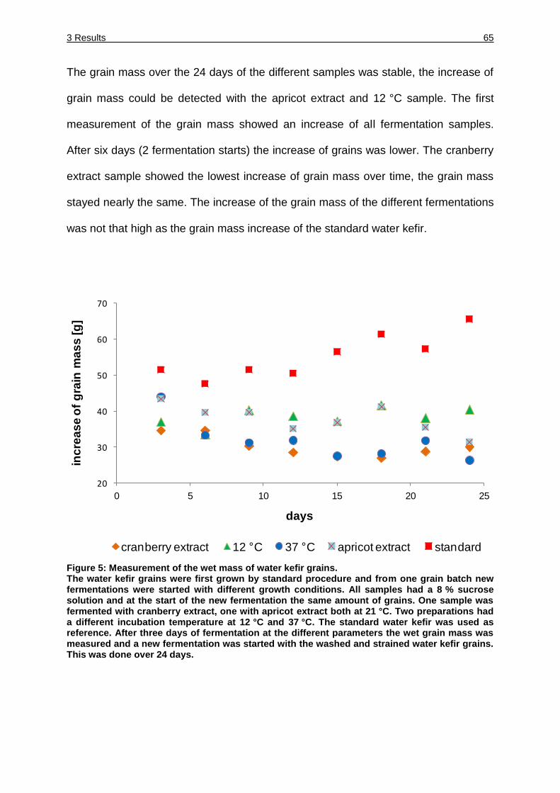
3 Results 65
The grain mass over the 24 days of the different samples was stable, the increase of
grain mass could be detected with the apricot extract and 12 °C sample. The first
measurement of the grain mass showed an increase of all fermentation samples.
After six days (2 fermentation starts) the increase of grains was lower. The cranberry
extract sample showed the lowest increase of grain mass over time, the grain mass
stayed nearly the same. The increase of the grain mass of the different fermentations
was not that high as the grain mass increase of the standard water kefir.
Figure 5: Measurement of the wet mass of water kefir grains. The water kefir grains were first grown by standard procedure and from one grain batch new fermentations were started with different growth conditions. All samples had a 8 % sucrose solution and at the start of the new fermentation the same amount of grains. One sample was fermented with cranberry extract, one with apricot extract both at 21 °C. Two preparations had a different incubation temperature at 12 °C and 37 °C. The standard water kefir was used as reference. After three days of fermentation at the different parameters the wet grain mass was measured and a new fermentation was started with the washed and strained water kefir grains. This was done over 24 days.
20
30
40
50
60
70
0 5 10 15 20 25
inc
rea
se
of
gra
in m
as
s [
g]
days
cranberry extract 12 C 37 C apricot extract standard
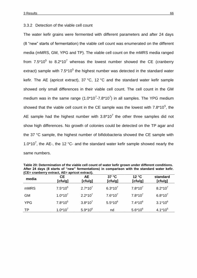
3 Results 66
3.3.2 Detection of the viable cell count
The water kefir grains were fermented with different parameters and after 24 days
(8 “new” starts of fermentation) the viable cell count was enumerated on the different
media (mMRS, GM, YPG and TP). The viable cell count on the mMRS media ranged
from 7.5*105 to 8.2*107 whereas the lowest number showed the CE (cranberry
extract) sample with 7.5*105 the highest number was detected in the standard water
kefir. The AE (apricot extract), 37 °C, 12 °C and the standard water kefir sample
showed only small differences in their viable cell count. The cell count in the GM
medium was in the same range (1.0*107-7.8*107) in all samples. The YPG medium
showed that the viable cell count in the CE sample was the lowest with 7.8*105, the
AE sample had the highest number with 3.8*107 the other three samples did not
show high differences. No growth of colonies could be detected on the TP agar and
the 37 °C sample, the highest number of bifidobacteria showed the CE sample with
1.0*107, the AE-, the 12 °C- and the standard water kefir sample showed nearly the
same numbers.
Table 20: Determination of the viable cell count of water kefir grown under different conditions. After 24 days (8 starts of “new” fermentations) in comparison with the standard water kefir. (CE= cranberry extract, AE= apricot extract).
media CE
[cfu/g] AE
[cfu/g] 37 °C
[cfu/g] 12 °C
[cfu/g] standard
[cfu/g]
mMRS 7.5*105 2.7*107 6.3*107 7.8*107 8.2*107
GM 1.0*107 2.2*107 7.6*107 7.8*107 6.8*107
YPG 7.8*105 3.8*107 5.5*106 7.4*106 3.1*106
TP 1.0*107 5.9*106 nd 5.6*106 4.1*105
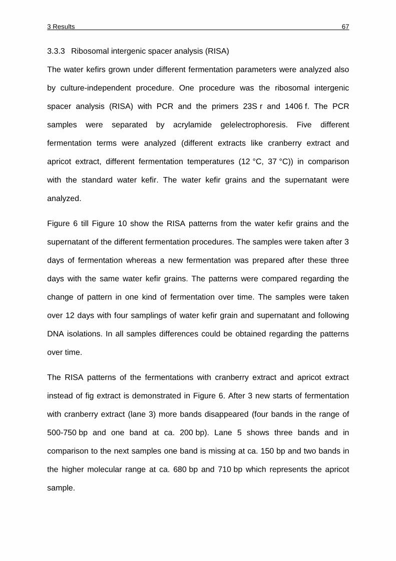
3 Results 67
3.3.3 Ribosomal intergenic spacer analysis (RISA)
The water kefirs grown under different fermentation parameters were analyzed also
by culture-independent procedure. One procedure was the ribosomal intergenic
spacer analysis (RISA) with PCR and the primers 23S r and 1406 f. The PCR
samples were separated by acrylamide gelelectrophoresis. Five different
fermentation terms were analyzed (different extracts like cranberry extract and
apricot extract, different fermentation temperatures (12 °C, 37 °C)) in comparison
with the standard water kefir. The water kefir grains and the supernatant were
analyzed.
Figure 6 till Figure 10 show the RISA patterns from the water kefir grains and the
supernatant of the different fermentation procedures. The samples were taken after 3
days of fermentation whereas a new fermentation was prepared after these three
days with the same water kefir grains. The patterns were compared regarding the
change of pattern in one kind of fermentation over time. The samples were taken
over 12 days with four samplings of water kefir grain and supernatant and following
DNA isolations. In all samples differences could be obtained regarding the patterns
over time.
The RISA patterns of the fermentations with cranberry extract and apricot extract
instead of fig extract is demonstrated in Figure 6. After 3 new starts of fermentation
with cranberry extract (lane 3) more bands disappeared (four bands in the range of
500-750 bp and one band at ca. 200 bp). Lane 5 shows three bands and in
comparison to the next samples one band is missing at ca. 150 bp and two bands in
the higher molecular range at ca. 680 bp and 710 bp which represents the apricot
sample.
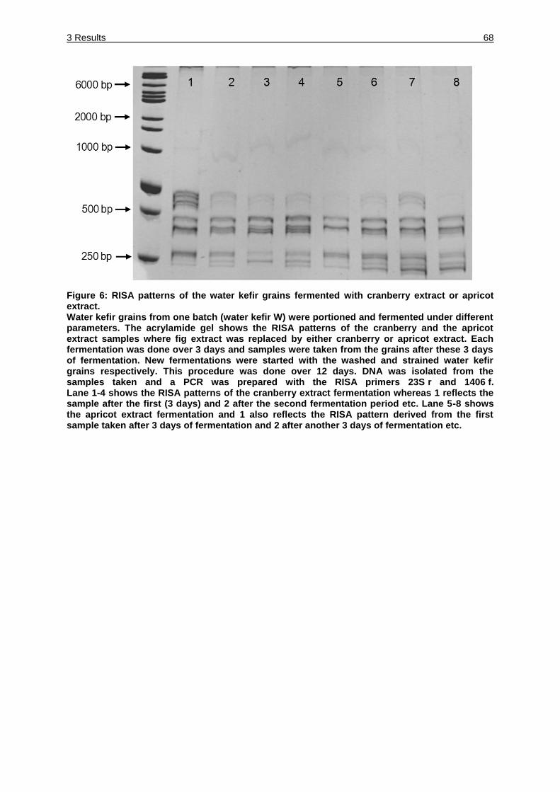
3 Results 68
Figure 6: RISA patterns of the water kefir grains fermented with cranberry extract or apricot extract. Water kefir grains from one batch (water kefir W) were portioned and fermented under different parameters. The acrylamide gel shows the RISA patterns of the cranberry and the apricot extract samples where fig extract was replaced by either cranberry or apricot extract. Each fermentation was done over 3 days and samples were taken from the grains after these 3 days of fermentation. New fermentations were started with the washed and strained water kefir grains respectively. This procedure was done over 12 days. DNA was isolated from the samples taken and a PCR was prepared with the RISA primers 23S r and 1406 f. Lane 1-4 shows the RISA patterns of the cranberry extract fermentation whereas 1 reflects the sample after the first (3 days) and 2 after the second fermentation period etc. Lane 5-8 shows the apricot extract fermentation and 1 also reflects the RISA pattern derived from the first sample taken after 3 days of fermentation and 2 after another 3 days of fermentation etc.
3 Results 69
The supernatant analysis of the cranberry and apricot fermentation (Figure 7) shows
differences in lane 3 and 4 which are showing three additional bands in comparison
to lane 1 and 2 (bands at 1200 bp, 2000 bp and at ca. 570 bp are missing). Lanes
6-8 have two additional bands in comparison to lane 5 at ca. 1200 bp and 2000 bp.
Figure 7: RISA patterns of the water kefir supernatant fermented with cranberry extract or apricot extract. Water kefir grains from one batch (water kefir W) were portioned and fermented under different parameters. The acrylamide gel shows the RISA patterns of the cranberry and the apricot extract samples where fig extract was replaced by either cranberry or apricot extract. Each fermentation was done over 3 days and samples were taken from the supernatant after these 3 days of fermentation. New fermentations were started with the washed and strained water kefir grains respectively. This procedure was done over 12 days. DNA was isolated from the samples taken and a PCR was prepared with the RISA primers 23S r and 1406 f. Lane 1-4 shows the RISA patterns of the supernatant of cranberry extract fermentation whereas 1 reflects the sample after the first (3 days) and 2 after the second fermentation period etc. Lane 5-8 shows the supernatant of the apricot extract fermentation and 1 also reflects the RISA pattern derived from the first sample taken after 3 days of fermentation and 2 after another 3 days of fermentation etc.
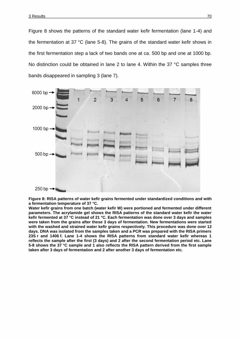
3 Results 70
Figure 8 shows the patterns of the standard water kefir fermentation (lane 1-4) and
the fermentation at 37 °C (lane 5-8). The grains of the standard water kefir shows in
the first fermentation step a lack of two bands one at ca. 500 bp and one at 1000 bp.
No distinction could be obtained in lane 2 to lane 4. Within the 37 °C samples three
bands disappeared in sampling 3 (lane 7).
Figure 8: RISA patterns of water kefir grains fermented under standardized conditions and with a fermentation temperature of 37 °C. Water kefir grains from one batch (water kefir W) were portioned and fermented under different parameters. The acrylamide gel shows the RISA patterns of the standard water kefir the water kefir fermented at 37 °C instead of 21 °C. Each fermentation was done over 3 days and samples were taken from the grains after these 3 days of fermentation. New fermentations were started with the washed and strained water kefir grains respectively. This procedure was done over 12 days. DNA was isolated from the samples taken and a PCR was prepared with the RISA primers 23S r and 1406 f. Lane 1-4 shows the RISA patterns from standard water kefir whereas 1 reflects the sample after the first (3 days) and 2 after the second fermentation period etc. Lane 5-8 shows the 37 °C sample and 1 also reflects the RISA pattern derived from the first sample taken after 3 days of fermentation and 2 after another 3 days of fermentation etc.
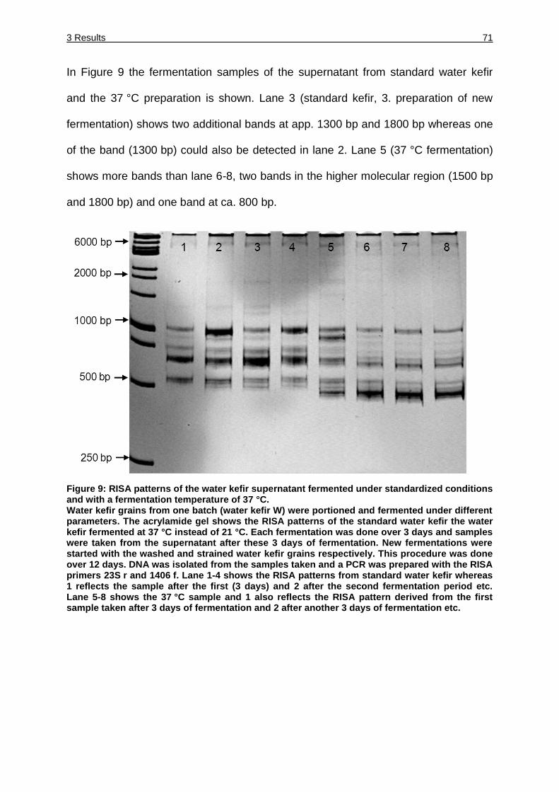
3 Results 71
In Figure 9 the fermentation samples of the supernatant from standard water kefir
and the 37 °C preparation is shown. Lane 3 (standard kefir, 3. preparation of new
fermentation) shows two additional bands at app. 1300 bp and 1800 bp whereas one
of the band (1300 bp) could also be detected in lane 2. Lane 5 (37 °C fermentation)
shows more bands than lane 6-8, two bands in the higher molecular region (1500 bp
and 1800 bp) and one band at ca. 800 bp.
Figure 9: RISA patterns of the water kefir supernatant fermented under standardized conditions and with a fermentation temperature of 37 °C. Water kefir grains from one batch (water kefir W) were portioned and fermented under different parameters. The acrylamide gel shows the RISA patterns of the standard water kefir the water kefir fermented at 37 °C instead of 21 °C. Each fermentation was done over 3 days and samples were taken from the supernatant after these 3 days of fermentation. New fermentations were started with the washed and strained water kefir grains respectively. This procedure was done over 12 days. DNA was isolated from the samples taken and a PCR was prepared with the RISA primers 23S r and 1406 f. Lane 1-4 shows the RISA patterns from standard water kefir whereas 1 reflects the sample after the first (3 days) and 2 after the second fermentation period etc. Lane 5-8 shows the 37 °C sample and 1 also reflects the RISA pattern derived from the first sample taken after 3 days of fermentation and 2 after another 3 days of fermentation etc.
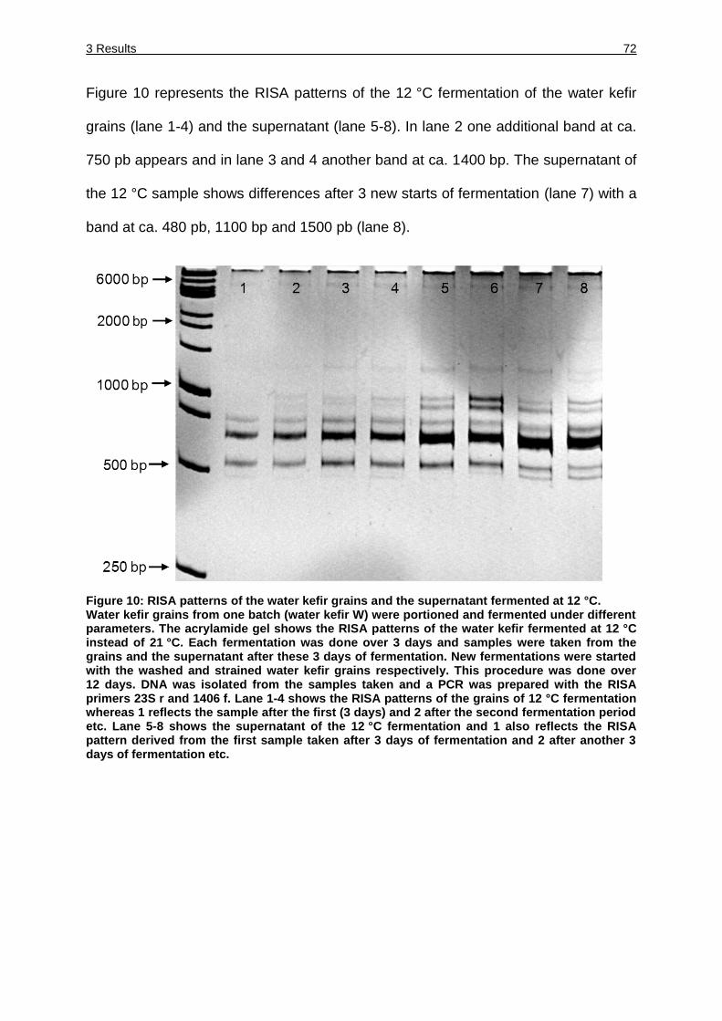
3 Results 72
Figure 10 represents the RISA patterns of the 12 °C fermentation of the water kefir
grains (lane 1-4) and the supernatant (lane 5-8). In lane 2 one additional band at ca.
750 pb appears and in lane 3 and 4 another band at ca. 1400 bp. The supernatant of
the 12 °C sample shows differences after 3 new starts of fermentation (lane 7) with a
band at ca. 480 pb, 1100 bp and 1500 pb (lane 8).
Figure 10: RISA patterns of the water kefir grains and the supernatant fermented at 12 °C. Water kefir grains from one batch (water kefir W) were portioned and fermented under different parameters. The acrylamide gel shows the RISA patterns of the water kefir fermented at 12 °C instead of 21 °C. Each fermentation was done over 3 days and samples were taken from the grains and the supernatant after these 3 days of fermentation. New fermentations were started with the washed and strained water kefir grains respectively. This procedure was done over 12 days. DNA was isolated from the samples taken and a PCR was prepared with the RISA primers 23S r and 1406 f. Lane 1-4 shows the RISA patterns of the grains of 12 °C fermentation whereas 1 reflects the sample after the first (3 days) and 2 after the second fermentation period etc. Lane 5-8 shows the supernatant of the 12 °C fermentation and 1 also reflects the RISA pattern derived from the first sample taken after 3 days of fermentation and 2 after another 3 days of fermentation etc.
3 Results 73
3.3.4 High-throughput sequencing of the water kefir samples grown under different
conditions
For culture-indepent analysis of the water kefir samples grown with apricot extract,
cranberry extract, at 37 °C and 12 °C and under standardized conditions. Based on
the results of the RISA procedure (see 3.3.3) the samples of the third new
fermentation from the five different parameter procedures were analyzed by high-
throughput sequencing (LGC Genomics, Berlin, Germany). Two different primer pairs
were used to analyze the influence of the primers and to compare the data sets
obtained.
3.3.4.1 High-throughput sequencing analysis of the sequences obtained with the
GM3 and 926R primer
The primers used for amplification were GM3 (5`-AGAGTTTGATCMTGGC-3`)
targeting E. coli position 8-24 and 926R (5′-CCGTCAATTCMTTTRAGTTT-3′). A total
of 9172 (standard kefir, water kefir grain), 6033 (standard kefir, supernatant),
9561 (apricot extract, water kefir grain), 16827 (apricot extract, supernatant),
10945 (cranberry extract, water kefir grain), 9782 (cranberry extract, supernatant),
16429 (37 °C, water kefir grain), 26567 (37 °C, supernatant), 12566 (12 °C, water
kefir grain) and 28861 (12 °C, supernatant) reads of the V4 region of the 16S rRNA
were used for taxon assignment. In all data sets the 16S rRNA chimeric sequences
were determined by using Database Enabled Code for Ideal Probe Hybridization
Employing R (DECIPHER) software (Wright et al., 2013). All data sets showed
chimeric sequences (standard, water kefir grain 66, standard supernatant 70, apricot
extract water kefir grain 232 and supernatant 571, cranberry extract water kefir grain
319 and supernatant 771, 37 °C water kefir grain 4 and supernatant 101, 12 °C water
kefir grain 166 and supernatant 164). The distribution of the different bacteria at
family level was evaluated by the software tools provided by the Ribosomal Database
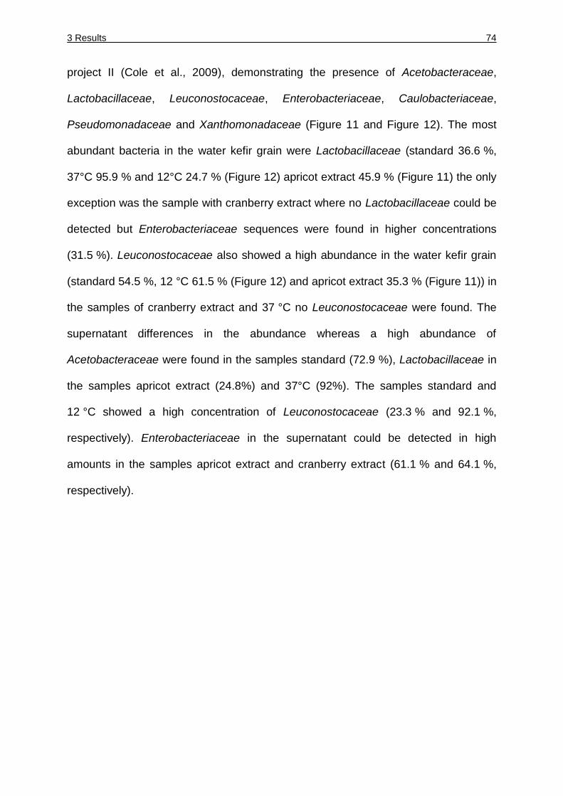
3 Results 74
project II (Cole et al., 2009), demonstrating the presence of Acetobacteraceae,
Lactobacillaceae, Leuconostocaceae, Enterobacteriaceae, Caulobacteriaceae,
Pseudomonadaceae and Xanthomonadaceae (Figure 11 and Figure 12). The most
abundant bacteria in the water kefir grain were Lactobacillaceae (standard 36.6 %,
37°C 95.9 % and 12°C 24.7 % (Figure 12) apricot extract 45.9 % (Figure 11) the only
exception was the sample with cranberry extract where no Lactobacillaceae could be
detected but Enterobacteriaceae sequences were found in higher concentrations
(31.5 %). Leuconostocaceae also showed a high abundance in the water kefir grain
(standard 54.5 %, 12 °C 61.5 % (Figure 12) and apricot extract 35.3 % (Figure 11)) in
the samples of cranberry extract and 37 °C no Leuconostocaceae were found. The
supernatant differences in the abundance whereas a high abundance of
Acetobacteraceae were found in the samples standard (72.9 %), Lactobacillaceae in
the samples apricot extract (24.8%) and 37°C (92%). The samples standard and
12 °C showed a high concentration of Leuconostocaceae (23.3 % and 92.1 %,
respectively). Enterobacteriaceae in the supernatant could be detected in high
amounts in the samples apricot extract and cranberry extract (61.1 % and 64.1 %,
respectively).
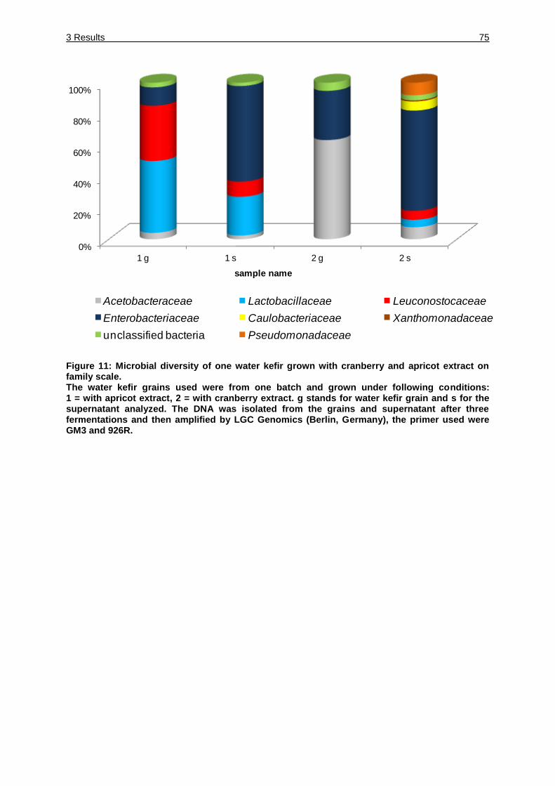
3 Results 75
Figure 11: Microbial diversity of one water kefir grown with cranberry and apricot extract on family scale. The water kefir grains used were from one batch and grown under following conditions: 1 = with apricot extract, 2 = with cranberry extract. g stands for water kefir grain and s for the supernatant analyzed. The DNA was isolated from the grains and supernatant after three fermentations and then amplified by LGC Genomics (Berlin, Germany), the primer used were GM3 and 926R.
0%
20%
40%
60%
80%
100%
1 g 1 s 2 g 2 s
sample name
Acetobacteraceae Lactobacillaceae Leuconostocaceae
Enterobacteriaceae Caulobacteriaceae Xanthomonadaceae
unclassified bacteria Pseudomonadaceae
3 Results 76
Figure 12: Microbial diversity of one water kefir grown under standard condition, at 37 °C and at 12 °C on family scale. The water kefir grains used were from one batch and grown under following conditions: 3 = standard water kefir, 4 = grown at 37 °C and 5 = grown at 12 °C. g stands for water kefir grain and s for the supernatant analyzed. The DNA was isolated from the grains and supernatant after three fermentations and then amplified by LGC Genomics (Berlin, Germany), the primer used were GM3 and 926R.
0%
20%
40%
60%
80%
100%
3 g 3 s 4 g 4 s 5 g 5 s
sample name
Acetobacteraceae Lactobacillaceae Leuconostocaceae
Enterobacteriaceae unclassified bacteria
3 Results 77
3.3.4.2 High-throughput sequencing analysis of the sequences obtained with the 27 f
and 519 R primer
For the second high-through put procedure the primer 27 f and 519 R were used.
A total of 7540 (standard, water kefir grains), 24293 (standard, supernatant),
5678 (apricot extract, water kefir grains), 10455 (apricot extract, supernatant),
5138 (cranberry extract, water kefir grain), 6275 (cranberry extract, supernatant),
22517 (37 °C, water kefir grain), 24120 (37 °C, supernatant), 5346 (12 °C, water kefir
grain), 29786 (12 °C, supernatant) reads of the V4 region of the 16S rRNA were used
for taxon assignment. In all data sets the 16S rRNA chimeric sequences were
determined by using Database Enabled Code for Ideal Probe Hybridization
Employing R (DECIPHER) software (Wright et al., 2013). All data sets showed
chimeric sequences (standard, water kefir grain 367, standard supernatant 9, apricot
extract water kefir grain 124 and supernatant 12, cranberry extract water kefir grain
174 and supernatant 19, 37 °C water kefir grain 14 and supernatant 4, 12 °C water
kefir grain 131 and supernatant 63. The distribution of the different bacteria at family
level was evaluated by the software tools provided by the Ribosomal Database
project II (Cole et al., 2009), demonstrating the presence of Actinobacteraiceae,
Acetobacteraceae, Lactobacillaceae, Leuconostocaceae, Enterobacteriaceae,
Caulobacteriaceae, Pseudomonadaceae and Xanthomonadaceae (Figure 13 and
Figure 14). The most abundant species in the water kefir grain samples showed
different species, Lactobacilliceae were most abundant in the samples standard,
37 °C, 12 °C and apricot extract, (34.1 %, 94.6 %, 21.6 % (Figure 14) and 30.7 %
(Figure 13), respectively), in the apricot sample Actinobacteriaceae showed a high
percentage with 34.6 %, Leuconostocaceae were found in high numbers in the
samples standard, 12 °C and cranberry extract (57 %, 57.5 %, and 41 %,
respectively).
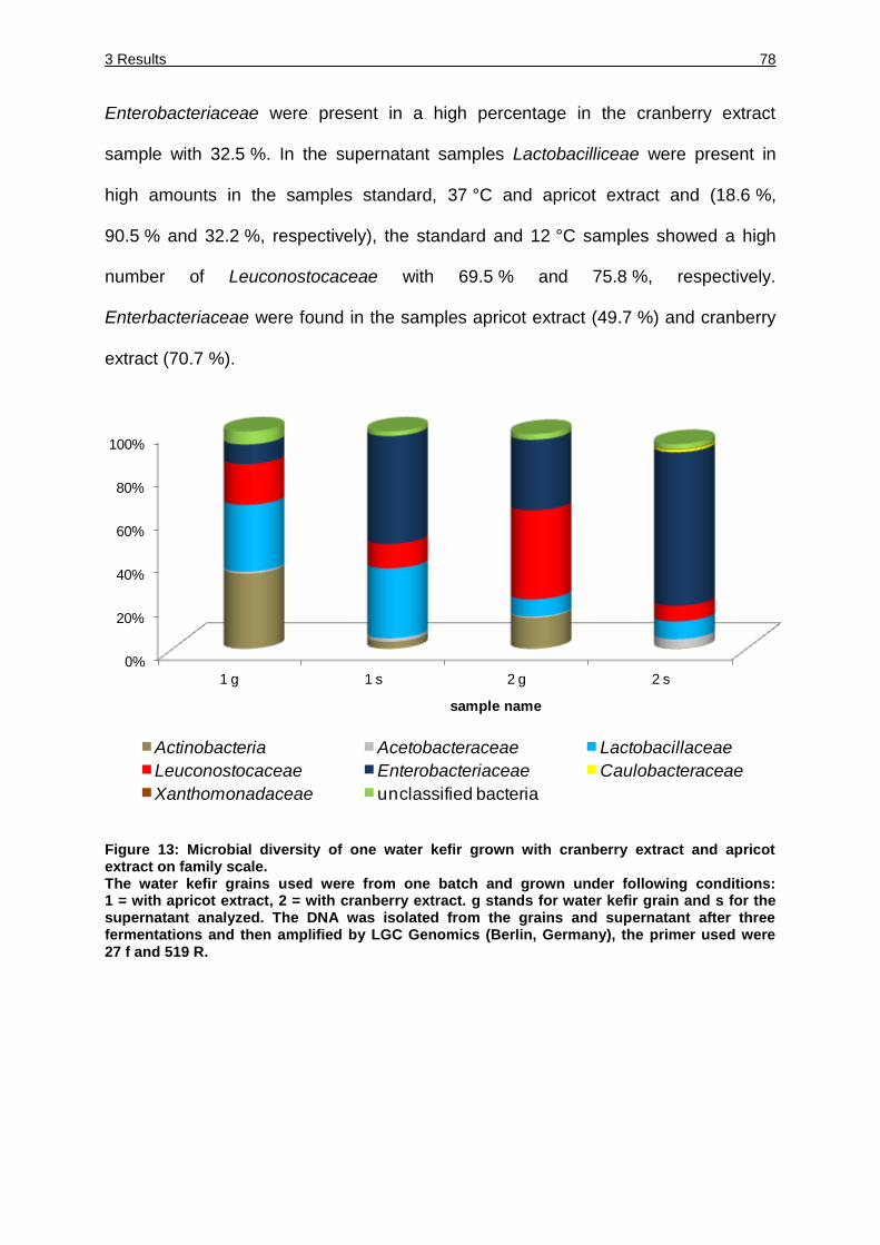
3 Results 78
Enterobacteriaceae were present in a high percentage in the cranberry extract
sample with 32.5 %. In the supernatant samples Lactobacilliceae were present in
high amounts in the samples standard, 37 °C and apricot extract and (18.6 %,
90.5 % and 32.2 %, respectively), the standard and 12 °C samples showed a high
number of Leuconostocaceae with 69.5 % and 75.8 %, respectively.
Enterbacteriaceae were found in the samples apricot extract (49.7 %) and cranberry
extract (70.7 %).
Figure 13: Microbial diversity of one water kefir grown with cranberry extract and apricot extract on family scale. The water kefir grains used were from one batch and grown under following conditions: 1 = with apricot extract, 2 = with cranberry extract. g stands for water kefir grain and s for the supernatant analyzed. The DNA was isolated from the grains and supernatant after three fermentations and then amplified by LGC Genomics (Berlin, Germany), the primer used were 27 f and 519 R.
0%
20%
40%
60%
80%
100%
1 g 1 s 2 g 2 s
sample name
Actinobacteria Acetobacteraceae Lactobacillaceae
Leuconostocaceae Enterobacteriaceae Caulobacteraceae
Xanthomonadaceae unclassified bacteria
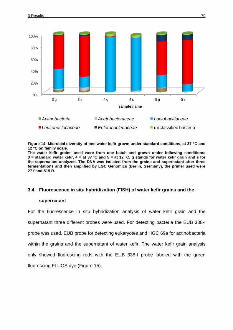
3 Results 79
Figure 14: Microbial diversity of one water kefir grown under standard conditions, at 37 °C and 12 °C on family scale. The water kefir grains used were from one batch and grown under following conditions: 3 = standard water kefir, 4 = at 37 °C and 5 = at 12 °C. g stands for water kefir grain and s for the supernatant analyzed. The DNA was isolated from the grains and supernatant after three fermentations and then amplified by LGC Genomics (Berlin, Germany), the primer used were 27 f and 519 R.
3.4 Fluorescence in situ hybridization (FISH) of water kefir grains and the
supernatant
For the fluorescence in situ hybridization analysis of water kefir grain and the
supernatant three different probes were used. For detecting bacteria the EUB 338-I
probe was used, EUB probe for detecting eukaryotes and HGC 69a for actinobacteria
within the grains and the supernatant of water kefir. The water kefir grain analysis
only showed fluorescing rods with the EUB 338-I probe labeled with the green
fluorescing FLUOS dye (Figure 15).
0%
20%
40%
60%
80%
100%
3 g 3 s 4 g 4 s 5 g 5 s
sample name
Actinobacteria Acetobacteraceae Lactobacillaceae
Leuconostocaceae Enterobacteriaceae unclassified bacteria
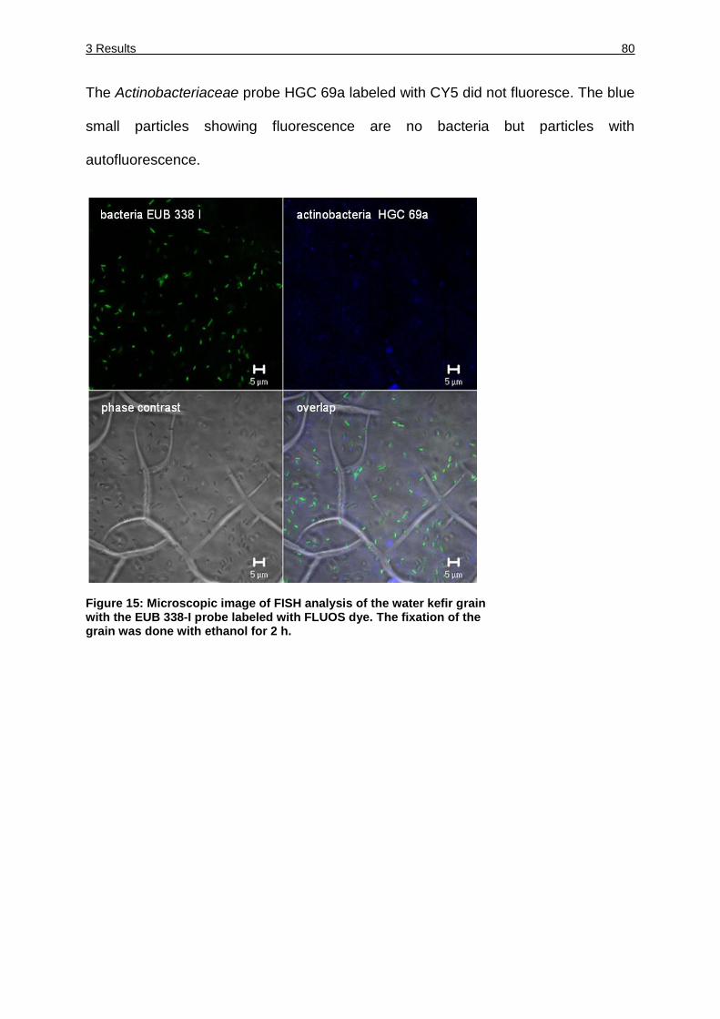
3 Results 80
The Actinobacteriaceae probe HGC 69a labeled with CY5 did not fluoresce. The blue
small particles showing fluorescence are no bacteria but particles with
autofluorescence.
Figure 15: Microscopic image of FISH analysis of the water kefir grain with the EUB 338-I probe labeled with FLUOS dye. The fixation of the grain was done with ethanol for 2 h.
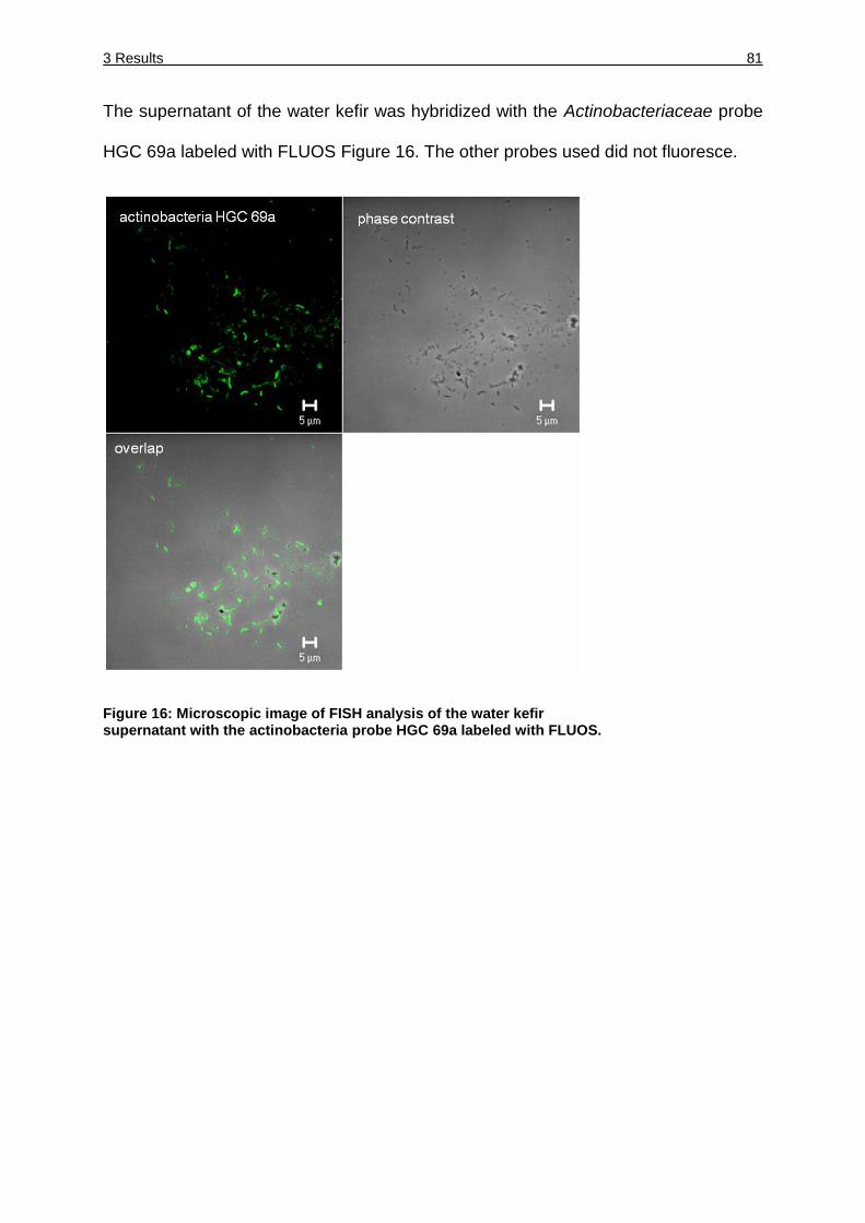
3 Results 81
The supernatant of the water kefir was hybridized with the Actinobacteriaceae probe
HGC 69a labeled with FLUOS Figure 16. The other probes used did not fluoresce.
Figure 16: Microscopic image of FISH analysis of the water kefir supernatant with the actinobacteria probe HGC 69a labeled with FLUOS.
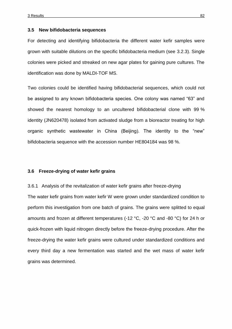
3 Results 82
3.5 New bifidobacteria sequences
For detecting and identifying bifidobacteria the different water kefir samples were
grown with suitable dilutions on the specific bifidobacteria medium (see 3.2.3). Single
colonies were picked and streaked on new agar plates for gaining pure cultures. The
identification was done by MALDI-TOF MS.
Two colonies could be identified having bifidobacterial sequences, which could not
be assigned to any known bifidobacteria species. One colony was named “63” and
showed the nearest homology to an uncultered bifidobacterial clone with 99 %
identity (JN620478) isolated from activated sludge from a bioreactor treating for high
organic synthetic wastewater in China (Beijing). The identity to the “new”
bifidobacteria sequence with the accession number HE804184 was 98 %.
3.6 Freeze-drying of water kefir grains
3.6.1 Analysis of the revitalization of water kefir grains after freeze-drying
The water kefir grains from water kefir W were grown under standardized condition to
perform this investigation from one batch of grains. The grains were splitted to equal
amounts and frozen at different temperatures (-12 °C, -20 °C and -80 °C) for 24 h or
quick-frozen with liquid nitrogen directly before the freeze-drying procedure. After the
freeze-drying the water kefir grains were cultured under standardized conditions and
every third day a new fermentation was started and the wet mass of water kefir
grains was determined.
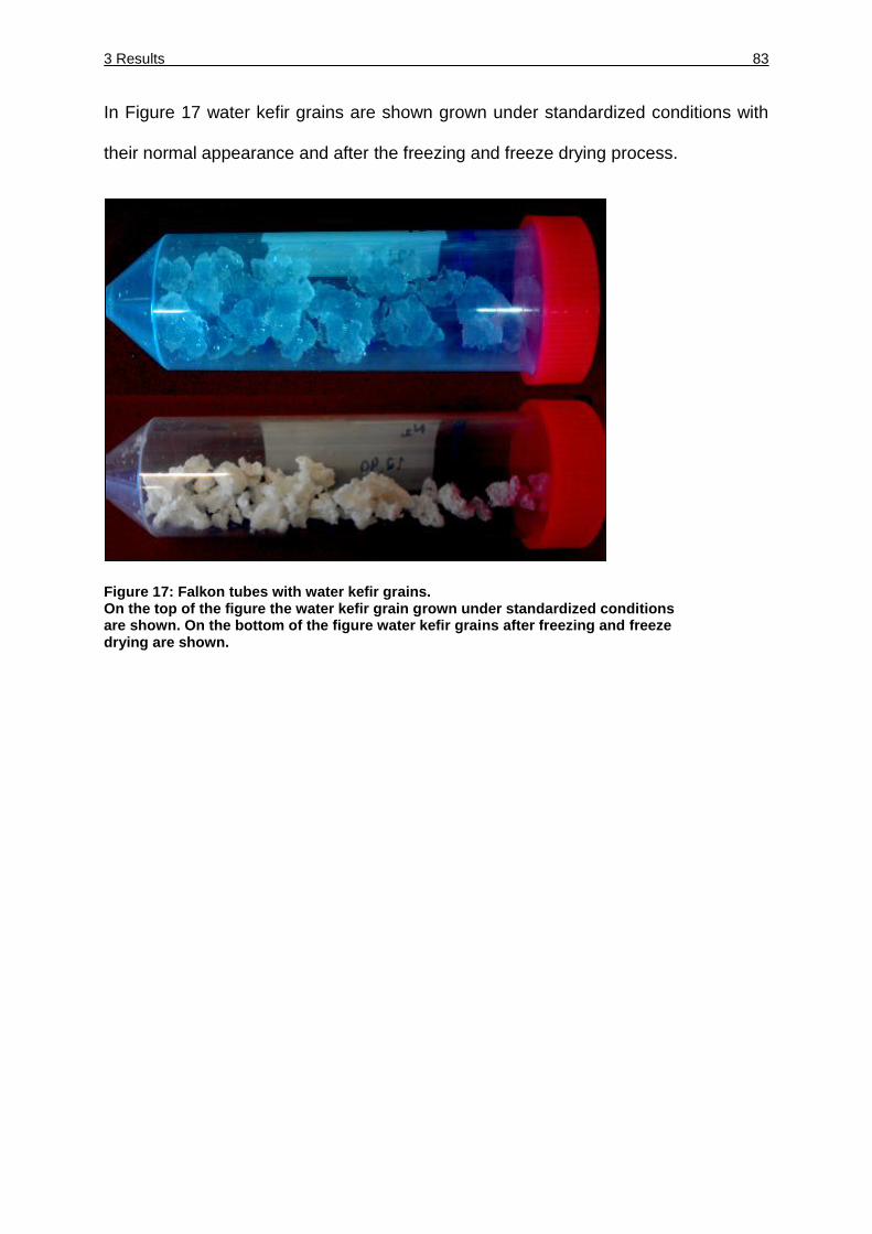
3 Results 83
In Figure 17 water kefir grains are shown grown under standardized conditions with
their normal appearance and after the freezing and freeze drying process.
Figure 17: Falkon tubes with water kefir grains. On the top of the figure the water kefir grain grown under standardized conditions are shown. On the bottom of the figure water kefir grains after freezing and freeze drying are shown.
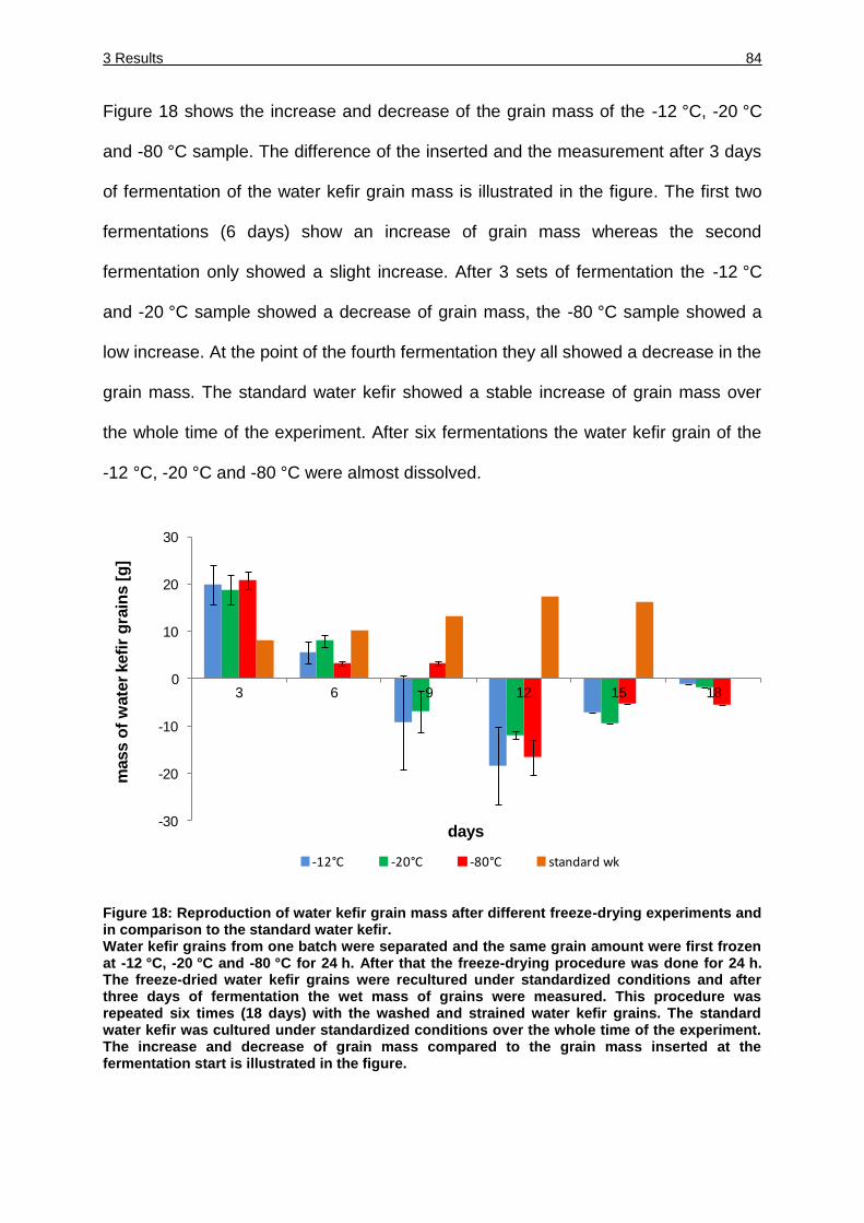
3 Results 84
Figure 18 shows the increase and decrease of the grain mass of the -12 °C, -20 °C
and -80 °C sample. The difference of the inserted and the measurement after 3 days
of fermentation of the water kefir grain mass is illustrated in the figure. The first two
fermentations (6 days) show an increase of grain mass whereas the second
fermentation only showed a slight increase. After 3 sets of fermentation the -12 °C
and -20 °C sample showed a decrease of grain mass, the -80 °C sample showed a
low increase. At the point of the fourth fermentation they all showed a decrease in the
grain mass. The standard water kefir showed a stable increase of grain mass over
the whole time of the experiment. After six fermentations the water kefir grain of the
-12 °C, -20 °C and -80 °C were almost dissolved.
Figure 18: Reproduction of water kefir grain mass after different freeze-drying experiments and in comparison to the standard water kefir. Water kefir grains from one batch were separated and the same grain amount were first frozen at -12 °C, -20 °C and -80 °C for 24 h. After that the freeze-drying procedure was done for 24 h. The freeze-dried water kefir grains were recultured under standardized conditions and after three days of fermentation the wet mass of grains were measured. This procedure was repeated six times (18 days) with the washed and strained water kefir grains. The standard water kefir was cultured under standardized conditions over the whole time of the experiment. The increase and decrease of grain mass compared to the grain mass inserted at the fermentation start is illustrated in the figure.
-30
-20
-10
0
10
20
30
3 6 9 12 15 18
mass o
f w
ate
r kefi
r g
rain
s [
g]
days
-12°C -20°C -80°C standard wk
3 Results 85
Figure 19 shows the appearance of the water kefir grains, grown under standardized
conditions (left side) and after freezing at -12 °C and freeze-drying over night
followed by regrowth under standardized conditions over three fermentations. The
grain consistency changed drastically. The water kefir grains quick freeze-dried with
N2, freeze-drying over night and regrown under standardized conditions did show the
same shape and consistency like the standard water kefir (shown on the left side of
the picture).
Figure 19: Strained water kefir grains after growth. On the left side water kefir grains were grown under standardized conditioned and strained through a sieve after three days of fermentation. On the left side strained water kefir grains are shown after freezing at -12 °C and freeze-drying over night. The water kefir grains were fermented three times (nine days).
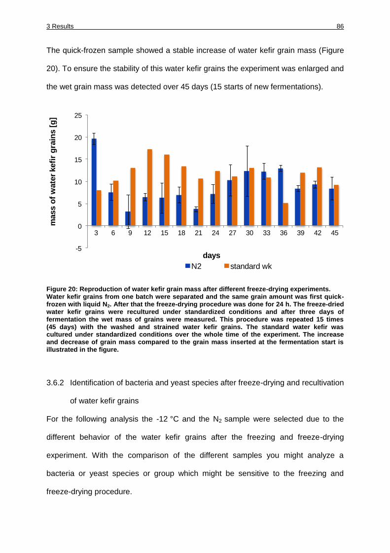
3 Results 86
The quick-frozen sample showed a stable increase of water kefir grain mass (Figure
20). To ensure the stability of this water kefir grains the experiment was enlarged and
the wet grain mass was detected over 45 days (15 starts of new fermentations).
Figure 20: Reproduction of water kefir grain mass after different freeze-drying experiments. Water kefir grains from one batch were separated and the same grain amount was first quick-frozen with liquid N2. After that the freeze-drying procedure was done for 24 h. The freeze-dried water kefir grains were recultured under standardized conditions and after three days of fermentation the wet mass of grains were measured. This procedure was repeated 15 times (45 days) with the washed and strained water kefir grains. The standard water kefir was cultured under standardized conditions over the whole time of the experiment. The increase and decrease of grain mass compared to the grain mass inserted at the fermentation start is illustrated in the figure.
3.6.2 Identification of bacteria and yeast species after freeze-drying and recultivation
of water kefir grains
For the following analysis the -12 °C and the N2 sample were selected due to the
different behavior of the water kefir grains after the freezing and freeze-drying
experiment. With the comparison of the different samples you might analyze a
bacteria or yeast species or group which might be sensitive to the freezing and
freeze-drying procedure.
-5
0
5
10
15
20
25
3 6 9 12 15 18 21 24 27 30 33 36 39 42 45
mass o
f w
ate
r kefi
r g
rain
s [
g]
days
N2 standard wk
3 Results 87
Therefore, the viable cell count on different media (mMRS, GM, TP and YPG) were
enumerated and 50 colonies of an appropriate dilution were identified by MALDI-TOF
MS.
The viable cell count of the -12 °C sample was lower directly after the freeze-drying
than after recultivation of the water kefir grains (Table 21). Differences could be
detected within the comparison of the -12 °C sample where the cell number counted
directly after the freeze-drying is much more lower than after regrowth (Lb. hordei
with 2.9*103 and 1.6*107, Lb. paracasei with 4.5*103 and 2.3*106 respectively,
Lc. citreum could not be detected after freeze-drying but after regrowth with 8.4*107).
On the other hand directly after freeze-drying three lactobacilli species were detected
(Lb. satsumensis, Lb. nagelii and Lb. hilgardii all with a cell count of 2.2*102) but after
regrowth they could not be identified. The N2 sample showed no significant difference
between the two analyzes. No differences in the cell count of Lb. hordei and
Lb. paracasei could be detected after freeze-drying and regrowth. Lb. nagelii could
not be detected at all, Lb. hilgardii and Lb. satsumensis only directly after freeze-
drying (2.2*104, 1.5*105). Lc. mesenteroides was just identified after regrowth
(3.3*106). Comparing the data obtained with the reference the viable cell count after
regrowth is almost the same with the species Lc. mesenteroides, Lb. hordei and the
cfu complete whereas Lb. paracasei, Lb. satsumensis, Lb. nagelii and Lb. hilgardii
could not be identified.
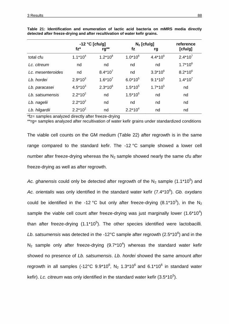
3 Results 88
Table 21: Identification and enumeration of lactic acid bacteria on mMRS media directly detected after freeze-drying and after recultivation of water kefir grains.
-12 °C [cfu/g]
fz* rg** N2 [cfu/g]
fz rg reference
[cfu/g]
total cfu 1.1*104 1.2*108 1.0*106 4.4*106 2.4*107
Lc. citreum nd nd nd nd 1.7*106
Lc. mesenteroides nd 8.4*107 nd 3.3*106 8.2*106
Lb. hordei 2.9*103 1.6*107 6.0*105 9.1*105 1.4*107
Lb. paracasei 4.5*103 2.3*106 1.5*105 1.7*105 nd
Lb. satsumensis 2.2*102 nd 1.5*105 nd nd
Lb. nagelii 2.2*102 nd nd nd nd
Lb. hilgardii 2.2*102 nd 2.2*104 nd nd
*fz= samples analyzed directly after freeze-drying **rg= samples analyzed after recultivation of water kefir grains under standardized conditions
The viable cell counts on the GM medium (Table 22) after regrowth is in the same
range compared to the standard kefir. The -12 °C sample showed a lower cell
number after freeze-drying whereas the N2 sample showed nearly the same cfu after
freeze-drying as well as after regrowth.
Ac. ghanensis could only be detected after regrowth of the N2 sample (1.1*105) and
Ac. orientalis was only identified in the standard water kefir (7.4*106). Gb. oxydans
could be identified in the -12 °C but only after freeze-drying (8.1*103), in the N2
sample the viable cell count after freeze-drying was just marginally lower (1.6*104)
than after freeze-drying (1.1*105). The other species identified were lactobacilli.
Lb. satsumensis was detected in the -12°C sample after regrowth (2.5*106) and in the
N2 sample only after freeze-drying (9.7*104) whereas the standard water kefir
showed no presence of Lb. satsumensis. Lb. hordei showed the same amount after
regrowth in all samples (-12°C 9.9*106, N2 1.3*106 and 6.1*106 in standard water
kefir). Lc. citreum was only identified in the standard water kefir (3.5*105).
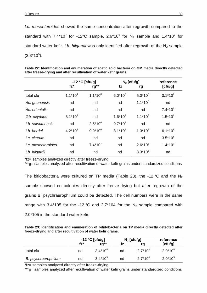
3 Results 89
Lc. mesenteroides showed the same concentration after regrowth compared to the
standard with 7.4*107 for -12°C sample, 2.6*106 for N2 sample and 1.4*107 for
standard water kefir. Lb. hilgardii was only identified after regrowth of the N2 sample
(3.3*105).
Table 22: Identification and enumeration of acetic acid bacteria on GM media directly detected after freeze-drying and after recultivation of water kefir grains.
-12 °C [cfu/g]
fz* rg** N2 [cfu/g]
fz rg reference
[cfu/g]
total cfu 1.1*104 1.1*108 6.0*105 5.0*106 3.1*107
Ac. ghanensis nd nd nd 1.1*105 nd
Ac. orientalis nd nd nd nd 7.4*106
Gb. oxydans 8.1*103 nd 1.6*104 1.1*105 1.5*106
Lb. satsumensis nd 2.5*106 9.7*104 nd nd
Lb. hordei 4.2*102 9.9*106 8.1*104 1.3*106 6.1*106
Lc. citreum nd nd nd nd 3.5*105
Lc. mesenteroides nd 7.4*107 nd 2.6*106 1.4*107
Lb. hilgardii nd nd nd 3.3*105 nd
*fz= samples analyzed directly after freeze-drying **rg= samples analyzed after recultivation of water kefir grains under standardized conditions
The bifidobacteria were cultured on TP media (Table 23), the -12 °C and the N2
sample showed no colonies directly after freeze-drying but after regrowth of the
grains B. psychraerophilum could be detected. The cell numbers were in the same
range with 3.4*105 for the -12 °C and 2.7*104 for the N2 sample compared with
2.0*105 in the standard water kefir.
Table 23: Identification and enumeration of bifidobacteria on TP media directly detected after freeze-drying and after recultivation of water kefir grains.
-12 °C [cfu/g]
fz* rg** N2 [cfu/g]
fz rg reference
[cfu/g]
total cfu nd 3.4*105 nd 2.7*104 2.0*105
B. psychraerophilum nd 3.4*105 nd 2.7*104 2.0*105
*fz= samples analyzed directly after freeze-drying **rg= samples analyzed after recultivation of water kefir grains under standardized conditions
3 Results 90
The yeast species were identified and enumerated on YPG medium. Four different
yeast species could be detected (Table 24). The viable cell count after regrowth of
the -12 °C and N2 was in the same range like the standard water kefir (5.4*106,
3.0*105 and 1.6*106 respectively). The viable cell count after freeze-drying of the
-12°C sample was lower with 4.8*103 cfu/g compared to the N2 sample with
2.1*105 cfu/g. Hanseniaspora valbyensis showed lower numbers in the -12 °C
sample after freeze-drying (1.6*103) but after regrowth the cell number was in the
same range like N2 and the standard water kefir (3.1*105, 1.4*105 and 9.7*104).
Pichia membranifaciens could not be detected after freeze-drying of the -12 °C and
N2 sample whereas after regrowth the cell number showed no significant difference
(1.1*105 and 1.7*104 cfu/g) compared to the standard water kefir with 6.0*105 cfu/g.
Saccharomyces cerevisiae was first analyzed in lower numbers after freeze drying
(-12 °C sample with 6.0*102 and N2 sample with 4.1*103) but after regrowth the cell
number was comparable to the standard water kefir (3.7*105) where the -12 °C had a
cfu/g of 1.2*105, N2 1.3*105. Zygotorulaspora florentina showed the same behavior
with a cfu/g of 8.0*102 after freeze-drying and 7.6*105 cfu/g after regrowth. The cell
number of the N2 sample showed no difference between the freeze-drying (4.1*104)
and regrowth of water kefir grains (3.4*104).
Table 24: Identification and enumeration of yeast species on YPG media directly detected after freeze-drying and after recultivation of water kefir grains.
-12 °C [cfu/g]
fz* rg** N2 [cfu/g]
fz rg reference
[cfu/g]
total cfu 4.8*103 5.4*106 2.1*105 3.0*105 1.6*106
Hanseniaspora valbyensis 1.6*103 3.1*106 1.6*105 1.4*105 9.7*104
Pichia membranifaciens nd 1.1*105 nd 1.7*104 6.0*105
Saccharomyces cerevisiae 6.0*102 1.2*106 4.1*103 1.3*105 3.7*105
Zygotorulaspora florentina 8.0*102 7.6*105 4.1*104 3.4*104 2.4*105
*fz= samples analyzed directly after freeze-drying **rg= samples analyzed after recultivation of water kefir grains under standardized conditions
4 Discussion 91
4 Discussion
The aim of this work was to analyze and characterize the water kefir microbiota
regarding their microbial composition. Culture-dependent and –independent
technologies showed the same core of microorganisms belonging to the bacterial
genera Lactobacillaceae, Leuconostocaceae, Acetobacteraceae and
Bifidobacteriaceae as well the yeast species Saccharomyces cerevisiae and
Zygotorulaspora florentina.
According to the results obtained it is possible to produce a water kefir based
beverage with species isolated from water kefir.
The analysis of water kefir fermented under different growth conditions showed that
the temperature and the nutrient provided by plant material are important parameters
for the stability and dynamics of the water kefir microbiota.
Water kefir grains could be preserved by shock-freezing with N2 and freeze-drying
over night whereas the water kefir species showed different viability.
4 Discussion 92
4.1 Microbial diversity of water kefir grains analyzed by culture-dependent
procedures
In this part of the thesis state of the art molecular culture-dependent methods were
used for the identification of several additional and/ or different species providing a
revised view of the water kefir microbiota. Species were found in water kefir which
have not been detected and identified in other water kefir studies so far and the
abundance of species in the water kefir microbiota were qualitatively and
quantitatively different from those previously described (Franzetti et al., 1998; Galli et
al., 1995; Kebler, 1921; Lutz, 1899; Pidoux, 1989).
A typical consortium regarding this type of culture-dependent investigation appears to
consist of mostly lactic acid bacteria plus yeasts promoting alcoholic fermentation
and some acetic acid bacteria, possibly oxidizing the ethanol formed.
The yeast predominant in all three water kefirs (A, F and I) is most probably
Z. florentina as demonstrated for water kefir W. Z. florentina showed a white cell
morphology in this analyses. Detailed studies of this white cell morphology showed
that more cells were identified as Z. florentina. The other water kefirs had a high
percentage of cells showing white morphology. Referred to the result of the detailed
study of the phenotype showing white morphology with the water kefir W, it might
indicate that Z. florentina is the most predominant species in all water kefirs.
All yeast species identified in the water kefirs were able to ferment glucose and this
could indicate that they all show the same underlying metabolism (Esteve-Zarzoso et
al., 2001; Kurtzman and and Robnett, 2003). S. cerevisiae is able to invert sucrose
into the monosaccharides glucose and fructose by the enzyme invertase so that the
yeast cells have glucose as a free metabolite (Ikram-Ul-Haq and Ali, 2007).
4 Discussion 93
This might indicate that in the other water kefirs the composition of these two yeasts
are nearly the same and that Z. florentina is the yeast species which is predominant.
The bacterial core species of the three water kefirs belonging to lactic acid bacteria
were Lb. hordei, Lb. nagelii and Lc. mesenteroides, whereas in water kefir W
Lb. hordei and Lb. nagelii were detected in much lower concentrations. The presence
of these two lactobacilli in all water kefir consortia claims that they are important for
the stability of the water kefir. It is not surprising that Lb. nagelii and Lb. hordei are
part of the water kefir consortia as it is an alcoholic environment with high sugar
concentration at the beginning of the fermentation and a low pH after three days of
fermentation with increasing alcoholic concentrations. The description of Lb. nagelii
was in 2000 by C. Edwards and it firstly was isolated from partially fermented wine
(Edwards, 2000). Lb. hordei was firstly described in 2008 and isolated von malted
barley (Rouse et al., 2008). These habitats (partially fermented wine and malted
barley) exhibits specific requirements like an alcoholic environment with a low pH. Lb.
nagelii is described to produce EPS from dextran which could be important for the
stability and forming of water kefir grain. Different strains of Lb. nagelii (TMW 1.1823,
1.1826, 1.1827) and Lb. hordei (TMW 1.1958) could be identified as EPS-producer
emphasizing the importance of these species.
Leuconostoc species were found in all three water kefir consortia in significant
percentages and were the main component in water kefir W. This high number of
Leuconostoc species in the water kefir consortia indicates their competitiveness in
the environment of water kefir. Leuconostocs have been found to play an important
role in fermentations like sauerkraut, cucumbers, finger millet, Boza and kimchi
where they are predominant in the early stages of fermentation where they have to
compete with other LAB (Dworkin et al., 2006).
4 Discussion 94
Lc. mesenteroides species could also be identified as EPS-producer (TMW 2.1073,
2.1076) underlining the important role within the consortia.
Acetic acid bacteria were also a part of the bacterial core in this water kefir analysis,
especially in water kefir W. Ac. fabarum was firstly described 2008 and isolated from
Ghanaian cocoa bean heap fermentation. Ac. fabarum is also able to fermentate
ethanol to acetic acid (Cleenwerck et al., 2008). During the water kefir fermentation
ethanol is formed mostly by yeasts so that acetic acid bacteria have a high nutrient
source. On the other hand it is not preferable to have to high concentration of acetic
acid bacteria regarding the taste of the resulting beverage. Ac. orientalis was firstly
isolated from canna flowers, starfruit and coconut and from traditional fermented
foods (curd of tofu and tempe) (Lisdiyanti et al., 2001). The natural habitat of this
species is fermented food where they could metabolize ethanol formed from yeasts
and or acetate formed by LAB, so they are resistant for low alcoholic concentrations
like in the water kefir environment. The fruits and flowers originate from exotic
environments this might be a hint that the water kefir grain grew on leaves of cactus
and the origin is from the orient.
The composition of the microorganisms in the water kefir consortia resulting from
culture-dependent analysis differs to the investigations done previously. In previous
investigations Lb. casei was described as a very predominant species (Franzetti et
al., 1998; Pidoux, 1989). With culture-dependent methods Lb. casei could not be
found in high percentages. The presence of Lb. casei in water kefir A and F could be
explained by the ubiquitous occurrence of this species while it unlikely exerts a
metabolic influence of the properties of the resulting beverage. The same is valid for
Lb. hilgardii, which was predominant in the sugar kefir grains studied by Pidoux
(1989) with a percentage of 30 %.
4 Discussion 95
The concentration of Lb. hilgardii was negligible in this investigation and it was only
found in water kefir A. There are some Lb. hilgardii strains, which produce EPS
(Pidoux et al., 1990) and which are even described to play a major role in forming the
stable grains of water kefir (Waldherr et al., 2010) while none of the Lb. hilgardii
strains isolated in this study were able to produce EPS.
Pidoux described that Lb. brevis is also an important LAB regarding the formation of
gel-like grain (Pidoux et al., 1988) which could not be identified in the different water
kefir microbiota. Horisberger described the main flora of water kefir consisting
predominantly of Lb. brevis, Streptococcus lactis and the yeast S. cerevisiae
(Horisberger, 1969). Both bacterial species could not be detected in the water kefir
grains characterized in this investigation featuring the predominance of Lb. nagelii,
Lb. hordei and Lc. mesenteroides in water kefirs A and F. This may be due to their
absence in those samples. That is one reason why they could not delineate
Lb. brevis from the other LABs in their previous investigation long before that time
due to the lack of molecular typing techniques.
Some acetic acid bacteria were identified in previous studies in water kefir consortia
but only in negligible quantity (Franzetti et al., 1998).
To conclude the differences in the composition of the water kefir consortia may be to
some extent referred to different identification techniques used. The technique used
for the identification of lactic acid bacteria and acetic acid bacteria in the
investigations done so far was API CH 50 (Franzetti et al., 1998; Pidoux, 1989). As
strains of different species can behave the same phenotype in some physiological
tests the identification with these tests has clear limits towards a valid identification of
Lactobacillus species closely related to each other (Boyd, 2005; Dalezios, 2001;
Singh et al., 2009). By RAPD genotyping and identification of the species with
4 Discussion 96
16S rDNA analysis it was possible to clearly allot isolates to species and also identify
microorganisms in water kefir, which have not been identified yet from this source.
The core organisms like Lb. nagelii, Lb. hordei, Lc. mesenteroides and Ac. fabarum
were isolated and identified in all three water kefirs. On the other hand, the different
composition of the three water kefirs indicates that the specific percentages of each
species are not the most important characteristic of a stable water kefir microbiota. It
rather suggests a metabolic core comprising the overall metabolic capacity of the
consortium, which may be more important than the species designation of the strains
found.
4.2 Microbial diversity of water kefir grains analyzed by culture-independent
procedures
In this study, the bacterial communities of four different water kefirs were analyzed by
a culture-independent, high-throughput sequencing procedure employing 16S rRNA
gene amplicon sequencing. The microbiota of all kefirs consisted of bacteria from
three phyla: Firmicutes (79 %), Actinobacteria (17.1 %) and Proteobacteria (3.7 %).
The Firmicutes were the dominant organisms in all kefirs, represented mainly by the
genera Lactobacillus and Leuconostoc.
Kefir I, which was directly analyzed upon arrival without any propagation showed high
numbers of clostridia (37.2 %), while clostridial sequences represented less than
0.03 % of the sequences in the other kefirs. Interestingly, Clostridiaceae assignments
accounted for 0.82 % of reads in the starter grains of milk kefir (Dobson et al., 2011).
The high clostridia content and a noticeable off flavour were obviously consequences
of improper propagation or contamination during preparation and/or shipment.
4 Discussion 97
Clostridium tyrobutyricum, which was the most abundant Clostridium species, is
known as a food spoiling bacterium and a major cause of late cheese blowing
(Klijn et al., 1995) but is also found in environments related to water kefir, such as in
fruit pulp (Mayer et al., 2010). Therefore, we do not consider kefir I to harbour a
typical kefir consortium and did not include the data from this sample in the
subsequent analysis.
The phylum Actinobacteria is represented exclusively by the genus Bifidobacterium,
and Proteobacteria are represented by Acetobacter, Gluconobacter and
Gluconacetobacter. Kefirs A, F and W differed in their composition only marginally.
Due to sequencing artefacts such as sequence-specific errors, miscounted
homopolymeric runs etc., the use of 454 pyrosequencing data for community profiling
is known to occasionally inflate estimates of actual diversity when taxon assignments
are made using alignment-based approaches (Kunin et al., 2010). To prevent
overestimates of diversity using regions of the 16S rDNA, a clustering threshold of
97 % identity is widely used. This limits the assignment of sequences at the current
species threshold of 98.7 %. In this study, we used a suffix tree-based search for
common heptanucleotide occurrences in the pyrosequencing data and reference
data allowing similarity analyzes without the need for sequence alignments of large
data sets. This allows a more reliable assignment of reads to a single species or
species group.
The data from this study corroborate the identification of species that have been
described in previous studies of water kefir using culture-dependent techniques
(Gulitz et al., 2011). The main core of bacteria described here is similar to that shown
in our previous investigation with culture-dependent methods, in which Lb. hordei,
Lb. nagelii, Lc. mesenteroides and Lc. citreum, as well as Ac. fabarum, represented
4 Discussion 98
the core group of organisms (Gulitz et al., 2011). In this study, kefir W had the
highest number of Leuconostocaceae, and high numbers of Lb. nagelii and
Lb. hordei sequences were found in water kefirs A and F. We were able to detect
only small numbers of Lb. casei in all four water kefirs, while Lb. casei (Franzetti et
al., 1998) and Lb. hilgardii were identified as the predominant bacterial species in
other water kefirs (Pidoux, 1989). A low percentage of the sequences in water kefir A
and F were derived from Lb. hilgardii, whereas water kefirs I and W showed a slightly
higher percentage. This is consistent with the data obtained using the culture-
dependent approach (Gulitz et al., 2011).
Based on the data obtained from culture-dependent and –independent analyses it
was possible to generate a starter culture with the most abundant species isolated
from water kefir microbiota (Gulitz et al., 2013 and 2011). As a result a water kefir
based beverage could be produced with the project partner at Hochschule
Ostwestfalen-Lippe, Technikum für Getränketechnologie, Lemgo.
4.2.1 Identification and characterization of bifidobacteria found in water kefir
The detection of bifidobacteria in water kefir is unexpected and has not been
described previously. In this context, it is worth mentioning that we were not able to
observe cells with bifidobacterial morphology using light microscopy. Notably,
previous studies using scanning electron microscopy have not identified
bifidobacteria as elements of the water kefir consortium (Neve and Heller, 2002).
This might be due to the atypical morphology of B. psychraerophilum, which is
described to show bifurcation only occasionally (Simpson et al., 2004).
In our hands, neither the type strain of B. psychraerophilum nor strains isolated from
water kefir in the present study showed bifidobacterial morphology, even in pure
cultures (data not shown).
4 Discussion 99
B. psychraerophilum firstly was isolated from porcine caecum but the authors claimed
that it is possible that they are no resident bacteria of the caecum but might passing
through and originate from food (Simpson et al., 2004). This species is able to
tolerate and even grow under aerobic conditions which are given in the water kefir
microbiota. But the water kefir grain itself also could exhibit anaerobic niches.
Recently, Dobson et al. (2011) reported the finding of bifidobacterial sequences in a
culture-independent analysis of milk kefir. In this consortium, the Bifidobacterium
population, containing the species Bifidobacterium breve, Bifidobacterium choerinum,
Bifidobacterium longum, and Bifidobacterium pseudolongum comprised just 0.2 % of
total assigned taxa in the collective starter grain and 0.4 % in the kefir milk. However,
culture-dependent methods failed to detect Bifidobacterium species in either sample.
This underscores the fact that culture-independent analysis is a powerful tool for the
better understanding of microbial consortia and that bifidobacteria with unknown
taxonomy and physiology may contribute to various extents to such consortia. This
underlines the difficulty to culture bacteria under laboratory conditions, this is
especially true for Bifidobacteriaceae due to their special nutritional requirements
(Arunachalam, 1999).
Interestingly, a large number of bifidobacterial reads from water kefir could not be
assigned to any known species.
The Bifidobacterium-specific 16S ARDRA analysis of 100 clones per kefir showed a
single restriction profile in all of the kefirs that could be clearly differentiated from that
of the type strains B. crudilactis FR62/b/3T and B. psychraerophilum LMG 21775T.
Moreover, the sequences of all corresponding amplicons showed a similarity of less
than 97 % to its closest relatives, B. crudilactis FR62/b/3T and B. psychraerophilum
LMG 21775T.
4 Discussion 100
This result was unexpected because the metagenomic data showed ratios of 3:1,
9.4:1, 1:6.8 and 1:2.1 of B. spec. to the bifidobacteria B. psychraerophilum,
B. crudilactis and B. subtile, respectively. Because the template DNA samples used
for pyrosequencing and ARDRA and primer binding sites were identical, this
discrepancy may be ascribed to an artificial cloning bias.
4.2.1.1 Cultivation of bifidobacteria isolated from water kefir
The selective cultivation of bifidobacteria from natural habitats harbouring lactobacilli
and other lactic acid bacteria is difficult because the cultural and biochemical
properties of both genera overlap (Thitaram et al., 2005). Many attempts to isolate
bifidobacteria from water kefir with previously described media, including mupirocin-
containing media, failed in our hands because the bifidobacteria were overgrown by
lactic acid bacteria or yeasts (data not shown). Our modified selective medium
suppresses yeast and LAB growth due to the high amounts of cycloheximide and
kanamycin, and the addition of oligofructose as bifidogenic carbohydrate allowed the
selective cultivation of up to 1.5*106 cfu g-1 B. psychraerophilum alone from kefir
granules.
4.2.2 Characterization of the novel bifidobacteria species found in water kefir
The isolation of novel Bifidobacterium species, the 16S sequence of which was highly
abundant in the pyrosequencing and ARDRA data, was not successful, suggesting
that either we were unable to meet its optimal growth conditions or the
pyrosequencing data do not necessarily reflect the true numbers of living cells.
Nevertheless, this medium may also be helpful to allow the cultivation of
bifidobacteria from other sources from which the isolation of bifidobacteria has not
been successful to date.
4 Discussion 101
4.2.2.1 Phylogenetic position of water kefir bifidobacteria
The phylogentic tree of Bifidobacteriaceae based on partial 16S rRNA gene
sequences including bifidobacteria isolated from water kefir in this thesis is shown in
Figure 21. The phylogenetic tree shows that the bifidobacteria isolated from water
kefir originate from a monophyletic group. In this group B. mongoliense is included,
this species was isolated from fermented milk a habitat also having slightly alcoholic
concentration (Watanabe et al., 2009). In this group all species are able to grow
under aerobic conditions and tolerate low pH (Delcenserie et al., 2007; Simpson et
al., 2004; Watanabe et al., 2009). B. psychraerophilum is able to grow at low
temperature even at 4°C growth could be detected. The minimal growth temperature
of B. mongoliense is 15°C. The optimal growth temperature of bifidobacterial species
normally ranges from 37-41°C and growth below 20°C normally does not occur
(Biavati et al., 2000). Bifidobacteria were mainly isolated from faeces (human and
animal) some from bumblebee intestine, sewage and dental plaque (Russell et al.).
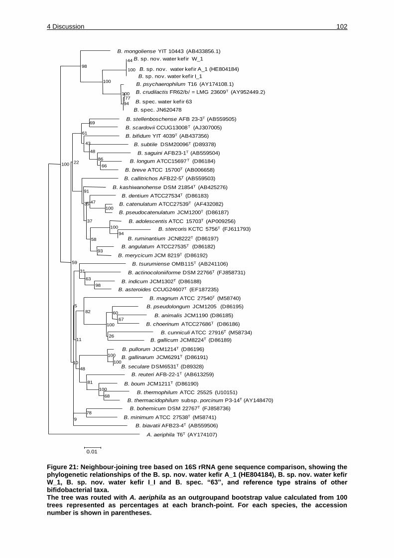
4 Discussion 102
Figure 21: Neighbour-joining tree based on 16S rRNA gene sequence comparison, showing the phylogenetic relationships of the B. sp. nov. water kefir A_1 (HE804184), B. sp. nov. water kefir W_1, B. sp. nov. water kefir I_I and B. spec. “63”, and reference type strains of other bifidobacterial taxa. The tree was routed with A. aeriphila as an outgroupand bootstrap value calculated from 100 trees represented as percentages at each branch-point. For each species, the accession number is shown in parentheses.
B. mongoliense YIT 10443 (AB433856.1)
B. sp. nov. water kef ir W_1
B. sp. nov. water kef ir A_1 (HE804184)
B. sp. nov. water kef ir I_1
B. psychaerophilum T16 (AY174108.1)
B. crudilactis FR62/b/ = LMG 23609T (AY952449.2)
B. spec. water kef ir 63
B. spec. JN620478
B. stellenboschense AFB 23-3T (AB559505)
B. scardovii CCUG13008T (AJ307005)
B. bifidum YIT 4039T (AB437356)
B. subtile DSM20096T (D89378)
B. saguini AFB23-1T (AB559504)
B. longum ATCC15697T (D86184)
B. breve ATCC 15700T (AB006658)
B. callitrichos AFB22-5T (AB559503)
B. kashiwanohense DSM 21854T (AB425276)
B. dentium ATCC27534T (D86183)
B. catenulatum ATCC27539T (AF432082)
B. pseudocatenulatum JCM1200T (D86187)
B. adolescentis ATCC 15703T (AP009256)
B. stercoris KCTC 5756T (FJ611793)
B. ruminantium JCN8222T (D86197)
B. angulatum ATCC27535T (D86182)
B. merycicum JCM 8219T (D86192)
B. tsurumiense OMB115T (AB241106)
B. actinocoloniiforme DSM 22766T (FJ858731)
B. indicum JCM1302T (D86188)
B. asteroides CCUG24607T (EF187235)
B. magnum ATCC 27540T (M58740)
B. pseudolongum JCM1205 (D86195)
B. animalis JCM1190 (D86185)
B. choerinum ATCC27686T (D86186)
B. cunniculi ATCC 27916T (M58734)
B. gallicum JCM8224T (D86189)
B. pullorum JCM1214T (D86196)
B. gallinarum JCM6291T (D86191)
B. seculare DSM6531T (D89328)
B. reuteri AFB-22-1T (AB613259)
B. boum JCM1211T (D86190)
B. thermophilum ATCC 25525 (U10151)
B. thermacidophilum subsp. porcinum P3-14T (AY148470)
B. bohemicum DSM 22767T (FJ858736)
B. minimum ATCC 27538T (M58741)
B. biavatii AFB23-4T (AB559506)
A. aeriphila T6T (AY174107)
9
78
68
100
81
48
10 100
100
11
82
100
26
60
67
5
31
63
98
93
59
58
100
94
37
35 47
100
91
22100 66
86
48
43
61
69
98
100
100
44
100
94
77
0.01
4 Discussion 103
In conclusion this investigation confirms previous data and broadens the available
knowledge about the microbial consortium of water kefir. The unexpected presence
of bifidobacteria in all samples and the difficulty of cultivating these species indicate
that the role of bifidobacteria in other fermented foods may also be underestimated.
The phylogenetic position of bifidobacterial 16S rRNA gene sequences with
similarities of less than 96.4 % to known species suggests the identification of a new
candidate species of the genus of Bifidobacterium. The large number of
bifidobacteria sequences found in all samples indicates that these bacteria are part of
the core species of water kefir.
4.3 Influence of the composition of water kefir microbiota by changing growth
conditions
4.3.1 Measurement of the wet mass of water kefir grains
The growth of the grain mass during the time of fermentation under different
conditions showed diverse increase.
The water kefir grains grown with cranberry extract, apricot extract and grown at
37 °C only showed a marginal increase in their wet mass after each fermentation.
Comparing to that the grain mass grown under standardized conditions increased
steadily. The consistency of the water kefir grains in all samples did not change
during the time of the experiment. The standard water kefir was grown with the
addition of dried figs and the result corresponds to investigation of Reiß, 1990. In that
investigation different fruits were tested and dried figs showed the highest increase in
grain mass upon 12.3 %, in this investigation the highest grain mass increase was
detected after the seventh fermentation with 126.5 % increase.
4 Discussion 104
The figs promote the greatest growth and this could be explained by their nutritive
substances acting as growth promoting factors. The nutrients seems to be located in
the fruit pulp and could be extracted with cold water (Reiß, 1990). In dried figs a high
amount of phenolic compounds could be detected such as proanthocyanidins which
are promoting the growth of bifidobacteria and lactic acid bacteria (Yamakoshi et al.,
2001). The mineral content like potassium, calcium and iron is also higher in figs than
in other fruits (Vinson, 1999). The high content of sugars, diverse saccharides and
different amino acids might also promote growth of microorganisms. This might be a
reason why the “traditional household” water kefir is prepared with dried figs.
Cranberries have been shown to prevent urinary tract infections by disease causing
organisms like E. coli, Staphylococcus aureus, Enterobacter aerogenes, Salmonella
spp, Listeria monocytogenes and Pseudomonas aeruginosa (Rahbar and Diba,
2010). The antimicrobial effects were mostly shown on bacteria causing urinary tract
infections which are gram negative bacteria. Lactic acid bacteria could not be shown
to be negatively affected by the natural compounds such as polyphenols,
anthocyanins and proanthocyanidins occurring in high amounts in cranberries
(Cimolai and Cimolai, 2007). Some lactic acid bacteria even show the ability to
degrade these natural compounds called tannic acid and could obtain energy from it
like Lb. plantarum and Lb. acidophilus (Hervert-Hernandez et al., 2009). The
influences of tannins on different species are dependent on the strain and the
presence of tannins could result in inhibitory effects of some water kefir
microorganisms even if it is unlikely due to the fact that mostly Gram negative
bacteria are inhibited. The apricot extract might not supply enough nutrients for an
increase in the grain mass, the 37 °C and 12 °C seems not to be the optimal growth
conditions for the microorganisms in the water kefir as they did not show a mass
increase as well.
4 Discussion 105
4.3.2 Constitutions of the water kefir grain and supernatant grown under different
conditions analyzed by high-through put sequencing with the primer 27 f and
519 R
The water kefir grain and supernatant of the standard sample showed nearly equal
numbers of two bacteria families namely Lactobacillaceae and Leuconostocaceae.
Lb. nagelii and Lb. hordei were identified as representatives belonging to the
Lactobacillaceae family in the culture-dependent as well as in the –independent
experiments as predominant species. There are many factors which could be acting
as limiting growth factor one important parameter is the optimal growth temperature
for each species. The optimal growth rate of Lb. nagelii is at 25 °C, over 45 °C the
growth is inhibited, the minimal temperature where growth could be detected is 5 °C
(Edwards, 2000). Lb. hordei shows an optimal growth rate at 30 °C and could grow
up to 45 °C but not below 15 °C (Rouse et al., 2008). This could explain the high
amount of Lactobacillaceae in the 37 °C sample where almost no other family is
represented. In comparison to that the 12 °C sample shows less abundance of
Lactobacillaceae where Lb. hordei is not able to grow according to Rouse et al.,
2008. Lb. nagelii on contrary shows growth at this temperature. There are other
Lactobacillaceae which were identified in the water kefir microbiota like Lb. ghanensis
showing weak growth at 15 °C (Nielsen et al., 2007), some species of the Lb. casei
group can grow at low temperature some even at 5 °C, Lb. satsumensis species
show growth at 15 °C (Endo and Okada, 2005). Overall there are some
Lactobacillaceae isolated and identified within the water kefir microbiota able to grow
at 12 °C. There could be a shift in the composition of the Lactobacillaceae for
showing specific alteration within this family a more detailed analysis would be
necessary.
4 Discussion 106
The samples with cranberry extract and apricot extract grown at 21 °C showed also a
lower amount of Lactobacillaceae which could not be explained by temperature
changes.
The Leuconostocaceae showed a higher abundance in the standard kefir, at 37 °C
no leuconstoc species could be detected whereas at 12 °C the abundance was also
high, the apricot and cranberry extract sample showed lower concentrations.
Leuconostoc mesenteroides and Lc. citreum identified as water kefir species behave
like psychrotrophic bacteria having an optimal growth temperature of 30 °C but they
are showing growth even at 4 °C (Hamasaki et al., 2003). At 37 °C some species
only show marginally growth that could be one reason why nearly no
Leuconostocaceae could be identified in the 37 °C.
Actinobacteriaceae are present in low concentrations in all samples except the 37 °C
sample where no Actinobacteriaceae were detected. Bifidobacterium
psychraerophilum was the species identified within this investigation as well as a
possible new species (B. sp. nov. water kefir A_1 (HE804184)). B. psychraerophilum
has its growth optima at 37 °C which is contrary to the findings in this investigation.
One explanation why no bifidobacteria could be identified within the 37 °C sample is
that the yeast species Zygotorulaspora florentina is not able to grow at this
temperature and Saccharomyces cerevisiae showed a limited growth rate over time
(personal communication, Jasmin Stadie). It might be that bifidobacteria need the
yeast for interaction and symbiosis and that there is a lack of nutrients which are
normally supplied by yeasts. Another reason could be the pH due to the high amount
of lactobacilli present and the resulting fast pH decrease. The lactobacilli do not
compete with the yeast species for the nutrients and so the metabolic activity of
lactobacilli increases.
4 Discussion 107
B. psychraerophilum needs a minimum initial pH of 4.5 for growth and below pH 4
inhibitory growth effects occur (Simpson et al., 2004). After 24 h of fermentation at
37 °C the pH was 3.4 and after 72 h the pH was 3.2 which are lower than the minimal
pH needed for growth (personal communication; Jasmin Stadie).
The family of Enterobacteriaceae was represented in high numbers in the cranberry
extract and apricot extract sample. These bacteria are not a member of the traditional
water kefir microbiota as they could cause spoilage of food or beverages where the
genera Escherichia, Erwinia, Enterobacter, Citrobacter, Serratia and Proteus are
representatives (Sperber and Doyle, 2009). These bacteria also might have
pathogenic traits. They are present in the environment like soil and freshwater and
were found in a lot of food products like raw milk concerning food, dairy product, fish
products, meat, poultry, fresh fruit and vegetables (Lawley et al., 2008). Some
species like Citrobacter freundii, C. koseri and Klebsiella pneumonia might be
resistant to extreme conditions as they were identified from dried infant formula so
that they should be able to survive desiccation for some time. The cranberry fruit
itself is unlikely to be a suitable habitat for these microorganisms as they have
antibacterial effects against E. coli, Samonella spp. and Listeria monocytogenes
(Rahbar and Diba, 2010). Cranberry extract could contain benzoic acid which also
exhibit antimicrobial effects and is used in beverages against spoilage and therefore
growth of bacteria (also lactic/acetic acid bacteria) is inhibited (González et al., 1998).
Another source could be the tap water or other contamination during preparation and
fermentation.
Pseudomonadaceae are only present in small percentages in the cranberry and
37 °C sample. Aeromonas species are belonging to the Pseudomonadaceae they
are thought to be a main contaminant in drinking water, there are species which can
4 Discussion 108
grow at low temperature (below 10 °C) and some species are mesophilic. One
contamination source in the preparation of water kefir is the tap water used for
washing the grains, after the washing these bacteria might grow under certain
conditions like within the cranberry extract sample where the pH is not as low as in
the other samples. The pH optima of these bacteria is between 6.5 and 7.5
(Lawley et al., 2008).
The apricot and cranberry sample showed the highest diversity of bacteria species
including food and beverage spoiling bacteria. By comparing these two samples with
the other samples one obvious characteristic is different, the pH in these samples is
much higher. The parameters pH, temperature and water activity are driving forces
for selective pressure on the natural food microbiota (Sperber and Doyle, 2009). The
pH of the cranberry extract sample is not changing over the three days of
fermentation the lowest pH after three days of fermentation was 6.6. The apricot
extract sample showed a pH after three days of fermentation of 4.6 whereas the pH
of the standard sample and 37 °C decreases over the first 24 h of fermentation to 3.7
and 3.4, respectively. The lowest pH measured was 3.2 with the 37 °C sample after
three days of fermentation (personal communication, Jasmin Stadie). The 12 °C
sample showed the lowest pH after three days of fermentation of 5.2, but in this
sample the metabolic activity is decreased due to the low temperature and some
species of different bacterial families are inhibited in growth. The fast decrease in pH
due to metabolic activity results in biopreservation of water kefir. Fermentation is
used widely as a natural food preservation either by natural occurring fermentation or
by adding specific bacteria (Caplice and Fitzgerald, 1999). Lactic acid bacteria play
an important role in fermentation as they can grow at a wide range of temperature
and some species are metabolically active even at pH 3.2.
4 Discussion 109
The metabolism of hexoses results in formation of lactic acids and ethanol protecting
the natural microbiota by lowering the pH. The natural acidification protects the food
or beverage for food spoiling bacteria like Enterobacteriaceae and
Pseudomonadaceae. The standard and 37 °C water kefir sample showed the
biopreservation by LAB, especially the 37 °C where no Enterobacteriaceae were
identified as a result of the fast pH decrease. In the apricot and cranberry extract
samples there were LAB present but the pH is not decreasing in a way to inhibit
growth of unwanted bacteria.
As a conclusion it could be stated that the water kefir grown under standardized
conditions showed stability in the composition of the microbiota, whereas the
cranberry, apricot extract and 12 °C samples were contaminated in different
concentrations by Enterobacteriaceae, Pseudomonadaceae, Caulobacteriaceae and
Xanthomonadaceae as a result of high pH. The 37 °C sample lack of
Leuconostocaceae which are also important for organoleptic characteristic as well as
Actinobacteriaceae which might also have health benefit effect when consuming
water kefir. With this experiment it could be shown that by changing different
parameters alteration in the microbiota of water kefir could be seen.
4.4 Comparison of culture-dependent and culture-independent method for
detecting and identifying bacteria in microbiota
4.4.1 Advantages and disadvantages of culture-dependent procedures
Comparing the culture-dependent and –independent procedures for analyzing the
water kefir microbiota, the main core of bacteria species identified is nearly the same.
4 Discussion 110
Bacterial species identified in both analyses were Lactobacillaceae,
Leuconostocaceae and Acetobacteraceae. But Bifidobacteriaceae were only found
with culture-independent procedure and were present in a high percentage in three
out of four water kefirs ranging from 18.7 % to 21.9 % whereas one water kefir only
showed 3.8 % (see 3.2.1). Traditional techniques such as microscopy, cultivation and
characterization according to physiological/ biochemical analysis for identification of
microorganisms exhibits disadvantages. The characterization of microorganisms with
microscopic features is nearly impossible due to their small appearance and lack of
remarkable morphological characteristics (Muyzer, 1999). Cultivation of
microorganisms from a natural microbiota is very difficult due to their special
physiological requirements, which are not easy to imitate artificially. Over 99 % of
microorganisms originating from natural habitats are not cultivated by standard
techniques (Hugenholtz et al., 1998). The culture-dependent investigation for
analyzing the water kefir microbiota was designed on the foundation of few previous
studies which were mostly performed decades ago (Franzetti et al., 1998; Galli et al.,
1995; Horisberger, 1969; Kebler, 1921; Lutz, 1899; Moinas, 1980; Neve and Heller,
2002; Pidoux, 1989; Pidoux et al., 1988; Reiß, 1990; Waldherr et al., 2010; Ward,
1892). Therefore three different media were applied for the enumeration of lactic acid
bacteria, acetic acid bacteria and yeasts. Microorganisms in food fermentation
coupled with alcoholic fermentation showed that they are in a viable but not
culturable state and therefore could not be detected with culture-dependent
procedures (Bokulich and Mills, 2012). Regarding these investigations no indication
of the presence of bifidobacteria in the water kefir microbiota were possible leading to
an incomplete characterization of the community as the growth parameters and
culture media did not achieve the demands of this genus.
4 Discussion 111
But there are some critical steps by interpreting data gained from high-throughput
analyses. The high amount of sequences/reads has to be stored and computational
power has to be adequate for handling the raw data.
4.4.1.1 Importance of primer design for analyzing bacterial microbiota by high-
throughput sequencing
The read length of the 16S rRNA gene sequences is a very important characteristic
for proper microbial identification. The 454 sequencing technique generates read
length of approximately 450-700 bp (van Hijum et al., 2013). Therefore the
oligonucleotide primer placement in the genome has to be selected carefully.
Placement of the primer in different regions of the 16S rRNA gene can result in a
completely different analysis of the community composition and the spreading of the
microorganisms leading to ambiguous interpretation. This is especially true for the
analysis of the water kefir consortium shown in this thesis.
All bacteria exhibit 16S rRNA genes consisting of nine hypervariable regions often
adjoined by conserved nucleotide sequences (Chakravorty et al., 2007). This
constitution permits the identification of certain bacterial species by PCR amplification
of specific sequences with the help of universal primers (Chakravorty et al., 2007).
Universal primer pairs bind to conserved regions of the 16S rRNA gene. But the
design of a universal primer binding to all bacteria is not possible (Sim et al., 2012).
The reason for that is that even the conserved regions show small differences in their
consecutive nucleotide sequence. The efficiency of the primer binding to the template
is dependent on the accurate matching of the primer to the template nucleotide
sequence. Mismatches in the sequence of the primer and the template are often not
tolerated and lead to bias in the amplification of all taxa in a heterogeneous
community (Frank et al., 2008). Mismatches at the 3’ end lead probably to a higher

4 Discussion 112
ineffective binding of the primer to the template than at the 5’end (Sim et al., 2012).
To overcome this problem degenerated primer could be used. Another limit by using
universal primers is that the sequence diversity of the 16S rRNA hypervariable
regions of bacteria is not adequate for distinguishing all bacteria (Chakravorty et al.,
2007). The annealing of the primer in a heterogeneous community is also a critical
step regarding the bias for detecting specific taxa (Sim et al., 2012).
4.4.1.2 Comparison of the primer pairs used for high-throughput sequencing analysis
of the water kefirs grown under different conditions
Overall the choice of the universal primer is one of the most critical factor in analyzing
a community by 16S rRNA procedure (Wang and Qian, 2009). But it also shows a
perspective for detecting species, which have not been cultivated yet (Frank et al.,
2008). In this investigation it could be shown that the choice of universal primer for
characterizing a whole community is very important. Using the universal primer
GM3/926R Actinobacteriaceae including mostly bifidobacteria sequences could not
be found at all, whereas with the primer 27 f/519 R Actinobacteriaceae sequences
were detected. The primer 926R showed one mismatch at the 10th position of the
primer sequence and by changing the nucleotide sequence of the primer
bifidobacteria species could be detected which was shown by Sim et al., 2012, where
they used a “bifidobacteria-optimized” universal primer called 926Rb. The primer
should be valid for the community analyzed so that the main bacteria species and the
organisms of interests are successfully amplified. In the case of this investigation the
usage of the primers 27 f and 519 R are the most suitable for analyzing the bacterial
water kefir microbiota. The analysis with two different universal primers but with the
same DNA template showed differences in the composition of the water kefir. The
main differences were in the standard cranberry extract sample with the GM3/926R
primers more Acetobacteraceae sequences were obtained whereas in the other
4 Discussion 113
samples the numbers of this bacteria family only differ marginally with both universal
primer pairs. In the standard sample in the supernatant no Lactobacillaceae
sequences were obtained with GM3/926R but with 27 f/519 R they could be
identified. With the usage of two primer pairs it could be shown that different results
in the composition of the water kefir microbiota were obtained.
As a resume it could be shown that the choice of universal primer for detecting a
microbiota is very important for analyzing the composition and making the right
conclusion. The universal primer pair 27 f/519 R did amplify one bacterial family
(Actinobacteriaceae), which was not amplified with the other primer pair used.
4.5 Characterization of the water kefir microbiota by Fluorescence in situ
hybridization (FISH) analysis
The FISH analysis of the water kefir was done to use another common procedure
without the need of pre-cultivation and for characterizing this microbiota.
The different probes were used for detecting bacteria in general (EUB 338-I) and
Actinobacteriaceae with a specific probe (HGC 69a). The actinobacteria probe was
used for detecting bifidobacteria which were identified as a water kefir microorganism
by high-throughput sequencing and could also be cultured on a modified
bifidobacteria medium. The FISH method is a procedure widely used for
characterizing communities. With in situ hybridization and probes targeting rRNA it is
possible to detect microorganisms without cultivation and it could be used for
detecting domains but also subspecies within a community. A lot of investigations
have been done analyzing the genetic diversity in bacterial communities (Amann et
al., 1996; Amann et al., 1990b; Amann et al., 1995; Manz et al., 1993).
4 Discussion 114
FISH is also used for detecting marker organisms within a community like in foods,
waste water, rivers, drinking water, sewage or clinical investigations (Ootsubo et al.,
2003). The FISH analysis with the water kefir was mainly done to visualize
bifidobacteria. The water kefir grain analyzed with FISH only showed fluorescence
with the universal EUB 338-I probe. In the water kefir grain no presence of
bifidobacteria could be shown (blue fluorescence; probe labeling with CY5) only
some autofluorescing background could be seen. The autofluorescing particles
showed irregular forms and no bacterial appearance. By comparing the pictures with
the different microscopic positions it could be shown that almost all bacteria of the
phase contrast are reacting with the EUB 338-I probe. This indicates that no
bifidobacteria are in the water kefir grain. But this result should be interpreted very
carefully. The experiment set up was not optimized for the analysis of the very
special consistency of the grain. During the FISH experiment a lot of biomass was
lost, especially during the washing steps with ethanol. The fixation with PFA and
ethanol is also a step which should be optimized. Another factor that influences the
FISH experiment and the result is the accessibility of the target rRNA intracelluarly
and this is dependent on the permeability of the target cell. The permeability is
influenced by the cell structure like cell walls, membranes and capsules. The
permeability of gram positive bacteria is more difficult than of gram negative bacteria
due to their rigid cell walls (Roller et al., 1994). That could be a reason why the
HGC 69a did not show any signal in the grain but cannot explain why the supernatant
did show signals. The availability of the probe binding site could also influence the
effectiveness of binding the probe to its target oligonucleotide sequence. The
supernatant shows the opposite of the water kefir grain, all fluorescing bacteria are
reacting with the HGC 69a probe showing green fluorescence.
4 Discussion 115
This indicates that the supernatant contains bifidobacteria but it is difficult to interpret
why no other bacteria are present. The loss of biomass was not as high as with the
water kefir grain.
As a conclusion the FISH experiment is a state of the art procedure and should be
improved for the analysis of water kefir. The most interesting characteristic that could
be analyzed by FISH is the arrangement and the localization of the species within the
grain. First investigations have been done by cryo-embeding the water kefir grain and
cryosectioning but the problem with the loss of biomass could not be solved yet (data
not shown). With the first FISH analyses it could be shown that bifidobacteria are a
part of the water kefir microbiota but for a more detailed analysis the procedures has
to be optimized.
4.6 Survival of water kefir microorganisms upon freeze-drying
The freeze-drying set up was done to analyze the ability of a possible conservation of
water kefir grains for a potential industrial use. The important characteristics like
microbial stability, appearance, consistency and size of the grains had to be
guaranteed. Therefore water kefir grains were examined in different ways. The water
kefir grains of the N2 sample did not show alteration in the consistency. Some
indications could be possible why the grains of these samples
(-12 °C, -20 °C and -80 °C) alter their consistency and are dissolved after few
fermentation steps (see Figure 19).
4 Discussion 116
4.6.1 Possible effects of freeze-drying on the stability of water kefir grain formation
4.6.1.1 Water kefir as an example for a biofilm community
The water kefir grain could be seen as an example for a biofilm formation, where
different bacteria and yeast species are enclosed by matrix consisting of various
EPS. Biofilms consist not only of EPS but also of proteins, nucleic acids, lipids and/or
humic substances (Mayer et al., 1999). The formation of biofilms by microbial
community protect them from their environmental stresses (Vu et al., 2009). In the
past some investigations have been done regarding the consistency of the water kefir
grain and these authors described the grains to consist of dextran (Galli et al., 1995;
Horisberger, 1969; Pidoux, 1989; Pidoux et al., 1988). These investigations were
done some years ago and therefore not the latest techniques were used. Pidoux
studied EPS formation of Lb. brevis and Lb. hilgardii which were identified as the
main bacterial components in the water kefir microbiota at that time (Pidoux et al.,
1990, Pidoux 1988). Compared to this investigations these two bacterial strains were
not identified or indentified in low numbers with culture-dependent and –independent
procedures, respectively. For the formation and the maintenance of a stable biofilm
the cell-to-cell interaction and the cell density is an important characteristic. One
factor which might be important for the control and interaction of the community is
quorum sensing where extracellular signal molecules (autoinducers) are released
and detected by microbes within a community (Bassler, 1999). The signal molecules
control gene expression which regulate physiological actions as a response of
alteration in the density of cells (Bassler, 1999). The changing structure of the water
kefir grains after slow freezing, freeze-drying and rehydration by starting the
fermentation under standard conditions could be an effect of the change of intact
microbial composition. It might be possible that the closeness of the microbiota,
especially after the rehydration (start of “new” fermentation) somehow changed and
4 Discussion 117
therefore the grains dissolved because stabilizing factors (like quorum sensing) could
not be organized any more. The understanding of the production and the whole
consistency of the water kefir grains is not well understood yet and the functionality
has not been described yet. The cell numbers directly counted after freeze-drying for
the -12 °C as a representative of the frozen samples were much lower compared to
the quick-frozen grains except for the bifidobacteria. The lower cell density in the
-12 °C sample on mMRS, GM and YPG media supports the assumption that the
cell-to-cell interaction might be negatively affected and the structure of the grain itself
changes as well as the structure of the microorganisms organizing the stability and
formation of the “biofilm” water kefir grain. Compared to this the quick-frozen sample
showed a high stability in the cell concentration and grain formation at all time points
of the experiment.
4.6.1.2 Effect of freezing temperature on the viability of water kefir microorganisms
The length of freezing or cooling of the sample is a very critical step for the viability of
the cells. Cells could be damaged by slow cooling, because of the osmotic
dehydration of the cell and intracellular water has time to flow out resulting in ice
crystal formation extracellularly. An osmotic disequilibrium appears as the water is
removed by ice crystals from extracellular environment and solutes are accreted
(Zhao and Zhang, 2005). Channels could be formed in the unfrozen solution and the
cells shrink as a respond to osmotic pressure (Mazur, 1984). These crystallization
procedure and the increase in solute concentration might result in damages in the
cell membrane leading to possible cell death (Zhao and Zhang, 2005). Microbial cells
could also be harmed by the formation of intracellular ice crystals leading to lower
viability of the cells. This happens when cooling is fast and the water has no time to
flow out of the cell for maintaining equilibrium. Intracellular ice formation could also
result in lethal damage (Zhao and Zhang, 2005). The quick-freezing process with

4 Discussion 118
liquid nitrogen might not damage the cell membrane of microorganisms due to less
time for building large ice crystals intracellularly. Freeze-drying with liquid nitrogen
often shows good strain stability and high viability rates (Novik et al., 2008-2009)
which was shown especially for different LAB cultures (Fonseca et al., 2006).
4.6.1.3 Influence of freeze-drying upon Gram positive and Gram negative bacteria
The viability of Gram negative bacteria after freeze-drying and rehydration is not as
high as for Gram positive bacteria. The Gram negative bacteria might have an
important role in the stability of the water kefir microbiota. This conclusion is
supported by the results obtained on the GM medium. The Gram negative bacteria
species Ac. ghanensis and Ac. orientalis could be important for the stability of the
water kefir grain. Both species Ac. ghanensis and Ac. orientalis did not survive
freeze-drying and rehydration in the -12 °C sample. In the N2 sample no Ac. orientalis
was identified but Ac. ghanensis could be detected after regrowth in high numbers.
The standard water kefir did show high amounts of Ac. orientalis underlining the
importance of this these species. One explanation for the stability of the water kefir
grain formation of the N2 sample could be that Ac. ghanensis might also fulfill the
function of Ac. orientalis. This indicated that not the specific species is important but
the overall microbial composition.
4.6.1.4 The water kefir grains as possible protectant for the microorganisms
Grains consisting of EPS, different proteins, amino acids or peptides could act as
cryoprotectants during freezing and freeze-drying. These compounds could appear to
act at the cell surface for stabilization or protection of the cells against solution effects
(Mazur, 1970). Normally microbes benefit from cryoprotectants (Hubalek, 2003). It is
very difficult to analyze the most suitable freeze-drying process for bacteria because
even different strains of one species react different at freezing procedures
4 Discussion 119
(Fonseca et al., 2000). The water kefir grain and the substances within this grain
could act as cryprotectants and that might be a reason why bacteria and yeast
species of the water kefir community survive freezing and freeze-drying at all. A lot of
research has been done focusing on substances acting as protective agents to
reduce damages of the cells during freezing as well as thawing summarized by
(Morgan et al., 2006). These compounds often are said to prevent formation of ice
crystals inter- and intracellularly by building hydrogen bonds with the membrane
phospholipids. For lactic acid bacteria different protective agents were positively
tested like amino acids and yeast extract. The viability could be improved by using
these substances (Zhao and Zhang, 2005). The compounds stored within the grains
might act as protective agents for some microorganisms during the stress of freeze-
drying and rehydration. The grains could also be used from the microbes as nutrient
resource even if it is not likely that bacteria use EPS as nutrients as described by
(De Vuyst and Degeest, 1999). Nevertheless these complicated interactions are not
cleared completely, especially not in the water kefir environment. The grains could
also serve with amino acid or peptides stored within the grain so the inelastic form of
the grains loosens.
In this experiment only a small percentage of the whole community was analyzed.
Bacteria and yeast which could not be detected at any time point of the procedure
could also be part of the microbiota, they are just under the detection limit. The
experiment was done to analyze if one main group or species of bacteria/yeasts are
mainly damaged by freezing, freeze-drying and rehydration. This would have been an
evidence that they are mainly responsible for the formation of a stable water kefir
microbiota. To analyze which bacteria or yeast group is affected the most and this
damage leads to the dissolving of the water kefir grains a study in more detail has to
be done.
4 Discussion 120
The conclusion of this experiment is that the freeze-drying experiment with
pretreatment of liquid nitrogen showed that the water kefir grains can be conserved
and do not lose their characteristics in the fermentation. It could be shown that Gram
negative bacteria could have an important impact on the water kefir stability and
might be responsible for the insoluble grain formation. To get more specific
information about the different species behavior during the freeze-drying process a
more detailed experiment has to be done with single cultures.
Summary 121
Summary
Water kefir is a traditionally homemade beverage based on sucrose solution with
dried fruits resulting in a fizzy and cloudy beverage after two or three days of
fermentation at room temperature. The beverage is low in acid, poor in sugar and
slightly alcoholic. Investigations of the water kefir grains did show that they consist of
different lactic acid bacteria, acetic acid bacteria and yeasts (Franzetti et al., 1998;
Galli et al., 1995; Horisberger, 1969; Lutz, 1899; Neve and Heller, 2002; Pidoux,
1989; Ward, 1892). These few investigations were done decades ago. There are only
few data available on the analysis of the water kefir microbiota where identification
and occurrence of microorganisms was not determined. The aim of this thesis was to
characterize the microbiota of water kefir in detail by identifying core microorganisms
important for a stable water kefir by culture-dependent and –independent
procedures. The overall aim was to identify possible water kefir microorganisms
which could be used as starter cultures for a water kefir based beverage.
Therefore the microbial diversity of water kefir, made from a mixture of water, dried
figs, slices of lemon and sucrose was firstly studied by culture-dependent
procedures. The microbial consortia residing in the grains of three water kefirs of
different origins were analyzed. A collection of 453 bacterial isolates was obtained on
different selective and differential media. Bacterial isolates were grouped with
randomly amplified polymorphic DNA (RAPD)-PCR analyses. For the identification
one representative of each RAPD genotype was identified by comparative 16S rDNA
gene sequencing. The three water kefir studied showed nearly the same composition
with the core species Lactobacillus hordei, Lb. nagelii, Leuconostoc mesenteroides
and Lc. citreum representing lactic acid bacteria. Acetic acid bacteria were identified
as Acetobacter fabarum and Ac. orientalis. A total of 57 LAB belonging to the species
Summary 122
Lb. casei, Lb. hordei, Lb. nagelii, Lb. hilgardii and Lc. mesenteroides were able to
produce exoplysaccharides from sucrose. The cluster analyses of RAPD-PCR
patterns revealed an interspecies diversity among the Lactobacillus and Acetobacter
strains. Additionally, Saccharomyces cerevisiae, Lachancea fermentati,
Hanseniaspora valbyensis and Zygotorulaspora florentina were isolated and
identified by comparison of partial 26S rDNA sequences and FTIR spectroscopy
whereas S. cerevisiae and Z. florentina were the predominant species in all water
kefirs.
As a second procedure, water kefir microbiota of four water kefirs were analyzed by
culture-independent methods 16S rRNA gene amplicon sequencing and amplified
ribosomal DNA restriction analysis (ARDRA). The microbiota also consisted of
different proportions of the genera Lactobacillus, Leuconstoc and Acetobacter
validating results gained from culture-dependent methods. Additionally low numbers
of Gluconobacter species were found. Surprisingly, varying but consistently high
numbers of sequences representing members of the genus bifidobacerium were
found in all water kefirs for the first time. Whereas part of the bifidobacterial
sequences could be assigned to Bifidobacterium psychraerophilum, a majority of
sequences identical to each other could not be assigned to any known species. The
closest relative species was B. psychaerophilum. A Bifidobacterium-specific ARDRA
analysis reflected the abundance of a novel B. species by revealing its unique
restriction profile. Attempts to isolate the bifidobacteria were successful for
B. psychaerophilum only.
To analyze the influence of growth parameters on the water kefir microbiota, water
kefir grains were fermented under different growth conditions with cranberry extract
and apricot extract, at 37°C and 12°C. The water kefir microbiota grown under
Summary 123
standardized conditions showed the highest stability in the bacterial composition. The
cranberry and apricot extract samples as well as the 12 °C sample showed high
numbers of contaminations with Enterobacteriaceae, Pseudomonadaceae,
Caulobacteriaceae and Xanthomonadaceae. The microbiota grown at 37 °C did not
show the abundance of Leuconostocaceae which might be important for organoleptic
characteristics of the beverage.
On the basis of the results from culture-dependent and culture-independent
procedures it was possible to generate a starter culture for a water kefir based
beverage which might be produced industrially.
For the utilization of water kefir grains in an industrial scale freeze-drying
preservation was analyzed. Water kefir grains quick-frozen with liquid nitrogen and a
freeze-drying procedure afterwards could be conserved without losing their
characteristics over the following fermentation process. The freezing of the water
kefir grains at lower temperatures (-12 °C, -20 °C and -80 °C) showed dissolving of
the water kefir grains. Hereby, the Gram negative bacteria like Acetobacter
ghanensis and Ac. orientalis did not show a high viability after regrowth and might be
important for a stable water kefir grain formation.
Zusammenfassung 124
Zusammenfassung
Wasser Kefir ist ein Getränk, das traditionell durch Wasser Kefir Granula in
Zuckerlösung und durch Zugabe von Trockenfrüchten im allgemeinen Hausgebrauch
hergestellt wird. Die Fermentation findet über zwei bis drei Tage bei Raumtemperatur
statt. Das entstehende Getränk weißt eine gelbliche Färbung auf, ist leicht sauer,
enthält geringe Mengen an Zucker und Alkohol.
Vorhergehende Untersuchungen des Wasser Kefir Konsortiums identifizierten
verschiedene Milchsäurebakterien, Essigsäurebakterien und Hefen als
Hauptbestandteil dieses Konsortiums (Franzetti et al., 1998; Galli et al., 1995;
Horisberger, 1969; Lutz, 1899; Neve and Heller, 2002; Pidoux, 1989; Ward, 1892).
Diese Untersuchungen reichen teilweise sehr weit zurück und nur wenig ist über die
mikrobielle Wasser Kefir Zusammensetzung, wie die Identifizierung der
Mikroorganismen und die Verteilung dieser innerhalb des Konsortiums, bekannt.
Das Ziel dieser Arbeit war deshalb die mikrobielle Vielfalt des Wasser Kefir
Konsortiums im Detail, durch kultur-abhängige und –unabhängige Methoden zu
bestimmen und einzelne Mikroorganismen zu identifizieren, die für die Stabilität des
Wasser Kefir Konsortiums wichtig sind. Das übergeordnete Ziel war Wasser Kefir
Mikroorganismen zu identifizieren, die als Starterkultur für die Herstellung eines
Wasser Kefir-basierenden Getränks eingesetzt werden können.
Hierfür wurde der Wasser Kefir zunächst mit Trockenfeigen und Zitronenscheiben in
einer Zuckerlösung fermentiert. Es wurden drei Wasser Kefire verschiedener
Herkunft mittels kultur-abhängiger Methoden untersucht. Hierbei wurden 453
einzelne Bakterienisolate auf verschiedenen, speziellen Kulturmedien angezogen
und untersucht. Die Bakterienisolate wurden auf Grund ihrer RAPD-Muster (randomly
Zusammenfassung 125
amplified polymorphic DNA) gruppiert und jeweils ein Repräsentant mittels 16S rDNA
Analyse identifiziert. Die drei untersuchten Wasser Kefire zeigten eine ungefähr
gleiche Zusammensetzung auf, mit den Hauptspezies der Milchsäurebakterien
Lactobacillus hordei, L. nagelii, Leuconostoc mesenteroides und Lc. citreum und den
Essigsäurebakterien Acetobacter fabarum und Ac. orientalis. 57 der verschiedene
Spezies (Lb. casei, Lb. hordei, Lb. nagelii, Lb. hilgardii and Lc. mesenteroides)
wurden als Exopolysaccharid-Produzenten identifiziert, bei Anzucht auf Saccharose-
Agar. Die RAPD-PCR Gruppen-Analyse zeigte die Vielfalt der Spezies von
Lactobacillus und Acetobacter auf. Zusätzlich wurden verschiedene Hefe Spezies
mittels 26S rDNA und FTIR identifiziert (Saccharomyces cerevisiae, Lachancea
fermentati, Hanseniaspora valbyensis und Zygotorulaspora florentina), wobei
S. cerevisiae und Z. florentina die dominanten Spezies waren.
Als weiter Methodik wurden vier verschiedene Wasser Kefire mittels kultur-
unabhängiger Prozesse durch 16S rRNA Gen Amplifikat Sequenzierung und
ribosomaler DNA Restriktions Analyse (ARDRA) untersucht. Die verschiedenen
Wasser Kefir Mikrobiota bestanden aus unterschiedlichen Anteilen der Bakterien-
Gattungen Lactobacillus, Leuconostoc und Acetobacter. Dies bestätigt die
Ergebnisse der kultur-abhängigen Methodik. Zusätzlich wurden geringe Mengen an
Gluconobacter Spezies gefunden. Überraschenderweise wurden in allen Wasser
Kefiren zum ersten Mal variierende, aber konstant hohe Konzentrationen an
Bifidobakterien gefunden. Ein Teil der Sequenzen konnte dem schon beschriebenen
Bifidobacterium psychraerophylum zugeordnet werden, ein Großteil der Sequenzen
konnte jedoch keiner schon beschriebenen Spezies zugeordnet werden. Die nächst
verwandte Spezies war B. psychaerophilum. Es wurde eine Bifidobacterium
spezifische ARDRA Analyse durchgeführt, hierbei zeigten alle Proben ein
spezifisches Restriktionsmuster auf. Dies unterstützt die Annahme, dass es sich
Zusammenfassung 126
hierbei um eine neue Bifidobakterium Spezies handelt. Es war möglich
B. psychraerophilum auf geeigneten Nährmedien erfolgreich zu kultivieren.
Um den Einfluss verschiedener Anzuchts-Parameter zu analysieren, wurde die
Wasser Kefir Granula unter verschiedenen Anzuchtsbedingungen fermentiert und
analysiert. Hierzu wurde die Granula mit Aprikosen- und Cranberryextrakt und bei
37 °C bzw. 12 °C fermentiert. Der Wasser Kefir, angezogen unter
Standardbedinungen zeigte hierbei die höchste Stabilität in der Zusammensetzung
und Verteilung der Bakterien. Die Wasser Kefir Proben, fermentiert mit Aprikosen-
bzw. Cranberryextrakt und der Wasser Kefir, der bei 12°C fermentiert wurde, zeigten
hohe Kontaminationen auf durch das Auftreten von Enterobacteriaceae,
Pseudomonadaceae, Caulobacteriaceae und Xanthomonadaceae. Bei der 37 °C
Probe konnten keine Leuconostocaceae detektiert werden, die aber für
organoleptischen Eigenschaften des Getränkes wichtig sein könnten.
Basierend auf den erhaltenen Ergebnissen der kultur-abhängigen und
-unabhängigen Untersuchungen des Wasser Kefir Konsortiums war es möglich eine
Starterkultur, mit verschiedenen Wasser Kefir Isolaten, zu entwickeln. Der Einsatz
dieser Starterkultur ermöglicht es ein Getränk auf Wasser Kefir Basis industriell zu
produzieren.
Für die industrielle Nutzung von Wasser Kefir Granula wurde zusätzlich die
Gefriertrocknung als Konservierungsmethodik untersucht. Wasser kefir Granula, die
zunächst mittels Stickstoff schockgefroren und dann gefriergetrocknet wurden,
zeigten bei Wiederanzucht bzw. über den Verlauf weiterer Fermentationszyklen
keinen Verlust in den typischen Charakteristiken des Wasser Kefirs auf. Dies macht
einen industriellen Einsatz der konservierten Wasser Kefir Granula möglich. Das
Einfrieren der Wasser Kefir Granula bei niederen Temperaturen (-12 °C, -20 °C und

Zusammenfassung 127
-80 °C) zeigte, dass das Wasser Kefir Konsortium sich änderte und dies über
mehrere Fermenationszyklen zur Auflösung der Wasser Kefir Granula führte. Hierbei
wurde festgestellt, dass die Gram negativen Bakterien Spezies Acetobacter
ghanensis und Ac. orientalis keine hohen Überlebensraten zeigten und dies gibt
Hinweis darauf, dass diese Spezies wichtig sind für die Stabilität des Wasser Kefir
Konsortiums.
Bibliography 128
Bibliography Alegria, A., P. Szczesny, B. Mayo, J. Bardowski, and M. Kowalczyk, 2012,
Biodiversity in Oscypek, a traditional Polish cheese, determined by culture-dependent and -independent approaches: Appl Environ Microbiol, v. 78, p. 1890-1898.
Amann, R., J. Snaidr, M. Wagner, W. Ludwig, and K. H. Schleifer, 1996, In situ visualization of high genetic diversity in a natural microbial community: J Bacteriol, v. 178, p. 3496-3500.
Amann, R. I., B. J. Binder, R. J. Olson, S. W. Chisholm, R. Devereux, and D. A. Stahl, 1990a, Combination of 16S rRNA-targeted oligonucleotide probes with flow cytometry for analyzing mixed microbial populations: Appl Environ Microbiol, v. 56, p. 1919-1925.
Amann, R. I., L. Krumholz, and D. A. Stahl, 1990b, Fluorescent-oligonucleotide probing of whole cells for determinative, phylogenetic, and environmental studies in microbiology: J Bacteriol, v. 172, p. 762-770.
Amann, R. I., W. Ludwig, and K. H. Schleifer, 1995, Phylogenetic identification and in situ detection of individual microbial cells without cultivation: Microbiol Rev, v. 59, p. 143-169.
Ansorge, W. J., 2009, Next-generation DNA sequencing techniques: N Biotechnol, v. 25, p. 195-203.
Arunachalam, K. D., 1999, Role of bifidobacteria in nutrition, medicine and technology: Nutrition Research, v. 19, p. 1559-1597.
Balzer, S., K. Malde, A. Lanzén, A. Sharma, and J. I., 2010, Characteristics of 454 pyrosequencing data—enabling realistic simulation with flowsim: Bioinformatics, v. 26, p. i420-i425.
Bassler, B. L., 1999, How bacteria talk to each other: regulation of gene expression by quorum sensing: Curr Opin Microbiol, v. 2, p. 582-7.
Biavati, B., and P. Mattarelli, 2006, The Family Bifidobacteriaceae: Prokaryotes, v. 3, p. 322-382.
Biavati, B., M. Vescovo, S. Torriani, and V. Bottazzi, 2000, Bifidobacteria: history, ecology, physiology and applications: Annals of Microbiology, v. 50, p. 117-131.
Blandino, A., M. E. Al-Aseeri, S. S. Pandiella, D. Cantero, and C. Webb, 2003, Cereal-based fermented foods and beverages: Food Research International, v. 36, p. 527-543.
Bokulich, N. A., and D. A. Mills, 2012, Next-generation approaches to the microbial ecology of food fermentations: BMB Rep, v. 45, p. 377-389.
Boyd, M. A., Antonio, May A. D., Hillier, Sharon L., 2005, Comparison of API 50 CH Strips to Whole-Chromosomal DNA Probes for Identification of Lactobacillus Species: Journal of Clinical Microbiology, v. 43, p. 5309-5311.
Brosius, J., T. J. Dull, D. D. Sleeter, and H. F. Noller, 1981, Gene organization and primary structure of a ribosomal RNA operon from Escherichia coli: J Mol Biol, v. 148, p. 107-27.
Caplice, E., and G. F. Fitzgerald, 1999, Food fermentations: role of microorganisms in food production and preservation: Int J Food Microbiol, v. 50, p. 131-149.
Cardinale, M., L. Brusetti, P. Quatrini, S. Borin, A. M. Puglia, A. Rizzi, E. Zandardini, C. Sorlini, C. Corselli, and D. Daffonchio, 2004, Comparison of Different Primer Sets for Use in Automated Ribosomal Intergenic Spacer Analysis of Complex Bacterial Communities: Appl Environ Microbiol, v. 70, p. 6147-6156.
Bibliography 129
Carvalho, A. S., J. Silva, P. Ho, P. Teixeira, F. X. Malcata, and P. Gibbs, 2002a, Effect of additives on survival of freeze-dried Lactobacillus plantarum and Lactobacillus rhamnosus during storage: Biotechnology Letters, v. 24, p. 1587-1591.
Carvalho, A. S., J. Silva, P. Ho, P. Teixeira, F. X. Malcata, and P. Gibbs, 2002b, Survival of freeze-dried Lactobacillus plantarum and Lactobacillus rhamnosus during storage in the presence of protectants: Biotechnology Letters, v. 24, p. 1587-1591.
Carvalho, A. S., J. Silva, P. Ho, P. Teixeira, F. X. Malcata, and P. Gibbs, 2004, Relevant factors for the preparation of freeze-dried lactic acid bacteria: International Dairy Journal, v. 14, p. 835-847.
Chakravorty, S., D. Helb, M. Burday, N. Connell, and D. Alland, 2007, A detailed analysis of 16S ribosomal RNA gene segments for the diagnosis of pathogenic bacteria: J Microbiol Methods, v. 69, p. 330-9.
Cimolai, N., and T. Cimolai, 2007, The cranberry and the urinary tract: Eur J Clin Microbiol Infect Dis, v. 26, p. 767-776.
Cleenwerck, I., A. Gonzalez, N. Camu, K. Engelbeen, P. De Vos, and L. De Vuyst, 2008, Acetobacter fabarum sp. nov., an acetic acid bacterium from a Ghanaian cocoa bean heap fermentation: Int J Syst Evol Microbiol, v. 58, p. 2180-5.
Cole, J. R., Q. Wang, E. Cardenas, J. Fish, B. Chai, R. J. Farris, A. S. Kulam-Syed-Mohideen, D. M. McGarrell, T. Marsh, G. M. Garrity, and J. M. Tiedje, 2009, The Ribosomal Database Project: improved alignments and new tools for rRNA analysis: Nucleic Acids Res, v. 37, p. D141-5.
Dalezios, I. a. S., K.J., 2001, Comparison of pattern recognition techniques for the identification of lactic acid bacteria: Journal of Applied Microbiology, v. 91, p. 225-236.
De Vuyst, L., and B. Degeest, 1999, Heteropolysaccharides from lactic acid bacteria: FEMS Microbiol Rev, v. 23, p. 153-177.
Delcenserie, V., F. Gavini, H. Beerens, O. Tresse, C. Franssen, and G. Daube, 2007, Description of a new species, Bifidobacterium crudilactis sp. nov., isolated from raw milk and raw milk cheeses: Syst Appl Microbiol, v. 30, p. 381-9.
Dobson, A., O. O'Sullivan, P. D. Cotter, P. Ross, and C. Hill, 2011, High-throughput sequence-based analysis of the bacterial composition of kefir and an associated kefir grain: FEMS Microbiol Lett, v. 320, p. 56-62.
Dworkin, M., S. Falkow, E. Rosenberg, K. H. Schleifer, and E. Stackebrandt, 2006, The Prokaryotes; Third Edition; A Handbook on the Biology of Bacteria: Bacteria: Firmucutes, Cyanobacteria: Springer Science and Business Media, LLC, 233 Spring Street, New York, USA, v. 4, p. Chapter 1.2.9: 272-289.
Edwards, C. G., Collins, M. D., Lawson, P. A., Rodriguez, A.V., 2000, Lactobacillus nagelii sp. nov., an organism isolated from a partially fermented wine: International Journal of Systematic and Evolutionary Microbiology, v. 50, p. 699-702.
Endo, A., and S. Okada, 2005, Lactobacillus satsumensis sp. nov., isolated from mashes of shochu, a traditional Japanese distilled spirit made from fermented rice and other starchy materials: Int J Syst Evol Microbiol, v. 55, p. 83-85.
Esteve-Zarzoso, B., M. J. Peris-Toran, D. Ramon, and A. Quero, 2001, Molecular characterisation of Hanseniaspora species: Antonie Van Leeuwenhoek, v. 80, p. 85-92.
Farnworth, E. R., 2005, Kefir - a complex probiotic: Food Science and Technology Bulletin: Funtional Foods, v. 2, p. 1-17.
Bibliography 130
Fonseca, F., C. Beal, and G. Corrieu, 2000, Method of quantifying the loss of acidification activity of lactic acid starters during freezing and frozen storage: J Dairy Res, v. 67, p. 83-90.
Fonseca, F., M. Marin, and G. J. Morris, 2006, Stabilization of frozen Lactobacillus delbrueckii subsp. bulgaricus in glycerol suspensions: Freezing kinetics and storage temperature effects: Appl Environ Microbiol, v. 72, p. 6474-82.
Frank, J. A., C. I. Reich, S. Sharma, J. S. Weisbaum, B. A. Wilson, and G. J. Olsen, 2008, Critical evaluation of two primers commonly used for amplification of bacterial 16S rRNA genes: Appl Environ Microbiol, v. 74, p. 2461-2470.
Franzetti, L., A. Galli, M. A. Pagani, and I. de Noni, 1998, Microbiological and Chemical Investigations on "Sugar Kefir" Drink: Anali di Microbiologia ed Enzimologia, v. 48, p. 67-80.
Fredrickson, J. K., S. M. Li, E. K. Gaidamakova, V. Y. Matrosova, M. Zhai, H. M. Sulloway, J. C. Scholten, M. G. Brown, D. L. Balkwill, and M. J. Daly, 2008, Protein oxidation: key to bacterial desiccation resistance?: Isme J, v. 2, p. 393-403.
Galli, A., E. Fiori, M. A. Pagani, and G. Ottogalli, 1995, Composizione microbiologica e chimica dei granuli di Kefir "di frutta": Annali di Microbiologia ed Enzimologia, v. 45, p. 85-95.
Giraffa, G., and E. Neviani, 2001, DNA-based, culture-independent strategies for evaluating microbial communities in food-associated ecosystems: Int J Food Microbiol, v. 67, p. 19-34.
Gong, J., and C. Yang, 2012, Advances in the methods for studying gut microbiota and their relevance to the research of dietary fiber functions: Food Research International, v. 48, p. 916-929.
González, M., M. Gallego, and M. Valcárcel, 1998, Simultaneous gas chromatographic determination of food preservatives following solid-phase extraction: Journal of Chromatography A, v. 823, p. 321-329.
Gulitz, A., J. Stadie, M. A. Ehrmann, W. Ludwig, and R. F. Vogel, 2013, Comparative phylobiomic analysis of the bacterial community of water kefir by 16S rRNA gene amplicon sequencing and ARDRA analysis: J Appl Microbiol, v. 114, p. 1082-1091.
Gulitz, A., J. Stadie, M. Wenning, M. A. Ehrmann, and R. F. Vogel, 2011, The microbial diversity of water kefir: Int J Food Microbiol, v. 151, p. 284-288.
Hamasaki, Y., M. Ayaki, H. Fuchu, M. Sugiyama, and H. Morita, 2003, Behavior of psychrotrophic lactic acid bacteria isolated from spoiling cooked meat products: Appl Environ Microbiol, v. 69, p. 3668-3671.
Hervert-Hernandez, D., C. Pintado, R. Rotger, and I. Goni, 2009, Stimulatory role of grape pomace polyphenols on Lactobacillus acidophilus growth: Int J Food Microbiol, v. 136, p. 119-122.
Horisberger, M., 1969, Structure of the dextran of the tibi grain: Carbohydrate Research, v. 10, p. 379-385.
Hubalek, Z., 2003, Protectants used in the cryopreservation of microorganisms: Cryobiology, v. 46, p. 205-229.
Hugenholtz, P., B. M. Goebel, and N. R. Pace, 1998, Impact of culture-independent studies on the emerging phylogenetic view of bacterial diversity: J Bacteriol, v. 180, p. 4765-4774.
Humblot, C., and J. P. Guyot, 2009, Pyrosequencing of tagged 16S rRNA gene amplicons for rapid deciphering of the microbiomes of fermented foods such as pearl millet slurries: Appl Environ Microbiol, v. 75, p. 4354-4361.
Bibliography 131
Ikram-Ul-Haq, and S. Ali, 2007, Kinetics of invertase production by Saccharomyces cerevisiae in batch culture: Pakistan Journal of Botany, v. 93, p. 907-912.
Jany, J. L., and G. Barbier, 2008, Culture-independent methods for identifying microbial communities in cheese: Food Microbiol, v. 25, p. 839-848.
Jung, M. J., Y. D. Nam, S. W. Roh, and J. W. Bae, 2012, Unexpected convergence of fungal and bacterial communities during fermentation of traditional Korean alcoholic beverages inoculated with various natural starters: Food Microbiol, v. 30, p. 112-23.
Juretschko, S., A. Loy, A. Lehner, and M. Wagner, 2002, The microbial community composition of a nitrifying-denitrifying activated sludge from an industrial sewage treatment plant analyzed by the full-cycle rRNA approach: Syst Appl Microbiol, v. 25, p. 84-99.
Kailasapathy, K., and J. Chin, 2000, Survival and therapeutic potential of probiotic organisms with reference to Lactobacillus acidophilus and Bifidobacterium spp: Immunol Cell Biol, v. 78, p. 80-88.
Kaufmann, P., A. Pfefferkorn, M. Teuber, and L. Meile, 1997, Identification and quantification of Bifidobacterium species isolated from food with genus-specific 16S rRNA-targeted probes by colony hybridization and PCR: Appl Environ Microbiol, v. 63, p. 1268-73.
Kebler, L. F., 1921, "California Bees": Journal of the Amercian Pharmaceutical Association, v. 10, p. 939-943.
Kesmen, Z., A. E. Yetiman, A. Gulluce, N. Kacmaz, O. Sagdic, B. Cetin, A. Adiguzel, F. Sahin, and H. Yetim, 2012, Combination of culture-dependent and culture-independent molecular methods for the determination of lactic microbiota in sucuk: Int J Food Microbiol, v. 153, p. 428-435.
Kim, Y. S., M. C. Kim, S. W. Kwon, S. J. Kim, I. C. Park, J. O. Ka, and H. Y. Weon, 2011, Analyses of bacterial communities in meju, a Korean traditional fermented soybean bricks, by cultivation-based and pyrosequencing methods: J Microbiol, v. 49, p. 340-348.
Kiyohara, M., T. Koyanagi, H. Matsui, K. Yamamoto, H. Take, Y. Katsuyama, A. Tsuji, H. Miyamae, T. Kondo, S. Nakamura, T. Katayama, and H. Kumagai, 2012, Changes in microbiota population during fermentation of narezushi as revealed by pyrosequencing analysis: Biosci Biotechnol Biochem, v. 76, p. 48-52.
Klijn, N., F. F. Nieuwenhof, J. D. Hoolwerf, C. B. van der Waals, and A. H. Weerkamp, 1995, Identification of Clostridium tyrobutyricum as the causative agent of late blowing in cheese by species-specific PCR amplification: Appl Environ Microbiol, v. 61, p. 2919-24.
Kok-Tas, T., A. C. Seydim, B. Ozer, and Z. B. Guzel-Seydim, 2013, Effects of different fermentation parameters on quality characteristics of kefir: J Dairy Sci, v. 96, p. 780-9.
Koyanagi, T., M. Kiyohara, H. Matsui, K. Yamamoto, T. Kondo, T. Katayama, and H. Kumagai, 2012, Pyrosequencing survey of the microbial diversity of 'narezushi', an archetype of modern Japanese sushi: Lett Appl Microbiol, v. 53, p. 635-640.
Kümmerle, M., S. Scherer, and H. Seiler 1998, Rapid and Reliable Identification of Food-Borne Yeasts by Fourier-Transform Infrared Spectroscopy: Applied and Environmental Microbilogy, v. 64, p. 2207-2214.
Kunin, V., A. Engelbrektson, H. Ochman, and P. Hugenholtz, 2010, Wrinkles in the rare biosphere: pyrosequencing errors can lead to artificial inflation of diversity estimates: Environ Microbiol, v. 12, p. 118-23.
Bibliography 132
Kurtzman, C. P., and C. J. and Robnett, 2003, Phylogenetic relationships among yeasts of the `Saccharomyces complex` determined from multigen sequence analyses: FEMS Yeast Research, v. 3, p. 417-432.
Lane, D. J., 1991, 16S/23S rRNA sequencing: Nucleic Acid Techniques in Bacterial Systematics. Stackebrandt, E., and Goodfellow, M., eds., John Wiley and Sons, New York, p. 115-175.
Langendijk, P., S., F. Schut, G. Jansen, J., G. Raangs, C., G. Kamphuis, R., M. Wilkinson, H.F., and G. Welling, W., 1995, Quantitative Fluorescence In Situ Hybridization of Bifidobacterium spp. with Genus-Specific 16S rRNA-Targeted Probes and Its Application in Fecal Samples: Applied and Environmental Microbilogy, v. 61 p. 3069-3075.
Lawley, R., L. Curtis, and J. Davis, 2008, The Food Safety Hazard Guidebook: The Royal Society of Chemistry. Thomas Graham House, Science Park, Milton Road, Cambridge CB4OWF, UK, p. 1-414.
Leistner, L., 2000, Basic aspects of food preservation by hurdle technology: Int J Food Microbiol, v. 55, p. 181-186.
Li, X. R., E. B. Ma, L. Z. Yan, H. Meng, X. W. Du, S. W. Zhang, and Z. X. Quan, 2011, Bacterial and fungal diversity in the traditional Chinese liquor fermentation process: Int J Food Microbiol, v. 146, p. 31-37.
Lisdiyanti, P., H. Kawasaki, T. Seki, Y. Yamada, T. Uchimura, and K. Komagata, 2001, Identification of Acetobacter strains isolated from Indonesian sources, and proposals of Acetobacter syzygii sp. nov., Acetobacter cibinongensis sp. nov., and Acetobacter orientalis sp. nov: J Gen Appl Microbiol, v. 47, p. 119-131.
Lopitz-Otsoa, F., A. Rementeria, N. Elguezabal, and J. Garaizar, 2006, Kefir: a symbiotic yeasts-bacteria community with alleged healthy capabilities: Rev Iberoam Micol, v. 23, p. 67-74.
Ludwig, W., O. Strunk, R. Westram, L. Richter, H. Meier, Yadhukumar, A. Buchner, T. Lai, S. Steppi, G. Jobb, W. Forster, I. Brettske, S. Gerber, A. W. Ginhart, O. Gross, S. Grumann, S. Hermann, R. Jost, A. Konig, T. Liss, R. Lussmann, M. May, B. Nonhoff, B. Reichel, R. Strehlow, A. Stamatakis, N. Stuckmann, A. Vilbig, M. Lenke, T. Ludwig, A. Bode, and K. H. Schleifer, 2004, ARB: a software environment for sequence data: Nucleic Acids Res, v. 32, p. 1363-71.
Lutz, M. L., 1899, Recherches biologiques sur la constitution du Tibi: Bulletin de la Societe Mycologique de France, v. 15, p. 68-72.
Manz, W., U. Szewzyk, P. Ericsson, R. Amann, K. H. Schleifer, and T. A. Stenstrom, 1993, In situ identification of bacteria in drinking water and adjoining biofilms by hybridization with 16S and 23S rRNA-directed fluorescent oligonucleotide probes: Appl Environ Microbiol, v. 59, p. 2293-2298.
Mayer, C., R. Moritz, C. Kirschner, W. Borchard, R. Maibaum, J. Wingender, and H. C. Flemming, 1999, The role of intermolecular interactions: studies on model systems for bacterial biofilms: Int J Biol Macromol, v. 26, p. 3-16.
Mayer, M. J., J. Payne, M. J. Gasson, and A. Narbad, 2010, Genomic sequence and characterization of the virulent bacteriophage phiCTP1 from Clostridium tyrobutyricum and heterologous expression of its endolysin: Appl Environ Microbiol, v. 76, p. 5415-22.
Mazur, P., 1970, Cryobiology: the freezing of biological systems: Science, v. 168, p. 939-949.
Mazur, P., 1984, Freezing of living cells: mechanisms and implications: Am J Physiol, v. 247, p. C125-C142.
Bibliography 133
Mitsuoka, T., 1990, Bifidobacteria and their role in human health: Journal of Industrial Microbiology, v. 6, p. 263-268.
Moinas, M., Horisberger, Mark, Bauer, Heinz, 1980, The Structural Organization of the Tibi Grain as Revealed by Light, Scanning and Tranmission Microscopy: Archives of Microbiology, v. 128, p. 157-161.
Morgan, C. A., N. Herman, P. A. White, and G. Vesey, 2006, Preservation of micro-organisms by drying; a review: J Microbiol Methods, v. 66, p. 183-193.
Morozova, O., and M. A. Marra, 2008, Applications of next-generation sequencing technologies in functional genomics: Genomics, v. 92, p. 255-264.
Munoz, R., P. Yarza, W. Ludwig, J. Euzéby, R. Amann, K. H. Schleifer, F. O. Glöckner, and R. Rosselló-Móra, 2011, Release LTPs104 of the All-Species Living Tree: Syst Appl Microbiol, v. 34, p. 169-170.
Muyzer, G., 1999, DGGE/TGGE a method for identifying genes from natural ecosystems: Curr Opin Microbiol, v. 2, p. 317-322.
Nam, Y. D., S. Y. Lee, and S. I. Lim, 2012a, Microbial community analysis of Korean soybean pastes by next-generation sequencing: Int J Food Microbiol, v. 155, p. 36-42.
Nam, Y. D., S. L. Park, and S. I. Lim, 2012b, Microbial composition of the Korean traditional food "kochujang" analyzed by a massive sequencing technique: J Food Sci, v. 77, p. M250-6.
Neve, H., and K. J. Heller, 2002, The microflora of water kefir: a glance by scanning electron microscopy: Kieler Milchwirtschaftliche Forschungsberichte, v. 54, p. 337-349.
Nielsen, D. S., U. Schillinger, C. M. Franz, J. Bresciani, W. Amoa-Awua, W. H. Holzapfel, and M. Jakobsen, 2007, Lactobacillus ghanensis sp. nov., a motile lactic acid bacterium isolated from Ghanaian cocoa fermentations: Int J Syst Evol Microbiol, v. 57, p. 1468-1472.
Novik, G., A. Sidarenka, D. Rakhuba, and E. Kolomiets, 2008-2009, Cryopreservation of bifidobacteria and bacteriophages in belarusian collection of non-pathogenic microorganisms: Journal of Culture Collections, v. 6, p. 67-84.
Ohlsson, T., and N. Bengtsson, 2002, Minimal processing technologies in the food industry: Woodhead Publishing Limited and CRC Press LLC, Abington, England, v. Chapter 6, Natural food preservatives.
Ootsubo, M., T. Shimizu, R. Tanaka, T. Sawabe, K. Tajima, and Y. Ezura, 2003, Seven-hour fluorescence in situ hybridization technique for enumeration of Enterobacteriaceae in food and environmental water sample: J Appl Microbiol, v. 95, p. 1182-1190.
Park, E. J., J. Chun, C. J. Cha, W. S. Park, C. O. Jeon, and J. W. Bae, 2012, Bacterial community analysis during fermentation of ten representative kinds of kimchi with barcoded pyrosequencing: Food Microbiol, v. 30, p. 197-204.
Pavlekovic, M., M. C. Schmid, N. Schmider-Poignee, S. Spring, M. Pilhofer, T. Gaul, M. Fiandaca, F. E. Loffler, M. Jetten, K. H. Schleifer, and N. M. Lee, 2009, Optimization of three FISH procedures for in situ detection of anaerobic ammonium oxidizing bacteria in biological wastewater treatment: J Microbiol Methods, v. 78, p. 119-126.
Petrosino, J. F., S. Highlander, R. A. Luna, R. A. Gibbs, and J. Versalovic, 2009, Metagenomic pyrosequencing and microbial identification: Clin Chem, v. 55, p. 856-866.
Bibliography 134
Pidoux, M., 1989, The microbial flora of sugary kefir grain (the gingerbeer plant): biosynthesis of the grain from Lactobacillus hilgardii producing a polysaccharide gel: MIRCEN Journal, v. 5, p. 223-238.
Pidoux, M., J. M. Brillouet, and B. Quermener, 1988, Characterization of the polysaccharides from a Lactobacillus brevis and from sugary kefir grains: Biotechnology Letters v. 10, p. 415-420.
Pidoux, M., G. A. de Ruiter, B. E. Brooker, I. J. Colquhoun, and V. J. Morris, 1990, Microscopic and Chemical Studies of a Gelling Polysaccharide from Lactobacillus hilgardii: Carbohydrate Polymers, v. 13, p. 351-362.
Piermaria, J. A., M. L. De la Canal, and A. G. Abraham, 2008, Gelling properties of kefiran, a food-grade polysaccharide obtained from kefir grain: Food Hydrocolloids, v. 22, p. 1520-1527.
Pokusaeva, K., G. F. Fitzgerald, and D. van Sinderen, 2011, Carbohydrate metabolism in Bifidobacteria: Genes Nutr, v. 6, p. 285-306.
Pruesse, E., C. Quast, K. Knittel, B. M. Fuchs, W. Ludwig, J. Peplies, and F. O. Glockner, 2007, SILVA: a comprehensive online resource for quality checked and aligned ribosomal RNA sequence data compatible with ARB: Nucleic Acids Res, v. 35, p. 7188-96.
Rahbar, M., and K. Diba, 2010, In vitro activity of cranberry extract against etiological agents of urinary tract infections: African Journal of Pharmacy and Pharmacology, v. 4, p. 286-288.
Reiß, J., 1990, Metabolic activity of Tibi grains: Zeitschrift für Lebensmittel-Untersuchung und -Forschung, v. 191, p. 462-465.
Roh, S. W., K. H. Kim, Y. D. Nam, H. W. Chang, E. J. Park, and J. W. Bae, 2010, Investigation of archaeal and bacterial diversity in fermented seafood using barcoded pyrosequencing: Isme J, v. 4, p. 1-16.
Roller, C., M. Wagner, R. Amann, W. Ludwig, and K. H. Schleifer, 1994, In situ probing of gram-positive bacteria with high DNA G + C content using 23S rRNA-targeted oligonucleotides: Microbiology, v. 140 ( Pt 10), p. 2849-58.
Ross, R. P., S. Morgan, and C. Hill, 2002, Preservation and fermentation: past, present and future: Int J Food Microbiol, v. 79, p. 3-16.
Rouse, S., C. Canchaya, and D. van Sinderen, 2008, Lactobacillus hordei sp. nov., a bacteriocinogenic strain isolated from malted barley: International Journal of Systematic and Evolutionary Microbiology, v. 58, p. 2013-2017.
Russell, D. A., R. P. Ross, G. F. Fitzgerald, and C. Stanton, Metabolic activities and probiotic potential of bifidobacteria: Int J Food Microbiol, v. 149, p. 88-105.
Sakamoto, N., S. Tanaka, K. Sonomoto, and J. Nakayama, 2011, 16S rRNA pyrosequencing-based investigation of the bacterial community in nukadoko, a pickling bed of fermented rice bran: Int J Food Microbiol, v. 144, p. 352-359.
Sim, K., M. J. Cox, H. Wopereis, R. Martin, J. Knol, M. S. Li, W. O. Cookson, M. F. Moffatt, and J. S. Kroll, 2012, Improved detection of bifidobacteria with optimised 16S rRNA-gene based pyrosequencing: PLoS One, v. 7, p. e32543.
Simpson, P. J., R. P. Ross, G. F. Fitzgerald, and C. Stanton, 2004, Bifidobacterium psychraerophilum sp. nov. and Aeriscardovia aeriphila gen. nov., sp. nov., isolated from a porcine caecum: Int J Syst Evol Microbiol, v. 54, p. 401-6.
Singh, S., P. Goswami, R. Singh, and K. J. Heller, 2009, Application of molecular identification tools for Lactobacillus, with a focus on discrimination between closely related species: A review: LTW-Food Science and Technology, v. 42, p. 448-457.
Bibliography 135
Sperber, W. H., and M. P. Doyle, 2009, Compemdium of the Microbiological Spoilage of Foods and Beverages: Springer Science and Business Media, LLC, 233 Spring Street, New York, USA, p. 1-351.
Stackebrandt, E., and J. Ebers, 2006, Taxonomic parameters revisited: tarnished gold standards: Microbiol. Today v. 33, p. 152-155.
Tamang, P. J., and K. Kasipathy, 2010, Fermented Foods and Beverages of the World: San Francisco, CA, USA: CRC Press.
Thitaram, S. N., G. R. Siragusa, and A. Hinton, Jr., 2005, Bifidobacterium-selective isolation and enumeration from chicken caeca by a modified oligosaccharide antibiotic-selective agar medium: Lett Appl Microbiol, v. 41, p. 355-60.
Topisirovic, L., M. Kojic, D. Fira, N. Golic, I. Strahinic, and J. Lozo, 2006, Potential of lactic acid bacteria isolated from specific natural niches in food production and preservation: Int J Food Microbiol, v. 112, p. 230-235.
van Hijum, S. A., E. E. Vaughan, and R. F. Vogel, 2013, Application of state-of-art sequencing technologies to indigenous food fermentations: Curr Opin Biotechnol.
Vinson, J. A., 1999, The functional food properties of figs: American Association of Cereal Chemists, Inc., v. 82, p. 82-87.
Vu, B., M. Chen, R. J. Crawford, and E. P. Ivanova, 2009, Bacterial extracellular polysaccharides involved in biofilm formation: Molecules, v. 14, p. 2535-2554.
Waldherr, F. W., V. M. Doll, D. Meißner, and R. F. Vogel, 2010, Identification and characterization of a glucan-producing enzyme from Lactobacillus hilgardii TMW 1.828 involved in granule formation of water kefir: Food Microbiology, v. 27, p. 672-678.
Wang, Q., G. Garrity, J. Tiedje, and J. Cole, 2007, Naive Bayesian classifier for rapid assignment of rRNA sequences into the new bacterial taxonomy: Appl Environ Microbiol, v. 73 (16), p. 5261-5267.
Wang, Y., and P. Y. Qian, 2009, Conservative fragments in bacterial 16S rRNA genes and primer design for 16S ribosomal DNA amplicons in metagenomic studies: PLoS One, v. 4, p. e7401.
Ward, H. M., 1892, The Ginger-Beer Plant and the Organisms composing it; a Contribution to the Study of Fermentation- Yeasts and Bacteria: Philosophical Transaction of the Royal Society of London, v. 183, p. 125-197.
Watanabe, K., H. Makino, M. Sasamoto, Y. Kudo, J. Fujimoto, and S. Demberel, 2009, Bifidobacterium mongoliense sp. nov., from airag, a traditional fermented mare's milk product from Mongolia: Int J Syst Evol Microbiol, v. 59, p. 1535-40.
Westram, R., K. Bader, E. Prüsse, Y. Kumar, H. Meier, F. O. Glöckner, and W. and Ludwig, 2011, ARB: A Software Environment for Sequence Data, : in Handbook of Molecular Microbial Ecology I: Metagenomics and Complementary Approaches (ed F. J. de Bruijn), v. John Wiley & Sons, Inc., Hoboken, NJ, USA. doi: 10.1002/9781118010518.ch46.
Wright, E. S., L. S. Yilmaz, and D. R. Noguera, 2013, DECIPHER, a search-based approach to chimera identification for 16S rRNA sequences: Appl Environ Microbiol, v. 78, p. 717-25.
Yamakoshi, J., S. Tokutake, M. Kikuchi, Y. Kubota, H. Konishi, and T. Mitsuoka, 2001, Effect of proanthocyanidin-rich extract from grape seeds on human fecal flora and fecal odor: Microbial Ecology in Health and Disease, v. 13, p. 25-31.

Bibliography 136
Yarza, P., M. Richter, J. Peplies, J. Euzeby, R. Amann, K. H. Schleifer, W. Ludwig, F. O. Glockner, and R. Rossello-Mora, 2008, The All-Species Living Tree project: a 16S rRNA-based phylogenetic tree of all sequenced type strains: Syst Appl Microbiol, v. 31, p. 241-50.
Zhao, G., and G. Zhang, 2005, Effect of protective agents, freezing temperature, rehydration media on viability of malolactic bacteria subjected to freeze-drying: J Appl Microbiol, v. 99, p. 333-338.