AUTOCRINE AND PARACRINE EFFECTS OF · PDF fileOF MECHANICALLY REGULATED MATRIX...
80
AUTOCRINE AND PARACRINE EFFECTS OF MECHANICALLY REGULATED MATRIX METALLOPROTEASES IN HUMAN MESENCHYMAL STEM CELLS Dissertation zur Erlangung des akademischen Grades des Doktors der Naturwissenschaften (Dr. rer. nat.) eingereicht im Fachbereich Biologie, Chemie und Pharmazie der Freien Universität Berlin vorgelegt von JULIANE DAGMAR GLÄSER aus Aachen November, 2008
Transcript of AUTOCRINE AND PARACRINE EFFECTS OF · PDF fileOF MECHANICALLY REGULATED MATRIX...
AUTOCRINE AND PARACRINE EFFECTS OF MECHANICALLY REGULATED MATRIX METALLOPROTEASES IN
HUMAN MESENCHYMAL STEM CELLS
Dissertation zur Erlangung des akademischen Grades des
Doktors der Naturwissenschaften (Dr. rer. nat.)
eingereicht im Fachbereich Biologie, Chemie und Pharmazie
der Freien Universität Berlin
vorgelegt von
JULIANE DAGMAR GLÄSER
aus Aachen
November, 2008

Die vorliegende Arbeit wurde im Zeitraum November 2004 - November 2008 in der
Zelltherapie - Gruppe des Julius Wolff Institutes und Centrums für muskuloskeletale
Chirurgie unter Leitung von Herrn Prof. Dr. - Ing. Georg N. Duda in Kooperation mit Frau
Prof. Dr. rer. nat. Petra Knaus vom Institut für Chemie und Biochemie angefertigt.
1. Gutachter: Prof. Dr.-Ing. Georg N. Duda
Julius Wolff Institut and Centrum für muskuloskeletale Chirurgie
Charité – Universitätsmedizin Berlin
2. Gutachter: Prof. Dr. rer. nat. Petra Knaus
Institut für Chemie und Biochemie
Freie Universität Berlin
Disputation am: 21.04.2009

i
Table of contents
Table of contents........................................................................................................................ i
List of abbreviations................................................................................................................iii
1 General introduction........................................................................................................ 1
1.1 Structure and function of bone ................................................................................... 2
1.2 Course of bone healing............................................................................................... 3
1.3 Pathophysiology of delayed and non healing............................................................. 5
1.4 Angiogenesis .............................................................................................................. 6
1.5 Mesenchymal stem cells............................................................................................. 8
1.6 Matrix metalloproteases ........................................................................................... 10
1.7 Cellular mechanotransduction.................................................................................. 16
1.8 Problem and hypothesis ........................................................................................... 18
1.9 Aims and outline of the thesis .................................................................................. 19
2 MMP activity links mechanical stimulus and MSC behaviour................................. 21
2.1 Introduction .............................................................................................................. 22
2.2 Materials and Methods ............................................................................................. 23
2.2.1 Cell culture ....................................................................................................... 23
2.2.2 Functional assays.............................................................................................. 24
2.2.3 Bioreactor experiments .................................................................................... 25
2.2.4 RNA expression analysis.................................................................................. 26
2.2.5 Zymography, ELISA and Western blotting ...................................................... 27
2.2.6 Statistics ........................................................................................................... 28
2.3 Results ...................................................................................................................... 29
2.3.1 Functional analysis with broad spectrum MMP inhibitors .............................. 29
2.3.2 Expression analysis of MMPs/TIMPs in MSCs............................................... 31
2.3.3 Mechanically regulated MMPs and TIMPs...................................................... 33
2.3.4 Functional analysis with MMP specific inhibitors........................................... 35
2.4 Discussion ................................................................................................................ 38
2.4.1 Mechano-regulation of MMPs ......................................................................... 38
2.4.2 Consequences of MMP inhibition.................................................................... 38

ii
3 MMP-2 in angiogenesis stimulation by mechanically loaded MSCs ........................ 41
3.1 Introduction .............................................................................................................. 42
3.2 Materials and methods ............................................................................................. 43
3.2.1 Animal bone defect model for haematoma sampling....................................... 43
3.2.2 Cell culture ....................................................................................................... 43
3.2.3 Bioreactor ......................................................................................................... 43
3.2.4 ELISA, zymography and MMP-2/-14 activity assay ....................................... 44
3.2.5 Tube formation assay ....................................................................................... 44
3.2.6 Statistics ........................................................................................................... 45
3.3 Results ...................................................................................................................... 46
3.3.1 Mechano-regulation of MMP-2 in vivo............................................................ 46
3.3.2 Influence of loading parameters on MMP-2 levels.......................................... 46
3.3.3 Mechanism of MMP-2 mechano-regulation .................................................... 49
3.3.4 Mechano-regulated MMP-2 in angiogenesis stimulation ................................ 50
3.4 Discussion ................................................................................................................ 54
3.4.1 Mechanical parameters..................................................................................... 54
3.4.2 Regulation of MMP-2 levels ............................................................................ 54
3.4.3 Angiogenesis stimulation ................................................................................. 55
4 General Discussion ......................................................................................................... 57
4.1 Summary of the findings .......................................................................................... 58
4.2 Mechano-regulation of MMPs in MSCs .................................................................. 58
4.3 Consequences of altered MMP/TIMP balance......................................................... 60
4.3.1 Consequences for MSCs .................................................................................. 60
4.3.2 Consequences for angiogenesis........................................................................ 62
4.3.3 Consequences for bone regeneration................................................................ 64
4.4 Potential therapeutic approaches.............................................................................. 65
4.5 Conclusion................................................................................................................ 67
Summary (English) ................................................................................................................ 68
Summary (German) ............................................................................................................... 70
References ............................................................................................................................... 73
Appendix ................................................................................................................................. 83
iii
List of abbreviations
Ang angiopoietin AP-1 activator protein-1 AP alkaline phosphatase AR alizarin red BFA brefeldin A bFGF basic fibroblast growth factor BMPs bone morphogenic proteins Ca2+ calcium ion cDNA complementary DNA CEA carcinoembryonic antigen-related adhesion molecule CM conditioned medium Col collagenase DCC deleted in colorectal carcinoma DMEM Dulbecco’s modified essential medium DmMMP drosophila melanogaster MMPs DNA deoxyribonucleic acid ECM extracellular matrix ECs endothelial cells EDTA ethylene-diamine-tetra-acetic acid EGF epidermal growth factor Egr early growth response protein ELAM-1/E-selectin endothelial leukocyte adhesion molecule ELISA enzyme-linked immunosorbent assay ERK extracellular signal-regulated kinases FACS fluorescent-activated cell sorting FCS foetal calf serum FGF fibroblast growth factor FGFR fibroblast growth factor receptor fur furin gene GAPDH glyceraldehyde 3-phosphate dehydrogenase GPI glycosylphosphatidylinositol G-protein guanine nucleotide-binding protein GSCs drosophila germ stem cells HMEC human dermal microvascular epithelial cell HMG high mobility group HSCs haematopoietic stem cells ICAM intracellular adhesion molecule ICSM intracellular signalling molecules IGF insulin-like growth factor IGF-BP insulin-like growth factor binding protein IgG immunoglobulin G IL interleukin JAK janus kinase JNK c-jun-N-terminal kinase K+ potassium ion MAPK mitogen-activated protein kinase MMPs matrix metalloproteases MSCs mesenchymal stem cells
iv
MT-MMP membrane type-MMP Na+ sodium ion NCAM neural cell adhesion molecule NF-κB nuclear factor-kappa B NRCAM neuronal cell adhesion molecule o/n overnight PAI plasminogen activator inhibitor PBS phosphate-buffered saline PDGF platelet-derived growth factor PEA polyoma enhancer A-binding protein PECAM platelet/endothelial cell adhesion molecule PPARγ peroxisome proliferator-activated receptor gamma mRNA messenger ribonucleic acid RS cells recycling stem cells RTK receptor tyrosine kinase Runx runt-related transcription factor SAC stretch-activated ion channels SDF stromal cell-derived factor SDS sodium dodecyl sulfate Sox sry-related HMG box SPRE stromelysin platelet-derived growth factor-responsive element STAT signal transducers and activator of transcription SV simian vacuolating virus TGF-β transforming growth factor-beta TIMPs tissue inhibitors of metalloproteases TNF-α tumour necrosis factor-alpha tPA plasminogen activator, tissue Tris-HCl tris(hydroxymethyl)aminomethane-hydrochloride uPA plasminogen activator, urokinase uPAR urokinase-type plasminogen activator receptor UTP uridine triphosphate VCAM vascular cell adhesion molecule VE-cadherin vascular endothelial-cadherin VEGF vascular endothelial growth factor

1
1 General introduction
This chapter provides a comprehensive review of the literature relevant to the presented
work. Firstly, the anatomy and physiology of bone as well as the bone healing process are
described. This is followed by detailed information about angiogenesis and mesenchymal
stem cells (MSCs) - key players in bone regeneration processes - and their cellular
microenvironment. Next, the role of matrix metalloproteases (MMPs) in the regulation of
matrix remodelling and their impact on paracrine and autocrine signalling processes are
discussed, along with the mechanism for MMP mechano-regulation. It is then summarised,
how mechanical loading known to occur during bone regeneration is transmitted to the cells
and thereby affects the gene/protein expression of MSCs. Finally, the problem and hypothesis
as well as the aims and outline of the thesis are presented.
Chapter 1
2
1.1 Structure and function of bone
The skeleton is a highly specialised form of connective tissue. It has mechanical and
protective, but also metabolic functions. Thereby, bone serves as the main store and supply of
inorganic ions, such as calcium and phosphorus in form of hydroxyapatite, which in turn
impact metabolic processes in the whole body1.
Morphologically, two types of bone need to be distinguished. Cortical bone is characterised
by densely packed concentric layers, which impart mechanical strength, due to their
inhomogenous orientation. Trabecular bone has a loosely organised porous matrix2. Its
structure and strength align with the principle stress trajectories. This adaptive response of
trabecular bone is generally referred to as Wolff’s law established in 18923. In long bones,
such as the human femur, trabecular bone is primarily found in the epiphyseal and
metaphyseal region, whereas cortical bone is present in the diaphysis (Figure 1-1)4.
Histologically, bone is divided into woven bone and lamellar bone. Woven bone, which is
randomly arranged without relation to lines of stress, and irregular and variable in thickness,
is chiefly present during bone development and regeneration. In lamellar bone, mineralized
bone matrix is deposited in concentric layers (lamellae) around a central vascular channel.
Both bone types harbour a network of tiny channels pervaded by blood vessels and nerves,
allowing for nutrition and cell-to-cell signalling5, 6.
The principal constituents of bone are organic matrix comprising 95% type I collagen, 5%
proteoglycans and non-collagenous proteins as well as various cell types, including
osteoblasts, osteoclasts, osteocytes and bone lining cells. A key role of these cells is bone
remodelling the well-ordered resorption and deposition of bone matrix6. For example,
osteoblasts regulate the mineralization and osteoclasts the resorption of bone. Osteocytes are
located within the concentric lamellae and have a role in calcium and phosphorus
homoeostasis6.
The external surface of diaphyseal bone is covered by the periosteum and the inner by the
endosteum. The periosteum consists of an outer fibrous layer and an inner cambial layer. The
outer fibrous layer is composed of fibroblasts, collagen and elastin fibers that partly form the
insertions of tendons, ligaments and muscles. The inner cambial layer is highly cellular
containing mesenchymal stem cells (MSCs), osteoprogenitor cells, osteoblasts, fibroblasts,
microvasculature and sympathetic nerves and is involved in bone formation7, 8.

General introduction
3
Figure 1-1 Overall composition of long bone. Left: Anatomic terms are displayed. Right: Section of diaphyseal bone and appropriate terms is shown. 1: haversian canal, 2: outer general lamellae, 3: intermediate lamellae (reproduced and modified from Rohen, 19984).
The endosteum lines the inner cavity of bone and contains osteoblasts as well as stromal cells
that provide stem cells with essential biological factors9.
The bone marrow, which fills the cavity, harbours blood vessels and is a habitat for
haematopoietic stem cells (HSCs) and MSCs. As such, it functions as a primary site for
haematopoiesis the process of blood cell production, including the differentiation of HSCs to
all classes of blood cells10.
1.2 Course of bone healing
Bone regeneration is characterised by extensive matrix synthesis and resorption including the
re-establishment of vascular supply, formation of woven bone and its remodelling into
lamellar bone11-13. These processes are both spatially and temporally regulated and
recapitulate aspects of embryonic skeletal development14. Four partly overlapping stages can
be distinguished12, 14:

Chapter 1
4
Inflammatory Phase
Injury of the bone results in a disruption of bone matrix, blood vessels and surrounding soft
tissues (Figure 1-2 A)11. Bleeding of these tissues into the fracture gap and release of bone
marrow give rise to the initial haematoma11, 12. The disruption of blood vessels creates a
hypoxic state around the fracture gap11. Following haematoma formation, inflammation leads
to the secretion of a variety of cytokines, growth factors and extracellular matrix (ECM)
proteins, which in turn stimulate the recruitment and proliferation of cells that are essential to
the repair process11, 15, 16. For example, platelets are deposited at the site of injury and secrete,
among other proteins, platelet-derived growth factor (PDGF) and transforming growth factor-
beta (TGF-β), which induce chemotaxis of acute inflammatory cells and MSCs11, 16.
Interleukin-1 and -6 (IL-1, IL-6) and tumour necrosis factor-alpha (TNF-α), secreted by
inflammatory cells, have a chemotactic effect on other cells in inflammation and on the
recruitment of mesenchymal cells16. Furthermore, the release of pro-angiogenic factors such
as angiopoietin (Ang) stimulate angiogenesis, the establishment of new blood vessels from
existing vasculature17. Subsequent to these processes, MSCs differentiate into functional
mesenchymal cells including chondrocytes and osteoblasts (Figure 1-2 B)12, 15.
Endochondral Ossification
In mechanically less stable regions, endochondral bone formation occurs, involving the
formation of a cartilage template. Specifically, differentiation of progenitor cells into
chondrocytes and subsequent proliferation and secretion of biological factors by these
chondrocytes results in the production of an abundant cartilagenous matrix, including
collagen II16. This soft callus spans the fracture gap18. Similarly to the processes known to
take place in the growth plate during development, chondrocytes undergo hypertrophy and
chondrocyte-mediated mineralization12, 19. As vasculature begins to invade, the hypertrophic
chondrocytes are removed and woven bone formation occurs after recruitment of
osteoprogenitor cells (Figure 1-2 C)16, 19.
Intramembranous Ossification
A few millimetres proximal and distal from the fracture site, a hard callus of fully mineralized
woven bone is formed12, 16. To accomplish this, osteoblasts from the cortical bone and
osteoprogenitors derived from the periosteum proliferate and deposit new bone matrix onto
existing bone surfaces16.

General introduction
5
Figure 1-2 The course of bone healing. A) Disruption of the bone matrix, blood vessels and surrounding soft tissues results in the formation of the haematoma. B) Secretion of a variety of cytokines, growth factors and ECM proteins stimulates the recruitment and proliferation of cells involved in the repair process and angiogenesis. Thereafter, progenitor cells differentiate into cells of the mesenchymal lineage. C) Subsequent to the differentiation of MSCs, soft callus and hard callus are formed. D) Newly formed woven bone is remodelled to lamellar bone and refined in thickness (reproduced and modified from Carano and Filvaroff, 200320).
Remodelling phase
During this stage, newly formed woven bone is converted to lamellar bone (Figure 1-2 D).
Firstly, osteoclasts begin to erode a cavity, referred to as cutting cone. Osteoblasts migrate
into this cone and deposit a layer of bone matrix in apposition to the existing surface. These
mechanisms restore the original structure and biomechanical competence of the injured bone6.
1.3 Pathophysiology of delayed and non healing
Non healing (also non-union) of a fracture is defined as the cessation of bone repair processes
without bone union and is generally declared between 6 and 8 months following injury21.
Delayed healing (also delayed union), as a diagnosis using distinct clinical and radiological
signs, implies that the restorative process for a specific fracture was not completed within the
interval expected for the repair21, 22.
Numerous factors have an impact on the rate and quality of the healing process, including
mechanical parameters, but also the patient’s biological competence to heal. Inadequate
immobilisation has been among the first factors implicated in delayed healing22. For example,
an excessive interfragmentary compression of 50% strain results in compromised healing
compared to smaller movements in a sheep model, due to the disruption of newly formed
blood vessels23, 24. In contrast, improved healing was associated with the application of a
controlled movement of approximately 30% between the bone fragments23. Limited motion at
the fracture site can be achieved by the application of treatments including plaster casts,
external fixators, intramedullary nailing or plate fixation22, 25. Particularly, the external fixator
allows for adjustment of the motion between the fracture ends by alteration of the fixator

Chapter 1
6
stiffness, the diameter and number of bone screws and the distance between the fixator
connecting rod and the bone25. However, in the case of a failure in the patient’s activation of
local cellular cascades, e.g. due to systemic factors, such as age, controlled micro-movement
at the defect site might be not sufficient to stimulate bone healing22. To augment biological
processes, bone marrow grafts or implants containing recombinant bone morphogenic
proteins (BMPs) are applied and gene therapies, including cell-based gene therapies (cells
transfected with the target gene) are being investigated6, 26, 27. Autologous bone grafts obtained
from illiac crest are currently the gold standard for the treatment of bone defects28. However,
they are only successful in small defects and have been associated with extended operative
time, increased pain and donor site morbidity29, 30. Application of BMP-2 is an alternative,
since it has been shown to result in improved healing, less pain and fewer infections
compared to bone grafts29, 30. In case of recombinant BMP-2 delivery, large doses of this
protein are required to augment healing and there are still risks of overdose31-33. In this
respect, application of recombinant BMP-2 to stimulate spinal fusion in humans has been
shown to lead to several adverse effects, such as edema, ectopic bone formation and bone
resorption in the graft area32, 33. To improve the delivery of these proteins, gene- and cell-
based therapies, as well as cell-based gene therapies are under investigation30, 31. Indeed, there
is evidence that cell-mediated gene therapy is more efficient than recombinant BMPs30, 31. In
contrast to gene therapies, applications of cell-based therapies and cell-based gene therapies
benefit from the cellular characteristics of MSCs, e.g. differentiation, homing and
immunosuppression potential30, 34. However, gene and cell-based gene therapies are not jet
established as standards in the clinic, due to concerns over potential risks, such as
mutagenesis or immune response, and costs, as well as a deficit of knowledge regarding the
cells’ functional behaviour after application30, 35.
1.4 Angiogenesis
Angiogenesis describes new vessel formation from existing blood vessels that either sprout
capillary buds (sprouting), become divided by periendothelial cells (intussusception) or are
separated by transendothelial cell bridges (bridging)36. Of these, sprouting is the most
common.
The process of angiogenic sprouting involves proteolytic degradation of the ECM, endothelial
cell (EC) proliferation, migration, lumen formation and vessel maturation (Figure 1-3)37.

General introduction
7
Figure 1-3 Molecular regulation of angiogenesis. Removal of pericytes from the endothelium and destabilisation (1) is followed by vessel hyperpermeability and degradation of the EC basement membrane (2). ECs proliferate (3), migrate (4), arrest in a monolayer (5) and form tube-like structures through which blood can flow (6). Progenitor cells proliferate and migrate along the new vessel (7) and differentiate into mature pericytes (8). Establishment of EC quiescence, strengthening of cell-cell contacts and elaboration of new matrix stabilise the new vessel (9) (reproduced from Papetti and Herman, 200238).
Sprouting is controlled by the balance between pro-angiogenic signals, such as the vascular
endothelial growth factor (VEGF), and factors that promote quiescence, including certain
ECM molecules or VEGF inhibitors such as angiostatin37, 39.
In conditions that favour angiogenesis such as hypoxia, pericytes detach from the endothelium
and vessels become destabilised by angiogenic factors such as Ang-238, 39. Furthermore,
vasodilation (relaxation of the muscular vessel wall) and vascular permeability increase in
response to VEGF, allowing the extravasion of ECM proteins38, 39.
This is followed by degradation of the EC basement membrane and remodelling of the ECM,
accomplished by proteases such as matrix metalloproteases (MMPs)37, 39, 40. ECM remodelling
contributes to the release of growth factors including basic fibroblast growth factor (bFGF)
that foster the migration and proliferation of ECs38, 39. After this, ECs arrest in a monolayer
and form a tube-like structure, which involves the fusion of vacuoles37, 38. Thinning of ECs
and fusion of vessels allow an increase in tube diameter and length39. The newly formed
Chapter 1
8
blood vessels are stabilised by pericytes that proliferate and migrate along the new vessel38.
Interaction between ECs and smooth muscle cells further contributes to vessel stabilisation, a
process for which TGF-β and Ang-1 are needed38.
1.5 Mesenchymal stem cells
MSCs were first described in 1968 by Friedenstein and colleagues, who showed that bone
marrow stroma contains cells that have significant proliferative capacity and are able to form
bone41. The name MSC was introduced by Caplan and colleagues in 1990 and is based on the
MSCs’ developmental origin in the mesenchyme42. However, it has been demonstrated that
MSCs are not only able to differentiate into mesenchymal cells, such as osteoblasts,
adipocytes and chondrocytes, but also into non-mesenchymal cells including ECs and neural
cells15, 43, 44.
For in vitro cultivation, MSCs are typically isolated from the mononuclear layer of the bone
marrow after separation by density gradient centrifugation and subsequently by adherence to
cell culture plastic45. However, these cells have been also obtained from sources such as
muscle, fat, umbilical cord blood, liver and spleen46-48. The resulting cell population is
morphologically heterogeneous, containing a major population of large and moderately
granular cells, referred to as mature MSCs, and a minor population of small and agranular
cells, referred to as recycling stem cells or RS-1 cells45. After short-term cultivation, RS-1
cells give rise to a new population of rapidly growing small and densely granular cells (RS-2).
These cells decrease in numbers during passaging until they have disappeared. At this point in
time, mature MSCs and RS-1 cells rapidly expand45. To distinguish MSCs from non-MSCs,
their potential to differentiate into the osteogenic, adipogenic and chondrogenic lineage and
their cell surface marker expression pattern is investigated46, 48, 49. This pattern includes the
cell surface markers CD105 (SH2), CD73 (SH3/4), CD44 and CD90 (Thy-1), as well as the
adhesion molecule CD106 (VCAM-1)50, none of which is by itself specific for MSCs.
Furthermore, the absence of the haematopoetic markers CD45 and CD34 is used to
characterise MSCs50.
The function of MSCs is chiefly influenced by their surrounding ECM51. The ECM maintains
the tissue architecture, acts as a ligand for cellular adhesion receptors such as integrins, and
provides signalling molecules, including growth factors and growth factor-binding proteins, to
control cellular behaviour51, 52. Interaction of cells with the ECM modulates signalling
cascades that control cell growth, differentiation, survival and morphogenesis, and therefore

General introduction
9
changes in the microenvironment are able to affect these processes53. For example,
differentiation can be initiated by binding of growth factors, such as TGF-β, to
transmembrane serine/threonine kinase receptors that signal via the TGF-β/Smad pathway,
which consequently regulates genes expression in the nucleus54. Transcription factors known
to be essential to MSC differentiation are runt-related transcription factor-2 (Runx-2) for
osteogenesis, peroxisome proliferator-activated receptor gamma2 (PPARγ2) for adipogenesis
and Sry-related high mobility group (HMG) box9 (Sox9) for chondrogenesis47, 55, 56.
MSCs are thought to contribute crucially to bone regeneration processes, since they do not
only differentiate into various mesenchymal cells, but also home to injury sites and secrete
bioactive factors15, 16, 57. Secretion of proteins by MSCs might be particulary important for the
re-establishment of the vascular supply during musculoskeletal regeneration12, 14, 58. For
example, MSCs facilitate wound healing through stimulation of angiogenesis, with a
concurrent up-regulation of the pro-angiogenic factors VEGF and Ang-159. Moreover, in
response to mechanical loading, the pro-angiogenic capacity of MSCs is even enhanced and
levels of secreted bFGF and TGF-β are elevated60.
Until tissue regeneration or natural physiological turnover, stem cells in general are thought to
reside in special tissue microenvironments, or niches, which keep them in a quiescent state61,
62. These niches have been extensively studied for drosophila germ stem cells (GSCs) and
HSCs61-63. Results proved that direct physical interaction with non-stem cell neighbours, such
as osteoblasts, contributes to the regulation of stem cell activity61, 62. The location and lineage
commitment of MSCs are much less characterised49, 62. Supported by the knowledge that
osteoblasts provide biological factors and adhesive properties to maintain HSC viability, a
recent study suggested that MSCs, as osteoblast progenitors, reside in close proximity to the
HSC niche49, 64. Furthermore, the existence of a perivascular niche in all tissues was recently
suggested; this would allow the rapid release of MSCs when required for regeneration57.
However, direct evidence for the existence of an MSC niche remains scarce to date.
Due to their healing-promoting properties and their great expansion potential in vitro, MSCs
are an attractive cell source for cell therapy and tissue engineering65, 66. Indeed, the use of the
mononuclear fraction of the bone marrow, containing MSCs, has been investigated in clinical
trials27, 67, 68. For example, implantation of autologous bone marrow cells into an osteonecrotic
femoral head significantly reduced the necrotic area compared to the untreated group67.
Furthermore, transplantation of allogenic bone marrow into children with osteogenesis
Chapter 1
10
imperfecta, via intravenous infusion, led to an increase in total body mineral content and
reduced frequencies of bone fracture69.
1.6 Matrix metalloproteases
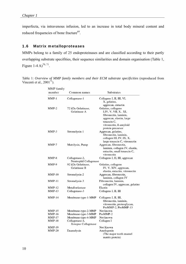
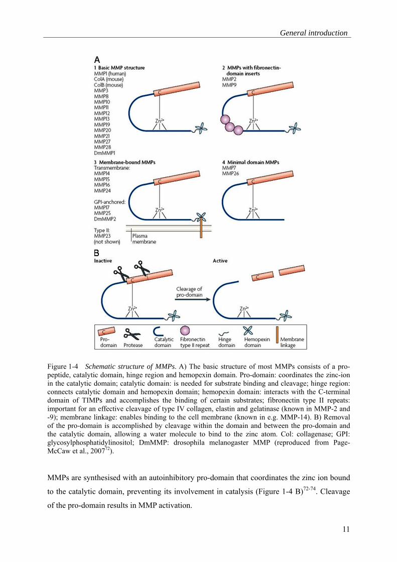
MMPs belong to a family of 25 endoproteinases and are classified according to their partly
overlapping substrate specifities, their sequence similarities and domain organisation (Table 1,
Figure 1-4 A)70, 71.
Table 1: Overview of MMP family members and their ECM substrate specificities (reproduced from Vincenti et al., 200171).
General introduction
11
Figure 1-4 Schematic structure of MMPs. A) The basic structure of most MMPs consists of a pro-peptide, catalytic domain, hinge region and hemopexin domain. Pro-domain: coordinates the zinc-ion in the catalytic domain; catalytic domain: is needed for substrate binding and cleavage; hinge region: connects catalytic domain and hemopexin domain; hemopexin domain: interacts with the C-terminal domain of TIMPs and accomplishes the binding of certain substrates; fibronectin type II repeats: important for an effective cleavage of type IV collagen, elastin and gelatinase (known in MMP-2 and -9); membrane linkage: enables binding to the cell membrane (known in e.g. MMP-14). B) Removal of the pro-domain is accomplished by cleavage within the domain and between the pro-domain and the catalytic domain, allowing a water molecule to bind to the zinc atom. Col: collagenase; GPI: glycosylphosphatidylinositol; DmMMP: drosophila melanogaster MMP (reproduced from Page-McCaw et al., 200772).
MMPs are synthesised with an autoinhibitory pro-domain that coordinates the zinc ion bound
to the catalytic domain, preventing its involvement in catalysis (Figure 1-4 B)72-74. Cleavage
of the pro-domain results in MMP activation.
Chapter 1
12
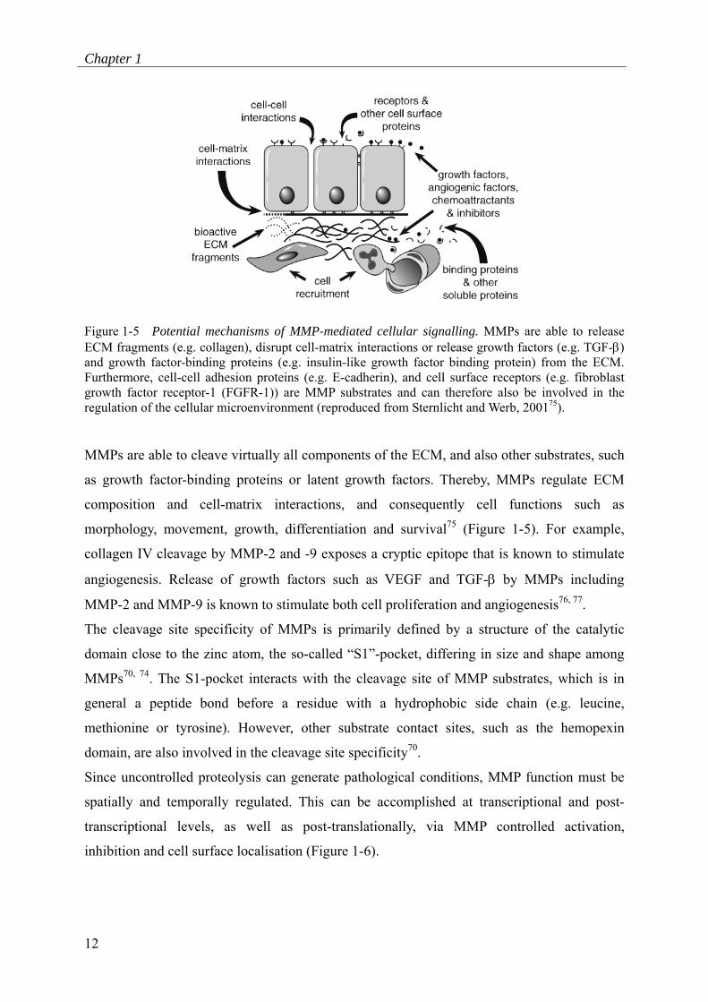
Figure 1-5 Potential mechanisms of MMP-mediated cellular signalling. MMPs are able to release ECM fragments (e.g. collagen), disrupt cell-matrix interactions or release growth factors (e.g. TGF-β) and growth factor-binding proteins (e.g. insulin-like growth factor binding protein) from the ECM. Furthermore, cell-cell adhesion proteins (e.g. E-cadherin), and cell surface receptors (e.g. fibroblast growth factor receptor-1 (FGFR-1)) are MMP substrates and can therefore also be involved in the regulation of the cellular microenvironment (reproduced from Sternlicht and Werb, 200175).
MMPs are able to cleave virtually all components of the ECM, and also other substrates, such
as growth factor-binding proteins or latent growth factors. Thereby, MMPs regulate ECM
composition and cell-matrix interactions, and consequently cell functions such as
morphology, movement, growth, differentiation and survival75 (Figure 1-5). For example,
collagen IV cleavage by MMP-2 and -9 exposes a cryptic epitope that is known to stimulate
angiogenesis. Release of growth factors such as VEGF and TGF-β by MMPs including
MMP-2 and MMP-9 is known to stimulate both cell proliferation and angiogenesis76, 77.
The cleavage site specificity of MMPs is primarily defined by a structure of the catalytic
domain close to the zinc atom, the so-called “S1”-pocket, differing in size and shape among
MMPs70, 74. The S1-pocket interacts with the cleavage site of MMP substrates, which is in
general a peptide bond before a residue with a hydrophobic side chain (e.g. leucine,
methionine or tyrosine). However, other substrate contact sites, such as the hemopexin
domain, are also involved in the cleavage site specificity70.
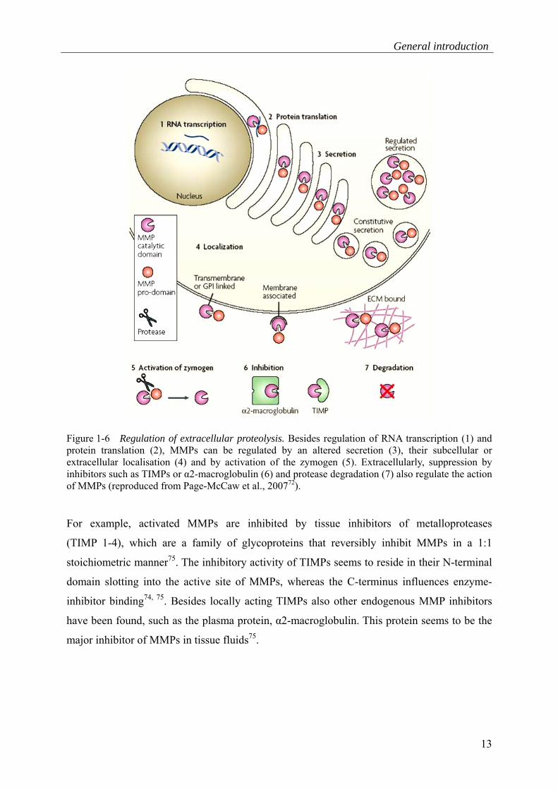
Since uncontrolled proteolysis can generate pathological conditions, MMP function must be
spatially and temporally regulated. This can be accomplished at transcriptional and post-
transcriptional levels, as well as post-translationally, via MMP controlled activation,
inhibition and cell surface localisation (Figure 1-6).
General introduction
13
Figure 1-6 Regulation of extracellular proteolysis. Besides regulation of RNA transcription (1) and protein translation (2), MMPs can be regulated by an altered secretion (3), their subcellular or extracellular localisation (4) and by activation of the zymogen (5). Extracellularly, suppression by inhibitors such as TIMPs or α2-macroglobulin (6) and protease degradation (7) also regulate the action of MMPs (reproduced from Page-McCaw et al., 200772).
For example, activated MMPs are inhibited by tissue inhibitors of metalloproteases
(TIMP 1-4), which are a family of glycoproteins that reversibly inhibit MMPs in a 1:1
stoichiometric manner75. The inhibitory activity of TIMPs seems to reside in their N-terminal
domain slotting into the active site of MMPs, whereas the C-terminus influences enzyme-
inhibitor binding74, 75. Besides locally acting TIMPs also other endogenous MMP inhibitors
have been found, such as the plasma protein, α2-macroglobulin. This protein seems to be the
major inhibitor of MMPs in tissue fluids75.
Chapter 1
14
MMP/TIMP transcription is regulated by extracellular signals including growth factors,
matrix proteins or cytokines71, 78. Signalling pathways known to be involved in transcriptional
activation of MMPs/TIMPs are the nuclear factor-kappa B (NF-кB) pathway, leading to an
activation of the NF-кB transcription factor. Also, mitogen-activated protein kinases (MAPK)
can be activated in response to growth factor binding. In particular, the extracellular signal-
regulated kinases (ERK) pathway activates Jun/Fos transcription factors that bind activator
protein-1 (AP-1) and phosphorylate ETS family transcription factors, which in turn target
polyoma enhancer A-binding protein (PEA-3) sites in MMP promotors. Moreover, the c-jun-
N-terminal kinase (JNK) pathway can be activated and initiates MMP transcription by
phosphorylation of AP-1 subunits. Finally, the janus-kinase-signal transducers and activator of
transcription (JAK-STAT) pathway is involved in MMP transcription in response to growth
factors71. However, the binding sites typically present in the promoters of inducible MMPs
and TIMPs, such as AP-1 or PEA-3 sites, are also able to interact with the promoter elements
of other transcription factors71.
Less well-characterised are the mechanisms of post-transcriptional or post-translational
regulation of MMPs. Several studies have provided evidence for the involvement of growth
factors (e.g. TGF-β or PDGF) in post-transcriptional regulation, leading to a stabilisation or
destabilisation of MMP transcripts71.
Post-translational mechanisms of MMP regulation can involve the modulation of the secretory
pathway or of protein stability79, 80. Depending on the cell type, this could be mediated by
specialised secretory granules known to occur in granulocytes (cells of the immune system) or
by secretory lysosomes, which serve as both a degradative and a secretory compartment and
occur, amongst other cells, in osteoblasts81. For example, MMP-8 and -9, synthesised by
granulocytes in the bone marrow, are stored in circulating neutrophils. These MMPs are
released following neutrophil activation by inflammatory mediators82. Also, localising cell-
MMP interactions, such as binding of MMPs to cell surface proteins, might regulate MMP
activity. Pro-enzyme activation or substrate binding to proteases can thereby be altered73.
General introduction
15
Figure 1-7 Cell surface activation of MMP-2. At the cell surface, previously activated MMP-14 (MT-MMP) binds TIMP-2 and thereby inhibits its activity (1-2). Binding of secreted pro-MMP-2 to the C-terminal domain of TIMP-2 and formation of a trimolecular complex enables MMP-14 to partially activate proMMP-2 (3-5). Full activation of MMP-2 is accomplished by a separate MMP-2 molecule that acts on the cell surface (6) (reproduced and modified from Sternlicht and Werb, 200175).
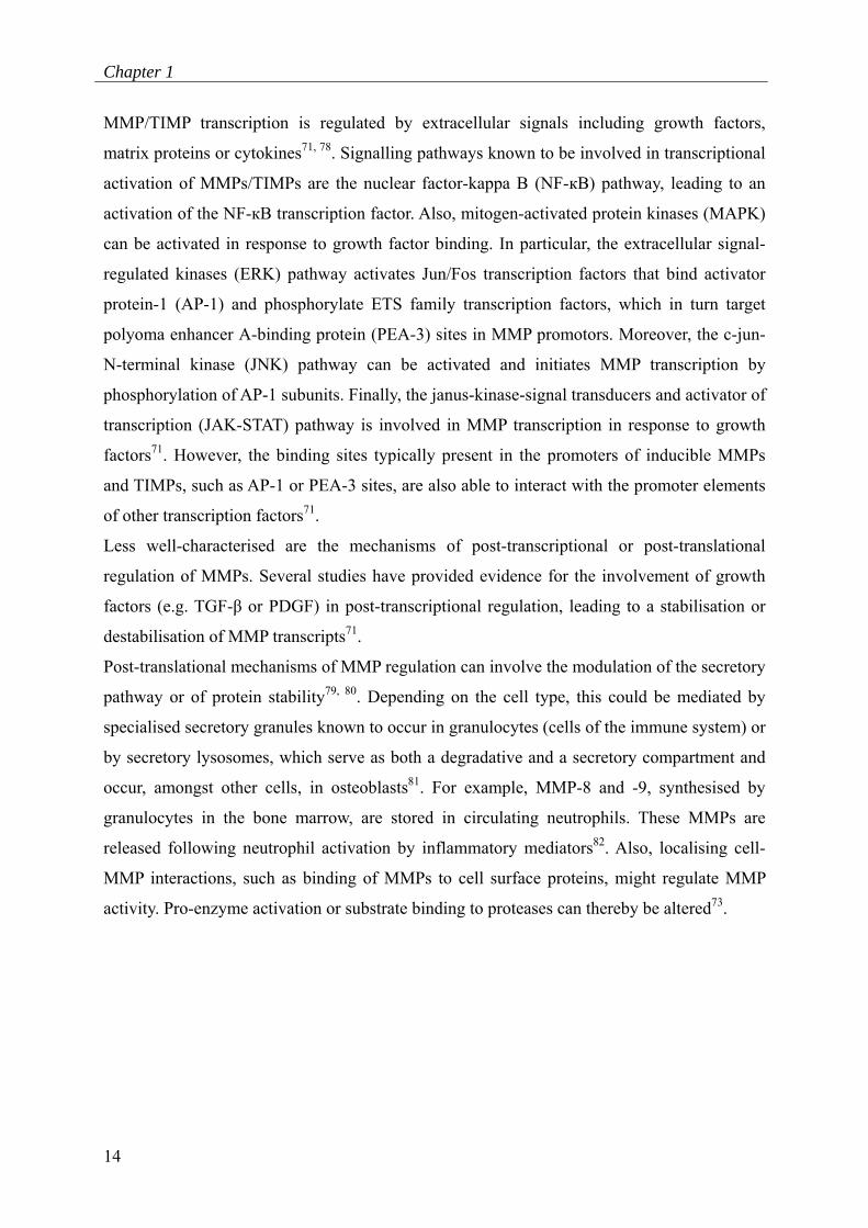
A well-described mechanism of MMP regulation is MMP-2 activation involving MMP-14 and
TIMP-2 (Figure 1-7)70, 75. Here, an extracellular ternary complex is assembled, in which
TIMP-2 is presumed to localise pro-MMP-2 adjacent to MMP-1483. Subsequently, MMP-14
cleaves pro-MMP-2, resulting in the release and activation of MMP-284, 85. Active MMP-14 is
in turn generated intracellularly by the endoprotease furin75, 86.

Chapter 1
16
The regulation of MMPs, allowing for controlled ECM remodelling, suggests their functional
involvement in bone development and regeneration processes. Indeed, a stage-specific
expression of several MMPs, such as MMP-2, -9 and -13, has been demonstrated during bone
healing14, 87. Furthermore, effects on bone morphology have been described for MMP-9, -13
and -14 knock out mice and, for MMP-2 and -13, in human individuals with loss of function
mutations87-90. For example, deletion of the MMP-9 gene in mice led to fractures with delayed
healing or non-union, caused by the persistent production of cartilage at the site of injury91.
These complications seem to be due to the limited bioavailability of VEGF caused by the lack
of MMP-9 activity72. Additionally, the involvement of MMPs in the regulation of stem cells
was, for example, demonstrated in MMP-9 knock out mice displaying an impaired release of
the Kit-Ligand, as well as impaired HSC motility92.
1.7 Cellular mechanotransduction
As previously mentioned, bone regeneration is not only affected by biochemical stimuli, but
also by mechanical stimuli. Particularly, the early phase of bone healing is highly dependent
upon the surrounding mechanical environment93-96. In this respect, it has been shown that the
gene expression and function of MSCs is affected by mechanical signals such as compressive
loading97-99. For example, MSCs respond with an altered gene expression pattern for ECM
proteins involving MMPs98, 99. Furthermore, mechanical loading of MSCs seems to affect
processes such as osteogenic differentiation and paracrine angiogenesis stimulation60, 100-102.
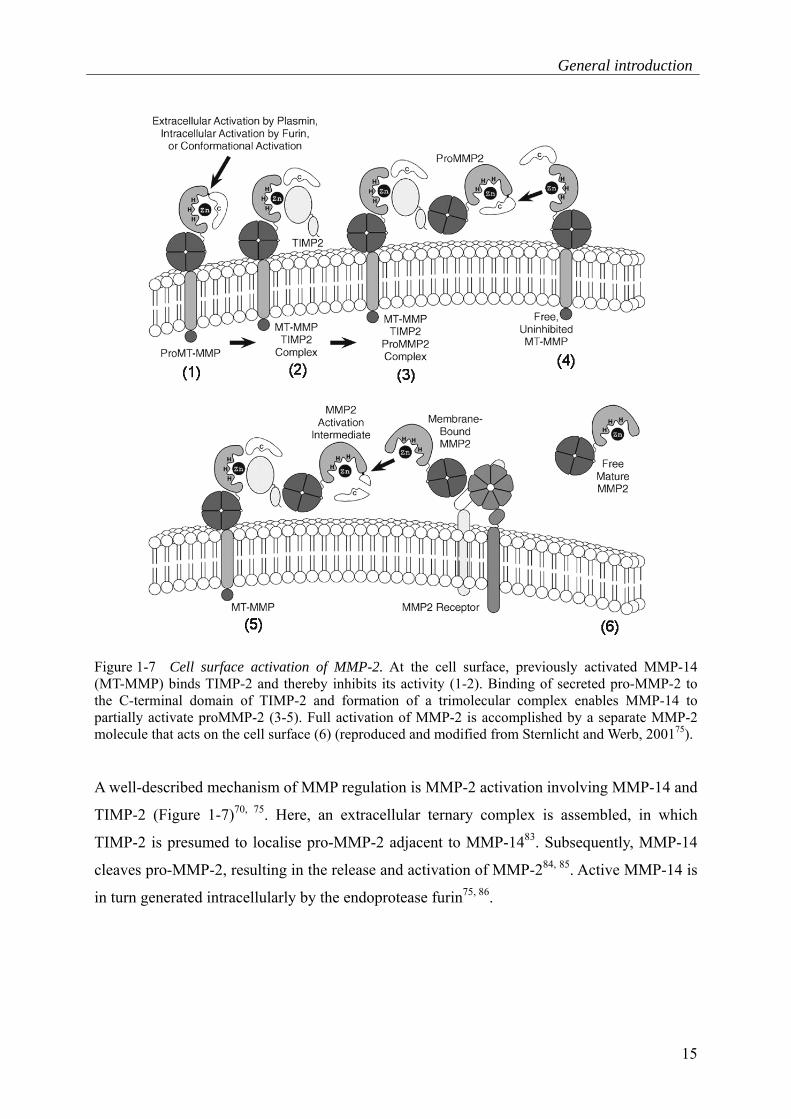
Various mechanisms have been proposed for how these mechano-sensitive cells detect
mechanical loading and convert it into biochemical signals (Figure 1-8). Stimuli can be
transduced via stretch-activated ion channels, cell adhesion molecules (e.g. integrins or
cadherins), and also by guanine nucleotide-binding protein-(G-protein-)coupled receptors and
growth factor receptor tyrosine kinases (RTK)103. For example, stretch-activated ion-channels
are able to induce a rapid internal ion transient, e.g. of Ca2+, which in turn regulate cellular
processes such as growth or differentiation103. In many cases, a connection from a membrane-
spanning receptor to the cytoskeleton and nucleus in a tensegrity unit may transmit the
signal104. Furthermore, transmembrane-spanning integrin subunits are known to unite the
ECM with actin-binding proteins, including talin and the focal adhesion kinases.
General introduction
17
Figure 1-8 Mechanisms of cellular mechanotransduction. Major cellular components involved include the cytoskeleton, stretch-activated ion channels (SAC), G proteins, integrins and RTK. Activation of intracellular signalling molecules (ICSM) leads to the rapid transmission of signals into the nucleus and to the activation of specific promoter elements. Transcription of downstream genes results, for example, in cell proliferation or protein secretion (reproduced from Wang et al., 2001104).
This induces an assembly of actin filaments and higher order structures such as stress fibers
and focal adhesions. Activation of these complexes initiates subsequent signalling cascades
including ERK, JNK or JAK-STAT, known to contribute also to MMP gene transcription71, 103.
Furthermore, since there is evidence for a transactivation between integrins and other
receptors (e.g. RTK), loading might act synergistically with ligand-activated receptors to drive
certain pathways105.

Chapter 1
18
1.8 Problem and hypothesis
The increasing knowledge of the molecular biology of bone regeneration has demonstrated
that delayed bone healing is not only a result of inappropriate stability, but also of impeded
cellular responses to biochemical and mechanical stimuli; hence, approaches for biological
augmentation are being investigated. This may involve local and systemic application of
biologically active agents, but also cell-based therapies26, 27, 30. Although mesenchymal stem
cells (MSCs) are, due to their contribution to the healing outcome and their easy expansion
in vitro, a promising tool for bone defect treatments, the application of MSCs in bone is to
date limited34, 35.
To expand/optimise the application of MSCs for bone defects, it is of interest to gain further
insights into their functional behaviour in response to healing-supportive mechanical
conditions, and to identify proteins that might regulate the transduction of mechanical signals
into the biological processes of healing. Since matrix metalloproteases (MMPs) and tissue
inhibitors of metalloproteases (TIMPs) are critically involved in matrix remodelling, such as
by ECM degradation and growth factor release, and are mechanically regulated in MSCs,
these proteins might provide a link between mechanical signals and MSC function.
Therefore, it is hypothesised that mechanical regulation of MMPs/TIMPs in MSCs stimulates
processes important for bone regeneration in an autocrine and paracrine manner. Results of
this study could form the basis for the rational design of MSC-based therapies.

General introduction
19
1.9 Aims and outline of the thesis
The aims of this project are therefore
to investigate whether mechanical loading of MSCs stimulates MMP gene/protein expression
and activity, and whether these mechanically regulated MMPs impact the function of MSCs
(Chapter 2).
to analyse one of the MMP candidates identified in Chapter 2 in more detail for its mechano-
regulation in vitro, the underlying mechanism of mechano-regulation as well as its paracrine
impact on angiogenesis stimulation (Chapter 3).
to bring together the knowledge of MMP mechano-regulation presented in Chapters 2 and 3
and to discuss its potential pathways, along with consequences of MMPs for cells and
processes involved in bone regeneration. Based on the results obtained within this study,
potential strategies for the development of therapeutic approaches will further be discussed
(Chapter 4).

20

21
2 MMP activity links mechanical stimulus
and MSC behaviour
In this chapter, the contribution of MMPs to MSC behaviour is shown by conducting
migration, proliferation and differentiation assays in the presence of broad spectrum MMP
inhibitors. Furthermore, the expression profile of MMPs/TIMPs at mRNA/protein level in
MSCs is shown. The effect of mechanical loading on MMP protein expression levels and
activity in MSCs is demonstrated by employing a bioreactor system that simulates the early
phase of bone healing. Finally, the contribution of the mechanically up-regulated MMPs to
MSC function is again evidenced by addition of specific MMP inhibitors to the functional
assays.
Chapter 2
22
Original article:
“Matrix metalloprotease activity is an essential link between mechanical
stimulus and mesenchymal stem cell behavior”
Grit Kasper1,2,†, Juliane D. Glaeser3,1,†, Sven Geissler1,2, Andrea Ode1,2, Jens Tuischer1,
Georg Matziolis1, Carsten Perka1, and Georg N. Duda1,2
†: authors contributed equally to this work 1: Musculoskeletal Research Center Berlin, Charité - Universitätsmedizin, Berlin, Germany 2: Berlin-Brandenburg Center for Regenerative Therapies,
Charité - Universitätsmedizin, Berlin, Germany 3: Free University Berlin, Institute for Chemistry/Biochemistry, Berlin, Germany
Stem Cells, 2007, 25:1985-1994
available at:
http://dx.doi.org/10.1634/stemcells.2006-0676

41
3 MMP-2 in angiogenesis stimulation
by mechanically loaded MSCs
In this chapter, MMP-2 is investigated in detail for its mechano-regulation in MSCs, as well
as potential consequences of MMP-2 enhancement for angiogenesis. Initially, the effect of
small and large interfragmentary movements on MMP-2 activity is shown in early
haematomas. This is followed by an analysis of MMP-2 mechano-regulation in MSCs
in vitro, as well as investigation of the underlying mechanism. Finally, an MMP-2 blocking
antibody and recombinant MMP-2 and TIMP-2 are examined in tube formation assays, to
evidence the paracine impact of increased MMP-2 levels on angiogenesis.
partially submitted as
Control of pro-MMP-2 levels and activation of MMP-2 via the
furin/MMP-14/TIMP-2 axis contribute concurrently to the pro-angiogenic
effects of mechanically stimulated mesenchymal stem cells, 2008
Juliane D. Glaeser1,2,†, Sven Geissler1,3,†, Patrick Strube1, Christian J. Schipp1, Georg
Matziolis1, William R. Taylor1,3, Petra Knaus2, Carsten Perka1,3, Georg N. Duda1,3 and Grit
Kasper1,3
†: authors contributed equally to this work 1: Julius Wolff Institute and Center for Musculoskeletal Surgery, Charité -Universitätsmedizin,
Berlin, Germany 2: Free University Berlin, Institute for Chemistry/Biochemistry, Berlin, Germany 3: Berlin-Brandenburg Center for Regenerative Therapies, Charité - Universitätsmedizin,
Berlin, Germany
Chapter 3
42
3.1 Introduction
Angiogenesis is a critical step for bone regeneration and responsible for re-establishing the
blood supply, resorption of necrotic tissue and mobilisation of different cell types including
mesenchymal cells134-136. During bone regeneration, angiogenesis is strongly influenced by
mechanical loading24, 96, 137. In this regard, limited micro-movement of the fracture ends is
crucial for uneventful healing, whereas exessive loading disrupts newly formed vessels23, 24.
MSCs are likely to play a crucial role in angiogenesis during musculoskeletal regeneration,
due to their ability to home to sites of injury and to generate a pro-angiogenic
microenvironment12, 14, 58. Indeed, the healing-promoting properties of MSCs were recently
attributed to their paracrine effects, such as those on ECs, rather than merely to their ability to
differentiate into functional mesenchymal cells58, 59, 138, 139.
Several members of MMP protein family are well-established regulators of angiogenesis72.
Particularly, secreted MMP-2 and cell membrane-anchored MMP-14 are expressed by MSCs
and have pro-angiogenic properties72, 140, 141. Proteolysis of the ECM by these MMPs, for
example, results in the release of pro-angiogenic factors such as VEGF and TGF-β, which in
turn are able to induce EC migration and proliferation142. MMP-14 and -2 also show
phenotypes related to bone remodelling in knock out mice and in humans with loss of
function mutations, respectively89, 90. For example, mice deficient in MMP-14 develop
multiple bone abnormalities after birth and die by 3-12 weeks, due to an ablation of
collagenolytic activity in osteoblastic cells essential for modelling of the skeletal system143.
Humans having MMP-2 mutations suffer from osteolysis and arthritis90. Furthermore, the
mechano-regulation of MMP-2 in MSCs indicates a potential role of this protease in
translating mechanical signals into the regulation of angiogenesis in bone140. For full function
of MMP-2, this protease needs to be activated by its ternary complex, consisting of active-
MMP-14, TIMP-2 and pro-MMP-283-85, 144. Active MMP-14, however, is intracellularly
generated via proteolysis by furin75, 86.
To date, it remains unclear whether the activation complex of MMP-2 is sensitive to
mechanical signals and whether pro- and/or active-MMP-2 are involved in the paracrine
stimulation of angiogenesis by mechanically loaded MSCs. To answer these questions,
the study presented in this chapter aimed to identify MMP-2-inducing loading conditions for
MSCs. Subsequently, the mechanism of MMP-2 mechano-regulation is examined in detail by
inhibitor application. Finally, the relevance of mechanically regulated MMP-2 to angiogenesis
stimulation is investigated.
MMP-2 in angiogenesis stimulation by mechanically loaded MSCs
43
3.2 Materials and methods
3.2.1 Animal bone defect model for haematoma sampling
A previously described animal model was used145. Briefly, the left femur of 12 female
Sprague-Dawley rats (aged 12 months) was osteotomised and distracted to an osteotomy gap
of 1.5mm. Semi-rigid and rigid fixations were obtained by varying the offset of the external
fixation.
Haematoma samples were collected 7 days post surgery from each group and frozen at -20°C
for a maximum of 2 months. For investigation, each haematoma was dispersed in 30µl
phosphate-buffered saline (PBS) and the supernatant was analysed. Total protein content was
determined by the DC Protein assay according to the manufacturer’s instructions (Biorad,
Germany).
3.2.2 Cell culture
MSCs were isolated, characterised and cultured as stated in 2.2.1. Passages 3-5 were used for
experiments. The mean donor age was 63 years (ranging from 18 to 86 years). For
angiogenesis assays, simian vacuolating virus (SV)40 immortalised human dermal
microvascular ECs (HMEC-1) were employed and cultured in MCDB-131
(Invitrogen, Germany) supplemented with 1µg/ml hydrocortisone, 2mM L-glutamine,
5% FCS (Biochrom, Germany) and 100U/ml penicillin + 100µg/ml streptomycin
(EC culture media) 146.
3.2.3 Bioreactor
The bioreactor was employed as specified in 2.2.3. Levels of compression, frequency and
duration of loading were adjusted as stated in the corresponding experiments. CM were
sampled and frozen at -20°C for a maximum of three months. For application of the Golgi
disturbing agent Brefeldin A (BFA) (2.8µg/ml) (Sigma-Aldrich, Germany)147, constructs were
transferred into 24-well culture plate containing MSC medium without FCS. After 8h
incubation CM was harvested. Furin inhibitor I (Calbiochem, Germany) was added to the
MSC medium prior to loading at 4µg/ml. Equal cell numbers in constructs exposed to
mechanical loading and in unloaded control constructs were verified by the Cell Titer 96®
AQueous test (Promega, Germany). Cells were recovered from the constructs by the addition
of 1ml trypsin solution (225U/ml) (Serva, Germany) and incubation for 15min at 37°C.
Trypsinised cells were repeatedly transferred into a tube containing 5ml MSC medium und
Chapter 3
44
2ml Trasylol® (Bayer, Germany) until complete digestion of the construct. Cell vitality was
after digestion of the cell/fibrin construct determined by the CASY® cell counter system
(Schärfe System, Germany)148. Furthermore, cell vitality was analysed by acridine orange
staining (acridine orange, ethidium bromide, Sigma-Aldrich, Germany) by means of a
counting chamber (Neubauer, Germany).
3.2.4 ELISA, zymography and MMP-2/-14 activity assay
Total-MMP-2 ELISA and gelatine zymograms were performed as stated in 2.2.5. Reverse
zymography was conducted using a 12% polyacrylamide, 0.4% sodium dodecyl sulfate (SDS)
gel containing porcine gelatine (Sigma-Aldrich, Germany) (2.3mg/ml) and recombinant pro-
MMP-2 (Calbiochem, Germany) (0.4mg/ml). This approach was performed according to the
manufacturer’s protocol of the Novex® system with either 20µl of CM or 60µg of total protein
from haematoma samples. Analysis of digital images was carried out using the NIH ImageJ
software package (http://rsb.info.nih.gov/ij/). MMP-2 and MMP-14 activity were determined
using the biotrak activity assay (GE healthcare) according to manufacturer’s protocol
employing 100µl of sample. To measure levels of MMP-2, CM was diluted in a ratio of 1:10
and to determine MMP-14, cells were removed from the construct, 2.5x10⁵ cells were
incubated in 125µl lysis buffer for 15min, resuspended and the supernatant was used as 1:2
dilution.
3.2.5 Tube formation assay
24-well plates were coated with 50µl matrigel (10mg/ml, Invitrogen, Germany) and allowed
to gel for 30min at 37°C. Afterwards, 4x10⁴ HMEC-1 cells were seeded in 100µl EC culture
medium per well and 500µl CM was added. The assay was incubated for 17h before results
were visualised by light microscopy. The cumulative length of the capillary network was
analysed for each well. Quantification was performed using NIH ImageJ. Recombinant
active-MMP-2 (150ng/ml) (Calbiochem, Germany) TIMP-2 (13ng/ml) (Calbiochem,
Germany) and anti-MMP-2 pro-form blocking antibody, clone CA-4001 (4µg/ml) (Millipore,
Germany) were used.

MMP-2 in angiogenesis stimulation by mechanically loaded MSCs
45
3.2.6 Statistics
A Student´s t-test was employed for statistical evaluation. Analysis of rat haematomas was
accomplished by an unpaired test. For the evaluation of the effect of bioreactor samples, a
paired test was applied. All tests were two sided, and at a significance level of p<0.05. In the
presented figure legends, the number of experiments is displayed and in the figures, statistical
significance is indicated by a star.

Chapter 3
46
3.3 Results
3.3.1 Mechano-regulation of MMP-2 in vivo
To investigate whether the in vitro described mechano-regulation of MMP-2 also occurs
in vivo140, haematomas from bone defects with different interfragmentary movements were
analysed for their MMP-2 activity. Indeed, MMP-2 was significantly elevated in haematomas
from semi-rigidly stabilised fractures 7 days post surgery compared to those with a rigid
fixator (factor: 1.6, p=0.017, Figure 3-1).
Figure 3-1 Up-regulation of MMP-2 by mechanical stimulation during fracture healing. Representative zymogram showing the gelatinolytic activity at the migration height of MMP-2 in semi-rigidly and rigidly fixated rat haematomas sampled at day 7. Absolute densities at the migration height of pro-MMP-2 were quantified. sr: semi-rigidly fixated; r: rigidly fixated (n=6) (*: p<0.05).
3.3.2 Influence of loading parameters on MMP-2 levels
To optimise the in vitro conditions for MMP-2 induction in response to mechanical
stimulation, loading parameters affecting cellular processes of bone healing were individually
varied. Firstly, compression levels were investigated149. 10%, 30%, 50% and 60%
compression were applied at a frequency of 1Hz for 72h. 30% compression significantly
enhanced MMP-2 levels compared to unloaded controls (meantotal-MMP-2 amount: 114%, p=0.015;
meanpro-MMP-2 activity: 174%, p=0.026, Figure 3-2 A, B).

MMP-2 in angiogenesis stimulation by mechanically loaded MSCs
47
Figure 3-2 MMP-2 levels increase at 30% compression and 0.5/1Hz, as well as 48h/72h of stimulation. Total-MMP-2 amounts were determined by ELISA and pro-MMP-2 activities by quantification of zymograms. A-F) MMP-2 levels versus loading conditions. All data were normalized to the unloaded controls (n=3) (*: p<0.05).
10%, 50% and 60% compression had no impact on MMP-2 concentration or activity. Cell
vitality was reduced in constructs loaded at 50% and 60% compared to unloaded controls
(mean50%: 92%, p=0.001; mean60%: 90%, p<0.001, Figure 3-3 A, B).

Chapter 3
48
Figure 3-3 Cell vitality is diminished at 50/60% compression and 2Hz. A) Representative pictures of cell vitality of MSCs recovered from constructs with and without loading. Top: acridine-orange staining (green: living cells, red: dead cells). Bottom: Light microscopy B-D) Cell vitality determined by means of a cell counter system. All data were normalized to the unloaded controls (n=3) (*: p<0.05).
A frequency range from 0.1 to 2Hz was additionally tested for its effect on MMP-2 levels; this
is considered to include the range of normal human walking150. Load cycles were maintained
at 30% for a duration of 72h. 0.5Hz and 1Hz resulted in a significant up-regulation of
total-MMP-2 protein concentration compared to unloaded controls (mean0.5Hz: 130%,
p=0.028; mean1Hz: 131%, p=0.005, Figure 3-2 C). Furthermore, pro-MMP-2 activities were
elevated at these frequencies (mean0.5Hz: 129%, p=0.014; mean1Hz: 157%, p=0.005, Figure 3-2
D). Frequencies of 0.1Hz and 2Hz had little or no effect on MMP-2 concentration or activity
(Figure 3-2 C, D). Cell vitality was reduced at 2Hz (mean: 91%, p=0.004), whereas loading
frequencies of 0.1Hz, 0.5Hz and 1Hz had no effect on cell vitality compared to unloaded
controls (Figure 3-3 C).

MMP-2 in angiogenesis stimulation by mechanically loaded MSCs
49
The duration of stimulation was then examined at 1Hz and 30% compression. Total-MMP-2
amounts were increased at 48h and 72h, but not at 24h compared to unloaded controls
(mean48h: 141%, p=0.022; mean72h: 148%, p=0.012, Figure 3-2E). Comparable results were
obtained for pro-MMP-2 activity (mean48h: 172%, p=0.037; mean72h: 170%, p=0.013,
Figure 3-2 F). MSC vitality was not altered in loaded and unloaded constructs (Figure 3-3 D).
From these experiments, inducing loading conditions in respect to MMP-2 expression and
activity were concluded to be 1Hz and 30% compression over a time period of 72h and
non-inducing to be 0.1Hz and 10%.
3.3.3 Mechanism of MMP-2 mechano-regulation
Having established conditions for enhanced MMP-2 production, the mechanism of its
mechano-regulation was examined under the inducing conditions.
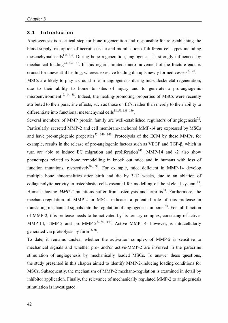
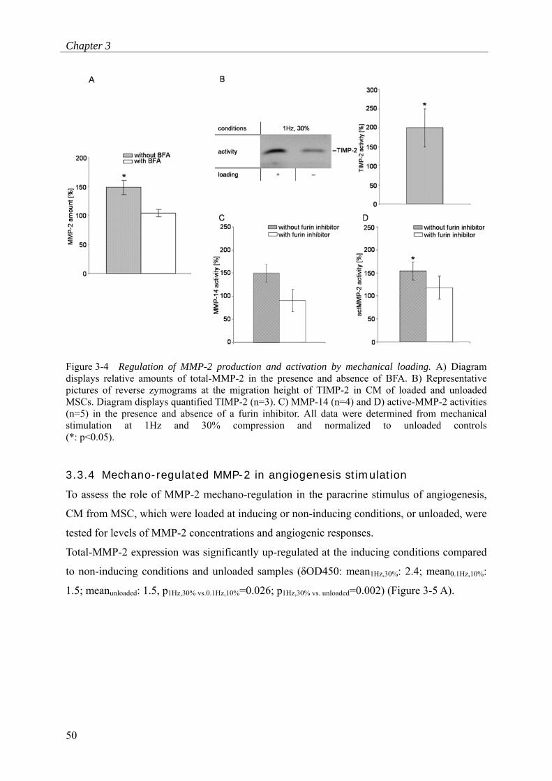
To investigate whether regulation occurs intracellularly, the secretion inhibitor BFA was
added. This hindered the elevation of total-MMP-2 amounts in response to mechanical
loading (meanBFA: 104%, meancontrol: 148%, p=0.012, Fig. 3-4 A).
Subsequently, extracellular activities of the activation complex components were determined.
TIMP-2 activity was increased (meanTIMP-2: 200%, p=0.014, Fig. 3-4 B), and MMP-14
activity showed a trend of up-regulation (meanMMP-14: 150%, p=0.104, Figure 3-4 C).
Moreover, the concentration of active-MMP-2 was elevated under mechanical stimulation
(meanactive-MMP-2: 153%, p=0.006, Figure 3-4 D).
Inhibition of furin, which is responsible for intracellular MMP-14 activation, suppressed the
mechanical up-regulation of MMP-14, as well as that of active-MMP-2 (Figure 3-4 C, D).
Chapter 3
50
Figure 3-4 Regulation of MMP-2 production and activation by mechanical loading. A) Diagram displays relative amounts of total-MMP-2 in the presence and absence of BFA. B) Representative pictures of reverse zymograms at the migration height of TIMP-2 in CM of loaded and unloaded MSCs. Diagram displays quantified TIMP-2 (n=3). C) MMP-14 (n=4) and D) active-MMP-2 activities (n=5) in the presence and absence of a furin inhibitor. All data were determined from mechanical stimulation at 1Hz and 30% compression and normalized to unloaded controls (*: p<0.05).
3.3.4 Mechano-regulated MMP-2 in angiogenesis stimulation
To assess the role of MMP-2 mechano-regulation in the paracrine stimulus of angiogenesis,
CM from MSC, which were loaded at inducing or non-inducing conditions, or unloaded, were
tested for levels of MMP-2 concentrations and angiogenic responses.
Total-MMP-2 expression was significantly up-regulated at the inducing conditions compared
to non-inducing conditions and unloaded samples (δOD450: mean1Hz,30%: 2.4; mean0.1Hz,10%:
1.5; meanunloaded: 1.5, p1Hz,30% vs.0.1Hz,10%=0.026; p1Hz,30% vs. unloaded=0.002) (Figure 3-5 A).

MMP-2 in angiogenesis stimulation by mechanically loaded MSCs
51
Angiogenic responses followed this pattern, showing increased tube formation at the inducing
conditions compared to non-inducing conditions and unloaded controls (cumulative tube
length [x105 pixel]: mean1Hz,30%: 203.6; mean0.1Hz,10%: 86.3; meanunloaded: 81.5, p1Hz,30% vs.
0.1Hz,10%=0.025; p1Hz,30% vs. unloaded=0.026, Figure 3-5 B, C). Controls using CM from equivalent
experiments without cells in the bioreactor construct showed no increase in tube formation.
Figure 3-5 CM from loaded MSCs shows similar increases in MMP-2 concentration and tube formation. A) MMP-2 amounts in CM from different loading conditions. B) Cumulative tube length in response to the different CM. CM from inducing conditions (1Hz, 30% compression), non-inducing conditions (0.1Hz, 10% compression), unloaded MSCs and controls (CM from equivalent experiments without MSCs in the bioreactor construct) were tested (n=6) (*: p<0.05).
Chapter 3
52
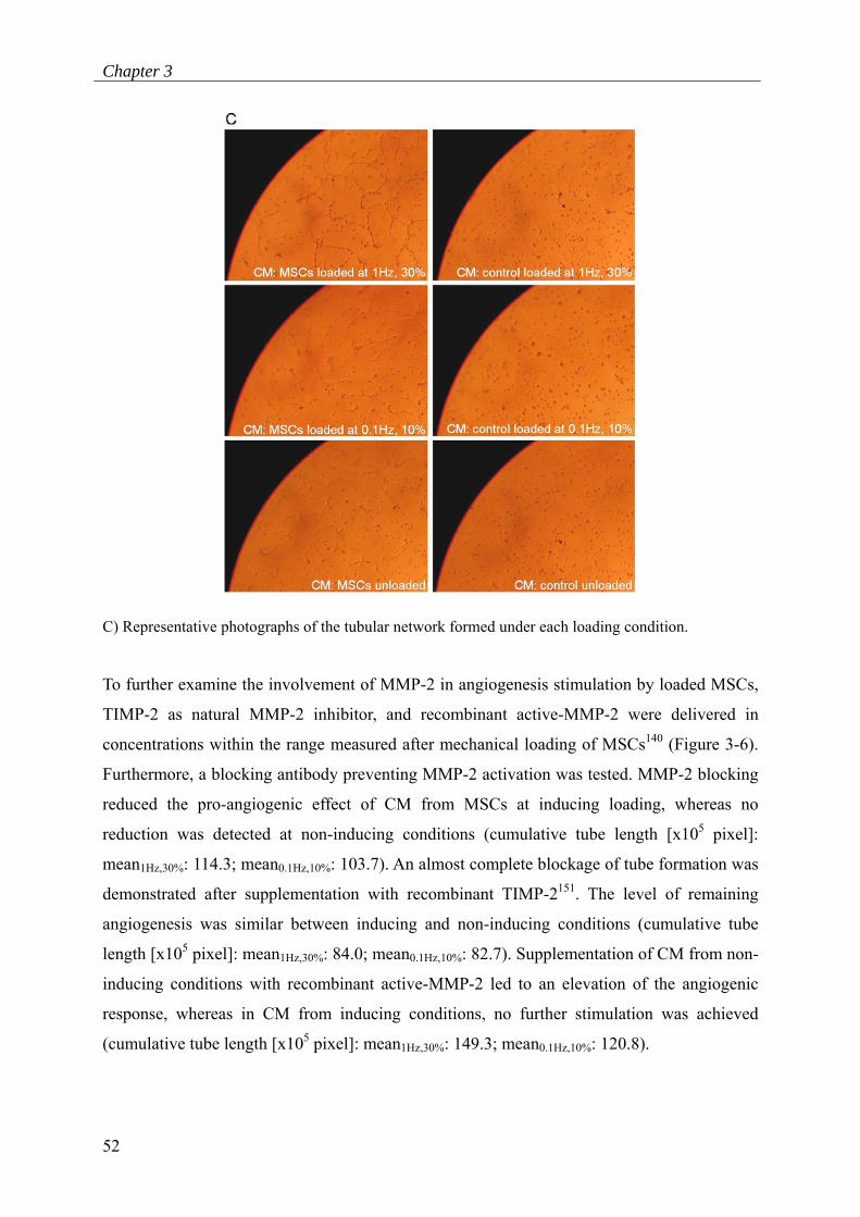
C) Representative photographs of the tubular network formed under each loading condition.
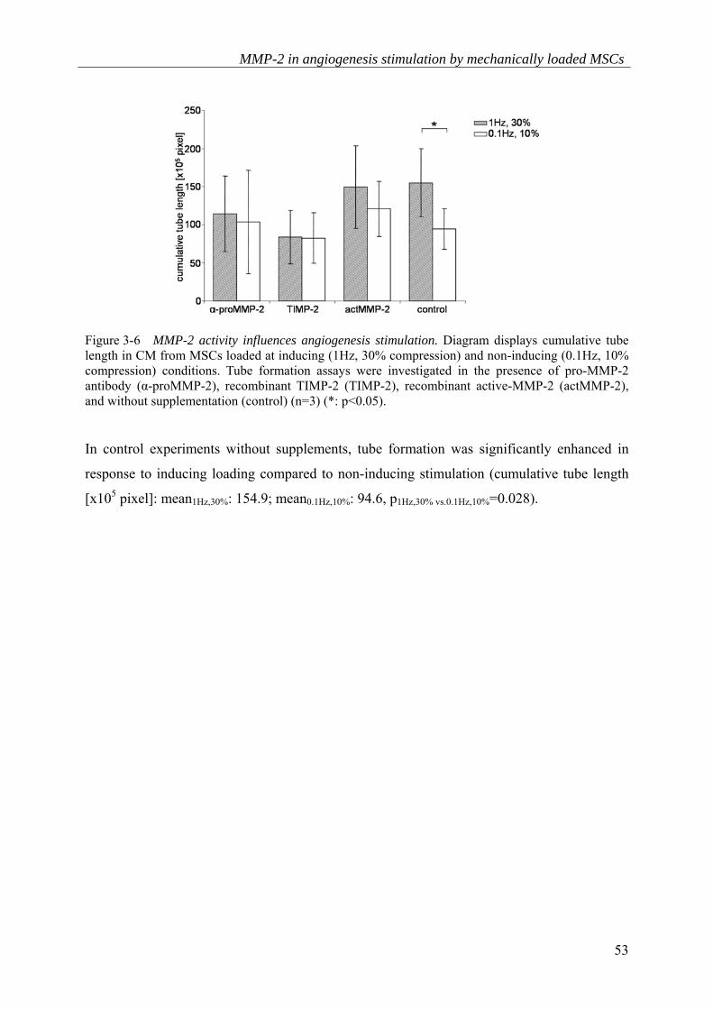
To further examine the involvement of MMP-2 in angiogenesis stimulation by loaded MSCs,
TIMP-2 as natural MMP-2 inhibitor, and recombinant active-MMP-2 were delivered in
concentrations within the range measured after mechanical loading of MSCs140 (Figure 3-6).
Furthermore, a blocking antibody preventing MMP-2 activation was tested. MMP-2 blocking
reduced the pro-angiogenic effect of CM from MSCs at inducing loading, whereas no
reduction was detected at non-inducing conditions (cumulative tube length [x105 pixel]:
mean1Hz,30%: 114.3; mean0.1Hz,10%: 103.7). An almost complete blockage of tube formation was
demonstrated after supplementation with recombinant TIMP-2151. The level of remaining
angiogenesis was similar between inducing and non-inducing conditions (cumulative tube
length [x105 pixel]: mean1Hz,30%: 84.0; mean0.1Hz,10%: 82.7). Supplementation of CM from non-
inducing conditions with recombinant active-MMP-2 led to an elevation of the angiogenic
response, whereas in CM from inducing conditions, no further stimulation was achieved
(cumulative tube length [x105 pixel]: mean1Hz,30%: 149.3; mean0.1Hz,10%: 120.8).
MMP-2 in angiogenesis stimulation by mechanically loaded MSCs
53
Figure 3-6 MMP-2 activity influences angiogenesis stimulation. Diagram displays cumulative tube length in CM from MSCs loaded at inducing (1Hz, 30% compression) and non-inducing (0.1Hz, 10% compression) conditions. Tube formation assays were investigated in the presence of pro-MMP-2 antibody (α-proMMP-2), recombinant TIMP-2 (TIMP-2), recombinant active-MMP-2 (actMMP-2), and without supplementation (control) (n=3) (*: p<0.05).
In control experiments without supplements, tube formation was significantly enhanced in
response to inducing loading compared to non-inducing stimulation (cumulative tube length
[x105 pixel]: mean1Hz,30%: 154.9; mean0.1Hz,10%: 94.6, p1Hz,30% vs.0.1Hz,10%=0.028).

Chapter 3
54
3.4 Discussion
3.4.1 Mechanical parameters
Mechanical loading for 72h and 48h, but not 24h stimulated the MMP-2 production by MSCs.
This is consistent with a study showing that mechanical stimulation of human fibroblasts
leads to an enhanced generation of active-MMP-2 at 48h, but not at 24h152. Regarding
frequencies, 0.5Hz and 1Hz resulted in an up-regulation of MMP-2 protein and activity,
whereas 0.1Hz and 2Hz had little or no effect. These results agree with those demonstrating
that mechanical stimulation of 1Hz influences mRNA and protein expression by MSCs153-155.
Compressive deformation of 30% caused an increase in MMP-2 concentration and activity,
but not 10%, 50% or 60%. These results imply that 1Hz and 30% strain are in the range of
optimal mechanical stimulation of MSCs in regard to MMP-2 production. Parameters both
above and below these values result in a reduced response. This might be due to
unresponsiveness of the cells at parameters outside the identified range, but may be also due
to decreased cell vitalities, which were demonstrated especially at higher values of loading,
such as 2Hz and 50% compression.
Remarkably, the MMP-2-inducing parameters determined (1Hz and 30% compression)
correspond to in vivo conditions measured during uneventful bone healing23, 150. An axial
displacement of approximately 1mm in a 3mm fracture gap (approximately 30%
compression) was measured in fracture models showing successful healing23. Regarding
frequencies, rat tibias respond with increased bone formation at loading above 0.5 Hz, but not
below156. Furthermore, 0.5-1Hz corresponds to the rate of human walking23, 150. Thus, it could
be hypothesised that in vivo mechanical conditions for fast bone regeneration are associated
with a stimulation of MMP-2. This proposal is strengthened by the observed up-regulation of
MMP-2 activity in haematomas from less-rigidly stabilised fractures, which exhibited
improved healing145.
3.4.2 Regulation of MMP-2 levels
The up-regulation of pro- and total-MMP-2 by mechanical stimuli was not observed before
48h and could be blocked by the Golgi disturbing agent, BFA. Furthermore, only protein and
not mRNA levels appear to be affected140. Thus, the mechano-regulation of intracellar
pro-MMP-2 seems to be mediated via an indirect mechanism, occuring after transcription and
prior to protein secretion. This mechanism might be based on an increase in protein stability
MMP-2 in angiogenesis stimulation by mechanically loaded MSCs
55
and/or alteration of the secretory pathway, since both possibilities have been shown to
regulate MMP-2 protein levels79, 157.
In addition to pro-/total-MMP-2, active-MMP-2 concentrations were enhanced by mechanical
loading. This enhancement corresponded to the mechanical regulation of the other members
of the ternary MMP-2 activation complex, evidenced by a significant elevation of TIMP-2
and a trend towards increased MMP-14. Based on previous results, the up-regulation of
TIMP-2 activity corresponds to an elevation of TIMP-2 protein concentration140. Furthermore,
furin activity was required for the enhancement of MMP-14 and active-MMP-2 by
mechanical loading. This suggests that the mechano-regulation of active-MMP-2 is mediated
by a stimulation of the furin/ternary complex axis75, 86. In experiments comparable to the
present study, a simultaneous up-regulation of pro- and active-MMP-2, TIMP-2 and MMP-14,
and the involvement of furin in enhanced active-MMP-2 and -14 activity, was demonstrated
in cardiac fibroblasts exposed to collagen I158. Hence, the concurrent regulation of pro-MMP-
2 levels, as well as of the MMP-2 activation machinery, might not only occur in response to
mechanical stimuli, but also to biochemical signals.
3.4.3 Angiogenesis stimulation
Inhibition experiments demonstrated that both modes of MMP-2 mechano-regulation the
elevation of pro-MMP-2 levels, as well as the enhancement of active-MMP-2 seem to
independently mediate the pro-angiogenic impact of mechanically loaded MSCs. Since the
angiogenesis experiments were performed in the absence of contact with MSCs, the effect of
pro-MMP-2 is likely to be caused by a subsequent MMP-2 activation, e.g. by EC-associated
MMP-14159. Thereby, enhanced levels of active-MMP-2 would be available in the
microenvironment for angiogenesis stimulation (for more details see 4.3.2).

56
57
4 General Discussion
In this section, the findings described in Chapters 2 and 3 are summarised and discussed with
respect to their consequences for the bone healing outcome. Then pathways of MMP/TIMP
induction in MSCs in response to mechanical conditions known from uneventful bone
healing are discussed. Furthermore, consequences of the altered MMP/TIMP balance for the
MSC microenvironment during healing are hypothesised. Finally, the potential value of the
project’s results for the rational design of new MSC-based therapies for bone regeneration is
discussed, and the conclusions of the thesis are presented.
Chapter 4
58
4.1 Summary of the findings
The early phase of bone healing and especially angiogenesis are known to be influenced by
mechanical conditions. Although MSCs, present in the early haematoma, are thought to
stimulate bone regeneration by production of proteins that act as autocrine and paracrine
signals, the interplay between mechanical stimuli,and molecular and cellular responses of
MSCs, as well as their effect on angiogenesis, remain poorly understood.
Results presented in chapter 2 showed an up-regulation of MMP-2, -3, -13 and TIMP-2
protein levels, but not of RNA levels. Furthermore, MMP-13 was demonstrated to be involved
in MSC differentiation and MMP-2 to likely contribute to the migratory behaviour of MSCs.
These findings indicate that alterations in the MMP/TIMP balance might mediate the
translation of mechanical stimuli into the cellular response of MSCs. In chapter 3, it was
shown that mechano-regulation of MMP-2 occurs at two levels in MSCs: via an increase in
pro-MMP-2 production and enhanced activation via the ternary MMP-2 activation complex
including MMP-14 and TIMP-2. Furthermore, it was demonstrated that both pro- and active-
MMP-2 are involved in the paracrine stimulation of angiogenesis by mechanically loaded
MSCs.
4.2 Mechano-regulation of MMPs in MSCs
Mechanical loading of MSCs resulted in an enhancement of MMP-2, -3, -13 and TIMP-2
proteins, whereas mRNA levels remained constant. Furthermore, detailed investigation of
MMP-2 demonstrated an enhancement of this protease by an intracellular mechanism, since
addition of BFA hindered the up-regulation of secreted MMP-2 levels. Due to the lack of a
difference in mRNA amounts between loaded and unloaded samples, the regulation of MMPs
in response to mechanical stimulation seems to occur via a post-transcriptional
mechanism79, 80.
Several studies provide evidence for a regulation of MMPs through modulations of the
secretory pathway or of protein stability. However, non of these studies specified the
underlying mechanism for these effects79, 80, 157. One option is an alteration of MMP protein
secretion by secretory lysosomes that are known to exist in osteoblasts81. Interestingly, it has
been shown that these lysosomes are sensitive to intracellular Ca2+ levels and could thereby
be affected by mechanical stimulation of the cells via stretch-activated ion-channels81, 103.
Extracellular MMP levels might be also regulated by shedding of matrix vesicles, membrane-
enclosed microstructures released from the plasma membrane, which occur in ECs, but also in
General discussion
59
differentiating chondrocytes and osteoblasts160, 161. Various pro- and active-MMPs have been
shown to be associated with these vesicles, including pro- and active-MMP-2, MMP-13, -14
and TIMP-2, which overlap with the mechano-regulated MMPs identified within this study160,
162. Previous results provide evidence for shedding of these vesicles in response to growth
factors, including VEGF and bFGF160. Therefore, the enhancement of MMP levels in response
to loading could be an indirect result of growth factor up-regulation, as seen for bFGF and
TGF-β in MSCs in response to mechanical stimulation60.
Results from this study further indicate a superior activation of MMP-2 on the MSC-cell
surface in response to loading, which was evidenced by an up-regulation of all members of
the ternary complex, as well as by the dependency of active-MMP-2 and MMP-14 activities
on the endoprotease furin. Notably, furin is also stimulated by TGF-β1 via enhanced
transactivation of the fur gene163. This could explain increased levels of MMP-14 and active-
MMP-2 protein, and also of MMP-13, since MMP-14 and MMP-2 were shown to be able to
activate this protease78.
One or several of the listed mechanisms could account for the up-regulation of MMP-2,
MMP-3 and MMP-13 in response to mechanical stimulation reported in the present study.
Nevertheless, other investigations using different experimental settings have provided
evidence for the regulation of MMP mRNA by mechanical loading. For example, an up-
regulation of MMP-1, -2, -3 and MMP-9 mRNA has been observed after cyclic tensile
loading in cultured chondrocytes and MMP-13 up-regulation has been described after
stretching of murine osteoblasts106, 164. These data indicate the existence of additional
regulatory mechanisms on mRNA levels in other mesenchymal cells or under different
stimulatory conditions. Furthermore, the nature of the applied stress seems to be important for
the regulation of matrix remodelling by MSCs, since a recent study demonstrated differential
effects in response to equiaxial and uniaxial strain99, 165. This strain-specific regulation might
also account for MMPs, since stromelysin mRNA was shown to be downregulated by uniaxial
strain, whereas in this study using uniaxial loading, no alterations of these mRNA levels were
found.

Chapter 4
60
4.3 Consequences of altered MMP/TIMP balance
4.3.1 Consequences for MSCs
The mechano-regulation of MMP-2, -3, -13 and TIMP-2 indicates an involvement of these
MMPs in the autocrine regulation of MSCs.
Indeed, data from this study showed enhanced MMP-2 activities are likely to contribute to
superior MSC migration. As previously discussed (see 2.4.2), this effect might occur via
mechanisms including ECM degradation, VEGF release or alteration of the IGF-BP-3/IGF
interactions75, 77, 128-130. In addition, adipogenic differentiation could also be altered by MMP-2
up-regulation, since levels of this protease have been shown to increase during the
differentiation process, and MMP-2 inhibition leads to a decrease in adipogenesis166. One
potential link is the ability of MMP-2 to cleave the fibroblast growth factor (FGF) receptor 1
resulting in the release of a soluble fragment that may indirectly influence bFGF availability,
which in turn enhances PPARγ ligand-induced adipogenesis in MSCs167, 168.
Up-regulated MMP-3 was shown not to be associated with altered MSC osteogenic
differentiation, proliferation or migration. However, a negative regulatory role of MMP-3 in
adipogenic differentiation of MSCs has been previously demonstrated169, 170.
MMP-13 up-regulation was demonstrated within this study to be involved in osteogenic
differentiation, and this may be explained by an interaction with the SDF-1/CXCR4 axis121, 126
(see 2.4.2). In addition, enhanced levels of MMP-13 could stimulate chondrogenic
differentiation, due to its ability to cleave ECM fragments, such as collagen II, or activate
latent TGF-β, both of which are critically involved in chondrogenesis162, 171, 172. For example,
matrix vesicle-associated MMP-13 obtained from growth plate cartilage has been shown to
activate latent TGF-β and might therefore play a role in chondrocyte-mediated
mineralization162. Interestingly, mechanical loading of bovine bone marrow stromal cells
results in an increase in Smad2/3 phosphorylation, which is known to be critical to
TGF-β-induced chondrogenic differentiation154. Based on the results of this study, it is
possible that the enhanced chondrogenesis results from an increase in TGF-β activation by up-
regulated MMPs, e.g. MMP-13.

General discussion
61
Figure 4-1 MMP/TIMP balance as a potential transducer of mechanical stimuli into MSC functions. Mechanical loading of MSCs leads to enhanced secretion and potential activation of MMPs/TIMPs (1), which might mediate various regulatory processes (2), such as degradation of the ECM (3) or release of latent growth factors (4). Interaction of ECM fragments or activated growth factors with cell surface receptors (5) might result in altered MSC migration, proliferation and differentiation in an autocrine manner.
The effects of increased MMP-14 levels in MSCs on cell function were not investigated
within this study. However, it is likely that this protease contributes not only indirectly via its
MMP-2-activating properties, but also directly to alterations in the functional behaviour of
these cells75, 132. In this respect, MMP-14 might be involved in MSC proliferation,
differentiation, migration and adhesion132, 133, 173. The contribution of MMP-14 to proliferation
has been shown by maintenance of osteoblast and also osteocyte viability, which is potentially
mediated by increased activation of latent TGF-β132, 133. Perhaps, MMP-14 is required for
differentiation, and particularly for osteocytogenesis in bone173. This effect could be
accomplished by cleavage of various MMP-14 substrates, such as collagen I/II, growth factors
(e.g. SDF-1 or bFGF) or indirectly via the action of MMP-275, 127, 174. Cleavage of cell surface

Chapter 4
62
tissue transglutaminase (tTG) by MMP-14 could also affect MSC migration and adhesion,
since over-expression of this protease was shown to inhibit migration and adhesion of tumor
cells175 176.
Likewise, the enhancement of TIMP-2 by mechanical loading could have MMP-2-
independent effects on MSCs, due to its ability to stimulate proliferation in a wide range of
human cells177, 178.
However, functional analyses conducted within this study did not discover all potential
actions of mechano-regulated MMPs on MSCs. It is furthermore most likely that the activities
of the MMPs studied are not independent from other family members, since they can act
synergistically as been observed in MMP-9/MMP-13 knock out animals88. Moreover, other
MMP family members could compensate for lost activity, since most substrates are
recognised by more than one of these proteases. However, it is likely that alterations in the
MMP/TIMP balance due to mechanical loading create a microenvironment for MSCs that
determines their functional behaviour, such as migration, proliferation and differentiation
(Figure 4-1).
4.3.2 Consequences for angiogenesis
This study provided evidence for the involvement of mechanically induced MMP-2 in the
augmentation of angiogenesis. Furthermore, angiogenesis stimulation by mechanically loaded
MSCs has been shown to be dependent upon FGF receptor 1 and VEGF receptor, but not
upon VEGF levels60. However, although FGF receptor 1 is an MMP-2 substrate, a stimulation
of angiogenesis after receptor cleavage is unlikely, since it leads to a loss of the receptor’s
signalling capacity167, 179, 180. Therefore, it may be hypothesised that angiogenesis stimulation
by CM from mechanically loaded MSCs is rather a result of other mechanisms. For example,
degradation of ECM components may facilitate EC migration by removing physical barriers72,
76. Also, modulation of angiogenic signals by MMP-2, including the exposure of a cryptic
epitope of collagen IV, or cleavage of pro-TGF-β, could affect EC migration and
proliferation76, 77, 181. MMP-2 might further activate other enzymes such as pro-angiogenic
MMP-9, as expressed by ECs78, 182. The important role of MMP-9 in the release of
ECM-bound VEGF could thereby potentiate the pro-angiogenic stimuli60, 77.
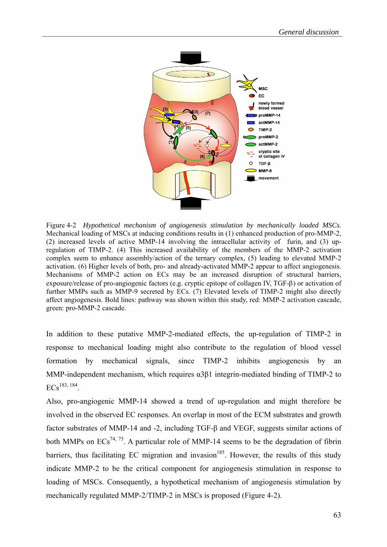
General discussion
63
Figure 4-2 Hypothetical mechanism of angiogenesis stimulation by mechanically loaded MSCs. Mechanical loading of MSCs at inducing conditions results in (1) enhanced production of pro-MMP-2, (2) increased levels of active MMP-14 involving the intracellular activity of furin, and (3) up-regulation of TIMP-2. (4) This increased availability of the members of the MMP-2 activation complex seem to enhance assembly/action of the ternary complex, (5) leading to elevated MMP-2 activation. (6) Higher levels of both, pro- and already-activated MMP-2 appear to affect angiogenesis. Mechanisms of MMP-2 action on ECs may be an increased disruption of structural barriers, exposure/release of pro-angiogenic factors (e.g. cryptic epitope of collagen IV, TGF-β) or activation of further MMPs such as MMP-9 secreted by ECs. (7) Elevated levels of TIMP-2 might also directly affect angiogenesis. Bold lines: pathway was shown within this study, red: MMP-2 activation cascade, green: pro-MMP-2 cascade.
In addition to these putative MMP-2-mediated effects, the up-regulation of TIMP-2 in
response to mechanical loading might also contribute to the regulation of blood vessel
formation by mechanical signals, since TIMP-2 inhibits angiogenesis by an
MMP-independent mechanism, which requires α3β1 integrin-mediated binding of TIMP-2 to
ECs183, 184.
Also, pro-angiogenic MMP-14 showed a trend of up-regulation and might therefore be
involved in the observed EC responses. An overlap in most of the ECM substrates and growth
factor substrates of MMP-14 and -2, including TGF-β and VEGF, suggests similar actions of
both MMPs on ECs74, 75. A particular role of MMP-14 seems to be the degradation of fibrin
barriers, thus facilitating EC migration and invasion185. However, the results of this study
indicate MMP-2 to be the critical component for angiogenesis stimulation in response to
loading of MSCs. Consequently, a hypothetical mechanism of angiogenesis stimulation by
mechanically regulated MMP-2/TIMP-2 in MSCs is proposed (Figure 4-2).

Chapter 4
64
4.3.3 Consequences for bone regeneration
Enhanced MMP levels were demonstrated within this thesis to stimulate the function of MSCs
themselves, but also to augment angiogenesis. Both processes are crucially involved in early
bone healing, including the phases of inflammation and endochondral ossification16.
Therefore, it is likely that altered MMP levels have an impact on the healing outcome.
In particular, enhancement of MMP-13 was shown to result in improved osteogenic and
chondrogenic differentiation of MSCs, indicating an important role of this protease during
endochondral ossification. This is supported by the strong expression of MMP-13 mRNA
during this phase in murine fracture repair models14. Moreover, the role of MMP-13 in bone
remodelling was evidenced by knock out mice displaying an abnormal growth plate
development and by human individuals having mutations in the MMP-13 gene88, 186 For
example, MMP-13 knock out mice showed an abnormal growth plate development due to a
lack of collagen type II and aggrecan proteolysis, which impedes chondrocyte remodelling88.
Enhanced MMP-2 activity in mechanically loaded MSCs potentially stimulates MSC
migration and also angiogenesis. Therefore, it is hypothesised that increased levels of MMP-2
play an important role during the inflammatory phase, which is thought to involve the
recruitment of MSCs to the fracture site and blood vessel stimulation12, 16. Evidence has been
provided by MMP-2 gene mutations resulting in severe bone defects90. However, there is a
lack of evidence of the contribution of MMP-2 to bone remodelling in mice and MMP-
2/MMP-14 double knock out mice die at birth187. Therefore, it is likely that in MMP-2 knock
out mice the deficiency of this protease is compensated by other proteases, including
potentially MMP-14.
Moreover, MMP-9 seems to be crucially involved in bone regeneration by a regulation of
progenitor differentiation and angiogenesis. This was shown in MMP-9 knock out mice
having vascular defects as well as impeded chondrogenic and osteogenic differentiation91.
However, these effects seem not to be a result of MMP-9 mechano-regulation in MSCs, since
the fracture stability had no influence, and MMP-9 is not expressed at protein level in MSCs,
as demonstrated in this study. This indicates that, besides MSCs, other cells present in early
haematomas, e.g. ECs or inflammatory cells, might also contribute to bone healing processes
by a regulation of MMP-9 levels.
General discussion
65
4.4 Potential therapeutic approaches
Data from this study indicate that loading at conditions known from uneventful bone healing
results in an up-regulation of MMPs in MSCs, which contributes to MSC differentiation,
migration and paracine angiogenesis stimulation. Moreover, MMP-2 activity was shown to be
up-regulated in fracture haematomas under loading conditions that support bone healing.
Therefore, MMPs might be considered as candidates for the design of new gene and cell
therapies to treat delayed or failed healing of bone. In this respect, it is interesting that
osteoinductive BMP-2, which is already available in the clinic for non-healing bone and open
tibial fractures, is also up-regulated in response to loading in mesenchymal cells188. These
findings support the importance of mechano-regulation in the identification of genes/proteins
relevant for bone healing.
In particular, data presented in this thesis indicate mechano-regulated MMP-2 to be involved
in MSC migration and in augmentation of angiogenesis. Therefore, this protease might be
interesting for a treatment of patients suffering from insufficient blood vessel regeneration in
bone (e.g. atrophic pseudarthrosis), which may result from risk factors including age, diabetes
and alcohol abuse13. The ability of MMP-2 to release a variety of pro-angiogenic factors in an
orchestered manner makes this protease even more attractive, since a combination of
pro-angiogenic factors has been shown to be more effective than application of single
factors189, 190. For example, a combination of recombinant VEGF-A and bFGF has been
reported to have potent synergistic effects on neovascular formation in experimental
conditions in vivo and in vitro189. In addition to the MMP-2-mediated effects, mechano-
regulated MMP-13 has been demonstrated in this and other studies to stimulate osteogenic
and chondrogenic differentiation of MSCs. Thus, this MMP could be considered as an
alternative/complement to BMP-2 treatment for a recovery of bone function, and applied via
binding to scaffolds, e.g. adsorbable collagen sponges, or via viral or non-viral gene delivery;
e.g. adenoviral systems or nucleofection30.
The response of MSCs to loading conditions by an expression of functionally relevant
MMP-2 and -13 could also be supportive for engineering of vascularised tissues. At present,
vascularisation is critical for the size of these engineered tissues, containing for example
MSCs and ECs191. The results obtained within this study suggest that limited compressive
loading of such MSC constructs might stimulate blood vessel formation by enhanced
secretion of pro-angiogenic MMPs.

Chapter 4
66
Genetic engineering of MSCs to overexpress the MMP candidates using viral/non-viral
systems could further potentiate the effect of MSCs in tissue engineering approaches or
cell-based therapies. Evidence for the efficacy of MMP transfection for regeneration in vivo
was recently provided by the restoration of the vascular network in aged murine dystrophic
muscle in response to application of tendon fibroblasts transduced with lentiviral vectors
encoding placental growth factor (PlGF) and MMP-9192. Based on the results of the present
study, MMP-2 should be considered as candidate gene for design of genetically modified
MSCs to restore the vescular system in compromised healing.
Since pro-angiogenic MMPs, such as MMP-2, have been also implicated as agonists in
tumour invasion, one has to be aware of this potential risk75. However, innovative delivery
systems using vectors with exonenously regulated gene expression could enable for a tight
spatial and temporal control of MMP-2 activity and therefore make this risk manageable30.
The healing-supportive properties of mechano-regulated MMPs presented in this study
highlight these proteases as valuable candidates for therapies. Their application might be
especially interesting for bone treatments in elderly patients, since they display not only
age-related decreases in regeneration potential; there are also hints for the influence of age on
the effects of the mechanical environment on bone defect healing, which might be a result of
diminished cell responses to loading13, 145.
General discussion
67
4.5 Conclusion
Mechanical stimuli have been shown to influence mesenchymal cell characteristics. From the
results of this thesis, it seems likely that these stimuli also regulate autocrine and paracrine
effects of MSCs including angiogenesis stimulation, which might in turn be mediated by the
regulation of MMPs. Whether these mechanisms also occur in vivo and whether they are
important for bone healing outcomes can only be speculated. However, there are promising
hints that this might be the case, since the range of in vitro mechano-induction of MMPs
resembles the parameters occurring in vivo during uneventful bone healing. Furthermore,
MMP-2 activity was increased in fracture haematomas subjected to mechanical conditions
that have been associated with superior healing. This mechano-regulation of MMPs could be
important for activating MSCs in the case of injury, e.g. in response to mechanical forces
present at the fracture site. However, to understand the physiological coordination of
progenitor cell differentiation, angiogenesis and tissue regeneration in vivo, further insight
into the influence of mechanical conditions on cell interactions is essential. Therefore,
mechano-regulation of MMPs should be validated in MSC co-culture systems with, for
example, ECs and immune competent cells. In this respect, the effect of loading on
MMP levels in human haematomas is currently under investigation in the Julius Wolff
Institute. Since various MMPs and TIMP-2 have been shown to be influenced by mechanical
stimuli in MSCs, the effect of complex alterations in the MMP/TIMP balance need to be
prospectively investigated in detail. Understanding the mechanisms of MMP involvement in
MSC biology and regeneration/developmental processes might be important for the design of
novel treatment strategies in bone healing, and also for assessing unintentional side effects of
MMP activity.
68
Summary (English)
Mechanical conditions have an impact on the early phase of bone healing and in particular on
angiogenesis. Mesenchymal stem cells (MSCs) play a key role, due to their ability to home to
the defect site, to differentiate into functional bone cells and to secrete pro-angiogenic factors.
However, it is yet unknown how MSCs respond to the loading conditions during defect
healing, and which proteins transduce the mechanical stimulus into the control of MSC
function. Matrix metalloproteases (MMPs) and the tissue inhibitors of metalloproteases
(TIMPs) are mechanically regulated in mesenchymal cells and are able to affect cell functions
in an autocrine and paracrine manner.
This project investigated MMP/TIMP levels in MSCs in response to mechanical loading, as
well as the consequences for MSC function. Furthermore, the pro-angiogenic capacity of
mechano-regulated MMP-2 was determined and the underlying mechanism of MMP-2
regulation in MSCs examined.
The involvement of MMPs in migration, proliferation and osteogenic differentiation was
evidenced using broad spectrum MMP inhibitors. Expression analysis detected MMP-2, -3,
-10, -11, -13, -14 and TIMP-2 in MSCs at the mRNA and protein levels. Mechanical
stimulation of MSC/fibrin constructs in a bioreactor system simulating conditions of early
bone healing (30% compression at a frequency of 1Hz) resulted in increased concentrations of
MMP-2, -3, -13 and TIMP-2 proteins, as well as enhanced gelatinolytic activities of MMP-2
and MMP-13. However, mRNA expression levels of MMPs/TIMPs showed no changes in
response to mechanical stimulation. Specific inhibition of mechano-regulated MMP-2, -3 and
-13 proved MMP-13 to be involved in osteogenic differentiation. Furthermore, MMP-2
appeared to alter MSC migration, although statistical significance was not reached.
Detailed investigation of MMP-2 demonstrated enhanced gelatinolytic activities in early
haematomas under large versus small interfragmentary movements. The measurement of
MMP-2 from MSCs at varying loading conditions confirmed loading at 1Hz and 30%
compression to be MMP-2-inducing and demonstrated 0.1Hz and 10% compression to be
non-inducing. Supplementation with the Golgi disturbing agent, brefeldin A, prevented the
mechanical up-regulation of MMP-2. In addition to the enhancement of pro-MMP-2 levels,
also the activities of active-MMP-2 and TIMP-2 were elevated, while MMP-14 activity
showed a trend of up-regulation. Furin inhibition suppressed this mechano-regulation of
MMP-14 and active-MMP-2. A pro-angiogenic effect, seen for conditioned medium (CM)
from MSCs loaded at inducing conditions, was reduced by blocking pro-MMP-2 activation,
69
as well as by inhibiting active-MMP-2. Furthermore, addition of recombinant active-MMP-2
to CM partially compensated for ineffective loading.
The results of this thesis suggest that MSC function is controlled by MMP/TIMP activity,
which in turn is regulated by mechanical stimulation of these cells under conditions of
uneventful healing. In the case of MMP-2, mechanical loading seems not only to stimulate
levels of the secreted pro-enzyme, but also its activation cascade. Both mechanisms appear to
contribute to the paracrine augmentation of angiogenesis in response to loading. Thus, a
failure to induce cascades of MMP regulation might contribute to the impaired clinical
outcome in suboptimally stabilised bone defects.
70
Summary (German)
Mechanische Bedingungen beeinflussen die frühe Phase der Knochenheilung und
insbesondere die Angiogenese. Mesenchymale Stammzellen (MSCs) sind aufgrund ihrer
Fähigkeit, zum Defektort zu migrieren, in Knochenzellen zu differenzieren und pro-angiogene
Faktoren zu sezernieren, essentiell für den Heilungsverlauf. Bis heute ist jedoch unklar, wie
MSCs auf die relevanten Belastungsbedingungen reagieren und welche Proteine die
Übertragung des mechanischen Stimulus in ein verändertes Zellverhalten vermitteln. Matrix-
Metalloproteasen (MMPs) und ihre Inhibitoren, die TIMPs, werden durch mechanische
Stimulation in mesenchymalen Zellen reguliert und beeinflussen Zellfunktionen autokrin
sowie parakrin.
In dieser Arbeit wurde untersucht, ob mechanische Belastung von MSCs zu einer Regulation
der MMP/TIMP Expressionen/Aktivitäten führt, und ob dies Auswirkungen auf die Funktion
der MSCs hat. Weiterhin wurde mechanisch-reguliertes MMP-2 in Bezug auf seine
Regulation in MSCs sowie seine pro-angiogenen Eigenschaften überprüft.
Dass MMPs in die Migration, Proliferation und osteogene Differenzierung involviert sind,
wurde anhand von MMP Breitspektruminhibitoren gezeigt. Expressionsanalysen von MMPs
detektierten MMP-2, -3, -10,- 11, -13, -14 sowie TIMP-2 in MSCs auf mRNA und Protein-
Niveau. Mechanische Stimulation von MSC/Fibrin-Konstrukten in einem Bioreaktor, welcher
die Bedingungen der frühen Knochenheilung simuliert (30% Kompression bei 1Hz), führte zu
einer Erhöhung der MMP-2, -3, -13 und TIMP-2 Proteinexpressionen sowie erhöhten
gelatinolytischen Aktivitäten von MMP-2 und -13. Es wurde hingegen keine Veränderung in
der Genexpression festgestellt. Durch die spezifische Inhibierung von mechanisch-reguliertem
MMP-2, -3 und -13 konnte ein Einfluss von MMP-13 auf die osteogene Differenzierung
gezeigt werden. Darüber hinaus scheint MMP-2 die Migration zu beeinflussen.
Detaillierte Untersuchungen von MMP-2 zeigten erhöhte gelatinolytische Aktivitäten in
frühen Hämatomen, die großen interfragmentären Bewegungen ausgesetzt waren.
Expressions- und Aktivitätsanalysen der mechanischen Regulation von MMP-2 in MSCs
unter unterschiedlichen Belastungsbedingungen zeigten, dass 30% Kompression bei 1Hz
MMP-2-induzierend und 10% Kompression bei 0,1Hz nicht-induzierend wirken. Neben einer
Erhöhung von pro-MMP-2, wurden auch erhöhte Aktivitäten von aktivem MMP-2 und
TIMP-2 detektiert. Zudem waren MMP-14 Aktivitäten tendenziell erhöht. Die Inhibierung
von Furin unterdrückte diese Stimulation von MMP-14 und aktivem MMP-2. Eine
pro-angiogene Wirkung des konditionierten Mediums (KM) von MSCs, die

71
MMP-2-induzierender Belastung ausgesetzt waren, konnte durch Blockierung der
pro-MMP-2 Aktivierung sowie Inhibierung von aktivem MMP-2 reduziert werden. Zugabe
von aktivem MMP-2 zum KM kompensierte hingegen teilweise eine ineffektive Belastung.
Diese Ergebnisse weisen darauf hin, dass die Funktion von MSCs durch
MMP-/TIMP-Aktivitäten reguliert wird, welche wiederum durch mechanische Stimulation
dieser Zellen unter Bedingungen normaler Heilung aktiviert werden. Im Falle von MMP-2
scheinen das Pro-Enzym sowie aktiviertes MMP-2 zu einer parakrinen Stimulation der
Angiogenese durch mechanische Belastung beizutragen. Infolgedessen könnte eine Störung in
der Induktion der MMP-Regulation zur Beeinträchtigung des Heilungserfolges in suboptimal-
stabilisierten Knochendefekten beitragen.

72
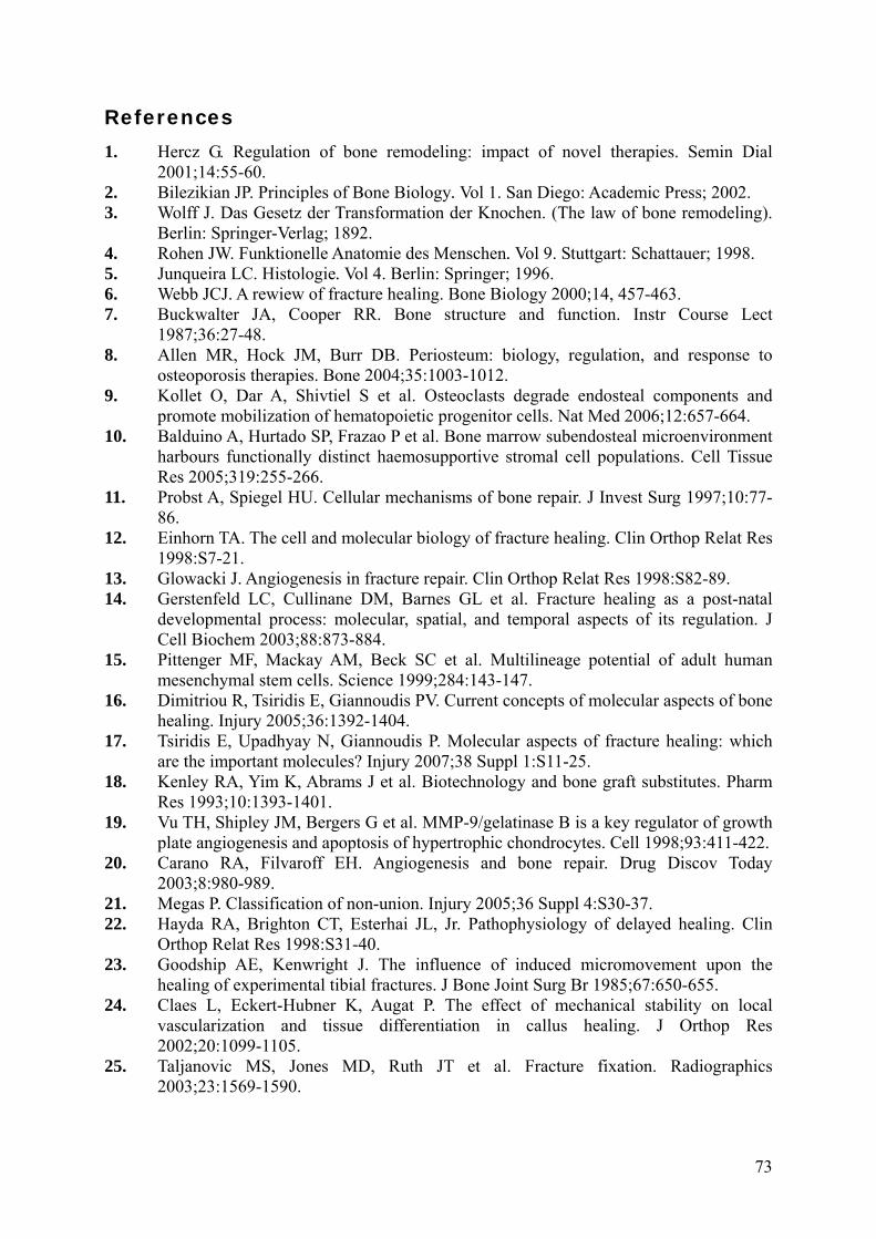
73
References
1. Hercz G. Regulation of bone remodeling: impact of novel therapies. Semin Dial 2001;14:55-60.
2. Bilezikian JP. Principles of Bone Biology. Vol 1. San Diego: Academic Press; 2002. 3. Wolff J. Das Gesetz der Transformation der Knochen. (The law of bone remodeling).
Berlin: Springer-Verlag; 1892. 4. Rohen JW. Funktionelle Anatomie des Menschen. Vol 9. Stuttgart: Schattauer; 1998. 5. Junqueira LC. Histologie. Vol 4. Berlin: Springer; 1996. 6. Webb JCJ. A rewiew of fracture healing. Bone Biology 2000;14, 457-463. 7. Buckwalter JA, Cooper RR. Bone structure and function. Instr Course Lect
1987;36:27-48. 8. Allen MR, Hock JM, Burr DB. Periosteum: biology, regulation, and response to
osteoporosis therapies. Bone 2004;35:1003-1012. 9. Kollet O, Dar A, Shivtiel S et al. Osteoclasts degrade endosteal components and
promote mobilization of hematopoietic progenitor cells. Nat Med 2006;12:657-664. 10. Balduino A, Hurtado SP, Frazao P et al. Bone marrow subendosteal microenvironment
harbours functionally distinct haemosupportive stromal cell populations. Cell Tissue Res 2005;319:255-266.
11. Probst A, Spiegel HU. Cellular mechanisms of bone repair. J Invest Surg 1997;10:77-86.
12. Einhorn TA. The cell and molecular biology of fracture healing. Clin Orthop Relat Res 1998:S7-21.
13. Glowacki J. Angiogenesis in fracture repair. Clin Orthop Relat Res 1998:S82-89. 14. Gerstenfeld LC, Cullinane DM, Barnes GL et al. Fracture healing as a post-natal
developmental process: molecular, spatial, and temporal aspects of its regulation. J Cell Biochem 2003;88:873-884.
15. Pittenger MF, Mackay AM, Beck SC et al. Multilineage potential of adult human mesenchymal stem cells. Science 1999;284:143-147.
16. Dimitriou R, Tsiridis E, Giannoudis PV. Current concepts of molecular aspects of bone healing. Injury 2005;36:1392-1404.
17. Tsiridis E, Upadhyay N, Giannoudis P. Molecular aspects of fracture healing: which are the important molecules? Injury 2007;38 Suppl 1:S11-25.
18. Kenley RA, Yim K, Abrams J et al. Biotechnology and bone graft substitutes. Pharm Res 1993;10:1393-1401.
19. Vu TH, Shipley JM, Bergers G et al. MMP-9/gelatinase B is a key regulator of growth plate angiogenesis and apoptosis of hypertrophic chondrocytes. Cell 1998;93:411-422.
20. Carano RA, Filvaroff EH. Angiogenesis and bone repair. Drug Discov Today 2003;8:980-989.
21. Megas P. Classification of non-union. Injury 2005;36 Suppl 4:S30-37. 22. Hayda RA, Brighton CT, Esterhai JL, Jr. Pathophysiology of delayed healing. Clin
Orthop Relat Res 1998:S31-40. 23. Goodship AE, Kenwright J. The influence of induced micromovement upon the
healing of experimental tibial fractures. J Bone Joint Surg Br 1985;67:650-655. 24. Claes L, Eckert-Hubner K, Augat P. The effect of mechanical stability on local
vascularization and tissue differentiation in callus healing. J Orthop Res 2002;20:1099-1105.
25. Taljanovic MS, Jones MD, Ruth JT et al. Fracture fixation. Radiographics 2003;23:1569-1590.

74
26. Stevenson S. Enhancement of fracture healing with autogenous and allogeneic bone grafts. Clin Orthop Relat Res 1998:S239-246.
27. Hernigou P, Beaujean F. Treatment of osteonecrosis with autologous bone marrow grafting. Clin Orthop Relat Res 2002:14-23.
28. De Long WG, Jr., Einhorn TA, Koval K et al. Bone grafts and bone graft substitutes in orthopaedic trauma surgery. A critical analysis. J Bone Joint Surg Am 2007;89:649-658.
29. Bishop GB, Einhorn TA. Current and future clinical applications of bone morphogenetic proteins in orthopaedic trauma surgery. Int Orthop 2007;31:721-727.
30. Kimelman N, Pelled G, Helm GA et al. Review: gene- and stem cell-based therapeutics for bone regeneration and repair. Tissue Eng 2007;13:1135-1150.
31. Wang JC, Kanim LE, Yoo S et al. Effect of regional gene therapy with bone morphogenetic protein-2-producing bone marrow cells on spinal fusion in rats. J Bone Joint Surg Am 2003;85-A:905-911.
32. Boden SD. The ABCs of BMPs. Orthop Nurs 2005;24:49-52; quiz 53-44. 33. Perri B, Cooper M, Lauryssen C et al. Adverse swelling associated with use of rh-
BMP-2 in anterior cervical discectomy and fusion: a case study. Spine J 2007;7:235-239.
34. Giordano A, Galderisi U, Marino IR. From the laboratory bench to the patient's bedside: an update on clinical trials with mesenchymal stem cells. J Cell Physiol 2007;211:27-35.
35. Giebel LB. Stem cells--a hard sell to investors. Nat Biotechnol 2005;23:798-800. 36. Khan TA, Sellke FW, Laham RJ. Gene therapy progress and prospects: therapeutic
angiogenesis for limb and myocardial ischemia. Gene Ther 2003;10:285-291. 37. Adams RH, Alitalo K. Molecular regulation of angiogenesis and lymphangiogenesis.
Nat Rev Mol Cell Biol 2007;8:464-478. 38. Papetti M, Herman IM. Mechanisms of normal and tumor-derived angiogenesis. Am J
Physiol Cell Physiol 2002;282:C947-970. 39. Carmeliet P. Mechanisms of angiogenesis and arteriogenesis. Nat Med 2000;6:389-
395. 40. Moses MA. The regulation of neovascularization of matrix metalloproteinases and
their inhibitors. Stem Cells 1997;15:180-189. 41. Friedenstein AJ, Petrakova KV, Kurolesova AI et al. Heterotopic of bone
marrow.Analysis of precursor cells for osteogenic and hematopoietic tissues. Transplantation 1968;6:230-247.
42. Caplan AI. Stem cell delivery vehicle. Biomaterials 1990;11:44-46. 43. Oswald J, Boxberger S, Jorgensen B et al. Mesenchymal stem cells can be
differentiated into endothelial cells in vitro. Stem Cells 2004;22:377-384. 44. Kopen GC, Prockop DJ, Phinney DG. Marrow stromal cells migrate throughout
forebrain and cerebellum, and they differentiate into astrocytes after injection into neonatal mouse brains. Proc Natl Acad Sci U S A 1999;96:10711-10716.
45. Colter DC, Class R, DiGirolamo CM et al. Rapid expansion of recycling stem cells in cultures of plastic-adherent cells from human bone marrow. Proc Natl Acad Sci U S A 2000;97:3213-3218.
46. Campagnoli C, Roberts IA, Kumar S et al. Identification of mesenchymal stem/progenitor cells in human first-trimester fetal blood, liver, and bone marrow. Blood 2001;98:2396-2402.
47. Minguell JJ, Erices A, Conget P. Mesenchymal stem cells. Exp Biol Med (Maywood) 2001;226:507-520.
75
48. Bieback K, Kern S, Kluter H et al. Critical parameters for the isolation of mesenchymal stem cells from umbilical cord blood. Stem Cells 2004;22:625-634.
49. Baksh D, Song L, Tuan RS. Adult mesenchymal stem cells: characterization, differentiation, and application in cell and gene therapy. J Cell Mol Med 2004;8:301-316.
50. Chamberlain G, Fox J, Ashton B et al. Concise review: mesenchymal stem cells: their phenotype, differentiation capacity, immunological features, and potential for homing. Stem Cells 2007;25:2739-2749.
51. Streuli C. Extracellular matrix remodelling and cellular differentiation. Curr Opin Cell Biol 1999;11:634-640.
52. Werb Z. ECM and cell surface proteolysis: regulating cellular ecology. Cell 1997;91:439-442.
53. Lukashev ME, Werb Z. ECM signalling: orchestrating cell behaviour and misbehaviour. Trends Cell Biol 1998;8:437-441.
54. Lutz M, Knaus P. Integration of the TGF-beta pathway into the cellular signalling network. Cell Signal 2002;14:977-988.
55. Rosen ED, Spiegelman BM. Molecular regulation of adipogenesis. Annu Rev Cell Dev Biol 2000;16:145-171.
56. Pan Q, Yu Y, Chen Q et al. Sox9, a key transcription factor of bone morphogenetic protein-2-induced chondrogenesis, is activated through BMP pathway and a CCAAT box in the proximal promoter. J Cell Physiol 2008;217:228-241.
57. da Silva Meirelles L, Caplan AI, Nardi NB. In Search of the in vivo Identity of Mesenchymal Stem Cells. Stem Cells 2008.
58. Al-Khaldi A, Eliopoulos N, Martineau D et al. Postnatal bone marrow stromal cells elicit a potent VEGF-dependent neoangiogenic response in vivo. Gene Ther 2003;10:621-629.
59. Wu Y, Chen L, Scott PG et al. Mesenchymal stem cells enhance wound healing through differentiation and angiogenesis. Stem Cells 2007;25:2648-2659.
60. Kasper G, Dankert N, Tuischer J et al. Mesenchymal stem cells regulate angiogenesis according to their mechanical environment. Stem Cells 2007;25:903-910.
61. Fuchs E, Tumbar T, Guasch G. Socializing with the neighbors: stem cells and their niche. Cell 2004;116:769-778.
62. Yin T, Li L. The stem cell niches in bone. J Clin Invest 2006;116:1195-1201. 63. Ohlstein B, Kai T, Decotto E et al. The stem cell niche: theme and variations. Curr
Opin Cell Biol 2004;16:693-699. 64. Shiozawa Y, Havens AM, Pienta KJ et al. The bone marrow niche: habitat to
hematopoietic and mesenchymal stem cells, and unwitting host to molecular parasites. Leukemia 2008;22:941-950.
65. Krampera M, Pizzolo G, Aprili G et al. Mesenchymal stem cells for bone, cartilage, tendon and skeletal muscle repair. Bone 2006;39:678-683.
66. Le Blanc K, Pittenger M. Mesenchymal stem cells: progress toward promise. Cytotherapy 2005;7:36-45.
67. Gangji V, Hauzeur JP, Matos C et al. Treatment of osteonecrosis of the femoral head with implantation of autologous bone-marrow cells. A pilot study. J Bone Joint Surg Am 2004;86-A:1153-1160.
68. Quarto R, Mastrogiacomo M, Cancedda R et al. Repair of large bone defects with the use of autologous bone marrow stromal cells. N Engl J Med 2001;344:385-386.
69. Horwitz EM, Prockop DJ, Fitzpatrick LA et al. Transplantability and therapeutic effects of bone marrow-derived mesenchymal cells in children with osteogenesis imperfecta. Nat Med 1999;5:309-313.
76
70. Visse R, Nagase H. Matrix metalloproteinases and tissue inhibitors of metalloproteinases: structure, function, and biochemistry. Circ Res 2003;92:827-839.
71. Vincenti MP. The matrix metalloproteinase (MMP) and tissue inhibitor of metalloproteinase (TIMP) genes. Transcriptional and posttranscriptional regulation, signal transduction and cell-type-specific expression. Methods Mol Biol 2001;151:121-148.
72. Page-McCaw A, Ewald AJ, Werb Z. Matrix metalloproteinases and the regulation of tissue remodelling. Nat Rev Mol Cell Biol 2007;8:221-233.
73. Fu X, Parks WC, Heinecke JW. Activation and silencing of matrix metalloproteinases. Semin Cell Dev Biol 2008;19:2-13.
74. Nagase H, Visse R, Murphy G. Structure and function of matrix metalloproteinases and TIMPs. Cardiovasc Res 2006;69:562-573.
75. Sternlicht MD, Werb Z. How matrix metalloproteinases regulate cell behavior. Annu Rev Cell Dev Biol 2001;17:463-516.
76. Mott JD, Werb Z. Regulation of matrix biology by matrix metalloproteinases. Curr Opin Cell Biol 2004;16:558-564.
77. Belotti D, Paganoni P, Manenti L et al. Matrix metalloproteinases (MMP9 and MMP2) induce the release of vascular endothelial growth factor (VEGF) by ovarian carcinoma cells: implications for ascites formation. Cancer Res 2003;63:5224-5229.
78. Chakraborti S, Mandal M, Das S et al. Regulation of matrix metalloproteinases: an overview. Mol Cell Biochem 2003;253:269-285.
79. Sehgal I, Thompson TC. Novel regulation of type IV collagenase (matrix metalloproteinase-9 and -2) activities by transforming growth factor-beta1 in human prostate cancer cell lines. Mol Biol Cell 1999;10:407-416.
80. Kjeldsen L, Sengelov H, Lollike K et al. Isolation and characterization of gelatinase granules from human neutrophils. Blood 1994;83:1640-1649.
81. Blott EJ, Griffiths GM. Secretory lysosomes. Nat Rev Mol Cell Biol 2002;3:122-131. 82. Hasty KA, Pourmotabbed TF, Goldberg GI et al. Human neutrophil collagenase. A
distinct gene product with homology to other matrix metalloproteinases. J Biol Chem 1990;265:11421-11424.
83. Butler GS, Butler MJ, Atkinson SJ et al. The TIMP2 membrane type 1 metalloproteinase "receptor" regulates the concentration and efficient activation of progelatinase A. A kinetic study. J Biol Chem 1998;273:871-880.
84. Strongin AY, Collier I, Bannikov G et al. Mechanism of cell surface activation of 72-kDa type IV collagenase. Isolation of the activated form of the membrane metalloprotease. J Biol Chem 1995;270:5331-5338.
85. Sounni NE, Janssen M, Foidart JM et al. Membrane type-1 matrix metalloproteinase and TIMP-2 in tumor angiogenesis. Matrix Biol 2003;22:55-61.
86. Yana I, Weiss SJ. Regulation of membrane type-1 matrix metalloproteinase activation by proprotein convertases. Mol Biol Cell 2000;11:2387-2401.
87. Ortega N, Behonick DJ, Werb Z. Matrix remodeling during endochondral ossification. Trends Cell Biol 2004;14:86-93.
88. Stickens D, Behonick DJ, Ortega N et al. Altered endochondral bone development in matrix metalloproteinase 13-deficient mice. Development 2004;131:5883-5895.
89. Zhou Z, Apte SS, Soininen R et al. Impaired endochondral ossification and angiogenesis in mice deficient in membrane-type matrix metalloproteinase I. Proc Natl Acad Sci U S A 2000;97:4052-4057.
90. Zankl A, Pachman L, Poznanski A et al. Torg syndrome is caused by inactivating mutations in MMP2 and is allelic to NAO and Winchester syndrome. J Bone Miner Res 2007;22:329-333.
77
91. Colnot C, Thompson Z, Miclau T et al. Altered fracture repair in the absence of MMP9. Development 2003;130:4123-4133.
92. Heissig B, Hattori K, Dias S et al. Recruitment of stem and progenitor cells from the bone marrow niche requires MMP-9 mediated release of kit-ligand. Cell 2002;109:625-637.
93. Duda GN, Sporrer S, Sollmann M et al. Interfragmentary movements in the early phase of healing in distraction and correction osteotomies stabilized with ring fixators. Langenbecks Arch Surg 2003;387:433-440.
94. Klein P, Opitz M, Schell H et al. Comparison of unreamed nailing and external fixation of tibial diastases--mechanical conditions during healing and biological outcome. J Orthop Res 2004;22:1072-1078.
95. Klein P, Schell H, Streitparth F et al. The initial phase of fracture healing is specifically sensitive to mechanical conditions. J Orthop Res 2003;21:662-669.
96. Lienau J, Schell H, Duda GN et al. Initial vascularization and tissue differentiation are influenced by fixation stability. J Orthop Res 2005;23:639-645.
97. Terraciano V, Hwang N, Moroni L et al. Differential response of adult and embryonic mesenchymal progenitor cells to mechanical compression in hydrogels. Stem Cells 2007;25:2730-2738.
98. Fujisawa T, Hattori T, Takahashi K et al. Cyclic mechanical stress induces extracellular matrix degradation in cultured chondrocytes via gene expression of matrix metalloproteinases and interleukin-1. J Biochem (Tokyo) 1999;125:966-975.
99. Park JS, Chu JS, Cheng C et al. Differential effects of equiaxial and uniaxial strain on mesenchymal stem cells. Biotechnol Bioeng 2004;88:359-368.
100. Mauney JR, Sjostorm S, Blumberg J et al. Mechanical stimulation promotes osteogenic differentiation of human bone marrow stromal cells on 3-D partially demineralized bone scaffolds in vitro. Calcif Tissue Int 2004;74:458-468.
101. Yoshikawa T, Peel SA, Gladstone JR et al. Biochemical analysis of the response in rat bone marrow cell cultures to mechanical stimulation. Biomed Mater Eng 1997;7:369-377.
102. Jagodzinski M, Drescher M, Zeichen J et al. Effects of cyclic longitudinal mechanical strain and dexamethasone on osteogenic differentiation of human bone marrow stromal cells. Eur Cell Mater 2004;7:35-41; discussion 41.
103. Wang JH, Thampatty BP. An introductory review of cell mechanobiology. Biomech Model Mechanobiol 2006;5:1-16.
104. Wang N, Naruse K, Stamenovic D et al. Mechanical behavior in living cells consistent with the tensegrity model. Proc Natl Acad Sci U S A 2001;98:7765-7770.
105. Schwartz MA. Integrin signaling revisited. Trends Cell Biol 2001;11:466-470. 106. Honda K, Ohno S, Tanimoto K et al. The effects of high magnitude cyclic tensile load
on cartilage matrix metabolism in cultured chondrocytes. Eur J Cell Biol 2000;79:601-609.
107. Chang C, Werb Z. The many faces of metalloproteases: cell growth, invasion, angiogenesis and metastasis. Trends Cell Biol 2001;11:S37-43.
108. Mannello F, Tonti GA, Bagnara GP et al. Role and function of matrix metalloproteinases in the differentiation and biological characterization of mesenchymal stem cells. Stem Cells 2006;24:475-481.
109. Parikka V, Vaananen A, Risteli J et al. Human mesenchymal stem cell derived osteoblasts degrade organic bone matrix in vitro by matrix metalloproteinases. Matrix Biol 2005;24:438-447.

78
110. Scutt A, Bertram P. Basic fibroblast growth factor in the presence of dexamethasone stimulates colony formation, expansion, and osteoblastic differentiation by rat bone marrow stromal cells. Calcif Tissue Int 1999;64:69-77.
111. Liu Z, Xu J, Colvin JS et al. Coordination of chondrogenesis and osteogenesis by fibroblast growth factor 18. Genes Dev 2002;16:859-869.
112. Fromigue O, Marie PJ, Lomri A. Bone morphogenetic protein-2 and transforming growth factor-beta2 interact to modulate human bone marrow stromal cell proliferation and differentiation. J Cell Biochem 1998;68:411-426.
113. Matziolis G, Tuischer J, Kasper G et al. Simulation of cell differentiation in fracture healing: mechanically loaded composite scaffolds in a novel bioreactor system. Tissue Eng 2006;12:201-208.
114. Sekiya I, Vuoristo JT, Larson BL et al. In vitro cartilage formation by human adult stem cells from bone marrow stroma defines the sequence of cellular and molecular events during chondrogenesis. Proc Natl Acad Sci U S A 2002;99:4397-4402.
115. Chen JM, Nelson FC, Levin JI et al. Structure-Based Design of a Novel, Potent, and Selective Inhibitor for MMP-13 Utilizing NMR Spectroscopy and Computer-Aided Molecular Design. Journal of the American Chemical Society 2000;122:9648.
116. Berton A, Rigot V, Huet E et al. Involvement of fibronectin type II repeats in the efficient inhibition of gelatinases A and B by long-chain unsaturated fatty acids. J Biol Chem 2001;276:20458-20465.
117. MacPherson LJ, Bayburt EK, Capparelli MP et al. Discovery of CGS 27023A, a non-peptidic, potent, and orally active stromelysin inhibitor that blocks cartilage degradation in rabbits. J Med Chem 1997;40:2525-2532.
118. Falk W, Goodwin RH, Jr., Leonard EJ. A 48-well micro chemotaxis assembly for rapid and accurate measurement of leukocyte migration. J Immunol Methods 1980;33:239-247.
119. Claes LE, Heigele CA, Neidlinger-Wilke C et al. Effects of mechanical factors on the fracture healing process. Clin Orthop Relat Res 1998:S132-147.
120. Gygi SP, Rochon Y, Franza BR et al. Correlation between protein and mRNA abundance in yeast. Mol Cell Biol 1999;19:1720-1730.
121. Son BR, Marquez-Curtis LA, Kucia M et al. Migration of bone marrow and cord blood mesenchymal stem cells in vitro is regulated by SDF-1-CXCR4 and HGF-c-met axes and involves matrix metalloproteinases. Stem Cells 2006.
122. Silva WA, Jr., Covas DT, Panepucci RA et al. The profile of gene expression of human marrow mesenchymal stem cells. Stem Cells 2003;21:661-669.
123. Panepucci RA, Siufi JL, Silva WA, Jr. et al. Comparison of gene expression of umbilical cord vein and bone marrow-derived mesenchymal stem cells. Stem Cells 2004;22:1263-1278.
124. Chiquet M. Regulation of extracellular matrix gene expression by mechanical stress. Matrix Biol 1999;18:417-426.
125. Cronwright G, Le Blanc K, Gotherstrom C et al. Cancer/testis antigen expression in human mesenchymal stem cells: down-regulation of SSX impairs cell migration and matrix metalloproteinase 2 expression. Cancer Res 2005;65:2207-2215.
126. Zhu W, Boachie-Adjei O, Rawlins BA et al. A novel regulatory role for stromal-derived factor-1 signaling in bone morphogenic protein-2 osteogenic differentiation of mesenchymal C2C12 cells. J Biol Chem 2007;282:18676-18685.
127. Stamenkovic I. Extracellular matrix remodelling: the role of matrix metalloproteinases. J Pathol 2003;200:448-464.
79
128. Fiedler J, Leucht F, Waltenberger J et al. VEGF-A and PlGF-1 stimulate chemotactic migration of human mesenchymal progenitor cells. Biochem Biophys Res Commun 2005;334:561-568.
129. Fiedler J, Brill C, Blum WF et al. IGF-I and IGF-II stimulate directed cell migration of bone-marrow-derived human mesenchymal progenitor cells. Biochem Biophys Res Commun 2006;345:1177-1183.
130. Fowlkes JL, Serra DM, Nagase H et al. MMPs are IGFBP-degrading proteinases: implications for cell proliferation and tissue growth. Ann N Y Acad Sci 1999;878:696-699.
131. Kleinman HK, Martin GR. Matrigel: basement membrane matrix with biological activity. Semin Cancer Biol 2005;15:378-386.
132. Karsdal MA, Andersen TA, Bonewald L et al. Matrix metalloproteinases (MMPs) safeguard osteoblasts from apoptosis during transdifferentiation into osteocytes: MT1-MMP maintains osteocyte viability. DNA Cell Biol 2004;23:155-165.
133. Karsdal MA, Larsen L, Engsig MT et al. Matrix metalloproteinase-dependent activation of latent transforming growth factor-beta controls the conversion of osteoblasts into osteocytes by blocking osteoblast apoptosis. J Biol Chem 2002;277:44061-44067.
134. Simmons DJ. Fracture healing perspectives. Clin Orthop Relat Res 1985:100-113. 135. Kanczler JM, Oreffo RO. Osteogenesis and angiogenesis: the potential for engineering
bone. Eur Cell Mater 2008;15:100-114. 136. Uchida S, Sakai A, Kudo H et al. Vascular endothelial growth factor is expressed
along with its receptors during the healing process of bone and bone marrow after drill-hole injury in rats. Bone 2003;32:491-501.
137. Wallace AL, Draper ER, Strachan RK et al. The vascular response to fracture micromovement. Clin Orthop Relat Res 1994:281-290.
138. Gruber R, Kandler B, Holzmann P et al. Bone marrow stromal cells can provide a local environment that favors migration and formation of tubular structures of endothelial cells. Tissue Eng 2005;11:896-903.
139. Ghajar CM, Blevins KS, Hughes CC et al. Mesenchymal stem cells enhance angiogenesis in mechanically viable prevascularized tissues via early matrix metalloproteinase upregulation. Tissue Eng 2006;12:2875-2888.
140. Kasper G, Glaeser JD, Geissler S et al. Matrix metalloprotease activity is an essential link between mechanical stimulus and mesenchymal stem cell behavior. Stem Cells 2007;25:1985-1994.
141. Ries C, Egea V, Karow M et al. MMP-2, MT1-MMP, and TIMP-2 are essential for the invasive capacity of human mesenchymal stem cells: differential regulation by inflammatory cytokines. Blood 2007;109:4055-4063.
142. Rundhaug JE. Matrix metalloproteinases and angiogenesis. J Cell Mol Med 2005;9:267-285.
143. Holmbeck K, Bianco P, Caterina J et al. MT1-MMP-deficient mice develop dwarfism, osteopenia, arthritis, and connective tissue disease due to inadequate collagen turnover. Cell 1999;99:81-92.
144. Wang Z, Juttermann R, Soloway PD. TIMP-2 is required for efficient activation of proMMP-2 in vivo. J Biol Chem 2000;275:26411-26415.
145. Strube P, Sentuerk U, Riha T et al. Influence of age and mechanical stability on bone defect healing: age reverses mechanical effects. Bone 2008;42:758-764.
146. Ades EW, Candal FJ, Swerlick RA et al. HMEC-1: establishment of an immortalized human microvascular endothelial cell line. J Invest Dermatol 1992;99:683-690.

80
147. Fujiwara T, Oda K, Yokota S et al. Brefeldin A causes disassembly of the Golgi complex and accumulation of secretory proteins in the endoplasmic reticulum. J Biol Chem 1988;263:18545-18552.
148. Berger TG, Feuerstein B, Strasser E et al. Large-scale generation of mature monocyte-derived dendritic cells for clinical application in cell factories. J Immunol Methods 2002;268:131-140.
149. Gardner TN, Evans M, Hardy J et al. Dynamic interfragmentary motion in fractures during routine patient activity. Clin Orthop Relat Res 1997:216-225.
150. Ivanenko YP, Grasso R, Lacquaniti F. Influence of leg muscle vibration on human walking. J Neurophysiol 2000;84:1737-1747.
151. Bernardo MM, Fridman R. TIMP-2 (tissue inhibitor of metalloproteinase-2) regulates MMP-2 (matrix metalloproteinase-2) activity in the extracellular environment after pro-MMP-2 activation by MT1 (membrane type 1)-MMP. Biochem J 2003;374:739-745.
152. Shelton L, Rada JS. Effects of cyclic mechanical stretch on extracellular matrix synthesis by human scleral fibroblasts. Exp Eye Res 2007;84:314-322.
153. Huang CY, Reuben PM, Cheung HS. Temporal expression patterns and corresponding protein inductions of early responsive genes in rabbit bone marrow-derived mesenchymal stem cells under cyclic compressive loading. Stem Cells 2005;23:1113-1121.
154. Mouw JK, Connelly JT, Wilson CG et al. Dynamic compression regulates the expression and synthesis of chondrocyte-specific matrix molecules in bone marrow stromal cells. Stem Cells 2007;25:655-663.
155. Campbell JJ, Lee DA, Bader DL. Dynamic compressive strain influences chondrogenic gene expression in human mesenchymal stem cells. Biorheology 2006;43:455-470.
156. Turner CH, Forwood MR, Otter MW. Mechanotransduction in bone: do bone cells act as sensors of fluid flow? Faseb J 1994;8:875-878.
157. Taraboletti G, Sonzogni L, Vergani V et al. Posttranscriptional stimulation of endothelial cell matrix metalloproteinases 2 and 1 by endothelioma cells. Exp Cell Res 2000;258:384-394.
158. Guo C, Piacentini L. Type I collagen-induced MMP-2 activation coincides with up-regulation of membrane type 1-matrix metalloproteinase and TIMP-2 in cardiac fibroblasts. J Biol Chem 2003;278:46699-46708.
159. Haas TL, Davis SJ, Madri JA. Three-dimensional type I collagen lattices induce coordinate expression of matrix metalloproteinases MT1-MMP and MMP-2 in microvascular endothelial cells. J Biol Chem 1998;273:3604-3610.
160. Taraboletti G, D'Ascenzo S, Borsotti P et al. Shedding of the matrix metalloproteinases MMP-2, MMP-9, and MT1-MMP as membrane vesicle-associated components by endothelial cells. Am J Pathol 2002;160:673-680.
161. Anderson HC, Stechschulte DJ, Jr., Collins DE et al. Matrix vesicle biogenesis in vitro by rachitic and normal rat chondrocytes. Am J Pathol 1990;136:391-398.
162. D'Angelo M, Billings PC, Pacifici M et al. Authentic matrix vesicles contain active metalloproteases (MMP). a role for matrix vesicle-associated MMP-13 in activation of transforming growth factor-beta. J Biol Chem 2001;276:11347-11353.
163. Blanchette F, Day R, Dong W et al. TGFbeta1 regulates gene expression of its own converting enzyme furin. J Clin Invest 1997;99:1974-1983.
164. Yang CM, Chien CS, Yao CC et al. Mechanical strain induces collagenase-3 (MMP-13) expression in MC3T3-E1 osteoblastic cells. J Biol Chem 2004;279:22158-22165.

81
165. Kurpinski K, Park J, Thakar RG et al. Regulation of vascular smooth muscle cells and mesenchymal stem cells by mechanical strain. Mol Cell Biomech 2006;3:21-34.
166. Bouloumie A, Sengenes C, Portolan G et al. Adipocyte produces matrix metalloproteinases 2 and 9: involvement in adipose differentiation. Diabetes 2001;50:2080-2086.
167. Levi E, Fridman R, Miao HQ et al. Matrix metalloproteinase 2 releases active soluble ectodomain of fibroblast growth factor receptor 1. Proc Natl Acad Sci U S A 1996;93:7069-7074.
168. Neubauer M, Fischbach C, Bauer-Kreisel P et al. Basic fibroblast growth factor enhances PPARgamma ligand-induced adipogenesis of mesenchymal stem cells. FEBS Lett 2004;577:277-283.
169. Alexander CM, Selvarajan S, Mudgett J et al. Stromelysin-1 regulates adipogenesis during mammary gland involution. J Cell Biol 2001;152:693-703.
170. Lilla J, Stickens D, Werb Z. Metalloproteases and adipogenesis: a weighty subject. Am J Pathol 2002;160:1551-1554.
171. Wu CW, Tchetina EV, Mwale F et al. Proteolysis involving matrix metalloproteinase 13 (collagenase-3) is required for chondrocyte differentiation that is associated with matrix mineralization. J Bone Miner Res 2002;17:639-651.
172. Fan H, Zhang C, Li J et al. Gelatin microspheres containing TGF-beta3 enhance the chondrogenesis of mesenchymal stem cells in modified pellet culture. Biomacromolecules 2008;9:927-934.
173. Holmbeck K, Bianco P, Pidoux I et al. The metalloproteinase MT1-MMP is required for normal development and maintenance of osteocyte processes in bone. J Cell Sci 2005;118:147-156.
174. Pitaru S, Kotev-Emeth S, Noff D et al. Effect of basic fibroblast growth factor on the growth and differentiation of adult stromal bone marrow cells: enhanced development of mineralized bone-like tissue in culture. J Bone Miner Res 1993;8:919-929.
175. Belkin AM, Akimov SS, Zaritskaya LS et al. Matrix-dependent proteolysis of surface transglutaminase by membrane-type metalloproteinase regulates cancer cell adhesion and locomotion. J Biol Chem 2001;276:18415-18422.
176. Song H, Chang W, Lim S et al. Tissue transglutaminase is essential for integrin-mediated survival of bone marrow-derived mesenchymal stem cells. Stem Cells 2007;25:1431-1438.
177. Hayakawa T, Yamashita K, Ohuchi E et al. Cell growth-promoting activity of tissue inhibitor of metalloproteinases-2 (TIMP-2). J Cell Sci 1994;107 (Pt 9):2373-2379.
178. Yamashita K, Suzuki M, Iwata H et al. Tyrosine phosphorylation is crucial for growth signaling by tissue inhibitors of metalloproteinases (TIMP-1 and TIMP-2). FEBS Lett 1996;396:103-107.
179. Stamenkovic I. Matrix metalloproteinases in tumor invasion and metastasis. Semin Cancer Biol 2000;10:415-433.
180. Cross MJ, Claesson-Welsh L. FGF and VEGF function in angiogenesis: signalling pathways, biological responses and therapeutic inhibition. Trends Pharmacol Sci 2001;22:201-207.
181. Xu J, Rodriguez D, Petitclerc E et al. Proteolytic exposure of a cryptic site within collagen type IV is required for angiogenesis and tumor growth in vivo. J Cell Biol 2001;154:1069-1079.
182. Li A, Dubey S, Varney ML et al. IL-8 directly enhanced endothelial cell survival, proliferation, and matrix metalloproteinases production and regulated angiogenesis. J Immunol 2003;170:3369-3376.

82
183. Baker AH, Edwards DR, Murphy G. Metalloproteinase inhibitors: biological actions and therapeutic opportunities. J Cell Sci 2002;115:3719-3727.
184. Seo DW, Li H, Guedez L et al. TIMP-2 mediated inhibition of angiogenesis: an MMP-independent mechanism. Cell 2003;114:171-180.
185. Hiraoka N, Allen E, Apel IJ et al. Matrix metalloproteinases regulate neovascularization by acting as pericellular fibrinolysins. Cell 1998;95:365-377.
186. Kennedy AM, Inada M, Krane SM et al. MMP13 mutation causes spondyloepimetaphyseal dysplasia, Missouri type (SEMD(MO). J Clin Invest 2005;115:2832-2842.
187. Oh J, Takahashi R, Adachi E et al. Mutations in two matrix metalloproteinase genes, MMP-2 and MT1-MMP, are synthetic lethal in mice. Oncogene 2004;23:5041-5048.
188. Rath B, Nam J, Knobloch TJ et al. Compressive forces induce osteogenic gene expression in calvarial osteoblasts. J Biomech 2008;41:1095-1103.
189. Kano MR, Morishita Y, Iwata C et al. VEGF-A and FGF-2 synergistically promote neoangiogenesis through enhancement of endogenous PDGF-B-PDGFRbeta signaling. J Cell Sci 2005;118:3759-3768.
190. Jain RK. Molecular regulation of vessel maturation. Nat Med 2003;9:685-693. 191. Jain RK, Au P, Tam J et al. Engineering vascularized tissue. Nat Biotechnol
2005;23:821-823. 192. Gargioli C, Coletta M, De Grandis F et al. PlGF-MMP-9-expressing cells restore
microcirculation and efficacy of cell therapy in aged dystrophic muscle. Nat Med 2008;14:973-978.
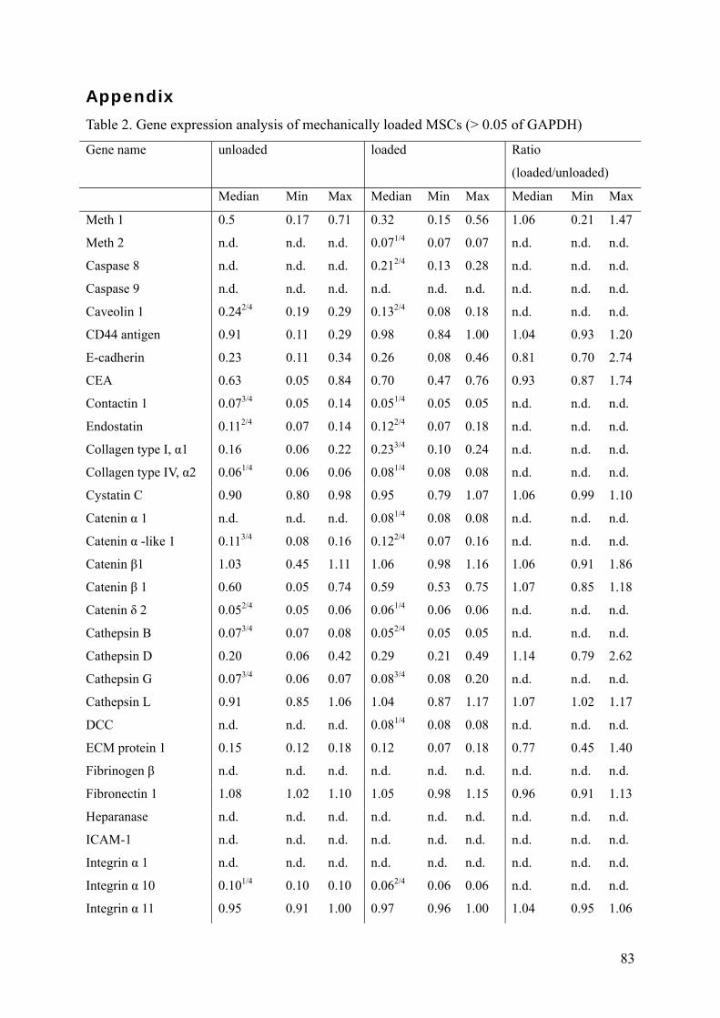
83
Appendix
Table 2. Gene expression analysis of mechanically loaded MSCs (> 0.05 of GAPDH)
Gene name unloaded loaded Ratio
(loaded/unloaded)
Median Min Max Median Min Max Median Min Max
Meth 1 0.5 0.17 0.71 0.32 0.15 0.56 1.06 0.21 1.47
Meth 2 n.d. n.d. n.d. 0.071/4 0.07 0.07 n.d. n.d. n.d.
Caspase 8 n.d. n.d. n.d. 0.212/4 0.13 0.28 n.d. n.d. n.d.
Caspase 9 n.d. n.d. n.d. n.d. n.d. n.d. n.d. n.d. n.d.
Caveolin 1 0.242/4 0.19 0.29 0.132/4 0.08 0.18 n.d. n.d. n.d.
CD44 antigen 0.91 0.11 0.29 0.98 0.84 1.00 1.04 0.93 1.20
E-cadherin 0.23 0.11 0.34 0.26 0.08 0.46 0.81 0.70 2.74
CEA 0.63 0.05 0.84 0.70 0.47 0.76 0.93 0.87 1.74
Contactin 1 0.073/4 0.05 0.14 0.051/4 0.05 0.05 n.d. n.d. n.d.
Endostatin 0.112/4 0.07 0.14 0.122/4 0.07 0.18 n.d. n.d. n.d.
Collagen type I, α1 0.16 0.06 0.22 0.233/4 0.10 0.24 n.d. n.d. n.d.
Collagen type IV, α2 0.061/4 0.06 0.06 0.081/4 0.08 0.08 n.d. n.d. n.d.
Cystatin C 0.90 0.80 0.98 0.95 0.79 1.07 1.06 0.99 1.10
Catenin α 1 n.d. n.d. n.d. 0.081/4 0.08 0.08 n.d. n.d. n.d.
Catenin α -like 1 0.113/4 0.08 0.16 0.122/4 0.07 0.16 n.d. n.d. n.d.
Catenin β1 1.03 0.45 1.11 1.06 0.98 1.16 1.06 0.91 1.86
Catenin β 1 0.60 0.05 0.74 0.59 0.53 0.75 1.07 0.85 1.18
Catenin δ 2 0.052/4 0.05 0.06 0.061/4 0.06 0.06 n.d. n.d. n.d.
Cathepsin B 0.073/4 0.07 0.08 0.052/4 0.05 0.05 n.d. n.d. n.d.
Cathepsin D 0.20 0.06 0.42 0.29 0.21 0.49 1.14 0.79 2.62
Cathepsin G 0.073/4 0.06 0.07 0.083/4 0.08 0.20 n.d. n.d. n.d.
Cathepsin L 0.91 0.85 1.06 1.04 0.87 1.17 1.07 1.02 1.17
DCC n.d. n.d. n.d. 0.081/4 0.08 0.08 n.d. n.d. n.d.
ECM protein 1 0.15 0.12 0.18 0.12 0.07 0.18 0.77 0.45 1.40
Fibrinogen β n.d. n.d. n.d. n.d. n.d. n.d. n.d. n.d. n.d.
Fibronectin 1 1.08 1.02 1.10 1.05 0.98 1.15 0.96 0.91 1.13
Heparanase n.d. n.d. n.d. n.d. n.d. n.d. n.d. n.d. n.d.
ICAM-1 n.d. n.d. n.d. n.d. n.d. n.d. n.d. n.d. n.d.
Integrin α 1 n.d. n.d. n.d. n.d. n.d. n.d. n.d. n.d. n.d.
Integrin α 10 0.101/4 0.10 0.10 0.062/4 0.06 0.06 n.d. n.d. n.d.
Integrin α 11 0.95 0.91 1.00 0.97 0.96 1.00 1.04 0.95 1.06

84
Integrin α 2 n.d. n.d. n.d. 0.082/4 0.07 0.08 n.d. n.d. n.d.
Integrin α 2b n.d. n.d. n.d. n.d. n.d. n.d. n.d. n.d. n.d.
Integrin α 3 0.122/4 0.11 0.13 0.063/4 0.05 0.15 n.d. n.d. n.d.
Integrin α 4 n.d. n.d. n.d. n.d. n.d. n.d. n.d. n.d. n.d.
Integrin α 5 0.37 0.24 0.61 0.38 0.26 0.53 1.12 0.42 2.05
Integrin α 6 n.d. n.d. n.d. n.d. n.d. n.d. n.d. n.d. n.d.
Integrin α 7 n.d. n.d. n.d. n.d. n.d. n.d. n.d. n.d. n.d.
Integrin α 8 n.d. n.d. n.d. n.d. n.d. n.d. n.d. n.d. n.d.
Integrin α 9 n.d. n.d. n.d. n.d. n.d. n.d. n.d. n.d. n.d.
Integrin α L n.d. n.d. n.d. n.d. n.d. n.d. n.d. n.d. n.d.
Integrin α M n.d. n.d. n.d. n.d. n.d. n.d. n.d. n.d. n.d.
Integrin α V 0.051/4 0.05 0.05 0.061/4 0.06 0.06 n.d. n.d. n.d.
Integrin α X n.d. n.d. n.d. n.d. n.d. n.d. n.d. n.d. n.d.
Integrin β 1 0.26 0.20 0.66 0.27 0.23 0.57 1.00 0.87 1.19
Integrin β 2 n.d. n.d. n.d. n.d. n.d. n.d. n.d. n.d. n.d.
Integrin β 3 0.08 0.06 0.14 0.11 0.07 0.18 1.46 0.50 2.70
Integrin β 4 n.d. n.d. n.d. n.d. n.d. n.d. n.d. n.d. n.d.
Integrin β 5 0.56 0.25 0.78 0.57 0.43 0.69 1.10 0.80 1.72
Integrin β 6 n.d. n.d. n.d. n.d. n.d. n.d. n.d. n.d. n.d.
Integrin β 7 0.091/4 0.07 0.09 0.191/4 0.19 0.19 n.d. n.d. n.d.
Integrin β 8 0.112/4 0.07 0.15 0.091/4 0.09 0.09 n.d. n.d. n.d.
Laminin B1 0.19 0.17 0.60 0.23 0.15 0.51 1.04 0.84 1.26
Laminin B2 n.d. n.d. n.d. n.d. n.d. n.d. n.d. n.d. n.d.
Hyaluronidase 0.60 0.06 0.88 0.81 0.50 0.88 1.17 1.01 2.18
MUC-18 0.061/4 0.06 0.06 0.121/4 0.12 0.12 n.d. n.d. n.d.
MMP-1 n.d. n.d. n.d. n.d. n.d. n.d. n.d. n.d. n.d.
MMP-10 n.d. n.d. n.d. n.d. n.d. n.d. n.d. n.d. n.d.
MMP-11 0.102/4 0.06 0.15 n.d. n.d. n.d. n.d. n.d. n.d.
MMP-12 n.d. n.d. n.d. 0.061/4 0.06 0.06 n.d. n.d. n.d.
MMP-13 0.54 0.05 0.84 0.50 0.20 0.92 0.97 0.79 1.21
MMP-14 0.102/4 0.05 0.14 0.08 0.06 0.17 n.d. n.d. n.d.
MMP-15 n.d. n.d. n.d. n.d. n.d. n.d. n.d. n.d. n.d.
MMP-16 n.d. n.d. n.d. n.d. n.d. n.d. n.d. n.d. n.d.
MMP-17 n.d. n.d. n.d. n.d. n.d. n.d. n.d. n.d. n.d.
MMP-2 0.80 0.06 0.96 0.75 0.38 1.04 1.04 0.39 1.35
MMP-20 0.061/4 0.06 0.06 0.101/4 0.10 0.10 n.d. n.d. n.d.
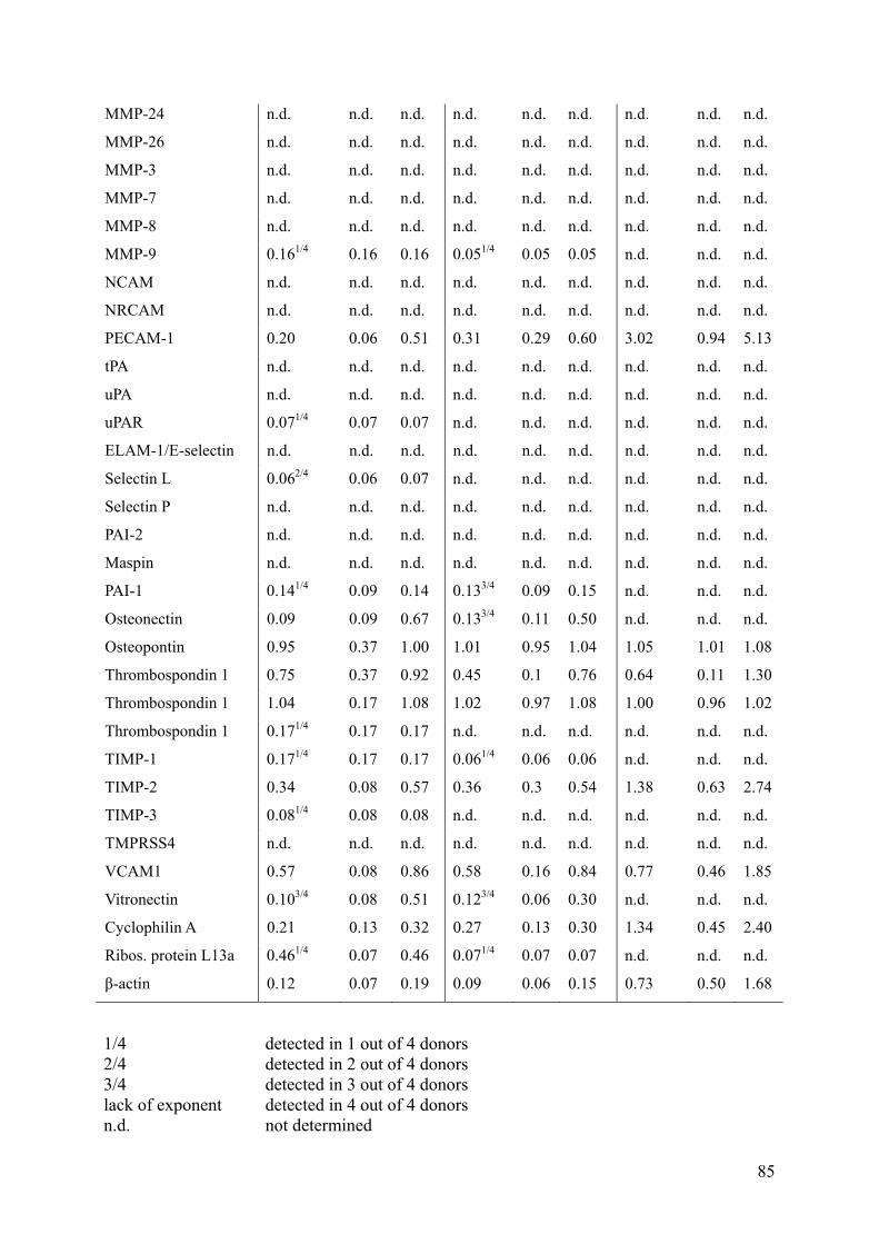
85
MMP-24 n.d. n.d. n.d. n.d. n.d. n.d. n.d. n.d. n.d.
MMP-26 n.d. n.d. n.d. n.d. n.d. n.d. n.d. n.d. n.d.
MMP-3 n.d. n.d. n.d. n.d. n.d. n.d. n.d. n.d. n.d.
MMP-7 n.d. n.d. n.d. n.d. n.d. n.d. n.d. n.d. n.d.
MMP-8 n.d. n.d. n.d. n.d. n.d. n.d. n.d. n.d. n.d.
MMP-9 0.161/4 0.16 0.16 0.051/4 0.05 0.05 n.d. n.d. n.d.
NCAM n.d. n.d. n.d. n.d. n.d. n.d. n.d. n.d. n.d.
NRCAM n.d. n.d. n.d. n.d. n.d. n.d. n.d. n.d. n.d.
PECAM-1 0.20 0.06 0.51 0.31 0.29 0.60 3.02 0.94 5.13
tPA n.d. n.d. n.d. n.d. n.d. n.d. n.d. n.d. n.d.
uPA n.d. n.d. n.d. n.d. n.d. n.d. n.d. n.d. n.d.
uPAR 0.071/4 0.07 0.07 n.d. n.d. n.d. n.d. n.d. n.d.
ELAM-1/E-selectin n.d. n.d. n.d. n.d. n.d. n.d. n.d. n.d. n.d.
Selectin L 0.062/4 0.06 0.07 n.d. n.d. n.d. n.d. n.d. n.d.
Selectin P n.d. n.d. n.d. n.d. n.d. n.d. n.d. n.d. n.d.
PAI-2 n.d. n.d. n.d. n.d. n.d. n.d. n.d. n.d. n.d.
Maspin n.d. n.d. n.d. n.d. n.d. n.d. n.d. n.d. n.d.
PAI-1 0.141/4 0.09 0.14 0.133/4 0.09 0.15 n.d. n.d. n.d.
Osteonectin 0.09 0.09 0.67 0.133/4 0.11 0.50 n.d. n.d. n.d.
Osteopontin 0.95 0.37 1.00 1.01 0.95 1.04 1.05 1.01 1.08
Thrombospondin 1 0.75 0.37 0.92 0.45 0.1 0.76 0.64 0.11 1.30
Thrombospondin 1 1.04 0.17 1.08 1.02 0.97 1.08 1.00 0.96 1.02
Thrombospondin 1 0.171/4 0.17 0.17 n.d. n.d. n.d. n.d. n.d. n.d.
TIMP-1 0.171/4 0.17 0.17 0.061/4 0.06 0.06 n.d. n.d. n.d.
TIMP-2 0.34 0.08 0.57 0.36 0.3 0.54 1.38 0.63 2.74
TIMP-3 0.081/4 0.08 0.08 n.d. n.d. n.d. n.d. n.d. n.d.
TMPRSS4 n.d. n.d. n.d. n.d. n.d. n.d. n.d. n.d. n.d.
VCAM1 0.57 0.08 0.86 0.58 0.16 0.84 0.77 0.46 1.85
Vitronectin 0.103/4 0.08 0.51 0.123/4 0.06 0.30 n.d. n.d. n.d.
Cyclophilin A 0.21 0.13 0.32 0.27 0.13 0.30 1.34 0.45 2.40
Ribos. protein L13a 0.461/4 0.07 0.46 0.071/4 0.07 0.07 n.d. n.d. n.d.
β-actin 0.12 0.07 0.19 0.09 0.06 0.15 0.73 0.50 1.68
1/4 detected in 1 out of 4 donors 2/4 detected in 2 out of 4 donors 3/4 detected in 3 out of 4 donors lack of exponent detected in 4 out of 4 donors n.d. not determined

86
87
Anteils-Erklärung meiner Person an in diese Dissertation eingegangene Publikationen
Kasper G†, Glaeser JD†, Geissler S, Ode A, Tuischer J, Matziolis G, Perka C, and Duda GN, MMP activity is an essential link between mechanical stimulus and mesenchymal stem cell behaviour, Stem Cells, 2007, 25:1985-1994, †: authors contributed equally
− Konzeption: das Konzept dieser Arbeit wurde von meiner Betreuerin Dr.-Ing. Grit Kasper erstellt.
− Durchführung: Die Charaktierisierung der MSCs mittels FACS-Analyse erfolgte durch Dipl.-Ing. Miriam Tschirschmann, TU-Berlin. Bei der Durchführung der funktionellen Assays wurde ich teilweise von unserer technischen Assistentin Marzena Princ unterstützt. Alle weiteren Versuche wurden von mir durchgeführt.
− Berichtsabfassung: das Manuskript wurde zu gleichen Teilen von Frau Dr.-Ing. Grit Kasper und mir verfasst.
Glaeser JD†, Geissler S†, Strube P, Schipp CJ, Matziolis G, Taylor WR, Knaus P, Perka C, Duda GN and Kasper G, Control of pro-MMP-2 levels and activation of MMP-2 via the furin/MMP-14/TIMP-2 axis contribute concurrently to the pro-angiogenic effects of mechanically stimulated mesenchymal stem cells, submitted, 2008, †: authors contributed equally
− Konzeption: das Konzept dieser Arbeit wurde von mir erstellt.
− Durchführung: Die Optimierung der einzelnen Belastungsparameter erfolgte durch Dipl.-Ing. Sven Geissler (FH) unter meiner Anleitung im Rahmen seiner Masterarbeit, die er 2006-2007 am Julius Wolff Institut anfertigte. Alle weiteren Versuche wurden von mir durchgeführt.
− Berichtsabfassung: das Manuskript wurde von mir verfasst.
Kasper G, Dankert N, Tuischer J, Hoeft M, Gaber T, Glaeser JD, Zander D, Tschirschmann M, Thompson M, Matziolis G and Duda GN, Mesenchymal stem cells regulate angiogenesis according to their mechanical environment, Stem Cells, 2007, 25:903-910
− Konzeption: das Konzept dieser Arbeit wurde von meiner Betreuerin Dr.-Ing. Grit Kasper erstellt.
− Durchführung: Die Zymogramm-Daten bezüglich MMP-2 wurden von mir beigesteuert.
− Berichtsabfassung: das Manuskript wurde von Frau Dr.-Ing. Grit Kasper verfasst.

88

89
Eidesstattliche Erklärung
Hiermit erkläre ich, Juliane Dagmar Gläser, an Eides statt, dass ich die vorgelegte Dissertation
selbst verfasst habe und keine anderen als die angegebenen Quellen und Hilfsmittel
verwendet habe. Außerdem erkläre ich, dass ich an keiner anderen Stelle ein
Promotionsverfahren beantragt habe.
Berlin den ________________ _______________________________
Juliane Gläser

90