Diazen-1-ium-1,2-diolate als NO-Donoren · Diazen-1-ium-1,2-diolate als NO-Donoren - Zusammenhänge...
173
Diazen-1-ium-1,2-diolate als NO-Donoren - Zusammenhänge zwischen Struktur, Bildung und NO-Freisetzung sowie die Umsetzung zu bioaktiven Prodrugs und Ketoconazol-NO-Donor Hybridverbindungen Dissertation zur Erlangung des akademischen Grades doctor rerum naturalium (Dr. rer. nat.) vorgelegt dem Rat der Biologisch-Pharmazeutischen Fakultät der Friedrich-Schiller-Universität Jena, 2007 von Jörg Konter geboren am 24.02.1978 in Leverkusen
Transcript of Diazen-1-ium-1,2-diolate als NO-Donoren · Diazen-1-ium-1,2-diolate als NO-Donoren - Zusammenhänge...

Diazen-1-ium-1,2-diolate als NO-Donoren -
Zusammenhänge zwischen Struktur, Bildung und NO-Freisetzung sowie die Umsetzung zu bioaktiven
Prodrugs und Ketoconazol-NO-Donor Hybridverbindungen
Dissertation zur Erlangung des akademischen Grades
doctor rerum naturalium (Dr. rer. nat.)
vorgelegt dem Rat der Biologisch-Pharmazeutischen Fakultät
der Friedrich-Schiller-Universität Jena, 2007
von Jörg Konter
geboren am 24.02.1978 in Leverkusen

Gutachter:
1. Prof. Dr. Jochen Lehmann
2. Prof. Dr. Gerhard Scriba
3. Prof. Dr. Hans-Jürgen Duchstein
Tag der Doktorprüfung:
Tag der öffentlichen Verteidigung:
Danksagung Die vorliegende Arbeit wurde im Zeitraum von Oktober 2003 bis August 2007 am
Institut für Pharmazie der Friedrich-Schiller Universität Jena durchgeführt.
Meinem Doktorvater Herrn Prof. Dr. Jochen Lehmann danke ich sehr herzlich für die
Überlassung des Themas, seine wertvollen Anregungen, die stete
Diskussionsbereitschaft und seine große Einsatzbereitschaft unvergessliche
Erfahrungen, wie der NO-Konferenz in Monterey, zu ermöglichen.
Herrn Prof. Dr. Gerhard Scriba und Herrn Prof. Dr. Hans-Jürgen Duchstein danke ich
für die freundliche Übernahme des Korreferats.
Für die große Kooperationsbereitschaft und erfolgreiche Zusammenarbeit danke ich
Frau Dr. Möllmann und ihren Mitarbeitern, Herrn Sven Winter und Herrn Prof. Dr.
Alfred Fahr, Herrn Andreas König und Frau Prof. Dr. Erika Glusa sowie Herrn
Michael Rothenburger.
Bedanken möchte ich mich bei allen Kollegen und Freunden des Instituts für die
unkomplizierte Zusammenarbeit, offene Hilfsbereitschaft und gemeinsame Zeit in
Jena. Frau Carolin Roegler danke ich für das Korrekturlesen der Arbeit.
Ein ganz besonderer Dank gilt meinen lieben Eltern, Helga und Wolfgang Konter, die
mich während meiner gesamten schulischen und universitären Ausbildung vielfältig
unterstützt haben.
Inhaltsverzeichnis I
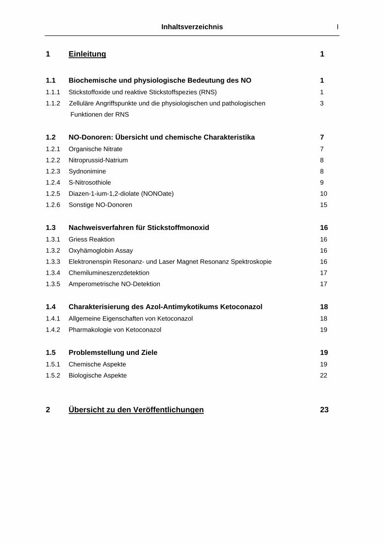
1 Einleitung 1 1.1 Biochemische und physiologische Bedeutung des NO 1 1.1.1 Stickstoffoxide und reaktive Stickstoffspezies (RNS) 1
1.1.2 Zelluläre Angriffspunkte und die physiologischen und pathologischen 3
Funktionen der RNS
1.2 NO-Donoren: Übersicht und chemische Charakteristika 7 1.2.1 Organische Nitrate 7
1.2.2 Nitroprussid-Natrium 8
1.2.3 Sydnonimine 8
1.2.4 S-Nitrosothiole 9
1.2.5 Diazen-1-ium-1,2-diolate (NONOate) 10
1.2.6 Sonstige NO-Donoren 15
1.3 Nachweisverfahren für Stickstoffmonoxid 16 1.3.1 Griess Reaktion 16
1.3.2 Oxyhämoglobin Assay 16
1.3.3 Elektronenspin Resonanz- und Laser Magnet Resonanz Spektroskopie 16
1.3.4 Chemilumineszenzdetektion 17
1.3.5 Amperometrische NO-Detektion 17
1.4 Charakterisierung des Azol-Antimykotikums Ketoconazol 18 1.4.1 Allgemeine Eigenschaften von Ketoconazol 18
1.4.2 Pharmakologie von Ketoconazol 19
1.5 Problemstellung und Ziele 19 1.5.1 Chemische Aspekte 19
1.5.2 Biologische Aspekte 22
2 Übersicht zu den Veröffentlichungen 23

II Inhaltsverzeichnis
3 Veröffentlichungen 26 Veröffentlichung 1 26 The NOtizer- A Device fort he Convenient Preparation of Diazen-1-ium-1,2-diolates. Konter, Joerg; Abuo-Rahma, Gamal El-Din Ali Ahmed Hassan; El-Emam, Ali; Lehmann,
Jochen. Methods in Enzymology (2005), 396 (Nitric Oxide, Part E), 17-26.
Veröffentlichung 2 37 Synthesis of Diazen-1-ium-1,2-diolates Monitored by the "NOtizer" Apparatus: Relationship between Formation Rates, Molecular Structure and the Release of Nitric Oxide. Konter, Joerg; Abuo-Rahma, Gamal El-Din A. A.; El-Emam, Ali; Lehmann, Jochen.
European Journal of Organic Chemistry (2007), 4, 616-624.
Veröffentlichung 3 47 NO Donors, part 17: Synthesis and Antimicrobial Activity of Novel Ketoconazole-NO-Donor Hybrid Drugs and Corresponding Nitric Oxide Donors. Konter, Joerg; Möllmann, Ute; Lehmann, Jochen. Journal of Medicinal Chemistry. In
Vorbereitung.
Veröffentlichung 4 64 Potency and In Vitro Tolerance of Organic Nitrates: Partially Denitrated Metabolites Contribute to the Tolerance-Devoid Activity of Pentaerythrityl Tetranitrate. Koenig, Andreas; Lange, Kathrin; Konter, Joerg; Daiber, Andreas; Stalleicken, Dirk;
Glusa, Erika; Lehmann, Jochen. Journal of Cardiovascular Pharmacology (2007), 50,
68-74.
Veröffentlichung 5 72 Permeability changes in response to NONOate and NONOate prodrug derived nitric oxide in a blood-brain barrier model formed by primary porcine endothelial cells. Winter, Sven; Konter, Joerg; Scheler, Stefan; Lehmann, Jochen; Fahr, Alfred. Nitric
Oxide. Eingereicht (2007).
Veröffentlichung 6 (Patentschrift) 90 Neue Ketoconazol-Derivate sowie deren Herstellung und Verwendung als anti-mikrobielle und antiinflammatorische Wirkstoffe. Konter, Joerg; Möllmann, Ute; Lehmann, Jochen. Eingereicht beim Deutschen Patent-
und Markenamt (25.07.2007).

Inhaltsverzeichnis III
4. Bisher unveröffentlichte Ergebnisse 110 4.1 Chemische Aspekte 110 4.1.1 Beobachtung der NONOat-Synthese mit dem NOtizer 110
4.1.2 HPLC Untersuchungen der NONOate 117
4.1.3 Synthese von Diazen-1-ium-1,2-diolat Prodrugs 119
4.2 Biologische Aktivität einiger synthetisierter NO-Donoren 122 4.2.1 Vasodilatation in vitro 122
4.2.2 Neuroregenerative Eigenschaften von DETA/NO und 124
Et-BUPIPERAZI/NO in vivo
5. Gesamtdiskussion 126 5.1 Bewertung der NOtizer®-Apparatur für die Synthese 126
von Diazen-1-ium-1,2-diolaten 5.1.1 Vorteile der NOtizer®-Apparatur und der erarbeiteten Methode 126
5.1.2 Nachteile der NOtizer®-Apparatur und der erarbeiteten Methode 127
5.2 Untersuchungen zur Bildung und zum Zerfall von 127 Diazen-1-ium-1,2-diolaten
5.2.1 Bildung von Diazen-1-ium-1,2-diolaten 127
5.2.2 Zerfall von Diazen-1-ium-1,2-diolaten 129
5.2.3 Zusammenhang zwischen molekularer Struktur des Amins, 130
NONOat-Bildung und NO-Freisetzung
5.3 Identitäts und Reinheitsuntersuchungen der 133 Diazen-1-ium-1,2-diolat-Prodrugs und Ketoconazol-NO-Donor Hybridverbindungen
5.3.1 Et-BUPIPERAZI/NO 133
5.3.2 Ketoconazol-Nitratopivalinsäure Hybridverbindung 134
5.4 Antimikrobielle Eigenschaften der Ketoconazol-NO-Donor- 135 Hybridverbindungen - Diskussion möglicher Targets, Unterschiede im Wirkspektrum und Ursachen des Synergismus 5.4.1 Mikrobielle zelluläre Targets für NO und andere RNS 136
5.4.2 Mögliche Ursachen der Unterschiede im Wirkspektrum und 136
des Synergismus
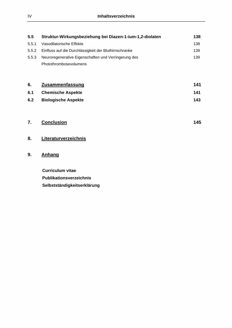
IV Inhaltsverzeichnis
5.5 Struktur-Wirkungsbeziehung bei Diazen-1-ium-1,2-diolaten 138 5.5.1 Vasodilatorische Effekte 138
5.5.2 Einfluss auf die Durchlässigkeit der Bluthirnschranke 139
5.5.3 Neuroregenerative Eigenschaften und Verringerung des 139
Photothrombosevolumens
6. Zusammenfassung 141 6.1 Chemische Aspekte 141 6.2 Biologische Aspekte 143
7. Conclusion 145 8. Literaturverzeichnis 9. Anhang
Curriculum vitae Publikationsverzeichnis Selbstständigkeitserklärung

Einleitung 1
1 Einleitung
Die in den 80er Jahren des vergangenen Jahrhunderts überraschende und zugleich
aufregende Entdeckung der physiologischen und pathologischen Bedeutung des Stickstoffmonoxid
(NO., Stickoxid) fand ihren bisherigen Höhepunkt mit der Verleihung des Nobelpreises für Physiologie
und Medizin an die drei Pioniere der NO-Forschung Robert F. Furchgott, Louis J. Ignarro und Ferid
Murad im Jahre 1998. Das vor seiner Identifizierung nur als “endothelium derived relaxing factor“
(EDRF) bezeichnete Stickoxidradikal wurde seit dieser Zeit als einer der wichtigsten physiologischen
Botenstoffe in den verschiedensten Bereichen der Medizin und Biologie erkannt. Schon 1992 wurde
das NO.-Radikal von der Fachzeitschrift Science als das “Molecule of the Year“ bezeichnet.
In diesem einleitenden Kapitel werden die biochemischen Grundlagen sowie die
physiologische und pathologische Rolle des NO. zusammengefasst und eine Übersicht und Einteilung
der wichtigsten NO-Donoren dargestellt. Es folgt ein Überblick über analytische Nachweisverfahren
von NO., eine Charakterisierung des Antimykotikums Ketoconazol und abschließend die
Problemstellung dieser Arbeit.
1.1 Biochemische und physiologische Bedeutung des NO 1.1.1 Stickstoffoxide und reaktive Stickstoffspezies (RNS)
Eines der wichtigsten Charakteristika des NO sind seine unterschiedlichen
Reaktionsmöglichkeiten. Man muss deutlich unterscheiden, besonders wenn es sich um NO-Donoren
handelt, welche Redoxform des NO vorliegt bzw. freigesetzt wird: NO., NO+ oder NO-. Jede dieser
Spezies kann unter dem allgemeinen Begriff “NO“ verstanden werden und alle weisen ein
unterschiedliches Reaktionsverhalten auf. Insbesondere die Folgeprodukte dieser NO-Spezies haben
wiederum eigene Eigenschaften und vor allem eine bedeutende pathologische oder physiologische
Relevanz, wie zum Beispiel das Peroxynitrit (ONOO-) oder die Nitrosothiole (R-SNO). Molekularer
Sauerstoff (O2), das Superoxidanion-Radikal (.O2-) sowie einige Übergangsmetalle und
Spurenelemente (M) sind die Hauptreaktionspartner für die NO-Spezies, um weitere reaktive
Stickstoffspezies (RNS; reactive nitrogen species) wie N2O3, NO2 oder ONOO- zu bilden.[1,2] Die
genauen Funktionen dieser Verbindungen werden im folgenden Unterkapitel (1.1.2) im Einzelnen
erläutert. Abbildung 1 und Tabelle 1 stellen die biologisch relevanten Stickoxide und die
dazugehörigen Reaktionsschemata dar.[3,4]

2 Einleitung
NO+ NO. NO-
Abb. 1 Reaktionen der reaktiven Stickstoffspezies Endogenes NO wird, katalysiert durch die NO-Synthasen (eNOS, nNOS, iNOS), aus der
Aminosäure L-Arginin gewonnen, wobei neben dem NO-Radikal noch L-Citrullin und Wasser als
Nebenprodukte entstehen. Wie Abbildung 1 deutlich zeigt, sind die durch das NO verursachten
physiologischen Veränderungen das Gesamtergebnis eines multifaktoriellen Prozesses. Trotzdem
müssen die Oxidationsprodukte NO2, N2O3 und insbesondere das ONOO- als die wichtigsten RNS
herausgestellt werden. Es ist nicht verwunderlich, dass gerade diese RNS äußerst schnell gebildet
werden, da es sich bei dem Superoxidanion, dem molekularen Sauerstoff, wie auch ihrem jeweiligen
Reaktionspartner, dem Stickstoffmonoxid, um Radikale handelt. Das .O2- Radikal ist ein ubiquitär
vorkommendes Stoffwechselnebenprodukt aus der Reduktion von O2 durch NADPH-Oxidasen und
der endothelialen Xanthinoxidase.[5,6] Ein weiteres mögliches Oxidationsprodukt des NO ist das
Nitrosyl Kation (NO+), welches schnell nukleophile Bindungspartner wie Thiole oder Amine zu
Nitrosothiolen und Nitrosaminen nitrosyliert.[7,8]
L-Arginin
L-Citrullin + H2O
ONOO- ONOO- ONOO-
M: Metall
MNO2
-/NO3-
H2O
O2-
O2-
NOO2
O2
Oxidation Reduktion
MM M
NO2/N2O3NO2
-
NO2-
NO2-
RSH
M-NO
RSNO
M-NO
RONO
M-NO
O2
H2O
NH2OH
H2O2
RSHRSH
RSNO RR’N-NO
RR‘NH ROH
NOS
N2O
NO2-/NO3
-

Einleitung 3
Tab. 1 Biologisch relevante Stickoxide und ihre Oxidationsstufen
Summenformel Name Oxidationsstufe des N-Atoms
NO- Nitroxyl Anion +1
N2O Distickstoffmonoxid (Lachgas) +1 (0 und +2)
NO. Stickstoffmonoxid +2
NO+ Nitrosyl (Nitrosonium) Kation +3
NO2- Nitrit +3
N2O3 Distickstofftrioxid (Salpetrige Säure Anhydrid) +3 (+2 und +4)
NO2 Stickstoffdioxid +4
ONOO- Peroxynitrit +5
NO3- Nitrat +5
NH2OH Hydroxylamin -1
RR’N-NO Nitrosamin +2
RS-NO Nitrosothiol +3
Durch Reduktion des NO. entsteht das Nitroxyl Anion (NO-), welches isoelektronisch zum O2
ist und somit ebenfalls als Triplett-NO- (gleicher Spin der Elektronen in den antibindenden πy*-/ πz
*
Orbitalen) oder Singulett-NO- (entgegengesetzter Spin) auftreten kann. Das stabilere Triplett-NO- kann
durch O2 zu Peroxynitrit oxidiert werden, während das wesentlich energiereichere Singulett-NO-
überwiegend zu Hydroxylamin reduziert wird, beispielsweise durch freie Thiol-Gruppen. Die
Hauptkonkurrenzreaktion ist die Umsetzung des NO- zum N2O.[9] Die biologischen Hauptmetabolite
der RNS sind Nitrat und Nitrit, die renal ausgeschieden werden.[10]
Die oben beschriebenen Stoffwechselwege verdeutlichen, dass im Hinblick auf NO-Donoren
nicht allein das NO., sondern ebenso gut seine Folgeprodukte die entscheidenden Faktoren für einen
physiologischen Effekt sein können. Abschließend sei darauf hingewiesen, dass sich in der Literatur
die Kurzform “NO“ für das damit gemeinte Radikal NO. durchgesetzt hat.
1.1.2 Zelluläre Angriffspunkte und die physiologischen und pathologischen Funktionen der RNS
Eines der bedeutendsten Merkmale des NO. in Bezug auf seine physiologische Funktion ist
die extrem hohe Affinität zu Häm-Proteinen, die fester Bestandteil vieler Enzyme, wie der löslichen
Guanylatcyclase (sGC), der Cyclooxygenase und der Cytochromen, ist.[11]
Für die Aktivierung der sGC in glatten Gefäßmuskelzellen bindet das aus dem Endothel
stammende NO (gebildet durch die eNOS) an das zentrale Eisen Atom der prosthetischen Häm-
Gruppe und löst dort eine bis zu 400-fache Aktivitätssteigerung für die Umsetzung von GTP zu cGMP
aus.[12] Das cGMP wiederum aktiviert eine Proteinkinase der Gefäßmuskelzelle (PKG II), die durch
Phosphorylierung verschiedener Membranproteine (u. a. VASP, IRAG, Phospholamban) des
sarkoplasmatischen Retikulums eine Absenkung der freien intrazellulären Ca2+-Ionen Konzentration

4 Einleitung
und folglich eine Relaxation der Gefäßmuskulatur bewirkt.[13] Das von der cGMP abhängigen
Proteinkinase phosphorylierte VASP (vasodilator stimulated phosphoprotein) wird unter anderen auch
in Thrombozyten exprimiert.[14] Hier verhindert das phosphorylierte VASP die Aktivierung des
Fibrinogen Rezeptors und von Integrin αIIbβ3, wodurch die thrombozytenaggregationshemmende Wirkung des NO zu erklären ist.[15] Neben den Proteinkinasen werden außerdem
Phosphodiesterasen (PDE) von cGMP reguliert, wie z.B. die durch cGMP inhibierbare PDE II in
Kardiomyozyten von Säugerherzen. Ein erhöhter cGMP Spiegel löst hier eine negativ inotrope Wirkung aus.[16] In glatten Gefäßmuskelzellen wird durch die cGMP abhängige Hemmung der PDE III
eine antiproliferative Wirkung entfaltet, die wiederum atherosklerotischen Veränderungen entgegenwirkt.[17,18] Eine Reihe von cGMP abhängigen Ionenkanälen, die vor allem den Einstrom von
Na+- und Ca2+- Ionen durch Membranen regulieren, spielen eine große Rolle bei der Verarbeitung von Signalen in Nervengewebe.[19] Zur Aufklärung solcher NO/cGMP vermittelten Effekte werden
häufig Inhibitoren der löslichen Guanylatcyclase verwendet, wie zum Beispiel der selektive Inhibitor
ODQ oder das etwas unspezifischere Methylenblau.[20] Beide Inhibitoren oxidieren das Häm-Eisen der
sGC, wobei das Methylenblau zusätzlich als “NO scavenger“ fungiert, indem es durch .O2- Bildung die
Bioverfügbarkeit des NO drastisch reduziert.[21-23] Ob Methylenblau zusätzlich auch die Aktivität der
NO-Synthasen hemmt, ist noch umstritten.[24]
Eine sehr häufig auftretende NO-bedingte Proteinmodulation ist die S-Nitrosylierung (RS-NO)
cysteinhaltiger Proteine. Eine solche Modulation wird beispielsweise für NMDA-Rezeptoren im ZNS
diskutiert, wodurch die Rolle von NO an Lern- und Gedächtnisprozessen sowie der
Schmerzwahrnehmung zu erklären ist.[25-27] Die S-Nitrosylierung und dadurch gleichzeitige
Aktivierung des Transkriptionsfaktors AP-1 bewirkt die Expression anti-apoptotischer Faktoren und
somit eine zytoprotektive Wirkung in ausgewählten Geweben.[28] Neben der Nitrosylierung finden
Nitrierungen (RS-NO2) durch aus Peroxynitrit gebildetem Stickstoffdioxid (siehe Abb.1) sowie
Sulfhydryl-Oxidationen (RS-H zu RS-SR) statt.[29-32] Solche Thiol-Gruppen-Modifikationen sind
besonders ausgeprägt in der Familie der Cysteinproteasen, welche proteolytische Enzyme nicht nur
im menschlichen Organismus, aber auch im Pflanzen- und Tierreich, sowie einen wichtigen
Bestandteil vieler Mikroorganismen darstellen. So übernehmen beispielsweise Cathepsine und
Caspasen entscheidende Funktionen in antigenpräsentierenden Zellen, der Aktivierung von Zytokinen
und der T-Zell-Aktivierung, womit die Rolle der RNS bei der Immunabwehr deutlich wird.[33-36] Ebenso
sind solche Cysteinproteasen direkt an der Reproduktion vieler Viren, Bakterien, Pilze und Parasiten
beteiligt.[37] Aktivierte Makrophagen produzieren NO. als einen wichtigen Verteidigungsmechanismus
gegen Tumorzellen, indem es die Freisetzung von Cytochrom c aus den Mitochondrien vermittelt und
letztendlich die Apoptose der entarteten Zellen einleitet .[38,39]
Ein weiteres Angriffsziel für RNS sind Nukleinsäuren, wobei durch elektrophile Substitution
Nitroverbindungen der Purin- und Pyrimidinbasen entstehen.[40,41] Die durch N2O3 verursachte
Desaminierung von Aminosäuren in Proteinen und Peptiden kann ebenso gut in der DNA auftreten,
insbesondere an den Pyrimidinbasen. Inwiefern diese gentoxischen “Mutationen“ und Strangbrüche in
vivo eine Rolle spielen ist umstritten. Schon in vitro ist eine unphysiologisch hohe Konzentration der
entsprechenden RNS nötig um diese Reaktionen zu ermöglichen.[42] Vor allem muss man beachten,
Einleitung 5
dass in vivo zahlreiche Abwehrmechanismen bestehen, wie z.B. Antioxidantien, die die RNS
“abfangen“ bevor sie überhaupt an die DNA/RNA gelangen können.[43]
Äußerst komplex sind die Reaktionen der RNS mit Lipiden. Das NO. und die daraus
gebildeten RNS sind in der Lage eine Lipidoxidation zu stimulieren, ebenso aber auch zu hemmen.[10]
Welcher der beiden gegensätzlichen Mechanismen eintritt ist abhängig von (1) der Konzentration an
NO., (2) der Konzentration an reaktiver Sauerstoffspezies (ROS), (3) der Anwesenheit von
Antioxidantien wie z.B. Vitamin E, und (4) den Reaktionsraten der beteiligten Spezies in Bezug auf
das Milieu in dem sie sich befinden.[44,45] Abbildung 2 verdeutlicht die Mechanismen der Lipidoxidation.
R
R
HH
ONOO
ungesättigtes Lipid
R
R
H
Lipid Radikal
R
R
OOH
R
R
HH
R
R
OOHH
AlkylperoxylradikalAlkylperoxid
Kettenfortpflanzung
NO R
R
OONOH Kettenabbruch
O2
[org. Peroxynitrit]
R
R
OH
-NO2
R
R
ONOH NO
AlkoxylradikalAlkylnitrit Abb. 2 Mechanismen der Peroxid- und Organonitritbildung bei ungesättigten Lipiden
Ein pro-oxidativer Prozess an Lipiden tritt auf, wenn eine mindestens gleich große Konzentration an .O2
- im Vergleich zum NO. vorhanden ist, da diese extrem schnell hochoxidatives Peroxynitrit bilden,
welches dann schließlich ungesättigte Lipide oxidiert.[46] Die so entstehenden Lipid-peroxylradikale
wiederum oxidieren ungesättigte Lipide und es entsteht eine Kettenreaktion, die immer mehr Lipide
oxidieren lässt. Der anti-oxidative Prozess tritt ein, wenn die Konzentration an NO. überwiegt.[47] Der
Überschuss an NO. Radikalen bewirkt einen Kettenabbruch der Reaktion indem sie mit den Lipid-
peroxylradikalen intermediär ein instabiles organisches Peroxynitrit bildet, das sofort in das
6 Einleitung
entsprechende Alkoxylradikal (RO.) und NO2 zerfällt. Ein zweites NO. Molekül setzt sich dann mit dem
RO. zum Alkylnitrit (RNO2) um.[10] Die in Abb. 2 dargestellte Oxidation der Lipide durch Peroxynitrit
kann auch von einigen ROS, wie dem hochreaktiven Hydroxylradikal (OH.) initiiert werden. Ein
Überschuss an NO. spielt so oxidativem Stress entgegen. Bemerkenswert ist die Tatsache, dass die
Kombination von α-Tocopherol/NO. effektiver gegen oxidative Prozesse schützt als die Kombination α-
Tocopherol/Ascorbinsäure.[48] Diese antioxidative Wirkung des NO. ist von sehr großer Bedeutung in
der Repression der Arteriosklerose, nicht nur weil durch den Kettenabbruch die Entstehung
oxidierter LDL Partikel unterdrückt wird, sondern insbesondere weil die Organonitrit-Gruppen
tragenden LDL Partikel (NO2-LDL) verstärkt von Makrophagen aufgenommen werden.[49,50] Die
Interaktionen von NO. im Eicosanoidstoffwechsel, sei es durch direkte Reaktionen mit den
ungesättigten Kohlenstoffgerüsten oder durch Modulation der Arachidonsäure verstoffwechselnden
Enzyme (Cyclooxygenase, Lipoxygenasen, Cytochrom 450), zeigen vielseitige Auswirkungen, wie
beispielsweise die Hemmung der Thrombozytenaggregation.[10] Über die durch RNS hervorgerufenen
Veränderungen an Zellmembranlipiden und ihre physiologische Bedeutung ist noch nicht sehr viel
bekannt. Sicher ist, dass nicht nur strukturelle Modifikationen an den Membranlipiden selbst
Signalkaskaden in Gang setzen, sondern auch, dass das gebildete Lipidradikal eine wichtige
Bindungsstelle für Proteine darstellt.[51] Dieses Charakteristikum der Lipide, auch nicht-
membranständige Proteine an die Zelloberfläche zu binden, wird von führenden Membranproteomik
Forschungsgruppen als ein bedeutender Bestandteil zukünftiger Forschung gesehen.[52]
Ähnlich wie beim oxidativen Stress, kann ein Ungleichgewicht der RNS zu Ungunsten der
physiologischen Abläufe ein komplexes pathologisches Bild entstehen lassen. Diese als nitrosativer Stress bezeichnete Entgleisung des Stoffwechsels geht in erster Linie von der aus NO. und O2
gebildeten RNS N2O3 aus.[51] Gleichung 1 und 2 stellen den genauen Zusammenhang der beteiligten
RNS dar.[53]
2 NO + O2 2 NO2 (1)
NO2 + NO N2O3 + H2O 2HNO2 (2)
Das Ausmaß des nitrosativen Stresses ist besonders abhängig von der NO. Konzentration. Es muss
ständig eine ausreichende Menge an N2O3 gebildet werden um die S-Nitrosylierung von Thiolen,
Desaminierung primärer Amine und die Nitrosaminbildung aus sekundären Aminen aufrecht erhalten
zu können. In wässrigen Medien beträgt die Halbwertszeit der N2O3 Hydrolyse etwa 1 ms.[54] Der
Hauptort der RNS Bildung aus NO. und O2 sind daher hydrophobe Medien, wie z.B. zwischen
Phospholipiden. Die Konzentration dieser beiden Gase kann in Membranlipiden 5 bis 50 mal höher
sein als im Intra- oder Extrazellulärraum.[55] Bekannt ist, dass bei inflammatorischer Aktivität die iNOS
(induzierte NO-Synthase) lokal begrenzt große Mengen NO. freisetzt. Ob die Expression der iNOS bei
Erkrankungen wie Colitis, Morbus Crohn, Multipler Sklerose, rheumatoider Arthritis, Psoriasis,
Alzheimer, Parkinson oder Krebs ein Teil des pathologischen Geschehens oder eine Immunantwort
darstellt, ist noch nicht vollständig aufgeklärt.
Vergleicht man die vielseitigen Effekte des NO und der anderen RNS, so stellt man fest, dass
häufig scheinbar Widersprüche bestehen. Zum Beispiel ist NO. einmal zytotoxisch, in anderen

Einleitung 7
Zusammenhängen wieder zytoprotektiv, oder, wie oben erwähnt, zugleich oxidativ und antioxidativ.
Diese Ambivalenz lässt sich dadurch erklären, dass alle vom NO ausgehenden Effekte zum einen von
der Art des Gewebes abhängt, vor allem aber auch von der Menge des dort auftretenden NO..
Wesentlich ist auch, ob oder wie viel der für die Produktion der übrigen RNS benötigten
Reaktionspartner, wie z.B. der ROS, vorhanden sind.
1.2 NO-Donoren: Übersicht und chemische Charakteristika 1.2.1 Organische Nitrate
Aus chemischer Sicht handelt es sich bei den organischen Nitraten um Ester der
Salpetersäure. Der in diesem Zusammenhang häufig verwendete Begriff “NO Donor“ ist nicht ideal, da
der Nitrat Stickstoff von seiner Oxidationsstufe +V reduziert werden muss, um das zweiwertige NO.
freisetzen zu können. Ob dies zur Auslösung eines biologischen Effektes zwingend notwendig ist, wird
kontrovers diskutiert. Zudem ist der genaue Bioaktivierungsmechanismus der Nitrate noch immer
Gegenstand der Forschung, da jedoch die biologische Aktivität der organischen Nitrate in den meisten
Fällen dem des NO. sehr ähnlich ist, ist der Begriff “NO-Mimetika“ angebrachter. Die auf dem Markt
erhältlichen organischen Nitrate sind (siehe Abb. 3):
• Pentaerythrityltetranitrat (PETN) mit vier Nitratgruppen
• Glyceroltrinitrat (GTN) mit drei Nitratgruppen
• Isosorbiddinitrat (ISDN) mit zwei Nitratgruppen
• Isosorbid-5-mononitrat (ISMN) mit einer Nitratgruppe
wobei die pharmakodynamische Potenz, unter Ausschluss pharmakokinetischer Parameter, in dieser
Reihenfolge abnimmt.[56]
ONO2
ONO2
O2NO
ONO2
O2NO ONO2
ONO2
O
O
O2NO
ONO2
H
H
O
O
O2NO
OHH
H
GTNPETN
ISDN ISMN
Abb. 3 Therapeutisch eingesetzte organische Nitrate

8 Einleitung
Für die biologische Aktivierung der organischen Nitrate sind die Glutathion-S-transferase,
Cytochrom P-450-abhängige Systeme, Xanthin-Oxidoreduktase und die mitochondriale
Aldehydehydrogenase (ALDH-2) im Gespräch.[57-60] Das einzige therapeutisch genutzte Nitrat, das in
vivo praktisch keine Tachyphylaxie (“Nitrattoleranz“) zeigt, ist das PETN.[61] Der häufig auftretende
“Nitratkopfschmerz“ ist beim PETN ebenfalls deutlich seltener beschrieben als bei den übrigen
Nitraten.[62] Die genauen Gründe für diese Phänomene sind nach wie vor Gegenstand der aktuellen
Forschung.
Die noch vor einigen Jahren eingesetzten organischen Nitrite, wie z.B. Amylnitrit, sind wegen
ihrer kurzen Halbwertszeit und einem schlechten Nutzen-Risiko Verhältnis heute nicht mehr im
Gebrauch. Auf dem Schwarzmarkt werden diese Nitrite noch missbräuchlich unter dem Szenenamen
“Poppers“ angeboten.
1.2.2 Nitroprussid-Natrium Dieser Eisen-Nitrosyl-Komplex mit der Summenformel Na2[Fe(CN)5NO] wird aufgrund seiner
extrem hohen blutdrucksenkenden Wirkung im arteriellen- und venösen Strombett ausschließlich in
der Intensivmedizin als intravenöse Darreichungsform genutzt. Nitroprussid-Na muss in erster Linie
als NO+-Donor (Oxidationsstufe +III) bezeichnet werden, da es eines Reduktionsmittels wie z.B.
Cystein, Glutathion, Ascorbat bedarf, um die RNS NO. mit der Oxidationsstufe +II vorliegen zu
haben.[63] Der genaue Mechanismus der Stickstoffmonoxid Freisetzung ist bislang nur unvollständig
geklärt.
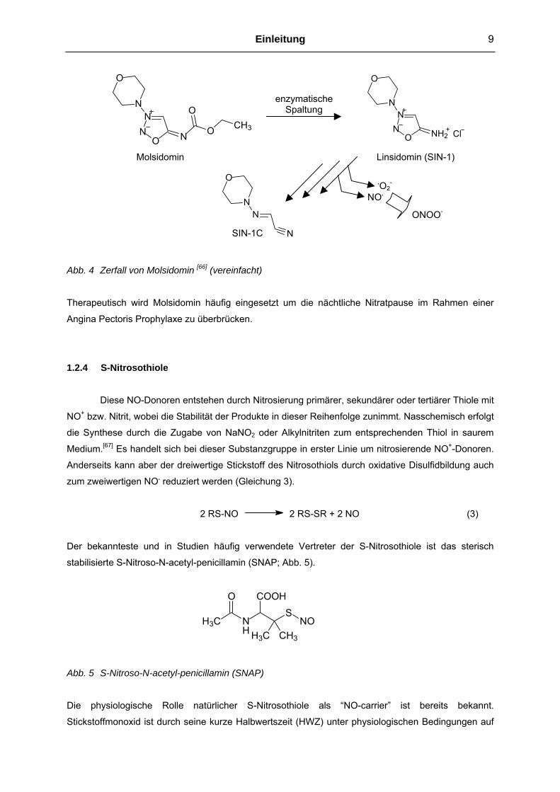
1.2.3 Sydnonimine Molsidomin ist ein Prodrug, das im Organismus durch enzymatische Abspaltung des
Ethoxycarbonyl-Restes den Metaboliten Linsidomin (SIN-1) bildet. Durch nichtenzymatische
Ringöffnung bildet sich zunächst das ringoffene Nitrosohydrazin (SIN-1A), welches dann das NO.
freisetzt.[64] Bei Molsidomin tritt keine Toleranzentwicklung auf, vermutlich weil eine Reduktion der
RNS nicht notwendig ist.[65] Aus diesem Grund wird SIN-1 häufig als biologisches “Tool“ für die
Erforschung NO. abhängiger Prozesse verwendet. Dabei ist jedoch zu beachten, dass der Zerfall
dieser Sydnonimine ebenfalls das Superoxidanion .O2- produziert, welches mit NO. die RNS
Peroxynitrit bildet.[66] Daher handelt es sich bei diesen Verbindungen streng genommen um ONOO--
wie auch NO. -Donoren.
Einleitung 9
Molsidomin
SIN-1C
NN
O
NN
O
N
O
N O CH3
ON
O
Abb. 4 Zerfall von Molsidomin [66] (vereinfacht)
Therapeutisch wird Molsidomin häufig eingeset
Angina Pectoris Prophylaxe zu überbrücken.
1.2.4 S-Nitrosothiole
Diese NO-Donoren entstehen durch Nitro
NO+ bzw. Nitrit, wobei die Stabilität der Produkte
die Synthese durch die Zugabe von NaNO2 od
Medium.[67] Es handelt sich bei dieser Substanzg
Anderseits kann aber der dreiwertige Stickstoff d
zum zweiwertigen NO. reduziert werden (Gleichu
2 RS-NO
Der bekannteste und in Studien häufig verwe
stabilisierte S-Nitroso-N-acetyl-penicillamin (SNA
H3C N
O CO
H3CH
Abb. 5 S-Nitroso-N-acetyl-penicillamin (SNAP)
Die physiologische Rolle natürlicher S-Nitr
Stickstoffmonoxid ist durch seine kurze Halbwer
enzymatische Spaltung
Linsidomin (SIN-1)
ONOO-
.O2-
NO.
N
NN
O NH2 Cl
zt um die nächtliche Nitratpause im Rahmen einer
sierung primärer, sekundärer oder tertiärer Thiole mit
in dieser Reihenfolge zunimmt. Nasschemisch erfolgt
er Alkylnitriten zum entsprechenden Thiol in saurem
ruppe in erster Linie um nitrosierende NO+-Donoren.
es Nitrosothiols durch oxidative Disulfidbildung auch
ng 3).
2 RS-SR + 2 NO (3)
ndete Vertreter der S-Nitrosothiole ist das sterisch
P; Abb. 5).
SNO
OH
CH3
osothiole als “NO-carrier” ist bereits bekannt.
tszeit (HWZ) unter physiologischen Bedingungen auf

10 Einleitung
die Stabilisierung durch niedermolekularer Peptide angewiesen.[68] Es konnte ebenso schon gezeigt
werden, dass NO. auf unterschiedliche Thiol-Gruppen tragende Moleküle übertragen werden kann um
einen Weitertransport zu gewährleisten.[69] Deshalb wird als EDRF nicht nur das NO. allein, sondern
auch seine Transportform S-Nitrosothiol angesehen.
1.2.5 Diazen-1-ium-1,2-diolate (NONOate)
Die aus medizinisch-chemischer Sicht wichtigsten Vertreter dieser Klasse sind die an
sekundäre Amine gebundenen Diazen-1-ium-1,2-diolate (kurz Diazeniumdiolate, NONOate, “solid
NO“), da nur diese in der Lage sind spontan und direkt 2 mol NO. aus einem mol NONOat
freizusetzen, im Gegensatz zu den an C-, O- oder S-Atome verknüpften NONOate. Wie die Abbildung
6 darstellt, können die Diazeniumdiolate als Salze vorliegen oder am O2-Atom kovalent an einen Rest
gebunden sein. Im Allgemeinen wird dann von einem NONOat Prodrug gesprochen, da für die NO
Liberation eine biologische oder chemische Aktivierung nötig ist. Es sei angemerkt, dass es sich
schon bei den NONOaten selbst um Prodrugs handelt, da das freigesetzte NO. das pharmakophore
Element darstellt und somit die am O2-derivatisierten Diazeniumdiolate eher als “Pre-Prodrugs“
bezeichnet werden müssen.
Abb. 6 Diazen-1-ium-1,2-diolat Bildung und NO-Freisetzung
Die Synthese und Charakterisierung der ersten Diazeniumdiolate wurde 1960 von R. Drago
beschrieben.[70] Seit der Identifizierung des NO. als EDRF haben die NONOate zunehmend an
Bedeutung gewonnen, zum einen weil sie zu den wenigen direkten NO.-Donoren gehören, zum
anderen aber auch weil sie je nach Struktur des Aminkörpers sehr unterschiedliche Halbwertszeiten
für die NO.-Freisetzung haben. Die zuverlässige und berechenbare NO.-Lieferung macht die
NONOate zu idealen Werkzeugen für die Erforschung NO-abhängiger Prozesse und Krankheiten.
Zusätzlich tritt ihr therapeutischer Nutzen als Arzneimittel immer mehr in den Vordergrund. Die für
diese Arbeit relevanten literaturbekannten Aspekte der Diazeniumdiolate sollen im Folgenden
detailliert dargestellt werden.
enzymatischer Metabolismus und/oder chemische Hydrolyse
- H+
+ H+
R1, R2 = Alkyl R3 = Alkyl, Aryl
-R3
2 NO. NR1
R2
NO
N O
R1
NHR2
NR1
R2
NO
N OR3

Einleitung 11
Synthese und Eigenschaften der Diazeniumdiolate
Die einzige brauchbare Methode Diazeniumdiolate herzustellen, ist die direkte Umsetzung
eines Amines mit 4-5 Atm NO.-Gas in einem geeigneten Lösungsmittel (Methanol, ggf. Acetonitril)
unter Ausschluss von Sauerstoff und Wasser. Um diese Bedingungen herstellen zu können, wird
üblicherweise eine Hydrierapparatur oder ein Autoklav mit Leitungen aus Edelstahl verwendet. Das
verwendete Amin dient dabei gleichzeitig als Base und es entstehen intermolekulare Ammonium
Salze. Einige Polyamine, wie das Diethylentriamin, sind in der Lage stabile intramolekulare Salze zu
bilden. Durch den Zusatz starker Basen, z.B. Natrium-methanolat, entstehen wesentlich weniger
hygroskopische Na-Salz NONOate. Abbildung 7 zeigt die Strukturformeln dieser drei möglichen
Salzarten am Beispiel des Diethylamin und Diethylentriamin als Edukte.
Abb. 7 NONOat-Salze: a) Intermolekulares Ammoniumsalz, b) Intramolekulares Salz, c) Na-Salz
In der Regel handelt es sich bei allen NONOat-Salzen um weiße Pulver, wobei einige Na-Salze bei
richtiger Aufarbeitung auch als farblose oder weiße Kristalle anfallen können. Die Identität der
Diazeniumdiolate lässt sich am besten durch 1H-NMR- (gemessen in D2O/NaOD), UV- und IR-
Spektroskopie bestimmen. Im 1H-NMR Spektrum ist, wie in Abbildung 8 beispielhaft aufgeführt, eine
Signalverschiebung der N-benachbarten CH-Protonen um 0.4-0.5 ppm, im Vergleich zum sekundären
Amin, charakteristisch. Ebenso geben die zum N-Atom β-ständigen CH-Protonen ein Signal, das in
einem geringeren Maße Tieffeld verschoben ist.
b c a
H3C N CH3
NNO
O H2N(C2H5)2
N
NNO
O
H2N NH3H3C N CH3
NNO
O Na
12 Einleitung
4.00
00
4.03
41
2.08
89
Inte
gral
2.78
48
Abb. 8 1H-NMR Spektrum mit chemischen Verschiebungen (D2O/NaOD; δ) des Piperidin im Vergleich
zum Piperidin-NONOat (PIPER/NO)
Das Absorptionsmaximum der [N(O)NO]- Gruppe im UV-Licht liegt bei 251 nm, wohingegen die
typischen IR-Valenzschwingungen bei 1225-1210 cm-1/1187-1155 cm-1 (N-O Valenz) und 1131-1129
cm-1 (N-N Valenz) Wellenzahlen liegen.[70,71] Für Reinheitsprüfungen sind Elementaranalysen
geeignet, wobei es erwähnenswert ist, dass bis heute keine universell einsetzbare HPLC Analytik für
Diazeniumdiolate zur Verfügung steht. Die beschriebene Methode von Fitzhugh et al. lässt durch die
verwendete UV-Detektion lediglich die Quantifizierung der NONOate und der entsprechenden
Nitrosamine als Verunreinigung zu. Andere Verunreinigungen, wie z.B. Natrium-methanolat oder die
Ausgangsamine, werden nicht erfasst.[72] Experimente diesbezüglich, die im Rahmen dieser Arbeit
durchgeführt wurden, sind im Kapitel 4.1.2 (Bisher unveröffentlichte Ergebnisse) aufgeführt.
Als Mechanismus der Diazeniumdiolat-Bildung sind zwei Möglichkeiten denkbar. Zum einen
die Entstehung eines NO.-Dimers, welches sich an das Amin bindet (Abb. 9, Weg A), zum anderen die
aufeinanderfolgende Anlagerung von jeweils einem NO.-Molekül (Weg B).[73,74] Da die Dimerisierung
von NO. unter normalen Reaktionsbedingungen ohnehin nicht stattfindet und die Anlagerung des
Dimers an das Amin in der Computersimulation von Dutton et al. als die endothermere Reaktion
kalkuliert wird (B3LYP-Modell), gilt der Mechanismus der sequenziellen NO.-Bindung als der
wahrscheinlichere.[73]
2.76
272.
7399
1.49
781.
4744
1.45
171.
4289
1.40
62
1.20
921.
1871
1.16
31
(ppm)1.01.21.41.61.82.02.22.42.62.8
4.00
00
6.07
78
Inte
gral
2.42
292.
4015
1.21
421.
1890
1.16
88
(ppm)1.01.21.41.61.82.02.22.42.62.8
NH
N
NNO
O Na
1.19
2.76
1.45 1.45
2.76
1.19
1.19
1.19
2.41 2.41

Einleitung 13
RR'N NN
O
ORR'N N
O
N O
RR'N H N O
RR'N N O
HN O RR'N H
NN O
O
RR'N
H
NO
N O
-H++H+
N O2
Weg B Weg A
Abb. 9 Mögliche Mechanismen der NONOat Bildung und der NO.-Freisetzung
Die treibende Kraft für diese Reaktion ist die Anwesenheit einer möglichst starken Base, um das
Gleichgewicht auf die Seite des Diazeniumdiolat-Anions zu verschieben. Bei der Synthese von
NONOaten muss auf die Abwesenheit von O2 und H2O geachtet werden, da sonst aus entstandenem
Distickstofftrioxid das entsprechende Nitrosamin und das Hydronitrit des eingesetzten Amins gebildet
wird (Gleichung 4, 5 und 6).
2 NO. + ½ O2 N2O3 + H2O 2 HNO2 (4)
RR’NH + N2O3 RR’NNO + HNO2 (5)
RR’NH + HNO2 RR’NH2+NO2
- (6)
Aus diesem Grund wird auch empfohlen die trockenen Diazeniumdiolate unter Schutzgasatmosphäre
kühl und lichtgeschützt zu lagern.[75]
NO Freisetzung aus Diazeniumdiolaten
Nach den oben genannten Modellberechnungen von Dutton et al. wird umgekehrt das N-Atom
des ursprünglichen Amins protoniert und nicht etwa eines der Sauerstoffatome. Darauf folgt die
Abspaltung des NO.-Dimers mit sofortiger Aufspaltung in zwei Moleküle monomeres Stickoxid (Abb. 9,
Weg A).[73] Für den Zerfall der NONOate ist somit der pH-Wert von entscheidender Bedeutung. Bei
einem konstanten pH-Wert zerfällt das Diazeniumdiolat nach einer Kinetik pseudo-erster Ordnung. In
Puffern verschiedener pH-Werte steigt die Geschwindigkeitskonstante des Zerfalls proportional zur
Protonenkonzentration.[71] Ebenso verkürzen sich die Halbwertszeiten der NO. Freisetzung durch
14 Einleitung
Erhöhung der Temperatur oder der Lichteinwirkung.[76] Die chemische Struktur des Amins hat nicht
nur auf die Synthesegeschwindigkeit, sondern auch auf den NONOat-Zerfall einen großen Einfluss.
Es sei darauf hingewiesen, dass bis heute keine systematischen Literaturdaten, sondern lediglich
vereinzelte Berichte über diese Zusammenhänge existieren. Beispiele sind die ultraschnelle NO-
Freisetzung bedingt durch eine Na-carboxylat Gruppe in direkter Nachbarschaft zum Amin das die -
[N(O)NO]--Gruppe bindet, sowie die teilweise sehr langsame NO-Abgabe aus zwitterionischen
Polyamin-NONOaten.[77,78]
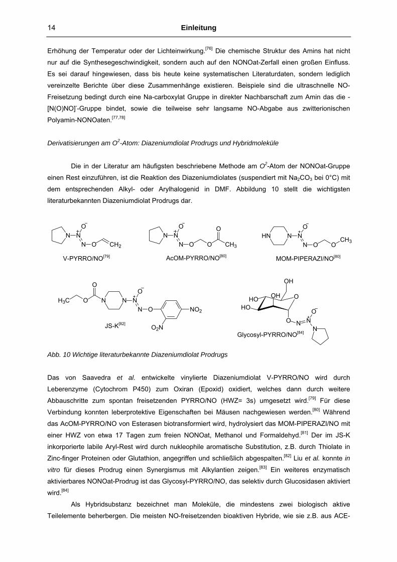
Derivatisierungen am O2-Atom: Diazeniumdiolat Prodrugs und Hybridmoleküle
Die in der Literatur am häufigsten beschriebene Methode am O2-Atom der NONOat-Gruppe
einen Rest einzuführen, ist die Reaktion des Diazeniumdiolates (suspendiert mit Na2CO3 bei 0°C) mit
dem entsprechenden Alkyl- oder Arylhalogenid in DMF. Abbildung 10 stellt die wichtigsten
literaturbekannten Diazeniumdiolat Prodrugs dar.
NO
N ON
CH2
NO
N O ON
CH3
OHN N N
O
N O OCH3
N N NO
N O NO2
O2N
OH3C
O
OOH
HOHO
OH
O N NO
]
Abb. 10 Wichtige li
Das von Saaved
Leberenzyme (Cy
Abbauschritte zum
Verbindung konnte
das AcOM-PYRRO
einer HWZ von e
inkorporierte labile
Zinc-finger Protein
vitro für dieses P
aktivierbares NON
wird.[84]
Als Hybrid
Teilelemente behe
JS-K[82
Nteraturbekannte Diazeniumdiolat Prodrugs
ra et al. entwickelte vinylierte Diazen
tochrom P450) zum Oxiran (Epoxid) ox
spontan freisetzenden PYRRO/NO (HW
n leberprotektive Eigenschaften bei Mäuse
/NO von Esterasen biotransformiert wird, hy
twa 17 Tagen zum freien NONOat, Methan
Aryl-Rest wird durch nukleophile aromatisc
en oder Glutathion, angegriffen und schließl
rodrug einen Synergismus mit Alkylantien
Oat-Prodrug ist das Glycosyl-PYRRO/NO, da
substanz bezeichnet man Moleküle, die
rbergen. Die meisten NO-freisetzenden bioa
Glycosyl-PYRRO/NO[84]
V-PYRRO/NO[79]
iumdiolat V-
idiert, welch
Z= 3s) umg
n nachgewie
drolysiert da
ol und Form
he Substituti
ich abgespal
zeigen.[83] E
s selektiv du
mindestens
ktiven Hybrid
MOM-PIPERAZI/NO[80]
AcOM-PYRRO/NO[80]PYRRO/NO wird durch
es dann durch weitere
esetzt wird.[79] Für diese
sen werden.[80] Während
s MOM-PIPERAZI/NO mit
aldehyd.[81] Der im JS-K
on, z.B. durch Thiolate in
ten.[82] Liu et al. konnte in
in weiteres enzymatisch
rch Glucosidasen aktiviert
zwei biologisch aktive
e, wie sie z.B. aus ACE-

Einleitung 15
Hemmern oder NSAID`S hergestellt wurden, enthalten als NO.-mimetische Funktion organische
Nitrate. Hybride als direkt NO.-freisetzende Diazeniumdiolate sind in der Literatur weitaus weniger oft
beschrieben. Die wichtigsten Vertreter sind NONOat-Hybride einiger nichtsteroidaler Antirheumatika,
Dihydropyridine und Heparin. Hinzukommen Polymere, die statt einer biologischen eine physikalische
Aktivität (z.B. als endoluminale Gefäßprothese) besitzen.[85-88] Den NSAID-, Heparin- und Polymer-
Hybriden gemeinsam ist das Hauptziel, einen Einfluss auf die Blutkoagulation/Plättchenaggregation zu
nehmen, auch wenn den Cyclooxygenase-hemmenden NO-Hybriden noch zahlreiche andere
denkbare Eigenschaften zugesprochen werden können. Das Hauptaugenmerk der Dihydropyridin-NO-
Hybride liegt in der zusätzlichen sGC Aktivierung und den damit verbundenen Effekten (vergl. Kapitel
1.1.2).
1.2.6 Sonstige NO-Donoren
Alle bisher nicht erwähnten NO-Donoren haben wegen ihrer teilweise hochtoxischen
Eigenschaften, chemischer Instabilität und schlechter Löslichkeit sowie komplizierten
Aktivierungsmechanismen nur noch eine untergeordnete Bedeutung. Einige weitere, aus
Forschungsaktivitäten in der medizinischen Chemie resultierenden NO-Donoren, sind in Tabelle 2 mit
der dazugehörigen Struktur, Art der freigesetzten RNS und den für die Freisetzung auslösenden
Faktoren zusammengefasst.[3]
Tab. 2 Ausgewählte NO-Donoren mit den daraus resultierenden RNS und Aktivierungsfaktoren
Name Struktur Freigesetzte RNS Auslösender Faktor
Angeli’s Salt O
N
NOO
Na
Na
NO-
NO2- Hydrolyse
Furoxane N
ON
O
RR
NO-
(durch Ox. NO.) Thiole
Oxime O2NNOH
CONH2
R R
NH2OH
(durch Ox. NO.)
O2
Eisenporphyrin
Enzymatisch
Nitrosamine NR
RN O
NO+
NO.
Licht
enzymatisch

16 Einleitung
1.3 Nachweisverfahren für Stickstoffmonoxid 1.3.1 Griess Reaktion
Das Griess Assay ist eine indirekte kolorimetrische Nachweismethode für NO. in wässrigem
Medium und beruht auf der Umsetzung von Nitrit, entstanden durch Oxidation des NO., mit primären
aromatischen Aminen (Sulfanilamid) in saurer Lösung zu einem Diazoniumsalz. Durch Diazokopplung
an Naphtylethylendiamin entsteht ein Azofarbstoff, der mittels UV-/Vis-Spektroskopie mit einer
Nachweisgrenze von etwa 0,3 µmol/l quantifiziert werden kann. Um auch das aus der NO.-Oxidation
resultierende Nitrat zu erfassen, wird der Lösung ein Überschuss Nitratreduktase zugesetzt.
1.3.2 Oxyhämoglobin Assay
Diese sehr empfindliche Methode mit einer Nachweisgrenze von etwa 1 nmol/l ist besonders
für die Stickstoffmonoxidbestimmung in biologischen Proben geeignet. Das Prinzip beruht auf der
Umsetzung von Oxyhämoglobin (HbFe2+O2) mit NO. zu Methämoglobin (HbFe3+) und NO3-. Aufgrund
der unterschiedlichen Absorptionswellenlänge von Oxy- und Methämoglobin (pH-Wert abhängig) wird
üblicherweise eine photometrische Detektion für die Quantifizierung verwendet.
1.3.3 Elektronenspin Resonanz- und Laser Magnet Resonanz Spektroskopie
Diese beiden hochempfindlichen Methoden (1nmol/l) beruhen auf dem Prinzip, dass die
ungepaarten Elektronen der paramagnetischen NO. Moleküle durch ein von außen angelegtes
Magnetfeld mit elektromagnetischer Strahlung (Mikrowellen bei der ESR-, Laserlicht bei der LMR-
Spektroskopie) in Resonanz gebracht werden. Resonanzbedingungen liegen vor, wenn durch die
variierte Magnetfeldstärke die ungepaarten Elektronen sich in ein Energieniveau (∆E) aufspalten,
dessen Energie ihrem gegensätzlichen Spin (±1/2) entspricht. Die konstante Frequenz der
eingestrahlten elektromagnetischen Welle muss der Differenz dieses aufgespalteten Energieniveaus
entsprechen. Die ESR-Spektroskopie erfasst die aufgespalteten Energieniveaus in einem niedrigen
Frequenzbereich, während bei der LMR-Spektroskopie die Energieniveaus eines höheren
Frequenzbereiches erfasst werden. Die Intensität des Resonanzsignals ist dabei proportional zur NO.
Konzentration. Um die eher schlechte Auflösung des ESR-Signals zu verbessern, werden häufig
zusätzlich Spintrap Moleküle, wie z.B. Eisen(II)-diethyldithiocarbamat (Fe2+[(C2H5)2NC(S)S-]2),
eingesetzt. Hierbei handelt es sich um diamagnetische Moleküle die mit NO. stabile, paramagnetische
Eisen-Komplexe bilden. Ein Vorteil der ESR Methode ist die Möglichkeit in vivo messen zu können,
wobei durch die nicht ausreichende Empfindlichkeit nur unphysiologisch hohe NO. Konzentrationen
detektiert werden können. Ein Vorteil der LMR Methode ist die gleichzeitige Bestimmung von 15NO.-
Isotopen.[89,90]

Einleitung 17
1.3.4 Chemilumineszenzdetektion
Bei der chemischen Umsetzung von Ozon mit Stickstoffmonoxid entsteht Sauerstoff (O2) und
angeregtes Stickstoffdioxid (NO2*), welches bei der Rückkehr in den Grundzustand (NO2) Energie in
Form von Lichtquanten emittiert, die photoelektrisch detektiert werden können. Diese sehr beliebte,
aber auch recht kostenintensive Methode hat eine sehr hohe Nachweisgrenze von etwa 1 nmol/l und
wird in N-selektiven HPLC-Detektoren (CLND) genutzt.
1.3.5 Amperometrische NO.-Detektion
NO-Elektroden sind äußerst empfindliche Sensoren für die direkte Quantifizierung von NO. in
wässrigen Medien, sowie für in vivo Untersuchungen.[91] Es handelt sich hierbei um
Kombinationselektroden, bei denen die Arbeits- und Bezugselektrode (Ag/AgCl) auf engstem Raum
Platz finden und mit einer Nachweisgrenze von 0,3 nmol/l (entspricht einem Stromfluss von 100 fA)
die empfindlichste NO. Nachweismethode darstellen. Die mangelnde Selektivität für Stickstoffmonoxid
konnte durch die Entwicklung einer selektiv gaspermeablen hydrophoben Membran ausgeglichen
werden, die Teilchen je nach Größe und Ladung selektiert. Das NO. diffundiert durch die Membran
und wird an der Anodenoberfläche zum Nitrosonium Kation (NO+) oxidiert, welches wiederum
unmittelbar mit Wasser zum Nitrit umgesetzt wird. Durch die Oxidation entsteht ein detektierbarer
Redoxstromfluss, dessen Stärke der NO. Konzentration proportional ist. Die Genauigkeit der
Messungen kann noch entscheidend verbessert werden, indem eine spezielle NO
Freisetzungskammer (NO-Chamber®; WPI), wie in Abbildung 11 dargestellt, verwendet wird.
NO-Sonde
Thermostat
EDV
Abb. 11 NO-Freisetzungskammer
18 Einleitung
Neben der Temperierbarkeit und der idealen Gefäßform ist der Hauptvorteil dieser
Freisetzungskammer die Abdichtung der Flüssigkeitsoberfläche gegenüber dem Luftraum, wodurch
die Messungen empfindlicher, reproduzierbarer und wegen der empfindlichen Messelektronik auch
robuster werden. Diese amperometrische Nachweismethode und Messanordnung fand in dieser
Arbeit Verwendung.
1.4 Charakterisierung des Azol-Antimykotikums Ketoconazol
Da in dieser Arbeit eine Reihe von Ketoconazol-NONOat und -Organonitrat Hybriden
synthetisiert, charakterisiert und biologisch getestet wurden, soll an dieser Stelle ein kurzer Überblick
über die Eigenschaften und Pharmakodynamik des Ketoconazols erläutert werden.
1.4.1 Allgemeine Eigenschaften von Ketoconazol
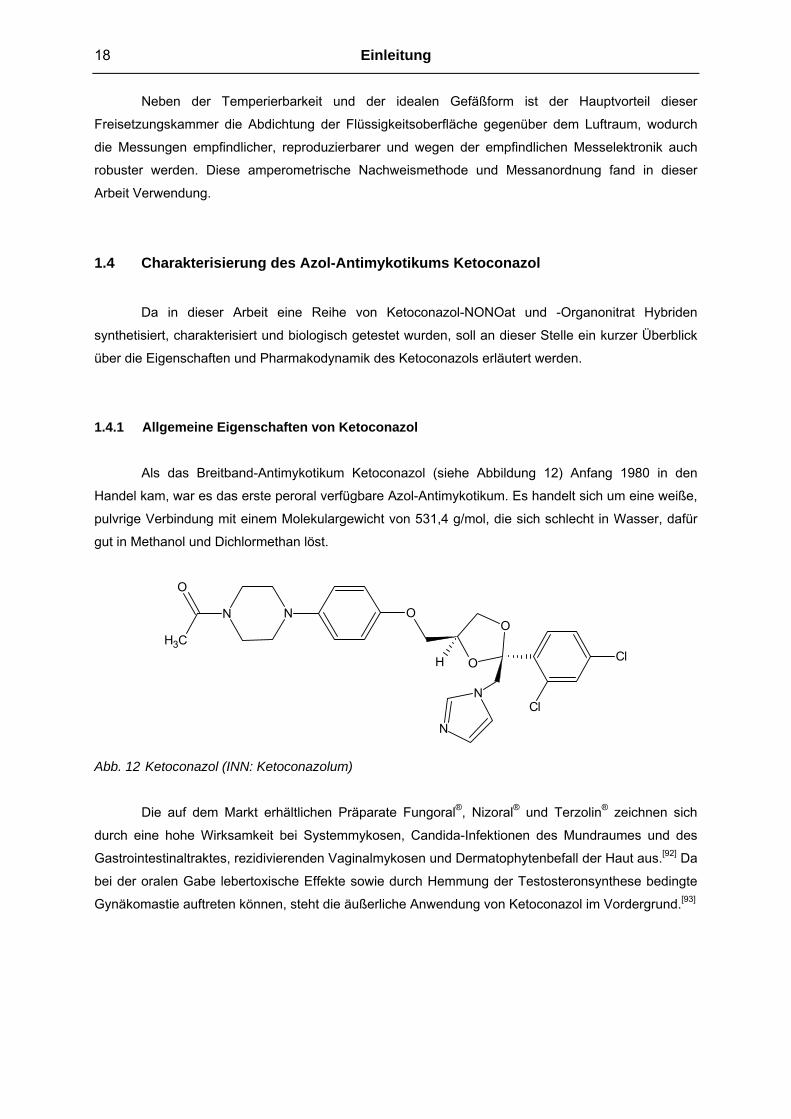
Als das Breitband-Antimykotikum Ketoconazol (siehe Abbildung 12) Anfang 1980 in den
Handel kam, war es das erste peroral verfügbare Azol-Antimykotikum. Es handelt sich um eine weiße,
pulvrige Verbindung mit einem Molekulargewicht von 531,4 g/mol, die sich schlecht in Wasser, dafür
gut in Methanol und Dichlormethan löst.
O
O
Cl
Cl
N
N
ONN
H
O
CH3
Abb. 12 Ketoconazol (INN: Ketoconazolum)
Die auf dem Markt erhältlichen Präparate Fungoral®, Nizoral® und Terzolin® zeichnen sich
durch eine hohe Wirksamkeit bei Systemmykosen, Candida-Infektionen des Mundraumes und des
Gastrointestinaltraktes, rezidivierenden Vaginalmykosen und Dermatophytenbefall der Haut aus.[92] Da
bei der oralen Gabe lebertoxische Effekte sowie durch Hemmung der Testosteronsynthese bedingte
Gynäkomastie auftreten können, steht die äußerliche Anwendung von Ketoconazol im Vordergrund.[93]

Einleitung 19
1.4.2 Pharmakologie von Ketoconazol
Wie alle Azol-Derivate gehört Ketoconazol zu den Hemmstoffen der Ergosterolbiosynthese,
dem Cholesterin-Äquivalent des Plasmalemms der Pilze. Ein Zwischenschritt dieser Biosynthese ist
die Demethylierung von Lanosterin zu 14-Desmethyl-Lanosterin, katalysiert durch ein Cytochrom
P450 abhängiges Enzym, der 14-α-Demethylase. Dessen Hemmung erfolgt durch die Bindung des
Imidazol-Restes von Ketoconazol an das Häm-Eisen der Cytochrom P450 (siehe Abbildung 13).[94]
Squalen
14-Desmethyl-Lanosterin
Lanosterin
14-α-Demethylase
N
N
R
Häm-Fe
Cyt-P450
Ergosterol
Abb. 13 Hemmung der Ergosterolbiosynthese durch Azol-Antimykotika
In der Fachliteratur werden Azol-Antimykotika aufgrund dieser Pharmakodynamik auch als
DMI’s (demethylation-inhibiting fungicides) bezeichnet. Die Einlagerung der falschen Sterole in die
Zellwand wirkt auf das Pilzwachstum fungistatisch, verursacht durch Stabilitätsstörungen der
Membran und durch die Beeinträchtigung der Funktion membranständiger Enzyme. In sehr hohen
Dosierungen kommen durch die direkte Einlagerung der Azole in das Plasmalemm und dem damit
verbundenen Austritt essentieller Zellbestandteile auch fungizide Effekte zustande.[93] Durch die
lipophile Struktur und die sehr hohe Plasma-Eiweißbindung beträgt die
Plasmaeleminationshalbwertszeit des Ketoconazol 7-10 Stunden.
1.5 Problemstellung und Ziele der Arbeit 1.5.1 Chemische Aspekte
An Stickstoff gebundene Diazen-1-ium-1,2-diolate (NONOate) stellen die einzige chemische
Verbindungsklasse dar, deren Derivate in der Lage sind, ohne enzymatische Bioaktivierung und ohne

20 Einleitung
die Freisetzung anderer „reactive nitrogen species“ (RNS), den physiologisch außerordentlich
bedeutsamen Botenstoff Stickstoffmonoxid (NO.) freizusetzen. Nachteile dieser Verbindungsklasse
sind nicht nur eingeschränkte Stabilität gegenüber Hitze, Feuchtigkeit und Licht, sondern auch
mangelnde Steuerung und Kontrollmöglichkeiten bei der Synthese aus Aminen und 4-5 Atmosphären
gasförmigem Stickstoffmonoxid im Autoklaven. Als Beispiel sei genannt, dass es bisher nicht möglich
ist, den individuellen Reaktionsendpunkt zu erkennen. Weiterhin gibt es praktisch keine
Aufreinigungsmöglichkeiten der Produkte, was wiederum erhöhte Anforderungen an die Einheitlichkeit
der Synthese stellt. So ist vor allem die Bildung von Nitrosaminen zu verhindern, die z.T. hohes
karzinogenes Potential besitzen. Neue Möglichkeiten der Synthesesteuerung sollte ein in unserer
Arbeitsgruppe neu entwickeltes und in der vorliegenden Dissertation weiterentwickeltes Gerät, der
“NOtizer®“ sein, dessen Kernstücke eine durchsichtige Reaktionskammer, ein Druckaufnahmesensor,
eine entsprechende EDV-Schnittstelle und eine Software sind, mit welcher der Umsetzungsverlauf
online verfolgt werden kann. Auf diese Weise können nicht nur die Reaktionsendpunkte der Synthese
erkannt werden, sondern auch die Einflüsse von strukturellen Besonderheiten der eingesetzten
Amine, der Reaktionstemperatur, dem Druck, dem Lösungsmittel und der Temperatur. Konkret sollten
die folgende Fragestellungen und thematischen Schwerpunkte in der vorliegenden Arbeit untersucht
werden.
1. Gibt es Zusammenhänge zwischen der Struktur des eingesetzten Amines und der
Bildungsgeschwindigkeit des resultierenden NONOates?
In der Literatur beschriebenen NONOat-Synthesen wurden ohne Rücksicht auf die Struktur des
eingesetzten Amines in etwa gleiche, vermutlich meistens zu lange, möglicherweise in Einzelfällen
aber auch zu kurze Reaktionszeiten festgesetzt. Mit dem NOtizer lässt sich die
Reaktionsgeschwindigkeit, wie auch der Reaktionsendpunkt anhand der aufgenommenen
Druckzeitkurve erkennen und damit auch die Reaktivität der unterschiedlichen Amine gegenüber
Stickstoffmonoxid ermitteln. Durch die Aufnahme von Druckzeitkurven bei der Umsetzung einer
sinnvollen Auswahl und nicht zu kleinen Anzahl von Aminen könnten die Zusammenhänge zwischen
Struktur und Reaktionsgeschwindigkeit erkannt werden.
2. Welche Abhängigkeiten bestehen zwischen der Bildungsgeschwindigkeit der NONOate und
Parametern wie Reaktionstemperatur, NO-Druck, Lösungsmittel und Natur der zugesetzten
Base?
Auch diese Zusammenhänge sollten durch systematische Variation der oben genannten Parameter
und Aufnahme der Druckzeitkurven analysiert und charakterisiert werden.
3. Welche Zusammenhänge gibt es zwischen Struktur des NONOates und der
Freisetzungsgeschwindigkeit von Stickstoffmonoxid aus ihnen?

Einleitung 21
In der Literatur ist verschiedentlich über die Abhängigkeit der NO-Freisetzung aus Diazeniumdiolaten
von pH-Wert, Temperatur und auch Struktur des Diazeniumdiolats berichtet worden. Nach der
ohnehin geplanten Synthese einer Vielzahl von Diazeniumdiolaten kann das Freisetzungsverhalten
auf einer sehr viel breiteren Basis und überdies mit einer Methode und “in einer Hand“ durchgeführt
werden. Es soll eine amperometrische Messmethode für NO (direkter NO-Nachweis) für diese Zwecke
herangezogen werden und diese mit der anerkannten Methode des UV-spektroskopisch bestimmten
Zerfalls der Diazeniumdiolate (indirekter Nachweis) kombiniert und die Ergebnisse verglichen und
gegenseitig gesichert werden.
4. Welche Zusammenhänge gibt es zwischen der Bildungsgeschwindigkeit von
Diazeniumdiolaten und den NO-Freisetzungsprofilen aus ihnen?
Alle aus den Arbeiten zu 1. bis 3. erhaltenen Ergebnisse sollten die Analyse solcher Zusammenhänge
ermöglichen. Grundfragen wären, ob schnell gebildete Diazeniumdiolate auch schnell zerfallen, ob es
genau umgekehrt ist oder ob kein systematischer Zusammenhang zwischen Bildung und Zerfall
besteht.
5. Synthese von Diazeniumdiolat-Prodrugs (”NO-preprodrugs“).
Die grundsätzliche Instabilität und der spontane pH-abhängige und dann weiter nicht mehr zu
steuernde Zerfall von Diazeniumdiolaten kann durch die Synthese von Diazeniumdiolat-prodrugs
begegnet werden. Diese sind grundsätzlich Verbindungen, die am O2-Atom des Diazeniumdiolats
alkyliert, möglicherweise auch acyliert sind. Der spontanen Freisetzung von NO aus
Diazeniumdiolaten wird dadurch ein chemischer (z.B. Hydrolyse), häufig auch enzymatisch
gesteuerter NO-Freisetzungsschritt voran gesetzt. Es handelt sich hier um eine wertvolle, fast
unerlässliche Strategie zu einer pharmakokinetisch steuerbaren und Target-orientierten NO-
Freisetzung.
6. Synthese von Stickstoffmonoxid freisetzenden Hybridverbindungen.
Ein weiterer Schwerpunkt ist die Herstellung von Stickstoffmonoxid freisetzenden Hybriden, d.h.
Substanzen, die bereits eine Arzneimittelwirkung haben, zusätzlich jedoch in der Lage sind, NO
abzugeben. Um die Stabilität der Substanzen zu gewährleisten und um das in vivo oder in vitro
angezielte Target möglichst hohen Konzentrationen des Arzneistoffs auszusetzen, ist auch hier die
Umsetzung der Diazeniumdiolat-Hybride zu Prodrugs unumgänglich. Da die physiologische Wirkung
von NO. überwiegend das Herz-Kreislauf-, aber auch das Immun- und zentrale Nervensystem
reguliert, sowie antiinfektive und antimykotische Eigenschaften besitzt, sind Arzneistoffe wie
Betablocker, Dihydropyridine, Antibiotika, Virustatika, Antimykotika, Lipidsenker und Neuroleptika
geeignete Grundkörper für die Synthese von NONOat-Hybriden. Für die Hybridsubstanzen wurde als
Forschungsschwerpunkt das antimykotisch wirksame Ketoconazol ausgewählt und entsprechende
Referenzsubstanzen ohne den gesamten Arzneistoffgrundkörper sollen synthetisiert werden. Um
22 Einleitung
einen Vergleich zwischen Hybridmolekülen mit unterschiedlichen NO freisetzenden Gruppen machen
zu können, sollen neben den NONOat-Hybriden und NONOat-Hybrid-Prodrugs auch Organonitrat-
Hybride hergestellt werden.
1.5.2 Biologische Aspekte
Ausgewählte Diazeniumdiolate, Diazeniumdiolat-Prodrugs, Diazeniumdiolat-Hybride und
Diazeniumdiolat-Prodrug-Hybride sollen in vitro und in vivo auf ihre biologischen Aktivitäten untersucht
werden. Dies erfordert in hohem Maße Kooperation innerhalb der eigenen Arbeitsgruppe, vor allem
aber mit anderen Arbeitskreisen. Die beobachteten biologischen Effekte sollen dabei in Bezug auf die
speziellen chemischen Charakteristika, wie z.B. Aktivierbarkeit oder NO-Freisetzungsprofil, der
jeweiligen Verbindung diskutiert werden. Die Schwerpunkte liegen bei folgenden Fragestellungen:
7. Untersuchung der vasodilatorischen Potenz von NONOaten und NONOat-Prodrugs an
isolierten Pulmonalarterien des Schweins.
Als Kooperationspartner stehen hier Herr Apotheker Andreas König und Frau Dipl. pharm. Caro
Roegler im eigenen Arbeitskreis, sowie PD Dr. Andreas Daiber, Klinikum der Johannes Gutenberg-
Universität Mainz, II. Medizinische Klinik, Kardiologie.
8. Einfluss von NONOaten und NONOat-Prodrugs auf die Durchlässigkeit der Bluthirnschranke
in einem in vitro Modell mit kultivierten cerebralen Endothelzellen.
Diese Untersuchungen erfolgen im Rahmen des Alzheimer-Verbundprojektes an der FSU Jena. Die
Durchlässigkeit der Bluthirnschranke stellt tatsächlich ein wesentliches Kriterium für potentielle
Alzheimertherapeutika dar. Das Projekt soll in Kooperation mit der Arbeitsgruppe von Herrn Prof.
Alfred Fahr und seinem Mitarbeiter Herr Apotheker Sven Winter, Pharmazeutische Technologie,
Institut für Pharmazie, Jena, durchgeführt werden.
9. In vivo Untersuchungen des Einflusses von NONOaten und NONOat-Prodrugs auf
neuroregenerative Prozesse nach experimentell induzierten Hirninfarkten bei Ratten.
Auch dieses Projekt ist im Rahmen des Alzheimer-Verbundprojektes zu sehen. Als
Kooperationspartner steht die Arbeitsgruppe von Herrn PD Dr. Redecker, Herrn Prof. Witte und Frau
Dr. Silke Keiner, Universitätsklinikum Jena, zur Verfügung.
10. Antimykotische und antibakterielle Wirkprofile verschiedener Ketoconazol-Diazeniumdiolat-
Hybride und verwandter Strukturen.
Dieses Projekt wird in Zusammenarbeit mit dem Hans-Knöll-Institut Jena unter der Leitung von Herrn
Prof. Axel Brakhage und Frau Dr. Ute Möllmann bearbeitet.
Übersicht zu den Veröffentlichungen 23
2 Übersicht zu den Veröffentlichungen In der folgenden Übersicht von insgesamt sechs Veröffentlichungen, bestehend aus fünf
experimentellen Arbeiten und einem Patent, tritt folgender Eigenanteil auf:
- als Erstautor: In den Veröffentlichungen 1, 2, 3 und 6 habe ich die Ausführung und Bewertung
der beschriebenen Synthesen und die NO-analytischen Messungen durchgeführt, sowie die
jeweiligen Manuskripte erstellt.
- Als Koautor: In Veröffentlichung 4 und 5 habe ich die Synthese und NO-Analytik der
beschriebenen Diazeniumdiolate und Diazeniumdiolat-Prodrugs durchgeführt, sowie an der
Erstellung der Manuskripte mitgewirkt.
2.1 Veröffentlichung 1 The NOtizer- A Device for the Convenient Preparation of Diazen-1-ium-1,2-diolates. Konter, Joerg; Abuo-Rahma, Gamal El-Din Ali Ahmed Hassan; El-Emam, Ali; Lehmann, Jochen.
Methods in Enzymology (2005), 396 (Nitric Oxide, Part E), 17-26.
In dieser Veröffentlichung wird die neu entwickelte Apparatur für die effiziente und qualitative
Synthese von Diazen-1-ium-1,2-diolaten mit dem Namen “NOtizer®“ vorgestellt. Neben dem
apparativen Aufbau werden detaillierte Synthesevorschriften für die effektive Herstellung
literaturbekannter NONOate und deren Eigenschaften bezüglich Reaktivität, NO.-Freisetzung und
Lagerung beschrieben. Probleme die bei früheren Herstellungsprozeduren auftreten, wie die
Entstehung von Nebenprodukten oder Ausbeuteeinbußen, konnten durch die Entwicklung dieser
Methode minimiert werden.
2.2 Veröffentlichung 2 Synthesis of Diazen-1-ium-1,2-diolates Monitored by the "NOtizer" Apparatus: Relationship between Formation Rates, Molecular Structure and the Release of Nitric Oxide. Konter, Joerg; Abuo-Rahma, Gamal El-Din A. A.; El-Emam, Ali; Lehmann, Jochen. European Journal
of Organic Chemistry (2007), 4, 616-624.
In dieser Studie werden erstmals systematisch Daten über die Reaktionsverläufe
verschiedener NONOat-Synthesen beschrieben und miteinander verglichen. Um eine möglichst große
Vielfalt an Strukturvariationen zu erhalten, die einen Zusammenhang zwischen der Aminstruktur und
dem ermittelten kinetischen Datenmaterial aus dem Syntheseverlauf erkennen lassen, wurden
insgesamt 22 Diazeniumdiolate mit unterschiedlichen Alkyl-, Heteroalkyl-, Benzyl- und substituierten
zyklischen Resten hergestellt. Neben dem Struktureinfluss auf die Reaktionsgeschwindigkeit, wird
24 Übersicht zu den Veröffentlichungen
dieser Einfluss auch hinsichtlich der NO.-Liberation aus den NONOaten beschrieben. Schließlich wird
ein Zusammenhang zwischen den ermittelten Daten aus Synthese und NO.-Freisetzung
zusammengefasst.
2.3 Veröffentlichung 3 NO Donors, part 17: Synthesis and Antimicrobial Activity of Novel Ketoconazole-NO-Donor Hybrid Drugs and Corresponding Nitric Oxide Donors. Konter, Joerg; Möllmann, Ute; Lehmann, Jochen. Journal of Medicinal Chemistry. In Vorbereitung.
Die Synthese und biologische Aktivität neuartiger NO-freisetzender Ketoconazol Derivate
sowie der analogen NO-Donor Derivate sind Gegenstand dieser Veröffentlichung. Während die
Hemmung des Bakterienwachstums bei den untersuchten Derivaten nur geringe Unterschiede
aufwies, konnte für die Inhibition des Pilzwachstums ein Zusammenhang zu der verwendeten NO-
Donor Spezies hergestellt werden. Hierbei zeigten insbesondere die Ketoconazol-Hybride mit
Organonitrat- und Diazeniumdiolat-Prodrug-Gruppe eine höhere Aktivität und/oder verändertes
Wirkspektrum als das Ketoconazol allein.
2.4 Veröffentlichung 4 Potency and In Vitro Tolerance of Organic Nitrates: Partially Denitrated Metabolites Contribute to the Tolerance-Devoid Activity of Pentaerythrityl Tetranitrate. Koenig, Andreas; Lange, Kathrin; Konter, Joerg; Daiber, Andreas; Stalleicken, Dirk; Glusa, Erika;
Lehmann, Jochen. Journal of Cardiovascular Pharmacology (2007), 50, 68-74.
Veröffentlichung 4 behandelt die in vitro Aktivität therapeutisch relevanter organischer Nitrate,
insbesondere die des Petaerythrityltetranitrat (PETN) und seiner in vivo nachgewiesenen Metabolite
PEtriN, PEdiN und PEmonoN. Ebenso wird die vasodilatorische Potenz des S-Nitroso-N-acetyl-
penicillamin (SNAP) und des Diazeniumdiolates PHEPIPERAZI/NO an dem verwendeten in vitro
Modell ermittelt. Die beim PETN und PEtriN signifikante, jedoch beim PEdiN, PEmonoN, SNAP und
PHEPIPERAZI/NO nur gering ausgeprägte Tachyphylaxie (in vitro) wird ausführlich beschrieben. Die
Ergebnisse dieses Forschungsprojekts ergaben die Hypothese, dass die in vivo fehlende Nitat-
Tachyphylaxie des PETN mit der Tatsache zusammenhängt, dass lediglich die Metabolite PEdiN und
PEmonoN bioaktiv sind, jedoch das PETN und PEtriN aufgrund fehlender Resorption ausgeschieden
werden.

Übersicht zu den Veröffentlichungen 25
2.5 Veröffentlichung 5 Permeability changes in response to NONOate and NONOate prodrug derived nitric oxide in a blood-brain barrier model formed by primary porcine endothelial cells. Winter, Sven; Konter, Joerg; Scheler, Stefan; Lehmann, Jochen; Fahr, Alfred. Nitric Oxide. Eingereicht
(2007).
In dieser Arbeit werden drei direkt NO.-freisetzende NONOate (PHEPIPERAZI/NO, DBA/NO,
DETA/NO) und drei NONOat-Prodrugs (Et-PHEPIPERAZI/NO, Tosyl-PYRRO/NO, Et-
BUPIPERAZI/NO) hinsichtlich ihres Einflusses auf die Durchlässigkeit der Bluthirnschranke in einem
in vitro Modell untersucht. Die Permeabilität der Markersubstanz Carboxyfluorescein wird durch die
direkten NO-Donoren verringert, während die Permeabilität bei den NONOat-Prodrugs Et-
PHEPIPERAZI/NO und Tosyl-PYRRO/NO zunimmt. Das Et-BUPIPERAZI/NO nimmt in Bezug auf die
Veränderung der Durchlässigkeit der Bluthirnschranke eine Mittelstellung ein. Mit Methylenblau
konnten die Permeabilitätsabnehmenden Effekte blockiert werden, die Permeabilitätssteigernden
jedoch nicht.
2.6 Veröffentlichung 6 Neue Ketoconazol-Derivate sowie deren Herstellung und Verwendung als antimikrobielle und antiinflammatorische Wirkstoffe. Konter, Joerg; Möllmann, Ute; Lehmann, Jochen. Eingereicht beim Deutschen Patent- und Markenamt
(25.07.2007).
Diese Offenlegungsschrift schützt rechtlich eine der in Veröffentlichung 3 beschriebenen
Ketoconazol-NO-Donor Hybridverbindungen, sowie die entsprechenden Analogderivate. Der Vorteil
dieser Erfindung ist die variable NO-Freisetzungskinetik gegenüber anderen antimykotisch wirksamen
Hybridverbindungen durch Veränderungen an der Diazeniumdiolat-Teilstruktur. Antibakterielle und
antiinflammatorische Wirksamkeit konnten dem genannten Ausführungsbeispiel ebenfalls
nachgewiesen werden.

3. Veröffentlichungen

Veröffentlichung 1 26-36
Veröffentlichung 1
The NOtizer- A Device for the Convenient Preparation of Diazen-1-ium-1,2-diolates
Konter, Joerg; Abuo-Rahma, Gamal El-Din Ali Ahmed Hassan; El-Emam,
Ali; Lehmann, Jochen. Methods in Enzymology (2005), 396 (Nitric Oxide,
Part E), 17-26.

Yoshimura, T. (1984). Reaction of nitrosylporphyrinatoiron (II) with nitrogen oxide. Inorg.
Chim. Acta 83, 17–21.Young, C. L. (1981). Oxides of nitrogen. In ‘‘Solubility Data Series’’ (R. Battino, H. L.
Clever, and C. L. Young, eds.) Vol. 8. Pergamon Press, New York.
[2] the NOtizer 17
[2] The NOtizer—A Device for the ConvenientPreparation of Diazen-1-ium-1,2-diolates
By JOERG KONTER, GAMAL EL-DIN ALI AHMED
HASSAN ABUO-RAHMA, ALI EL-EMAM, and JOCHEN LEHMANN
Y AbstractN-bound diazen-1-ium-1,2-diolates, also known as NONOates or ‘‘solidnitric oxide’’ (NO), have become popular tools in biomedical researchsince the discovery of NO as a very important multifunctional endogenousmessenger. In contrast to other well-known NO donors, NONOates arecapable of releasing NO spontaneously in aqueous media. The rate of NOliberation is determined by the molecular structure of the diazeniumdiolateand the pH value and temperature of the medium in which it is dissolved.
In this chapter, we introduce a novel device (the NOtizer) for simpleand convenient preparation of diazeniumdiolates. It not only enables theuser to provide all the necessary conditions for reliable synthesis such asanaerobic conditions and high pressure of NO gas in the translucentreaction chamber but also includes software that records the course ofpressure and temperature online and calculates the consumption of NOby the reaction. The plot of the pressure decay shows the user completionof the reaction and allows the user to study kinetic characteristics fromsynthesis of different NONOates. A brief guide for the synthesis ofPYRRO/NO, DEA/NO, PAPA/NO, SPER/NO, and DETA/NO, whichare the most widely applied diazeniumdiolates, is presented in this chapter.Finally, characteristics of NONOates that need to be consideredconcerning analytics and storage are mentioned.
Introduction
1-Substituted diazen-1-ium-1,2-diolates, also known as NONOates, arecompounds of the general structure X-[N(O)NO]�. According to the atomX, diazeniumdiolates can be classified in three major classes: C-,O-/S-, andN-bound diazeniumdiolates. Because the N-bound NONOates can directlyrelease the endogenous mediator nitric oxide (NO), they are interesting
AUTHOR'S PERSONAL COP
METHODS IN ENZYMOLOGY, VOL. 396 0076-6879/05 $35.00Copyright 2005, Elsevier Inc. All rights reserved. DOI: 10.1016/S0076-6879(05)96002-3

18 biochemical, molecular, and real-time detection of nitric oxide [2]
compounds from a biomedical point of view. One mole of diazeniumdio-late is capable of generating 2 mol of NO by acid-catalyzed dissociationaccording to Eq. (1) (Keefer et al., 1996).
ð1Þ
Organic nitrates are generally declared to be ‘‘NO donors,’’ but incontrast to diazeniumdiolates, they need to be bioactivated. The cor-responding bioactivation processes are not yet completely understood,and the resulting bioactive species are still under discussion (Chen et al.,2002; Chung and Fung, 1990). Nitrosothiols [e.g., S-nitroso-N-acetyl-penicillamine (SNAP)] can be considered NOþ donors rather than NOdonors. In contrast to these compounds, N-diazeniumdiolates haveproven to be excellent tools to demonstrate the physiological propertiesof NO in vitro and have gained importance as potential drugs for in vivoapplication. Innovative work on the development of novel NONOates andNONOate prodrugs, such as the liver-selective NO donor prodrug V-PYRR O/NO, has been pe rformed by Saavedra et al. (1997) and othershave reported on the development of thromboresistant NO-releasing poly-meric coatings (Batchelor et al., 2003; Keefer, 1998; Maragos et al., 1991,1993; Saavedra et al., 1992, 1997, 2001; Woodward and Wintner, 1969;Zhang et al., 2003).
The most useful preparation of N-diazeniumdiolates is the direct expo-sure of a secondary amine, acting as a nucleophile, to several atmospheresof gaseous NO under anaerobic conditions in a suitable solvent and addedbase to keep the product in a stable anionic form (Keefer and Hrabie, 2002;Longhi et al., 1962). We now introduce a novel device (NOtizer) thatallows the convenient preparation of diazeniumdiolates, the monitoringof NO consumption, temperature pattern, and thus kinetic studies on theNONOate formation.THOR'S PERSONAL COPY
U
NONOates as NO Donors in Biological SystemsThe rate of NO release is determined by the molecular structure of therespective NONOate, pH value, and temperature of the solution in whichthe compound is dissolved. Concerning pH, the decomposition of diaze-niumdiolates proceeds extremely slowly at a pH value of more than 9, at amoderate rate at physiological pH values, and almost instantaneously atacetic pH values (Davies et al., 2001; Keefer et al., 1996). Depending on the
A

[2] the NOtizer 19
structure of the parent nucleophile, the half-life of diazeniumdiolatesranges from a few seconds to several days, which grants access to versatilesources of NO adjustable to the requirements of the experiment (Lehmannet al., 2002; Thomas et al., 2002). The structures of the most intensivelydescribed diazeniumdiolates and their corresponding half-lives are given inTable I (Hrabie et al., 1993; Keefer et al., 1996; Maragos et al., 1991;Saavedra et al., 1997; Thomas et al., 2002). Because the NO releasing rateof diazeniumdiolates may vary because of different conditions of theexperiment, one should measure the NO release under study ratherthan rely on half-lives given in literature (Keefer et al., 1996; Kronckeand Kolb-Bachofen, 1999).
TABLE I
STRUCTURE AND HALF-LIVES OF THE MOST ESTABLISHED NONOATES
Name
Corresponding
amine Structure
Half-life
(37�,pH 7.4)
PYRRO/NO Pyrrolidine 3 s
DEA/NO Diethylamine 2–4 min
PAPA/NO N-propyl-1,3-
propane-
diamine
15 min
SPER/NO Spermine 40 min
DETA/NO Diethylene-
triamine
20 h
AUTHOR'S PERSONAL COPY

20 biochemical, molecular, and real-time detection of nitric oxide [2]
In addition to other advantages of diazeniumdiolates as direct NOdonors (‘‘solid NO’’), NO production in vitro is not influenced by catalyticcopper or iron ions, which guarantees the predictability and reproducibilityof NO release. Besides delivering NO, NONOates themselves interact verylittle with other compounds in media, but nevertheless pharmacologicalinvestigations should go along with control experiments, using the aminesresulting by cleavage of the respective diazeniumdiolates (Keefer et al.,1996; Kroncke and Kolb-Bachofen, 1999; Thomas et al., 2002). For exam-ple, the physiologically important polyamine spermine is the resultingnucleophile generated by dissociation of SPER/NO (Keefer et al., 1996;Maragos et al., 1993).
Y Synthesis of Diazen-1-ium-1,2-diolates Using the NOtizerIn the 1960s, Drago and Paulik (1960) and Drago and Karstetter (1960)reported on the reaction of NO with anhydrous diethylamine dissolved inether at a temperature of �78� using a three-necked flask and passing NOthrough the reaction mixture. In the 1990s, when the role of endogenousNO was recognized, interest in NO donors increased and the synthesis ofdiazeniumdiolates was reinvestigated and improved, mainly by Keefer,Saavedra, Hrabie, and others. These authors used a glass bottle placed ina standard hydrogenation apparatus with a pressure of up to 5 atm NO atroom temperature (Lehmann, 2000; Maragos et al., 1991).
To shift the equilibrium toward the product and to keep the evolv-ing diazeniumdiolate in the stable anionic form, the presence of base isnecessary (Keefer et al., 1996). Usually, equimolar quantities of sodium orpotassium methylate are added. Dry methanol or mixtures of methanol andether can be considered solvents of first choice. It is important to considerthat ethanol (specifically ethoxide), under alkaline conditions, reacts withNO to yield undesired products by Traube’s degeneration (Keefer et al.,2001; Traube, 1898; Woodward and Wintner, 1969). A study by Arnoldet al. (2002) shows that commonly used acetonitrile is not recommendable,because it reacts with NO, yielding a C-bound polydiazeniumdiolate asbyproduct. Because oxygen or water will lead to the corresponding ammo-nium nitrite salts or even toxic nitrosamines, dry solvents and anaerobicconditions are obligatory (Bonner, 1996; Hansen et al., 1982; Keefer, 1998;Masuda et al., 2000; Srinivasan et al., 2001).
Together with the Hyscho company, Bonn, Germany, we developed theNOtizer (Fig. 1), which is a modified hydrogenation apparatus.
As illustrated in Fig. 1, the NOtizer consists of a translucent pressurevessel as reaction chamber, stirrer, protective grating, regulator valves,indicators for pressure and temperature, and a computer with the
AUTHOR'S PERSONAL COP

FIG. 1. The NOtizer for the preparation of diazeniumdiolates.
[2] the NOtizer 21
ONAL COPY
appropriate software to record the data obtained. The computer can plotthe numerical values of pressure and temperature changes and calculatethe consumption of NO in the course of the synthesis. Particularly, thepressure decay curves are of interest, because they inform about theprogression of the synthesis and can be fitted with appropriate software.The fit of these curves permits the user to gain information aboutthe reaction rate, the kinetic rate constant, and the kinetic order of thereaction.
To demonstrate the benefits of the NOtizer, we have synthesized thediazeniumdiolates displayed in Table I. Exactly 0.05 mol of the respectiveamines was assorted with dry methanol to give a final volume of 100 ml.Before adding the solvent to the amine, 0.05 mol of elementary sodium wasdissolved in the methanol to produce the necessary base. The vessel wasplaced in the NOtizer, evacuated, and filled with nitrogen four consecutivetimes, and then it was evacuated again and NO was administered to give apressure of 4 bar. The recording of the reaction was ended when no moredecrease of pressure occurred. All reactions were performed at roomtemperature, and the (either immediately or after concentration) precipi-tated diazeniumdiolates were obtained by suction filtration and washedwith ether. To compare the reaction rate of the respective amines, one cansuperpose the pressure decay curves (Fig. 2).
AUTHOR'S PERS

FIG. 2. Pressure decay curves for the reaction of diethylamine, N‐propyl‐1,3‐propanedia-mine, diethylenetriamine, spermine, and pyrrolidine with nitric oxide.
22 biochemical, molecular, and real-time detection of nitric oxide [2]
SONAL COPY
The pressure decay curves in Fig. 2 show that steric and electroniceffects have great influence on the reaction rate. For example, in reflectionto the course of pressure decline, diethylamine shows a much slowerreaction in comparison to pyrrolidine. These attained curves can be fittedwith the appropriate computer software to reveal clues about the kineticproperties of the respective reaction.
It is noteworthy that if only formation of the product is desired, theaddition of NO to give a pressure of 4–5 atm should be repeated until noappreciable amount of NO is consumed by the reaction, thus resulting in ahigher yield. Figure 3 demonstrates how the consumption of NO decreaseseach time the pressure is raised back to 4.0 bar.
Because the software of the NOtizer also records the temperaturepattern, we could demonstrate that formation of diazeniumdiolates is anexothermic reaction. Furthermore, it is possible to analyze the changes inreaction rate that occur with changes in temperature. Figure 4 shows thesuperposed pressure decay curves for the synthesis of PYRRO/NO at roomtemperature and under cooled conditions to point out this aspect.
As is expected from the collision theory, under ice-cold conditions thereaction rate slows and the yield of the product is slightly lower. It is
AUTHOR'S PER

FIG. 3. Pressure decay curve for the synthesis of SPER/NO when inflowing NO
repeatedly.
[2] the NOtizer 23
SONAL COPY
noteworthy that no improvement in quality or purity was attained at lowtemperature. PER
Characteristics of Diazeniumdiolates to be ConsideredTo confirm quality and purity of the synthesized NONOates, nuclearmagnetic resonance (NMR) spectroscopy and elemental analysis are themost reliable techniques. A suitable solvent for the NMR spectroscopicanalysis is D2O with NaOD for alkaline conditions in which the compoundsare kept in their stable anionic form. Using 1H-NMR spectroscopy, thesignal of the protons bound to the alpha-C-atoms shows a downfield shiftusually about 0.4–0.5 ppm compared to the amine. Another criterion for thequality of the formed product is, of course, that NO is released by thecompound when dissolved in aqueous media. A commercially availableNO-selective sensor can be used to determine the amount of NO beingreleased. If the compound is pure, 1 mol of NONOate will generate 2 molof NO. Because of the volatility of NO from solution, this method is not veryconvincing for the evaluation of purity (Thomas et al., 2002). A superiormethod for quantitative acquisition of NO from solution is laser magnetic
AUTHOR'S

FIG. 4. Pressure decay curves when synthesizing PYRRO/NO at room temperature and
under cooling.
24 biochemical, molecular, and real-time detection of nitric oxide [2]
SONAL COPY
resonance spectroscopy (LMRS) because NO is detected in its gaseous formand accumulatively over longer periods (Lehmann et al., 2002).
Concerning the storage of diazeniumdiolates, Keefer et al. (1996) rec-ommend storage in a refrigerator at 4� under dry nitrogen or argon atmo-sphere protected from moisture. Initial indications for the decompositionare agglutination and especially change in color.
References
Arnold, E. V., Citro, M. L., Keefer, L. K., and Hrabie, J. A. (2002). A nitric oxide–releasing
polydiazeniumdiolate derived from acetonitrile. Org. Lett. 4(8), 1323–1325.
Batchelor, M. M., Reoma, S. L., Fleser, P. S., Nuthakki, V. K., Callahan, R. E., Shanley, C. J.,
Politis, J. K., Elmore, J., Merz, S. I., and Meyerhoff, M. E. (2003). More lipophilic
dialkyldiamine-based diazeniumdiolates: Synthesis, characterization, and application in
preparing thromboresistant nitric oxide release polymeric coatings. J. Med. Chem. 46,
5153–5161.
Bonner, F. T. (1996). Nitric oxide gas. Methods Enzymol. 268, 50–57.
Chen, Z., Zang, J., and Stamler, J. S. (2002). Identification of the enzymatic mechanism of the
nitroglycerin bioactivation. Proc. Natl. Acad. Sci. USA 99, 8306–8311.
Chung, S. J., and Fung, H. L. (1990). Identification of the subcellular site for nitroglycerin
metabolism to nitric oxide in bovine coronary smooth muscle cells. J. Pharmacol. Exp.
Ther. 253, 614–619.
AUTHOR'S PER
[2] the NOtizer 25
Davies, K. M., Wink, D. A., Saavedra, J. E., and Keefer, L. K. (2001). Chemistry of the
diazeniumdiolates, 2: Kinetics and mechanism of dissociation to nitric oxide in aqueous
solution. J. Am. Chem. Soc. 123, 5473–5481.
Drago, R. S., and Karstetter, B. R. (1960). The reactions of nitrogen(II) oxide with various
primary and secondary amines. J. Am. Chem. Soc. 83, 1819–1822.Drago, R. S., and Paulik, F. E. (1960). The reaction of nitrogen(II) oxide with diethylamine.
J. Am. Chem. Soc. 82, 96–98.
Hansen, T. J., Croisy, A. F., and Keefer, L. K. (1982). N-nitrosation of secondary amines by
nitric oxide via the ‘‘Drago complex.’’ IARC Sci. Publ. 41, 21–29.Hrabie, J. A., Klose, J. R., Wink, D. A., and Keefer, L. K. (1993). New nitric oxide–releasing
zwitterions derived from polyamines. J. Org. Chem. 58, 1472–1476.
Keefer, L. K. (1998). Nitric oxide-releasing compounds: From basic research to promising
drugs. Chemtech. 28(8), 30–35.Keefer, L. K., Flippen-Anderson, J. L., George, C., Shanklin, A. P., Dunams, T. M.,
Christodoulou, D., Saavedra, J. E., Sagan, E. S., and Bohle, D. S. (2001). Chemistry of the
diazeniumdiolates, I: Structural and spectral characteristics of the [N(O)NO]-functional
group. Nitric Oxide 5(4), 377–394.Keefer, L. K., and Hrabie, J. A. (2002). Chemistry of the nitric oxide–releasing
diazeniumdiolate (‘‘nitrosohydroxylamine’’) functional group and its oxygen-substituted
derivatives. Chem. Rev. 102, 1135–1154.Keefer, L. K., Nims, R. W., Davies, K. M., and Wink, D. A. (1996). ‘‘NONOates’’
(1-substituted diazen-1-ium-1,2-diolates) as nitric oxide donors: Convenient nitric oxide
dosage forms. Methods Enzymol. 268, 281–293.
Kroncke, K. D., and Kolb-Bachofen, V. (1999). Measurement of nitric oxide–mediated effects
on zinc homeostasis and zinc finger transcription factors. Methods Enzymol. 301, 126–135.
Lehmann, J. (2000). Nitric oxide donors—Current trends in therapeutic applications. Expert
Opin. Ther. Pat. 10(5), 559–574.
Lehmann, J., Horstmann, A., Menzel, L., Gabler, R., Jentsch, A., and Urban, W. (2002).
Release of nitric oxide from novel diazeniumdiolates monitored by laser magnetic
resonance spectroscopy. Nitric Oxide Biol. Ch. 6(2), 135–141.
Longhi, R., Ragsdale, R. O., and Drago, R. S. (1962). Reactions of nitrogen(II) oxide with
miscellaneous Lewis bases. Inorg. Chem. 1, 768–770.
Maragos, C. M., Morley, D., Wink, D. A., Dunams, T. M., Saavedra, J. E., Hoffman, A., Bove,
A. A., Isaac, L., Hrabie, J. A., and Keefer, L. K. (1991). Complexes of NO with
nucleophiles as agents for the controlled biological release of nitric oxide. Vasorelaxant
effects. J. Med. Chem. 34(11), 3242–3247.
Maragos, C. M., Wang, J. M., Hrabie, J. A., Oppenheim, J. J., and Keefer, L. K. (1993). Nitric
oxide/nucleophile complexes inhibit the in vitro proliferation of A375 melanoma cells via
nitric oxide release. Cancer Res. 53, 564–568.Masuda, M., Mower, H. F., Pignatelli, B., Celan, I., Friesen, M. D., Nishino, H., and Ohshima,
H. (2000). Formation of N-nitrosamines and N-nitramines by the reaction of secondary
amines with peroxynitrite and other reactive nitrogen species: Comparison with
nitrotyrosine formation. Chem. Res. Toxicol. 13(4), 301–308.
Saavedra, J. E., Billiar, T. R., Williams, D. L., Kim, Y.-M., Watkins, S. C., and Keefer, L. K.
(1997). Targeting nitric oxide (NO) delivery in vivo. Design of a liver-selective NO donor
prodrug that blocks tumor necrosis factor-�–induced apoptosis and toxicity in the liver.
J. Med. Chem. 40, 1947–1954.
Saavedra, J. E., Dunams, T. M., Flippen-Anderson, J. L., and Keefer, L. K. (1992). Secondary
amine/nitric oxide complex ions, R2N[N(O)NO]-. O-Functionalization chemistry. J. Org.
Chem. 57(23), 6134–6138.
AUTHOR'S PERSONAL COPY
26 biochemical, molecular, and real-time detection of nitric oxide [3]
Saavedra, J. E., Srinivasan, A., Bonifant, C. L., Chu, J., Shanklin, A. P., Flippen-Anderson,
J. L., Rice, W. G., Turpin, J. A., Davies, K. M., and Keefer, L. K. (2001). The secondary
amine/nitric oxide complex ion R(2)N[N(O)NO](�) as nucleophile and leaving group in
SNAr reactions. J. Org. Chem. 66(9), 3090–3098.
Srinivasan, A., Kebede, N., Saavedra, J. E., Nikolaitchik, A. V., Brady, D. A., Yourd, E.,
Davies, K. M., Keefer, L. K., and Toscano, J. P. (2001). Chemistry of the diazenium-
diolates, 3: Photoreactivity. J. Am. Chem. Soc. 123(23), 5465–5472.
Thomas, D. D., Miranda, K. M., Espey, M. G., Citrin, D., Jourd’Heuil, D., Paolocci, N.,
Hewett, S. J., Colton, C. A., Grisham, M. B., Feelisch, M., and Wink, D. A. (2002). Guide
for the use of nitric oxide (NO) donors as probes of the chemistry of NO and related redox
species in biological systems. Methods Enzymol. 359, 84–105.
Traube, W. (1898). Uber synthesen stickstoffhaltiger verbindungen mit hilfe des stickoxyds.
Liebigs Ann. Chem. 300, 81–128.Woodward, R. B., and Wintner, C. (1969). The methoxazonyl group. Tetrahedron Lett. 32,
2689–2692.
Zhang, H., Annich, G. M., Miskulin, J., Stankiewicz, K., Osterholzer, K., Merz, S. I., Bartlett,
R. H., andMeyerhoff, M. E. (2003). Nitric oxide–releasing fumed silica particles: Synthesis,
characterization, and biomedical application. J. Am. Chem. Soc. 125(17), 5015–5024. COPY
[3] Quantum Mechanical Determinations of ReactionMechanisms, Acid Base, and Redox Properties of
Nitrogen Oxides and Their Donors
By ANDREW S. DUTTON, JON M. FUKUTO, and K. N. HOUK
Abstract
This chapter reviews computational methods based on quantum me-chanics and commonly used commercial programs for the exploration ofchemical phenomena, particularly in the field of nitrogen oxides. Examplesfrom the literature are then used to demonstrate the application of thesemethods to the chemistry and biochemistry of various nitrogen oxides.These examples include determining reaction mechanisms using computedreaction energies, predicting rates of reactions using transition state theory,and determining chemical properties such as hydration equilibria, pKas,and reduction potentials.
Introduction
Nitrogen oxides have a rich history in the study of atmospheric andbiological chemistry. The studies of Paul Crutzen and others demonstratethat nitrogen oxides could catalyze the decomposition of ozone. This workled to the Nobel Prize in Chemistry for Crutzen in 1995. Three years later,
METHODS IN ENZYMOLOGY, VOL. 396 0076-6879/05 $35.00Copyright 2005, Elsevier Inc. All rights reserved. DOI: 10.1016/S0076-6879(05)96003-5
AUTHOR'S PERSONAL
Veröffentlichung 2 37-46
Veröffentlichung 2
Synthesis of Diazen-1-ium-1,2-diolates Monitored by the "NOtizer" Apparatus: Relationship between Formation
Rates, Molecular Structure and the Release of Nitric Oxide
Konter, Joerg; Abuo-Rahma, Gamal El-Din A. A.; El-Emam, Ali;
Lehmann, Jochen. European Journal of Organic Chemistry (2007), 4,
616-624.
FULL PAPER
DOI: 10.1002/ejoc.200600662
Synthesis of Diazen-1-ium-1,2-diolates Monitored by the “NOtizer” Apparatus:Relationship between Formation Rates, Molecular Structure and the Release of
Nitric Oxide[‡]
Joerg Konter,[a] Gamal El-Din A. A. Abuo-Rahma,[b] Ali El-Emam,[c] andJochen Lehmann*[a]
Keywords: Diazeniumdiolate / NONOate / Nitric oxide / NO donors
Nitrogen-bound diazen-1-ium-1,2-diolates (diazeniumdiol-ates, “NONOates”, “solid NO”) are generally prepared fromsecondary amines and nitric oxide and are compounds of firstchoice for the direct release of nitric oxide (NO). First, wereport on the relationships between the structures of theamines and the formation rates of the corresponding NON-Oates, second on the structures of the NONOates and therate of NO release and finally between the rates of NONOateformation and NO release from these species. A series of dif-ferently sized and substituted cyclic and aliphatic amineswere used to quantify the reactivity of amines towards NOby monitoring the decrease in NO pressure with the NOtizer,an apparatus developed for this study. The release of NO wasmeasured amperometrically with an NO-sensitive electrodeand the half-lives of novel diazeniumdiolates were deter-mined by UV spectroscopy. It was found that steric hindrance
IntroductionNitrogen-bound diazen-1-ium-1,2-diolates 2 (NONOates)
are used as biomedical tools to mimic the physiological roleof nitric oxide in vitro, but they are also potential drugsand have uses as molecular components in NO-releasingprodrugs, biomaterials and hybrid drugs.[2–6] NONOatesimply the functional group [N(O)NO]– and in solution theyare capable of donating NO directly into the media in apH-, temperature- and structure-dependent manner. The re-lease of NO is accelerated by lowering the pH and increas-ing the temperature.[7] Literature data demonstrate that de-composition rates of NONOates are mainly determined bytheir structure (Figure 1).[7–11] Zwitterionic oligoamineNONOates such as DETA/NO, SPER/NO and PAPA/NO
[‡] NO Donors, Part 11. Part 10: Ref.[1]
[a] Institute of Pharmacy, Department of Pharmaceutical/Medici-nal Chemistry, Friedrich-Schiller-University Jena,Philosophenweg 14, 07743 Jena, GermanyFax: +49-3641-949802E-mail: [email protected]
[b] Faculty of Pharmacy, Medicinal Chemistry Department, MiniaUniversity,Minia, 61519 Egypt
[c] College of Pharmacy, Department of Pharmaceutical Chemis-try, King Saud University,P. O. Box 2457, Riyadh, 11451 Egypt
© 2007 Wiley-VCH Verlag GmbH & Co. KGaA, Weinheim Eur. J. Org. Chem. 2007, 616–624616
and heteroatomic substituents in the amines’ side chains re-duce the formation rates of the sodium salts of the NONOatesand also slow down NO release from them. Exceptions werefound with piperidine-2-carboxylic acid derivatives which re-act to give NONOates more slowly than piperidine but re-lease NO much faster despite steric hindrance. A secondaryamine carrying an additional primary amine reacted quicklywith NO, but the corresponding NONOate showed slowerreleaser of NO owing to the formation of an intramolecularNONOate salt as well as the sodium salt derivative. Azetid-ine reacts faster with NO than all of the other amines, butdecomposition of the corresponding NONOate proved to beunexpectedly slow.
(© Wiley-VCH Verlag GmbH & Co. KGaA, 69451 Weinheim,Germany, 2007)
have rather long half-lives, whereas sodium salt diazenium-diolates like DEA/NO, PYRRO/NO, Na-PIPERAZI/NOand PIPECO/NO are comparatively quick releasers andwithin both groups the rate of decomposition is modulatedby the individual structures.
NONOates are generally synthesized in a stainless steelstandard hydrogenation apparatus or an autoclave in whichthe dissolved amine is exposed to several atmospheres ofnitric oxide.[8,9] Sodium methoxide is used in the synthesisof sodium salt diazeniumdiolates [Equation (1)], whereas noadditional base is needed in the formation of intermolecularammonium salts [Equation (2)] or zwitterionic compounds[Equation (3)].
We recently developed the “NOtizer”, a modified and im-proved hydrogenation apparatus for the specific synthesisof NONOates.[12] Besides several attributes that simplify thesynthesis, the NOtizer features a PC-interface linked to acomputer with appropriate software that allows the con-tinuous on-line recording and graphical illustration of thepressure and temperature pattern in the reaction chamber.Further details are provided in the Experimental Section.
The effect of pH, temperature, light and the NONOates’molecular structure on dissociation rates has been describedin previous publications.[8,9,13–23] The influence of these pa-

Synthesis of Diazen-1-ium-1,2-diolates by the “NOtizer” Apparatus FULL PAPER
Figure 1. Zwitterionic and sodium salt NONOates with their corresponding half-lives at pH 7.4 and 37 °C.[7–11]
(1)
(2)
(3)
rameters on diazeniumdiolate formation, however, has notbeen investigated systematically to date. By using theNOtizer we have been able to study the relationships be-tween the steric (i.e. constraints due to rehybridization ofthe amine nitrogen atom or congestion near to it) and elec-tronic properties of the secondary amine acting as a nucleo-phile and the progress and rate of NONOate formation bymeasuring the amount (volume) of NO removed per unittime. Because we would rather focus on the “practical” re-activity of the amines under conditions that are commonlyapplied in diazeniumdiolate synthesis and not on the theo-retical formation rates, and because all of the experimentswere performed under identical standard conditions, com-parison of the reactivities of different amines is meaningful.To simplify matters we use the term “formation rate” todescribe the data we have obtained from the recording ofNO consumed in the reaction with amines. Furthermore,we have investigated the correlation between the formationrates of NONOates and their dissociation in order to designcompounds that allow controlled release of nitric oxide.
Eur. J. Org. Chem. 2007, 616–624 © 2007 Wiley-VCH Verlag GmbH & Co. KGaA, Weinheim www.eurjoc.org 617
Results and Discussion
The structures of amines 1a–v leading to correspondingdiazeniumdiolates 2a–v [see Equation (1)] are given in Fig-ure 2.
Effect of Ring Size of Cyclic Amines/NONOates
A set of cyclic amines with 3–6 methylene groups wastransformed into the corresponding NONOate sodiumsalts. The pressure–time plots were recorded in order to esti-mate the formation rates. The NO release from isolatedNONOates was characterized with a nitric oxide sensor.The half-lives of the diazeniumdiolates were determined byUV spectroscopy at pH 7.4 at 37 °C. The results are illus-trated in Figure 3 and Table 1(I). The decrease in ring sizeof the cyclic amine parallels the higher reactivity of theamine towards nitric oxide. The formation rate for com-pound 2a is a factor of 1.73 higher than for 2c, and for 2bit is 1.44 higher than that of 2c. The formation rates for2c, 2d and 2e are nearly identical. Steric ring strain due toabnormal bonding angles exists in 1a but not significantlyin 1b. It can be concluded, therefore, that the reactivity ofcyclic amines towards NO increases as the steric inter-ference around the basic nitrogen atom decreases. This canbe demonstrated impressively by comparing the reactivitiesof the sterically rigid pyrrolidine (1b) and the highly flexiblediethylamine (1l) (see Figure 3). Furthermore, when com-paring the mole ratio of NO consumed/amine for cyclic(Table 1, column D) and aliphatic amines (Table 2, col-umn D) it is found that all of the cyclic amines, with theexception of compounds 1h and 1k, have a higher reactivitytowards NO than the aliphatic and benzylamines.
Unexpectedly, 2a turned out to be a rather slow releaser,whereas the other cyclic NONOates did not show signifi-cant differences in dissociation at pH 6.4. To verify this re-sult, the decomposition of NONOates 2a, 2b and 2c wasmonitored by UV spectroscopy. Figure 4(A) illustrates thedecrease in UV absorption of these compounds dissolved

J. Konter, G. E.-D. A. A. Abuo-Rahma, A. El-Emam, J. LehmannFULL PAPER
Figure 2. Amines 1a–v used in the preparation of diazeniumdiolates 2a–v [see Equation (1)].
in phosphate buffer at pH 7.4 at 37 °C. Again, compound2a decomposes much more slowly (k = 8.13�10–3;half-life ca. 1.5 min) following a first-order kinetic curve[Figure 4(B)], as demonstrated previously for other NON-Oates.[8,14] It could be expected that the rate of decomposi-tion of the NONOates increases with decreasing ring size;however, this only tends to be the case when comparing 2c–e with 2b. Possible reasons for the slower release of NOfrom 2a are being explored.
Table 1. Formation and NO release data for cyclic amines 1/2a–k.
Compound Formation NO release
A[a] B[b] C[c] D[d] Maximum [s] Half-life [s](NO electrode) (UV spectr.)
I 1a[e]/2a 2.55 (1.73) 16:45 1.25 1.51 75.3�2.8 85.21b[e]/2b 2.11 (1.44) 19:24 1.45 1.39 29.3�1.5 3.61c[e]/2c 1.47 (1.00) 19:48 1.99 1.10 36.3�3.2 18.91d[e]/2d 1.43 (0.97) 31:15 1.82 1.08 29.3�2.3 3.61e[e]/2e 1.52 (1.03) 21:02 1.92 1.12 29.0�2.6 4.7
II 1f[e]/2f 1.40 (0.95) 26:31 1.69 1.08 100.0�11.9 152.31g[e]/2g 1.17 (0.80) 24:32 2.16 1.00 82.0�3.4 263.51h[e]/2h 0.48 (0.33) 23:48 2.78 0.65 32.0�1.0 6.11i[e]/2i 1.24 (0.84) 20:14 2.19 0.98 39.6�3.0 22.61j[e]/2j 1.36 (0.93) 21:28 2.01 1.08 38.3�2.8 29.31k[e]/2k 0.45 (0.31) 45:48 2.65 0.61 27.3�3.2 6.8
[a] Decrease in pressure (pressure descent) [bar] within the first 7 h (ratio compd./1c). [b] End of reaction [hh:mm]. [c] Remaining pressure[bar]. [d] Mole ratio of NO consumed/amine. [e] 0.05 mol amine was used in the synthesis.
www.eurjoc.org © 2007 Wiley-VCH Verlag GmbH & Co. KGaA, Weinheim Eur. J. Org. Chem. 2007, 616–624618
Effect of Molecular Modifications in Six-Membered CyclicAmines/NONOates
Not only steric but also electronic properties affect theformation and dissociation of NONOates. Some modifiedpiperidine derivatives (1f–k) were selected to measure theprogress of the reaction with NO and to compare it withthe release of NO from corresponding NONOates 2f–k.Figure 5 and Table 1(II) illustrate the results obtained using

Synthesis of Diazen-1-ium-1,2-diolates by the “NOtizer” Apparatus FULL PAPER
Figure 3. A) Pressure–time plot for the reaction of cyclic aminesand 2l with NO. B) NO release measured with an NO electrode.
2c as a reference. Substituents and heteroatoms attached orincorporated into the cyclic molecules cause a decrease inthe rate of NONOate formation. This is especially pro-
Table 2. Formation and NO release data for aliphatic and benzylamines 1/2l–v.
Formation NO releaseCompoundA[a] B[b] C[c] D[d] Maximum [s] Half-life [s]
(NO electrode) (UV spectr.)
I 1l[e]/2l 1.04 (1.12) 37:29 1.87 0.57 85.5�4.4 112.31m[e]/2m 0.93 (1.00) 61:54 1.70 0.56 70.2�9.8 93.21n[e]/2n 1.20 (1.29) 38:01 1.68 0.63 131.2�14.9 246.6
II 1o[e]/2o 0.73 (0.78) 44:04 2.11 0.59 266.0�2.0 456.01p[e]/2p 1.29 (1.39) 37:25 1.55 0.66 1765.0�209.0 7498.91q[e]/2q 1.04 (1.12) 62:48 1.56 0.65 116.4�18.7 206.9
III 1r[e]/2r 2.11 (2.26) 36:54 0.94 0.84 50.6�0.5 56.71s[e]/2s 1.00 (1.07) 60:34 1.71 0.62 146.6�16.6 302.01t[e]/2t 0.55 (0.59) 69:39 2.66 0.37 911.0�147.2 7036.11u[e]/2u 0.82 (0.88) 65:57 1.88 0.56 233.0�11.0 537.31v[e]/2v 0.78 (0.84) 38:28 2.10 0.53 260.3�5.0 679.5
[a] Decrease in pressure [bar] within the first 7 h (ratio compd./1m). [b] End of reaction [hh:mm]. [c] Remaining pressure [bar]. [d] Moleratio of NO consumed/amine. [e] 0.1 mol amine was used in the synthesis.
Eur. J. Org. Chem. 2007, 616–624 © 2007 Wiley-VCH Verlag GmbH & Co. KGaA, Weinheim www.eurjoc.org 619
Figure 4. A) UV absorption for NONOates 2a–c at 251 nm inphosphate buffer at ph 7.4 at 37 °C over 30 min. B) Linear fit ofthe natural logarithm of the UV absorption with time for com-pound 2a.
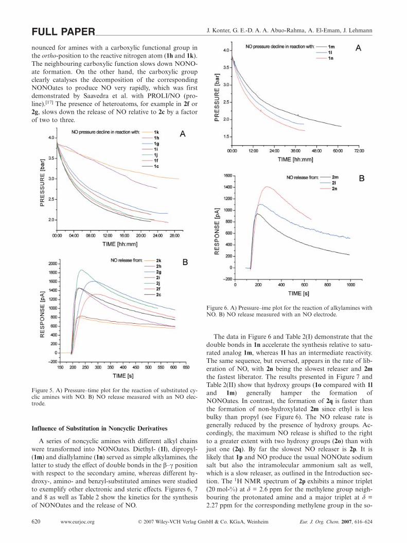
J. Konter, G. E.-D. A. A. Abuo-Rahma, A. El-Emam, J. LehmannFULL PAPERnounced for amines with a carboxylic functional group inthe ortho-position to the reactive nitrogen atom (1h and 1k).The neighbouring carboxylic function slows down NONO-ate formation. On the other hand, the carboxylic groupclearly catalyses the decomposition of the correspondingNONOates to produce NO very rapidly, which was firstdemonstrated by Saavedra et al. with PROLI/NO (pro-line).[17] The presence of heteroatoms, for example in 2f or2g, slows down the release of NO relative to 2c by a factorof two to three.
Figure 5. A) Pressure–time plot for the reaction of substituted cy-clic amines with NO. B) NO release measured with an NO elec-trode.
Influence of Substitution in Noncyclic Derivatives
A series of noncyclic amines with different alkyl chainswere transformed into NONOates. Diethyl- (1l), dipropyl-(1m) and diallylamine (1n) served as simple alkylamines, thelatter to study the effect of double bonds in the β–γ positionwith respect to the secondary amine, whereas different hy-droxy-, amino- and benzyl-substituted amines were studiedto exemplify other electronic and steric effects. Figures 6, 7and 8 as well as Table 2 show the kinetics for the synthesisof NONOates and the release of NO.
www.eurjoc.org © 2007 Wiley-VCH Verlag GmbH & Co. KGaA, Weinheim Eur. J. Org. Chem. 2007, 616–624620
Figure 6. A) Pressure–ime plot for the reaction of alkylamines withNO. B) NO release measured with an NO electrode.
The data in Figure 6 and Table 2(I) demonstrate that thedouble bonds in 1n accelerate the synthesis relative to satu-rated analog 1m, whereas 1l has an intermediate reactivity.The same sequence, but reversed, appears in the rate of lib-eration of NO, with 2n being the slowest releaser and 2mthe fastest liberator. The results presented in Figure 7 andTable 2(II) show that hydroxy groups (1o compared with 1land 1m) generally hamper the formation ofNONOates. In contrast, the formation of 2q is faster thanthe formation of non-hydroxylated 2m since ethyl is lessbulky than propyl (see Figure 6). The NO release rate isgenerally reduced by the presence of hydroxy groups. Ac-cordingly, the maximum NO release is shifted to the rightto a greater extent with two hydroxy groups (2o) than withjust one (2q). By far the slowest NO releaser is 2p. It islikely that 1p and NO produce the usual NONOate sodiumsalt but also the intramolecular ammonium salt as well,which is a slow releaser, as outlined in the Introduction sec-tion. The 1H NMR spectrum of 2p exhibits a minor triplet(20 mol-%) at δ = 2.6 ppm for the methylene group neigh-bouring the protonated amine and a major triplet at δ =2.27 ppm for the corresponding methylene group in the so-

Synthesis of Diazen-1-ium-1,2-diolates by the “NOtizer” Apparatus FULL PAPER
Figure 7. A) Pressure–time plot for the reaction of substituted alk-ylamines with NO. B) NO release measured with an NO electrode.
dium salt. Nevertheless, the additional primary amino func-tion in 1p accelerates the formation of the correspondingNONOate.
Furthermore, the effect of steric hindrance in the second-ary aliphatic amines reacting with NO can be demonstratedwith the benzylamine derivatives. As shown in Figure 8 andTable 2(III), an isopropyl moiety (1t) causes a total NOconsumption of just over 1.3 bar within a reaction time of70 h, while the amine with a methyl moiety (1r) consumesmore than 3 bar of NO in total and an amount four-foldhigher in the first 7 h. Only minor differences in the reac-tion rates of 1u, 1v and 1s could be observed. Analysis ofthe release of NO again demonstrates that an increase insteric hindrance decelerates the rate of NO liberation fromthe NONOates in solution (compare 2r with 2s, 2s with 2tand 2u with 2v). The synthesis of diazeniumdiolates derivedfrom amines like diisopropylamine, di-sec-butylamine, dicy-clohexylamine or 2,2,6,6-tetramethylpiperidine was not suc-cessful, as evidenced by an NO consumption that was closeto zero.
Eur. J. Org. Chem. 2007, 616–624 © 2007 Wiley-VCH Verlag GmbH & Co. KGaA, Weinheim www.eurjoc.org 621
Figure 8. A) Pressure–time plot for the reaction of benzylamineswith NO. B) NO release measured with an NO electrode.
Correlation between NONOate Formation andDecomposition
The correlation between NONOate formation and de-composition is visualized in Figure 9. It can be observedthat steric hindrance around the secondary amine as well asheteroatomic substitutions in the amine’s backbone clearlyhamper the bonding of NO to the amine’s nitrogen atomand the formation of the NONOate is slowed. Because oftheir reduced conformational flexibility, cyclic amines showmuch higher reaction rates than aliphatic amines, whereasthe effect of heteroatomic substituents on slowing the reac-tion progress can be observed in both cyclic and aliphaticamines. Phenyl substituents in the α position are well-toler-ated with regard to NONOate formation (compare 1l with1s and 1u with 1v in Table 2), whereas the introduction ofa second substituent in the α position � even a small methylgroup giving an isopropyl derivative (1t) � inhibits the reac-tion with NO dramatically (compare 1c with 1h and 1j with1k in Table 1 and 1s with 1t in Table 2).

J. Konter, G. E.-D. A. A. Abuo-Rahma, A. El-Emam, J. LehmannFULL PAPER
Figure 9. Relationship between NONOate formation and NO re-lease for compounds 1/2a–v. A) Ordinate: maximum release [s]measured with an NO-sensitive electrode at pH 6.4 at 37 °C. B)Ordinate: half-lives from UV measurements at pH 7.4 at 37 °C.
In general, a slow formation of NONOates yields slowNO releasers and vice versa, but remarkable exceptions, forexample, 1/2h,k,p, are evident. Sterically demanding substi-tution by carboxy or dimethyl (i.e. the isopropyl moiety)groups in the position α to the amine, as found in com-pounds 1h, 1k and 1t, causes slow NONOate formation,but only compound 2t shows the expected slow NO release.The ortho-carboxy function in 2h and 2k is certainly consid-ered to be bulky, but on the other hand it clearly catalyzesthe decomposition of the NONOates. Compounds 1/2p areexceptional as well; the partial formation of the intra-molecular salt contributes to a rather fast formation theNONOate, but this product is the slowest NO releaser ofall the compounds investigated. Less obvious, owing to thescaling in Figure 9, is the exceptional pair of compounds 1/2a. Compound 1a was found to be the fastest reacting
www.eurjoc.org © 2007 Wiley-VCH Verlag GmbH & Co. KGaA, Weinheim Eur. J. Org. Chem. 2007, 616–624622
amine, but 2a was not the fastest NO releaser. In fact it wasfound to be the slowest releaser of the unsubstituted cyclicNONOates.
Significance of the Results
The direct controlled release of nitric oxide from water-soluble compounds is of great interest not only for their useas tools to simulate physiological conditions in vitro, butalso in drugs and biomaterials with therapeutic roles.[4,24]
The “NOtizer” represents a modified hydrogenation appa-ratus which simplifies the synthesis of NONOates and al-lows the decrease in NO pressure to be recorded on-line.The rates of formation of the NONOates and the end ofthe reaction can be easily determined by this approach. Thecollected data can be used to predict NONOate formationand decomposition rates for novel compounds and there-fore simplify their design.
We have also demonstrated that both amperometry andUV spectroscopy can be used to determine the liberationprofile of different NONOates. Whether the pH is 6.4 or7.4, the order of decreasing rate of NO release fromNONOates is nearly identical [compare Figure 9(A) and(B)]. The advantage of the NO-sensitive electrode lies in itshigher throughput which allows the rate of NO release fromthe tested NONOate to be assessed. However, the half-lifecannot be determined by this method which can be re-garded as a disadvantage.
Experimental SectionAcquisition of Pressure-Time Plots in the Formation of Nonoates:Chemicals were purchased from VWR International Merck KGaA,Darmstadt, and Linde AG, Unterschleissheim, Germany (NO gas2.5 UN–Nr. 1660), and used without further treatment unlessnoted otherwise. All reactions in the NOtizer were conducted with-out any heating or cooling. The structures of amines 1a–v, leadingto the corresponding diazeniumdiolates 2a–v [see Equation (1)], aregiven in Figure 2. To ensure comparability, all reactions were per-formed in methanol, which was dried before use and kept underan atmosphere of dry nitrogen. Elementary sodium was dissolvedin methanol and the solution was filtered and added to an equi-molar amount of amine, although two-fold molar quantities of so-dium were added to amines 1h, 1i and 1k. The initial quantity ofamine was 0.05 mol for cyclic amines 1a–k and 0.1 mol for alkyl-and benzylamines 1l–v. The final volume for every experiment wasadjusted to 100 mL by adding methanol and the solution pouredinto the reaction chamber and tightly sealed. In order to providean anaerobic atmosphere, the chamber was evacuated and flushedwith dry nitrogen four times and then evacuated again. Finally aninitial pressure of 4.0 bar of nitric oxide was applied, all the regula-tory valves were closed, the solution stirred magnetically at700 rpm and the pressure was recorded on-line producing the plotsshown in Figures 3(A), 5–8(A).
The complete NO pressure–time plots were recorded and we alsodefined a further four reaction parameters. The end of the reaction[hh:mm] was fixed at the point when a drop in pressure of 0.05 bartakes longer than 3 h. The remaining pressure [bar] at this pointwas also used to describe the extent of the reaction. The remaining

Synthesis of Diazen-1-ium-1,2-diolates by the “NOtizer” Apparatus FULL PAPERpressure at the end of the reaction is due to unreacted NO, but sidereactions such as the disproportionation of NO cannot be ex-cluded. Furthermore, we compared the reduction in pressure (pres-sure descent) [bar] within the first 7 h and the mol ratio of NOconsumed by the amine. Because 2 mol of NO bind to 1 mol ofamine the maximum theoretical ratio is 2. Note that after recordingthe pressure–time plots the pressure was raised back to high pres-sure several times until the NO consumption in the reaction wasclose to zero to improve the yield of the product as reported pre-viously.[12]
Isolation and Characterization of the NONOates: The NONOatesprecipitated as solids during the reaction or after concentrating thefinal solution. Compounds 2o, 2p and 2q formed as resin-like resi-dues after solvent evaporation. The solids were separated by fil-tration, washed with diethyl ether and dried in a desiccator underreduced pressure overnight. Melting points were measured with aMel-Temp II, Laboratory Devices Inc., USA. The identity and pu-rity of the products were verified by 1H NMR spectroscopy (BrukerAdvance 250 spectrometer) and elemental analyses were performedwith a Hereaus Vario EL apparatus. Compounds 2b,c,f–i,l,m,q,rhave previously been reported in the literature and analytical datawere confirmed.[9,11,25–30]
Sodium 1-(Azetidin-1-yl)diazen-1-ium-1,2-diolate (2a): Preparedfrom 1a (2.86 g, 0.05 mol). Yield: 6.41 g (92%) as white crystals.M.p. 173–175 °C (dec.). 1H NMR (250 MHz, D2O/NaOD): δ =1.93 (q, 3J = 7.5 Hz, 2 H, NCH2CH2), 3.75 (t, 3J = 7.5 Hz, 4 H,2�NCH2) ppm. C3H6N3NaO2 (139.09): calcd. C 25.91, H 4.35, N30.21; found C 25.59, H 4.25, N 29.88.
Sodium 1-(Azepan-1-yl)diazen-1-ium-1,2-diolate (2d): Preparedfrom 1d (4.96 g, 0.05 mol). Yield: 4.23 g (47%) as a white solid.M.p. 198 °C (dec.). 1H NMR (250 MHz, D2O/NaOD): δ = 1.39–1.67 (m, 8 H, 4�CH2), 3.01 (t, 3J = 5.5 Hz, 4 H, 2�NCH2) ppm.C6H12N3NaO2 (181.17): calcd. C 39.78, H 6.68, N 23.19; found C39.50, H 6.46, N 22.79.
Sodium 1-(Azocan-1-yl)diazen-1-ium-1,2-diolate (2e): Prepared from1e (5.66 g, 0.05 mol). Yield: 7.13 g (73%) as a white solid. M.p.238 °C (dec.). 1H NMR (250 MHz, D2O/NaOD): δ = 1.36–1.65(m, 10 H, 5�CH2), 2.98 (t, 3J = 5.5 Hz, 4 H, 2�NCH2) ppm.C7H14N3NaO2 (195.20): calcd. C 43.07, H 7.23, N 21.53; found C43.05, H 7.22, N 21.41.
Sodium 1-(1,2,3,4-Tetrahydroisoquinolin-2-yl)diazen-1-ium-1,2-diol-ate (2j): Prepared from 1j (6.66 g, 0.05 mol). Yield: 3.74 g (35%) asa pale yellow solid. M.p. 171 °C (dec.). 1H NMR (250 MHz, D2O/NaOD): δ = 2.95 (t, 3J = 6.0 Hz, 2 H, NCH2CH2), 3.25 (t, 3J =6.0 Hz, 2 H, NCH2), 4.17 (s, 2 H, NCH2), 6.97–7.20 (m, 4 H, Ar–H) ppm. C9H10N3NaO2 (215.18): calcd. C 50.23, H 4.68, N 19.53;found C 50.00, H 4.93, N 19.21.
Disodium 1-(3-Carboxylato-1,2,3,4-tetrahydroisoquinolin-2-yl)di-azen-1-ium-1,2-diolate (2k): Prepared from 1k (8.86 g, 0.05 mol).Yield: 6.46 g (46%) as a white solid. M.p. 282–286 °C (dec.). 1HNMR (250 MHz, D2O/NaOD): δ = 2.60–3.16 (m, 2 H, NCHCH2),3.73–3.95 (m, 2 H, NCH2), 4.14–4.38 (m, 1 H, NCH), 6.92–7.20(m, 4 H, Ar–H) ppm. C10H9N3Na2O4 (281.18): calcd. C 42.72, H3.23, N 14.94; found C 43.02, H 3.23, N 14.95.
Sodium 1-(N,N-Diallylamino)diazen-1-ium-1,2-diolate (2n): Pre-pared from 1n (9.7 g, 0.1 mol). Yield: 11.47 g (64%) as a beige so-lid. M.p. 147 °C (dec.). 1H NMR (250 MHz, D2O/NaOD): δ = 3.36(d, 3J = 6.63 Hz, 4 H, 2�NCH2), 4.96–5.19 (m, 4 H,2�NCH2CHCH2), 5.52–5.74 (m, 2 H, 2�NCH2CHCH2) ppm.C6H10N3NaO2 (179.15): calcd. C 40.23, H 5.63, N 23.46; found C39.96, H 5.60, N 23.15.
Eur. J. Org. Chem. 2007, 616–624 © 2007 Wiley-VCH Verlag GmbH & Co. KGaA, Weinheim www.eurjoc.org 623
Sodium 1-[N,N-Bis(2-hydroxyethyl)amino]diazen-1-ium-1,2-diolate(2o): The use of 1o (10.5 g, 0.1 mol) did not produce a solid com-pound but rather a viscous fluid which yielded a colourless highlyhygroscopic solid after prolonged drying under reduced pressure.1H NMR (250 MHz, D2O/NaOD): δ = 2.89 (t, 3J = 5.74 Hz, 4H, 2�NCH2), 3.48 (t, 3J = 5.74 Hz, 4 H, 2�NCH2CH2) ppm.C4H10N3NaO4 (187.13): calcd. C 25.67, H 5.39, N 22.46; found C25.85, H 5.60, N, 22.12.
Sodium 1-[N-(2-Aminoethyl)-2-hydroxyethylamino])diazen-1-ium-1,2-diolate + 1-[N-(2-Ammonioethyl)-2-hydroxyethylamino]diazen-1-ium-1,2-diolate (2p): The use of 1p (10.4 g, 0.1 mol) produced ahighly viscous and hygroscopic amber fluid. 1H NMR (250 MHz,D2O/NaOD): δ = 2.27 (t, 3J = 5.93 Hz, 2 H, NCH2CH2NH2), 2.75(t, 3J = 5.93 Hz, 2 H, NCH2CH2NH2), 2.81 (t, 3J = 5.93 Hz, 2 H,NCH2CH2OH), 3.30 (t, 3J = 5.93 Hz, 2 H, NCH2CH2OH) + δ =2.60 (t, 3J = 5.93 Hz, 2 H, NCH2CH2NH3
+), 2.75 (t, 3J = 5.93 Hz,2 H, NCH2CH2NH3
+), 2.81 (t, 3J = 5.93 Hz, 2 H, NCH2CH2OH),3.30 (t, 3J = 5.93 Hz, 2 H, NCH2CH2OH) ppm.
Sodium 1-(N-Benzyl-N-ethylamino)diazen-1-ium-1,2-diolate (2s):Prepared from 1s (13.5 g, 0.1 mol). Yield: 8.47 g (39%) as a whitesolid. M.p. 175 °C (dec.). 1H NMR (250 MHz, D2O/NaOD): δ =0.60 (t, 3J = 7.10 Hz, 3 H, NCH2CH3), 2.69 (q, 3J = 7.10 Hz, 2 H,NCH2CH3), 3.67 (s, 2 H, NCH2Ar), 6.87–7.10 (m, 5 H, Ar–H) ppm. C9H12N3NaO2 (217.20): calcd. C 49.77, H 5.57, N 19.35;found C 49.62, H 5.80, N 18.98.
Sodium 1-(N-Benzyl-N-isopropylamino)diazen-1-ium-1,2-diolate(2t): Prepared from 1t (14.9 g, 0.1 mol). Yield: 3.4 g (15%) as abeige solid. The solid was filtered off from a large amount of un-changed 1t and washed with diethyl ether. M.p. 298 °C (dec.).1HNMR (250 MHz, D2O/NaOD): δ = 0.99 (d, 3J = 6.31 Hz, 6 H,NCH(CH3)2), 3.90–4.00 (m, 1 H, NCH), 3.56 (s, 2 H, NCH2Ar),7.10–7.22 (m, 5 H, Ar–H) ppm. C10H14N3NaO2 (231.23): calcd. C51.94, H 6.10, N 18.17; found C 51.63, H 6.15, N 17.84.
Sodium 1-[N-Benzyl-N-(2-phenylethyl)amino]diazen-1-ium-1,2-diol-ate (2u): Prepared from 1u (21.1 g, 0.1 mol). Yield: 14.33 g (49%)as a white solid. M.p. 217 °C (dec.). 1H NMR (250 MHz, D2O/NaOD): δ = 2.45 (t, 3J = 7.27 Hz, 2 H, NCH2CH2Ar), 2.99 (t, 3J
= 7.27 Hz, 2 H, NCH2CH2Ar), 3.86 (s, 2 H, NCH2Ar), 6.99–7.25(m, 10 H, Ar–H) ppm. C15H16N3NaO2 (293.30): calcd. C 61.43, H5.50, N 14.33; found C 61.11, H 5.55, N 14.42.
Sodium 1-(N,N-Dibenzylamino)diazen-1-ium-1,2-diolate (2v): Pre-pared from 1v (19.7 g, 0.1 mol). Yield: 9.2 g (33%) as a white volu-minous solid. M.p. 201–203 °C (dec.). 1H NMR (250 MHz, D2O/NaOD): δ = 4.01 (s, 4 H, 2�NCH2), 7.20 (m, 10 H, Ar–H) ppm.C14H14N3NaO2 (279.27): calcd. C 60.21, H 5.05, N 15.05; found C60.41, H 4.80, N 14.99.
NO Release Measurement: NO release was measured by using theApollo 4000 free-radical analyser system with an ISO-NOP nitricoxide selective sensor built into a multiport measurement chamberby World Precision Instruments Inc., USA. The measurementchamber was tempered with a Lauda M3 circulating thermostat at37 °C and stirred at 800 r.p.m. All experiments were started afterthe initial background signal had fallen below 7000 pA in a channelrange of 10 nA for the electrode. The apparatus was calibrated dailywith 2.0 mL of a 0.1 m KI/0.1 m H2SO4 solution in the chamberand an increasing amount of 50 μm NaNO2 solution to producenitric oxide by reduction. The nitrite solution was applied with anEppendorf pipette (10, 20, 30, 40 and 60 μL) to give 249, 493, 728,952 and 1388 nM solutions of NO, respectively. The dissociationof the diazeniumdiolates was measured by injecting a 50 μm NON-Oate/0.01 m NaOH solution (20 μL) into phosphate buffer (2.0 mL,

J. Konter, G. E.-D. A. A. Abuo-Rahma, A. El-Emam, J. LehmannFULL PAPERpH 6.4) resulting in a 495 nM solution of NONOate. Because 1 molNONOate produces 2 mol NO, the redox current flow of a 990 nMNO solution was measured. Note that the nitric oxide sensor mea-surement is not accumulative and only measures the arising currentat any given time. For this reason the phosphate buffer at pH 6.4was used to ensure a sharper signal for slower releasing NONOates.The maximum of the signal was chosen as a parameter with whichto compare dissociation rates. Figure 10 shows a typical signalevolving from the decomposition of a diazeniumdiolate (here 2r).This parameter represents the maximum concentration of nitric ox-ide reaching the electrode at a certain time. Every measurementwas carried out at least three times and the mean value with thecorresponding standard deviation was calculated.
Figure 10. Calibration and NO release–time plot of NONOate 2rin phosphate buffer at pH 6.4 at 37 °C.
The dissociation of the –[N(O)NO]– functional group rather thanNO release was measured with a Jasco V-570 UV spectrometer inphosphate buffer at pH 7.4 at 37 °C with a NONOate concentra-tion of 10–4M. The decrease in the absorption was measured ata wavelength of 251 nm over a period of 30 min. By plotting theabsorption logarithmically and performing a linear regressionanalysis the rate constants (k) were determined and used to calcu-late the half-lives of the respective NONOates using the formulat1/2 = ln2/k. R2 values were greater than 0.998 and every measure-ment was carried out at least three times to ensure reproducibilityto within �5%.[8]
[1] A. Seeling, J. Lehmann, J. Pharm. Biomed. Anal. 2006, 40,1131–1136.
[2] C.-F. Lam, S. Sviri, K. F. Ilett, P. V. Van Heerden, Expert Opin.Invest. Drugs 2002, 11, 897–909.
[3] J. Liu, C. Li, W. Qu, E. Leslie, C. L. Bonifant, G. S. Buzard,J. E. Saavedra, L. K. Keefer, M. P. Waalkes, Mol. Cancer Ther.2004, 3, 709–714.
[4] B. J. Nablo, A. R. Rothrock, M. H. Schoenfisch, Biomaterials2005, 26, 917–924.
www.eurjoc.org © 2007 Wiley-VCH Verlag GmbH & Co. KGaA, Weinheim Eur. J. Org. Chem. 2007, 616–624624
[5] K. C. Young, J. Ladino, C. Navarrete, K. Dabrowska, D.Hehre, E. Bancalari, C. Suguihara, Biol. Neonate 2004, 85,195–202.
[6] C. Velázquez, E. E. Knaus, Bioorg. Med. Chem. 2004, 12, 3831–3840.
[7] L. K. Keefer, R. W. Nims, K. M. Davies, D. A. Wink, Meth.Enzymol. 1996, 268, 281–293.
[8] J. A. Hrabie, J. R. Klose, D. A. Wink, L. K. Keefer, J. Org.Chem. 1993, 58, 1472–1476.
[9] A. Horstmann, L. Menzel, R. Gaebler, A. Jentsch, W. Urban,J. Lehmann, Nitric Oxide 2002, 6, 135–141.
[10] H. C. Champion, T. J. Bivalacqua, R. Wang, P. J. Kadowitz,L. K. Keefer, J. E. Saavedra, J. A. Hrabie, P. C. Doherty,W. J. G. Hellstrom, J. Urol. 1999, 161, 2013–2019.
[11] J. E. Saavedra, T. R. Billiar, D. L. Williams, Y.-M. Kim, S. C.Watkins, L. K. Keefer, J. Med. Chem. 1997, 40, 1947–1954.
[12] J. Konter, G. E.-D. A. A. Abuo-Rahma, A. El-Emam, J.Lehmann, Meth. Enzymol. 2005, 396, 17–26.
[13] K. M. Davies, D. A. Wink, J. E. Saavedra, L. K. Keefer, J. Am.Chem. Soc. 2001, 123, 5473–5481.
[14] C. M. Maragos, D. Morley, D. A. Wink, T. M. Dunams, J. E.Saavedra, A. Hoffman, A. A. Bove, L. Isaac, J. A. Hrabie,L. K. Keefer, J. Med. Chem. 1991, 34, 3242–3247.
[15] L. A. Hammad, P. H. Ruane, N. A. Kumar, J. P. Toscano, P. G.Wenthold, Int. J. Mass Spectrom. 2003, 222, 269–279.
[16] B. J. Nablo, M. H. Schoenfisch, Biomaterials 2005, 26, 4405–4415.
[17] J. E. Saavedra, G. D. Southan, K. M. Davies, A. Lundell, C.Markou, S. R. Hanson, C. Adrie, W. E. Hurford, W. M. Zapol,L. K. Keefer, J. Med. Chem. 1996, 39, 4361–4365.
[18] G. E.-D. A. A. Abuo-Rahma, A. Horstmann, M. F. Radwan,A. El-Emam, E. Glusa, J. Lehmann, Eur. J. Med. Chem. 2005,40, 281–287.
[19] H. Zhang, G. M. Annich, J. Miskulin, K. Stankiewicz, K.Osterholzer, S. I. Merz, R. H. Bartlett, M. E. Meyerhoff, J.Am. Chem. Soc. 2003, 125, 5015–5024.
[20] A. Srinivasan, N. Kebede, J. E. Saavedra, A. V. Nikolaitchik,D. A. Brady, E. Yourd, K. M. Davies, L. K. Keefer, J. P. Tos-cano, J. Am. Chem. Soc. 2001, 123, 5465–5472.
[21] P. H. Ruane, K. M. Bushan, C. M. Pavlos, R. A. D’Sa, J. P. To-scano, J. Am. Chem. Soc. 2002, 124, 9806–9811.
[22] M. M. Batchelor, S. L. Reoma, P. S. Fleser, V. K. Nuthakki,R. E. Callahan, C. J. Shanley, J. K. Politis, J. Elmore, S. I.Merz, M. E. Meyerhoff, J. Med. Chem. 2003, 46, 5153–5161.
[23] D. K. Taylor, I. Bytheway, D. H. R. Barton, C. A. Bayse, M. B.Hall, J. Org. Chem. 1995, 60, 435–444.
[24] Z. Zhou, M. E. Meyerhoff, Biomacromolecules 2005, 6, 780–789.
[25] J. E. Saavedra, A. Srinivasan, C. L. Bonifant, J. Chu, A. P.Shanklin, J. L. Flippen-Anderson, W. G. Rice, J. A. Turpin,K. M. Davies, L. K. Keefer, J. Org. Chem. 2001, 66, 3090–3098.
[26] F. Herrmann, N. List (Transdermal therapeutic system for ad-ministration of nitric oxide), DE 4305881, 1993.
[27] J. E. Saavedra, M. N. Booth, J. A. Hrabie, K. M. Davies, L. K.Keefer, J. Org. Chem. 1999, 64, 5124–5131.
[28] R. S. Drago, B. R. Karstetter, J. Am. Chem. Soc. 1960, 82,1819–1822.
[29] S. E. Price, D. Jappar, P. Lorenzo, J. E. Saavedra, J. A. Hrabie,K. M. Davies, Langmuir 2003, 19, 2096–2102.
[30] J.-T. Nguyen, C. A. Velazquez, E. E. Knaus, Bioorg. Med.Chem. 2005, 13, 1725–1738.
Received: July 31, 2006Published Online: November 27, 2006

Veröffentlichung 3 47-63
Veröffentlichung 3
NO Donors, part 17: Synthesis and Antimicrobial Activity of Novel Ketoconazole-NO-Donor Hybrid Drugs and
Corresponding Nitric Oxide Donors
Konter, Joerg; Möllmann, Ute; Lehmann, Jochen. Journal of Medicinal
Chemistry. In Vorbereitung.

1
NO Donors, part 171: Synthesis and antimicrobial activity of novel ketoconazole-NO-donor hybrid drugs and corresponding nitric oxide donors possessing a diazen-1-ium-1,2-diolate or organic nitrate moiety
Joerg Konter†, Ute Möllmann‡, Jochen Lehmann†,*
† Institute of Pharmacy, Department of Pharmaceutical/Medicinal Chemistry,
Friedrich-Schiller-University Jena, Philosophenweg 14, D-07743 Jena, Germany ‡ Hans Knöll-Institute for Natural Products Research, Beutenbergstr. 11a, D-07745 Jena, Germany
* To whom correspondence should be addressed. Tel.: 0049-3641-949803, Fax.: 0049-3641-949802,
E-mail: [email protected]
Abstract Novel nitric oxide donors and hybrid drugs combining the modified antifungal drug
ketoconazole with a diazen-1-ium-1,2-diolate or an organic nitrate moiety were synthesized and their
activities against a broad variety of bacteria and fungal strains were tested. Whereas the efficacy of
the compounds against bacteria showed little variation, the ketoconazole-NO-donor hybrid drugs
differed in their effectiveness and susceptibility against fungi. The NO-donors alone showed
insufficient effectiveness.

2
Introduction Ketoconazole (1a, KT) is a synthetic broad spectrum antifungal drug that was released in the
early 1980s and is used to prevent and treat mycotic infections systemically, but also externally. Due
to its variable bioavailability, liver toxicity and inhibition of steroid biosynthesis, the role of KT as an
orally administered drug has been limited to few indications such as candida-oesophagitis of patients
with acquired immune deficiency syndrome or to suppress glucocorticoid synthesis in the treatment of
Cushing’s disease, for example. Pharmacodynamically, all azole-antimycotics prevent the fungal
synthesis of ergosterole, which is a major constituent in fungal cell membranes, by enzyme inhibition
of the cytochrome P450 dependent 14-α-demethylase [2]. The inhibitory effect of KT against bacterial
growth in mycobacteria and streptomycetes is based on the interaction with cytochrome P450 as well
[3].
Previous studies have demonstrated the antimicrobial activity of endogenously derived nitric
oxide (NO) from the inducible NO synthase isoform (iNOS) as well as from exogenously NO-delivering
NO-donors[4-8]. Nitrogen bound diazen-1-ium-1,2-diolates (diazeniumdiolates, NONOates, “solid NO”)
represent a commonly used biological tool in nitric oxide related research and are NO-donating
compounds with the general structure R1R2N[N(O)NO]-. The predictable and exclusive release of the
reactive nitrogen species (RNS) NO. in a temperature, pH and structure dependent manner with half-
lives reaching from a few seconds to many hours, depending on the compound used, are the most
mentionable advantages of this class of NO-donor [9-12]. In order to prevent the immediate
decomposition when dissolved in aqueous solution and further modulate the liberation of NO,
NONOates can be derivatized to prodrugs that require enzymatic and/or chemical activation to result
in the corresponding diazeniumdiolate. Recent reports show that not only the NONOate DETA/NO
itself exerts inhibitory effects against Candida species, but also in synergy when applied in
combination with azole antimycotics such as KT, fluconazole and miconazole [13].
Contrary to NONOates, organic nitrates require reduction of the nitrogen from oxidation state
+V to +II in order to deliver NO. Glutathione reductase, cytochrome P450, xanthine oxidoreductase
and mitochondrial aldehyde dehydrogenase were recognized to be mediators in the bioactivation of
organic nitrates [14-18]. Furthermore, organic nitrates are supposed to induce NO-like biological
effects without generating free NO radicals and may be addressed as NO-mimetics rather than NO-
donors. Therapeutically, organic nitrates are used for prophylaxis and acute treatment of angina
pectoris, whereas most of the nitrate hybrid drugs introduced up to now possess a dihydropyridine or
NSAID structure as second active principle [19,20].
In this investigation we report on the synthesis of a series of phenylpiperazine – which is a part
of the ketoconazole structure - and ketoconazole derivatives bearing a diazen-1-ium-1,2-diolate or
organic nitrate moiety and the NO-liberation profile of the NONOate compounds. The NO-donors and
KT-NO-donor hybrid drugs were tested for their in vitro antimicrobial activity on a series of bacterial-
and fungal strains by determining the zone of inhibition and minimum inhibitory concentration (MIC).
Chemistry
For the conversion of KT (1a) into a NO-donating hybrid drug, we focused on the
phenylpiperazine substructure as pointed out in Scheme 1. In order to make 1a accessible for
3
derivatization, the acetyl residue was cleaved from the phenylpiperazine moiety with NaOH solution.
As mentioned above, the time profile of NO liberation from NO-donors (e.g. NONOates) depends
greatly on their individual chemical structure. To differentiate whether biologic effects rise from the
released NO or from the synergy of KT and NO, all of the chemical modifications of the deacetylated
KT (2a) were performed with phenylpiperazine (2b) as well.
The sodium salt NONOates 3a and 3b were produces by exposing the respective amines to 5
bar of NO under exclusion of oxygen and water in 1:1 methanol/THF for 3a and dry methanol for 3b
[21]. We used the NOtizer® apparatus as described previously [12,22]. The halflives (t1/2) of the
NONOates were determined both via UV-spectroscopic decomposition of the diazeniumdiolate group
and by direct NO measurement with a nitric oxide sensitive electrode reported previously [23,12]. The
halflife of NO release for 3a/3b was approximately t1/2= 4.5 min with no significant difference in kinetics
(see Supporting Information). The alkylation of 3a/3b with bromoethane and chlormethyl methylether
yielded 4a/4b and 5a/5b respectively, which do not release NO until the residues bonded to the O2-
atom of the diazen-1-ium-1,2-diolate group are cleaved. While the metabolism of 4a/4b in vitro is
enzyme related, compounds 5a/5b are capable of being hydrolyzed chemically to the respective
NONOate, formaldehyde and methanol.
The NONOates 6a/6b, resulting from the reaction of 2a/2b with chloroacetyl chloride followed
by the addition of the sodium salt of 1-(pyrrolidin-1-yl)diazen-1-ium-1,2-diolate (PYRRO/NO), are
reliant on bond cleavage as well in order to liberate NO, yielding the bare NONOate and the
hydroxyethanone derivative. The halflife of the resulting PYRRO/NO is approximately t1/2= 3-5 s under
physiologic conditions [24]. In contrast to 4a/4b and 5a/5b, this prodrug synthesis strategy
advantageously allows the design of drugs with any kinetic of NO-release just by appropriate
modification on the NONOates structure. For instance, the NO release from 6a/6b can be slowed
down easily by changing the amine backbone linked to the diazeniumdiolate group to a bulky
alkylamine or polyamine [12,23]. Additionally, the amide carbonyl function attached to the
phenylpiperazine moiety enhances the similarity to KT.
The organic nitrates 7a/7b differ to 8a/8b only through the carbonyl group, introduced by
acylation of 2a/2b with carbonyldiimidazole (CDI) activated nitratoacetic acid, whereas 8a/8b was
prepared by alkylation of 2a/2b with 2-bromoethyl nitrate. These two stets of organic nitrates were
designed to assess the importance of the amide moiety in association with the NO mimetic group.
Furthermore, the nitratopivalinic acid amides 9a/9b were synthesized to study the effect of structural
changes in the organic nitrate moiety on biologic activity.
Biological Testing For antimicrobial activity assays we used Bacillus subtilis ATCC 6633, Staphylococcus aureus
SG 511, S. areus SG 134/94 (MRSA), Escherichia coli SG 458, Pseudomonas aeruginosa SG 137, P.
aeruginosa K799/61, Enterococcus faecalis 1528 (VRE), Mycobacterium vaccae IMET 10670,
Aspergillus fumigatus ATCC 46645, A. terreus DSM 826, A. niger DSM 737, Fusarium oxysporum
i1205, Candida albicans BMSY 212, C. glabrata DSM 6525, Sporobolomyces salmonicolor SG 549
and Penicillium notatum JP 36.

4
Antimicrobial activity was determined qualitatively by agar diffusion tests according to:
European Pharmacopoeia, 3ed Edition, 1997; 13: 118, Deutscher Apotheker Verlag Stuttgart, as
described [25]. Shortly, 100 µL suspension of the test organism with a density of McFarland standard
0.5 were inoculated into 32 mL of sterile melted agar medium and poured into petri dishes. Holes of 9
mm in diameter were cut in the agar and filled with 50 µL of a 0.2 mM (or as stated otherwise) solution
of the compound. Exceptionally, compounds 3a/3b were diluted in 0.1 M NaOH solution, only the last
dilution step was performed in the given media and used immediately. Inhibition zones were read after
overnight incubation.
Antifungal activity of the compounds was studied quantitatively by determination of minimal
inhibitory concentrations (MIC) according to the NCCLS guidelines using the broth microdilution
method [26,27]. The bacteria were grown overnight at 37 °C in Mueller-Hinton broth (MHB)(Difco). 50
µL of a compound solution of 2 mM in DMSO/Methanol (1:10) were serially diluted by factor two with
MHB. Exceptionally, compounds 3a/3b were diluted in 0.1 M NaOH solution, only the last dilution step
was performed in the given media and used immediately. Then the wells were inoculated with 50 µL of
testorganisms to give a final concentration of 5×10⁵ CFU/mL. After microtiter plates were incubated at
37 °C for 24 h, the MIC values were read with a Nepheloscan Ascent 1.4 automatic plate reader
(Labsystems, Vantaa, Finland) as the lowest dilution of antibiotic allowing no visible growth. Candida
albicans was grown in yeast nitrogen base (Difco) supplemented with 1 % glucose and incubated at
30 °C.
Results and Discussion
In order to compare the antimicrobial activity of compounds 1a-9a qualitatively, a 200 µM
solution of the respective KT-NO-donor-hybrid drug was exposed to the different bacterial strains
mentioned in Table 1. No considerable inhibition of growth was found for any of the compounds when
applied to E. coli, P. aeruginosa or E. faecalis. Significant differences in the zone of inhibition diameter
or spectrum change were found for the compounds 3a, 6a, 7a and 9a in comparison to the KT-
derivative 1a. The NONOate-group leading to spontaneous NO release from 3a enhances the
antibacterial activity slightly for all KT sensitive strains, but especially for M. Vaccae. Interestingly, the
derivatives 6a, 7a and 9a loose their efficacy against methicillin-resistant S. aureus SG 134/94
(MRSA) compared to the other tested compounds, which showed similar results against S. aureus and
methicillin-resistant S. aureus. The only chemical structure similarity of these three compounds is the
derivatisation of the acetyl moiety on the phenylpiperazine which may represent an essential element
in S. aureus susceptibility, since the deacetylated KT (2a) showed no activity against S. aureus or
MRSA.
Significant differences were found in the zone of inhibition diameter for compounds 1a-9a tested on fungal strains of Aspergillus fumigatus, A. terreus, A. niger, Fusarium oxysporum, Candida
albicans, C. glabrata, Sporobolomyces salmonicolor and Penicillium notatum (Data in Table 1 of
Supporting Information) necessitating MIC-value determination. Table 2 displays the MIC for the KT-
NO-donor Hybrids 3a-9a compared to KT (1a) and N-deacetyl-KT (2a) on the different fungal strains.
For better comparison, the respective fractional inhibitory concentration (FIC = MIC compound / MIC
KT) was calculated, whereas values under 1 represent higher antifungal activity than KT alone. The N-

5
deacetyl-ketoconazole 2a is a major metabolite of KT and its activity has been tested on Plasmodium
falciparum, Candida albicans and C. tropicalis previously [28,29]. Literature data of no significant
differences between 1a and 2a in activity against these Candida isolates could be confirmed by us for
C. glabrata as well. However, major loss of antifungal activity for 2a against Aspergillus isolates and
Penicillium notatum can be recognized in our study. The MIC of 2a is 30-60 fold higher for some of the
strains compared to the N-acetylated 1a. The direct NO-donor derivative of KT carrying a
diazeniumdiolate moiety (3a) shows similar antifungal potency against all strains tested compared to
1a. NO-donor 3a exclusively releases the redox-species NO. with a half-life of approximately t1/2 = 4.5
min at pH 7.4 and 37 °C (see Supporting Information). After complete NO liberation from 3a, the
secondary amine equivalent to structure 2a, remains. Obviously the NO released from 3a is beneficial
for the antifungal activity due to the higher activity compared to 2a, but not synergistic enough to
pursue 1a. The strategy of synthesizing diazeniumdiolate-prodrugs 4a and 5a with an ethyl- and
methoxymethyl moiety bound to the O2-atom of the NONOate-group respectively, enhances the
antifungal properties of these compounds tremendously compared to the sodium salt NONOate 3a.
Both of these compounds are more active than KT, especially against the tested Aspergillus strains,
but no significant differences were found on Candida albicans. A possible explanation is that this
species lacks the means necessary to metabolize 4a and 5a to its active NO-donor. No difference in
efficacy is observed when comparing compound 6a to KT. Cleavage of 6a results, amongst other, in
the fast releasing NONOate PYRRO/NO with t1/2 = 3 s for NO release. Perhaps the low synergistic
effect of this KT-NO-donor hybrid is due to higher immobility thru membranes compared to the
NONOate-prodrugs 4a/5a or maybe a result of the very short half life of the NO-donor.
The synergistic antimicrobial effects of organic nitrates with KT have not been investigated
before. With this NO-donor class it is difficult to distinguish if the observed activity results from the
synergy of KT with NO, another nitrogen species or even receptor-related interaction. The organic
nitrates 7a and 9a, which are bound to the KT structure as amides, showed to be more potent than KT
alone, especially compound 9a against the tested Aspergillus strains. It is clearly recognizable, that
the spectrum of antifungal activity of the tested KT-NO-donors is different from the spectrum of KT,
mainly a higher potency against Aspergillus species. Unexpectedly, the alkyl nitrate residue
incorporated in compound 8a turned out to be less potent than KT, suggesting that the combination of
amide linkage and organic nitrate is obligatory if antifungal properties are desired.
The NO-donor compounds 2b-9b lacking the KT structure showed no inhibition of test
organism growth in all agar diffusion tests on a broad spectrum of bacterial and fungal strains at a
concentration of 0.2 mM. The NONOate DETA/NO has been previously reported to have a MIC-value
of about 2000 µg/mL (12.5 mM) on Candida species, DPTA/NO even up to 5000 µg/mL (26.2 mM)
[13,30]. Therefore, as expected, the NONOate, NONOate-prodrugs and organic nitrates of the
phenylpiperazine derivative (2b-9b) showed no reckonable antimicrobial activity at a concentration of
200 µM if applied exclusively, but only in synergy with the antifungal KT-backbone.
The difference in antimicrobial activity of the KT-NO-donors in comparison to KT can be
explained thru several possible mechanisms that can vary in the respective pathogen species. For
instance, from a chemical point of view the entry of the hybrids into microbial cells can differ thru
properties such as lipophilicity or molecular weight of the compounds. Microbial cellular targets for NO

6
such as proteins, lipids or DNA can be altered or detoxified by pathogen specific defense mechanisms
such as NO-scavengers like homocysteine or glutathione, detoxifying enzymes or DNA repair
mechanisms [6]. Concerning the synergistic effect of KT with NO it is also conceivable that NO
interacts with the hemoprotein CYP450 of the 14-α-demethylase and hence amplifies the antimicrobial
target of KT.
Conclusion Ketoconazol-NO-donor hybrid drugs represent a useful approach in antimicrobial and
antifungal treatment. For KT carrying a diazeniumdiolate moiety, derivatization to prodrugs is
obligatory to ensure stability of the compounds. Hybrid drugs of KT with an organic nitrate group are
highly active against the tested strains as well, whereas variation in structure seems to have greater
influence on in vitro efficacy than for NONOate-hybrids. This study demonstrates that it is not
regardless which type of NO-donor is used to show synergy with antimycotic agents, but also that the
spectrum of susceptibility against different strains can alter from the one of the antimycotic agent
alone. The KT-NO-donor Hybrids would advantageously ensure a kind of drug targeting for the NO
and other RNS in comparison to fixed combinations of KT and NO-donor that have different
distribution properties in vitro and in vivo. It is also considerable that the antiinfective properties of
these compounds in vivo could be much greater, since amongst the versatile NO activities is its
important regulatory role in the immune system or in inflammatory processes that would indirectly
contribute against microbial pathogens.
Experimental Section (i) Deacetylation of 1a to 2a. KT (1a) was purchased from Caesar & Loretz GmbH Hilden,
Germany (Art. Nr. 5270-25G) and 8 g (15 mmol) were dissolved in 175 mL methanol under reflux.
Afterwards, 20 % aqueous NaOH solution (40 mL) was slowly added and kept under reflux until
completion of the reaction (TLC controlled). Chopped ice was added to the solution and the
precipitated product was filtered and dried under vacuum to give 2a in quantitative yield.
(ii) General Procedure for Synthesis of 3a/3b. Equimolar amounts of elementary sodium
were dissolved in dry methanol (75 mL) and the solution filtered and added to 2a (5 mmol) and 2b (50
mmol) respectively. Additionally, dry tetrahydrofuran (75 mL) was only poured to the mixture
containing 2a. The solutions were transferred into the reaction chamber of the NOtizer® apparatus and
closed tightly. The chamber was evacuated and flushed with dry nitrogen four consecutive times, then
evacuated again. An initial pressure of 5 bar nitric oxide was applied and the solutions stirred
magnetically at 700 rpm. The pressure was raised back to 5 bar until remaining constant. The
precipitated products after solvent concentration were filtered, washed with ether and dried in a
desiccator under reduced pressure over night.
(iii, iv) General Procedure for Synthesis of 4a/4b and 5a/5b. A mixture of pulverized
anhydrous Na2CO3 (2 g) and 3a/3b suspended in 50 mL hexamethylphosphoric triamide (HMPA) was
stirred at 4 °C and an equimolar amount of the respective alky halide added dropwise. After ice bath
removal, the mixtures were stirred at room temperature for 72 h. Insoluble solids were removed by
filtration and the solution poured into NaHCO3 saturated water. The products were extracted with ethyl
7
acetate (2 x 30 mL) and washed with water (5×30 mL). The residues after solvent evaporation were
purified by column chromatography on silica gel using the given eluent to furnish 4a, 4b, 5a or 5b
respectively.
(v) General Procedure for Synthesis of 6a/6b. A mixture of pulverized anhydrous Na2CO3 (2
g) and 1.5 molar excess chloroacetyl chloride in 20 mL dichloromethane (DCM) was stirred at 4 °C
and 2a/2b dissolved in 40 mL DCM added dropwise and stirred until completion of the reaction (TLC
controlled), respectively. Solids were filtered off and the filtrate washed with saturated NaHCO3
solution (1×50 mL) and water (3×50 mL). The organic phase was dropped to a freshly prepared
suspension of Na2CO3 and equimolar amount of sodium 1-(pyrrolidin-1-yl)diazen-1-ium-1,2-diolate
(PYRRO/NO) in 50 mL HMPA in sito at room temperature. A stream of dry nitrogen was passed
through the mixture until the DCM was disposed and stirred for 72 h afterwards. The suspension was
filtered into 60 mL of NaHCO3 saturated water, extracted with ethyl acetate (2×50 mL) and the organic
phase washed with water (5×75 mL). The residues after solvent evaporation were purified by column
chromatography on silica gel using the given eluent to furnish 6a or 6b respectively.
(vi and viii) General Procedure for Synthesis of 7a/7b and 9a/9b. Compounds 2a/2b (1
mmol) dissolved in 30 mL dichloromethane were slowly added via a syringe through a septum to a
solution of CDI activated nitratoacetic acid or nitratopivalinic acid (1.5 mmol) in 20 mL
dichloromethane under dry nitrogen atmosphere and stirred for 24 h at room temperature. The
solutions were washed with NaHCO3 saturated water (3×40 mL) and the organic phase evaporated to
yield residues of 7a, 7b, 9a or 9b that were purified thru silica gel column chromatography or
recrystallization respectively.
Note that the much simpler acetylation of the secondary nitrogen atom with bromoacetyl
bromide and subsequent nitration using silver nitrate in acetonitrile to produce compounds 7a/7b only
worked for the preparation of 7b. The acetal group incorporated in the KT molecule was cleaved upon
work up preventing this reaction to furnish 7a.
(vii) General Procedure for Synthesis of 8a/8b. A given amount of 2a/2b dissolved in
dichloromethane (10 mL) was slowly added to a twofold excess of 2-bromoethyl nitrate and stirred at
60 °C for 48 h. The organic phase was washed with NaHCO3/H2O (3×20 mL) and evaporated to give
brown fluids that required column chromatography purification on silica gel to yield 8a/8b.
Acknowledgement: We thank Uta Wohlfeld and Gabi Schwinger for the
antimicrobial/antifungal testing and excellent technical assistance.
Supporting Information Available: Includes detailed experimental protocols, physical-,
spectroscopic- and elemental analysis data for compounds 2a, 3-9a/b and NO-release measurements
data for compounds 3a/3b. Inhibition zones for all compounds on a broad spectrum of fungi is
presented.

8
References (1) Koenig, A.; Roegler, C.; Lange, K.; Daiber, A.; Glusa, E.; Lehmann, J. NO donors. Part 16:
Investigations on structure-activity relationships of organic mononitrates reveal 2-
nitrooxyethylammoniumnitrate as a high potent vasodilator. Bioorg. Med. Chem. Lett.,
accepted.
(2) Van Tyle, J. H. Ketoconazole. Mechanism of action, spectrum of activity, pharmacokinetics,
drug interactions, adverse reactions and therapeutic use. Pharmacotherapy 1984, 4, 343-373.
(3) McLean, K. J.; Marshall, K. R.; Richmond, A.; Hunter, I. S.; Fowler, K.; Kieser, T.; Gurcha, S.
S.; Besra, G. S.; Munro, A. W. Azole antifungals are potent inhibitors of cytochrome P450
monooxygenases and bacterial growth in mycobacteria and streptomycetes. Microbiology
2002, 148, 2937-2949.
(4) Ochoa, J. B.; Udekwu, A. O.; Billiar, T. R.; Curran, R. D.; Cerra, F. B.; Simmons, R. L.;
Peitzman, A. B. Nitrogen oxide levels in patients after trauma and during sepsis. Annals of
Surgery 1991, 214, 621-626.
(5) Keller, C. C.; Kremsner, P. G.; Hittner, J. B.; Misukonis, M. A.; Weinberg, J. B.; Perkins, D. J.
Elevated nitric oxide production in children with malarial anemia: Hemozoin-induced nitric
oxide synthase type 2 transcripts and nitric oxide in blood mononuclear cells. Infection and
Immunity 2004, 72, 4868-4873.
(6) Fang, F. C. Mechanisms of nitric oxide-related antimicrobial activity. Journal of Clinical
Investigation 1997, 100, 43-50.
(7) He, Q.; Leitch, G. J.; Visvesvara, G. S.; Wallace, S. Effects of nifedipine, metronidazole, and
nitric oxide donors on spore germination and cell culture infection of the microsporidia
Encephalitozoon hellem and Encephalitozoon intestinalis. Antimicrobial agents and
chemotherapy 1996, 40, 179-185.
(8) Wheeler, M. A.; Smith, S. D.; Garcia-Cardena, G.; Nathan, C. F.; Weiss, R. M.; Sessa, W. C.
Bacterial infection induces nitric oxide synthase in human neutrophils. Journal of Clinical
Investigation 1997, 99, 110-116.
(9) Drago, R. S.; Paulik, F. E. The reaction of nitrogen(II) oxide with diethylamine. Journal of the
American Chemical Society 1960, 82, 96-98.
(10) Keefer, L. K.; Nims, R. W.; Davies, K. M.; Wink, D. A. "NONOates" (1-substituted diazen-1-
ium-1,2-diolates) as nitric oxide donors: convenient nitric oxide dosage forms. Methods in
Enzymology 1996, 268, 281-293.
(11) Keefer, L. K. Progress toward clinical application of the nitric oxide-releasing
diazeniumdiolates. Annual Review of Pharmacology and Toxicology 2003, 43, 585-607.
(12) Konter, J.; Abuo-Rahma, G. E.-D.; El-Emam, A.; Lehmann, J. Synthesis of diazen-1-ium-1,2-
diolates monitored by the "NOtizer" apparatus: relationship between formation rates,
molecular structure and the release of nitric oxide. European Journal of Organic Chemistry
2007, 4, 616-624.
(13) McElhaney-Feser, G. E.; Raulli, R. E.; Cihlar, R. L. Synergy of nitric oxide and azoles against
Candida species in vitro. Antimicrobial Agents and Chemotherapy 1998, 42, 2342-2346.

9
(14) Needleman, P.; Harkey, A. B. Role of endogenous glutathione in the metabolism of glyceryl
trinitrate by isolated perfused rat liver. Biochemical Pharmacology 1971, 20, 1867-1876.
(15) Servent, D.; Delaforge, M.; Ducrocq, C.; Mansuy, D.; Lenfant, M. Nitric oxide formation during
microsomal hepatic denitration of glyceryl trinitrate: involvement of the cytochrome P-450.
Biochemical and Biophysical Research Communications 1989, 163, 1210-1216.
(16) Millar, T. M.; Stevens, C. R.; Blake, D. R. Xanthine oxidase can generate nitric oxide from
nitrate in ischemia. Biochemical Society Transactions 1997, 25, 528.
(17) Chen, Z.; Zhang, J.; Stamler, J. S. Identification of the enzymatic mechanism of nitroglycerin
bioactivation. Proceedings of the National Academy of Sciences 2002, 99, 8306-8311.
(18) Wenzel, P.; Hink, U.; Oelze, M.; Seeling, A.; Isse, T.; Bruns, K.; Steinhoff, L.; Brandt, M.;
Kleschyov, A. L.; Schulz, E.; Lange, K.; Weiner, H.; Lehmann, J.; Lackner, K. J.; Kawamoto,
T.; Muenzel, T.; Daiber, A. Number of nitrate groups determines reactivity and potency of
organic nitrates: a proof of concept study in ALDH-2-/- mice. British Journal of Pharmacology
2007, 150, 526-533.
(19) Velazquez, C.; Knaus, E. E. Synthesis and biological evaluation of 1,4-dihydropyridine
calcium channel modulators having a diazen-1-ium-1,2-diolate nitric oxide donor moiety for
the potential treatment of congestive heart failure. Bioorganic & Medicinal Chemistry 2004,
12, 3831-3840.
(20) Davies, N. M.; Roseth, A. G.; Appleyard, C. B.; Mcknight, W.; Del Soldato, P.; Calignano, A.;
Cirino, G.; Wallace, J. L. NO - naproxen vs. naproxen: ulcerogenic, analgesic and anti-
inflammatory effects. Alimentary Pharmacology and Therapeutics 1997, 11, 69-79.
(21) Bonifant, C.; Saavedra, J.; Keefer, L.; Citro, M.; Buzard, G.; Fitzhugh, A.; Donovan, P.; Ji, X.;
Shami, P.; Singh, S.; Srinivasan, A. Preparation of arenediazeniumdiolates capable of
generating nitric oxide and superoxide. PCT Int. Appl. 2001, 71. WO 2001092215.
(22) Konter, J.; Abuo-Rahma, G. E.-D.; El-Emam, A.; Lehmann, J. The NOtizer-A device for the
convenient preparation of diazen-1-ium-1,2-diolates. Methods in Enzymology 2005, 396, 17-
26.
(23) Hrabie, J. A.; Klose, J. R.; Wink, D. A.; Keefer, L. K. New nitric oxide-releasing zwitterions
derived from polyamines. Journal of Organic Chemistry 1993, 58, 1472-1476.
(24) Saavedra, J. E.; Billiar, T. R.; Williams, D. L.; Kim, Y.-M.; Watkins, S. C.; Keefer, L. K.
Targeting nitric oxide (NO) delivery in vivo. Design of a liver-selective NO donor prodrug that
blocks tumor necrosis factor-α-induced apoptosis and toxicity in the liver. Journal of Medicinal
Chemistry 1997, 40, 1947-1954.
(25) Afonin, S.; Glaser, R. W.; Berditchevskaia, M.; Wadhwani, P.; Guehrs, K.-H.; Moellmann, U.;
Perner, A.; Ulrich, A. S. 4-fluorophenylglycine as a label for 19F NMR structure analysis of
membrane-associated peptides. Chem.Bio.Chem. 2003, 4, 1151-1163.
(26) National Committee for Clinical Laboratory Standards: Methods for Dilution Antimicrobial
Susceptibility Tests for Bacteria that Grow Aerobically; Approved Standard. 6th edn.
Document M7-A6. Wayne, USA: NCCLS, 2003.
10
(27) Heinisch, L.; Wittmann, S.; Stoiber, T.; Berg, A.; Ankel-Fuchs, D.; Moellmann, U. Highly
antibacterial active aminoacyl penicillin conjugates with acylated bis-catecholate siderophores
based on secondary diamino acids and related compounds. Journal of Medicinal Chemistry
2002, 45, 3032-3040.
(28) Whitehouse, L. W.; Menzies, A.; Dawson, B.; Zamecnik, J.; Sy, W. W. Deacetylated
ketoconazole: a major ketoconazole metabolite isolated from mouse liver. Journal of
Pharmaceutical and Biomedical Analysis 1990, 8, 603-606.
(29) Pfaller, M. A.; Segal, J. J.; Krogstad, D. J. Activity of ketoconazole and its deacyl derivative
against Plasmodium falciparum and Candida isolates. Antimicrobial Agents and
Chemotherapy 1982, 22, 917-919.
(30) Falahati, M.; Shabani, M.; Rodaki, M. M. A.; Jahaniani, F.; Bagheri, K. P.; Ahmed Ebrahimi, S.
Interaction between ketoconazole, amphotericin B and terbinafine and three diazenumdiolates
in concomitant uses against some fungal species. Daru, Journal of Faculty of Pharmacy,
Tehran University of Medical Sciences 2006, 14, 87-92.

11
Scheme 1a
O
O
Cl
Cl
N
N
ONN
H
O
CH3
O
O
Cl
Cl
N
N
ONN
H
H
O
O N
N+
Na+
CH3
O
O
O N
N+
O
O N
N+
CH3
N
O
N+
O
N
O
O2NO
O
O2NOCH3
CH3
O
O2NO
NNH
1a
2a
2b
3a/3b
4a/4b
6a/6b
5a/5b
7a/7b 8a/8b 9a/9b
i
iii
iv
v vi viiii viii
v vi viiii viii
O
O
Cl
Cl
N
N
ONN
H
O
CH3
O
O
Cl
Cl
N
N
ONN
H
H
O
O N
N+
Na+
CH3
O
O
O N
N+
O
O N
N+
CH3
N
O
N+
O
N
O
O2NO
O
O2NOCH3
CH3
O
O2NO
NNH
1a
2a
2b
3a/3b
4a/4b
6a/6b
5a/5b
7a/7b 8a/8b 9a/9b
i
iii
iv
v vi viiii viii
v vi viiii viii
a Reagents and conditions: (i) 20% aq. NaOH, rx, 24 h; (ii) 5 bar NO, neq CH3ONa, rt; (iii) bromoethane, 72 h ; (iv) chlormethyl methylether, 72 h; (v) 1.chloroacetyl chloride, 4°C, 2. PYRRO/NO, rt, 72 h; (vi) CDI activated nitratoacetic acid, rt, 24 h; (vii) 2-bromoethyl nitrate, rt, 72 h; (viii) CDI activated nitratopivalinic acid, rt, 24 h.

12
Table 1. Antibacterial Activities In Vitro of Ketoconazole and the Ketoconazole-NO-Donor Hybrid Drugs 1a-9a and the Phenylpiperazine Analogs 2b-9b. Inhibition Zone [mm].
p = partial inhibition
B. subtilis S. aureus E. coli P. aeruginosa E. faecalis M. vaccae
Compd 200 µM ATCC
6633 SG 511
SG 134/94 SG
458 SG 137
K 199/61 1528
(VRE) IMET 10670
1a 10.5/17p 9.5/15p 9.5/15p 0 0 0 0 17.5p 2a 10 0 0 0 0 0 0 17.5p 3a 12/21.5p 17.5p 15p 0 0 0 0 24/34p 4a 11.5/17p 14.5 11p 0 0 0 0 16.5/24p 5a n.t. n.t. n.t. n.t. n.t. n.t. n.t. n.t. 6a 10.5/15p 11.5/15p 0 0 0 0 0 10/19p 7a 11/18p 13/16p 0 0 0 0 0 22p 8a 15 14 14 10.5p 0 0 10p 16 9a 13p 10p 0 0 0 0 0 10/16p
2b-9b 0 0 0 0 0 0 0 0
n.t. = not tested Table 2. Antifungal Activities In Vitro of Ketoconazole and the Ketoconazole-NO-Donor Hybrid Drugs 1a-9a. MIC-Value [µM].
(FIC = MIC Compound / MIC KT)
Compd. Asperg. fumigatus
Asperg. terreus
Asperg. niger
Fusarium oxysporum
Candida albicans
Candida glabrata
Sporobolom. salmonicolor
Penicillium notatum
1a = KT 62.5 31.25 62.5 500 25 12.5 1.56 8
2a
2000 (32)
2000 (64)
2000 (32)
2000 (4)
25 (1)
50 (4)
25 (16)
250 (31.25)
3a
200 (3.2)
100 (3.2)
100 (1.6)
200 (0.4)
100 (4)
12.5 (1)
1.25 (0.8)
20 (2.5)
4a
12.5 (0.2)
3.13 (0.1)
1.56 (0.025)
50 (0.1)
50 (2)
3.13 (0.25)
0.16 (0.1)
1.25 (0.16)
5a
8 (0.13)
8 (0.26)
4 (0.06)
250 (0.5)
25 (1)
3.13 (0.25)
2 (1.3)
1.56 (0.2)
6a
125 (2)
62.5 (2)
62.5 (1)
1000 (2)
25 (1)
12.5 (1)
2 (1.3)
8 (1)
7a
25 (0.4)
12.5 (0.4)
12.5 (0.2)
100 (0.2)
25 (1)
1.56 (0.12)
0.16 (0.1)
2.5 (0.31)
8a
1000 (16)
1000 (32)
250 (4)
1000 (2)
12.5 (0.5)
6.25 (0.5)
15.6 (10)
62.5 (7.81)
9a
3.13 (0.05)
1.56 (0.05)
1.56 (0.025)
25 (0.05)
6.25 (0.25)
3.13 (0.25)
0.6 (0.38)
8 (1)

13
Supporting Information
NO Donors, Part 17: Synthesis and Antimicrobial Activity of Novel Nitric Oxide Donors; Ketoconazole-Hybrid Drugs Possessing a Diazen-1-ium-1,2-
diolate or Organic Nitrate Moiety Joerg Konter†, Ute Möllmann‡ and Jochen Lehmann†,*
The supporting Information contains detailed experimental protocols, physical-, spectroscopic- and elemental analysis data for compounds 2a, 3-9a/b and NO-release measurements data for compounds 3a/3b. Purity of the compounds is in the “Purity Appendix” in tabular form. Supporting results from biological testing are presented.
General Methods and Materials: Chemicals (including 2b) were purchased from VWR International Merck KGaA, Darmstadt and Linde AG, Unterschleissheim, Germany (NO gas 2.5 UN–Nr. 1660) and used without further treatment unless noted otherwise. Ketoconazole (1a) was purchased from Caesar & Loretz GmbH Hilden, Germany (Art. Nr. 5270-25G). Melting points are uncorrected and were measured in open capillary tubes, using a Gallenkamp melting point apparatus. 1H and 13C-NMR spectral data were obtained from a Bruker Advance 250 spectrometer (250 MHz) and Advance 400 spectrometer (400 MHz), respectively. Elemental analysis was performed on a Hereaus Vario EL apparatus and TLC on silica gel F254 plates (Merck). MS data were determined by GC/MS, using a Hewlett Packard GCD-Plus (G1800C) apparatus (HP-5MS column; J&W Scientific) or a mass spectrometer type SSQ-710 from Finnigan MAT (EI and FAB).
NO Release Measurements: NO release was measured using the Apollo 4000 free radical analyzer system with an ISO-NOP nitric oxide selective sensor built in a multi-port measurement chamber by World Precision Instruments Inc., USA. The measurement chamber was tempered with a Lauda M3 circulating thermostat at 37 °C and stirred at 800 RPM. All experiments were started after the initial background signal was below 7000 pA at a channel range of 10 nA for the electrode. Calibration was conducted daily with 2.0 mL of 0.1 M KI / 0.1 M H2SO4 solution in the chamber and an increasing amount of 50 µM NaNO2 solution to produce nitric oxide by reduction. The nitrite solution was applied with an Eppendorf pipette (10, 20, 30, 40, 60 µL) resulting in 249, 493, 728, 952 and 1388 nM solutions of NO, respectively. The dissociation of the diazeniumdiolates was measured by injecting 20 µL of a 50 µM NONOate / 0.01 M NaOH solution into 2.0 mL of phosphate buffer pH 6.4 resulting in a 495 nM solution of NONOate. Since one mol NONOate produces two moles NO, the redox current flow of a 990 nM NO solution was measured. Note that the nitric oxide sensor measurement is not accumulative and only measures the arising current at the given time. For this reason a phosphate buffer pH 6.4 was used to ensure a sharper signal.1 Figure 1 shows the amperometric NO release signal for 3a and 3b. Both signals reach their maximum after approximately 120 s and quantification resulted in 95 % for 3a and 94 % for 3b of theoretical 100 % detection.
Figure 1. Nitric oxide redox current from compounds 3a and 3b measured with an NO sensitive electrode at pH 6.4, 37 °C.
Not directly NO release, but the dissociation of the –[N(O)NO]– functional group was measured with a Jasco V-570 UV-spectrometer in phosphate buffer pH 7.4 at 37 °C with a NONOate concentration of 10–4 M. The decrease of absorption was

14 measured at a wavelength of 251 nm over a period of 30 minutes. By plotting the absoption logarithmically and linear regression analysis the rate constants (k) were determining and used to calculate the half-lives [s] of the respective NONOates using the formula t1/2 = ln2/k. R2-values were higher than 0,998 and every measurement was carried out at least three times to ensure reproducibility within ± 5 %.2,1 The decrease in UV-absorption for compound 3a and 3b is presented in Fig. 2. The calculated half-life for 3a is t1/2 = 256 s with a rate constant of k = 2.705×10⁻³ and t1/2 = 264 s with k = 2.63×10⁻³ for 3b.
Figure 2. UV-a
Biological T
Table 1. AntifuPhenylpiperaz
p = partial in
Compd. f1a 2a 3a 4a 5a 6a 7a 8a 9a
2b-9b
pp = weak parn.t. = not teste
Chemistry:
1-(4-{[2-(2,4powder (99 % = 8.2, 4.9 Hz, 6.76 (d, J = 9.7.57 (d, J = 8.128.6, 129.5, 1460 (100), 471
Sodium 11,2-diolat)pipe3.28-3.41 (m, 54.53 (dd, J = 41H), 7.48 (d, J
Sodium 1-(MHz (NaOD):
bsorption of the [N(O)NO]- group at 251 nm for compounds 3a and 3b at pH 7.4, 37 °C.
esting:
ngal Activities In Vitro of Ketoconazole and the Ketoconazole-NO-Donor Hybrid Drugs 1a-9a and the ine Analogs 2b-9b. Inhibition Zone [mm].
hibition
Asperg. umigatus
Asperg. terreus
Asperg. niger
Fusarium oxysporum
Candida albicans
Candida glabrata
Sporobolom. salmonicolor
Penicillium notatum
21 25 25 19pp 20/42p 22 42 30 n.t. n.t. n.t. n.t. n.t. n.t. n.t. n.t.
15pp 18pp 19 0 20 23 44 24 22 25 27 20p 18p 30 48 30 24 27 29 0 19 29 46 30
15p 14 19 0 16p 18 40 25 25 27 29 20p 18p 31 48 32 0 0 0 0 19 19p 26 12
25/33p 30 31 15pp 20/42p 26 38 25 0 0 0 n.t. 0 n.t. 0 0
tial inhibition d
-dichlorophenyl)-2-(1H-imidazol-1-ylmethyl)-1,3-dioxolan-4-yl]methoxy}phenyl)piperazine (2a): White yield); mp 173 °C; 1H-NMR: 400 MHz (CDCl3): δ 3.03 (s, 8H), 3.32-3.36 (dd, J = 9.6, 6.7 Hz, 1H), 3.72-3.77 (dd, J 2H), 3.86-3.90 (dd, J = 6.7, 6.6 Hz, 1H), 4.31-4.37 (quin, J = 5.7 Hz, 1H), 4.39-4.53 (dd, J = 40.7, 14.8 Hz, 2H), 1 Hz, 2H), 6.88 (d, J = 9.1 Hz, 2H), 6.97 (d, J = 13.6 Hz, 2H), 7.25 (d, J = 8.1 Hz, 1H), 7.48 (d, J = 14.7 Hz, 2H), 4 Hz, 1H); 13C-NMR: 400 MHz (CDCl3): δ 46.3, 51.3, 51.8, 67.6, 67.8, 74.9, 108.0, 115.3, 118.1, 121.1, 127.2, 31.3, 133.0, 134.7, 135.9, 138.8, 146.8, 152.3; EI-MS m/z (%): 83 (15), 173 (20), 183 (39), 432 (28), 445 (61),
(32), 489 (16, [M+H]+) Anal. (C24H26Cl2N4O3): C, H, N.
-(4-{[2-(2,4-dichlorophenyl)-2-(1H-imidazol-1-ylmethyl)-1,3-dioxolan-4-yl]methoxy}phenyl)-4-(diazen-1-ium-razine (3a): Crude white solid (82 % yield); mp 165-170 °C dec.; 1H-NMR: 400 MHz (DMSO-D6): δ 3.05 (m, 4H), H), 3.72-3.77 (dd, J = 8.2, 4.9 Hz, 2H), 3.86-3.90 (dd, J = 6.7, 6.6 Hz, 1H), 4.31-4.37 (quin, J = 5.7 Hz, 1H), 4.39-
0.7, 14.8 Hz, 2H), 6.76 (d, J = 9.1 Hz, 2H), 6.88 (d, J = 9.1 Hz, 2H), 6.97 (d, J = 13.6 Hz, 2H), 7.25 (d, J = 8.1 Hz, = 14.7 Hz, 2H), 7.57 (d, J = 8.4 Hz, 1H); Anal. (C24H25Cl2N6NaO5): C, H, N.
phenylpiperazin-1-yl)diazen-1-ium-1,2-diolate (3b): White powder (86 % yield); mp 148 °C dec.; 1H-NMR: 250 δ 3.10-3.31 (m, 8H), 6.82-6.94 (m, 3H), 7.15-7.21 (m, 2H); Anal. (C10H13N4NaO2): C, H, N.

15
1-(4-{[2-(2,4-dichlorophenyl)-2-(1H-imidazol-1-ylmethyl)-1,3-dioxolan-4-yl]methoxy}phenyl)-4-(O2-ethyl-diazen-1-ium-1,2-diolat)piperazine (4a): From 1.4 mmol 3a a brown solid after column purification with ethanol:acetone (1:1) as eluent (62 % yield); mp 181 °C; 1H-NMR: 250 MHz (CDCl3): δ 1.39 (t, J = 7.0, 3H) 3.07 (m, 4H), 3.26-3.41 (m, 5H), 3.72-3.77 (dd, J = 8.2, 4.9 Hz, 2H), 3.86-3.90 (dd, J = 6.7, 6.6 Hz, 1H), 4.25-4.53 (m, 5H), 6.74 (d, J = 9.1 Hz, 2H), 6.88 (d, J = 9.1 Hz, 2H), 6.97 (d, J = 13.6 Hz, 2H), 7.25 (d, J = 8.1 Hz, 1H), 7.48 (d, J = 14.7 Hz, 2H), 7.59 (d, J = 8.4 Hz, 1H); 13C-NMR: 250 MHz (CDCl3): δ 14.4, 46.3, 51.3, 51.8, 67.6, 67.8, 69.8, 74.9, 108.0, 115.3, 118.6, 121.1, 127.2, 128.6, 129.3, 131.7, 133.0, 134.7, 135.9, 138.8, 145.8, 152.9; Anal. (C26H30Cl2N6O5): C, H, N.
O2-Ethyl-1-(phenylpiperazin-1-yl)diazen-1-ium-1,2-diolate (4b): Brown plates (72 % yield); mp 198 °C; 1H-NMR: 250 MHz (CDCl3): δ 1.40 (t, J = 7.0 Hz, 3H), 3.37 (t, J = 5.0 Hz, 4H), 3.58 (t, J = 5.1 Hz, 4H), 3.31 (q, J = 7.1 Hz, 2H), 6.88-6.96 (m, 3H), 7.26-7.33 (m, 2H); 13C-NMR: 250 MHz (CDCl3): δ 14.5, 48.8, 51.3, 69.8, 117.2, 120.6, 129.3, 150.3; Anal. (C12H18N4O2): C, H, N.
1-(4-{[2-(2,4-dichlorophenyl)-2-(1H-imidazol-1-ylmethyl)-1,3-dioxolan-4-yl]methoxy}phenyl)-4-(O2-methoxymethyl-diazen-1-ium-1,2-diolat)piperazine (5a): From 1.0 mmol 3a a beige solid after column purification with dichloromethane:methanol (9:1) as eluent (47 % yield); mp 186 °C; 1H-NMR: 400 MHz (CDCl3): δ 3.05 (m, 4H), 3.28-3.41 (m, 5H), 3.52 (s, 3H), 3.73-3.77 (dd, J = 8.2, 4.9 Hz, 2H), 3.83-3.95 (dd, J = 6.7, 6.6 Hz, 1H), 4.30-4.39 (quin, J = 5.7 Hz, 1H), 4.39-4.53 (dd, J = 40.7, 14.8 Hz, 2H), 5.24 (s, 2H), 6.73 (d, J = 9.1 Hz, 2H), 6.89 (d, J = 9.1 Hz, 2H), 6.97 (d, J = 13.6 Hz, 2H), 7.29 (d, J = 8.1 Hz, 1H), 7.48 (d, J = 14.7 Hz, 2H), 7.56 (d, J = 8.4 Hz, 1H); 13C-NMR: 400 MHz (CDCl3): δ 49.3, 51.3, 51.7, 57.0, 67.7, 67.8, 74.7, 97.9, 108.0, 115.3, 118.7, 121.1, 127.2, 128.4, 129.5, 131.3, 133.0, 134.6, 135.9, 138.8, 145.8, 152.4; FAB-MS m/z (%):255 (40), 257 (33), 307 (100), 432 (20), 446 (28), 459 (33), 487 (43), 518 (50), 593 (24, [M+H]+); Anal. (C26H30Cl2N6O6): C, H, N.
O2-Methoxymethyl-1-(phenylpiperazin-1-yl)diazen-1-ium-1,2-diolate (5b): Yellow solid (61 % yield); mp 187 °C; 1H-NMR: 250 MHz (CDCl3): δ 3.36 (mc, 4H), 3.51 (s, 3H), 3.64 (mc, 4H), 5.24 (s, 2H), 6.89-6.97 (m, 3H), 7.25-7.29 (m, 2H); 13C-NMR: 250 MHz (CDCl3): δ 48.3, 51.0, 57.0, 97.9, 117.2, 121.2, 129.3, 150.3; Anal. (C12H18N4O3): C, H, N.
O2- ﴾1-(4-{[2-(2,4-dichlorophenyl)-2-(1H-imidazol-1-ylmethyl)-1,3-dioxolan-4-yl]methoxy}phenyl)piperazine-4-carboxymethyl﴿-1-(pyrrolidin-1-yl)diazen-1-ium-1,2-diolate (6a): Column purification on silica gel with ethylacetate:acetonitrile (1:1) then methanol washout to give orange solid (38 % yield); mp 237 °C; 1H-NMR: 400 MHz (CDCl3): δ 1.93 (m, 4H), 3.02-3.13 (m, 8H), 3.32-3.36 (m, 1H), 3.55 (mc, 4H), 3.72-3.77 (m, 2H), 3.86-3.90 (dd, J = 6.7, 6.6 Hz, 1H), 4.31-4.37 (quin, J = 5.7 Hz, 1H), 4.39-4.53 (dd, J = 40.7, 14.8 Hz, 2H), 4.80 (s, 2H), 6.76 (d, J = 9.1 Hz, 2H), 6.90 (m, 2H), 6.97 (m, 2H), 7.25 (d, J = 8.1 Hz, 1H), 7.49 (d, J = 14.7 Hz, 2H), 7.57 (d, J = 8.3 Hz, 1H); 13C-NMR: 400 MHz (CDCl3): δ 22.9, 36.8, 51.3, 51.7, 53.5, 67.6, 67.7, 71.4, 74.8, 108.0, 115.3, 118.8, 121.2, 127.2, 128.4, 129.5, 131.3, 133.0, 134.6, 135.9, 138.8, 145.6, 152.9, 166.7; FAB-MS m/z (%): 202 (48), 217 (55), 235 (50), 255 (100), 432 (38), 446 (51), 459 (70), 471 (63), 489 (64), 517 (39), 547 (82), 600 (69), 660 (42, M++H), 682 (19, [M+Na]+), 698 (17, M++K); Anal. (C30H35Cl2N7O6): C, H, N.
O2-[(1-Phenylpiperazine-4-carboxymethyl)-1-(pyrrolidin-1-yl)]diazen-1-ium-1,2-diolate (6b): Column purification with ethanol:acetone (1:1) as eluent gave orange oil (58 % yield); 1H-NMR: 250 MHz (CDCl3): δ 1.80 (m, 4H), 2.58 (m, 4H), 3.16 (mc, 4H), 3.37 (s, 2H), 3.76 (mc, 4H), 6.87-6.95 (m, 3H), 7.25-7.31 (m, 2H); 13C-NMR: 250 MHz (CDCl3): δ 23.8, 41.7, 43.4, 49.4, 49.9, 53.9, 58.5, 116.5, 120.4, 129.3, 151.0, 168.6; Anal. (C16H23N5O3): C, H, N.
1-(4-{[2-(2,4-dichlorophenyl)-2-(1H-imidazol-1-ylmethyl)-1,3-dioxolan-4-yl]methoxy}phenyl)piperazine-4-carboxymethylnitrate (7a): Crude light orange solid after column purification with methanol on silica gel (86 % yield); mp >250 °C; 1H-NMR: 250 MHz (CDCl3): δ 3.03 (m, 4H), 3.27 (m, 5H), 3.47 (s, 2H), 3.72-3.78 (m, 2H), 3.83-3.90 (dd, J = 6.6, 6.6 Hz, 1H), 4.30-4.57 (m, 3H), 6.76 (d, J = 9.0 Hz, 2H), 6.88-6.98 (m, 4H), 7.25 (mc, 1H), 7.48 (d, J = 14.4 Hz, 2H), 7.49 (d, J = 8.4 Hz, 1H); 13C-NMR: 250 MHz (CDCl3): δ 39.6, 50.1, 50.7, 51.2, 51.7, 67.5, 67.6, 74.7, 108.0, 115.3, 119.2, 121.2, 127.2, 128.5, 129.5, 131.4, 133.0, 134.6, 135.9, 138.8, 145.1, 153.3; Anal. (C26H27Cl2N5O7): C, H, N.
1-Phenylpiperazine-4-carboxymethylnitrate (7b): Red crystals after recrystallization with ethanol/ether (74 % yield); mp 243 °C; 1H-NMR: 250 MHz (CDCl3): δ 3.21 (m, 4H), 3.70 (mc, 4H), 5.07 (s, 2H), 6.91-6.97 (m, 3H), 7.26-7.34 (m, 2H); 13C-NMR: 250 MHz (CDCl3): δ 42.0, 44.5, 49.4, 67.7, 116.9, 121.0, 129.3, 150.6, 162.9; Anal. (C12H15N3O4): C, H, N.
1-(4-{[2-(2,4-dichlorophenyl)-2-(1H-imidazol-1-ylmethyl)-1,3-dioxolan-4-yl]methoxy}phenyl)piperazine-4-ethylnitrate (8a): Yellow plates after column purification with methanol on silica gel (62 % yield); mp >250 °C; 1H-NMR: 400 MHz (CDCl3): δ 2.64-2.81 (m, 6H), 3.22 (mc, 4H), 3.35 (mc, 1H), 3.72-3.91 (m, 3H), 4.30-4.37 (quin, J = 5.7 Hz, 1H), 4.42-4.65 (m, 4H), 6.76-6.91 (m, 4H), 6.97 (d, J = 13.6 Hz, 2H), 7.25-7.53 (m, 4H); 13C-NMR: 400 MHz (CDCl3): δ 49.1, 52.9, 53.4, 57.7, 67.6, 67.8, 70.4, 74.6, 108.0, 115.3, 117.9, 121.0, 127.2, 128.6, 129.5, 131.3, 133.2, 134.7, 136.0, 138.8, 146.7, 152.3; Anal. (C26H29Cl2N5O6): C, H, N.
1-Phenylpiperazine-4-ethylnitrate (8b): Yellow solid after column chromatography first with methanol, then dichloromethane:n-hexane (2:8) as eluent (55 % yield); mp >250 °C; 1H-NMR: 250 MHz (CDCl3): δ 2.65-2.79 (m, 6H), 3.50 (mc, 4H), 4.62 (t, J = 6.2 Hz, 2H), 6.85-6.95 (m, 3H), 7.25-7.30 (m, 2H); 13C-NMR: 250 MHz (CDCl3): δ 49.0, 52.9, 55.0, 70.7, 116.2, 119.9, 129.0, 151.1; Anal. (C12H17N3O3): C, H, N.
1-(4-{[2-(2,4-dichlorophenyl)-2-(1H-imidazol-1-ylmethyl)-1,3-dioxolan-4-yl]methoxy}phenyl)-4-(2,2-dimethyl-3-oxopropylnitrate)piperazine (9a): White crude product after column purification with dichloromethane:ethanol (9:1) as eluent (70 % yield); mp >250 °C; 1H-NMR: 400 MHz (CDCl3): δ 1.41 (s, 6H), 3.03-3.20 (mc, 8H), 3.28 (m, 1H), 3.67-3.90 (m, 3H), 4.34-4.53 (m, 3H), 4.59 (s, 2H), 6.74-6-88 (mc, 4H), 6.99 (d, J = 13.6 Hz, 2H), 7.25 (m, 1H), 7.48-7.55 (m, 3H); 13C-NMR: 400 MHz (CDCl3): δ 18.4, 22.4, 42.1, 45.0, 50.8, 51.3, 58.4, 67.6, 67.8, 74.8, 108.0, 115.3, 118.6, 121.2, 127.2, 128.4, 129.5, 131.4, 133.0, 134.6, 135.9, 138.8, 145.5, 153.0, 172.7; EI-MS m/z (%): 203 (24), 215 (29), 245 (46), 255 (100), 257 (74), 432 (38), 446 (56), 459 (30), 487 (32), 488 (21), 556 (48), 587 (33), 634 (45, [M+H]+); Anal. (C29H33Cl2N5O7): C, H, N.
1-Phenyl-4-(2,2-dimethyl-3-oxopropylnitrate)piperazine (9b): Colourless crystals after recrystallization from dichloromethane:n-hexane (1:1) (92 % yield); mp >250 °C; 1H-NMR: 250 MHz (CDCl3): δ 1.42 (s, 6H), 3.19 (t, J = 5.0 Hz, 4H), 3.80 (t, J = 5.0 Hz, 4H), 4.60 (s, 2H), 6.89-6.95 (m, 3H), 7.26-7.33 (m, 2H); 13C-NMR: 250 MHz (CDCl3): δ 22.4, 42.1, 44.9, 49.5, 79.2, 116.5, 120.6, 129.3, 150.8, 172.7; Anal. (C15H21N3O4): C, H, N.

16
Intermediate compounds for synthesis of 6a/6b, 7a/7b, 8a/8b and 9a/9b: Sodium 1-(pyrrolidin-1-yl)diazen-1-ium-1,2-diolate, nitratoacetic acid, 2-bromoethyl nitrate and nitratopivalinic acid were synthesized as described in literature and analytical data was confirmed.[1,3,4,5,6]
Appendix (Purity):
Calcd Found
Compd. Formula Mw
(g/mol) %C %H %N %C %H %N
2a C24H26Cl2N4O3 489.4 58.90 5.35 11.45 58.90 5.36 11.38
3a C24H25Cl2N6NaO5 571.4 50.45 4.41 14.71 50.54 4.06 14.37
3b C10H13N4NaO2 244.2 49.18 5.37 22.94 49.22 5.37 22.88
4a C26H30Cl2N6O5 577.5 54.08 5.24 14.55 53.98 5.23 14.22
4b C12H18N4O2 250.3 57.58 7.25 22.38 57.69 7.28 22.22
5a C26H30Cl2N6O6 593.5 52.62 5.10 14.16 52.95 5.21 13.82
5b C12H18N4O3 266.3 54.12 6.81 21.04 54.25 6.78 20.86
6a C30H35Cl2N7O6 660.5 54.55 5.34 14.84 54.17 5.43 14.78
6b C16H23N5O3 333.4 57.64 6.95 21.01 57.53 6.99 21.17
7a C26H27Cl2N5O7 592.4 52.71 4.59 11.82 53.01 4.53 11.68
7b C12H15N3O4 265.3 54.33 5.70 15.84 54.01 5.56 15.56
8a C26H29Cl2N5O6 578.4 53.99 5.05 12.11 54.02 4.78 12.14
8b C12H17N3O3 251.3 57.36 6.82 16.72 57.47 7.09 16.63
9a C29H33Cl2N5O7 634.5 54.89 5.24 11.04 54.91 5.61 10.94
9b C15H21N3O4 307.3 58.62 6.89 13.67 58.75 6.89 13.62
Reference List:
(1) Konter, J.; Abuo-Rahma, G. E.-D.; El-Emam, A.; Lehmann, J. Synthesis of diazen-1-ium-1,2-diolates monitored by
the "NOtizer" apparatus: relationship between formation rates, molecular structure and the release of nitric oxide. European Journal of Organic Chemistry 2007, 4, 616-624.
(2) Hrabie, J. A.; Klose, J. R.; Wink, D. A.; Keefer, L. K. New nitric oxide-releasing zwitterions derived from polyamines.
Journal of Organic Chemistry 1993, 58, 1472-1476. (3) Saavedra, J. E.; Billiar, T. R.; Williams, D. L.; Kim, Y.-M.; Watkins, S. C.; Keefer, L. K. Targeting nitric oxide (NO)
delivery in vivo. Design of a liver-selective NO donor prodrug that blocks tumor necrosis factor-α-induced apoptosis and toxicity in the liver. Journal of Medicinal Chemistry 1997, 40, 1947-1954.
(4) Kochergin, P. M.; Titkova, R. M.; Tsyganova, A. M. Synthesis of organic preparations and intermediates based on
nitro esters. XII. New synthesis of glyoxylic acid and its ethyl ester from glycolic acid. Khimiko-Farmatsevticheskii Zhurnal 1967, 1, 30-32.
(5) Olah, G.; Noszko, L.; Kuhn, I.; Szelke, M. Preparation of nitrosamines, alkylnitriles, and alkyl nitrates with nitrosyl and
nitryl tetrafluoroborate. Hung. Acad. Sci. 1956, 89, 2374-2377. (6) Decker, M.; Koenig, A.; Glusa, E.; Lehmann, J. Synthesis and vasorelaxant properties of hybrid molecules out of NO-
donors and the β-receptor blocking drug propranolol. Bioorganic & Medicinal Chemistry Letters 2004, 14, 4995-4997.

Veröffentlichung 4 64-71
Veröffentlichung 4
Potency and In Vitro Tolerance of Organic Nitrates: Partially Denitrated Metabolites Contribute to the
Tolerance-Devoid Activity of Pentaerythrityl Tetranitrate
Koenig, Andreas; Lange, Kathrin; Konter, Joerg; Daiber, Andreas;
Stalleicken, Dirk; Glusa, Erika; Lehmann, Jochen. Journal of
Cardiovascular Pharmacology (2007), 50, 68-74.

ORIGINAL ARTICLE
Potency and In Vitro Tolerance of Organic Nitrates:Partially Denitrated Metabolites Contribute to the
Tolerance-Devoid Activity of Pentaerythrityl Tetranitrate
Andreas Koenig, MS,* Kathrin Lange, MS,* Joerg Konter, MS,* Andreas Daiber, PhD,†
Dirk Stalleicken, MD,‡ Erika Glusa, MD,* and Jochen Lehmann, PhD*
Abstract: Neither therapeutic dosage of nitrovasodilators nor the
development of tolerance correlates with nitrate groups in these
molecules. Clinically, low dosages of glyceryl trinitrate (GTN)
develop tolerance, but 100-fold higher dosages of pentaerythrityl
tetranitrate (PETN) do not. Vasorelaxation was studied on prosta-
glandian F2a (PGF2a)-precontracted porcine pulmonary arteries in
organ bath procedure. In vitro tolerance was induced by incubating
the arteries with different nitrate concentrations and thereafter con-
centration-response curves were repeated. Furthermore, 14 mg/kg
PETN were daily administered to rats by gavage; PETN and meta-
bolites were measured in feces and blood. In vitro, the vasodilator
potencies increased from mononitrates to tetranitrates (pD2: 4.14 to
8.18); PETN was the most potent vasodilator. In vitro tolerance was
found with PETN and trinitrates but not with dinitrates and mono-
nitrates. Thus, in vitro tolerance correlated with the in vitro potency of
nitrates but not with the vasodilator potency of NO donors in general,
because S-nitroso-N-aectyl-D-penicillamine and N-phenylpiperazin-
NONOate were more potent than GTN but did not induce tolerance.
After feeding of rats with PETN, pentaerythrityl dinitrate (PEdiN)
and mononitrate (PEmonoN) but neither PETN nor PEtriN (both
detected in feces) were found in the blood. The missing systemic
bioavailability of PETN and PEtriN may explain the discrepancy
between in vitro and in vivo findings. We conclude that the partially
denitrated metabolites PEdiN and PEmonoN contribute to the
moderate and tolerance-devoid clinical activity of PETN.
Key Words: organic nitrates, pentaerythrityl tetranitrate, vasodila-
tion, nitrate tolerance, bioavailability
(J Cardiovasc PharmacolTM 2007;50:68–74)
O rganic mononitrates, dinitrates, trinitrates, and tetrani-trates are used therapeutically for vasodilation in cardio-
vascular diseases. Among these, pentaerythrityl tetranitrate(PETN) develops little or no nitrate tolerance and fewer
patients suffer from headaches in comparison to a treatmentwith other nitrates such as glyceryl trinitrate (GTN).1–3 Variousexplanations for the phenomenon of nitrate tolerance havebeen proposed and investigations are still ongoing.4,5 Detailedstudies with GTN suggest a mechanism of tolerance that isbased in part on the inhibition of the mitochondrial aldehydedehydrogenase (ALDH-2), which catalyzes the bioactivationof highly potent vasodilators such as GTN.6 Inhibition ofALDH-2 diminishes the GTN-induced vasorelaxation ten-fold. However, because bioactivation of higher concentrationsof GTN is thought to be performed by an ALDH-2-independent mechanism that is not susceptible to exhibittolerance, a reduced vasodilator activity is still maintained.These findings were confirmed by studies with mtALDH-/-
mice.7 Furthermore, tolerance to GTN is accompanied by anincrease in vascular reactive oxygen species production.5
Peroral therapeutic dosage of organic nitrates differsextremely from each other. Clinically, there is no correlationbetween the number of nitrate groups and the vasodilatorsefficacy represented by the dosage, or to the development ofnitrate tolerance. Application of a comparatively low dose ofthe trinitrate GTN (0.4 to 2.4 mg/day) develops tolerance; thetetranitrate PETN, which has to be administered in highdosage of 100 to 240 mg/day, does not. One objective of ourpharmacological in vitro study using isolated vessels was toinvestigate and compare the vasorelaxant effects and thedevelopment of acute tolerance, respectively, for PETNand for its potential metabolites pentaerythrityl trinitrate(PEtriN), pentaerythrityl dinitrate (PEdiN), pentaerythritylmononitrate (PEmonoN), as well as for other nitrates such asGTN, isosorbide-dinitrate (ISDN), and isosorbide-5-mononitrat(ISMN). The NO/NO+ donor S-nitroso-N-aectyl-D-penicillamine(SNAP), which is bioactivated differently from the nitrates,and sodium 1-(Phenylpiperazin-1-yl)diazen-1-ium-1,2-diolate(PHEPIPERAZI/NO), which releases NO spontaneously with-out bioactivation, were also included. In preliminary studies,we found that porcine pulmonary arteries respond verysensitively and reproducibly to organic nitrates. These vesselspossess an endothelium with a high capacity for NO synthesis,and therefore they are suitable for measuring NO-mediatedendothelium-dependent relaxation.8 Porcine pulmonary ar-teries are simple to handle with regard to the preparation andthe measurement of vascular tone.
So far, such a wide range of NO-based vasodilators havenot been investigated under the same conditions and the same
Received for publication November 30, 2006; accepted March 6, 2007.From the *Institut fur Pharmazie, Friedrich-Schiller-Universitat Jena, Jena,
Germany; †Klinikum der Johannes Gutenberg-Universitat Mainz, II.Medizinische Klinik Kardiologie, Mainz, Germany; and ‡ActavisDeutschland, Langenfeld, Germany.
Reprints: Jochen Lehmann, PhD, Institut fur Pharmazie, Friedrich-Schiller-Universitat Jena, Philosophenweg 14, D-07743 Jena, Germany (e-mail:[email protected]).
Copyright � 2007 by Lippincott Williams & Wilkins
68 J Cardiovasc Pharmacol� � Volume 50, Number 1, July 2007

vessel. The objectives of our study are to identify the relationsbetween the structure of a NO donor and its vasodilatorpotency and its tendency to induce in vitro tolerance,respectively. Furthermore, in combination with in vivo bio-availability studies on rats with PETN, we want yield newinsights into the moderate and tolerance-devoid clinicalactivity of PETN.
MATERIALS AND METHODS
SubstancesThe following drugs were used: bradykinin triacetate
and prostaglandin F2a (PGF2a; Serva, Heidelberg, Germany),NG-nitro-L-arginin methyl ester (L-NAME) and 1H-[1,2,4]oxadiazole[4,3-a]quinoxalin-1-one (ODQ; Sigma, Deisenhofen,Germany), GTN (Merck, Darmstadt, Germany). ISDN andISMN were obtained as gifts from Schwarz-Pharma AG(Monheim). SNAP was synthesized in our laboratory as des-cribed recently.9 Pure PETN was synthesized by treating pen-taerythritole with a mixture of nitrous and sulfuric acid. PEtriN,PEdiN, and PEmonoN were obtained by selectively reducingPETN using hydrazine hydrate. Sodium 1-(Phenylpiperazin-1-yl)diazen-1-ium-1,2-diolate (PHEPIPERAZI/NO) was preparedby treating N-phenylpiperazine and sodium methoxide withfour bar nitric oxide in the NOtizer.10
Tissue PreparationAccording to a previously described protocol,11 lungs
from adult pigs were obtained from the local slaughterhouse.Small branches of pulmonary arteries were removed, clearedof parenchyma and connective tissue, and kept in a modifiedKrebs-Henseleit solution composed of the following com-pounds (mM): NaCl 118, KCl 4.7, CaCl2 2.5, MgSO4 1.2,KH2PO4 1.2, NaHCO3 25.0, and glucose 11.5 (pH 7.4).Vessels to be used the next day were kept in the refrigerator(4�C) overnight.
Measurement of Vascular TonePulmonary arteries were cut into rings (2-mm long, 1.5
to 2 mm diameter). In experiments with endothelium-denudedrings, the endothelium was removed by gently rubbing theinner surface with a rough plastic rod. The rings weresuspended between two L-shaped platinum hooks andmounted in a 10-mL organ bath filled with modified Krebs-Henseleit solution. The solution was kept at 37�C and aeratedwith 95% O2/5% CO2. Preparations were connected to anisometric force transducer (Hugo Sachs Elektronik, March-Hugstetten, Germany) to continuously measure changesin tension. A resting tension of 20 mN was maintainedthroughout the experiment. After an equilibration period of 60min, contractions were induced at intervals of 45 min. The ringsegments were initially made to contract with KCl (45 mM)and subsequently with PGF2a (2–3 3 3 mM) until thecontractions became constant. Endothelial integrity wasassessed by the bradykinin (10 nM)-induced relaxation ofPGF2a-precontracted vessels. In mechanically endothelium-denuded arterial rings, pretreated with 0.2 mM L-NAME,the relaxation induced by bradykinin was less than 10%. Therelaxation response to organic nitrates was studied after
the second or third PGF2a-induced contraction had stabilized,by constructing a cumulative concentration-response curve.Relaxation responses were expressed as a percentage of thePGF2a (3 mM)-induced contraction in each tissue.
Measurement of In Vitro ToleranceTo study the relaxation responses after a repeated
exposure of the vessels to the vasodilators, two differentexperimental protocols were used. In protocol A, the com-pounds were given cumulatively up to a maximum relaxationof 100% and after 30 min a washout time of 45 min wasperformed (3 times changing the medium). Thereafter thecumulative concentration-response curve was repeated. Inprotocol B, after 30-min incubation with a bolus of 300 mM ofthe compound and a washout period of 45 min, the cumulativeconcentration-response curve for the relaxation by therespective nitrates was performed again. In protocol A, thearterial rings were pretreated with concentrations of equivalentvasorelaxation potency; in protocol B, concentrations of thesame molarity were added.
Quantification of PETN Metabolites in RatBlood and Feces
The levels of PETN and its metabolites in whole bloodand feces samples were measured on three consecutive days,using an improved high-performance liquid chromatography(HPLC)-based method with chemoluminescence nitrogendetection (CLND) as described previously.12 After cleavageof conjugates by an aryl sulfatase/glucuronidase-mixture(VWR International; 5 hours at 40�C), blood samples werediluted 1:1 with methanol and centrifuged 15 min at 3000 rpm,completely separating denaturated proteins. Then 100 mL ofthe clear supernatant were injected directly into the HPLC.Feces samples were solubilized by freeze drying and thenextracted under reflux with ethyl acetate, isopropanol, andacetonitrile. The extracts were dried in a nitrogen stream,resolved in 1 mL of the eluent (methanol/water), and analyzedby HPLC.
The animal experiments were carried out in the labo-ratories of the Second Medical Clinic, Johannes Gutenberg-University Mainz, Germany; details of the experimentalprocedures are described elsewhere.6 Male Wistar rats (200 g,Charles River, Sulzfeld, Germany) were fed by gavage(feeding tube) at intervals of 12 hours with either 7 mg/kgPETN (suspension in water) or the solvent for 3 days. After3 days, rats were sacrificed after blood samples were takenfrom anesthetized rats. Feces were collected during the last dayof treatment. All animal treatment was in accordance with theDeclaration of Helsinki and the Guide for the Care and Use ofLaboratory Animals as adopted and promulgated by the U.S.National Institutes of Health. The study was approved by theEthics Committee of the University Hospital Eppendorf andthe University Hospital Mainz.
Data AnalysisData are given as means 6 SEM for tissues from n ani-
mals. Agonist concentration-response curves were constructedusing GraphPad Prism 4.0 (GraphPad Prism Software, SanDiego, CA). The calculated pD2 presented the negative
q 2007 Lippincott Williams & Wilkins 69
J Cardiovasc Pharmacol� � Volume 50, Number 1, July 2007 Potency and In Vitro Tolerance of Organic Nitrates

logarithm of the molar concentration producing 50% of themaximum response. Results were compared using Student’st test followed by Bonferroni corrected t test for stepwisecomparison. P values ,0.05 were considered to be significant.
RESULTS
Vasorelaxant Effects of Nitrates, SNAP,and PHEPIPERAZI/NO
In PGF2a-precontracted porcine pulmonary arteries withintact endothelium, all of the compounds were able to elicita vasodilation in a concentration-dependent manner (Fig. 1). Inall experiments, a maximum vasodilation has been achieved(Emax ;100%). PETN proved to be the most potent nitrate,followed by PEtriN and GTN; the pD2-values of thesetrinitrates did not differ significantly (Table 1). The dinitratesand mononitrates induced less vasodilation. However, ISDNexhibited a more pronounced relaxant response than did therespective pentaerythrityl derivative PEdiN. In conclusion, theresults demonstrate that the relaxation potency of the organicnitrates investigated correlated with the number of nitrategroups in the molecule. The nitrosothiol SNAP was foundto be nearly equipotent and the phenylpiperazine-deriveddiazeniumdiolate PHEPIPERAZI/NO even more potent thanthe most active nitrate PETN (Fig. 1, Table 1).
In order to examine whether the relaxant effects of theorganic nitrates depends on the integrity of endothelium,endothelium-denuded vessels that did not relax to bradykininwere used. In addition, the endothelial NO-synthase (eNOS)was inhibited by preincubation with L-NAME (0.2 mM). Com-pared to the vessels with intact endothelium, the concentration-response curves in the endothelium-denuded vessels did notshift significantly, with the exception of PEmonoN, which wasclearly more effective in denuded arteries (Table 1).
To ensure that all tested organic nitrates stimulate thesoluble guanylyl cyclase (sGC) in smooth muscle cells, the
vessels were treated with ODQ, a selective inhibitor of the NO-sensitive sGC. Preincubation of isolated pulmonary arterieswith 10 mM ODQ shifted the concentration-response curvesfor the nitrate-induced relaxation significantly to the right (datanot shown).
Studies of In Vitro ToleranceTwo different procedures (see Methods) were used to
investigate any development of in vitro tolerance by repeatedexposure to the vasodilators. Preincubation was performedwith concentrations of the same vasodilator potency (protocolA: EC100) and in some experiments with a 300 mM bolus(protocol B) depending on the EC50-values of the testedcompounds.
The tetranitrate PETN and the trinitrates PEtriN andGTN were significantly less potent in the second exposure(Figs. 2 and 3, Table 2). Depending on the preincubationprocedure, the EC50 values were enhanced by factors rangingfrom 11 to more than 30 compared to the control. The bolusapplication induced the strongest rightward shift of theconcentration-relaxation curves. Due to its low solubilityand high vasorelaxation potency, a 300 mM bolus could not beapplied to PETN; only a prerelaxation to 100% (protocol A)was used for this compound (Fig. 3), resulting in a significantincrease of the EC50 value from 7 nM to 102 nM. Thetrinitrates PEtriN and GTN induced in vitro tolerance in thesame order of magnitude (Figs. 2 and 3). ISDN developedsome in vitro tolerance after preincubation with a 300 mMbolus, but generally and in contrast to the trinitrate and thetetranitrate, the dinitrates (PEdiN, ISDN) and mononitrates(PEmonoN, ISMN) exhibited no or negligible in vitro toler-ance in all experimental procedures (Table 2, Figs. 4 and 5).Protocol B was not performed with mononitrates because theEC100 values were already in the range of the 300 mMbolus. SNAP and the diazeniumdiolate PHEPIPERAZI/NOwere more potent vasodilators than GTN but did not inducein vitro tolerance, which was exemplified by using protocol A(Table 2, Fig. 6).
Studies on Bioavailability of PETN in RatsAfter feeding rats with 7 mg/kg PETN 2 times per day
by gavage for 3 days, on day 3 we found considerable amountsof PETN (up to 120.8 to 136.7 ng/g), low amounts of PEtriN(18.1 to 21.9 ng/g), and very low amount PEdiN (6.4 to 11.6ng/g) in the feces (Table 3). Neither PETN nor PEtriN wasfound in the blood (LOD: 4 ng/mL). PEdiN was quantified inblood (17 to 33 ng/mL), PEmonoN could not be quantifiedbecause the short retention time of this compound in the HPLCprotocol applied caused it to coelute with other nitrogen-containing compounds in the blood, but it was indicated bypeak-broadening.
DISCUSSIONThe results of our comparative study demonstrate the
vasorelaxant responses of isolated porcine pulmonary arteriesto different types of nitric-oxide-based vasodilators, includ-ing therapeutically used organic nitrates, their bioactivemetabolites, a nitrosothiol (SNAP), and a diazeniumdiolate
FIGURE 1. Concentration-response curves for the vasorelaxanteffects of the NONOate PHEPIPERAZI/NO, S-nitroso-N-acetyl-D-penicillamine (SNAP), PETN, PEtriN, PEdiN, PEmonoN,GTN, ISDN, ISMN, on PGF2a (3 mM)-precontracted porcinepulmonary arteries in vitro. Data are means 6 SEM from 3 to9 independent experiments.
70 q 2007 Lippincott Williams & Wilkins
Koenig et al J Cardiovasc Pharmacol� � Volume 50, Number 1, July 2007
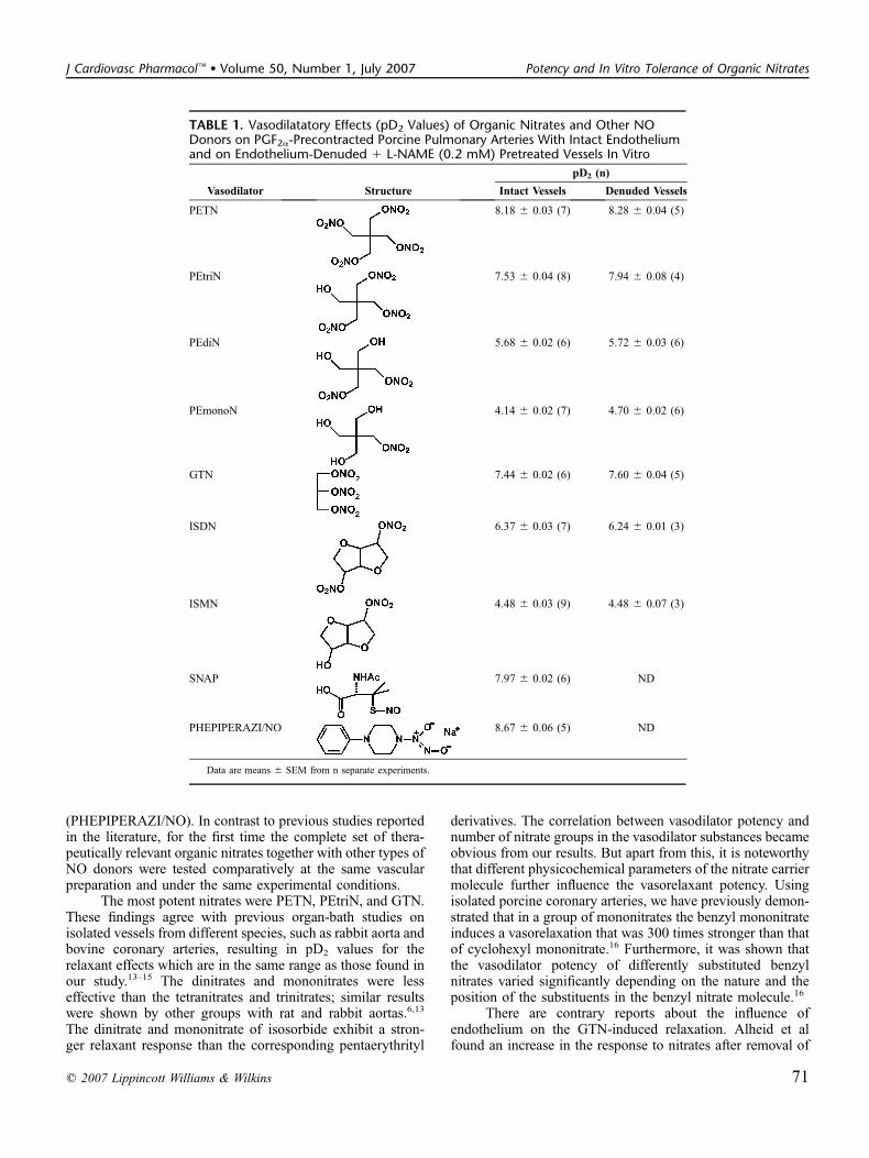
(PHEPIPERAZI/NO). In contrast to previous studies reportedin the literature, for the first time the complete set of thera-peutically relevant organic nitrates together with other types ofNO donors were tested comparatively at the same vascularpreparation and under the same experimental conditions.
The most potent nitrates were PETN, PEtriN, and GTN.These findings agree with previous organ-bath studies onisolated vessels from different species, such as rabbit aorta andbovine coronary arteries, resulting in pD2 values for therelaxant effects which are in the same range as those found inour study.13–15 The dinitrates and mononitrates were lesseffective than the tetranitrates and trinitrates; similar resultswere shown by other groups with rat and rabbit aortas.6,13
The dinitrate and mononitrate of isosorbide exhibit a stron-ger relaxant response than the corresponding pentaerythrityl
derivatives. The correlation between vasodilator potency andnumber of nitrate groups in the vasodilator substances becameobvious from our results. But apart from this, it is noteworthythat different physicochemical parameters of the nitrate carriermolecule further influence the vasorelaxant potency. Usingisolated porcine coronary arteries, we have previously demon-strated that in a group of mononitrates the benzyl mononitrateinduces a vasorelaxation that was 300 times stronger than thatof cyclohexyl mononitrate.16 Furthermore, it was shown thatthe vasodilator potency of differently substituted benzylnitrates varied significantly depending on the nature and theposition of the substituents in the benzyl nitrate molecule.16
There are contrary reports about the influence ofendothelium on the GTN-induced relaxation. Alheid et alfound an increase in the response to nitrates after removal of
TABLE 1. Vasodilatatory Effects (pD2 Values) of Organic Nitrates and Other NODonors on PGF2a-Precontracted Porcine Pulmonary Arteries With Intact Endotheliumand on Endothelium-Denuded 1 L-NAME (0.2 mM) Pretreated Vessels In Vitro
Vasodilator Structure
pD2 (n)
Intact Vessels Denuded Vessels
PETN 8.18 6 0.03 (7) 8.28 6 0.04 (5)
PEtriN 7.53 6 0.04 (8) 7.94 6 0.08 (4)
PEdiN 5.68 6 0.02 (6) 5.72 6 0.03 (6)
PEmonoN 4.14 6 0.02 (7) 4.70 6 0.02 (6)
GTN 7.44 6 0.02 (6) 7.60 6 0.04 (5)
ISDN 6.37 6 0.03 (7) 6.24 6 0.01 (3)
ISMN 4.48 6 0.03 (9) 4.48 6 0.07 (3)
SNAP 7.97 6 0.02 (6) ND
PHEPIPERAZI/NO 8.67 6 0.06 (5) ND
Data are means 6 SEM from n separate experiments.
q 2007 Lippincott Williams & Wilkins 71
J Cardiovasc Pharmacol� � Volume 50, Number 1, July 2007 Potency and In Vitro Tolerance of Organic Nitrates
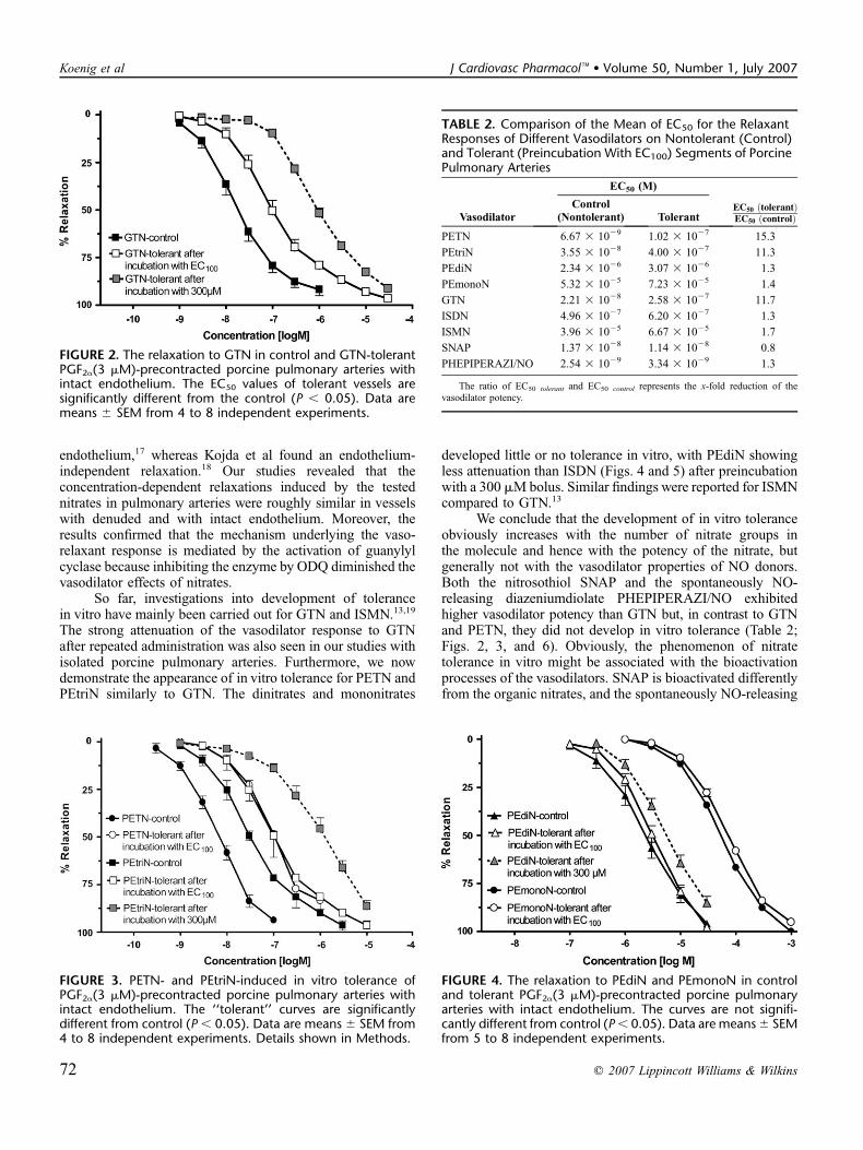
endothelium,17 whereas Kojda et al found an endothelium-independent relaxation.18 Our studies revealed that theconcentration-dependent relaxations induced by the testednitrates in pulmonary arteries were roughly similar in vesselswith denuded and with intact endothelium. Moreover, theresults confirmed that the mechanism underlying the vaso-relaxant response is mediated by the activation of guanylylcyclase because inhibiting the enzyme by ODQ diminished thevasodilator effects of nitrates.
So far, investigations into development of tolerancein vitro have mainly been carried out for GTN and ISMN.13,19
The strong attenuation of the vasodilator response to GTNafter repeated administration was also seen in our studies withisolated porcine pulmonary arteries. Furthermore, we nowdemonstrate the appearance of in vitro tolerance for PETN andPEtriN similarly to GTN. The dinitrates and mononitrates
developed little or no tolerance in vitro, with PEdiN showingless attenuation than ISDN (Figs. 4 and 5) after preincubationwith a 300 mMbolus. Similar findings were reported for ISMNcompared to GTN.13
We conclude that the development of in vitro toleranceobviously increases with the number of nitrate groups inthe molecule and hence with the potency of the nitrate, butgenerally not with the vasodilator properties of NO donors.Both the nitrosothiol SNAP and the spontaneously NO-releasing diazeniumdiolate PHEPIPERAZI/NO exhibitedhigher vasodilator potency than GTN but, in contrast to GTNand PETN, they did not develop in vitro tolerance (Table 2;Figs. 2, 3, and 6). Obviously, the phenomenon of nitratetolerance in vitro might be associated with the bioactivationprocesses of the vasodilators. SNAP is bioactivated differentlyfrom the organic nitrates, and the spontaneously NO-releasing
FIGURE 2. The relaxation to GTN in control and GTN-tolerantPGF2a(3 mM)-precontracted porcine pulmonary arteries withintact endothelium. The EC50 values of tolerant vessels aresignificantly different from the control (P , 0.05). Data aremeans 6 SEM from 4 to 8 independent experiments.
TABLE 2. Comparison of the Mean of EC50 for the RelaxantResponses of Different Vasodilators on Nontolerant (Control)and Tolerant (Preincubation With EC100) Segments of PorcinePulmonary Arteries
Vasodilator
EC50 (M)
Control(Nontolerant) Tolerant
EC50 ðtolerantÞEC50 ðcontrolÞ
PETN 6.67 3 1029 1.02 3 1027 15.3
PEtriN 3.55 3 1028 4.00 3 1027 11.3
PEdiN 2.34 3 1026 3.07 3 1026 1.3
PEmonoN 5.32 3 1025 7.23 3 1025 1.4
GTN 2.21 3 1028 2.58 3 1027 11.7
ISDN 4.96 3 1027 6.20 3 1027 1.3
ISMN 3.96 3 1025 6.67 3 1025 1.7
SNAP 1.37 3 1028 1.14 3 1028 0.8
PHEPIPERAZI/NO 2.54 3 1029 3.34 3 1029 1.3
The ratio of EC50 tolerant and EC50 control represents the x-fold reduction of thevasodilator potency.
FIGURE 3. PETN- and PEtriN-induced in vitro tolerance ofPGF2a(3 mM)-precontracted porcine pulmonary arteries withintact endothelium. The ‘‘tolerant’’ curves are significantlydifferent from control (P , 0.05). Data are means 6 SEM from4 to 8 independent experiments. Details shown in Methods.
FIGURE 4. The relaxation to PEdiN and PEmonoN in controland tolerant PGF2a(3 mM)-precontracted porcine pulmonaryarteries with intact endothelium. The curves are not signifi-cantly different from control (P, 0.05). Data are means6 SEMfrom 5 to 8 independent experiments.
72 q 2007 Lippincott Williams & Wilkins
Koenig et al J Cardiovasc Pharmacol� � Volume 50, Number 1, July 2007

diazeniumdiolate PHEPIPERAZI/NO needs no bioactivation.Within the organic nitrates, only the high-potency compoundsGTN, PETN, and PEtriN undergo ALDH2-catalyzed bio-activation, and they are sensitive to development of in vitrotolerance unlike the low-potency dinitrates and mononitrates.5
However, the in vitro tolerance allows only few insights in themechanism of clinical tolerance caused by chronic treatment;additional factors may play an important role in vivo. There arevarious reports in the literature that the therapy with PETNdoes not induce oxidative stress and this is supposed to bea key factor in preventing clinical tolerance.2 Especially, theincreased heme oxygenase (HO)-1 expression and ensuingformation of cytoprotective bilirubin may contribute as well.20
All of our in vitro findings are in contrast to thetherapeutic use of PETN, which requires high oral dosage and
does not develop significant tolerance.2 The missing bio-availability for PETN—and also PEtriN—due to poorabsorption or a fast hydrolytic degradation to less vasoactivemetabolites may explain the discrepancy between in vitro andin vivo.21,22 Recently, we could excluded hydrolytic degrada-tion of PETN in different mediums.12 Only rapid degradationin vitro occurred after adding thiol. Thus, PETN is supposed toundergo enzymatic denitration by intestinal microorganismsyielding the lower nitrate metabolites PEtriN, PEdiN, andPEmonoN.23 After peroral application of PETN to humans, itwas found in the feces and remained present throughout a3-day period after the drug was administered.21 To our knowl-edge, PETN has never been quantified in the blood and PEtriNhas only been detected occasionally at very low concen-trations. Only PEdiN and PEmonoN have been found at higherconcentrations in the blood.24 In healthy volunteers, theplasma half-life time of PEdiN and PEmonoN amounted to4 to 5 hours and 10 to 11 hours, respectively.25 After feeding ratsPETN, we found considerable amounts of PETN in the feces,but little PEtriN and very little PEdiN. In agreement withhuman investigations, neither PETN nor PEtriN were detectedin the blood of rats; only PEdiN could be quantified by theHPLC protocol. Therefore, according to the in vivo findings,in the future in vitro studies on the pharmacological effects ofPETN should also include the effects of PEdiN and PEmonoN.
We conclude that, in vivo, PETN is highly stable andvery poorly soluble and absorbable. Biotransformation andabsorption produce successively limited blood levels of PEdiNand PEmonoN. As shown by our investigations, these twometabolites contribute to the moderate, long-acting, andtolerance-devoid activity of the PETN.
ACKNOWLEDGMENTSA Ph.D. scholarship for A.K. by Graduiertenforderung
(Thuringia, Germany) is gratefully acknowledged. Theauthors also thank Dr. Kleinhans (Veterinaramt WeimarerLand, Nohra) for providing with lungs from pigs for this studyand Actavis Deutschland for financial support.
REFERENCES1. Fink B, Bassenge E. Unexpected, tolerance-devoid vasomotor and platelet
actions of pentaerythrityl tetranitrate. J Cardiovasc Pharmacol. 1997;30:831–836.
FIGURE 6. Vasorelaxation induced by PHEPIPERAZI/NO(NONOate) and SNAP in control and tolerant PGF2a(3 mM)-precontracted porcine pulmonary arteries with intact endo-thelium. The ‘‘tolerant’’ curves are not significantly differentfrom control (P , 0.05). Data are means 6 SEM from twoindependent experiments.
FIGURE 5. The relaxation to ISDN and ISMN in control andtolerant PGF2a(3 mM)-precontracted porcine pulmonary ar-teries with intact endothelium. Data are means 6 SEM from 5to 8 independent experiments.
TABLE 3. Whole Blood and Feces Levels of PETN and ItsMetabolites Upon Administration of 7 mg/kg PETN 2 TimesPer Day by Gavage for 3 Days to Rats (n = 12)
Sample Source PETN PEtriN PEdiN
Blood (ng/mL) ND ND 17–33
Feces (ng/g)
Day 1 38.6–53.9 13.5–16 6.6–8.6
Day 2 38.5–129.5 19.1–24 5.4–8.6
Day 3 120.8–136.7 18.1–21.9 6.4–11.6
ND, not determined because the concentrations in the blood samples were mostly(n = 10) at the LOQ of 11 ng/mL. Only two samples have shown higher amounts. Fecessamples were collected from three animals.
q 2007 Lippincott Williams & Wilkins 73
J Cardiovasc Pharmacol� � Volume 50, Number 1, July 2007 Potency and In Vitro Tolerance of Organic Nitrates

2. Jurt U, Gori T, Ravandi A, et al. Differential effects of pentaerythritoltetranitrate and nitroglycerin on the development of tolerance andevidence of lipid peroxidation: A human in vivo study. J Am Coll Cardiol.2001;38:854–859.
3. Pfaffenrath V, de la Motte S, Harrison F, et al. Effect of pentaerithritoltetranitrate, isosorbide mononitrate and placebo on the headache and onthe ability to work of healthy volunteers. Arzneimittelforschung. 1998;48:646–650.
4. Schwemmer M, Bassenge E. New approaches to overcome tolerance tonitrates. Cardiovasc Drugs Ther. 2003;17:159–173.
5. Munzel T, Daiber A, Mulsch A. Explaining the phenomenon of nitratetolerance. Circ Res. 2005;97:618–628.
6. Daiber A, Oelze M, Coldewey M, et al. Oxidative stress andmitochondrial aldehyde dehydrogenase activity: a comparison ofpentaerythritol tetranitrate with other organic nitrates. Mol Pharmacol.2004;66:1372–1382.
7. Chen ZQ, Foster MW, Zhang J, et al. An essential role for mitochondrialaldehyde dehydrogenase in nitroglycerin bioactivation. Proc Natl AcadSci U S A. 2005;102:12159–12164.
8. Glusa E, Pertz HH. Further evidence that 5-HT-induced relaxation of pigpulmonary artery is mediated by endothelial 5-HT2B receptors. Br JPharmacol. 2000;130:692–698.
9. Decker M, Koenig A, Glusa E, et al. Synthesis and vasorelaxantproperties of hybrid molecules out of NO-donors and the beta-receptor blocking drug propranolol. Bioorg Med Chem Lett. 2004;14:4995–4997.
10. Konter J, Abou-Rahma El-Din AAH, El-Emam A, et al. The NOtizer -a device for the convenient preparation of diazen-1-ium-1,2-diolates.Methods Enzymol. 2005;396:17–26.
11. Glusa E, AdamC. Endothelium-dependent relaxation induced by cathepsinG in porcine pulmonary arteries. Br J Pharmacol. 2001;133:422–428.
12. Seeling A, Lehmann J. NO-donors, part X [1]: Investigations on the stabil-ity of pentaerythrityl tetranitrate (PETN) by HPLC-chemoluminescence-N-detection (CLND) versus UV-detection in HPLC. J Pharm BiomedAnal. 2006;40:1131–1136.
13. Kukovetz WR, Holzmann S. Mechanisms of nitrate-induced vaso-dilatation and tolerance development. Eur J Clin Pharmacol. 1991;40:103–109.
14. Kojda G, Hacker A, Noack E. Effects of nonintermittent treatment ofrabbits with pentaerythritol tetranitrate on vascular reactivity andsuperoxide production. Eur J Pharmacol. 1998;355:23–31.
15. Husgen B, Noack E, Kojda G. Comparison of the vasorelaxing effect ofdifferent nitrovasodilators in conductive arterial and venous blood vessels.In: Schror K, Pace-Asciak CR, eds. Mediators in the CardiovascularSystem: Regional Ischemia. Basel: Birkhauser Verlag; 1995:183–187.
16. Wessler C, Homann A, Fricke U, et al. NO donors, part 8 [1]: synthesisand vasodilating activities of substituted benzylnitrates compared tocyclohexylmethylnitrate and GTN. Eur J Med Chem. 2003;38:581–586.
17. Alheid U, Dudel C, Forstermann U. Selective-inhibition by gossypol ofendothelium-dependent relaxations augments relaxations to glyceryltrinitrate in rabbit celiac artery. Br J Pharmacol. 1987;92:237–240.
18. Kojda G, Beck JK, Meyer W, et al. Nitrovasodilator-induced relaxationand tolerance development in porcine vena cordis magna - dependence onintact endothelium. Br J Pharmacol. 1994;112:533–540.
19. Chen ZQ, Zhang J, Stamler JS. Identification of the enzymatic mechanismof nitroglycerin bioactivation. Proc Natl Acad Sci USA. 2002;99:8306–8311.
20. Oberle S, Abate A, Grosser N, et al. Heme oxygenase-1 induction mayexplain the antioxidant profile of pentaerythrityl trinitrate. BiochemBiophys Res Commun. 2002;290:1539–1544.
21. Davidson IW, Miller HS, Dicarlo FJ. Absorption, excretion andmetabolism of pentaerythritol tetranitrate by humans. J Pharmacol ExpTher. 1970;175:42–50.
22. Davidson IW, Rollins FO, Dicarlo FJ, et al. Pharmacodynamics andbiotransformation of pentaerythritol trinitrate in man. Clin PharmacolTher. 1971;12:972–981.
23. King SY, Fung HL. Rapid microbial degradation of organic nitrates inrat excreta. Re-examination of the urinary and fecal metabolite profilesof pentaerythritol tetranitrate in the rat. Drug Metab Dispos. 1984;12:353–357.
24. Neurath GB, Dunger M. Blood levels of metabolites of glyceryl trinitrateand pentaerythritol tetranitrate after administration of a two-steppreparation. Arzneimittelforschung. 1977;27:416–419.
25. Weber W, Michaelis K, Luckow V, et al. Pharmacokinetics andbioavailability of pentaerithrityl tetranitrate and two of its metabolites.Arzneimittelforschung. 1995;45:781–784.
74 q 2007 Lippincott Williams & Wilkins
Koenig et al J Cardiovasc Pharmacol� � Volume 50, Number 1, July 2007
Veröffentlichung 5 72-89
Veröffentlichung 5
Permeability changes in response to NONOate and NONOate prodrug derived nitric oxide in a blood-brain
barrier model formed by primary porcine endothelial cells
Winter, Sven; Konter, Joerg; Scheler, Stefan; Lehmann, Jochen; Fahr,
Alfred. Nitric Oxide. Eingereicht (2007).

Permeability changes in response to NONOate and NONOate prodrug derived nitric oxide in a blood-brain barrier model formed by primary porcine endothelial cells
Sven Wintera, Joerg Konterb, Stefan Schelera, Jochen Lehmannb, and Alfred Fahra
a Friedrich-Schiller-University Jena, Department of Pharmaceutical Technology, Lessingstrasse 8,
Jena, Germany b Friedrich-Schiller-University Jena, Department of Pharmaceutical/Medicinal Chemistry,
Philosophenweg 14, Jena, Germany
Corresponding author:
Sven Winter, Department of Pharmaceutical Technology, Lessingstrasse 8, 07743 Jena, Germany
Telephone: +49-176-23202378
Fax: +49-7344-143237
e-mail: [email protected]
Private address: Erhard-Grözinger-Str. 11
89134 Blaustein
Germany
Abstract The influence of nitric oxide (NO) and NO-donors on the permeability of the blood-brain barrier (BBB)
is still not well understood and the literature is quite controversial. Some studies revealed increasing,
others decreasing or even no effects of NO-donors on the BBB permeability. In this work we report
about the influence of three diazeniumdiolates, which release NO spontaneously and three different
diazeniumdiolate prodrugs, which have to be cleaved chemically or enzymatically before releasing
NO, on the BBB permeability of a primary porcine endothelial cell monolayer. By measuring the flux of
a small polar molecule (carboxyfluorescein: CF) we could show, that the releasers PHEPIPERAZI/NO,
DBA/NO, DETA/NO reduced the BBB permeability. In contrast, the prodrugs Et-PHEPIPERAZI/NO
and Tosyl-PYRRO/NO increased the permeability, whereas the prodrug Et-BUPIPERAZI/NO reduced
it. Blocking the effect of the BBB permeability reducing compounds could be done by methylene blue,
whereas permeability increasing effects could not be blocked.
Keywords: Diazeniumdiolate; NO-donor; NONOate; blood-brain barrier; primary porcine endothelial
cells; permeability
Introduction
Nitric oxide (NO) has been found to exert positive [1; 2; 3; 4] and negative [5; 6] influence on the
permeability of venules of different origin. Most of the investigations were done by using NO agonists
or NO synthase inhibitors to discern its role on vascular permeability. Further studies used NO-donors
or gaseous NO to investigate the permeability changing. The findings of these studies are
controversial, since some authors have shown a decrease in peripheral vascular permeability or no
effect [7; 8; 9; 10; 11], while others have found an increase by the use of NO-donors [12; 13; 14].
Studying the effect of different NO-donors on the cerebrovascular endothelium showed an increasing
effect on the permeability [15; 16] especially by using higher concentrations of the NO-donor in the
blood-brain barrier model [17]. What kind of influence a NO-donor exerts on cells is also dependent on
the “kind of NO” which is released [18]. The three different redox forms NO•, NO+ and NO- can be
released depending on the used type of NO-donor. In this in vivo investigation three diazen-1-ium-1,2-
diolate derivatives, also called NONOates, and three different prodrugs of these direct releasers were
synthesized and tested (Fig. 1). The nitrogen bound NONOates, used in this study, release the NO•
radical exclusively and are ideal tools to study the effects of nitric oxide on the BBB, due to their
predictable first order NO liberation and variable half lives depending on the NONOate used. But,
because of the short half-life of some compounds in aqueous buffer, the actual intracellular NO
concentration is questionable which limits their usefulness in in vivo experiments or even as drugs with
a therapeutic aim. One possibility to circumvent this limitation is the design of prodrugs that can not
release NO unless they are chemically or enzymatically converted to the drug.
The cell culture model used in this study was formed by freshly isolated porcine brain endothelial cells,
growing on the upper side of a Transwell® filter insert. To obtain information on the tightness of the
endothelial monolayer, the transendothelial electrical resistance (TEER) was measured by using a
chopstick electrode. In order to gain information on the influence of different NONOates and NONOate
prodrugs on the permeability of the blood-brain barrier model, the flux of a marker substance (CF) was
determined.
The first goal of this study was to examine the general influence of NO on the permeability of
monolayers of primary porcine endothelial cells. The second aim was to find out if there are
differences in the effect of spontaneously releasing NONOates and the NONOate prodrugs.
Materials and methods
Materials and Chemicals
The cell culture media M199 and Dulbecco’s minimal essential medium/Ham’s F12 (DMEM/Ham’s
F12), 10-fold hanks balanced salt solution (HBSS), horse serum used for the cell culture media,
penicillin/streptomycin 100-fold solution and the trypsin were purchased from PAA Laboratories GmbH
(Austria). Puromycin for purity improvement of the cells was purchased from Calbiochem®, Merck
Biosciences GmbH (Germany), the enzymes dispase and collagenase from Invitrogen GmbH
(Germany). Rat tail collagen solution (4 mg/ml) was obtained from SERVA Electrophoresis GmbH
2
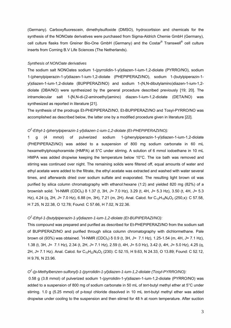
(Germany). Carboxyfluorescein, dimethylsulfoxide (DMSO), hydrocortision and chemicals for the
synthesis of the NONOate derivatives were purchased from Sigma-Aldrich Chemie GmbH (Germany),
cell culture flasks from Greiner Bio-One GmbH (Germany) and the Costar® Transwell® cell culture
inserts from Corning B.V Life Sciences (The Netherlands).
Synthesis of NONOate derivatives
The sodium salt NONOates sodium 1-(pyrrolidin-1-yl)diazen-1-ium-1,2-diolate (PYRRO/NO), sodium
1-(phenylpiperazin-1-yl)diazen-1-ium-1,2-diolate (PHEPIPERAZI/NO), sodium 1-(butylpiperazin-1-
yl)diazen-1-ium-1,2-diolate (BUPIPERAZI/NO) and sodium 1-(N,N-dibutylamino)diazen-1-ium-1,2-
diolate (DBA/NO) were synthesized by the general procedure described previously [19; 20]. The
intramolecular salt 1-[N,N-di-(2-aminoethyl)amino) diazen-1-ium-1,2-diolate (DETA/NO) was
synthesized as reported in literature [21].
The synthesis of the prodrugs Et-PHEPIPERAZI/NO, Et-BUPIPERAZI/NO and Tosyl-PYRRO/NO was
accomplished as described below, the latter one by a modified procedure given in literature [22].
O2-Ethyl-1-(phenylpiperazin-1-yl)diazen-1-ium-1,2-diolate (Et-PHEPIPERAZI/NO):
1 g (4 mmol) of pulverized sodium 1-(phenylpiperazin-1-yl)diazen-1-ium-1,2-diolate
(PHEPIPERAZI/NO) was added to a suspension of 800 mg sodium carbonate in 60 mL
hexamethylphosphoramide (HMPA) at 5°C under stirring. A solution of 6 mmol iodoethane in 10 mL
HMPA was added dropwise keeping the temperature below 10°C. The ice bath was removed and
stirring was continued over night. The remaining solids were filtered off, equal amounts of water and
ethyl acetate were added to the filtrate, the ethyl acetate was extracted and washed with water several
times, and afterwards dried over sodium sulfate and evaporated. The resulting light brown oil was
purified by silica column chromatography with ethanol:hexane (1:2) and yielded 820 mg (82%) of a
brownish solid. 1H-NMR (CDCl3) δ 1.37 (t, 3H, J= 7.0 Hz), 3.29 (t, 4H, J= 5.3 Hz), 3.50 (t, 4H, J= 5.3
Hz), 4.24 (q, 2H, J= 7.0 Hz), 6.88 (m, 3H), 7.21 (m, 2H). Anal. Calcd. for C12H18N4O2 (250,x): C 57.58,
H 7.25, N 22.38, O 12.78; Found: C 57.66, H 7.02, N 22.36.
O2-Ethyl-1-(butylpiperazin-1-yl)diazen-1-ium-1,2-diolate (Et-BUPIPERAZI/NO):
This compound was prepared and purified as described for Et-PHEPIPERAZI/NO from the sodium salt
of BUPIPERAZI/NO and purified through silica column chromatography with dichlormethane. Pale
brown oil (93%) was obtained. 1H-NMR (CDCl3) δ 0.9 (t, 3H, J= 7.1 Hz), 1.25-1.54 (m, 4H, J= 7.1 Hz),
1.38 (t, 3H, J= 7.1 Hz), 2.34 (t, 2H, J= 7.1 Hz), 2.59 (t, 4H, J= 5.0 Hz), 3.42 (t, 4H, J= 5.0 Hz), 4.25 (q,
2H, J= 7.1 Hz). Anal. Calcd. for C10H22N4O2 (230): C 52.15, H 9.63, N 24.33, O 13.89; Found: C 52.12,
H 9.76, N 23.96.
O2-(p-Methylbenzen-sulfonyl)-1-(pyrrolidin-1-yl)diazen-1-ium-1,2-diolate (Tosyl-PYRRO/NO):
0.58 g (3.8 mmol) of pulverized sodium 1-(pyrrolidin-1-yl)diazen-1-ium-1,2-diolate (PYRRO/NO) was
added to a suspension of 800 mg of sodium carbonate in 50 mL of tert-butyl methyl ether at 5°C under
stirring. 1.0 g (5.25 mmol) of p-tosyl chloride dissolved in 10 mL tert-butyl methyl ether was added
dropwise under cooling to the suspension and then stirred for 48 h at room temperature. After suction
3
filtration, the clear colorless filtrate was kept refrigerated until white crystals precipitated (62% yield). 1H-NMR (CDCl3) δ 1.97 (t, 4H, J= 5.0 Hz), 2.46 (s, 3H), 3.60 (t, 4H, J= 5.0 Hz), 7.36 (d, 2H, J= 8.1
Hz), 7.89 (d, 2H, J= 8.1 Hz). Anal. Calcd. for C11H15N3O4S (285): C 46.30, H 5.30, N 14.73, O 22.43, S
11.24; Found: C 46.21, H 5.09, N 14.92, S 11.16.
The half-lives for the NO release from the NO donors was determined with a Jasco V-570 UV-
spectrometer in phosphate buffer pH 7.4 at 37°C with a NONOate concentration of 10–4M. The
decrease of absorption was measured at a wavelength of 251 nm over a period of 30 minutes. By
plotting the absoption logarithmically and linear regression analysis the rate constants (k) were
determining and used to calculate the half-lives [s] of the respective NONOates using the formula t1/2=
ln2/k. R2-values were higher than 0,998 and every measurement was carried out at least three times
to ensure reproducibility within ±5% [22].
Isolation of porcine brain endothelial cells
The isolation procedure was described in detail by Franke et. al. [23]. In brief, three fresh porcine
brains obtained from the slaughterhouse were purged from meninges and capillaries, followed by the
digestion of brains with the enzyme dispase to release the capillaries. After separating the capillaries
by centrifugation and filtration a second enzymatic digestion was carried out using a mixture of
dispase and collagenase (1+1). The obtained endothelial cells were seeded in T175-cell culture flasks
with rat-tail-collagen-coated surfaces. For one brain two T175-cell culture flasks were needed.
Cell culture conditions
For culturing the porcine endothelial cells, puromycin-containing medium M199 was used on the first
two days after extraction, to assure the highest possible purity of the endothelial cells. The puromycin-
containing medium contained 10 % horse serum, 200 units/ml penicillin, 0.2 mg/ml streptomycin and 2
µg/ml puromycin. After the first two days, the medium was replaced with a horse-serum containing
M199 medium without puromycin. This medium was changed on the 4th day after the extraction of the
porcine endothelial cells to a serum free Dulbecco's Modified Eagle Medium/Ham’s F12 medium
(DMEM/Ham’s F12 medium). DMEM/Ham’s F12 contained 200 units/ml penicillin, 0.2 mg/ml
streptomycin and 550 µM hydrocortisone.
Coating of flasks and filters
All surfaces on which endothelial cells grew and formed cell monolayers had to be coated with rat-tail
collagen [24]. For coating of the T175-flasks with a surface of 175 cm², 3 ml of the 0.1 mg/ml rat-tail
collagen solution were added to the flask and then the flask was dried for 3 days at 37 °C. No further
washing steps were performed, before the cell suspension was added to the flask. For the coating of
the Costar® Transwell® filter inserts, a solution of 0.04 mg/ml rat tail collagen was prepared. 100 µl of
the rat tail collagen solution was added to the filter insert and incubated over night in an incubator at
37 °C. Finally the inserts were totally dried in the air flow of the laminar air flow workstation.
4

Permeability experiments
To control the tightness of the endothelial monolayer the transendothelial electrical resistance (TEER)
was measured by using a chopstick electrode and the Millicell®-ERS-device. Since the monolayer is
tight when the resistance is above 300 Ohm*cm² [23], all permeability experiments were done with
monolayers showing a resistance higher than 300 Ohm*cm². Monolayers with lower resistance were
not included in the investigation. In order to estimate the permeability of the endothelial monolayer, the
CF flux was determined. The medium in the acceptor compartment of the inserts was replaced with
the hank’s balanced salt solution (HBSS). For the control experiment, CF was added to the inner
compartment of the inserts at a concentration of 1 µM and the amount of CF in the acceptor
compartment was measured spectroscopically. To estimate the influence of NO-donor substances the
NO-donor was dissolved in 0.1 N NaOH (1µl) and then added to the inner compartment of the
Transwell® insert parallel to the addition of CF. NO-donor prodrugs were dissolved in DMSO (1µl) and
then added to the inner compartment. During the experiment the Corning® Transwell® filter containing
12-well plate was shaken at 500 rpm. Every 10 minutes a sample of 100 µl was withdrawn from the
acceptor compartment and poured into a well of a 96-well-plate. The acceptor compartment was
refilled with 100 µl of the fresh buffer solution. The fluorescence of the sample was measured in a
plate reader (BMG FLUOstar Optima, Germany) and the CF concentration was calculated by the
calibration curve analysis. The CF concentration used for the calibration curve ranged from 0.0001 µM
to 1 µM.
Methylene blue experiments
The effect of methylene blue on the influence of NO-donors and NO-donor prodrugs was investigated
in a separate experiment.
A concentration of 50 µM of methylene blue was chosen to inhibit the intracellular available guanylate
cyclase. 5 µl Methylene blue solution (5 mM) was added 10 minutes before the addition of the NO-
donor compounds to ensure that methylene blue penetrates into the endothelial cells and inhibit the
guanylate cyclase. 1.25 µl per insert of a 100 mM solution of sodium salt NONOates or NO-donor
prodrugs were used.
Estimation of the apparent permeability coefficient (Papp)
To estimate the Papp, we used the method described in literature [25; 26]. The Papp-value was
calculated by using the following equation:
Papp=dQ/t*VA/A*1/100%*1/x
dQ/dt represents the slope of the diagram concentration versus time (%/sec), VA represents the
volume of the acceptor compartment (1.5 ml), A represents the surface of the filter insert (1.13 cm²)
and x represents the ratio between the volume of the acceptor compartment and the volume of the
donor compartment (x was 3 in all experiments).
Statistical analysis
Data are presented as means ± standard errors of the mean (SEM). Comparisons between groups
were made using Student’s t test. Differences were considered significant at p ≤ 0.05 level.
5

Results
Effect of direct NONOates on the BBB.
For this study we used the three NO-donors: PHEPIPERAZI/NO, DBA/NO and DETA/NO (see Fig. 1),
that liberate NO directly after dissolution in aqueous media. The half-life of the NO-donors at pH 7.4
range from minutes for PHEPIPERAZI/NO and DBA/NO to several hours for DETA/NO. The
investigated NONOate salts decreased the permeability of the endothelial cell monolayer for CF in a
dose-dependant manner (Figs. 2-4). In Fig. 2 the CF concentration in the acceptor compartment
versus time is shown, after treatment with three different concentrations of PHEPIPERAZI/NO (which
has a half-life of approximately four minutes). PHEPIPERAZI/NO induced a significant decrease of the
Papp even at the lowest used concentration of 100 µM. By increasing the NO-donor concentration, the
BBB permeability further decreased. The Papp for the treatment at 100 µM was 9.00 x 10⁻⁶ ± 3.32 x 10⁻⁶
cm*s-1, 3.00 x 10⁻⁶ ± 1.07 x 10⁻⁶ cm*s-1 for 250 µM and only slightly lower when treated with 500 µM of
PHEPIPERAZI/NO (2.18 x 10⁻⁶ ± 0.80 x 10⁻⁶ cm*s-1). The untreated monolayer showed a significant
higher permeability with a permeation coefficient of 27.74 x 10⁻⁶ ± 8.61 x 10⁻⁶ cm*s-1. Furthermore, the
increase of CF concentration – on a lower level compared to control – was quite linear with time. In
conclusion, the treatment with PHEPIPERAZI/NO in different concentrations lowered the permeability
in a dose dependent manner. By using 100 µM the permeability coefficient was reduced to 38.0 ± 8.7
% of the control, further reductions were achieved with higher concentrations (250 µM: -12.7 ± 2.8 %
and 500 µM: - 9.2 ± 2.1 %).
DBA/NO reduced the permeability for CF in all investigated concentrations (100 µM, 250 µM and 500
µM) compared to the untreated monolayers (Fig. 3). The half-life for DBA/NO is approximately 1.5 min
at pH 7.4. The increase of the CF concentration in the acceptor compartment was very low compared
to the experiment with PHEPIPERAZI/NO. Note that this was due to the higher tightness of the
monolayer in the batch used for this experiment. The apparent permeability coefficients of the CF flux
after the treatment with the three investigated concentrations of DBA/NO were lower than the apparent
permeability coefficient of the flux of the untreated cells. In contrast to the action of PHEPIPERAZI/NO,
there was no significant difference between the three investigated concentrations (100 µM, 250 µM,
500 µM) of DBA/NO and the permeation coefficients were found to be similar: for 100 µM the Papp was
0.64 x 10⁻⁶ ± 0.005 x 10⁻⁶ cm*s-1, for 250 µM it was 0.66 x 10⁻⁶ ± 0.21 x 10⁻⁶ cm*s-1 and using the
highest concentration of 500 µM the Papp was 0.46 x 10⁻⁶ ± 0.14 x 10⁻⁶ cm*s-1. The untreated
monolayer reached a Papp of 1.83 x 10⁻⁶ ± 0.17 x 10⁻⁶ cm*s-1. The reduction of the apparent
permeability coefficient for the 100 µM and the 250 µM concentration of DBA/NO was nearly the same
(35.0 ± 1.5 % and 36.3 ± 7.4 % of the control). After applying DBA/NO in a concentration of 500 µM
the Papp-value was reduced to 25.2 ± 4.9 % of the control.
Similarly, the treatment with DETA/NO (compound with the longest half-life of the investigated non-
prodrug NO-donors, i.e. ~20 hours in a solution of pH 7.4 at 37°C [27]), resulted in the reduction of the
permeability of the marker substance (Fig. 4) and lowered permeability coefficients, especially at
higher concentrations. The influence of the low concentration of DETA/NO, i.e. 100 µM, was weak and
there is no significant difference in the permeation coefficients of 100 µM NO-donor treatment and
6

untreated control cells. For untreated endothelial cells (control) a Papp of 27.74 x 10⁻⁶ ± 8.61 x 10⁻⁶
cm*s-1 was found which slightly decreased with the use of the 100 µM DETA/NO to a value of 24.04 x
10-6 ± 4.34 x 10⁻⁶ cm*s-1. The 250 µM DETA/NO solution yielded a higher significant reduction of the
permeability, especially in the first 60 minutes (Papp: 11.10 x 10⁻⁶ ± 2.00 x 10⁻⁶ cm*s-1) after addition.
Surprisingly, at a concentration of 500 µM no further permeability decrease was observed (11.63 x 10-
6 ± 2.07 x 10⁻⁶ cm*s-). The use of the 100 µM DETA/NO did not change the permeability coefficient
compared to the control (101.6 ± 15.5 % of the control). However, Papp decreased using higher
concentrations of DETA/NO (250 µM: -46.9 ± 7.2 % and 500 µM: - 49.2 ± 7.5 %).
Nitric oxide donor prodrugs
NO donor prodrugs like Et-PHEPIPERAZI/NO, Et-BUPIPERAZI/NO and Tosyl-PYRRO/NO (Fig. 1)
have to be cleaved by cell enzymes in order to produce the corresponding NO releasing NONOates
PHEPIPERAZI/NO (t1/2= 4 min); BUPIPERAZI/NO (t1/2= 3 min) and PYRRO/NO (t1/2= 3 sec.).
A concentration vs. time plot (Fig. 5) shows the influence of increasing concentrations of the NO-
donor-prodrug Et-PHEPIPERAZI/NO on the permeability of CF in the BBB model. In contrast to the
treatment with the corresponding “drug” PHEPIPERAZI/NO, the permeability of the marker CF
increases dramatically in a dose dependent manner. In this experiment, the untreated cells (control)
showed a permeation coefficient of 1.88 x 10⁻⁶ ± 0.17 x 10⁻⁶ cm*s-1. The lowest increase was obtained
with 100 µM of Et-PHEPIPERAZI/NO, and the corresponding apparent permeation coefficient was
7.77 x 10⁻⁶ ± 0.008 x 10⁻⁶ cm*s-1, which is 416.8 ± 22.1 % of the untreated monolayer. The 250 µM
solution of Et-PHEPIPERAZI/NO increased the permeability further (Papp of 38.91 x 10⁻⁶ ± 6.54 x 10⁻⁶
cm*s-1 = 2086.4 ± 238.8 % of control) and the highest concentration of Et-PHEPIPERAZI/NO resulted
in the highest flux of CF (Papp = 57.23 x 10⁻⁶ ± 3.55 x 10⁻⁶ cm*s-1 = 3068.9 ± 174.9 % of the control).
TOSYL-PYRRO/NO, the substance with is cleaved to the fast releaser PYRRO/NO, also increased the
flux through the endothelial monolayer (Fig. 6): Papp = 31.69 x 10⁶ ± 1.18 x 10⁻⁶ cm*s-1 for the 100 µM
concentration, 92.03 x 10⁻⁶ ± 4.94 x 10⁻⁶ cm*s-1 for 250 µM and 83.93 x 10⁻⁶ ± 2.07 x 10⁻⁶ cm*s-1 for
the highest concentration of TOSYL-PYRRO/NO (Control: 4.57 x 10⁻⁶ ± 0.16 x 10⁻⁶ cm*s-1). This
increase of the CF flux was obviously not further enhanced by the use of the highest concentration of
TOSYL-PYRRO/NO (500 µM). The lowest concentration increased the permeation to 708.9 ± 15.1 %
of the control, the median concentration of 250 µM led to the highest permeation (2019.2 ± 66.2 % of
the control), while the highest investigated concentration (500 µM) led only to 1841.4 ± 45.8 % of the
control.
The third NONOate prodrug Et-BUPIPERAZI/NO influenced the primary porcine endothelial cell
monolayer differently compared to other NO-donor prodrugs. Similar to the directly NO donating
NONOates it did not increase, but it reduced the flux of CF in a concentration dependant manner (Fig.
7). With increasing the concentrations from 100 µM to 500 µM, the increase of the concentration of the
marker substance in the acceptor compartment was lower in respect to the control. The corresponding
apparent permeation coefficients illustrate this effect. For the 100 µM solution of Et-BUPIPAZI/NO the
Papp was 3.70 x 10⁻⁶ ± 0.21 x 10⁻⁶ cm*s-1, for the 250 µM solution the Papp was 2.13 x 10⁻⁶ ± 0.24 x 10⁻⁶
cm*s-1, and by applying the 500 µM Et-BUPIPERAZI/NO a Papp-value of 1.81 x 10⁻⁶ ± 0.44 x 10⁻⁶ cm*s-
7

1 was obtained. These low values represent reductions of 81.1 ± 2.7 % (100 µM), 46.7 ± 2.7 % (250
µM) and 39.6 ± 4.9 % (500 µM) of the control. For the untreated cells we found a permeation
coefficient of 4.57 x 10⁻⁶ ± 0.16 x 10⁻⁶ cm*s-1. A significant difference to the control was only observed
by using higher concentrations, i.e. 250 µM and 500 µM solution of this prodrug, whereas the curve for
the 100 µM Et-BUPIPERAZI/NO treatment was nearly identical to control.
Methylene blue experiments
Many biological effects of nitric oxide are performed by stimulating the soluble guanylate cyclase
(sGC), thus increasing the intracellular amount of cyclic guanylate monophosphate (cGMP).
Methylene blue is known to inhibit this enzyme [28; 29] and additionally acting as a NO scavenger [30;
31]. Therefore, it may block physiological effects in the cell culture system caused by subsequent
addition of NO-donors.
The results of the investigation of the methylene blue addition prior to the addition of the NO-donors or
NO-donor prodrugs to the monolayer of the endothelial cells are summarized in Table 1.
The use of the 250 µM DETA/NO resulted in a permeability Papp of 51.7 ± 2.7 % relative to control,
thus showing a significant reduction compared to the control. Upon applying DETA/NO together with
the radical scavenger methylene blue, the permeation coefficient raised to 141.4 ± 12.4 % of the
control. The same was observed using the NO-donor DBA/NO, i.e. the Papp increased from 67.0 ± 6.4
% of the control value without methylene blue to 146.7 ± 12.9 % with methylene blue. The highest
effect of methylene blue on the change of the Papp-value was observed using PHPIP/NO. The
permeation coefficient raised to 314.0 ± 20.5 % when PHPIP/NO was added. The combination of both
PHEPIPERZI/NO and methylene blue reduced the Papp-value to 67.1 ± 1.3 % of the control. Addition
of methylene blue led to a significant change of the Papp-values for all investigated NO-donors which
spontaneously release nitric oxide in aqueous medium at pH 7.4 (Fig. 8).
It was not possible to block the permeability increasing effect of TOSYL-PYRRO/NO, Et-
PHEPIPERAZI/NO and Et-BUPIPERAZI/NO using methylene blue (Fig. 8). This fact is also illustrated
by the percentage enhancement of the apparent permeability coefficient. In the case of the NO-donor
prodrug TOSYL-PYRRO/NO the percentage of the permeation coefficient enhancement was 1539.5 ±
48.0 % of the control for the experiment without methylene blue, whilst in the experiment with
methylene blue the value was 1356.0 ± 45.6 % of the control. The value of the percentage
enhancement of the Papp for the experiment with Et-PHEPIPERAZI/NO was 1210.0 ± 184.9 % of the
control without methylene blue and 1361.3 ± 85.3 % for the treatment with NO-donor prodrug and
methylene blue. Methylene blue was able to further increase the effect of the prodrug Et-
BUPIPERAZI/NO. The permeation coefficient raised from 283.0 ± 19.3 % of the control in the
experiment with Et-BUPIPERAZI/NO to a value of 938.3 ± 49.9 % of the control in the test with
methylene blue and Et-BUPIPERAZI/NO.
8

Discussion The main function of the blood-brain barrier (BBB) is the maintenance of the cerebral liquor
composition and the environment of the nerve cells. Many factors influence the regulation of the
permeability of the BBB. Nitric oxide has been reported to be one of them [10; 18; 32; 33].
Primary endothelial cells are considered to be the blood-brain barrier model closest to the in vivo
situation. They retain most of the cell properties of the brain capillary endothelial cells. Despite of this,
the biggest disadvantage in using primary endothelial cells as model is the loss of some features
during cultivation. In fact, one should not work with cells which are often passaged to gain information
which are related closer to the in vivo situation. Therefore, we worked with primary endothelial cells
which were passaged only once to ensure that all data generated on the influence of the NO-donating
compounds on the permeability of the BBB are as close as possible to the in vivo conditions.
All of the investigated NO-donors, which spontaneously release NO in the aqueous solution
(PHEPIPERAZI/NO, DBA/NO, DETA/NO), decreased the permeability of the endothelial monolayer in
our BBB model in a dose dependant manner. The strongest “tightening” effect was found for DBA/NO
and the resulting permeability coefficients were the lowest measured. PHEPIPERAZI/NO and
DETA/NO were found to be less effective and less dose dependent, i.e. the effect on the endothelial
cells could not be increased by increasing the concentration of the NO-donor above 250 µM.
In the investigation of Wong et al. [34] DETA/NO showed an increasing effect on the transendothelial
electrical resistance (TEER) and similarly a decreasing effect on the permeability for horse radish
peroxidase in an in vitro-model formed by confluent human brain microvessel endothelial cells. By
using 100 µM DETA/NO there was seen a 20% increase in TEER and a 50 % reduction for the horse
radish permeability. These data are in good correlation with our findings for the DETA/NO treatment of
endothelial monolayers. The use of 250 µM and 500 µM of DETA/NO reduced in our experiment the
permeability coefficient for CF to 50 % of the control. Boje and Lakhman [18] treated rats with
DETA/NO and found that this NO-donor has no effect on the permeability in the in vivo experiment
compared to untreated animals. The other NONOates used in the study of Boje and Lakhman showed
slight to moderate permeability increasing effects on the permeability of the blood-brain barrier. Our
results and the results of Wong et al. [34] are not in accordance with this effect, i.e. we could show a
permeability decreasing effect of direct NO-donor substances on the endothelial cells, at least for the
tested marker substance CF. This result could be an indication, that the in vitro results are not
vigorous enough to be seen in vivo. Mayhan [33] investigated SIN-1 and SNAP by performing several
in vivo experiments. In contrast to our findings upon using NONOates, they observed a permeability
increase after infusion of SIN-1 or SNAP obtained by an increased clearance of FITC-dextran-10k.
NO-donor prodrugs have not been investigated before in this context. Therefore we synthesized three
novel diazeniumdiolate prodrugs (Et-PHEPIPAZI/NO, TOSYL-PYRRO/NO, Et-BUPIPERAZI/NO). Due
to their stability in aqueous solution and their higher lipophilicity, the resulting intracellular amount of
NO in our model is supposed to be higher than by using NONOate sodium salts which instantaneously
start to liberate NO extracellulary after being dissolved in the buffer. According to this, we conclude
that the effects of these prodrugs are based on an intracellular release of NO exclusively. A possible
explanation of the action of NO-donor prodrugs on endothelial cells is the increase of cGMP by
9
activation of intracellular guanylate cyclase by the released NO [6]. Chi et al. [35] showed a dose
dependent increase in the permeability of brain microvessels by cGMP. The possibility that the
permeability increase is simply caused by cytotoxic effects from higher intracellular NO concentration
is unlikely, as the third prodrug Et-BUPIPERAZI/NO did not show this effect. Cytotoxic studies done by
our group showed no cytotoxic effect of NO-donors and NO-donor prodrugs on primary endothelial
cells (Data not shown).
Hurst and Fritz [32] have shown this intracellular effect of NO by using the intracellular accumulating
substance Roussin’s black salt which is not able to release nitric oxide outside the cells. This
compound reduced the resistance of a co-culture model of human umbilical vein endothelial cells
(HUVEC-304) in a dose-dependent manner. In that investigation it was possible to inhibit the
described effect by the use of haemoglobin and methylene blue.
Our results are in accordance with these findings, i.e. we could also inhibit the permeability reducing
effect of the investigated NO-donors PHEPIPERAZI/NO, DBA/NO and DETA/NO by the use of
methylene blue. The permeability increasing effect of TOSYL-PYRRO/NO and Et-PHEPIPERAZI/NO
could not be reduced by using the substance methylene blue. Methylene blue acts in two ways. It can
block the guanylate cyclase activity [28; 29] and it may act additionally as a NO-scavenger via
generation of the superoxide anion [30; 31]. The amount of methylene blue could be to low to prevent
the activation of the guanylate cyclase by the high amount of NO released by the NO-donor
substances TOSYL-PYRRO/NO and Et-PHEPIPERAZI/NO.
The generation of the superoxide anion by methylene blue reduces the amount of NO in the donor
compartment released by the spontaneously releasing NO-donor substances. Hence the permeability
reducing effect of NO-donors was blocked when methylene blue was administered to the endothelial
cells.
We conclude that the mechanism of increasing the permeability of the endothelial cells induced by
NO-donor prodrugs was different compared to the mechanism of decreasing the permeability upon
treatment with spontaneously NO-releasing compounds.
The influence on the permeability of endothelial cells depends mainly on the released NO species; this
was shown by the investigation of Boje and Lakhman [18]. We only used compounds in our
investigations which release the radical form NO• and therefore observed influences only depend on
this. Hence, the influence on endothelial cells should be comparable between NO-donors and NO-
donor prodrugs. Our different findings for NO-donors, which reduce the permeability of endothelial
cells, and NO-donor prodrugs, which increase dramatically the permeability could certainly not be a
result of different NO-species.
The amount of NO which is present to react with the intracellular guanylate cyclase is lower by the use
of direct NO-releasers, while the intracellular NO concentration is higher using permeable NO-donor
prodrugs. The level of cGMP produced by the cells, increases by the influence of the NO-donor
prodrugs. We conclude that increasing intracellular levels of cGMP can decrease the permeability,
while low levels of cGMP accordingly increase the permeability of the blood brain barrier.
The present study demonstrates that the newly developed NO-donor prodrugs showed a different
behaviour compared to the spontaneous NO releasers.
10

Acknowledgement
We thank Ms. Isabella Klein for technical assistance during the extraction and cultivation of the
endothelial cells.
References [1] M.P. Gupta, M.D. Ober, C. Patterson, M. Al-Hassani, V. Natarajan, and C.M. Hart, Nitric oxide
attenuates H2O2-induced endothelial barrier dysfunction: mechanism of protection. J. Physiol.
280 (2001) L116-L126.
[2] W.G. Mayhan, Role of nitric oxide in modulating permeability of hamster cheek pouch in response
to adenosine 5′-diphosphate and bradykinin. Inflammation 16 (1992) 295-305.
[3] W.G. Mayhan, Nitric oxide accounts for histamine-induced increases in macromolecular
extravasation. Am. J. Physiol. 266 (1994) H2369-H2373.
[4] Y. Yuan, H.J. Granger, D.C. Zawieja, and W.M. Chilian, Flow modulates coronary venular
permeability by a nitric oxide-related mechanism. Am. J. Physiol. 263 (1992) H641-H646.
[5] W.G. Mayhan, Role of nitric oxide in disruption of the blood-brain barrier during acute hypertension.
Brain Res. 686 (1995) 99-103.
[6] W.G. Mayhan, VEGF increases permeability of the blood-brain barrier via a nitric oxide
synthase/cGMP-dependent pathway. Am. J. Physiol. 276 (1999) C1148-C1153.
[7] S. Arnhold, D. Antoine, H. Blaser, W. Bloch, C. Andressen, and K. Addicks, Nitric oxide decreases
microvascular permeability in bradykinin stimulated and nonstimulated conditions. J.
Cardiovasc. Pharmacol. 33 (1999) 938-947.
[8] M. Feletou, E. Bonnardel, and E. Canet, Bradykinin and changes in microvascular permeability in
the hamster cheek pouch: role of nitric oxide. Br. J. Pharmacol. (1996).
[9] R.D. Hurst, and J.B. Clark, Nitric oxide-induced blood-brain barrier dysfunction is not mediated by
inhibition of mitochondrial respiratory chain activity and/or energy depletion. Nitric oxide 1
(1997) 121-129.
[10] P. Kubes, Nitric oxide-induced microvascular permeability alterations: A regulatory role for cGMP.
Am. J. Physiol. 265 (1993) H1909-H1915.
[11] S.R. Mariani-Pedroso, L. Bizeto, E. Antunes, R. Zatz, and G. de Nucci, Dissimilarity between
prostaglandin E1 and nitric oxide donors as potentiators of plasma exudation in the rabbit skin
in vivo, Prostaglandins, leukotrienes and essential fatty acids. Prostaglandins Leukot Essent
Fatty Acids 52 (1995) 399-402
[12] E. Fujii, K. Wada, H. Ishida, T. Yoshioka, and T. Muraki, Role of endogenous nitric oxide in the
nitric oxide donor-induced plasma extravasation of mouse skin. Eur. J. Pharmacol. 377 (1999)
219-222.
[13] G. Gimeno, P.H. Carpentier, S. Desquand-Billiald, R. Hanf, and M. Finet, L-Arginine and NG-nitro-
L-arginine methyl ester cause macromolecular extravasation in the microcirculation of awake
hamsters. Eur. J. Pharmacol. 346 (1998) 275-282.
11

[14] M.J. Menconi, N. Unno, M. Smith, D.E. Aguirre, and M.P. Fink, Nitric oxide donor-induced
hyperpermeability of cultured intestinal epithelial monolayers: role of superoxide radical,
hydroxyl radical, and peroxynitrite. Biochim. Biophys. Acta 1425 (1998) 189-203.
[15] W.G. Mayhan, and S.P. Didion, Glutamate-induced disruption of the blood-brain barrier in rats.
Role of nitric oxide. Stroke 27 (1996) 965-970.
[16] A. Shukla, M. Dikshit, and R.C. Srimal, Nitric oxide-dependent blood-brain barrier permeability
alteration in the rat brain. Cell. Mol. Life Sci. 52 (1996) 136-140.
[17] D.I. Utepbergenov, K. Mertsch, A. Sporbert, K. Tenz, M. Paul, R.F. Haseloff, and I.E. Blasig, Nitric
oxide protects blood-brain barrier in vitro from hypoxia/reoxygenation-mediated injury. FEBS
Lett. 424 (1998) 197-201.
[18] K.M.K. Boje, and S.S. Lakhman, Nitric oxide redox species exert differential permeability effects
on the blood-brain barrier. J. Pharmacol. Exp. Ther. 293 (2000) 545-550.
[19] J. Konter, G. El-Din Ali Ahmed Hassan Abuo-Rahma, A. El-Emam, and J. Lehmann, The NOtizer-
-a device for the convenient preparation of diazen-1-ium-1,2-diolates. Methods Enzymol. 396
(2005) 17-26.
[20] J. Konter, G. El-Din Ali Ahmed Hassan Abuo-Rahma, A. El-Emam, and J. Lehmann, part 11:
Synthesis of diazen-1-ium-1,2-diolates monitored by the “NOtizer”: The relations between
formation rates, molecular structure and release of nitric oxide. Eur. J. Org. Chem. (in press).
[21] J.A. Hrabie, J.R. Klose, D.A. Wink, and L.K. Keefer, New nitric oxide-releasing zwitterions derived
from polyamines. J. Org. Chem. 58 (1993) 1472-1476.
[22] R.A. Raechelle, Y. Wang, P.H. Ruane, B.M. Showalter, J.E. Saavedra, K.M. Davies, M.L. Citro,
M.N. Booth, L.K. Keefer, and J.P. Toscano, Preparation and reactivity of O2-sulfonated
diazeniumdiolates. J. Org. Chem. 68 (2003) 656-657.
[23] H. Franke, H.-J. Galla, and C.T. Beuckmann, Primary cultures of brain microvessel endothelial
cells: a valid and flexible model to study drug transport through the blood-brain barrier in vitro.
Brain Res. Prot. 5 (2000) 248-256.
[24] T. Tilling, D. Korte, D. Hoheisel, and H.-J. Galla, Basement membrane proteins influence brain
capillary endothelial functions in vitro. J. Neurochem. 71 (1998) 1151-1157.
[25] B. Bauer, In vitro Zellkulturmodelle der Blut-Hirn-Schranke zur Untersuchung der Permeation und
P-Glykoprotein-Interaktion von Arzneistoffen, Institute of Biopharmaceutics and
Pharmaceutics, University of Heidelberg, Heidelberg, 2002.
[26] M. Török, J. Huwyler, H. Gutmann, G. Fricker, and J. Drewe, Modulation of transendothelial
permeability and expression of ATP-binding cassette transporters in cultured brain capillary
endothelial cells by astrocytic factors and cell-culture conditions. Exp. Brain Res. 153 (2003)
356-365.
[27] D.L. Mooradian, T.C. Hutsell, and L.K. Keefer, Nitric oxide (NO) donor molecules: effect of NO
release rate on vascular smooth muscle cell proliferation in vitro. J Cardiovasc Pharmacol 25
(1995) 674-8.
[28] W. Martin, G.M. Villani, D. Jothianandan, and R.F. Furchgott, Selective blockade of endothelium-
dependent and glyceryl trinitrate- induced relaxation by hemoglobin and by methylene blue in
the rabbit aorta. J. Pharmacol. Exp. Ther. 132 (1985) 708-716.
12

[29] B. Mayer, F. Brunner, and K. Schmidt, Inhibition of nitric oxide synthesis by methylene blue.
Biochem. Pharmacol. 45 (1993) 367-374.
[30] N. Marczin, U.S. Ryan, and J.D. Catravas, Methylene Blue Inhibits Nitrovasodilator- and
Endothelium-Derived Relaxing Factor-Induced Cyclic GMP Accumulation in Cultured
Pulmonary Arterial Smooth Muscle Cells via Generation of Superoxide Anion. J. Pharmacol.
Exp. Ther. 263 (1992) 170-179.
[31] A. Miclescu, S. Basu, and L. Wiklund, Methylene blue added to a hypertonic-hyperoncotic solution
increases short-term survival in experimental cardiac arres. Crit. Care Med. 34 (2006) 2806-
2813.
[32] R.D. Hurst, and I.B. Fritz, Nitric oxide-induced perturbations in a cell culture model of the blood-
brain barrier. J. Cell. Physiol. 167 (1996) 89-94.
[33] W.G. Mayhan, Nitric oxide donor-induced increase in permeability of the blood-brain barrier. Brain
Res. 866 (2000) 101-108.
[34] D. Wong, K. Dorovini-Zis, and S.R. Vincent, Cytokines, nitric oxide and cGMP modulate the
permeability of an in vitro model of the human blood-brain barrier. Exp. Neurol. 190 (2004)
446-455.
[35] O.Z. Chi, X. Liu, and H.R. Weiss, Effects of Cyclic GMP on Microvascular Permeability of the
Cerebral Cortex. Microvasc. Res. 58 (1999) 35-40.
Table 1. Apparent permeation coefficient for the NO-donor treatment compared with NO-donor
treatment after incubation of the cells with methylene blue and compared with untreated cells, Papp-
values are mean ± SEM (x 10-6 cm*s-1), experiments were done as triplicate
Control without NO-donor
250 µM NO-donor 250 µM NO donor + 50 µM methylene blue
PHEPIPERAZI/NO 0.85 ± 0.00 2.67 ± 0.35 0.57 ± 0.02
DBA/NO 1.83 ± 0.17 1.18 ± 0.14 2.58 ± 0.26
DETA/NO 1.83 ± 0.17 0.91 ± 0.02 2.56 ± 0.32
Et-PHEPIPERAZI/NO 4.57 ± 0.16 55.15 ± 16.72 62.05 ± 7.46
TOSYL-PYRRO/NO 4.57 ± 0.16 70.17 ± 3.60 61.81 ± 3.53
Et-BUPIPERAZI/NO 1.83 ± 0.17 5.21 ± 0.36 17.34 ± 0.39
13

Figures:
Fig. 1. Chemical structure of the used NONOates (A) and NONOate prodrugs (B)
Fig. 2. Time course for the concentration of the marker substance carboxyfluoresceine in the acceptor
compartment for the untreated cells (control) and by adding the NO-donor PHEPIPERAZI/NO at
concentrations of 100 µM, 250 µM and 500 µM, values are mean ± SEM
14

Fig. 3. Time course for the concentration of the marker substance carboxyfluoresceine in the acceptor
compartment for the untreated cells (control) and by adding the NO-donor DBA/NO at concentrations
of 100 µM, 250 µM and 500 µM, values are mean ± SEM
Fig. 4. Time course for the concentration of the marker substance carboxyfluoresceine in the acceptor
compartment for the untreated cells (control) and by adding the NO-donor DETA/NO at concentrations
of 100 µM, 250 µM and 500 µM, values are mean ± SEM
15
Fig 5. Time course for the concentration of the marker substance carboxyfluoresceine in the acceptor
compartment for the untreated cells (control) and by adding the NO-donor-prodrug Et-
PHEPIPERAZI/NO at concentrations of 100 µM, 250 µM and 500 µM, values are mean ± SEM
Fig 6. Time course for the concentration of the marker substance carboxyfluoresceine in the acceptor
compartment for the untreated cells (control) and by adding the NO-donor-prodrug TOSYL-
PYRRO/NO at concentrations of 100 µM, 250 µM and 500 µM, values are mean ± SEM
16
Fig. 7. Time course for the concentration of the marker substance carboxyfluoresceine in the acceptor
compartment for the untreated cells (control) and by adding the NO-donor-prodrug Et-
BUPIPERAZI/NO at concentrations of 100 µM, 250 µM and 500 µM, values are mean ± SEM
Fig. 8. Change of the Papp-value compared to the control for the methylene blue (MB) influence
experiment, NO-donor treatment (empty bars) vs. NO-donor treatment after incubation of the
endothelial cells with methylene blue (filled bars), %-values are mean ± SEM, experiments were done
as triplicate
17

Veröffentlichung 6 90-109
Veröffentlichung 6 (Patentschrift)
Neue Ketoconazol-Derivate sowie deren Herstellung und Verwendung als antimikrobielle und antiinflammatorische
Wirkstoffe
Konter, Joerg; Möllmann, Ute; Lehmann, Jochen. Eingereicht beim
Deutschen Patent- und Markenamt (25.07.2007).

- 1 -
5
10
15
20
25
30
35
Beschreibung der Erfindung
Neue Ketoconazol-Derivate sowie deren Herstellung und Verwendung als antimikrobielle und antiinflammatorische Wirkstoffe
[0001] Die Erfindung betrifft neue Derivate von Ketoconazol (INN: Ketoconazolum; IUPAC: 1-(4-
{[2-(2,4-dichlorophenyl)-2-(1H-imidazol-1-ylmethyl)-1,3-dioxolan-4-yl] methoxy}phenyl)piperazine;
siehe Formel III) und strukturelle Analoga, die in der Lage sind, auf molekularer Ebene
Stickstoffmonoxid (NO) und andere reaktive Stickstoffspezies (RNS) freizusetzen bzw. zu
erzeugen und als antimikrobiell wirksame Derivate in der antiinfektiven Therapie Einsatz finden
können, sowie Verfahren zu ihrer Herstellung. Antiinflammatorische Eigenschaften der neuen
Verbindungen unterstützen zusätzlich die Therapie.
[0002] Als NO-freisetzende funktionelle Gruppe der Verbindungen, dienen strukturell
unterschiedliche Derivate der Diazen-1-ium-1,2-diolate (kurz: Diazeniumdiolate oder NONOate).
[0003] Die Freisetzung von NO und anderen RNS aus diesen Verbindungen kann durch
geeignete analytische Messverfahren, wie zum Beispiel der Amperometrie oder UV-VIS-
Spektroskopie, verfolgt werden. Das Messen definierter Halbwertszeiten (HWZ; t1/2) der NO-
Freisetzung aus den Verbindungen dient der Abschätzung der möglichen Expositionsdauer von
Mikroorganismen mit NO und anderen RNS.
[0004] Ketoconazol gehört zu der Gruppe der Azol-Antimykotika und ist wirksam gegen
Mikroorganismen, beispielsweise Pilze, Bakterien und Parasiten, wie z. B. Plasmodien.
[0005] Ketoconazol ist pharmakodynamisch ein Hemmstoff der Ergosterolbiosynthese, dem
Cholesterin-Äquivalent des Plasmalemms der Pilze. Ein Zwischenschritt dieser Biosynthese ist die
Demethylierung von Lanosterin zu 14-Desmethyl-Lanosterin, katalysiert durch ein Cytochrom P450
abhängiges Enzym, der 14-α-Demethylase. Dessen Hemmung erfolgt durch die Bindung des
Imidazol-Restes von Ketoconazol an das Häm-Eisen der Cytochrom P450. Durch die Hemmung
entstehen, statt des Ergosterol, strukturell falsche Sterole, deren Einlagerung in das Plasmalemm
der Pilze zu Instabilität der Zellwände und folglich dem Tod der Pilzzellen führt.
[0006] Die Einlagerung dieser strukturell falschen Sterole in die Zellwand wirkt auf das
Pilzwachstum fungistatisch, verursacht durch Stabilitätsstörungen der Membran und durch die
Beeinträchtigung der Funktion membranständiger Enzyme. In sehr hohen Dosierungen kommen
durch die direkte Einlagerung der Azole in das Plasmalemm und dem damit verbundenen Austritt
essentieller Zellbestandteile auch fungizide Effekte zustande.
FSU JENA / HA 07-34
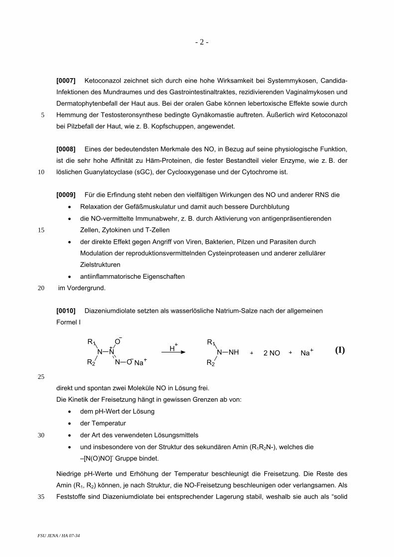
- 2 -
5
10
15
20
[0007] Ketoconazol zeichnet sich durch eine hohe Wirksamkeit bei Systemmykosen, Candida-
Infektionen des Mundraumes und des Gastrointestinaltraktes, rezidivierenden Vaginalmykosen und
Dermatophytenbefall der Haut aus. Bei der oralen Gabe können lebertoxische Effekte sowie durch
Hemmung der Testosteronsynthese bedingte Gynäkomastie auftreten. Äußerlich wird Ketoconazol
bei Pilzbefall der Haut, wie z. B. Kopfschuppen, angewendet.
[0008] Eines der bedeutendsten Merkmale des NO, in Bezug auf seine physiologische Funktion,
ist die sehr hohe Affinität zu Häm-Proteinen, die fester Bestandteil vieler Enzyme, wie z. B. der
löslichen Guanylatcyclase (sGC), der Cyclooxygenase und der Cytochrome ist.
[0009] Für die Erfindung steht neben den vielfältigen Wirkungen des NO und anderer RNS die
• Relaxation der Gefäßmuskulatur und damit auch bessere Durchblutung
• die NO-vermittelte Immunabwehr, z. B. durch Aktivierung von antigenpräsentierenden
Zellen, Zytokinen und T-Zellen
• der direkte Effekt gegen Angriff von Viren, Bakterien, Pilzen und Parasiten durch
Modulation der reproduktionsvermittelnden Cysteinproteasen und anderer zellulärer
Zielstrukturen
• antiinflammatorische Eigenschaften
im Vordergrund.
[0010] Diazeniumdiolate setzten als wasserlösliche Natrium-Salze nach der allgemeinen
Formel I
R1
NR2
NO
N O Na
HR1
NR2
NH 2 NO Na
(I)
25
30
35
direkt und spontan zwei Moleküle NO in Lösung frei.
Die Kinetik der Freisetzung hängt in gewissen Grenzen ab von:
• dem pH-Wert der Lösung
• der Temperatur
• der Art des verwendeten Lösungsmittels
• und insbesondere von der Struktur des sekundären Amin (R1R2N-), welches die –[N(O)NO]- Gruppe bindet.
Niedrige pH-Werte und Erhöhung der Temperatur beschleunigt die Freisetzung. Die Reste des
Amin (R1, R2) können, je nach Struktur, die NO-Freisetzung beschleunigen oder verlangsamen. Als
Feststoffe sind Diazeniumdiolate bei entsprechender Lagerung stabil, weshalb sie auch als “solid
FSU JENA / HA 07-34
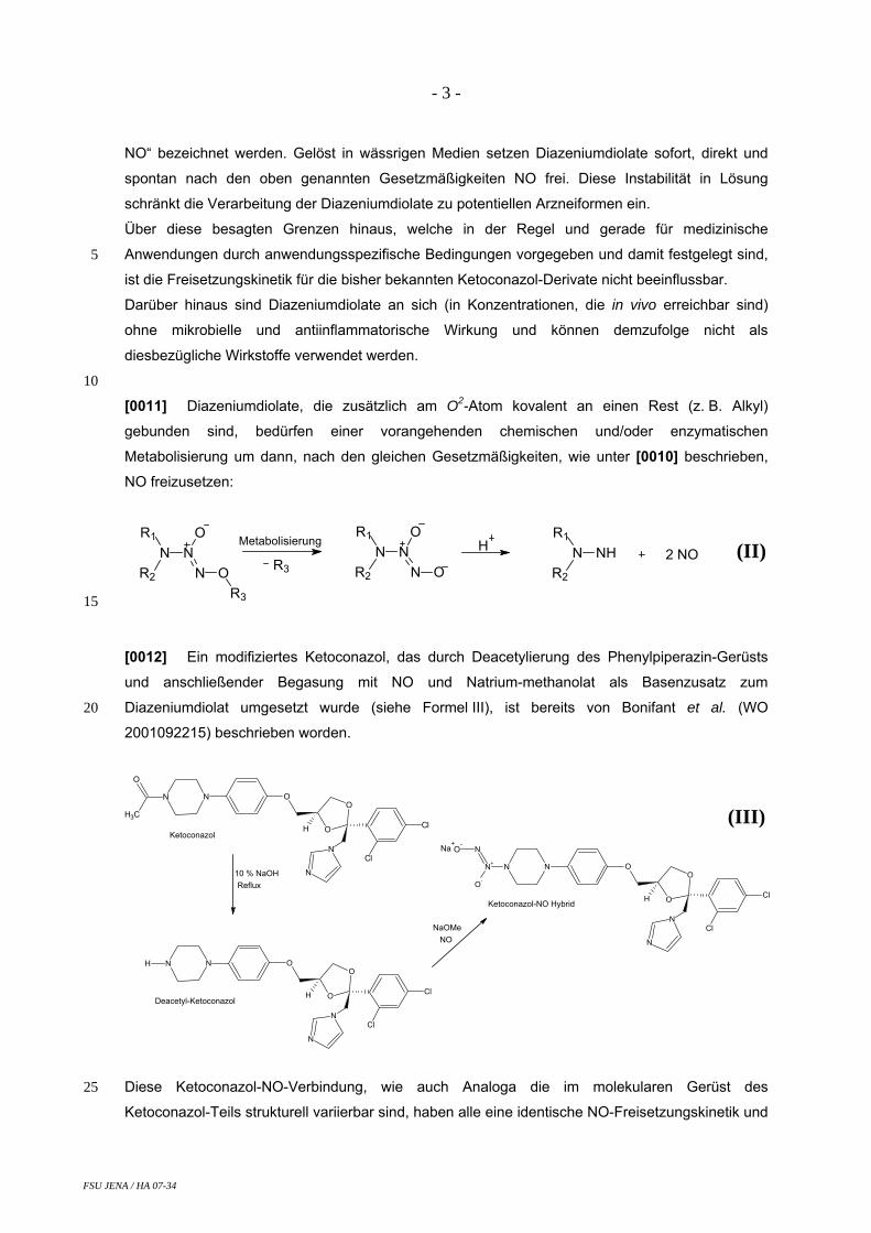
- 3 -
5
10
NO“ bezeichnet werden. Gelöst in wässrigen Medien setzen Diazeniumdiolate sofort, direkt und
spontan nach den oben genannten Gesetzmäßigkeiten NO frei. Diese Instabilität in Lösung
schränkt die Verarbeitung der Diazeniumdiolate zu potentiellen Arzneiformen ein.
Über diese besagten Grenzen hinaus, welche in der Regel und gerade für medizinische
Anwendungen durch anwendungsspezifische Bedingungen vorgegeben und damit festgelegt sind,
ist die Freisetzungskinetik für die bisher bekannten Ketoconazol-Derivate nicht beeinflussbar.
Darüber hinaus sind Diazeniumdiolate an sich (in Konzentrationen, die in vivo erreichbar sind)
ohne mikrobielle und antiinflammatorische Wirkung und können demzufolge nicht als
diesbezügliche Wirkstoffe verwendet werden.
[0011] Diazeniumdiolate, die zusätzlich am O2-Atom kovalent an einen Rest (z. B. Alkyl)
gebunden sind, bedürfen einer vorangehenden chemischen und/oder enzymatischen
Metabolisierung um dann, nach den gleichen Gesetzmäßigkeiten, wie unter [0010] beschrieben,
NO freizusetzen:
R1
NR2
NO
N O
HR1
NR2
NH 2 NOR1
NR2
NO
N OR3
Metabolisierung
R3
(II)
15
20
[0012] Ein modifiziertes Ketoconazol, das durch Deacetylierung des Phenylpiperazin-Gerüsts
und anschließender Begasung mit NO und Natrium-methanolat als Basenzusatz zum
Diazeniumdiolat umgesetzt wurde (siehe Formel III), ist bereits von Bonifant et al. (WO
2001092215) beschrieben worden.
O
O
Cl
Cl
N
N
ONN
H
O
CH3
O
O
Cl
Cl
N
N
ONN
H
H
10 % NaOHReflux
NO
O
O
Cl
Cl
N
N
ONN
H
N+
N
O-
O-Na
+
NaOMe
Ketoconazol
Deacetyl-Ketoconazol
Ketoconazol-NO Hybrid
(III)
25 Diese Ketoconazol-NO-Verbindung, wie auch Analoga die im molekularen Gerüst des
Ketoconazol-Teils strukturell variierbar sind, haben alle eine identische NO-Freisetzungskinetik und
FSU JENA / HA 07-34

- 4 -
5
10
15
20
Halbwertszeit. Bei allen Analoga ist die [N(O)NO]- -Gruppe an den Phenylpiperazin-Rest
gebunden, welcher bei 37 °C und pH 7,4 die Kinetik und Geschwindigkeit der NO-Freisetzung
bestimmt. Die gemessene Halbwertszeit dieser Verbindungen beträgt t1/2 = 4.5 Minuten (bei 37 °C,
pH 7,4). Es gelten die gleichen, bereits unter [0010] beschriebenen Nachteile für die NO-
Freisetzungskinetik und die Verwendbarkeit.
Wünschenswert wäre es, diese NO-Freisetzungskinetik je nach bestimmungsgemäßem
Verwendungszweck variieren zu können. Ferner wäre es wünschenswert, einen auch in wässrigen
Medien stabilen Wirkstoff zu erhalten, der einer Weiterverarbeitung zur Arzneiform standhält und
eine ausreichende Haltbarkeit aufweist.
[0013] Der Erfindung liegt deshalb die Aufgabe zu Grunde, neue, beständige und als anti-
mikrobielle sowie antiinflammatorische Wirkstoffe einsetzbare Verbindungen bereitzustellen, bei
denen die Freisetzung von Stickstoffmonoxid und anderen reaktiven Stickstoffspezies in weiten
Grenzen verändert werden kann.
Insbesondere soll die Freisetzung von Stickstoffmonoxid für medizinische Anwendungen je nach
deren Erfordernissen verlängert oder verkürzt werden können.
[0014] Zur Lösung der Aufgabe werden neue Ketoconazol-Derivate mit Diazen-1-ium-1,2-diolat
gemäß der allgemeinen Formel IV
O
O
Cl
Cl
N
N
ONN
H
O
ZO
NN+
O-
NR1
R2
(IV)
vorgeschlagen,
wobei:
- R1, R2: unabhängig voneinander geradkettiges oder verzweigtes Alkyl, Arylalkyl,
Aminoalkyl, Alkylaminoalkyl sein kann, 25
30
- R1 und R2: miteinander Kohlenwasserstoff Zyklen mit einer Ringgröße von 3-10 Atomen
bilden können. Diese Zyklen können ein oder mehrere Heteroatome wie z. B. O-, S- oder
N-Atome enthalten,
- R1, R2: unabhängig voneinander Teil eines Polymeren darstellen und
- Z: Methylen, geradkettiges oder verzweigtes Alkylen darstellt.
[0015] Eine Verbindung nach dieser allgemeinen Formel IV verknüpft mit einer speziellen Bin-
dung das an sich bekannte Ketoconazol mit einem strukturell variierbaren Diazen-1-ium-1,2-diolat-
FSU JENA / HA 07-34
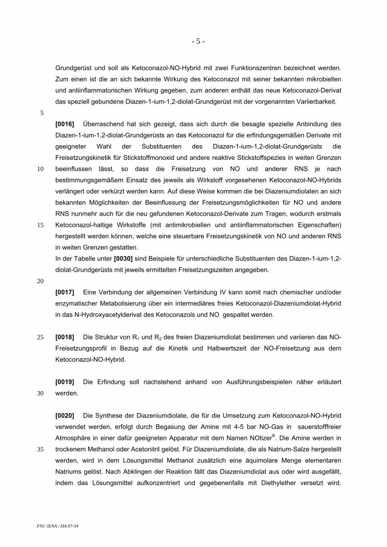
- 5 -
5
10
15
20
25
30
35
Grundgerüst und soll als Ketoconazol-NO-Hybrid mit zwei Funktionszentren bezeichnet werden.
Zum einen ist die an sich bekannte Wirkung des Ketoconazol mit seiner bekannten mikrobiellen
und antiinflammatorischen Wirkung gegeben, zum anderen enthält das neue Ketoconazol-Derivat
das speziell gebundene Diazen-1-ium-1,2-diolat-Grundgerüst mit der vorgenannten Variierbarkeit.
[0016] Überraschend hat sich gezeigt, dass sich durch die besagte spezielle Anbindung des
Diazen-1-ium-1,2-diolat-Grundgerüsts an das Ketoconazol für die erfindungsgemäßen Derivate mit
geeigneter Wahl der Substituenten des Diazen-1-ium-1,2-diolat-Grundgerüsts die
Freisetzungskinetik für Stickstoffmonoxid und andere reaktive Stickstoffspezies in weiten Grenzen
beeinflussen lässt, so dass die Freisetzung von NO und anderer RNS je nach
bestimmungsgemäßem Einsatz des jeweils als Wirkstoff vorgesehenen Ketoconazol-NO-Hybrids
verlängert oder verkürzt werden kann. Auf diese Weise kommen die bei Diazeniumdiolaten an sich
bekannten Möglichkeiten der Beeinflussung der Freisetzungsmöglichkeiten für NO und andere
RNS nunmehr auch für die neu gefundenen Ketoconazol-Derivate zum Tragen, wodurch erstmals
Ketoconazol-haltige Wirkstoffe (mit antimikrobiellen und antiinflammatorischen Eigenschaften)
hergestellt werden können, welche eine steuerbare Freisetzungskinetik von NO und anderen RNS
in weiten Grenzen gestatten.
In der Tabelle unter [0030] sind Beispiele für unterschiedliche Substituenten des Diazen-1-ium-1,2-
diolat-Grundgerüsts mit jeweils ermittelten Freisetzungszeiten angegeben.
[0017] Eine Verbindung der allgemeinen Verbindung IV kann somit nach chemischer und/oder
enzymatischer Metabolisierung über ein intermediäres freies Ketoconazol-Diazeniumdiolat-Hybrid
in das N-Hydroxyacetylderivat des Ketoconazols und NO gespaltet werden.
[0018] Die Struktur von R1 und R2 des freien Diazeniumdiolat bestimmen und variieren das NO-
Freisetzungsprofil in Bezug auf die Kinetik und Halbwertszeit der NO-Freisetzung aus dem
Ketoconazol-NO-Hybrid.
[0019] Die Erfindung soll nachstehend anhand von Ausführungsbeispielen näher erläutert
werden.
[0020] Die Synthese der Diazeniumdiolate, die für die Umsetzung zum Ketoconazol-NO-Hybrid
verwendet werden, erfolgt durch Begasung der Amine mit 4-5 bar NO-Gas in sauerstofffreier
Atmosphäre in einer dafür geeigneten Apparatur mit dem Namen NOtizer®. Die Amine werden in
trockenem Methanol oder Acetonitril gelöst. Für Diazeniumdiolate, die als Natrium-Salze hergestellt
werden, wird in dem Lösungsmittel Methanol zusätzlich eine äquimolare Menge elementaren
Natriums gelöst. Nach Abklingen der Reaktion fällt das Diazeniumdiolat aus oder wird ausgefällt,
indem das Lösungsmittel aufkonzentriert und gegebenenfalls mit Diethylether versetzt wird.
FSU JENA / HA 07-34

- 6 -
5
10
15
20
25
30
35
Ausführungsbeispiel 1a beschreibt die Synthesevorschrift am repräsentativen Beispiel von Natrium
1-(pyrrolidin-1-yl)diazen-1-ium-1,2-diolat.
[0021] Die Synthese der Ketoconazol-NO Hybride erfolgt durch Deacetylierung des Phe-
nylpiperazin-Restes von Ketoconazol mit NaOH, anschließender Haloacetylierung des
Phenylpiperazin-Rest und darauffolgender Umsetzung mit dem entsprechenden Diazeniumdiolat.
Die detaillierte Arbeitsvorschrift anhand der beispielhaften Synthese des O2-﴾1-(4-{[2-(2,4-
dichlorophenyl)-2-(1H-imidazol-1-ylmethyl)-1,3-dioxolan-4-yl]methoxy}phe-nyl)piperazine-4-
carboxymethyl ﴿-1-(pyrrolidin-1-yl)diazen-1-ium-1,2-diolat ist in Ausführungsbeispiel 1b aufgeführt.
[0022] Die Freisetzung aus den Diazeniumdiolaten, die für die Umsetzung zum Ketoconazol-
Hybrid verwendet werden, erfolgt mittels amperometrischer NO-Detektion und UV-Spektroskopie.
[0023] Amperometrische NO-Detektion:
Die Kinetik der NO-Freisetzung wird mit einer Stickstoffmonoxid selektiven ISO-NOP Elektrode
eingebaut in eine temperierbare, abgeschlossene Messkammer und angeschlossen an das Apollo
4000 free radical analyzer system der Firma WPI Inc., USA, gemessen. Die Messkammer wird bei
37 °C temperiert.
Es werden 20 µl einer 50 µM Diazeniumdiolat / 0.01 M NaOH Lösung in 2.0 ml Phosphatpuffer 6.4
injiziert und der entstehende Redoxstrom gemessen.
[0024] UV-Spektroskopische Diazeniumdiolat-Zerfallsmessung:
Der Zerfall der [N(O)NO]- - Gruppe wird bei 251 nm über einen Zeitraum von 30 min mit einem
temperierbaren Jasco V-570 Spektrometer aufgezeichnet. Bei 37 °C werden 4 µl einer 50 mM
Diazeniumdiolat / 0.01 M NaOH Lösung in 2.0 ml Phosphatpuffer 7.4 injiziert und die
Lichtabsorption gemessen. Die Datenwerte werde logarithmisch geplottet und eine linerare
Regressionsanalyse durchgeführt. Die Steigung ergibt die Zerfallskonstante k, aus der nach der
Formel t1/2= ln2/k die Zerfallshalbwertszeit des Diazeniumdiolates bestimmt wird.
Beispielhaft sind die amperometrisch und UV-Spektroskopisch gemessenen NO-Freisetzungs-
kinetiken aus Diazeniumdiolaten, die für die Umsetzung zum Ketoconazol-NO-Hybrid verwendet
werden, in Ausführungsbeispiel 2 dargestellt.
[0025] Untersuchungen der antibakteriellen Eigenschaften:
Es wurden Stämme von Bacillus subtilis ATCC 6633, Staphylococcus aureus SG 511, S. aureus
SG 134/94 (MRSA), Escherichia coli SG 458, Pseudomonas aeruginosa SG 137, Enterococcus
faecalis 1528 (VRE) und Mycobacterium vaccae IMET 1528 als Testorganismen verwendet. Für
den Agardiffusionstest wurden 100 µl des jeweiligen Testorganismus mit einer Konzentration von
0.5 McFarland Standard zu einer sterilen, geschmolzenen 1 % Trypticase Soja Bouillon Agarose
(32 ml) beimpft, auf 50 °C abgekühlt und in Petri-Schalen gegossen. Löcher von 9 mm
FSU JENA / HA 07-34

- 7 -
5
10
15
20
Durchmesser wurden in die Agarose-Schicht geschnitten und mit 50 µL einer 200 µM Ketoconazol-
NO Hybrid Lösung befüllt. Die Inhibitionszonen für das Keimwachstum wurden nach 24 Stunden
Bebrütung bei 37 °C gemessen.
[0026] Untersuchungen der antimykotischen Eigenschaften:
Es wurden Stämme von Aspergillus fumigatus ATCC46646, Aspergillus terreus DSM 826,
Aspergillus niger DSM 737, Fusarium oxysporum i1205, Candida albicans BMSY 212, C. glabrata
DSM 6525, Sporobolomyces salmonicolor SG 549 und Penicillium notatum JP 36 als
Testorganismen verwendet. Nach der Methode unter [0025] beschrieben wurden zusätzlich die
Minimale Inhibitorische Konzentration (MIC) der Ketoconazol-NO Hybride bestimmt. Hierzu wurden
in die einzelnen Löcher 50 µL einer Ketoconazol-NO Hybrid Verdünnung befüllt und die
Inhibitionszonen für das Pilzwachstum wurden nach 24 Stunden Bebrütung bei 37 °C gemessen.
Die niedrigste Ketoconazol-NO-Hybrid-Verdünnung die ein Pilzwachstum inhibierte, wurde als
MIC-Wert bestimmt.
[0027] Untersuchungen der antiinflammatorischen Eigenschaften:
Es wurde die durch reaktive Sauerstoffspezies (ROS) induzierte Oxidation von Luminol per
Chemilumineszenz gemessen. Die “oxidativen bursts“ (ROS Produktion und Freisetzung) aus
polymorphen, mononuklearen Leukozyten (PMNL vom Schwein) wurden in einer Konzentration
von 1 x 10⁶ Zellen/ml Suspension bei 37 °C gemessen.
Die antiinflammatorischen Effekte sind ausgedrückt in Prozent-Inhibition der Luminol Oxidation
gegenüber der Positiv-Kontrolle ausgelöst durch Liponsäure und 4-Aminobenzoesäurehydrazid.
Ausführungsbeispiel 1a:
[0028] Natrium 1-(pyrrolidin-1-yl)diazen-1-ium-1,2-diolat 25
O-
NN+
O-
N
Na+
In 100 ml trockenem Methanol werden 0.05 mol (1.15 g) elementares Natrium gelöst und zu
0.05 mol Pyrrolidin filtriert. Die Mischung wird in das druckbeständige Reaktionsgefäß des
NOtizers® gegossen und dicht verschlossen. Anschließend evakuiert man das Gefäß, begast bis
3 bar mit Stickstoff, evakuiert erneut und wiederholt den Vorgang noch zweimal. Das evakuierte
Gefäß wird mit 4.5 bar Stickstoffmonoxid begast und 48 Stunden gerührt. Schließlich wird
nochmals mit Stickstoff gespült, das Reaktionsgemisch entnommen und bei 60 °C unter
30
FSU JENA / HA 07-34
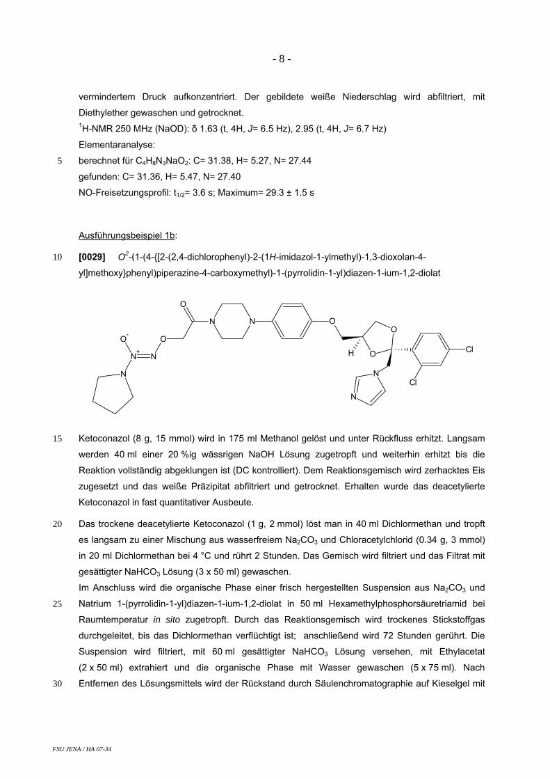
- 8 -
5
vermindertem Druck aufkonzentriert. Der gebildete weiße Niederschlag wird abfiltriert, mit
Diethylether gewaschen und getrocknet. 1H-NMR 250 MHz (NaOD): δ 1.63 (t, 4H, J= 6.5 Hz), 2.95 (t, 4H, J= 6.7 Hz)
Elementaranalyse:
berechnet für C4H8N3NaO2: C= 31.38, H= 5.27, N= 27.44
gefunden: C= 31.36, H= 5.47, N= 27.40
NO-Freisetzungsprofil: t1/2= 3.6 s; Maximum= 29.3 ± 1.5 s
Ausführungsbeispiel 1b:
[0029] O2-﴾1-(4-{[2-(2,4-dichlorophenyl)-2-(1H-imidazol-1-ylmethyl)-1,3-dioxolan-4-
yl]methoxy}phenyl)piperazine-4-carboxymethyl﴿-1-(pyrrolidin-1-yl)diazen-1-ium-1,2-diolat
10
O
O
Cl
Cl
N
N
ONN
H
O
O
NN+
O-
N
15
20
25
30
Ketoconazol (8 g, 15 mmol) wird in 175 ml Methanol gelöst und unter Rückfluss erhitzt. Langsam
werden 40 ml einer 20 %ig wässrigen NaOH Lösung zugetropft und weiterhin erhitzt bis die
Reaktion vollständig abgeklungen ist (DC kontrolliert). Dem Reaktionsgemisch wird zerhacktes Eis
zugesetzt und das weiße Präzipitat abfiltriert und getrocknet. Erhalten wurde das deacetylierte
Ketoconazol in fast quantitativer Ausbeute.
Das trockene deacetylierte Ketoconazol (1 g, 2 mmol) löst man in 40 ml Dichlormethan und tropft
es langsam zu einer Mischung aus wasserfreiem Na2CO3 und Chloracetylchlorid (0.34 g, 3 mmol)
in 20 ml Dichlormethan bei 4 °C und rührt 2 Stunden. Das Gemisch wird filtriert und das Filtrat mit
gesättigter NaHCO3 Lösung (3 x 50 ml) gewaschen.
Im Anschluss wird die organische Phase einer frisch hergestellten Suspension aus Na2CO3 und
Natrium 1-(pyrrolidin-1-yl)diazen-1-ium-1,2-diolat in 50 ml Hexamethylphosphorsäuretriamid bei
Raumtemperatur in sito zugetropft. Durch das Reaktionsgemisch wird trockenes Stickstoffgas
durchgeleitet, bis das Dichlormethan verflüchtigt ist; anschließend wird 72 Stunden gerührt. Die
Suspension wird filtriert, mit 60 ml gesättigter NaHCO3 Lösung versehen, mit Ethylacetat
(2 x 50 ml) extrahiert und die organische Phase mit Wasser gewaschen (5 x 75 ml). Nach
Entfernen des Lösungsmittels wird der Rückstand durch Säulenchromatographie auf Kieselgel mit
FSU JENA / HA 07-34

- 9 -
5
10
15
Ethylacetat:Acetonitril (1:1) und anschließender methanolischer Auswaschung gereinigt zu einem
orangefarbenen Pulver.
Ausbeute: 38 %
Schmelzpunkt: 237 °C 1H-NMR: 400 MHz (CDCl3): δ 1.93 (m, 4H), 3.02-3.13 (m, 8H), 3.32-3.36 (m, 1H), 3.55 (mc, 4H),
3.72-3.77 (m, 2H), 3.86-3.90 (dd, J = 6.7, 6.6 Hz, 1H), 4.31-4.37 (quin, J = 5.7 Hz, 1H),
4.39-4.53 (dd, J = 40.7, 14.8 Hz, 2H), 4.80 (s, 2H), 6.76 (d, J = 9.1 Hz, 2H), 6.90 (m, 2H), 6.97 (m,
2H), 7.25 (d, J = 8.1 Hz, 1H), 7.49 (d, J = 14.7 Hz, 2H), 7.57 (d, J = 8.3 Hz, 1H) 13C-NMR: 400 MHz (CDCl3): δ 22.9, 36.8, 51.3, 51.7, 53.5, 67.6, 67.7, 71.4, 74.8, 108.0, 115.3,
118.8, 121.2, 127.2, 128.4, 129.5, 131.3, 133.0, 134.6, 135.9, 138.8, 145.6, 152.9, 166.7
FAB-MS m/z (%): 202 (48), 217 (55), 235 (50), 255 (100), 432 (38), 446 (51), 459 (70), 471 (63),
489 (64), 517 (39), 547 (82), 600 (69), 660 (42, M++H), 682 (19, [M+Na]+), 698 (17, M++K)
Elementaranalyse:
berechnet für C30H35Cl2N7O6: C= 54.55, H= 5.34, N= 14.84
gefunden: C= 54.17, H= 5.43, N= 14.78
Antibakterielle Eigenschaften von Ausführungsbeispiel 1b gemäß [0025]: Hemmhofdurchmesser (Inhibitionszonen für Keimwachstum) nach 24 h.
Bacillus
subtilis Staph. aureus
Staph. aureus (MRSA)
E. coli
Pseud. aeruginosa
Enterococcus faecalis
Mycobact. vaccae
Hemmhof-durchmesser
[mm] 15 15 0 0 0 0 19
20
25
Antimykotische Eigenschaften von Ausführungsbeispiel 1b gemäß [0026]: MIC-Werte [µM] des O2- ﴾1-(4-{[2-(2,4-dichlorophenyl)-2-(1H-imidazol-1-ylmethyl)-1,3-dioxolan-4-
yl]methoxy}phenyl)piperazine-4-carboxymethyl﴿-1-(pyrrolidin-1-yl)diazen-1-ium-1,2-diolat an
Pilzstämmen.
Asp. fumigatus
Asp. terreus
Asp. niger
Fusarium oxysp.
Candida albicans
Candida glabrata
Sporobolom. salmonicolor
Penicillium notatum
MIC [µM]
125 62.5 62.5 1000 25 12.5 1.56 8
FSU JENA / HA 07-34

- 10 -
5
Antiinflammatorische Eigenschaften von Ausführungsbeispiel 1b gemäß [0027]: Hemmung ausgedrückt durch Verringerung der AUC (area under the curve) im Vergleich zur
Positiv-Kontrolle (100 %). Eine 80 µM Lösung von O2- ﴾1-(4-{[2-(2,4-dichlorophenyl)-2-(1H-imidazol-
1-ylmethyl)-1,3-dioxolan-4-yl]methoxy}phenyl)piperazine-4-carboxymethyl﴿-1-(pyrrolidin-1-
yl)diazen-1-ium-1,2-diolat verringert die Fläche auf 4 %, eine 8 µM Lösung auf 13 % und eine
0.8 µM Lösung auf 36 % im Vergleich zur Positiv-Kontrolle (100 %).
Ausführungsbeispiel 2:
[0030] Tabellarische Aufstellung ausgewählter Diazeniumdiolate, die für die Umsetzung zum
Ketoconazol-Hybrid verwendet werden, mit dazugehörigem NO-Freisetzungsprofil: 10
NO-Freisetzungsprofil
Diazeniumdiolat Maximum [s] (amperometrisch)
Halbwertszeit [s] (UV-spektroskopisch)
NN
+
N
O-
O-Na
+
75.3 ± 2.8 85.2
N N+
N
O-
O- Na
+
29.3 ± 1.5 3.6
N N+
N
O-
O-Na
+
36.3 ± 3.2 18.9
N N+
N
O-
O-Na
+
29.3 ± 2.3 3.6
N N+
N
O-
O-Na
+
29.0 ± 2.6 4.7
NO N+
N
O-
O-Na+
100.0 ± 11.9 152.3
NN N+
N
O-
O-Na+
82.0 ± 3.4 263.5
FSU JENA / HA 07-34

- 11 -
N N+
N
O-
O-
COOH
Na+
32.0 ± 1.0 6.1
N N+
N
O-
O-
HOOC
Na+ 39.6 ± 3.0 22.6
N N+
N
O-
O-Na
+
38.3 ± 2.8 29.3
N N+
N
O-
O-
COOH
Na+
27.3 ± 3.2 6.8
Na+
CH3 N CH3
N+
NO-
O-
85.5 ± 4.4 112.3
Na+
N
N+
NO-
O-
CH3 CH3
70.2 ± 9.8 93.2
Na+
N
N+
NO-
O-
CH2 CH2
131.2 ± 14.9 246.6
Na+
N
N+
NO-
O-
OH OH
266.0 ± 2.0 456.0
Na+
N
N+
NO-
O-
OH NH2
1765.0 ± 209.0 7498.9
Na+
N CH3
N+
NO-
O-
OH
116.4 ± 18.7 206.9
FSU JENA / HA 07-34

- 12 -
Na+
CH3
N
N+
NO-
O-
50.6 ± 0.5 56.7
Na+
N
N+
NO-
O-
CH3
146.6 ± 16.6 302.0
Na+
N
N+
NO-
O-
CH3
CH3
911.0 ± 147.2 7036.1
Na+
N
N+
NO-
O-
233.0 ± 11.0 537.3
Na+
N
N+
NO-
O-
260.3 ± 5.0 679.5
Es hat sich gezeigt, dass die tabellarisch aufgeführten Diazeniumdiolate für die erfindungsgemäße
Herstellung neuer Ketoconazol-Derivate geeignet sind. Dabei zeigten die Ketoconazol-Derivate
das von der Struktur des jeweiligen Diazeniumdiolates abhängige, charakteristische NO-
Freisetzungsprofil. 5
FSU JENA / HA 07-34

- 13 -
5
Patentansprüche
1. Neue Ketoconazol-Derivate mit Diazen-1-ium-1,2-diolat, gekennzeichnet durch eine Verbindung
der allgemeinen Formel IV:
O
O
Cl
Cl
N
N
ONN
H
O
ZO
NN+
O-
NR1
R2
,
(IV)
wobei:
- R1, R2: unabhängig voneinander geradkettiges oder verzweigtes Alkyl, Arylalkyl,
Aminoalkyl, Alkylaminoalkyl sein kann, 10
15
20
- R1 und R2: miteinander Kohlenwasserstoff Zyklen mit einer Ringgröße von 3-10 Atomen
bilden können. Diese Zyklen können ein oder mehrere Heteroatome wie z. B. O-, S- oder
N-Atome enthalten,
- R1, R2: unabhängig voneinander Teil eines Polymeren darstellen und
- Z: Methylen, geradkettiges oder verzweigtes Alkylen darstellt.
2. Verfahren zur Herstellung der neuen Ketoconazol-Derivate gemäß Anspruch 1, dadurch
gekennzeichnet, dass Diazen-1-ium-1,2-diolat mit N-Haloacyl-Ketoconazol-Derivat durch
nukleophile Substitution umgesetzt wird.
3. Verfahren gemäß Anspruch 2, dadurch gekennzeichnet, dass als Diazen-1-ium-1,2-diolat die
Verbindung
NN
+
N
O-
O-Na
+
(V) eingesetzt wird.
25
4. Verfahren gemäß Anspruch 2, dadurch gekennzeichnet, dass als Diazen-1-ium-1,2-diolat die
Verbindung
N N
+
N
O-
O- Na
+
(VI)
FSU JENA / HA 07-34

- 14 -
eingesetzt wird.
5. Verfahren gemäß Anspruch 2, dadurch gekennzeichnet, dass als Diazen-1-ium-1,2-diolat die
Verbindung
N N+
N
O-
O-Na
+ (VII)5
eingesetzt wird.
6. Verfahren gemäß Anspruch 2, dadurch gekennzeichnet, dass als Diazen-1-ium-1,2-diolat die
Verbindung
N N+
N
O-
O-Na
+
(VIII)
10
15
eingesetzt wird.
7. Verfahren gemäß Anspruch 2, dadurch gekennzeichnet, dass als Diazen-1-ium-1,2-diolat die
Verbindung
N N+
N
O-
O-Na
+
(IX)
eingesetzt wird.
8. Verfahren gemäß Anspruch 2, dadurch gekennzeichnet, dass als Diazen-1-ium-1,2-diolat die
Verbindung 20
NO N+
N
O-
O-Na+
(X)
eingesetzt wird.
9. Verfahren gemäß Anspruch 2, dadurch gekennzeichnet, dass als Diazen-1-ium-1,2-diolat die
Verbindung 25
NN N+
N
O-
O-Na+
(XI) eingesetzt wird.
FSU JENA / HA 07-34

- 15 -
10. Verfahren gemäß Anspruch 2, dadurch gekennzeichnet, dass als Diazen-1-ium-1,2-diolat die
Verbindung
N N+
N
O-
O-
COOH
Na+
(XII)
eingesetzt wird.
5
11. Verfahren gemäß Anspruch 2, dadurch gekennzeichnet, dass als Diazen-1-ium-1,2-diolat die
Verbindung
N N+
N
O-
O-
HOOC
Na+ (XIII)eingesetzt wird.
10
12. Verfahren gemäß Anspruch 2, dadurch gekennzeichnet, dass als Diazen-1-ium-1,2-diolat die
Verbindung
N N+
N
O-
O-Na
+
(XIV)
eingesetzt wird.
15
13. Verfahren gemäß Anspruch 2, dadurch gekennzeichnet, dass als Diazen-1-ium-1,2-diolat die
Verbindung
N N+
N
O-
O-
COOH
Na+
(XV)
eingesetzt wird.
20
14. Verfahren gemäß Anspruch 2, dadurch gekennzeichnet, dass als Diazen-1-ium-1,2-diolat die
Verbindung
Na+
CH3 N CH3
N+
NO-
O-
(XVI)
eingesetzt wird.
25
FSU JENA / HA 07-34

- 16 -
15. Verfahren gemäß Anspruch 2, dadurch gekennzeichnet, dass als Diazen-1-ium-1,2-diolat die
Verbindung
Na+
N
N+
NO-
O-
CH3 CH3 (XVII)
eingesetzt wird.
5
16. Verfahren gemäß Anspruch 2, dadurch gekennzeichnet, dass als Diazen-1-ium-1,2-diolat die
Verbindung
Na+
N
N+
NO-
O-
CH2 CH2
(XVIII)
eingesetzt wird.
10
17. Verfahren gemäß Anspruch 2, dadurch gekennzeichnet, dass als Diazen-1-ium-1,2-diolat die
Verbindung
Na+
N
N+
NO-
O-
OH OH
(XIX)
eingesetzt wird.
15
18. Verfahren gemäß Anspruch 2, dadurch gekennzeichnet, dass als Diazen-1-ium-1,2-diolat die
Verbindung
Na+
N
N+
NO-
O-
OH NH2 (XX)
eingesetzt wird.
20
25
FSU JENA / HA 07-34

- 17 -
19. Verfahren gemäß Anspruch 2, dadurch gekennzeichnet, dass als Diazen-1-ium-1,2-diolat die
Verbindung
Na+
N CH3
N+
NO-
O-
OH (XXI)
eingesetzt wird.
5
20. Verfahren gemäß Anspruch 2, dadurch gekennzeichnet, dass als Diazen-1-ium-1,2-diolat die
Verbindung
Na+
CH3
N
N+
NO-
O-
(XXII)
eingesetzt wird.
10
21. Verfahren gemäß Anspruch 2, dadurch gekennzeichnet, dass als Diazen-1-ium-1,2-diolat die
Verbindung
Na+
N
N+
NO-
O-
CH3 (XXIII)
eingesetzt wird.
15
22. Verfahren gemäß Anspruch 2, dadurch gekennzeichnet, dass als Diazen-1-ium-1,2-diolat die
Verbindung
Na+
N
N+
NO-
O-
CH3
CH3
(XXIV)
eingesetzt wird.
20
FSU JENA / HA 07-34

- 18 -
23. Verfahren gemäß Anspruch 2, dadurch gekennzeichnet, dass als Diazen-1-ium-1,2-diolat die
Verbindung
Na+
N
N+
NO-
O-
(XXV)
eingesetzt wird.
5
24. Verfahren gemäß Anspruch 2, dadurch gekennzeichnet, dass als Diazen-1-ium-1,2-diolat die
Verbindung
Na+
N
N+
NO-
O-
(XXVI)
eingesetzt wird.
10
15
20
25. Verwendung der neuen Ketoconazol-Derivate gemäß Anspruch 1 als Wirkstoff gegen
mikrobielle Krankheitserreger, wie z. B. Pilze, Bakterien und Parasiten.
26. Verwendung der neuen Ketoconazol-Derivate gemäß Anspruch 1 als antiinflammatorische
Wirkstoffe.
27. Verwendung gemäß Anspruch 25 und/oder 26, dadurch gekennzeichnet, dass das
Ketoconazol-Derivat in einer pharmazeutischen Formulierung als lokal, oral, rectal, transdermal,
parenteral oder intraperitoneal zu verabreichender Wirkstoff, beispielsweise in Form von Shampoo,
halbfesten oder flüssigen Emulsionen oder Suspensionen, Tabletten, Dragees, Kapseln, Zäpfchen,
transdermalen Plasten, Injektionen oder Infusionen vorliegt.
FSU JENA / HA 07-34

- 19 -
5
10
Zusammenfassung
1. Neue Ketoconazol-Derivate sowie deren Herstellung und Verwendung als antimikrobielle und
antiinflammatorische Wirkstoffe
2.1. Aufgabe war es, beständige und als antimikrobielle sowie antiinflammatorische Wirkstoffe
einsetzbare Verbindungen bereitzustellen, bei denen die Freisetzung von Stickstoffmonoxid und
anderen reaktiven Stickstoffspezies in weiten Grenzen verändert werden kann.
2.2. Es wurden neue Ketoconazol-Derivate mit Diazen-1-ium-1,2-diolat gefunden, gekennzeichnet
durch eine Verbindung der allgemeinen Formel IV:
O
O
Cl
Cl
N
N
ONN
H
O
ZO
NN+
O-
NR1
R2
(IV)
2.3. Die neuen Derivate finden beispielsweise in der Medizin Einsatz bei der antiinfektiven Therapie.
15
FSU JENA / HA 07-34


Bisher unveröffentlichte Ergebnisse 110
4 Bisher unveröffentlichte Ergebnisse In diesem Kapitel werden Ergebnisse präsentiert, die bisher nicht veröffentlicht wurden. Gemäß der
Gliederung der Zielstellungen dieser Arbeit wird zunächst auf chemische Aspekte, dann auf
biologische Aktivitäten der neu synthetisierten Derivate eingegangen. Die Beschreibung der
verwendeten Materialien und Methoden sind, wenn unverändert, den einzelnen Veröffentlichungen zu
entnehmen und werden an entsprechender Stelle zitiert. Auf Abweichungen zu den zitierten
Protokollen wird explizit hingewiesen.
4.1 Chemische Aspekte 4.1.1 Beobachtung der NONOat-Synthese mit dem NOtizer
Reaktionsenthalpie
Wie in Veröffentlichung 1 schon erwähnt, verfügt der NOtizer über einen Druck- und einen
Temperatursensor, deren Messsignal durch einen Computer online aufgezeichnet werden kann. In der
Literatur wird lediglich über einen leichten Temperaturanstieg während der NONOat-Synthese
berichtet, wobei es sich aber bei dem apparativen Aufbau um ein offenes System mit ständiger NO.-
Begasung handelt. Um das Ausmaß dieser Exothermie besser einschätzen zu können, wurde eine
relativ große Menge (0.25 mol) des schnell reagierenden Pyrrolidin mit einer äquimolaren Menge
Natrium in 300 ml Methanol gelöst und einem 4.5 bar hohen Druck an NO.-Gas ausgesetzt. Wie in
Abbildung 14 (A) zu erkennen ist, wurde, um die Reaktion zu unterhalten, bei einem Druckabfall unter
2 bar erneut NO. eingeleitet und der Temperaturverlauf aufgezeichnet. Um sicherzustellen, dass der
Temperaturanstieg ausschließlich aus der Reaktion des NO. mit dem Amin resultiert, wurde eine
Vergleichsmessung ohne Pyrrolidin unter denselben Bedingungen durchgeführt (siehe Abb. 14 (B)).
A B
Abb. 14 (A) Druck- und Temperaturverlauf bei der Reaktion von Pyrrolidin mit NO.-Gas
(B) Druck- und Temperaturverlauf bei der Reaktion von Natrium-methanolat mit NO.-Gas

111 Bisher unveröffentlichte Ergebnisse
Der deutliche Temperaturanstieg bei der NONOat-Bildung von über 5 °C ist angesichts des großen
Lösungsmittelvolumens erstaunlich hoch. Abbildung 14 (B) verdeutlicht, dass keine
Temperaturänderung ohne Pyrrolidin als Reaktionspartner auftritt. Der Druckabfall von 0.5 bar
(entspricht 13 mmol NO.) innerhalb der ersten 20 Minuten ist bedingt durch die Löslichkeit des NO.
unter Druck in den 300 ml Methanol. Dieses Experiment zeigt zum einen, dass die Bildungsenthalpie
exotherm ist (∆Hf < 0) und zum anderen, dass unter den Standardreaktionsbedingungen (100 ml
Methanol; 0.05 mol Amin) für die reaktionskinetischen Vergleiche (Veröffentlichung 2) keine falschen
Druckverläufe durch temperaturbedingte Gasexpansion aufgezeichnet werden.
Temperatureinfluss auf die NONOatbildung
Als Modellamin wurde für diese Experimente ebenfalls Pyrrolidin verwendet. Die Durchführung
entspricht der General Procedure aus Veröffentlichung 1 und 2 mit jeweils 0.05 mol Amin und
Natrium, gelöst in trockenem Methanol und 4 bar Stickstoffmonoxid. Vor der NO.-Begasung wurde die
Temperatur mit einem Kryostat oder einer Heizplatte eingestellt. Abbildung 15 stellt die
aufgezeichneten Druckverläufe und Tabelle 3 die Werte der definierten Reaktionsparameter dar.
Abb. 15 Druckverlauf während der Reaktion von Pyrrolidin mit NO.-Gas in Abhängigkeit von der
Temperatur
Durch Kühlen wird die Reaktionsgeschwindigkeit verlangsamt und die isolierbare Ausbeute ist etwas
geringer als bei der Synthese mit Raumtemperatur. Die gemessene Druckabnahme bei 80 °C
entspricht nicht dem tatsächlichen NO.-Verbrauch, da das Reaktionsgemisch unter diesen
Bedingungen siedet und somit schon allein für einen erhöhten Druck verantwortlich ist. Trotzdem ist
zu erkennen, dass die Umsetzung von Amin und NO. langsamer verläuft, wenn nicht sogar ganz

Bisher unveröffentlichte Ergebnisse 112
verhindert wird und ein anderes Produkt entsteht. Die Aufarbeitung nach Abklingen der Reaktion
ergab kein isolierbares einheitliches Produkt, sondern ein Gemisch aus 1-Nitrosopyrrolidin (GC/MS
m/z= 100 [M+H]+), Pyrrolidin und Natriummethanolat. Tiefe Temperaturen scheinen also die Bildung
reiner Diazeniumdiolate zu begünstigen, während bei höheren Temperaturen das korrespondierende
Nitrosamin als Nebenprodukt entsteht.
Tab. 3 Charakteristische Reaktionsparameter bei der Reaktion von Pyrrolidin und NO.-Gas bei
unterschiedlichen Temperaturen
Temperatur
80 °C 5 °C 25 °C Druckverlust nach 7 h [Bar] 0.72 1.8 2.11
Dauer der Reaktion [hh:mm] -- 17:30 19:24
Restdruck [Bar] 2.73 1.71 1.45
Molverhältnis NO./Amin -- 1.42 1.61
In Bezug auf die Reaktionsgeschwindigkeit in Abhängigkeit von der Temperatur bildet Piperazin als
Amin eine Ausnahme. Eine Besonderheit bei der Umsetzung von Piperazin mit NO. ist, wie in der
Literatur beschrieben, dass sich unterschiedliche Piperazin-NONOate bilden, je nachdem welches
Lösungsmittel, welche Temperatur und welcher Basenzusatz verwendet wird.[95,96] Abbildung 16 fasst
die Strukturformeln der möglichen Piperazin-NONOate zusammen.
HN N NO
N O
N N NO
N ON
O
NO
HN NH2 H2N NH2
H2N NH2 2 Na
Na
1
2
Kation:
Kation:
a b c
a b
Abb. 16 Piperazin-NONOat Salze
Beschrieben ist, dass sich in heißem Methanol überwiegend die Verbindung 1a bildet, 1b in Acetonitril
und 2a in kaltem Methanol entsteht. Die entsprechenden Natriumsalze 1c und 2b werden durch
Ionenaustausch mit Natriummethanolat hergestellt. Die Druckabnahmekurven aus den Reaktionen
von 0.05 mol Piperazin mit NO. in 100 ml Methanol bei unterschiedlichen Temperaturen sind in
Abbildung 17 dargestellt.

113 Bisher unveröffentlichte Ergebnisse
Abb. 17 Stickoxidverbrauch während der Reaktion mit Piperazin in Abhängigkeit von der Temperatur
Wie schon bei der Reaktion des Pyrrolidins mit NO. zu beobachten war, wird die
Reaktionsgeschwindigkeit durch Kälte verlangsamt. Anders ist jedoch, dass bei der Reaktion von
Piperazin in heißem Methanol eine leichte Beschleunigung der NO. Druckabnahme gegenüber der
Reaktion bei Raumtemperatur zu beobachten ist. Das scheinbar frühe Abklingen der Reaktion hängt
zum einen mit der oben diskutierten temperaturbedingten Gasvolumenexpansion zusammen, zum
anderen mit der Möglichkeit, dass in der Hitze nicht das NONOat, sondern ein oder mehrere andere
Produkte gebildet werden. Die Aufarbeitung dieses Ansatzes hinterließ einen dunkelbraunen, öligen
Rückstand, dessen 1H-NMR die Bildung des 1-Nitrosopiperazin belegt und nicht etwa die der
Verbindung 1a (siehe Abb. 18). Die Signale der Protonen aus Position 5/5’ (m, 2.65 ppm), 3/3’ (m,
2.88 ppm), 2’/6’ (m, 3.71 ppm) und 2/6 (m, 4.05) des 1-Nitrosopiperazin stimmen mit Literaturdaten
überein.[97] Für Verbindung 1a fände man im 1H-NMR Spektrum für das mono-protonierte Piperazin
laut Literatur ein Singulett (bedingt durch den raschen Protonenaustausch der N-Atome) bei 3.7
ppm.[98]

Bisher unveröffentlichte Ergebnisse 114
2.00
00
2.02
29
2.06
21
2.18
30
Inte
gral
4.07
814.
0613
4.05
674.
0506
4.03
53
3.72
653.
7051
3.68
29
2.89
782.
8833
2.87
642.
8703
2.85
50
2.67
462.
6524
2.63
03
(ppm)2.42.62.83.03.23.43.63.84.04.2
N
N
NOH
H
H
H Piperazin Methanol
5,5‘2‘,6‘ 3,3‘2,6
3 3‘
22‘
H
HH
5 5‘
H6
6‘
H
Abb. 18 1H-NMR Spektrum von 1-Nitrosopiperazin in D2O/NaOD
Druckabhängigkeit der Reaktion
Um die Druckabhängigkeit der Diazeniumdiolat-Synthese zu untersuchen, wurden jeweils 0.05
mol Pyrrolidin und Natrium in 100 ml Methanol gelöst und einem Druck von 2 bar NO.-Gas ausgesetzt.
In einem der Experimente wurde zusätzlich Stickstoff eingeleitet, um einen Gesamtdruck von 4 bar zu
erhalten. Die unterschiedlichen Kurven sind in Abbildung 19 dargestellt.
Abb. 19 Stickoxidverbrauch während der Reaktion mit Pyrrolidin in Abhängigkeit vom Anfangsdruck

115 Bisher unveröffentlichte Ergebnisse
Es ist deutlich zu erkennen, dass bei einem Gesamtdruck von 4 bar die Reaktion schneller und
quantitativer abläuft. Der Druckverlust innerhalb der ersten sieben Stunden nach Reaktionsbeginn
beträgt 0.8 bar für die Synthese bei 2 bar NO. und 1.3 bar bei der Reaktion mit Stickstoffanteil. Obwohl
in beiden Reaktionskammern dieselbe Stoffmenge Stickstoffmonoxid vorhanden ist, ist der
Gesamtdruckverlust bei der Hochdruckreaktion doppelt so groß wie bei der Niedrigdruckreaktion, was
einen quantitativeren Verlauf der NONOat-Bildung belegt. Bestätigt wird diese Aussage durch die fast
30 % höhere isolierbare Ausbeute an PYRRO/NO nach Beendigung der Reaktion.
Einfluss des Lösungsmittels
Für die Synthese von intramolekularen Diazeniumdiolat-Salzen aus Oligoaminen wird häufig
Acetonitril als Lösungsmittel verwendet. Um den Einfluss anderer Lösungsmittel zu untersuchen
wurde Diethylentriamin als Ausgangssubstanz verwendet, um die Bildung des bekannten
Diazeniumdiolates DETA/NO zu überwachen. Da für die Produktion dieses intramolekularen Salzes
kein Basenzusatz erforderlich ist, konnte auch auf unpolare, leicht flüchtige Lösungsmittel wie
Diethylether und Tetrahydrofuran (THF) zurückgegriffen werden. Abbildung 20 zeigt die NO.-
Druckabnahme bei der Reaktion mit Diethylentriamin jeweils in Diethylether, THF, Methanol und
Acetonitril als Lösungsmittel.
Abb. 20 Stickoxidverbrauch während der Reaktion mit Diethylentriamin in unterschiedlichen
Lösungsmitteln
Die Druckabnahme innerhalb der ersten 7 Stunden nach Reaktionsbeginn war mehr als doppelt so
hoch bei der Synthese in Acetonitril im Vergleich zu Methanol und THF, sogar fünf mal höher als in
Diethylether. Lediglich in Acetonitril bildete sich im Verlauf der Reaktion das weiße Präzipitat von
DETA/NO. In Diethylether und THF fiel das Produkt nach einengen des Reaktionsgemisches in
geringen Mengen aus, dagegen konnte das NONOat aus Methanol nicht isoliert werden.

Bisher unveröffentlichte Ergebnisse 116
Einfluss der Basenstärke/Gegenion
Eine treibende Kraft, um das Reaktionsgleichgewicht in Richtung der NONOat-Bildung zu
fördern, ist der Zusatz einer Base. In verschiedenen Experimenten mit Pyrrolidin als Ausgangsamin
wurde der Reaktionsverlauf aufgezeichnet, bei denen zum einen die starke Base Methanolat, zum
anderen das Pyrrolidin selbst als Base verwendet wurde. In zwei Experimenten entstand das
Methanolat durch Lösen äquimolarer Mengen (0.05 mol) von elementarem Natrium oder Lithium in
Methanol, in einem dritten Ansatz wurde die doppelte Stoffmenge (0.1 mol) Pyrrolidin eingesetzt. Als
Produkt entstand in allen drei Fällen das PYRRO/NO, jeweils als Na-, Li- oder Pyrrolidinium-Salz. Die
aufgezeichneten Druckabnahmekurven sind in Abbildung 21 dargestellt.
N H N N+
N
O-
O-
H2N+2 NO2 +
N H 2 NO+NaOCH3
- CH3OHN N+
N
O-
O- Na+
N H 2 NO+LiOCH3
- CH3OHN N+
N
O-
O- Li+
N H N N+
N
O-
O-
H2N+2 NO2 +
N H 2 NO+NaOCH3
- CH3OHN N+
N
O-
O- Na+
N H 2 NO+LiOCH3
- CH3OHN N+
N
O-
O- Li+
Abb. 21 Stickoxid Druckabnahme während der Reaktion mit Pyrrolidin und unterschiedlichen Basen
Die Experimente, bei denen Lithium- bzw. Natrium-Methanolat verwendet wurde, zeigten keinen
signifikanten Unterschied in der Reaktionsgeschwindigkeit und boten auch eine vergleichbare
Ausbeute von ca. 90 %. Dies verwundert nicht, da in beiden Fällen das Methanolat-Anion die Base
darstellt. Sehr viel langsamer verlief die Umsetzung zum PYRRO/NO-Pyrrolidinium Salz, welches aus
dem Lösungsmittel präzipitierte. Die Messung der NO.-Freisetzung aus den drei Salzen ergab
praktisch identische Zerfallshalbwertszeiten von t1/2= 3-6 s.
Um festzustellen, ob eine mehr als äquimolare Stoffmenge an Methanolat die Reaktion
beschleunigt, wurde in einem Versuch die doppelte Menge Natrium (0.1 mol) zugesetzt. Die
aufgezeichnete Druckabnahme zeigte sich jedoch deckungsgleich mit der Kurve aus der Synthese mit
äquimolarem Basenzusatz.
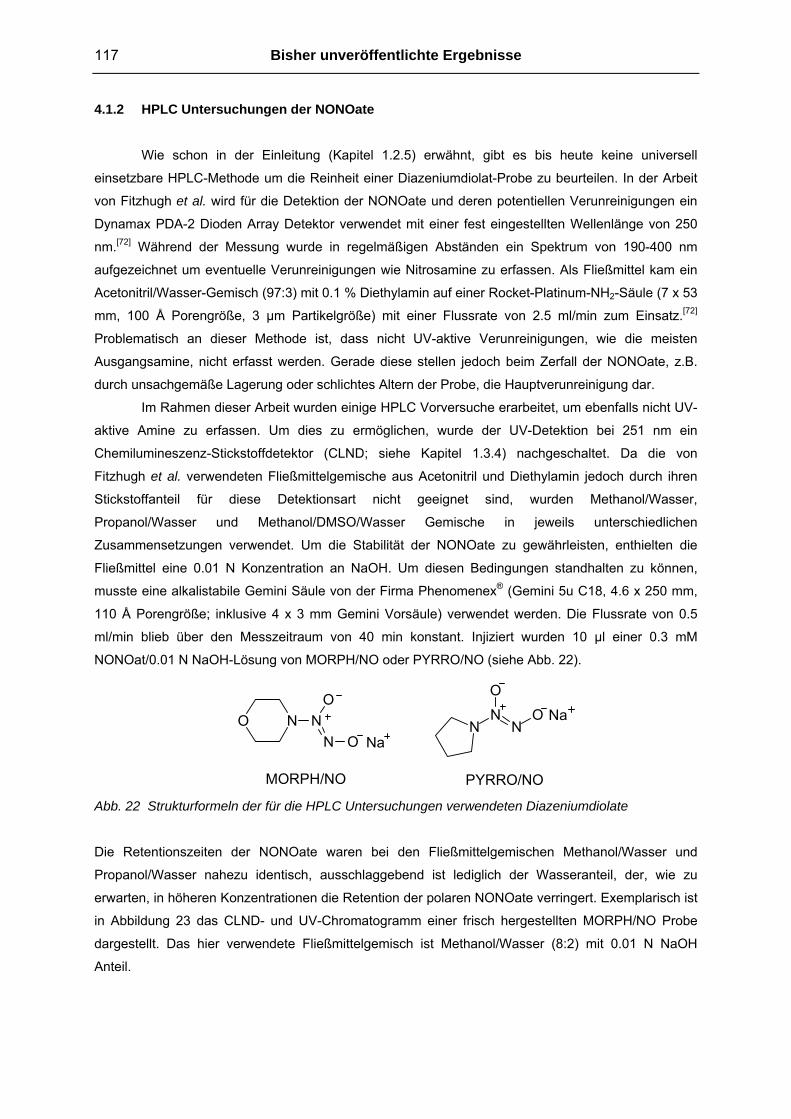
117 Bisher unveröffentlichte Ergebnisse
4.1.2 HPLC Untersuchungen der NONOate
Wie schon in der Einleitung (Kapitel 1.2.5) erwähnt, gibt es bis heute keine universell
einsetzbare HPLC-Methode um die Reinheit einer Diazeniumdiolat-Probe zu beurteilen. In der Arbeit
von Fitzhugh et al. wird für die Detektion der NONOate und deren potentiellen Verunreinigungen ein
Dynamax PDA-2 Dioden Array Detektor verwendet mit einer fest eingestellten Wellenlänge von 250
nm.[72] Während der Messung wurde in regelmäßigen Abständen ein Spektrum von 190-400 nm
aufgezeichnet um eventuelle Verunreinigungen wie Nitrosamine zu erfassen. Als Fließmittel kam ein
Acetonitril/Wasser-Gemisch (97:3) mit 0.1 % Diethylamin auf einer Rocket-Platinum-NH2-Säule (7 x 53
mm, 100 Å Porengröße, 3 µm Partikelgröße) mit einer Flussrate von 2.5 ml/min zum Einsatz.[72]
Problematisch an dieser Methode ist, dass nicht UV-aktive Verunreinigungen, wie die meisten
Ausgangsamine, nicht erfasst werden. Gerade diese stellen jedoch beim Zerfall der NONOate, z.B.
durch unsachgemäße Lagerung oder schlichtes Altern der Probe, die Hauptverunreinigung dar.
Im Rahmen dieser Arbeit wurden einige HPLC Vorversuche erarbeitet, um ebenfalls nicht UV-
aktive Amine zu erfassen. Um dies zu ermöglichen, wurde der UV-Detektion bei 251 nm ein
Chemilumineszenz-Stickstoffdetektor (CLND; siehe Kapitel 1.3.4) nachgeschaltet. Da die von
Fitzhugh et al. verwendeten Fließmittelgemische aus Acetonitril und Diethylamin jedoch durch ihren
Stickstoffanteil für diese Detektionsart nicht geeignet sind, wurden Methanol/Wasser,
Propanol/Wasser und Methanol/DMSO/Wasser Gemische in jeweils unterschiedlichen
Zusammensetzungen verwendet. Um die Stabilität der NONOate zu gewährleisten, enthielten die
Fließmittel eine 0.01 N Konzentration an NaOH. Um diesen Bedingungen standhalten zu können,
musste eine alkalistabile Gemini Säule von der Firma Phenomenex® (Gemini 5u C18, 4.6 x 250 mm,
110 Å Porengröße; inklusive 4 x 3 mm Gemini Vorsäule) verwendet werden. Die Flussrate von 0.5
ml/min blieb über den Messzeitraum von 40 min konstant. Injiziert wurden 10 µl einer 0.3 mM
NONOat/0.01 N NaOH-Lösung von MORPH/NO oder PYRRO/NO (siehe Abb. 22).
NN
O
NO
NaN N
O
N OO Na
MORPH/NO PYRRO/NO Abb. 22 Strukturformeln der für die HPLC Untersuchungen verwendeten Diazeniumdiolate
Die Retentionszeiten der NONOate waren bei den Fließmittelgemischen Methanol/Wasser und
Propanol/Wasser nahezu identisch, ausschlaggebend ist lediglich der Wasseranteil, der, wie zu
erwarten, in höheren Konzentrationen die Retention der polaren NONOate verringert. Exemplarisch ist
in Abbildung 23 das CLND- und UV-Chromatogramm einer frisch hergestellten MORPH/NO Probe
dargestellt. Das hier verwendete Fließmittelgemisch ist Methanol/Wasser (8:2) mit 0.01 N NaOH
Anteil.
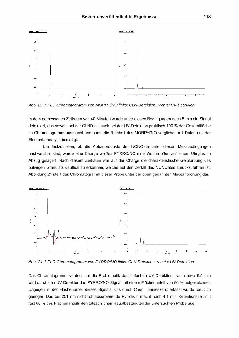
Bisher unveröffentlichte Ergebnisse 118
Abb. 23 HPLC-Chromatogramm von MORPH/NO links: CLN-Detektion, rechts: UV-Detektion
In dem gemessenen Zeitraum von 40 Minuten wurde unter diesen Bedingungen nach 5 min ein Signal
detektiert, das sowohl bei der CLND als auch bei der UV-Detektion praktisch 100 % der Gesamtfläche
im Chromatogramm ausmacht und somit die Reinheit des MORPH/NO verglichen mit Daten aus der
Elementaranalyse bestätigt.
Um festzustellen, ob die Abbauprodukte der NONOate unter diesen Messbedingungen
nachweisbar sind, wurde eine Charge weißes PYRRO/NO eine Woche offen auf einem Uhrglas im
Abzug gelagert. Nach diesem Zeitraum war auf der Charge die charakteristische Gelbfärbung des
pulvrigen Granulats deutlich zu erkennen, welche auf den Zerfall des NONOates zurückzuführen ist.
Abbildung 24 stellt das Chromatogramm dieser Probe unter der oben genannten Messanordnung dar.
Abb. 24 HPLC-Chromatogramm von PYRRO/NO links: CLN-Detektion, rechts: UV-Detektion
Das Chromatogramm verdeutlicht die Problematik der einfachen UV-Detektion. Nach etwa 6.5 min
wird durch den UV-Detektor das PYRRO/NO-Signal mit einem Flächenanteil von 86 % aufgezeichnet.
Dagegen ist der Flächenanteil dieses Signals, das durch Chemilumineszenz erfasst wurde, deutlich
geringer. Das bei 251 nm nicht lichtabsorbierende Pyrrolidin macht nach 4.1 min Retentionszeit mit
fast 60 % des Flächenanteils den tatsächlichen Hauptbestandteil der untersuchten Probe aus.

119 Bisher unveröffentlichte Ergebnisse
Da der Hauptfokus dieser Arbeit nicht die Entwicklung einer verbesserten HPLC-Methode sein
sollte, wurden keine detaillierten Untersuchungen diesbezüglich angestellt. Die oben genannten
Vorversuche sollten lediglich feststellen, ob die HPLC mit CLN-Detektion prinzipiell ein geeignetes
Mittel für Untersuchungen an NONOaten darstellt.
4.1.3 Synthese von Diazen-1-ium-1,2-diolat Prodrugs In diesem Kapitel sollen die synthetisierten Diazeniumdiolat-Prodrugs, die in keiner der
Veröffentlichungen der vorliegenden Arbeit erwähnt werden, monographiert werden.
Tosyl-PYRRO/NO
NN
O
NO S
O
O
CH3 O2-(p-Methylbenzen-sulfonyl)-1-(pyrrolidin-1-yl)diazen-1-ium-1,2-diolat: 0.58 g (3.8 mmol) fein
disperses Natrium 1-(pyrrolidin-1-yl)diazen-1-ium-1,2-diolat (PYRRO/NO) werden zusammen mit 800
mg Natriumcarbonat bei 4 °C in 50 ml tert-Butylmethylether suspendiert. 1.0 g (5.25 mmol) Toluol-4-
sulfonylchlorid gelöst in 10 ml tert-Butylmethylether werden tropfenweise der gekühlten Suspension
zugesetzt und anschließend für 48 Stunden bei Raumtemperatur gerührt. Feststoffe werden abfiltriert
und das farblose Filtrat im Kühlschrank gelagert bis sich weiße Kristalle abscheiden (62% Ausbeute). 1H-NMR 250 MHz (CDCl3) δ 1.97 (t, 4H, J= 5.0 Hz), 2.46 (s, 3H), 3.60 (t, 4H, J= 5.0 Hz), 7.36 (d, 2H,
J= 8.1 Hz), 7.89 (d, 2H, J= 8.1 Hz); 13C-NMR 250 MHz (CDCl3) δ 21.7, 23.3, 50.3, 128.8, 129.7, 132.1,
145.7; E.A. ber. für: C11H15N3O4S (285): C 46.30, H 5.30, N 14.73, O 22.43, S 11.24; gef.: C 46.21, H
5.09, N 14.92, S 11.16.
Um sicher zu stellen, dass nicht fälschlicherweise durch den vorzeitigen PYRRO/NO-Zerfall
einfach das Pyrrolidin-Analogum gebildet wird, wurde die oben beschriebene Reaktion ebenfalls mit
Pyrrolidin statt PYRRO/NO durchgeführt, um eine Referenzsubstanz zu erhalten. Im 1H-NMR
Spektrum ist deutliche zu erkennen, dass die Protonen der zum Pyrrolidin N-Atom benachbarten
Kohlenwasserstoffe statt bei δ 3.60 ppm bei dieser Verbindung zu δ 3.21 ppm hochfeldverschoben
sind.

Bisher unveröffentlichte Ergebnisse 120
Me-PYRRO/NO
NN
O
NO CH3
O2-(Methyl)-1-(pyrrolidin-1-yl)diazen-1-ium-1,2diolat: 0.58 g (3.8 mmol) fein disperses Natrium 1-
(pyrrolidin-1-yl)diazen-1-ium-1,2-diolat (PYRRO/NO) wird zusammen mit 800 mg Natriumcarbonat bei
4 °C in 50 ml tert-Butylmethylether suspendiert. Eine äquimolare Menge (0.5 g) Dimethylsulfat gelöst
in 10 ml tert-Butylmethylether wird vorsichtig zugetropft und 24 h bei Raumtemperatur gerührt.
Anschließendes Filtrieren und Einrotieren des Filtrats ergibt ein gelbes Öl, das keiner weiteren
Reinigung bedarf (83 % Ausbeute). 1H-NMR 250 MHz (D2O) δ 1.70 (m, 4H), 3.26 (m, 4H), 3.76 (s,
3H); E.A. ber. für: C5H11N3O2 (145): C 41.37, H 7.64, N 28.95, O 22.04; gef.: C 41.31, H 7.31, N 28.99.
Et-BUPIPERAZI/NO, Prop-PIPER/NO, Pent-PIPER/NO, MTME-PIPER/NO, 1,5-di-PYRRO/NO-pentan
N NO
N OCH3
N NO
N ON
H3C CH32
N NO
N OCH34
N NO
N O NNO
NO
N NO
N OSCH3
Et-BUPIPERAZI/NO Prop-PIPER/NO
Pent-PIPER/NO
1,5-di-PYRRO/NO-pentan
MTME-PIPER/NO
Für die Derivatisierung des O2-Atoms an NONOaten mit Alkylhalogeniden wurde folgende allgemeine
Arbeitsvorschrift (AAV) entwickelt:
AAV 1: Etwa 800 mg fein verriebenes Natriumcarbonat in 50 ml Hexamethylphosphorsäuretriamid
(HMPA) suspendieren, auf 4 °C abkühlen und die angegebene Menge fein disperses NONOat geben.
Langsam einen 1.5-fach molaren Überschuss des entsprechenden Alkylhalogenides zutropfen und 72
h bei Raumtemperatur rühren lassen. Anschließend in eine gesättigte, wässrige NaHCO3 Lösung
filtrieren und zweimal mit Ethylacetat extrahieren. Das Ethylacetat 5-6 mal mit NaHCO3 Lösung

121 Bisher unveröffentlichte Ergebnisse
waschen und einrotieren. Der Rückstand wird säulenchromatographisch über Kieselgel mit
geeignetem Eluent gereinigt.
In der Literatur findet man häufig Dimethylformamid (DMF) als verwendetes Lösungsmittel für ähnliche
Reaktionen. Allerdings verläuft die Synthese bei der Verwendung von HMPA vollständiger ab und der
Lösungsmittelrückstand lässt sich besser entfernen. Experimente das HMPA durch weniger giftigen
Tetramethylharnstoff als Lösungsmittel zu ersetzen, waren aufgrund eines schlechteren
Ausbeute/Nebenprodukt Verhältnisses nicht erfolgreich.
O2-Ethyl-1-(butylpiperazin-1-yl)diazen-1-ium-1,2-diolat (Et-BUPIPERAZI/NO): 4 mmol (900 mg)
Natrium 1-(butylpiperazin-1-yl)diazen-1-ium-1,2-diolat (BUPIPERAZI/NO) wurde nach AAV 1 mit
Ethyliodid umgesetzt und mit Dichlormethan säulenchromatographisch zum hellbraunen Öl gereinigt
(93 % Ausbeute). 1H-NMR (CDCl3) δ 0.90 (t, 3H, J= 7.1 Hz), 1.25-1.54 (m, 4H, J= 7.1 Hz), 1.38 (t, 3H,
J= 7.1 Hz), 2.34 (t, 2H, J= 7.1 Hz), 2.59 (t, 4H, J= 5.0 Hz), 3.42 (t, 4H, J= 5.0 Hz), 4.25 (q, 2H, J= 7.1
Hz); 13C-NMR 250 MHz (CDCl3) δ 13.9, 14.6, 20.6, 28.9, 51.3, 52.0, 57.7, 69.6; E.A. ber. für:
C10H22N4O2 (230): C 52.15, H 9.63, N 24.33, O 13.89; gef.: C 52.12, H 9.76, N 23.96.
O2-Propyl-1-(piperidin-1-yl)diazen-1-ium-1,2-diolat (Prop-PIPER/NO): 6 mmol (1 g) Natrium 1-
(piperidin-1-yl)diazen-1-ium-1,2-diolat (PIPER/NO) wurde nach AAV 1 mit Propyliodid zu einer rot-
braunen Flüssigkeit umgesetzt, die keiner weiteren Reinigung bedarf (76 % Ausbeute). 1H-NMR
(CDCl3) δ 0.96 (t, 3H, J= 7.4 Hz), 1.45-1.54 (m, 2H), 1.71-1.82 (m, 6H), 3.31 (t, 4H, J= 5.5 Hz), 4.17
(t, 2H, J= 6.9 Hz); E.A. ber. für: C8H17N3O2 (187): C 51.32, H 9.15, N 22.44, O 17.09; gef.: C 51.33, H
9.23, N 22.28.
O2-Pentyl-1-(piperidin-1-yl)diazen-1-ium-1,2-diolat (Pent-PIPER/NO): 6 mmol (1 g) Natrium 1-
(piperidin-1-yl)diazen-1-ium-1,2-diolat (PIPER/NO) wurde nach AAV 1 mit 1-Iodpentan zu einer
braunen Flüssigkeit umgesetzt. Der Rückstand wurde mit Chloroform/Ethylacetat/N-Hexan (35:15:50)
chromatographisch gereinigt. (23 % Ausbeute). 1H-NMR (CDCl3) δ 0.90 (m, 3H), 1.31-1.52 (m, 8H),
1.63-1.80 (m, 4H), 3.31 (t, 4H, J= 5.6 Hz), 4.23 (t, 2H, J= 6.9 Hz); E.A. ber. für: C10H21N3O2 (215): C
55.79, H 9.83, N 19.52, O 14.86; gef.: C 56.23, H 9.53, N 19.28.
O2-Methylthiomethyl-1-(piperidin-1-yl)diazen-1-ium-1,2-diolat (MTME-PIPER/NO): 6 mmol (1 g)
Natrium 1-(piperidin-1-yl)diazen-1-ium-1,2-diolat (PIPER/NO) wurde nach AAV 1 mit
Chlormethylmethylsulfid zu einer rot-braunen Flüssigkeit umgesetzt, die keiner weiteren Reinigung
bedarf (95 % Ausbeute). 1H-NMR (CDCl3) δ 1.72-1.82 (m, 6H), 3.34 (t, 4H, J= 5.5 Hz), 3.52 (s, 3H),
5.28 (s, 2H); E.A. ber. für: C7H15N3O2S (205): C 40.96, H 7.37, N 20.47, O 15.59, S 15.62; gef.: C
40.63, H 7.23, N 20.28.
O2,O2’-Pentylen-bis-1-(pyrrolidin-1-yl)diazen-1-ium-1,2-diolat (1,5-di-PYRRO/NO-pentan): Abweichend
von AAV 1 wurde ein 3-facher molarer Überschuss Natrium 1-(pyrrolidin-1-yl)diazen-1-ium-1,2-diolat
(PYRRO/NO) mit 2.2 mmol (0.5 g) 1,5-dibromopentan umgesetzt. Braunes Öl (61 % Ausbeute). 1H-

Bisher unveröffentlichte Ergebnisse 122
NMR (CDCl3) δ 1.46-1.55 (m, 2H), 1.73-1.82 (m, 4H), 1.86-2.00 (m, 8H), 3.50 (mc, 8H), 4.16 (m, 4H);
E.A. ber. für: C13H26N6O4 (330): C 47.26, H 7.93, N 25.44, O 19.37; gef.: C 46.74, H 8.14, N 25.86.
4.2 Biologische Aktivität einiger synthetisierter NO-Donoren 4.2.1 Vasodilatation in vitro
Drei der in Veröffentlichung 2 beschriebenen Diazeniumdiolate und ein Diazeniumdiolat-
Prodrug aus Veröffentlichung 3 (siehe Abb. 25) wurden für weitere Untersuchungen an isolierten
Gefäßringen der Arteria Pulmonalis vom Schwein verwendet. Die praktische Durchführung dieser
pharmakologischen Untersuchungen entspricht denen, die auch in Veröffentlichung 4 beschrieben
sind und erfolgten in Kooperation mit Herrn Apotheker Andreas König.
N NO
N ON
NaN N
O
N OOOC 2 Na
NaN N
O
N O
H3C
H3C
N NO
N ON
CH3
Trivialname: PHEPIPERAZI/NO Bezeichnung in Veröffentlichung 2: 2g
Trivialname: ISONIPECO/NO Bezeichnung in Veröffentlichung 2: 2i
Trivialname: DPA/NO Bezeichnung in Veröffentlichung 2: 2m
Trivialname: Et-PHEPIPERAZI/NO Bezeichnung in Veröffentlichung 3: 4b
Abb. 25 Für pharmakologische Untersuchungen verwendete Diazeniumdiolate
Um die vasodilatatorische Potenz der Substanzen besser einschätzen zu können, wurde das
therapeutisch häufig eingesetzte Glyceroltrinitrat (GTN) als Referenzsubstanz verwendet. Abbildung
26 und Tabelle 4 skizzieren die durch die Substanzen ausgelöste Vasorelaxation.

123 Bisher unveröffentlichte Ergebnisse
Abb. 26 Vasodilatierende Aktivität der Diazeniumdiolate und Glyceroltrinitrat (GTN) als Referenz an
PGF2α-vorkontrahierten Pulmonalarterien mit intaktem Endothel. Die gefäßrelaxierende
Wirkung ist in Prozent zu der durch PGF2α ausgelösten Vorkontraktion dargestellt. Mittelwerte
± SEM
Das direkt NO.-freisetzende NONOat PHEPIPERAZI/NO löst bereits bei einer Konzentration von 2.12
x 10-9 M den halbmaximalen Effekt an den isolierten Gefäßen aus. Etwa um den Faktor 3 schwächer
ist das Dipropylamino-NONOat DPA/NO und ISONIPECO/NO stellt von den Diazeniumdiolat-Natrium
Salzen mit einem EC50-Wert von 2.06 x 10⁻⁸ M die schwächste Verbindung dar. Für diese drei spontan
freisetzenden NO.-Donoren sind in Tabelle 4 die UV-spektroskopisch ermittelten
Freisetzungshalbwertszeiten (t1/2 bei pH 7.4, 37 °C) angegeben (vergl. Veröffentlichung 2).
Tab. 4 Ermittelte EC50-, pD2-Werte und Zerfallshalbwertszeiten der NONOate und Glyceroltrinitrat
(GTN) als Referenz an PGF2α-vorkontrahierten Pulmonalarterien vom Schwein mit intaktem
Endothel
Substanz EC50 [mol/l] pD2 (n) Halbwertszeit (t1/2) der NO.-
Freisetzung [min]
PHEPIPERAZI/NO 2.12 x 10⁻⁹ 8.67 (5) 4.5
DPA/NO 6.37 x 10⁻⁹ 8.20 (2) 1.5
ISONIPECO/NO 2.06 x 10⁻⁸ 7.69 (3) 0.5
GTN 1.91 x 10⁻⁸ 7.44 (6) --
Et-PHEPIPERAZI/NO 1.09 x 10⁻⁵ 4.96 (2) --
Vergleicht man die NO.-Freisetzung mit der vasodilatatorischen Aktivität, so stellt man fest, dass die
langsamer freisetzenden NONOat-Natrium Salze die größere Gefäßerweiterung auslösen. Der Grund
hierfür ist die höhere Verfügbarkeit des NO. am Gefäßring. Je schneller ein NONOat freisetzt, desto
mehr NO. wird in dem mit Carbogengas (95% Sauerstoff!) durchspülten Nährmedium zum NO2
oxidiert, welches dann wiederum rasch mit Wasser überwiegend zum Nitrit weiterreagiert (vergl.
Kapitel 1.1). Da die schnell freisetzenden NONOate in einem kurzen Zeitraum mehr Stickstoffmonoxid

Bisher unveröffentlichte Ergebnisse 124
freisetzen als am Gefäßring in dieser Zeit einen biologischen Effekt auslösen kann, geht ein relativ
großer Teil des theoretisch verfügbaren NO. verloren. Das Organonitrat GTN und das NONOat-
Prodrug Et-PHEPIPERAZI/NO setzen im Organbad erst nach biologischer Aktivierung
Stickstoffmonoxid frei. Die im Vergleich zu den NONOat-Na Salzen erst verzögert eintretende Wirkung
lässt sich dadurch erklären, dass der geschwindigkeitsbestimmende Schritt, anders als bei den direkt
und spontan freisetzenden NONOat-Na Salzen, die Enzymaktivität ist. Die deutlich geringere Aktivität
des NONOat-Prodrugs Et-PHEPIPERAZI/NO muss somit ebenfalls mit der Aktivität und/oder der
geringeren Konzentration des metabolisierenden Enzyms zusammenhängen. Messungen mit der
zuvor schon beschriebenen NO-Sonde zeigten, dass eine chemische Hydrolyse dieses Prodrugs im
Nährmedium nicht stattfindet.
Eine Präinkubation der isolierten Gefäßringe mit dem kompetitiven Antagonisten der löslichen
Guanylatcyclase ODQ (10 µM) bewirkte eine starke Rechtsverschiebung der Konzentrations-
Wirkungskurve während der Messung mit ISONIPECO/NO. Der ermittelte EC50-Wert ergab eine
Konzentration von 1.02 x 10⁻⁶ mol/l im Vergleich zu 2.06 x 10⁻⁸ mol/l ohne ODQ-Inkubation. Wie
erwartet konnte somit die Wirkungsabhängigkeit von der sGC für das ISONIPECO/NO nachgewiesen
werden.
4.2.2 Neuroregenerative Eigenschaften von DETA/NO und Et-BUPIPERAZI/NO in vivo
In Kooperation mit dem Arbeitskreis von Dr. Redecker und seiner Mitarbeiter, insbesondere
Frau Dr. Silke Keiner, wurden Diazeniumdiolate zur Verfügung gestellt, um die Zellproliferation und –
differenzierung in der Umgebung experimentell induzierter Hirninfarkte bei der Ratte zu untersuchen.
Bei diesem in vivo Modell wird durch unterschiedliche Verhaltenstests, sowie einer Reihe von zyto-
und histologischen Untersuchungen post mortem, die neuroregenerativen Eigenschaften applizierter
Substanzen beurteilt. Da die Untersuchungen noch nicht vollständig abgeschlossen und ausgewertet
sind, soll an dieser Stelle nur kurz auf die bisher vorliegenden Ergebnisse aus den Vorversuchen
eingegangen werden.
Als NO.-Donoren wurden das direkt NO.-freisetzende Diazeniumdiolat DETA/NO (siehe
Veröffentlichung 1) und das NONOat-Prodrug Et-BUPIPERAZI/NO (siehe Kapitel 4.1.3 und
Veröffentlichung 5) verwendet. Die genauen Durchführungen dieser Vorversuche sind der zitierten
Literatur zu entnehmen, eine kurze Zusammenfassung ist wie folgt:[99-101] Zwei Tage vor der Induktion
des fokalen kortikalen Infarktes im Vorderpfotenkortex, ausgelöst durch Kaltlicht Photothrombose am
geöffneten Schädel der narkotisierten Ratten, wurde den Versuchstieren eine tägliche Dosis von 30,
60 oder 600 µg/kg Körpergewicht an Et-BUPIPERAZI/NO verabreicht. Anderen Tieren wurde zum
Vergleich das DETA/NO in einer Dosierung von 400 µg/kg ebenfalls intraperitoneal appliziert.
Zusammen mit jeder Wirkstoffapplikation wurde den Versuchtieren BrdU (Bromdesoxyuracil) in einer
Konzentration von 50 mg/kg Körpergewicht injiziert. BrdU ist ein Proliferationsmarker und lässt somit
die Quantifizierung von neugebildetem Gewebe zu. Die tägliche NO.-Donor- und BrdU Gabe wurde bis
zum 14. Tag nach Auslösen der Photothrombose fortgesetzt. Am 14. Tag nach Hirninfarktinduktion
wurde das Tiermaterial getötet, die Gehirne freipräpariert und anschließend makro- und mikroskopisch

125 Bisher unveröffentlichte Ergebnisse
untersucht. Um das Ausmaß an neugebildetem Gewebe zu bestimmen, wurde zum einen die Anzahl
BrdU-positiver Zellen in der Subgranulärzone des Gyrus dentatus im Hippocampus bestimmt, zum
anderen das globale Hirnvolumen und das Volumen der photothrombotischen Läsion
(Photothrombose-Volumen).
In Abbildung 27 ist die Anzahl BrdU-positiver Zellen und das Volumen der Photothrombose
Läsion in Abhängigkeit vom applizierten NONOat und der jeweiligen Dosierung graphisch dargestellt.
Abb. 27 links: BrdU-positive Zellen in der Subgranulärzone des Hippocampus der Rattenhirne und
rechts: Photothrombose-Volumen nach NONOat-Applikation
Die höchste Dosis von 600 µg/kg (entspricht 2.45 µmol/kg) Et-BUPIPERAZI/NO ergab in den
Versuchstieren die höchste Proliferation an neuronalem Gewebe. Das direkt NO.-freisetzende
DETA/NO veranlasst bei etwa gleicher molarer Dosis (400 µg/kg entspricht 2.5 µmol/kg) das Auffinden
von nur halb soviel markierten Zellen. Eine direkte Dosis-Wirkungskorrelation für das Et-
BUPIPERAZI/NO konnte in diesen Vorversuchen jedoch nicht festgestellt werden. Während der
Betrag des globalen Hirnvolumens bei allen vier Versuchsreihen mit 1000 +/-75 mm3 gleich war, ist
dass Photothrombose-Volumen bei den Versuchsreihen mit Et-BUPIPERAZI/NO auffallend geringer.
Die durch das Kaltlicht verursachte Verletzung ist bei den Versuchstieren, die mit Et-BUPIPERAZI/NO
behandelt wurden, in ihrem Volumen somit geringer als bei denen, die DETA/NO erhielten.

Gesamtdiskussion 126
5. Gesamtdiskussion 5.1 Bewertung der NOtizer®-Apparatur für die Synthese von Diazen-1-ium-1,2-
diolaten
Die ersten NONOate wurden 1960 von Drago et al. synthetisiert, indem NO-Gas bei einer
Temperatur von -78 °C durch Lösungen von Aminen in Ether geleitet wurde.[70,95] Nachdem die
physiologische Bedeutung des NO erkannt war, wurde die Synthese der NONOate in den 1990er
Jahren verbessert. Unter erhöhtem Druck und Basenzusatz in einem Autoklav konnten neue
NONOate in verbesserter Ausbeute als einheitlichere Produkte dargestellt werden.[70,102] Nachteil
dieser Methode blieb die fehlende Überwachung des Syntheseverlaufs, sowohl durch Temperatur-
und Druckaufzeichnung, als auch durch schlichtes visuelles Beobachten des Reaktionsgemisches.
Mögliche Präzipitatbildung, Veränderungen in Farbe des Reaktionsgemisches oder des Gasraumes,
z.B. durch Oxidation des farblosen NO. zu braunem NO2 und N2O3, blieben bis zum Abbruch der
Reaktion und Öffnen des Autoklaven unerkannt.
5.1.1 Vorteile der NOtizer®-Apparatur und der erarbeiteten Methode Der Einsatz des NOtizers® und die Anwendung der in Veröffentlichung 1 beschriebenen
Methode stellen gegenüber den literaturbekannten Verfahren in erster Linie eine erhebliche
Steigerung in Ausbeute und Reinheit der gewonnenen NONOate dar. Durch die Aufzeichnung des
NO-Druckverlaufs kann auch nach wiederholter Druckerhöhung genau erkannt werden, wann der NO-
Verbrauch, und somit die Reaktion, zum Stillstand kommt. Ferner ist es möglich, durch Quantifizierung
der verbrauchten Stoffmenge an NO die zu erwartende Stoffmenge des jeweiligen NONOates
absehen zu können, was gerade bei erstmaliger Synthese eines neuen Diazeniumdiolates von
Bedeutung ist. Hieraus resultieren wichtige Erkenntnisse bezüglich möglicher Verbesserungen der
anschließenden Produktisolierung. Äußerst hilfreich und sehr wichtig für die Reinheit des NONOates
ist ferner die Möglichkeit, das Reaktionsgemisch beobachten zu können. Als eine Art
“Inprozesskontrolle“ kann die O2-Freiheit der Reaktionskammer überprüft werden, wenn sich beim
Einleiten des farblosen NO-Gases weder der Gasraum noch die Lösung gelb oder braun verfärbt
durch eventuelle NO-Oxidationsprodukte, die zu Nebenproduktbildung führen (vergl. Gleichung 4 bis
6, Kapitel 1.2.5). Ebenso ist es vorteilhaft, wenn das NONOat aus der Reaktionslösung präzipitiert, da
Einengen des Lösungsmittels nach Abschluss der Reaktion immer die Gefahr der Mitisolierung von
Edukt birgt. Beispielsweise ist weißes Natriummethanolat visuell nicht von den meisten NONOaten zu
unterscheiden. Das zur Ausfällung des Diazeniumdiolates führende ideale Lösungsmittel kann so
relativ leicht für neue, bisher unbekannte NONOate experimentell bestimmt werden.

127 Gesamtdiskussion
5.1.2 Nachteile der NOtizer®-Apparatur und der erarbeiteten Methode Da es sich bei der Reaktionskammer um eine druckbeständige Glasflasche handelt, kann die
druckabhängige Gleichgewichtsreaktion zum NONOat nur einem maximalen Druck von 5-6 bar
ausgesetzt werden. Dies ist ein Nachteil, wenn Diazeniumdiolate mit sterisch anspruchsvollen
Substituenten am Amin hergestellt werden sollen (vergl. Veröffentlichung 2), wie es z.B. bei
stickstoffhaltigen Polymeren der Fall sein kann.[103] Für solche Zwecke wäre ein
hochdruckbeständiges Gefäß nötig um verwertbare Produktausbeuten zu erhalten. Ein Nachteil, der
nicht direkt mit der NOtizer®-Apparatur zusammenhängt, sondern ein genereller Nachteil bei der
Synthese von Diazeniumdiolaten ist, dass während der Reaktion keine Proben des
Reaktionsgemisches entnommen werden können, beispielsweise für reaktionskinetische
Untersuchungen.
5.2 Untersuchungen zur Bildung und Zerfall von Diazen-1-ium-1,2-diolaten 5.2.1 Bildung von Diazen-1-ium-1,2-diolaten
Die unmittelbare Bildung der NONOate lässt sich aus den oben genannten Gründen weder mit
den bisher beschriebenen Methoden, noch mit dem NOtizer® beobachten. Um Proben während der
Umsetzung entnehmen zu können, müsste ein offenes System oder eine geeignete Apparatur
verwendet werden. Idealerweise müsste die Reaktionskammer so gestaltet sein, dass z.B. UV-
spektroskopisch die Bildung des NONOates verfolgt werden kann. Auf diese Art wäre auch die
Synthese von Diazeniumdiolaten nachvollziehbar, die einen hohen Druck an NO-Gas benötigen, um
überhaupt entstehen zu können. Mit der NOtizer®-Apparatur kann jedoch im Gegensatz zu den
literaturbekannten Methoden der NO-Druckverlauf aufgezeichnet und analysiert werden. So kann
indirekt auch die NONOat-Bildung verfolgt werden, wobei vor allem die Registrierung der
Reaktionsgeschwindigkeit und des Reaktionsendpunktes sehr wertvoll ist. Abbildung 28 stellt den
Druck-Zeit Verlauf bei der Synthese des PYRRO/NO dar.

Gesamtdiskussion 128
Abb. 28 schwarze Kurve: Druck-Zeit Verlauf bei der Reaktion von Pyrrolidin mit NO-Gas
rote Kurve: exponentieller Fit 1.Ordnung
blaue Kurve: exponentieller Fit 2. Ordnung
Mit entsprechender Software kann der Kurvenverlauf, und somit der NO-Gas Verbrauch, einem
Zeitgesetz zweiter Ordnung zugeordnet werden. Eine Übertragung dieses Zeitgesetzes auf die
Bildung des NONOates ist jedoch, ohne die Produktbildung zu verfolgen, nicht möglich. Ein
Rückschluss auf die Reaktionsmolekularität oder den Mechanismus ist ebenfalls nur spekulativ (vergl.
Kapitel 1.2.5). Eine wertvolle Information, die sich jedoch aus den Daten mit dem NOtizer® bestimmen
lässt, ist neben der verbrauchten Stoffmenge NO pro Zeit (nNO/dt) die Umsatzrate (rate of conversion
ξ). Die IUPAC definiert die Umsatzrate wie folgt:[104]
dξ dni1 ξ =
Geht man davon aus, dass
annehmen, dass die Umsa
proportional ist. Um eine be
Veröffentlichung 2 um einige
- der NO-Druckab
- Ende der Reakt
- NO-Restdruck a
- Stoffmengenver
W
=
dtaus dem NO ausschließlich und sofort das NONOat entsteht, so kann man
tzrate des NO der Bildungsrate (rate of formation) des Diazeniumdiolats
ssere Differenzierung zu ermöglichen, wurde der Begriff “formation rate“ in
Aspekte erweitert:
fall innerhalb der ersten 7 h nach Synthesebeginn [bar]
ion [hh:mm]
m Ende der Reaktion [bar]
hältnis von NO-Verbrauch und Amin
dt νi
obei: ni = Stoffmenge eines Edukts i zur Zeit t
νi = stöchiometrischer Faktor von Edukt i

129 Gesamtdiskussion
5.2.2 Zerfall von Diazen-1-ium-1,2-diolaten Der Zerfall der Diazeniumdiolate lässt sich sehr gut UV-spektroskopisch verfolgen, wobei das
Absorptionsmaximum der [N(O)NO]- Gruppe bei 251 nm liegt. In Abbildung 29 ist das Wellenlängen-
Absorptionsspektrum des zerfallenden PIPER/NO in Abhängigkeit von der Zeit dargestellt.
Abb. 29 Wellenlängen-Absorptions Spektrum des PIPER/NO in Phosphatpuffer 7.4, 37°C in
Abhängigkeit von der Zeit
Wenn die Absorptionsabnahme logarithmisch gegen die Zeit aufgetragen wird, erhält man eine
Gerade, was zum einen eine Zerfallskinetik 1. Ordnung belegt, zum anderen das Berechnen der
Zerfallshalbwertszeit t1/2 nach der Formel t1/2 = ln2/k ermöglicht (siehe Veröffentlichung 2, Fig. 4). Die
Zerfallskonstante k ist dabei die Steigung der Geraden nach linearer Regressionsanalyse. Dieser
indirekte NO-Nachweis eignet sich vor allem, um die Beständigkeit des NONOates in einem
bestimmten Medium zu untersuchen und Struktur-Freisetzungs Beziehungen der jeweiligen
Diazeniumdiolate miteinander vergleichen zu können.
Als direkte NO-Nachweismethode aus NONOaten wurde die amperometrische Detektion in
einer speziellen NO-Freisetzungszelle verwendet (siehe Kapitel 1.3.5). Da das NO direkt
nachgewiesen wird und nicht die [N(O)NO]--Gruppe, wird für quantitative Untersuchungen vorteilig
keine Referenzsubstanz benötigt, sondern die Eichung kann bequem durch Nitrit Reduktion erfolgen,
wobei der gemessene Redoxstromfluss der NO Menge proportional ist. Abbildung 30 zeigt eine
typische Eichkalibrierung der NO-Sonde.

Gesamtdiskussion 130
Abb. 30 Gemessene Redoxstromstärke einer zunehmend höher konzentrierten NO-Lösung
(durchReduktion von Nitrit)
Die in Veröffentlichung 2 dargestellten Redoxstromflüsse lassen sich somit quantifizieren,
vorausgesetzt man verwendet die Eichgerade des jeweiligen Messtages. Als
Geschwindigkeitsmerkmal ist die Zeit bis zum Redoxstromfluss-Maximum der zu verwendende
Parameter. Ab dem Maximum fällt der Kurvenverlauf wieder ab, was natürlich mit der geringer
werdenden NO-Freisetzung zu tun hat, jedoch durch die Repolarisation der Elektrode erheblich
gestört wird. Die Repolarisation der Elektrode dauert in etwa zehn Minuten und äußert sich durch eine
kontinuierliche Abnahme des “gemessenen Stromflusses“, obwohl sich in der Lösung kein NO mehr
befindet. Die Halbwertszeit der NO-Freisetzung lässt sich somit nicht über die AUC (area under the
curve) bestimmen, da das Integral der Kurve nicht ausschließlich den gemessenen Redoxstrom
darstellt, sondern auch die Repolarisation der Elektrode. Als nachteilig hat sich auch die geringe
Robustheit dieser Methode herausgestellt. Die Reproduzierbarkeit ist zwar sehr hoch, jedoch ist sehr
darauf zu achten, dass alle Parameter wie z.B. die channel range, das Hintergrundsignal der
Elektrode, sowie äußere Umstände möglichst gleich sind, um eine gleichbleibende Empfindlichkeit zu
gewährleisten.
5.2.3 Zusammenhang zwischen Aminstruktur, NONOat-Bildung und NO-Freisetzung
Die in Veröffentlichung 2 präsentierten Ergebnisse verdeutlichen, dass sterische Hinderung in
direkter Nachbarschaft zum Stickstoff des Amins, sowie elektronenziehende Substituenten die
Reaktivität mit NO verringern. Während nur ein sterisch anspruchsvoller Rest die Reaktion ein wenig
verlangsamt, bewirkt ein zweiter, wie es z.B. beim Diisopropylamin oder Dicyclohexylamin der Fall ist,
praktisch das Scheitern der Synthese unter diesen Bedingungen. Die immense Bedeutung der
Zugänglichkeit des N-Atoms für das NO lässt sich besonders am Vergleich der Umsatzraten der
zyklischen mit den aliphatischen Aminen beobachten. Die NONOat-Bildung verläuft bei allen

131 Gesamtdiskussion
untersuchten konformativ rigidisierten Aminen um ein vielfaches schneller und vollständiger im
Vergleich zu den aliphatischen Aminen, wie z.B. Diethylamin oder Dipropylamin. Einen sehr viel
geringeren Einfluss auf die Bildungsgeschwindigkeit haben elektronenziehende Heteroatome, die die
Nukleophilie des Stickstoffs verringern, sofern mindestens eine Methylengruppe diese vom
Aminstickstoff trennt. Ein Rest, der eine weitere Aminfunktion beinhaltet, wie z.B. das primäre Amin in
N-[(2-Aminoethyl)amino]ethanol, kann die Umsatzrate auch erhöhen. Die bereits diskutierte Bildung
des intramolekularen NONOat Salzes dieser Verbindung als Verunreinigung ist unabhängiger von der
Anwesenheit einer Base und verursacht somit die erhöhte Umsatzrate des NO. Abbildung 31 zeigt die
NO-Freisetzungsgeschwindigkeit von 22 Diazeniumdiolaten aus Veröffentlichung 2, aufgetragen
gegen den NO-Druckabfall bei der Bildung der gleichen Diazeniumdiolate, was als Maß für die
Reaktionsgeschwindigkeit gilt.
m
v
k
h
o
t
u
c
nq
gl
i
s
p
j
f
d
r
be
am
v
k
h
o
t
u
c
nq
gl
i
s
p
j
f
d
r
be
a
t
hk
u
m
s
ql
g n
v
o
p
j
f
d
cia
b
r
e
t
hk
u
m
s
ql
g n
v
o
p
j
f
d
cia
b
r
e
Abb. 31
Zusammenhang zwischen
NONOat-
Bildungsgeschwindigkeit und
NO-Freisetzung.
Oben: NO-Freisetzung
gemessen mit einer NO-
Sonde.
Unten: NO-Freisetzung
gemessen mit einem UV-
Spektrometer. Legende für
Buchstaben in
Veröffentlichung 2
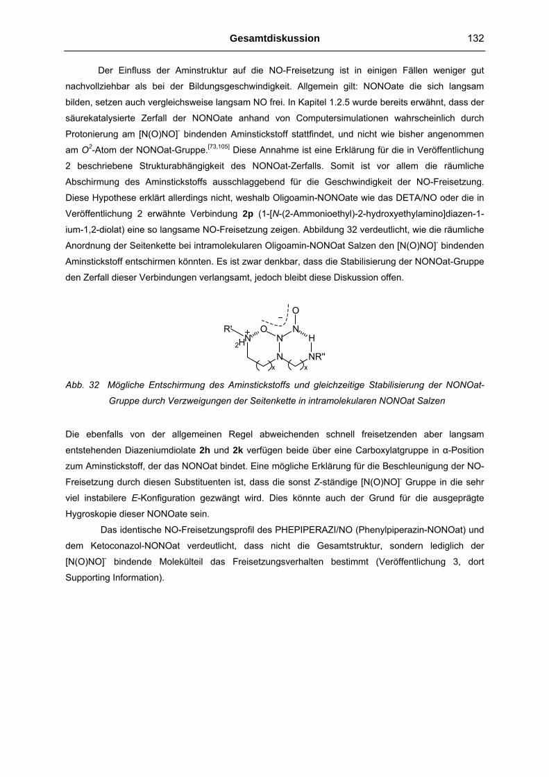
Gesamtdiskussion 132
Der Einfluss der Aminstruktur auf die NO-Freisetzung ist in einigen Fällen weniger gut
nachvollziehbar als bei der Bildungsgeschwindigkeit. Allgemein gilt: NONOate die sich langsam
bilden, setzen auch vergleichsweise langsam NO frei. In Kapitel 1.2.5 wurde bereits erwähnt, dass der
säurekatalysierte Zerfall der NONOate anhand von Computersimulationen wahrscheinlich durch
Protonierung am [N(O)NO]- bindenden Aminstickstoff stattfindet, und nicht wie bisher angenommen
am O2-Atom der NONOat-Gruppe.[73,105] Diese Annahme ist eine Erklärung für die in Veröffentlichung
2 beschriebene Strukturabhängigkeit des NONOat-Zerfalls. Somit ist vor allem die räumliche
Abschirmung des Aminstickstoffs ausschlaggebend für die Geschwindigkeit der NO-Freisetzung.
Diese Hypothese erklärt allerdings nicht, weshalb Oligoamin-NONOate wie das DETA/NO oder die in
Veröffentlichung 2 erwähnte Verbindung 2p (1-[N-(2-Ammonioethyl)-2-hydroxyethylamino]diazen-1-
ium-1,2-diolat) eine so langsame NO-Freisetzung zeigen. Abbildung 32 verdeutlicht, wie die räumliche
Anordnung der Seitenkette bei intramolekularen Oligoamin-NONOat Salzen den [N(O)NO]- bindenden
Aminstickstoff entschirmen könnten. Es ist zwar denkbar, dass die Stabilisierung der NONOat-Gruppe
den Zerfall dieser Verbindungen verlangsamt, jedoch bleibt diese Diskussion offen.
N
N
NO N
H
NR''
O
R'
x x
2H
Abb. 32 Mögliche Entschirmung des Aminstickstoffs und gleichzeitige Stabilisierung der NONOat-
Gruppe durch Verzweigungen der Seitenkette in intramolekularen NONOat Salzen
Die ebenfalls von der allgemeinen Regel abweichenden schnell freisetzenden aber langsam
entstehenden Diazeniumdiolate 2h und 2k verfügen beide über eine Carboxylatgruppe in α-Position
zum Aminstickstoff, der das NONOat bindet. Eine mögliche Erklärung für die Beschleunigung der NO-
Freisetzung durch diesen Substituenten ist, dass die sonst Z-ständige [N(O)NO]- Gruppe in die sehr
viel instabilere E-Konfiguration gezwängt wird. Dies könnte auch der Grund für die ausgeprägte
Hygroskopie dieser NONOate sein.
Das identische NO-Freisetzungsprofil des PHEPIPERAZI/NO (Phenylpiperazin-NONOat) und
dem Ketoconazol-NONOat verdeutlicht, dass nicht die Gesamtstruktur, sondern lediglich der
[N(O)NO]- bindende Molekülteil das Freisetzungsverhalten bestimmt (Veröffentlichung 3, dort
Supporting Information).
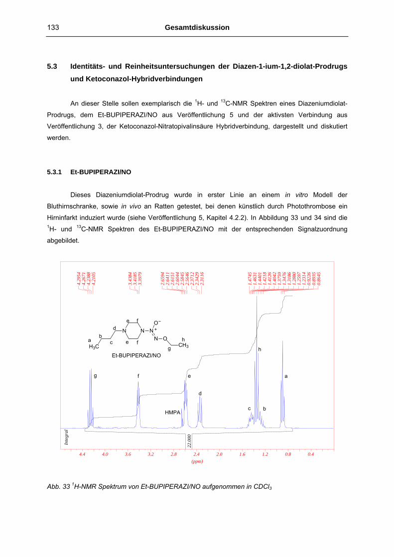
133 Gesamtdiskussion
5.3 Identitäts- und Reinheitsuntersuchungen der Diazen-1-ium-1,2-diolat-Prodrugs und Ketoconazol-Hybridverbindungen
An dieser Stelle sollen exemplarisch die 1H- und 13C-NMR Spektren eines Diazeniumdiolat-
Prodrugs, dem Et-BUPIPERAZI/NO aus Veröffentlichung 5 und der aktivsten Verbindung aus
Veröffentlichung 3, der Ketoconazol-Nitratopivalinsäure Hybridverbindung, dargestellt und diskutiert
werden.
5.3.1 Et-BUPIPERAZI/NO Dieses Diazeniumdiolat-Prodrug wurde in erster Linie an einem in vitro Modell der
Bluthirnschranke, sowie in vivo an Ratten getestet, bei denen künstlich durch Photothrombose ein
Hirninfarkt induziert wurde (siehe Veröffentlichung 5, Kapitel 4.2.2). In Abbildung 33 und 34 sind die 1H- und 13C-NMR Spektren des Et-BUPIPERAZI/NO mit der entsprechenden Signalzuordnung
abgebildet.
N NO
N ON
H3C CH3
Et-BUPIPERAZI/NO
22.0
00
Inte
gral
4.29
544.
2671
4.23
884.
2105
3.43
843.
4185
3.39
79
2.65
942.
6411
2.61
512.
6044
2.58
452.
5646
2.37
122.
3429
2.31
16
1.47
451.
4631
1.44
011.
4218
1.41
261.
4042
1.37
591.
3476
1.31
861.
2880
1.25
971.
2314
0.92
260.
8935
0.86
45
(ppm)0.40.81.21.62.02.42.83.23.64.04.4
e f d
b ha e c f g h
g e a f
d
c bHMPA
Abb. 33 1H-NMR Spektrum von Et-BUPIPERAZI/NO aufgenommen in CDCl3

Gesamtdiskussion 134
77.5
439
77.0
313
76.5
273
69.5
908
57.6
057
51.6
687
29.0
653
20.5
570
14.3
210
13.9
451
(ppm)051015202530354045505560657075808590
N NO
N ON
H3C CH3
Et-BUPIPERAZI/NOa
f
eb
c
e
d
fg
g fed c hb a
h
CDCl3
77.5
439
77.0
313
76.5
273
69.5
908
57.6
057
51.6
687
29.0
653
20.5
570
14.3
210
13.9
451
(ppm)051015202530354045505560657075808590
N NO
N ON
H3C CH3
Et-BUPIPERAZI/NOa
f
eb
c
e
d
fg
g fed c hb a
h
CDCl3
Abb. 34 13C-NMR Spektrum vom Et-BUPIPERAZI/NO aufgenommen in CDCl3
Das wichtigste Identitätskriterium des NONOates ist das Signal im 1H-NMR Spektrum bei 3.41 ppm
(Signalzuordnung f). Würde es sich statt des NONOates um das alkylierte Piperazin Derivat handeln,
so würde sich dieses Signal weiter Hochfeld, etwa bei 2.6 ppm befinden.
5.3.2 Ketoconazol-Nitratopivalinsäure Hybridverbindung Diese Verbindung wurde als “Vergleichs NO-Donor“ zu den Diazeniumdiolat-Ketoconazol
Derivaten synthetisiert und hinsichtlich antimikrobieller Eigenschaften untersucht (Veröffentlichung 3).
In Abbildung 35 sind die 13C-NMR Spektren des Ketoconazol-Nitratopivalinsäure Hybrids (rot) und
zum Vergleich das der Phenylpiperazin-Nitratopivalinsäure Verbindung (blau) übereinandergelegt.

135 Gesamtdiskussion
2030405060708090100110120130140150160170
(ppm)
O
O
Cl
Cl
N
N
O
H
NN
O
O2NO
CH3CH3
a
a
b
b
c
cc
d
d
e
e f
f
h
h
i
i k
lm
n
op
pp
p pj
j
g
p
p
g
p
p
h
i
n m k f
e
o
l
CDCl3
2030405060708090100110120130140150160170
(ppm)
O
O
Cl
Cl
N
N
O
H
NN
O
O2NO
CH3CH3
a
a
b
b
c
cc
d
d
e
e f
f
h
h
i
i k
lm
n
op
pp
p pj
j
g
p
p
g
p
p
h
i
n m k f
e
o
l
CDCl3
Abb. 35 Überlagerte 13C-NMR Spektren der Ketoconazol-Nitratopivalinsäure Hybridverbindung (rot)
und des entsprechenden Phenylpiperazin Analogen (blau) aufgenommen in CDCl3
Während die Identität des Phenylpiperazin-Nitratopivalinsäure Derivats schon anhand des 1H- und 13C-NMR Spektrums festgestellt werden kann, ist es aufgrund der Komplexität der analogen
Ketoconazol-Derivate sinnvoll, zusätzlich FAB-MS Daten mit einzubeziehen. Die geeignete Methode
um die Reinheit aller synthetisierten Derivate festzustellen, ist die Elementaranalyse. Verfahren wie
z.B. GC-MS, konnten aufgrund der Temperaturinstabilität oder des hohen Molekulargewichts vieler
Derivate nicht etabliert werden.
5.4 Antimikrobielle Eigenschaften der Ketoconazol-NO-Donor Hybridverbindungen: Diskussion möglicher Targets, Unterschiede im Wirkspektrum und Ursachen des Synergismus
Wie bereits in Kapitel 1.1.1 erwähnt, sind die biologischen Effekte des NO. als die Summe
eines multifaktoriellen Geschehens zu sehen, das sich aus NO und seinen Folgeprodukten wie z.B.
Peroxynitrit, S-Nitrosothiolen, Stickstoffdioxid oder Distickstofftrioxid ergibt. Die in Kapitel 1.1.2
dargestellte physiologische Funktion des NO. im Immunsystem, wie z.B. die Aktivierung von Zytokinen
und Lymphozyten, spielen bei den im Rahmen dieser Arbeit durchgeführten antimikrobiellen

Gesamtdiskussion 136
Untersuchungen in vitro keine Rolle. Die direkte Wirkung des NO und anderer RNS auf
Mikroorganismen sowie potentielle zelluläre Targets soll deswegen folgend im Vordergrund stehen.
5.4.1 Mikrobielle zelluläre Targets für NO und andere RNS
Ein wichtiges direktes Angriffsziel der RNS in Mikroorganismen sind die Nukleinsäuren. Es
konnte bereits gezeigt werden, dass durch NO-Donoren die DNA vieler Bakterienstämme in vitro
desaminiert wird. So werden z.B. die Nukleotide des Cytosin zu Uracil und des Guanin zu Xanthin
desaminiert. Als Mechanismus wird angenommen, dass das Amin durch N2O3 erst N-nitrosyliert und
anschließend hydrolysiert wird.[106] Ebenso sind Peroxynitrit und Stickstoffdioxid in der Lage
Nukleinsäuren zu oxidieren. Die so entstehenden DNA Schäden, wie z.B. Strangbrüche, werden
insbesondere durch die Oxidation von Guanidin, bzw. genauer des entsprechenden Guanosin-
Nukleotids, zu 8-Oxo-7,8-dihydro-guanidin verursacht.[107]
Die Interaktionen der RNS mit Proteinen und Kofaktoren ist um einiges vielfältiger als auf DNA
Ebene. So sind zum einen direkte Reaktionen mit Thiolen, Aminen, aromatischen Aminosäuren, Häm-
Gruppen oder Eisen-Schwefel-Cluster möglich, zum anderen auch unspezifische Oxidationen an
Eiweißen durch ONOO- und NO2. Unter physiologischen Bedingungen wird die Bildung von
Nitrosothiolen der von Nitrosaminen bevorzugt. Ebenso ist die Modifikation in Form von Thiol-Gruppen
Oxidation zu Disulfiden eine häufig stattfindende Reaktion. Die Nitrierung aromatischer Aminosäuren,
z.B. von Tyrosin, kann direkt oder durch das Unterbinden von Phosphorylierungsreaktionen die
Proteinfunktion modifizieren. Die Inaktivierung von Eisen-Schwefel-Cluster-haltigen Enzymen (Fe-S-
Cluster) kann zu einer erheblichen Beeinträchtigung des Stoffwechsels im Mikroorganismus führen.
Beispielhaft ist die Aconitase ein solch Fe-S-Cluster-haltiges Enzym, das Citrat im Citrat- und im
Glyoxylatzyklus zu Isocitrat umsetzt.[108] Dieser gehemmte Abbau organischen Materials ist gerade bei
Pilzen eine mögliche Erklärung für die antimykotische Aktivität des NO.
Die in Kapitel 1.1.2 ausführlich besprochenen Reaktionen der RNS mit Lipiden stellen
natürlich auch einen möglichen Mechanismus der mikrobiellen Schädigung dar. Die Lipide sind, neben
Chitin und Hemicellulosen, ein weiterer Hauptbestandteil der Zellwände bei Pilzen. Die
Lipidperoxidation wird dabei überwiegend durch Peroxynitrit und Stickstoffdioxid verursacht.
5.4.2 Mögliche Ursachen der Unterschiede im Wirkspektrum und des Synergismus
Das NO und die sich daraus ableitenden RNS sind nicht als “magic bullets“ gegen jede Art
von Mikroorganismen zu sehen. So wurde in der Literatur bereits beschrieben, dass NO selbst gegen
Escherichia coli kaum antibakterielle Wirkung entfaltet, was sich aus den im Rahmen dieser Arbeit
durchgeführten Testungen bestätigen lässt (siehe Veröffentlichung 3).[109] In dieser Studie konnte
gezeigt werden, dass die Expression der durch das Stress Regulon OxyR regulierten Gene eine
Reihe von enttoxifizierenden Enzymen hervorbringt. Als Beispiel sei die Glutathion-Reduktase
genannt, die intrazellulär vorhandenes S-Nitrosoglutathion abbaut. Ein Abwehrmechanismus von

137 Gesamtdiskussion
Mikroorganismen gegen RNS verursachte Modifikationen an Nukleinsäuren sind z.B. die
Reparatursysteme der DNA. Sehr viel artspezifischer, und somit eine mögliche Erklärung der
unterschiedlichen Wirksamkeit der von uns getesteten NO-Donoren gegen das breite Spektrum an
Mikroorganismen, ist die Expression der Superoxid Dismutase (SOD). Die SOD katalysiert die
Disproportionierung des Superoxidanion-Radikals (.O2-) zu molekularem Sauerstoff und
Wasserstoffperoxid. Das Fehlen von .O2- als potentiellen Reaktionspartner für NO verhindert die
Bildung des hochreaktiven Peroxynitrits und somit einer wichtigen RNS für mögliche
Zellmodifikationen. Ebenso artspezifisch ist die Möglichkeit Radikal-scavenger bereitzustellen, die in
der Lage sind, die RNS zu entgiften. In diesem Zusammenhang wird in der Literatur häufig das
Glutathion (GSH) erwähnt.[110,111] Der ohnehin schon komplexe Stoffwechsel des GSH ist, als
Reaktionspartner für NO, in erster Linie abhängig von der Konzentration aller Reaktionspartner und
der Anwesenheit anderer Thiole oder Sauerstoff. Außerdem konnte schon gezeigt werden, dass
spezialisierte Permeasen im Stande sind, S-Nitroso-GSH gezielt sowohl in die Zelle ein- wie auch
auszuschleusen.[112] Ein weiterer bedeutender NO-scavenger ist das Homocystein (HC). Die oben
erwähnten zellulären Targets und die defensiven Mechanismen der Mikroorganismen gegen die RNS
sind in Abbildung 36 zusammengefasst.
Enttoxifizierende Enzyme
Lipide
DNA
SH
Proteine- Thiole, Amine, Aromaten
- Fe-S-Cluster
- Häm-Gruppen
RNSMembran-proteine
= RNS Targets
= Defensive Faktoren
SOD
DNA Reparatursysteme
Inhibitoren der RNS Produktion
Stress Regulons
Scavenger (GSH, HC)Enttoxifizierende Enzyme
Lipide
DNA
SH
Proteine- Thiole, Amine, Aromaten
- Fe-S-Cluster
- Häm-Gruppen
RNSMembran-proteine
= RNS Targets
= Defensive Faktoren
SOD
DNA Reparatursysteme
Inhibitoren der RNS Produktion
Stress Regulons
Scavenger (GSH, HC)
Abb. 36 Zelluläre Angriffspunkte der RNS und unterschiedliche Abwehrmechanismen
Es ist wahrscheinlich, dass einige der diskutierten Abwehrmechanismen gegen die RNS bei einigen
Pilz- oder Bakterienarten unterentwickelt oder gar nicht vorhanden sind. Diese Theorie würde die in
Veröffentlichung 3 beschriebene, äußerst hohe Empfindlichkeit der drei Aspegillus Stämme und des
Fusarium oxysporum Stamms gegenüber der Ketoconazol-NO-Donor Hybridverbindungen erklären.
Die synergistischen Effekte von Ketoconazol und NO. lassen sich ebenfalls nicht quantitativ in
einer definierten Pharmakodynamik begründen. In der Literatur wird berichtet, dass die
Diazeniumdiolate DPTA/NO und DETA/NO in fixer Kombination mit Ketoconazol, Amphotericin B oder
Terbinafin in vitro nicht immer gleich effektiv sind.[113] Es konnte ein Synergismus für die Kombination
der NO-Donoren mit Ketoconazol und Amphotericin B, weniger aber für die Kombination mit

Gesamtdiskussion 138
Terbinafin nachgewiesen werden. Ein Grund könnte sein, dass der Synergismus nur zustande kommt,
wenn der NO-Donor durch die veränderte Zellpermeabilität leichter und somit in höheren
Konzentrationen in die Pilzzelle gelangt. Im Gegensatz zu den beiden anderen Antimykotika ist der
zellmembrandestabilisierende Effekt des Terbinafin nicht bedingt durch den Einbau strukturell falscher
Sterole, sondern mehr durch die Squalen-Epoxidase Hemmung verursachten Überschuss an Squalen
gegenüber Ergosterol.[93] Dieser Theorie nach ist die synergistische Wirkung in erster Linie abhängig
von der intrazellulären NO-Konzentration, sei es durch erhöhte Zellpermeabilität oder durch aktive
Transportvorgänge. Die um ein Vielfaches höhere synergistische Wirkung der im Rahmen dieser
Arbeit synthetisierten Ketoconazol-NO-Donor Hybridverbindungen lässt sich dadurch erklären, dass
durch die chemisch stabile Verbindung der beiden Wirkprinzipien das NO nur in Kombination mit dem
Ketoconazol in die Pilzzelle gelangt. Dort erfolgt der NO-freisetzende Metabolismus und führt folglich
zu einer höheren intrazellulären NO Konzentration als bei fixer Kombination von Ketoconazol und NO-
Donor. Versucht man den Grund des Synergismus zu erörtern, ist es auch durchaus denkbar, dass
die Anbindung des Azol-Antimykotikums an die Cyt-P450 abhängige 14-α-Demethylase (siehe Kapitel
1.4.2) durch das NO verstärkt wird. Wie schon erwähnt, bindet der Azol-Rest des Ketoconazol an das
Häm-Eisen der Cyt-P450, wodurch die 14-α-Demethylase gehemmt wird. Die hohe Affinität des NO an
Häm-Eisen zu binden könnte denselben oder sogar verstärkt hemmenden Effekt haben.
Für das Zutreffen der oben genannten Hypothesen sprechen einige Daten aus
Veröffentlichung 3. Der relativ geringe Synergismus der direkt und spontan NO-freisetzenden
Ketoconazol-Diazeniumdiolat Hybridverbindung lässt sich dadurch begründen, dass das NO bereits
freigesetzt wird bevor die Verbindung in die Zelle gelangen kann. Die Stabilisierung der NONOat-
Gruppe durch die Umsetzung zum Prodrug, durch Anbinden des Ethyl- oder Methoxymethylen-
Restes, bewirkt die für synergistische Effekte ausschlaggebende höhere intrazelluläre NO.
Konzentrationen und somit vielfach größere antimykotische Aktivität dieser Derivate.
5.5 Struktur-Wirkungsbeziehung zwischen Diazen-1-ium-1,2-diolaten und ihren biologischen Effekten
5.5.1 Vasodilatorische Effekte
Eine genaue Struktur-Wirkungsbeziehung der Diazeniumdiolate in Hinblick auf eine Ligand-
Rezeptor Interaktion ist bezüglich vasodilatorischer Effekte nicht möglich, da man davon ausgehen
kann, dass ausschließlich das NO die lösliche Guanylatcyclase aktiviert und nicht die untersuchten
NONOate oder NONOat-Prodrugs selbst. Andere Angriffspunkte als die sGC für NO und der
resultierenden RNS spielen, wenn überhaupt, nur eine untergeordnete Rolle. Ausschlaggebend ist
somit nur die in Veröffentlichung 4 und Kapitel 4.2.1 diskutierte NO-Freisetzungskinetik. Die relativ
hohe Konzentration des Prodrugs Et-PHEPIPERAZI/NO, die benötigt wird um den halbmaximalen
Effekt an den isolierten Gefäßringen auszulösen, lässt sich dadurch begründen, dass die
Metabolisierung des Prodrugs zum freien NONOat den geschwindigkeitsbestimmenden Schritt
darstellt.

139 Gesamtdiskussion
5.5.2 Einfluss auf die Durchlässigkeit der Bluthirnschranke Um den Einfluss der untersuchten NO-Donoren auf die Bluthirnschranke diskutieren zu
können, sollen der Übersicht wegen folgende Ergebnisse und Informationen aus Veröffentlichung 5
zusammengefasst werden:
1. Die direkten (also spontan in Lösung freisetzenden) NO-Donoren "verdichten" die
Bluthirnschranke (blood-brain-barrier; BBB). Methylenblau (MB) blockiert diesen Effekt,
wodurch die BBB wieder permeabler wird.
2. Die NONOat-Prodrugs erhöhen die Durchlässigkeit der Bluthirnschranke und dieser Effekt
wird durch MB nicht blockiert.
3. MB ist nur in einem geringen Maße intrazellulär verfügbar, verhindert die cGMP-Bildung der
Guanylatcyclase und kann ebenfalls als scavenger NO abfangen.[114,115]
Geht man davon aus, dass NO die BBB für den Marker Carboxyfluorescein weniger permeabel macht,
lässt sich folgende Erklärung für die unterschiedliche Wirkung der NONOate und der NONOat-
Prodrugs aufstellen. Die direkt NO-freisetzenden NONOate setzen unmittelbar nach ihrer Applikation
NO in das Medium frei, wobei ein eher geringer Teil tatsächlich in die Zellen eindringt. Durch die
Aktivierung der löslichen Guanylatcyclase wird eine biochemische Kaskade in Gang gesetzt, die dann
die Zellen weniger permeabel macht. MB blockiert diesen Effekt, da ein Teil des NO schon im Medium
abgefangen wird, der intrazellulär verfügbare NO-Gehalt dann aber nicht ausreicht, um für das MB an
der Guanylatcyclase eine Konkurrenz darstellen zu können. Es ist bereits bekannt, dass zwischen
dem Actin im Zytosklett eukaryotischer Zellen und cGMP-abhängigen Proteinkinasen ein
Zusammenhang besteht.[116] Die Prodrugs wiederum müssen erst in die Zellen gelangen, um dort NO
freisetzen zu können. Dies führt zu einer sehr viel höheren intrazellulären NO-Konzentration als bei
den direkten NO-Donoren. Diese Konzentration könnte unter Umständen aufgrund der schon
erwähnten oxidativen-, nitrierenden- oder nitrosylierenden Reaktionen schädigend für die Zellen sein.
Das simple Absterben der Zellen verursacht dann die Erhöhung der BBB-Permeabilität, was auch
unabhängig von einer MB-Zugabe geschieht.
5.5.3 Neuroregenerative Eigenschaften und Verringerung des Photothrombosevolumens Die teilweise spekulativen Interpretationen über genaue Wirkmechanismen des NO. finden bei
der Diskussion möglicher Zielstrukturen im ZNS ihre Fortsetzung. Zum Beispiel findet man Studien, in
denen ein klarer Zusammenhang zwischen neuronaler Zytotoxizität und erhöhter NMDA-Rezeptor
Aktivität beschrieben wird. Diese NMDA-Rezeptor vermittelte Neurotoxizität lässt sich durch NOS
Inhibitoren, wie N-Monomethyl-L-arginin, verringern, bei nNOS knockout Mäusen verschwindet sie
sogar fast völlig.[117,118] Inwiefern NO direkt den NMDA-Rezeptor moduliert oder eventuell früher in die
Reaktionskaskade eingreift, z.B. durch Vermittlung der Glutamat-Freisetzung, ist unklar. Im

Gesamtdiskussion 140
Gegensatz dazu konnte in einem in vitro Modell die durch NMDA ausgelöste Neurotoxizität durch
Zugabe verschiedenartiger NO-Donoren signifikant verringert werden.[119] Für das Diazeniumdiolat
DETA/NO konnte bereits festgestellt werden, dass nach intravenöser Applikation an Mäusen, die
neuronale Zellproliferation im Gyrus dentatus signifikant zunimmt.[120] Bei dieser in vivo Studie wurden
neben den histologischen Befunden ebenfalls erhöhte kortikale cGMP-Spiegel und die Amplifikation
des Ca2+-Signals durch NMDA-Rezeptor Stimulation festgestellt. Es ist natürlich möglich, dass nicht
diese biochemischen Prozesse, sondern andere für die NO-induzierte Neuroneogenese verantwortlich
sind. Für das cGMP findet man jedoch häufig Berichte über Zusammenhänge zwischen dem cGMP-
Spiegel und essentiellen Neurogenese Aktivitäten, wie z.B. Axonstreckung und Axonkontraktion.[121,27]
Die im Rahmen dieser Arbeit durchgeführten und im Kapitel 4.2.2 beschriebenen in vivo
Untersuchungen konnten die neuroregenerativen Eigenschaften des Diazeniumdiolats DETA/NO
bestätigen. Das für diese Studie neu entwickelte Diazeniumdiolat-Prodrug Et-BUPIPERAZI/NO zeigte
diesbezüglich jedoch eine höhere Aktivität. Die Gründe für dieses Ergebnis könnten in der höheren
Bioverfügbarkeit des Prodrugs liegen, sei es durch erleichterte Passage der Bluthirnschranke oder der
größeren Stabilität des NO-Donors im Blut. Da die Ergebnisse dieser Studie noch nicht vollständig
vorliegen, insbesondere die der phänotypischen Bestimmung der proliferierten Zellen, ist eine
genauere Diskussion über mögliche neurobiochemische Prozesse nicht möglich.
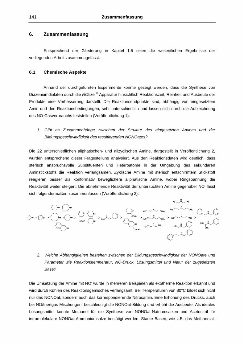
141 Zusammenfassung
6. Zusammenfassung Entsprechend der Gliederung in Kapitel 1.5 seien die wesentlichen Ergebnisse der
vorliegenden Arbeit zusammengefasst.
6.1 Chemische Aspekte Anhand der durchgeführten Experimente konnte gezeigt werden, dass die Synthese von
Diazeniumdiolaten durch die NOtizer® Apparatur hinsichtlich Reaktionszeit, Reinheit und Ausbeute der
Produkte eine Verbesserung darstellt. Die Reaktionsendpunkte sind, abhängig von eingesetztem
Amin und den Reaktionsbedingungen, sehr unterschiedlich und lassen sich durch die Aufzeichnung
des NO-Gasverbrauchs feststellen (Veröffentlichung 1).
1. Gibt es Zusammenhänge zwischen der Struktur des eingesetzten Amines und der
Bildungsgeschwindigkeit des resultierenden NONOates?
Die 22 unterschiedlichen aliphatischen- und alizyclischen Amine, dargestellt in Veröffentlichung 2, wurden entsprechend dieser Fragestellung analysiert. Aus den Reaktionsdaten wird deutlich, dass
sterisch anspruchsvolle Substituenten und Heteroatome in der Umgebung des sekundären
Aminstickstoffs die Reaktion verlangsamen. Zyklische Amine mit sterisch entschirmtem Stickstoff
reagieren besser als konformativ beweglichere aliphatische Amine, wobei Ringspannung die
Reaktivität weiter steigert. Die abnehmende Reaktivität der untersuchten Amine gegenüber NO. lässt
sich folgendermaßen zusammenfassen (Veröffentlichung 2):
NH NH
NH NH
NH O NH
N NH
NH
COOH
NHHOOC
NH
NH
COOH
N CH3H3CH
NCH3H3C
H
NCH2H2C
H NOHHO
H
NNH2HO
H
H3CNH
NH3CH
NH
HN
N CH3HOH NH3C
CH3
H
> > > > > > > >NH NH
NH NH
NH O NH
N NH
NH
COOH
NHHOOC
NH
NH
COOH
N CH3H3CH
NCH3H3C
H
NCH2H2C
H NOHHO
H
NNH2HO
H
H3CNH
NH3CH
NH
HN
N CH3HOH NH3C
CH3
H
> > > > > > > >
2. Welche Abhängigkeiten bestehen zwischen der Bildungsgeschwindigkeit der NONOate und
Parameter wie Reaktionstemperatur, NO-Druck, Lösungsmittel und Natur der zugesetzten
Base?
Die Umsetzung der Amine mit NO. wurde in mehreren Beispielen als exotherme Reaktion erkannt und
wird durch Kühlen des Reaktionsgemisches verlangsamt. Bei Temperaturen von 80°C bildet sich nicht
nur das NONOat, sondern auch das korrespondierende Nitrosamin. Eine Erhöhung des Drucks, auch
bei NO/Inertgas Mischungen, beschleunigt die NONOat-Bildung und erhöht die Ausbeute. Als ideales
Lösungsmittel konnte Methanol für die Synthese von NONOat-Natriumsalzen und Acetonitril für
intramolekulare NONOat-Ammoniumsalze bestätigt werden. Starke Basen, wie z.B. das Methanolat-

Zusammenfassung 142
Anion, verschieben das Reaktionsgleichgewicht zugunsten des Produktes, die Natur des NONOat-
Gegenions spielte bei den durchgeführten Experimenten keine Rolle (Kapitel 4.1.1 und
Veröffentlichung 1).
3. Welche Zusammenhänge gibt es zwischen Struktur des NONOates und der
Freisetzungsgeschwindigkeit von Stickstoffmonoxid aus ihnen?
Der Zusammenhang zwischen NONOat-Struktur und NO-Freisetzungsgeschwindigkeit
(Veröffentlichung 2) ist im Vergleich zur Bildungsgeschwindigkeit weniger einheitlich. Allgemein lässt
sich festhalten, dass sterisch anspruchsvolle Substituenten in direkter Umgebung des [N(O)NO]--
Gruppe bindenden N-Atoms, sowie weitere protonierbare Amine im NONOat die NO-Freisetzung
verlangsamen. Eine Ausnahme bilden benachbarte Carboxylat-Gruppen, welche den Zerfall des
NONOates beschleunigen. Ebenso spielen außergewöhnlich ringgespannte Bindungswinkel am
sekundären Amin für die Freisetzungsgeschwindigkeit eine entscheidende Rolle. So zeigte
überraschend das sehr rasch zu synthetisierende Azetidin-NONOat von allen untersuchten zyklischen
NONOaten die mit Abstand langsamste NO-Freisetzung. Die für diese Untersuchungen verwendete
amperometrische Messmethode (direkter NO-Nachweis) konnte mit der UV-spektroskopischen
Messmethode (indirekter Nachweis) verglichen und die Ergebnisse gegenseitig gesichert werden.
4. Welche Zusammenhänge gibt es zwischen der Bildungsgeschwindigkeit von
Diazeniumdiolaten und den NO-Freisetzungsprofilen aus ihnen?
Trägt man die Daten aus der Bildungs- und Freisetzungsgeschwindigkeit der NONOate in einem
Diagramm gegeneinander auf, so kann man allgemein festhalten, dass generell NONOate die sich
langsam bilden, auch langsam NO. freisetzen und umgekehrt (Veröffentlichung 2 und Kapitel 5.2.3).
5. Synthese von Diazeniumdiolat-Prodrugs.
Die Alkylierung und Acylierung einiger NONOate zu NONOat-Prodrugs erfolgte durch Anpassung
literaturbekannter Methoden an die speziellen Anforderungen des jeweiligen NONOates. Einige
Methoden, wie z.B. die Synthese des Tosyl-PYRRO/NO (Kapitel 4.1.3) oder der Prodrugs in
Veröffentlichung 3 und 5, wurden neu entwickelt.
6. Synthese von Stickstoffmonoxid freisetzenden Hybridverbindungen.
Neben dem spontan NO-freisetzenden Ketoconazol-NONOat 3a wurden drei unterschiedlich
bioaktivierbare Ketoconazol-NONOat-Prodrugs 4a, 5a, 6a hergestellt. Besonderheit von 6a ist, dass
es als Pyrrolidin-basiertes “inverses NONOat-Prodrug“, nach biologischer Aktivierung, eine andere
und schnellere NO-Freisetzungskinetik aufweist, als die anderen durch Phenylpiperazin-basierten
Ketoconazol-NONOat Prodrugs. Zusätzlich wurden Ketoconazol-Organonitrat Hybride 7a, 8a, 9a
hergestellt. Strukturelle Variationen am Organonitrat-Rest erfolgten, um mögliche Struktur-Wirkungs

143 Zusammenfassung
Prinzipien erkennen zu lassen. Um auch die alleinige Ketoconazol-unabhängige NO-Wirkung und NO-
Freisetzungskinetik zu charakterisieren, wurden alle oben erwähnten Derivatisierungen auch mit der
Ketoconazolteilstruktur Phenylpiperazin (b-Reihe) durchgeführt (Veröffentlichung 3 und 6). Das
folgende Schema fasst die oben genannten Derivatisierungen zusammen (1a = Ketoconazol):
O
O
Cl
Cl
N
N
ONN
H
O
CH3
O
O
Cl
Cl
N
N
ONN
H
H
O
O N
N+
Na+
CH3
O
O
O N
N+
O
O N
N+
CH3
N
O
N+
O
N
O
O2NO
O
O2NOCH3
CH3
O
O2NO
NNH
1a
2a
2b
3a/3b
4a/4b
6a/6b
5a/5b
7a/7b 8a/8b 9a/9b
i
iii
iv
v vi viiii viii
v vi viiii viii
O
O
Cl
Cl
N
N
ONN
H
O
CH3
O
O
Cl
Cl
N
N
ONN
H
H
O
O N
N+
Na+
CH3
O
O
O N
N+
O
O N
N+
CH3
N
O
N+
O
N
O
O2NO
O
O2NOCH3
CH3
O
O2NO
NNH
1a
2a
2b
3a/3b
4a/4b
6a/6b
5a/5b
7a/7b 8a/8b 9a/9b
i
iii
iv
v vi viiii viii
v vi viiii viii
6.2 Biologische Aspekte
7. Untersuchung der vasodilatorischen Potenz von NONOaten und NONOat-Prodrugs an
isolierten Pulmonalarterien des Schweins.
Die NONOate PHEPIPERAZI/NO, DPA/NO, ISONIPECO/NO zeigten an den isolierten Gefäßringen
hohe Aktivität mit EC50-Werten im nanomolaren Bereich und waren potenter als die Referenzsubstanz
Glyceroltrinitrat (GTN). Die GTN-typische Tachyphylaxie blieb bei allen untersuchten NONOaten aus.
Eine Präinkubation der Gefäßringe mit dem Inhibitor der löslichen Guanylatcyclase ODQ verschob die
Konzentrations-Wirkungskurven nach rechts zu größeren EC50-Werten. Die geringere Aktivität des
NONOat-Prodrugs Et-PHEPIPERAZI/NO ist bioaktivierungsbedingt (Veröffentlichung 4, Kapitel 4.2.1).
8. Einfluss von NONOaten und NONOat-Prodrugs auf die Durchlässigkeit der Bluthirnschranke
in einem in vitro Modell mit kultivierten cerebralen Endothelzellen.
Drei direkt NO.-freisetzende NONOate (PHEPIPERAZI/NO, DBA/NO, DETA/NO) und drei NONOat-
Prodrugs (Et-PHEPIPERAZI/NO, Tosyl-PYRRO/NO, Et-BUPIPERAZI/NO) wurden hinsichtlich ihres
Einflusses auf die Durchlässigkeit der Bluthirnschranke in einem in vitro Modell untersucht. Die
Permeabilität der Markersubstanz Carboxyfluorescein wird durch die direkten NO-Donoren verringert,
Zusammenfassung 144
während die Permeabilität nach Inkubation mit den NONOat-Prodrugs Et-PHEPIPERAZI/NO und
Tosyl-PYRRO/NO zunimmt. Das Et-BUPIPERAZI/NO nimmt in Bezug auf die Veränderung der
Durchlässigkeit der Bluthirnschranke eine Mittelstellung ein. Mit Methylenblau konnten die
permeabilitätssenkenden Effekte blockiert werden, die permeabilitätssteigernden jedoch nicht
(Veröffentlichung 5).
9. In vivo Untersuchungen des Einflusses von NONOaten und NONOat-Prodrugs auf
neuroregenerative Prozesse nach experimentell induzierten Hirninfarkten bei Ratten.
Die bisher vorliegenden Ergebnisse ergaben, dass die Gabe des NONOat-Prodrugs Et-
BUPIPERAZI/NO noch mehr als der direkte NO-Donor DETA/NO als Referenz, bei den
Versuchstieren die Anzahl BrdU-positiver Zellen (Indikator für Proliferation) deutlich erhöht und das
infarktbedingte Photothrombose-Volumen stärker verringert.
10. Antimykotische und antibakterielle Wirkprofile verschiedener Ketoconazol-Diazeniumdiolat-
Hybride und verwandter Strukturen.
Die antimikrobielle Aktivität der Ketoconazol-NO-Donor Hybridverbindungen wurde an einem breiten
Bakterien- und Pilzspektrum untersucht. Die Hemmung des Bakterienwachstums wies bei den
untersuchten Derivaten nur geringe Unterschiede gegenüber Ketoconazol auf. Dagegen wurden für
die Inhibition des Pilzwachstums deutliche Unterschiede festgestellt. Die Ketoconazol-Hybride mit
Organonitrat- und Diazeniumdiolat-Prodrug-Gruppe zeigten eine höhere Aktivität und ein verändertes
Wirkspektrum im Vergleich zu Ketoconazol. Eine entsprechende Patentschrift wurde eingereicht. Die
NO-Donor Referenzsubstanzen ohne das gesamte Ketoconazol-Grundgerüst (b-Reihe) zeigten eine
vergleichsweise geringe Inhibition des Pilz- und Bakterienwachstums (Veröffentlichung 3). Für das
Ketoconazol-PYRRO/NO-Prodrug Hybrid konnten zusätzlich antiinflammatorische Eigenschaften
nachgewiesen werden (Veröffentlichung 6).

145 Conclusion
7. Conclusion
Nitrogen-bound diazen-1-ium-1,2-diolates (diazeniumdiolates, “NONOates”, “solid NO”) are
generally prepared from secondary amines and nitric oxide (NO) and are compounds of first choice for
a direct release of NO. A series of differently sized and substituted cyclic and aliphatic amines were
used to quantify the reactivity towards NO by monitoring the decrease of NO pressure with the
NOtizer®, an apparatus developed for this study. The release of NO was measured amperometrically
with an NO sensitive electrode and the half-lives of novel diazeniumdiolates were determined via UV-
spectroscopy. It was found that steric hindrance and heteroatomic substituents in the amines side
chains decrease the formation rates of sodium salt NONOates and also slow down NO release from
them. Exceptions were found to be piperidine-2-carboxylic acid derivatives which react to NONOates
slower than piperidine, but released NO much faster despite steric hindrance. A secondary amine
carrying an additional primary amine reacted fast with NO, but the corresponding NONOate showed to
be a slow releaser due to formation of an intramolecular NONOate salt together with the sodium salt
derivative. Azetidine reacts faster with NO than all of the other amines, but decomposition of the
corresponding NONOate showed to be unexpectedly slow. The usefulness of the amperometric NO-
detection (direct NO measurement) could be demonstrated in comparison to the UV-spectroscopic
method (indirect measurement). In a series of different experiments it could be shown that the reaction
of secondary amines with NO is exothermic and that external heating above 80 °C leads to the
formation of the corresponding N-nitrosamine as a byproduct to the NONOate. The pressure
dependence of the reaction was quantified by using mixtures of NO/inert-gas in the reaction with
amines and plotting the decrease in pressure.
Novel nitric oxide donors and hybrid drugs combining the modified antifungal drug ketoconazole with a
diazeniumdiolate or an organic nitrate moiety were synthesized and their activities against a broad
variety of bacteria and fungal strains were tested. Whereas the efficacy of the compounds against
bacteria showed little variation, the ketoconazole-NO-donor hybrid drugs differed in their effectiveness
and susceptibility against fungi. The ketoconazole-NO-donor hybrids with an alkylated NONOate
group or organic nitrate moiety showed to be much more effective than ketoconazole alone, especially
against Aspergillus species, proving a change in the susceptibility spectrum. The NO-donors bound to
phenylpiperazine alone, which is part of the ketoconazole structure, showed insufficient effectiveness.
The sodium salt NONOates PHEPIPERAZI/NO, DPA/NO and ISONIPECO/NO were measured in vitro
on isolated porcine pulmonary arteries and showed nanomolar efficacies concerning vasodilatation.
Also, tolerance-devoid activity for the tested compounds could be demonstrated and compared with
glyceryl trinitrate and pentaerythrityl tetranitrate. The comparable lower activity of the NONOate
prodrug Et-PHEPIPERAZI/NO was related to bioactivation processes.
Three direct NO releasing agents (PHEPIPERAZI/NO, DBA/NO, DETA/NO) and three NONOate
prodrugs (Et-PHEPIPERAZI/NO, Tosyl-PYRRO/NO, Et-BUPIPERAZI/NO) were tested on their effect
on the permeability of the blood-brain-barrier (BBB) in an in vitro model procedure. By measuring the

Conclusion 146
flux of a small polar molecule (carboxyfluorescein) it could be shown, that the releasers
PHEPIPERAZI/NO, DBA/NO, DETA/NO reduced the BBB permeability. In contrast, the prodrugs Et-
PHEPIPERAZI/NO and Tosyl-PYRRO/NO increased the permeability, whereas the prodrug Et-
BUPIPERAZI/NO showed a medial setting. Blocking the effect of the BBB permeability reducing
compounds could be done by methylene blue, whereas permeability increasing effects could not be
blocked.
Neuroregenerative properties were tested for the NONOate prodrug Et-BUPIPERAZI/NO in
comparison to the direct releaser DETA/NO on an in vivo model of rats after experimentally induced
brain stroke. The prodrug showed to produce higher amounts of BrdU-positive cells (as an indicator
for proliferation) than DETA/NO. Additionally, the stroke related volume of the thrombus was
significantly reduced.

I Literaturverzeichnis
8. Literaturverzeichnis [1] Davis, K. L.; Martin, E.; Turko, I. V.; Murad, F. Novel effects of nitric oxide. Annual Review of
Pharmacology and Toxicology 2001, 41, 203-236.
[2] Wink, D. A.; Osawa, Y.; Darbyshire, J. F.; Jones, C. R.; Eshenaur, S. C.; Nims, R. W.
Inhibition of cytochromes P450 by nitric oxide and a nitric oxide-releasing agent. Archives of
Biochemistry and Biophysics 1993, 300 (1), 115-123.
[3] Wang, P. G.; Cai, T. B.; Taniguchi, N. Nitric Oxide Donors For Pharmaceutical and Biological
Applications. 1. Auflage 2005, Wiley-VCH, Weinheim, ISBN-13: 978-3-527-31015-9.
[4] van der Vliet, A.; Eiserich, J. P..; Morrissey, B.; Cross, C. E. Reactive nitrogen species and
tyrosine nitration in airway inflammation. Lung Biology in Health and Disease 2003, 177, 101-
121.
[5] Wolin, M.S. Interactions of oxidants with vascular signaling systems. Arteriosclerosis
Thrombosis and Vascular Biology 2000, 20, 1430-1442.
[6] Ohara, Y.; Peterson, T. E.; Harrison, D. G. Hypercholesterolemia increases endothelial
superoxide anion production. Journal of Clinical Investigation 1993, 91, 2546-2551.
[7] Heck, D. E. *NO, RSNO, ONOO-, NO+, *NOO, NOx--dynamic regulation of oxidant scavenging,
nitric oxide stores, and cyclic GMP-independent cell signaling. Antioxidants & Redox Signaling
2001, 3 (2), 249-260.
[8] Stamler, J. S. Redox signaling: nitrosylation and related target interactions of nitric oxide. Cell
1994, 78(6), 931-936.
[9] Fukuto, J. M.; Bartberger, M. D.; Dutton, A. S.; Paolocci, N.; Wink, D. A.; Houk, K. N. The
physiological chemistry and biological activity of nitroxyl (HNO): The neglected, misunderstood
and enigmatic nitrogen oxide. Chemical Research in Toxicology 2005, 18 (5), 790-801.
[10] Bloodsworth, A.; O'Donnell, V. B.; Freeman, B. A. Nitric oxide regulation of free radical- and
enzyme - mediated lipid and lipoprotein oxidation. Arteriosclerosis Thrombosis and Vascular
Biology 2000, 20 (7), 1707-1715.
[11] Tsai, A. L. How does NO activate heme-proteins? FEBS Letters. 1994, 341, 141-145.

Literaturverzeichnis II
[12] Denninger, J. W.; Marletta, M. A. Guanylate cyclase and the NO/cGMP signaling pathway.
Biochimica et Biophysica Acta 1999, 1411, 334-350.
[13] Cornwell, T. L.; Pryzwansky, K. B.; Wyatt, T. A.; Lincoln, T. M. Regulation of sarcoplasmic
reticulum protein phosphorylation by localized cyclic GMP-dependent protein kinase in
vascular smooth muscle cells. Molecular Pharmacology 1991, 40 (6), 923-931.
[14] Reinhard, M.; Halbrugge, M.; Scheer, U.; Wiegand, C.; Jockusch, B. M.; Walter, U. The 46/50
KDa phosphoprotein VASP purified from human platelets is a novel protein associated with
actin filaments and focal contacts. European Molecular Biology Organization Journal 1992, 11,
2063-2070.
[15] Horstrup, K.; Jablonka, B.; Honig-Liedl, P.; Just, M., Kochsiek, K.; Walter, U. Phosphorylation
of focal adhesion vasodilator-stimulated phosphoprotein at Ser157 in intact human platelets
correlates with fibrinogen receptor inhibition. European Journal of Biochemistry 1994, 225, 21-
27.
[16] Hartzell, H. C.; Fischmeister, R. Opposite effects of cyclic GMP and cyclic AMP on calcium
current in single heart cells. Nature 1986, 323 (6085), 273-275.
[17] Cook, S. J.; McCormick, F. Inhibition by cAMP of Ras-dependent activation of Raf. Science
1993, 262 (5136), 1069-1072.
[18] Schwartz, S. M. Smooth muscle migration in atherosclerosis and restenosis. Journal of
Clinical Investigation 1997, 100 (11), 87-89.
[19] Biel, M.; Seeliger, M.; Pfeifer, A.; Kohler, K.; Gerstner, A.; Ludwig, A.; Jaissle, G.; Fauser, S.;
Zrenner, E.; Hofmann, F. Selective loss of cone function in mice lacking the cyclic nucleotide-
gated channel CNG3. Proceedings of the National Academy of Sciences 1999, 96, 7553-
7557.
[20] Garthwaite, J.; Southam, E.; Boulton, C. L.; Nielsen, E. B.; Schmidt, K.; Mayer, B. Potent and
selective inhibition of nitric oxide-sensitive guanylyl cyclase by 1H-[1,2,4]-oxadiazolo-[4,3-a]-
quinoxalin-1-one. Molecular Pharmacology 1995, 48, 184-188.
[21] Schrammel, A.; Behrends, S.; Schmidt, K.; Koesling, D.; Mayer, B. Characterization of 1H-
[1,2,4]oxadiazolo[4,3-a]quinoxalin-1-one as a heme-site inhibitor of nitric oxide-sensitive
guanylyl cyclase. Molecular Pharmacology 1996, 50, 1-5.

III Literaturverzeichnis
[22] Weissmann, N.; Voswinckel, R.; Tadic, A.; Hardebusch, T.; Ghofrani, H. A.; Schermuly, R. T.;
Seeger, W.; Grimminger, F. Nitric oxide (NO)-dependent but not NO-independent guanylate
cyclase activation attenuates hypoxic vasoconstriction in rabbit lungs. American Journal of
Respiratory Cell and Molecular Biology 2000, 23 222-227.
[23] Hasegawa, T.; Bando, A.; Tsuchiya, K.; Abe, S.; Okamoto, M.; Kirima, K.; Ueno, S.;
Yoshizumi, M.; Houchi, H.; Tamaki, T. Enzymatic and nonenzymatic formation of reactive
oxygen species from 6-Anilino-5,8-quinolinequinone. Biochimica et Biophysica Acta 2004,
1670, 19-27.
[24] Mayer, B.; Brunner, F.; Schmidt, K. Novel actions of methylene blue. European Heart Journal
1993, 14, 22-26.
[25] Lipton, S. A.; Singel, D. J.; Stamler, J. S. Neuroprotective and neurodestructive effects of
nitric oxide and redox congeners. Annals of the New York Academy of Sciences 1994, 738,
382-387.
[26] Gordh, T.; Karlsten, R; Kristensen, J. Intervention with spinal NMDA, adenosine, and NO
systems for pain modulation. Annals of Medicine 1995, 27, 229-234.
[27] Garthwaite, J.; Boulton, C. L. Nitric oxide signaling in the central nervous system. Annual
Reviews of Physiology 1995, 57, 683-706.
[28] Melino, G.; Bernassola, F.; Catani, M. V.; Rossi, A.; Corazzari, M.; Sabatini, S.; Vilbois, F.;
Green, D. R. Nitric oxide inhibits apoptosis via AP-1-dependent CD95L transactivation. Cancer
Research 2000, 60, 2377-2383.
[29] Stamler, J. S.; Singel, D. J.; Loscalzo, J. Biochemistry of nitric oxide and its redox-activated
forms. Science 1992, 258, 1898-1902.
[30] Li, R.; Liu, Z.; Zhou, Y.; Wu, L. Highly stereoselective nitration of chalcone derivatives with
nitric oxide. Synthesis Letters 2006, 9, 1367-1368.
[31] Nishida, K.; Ohta, Y.; Ishiguro, I. Relation of inducible nitric oxide synthase activity to lipid
peroxidation and nonprotein sulfhydryl oxidation in the development of stress-induced gastric
mucosal lesions in rats. Nitric Oxide 1998, 2 (4), 215-223.
[32] Becker, K.; Savvides, S. N.; Keese, M.; Schirmer, R. H.; Karplus, P. A. Enzyme inactivation
through sulfhydryl oxidation by physiologic NO-carriers. Nature Structural and Molecular
Biology 1998, 5 (4), 267-271.

Literaturverzeichnis IV
[33] Cygler, M.; Mort, J. S. Proregion structure of members of the papain superfamily. Mode of
inhibition of enzymatic activity. Biochimie 1997, 79, 645-652.
[34] Denis, F.; Rheaume, S. M.; Aouad, A.; Alam, R. P.; Sekaly, P.; Cohen, L. Y. The role of
caspases in T-cell development and the control of immune responses. Cellular and Molecular
Life Sciences 1998, 54, 1005-1019.
[35] Alam, A.; Cohen, L. Y.; Aouad, S.; Sekaly, R. P. Early activation of caspases during T
lymphocyte stimulation results in selective substrate cleavage in nonapoptotic cells. Journal of
Experimental Medicine 1999, 190,1879-1890.
[36] Elkon, K. B. Caspases: Multifunctional Proteases. Journal of Experimental Medicine 1999,
190,1725-1727.
[37] Colasanti, M.; Salvati, L.; Venturini, G.; Ascenzi, P.; Gradoni, L. Cysteine protease as a target
for nitric oxide in parasitic organisms. Trends in Parasitology 2001, 17 (12), 575.
[38] Farias-Eisner, R.; Sherman, M. P.; Aeberhard, E.; Chaudhuri, G. Nitric oxide is an important
mediator for tumoricidal activity in vivo. Proceedings of the National Academy of Sciences
1994, 91 (20), 9407-9411.
[39] Boyd, C. S.; Cadenas, E. Nitric oxide and cell signaling pathways in mitochondrial-dependent
apoptosis. Biological Chemistry 2002, 383 (3/4), 411-423.
[40] Moochhala, S.; Rajnakova, A. Role of nitric oxide in cancer biology. Free Radical Research
1999, 31 (6), 671-679.
[41] Riederer, U., Darstellung und Reaktionen von aktiven Stickstoffspezies mit DNA/RNA-
Teilstrukturen als biologische Zielmoleküle. Dissertation 1999.
[42] Felley-Bosco, E.; Mirkovitch, J.; Ambs, S.; Mace, K.; Pfeifer, A.; Keefer, L. K.; Harris, C. C.
Nitric oxide and ethylnitrosourea: relative mutagenicity in the p53 tumor suppressor and
hypoxanthine-phosphoribosyltransferase genes. Carcinogenesis 1995, 16 (9), 2069-2074.
[43] Wink, D. A.; Mitchell, J. B. Chemical biology of nitric oxide: Insights into regulatory, cytotoxic,
and cytoprotective mechanisms of nitric oxide. Free radical biology & medicine 1998, 25 (4-5),
434-456.
[44] Hogg, N.; Kalyanaraman, B. Nitric oxide and lipid peroxidation. Biochimica et Biophysica Acta
1999, 1411 (2-3), 378-384.

V Literaturverzeichnis
[45] Patel, R. P.; McAndrew, J.; Sellak, H.; White, C. R.; Jo, H.; Freeman, B. A.; Darley-Usmar, V.
M. Biological aspects of reactive nitrogen species. Biochimica et Biophysica Acta 1999, 1411
(2-3), 385-400.
[46] Rubbo, H.; Parthasarathy, S.; Barnes, S.; Kirk, M.; Kalyanaraman, B.; Freeman, B. A. Nitric
oxide inhibition of lipoxygenase-dependent liposome and low-density lipoprotein oxidation:
Termination of radical chain propagation reactions and formation of nitrogen-containing
oxidized lipid derivatives. Archives of Biochemistry and Biophysics 1995, 324 (1), 15-25.
[47] Rubbo, H.; Radi, R.; Trujillo, M.; Telleri, R.; Kalyanaraman, B.; Barnes, S.; Kirk, M.; Freeman,
B. A. Nitric oxide regulation of superoxide and peroxynitrite-dependent lipid peroxidation.
Formation of novel nitrogen-containing oxidized lipid derivatives. Journal of Biological
Chemistry 1994, 269 (42), 26066-26075.
[48] Rubbo, H.; Radi, R.; Anselmi, D.; Kirk, M.; Barnes, S.; Butler, J.; Eiserich, J. P.; Freeman, B.
A. Nitric oxide reaction with lipid peroxyl radicals spares α-tocopherol during lipid peroxidation:
greater oxidant protection from the pair nitric oxide/α-tocopherol than α-tocopherol/ascorbate.
Journal of Biological Chemistry 2000, 275 (15), 10812-10818.
[49] Hogg, N.; Struck, A.; Goss, S. P. A.; Santanam, N.; Joseph, J.; Parthasarathy, S.;
Kalyanaraman, B. Inhibition of macrophage-dependent low density lipoprotein oxidation by
nitric oxide donors. Journal of Lipid Research 1995, 36 (8), 1756-62.
[50] Graham, A.; Hogg, N.; Kalyanaraman, B.; O'Leary, V.; Darley-Usmar, V.; Moncada, S.
Peroxynitrite modification of low-density lipoprotein leads to recognition by the macrophage
scavenger receptor. FEBS Letters 1993, 330 (2), 181-185.
[51] Mayer, B. Handbook of Experimental Pharmacology, Nitric Oxide. Springer Verlag 2000, 143,
17, ISBN 3-540-66122-0.
[52] Walker, J. E.; Saraste, M. Membrane proteins membrane protein structure. Current Opinion in
Structural Biology 1996, 6 (4), 457-459.
[53] Ford, P. C.; Wink, D. A.; Stanbury, D. M. Autoxidation kinetics of aqueous nitric oxide. FEBS
Letters 1993, 326 (1-3), 1-3.
[54] Wink, D. A.; Grisham, M. B.; Miles, A. M.; Nims, R. W.; Krishna, M. C.; Pacelli, R.; Teague, D.;
Poore, C. M. B.; Cook, J. A.; Ford, P. C. Determination of selectivity of reactive nitrogen oxide
species for various substrates. Methods in Enzymology 1996, 268, 120-130.
[55] Enicola, A.; Souza, J. M.; Radi, R.; Lissi, E. Nitric oxide diffusion in membranes determined by
fluorescence quenching. Archives of biochemistry and biophysics 1996, 328 (1), 208-212.

Literaturverzeichnis VI
[56] Lehmann, J. Nitric oxide donors - current trends in therapeutic applications. Expert Opinion on
Therapeutic Patents 2000, 10 (5), 559-574.
[57] Needleman, P.; Harkey, A. B. Role of endogenous glutathione in the metabolism of glyceryl
trinitrate by isolated perfused rat liver. Biochemical Pharmacology 1971, 20 (8), 1867-1876.
[58] Servent, D.; Delaforge, M.; Ducrocq, C.; Mansuy, D.; Lenfant, M. Nitric oxide formation during
microsomal hepatic denitration of glyceryl trinitrate: involvement of the cytochrome P-450.
Biochemical and Biophysical Research Communications 1989, 163 (3), 1210-1216.
[59] Millar, T. M.; Stevens, C. R.; Blake, D. R. Xanthine oxidase can generate nitric oxide from
nitrate in ischemia. Biochemical Society Transactions 1997, 25 (3), 528.
[60] Chen, Z.; Zhang, J.; Stamler, J. S. Identification of the enzymatic mechanism of nitroglycerin
bioactivation. Proceedings of the National Academy of Sciences 2002, 99 (12), 8306-8311.
[61] Fink, B.; Bassenge, E. Unexpected, tolerance - devoid vasomotor and platelet actions of
pentaerythritol tetranitrate. Journal of Cardiovascular Pharmacology 1997, 30 (6), 831-836.
[62] Pfaffenrath, V.; de la Motte, S.; Harrison, F.; Ruthning, C. Actions of pentaerithritol tetranitrate,
isosorbide mononitrate and placebo on headache and ability to work of healthy subjects.
Arzneimittel-Forschung 1998, 48 (6), 646-650.
[63] Harrison, D. G.; Bates, J. N. The nitrovasodilators: new ideas about old drugs. Circulation
1993, 87 (5), 1461-1467.
[64] Bohn, H.; Schoenafinger, K. Oxygen and oxidation promote the release of nitric oxide from
sydnonimines. Journal of Cardiovascular Pharmacology 1989, 14, 6-12.
[65] Rudolph, W.; Dirschinger, J. Clinical comparison of nitrates and sydnonimines. European
Heart Journal 1991, 12, 33-41.
[66] Pryor, W. A.; Squadrito, G. L. The chemistry of peroxynitrite: a product from the reaction of
nitric oxide with superoxide. American journal of physiology 1995, 268 (5), 699-722.
[67] Stamler, J. S.; Simon, D. I.; Osborne, J. A.; Mullins, M. E.; Jaraki, O.; Michel, T.; Singel, D. J.;
Loscalzo, J. S-Nitrosylation of proteins with nitric oxide: synthesis and characterization of
biologically active compounds. Proceedings of the National Academy of Sciences 1992, 89
(1), 444-448.

VII Literaturverzeichnis
[68] Simon, D. I.; Stamler, J. S.; Jaraki, O.; Keaney, J. F.; Osborne, J. A.; Francis, S. A.; Singel, D.
J.; Loscalzo, J. Antiplatelet properties of protein S-nitrosothiols derived from nitric oxide and
endothelium-derived relaxing factor. Arteriosclerosis and Thrombosis 1993, 13 (6), 791-799.
[69] Stamler, J. S. S-Nitrosothiols and the bioregulatory actions of nitrogen oxides through
reactions with thiol groups. Current Topics in Microbiology and Immunology 1995, 196, 19-36.
[70] Drago, R. S.; Paulik, F. E. The reaction of nitrogen(II) oxide with diethylamine. Journal of the
American Chemical Society 1960, 82, 96-98.
[71] Davies, K. M.; Wink, D. A.; Saavedra, J. E.; Keefer, L. K. Chemistry of the diazeniumdiolates.
2. Kinetics and mechanism of dissociation to nitric oxide in aqueous solution. Journal of the
American Chemical Society 2001, 123 (23), 5473-5481.
[72] Fitzhugh, A. L.; Anadu, N. O.; Waterhouse, D. J.; Hrabie, J. A.; Saavedra, J. E.; Keefer, L. K.
Qualitative thin-layer and high-performance liquid chromatographic analysis of 1-substituted
diazen-1-ium-1,2-diolates on aminopropyl-bonded silica gel. Analytical Biochemistry 2002, 301
(1), 97-102.
[73] Dutton, A. S.; Fukuto, J. M.; Houk, K. N. The mechanism of NO formation from the
decomposition of dialkylamino diazeniumdiolates: Density functional theory and CBS-QB3
predictions. Inorganic Chemistry 2004, 43 (3), 1039-1045.
[74] Zhang, H.; Annich, G. M.; Miskulin, J.; Stankiewicz, K.; Osterholzer, K.; Merz, S. I.; Bartlett, R.
H.; Meyerhoff, M. E. Nitric Oxide-releasing fumed silica particles: Synthesis, characterization,
and biomedical application. Journal of the American Chemical Society 2003, 125 (17), 5015-
5024.
[75] Keefer, L. K.; Nims, R. W.; Davies, K. M.; Wink, D. A. "NONOates" (1-substituted diazen-1-
ium-1,2-diolates) as nitric oxide donors: convenient nitric oxide dosage forms. Methods in
Enzymology 1996, 268, 281-293.
[76] Srinivasan, A.; Kebede, N.; Saavedra, J. E.; Nikolaitchik, A. V.; Brady, D. A.; Yourd, E.;
Davies, K. M.; Keefer, L. K.; Toscano, J. P. Chemistry of the Diazeniumdiolates. 3.
Photoreactivity. Journal of the American Chemical Society 2001, 123 (23), 5465-5472.
[77] Saavedra, J. E.; Southan, G. D.; Davies, K. M.; Lundell, A.; Markou, C.; Hanson, S. R.; Adrie,
C.; Hurford, W. E.; Zapol, W. M.; Keefer, L. K. Localizing antithrombotic and vasodilatory
activity with a novel, ultrafast nitric oxide donor. Journal of Medicinal Chemistry 1996, 39 (22),
4361-4365.
Literaturverzeichnis VIII
[78] Hrabie, J. A.; Klose, J. R.; Wink, D. A.; Keefer, L. K. New nitric oxide-releasing zwitterions
derived from polyamines. Journal of Organic Chemistry 1993, 58 (6), 1472-1476.
[79] Saavedra, J. E.; Billiar, T. R.; Williams, D. L.; Kim, Y.-M.; Watkins, S. C.; Keefer, L. K.
Targeting nitric oxide (NO) delivery in vivo. Design of a liver-selective NO donor prodrug that
blocks tumor necrosis factor-α-induced apoptosis and toxicity in the liver. Journal of Medicinal
Chemistry 1997, 40 (13), 1947-1954.
[80] Liu, J.; Qu, W.; Saavedra, J. E.; Waalkes, M. P. The nitric oxide donor, O2-vinyl-1-(pyrrolidin-
1-yl)diazen-1-ium-1,2-diolate (V-PYRRO/NO), protects against cadmium-induced
hepatotoxicity in mice. Journal of Pharmacology and Experimental Therapeutics 2004, 310 (1),
18-24.
[81] Hrabie, J. A.; Keefer, L. K. Chemistry of the nitric oxide-releasing diazeniumdiolate
("Nitrosohydroxylamine") functional group and its oxygen-substituted derivatives. Chemical
Reviews 2002, 102 (4), 1135-1154.
[82] Saavedra, J. E.; Srinivasan, A.; Bonifant, C. L.; Chu, J.; Shanklin, A. P.; Flippen-Anderson, J.
L.; Rice, W. G.; Turpin, J. A.; Davies, K. M.; Keefer, L. K. The secondary amine/nitric oxide
complex ion R2N[N(O)NO]- as nucleophile and leaving group in SNAr reactions. Journal of
Organic Chemistry 2001, 66 (9), 3090-3098.
[83] Liu, J.; Li, C.; Qu, W.; Leslie, E.; Bonifant, C. L.; Buzard, G. S.; Saavedra, J. E.; Keefer, L. K.;
Waalkes, M. P. Nitric oxide prodrugs and metallochemotherapeutics: JS-K and CB-3-100
enhance arsenic and cisplatin cytolethality by increasing cellular accumulation. Molecular
Cancer Therapeutics 2004, 3 (6), 709-714.
[84] Wu, X.; Tang, X.; Xian, M.; Wang, P. G. Glycosylated diazeniumdiolates: a novel class of
enzyme-activated nitric oxide donors. Tetrahedron Letters 2001, 42 (23), 3779-3782.
[85] Velazquez, C.; Rao, P. N.; Knaus, E. E. Novel nonsteroidal antiinflammatory drugs possessing
a nitric oxide donor diazen-1-ium-1,2-diolate moiety: Design, synthesis, biological evaluation,
and nitric oxide release studies. Journal of Medicinal Chemistry 2005, 48 (12), 4061-4067.
[86] Velazquez, C.; Knaus, E. E. Synthesis and biological evaluation of 1,4-dihydropyridine calcium
channel modulators having a diazen-1-ium-1,2-diolate nitric oxide donor moiety for the
potential treatment of congestive heart failure. Bioorganic & Medicinal Chemistry 2004, 12
(14), 3831-3840.

IX Literaturverzeichnis
[87] Saavedra, J. E.; Mooradian, D. L.; Mowery, K. A.; Schoenfisch, M. H.; Citro, M. L.; Davies, K.
M.; Meyerhoff, M. E.; Keefer, L. K. Conversion of a polysaccharide to nitric oxide-releasing
form. Dual-mechanism anticoagulant activity of diazeniumdiolated heparin. Bioorganic &
Medicinal Chemistry Letters 2000, 10 (8), 751-753.
[88] Keefer, L. K. Progress toward clinical application of the nitric oxide-releasing
diazeniumdiolates. Annual Review of Pharmacology and Toxicology 2003, 43, 585-607.
[89] Horstmann, A. Diazeniumdiolate als direkte NO-Donoren - Synthese, Pharmakologie und
Untersuchungen der NO-Freisetzungskinetik durch Laser Magnet Resonanz Spektroskopie.
Dissertation 2003.
[90] Gaebler, R.; Lehmann, J. Sensitive and isotope selective (14NO/15NO) online detection of
nitric oxide by Faraday-laser magnetic resonance spectroscopy. Methods in Enzymology
2005, 396, 54-60.
[91] Iijima, K.; Henry, E.; Moriya, A.; Wirz, A.; Kelman, A. W.; McColl, K. E. L. Dietary nitrate
generates potentially mutagenic concentrations of nitric oxide at the gastroesophageal
junction. Gastroenterology 2002, 122 (5), 1248-1257.
[92] Hunnius. Pharmazeutisches Wörtebuch, Walter de Gruyter Verlag 1998, 8.
[93] Mutschler, E. Arzneimittelwirkung, Wissenschaftliche Verlagsgesellschaft mbH Stuttgart 1997,
7.
[94] Lüllmann, H.; Mohr, K. Pharmakologie und Toxikologie, Georg Thieme Verlag 1999, 14.
[95] Longhi, R.; Ragsdale, R. O.; Drago, R. S. Reactions of nitrogen(II)oxide with miscellaneous
lewis bases. Inorganic Chemistry 1962, 1, 768-770.
[96] Hrabie, J. A.; Saavedra, J. E.; Davies, K. M.; Keefer, L. K. Adducts of piperazine with nitric
oxide. Organic Preparations and Procedures International 1999, 31 (2), 189-192.
[97] Zolfigol, M. A.; Choghamarani, A. G.; Hazarkhani, H. Trichloroisocyanuric acid/NaNO2 as a
novel heterogeneous system for the N-nitrosation of N,N-dialkylamines under mild conditions.
Synthesis Letters 2002, 6, 1002-1004.
[98] Nordberg, R.; Albridge, R. G.; Bergmark, T.; Ericson, U.; Hedman, J.; Nordling, C.; Siegbahn,
K.; Lindberg, B. J. Molecular spectroscopy by means of electron spectroscopy for chemical
analysis. Charge distribution in nitrogen compounds. Arkiv foer Kemi 1967, 28 (19), 257-278.

Literaturverzeichnis X
[99] Keiner, Silke. Zellproliferation und –differenzierung in der Umgebung experimentell induzierter
Hirninfarkte bei der Ratte - Einfluss von rehabilitativem Training und intrahekaler Applikation
des Wachstumsfaktors Brain-derived neurotrophic factor sowie des Zytostatikums Cytarabin.
Dissertation 2007.
[100] Kunze, A.; Grass, S.; Witte, O. W.; Yamaguchi, M.; Kempermann, G.; Redecker, C.
Proliferative response of distinct hippocampal progenitor cell populations after cortical infarcts
in the adult brain. Neurobiology of Disease 2006, 21 (2), 324-332.
[101] Wurm, F.; Keiner, S.; Kunze, A.; Witte, O.W.; Redecker, C. Effects of skilled forelimb training
on hippocampal neurogenesis in the lesioned and intact adult brain. Stroke 2007, im Druck.
[102] Maragos, C. M.; Morley, D.; Wink, D. A.; Dunams, T. M.; Saavedra, J. E.; Hoffman, A.; Bove,
A. A.; Isaac, L.; Hrabie, J. A.; Keefer, L. K. Complexes of NO with nucleophiles as agents for
the controlled biological release of nitric oxide. Vasorelaxant effects. Journal of medicinal
chemistry 1991, 34 (11), 3242-3247.
[103] Nablo, Brian J.; Schoenfisch, Mark H. In vitro cytotoxicity of nitric oxide-releasing sol-gel
derived materials. Biomaterials 2005, 26 (21), 4405-4415.
[104] IUPAC. Pure and Applied Chemistry 1996, 68 (1), 64.
[105] Reynolds, M. M.; Zhou, Z.; Oh, B. K.; Meyerhoff, M. E. Bis-diazeniumdiolates of
dialkyldiamines: Enhanced nitric oxide loading of parent diamines. Organic Letters 2005, 7
(14), 2813-2816.
[106] Suzuki, T. DNA damage and mutation caused by vital biomolecules, water, nitric oxide, and
hypochlorus acid. Genes and Environment 2006, 28 (2), 48-55.
[107] Juedes, M. J.; Wogan, G. N. Peroxynitrite-induced mutation spectra of pSP189 following
replication in bacteria and in human cells. Mutation Research. Fundamental and Molecular
Mechanisms of Mutagenesis 1996, 349 (1), 51-61.
[108] Drapier, J. C.; Hibbs, J. B. Jr. Aconitases: A class of metalloproteins highly sensitive to nitric
oxide synthesis. Methods in Enzymology 1996, 269, 26-36.
[109] Pacelli, R.; Wink, D. A.; Cook, J. A.; Krisha, M. C.; DeGraff, W.; Friedman, N.; Tsokos, M.;
Samuni, A.; Mitchell, J. B. Nitric oxide potentiates hydrogen peroxide-induced killing of
Escherichia coli. Journal of Experimental Medicine 1995, 182 (5), 1469-1479.
XI Literaturverzeichnis
[110] Kaplan, S. S.; Lancaster, J. R., Jr.; Basford, R. E.; Simmons, R. L. Effect of nitric oxide on
staphylococcal killing and interactive effect with superoxide. Infection and Immunity 1996, 64
(1), 69-76.
[111] Newton, G. L.; Arnold, K.; Price, M. S.; Sherrill, C.; Delcardayre, S. B.; Aharonowitz, Y.;
Cohen, G.; Davies, J.; Fahey, R. C.; Davis, C. Distribution of thiols in microorganisms:
mycothiol is a major thiol in most actinomycetes. Journal of Bacteriology 1996, 178 (7), 1990-
1995.
[112] Abouhamad, W. N.; Manson, M.; Gibson, M. M.; Higgins, C. F. Peptide transport and
chemotaxis in Escherichia coli and Salmonella typhimurium: Characterization of the dipeptide
permease (Dpp) and the dipeptide-binding protein. Molecular Microbiology 1991, 5 (5), 1035-
1047.
[113] Falahati, M.; Shabani, M.; Rodaki, M. M. A.; Jahaniani, F.; Bagheri, K. P.; Ahmed Ebrahimi, S.
Interaction between ketoconazole, amphotericin B and terbinafine and three diazenumdiolates
in concomitant uses against some fungal species. Journal of Faculty of Pharmacy, Tehran
University of Medical Sciences 2006, 14 (2), 87-92.
[114] Martin, W.; Villani, G. M.; Jothianandan, D.; Furchgott, R. F. Selective blockade of
endothelium-dependent and glyceryl trinitrate-induced relaxation by hemoglobin and by
methylene blue in the rabbit aorta. Journal of Pharmacology and Experimental Therapeutics
1985, 232 (3), 708-716.
[115] Marczin, N.; Ryan, U. S.; Catravas, J. D. Methylene blue inhibits nitrovasodilator- and
endothelium-derived relaxing factor-induced cyclic GMP accumulation in cultured pulmonary
arterial smooth muscle cells via generation of superoxide anion. Journal of Pharmacology and
Experimental Therapeutics 1992, 263 (1), 170-179.
[116] Chen, Y.; McCarron, R. M.; Golech, S.; Bembry, J.; Ford, B.; Lenz, F. A.; Azzam, N.; Spatz, M.
ET-1- and NO-mediated signal transduction pathway in human brain capillary endothelial cells.
American Journal of Physiology 2003, 284, 243-249.
[117] Dawson, V. L.; Dawson, T. M.; London, E. D.; Bredt, D. S.; Snyder, S. H. Nitric oxide mediates
glutamate neurotoxicity in primary cortical cultures. Proceedings of the National Academy of
Sciences 1991, 88 (14), 6368-6371.
[118] Ayata, C.; Ayata, G.; Hara, H.; Matthews, R. T.; Beal, M. F.; Ferrante, R. J.; Endres, M.; Kim,
A.; Christie, R. H.; Waeber, C.; Huang, P. L.; Hyman, B. T.; Moskowitz, M. A. Mechanisms of
reduced striatal NMDA excitotoxicity in type I nitric oxide synthase knock-out mice. Journal of
Neuroscience 1997, 17 (18), 6908-6917.

Literaturverzeichnis XII
[119] Vidwans, A. S.; Kim, S.; Coffin, D. O.; Wink, D. A.; Hewett, S. J. Analysis of the
neuroprotective effects of various nitric oxide donor compounds in murine mixed cortical cell
culture. Journal of Neurochemistry 1999, 72 (5), 1843-1852.
[120] Zhang, R.; Zhang, L.; Zhang, Z.; Wang, Y.; Lu, M.; LaPointe, M.; Chopp, M. A nitric oxide
donor induces neurogenesis and reduces functional deficits after stroke in rats. Annals of
Neurology 2001, 50 (5), 602-611.
[121] Williams, C. V.; Nordquist, D.; McLoon, S. C. Correlation of nitric oxide synthase expression
with changing patterns of axonal projections in the developing visual system. Journal of
Neuroscience 1994, 14, 1746-1755.
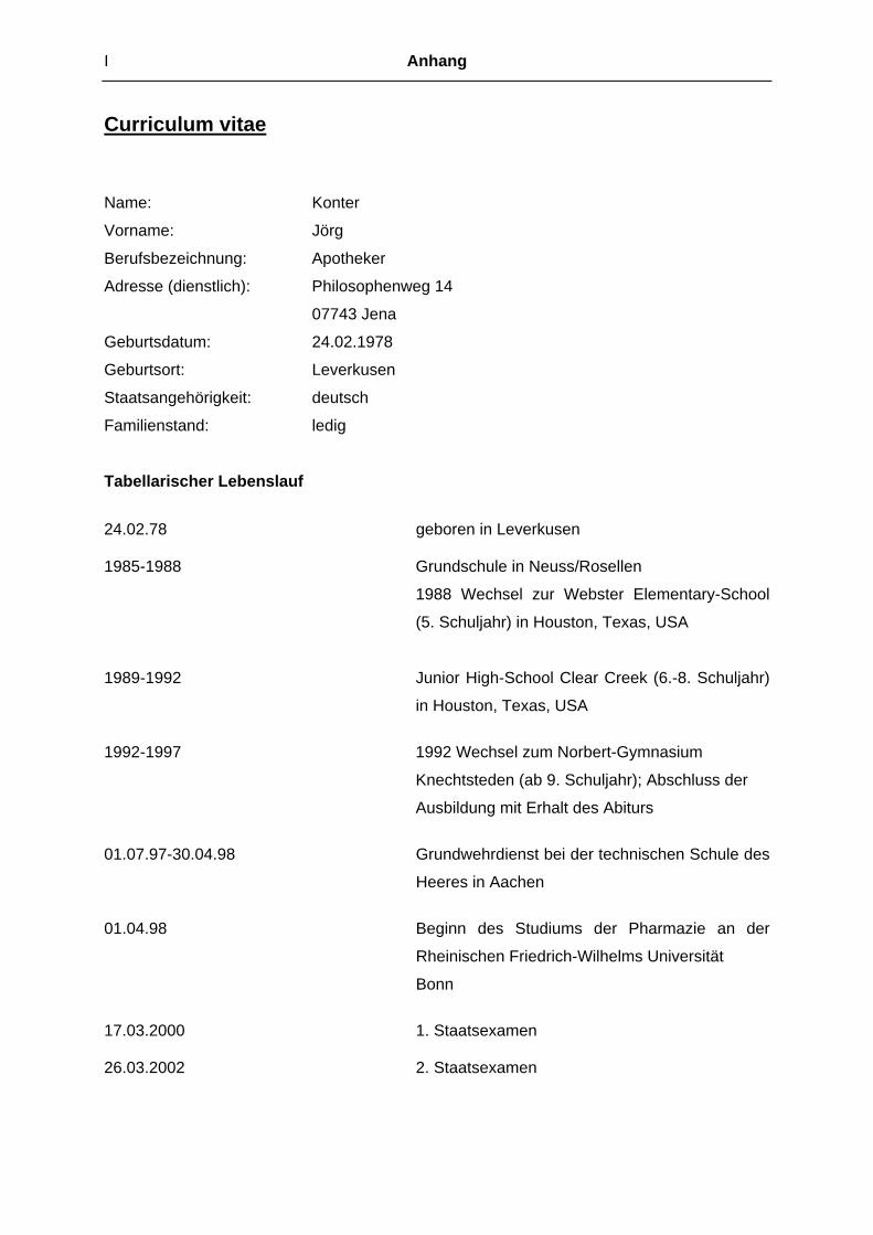
I Anhang
Curriculum vitae
Name: Konter
Vorname: Jörg
Berufsbezeichnung: Apotheker
Adresse (dienstlich): Philosophenweg 14
07743 Jena
Geburtsdatum: 24.02.1978
Geburtsort: Leverkusen
Staatsangehörigkeit: deutsch
Familienstand: ledig
Tabellarischer Lebenslauf
24.02.78 geboren in Leverkusen 1985-1988 Grundschule in Neuss/Rosellen
1988 Wechsel zur Webster Elementary-School
(5. Schuljahr) in Houston, Texas, USA
1989-1992 Junior High-School Clear Creek (6.-8. Schuljahr)
in Houston, Texas, USA
1992-1997 1992 Wechsel zum Norbert-Gymnasium
Knechtsteden (ab 9. Schuljahr); Abschluss der
Ausbildung mit Erhalt des Abiturs
01.07.97-30.04.98 Grundwehrdienst bei der technischen Schule des
Heeres in Aachen
01.04.98 Beginn des Studiums der Pharmazie an der
Rheinischen Friedrich-Wilhelms Universität
Bonn
17.03.2000 1. Staatsexamen 26.03.2002 2. Staatsexamen

Anhang II
01.05.02-31.10.02 Apotheke am Lessingplatz, Neuss
(Praktisches Jahr)
Apotheker Jürgen Linder
01.11.02-30.04.03 Bayer AG Leverkusen, Pharmaforschung, Cell
Membrane Proteomics (Praktisches Jahr)
Dr. Hans-Ulrich Siegmund
12.06.03 3. Staatsexamen in Düsseldorf; Erhalt der
Approbation zum Apotheker
August 2003 Urlaubsvertretung in der Apotheke am
Lessingplatz, Neuss
01.10.03-heute Promotion und wissenschaftlicher Mitarbeiter im
Institut für Medizinische/Pharmazeutische
Chemie im Arbeitskreis von Herrn Prof. Dr. J.
Lehmann
Tagungen
Okt. 2003 Joint Meeting on Medicinal Chemistry Krakau, Polen
Sept. 2004 W.C. on Dosing of Antiinfectives Nürnberg
Okt. 2004 DPhG Jahrestagung Regensburg
Feb. 2005 DPhG/APV Doktorandentagung Leipzig
Okt. 2005 DPhG Jahrestagung Mainz
Juni 2006 4th Intl. Conference on Nitric Oxide Monterey, Ca, USA
Okt. 2006 DPhG Jahrestagung Marburg
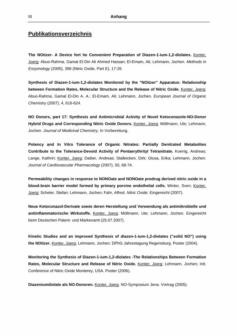
III Anhang
Publikationsverzeichnis
The NOtizer- A Device fort he Convenient Preparation of Diazen-1-ium-1,2-diolates. Konter,
Joerg; Abuo-Rahma, Gamal El-Din Ali Ahmed Hassan; El-Emam, Ali; Lehmann, Jochen. Methods in
Enzymology (2005), 396 (Nitric Oxide, Part E), 17-26.
Synthesis of Diazen-1-ium-1,2-diolates Monitored by the "NOtizer" Apparatus: Relationship between Formation Rates, Molecular Structure and the Release of Nitric Oxide. Konter, Joerg;
Abuo-Rahma, Gamal El-Din A. A.; El-Emam, Ali; Lehmann, Jochen. European Journal of Organic
Chemistry (2007), 4, 616-624.
NO Donors, part 17: Synthesis and Antimicrobial Activity of Novel Ketoconazole-NO-Donor Hybrid Drugs and Corresponding Nitric Oxide Donors. Konter, Joerg; Möllmann, Ute; Lehmann,
Jochen. Journal of Medicinal Chemistry. In Vorbereitung.
Potency and In Vitro Tolerance of Organic Nitrates: Partially Denitrated Metabolites Contribute to the Tolerance-Devoid Activity of Pentaerythrityl Tetranitrate. Koenig, Andreas;
Lange, Kathrin; Konter, Joerg; Daiber, Andreas; Stalleicken, Dirk; Glusa, Erika; Lehmann, Jochen.
Journal of Cardiovascular Pharmacology (2007), 50, 68-74.
Permeability changes in response to NONOate and NONOate prodrug derived nitric oxide in a blood-brain barrier model formed by primary porcine endothelial cells. Winter, Sven; Konter,
Joerg; Scheler, Stefan; Lehmann, Jochen; Fahr, Alfred. Nitric Oxide. Eingereicht (2007).
Neue Ketoconazol-Derivate sowie deren Herstellung und Verwendung als antimikrobielle und antiinflammatorische Wirkstoffe. Konter, Joerg; Möllmann, Ute; Lehmann, Jochen. Eingereicht
beim Deutschen Patent- und Markenamt (25.07.2007).
Kinetic Studies and an improved Synthesis of diazen-1-ium-1,2-diolates (“solid NO”) using the NOtizer. Konter, Joerg; Lehmann, Jochen; DPhG Jahrestagung Regensburg. Poster (2004).
Monitoring the Synthesis of Diazen-1-ium-1,2-diolates -The Relationships Between Formation Rates, Molecular Structure and Release of Nitric Oxide. Konter, Joerg; Lehmann, Jochen; Intl.
Conference of Nitric Oxide Monterey, USA. Poster (2006).
Diazeniumdiolate als NO-Donoren. Konter, Joerg. NO-Symposium Jena. Vortrag (2005).

Anhang IV
Selbstständigkeitserklärung
Hiermit erkläre ich,
dass mir die geltende Promotionsordnung der Fakultät bekannt ist.
Die vorliegende Arbeit habe ich selbstständig und nur unter Verwendung der angegebenen
Hilfsmittel und Literatur angefertigt.
Ich habe weder die Hilfe eines Promotionsberaters in Anspruch genommen, noch unmittelbar
oder mittelbar geldwerte Leistungen in Zusammenhang mit dem Inhalt meiner Dissertation
an Dritte erbracht.
Die vorliegende Dissertation habe ich ausschließlich an der Friedrich-Schiller Universität
Jena als Prüfungsarbeit eingereicht.
Jena, im August 2007


![Tris[4,4'-(ethene-1,2-diyl)dipyridinium] decavanadate ...journals.iucr.org/e/issues/2010/03/00/bg2319/bg2319.pdf · Tris[4,4000-(ethene-1,2-diyl)dipyridinium] decavanadate dihydrate](https://static.fdokument.com/doc/165x107/5fa3b6c93bca6f76e64e095f/tris44-ethene-12-diyldipyridinium-decavanadate-tris44000-ethene-12-diyldipyridinium.jpg)