Diss Verena Gesamtversion final VS2 genehmigtes Titelblatt ... · 3uhidfh ,, &kdswhu d ghvfulswlrq...
143
TECHNISCHE UNIVERSITÄT MÜNCHEN ZIEL - Institute for Food & Health Lehrstuhl für Mikrobielle Ökologie Microbiota of raw and microfiltered ESL milk Verena Sophia Johanna Schmidt Vollständiger Abdruck der von der Fakultät Wissenschaftszentrum Weihenstephan für Ernährung, Landnutzung und Umwelt der Technischen Universität München zur Erlangung des akademischen Grades eines Doktors der Naturwissenschaften genehmigten Dissertation. Vorsitzende(r): Prof. Dr. Ulrich Kulozik Prüfer der Dissertation: 1. Prof. Dr. Siegfried Scherer 2. apl. Prof. Dr. Matthias A. Ehrmann Die Dissertation wurde am 16.07.2018 bei der Technischen Universität München eingereicht und durch die Fakultät Wissenschaftszentrum Weihenstephan für Ernährung, Landnutzung und Umwelt am 17.10.2018 angenommen.
Transcript of Diss Verena Gesamtversion final VS2 genehmigtes Titelblatt ... · 3uhidfh ,, &kdswhu d ghvfulswlrq...
TECHNISCHE UNIVERSITÄT MÜNCHEN ZIEL - Institute for Food & Health Lehrstuhl für Mikrobielle Ökologie
Microbiota of raw and microfiltered ESL milk
Verena Sophia Johanna Schmidt Vollständiger Abdruck der von der Fakultät Wissenschaftszentrum Weihenstephan für Ernährung, Landnutzung und Umwelt der Technischen Universität München zur Erlangung des akademischen Grades eines
Doktors der Naturwissenschaften genehmigten Dissertation. Vorsitzende(r): Prof. Dr. Ulrich Kulozik Prüfer der Dissertation:
1. Prof. Dr. Siegfried Scherer 2. apl. Prof. Dr. Matthias A. Ehrmann
Die Dissertation wurde am 16.07.2018 bei der Technischen Universität München eingereicht und durch die Fakultät Wissenschaftszentrum Weihenstephan für Ernährung, Landnutzung und Umwelt am 17.10.2018 angenommen.

Jucundi acti labores Marcus Tullius Cicero

Preface I
Preface Chapters 2-5 of the present PhD thesis are either available as manuscript (chapter 2) or published to peer-reviewed journals (chapter 3-5). As each of these chapters comprises a separate publication, each part has its own introduction which might to a certain extent overlap with the general introduction given in chapter 1. This general introduction focuses on the composition of raw milk microbiota, illustrates the process of microfiltration for the production of microfiltered extended shelf life (ESL) milk, gives an overview about the types of retail milk available on the market and shows the aims of this thesis. A general discussion summarizes the outcome of this thesis. Chapter 2 comprises a study about microbial diversity, variability and stability of raw milk microflora on farm level. All experimental work was done by myself and I wrote the major part of the manuscript. Chapter 3 covers the microflora and enzymatic quality of microfiltered and pasteurized retail milk with special focus on factors limiting shelf life. The main part of the experimental work was performed by myself, enzymatic assays were done by Dr. Veronika Kaufmann (Lehrstuhl für Lebensmittel- und Bio- Prozesstechnik, Technical University of Munich). This chapter was published as “Microbial biodiversity, quality and shelf life of microfiltered and pasteurized extended shelf life (ESL) milk from Germany, Austria and Switzerland” in 2012 in International Food Microbiology, volume 154, issues 1-2, pages 1-9 by Verena S. J. Schmidt, Veronika Kaufmann, Ulrich Kulozik, Siegfried Scherer, * and Mareike Wenning. I wrote the major part of the manuscript. Chapter 4, a description of a novel genus of lactic acid bacteria of the phylum Firmicutes, was published as “Bavariicoccus seileri gen. nov., sp. nov., isolated from the surface and smear water of German red smear soft cheese” by Verena S. J. Schmidt, Ralf Mayr, Mareike Wenning, Jana Glöckner, Hans-Jürgen Busse and Siegfried Scherer in 2009 in International Journal of Systematic and Evolutionary Microbiology volume 59, issue 10, pages 2437-2443. Polar lipid profile analyses were carried out by Hans-Jürgen Busse (Institute of Microbiology, Department of Pathobiology, University of Veterinary Medicine Vienna). Analyses of G+C content, peptidoglycan, fatty acids and hybridization studies were performed by the Leibniz-Institut-Deutsche Sammlung von Mikroorganismen GmbH (DSMZ), the rest of the analyses were performed by myself and I wrote the major part of the manuscript.
Preface II
Chapter 5, a description of two novel Gram-negative bacterial species of the phylum Bacteroidetes, was published as “Sphingobacterium lacticum sp. nov. and Sphingobacterium alimentarium sp. nov. isolated from raw milk and a dairy environment” by Verena S. J. Schmidt, Mareike Wenning, and Siegfried Scherer in 2012 in International Journal of Systematic and Evolutionary Microbiology, issue 62, volume 7, pages 1506-1511. Analyses of G+C content, respiratory quinones, cellular fatty acids and hybridization studies were performed by the DSMZ, the rest of the analyses were performed by myself and I wrote the major part of the manuscript.

List of publications III
List of publications Bavariicoccus seileri gen. nov., sp. nov., isolated from the surface and smear water of German red smear soft cheese. Verena S. J. Schmidt, Ralf Mayr, Mareike Wenning, Jana Glöckner, Hans-Jürgen Busse and Siegfried Scherer (2009) International Journal of Systematic and Evolutionary Microbiology 59: 2437-2443. Microbial biodiversity, quality and shelf life of microfiltered and pasteurized extended shelf life (ESL) milk from Germany, Austria and Switzerland. Verena S. J. Schmidt, Veronika Kaufmann, Ulrich Kulozik, Siegfried Scherer and Mareike Wenning (2012) International Food Microbiology 154: 1-9. Sphingobacterium lacticum sp. nov. and Sphingobacterium alimentarium sp. nov. isolated from raw milk and a dairy environment. Verena S. J. Schmidt, Mareike Wenning, and Siegfried Scherer (2012) International Journal of Systematic and Evolutionary Microbiology 62: 1506-1511. Bacillus kochii sp. nov., isolated from foods and a pharmaceuticals manufacturing site. Herbert Seiler, Verena Schmidt, Mareike Wenning and Siegfried Scherer (2012) International Journal of Systematic and Evolutionary Microbiology 62: 1092–1097. Bacillus gottheilii sp. nov., isolated from a pharmaceutical manufacturing site. Herbert Seiler, Mareike Wenning, Verena Schmidt and Siegfried Scherer (2012) International Journal of Systematic and Evolutionary Microbiology 63: 867-872.
Summary IV
Summary This thesis aims at the characterization of cow`s milk microbiota, i.e. the diversity, variance and stability of raw milk microflora on farm level monitored over 15 months, the microbiota of retail microfiltered extended shelf life milk (ESL) with special reference to bacteria limiting shelf life as well as the description of hitherto unknown bacteria originating from raw milk of this study and the dairy environment. Out of six raw milk samples of the bulk tank of one farm a total of 626 isolates was identified by FT-IR spectroscopy and DNA gene sequence analysis to 106 species with 27 to 44 species found in each of the samples. This high microbial biodiversity was also supported by a linear slope of the species accumulation curve and a high incidence of rare species. Yet, single floras comprised high shares of pathogenic bacteria, like Streptococcus (Sc.) dysgalactiae and Staphylococcus (St.) aureus, indicating a subclinical masitits of the cows. In addition, for the first-time stability of farm milk microbiota at strain level could be demonstrated appyling FT-IR hierarchal cluster analysis and molecular typing (RAPD, BOX- and rep-PCR). Clonal isolates of St. warneri and Kocuria (Koc.) rhizophila respectively were repeatedly detected from different samplings with up to 15 months in between. Microfiltered retail extended shelf life milk was analyzed for factors limiting shelf life. Three different batches of one manufacturer were examined for microbial diversity and enzymatic quality at different stages of production and after cold storage over shelf life. 250 retail ESL milk packages by five manufacturers in Germany, Austria and Switzerland exhibited a great variance in microbial counts at the best before date (BBD) after storage at 8 °C (< 1-8 log10 cfu/mL), including also a variance between different packages of the same batch. This indicated a stochastic distribution of few germs to different packages at the filling process resulting in a different microbial flora in different packages after cold storage till the BBD. 8% of the packages were spoilt by Gram-negative post-process recontaminants and the spore formers Paenibacillus and Bacillus cereus, accompagnied by enzymatic spoilage factors and off-flavour. Other spore formers and Microbacterium spp. did not reach spoilage levels. Toxin profiling of B. cereus demonstrated Hbl, nhe, and cytK toxin genes in some strains, but no ces toxin gene. A minor share of B. cereus strains possessed the major cold shock cspA gene signature, indicating psychrotolerant growth characteristics.

Summary V
Hitherto unknown, milk related bacteria were validely described in a polyphasic approach combing genetic, phylogenetic and phenotypic analyses. Spingobacterium lacticum and Sphingobacterium alimentarium represent novel species of Gram-negative bacteria, phylum Firmicutes, and Bavariicoccus seileri gen. nov. sp. nov., a novel genus of lactic acid bacteria, pylum Bacteroidetes.

Zusammenfassung VI
Zusammenfassung Das Ziel dieser Arbeit war die Charakterisierung der Mikrobiota von Kuhmilch. Zum einen wurde die Varianz, Diversität und Stabilität der Rohmilchflora eines Bauernhofes über einen Zeitraum von 15 Monaten analysiert, des Weiteren wurde die Mikroflora von mikrofiltrierter ESL Milch mit Schwerpunkt auf haltbarkeitslimitierende Bakterien untersucht. Außerdem erfolgte eine Neubeschreibung von Bakterien, welche aus Rohmilch im Rahmen dieser Studie bzw. aus dem Umfeld einer Molkerei gewonnen wurden. Aus sechs Rohmmilchproben eines Bauernhof-Sammeltanks wurden insgesamt 626 Isolate gewonnen und mittels FT-IR Spektroskopie und DNA Sequenzanalyse als 106 verschiedene Spezies identifiziert, wobei jede einzelne Rohmilchprobe 27 bis 44 Spezies aufwies. Dieser hohe Grad an mikrobieller Biodiversität zeigte sich auch durch einen linearen Anstieg der Speziesakkumulierungskurve und ein hohes Auftreten seltener Spezies. Andererseits zeigten manche Floren hohe Anteile einzelner pathogener Bakterienspezies wie Streptococcus (Sc.) dysgalactiae und Staphylococcus (St.). aureus, was auf eine subklinische Mastitits der Milchkühe hindeutet. Erstmals wurde Stabiltität der Rohmilch-Mikrobiota auf Stammebene nachgewiesen mittels FT-IR Clusteranalyse und molekularbiologischen Typisierungsmethoden (RAPD, BOX- und rep-PCR). Klonale Isolate von St. warneri und Kocuria (Koc.) rhizophila wurden wiederholt detektiert, wobei zwischen den Probenahmen bis zu 15 Monate lagen. Mikrofiltierte ESL Milch aus dem Handel, d.h. Milch mit verlängerter Haltbarkeit, wurde auf haltbarkeitslimitierende Faktoren untersucht. Drei verschiedene Chargen mikrofiltrierter Milch eines Herstellers wurden zu unterschiedlichen Prozessschritten, d.h. während der Produktion sowie während Kühllagerung bei 8 °C bis zum Ende des MHDs auf mikrobielle Diversität sowie enzymatische Qualität untersucht. 250 Packungen mikrofiltierter Milch von fünf verschiedenen Herstellern aus Deutschland, Österreich und der Schweiz wiesen am MHD große Unterschiede in den Keimzahlen auf (< 1-8 log10 cfu/mL). Dies betraf auch verschiedene Packungen derselben Charge, was auf eine stochastische Keimzahlverteilung während des Abfüllprozesses hindeutet und eine daraus resultierende unterschiedliche mikrobielle Flora am MHD nach Kaltlagerung. 8% der Packungen waren verdorben durch gramnegative Rekontaminaten und die Sporenbildner Paenibacillus und Bacillus cereus, verbunden mit enzymatischen Verderbsanzeichen und Fehlsensorik. Andere
Zusammenfassung VII
Sporenbildner und Microbacterium spp. führten nicht zu Verderb innerhalb des MHDs. Ein Toxinscreening der isolierten B. cereus Vertreter zeigte das Vorkommen von Hbl, nhe, and cytK Toxingenen in einigen Stämmen, aber kein ces Toxingen. Nur bei wenigen B. cereus Stämmen wurde das Hauptkälteschock-Gen cspA nachgewiesen, welches auf ein Wachstumspotential bei kalten Temperaturen hindeutet. Bisher unbekannte milchassoziierte Baktereien wurden im Rahmen eines polyphasischen Ansatzes bestehend aus genetischen, phylogenetischen und phänotypischen Analysen beschrieben. Sphingobacterium lacticum sp. nov. und Sphingobacterium alimentarium sp. nov. sind zwei neue gramnegative Bakterienspezies aus dem Phylum Bacteriodetes; Bavariicoccus seileri gen. nov., sp. nov. stellt eine neue Milchsäurebakterien-Gattung aus dem Phylum Firmicutes dar.

Table of contents VIII
Table of contents Preface .................................................................................................... I List of publications .............................................................................. III Summary ............................................................................................. IV Zusammenfassung ............................................................................. VI Table of contents ...............................................................................VIII List of figures ...................................................................................... XI List of supplementary figures ........................................................... XII List of tables .......................................................................................XIII List of supplementary tables ............................................................ XIV Symbols and abbreviations ............................................................... XV Abbreviations of bacterial names .................................................... XVI 1 General introduction ....................................................................... 1
Microbial composition of raw cow`s milk ............................................... 1 1.1 Procedures for the production of market milk........................................ 2 1.2 Microfiltration for the production of ESL milk ......................................... 5 1.3 Spoilage of market milk ......................................................................... 8 1.4 Aims and objectives of this thesis ....................................................... 10 1.5
2 Diversity, stability and variability of single farm raw milk microbiota monitored over 15 months .............................................. 11
Abstract ............................................................................................... 12 2.1 Introduction .......................................................................................... 12 2.2 Materials and methods ........................................................................ 13 2.3
2.3.1 Sampling of raw milk and sample preparation ...................................... 13 2.3.2 Identification by FT-IR spectroscopy ..................................................... 14 2.3.3 Identification by DNA sequence analysis .............................................. 15 2.3.4 Strain typing by RAPD, rep- and BOX-PCR .......................................... 15 2.3.5 Statistical analysis of biodiversity .......................................................... 16
Results and discussion ........................................................................ 16 2.42.4.1 Biodiversity and composition of milk microbiota .................................... 16 2.4.2 Stability of microbiota ............................................................................ 21

Table of contents IX
Conclusion ........................................................................................... 24 2.5 Acknowledgements ............................................................................. 24 2.6 Supplementary material ...................................................................... 25 2.7
3 Microbial biodiversity, quality and shelf life of microfiltered and pasteurized extended shelf life (ESL) milk from Germany, Austria and Switzerland ................................................................................... 29
Abstract ............................................................................................... 30 3.1 Introduction .......................................................................................... 31 3.2 Materials and methods ........................................................................ 32 3.3
3.3.1. Processing of ESL milk and microbial analyses during storage at different temperatures ..................................................................................................... 32 3.3.2 Analysis of retail ESL milk of different manufacturers ............................... 33 3.3.3 Identification of isolates ............................................................................ 34 3.3.4 Analysis of B. cereus isolates ................................................................... 35 3.3.5 Enzymatic assays ..................................................................................... 36
Results ................................................................................................. 37 3.43.4.1. Monitoring of composition and population dynamics of milk microbiota over the production process and during cold storage ........................................ 37 3.4.2. Microbiological evaluation of retail ESL milk ............................................ 44 3.4.3. Prevalence of B. cereus and other risk group II bacteria ......................... 48
Discussion ........................................................................................... 50 3.53.5.1 Technological potential of combined microfiltration and pasteurization for reduction of microbial cell counts ...................................................................... 50 3.5.2 Enzymatic spoilage ................................................................................... 51 3.5.3 Recontamination....................................................................................... 52 3.5.4 Population dominance .............................................................................. 52 3.5.5 Suitability of combined microfiltration and pasteurization for the production of ESL milk ........................................................................................................ 53
Acknowledgements ............................................................................. 54 3.6 Supplementary material ...................................................................... 55 3.7
4 Bavariicoccus seileri gen. nov., sp. nov., isolated from the surface and smear water of German red smear soft cheese ........... 61
Abstract ............................................................................................... 62 4.1

Table of contents X
Introduction .......................................................................................... 62 4.2 Materials and methods ........................................................................ 63 4.3 Description of Bavariicoccus gen. nov. ............................................... 72 4.4 Description of Bavariicoccus seileri sp. nov. ....................................... 72 4.5 Acknowledgements ............................................................................. 73 4.6 Supplementary material ...................................................................... 74 4.7
5 Sphingobacterium lactis sp. nov. and Sphingobacterium alimentarium sp. nov., isolated from raw milk and a dairy environment. ....................................................................................... 78
Introduction .......................................................................................... 79 5.1 Materials and methods ........................................................................ 80 5.2 Description of Sphingobacterium lactis sp. nov. ................................. 88 5.3 Description of Sphingobacterium alimentarium sp. nov. ..................... 89 5.4 Supplementary material ...................................................................... 90 5.5
6 General discussion ....................................................................... 91 Highly diverse raw milk microbiota: polyphasic approach for the 6.1
description of Sphingobacterium alimentarium sp. nov. and Sphingobacterium lacticum sp. nov., isolated from raw milk and the dairy environment .................................................................................................. 91
6.1.1 Genotypic and phylogenetic differentiation ........................................... 92 6.1.2 Phenotypic differentiation ...................................................................... 94
Microbial quality of MF/PAST ESL milk at the end of BBD with respect 6.2to spoilage factors ........................................................................................ 95
Spoilage of MF/PAST milk in contrast to retail milk processed by other 6.3procedures ................................................................................................... 98
References ......................................................................................... 102 General Acknowledgments .............................................................. 124
List of figures XI
List of figures Figure 1-1 Filter module for microfiltration of consumer milk. ..................................... 5 Figure 1-2: Size distribution of raw milk constituents like fat, proteins and bacteria and their separability by filtration processes ............................................................... 6 Figure 1-3 Flow chart for the production of MF/PAST consumer milk ........................ 7 Figure 2-1 Microbial composition of six raw milk samples from a single farm over a period of fifteen months ............................................................................................ 18 Figure 2-2 Randomized species accumulation curve ............................................... 21 Figure 2-3 Hierarchal cluster analysis of FT-IR spectra of six St. warneri isolates in comparison with rep PCR and ERIC 2 profiles of these isolates .............................. 23 Figure 3-1 Distribution of bacteria in raw milk, MF milk and MF/PAST milk for three different batches of ESL milk (A, B, C) ..................................................................... 38 Figure 3-2 Aerobic counts of raw milk, MF milk and fresh MF/PAST milk and changes in microbial load of MF/PAST milk over shelf life depending on different storage temperatures (4 °C/8 °C/10 °C) ................................................................................ 40 Figure 3-3 Distribution of bacterial counts (log10 cfu/mL) of retail ESL milk stored at 8 °C at the end of shelf life ....................................................................................... 45 Figure 3-4 Hierarchical cluster analysis of FT-IR spectra of Bacillus cereus isolates 49 Figure 4-1 Nucleotide sequence similarity tree based on comparison of 16S rDNA, showing the relationships of Bavariicoccus gen. nov. to the most closely related taxa ................................................................................................................................. 66 Figure 4-2 Nucleotide sequence similarity tree, based on comparison of groEL gene sequences of Bavariicoccus gen. nov. and related taxa ........................................... 67 Figure 4-3 Polar lipid profiles of WCC 4188T (a) and A. phocae CIP 106292T (b) after two-dimensional thin layer chromatography and detection with molybdatophosphoric acid ........................................................................................................................... 69 Figure 5-1 Neighbour-joining phylogenetic tree based on 16S rRNA gene sequences showing the position of the isolates within the genus Sphingobacterium ................. 82
List of supplementary figures XII
List of supplementary figures Supplementary figure 2-1 Hierarchal cluster analysis of FT-IR spectra of five Koc. rhizophila isolates in comparison with rep PCR and ERIC 2 profiles of these isolates ................................................................................................................................. 25 Supplementary figure 4-1 FT-IR-based similarity dendrogram for Bavariicoccus seileri gen. nov., sp. nov. WCC 4188T and representatives of closely related genera ........ 76 Supplementary figure 4-2 FT-IR-based similarity dendrogram for the six strains of Bavariicoccus seileri gen. nov., sp. nov., WCC 4185, WCC 4186, WCC 4187, WCC 4188T, WCC 4189 and WCC 4190, and comparison with M13 RAPD-fingerprint profiles ...................................................................................................................... 77 Supplementary figure 5-1 Neighbour-joining phylogenetic tree based on groEL sequences showing the position of the new strains within the genus Sphingobacterium ..................................................................................................... 90
List of tables XIII
List of tables Table 1-1 Methods for the production of consumer milk ............................................. 4 Table 2-1 Diversity of taxonomic groups - number of families, genera, species and isolates ..................................................................................................................... 17 Table 2-2 Estimates for total species richness ......................................................... 20 Table 2-3 Number of bacterial species, genera and families common to several of the six microbiota ............................................................................................................ 21 Table 3-1 Enzymatic spoilage of MF/PAST milk throughout storage ........................ 42 Table 3-2 Incidence of different bacterial species in retail ESL milk after enrichment at 30 °C and after storage at 8 °C until the end of shelf life ...................................... 46 Table 3-3 Spoilage of MF/PAST ESL milk packages at the end of shelf life ............. 47 Table 3-4 Toxin gene profiles of the tested B. cereus types ..................................... 50 Table 4-1 Bacterial isolates ...................................................................................... 63 Table 4-2 Differential characteristics of the novel genus and type species of phylogenetically closely related genera .................................................................... 70 Table 5-1 Differential characteristics of the novel bacteria and related members of the genus Sphingobacterium .......................................................................................... 85 Table 5-2 Cellular fatty acid composition (%) of strains WCC 4512T and WCC 4521T and the type strains of related Sphingobacterium species ....................................... 87

List of supplementary tables XIV
List of supplementary tables Supplementary table 2-1 Taxonomic groups – most frequently detected species .... 26 Supplementary table 2-2 Number of bacterial species common to several of the six microbiota ................................................................................................................. 27 Supplementary table 3-1 Detected species in raw milk, MF milk, in freshly produced MF/PAST milk and in MF/PAST milk at the end of shelf life after storage at 4 °C, 8 °C and 10 °C, respectively ............................................................................................. 55 Supplementary table 3-2 Incidence of different bacterial species in retail ESL milk after enrichment at 30 °C and after storage at 8 °C till the end of shelf life .............. 58 Supplementary table 4-1 DNA-DNA relatedness, determined by hybridization studies between the four Bavariicoccus seileri isolates WCC 4187, WCC 4188T, WCC 4189 and WCC 4190 ......................................................................................................... 74 Supplementary table 4-2 Cellular fatty acid profiles of isolates WCC 4188T, WCC 4189, WCC 4190 and WCC 4187 ................................................................... 75

Symbols and abbreviations XV
Symbols and abbreviations BBD best before date cfu colony forming units DSMZ Leibniz-Institut DSMZ - Deutsche Sammlung von
Mikroorganismen GmbH ESL extended shelf life FAME fatty acid methyl ester FT-IR Fourier-transform infrared MF microfiltration MF/PAST milk microfiltered and pasteurized ESL milk PC+MM agar Plate Count Agar with 1% skim milk powder PCR polymerase chain reaction PPC post process contamination PSF psychrotolerant spore former Rep repetitive extragenic palindromic RAPD Random Amplified Polymorphic DNA TMP transmembrane pressure UHT ultra-high temperature UTP uniforme transmembrane pressure VBNC viable but non-culturable bacteria WCC Weihenstephan Culture Collection (inhouse collection for
bacteria)
Abbreviations of bacterial names XVI
Abbreviations of bacterial names A. = Atopobacter Ac. = Aerococcus Adv. = Advenella Aci. = Acinetobacter Agr. = Agrococcus Art. = Arthrobacter B. = Bacillus Bc. = Blastococcus Bra. = Brachybacterium Brev. = Brevibacterium C. = Carnobacterium Can. = Candida Cel. = Cellulosimicrobium Chr. = Chryseobacterium Com. = Comamonas Cor. = Corynebacterium Die. = Dietzia E. = Enterococcus Ent. = Enterobacter Ex. = Exiguobacterium Gor. = Gordonia Jan. = Janibacter Koc. = Kocuria Lb. = Lactobacillus Lc. = Lactococcus Mac. = Macrococcus Mb. = Microbacterium Mc. = Micrococcus Mor. = Moraxella Myc. = Mycobacterium Pic. = Pichia Pcb. = Pseudoclavibacter Pse. = Pseudomonas Rao. = Raoultella Rc. = Rhodococcus Rot. = Rothia
Abbreviations of bacterial names XVII
S. = Sphingobacterium Sc. = Streptococcus St. = Staphylococcus T. = Trichococcus V. = Vagococcus Waut. = Wautersiella Xen. = Xenophilus

1 General introduction 1
1 General introduction
Microbial composition of raw cow`s milk 1.1Raw milk according to European Law is defined as “das unveränderte Gemelk von Nutztieren, das nicht über 40 °C erhitzt und keiner Behandlung mit ähnlicher Wirkung unterzogen wurde”, i. e. “unmodified milking of farm animals, which was not heated above 40 °C nor did not undergo a treatment with a similar effect”) (Regulation (EC) No 853/2004). Milk promotes the growth of many microorganisms, as it is highly nutritious concerning the content of protein, fat, carbohydrates, vitamins, minerals and essential amino acids and besides offers a neutral pH value and a high availability of water (aw value) (Frank, 2007). Raw milk possesses a complex microbial composition due to multiple points of microbial entry like air, the cow`s skin, milking equipment and feed. Its microbiological quality is also depending on the sanitary conditions, the milking practices of the farmers as well as on storage conditions (duration and temperature) (Vacheyrou et al., 2011, Nucera et al., 2016). Cow`s milk comprises lactic acid bacteria (genera Lactococcus, Lactobacillus, Leuconostoc, Streptococcus, Enterococcus), Gram-negative bacteria like Alcaligenes, Flavobacterium, Aeromonas, Pseudomonas and Acinetobacter spp. as well as yeast and molds in lower shares (Quigley et al., 2011). Besides also spore forming bacteria are common in raw milk like Bacillus cereus, entering the milk via soiled udder, teats, silage or feed concentrate (Slaghuis et al., 1997, Vaerewijck et al., 2001, te Giffel et al., 2002). Some of the bacteria are also relevant for cheese making like Arthrobacter, Corynebacterium, Brevibacerium and Propionibacterium (Feurer et al., 2004, Gagnaire et al., 2015). Raw milk can also comprise foodborne pathogenic bacteria like Listeria monocytogenes, Salmonella spp., Campylobacter spp, verotoxigenic Escherichia coli, (VTEC O157:H7), Staphylococcus aureus, Escherichia coli and Bacillus cereus, which makes it necessary to process, i.e. pasteurize, the milk before human consumption (Steele et al., 1997, Jackson et al., 2012). Fresh milk is dominated by Gram-positive bacteria, but during storage at refrigerated temperatures there is a shift to a major share of psychrotolerant Gram-negative flora, consisting mainly of Pseudomonas spp. (Sørhaug & Stepaniak, 1997, Lafarge et al., 2004, Delbès et al., 2007, Fricker et al., 2011). This population shift is intensified by

1 General introduction 2
extended cold storage when collection intervals of the raw milk at the farm are elongated to every second day, sometimes even to every third or fourth day and if also dairies store the milk before subsequent processing. Also the health status of the cows influences the composition of the microbiota by increased shares of pathogenic bacteria in the milk like Staphylococcus aureus and other coagulase-negative staphylococci, Escherichia coli, Streptococcus uberis, Streptococcus dysgalactiae, Klebsiella, Proteus, Serratia, Pseudomonas, Enterobacter, Trueperella pyogenes, indicating a (subclinical) mastitis (Hogan & Smith, 2003, Olde Riekerink et al., 2008, Oikonomou et al., 2012). Although the existence of a core microbiome of certain bacterial genera, being detected in every raw milk, has been reported, there may also be seasonal variations in the microbiota, i.e. in one study the share of actinobacteria was elevated in spring (Kable et al., 2016), another study exhited a higher content of aerobic spore forming bacteria in summer, probably due to the fact that cows were grazing outside in contrast to the winter season characterized by permant indoor housing (Christiansson et al., 1999).
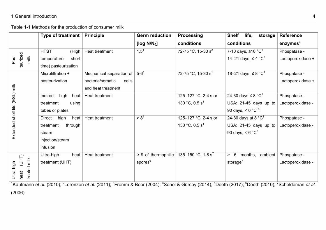
Procedures for the production of market milk 1.2Currently there is a great variety of procedures for the production of consumer milk (Table 1-1). Traditionally only two types could be distinguished, i.e. pasteurized milk and ultra-high heat treated (UHT) milk. Pasteurization, applying temperatures of 72–75 °C for 15–20 s (Lorenzen et al., 2011), aims “at reducing the number of any pathogenic microorganisms in milk and liquid products, if present, to a level at which they do not constitute a significant health hazard” (CAC/RCP 57-2004), a procedure also inactivating or at least reducing a major share of spoilage bacteria. UHT milk on the contrary is processed at 130–150 °C “in combination with appropriate holding times necessary to achieve commercial sterility”, a status with no growth of microorganisms at ambient storage (CAC/RCP 23-1979, Rev. 2 1983, CAC/RCP 57-2004). Pasteurized milk has a fresh taste, but a reduced shelf life of 7 and 12 days at cold storage, while ultra-high heat treated (UHT) milk offers a shelf life of several months and can be stored ambiently, but elevated temperature treatment creates sensorial changes, i.e. the typical “cooked flavour” (Manners & Craven, 2003, Hoffmann et al., 2006).

1 General introduction 3
To close the gap between fresh milk and UHT milk, the first extended shelf life (ESL) milk products have been produced in the early 1960s in North America (Henyon, 1999). ESL milks combine a fresh taste of pasteurized milk and a longer keeping quality while stored at refrigerated conditions. This prolonged shelf life of 18 to 35 days is beneficial and convenient for producers, retail as well as the consumer. ESL procedures are confronted with the task to remove as many vegetative cells and spore formers as possible, but at the same time to limit process induced product deterioration like changes in colour or vitamin contents and milk protein denaturation (Fernández García et al., 2013). In the meantime different procedures for the production of ESL milks have been developed and established in dairies (Rysstad & Kolstad, 2006), while the most common technologies are high heat treatment and microfiltration. Microfiltration as a non-thermal technology is only an ajunct to pasteurization, as for food safety reasons the thermal processing is essential to inactivate pathogenic bacteria like Mycobacterium bovis, Salmonella spp., Campylobacter jejuni, Campylobacter coli and Listeria monocytogenes (Lucey, 2015). According to Rysstad & Kolstad (2006) “ESL products are products that have been treated in a manner to reduce the microbial count beyond normal pasteurization, packaged under extreme hygienic conditions, and which have a defined prolonged shelf life under refrigerated conditions”. So the term ESL not only pertains to the bacterial decontamination technology like i.e. the filtration process, but is a complex system incorporating the whole production and distribution system. Raw milk has to be of high quality with less than 50.000 cfu/mL and post-process bacterial contamination has to prevented by the use of ultra-clean or aseptic filling machines and packaging disinfection. Furthermore, the cold chain of a maximal 8 °C has to be maintained throughout processing and distribution. This strict temperature regime is of great importance as ESL products are stronger germ reduced than pasteurized milk, but are not sterile (Fernández García et al., 2013). Besides the established procedures for the production of ESL milk also further processing alternatives have been developed like pulsed electric fields (Toepfl et al., 2007), ultrasonication (Khanal et al., 2014), high pressure processing (Evelyn & Silva, 2015), treatment of milk with bacteriocins ajunct to a heat treatment (Wirjantoro et al., 2001) or cold sterilization applying microsieves (Brito-de la Fuente et al., 2010). Yet none of these methods has reached commercial importance by now.
1 General introduction 4
Table 1-1 Methods for the production of consumer milk Type of treatment Principle Germ reduction
[log N/N0] Processing conditions
Shelf life, storage conditions
Reference enzymes4
Pas-
teurize
d mil
k
HTST (High temperature short time) pasteurization
Heat treatment 1,51 72-75 °C, 15-30 s2 7-10 days, ≤10 °C1 14–21 days, ≤ 4 °C3
Phospatase - Lactoperoxidase +
Exten
ded sh
elf life
(ESL
) milk
Microfiltration + pasteurization
Mechanical separation of bacteria/somatic cells and heat treatment
5-61 72-75 °C, 15-30 s1 18–21 days, ≤ 8 °C1 Phospatase - Lactoperoxidase +
Indirect high heat treatment using tubes or plates
Heat treatment 125–127 °C, 2-4 s or 130 °C, 0.5 s1
24-30 days ≤ 8 °C1 USA: 21-45 days up to 90 days, < 6 °C 5
Phospatase - Lactoperoxidase -
Direct high heat treatment through steam injection/steam infusion
Heat treatment > 81 125–127 °C, 2-4 s or 130 °C, 0.5 s1
24-30 days at 8 °C1 USA: 21-45 days up to 90 days, < 6 °C5
Phospatase - Lactoperoxidase -
Ultra-
high
heat
(UHT)
treate
d milk
Ultra-high heat treatment (UHT)
Heat treatment ≥ 9 of thermophilic spores6
135–150 °C, 1-8 s7
> 6 months, ambient storage7
Phospatase - Lactoperoxidase -
1Kaufmann et al. (2010); 2Lorenzen et al. (2011); 3Fromm & Boor (2004); 4Senel & Gürsoy (2014), 5Deeth (2017); 6Deeth (2010); 7Scheldeman et al. (2006)

1 General introduction 5
Microfiltration for the production of ESL milk 1.3Microfiltration has gained importance in the dairy industry since the 1980ies and is nowadays a well-established procedure in dairies with a wide field of applications, like globular milk fractionation, extraction of milk and whey proteins, casein enrichment, bacterial decontamination of cheese milk and brine and treatments of dairy effluents streams. Another important field is the removal of microorganisms for the production of ESL consumer milk (Mistry & Maubois, 1993, Saboya & Maubois, 2000, Awad et al., 2010, Fernández García et al., 2013)
Figure 1-1 Filter module for microfiltration of consumer milk Several candles are arranged in parallel, adopted from Bylund (1995) For the filtration of consumer milk ceramic membranes are used, which comprise two parts, i.e. a macroporous support with a minimum pore diameter of 10 µm and the active membrane, 3-5 µm thick, made from alumina, titanium or zirconia or a mixture of the latter two (Maubois, 2002). Several filter candles are arranged in parallel and form together a filter module (Figure 1-1). Pore size diameters of microfiltration membranes range between 0.8 and 1.4 µm, but most widely 1.4 µm membranes are used as they offer an optimal balance between separation of bacteria and a long running time (Malmberg & Holm, 1988). Microfiltration membranes retain most of the bacteria, but at the same time separation of desired milk components like protein,

1 General introduction 6
lactose and ash is avoided as they can pass the membranes due to their smaller size (Figure 1-2). As milk fat globules with sizes of 1 to 8 µm would block the membranes, milk is degreased and only skim milk is microfiltered (Walstra & Jenness, 1984).
Figure 1-2: Size distribution of raw milk constituents like fat, proteins and bacteria and their separability by filtration processes MF = microfiltration, UF =ultrafiltration, NF = nanofiltration, RO = reverse osmosis Adopted from Daufin et al. (2001) The process of microfiltration and pasteurization reduces bacterial counts by 3.5–5.2 log units, whereas the separation efficiency correlates with the bacterial size (Trouvé et al., 1991, Fernández García et al., 2013). Spore formers are separated more effectively as their cells are bigger (Saboya & Maubois, 2000). Doll et al. (2017) found a reduction of almost 4 log units for psychrotolerant spore formers. The level of Bacillus anthracis spores was decreased by 5.91 ± 0.05 log10 cfu/mL (0.8 μm membrane) and by 4.50 ± 0.35 log10 cfu/mL (1.4 μm membrane) (Tomasula et al., 2011). The pathogenic bacterial species Listeria monocytogenes, Brucella abortus, Salmonella typhimurium, Mycobacterium tuberculosis had decimal reduction rates of 3.4, 4.0, 3.5, 3.7, respectively (Madec et al., 1992, Saboya & Maubois, 2000). Somatic cells of the cows, commonly comprising 100000 to 400000 cells per mL raw milk, are totally separated by microfiltration, which prevents premature milk spoilage by the partially heat stable enzymes plasmin, plasminogen and proteases enclosed in the somatic cells (Maubois, 2002, Barbano et al., 2006). Traditional heat treatments inactivate bacteria, but their dead cells including heat stable enzymes remain in the milk causing undesired alterations during storage (Saboya & Maubois, 2000). Microfiltration on the contrary removes the bacterial cells together with their enzymes. Microfiltration technology for milk as it is used nowadays is based on patents by Holm et al. (1986) and Piot et al. (1987). Raw milk is heated at 50-55 °C
1 General introduction 7
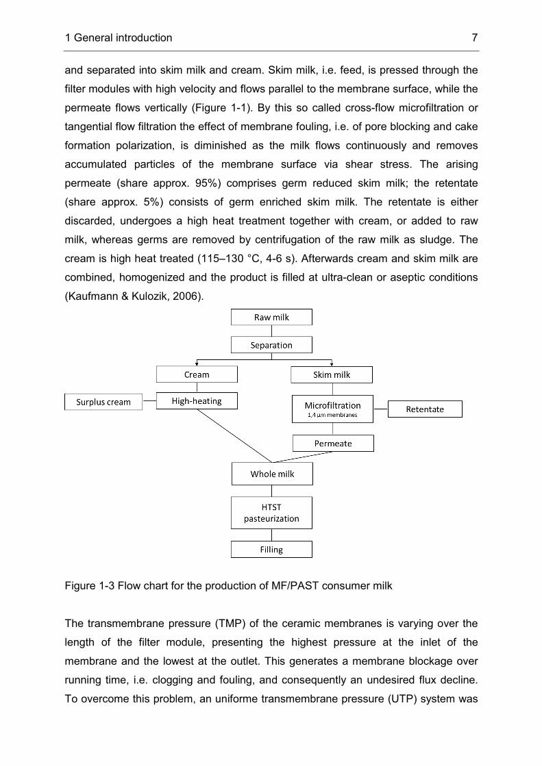
and separated into skim milk and cream. Skim milk, i.e. feed, is pressed through the filter modules with high velocity and flows parallel to the membrane surface, while the permeate flows vertically (Figure 1-1). By this so called cross-flow microfiltration or tangential flow filtration the effect of membrane fouling, i.e. of pore blocking and cake formation polarization, is diminished as the milk flows continuously and removes accumulated particles of the membrane surface via shear stress. The arising permeate (share approx. 95%) comprises germ reduced skim milk; the retentate (share approx. 5%) consists of germ enriched skim milk. The retentate is either discarded, undergoes a high heat treatment together with cream, or added to raw milk, whereas germs are removed by centrifugation of the raw milk as sludge. The cream is high heat treated (115–130 °C, 4-6 s). Afterwards cream and skim milk are combined, homogenized and the product is filled at ultra-clean or aseptic conditions (Kaufmann & Kulozik, 2006).
Figure 1-3 Flow chart for the production of MF/PAST consumer milk The transmembrane pressure (TMP) of the ceramic membranes is varying over the length of the filter module, presenting the highest pressure at the inlet of the membrane and the lowest at the outlet. This generates a membrane blockage over running time, i.e. clogging and fouling, and consequently an undesired flux decline. To overcome this problem, an uniforme transmembrane pressure (UTP) system was

1 General introduction 8
developed by Sandblom and commercialized by Alfa-Laval under the designation “bactocatch”. Here, the permeate is recirculated with high velocity, resulting in a pressure drop on the permeate side with the same magnitude as the pressure drop on the retentate side. This small UTP enables long run cycles of 9-12 hours, before a cleaning step of the membranes becomes necessary (Kaufmann & Kulozik, 2006). Alternatively, membranes are equipped with a longitudinal permeability gradient constructed into the support structure without a chance of the filtration layer (Pall®Membralox®GP (Pall®, 2007)) as well as there are membranes with the same pore size, but a reduced pore size distribution (Sterilox TM), allowing higher reduction factors of 4-5 (Fernández García et al., 2013).
Spoilage of market milk 1.4Spoilage of consumer milk before the best before date (BBD) poses a great problem to the milk industry as it leads to significant economic losses due to cost-intensive product recalls and food destruction, consumer irritation and a bad impact on the image of a brand. Spoilage according to Goff (2016) and Machado et al. (2017) can be defined as any change which renders a food product unacceptable, i.e. unappetizing or unsuitable for human consumption or for business to business trading. Microbial spoilage of food often involves the degradation of protein, carbohydrates and fats by the microorganisms or their enzymes resulting in souring, changes in texture, color or development of off-flavors as well as a physical damage to milk packaging. Spoilage of refrigerated consumer milk, i.e. all milk types but UHT milk, and consequently a reduced keeping quality is mainly caused by psychrotolerant bacteria and their microbial enzymes, i.e. lipases and proteases (Eneroth et al., 2000). Psychrotolerant bacteria are able to proliferate at 7 °C and therefore cause spoilage even when the cold chain is maintained and although their optimal growth temperature might be higher (Champagne et al., 1994). These bacteria are omnipresent in nature. Hence, it is impossible to avoid their presence in raw milk. They mostly intrude via water, soil or vegetation at dairy farms (Vissers & Driehuis, 2009) or as post-heat treatment contaminants and in low numbers (Moseley, 1980, Ralyea et al., 1998), but become the dominant microflora during subsequent cold storage and lead to premature spoilage. The risk of spoilage before the BBD is even greater for ESL milks as they have a prolonged shelf life at cold storage and are
1 General introduction 9
germ-reduced, i.e. only little competitive microbial flora counteracts the growth of undesired bacteria. Spoilage organisms of consumer milk can be basically assigned to two different bacterial groups, i.e. aerobic spore formers and Gram-negative bacteria. Spore formers mainly of the genera Bacillus and Paenibacillus are problematic as their spores are not inactivated by pasteurization and some species are even reported to show a heat resistance able to survive a UHT treatment, like Bacillus sporothermodurans (Gopal et al., 2015). The most prevalent psychrotolerant spore former, particularly in summer, is Bacillus cereus (Scheldeman et al., 2006), a bacterium responsible for sweet curdling of milk. The Bacillus cereus group, also called Bacillus cereus sensu lato (s.l.), comprises 9 validely described species, which include non-pathogenic and pathogenic species producing emetic and diarrhetic toxins (Ehling-Schulz et al., 2011, Ceuppens et al., 2013, Miller et al., 2016). Another group of important milk spoilage bacteria are Gram-negative bacteria. Some strains also show psychrotolerant growth and produce extracellular enzymes, i.e. mainly lipases and proteases. Lipases cause breakdown of fat resulting in rancid off-flavours, break down of casein leads to a grey colour and bitter off-flavours (De Jonghe et al., 2011). Especially Pseudomonas spp. group are frequent spoilage organisms due to the production of heat-resistant proteases and lipases during cold-storage of raw milk (Hantsis-Zacharov & Halpern, 2007, von Neubeck et al., 2015). Some spoilage bacteria are organized as biofilms, “an aggregate of micro-organisms, in which cells are frequently embedded within a self-produced matrix of extracellular polymeric substances (EPS) adhere to each other and/or to a surface” (Bremer et al., 2015).This conglomerate is located on product contacted stainless steel surfaces or gaskets and functions as a protection against cleaning detergents. From time to time bacteria are released from the biofilm in the processed product, inducing premature spoilage (Simões et al., 2010, Bai & Rai, 2011). Ability for biofilm formation has been reported for a wide range of typical milk spoilage bacteria, i.e. Bacillus, Paenibacillus and Pseudomonas (Seale et al., 2015).
1 General introduction 10
Aims and objectives of this thesis 1.5This project aims at analyzing the microbiota of cow`s milk upon three aspects: the microbiota of raw milk, the microbiota of MF/PAST ESL milk and the description of hitherto unknown bacteria isolated from dairy sources. Raw milk microbiota of one farm was repeatedly characterizated in the course of 15 months in order to elucidate the stability, diversity and variability of the microflora. After aerobic cultivation on agar plates, selected microorganisms were identified to genus- and species-level by FT-IR spectroscopy and gene sequencing (16S rRNA and rpoB). Statistical tools, i.e. different species richness estimators were used to estimate biodiversity. In addition to existing literature about raw milk microflora this project also aimed at analyzing stability on strain level, applying FT-IR hierarchal cluster analysis and molecular strain typing (RAPD, rep-PCR, Box-PCR). Microfiltration, using ceramic membranes for the mechanical separation of bacteria, constitutes an established procedure for the production of retail milk with an extended shelf life (ESL). Previous studies dealt with technical aspects like decimal reduction of bacterial numbers by microfiltration and subsequent pasteurization or the separation of different bacterial groups like spore formers or pathogens but yielded no information about its overall microbial quality and bacteria limiting shelf life. This project surveyed the microbial composition of industrially produced MF/PAST milk along the production chain including raw milk, microfiltered milk and freshly produced MF/PAST milk, as well as the microbiota of MF/PAST milk during refrigerated storage and at the end of shelf life, taking into account five different manufacturers, different batches and also different storage temperatures till the BBD. Special respect was given to representative spoilage bacteria and potentially pathogenic bacteria belonging to risk group II like B. cereus. The isolated B. cereus strains were characterized by multiplex PCR for the two cold shock proteins (cspA, cspF) and their toxim profile. Hitherto unknown bacteria, isolated from raw milk in the course of this project and from the dairy environment in previous studies, were validly described. The applied polyphasic procedure comprisising genotypic, phenotypic and phylogentic analyses, allows identification of the novel bacteria and consequently enables scientific exchange among researchers.
2 Diversity, stability and variability of single farm raw milk microbiota monitored over 15 months 11
2 Diversity, stability and variability of single farm raw milk microbiota monitored over 15 months
Verena S.J. Schmidt1, Monika Ehling-Schulz2, Siegfried Scherer1, 3 and Mareike Wenning1
1ZIEL - Institute for Food & Health, Technische Universität München, D-85354 Freising 2Abteilung für Lebensmittelmikrobiologie, Klinik für Wiederkäuer, Department für Nutztiere und öffentliches Gesundheitswesen in der Veterinärmedizin, Veterinärmedizinische Universität Wien, 1210 Wien, Austria 3Lehrstuhl für Mikrobielle Ökologie, ZIEL - Institute for Food & Health, Technische Universität München, D-85354 Freising Keywords: Raw milk, Microbiota, Biodiversity *Correspondence [email protected]
2 Diversity, stability and variability of single farm raw milk microbiota monitored over 15 months 12
Abstract 2.1The microbiota of raw milk from the bulk tank of a single farm was monitored over 15 months analyzing six samples at an interval of two to 251 days. A total of 626 isolates were randomly chosen from agar plates and assigned to 106 species using FT-IR spectroscopy and DNA gene sequence analysis. All samples were dominated by Gram-positive non-spore formers (genera Corynebacterium, Kocuria, Brevibacterium, Lactococcus and Staphylococcus) with Gram-negative bacteria and yeasts in lower shares. A high degree of biodiversity was indicated by a linear slope of the species accumulation curve and a high incidence of rare species. The six milk samples varied in biodiversity with 27 to 44 species found in each sample. 59% of all species were only detected in one flora, mostly in lower shares of 1-3%. On the contrary, single floras exhibited higher shares of particular species of up to 24%, like Streptococcus (Sc.) dysgalactiae and Staphylococcus (St.) aureus, indicating a subclinical mastitis. In contrast to variations in biodiversity among the different samples, considerable overlap was found as other species were repeatedly detected in several up to all floras. Furthermore, for the first time, evidence for stability at strain level was found by detection of clonal isolates of Kocuria rhizophila and Staphylococcus warneri from different samplings with up to 15 months in between. Strain identity was confirmed by the formation of distinct clusters in FT-IR hierarchal cluster analysis and identical patterns in molecular typing (RAPD, BOX- and rep-PCR).
Introduction 2.2Milk offers optimal growth conditions for a wide range of microorganisms, because of an almost neutral pH value, high water activity and high content of nutrients (Frank, 2007). While raw milk is sterile in the udder, it gets contaminated during the milking process by sources like the streak canal, the cows` udder, faeces, air, feed and farm workers, resulting in a complex microbial composition of raw milk. In recent years different approaches for population analyses of raw milk have been used emphasizing different aspects of milk microbiota. Some research focused on certain groups of microorganisms like pathogens, spoilage bacteria or lactic acid bacteria
2 Diversity, stability and variability of single farm raw milk microbiota monitored over 15 months 13
relevant for cheese-making (Michel et al., 2001, Dogan & Boor, 2003, Giannino et al., 2009, Ruusunen et al., 2013, Decimo et al., 2014, von Neubeck et al., 2015). For these analyses either culture dependant methods applying different selective media or DNA microarrays targeting defined species were used. Other investigations aimed to depict the whole bacterial community including also uncultivable species and therefore applied direct analyses based on direct sequencing or molecular fingerprinting techniques like single-strand conformational polymorphism (SSCP), denaturing gradient gel electrophoresis (DGGE) and temperature gradient gel electrophoresis (TTGE) (Lafarge et al., 2004, Delbès et al., 2007, Giannino et al., 2009, Verdier-Metz et al., 2009, Fricker et al., 2011, Raats et al., 2011, Delgado et al., 2013). However, there are very few papers targeting stability in the composition of microbial populations over a certain period. Delbès et al. (2007) found out that there were some dominant communities in milk of three different farms, which were isolated from every population independant from the sampling time over a period of nine months. Callon et al. (2007) used SSCP analyses to detect stable microbiota in goat milk within each season and found out that variation between milks from different seasons was a function of varying feeding and housing conditions over the year. In both studies stability was characterized by the detection of the same species. Yet, to our knowledge, there are no analyses about stability of the milk microbiota at strain level. The objective of the present work was to investigate the composition and changes of the microbiota of raw milk taken over a period of 15 months from a single farm, where feeding and housing conditions were constant all year. The isolates of six raw milk samples were identified by FT-IR spectroscopy and gene sequence analyses at species level and selective isolates were further characterized at strain level by FT-IR hierarchal cluster analysis and randomly amplified polymorphic DNA (RAPD) analyses, repetitive elements (rep) and BOX-PCR.
Materials and methods 2.32.3.1 Sampling of raw milk and sample preparation Six samples of raw milk were taken every two to 251 days over a period of 15 months at a research farm belonging to the Technical University Munich (Freising,
2 Diversity, stability and variability of single farm raw milk microbiota monitored over 15 months 14
Germany) from December 2007 to March 2009. The herd consisted of 55 German Brown Swiss cows with an average milk yield of 9200 kg from a milking period of 305 d/a. The cows` diet comprised the same feed all year; a mix consisting of 60% corn silage, 24% grass silage, 4% hay, 11% concentrate and 1% mineral fed ad libitum as well as 1.5 kg soy grist per cow. Cows were kept at permanent housing in a cubicle housing system fitted with rubber-coated slatted floors and spread with wood shavings. Milking of the cows was performed in a 2x2 tandem milking parlor (GEA WestphaliaSurge GmbH, Boenen, Germany) two times a day (4 am and 4 pm). Milk was collected in a bulk tank (Alpha laval) with a capacity of 1200 L and rapidly cooled down to a temperature of 6 °C using the cooling system copeland scroll. 500 mL of milk were taken at sterile conditions from the bulk tank, containing milkings from two successive milkings (evening and morning), brought to the laboratory at refrigerated conditions and analyzed within one hour. Appropriate dilutions of each sample were made with Ringer solution and plated on Plate Count Agar supplemented with 1% skim milk (PC+MM agar, Merck). Plates were incubated aerobically at 30 °C for five days and total aerobic plate counts were determined. 100 isolates were then chosen randomly, streaked on PC+MM agar and incubated at 30 °C for three days. In those cases, where mixed cultures were observed on the plates additional purification streaks were carried out to separate the different colony morphologies. In few cases where chosen isolates did not grow after streaking on PC+MM agar, they were not replaced.
2.3.2 Identification by FT-IR spectroscopy All isolates were identified using FT-IR spectroscopy. Sample preparation was done as described by Kümmerle et al. (1998). Lactic acid bacteria were cultivated on APT agar (Merck) anaerobically at 34 °C, spore formers were cultivated on TS agar (Oxoid) at 25 °C, and all other bacteria were cultivated on TS agar at 30 °C. Yeasts were incubated on YGC agar (Merck) at 27 °C. Plates were cultivated for 24 ± 0.5 h. All spectra were recorded and evaluated according to the methods of Oberreuter et al. (2002) using a HTS-XT FT-IR spectrometer (Bruker, Germany). To diminish the difficulties arising from unavoidable baseline shifts and to improve the resolution of complex bands, the first (lactic acid bacteria, aerobic Gram-positive non-spore formers) and second derivatives (spore formers, yeasts, Gram-negative bacteria) of

2 Diversity, stability and variability of single farm raw milk microbiota monitored over 15 months 15
the digitized original spectra were calculated. Six different FT-IR reference libraries (Schmidt et al., 2012) containing a total of 7220 spectra of 394 genera and 1019 species, were used for the identification of isolates.
2.3.3 Identification by DNA sequence analysis For all bacterial isolates, but staphylococci, whose spectra did not match with reference spectra in the FT-IR databases, 16S rRNA gene sequence analyses were performed. DNA extraction was performed according to Büchl et al. (2008) using a FastPrep®-24 instrument (MP Biomedicals, Germany) and zirconia silica beads (0.1mm, Roth) for cell wall disruption. 16S rRNA gene sequencing was performed in a 50 µL reaction mix containing 1 U of Thermo-Start Taq DNA polymerase, 5 µL Thermo-Start PCR buffer, 1.5 mM MgCl2, 200 µM (each) deoxynucleoside triphosphate (dNTP) (all Thermo Scientific), 1 µL lysate, 0,5 μL 16S_27f- Primer(5′-agagtttgatcctggctca-3′) and 0,5 μL 16S_1492r (5′- cggctaccttgttacgac-3′). Cycling conditions were the following: 1 min at 95 °C, 35 cycles of 20s at 95 °C, 40s at 52 °C and 100s at 72 °C, and a final extension step of 6 min at 72 °C. PCR products were resolved by gel electrophoresis for 1h at 90 V. Cycle sequencing of a 1500 bp fragment was done by GATC, Konstanz or 4base lab, Reutlingen and identification was performed by a BLAST sequence similarity search of the Genbank database (http://www.ncbi.nlm.nih.gov/blast/). For staphylococci sequence analyses of rpoB, the gene encoding the highly conserved β subunit of the bacterial RNA polymerase, were performed as this gene is more discriminative for staphylococci than the 16S rRNA gene (Drancourt & Raoult, 2002). Reaction mix and sequencing of this 900 bp fragment was the same as described for 16S rRNA sequencing, but primers were 1418f and 3554r (Mellmann et al., 2006).
2.3.4 Strain typing by RAPD, rep- and BOX-PCR Cell lysis was performed as described above, modified by an additional cooking step at 95 °C for 5 min. Reaction mix for PCR was the same as described for 16S rRNA gene sequencing, but primer concentration was 1 µM. Eric2 RAPD Primer and cycling conditions were according to Versalovic et al. (1991), the rep GTG5 primer

2 Diversity, stability and variability of single farm raw milk microbiota monitored over 15 months 16
and the BOX AFPR1 primer and cycling conditions were as described by Versalovic et al. (1994). PCR products were resolved by electrophoresis on 2% (w/v) agarose gels for 1.5 h at 100 V. Band patterns were compared visually.
2.3.5 Statistical analysis of biodiversity All statistics were calculated using the species diversity and richness 4.1.2 software package (Seaby & Henderson, 2006). A species accumulation curve was generated randomizing the sample order six times in order to evaluate the sampling effort i.e. if the number of species observed in the samplings represent the whole microbial species biodiversity of the raw milk habitat (Sobéron & Llorente, 1993). For the estimation of species richness six different estimators were used, as there is no general approval of one certain estimate. The Renyi family index was applied to compare the diversities of the six samples.
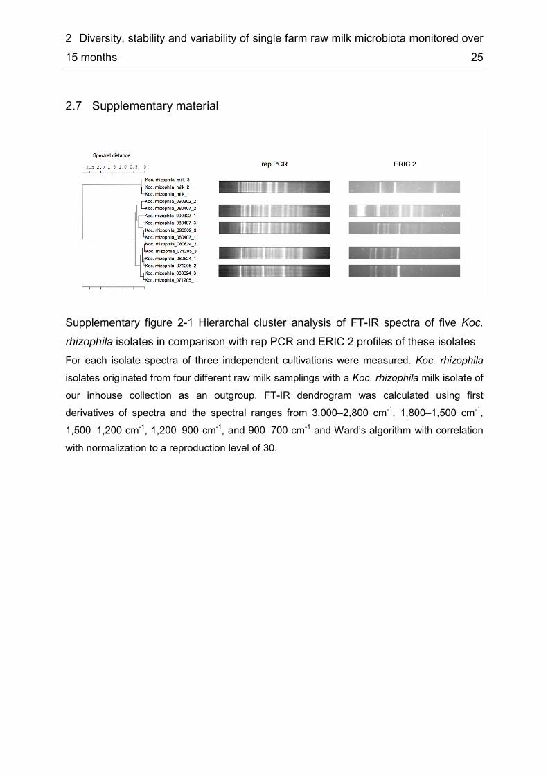
Results and discussion 2.42.4.1 Biodiversity and composition of milk microbiota Six fresh raw milk samples from a single farm were collected and analyzed over a period of 15 months. Total aerobic counts ranged from 2.9 to 4.0 log10 cfu/mL, indicating a very good microbiological quality of the milk (Class I according to German law (MilchGV, 1980)). A total of 626 isolates were isolated from PC+MM agar and identified by FT-IR spectroscopy and DNA gene sequence analysis. The isolates belonged to 106 different species of 39 different genera and 29 families. According to their identification result, all isolates were assigned to six different taxonomic groups: high G+C Gram-positive bacteria, lactic acid bacteria, other Gram-positive bacteria, obligate aerobic Gram-negative bacteria, facultatively anaerobic Gram-negative bacteria and yeasts (Figure 2-1). Most of the isolates (43%) belonged to the group of high G+C Gram-positive bacteria which also comprised most of the observed species (47/106) followed by obligate aerobic Gram-negative bacteria with 25 different species. In contrast, other Gram-positive bacteria (staphylocci) showed a low biodiversity as the had a share of 28.8% of all isolates, but these isolates were identified to a comparatively low number of species with an abundance of two

2 Diversity, stability and variability of single farm raw milk microbiota monitored over 15 months 17
different species St. warneri and St. aureus (18/106) (Table 2-1, Supplementary table 2-1). Table 2-1 Diversity of taxonomic groups - number of families, genera, species and isolates Obligate
aerobic Gram-negatives
Facultatively anaerobic Gram-negatives
High GC Gram-positives
Lactic acid bacteria
Other Gram-positives
Yeasts Total
Families 7 1 13 4 2 2 29 Genera 10 2 17 5 3 2 39 Species 25 2 47 11 18 3 106 Isolates 79 2 271 84 180 10 626
While the four major groups (obligate aerobic Gram-negatives, high G+C Gram-positives, lactic acid bacteria and other Gram-positives) were detected in each flora, facultatively anaerobic Gram-negative bacteria and yeasts were only recovered from 2 and 3 samples in shares of 1 and 1-7%, respectively (Figure 2-1) and both comprised the lowest number of isolates (2 and 10/626) and species (2 and 3/106). Dominance of Gram-positive bacteria in freshly produced farm milk (78-94% in samples of this study) and high G+C Gram-positives and other Gram-positives as the major groups has also been reported in previous works, whereas dairy bulk tank milk is dominated by Gram-negative bacteria (Desmasures et al., 1997, Weber, 2006, Delbès et al., 2007, Fricker et al., 2011, Raats et al., 2011). Yet, Ercolini et al. (2009) found significantly lower shares of Gram-positive bacteria in fresh farm milk, but incubation times of the inoculated agar plates were only two days in contrast to five days in the present study. As the growth rate of many Gram-positive bacteria is significantly lower than that of Gram-negatives`, their number might have been underestimated in those analyses.
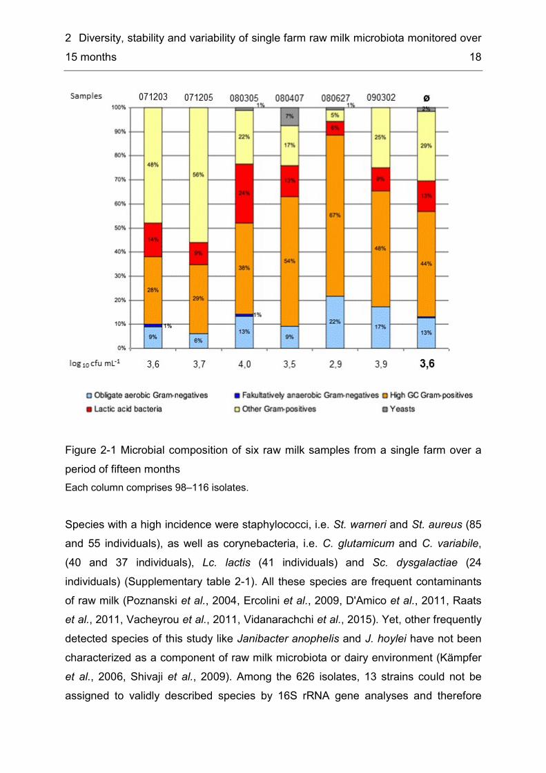
2 Diversity, stability and variability of single farm raw milk microbiota monitored over 15 months 18
Figure 2-1 Microbial composition of six raw milk samples from a single farm over a period of fifteen months Each column comprises 98–116 isolates. Species with a high incidence were staphylococci, i.e. St. warneri and St. aureus (85 and 55 individuals), as well as corynebacteria, i.e. C. glutamicum and C. variabile, (40 and 37 individuals), Lc. lactis (41 individuals) and Sc. dysgalactiae (24 individuals) (Supplementary table 2-1). All these species are frequent contaminants of raw milk (Poznanski et al., 2004, Ercolini et al., 2009, D'Amico et al., 2011, Raats et al., 2011, Vacheyrou et al., 2011, Vidanarachchi et al., 2015). Yet, other frequently detected species of this study like Janibacter anophelis and J. hoylei have not been characterized as a component of raw milk microbiota or dairy environment (Kämpfer et al., 2006, Shivaji et al., 2009). Among the 626 isolates, 13 strains could not be assigned to validly described species by 16S rRNA gene analyses and therefore
2 Diversity, stability and variability of single farm raw milk microbiota monitored over 15 months 19
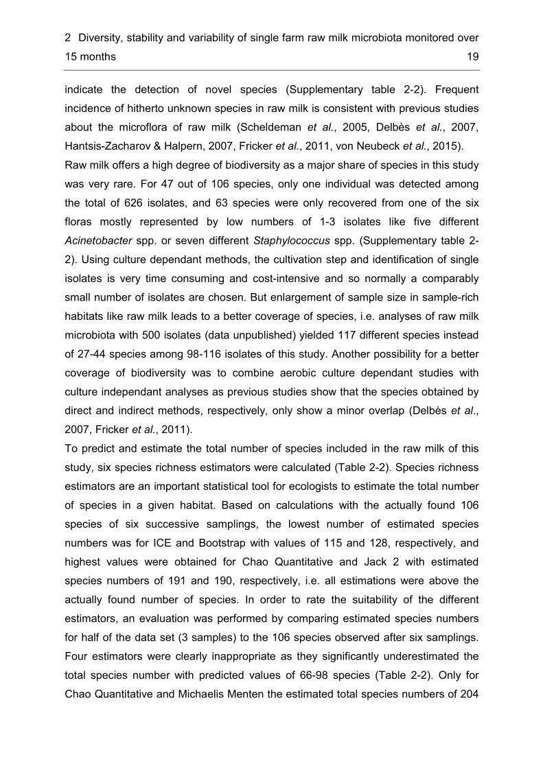
indicate the detection of novel species (Supplementary table 2-2). Frequent incidence of hitherto unknown species in raw milk is consistent with previous studies about the microflora of raw milk (Scheldeman et al., 2005, Delbès et al., 2007, Hantsis-Zacharov & Halpern, 2007, Fricker et al., 2011, von Neubeck et al., 2015). Raw milk offers a high degree of biodiversity as a major share of species in this study was very rare. For 47 out of 106 species, only one individual was detected among the total of 626 isolates, and 63 species were only recovered from one of the six floras mostly represented by low numbers of 1-3 isolates like five different Acinetobacter spp. or seven different Staphylococcus spp. (Supplementary table 2-2). Using culture dependant methods, the cultivation step and identification of single isolates is very time consuming and cost-intensive and so normally a comparably small number of isolates are chosen. But enlargement of sample size in sample-rich habitats like raw milk leads to a better coverage of species, i.e. analyses of raw milk microbiota with 500 isolates (data unpublished) yielded 117 different species instead of 27-44 species among 98-116 isolates of this study. Another possibility for a better coverage of biodiversity was to combine aerobic culture dependant studies with culture independant analyses as previous studies show that the species obtained by direct and indirect methods, respectively, only show a minor overlap (Delbès et al., 2007, Fricker et al., 2011). To predict and estimate the total number of species included in the raw milk of this study, six species richness estimators were calculated (Table 2-2). Species richness estimators are an important statistical tool for ecologists to estimate the total number of species in a given habitat. Based on calculations with the actually found 106 species of six successive samplings, the lowest number of estimated species numbers was for ICE and Bootstrap with values of 115 and 128, respectively, and highest values were obtained for Chao Quantitative and Jack 2 with estimated species numbers of 191 and 190, respectively, i.e. all estimations were above the actually found number of species. In order to rate the suitability of the different estimators, an evaluation was performed by comparing estimated species numbers for half of the data set (3 samples) to the 106 species observed after six samplings. Four estimators were clearly inappropriate as they significantly underestimated the total species number with predicted values of 66-98 species (Table 2-2). Only for Chao Quantitative and Michaelis Menten the estimated total species numbers of 204

2 Diversity, stability and variability of single farm raw milk microbiota monitored over 15 months 20
and 156 species, respectively, were above the value for the actually observed species numbers after six samplings. As discussed by O'Hara (2005) species estimators in general underestimate the real number of species in any habitat, especially for non-parametric estimators like Jack 1, Jack 2 or Bootstrap, and even more if the community examined has a high share of rare species (Smith & van Belle, 1984), which was observed for raw milk in this study. Each of the six samplings revealed 12–18 novel species not covered by the previous sampling and consequently all estimated values would probably be surpassed by a few more samplings. This finding is supported by a species accumulation curve of the six samples with exponential growth (Figure 2-2), i.e. new samplings would lead to an extension of species numbers, while sufficient sampling was indicated by the species accumulation curve approaching an asymptote (Sobéron & Llorente, 1993). Therefore species richness estimators in general cannot be applied for the estimation of raw milk biodiversity, particularly as raw milk is not a clearly separated habitat as it is influenced by habitually changes in the environment like variations in the livestock, feed etc. and therefore also comprises transient species, which are included in the microflora for shorter time periods like weeks or months, but do not become a permanent member. Table 2-2 Estimates for total species richness Table shows estimated species richness of raw milk microflora after six samplings. In order to rate the different estimates also a calculation using half of the dataset (three samplings) as well as the ratio between observed and estimated species numbers are included. Type of estimator ICE Chao
Quantitative Jack 1 Jack
2 Bootstrap Michaelis Menten Mean
Estimated species numbers (all six samples) 115 191 159 190 128 158 157 Estimated species numbers (three samples) 66 204 86 98 68 156 113 Observed species number (=106) /estimated species number (six samples)[%]
92.2 55.5 66.7 55.8 82.8 67.1 67.5
2 Diversity, stability and variability of single farm raw milk microbiota monitored over 15 months 21
Figure 2-2 Randomized species accumulation curve Curve shows the plot of observed species, accumulated over the samplings, against number of samples.
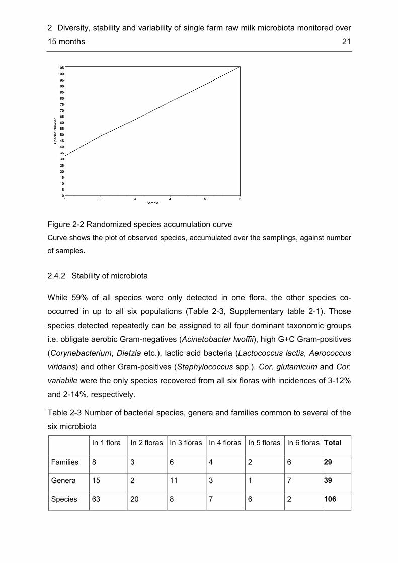
2.4.2 Stability of microbiota While 59% of all species were only detected in one flora, the other species co-occurred in up to all six populations (Table 2-3, Supplementary table 2-1). Those species detected repeatedly can be assigned to all four dominant taxonomic groups i.e. obligate aerobic Gram-negatives (Acinetobacter lwoffii), high G+C Gram-positives (Corynebacterium, Dietzia etc.), lactic acid bacteria (Lactococcus lactis, Aerococcus viridans) and other Gram-positives (Staphylococcus spp.). Cor. glutamicum and Cor. variabile were the only species recovered from all six floras with incidences of 3-12% and 2-14%, respectively. Table 2-3 Number of bacterial species, genera and families common to several of the six microbiota In 1 flora In 2 floras In 3 floras In 4 floras In 5 floras In 6 floras Total Families 8 3 6 4 2 6 29 Genera 15 2 11 3 1 7 39 Species 63 20 8 7 6 2 106
2 Diversity, stability and variability of single farm raw milk microbiota monitored over 15 months 22
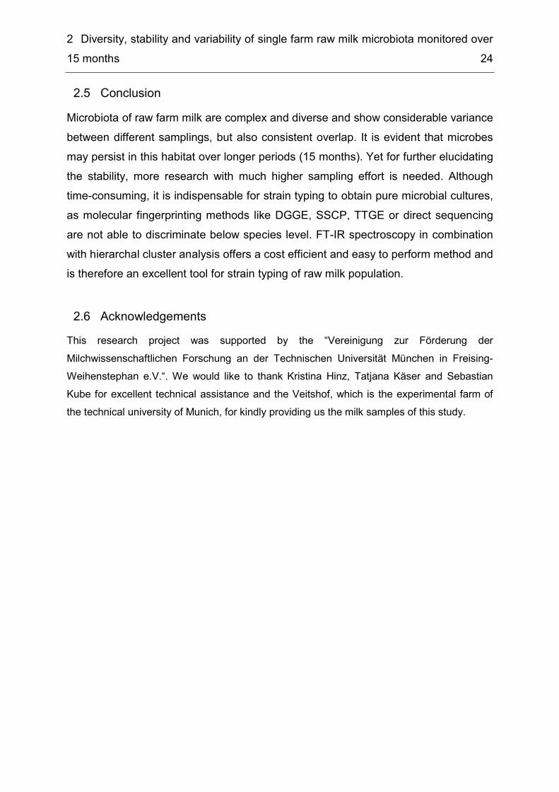
In addition to the repeated detection of the same species over the 15 months-interval, also indications for a stable microbiota at strain level were found. St. warneri isolates from four different samples exhibited a high spectral similarity to each other in hierarchical cluster analysis of FT-IR spectra in comparison to two St. warneri reference strains, i. e. the type strain DSMZ 1163T and a milk isolate of our in-house collection with isolates 090302, 080305 and 080407 forming one homogenous cluster with three independant FT-IR measurements of each isolate intermixed and isolate 071203 clustered separately with only little spectral distance (Figure 2-3). RAPD and rep-PCR resulted in identical band patterns for all four isolates supporting the conclusion that at least three of the four strains are clonal and must therefore represent a strain persisting in the herd or its environment. This is to our knowledge the first time, that evidence for persisting strains in raw milk is adduced. In addition, Koc. rhizophila isolates originating from four different raw milk samples (071205, 080407, 080624, and 090302) formed two separate FT-IR clusters with two isolates per cluster, which are also supported by two distinct rep PCR profiles (Supplementary figure 2-1). RAPD PCR confirmed the identity of one pair of isolates but the other two isolates were differentiated into two different types. The fact that FT-IR spectra indicate strain identity, but RAPD PCR with ERIC2 primers detects dissimilarities may be due to the presence of genetic variance that does not result in phenotypic differences under the incubation conditions applied for FT-IR spectroscopy. In contrast to repeated detection of clonal isolates of St. warneri and Koc. rhizophila, Kocuria carniphila and Dietzia timorensis isolates both originating from five different raw milk samples proved to be different by both FT-IR spectra and PCR band patterns (data not shown).
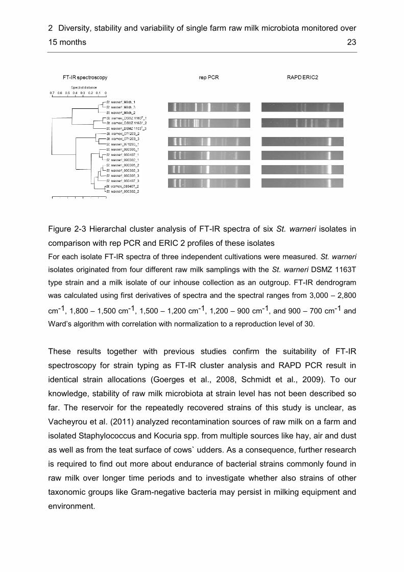
2 Diversity, stability and variability of single farm raw milk microbiota monitored over 15 months 23
Figure 2-3 Hierarchal cluster analysis of FT-IR spectra of six St. warneri isolates in comparison with rep PCR and ERIC 2 profiles of these isolates For each isolate FT-IR spectra of three independent cultivations were measured. St. warneri isolates originated from four different raw milk samplings with the St. warneri DSMZ 1163T type strain and a milk isolate of our inhouse collection as an outgroup. FT-IR dendrogram was calculated using first derivatives of spectra and the spectral ranges from 3,000 – 2,800 cm-1, 1,800 – 1,500 cm-1, 1,500 – 1,200 cm-1, 1,200 – 900 cm-1, and 900 – 700 cm-1 and Ward’s algorithm with correlation with normalization to a reproduction level of 30. These results together with previous studies confirm the suitability of FT-IR spectroscopy for strain typing as FT-IR cluster analysis and RAPD PCR result in identical strain allocations (Goerges et al., 2008, Schmidt et al., 2009). To our knowledge, stability of raw milk microbiota at strain level has not been described so far. The reservoir for the repeatedly recovered strains of this study is unclear, as Vacheyrou et al. (2011) analyzed recontamination sources of raw milk on a farm and isolated Staphylococcus and Kocuria spp. from multiple sources like hay, air and dust as well as from the teat surface of cows` udders. As a consequence, further research is required to find out more about endurance of bacterial strains commonly found in raw milk over longer time periods and to investigate whether also strains of other taxonomic groups like Gram-negative bacteria may persist in milking equipment and environment.
2 Diversity, stability and variability of single farm raw milk microbiota monitored over 15 months 24
Conclusion 2.5Microbiota of raw farm milk are complex and diverse and show considerable variance between different samplings, but also consistent overlap. It is evident that microbes may persist in this habitat over longer periods (15 months). Yet for further elucidating the stability, more research with much higher sampling effort is needed. Although time-consuming, it is indispensable for strain typing to obtain pure microbial cultures, as molecular fingerprinting methods like DGGE, SSCP, TTGE or direct sequencing are not able to discriminate below species level. FT-IR spectroscopy in combination with hierarchal cluster analysis offers a cost efficient and easy to perform method and is therefore an excellent tool for strain typing of raw milk population.
Acknowledgements 2.6This research project was supported by the “Vereinigung zur Förderung der Milchwissenschaftlichen Forschung an der Technischen Universität München in Freising-Weihenstephan e.V.“. We would like to thank Kristina Hinz, Tatjana Käser and Sebastian Kube for excellent technical assistance and the Veitshof, which is the experimental farm of the technical university of Munich, for kindly providing us the milk samples of this study.
2 Diversity, stability and variability of single farm raw milk microbiota monitored over 15 months 25
Supplementary material 2.7
Supplementary figure 2-1 Hierarchal cluster analysis of FT-IR spectra of five Koc. rhizophila isolates in comparison with rep PCR and ERIC 2 profiles of these isolates For each isolate spectra of three independent cultivations were measured. Koc. rhizophila isolates originated from four different raw milk samplings with a Koc. rhizophila milk isolate of our inhouse collection as an outgroup. FT-IR dendrogram was calculated using first derivatives of spectra and the spectral ranges from 3,000–2,800 cm-1, 1,800–1,500 cm-1, 1,500–1,200 cm-1, 1,200–900 cm-1, and 900–700 cm-1 and Ward’s algorithm with correlation with normalization to a reproduction level of 30.
2 Diversity, stability and variability of single farm raw milk microbiota monitored over 15 months 26
Supplementary table 2-1 Taxonomic groups – most frequently detected species Depicted species have shares ≥ 0.8% and numbers ≥ 5 (brackets) and with respect to a total of 626 isolates, obtained in this study. Ac. = Aerococcus, Aci. = Acinetobacter, Brev. = Brevibacterium, Can. = Candida, Cor. = Corynebacterium, Die. = Dietzia, Gor. = Gordonia, Jan. = Janibacter, Koc. = Kocuria, Lc. = Lactococcus, Mb. = Microbacterium, Mc. = Micrococcus, Pse. = Pseudomonas, Sc. = Streptococcus, St. = Staphylococcus, Ste = Stenotrophomonas, Waut = Wautersiella Obligate aerobic Gram-negatives
Shares High GC Gram-positives
Shares Lactic acid bacteria
Shares Other Gram-positives
Shares Yeasts Shares
Pse. fluorescens 3% (16) Cor. glutamicum 6% (40) Lc. lactis 7% (41) St. warneri 14% (85) Can. parapsilosis 1% (6) Ste. maltophila 2% (11) Cor. variabile 6% (37) Sc. dysgalactiae 4% (24) St. aureus 9% (55) Aci. lwoffii 2% (11) Koc. carniphila 5% (33) Ac. viridans 1% (5) St. sciuri 1% (7) Pse. aeruginosa 1% (5) Brev. epidermidis 2% (15) Sc. uberis 1% (6) St. epidermidis 1% (6) Aci. johnsonii/ junii 1% (5) Brev. linens 2% (11) St. haemolyticus 1% (6) Waut. sp. nov. 1% (5) Jan. anophelis 2% (10) Jan. hoylei 1% (9) Brev. sp. nov. 1% (8) Cor. xerosis 1% (8) Die. timorensis 1% (8) Koc. rhizophila 1% (7) Gor. polyisoprenivorans 1% (6) Mb. oxidans 1% (5) Gor. terrae 1% (5) Koc. kristinae 1% (5) Mc. luteus 1% (5)

2 Diversity, stability and variability of single farm raw milk microbiota monitored over 15 months 27
Supplementary table 2-2 Number of bacterial species common to several of the six microbiota Numbers in brackets indicate shares of species in the six microfloras. Probably undescribed species are characterized by the addition “sp. nov.”, after the genus name and species names in brackets indicates the greatest analogy based on 16S rRNA sequences.” Ac. = Aerococcus, Adv. = Advenella, Aci. = Acinetobacter, Agr. = Agrococcus, Art. = Arthrobacter, Bc. = Blastococcus, Bra. = Brachybacterium, Brev. = Brevibacterium, Can. = Candida, Cel. = Cellulosimicrobium, Chr. = Chryseobacterium, Com. = Comamonas, Cor. = Corynebacterium, Die. = Dietzia, E. = Enterococcus, Ent. = Enterobacter, Ex. = Exiguobacterium, Gor. = Gordonia, Jan. = Janibacter, Koc. = Kocuria, Lb. = Lactobacillus, Lc. = Lactococcus, Mac. = Macrococcus, Mb. = Microbacterium, Mc. = Micrococcus, Mor. = Moraxella, Myc. = Mycobacterium, Pic. = Pichia, Pcb. = Pseudoclavibacter, Pse. = Pseudomonas, Rao. = Raoultella, Rc = Rhodococcus, Rot. = Rothia, S. = Sphingobacterium, Sc. = Streptococcus, St. = Staphylococcus, Waut = Wautersiella, Xen. = Xenophilus Detected in 1 flora: 63 spp.
Detected in 2 floras: 20 spp.
Detected in 3 floras: 8 spp.
Detected in 4 floras: 7 spp.
Detected in 5 floras: 6 spp.
Detected in 6 floras: 2 spp.
Aci. (bouvetii 97.0%) sp.nov. (2%) Aci. (hae. 97.6%) sp.nov. (1%) Aci. (johnsonii 96.1%) sp.nov. (2%) Aci. baumannii (1%) Aci. calcoaceticus (1%) Adv. kashmirensis (1%) Agr. casei (3%) Art. (russicus 97.2%) sp. nov. (1%) Art. arilaitensis (4%) Bc. (93.3%) gen.nov. (1%) Bra. (paracon./sacelli 97.2%) sp.nov (1%) Bra. arcticum (1%) Bra. conglomeratum (1%) Bra. nesterenkovii (3%) Bra. rhamnosum (2%) Brev. oceani (2%) Brev. pityocampae (2%) Brev. aurantiacum (1%) Cel. cellulans (1%) Chr. lactis (1%) Chr. sp. (scophthalmum) (1%) Chr. sp. (jostei/stagni) (2%) Com. korreensis (1%) Cor. bovis (1%) Cor. casei (1%) Cor. sp. (variabile/propinquum) (1%) Die. psychralcaliphila (2%) Ec. faecium (1%)
Aci. johnsonii/junii (1-4%) Brev. (samyangensis 96.7%) sp. nov. (3-5%) Cor. ammoniagenes (1-2%) Cor. asperum (1-2%) Die. maris (1-3%) E. gallinarum (1%) Gor. polyisoprenivorans (1-4%) Gor. terrae (2-3%) Mb. hominis (1-3%) Mb. oxidans (1-3%) Pse. aeruginosa (1-4%) Rc. erythropolis (1-3%) Rot. mucilaginosa (1%) S. lacticum sp. nov. (1-2%) S. (mizutaii 94.9%) sp. nov. (1%) St. auricularis (1-2%) St. equorum (1-2%) St. sciuri (1-6%) St. vitulinus (1%) St. xylosus (1%)
Ac. viridans (1-2%) Can. parapsilosis (1-4%) Jan. hoylei (3%) Mb. lacticum (1-2%) Mc. luteus (1-2%) St. epidermidis (2%) Ste. maltophila (1-8%) Waut. (falsenii 96.4%) sp. nov. (1-2%)
Aci. lwoffii (2-3%) Brev. linens (1-5%) Cor. xerosis (1-3%) Koc. kristinae (1-2%) Koc. rhizophila (1-2%) Pse. fluorescens (1-9%) St. aureus (1-34%)
Brev. epidermidis (1-6%) Die. timorensis (1-3%) Koc. carniphila (2-12%) Lc. lactis (4-12%) St. warneri (11-19%) St. haemolyticus (1-2%)
Cor. glutamicum (3-11%) Cor. variabile (2-13%)

2 Diversity, stability and variability of single farm raw milk microbiota monitored over 15 months 28
Detected in 1 flora: 63 spp.
Detected in 2 floras: 20 spp.
Detected in 3 floras: 8 spp.
Detected in 4 floras: 7 spp.
Detected in 5 floras: 6 spp.
Detected in 6 floras: 2 spp.
Ent. cloacae/asburiae (1%) Ex. undae (1%) Jan. anophelis (9%) Koc. varians (1%) Lb. helveticus (1%) Lc. garviae (1%) Mac. caseolyticus (1%) Mb. esteraromaticum (1%) Mb. laevaniformans (1%) Mb. trichotecenolyticum (1%) Mor. osloensis (1%) Myc. phocaicum (1%) Pic. cactophila/pseudocact. (3%) Pic. fermentans (1%) Pse. gessardii (1%) Pse. putida (1%) Pcb. (helvolus 96.3%) sp. nov. (1%) Rao. terrigena (1%) Rot. arfidiae (1 %) S. (mizutaii 93.9%) sp. nov. (1%) S. alimentarium sp.nov. (2%) S. mizutaii (1%) St. chromogenes (1%) St. fleuretti (1%) St. hominis (1%) St. pasteuri (3%) St. saprophyticus (1%) St. sciuri/vitulinus (1%) St. succinus/kloosii (1%) Sc. dysgalactiae (24%) Sc. parauberis (1%) Sc. suis (1%) Sc. thermophilus (1%) Sc. uberis (5%) Xen. arolata (1%)
3 Microbial biodiversity, quality and shelf life of microfiltered and pasteurized extended shelf life (ESL) milk from Germany, Austria and Switzerland 29
3 Microbial biodiversity, quality and shelf life of microfiltered and pasteurized extended shelf life (ESL) milk from Germany, Austria and Switzerland
Verena S.J. Schmidt1, Veronika Kaufmann2, Ulrich Kulozik2, 3, Siegfried Scherer1, 4, * and Mareike Wenning1 1Abteilung für Mikrobiologie, ZIEL, Technische Universität München, D-85354 Freising, Germany 2Abteilung für Technologie, ZIEL, Technische Universität München, D-85354 Freising, Germany 3Lehrstuhl für Lebensmittelverfahrenstechnik und Molkereitechnologie, Technische Universität München, D-85350 Freising, Germany 4Lehrstuhl für Mikrobielle Ökologie, Department für Grundlagen der Biowissenschaften, WZW, Technische Universität München, D-85350 Freising, Germany *Correspondence [email protected] Published as: Schmidt VSJ, Kaufmann V, Kulozik U, Scherer S & Wenning M (2012) Microbial biodiversity, quality and shelf life of microfiltered and pasteurized extended shelf life (ESL) milk from Germany, Austria and Switzerland. International Journal of Food Microbiology 154: 1-9.
3 Microbial biodiversity, quality and shelf life of microfiltered and pasteurized extended shelf life (ESL) milk from Germany, Austria and Switzerland 30
Abstract 3.1Information on factors limiting the shelf life of extended shelf life (ESL) milk produced by microfiltration and subsequent pasteurization is very limited. In this study, three different batches of ESL milk were analyzed at different stages of the production process and during storage at 4 °C, 8 °C and 10 °C in order to evaluate the changes in bacterial cell counts, microbial diversity and enzymatic quality. Additionally, detailed biodiversity analyses of 250 retail ESL milk packages produced by five manufacturers in Germany, Austria and Switzerland were performed at the end of shelf life. It was observed that microfiltration decreased the microbial loads by 5–6 log10 units to lower than 1 cfu/mL. However, bacterial counts at the end of shelf life were extremely variable and ranged between < 1 and 8 log10 cfu/mL. 8% of all samples showed spoilage indicated by cell counts higher than 6 log10 cfu/mL. The main spoilage groups of bacteria were Gram-negative post-process recontaminants (Acinetobacter, Chryseobacterium, Psychrobacter, Sphingomonas) and the spore formers Paenibacillus and Bacillus cereus, while other spore formers and Microbacterium spp. did not reach spoilage levels. Paenibacillus spp. and B. cereus apparently influenced enzymatic spoilage, as indicated by increased free fatty acid production, pH 4.6-soluble peptide fractions and off-flavours. In some cases, enzymatic spoilage was observed although microbial counts were well below 6 log10 cfu/mL. Thirteen B. cereus isolates were characterized for their toxin profiles and psychrotolerance. Hbl, nhe, and cytK toxin genes were detected in ten, thirteen, and four isolates, respectively, whereas the ces gene was always absent. Interestingly, only three of the thirteen isolates could be allocated to psychrotolerant genotypes, as indicated by the major cold shock cspA gene signature. Generally, large discrepancies in microbial loads and biodiversity were observed at the end of shelf life, even among packages of the same production batch. We suggest that such unexpected differences may be due to very low cell counts after ESL treatment, causing stochastic variations of initial species distributions in individual packages. This would result in the development of significantly different bacterial populations during cold storage, including the occasional development of high numbers of pathogenic species such as B. cereus or Acinetobacter. Keywords: Microfiltration, Extended Shelf Life (ESL) milk, Microbiota, Spoilage, B. cereus, Paenibacillus spp., FT-IR spectroscopy
3 Microbial biodiversity, quality and shelf life of microfiltered and pasteurized extended shelf life (ESL) milk from Germany, Austria and Switzerland 31
Introduction 3.2Consumers as well as the milk processing industry and distributors have a strong interest in an extended shelf life of products. While high temperature short time (HTST) pasteurized milk has a keeping quality of about one week at cold storage, ultra-high temperature (UHT) milk can be held at room temperature for several months. Yet, the production of this type of milk is accompanied by undesired thermally derived product alterations, i.e., cooked and rich caramelized flavours and off-flavours (Shipe et al., 1978, Mehta, 1980). Therefore, novel manufacturing techniques have been introduced for the production of Extended Shelf Life (ESL) milk with a taste like fresh milk, but a prolonged shelf life of up to 4 weeks in cold chain distribution. Besides high heat treatment at 123-127 °C for 1-5 seconds (Mayr et al., 2004, Mayr et al., 2004, Kaufmann et al., 2010) a combined processing of microfiltration and pasteurization (ESL) has also been established. First approaches of using microfiltration for the reduction of the microbial load have been undertaken for more than 25 years (Holm et al., 1986, Piot et al., 1987) and at this time the patented Bactocatch-procedure was introduced (Holm et al., 1986). By applying this method, raw milk is separated into skimmed milk and milk fat. The skimmed milk is microfiltered through ceramic membranes and subsequently pasteurized. The milk fat together with the germ-enriched retentate obtained after microfiltration of the skimmed milk is processed by ultra-high heat treatment and then reverted into the skimmed milk. To date, there is knowledge about the keeping quality of ESL milk in relation to different storage temperatures and other physico-chemical factors (Elwell M. W. and Barbano D. M., 2006, Kaufmann & Kulozik, 2006, Kaufmann & Kulozik, 2008, Kaufmann et al., 2010), but, to our knowledge, there is no information about which spoilage microorganisms influence the shelf life of ESL milk and which potentially pathogenic organisms belonging to risk group II according to German legislation (IfSG, 2000) are found in such products. Of particular interest with regard to food quality and safety is the spore former Bacillus cereus, which is one of the major spoilage organisms of processed milk causing off-flavours, “sweet curdling” and “bitty cream” defects in milk due to proteinase, lipase and phospholipase activity. Some sporulated strains of B. cereus are able to germinate and multiply even under refrigerated conditions. Lechner et al. (1998) described B. weihenstephanensis as the psychrotolerant species of the B.

3 Microbial biodiversity, quality and shelf life of microfiltered and pasteurized extended shelf life (ESL) milk from Germany, Austria and Switzerland 32
cereus complex, differing from B. cereus sensu strictu in its ability to grow at 4 °C, but not at 43 °C. B. weihenstephanensis can be detected by targeting the major cold shock protein cspA and a psychrotolerance signature in the 16S rDNA sequence (Lechner et al., 1998, von Stetten et al., 1998). Besides spoilage of processed milk, B. cereus is also capable of producing food poisoning toxins, i.e. the nonhemolytic enterotoxin complex NHE, the hemolytic enterotoxin complex HBL and the single protein cytotoxin and the emetic toxin cereulide, a dodecadepsipeptide (Ehling-Schulz et al., 2011). Intoxication with B. cereus toxins mostly has a moderate progression and are self-limiting (Stenfors Arnesen et al., 2008), but in some cases the emetic toxin has been reported to cause severe illness with fatal outcome due to fulminant liver failure and rhabdomyolysis (Mahler et al., 1997). Emetic toxins are mainly produced in starchy foods like rice, but low levels of cereulide have also been observed in milk (Agata et al., 2002). The occurrence of diarrheic toxins in milk is reported to be higher, as there are several case studies about intoxications with the diarrheic toxins after the consumption of contaminated milk (Granum & Lund, 1997). This study is focused on the influence of microfiltration and pasteurization processes on the microbial populations of ESL milk after elaboration and at the end of the shelf life, as well as the evaluation of changes in lipolytic, proteolytic and sensory properties. Besides, the microbial status of ESL milk at retail from five different manufacturers was assessed at the end of the shelf life in order to isolate the most representative spoilage and pathogenic species belonging to risk group II.
Materials and methods 3.33.3.1. Processing of ESL milk and microbial analyses during storage at different
temperatures Three batches of ESL milk samples A, B, C (3.8% fat matter) and their precursor products raw milk and microfiltered milk (MF) were obtained from a German dairy over a period of four months. Milk was treated by the Bactocatch procedure. Raw milk was degreased to avoid blocking of the ceramic membranes by fat globules. Skimmed milk was microfiltered through ceramic membranes with a nominal pore size of 1.4 µm (Tami, France) and then pasteurized at 77 °C for 30 seconds. Cream was heated separately at 125 °C for 4 seconds and reverted to the pasteurized

3 Microbial biodiversity, quality and shelf life of microfiltered and pasteurized extended shelf life (ESL) milk from Germany, Austria and Switzerland 33
skimmed milk. Two raw milk samples, two MF samples and 2 carton packages (1.5 litres) of ESL milk from each batch were analyzed at the day of production and a total of 51 carton packages were stored at 4 °C, 8 °C and 10 °C, respectively, for up to 29 days. Two packages were periodically analyzed after 7, 14, 16, 18, 20 and 29 days while after 22 days (end of shelf life), five packages of each storage temperature and each batch were analyzed. Milk was diluted with Ringer solution (Merck) and plated on Plate Count Agar supplemented with 0.1% skim milk powder (PC+MM, Merck) according to IDF standards and German legislation. Plates were incubated aerobically at 30 °C for 5 days and the total aerobic counts were determined. For analyzing the raw milk biodiversity, 100 isolates were randomly chosen. Due to the low microbial loads expected for the MF and ESL milk samples directly after production, an additional enrichment procedure was performed for these samples of each batch to ensure accurate cell count determination and a sufficiently high number of isolates for identification. 100 mL of milk were therefore divided into 1 mL aliquots, enriched with 9 mL of PC+MM broth and incubated at 30 °C for five days. After enrichment, one loopful of each sample was plated on PC+MM agar and plates were incubated at 30 °C for 120 h. Finally, all bacterial colonies grown in 100 mL enriched milk sample were chosen for species determination. For Batch C, which had higher cells counts in the freshly produced ESL milk, 200 colonies were randomly selected and isolated from PC+MM agar after direct plating and due to their high spoilage potential additionally all spore formers were chosen from the enriched aliquots. From the refrigerated samples after 22 days of storage, representative colonies of each of the distinct morphologies were isolated from the agar plates. Additionally, during storage, representative colonies of B. cereus and spore formers dominating the microbiota were isolated and identified.
3.3.2 Analysis of retail ESL milk of different manufacturers To obtain an overview of the microbiological quality of ESL milk available on the market and to determine which species are of particular relevance for spoilage, milk of five different manufacturers, three from Germany, one from Austria and one from Switzerland (manufacturer I to V), were analyzed for the microbiota in the fresh product and at the end of shelf life. From each manufacturer, five different batches and ten packages of each batch were analyzed. Apart from milk of manufacturer IV,
3 Microbial biodiversity, quality and shelf life of microfiltered and pasteurized extended shelf life (ESL) milk from Germany, Austria and Switzerland 34
which had more variable fat content, ranging from 0.5 to 3.8%, all other samples had 3.5 to 3.8%. Batches one and two from manufacturer V had been organically produced, which means that the cream separated before the microfiltration process was only treated at 90 °C for two to five seconds, while the cream of the conventional milks of this manufacturer was heated at 125 °C for the same time. Milk packages were either sent to our laboratory at refrigerated conditions by the manufacturer or they were directly collected from food retailing. Out of the ten packages per batch, five were stored at 30 °C for three days to detect competitive mesophilic bacteria present in the milk, while the other five were stored at 8 °C (maximum storage temperature recommended by the manufacturers) till the end of shelf life as defined by the manufacturer in order to detect the spoilage flora at refrigerated temperatures. Samples were plated on PC+MM agar and aerobic counts were determined. Representative colonies having different morphologies were isolated from the agar plates and further identified by FT-IR spectroscopy.
3.3.3 Identification of isolates All isolates were identified using FT-IR spectroscopy (Wenning et al., 2008). Spore formers were cultivated on TS agar (Oxoid) at 25 °C, lactic acid bacteria on All Purpose Tween (APT) agar (Merck) anaerobically at 34 °C according to Wenning et al. (2010) and all other bacteria on Tryptic Soy (TS) agar at 30 °C. Yeasts were incubated on Yeast Extract Glucose Chloramphenicol (YGC) agar (Merck) at 27 °C according to Kümmerle et al. (1998). Incubation time was in all cases for 24 ± 0.5 h. Few strains yielding a clumpy suspension were homogenized using the FastPrep instrument (MP Biomedicals) at 4.5 m/s for 10 seconds without the addition of beads in order to improve spectral quality. All spectra were recorded and evaluated according to the methods of Oberreuter et al. (2002) using an HTS-XT FT-IR spectrometer (Bruker Optics, Germany). To overcome the difficulties arising from baseline shifts and to improve the resolution of complex bands, the first (lactic acid bacteria, aerobic Gram-positive non-spore formers) or second derivations (spore formers, yeasts, Gram-negative bacteria) of the digitized original spectra were used. Six different FT-IR reference libraries, that had previously been established in our lab (Kümmerle et al., 1998, Oberreuter et al., 2002, Büchl et al., 2008, Wenning et al.,
3 Microbial biodiversity, quality and shelf life of microfiltered and pasteurized extended shelf life (ESL) milk from Germany, Austria and Switzerland 35
2010) and unpublished data, containing a total of 7220 spectra of 394 genera and 1019 species, were applied for the identification of isolates. For all isolates, whose spectra did not match with the reference spectra in the FT-IR databases, 16S rRNA gene sequence analyses were performed. DNA extraction and cycling conditions were performed according to Schmidt et al. (2009), using the primers 16S 27f and 1492r. Cycle sequencing was done by GATC Biotech (Konstanz) and identification was delineated by a BLAST sequence similarity search of the Genbank database (http://www.ncbi.nlm.nih.gov/blast/). If the closest matches of the partial 16S rDNA sequences of the isolates, obtained from spoilt samples, had similarity values of less than 97.5%, indicating the detection of a novel species, an almost complete 16S rDNA gene sequence was created using the primers 27f, 907r (5'-CCGTCAATTCMTTTGAGTTT-3') and 1492r for sequencing and ClustalX (1.8) (Thompson et al., 1997) for aligning the three sequences and assembling an almost complete 16S rRNA gene sequence.
3.3.4 Analysis of B. cereus isolates Toxin gene profiling of the B. cereus isolates was performed by multiplex PCR according to the method described by Ehling-Schulz et al. (2006) for simultaneous detection of four target genes, i.e. the nonhemolytic enterotoxin complex NHE, the hemolytic enterotoxin complex HBL, the single protein cytotoxin K and the emetic toxin cereulide. The type strain of B. cereus, which carries hbl, cytk and nhe genes (ATCC 14579), and the strain F4810/72 (AH187) (Turnbull et al., 1979), harbouring nhe and ces genes, were used as positive controls. To distinguish between psychrotolerant and mesophilic B. cereus isolates, cold shock gene profiling was performed according to Francis et al. (1998). Two cold shock proteins were targeted by multiplex PCR, i.e. psychrotolerant signatures of the major cold shock protein cspA and the cold shock protein cspF encoded by both psychrotolerant and mesophilic strains. The psychrotolerant strain WSBC 10204, which is the type strain of B. weihenstephanensis (Lechner et al., 1998), and the mesophilic strain F4810/72 (AH187) (Turnbull et al., 1979) were used as positive controls.

3 Microbial biodiversity, quality and shelf life of microfiltered and pasteurized extended shelf life (ESL) milk from Germany, Austria and Switzerland 36
3.3.5 Enzymatic assays 3.3.5.1 Proteolysis For the detection of proteolysis caused by bacterial and indigenous milk proteinases the amounts of pH 4.6 soluble peptides in milk samples were determined (Datta & Deeth, 2003). Milk samples were adjusted to pH 4.6 with 10% acetic acid, while stirring with a magnetic stirrer. The mixture was left at room temperature for 20 min and filtered through a 0.45 µm Millipore filter (Germany). The filtrate was analysed for peptides by the fluorescamine method of Beeby (1980) as modified by Kocak & Zadow (1985). Filtrates (100 µl), 3 mL of phosphate buffer (0.1 M; pH 8.0) and 1 mL of fluorescamine solution (0.2 mg/mL in actone) were mixed. The fluorescence of the solution, in relative fluorescence intensity values, was measured in a Microplate reader (Tecan GENios, Tecan Germany) at an emission wavelength of 475 nm and excitation wavelength of 390 nm using fluorescein as the standard. A calibration curve was constructed using the dipeptide Tyrosyl-Leucin (Tyr-Leu; Sigma-Aldrich) to convert the relative fluorescence intensity values to the concentration of α-amino groups. The proteolysis was expressed as equivalent concentration of Tyr-Leu in mmol released per mL of milk; the detection level was 0. 02 mmol/L. 3.3.5.2 Lipolysis The presence of free fatty acids (FFA) as an indicator of lipolysis were evaluated as described by Deeth et al. (1975). A lipid extraction was achieved by the addition of 10 mL of extraction mixture consisting of 2-propanol, petroleum ether, 4 N H2SO4 in 40:10:1), 6 mL petroleum ether and 4 mL water to 2 mL of milk, preheated to 30 °C. The mixture was shaken vigorously, allowed to stand for 20 min for the separation of the two phases, and 2 mL of the upper phase were removed to be titrated with 0.02 N methanolic KOH following the addition of six drops of colour indicator (1% phenolphthalein). The concentration of FFA was determined as µequiv. FFA/mL = ((T·N)/(P·V)) 103
where T is the net titration volume and N the normality of KOH, P the quotient of the volume titrated and the volume of upper layer and V the volume of milk.

3 Microbial biodiversity, quality and shelf life of microfiltered and pasteurized extended shelf life (ESL) milk from Germany, Austria and Switzerland 37
Results 3.43.4.1. Monitoring of composition and population dynamics of milk microbiota over the
production process and during cold storage Three batches of ESL milk were analyzed over the production process from raw milk to the end product and subsequently during cold storage until the end of shelf life. The raw milk samples reached bacterial counts of 4.8, 6.4 and 5.0 log10 cfu/mL, respectively, which were reduced by 5-6 log10 units during microfiltration to counts less than 1 cfu/mL (Figure 3-1). The subsequent pasteurization step led to a further decrease in the cell count for batch A, but slightly increased the counts for batches B and C, most probably due to heat-induced spore germination and recontamination processes, as explained in section 3.5.3. The population analyses (Figure 3-1, Supplementary table 3-1) revealed that all three raw milk microbiota were dominated by obligate aerobic Gram-negative bacteria (49 to 78%) which were mainly represented by the genera Pseudomonas, Acinetobacter, Chryseobacterium and Stenotrophomonas. Lactic acid bacteria and facultative anaerobic Gram-negative bacteria (i.e. Enterobacteriaceae) were the second most frequent groups followed by high G+C Gram-positive bacteria with shares of 28% (batch B) and 5% (batch C), comprising mainly Brachybacterium, Corynebacterium and Microbacterium. Spore formers were detected only in very low numbers of approx. 1% in batch A. After microfiltration the microbiota was significantly shifted towards the high G+C Gram-positive (54–74%) and lactic acid bacteria (2–28%), whereas the shares of Gram-negative bacteria were considerably reduced. The pasteurization step eliminated all Gram-negative and lactic acid bacteria, leaving only high G+C Gram-positive bacteria and spore formers (Figure 3-1). The spore formers now shared 7–25% and were identified as B. cereus, B. licheniformis, B. pumilus, B. subtilis, Brevibacillus agri, Paenibacillus cookii, P. amylolyticus/glucanolyticus and P. amylolyticus.

3 Microbial biodiversity, quality and shelf life of microfiltered and pasteurized extended shelf life (ESL) milk from Germany, Austria and Switzerland 38
Figure 3-1 Distribution of bacteria in raw milk, MF milk and MF/PAST milk for three different batches of ESL milk (A, B, C) Milk batches A, B and C were produced by the same manufacturer, aerobic cell counts are given below each column.
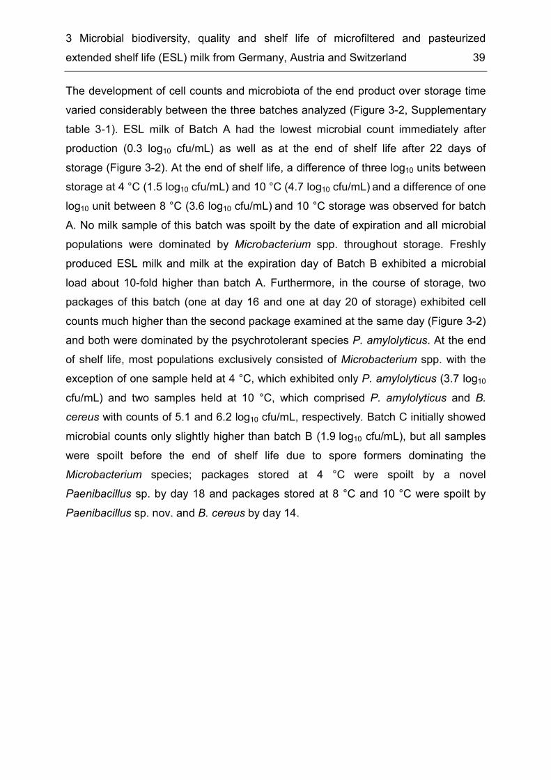
3 Microbial biodiversity, quality and shelf life of microfiltered and pasteurized extended shelf life (ESL) milk from Germany, Austria and Switzerland 39
The development of cell counts and microbiota of the end product over storage time varied considerably between the three batches analyzed (Figure 3-2, Supplementary table 3-1). ESL milk of Batch A had the lowest microbial count immediately after production (0.3 log10 cfu/mL) as well as at the end of shelf life after 22 days of storage (Figure 3-2). At the end of shelf life, a difference of three log10 units between storage at 4 °C (1.5 log10 cfu/mL) and 10 °C (4.7 log10 cfu/mL) and a difference of one log10 unit between 8 °C (3.6 log10 cfu/mL) and 10 °C storage was observed for batch A. No milk sample of this batch was spoilt by the date of expiration and all microbial populations were dominated by Microbacterium spp. throughout storage. Freshly produced ESL milk and milk at the expiration day of Batch B exhibited a microbial load about 10-fold higher than batch A. Furthermore, in the course of storage, two packages of this batch (one at day 16 and one at day 20 of storage) exhibited cell counts much higher than the second package examined at the same day (Figure 3-2) and both were dominated by the psychrotolerant species P. amylolyticus. At the end of shelf life, most populations exclusively consisted of Microbacterium spp. with the exception of one sample held at 4 °C, which exhibited only P. amylolyticus (3.7 log10 cfu/mL) and two samples held at 10 °C, which comprised P. amylolyticus and B. cereus with counts of 5.1 and 6.2 log10 cfu/mL, respectively. Batch C initially showed microbial counts only slightly higher than batch B (1.9 log10 cfu/mL), but all samples were spoilt before the end of shelf life due to spore formers dominating the Microbacterium species; packages stored at 4 °C were spoilt by a novel Paenibacillus sp. by day 18 and packages stored at 8 °C and 10 °C were spoilt by Paenibacillus sp. nov. and B. cereus by day 14.
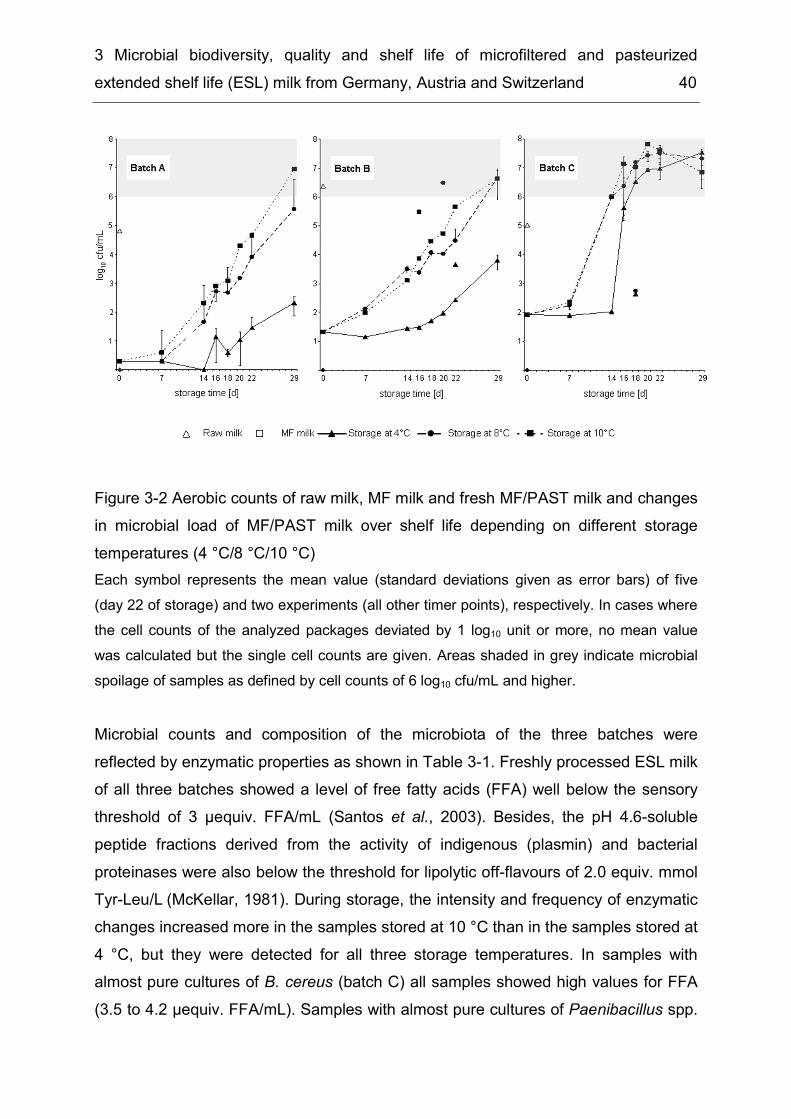
3 Microbial biodiversity, quality and shelf life of microfiltered and pasteurized extended shelf life (ESL) milk from Germany, Austria and Switzerland 40
Figure 3-2 Aerobic counts of raw milk, MF milk and fresh MF/PAST milk and changes in microbial load of MF/PAST milk over shelf life depending on different storage temperatures (4 °C/8 °C/10 °C) Each symbol represents the mean value (standard deviations given as error bars) of five (day 22 of storage) and two experiments (all other timer points), respectively. In cases where the cell counts of the analyzed packages deviated by 1 log10 unit or more, no mean value was calculated but the single cell counts are given. Areas shaded in grey indicate microbial spoilage of samples as defined by cell counts of 6 log10 cfu/mL and higher. Microbial counts and composition of the microbiota of the three batches were reflected by enzymatic properties as shown in Table 3-1. Freshly processed ESL milk of all three batches showed a level of free fatty acids (FFA) well below the sensory threshold of 3 µequiv. FFA/mL (Santos et al., 2003). Besides, the pH 4.6-soluble peptide fractions derived from the activity of indigenous (plasmin) and bacterial proteinases were also below the threshold for lipolytic off-flavours of 2.0 equiv. mmol Tyr-Leu/L (McKellar, 1981). During storage, the intensity and frequency of enzymatic changes increased more in the samples stored at 10 °C than in the samples stored at 4 °C, but they were detected for all three storage temperatures. In samples with almost pure cultures of B. cereus (batch C) all samples showed high values for FFA (3.5 to 4.2 µequiv. FFA/mL). Samples with almost pure cultures of Paenibacillus spp.
3 Microbial biodiversity, quality and shelf life of microfiltered and pasteurized extended shelf life (ESL) milk from Germany, Austria and Switzerland 41
(batches B and C) showed elevated levels of FFA, but proteinase activities were substantially below than the sensorially noticeable values (ca. 1,70 equiv. mmol Tyr-Leu/L). In contrast, samples of batch B comprising only Microbacterium spp. exhibited levels of proteolysis and lipolysis below the sensory threshold throughout storage at 4 °C, 8 °C and 10°C, but the sample of batch A stored at 10 °C developed elevated levels of FFA at the end of shelf life. Some of the latter samples presented levels of proteolysis and lipolysis above the sensory threshold, although microbial counts were below than the microbiological spoilage level.
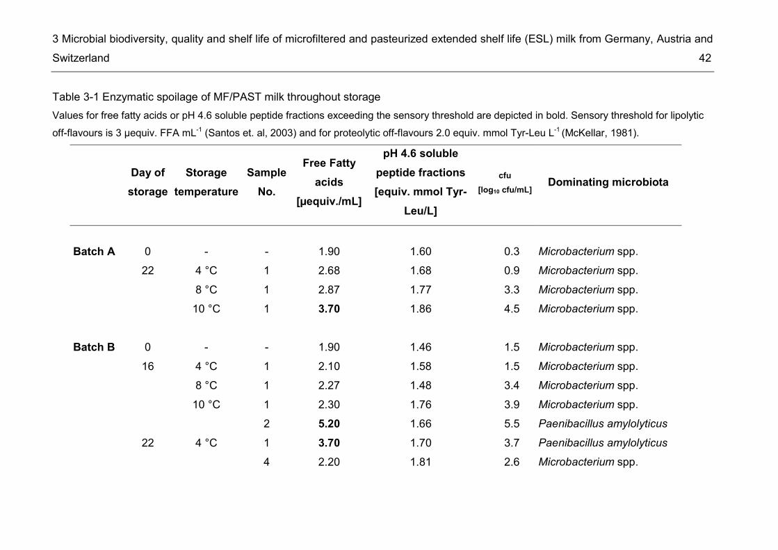
3 Microbial biodiversity, quality and shelf life of microfiltered and pasteurized extended shelf life (ESL) milk from Germany, Austria and Switzerland 42
Table 3-1 Enzymatic spoilage of MF/PAST milk throughout storage Values for free fatty acids or pH 4.6 soluble peptide fractions exceeding the sensory threshold are depicted in bold. Sensory threshold for lipolytic off-flavours is 3 µequiv. FFA mL-1 (Santos et. al, 2003) and for proteolytic off-flavours 2.0 equiv. mmol Tyr-Leu L-1 (McKellar, 1981).
Day of storage
Storage temperature
Sample No.
Free Fatty acids
[µequiv./mL]
pH 4.6 soluble peptide fractions [equiv. mmol Tyr-
Leu/L]
cfu [log10 cfu/mL] Dominating microbiota
Batch A 0 - - 1.90 1.60 0.3 Microbacterium spp. 22 4 °C 1 2.68 1.68 0.9 Microbacterium spp. 8 °C 1 2.87 1.77 3.3 Microbacterium spp. 10 °C 1 3.70 1.86 4.5 Microbacterium spp. Batch B 0 - - 1.90 1.46 1.5 Microbacterium spp. 16 4 °C 1 2.10 1.58 1.5 Microbacterium spp. 8 °C 1 2.27 1.48 3.4 Microbacterium spp. 10 °C 1 2.30 1.76 3.9 Microbacterium spp. 2 5.20 1.66 5.5 Paenibacillus amylolyticus 22 4 °C 1 3.70 1.70 3.7 Paenibacillus amylolyticus 4 2.20 1.81 2.6 Microbacterium spp.

3 Microbial biodiversity, quality and shelf life of microfiltered and pasteurized extended shelf life (ESL) milk from Germany, Austria and Switzerland 43
Day of storage
Storage temperature
Sample No.
Free Fatty acids
[µequiv./mL]
pH 4.6 soluble peptide fractions [equiv. mmol Tyr-
Leu/L]
cfu [log10 cfu
/mL] Dominating microbiota
Batch B 8 °C 4 2.60 1.88 4.1 Microbacterium spp. 10 °C 1 4.96 2.10 4.4 Bacillus cereus 6.2 Microbacterium spp. 2 4.00 2.00 5.3 Microbacterium spp. 5 4.15 2.04 4.2 Bacillus cereus 5.1 Microbacterium spp. Batch C 0 - - 1.76 1.56 1.9 Microbacterium spp. 18 4 °C 1 3.80 1.71 6.5 Paenibacillus sp. nov. 2 3.50 1.86 2.6 Bacillus cereus 8 °C 1 3.60 1.92 2.7 Bacillus cereus 2 4.10 2.01 4.6 Bacillus cereus 7.2 Paenibacillus sp. nov. 10 °C 1 4.20 2.15 6.7 Bacillus cereus
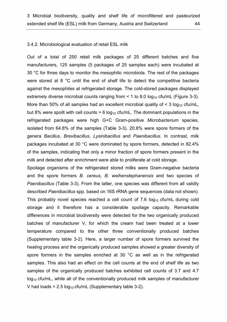
3 Microbial biodiversity, quality and shelf life of microfiltered and pasteurized extended shelf life (ESL) milk from Germany, Austria and Switzerland 44
3.4.2. Microbiological evaluation of retail ESL milk Out of a total of 250 retail milk packages of 25 different batches and five manufacturers, 125 samples (5 packages of 25 samples each) were incubated at 30 °C for three days to monitor the mesophilic microbiota. The rest of the packages were stored at 8 °C until the end of shelf life to detect the competitive bacteria against the mesophiles at refrigerated storage. The cold-stored packages displayed extremely diverse microbial counts ranging from < 1 to 8.0 log10 cfu/mL (Figure 3-3). More than 50% of all samples had an excellent microbial quality of < 3 log10 cfu/mL, but 8% were spoilt with cell counts > 6 log10 cfu/mL. The dominant populations in the refrigerated packages were high G+C Gram-positive Microbacterium species, isolated from 64.8% of the samples (Table 3-3). 20.8% were spore formers of the genera Bacillus, Brevibacillus, Lysinibacillus and Paenibacillus. In contrast, milk packages incubated at 30 °C were dominated by spore formers, detected in 82.4% of the samples, indicating that only a minor fraction of spore formers present in the milk and detected after enrichment were able to proliferate at cold storage. Spoilage organisms of the refrigerated stored milks were Gram-negative bacteria and the spore formers B. cereus, B. weihenstephanensis and two species of Paenibacillus (Table 3-3). From the latter, one species was different from all validly described Paenibacillus spp. based on 16S rRNA gene sequences (data not shown). This probably novel species reached a cell count of 7.6 log10 cfu/mL during cold storage and it therefore has a considerable spoilage capacity. Remarkable differences in microbial biodiversity were detected for the two organically produced batches of manufacturer V, for which the cream had been treated at a lower temperature compared to the other three conventionally produced batches (Supplementary table 3-2). Here, a larger number of spore formers survived the heating process and the organically produced samples showed a greater diversity of spore formers in the samples enriched at 30 °C as well as in the refrigerated samples. This also had an effect on the cell counts at the end of shelf life as two samples of the organically produced batches exhibited cell counts of 3.7 and 4.7 log10 cfu/mL, while all of the conventionally produced milk samples of manufacturer V had loads < 2.5 log10 cfu/mL (Supplementary table 3-2).
3 Microbial biodiversity, quality and shelf life of microfiltered and pasteurized extended shelf life (ESL) milk from Germany, Austria and Switzerland 45
Figure 3-3 Distribution of bacterial counts (log10 cfu/mL) of retail ESL milk stored at 8 °C at the end of shelf life Milk from five different manufacturers (M I – M V) were analyzed. M = manufacturer of retail ESL milk. 25 milk packages of each of the manufacturers were examined. Additionally, the results of the first study (section 3.1), which was performed with M I, have also been included. Here, 15 packages were analyzed yielding a total of 140 packages.

3 Microbial biodiversity, quality and shelf life of microfiltered and pasteurized extended shelf life (ESL) milk from Germany, Austria and Switzerland 46
Table 3-2 Incidence of different bacterial species in retail ESL milk after enrichment at 30 °C and after storage at 8 °C until the end of shelf life 125 packages were analysed at each temperature and values represent percentage of packages, from which the respective organisms have been isolated. n.d., not detected. 8 °C 30 °C High G+C Gram-positives 64.8% 45.6%
Microbacterium sp. 40.0% 25.6% Microbacterium lacticum 34.4% 25.6% Micrococcus luteus n.d. 0.8%
Spore formers 20.8% 82.4% Bacillus subtilis 8.00% 28.80% Bacillus licheniformis 5.60% 1.60% Bacillus cereus * § 4.80% 12.00% Bacillus pumilus 4.80% 48.00% Bacillus simplex 1.60% 0.80% Paenibacillus taichungensis/tylopili/tundrae 1.60% 0.80% Paenibacillus caespitis 1.60% 1.60% Paenibacillus odorifer § 1.60% 0.80% Bacillus circulans 0.80% 2.40% Bacillus idriensis 0.80% n.d. Paenibacillus borealis 0.80% 0.80% Paenibacillus sp. 0.80% 0.80% Paenibacillus sp. nov. § 1 0.80% 1.60% Bacillus flexus n.d. 0.80% Brevibacillus agri n.d. 8.00% Lysinibacillus fusiformis n.d. 3.20% Paenibacillus sp. nov. 2 n.d. 0.80% Paenibacillus turicensis n.d. 0.80%
Obligate aerobic Gram-negatives 8.0% 12.0% Acinetobacter junii/johnsonii* § 2.4% 0.8% Chryseobacterium haifense § 1.6% 2.4% Acinetobacter johnsonii* 0.8% 0.8% Moraxella osloensis* 0.8% 1.6% Sphingomonas sp. § 0.8% 0.8% Psychrobacter glacincola § n.d. 1.6%
Lactic acid bacteria 4.8% 21.6% Streptococcus mitis/oralis/parasanguinis * 0.8% 0.8% Streptococcus thermophilus 0.8% 19.2% Enterococcus casseliflavus * n.d. 0.8% Lactococcus lactis n.d. 0.8%
* Species classified in risk group II. § Species causing spoilage (≥ 6 log10 cfu/mL) at refrigerated storage (8 °C). 1 most closely related to Paenibacillus odorifer according to 16S rRNA gene sequence analysis. 2 most closely related to Paenibacillus borealis according to 16S rRNA gene sequence analysis.
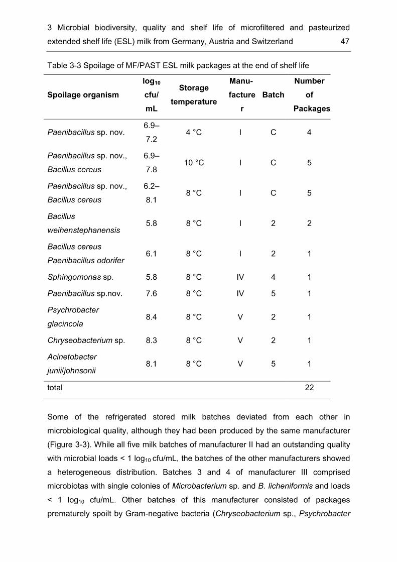
3 Microbial biodiversity, quality and shelf life of microfiltered and pasteurized extended shelf life (ESL) milk from Germany, Austria and Switzerland 47
Table 3-3 Spoilage of MF/PAST ESL milk packages at the end of shelf life
Spoilage organism log10 cfu/mL
Storage temperature
Manu-facture
r Batch
Number of
Packages
Paenibacillus sp. nov. 6.9–7.2 4 °C I C 4
Paenibacillus sp. nov., Bacillus cereus
6.9–7.8 10 °C I C 5
Paenibacillus sp. nov., Bacillus cereus
6.2–8.1 8 °C I C 5
Bacillus weihenstephanensis 5.8 8 °C I 2 2
Bacillus cereus Paenibacillus odorifer 6.1 8 °C I 2 1
Sphingomonas sp. 5.8 8 °C IV 4 1 Paenibacillus sp.nov. 7.6 8 °C IV 5 1 Psychrobacter glacincola 8.4 8 °C V 2 1
Chryseobacterium sp. 8.3 8 °C V 2 1 Acinetobacter junii/johnsonii 8.1 8 °C V 5 1
total 22 Some of the refrigerated stored milk batches deviated from each other in microbiological quality, although they had been produced by the same manufacturer (Figure 3-3). While all five milk batches of manufacturer II had an outstanding quality with microbial loads < 1 log10 cfu/mL, the batches of the other manufacturers showed a heterogeneous distribution. Batches 3 and 4 of manufacturer III comprised microbiotas with single colonies of Microbacterium sp. and B. licheniformis and loads < 1 log10 cfu/mL. Other batches of this manufacturer consisted of packages prematurely spoilt by Gram-negative bacteria (Chryseobacterium sp., Psychrobacter
3 Microbial biodiversity, quality and shelf life of microfiltered and pasteurized extended shelf life (ESL) milk from Germany, Austria and Switzerland 48
glacincola, Acinetobacter junii/johnsonii). Furthermore, in some cases even packages of the same milk batch differed from each other in microbiota and microbial load. While 36% of the batches exhibited similar loads for all five milk packages of one batch with counts differing less than one log10 order, other batches showed a great variance of up to 7 log units among the five packages. In one batch, counts ranged from close to the detection limit to 4.7 log10 cfu/mL and each of the five microbial profiles comprised different spore formers. Thus, as the cell counts of freshly produced MF/PAST milk showed only minor differences, the resulting milk quality at the end of shelf life appeared to be strongly dependent on the species composition in each package.
3.4.3. Prevalence of B. cereus and other risk group II bacteria 12.1% of the ESL milks stored at 8 °C comprised bacteria classified into risk group II according to German legislation for bacteria which pose a moderate individual risk and low community risk (IfSG, 2000). In this study Acinetobacter junii/johnsonii, A. johnsonii, Enterococcus casseliflavus, M. osloensis, Streptococcus mitis/oralis/parasanguinis, B. weihenstephanensis and B. cereus were recovered from milk of four manufacturers (Supplementary table 3-2). The 50 B. cereus isolates of this study could be divided into thirteen different types by hierarchical cluster analysis of FT-IR spectra (Figure 3-4). One representative of each type was then analyzed for the presence of the four different toxin genes hbl, nhe, cytk, and ces and psychrotolerant growth behaviour, coded by the cspA gene. Three closely related types (I-III) held the cspA gene and were therefore assigned to the psychrotolerant species B. weihenstephanensis. Concerning their toxin profiles, the thirteen B. cereus types could be differentiated into three groups (Table 3-4). No isolate analyzed was positive for the cereulide synthetase genes, but all harboured the nhe gene complex. Four types harboured only the nhe gene, six the nhe and hbl genes, and four the nhe, hbl and cytk genes. The psychrotolerant B. weihenstephanensis had counts of 3.4–6.0 log10 cfu/mL in all refrigerated packages of one batch, but for another two batches growth was only observed in the packages after enrichment at 30 °C, but not in the refrigerate stored packages of the same batch. Presumably, the initial level of contamination directly after production of the MF/PAST milk was so low, that these B. weihenstephanensis were not distributed to all packages during the
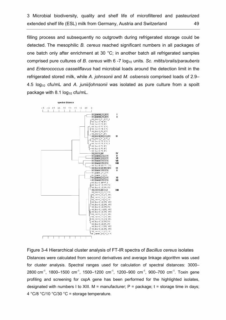
3 Microbial biodiversity, quality and shelf life of microfiltered and pasteurized extended shelf life (ESL) milk from Germany, Austria and Switzerland 49
filling process and subsequently no outgrowth during refrigerated storage could be detected. The mesophilic B. cereus reached significant numbers in all packages of one batch only after enrichment at 30 °C; in another batch all refrigerated samples comprised pure cultures of B. cereus with 6 -7 log10 units. Sc. mitits/oralis/parauberis and Enterococcus casseliflavus had microbial loads around the detection limit in the refrigerated stored milk, while A. johnsonii and M. osloensis comprised loads of 2.9–4.5 log10 cfu/mL and A. junii/johnsonii was isolated as pure culture from a spoilt package with 8.1 log10 cfu/mL.
Figure 3-4 Hierarchical cluster analysis of FT-IR spectra of Bacillus cereus isolates Distances were calculated from second derivatives and average linkage algorithm was used for cluster analysis. Spectral ranges used for calculation of spectral distances: 3000–2800 cm-1, 1800–1500 cm-1, 1500–1200 cm-1, 1200–900 cm-1, 900–700 cm-1. Toxin gene profiling and screening for cspA gene has been performed for the highlighted isolates, designated with numbers I to XIII. M = manufacturer; P = package; t = storage time in days; 4 °C/8 °C/10 °C/30 °C = storage temperature.
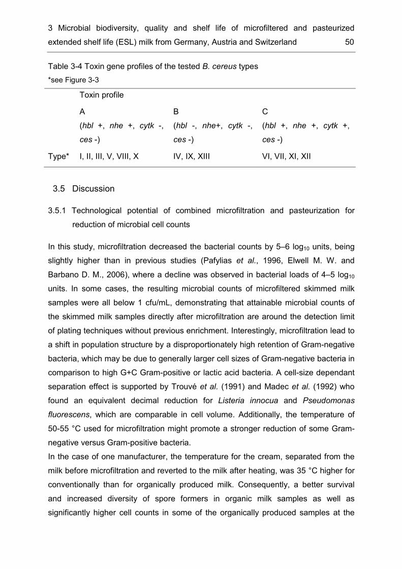
3 Microbial biodiversity, quality and shelf life of microfiltered and pasteurized extended shelf life (ESL) milk from Germany, Austria and Switzerland 50
Table 3-4 Toxin gene profiles of the tested B. cereus types *see Figure 3-3 Toxin profile
A (hbl +, nhe +, cytk -, ces -)
B (hbl -, nhe+, cytk -, ces -)
C (hbl +, nhe +, cytk +, ces -)
Type* I, II, III, V, VIII, X IV, IX, XIII VI, VII, XI, XII
Discussion 3.53.5.1 Technological potential of combined microfiltration and pasteurization for
reduction of microbial cell counts In this study, microfiltration decreased the bacterial counts by 5–6 log10 units, being slightly higher than in previous studies (Pafylias et al., 1996, Elwell M. W. and Barbano D. M., 2006), where a decline was observed in bacterial loads of 4–5 log10 units. In some cases, the resulting microbial counts of microfiltered skimmed milk samples were all below 1 cfu/mL, demonstrating that attainable microbial counts of the skimmed milk samples directly after microfiltration are around the detection limit of plating techniques without previous enrichment. Interestingly, microfiltration lead to a shift in population structure by a disproportionately high retention of Gram-negative bacteria, which may be due to generally larger cell sizes of Gram-negative bacteria in comparison to high G+C Gram-positive or lactic acid bacteria. A cell-size dependant separation effect is supported by Trouvé et al. (1991) and Madec et al. (1992) who found an equivalent decimal reduction for Listeria innocua and Pseudomonas fluorescens, which are comparable in cell volume. Additionally, the temperature of 50-55 °C used for microfiltration might promote a stronger reduction of some Gram-negative versus Gram-positive bacteria. In the case of one manufacturer, the temperature for the cream, separated from the milk before microfiltration and reverted to the milk after heating, was 35 °C higher for conventionally than for organically produced milk. Consequently, a better survival and increased diversity of spore formers in organic milk samples as well as significantly higher cell counts in some of the organically produced samples at the

3 Microbial biodiversity, quality and shelf life of microfiltered and pasteurized extended shelf life (ESL) milk from Germany, Austria and Switzerland 51
end of storage were observed. This corroborates the known fact that a less stringent cream heating step increases the survival of potential spoilage organisms in ESL milks. As demonstrated by our analyses of retail ESL milk, microfiltration combined with pasteurization can produce milk of high microbiological quality with a shelf life of several weeks. However, a substantial fraction of the retail ESL milk packages showed spoilage at the end of shelf life or did exhibit high cell counts indicating that shelf life is also influenced by a number of factors, which are discussed below.
3.5.2 Enzymatic spoilage Enzymatic spoilage occurred in some cases before the cell counts reached the microbiological spoilage level for packages dominated by Paenibacillus spp., B. cereus and Microbacterium spp. (Table 3-1). Samples with B. cereus exhibited enzymatic changes correlated with rancid and bitter off-flavours, a well-known problem in the milk industry (Billing & Cuthbert, 1958, Overcast W.W. and Atmaram K., 1974). Paenibacillus spp. have been recently reported for causing sensorial off-flavours (Martin et al., 2011). In addition, a number of samples with Microbacterium spp. exhibited elevated values for FFA and proteolysis, which is supported by a high proteolytic and lipolytic activity of milk-related Microbacterium spp. as observed by Hantsis-Zacharov & Halpern (2007). The interpretation of enzymatic evaluations is difficult, as not only enzymes of microorganisms are present, but also indigenous milk proteinases and lipases as well as the raw milk microbiota can contribute to the deterioration of lipids and proteins during storage (Nielsen, 2002, Kelly et al., 2006). Therefore, only results that include spoilt and non-spoilt samples of the same batch at the same time of analysis are a clear indication for live microbes being the causative agents of aberrant sensorial properties. In Table 3-1 these can be found in batch B after 16 and 22 days of storage, where most of the samples with Microbacterium spp. as the dominant microbiota had values of FFA and pH 4.6 soluble peptide fractions below the sensory thresholds, but samples of the same batch comprising Paenibacillus spp. or B. cereus exhibited elevated levels particularly for FAA even if the cell numbers were as low as 3.7 log10 cfu/mL. De Jonghe et al. (2010) have shown that these species are highly proteolytic and exhibit lecithinase activity, which may lead to the observed sensory defects even at low cell counts.
3 Microbial biodiversity, quality and shelf life of microfiltered and pasteurized extended shelf life (ESL) milk from Germany, Austria and Switzerland 52
3.5.3 Recontamination Freshly produced ESL milk samples in this study comprised bacterial cell counts between 0.3 and 1.9 log10 cfu/mL, which in case of batches B and C is an increase in microbial load of almost two log10 units compared to microfiltered milk. For spore forming bacteria, this could be due to heat-induced spore germination, but post process recontamination in milk processing plants has also been reported (Griffiths & Phillips, 1990, Svensson et al., 1999). Additionally, Fromm & Boor (2004) suggested recontamination for heat tolerant non-spore formers like Microbacterium spp.. Recontamination by psychrotolerant Gram-negative bacteria such as Acinetobacter junii/johnsonii, Sphingomonas sp. and Chryseobacterium sp., observed in ESL milk of two manufacturers (Table 3-3, Supplementary table 3-2), is also a major reason for premature spoilage of pasteurized milk and most probably occurred during the filling process when the filling nozzles could be contaminated with water (Schröder, 1984, Cromie, 1991, Ralyea et al., 1998, Gruetzmacher & Bradley, 1999). In cases where no aseptic filling machines are used, the risk of recontamination is high.
3.5.4 Population dominance As a consequence of the low microbial counts in freshly produced ESL milk, we suggest that the remaining bacteria were stochastically distributed over the retail packages during the filling process. This would explain why significantly different populations developed during cold storage among milk packages of the same batch. In some packages B. cereus, B. weihenstephanensis and Paenibacillus grew out to levels ≥ 6 log10 cfu/mL, developing into almost pure cultures. In case of rapidly growing species even temperature induced differences might be equalized as was observed for batch C (Figure 3-2), where psychrotolerant B. weihenstephanensis and Paenibacillus spp. gained predominance in the course of storage. The ability of Paenibacillus spp. to be highly competitive against the accompanying microbiota at refrigerated conditions has already been reported for pasteurized milk (Durak et al., 2006, Ranieri & Boor, 2009). The fact that microorganisms can grow to high numbers until the expiration date might be enhanced by a three times prolonged storage time of ESL milk in contrast to pasteurized milk. Furthermore, a reduced concomitant microbiota, which might otherwise function as competitors, might facilitate that a
3 Microbial biodiversity, quality and shelf life of microfiltered and pasteurized extended shelf life (ESL) milk from Germany, Austria and Switzerland 53
single species gains predominance. A previous study about the development of bacteria in pasteurized milk over cold storage concluded that only psychrotolerant B. weihenstephanensis, but not the mesophilic B. cereus, were able to outcompete and dominate the concomitant microbiota in pasteurized milk (unpublished data). In the present study, however, mesophilic B. cereus strains reached counts of 6 log10 cfu/mL after 14 days of storage at 8 °C, perhaps due to a lack of competing species in ESL milk. In contrast, some spore formers like B. pumilus and B. subtilis and Gram-positive non-spore forming bacteria were able to proliferate at 30 °C, but did not grow to significant numbers during cold storage and were insignificant for spoilage if the storage temperature did not exceed 8 °C (Table 3-2). Besides heat tolerant spore formers also Gram-negative recontaminants were able to grow into dominance as almost pure cultures in the course of refrigerated storage. Recontamination in very low numbers may be sufficient for spoilage as many Gram-negative species have advantageous growth conditions, like psychrotolerance with comparably short generation times at low temperatures which enable them to compete against the residual Gram-positive bacteria in ESL milk, like Microbacterium spp. (Schröder, 1984, Sørhaug & Stepaniak, 1997, He et al., 2009). In addition to their spoilage potential some strains like B. cereus or Acinetobacter junii/johnsonii are assigned to risk group II, which may be of concern if such microbes reach dominance in a milk product.
3.5.5 Suitability of combined microfiltration and pasteurization for the production of ESL milk
The shelf life of MF/PAST milk is strongly influenced by raw milk quality as well as processing and storage conditions. The results of the present studies demonstrate that the microbiological quality of MF/PAST milk at the end of shelf life can be excellent. The combination of microfiltration and pasteurization generally has a remarkable potential for producing high quality ESL milk. However, the results also indicate that the application of this technology may bear a higher risk for early spoilage of end products. Approximately one third of spoiled milk packages in the present study were due to recontamination by Gram-negative bacteria, which is a matter of production hygiene and not specifically related to the MF/PAST technology. The other two thirds of the samples, however, were spoiled by spore forming bacteria
3 Microbial biodiversity, quality and shelf life of microfiltered and pasteurized extended shelf life (ESL) milk from Germany, Austria and Switzerland 54
(B. cereus and Paenibacillus spp.), which cannot be completely eliminated using microfiltration and pasteurization. Although there has also been evidence for post-process recontamination of pasteurized milk by B. cereus (Lin et al., 1998, Svensson et al., 2000), some studies could demonstrate that strains from raw milk were transmitted into the finished product (Lin et al., 1998, Huck et al., 2007, Banykó & Vyletělová, 2009). Thus, in this regard there is a direct influence of raw milk quality on the shelf life of the end product, which can only be enhanced by improving raw milk quality. Accordingly, application of the MF/PAST technology requires high standards of production hygiene as well as an exceptionally good raw milk quality to ensure a safe and palatable end product. It should therefore be accompanied by an effective quality control including a well-designed hygiene management of production facilities as well as limits for the content of B. cereus and Paenibacillus spores in raw milk. Apparently, three of five manufacturers included in this study haven’t yet found an adequate solution for the problem, as their products exhibited extremely large variations in microbial quality and a substantial fraction of spoilt samples. To identify the significant factors associated to the large variations of milk quality between different packages and batches of the same manufacturer, more research is needed and deeper studies of the raw milk quality and processing conditions of single manufacturers would be helpful.
Acknowledgements 3.6This research project was supported by the German Ministry of Economics and Technology (via AiF) and the FEI (Forschungskreis der Ernährungsindustrie e.V., Bonn), Project AiF 15047 N. We would like to thank Monika Ehling-Schultz for providing the Bacillus cereus reference strains for toxin gene screening and Stephanie Düringer for her excellent technical assistance.
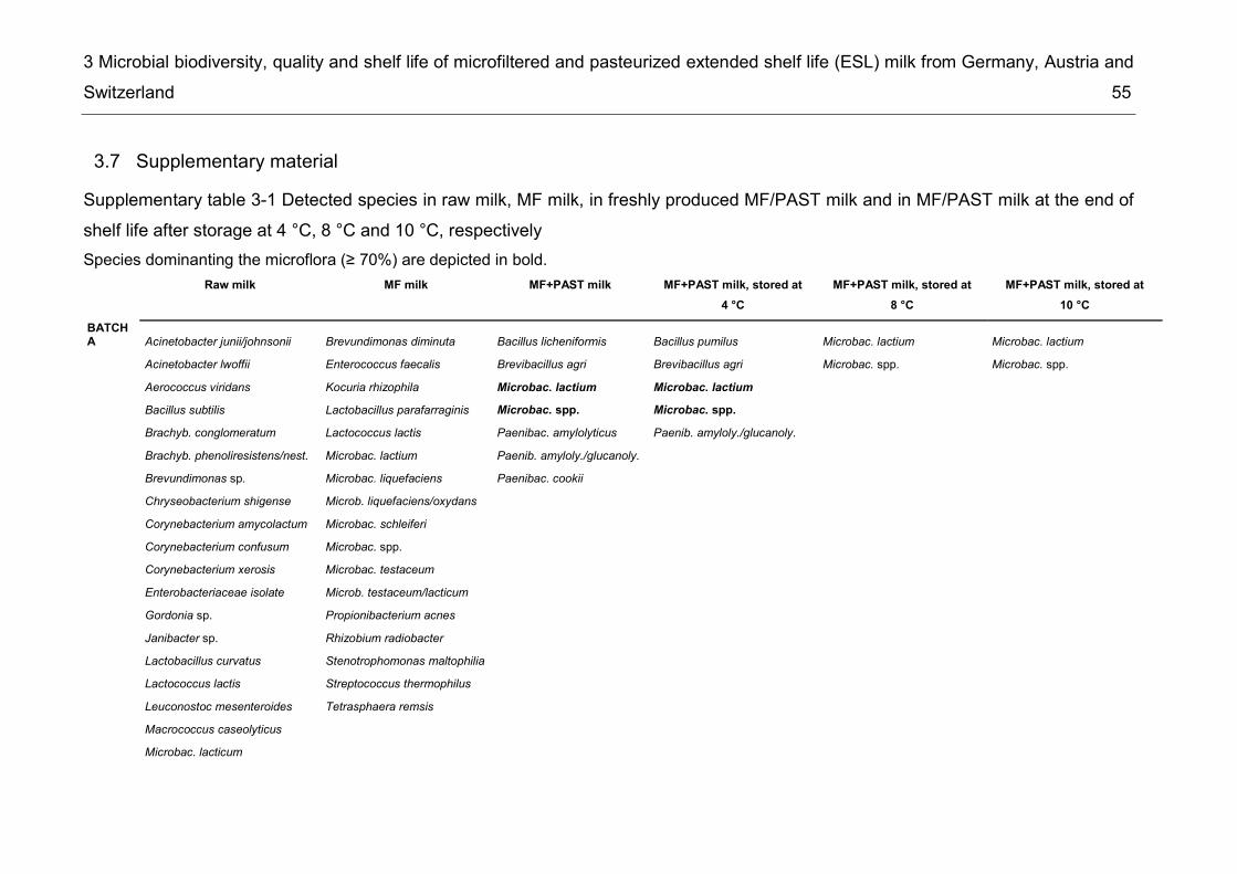
3 Microbial biodiversity, quality and shelf life of microfiltered and pasteurized extended shelf life (ESL) milk from Germany, Austria and Switzerland 55
Supplementary material 3.7Supplementary table 3-1 Detected species in raw milk, MF milk, in freshly produced MF/PAST milk and in MF/PAST milk at the end of shelf life after storage at 4 °C, 8 °C and 10 °C, respectively Species dominanting the microflora (≥ 70%) are depicted in bold.
Raw milk MF milk MF+PAST milk MF+PAST milk, stored at 4 °C
MF+PAST milk, stored at 8 °C
MF+PAST milk, stored at 10 °C
BATCH A Acinetobacter junii/johnsonii Brevundimonas diminuta Bacillus licheniformis Bacillus pumilus Microbac. lactium Microbac. lactium Acinetobacter lwoffii Enterococcus faecalis Brevibacillus agri Brevibacillus agri Microbac. spp. Microbac. spp. Aerococcus viridans Kocuria rhizophila Microbac. lactium Microbac. lactium Bacillus subtilis Lactobacillus parafarraginis Microbac. spp. Microbac. spp. Brachyb. conglomeratum Lactococcus lactis Paenibac. amylolyticus Paenib. amyloly./glucanoly. Brachyb. phenoliresistens/nest. Microbac. lactium Paenib. amyloly./glucanoly. Brevundimonas sp. Microbac. liquefaciens Paenibac. cookii Chryseobacterium shigense Microb. liquefaciens/oxydans Corynebacterium amycolactum Microbac. schleiferi Corynebacterium confusum Microbac. spp. Corynebacterium xerosis Microbac. testaceum Enterobacteriaceae isolate Microb. testaceum/lacticum Gordonia sp. Propionibacterium acnes Janibacter sp. Rhizobium radiobacter Lactobacillus curvatus Stenotrophomonas maltophilia Lactococcus lactis Streptococcus thermophilus Leuconostoc mesenteroides Tetrasphaera remsis Macrococcus caseolyticus Microbac. lacticum

3 Microbial biodiversity, quality and shelf life of microfiltered and pasteurized extended shelf life (ESL) milk from Germany, Austria and Switzerland 56
Raw milk MF milk MF+PAST milk MF+PAST milk, stored at
4 °C MF+PAST milk, stored at
8 °C MF+PAST milk, stored at
10 °C Pseudomonas sp. Rhodococcus sp. Serratia liquefaciens Streptococcus thermophilus Trichosporon laibachii Wautersiella falsenii BATCH B Acinetobacter junii/johnsonii Brevundimonas sp. Bacillus licheniformis Microbac. lactium Microbac. lactium Bacillus cereus Buttiauxella brennerae Brev. vesicularis/aurantiaca Bacillus subtilis Paenibac. amylolyticus Microbac. spp. Microbac. lactium Chryseobacterium sp. Chryseob. haif./Haloan. gall. Brachbacterium sp. Microbac. spp. Enterobacter sp. Chryseobacterium sp. Corynebacterium variabilis Paenibac. amylolyt. Lactobacillus fermentum Corynebacterium variabilis Microbac. lactium Lactococcus lactis Enterococcus faecalis Microbac. oxydans Pseudomonas alcaliphila Lactococcus lactis Microbac. sp. nov. Pseudomonas sp. Leifsonia/Microbac. Microbac. spp. Pseudomonas fluorescens Leuconostoc lactis Pseud. fluorescens/brenneri Microbac. lactium Pseudomonas fragi Microb. laevanif./flaves. Pseudomonas fragi/syringae Microbac. liquefaciens Pseudomonas jessenii Microbac. oxydans Serratia sp. Microbac. spp. Stenotrophomonas maltophila Microbac. testaceum Streptococcus parauberis Rhizobium radiobacter Sphingobacterium multivorum Staphylococcus epidermidis Stenotrophomonas maltophilia
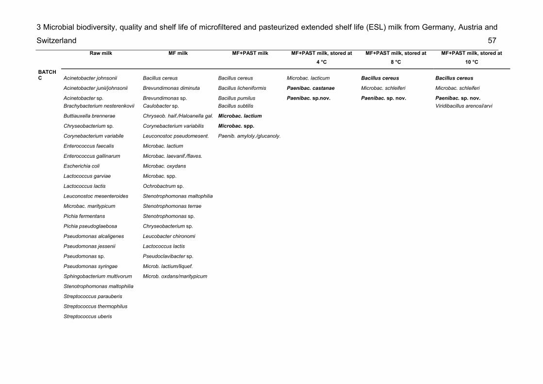
3 Microbial biodiversity, quality and shelf life of microfiltered and pasteurized extended shelf life (ESL) milk from Germany, Austria and Switzerland 57
Raw milk MF milk MF+PAST milk MF+PAST milk, stored at
4 °C MF+PAST milk, stored at
8 °C MF+PAST milk, stored at
10 °C BATCH C Acinetobacter johnsonii Bacillus cereus Bacillus cereus Microbac. lacticum Bacillus cereus Bacillus cereus Acinetobacter junii/johnsonii Brevundimonas diminuta Bacillus licheniformis Paenibac. castanae Microbac. schleiferi Microbac. schleiferi Acinetobacter sp. Brevundimonas sp. Bacillus pumilus Paenibac. sp.nov. Paenibac. sp. nov. Paenibac. sp. nov. Brachybacterium nesterenkovii Caulobacter sp. Bacillus subtilis Viridibacillus arenosi/arvi Buttiauxella brennerae Chryseob. haif./Haloanella gal. Microbac. lactium Chryseobacterium sp. Corynebacterium variabilis Microbac. spp. Corynebacterium variabile Leuconostoc pseudomesent. Paenib. amyloly./glucanoly. Enterococcus faecalis Microbac. lactium Enterococcus gallinarum Microbac. laevanif./flaves. Escherichia coli Microbac. oxydans Lactococcus garviae Microbac. spp. Lactococcus lactis Ochrobactrum sp. Leuconostoc mesenteroides Stenotrophomonas maltophilia Microbac. maritypicum Stenotrophomonas terrae Pichia fermentans Stenotrophomonas sp. Pichia pseudoglaebosa Chryseobacterium sp. Pseudomonas alcaligenes Leucobacter chironomi Pseudomonas jessenii Lactococcus lactis Pseudomonas sp. Pseudoclavibacter sp. Pseudomonas syringae Microb. lactium/liquef. Sphingobacterium multivorum Microb. oxdans/maritypicum Stenotrophomonas maltophilia Streptococcus parauberis Streptococcus thermophilus Streptococcus uberis
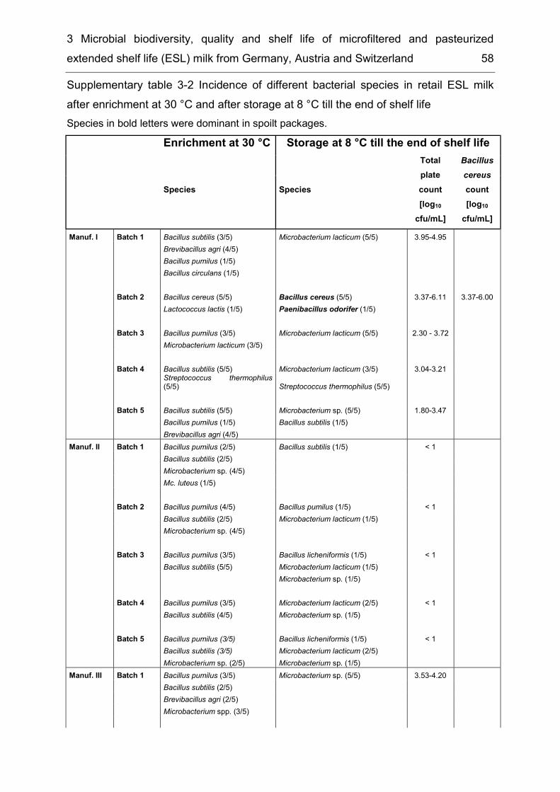
3 Microbial biodiversity, quality and shelf life of microfiltered and pasteurized extended shelf life (ESL) milk from Germany, Austria and Switzerland 58
Supplementary table 3-2 Incidence of different bacterial species in retail ESL milk after enrichment at 30 °C and after storage at 8 °C till the end of shelf life Species in bold letters were dominant in spoilt packages. Enrichment at 30 °C Storage at 8 °C till the end of shelf life
Species Species
Total plate count [log10
cfu/mL]
Bacillus cereus count [log10
cfu/mL] Manuf. I Batch 1 Bacillus subtilis (3/5) Microbacterium lacticum (5/5) 3.95-4.95 Brevibacillus agri (4/5) Bacillus pumilus (1/5) Bacillus circulans (1/5) Batch 2 Bacillus cereus (5/5) Bacillus cereus (5/5) 3.37-6.11 3.37-6.00 Lactococcus lactis (1/5) Paenibacillus odorifer (1/5) Batch 3 Bacillus pumilus (3/5) Microbacterium lacticum (5/5) 2.30 - 3.72 Microbacterium lacticum (3/5) Batch 4 Bacillus subtilis (5/5) Microbacterium lacticum (3/5) 3.04-3.21 Streptococcus thermophilus
(5/5) Streptococcus thermophilus (5/5) Batch 5 Bacillus subtilis (5/5) Microbacterium sp. (5/5) 1.80-3.47 Bacillus pumilus (1/5) Bacillus subtilis (1/5) Brevibacillus agri (4/5) Manuf. II Batch 1 Bacillus pumilus (2/5) Bacillus subtilis (1/5) < 1 Bacillus subtilis (2/5) Microbacterium sp. (4/5) Mc. luteus (1/5) Batch 2 Bacillus pumilus (4/5) Bacillus pumilus (1/5) < 1 Bacillus subtilis (2/5) Microbacterium lacticum (1/5) Microbacterium sp. (4/5) Batch 3 Bacillus pumilus (3/5) Bacillus licheniformis (1/5) < 1 Bacillus subtilis (5/5) Microbacterium lacticum (1/5) Microbacterium sp. (1/5) Batch 4 Bacillus pumilus (3/5) Microbacterium lacticum (2/5) < 1 Bacillus subtilis (4/5) Microbacterium sp. (1/5) Batch 5 Bacillus pumilus (3/5) Bacillus licheniformis (1/5) < 1 Bacillus subtilis (3/5) Microbacterium lacticum (2/5) Microbacterium sp. (2/5) Microbacterium sp. (1/5) Manuf. III Batch 1 Bacillus pumilus (3/5) Microbacterium sp. (5/5) 3.53-4.20 Bacillus subtilis (2/5) Brevibacillus agri (2/5) Microbacterium spp. (3/5)
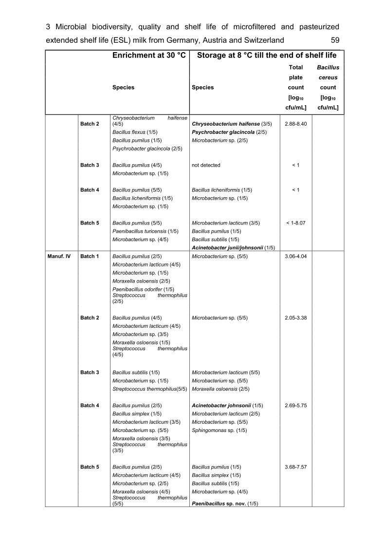
3 Microbial biodiversity, quality and shelf life of microfiltered and pasteurized extended shelf life (ESL) milk from Germany, Austria and Switzerland 59
Enrichment at 30 °C Storage at 8 °C till the end of shelf life
Species Species
Total plate count [log10
cfu/mL]
Bacillus cereus count [log10
cfu/mL] Batch 2 Chryseobacterium haifense
(4/5) Chryseobacterium haifense (3/5) 2.88-8.40 Bacillus flexus (1/5) Psychrobacter glacincola (2/5) Bacillus pumilus (1/5) Microbacterium sp. (2/5) Psychrobacter glacincola (2/5) Batch 3 Bacillus pumilus (4/5) not detected < 1 Microbacterium sp. (1/5) Batch 4 Bacillus pumilus (5/5) Bacillus licheniformis (1/5) < 1 Bacillus licheniformis (1/5) Microbacterium sp. (1/5) Microbacterium sp. (1/5) Batch 5 Bacillus pumilus (5/5) Microbacterium lacticum (3/5) < 1-8.07 Paenibacillus turicensis (1/5) Bacillus pumilus (1/5) Microbacterium sp. (4/5) Bacillus subtilis (1/5) Acinetobacter junii/johnsonii (1/5) Manuf. IV Batch 1 Bacillus pumilus (2/5) Microbacterium sp. (5/5) 3.06-4.04 Microbacterium lacticum (4/5) Microbacterium sp. (1/5) Moraxella osloensis (2/5) Paenibacillus odorifer (1/5) Streptococcus thermophilus
(2/5) Batch 2 Bacillus pumilus (4/5) Microbacterium sp. (5/5) 2.05-3.38 Microbacterium lacticum (4/5) Microbacterium sp. (3/5) Moraxella osloensis (1/5) Streptococcus thermophilus
(4/5) Batch 3 Bacillus subtilis (1/5) Microbacterium lacticum (5/5) Microbacterium sp. (1/5) Microbacterium sp. (5/5) Streptococcus thermophilus(5/5) Moraxella osloensis (2/5) Batch 4 Bacillus pumilus (2/5) Acinetobacter johnsonii (1/5) 2.69-5.75 Bacillus simplex (1/5) Microbacterium lacticum (2/5) Microbacterium lacticum (3/5) Microbacterium sp. (5/5) Microbacterium sp. (5/5) Sphingomonas sp. (1/5) Moraxella osloensis (3/5) Streptococcus thermophilus
(3/5) Batch 5 Bacillus pumilus (2/5) Bacillus pumilus (1/5) 3.68-7.57 Microbacterium lacticum (4/5) Bacillus simplex (1/5) Microbacterium sp. (2/5) Bacillus subtilis (1/5) Moraxella osloensis (4/5) Microbacterium sp. (4/5) Streptococcus thermophilus
(5/5) Paenibacillus sp. nov. (1/5)
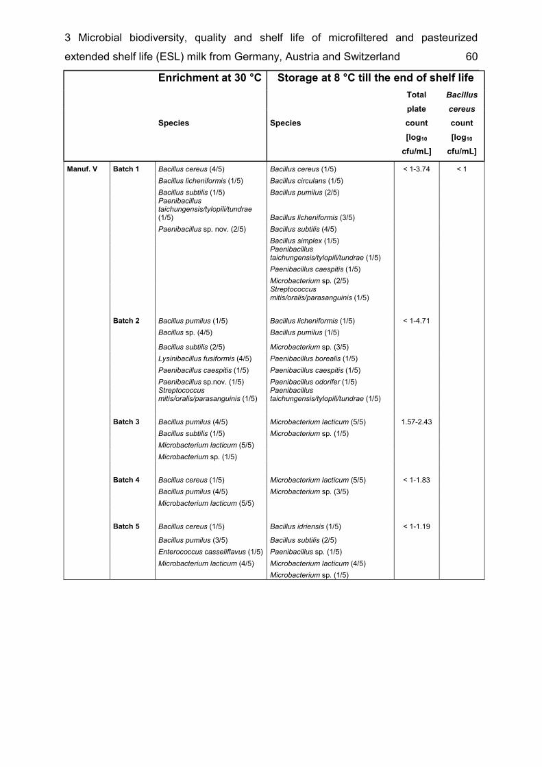
3 Microbial biodiversity, quality and shelf life of microfiltered and pasteurized extended shelf life (ESL) milk from Germany, Austria and Switzerland 60
Enrichment at 30 °C Storage at 8 °C till the end of shelf life
Species Species
Total plate count [log10
cfu/mL]
Bacillus cereus count [log10
cfu/mL] Manuf. V Batch 1 Bacillus cereus (4/5) Bacillus cereus (1/5) < 1-3.74 < 1 Bacillus licheniformis (1/5) Bacillus circulans (1/5) Bacillus subtilis (1/5) Bacillus pumilus (2/5)
Paenibacillus taichungensis/tylopili/tundrae (1/5) Bacillus licheniformis (3/5)
Paenibacillus sp. nov. (2/5) Bacillus subtilis (4/5) Bacillus simplex (1/5)
Paenibacillus taichungensis/tylopili/tundrae (1/5)
Paenibacillus caespitis (1/5) Microbacterium sp. (2/5)
Streptococcus mitis/oralis/parasanguinis (1/5)
Batch 2 Bacillus pumilus (1/5) Bacillus licheniformis (1/5) < 1-4.71 Bacillus sp. (4/5) Bacillus pumilus (1/5) Bacillus subtilis (2/5) Microbacterium sp. (3/5) Lysinibacillus fusiformis (4/5) Paenibacillus borealis (1/5) Paenibacillus caespitis (1/5) Paenibacillus caespitis (1/5) Paenibacillus sp.nov. (1/5) Paenibacillus odorifer (1/5) Streptococcus mitis/oralis/parasanguinis (1/5) Paenibacillus taichungensis/tylopili/tundrae (1/5) Batch 3 Bacillus pumilus (4/5) Microbacterium lacticum (5/5) 1.57-2.43 Bacillus subtilis (1/5) Microbacterium sp. (1/5) Microbacterium lacticum (5/5) Microbacterium sp. (1/5) Batch 4 Bacillus cereus (1/5) Microbacterium lacticum (5/5) < 1-1.83 Bacillus pumilus (4/5) Microbacterium sp. (3/5) Microbacterium lacticum (5/5) Batch 5 Bacillus cereus (1/5) Bacillus idriensis (1/5) < 1-1.19 Bacillus pumilus (3/5) Bacillus subtilis (2/5) Enterococcus casseliflavus (1/5) Paenibacillus sp. (1/5) Microbacterium lacticum (4/5) Microbacterium lacticum (4/5) Microbacterium sp. (1/5)
4 Bavariicoccus seileri gen. nov., sp. nov., isolated from the surface and smear water of German red smear soft cheese 61
4 Bavariicoccus seileri gen. nov., sp. nov., isolated from the surface and smear water of German red smear soft cheese
Verena S.J. Schmidt1, Ralf Mayr1, Mareike Wenning1, Jana Glöckner3§, Hans-Jürgen Busse3 and Siegfried Scherer1, 2, * 1Abteilung für Mikrobiologie, ZIEL, Technische Universität München, D-85354 Freising, Germany 2Lehrstuhl für Mikrobielle Ökologie, Department für Grundlagen der Biowissenschaften, WZW, Technische Universität München, D-85350 Freising, Germany 3 Institut für Bakteriologie, Mykologie und Hygiene, Veterinärmedizinische Universität, A-1210 Wien, Austria § Present Address: Max Planck Institute for Terrestrial Microbiology, Karl-von-Frisch-Straße, D-35043 Marburg, Germany *Correspondence [email protected] Published as: Schmidt VSJ, Mayr R, Wenning M, Glockner J, Busse HJ & Scherer S (2009) Bavariicoccus seileri gen. nov., sp. nov., isolated from the surface and smear water of German red smear soft cheese. International Journal of Systematic and Evolutionary Microbiology 59: 2437-2443.

4 Bavariicoccus seileri gen. nov., sp. nov., isolated from the surface and smear water of German red smear soft cheese 62
Abstract 4.1The phylogenetic position and physiological characters of six hitherto unknown lactic acid bacterial isolates are reported, which form part of the surface microbiota of German red smear soft cheese. The coccoid cells are aerotolerant, Gram-positive, catalase-negative and non-motile. The cell-wall peptidoglycan contains alanine, glutamic acid, lysine and aspartic acid and is of A4α type (L-Lys ← D-Asp). 16S ribosomal DNA, groEL and rpoB sequences of the six isolates are identical and revealed that these Firmicutes isolates represent an independent lineage within the radiation of the Enterococcaceae. Their closest phylogenetic neighbour is the lactic acid bacterium Atopobacter phocae with which they share 94.9% 16S rRNA gene sequence similarity; representatives of other genera such as Granulicatella, Carnobacterium and Trichococcus are more distantly related. DNA-DNA hybridization studies revealed that the six isolates are members of a single species which was confirmed by similarities in biochemical characteristics. The affiliation of the six isolates to four different strains was supported by both FT-IR typing and RAPD typing. Therefore, it is formally proposed that these isolates should be classified in a single new species of a novel genus and be named Bavariicoccus seileri gen. nov., sp. nov.. The type strain is Bavariicoccus seileri WCC 4188T (=DSMZ 19936T =CCUG 55508T).
Introduction 4.2Red smear cheeses are mainly produced in Austria, Germany, France, Scandinavia and Switzerland. For the ripening process, the microbial consortia of mature cheeses are washed off the surfaces with a brine solution which is then used for inoculation of freshly produced cheeses. By virtue of the traditional manufacturing process, a variety of different smear cheeses exist which often comprise quite complex, species-rich microbial consortia (Maoz et al., 2003, Brennan et al., 2004, Feurer et al., 2004, Mounier et al., 2005, Wenning et al., 2006). Some recently described species have been isolated from these consortia, such as Agrococcus casei (Bora et al., 2007), Arthrobacter bergerei and Arthrobacter arilaitensis (Irlinger et al., 2005), Brevibacterium aurantiacum (Gavrish et al., 2004), Corynebacterium casei (Brennan
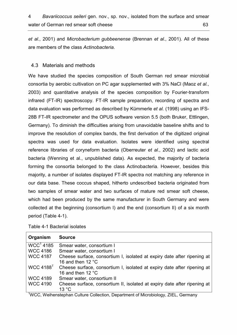
4 Bavariicoccus seileri gen. nov., sp. nov., isolated from the surface and smear water of German red smear soft cheese 63
et al., 2001) and Microbacterium gubbeenense (Brennan et al., 2001). All of these are members of the class Actinobacteria.
Materials and methods 4.3We have studied the species composition of South German red smear microbial consortia by aerobic cultivation on PC agar supplemented with 3% NaCl (Maoz et al., 2003) and quantitative analysis of the species composition by Fourier-transform infrared (FT-IR) spectroscopy. FT-IR sample preparation, recording of spectra and data evaluation was performed as described by Kümmerle et al. (1998) using an IFS-28B FT-IR spectrometer and the OPUS software version 5.5 (both Bruker, Ettlingen, Germany). To diminish the difficulties arising from unavoidable baseline shifts and to improve the resolution of complex bands, the first derivation of the digitized original spectra was used for data evaluation. Isolates were identified using spectral reference libraries of coryneform bacteria (Oberreuter et al., 2002) and lactic acid bacteria (Wenning et al., unpublished data). As expected, the majority of bacteria forming the consortia belonged to the class Actinobacteria. However, besides this majority, a number of isolates displayed FT-IR spectra not matching any reference in our data base. These coccus shaped, hitherto undescribed bacteria originated from two samples of smear water and two surfaces of mature red smear soft cheese, which had been produced by the same manufacturer in South Germany and were collected at the beginning (consortium I) and the end (consortium II) of a six month period (Table 4-1). Table 4-1 Bacterial isolates Organism Source WCC1 4185 Smear water, consortium I WCC 4186 Smear water, consortium I WCC 4187 Cheese surface, consortium I, isolated at expiry date after ripening at
16 and then 12 °C WCC 4188T Cheese surface, consortium I, isolated at expiry date after ripening at
16 and then 12 °C WCC 4189 Smear water, consortium II WCC 4190 Cheese surface, consortium II, isolated at expiry date after ripening at
13 °C 1WCC, Weihenstephan Culture Collection, Department of Microbiology, ZIEL, Germany

4 Bavariicoccus seileri gen. nov., sp. nov., isolated from the surface and smear water of German red smear soft cheese 64
The new bacteria were found in considerable numbers in their cheese habitats, constituting 14% and 8%, respectively, of the total cell counts in the smear water samples relating to consortium I and II. 9% and 1% of the cultivable cells of the cheese surface consortium I and II, respectively, belonged to the new species. All isolates grew well on common commercial media for lactic acid bacteria like APT (Merck) and TSA (Oxoid, Wesel, Germany), but to archive fastest growth, organisms were cultivated in M17 broth (Merck) with 2% glucose without shaking, which was the medium used for phenotypic tests. The ability to grow at anaerobic conditions was determined in an anaerobic jar containing the anaerobic catalyst Anaerocult A (Merck), prepared according to the manufacturer’s instructions. No catalase-activity was detected when cells were treated with 3% hydrogen peroxide. Morphological examination of the Gram behaviour was positive after staining (Gregersen, 1978). Non-spore forming cocci were identified in phase-contrast microscopy with a cell size of 0.9-1.2μm after anaerobic cultivation on APT agar. The effects of pH (pH 5.0; pH 5.5 and pH 6-10 at intervals of 0.5 pH units) and salt tolerance (6.5 and 7-13%, w/v at intervals of 1%) were assessed at 30 °C. The temperature range for growth was determined using increments of 1 °C units, from 5-10 °C and 37-41 °C. Growth within 14 days was indicated by visible turbidity. Additionally, all isolates were biochemically characterized by using the API 50CH system with the API 50 CHB/L inoculation medium, API rapid ID 32 Strep and API 20 Strep (all bioMérieux, Nürtingen, Germany). Results are listed in the species description. The phylogenetic position of the six isolates was determined by 16S rDNA, groEL and rpoB gene sequence analyses. The complete 16S rDNA genes were amplified by PCR (Büchl et al., 2008), using universal rDNA binding-primers and cycling conditions specified by Oberreuter et al. (2002). 16S rDNA was purified (QIAquick®
PCR Purification Kit, Qiagen, Hilden, Germany) and cycle sequencing PCR of 16S rDNA was performed by Sequiserve (Vaterstetten, Germany). Amplification of groEL gene sequences was performed as described previously (Goh et al., 1996) with the following modifications. The thermal cycling conditions were 5 min at 95 °C for the first cycle, followed by 35 cycles of 20 sec at 95 °C, 40 sec at 55 °C, and 1 min at 72 °C. The last cycle lasted for 7 min at 72 °C. Sequencing of 600 bp PCR products was done by 4base lab (Reutlingen, Germany). Primer sequences for amplification of

4 Bavariicoccus seileri gen. nov., sp. nov., isolated from the surface and smear water of German red smear soft cheese 65
rpoB sequences were obtained from Drancourt et al. (2004), cycling conditions were the same as described for groEL sequences, but annealing temperatures were lowered to 40 °C. Sequencing of 700-bp PCR products was performed by GATC (Konstanz, Germany). In addition to the six isolates, sequencing of 16S rDNA, groEL and rpoB genes of the closest known relatives was either performed as described above or, if available, sequences were retrieved from database researches (NCBI, http://www.ncbi.nlm.nih.gov/blast/Blast). The sequences obtained were aligned with CLUSTAL_X (1.8) (Thompson et al., 1997). The distance matrices of the resulting multiple sequence alignments were calculated using Treecon (Van de Peer & De Wachter, 1997). Rooted phylogenetic trees with Staphylococcus aureus as an outgroup were constructed according to the neighbour-joining method. Stability of clusters was tested by regenerating trees using UPGMA (data not shown). 16S rDNA (Figure 4-1), groEL (Figure 4-2) and rpoB sequences (data not shown) of the six isolates were identical and revealed a distinct lineage with respect to other established taxa. The 16S rDNA similarity values of the new species compared to the closest neighbours Atopobacter phocae and Granulicatella species were 94.9% and 94.3–94.9% respectively, which would support their affiliation to a novel genus (Ludwig et al., 1998). GroEL gene sequence analyses showed similarities of 81.3% and 78.7–80.2% to A. phocae and Granulicatella species respectively. The new bacteria shared rpoB gene sequence similarity of 75.3% with A. phocae and 76.7-80.3% with Granulicatella species. Bavariicoccus seileri and the phylogenetically closely related genera Atopobacter, Carnobacterium, Enterococcus, Trichococcus and Vagococcus form different groups based on FT-IR spectra (Fig S1 supplementary data, IJSEM Online). Granulicatella and Abiotrophia were not included in this examination as recording of spectra at aerobic conditions on TS agar could not be performed due to the fastidious growth of both genera. FT-IR spectra, therefore, reveal a phenotype clearly differentiating Bavariicoccus from the most closely related genera. The G+C content (mol%) of genomic DNA was examined by the Identification Service of the Deutsche Sammlung von Mikroorganismen und Zellkulturen (DSMZ) GmbH (Braunschweig, Germany) using HPLC (Mesbah et al., 1989) for isolates WCC 4186 (39 mol%), WCC 4188T (38 mol%) and WCC 4189 (38 mol%).
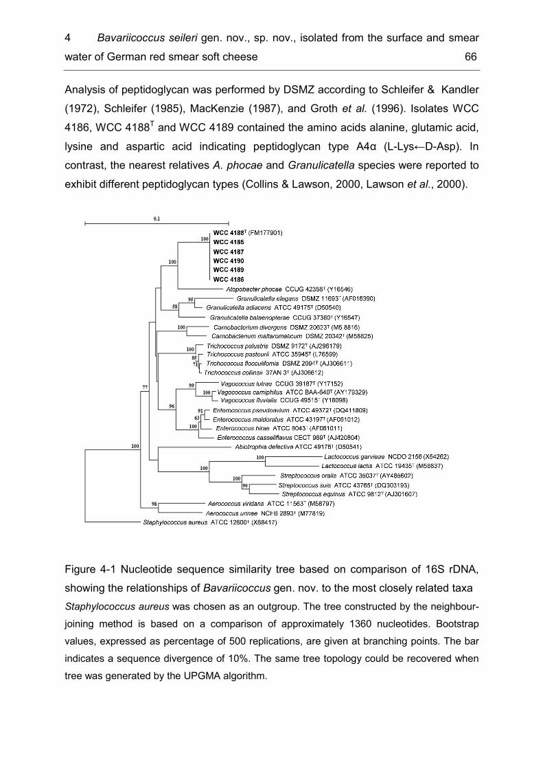
4 Bavariicoccus seileri gen. nov., sp. nov., isolated from the surface and smear water of German red smear soft cheese 66
Analysis of peptidoglycan was performed by DSMZ according to Schleifer & Kandler (1972), Schleifer (1985), MacKenzie (1987), and Groth et al. (1996). Isolates WCC 4186, WCC 4188T and WCC 4189 contained the amino acids alanine, glutamic acid, lysine and aspartic acid indicating peptidoglycan type A4α (L-Lys←D-Asp). In contrast, the nearest relatives A. phocae and Granulicatella species were reported to exhibit different peptidoglycan types (Collins & Lawson, 2000, Lawson et al., 2000).
Figure 4-1 Nucleotide sequence similarity tree based on comparison of 16S rDNA, showing the relationships of Bavariicoccus gen. nov. to the most closely related taxa Staphylococcus aureus was chosen as an outgroup. The tree constructed by the neighbour-joining method is based on a comparison of approximately 1360 nucleotides. Bootstrap values, expressed as percentage of 500 replications, are given at branching points. The bar indicates a sequence divergence of 10%. The same tree topology could be recovered when tree was generated by the UPGMA algorithm.
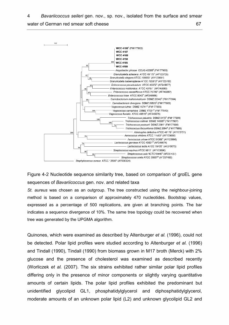
4 Bavariicoccus seileri gen. nov., sp. nov., isolated from the surface and smear water of German red smear soft cheese 67
Figure 4-2 Nucleotide sequence similarity tree, based on comparison of groEL gene sequences of Bavariicoccus gen. nov. and related taxa St. aureus was chosen as an outgroup. The tree constructed using the neighbour-joining method is based on a comparison of approximately 470 nucleotides. Bootstrap values, expressed as a percentage of 500 replications, are given at branching points. The bar indicates a sequence divergence of 10%. The same tree topology could be recovered when tree was generated by the UPGMA algorithm. Quinones, which were examined as described by Altenburger et al. (1996), could not be detected. Polar lipid profiles were studied according to Altenburger et al. (1996) and Tindall (1990), Tindall (1990) from biomass grown in M17 broth (Merck) with 2% glucose and the presence of cholesterol was examined as described recently (Worliczek et al. (2007). The six strains exhibited rather similar polar lipid profiles differing only in the presence of minor components or slightly varying quantitative amounts of certain lipids. The polar lipid profiles exhibited the predominant but unidentified glycolipid GL1, phosphatidylglycerol and diphosphatidylglycerol, moderate amounts of an unknown polar lipid (L2) and unknown glycolipid GL2 and
4 Bavariicoccus seileri gen. nov., sp. nov., isolated from the surface and smear water of German red smear soft cheese 68
minor amounts of phosphatidylethanolamine and of several unknown aminolipids, phospholipids and polar lipids (Fig 4-3a). The nearest relative of Bavariicoccus, A. phocae, exhibited significant amounts of cholesterol but no unknown aminolipids (Fig. 4-3b). Fatty acids were extracted and analyzed by the DSMZ service as described recently (Verbarg et al., 2008). Fatty acid profiles consisted almost exclusively of unbranched saturated and unsaturated acids. Major compounds were C16:0 (16-30%) and C18:1 ω9c (35-73%); C18:0 (4-22%) was detected in moderate to minor amounts. Other fatty acids detected are listed in Supplementary table 4-2. Taken together, 16S rDNA, groEL and rpoB gene sequence analyses revealed that these Firmicutes isolates represent an independent lineage within the radiation of the Enterococcaceae. The six coccoid isolates can be clearly distinguished from the rod shaped monospecific genus Atopobacter and from the genera Granulicatella and Trichococcus based on phenotypic characteristics such as the peptidoglycan type, a different polar lipid profile (Supplementary figure 4-1) and FT-IR spectra. Hence, we suggest a novel genus and species for which we propose the name Bavariicoccus seileri gen. nov., sp. nov..

4 Bavariicoccus seileri gen. nov., sp. nov., isolated from the surface and smear water of German red smear soft cheese 69
Fig. 4-3a Fig. 4-3b
1st dimension → Figure 4-3 Polar lipid profiles of WCC 4188T (a) and A. phocae CIP 106292T (b) after two-dimensional thin layer chromatography and detection with molybdatophosphoric acid Abbreviations: Chol, cholesterol, PE, phosphatidylethanolamine; PG, phosphatidylglycerol; DPG; diphosphatidylglycerol; PL1, PL2, PL3, PL4, unknown phospholipids; AL1, 2, unknown aminolipids; L1, L2, L2, L3, L4, L5, L6, L7, unknown polar lipid; GL1, 2, 3, 4, 5, unknown glycolipids.
2nd dimens
ion →
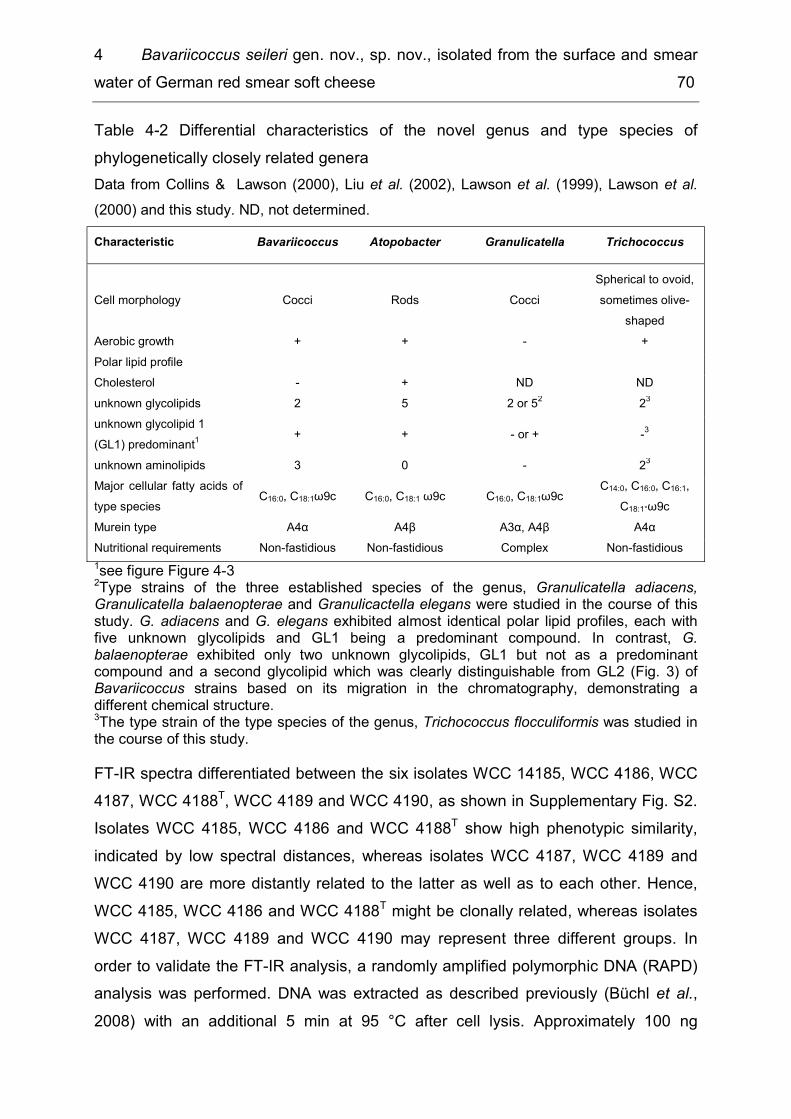
4 Bavariicoccus seileri gen. nov., sp. nov., isolated from the surface and smear water of German red smear soft cheese 70
Table 4-2 Differential characteristics of the novel genus and type species of phylogenetically closely related genera Data from Collins & Lawson (2000), Liu et al. (2002), Lawson et al. (1999), Lawson et al. (2000) and this study. ND, not determined. Characteristic Bavariicoccus Atopobacter Granulicatella Trichococcus
Cell morphology Cocci Rods Cocci Spherical to ovoid, sometimes olive-
shaped Aerobic growth + + - + Polar lipid profile Cholesterol - + ND ND unknown glycolipids 2 5 2 or 52 23 unknown glycolipid 1 (GL1) predominant1 + + - or + -3 unknown aminolipids 3 0 - 23 Major cellular fatty acids of type species C16:0, C18:1ω9c C16:0, C18:1 ω9c C16:0, C18:1ω9c C14:0, C16:0, C16:1,
C18:1°ω9c Murein type A4α A4β A3α, A4β A4α Nutritional requirements Non-fastidious Non-fastidious Complex Non-fastidious 1see figure Figure 4-3 2Type strains of the three established species of the genus, Granulicatella adiacens, Granulicatella balaenopterae and Granulicactella elegans were studied in the course of this study. G. adiacens and G. elegans exhibited almost identical polar lipid profiles, each with five unknown glycolipids and GL1 being a predominant compound. In contrast, G. balaenopterae exhibited only two unknown glycolipids, GL1 but not as a predominant compound and a second glycolipid which was clearly distinguishable from GL2 (Fig. 3) of Bavariicoccus strains based on its migration in the chromatography, demonstrating a different chemical structure. 3The type strain of the type species of the genus, Trichococcus flocculiformis was studied in the course of this study. FT-IR spectra differentiated between the six isolates WCC 14185, WCC 4186, WCC 4187, WCC 4188T, WCC 4189 and WCC 4190, as shown in Supplementary Fig. S2. Isolates WCC 4185, WCC 4186 and WCC 4188T show high phenotypic similarity, indicated by low spectral distances, whereas isolates WCC 4187, WCC 4189 and WCC 4190 are more distantly related to the latter as well as to each other. Hence, WCC 4185, WCC 4186 and WCC 4188T might be clonally related, whereas isolates WCC 4187, WCC 4189 and WCC 4190 may represent three different groups. In order to validate the FT-IR analysis, a randomly amplified polymorphic DNA (RAPD) analysis was performed. DNA was extracted as described previously (Büchl et al., 2008) with an additional 5 min at 95 °C after cell lysis. Approximately 100 ng
4 Bavariicoccus seileri gen. nov., sp. nov., isolated from the surface and smear water of German red smear soft cheese 71
extracted DNA was subjected to PCR amplification in a total volume of 50 μl following the protocol described by (Büchl et al., 2008). Thermal cycling was performed on a T3000 Thermocycler (Biometra) with the following conditions: 6 min at 95 °C; 35 cycles of 1 min at 94 °C, 1 min at 38 °C and 2 min at 72 °C; and 10 min at 72 °C. Amplification products (6 μl) were separated on a 1.5 % agarose gel in TBE buffer and the gels were stained with ethidium bromide, visualized with a UV lamp and photographed with ImageMaster VDS-CL (Amersham Biosciences). RAPD analysis using the M13 primer (5′-GAGGGTGGCGGTTCT-3′) (Gräser et al., 1993) revealed the same four groups as FT-IR spectroscopy (Supplementary Fig. S2). Therefore, the applicability of FT-IR spectroscopy for typing these lactic acid bacteria below the species level is demonstrated by our data, as has already been done for coryneform bacteria by Goerges et al. (2008). In order to see whether these four groups belong to the same species, the six isolates were studied with DNA–DNA hybridization analyses based on renaturation curves, performed by the Identification Service of the DSMZ. DNA was isolated using a French pressure cell (Thermo Spectronic) and purified by chromatography on hydroxyapatite as described by Cashion et al. (1977). Hybridizations were carried out as described by De Ley et al. (1970) with the modifications described by Huss et al. (1983) using a Cary 100 Bio UV-Vis spectrophotometer equipped with a Peltier-thermostatted 6×6 multicell changer and temperature controller with in-situ temperature probe (Varian). Hybridizations between isolates WCC 4187, WCC 4189 and WCC 4190 revealed reassociation values between 74 and 96 % (Supplementary table 4-2). Pairing of WCC 4188T with WCC 4187 and WCC 4190 revealed reassociation values between 95 and 98 %. All of these values are above the 70 % cut-off point for species delineation recommended by Wayne et al. (1987). However, DNA–DNA hybridization between WCC 4188T and WCC 4189 revealed only 61 % relatedness (mean), a value that falls below the threshold of relatedness at the species level. Four independent DNA–DNA hybridization experiments between WCC 4188T and WCC 4189, with two independent DNA preparations, resulted in hybridization values within the range of 54–64 %. Yet, since all hybridization results of these two isolates with WCC 4187 and WCC 4190 were clearly above the threshold value of 70%, we do not conclude that WCC 4188T and WCC 4189 should be assigned to separate species. This conclusion

4 Bavariicoccus seileri gen. nov., sp. nov., isolated from the surface and smear water of German red smear soft cheese 72
is supported by identical 16S rRNA, groEL and rpoB gene sequences and almost-identical physiological and biochemical characteristics. Hence, we suggest that the six strains represent a novel genus and species, for which the name Bavariicoccus seileri gen. nov., sp. nov. is proposed.
Description of Bavariicoccus gen. nov. 4.4Bavariicoccus (Ba.va.ri.i.coc′cus. L. fem. Bavaria Bavaria, Germany; N.L. masc. n. coccus coccus from Gr. masc. n. kokkos berry; N.L. masc. n. Bavariicoccus a coccoid-shaped bacterium isolated in Bavaria). Cells stain Gram-positive and are non-spore forming, aerotolerant and catalase-negative. Polar lipid profiles are dominated by an unknown glycolipid and moderate amounts of diphosphatidylglycerol and phosphatidylglycerol. Cholesterol is absent. Fatty acid profiles are predominantly composed of unbranched saturated and unsaturated fatty acids (> 90 %). Major fatty acids are C 16 :0 and C 18 : 1 ω9 c. Quinones are not detectable. The cell-wall peptidoglycan contains the amino acids alanine, glutamic acid, lysine and aspartic acid (A4 α type). The DNA G+C content of the type species is 38–39 mol%. The type species is Bavariicoccus seileri.
Description of Bavariicoccus seileri sp. nov. 4.5Bavariicoccus seileri (sei′le.ri. N.L. gen. masc. n. seileri named in honour of Herbert Seiler, former microbiologist of the Technical University of Munich with great merit in FT-IR spectroscopic identification of micro-organisms). The characteristics are the same as those given in the description of the genus with the following additions. Small, smooth colonies up to 1 mm in diameter are formed aerobically on TSA at 30 °C and up to 2 mm in diameter on APT agar under anaerobic conditions at 34 °C. Cell diameter is 0.9–1.2 μm. Growth occurs at 10 and 40 °C, at pH 5.5 and with 11 % (w/v) NaCl. Produces dl-lactic acid, ethanol and acetic acid from d-glucose. Utilizes galactose, d-glucose, d-fructose, amygdalin, arbutin, salicin, cellobiose, maltose, β-gentiobiose, trehalose, lactose (except isolates WCC 4189 and WCC 4190) and pyruvate as the sole sources of carbon and energy, but

4 Bavariicoccus seileri gen. nov., sp. nov., isolated from the surface and smear water of German red smear soft cheese 73
not glycerol, erythritol, d- or l-arabinose, ribose, d- or l-xylose, adonitol, methyl β- d-xyloside, l-sorbose, rhamnose, dulcitol, inositol, mannitol, sorbitol, methyl α- d-mannoside, inulin, melezitose, raffinose, glycogen, xylitol, melibiose, sucrose, turanose, d-lyxose, d-tagatose, d-fucose, d- or l-arabitol, gluconate, 2- or 5-ketogluconate, pullulan or starch. Positive for β-glucosidase, pyrrolidonyl arylamidase and leucine arylamidase, but negative for α-galactosidase, β-galactosidase (except isolate WCC 4187), β-glucuronidase and alkaline phosphatase. Hippurate is not hydrolysed. In addition to the lipids listed in the genus description, several unknown lipids (polar lipids, glyco-, phospho- and aminolipids) are found in the polar lipid profile. The DNA G+C content of the type strain is 38 mol%. The type strain is WCC 4188T (=DSM 19936 T =CCUG 55508T). The type strain and reference strains WCC 4187 (=CCUG 55507) and WCC 4189 (=CCUG 55509) were isolated from the surface and smear water of German smear-ripened soft cheeses.
Acknowledgements 4.6We are grateful to Professor Jean P. Euzéby for his expert suggestions concerning nomenclature.
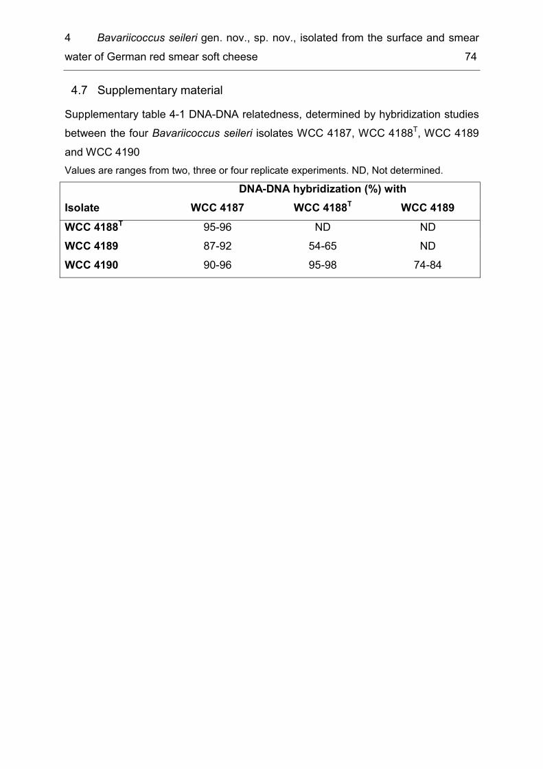
4 Bavariicoccus seileri gen. nov., sp. nov., isolated from the surface and smear water of German red smear soft cheese 74
Supplementary material 4.7Supplementary table 4-1 DNA-DNA relatedness, determined by hybridization studies between the four Bavariicoccus seileri isolates WCC 4187, WCC 4188T, WCC 4189 and WCC 4190 Values are ranges from two, three or four replicate experiments. ND, Not determined. DNA-DNA hybridization (%) with Isolate WCC 4187 WCC 4188T WCC 4189 WCC 4188T 95-96 ND ND WCC 4189 87-92 54-65 ND WCC 4190 90-96 95-98 74-84
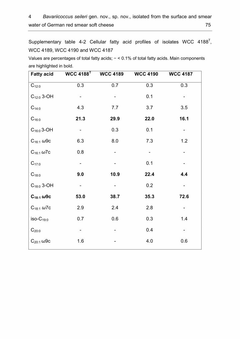
4 Bavariicoccus seileri gen. nov., sp. nov., isolated from the surface and smear water of German red smear soft cheese 75
Supplementary table 4-2 Cellular fatty acid profiles of isolates WCC 4188T, WCC 4189, WCC 4190 and WCC 4187 Values are percentages of total fatty acids; − < 0.1% of total fatty acids. Main components are highlighted in bold. Fatty acid WCC 4188T WCC 4189 WCC 4190 WCC 4187 C12:0 0.3 0.7 0.3 0.3 C12:0 3-OH - - 0.1 - C14:0 4.3 7.7 3.7 3.5 C16:0 21.3 29.9 22.0 16.1 C16:0 3-OH - 0.3 0.1 - C16:1 ω9c 6.3 8.0 7.3 1.2 C16:1 ω7c 0.8 - - - C17:0 - - 0.1 - C18:0 9.0 10.9 22.4 4.4 C18:0 3-OH - - 0.2 - C18:1 ω9c 53.0 38.7 35.3 72.6 C18:1 ω7c 2.9 2.4 2.8 - iso-C19:0 0.7 0.6 0.3 1.4 C20:0 - - 0.4 - C20:1 ω9c 1.6 - 4.0 0.6
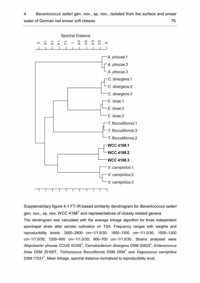
4 Bavariicoccus seileri gen. nov., sp. nov., isolated from the surface and smear water of German red smear soft cheese 76
Supplementary figure 4-1 FT-IR-based similarity dendrogram for Bavariicoccus seileri gen. nov., sp. nov. WCC 4188T and representatives of closely related genera The dendrogram was calculated with the average linkage algorithm for three independent spectraper strain after aerobic cultivation on TSA. Frequency ranges with weights and reproducibility levels: 3000–2800 cm−1/1.0/30; 1800–1500 cm−1/1.0/30; 1500–1200 cm−1/1.0/30; 1200–900 cm−1/1.0/30; 900–700 cm−1/1.0/30. Strains analysed were Atopobacter phocae CCUG 42358T, Carnobacterium divergens DSM 20623T; Enterococcus hirae DSM 20160T, Trichococcus flocculiformis DSM 2094T and Vagococcus carniphilus DSM 17031T. Mean linkage, spectral distance normalized to reproducibility level.
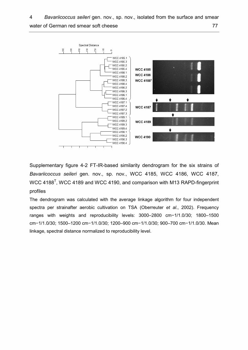
4 Bavariicoccus seileri gen. nov., sp. nov., isolated from the surface and smear water of German red smear soft cheese 77
Supplementary figure 4-2 FT-IR-based similarity dendrogram for the six strains of Bavariicoccus seileri gen. nov., sp. nov., WCC 4185, WCC 4186, WCC 4187, WCC 4188T, WCC 4189 and WCC 4190, and comparison with M13 RAPD-fingerprint profiles The dendrogram was calculated with the average linkage algorithm for four independent spectra per strainafter aerobic cultivation on TSA (Oberreuter et al., 2002). Frequency ranges with weights and reproducibility levels: 3000–2800 cm−1/1.0/30; 1800–1500 cm−1/1.0/30; 1500–1200 cm−1/1.0/30; 1200–900 cm−1/1.0/30; 900–700 cm−1/1.0/30. Mean linkage, spectral distance normalized to reproducibility level.
5 Sphingobacterium lactis sp. nov. and Sphingobacterium alimentarium sp. nov., isolated from raw milk and a dairy environment 78
5 Sphingobacterium lactis sp. nov. and Sphingobacterium alimentarium sp. nov., isolated from raw milk and a dairy environment.
Verena S.J. Schmidt1, Mareike Wenning1 and Siegfried Scherer1, 2, * 1Abteilung für Mikrobiologie, ZIEL, Technische Universität München, D-85354 Freising, Germany 2Lehrstuhl für Mikrobielle Ökologie, Department für Grundlagen der Biowissenschaften, WZW, Technische Universität München, D-85350 Freising, Germany Subject Category: New Taxa (Bacteroidetes) Running title: Sphingobacterium lactis sp. nov. and S. alimentarium sp. nov. The GenBank/EMBL/DDBJ accession numbers for the 16S rRNA gene of strains WCC 4512T, WCC 4521T, WS 4555 and WS 4556 are FN908501 to FN908504, respectively. A phylogenetic tree based on groEL gene sequences is available as supplementary material with the online version of this paper. The GenBank/EMBL/DDBJ accession numbers for the groEL gene sequences of strains WCC 4512T, WCC 4521T, WS 4555, WS 4556, S. daejeonense LMG 23402T, S. mizutaii DSMZ 11724T, S. composti LMG 23401T, S. faecium DSMZ 11690T, S. spiritivorum DSMZ 11722T and S. composti DSMZ 18850T are FN985014 to FN985023, respectively. *Correspondence [email protected] Published as: Schmidt VSJ, Wenning M & Scherer S (2012) Sphingobacterium lactis sp. nov. and Sphingobacterium alimentarium sp. nov., isolated from raw milk and a dairy environment. International Journal of Systematic and Evolutionary Microbiology 62: 1506-1511.

5 Sphingobacterium lactis sp. nov. and Sphingobacterium alimentarium sp. nov., isolated from raw milk and a dairy environment 79
Abstract Four non-fermenting, rod-shaped, Gram-staining-negative, bacterial strains, designated WCC 4512T and WS 4555, WCC 4521T and WS 4556, were isolated from raw milk and the dairy environment. Phylogenetic analyses based on 16S rRNA and groEL genes sequences demonstrated the affiliation of the four strains to two distinct clusters within the class Sphingobacteriia, phylum Bacteroidetes. Strains WCC 4512T and WS 4555 showed the highest 16S rRNA gene sequence similarity to S. daejeonense (97.3% and 97.2%, respectively), while strains WCC 4521T and WS 4556 were most closely related to S. composti LMG 23401T (97.6 % 16S rRNA gene sequence similarity). The DNA G + C content of the novel bacteria was 44.2 mol% (strain WCC 4512T) and 39.3 mol% (strain WCC 4521T), respectively. The major cellular fatty acids and the presence of menaquinone 7 (MK-7) as the predominant quinone for both strains WCC 4512T and WCC 4521T supported their affiliation to the genus Sphingobacterium. DNA-DNA hybridization experiments between strain WCC 4512T and S. daejeonense LMG 23402T and between strain WCC 4521T and S. composti LMG 23401T revealed DNA relatedness values of 2% (duplicate, 3%) and 8% (duplicate, 17%), respectively. On the basis of phenotypic and genetic properties as well as phylogenetic distinctiveness it is suggested that the four strains represent two novel Sphingobacterium species with strain WCC 4512T (= DSMZ 22361T = LMG 25272T) as the type strain of S. lactis and strain WCC 4521T (= DSMZ 22362T = LMG 25273T) as the type strain of S. alimentarium.
Introduction 5.1In 1983, Yabuuchi and colleagues described the genus Sphingobacterium (family Sphingobacteriaceae, order Sphingobacteriales, class Sphingobacteriia, phylum Bacteroidetes (Ludwig et al., 1998)), for Gram-staining-negative, non-spore forming rods containing high quantities of sphingophospholipids in their cells. At the time of writing, the genus encompassed the following 15 species with validly published names: Sphingobacterium multivorum, S. mizutaii and the type species S. spiritivorum (Yabuuchi et al., 1983), S. antarcticum (Shivaji et al., 1992), S. faecium and S. thalpophilum (Takeuchi & Yokota, 1992), S. daejeonense (Kim et al., 2006), S. composti T5-12T (Ten et al., 2006), S. composti 4M24T (Yoo et al., 2007), S.
5 Sphingobacterium lactis sp. nov. and Sphingobacterium alimentarium sp. nov., isolated from raw milk and a dairy environment 80
canadense (Mehnaz et al., 2007), S. siyangense (Liu et al., 2008), S. kitahiroshimense (Matsuyama et al., 2008), S. anhuiense (Wei et al., 2008), S. shayense (He et al., 2010) and S. bambusae (Duan et al., 2009). There are two validly named S. composti strains, described by Ten et al. (2006) and Yoo et al. (2007), respectively. According to the bacteriological code, section 5, S. composti Ten et al. has priority over S. composti Yoo et al. (www.bacterio.cict.fr/s/sphingobacterium.html) (Tindall, 1999), but the name of the latter organism has not been changed as yet. The present study was designed to determine the taxonomic position of four novel bacterial strains, which are considered to represent two novel species of the genus Sphingobacterium. Strain WCC 4521T, isolated in November 2005, originated from an environmental swab taken in a German dairy which produces milk powders. The swab had been streaked on plate count agar (PCA), which was subsequently cultivated at 30 °C. Strain WCC 4512T was isolated from raw milk in December 2007. The milk sample had been taken aseptically from the collection tank of a Bavarian farm and was plated on PCA with 1 % skimmed milk by using standard dilution techniques. A second raw milk sample, taken from the same Bavarian farm, was analysed in March 2009 and provided another two strains (WS 4555 and WS 4556). For identification purposes, FT-IR spectra of the four strains were recorded (Wenning et al., 2008) and compared with a reference database for aerobic, Gram-negative bacteria, including 850 spectra from 196 species and 75 genera, but no match was found (M. Wenning and others, unpublished data).
Materials and methods 5.2Amplification and sequencing of the 16S rRNA and groEL genes were carried out as described by Schmidt et al. (2009). 16S rRNA and, when available, groEL gene sequences of phylogenetically close relatives were obtained from the NCBI-database (http://www.ncbi.nlm.nih.gov/blast/Blast). Alignment of the obtained sequences was done with CLUSTAL_X (1.8) (Thompson et al., 1997). Phylogenetic trees with Rikenella microfusus ATCC 29728T as an outgroup were calculated using Treecon (Van de Peer & De Wachter, 1997)(Van de Peer & De Wachter, 1997). In order to
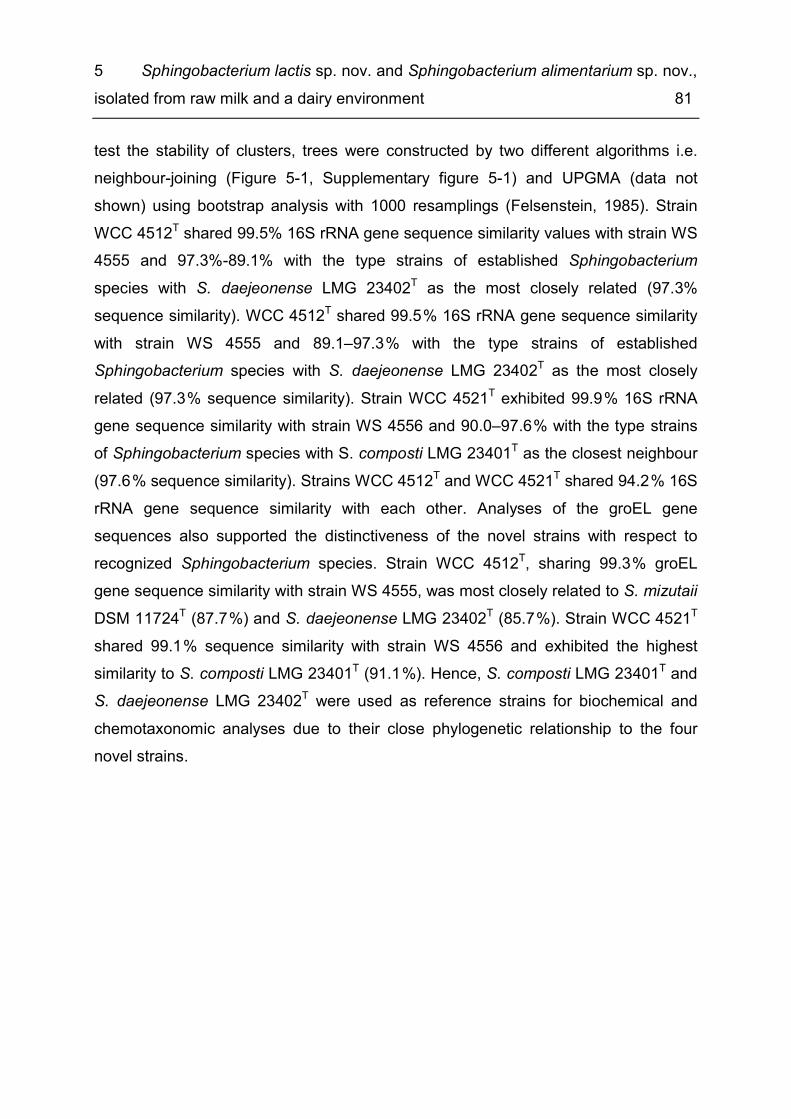
5 Sphingobacterium lactis sp. nov. and Sphingobacterium alimentarium sp. nov., isolated from raw milk and a dairy environment 81
test the stability of clusters, trees were constructed by two different algorithms i.e. neighbour-joining (Figure 5-1, Supplementary figure 5-1) and UPGMA (data not shown) using bootstrap analysis with 1000 resamplings (Felsenstein, 1985). Strain WCC 4512T shared 99.5% 16S rRNA gene sequence similarity values with strain WS 4555 and 97.3%-89.1% with the type strains of established Sphingobacterium species with S. daejeonense LMG 23402T as the most closely related (97.3% sequence similarity). WCC 4512T shared 99.5 % 16S rRNA gene sequence similarity with strain WS 4555 and 89.1–97.3 % with the type strains of established Sphingobacterium species with S. daejeonense LMG 23402T as the most closely related (97.3 % sequence similarity). Strain WCC 4521T exhibited 99.9 % 16S rRNA gene sequence similarity with strain WS 4556 and 90.0–97.6 % with the type strains of Sphingobacterium species with S. composti LMG 23401T as the closest neighbour (97.6 % sequence similarity). Strains WCC 4512T and WCC 4521T shared 94.2 % 16S rRNA gene sequence similarity with each other. Analyses of the groEL gene sequences also supported the distinctiveness of the novel strains with respect to recognized Sphingobacterium species. Strain WCC 4512T, sharing 99.3 % groEL gene sequence similarity with strain WS 4555, was most closely related to S. mizutaii DSM 11724T (87.7 %) and S. daejeonense LMG 23402T (85.7 %). Strain WCC 4521T shared 99.1 % sequence similarity with strain WS 4556 and exhibited the highest similarity to S. composti LMG 23401T (91.1 %). Hence, S. composti LMG 23401T and S. daejeonense LMG 23402T were used as reference strains for biochemical and chemotaxonomic analyses due to their close phylogenetic relationship to the four novel strains.
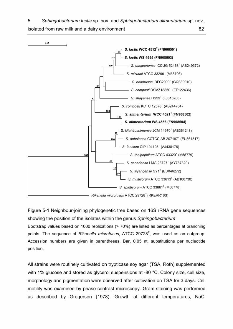
5 Sphingobacterium lactis sp. nov. and Sphingobacterium alimentarium sp. nov., isolated from raw milk and a dairy environment 82
Figure 5-1 Neighbour-joining phylogenetic tree based on 16S rRNA gene sequences showing the position of the isolates within the genus Sphingobacterium Bootstrap values based on 1000 replications (> 70%) are listed as percentages at branching points. The sequence of Rikenella microfusus, ATCC 29728T, was used as an outgroup. Accession numbers are given in parentheses. Bar, 0.05 nt. substitutions per nucleotide position. All strains were routinely cultivated on trypticase soy agar (TSA, Roth) supplemented with 1% glucose and stored as glycerol suspensions at -80 °C. Colony size, cell size, morphology and pigmentation were observed after cultivation on TSA for 3 days. Cell motility was examined by phase-contrast microscopy. Gram-staining was performed as described by Gregersen (1978). Growth at different temperatures, NaCl
S. mizutaii ATCC 33299T (M58796)
S. faecium CIP 104193T (AJ438176)
S. siyangense SY1T (EU046272) S. multivorum ATCC 33613T (AB100738)
S. lactis WS 4555 (FN908503) S. lactis WCC 4512T (FN908501)
S. alimentarium WS 4556 (FN908504) S. alimentarium WCC 4521T (FN908502)
S. kitahiroshimense JCM 14970T (AB361248)
S. composti DSMZ18850T (EF122436)
S. daejeonense CCUG 52468T (AB249372)
S. anhuiense CCTCC AB 207197T (EU364817)
S. thalpophilum ATCC 43320T (M58779)
S. spiritivorum ATCC 33861T (M58778)
S. composti KCTC 12578T (AB244764)
S. canadense LMG 23727T (AY787820)
Rikenella microfusus ATCC 29728T (RKERR16S)
S. shayense HS39T (FJ816788)
S. bambusae IBFC2009T (GQ339910)
5 Sphingobacterium lactis sp. nov. and Sphingobacterium alimentarium sp. nov., isolated from raw milk and a dairy environment 83
concentrations and pH values was assessed in duplicate in trypticase soy broth [TSB; containing (l −1) 15.0 g pancreatic digest of casein, 5.0 g soybean enzymic digest, 5.0 g NaCl] after cultivation for 7 days; pH values of the TSB were adjusted by the addition of 1 M NaOH or HCl and verified after autoclaving. Growth at pH 4–10 (at intervals of 1 pH unit) and in the presence of 0–8 % NaCl (w/v, at intervals of 1 %) was assessed at 30 °C. Growth was tested at 5, 10, 15, 30, 37, 40, 41 and 42 °C. Growth on R2A agar (Merck), cetrimide agar (Merck), MacConkey agar (Difco), Columbia blood agar supplemented with 5 % sheep blood (Oxoid) and nutrient agar (medium 1; DSMZ) was assessed at 30 °C. Agar plates for testing the degradation of starch (l −1: 3.0 g meat extract, 10.0 g soluble starch, 12.0 g agar, pH 7.4) and DNase activity (VWR) were incubated at 25 °C for 7 days and were flooded with Kovac’s reagent (Merck) and 1 M HCl (Merck), respectively, in order to visualize clearing zones. The presence of cytochrome oxidase was detected with Bactident Oxidase test strips (Merck). Decomposition of citrate on Simmons’ citrate agar, catalase activity, hydrolysis of aesculin, casein and urea, as well as pigment production on King’s A and King’s B agars were tested as specified by Smibert & Krieg (1994). Production of a flexirubin-type pigment was examined as described by Buller (2004). Hydrolysis of Tween 20 and Tween 80 was assessed according to Sierra (1957). Degradation of tyrosine (Logan & De Vos, 2009), xanthine (Seiler et al., 1980) and tributyrine [according to Sizemore et al. (1992), but without addition of Tween 20] was determined after incubation for up to 14 days at 30 °C. Additionally, assimilation of different carbohydrates was assessed using the API 50CH strips (bioMérieux) with M9 minimal medium (Miller, 1972) as inoculation medium, modified by addition of a vitamin solution (Murashige & Skoog, 1962). After solubilization of the carbohydrates, the inoculated medium was aseptically transferred from the cupules of the API strips into a 96-well microtitre plate to allow orbital shaking. Plates were sealed with Breathe-Easy foil (Diversified Biotech) and incubated at 30 °C at 550 r.p.m. for 5 days. Utilization of the carbohydrates was indicated by visible turbidity of the media. For further characterization, API 20E strips (bioMérieux) were used according to the manufacturer’s instructions. Acid production from eight different carbohydrates was examined in semi-solid OF medium (Smibert & Krieg, 1994), supplemented with 1 % filter-sterilized carbohydrate (amygdalin, l-arabinose, d-glucose, d-lactose, d-mannitol, melibiose, l-rhamnose and sucrose). Tubes were
5 Sphingobacterium lactis sp. nov. and Sphingobacterium alimentarium sp. nov., isolated from raw milk and a dairy environment 84
incubated at 30 °C for 96 h and oxidative acid production was indicated by a colour change from green to yellow at the surface area of the inoculated medium. Growth under anaerobic conditions was determined on TSA supplemented with 1 % glucose, using the gas generator system Anaerocult A mini (Merck) to obtain an oxygen-free atmosphere. Results are listed in the species descriptions and in Table 5-1.
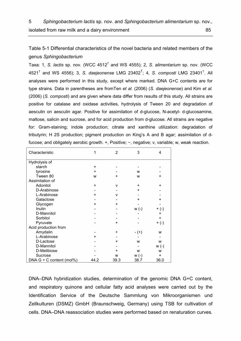
5 Sphingobacterium lactis sp. nov. and Sphingobacterium alimentarium sp. nov., isolated from raw milk and a dairy environment 85
Table 5-1 Differential characteristics of the novel bacteria and related members of the genus Sphingobacterium Taxa: 1, S. lactis sp. nov. (WCC 4512T and WS 4555); 2, S. alimentarium sp. nov. (WCC 4521T and WS 4556); 3, S. daejeonense LMG 23402T; 4, S. composti LMG 23401T. All analyses were performed in this study, except where marked. DNA G+C contents are for type strains. Data in parentheses are fromTen et al. (2006) (S. daejeonense) and Kim et al. (2006) (S. composti) and are given where data differ from results of this study. All strains are positive for catalase and oxidase activities, hydrolysis of Tween 20 and degradation of aesculin on aesculin agar. Positive for assimilation of d-glucose, N-acetyl- d-glucosamine, maltose, salicin and sucrose, and for acid production from d-glucose. All strains are negative for: Gram-staining; indole production; citrate and xanthine utilization: degradation of tributyrin; H 2S production; pigment production on King’s A and B agar; assimilation of d-fucose; and obligately aerobic growth. +, Positive; −, negative; v, variable; w, weak reaction. Characteristic 1 2 3 4 Hydrolysis of
starch + - - - tyrosine + - w - Tween 80 w + w +
Assimilation of Adonitol + v + + D-Arabinose - - + - L-Arabinose + v - - Galactose - - + + Glycogen + + - - Inulin - - w (-) + (-) D-Mannitol - - - + Sorbitol - - - + Pyruvate - + - + (-)
Acid production from Amydalin - + - (+) w L-Arabinose + - - - D-Lactose - + w w D-Mannitol - - - w (-) D-Melibiose - + w w Sucrose - w w (-) +
DNA G + C content (mol%) 44.2 39.3 38.7 36.0 DNA–DNA hybridization studies, determination of the genomic DNA G+C content, and respiratory quinone and cellular fatty acid analyses were carried out by the Identification Service of the Deutsche Sammlung von Mikroorganismen und Zellkulturen (DSMZ) GmbH (Braunschweig, Germany) using TSB for cultivation of cells. DNA–DNA reassociation studies were performed based on renaturation curves.

5 Sphingobacterium lactis sp. nov. and Sphingobacterium alimentarium sp. nov., isolated from raw milk and a dairy environment 86
The DNAs were isolated using a French pressure cell (Thermo Spectronic) and purified by chromatography on hydroxyapatite as described by Cashion et al. (1977). Hybridization studies were performed as described by De Ley et al. (1970) with the modifications described by Huss et al. (1983) using a model Cary 100 Bio UV/VIS-spectrophotometer equipped with a Peltier-thermostatted 6×6 multicell changer and a temperature controller with in situ temperature probe (Varian). The level of DNA–DNA relatedness between strain WCC 4512T and S. daejeonense LMG 23402T was 2 % (repetition 3 %) and that between strain WCC 4521T and S. composti LMG 23401T was 8% (repetition 17 %). These values are well below the 70 % cut-off for species delineation defined by Wayne et al. (1987). As determined by HPLC (Mesbah et al., 1989), the DNA G+C content of strains WCC 4512T and WCC 4521T were 44.2 and 39.3 mol%, respectively. Both values fall within the range (36.0–44.2 mol%) reported for established Sphingobacterium species (Ten et al., 2006, Mehnaz et al., 2007, Yoo et al., 2007, Liu et al., 2008, Matsuyama et al., 2008, Wei et al., 2008, Duan et al., 2009, He et al., 2010). Respiratory quinones were analysed according to Altenburger et al. (1996). Strains WCC 4512T and WCC 4521T exhibited menaquinone MK-7 as the major respiratory quinone (95 and 96 %, respectively), which is in line with all members of the family Sphingobacteriaceae (Steyn et al., 1998). In addition, both strains also contained minor amounts of MK-6 and MK-8. Fatty acids were extracted and analysed as described recently (Verbarg et al., 2008). In general, the major fatty acids of WCC 4512T and WCC 4521T were similar to those of related Sphingobacterium species (Table 5-2). Notably, strain WCC 4512T and S. daejeonense LMG 23402T both contained a significantly lower amount of iso-C 17 : 1ω9 c compared with strain WCC 4521T and S. composti LMG 23401T. The respective proportions of the following fatty acids also distinguished the two pairs of strains: C 16 : 0, iso-C 15 : 0, iso-C 15 : 1F and summed feature 3 (iso-C 15 : 0 2-OH and/or C 16 : 1ω7 c). This is in agreement with their phylogenetic position based on 16S rRNA gene and groEL gene sequences (Figure 5-1, Supplementary figure 5-1).
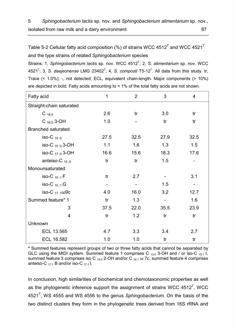
5 Sphingobacterium lactis sp. nov. and Sphingobacterium alimentarium sp. nov., isolated from raw milk and a dairy environment 87
Table 5-2 Cellular fatty acid composition (%) of strains WCC 4512T and WCC 4521T and the type strains of related Sphingobacterium species Strains: 1, Sphingobacterium lactis sp. nov. WCC 4512T; 2, S. alimentarium sp. nov. WCC 4521T; 3, S. daejeonense LMG 23402T; 4, S. composti T5-12T. All data from this study. tr, Trace (< 1.0%); -, not detected; ECL, equivalent chain-length. Major components (> 10%) are depicted in bold. Fatty acids amounting to < 1% of the total fatty acids are not shown. Fatty acid 1 2 3 4 Straight-chain saturated
C 16:0 2.6 tr 3.0 tr C 16:0 3-OH 1.0 - tr tr
Branched saturated iso-C 15 :0 27.5 32.5 27.9 32.5 iso-C 15 :0 3-OH 1.1 1.6 1.3 1.5 iso-C 17 :0 3-OH 16.6 15.6 18.3 17.6 anteiso-C 15 :0 tr tr 1.5 -
Monounsaturated iso-C 15 :1 F tr 2.7 - 3.1 iso-C 15 :1 G - - 1.5 - iso-C 17 :1ω9c 4.0 16.0 3.2 12.7
Summed feature* 1 tr 1.3 - 1.6 3 37.5 22.0 35.5 23.9 4 tr 1.2 tr tr Unknown
ECL 13.565 4.7 3.3 3.4 2.7 ECL 16.582 1.0 1.0 tr tr
* Summed features represent groups of two or three fatty acids that cannot be separated by GLC using the MIDI system. Summed feature 1 comprises C 13:0 3-OH and / or iso-C 15:1 I, summed feature 3 comprises iso C 15:0 2-OH and/or C 16:1 ω 7c, summed feature 4 comprises anteiso-C 17:1 B and/or iso-C 17:1 I. In conclusion, high similarities of biochemical and chemotaxonomic properties as well as the phylogenetic inference support the assignment of strains WCC 4512T, WCC 4521T, WS 4555 and WS 4556 to the genus Sphingobacterium. On the basis of the two distinct clusters they form in the phylogenetic trees derived from 16S rRNA and

5 Sphingobacterium lactis sp. nov. and Sphingobacterium alimentarium sp. nov., isolated from raw milk and a dairy environment 88
groEL gene sequences, DNA-DNA hybridisation results and differentiating phenotypic characteristics (Table 5-1, Table 5-2), we propose two novel species, Sphingobacterium lactis sp. nov. and Sphingobacterium alimentarium sp. nov., to accommodate the four novel strains.
Description of Sphingobacterium lactis sp. nov. 5.3Sphingobacterium lactis (lac´tis L. gen. n., from milk; pertaining to the isolation source of the type strain). Cells are Gram-staining-negative, strictly aerobic, non-spore forming and non-motile rods, 0.6–0.7 µm in diameter and 1.1–2.2 µm in length. After incubation on TSA for 3 days, colonies are 2–3 mm in diameter, smooth, circular with entire margins, translucent and convex. On TSA, a very pale yellow, non-fluorescent and non-diffusible pigment is produced, which is not of the flexirubin or phenazine types. In TSB, growth occurs at 10–41 °C, at pH 6–9 and with 0–5 % NaCl. Positive for oxidase and catalase activities. Hydrolyses starch, tyrosine, Tween 20, Tween 80, DNA (weak reaction) and aesculin, but not casein, tributyrin or xanthine. Growth occurs on nutrient agar, cetrimide agar, R2A agar and Columbia agar supplemented with 5 % sheep blood (with γ-haemolysis type), but not on MacConkey agar. Assimilates l-arabinose (weak reaction), d-adonitol (weak reaction), d-glucose, d-fructose, d-mannose, methyl α- d-mannopyranoside, methyl α- d-glucopyranoside, N-acetylglucosamine, arbutin, aesculin, salicin, cellobiose, maltose, d-lactose, melibiose, sucrose, trehalose, melezitose, raffinose, glycogen, gentiobiose and d-turanose in API 50CH strips. The other substrates in the API 50CH strips are not assimilated. Acid is produced from l-arabinose and d-glucose, but not from amygdalin, d-lactose, d-mannitol, melibiose, l-rhamnose or sucrose. In the API 20E gallery, positive for β-galactosidase activity, but negative for arginine dihydrolase, lysine decarboxylase, ornithine decarboxylase, urease and tryptophan deaminase activities, citrate utilization, H 2S, acetoin and indole production, hydrolysis of gelatin and reduction of nitrate. MK-7 is the predominant menaquinone; MK-6 and MK-8 also occur in minor amounts. The major fatty acids (> 15 %) are iso-C 15 : 0, iso-C 17 : 0 3-OH and summed feature 3 (comprising iso-C 15 : 0 2-OH and/or C 16 : 1ω7 c).
5 Sphingobacterium lactis sp. nov. and Sphingobacterium alimentarium sp. nov., isolated from raw milk and a dairy environment 89
The type strain WCC 4512T (= DSMZ 22361T = LMG 25272T) was isolated from raw cow’s milk from a Bavarian farm.
Description of Sphingobacterium alimentarium sp. nov. 5.4Sphingobacterium alimentarium sp. nov. (ali.men.ta´rium. L. neut. adj. alimentarium, pertaining to food). Cells are Gram-staining-negative, strictly aerobic, non-spore forming and non-motile rods, 0.6–1.0 µm in diameter and 1.1–2.2 µm in length. After incubation on TSA for 3 days, colonies are 2–3 mm in diameter, smooth, circular with entire margins, translucent and convex. On TSA, a bright orange coloured, non-diffusible and non-fluorescent pigment is produced, which is not of the flexirubin or phenazine types. In TSB, growth occurs at 10–41 °C, at pH 6–9 and with 0–3 % NaCl. Positive for oxidase and catalase activities. Hydrolyses tyrosine, Tween 20, Tween 80, DNA (weak reaction) and aesculin, but not starch, casein, tributyrin or xanthine. Growth occurs on nutrient agar, cetrimide agar, R2A agar and Columbia agar plates supplemented with 5 % sheep blood (with γ-haemolysis type), but not on MacConkey agar. Assimilates d-glucose, d-fructose, d-mannose, methyl α- d-mannopyranoside, methyl α- d-glucopyranoside, N-acetylglucosamine, arbutin (weak reaction), aesculin, salicin, cellobiose, maltose, d-lactose, melibiose, sucrose, trehalose, melezitose, raffinose, glycogen, gentiobiose and d-turanose in the API50CH strip. Assimilation of adonitol and l-arabinose is strain dependent. The other substrates in the API 50CH strip are not assimilated. Acid is produced from amygdalin, d-glucose, d-lactose, melibiose and sucrose (weak reaction), but not from l-arabinose, d-mannitol or l-rhamnose. In the API 20E gallery, positive for β-galactosidase activity and formation of acetoin, but negative for arginine dihydrolase, lysine decarboxylase, ornithine decarboxylase, urease and tryptophan deaminase activities, citrate utilization, H 2S, acetoin and indole production, hydrolysis of gelatin and reduction of nitrate. MK-7 is the major menaquinone and predominant fatty acids (> 15%) are iso-C 15 : 0, iso-C 17 : 1ω9 c, iso-C 17 : 0 3-OH and summed feature 3 (comprising iso-C 15 : 0 2-OH and/or C 16 : 1ω7 c). The type strain WCC 4521T (= DSMZ 22362T = LMG 25273T), was isolated from the dairy environment.
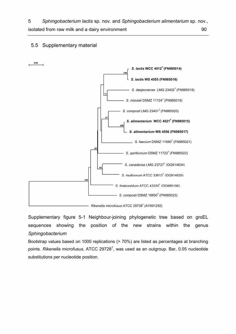
5 Sphingobacterium lactis sp. nov. and Sphingobacterium alimentarium sp. nov., isolated from raw milk and a dairy environment 90
Supplementary material 5.5
Supplementary figure 5-1 Neighbour-joining phylogenetic tree based on groEL sequences showing the position of the new strains within the genus Sphingobacterium Bootstrap values based on 1000 replications (> 70%) are listed as percentages at branching points. Rikenella microfusus, ATCC 29728T, was used as an outgroup. Bar, 0.05 nucleotide substitutions per nucleotide position.
S. mizutaii DSMZ 11724T (FN985019)
S. faecium DSMZ 11690T (FN985021)
S. multivorum ATCC 33613T (DQ914835)
S. lactis WS 4555 (FN985016)
S. lactis WCC 4512T (FN985014)
S. alimentarium WS 4556 (FN985017)
S. alimentarium WCC 4521T (FN985015)
S. composti DSMZ 18850T (FN985023)
S. daejeonense LMG 23402T (FN985018)
S. thalpophilum ATCC 43320T (DQ985196)
S. spiritivorum DSMZ 11722T (FN985022)
S. composti LMG 23401T (FN985020)
S. canadense LMG 23727T (DQ914834)
Rikenella microfusus ATCC 29728T (AY691292)

6 General discussion 91
6 General discussion
Highly diverse raw milk microbiota: polyphasic approach for the 6.1description of Sphingobacterium alimentarium sp. nov. and Sphingobacterium lacticum sp. nov., isolated from raw milk and the dairy environment
Due to the high nutritional value of raw milk, the growth of a broad range of bacteria is supported, resulting in a great microbial diversity and among this there is also a high share of unknown bacteria. In this study 2% of the raw milk isolates (13/626) could not be assigned to existing taxa based on their almost complete 16S rRNA sequence, this finding is in agreement with other studies with 0.6 to even 20% probably novel species in raw milk (Hantsis-Zacharov & Halpern, 2007, von Neubeck et al., 2015, Doll et al., 2017) and by a variety of novel raw milk bacteria from diverse taxonomic groups been described in recent years, i.e. Pseudomonas lactis sp. nov., Pseudomonas paralactis sp. nov. (von Neubeck et al., 2017), Pseudomonas helleri sp. nov., Pseudomonas weihenstephanensis sp. nov. (von Neubeck et al., 2016), Chryseobacterium haifense sp. nov. (Hantsis-Zacharov & Halpern, 2007), Corynebacterium crudilactis sp. nov. (Zimmermann et al., 2016), Bacillus wiedmannii sp. nov. (Miller et al., 2016) and Paenibacillus bovis sp. nov. (Gao et al., 2016). Also a new genus of dairy associated bacteria, Bavariicoccus seileri gen. nov. sp. nov., has been characterized in this thesis, i.e. lactic acid bacteria originating from the surface of red smear cheese (Schmidt et al., 2009). Furthermore, four raw milk strains of this study have been validly described as Sphingobacterium alimentarium and Sphingobacterium lacticum, based on a polyphasic approach including genotypic, phylogenetic and phenotypic characteristics (Schmidt et al., 2012). A polyphasic approach aims at a complete characterization of bacteria under avoidance of mistaken descriptions. This method was first proposed by Colwell (1970) for the genus Vibrio, but serves nowadays as an established prodcedure for the description of prokaryotes. Yet a prerequisite for this method is the cultivability of bacteria (Konstantinidis et al., 2017). Prokaryotic genus and species are considered the “working unit” of bacterial taxonomy (Moore et al., 2010), i. e. an assignment of a bacterial isolate to taxonomical systematics is crucial to distinguish different bacteria from each other, to

6 General discussion 92
enable their recognition on the basis of characteristic traits (= identification) and consequently to allow scientific exchange (Baron, 1996). In the case of the four Sphingobacterium strains, inhouse FT-IR analysis as well as 16S rRNA sequence analyses did not result in an identification, as there was no match with the reference databases (Wenning & Scherer, 2013). Hence, the description process of the two novel Sphingobacterium spp., Sphingobacterium lactium and Sphingobacterium alimentarium was initiated. Each of the novel species was represented by two strains, meeting the recommendation that description of novel species should not be based on a single strain (Tindall, 1999).
6.1.1 Genotypic and phylogenetic differentiation Genotypic and phylogenetic analyses are considered a useful tool for species description as they depict the past evolutionary development serving as “molecular clocks” The molecular clock hypothesis acts on the assumption that there is a constant rate of evolution for DNA and protein sequences, valid also among different organisms. Hence the degree of genetic relationship two species share correlates with the length of time they had their last ancestor in common (Ho, 2008). Concerning genotypic differentiation, sequence analyses of the 16S rRNA as well as DNA-DNA hybridization studies are still supposed to be the gold standard for delimitation of bacterial species (Stackebrandt & Ebers, 2006). The 16S rRNA is universally distributed among bacteria and possesses the same function in every organism. It is conserved, but holds variable and hyper-variable regions. Furthermore its 1500 base pairs are a suitable size, i.e. on the one hand it is quite easy to determine its sequence and on the other hand it comprises enough genetic information applicable for phylogenetic studies and identification by comparison of the unknown sequence with large sequence databases (Clarridge, 2004), i.e. the NCBI database (http://www.ncbi.nlm.nih.gov/blast.cgi). The widely accepted cutoff of 98.7% 16S rRNA sequence identity for species demarcation with respect to established species was met for both new Sphingobacterium species. For Sphingobacterium lactis the two strains WCC 4512T and WS 4555 showed a high similarity to each other (99.5%) indicating the belonging to the same species. Yet, they exhibited a distinct position to established Sphingobacterium spp. with 89.1–97.3% relatedness. Both strains showed the
6 General discussion 93
highest 16S rRNA gene sequence similarity to the type strain of S. daejeonense (97.3 and 97.2%, respectively). The Sphingobacterium alimentarium strains WCC 4521T and WS 4556 demonstrated a high similarity of 99.9% to each other, but exhibited a distinct position to established Sphingobacterium spp. with 90.0–97.6% relatedness. Closest affinity concerning 16S rRNA gene sequence similarity was exhibited to the type strain of S. composti LMG 23401T (97.6%). Also DNA-DNA hybridization studies supported the existence of two novel species. These analyses depict a measure for overall genetic diversity of two different bacteria rated by the degree their genomes hybridize under standard conditions (Gevers et al., 2005). The 70% cutoff for DNA reassociation was assessed empirically, as two bacterial strains exhibiting a DNA similarity of ≥ 70% also resemble in their chemotaxonomic, genomic, serological, and numerical phenetic characteristics (Wayne et al., 1987). Both Sphingobacterium lactis and Spingobacterium alimentarium exceeded the threshold of 70% DNA binding with respect to their closest neighbouring species concerning 16S rRNA relatedness. Hybridization analyses exhibited values of 2% (repetition 3%) between Sphingobacterium lactis WCC 4512T and S. daejeonense and values of 8% (repetition 17%) between Sphingobacterium alimentarium WCC 4521T and S. composti. In addition phylogenetic trees constructed by the use of almost complete 16S rRNA sequences of the four novel strains and other Sphingobacterium spp. supported the close relationship of each of the two Sphingobacterium lactis and alimentarium strains, respectively, as belonging to the same species, as well as a distinct position with regard to established Sphingobacterium spp.. The tree topology was reproducible for two different algorithms (neighbor joining, UPGMA) and also when sequences of the groEL genes were used instead of the 16S rRNA. The integration of groEL sequences as a representative of the housekeeping genes in descriptions of novel species is highly recommended (Stackebrandt et al., 2002). These genes hold essential metabolic functions, but vary faster than the 16S rRNA gene and therefore have a higher resolution (Gevers et al., 2005). In conclusion, all genotypic methods underpinned the existence of two novel Spingobacterium spp. represented by two strains each. The DNA G+C content supported the allocation of the novel strains to the genus Sphingobacterium (Yabuuchi et al., 1983).

6 General discussion 94
6.1.2 Phenotypic differentiation Phenotypic analyses, summing up all methods which do not deal with DNA or RNA, function as the quintessence of a polyphasic approach, i.e. genotypic methods depict the framework by enabling the allocation of a bacterium to a phylogenetic cluster, but phenotypic consistency is crucial for the description of a novel species (Vandamme 1996). Cellular fatty acids are representatives of the so called chemotaxonomic parameters. The latter aim at those chemical constituents of the bacterial cell applicable for bacterial classification. Especially cellular fatty acids represent a useful taxonomic marker as they show quite conserved patterns due to their essential role in cell structure and function, but comprise a great variety between different bacterial species and genera. After growth of cells at standardized conditions, whole cell FAME (fatty acid methyl ester) profiles can even be used for identification purposes, applying established databases. More than 300 diverse fatty acids are known and they differ in each other by the position of double bonds, binding of functional groups and a difference in chain length form (Vandamme et al., 1996, Dawyndt et al., 2006). The major cellular fatty acids iso-C15:0, iso-C17:0 3-OH and summed feature 3 (iso C15:02-OH and/or C16:1 ω7c) confirmed the allocation of the two novel species to the genus Sphingobacterium. Yet, other major fatty acids also comprised distinctive features enabling the differentiation of the two novel Sphingobacterium spp. and their closest phylogenetic neighbours. Respiratory quinones also serve as chemotaxonomic marker as the type of quinone present in an organism and the isoprenoid chain length reflect, in many cases, the phylogenetic affiliation of a bacterium. Yet, they often do not allow distinction on species level, but only for higher taxonomic ranks (Da Costa et al., 2011). Both novel Sphingobacterium spp. exhibited MK-7 as major respiratory quinone, a characteristic trait for the family Sphingobacteriaceae. In addition, also morphological traits, i.e. cells were non-motile, strictly aerobic and Gram-stain-negative short rods and colonies exhibited yellow coloured pigmentation, were consistent with other species of the genus Spingobacterium. In order to further describe the two novel species, growth tests at different temperatures, NaCl contents, pH values and assimilation of different C-sources using API 50CH and 20E test were performed. These analyses featured distinctive

6 General discussion 95
characteristics between the four novel Sphingobacterium strains, useful for identification purposes. In conclusion genotypic, phylogenetic and phenotypic methods supported the affiliation of the four novel strains to the genus Spingobacterium, yet due to distinct features with respect to established Spingobacterium species as novel species Spingobacterium lactis and Sphingobacterium alimentarium. They were named according to “The International Code of Nomenclature of Prokaryotes” (Lapage et al., 1992), their 16S rRNA and groEL gene sequences were located in GenBank/EMPL/DDBJ and furthermore the two type strains were deposited as freeze-dried ampules in the German (Leibniz-Institut DSMZ - Deutsche Sammlung von Mikroorganismen und Zellkulturen GmbH) and in the Belgian culture collection (BCCM/LMG Bacteria Collection). The deposit of gene sequences and freeze-dried ampoules enables their availabilty to the scientific community.
Microbial quality of MF/PAST ESL milk at the end of BBD with respect to 6.2spoilage factors
Analyzing MF/PAST ESL milk of five manufacturers in Germany, Austria and Switzerland after storage at 8 °C till the BBD, aerobic counts comprised a great variance ranging from < 1-8 log10 cfu/mL, this range is in accordance with Doll et al. (2017) who examined microfiltered milk of four manufacturers and found counts < 1-7.9 log10 cfu/mL. Variance was observed in both studies for packages of different batches, but also within different packages of one batch, a difference of up to 7 log units (this study) and 3 log units (Doll et al., 2017) was noticed. This finding is supported by Eneroth et al. (2001), who found a great variance of up to 2.8 log10 cfu/mL of B. cereus between different packages of pasteurized milk after storage at 10 °C for five days, although they had been filled within one hour. That means that the germs were not equally distributed to the packages during the filling process. Consequently, in order to cover the microbial biodiversity over shelf life especially in case of ESL milk with a low bacterial content, it is necessary to sample more packages of one batch. It is also important to use the original packages instead of smaller amounts like 100 mL aliquots. Some bacteria have a very low contamination titer directly after production, not regarded when only small amounts of milk are

6 General discussion 96
examined, but nonetheless these bacteria might become the dominant flora during extended cold storage. All five batches produced by manufacturer II exhibited an outstanding quality at the end of shelf life after storage at 8 °C with low numbers of < 1 cfu/mL for all 25 packages. Also Doll et al. (2017) found milk cartons with loads < 1 cfu/mL at the end of BBD after storage at 8 °C for 2 of 4 manufacturers, but only in shares of 3 and 26% respectively, while other packages of these manufacturers were spoilt by Gram-negative bacteria and psychrotolerant spore formers. The five batches of this study had been produced in temporal proximity of few days, as they had been sent to the laboratory at the same time. Hence it would be interesting to conduct a deeper study at this dairy to see, if the MF/PAST milk quality was still stable and to fathom reasons for these low bacterial counts and process security without recontamination. Spoilt packages of MF/PAST milk at the BBD comprised counts between 5.8 and 8.4 log10 cfu/mL and featured two groups of bacteria, i.e. psychrotolerant spore formers (PSF) (Paenibacillus sp. nov., Bacillus cereus, Bacillus weihenstephansis) and Gram-negative bacteria. The presence of spore formers might result from inadequate separation or alternatively from post-process recontamination (PPC). Doll et al. (2017) detected the same strains of PSF, i.e. P. odorifer, P. taichungensis/tundra, Paenibacillus sp. nov. and B. pumilus/safensis in the bulk tank and in the milk packages, demonstrating raw milk as contamination source for these bacteria. Yet in case of the major spoilage bacteria of retail milk, B. cereus and a Paenibacillus sp., their RAPD types could not be retrieved in the corresponding bulk tank milk, when conducting process analyses along the production chain. Consequently, they most probably constituted post process recontamination by the dairy environment. A pasteurizer, dairy silo tanks and the filling machine have been described as a source of contamination for B. cereus (Svensson et al., 2000, Eneroth et al., 2001, Shaheen et al., 2010), and analysis of 32 B. cereus ribotypes, isolated from Brazilian pasteurized milk, found identical patterns of product isolates and dairy surfaces (Salustiano et al., 2009). Bacillus cereus and Paenibacillus spp. are well known to form heat resistant biofilms on gaskets and dead ends of milk processing lines, rubbers and stainless stain surfaces (Czechowski, 1990, Andersson & Rönner, 1998), and as part of a microbial consortium they are much more difficult to be eliminated than planktonic cells (Gopal et al., 2015, Majed et al., 2016). Especially
6 General discussion 97
the steel–rubber contact surfaces of gaskets are a hygienic problem, as normal cleaning procedures (CIPs, cleaning in place, including cleaning steps with acid and caustic and rinsing with water) do not inhibit colonization with Bacillus spp. and the degree of colonization even accelerates with increased age and deterioration of the gaskets (Mettler & Carpentier, 1997). Also contamination of foodgrade paper and paperboard with B. cereus has been described, spore formers belonging to the genera Bacillus, Paenibacillus and Brevibacillus showed counts between < 50 to 250 cfu/g on homogenized paper material (Pirttijärvi et al., 2000). Yet packaging material is disinfected in the filling machine directly before use and so far, no strain typing has proven the transmission of packaging material to consumer packages. Gram-negative bacteria as the second most spoilage group, detected in 4/125 packages of MF/PAST milk of this study were represented by Spingomonas sp., Psychrobacter glacincola, Chryeobacterium sp. and Acinetobacter junii/johnsonii, most probably a result of post process recontamination rather than insufficient heat treatment (Ternström et al., 1993, Eneroth et al., 2000). Doll et al. (2017) found a similar recontamination rate of microfiltered milk with Gram-negative bacteria (10/234 packages) comprising Moraxella and Pseudomonas. Control of process security with respect to Gram-negative recontaminants poses a routine task in dairies` quality control, nowadays often performed by the coliform count. The group of the coliform bacteria comprises aerobic or facultatively anaerobic, Gram-negative, non-spore forming rods which are able to ferment lactose to produce gas and acid within 48 h at 32–35 °C (Davidson et al., 2004). Most of them belong to the family Enterobacteriaceae (e.g. Escherichia, Klebsiella and Serratia), while Aeromonas belongs to the family Aeromonadaceae. Yet the coliform count has a minor informative value for hygienic conditions and process safety without recontamination, as most of the Gram-negative recontaminants are Pseudomonas spp. and are not detected when samples are analyzed for coliform bacteria (D'Amico et al., 2008, Jackson et al., 2012). Consequently, culture media and also molecular methods aiming at the detection of Gram-negative recontaminants with high frequency and spoilage potential like Pseudomonas have to be included in dairy lab routine analyses. High GC Gram-positive bacteria i.e., mainly Microbacterium spp. were the predominating microbial group of microfiltered ESL milk, isolated from 64.8% of the packages in this study, yet their aerobic counts reached max. 3.6 log10 cfu/mL after
6 General discussion 98
storage at 8 °C till the end of BBD (22 days after production), i.e. no package was spoilt by them. Doll et al. (2017) found MF/PAST milk packages perished by Microbacterium spp. with counts reaching 6-7 log10 cfu/mL, yet spoilage rate (4% of spoilt packages) was lower as for spore formers or Gram-negative bacteria. A great share of Microbacterium spp. strains from raw milk is highly lipolytic and proteolytic (Hantsis-Zacharov & Halpern, 2007) and some representatives are psychrotolerant. Microbacterium spp. are difficult to exclude from microfiltered milk, as they are very small in size (0.4-0.8 µm x 1.0-4.0 µm) and hence they cannot be separated by microfiltration, a size dependent separation procedure (Trouvé et al., 1991). Besides they are also thermoduric, which enables them to endure the pasteurization step (Walsh et al., 2012).
Spoilage of MF/PAST milk in contrast to retail milk processed by other 6.3procedures
MF/PAST milks of this study exhibited spoilage rates of 11% (15/140 packages) at the BBD, strongly varying between the five different manufacturers (0%, 0%, 8% 12%, 25%) after keeping the milk cartons at 8 °C, which is the recommended storage temperature for milk in Germany. Doll et al. (2014) found a similar spoilage rate with 15% of the packages (43/287) exhibiting counts of ≥ 106 cfu/mL at the expiry date. Lorenzen et al. (2011) reported no spoilt packages examining microfiltered milk at the BBD, on the contrary all packages exhibited very low counts of < 10 cfu/mL, but the milk containers had been kept at 6.5 °C in contrast to 8 °C of the other two studies and only 30 packages of 6 batches had been examined. When not recontaminated, high-heat-treated ESL milk, another type of ESL milk, comprises only spore forming bacteria, as all vegetative cells and partly also spores are inactivated by the applied temperatures of ≥ 120 °C (Deeth, 2017), in contrast to the dominance of Gram-positive non-spore formers in MF/PAST milk of this study and Doll et al. (2017), where the heat treatment is much milder (72-75 °C, 15-30 s). In one survey no bacterial growth and consequently no spoilage was found at the end of shelf life (22 days after production) after storage at 10 °C (Mayr et al., 2004). Blake et al. (1995) detected growth of spore formers in high heat-treated ESL milk after cold storage at 7 °C, i.e. Bacillus insolitus, now reclassified as Psychrobacillus insolitus, Bacillus cereus/thuringiensis, Bacillus coagulans, but also no package was
6 General discussion 99
spoilt within 30 days. Yet also PPC limiting the shelf life of thermal ESL milk has been reported. Mayr et al. (2004) found Gram-positive non-spore forming bacteria like Rhodococcus, Anquinibacter, Arthrobacter, Microbacterium, Enterococcus and Staphylococcus, aerobic spore formers and Gram-negative bacteria in spoilt retail cartons at the expiry date, i.e. 22 days after production and at keeping temperatures of 8 and 10 °C. The authors assumed a very low contamination titer, as other packages of the batches were still acceptable after 40 days at cold storage. Strong differences in bacterial load among different packages of the same batch could also be demonstrated for MF/PAST ESL milk packages of this study, which exhibited differences in aerobic counts of up to seven log units at the end of BBD. To the author´s knowledge there are no data about spoilage or the microbial flora of other types of ESL milks, most probably because they are not as widely used and consequently relevant as thermic and MF/PAST ESL milk. Highest spoilage rates, i.e. counts ≥ 106 cfu/mL, concerning retail milks are reported for pasteurized (HTST) milks at the BBD. A German study revealed 40% (10/25) deteriorated packages after storage at 6.5 °C (Lorenzen et al., 2011), an extensive American survey from 1998 including 371 packages of 25 dairies even 59% deteriorated packages after 14 days at 6.1 °C (Boor & Murphy, 2005). However more recent American data demonstrate a strong quality improvement over the last decade as a result of improved cleaning and maintenance protocols in dairies limiting PPC (Martin et al., 2012). If PPC by Gram-negative bacteria, i.e. mainly Pseudomonas spp. (Estrada-Anzueto 2017) is absent, psychrotolerant spore formers like Paenibacillus spp. and Bacillus spp. constitute a biological barrier for extending shelf life of pasteurized milk (Huck 2008, Ranieri 2010), i.e. the same genera reported also for shelf life limitation of MF/PAST milk (this study, Doll et al. 2017). Paenibacillus spp. dominate refrigerated milks at the end of BBD as they outcompete B. cereus during refrigerated storage (this study, Ranieri and Boor 2010). For pasteurized milk also spoilage via viable but non-culturable (VBNC) bacteria has been postulated. These bacteria show metabolic activity and ability to reproduce but are not detected by culture-based methods. A VBNC state has been reported for E. coli, Pseudomonas putida, P. aeruginosa, P. fluorescens and P. syringae in milk after pasteurization (Gunasekera et al., 2002). As microfiltration does not secure a complete separation of Gram-negative bacteria as demonstrated by this study and Trouvé et al. (1991) and as temperature regimes for the production of MF/PAST

6 General discussion 100
milks are identical, VBNCs might also play a role in premature spoilage of MF/PAST retail milk. UHT milk is commercially sterile by reason of ultra-high heat treatment and has the lowest rate of post process contamination in contrast to the other types of consumer milk due to aseptic processing including tanks, pipes, valves and the filling machine. Statistically only 1/5000 packages is microbiologically spoilt (0,02%) (Muir, 1990), while higher failure rates indicate deficiencies in the aseptic systems. In recent years B. cereus has been responsible for two major product recalls of UHT milk in Germany (Produktwarnung, 2016, Produktwarnung, 2017), due to its ability to persist in the milk processing line as a biofilm (Lin et al. 2017) and consequently to cause breakage of the aseptic systems. As UHT milk has a very long shelf life of several months and is stored at ambient temperatures, growth of bacteria and/or heat-stable microbial enzymes have a stronger impact on milk quality than for refrigerated retail milks. UHT-stable proteinases cause bitter, unclean off-flavour and UHT gelation (Barbano et al., 2006, Hantsis-Zacharov & Halpern, 2007, Marchand et al., 2009, Martins et al., 2015), heat-stable bacterial lipases lead to the formation of rancid flavors in UHT milk (Adams & Brawley, 1981). In recent decades also high resistant spore forming bacteria, most probably once recontaminants, but adapted to high temperatures over time, have been isolated from UHT milk and heat exposure tests proved their ability to survive a UHT treatment of milk i.e. Paenibacillus lactis (Scheldeman et al., 2004) and Bacillus sporothermodurans (Scheldeman et al., 2006). Especially Bacillus sporothermodurans spores are extremely thermoduric (D140 values of 3.4–7.9 s) (Huemer et al., 1998). This bacterium propagates at ambient storage, but only to a maximum of 105 cfu/mL (Pettersson et al., 1996 ), without changing the pH value, the stability or sensory properties (Klijn et al 1997). Its poor growth in milk explains why is has so far only been detected as spoilage bacteria in UHT milk, where no concomitant flora inhibits growth, but not in other types of market milks (Scheldeman et al., 2006). In conclusion the shelf life of all types of refrigerated retail milk, i.e. thermic and MF/PAST ESL milks, as well as pasteurized milks, is strongly limited by recontamination with psychrotolerant Gram-negative bacteria and psychrotolerant spore formers, the latter either by surviving the processing or as PPC by the dairy environment through their ability to build biofilms. Among these psychrotolerant spore formers both Bacillus spp. as well as Paenibacillus spp. are of greatest importance

6 General discussion 101
for premature spoilage of refrigerated milks (this study, Fromm & Boor, 2004, Huck et al., 2008, Ivy et al., 2012, Doll et al., 2017) and the incidence of psychrotolerant spore formers is accompanied with a high probability for premature spoilage (90%) (Doll et al., 2017). Hence there is a need for fast, cost-efficient and easy to perform methods for the detection of psychrotolerant bacteria in the production chain of consumer milk to be able to control them. Among others, this study demonstrated a strong processor specific spoilage pattern, indicated by large microbial quality differences of MF/PAST milk produced by different manufacturers. Hence tailor-made solutions are necessary to find plant-specific bacterial reservoirs along the production chain of fluid milk products and to eliminate in-house flora, with special emphasis on screening for spore formers with high spoilage potential, like Paenibacillus spp.. Strain typing methods typing like random amplified polymorphic DNA (RAPD) for Bacillus cereus (Svensson et al., 1999) and real time PCR for Paenibacillus spp. (Ranieri et al., 2012), rpoB sequencing for Paenibacillus and Bacillus spp. (Durak et al., 2006) have been proven to be useful tools to elucidate contamination routes of retail milks. In contrast to B. cereus, selective media for the rapid identification of Paenibacillus spp. are not available so far, yet ß-galactosidase activity and psychrotolerance could serve as helpful marker for the specific detection of Paenibacillus spp. (De Jonghe et al., 2010, Ivy et al., 2012).
References 102
References Agata N, Ohta M & Yokoyama K (2002) Production of Bacillus cereus emetic toxin (cereulide) in various foods. Int J Food Microbiol 73: 23-27. Altenburger P, Kämpfer P, Makristathis A, Lubitz W & Busse H-J (1996) Classification of bacteria isolated from a medieval wall painting. J Biotechnol 47: 39-52. Andersson A & Rönner U (1998) Adhesion and removal of dormant, heat‐activated, and germinated spores of three strains of Bacillus cereus. Biofouling 13: 51-67. Awad S, Ahmed N & El Soda M (2010) Influence of microfiltration and adjunct culture on quality of Domiati cheese. J Dairy Sci 93: 1807-1814. Bai AJ & Rai VR (2011) Bacterial quorum sensing and food industry. Compr Rev Food Sci Food Saf 10: 183-193. Banykó J & Vyletělová M (2009) Determining the source of Bacillus cereus and Bacillus licheniformis isolated from raw milk, pasteurized milk and yoghurt. Lett Appl Microbiol 48: 318-323. Barbano DM, Ma Y & Santos MV (2006) Influence of raw milk quality on fluid milk shelf life. J Dairy Sci 89 Suppl 1: E15-19. Baron EJ (1996) Classification. (Baron S, ed.). University of Texas Medical Branch Galveston (TX). Beeby R (1980) The use of fluorescamine at pH 6.0 to follow the action of chymosin on kappa-casein and to estimate this protein in Milk. NZ J Dairy Sci Technol 15: 99-108. Billing E & Cuthbert WA (1958) "Bitty" cream. The occurrence and significance of Bacillus cereus spores in raw milk supplies. J Appl Microbiol 21: 65-78. Blake MR, Weimer BC, McMahon DJ & Savello PA (1995) Sensory and microbial quality of milk processed for extended shelf life by direct steam injection. J Food Prot 58: 1007-1013. Boor KJ & Murphy SC (2005) Microbiology of market milks. John Wiley & Sons, Inc. Bora N, Vancanneyt M, Gelsomino R, et al. (2007) Agrococcus casei sp. nov., isolated from the surfaces of smear-ripened cheeses. Int J Syst Evol Microbiol 57: 92-97.
References 103
Bremer P, Flint S, Brooks J & Palmer J (2015) Introduction to biofilms: definition and basic concepts. Biofilms in the Dairy Industry,(Teh KH, Flint S, Brooks J & Knight G, eds.), p.^pp. 1-16. John Wiley & Sons, Ltd. Brennan NM, Cogan TM, M. L & Scherer S (2004) Bacterial surface-ripened cheeses. Cheese - Chemistry, Physics and Microbiology,(Fox PF, McSweeney PLH, Cogan TM & Guinee TP, eds.), p.^pp. 199-225. Elsevier Academic Press, Amsterdam. Brennan NM, Brown R, Goodfellow M, Ward AC, Beresford TP, Simpson PJ, Fox PF & Cogan TM (2001) Corynebacterium mooreparkense sp. nov. and Corynebacterium casei sp. nov., isolated from the surface of a smear-ripened cheese. Int J Syst Evol Microbiol 51: 843-852. Brennan NM, Brown R, Goodfellow M, Ward AC, Beresford TP, Vancanneyt M, Cogan TM & Fox PF (2001) Microbacterium gubbeenense sp. nov., from the surface of a smear-ripened cheese. Int J Syst Evol Microbiol 51: 1969-1976. Brito-de la Fuente E, Torrestiana-Sánchez B, Martínez-González E & Mainou-Sierra JM (2010) Microfiltration of whole milk with silicon microsieves: effect of process variables. Chem Eng Res Des 88: 653-660. Büchl NR, Wenning M, Seiler H, Mietke-Hofmann H & Scherer S (2008) Reliable identification of closely related Issatchenkia and Pichia species using artificial neural network analysis of Fourier-transform infrared spectra. Yeast 25: 787-798. Buller NB (2004) Bacteria from fish and other aquatic animals: a practical identification manual. CABI publishing. Bylund G (1995) Dairy processing handbook. Tetra Pak Processing Systems AB. CAC (1983) Recommended international code of hygienic practice for low and acidified low acid canned foods.CAC/RCP 23-1979, Rev. 2. FAO/WHO, Rome. CAC (2004) Code of hygienic practice for milk and milk products (CAC/RCP 57-2004) FAO/WHO, Rome. Callon C, Duthoit F, Delbès C, Ferrand M, Le Frileux Y, De Crémoux R & Montel M-C (2007) Stability of microbial communities in goat milk during a lactation year: molecular approaches. Syst Appl Microbiol 30: 547-560. Cashion P, Holder-Franklin MA, McCully J & Franklin M (1977) A rapid method for the base ratio determination of bacterial DNA. Anal Biochem 81: 461-466.
References 104
Ceuppens S, Boon N & Uyttendaele M (2013) Diversity of Bacillus cereus group strains is reflected in their broad range of pathogenicity and diverse ecological lifestyles. FEMS Microbiol Ecol 84: 433-450. Champagne CP, Laing RR, Roy D, Mafu AA, Griffiths MW & White C (1994) Psychrotrophs in dairy products: their effects and their control. Crit Rev Food Sci Nutr 34: 1-30. Christiansson A, Bertilsson J & Svensson B (1999) Bacillus cereus spores in raw milk: factors affecting the contamination of milk during the grazing period. J Dairy Sci 82: 305-314. Clarridge JE (2004) Impact of 16S rRNA Gene Sequence Analysis for Identification of Bacteria on Clinical Microbiology and Infectious Diseases. Clin Microbiol Rev 17: 840-862. Collins MD & Lawson PA (2000) The genus Abiotrophia (Kawamura et al.) is not monophyletic: proposal of Granulicatella gen. nov., Granulicatella adiacens comb. nov., Granulicatella elegans comb. nov. and Granulicatella balaenopterae comb. nov. Int J Syst Evol Microbiol 50: 365-369. Colwell RR (1970) Polyphasic taxonomy of the genus Vibrio: numerical taxonomy of Vibrio cholerae, Vibrio parahaemolyticus, and related vibrio species. J Bacteriol 104: 410-433. Cromie SJ (1991) Microbiological aspects of extended shelf-life products. Aust J Dairy Technol 46: 101-104. Czechowski MH (1990) Bacterial attachment to buna-n gaskets in milk processing equipment. Aust J Dairy Technol 45: 113-114. D'Amico, J. D, Donnelly & W. C (2011) Characterization of Staphylococcus aureus strains isolated from raw milk utilized in small-scale artisan cheese production. J Food Prot 74: 1353-1358. D'Amico DJ, Groves E & Donnelly CW (2008) Low incidence of foodborne pathogens of concern in raw milk utilized for farmstead cheese production. J Food Prot 71: 1580-1589. Da Costa MS, Albuquerque L, Nobre MF & Wait R (2011) 9 - The Extraction and Identification of Respiratory Lipoquinones of Prokaryotes and Their Use in Taxonomy. Methods in Microbiology, Vol. 38 (Rainey F & Oren A, eds.), p.^pp. 197-206. Academic Press.
References 105
Datta N & Deeth HC (2003) Diagnosing the cause of proteolysis in UHT milk. Lebensm Wiss Technol 36: 173-182. Daufin G, Escudier JP, Carrère H, Bérot S, Fillaudeau L & Decloux M (2001) Recent and emerging applications of membrane processes in the food and dairy Industry. Food Bioprod Process 79: 89-102. Davidson PM, Roth LA, Gambrel-Lenarz SA & Bruhn J (2004) Chapter 7 Coliform and other indicator bacteria. Standard Methods for the Examination of Dairy Products,(Bruhn J, ed.). American Public Health Association. Dawyndt P, Vancanneyt M, Snauwaert C, De Baets B, Meyer H & Swings J (2006) Mining fatty acid databases for detection of novel compounds in aerobic bacteria. J Microbiol Methods 66: 410-433. De Jonghe V, Coorevits A, Van Hoorde K, Messens W, Van Landschoot A, De Vos P & Heyndrickx M (2011) Influence of storage conditions on the growth of Pseudomonas species in refrigerated raw milk. Appl Environ Microbiol 77: 460-470. De Jonghe V, Coorevits A, De Block J, Van Coillie E, Grijspeerdt K, Herman L, De Vos P & Heyndrickx M (2010) Toxinogenic and spoilage potential of aerobic spore-formers isolated from raw milk. Int J Food Microbiol 136: 318-325. De Ley J, Cattoir H & Reynaerts A (1970) The quantitative measurement of DNA hybridization from renaturation rates. Eur J Biochem 12: 133-142. Decimo M, Morandi S, Silvetti T & Brasca M (2014) Characterization of gram-negative psychrotrophic bacteria isolated from italian bulk tank milk. J Food Sci 79: M2081-M2090. Deeth H (2017) Optimum Thermal Processing for Extended Shelf-Life (ESL) Milk. Foods 6: 102. Deeth HC (2010) 13-Improving UHT processing and UHT milk products. Improving the safety and quality of milk, Vol. 1 Milk production and processing (Griffiths MW, ed.) p.^pp. 302-329. Woodhead Publishing. Deeth HC, Fitz-Gerald CH & Wood AF (1975) A convenient method for determining the extent of lipolysis in milk. Aust J Dairy Technol 30 109 -111. Delbès C, Ali-Mandjee L & Montel M-C (2007) Monitoring bacterial communities in raw milk and cheese by culture-dependent and -independent 16S rRNA gene-based analyses. Appl Environ Microbiol 73: 1882–1891. Delgado S, Rachid CTCC, Fernández E, Rychlik T, Alegría Á, Peixoto RS & Mayo B (2013) Diversity of thermophilic bacteria in raw, pasteurized and selectively-cultured
References 106
milk, as assessed by culturing, PCR-DGGE and pyrosequencing. Food Microbiol 36: 103-111. Desmasures N, Bazin F & Guéguen M (1997) Microbiological composition of raw milk from selected farms in the Camembert region of Normandy. J Appl Microbiol 83: 53-58. Dogan B & Boor KJ (2003) Genetic diversity and spoilage potentials among Pseudomonas spp. isolated from fluid milk products and dairy orocessing plants. Appl Environ Microbiol 69: 130-138. Doll EV, Scherer S & Wenning M (2017) Spoilage of microfiltered and pasteurized extended shelf life milk is mainly induced by psychrotolerant spore-forming bacteria that often originate from recontamination. Front Microbiol 8: 135. Drancourt M & Raoult D (2002) RpoB gene sequence-based identification of Staphylococcus species. J Clin Microbiol 40: 1333-1338. Drancourt M, Roux V, Fournier P-E & Raoult D (2004) RpoB gene sequence-sased identification of aerobic gram-positive cocci of the genera Streptococcus, Enterococcus, Gemella, Abiotrophia, and Granulicatella. J Clin Microbiol 42: 497-504. Duan S, Liu Z, Feng X, Zheng K & Cheng L (2009) Sphingobacterium bambusae sp. nov., isolated from soil of bamboo plantation. J Microbiol 47: 693-698. Durak MZ, Fromm HI, Huck JR, Zadoks R, N. & Boor KJ (2006) Development of molecular typing methods for Bacillus spp. and Paenibacillus spp. isolated from fluid milk products. J Food Sci 71: 50-56. Ehling-Schulz M, Messelhäusser U & Granum PE (2011) Bacillus cereus in Milk and Dairy Production. Rapid Detection, Characterization, and Enumeration of Foodborne Pathogens,p.^pp. American Society of Microbiology. Ehling-Schulz M, Guinebretiere MH, Monthán A, Berge O, Fricker M & Svensson B (2006) Toxin gene profiling of enterotoxic and emetic Bacillus cereus. FEMS Microbiol Lett 260: 232-240. Elwell M. W. and Barbano D. M. (2006) Use of microfiltration to improve fluid milk quality. J Dairy Sci 89 Suppl 1: 20-30. Eneroth Å, Ahrné S & Molin G (2000) Contamination of milk with Gram-negative spoilage bacteria during filling of retail containers. Int J Food Microbiol 57: 99-106. Eneroth Å, Svensson B, Molin G & Christiansson A (2001) Contamination of pasteurized milk by Bacillus cereus in the filling machine. J Dairy Res 68: 189-196.
References 107
Ercolini D, Russo F, Ferrocino I & Villani F (2009) Molecular identification of mesophilic and psychrotrophic bacteria from raw cow's milk. Food Microbiol 26: 228-231. Evelyn & Silva FVM (2015) High pressure processing of milk: Modeling the inactivation of psychrotrophic Bacillus cereus spores at 38–70 °C. J Food Eng 165: 141-148. Felsenstein J (1985) Confidence limits on phylogenies: An approach using the bootstrap. Evolution 39: 783-791. Fernández García L, Álvarez Blanco S & Riera Rodríguez FA (2013) Microfiltration applied to dairy streams: removal of bacteria. J Sci Food Agric 93: 187-196. Feurer C, Irlinger F, Spinnler HE, Glaser P & Vallaeys T (2004) Assessment of the rind microbial diversity in a farmhouse-produced vs a pasteurized industrially produced soft red-smear cheese using both cultivation and rDNA-based methods. J Appl Microbiol 97: 546-556. Francis KP, Mayr R, von Stetten F, Stewart GSAB & Scherer S (1998) Discrimination of psychrotrophic and mesophilic strains of the Bacillus cereus group by PCR targeting of major cold shock protein genes. Appl Environ Microbiol 64: 3525-3529. Frank JF (2007) Milk and Dairy Products. Food Microbiology: Fundamentals and Frontiers, Third Edition,(Doyle MP & Beuchat LR, eds.), p.^pp. 141-155. American Society of Microbiology, Washington. Fricker M, Skånseng B, Rudi K, Stessl B & Ehling-Schulz M (2011) Shift from farm to dairy tank milk microbiota revealed by a polyphasic approach is independent from geographical origin. Int J Food Microbiol 145: S24-S30. Fromm HI & Boor KJ (2004) Characterization of pasteurized fluid milk shelf-life attributes. J Food Sci 69: 207-214. Gagnaire V, Jardin J, Rabah H, Valérie B-B & Jan G (2015) Emmental cheese environment enhances Propionibacterium freudenreichii stress tolerance. PLoS ONE 10: e0135780. Gao C, Han J, Liu Z, Xu X, Hang F & Wu Z (2016) Paenibacillus bovis sp. nov., isolated from raw yak (Bos grunniens) milk. Int J Syst Evol Microbiol 66: 1413-1418. Gavrish EY, Krauzova VI, Potekhina NV, Karasev SG, Plotnikova EG, Altyntseva OV, Korosteleva LA & Evtushenko LI (2004) Three New Species of Brevibacteria, Brevibacterium antiquum sp. nov., Brevibacterium aurantiacum sp. nov., and Brevibacterium permense sp. nov. Microbiology 73: 176-183.

References 108
Gevers D, Cohan FM, Lawrence JG, et al. (2005) Re-evaluating prokaryotic species. Nat Rev Microbiol 3: 733. Giannino ML, Marzotto M, Dellaglio F & Feligini M (2009) Study of microbial diversity in raw milk and fresh curd used for Fontina cheese production by culture-independent methods. Int J Food Microbiol 130: 188-195. Giannino ML, Aliprandi M, Feligini M, Vanoni L, Brasca M & Fracchetti F (2009) A DNA array based assay for the characterization of microbial community in raw milk. J Microbiol Meth 78: 181-188. Goerges S, Mounier J, Rea MC, Gelsomino R, Heise V, Beduhn R, Cogan TM, Vancanneyt M & Scherer S (2008) Commercial ripening starter microorganisms inoculated into cheese milk do not successfully establish themselves in the resident microbial ripening consortia of a south german red smear cheese. Appl Environ Microbiol 74: 2210-2217. Goff HD (2016) The Dairy Science and Technology eBook. University of Guelph, Canada. Goh SH, Potter S, Wood JO, Hemmingsen SM, Reynolds RP & Chow AW (1996) HSP60 gene sequences as universal targets for microbial species identification: studies with coagulase-negative staphylococci. J Clin Microbiol 34: 818-823. Gopal N, Hill C, Ross PR, Beresford TP, Fenelon MA & Cotter PD (2015) The prevalence and control of Bacillus and related spore-forming bacteria in the dairy industry. Front Microbiol 6. Granum PE & Lund T (1997) Bacillus cereus and its food poisoning toxins. FEMS Microbiol Lett 157: 223-228. Gräser Y, Klare I, Halle E, Gantenberg R, Buchholz P, Jacobi HD, Presber W & Schönian G (1993) Epidemiological study of an Acinetobacter baumannii outbreak by using polymerase chain reaction fingerprinting. J Clin Microbiol 31: 2417-2420. Gregersen T (1978) Rapid method for disitinction of gram-negative from gram-positive bacteria. Appl Microbiol Biotech 5: 123-127. Griffiths MW & Phillips JD (1990) Incidence, source and some properties of psychrotrophic Bacillus spp found in raw and pasteurized milk. Int J Dairy Technol 43: 62-66. Groth I, Schumann P, Weiss N, Martin K & Rainey FA (1996) Agrococcus jenensis gen. nov., sp. nov., a new genus of actinomycetes with diaminobutyric acid in the cell wall. Int J Syst Bacteriol 46: 234-239.

References 109
Gruetzmacher TJ & Bradley RL (1999) Identification and control of processing variables that affect the quality and safety of fluid milk. J Food Prot 62: 625-631. Gunasekera TS, Sorensen A, Attfield PV, Sorensen SJ & Veal DA (2002) Inducible gene expression by nonculturable bacteria in milk after pasteurization. Appl Environ Microbiol 68: 1988-1993. Hantsis-Zacharov E & Halpern M (2007) Chryseobacterium haifense sp. nov., a psychrotolerant bacterium isolated from raw milk. Int J Syst Evol Microbiol 57: 2344-2348. Hantsis-Zacharov E & Halpern M (2007) Culturable psychrotrophic bacterial communities in raw milk and their proteolytic and lipolytic traits. Appl Environ Microbiol 73: 7162-7168. He H, Dong J, Lee CN & Li Y (2009) Molecular analysis of spoilage-related bacteria in pasteurized milk during refrigeration by PCR and denaturing gradient gel electrophoresis. J Food Prot 72: 572-577(576). He X, Xiao T, Kuang H, Lan X, Tudahong M, Osman G, Fang C & Rahman E (2010) Sphingobacterium shayense sp. nov., isolated from soil of a natural Populus euphractica forest. Int J Syst Evol Microbiol 2377–2381. Henyon DK (1999) Extended shelf-life milks in North America: a perspective. Int J Dairy Technol 52: 95-101. Ho S (2008) The molecular clock and estimating species divergence. Nature Education 1: 168. Hoffmann W, Kiesner C, Clawin-Rädecker I, Martin D, Einhoff K, Lorenzen PC, Meisel H, Hammer P, Suhren G & Teufel P (2006) Processing of extended shelf life milk using microfiltration. Int J Dairy Technol 59: 229-235. Hogan J & Smith KL (2003) Coliform mastitis. Vet Res 34: 507-519. Holm S, Malmerg R & Svensson K (1986) Method and plant for producing milk with a low bacterial count. World Patent Application WO 86/01687. Huck JR, Sonnen M & Boor KJ (2008) Tracking heat-resistant, cold-thriving fluid milk spoilage bacteria from farm to packaged product. J Dairy Sci 91: 1218-1228. Huck JR, Hammond BH, Murphy SC, Woodcock NH & Boor KJ (2007) Tracking spore-forming bacterial contaminants in fluid milk-processing systems. J Dairy Sci 90: 4872-4883.

References 110
Huemer IA, Klijn N, Vogelsang HWJ & Langeveld LPM (1998) Thermal death kinetics of spores of Bacillus sporothermodurans isolated from UHT milk. Int Dairy J 8: 851-855. Huss VAR, Festl H & Schleifer KH (1983) Studies on the spectrophotometric determination of DNA hybridization from renaturation rates. Syst Appl Microbiol 4: 184-192. IfSG (2000) The German Protection against Infection Act (Infektionsschutzgesetz) vom 20. Juli 2000 (BGBI. I S. 1045), das zuletzt durch Artikel 1 des Gesetzes vom 17. Juli 2017 (BGBI. I S. 2615) geändert worden ist. Irlinger F, Bimet F, Delettre J, Lefèvre M & Grimont PAD (2005) Arthrobacter bergerei sp. nov. and Arthrobacter arilaitensis sp. nov., novel coryneform species isolated from the surfaces of cheeses. Int J Syst Evol Microbiol 55: 457-462. Ivy RA, Ranieri ML, Martin NH, den Bakker HC, Xavier BM, Wiedmann M & Boor KJ (2012) Identification and characterization of psychrotolerant sporeformers associated with fluid milk production and processing. Appl Environ Microbiol 78: 1853-1864. Jackson EE, Erten ES, Maddi N, Graham TE, Larkin JW, Blodgett RJ, Schlesser JE & Reddy RM (2012) Detection and enumeration of four foodborne pathogens in raw commingled silo milk in the United States. J Food Prot 75: 1382-1393. Kable ME, Srisengfa Y, Laird M, Zaragoza J, McLeod J, Heidenreich J & Marco ML (2016) The core and seasonal microbiota of raw bovine milk in tanker trucks and the impact of transfer to a milk processing facility. mBio 7: e00836-00816. Kämpfer P, Terenius O, Lindh JM & Faye I (2006) Janibacter anophelis sp. nov., isolated from the midgut of Anopheles arabiensis. Int J Syst Evol Microbiol 56: 389-392. Kaufmann V & Kulozik U (2006) Kombination von Mikrofiltration und thermischen Verfahren zur Haltbarkeitsverlängerung von Lebensmitteln. Chem Ing Tech 78: 1647-1654. Kaufmann V & Kulozik U (2008) Verfahrenstechnische Einflussfaktoren auf die Qualität und Stabilität von ESL-Milch. DMZ 24: 28–30. Kaufmann V, Scherer S & Kulozik U (2010) Verfahren zur Verlängerung der Haltbarkeit von Konsummilch und ihre stofflichen Veränderungen: ESL-Milch. J Verbrauch Lebensm 5: 59-64. Kelly AL, O'Flaherty F & Fox PF (2006) Indigenous proteolytic enzymes in milk: a brief overview of the present state of knowledge. Int Dairy J 16: 563-572.
References 111
Khanal SN, Anand S, Muthukumarappan K & Huegli M (2014) Inactivation of thermoduric aerobic sporeformers in milk by ultrasonication. Food Control 37: 232-239. Kim KH, Ten LN, Liu QM, Im WT & Lee ST (2006) Sphingobacterium daejeonense sp. nov., isolated from a compost sample. Int J Syst Evol Microbiol 56: 2031-2036. Kocak HR & Zadow JG (1985) Age gelation of UHT whole milk as influenced by storage-temperature. Aust J Dairy Technol 40 14-21. Konstantinidis KT, Rosselló-Móra R & Amann R (2017) Uncultivated microbes in need of their own taxonomy. The ISME Journal 11: 2399. Kümmerle M, Scherer S & Seiler H (1998) Rapid and reliable identification of food-borne yeasts by Fourier-transform infrared spectroscopy. Appl Environ Microbiol 64: 2207-2214. Lafarge V, Ogier J-C, Girard V, Maladen V, Leveau J-Y, Gruss A & Delacroix-Buchet A (2004) Raw cow milk bacterial population shifts attributable to refrigeration. Appl Environ Microbiol 70: 5644-5650. Lapage SP, Sneath PHA, Lessel EF, Skerman VBD, Seeliger HPR & Clark WA (1992) International Code of Nomenclature of Bacteria (Bacteriological Code 1990 Revision). American Society for Microbiology, Washington. Lawson P, Foster G, Falsen E, Sjödén B & D Collins M (1999) Abiotrophia balaenopterae sp. nov., isolated from the minke whale (Balaenoptera acutorostrata). Int J Syst Bacteriol 49 503-506. Lawson P, Foster G, Falsen E, Ohlén M & Collins M (2000) Atopobacter phocae gen. nov., sp. nov., a novel bacterium isolated from common seals. Int J Syst Evol Microbiol 50: 1755-1760. Lechner S, Mayr R, Francis KP, Pruss BM, Kaplan T, WiessNer-Gunkel E, Stewart GSAB & Scherer S (1998) Bacillus weihenstephanensis sp. nov. is a new psychrotolerant species of the Bacillus cereus group. Int J Syst Bacteriol 48: 1373-1382. Lin S, Schraft H, Odumeru JA & Griffiths MW (1998) Identification of contamination sources of Bacillus cereus in pasteurized milk. Int J Food Microbiol 43: 159-171. Liu JR, Tanner RS, Schumann P, Weiss N, McKenzie CA, Janssen PH, Seviour EM, Lawson PA, Allen TD & Seviour RJ (2002) Emended description of the genus Trichococcus, description of Trichococcus collinsii sp. nov., and reclassification of Lactosphaera pasteurii as Trichococcus pasteurii comb. nov. and of Ruminococcus
References 112
palustris as Trichococcus palustris comb. nov. in the low-G+C Gram-positive bacteria. Int J Syst Evol Microbiol 52: 1113-1126. Liu R, Liu H, Zhang CX, Yang SY, Liu XH, Zhang KY & Lai R (2008) Sphingobacterium siyangense sp. nov., isolated from farm soil. Int J Syst Evol Microbiol 58: 1458-1462. Logan NA & De Vos P (2009) Genus I. Bacillus. Bergey's Manual of Systematic Bacteriology, Vol. 3 (De Vos P, Garrity G, Jones D, Krieg NR, Ludwig W, Rainey FA, Schleifer KH & Whitman WB, eds. Springer-Verlag, New York. Lorenzen PC, Clawin-Rädecker I, Einhoff K, Hammer P, Hartmann R, Hoffmann W, Martin D, Molkentin J, Walte HG & Devrese M (2011) A survey of the quality of extended shelf life (ESL) milk in relation to HTST and UHT milk. Int J Dairy Technol 64: 166-178. Lorenzen PC, Clawin-Rädecker I, Einhoff K, Hammer P, Hartmann R, Hoffmann W, Martin D, Molkentin J, Walte HG & Devrese M (2011) A survey of the quality of extended shelf life (ESL) milk in relation to HTST and UHT milk. Int J Dairy Technol 64: 166-178. Lucey JA (2015) Raw milk consumption: Risks and benefits. Nutrition Today 50: 189-193. Ludwig W, Strunk O, Klugbauer S, Klugbauer N, Weizenegger M, Neumaier J, Bachleitner M & Schleifer K (1998) Bacterial phylogeny based on comparative sequence analysis. Electrophoresis 19: 554-568. Machado SG, Baglinière F, Marchand S, Van Coillie E, Vanetti MCD, De Block J & Heyndrickx M (2017) The biodiversity of the microbiota producing heat-resistant enzymes responsible for spoilage in processed bovine milk and dairy products. Front Microbiol 8: 302. MacKenzie SL (1987) Gas chromatographic analysis of amino acids as the N-heptafluorobutyryl isobutyl esters. J Assoc Off Anal Chem 70: 151-160. Madec MN, Mejean S & Maubois JL (1992) Retention of Listeria and Salmonella cells contaminating skim milk by tangential membrane microfiltration ("Bactocatch" process). Lait 72: 327-332. Mahler H, Pasi A, Kramer JM, Schulte P, Scoging AC, Bär W & Krähenbühl S (1997) Fulminant liver failure in association with the emetic toxin of Bacillus cereus. N Engl J Med 336: 1142-1148.
References 113
Majed R, Faille C, Kallassy M & Gohar M (2016) Bacillus cereus biofilms—same, only different. Front Microbiol 7: 1054. Malmberg R & Holm S (1988) Producing low-bacteria milk by microfiltration. North Eur Dairy J 54: 30–32. Manners J & Craven H (2003) Milk processing of liquid milk. Encyclopedia of Food Sciences and Nutrition (Second Edition),(Caballero B, ed.) p.^pp. 3951-3957. Academic Press, Oxford. Maoz A, Mayr R & Scherer S (2003) Temporal stability and biodiversity of two complex antilisterial cheese-ripening microbial consortia. Appl Environ Microbiol 69: 4012-4018. Marchand S, Vandriesche G, Coorevits A, Coudijzer K, De Jonghe V, Dewettinck K, De Vos P, Devreese B, Heyndrickx M & De Block J (2009) Heterogeneity of heat-resistant proteases from milk Pseudomonas species. Int J Food Microbiol 133: 68-77. Martin NH, Carey NR, Murphy SC, Wiedmann M & Boor KJ (2012) A decade of improvement: New York State fluid milk quality. J Dairy Sci 95: 7384-7390. Martin NH, Ranieri ML, Murphy SC, Ralyea RD, Wiedmann M & Boor KJ (2011) Results from raw milk microbiological tests do not predict the shelf-life performance of commercially pasteurized fluid milk. J Dairy Sci 94: 1211-1222. Martins ML, Pinto UM, Riedel K & Vanetti MCD (2015) Milk-deteriorating exoenzymes from Pseudomonas fluorescens 041 isolated from refrigerated raw milk. Braz J Microbiol 46: 207-217. Matsuyama H, Katoh H, Ohkushi T, Satoh A, Kawahara K & Yumoto I (2008) Sphingobacterium kitahiroshimense sp. nov., isolated from soil. Int J Syst Evol Microbiol 58: 1576-1579. Maubois JL (2002) Membrane microfiltration: a tool for a new approach in dairy technology. Aust J Dairy Technol 57: 92-96. Mayr R, Gutser K, Busse M & Seiler H (2004) Indigenous aerobic sporeformers in high heat treated (127°C, 5 s) German ESL (Extended Shelf Life) milk. Milchwissenschaft 59: 143-146. Mayr R, Gutser K, Busse M & Seiler H (2004) Gram positive non-sporeforming recontaminants are frequent spoilage organisms of German retail ESL (Extended Shelf Life) milk. Milchwissenschaft 59: 262-266. McKellar RC (1981) Development of off-flavors in ultra-high temperature and pasteurized milk as a function of proteolysis. J Dairy Sci 64: 2138-2145.

References 114
Mehnaz S, Weselowski B & Lazarovits G (2007) Sphingobacterium canadense sp. nov., an isolate from corn roots. Syst Appl Microbiol 30: 519-524. Mehta RS (1980) Milk processed at ultra-high-temperatures: A review. J Food Prot 43: 212-225. Mellmann A, Becker K, von Eiff C, Keckevoet U, Schumann P & Harmsen D (2006) Sequencing and Staphylococci identification. Emerg Infect Dis 12: 333–336. Mesbah M, Premachandran U & Whitman WB (1989) Precise measurement of the G+C content of deoxyribonucleic acid by high-performance liquid chromatography. Int J Syst Bacteriol 39: 159-167. Mettler E & Carpentier B (1997) Location, enumeration and identification of the microbial contamination after cleaning of EPDM gaskets introduced into a milk pasteurization line. Lait 77: 489-503. Michel V, Hauwuy A & Chamba JF (2001) Raw cow milk microflora: diversity and influence of conditions of production. Lait 81: 575-592. MilchGV (1980) Verordnung über die Güteprüfung und Bezahlung der Anlieferungsmilch vom 9. Juli 1980 in der Fassung der 5. Verordnung zur Änderung der Milch-Güteverordnung vom 27. Dezember 1993 (BGBI. I S. 2481). Miller JH (1972) Experiments in Molecular Genetics. Cold Spring Harbor Laboratory Press, Cold Spring Harbor, NY. Miller RA, Beno SM, Kent DJ, Carroll LM, Martin NH, Boor KJ & Kovac J (2016) Bacillus wiedmannii sp. nov., a psychrotolerant and cytotoxic Bacillus cereus group species isolated from dairy foods and dairy environments. Int J Syst Evol Microbiol 66: 4744-4753. Mistry VV & Maubois J-L (1993) Application of Membrane Separation Technology to Cheese Production. Cheese: Chemistry, Physics and Microbiology: Volume 1 General Aspects,(Fox PF, ed.) p.^pp. 493-522. Springer US, Boston, MA. Moore ERB, Mihaylova SA, Vandamme P, Krichevsky MI & Dijkshoorn L (2010) Microbial systematics and taxonomy: relevance for a microbial commons. Res Microbiol 161: 430-438. Moseley WK (1980) A field topic: pinpointing post-pasteurization contamination. J Food Prot 43: 414-414. Mounier J, Gelsomino R, Goerges S, Vancanneyt M, Vandemeulebroecke K, Hoste B, Scherer S, Swings J, Fitzgerald GF & Cogan TM (2005) Surface microflora of four smear-ripened cheeses. Appl Environ Microbiol 71: 6489-6500.
References 115
Muir DD (1990) The microbiology of heat treated fluid milk products. Dairy microbiology, Vol. 1 (Robinson RK, ed.) p.^pp. 209-243. Elsevier Applied Science, New York. Murashige T & Skoog F (1962) A revised medium for rapid growth and bioassays with tobacco tissue cultures. Physiol Plant 15: 473-497. Nielsen SS (2002) Plasmin system and microbial proteases in milk: characteristics, roles, and relationship. J Agric Food Chem 50: 6628-6634. Nucera DM, Lomonaco S, Morra P, Ortoffi MF, Giaccone D & Grassi MA (2016) Dissemination and persistence of Pseudomonas spp. in small-scale dairy farms. Ital J Food Saf 5: 5652. O'Hara RB (2005) Species richness estimators: how many species can dance on the head of a pin? J Animal Ecology 74: 375-386. Oberreuter H, Seiler H & Scherer S (2002) Identification of coryneform bacteria and related taxa by Fourier-transform infrared (FT-IR) spectroscopy. Int J Syst Evol Microbiol 52: 91-100. Oikonomou G, Machado VS, Santisteban C, Schukken YH & Bicalho RC (2012) Microbial diversity of bovine mastitic milk as described by pyrosequencing of metagenomic 16s rDNA. PLoS ONE 7: e47671. Olde Riekerink RGM, Barkema HW, Kelton DF & Scholl DT (2008) Incidence rate of clinical mastitis on canadian dairy farms. J Dairy Sci 91: 1366-1377. Overcast W.W. and Atmaram K. (1974) The role of Bacillus cereus in sweet curdling of fluid milk. J Milk Food Technol 37: 233–236. Pafylias I, Cheryan M, Mehaia MA & Saglam N (1996) Microfiltration of milk with ceramic membranes. Food Res International 29: 141-146. Pettersson B, Lembke F, Hammer P, Stackebrandt E & Priest F (1996 ) Bacillus sporothermodurans, a new species producing highly heat-resistant endospores. Int J Syst Bacteriol 46: 759-764. Piot M, Vachot, JC, Veaux M, Maubois JL & Brinkman GE (1987) Ecrémage et épuration bactérienne du lait entier cru par microfiltration sur membrane en flux tangenttiel. Technique Laitière 1016: 42–46. Pirttijärvi TSM, Andersson MA & Salkinoja-Salonen MS (2000) Properties of Bacillus cereus and other bacilli contaminating biomaterial-based industrial processes. Int J Food Microbiol 60: 231-239.
References 116
Poznanski E, Cavazza A, Cappa F & Cocconcelli PS (2004) Indigenous raw milk microbiota influences the bacterial development in traditional cheese from an alpine natural park. Int J Food Microbiol 92: 141-151. Quigley L, O'Sullivan O, Beresford TP, Ross RP, Fitzgerald GF & Cotter PD (2011) Molecular approaches to analysing the microbial composition of raw milk and raw milk cheese. Int J Food Microbiol 150: 81-94. Raats D, Offek M, Minz D & Halpern M (2011) Molecular analysis of bacterial communities in raw cow milk and the impact of refrigeration on its structure and dynamics. Food Microbiol 28: 465-471. Ralyea RD, Wiedemann M & Boor KJ (1998) Bacterial tracking in a dairy production system using phenotypic and ribotyping methods. J Food Prot 61: 1336-1340. Ranieri ML & Boor KJ (2009) Short communication: Bacterial ecology of high-temperature, short-time pasteurized milk processed in the United States. J Dairy Sci 92: 4833-4840. Ranieri ML, Ivy RA, Mitchell WR, Call E, Masiello SN, Wiedmann M & Boor KJ (2012) Real-time PCR detection of Paenibacillus spp. in raw milk to predict shelf life performance of pasteurized fluid milk products. Appl Environ Microbiol 78: 5855-5863. Regulation (EC) No 853/2004 OF THE EUROPEAN PARLIAMENT AND OF THE COUNCIL of 29 April 2004 laying down specific hygiene rules for food of animal origin. Ruusunen M, Salonen M, Pulkkinen H, Huuskonen M, Hellström S, Revez J, Hänninen M-L, Fredriksson-Ahomaa M & Lindström M (2013) Pathogenic bacteria in finnish bulk tank milk Foodborne Pathog Dis 10: 99-106. Rysstad G & Kolstad J (2006) Extended shelf life milk—advances in technology. Int J Dairy Technol 59: 85-96. Saboya LV & Maubois J-L (2000) Current developments of microfiltration technology in the dairy industry. Lait 80: 541-553. Salustiano VC, Andrade NJ, Soares NFF, Lima JC, Bernardes PC, Luiz LMP & Fernandes PE (2009) Contamination of milk with Bacillus cereus by post-pasteurization surface exposure as evaluated by automated ribotyping. Food Control 20: 439-442.
References 117
Santos MV, Ma Y & Barbano DM (2003) Effect of somatic cell count on proteolysis and lipolysis in pasteurized fluid milk during shelf-life storage. J Dairy Sci 86: 2491-2503. Scheldeman P, Herman L, Foster S & Heyndrickx M (2006) Bacillus sporothermodurans and other highly heat-resistant spore formers in milk. J Appl Microbiol 101: 542-555. Scheldeman P, Pil A, Herman L, De Vos P & Heyndrickx M (2005) Incidence and diversity of potentially highly heat-resistant spores isolated at dairy farms. Appl Environ Microbiol 71: 1480-1494. Scheldeman P, Goossens K, Rodriguez-Diaz M, Pil A, Goris J, Herman L, De Vos P, Logan NA & Heyndrickx M (2004) Paenibacillus lactis sp. nov., isolated from raw and heat-treated milk. Int J Syst Evol Microbiol 54: 885-891. Schleifer KH (1985) 5 Analysis of the Chemical Composition and Primary Structure of Murein. Methods in Microbiology, Vol. 18 (Bergan T, ed.) p.^pp. 123-156. Academic Press. Schleifer KH & Kandler O (1972) Peptidoglycan types of bacterial cell walls and their taxonomic implications. Bacteriol Rev 36: 407-477. Schmidt VS, Mayr R, Wenning M, Glockner J, Busse HJ & Scherer S (2009) Bavariicoccus seileri gen. nov., sp. nov., isolated from the surface and smear water of German red smear soft cheese. Int J Syst Evol Microbiol 59: 2437-2443. Schmidt VSJ, Wenning M & Scherer S (2012) Sphingobacterium lactis sp. nov. and Sphingobacterium alimentarium sp. nov., isolated from raw milk and a dairy environment. Int J Syst Evol Microbiol 62: 1506-1511. Schmidt VSJ, Kaufmann V, Kulozik U, Scherer S & Wenning M (2012) Microbial biodiversity, quality and shelf life of microfiltered and pasteurized extended shelf life (ESL) milk from Germany, Austria and Switzerland. Int J Food Microbiol 154: 1-9. Schröder MJA (1984) Origins and levels of post pasteurization contamination of milk in the dairy and their effects on keeping quality. J Dairy Res 51: 59-67. Seaby RM & Henderson PA (2006) Species Diversity and Richness. Pisces Conservation Ltd., Lymington, England. Seale B, Bremer P, Flint S, Brooks J & Palmer J (2015) Overview of the problems resulting from biofilm contamination in the dairy industry. Biofilms in the Dairy Industry, (Teh KH, Flint S, Brooks J & Knight G, eds.), p.^pp. 49-64. John Wiley & Sons, Ltd.

References 118
Seiler H, Braatz R & Ohmayer G (1980) Numerical cluster analysis of the coryneform bacteria from activated sludge. Zbl Bakt Hyg, Abt I Orig C 1: 357-375. Senel E & Gürsoy A (2014) Microbiology of processed liquid milk. Dairy microbiology and biochemistry Recent developments, (Özer BH & Akdemir-Evrendilek G, eds.), p.^pp. 95-112. CRC Press, Boca Raton. Shaheen R, Svensson B, Andersson MA, Christiansson A & Salkinoja-Salonen M (2010) Persistence strategies of Bacillus cereus spores isolated from dairy silo tanks. Food Microbiol 27: 347-355. Shipe WF, Bassette R, Deane DD, Dunkley WL, Hammond EG, Harper WJ, Kleyn DH, Morgan ME, Nelson JH & Scanlan RA (1978) Off flavors of milk: nomenclature, standards, and bibliography. J Dairy Sci 61: 855-869. Shivaji S, Ray MK, Shyamala Rao N, Saisree L, Jagannadham MV, Seshu Kumar G, Reddy GSN & Bhargava PM (1992) Sphingobacterium antarcticus sp. nov., a psychrotrophic bacterium from the soils of Schirmacher Oasis, Antarctica. Int J Syst Bacteriol 42: 102-106. Shivaji S, Chaturvedi P, Begum Z, et al. (2009) Janibacter hoylei sp. nov., Bacillus isronensis sp. nov. and Bacillus aryabhattai sp. nov., isolated from cryotubes used for collecting air from the upper atmosphere. Int J Syst Evol Microbiol 59: 2977-2986. Sierra G (1957) A simple method for the detection of lipolytic activity of micro-organisms and some observations on the influence of the contact between cells and fatty substrates. Antonie Leeuwenhoek 23: 15-22. Simões M, Simões LC & Vieira MJ (2010) A review of current and emergent biofilm control strategies. LWT - Food Sci Technol 43: 573-583. Sizemore C, Wieland B, Gotz F & Hillen W (1992) Regulation of Staphylococcus xylosus xylose utilization genes at the molecular level. J Bacteriol 174: 3042-3048. Slaghuis BA, Te Giffel MC, Beumer RR & André G (1997) Effect of pasturing on the incidence of Bacillus cereus spores in raw milk. Int Dairy J 7: 201-205. Smibert RM & Krieg NR (1994) Phenotypic characterization. Methods for general and molecular bacteriology,(Gerhardt P MR, Wood WA, Krieg NR, ed.) p.^pp. American Society for Microbiology, Washington D.C. Smith EP & van Belle G (1984) Nonparametric estimation of species richness. Biometrics 40: 119-129. Sobéron J & Llorente J (1993) The use of species accumulation functions for the prediction of species richness. Conserv Biol 7: 480-488.
References 119
Sørhaug T & Stepaniak L (1997) Psychrotrophs and their enzymes in milk and dairy products: quality aspects. Trends Food Sci Technol 8: 35-41. Stackebrandt E & Ebers J (2006) Taxonomic parameters revisited: tarnished gold standards. Microbiology Today 33: 152-155. Stackebrandt E, Frederiksen W, Garrity G, et al. (2002) Report of the ad hoc committee for the re-evaluation of the species definition in bacteriology. Int J Syst Evol Microbiol 52: 1043-1047. Steele ML, McNab WB, Poppe C, Griffiths MW, Chen S, Degrandis SA, Fruhner LC, Larkin CA, Lynch JA & Odumeru JA (1997) Survey of Ontario bulk tank raw milk for food-borne pathogens. J Food Prot 60: 1341-1346. Stenfors Arnesen LP, Fagerlund A & Granum PE (2008) From soil to gut: Bacillus cereus and its food poisoning toxins. FEMS Microbiol Rev 32: 579-606. Steyn PL, Segers P, Vancanneyt M, Sandra P, Kersters K & Joubert JJ (1998) Classification of heparinolytic bacteria into a new genus, Pedobacter, comprising four species: Pedobacter heparinus comb. nov., Pedobacter piscium comb. nov., Pedobacter africanus sp. nov. and Pedobacter saltans sp. nov. proposal of the family Sphingobacteriaceae fam. nov. Int J Syst Bacteriol 48 165-177. Svensson B, Eneroth Å, Brendehaug J & Christiansson A (1999) Investigation of Bacillus cereus contamination sites in a dairy plant with RAPD-PCR. Int Dairy J 9: 903-912. Svensson B, Eneroth A, Brendehaug J, Molin G & Christiansson A (2000) Involvement of a pasteurizer in the contamination of milk by Bacillus cereus in a commercial dairy plant. J Dairy Res 67: 455-460. Takeuchi M & Yokota A (1992) Proposals of Sphingobacterium faecium sp. nov., Sphingobacterium piscium sp. nov., Sphingobacterium heparinum comb. nov., Sphingobacterium thalpophilum comb. nov. and two genospecies of the genus Sphingobacterium, and synonymy of Flavobacterium yabuuchiae and Sphingobacterium spiritivorum. J Gen Appl Microbiol 38: 465-482. te Giffel MC, Wagendorp A, Herrewegh A & Driehuis F (2002) Bacterial spores in silage and raw milk. Antonie Leeuwenhoek 81: 625-630. Ten LN, Liu QM, Im WT, Aslam Z & Lee ST (2006) Sphingobacterium composti sp. nov., a novel DNase-producing bacterium isolated from compost. J Microbiol Biotechnol 16: 6.
References 120
Ternström A, Lindberg AM & Molin G (1993) Classification of the spoilage flora of raw and pasteurized bovine milk, with special reference to Pseudomonas and Bacillus. J Appl Microbiol 75: 25-34. Thompson JD, Gibson TJ, Plewniak F, Jeanmougin F & Higgins DG (1997) The CLUSTAL_X windows interface: flexible strategies for multiple sequence alignment aided by quality analysis tools. Nucleic Acids Res 25: 4876-4882. Tindall BJ (1990) A comparative study of the lipid composition of Halobacterium saccharovorum from various sources. Syst Appl Microbiol 13: 128-130. Tindall BJ (1990) Lipid composition of Halobacterium lacusprofundi. FEMS Microbiol Lett 66: 199-202. Tindall BJ (1999) Misunderstanding the Bacteriological Code. Int J Syst Bacteriol 49: 1313-1316. Toepfl S, Heinz V & Knorr D (2007) High intensity pulsed electric fields applied for food preservation. Chem Eng Process 46: 537-546. Tomasula PM, Mukhopadhyay S, Datta N, Porto-Fett A, Call JE, Luchansky JB, Renye J & Tunick M (2011) Pilot-scale crossflow-microfiltration and pasteurization to remove spores of Bacillus anthracis (Sterne) from milk. J Dairy Sci 94: 4277-4291. Trouvé E, Maubois JL, Piot M, Madec MN, Fauquant J, Rouault A, Tabard J & Brinkman G (1991) Retention of various microbial species during milk epuration by cross-flow microfiltration. Lait 71: 1-13. Turnbull PC, Kramer JM, Jorgensen K, Gilbert RJ & Melling J (1979) Properties and production characteristics of vomiting, diarrheal, and necrotizing toxins of Bacillus cereus. Am J Clin Nutr 32: 219-228. Vacheyrou M, Normand A-C, Guyot P, Cassagne C, Piarroux R & Bouton Y (2011) Cultivable microbial communities in raw cow milk and potential transfers from stables of sixteen French farms. Int J Food Microbiol 146: 253-262. Vaerewijck M, De Vos P, Lebbe L, Scheldeman P, Hoste B & Heyndrickx M (2001) Occurrence of Bacillus sporothermodurans and other aerobic spore-forming species in feed concentrate for dairy cattle. J Appl Microbiol 91: 1074-1084. Van de Peer Y & De Wachter R (1997) Construction of evolutionary distance trees with TREECON for Windows: accounting for variation in nucleotide substitution rate among sites. Comput Appl Biosci 13: 227-230.

References 121
Vandamme P, Pot B, Gillis M, de Vos P, Kersters K & Swings J (1996) Polyphasic taxonomy, a consensus approach to bacterial systematics. Microbiol Rev 60: 407-438. Verbarg S, Fruhling A, Cousin S, Brambilla E, Gronow S, Lunsdorf H & Stackebrandt E (2008) Biostraticola tofi gen. nov., spec. nov., a novel member of the family Enterobacteriaceae. Curr Microbiol 56: 603-608. Verdier-Metz I, Michel V, Delbès C & Montel M-C (2009) Do milking practices influence the bacterial diversity of raw milk? Food Microbiol 26: 305-310. Versalovic J, Koeuth T & Lupski JR (1991) Distribution of repetitive DNA sequences in eubacteria and application to fingerprinting of bacterial genomes. Nucleic Acids Res 19: 6823-6831. Versalovic J, Schneider M, Bruijn DEFJ & Lupski JR (1994) Genomic fingerprinting of bacteria using repetitive sequence-based polymerase chain reaction. Meth Mol Cell Biol 5: 25-40. Vidanarachchi JK, Li S, Lundh ÅS & Johansson M (2015) Short communication: Lipolytic activity on milk fat by Staphylococcus aureus and Streptococcus agalactiae strains commonly isolated in Swedish dairy herds. J Dairy Sci 98: 8560-8564. Vissers MMM & Driehuis F (2009) On-farm hygienic milk production. Milk Processing and Quality Management,p.^pp. 1-22. Wiley-Blackwell. von Neubeck M, Baur C, Krewinkel M, Stoeckel M, Kranz B, Stressler T, Fischer L, Hinrichs J, Scherer S & Wenning M (2015) Biodiversity of refrigerated raw milk microbiota and their enzymatic spoilage potential. Int J Food Microbiol 211: 57-65. von Neubeck M, Huptas C, Glück C, Krewinkel M, Stoeckel M, Stressler T, Fischer L, Hinrichs J, Scherer S & Wenning M (2016) Pseudomonas helleri sp. nov. and Pseudomonas weihenstephanensis sp. nov., isolated from raw cow`s milk. Int J Syst Evol Microbiol 66: 1163-1173. von Neubeck M, Huptas C, Glück C, Krewinkel M, Stoeckel M, Stressler T, Fischer L, Hinrichs J, Scherer S & Wenning M (2017) Pseudomonas lactis sp. nov. and Pseudomonas paralactis sp. nov., isolated from bovine raw milk. Int J Syst Evol Microbiol. von Stetten F, Francis KP, Lechner S, Neuhaus K & Scherer S (1998) Rapid discrimination of psychrotolerant and mesophilic strains of the Bacillus cereus group by PCR targeting of 16S rDNA. J Microbiol Meth 34: 99-106.
References 122
Walsh C, Meade J, McGill K & Fanning S (2012) The biodiversity of thermoduric bacteria isolated from whey. J Food Safety 32: 255-261. Walstra P & Jenness R (1984) Milk fat globules. Dairy chemistry and physics (Walstra P, Jenness R & Badings HT, eds.), p.^pp. 254-278. Wiley, New York. Wayne LG, Brenner DJ, Colwell RR, et al. (1987) Report of the ad hoc committee on reconciliation of approaches to bacterial systematics. Int J Syst Bacteriol 37: 463-464. Weber H (2006) Mikrobiologie der Lebensmittel. Milch und Milchprodukte. p.^pp. 396. Behr`s Verlag, Hamburg. Wei W, Zhou Y, Wang X, Huang X & Lai R (2008) Sphingobacterium anhuiense sp. nov., isolated from forest soil. Int J Syst Evol Microbiol 58: 2098-2101. Wenning M & Scherer S (2013) Identification of microorganisms by FTIR spectroscopy: perspectives and limitations of the method. Appl Microbiol Biotech 97: 7111-7120. Wenning M, Theilmann V & Scherer S (2006) Rapid analysis of two food-borne microbial communities at the species level by Fourier-transform infrared microspectroscopy. Environ Microbiol 8: 848-857. Wenning M, Scherer S & Naumann D (2008) Infrared spectroscopy in the identification of microorganisms. Vibrational spectroscopy for medical diagnosis,(Diem M, Griffith PR & Chalmers JM, eds.), p.^pp. 71-96. John Wiley & Sons, Ltd., Chichester. Wenning M, Büchl NR & Scherer S (2010) Species and strain identification of lactic acid bacteria using FTIR spectroscopy and artificial neural networks. J Biophotonics 3: 493-505. Wirjantoro TI, Lewis MJ, Grandison AS, Williams GC & Delves-Broughton J (2001) The effect of nisin on the keeping quality of reduced heat-treated milks. J Food Prot 64: 213-219. Worliczek HL, Kämpfer P, Rosengarten R, Tindall BJ & Busse H-J (2007) Polar lipid and fatty acid profiles–Re-vitalizing old approaches as a modern tool for the classification of mycoplasmas? Syst Appl Microbiol 30: 355-370. Yabuuchi E, Kaneko T, Yano I, Moss CW & Miyoshi N (1983) Sphingobacterium gen. nov., Sphingobacterium spiritivorum comb. nov., Sphingobacterium multivorum comb. nov., Sphingobacterium mizutae sp. nov., and Flavobacterium indologenes sp.

References 123
nov.: Glucose-Nonfermenting Gram-Negative Rods in CDC Groups IIK-2 and IIb. Int J Syst Evol Microbiol 33: 580-598. Yabuuchi E, Kaneko T, Yano I, Moss CW & Miyoshi N (1983) Sphingobacterium gen. nov., Sphingobacterium spiritivorum comb. nov., Sphingobacterium multivorum comb. nov., Sphingobacterium mizutae sp. nov., and Flavobacterium indologenes sp. nov.: Glucose-Nonfermenting Gram-Negative Rods in CDC Groups IIK-2 and IIb. Int J Syst Evol Microbiol 33: 580-598. Yoo SH, Weon HY, Jang HB, Kim BY, Kwon SW, Go SJ & Stackebrandt E (2007) Sphingobacterium composti sp. nov., isolated from cotton-waste composts. Int J Syst Evol Microbiol 57: 1590-1593. Zimmermann J, Rückert C, Kalinowski J & Lipski A (2016) Corynebacterium crudilactis sp. nov., isolated from raw cow`s milk. Int J Syst Evol Microbiol 66: 5288-5293. Internet sources Data Sheet Pall® Membralox® GP Ceramic Membranes with Longitudinal Permeability Gradient. Available at: https://food-beverage.pall.com/content/dam/pall/food-beverage/literature-library/non-gated/PIMEMBRAGPEN.pdf, [Accessed 26 June 2018]. Product recall UHT-milk 2016. Available at: https://www.produktwarnung.eu/2016/09/24/rueckruf-achtung-gesundheitsgefahr-massen-rueckruf-von-h-milch-verschiedener-handelsmarken/3948, [Accessed 28 January 2018]. Product recall UHT-milk 2017. Available at: https://www.produktwarnung.eu/2017/02/14/rueckruf-bakterien-hersteller-ruft-milbona-h-milch-35-via-lidl-zurueck/5030, [Accessed 28 January 2018].

General Acknowledgments The work presented here was performed under the supervision of Prof. Dr. Siegfried Scherer at the Zentralinstiut für Ernährungs- und Lebensmittelforschung (ZIEL), Department Microbiology, Technical University of Munich. I would like to express my sincere gratitude to Prof. Dr. Siegfried Scherer for the continuous support of my PhD study and related research, for his patience, motivation, and immense knowledge. His door was always open for me and his guidance helped me in all the time of research and writing of this thesis. I have learnt so much from him. I would like to give my sincere thanks to Dr. Mareike Wenning, my direct supervisor, for her insightful comments, constructive critics and encouragement. Without her precious support it would not have been possible to conduct this research. I also thank my fellow colleagues at the institute for the support and the pleasant working atmosphere. My special thanks to my encouraging and faithful co-workers Prof. Dr. Ulrich Kulozik and Dr. Veronika Kaufmann in the FEI project. Last but not the least; I would like to thank my family as well as my friends Andrea, Julia and Eva for supporting me throughout writing this thesis and my life in general. My work was supported by project AiF-FV 15047 N, “Optimisation of processing solutions for ESL-milk using thermal treatment and membrane techniques was supported by BMWi via Arbeitskreis für industrielle Forschung (AiF)/Forschungskreis der Ernährungsindustrie e.V. (FEI) and by the “Vereinigung zur Förderung der Milchwissenschaftlichen Forschung an der Technischen Universität München in Freising-Weihenstephan e. V.“.I want to thank both organisations for their financial support throughout my work.


![· RI _ RI _ Gruppe 1 Göppel, Magnus [1] TV 1924 Selmes, Ben 0-1K Seitriedsberg Dering, Eric TTF Günztal Selmes, Ben Góppel, Magnus Dering, Eric](https://static.fdokument.com/doc/165x107/5d5fc66188c993ab538b89aa/-ri-ri-gruppe-1-goeppel-magnus-1-tv-1924-selmes-ben-0-1k-seitriedsberg.jpg)