divalent metal cation cofactor isoprenoid biosynthesis enzyme IspF … · 2018-06-18 · 1...
20
1 Supporting Information 2 Aryl bis-sulfonamides bind to the active site of a homotrimeric 3 isoprenoid biosynthesis enzyme IspF and extract the essential 4 divalent metal cation cofactor 5 Katharina Root a§ , Konstantin Barylyuk a§† , Anatol Schwab a , Jonas Thelemann a , 6 Boris Illarionov b , Julie Geist a , Tobias Gräwert b , Adelbert Bacher c , Markus 7 Fischer b , François Diederich a , Renato Zenobi *a 8 a Department of Chemistry and Applied Biosciences, ETH Zurich, Zurich, Switzerland 9 b Hamburg School of Food Science, University of Hamburg, Hamburg, Germany. 10 c Department of Chemistry, Technical University Munich (TUM), Garching, Germany. 11 §K.R. and K.B. contributed equally to this work. 12 † Present address: Department of Biochemistry, University of Cambridge, Hopkins building, Downing 13 Site, Tennis Court Road, CB2, 1QW, United Kingdom. 14 15 16 Experimental 17 Materials 18 All chemicals used were of analytical grade or higher. Ammonium acetate (>99.0% 19 for LC-MS), cesium iodide (Analytical standard for high-resolution mass 20 spectrometry) and dimethylsulfoxide (DMSO) were purchased from Fluka Chemie 21 AG (Buchs, Switzerland). Ultrapure water was obtained from Merck Millipore (Zug, 22 Switzerland). Phenylmethanesulfonyl flouride (PMSF) and ethylenediaminetetraacetic 23 acid (EDTA) were purchased from Sigma (Buchs, Switzerland). Recombinant IspF 24 from A. thaliana was expressed and purified in-house. 25 Aryl bis-sulfonamide compounds (8, 9, 10, 11, 12, 13) were synthesized as in[1] and 26 used without further purification. Stock solutions at a concentration of 50 mM were 27 prepared in DMSO. The structures of the individual compounds, their molecular 28 weight, IC 50 values in complex with AtIspF determined in vitro[1] are summarized in 29 Table SI1. 30 31 Buffer Exchange. AtIspF was buffer-exchanged against 200 mM ammonium acetate 32 (pH = 8.0) overnight at 4°C at 600 rpm using Slide-A-Lyzer MINI dialysis devices 33 MWCO 7000 Da (Thermo Scientific, Switzerland). 34 1 Electronic Supplementary Material (ESI) for Chemical Science. This journal is © The Royal Society of Chemistry 2018
Transcript of divalent metal cation cofactor isoprenoid biosynthesis enzyme IspF … · 2018-06-18 · 1...

1 Supporting Information
2 Aryl bis-sulfonamides bind to the active site of a homotrimeric
3 isoprenoid biosynthesis enzyme IspF and extract the essential
4 divalent metal cation cofactor
5 Katharina Roota§, Konstantin Barylyuka§†, Anatol Schwaba, Jonas Thelemanna, 6 Boris Illarionovb, Julie Geista, Tobias Gräwertb, Adelbert Bacherc, Markus 7 Fischerb, François Diedericha, Renato Zenobi*a
8 a Department of Chemistry and Applied Biosciences, ETH Zurich, Zurich, Switzerland9 b Hamburg School of Food Science, University of Hamburg, Hamburg, Germany.
10 c Department of Chemistry, Technical University Munich (TUM), Garching, Germany.11 §K.R. and K.B. contributed equally to this work.12 † Present address: Department of Biochemistry, University of Cambridge, Hopkins building, Downing 13 Site, Tennis Court Road, CB2, 1QW, United Kingdom.1415
16 Experimental
17 Materials
18 All chemicals used were of analytical grade or higher. Ammonium acetate (>99.0%
19 for LC-MS), cesium iodide (Analytical standard for high-resolution mass
20 spectrometry) and dimethylsulfoxide (DMSO) were purchased from Fluka Chemie
21 AG (Buchs, Switzerland). Ultrapure water was obtained from Merck Millipore (Zug,
22 Switzerland). Phenylmethanesulfonyl flouride (PMSF) and ethylenediaminetetraacetic
23 acid (EDTA) were purchased from Sigma (Buchs, Switzerland). Recombinant IspF
24 from A. thaliana was expressed and purified in-house.
25 Aryl bis-sulfonamide compounds (8, 9, 10, 11, 12, 13) were synthesized as in[1] and
26 used without further purification. Stock solutions at a concentration of 50 mM were
27 prepared in DMSO. The structures of the individual compounds, their molecular
28 weight, IC50 values in complex with AtIspF determined in vitro[1] are summarized in
29 Table SI1.
30
31 Buffer Exchange. AtIspF was buffer-exchanged against 200 mM ammonium acetate
32 (pH = 8.0) overnight at 4°C at 600 rpm using Slide-A-Lyzer MINI dialysis devices
33 MWCO 7000 Da (Thermo Scientific, Switzerland).
34
1
Electronic Supplementary Material (ESI) for Chemical Science.This journal is © The Royal Society of Chemistry 2018

35 Determination of the Protein Concentration. The protein concentration was
36 determined by measuring the absorbance at 280 nm with a Genesys 10S UV/Vis
37 spectrophotometer (Thermo Scientific, Madison, WI, USA) using disposable plastic
38 UV cuvettes (UVette, Vaudax-Eppendorf AG, Schönenbuch/Basel, Switzerland).
39 Aqueous ammonium acetate solution (c = 200 mM; pH = 8.0) was used as a blank.
40 The concentration of monomeric AtIspF was calculated according to Beer-Lambert
41 law (using the extinction coefficient ε280 = 8490 cm-1 M-1).
42
43 Computational Docking Studies. The structure-based analysis was performed on the
44 crystal structure of CMP-bound AtIspF (PDB ID: 2PMP).[2] Representative for other
45 substituents, 10 was chosen. Modeling of two ligands in the active site was performed
46 using the program MOLOC,[3] and in line with the experimental results, binding
47 without involvement of the catalytic zinc ion to the enzyme was assumed. Ligands
48 were positioned to be in agreement with the previously reported structure-activity
49 relationship,[1] and to account for the conformational preferences of their functional
50 groups. The conformation of aryl-sulfonamides is strongly dictated by
51 stereoelectronic and steric interactions, as evident from crystallographic data of the
52 CSD database and DFT calculations (Figure 5b).[4, 5] The π-orbital of an aryl
53 substituent on the sulfonamide favorably bisects the O=S=O angle, which has been
54 accounted for by a Car–Car–S–N angle constraint of 90±15° in our modeling. Along
55 the C–S–N–C dihedral a staggered conformation with the nitrogen lone pair bisecting
56 the O=S=O angle is preferred, resulting in a gauche arrangement of carbon
57 substituents on the nitrogen and sulfur atom. For this dihedral, the majority of CSD
58 occurrences fall in a range of 60–90° which led us to assume 75±15° as a constraint.
59 The sulfonamide groups are largely buried in the binding pocket and establish polar
60 interactions with Lys135, Lys107 and Ser38, but also engage in hydrophobic contacts
61 such as the Car–H···O interaction with the side chain phenyl of Phe79.
62 To account for the different spatial demand of the various inhibitors (Scheme 2), the
63 substituted positions were oriented towards the periphery or allowed a suitable exit
64 vector. Inhibitors bearing bromine substituents on the diamine scaffold constitute the
65 most active derivatives of the ligand class. Superior inhibition and binding of bromide
66 over the corresponding methyl derivatives, such as 10 vs. 13, suggest favorable
67 halogen bonding interactions of the two halide substituents.[6] In the modeled binding
68 mode, one inhibitor establishes a halogen bond at a close to ideal geometry of 160° to
2
69 the backbone carbonyl group of Gly61. The second ligand positions the two bromine
70 substituents at close distance above and below the side chain carboxylate of Glu138
71 such that a small rotation of the carboxylate in either direction would result in a
72 similar, highly favorable interaction.[7, 8]
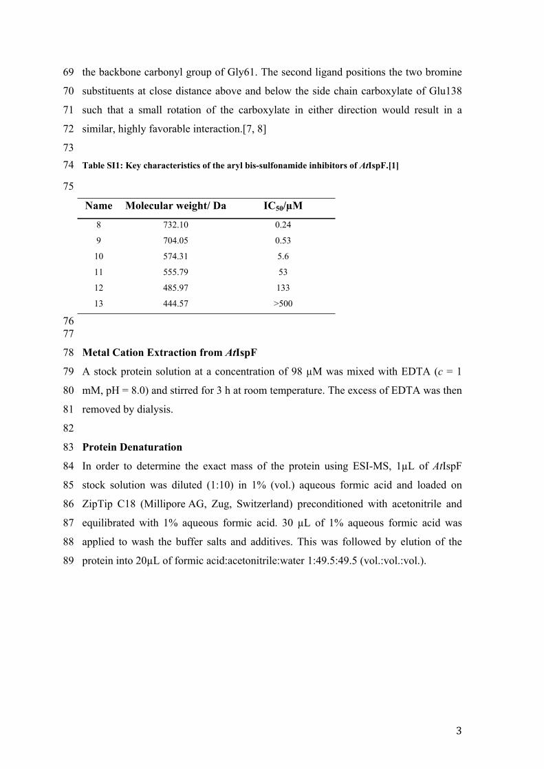
7374 Table SI1: Key characteristics of the aryl bis-sulfonamide inhibitors of AtIspF.[1]
75
Name Molecular weight/ Da IC50/µM
8 732.10 0.24
9 704.05 0.53
10 574.31 5.6
11 555.79 53
12 485.97 133
13 444.57 >500
7677
78 Metal Cation Extraction from AtIspF
79 A stock protein solution at a concentration of 98 µM was mixed with EDTA (c = 1
80 mM, pH = 8.0) and stirred for 3 h at room temperature. The excess of EDTA was then
81 removed by dialysis.
82
83 Protein Denaturation
84 In order to determine the exact mass of the protein using ESI-MS, 1µL of AtIspF
85 stock solution was diluted (1:10) in 1% (vol.) aqueous formic acid and loaded on
86 ZipTip C18 (Millipore AG, Zug, Switzerland) preconditioned with acetonitrile and
87 equilibrated with 1% aqueous formic acid. 30 µL of 1% aqueous formic acid was
88 applied to wash the buffer salts and additives. This was followed by elution of the
89 protein into 20µL of formic acid:acetonitrile:water 1:49.5:49.5 (vol.:vol.:vol.).
3

90 Mixing Scheme for the titration Experiments
91
9293 Scheme SI 1: Mixing scheme visualizing how the protein and ligand solutions were mixed for the 94 titration experiments.
9596 Data Analysis
97 All data analysis was done in MATLAB R2017a (MathWorks Inc., Natick, MA,
98 USA). When necessary, the spectra were background-adjusted using msbackadj
99 function from MATLAB Bioinformatics Toolbox and smoothed using Savitzky-
100 Golay algorithm implemented in sgolayfilt MATLAB function. In Savitzky-Golay
101 smoothing, the 2nd order polynomial function and a window of 9-25 samples were
102 used. The spectra normalization was done using msnorm function from MATLAB
103 Bioinformatics Toolbox.
104
105 Expression, Purification, and Characterization of Recombinant AtIspF
106 Synthetic gene of the full-length wild-type (wt) IspF from Arabidopsis thaliana
107 (AtIspF; UniProt accession number ISPF_ARATH) fused at the N-terminus with the
108 tobacco etch virus protease (TEV-protease) recognition site (ENLYF) was inserted
109 into a Novagen pET-15b expression vector (Merck & Cie, Schaffhausen, Switzerland)
110 at the NdeI and BamHI cloning sites. The gene synthesis and cloning into the
111 expression vector was performed by GenScript (GenScript USA Inc., Piscataway, NJ,
112 USA). The resulting recombinant product consisted of the N-terminal His6-tag, a
113 linker containing recognition sites for thrombin and TEV-protease, and wt AtIspF
4

114 (Scheme SI2). The recombinant polypeptide was 205-amino-acid-residue-long and
115 had a theoretical molecular weight of 22186.3 Da.
116
117
11810 20 30 40 50 60
MGSSHHHHHH SSGLVPRGSH MENLYFAASS AVDVNESVTS EKPTKTLPFR IGHGFDLHRL
70 80 90 100 110 120EPGYPLIIGG IVIPHDRGCE AHSDGDVLLH CVVDAILGAL GLPDIGQIFP DSDPKWKGAA
130 140 150 160 170 180SSVFIKEAVR LMDEAGYEIG NLDATLILQR PKISPHKETI RSNLSKLLGA DPSVVNLKAK
190 200THEKVDSLGE NRSIAAHTVI LLMKK
119120 Scheme SI 2: Amino acid sequence of the recombinant fusion protein His6-TEVrs-AtIspF. The N-121 terminal His6-tag sequence is highlighted in blue, the TEV-protease recognition site sequence is 122 highlighted in red. The sequence of full-length mature wild-type IspF from A. thaliana (UniProt 123 ID ISPF_ARATH; residues number 53-231 in UniProt notation) is shown in bold.
124125 His6-AtIspF was expressed in soluble form in E. coli One Shot™ BL21 Star™ (DE3)
126 host cells (Life Technologies Europe B.V., Zug, Switzerland). Cells were precultured
127 in 10 ml of LB broth containing 0.1 g L-1 ampicillin for 12 h at 37 °C with agitation
128 (240 rpm, orbital shaker). The starting culture was inoculated into 2 L of LB broth
129 containing 0.1 g L-1 ampicillin and was cultured at 37 °C with continuous agitation
130 (120 rpm, orbital shaker) for approximately 3.5 h until reached OD600 nm = 0.8. The
131 expression of the target transgene was induced by adding IPTG to a final
132 concentration of 1 mM. The expression was performed for 4 h at 37 °C with
133 continuous agitation (120 rpm, orbital shaker). The cells were harvested by
134 centrifugation and stored at -80 °C until used.
135 For extraction of the recombinant fusion protein, the cells were resuspended in a lysis
136 buffer (50 mM Tris-HCl, pH 8.0, 100 mM NaCl, 20 mM imidazole, 1 mg mL-1
137 lysozyme, 0.1 mM PMSF, 1 mM DTT) at a proportion of 10 ml buffer per 1 g of wet
138 cell paste and lysed by two consecutive passages through a high-pressure fluid
139 processor Microfluidizer 110S (Microfluidics, Newton, Massachusetts, USA)
140 operated at 40 psi. The homogenate was centrifuged at 25,000 rpm (rotor 45 Ti,
141 Optima L-90 K Ultracentrifuge, Beckman Coulter, Inc.), +4 °C for 1 h. The
142 supernatant containing soluble proteins was collected and processed further.
5

143 The target product was isolated from the soluble fraction of host cell lysate by Ni-
144 chelate chromatography on a GE HisTrap-FF column (5 ml bed volume; GE
145 Healthcare, Glattbrugg, Switzerland) using an Akta Prime Plus FPLC system (GE
146 Healthcare, Glattbrugg, Switzerland) equipped with dual-channel fluidics and a UV
147 absorbance and conductivity detectors. The column was equilibrated in elution buffer
148 A (50 mM Tris-HCl, pH 8.0, 100 mM NaCl, 20 mM imidazole, 0.5 mM DTT) at a
149 flow rate of 5 mL min-1. After the protein extract was loaded on the column and the
150 A280 nm reading returned to the base line, the protein captured on the Ni-NTA resin
151 was eluted with elution buffer B (50 mM Tris-HCl, pH 8.0, 100 mM NaCl, 300 mM
152 imidazole, 0.5 mM DTT) (Scheme SI3a). The concentration of imidazole in the
153 eluate was reduced back to 20 mM by desalting on a GE HighPrep 26/10 size-
154 exclusion column (MWCO = 5 kDa; GE Healthcare, Glattbrugg, Switzerland)
155 equilibrated in elution buffer A Scheme SI3b).
156 The His6-TEVrs-AtIspF fusion protein was digested with the recombinant catalytic
157 domain of TEV-protease to cleave the N-terminal His6-tag. After 4 h incubation at
158 room temperature, the reaction mixture was subjected to another round of Ni-chelate
159 chromatography in order to remove the uncleaved fusion protein, the His6-tag-
160 containing N-terminal fragment, and the TEV-protease, which also carried N-terminal
161 His6-tag (Scheme SI3c). The flow-through fraction from the GE HisTrap-FF column
162 contained high-purity full-length wt AtIspF (Scheme SI3d). Overall, the cloning,
163 expression, and purification strategy used here yielded approximately 18 mg of AtIspF
164 per 1 L of bacterial culture. The coupled-enzyme photometric assay confirmed that
165 the purified enzyme was active. The turnover numbers measured for two AtIspF
166 samples purified in separate batches were 5.9 min-1 and 6.8 min-1. The purified
167 enzyme was also sensitive to synthetic bis-sulfonamide inhibitors in the standard IC50
168 assay.[9]
169
6
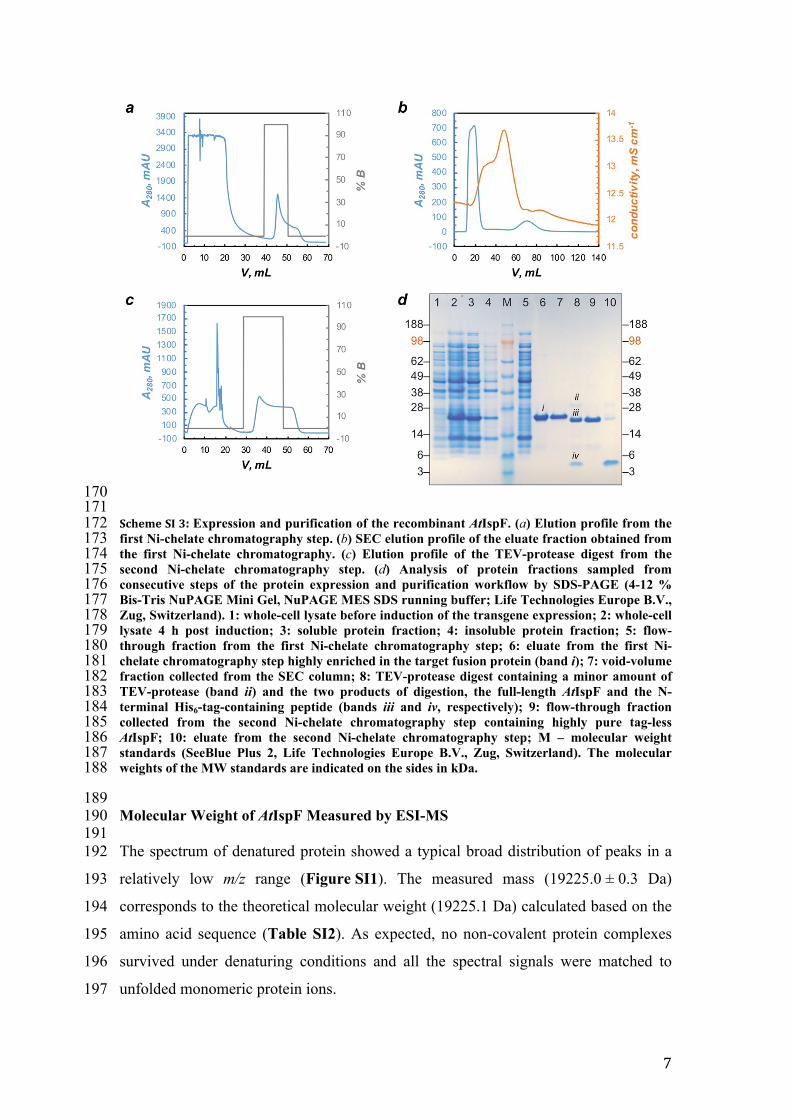
170171172 Scheme SI 3: Expression and purification of the recombinant AtIspF. (a) Elution profile from the 173 first Ni-chelate chromatography step. (b) SEC elution profile of the eluate fraction obtained from 174 the first Ni-chelate chromatography. (c) Elution profile of the TEV-protease digest from the 175 second Ni-chelate chromatography step. (d) Analysis of protein fractions sampled from 176 consecutive steps of the protein expression and purification workflow by SDS-PAGE (4-12 % 177 Bis-Tris NuPAGE Mini Gel, NuPAGE MES SDS running buffer; Life Technologies Europe B.V., 178 Zug, Switzerland). 1: whole-cell lysate before induction of the transgene expression; 2: whole-cell 179 lysate 4 h post induction; 3: soluble protein fraction; 4: insoluble protein fraction; 5: flow-180 through fraction from the first Ni-chelate chromatography step; 6: eluate from the first Ni-181 chelate chromatography step highly enriched in the target fusion protein (band i); 7: void-volume 182 fraction collected from the SEC column; 8: TEV-protease digest containing a minor amount of 183 TEV-protease (band ii) and the two products of digestion, the full-length AtIspF and the N-184 terminal His6-tag-containing peptide (bands iii and iv, respectively); 9: flow-through fraction 185 collected from the second Ni-chelate chromatography step containing highly pure tag-less 186 AtIspF; 10: eluate from the second Ni-chelate chromatography step; M – molecular weight 187 standards (SeeBlue Plus 2, Life Technologies Europe B.V., Zug, Switzerland). The molecular 188 weights of the MW standards are indicated on the sides in kDa.
189190 Molecular Weight of AtIspF Measured by ESI-MS 191192 The spectrum of denatured protein showed a typical broad distribution of peaks in a
193 relatively low m/z range (Figure SI1). The measured mass (19225.0 ± 0.3 Da)
194 corresponds to the theoretical molecular weight (19225.1 Da) calculated based on the
195 amino acid sequence (Table SI2). As expected, no non-covalent protein complexes
196 survived under denaturing conditions and all the spectral signals were matched to
197 unfolded monomeric protein ions.
7

198
199200 Figure SI 1: ESI mass spectrum acquired under denaturing conditions in positive ion mode. The 201 broad charge state distribution shows the unfolded AtIspF monomer in the low m/z range.
202
203 Table SI2: – Molecular weight of bare AtIspF using denaturing- and native ESI-MS.
204Monomer Dimer Trimer
Theoretical 19225.1 38450.2 57675.3
Denaturing 19225.0 ± 0.3 - -
Native 19224.1 ± 1.0 38449.2 ± 7.1 57680.7 ± 5.4
ESI
CID 19224.03 ± 0.4 38451.20 ± 2.1 -
205206 Experimentally estimated masses of neutral species were calculated from m/z values
207 of multiply-charged ions attributed to monomers, dimers, and trimers in Figure 1.
208 These values are compared to theoretical molecular weights calculated from amino
209 acid sequences. Deviations between experimental and theoretical values are indicated.
8

210211212 Figure SI 2: Evaluation of non-specific binding of CDP-ME to AtIspF. PMSF-inactivated trypsin 213 was mixed with AtIspF and measured before incubation (top spectrum) and after incubation 214 (bottom spectrum) with CDP-ME. Associated signals are highlighted in yellow boxes and were 215 attributed to trypsin (left box, green) and AtIspF (right box, yellow). Upon CDP-ME addition, 216 mass shifts corresponding to 1, 2 or 3 CDP-ME molecules bound to AtIspF trimer (indicated by 217 dotted lines) were observed exclusively. Negligible complex formation by CDP-ME and trypsin 218 was detected.
219 Formation of nonspecific ligated states is well known in ESI-MS and originates from
220 ligands sticking to the protein surface upon droplet shrinkage.[10] ESI-born non-
221 specific ligand binding due to electrostatic attraction has been shown to proceed
222 equally for all protein ions, regardless of their molecular weight.[10] Therefore, to
223 rule out the possibility of nonspecific binding of the substrate to AtIspF, we added
224 PMSF-inactivated trypsin as a reference protein (PREF) to the mixture of AtIspF with 7
225 (Figure SI2). We expected to detect complexes of trypsin with 5 or 7 should ESI-
226 related nonspecific binding be pronounced.
227
9
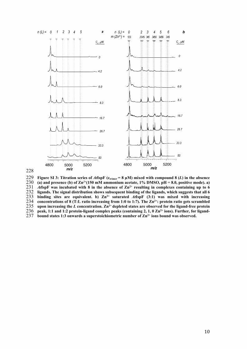
228229 Figure SI 3: Titration series of AtIspF (ctrimer = 8 µM) mixed with compound 8 (L) in the absence 230 (a) and presence (b) of Zn2+(150 mM ammonium acetate, 1% DMSO, pH = 8.0, positive mode). a) 231 AtIspF was incubated with 8 in the absence of Zn2+ resulting in complexes containing up to 6 232 ligands. The signal distribution shows subsequent binding of the ligands, which suggests that all 6 233 binding sites are equivalent. b) Zn2+ saturated AtIspF (3:1) was mixed with increasing 234 concentrations of 8 (T:L ratio increasing from 1:0 to 1:7). The Zn2+: protein ratio gets scrambled 235 upon increasing the L concentration. Zn2+ depleted states are observed for the ligand-free protein 236 peak, 1:1 and 1:2 protein-ligand complex peaks (containing 2, 1, 0 Zn2+ ions). Further, for ligand-237 bound states 1:3 onwards a superstoichiometric number of Zn2+ ions bound was observed.
10
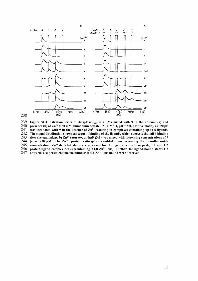
238239 Figure SI 4: Titration series of AtIspF (ctrimer = 8 µM) mixed with 9 in the absence (a) and 240 presence (b) of Zn2+ (150 mM ammonium acetate, 1% DMSO, pH = 8.0, positive mode). a) AtIspF 241 was incubated with 9 in the absence of Zn2+ resulting in complexes containing up to 6 ligands. 242 The signal distribution shows subsequent binding of the ligands, which suggests that all 6 binding 243 sites are equivalent. b) Zn2+ saturated AtIspF (3:1) was mixed with increasing concentrations of 9 244 (cL = 0-50 µM). The Zn2+: protein ratio gets scrambled upon increasing the bis-sulfonamide 245 concentration. Zn2+ depleted states are observed for the ligand-free protein peak, 1:1 and 1:2 246 protein-ligand complex peaks (containing 2,1,0 Zn2+ ions). Further, for ligand-bound states 1:3 247 onwards a superstoichiometric number of 4-6 Zn2+ ions bound were observed.
11
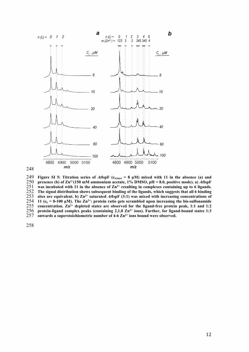
248249 Figure SI 5: Titration series of AtIspF (ctrimer = 8 µM) mixed with 11 in the absence (a) and 250 presence (b) of Zn2+(150 mM ammonium acetate, 1% DMSO, pH = 8.0, positive mode). a) AtIspF 251 was incubated with 11 in the absence of Zn2+ resulting in complexes containing up to 6 ligands. 252 The signal distribution shows subsequent binding of the ligands, which suggests that all 6 binding 253 sites are equivalent. b) Zn2+ saturated AtIspF (3:1) was mixed with increasing concentrations of 254 11 (cL = 0-100 µM). The Zn2+: protein ratio gets scrambled upon increasing the bis-sulfonamide 255 concentration. Zn2+ depleted states are observed for the ligand-free protein peak, 1:1 and 1:2 256 protein-ligand complex peaks (containing 2,1,0 Zn2+ ions). Further, for ligand-bound states 1:3 257 onwards a superstoichiometric number of 4-6 Zn2+ ions bound were observed.
258
12

259260 Figure SI 6: Titration series of AtIspF (ctrimer = 8 µM) mixed with 12 in the absence (a) and 261 presence (b) of Zn2+. a) AtIspF was incubated with 12 in the absence of Zn2+ resulting in 262 complexes containing up to 6 ligands. The signal distribution shows subsequent binding of the 263 ligands, which suggests that all 6 binding sites are equivalent. b) Zn2+ saturated AtIspF (3:1) was 264 mixed with increasing concentrations of 12 (cL = 0-100 µM). The Zn2+: protein ratio gets 265 scrambled upon increasing the sulfonamide concentration. Zn2+ depleted states are observed for 266 the ligand-free protein peak, 1:1 and 1:2 protein-ligand complex peaks (containing 2,1,0 Zn2+ 267 ions). Further, for ligand-bound states 1:3 onwards a superstoichiometric number of 4-6 Zn2+ 268 ions bound were observed.
13
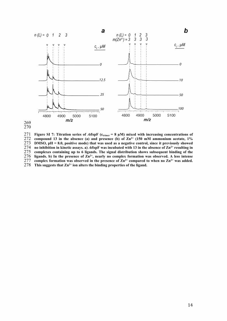
269270271 Figure SI 7: Titration series of AtIspF (ctrimer = 8 µM) mixed with increasing concentrations of 272 compound 13 in the absence (a) and presence (b) of Zn2+ (150 mM ammonium acetate, 1% 273 DMSO, pH = 8.0, positive mode) that was used as a negative control, since it previously showed 274 no inhibition in kinetic assays. a) AtIspF was incubated with 13 in the absence of Zn2+ resulting in 275 complexes containing up to 6 ligands. The signal distribution shows subsequent binding of the 276 ligands. b) In the presence of Zn2+, nearly no complex formation was observed. A less intense 277 complex formation was observed in the presence of Zn2+ compared to when no Zn2+ was added. 278 This suggests that Zn2+ ion alters the binding properties of the ligand.
14
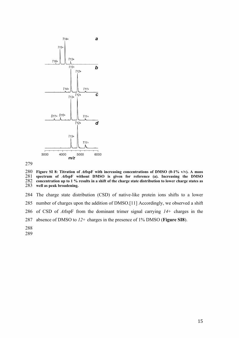
279280 Figure SI 8: Titration of AtIspF with increasing concentrations of DMSO (0-1% v/v). A mass 281 spectrum of AtIspF without DMSO is given for reference (a). Increasing the DMSO 282 concentration up to 1 % results in a shift of the charge state distribution to lower charge states as 283 well as peak broadening.
284 The charge state distribution (CSD) of native-like protein ions shifts to a lower
285 number of charges upon the addition of DMSO.[11] Accordingly, we observed a shift
286 of CSD of AtIspF from the dominant trimer signal carrying 14+ charges in the
287 absence of DMSO to 12+ charges in the presence of 1% DMSO (Figure SI8).
288289
15

290291 Figure SI 9: Evaluation of non-specific binding of 8 and inactive 13 to AtIspF. PMSF-inactivated 292 trypsin was mixed with AtIspF alone (top spectra in a and b) and upon incubation (bottom 293 spectra in a and b) with 8 and 13, respectively. Associated were attributed to trypsin (left box, 294 green) and AtIspF (right box, yellow). Upon the addition of 8, mass shifts corresponding to up to 295 three molecules 8 bound to AtIspF trimer exclusively (indicated by dotted lines) were observed. 296 Contrarily, no complex formation by 8 or 13 and trypsin was detected, respectively.
297 To determine the fraction of nonspecific protein-ligand complexes (8 and 13,
298 respectively) generated during the electrospray process, PMSF-inactivated trypsin
299 was added to the mixture as a reference protein (PREF) (Figure SI9). However, no
300 ligated states were observed for PREF with either compound. Thus, we can conclude
16
301 that the complexes between bis-sulfonamides and AtIspF (in the absence of Zn2+) are
302 not due to ESI artifacts but are indeed present in solution.
303
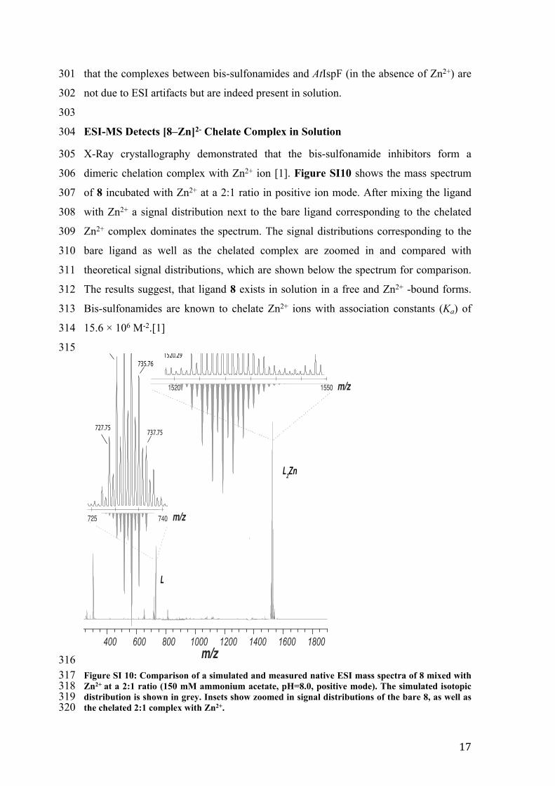
304 ESI-MS Detects [8–Zn]2- Chelate Complex in Solution
305 X-Ray crystallography demonstrated that the bis-sulfonamide inhibitors form a
306 dimeric chelation complex with Zn2+ ion [1]. Figure SI10 shows the mass spectrum
307 of 8 incubated with Zn2+ at a 2:1 ratio in positive ion mode. After mixing the ligand
308 with Zn2+ a signal distribution next to the bare ligand corresponding to the chelated
309 Zn2+ complex dominates the spectrum. The signal distributions corresponding to the
310 bare ligand as well as the chelated complex are zoomed in and compared with
311 theoretical signal distributions, which are shown below the spectrum for comparison.
312 The results suggest, that ligand 8 exists in solution in a free and Zn2+ -bound forms.
313 Bis-sulfonamides are known to chelate Zn2+ ions with association constants (Ka) of
314 15.6 × 10⁶ M-2.[1]
315
316317 Figure SI 10: Comparison of a simulated and measured native ESI mass spectra of 8 mixed with 318 Zn2+ at a 2:1 ratio (150 mM ammonium acetate, pH=8.0, positive mode). The simulated isotopic 319 distribution is shown in grey. Insets show zoomed in signal distributions of the bare 8, as well as 320 the chelated 2:1 complex with Zn2+.
17

321 Docking of Bis-Sulfonamide Ligands in the Presence of Zn
322 Bis-sulfonamide ligands have previously been shown to form a dianionic 2:1
323 complex with Zn2+ upon deprotonation of the sulfonamide N-H.[1] While Zn2+
324 cofactor depletion might be a possible mode of inhibition, enzymatic assays in
325 the presence of excess Zn2+ have shown undiminished inhibition and suggest
326 complex binding to the active site. The crystal structure of the [L2Zn]2–
327 complex with ligand 10 shows a tetrahedral coordination with the Zn2+ ion.
328 This complex was successfully modeled in the active site of AtIspF (PDB ID:
329 2PMP, Figure S11a). For the model the coordination geometry of the Zn2+ was
330 constrained. For the Zn2+, as well as for the Zn-free binding mode,
331 conformational preferences of the aryl-sulfonamide were taken into account
332 (Figure S11b).
333
334335 Figure SI11: Proposed binding mode of the [L2Zn]2– complex with 10 in the active site of AtIspF 336 (a, PDB ID: 2PMP,[2] 2.3 Å), including conformational constraints for aryl sulfonamides (b). The 337 mesh surface spans the volume of the active site.[12] Atom coloring: Br brown, N blue, O red, S 338 yellow, distances are given in Å.
339
340 The binding mode of the [L2Zn]2– complex of 10 shows coordination of the
341 zinc ion with Asp66, which is similar to the binding mode of CDP, where a
18

342 magnesium ion binds to the diphosphate moiety and to Asp66.[13] One ligand
343 molecule of 10 is deeply buried in the pocket, while the other partially
344 protrudes into the bulk. The buried ligand shows aromatic stacking interactions
345 of one tolyl substituent to the backbone amide of His37, while the other tolyl is
346 sandwiched between side-chains of Thr136 and Lys107. The latter possibly
347 also displays cation-π interactions with Lys107. The more exposed ligand has
348 two edge-to-face interactions of the tolyl and dibromobenzyl moieties with the
349 His37 side chain. The second tolyl group of this ligand has Van-der-Waals
350 interactions with the side chains of Thr136, Arg105 and Lys139.
351
352353 References354 1. Thelemann J, Illarionov B, Barylyuk K, Geist J, Kirchmair J, Schneider P, et 355 al. Aryl Bis-Sulfonamide Inhibitors of IspF from Arabidopsis thaliana and 356 Plasmodium falciparum. ChemMedChem. 2015;10(12):2090-8. doi: 357 10.1002/cmdc.201500382. PubMed PMID: 26435072.358 2. Calisto BM, Perez-Gil J, Bergua M, Querol-Audi J, Fita I, Imperial S. 359 Biosynthesis of isoprenoids in plants: structure of the 2C-methyl-D-erithrytol 360 2,4-cyclodiphosphate synthase from Arabidopsis thaliana. Comparison with the 361 bacterial enzymes. Protein Sci. 2007;16(9):2082-8. doi: 10.1110/ps.072972807. 362 PubMed PMID: 17660251; PubMed Central PMCID: PMCPMC2206962.363 3. Gerber PR, Muller K. Mab, a Generally Applicable Molecular-Force Field 364 for Structure Modeling in Medicinal Chemistry. J Comput Aided Mol Des. 365 1995;9(3):251-68. doi: Doi 10.1007/Bf00124456. PubMed PMID: 366 WOS:A1995RG92700005.367 4. Schwertz G, Frei MS, Witschel MC, Rottmann M, Leartsakulpanich U, 368 Chitnumsub P, et al. Conformational Aspects in the Design of Inhibitors for Serine 369 Hydroxymethyltransferase (SHMT): Biphenyl, Aryl Sulfonamide, and Aryl 370 Sulfone Motifs. Chem-Eur J. 2017;23(57):14345-57. doi: 371 10.1002/chem.201703244. PubMed PMID: WOS:000412819400032.372 5. Brameld KA, Kuhn B, Reuter DC, Stahl M. Small molecule conformational 373 preferences derived from crystal structure data. A medicinal chemistry focused 374 analysis. J Chem Inf Model. 2008;48(1):1-24. doi: 10.1021/ci7002494. PubMed 375 PMID: WOS:000252713700001.376 6. Cavallo G, Metrangolo P, Milani R, Pilati T, Priimagi A, Resnati G, et al. The 377 Halogen Bond. Chem Rev. 2016;116(4):2478-601. doi: 378 10.1021/acs.chemrev.5b00484. PubMed PMID: WOS:000371106000018.379 7. Zimmermann MO, Lange A, Zahn S, Exner TE, Boeckler FM. Using Surface 380 Scans for the Evaluation of Halogen Bonds toward the Side Chains of Aspartate, 381 Asparagine, Glutamate, and Glutamine. J Chem Inf Model. 2016;56(7):1373-83. 382 doi: 10.1021/acs.jcim.6b00075. PubMed PMID: WOS:000380422500013.383 8. Schwab A, Illarionov B, Frank A, Kunfermann A, Seet M, Bacher A, et al. 384 Mechanism of Allosteric Inhibition of the Enzyme IspD by Three Different Classes
19
385 of Ligands. ACS Chem Biol. 2017;12(8):2132-8. doi: 386 10.1021/acschembio.7b00004. PubMed PMID: 28686408.387 9. Illarionova V, Kaiser J, Ostrozhenkova E, Bacher A, Fischer M, Eisenreich 388 W, et al. Nonmevalonate terpene biosynthesis enzymes as antiinfective drug 389 targets: substrate synthesis and high-throughput screening methods. J Org Chem. 390 2006;71(23):8824-34. doi: 10.1021/jo061466o. PubMed PMID: 17081012.391 10. Sun N, Sun J, Kitova EN, Klassen JS. Identifying nonspecific ligand binding 392 in electrospray ionization mass spectrometry using the reporter molecule 393 method. J Am Soc Mass Spectrom. 2009;20(7):1242-50. doi: 394 10.1016/j.jasms.2009.02.024. PubMed PMID: 19321359.395 11. Sterling HJ, Prell JS, Cassou CA, Williams ER. Protein conformation and 396 supercharging with DMSO from aqueous solution. J Am Soc Mass Spectrom. 397 2011;22(7):1178-86. doi: 10.1007/s13361-011-0116-x. PubMed PMID: 398 21953100; PubMed Central PMCID: PMCPMC3107942.399 12. Ho BK, Gruswitz F. HOLLOW: generating accurate representations of 400 channel and interior surfaces in molecular structures. BMC Struct Biol. 401 2008;8:49. doi: 10.1186/1472-6807-8-49. PubMed PMID: 19014592; PubMed 402 Central PMCID: PMCPMC2603037.403 13. Kemp LE, Bond CS, Hunter WN. Structure of 2C-methyl-D-erythritol 2,4- 404 cyclodiphosphate synthase: an essential enzyme for isoprenoid biosynthesis and 405 target for antimicrobial drug development. Proc Natl Acad Sci U S A. 406 2002;99(10):6591-6. doi: 10.1073/pnas.102679799. PubMed PMID: 11997478; 407 PubMed Central PMCID: PMCPMC124447.408
20


![Advances in the stereoselective synthesis of …elpub.bib.uni-wuppertal.de/servlets/DerivateServlet/...Index III 3.2. Synthesis of Aryl-2-Benzo-[b]-Furanones 26 a-e: Prochiral Ketones](https://static.fdokument.com/doc/165x107/5f996fbb7308da14f4648be3/advances-in-the-stereoselective-synthesis-of-elpubbibuni-index-iii-32-synthesis.jpg)