Dopamine Modulation of Synaptic Transmission and Plasticity · Vous êtes super et sans votre...
124
Dopaminergic Modulation of Synaptic Transmission and Plasticity in the Lateral Amygdala Inauguraldissertation Zur Erlangung der Wuerde eines Doktors der Philosophie vorgelegt der Philosophisch-Naturwissenschaftlichen Fakultaet der Universitaet Basel von Stéphanie Bissière Aus Toulon, France Basel, June 2004
Transcript of Dopamine Modulation of Synaptic Transmission and Plasticity · Vous êtes super et sans votre...
Dopaminergic Modulation of Synaptic Transmission and
Plasticity
in the Lateral Amygdala
Inauguraldissertation
Zur
Erlangung der Wuerde eines Doktors der Philosophie
vorgelegt der
Philosophisch-Naturwissenschaftlichen Fakultaet
der Universitaet Basel
von
Stéphanie Bissière Aus Toulon, France
Basel, June 2004
Genehmigt von der Philosophisch-Naturwissenschaftlichen Fakultät
auf Antrag von
Andreas Lüthi and Markus Rüegg
Basel, den
Acknowledgement
A. Acknowledgements Premièrement, je voudrais remercier mes parents pour leur soutiens moral et financier pendant ces 10 ans d'études. Vous êtes super et sans votre constant réconfort et vos conseils je n'aurais jamais finis cette thèse. Une pensée pour mes grand-parents qui sont partis trop tôt...j'espère que vous êtes fiers de moi. Tout mon amour a Nicolas, éternellement reconnaissante pour tout, et surtout pour etre si different. Un énorme merci a tous mes amis, parents, cousins, cousines.. ici, là-bas et ailleurs..:on oublie jamais d'ou on vient et en quoi on croit. I would like to thank Andreas Lüthi for giving me the opportunity to be his first PhD student and to work on such an interesting project. I would like to thank Ronny, Nicolas and Hamdy I wish all the best and good luck for the futur. Thank you to Markus Rüegg for helpful discussion and for accepting to be part of my thesis commity. Thank you to Denis Monard for kindly agreing to supervise my oral exam. Merci à tous
1
Summary
B- Summary of the Thesis Fear conditioning is one of the most powerful and widely used paradigm to investigate
the mechanisms of associative learning in animals (LeDoux, 2000; Maren, 2001).
Behavioral and in vivo electrophysiological evidence indicate that induction of long-term-
potentiation (LTP), a form of associative, activity-dependent synaptic plasticity in the
lateral amygdala (LA), a brain structure tightly controlled by GABAergic inhibition,
underlies the acquisition of fear conditioning (Lang and Pare, 1997; Pare et al., 2003).
Dopamine (DA), the most abundant catecholemine in the brain, is released in the
amygdala upon stress. DA receptor activation is required for the potentiation of sensory
evoked neuronal activity in the LA during conditioning (Rosenkranz and Grace, 2002).
Conversely, intra-amygdala injections of DA receptor antagonists prevents the
acquisition of fear conditioning (Greba et al., 2001; Greba and Kokkinidis, 2000;
Guarraci et al., 2000; Guarraci et al., 1999). The cellular and synaptic mechanisms
underlying the dopaminergic modulation of fear conditioning and synaptic plasticity are,
however, still unknown. In the first part of my work, I showed that DA gates the
induction of LTP in the mouse LA by supressing feed-forward inhibition mediated by
local interneurons. The action of DA on synaptic plasticity depended on the activation of
D2 receptors and appeared to be twofold. First, it reduced the quantal content at
inhibitory synapses, thereby decreasing inhibitory synaptic transmission and second, it
facilitated inhibition onto interneurons by depolarizing interneurons involved in
disinhibition. In the second part of my work I investigated the role of DA on spontaneous
inhibitory network activity. Consistent with previous in vivo data showing that systemic
administration of DA agonists in the LA increases the spontaneous firing of interneurons
2
Summary
3
(Rosenkranz and Grace, 1999), we found that bath application of DA increased the
frequency of spontaneous inhibitory transmission recorded from projection neurons. In
contrast to the gating of LTP, this effect required the activation of D1 and D2 receptors in
synergy. Preliminary data suggested that the D1 receptor-mediated increase in
spontaneous inhibitory transmission did not involve cAMP-mediated intracellular
signaling mechanisms.
Table of Contents
C. Table of Contents Pages A. Acknowledgements 1 B. Summary of the Thesis 2 C. Table of Contents 4 D. List of Figures 6 E. List of Abbrevations 7 1. INTRODUCTION 10 1.1. The emotional brain 10
1.1.1. Neural circuits underlying associative fear conditioning 13 1.2. The amygdala 14 1.2.1. Brief history 14 1.2.2. Amygdala terminology 14 1.2.3. Amygdala connectivity 16 1.3. The amygdala and Pavlovian fear conditioning 20 1.3.1. Pathways transmitting information about the CS 20 1.3.2. Pathways transmitting information about the US 22 1.3.3. Expression of fear conditioning: output pathways 22 1.4. Synaptic transmission in the LA 25 1.4.1. Cell type diversity 25 1.4.2. Excitatory transmission in the LA 28 1.4.3. Inhibitory transmission in the LA 30 1.5. Long-term changes in synaptic strength: a model for fear learning 31 1.5.1. Hebbian theory 32 1.5.2. Synaptic plasticity: LTP in the hippocampus 32 1.5.3. Hippocampal LTP: Mechanisms 36 1.5.4. Long-term-depression (LTD) 39 1.5.5. LTP versus LTD 39 1.6. The amygdala and fear conditioning: pharmacological studies 40 1.6.1. NMDA receptors 41 1.6.2. VGCCs 41 1.6.3. Protein synthesis and fear conditioning 41 1.7. Physiological Plasticity in the Amygdala Related to Fear Conditioning. 41 1.7.1. Single unit recordings and sensory pathway stimulation in freely moving 41
animals 1.7.2. Amygdala neurons exhibit LTP 42 1.7.3. Synaptic plasticity in the LA 43 1.7.4. LTP and synaptic inhibition 45
4
Table of Contents
5
1.8. Neuromodulation of inhibition 46 1.8.1. The dopaminergic system: an introduction 46 1.8.2. A modulatory role for dopamine 46 2. AIM OF THE THESIS 48 3.RESULTS 49 3.1. Publication 1: Dopamine gates LTP induction in the lateral amygdala 49 by supressing feed-forward inhibition -Dopamine gates LTP induction 50 -Supression of feedforward inhibition 54 -Discussion 61 -Materials and methods 65 3.2.Unpublished data: Dopaminergic modulation of spontaneous 68 inhibitory network activity in the lateral amygdala -Dopamine depolarizes interneurons 69 -Dopamine receptor subtypes mediating the increase 69 in sIPSCs frequency -Signalling mechanisms mediating the DA-dependent 70 increase in sIPSC frequency -Dopamine induces alteration in the burst frequency, 70 synchronization and oscillatory inhibitory activity in the LA -Discussion 71 -Figures 74 -Materials and methods 80 4. GENERAL DISCUSSION 82 4.1. Thalamico-amygdala plasticity and fear conditioning 82 4.2. Thalamic versus cortical inputs to the LA 85 4.3. Amygdala and inhibition 86 4.4. Dopaminergic modulation of synaptic transmission and plasticity 89 5. CONCLUSIONs AND OUTLOOK 92 6. REFERENCES 93 7. CV 122
List of Figures
D. List of Figures Pages 1. INTRODUCTION Fig.1. A typical fear conditioning experiment 12 Fig.2. Nuclei of the rat amygdaloid complex 15 Fig.3. Main connections of the LA 17 Fig.4. Summary of the main inputs and outputs of the LA 18 Fig.5. Intranuclear connections of the BL and summary of the extra-amygdaloid 19
inputs and ouputs of the BL. Fig.6. Intranuclear connections of the CeA and its extra-amygdaloid inputs and ouputs. 20 Fig.7. Anatomy of fear conditioning in the brain 24 Fig.8. Morphological and electrophysiological properties of projection neurons and 26 interneurons, the two main cell types in the LA Fig.9. Diagram showing the overlap and relative proportions of CB+, PV+ and CR+ 27 containing GABAergic interneurons Fig.10. LTP and the hippocampus 33 Fig.11. Basic properties of LTP 35 Fig.12. Model for the induction of LTP 37 Fig.13. Behavioral LTP is induced in the lateral amygdala by Pavlovian fear conditioning 43 fear conditioning 3. RESULTS 3.1. Publication 1 Fig.14. Inhibitory synaptic transmission prevents the induction of LTP 51 Fig.15. Dopamine enables the induction of LTP in the absence of picrotoxin 53 Fig.16. Dopamine depresses feed-forward inhibition 56 Fig.17. Dopamine decreases inhibition onto projection neurons 58 Fig.18. Dopamine increases inhibition onto interneurons 60 Fig.19. Schematic circuit diagram based on the present results illustrating 62 two mechanisms by which dopamine modulates feed-forward inhibitory circuits gating LTP induction in the LA 3.2. Unpublished Results Fig.20. Dopamine increases spontaneous inhibition 74 Fig.21. Dopamine does not affect spontaneous excitatory network activity 75 Fig.22. Dopamine increases the spontaneous inhibitory network activity by 76 modulating the somatodendritic properties of interneurons Fig.23. Dopamine-dependent increase in sIPSCs requires D1 receptor activation 77 and D1/D2 synergy Fig.24. The increase in sIPSC frequency does not require activation of PKA 78 or PLC Fig.25. Dopamine induces inhibitory oscillatory network activity 79
6
Introduction
1-Introduction
1.1. The Emotional Brain
Whether you are happy, sad or frightened, emotions in general have an undeniable grip
on your life. However, in the history of neuroscience, understanding the brain
mechanisms involved in emotion was not always of primary interest. During the first half
of the 20th century, many researchers, including pioneers of neuroscience like Hebb,
Sherrington or Cannon, were immensely interested in brain mechanisms underlying
emotional behavior. Later on in the 50th however, the amount of interest dedicated to this
area started to decline with the emergence of cognitive science. Thus, research interests
shifted towards processes like perception or memory that were thought to perform in a
similar way to computers. In addition, cognitive questions seemed more tractable and less
influenced by subjectivity than emotional ones. Another factor that contributed at the
time to the decline of research on emotions, was the "limbic system" concept, developed
by MacLean in 1952. This concept reintroduced Broca's term "limbic" to describe a
neuroanatomical circuit involved in emotional functions. At the time, this seemed a
reasonable answer to the problem on how the brain makes emotions (Maclean, 1952). His
theory was based on two concepts: First, that the neocortex was a mammalian
specialization and that therefore all the cognitive processes such as thinking, reasoning or
memory, had to be mediated by this brain area. Second, the limbic system, that comprised
at the time the old cortex and related subcortical areas, was responsible for mediating old
aspect of behavior such as mental life and emotions. Based on these two facts, MacLean
concluded that the neocortex was responsible for the processing of cognitive functions
whereas the limbic system would process emotions.
Soon after its emergence, the limbic system explanation of emotions started to be
questioned, especially with the discovery that the equivalent of mammalian neocortex
was also found in non-mammalian vertebrates (Nauta, 1979). Today, even if there is quite
some understanding on the limbic system as an anatomical concept and as a
neuroanatomical circuit involved in emotion processing, the exact brain areas involved in
-10-
Introduction this system still remain a matter of debate. Moreover, very little is known on how it
actually produces emotions. Some of the original notions of MacLean, however, seem to
be still holding true. Especially the idea that emotions are primitive circuits and that they
are conserved throughout mammalian evolution emerged as a framework. One exception
that was made concerning the amount of interest dedicated to the understanding of
emotion processing was the circuitry of fear reactions. Fear is a particularly good model
to study because it is well conserved across human and non-human species. In animals,
the behavioral reactions produced by fear are a direct read out of the activation of the
processing circuits that detect and respond to fearful situations. Thus, it is an attractive
and experimentally tractable model. The fact that the emotional significance of a stimulus
can be manipulated was showed for the first time by Pavlov (1915) in a food conditioning
experiment. In this experiment, he made dogs salivate just by presenting them with a
stimulus that had been associated with the delivery of food. In an other set of
experiments, the dogs also exhibited conditioned reflexes as a protection against harmful
stimuli which were referred to by Pavlov as conditioned reflexes, or fear conditioning. In
the early 1900's, another set of experiment was carried out by Watson who conclusively
demonstrated that Pavlov's model of behavior and learning could also apply in human
(Watson, 1920). In his test, Watson and his graduate student Rosalie Rayner took an
infant orphan, best known now as little Albert, who was scared of nothing except of loud
and abrupt noises, and attempted to condition him to fear rats. In order to achieve this,
they hammered a steel bar every time little Albert was reaching out to play with the rat.
Since little Albert was scared of the noise, he learned that the rat was associated with the
loud hammering noise and started to exhibit extreme fear to the rat alone. Since then, fear
conditioning has become one of the most robust and widely used paradigm to study the
mechanisms of associative learning in animals and humans. In a classical fear
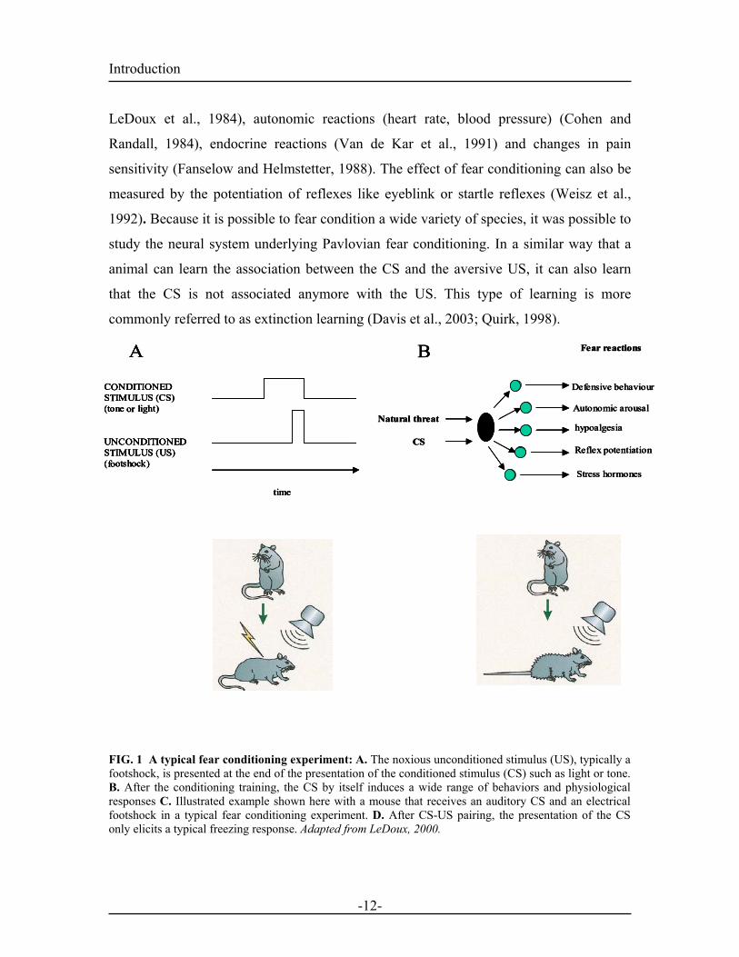
conditioning experiment, the animal, a rat or a mouse for example is subjected to a
conditioned stimulus (CS) like a light or a tone, followed by an unconditioned stimulus
(US) such as a mild electric footshock. After a few CS-US pairings, a fear response can
be elicited by the CS presentation alone. The fear reactions induced by the CS
presentation include freezing (absence of movement) (Blanchard and Blanchard, 1972;
-11-
Introduction
-12-
LeDoux et al., 1984), autonomic reactions (heart rate, blood pressure) (Cohen and
Randall, 1984), endocrine reactions (Van de Kar et al., 1991) and changes in pain
sensitivity (Fanselow and Helmstetter, 1988). The effect of fear conditioning can also be
measured by the potentiation of reflexes like eyeblink or startle reflexes (Weisz et al.,
1992). Because it is possible to fear condition a wide variety of species, it was possible to
study the neural system underlying Pavlovian fear conditioning. In a similar way that a
animal can learn the association between the CS and the aversive US, it can also learn
that the CS is not associated anymore with the US. This type of learning is more
commonly referred to as extinction learning (Davis et al., 2003; Quirk, 1998).
Mouse learns to fear toneMouse freezes in response to tone
C NDITIONED S IMULUS (CS) (tone light)
UNCONDITIONED S IMULUS (US)( otshock)
timeTraining: tone + shock
A Fear reactions
Natural threat
CS
Defensive behaviour
Autonomic arousal
hypoalgesia
Reflex potentiation
Stress hormones
B
Mouse learns to fear toneMouse freezes in response to tone
C NDITIONED S IMULUS (CS) (tone light)
UNCONDITIONED S IMULUS (US)( otshock)
timeTraining: tone + shock
A
C NDITIONED S IMULUS (CS) (tone light)
UNCONDITIONED S IMULUS (US)( otshock)
time
C NDITIONED S IMULUS (CS) (tone light)
UNCONDITIONED S IMULUS (US)( otshock)
timeTraining: tone + shock
A Fear reactions
Natural threat
CS
Defensive behaviour
Autonomic arousal
hypoalgesia
Reflex potentiation
Stress hormones
B Fear reactions
Natural threat
CS
Defensive behaviour
Autonomic arousal
hypoalgesia
Reflex potentiation
Stress hormones
B
OTOTOTOT
orororor
TTTT fofofofo
FIG. 1 A typical fear conditioning experiment: A. The noxious unconditioned stimulus (US), typically a footshock, is presented at the end of the presentation of the conditioned stimulus (CS) such as light or tone. B. After the conditioning training, the CS by itself induces a wide range of behaviors and physiological responses C. Illustrated example shown here with a mouse that receives an auditory CS and an electrical footshock in a typical fear conditioning experiment. D. After CS-US pairing, the presentation of the CS only elicits a typical freezing response. Adapted from LeDoux, 2000.
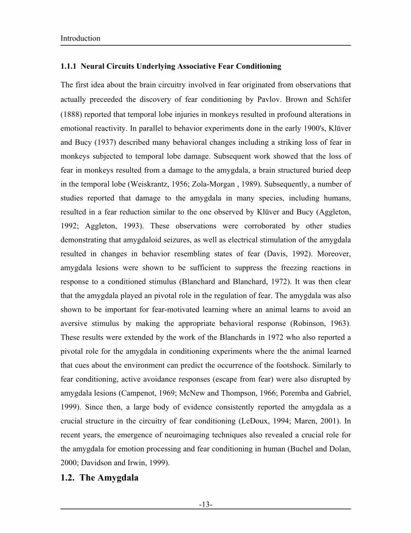
Introduction 1.1.1 Neural Circuits Underlying Associative Fear Conditioning
The first idea about the brain circuitry involved in fear originated from observations that
actually preceeded the discovery of fear conditioning by Pavlov. Brown and Schäfer
(1888) reported that temporal lobe injuries in monkeys resulted in profound alterations in
emotional reactivity. In parallel to behavior experiments done in the early 1900's, Klüver
and Bucy (1937) described many behavioral changes including a striking loss of fear in
monkeys subjected to temporal lobe damage. Subsequent work showed that the loss of
fear in monkeys resulted from a damage to the amygdala, a brain structured buried deep
in the temporal lobe (Weiskrantz, 1956; Zola-Morgan , 1989). Subsequently, a number of
studies reported that damage to the amygdala in many species, including humans,
resulted in a fear reduction similar to the one observed by Klüver and Bucy (Aggleton,
1992; Aggleton, 1993). These observations were corroborated by other studies
demonstrating that amygdaloid seizures, as well as electrical stimulation of the amygdala
resulted in changes in behavior resembling states of fear (Davis, 1992). Moreover,
amygdala lesions were shown to be sufficient to suppress the freezing reactions in
response to a conditioned stimulus (Blanchard and Blanchard, 1972). It was then clear
that the amygdala played an pivotal role in the regulation of fear. The amygdala was also
shown to be important for fear-motivated learning where an animal learns to avoid an
aversive stimulus by making the appropriate behavioral response (Robinson, 1963).
These results were extended by the work of the Blanchards in 1972 who also reported a
pivotal role for the amygdala in conditioning experiments where the the animal learned
that cues about the environment can predict the occurrence of the footshock. Similarly to
fear conditioning, active avoidance responses (escape from fear) were also disrupted by
amygdala lesions (Campenot, 1969; McNew and Thompson, 1966; Poremba and Gabriel,
1999). Since then, a large body of evidence consistently reported the amygdala as a
crucial structure in the circuitry of fear conditioning (LeDoux, 1994; Maren, 2001). In
recent years, the emergence of neuroimaging techniques also revealed a crucial role for
the amygdala for emotion processing and fear conditioning in human (Buchel and Dolan,
2000; Davidson and Irwin, 1999).
1.2. The Amygdala
-13-
Introduction
1.2.1 Brief History
Two centuries ago the latin name amygdala was given by the anatomist Burdach
(Burdach, 1819-1922) to describe a cluster of brain nuclei in the anterior portion of the
human temporal lobe whose shape resembled that of an almond. The subsequent work of
Völsch and Johnston (Jonston, 1923) together with the development of histological
techniques, set the landmark of what was going to be a hundred years later one of the best
studied brain structure for understanding association and sorting of emotional signals and
their resulting pathologies.
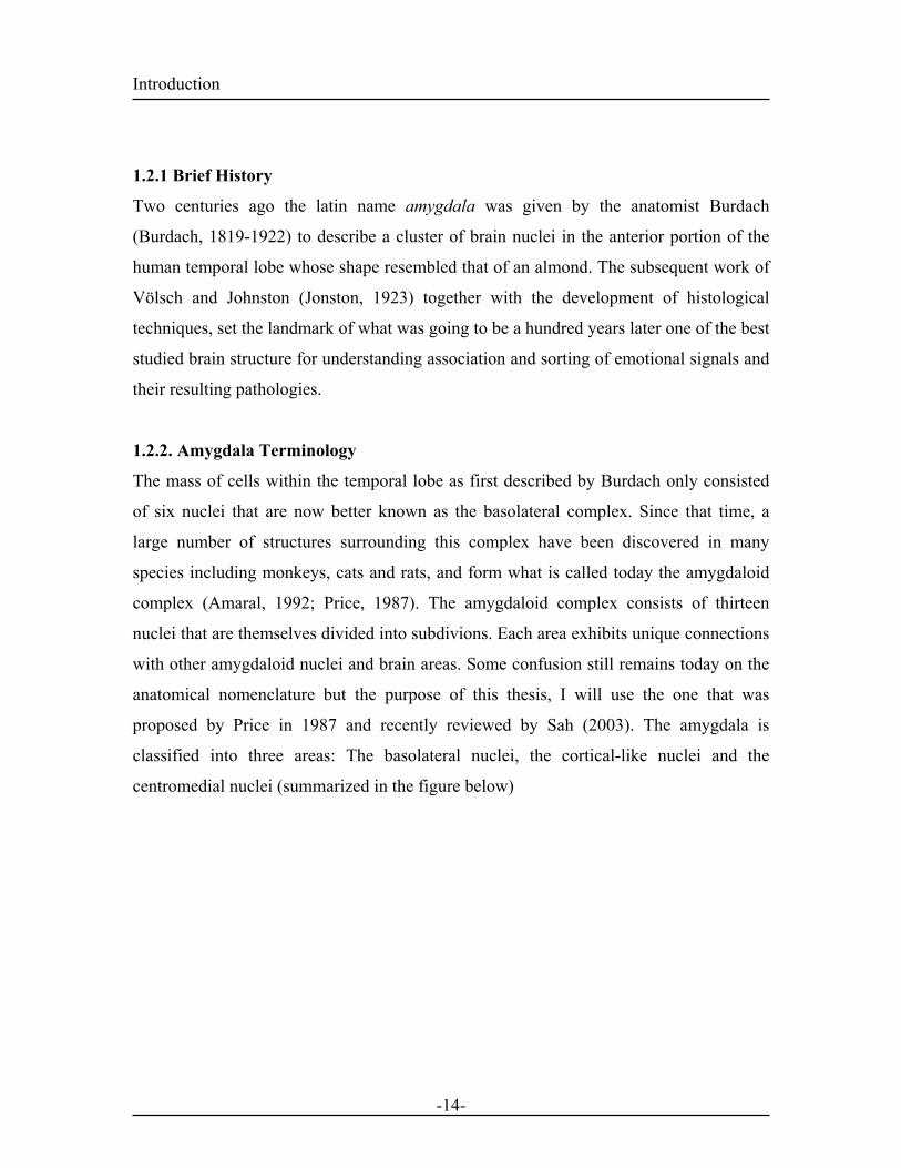
1.2.2. Amygdala Terminology
The mass of cells within the temporal lobe as first described by Burdach only consisted
of six nuclei that are now better known as the basolateral complex. Since that time, a
large number of structures surrounding this complex have been discovered in many
species including monkeys, cats and rats, and form what is called today the amygdaloid
complex (Amaral, 1992; Price, 1987). The amygdaloid complex consists of thirteen
nuclei that are themselves divided into subdivions. Each area exhibits unique connections
with other amygdaloid nuclei and brain areas. Some confusion still remains today on the
anatomical nomenclature but the purpose of this thesis, I will use the one that was
proposed by Price in 1987 and recently reviewed by Sah (2003). The amygdala is
classified into three areas: The basolateral nuclei, the cortical-like nuclei and the
centromedial nuclei (summarized in the figure below)
-14-
Introduction
DC
BA
FIG. 2 Nuclei of the rat amygdaloid complex. Coronal sections are drawn from rostral (A) to caudal (D). The different nuclei are divided into three groups as described in the text. Areas in blue form part of the basolateral group, areas in yellow are the cortical group, and areas in green form the centromedial group. Abbr: ABmc, accessory basal magnocellular subdivision; ABpc, accessory basal parvicellular subdivision; Bpc, basal nucleus magnocellular subdivision; e.c., external capsule; Ladl, lateral amygdala medial subdivision; Lam, lateral amygdala medial subdivision; Lavl, lateral amygdala ventrolateral subdivision; Mcd, medial amygdala dorsal subdivision; Mcv, medial amygdala ventral subdivision; Mr, medial amygdala rostral subdivision; Pir, piriform cortex; s.t., stria terminalis. Adapted from Sah, 2003.
-15-
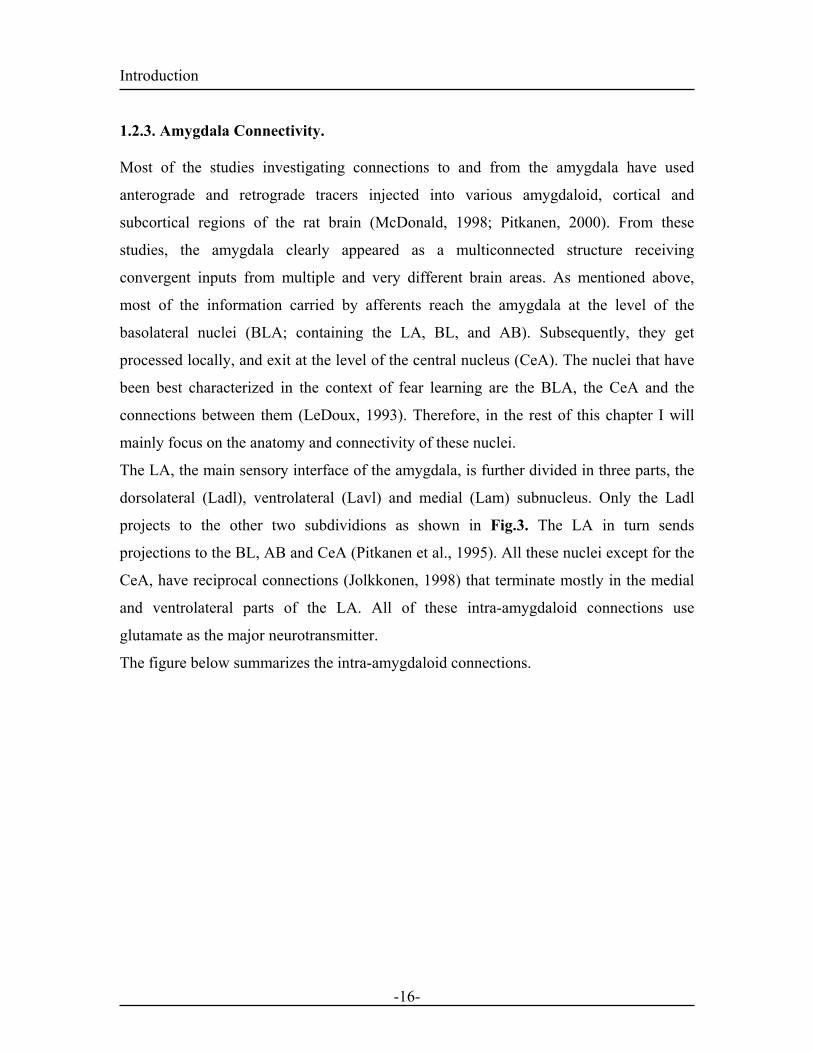
Introduction 1.2.3. Amygdala Connectivity.
Most of the studies investigating connections to and from the amygdala have used
anterograde and retrograde tracers injected into various amygdaloid, cortical and
subcortical regions of the rat brain (McDonald, 1998; Pitkanen, 2000). From these
studies, the amygdala clearly appeared as a multiconnected structure receiving
convergent inputs from multiple and very different brain areas. As mentioned above,
most of the information carried by afferents reach the amygdala at the level of the
basolateral nuclei (BLA; containing the LA, BL, and AB). Subsequently, they get
processed locally, and exit at the level of the central nucleus (CeA). The nuclei that have
been best characterized in the context of fear learning are the BLA, the CeA and the
connections between them (LeDoux, 1993). Therefore, in the rest of this chapter I will
mainly focus on the anatomy and connectivity of these nuclei.
The LA, the main sensory interface of the amygdala, is further divided in three parts, the
dorsolateral (Ladl), ventrolateral (Lavl) and medial (Lam) subnucleus. Only the Ladl
projects to the other two subdividions as shown in Fig.3. The LA in turn sends
projections to the BL, AB and CeA (Pitkanen et al., 1995). All these nuclei except for the
CeA, have reciprocal connections (Jolkkonen, 1998) that terminate mostly in the medial
and ventrolateral parts of the LA. All of these intra-amygdaloid connections use
glutamate as the major neurotransmitter.
The figure below summarizes the intra-amygdaloid connections.
-16-
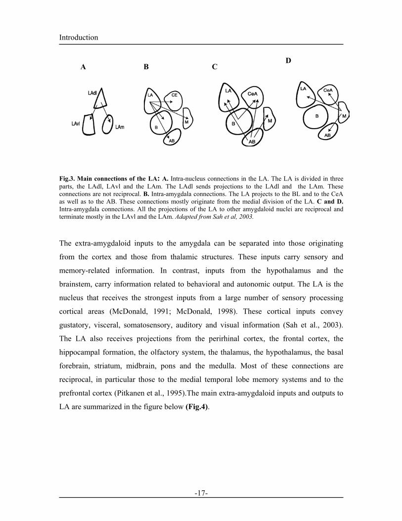
Introduction D A B C Fig.3. Main connections of the LA: A. Intra-nucleus connections in the LA. The LA is divided in three parts, the LAdl, LAvl and the LAm. The LAdl sends projections to the LAdl and the LAm. These connections are not reciprocal. B. Intra-amygdala connections. The LA projects to the BL and to the CeA as well as to the AB. These connections mostly originate from the medial division of the LA. C and D. Intra-amygdala connections. All the projections of the LA to other amygdaloid nuclei are reciprocal and terminate mostly in the LAvl and the LAm. Adapted from Sah et al, 2003.
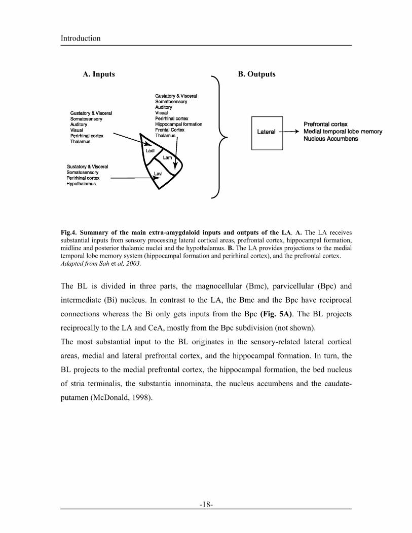
The extra-amygdaloid inputs to the amygdala can be separated into those originating
from the cortex and those from thalamic structures. These inputs carry sensory and
memory-related information. In contrast, inputs from the hypothalamus and the
brainstem, carry information related to behavioral and autonomic output. The LA is the
nucleus that receives the strongest inputs from a large number of sensory processing
cortical areas (McDonald, 1991; McDonald, 1998). These cortical inputs convey
gustatory, visceral, somatosensory, auditory and visual information (Sah et al., 2003).
The LA also receives projections from the perirhinal cortex, the frontal cortex, the
hippocampal formation, the olfactory system, the thalamus, the hypothalamus, the basal
forebrain, striatum, midbrain, pons and the medulla. Most of these connections are
reciprocal, in particular those to the medial temporal lobe memory systems and to the
prefrontal cortex (Pitkanen et al., 1995).The main extra-amygdaloid inputs and outputs to
LA are summarized in the figure below (Fig.4).
-17-
Introduction
B. Outputs A. Inputs
Fig.4. Summary of the main extra-amygdaloid inputs and outputs of the LA. A. The LA receives substantial inputs from sensory processing lateral cortical areas, prefrontal cortex, hippocampal formation, midline and posterior thalamic nuclei and the hypothalamus. B. The LA provides projections to the medial temporal lobe memory system (hippocampal formation and perirhinal cortex), and the prefrontal cortex. Adapted from Sah et al, 2003.
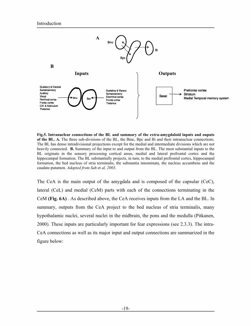
The BL is divided in three parts, the magnocellular (Bmc), parvicellular (Bpc) and
intermediate (Bi) nucleus. In contrast to the LA, the Bmc and the Bpc have reciprocal
connections whereas the Bi only gets inputs from the Bpc (Fig. 5A). The BL projects
reciprocally to the LA and CeA, mostly from the Bpc subdivision (not shown).
The most substantial input to the BL originates in the sensory-related lateral cortical
areas, medial and lateral prefrontal cortex, and the hippocampal formation. In turn, the
BL projects to the medial prefrontal cortex, the hippocampal formation, the bed nucleus
of stria terminalis, the substantia innominata, the nucleus accumbens and the caudate-
putamen (McDonald, 1998).
-18-
Introduction A
B Inputs Outputs
Fig.5. Intranuclear connections of the BL and summary of the extra-amygdaloid inputs and ouputs of the BL. A. The three sub-divisions of the BL, the Bmc, Bpc and Bi and their intranuclear connections. The BL has dense intradivisional projections except for the medial and intermediate divisions which are not heavily connected. B. Summary of the input to and output from the BL. The most substantial inputs to the BL originate in the sensory processing cortical areas, medial and lateral prefrontal cortex and the hippocampal formation. The BL substantially projects, in turn, to the medial prefrontal cortex, hippocampal formation, the bed nucleus of stria terminalis, the substantia innominata, the nucleus accumbens and the caudate-putamen. Adapted from Sah et al, 2003.
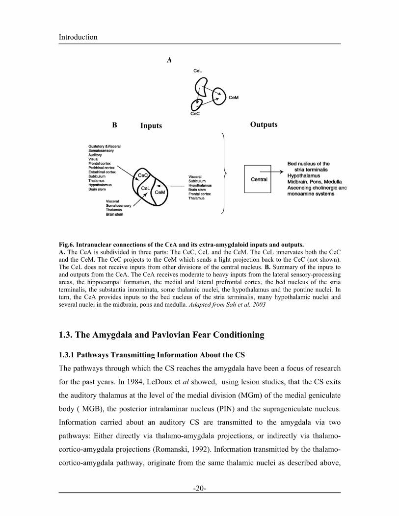
The CeA is the main output of the amygdala and is composed of the capsular (CeC),
lateral (CeL) and medial (CeM) parts with each of the connections terminating in the
CeM (Fig. 6A) . As described above, the CeA receives inputs from the LA and the BL. In
summary, outputs from the CeA project to the bed nucleus of stria terminalis, many
hypothalamic nuclei, several nuclei in the midbrain, the pons and the medulla (Pitkanen,
2000). These inputs are particularly important for fear expressions (see 2.3.3). The intra-
CeA connections as well as its major input and output connections are summarized in the
figure below:
-19-
Introduction A
Outputs B Inputs
Fig.6. Intranuclear connections of the CeA and its extra-amygdaloid inputs and outputs. A. The CeA is subdivided in three parts: The CeC, CeL and the CeM. The CeL innervates both the CeC and the CeM. The CeC projects to the CeM which sends a light projection back to the CeC (not shown). The CeL does not receive inputs from other divisions of the central nucleus. B. Summary of the inputs to and outputs from the CeA. The CeA receives moderate to heavy inputs from the lateral sensory-processing areas, the hippocampal formation, the medial and lateral prefrontal cortex, the bed nucleus of the stria terminalis, the substantia innominata, some thalamic nuclei, the hypothalamus and the pontine nuclei. In turn, the CeA provides inputs to the bed nucleus of the stria terminalis, many hypothalamic nuclei and several nuclei in the midbrain, pons and medulla. Adapted from Sah et al. 2003
1.3. The Amygdala and Pavlovian Fear Conditioning
1.3.1 Pathways Transmitting Information About the CS
The pathways through which the CS reaches the amygdala have been a focus of research
for the past years. In 1984, LeDoux et al showed, using lesion studies, that the CS exits
the auditory thalamus at the level of the medial division (MGm) of the medial geniculate
body ( MGB), the posterior intralaminar nucleus (PIN) and the suprageniculate nucleus.
Information carried about an auditory CS are transmitted to the amygdala via two
pathways: Either directly via thalamo-amygdala projections, or indirectly via thalamo-
cortico-amygdala projections (Romanski, 1992). Information transmitted by the thalamo-
cortico-amygdala pathway, originate from the same thalamic nuclei as described above,
-20-
Introduction as well as from the ventral (MGv) and, dorsal (MGd) divisions of the MGB. They then
exit the thalamus to terminate in the temporal neocortex and the perirhinal periallocortex.
These regions in turn, project to the dorsolateral and medial lateral part of the LA (see
2.1.2, Fig. 4) (LeDoux et al., 1991; McDonald, 1998; Roger and Arnault, 1989; Turner
and Herkenham, 1991). Lesion of either pathway does not impair the acquisition of fear
conditioning to a simple tone, implying that each of the two routes are sufficient for CS
transmission. Lesions of both pathways however, impairs fear conditioning dramatically
(Romanski and LeDoux, 1992b). These results indicate that CS transmission to the LA is
necessary for fear conditioning to occur (LeDoux et al., 1984; Romanski and LeDoux,
1992a; Romanski, 1992). The main difference of the thalamo-cortico-amygdala system
over the thalamo-amygdala pathway has been suggested to be that information carried by
the CS have access to the higher processing capacities of the neocortex making it a more
suitable pathway to process elaborate auditory information (Jarrell et al., 1987). It is
generally believed that more complex processes occur in cortical areas rather than in
thalamic ones, but the exact conditions requiring cortical discriminative processes during
fear conditioning are still poorly understood (Armony et al., 1997). In addition, the
thalamo-cortico-amygdala pathway is slower at transmitting information to the LA since
it involves several cortico-cortico synapses. This suggest complementary properties of
the two pathways with respect to the speed and the accurancy of transmitted information.
Although much of the studies on fear conditioning have used an auditory CS, some
studies have also used a visual CS (Aggleton, 1992; Davis, 1987). Visual fear
conditioning, even if it is a less frequently used paradigm, can also be acquired by pairing
a light with a footshock (Shi and Davis, 2001).
In addition to cued fear conditioning (CS-US association), a rat can also exhibit fear just
by being placed back into the environment where the conditioning previously occurred.
This type of conditioning is called contextual fear conditioning (Maren and Fanselow,
1995). Information about the spatial context are provided by the CA1 area and subiculum
of the ventral hippocampus, that project monosynaptically to the B and AB nucleus of the
amygdala (Canteras and Swanson, 1992). Damage to either of these areas interferes with
the acquisition of contextual conditioning (Maren et al., 1997; Phillips and LeDoux,
-21-
Introduction 1992). The hippocampus has been hypothesized to be the link between emotional
learning and the contextual information associated with it (Phillips and LeDoux, 1992;
Selden et al., 1991). Once acquired, contextual conditioning allows for discrimination
between fearful events, where defense is necessary for survival, and situations where
defense is not necessary (eg: snake on a path or snake at the zoo).
1.3.2. Pathways Transmitting Information About the US
A nociceptive stimulus such as a footshock or a tail pinch, is processed by somatosensory
cortical areas which project to the three subdivisions of the LA (McDonald, 1998; Turner
and Herkenham, 1991). The posterior thalamus also receives information about
nociceptive stimuli via the spino-thalamic tract and, projects in turn to the LA (LeDoux et
al., 1987). Romanski et al (1993) could show that most cells in the LA are responsive to
both nociceptive and auditory stimulations suggesting that both the CS and the US
converge in the LA. However, the exact pathway by which information about the US are
reaching the amygdala is not clear (Romanski et al., 1993). Lesion studies indicated that
damage to the posterior intralaminar nuclei of the thalamus alone is not enough to block
the acquisition of fear conditioning, implying that additional pathways contribute to the
transmission of footshock information to the LA (LeDoux et al., 1987; LeDoux, 1990;
Turner and Zimmer, 1984). In 1999, Shi and Davis could show that combined lesions of
the parietal insular cortex, which is especially involved in aversive pain sensation, and of
the posterior intralaminar nuclei of the thalamus, were sufficient to disrupt footshock
transmission and acquisition of fear conditioning. Therefore, conditioning can be
mediated by US inputs to the amygdala also via cortical and thalamic pathways.
Similarly to the CS, these two pathways mainly terminate in the LA, emphasizing the fact
that the LA is a site for CS-US association.
1.3.3. Expression of Fear Conditioning: Output Pathways
The CeA, which is the main output nucleus projects to a number of brain areas (described
in paragraph 2.2.2) mediating the expression of fear responses (Davis, 1992; LeDoux et
al., 1988). Lesion studies targeting either the CeA or the brain areas it projects to, such as
-22-
Introduction the hypothalamus, the peraqueductal gray or the bed nucleus of the stria terminalis,
interfere with the expression of fear. More specifically such lesions can interfere with the
expression of distinct fear responses such as freezing, increased heart and blood pressure
(Hitchcock and Davis, 1986; Kapp, 1979; LeDoux et al., 1988). Fig. 7 summarizes the
different convergent routes of the CS and the US to the amygdala.
-23-
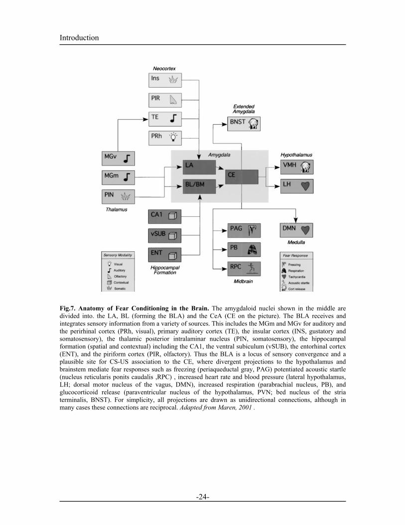
Introduction Fig.7. Anatomy of Fear Conditioning in the Brain. The amygdaloid nuclei shown in the middle are divided into. the LA, BL (forming the BLA) and the CeA (CE on the picture). The BLA receives and integrates sensory information from a variety of sources. This includes the MGm and MGv for auditory and the perirhinal cortex (PRh, visual), primary auditory cortex (TE), the insular cortex (INS, gustatory and somatosensory), the thalamic posterior intralaminar nucleus (PIN, somatosensory), the hippocampal formation (spatial and contextual) including the CA1, the ventral subiculum (vSUB), the entorhinal cortex (ENT), and the piriform cortex (PIR, olfactory). Thus the BLA is a locus of sensory convergence and a plausible site for CS-US association to the CE, where divergent projections to the hypothalamus and brainstem mediate fear responses such as freezing (periaqueductal gray, PAG) potentiated acoustic startle (nucleus reticularis ponits caudalis ,RPC) , increased heart rate and blood pressure (lateral hypothalamus, LH; dorsal motor nucleus of the vagus, DMN), increased respiration (parabrachial nucleus, PB), and glucocorticoid release (paraventricular nucleus of the hypothalamus, PVN; bed nucleus of the stria terminalis, BNST). For simplicity, all projections are drawn as unidirectional connections, although in many cases these connections are reciprocal. Adapted from Maren, 2001 .
-24-

Introduction 1.4. Synaptic Transmission in the LA The amygdala as compared to the hippocampus or the cortex, does not display a
structured or layered anatomy. The lack of architectural orientation makes it a difficult
task to explore its physiological role in learning and memory. In the next chapter, I will
review what is known about the network in the LA, a necessary step to understand
amygdala function in fear conditioning.
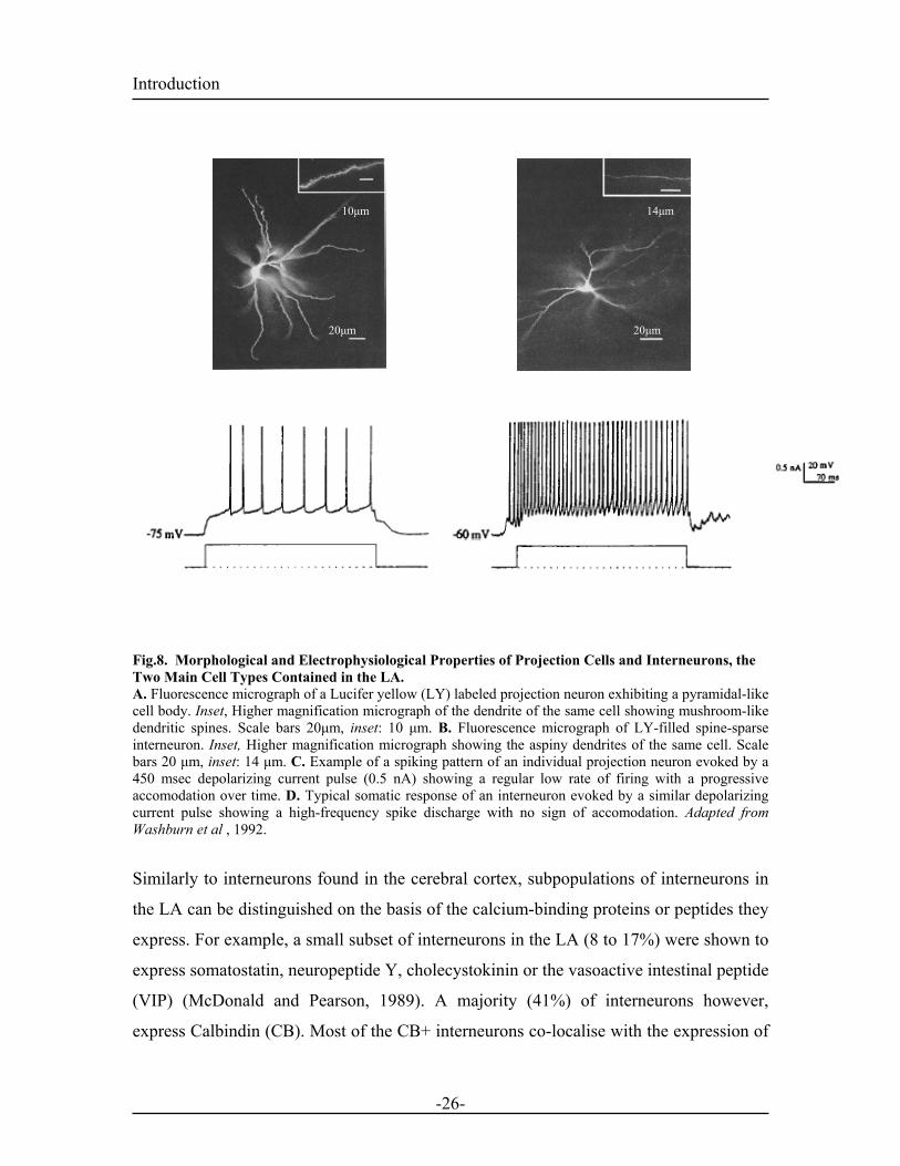
1.4.1. Cell Types Correlation between Golgi-stained neurons and neurons identified with retrograde
labeling, revealed two main cell types in the LA. 1) Spiny pyramidal (projection) neurons
representing the majority of the cells and 2) Spine-sparse non pyramidal neurons forming
a minority (10%). The latter mainly function as local circuit neurons (McDonald, 1992).
Intracellular recordings characterized the spiking properties of these two classes of
neurons. Prolonged injection of depolarizing current into projection neurons induces a
train of low frequency action potentials (APs), accomodating with time (Washburn and
Moises, 1992a). In contrast, the spine-sparse neurons exhibit a non-accommodating firing
pattern associated with a higher firing frequency (Rainnie et al., 1993; Washburn and
Moises, 1992a). The spiny projection neurons, exhibit large pyramidal shapes and utilize
glutamate as the neurotransmitter (Maren et al., 2001; McDonald, 1982; Rainnie et al.,
1991a; Rainnie et al., 1993). The spine-sparse interneurons which utilize GABA as the
transmitter, have a smaller and rounder morphology (Rainnie et al., 1991b; Washburn
and Moises, 1992a; Washburn and Moises, 1992b)
Lesions of afferents to the LA produced very small decrease in the levels of glutamic acid
decarboxylase, the main enzyme for GABA synthesis suggesting that local GABAergic
interneurons provide the main source of inhibition in the LA (Le Gal La Salle, 1978).
More recently, GABA immunoreactivity and glutamic acid decarboxylase (GAD)
staining confirmed the presence of local interneurons in the LA (Carlsen, 1988;
McDonald and Mascagni, 2001; Pitkanen and Amaral, 1994)
-25-
Introduction
20µm
10µm
20µm
14µm
Fig.8. Morphological and Electrophysiological Properties of Projection Cells and Interneurons, the Two Main Cell Types Contained in the LA. A. Fluorescence micrograph of a Lucifer yellow (LY) labeled projection neuron exhibiting a pyramidal-like cell body. Inset, Higher magnification micrograph of the dendrite of the same cell showing mushroom-like dendritic spines. Scale bars 20µm, inset: 10 µm. B. Fluorescence micrograph of LY-filled spine-sparse interneuron. Inset, Higher magnification micrograph showing the aspiny dendrites of the same cell. Scale bars 20 µm, inset: 14 µm. C. Example of a spiking pattern of an individual projection neuron evoked by a 450 msec depolarizing current pulse (0.5 nA) showing a regular low rate of firing with a progressive accomodation over time. D. Typical somatic response of an interneuron evoked by a similar depolarizing current pulse showing a high-frequency spike discharge with no sign of accomodation. Adapted from Washburn et al , 1992.
Similarly to interneurons found in the cerebral cortex, subpopulations of interneurons in
the LA can be distinguished on the basis of the calcium-binding proteins or peptides they
express. For example, a small subset of interneurons in the LA (8 to 17%) were shown to
express somatostatin, neuropeptide Y, cholecystokinin or the vasoactive intestinal peptide
(VIP) (McDonald and Pearson, 1989). A majority (41%) of interneurons however,
express Calbindin (CB). Most of the CB+ interneurons co-localise with the expression of
-26-
10µm 14µm
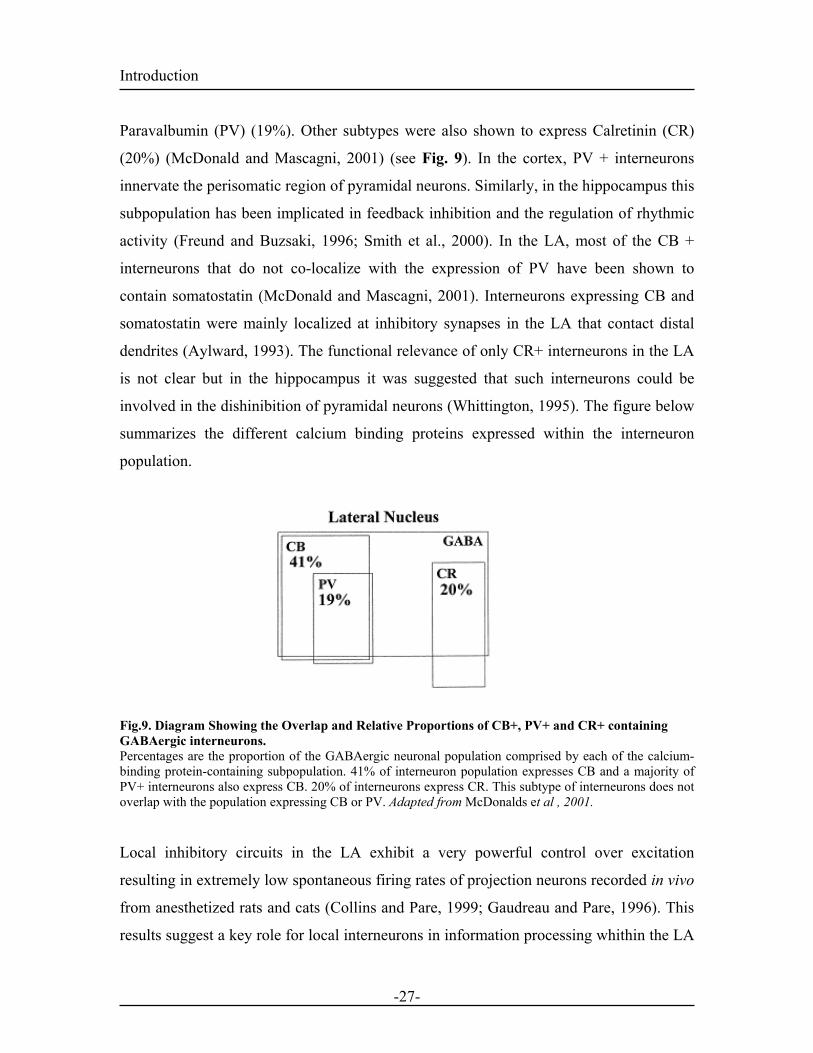
Introduction Paravalbumin (PV) (19%). Other subtypes were also shown to express Calretinin (CR)
(20%) (McDonald and Mascagni, 2001) (see Fig. 9). In the cortex, PV + interneurons
innervate the perisomatic region of pyramidal neurons. Similarly, in the hippocampus this
subpopulation has been implicated in feedback inhibition and the regulation of rhythmic
activity (Freund and Buzsaki, 1996; Smith et al., 2000). In the LA, most of the CB +
interneurons that do not co-localize with the expression of PV have been shown to
contain somatostatin (McDonald and Mascagni, 2001). Interneurons expressing CB and
somatostatin were mainly localized at inhibitory synapses in the LA that contact distal
dendrites (Aylward, 1993). The functional relevance of only CR+ interneurons in the LA
is not clear but in the hippocampus it was suggested that such interneurons could be
involved in the dishinibition of pyramidal neurons (Whittington, 1995). The figure below
summarizes the different calcium binding proteins expressed within the interneuron
population.
Fig.9. Diagram Showing the Overlap and Relative Proportions of CB+, PV+ and CR+ containing GABAergic interneurons. Percentages are the proportion of the GABAergic neuronal population comprised by each of the calcium-binding protein-containing subpopulation. 41% of interneuron population expresses CB and a majority of PV+ interneurons also express CB. 20% of interneurons express CR. This subtype of interneurons does not overlap with the population expressing CB or PV. Adapted from McDonalds et al , 2001. Local inhibitory circuits in the LA exhibit a very powerful control over excitation
resulting in extremely low spontaneous firing rates of projection neurons recorded in vivo
from anesthetized rats and cats (Collins and Pare, 1999; Gaudreau and Pare, 1996). This
results suggest a key role for local interneurons in information processing whithin the LA
-27-
Introduction (Lang and Pare, 1998; Mahanty and Sah, 1999). During fear conditioning, auditory
information coming from the thalamus reaches the LA through the internal capsule
whereas auditory information coming from cortical areas access the LA via the external
capsule (Romanski et al., 1993).These two inputs to the LA are excitatory and electrical
stimulation of either afferents gives rise to excitatory postsynaptic potentials (EPSPs) at
both interneurons and pyramidal cells (Szinyei et al., 2000), supporting the existence of
feed-forward and feed-backward inhibitory micro-circuits (Li et al., 1996) (Lang and
Pare, 1998; Woodson et al., 2000).
1.4.2. Excitatory transmission in the LA
Release of glutamate, the major excitatory neurotransmitter of the central nervous system
(CNS), mediates synaptic transmission by binding to four different types of postsynaptic
glutamate receptors: α-amino-3-hydroxy-5-methyl-4-isoxazolepropianate (AMPA),
Kainate, N-methyl-D-aspartate (NMDA) and metabotropic postsynaptic receptors
(mGlurs). The first three belong to the ionotropic class of receptors whereas mGlurs are
metabotrophic receptors triggering the activation of second messenger cascades. Under
resting conditions, AMPA receptors open in response to L-glutamate binding and are
underlying the fast excitatory postsynaptic current (EPSC) seen at most excitatory
synapses. These receptors are tetramers composed of the GluR1 to 4 subunits. They are
permeable to monovalent cations Na+ and K+ and are impermeable to Ca2+ in the presence
of the GluR2 subunit in the receptor composition (Washburn et al., 1997). NMDA
receptors are heteromeric complexes composed of several subunits: the NR1 subunit,
which is required for channel function and the NR2 subunits (NR2A/B/C/D), responsible
for channel gating and glutamate binding (Cull-Candy et al., 2001). Functional NMDA
receptors are usually composed of multiple NR1 subunits in combination with at least
one type of NR2 subunit (Monyer et al., 1992). NR2B and NR2D subunits predominate
in the neonatal brain but during development, they are replaced by NR2A or NR2C
subunits depending on the brain region (Monyer et al., 1992). NMDA receptors exhibit
three unique properties: 1) They are high conductance receptors (50 pS), permeable to
-28-
Introduction Ca 2+, Na+ and K+ 2) They require glycine as a co-factor. 3) The opening of the channel is
voltage-dependent. The voltage dependency is due to the fact that at the resting
membrane potential (~ -70 mV) the channel is blocked by Mg2+ (Nowak et al., 1984).
These Mg2+ ions are only removed when the postsynapse is depolarized (Coan and
Collingridge, 1985; Nowak et al., 1984). Therefore, during normal low-frequency
transmission glutamate released from the presynapse will bind to both NMDA and
AMPA receptors but transmission will only occur through AMPA receptors. At
depolarized membrane potentials NMDA receptors, released from the Mg2+ block, open
upon glutamate release and give rise to the late phase of EPSCs. Interestingly, NMDAR
receptors that integrate NR2B subunits generate longer EPSCs than the ones containing
NR2A subunit (Monyer et al., 1992; Szinyei et al., 2003). Stimulation of both thalamic and cortical inputs to projection cells in the LA activate a
fast decaying inward current that is fully blocked by 6-cyano-7-nitroquinoxaline-2,3-
dione (CNQX), a specific non-NMDA receptor antagonist. At more depolarized
membrane potentials, the same afferent stimulation reveals a slower current, blocked by
the specific NMDA receptor antagonist 2-amino-5-phosphonovaleric acid (D-APV)
(Rainnie et al., 1991a). The AMPA-receptor mediated response shows a linear current-
voltage relation reversing at 0 mV whereas, the NMDA-receptor mediated component
has a region of negative slope resistance between -70 to -20 mV consistent with a Mg2+
block (Coan and Collingridge, 1985; Nowak et al., 1984). NMDA receptors are expressed
at both thalamic and cortical inputs (Li et al., 1995) but the relative contribution of
NMDARs at these two inputs remains unclear (Mahanty and Sah, 1999; Weisskopf and
LeDoux, 1999). Stimulation of cortical inputs to LA interneurons were reported to
activate synapses that only contain AMPA receptors which in addition lacked the GluR2
subunit (Lopez de Armentia and Sah, 2003; Mahanty and Sah, 1998). These receptors,
that are Ca2+ permeable, enable faster excitatory transmission and have recently been
implicated in NMDAR-independent forms of plasticity expressed at interneuron synapses
(McMahon and Kauer, 1997) Interneurons in the LA also contain functional NMDA
receptors which participate in basal synaptic transmission at both thalamic and cortical
inputs (Szinyei et al., 2003). In addition NR2B subunits were found to be critically
-29-
Introduction involved in the NMDAR mediated signaling at both input pathways onto interneurons
and projection cells of the LA (Szinyei et al., 2003). All these intrinsic properties confer a
stronger and reliable transmission onto interneurons important for their role in the
regulation of output activity.
1.4.3. Inhibitory Transmission in the LA
γ-amminobutyric acid (GABA) is the major inhibitory transmitter in the brain and acts on
three classes of receptors, ionotropic GABAA and GABAC receptors, and metabotropic
GABAB receptors (Chebib and Johnston, 1999). Most of the fast inhibitory synaptic
transmission is mediated via GABAA receptors that form ligand-gated choride (Cl-)
channels. GABAA receptor channels are composed of five subunits α, β, γ, δ, and ε.
These subunits can assemble in various combinations but the presence of the α and the β
subunits are essential for GABA binding (Tretter et al., 1997). GABAA receptors are
important for drug binding in particular, they have a binding site for benzodiazepines that
once bound, increases the affinity of the receptor for GABA and increases its opening
probability (Sigel and Buhr, 1997). GABAB receptors are G-protein-coupled receptors
(Bowery et al., 1983). Their postsynaptic activation mediates a prolonged
hyperpolarisation due to an increased potassium (K+) conductance (Bowery, 1989) giving
rise to the late IPSP observed at GABAergic synapses (Bowery, 1989). GABAB receptors
are also found presynaptically where they reduce neurotransmitter release at inhibitory
and excitatory synapses (Pierau and Zimmermann, 1973; Thompson and Gahwiler,
1989). Presynaptic GABAB autoreceptors are activated by spillover of synaptically
released GABA and their action on neurotransmitter release has been attributed to an
inhibition of voltage-dependent Ca2+ channels at GABAergic nerve terminals and to
Ca2+channel-independent mechanisms at glutamatergic terminals (Scanziani et al., 1992;
Wojcik et al., 1990).
The large hyperpolarization that dominate the spontaneous and stimulus-evoked synaptic
response of projection cells are the results of combined action of synaptic conductances
(IPSPs) and synaptically activated intrinsic membrane conductances Indeed, consistent
with local interneurons forming feed-forward circuits within the LA, in vitro stimulation
-30-
Introduction of sensory afferents onto projection cells induces an initial EPSP followed by a fast
GABAA and a slow GABAB mediated IPSPs (Rainnie et al., 1991b). Similar to in vitro
studies, in vivo experiments also reported the presence of large IPSPs truncating stimulus
evoked or spontaneously occuring EPSPs (Lang and Pare, 1997a). In addition, in vivo
stimulation of the MGB eliciting short latency single unit responses in the LA (see 2.5)
were increased upon blockade of GABAA receptor antagonist (Li et al., 1996).
Furthermore, the cells that were not responsive to MGB stimulation also elicited a
response after blockade of GABAA receptors (Li et al., 1996). The tight control of
excitation by synaptic inhibition results primarily from the relatively low level of
inhibition directed towards interneurons (Smith et al., 2000). This low level of inhibition
of interneurons results from several factors. First, inhibitory responses in interneurons
appear to lack GABAB IPSPs or the synaptically activated Ca2+-dependent K+(Kca) found
in projection cells (Lang and Pare, 1997b; Martina et al., 2001). In addition, the reversal
potential of GABAA IPSPs in interneurons is depolarized compared to that in projection
cells (Martina et al., 2001). All these factors contribute to the relatively high excitable
states of interneurons also reflected by a depolarized resting membrane potential (Lang
and Pare, 1998). Considerable mechanisms in the LA are devoted to control the
excitability of projection cells and limit their responsiveness. This powerful inhibitory
control might also be important for gating the induction of synaptic plasticity.
1.5. Long-Term Changes in Synaptic Strength: A Model for Fear Learning Fear learning in the amygdala has been studied in three main intertwined ways. First, the
areas of the amygdala whose role in fear conditioning had been derived from lesion
studies (see 2.3.1/2.3.2/2.3.3) were investigated using single-unit recording experiments
in vivo. In this type of experiments, single cell activity in defined brain regions can be
measured in vivo by chronically implanting placing recording electrodes. Different
filtering techniques enables the detection of population spike such as the ones observed in
extracellular field recordings or single cell activity observed in single-unit recordings. in
-31-
Introduction vivo recordings of freely moving animals strongly support the idea for the LA being a site
of integration and storage of fear memories (see below) (Rogan and LeDoux, 1995;
Rogan et al., 1997). Second, long-term-potentiation (LTP), a widely studied form of
synaptic plasticity thought to be the cellular correlate of associative learning processes,
was assessed in the areas of the amygdala important for fear conditioning. Third, the
main strategy for linking LTP to learning and memory involves the disruption of its
induction mechanism and assessing the consequences of this disruption on behavior. In
order to achieve this, drugs that block LTP in other brain structures where infused in
relevant areas of the amygdala and their effects were assessed on the acquisition of
conditioned fear.
The next paragraph introduces the concept of LTP and its relevance for the understanding
of the cellular mechanisms underlying fear learning
1.5.1. Hebbian Theory of Plasticity
The idea that the cellular changes that occur during learning involve alterations in
synaptic transmission goes back to the beginning of the twentieth century (Cajal, 1909;
Eccles, 1965; Hebb, 1949; Kandel and Spencer, 1968). One of the most influential
theories came from Donald.O. Hebb (1949) who proposed that if two interconnected cells
fire at the same time, the synaptic connections between them will be strengthened.
Accordingly, synaptic plasticity induced by coincident pre-ane postsynaptic activity is
referred to as Hebbian plasticity..
1.5.2 Synaptic Plasticity: LTP in the hippocampus
Exploration of neurobiological evidence corresponding to the Hebbian theory of
plasticity started with Bliss & Lomo (1973) who discovered long-term-potentiation (LTP)
in the hippocampus. They provided the first evidence that high-frequency stimulation of
excitatory connections made by perforant fibers onto granule cells of the hippocampus
could induce a long-term increase in synaptic efficacy at these synapses that they called
LTP (Bliss and Lomo, 1973). Subsequently, most of the work that aimed at
understanding the mechanisms of LTP was performed on excitatory synapses in the
-32-
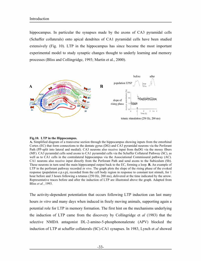
Introduction hippocampus. In particular the synapses made by the axons of CA3 pyramidal cells
(Schaffer collaterals) onto apical dendrites of CA1 pyramidal cells have been studied
extensively (Fig. 10). LTP in the hippocampus has since become the most important
experimental model to study synaptic changes thought to underly learning and memory
processes (Bliss and Collingridge, 1993; Martin et al., 2000).
BA
Fig.10. LTP in the Hippocampus. A. Simplified diagram of a transverse section through the hippocampus showing inputs from the entorhinal Cortex (EC) that form connections to the dentate gyrus (DG) and CA3 pyramidal neurons via the Perforant Path (PP-split into lateral and medial). CA3 neurons also receive input from theDG via the mossy fibers (MF). CA3 pyramidal cells send axons to CA1 pyramidal cells via the Schaffer Collateral Pathway (SC), as well as to CA1 cells in the contralateral hippocampus via the Associational Commissural pathway (AC). CA1 neurons also receive input directly from the Perforant Path and send axons to the Subiculum (Sb). These neurons in turn send the main hippocampal output back to the EC, forming a loop. B. An example of LTP in the perforant pathway recorded in vivo. The graph plots the slope of the rising phase of the evoked response (population e.p.s.p), recorded from the cell body region in response to constant test stimuli, for 1 hour before and 3 hours following a tetanus (250 Hz, 200 ms), delivered at the time indicated by the arrow. Representative traces before and after the induction of LTP are illustrated above the graph. Adapted from Bliss et al , 1993.
The activity-dependent potentiation that occurs following LTP induction can last many
hours in vitro and many days when induced in freely moving animals, supporting again a
potential role for LTP in memory formation. The first hint on the mechanisms underlying
the induction of LTP came from the discovery by Collingridge et al (1983) that the
selective NMDA antagonist DL-2-amino-5-phosphononalerate (APV) blocked the
induction of LTP at schaffer collaterals (SC)-CA1 synapses. In 1983, Lynch et al showed
-33-
Introduction that intracellular injection of EGTA, a calcium chelator, into pyramidal cells of the CA1
region of the hippocampus also blocked the induction of LTP at SC-CA1 synapses. The
work of Nowak et al (1984) provided a explanation for both the voltage dependent
properties of NMDAR activation as well as for the criteria that had to be met for the
induction of LTP. LTP is characterized by three main properties that were already
predicted by Hebb's theory: Cooperativity, Associativity, and Input-specificity. These
three properties can be better explain by the constrain set by NMDAR activation.
Cooperativity is required to achieve enough postsynaptic depolarisation in order to
release the Mg2+ block of NMDARs and activate them. If the tetanus protocol applied at
afferent fibers is too weak not enough postsynaptic depolarization will be achieved to
activate NMDARs and trigger potentiation (McNaughton et al., 1978). LTP is associative
because coactivation of a weak input and a strong input to the same neuron will lead to
activation of NMDARs at the weak input and its subsequent potentiation. The
associativity provides a key link to the hebbian theory and supposes that a synapse will
be potentiated if and solely if it is activated at the time when the region of dendrite on
which it terminates is depolarized enough. Stimulation of the weak input alone is not
enough to achieve sufficient postsynaptic depolarisation (Levy, 1979; McNaughton et al.,
1978). Finally, LTP is input specific as it will only occur at the synapses that reach
enough postsynaptic depolarization during the tetanus (Andersen et al., 1977; Lynch,
266). The figure below summarizes the three properties associated with LTP induction.
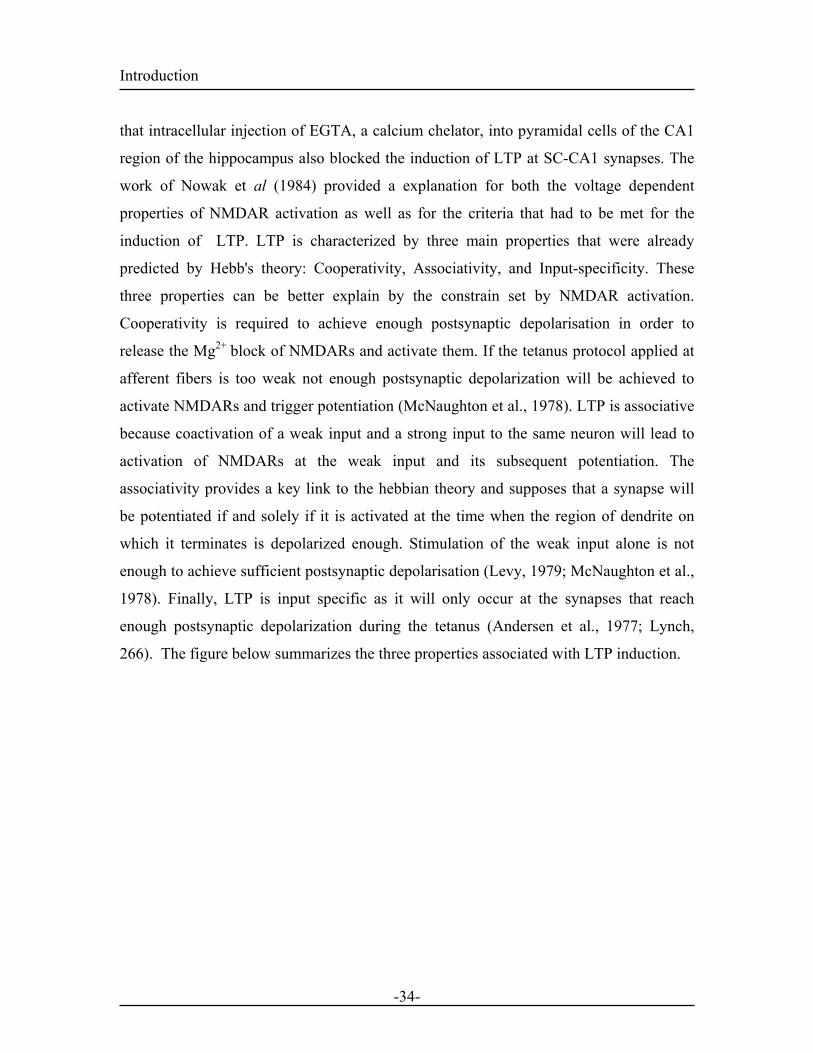
-34-
Introduction
BA
Fig.11. Basic Properties of LTP. A. Experimental arrangements in the CA1 region of the hippocampal slice preparation. Two independent sets of afferent fibers converging on a common population of cells are activated by stimulating electrodes (S1 and S2) placed either side of the extracellular recording electrode. The stimulus intensities are adjusted so that S1 activates fewer fibers than S2. B. Top and bottom graphs. The slope of the population e.p.s.ps in response to stimuli delivered alternately to S1 and S2 at 15-s intervals, are plotted as a function of time.Arrows denote episodes of tetanic stimulation to S1 (the weak pathway, open arrows) or S2 (the strong pathway, solid arrows). The tetanus to S1did not produce a stable increase in synaptic transmission; the intensity of the tetanus was below the cooperativity threshold of LTP. The stronger tetanus to S2 (first filled arrow) produced a robust LTP, but there was not transfer of the effect to the first input, demonstrating the input specificity of LTP. Finally tetani to S1 and S2 are delivered together. The coincident activation of a weak and a strong input induced associative LTP at the weak input. The traces above the graph illustrate field e.p.s.ps, evoked by test shocks in S1 and recorded in the synaptic layer, before and after the induction of associative LTP. Taken and adapted from Bliss et al , 1993.
LTP in the hippocampus has been induced using three main type of protocols: tetanic
stimulation, pairing stimulation, and spike timing dependent induction. During tetanic
stimulation (eg: 100 HZ for 1s) the cell receives one set of stimulation, strong enough to
provide postsynaptic depolarization and NMDARs activation (Bliss and Lomo, 1973;
McNaughton et al., 1978). LTP can also be induced using a pairing stimulation protocol
where a low frequency afferent stimulation is coupled with a concurrent postsynaptic
depolarization (Kelso and Brown, 1986; Malenka and Nicoll, 1999; Sastry et al., 1986)
Under physiological conditions, the postsynaptic cell fires action potentials that can back
propagate into the dendritic tree and reach individual synapses (Stuart and Sakmann,
1994). Subsequently these events might support a physiological depolarization at the
-35-
Introduction postsynapse resulting in the removal of the . This notion led to the so-called spike timing
dependent plasticity (STDP) protocol in which a presynaptic stimulation is paired with a
postsynaptic action potential (AP), induced by brief current injection. In 1983, Levy &
Steward studied the temporal requirements for the induction of associative LTP by
stimulating a weak and a stronger input from the entorhinal cortex to the dentate gyrus of
the hippocampus at different time intervals. These studies revealed that induction of
associative LTP did not require perfect synchronous activation of the two pathways
(Levy and Steward, 1983). Similarly, LTP induced by STDP does not require perfect
synchronization for coincident pre and postsynaptic activity. In interconnected cortical
layer 5 pyramidal cells, STDP triggers LTP only when the postsynaptic spikes are
induced 10 ms after the onset of the EPSP (Markram et al., 1997).Similarly, in cultures of
dissociated rat hippocampal neurons a persistent potentiation could be induced using
STDP only when repetitive postsynaptic spiking were triggered within a window of 20
msec after presynaptic stimulation (Bi and Poo, 1998). At schaffer colleterals in the CA1
region, plasticity can be induced also by pairing pre and postsynaptic activation but LTP
is only triggered if the EPSP arrives before the BPAP (Nishiyama et al., 2000). These
results suggest that back-propagating spikes provide a precise signal informing the
synapse of postsynaptic activity and may play an active role in associative synaptic
modification.
The next chapter focuses on the cellular mechanisms underlying these synaptic changes
1.5.3. Hippocampal LTP: Mechanism.
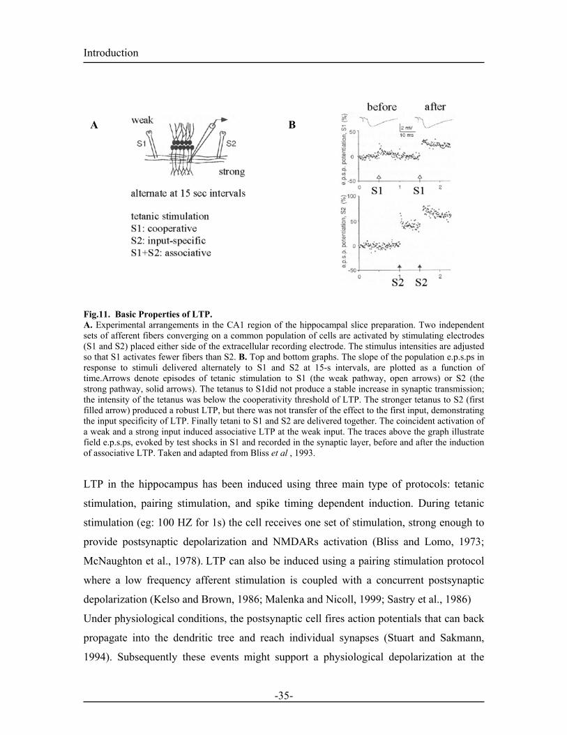
Since the discovery of Novak (see 1.5.2) on the properties of NMDARs (Nowak et al.,
1984) it is well accepted that induction of LTP requires NMDAR activation. During
normal low frequency transmission, glutamate released from the presynapse will bind to
both NMDA and non-NMDARs but transmission will only occur through AMPA
receptors due to the Mg2+ block at NMDARs. During high frequency stimulation or
postsynaptic depolarization, the Mg2+ block is released (1.4.2). As a consequence, Ca2+
flows into the dendritic spine and triggers calcium-dependent processes (Fig. 12). This is
turn results in the activation of serine kinases like protein kinase C (PKC), protein kinase
-36-
Introduction A (PKA) and calcium/calmodulin-dependent protein kinase (CaMKII) (Malenka et al.,
1989; O'Dell et al., 1991; Silva et al., 1992a; Silva et al., 1992b) (Lledo et al., 1995;
Lledo et al., 1998)
A key role was given to CaMKII in the induction LTP (Lledo et al., 1995; Nicoll and
Malenka, 1999). CaMKII is activated by Ca2+- Calmodulin (Ca-Cam) which is itself
activated by an increase in postsynaptic Ca2+ levels. Upon activation, CaMKII can
autophosphorylate itself, thereby becoming independent of Ca2+ levels.
Autophosphorylation triggers the translocation of CaMKII to the postsynaptic density
(PSD) through interactions with NMDA receptors (Lisman et al., 1997). In hippocampal
and cortical pyramidal neurons, these various postsynaptic changes due to calcium influx
and activation of CaMKII can lead to an increase in the number of AMPA receptors
expressed at the postsynapse or/and changes in the channel conductance following GluR1
phosphorylation (Benke et al., 1998; Derkach et al., 1999; Luscher et al., 1999; Malinow
Fig.12. Model for the induction of LTP. During normal synaptic transmission, glutamate (Glu) is released from the presynaptic bouton and acts on bothAMPA receptors (AMPARs) and NMDA receptors (NMDARs). However, Na+ flows only through the AMPAreceptor, but not the NMDA receptor, because Mg2+ blocks the channel of the NMDA receptor. Depolarization of thepostsynaptic cell relieves the Mg2+ block of the NMDA receptor channel, allowing Na+ and Ca2+ to flow into thedendritic spine by means of the NMDA receptor. The resultant rise in Ca2+ within the dendritic spine is the criticaltrigger for LTP. Adapted from Malenka et al,1999.
-37-
Introduction and Malenka, 2002). These processes eventually results in an enhancement of glutamate-
evoked postsynaptic responses. Associative LTP can be blocked by intracellular injection
of the Ca2+ chelator EGTA There are two major molecular mechanisms underlying
calcium influx into the postsynapse during LTP induction. First, NMDA receptors
mediated (see above) and the second involves voltage gated calcium channels (VGCCs).
Indeed, NMDA-independent LTP has been observed in the hippocampus at mossy fibers
(see 2.5.1.4) and requires calcium entry via activation of postsynaptic VGCCs (Grover
and Teyler, 1990). Since LTP is an associative and synapse specific process, it raises the
possibility that VGCCs like NMDAR are able to function as hebbian coincidence
detectors for postsynaptic activity. Six types of VGCCs have been cloned to date, (L, N,
P, Q, R, I) that can be separated by their voltage dependency for activation and
pharmacologically by the use of specific toxins.(Catterall, 2000). L, N, P, Q and R types
are high voltage activated channels and T types are low voltage activated channels Under
physiological conditions, VGCCs are thought to open in response to strong depolarization
arising from BPAPs that spread along the dendrite (Stuart et al., 1997). Contradicting
data suggest, however, that BPAPs might be too fast for the slow kinetic of L-type
VGCCs (Mermelstein et al., 2000). Stuart et al (2001) could show that BPAPs can be
prolonged and amplified in the dendrite when they collide with EPSPs triggered by the
postsynaptic cell. This coincidence enlarges the time window during which VGCCs can
be activated and can lead to the entry of Ca2+(Stuart and Sakmann, 1995; Stuart and
Hausser, 2001). VGCCs participate in the induction of LTP at SC-CA1 synapses but their
contribution is typically only detectable when strong tetanic stimulation is used (Grover
and Teyler, 1990).
LTP can be divided into several temporal stages, short-term potentiation (STP) lasting
only for 15 to 30 min, the early phase of LTP (E-LTP) which is stable for up to 2 to 3
hours and the late phase of LTP (L-LTP) that have been shown to last up to 8 hours in
hippocampal slices(Bailey, 1996). The L-LTP is associated with gene expression, de
novo protein synthesis and formation of new synaptic connections. Consistently, protein
synthesis inhibitor can block L-LTP but leave STP and E-LTP unaffected (Lynch, 2004).
-38-
Introduction 1.5.4. Long-term-depression (LTD)
LTD is a persistent activity-dependent reduction in synaptic efficacy that has typically
been observed following low frequency afferent stimulation (eg: 900 pulses at 1HZ).
Because of the role of the hippocampus in certain forms of memory storage and retrieval
the study of LTD in this area has been the subject of particular interest. In the CA1 region
of the hippocampus, LTD is NMDAR dependent and requires an increase in postsynaptic
calcium levels (Bear, 1996). The postsynaptic Ca2+ increase induced by low frequency
pairing activate calcineurin (PP2B) through a calcium-calmodulin-dependent process.
Once activated, PP2B dephosphorylates and inactivates inhibitor 1, resulting in the
activation of protein phosphatase 1 and/or 2 (PP1/2) and LTD induction (Dou et al.,
2001; Kemp and Bashir, 2001). LTD expression is thought to result from the
dephosphorylation of AMPA receptors by PP1/2, followed by the internalization of the
receptors and a decrease in the single-channel conductance (Beattie et al., 2000; Ehlers,
2000; Luscher et al., 1999). PP1/2 activation can also target and dephosphorylate
CaMKII which in turn cannot phosphorylate AMPA receptors, favoring the induction of
LTD over LTP. Indeed, CaMKII antagonist have been shown to facilitate the induction of
LTD in the CA1 region whereas PP1/2A inhibitors block it (Mulkey et al., 1994;
Schnabel et al., 1999).
1.5.5. LTP versus LTD
From the above, it is clear that the temporal order of pre and post-synaptic spiking is of
crucial importance and can determine in the order of milliseconds whether LTP or LTD is
induced (Bi and Poo, 1998; Debanne et al., 1998). Both homosynaptic LTP and LTD
require pre and postsynaptic coincident activation and postsynaptic calcium elevation.
There is some evidence that distinct properties of the Ca2+ signal may determine whether
LTP or LTD results (Bi, 2001; Malenka and Nicoll, 1999). However, the Bienenstock,
Cooper and Munro (BCM) theory and recent studies on STDP also predict that the
activity-dependent changes in synaptic strength depend on the frequency and timing of
presynaptic and postsynaptic activity (synaptic plasticity) as well as the history of activity
at those synapses (metaplasticty) (Jedlicka, 2002; Sjostrom and Nelson, 2002; Sjostrom
-39-
Introduction et al., 2001) predicts that high levels of calcium elevation favor LTP induction whereas
low calcium levels favor LTD (Bi, 2001; Malenka and Nicoll, 1999).However, a recent
studies by Sjöström and colleagues (2001) Different properties related to synaptic
modification might serve specific functions relative to the distinct role of each brain
structure in the neural processing of information.
1.6. The amygdala and fear conditioning : pharmacological studies 1.6.1. NMDA Receptors
In an attempt to link the mechanisms of fear learning with long-term-synaptic changes
observed in the hippocampus, some groups started to assess the role of NMDA receptors
in the acquisition of fear conditioning.
Some experiments demonstrated that infusion of NMDA antagonists into the LA and BL
before training was enough to block the acquisition of fear conditioning, without
affecting basal synaptic transmission (Gewirtz and Davis, 1997; Miserendino et al.,
1990). Other groups, however, showed that NMDA receptor blockade after training but
before testing prevented the expression of fear conditioning (Lee et al., 2001; Maren et
al., 1996). The apparent discrepancies in these results have been suggested to result from
the presence of NR2B containing NMDA receptors in the BLA at both projection
neurons and interneurons. The NR2B subunit appears to be crucial in synaptic changes
underlying fear conditioning independently from its role in synaptic transmission
(Szinyei et al., 2003). Such a specific role for the NR2B subunit could not be distinguish
for when using APV, a competitive NMDA receptor antagonist. Conversely, systemic
injections of ifenprodil, a NR2B antagonist, before training impaired the acquisition of
fear conditioning whereas injections after training had no effect (Blair et al., 2001). These
results clearly demonstrated a role for the NR2B subunits in the acquisition of fear
conditioning. In particular the NR2B subunit appear to be crucial in synaptic changes
underlying the acquisition of fear conditioning (Rodrigues et al., 2001)
-40-
Introduction 1.6.2. VGCC and Fear Conditioning
Infusion of the L-type VGCC blocker verapamil into the LA during training blocked the
acquisition of fear conditioning, but did not impair the expression of previously learned
conditioned fear responses (Bauer et al., 2002). NMDA receptors and VGCCs seem to be
necessary for fear learning to take place, emphasizing similarities to induction
mechanism of hebbian plasticity observed in the hippocampus.
1.6.3. Protein Synthesis and Fear Conditioning
Interfering with signal transduction cascades and gene transcription mechanism in the
hippocampus blocks the long term maintenance of LTP. Similarly, infusion of PKC and
PKA inhibitors in the LA prior to training attenuated the long term expression of fear
memory. This effect was specific for the LA as similar infusion in the CeA induced no
changes in fear expression (Goosens et al., 2000). Infusion of inhibitors of extracellular
signal-regulated kinase (ERK) or mitogen activated kinase (MAPK) which have been
shown to be activated and phosphorylated in the amygdala during learning also impair
the long-term maintenance of fear memories (Schafe et al., 2000). Extinction of fear
learning parallels a reduction in extracellular field potentials observed in the LA ((Davis
et al., 2003).This depression can be blocked by infusion of calcineurin inhibitor in a
similar way to that observed in the hippocampus during blockade of LTD (Lin et al.,
2003). Taken together, these data show that fear learning in the amygdala and
mechanisms underlying synaptic plasticity in the hippocampus share similar mechanisms. 1.7. Physiological Plasticity in the Amygdala Related to Fear Conditioning. 1.7.1. Single Unit Recordings and Sensory Pathway Stimulation in Freely Moving Rats Upon CS-US pairing, during fear conditioning, the CS induced activity in the LA is
enhanced. To understand the nature of plasticity within the amygdala that might underlie
fear learning, single-unit recordings from LA projection neurons were obtained from
freely behaving animals before and after auditory fear conditioning. Quirk et al (1995)
reported that fear conditioning significantly increased the magnitude of tone-elicited
-41-
Introduction responses, converted unresponsive cells into tone responsive ones and changed the
functional coupling between LA neurons. In addition, they reported that this increase was
greater on the short latency (less than 15 ms) component of the tone-elicited responses.
Since auditory information reach the amygdala directly via the thalamo-amygdala
pathway and indirectly via the cortico-amygdala pathway the following studies were
undertaken to elucidate the origin of the short latency response observed in LA projection
neurons. Quirk et al (1997) demonstrated, using single cell recordings, that during fear
conditioning, auditory cortex neurons took more trials to potentiate and responded more
slowly than LA neurons. These results suggested that the short-latency plasticity
observed in the LA reflects inputs from the auditory thalamus rather than the auditory
cortex (Quirk et al., 1997). These observations were confirmed by the group of Rogan et
al (1995) who observed a similar potentiation of evoked potentials in the LA upon high
frequency stimulation of the MGm/PIN (thalamic nucleus conveying information about
the CS to the LA). Thus, fear conditioning mediates plasticity in the amygdala and it
appears that rapid conditioned responses are mediated by inputs from the thalamus
whereas cortical areas may be involved in higher cognitive processing of fear experience
(Quirk et al., 1997).
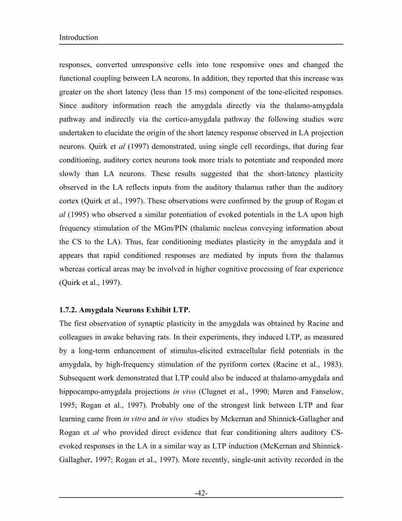
1.7.2. Amygdala Neurons Exhibit LTP.
The first observation of synaptic plasticity in the amygdala was obtained by Racine and
colleagues in awake behaving rats. In their experiments, they induced LTP, as measured
by a long-term enhancement of stimulus-elicited extracellular field potentials in the
amygdala, by high-frequency stimulation of the pyriform cortex (Racine et al., 1983).
Subsequent work demonstrated that LTP could also be induced at thalamo-amygdala and
hippocampo-amygdala projections in vivo (Clugnet et al., 1990; Maren and Fanselow,
1995; Rogan et al., 1997). Probably one of the strongest link between LTP and fear
learning came from in vitro and in vivo studies by Mckernan and Shinnick-Gallagher and
Rogan et al who provided direct evidence that fear conditioning alters auditory CS-
evoked responses in the LA in a similar way as LTP induction (McKernan and Shinnick-
Gallagher, 1997; Rogan et al., 1997). More recently, single-unit activity recorded in the
-42-
Introduction dorsal subnucleus of the LA (LAd) revealed that during fear conditioning, the neuronal
activity of two type of cells were changed. The first type, located in the dorsal tip of the
LAd only exhibited transient changes whereas the second type, located more ventrally,
had longer enhanced responses that lasted even throughout extinction (Repa et al., 2001).
These results all together give strong evidence that fear learning alters neural activity in
the LA a way consistent with an LTP-like process . Furthemore, distinct cell types in the
LAd seem to be differentially involved in the initiation of learning and long-term
memory storage (Repa et al., 2001).
1.7.3. Mechanisms of Synaptic plasticity in the LA
Fig.13. Behavioural LTP is induced in the lateral nucleus of the amygdala by Pavlovian Fear Conditioning A. A recording electrode was placed in the lateral nucleus of the amygdala (LA, dark gray) to measure auditory-stimulus-elicited extracellular field potentials in awake rats. B. Brief tone stimuli elicited a biphasic extracellular field potential with a short latency negative component (pre-training indicated by the black dots). Unpaired traces refer to control experiments where the CS was not paired with the US. Pre-training, Training and Testing refer to traces recorded before, during and after fear conditioning. Adapted from Rogan et al , 1995.
The cellular mechanisms of homosynaptic LTP in the amygdala were mostly investigated
in the LA in vitro using field or whole-cell recordings. Changes in the excitatoty
postsynaptic potentials (EPSPs) or field EPSPs in the LA were measured upon
stimulation of the internal capsule or the external capsule. LTP induced by tetanic
stimulation of either pathway was reported to be NMDA-dependent (Bauer et al., 2002;
Huang and Kandel, 1998; Huang et al., 2000) and to require the entry of calcium into the
postsynaptic cell. These results implied that hebbian plasticity underlies fear memory
-43-
Introduction acquisition in the amygdala. These findings are consistent with in vivo data reporting that
infusion of an NMDA receptor antagonists into the LA prior to conditioning interfered
with learning (Gewirtz and Davis, 1997; Miserendino et al., 1990). Pairing protocols of
weak stimulation of cortical or thalamic afferents with strong postsynaptic depolarization
can also efficiently induce LTP in the LA. For example, Huang and Kandel (1998),
induced LTP at cortical afferents by pairing low-frequency stimulation (0.2 Hz) with
prolonged postsynaptic current injection (2-4 nA for 50ms). This LTP was blocked
partially by NMDAR antagonists and entirely by VGCCs and NMDAR antagonists
(Huang and Kandel, 1998). Weisskopf et al (1999), using a different pairing protocol,
induced an associative and synapse specific LTP at the thalamo-amygdala pathway. This
type of LTP, however, was not affected by NMDAR antagonists but was completely
blocked by bath application of the L-type VGCC antagonist, nifedipine and by
postsynaptic perfusion of BAPTA, a calcium chelator (Weisskopf and LeDoux, 1999). In
contrast, LTP induced by tetanic protocol at the same synapses was found to be NMDA
receptor dependent (Bauer et al., 2002). Studies by Lee et al (2002) and our own studies
further demonstrated that an NMDAR and VGCCs -dependent LTP could also be
induced at these syanpses (Bissiere et al., 2003; Lee et al., 2002). Thus, it seems that in
the LA, weak pairing protocol (STDP) induce a NMDAR and VGCCs-dependent LTP
whereas strong pairings trigger VGCCs-dependent only LTP. Finally tetanic stimulation
that do not require postsynaptic APs, induce NMDAR-dependent only LTP. These data
suggest that there are pharmacologically distinct form of LTP can be induced in pathways
that transmit the CS to the LA (Bauer et al., 2002). Furthemore, the drugs that interfere
with LTP in the LA also shown to disrupt behavioral fear conditioning (see 1.6).
A cellular hypothesis has been put forward by Blair et al (2001) to explain how neural
responses to the CS and US in the LA could induce LTP-like changes that store
memories during fear conditioning. In their model, they propose that the CS evokes
EPSPs at sensory input synapses onto LA projection neurons while the US strongly
depolarizes the same neurons. This depolarization in turn activate NMDARs by releasing
the Mg2+ block and can, if strong enough, cause the LA neuron to spike. APs can back-
propagate in the dendrites where they can collide with CS-evoked EPSPs activating
-44-
Introduction VGCCs and the subsequent calcium influx (Blair et al., 2001). In addition they
hypothesize that NMDARs activation support the short-term fear memory whereas
activation of both NMDARs and VGCCs is needed to initiate molecular processes that
consolidate synaptic changes into long-term memory (Blair et al., 2001) . Rosenkranz and
Grace (2002), provided direct evidence that a footshock can elicit depolarization of
postsynaptic in vivo and that if strong enough can also elicit spiking. However, they could
also show that for CS-US association to occur (in this case an odor associated with a
footshock) postsynaptic spiking induced by the US presentation is not necessary
(Rosenkranz and Grace, 2002), indicating that the Blair model might not be able to
explain all types of conditioning. Thus, it is important next to investigate the temporal
constraint for CS-US association during fear conditioning in the LA.
1.7.4. LTP and Synaptic Inhibition.in the LA
The role of inhibition in the amygdala was discussed in chapter 1.4.3. Consistent with in
vivo data showing very low firing rates of projection neurons, local inhibitory processes
play a crucial role in the overall excitability of the amygdala. In the hippocampus,
GABAAR antagonists greatly increase the time window of EPSPs, allowing them to
summate over a larger period of time and give rise to an AP (Pouille and Scanziani,
2001). Consistently, the induction of LTP in the hippocampus was shown to be greatly
diminished in the presence of synaptic inhibition (Whittington, 1995; Wigstrom and
Gustafsson, 1983). In the amygdala such a tight control of excitatoty processes could also
prevent the postsynaptic depolarization required for LTP and thereby reduce the time
window in which learning or synaptic modification can occur. Our own data provided
direct evidence that in the presence of inhibition, LTP could not be induced at thalamo-
amygdala synapses. Since learning occurs during fear conditioning there must be some
modulator mechanisms that can suppress synaptic inhibition in vivo. The existence of
such a mechanism would renable the temporal summation of excitatory processes leading
to postsynaptic depolarization and LTP induction.
-45-
Introduction 1.8. Neuromodulation of Inhibition
1.8.1. The Dopaminergic System: An Introduction Dopamine (DA) is the predominant catecholamine neurotransmitter in the mammalian
brain. It has been shown to play a important role in a variety of functions ranging from
locomotor activity to cognition, emotion, positive reinforcement of drug to endocrine
regulation. The dopaminergic systems have been the focus of research over the past 30
years mainly due to Parkinson's disease or schizophrenia, disorders associated with a
disregulation of dopaminergic transmission. The first evidence for the existence of DA
receptors in the CNS came from biochemical studies reporting that DA was able to
stimulate adenylyl cyclase (AC) (Kebabian, 1979). Subsequently, six major types of
dopamine receptors have been cloned in human: D1, D2L&S (splice variant) , D3, D4, and
D5. The amino acid sequence of each of the cloned subtypes encodes for a seven-trans
membrane G-protein coupled receptor (Probst et al., 1992). These receptors are divided
into the D1 and D2 subfamilies. D1 and D5 form the D1-like family whereas D2, D3, and
D4 compose the D2-like family. Dopaminergic neurons are divided between the
substanstia nigra, which give rise to the nigrostriatal pathway, and the ventral tegmental
area, which give rise to the mesocorticolimbic projection. The nigrostriatal pathway is
part of the striatal motor system and its degeneration leads to Parkinson's disease. The
mesocorticolimbic pathway plays a key role in reinforcement of positive experiences and
emotional expression. Studies in these brain areas consistently reported that D1-like
receptor family activation activates Gs / olf proteins, which eventually activate PKA. In
contrast, D2 receptors activate Gi/o proteins resulting in the inhibition of PKA (Memo et
al., 1992; Memo, 1986). 1.8.2. A Modulatory Role for Dopamine
The amygdala receives strong dopaminergic innervation originating from the A9 and A10
cells of the VTA and substantia nigra (Swanson, 1982). Dopamine projections from the
VTA to the BLA have been shown to be strongly activated during stressful stimuli (Inglis
and Moghaddam, 1999). A number of in vivo studies pointed at dopamine as being
-46-
Introduction involved in the acquisition of fear conditioning (Paré, 2002; Rosenkranz and Grace,
2002). Indeed, infusion of D2 antagonists into the amygdala prior to conditioning, block
the CS association to the US (Greba et al., 2001). It was further demonstrated that D2
antagonist rather than devaluating the US, block the retrieval of the learned CS-US
association (Nader and LeDoux, 1999). Similarly, blocking of D1-receptors with
SCH23390 also interfered with the expression of conditioned fear. This effect however
seemed to be specific for the expression of conditioned reactions retrieved from long-
term memory suggesting that dopamine might also be involved in the formation and/or
consolidation of long-term memory (Guarraci et al., 1999; Lamont and Kokkinidis,
1998). The way dopamine is able to modulate the outcome of amygdaloid response is not
clear. Immunocytochemical studies indicated that dopaminergic fibers synapse on the
dendritic spines of projection neurons whereas they form pericellular baskets around the
cell body of PV+ interneurons (Brinley-Reed and McDonald, 1999). The physiological
relevance of this innervation for the modulation of synaptic transmission was emphasized
by the studies of Rosenkranz and Grace (1999). Indeed, they showed that in vivo,
dopamine increases the spontaneous firing rate of interneurons modulating indirectly the
excitability of projection neurons (Rosenkranz and Grace, 1999).The medial prefrontal
cortex (mPFC) is an important structure involves in the extinction of fear conditioning.
The mPFC modulates the activity of the LA by increasing the excitability of interneurons
(Rosenkranz and Grace, 1999). Dopamine release in the LA was shown to be very
important during the acquisition of fear conditioning. Indeed, it was reported that D1
receptor activation can suppress the inhibitory action of the mPFC over the BLA, while
D2 receptor activation enhance the sensory-related inputs from the cortex by increasing
the excitability of projection neurons (Rosenkranz and Grace, 1999). These data suggest a
crucial role for dopamine in controlling the excitability state of the amygdala. However,
to date the exact mechanisms underlying these differential effects are not well define.
This is due in part to the fact that the signaling cascade mediated by the D1 receptor
activation in this brain area differs from what is known elsewhere. Indeed, D1 receptors
in the BLA do not signal through cAMP. The exact mechanism is still unknown and
leaves important implication for the neurobiology of dopamine in this area.
-47-
Introduction
-48-
2.Aim of the Thesis
LTP mechanisms in the LA are thought to underlie the acquisition of fear learning. Most
of the previous work investigating the role of LTP in the LA have used pharmacological
tools to block inhibition. However, in vivo data support an important role for inhibitory
circuits in this area. Our first data revealed that in the presence of synaptic inhibitory
transmission, LTP in the LA could not be induced at thalamo-amygdala synapses.
Dopamine has been shown to modulate GABAergic transmission and synaptic plasticity
in many brain areas. In addition, perfusion of dopamine antagonists into the LA, block
the acquisition of fear conditioning. Therefore, we decided to investigate the role of
dopamine on the induction of plasticity in the LA. We also investigated the potential
modulatory action of dopamine on inhibitory circuits within the LA.
Results
3-Results 3.1.Published Results
Dopamine Gates LTP Induction in the Lateral Amygdala by Supressing Feedforward Inhibition Stephanie Bissière, Yann Humeau & Andreas Lüthi Nature Neuroscience, Vol.6, N.6, 587-592 (2003)
Fear conditioning involves the induction of long-term potentiation (LTP) of
excitatory synaptic transmission in the lateral amygdala, a brain structure which is
tightly controlled by GABAergic inhibition. Here we show that dopamine gates the
induction of LTP in the mouse lateral amygdala by suppressing feed-forward
inhibitory control mediated by local interneurons. Our findings provide a cellular
mechanism for the dopaminergic modulation of fear conditioning and indicate that
suppression of feed-forward inhibition represents a key mechanism for the
induction of associative synaptic plasticity in the lateral amygdala.
During fear conditioning, one of the most powerful and widely used paradigms to
investigate the mechanisms of associative learning in animals, the subject is exposed to
an aversive stimulus such as a foot-shock, in conjunction with a neutral stimulus such as
a tone. As a consequence, the tone acquires aversive properties and will, on subsequent
exposure, elicit a fear response (LeDoux, 2000; Maren, 2001). Behavioral and in vivo
electrophysiological evidence indicate that induction of long-term potentiation (LTP), a
form of associative, activity-dependent synaptic plasticity (Bliss and Collingridge, 1993),
and concomitant activation of dopaminergic afferents to the lateral nucleus of the
amygdala (LA) underlie the acquisition of fear conditioning (Blair et al., 2001; Collins
and Pare, 2000; Greba et al., 2001; Greba and Kokkinidis, 2000; Guarraci et al., 2000;
Guarraci et al., 1999; Inglis and Moghaddam, 1999). Dopamine is released in the
49
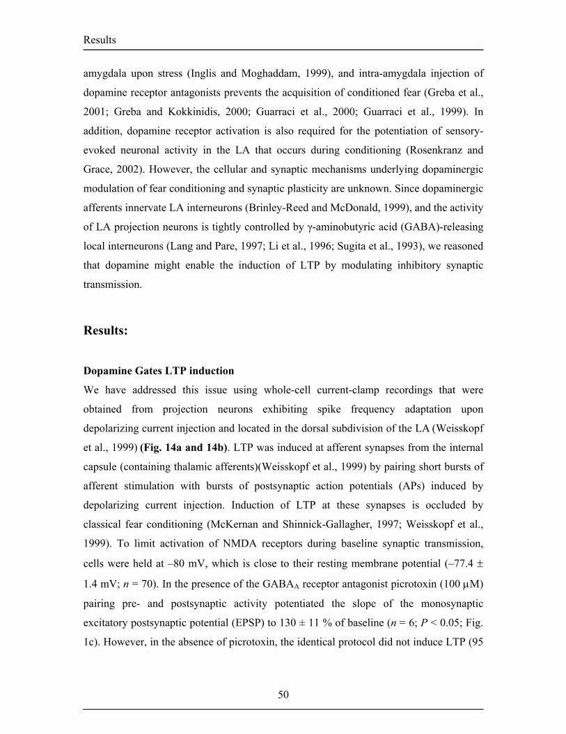
Results
amygdala upon stress (Inglis and Moghaddam, 1999), and intra-amygdala injection of
dopamine receptor antagonists prevents the acquisition of conditioned fear (Greba et al.,
2001; Greba and Kokkinidis, 2000; Guarraci et al., 2000; Guarraci et al., 1999). In
addition, dopamine receptor activation is also required for the potentiation of sensory-
evoked neuronal activity in the LA that occurs during conditioning (Rosenkranz and
Grace, 2002). However, the cellular and synaptic mechanisms underlying dopaminergic
modulation of fear conditioning and synaptic plasticity are unknown. Since dopaminergic
afferents innervate LA interneurons (Brinley-Reed and McDonald, 1999), and the activity
of LA projection neurons is tightly controlled by γ-aminobutyric acid (GABA)-releasing
local interneurons (Lang and Pare, 1997; Li et al., 1996; Sugita et al., 1993), we reasoned
that dopamine might enable the induction of LTP by modulating inhibitory synaptic
transmission.
Results:
Dopamine Gates LTP induction
We have addressed this issue using whole-cell current-clamp recordings that were
obtained from projection neurons exhibiting spike frequency adaptation upon
depolarizing current injection and located in the dorsal subdivision of the LA (Weisskopf
et al., 1999) (Fig. 14a and 14b). LTP was induced at afferent synapses from the internal
capsule (containing thalamic afferents)(Weisskopf et al., 1999) by pairing short bursts of
afferent stimulation with bursts of postsynaptic action potentials (APs) induced by
depolarizing current injection. Induction of LTP at these synapses is occluded by
classical fear conditioning (McKernan and Shinnick-Gallagher, 1997; Weisskopf et al.,
1999). To limit activation of NMDA receptors during baseline synaptic transmission,
cells were held at –80 mV, which is close to their resting membrane potential (–77.4 ±
1.4 mV; n = 70). In the presence of the GABAA receptor antagonist picrotoxin (100 µM)
pairing pre- and postsynaptic activity potentiated the slope of the monosynaptic
excitatory postsynaptic potential (EPSP) to 130 ± 11 % of baseline (n = 6; P < 0.05; Fig.
1c). However, in the absence of picrotoxin, the identical protocol did not induce LTP (95
50
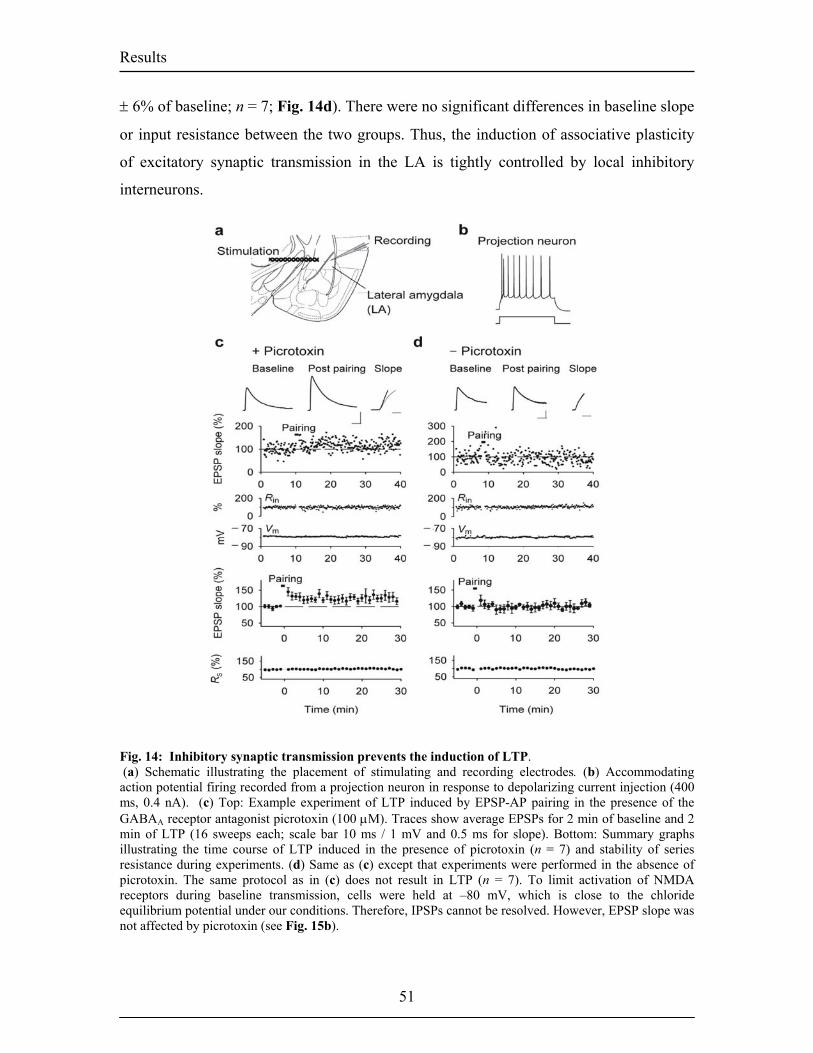
Results
± 6% of baseline; n = 7; Fig. 14d). There were no significant differences in baseline slope
or input resistance between the two groups. Thus, the induction of associative plasticity
of excitatory synaptic transmission in the LA is tightly controlled by local inhibitory
interneurons.
Fig. 14: Inhibitory synaptic transmission prevents the induction of LTP. (a) Schematic illustrating the placement of stimulating and recording electrodes. (b) Accommodating action potential firing recorded from a projection neuron in response to depolarizing current injection (400 ms, 0.4 nA). (c) Top: Example experiment of LTP induced by EPSP-AP pairing in the presence of the GABAA receptor antagonist picrotoxin (100 µM). Traces show average EPSPs for 2 min of baseline and 2 min of LTP (16 sweeps each; scale bar 10 ms / 1 mV and 0.5 ms for slope). Bottom: Summary graphs illustrating the time course of LTP induced in the presence of picrotoxin (n = 7) and stability of series resistance during experiments. (d) Same as (c) except that experiments were performed in the absence of picrotoxin. The same protocol as in (c) does not result in LTP (n = 7). To limit activation of NMDA receptors during baseline transmission, cells were held at –80 mV, which is close to the chloride equilibrium potential under our conditions. Therefore, IPSPs cannot be resolved. However, EPSP slope was not affected by picrotoxin (see Fig. 15b).
51
Results
Bath-application of dopamine (100 µM) during pairing enabled the induction of LTP
even in the presence of functional synaptic inhibition (135 ± 9% of baseline; n = 9; P <
0.01; Fig. 15). This effect was not due to a delayed potentiation of excitatory synaptic
transmission by dopamine itself, since dopamine application in the absence of pairing did
not lead to potentiation of the EPSP slope (94 ± 8% of baseline; n = 11; Fig. 15c). In
addition, dopamine did not facilitate the induction of LTP in the presence of picrotoxin,
but rather seemed to induce a transient depression of excitatory synaptic transmission (88
± 15% of baseline; n = 4; Fig. 15c). The finding that dopamine blocked LTP in the
presence of picrotoxin indicates that the processes triggered by the pairing protocol
differ, depending on the presence or absence of functional synaptic inhibition.
Dopaminergic gating of LTP induction was mediated by D2 receptors. LTP could be
induced in the presence of the D2 receptor agonist quinpirole (quin; 10 µM; 131 ± 5% of
baseline; n = 3; P < 0.05; Fig. 15c), but not in the presence of the D1 receptor agonist
dihydrexidine (DH, 10 µM; 98 ± 6% of baseline; n = 4; Fig. 15c). Thus, our data
demonstrate that, if functional synaptic inhibition is present, dopamine and D2 receptor
activation is necessary for the induction of LTP.
52
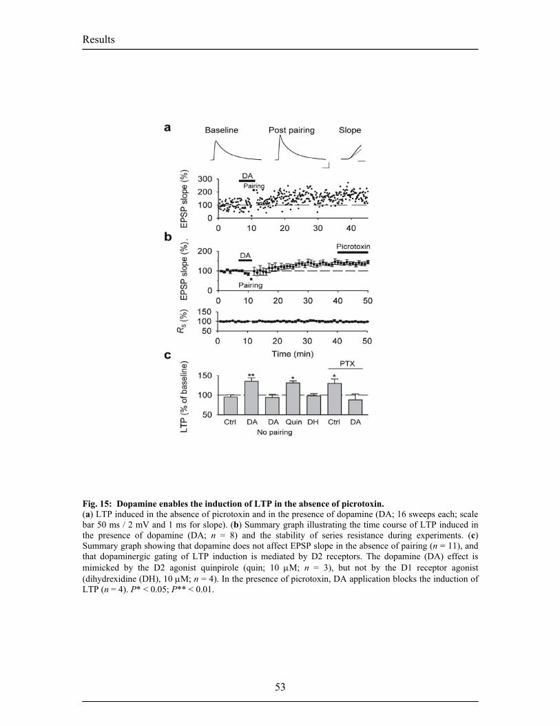
Results
Fig. 15: Dopamine enables the induction of LTP in the absence of picrotoxin. (a) LTP induced in the absence of picrotoxin and in the presence of dopamine (DA; 16 sweeps each; scale bar 50 ms / 2 mV and 1 ms for slope). (b) Summary graph illustrating the time course of LTP induced in the presence of dopamine (DA; n = 8) and the stability of series resistance during experiments. (c) Summary graph showing that dopamine does not affect EPSP slope in the absence of pairing (n = 11), and that dopaminergic gating of LTP induction is mediated by D2 receptors. The dopamine (DA) effect is mimicked by the D2 agonist quinpirole (quin; 10 µM; n = 3), but not by the D1 receptor agonist (dihydrexidine (DH), 10 µM; n = 4). In the presence of picrotoxin, DA application blocks the induction of LTP (n = 4). P* < 0.05; P** < 0.01.
53
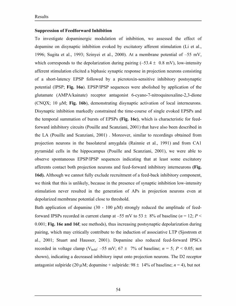
Results
Suppression of Feedforward Inhibition
To investigate dopaminergic modulation of inhibition, we assessed the effect of
dopamine on disynaptic inhibition evoked by excitatory afferent stimulation (Li et al.,
1996; Sugita et al., 1993; Szinyei et al., 2000). At a membrane potential of –55 mV,
which corresponds to the depolarization during pairing (–53.4 ± 0.8 mV), low-intensity
afferent stimulation elicited a biphasic synaptic response in projection neurons consisting
of a short-latency EPSP followed by a picrotoxin-sensitive inhibitory postsynaptic
potential (IPSP; Fig. 16a). EPSP/IPSP sequences were abolished by application of the
glutamate (AMPA/kainate) receptor antagonist 6-cyano-7-nitroquinoxaline-2,3-dione
(CNQX; 10 µM; Fig. 16b), demonstrating disynaptic activation of local interneurons.
Disynaptic inhibition markedly constrained the time-course of single evoked EPSPs and
the temporal summation of bursts of EPSPs (Fig. 16c), which is characteristic for feed-
forward inhibitory circuits (Pouille and Scanziani, 2001) that have also been described in
the LA (Pouille and Scanziani, 2001) . Moreover, similar to recordings obtained from
projection neurons in the basolateral amygdala (Rainnie et al., 1991) and from CA1
pyramidal cells in the hippocampus (Pouille and Scanziani, 2001), we were able to
observe spontaneous EPSP/IPSP sequences indicating that at least some excitatory
afferents contact both projection neurons and feed-forward inhibitory interneurons (Fig.
16d). Although we cannot fully exclude recruitment of a feed-back inhibitory component,
we think that this is unlikely, because in the presence of synaptic inhibition low-intensity
stimulation never resulted in the generation of APs in projection neurons even at
depolarized membrane potential close to threshold.
Bath application of dopamine (30 - 100 µM) strongly reduced the amplitude of feed-
forward IPSPs recorded in current clamp at –55 mV to 53 ± 8% of baseline (n = 12; P <
0.001; Fig. 16e and 16f; see methods), thus increasing postsynaptic depolarization during
pairing, which may critically contribute to the induction of associative LTP (Sjostrom et
al., 2001; Stuart and Hausser, 2001). Dopamine also reduced feed-forward IPSCs
recorded in voltage clamp (Vhold: –55 mV; 67 ± 7% of baseline; n = 5; P < 0.05; not
shown), indicating a decreased inhibitory input onto projection neurons. The D2 receptor
antagonist sulpiride (20 µM; dopamine + sulpiride: 98 ± 14% of baseline; n = 4), but not
54
Results
the D1 receptor antagonist SCH23390 (10 µM; dopamine + SCH23390: 54 ± 6% of
baseline; n = 4; P < 0.01) completely blocked the effect of dopamine on the IPSP
amplitude (Fig. 16g). Consistently, IPSP amplitude was reduced by the D2 receptor
agonist quinpirole (10 µM; 58 ± 11% of baseline; n = 4; P < 0.05; Fig. 16g), but not by
the D1 receptor agonist dihydrexidine (10 µM; 99 ± 11% of baseline; n = 6; Fig. 16g),
indicating that, like the gating of LTP induction, suppression of feed-forward inhibition is
mediated by D2 receptors. In addition, consistent with in vivo recordings that showed an
increase in the spontaneous firing rate of interneurons upon application of dopamine
receptor agonists (Rosenkranz and Grace, 1999), dopamine increased spontaneous
inhibitory network activity as measured by the frequency of spontaneous inhibitory
postsynaptic currents (sIPSC; 157 ± 13% of baseline; n = 8; P < 0.01). However, in
contrast to the suppression of feed-forward inhibition, dopamine still increased sIPSC
frequency in the presence of the D2 antagonist sulpiride (134 ± 3% of baseline; n = 3; P
< 0.05).
55
Results
Fig.16: Dopamine depresses feed-forward inhibition (a) Disynaptic IPSP constrains the EPSP time course and is blocked by picrotoxin. (b) EPSP/IPSP sequences are blocked by CNQX, demonstrating disynaptic activation of GABAergic interneurons (scale bar in a and b: 50 ms / 2 mV). (c) Disynaptic IPSPs inhibit temporal summation of EPSPs at the stimulation frequency used for LTP induction (33 Hz; scale bar 10 ms). (d) Spontaneous EPSP (arrow)/IPSP sequence recorded from a projection neuron at –55 mV (scale bar 5 ms / 0.5 mV). (e) Experiment illustrating the effect of dopamine on EPSP/IPSP sequences (scale bar 50 ms / 2 mV). (f) Time course of the dopamine-induced decrease in IPSP amplitude (n = 8). (g) Depression of feed-forward inhibition is mediated by D2 receptors. The dopamine (DA) effect is blocked by the D2 antagonist sulpiride (sulp; 20 µM; n = 4), and mimicked by the D2 agonist quinpirole (Quin; 10 µM; n = 4). D1 receptor antagonists and agonists (SCH23390 (SCH), 10 µM; n = 4; dihydrexidine (DH), 10 µM; n = 6) have no effect. P* < 0.05; P** < 0.01; P*** < 0.001.
56
Results
To determine the mechanism underlying dopaminergic suppression of feed-forward
inhibition, we first addressed the question whether dopamine directly acted on inhibitory
synaptic transmission between LA interneurons and projection neurons. Monosynaptic
IPSCs recorded from projection neurons and elicited by proximal extracellular
stimulation within the LA in the presence of CNQX and the NMDA receptor antagonist
3-((+/–)-2-carboxypiperazin-4-yl)-propyl-1-phosphonic acid (CPP) were reduced by
dopamine to 85 ± 7% of baseline (n = 12; P < 0.05; Fig 17a). Consistent with our
observation that the reduction of disynaptic feed-forward inhibition was mediated by D2
receptors, we found that monosynaptic IPSCs were also reduced by the D2 receptor
agonist quinpirole (83 ± 4% of baseline; n = 10; P < 0.01), but not by the D1 receptor
agonist dihydrexidine (97 ± 4% of baseline; n = 10; Fig. 17b). In addition, dopamine
decreased the amplitude but not the frequency of miniature IPSCs (mIPSCs) recorded in
the presence of tetrodotoxin (TTX; 92 ± 3% of baseline; n = 5; P < 0.05; not shown).
These results could suggests a postsynaptic effect of dopamine. However, postsynaptic
currents induced by local application of the GABAA receptor agonist muscimol (50 µM)
using pressure ejection from a nearby patch-pipette were not affected by dopamine
application (101 ± 3% of baseline; n = 5; Fig. 17c). Furthermore, postsynaptic blockade
of G-proteins by GDPβS (1 mM in the recording electrode; 50 min. whole-cell perfusion)
did not interfere with the dopamine-induced reduction in monosynaptic IPSC amplitude
(82 ± 3% of baseline; n = 5; P < 0.05; Fig. 17d), although postsynaptic currents induced
by application of baclofen (50 µM) were completely blocked (control intracellular
solution: 34.3 ± 12.8 pA; n = 3; intracellular solution containing GDPβS: –2.7 ± 2.9 pA;
n = 3; P < 0.05). Taken together, these results indicate that dopamine presynaptically
reduces the quantal content of inhibitory synapses by D2 receptor activation. However,
the relatively small reduction also suggest that additional mechanisms might have
contributed to the suppression of feed-forward inhibition.
57
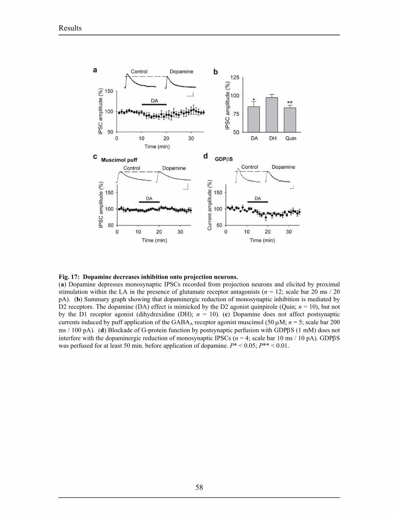
Results
Fig. 17: Dopamine decreases inhibition onto projection neurons. (a) Dopamine depresses monosynaptic IPSCs recorded from projection neurons and elicited by proximal stimulation within the LA in the presence of glutamate receptor antagonists (n = 12; scale bar 20 ms / 20 pA). (b) Summary graph showing that dopaminergic reduction of monosynaptic inhibition is mediated by D2 receptors. The dopamine (DA) effect is mimicked by the D2 agonist quinpirole (Quin; n = 10), but not by the D1 receptor agonist (dihydrexidine (DH); n = 10). (c) Dopamine does not affect postsynaptic currents induced by puff application of the GABAA receptor agonist muscimol (50 µM; n = 5; scale bar 200 ms / 100 pA). (d) Blockade of G-protein function by postsynaptic perfusion with GDPβS (1 mM) does not interfere with the dopaminergic reduction of monosynaptic IPSCs (n = 4; scale bar 10 ms / 10 pA). GDPβS was perfused for at least 50 min. before application of dopamine. P* < 0.05; P** < 0.01.
58
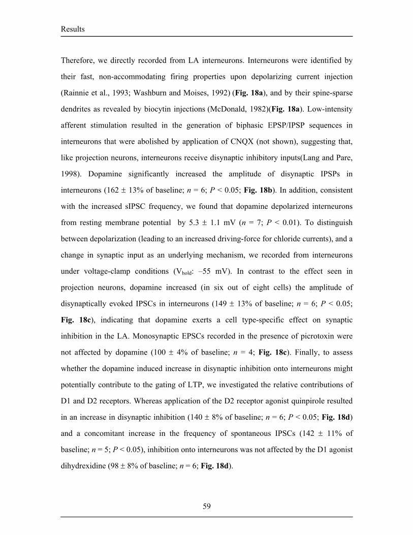
Results
Therefore, we directly recorded from LA interneurons. Interneurons were identified by
their fast, non-accommodating firing properties upon depolarizing current injection
(Rainnie et al., 1993; Washburn and Moises, 1992) (Fig. 18a), and by their spine-sparse
dendrites as revealed by biocytin injections (McDonald, 1982)(Fig. 18a). Low-intensity
afferent stimulation resulted in the generation of biphasic EPSP/IPSP sequences in
interneurons that were abolished by application of CNQX (not shown), suggesting that,
like projection neurons, interneurons receive disynaptic inhibitory inputs(Lang and Pare,
1998). Dopamine significantly increased the amplitude of disynaptic IPSPs in
interneurons (162 ± 13% of baseline; n = 6; P < 0.05; Fig. 18b). In addition, consistent
with the increased sIPSC frequency, we found that dopamine depolarized interneurons
from resting membrane potential by 5.3 ± 1.1 mV (n = 7; P < 0.01). To distinguish
between depolarization (leading to an increased driving-force for chloride currents), and a
change in synaptic input as an underlying mechanism, we recorded from interneurons
under voltage-clamp conditions (Vhold: –55 mV). In contrast to the effect seen in
projection neurons, dopamine increased (in six out of eight cells) the amplitude of
disynaptically evoked IPSCs in interneurons (149 ± 13% of baseline; n = 6; P < 0.05;
Fig. 18c), indicating that dopamine exerts a cell type-specific effect on synaptic
inhibition in the LA. Monosynaptic EPSCs recorded in the presence of picrotoxin were
not affected by dopamine (100 ± 4% of baseline; n = 4; Fig. 18c). Finally, to assess
whether the dopamine induced increase in disynaptic inhibition onto interneurons might
potentially contribute to the gating of LTP, we investigated the relative contributions of
D1 and D2 receptors. Whereas application of the D2 receptor agonist quinpirole resulted
in an increase in disynaptic inhibition (140 ± 8% of baseline; n = 6; P < 0.05; Fig. 18d)
and a concomitant increase in the frequency of spontaneous IPSCs (142 ± 11% of
baseline; n = 5; P < 0.05), inhibition onto interneurons was not affected by the D1 agonist
dihydrexidine (98 ± 8% of baseline; n = 6; Fig. 18d).
59
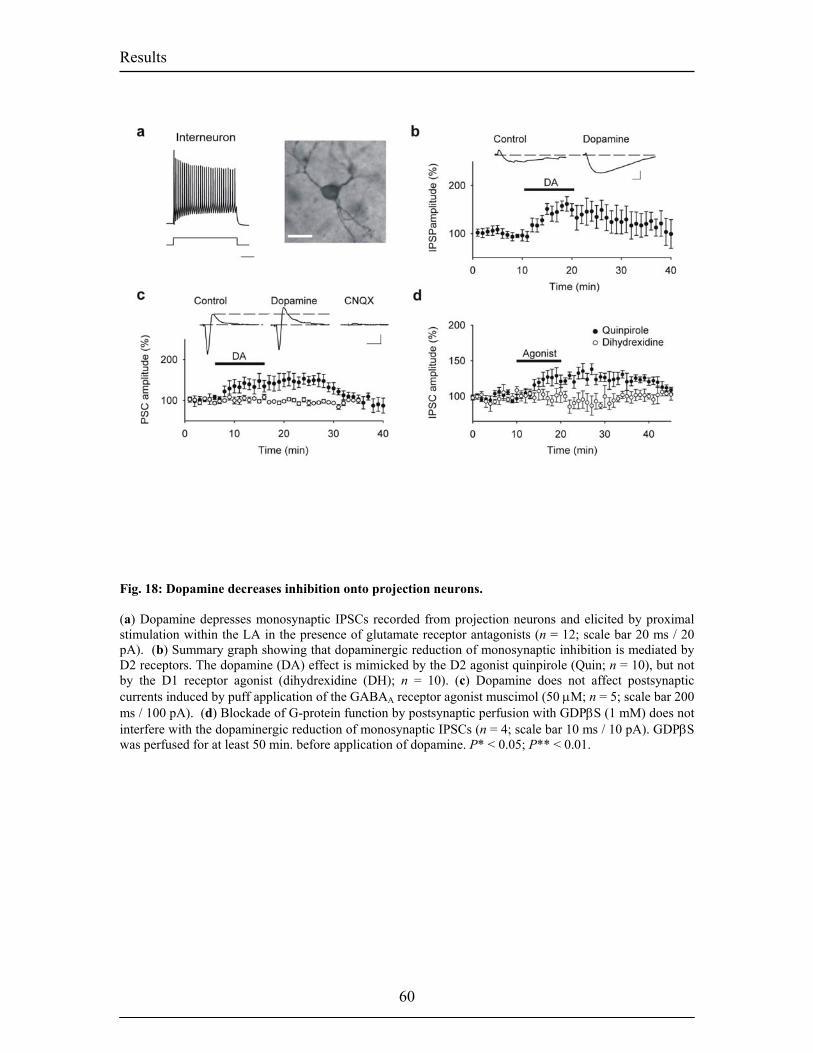
Results
Fig. 18: Dopamine decreases inhibition onto projection neurons. (a) Dopamine depresses monosynaptic IPSCs recorded from projection neurons and elicited by proximal stimulation within the LA in the presence of glutamate receptor antagonists (n = 12; scale bar 20 ms / 20 pA). (b) Summary graph showing that dopaminergic reduction of monosynaptic inhibition is mediated by D2 receptors. The dopamine (DA) effect is mimicked by the D2 agonist quinpirole (Quin; n = 10), but not by the D1 receptor agonist (dihydrexidine (DH); n = 10). (c) Dopamine does not affect postsynaptic currents induced by puff application of the GABAA receptor agonist muscimol (50 µM; n = 5; scale bar 200 ms / 100 pA). (d) Blockade of G-protein function by postsynaptic perfusion with GDPβS (1 mM) does not interfere with the dopaminergic reduction of monosynaptic IPSCs (n = 4; scale bar 10 ms / 10 pA). GDPβS was perfused for at least 50 min. before application of dopamine. P* < 0.05; P** < 0.01.
60
Results
DISCUSSION: Our data demonstrate that, in the presence of inhibition, LTP cannot be induced at
excitatory afferents from the internal capsule to the LA. This is consistent with in vivo
experiments showing that the activity of LA projection neurons is tightly controlled by
local GABAergic inhibition resulting in extremely low spontaneous firing rates (Lang
and Pare, 1997; Pare and Collins, 2000; Pare and Gaudreau, 1996). Moreover, behavioral
experiments indicate that the GABAergic system of the basolateral amygdala tightly
controls the acquisition and expression of different measures of conditioned and
unconditioned fear (Davis, 2000). Since dopamine receptor activation is needed for the
acquisition of fear conditioning and the concomitant potentiation of sensory-evoked
responses (Greba et al., 2001; Greba and Kokkinidis, 2000; Guarraci et al., 2000;
Guarraci et al., 1999; Rosenkranz and Grace, 2002), and since dopaminergic afferents
innervate LA interneurons (Brinley-Reed and McDonald, 1999), we reasoned that
dopamine might enable the induction of LTP by modulating inhibitory synaptic
transmission. Our study shows that activation of dopamine D2 receptors suppresses feed-
forward inhibition, and enables the induction of LTP at excitatory afferent synapses.
The mechanism underlying dopaminergic suppression of feed-forward inhibition appears
to be twofold. First, monosynaptic IPSCs recorded from projection neurons and elicited
by proximal stimulation within the LA were reduced upon dopamine application
indicating that dopamine directly affects inhibitory synaptic transmission. Further
experiments using either puff application of the GABAA receptor agonist muscimol or
intracellular application of GDPβS to block postsynaptic G-protein function indicated
that the dopamine-induced decrease in inhibitory synaptic transmission is due to a
presynaptic reduction in quantal content (Seamans et al., 2001) (Fig. 19a). Moreover, in
contrast to the dopaminergic suppression of feed-forward inhibition onto projection
neurons, we observed that feed-forward inhibition onto interneurons was enhanced upon
application of dopamine, which may lead to a reduction in feed-forward inhibition onto
projection neurons. This enhancement was accompanied by an increase in spontaneous
inhibitory input onto interneurons. This suggests that dopamine facilitates inhibition onto
interneurons by depolarizing interneurons, or a subpopulation of interneurons
(Kemppainen and Pitkanen, 2000; McDonald and Betette, 2001), involved in
61
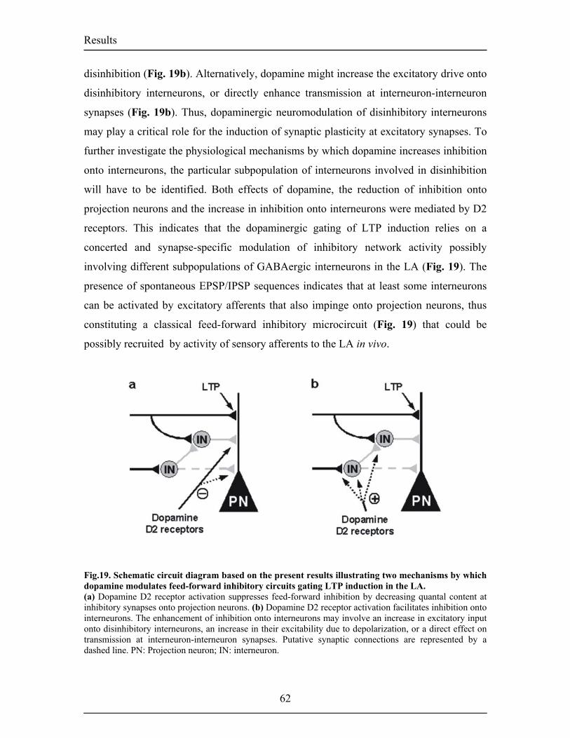
Results
disinhibition (Fig. 19b). Alternatively, dopamine might increase the excitatory drive onto
disinhibitory interneurons, or directly enhance transmission at interneuron-interneuron
synapses (Fig. 19b). Thus, dopaminergic neuromodulation of disinhibitory interneurons
may play a critical role for the induction of synaptic plasticity at excitatory synapses. To
further investigate the physiological mechanisms by which dopamine increases inhibition
onto interneurons, the particular subpopulation of interneurons involved in disinhibition
will have to be identified. Both effects of dopamine, the reduction of inhibition onto
projection neurons and the increase in inhibition onto interneurons were mediated by D2
receptors. This indicates that the dopaminergic gating of LTP induction relies on a
concerted and synapse-specific modulation of inhibitory network activity possibly
involving different subpopulations of GABAergic interneurons in the LA (Fig. 19). The
presence of spontaneous EPSP/IPSP sequences indicates that at least some interneurons
can be activated by excitatory afferents that also impinge onto projection neurons, thus
constituting a classical feed-forward inhibitory microcircuit (Fig. 19) that could be
possibly recruited by activity of sensory afferents to the LA in vivo.
Fig.19. Schematic circuit diagram based on the present results illustrating two mechanisms by which dopamine modulates feed-forward inhibitory circuits gating LTP induction in the LA. (a) Dopamine D2 receptor activation suppresses feed-forward inhibition by decreasing quantal content at inhibitory synapses onto projection neurons. (b) Dopamine D2 receptor activation facilitates inhibition onto interneurons. The enhancement of inhibition onto interneurons may involve an increase in excitatory input onto disinhibitory interneurons, an increase in their excitability due to depolarization, or a direct effect on transmission at interneuron-interneuron synapses. Putative synaptic connections are represented by a dashed line. PN: Projection neuron; IN: interneuron.
62
Results
The finding that modulation of feed-forward inhibition is able to control the induction of
LTP in the amygdala is consistent with previous studies in the hippocampus, indicating
that LTP induction is facilitated upon blockade of GABAA receptors (Wigstrom and
Gustafsson, 1983), or GABAB autoreceptors on feed-forward interneurons (Davies et al.,
1991), and may be explained by several possible mechanisms resulting in a reduction in
postsynaptic depolarization which is required for LTP induction (Blair et al., 2001; Bliss
and Collingridge, 1993; Sjostrom et al., 2001). First, our own results (Fig. 16a) and a
recent study on hippocampal feed-forward inhibition indicates that feed-forward
inhibition constrains the effective temporal summation of EPSPs during repetitive
afferent activity (Pouille and Scanziani, 2001). Second, since in LA projection neurons
APs are followed by a pronounced afterdepolarization, feed-forward inhibition may also
reduce the temporal summation of afterdepolarizations during bursts of backpropagating
APs (Sjostrom et al., 2001). Third, based on the observation that dendritic EPSPs can
boost AP backpropagation (Stuart and Hausser, 2001), it is possible that feed-forward
inhibitory inputs might interfere with AP backpropagation. Finally, rather than acting on
autoreceptors on inhibitory terminals, GABA released by feed-forward inhibitory neurons
might activate heterosynaptic GABAB receptors located presynaptically on excitatory
synaptic boutons (Yamada et al., 1999) thereby reducing the probability of release at
glutamatergic afferent synapses during pairing. A common consequence of these
mechanisms would be a decreased activation of NMDA receptors or voltage-gated
calcium channels during pairing. Indeed, consistent with earlier reports indicating a role
for NMDA receptors and voltage-gated calcium channels (VGCCs) for the induction of
LTP at thalamo-amygdala synapses (Bauer et al., 2002; Weisskopf et al., 1999), we found
that LTP was blocked by NMDA receptor or VGCC antagonists (Y.H. and A.L.,
unpublished results). The finding that both NMDA and VGCC antagonists interfered with
LTP induction might indicate that, under our experimental conditions, activation of
NMDA receptors and VGCCs during pairing were not independent processes.
In addition to the D2 receptor-mediated suppression of feed-forward inhibition, we
observed a dopamine-induced increase in spontaneous inhibitory network activity
requiring D1 receptor activation. D1 receptor activation, however, was not necessary for
the gating of LTP induction. Behavioral experiments indicate that pharmacological
63
Results
64
blockade of D1 or D2 receptors in the basolateral amygdala interferes with the
acquisition of fear conditioning(Greba et al., 2001; Greba and Kokkinidis, 2000; Guarraci
et al., 2000; Guarraci et al., 1999; Rosenkranz and Grace, 2002). One possible
interpretation is that the D2 receptor-mediated suppression of feed-forward inhibition
facilitates the induction of LTP in response to sensory input whereas the increase in
spontaneous inhibitory network activity requiring D1 receptor activation might suppress
synaptic plasticity induced by random associations. In addition, in vivo
electrophysiological experiments indicate that sensory inputs to the LA may also be
enhanced by dopaminergic attenuation of prefrontal cortical inputs suppressing the
activity of LA projection neuronsRosenkranz, 2001 #3.
In conclusion, our study suggests that dopaminergic suppression of feed-forward
inhibition represents one key mechanism underlying the acquisition of fear conditioning.
Furthermore, dopaminergic neuromodulation of inhibitory synaptic transmission seems to
be pivotal not only for the acquisition, but also for other processes such as retrieval or
extinction (El-Ghundi et al., 2001; Nader and LeDoux, 1999). This suggests that
alterations in the dopaminergic projection to the amygdala might have profound effects
on amygdala-dependent emotional behavior. Such alterations could for instance be
induced by stress, particularly during early development (Heidbreder et al., 2000),
thereby contributing to various conditions with emotional dysregulation (Klimek et al.,
2002; Tessitore et al., 2002).
Results
Materials and Methods
Slice preparation. Standard procedures were used to prepare 350 µm thick coronal
slices from three to four week old male C57BL/6J mice following a protocol approved by
the Veterinary Department of the Canton of Basel-Stadt. Briefly, the brain was dissected
in ice-cold artificial cerebrospinal fluid (ACSF), mounted on an agar block and sliced
with a vibratome at 4°C. Slices were maintained for 45 min. at 35°C in an interface
chamber containing ACSF equilibrated with 95% O2/5% CO2 and containing (in mM):
124 NaCl, 2.7 KCl, 2 CaCl2, 1.3 MgCl2, 26 NaHCO3, 0.4 NaH2PO4, 10 glucose, 4
ascorbate, and then for at least 45 min. at room temperature before being transferred to a
superfusing recording chamber.
Whole-cell recordings. Whole-cell recordings from LA projection neurons and
interneurons were performed at 30-32°C in a superfusing chamber. Neurons were
visually identified with infrared videomicroscopy. Patch electrodes (3-5 MΩ) were
normally filled with a solution containing (in mM): 120 K-gluconate, 20 KCl, 10 HEPES,
10 phosphocreatine, 4 Mg-ATP, 0.3 Na-GTP, 0.1 % biocytin, pH 7.25, 295 mOsm. To
resolve IPSPs at resting membrane potential in interneurons intracellular Cl–
concentration was lowered to 2 mM by replacing KCl with equi-osmolar K-gluconate.
For voltage-clamp recordings of sIPSCs and mIPSCs, K-gluconate and KCl were
replaced by equimolar CsCl. For experiments assessing postsynaptic responses, the
GABAA receptor agonist muscimol (50 µM) was pressure applied (20 psi; duration 6 ms)
every 60 s via a puffer pipette (containing 20 µM CNQX and 20 µM CPP), placed near
the soma.
Stimulation and analysis. Afferent fibers were stimulated with a bipolar twisted
platinum/10% iridium wire (25 µm diameter). LTP was induced by pairing 3
monosynaptic EPSPs with 3 APs (elicited by 0.8 nA, 5 ms current steps; EPSP onset-AP
65
Results
peak delay was 10 ms) at 33 Hz (Weisskopf et al., 1999). This pattern was repeated 15×
at 0.2 Hz. LTP was quantified for statistical comparisons by normalizing and averaging
EPSP slopes during the last 10 min. of experiments relative to 10 min of baseline.
Depolarization during pairing was measured by averaging depolarization during a 1 ms
time window just before the 2nd and 3rd spike of a spike burst. The reduction in disynaptic
inhibition was quantified as change in IPSP amplitude relative to baseline, which does
not include the fraction of inhibitory conductance at membrane potentials positive to the
holding potential. All membrane potentials have been corrected for a junction potential of
11 mV recorded with the K-gluconate internal solution. Data were recorded with an
Axopatch200B, filtered at 2 kHz and digitized at 10 kHz. In all experiments series
resistance was monitored throughout the experiment by applying a hyperpolarizing
current or voltage pulse, and if it changed by more than 15%, the data were not included
in the analysis. Data were acquired and analyzed with: ClampEx8.0, ClampFit8.0 (Axon
Instruments, Union City, CA, USA), Mini Analysis Program (Synaptosoft, Decatur, GA,
USA), and the LTP Program (W. Anderson, University of Bristol, UK). The thresholds
for mIPSC and sIPSC detection were set to 5 pA and 12 pA, respectively. Average
frequency changes were calculated by averaging in each cell two min. during baseline
and at the end of dopamine application. A time bin of two min. contained at least 700
(mIPSCs) or 1400 (sIPSCs) events. Frequency and amplitude of mIPSCs and sIPSCs
within individual neurons were evaluated using cumulative probability analysis, with
statistical significance determined by using the Kolmogorov-Smirnov nonparametric two-
sample test. All other statistical comparisons were done with paired or unpaired Student’s
t-test as appropriate (P < 0.05 was considered significant). All values are expressed as
means ± s.e.m. CNQX, (S)-(–)-sulpiride, (–)-quinpirole, dihydrexidine, and SCH23390
were from Tocris-Cookson (Bristol, UK), TTX from Latoxan (Valence, France). CPP and
CGP54626 were obtained as a gift from Novartis Inc. (Basel, Switzerland). All other
drugs were from Fluka/Sigma (Buchs, Switzerland).
66

Results
67
Morphology. Following immersion fixation of the slices in 4% PFA, biocytin-filled
neurons were visualized using the standard ABC method (Vector Laboratories,
Peterborough, UK) and nickel-intensified DAB as a chromogen.
Results
3.2-Unpublished Results Dopaminergic Modulation of Spontaneous Inhibitory Network Activity in the Lateral Amygdala. The amygdala plays an important role in the association of sensory stimuli with
aversive outcomes (LeDoux, 2000). Powerful inhibitory circuits exist in the
amygdala resulting in the low spontaneous firing rates of principal neurons
observed in vivo (Lang and Pare, 1997; Pare et al., 2003; Quirk et al., 2003). There is
converging evidence that dopamine (DA) plays a significant role in fear conditioning
(Lamont and Kokkinidis, 1998; Nader and LeDoux, 1999) and previous studies
revealed that systemic administration of dopamine agonists increases the
spontaneous firing rate of interneurons in the LA (Rosenkranz and Grace, 1999).
However, the effect of dopamine on spontaneous network activity in the LA in vitro
has never been assessed. Here, we show that bath application of dopamine (100 µM)
increases the frequency of spontaneous inhibitory postsynaptic currents (IPSCs)
recorded from pyramidal cells in the LA. Recordings of miniature postsynaptic
currents (mIPSCs) and direct recordings from interneurons suggest that dopamine
mediates its action by depolarizing local interneurons. In contrast to the effect of
dopamine on feed-forward inhibition and LTP induction (Bissiere et al., 2003), the
increase in the frequency of spontaneous inhibitory networks required the
activation of D1 receptors in synergy with D2 receptors. Moreover, preliminary
data revealed that the signaling cascade resulting in this effect does not require the
activation of conventional downstream targets of G-protein coupled DA receptors.
We have addressed the effect of dopamine on spontaneous inhibitory network activity in
the LA using in vitro whole-cell voltage clamp recordings at -70 mV obtained from
projection neurons. Spontaneous inhibitory postsynaptic currents (sIPSCs) were selected
for using a Cs Cl-based intracellular solution in the presence of blockers of excitatory
synaptic transmission (10 µM of CNQX and D-CPPene) applied in the extracellular
solution. Perfusion of DA (100 µM) for 10 min increased the spontaneous inhibitory
68
Results
network activity as measured by the frequency of sIPSCs (157% ± 13% of baseline; n =
8; P < 0.01; Fig. 19). The role of DA on the spontaneous excitatory network was also
assessed by measuring the spontaneous excitatory postsynaptic currents (sEPSCs) at -
70mV in the presence of the GABAA receptor antagonist picrotoxin (100µM) and by
using a K+ gluconate based solution. Bath-application of DA did not affect the frequency
or the amplitude of the sEPSCs (109% ± 15% of baseline; n = 6; Fig. 20).
Dopamine depolarizes interneurons
To assess whether dopamine directly affected the properties of inhibitory synaptic
transmission, we recorded mIPSCs from pyramidal cells at -70 mv in the presence of
TTX (1 µM). DA application did not change the frequency of mIPSCs ( 103% ± 2 % of
baseline; n = 5; Fig. 22A to C). In addition, direct current clamp recordings from LA
interneurons revealed that DA depolarizes the membrane potential by 5.3 ± 1.1 mV (n =
7; P < 0.01; Fig. 22D). These results suggest that dopamine modulates the
somatodendritic properties of interneurons resulting in an increased excitability and
higher spontaneous firing rate.
Dopamine receptor subtype mediating the increase in sIPSCs frequency
To investigate which DA receptor was underlying the above observations, we perfused
D1 or D2 receptor antagonists during DA application. We found that application of the
of the D2 receptor antagonist sulpiride (25 µM) with dopamine still induced an increased
in the frequency of sIPSCs (134 ± 3% of baseline; n = 3; P < 0.05; Fig. 23B). However,
application of the D1 receptor antagonist SCH 23390 (25 µM) completely abolished the
DA-mediated increase in the frequency of sIPSCs (Fig 23.A). In contrast, neither the D1
receptor agonist, dihydrexidine (10 µM) nor the D2 receptor agonist quinpirole (10 µM)
were able to reproduce the effect of DA on sIPSCs (Fig. 23C to D). However, co-
application of both agonists could partially mimic the DA effect on sIPSC frequency
increased in sIPSC frequency (20 µM; 115% ± 8% of baseline; n= 8; P <0.01; Fig. 23E
69
Results
). These results suggest that the effect of DA on the sIPSC frequency is a D1 receptor
mediated process requiring however the concomitant activation of D2 receptors.
Signaling mechanisms mediating the DA-dependent increase in sIPSC frequency
DA receptors have been demonstrated to be couple to Gi and Gs signaling mechanisms
(Missale et al., 1998), eventually resulting in the modulation of ligand and voltage-gated
ion channels. To elucidate which signaling mechanisms resulted in an increased
frequency of sIPSCs, we first addressed the question whether dopamine receptors in the
LA were coupled to the Gs subunit. Activation of Gs leads to an increase in cAMP levels
which is turn activate protein kinase A (PKA). Preliminary data showed that application
of DA in the presence of Rp-cAMP (100 µM), a cAMP analogue, still induced an
increase in the frequency of sIPSCs (24% ± 18 P< 0.007 n = 2 Fig. 24A). Moreover, we
investigated whether DA activated the enzyme phospholipase C (PLC) via activation of
the Gq subunit. However, even in the presence of U71322, a PLC inhibitor, DA still
induced an increase in the sIPSC frequency (38% ± 9 P< 0.0001 n=3 Fig. 24B). Most of
what we know on the signaling of dopamine receptors through heterotrimeric G-proteins
was mainly ascribed to the role of the Gα subunits. However, little is known about the
involvement of the βγ subunit. To assess if the effect of dopamine was mediated via a βγ-
dependent mechanism, we perfused suramin, a drug that interferes with Gα subunit
signaling, together with dopamine. One single experiment could be performed, and
recordings from this cell showed that dopamine was still able to induce an increase in the
frequency of sIPSCs (Fig. 24C).
DA induces alteration in the burst frequency, synchronization and oscillatory
inhibitory network activity in the LA.
In addition to an increase in sIPSCs frequency reflecting a change in the firing of
interneurons, DA also triggered changes in the pattern of the sIPSCs towards more
rhythmic activity. In some cells, this sometimes resulted in the induction of multisecond
oscillations Fig.25 .
70
Results
Discussion:
Consistent with in vivo studies showing an increase in the spontaneous firing rate of
interneurons upon systemic administration of DA agonists (Rosenkranz and Grace,
1999), we observed that bath application of DA increased the frequency of sIPSCs
recorded from pyramidal neurons in the LA (Fig. 20). This effect was specific as DA did
not induce changes in the sEPSCs recorded from pyramidal cells (Fig.21). The frequency
of mIPSCs was not affected by DA suggesting that the properties of the inhibitory
synapses were unchanged (Fig. 22). In contrast, we found that DA induced a
depolarization of local interneurons resulting in an increased excitability and a increased
in the frequency of action potential (AP) firing (Fig. 22D). Overall, these results suggest
that DA directly modulates the perisomatic properties of LA interneurons. However, it is
unclear whether DA depolarizes all interneurons or certain subtypes (McDonald and
Mascagni, 2001; McDonald and Pearson, 1989). One way to answer that question would
be to directly record from identified interneuron subpopulations. Pharmacological data
indicate that DA in the presence of the D2 antagonist sulpiride still induced an increase in
the frequency of sIPSCs. In contrast DA application in the presence of SCH 23390, a D1
receptor antagonist could block the effect of dopamine effect on the sIPSCs frequency
(Fig. 23A,B). Both antagonists induced a decrease in the sIPSCs baseline frequency
suggesting a certain level of tonic receptor activation in our preparation. In addition, co-
application of D1 or D2 receptor antagonists with DA resulted in opposite modulation of
the sIPSC frequency (Fig. 23A,B). These results demonstrated that only the activation of
D1 receptors was able to induce an increase in the sIPSC frequency (Fig. 23A). Next, we
showed that bath application of the D1 agonist dihydrexidine or, the D2 agonist
quinpirole alone could not mimic the effect of dopamine (Fig. 23C and D). However, co-
application of the two agonists together, partially mimicked the DA-mediated effect on
the sIPSC frequency (Fig. 23E). These results indicate that D1 receptor activation are
required but not sufficient for the increase in the sIPSCs frequency. Rather, a synergistic
interaction between D1 and D2 receptors seem to mediate this process. Such interactions
have already been observed to be important for locomotor activity in vivo (Jackson and
Hashizume, 1986). Similarly, synergy between the two receptors appears to be important
71
Results
for the modulation of firing rates recorded in vivo from basal ganglia neurons (Carlson et
al., 1987; Walters et al., 1987), with important implications for Parkinson's disease or
schizophrenia. In the amygdala the ratio of D1 to D2 receptors expressed in different
neurons, activated during different behavioral states might explain why dopamine does
not affect the sEPSCs under our experimental conditions. When stimulating thalamic
afferents to the LA, DA inhibits feed-forward inhibition via D2 receptor activation
(Bissiere et al., 2003). The different modulatory actions of these two receptors suggests
opposite roles for DA depending on its concentration and on the relative contribution of
D1/D2 receptors. During inactivated state of the amygdala, DA would have a filtering
role by keeping the level of spontaneous inhibition high and the level of random
association low. This effect would require the simultaneous activation of D1 and D2
receptors and the subsequent depolarization of interneurons. In contrast, upon the arrival
of relevant sensory informations, dopamine via a D2 effect supresses feed-forward
inihibition and allows for associative learning (Bissiere et al., 2003). Such a bidirectional
modulation has also been observed in the prefrontal cortex, the nucleus accumbens and
the striatum (Gorelova et al., 2002; Guzman et al., 2003; Seamans et al., 2001;
Vanderschuren et al., 1999) where dopamine has differential effects on spontaneous or
stimulated GABA release by activating different receptor subtypes.
Our preliminary data suggests that the increase in sIPSC frequency does not require PKA
or PLC activation (Fig. 24A, B). These findings are consistent with studies demonstrating
that D1 receptors in the BLA do not couple to the cAMP pathway (Leonard et al., 2003).
In vitro electrophysiological studies in cortical layer V-VI pyramidal neurons, reported
that DA increases the frequency of sIPSCs by suppressing an inward rectifier K+ current
and a resting leak K+ current, leading to a depolarization of interneurons (Gorelova et al.,
2002; Seamans et al., 2001). Similarly, in the striatum DA, via activation of D1 receptors,
depolarizes interneurons and increases the excitability of interneurons by supressing K+
currents (Bracci et al., 2002). In addition to K+ channels, D1 receptors can also directly
modulate calcium channels, or physically interact with ionotrophic receptors. Recently a
certain number of DA receptor associated proteins (DRIPs) have been identified, opening
new insight on the proteins that might regulate the intracellular activity of dopamine
receptors (Bergson et al., 2003). In addition, D1 and D2 receptor interactions might lead
72
Results
73
to unique functional properties as already observed for heterodimers formed by D2 and
D3 receptors (Scarselli et al., 2001). Finally, we also observed that bath application of
DA induced synchronization in the firing of interneurons that sometimes resulted in the
activation of multisecond oscillatory network activity (Fig.25). In the hippocampus, DA
was shown to modulate network properties by inducing oscillatory processes important
for learning and memory function (Lisman and Otmakhova, 2001). In the amygdala, a
range of slow waves (< 1 Hz) to fast gamma (γ) oscillations have been observed in vivo
and in vitro (Pape et al., 1998; Pare et al., 1995). It has been suggested that oscillatory
activity in the LA enables consolidation of memory processes and facilitates interactions
with neocortical storage sites (Collins et al., 2001; Pare et al., 2002; Pelletier and Pare,
2004). However, at this point the relevance of these spontaneous oscillations and the
exact conditions of their occurence, are unclear. One possibility would be that DA, by
depolarizing a large number of interneurons, would induce network activity mediated by
synchronization through gap junction. Similarly, DA could directly modulate the
properties of gap junctions. Such an effect has previously been reported in the
mammalian retina where DA via D1 receptor activation modulates the gap junctions
connecting amacrine cells (Hampson et al., 1992). As a next step, it will be important to
identify the different interneuron subtypes present in the LA. This can be done by
analyzing mice expressing GFP under the control of specific promoters. In this way,
interneurons involved in feed-forward inhibition or in dishinibition could be identifed and
the role of dopamine assessed in details. In addition, paired recordings from interneurons
or interneuron-pyramidal cells will also give important informations on the network
connectivity, inhibitory network and network oscillations of the LA. Here again the use
of GFP mice should be a very helpful tool. Overall these studies will bring a better
understanding of the mechanisms underlying the acquisition, storage, extinction and
recall of emotional information.
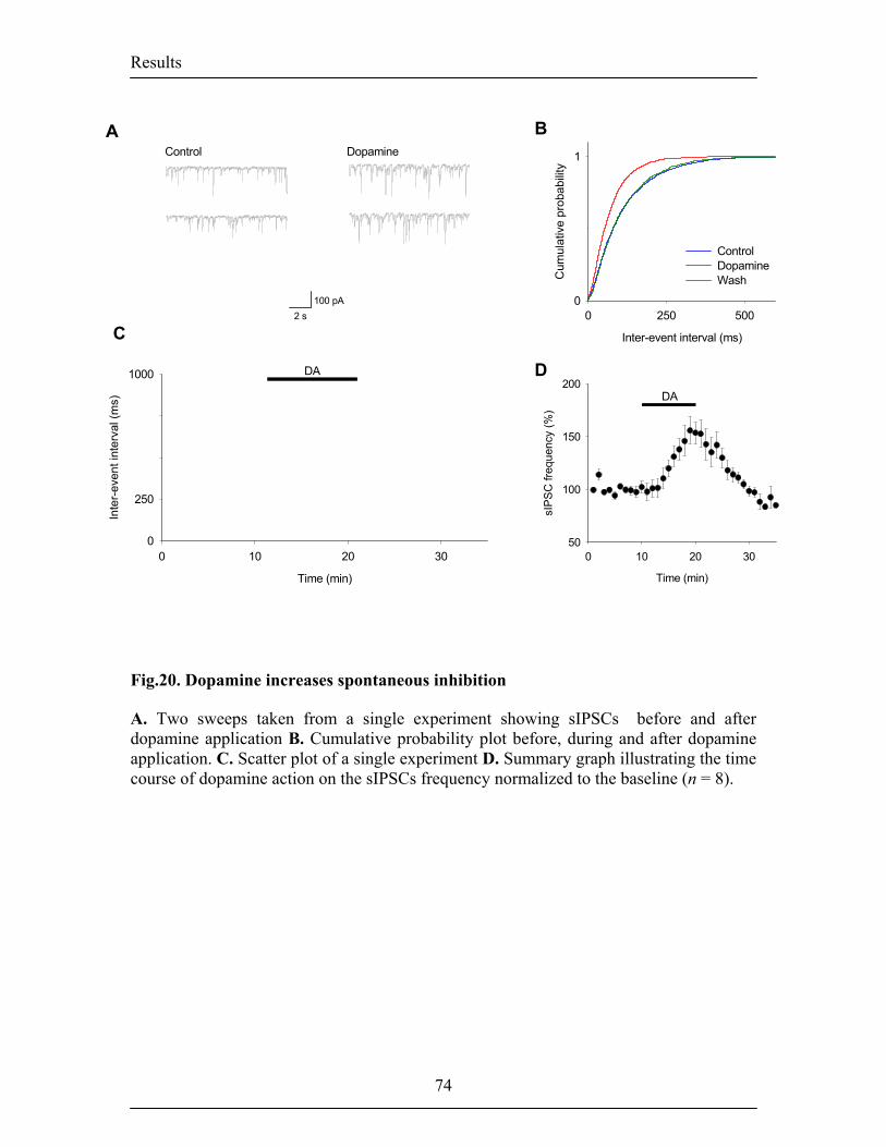
Results
750
500
AControl Dopamine
100 pA2 s
Time (min)
0 10 20 30sI
PS
C fr
eque
ncy
(%)
50
100
150
200
Inter-event interval (ms)
0 250 500
Cum
ulat
ive
prob
abilit
y
0
1
ControlDopamineWash
B
DA
DDA
C
Time (min)
0 10 20 30
Inte
r-ev
ent i
nter
val (
ms)
0
250
1000
Fig.20. Dopamine increases spontaneous inhibition
A. Two sweeps taken from a single experiment showing sIPSCs before and after dopamine application B. Cumulative probability plot before, during and after dopamine application. C. Scatter plot of a single experiment D. Summary graph illustrating the time course of dopamine action on the sIPSCs frequency normalized to the baseline (n = 8).
74
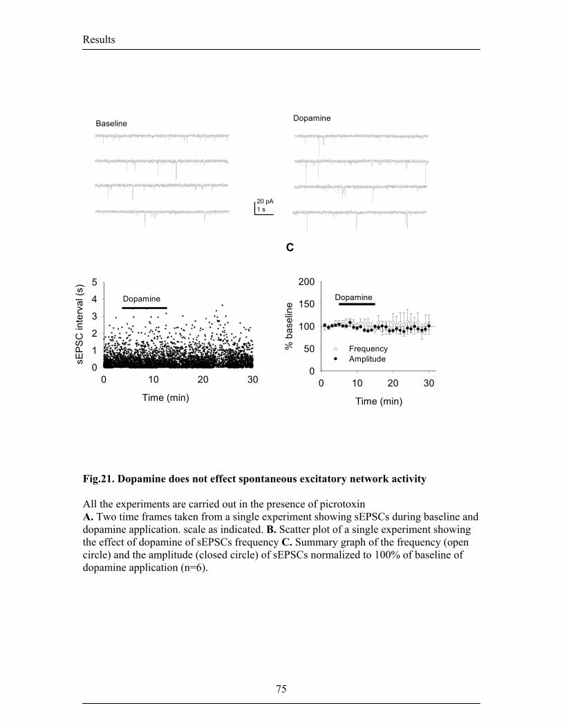
Results
A B
Time (min)
Dopamine
C
BaselineDopamine
0 10 20 30
sEPS
C in
terv
al (s
)
0
1
2
3
4
5
20 pA1 s
Time (min)
0 10 20 30
% b
asel
ine
0
50
100
150
200Dopamine
FrequencyAmplitude
Fig.21. Dopamine does not effect spontaneous excitatory network activity All the experiments are carried out in the presence of picrotoxin A. Two time frames taken from a single experiment showing sEPSCs during baseline and dopamine application. scale as indicated. B. Scatter plot of a single experiment showing the effect of dopamine of sEPSCs frequency C. Summary graph of the frequency (open circle) and the amplitude (closed circle) of sEPSCs normalized to 100% of baseline of dopamine application (n=6).
75
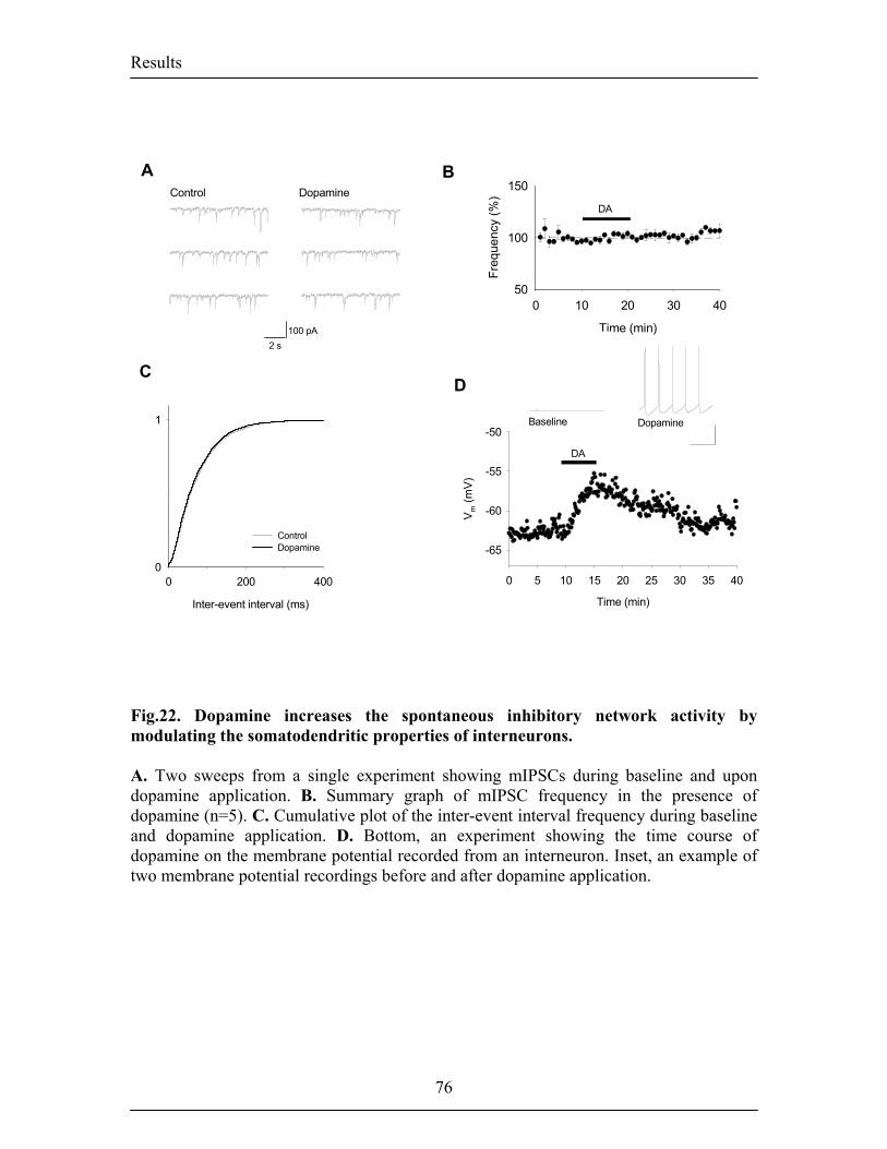
Results
ity
obab
il
iv
e pr
Cum
ulat
0 200 4000
1
ControlDopamine
Inter-event interval (ms)
A B
C
Control Dopamine
100 pA2 s
Time (min)
0 10 20 30 40
Freq
uenc
y (%
)
50
100
150
DA
D
Time (min)
0 5 10 15 20 25 30 35 40
V m (m
V)
-65
-60
-55
-50
DA
Baseline Dopamine
Fig.22. Dopamine increases the spontaneous inhibitory network activity by modulating the somatodendritic properties of interneurons. A. Two sweeps from a single experiment showing mIPSCs during baseline and upon dopamine application. B. Summary graph of mIPSC frequency in the presence of dopamine (n=5). C. Cumulative plot of the inter-event interval frequency during baseline and dopamine application. D. Bottom, an experiment showing the time course of dopamine on the membrane potential recorded from an interneuron. Inset, an example of two membrane potential recordings before and after dopamine application.
76
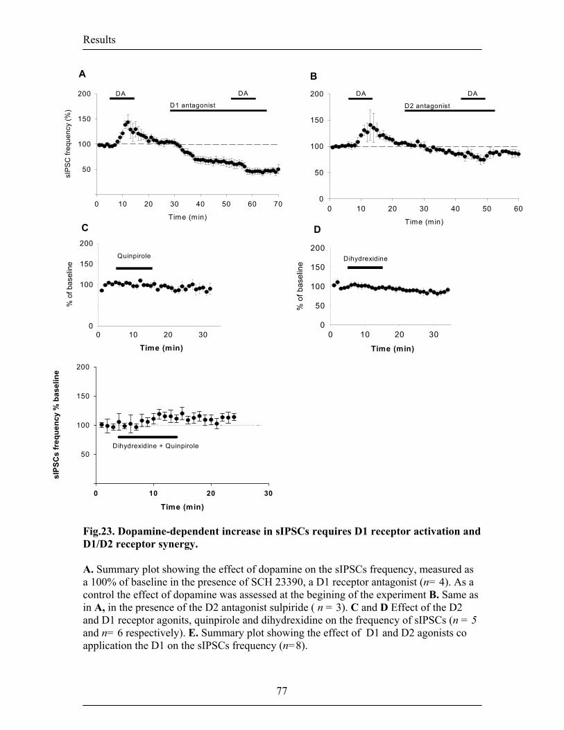
Results
0
50 E 0
Time (min)
0 10 20 30 40 50 60 70
sIP
SC
freq
uenc
y (%
)
50
100
150
200 DA DA
D1 antagonist
A B
C D
Quinpirole
Time (min)
0 10 20 30 40 50 600
50
100
150
200 DA DA
D2 antagonist
Dihydrexidine
Time (min) Time (min)
0 10 20 30
% o
f bas
elin
e
0
100
150
200
Time (min)
0 10 20 30
sIPS
Cs
freq
uenc
y %
bas
elin
e
50
100
150
200
Dihydrexidine + Quinpirole
0 10 20 30
% o
f bas
elin
e
0
50
100
150
200
Fig.23. Dopamine-dependent increase in sIPSCs requires D1 receptor activation and D1/D2 receptor synergy. A. Summary plot showing the effect of dopamine on the sIPSCs frequency, measured as a 100% of baseline in the presence of SCH 23390, a D1 receptor antagonist (n= 4). As a control the effect of dopamine was assessed at the begining of the experiment B. Same as in A, in the presence of the D2 antagonist sulpiride ( n = 3). C and D Effect of the D2 and D1 receptor agonits, quinpirole and dihydrexidine on the frequency of sIPSCs (n = 5 and n= 6 respectively). E. Summary plot showing the effect of D1 and D2 agonists co application the D1 on the sIPSCs frequency (n=8).
77
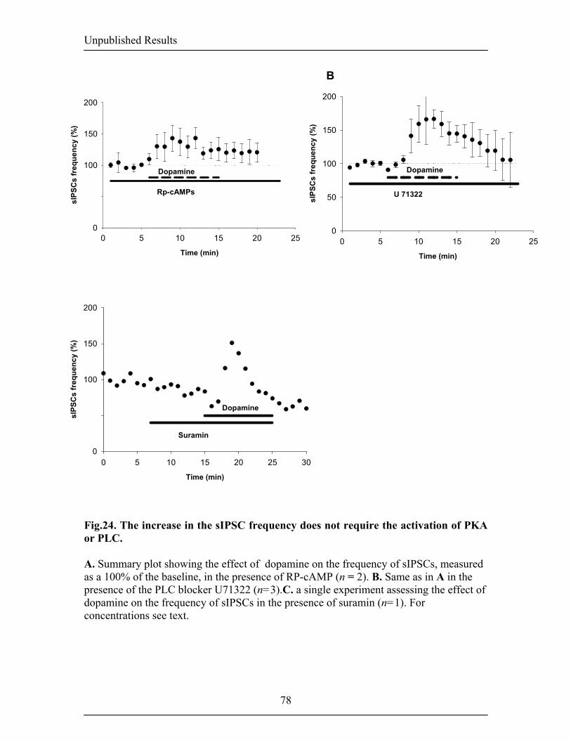
Unpublished Results
A 50
C 50
Time (min)
0 5 10 15 20 25 30
sIPS
Cs
freq
uenc
y (%
)
0
100
150
200
Suramin
Dopamine
B
Time (min)
0 5 10 15 20 25
sIPS
Cs
freq
uenc
y (%
)
0
100
150
200
Dopamine
Rp-cAMPs
Time (min)
0 5 10 15 20 25
sIPS
Cs
freq
uenc
y (%
)
0
50
100
150
200
U 71322
Dopamine
Fig.24. The increase in the sIPSC frequency does not require the activation of PKA or PLC. A. Summary plot showing the effect of dopamine on the frequency of sIPSCs, measured as a 100% of the baseline, in the presence of RP-cAMP (n = 2). B. Same as in A in the presence of the PLC blocker U71322 (n=3).C. a single experiment assessing the effect of dopamine on the frequency of sIPSCs in the presence of suramin (n=1). For concentrations see text.
78
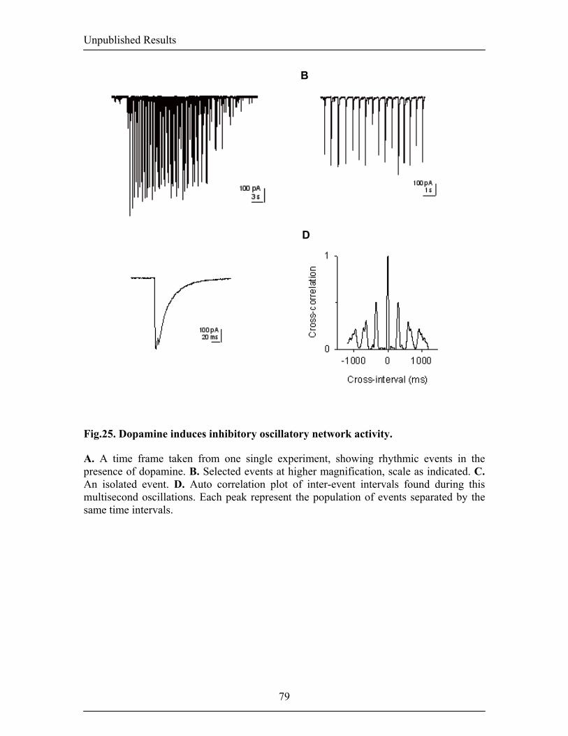
Unpublished Results
A
C
B
D
Fig.25. Dopamine induces inhibitory oscillatory network activity.
A. A time frame taken from one single experiment, showing rhythmic events in the presence of dopamine. B. Selected events at higher magnification, scale as indicated. C. An isolated event. D. Auto correlation plot of inter-event intervals found during this multisecond oscillations. Each peak represent the population of events separated by the same time intervals.
79
Results
Materials and Methods
Slice preparation. Standard procedures were used to prepare 350 µm thick coronal
slices from three to four week old male C57BL/6J mice following a protocol approved by
the Veterinary Department of the Canton of Basel-Stadt. Briefly, the brain was dissected
in ice-cold artificial cerebrospinal fluid (ACSF), mounted on an agar block and sliced
with a vibratome at 4°C. Slices were maintained for 45 min. at 35°C in an interface
chamber containing ACSF equilibrated with 95% O2/5% CO2 and containing (in mM):
124 NaCl, 2.7 KCl, 2 CaCl2, 1.3 MgCl2, 26 NaHCO3, 0.4 NaH2PO4, 10 glucose, 4
ascorbate, and then for at least 45 min. at room temperature before being transferred to a
superfusing recording chamber.
Whole-cell recordings. Whole-cell recordings from LA projections neurons were
performed at room temperature and neurons were visually identified with infrared
videomicroscopy. sIPSCs were recorded in voltage clamp using a CsCl- based
intracellular solution containing (in mM) 140 CsCl- , 5 HEPES, 1.1 EGTA, 10
phosphocreatine, 5 QX-314 Cl- , 4 MgCl2 , pH 7.25, 295 mOsm. For sEPSCs recordings
a K-gluconate intracellular solution was used containing (in mM): 120 K-gluconate, 20
KCl, 10 HEPES, 10 phosphocreatine, 4 Mg-ATP, 0.3 Na-GTP, 0.1 % biocytin, pH 7.25,
295 mOsm.
Acquisition software and analysis. sIPSCs and sEPSCs were acquired and analyzed
with ClampEx8.0, Clampfit 8.0 (Axon Instruments, Union City, CA, USA) and Mini
Analysis Program (Synaptosoft, Decatur, GA, USA), The thresholds for sIPSC and
sEPSC detection were set to 12 pA. Average frequency changes were calculated by
averaging each cell for two minutes during baseline and two minutes at the end of
dopamine application. Frequency and amplitude of sIPSCs within individual experiments
were evaluated using cumulative probability analysis, with statistical significance
determined by using the Kolmogorov-Smirnov nonparametric two-sample test. All other
statistical comparisons were done with paired or unpaired Student’s t-test as appropriate
(P < 0.05 was considered significant). All values are expressed as means ± s.e.m. CNQX,
80

Results
81
(S)-(–)-sulpiride, (–)-quinpirole, dihydrexidine, and SCH23390 were from Tocris-
Cookson (Bristol, UK), sulpiride, U71322, Rp-cAMPs and suramin were from
Fluka/Sigma (Buchs, Switzerland).
General Discussion
4- General Discussion In this chapter I will discuss some of our major findings and try to present them in a more
general context of synaptic plasticity mechanisms. I will first discuss the functional
implication of LTP at thalamo-amygdala synapses and its relevance for fear conditioning.
Then, I will discuss inhibitory circuits in the LA and how they may control the
acquisition and expression of fear conditioning. Finally I will report on the role of
catecholamine on the modulation of plasticity in the LA compared to other brain areas.
4.1. Thalamic-amygdala plasticity and fear conditioning.
The neural pathways mediating fear conditioning involve the transmission of information
about the CS and the US to the LA (for review see Blair et al, 2001). In vivo studies
provided evidence that fear conditioning is accompanied by an enhancement of synaptic
transmission at auditory input synapses to the LA. Rogan and LeDoux (1995), provided
the first link between fear learning in the amygdala and LTP at thalamo-amygdala
synapses, in freely moving animals. To explain how neural responses to the CS and the
US could induce LTP-like changes at these synapses, Blair et al (2001) developed the
following cellular hypothesis. They proposed that the CS evokes EPSPs at the sensory
input synapses on LA pyramidal neurons while the US would strongly depolarize the
same cell (Blair et al., 2001). Therefore, to investigate the mechanisms underlying LTP at
thalamo-amygdala synapses in vitro, we paired short bursts of thalamic afferent
stimulation with bursts of postsynaptic action potentials (see Materials and Methods).
Further experiments indicated that this LTP was dependent on NMDARs and VGCCs
(Humeau et al, unpublished data). So far, results on LTP at these synapses had been
contradictory mainly due to the different stimulation protocols used. Weisskopf et al
(1999) found that LTP induced by trains of thalamic afferent stimulation at 30Hz, with
brief postsysnaptic depolarization, was dependent on L-type VGCCs but independent of
NMDAR activation. Also, Bauer et al (2002) induced pharmacologically distinct types of
LTP at these synapses. They reported that a pairing protocol that included brief
postsynaptic depolarization, led to a VGCCs-dependent but NMDAR-independent LTP.
82
General Discussion
Conversely, tetanic stimulation of thalamic afferent that produced a prolonged
depolarization of the postsynaptic cell without spikes, resulted in an NMDAR dependent
LTP. However, behavioral studies, carried out by other labs showed that both NMDARs
and VGCCs are necessary for fear memory acquisition (Borroni et al., 2000; Rodrigues et
al., 2001; Walker and Davis, 2000). Other behavioral groups suggested that NMDARs, in
particular the ones containing NR2B subunit, are essential for the short-term of memory
(STM) (Rodrigues et al., 2001; Walker and Davis, 2000). On the other hand, L-type
VGCCs would be involved in the long term phase of memory (LTM). A number of
experiments demonstrated that in vitro, L-type VGCCs are involved in c-AMP response
binding protein (CREB). L-type VGCCs are therefore capable of influencing the protein
synthesis required for the long term maintenance of LTP (Schafe et al., 2001). NMDARs,
on the other hand would play a role in the short term phase of LTP by activating local
protein kinases that can phosphorylate membrane proteins. The LTP induced by our
pairing protocol was dependent on both NMDARs and VGCCs (Humeau et al,
unpublished data) and are therefore in accord with behavioral studies.
Together, the above findings suggest that hebbian LTP can be induced at thalamo-
amygdala synapses onto projection neurons in the LA and that it requires both NMDAR
and VGCCs. The exact role of these two coincidence detectors and their interaction
during LTP acquisition and expression remains to be investigated.
A number of questions have been raised concerning the different timing requirements
needed for CS-US associations and for LTP formation. VGCCs were shown to be
activated in the hippocampus when the EPSP precisely coincided with the BPAPs at the
synapse (Magee and Johnston, 1997). Since both the EPSPs and BPAPs are quite short in
duration this might impose a natural temporal constraint during learning in the LA. Spike
timing windows resume the amount of potentiation or depotentiation induced by pairing
EPSPs and BPAPs at different time intervals and at different frequency. Such graph gives
indication on the precise pre and postsynaptic coincidence requirements for synaptic
modification at particular synapses. In the hippocampus or in the cortex, spike timing
windows revealed that LTP can be induced only when the postsynaptic spike occurs
within 20 ms of the onset of the EPSP. Conversely, synaptic depression is induced if the
83
General Discussion
BPAPs are generated before the EPSPs, (Bi and Poo, 1998; Markram et al., 1997). Data
from our lab indicate that in the LA, pairing a single EPSP from thalamic afferent
stimulation with a single BPAP (x 100 at 0.14 Hz) also results in a narrow plasticity
window (Humeau et al, unpublished results) . LTP at thalamo-amygdala synapses can
only be triggered when the BPAPs occur within 15 ms after the onset of the EPSPs. This
time window is very precise given the low average firing rates observed for principal
neurons in the LA in vivo (Lang and Pare, 1997a). In the cortex and in the hippocampus,
critical spike timing windows for LTP can be increased to 100 ms when the EPSPs are
paired with multiple BPAPs (Gustafsson et al., 1987; Paulsen and Sejnowski, 2000).
This suggests that a burst of APs can offer a means of broadening the window of
plasticity. However, pairing bursts of EPSPs with bursts of AP at thalamo-amygdala
synapses did not broaden the spike timing window for LTP. Changing the frequency at
which the pairing was applied however, shifted the most efficient timing intervals and
the absolute magnitude of the LTP (Humeau et al, unpublished results). It remains
difficult to explain how a process like LTP, where the optimal time window during which
pre- and postsynaptic activity have to occur within 100ms, can underlie CS-US
associations that can take place in the range of seconds or even minutes. In vivo studies
reported that cells in the LA respond with a burst of APs to the onset of a tone and then
fire more sparsely until the end of the tone presentation (Collins and Pare, 2000; Quirk et
al., 1997; Quirk et al., 1995). It is therefore possible that the neurons do not fire just prior
to the US presentation. Activity of the sensory afferents in this case might still be enough
to provide glutamate release during the CS presentation until the US is presented.
Another explanation comes from observations in cortical areas such as the perhirinal
cortex, where special types of neurons are capable of maintaining sustained firing for up
to several seconds after depolarization. Based on the presence of these types of neurons
and their connectivity, models have now been proposed for the amygdala (Faulkner and
Brown, 1999; Faulkner, 1997). Another criticisms of LTP being a substrate of fear
conditioning, addresses the discrepancies between the decay of LTP and the decay of
forgetting. However, by addressing these type of criticisms one might have under-
estimated the involvement of other brain areas in memory processes. It is possible that
the LTP triggered at one synapse is being compensated at other synapses or even in other
84
General Discussion
brain areas, resulting in no overall potentiation. Therefore, LTP is probably still the best
mechanism so far to account for long-term memory formation in brain areas including the
LA. However, in the process of understanding what occurs under natural conditions, a
effort should be made to incorporate to the current research on local circuits, the
interactions between the LA and other brain areas.
4.2. Thalamic versus cortical inputs to the LA
Past studies have shown that not all aspects of emotional learning are mediated by
thalamic projections to the amygdala and that association between complex stimuli and
their affective representations also requires cortical input to the amygdala (Armony et al.,
1998; Armony et al., 1997; Quirk et al., 1997). Consistent with this idea the burst pairing
protocol used in our experiment to induce LTP at thalamo-amygdala synapses was input
specific, and did not induce plasticity at cortico-amygdala synapses (Humeau et al,
unpublished data). Induction of LTP at cortico-amygdala synapses has precisely been
shown to be induced by high frequency stimulation (Huang and Kandel, 1998; Huang et
al., 2000). This LTP required NMDAR activation and postsynaptic Ca2+, influx implying
that it is Hebbian in nature. In these studies, LTP at cortico-amygdala synapses was also
induced by pairing weak presynaptic stimulation with prolonged postsynaptic
depolarization (Huang and Kandel, 1998). This protocol led to a form of LTP, induced
postsynaptically, but expressed presynaptically via mechanism requiring PKA activation
(Huang and Kandel, 1998). These data suggest that both pathways are able to support
conditioning, which is in accord with previous lesion studies (Campeau and Davis, 1995;
Romanski and LeDoux, 1992). However, the possibility that there could be associative
interactions between the two afferents had never been looked into until recently. The
work of Doyère and LeDoux (2003) showed that in awake and freely moving animals,
both the thalamic and cortical pathways could undergo input-specific LTP. This LTP
could last for over twenty-four hours which is consistent with long-term memory storage.
They also found that similarly to associative processes in the hippocampus, these two
pathways could be reciprocally potentiated by their simultaneous tetanization. They
suggested that this form of associativity could led to a mutual strengthening during CS-
85
General Discussion
US formation (Doyere et al., 2003). Recent work from our lab confirmed these findings
by showing that only a simultaneous stimulation of thalamic and cortical afferents in
vitro could restore LTP at cortical inputs (Humeau et al., 2003). The mechanisms
underlying this form of heterosynaptic associative LTP at cortical afferents were also
investigated. In their study, Humeau et al found that the repetitive stimulation at thalamic
afferents increased the probability of release at cortical afferents in a presynaptic
NMDARs-dependent manner. In addition, this LTP did not require postsynaptic
NMDARs or Ca2+ influx and did not require network activity indicating that it is entirely
determined by presynaptic mechanisms (Humeau et al., 2003). These data confirm that
other forms of synaptic plasticity besides the classical hebbian LTP can be induced in the
LA. The fact that converging afferents are able to independently support plasticity and
also influence each other, adds more complexity to the integrative properties underlying
sensory processes.
4.3 Amygdala and inhibition
During an LTP experiment, the inhibitory network is usually blocked by GABA
antagonists, in order to focus on the properties of glutamatergic synapses per se. In the
amygdala the spontaneous firing rates of projection neurons are some of the lowest of the
brain (Pare and Collins, 2000; Pascoe and Kapp, 1985; Quirk et al., 1997), suggesting the
existence of a tight inhibitory control. To understand the role of LTP in the LA under
physiological conditions, we induced LTP at the thalamo-amygdala synapses in the
presence of functional synaptic inhibition. Consistent with the idea that inhibition is a
major determinant of LA plasticity, LTP could no longer be induced. We next stimulated
thalamic afferents while recording from pyramidal cells in the LA. This, elicited an initial
EPSP that was truncated by a large amplitude prolonged IPSP. In the presence of
picrotoxin, the amplitude and the decay time of the evoked EPSPs were much longer. In
addition, bursts of evoked EPSPs elicited in the absence of inhibition were able to sum up
over time, bringing the cell closer to firing threshold. In the presence of inhibition
however, evoked IPSPs were dominant and summation of excitatory events was
prevented. Similar results were observed in vitro by others (Lang and Pare, 1997a; Lang
86
General Discussion
and Pare, 1998; Washburn and Moises, 1992a; Washburn and Moises, 1992b). In vivo
studies reported that the spontaneous activity in the LA was similarly dominated by large
hyperpolarizing currents (Lang and Pare, 1997b). The above observations, together with
studies showing that auditory afferents synapse on both interneurons and pyramidal cells,
support the existence of a feed-forward inhibitory circuit in the LA (Szinyei et al., 2000).
Feed-forward inhibition occurs when an afferent fiber excites both excitatory and
inhibitory neurons. Activation of inhibitory neurons provides in turn inhibition to the
same projection neuron population. Feed-forward circuits have been well characterized in
the hippocampal formation (Buzsaki, 1984; Pouille and Scanziani, 2001) and it was
suggested that such an arrangement exists to exert a powerful regulatory control over the
hippocampal network (Jonas et al., 2004). The level of control is not static but rather
depends on both the nature of transmission at the target neurons, and on the subtype of
interneurons activated. Interneurons targeting the soma of pyramidal cells respond faster
and more reliably, enabling a fast shunting of the EPSP and a restriction of spike
generation. in the hippocampus, common features have been described for interneurons
directly targeting the axon initial segment or the soma of pyramidal cells (Fujimaru and
Kosaka, 1996; Fujise et al., 1995; Fukuda et al., 1996) which are thought to be involved
in feed-forward mechanisms whereas interneurons targetting the dendritic domain are
generally associated with feed-back mechanisms. Similarly, afferent fibers were also
shown to target different interneuron subtypes (Buzsaki, 1984). In the LA, the
topographic organization of inhibitory circuits and the characterization of intereuron
subtypes are less understood. So far, at least three major subtypes of distinct GABAergic
interneurons have been identified in the LA based on the expression of calcium binding
protein and peptides. The somatostatin positive interneurons, which also express
calbindin and neuropeptide Y, the paravalbumin positive interneurons, most of which
also co-localize with calbindin and, the calretinin positive interneurons which also
express vasoactive intestinal polypeptides (McDonald and Mascagni, 2001). Interneurons
in the rat neocortex, have very similar patterns of calcium binding protein expression
(gonchar and burkhalter). However, they also express large heterogeneous populations
with different functional roles among the interneuron subtypes expressing the same
proteins. In the LA, paravalbumin (PV+) interneurons are thought to be involved in the
87
General Discussion
targeting of somatodendritic and axon initial segment regions of principal neurons (Smith
et al., 2000). Our data indicated that some interneurons in the LA also receive input from
other interneurons, implying the presence of interneurons involved in disinhibition.
Consistent with the idea that specific interneuron population mediate dishinibition onto
other interneurons a recent study from Muller et al, reported that in the BLA, 30% of VIP
positive interneurons innervate interneurons that were themselves either CB positive of
VIP positive (Muller et al., 2003). It also possible that some interneurons are not
contacted by sensory afferents but directly by LA projection neurons therefore forming
part of local fedback circuits. Interneurons involved in feed-forward inhibition however,
are contacted by sensory fibers and provide a fast and strong inhibition at the soma and
axon initial segment. In order to elicit spiking of projection neurons and produce a
sensory ouput, thalamic and cortical afferents need to be strong enough to overcome
feed-forward inhibition. This differential inhibitory strength in the presence or absence of
sensory input provide an additional mean to control for the processing of relevant sensory
information.
Besides the presence of feed-forward inhibition, other intrinsic factors contribute to the
strong inhibitory circuit of the LA. Compared to pyramidal cells, interneurons were
shown to receive relatively low levels of inhibition directed towards themselves (Pare et
al., 2003). in the LA, IPSPs recorded directly from interneurons revealed the presence of
a GABAA but not a GABAB component. In contrast, IPSPs observed at pyramidal cells
are biphasic and comprise both GABAA and GABAB components (Martina et al., 2001).
In addition, the reversal potential of these IPSPs in interneurons was shown to be more
depolarized relative to the ones of pyramidal neurons (Martina et al., 2001). These
results support the fact that interneurons fire at higher rates than projection neurons and
suggest that they are more readily available to respond to synaptic recruitment (Penttonen
et al., 1998). Glutamatergic synaptic transmission onto interneurons in the LA is also
different than onto pyramidal cells. For example, interneurons in the LA contain AMPA
receptors that lack the GluR2 subunit (Mahanty and Sah, 1998), similarly to some
interneuron in the hippocampus (Geiger et al., 1995; McBain and Dingledine, 1993).
These receptors, that are Ca2+ permeable, enable faster excitatory transmission and have
recently been implicated in NMDAR-independent forms of plasticity expressed at
88
General Discussion
interneuron synapses (McMahon and Kauer, 1997). All these intrinsic properties confer a
means of rapid signaling important for the role of interneurons in detecting and
controlling synchronous principal cell population activity. During feed-forward inhibition
interneurons must activate fast in order to shunt pyramidal cells before they fire. Thus,
the above connectivity and intrinsic properties of interneurons provide the necessary
mechanisms to achieve this. It is also possible that thalamic and cortical afferent fibers
originate from different cells depending on whether they target interneuron or projection
neuron. One way to check for this would be to trace back afferent fibers that synapse on
both type of neurons and see if they originate from the same cell.
During fear memory acquisition, feed-forward inhibition needs to be over-come by
physiological means. In the hippocampus, a number of neurotransmitters and
neuromodulators are released during different behavioral states of the animal that can
selectively regulate the excitability of interneuron subsets. Our results demonstrated that
in the LA, dopamine, which is normally released in this area upon stress, can play such a
role thereby facilitating LTP induction. So far, the interneuron subtypes targeted by
dopamine were not identified. Combining paired recordings of connected interneuron
/pyramidal cells will give important information on inhibitory network and its regulation
by dopamine. Imaging studies of mice expressing GFP interneuron under the control of
specific promoters will identify the interneuron population involve in feed-forward or
feed-back circuits. Finally, in vivo studies will shed light on the role of inhibitory
processes and network oscillations during different behavioral states.
4.3 Dopaminergic modulation of synaptic transmission and plasticity
In vivo studies indicated that the activation of dopaminergic afferents to the LA is
necessary for the acquisition of fear conditioning. This effect requires both D1 and D2
dopamine receptor activation (Greba et al., 2001; Greba and Kokkinidis, 2000; Guarraci
et al., 2000; Guarraci et al., 1999; Rosenkranz and Grace, 2002b). The cellular and
synaptic mechanisms underlying this effect are, however, unknown. Our findings provide
the first evidence that in vitro, dopamine modulates synaptic plasticity by suppressing
feed-forward inhibition onto pyramidal cells of the LA. This effect was cell type specific
89
General Discussion
and did not affect the excitatory network. In addition, we could observe that D1 and D2
receptors acted in opposite ways during the acquisition of conditioned fear. Dopamine
was also reported to affect the responses of the LA to inputs from the medial prefrontal
cortex (mPFC). These afferents have been shown in vivo to induce a potent inhibition of
LA projection neurons via activation of interneurons (Rosenkranz and Grace,
2002a).When activated, the mPFC has therefore the ability to suppress responses of the
BLA when an emotional response to a threatening stimulus is not appropriate. D1
receptor stimulation in the LA, was found to strongly attenuate these mPFC afferent
inputs to the BLA (Rosenkranz and Grace, 1999). In contrast, D2 receptor activation
potentiate sensory cortical inputs by increasing the neuronal excitability (Grace and
Rosenkranz, 2002). Thus when dopamine is released in the LA, D1 and D2 receptors
support different actions leading to an increased efficiency of sensory cortical afferents
on BLA excitation. Our data showed that dopamine via D2 receptor activation facilitate
the induction of LTP by suppressing feed-forward inhibition. This effect is mediated via
two mechanisms, first via a reduction of the quantal content at inhibitory synapses onto
pyramidal cells, and second by increasing inhibition onto interneuron. In contrast D1
receptors, in co-activation with D2 receptors, seem to increase the spontaneous inhibitory
network frequency by altering the somatodendritic properties of interneurons. These data
suggest that D2 receptors in the LA, associated with cortical or thalamic inputs, seem to
consistently affect the neuronal excitability of neurons in a cell type specific manner. The
role of D1 receptors in the LA is more controversial. Their activation might be dependent
on DA concentration, the presence or not of sensory afferents and the type of afferent
inputs. In the mPFC, DA can either facilitate or suppress GABA release. There also, its
action is complex and depends on a number of factors. Overall, DA by differentially
regulating inhibition in many brain areas allows for a different type of control eventually
leading to changes in synaptic plasticity. The involvement of catecholamine influences on
LTP was first considered when complete depletion of catecholamines modulated the LTP
observed in the dentate gyrus of freely moving animals (Bliss et al., 1983). Since then,
the role of dopamine on synaptic plasticity has been examined in many brain areas
including the striatum, the hippocampus, the prefrontal cortex and recently with our
studies in the amygdala. These different regions do not appear to recruit the DA system in
90
General Discussion
91
similar manners (for reviews see (Lisman, 2001; Lovinger, 1996). DA modulation in the
hippocampus and prefrontal cortex seem to be comparable in the fact that both require D1
receptor activation for the initiation and consolidation of LTP but not D2 receptors. In the
amygdala D1 receptor activation does not seem to be necessary for LTP induction. Such
discrepancies might be explained by the different ratio of D1 to D2 receptor expression
observed in different brain areas or by the distinct activation of DA systems depending on
behavioral states. In addition DA action on synaptic plasticity might also depend on the
history of particular synapses. Indeed, DA has also been involved in depotentiation
processes in the CA1 region of the hippocampus and in the dentate gyrus (Kulla, 2000).
Since most of the DA fibers innervating the hippocampus, the mPFC and the amygdala
originates from the VTA, it is also possible that DA signals are being shaped by activity
initiated in the VTA during different behavioral states. Several studies have also
suggested that dopamine is not only important for the acquisition but also for the
extinction of fear conditioning. The exact mechanisms are unclear but it is thought that
dopamine would potentiate mPFC input to LA interneurons leading to its inactivation
during extinction learning (willick and kokkinidis 1995) (Myers and Davis 2002). These
results suggests that overall, dopamine seems to be able to regulate the activity of
different brain regions throughout different stages of memory. Future work will define
more precisely at the cellular level which specific cells dopamine targets and how it
influences synaptic inhibitory transmission in the LA. Further, the proteins involved in
DA modulation relevant to plasticity and memory processes will also be investigated.
Conclusion and Outlook
5- Conclusions and Outlook The experiments carried out during my thesis have revealed that, under physiological
conditions, inhibitory circuits in the LA can prevent the induction of LTP in vitro at
thalamic-amygdala afferents. We have shown that DA, which is released in the amygdala
upon stress can overcome this inhibition by suppressing feed-forward inhibition and, by
reducing the quantal content at inhibitory synapses upon thalamic afferent stimulation.
The DA-dependent suppression of feed-forward was mediated by the activation of D2
receptors that depolarized directly interneurons involved in dishinibitory processes. The
mechanisms underlying the D2 receptor mediated depolarization of interneurons is at
present not clear. In the second part of my thesis I focused on the modulation of the
spontaneous inhibitory network activity by DA. We observed that bath application of DA
in the absence of afferent stimulation increases the frequency of spontaneous inhibitory
postsynaptic currents. These results are consistent with an increased firing rate of
interneuron upon DA application and required the co-activation of D1 and D2 receptors
Under some circumstances, DA even induced some rhythmic oscillatory activity of
interneurons. This suggests an important role of this catecholamine in the regulation of
synchronous inhibitory network activity. These results have raised new questions about
the role of inhibition in the LA: Is there an heterogeneous population of interneurons ?
Can we differentiate them on the basis of neurochemical markers such as paravalbumin
or calretinin ? How do they gate plasticity in the amygdala ? How do they contribute to
network oscillation ? And finally what is the role of interneuron-interneuron interactions
modulated by DA To investigate disinhibitory circuits in the LA, it is planed to directly
record from pairs of connected interneurons. Recording of interneuron pairs will also
provide new insights on how dopamine influences different interneuron subtypes,
improve the signal to noise ratio in the presence or absence of sensory input and how it
eventually controls the induction of synaptic plasticity. Overall these experiments will
provide an important step towards a better understanding of amygdala dysfunction and
thus of anxiety disorders.
92
References
6- References Aggleton (1992). The amygdala: neurobiological aspects of emotion, memory and mental dysfunction., New-York: Wiley-liss).
Aggleton, J. P. (1993). The contribution of the amygdala to normal and abnormal emotional states. Trends Neurosci 16, 328-333.
Amaral, D. G. P., J.L. Pitkanen, A. Carmichael, S.T. (1992). Anatomical organisation of the primate amygdaloid complex In: The Amygdala: Neurobiological Aspects of Emotion, Memory and Mental Dysfunction., Aggleton JP. New-York: Wiley-Liss).
Andersen, P., Sundberg, S. H., Sveen, O., and Wigstrom, H. (1977). Specific long-lasting potentiation of synaptic transmission in hippocampal slices. Nature 266, 736-737.
Antoniadis, E. A., and McDonald, R. J. (2001). Amygdala, hippocampus, and unconditioned fear. Exp Brain Res 138, 200-209.
Armony, J. L., Quirk, G. J., and LeDoux, J. E. (1998). Differential effects of amygdala lesions on early and late plastic components of auditory cortex spike trains during fear conditioning. J Neurosci 18, 2592-2601.
Armony, J. L., Servan-Schreiber, D., Romanski, L. M., Cohen, J. D., and LeDoux, J. E. (1997). Stimulus generalization of fear responses: effects of auditory cortex lesions in a computational model and in rats. Cereb Cortex 7, 157-165.
Aylward, R. L. M. T., S. (1993). Neurons in the ventral subiculum amygdala and enthorinal cortex which project to the nucleus accumbens: Their input from somatstatin boutons. JChemNeuroanat 6, 31-42.
Bailey, C. H., Bartsch, D., and Kandel, E.R. (1996). Towards a molecular definition of long-term memory storage. Proc Natl Acad Sci U S A 93, 13445-13452.
Bannerman, D. M., Good, M. A., Butcher, S. P., Ramsay, M., and Morris, R. G. (1995). Distinct components of spatial learning revealed by prior training and NMDA receptor blockade. Nature 378, 182-186.
Barria, A., Muller, D., Derkach, V., Griffith, L. C., and Soderling, T. R. (1997). Regulatory phosphorylation of AMPA-type glutamate receptors by CaM-KII during long-term potentiation. Science 276, 2042-2045.
Bauer, E. P., LeDoux, J. E., and Nader, K. (2001). Fear conditioning and LTP in the lateral amygdala are sensitive to the same stimulus contingencies. Nat Neurosci 4, 687-688.
Bauer, E. P., Schafe, G. E., and LeDoux, J. E. (2002). NMDA receptors and L-type voltage-gated calcium channels contribute to long-term potentiation and different components of fear memory formation in the lateral amygdala. J Neurosci 22, 5239-5249.
93
References
Bear, M. F. A., W.C. (1996). Long-term depression in the hippocampus. Annu Rev Neurosci 19, 437-462.
Beattie, E. C., Carroll, R. C., Yu, X., Morishita, W., Yasuda, H., von Zastrow, M., and Malenka, R. C. (2000). Regulation of AMPA receptor endocytosis by a signaling mechanism shared with LTD. Nat Neurosci 3, 1291-1300.
Bell, C. C., Han, V. Z., Sugawara, Y., and Grant, K. (1997). Synaptic plasticity in a cerebellum-like structure depends on temporal order. Nature 387, 278-281.
Benke, T. A., Luthi, A., Isaac, J. T., and Collingridge, G. L. (1998). Modulation of AMPA receptor unitary conductance by synaptic activity. Nature 393, 793-797.
Benuskova, L., Rema, V., Armstrong-James, M., and Ebner, F. F. (2001). Theory for normal and impaired experience-dependent plasticity in neocortex of adult rats. Proc Natl Acad Sci U S A 98, 2797-2802.
Bergson, C., Levenson, R., Goldman-Rakic, P. S., and Lidow, M. S. (2003). Dopamine receptor-interacting proteins: the Ca(2+) connection in dopamine signaling. Trends Pharmacol Sci 24, 486-492.
Bettler, B., Kaupmann, K., and Bowery, N. (1998). GABAB receptors: drugs meet clones. Curr Opin Neurobiol 8, 345-350.
Bi, G. Q., and Poo, M. M. (1998). Synaptic modifications in cultured hippocampal neurons: dependence on spike timing, synaptic strength, and postsynaptic cell type. J Neurosci 18, 10464-10472.
Bi, G. Q. P., M.M. (1999). Distributed synaptic modification in neural networks induced by patterned stimulation. nature 401, 792-796.
Bi, G. Q. P., M.M. (2001). Synaptic modification by correlated activity: Hebb's postulate revisited. AnnRevNeurosci 24, 139-166.
Bissiere, S., Humeau, Y., and Luthi, A. (2003). Dopamine gates LTP induction in lateral amygdala by suppressing feedforward inhibition. Nat Neurosci 6, 587-592.
Blair, H. T., Schafe, G. E., Bauer, E. P., Rodrigues, S. M., and LeDoux, J. E. (2001). Synaptic plasticity in the lateral amygdala: a cellular hypothesis of fear conditioning. Learn Mem 8, 229-242.
Blair, H. T., Tinkelman, A., Moita, M. A., and LeDoux, J. E. (2003). Associative plasticity in neurons of the lateral amygdala during auditory fear conditioning. Ann N Y Acad Sci 985, 485-487.
Blanchard, D. C., and Blanchard, R. J. (1972). Innate and conditioned reactions to threat in rats with amygdaloid lesions. J Comp Physiol Psychol 81, 281-290.
94
References
Bliss, T. V., and Collingridge, G. L. (1993). A synaptic model of memory: long-term potentiation in the hippocampus. Nature 361, 31-39.
Bliss, T. V., and Gardner-Medwin, A. R. (1973). Long-lasting potentiation of synaptic transmission in the dentate area of the unanaestetized rabbit following stimulation of the perforant path. J Physiol 232, 357-374.
Bliss, T. V., Goddard, G. V., and Riives, M. (1983). Reduction of long-term potentiation in the dentate gyrus of the rat following selective depletion of monoamines. J Physiol 334, 475-491.
Bliss, T. V., and Lomo, T. (1973). Long-lasting potentiation of synaptic transmission in the dentate area of the anaesthetized rabbit following stimulation of the perforant path. J Physiol 232, 331-356.
Bloom, F. E., and Iversen, L. L. (1971). Localizing 3H-GABA in nerve terminals of rat cerebral cortex by electron microscopic autoradiography. Nature 229, 628-630.
Borroni, A. M., Fichtenholtz, H., Woodside, B. L., and Teyler, T. J. (2000). Role of voltage-dependent calcium channel long-term potentiation (LTP) and NMDA LTP in spatial memory. J Neurosci 20, 9272-9276.
Bourtchuladze, R., Frenguelli, B., Blendy, J., Cioffi, D., Schutz, G., and Silva, A. J. (1994). Deficient long-term memory in mice with a targeted mutation of the cAMP-responsive element-binding protein. Cell 79, 59-68.
Bowery, N. (1989). GABAB receptors and their significance in mammalian pharmacology. Trends Pharmacol Sci 10, 401-407.
Bowery, N. G., Hill, D. R., and Hudson, A. L. (1983). Characteristics of GABAB receptor binding sites on rat whole brain synaptic membranes. Br J Pharmacol 78, 191-206.
Bracci, E., Centonze, D., Bernardi, G., and Calabresi, P. (2002). Dopamine excites fast-spiking interneurons in the striatum. J Neurophysiol 87, 2190-2194.
Bradler, J. E. B., G. (1989). Long-term-potentiation in hippocampal CA3 neurons: tetanized input regulates heterosynaptic efficacy. Synapse 4, 132-142.
Braunewell, K. H., and Manahan-Vaughan, D. (2001). Long-term depression: a cellular basis for learning? Rev Neurosci 12, 121-140.
Brinley-Reed, M., and McDonald, A. J. (1999). Evidence that dopaminergic axons provide a dense innervation of specific neuronal subpopulations in the rat basolateral amygdala. Brain Res 850, 127-135.
Brown, S. S., A (1888). An investigation into the functions of the occipital and temporal lobes of the monkey's brain. PhilosTransRSocLondon Ser B 179, 303-327.
95
References
Buchel, C., and Dolan, R. J. (2000). Classical fear conditioning in functional neuroimaging. Curr Opin Neurobiol 10, 219-223.
Burdach, K. F. (1819-1922). Vom Baue und Leben des Gehirn. Leipzig.
Buzsaki, G. (1984). Feed-forward inhibition in the hippocampal formation. Prog Neurobiol 22, 131-153.
Cador, M. R., T.W. Everitt, B.J. (1989). Involvement of the amygdala in stimulus-reward association: interaction with the ventral striatum. Neuroscience 30, 77-86.
Caine, S. B., Heinrichs, S. C., Coffin, V. L., and Koob, G. F. (1995). Effects of the dopamine D-1 antagonist SCH 23390 microinjected into the accumbens, amygdala or striatum on cocaine self-administration in the rat. Brain Res 692, 47-56.
Caine, S. B., Negus, S. S., Mello, N. K., Patel, S., Bristow, L., Kulagowski, J., Vallone, D., Saiardi, A., and Borrelli, E. (2002). Role of dopamine D2-like receptors in cocaine self-administration: studies with D2 receptor mutant mice and novel D2 receptor antagonists. J Neurosci 22, 2977-2988.
Cajal, S. R. (1909). Histologie du systeme nerveux de l'homme et des vertebretes (Paris, France).
Callahan, P. M., Paris, J. M., Cunningham, K. A., and Shinnick-Gallagher, P. (1991). Decrease of GABA-immunoreactive neurons in the amygdala after electrical kindling in the rat. Brain Res 555, 335-339.
Campeau, S., and Davis, M. (1995). Involvement of subcortical and cortical afferents to the lateral nucleus of the amygdala in fear conditioning measured with fear-potentiated startle in rats trained concurrently with auditory and visual conditioned stimuli. J Neurosci 15, 2312-2327.
Campenot, R. B. (1969). Effect of amygdaloid lesions upon active avoidance acquisition and anticipatory responding in rats. J Comp Physiol Psychol 69, 492-497.
Canteras, N. S., and Swanson, L. W. (1992). Projections of the ventral subiculum to the amygdala, septum, and hypothalamus: a PHAL anterograde tract-tracing study in the rat. J Comp Neurol 324, 180-194.
Carlsen, J. (1988). Immunocytochemical localization of glutamate decarboxylase in the rat basolateral amygdaloid nucleus, with special reference to GABAergic innervation of amygdalostriatal projection neurons. J Comp Neurol 273, 513-526.
Carlson, J. H., Bergstrom, D. A., and Walters, J. R. (1987). Stimulation of both D1 and D2 dopamine receptors appears necessary for full expression of postsynaptic effects of dopamine agonists: a neurophysiological study. Brain Res 400, 205-218.
96
References
Castillo, P. E., Weisskopf, M. G., and Nicoll, R. A. (1994). The role of Ca2+ channels in hippocampal mossy fiber synaptic transmission and long-term potentiation. Neuron 12, 261-269.
Catterall, W. A. (2000). Structure and Regulation of Voltage Gated Calcium Channels. AnnRevCell DevBiol 16, 521-555.
Centonze, D., Picconi, B., Gubellini, P., Bernardi, G., and Calabresi, P. (2001). Dopaminergic control of synaptic plasticity in the dorsal striatum. Eur J Neurosci 13, 1071-1077.
Chapman, P. F., and Bellavance, L. L. (1992). Induction of long-term potentiation in the basolateral amygdala does not depend on NMDA receptor activation. Synapse 11, 310-318.
Chapman, P. F., Kairiss, E. W., Keenan, C. L., and Brown, T. H. (1990). Long-term synaptic potentiation in the amygdala. Synapse 6, 271-278.
Chebib, M., and Johnston, G. A. (1999). The 'ABC' of GABA receptors: a brief review. Clin Exp Pharmacol Physiol 26, 937-940.
Chevaleyre, V., and Castillo, P. E. (2002). Assessing the role of Ih channels in synaptic transmission and mossy fiber LTP. Proc Natl Acad Sci U S A 99, 9538-9543.
Chu, D. C., Albin, R. L., Young, A. B., and Penney, J. B. (1990). Distribution and kinetics of GABAB binding sites in rat central nervous system: a quantitative autoradiographic study. Neuroscience 34, 341-357.
Clark, G. A., Hawkins, R. D., and Kandel, E. R. (1994). Activity-dependent enhancement of presynaptic facilitation provides a cellular mechanism for the temporal specificity of classical conditioning in Aplysia. Learn Mem 1, 243-257.
Clugnet, M. C., and LeDoux, J. E. (1990). Synaptic plasticity in fear conditioning circuits: induction of LTP in the lateral nucleus of the amygdala by stimulation of the medial geniculate body. J Neurosci 10, 2818-2824.
Clugnet, M. C., LeDoux, J. E., and Morrison, S. F. (1990). Unit responses evoked in the amygdala and striatum by electrical stimulation of the medial geniculate body. J Neurosci 10, 1055-1061.
Coan, E. J., and Collingridge, G. L. (1985). Magnesium ions block an N-methyl-D-aspartate receptor-mediated component of synaptic transmission in rat hippocampus. Neurosci Lett 53, 21-26.
Cohen, D. H., and Randall, D. C. (1984). Classical conditioning of cardiovascular responses. Annu Rev Physiol 46, 187-197.
97
References
Collingridge, G. L., Kehl, S. J., Loo, R., and McLennan, H. (1983). Effects of kainic and other amino acids on synaptic excitation in rat hippocampal slices: 1. Extracellular analysis. Exp Brain Res 52, 170-178.
Collins, D. R., and Pare, D. (1999). Reciprocal changes in the firing probability of lateral and central medial amygdala neurons. J Neurosci 19, 836-844.
Collins, D. R., and Pare, D. (2000). Differential fear conditioning induces reciprocal changes in the sensory responses of lateral amygdala neurons to the CS(+) and CS(-). Learn Mem 7, 97-103.
Collins, D. R., Pelletier, J. G., and Pare, D. (2001). Slow and fast (gamma) neuronal oscillations in the perirhinal cortex and lateral amygdala. J Neurophysiol 85, 1661-1672.
Cull-Candy, S., Brickley, S., and Farrant, M. (2001). NMDA receptor subunits: diversity, development and disease. Curr Opin Neurobiol 11, 327-335.
Cummings, J. A., Nicola, S. M., and Malenka, R. C. (1994). Induction in the rat hippocampus of long-term potentiation (LTP) and long-term depression (LTD) in the presence of a nitric oxide synthase inhibitor. Neurosci Lett 176, 110-114.
Danober, L., and Pape, H. C. (1998). Mechanisms and functional significance of a slow inhibitory potential in neurons of the lateral amygdala. Eur J Neurosci 10, 853-867.
Davidson, R. J., and Irwin, W. (1999). The functional neuroanatomy of emotion and affective style. Trends Cogn Sci 3, 11-21.
Davies, C. H., Starkey, S. J., Pozza, M. F., and Collingridge, G. L. (1991). GABA autoreceptors regulate the induction of LTP. Nature 349, 609-611.
Davis, M. (1992). The role of the amygdala in fear and anxiety. Annu Rev Neurosci 15, 353-375.
Davis, M. (2000). The role of the amygdala in conditioned and unconditioned fear and anxiety . In The Amygdala, J. P. Aggleton, ed. (Oxford, UK, Oxford Univ. Press), pp. 231-288.
Davis, M., Walker, D. L., and Myers, K. M. (2003). Role of the amygdala in fear extinction measured with potentiated startle. Ann N Y Acad Sci 985, 218-232.
Davis, M., and Whalen, P. J. (2001). The amygdala: vigilance and emotion. Mol Psychiatry 6, 13-34.
Davis, M. H., J.M. Rosen, J.B. (1987). Anxiety and the amygdala: pharmacological and anatomical analysis of the fear-potentiated startle paradigm. In The psychology of learning and motivation, G. Bower, ed. (San diego academic), pp. 263-305.
98
References
Debanne, D., Gahwiler, B. H., and Thompson, S. M. (1997). Bidirectional associative plasticity of unitary CA3-CA1 EPSPs in the rat hippocampus in vitro. J Neurophysiol 77, 2851-2855.
Debanne, D., Gahwiler, B. H., and Thompson, S. M. (1998). Long-term synaptic plasticity between pairs of individual CA3 pyramidal cells in rat hippocampal slice cultures. J Physiol 507 ( Pt 1), 237-247.
Deisseroth, K., and Tsien, R. W. (2002). Dynamic multiphosphorylation passwords for activity-dependent gene expression. Neuron 34, 179-182.
Derkach, V., Barria, A., and Soderling, T. R. (1999). Ca2+/calmodulin-kinase II enhances channel conductance of alpha-amino-3-hydroxy-5-methyl-4-isoxazolepropionate type glutamate receptors. Proc Natl Acad Sci U S A 96, 3269-3274.
Doherty, M. D., and Gratton, A. (1992). High-speed chronoamperometric measurements of mesolimbic and nigrostriatal dopamine release associated with repeated daily stress. Brain Res 586, 295-302.
Doherty, M. D., and Gratton, A. (1996). Medial prefrontal cortical D1 receptor modulation of the meso-accumbens dopamine response to stress: an electrochemical study in freely-behaving rats. Brain Res 715, 86-97.
Dou, Y., Yan, J., Wu, Y. Y., Cui, R. Y., and Lu, C. L. (2001). [Calcium dependent synaptic plasticity]. Sheng Li Ke Xue Jin Zhan 32, 35-38.
Doyere, V., Schafe, G. E., Sigurdsson, T., and LeDoux, J. E. (2003). Long-term potentiation in freely moving rats reveals asymmetries in thalamic and cortical inputs to the lateral amygdala. Eur J Neurosci 17, 2703-2715.
Eccles, J. C. (1965). Conscious experienceand memory. Recent AdvBiolPsychiatry 8, 235-256.
Ehlers, M. D. (2000). Reinsertion or degradation of AMPA receptors determined by activity-dependent endocytic sorting. Neuron 28, 511-525.
Elgersma, Y., and Silva, A. J. (1999). Molecular mechanisms of synaptic plasticity and memory. Curr Opin Neurobiol 9, 209-213.
El-Ghundi, M., Fletcher, P. J., Drago, J., Sibley, D. R., O'Dowd, B. F., and George, S. R. (1999). Spatial learning deficit in dopamine D(1) receptor knockout mice. Eur J Pharmacol 383, 95-106.
El-Ghundi, M., George, S. R., Drago, J., Fletcher, P. J., Fan, T., Nguyen, T., Liu, C., Sibley, D. R., Westphal, H., and O'Dowd, B. F. (1998). Disruption of dopamine D1 receptor gene expression attenuates alcohol-seeking behavior. Eur J Pharmacol 353, 149-158.
99
References
El-Ghundi, M., O'Dowd, B. F., Erclik, M., and George, S. R. (2003). Attenuation of sucrose reinforcement in dopamine D1 receptor deficient mice. Eur J Neurosci 17, 851-862.
El-Ghundi, M., O'Dowd, B. F., and George, S. R. (2001). Prolonged fear responses in mice lacking dopamine D1 receptor. Brain Res 892, 86-93.
Engert, F., and Bonhoeffer, T. (1997). Synapse specificity of long-term potentiation breaks down at short distances. Nature 388, 279-284.
Fanselow, M. S., and Helmstetter, F. J. (1988). Conditional analgesia, defensive freezing, and benzodiazepines. Behav Neurosci 102, 233-243.
Faulkner, B., and Brown, T. H. (1999). Morphology and physiology of neurons in the rat perirhinal-lateral amygdala area. J Comp Neurol 411, 613-642.
Faulkner, B. T., K.H. Brown, T.H. (1997). Mechanims for temporal encoding in fear conditioning. In Computational Neuroscience, I. Bower, ed. (New-Yok, Plenum Press), pp. 641-645.
Fibiger, H. C. (1991). Dopaminergic-cholinergic interactions in the striatum. Jpn J Psychiatry Neurol 45, 512.
Finlay, J. M., and Zigmond, M. J. (1997). The effects of stress on central dopaminergic neurons: possible clinical implications. Neurochem Res 22, 1387-1394.
Freedman, L. J., and Cassell, M. D. (1994). Distribution of dopaminergic fibers in the central division of the extended amygdala of the rat. Brain Res 633, 243-252.
Freund, T. F., and Buzsaki, G. (1996). Interneurons of the hippocampus. Hippocampus 6, 347-470.
Fujimaru, Y., and Kosaka, T. (1996). The distribution of two calcium binding proteins, calbindin D-28K and parvalbumin, in the entorhinal cortex of the adult mouse. Neurosci Res 24, 329-343.
Fujise, N., Hunziker, W., Heizmann, C. W., and Kosaka, T. (1995). Distribution of the calcium binding proteins, calbindin D-28K and parvalbumin, in the subicular complex of the adult mouse. Neurosci Res 22, 89-107.
Fukuda, T., Aika, Y., Heizmann, C. W., and Kosaka, T. (1996). Dense GABAergic input on somata of parvalbumin-immunoreactive GABAergic neurons in the hippocampus of the mouse. Neurosci Res 26, 181-194.
Gaudreau, H., and Pare, D. (1996). Projection neurons of the lateral amygdaloid nucleus are virtually silent throughout the sleep--waking cycle. J Neurophysiol 75, 1301-1305.
100
References
Geiger, J. R., Lubke, J., Roth, A., Frotscher, M., and Jonas, P. (1997). Submillisecond AMPA receptor-mediated signaling at a principal neuron-interneuron synapse. Neuron 18, 1009-1023.
Geiger, J. R., Melcher, T., Koh, D. S., Sakmann, B., Seeburg, P. H., Jonas, P., and Monyer, H. (1995). Relative abundance of subunit mRNAs determines gating and Ca2+ permeability of AMPA receptors in principal neurons and interneurons in rat CNS. Neuron 15, 193-204.
Gewirtz, J. C., and Davis, M. (1997). Second-order fear conditioning prevented by blocking NMDA receptors in amygdala. Nature 388, 471-474.
Goldman-Rakic, P. S. (1998). The cortical dopamine system: role in memory and cognition. Adv Pharmacol 42, 707-711.
Goosens, K. A., Holt, W., and Maren, S. (2000). A role for amygdaloid PKA and PKC in the acquisition of long-term conditional fear memories in rats. Behav Brain Res 114, 145-152.
Goosens, K. A., and Maren, S. (2002). Long-term potentiation as a substrate for memory: evidence from studies of amygdaloid plasticity and Pavlovian fear conditioning. Hippocampus 12, 592-599.
Gorelova, N., Seamans, J. K., and Yang, C. R. (2002). Mechanisms of dopamine activation of fast-spiking interneurons that exert inhibition in rat prefrontal cortex. J Neurophysiol 88, 3150-3166.
Grace, A. A. (2000a). Gating of information flow within the limbic system and the pathophysiology of schizophrenia. Brain Res Brain Res Rev 31, 330-341.
Grace, A. A. (2000b). The tonic/phasic model of dopamine system regulation and its implications for understanding alcohol and psychostimulant craving. Addiction 95 Suppl 2, S119-128.
Grace, A. A., and Rosenkranz, J. A. (2002). Regulation of conditioned responses of basolateral amygdala neurons. Physiol Behav 77, 489-493.
Greba, Q., Gifkins, A., and Kokkinidis, L. (2001). Inhibition of amygdaloid dopamine D2 receptors impairs emotional learning measured with fear-potentiated startle. Brain Res 899, 218-226.
Greba, Q., and Kokkinidis, L. (2000). Peripheral and intraamygdalar administration of the dopamine D1 receptor antagonist SCH 23390 blocks fear-potentiated startle but not shock reactivity or the shock sensitization of acoustic startle. Behav Neurosci 114, 262-272.
101
References
Grobin, A. C., and Deutch, A. Y. (1998). Dopaminergic regulation of extracellular gamma-aminobutyric acid levels in the prefrontal cortex of the rat. J Pharmacol Exp Ther 285, 350-357.
Grover, L. M., and Teyler, T. J. (1990). Two components of long-term potentiation induced by different patterns of afferent activation. Nature 347, 477-479.
Guarraci, F. A., Frohardt, R. J., Falls, W. A., and Kapp, B. S. (2000). The effects of intra-amygdaloid infusions of a D2 dopamine receptor antagonist on Pavlovian fear conditioning. Behav Neurosci 114, 647-651.
Guarraci, F. A., Frohardt, R. J., and Kapp, B. S. (1999a). Amygdaloid D1 dopamine receptor involvement in Pavlovian fear conditioning. Brain Res 827, 28-40.
Guarraci, F. A., Frohardt, R. J., Young, S. L., and Kapp, B. S. (1999b). A functional role for dopamine transmission in the amygdala during conditioned fear. Ann N Y Acad Sci 877, 732-736.
Gustafsson, B., Wigstrom, H., Abraham, W. C., and Huang, Y. Y. (1987). Long-term potentiation in the hippocampus using depolarizing current pulses as the conditioning stimulus to single volley synaptic potentials. J Neurosci 7, 774-780.
Guzman, J. N., Hernandez, A., Galarraga, E., Tapia, D., Laville, A., Vergara, R., Aceves, J., and Bargas, J. (2003). Dopaminergic modulation of axon collaterals interconnecting spiny neurons of the rat striatum. J Neurosci 23, 8931-8940.
Hampson, E. C., Vaney, D. I., and Weiler, R. (1992). Dopaminergic modulation of gap junction permeability between amacrine cells in mammalian retina. J Neurosci 12, 4911-4922.
Hebb, D. O. (1949). The organization of behaviour (New-York).
Heidbreder, C. A., Weiss, I. C., Domeney, A. M., Pryce, C., Homberg, J., Hedou, G., Feldon, J., Moran, M. C., and Nelson, P. (2000). Behavioral, neurochemical and endocrinological characterization of the early social isolation syndrome. Neuroscience 100, 749-768.
Hitchcock, J., and Davis, M. (1986). Lesions of the amygdala, but not of the cerebellum or red nucleus, block conditioned fear as measured with the potentiated startle paradigm. Behav Neurosci 100, 11-22.
Huang, Y. Y., and Kandel, E. R. (1998). Postsynaptic induction and PKA-dependent expression of LTP in the lateral amygdala. Neuron 21, 169-178.
Huang, Y. Y., Martin, K. C., and Kandel, E. R. (2000). Both protein kinase A and mitogen-activated protein kinase are required in the amygdala for the macromolecular synthesis-dependent late phase of long-term potentiation. J Neurosci 20, 6317-6325.
102
References
Humeau, Y., Shaban, H., Bissiere, S., and Luthi, A. (2003). Presynaptic induction of heterosynaptic associative plasticity in the mammalian brain. Nature 426, 841-845.
Inglis, F. M., and Moghaddam, B. (1999). Dopaminergic innervation of the amygdala is highly responsive to stress. J Neurochem 72, 1088-1094.
Isaac, J. T., Nicoll, R. A., and Malenka, R. C. (1995). Evidence for silent synapses: implications for the expression of LTP. Neuron 15, 427-434.
Iwata, J. L., J.E. Meeley, M.P. Arneric, S. Reis, D.J. (1986). Intrinsic neurons in the amygdaloid field projected to by the medial geniculate body mediate emotional responses conditioned to acoustic stimuli. Brain Research 383, 195-214.
Jackson, D. M., and Hashizume, M. (1986). Bromocriptine induces marked locomotor stimulation in dopamine-depleted mice when D-1 dopamine receptors are stimulated with SKF38393. Psychopharmacology (Berl) 90, 147-149.
Jarrell, T. W., Gentile, C. G., McCabe, P. M., and Schneiderman, N. (1986). The role of the medial geniculate region in differential Pavlovian conditioning of bradycardia in rabbits. Brain Res 374, 126-136.
Jarrell, T. W., Gentile, C. G., Romanski, L. M., McCabe, P. M., and Schneiderman, N. (1987). Involvement of cortical and thalamic auditory regions in retention of differential bradycardiac conditioning to acoustic conditioned stimuli in rabbits. Brain Res 412, 285-294.
Jedlicka, P. (2002). Synaptic plasticity, metaplasticity and BCM theory. Bratisl Lek Listy 103, 137-143.
Jolkkonen, E. P., A. (1998). Intrinsic connexions of the rat amygdaloid complex: projections originating in the central nucleus. J Comp Neurol 395, 53-72.
Jonas, P., Bischofberger, J., Fricker, D., and Miles, R. (2004). Interneuron Diversity series: Fast in, fast out--temporal and spatial signal processing in hippocampal interneurons. Trends Neurosci 27, 30-40.
Jonston, J. (1923). Further contributions to the study of the evolution of the forebrain. JCompNeurol 35, 337-481.
Kandel, E. R., and Spencer, W. A. (1968). Cellular neurophysiological approaches in the study of learning. Physiol Rev 48, 65-134.
Kapp, B. S. F., R.C, Gallagher, M. Haselton, J. (1979). Amygdala central nucleus lesions: effect on heart rate conditioning in te rabbit. physiolbehav 23, 1109-1117.
Karasinska, J. M., George, S. R., El-Ghundi, M., Fletcher, P. J., and O'Dowd, B. F. (2000). Modification of dopamine D(1) receptor knockout phenotype in mice lacking both dopamine D(1) and D(3) receptors. Eur J Pharmacol 399, 171-181.
103
References
Kauer, J. A., and McMahon, L. L. (1997). Depressing transmission in GABAergic hippocampal neurons. Mol Psychiatry 2, 434-436.
Kaupmann, K., Huggel, K., Heid, J., Flor, P. J., Bischoff, S., Mickel, S. J., McMaster, G., Angst, C., Bittiger, H., Froestl, W., and Bettler, B. (1997). Expression cloning of GABA(B) receptors uncovers similarity to metabotropic glutamate receptors. Nature 386, 239-246.
Kebabian, J. W. C., D.B. (1979). Multiple receptors for dopamine. Nature 277, 93-96.
Kelso, S. R., and Brown, T. H. (1986). Differential conditioning of associative synaptic enhancement in hippocampal brain slices. Science 232, 85-87.
Kemp, N., and Bashir, Z. I. (2001). Long-term depression: a cascade of induction and expression mechanisms. Prog Neurobiol 65, 339-365.
Kemppainen, S., and Pitkanen, A. (2000). Distribution of parvalbumin, calretinin, and calbindin-D(28k) immunoreactivity in the rat amygdaloid complex and colocalization with gamma-aminobutyric acid. J Comp Neurol 426, 441-467.
Kilts, C. D., Anderson, C. M., Ely, T. D., and Mailman, R. B. (1988). The biochemistry and pharmacology of mesoamygdaloid dopamine neurons. Ann N Y Acad Sci 537, 173-187.
Kilts, C. D., Anderson, C. M., Ely, T. D., and Nishita, J. K. (1987). Absence of synthesis-modulating nerve terminal autoreceptors on mesoamygdaloid and other mesolimbic dopamine neuronal populations. J Neurosci 7, 3961-3975.
Klimek, V., Schenck, J. E., Han, H., Stockmeier, C. A., and Ordway, G. A. (2002). Dopaminergic abnormalities in amygdaloid nuclei in major depression: a postmortem study. Biol Psychiatry 52, 740-748.
Klüver, H. B., P.C (1937). "psychic blindness" and other symptoms following bilateral temporal lobectomy in rhesus monkeys. AmJPhysiol 119, 352-353.
Kulla, A. M.-V., D. (2000). Depotentiation in the dentate gyrus of freely moving rats is modulated by D1/D5 dopamine receptors. Cereb Cortex 10, 614-620.
Laezza, F., Doherty, J. J., and Dingledine, R. (1999). Long-term depression in hippocampal interneurons: joint requirement for pre- and postsynaptic events. Science 285, 1411-1414.
Lamont, E. W., and Kokkinidis, L. (1998). Infusion of the dopamine D1 receptor antagonist SCH 23390 into the amygdala blocks fear expression in a potentiated startle paradigm. Brain Res 795, 128-136.
104
References
Lang, E. J., and Pare, D. (1997a). Similar inhibitory processes dominate the responses of cat lateral amygdaloid projection neurons to their various afferents. J Neurophysiol 77, 341-352.
Lang, E. J., and Pare, D. (1997b). Synaptic and synaptically activated intrinsic conductances underlie inhibitory potentials in cat lateral amygdaloid projection neurons in vivo. J Neurophysiol 77, 353-363.
Lang, E. J., and Pare, D. (1998). Synaptic responsiveness of interneurons of the cat lateral amygdaloid nucleus. Neuroscience 83, 877-889.
Le Gal La Salle, G. P., G. Emson, P. Ben-Ari, Y. (1978). neurochemical mapping of GABAergic systems in the amygdaloid complex and bed nucleus of the stria terminalis. Brain Res 155, 397-403.
LeDoux, J. E. (1993). Emotional memory systems in the brain. Behav Brain Res 58, 69-79.
LeDoux, J. E. (1994). Emotion, memory and the brain. Sci Am 270, 50-57.
LeDoux, J. E. (2000a). The amygdala and emotion: a view through fear, Oxford University Press).
LeDoux, J. E. (2000b). Emotion circuits in the brain. Annu Rev Neurosci 23, 155-184.
LeDoux, J. E., Farb, C. R., and Romanski, L. M. (1991). Overlapping projections to the amygdala and striatum from auditory processing areas of the thalamus and cortex. Neurosci Lett 134, 139-144.
LeDoux, J. E., Iwata, J., Cicchetti, P., and Reis, D. J. (1988). Different projections of the central amygdaloid nucleus mediate autonomic and behavioral correlates of conditioned fear. J Neurosci 8, 2517-2529.
Ledoux, J. E., Ruggiero, D. A., Forest, R., Stornetta, R., and Reis, D. J. (1987). Topographic organization of convergent projections to the thalamus from the inferior colliculus and spinal cord in the rat. J Comp Neurol 264, 123-146.
LeDoux, J. E., Ruggiero, D. A., and Reis, D. J. (1985). Projections to the subcortical forebrain from anatomically defined regions of the medial geniculate body in the rat. J Comp Neurol 242, 182-213.
LeDoux, J. E., Sakaguchi, A., and Reis, D. J. (1984). Subcortical efferent projections of the medial geniculate nucleus mediate emotional responses conditioned to acoustic stimuli. J Neurosci 4, 683-698.
LeDoux, J. E. R., D.A. Forest, R. Stornetta, R. Reis,D.J (1990). Topographic organization of neurons in the acoustic thalamus that project to the amygdala. J Neuroscience 10, 1043-1054.
105
References
Lee, H. J., Choi, J. S., Brown, T. H., and Kim, J. J. (2001). Amygdalar nmda receptors are critical for the expression of multiple conditioned fear responses. J Neurosci 21, 4116-4124.
Lee, O., Lee, C. J., and Choi, S. (2002). Induction mechanisms for L-LTP at thalamic input synapses to the lateral amygdala: requirement of mGluR5 activation. Neuroreport 13, 685-691.
Leonard, S. K., Anderson, C. M., Lachowicz, J. E., Schulz, D. W., Kilts, C. D., and Mailman, R. B. (2003). Amygdaloid D1 receptors are not linked to stimulation of adenylate cyclase. Synapse 50, 320-333.
Levy, W. B., and Steward, O. (1979). Synapses as associative memory elements in the hippocampal formation. Brain Res 175, 233-245.
Levy, W. B., and Steward, O. (1983). Temporal contiguity requirements for long-term associative potentiation/depression in the hippocampus. Neuroscience 8, 791-797.
Levy, W. B. S., O. (1979). Synapses as associative memory elements in the hippocampal formation. Brain Research 175, 233-242.
Li, X. F., Armony, J. L., and LeDoux, J. E. (1996a). GABAA and GABAB receptors differentially regulate synaptic transmission in the auditory thalamo-amygdala pathway: an in vivo microiontophoretic study and a model. Synapse 24, 115-124.
Li, X. F., Phillips, R., and LeDoux, J. E. (1995). NMDA and non-NMDA receptors contribute to synaptic transmission between the medial geniculate body and the lateral nucleus of the amygdala. Exp Brain Res 105, 87-100.
Li, X. F., Stutzmann, G. E., and LeDoux, J. E. (1996b). Convergent but temporally separated inputs to lateral amygdala neurons from the auditory thalamus and auditory cortex use different postsynaptic receptors: in vivo intracellular and extracellular recordings in fear conditioning pathways. Learn Mem 3, 229-242.
Liao, D., Hessler, N. A., and Malinow, R. (1995). Activation of postsynaptically silent synapses during pairing-induced LTP in CA1 region of hippocampal slice. Nature 375, 400-404.
Lin, C. H., Lee, C. C., and Gean, P. W. (2003). Involvement of a calcineurin cascade in amygdala depotentiation and quenching of fear memory. Mol Pharmacol 63, 44-52.
Lin, C. H., Yeh, S. H., Lu, K. T., Leu, T. H., Chang, W. C., and Gean, P. W. (2001). A role for the PI-3 kinase signaling pathway in fear conditioning and synaptic plasticity in the amygdala. Neuron 31, 841-851.
Lisman, J., Malenka, R. C., Nicoll, R. A., and Malinow, R. (1997). Learning mechanisms: the case for CaM-KII. Science 276, 2001-2002.
106
References
Lisman, J. E. O., N.A. (2001). Storage, recall and novelty detection of sequences by the hippocampus: elaborating on the SOCRATIC model to account for normal and aberrant effects of dopamine. Hippocampus 11, 551-568.
Lledo, P. M., Hjelmstad, G. O., Mukherji, S., Soderling, T. R., Malenka, R. C., and Nicoll, R. A. (1995). Calcium/calmodulin-dependent kinase II and long-term potentiation enhance synaptic transmission by the same mechanism. Proc Natl Acad Sci U S A 92, 11175-11179.
Lledo, P. M., Zhang, X., Sudhof, T. C., Malenka, R. C., and Nicoll, R. A. (1998). Postsynaptic membrane fusion and long-term potentiation. Science 279, 399-403.
Lopez de Armentia, M., and Sah, P. (2003). Development and subunit composition of synaptic NMDA receptors in the amygdala: NR2B synapses in the adult central amygdala. J Neurosci 23, 6876-6883.
Lovinger, D. M. T., E. (1996). Synaptic transmission and modulation in the neostriatum. Neubiology 39, 77-111.
Luscher, C., Nicoll, R. A., Malenka, R. C., and Muller, D. (2000). Synaptic plasticity and dynamic modulation of the postsynaptic membrane. Nat Neurosci 3, 545-550.
Luscher, C., Xia, H., Beattie, E. C., Carroll, R. C., von Zastrow, M., Malenka, R. C., and Nicoll, R. A. (1999). Role of AMPA receptor cycling in synaptic transmission and plasticity. Neuron 24, 649-658.
Luthi, A., Chittajallu, R., Duprat, F., Palmer, M. J., Benke, T. A., Kidd, F. L., Henley, J. M., Isaac, J. T., and Collingridge, G. L. (1999). Hippocampal LTD expression involves a pool of AMPARs regulated by the NSF-GluR2 interaction. Neuron 24, 389-399.
Lynch, G., Larson, J., Kelso, S., Barrionuevo, G., and Schottler, F. (1983). Intracellular injections of EGTA block induction of hippocampal long-term potentiation. Nature 305, 719-721.
Lynch, G. S. D., T. Gribkoff,V. (266). Heterosynaptic depression: a correlate of long-term potentiation. Nature 266, 737-739.
Lynch, M. A. (2004). Long-term potentiation and memory. Physiol Rev 84, 87-136.
Maclean, P. D. (1952). Some psychiatric implications of physiological studies on frontotemporal portion of limbic system (visceral brain). Electroencephalogr Clin Neurophysiol Suppl 4, 407-418.
Maclean, P. D. (1954). The limbic system and its hippocampal formation; studies in animals and their possible application to man. J Neurosurg 11, 29-44.
Maclean, P. D. (1955). The limbic system ("visceral brain") and emotional behavior. AMA Arch Neurol Psychiatry 73, 130-134.
107
References
Magee, J. C., and Johnston, D. (1997). A synaptically controlled, associative signal for Hebbian plasticity in hippocampal neurons. Science 275, 209-213.
Mahanty, N. K., and Sah, P. (1998). Calcium-permeable AMPA receptors mediate long-term potentiation in interneurons in the amygdala. Nature 394, 683-687.
Mahanty, N. K., and Sah, P. (1999). Excitatory synaptic inputs to pyramidal neurons of the lateral amygdala. Eur J Neurosci 11, 1217-1222.
Malenka, R. C. (2003). The long-term potential of LTP. Nat Rev Neurosci 4, 923-926.
Malenka, R. C., Kauer, J. A., Perkel, D. J., Mauk, M. D., Kelly, P. T., Nicoll, R. A., and Waxham, M. N. (1989). An essential role for postsynaptic calmodulin and protein kinase activity in long-term potentiation. Nature 340, 554-557.
Malenka, R. C., and Nicoll, R. A. (1999). Long-term potentiation--a decade of progress? Science 285, 1870-1874.
Malinow, R., Madison, D. V., and Tsien, R. W. (1988). Persistent protein kinase activity underlying long-term potentiation. Nature 335, 820-824.
Malinow, R., and Malenka, R. C. (2002). AMPA receptor trafficking and synaptic plasticity. Annu Rev Neurosci 25, 103-126.
Maren, S. (1996). Synaptic transmission and plasticity in the amygdala. An emerging physiology of fear conditioning circuits. Mol Neurobiol 13, 1-22.
Maren, S. (1999). Long-term potentiation in the amygdala: a mechanism for emotional learning and memory. Trends Neurosci 22, 561-567.
Maren, S. (2000). Auditory fear conditioning increases CS-elicited spike firing in lateral amygdala neurons even after extensive overtraining. Eur J Neurosci 12, 4047-4054.
Maren, S. (2001). Neurobiology of Pavlovian fear conditioning. Annu Rev Neurosci 24, 897-931.
Maren, S. (2003). The amygdala, synaptic plasticity, and fear memory. Ann N Y Acad Sci 985, 106-113.
Maren, S., Aharonov, G., and Fanselow, M. S. (1997). Neurotoxic lesions of the dorsal hippocampus and Pavlovian fear conditioning in rats. Behav Brain Res 88, 261-274.
Maren, S., Aharonov, G., Stote, D. L., and Fanselow, M. S. (1996). N-methyl-D-aspartate receptors in the basolateral amygdala are required for both acquisition and expression of conditional fear in rats. Behav Neurosci 110, 1365-1374.
Maren, S., and Fanselow, M. S. (1995). Synaptic plasticity in the basolateral amygdala induced by hippocampal formation stimulation in vivo. J Neurosci 15, 7548-7564.
108
References
Maren, S., Yap, S. A., and Goosens, K. A. (2001). The amygdala is essential for the development of neuronal plasticity in the medial geniculate nucleus during auditory fear conditioning in rats. J Neurosci 21, RC135.
Markram, H., Lubke, J., Frotscher, M., and Sakmann, B. (1997). Regulation of synaptic efficacy by coincidence of postsynaptic APs and EPSPs. Science 275, 213-215.
Martin, S. J., Grimwood, P. D., and Morris, R. G. (2000). Synaptic plasticity and memory: an evaluation of the hypothesis. Annu Rev Neurosci 23, 649-711.
Martina, M., Royer, S., and Pare, D. (2001). Cell-type-specific GABA responses and chloride homeostasis in the cortex and amygdala. J Neurophysiol 86, 2887-2895.
McBain, C. J., and Dingledine, R. (1993). Heterogeneity of synaptic glutamate receptors on CA3 stratum radiatum interneurones of rat hippocampus. J Physiol 462, 373-392.
McDonald, A. J. (1982). Neurons of the lateral and basolateral amygdaloid nuclei: a Golgi study in the rat. J Comp Neurol 212, 293-312.
McDonald, A. J. (1987). Organization of amygdaloid projections to the mediodorsal thalamus and prefrontal cortex: a fluorescence retrograde transport study in the rat. J Comp Neurol 262, 46-58.
McDonald, A. J. (1991). Organization of amygdaloid projections to the prefrontal cortex and associated striatum in the rat. Neuroscience 44, 1-14.
McDonald, A. J. (1992). Projection neurons of the basolateral amygdala: a correlative Golgi and retrograde tract tracing study. Brain Res Bull 28, 179-185.
McDonald, A. J. (1998). Cortical pathways to the mammalian amygdala. Prog Neurobiol 55, 257-332.
McDonald, A. J., and Betette, R. L. (2001). Parvalbumin-containing neurons in the rat basolateral amygdala: morphology and co-localization of Calbindin-D(28k). Neuroscience 102, 413-425.
McDonald, A. J., and Jackson, T. R. (1987). Amygdaloid connections with posterior insular and temporal cortical areas in the rat. J Comp Neurol 262, 59-77.
McDonald, A. J., and Mascagni, F. (1997). Projections of the lateral entorhinal cortex to the amygdala: a Phaseolus vulgaris leucoagglutinin study in the rat. Neuroscience 77, 445-459.
McDonald, A. J., and Mascagni, F. (2001). Colocalization of calcium-binding proteins and GABA in neurons of the rat basolateral amygdala. Neuroscience 105, 681-693.
109
References
McDonald, A. J., and Pearson, J. C. (1989). Coexistence of GABA and peptide immunoreactivity in non-pyramidal neurons of the basolateral amygdala. Neurosci Lett 100, 53-58.
McKernan, M. G., and Shinnick-Gallagher, P. (1997). Fear conditioning induces a lasting potentiation of synaptic currents in vitro. Nature 390, 607-611.
McMahon, L. L., and Kauer, J. A. (1997). Hippocampal interneurons express a novel form of synaptic plasticity. Neuron 18, 295-305.
McNaughton, B. L., Douglas, R. M., and Goddard, G. V. (1978). Synaptic enhancement in fascia dentata: cooperativity among coactive afferents. Brain Res 157, 277-293.
McNew, J. J., and Thompson, R. (1966). Role of the limbic system in active and passive avoidance conditioning in the rat. J Comp Physiol Psychol 61, 173-180.
Meador-Woodruff, J. H., Mansour, A., Healy, D. J., Kuehn, R., Zhou, Q. Y., Bunzow, J. R., Akil, H., Civelli, O., and Watson, S. J., Jr. (1991). Comparison of the distributions of D1 and D2 dopamine receptor mRNAs in rat brain. Neuropsychopharmacology 5, 231-242.
Mellor, J., Nicoll, R. A., and Schmitz, D. (2002). Mediation of hippocampal mossy fiber long-term potentiation by presynaptic Ih channels. Science 295, 143-147.
Memo, M., Pizzi, M., Valerio, A., Missale, C., Carruba, M. O., and Spano, P. F. (1992). Pharmacological basis for dopamine D-2 receptor diversity. Neurochem Int 20 Suppl, 185S-187S.
Memo, M. C. M., M.O. Carruba, Spano, P.F (1986). Pharmacology and biochemistry of dopamine receptors in the central nervous system and peripheral tissue. JNeural transm 22, 19-32.
Mermelstein, P. G., Bito, H., Deisseroth, K., and Tsien, R. W. (2000). Critical dependence of cAMP response element-binding protein phosphorylation on L-type calcium channels supports a selective response to EPSPs in preference to action potentials. J Neurosci 20, 266-273.
Meunier, M., Bachevalier, J., Murray, E. A., Malkova, L., and Mishkin, M. (1999). Effects of aspiration versus neurotoxic lesions of the amygdala on emotional responses in monkeys. Eur J Neurosci 11, 4403-4418.
Miserendino, M. J., Sananes, C. B., Melia, K. R., and Davis, M. (1990). Blocking of acquisition but not expression of conditioned fear-potentiated startle by NMDA antagonists in the amygdala. Nature 345, 716-718.
Misgeld, U. S., J.M. Klee,M.R (1979). Heterosynaptic postactiavtion potentiation in hippocampal CA3 neurons: Long-Term changes of the postsynaptic potentials. Exp Brain Res 37, 217-229.
110
References
Missale, C., Nash, S. R., Robinson, S. W., Jaber, M., and Caron, M. G. (1998). Dopamine receptors: from structure to function. Physiol Rev 78, 189-225.
Moghaddam, B. (2002). Stress activation of glutamate neurotransmission in the prefrontal cortex: implications for dopamine-associated psychiatric disorders. Biol Psychiatry 51, 775-787.
Monyer, H., Sprengel, R., Schoepfer, R., Herb, A., Higuchi, M., Lomeli, H., Burnashev, N., Sakmann, B., and Seeburg, P. H. (1992). Heteromeric NMDA receptors: molecular and functional distinction of subtypes. Science 256, 1217-1221.
Morgan, M. A., Romanski, L. M., and LeDoux, J. E. (1993). Extinction of emotional learning: contribution of medial prefrontal cortex. Neurosci Lett 163, 109-113.
Morris, R. G., Anderson, E., Lynch, G. S., and Baudry, M. (1986). Selective impairment of learning and blockade of long-term potentiation by an N-methyl-D-aspartate receptor antagonist, AP5. Nature 319, 774-776.
Morris, R. G., Davis, S., and Butcher, S. P. (1990). Hippocampal synaptic plasticity and NMDA receptors: a role in information storage? Philos Trans R Soc Lond B Biol Sci 329, 187-204.
Mulkey, R. M., Endo, S., Shenolikar, S., and Malenka, R. C. (1994). Involvement of a calcineurin/inhibitor-1 phosphatase cascade in hippocampal long-term depression. Nature 369, 486-488.
Mulkey, R. M., and Malenka, R. C. (1992). Mechanisms underlying induction of homosynaptic long-term depression in area CA1 of the hippocampus. Neuron 9, 967-975.
Muller, J. F., Mascagni, F., and McDonald, A. J. (2003). Synaptic connections of distinct interneuronal subpopulations in the rat basolateral amygdalar nucleus. J Comp Neurol 456, 217-236.
Nader, K., and LeDoux, J. (1999a). The dopaminergic modulation of fear: quinpirole impairs the recall of emotional memories in rats. Behav Neurosci 113, 152-165.
Nader, K., and LeDoux, J. E. (1999b). Inhibition of the mesoamygdala dopaminergic pathway impairs the retrieval of conditioned fear associations. Behav Neurosci 113, 891-901.
Nakamura, T., Barbara, J. G., Nakamura, K., and Ross, W. N. (1999). Synergistic release of Ca2+ from IP3-sensitive stores evoked by synaptic activation of mGluRs paired with backpropagating action potentials. Neuron 24, 727-737.
Nakano, Y., Lenard, L., Oomura, Y., Nishino, H., Aou, S., and Yamamoto, T. (1987). Functional involvement of catecholamines in reward-related neuronal activity of the monkey amygdala. J Neurophysiol 57, 72-91.
111
References
Nauta, W. J. H. (1979). Expanding borders of the limbic system concept. In Functional Neurosurgery, R. M. T.Rasmussen, ed. (New-York), pp. 7-23.
Nicola, S. M., and Deadwyler, S. A. (2000). Firing rate of nucleus accumbens neurons is dopamine-dependent and reflects the timing of cocaine-seeking behavior in rats on a progressive ratio schedule of reinforcement. J Neurosci 20, 5526-5537.
Nicola, S. M., and Malenka, R. C. (1997). Dopamine depresses excitatory and inhibitory synaptic transmission by distinct mechanisms in the nucleus accumbens. J Neurosci 17, 5697-5710.
Nicola, S. M., Surmeier, J., and Malenka, R. C. (2000). Dopaminergic modulation of neuronal excitability in the striatum and nucleus accumbens. Annu Rev Neurosci 23, 185-215.
Nicoll, R. A., Castillo, P. E., and Weisskopf, M. G. (1994). The role of Ca2+ in transmitter release and long-term potentiation at hippocampal mossy fiber synapses. Adv Second Messenger Phosphoprotein Res 29, 497-505.
Nicoll, R. A., and Malenka, R. C. (1999). Expression mechanisms underlying NMDA receptor-dependent long-term potentiation. Ann N Y Acad Sci 868, 515-525.
Nishiyama, M., Hong, K., Mikoshiba, K., Poo, M. M., and Kato, K. (2000). Calcium stores regulate the polarity and input specificity of synaptic modification. Nature 408, 584-588.
Nowak, L., Bregestovski, P., Ascher, P., Herbet, A., and Prochiantz, A. (1984). Magnesium gates glutamate-activated channels in mouse central neurones. Nature 307, 462-465.
O'Dell, T. J., and Kandel, E. R. (1994). Low-frequency stimulation erases LTP through an NMDA receptor-mediated activation of protein phosphatases. Learn Mem 1, 129-139.
O'Dell, T. J., Kandel, E. R., and Grant, S. G. (1991). Long-term potentiation in the hippocampus is blocked by tyrosine kinase inhibitors. Nature 353, 558-560.
Otani, S., Blond, O., Desce, J. M., and Crepel, F. (1998). Dopamine facilitates long-term depression of glutamatergic transmission in rat prefrontal cortex. Neuroscience 85, 669-676.
Otmakhova, N. A., and Lisman, J. E. (1999). Dopamine selectively inhibits the direct cortical pathway to the CA1 hippocampal region. J Neurosci 19, 1437-1445.
Pape, H. C., Pare, D., and Driesang, R. B. (1998). Two types of intrinsic oscillations in neurons of the lateral and basolateral nuclei of the amygdala. J Neurophysiol 79, 205-216.
112
References
Paré, D. (2002). Mechanisms of Pavlovian fear conditioning: has the engram been located? Trends Neurosci 25, 436-437; discussion 437-438.
Paré, D., and Collins, D. R. (2000). Neuronal correlates of fear in the lateral amygdala: multiple extracellular recordings in conscious cats. J Neurosci 20, 2701-2710.
Paré, D., Collins, D. R., and Pelletier, J. G. (2002). Amygdala oscillations and the consolidation of emotional memories. Trends Cogn Sci 6, 306-314.
Paré, D., and Gaudreau, H. (1996). Projection cells and interneurons of the lateral and basolateral amygdala: distinct firing patterns and differential relation to theta and delta rhythms in conscious cats. J Neurosci 16, 3334-3350.
Paré, D., Pape, H. C., and Dong, J. (1995). Bursting and oscillating neurons of the cat basolateral amygdaloid complex in vivo: electrophysiological properties and morphological features. J Neurophysiol 74, 1179-1191.
Paré, D., Royer, S., Smith, Y., and Lang, E. J. (2003). Contextual inhibitory gating of impulse traffic in the intra-amygdaloid network. Ann N Y Acad Sci 985, 78-91.
Pascoe, J. P., and Kapp, B. S. (1985). Electrophysiological characteristics of amygdaloid central nucleus neurons during Pavlovian fear conditioning in the rabbit. Behav Brain Res 16, 117-133.
Paulsen, O., and Sejnowski, T. J. (2000). Natural patterns of activity and long-term synaptic plasticity. Curr Opin Neurobiol 10, 172-179.
Pavlov, I. P. (1927). conditioned reflexes. New York: Dover.
Pei, L., Lee, F. J., Moszczynska, A., Vukusic, B., and Liu, F. (2004). Regulation of dopamine D1 receptor function by physical interaction with the NMDA receptors. J Neurosci 24, 1149-1158.
Pelletier, J. G., and Pare, D. (2004). Role of amygdala oscillations in the consolidation of emotional memories. Biol Psychiatry 55, 559-562.
Penit-Soria, J., Retaux, S., and Maurin, Y. (1989). [Effects of the stimulation of D1 and D2 dopaminergic receptors on the electrically induced release of gamma-(3H)-aminobutyric acid in the prefrontal cortex of the rat]. C R Acad Sci III 309, 441-446.
Penttonen, M., Kamondi, A., Acsady, L., and Buzsaki, G. (1998). Gamma frequency oscillation in the hippocampus of the rat: intracellular analysis in vivo. Eur J Neurosci 10, 718-728.
Phillips, R. G., and LeDoux, J. E. (1992). Differential contribution of amygdala and hippocampus to cued and contextual fear conditioning. Behav Neurosci 106, 274-285.
113
References
Phillips, R. G., and LeDoux, J. E. (1994). Lesions of the dorsal hippocampal formation interfere with background but not foreground contextual fear conditioning. Learn Mem 1, 34-44.
Pierau, F. K., and Zimmermann, P. (1973). Action of a GABA-derivative on postsynaptic potentials and membrane properties of cats' spinal motoneurones. Brain Res 54, 376-380.
Pitkanen, A. (2000). Connectivity of the rat amygdaloid complex (Oxford, UK, Oxford university press).
Pitkanen, A., and Amaral, D. G. (1994). The distribution of GABAergic cells, fibers, and terminals in the monkey amygdaloid complex: an immunohistochemical and in situ hybridization study. J Neurosci 14, 2200-2224.
Pitkanen, A., Savander, M., Nurminen, N., and Ylinen, A. (2003). Intrinsic synaptic circuitry of the amygdala. Ann N Y Acad Sci 985, 34-49.
Pitkanen, A., Savander, V., and LeDoux, J. E. (1997). Organization of intra-amygdaloid circuitries in the rat: an emerging framework for understanding functions of the amygdala. Trends Neurosci 20, 517-523.
Pitkanen, A., Stefanacci, L., Farb, C. R., Go, G. G., LeDoux, J. E., and Amaral, D. G. (1995). Intrinsic connections of the rat amygdaloid complex: projections originating in the lateral nucleus. J Comp Neurol 356, 288-310.
Poremba, A., and Gabriel, M. (1999). Amygdala neurons mediate acquisition but not maintenance of instrumental avoidance behavior in rabbits. J Neurosci 19, 9635-9641.
Pouille, F., and Scanziani, M. (2001). Enforcement of temporal fidelity in pyramidal cells by somatic feed-forward inhibition. Science 293, 1159-1163.
Price, J. L. (2003). comparative aspect of amygdala connectivity. Ann N Y Acad Sci 985, 50-58.
Price, J. L. R. F. T. A., D.G. (1987). The Limbic region.II: The Amygdaloid complex, New York: Elsevier Science).
Probst, W. C., Snyder, L. A., Schuster, D. I., Brosius, J., and Sealfon, S. C. (1992). Sequence alignment of the G-protein coupled receptor superfamily. DNA Cell Biol 11, 1-20.
Quirk, G. J. (1998). Fear research: implications for anxiety disorders. Bol Asoc Med P R 90, 27-29.
Quirk, G. J., Armony, J. L., and LeDoux, J. E. (1997). Fear conditioning enhances different temporal components of tone-evoked spike trains in auditory cortex and lateral amygdala. Neuron 19, 613-624.
114
References
Quirk, G. J., Likhtik, E., Pelletier, J. G., and Pare, D. (2003). Stimulation of medial prefrontal cortex decreases the responsiveness of central amygdala output neurons. J Neurosci 23, 8800-8807.
Quirk, G. J., Repa, C., and LeDoux, J. E. (1995). Fear conditioning enhances short-latency auditory responses of lateral amygdala neurons: parallel recordings in the freely behaving rat. Neuron 15, 1029-1039.
Quirk, G. J., Russo, G. K., Barron, J. L., and Lebron, K. (2000). The role of ventromedial prefrontal cortex in the recovery of extinguished fear. J Neurosci 20, 6225-6231.
Racine, R. J., Milgram, N. W., and Hafner, S. (1983). Long-term potentiation phenomena in the rat limbic forebrain. Brain Res 260, 217-231.
Rainnie, D. G., Asprodini, E. K., and Shinnick-Gallagher, P. (1991a). Excitatory transmission in the basolateral amygdala. J Neurophysiol 66, 986-998.
Rainnie, D. G., Asprodini, E. K., and Shinnick-Gallagher, P. (1991b). Inhibitory transmission in the basolateral amygdala. J Neurophysiol 66, 999-1009.
Rainnie, D. G., Asprodini, E. K., and Shinnick-Gallagher, P. (1993). Intracellular recordings from morphologically identified neurons of the basolateral amygdala. J Neurophysiol 69, 1350-1362.
Repa, J. C., Muller, J., Apergis, J., Desrochers, T. M., Zhou, Y., and LeDoux, J. E. (2001). Two different lateral amygdala cell populations contribute to the initiation and storage of memory. Nat Neurosci 4, 724-731.
Rescorla, R. A. (1976). Stimulus generalization: some predictions from a model of Pavlovian conditioning. J Exp Psychol Anim Behav Process 2, 88-96.
Retaux, S., Besson, M. J., and Penit-Soria, J. (1990). [D2 dopaminergic receptor activation enhances the spontaneous release of 3H-GABA in the prefrontal cortex of rats, in vitro. The facilitating role of D1 dopaminergic receptors]. C R Acad Sci III 311, 295-300.
Reyes, M., and Stanton, P. K. (1996). Induction of hippocampal long-term depression requires release of Ca2+ from separate presynaptic and postsynaptic intracellular stores. J Neurosci 16, 5951-5960.
Robinson, E. (1963). Effect of Amygdalectomy on Fear-Motivated Behavior in Rats. J Comp Physiol Psychol 56, 814-820.
Rodrigues, S. M., Schafe, G. E., and LeDoux, J. E. (2001). Intra-amygdala blockade of the NR2B subunit of the NMDA receptor disrupts the acquisition but not the expression of fear conditioning. J Neurosci 21, 6889-6896.
115
References
Rogan, M. T., and LeDoux, J. E. (1995). LTP is accompanied by commensurate enhancement of auditory-evoked responses in a fear conditioning circuit. Neuron 15, 127-136.
Rogan, M. T., and LeDoux, J. E. (1996). Emotion: systems, cells, synaptic plasticity. Cell 85, 469-475.
Rogan, M. T., Staubli, U. V., and LeDoux, J. E. (1997). Fear conditioning induces associative long-term potentiation in the amygdala. Nature 390, 604-607.
Roger, M., and Arnault, P. (1989). Anatomical study of the connections of the primary auditory area in the rat. J Comp Neurol 287, 339-356.
Romanski, L. M., Clugnet, M. C., Bordi, F., and LeDoux, J. E. (1993). Somatosensory and auditory convergence in the lateral nucleus of the amygdala. Behav Neurosci 107, 444-450.
Romanski, L. M., and LeDoux, J. E. (1992a). Bilateral destruction of neocortical and perirhinal projection targets of the acoustic thalamus does not disrupt auditory fear conditioning. Neurosci Lett 142, 228-232.
Romanski, L. M., and LeDoux, J. E. (1992b). Equipotentiality of thalamo-amygdala and thalamo-cortico-amygdala circuits in auditory fear conditioning. J Neurosci 12, 4501-4509.
Rosenkranz, J. A., and Grace, A. A. (1999). Modulation of basolateral amygdala neuronal firing and afferent drive by dopamine receptor activation in vivo. J Neurosci 19, 11027-11039.
Rosenkranz, J. A., and Grace, A. A. (2001). Dopamine attenuates prefrontal cortical suppression of sensory inputs to the basolateral amygdala of rats. J Neurosci 21, 4090-4103.
Rosenkranz, J. A., and Grace, A. A. (2002a). Cellular mechanisms of infralimbic and prelimbic prefrontal cortical inhibition and dopaminergic modulation of basolateral amygdala neurons in vivo. J Neurosci 22, 324-337.
Rosenkranz, J. A., and Grace, A. A. (2002b). Dopamine-mediated modulation of odour-evoked amygdala potentials during pavlovian conditioning. Nature 417, 282-287.
Royer, S., and Pare, D. (2003). Conservation of total synaptic weight through balanced synaptic depression and potentiation. Nature 422, 518-522.
Sah, P. (2002). Never fear, cannabinoids are here. Nature 418, 488-489.
Sah, P., Faber, E. S., Lopez De Armentia, M., and Power, J. (2003). The amygdaloid complex: anatomy and physiology. Physiol Rev 83, 803-834.
116
References
Sah, P., and Lopez De Armentia, M. (2003). Excitatory synaptic transmission in the lateral and central amygdala. Ann N Y Acad Sci 985, 67-77.
Sastry, B. R., Goh, J. W., and Auyeung, A. (1986). Associative induction of posttetanic and long-term potentiation in CA1 neurons of rat hippocampus. Science 232, 988-990.
Scannevin, R. H., and Huganir, R. L. (2000). Postsynaptic organization and regulation of excitatory synapses. Nat Rev Neurosci 1, 133-141.
Scanziani, M., Capogna, M., Gahwiler, B. H., and Thompson, S. M. (1992). Presynaptic inhibition of miniature excitatory synaptic currents by baclofen and adenosine in the hippocampus. Neuron 9, 919-927.
Scarselli, M., Novi, F., Schallmach, E., Lin, R., Baragli, A., Colzi, A., Griffon, N., Corsini, G. U., Sokoloff, P., Levenson, R., et al. (2001). D2/D3 dopamine receptor heterodimers exhibit unique functional properties. J Biol Chem 276, 30308-30314.
Schafe, G. E., Atkins, C. M., Swank, M. W., Bauer, E. P., Sweatt, J. D., and LeDoux, J. E. (2000). Activation of ERK/MAP kinase in the amygdala is required for memory consolidation of pavlovian fear conditioning. J Neurosci 20, 8177-8187.
Schafe, G. E., Nader, K., Blair, H. T., and LeDoux, J. E. (2001). Memory consolidation of Pavlovian fear conditioning: a cellular and molecular perspective. Trends Neurosci 24, 540-546.
Schmitz, D., Mellor, J., Breustedt, J., and Nicoll, R. A. (2003). Presynaptic kainate receptors impart an associative property to hippocampal mossy fiber long-term potentiation. Nat Neurosci 6, 1058-1063.
Schnabel, R., Palmer, M. J., Kilpatrick, I. C., and Collingridge, G. L. (1999). A CaMKII inhibitor, KN-62, facilitates DHPG-induced LTD in the CA1 region of the hippocampus. Neuropharmacology 38, 605-608.
Seamans, J. K., Gorelova, N., Durstewitz, D., and Yang, C. R. (2001). Bidirectional dopamine modulation of GABAergic inhibition in prefrontal cortical pyramidal neurons. J Neurosci 21, 3628-3638.
Selden, N. R., Everitt, B. J., Jarrard, L. E., and Robbins, T. W. (1991). Complementary roles for the amygdala and hippocampus in aversive conditioning to explicit and contextual cues. Neuroscience 42, 335-350.
Shi, C., and Davis, M. (1999). Pain pathways involved in fear conditioning measured with fear-potentiated startle: lesion studies. J Neurosci 19, 420-430.
Shi, C., and Davis, M. (2001). Visual pathways involved in fear conditioning measured with fear-potentiated startle: behavioral and anatomic studies. J Neurosci 21, 9844-9855.
117
References
Sigel, E., and Buhr, A. (1997). The benzodiazepine binding site of GABAA receptors. Trends Pharmacol Sci 18, 425-429.
Silva, A. J., Stevens, C. F., Tonegawa, S., and Wang, Y. (1992a). Deficient hippocampal long-term potentiation in alpha-calcium-calmodulin kinase II mutant mice. Science 257, 201-206.
Silva, A. J., Wang, Y., Paylor, R., Wehner, J. M., Stevens, C. F., and Tonegawa, S. (1992b). Alpha calcium/calmodulin kinase II mutant mice: deficient long-term potentiation and impaired spatial learning. Cold Spring Harb Symp Quant Biol 57, 527-539.
Sjostrom, P. J., and Nelson, S. B. (2002). Spike timing, calcium signals and synaptic plasticity. Curr Opin Neurobiol 12, 305-314.
Sjostrom, P. J., Turrigiano, G. G., and Nelson, S. B. (2001). Rate, timing, and cooperativity jointly determine cortical synaptic plasticity. Neuron 32, 1149-1164.
Smith, Y., Pare, J. F., and Pare, D. (2000). Differential innervation of parvalbumin-immunoreactive interneurons of the basolateral amygdaloid complex by cortical and intrinsic inputs. J Comp Neurol 416, 496-508.
Somogyi, P. (1977). A specific 'axo-axonal' interneuron in the visual cortex of the rat. Brain Res 136, 345-350.
Spruston, N., Schiller, Y., Stuart, G., and Sakmann, B. (1995). Activity-dependent action potential invasion and calcium influx into hippocampal CA1 dendrites. Science 268, 297-300.
Stein, M. B., Heuser, I. J., Juncos, J. L., and Uhde, T. W. (1990). Anxiety disorders in patients with Parkinson's disease. Am J Psychiatry 147, 217-220.
Stuart, G., and Sakmann, B. (1995). Amplification of EPSPs by axosomatic sodium channels in neocortical pyramidal neurons. Neuron 15, 1065-1076.
Stuart, G., Schiller, J., and Sakmann, B. (1997). Action potential initiation and propagation in rat neocortical pyramidal neurons. J Physiol 505 ( Pt 3), 617-632.
Stuart, G. J., and Hausser, M. (2001). Dendritic coincidence detection of EPSPs and action potentials. Nat Neurosci 4, 63-71.
Stuart, G. J., and Sakmann, B. (1994). Active propagation of somatic action potentials into neocortical pyramidal cell dendrites. Nature 367, 69-72.
Sugita, S., Tanaka, E., and North, R. A. (1993). Membrane properties and synaptic potentials of three types of neurone in rat lateral amygdala. J Physiol 460, 705-718.
118
References
Sutherland, R. J., and McDonald, R. J. (1990). Hippocampus, amygdala, and memory deficits in rats. Behav Brain Res 37, 57-79.
Swanson, L. W. (1982). The projections of the ventral tegmental area and adjacent regions: a combined fluorescent retrograde tracer and immunofluorescence study in the rat. Brain Res Bull 9, 321-353.
Szinyei, C., Heinbockel, T., Montagne, J., and Pape, H. C. (2000). Putative cortical and thalamic inputs elicit convergent excitation in a population of GABAergic interneurons of the lateral amygdala. J Neurosci 20, 8909-8915.
Szinyei, C., Stork, O., and Pape, H. C. (2003). Contribution of NR2B subunits to synaptic transmission in amygdaloid interneurons. J Neurosci 23, 2549-2556.
Tessitore, A., Hariri, A. R., Fera, F., Smith, W. G., Chase, T. N., Hyde, T. M., Weinberger, D. R., and Mattay, V. S. (2002). Dopamine modulates the response of the human amygdala: a study in Parkinson's disease. J Neurosci 22, 9099-9103.
Thomas, M. J., Malenka, R. C., and Bonci, A. (2000). Modulation of long-term depression by dopamine in the mesolimbic system. J Neurosci 20, 5581-5586.
Thompson, S. M., and Gahwiler, B. H. (1989). Activity-dependent disinhibition. I. Repetitive stimulation reduces IPSP driving force and conductance in the hippocampus in vitro. J Neurophysiol 61, 501-511.
Tretter, V., Ehya, N., Fuchs, K., and Sieghart, W. (1997). Stoichiometry and assembly of a recombinant GABAA receptor subtype. J Neurosci 17, 2728-2737.
Tsien, J. Z. (2000). Linking Hebb's coincidence-detection to memory formation. Curr Opin Neurobiol 10, 266-273.
Turner, B. H., and Herkenham, M. (1991). Thalamoamygdaloid projections in the rat: a test of the amygdala's role in sensory processing. J Comp Neurol 313, 295-325.
Turner, B. H., and Zimmer, J. (1984). The architecture and some of the interconnections of the rat's amygdala and lateral periallocortex. J Comp Neurol 227, 540-557.
Vallone, D. P., R. Borelli, E. (2000). Structure and function of dopamine receptors. Neurosci Biobehavior Rev 24, 125-132.
Van de Kar, L. D., Piechowski, R. A., Rittenhouse, P. A., and Gray, T. S. (1991). Amygdaloid lesions: differential effect on conditioned stress and immobilization-induced increases in corticosterone and renin secretion. Neuroendocrinology 54, 89-95.
Vanderschuren, L. J., Wardeh, G., De Vries, T. J., Mulder, A. H., and Schoffelmeer, A. N. (1999). Opposing role of dopamine D1 and D2 receptors in modulation of rat nucleus accumbens noradrenaline release. J Neurosci 19, 4123-4131.
119
References
Walker, D. L., and Davis, M. (2000). Involvement of NMDA receptors within the amygdala in short- versus long-term memory for fear conditioning as assessed with fear-potentiated startle. Behav Neurosci 114, 1019-1033.
Walters, J. R., Bergstrom, D. A., Carlson, J. H., Chase, T. N., and Braun, A. R. (1987). D1 dopamine receptor activation required for postsynaptic expression of D2 agonist effects. Science 236, 719-722.
Wang, S. J., and Gean, P. W. (1999). Long-term depression of excitatory synaptic transmission in the rat amygdala. J Neurosci 19, 10656-10663.
Washburn, M. S., and Moises, H. C. (1992a). Electrophysiological and morphological properties of rat basolateral amygdaloid neurons in vitro. J Neurosci 12, 4066-4079.
Washburn, M. S., and Moises, H. C. (1992b). Inhibitory responses of rat basolateral amygdaloid neurons recorded in vitro. Neuroscience 50, 811-830.
Washburn, M. S., Numberger, M., Zhang, S., and Dingledine, R. (1997). Differential dependence on GluR2 expression of three characteristic features of AMPA receptors. J Neurosci 17, 9393-9406.
Watson, J. B., Rayner, R. (1920). Conditioned emotional reactions. JExpPsychol 3, 1-14.
Weinberger, D. R. (1997). On localizing schizophrenic neuropathology. Schizophr Bull 23, 537-540.
Weinberger, N. M., Javid, R., and Lepan, B. (1995). Heterosynaptic long-term facilitation of sensory-evoked responses in the auditory cortex by stimulation of the magnocellular medial geniculate body in guinea pigs. Behav Neurosci 109, 10-17.
weiskrantz, L. (1956). Behavioural changes associated with ablation of the amygdaloid complex in monkeys. J Comp Physiol Psychol 49, 381-391.
Weisskopf, M. G., Bauer, E. P., and LeDoux, J. E. (1999). L-type voltage-gated calcium channels mediate NMDA-independent associative long-term potentiation at thalamic input synapses to the amygdala. J Neurosci 19, 10512-10519.
Weisskopf, M. G., Castillo, P. E., Zalutsky, R. A., and Nicoll, R. A. (1994). Mediation of hippocampal mossy fiber long-term potentiation by cyclic AMP. Science 265, 1878-1882.
Weisskopf, M. G., and LeDoux, J. E. (1999). Distinct populations of NMDA receptors at subcortical and cortical inputs to principal cells of the lateral amygdala. J Neurophysiol 81, 930-934.
Weisskopf, M. G., and Nicoll, R. A. (1995). Presynaptic changes during mossy fibre LTP revealed by NMDA receptor-mediated synaptic responses. Nature 376, 256-259.
120
References
121
Weisz, D. J., Harden, D. G., and Xiang, Z. (1992). Effects of amygdala lesions on reflex facilitation and conditioned response acquisition during nictitating membrane response conditioning in rabbit. Behav Neurosci 106, 262-273.
whittington, M. A. T., R.D. Jeffreys, J.G. (1995). Synchronized oscillation in interneuron networks driven by metabotropic glutamate receptors activation. Nature 33, 612-615.
Wigstrom, H., and Gustafsson, B. (1983). Facilitated induction of hippocampal long-lasting potentiation during blockade of inhibition. Nature 301, 603-604.
Wigstrom, H., and Gustafsson, B. (1986). Postsynaptic control of hippocampal long-term potentiation. J Physiol (Paris) 81, 228-236.
Williams, S. R., and Stuart, G. J. (2000). Backpropagation of physiological spike trains in neocortical pyramidal neurons: implications for temporal coding in dendrites. J Neurosci 20, 8238-8246.
Wojcik, W. J., Travagli, R. A., Costa, E., and Bertolino, M. (1990). Baclofen inhibits with high affinity an L-type-like voltage-dependent calcium channel in cerebellar granule cell cultures. Neuropharmacology 29, 969-972.
Woodson, W., Farb, C. R., and Ledoux, J. E. (2000). Afferents from the auditory thalamus synapse on inhibitory interneurons in the lateral nucleus of the amygdala. Synapse 38, 124-137.
Yamada, J., Saitow, F., Satake, S., Kiyohara, T., and Konishi, S. (1999). GABA(B) receptor-mediated presynaptic inhibition of glutamatergic and GABAergic transmission in the basolateral amygdala. Neuropharmacology 38, 1743-1753.
Yamamoto, T., Shimura, T., Sako, N., Yasoshima, Y., and Sakai, N. (1994). Neural substrates for conditioned taste aversion in the rat. Behav Brain Res 65, 123-137.
Yaniv, D., Schafe, G. E., LeDoux, J. E., and Richter-Levin, G. (2001). A gradient of plasticity in the amygdala revealed by cortical and subcortical stimulation, in vivo. Neuroscience 106, 613-620.
Zola-Morgan, S., Squire, L. R., Amaral, D. G., and Suzuki, W. A. (1989). Lesions of perirhinal and parahippocampal cortex that spare the amygdala and hippocampal formation produce severe memory impairment. J Neurosci 9, 4355-4370.
Curriculum Vitae Ms Stéphanie Bissière
Date of Birth: 3 July, 1976 Address: 114, rue de mulhouse Nationality: French 68300 St-Louis Marital status: Single France Email: [email protected] Telephone: +41-78-773-9343 (personal) +41-61-694-5962 (work) EDUCATION: 2000-2004 PhD studies – Friedrich Miescher Institute for Biomedical Research-Basel,
Switzerland and Biozentrum, University of Basel, Switzerland Title: Dopaminergic modulation of associative synaptic plasticity in the lateral
amygdala. Supervision: Prof. Andreas Lüthi 1995-2000: Master Degree – Imperial College of Science, Medicine and Technology –
London. Joint Honours 5 years degree in Chemistry and Biochemistry, including a year in industry – Final year specialization in Neuroscience.
1995-1992: French Baccalaureat – Lycée Dumont d’Urville, Scientific Section, Toulon,
France - obtained with honours. WORK EXPERIENCE: 07-09 / 2000: F.Hoffmann-La Roche Ltd. Pharmaceuticals, Basel, PRBBDivision,
Studies on the kinetic of β-Lactamase inhibitors, supervision Dr.Malcom Page.
04-07 / 2000: Imperial College, London, Department of Neurobiology, studies on
mutagenesis of Latrophillin contained in the venom of the black widow spider to study molecular mechanisms underlying exocytosis supervision: Dr. Yuri Ushkaryov.
1998-1999: National Institute for Medical Research (MRC) –London
Division of Physical Biochemistry, Functional Studies of Ca2+regulation of Actomyosin using in vivo motility assay. supervision: Dr. David Trentham. Participation to the undergraduate program of Neuroscience and Electrophysiology techniques supervision: Dr.Tim Bliss
07-09 /1997: Imperial College – London, department of inorganic chemistry, London, interaction between Tungsten/Silver compounds and macrocyclic ligands. Development of two new inorganic compounds.
Supervision, Dr.Nick Long.
07-09 /1996: Harrod’s – London, sales assistant. 07-09 /1995: Procter&Gamble – Newcsastle UK – research assistant on new
surfactant technology. 07-09 /1994: Procter&Gamble – Newcsastle UK – research assistant on granular
detergent technology. 07-09 /1994: Procter&Gamble – Newcsastle UK – research assistant on bleach
technology. PUBLICATIONS: Bissière S, Loretan K (2004). Dopaminergic modulation of spontaneous inhibitory network activity in the lateral amygdala. Neuropharmacology.Vol.47, 631-39 (2004) Bissière S, Lüthi A (2003).Dopamine gates LTP induction in the lateral amygdala by supressing feed-forward inhibition. Nature Neuroscience.Vol 6, 587-592 (2003) Humeau Y, Shaban H, Bissiere S, Lüthi A. (2003) Presynaptic induction of heterosynaptic associative LTP in the mamalian brain. Nature. 426, 841-845 (2003) Humeau Y, Kemp N, Bissière S, Lüthi A. Input-specific spike timing-dependent plasticity in the lateral amygdala. Neuron, Under Review COMMUNICATIONS: 19.12.2002 : Zentrum für Molekurare Neurobiologie, University of Hamburg, Germany “Mechanisms of associative synaptic plasticity” 30.10.2002: Dept. of Neurosciences, USUHS, Bethesda, MD, USA “Neural substrates of fear conditioning: synaptic and cellular mechanisms of associative plasticity in the lateral amygdala. ” 06.05.2004: “Central Nervous System – Novartis Pharma AG - Switzerland Dopaminergic modulation of synaptic plasticity in the amygdala ”
ABSTRACTS: Bissiere S, Humeau Y, Shaban H, Kemp N, Lüthi A (2003). Input specific spike-timing dependent plasticity in the Lateral Amygdala. Soc Neuroscience Abstr 257.7 Bissiere S, Lüthi A (2002). Dopamine gates the induction of LTP in the mouse Lateral Amygdala by supressing feed-forward inhibition.Soc Neuroscience Abstr.28:347.17 Bissiere S, Lüthi A (2001). Dopaminergic modulation of inhibitory synaptic transmission In the mouse lateral amygdala in vitro. Soc Neuroscience Abstr. 27: 143.3
Canepari, M., Bissiere, S., Anson, M.(1999) Temperature effect on velocities of regulated actin Moving in vitro on HMM at physiological ionic strength. Biophysical J., 76:A155 OTHER: April 2002 – April 2003 : WIN (women in Industry program) joint program between Novartis Basel and the University of Basel. January 2002 - : Part of organization team for “Neurex”, the neuroscience program in the upper rhin valley (Strasbourg, Fribourg, Basel) LANGUAGES: French: mother tongue, English: bilingual Spanish: basic German: basic REFERENCES: Prof. A.Lüthi Dr. John F.Cryan Friedrich Miescher Institute Novartis Pharma AG Maulbeerstrasse 66 CNS department 4058 Basel Lichtstrasse 35 Switzerland CH-4056 Basel Tel: +4161 6978 271 Switzerland Email: [email protected] Tel: +41-61-3247 489
Email: [email protected]
Dr. Y.Ushkaryov Dr.Konstanze Hurth Imperial College Novartis Pharma AG Biochem. Dept. Lichtstrasse 35 Exhibition Road CH-4056 Basel SW7 2AY Tel: +41 61 3244027 Tel: +44-207-589-5111 Fax: +41 61 3246760