Evaluation of inflammatory miRNA155 and 146a expression in ...
5
J Res Clin Med, 2021, 9: 11 doi: 10.34172/jrcm.2021.011 https://jrcm.tbzmed.ac.ir Evaluation of inflammatory miRNA155 and 146a expression in heart tissue of ovalbumin-sensitized male rats Mehdi Hassanpour 1,2,3 ID , Akbar Darbin 2 ID , Rana Keyhanmanesh 4,5 ID , Mahdi Ahmadi 4,5*# ID , Reza Rahbarghazi 4,6*# ID 1 Department of Clinical Biochemistry and Laboratory Medicine, Tabriz University of Medical Sciences, Tabriz, Iran 2 Cardiovascular Research Center, Tabriz University of Medical Sciences, Tabriz, Iran 3 Student Research Committee, Tabriz University of Medical Sciences, Tabriz, Iran 4 Drug Applied Research Center, Tabriz University of medical sciences, Tabriz, Iran 5 Tuberculosis and lung Diseases Research Center, Tabriz University of Medical Sciences, Tabriz, Iran 6 Department of Applied Cell Science, Faculty of Advanced Medical Sciences, Tabriz University of Medical Sciences, Tabriz, Iran Introduction Asthma is a chronic pulmonary inflammatory disease with profound structural alteration in lungs and airway conduits, contributing to airway obstruction. 1,2 The emergence of chronic inflammatory response, not only, could affect the function of respiratory system, but also affect the physiological behavior of tissues which functionally are in close relation with respiration. 3 Evidences have shown the potency of asthma inflammatory factors in the progression of atherosclerosis and cardiovascular disease (CVD). 4 Based on the released facts from cohort studies, there is a close association between asthma and CVDs, in which an increased risk of CVDs has been reported in asthmatic patients. 5 However, there is no consolidated agreement on the reciprocal association of CVD and asthmatic changes and underlying mechanisms are still unknown. 6 It is thought that the increase of acute phase proteins and inflammatory mediators in asthmatic patients such as including interleukin-1 (IL-1), tumor necrosis factor- alpha (TNF-α), interleukin-6 (IL-6), C-reactive protein (CRP), platelet-activating factor, etc., can play a role in the progression of atherosclerosis and CVD. 7 Along with these changes, the level of leukotriene is increased after initiation of inflammation in cardiac and pulmonary tissues. 8 miRNAs are small non-coding RNAs, ranging from 18 to 25 nucleotides, and regulate gene expression by the inhibition of mRNA translation or degradation. 9 Original Article Abstract Introduction: Asthma is a chronic pulmonary inflammation occurred in response to different allergens, leading to respiratory system insufficiency. The production of different inflammatory factors and enhanced immune system response may affect the function of other organs. The aim of this study was to investigate the expression of inflammatory microRNAs in cardiac tissue in asthmatic rat models. Methods: In this study, the animals were allocated into control and asthmatic rats (n=8). To induce asthma, rats were challenged with ovalbumin (OVA). Fourteen days after induction of asthma, rats were euthanized and hematoxylin-eosin (H&E) staining was performed to assess pathological changes in their pulmonary tissue. Serum levels of cardiac enzymes were measured using ELISA kits. Finally, transcription level of inflammatory miRNAs, miRNA-146a and -155, were measured using real-time polymerase chain reaction (PCR) analysis. Results: Based on our findings, histological examination indicated the existence of pathological changes in pulmonary tissue after asthma induction. Bright-field analysis revealed an existence of inflammatory response and cytotoxicity in cardiac tissue. Also, the serum levels of creatine phosphokinase-MB (CpK-MB), alanine aminotransferase (ALT), and aspartate aminotransferase (AST) were significantly higher in the serum of asthmatic group compared to control group (P < 0.05). Finally, asthmatic condition induced the expression of (2-fold) miRNA-146a and (1.5-fold)-155 in cardiac tissue, respectively. Conclusion: As a conclusion, it could be concluded that asthmatic condition induces systemic inflammation in cardiac tissue. On a more general note, we propose that therapeutical approaches directed to inflammatory pathway may be required to preserve cardiac injuries caused of asthma. Article History: Received: 29 Aug. 2020 Accepted: 4 Oct. 2020 e-Published: 15 Mar. 2021 Keywords: • Asthma • Cardiovascular injury • Inflammatory miRNAs Article info *Corresponding Authors: Reza Rahbarghazi, Email: [email protected]; Mahdi Ahmadi, Email: [email protected] # Reza Rahbarghazi and Mahdi Ahmadi contributed equally to this work. © 2021 The Author(s). This is an open access article distributed under the terms of the Creative Commons Attribution License (http:// creativecommons.org/licenses/by/4.0/), which permits unrestricted use, distribution, and reproduction in any medium, provided the original work is properly cited. TUOMS PRESS
Transcript of Evaluation of inflammatory miRNA155 and 146a expression in ...

J Res Clin Med 2021 9 11doi 1034172jrcm2021011
httpsjrcmtbzmedacir
Evaluation of inflammatory miRNA155 and 146a expression in heart tissue of ovalbumin-sensitized male ratsMehdi Hassanpour123 ID Akbar Darbin2 ID Rana Keyhanmanesh45 ID Mahdi Ahmadi45 ID Reza Rahbarghazi46 ID
1Department of Clinical Biochemistry and Laboratory Medicine Tabriz University of Medical Sciences Tabriz Iran 2Cardiovascular Research Center Tabriz University of Medical Sciences Tabriz Iran3Student Research Committee Tabriz University of Medical Sciences Tabriz Iran4Drug Applied Research Center Tabriz University of medical sciences Tabriz Iran5Tuberculosis and lung Diseases Research Center Tabriz University of Medical Sciences Tabriz Iran6Department of Applied Cell Science Faculty of Advanced Medical Sciences Tabriz University of Medical Sciences Tabriz Iran
IntroductionAsthma is a chronic pulmonary inflammatory disease with profound structural alteration in lungs and airway conduits contributing to airway obstruction12 The emergence of chronic inflammatory response not only could affect the function of respiratory system but also affect the physiological behavior of tissues which functionally are in close relation with respiration3 Evidences have shown the potency of asthma inflammatory factors in the progression of atherosclerosis and cardiovascular disease (CVD)4 Based on the released facts from cohort studies there is a close association between asthma and CVDs in which an increased risk of CVDs has been reported in asthmatic patients5 However there is no consolidated agreement
on the reciprocal association of CVD and asthmatic changes and underlying mechanisms are still unknown6 It is thought that the increase of acute phase proteins and inflammatory mediators in asthmatic patients such as including interleukin-1 (IL-1) tumor necrosis factor-alpha (TNF-α) interleukin-6 (IL-6) C-reactive protein (CRP) platelet-activating factor etc can play a role in the progression of atherosclerosis and CVD7 Along with these changes the level of leukotriene is increased after initiation of inflammation in cardiac and pulmonary tissues8
miRNAs are small non-coding RNAs ranging from 18 to 25 nucleotides and regulate gene expression by the inhibition of mRNA translation or degradation9
Original Article
AbstractIntroduction Asthma is a chronic pulmonary inflammation occurred in response to different allergens leading to respiratory system insufficiency The production of different inflammatory factors and enhanced immune system response may affect the function of other organs The aim of this study was to investigate the expression of inflammatory microRNAs in cardiac tissue in asthmatic rat modelsMethods In this study the animals were allocated into control and asthmatic rats (n=8) To induce asthma rats were challenged with ovalbumin (OVA) Fourteen days after induction of asthma rats were euthanized and hematoxylin-eosin (HampE) staining was performed to assess pathological changes in their pulmonary tissue Serum levels of cardiac enzymes were measured using ELISA kits Finally transcription level of inflammatory miRNAs miRNA-146a and -155 were measured using real-time polymerase chain reaction (PCR) analysisResults Based on our findings histological examination indicated the existence of pathological changes in pulmonary tissue after asthma induction Bright-field analysis revealed an existence of inflammatory response and cytotoxicity in cardiac tissue Also the serum levels of creatine phosphokinase-MB (CpK-MB) alanine aminotransferase (ALT) and aspartate aminotransferase (AST) were significantly higher in the serum of asthmatic group compared to control group (P lt 005) Finally asthmatic condition induced the expression of (2-fold) miRNA-146a and (15-fold)-155 in cardiac tissue respectivelyConclusion As a conclusion it could be concluded that asthmatic condition induces systemic inflammation in cardiac tissue On a more general note we propose that therapeutical approaches directed to inflammatory pathway may be required to preserve cardiac injuries caused of asthma
Article HistoryReceived 29 Aug 2020Accepted 4 Oct 2020e-Published 15 Mar 2021
Keywordsbull Asthmabull Cardiovascular injurybull Inflammatory miRNAs
Article info
Corresponding Authors Reza Rahbarghazi Email Rezarahbardvmgmailcom Mahdi Ahmadi Email Mahdi59866gmailcom
Reza Rahbarghazi and Mahdi Ahmadi contributed equally to this work
copy 2021 The Author(s) This is an open access article distributed under the terms of the Creative Commons Attribution License (http creativecommonsorglicensesby40) which permits unrestricted use distribution and reproduction in any medium provided the original work is properly cited
TUOMSPRE S S
Hassanpour et al
J Res Clin Med 2021 9 112
Literature reviews demonstrated that level of distinct microRNAs could be changed in cardiac disorders and could be consider as a biomarker for heart tissue injury10 In this respect experiments highlighted the increase of miRNA-155 and -146a in acute myocardial infarction and inflammatory diseases11 However there is a controversial debate regarding miRNA-155 expression in asthmatic animals compared to normal subjects12 This miRNA targets critical inflammatory genes including c-Fos C-Maf and INF-γ receptor1213 The promotion of asthmatic changes in rats and mice by ovo-albumin induced the expression of miRNA-155 in the lungs14-16 As a result targeting specific miRNAs could supplement instrumental data in the control of allergic asthma and asthma-derived pathology in non-pulmonary tissues such as heart
Despite the great importance of asthma in disrupting cardiac pathophysiology few studies have been conducted yet The aim of this study was to investigate the expression level of inflammatory microRNAs in cardiac tissue of asthmatic rats
Material and methodsAsthma induction and animal groupsSixteen adult male Wistar rats weighting approximately 200 g were enrolled in this study The animals were kept in standard cages under 1212 lightndashdark cycle at 18-22degC with free access to water and food After adaptation to the new condition rats were allocated into control and asthmatic rats (n=8) In order to induce asthmatic changes rats were exposed to ovalbumin (OVA Sigma-Aldrich USA) for a period of 32 plusmn 1 days according to our previous studies17
Each animal received 1 mg OVA and 200 mg aluminum hydroxide (as adjuvant) intra-peritoneally dissolved in 1 mL normal saline from the days 1 to 8 On day 14 the sensitized rats were challenged daily with aerosolized condition of 4 OVA formed by a nebulizer for 5 minutes (CX3 Omron Co Netherlands) for 18 plusmn 1 days without any interruption This exposure was induced in a special sealed box with dimensions of 30 times 20 times 20 cm3 In the healthy subjects the same vehicle type was injected instead of OVA with the same manner One day after the completion of sensitization protocol all assays
was performed After completion of asthma procedure animals were euthanized by using the combination of ketamine and xylazine Following the completion of asthma induction the animals were anesthetized using ketamine (75 mgkg bw)xylazine (3 mgkg bw) solution intraperitoneally and scarified Both hearts and lungs were removed and subjected to the histological examination and genomic analysis
Histological examinationTo confirm the promotion of asthmatic changes in pulmonary tissue and assess the effect of asthma on cardiac tissue we performed hematoxylin-eosin (HampE) staining To this end left ventricle tissue was excised and kept in 10 buffered-formalin solution (Merck Germany) Samples were dehydrated cleared by xylol and paraffin-embedded 4 microm-thick sections were prepared using microtome (Leica) and stained by HampE solution Cardiac samples were evaluated for the existence of necrosis and inflammation In pulmonary tissue we monitored different pathologies such as immune cell infiltration edema emphysema and atelectasis and bronchioles epithelial detachment
Investigation of cardiac enzymesThe activity levels of creatine phosphokinase-MB (CpK-MB) alanine aminotransferase (ALT) and aspartate aminotransferase (AST) in the serums were measured using commercial kits according to manufacture protocol (Pars Azmun Iran)
Real-time polymerase chain reaction assayTo assess the transcription of miRNA-146a and -155 real-time polymerase chain reaction (PCR) analysis was performed On this basis left ventricle was quickly chopped and RNA content isolated using TRIzol (Roche Germany) according to the manufacturerrsquos protocols RNA concentration and integrity were measured by using a Picodrop 1000 Spectrophotometer (Thermo Scientific USA) The sequence of target gene was designed using Gene-Runner Software (Ver 305) (Table 1) To synthesize cDNA reverse transcription was carried out by cDNA Synthesis Kit (TaKaRa) Real-time PCR assay was conducted by cocktail of cDNA sample (1 microL) SYBR
Table 1 Primer list
Gene Sequence Ref
miRNA-146aForward CGTGCTGTGACCTATGCTGUniversal Reverse CCAGTGCAGGGTCCGAGGTAStem loop GTCGTATCCAGTGCAGGGTCCGAGGTATTCGCACTGGATACGACTTCCCT
18
miRNA-155Forward CGGCGCTTAATGCTAATCGTGATAGUniversal Reverse GTGCAGGGTCCGAGGTStem loop GTCGTATCCAGTGCAGGGTCCGAGGTATTCGCACTGGATACGACACCCCT
18
U6-6pForward GCTTCGGCAGCACATATACTAAAATReverse CGCTTCACGAATTTGCGTGTCATStem loop GTCGTATCCAGTGCAGGGTCCGAGGTATTCGCACTGGATACGACAAAAATAT
18
Asthmatic condition could initiate the inflammation process in cardiac tissue
J Res Clin Med 2021 9 11 3
Green master mix (5 microL TaKaRa) DEPC water (37 microL) and primers (03 microL) on a Rotor-Gene 6000 instrument (Corbett Australia) The amount of PCR products was normalized to housekeeping U6 The 2-ΔΔCt method was used to calculate relative-quantitative expression of miRNA
Statistical analysisThe analysis was done using GraphPad software (version Prism 8) We checked the data distribution normality using the KolmogorovndashSmirnov test All quantitative data were presented as mean plusmn SD and analyzed using Student t test Statistical significance was considered at P lt 005
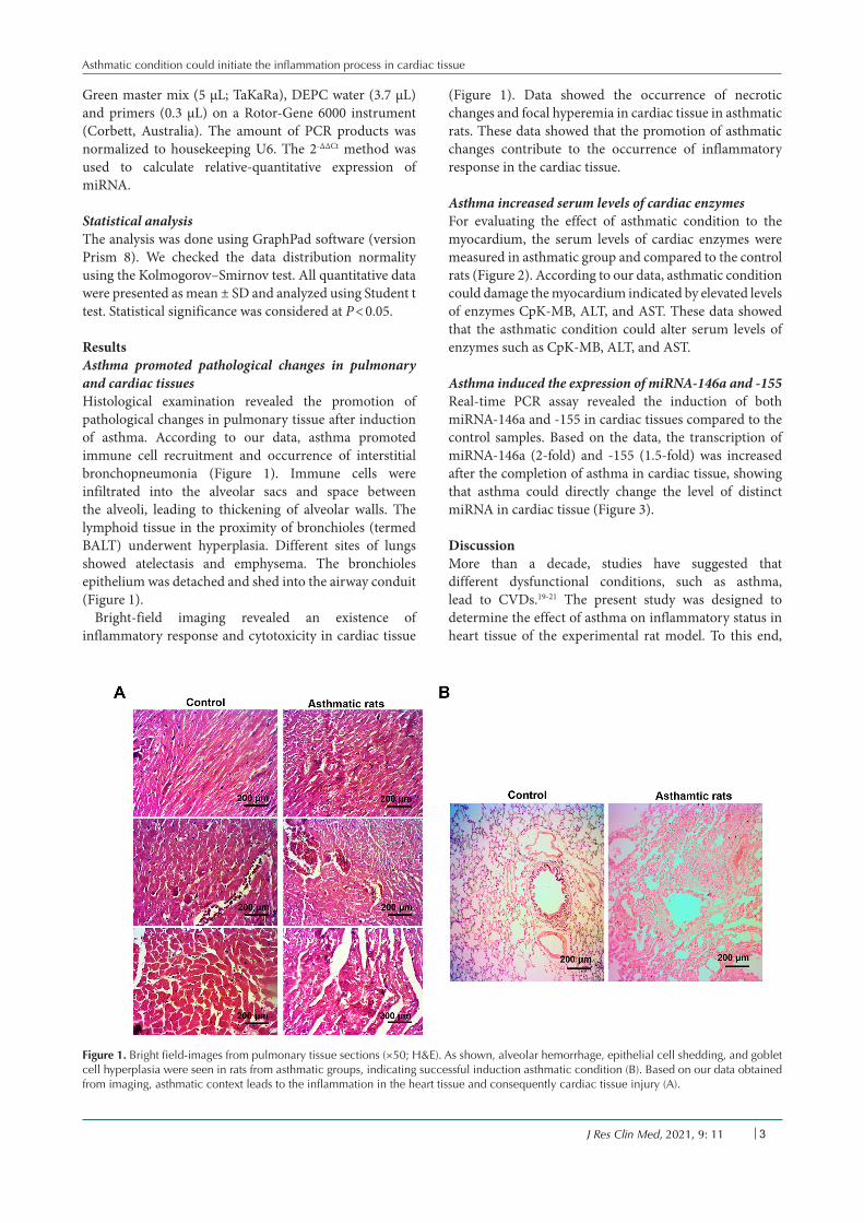
ResultsAsthma promoted pathological changes in pulmonary and cardiac tissuesHistological examination revealed the promotion of pathological changes in pulmonary tissue after induction of asthma According to our data asthma promoted immune cell recruitment and occurrence of interstitial bronchopneumonia (Figure 1) Immune cells were infiltrated into the alveolar sacs and space between the alveoli leading to thickening of alveolar walls The lymphoid tissue in the proximity of bronchioles (termed BALT) underwent hyperplasia Different sites of lungs showed atelectasis and emphysema The bronchioles epithelium was detached and shed into the airway conduit (Figure 1)
Bright-field imaging revealed an existence of inflammatory response and cytotoxicity in cardiac tissue
(Figure 1) Data showed the occurrence of necrotic changes and focal hyperemia in cardiac tissue in asthmatic rats These data showed that the promotion of asthmatic changes contribute to the occurrence of inflammatory response in the cardiac tissue
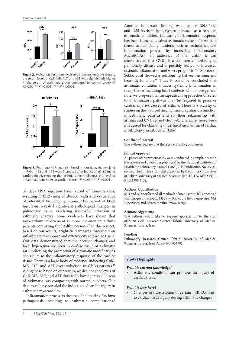
Asthma increased serum levels of cardiac enzymes For evaluating the effect of asthmatic condition to the myocardium the serum levels of cardiac enzymes were measured in asthmatic group and compared to the control rats (Figure 2) According to our data asthmatic condition could damage the myocardium indicated by elevated levels of enzymes CpK-MB ALT and AST These data showed that the asthmatic condition could alter serum levels of enzymes such as CpK-MB ALT and AST
Asthma induced the expression of miRNA-146a and -155 Real-time PCR assay revealed the induction of both miRNA-146a and -155 in cardiac tissues compared to the control samples Based on the data the transcription of miRNA-146a (2-fold) and -155 (15-fold) was increased after the completion of asthma in cardiac tissue showing that asthma could directly change the level of distinct miRNA in cardiac tissue (Figure 3)
DiscussionMore than a decade studies have suggested that different dysfunctional conditions such as asthma lead to CVDs19-21 The present study was designed to determine the effect of asthma on inflammatory status in heart tissue of the experimental rat model To this end
Figure 1 Bright field-images from pulmonary tissue sections (times50 HampE) As shown alveolar hemorrhage epithelial cell shedding and goblet cell hyperplasia were seen in rats from asthmatic groups indicating successful induction asthmatic condition (B) Based on our data obtained from imaging asthmatic context leads to the inflammation in the heart tissue and consequently cardiac tissue injury (A)
Hassanpour et al
J Res Clin Med 2021 9 114
32 days OVA injection have recruit of immune cells resulting in thickening of alveolar walls and occurrence of interstitial bronchopneumonia This period of OVA injections revealed significant pathological changes in pulmonary tissue validating successful induction of asthmatic changes Some evidences have shown that myocardium involvement is more common in asthma patients comparing the healthy persons22 In this respect based on our results bright-field imaging discovered an inflammatory response and cytotoxicity in cardiac tissue Our data demonstrated that the necrotic changes and focal hyperemia was seen in cardiac tissue of asthmatic rats indicating the promotion of asthmatic modifications contribute to the inflammatory response of the cardiac tissue There is a large body of evidence indicating CpK-MB ALT and AST overproduction in CVDs patients23 Along these based on our results we decided that levels of CpK-MB ALT and AST drastically have increased in sera of asthmatic rats comparing with normal subjects Our data must have revealed the induction of cardiac injury in asthmatic myocardium
Inflammation process is the one of hallmarks of asthma pathogenesis resulting to asthmatic complications2
Another important finding was that miRNA-146a and -155 levels in lung tissues increased as a result of asthmatic condition indicating inflammation response has been launched against asthmatic status24 These data demonstrated that conditions such as asthma induces inflammation process by increasing inflammatory MicroRNAs25 In authorize of this claim it was demonstrated that CVDs is a common comorbidity of pulmonary disease and is possibly related to increased systemic inflammation and worse prognosis2627 Moreover Sobko et al showed a relationship between asthma and heart dysfunction28 Thus it could be concluded that asthmatic condition induces systemic inflammation in many tissues including heart contexts On a more general note we propose that therapeutically approaches directed to inflammatory pathway may be required to preserve cardiac injuries caused of asthma There is a scarcity of studies on the involved mechanisms of cardiac dysfunction in asthmatic patients and so their relationship with asthma and CVDs is not clear yet Therefore more work is required for clarifying underlined mechanism of cardiac insufficiency in asthmatic status
Conflict of Interest The authors declare that there is no conflict of interest
Ethical ApprovalAll phases of the present study were conducted in compliance with the criteria and guidelines published by the National Institutes of Health for Laboratory Animal Care (NIH Publication No 85-23 revised 1996) This study was approved by the Ethics Committee of Tabriz University of Medical Sciences (No IRTBZMEDVCRREC1398313)
Authorsrsquo ContributionMH and AD performed all methods of manuscript RR conceived and designed the topic MH and RK wrote the manuscript MA supervised and edited the final manuscript
AcknowledgementsThe authors would like to express appreciation to the staff of Stem Cell Research Center Tabriz University of Medical Sciences Tabriz Iran
FundingPulmonary Research Center Tabriz University of Medical Sciences Tabriz Iran (Grant No 63738)
Figure 3 Real-time PCR analysis Based on our data the levels of miRNA-146a and -155 were increased after induction of asthma in cardiac tissue showing that asthma directly changes the level of inflammatory miRNAs in cardiac tissue (P lt005 P lt0001)
Figure 2 Evaluating the serum levels of cardiac enzymes As shown the serum levels of CpK-MB ALT and AST were significantly higher in the serum of asthmatic group compared to control group (P lt005) P lt0001 P lt00001
What is current knowledge bull Asthmatic condition can promote the injury of
cardiac tissue
What is new herebull Changes in transcription of certain miRNAs lead
to cardiac tissue injury during asthmatic changes
Study Highlights
Asthmatic condition could initiate the inflammation process in cardiac tissue
J Res Clin Med 2021 9 11 5
References1 Nakawah MO Hawkins C Barbandi F Asthma chronic
obstructive pulmonary disease (COPD) and the overlap syndrome J Am Board Fam Med 201326(4)470-7 doi 103122jabfm201304120256
2 Rahbarghazi R Keyhanmanesh R Aslani MR Hassanpour M Ahmadi M Bone marrow mesenchymal stem cells and condition media diminish inflammatory adhesion molecules of pulmonary endothelial cells in an ovalbumin-induced asthmatic rat model Microvasc Res 201912163-70 doi 101016jmvr201810005
3 Ramakrishnan RK Mahboub B Hamid Q Asthma-chronic obstructive pulmonary disease overlap a distinct pathophysiological and clinical entity In Bernstein JA Boulet LP Wechsler ME eds Asthma COPD and Overlap A Case Based Overview of Similarities and Differences Boca Raton CRC Press 2018 p 55-66
4 Tattersall MC Guo M Korcarz CE Gepner AD Kaufman JD Liu KJ et al Asthma predicts cardiovascular disease events the multi-ethnic study of atherosclerosis Arterioscler Thromb Vasc Biol 201535(6)1520-5 doi 101161atvbaha115305452
5 Iribarren C Tolstykh IV Miller MK Sobel E Eisner MD Adult asthma and risk of coronary heart disease cerebrovascular disease and heart failure a prospective study of 2 matched cohorts Am J Epidemiol 2012176(11)1014-24 doi 101093ajekws181
6 Cepelis A Brumpton BM Laugsand LE Dalen H Langhammer A Janszky I et al Asthma asthma control and risk of acute myocardial infarction HUNT study Eur J Epidemiol 201934(10)967-77 doi 101007s10654-019-00562-x
7 Barnes PJ Cellular and molecular mechanisms of asthma and COPD Clin Sci (Lond) 2017131(13)1541-58 doi 101042cs20160487
8 Ahmadi M Rahbarghazi R Shahbazfar AA Baghban H Keyhanmanesh R Bone marrow mesenchymal stem cells modified pathological changes and immunological responses in ovalbumin-induced asthmatic rats possibly by the modulation of miRNA155 and miRNA133 Gen Physiol Biophys 201837(3)263-74 doi 104149gpb_2017052
9 Gebert LFR MacRae IJ Regulation of microRNA function in animals Nat Rev Mol Cell Biol 201920(1)21-37 doi 101038s41580-018-0045-7
10 Viereck J Thum T Circulating noncoding RNAs as biomarkers of cardiovascular disease and injury Circ Res 2017120(2)381-99 doi 101161circresaha116308434
11 Su Q Yang H Li L Circulating miRNA-155 as a potential biomarker for coronary slow flow Dis Markers 201820186345284 doi 10115520186345284
12 Kai W Qian XU Qun WU MicroRNAs and asthma regulation Iran J Allergy Asthma Immunol 201514(2)120-5
13 Banerjee A Schambach F DeJong CS Hammond SM Reiner SL Micro-RNA-155 inhibits IFN-gamma signaling in CD4+ T cells Eur J Immunol 201040(1)225-31 doi 101002eji200939381
14 Malmhaumlll C Alawieh S Lu Y Sjoumlstrand M Bossios A Eldh M et al MicroRNA-155 is essential for T(H)2-mediated allergen-induced eosinophilic inflammation in the lung J Allergy Clin Immunol 2014133(5)1429-38 1438e1-7 doi 101016jjaci201311008
15 Ahmadi M Rahbarghazi R Shahbazfar AA keyhanmanesh R Monitoring IL-13 expression in relation to miRNA-155 and miRNA-133 changes following intra-tracheal administration of mesenchymal stem cells and conditioned media in ovalbumin-sensitized rats Thai J Vet Med 201848(3)347-55
16 Solberg OD Ostrin EJ Love MI Peng JC Bhakta NR Hou L et al Airway epithelial miRNA expression is altered in asthma Am J Respir Crit Care Med 2012186(10)965-74 doi 101164rccm201201-0027OC
17 Keyhanmanesh R Rahbarghazi R Aslani MR Hassanpour M Ahmadi M Systemic delivery of mesenchymal stem cells condition media in repeated doses acts as magic bullets in restoring IFN-γIL-4 balance in asthmatic rats Life Sci 201821230-6 doi 101016jlfs201809049
18 Shademan B Nourazarian A Nikanfar M Avci CB Hasanpour M Isazadeh A Investigation of the miRNA146a and miRNA155 gene expression levels in patients with multiple sclerosis Journal of Clinical Neuroscience 2020 78 189-93
19 Pollevick ME Xu KY Mhango G Federmann EG Vedanthan R Busse P et al The relationship between asthma and cardiovascular disease an examination of the Framingham offspring study Chest 2020 doi 101016jchest202011053
20 Dogra S Ardern CI Baker J The relationship between age of asthma onset and cardiovascular disease in Canadians J Asthma 200744(10)849-54 doi 10108002770900701752391
21 Burge MR Aldrete KL Ehrhart M Murray-Krezan C 439-P Assessing the Relationship between Asthma and Cardiovascular Disease Risk in Type 2 Diabetes Using Revised Risk Estimates Diabetes 201968(Suppl 1)439 doi 102337db19-439-P
22 Ingebrigtsen TS Marott JL Vestbo J Nordestgaard BG Lange P Coronary heart disease and heart failure in asthma COPD and asthma-COPD overlap BMJ Open Respir Res 20207(1)e000470 doi 101136bmjresp-2019-000470
23 Pochhi M Muddeshwar M Serum enzymes markers in myocardial infarction a study of rural area Asian J Med Sci 20178(2)34-7 doi 103126ajmsv8i216313
24 Mousavi SR Ahmadi A Jamalkandi SA Salimian J Involvement of microRNAs in physiological and pathological processes in asthma J Cell Physiol 2019234(12)21547-59 doi 101002jcp28781
25 Heffler E Allegra A Pioggia G Picardi G Musolino C Gangemi S MicroRNA profiling in asthma potential biomarkers and therapeutic targets Am J Respir Cell Mol Biol 201757(6)642-50 doi 101165rcmb2016-0231TR
26 Chen W Thomas J Sadatsafavi M FitzGerald JM Risk of cardiovascular comorbidity in patients with chronic obstructive pulmonary disease a systematic review and meta-analysis Lancet Respir Med 20153(8)631-9 doi 101016s2213-2600(15)00241-6
27 Muumlllerova H Agusti A Erqou S Mapel DW Cardiovascular comorbidity in COPD systematic literature review Chest 2013144(4)1163-78 doi 101378chest12-2847
28 Sobko EA Solovrsquoeva IA Kraposhina A Riazanova NG Vtiurina SS Salmina AB et al [Relationship between endothelial dysfunction and mechanisms of systemic inflammation in left heart remodeling in patients with bronchial asthma] Klin Med (Mosk) 201492(11)43-8

Hassanpour et al
J Res Clin Med 2021 9 112
Literature reviews demonstrated that level of distinct microRNAs could be changed in cardiac disorders and could be consider as a biomarker for heart tissue injury10 In this respect experiments highlighted the increase of miRNA-155 and -146a in acute myocardial infarction and inflammatory diseases11 However there is a controversial debate regarding miRNA-155 expression in asthmatic animals compared to normal subjects12 This miRNA targets critical inflammatory genes including c-Fos C-Maf and INF-γ receptor1213 The promotion of asthmatic changes in rats and mice by ovo-albumin induced the expression of miRNA-155 in the lungs14-16 As a result targeting specific miRNAs could supplement instrumental data in the control of allergic asthma and asthma-derived pathology in non-pulmonary tissues such as heart
Despite the great importance of asthma in disrupting cardiac pathophysiology few studies have been conducted yet The aim of this study was to investigate the expression level of inflammatory microRNAs in cardiac tissue of asthmatic rats
Material and methodsAsthma induction and animal groupsSixteen adult male Wistar rats weighting approximately 200 g were enrolled in this study The animals were kept in standard cages under 1212 lightndashdark cycle at 18-22degC with free access to water and food After adaptation to the new condition rats were allocated into control and asthmatic rats (n=8) In order to induce asthmatic changes rats were exposed to ovalbumin (OVA Sigma-Aldrich USA) for a period of 32 plusmn 1 days according to our previous studies17
Each animal received 1 mg OVA and 200 mg aluminum hydroxide (as adjuvant) intra-peritoneally dissolved in 1 mL normal saline from the days 1 to 8 On day 14 the sensitized rats were challenged daily with aerosolized condition of 4 OVA formed by a nebulizer for 5 minutes (CX3 Omron Co Netherlands) for 18 plusmn 1 days without any interruption This exposure was induced in a special sealed box with dimensions of 30 times 20 times 20 cm3 In the healthy subjects the same vehicle type was injected instead of OVA with the same manner One day after the completion of sensitization protocol all assays
was performed After completion of asthma procedure animals were euthanized by using the combination of ketamine and xylazine Following the completion of asthma induction the animals were anesthetized using ketamine (75 mgkg bw)xylazine (3 mgkg bw) solution intraperitoneally and scarified Both hearts and lungs were removed and subjected to the histological examination and genomic analysis
Histological examinationTo confirm the promotion of asthmatic changes in pulmonary tissue and assess the effect of asthma on cardiac tissue we performed hematoxylin-eosin (HampE) staining To this end left ventricle tissue was excised and kept in 10 buffered-formalin solution (Merck Germany) Samples were dehydrated cleared by xylol and paraffin-embedded 4 microm-thick sections were prepared using microtome (Leica) and stained by HampE solution Cardiac samples were evaluated for the existence of necrosis and inflammation In pulmonary tissue we monitored different pathologies such as immune cell infiltration edema emphysema and atelectasis and bronchioles epithelial detachment
Investigation of cardiac enzymesThe activity levels of creatine phosphokinase-MB (CpK-MB) alanine aminotransferase (ALT) and aspartate aminotransferase (AST) in the serums were measured using commercial kits according to manufacture protocol (Pars Azmun Iran)
Real-time polymerase chain reaction assayTo assess the transcription of miRNA-146a and -155 real-time polymerase chain reaction (PCR) analysis was performed On this basis left ventricle was quickly chopped and RNA content isolated using TRIzol (Roche Germany) according to the manufacturerrsquos protocols RNA concentration and integrity were measured by using a Picodrop 1000 Spectrophotometer (Thermo Scientific USA) The sequence of target gene was designed using Gene-Runner Software (Ver 305) (Table 1) To synthesize cDNA reverse transcription was carried out by cDNA Synthesis Kit (TaKaRa) Real-time PCR assay was conducted by cocktail of cDNA sample (1 microL) SYBR
Table 1 Primer list
Gene Sequence Ref
miRNA-146aForward CGTGCTGTGACCTATGCTGUniversal Reverse CCAGTGCAGGGTCCGAGGTAStem loop GTCGTATCCAGTGCAGGGTCCGAGGTATTCGCACTGGATACGACTTCCCT
18
miRNA-155Forward CGGCGCTTAATGCTAATCGTGATAGUniversal Reverse GTGCAGGGTCCGAGGTStem loop GTCGTATCCAGTGCAGGGTCCGAGGTATTCGCACTGGATACGACACCCCT
18
U6-6pForward GCTTCGGCAGCACATATACTAAAATReverse CGCTTCACGAATTTGCGTGTCATStem loop GTCGTATCCAGTGCAGGGTCCGAGGTATTCGCACTGGATACGACAAAAATAT
18
Asthmatic condition could initiate the inflammation process in cardiac tissue
J Res Clin Med 2021 9 11 3
Green master mix (5 microL TaKaRa) DEPC water (37 microL) and primers (03 microL) on a Rotor-Gene 6000 instrument (Corbett Australia) The amount of PCR products was normalized to housekeeping U6 The 2-ΔΔCt method was used to calculate relative-quantitative expression of miRNA
Statistical analysisThe analysis was done using GraphPad software (version Prism 8) We checked the data distribution normality using the KolmogorovndashSmirnov test All quantitative data were presented as mean plusmn SD and analyzed using Student t test Statistical significance was considered at P lt 005
ResultsAsthma promoted pathological changes in pulmonary and cardiac tissuesHistological examination revealed the promotion of pathological changes in pulmonary tissue after induction of asthma According to our data asthma promoted immune cell recruitment and occurrence of interstitial bronchopneumonia (Figure 1) Immune cells were infiltrated into the alveolar sacs and space between the alveoli leading to thickening of alveolar walls The lymphoid tissue in the proximity of bronchioles (termed BALT) underwent hyperplasia Different sites of lungs showed atelectasis and emphysema The bronchioles epithelium was detached and shed into the airway conduit (Figure 1)
Bright-field imaging revealed an existence of inflammatory response and cytotoxicity in cardiac tissue
(Figure 1) Data showed the occurrence of necrotic changes and focal hyperemia in cardiac tissue in asthmatic rats These data showed that the promotion of asthmatic changes contribute to the occurrence of inflammatory response in the cardiac tissue
Asthma increased serum levels of cardiac enzymes For evaluating the effect of asthmatic condition to the myocardium the serum levels of cardiac enzymes were measured in asthmatic group and compared to the control rats (Figure 2) According to our data asthmatic condition could damage the myocardium indicated by elevated levels of enzymes CpK-MB ALT and AST These data showed that the asthmatic condition could alter serum levels of enzymes such as CpK-MB ALT and AST
Asthma induced the expression of miRNA-146a and -155 Real-time PCR assay revealed the induction of both miRNA-146a and -155 in cardiac tissues compared to the control samples Based on the data the transcription of miRNA-146a (2-fold) and -155 (15-fold) was increased after the completion of asthma in cardiac tissue showing that asthma could directly change the level of distinct miRNA in cardiac tissue (Figure 3)
DiscussionMore than a decade studies have suggested that different dysfunctional conditions such as asthma lead to CVDs19-21 The present study was designed to determine the effect of asthma on inflammatory status in heart tissue of the experimental rat model To this end
Figure 1 Bright field-images from pulmonary tissue sections (times50 HampE) As shown alveolar hemorrhage epithelial cell shedding and goblet cell hyperplasia were seen in rats from asthmatic groups indicating successful induction asthmatic condition (B) Based on our data obtained from imaging asthmatic context leads to the inflammation in the heart tissue and consequently cardiac tissue injury (A)
Hassanpour et al
J Res Clin Med 2021 9 114
32 days OVA injection have recruit of immune cells resulting in thickening of alveolar walls and occurrence of interstitial bronchopneumonia This period of OVA injections revealed significant pathological changes in pulmonary tissue validating successful induction of asthmatic changes Some evidences have shown that myocardium involvement is more common in asthma patients comparing the healthy persons22 In this respect based on our results bright-field imaging discovered an inflammatory response and cytotoxicity in cardiac tissue Our data demonstrated that the necrotic changes and focal hyperemia was seen in cardiac tissue of asthmatic rats indicating the promotion of asthmatic modifications contribute to the inflammatory response of the cardiac tissue There is a large body of evidence indicating CpK-MB ALT and AST overproduction in CVDs patients23 Along these based on our results we decided that levels of CpK-MB ALT and AST drastically have increased in sera of asthmatic rats comparing with normal subjects Our data must have revealed the induction of cardiac injury in asthmatic myocardium
Inflammation process is the one of hallmarks of asthma pathogenesis resulting to asthmatic complications2
Another important finding was that miRNA-146a and -155 levels in lung tissues increased as a result of asthmatic condition indicating inflammation response has been launched against asthmatic status24 These data demonstrated that conditions such as asthma induces inflammation process by increasing inflammatory MicroRNAs25 In authorize of this claim it was demonstrated that CVDs is a common comorbidity of pulmonary disease and is possibly related to increased systemic inflammation and worse prognosis2627 Moreover Sobko et al showed a relationship between asthma and heart dysfunction28 Thus it could be concluded that asthmatic condition induces systemic inflammation in many tissues including heart contexts On a more general note we propose that therapeutically approaches directed to inflammatory pathway may be required to preserve cardiac injuries caused of asthma There is a scarcity of studies on the involved mechanisms of cardiac dysfunction in asthmatic patients and so their relationship with asthma and CVDs is not clear yet Therefore more work is required for clarifying underlined mechanism of cardiac insufficiency in asthmatic status
Conflict of Interest The authors declare that there is no conflict of interest
Ethical ApprovalAll phases of the present study were conducted in compliance with the criteria and guidelines published by the National Institutes of Health for Laboratory Animal Care (NIH Publication No 85-23 revised 1996) This study was approved by the Ethics Committee of Tabriz University of Medical Sciences (No IRTBZMEDVCRREC1398313)
Authorsrsquo ContributionMH and AD performed all methods of manuscript RR conceived and designed the topic MH and RK wrote the manuscript MA supervised and edited the final manuscript
AcknowledgementsThe authors would like to express appreciation to the staff of Stem Cell Research Center Tabriz University of Medical Sciences Tabriz Iran
FundingPulmonary Research Center Tabriz University of Medical Sciences Tabriz Iran (Grant No 63738)
Figure 3 Real-time PCR analysis Based on our data the levels of miRNA-146a and -155 were increased after induction of asthma in cardiac tissue showing that asthma directly changes the level of inflammatory miRNAs in cardiac tissue (P lt005 P lt0001)
Figure 2 Evaluating the serum levels of cardiac enzymes As shown the serum levels of CpK-MB ALT and AST were significantly higher in the serum of asthmatic group compared to control group (P lt005) P lt0001 P lt00001
What is current knowledge bull Asthmatic condition can promote the injury of
cardiac tissue
What is new herebull Changes in transcription of certain miRNAs lead
to cardiac tissue injury during asthmatic changes
Study Highlights
Asthmatic condition could initiate the inflammation process in cardiac tissue
J Res Clin Med 2021 9 11 5
References1 Nakawah MO Hawkins C Barbandi F Asthma chronic
obstructive pulmonary disease (COPD) and the overlap syndrome J Am Board Fam Med 201326(4)470-7 doi 103122jabfm201304120256
2 Rahbarghazi R Keyhanmanesh R Aslani MR Hassanpour M Ahmadi M Bone marrow mesenchymal stem cells and condition media diminish inflammatory adhesion molecules of pulmonary endothelial cells in an ovalbumin-induced asthmatic rat model Microvasc Res 201912163-70 doi 101016jmvr201810005
3 Ramakrishnan RK Mahboub B Hamid Q Asthma-chronic obstructive pulmonary disease overlap a distinct pathophysiological and clinical entity In Bernstein JA Boulet LP Wechsler ME eds Asthma COPD and Overlap A Case Based Overview of Similarities and Differences Boca Raton CRC Press 2018 p 55-66
4 Tattersall MC Guo M Korcarz CE Gepner AD Kaufman JD Liu KJ et al Asthma predicts cardiovascular disease events the multi-ethnic study of atherosclerosis Arterioscler Thromb Vasc Biol 201535(6)1520-5 doi 101161atvbaha115305452
5 Iribarren C Tolstykh IV Miller MK Sobel E Eisner MD Adult asthma and risk of coronary heart disease cerebrovascular disease and heart failure a prospective study of 2 matched cohorts Am J Epidemiol 2012176(11)1014-24 doi 101093ajekws181
6 Cepelis A Brumpton BM Laugsand LE Dalen H Langhammer A Janszky I et al Asthma asthma control and risk of acute myocardial infarction HUNT study Eur J Epidemiol 201934(10)967-77 doi 101007s10654-019-00562-x
7 Barnes PJ Cellular and molecular mechanisms of asthma and COPD Clin Sci (Lond) 2017131(13)1541-58 doi 101042cs20160487
8 Ahmadi M Rahbarghazi R Shahbazfar AA Baghban H Keyhanmanesh R Bone marrow mesenchymal stem cells modified pathological changes and immunological responses in ovalbumin-induced asthmatic rats possibly by the modulation of miRNA155 and miRNA133 Gen Physiol Biophys 201837(3)263-74 doi 104149gpb_2017052
9 Gebert LFR MacRae IJ Regulation of microRNA function in animals Nat Rev Mol Cell Biol 201920(1)21-37 doi 101038s41580-018-0045-7
10 Viereck J Thum T Circulating noncoding RNAs as biomarkers of cardiovascular disease and injury Circ Res 2017120(2)381-99 doi 101161circresaha116308434
11 Su Q Yang H Li L Circulating miRNA-155 as a potential biomarker for coronary slow flow Dis Markers 201820186345284 doi 10115520186345284
12 Kai W Qian XU Qun WU MicroRNAs and asthma regulation Iran J Allergy Asthma Immunol 201514(2)120-5
13 Banerjee A Schambach F DeJong CS Hammond SM Reiner SL Micro-RNA-155 inhibits IFN-gamma signaling in CD4+ T cells Eur J Immunol 201040(1)225-31 doi 101002eji200939381
14 Malmhaumlll C Alawieh S Lu Y Sjoumlstrand M Bossios A Eldh M et al MicroRNA-155 is essential for T(H)2-mediated allergen-induced eosinophilic inflammation in the lung J Allergy Clin Immunol 2014133(5)1429-38 1438e1-7 doi 101016jjaci201311008
15 Ahmadi M Rahbarghazi R Shahbazfar AA keyhanmanesh R Monitoring IL-13 expression in relation to miRNA-155 and miRNA-133 changes following intra-tracheal administration of mesenchymal stem cells and conditioned media in ovalbumin-sensitized rats Thai J Vet Med 201848(3)347-55
16 Solberg OD Ostrin EJ Love MI Peng JC Bhakta NR Hou L et al Airway epithelial miRNA expression is altered in asthma Am J Respir Crit Care Med 2012186(10)965-74 doi 101164rccm201201-0027OC
17 Keyhanmanesh R Rahbarghazi R Aslani MR Hassanpour M Ahmadi M Systemic delivery of mesenchymal stem cells condition media in repeated doses acts as magic bullets in restoring IFN-γIL-4 balance in asthmatic rats Life Sci 201821230-6 doi 101016jlfs201809049
18 Shademan B Nourazarian A Nikanfar M Avci CB Hasanpour M Isazadeh A Investigation of the miRNA146a and miRNA155 gene expression levels in patients with multiple sclerosis Journal of Clinical Neuroscience 2020 78 189-93
19 Pollevick ME Xu KY Mhango G Federmann EG Vedanthan R Busse P et al The relationship between asthma and cardiovascular disease an examination of the Framingham offspring study Chest 2020 doi 101016jchest202011053
20 Dogra S Ardern CI Baker J The relationship between age of asthma onset and cardiovascular disease in Canadians J Asthma 200744(10)849-54 doi 10108002770900701752391
21 Burge MR Aldrete KL Ehrhart M Murray-Krezan C 439-P Assessing the Relationship between Asthma and Cardiovascular Disease Risk in Type 2 Diabetes Using Revised Risk Estimates Diabetes 201968(Suppl 1)439 doi 102337db19-439-P
22 Ingebrigtsen TS Marott JL Vestbo J Nordestgaard BG Lange P Coronary heart disease and heart failure in asthma COPD and asthma-COPD overlap BMJ Open Respir Res 20207(1)e000470 doi 101136bmjresp-2019-000470
23 Pochhi M Muddeshwar M Serum enzymes markers in myocardial infarction a study of rural area Asian J Med Sci 20178(2)34-7 doi 103126ajmsv8i216313
24 Mousavi SR Ahmadi A Jamalkandi SA Salimian J Involvement of microRNAs in physiological and pathological processes in asthma J Cell Physiol 2019234(12)21547-59 doi 101002jcp28781
25 Heffler E Allegra A Pioggia G Picardi G Musolino C Gangemi S MicroRNA profiling in asthma potential biomarkers and therapeutic targets Am J Respir Cell Mol Biol 201757(6)642-50 doi 101165rcmb2016-0231TR
26 Chen W Thomas J Sadatsafavi M FitzGerald JM Risk of cardiovascular comorbidity in patients with chronic obstructive pulmonary disease a systematic review and meta-analysis Lancet Respir Med 20153(8)631-9 doi 101016s2213-2600(15)00241-6
27 Muumlllerova H Agusti A Erqou S Mapel DW Cardiovascular comorbidity in COPD systematic literature review Chest 2013144(4)1163-78 doi 101378chest12-2847
28 Sobko EA Solovrsquoeva IA Kraposhina A Riazanova NG Vtiurina SS Salmina AB et al [Relationship between endothelial dysfunction and mechanisms of systemic inflammation in left heart remodeling in patients with bronchial asthma] Klin Med (Mosk) 201492(11)43-8
Asthmatic condition could initiate the inflammation process in cardiac tissue
J Res Clin Med 2021 9 11 3
Green master mix (5 microL TaKaRa) DEPC water (37 microL) and primers (03 microL) on a Rotor-Gene 6000 instrument (Corbett Australia) The amount of PCR products was normalized to housekeeping U6 The 2-ΔΔCt method was used to calculate relative-quantitative expression of miRNA
Statistical analysisThe analysis was done using GraphPad software (version Prism 8) We checked the data distribution normality using the KolmogorovndashSmirnov test All quantitative data were presented as mean plusmn SD and analyzed using Student t test Statistical significance was considered at P lt 005
ResultsAsthma promoted pathological changes in pulmonary and cardiac tissuesHistological examination revealed the promotion of pathological changes in pulmonary tissue after induction of asthma According to our data asthma promoted immune cell recruitment and occurrence of interstitial bronchopneumonia (Figure 1) Immune cells were infiltrated into the alveolar sacs and space between the alveoli leading to thickening of alveolar walls The lymphoid tissue in the proximity of bronchioles (termed BALT) underwent hyperplasia Different sites of lungs showed atelectasis and emphysema The bronchioles epithelium was detached and shed into the airway conduit (Figure 1)
Bright-field imaging revealed an existence of inflammatory response and cytotoxicity in cardiac tissue
(Figure 1) Data showed the occurrence of necrotic changes and focal hyperemia in cardiac tissue in asthmatic rats These data showed that the promotion of asthmatic changes contribute to the occurrence of inflammatory response in the cardiac tissue
Asthma increased serum levels of cardiac enzymes For evaluating the effect of asthmatic condition to the myocardium the serum levels of cardiac enzymes were measured in asthmatic group and compared to the control rats (Figure 2) According to our data asthmatic condition could damage the myocardium indicated by elevated levels of enzymes CpK-MB ALT and AST These data showed that the asthmatic condition could alter serum levels of enzymes such as CpK-MB ALT and AST
Asthma induced the expression of miRNA-146a and -155 Real-time PCR assay revealed the induction of both miRNA-146a and -155 in cardiac tissues compared to the control samples Based on the data the transcription of miRNA-146a (2-fold) and -155 (15-fold) was increased after the completion of asthma in cardiac tissue showing that asthma could directly change the level of distinct miRNA in cardiac tissue (Figure 3)
DiscussionMore than a decade studies have suggested that different dysfunctional conditions such as asthma lead to CVDs19-21 The present study was designed to determine the effect of asthma on inflammatory status in heart tissue of the experimental rat model To this end
Figure 1 Bright field-images from pulmonary tissue sections (times50 HampE) As shown alveolar hemorrhage epithelial cell shedding and goblet cell hyperplasia were seen in rats from asthmatic groups indicating successful induction asthmatic condition (B) Based on our data obtained from imaging asthmatic context leads to the inflammation in the heart tissue and consequently cardiac tissue injury (A)
Hassanpour et al
J Res Clin Med 2021 9 114
32 days OVA injection have recruit of immune cells resulting in thickening of alveolar walls and occurrence of interstitial bronchopneumonia This period of OVA injections revealed significant pathological changes in pulmonary tissue validating successful induction of asthmatic changes Some evidences have shown that myocardium involvement is more common in asthma patients comparing the healthy persons22 In this respect based on our results bright-field imaging discovered an inflammatory response and cytotoxicity in cardiac tissue Our data demonstrated that the necrotic changes and focal hyperemia was seen in cardiac tissue of asthmatic rats indicating the promotion of asthmatic modifications contribute to the inflammatory response of the cardiac tissue There is a large body of evidence indicating CpK-MB ALT and AST overproduction in CVDs patients23 Along these based on our results we decided that levels of CpK-MB ALT and AST drastically have increased in sera of asthmatic rats comparing with normal subjects Our data must have revealed the induction of cardiac injury in asthmatic myocardium
Inflammation process is the one of hallmarks of asthma pathogenesis resulting to asthmatic complications2
Another important finding was that miRNA-146a and -155 levels in lung tissues increased as a result of asthmatic condition indicating inflammation response has been launched against asthmatic status24 These data demonstrated that conditions such as asthma induces inflammation process by increasing inflammatory MicroRNAs25 In authorize of this claim it was demonstrated that CVDs is a common comorbidity of pulmonary disease and is possibly related to increased systemic inflammation and worse prognosis2627 Moreover Sobko et al showed a relationship between asthma and heart dysfunction28 Thus it could be concluded that asthmatic condition induces systemic inflammation in many tissues including heart contexts On a more general note we propose that therapeutically approaches directed to inflammatory pathway may be required to preserve cardiac injuries caused of asthma There is a scarcity of studies on the involved mechanisms of cardiac dysfunction in asthmatic patients and so their relationship with asthma and CVDs is not clear yet Therefore more work is required for clarifying underlined mechanism of cardiac insufficiency in asthmatic status
Conflict of Interest The authors declare that there is no conflict of interest
Ethical ApprovalAll phases of the present study were conducted in compliance with the criteria and guidelines published by the National Institutes of Health for Laboratory Animal Care (NIH Publication No 85-23 revised 1996) This study was approved by the Ethics Committee of Tabriz University of Medical Sciences (No IRTBZMEDVCRREC1398313)
Authorsrsquo ContributionMH and AD performed all methods of manuscript RR conceived and designed the topic MH and RK wrote the manuscript MA supervised and edited the final manuscript
AcknowledgementsThe authors would like to express appreciation to the staff of Stem Cell Research Center Tabriz University of Medical Sciences Tabriz Iran
FundingPulmonary Research Center Tabriz University of Medical Sciences Tabriz Iran (Grant No 63738)
Figure 3 Real-time PCR analysis Based on our data the levels of miRNA-146a and -155 were increased after induction of asthma in cardiac tissue showing that asthma directly changes the level of inflammatory miRNAs in cardiac tissue (P lt005 P lt0001)
Figure 2 Evaluating the serum levels of cardiac enzymes As shown the serum levels of CpK-MB ALT and AST were significantly higher in the serum of asthmatic group compared to control group (P lt005) P lt0001 P lt00001
What is current knowledge bull Asthmatic condition can promote the injury of
cardiac tissue
What is new herebull Changes in transcription of certain miRNAs lead
to cardiac tissue injury during asthmatic changes
Study Highlights
Asthmatic condition could initiate the inflammation process in cardiac tissue
J Res Clin Med 2021 9 11 5
References1 Nakawah MO Hawkins C Barbandi F Asthma chronic
obstructive pulmonary disease (COPD) and the overlap syndrome J Am Board Fam Med 201326(4)470-7 doi 103122jabfm201304120256
2 Rahbarghazi R Keyhanmanesh R Aslani MR Hassanpour M Ahmadi M Bone marrow mesenchymal stem cells and condition media diminish inflammatory adhesion molecules of pulmonary endothelial cells in an ovalbumin-induced asthmatic rat model Microvasc Res 201912163-70 doi 101016jmvr201810005
3 Ramakrishnan RK Mahboub B Hamid Q Asthma-chronic obstructive pulmonary disease overlap a distinct pathophysiological and clinical entity In Bernstein JA Boulet LP Wechsler ME eds Asthma COPD and Overlap A Case Based Overview of Similarities and Differences Boca Raton CRC Press 2018 p 55-66
4 Tattersall MC Guo M Korcarz CE Gepner AD Kaufman JD Liu KJ et al Asthma predicts cardiovascular disease events the multi-ethnic study of atherosclerosis Arterioscler Thromb Vasc Biol 201535(6)1520-5 doi 101161atvbaha115305452
5 Iribarren C Tolstykh IV Miller MK Sobel E Eisner MD Adult asthma and risk of coronary heart disease cerebrovascular disease and heart failure a prospective study of 2 matched cohorts Am J Epidemiol 2012176(11)1014-24 doi 101093ajekws181
6 Cepelis A Brumpton BM Laugsand LE Dalen H Langhammer A Janszky I et al Asthma asthma control and risk of acute myocardial infarction HUNT study Eur J Epidemiol 201934(10)967-77 doi 101007s10654-019-00562-x
7 Barnes PJ Cellular and molecular mechanisms of asthma and COPD Clin Sci (Lond) 2017131(13)1541-58 doi 101042cs20160487
8 Ahmadi M Rahbarghazi R Shahbazfar AA Baghban H Keyhanmanesh R Bone marrow mesenchymal stem cells modified pathological changes and immunological responses in ovalbumin-induced asthmatic rats possibly by the modulation of miRNA155 and miRNA133 Gen Physiol Biophys 201837(3)263-74 doi 104149gpb_2017052
9 Gebert LFR MacRae IJ Regulation of microRNA function in animals Nat Rev Mol Cell Biol 201920(1)21-37 doi 101038s41580-018-0045-7
10 Viereck J Thum T Circulating noncoding RNAs as biomarkers of cardiovascular disease and injury Circ Res 2017120(2)381-99 doi 101161circresaha116308434
11 Su Q Yang H Li L Circulating miRNA-155 as a potential biomarker for coronary slow flow Dis Markers 201820186345284 doi 10115520186345284
12 Kai W Qian XU Qun WU MicroRNAs and asthma regulation Iran J Allergy Asthma Immunol 201514(2)120-5
13 Banerjee A Schambach F DeJong CS Hammond SM Reiner SL Micro-RNA-155 inhibits IFN-gamma signaling in CD4+ T cells Eur J Immunol 201040(1)225-31 doi 101002eji200939381
14 Malmhaumlll C Alawieh S Lu Y Sjoumlstrand M Bossios A Eldh M et al MicroRNA-155 is essential for T(H)2-mediated allergen-induced eosinophilic inflammation in the lung J Allergy Clin Immunol 2014133(5)1429-38 1438e1-7 doi 101016jjaci201311008
15 Ahmadi M Rahbarghazi R Shahbazfar AA keyhanmanesh R Monitoring IL-13 expression in relation to miRNA-155 and miRNA-133 changes following intra-tracheal administration of mesenchymal stem cells and conditioned media in ovalbumin-sensitized rats Thai J Vet Med 201848(3)347-55
16 Solberg OD Ostrin EJ Love MI Peng JC Bhakta NR Hou L et al Airway epithelial miRNA expression is altered in asthma Am J Respir Crit Care Med 2012186(10)965-74 doi 101164rccm201201-0027OC
17 Keyhanmanesh R Rahbarghazi R Aslani MR Hassanpour M Ahmadi M Systemic delivery of mesenchymal stem cells condition media in repeated doses acts as magic bullets in restoring IFN-γIL-4 balance in asthmatic rats Life Sci 201821230-6 doi 101016jlfs201809049
18 Shademan B Nourazarian A Nikanfar M Avci CB Hasanpour M Isazadeh A Investigation of the miRNA146a and miRNA155 gene expression levels in patients with multiple sclerosis Journal of Clinical Neuroscience 2020 78 189-93
19 Pollevick ME Xu KY Mhango G Federmann EG Vedanthan R Busse P et al The relationship between asthma and cardiovascular disease an examination of the Framingham offspring study Chest 2020 doi 101016jchest202011053
20 Dogra S Ardern CI Baker J The relationship between age of asthma onset and cardiovascular disease in Canadians J Asthma 200744(10)849-54 doi 10108002770900701752391
21 Burge MR Aldrete KL Ehrhart M Murray-Krezan C 439-P Assessing the Relationship between Asthma and Cardiovascular Disease Risk in Type 2 Diabetes Using Revised Risk Estimates Diabetes 201968(Suppl 1)439 doi 102337db19-439-P
22 Ingebrigtsen TS Marott JL Vestbo J Nordestgaard BG Lange P Coronary heart disease and heart failure in asthma COPD and asthma-COPD overlap BMJ Open Respir Res 20207(1)e000470 doi 101136bmjresp-2019-000470
23 Pochhi M Muddeshwar M Serum enzymes markers in myocardial infarction a study of rural area Asian J Med Sci 20178(2)34-7 doi 103126ajmsv8i216313
24 Mousavi SR Ahmadi A Jamalkandi SA Salimian J Involvement of microRNAs in physiological and pathological processes in asthma J Cell Physiol 2019234(12)21547-59 doi 101002jcp28781
25 Heffler E Allegra A Pioggia G Picardi G Musolino C Gangemi S MicroRNA profiling in asthma potential biomarkers and therapeutic targets Am J Respir Cell Mol Biol 201757(6)642-50 doi 101165rcmb2016-0231TR
26 Chen W Thomas J Sadatsafavi M FitzGerald JM Risk of cardiovascular comorbidity in patients with chronic obstructive pulmonary disease a systematic review and meta-analysis Lancet Respir Med 20153(8)631-9 doi 101016s2213-2600(15)00241-6
27 Muumlllerova H Agusti A Erqou S Mapel DW Cardiovascular comorbidity in COPD systematic literature review Chest 2013144(4)1163-78 doi 101378chest12-2847
28 Sobko EA Solovrsquoeva IA Kraposhina A Riazanova NG Vtiurina SS Salmina AB et al [Relationship between endothelial dysfunction and mechanisms of systemic inflammation in left heart remodeling in patients with bronchial asthma] Klin Med (Mosk) 201492(11)43-8