GEORG SPEYER HAUS INSTITUT FÜR …€¦ · Louis Vermeulen University of Amsterdam Chris Madsen...
72
GEORG SPEYER HAUS INSTITUT FÜR TUMORBIOLOGIE UND EXPERIMENTELLE THERAPIE 2016 Annual Report Georg-Speyer-Haus Zelluläre Kommunikation in der Stammzellnische Zell-Zell Interaktionen im Tumorstroma Experimentelle Therapie
Transcript of GEORG SPEYER HAUS INSTITUT FÜR …€¦ · Louis Vermeulen University of Amsterdam Chris Madsen...

1
GEORG SPEYER HAUSINSTITUT FÜR TUMORBIOLOGIE UND EXPERIMENTELLE THERAPIE
2016Annual ReportGeorg-Speyer-Haus
Zellu
läre
Kom
mun
ikat
ion
in d
er S
tam
mze
llnis
che
Zell-
Zell
Inte
rakt
ione
n im
Tum
orst
rom
aEx
perim
ente
lle T
hera
pie

2
Die Grundfinanzierung des Georg-Speyer-Hauses wird vom Bundesministerium für Gesundheit und dem Hessischen Ministerium für Wissenschaft und Kunst getragen.
The basic funding of the Georg-Speyer-Haus is provided by the Federal Ministry of Health and the Ministry of Higher Education, Research and the Arts of the State of Hessen.
3
Forschen für das Leben Research for Life

4

5
7
11
13
14
16
18
22
26
30
32
36
40
44
48
50
54
58
64
65
67
69
70
Inhalt
VorwortDas Georg-Speyer-HausOrganisationsstruktur Highlights
Zelluläre Kommunikation in der StammzellnischeCellular Communication in the Stem Cell NicheProf. Dr. D. Krause Dr. H. MedyoufProf. Dr. M. Zörnig
Zell-Zell Interaktionen im TumorstromaCell-Cell Interaction in the Tumor StromaPD Dr. M. C. ArkanDr. H. Farin Prof. Dr. F. R. GretenDr. L. Sevenich
Experimentelle TherapieExperimental TherapyDr. U. DietrichProf. Dr. W. S. Wels
PublikationenFinanzen und AdministrationServiceVeranstaltungen, Lehre und NachwuchsförderungDer Verein »Freunde und Förderer des Georg-Speyer-Hauses«Zuwendungsgeber
Content
I
II
III

6
Der neue 7 Tesla-Kleintier-
Magnetresonanztomograph,
der uns eine höchstauflösende
longitudinale Bildgebung
unserer Modelle erlaubt.

7
Introduction
die in den vergangenen Jahren begonnene Umstruk-turierung und Fokussierung des Georg-Speyer-Hauses auf die translationale, onkologische Forschung unter besonderer Berücksichtigung des Tumor Microenvironments nimmt inzwischen Gestalt an. Die neu rekrutierten Nachwuchswissenschaftlerinnen und Nachwuchswissenschaftler haben den Aufbau ihrer Arbeitsgruppen bereits weit vorangebracht.
Im Vordergrund unserer Bemühungen steht die enge Interaktion mit unseren klinischen Partnern der Goethe-Universität im Universitären Centrum für Tumorerkrankungen, welches auch in diesem Jahr nach Evaluation durch ein internationales Experten-gremium erneut den Status eines „Onkologischen Spitzenzentrums“ durch die Deutsche Krebshilfe erhalten hat. Wir freuen uns, dass das Georg-Speyer-Haus als starker Partner auf der Seite der präklinischen Forschung seinen Teil dazu beitragen konnte.
Selbstverständlich sind wir mit dem gleichen Einsatz auch im Deutschen Konsortium für Translationale Krebsforschung (DKTK) sowie im LOEWE Zentrum für Zell- und Gentherapie als aktiver Partner tätig. Gemeinsam mit unseren Kollegen des Standorts Frankfurt/Mainz bauen wir im Rahmen des DKTK sukzessive eine experimentelle, präklinische Einheit am Georg-Speyer-Haus auf, die es uns erlauben wird The-rapiestudien an entsprechenden Tumor-Tiermodellen durchzuführen. Ein großer Schritt in diese Richtung war die Einweihung eines 7 Tesla-Kleintier-Magnet-resonanztomographen, der eine höchstauflösende longitudinale Bildgebung unserer Modelle erlaubt.
our efforts to strictly focus on translational oncology with a special emphasis on the tumor microenviron-ment is becoming increasingly visible. The newly recruited group leaders have managed to set up their groups.
One of our most important aims remains the close interaction with our clinical colleagues within the University Center for Tumor Diseases (UCT) at the Goethe-University. This year the UCT has been successfully evaluated by an international reviewing board and received again the status of an “Center of Excellence” by the German Cancer Aid. We are very proud to contribute to this success as strong pre-clinical partners in the UCT.
Of course we are equally active in the German Cancer Consortium (DKTK) and the LOEWE Center for Cell and Gene Therapy. Together with our partners here in Frankfurt/Mainz we are currently setting up a strong pre-clinical animal unit within the DKTK. This will allow us to perform relevant therapy studies in tumor models in vivo. A big step forward was the acquisition of a 7T small animal MRI which will enable
Dear Reader,dear friends of the Georg-Speyer-Haus,
Liebe Leserinnen und Leser, liebe Freunde des Georg-Speyer-Hauses,
Florian R. Greten | DirektorGeorg-Speyer-HausInstitut für Tumorbiologie und experimentelle Therapie
Paul-Ehrlich-Str. 42 – 44D-60596 Frankfurt / M.Tel. (069) 63395-232Fax (069) [email protected]

8
Unsere exzellenten partnerschaftlichen Beziehungen mit der Universität werden auch dadurch dokumen-tiert, dass Frau Priv.-Doz. Dr. Melek C. Arkan aus dem Institut für Biochemie II ihre Arbeitsgruppe in diesem Sommer am Georg-Speyer-Haus angesiedelt hat und somit eine direkte Brücke zum Institut von Prof. Ivan Dikic schlägt. Frau Dr. Arkan beschäftigt sich mit dem Einfluss einer fettreichen Ernährung auf die Entwick-lung von Darm- und Pankreastumoren und untersucht dabei insbesondere Diät-vermittelte Änderungen des Immunsystems sowie Alterationen in der Zusammen-setzung der bakteriellen Besiedlung des Darms und bringt damit eine weitere komplementäre Expertise an unser Institut. Diese nun auf verschiedenen Ebenen sehr erfolgreiche und enge Kooperation mit den diversen universitären Partnern wollen wir durch die Gründung eines Frankfurt Cancer Institutes (FCI), das
unseren gemeinsamen onkologi-schen Forschungstätigkeiten eine zusätzliche neue Heimat bieten soll, verstetigen. Auf Grund der von Staatsminister Boris Rhein in Aus-sicht gestellten Unterstützung dieser
Initiative durch die Hessische Landesregierung, sowie der von zwei Stiftungen zugesagten Finanzierung der Baukosten sind die Aussichten auf eine Realisierung dieses Projektes exzellent. Wir sind sehr optimistisch, dass wir im kommenden Jahr mit der konkreten Bauplanung beginnen können, so dass ein Bezug dieses neuen Gebäudes, das auf der Grenze zwischen Georg-Speyer-Haus und dem Universitätsklinikum errichtet werden soll, für 2020 realistisch erscheint. Neben all den strukturellen Fortschritten und wissen-schaftlichen Erfolgen des Jahres gab es eine Reihe weiterer Highlights. Die über die Jahre fest etablierte und auch in diesem Jahr wieder außerordentlich gut besuchte Schülervorlesungsreihe sowie das Schüler-praktikum, organisiert von Dr. Ursula Dietrich, ermög-lichten interessierten Oberstufenschülern, mehr über die experimentelle Grundlagenforschung zu erfahren.
Im Mai wurde im Rahmen einer Feierstunde die Ehrentafel anlässlich der Verleihung der Ehrenmit-gliedschaft an Dr. Rolf E. Breuer in Anerkennung seiner langjährigen, außergewöhnlichen Verdienste um das Georg-Speyer-Haus enthüllt. Außerdem wurde die Eröffnung des „Forums“ festlich begangen, das nach Umgestaltung der ehemaligen Bibliothek nun einen geeigneten Treffpunkt für die Interaktion aller Beschäftigten des Georg-Speyer-Hauses und den wissenschaftlichen Austausch darstellt.
longitudinal high resolution imaging of our animal models. Our excellent relationship with the University becomes evident also in other ways. Priv.-Doz. Dr. Melek C. Arkan from the Institute of Biochemistry II has set up her group at the Georg-Speyer-Haus and will thereby form a bridging group to Prof. Ivan Dikic’s institute. Dr. Arkan examines the effects of a high-fat diet on the development of intestinal and pancreatic tumors and focuses specifically on diet-induced effects on the immune system as well as on diet-dependent alterations of the intestinal microbiome and will provide therefore an additional complementary expertise to our institute. Thus, by now we have very successfully established a strong partnership with various partners at the University and we aim to stabilize this further by the foundation of the Frankfurt Cancer Institute (FCI). This new building is intended to provide laboratories and offices for our common research activities. State Minister Boris Rhein has held out in prospect the support of the Hessian Government for this ambitious initiative, and two foundations will most likely provide the financial support for the building costs. We are therefore very optimistic to initiate the precise construction planning for this building next year, which would allow an opening of the FCI – located on the border between Georg-Speyer-Haus and University Hospital – in 2020. Apart from this year’s structural and scientific progress we experienced several other highlights. The over the years well-established and in this year again extremely well attended student lecture series as well as the student internships, organized by Dr. Ursula Dietrich, allowed interested high school students to gain a detailed insight into experimental basic research.
In May we revealed the new honor board that had been placed to recognize Dr. Rolf E. Breuer’s honorary foundation membership and to recognize his extraordinary support of the Georg-Speyer-Haus for more than forty years during his service on the Foundation Board that he chaired for 36 years. Moreover, after some months of remodeling our old library we opened the new “Forum”, which now provides a pleasant meeting point for our employees and will hopefully foster scientific exchange.
Introduction
FRANKFURT CANCER INSTITUTE

9
Das von Dr. Lisa Sevenich organisierte Symposium „CNS Inflammation in Neurodegenerative Disease and Brain Cancer“ brachte eine Reihe internationaler Experten für einen zweitägigen Gedankenaustausch und intensive Diskussionen ans Georg-Speyer-Haus. Auch das nun zum zweiten Mal stattgefundene Symposium „Cellular Crosstalk in the Tumor Microenvironment“, war im Oktober wieder außerordentlich gut besucht und eine Reihe führender Wissenschaftler stellten ihre neuesten zum großen Teil noch unpublizierten Ergebnisse vor.
Unsere auch in diesem Jahr wieder erreichten Erfolge sowie die positive strukturelle Entwicklung des Instituts bestärken uns in unserer Arbeit und geben uns die notwendige Motivation für die Bewerkstelligung der noch vor uns liegenden Aufgaben in den kommenden Jahren.
Introduction
Dr. Lisa Sevenich organized the symposium “CNS Inflammation in Neurodegenerative Disease and Brain Cancer”. During two days international experts discussed and exchanged ideas. In October the second sympo-sium on “Cellular Crosstalk in the Tumor Microenvironment”, was also very well attended and many renown leaders in the field presented exciting and unpublished results.
Our structural achievements as well as our scientific success fuel our motivation to face the challenges ahead of us and we are optmistically looking forward to continue the path we have started to take.
Florian R. Greten, Direktor
GEORG SPEYER HAUSINSTITUTE FOR TUMOR BIOLOGY
AND EXPERIMENTAL THERAPY
2ND SYMPOSIUM
Cellular Crosstalk in the Tumor Microenvironment
OCT 13 – 14, 2016
FRANKFURT
GERMANY
WW
W.G
EO
RG
-SP
EY
ER
-HA
US
.DE
KEY NOTE SPEAKER
Jeffrey W. PollardUniversity of Edinburgh
ORGANIZER
Florian GretenGeorg-Speyer-Haus, Frankfurt
REGISTRATION
Stefanie Schütt
Phone: +49 (0)69 63395-183
CONFIRMED SPEAKERS
Massimiliano MazzoneUniversity of Leuven
Almut SchulzeUniversity of Würzburg
Owen SansomUniversity of Glasgow
Manuela BaccariniUniversity of Vienna
Jörg HuelskenEcole Polytechnique Fédérale de
Lausanne
Lars ZenderUniversity of Tübingen
Ruth Scherz-Shouval Weizmann Institute of Science Israel
Melek Canan ArkanUniversity of Frankfurt
Boris Strilic Max-Planck-Institute, Bad Nauheim
Stefano PiccoloUniversity of Padova
Louis Vermeulen University of Amsterdam
Chris Madsen University of Copenhagen
Hind Medyouf Georg-Speyer-Haus, Frankfurt

10

11
Die Stiftung privaten Rechts „Chemo-therapeutisches Forschungsinstitut Georg-Speyer-Haus“ wurde 1904 in Frankfurt am Main gegründet, um eine Forschungsstätte für Paul Ehrlich, den ersten Direktor des Hauses, zu schaffen. Die Stiftungsverfassung bestimmt als Zweck der Stiftung die wissenschaftliche Forschung auf den Gebieten der Chemo-therapie und verwandter Wissenschaften, die dem Fortschritt der Biomedizin dienen. Es werden ausschließlich und unmittelbar gemeinnützige Zwecke verfolgt.
Die laufenden Geschäfte des heutigen Instituts für Tumorbiologie und experimentelle Therapie nimmt der Direktor wahr. Er ist in dieser Tätigkeit dem Stiftungsvorstand verantwortlich. Das Georg-Speyer-Haus ist durch einen Kooperationsvertrag mit der Goethe-Universität Frankfurt verbunden.
Das Gebäude des Georg-Speyer-Hauses in der Paul-Ehrlich-Straße 42 – 44, 1906 eröffnet, wurde von der Stadt Frankfurt am Main zur Nutzung für Institutszwecke zur Verfügung gestellt. Der gesamte Gebäudekomplex wurde in den Jahren 1995 – 1997 aus Mitteln des Bundesmi-
nisteriums für Gesundheit und des Hessischen Ministeriums für Wissenschaft und Kunst saniert und modernisiert. Er umfasst eine Gesamtfläche von 4710 qm. Die Laboratorien sind für
Arbeiten unter verschiedenen biologischen und gentechnischen Sicherheitsstufen (L2, L3, S1, S2, S3) zugelassen.
The private foundation “Chemothera-peutisches Forschungsinstitut Georg-Speyer-Haus” (Chemotherapeutic Research Institute Georg-Speyer-House) was established in 1904 in order to provide a research institute for Paul Ehrlich, its first director. The constitution of the institute, originating from its foundation, defines its purpose as an establishment for scientific research in the field of chemotherapy and related sciences. It is an independent institution under public law which is exclusively engaged in non-profit work.
Today’s Institute for Tumor Biology and Experimental Therapy is headed by the Scientific Director who reports to the Board of the Foundation. The Georg-Speyer-Haus has a cooperative agreement with the Goethe University Frankfurt.
The Georg-Speyer-Haus is located in a building on Paul-Ehrlich-Str. 42- 44, which has been provided by the City of Frankfurt. The building which was opened in 1906 was renovated in the years from 1995 – 1997 with support from the Federal Ministry of Health and the Ministry of Higher Education, Research and the Arts of the State of Hessen.
The Georg-Speyer-Haus
Research for LifeForschen für das Leben

12
Das Georg-Speyer-Haus wird finanziell vom Bundesministerium für Gesundheit (BMG) sowie vom Hessischen Ministerium für Wissenschaft und Kunst (HMWK) unterstützt. Zusätzlich stehen Mittel aus der Drittmittelförderung öffentlicher und privater Forschungsförderungsorganisatio-nen, aus Kooperationsvereinbarungen mit Unternehmen, aus Erträgen des Stiftungs-kapitals und aus Spenden zur Verfügung.
Als Partner im Universitären Centrum für Tumorerkran-kungen (UCT), dem LOEWE Zentrum für Zell-und Gen-therapie (LOEWE-CGT) sowie dem Deutschen Konsortium
für translationale Krebsforschung (DKTK) führt das Georg-Speyer-Haus international kompetitive Grundlagenforschung auf dem Gebiet der Tumorbiologie unter besonderer Berücksichtigung des Tumormikromilieus durch. Durch die enge Kollaboration mit den klinischen Partnern der Goethe-Universität im Rahmen der oben genannten Verbünde werden die Ergebnisse aus der Grundlagenforschung in frühe klinische Studien überführt. Darüberhinaus engagiert sich das Georg-Speyer-Haus in der Wissensvermittlung sowie in der Umsetzung neuer Einsichten in therapeutische Applikationen, Dienst-leistungen und Produkte und kann so als ein Zentrum der translationalen onkolo-gischen Forschung angesehen werden.
It comprises an area of 4710 m2. The laboratories are certified for work under different biological and gene technology safety regulations (L2, L3, S1, S2, S3).
The Georg-Speyer-Haus is supported by the Federal Ministry of Health and the Ministry of Higher Education, Research and the Arts of the State of Hessen. Additional funding is provided by competitive grants, by cooperation agreements with companies, by returns from the investment of the founda-tion and by private donations.
As a strong partner within the University Cancer Center, the LOEWE Center für Cell and Gene Therapy as well as the German Cancer Consortium the Georg-Speyer-Haus is performing internationally competitive basic research in the field of tumor biology with a particular focus on the tumor microenvironment. In close collaboration with clinical partners at the Goethe-University, results are translated into early clinical trials and the Georg-Speyer-Haus can therefore be considered a center of translational oncology.
The Georg-Speyer-Haus

13
FORSCHUNGSBEREICH 3RESEARCH AREA 3
Experimentelle TherapieExperimental Therapy
Dr. U. DietrichProf. Dr. W. Wels
STIFTUNGSRATBOARD OF TRUSTEES
VorsitzenderChairman
G. Wiesheu
Dr. U. BollertMinDirig. Dr. V. GrigutschProf. Dr. W. Müller-Esterl Prof. Dr. S. Offermanns Prof. Dr. J. Pfeilschifter
MinR´in A. Steinhofer-Adam
WISSENSCHAFTLICHER BEIRATSCIENTIFIC ADVISORY BOARD
VorsitzenderChairman
Prof. Dr. A. Radbruch
Prof. Dr. T. BrunnerProf. Dr. A. Eggert
Prof. Dr. L. HennighausenProf. Dr. K. L. RudolphProf. Dr. D. TuvesonProf. Dr. E. Wiertz
DIREKTORIUMEXECUTIVE BOARD
Wissenschaftlicher DirektorDirector
Prof. Dr. F. R. Greten
StellvertreterDeputy Director
Prof. Dr. W. S. Wels
Kaufmännischer LeiterHead of Administration
R. Dornberger
SERVICE-EINRICHTUNGENCORE FACILITIES
DurchflusszytometrieHistologie / Präklinische Einheit
Transgenic Core FacilityFlow Cytometry
Histology / Pre-Clinical UnitTransgenic Core Facility
Dr. B. BrillDr. M. Karimova
Dr. S. Stein
VERWALTUNGADMINISTRATION
Personal, Finanzen, IT, Innendienst, Einkauf, Arbeitssicherheit
Personnel, Finances, IT, Facility Management, Supplies,
Occupational Safety
R. Dornberger
FORSCHUNGSBEREICH 1RESEARCH AREA 1
Zelluläre Kommunikation in der StammzellnischeCellular Communication in the Stem Cell Niche
Prof. Dr. D. Krause Dr. H. Medyouf
Prof. Dr. M. Zörnig
FORSCHUNGSBEREICH 2RESEARCH AREA 2
Zell-Zell Interaktionen im Tumorstroma
Cell-Cell Interaction in the Tumor Stroma
PD Dr. M.C. Arkan* Dr. H. Farin
Prof. Dr. F. GretenDr. L. Sevenich
Organizational Structure
* Kooperationsgruppe mit Institut für Biochemie II, Goethe-Universität Frankfurt In cooperation with Institute of Biochemistry II Goethe University Frankfurt
14
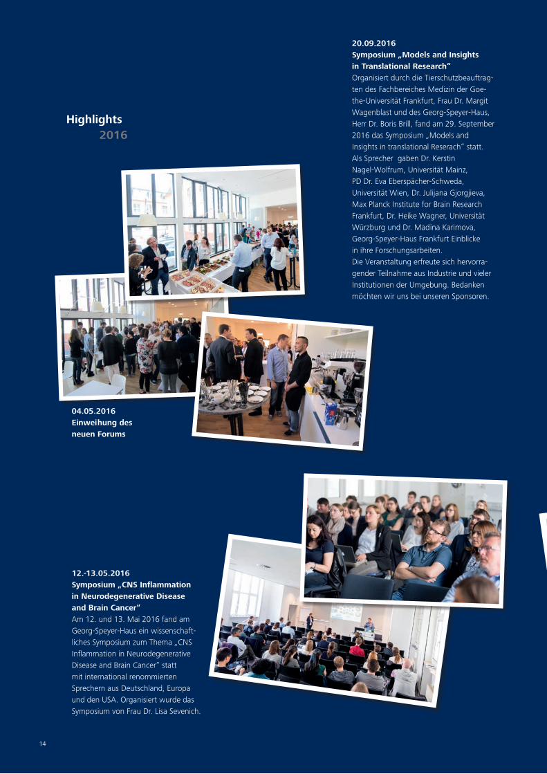
Highlights 2016
20.09.2016Symposium „Models and Insights in Translational Research“Organisiert durch die Tierschutzbeauftrag-ten des Fachbereiches Medizin der Goe-the-Universität Frankfurt, Frau Dr. Margit Wagenblast und des Georg-Speyer-Haus, Herr Dr. Boris Brill, fand am 29. September 2016 das Symposium „Models and Insights in translational Reserach“ statt. Als Sprecher gaben Dr. Kerstin Nagel-Wolfrum, Universität Mainz, PD Dr. Eva Eberspächer-Schweda, Universität Wien, Dr. Julijana Gjorgjieva, Max Planck Institute for Brain Research Frankfurt, Dr. Heike Wagner, Universität Würzburg und Dr. Madina Karimova, Georg-Speyer-Haus Frankfurt Einblicke in ihre Forschungsarbeiten.Die Veranstaltung erfreute sich hervorra-gender Teilnahme aus Industrie und vieler Institutionen der Umgebung. Bedanken möchten wir uns bei unseren Sponsoren.
14
04.05.2016Einweihung des neuen Forums
12.-13.05.2016Symposium „CNS Inflammation in Neurodegenerative Disease and Brain Cancer“Am 12. und 13. Mai 2016 fand am Georg-Speyer-Haus ein wissenschaft-liches Symposium zum Thema „CNS Inflammation in Neurodegenerative Disease and Brain Cancer“ statt mit international renommierten Sprechern aus Deutschland, Europa und den USA. Organisiert wurde das Symposium von Frau Dr. Lisa Sevenich.

15
04.05.2016Feierstunde im Georg-Speyer-Haus zur Enthüllung des Ehrentafeleintrags für Dr. Rolf-E. BreuerIn höchster Anerkennung und Wertschätzung seiner über 40-jährigen Tätigkeit als Mitglied und Vorsitzender des Vorstands und der damit verbundenen
Verdienste um das Georg-Speyer-Haus wurde Herr Dr. Breuer anlässlich seines Ausscheidens aus dem Vorstand am 2. März 2015 zum Ehrenmitglied der Stiftung Chemotherapeutisches Forschungsinstitut Georg-Speyer-Hauses ernannt.Dieser Titel wurde erstmals 1923 an Ludwig Darmstaedter vergeben und letztmalig 1948 an Prof. Dr. Erich Hoffmann, verliehen. Weitere prominente Ehrenmitglieder waren Carl Duisberg, Eduard Beit von Speyer, Karl Herxheimer und Ferdinand Blum. In dieser Tradition wurde der Ehrentafeleintrag von Herrn Dr. Rolf.-E. Breuer am 4. Mai 2016 in einer Feierstunde in Anwesenheit von Herrn Dr. Breuer und der Mitarbeiterschaft des Georg-Speyer-Hauses feierlich enthüllt. Der Direktor des Hauses, Prof. Dr. Florian Greten, würdigte in seiner Ansprache noch einmal die herausragenden Leistungen und Verdienste von Herrn Dr. Breuer für die Stiftung und drückte ihm seine tiefe Dankbarkeit für die langjährige Verbundenheit zum Georg-Speyer-Haus aus.
15
11.09.2016Lauf für mehr ZeitOrganisation Dr. Ursula Dietrich
13.09.2016 (Foto: Uwe Dettmar)Auswahlsymposium zum Paul-Ehrlich- und-Ludwig-Darmstaedter-Nachwuchspreis
Am Dienstag, 13. September fand das Auswahlsymposium zum Paul-Ehrlich-und Ludwig-Darmstädter-Nachwuchspreis 2016/17 im Hörsaal des Georg-Speyer-Hauses statt. Dieser Preis wird von der Paul-Ehrlich-Stiftung einmal jährlich an eine/n promovierte/n Nachwuchswis-senschaftler/in verliehen, die/der an einer Forschungseinrichtung in Deutschland herausragende
Leistungen auf dem Gebiet der biomedizinischen Forschung erbracht hat. Das Preisgeld beträgt bis zu 60.000 Euro und darf ausschließlich forschungsbe-zogen verwendet werden. Die Preisver-leihung findet in Form einer feierlichen Übergabe durch die Stiftung am 14. März 2017 in der Paulskirche in Frankfurt statt.
13.-14.10.20162nd Symposium on Cellular Crosstalk in the Tumor MicroenvironmentAm 13. und 14. Oktober 2016 fand das „2nd Symposium on Cellular Crosstalk in the Tumor Microenvironment“ mit 14 international renommierten Sprechern aus Deutschland und Europa statt. Die „Key Note Lecture“ hielt Prof. Dr. Jeffrey W. Pollard von der University of Edinburgh. Organisiert wurde es von Prof. Dr. Florian Greten.

16

17
Zelluläre Kommunikation in der Stammzellnische Cellular Communication in the Stem Cell Niche
Laboratories I
I

18
Bone marrow microenvironmentDaniela Krause
Die Rolle des Knochenmarksmikromilieus bei den Leukämien
The bone marrow microenvironment (BMM) is increasingly being considered as a novel target to augment existing thera-pies for haematological malignancies. This is important, as the overall survival rate for all leukaemias in adults is only 44% and leukaemic stem cells are rarely eradicated. Eradication of cancer stem cells or leukaemia stem cells in leukaemia, however, is important for cure of a cancer.
Based on our previous work our labora-tory now focuses on various pathways of interaction of leukaemia cells with their surrounding bone marrow microenviron-ment in an effort to eventually target these interactions and eradicate leukae-mic stem cells (LSC). The vascular niche, the extracellular matrix, the coagulation system and novel pathways of adhe-sion to the BMM, studied by various in vitro and in vivo modelling systems, as well as in vivo 2-photon based imag-ing (in collaboration with Prof. S. Dim-meler) form the basis of our studies.
The role of the bone marrow microenvironment in leukaemia
MitarbeiterRahul Kumar Thanh Van HoangDivij VermaSonika GodavarthyJennifer ButenschönJoscha Ender
GruppenleiterinDaniela KrauseTel.: +49 69 63395-500Fax: +49 69 [email protected]
leukaemia
bone marrow microenvironment
pharmacological modulation

19
Bone marrow microenvironmentDaniela Krause
Trotz verbesserter Therapien, z.B. in Form von Tyrosinkinaseinhibi-toren, liegt die 5-Jahres-Überlebensrate bei Erwachsenen für alle Leukämien bei nur 40%. Deshalb hat es sich unsere Arbeitsgruppe zur Aufgabe gemacht, neue Therapien, vor allem solche mit neuem Therapieansatz, zu entwickeln.Wie bereits von uns und anderen Gruppen publiziert, kann eine gezielte Modulation des Knochenmarksmikromilieus (KMMM), dem Ort, wo eine Leukämie entsteht und voranschreitet, eine Ver-ringerung von leukämischen Stammzellen nach sich ziehen. Dies ist notwendig, denn leukämische Stammzellen können zu Thera-pieresistenz und Krankheitsrückfall führen. Das KMMM, welches leukämische Stammzellen vor der Chemotherapie „beschützen“ kann, besteht aus verschiedenen Zelltypen wie Osteoblasten, Osteoklasten, mesenchymalen Stammzellen, Endothelzellen, und der extrazellulären Matrix.
Wir testen experimentell, durch welchen Mechanismus eine Blo-ckade eines auf Endothelzellen exprimierten Proteins, E-Selektin, das Überleben von leukämischen Stammzellen beeinträchtigt und wie die Lokalisation von leukämischen Stammzellen innerhalb des KMMMs und ihre spezifische Interaktion mit dem KMMM den Krankheitsverlauf einer Leukämie beeinflussen kann. Ferner sind die Rolle des Blutgerinnungssystems im KMMM und die Adhäsion von Leukämie-induzierenden Zellen im KMMM Fokus unserer Arbeitsgruppe. Zur Durchführung einer klinischen Studie zur Modulation des KMMMs haben wir in enger Kollaboration mit der Medizinischen Klinik für Hämatologie/Onkologie des Klinikums der Goethe Universität einen Drittmittelantrag gestellt.
As we have previously shown that CD44 and selectins and their ligands play a role for the engraftment of LSC in chronic myelogenous leukaemia (CML) we are now testing, if the E-selectin-specific inhibitor GMI-1271 is beneficial for the reduction of LSC in CML. Indeed, our data indicate that inhibition of E-selectin in murine CML may lead to ‘non-adherence’ to the vascular niche, reduced engraft-ment of leukaemia-initiating cells and possibly an altered cell cycle of LSC, likely via non-adhesion to the vascular niche, a novel finding in leukaemia.
In other work we have shown by confocal 2-photon in-vivo microscopy of the calvarium in live mice that leukaemic stem cells in CML have a distinct location in the bone marrow niche, which is differ-ent from the location of normal haemato-poietic stem cells. Prior in-vitro treatment of leukaemic stem cells with imatinib, considered standard of care in CML, reversed this phenotype. Furthermore, LSC in CML, harbouring the T315I point mutation in BCR-ABL1 (BCR-ABL1T315I), which conveys resistance to imatinib, were found closest to the endosteum.
As patients and mice with CML due to BCR-ABL1T315I have an accelerated clinical course and a worse outcome, we are currently investigating whether an altered interaction with the bone marrow micro-environment via altered signaling from BCR-ABL1 or its respective mutants may be a cause of this. Indeed, in compari-son to BCR-ABL1WT+ cells we have been able to implicate an altered expression of adhesion molecules, increased engraft-ment, increased adhesion, alterations of the actin cytoskeleton and differences in signaling pathways. These studies have led to a novel form of treatment, which significantly prolongs survival and leads to some cures in mice. This has been submitted to the European Patent Office. We are currently working on the initiation of a clinical trial for this novel treatment.
Thirdly, the coagulation system is a com-plex system comprised of various prote-ases and other factors which result in the formation of a blood clot. The bone mar-row microenvironment has been implicat-ed in the maintenance of haematopoietic stem and progenitor cells (HSPC), but pro-teases such as metalloproteinase 9 have
only been implicated in the mobilization of HSPC, but not for HSPC homeostasis.Therefore, we hypothesized that the proteases of the coagulation system may play a role in the normal physiology of the BMM and that modulation of coagulation factors may have an effect on HSPC, for instance via degradation of the extracel-lular matrix and increased mobilization of HPSC. Indeed, by this approach we discovered a prominent role for certain proteins in the BMM which influence the homeostasis and maintenance of HSPC.
In a fourth project we are investigat-ing the role of lipid raft associated molecules for adhesion of leukaemia cells to the BMM. We have found that lipid raft-associated molecules play a prominent role for the engraftment of leukaemia cells, possibly via associa-tion with certain adhesion molecules.
I

20
Bone marrow microenvironmentDaniela Krause
We continue to work on the role of the transcriptional regulator Far upstream element-binding protein 1 (FUBP1) and its role in the development of leukaemia in a productive collabora-tion with the Zörnig Group in a project funded by the Sander Foundation.
Translation into clinical medicineBased on our findings that modulation of bone by parathyroid hormone (PTH) can reduce LSC in CML in transgenic, pharmacological and xenotransplanta-tion models we are applying for funding of an exploratory phase Ib clinical trial. In this trial we intend to treat 20 patients with CML, who have been on a tyrosine kinase inhibitor for at least 6 months, with parathyroid hormone in collaboration with the department of haematology/oncol-
Figure 1.Deposition of extracellular matrix by fibroblasts.
ogy (Prof. H. Serve, Dr. C. Ortmann, Dr. F. Lang) at the university clinic in Frankfurt. Partner sites are Berlin, Munich, Essen/Düsseldorf and Freiburg. Parathyroid hormone (Forsteo® by Lilly) is a drug ap-proved by the federal agency in Ger-many for the treatment of osteoporosis, especially in women at risk for fracture. As mentioned, we are also in the planning stages of a clinical trial for the treatment of patients with imatinib-resistant CML and B-cell acute lymphoblastic leukaemia.
Figure 2.Green fluorescent osteoblastic cells in the bone marrow cavity in a Col2.3kb GFP reporter mouse. The image was taken by 2-photon in-vivo micros-copy.
Figure 3.Green fluorescent mesenchymal stem cells in the bone marrow cavity in a Nestin GFP reporter mouse. The image was taken by 2-photon in-vivo microscopy.

21
Bone marrow microenvironmentDaniela Krause
Ausgewählte Publikationen
Krause DS, Lazarides K, Lewis JB, von Andrian UH, Van Etten RA. Selectins and their ligands are required for homing and engraftment of BCR-ABL1+ leukemic stem cells in the bone marrow nicheBlood 2014; 123(9): 1361-1371 Krause DS, Fulzele K, Catic A, Sun CC, Dombkow-ski D, Hurley MP, Lezeau S, Attar E, Wu JY, Lin HY, Divieti-Pajevic P, Hasserjian RP, Schipani E, Van Etten RA, Scadden DT. Differential regulation of my-eloid leukemias by the bone marrow microenviron-ment. Nature Medicine 2013; 19(11):1513-1517*
Fulzele K*, Krause DS*, Panaroni C, Saini V, Barry KJ, Lotinun S, Baron R, Bonewald L, Feng JQ, Chen M, Weinstein LS, Wu JY, Kronenberg HM, Scadden DT, Divieti-Pajevic P. Myelopoiesis is regu-lated by osteocytes through Gsα-dependent signaling Blood 2013 Feb 7;121(6):930-9.
Krause DS, Lazarides K, von Andrian UH, Van Etten RA. Requirement for CD44 in homing and engraftment of BCR-ABL-expressing leukemic stem cells. Nat Med 2006; 12 (10):1175-80
*co-first authorship
... weitere Publikationen finden Sie auf Seite 59
Figure 4.Transplanted leukaemia-initiating cells, labelled with the CMTMR Dye, in the bone marrow cavity of a Tie2 GFP reporter mouse. Endothelial cells fluoresce in green, bone in blue. The image was taken by 2-photon in-vivo microscopy.
Figure 5.Transplanted leukaemia-initiating cells, labelled with the CMTMR Dye, in the bone marrow cavity of a Col2.3GFP reporter mouse. Osteoblastic cells fluoresce in green, bone in blue. The image was taken by 2-photon in-vivo microscopy.
Figure 6.Transplanted leukaemia-initiating cells, labelled with the CMTMR Dye, in the bone marrow cavity of a Col2.3GFP reporter mouse. Osteoblastic cells fluoresce in green. The image was taken by 2-photon in-vivo microscopy.
In summary, the laboratory focuses on the role of the different constituents of the BMM on the initiation, maintenance and progression of leukaemias in an at-tempt to develop novel therapies which can augment our existing armamen-tarium against this intractable disease.
Other activitiesWe are coorganizers of an International Scientific Workshop on the “Tumor environment in haematological malignancies and its therapeutic targeting” (together with the European School of Haematology).
I

22
Bone Marrow MicroenvironmentHind Medyouf
Die Rolle der Knochenmarksnische bei Myelodysplastischen Syndromen und myeloider Leukämie
The research in our group interrogates the complex biology of hematologic malignancies by investigating both intrinsic and extrinsic mechanisms that control the fate of the hemato-poietic stem/progenitor cells (HSPCs) responsible for disease propagation. Hallmarks of cancer are thought to be acquired via somatic mutational events and/or epigenetic changes that progres-sively provide a fitness advantage over normal cells, in a specific niche context. This leads to the progressive dominance of the tumor cell clone(s). Importantly, mutation-mediated cellular fitness is largely dependent on the status of the surrounding microenvironment, which may reinforce or rather counter select (Epi)-genetically-defined sub-clone(s). Moreover, growing evidence, including from our group, indicates that tumor cells are able to alter their surrounding microenvironment and thereby actively modulate the extrinsic parameters, which in turn dictate the selection for particu-lar features as the disease progresses.
This implies that understanding the evolutionary history of leukemia develop-
Role of the Bone Marrow Microenvironment in Human Myelodysplasia and Acute Leukemia
Bone marrow niche
Cellular crosstalk
Patient-derived xenografts
MitarbeiterIrene Tirado GonzalezMaresa WeitmannAleksandra NevmerzhitskayaAbdelrahman Mahmoud MohammedAdriana Contreras
GruppenleiterinHind MedyoufTel.: +49 69 63395-540Fax: +49 69 [email protected]

23
Bone Marrow MicroenvironmentHind Medyouf
Myelodysplastische Syndrome (MDS) bilden eine heterogene Gruppe hämatologischer Erkrankungen, die von blutbildenden Stammzellen des Knochenmarks ausgehen, und die durch eine unzureichende Bildung reifer Blutzellen charakterisiert sind. Vor allem ältere Menschen sind betroffen. MDS können sich fortschrei-tend verschlechtern und zum Versagen des Knochenmarks führen. Etwa 30% der Betroffenen entwickeln eine sekundäre akute mye-loische Leukämie (sAML), die besonders schwierig zu behandeln ist. Die aktuellen Therapiemöglichkeiten sind sehr begrenzt und beeinflussen nicht den Verlauf der Krankheit. Neue therapeutische Strategien werden daher dringend benötigt, um der wachsenden Herausforderung durch MDS in unserer alternden Gesellschaft zu begegnen.
Meine Arbeiten haben kürzlich gezeigt, dass die zelluläre Mik-roumgebung im Knochenmark, die so genannte Knochenmarks-nische, eine entscheidende Rolle bei der Pathogenese von MDS spielt. Mesenchymale Nischenzellen bilden ein Gerüst, in dem
sich die blutbildenden Zellen entwickeln können. Unsere Arbei-ten ergaben, dass ein komplexer Signalaustausch zwischen den erkrankten hämatopoietischen Zellen und ihren benachbarten Nischenzellen stattfindet. Mesenchymale Nischenzellen von MDS Patienten zeigen eine Reihe von molekularen Veränderungen, die wahrscheinlich zum Krankheitsbild beitragen. Andererseits können prä-leukämische MDS Zellen ihrerseits ihre Nische verändern und eine MDS Stammzellnische etablieren, die nun statt der normalen Blutbildung das Fortschreiten der Krankheit fördert.
Eines der Hauptziele unserer Nachwuchsgruppe ist es, die Mole-küle zu identifizieren, die diesen Signalaustausch vermitteln. Potenzielle Möglichkeiten, in die Interaktion zwischen Nische und hämatopoietischen Zellen einzugreifen, sind bisher kaum unter-sucht. Sie stellen eine sehr attraktive und neuartige Möglichkeit dar, die Nischenfunktion insbesondere bei MDS therapeutisch zu beeinflussen.
ment, how it modulates its microenviron-ment and the means by which it adapts to current therapies to the disadvantage of cancer patients, is essential to devis-ing better therapeutic strategies. There-fore, our group strives to understand the clonal evolution of human leukemia and define the signals involved in the crosstalk between leukemia cells and their microenvironment. Based on the hypothesis that these two components have co-evolved to fulfill the needs of the tumor clone(s), developing thera-peutic approaches that would limit the niche support represent attractive novel therapeutic options that would hamper the growth of the malignant HSPCs, thereby improving patient outcome.
To achieve these goals, we carry out a highly translational research program based on integrative “omics” approaches to characterize primary patient samples, and apply state-of-the-art genetic tools (CRISPR/CAS) and inhibitor studies to evaluate the functional relevance of our findings using in vitro co-culture systems (2D and 3D), as well as our extensive expertise in in vivo modeling of human
diseases using both syngeneic models (Medyouf, Nat. Med, 2007; Medyouf, Blood, 2010; Medyouf, JEM, 2011) and patient-derived xenografts (Gerby, Leuke-mia, 2010; Medyouf, Cell Stem Cell, 2014).
Interrogating the clinical implica-tions of the genetic landscape in human myelodysplastic syndromes Myelodysplastic syndromes are frequent malignant bone marrow disorders of the elderly with limited treatment options and a high risk of progression to bone marrow failure or acute myeloid leukemia that have an aggressive course. These syndromes are characterized by the ineffective production of mature blood cells, leading to peripheral cytopenia that can affect one or several lineages. As such, most MDS patients rely on frequent blood transfusions. Beyond hematopoietic stem cell transplantation, no treatment is able to alter the natu-ral course of this disease. Importantly, phenotypic manifestations and clinical course are highly variable amongst patients with MDS, a phenomenon that poses tremendous challenges in clinical care. We hypothesized that this clinical heterogeneity is likely to be contributed
to by (1) a previously unrecognized complex genetic make-up at the level of individual MDS patients and (2) a disease-associated microenvironment that may further modulate disease course and alter treatment responses.Previous cross-sectional studies investi-gating single time point bone marrow specimens from large MDS patient cohorts have reported that MDS patients carry a plethora of somatically acquired muta-tions that recurrently affect genes involved in important cellular processes with specific patterns of co-occurrence and mutual exclusivity (Haferlach, Leukemia, 2014). These patterns were suggestive of a pathogenic process that mirrors evolutionary paths that govern species evolution, namely mutation and selection acting on individuals in a population. In a collaborative effort with clinical partners, we set out to precisely define this evolutionary process on a patient-specific level, thereby defining specific paths leading to MDS development and MDS recurrence post-treatment (Figure 1). The mutational trajectories that we uncovered provided experimental evidence that in most patients, MDS originates from benign
I

24
founder stem cell clones, still capable of multilineage differentiation, carrying specific genetic alterations affecting RNA splicing and epigenetic modifier genes. Concomitantly, we could demonstrate that in MDS, mutations in signaling and transcription regulator genes as well as cytogenetic lesions are rather observed in advanced subclones generated through both linear and branching evolution-ary paths. In particular, high-throughput molecular monitoring of long-term serial follow-ups allowed us to demonstrate that therapeutic intervention, such as lenalidomide treatment in del(5q)-bearing patients, was immediately followed by significantly elevated fluctuations in both
the oligoclonal BM composition and clini-cal parameters. Although the initial clinical consequences of such therapies were either positive (responders) or, at worst, neutral (in non-responders), in most cases, the BM remained largely clonal due to (1) the rapid outgrowth of founder- or initially minor subclones or (2) the emergence of resistant clones carrying additional muta-tions that significantly expanded under active therapy. New subclones carrying additional mutations appeared to originate from pre-existing ancestral clones, which were likely best fitted to cope with the new environment, i.e insensitive to treat-ment and therefore capable of expanding under therapy (Mossner, Blood, 2016).
Follow-up work on this topic is aiming to :
• Experimentally define the relation-ship between specific genotypes/order of mutational acquisition and phenotypic manifestations
• Explore how the niche impacts the clonal selection process observed during the disease course
• Address the important issue of niche-mediated protection of dis-eased-stem cells.
Bone Marrow MicroenvironmentHind Medyouf
Figure 1.Complex branched and independent evolution promotes dynamic clonal heterogeneity in MDS leading to escape from drug treatment.(right) representative case of clonal evolution in longitudinal follow up analysis of a lenalidomide-treated patient. (left) Schematic view of clonal evolution paths in MDS (Mossner et al, Blood, 2016).

25
Targeting the niche as a new avenue for leukemia therapyAnother active area of research in our group is focusing on the cell extrinsic signals that contribute to the develop-ment and progression of pre-leukemic syndromes (i.e. MDS) and acute leukemias. Indeed, although blood malignancies are believed to be primarily driven by somatic events affecting the hematopoi-etic compartment, hematopoietic stem and progenitor cells behavior is closely modulated by surrounding niche com-ponents. Under physiological conditions, the niche provides instructive cues to maintain self-renewal and adapt blood supply as needed. Aging has been shown to negatively impact the ability of the bone marrow niche to sustain the fitness of HSPCs and proposed to contribute to the progressive decline in HSPCs function, induce immune remodeling and promote oligo-clonal dominance. These age-related changes are thought to provide increas-ing opportunities for altered clones with increased fitness (e.g. due to mutation in genes conferring increased self-renewal such as TET2, DNMT3A, etc) to progres-sively dominate the marrow. This is in
line with the observation that 10-20% of haematologically healthy individuals aged over 70, present with clonal hematopoi-esis dominated by HSC clones that carry specific mutations (e.g. TET2, DNMT3A, ASXL1). Moreover, niche alterations have been shown to contribute to leukemo-genesis and reported in most blood malignancies, with evidence, at least in mice, that alterations in the microenvi-ronment can instigate the development of myeloid neoplasms (Reviewed in Schepers et al, Cell Stem Cell, 2015).
We have recently shown that acquisi-tion of disease-associated molecular features in mesenchymal niche cells, in pre-leukemic syndromes, can be triggered as a consequence of instructive signals emanating from the diseased hemato-poietic compartment. These instructive signals are thought to play an important role in re-shaping the bone marrow niche and convert it into a “homey” environ-ment for genetically altered clone(s) while concomitantly interfering with normal hematopoiesis (Medyouf Cell Stem Cell, 2014 and unpublished data).
Our work, currently aims to (1) explore the molecular impact of these niche changes on normal and malignant hematopoiesis, (2) identify the nature of the instructive signal responsible for the niche re-shaping and (3) evaluate the therapeutic benefit of targeting the specific cross-talk between niche and hematopoietic cells in differ-ent pre-leukemic and leukemic contexts.
ConclusionDeciphering the complex interplay between niche and hematopoietic cells in normal and disease contexts is increasing our understanding of disease pathogenesis and providing us the knowledge neces-sary to devise new strategies to specifically dampen the niche support towards malig-nant cells while concomitantly improving the support towards normal HSPCs.
Our work is supported by institutional funds from the GSH, a starting grant from the European Research Council and a José Carreras Career Award. In addi-tion, we are very excited about new research partnerships with pharmaceuti-cal partners to move forward with the translation of our preclinical findings.
Bone Marrow MicroenvironmentHind Medyouf
Ausgewählte Publikationen
Mossner M, Jann JC, Wittig J, Nolte F, Fey S, Nowak V, Obländer J, Pressler J, Palme I, Xanthopoulos C, Boch T, Metzgeroth G, Röhl H, Witt SH, Dukal H, Klein C, Schmitt S, Gelß P, Platzbecker U, Balaian E, Fabarius A, Blum H, Schulze TJ, Meggendorfer M, Haferlach C, Trumpp A, Hofmann WK, Medyouf H*, Nowak D*. Mutational hierarchies in myelodysplastic syndromes dynamically adapt and evolve upon therapy response and failure.Blood. 2016 Sep 1;128(9):1246-59.
Medyouf H, Mossner M, Jann JC, Nolte F, Raffel S, Herrmann C, Lier A, Eisen C, Nowak V, Zens B, Müdder K, Klein C, Obländer J, Fey S, Vogler J, Fabarius A, Riedl E, Roehl H, Kohlmann A, Staller M, Haferlach C, Müller N, John T, Platzbecker U, Metzgeroth G, Hofmann WK, Trumpp A, Nowak D.Myelodysplastic Cells in Patients Re-program Mesenchymal Stromal Cells to Establish a Transplantable Stem Cell-Niche Disease Unit. Cell Stem Cell. 2014. Jun 5;14(6):824-37.
Medyouf H, Gusscott S, Wang H, Tseng JC, Wai C, Nemirovsky O, Trumpp A, Pflumio F, Carboni J, Gottardis M, Pollak M, Kung AL, Aster JC, Holzen-berger M, Weng AP. High Level IGF1R Expression is Required for Leukemia-Initiating Cell Activity in T-ALL and is Supported by Notch Signaling. J. Exp. Med. 2011. 208(9): 1809-22.
Medyouf H, Alcalde H, Berthier C, Guillemin MC, et al. Targeting Calcineurin Activation as a Therapeutic Strategy for Lymphoid Malignancies. Nat Medicine. 2007. (13): 736-741. 2007.
* Co-senior authors... weitere Publikationen
finden Sie auf Seite 59
I

26
Regulation and deregulationMartin Zörnig
Regulationsmechanismen des programmierten Zelltodes (Apoptose)
Alteration of the control mechanisms of cell death contributes to the pathogenesis of many human diseases, including cancer and neurodegenerative diseases. Our group is interested in identifying novel anti-apoptotic oncoproteins that are responsible for tumor initiation, progres-sion and/or therapy resistance in particular cancer entities. We are analyzing the precise molecular mechanisms of how these proteins inhibit cell death and support tumor growth, and we are validating their potential as targets for future molecular therapies. At the same time, we are investigating their physiologi-cal role in vivo to predict potential side effects of molecular targeting strategies.
Identification of novel mam-malian proteins that regulate apoptosome activitySeveral apoptotic stimuli, including cancer treatment regimens of radiation and chemotherapy, ultimately result in the activation of the mitochondrial apoptosis pathway. Inhibition of this pathway allows malignant cells to evade cell death successfully during tumorigenesis
Regulation and deregulation of apoptosis
Regulation of the mitochondrial apoptosis pathway
Targeting of anti-apoptotic oncoproteins
Analysis of the transcriptional FUBP1 network
MitarbeiterKatharina GerlachViktoria von MansteinVan Thanh HoangStefanie HauckJosephine WeselyMarlene SteinerSusanne Bösser
GruppenleiterMartin ZörnigTel.: +49 69 63395-115Fax: +49 69 [email protected]
27
Regulation and deregulationMartin Zörnig
Unsere Arbeitsgruppe beschäftigt sich mit der Identifizierung und Analyse neuer anti-apoptotischer Onkogene, die für die Tumo-rentstehung sowie für Therapieresistenzen verantwortlich sind. In der Vergangenheit konnten wir in einem selbst entwickelten „Hefe-Survival-Screen“ mehrere interessante anti-apoptotische Kandidatengene identifizieren, die in bestimmten Tumorentitä-ten überexprimiert werden. Wir untersuchen diese Moleküle in Zellkulturexperimenten und in geeigneten Mausmodellen in vivo daraufhin, welche Rolle sie während der Tumorentstehung und -progression spielen, und ob sie sich als Zielstrukturen für zukünftige
molekulare Krebstherapien eignen. Parallel sind wir auch daran inte-ressiert herauszufinden, welche Funktionen diese Gene im gesun-den Organismus ausüben. Letzteres erlaubt auch die Abschätzung möglicher Nebenwirkungen bei molekularen Therapien, in deren Verlauf die tumorrelevante Funktion solcher Gene gestört werden soll. Für ein „Targeting“ geeigneter Kandidatengene bzw. der ent-sprechenden Onkoproteine versuchen wir, kleine inhibitorische Moleküle und shRNA-basierte Strategien für therapeutische Zwecke zu entwickeln, um eine Resistenzentwicklung der Tumorzellen zu umgehen und diese für weitere Behandlungen zu sensibilisieren.
I
and to acquire therapy resistance which represents the biggest challenge in cancer treatment. Therefore, it is important to identify and characterize novel anti-apoptotic proteins that inhibit Caspase-9 activation within the apoptosome complex during execution of the intrinsic mitochon-drial apoptosis pathway. In several screens, we isolated human genes that inhibit cell death at the level of the apoptosome, and we identified promising candidates as potential targets for cancer therapy.
FUBP1 binds to single-stranded DNA and regulates the expression of various target genes. Transcription of the proto-oncogene c-myc for example is activated by FUBP1, while the cell cyle inhibitor gene p21 is repressed by the same protein. We isolated FUBP1 from a breast carcinoma-derived cDNA library and studied its involvement in tumorigenesis. FUBP1 is overexpressed in more than 80% of hepatocellular carcinomas (HCCs) and supports HCC tumor growth by inhibiting anti-proliferative and pro-apoptotic genes. We are currently developing small molecule inhibitors of FUBP1 for HCC therapy, and we are testing these
inhibitors in several different systems, including liver organoid cultures and an orthotopic HCC transplantation mouse model (Fig. 1). In parallel studies, we are analyzing the physiological function of FUBP1 in suitable mouse models. Our experiments unraveled a crucial role of FUBP1 for the maintenance and self-renewal of both, adult and fetal, hema-topoietic stem cells (HSCs) by regulating relevant target genes. The transcriptional FUBP1 network is also involved in the differentiation of embryonic stem cells into cells of the mesoderm germ layer.
The anti-apoptotic protein AVEN was also isolated in one of our functional screenings, and it interacts with various regulators of apoptosis, such as the apoptosome adaptor protein APAF-1. We could demonstrate that AVEN requires proteolytic processing by the lysosomal protease Cathepsin D to unleash its full anti-apoptotic potential, thereby implying that AVEN may be involved in the lysosomal apoptotic pathway.
Published data indicate a strong association between poor prognosis in
acute childhood lymphoblastic leukemia (ALL) and AVEN expression, suggesting that AVEN has oncogenic activity. We established a transgenic mouse line with T cell-specific overexpression of the full-length AVEN protein, which acceler-ates leukemogenesis in heterozygous p53+/- knockout mice. Moreover, the downregulation of AVEN in T-ALL cell lines reduces tumor growth in xenograft experiments. Both findings demonstrate the significant oncogenic potential of AVEN. Our initial results using AVEN knockdown breast cancer cell lines also support a tumor-promoting role of AVEN in mamma carcinoma. In murine mam-mary glands, AVEN expression increases during pregnancy and lactation, before it is downregulated again when the tissue is removed by involution (Fig. 2).
AVEN has also been implicated in both DNA repair and the activation of ataxia telangiectasia mutated (ATM) protein kinase, a major regulator of the cell cycle and the DNA damage response. We developed and analyzed a constitutive Aven-/- knockout mouse model, and the results suggest that AVEN plays a vital

28
role in embryonic development. Lack of AVEN expression leads to accumulation of DNA damage and growth arrest, thereby resulting in embryonic lethality at approximately day E9.5. To further study the physiological role of AVEN in adult mouse organs and tissues, and to inves-tigate its oncogenic function in leukemia and breast carcinoma, we established
conditional Aven-/- knockout mice.
The detailed analyses of FUBP1 and AVEN activity will help to further clarify the mechanism by which apoptosome assem-bly and Caspase-9 activation are regulated in response to mitochondrial Cytochrome c release. Based on this knowledge, we aim to develop inhibitors for therapeutic
intervention targeting the anti-apoptotic activities of these molecules.
Recently, long non-coding RNAs have been shown to promote both tumor suppression and oncogenesis in a variety of tumor entities. MALAT1 (metastasis-associated lung adenocarcinoma transcript 1) is a long non-coding RNA of 8 kb that
Regulation and deregulationMartin Zörnig
Figure 1.Murine orthotopic HCC trans-plantation tumor model. Mice were injected with the murine HCC cell line Hepa129. Already two weeks after injection, visible tumors were established. Tumor growth was monitored by magnetic resonance imaging (MRI) fifteen days after tumor cell injection. Left panel: transverse plane; right panel: sagittal plane; arrow: liver; arrowhead: tumor. Sixteen days after tumor cell injection, the mouse was sacrificed and the tumor-bearing liver lobe was extracted. Histological analyses were per-formed to further characterize the tumor. Left panel: hematoxylin/eosin staining to visualize nuclei (blue) and cytoplasm (red); right panel: anti-Ki67 antibody staining to detect proliferating cells (brown).
A
C
B
D
A
B
C
D

29
I
Regulation and deregulationMartin Zörnig
Ausgewählte Publikationen
Rabenhorst U¹, Thalheimer FB¹, Gerlach K¹, Kijonka M, Böhm S, Krause DS, Vauti F, Arnold HH, Schroeder T, Schnütgen F, von Melchner H, Rieger MA¹, Zörnig M1. Single-stranded DNA-bin-ding transcriptional regulator FUBP1 is essential for fetal and adult hematopoietic stem cell self-renewal. Cell Rep. 2015, 11:1847-55.
Eißmann M1, Melzer IM1, Mateus Fernández SB1, Michel G, Hrabě de Angelis M, Hoefler G, Finkenwirth P, Jauch A, Schoell B, Grez M, Schmidt M, Bartholomae CC, Newrzela S, Haetscher N, Rieger MA, Zachskorn C, Mittelbronn M, Zörnig M. Overexpression of the anti-apoptotic protein AVEN contributes to increased malignancy in hematopoietic neoplasms. Oncogene 2013, 32: 2586-91.
Rabenhorst U, Beinoraviciute-Kellner R, Breznicea-nu ML, Joos S, Devens F, Lichter P, Rieker RJ, Trojan J, Chung HJ, Levens DL, Zörnig M. Overexpression of the Far Upstream Element Binding Protein FBP1 in hepatocellular carcinoma is required for tumor growth. Hepatology 2009, 50: 1121 – 9.
¹ these authors contributed equally to the work
... weitere Publikationen finden Sie auf Seite 60
has been reported to be overexpressed in various human solid carcinomas. MALAT1 has been linked to gene regulation, and it seems to play an important role in metas-tasis. We isolated several MALAT1 cDNA clones from melanoma- and leukemia-derived tumor libraries in our functional survival screens. Cell culture experiments confirmed increased apoptosis rates in
the absence of MALAT1 expression. We established a conditional Malat1 knockout mouse model that is currently being used to study the physiological function of Malat1 and its influence on tumor development, progression, and metastasis. Interestingly, homozygous Malat1-/- knock-out mice are born and appear normal, suggesting that Malat1 is not required for
normal murine development. Currently, we are investigating the role of Malat1 in hematopoietic stem cells and leukemia.
Figure 2.Dynamic regulation of AVEN expression during the develop-ment of the murine mammary gland.Mammary glands of adult female mice at different stages during pregnancy were dissected and fixed in formalin. Immunohistochemistry was performed on paraffin sections with an anti-AVEN antibody. The staining shows AVEN expression in the luminal epithelial cells lining the inner surface of the ducts. virgin, pregnancy day 15.5, lactation day 1, lactation day 10, involution day 1. Scale bar (shown below panel ): 50 µm in , 100 µm in - . AVEN expression in protein lysates of the mammary glands was quantified by western blotting and normalized to Cytokera-tin-18 expression. AVEN expression increases during pregnancy and lactation when the epithelium of the mammary gland is expanding, and it declines after weaning during involu-tion of the mammary gland.
A B C
D
A B C
FEDE
AD
B E F

30

31
Zell-Zell Interaktionen im Tumorstroma Cell-Cell Interaction in the Tumor Stroma
II
Laboratories II

32
Diet and Cancer LinkMelek Canan Arkan
Ernährung und Krebs
From Metabolism to Post-transla-tional Modifications and MicrobiotaDiet-induced obesity is a major risk factor for the development of cancer. Although alterations in inflammatory and bioenergetic pathways are critical in linking excessive weight gain to cancer, there is now sufficient evidence to also suggest disease progression may indeed have more to do with the diet itself than increased obesity per se. The diet is shaped by multiple diverse factors such as culture, nutritional knowledge, price, availability, taste and convenience. With our current knowledge of the importance of the reciprocal interaction between host and environmental factors in selecting a microbiota that favours carcinogenesis, the food consumption is critical. Given the distinct shifts in agriculture and changes in crops, food may have a pivotal role in aggravating disease. Our laboratory focuses on how changing diet is associated with cancer initiation and progression in the pancreas and intestine at a molecular and cellular level using mouse models with oncogene activation. We aim at dwelling how inflammatory cells cross-talk to host during disease
Diet and Cancer Link
Diet as a major risk factor for cancer
Metabolic vulnerabilities in host and tumor cells
Microbial community changes and disease susceptibility
GruppenleiterinMelek Canan ArkanTel.: +49 69 63395-600Fax: +49 69 [email protected]
MitarbeiterVeronika LiptakovaRoshni Lakra

33
Diet and Cancer LinkMelek Canan Arkan
Ernährungsbedingte Fettleibigkeit ist ein wesentlicher Risikofaktor für die Entstehung von Krebs. Obwohl Veränderungen in bioener-getischen und inflammatorischen Signalwegen mit der Entstehung von Krebs durch übermäßigen Gewichtszuwachs zusammenhän-gen, liegen nun ausreichende Hinweise vor, dass der Krankheits-verlauf vielmehr mit der Ernährungsweise in Verbindung stehen könnte und nicht nur mit erhöhter Fettleibigkeit per se. Viele ver-schiedene Faktoren wie beispielsweise die Kultur, das vorhandene Wissen über Ernährung, Preis und Verfügbarkeit von Nährstoffen, Geschmack sowie Zweckmäßigkeit der Nahrung beeinflussen die Art der Ernährung. Da bekannt ist, dass die Wechselbeziehung zwischen Wirt und auf diesen einwirkenden Umweltfaktoren für die Begünstigung eines für die Krebsentstehung vorteilhaften Mikrobioms entscheidend ist, kommt der Nahrungsaufnahme eine maßgebliche Bedeutung zu. Angesichts der massiven Ver-schiebungen in Landwirtschaft und Anbaukulturen könnte die
Ernährungsweise von großer Relevanz bei der Verschlimmerung von Krankheiten sein.
Unser Labor erforscht, wie eine Veränderung der Ernährung mit der Entstehung und dem Fortschreiten von Krebs in der Bauchspeichel-drüse und im Darm in Zusammenhang steht. Ziel ist zu verstehen, wie entzündliche Zellen mit anderen Zellen in der Mikroumgebung des Tumors in wechselseitigem Kontakt stehen, welche Störungen im Energiemetabolismus von Wirt und Tumor hervorgerufen werden, ob ein geänderter Status post-translationaler Modifikati-onen eine ursächliche Rolle spielt und ob oder wie das Mikrobiom in die geschilderten Prozesse involviert ist. Letztlich streben wir an, unsere Studien direkt in klinische Anwendungen umzusetzen.
Unsere Gruppe wird vom Institut für Biochemie II, Goethe Univer-sität Frankfurt finanziert.
progression, what sort of derangements are taking place in both host and tumor energy metabolism, whether altered post-translational modifications play a
II
Figure 1.Interaction of environmental factors with genetic factors under disease context involves whole lot of alterations in the host and tumor cell bioenergetics. Our studies focus on the critical derangements in energy metabolism during disease initiation and progression, which may set the stage for future drug discovery aimed at developing therapeutic interventions against diet-induced cancers.
causative role and if/how microbiota is involved in these processes. Once defined in depth, the next step will be to set the stage for direct interference with any of
these processes and to test the possibility for developing therapeutic interventions in mice. Our ultimate goal will be direct translation of our studies into the clinic.

34
Diet and Cancer LinkMelek Canan Arkan
Diet-induced Metabolic Derangements in CancerCancer is one of the most common cause of death worldwide and is defined by dysregulation of signaling pathways that orchestrate proliferation, cell death, tumor-promoting inflammation and energy metabolism. Although there has been tremendous effort in the past decades, therapeutic approaches that culminate on specific inhibition of sustained proliferation and resistance to cell death have not been fully successful. Since obesity is associated closely with cancer development and increased BMI positively correlates with the mortality rates, tumor energy metabolism may represent a challenging new field. Our studies focus on elucidating the host and tumor energy metabolism under a state of increased energy consumption. We aim to delineate the critical alterations that take place during disease initiation and progression, which may have a diagnostic value in pancreatic cancer.
Diet, Glycosylation and CancerScreening methods and early prevention options in determining disease risk are only partially successful leading colorectal cancer to be the third most common cause of cancer-associated death with a high mortality rate due to increased metastasis at time of diagnosis or in a later stage of the disease. Tumor cells display a wide range of glycosylation changes compared to non-transformed tissue. Thus, we aim at elucidating glycan structures and their contribution to disease development. Since glycosylation is a reversible modification and highly susceptible to environmental factors, characterizing glycans under a diet context can set the stage for early detection biomarkers and future drug discovery aimed at developing therapeutic interventions against intestinal cancer.
Diet-associated Changes in Microbiota and CancerHuman gut is inhabited by billions of bacteria contributing majorly to the regulation of metabolic functions and immune homeostasis. Bacterial interaction with host cells is a complex and dynamic process involving a variety of bacterial cell surface layers and a host of cell receptors. Given the reciprocal interaction between host and diet in selecting a microbiota that favours carcinogenesis in our previous studies, we aim at addressing community changes and bacterial metabolites during tumorigenesis in the gut. The effect of dietary interventions designed to impact microbial community and function during disease development will be our ultimate goal after a full assessment of what nutrient components exist for the gut community and what metabolites of which function are being produced.
Our research group is funded by the Institute of Biochemistry II, Goethe University Frankfurt.

35
II
Ausgewählte Publikationen
Arkan MC. ‘Cancer: Fat and fate of pancreatic tu-mours’. Nature, Aug 11; 536(7615):157–8, 2016.
Schulz MD*, Atay C*, Heringer J*, Romrig FR, Schwitalla S, Aydin B, Ziegler PK, Varga J, Reindl W, Pommerenke C, Salinas-Riester G, Böck A, Alpert C, Blaut M, Polson SC, Brandl L, Kirchner T, Greten FR, Polson SW and Arkan MC. ‘High-fat-diet-mediated dysbiosis promotes intestinal carcinogenesis independently of obesity’. Nature, Oct 23; 514(7523): 508–512, 2014.
Khasawneh J, Schulz MD, Walch A, Rozman J, Hrabe de Angelis M, Klingenspor M, Buck A, Schwaiger M, Saur D, Schmid RM, Klöppel G, Sipos B, Greten FR, Arkan MC. ‘Inflammation and mitochondrial β-oxidation link obesity to pancreatic cancer’ Proc. Natl. Acad. Sci. USA, Mar 3; 106(9): 3354–9, 2009.
... weitere Publikationen finden Sie auf Seite 61
Figure 2. Diet is vital but excess intake or imbalanced macro- and micronutrient constituent of it can employ deleterious effects on cancer development. Interaction of environmental factors with genetic factors under disease context involves whole lot of changes in the host and tumor cell signaling, metabolites, crosstalk to bacterial community that colonizes our gut or to immune cells of the tumor microenvironment. This critical crosstalk based on the profile, function and expressed activities of cells can help define the ultimate anti-tumorigenic response in cancer aimed at developing therapeutic interventions against diet-induced cancers.
Diet and Cancer LinkMelek Canan Arkan
Bacterialmetagenomics
Post-translationalmechanisms
Energymetabolism
Cellularcrosstalks

36
Microenvironmental crosstalkHenner Farin
Gewebsinteraktionen und Signalmechanismen im Darmkrebs
Colorectal cancer (CRC) is the third leading cause of death from cancer among adults in Germany. Cancer genome sequencing programs have identified a heterogeneous spectrum of oncogenic mutations in patients. However, we currently cannot predict therapeutic responses based on the genetic composition of a tumor. This is due to the complexity of cell signaling processes that involve an intrinsic crosstalk between all tissue compartments. Our lab explores the 3-D ‘organoid’ culture system as a solid tumor model. This system allows expansion of primary intestinal stem cells under full control of the exogenous ‘microenvironment’. Exposure to microen-vironmental signals such as inflammatory cytokines, growth factors, microbial and metabolic cues are analyzed to dissect tumor-specific vulnerabilities. Our goal is to understand how tumor cells respond to and influence their microenvironment to be able to specifically target this crosstalk.
The organoid culture system as a human colorectal cancer modelTumor cells successively acquire mutations that confer unrestricted local growth before progression to metastatic disease.
Signaling crosstalk in the colon cancer microenvironment
3-D epithelial cultures from endoscopic biopsies
Paracrine signaling mechanisms of the intestinal stem cell niche
Targeting of the colon cancer microenvironment
MitarbeiterMohammed MosaBirgitta MichelsMoyo GrebbinConstantin MencheTahmineh Darvishi
GruppenleiterHenner FarinTel.: +49 69 63395-520Fax.: +49 69 [email protected]

37
Microenvironmental crosstalkHenner Farin
Unsere Arbeitsgruppe am Georg-Speyer-Haus erforscht die zellulä-ren und molekularen Vorgänge bei der Entstehung von Darmkrebs. Insbesondere interessiert uns die Kommunikation verschie dener Zelltypen in der unmittelbaren Umgebung des Tumors, dem so genannten „Tumor-microenvironment“. Dabei nutzen wir „Organoide“, ein neuartiges dreidimensionales Gewebekultur-System. Organoide können unter definierten Kulturbedingungen aus humanen Darm-Stammzellen etabliert werden und bilden Darmepithel-spezifische Strukturen wie Krypten (Furchen) oder Villi (Zotten) aus (so genannte „Mini-Därme“). Dieses System ermög-licht die Expansion von Stammzellen in einem Gewebe-ähnlichen Zustand, was die Untersuchung von molekularen Signalen in einer definierbaren Mikroumgebung ermöglicht. So kann z.B. durch Zugabe von nicht-epithelialen Zellen wie Fibroblasten, Gefäß- oder
Immunzellen der Organkontext nachgebildet werden. Im Mittel-punkt unserer Forschung steht die genetische Analyse der Ent-stehung und Progression des Darm-Karzinoms sowie der Einfluss körpereigener Abwehrmechanismen wie Entzündungsreaktionen. Dazu werden in klinischer Kollaboration Tumorbiopsien expandiert um Patienten-spezifische Signalmechanismen zu identifizieren. Mit Hilfe von genetischen Techniken versuchen wir zu verstehen wie ein-zelne onkogene Mutationen den zellulären Phänotyp beeinflussen, als Ansatzpunkt für zukünftige Therapien.
Unsere Gruppe wird vom Deutschen Krebsforschungszentrum (DKFZ) im Rahmen des Deutschen Konsortiums für Translationale Krebsforschung (DKTK) am Georg-Speyer-Haus finanziert.
Although great advances have been achieved in prevention and therapy, the clinical options for progressed CRC patients are still very limited. Metastatic colonization to distant sites, which are mainly the liver and the lungs, often precludes surgical and radiological inter-vention. The response rates to classical chemotherapy and targeted therapies are modest with a high degree of relapse. To better predict drug efficiency and prevent development of resistance, new tumor models are required that recapitulate the signaling processes in CRC. Currently used transformed cell lines have lost important traits of tumor cells and cannot fully reflect the heterogenic nature of the disease. Species barriers limit the use of animal models to test genetic hypotheses.
The ‘organoid’ culture system (first described by Sato et al., 2009 in Nature) allows expansion of gastrointestinal stem cells over long periods ex vivo. In a 3-D extracellular matrix epithelial structures are formed that undergo continuous self-renewal and differentia-tion, recapitulating a normal crypt-villus architecture (Figure 1). Organoids can
Figure 1.Organoid cultures recapitulate the intestinal stem cell nicheSchematic illustration of the intestinal organoid culture system. The medium composition mimics the stem cell niche environment in the intestine, which is characterized by high WNT/EGF and low BMP signals. In 3-D Matrigel ‘mini guts’ are formed that consist of a single-layered epithelium. Proliferation occurs in the crypt–like structures, each composed of a stem cell niche compartment.
II

38
be established from endoscopic patient biopsies of normal and tumor tissue. Importantly, the culture conditions preserve cells in a native state and stem cell niche signals have to be supplemented with the culture medium. This allows functional selection for presence of oncogenic mutations e.g. affecting the WNT, BMP or the EGF pathways. In our lab we exploit and further improve this technological platform as a CRC model.
Characterization of the normal and tumorigenic stem cell nicheIn the normal intestine, the Wnt pathway controls epithelial self-renewal and differentiation. Constitutive Wnt pathway activation is the main driver of CRC affecting >90% of all cases. We have shown previously that mouse intestinal organoids produce Wnt3 by a specific cell type, the Paneth cells, and moreover critically depend on Wnt3 as an epithelial
niche signal (Farin et al., Gastroenterol-ogy 2012). Thus organoids provide a unique and accessible experimental system to investigate the mechanisms that confine Wnt signaling. By combination of mouse transgenesis, in vitro manipulation of organoids and confocal microscopy we could recently demonstrate that Wnt3 represents a short-range signal that is directly transferred from Paneth cells to the adjacent stem cells (Farin et al.,
Figure 2.Localized Wnt signals compartmentalize stem cells and differentiation in intestinal organoids Endogenous Wnt3 protein is produced by Paneth cells and acts as a niche factor. Confocal microscopy shows membranous distribution of Wnt3 that covers the crypts. Model for the short-range action of the Wnt3. Niche contact-dependent transfer and limited diffusion result in a Wnt3 gradient that orchestrates self-renewal and
differentiation.
A
A
B
B
Microenvironmental crosstalkHenner Farin
39
Microenvironmental crosstalkHenner Farin
Ausgewählte Publikationen
Farin HF*, Jordens I, Mosa MH, Basak O, Korving J, Tauriello DVF, de Punder K, Angers S, Peters PJ, Maurice MM, Clevers H.* (2016). Visualization of a short-range Wnt gradient in the intestinal stem-cell niche. Nature 530, 340 – 343.
Tetteh PW, Farin HF, and Clevers H. (2015). Plasticity within stem cell hierarchies in mammalian epithelia. Trends in Cell Biology 25, 100 – 108.
Yin X, Farin HF, Es JH, Clevers H, Langer R, Karp JM. (2014) Niche-independent high-purity cultures of Lgr5+ intestinal stem cells and their progeny. Nature Methods, 11: 106 – 112
Bigorgne AE**, Farin HF**, Lemoine R**, Mahlaoui N, Lambert N, Gil M, Schulz A, Philippet P, Schlesser P, Abrahamsen TG, Oymar K, Davies EG, Ellingsen CL, Leteurtre E, Moreau-Massart B, Berrebi D, Bole-Feysot C, Nischke P, Brousse N, Fischer A, Clevers H, de Saint Basile G. (2014) TTC7A muta-tions disrupt intestinal epithelial apicobasal polarity. Journal of Clinical Investigation, 124: 328 – 237.
* co-corresponding authors** co-first authors
... weitere Publikationen finden Sie auf Seite 61
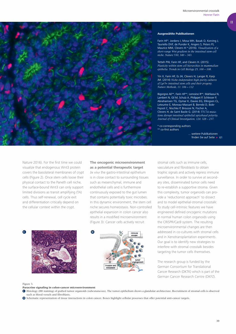
Nature 2016). For the first time we could visualize that endogenous Wnt3 protein covers the basolateral membranes of crypt cells (Figure 2). Once stem cells loose their physical contact to the Paneth cell niche, the surface-bound Wnt3 can only support limited divisions as transit amplifying (TA) cells. Thus self-renewal, cell cycle exit and differentiation critically depend on the cellular context within the crypt.
The oncogenic microenvironment as a potential therapeutic targetIn vivo the gastro-intestinal epithelium is in close contact to surrounding tissues such as mesenchymal, immune and endothelial cells and is furthermore continuously exposed to the gut lumen that contains potentially toxic microbes. In this dynamic environment, the stem cell niche secures homeostasis. Non-controlled epithelial expansion in colon cancer also results in a modified microenvironment (Figure 3): Cancer cells actively recruit
stromal cells such as immune cells, vasculature and fibroblasts to obtain trophic signals and actively repress immune surveillance. In order to survive at second-ary sites, disseminated tumor cells need to re-establish a supportive stroma. Given this complexity, tumor organoids can pro-vide a ‘reductionist approach’ to dissect and to model epithelial-stromal crosstalk: To study cell-intrinsic features we have engineered defined oncogenic mutations in normal human colon organoids using the CRISPR/Cas9 system. The resulting microenvironmental changes are then addressed in co-cultures with stromal cells and in Xenotransplantation experiments. Our goal is to identify new strategies to interfere with stromal crosstalk besides targeting the tumor cells themselves.
The research group is funded by the German Consortium for Translational Cancer Research (DKTK) which is part of the German Cancer Research Centre (DKFZ).
Figure 3.Paracrine signaling in colon-cancer microenvironment Histology (HE staining) of grafted tumor organoids (subcutaneous). The tumor epithelium shows a glandular architecture. Recruitment of stromal cells is observed
such as blood vessels and fibroblasts. Schematic representation of tissue interactions in colon cancer. Boxes highlight cellular processes that offer potential anti-cancer targets.D
C
C D
II

40
Cell PlasticityFlorian Greten
Zellplastizität im Mikromilieu des Kolonkarzinoms
Colorectal cancer (CRC) belongs to the most frequent types of cancer and is the second most commonly diagnosed cancer and the second leading cause of cancer death among cancers that affect both men and women. In 2008 the CRC incidence was 70,000 and about 27,500 people died of the disease and it is estimated that still more than 26,000 patients have died of CRC in Germany in 2011. These numbers illustrate that CRC is a major health care problem that puts a significant burden on the national health care systems. While the vast majority of CRC are of sporadic origin, about 3-5% of develop in the context of chronic inflammation in patients suffering from inflammatory bowel disease (IBD). While the spectrum of genetic alterations within tumor cells as well as the order of mutational events may differ between sporadic and inflammation-associated carcinogenesis, over the last decade it has become increasingly evident that both forms of cancer develop an inflammatory microenvironment that drives the different stages of carcinogenesis. Besides local resident cells, such as vascular cells and fibroblasts colon cancers are infiltrated
Cell Plasticity in the Intestinal Tumor Microenvironment
Cell-cell interaction is essential for tumorigenesis
Inflammation controls cell plasticity of tumor and stromal cells
De-differentiation and EMT are controlled by the microenvironment
MitarbeiterÖzge CanliFatih CeticiChristin DanneilNatalia DelisTiago de OliveiraFabian FinkelmeierJalaj GuptaHana KunkelBirgit LehmannKathleen MohsTobias NeumannAdele NicolasMarina PesicMallika RamakrishnanEva RudolfJulia VargaPaul Ziegler
GruppenleiterFlorian R. Greten, DirektorTel.: +49-69-63395-232Fax: [email protected]

41
Cell PlasticityFlorian Greten
Der Fokus unserer Forschung liegt auf der funktionellen Analyse des Mikromilieus im Kolonkarzinom. Hierbei konzentrieren wir uns auf verschiedene Signalkaskaden, welche die Transkription von Genen regulieren, die für Zytokine, pro-und anti-apoptotische Pro-teine sowie Zellzyklusregulatoren kodieren. Mit Hilfe konditionaler Knockout-Mäuse, erreichen wir eine zelltypspezifische Inaktivierung verschiedener Signalwege in Darmepithelzellen, Immunzellen und Stromazellen im Tumor. Mit diesen Mäusen führen wir funktionelle Untersuchungen zur Rolle dieser Signalwege in den entsprechenden Zelltypen sowie deren Effekte auf benachbarte Zellen während der Tumorentstehung als auch während der Therapie von etablierten Tumoren durch. Zell-Zell Interaktionen spielen eine besonders große Rolle in der Entwicklung einer Zellplastizität sowohl in Tumorzellen als auch in lokalen und rekrutierten Stromazellen. Der Tumor benö-
tigt ein hohes Maß an Plastizität dieser verschiedenen Zellen und schafft sich die geeigneten Voraussetzungen dafür während aller Stadien seiner Entwicklung: Initiation, Promotion und Progression (Invasion und Metastasierung). Seit vielen Jahren beschäftigen wir uns mit der systematischen Analyse des entzündlichen Tumormik-romilieus im Kolonkarzinom und untersuchen insbesonders, welche Rolle der IκB-kinase (IKK) Komplex oder andere Signalwege wie z.B. die gp130-abhängige Stat3 Aktvierung für die Zellplastizität spielt. Es gelang uns eine essentielle Funktion der klassischen IKKβ-abhängigen NF-κB Aktivierung in Tumorzellen sowie in Immunzellen und Fibroblasten während der verschiedenen Tumorstadien nach-zuweisen. Auch Stat3 übernimmt sehr wichtige Funktionen in der Plastizität von Epithelzellen die sowohl für die Tumorentstehung als für die Tumorinvasion und Metastasierung essentiell sind.
by a range of bone marrow-derived cells that contribute to an expanding stroma. This microenvironment contains cells of the innate immune system such as tumor- associated macrophages (TAMs), neutrophil granulocytes, myeloid-derived suppressor cells (MDSCs) or immature myeloid cells (iMCs), mast cells (MCs), and dendritic cells (DCs). It also contains cells of the adaptive immune system, such as T and B cells. This complex network of immune cells in the tumor stroma affects basically every aspect of tumor biology by secretion of cytokines and growth factors that act in paracrine, autocrine, and juxtacrine manners to activate signaling pathways in both cancer and stromal cells. However, it is important to note that tumor growth does not simply depend on the presence of these cells. Instead, particularly infiltrating immune cells are characterized by a high degree of plasticity and therefore, it is their respective polarization profile that decides whether these immune cells confer pro- or anti-tumorigenic properties. Recent studies provide strong evidence for an important tumor affecting contribution of another constituent, which is in close proximity to
the cancer and which seems to affect the plasticity of both stromal and tumor cells: the intestinal microbiome. The tumor-promoting property of certain colonic bacteria involves the release of bacterial toxins that trigger inflammation and
induction of colonic tumors by inducing a tumor-promoting immune response or by propagation of specific genotoxic bacterial strains that can directly induce DNA damage in intestinal epithelial cells.
II
Figure 1. Different components of the tumor stroma communicate and shape each other in an autocrine, juxtacrine and paracrine manner. In addition, the intestinal microbiome can interfere with and influence cellular plasticity in the host either by alterations in the intestinal barrier or by changes in the immune cell composition and activation

42
Mouse models – a necessary tool for functional analysis of the microenvironmentWe use conditional mouse models to unravel key signaling pathways controlling cell plasticity during the different phases of tumor development and which therefore may represent novel therapeutic targets. In this context we have been focusing on how the inflammatory tumor microenviron-ment and particularly the IκB-kinase (IKK) complex that is responsible for NF-κB activation affects plasticity of various cell types within the tumor microenvironment. Furthermore, we examine several other pro-and anti-inflammatory signaling cascades to define their contribution to colorectal carcinogenesis. We were able to demonstrate an essential function of clas-sical IKKβ-dependent NF-κB activation in tumor cells during tumor initiation, tumor promotion as well as tumor progression. During early tumor initiation NF-κB is involved in stem cell expansion and can contribute to re-programming of post-mitotic epithelial cells into tumor initiating stem cells. During tumor promotion NF-κB controls tumor cell survival and during tumor progression it contributes to invasion
and metastasis. Besides immune cells or vascular cells, cancer associated fibroblasts (CAFs) comprise an essential component of the colonic tumor microenvironment and affect presumably every phase of colorectal carcinogenesis. Tumor-associated fibroblasts contribute to colorectal tumorigenesis not only by secreting factors that directly influence the tumor cells, but also by indirect mechanisms involving other cell types and mechanistically by providing stiffness and creating a scaffold for tumor cells to grow and expand. Many factors that are released by the fibroblast into the tumor microenvironment, affect the innate as well as the adaptive immune system, both of which have an essential role in the development of colorectal malignancies. Importantly, CAFs were recently shown to have a pro-inflammatory NF-κB dependent signature in various tumor models as well as their cognate human counterparts. However, using a model of colitis-associated carcinogenesis we obtained surprising results that were in stark contrast to the previously published data that had suggested a pro-tumorigenic function of NF-κB in skin, mammary and pancreatic cancer. Fibroblast restricted
inhibition of NF-κB signaling stimulated intestinal epithelial cell proliferation, suppressed tumor cell death, enhanced accumulation of CD4+Foxp3+ Treg cells and induced angiogenesis ultimately promoting colonic tumor growth. Thus, inhibition of NF-κB signaling may be associated with unwarranted tumor promoting side effects. Interestingly, we could identify a novel function of Stat3 in epithelial cells during tumor initiation in the suppression of a cytotoxic adaptive immune response. In tumor progression gp130-dependent Stat3 is involved in stem-ness, invasion and metastasis highlighting its importance for epithelial plasticity during all stages of tumor development.
Cell PlasticityFlorian Greten

43
Figure 2. The tumor microenvironment potentially impacts on different aspects of tumor stem cell biology. Different subpopulations of tumor stem cells might be able to interconvert in response to environmental triggers. Microenvironmental signals influence the de-differentiation process of transformed cells into cancer stem cells. Furthermore stromal-derived factors can lead to the activation of a transdifferentiation program resulting in epithelial-to-mesenchymal transition (EMT) and meta-static spread.
II
Cell PlasticityFlorian Greten
Ausgewählte Publikationen
Pesic M, Greten FR. Inflammation and cancer: tissue regeneration gone awry. Curr Opin Cell Biol. 2016 Aug 10;43:55-61.
Pallangyo CK, Ziegler PK, Greten FR.IKKß acts as a tumor suppressor in cancer-associated fibroblasts during intestinal tumorigenesis. J Exp Med. 2015 Dec 14;212(13):2253-66.
Göktuna SI, Canli O, Bollrath J, Fingerle AA, Horst D, Diamanti MA, Pallangyo C, Bennecke M, Nebel-siek T, Mankan AK, Lang R, Artis D, Hu Y, Patzelt T, Ruland J, Kirchner T, Taketo MM, Chariot A, Arkan MC, Greten FR (2014). IKKa promotes intestinal carcinogenesis by limiting recruitment of M1-like polarized myeloid cells. Cell Rep, 7(6): 1914-25.
... weitere Publikationen finden Sie auf Seite 61
DE-DIFFERENTIATION
EMT
INTERCONVERSION
METASTASIS

44
Microenvironmental regulationLisa Sevenich
Die Rolle der Tumormikroumgebung in der Hirnmetastasierung
Brain metastasis remains one of the most lethal aspects of cancer with limited effective treatment options that still mostly depend on surgical resection and/or radio- or chemotherapy. To provide better care for brain metastasis patients novel therapies have to be developed. The identification of therapeutic targets however requires a detailed understand-ing of the underlying mechanisms that drive brain metastasis. It is increasingly recognized that in addition to tumor cell intrinsic properties, the crosstalk between tumor cells and the surrounding tissue is an important regulator of disease progression and therapeutic response.
Cancer-associated inflamma-tion in brain metastasisThe brain represents a unique tissue environment with various highly spe-cialized cell types such as neurons and neuroglia. Neuroglia are a heterogeneous cell population that comprises astrocytes, oligodendrocytes, and microglia, the brain resident macrophages. Unlike other tissue resident macrophages, microglia derive exclusively from the yolk sac and populate the developing brain before the
Microenvironmental regulation of brain metastasis
MitarbeiterKatja NieselAnna Salamero BoixMichael SchulzWoon Hyung ChaeSebastian FriemelTijna Alekseeva
GruppenleiterinLisa SevenichTel.: +49 69 63395-560Fax: +49 69 [email protected]
Cellular cross-talk in brain metastasis
Therapy-induced inflammatory response
Tumor microenvironment targeted therapy

45
Microenvironmental regulationLisa Sevenich
Viele Krebserkrankungen können dank intensiver Forschung und den daraus resultierenden Therapiefortschritten erfolgreich behan-delt werden. Metastasen stellen jedoch weiterhin die Haupttodes-ursache bei Tumorpatienten dar, da die verfügbaren Behandlungs-möglichkeiten, insbesondere bei Hirnmetastasen, nur begrenzt wirksam sind. Die Entwicklung neuartiger Therapieansätze zur Bekämpfung von Hirnmetastasen ist daher von großer Bedeutung. Neuere Studien verdeutlichen den Einfluss der Gewebsumgebung auf die Tumorprogression und die organspezifische Metastasie-rung. Die Mikroumgebung von Hirnmetastasen weist aufgrund des Vorhandenseins hirn-spezifischer Zelltypen, wie z.B. Mikroglia oder Astrozyten, im Vergleich zu anderen Organen einige Besonderhei-
ten auf. Der Einfluss dieser hirn-residenten Zelltypen sowie weiterer Entzündungszellen, die in Hirnmetastasen einwandern, ist derzeit weitgehend unbekannt. Das Forschungsziel unserer Nachwuchs-gruppe besteht darin, die komplexen Interaktionen zwischen Tumorzellen unterschiedlicher Entitäten (Melanom, Bronchial- oder Mammakarzinom) und hirnresidenten- sowie rekrutierten Entzün-dungszellen während der Hirnmetastasierung zu entschlüsseln. Ein besonderer Fokus liegt hierbei auf der funktionellen Analyse der Mechanismen, durch die Krebszellen tumor-fördernde Entzün-dungsreaktionen im Gehirn hervorrufen sowie der Fragestellung wie Strahlen- bzw. Chemotherapie diese Vorgänge auf zellulärer und molekularer Ebene beeinflusst.
blood-brain barrier is fully established. Thus, the brain has long been regarded as an immunologically sanctuary site with restricted entry of immune and inflamma-tory cells from the periphery. However, brain tumors diminish the integrity of the blood-brain barrier and thus lead to mas-sive invasion of immune and inflammatory cells from the periphery. The main cellular players of neuro-inflammation in brain metastasis are astrocytes and microglia, as well as blood-borne myeloid cells, i.e. macrophages, inflammatory monocytes, and granulocytes. In addition, neurogenic neuro-inflammation is a unique feature of the central nervous system. There is accumulating evidence that inflam-matory responses in brain metastasis support distinct steps within the meta-static cascade. However, the underlying mechanisms remain largely unknown. Our goal is to investigate the complex cellular crosstalk in brain metastases from different tumor entities that frequently metastasize to brain, i.e. melanoma, breast- and lung cancer to identify (1) oncogene-driven signals that are involved in stromal education to generate a cancer-permissive environment and (2) to
identify stromal-derived stimuli that sup-port metastatic seeding and outgrowth. We use a comprehensive set of xenograft and syngeneic models for brain metastasis from melanoma, breast- and lung cancer. These models allow analysis of different rate-limiting steps within the metastatic cascade, i.e. tumor cell extravasation, metastatic seeding/vessel co-option and outgrowth. We complement our in vivo studies with different cell culture assays such as organotypic brain slices or in vitro co-culture assays for functional validation of our findings. In close col-laboration with neuropathologists from the Edinger Institute, University Hospital Frankfurt (Prof. Plate, Prof. Mittelbronn and Dr. Harter) we evaluate the clinical relevance of our experimental findings using human brain metastasis specimen. One focus of our lab is the functional investigation of the role of microglia in tumor cell extravasation to gain insight into the paracrine signaling that deter-mines if microglia are activated to support BBB transmigration of tumor cells or if microglia maintain their intrinsic anti-tumor properties (Figure 1A). After suc-cessful seeding of the brain parenchyma,
metastatic progression is associated with accumulation of reactive astrocytes and microglia. We found that neurons and oligodendrocytes are also present in close vicinity to metastatic lesions however direct interactions with tumor cells are rare. Brain-resident cells together with blood-borne immune and inflammatory cells form the complex brain metastasis-associated microenvironment. We are particularly interested in the question how tumor cells from primary tumor entities with different cells of origin, i.e. epithelial vs neural crest derived, affect the cel-lular composition of the TME as well as the multi-directional paracrine signaling between stromal cells (Figure 1B). Here we seek to identify mechanisms by which tumor cells exploit effector functions of brain resident and recruited stromal cells and to identify tumor cell or stromal cell derived factors that orchestrate the generation of the cancer permissive metastatic niche in brain metastases from melanoma, lung- or breast cancer.
II

46
Microenvironmental regulationLisa Sevenich
Effects of standard of care therapy on the brain metastasis-associated microenvironmentRadio- and chemotherapy have been shown to induce increased infiltration of peripheral immune- and inflammatory cells into the brain in the presence of primary and metastatic brain tumors. A strong focus of our research is the investigation of direct and indirect effects of ionizing radiation or different chemotherapeutics on various metastasis-associated brain-resident and recruited cell-types of which some are considered radio-resistant, i.e. astrocytes and microglia, while others are highly susceptible to radiation, i.e. neurons and oligodendrocyte as well as blood-borne immune and inflammatory cells (Figure 1C). To dissect the molecular and cellular changes in the brain metasta-
sis-associated stromal compartment, we analyze genetic and epigenetic changes in FACS-purified stromal cell populations. Moreover, we analyze effects on the cellular composition of the TME in brain metastases by flow cytometry or histology to capture the spatial organi-zation. Brain metastases-bearing mice are identified by bioluminescence and MRI measurements and ionizing radiation is applied either as fractionated whole brain radiotherapy (WBRT, Figure 3A) or stereotactic radiosur-gery (SRS, Figure 3B) using the Small Animal Radiation Research Platform (SARRP)
in collaboration with Prof. Rödel and Prof. Fokas (Department of Radiooncol-
Figure 1. Microglia are in close contact during early steps of tumor cell blood-brain barrier transmigration. It remains unclear if this initial contact between tumor cells and microglia is part of the host defense system and induces apoptosis in extravasating tumor cells or if tumor cells rapidly co-opt microglia to support extravasation of tumor cells. Brain metastatic progression is associated with accumulation of brain-resident cells as well as recruitment of blood-borne inflammatory cells that together form the complex tumor microenvironment of brain metastases in which cellular cross-talk between various different cell-types dictates effector functions that are critical for disease progression and therapeutic response. Standard of care including radio- and therapy is known to present an additional stimulus to induce infiltration of immune and inflammatory cells from the periphery. Moreover, direct and indirect effects of radio- or chemotherapy on brain-associated stromal cells can have profound effects on gene expression signatures and effector functions. Cells of the tumor microenvironment (TME) are known to critically affect primary and metastatic tumor growth. Thus, TME targeting strategies are emerging to either block recruitment of inflammatory cells or to disrupt cellular communication to prevent stromal co-option and/or to maintain or induce anti-tumor functions in stromal cells. TME targeted therapies have been shown to increase efficacy of standard of care in combination trials.
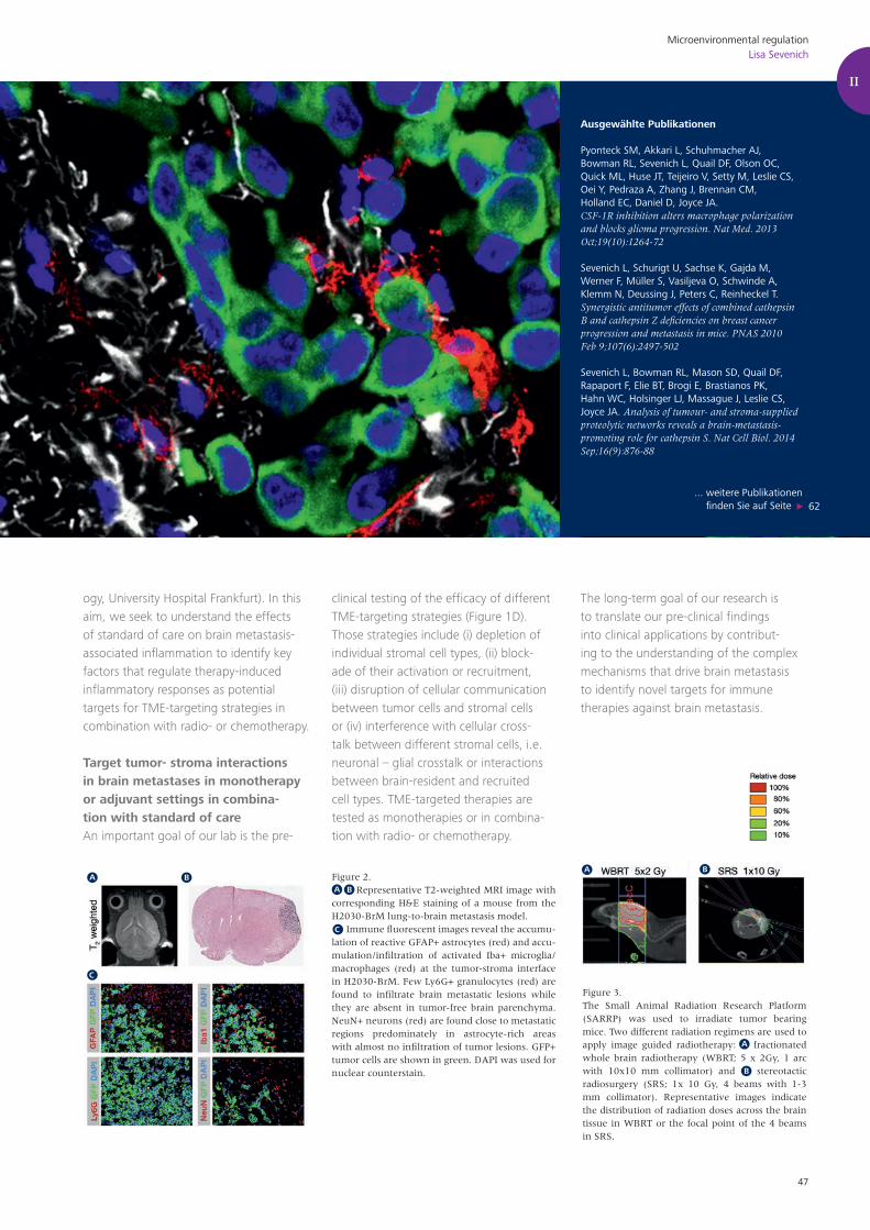
47
ogy, University Hospital Frankfurt). In this aim, we seek to understand the effects of standard of care on brain metastasis-associated inflammation to identify key factors that regulate therapy-induced inflammatory responses as potential targets for TME-targeting strategies in combination with radio- or chemotherapy.
Target tumor- stroma interactions in brain metastases in monotherapy or adjuvant settings in combina-tion with standard of careAn important goal of our lab is the pre-
clinical testing of the efficacy of different TME-targeting strategies (Figure 1D). Those strategies include (i) depletion of individual stromal cell types, (ii) block-ade of their activation or recruitment, (iii) disruption of cellular communication between tumor cells and stromal cells or (iv) interference with cellular cross-talk between different stromal cells, i.e. neuronal – glial crosstalk or interactions between brain-resident and recruited cell types. TME-targeted therapies are tested as monotherapies or in combina-tion with radio- or chemotherapy.
The long-term goal of our research is to translate our pre-clinical findings into clinical applications by contribut-ing to the understanding of the complex mechanisms that drive brain metastasis to identify novel targets for immune therapies against brain metastasis.
Figure 2. Representative T2-weighted MRI image with corresponding H&E staining of a mouse from the H2030-BrM lung-to-brain metastasis model. Immune fluorescent images reveal the accumu-lation of reactive GFAP+ astrocytes (red) and accu-mulation/infiltration of activated Iba+ microglia/macrophages (red) at the tumor-stroma interface in H2030-BrM. Few Ly6G+ granulocytes (red) are found to infiltrate brain metastatic lesions while they are absent in tumor-free brain parenchyma. NeuN+ neurons (red) are found close to metastatic regions predominately in astrocyte-rich areas with almost no infiltration of tumor lesions. GFP+ tumor cells are shown in green. DAPI was used for nuclear counterstain.
Ausgewählte Publikationen
Pyonteck SM, Akkari L, Schuhmacher AJ, Bowman RL, Sevenich L, Quail DF, Olson OC, Quick ML, Huse JT, Teijeiro V, Setty M, Leslie CS, Oei Y, Pedraza A, Zhang J, Brennan CM, Holland EC, Daniel D, Joyce JA. CSF-1R inhibition alters macrophage polarization and blocks glioma progression. Nat Med. 2013 Oct;19(10):1264-72
Sevenich L, Schurigt U, Sachse K, Gajda M, Werner F, Müller S, Vasiljeva O, Schwinde A, Klemm N, Deussing J, Peters C, Reinheckel T. Synergistic antitumor effects of combined cathepsin B and cathepsin Z deficiencies on breast cancer progression and metastasis in mice. PNAS 2010 Feb 9;107(6):2497-502
Sevenich L, Bowman RL, Mason SD, Quail DF, Rapaport F, Elie BT, Brogi E, Brastianos PK, Hahn WC, Holsinger LJ, Massague J, Leslie CS, Joyce JA. Analysis of tumour- and stroma-supplied proteolytic networks reveals a brain-metastasis-promoting role for cathepsin S. Nat Cell Biol. 2014 Sep;16(9):876-88
... weitere Publikationen finden Sie auf Seite 62
Microenvironmental regulationLisa Sevenich
II
Figure 3.The Small Animal Radiation Research Platform (SARRP) was used to irradiate tumor bearing mice. Two different radiation regimens are used to apply image guided radiotherapy: fractionated whole brain radiotherapy (WBRT; 5 x 2Gy, 1 arc with 10x10 mm collimator) and stereotactic radiosurgery (SRS; 1x 10 Gy, 4 beams with 1-3 mm collimator). Representative images indicate the distribution of radiation doses across the brain tissue in WBRT or the focal point of the 4 beams in SRS.

48

49
Experimentelle Therapie Experimental Therapy
Laboratories III
III

50
Molecular therapyUrsula Dietrich
Molekulare Therapie viraler Infektionen
More than 37 million people are currently living with HIV-1 worldwide. Despite the successful current antiretroviral regi-mens, which have turned infection with HIV-1 into a chronic disease in many countries and even resulted in declining numbers of new infections, the devel-opment of a vaccine is still the major goal to prevent new infections and their consequences. In recent years, broadly neutralizing antibodies (bnAbs) have been identified from a subset of chroni-cally infected patients, which neutralize multiple primary HIV-1 strains in vitro, eventually prevent infections in animal models and the first ones are in clinical development for therapeutic applications. However, no HIV-1 Env immunogen was able so far to induce such bnAbs upon vaccinations and the direct production of bnAbs for clinical applications is costly. We could recently select broadly neutral-izing nanobodies, which are antibody fragments corresponding to the variable domain of heavy chain only antibodies from camelids. Due to their small size and favourable physicochemical properties bn nanobodies are promising candidates for clinical development in view of prophy-
Molecular therapy of viral infections
HIV neutralizing antibodies and their epitopes
nanobodies
HIV-1 Env immunogens
MitarbeiterYvonne GeißSarah KaluscheKathrin KochKarsten MüllerSebastian ReneltOliver RingelSvenja WeißNadine Hergenröther Bianca PetriPatricia Schult-Dietrich
GruppenleiterinUrsula DietrichTel.: +49 69 63395-216Fax: +49 69 [email protected]
Wissenschaftliche GästeYenisleidys Martínez MontesinoMarta Sisteré Oró

51
Molecular therapyUrsula Dietrich
Unsere Gruppe entwickelt neue experimentelle Ansätze zur thera-peutischen und prophylaktischen Behandlung von HIV-Infektionen. Im Mittelpunkt steht die Identifizierung HIV-neutralisierender Antikörper sowie ihrer Epitope im viralen Hüllprotein (Env). Aus-gehend von polyklonalen Patientenseren oder Seren immunisierter Tiere selektionieren wir Hüllprotein-spezifische Antikörper über die „Phage display“ Technologie oder Env-spezifische B-Zellsortierung mit klinisch relevanten rekombinanten Env-Konstrukten. Diese werden anschließend hinsichtlich ihrer Neutralisationsfähigkeit eines Referenzsets primärer Viren in vitro untersucht. Hierbei gelang es uns, breit neutralisierende Antikörperfragmente, sog. „nanobodies“, aus Dromedaren zu identifizieren, die mit trime-ren HIV-1 Hüllproteinen immunisiert wurden. Insbesondere zwei nanobodies erwiesen sich als sehr effizient, denn sie können in Kombination 19 von 21 primären HIV-1 Isolaten verschiedener Subtypen neutralisieren.
Weiterhin entwickeln und charakterisieren wir verschiedene Env-Immunogene, die zum einen für die obigen Selektionen und Immunisierungen verwendet werden, zum anderen aber auch der Charakterisierung klinisch relevanter Hüllproteine dienen. Im Mittelpunkt stehen hierbei primäre X4 Viren, die über den CXCR4-Rezeptor infizieren und mit einer schnelleren Krankheitsprogression assoziiert sind. Wir konnten erstmals ein Set von primären X4 Viren verschiedener Subtypen generieren und zeigen, dass diese sich in ihrem Neutralisationsverhalten gegenüber bekannten breit neutra-lisierenden Antikörpern von CCR5-nutzenden Viren unterscheiden. Aufgrund der zunehmenden Primärinfektionen mit X4-Viren ist eine genaue immunologische, strukturelle und funktionelle Charakterisierung dieser Viren essentiell, um ggf. therapeutische Maßnahmen anzupassen.
III
lactic and therapeutic applications.We also select HIV-specific Abs or scFv from patients with bnAbs by antigen-spe-cific single B cell sorting or by the phage display technology using soluble native-like trimeric envelope (Env) constructs derived from clinically relevant HIV-1 strains. These include viruses from HIV-1 subtype C, recently infected persons and CXCR4 using viruses associated with faster disease progression and the potential to infect human stem and progenitor cells. To study this as well as the neutraliza-tion behaviour and structural properties, we generated the first comprehensive panel of primary CXCR4 using HIV-1 strains comprising different subtypes.
Identification of broadly neutral-izing nanobodies against HIV-1After immunizing four dromedaries with three soluble trimeric HIV-1 subtype C gp140 Env immunogens we could select eight nanobodies from the generated phage immune libraries, which neutralize primary tier 2 HIV-1 strains from different subtypes in a standardized in vitro neutral-ization assay. Remarkably, the neutraliza-tion breadth of two potent selected nano-
bodies in combination covers 19 of the 21 strains of the global HIV-1 reference panel comprising the major subtypes and recom-binant forms. Thus, these nanobodies represent promising candidates for further clinical development aiming at prophylac-tic and therapeutic applications. Optimi-zation includes intra- and intermolecular combination of nanobodies, functional analyses in the humanized mouse model (collaboration with Florian Klein, Cologne) and expression from different vectors for in vivo targeting of the genital mucosa (collaboration with Harold Marcotte, Karo-linska). Optimization will also be based on structural analyses of the nanobodies in complex with HIV-1 Env, which are ongoing in collaboration with SCRIPPS.
Identification of virus neutral-izing antibodies and their epi-topes from HIV controllersWe recently identified an epitope in the MPER region of the transmembrane protein gp41, which is targeted by a neutralizing antibody present in the serum from an elite controller by screen-ing an Env-tailored phage library with the patient´s IgG. In the context of the
European HIVERA project this epitope, EC26-2A4, was further evaluated in conjunction with a nearby epitope (3S), identified by our French partners (Vieillard/Debré, Paris), regarding their potential as vaccine candidates for clinical applica-tions. EC26-2A4 epitope optimization through peptide arrays and computa-tional analyses resulted in the identifica-tion of two shorter peptide sequences, which specifically react with antibodies against the original epitope, but not with the autoreactive mAb 2F5. Both short peptides coupled to KLH were able to induce HIV-neutralizing antibodies in mice. We then analysed the presence of antibodies in the HIV-1 positive cohort from the Frankfurt HIV Center (collabora-tion with Prof. C. Stephan) against the short EC26-2A4 peptides. From close to 1000 sera analysed about 6% were posi-tive independently of testing early or late timepoints after infection and there was no correlation with clinical parameters in this cohort of treated patients. In con-trast, the analysis of sera from a French untreated cohort revealed that the pres-ence of antibodies reacting with both, the short EC26-2A4 and the 3S peptide, was

52
A
B
Figure 1.Broadly neutralizing nanobodies against HIV-1. Schematic comparison of nanobodies and IgG. Experimental outline for the identification of bn nano-bodies. VHH28 shows potent and broad neutralizing activity against primary HIV-1 of different subtypes. Negative stain electron microscopy of nanobody VHH28 complexed to the ZM197M gp140 SOSIP trimer (orange arrow) and high resolution model of BG505 SOSIP.664 with PGV04 Fab (blue/green) complex docked into the 3D reconstructions of ZM197M SOSIP-VHH28 (orange) reveal the CD4 binding site in gp120 as target epitope for VHH28 (groups Ward/Wilson at SCRIPPS).
A B
C
Molecular therapyUrsula Dietrich
C
inversely correlated to viral load in plasma and cell associated proviral DNA load. Fur-thermore, antibodies purified with the 3S peptide from the respective sera showed neutralizing activity against primary tier 2 HIV-1 of different subtypes in vitro. EC26-2A4 purified antibodies from the sera are currently being tested for neutralization. The inverse correlation of the presence of antibodies against both MPER epit-opes in patient sera with viral parameters and the fact that both epitopes induce neutralizing antibodies in mice suggests these epitopes are promising candi-dates for further clinical development.
Characterization of Env immunogens from CXCR4 using (X4) HIV-1A peculiar trend in the HIV-1 epidemic in recent years is the observed increase of infections with primary X4 viruses, which are associated with faster disease progression. So far, X4 viruses have mostly been found as late population arising in persons initially infected by CCR5 using viruses (R5). To study these new primary X4 viruses we generated the first panel of primary X4 viruses comprising all
D
D

53
Ausgewählte Publikationen
Arnold P*, Himmels P*, Weiß S*, Decker TM, Markl J, Gatterdam V, Tampé R, Bartholomäus P, Dietrich U+, Dürr R+. Antigenic and 3D structural characte-rization of soluble X4 and hybrid X4-R5 HIV-1 Env trimers. Retrovirology 2014, 11(1):42
Trott M, Weiß S, Antoni S, Koch J, von Briesen H, Hust M, Dietrich U. Functional characterization of two scFv-Fc antibodies from an HIV controller selected on soluble HIV-1 Env com-plexes: a neutralizing V3- and a trimer-specific gp41 antibody. PLoS ONE 2014, 9(5): e97478.
Zhou M, Meyer T, Koch S, Koch J, Brill B, von Briesen H, Benito JM, Soriano V, Haberl A, Bickel M, Dübel S, Hust M, Dietrich U. Identification of a new epitope for HIV neutralizing antibodies in the gp41membrane proximal external region by an Env-tailored phage display library. Eur J Immunol 2013, 43: 499 – 509.
* corresponding authors
... weitere Publikationen finden Sie auf Seite 62
III
Molecular therapyUrsula Dietrich
major HIV-1 subtypes in collaboration with C. Kücherer (Robert Koch Institute, Berlin) and R. Dürr (NYU, New York) and compared it to lab-adapted strains as well as to a standard panel of R5 viruses. In neutralization assays we investigated the sensitivity to a broad spectrum of well-known bnAbs that target different epitopes on the trimeric HIV-1 envelope. Our data provide evidence that primary X4 strains are much more resistant against these bnAbs compared to lab-adapted X4 viruses and furthermore showed less neutralization sensitivity within the CD4 epitopes, when compared to primary R5 viruses. This argues for a more closed and compact structure of primary X4 Envs, which may facilitate the increased emer-gence of X4 viruses in primary infections.
Figure 2.Gp41 epitopes targeted by neutralizing antibodies in patient sera. Location of the EC26-2A4 and the 3S epitope in the exterior part of the gp41 transmembrane protein. Optimization of the EC26-2A4 core epitope to avoid crossreactivity with mAb 2F5 known to be autoreactive. The presence of antibodies in patient sera against both gp41 peptides correlates with reduced proviral DNA and viral load in plasma.
A
B
C
A
B
C

54
Immunotherapy of malignanciesWinfried Wels
Immuntherapie maligner Erkrankungen
Expression of chimeric antigen recep-tors (CARs) in cytotoxic lymphocytes constitutes a promising strategy for adoptive cancer immunotherapy with effector cells of defined specificity. CARs consist of a tumor-specific single chain Fv (scFv) antibody fragment connected via a flexible spacer and a transmembrane domain to intracellular signaling domains such as CD3ζ chain or CD3ζ together with one or more costimulatory protein domains. In recent years, CAR-engineered T cells targeting CD19 have demonstrated remarkable clinical efficacy in patients with malignancies of B-cell origin. Natural killer (NK) cells represent another valuable effector cell population for adoptive cancer immunotherapy, but experience with CAR-engineered NK cells is still limited. NK cells are part of the innate immune system and play an important role in cancer immunosurveillance, with their cytotoxicity being triggered rapidly upon appropriate stimulation through germline-encoded cell surface receptors. In cancer patients NK cells, like other immune cells, are often functionally com-promised due to the immunosuppressive activity of the tumor. Hence, for adoptive
Immunotherapy of malignancies
chimeric antigen receptors
natural killer cells
adoptive cancer immunotherapy
MitarbeiterSabrina GenßlerPranav OberoiCongcong ZhangIris MildenbergerAline LindnerSarah OelsnerAnja WaldmannKathrina KamenjarinThorsten GeyerBarbara Uherek
GruppenleiterWinfried Wels, stellvertretender Direktor Tel.: +49 69 63395-188Fax: +49 69 [email protected]
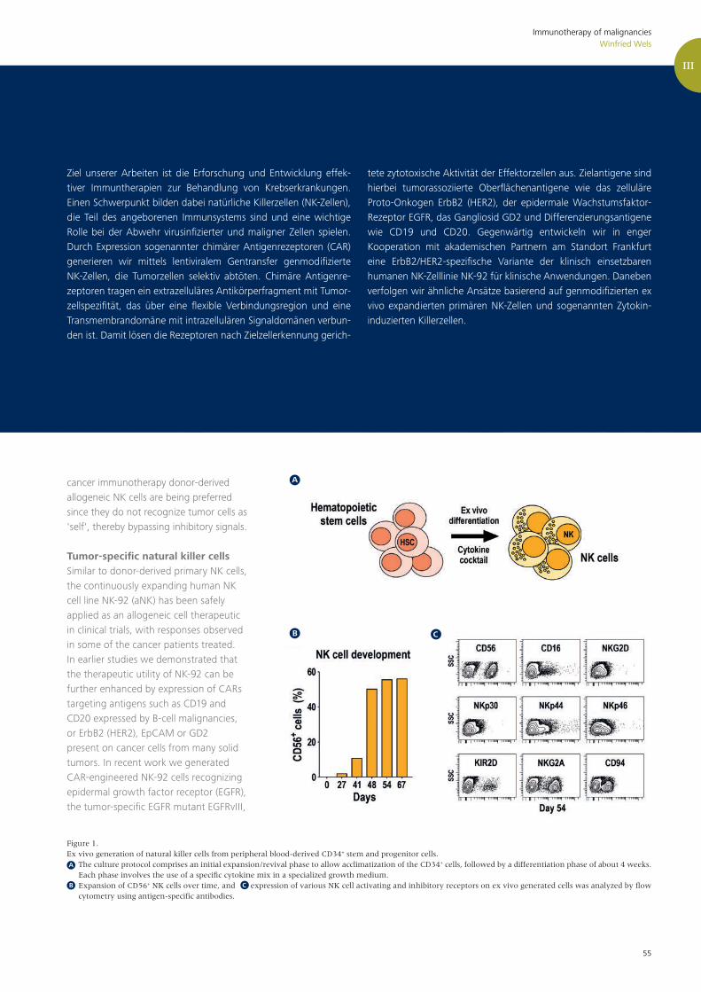
55
Immunotherapy of malignanciesWinfried Wels
Ziel unserer Arbeiten ist die Erforschung und Entwicklung effek-tiver Immuntherapien zur Behandlung von Krebserkrankungen. Einen Schwerpunkt bilden dabei natürliche Killerzellen (NK-Zellen), die Teil des angeborenen Immunsystems sind und eine wichtige Rolle bei der Abwehr virusinfizierter und maligner Zellen spielen. Durch Expression sogenannter chimärer Antigenrezeptoren (CAR) generieren wir mittels lentiviralem Gentransfer genmodifizierte NK-Zellen, die Tumorzellen selektiv abtöten. Chimäre Antigenre-zeptoren tragen ein extrazelluläres Antikörperfragment mit Tumor-zellspezifität, das über eine flexible Verbindungsregion und eine Transmembrandomäne mit intrazellulären Signaldomänen verbun-den ist. Damit lösen die Rezeptoren nach Zielzellerkennung gerich-
tete zytotoxische Aktivität der Effektorzellen aus. Zielantigene sind hierbei tumorassoziierte Oberflächenantigene wie das zelluläre Proto-Onkogen ErbB2 (HER2), der epidermale Wachstumsfaktor-Rezeptor EGFR, das Gangliosid GD2 und Differenzierungsantigene wie CD19 und CD20. Gegenwärtig entwickeln wir in enger Kooperation mit akademischen Partnern am Standort Frankfurt eine ErbB2/HER2-spezifische Variante der klinisch einsetzbaren humanen NK-Zelllinie NK-92 für klinische Anwendungen. Daneben verfolgen wir ähnliche Ansätze basierend auf genmodifizierten ex vivo expandierten primären NK-Zellen und sogenannten Zytokin-induzierten Killerzellen.
cancer immunotherapy donor-derived allogeneic NK cells are being preferred since they do not recognize tumor cells as 'self', thereby bypassing inhibitory signals.
Tumor-specific natural killer cellsSimilar to donor-derived primary NK cells, the continuously expanding human NK cell line NK-92 (aNK) has been safely applied as an allogeneic cell therapeutic in clinical trials, with responses observed in some of the cancer patients treated. In earlier studies we demonstrated that the therapeutic utility of NK-92 can be further enhanced by expression of CARs targeting antigens such as CD19 and CD20 expressed by B-cell malignancies, or ErbB2 (HER2), EpCAM or GD2 present on cancer cells from many solid tumors. In recent work we generated CAR-engineered NK-92 cells recognizing epidermal growth factor receptor (EGFR), the tumor-specific EGFR mutant EGFRvIII,
Figure 1.Ex vivo generation of natural killer cells from peripheral blood-derived CD34+ stem and progenitor cells. The culture protocol comprises an initial expansion/revival phase to allow acclimatization of the CD34+ cells, followed by a differentiation phase of about 4 weeks.
Each phase involves the use of a specific cytokine mix in a specialized growth medium. Expansion of CD56+ NK cells over time, and expression of various NK cell activating and inhibitory receptors on ex vivo generated cells was analyzed by flow
cytometry using antigen-specific antibodies.
A
B C
A
B C
III

56
or an epitope common to both target antigens which are frequently expressed by glioblastomas. In vitro analysis revealed high and specific cytotoxicity of EGFR-targeted NK-92 against established and primary glioblastoma cells, which was dependent on EGFR expression and CAR signaling. EGFRvIII-targeted NK-92 only lysed EGFRvIII-positive GBM cells, while dual-specific NK cells were active against both types of tumor cells. In immunode-ficient mice carrying intracranial glioblas-toma xenografts either expressing EGFR, EGFRvIII or both receptors, treatment with dual-specific NK cells was superior to treatment with the corresponding mono-specific CAR NK cells without inducing rapid immune escape as observed upon therapy with monospecific effectors.
Genetic modification of primary lymphocytesIn addition to our work with clinically usable NK-92 cells we are developing improved CAR vectors and optimized transduction protocols suitable for primary NK cells that are either expanded from peripheral blood or differentiated ex vivo from mobilized hematopoietic
stem or progenitor cells (Figure 1). In a collaborative project together with the Department of Stem Cell Transplantation and Immunology, Clinic for Pediatric and Adolescent Medicine at the University of Frankfurt, we also investigated tumor-specific cytokine-induced killer (CIK) cells as a potential pre-emptive immunotherapy for acute leukemia and other childhood malignancies. CIK cells consist mainly of T cells and NK-T cells, and are being expanded ex vivo from peripheral blood mononuclear cells. In preclinical models we could demonstrate potent and selective cytotoxicity of CD19-specific CAR CIK cells against cancer cell lines and primary pre-B-ALL blasts.
CAR-NK cells for clinical applicationsFollowing GMP-compliant procedures, we generated in close collaboration with the Blood Donation Service in Frankfurt a clonal ErbB2-specific CAR NK-92 cell line that may become useful as an off-the-shelf cell therapeutic for cancer immunotherapy. These NK-92/5.28.z cells selectively recognized ErbB2-expressing cells of different tumor origins and displayed high and selective
antitumor activity in in vitro and in vivo models. Ongoing work focuses on the development of these cells for adoptive immunotherapy of ErbB2-positive glio-blastoma. Together with colleagues from the Institute of Neurooncology at the University of Frankfurt we demonstrated potent activity of NK-92/5.28.z against established and primary glioblastoma cells in vitro and orthotopic glioblastoma xenografts in immunodeficient mice. In immunocompetent animals, local therapy with NK-92/5.28.z cells resulted in cures of transplanted syngeneic glioblastoma tumors, induction of endogenous antitu-mor immunity and long-term protection against tumor rechallenge at distant sites (Figure 2). Based on these promising data, preparations for a phase I clinical trial to evaluate NK-92/5.28.z cells as a treatment for patients with recurrent ErbB2-positive glioblastoma have been initiated.
Immunotherapy of malignanciesWinfried Wels

57
Figure 2.Antitumor activity of CAR-engineered natural killer cells against intracranial glioblastoma in immunocompetent animals. Murine GL261/ErbB2 glioblastoma cells were stereotactically injected into the right striatum of C57BL/6 mice. Seven days later, animals were treated by intratumoral injection of parental NK-92 or ErbB2-specific NK-92/5.28.z cells once per week for 3 weeks. Symptom-free survival of the mice indicates complete tumor rejection in the majority of animals. The insert shows tumor development as assessed by MRI at day 28 for representative animals from each group. Animals that were cured upon NK-92/5.28.z treatment were rechallenged at day 126 by stereotactic injection of GL261/ErbB2 cells into the left brain hemisphere without any further therapy. Long-term survival after rechallenge demonstrates induction of a long-lasting endogenous antitumor immune response by the initial NK-92/5.28.z treatment, while naïve control mice injected with GL261/ErbB2 cells were unable to control tumor development.
Immunotherapy of malignanciesWinfried Wels
Ausgewählte Publikationen
Zhang C, Burger MC, Jennewein L, Genßler S, Schönfeld K, Zeiner P, Hattingen E, Harter PN, Mittelbronn M, Tonn T, Steinbach JP, Wels WS. ErbB2/HER2-specific NK cells for targeted the-rapy of glioblastoma. J Natl Cancer Inst. 2016 May;108:djv375.
Genßler S, Burger MC, Zhang C, Oelsner S, Mil-denberger I, Wagner M, Steinbach JP, Wels WS. Dual targeting of glioblastoma with chimeric antigen receptor-engineered natural killer cells overcomes he-terogeneity of target antigen expression and enhances antitumor activity and survival. OncoImmunology. 2016 Apr;5(4):e1119354.
Oelsner S, Wagner J, Friede ME, Pfirrmann V, Genßler S, Rettinger E, Buchholz CJ, Pfeiffer H, Schubert R, Ottmann OG, Ullrich E, Bader P, Wels WS.Chimeric antigen receptor-engineered cytokine-induced killer cells overcome treatment resistance of pre-B-cell acute lymphoblastic leukemia and enhance survival. Int J Cancer. 2016 Oct 15;139(8):1799-809.
... weitere Publikationen finden Sie auf Seite 63
A
B C
A
B
C
III

Publikationen

AG MedyoufAG Krause
ReviewsEkaterina B, Rauner M, Medyouf H, Theurl I, Bornhauser M, Hofbauer L, U Platzbecker.Myelodysplasia is in the niche - novel con-cepts and emerging therapies. Leukemia. Review. 2015. Feb;29(2):259-68.
Book Chapters, NewslettersMedyouf H and De Fontenay. M. Book Chapter: Pathophysiologie des syndromes myélodysplasiques: biologie cellulaire. Syndromes myélodysplasiques. John Libbey, 4th Edition. 2016 (French).
Medyouf H. Mouse models of MDS. The European MDS Studies Coordination Organisation (EMSCO) Newsletter. March 2015.
* co-senior authors
Mossner M, Jann JC, Wittig J, Nolte F, Fey S, Nowak V, Obländer J, Pressler J, Palme I, Xanthopoulos C, Boch T, Metzgeroth G, Röhl H, Witt SH, Dukal H, Klein C, Schmitt S, Gelß P, Platzbecker U, Balaian E, Fabar-ius A, Blum H, Schulze TJ, Meggendorfer M, Haferlach C, Trumpp A, Hofmann WK, Medyouf H*, Nowak D*. Mutational hierarchies in myelodysplastic syndromes dynamically adapt and evolve upon therapy response and failure.Blood. 2016 Sep 1;128(9):1246-59.
Botezatu L, Michel LC, Helness A, Vadnais C, Makishima H, Hönes JM, Robert F, Vassen L, Thivakaran A, Al-Matary Y, Lams RF, Schütte J, Giebel B, Görgens A, Heuser M, Medyouf H, Maciejewski J, Dührsen U, Möröy T, Khandanpour C.Epigenetic therapy as a novel approach for GFI136N-associated murine/human AML. Exp Hematol. 2016 Aug;44(8):713-726.e14.
Kohrs N, Kolodziej S, Kuvardina ON, Herglotz J, Yillah J, Herkt S, Piechatzek A, Salinas Riester G, Lingner T, Wichmann C, Bonig H, Seifried E, Platzbecker U, Medyouf H, Grez M, Lausen J.MiR144/451 Expression Is Repressed by RUNX1 During Megakaryopoiesis and Disturbed by RUNX1/ETO. PLoS Genet. 2016 Mar 18;12(3):e1005946.
Medyouf H, Mossner M, Jann JC, Nolte F, Raffel S, Herrmann C, Lier A, Eisen C, Nowak V, Zens B, Müdder K, Klein C, Obländer J, Fey S, Vogler J, Fabarius A, Riedl E, Roehl H, Kohlmann A, Staller M, Haferlach C, Müller N, John T, Platzbecker U, Metzgeroth G, Hofmann WK, Trumpp A, Nowak D.Myelodysplastic Cells in Patients Re-program Mesenchymal Stromal Cells to Establish a Transplantable Stem Cell-Niche Disease Unit. Cell Stem Cell. 2014. Jun 5;14(6):824-37.
Ehninger A, Boch T, Medyouf H, Müdder K, Orend G, A. Trumpp.Loss of SPARC Protects Hematopoietic Stem Cells from Toxicity of Repeated Cycles of Chemotherapy by Accelerating their Return to Quiescence. Blood. 2014 Jun 26;123(26):4054-63.
Publications 2014 – 2016
Naka K, Jin CH, Ishihara K, Jomen Y, Kim D-H, Gu Y-K, Jeong E-S, Li S, Krause DS, Bae E, Ooshima A, Sheen YY, Kim S-J, Kim D-K.The novel oral TGF- signaling inhibitor EW-7197 targets chronic myeloid leukemia stem cells Cancer Sci. 2015 Nov 19. doi: 10.1111/cas.12849.
Rabenhorst U, Thalheimer F, Gerlach K, Kijonka M, Böhm S, Krause DS, Vauti F, Arnold HH, Schroeder T, Schnütgen F, von Melchner H, Rieger M, Zörnig M.Single-stranded DNA-binding transcripti-onal regulator FUBP1 is essential for fetal and adult hematopoietic stem cell self-rene-wal Cell Rep. 2015 Jun 30;11(12):1847-55.
Masia R, Krause DS, Yellen G.The inward rectifier potassium channel Kir2.1 is expressed in mouse neutrophils from bone marrow and liverAm J Physiol Cell Physiol. 2015 Feb 1; 308(3):C264-76.
Wheat JC*, Krause DS*, Shin TH*, Chen X, Wang J, Ding D, Yamin R, Sweetser D.The corepressor Tle4 is a novel regulator of murine hematopoiesis and bone development. PLoS One. 2014 Aug 25; 9(8):e105557
Krause DS, Lazarides K, Lewis JB, von Andrian UH, Van Etten RA.Selectins and their ligands are required for homing and engraftment of BCR-ABL1+ leukemic stem cells in the bone marrow niche. Blood 2014; 123(9): 1361-1371
Reviews, Editorials, Book ChaptersWeissenberger ES, Krause DS.Histological and In Vivo Microscopic Analysis of the Bone Marrow Microenvi-ronment in a Murine Model of Chronic Myelogenous Leukemia Methods Mol Biol. 2016; 1465:59-72
Krause DS, Scadden DT.A hostel for the hostile: The stem cell niche in haematological neoplasmsHaematologica 2015; 100(11):1376-87
Krause DS.Illness and artistic creativity (on the 70th anniversary of the death of Béla Bartók, composer, ethnomusicologist and leukemia patient). Leukemia 2015; 29(12):2417-8
Krause DS, DeLelys ME, Preffer FI.Flow Cytometry for Hematopoietic CellsMethods Mol Biol 2014; 1109:23-46
* co-first authourship
I
59
Hauck S, Hiesinger K, Khageh Hossei-ni S, Achenbach J, Biondi RM, Proschak E, Zörnig M1, Odadzic D1 (2016).Pyrazolo[1,5a]pyrimidines as a new class of FUSE binding protein 1 (FUBP1) inhibitors.Bioorg Med Chem., in press (http://dx.doi.org/10.1016/j.bmc.2016.09.015)
Tan D, Tao S, Chen Z, Koliesnik IO, Calmes PG, Hoerr V, Han B, Gebert N, Zörnig M, Löffler B, Morita Y, Rudol-ph KL.Dietary restriction improves repopula-tion but impairs lymphoid differentiation capacity of hematopoietic stem cells in early aging.J Exp Med. 2016 Apr 4; 213(4):535-53.
Rabenhorst U, Thalheimer FB, Gerlach K, Kijonka M, Böhm S, Krause DS, Vauti F, Arnold HH, Schroeder T, Schnütgen F, von Melchner H, Rieger MA, Zörnig M.Single-stranded DNA-binding transcrip-tional regulator FUBP1 is essential for fetal and adult hematopoietic stem cell self-renewal. Cell Rep. 2015 Jun 30; 11(12):1847-55.
Harter PN, Jennewein L, Baumgarten P, Ilina E, Burger MC, Thiepold AL, Tichy J, Zörnig M, Senft C, Steinbach JP, Mittelbronn M, Ronellenfitsch MW.Immunohistochemical assessment of phos-phorylated mTORC1-pathway proteins in human brain tumors. PLoS One. 2015 May 19; 10(5):e0127123.
Ordog T, Zörnig, M., Hayashi Y.Targeting disease persistence in gastroin-testinal stromal tumors. Stem Cells Transl Med. 2015 Jul; 4(7):701-7.
Schwamb B, Pick R, Mateus Fernández SB, Völp K, Heering J, Dötsch V, Bösser S, Jung J, Beinoraviciute-Kellner R, Wesely J, Zörnig I, Hammerschmidt M, Nowak M, Penzel R, Zatloukal K, Joos S, Rieker R, Agaimy A, Söder S, Reid-Lombardo K, Kendrick ML, Bardsley MR, Hayashi Y, Asuzu DT, Syed SA, Ordog T, Zörnig M.FAM96A is a novel pro-apoptotic tumor suppressor in gastrointestinal stromal tumors. Int J Cancer. 2015 Sep; 137(6):1318-29
Kramer D, Schön M, Bayerlová M, Bleckmann A, Schön MP, Zörnig M, Dobbelstein M.A pro-apoptotic function of iASPP by sta-bilizing p300 and CBP through inhibition of BRMS1 E3 ubiquitin ligase activity. Cell Death Dis. 2015 Feb 12; 6:e1634.
Malz M, Bovet M, Samarin J, Rabenhorst U, Sticht C, Bissinger M, Roessler S, Lorenzo Bermejo J, Renner M, Calvisi DF, Singer S, Ganzinger M, Weber A, Gretz N, Zörnig M, Schirmacher P, Breuhahn K.Overexpression of far upstream element (FUSE) binding protien (FBP)-interacting repressor (FIR) supports growth of hepato-cellular carcinoma.Hepatology. 2014 May 13. 60(4):1241-50.
Michalik KM, You X, Manavski Y, Doddaballapur A, Zörnig M, Braun T, John D, Ponomareva Y, Chen W, Uchi-da S, Boon RA, Dimmeler S.Long noncoding RNA MALAT1 regulates endothelial cell function and vessel growth. Circ Res. 2014 Apr 25; 114(9):1389-97.
Pellegrino R, Calvisi DF, Neumann O, Kol-luru V, Wesely J, Chen X, Wang C, Wu-estefeld T, Ladu S, Elgohary N, Bermejo JL, Radlwimmmer B, Zörnig M, Zender L, Dombrowski F, Evert M, Schirmacher P, Longerich T.EEF1A2 inactivates p53 via PI3K/AKT/mTOR-dependent stabilization of MDM4 in hepatocellular carcinoma.Hepatology 2014 May; 59(5):1886-99.
Baumgarten P, Harter PN, Tönjes M, Capper D, Blank A-E, Sahm F, von Deinling A, Kolluru K, Schwamb B, Rabenhorst U, Starzetz T, Kögel D, Rieker RJ, Plate KH, Ohgaki H, Radlwimmer B, Zörnig M1, Mittelbronn M1.Loss of FUBP1 expression in gliomas predicts FUBP1 mutation and is associated with oligodendroglial differentiation, IDH1 mutation and 1p/19q loss of het-erozygosity. Neuropathol Appl Neurobiol. 2014 Feb; 40(2):205-16.
1 both senior authors contributed equally to the work
Akademische Ausbildung Katharina Gerlach: „Characterization of FUSE-Binding Protein 1 as a hematopoi-etic stem cell self-renewal factor“. Dissertation am Fachbereich 14: Bio-chemie, Chemie und Pharmazie der Goethe-Universität Frankfurt am Main, 2016
Bernd Büttner: „Inhibierung des PI3K-Akt-Signalwegs in Kombination mit 5-FU-Behandlung in 5-FU-resistenten Kolonkarzinomzelllinien“.Dissertation am Fachbereich 16: Medizin der Goethe-Universität Frankfurt am Main, 2016
Giacomo Hokuto Canzian FT: „Analyse der physiologischen Funktion von FUBP1 in embryonalen Stammzellen der Maus vor und während der frühen Differen-zierung“.Bachelorarbeit am Fachbereich 15: Biowissenschaften der Goethe-Universität Frankfurt am Main, 2016
Publications 2014 – 2016
I
AG Zörnig
60

Publications 2014 – 2016
II
61
Arkan MC. ‘Cancer: Fat and fate of pancreatic tumours’. Nature, Aug 11; 536(7615):157-8, 2016.
Schulz MD*, Atay C*, Heringer J*, Romrig FR, Schwitalla S, Aydin B, Ziegler PK, Varga J, Reindl W, Pommerenke C, Salinas-Riester G, Böck A, Alpert C, Blaut M, Polson SC, Brandl L, Kirchner T, Greten FR, Polson SW and Arkan MC. ‘High-fat-diet-mediated dysbiosis promotes intestinal carcinogenesis independently of obesity’. Nature, Oct 23; 514(7523): 508-512, 2014.
Göktuna SI, Canli O, Bollrath J, Fingerle AA, Horst D, Diamanti MA, Pallangyo C, Bennecke M, Nebelsiek T, Mankan AK, Lang R, Artis D, Hu Y, Patzelt T, Ruland J, Kirchner T, Taketo MM, Chariot A, Arkan MC, Greten FR. ‘IKKα Promotes Intestinal Tumorigen-esis by Limiting Recruitment of M1-like Polarized Myeloid Cells.’ Cell Rep., Jun 26; 7(6): 1914-25, 2014.
Tetteh PW, Kretzschmar, K, Begthel H, van den Born M, Korving J, Morsink F, Farin H, van Es JH, Offerhaus GJA, and Clevers H (2016). Generation of an inducible colon-specific Cre enzyme mouse line for colon cancer research. PNAS 201614057.
Greulich F, Rudat C, Farin HF, Christoffels VM, and Kispert A (2016). Lack of Genetic Interaction between Tbx18 and Tbx2/Tbx20 in Mouse Epicardial Development. PLoS ONE 11, e0156787.
Greulich F, Trowe M-O, Leffler A, Stoetzer C, Farin HF, and Kispert A (2016). Misexpression of Tbx18 in cardiac chambers of fetal mice interferes with chamber-specific developmental programs but does not induce a pacemaker-like gene signature. J. Mol. Cell. Cardiol. 97, 140–149.
Farin HF*, Jordens I, Mosa MH, Basak O, Korving J, Tauriello DVF, de Punder K, Angers S, Peters PJ, Maurice MM, Clevers H* (2016). Visualization of a short-range Wnt gradient in the intestinal stem-cell niche. Nature 530, 340–343.
Tetteh PW, Basak O, Farin HF, Wiebrands K, Kretzschmar K, Begthel H, van den Born M, Korving J, de Sauvage F, van Es JH, et al. (2016). Replacement of Lost Lgr5-Positive Stem Cells through Plasticity of Their Enterocyte-Lineage Daughters. Cell Stem Cell 18, 203–213.
Weren RD, Venkatachalam R, Cazier JB, Farin HF, Kets CM, de Voer RM, Vreede L, Verwiel ET, van Asseldonk M, Kamping EJ, Kiemeney LA, Neveling K, Aben KK, Carvajal-Carmona L, Nagtegaal ID, Schackert HK, Clevers H, van de Weter-ing M, Tomlinson IP, Ligtenberg MJ, Hoogerbrugge N, Geurts van Kessel A, Kuiper RP. Germline deletions in the tumour suppressor gene FOCAD are associated with polyposis and colorectal cancer development. J Pathol. 2015 Jun; 236(2):155-64.
Tetteh PW, Farin HF, and Clevers H (2015). Plasticity within stem cell hierarchies in mammalian epithelia. Trends in Cell Biology 25, 100–108.
Lemoine R, Pachlopnik-Schmid J, Farin HF, Bigorgne A, Debré M, Sepulveda F, Héritier S, Lemale J, Talbotec C, Rieux-Laucat F, Ruemmele F, Morali A, Cathebras P, Nitschke P, Bole-Feysot C, Blanche S, Brousse N, Picard C, Clevers H, Fischer A, de Saint Basile G. Immune deficiency-related enteropathy-lymphocytopenia-alopecia syndrome results from tetratricopeptide repeat domain 7A deficiency. J Allergy Clin Immunol. 2014, doi:10.1016/j.jaci.2014.07.019.
Farin HF**, Karthaus WR**, Kujala P, Rakhshandehroo M, Schwank G, Schwank G, Vries RG, Kalkhoven E, Nieuwenhuis EE, Clevers H.Paneth cell extrusion and release of antimicrobial products is directly controlled by immune cell-derived IFN-γ. J Exp Med 2014, 211: 1393–1405.
Forster R, Chiba K, Schaeffer L, Regalado SG, Lai CS, Gao Q, Kiani S, Farin HF, Clevers H, Cost GJ, Chan A, Rebar EJ, Urnov FD, Gregory PD, Pachter L, Jaenisch R, Hockemeyer D. Human intestinal tissue with adult stem cell properties derived from pluripotent stem cells. Stem Cell Reports 2014, 2: 838–852.
Petersen N, Reimann F, Bartfeld S, Farin HF, Ringnalda FC, Vries RG, van den Brink S, Clevers H, Gribble FM, de Koning EJ. Generation of L cells in mouse and human small intestine organoids. Diabetes 2014 63: 410–420.
Bigorgne AE**, Farin HF**, Lemoine R**, Mahlaoui N, Lambert N, Gil M, Schulz A, Philippet P, Schlesser P, Abrahamsen TG, Oymar K, Davies EG, Ellingsen CL, Leteurtre E, Moreau-Massart B, Berrebi D, Bole-Feysot C, Nischke P, Brousse N, Fischer A, Clevers H, de Saint Basile G. TTC7A mutations disrupt intestinal epithe-lial apicobasal polarity. J Clin Invest 2014, 124: 328–337.
Yin X, Farin HF, van Es JH, Clevers H, Langer R, Karp JM. Niche-independent high-purity cultures of Lgr5+ intestinal stem cells and their progeny. Nature Methods 2014, 11: 106–112.
* co-corresponding authors** equal contribution
Akademische AusbildungMarnix de Groot (Universität Leiden); Masterarbeit 2016 mit dem Titel:"Patient derived organoids as a tool to test immunotherapeutic strategies”
Constantin Menche (Masterstudiengang Molekulare Medizin); Masterarbeit 2016 mit dem Titel: “Reprogramming of adult gastrointesti-nal organoids"
Greten FR.The Irony of Tumor-Induced Inflammation. Cell Metab. 2016 Sep 13;24(3):368-9.
Pesic M, Greten FR.Inflammation and cancer: tissue regenera-tion gone awry. Curr Opin Cell Biol. 2016 Aug 10;43:55-61.
Bocklitz TW, Salah FS, Vogler N, Heuke S, Chernavskaia O, Schmidt C, Waldner MJ, Greten FR, Bräuer R, Schmitt M, Stallmach A, Petersen I, Popp J.Pseudo-HE images derived from CARS/TPEF/SHG multimodal imaging in combination with Raman-spectroscopy as a pathological screening tool. BMC Cancer. 2016 Jul 26;16:534.
Finkelmeier F, Canli Ö, Tal A, Pleli T, Trojan J, Schmidt M, Kronenberger B, Zeuzem S, Piiper A, Greten FR, Waidmann O.High levels of the soluble programmed death-ligand (sPD-L1) identify hepatocellu-lar carcinoma patients with a poor progno-sis. Eur J Cancer. 2016 Mar 31;59:152-159.
Göktuna SI, Shostak K, Chau TL, Heukamp LC, Hennuy B, Duong HQ, Ladang A, Close P, Klevernic I, Olivier F, Florin A, Ehx G, Baron F, Vandereyken M, Rahmouni S, Vereecke L, van Loo G, Büttner R, Greten FR, Chariot A.The Prosurvival IKK-Related Kinase IKKε Integrates LPS and IL17A Signaling Cascades to Promote Wnt-Dependent Tumor Development in the Intestine. Cancer Res. 2016 May 1;76(9):2587-99.
van der Heijden M, Zimberlin CD, Nicholson AM, Colak S, Kemp R, Meijer SL, Medema JP, Greten FR, Jansen M, Winton DJ, Vermeulen L. Bcl-2 is a critical mediator of intestinal transformation. Nat Commun. 2016 Mar 9;7:10916.
Varga J, Greten FR.Lifting the Mist on Gastric Stem Cells. Cell Stem Cell. 2016 Jan 7;18(1):7-9.
Vogler N, Bocklitz T, Subhi Salah F, Schmidt C, Bräuer R, Cui T, Mires-kandari M, Greten FR, Schmitt M, Stallmach A, Petersen I, Popp J.Systematic evaluation of the biological variance within the Raman based colorectal tissue diagnostics. J Biophotonics. 2016 May;9(5):533-41
Pallangyo CK, Ziegler PK, Greten FR.IKKß acts as a tumor suppressor in cancer-associated fibroblasts during intestinal tumorigenesis. J Exp Med. 2015 Dec 14;212(13):2253-66.
AG Farin AG Arkan AG Greten

Publications 2014 – 2016
II
62
Canli Ö, Alankuş YB, Grootjans S, Vegi N, Hültner L, Hoppe PS, Schroeder T, Vandenabeele P, Bornkamm GW, Greten FR, (2015).Glutathione peroxidase 4 prevents necrop-tosis in mouse erythroid precursors. Blood. 2016 Jan 7;127(1):139-48.
Drube S, Weber F, Göpfert C, Loschinski R, Rothe M, Boelke F, Diamanti MA, Löhn T, Ruth J, Schütz D, Häfner N, Gre-ten FR, Stumm R, Hartmann K, Krämer OH, Dudeck A, Kamradt T (2015).TAK1 and IKK2, novel mediators of SCF-induced signaling and potential targets for c-Kit-driven diseases. Oncotarget 6(30):28833-50
Drube S, Weber F, Loschinski R, Beyer M, Rothe M, Rabenhorst A, Göpfert C, Meininger I, Diamanti MA, Stegner D, Häfner N, Böttcher M, Reinecke K, Herde-gen T, Greten FR, Nieswandt B, Hartmann K, Krämer OH, Kamradt T. (2015).Subthreshold IKK activation modulates the effector functions of primary mast cells and allows specific targeting of transformed mast cells. Oncotarget, 6(7):5354-68
Davis H, Irshad S, Bansal M, Rafferty H, Boitsova T, Bardella C, Jaeger E, Lewis A, Freeman-Mills L, Giner FC, Rodenas-Cuadrado P, Mallappa S, Clark S, Thomas H, Jeffery R, Poulsom R, Rodriguez-Justo M, Novelli M, Chetty R, Silver A, Sansom OJ, Greten FR, Wang LM, East JE, Tomlinson I, Leedham S (2015).Abberant epithelial GREM1 expression initiates colorectal tumour formation from cells outside the crypt based stem cell niche. Nature Medicine, 21:62-70.
Stangl S, Varga J, Freysold B, Traijkovic-Arsic M, Siveke JT, Greten FR, Ntzia-christos V, Multhoff G (2014).Selective in vivo imaging of tumors with a tumor cell-specific Hsp70 peptide-based probe. Cancer Res, 74(23):6903-12.
Schulz MD, Atay C, Heringer J, Romrig FK, Schwitalla S, Aydin B, Ziegler PK, Varga J, Reindl W, Pommerenke C, Salinas-Riester G, Böck A, Alpert C, Blaut M, Polson SC, Brandl L, Kirchner T, Greten FR, Polson SW, Arkan MC (2014).High-fat-diet-mediated dysbiosis promotes intestinal carcinogenesis independently of obesity. Nature, 514(7523): 508-12.
Greten FR (2014).AP1 takes over when oncogenic K-Ras slumbers. Cell 158(1): 11-2, 2014.
Varga J, De Oliveira T, Greten FR (2014).The architect who never sleeps: tumor-induced plasticity. FEBS Lett. 588(15): 2422-2427, 2014.
Göktuna SI, Canli O, Bollrath J, Fingerle AA, Horst D, Diamanti MA, Pallangyo C, Bennecke M, Nebelsiek T, Mankan AK, Lang R, Artis D, Hu Y, Patzelt T, Ru-land J, Kirchner T, Taketo MM, Chariot A, Arkan MC, Greten FR (2014). IKK promotes intestinal carcinogenesis by limiting recruitment of M1-like polarized myeloid cells. Cell Rep, 7(6): 1914-25.
Rokavec M, Öner MG, Li H, Jackstadt R, Longchang J, Lodygin D, Kaller M, Horst D, Ziegler PK, Schwitalla S, Slotta-Huspenina J, Bader FG, Greten FR, Hermeking H (2014). IL-6R/STAT3/miR-34a feedback controls EMT, invasion and metastasis of colorectal cancer. J Clin Invest, 124(4): 1853-67.
Akademische Ausbildung Charles Pallangyo: „Canonical NF-kB signaling in fibroblasts and cancer-asso-ciated fibroblasts in colitis and colorectal cancer“. Thesis Examen Technische Universität München
Olga Goncharova: „The mast cell func-tions of IKKß and NK-kB during intestinal tumorigenesis“.Disputation Universitätsklinikum Frankfurt
Nina Dahl: „Anwendung eines neuen genetischen Modells in der dreidimen-sionalen Zellkultur zur Untersuchung der Funktion von Glutathionperoxidase 4 in intestinalen Epithzellen“.Masterarbeit Technische Universität München
Bowman RL, Klemm F, Akkari L, Pyonteck SM, Sevenich L, Quail DF, Dhara S, Simpson K, Gardner EE, Iacobuzio-Donahou C, Brennan CW, Tabar V, Gutin PH, Joyce JA.Macrophage ontogeny underlines differ-ences in tumor-specific education in brain malignancies. Cell Reports 2016; accepted for publication.
Sevenich L, Joyce JA.Pericellular proteolysis in cancer. Genes Dev. 2014 Nov 1; 28(21):2331-47
Akkari L, Gocheva V, Kester JC, Hunter KE, Quick ML, Sevenich L, Wang HW, Peters C, Tang LH, Klimstra DS, Reinheckel T, Joyce JA. Distinct functions of macrophage-derived and cancer cell-derived cathepsin Z combine to promote tumor malignancy via interac-tions with the extracellular matrix. Genes Dev. 2014 Oct 1; 28(19):2134-50
Sevenich L, Bowman RL, Mason SD, Quail DF, Rapaport F. Elie BT, Brogi E, Brastianos PK, Hahn WC, Holsinger LJ, Massague J, Leslie CS, Joyce JA. Analysis of tumour- and stroma-supplied proteolytic networks reveals a brain-metas-tasis-promoting role for cathepsin S. Nat Cell Biol. 2014 Sep; 16(9):876-88
Bengsch F, Buck A, Günther SC, Seiz JR, Tacke M, Pfeifer D, von Elverfeldt D, Sevenich L, Hillebrand LE, Kern U, Sameni M, Peters C, Sloane BF, Reinheckel T. Cell type-dependent pathogenic functions of overexpressed human cathepsin B in murine breast cancer progression. Oncogene 2014 Sep 4; 33(36):4474-84
Schmier S+, Mostafa A+, Haarmann T, Bannert N, Ziebuhr J, Veljkovic V*, Dietrich U*, Pleschka S*. In silico prediction and experimental confir-mation of amino acids in the HA conferring enhanced receptor specificity for H5N1 Influenza A viruses. Scientific Reports 2015, 5:11434. doi: 10.1038/srep11434.
Geiß Y, Dietrich U. Catch me if you can – the race between HIV and neutralizing antibodies. AIDS Rev 2015, 17(2): 107-113.
Dietrich U, Landersz M, Stahl-Hennig C, Geiger C, Foley BT Genetic characterization of near full length SIVdrl genomes from four captive drills (Mandrillus leucophaeus). AIDS Res Hum Retrovir 2015, 31(3): 353-357.
Dietrich U. Elite controllers: Viruskontrolle ohne anti-virale Therapie – auf Kosten des Immunsys-tems? Retroviren Bulletin 2014, 2: 2-5.
Veljkovic V, Glisic S, Veljkovic N, Bojic Milinovic T, Dietrich U, Perovic VR, Colombatti A. Influenza vaccine as prevention for cardio-vascular diseases: possible molecular mecha-nism. Vaccine 2014, 48(12): 6569-6575.
Mori M, Nucci A, Dasso Lang MC, Humbert N, Boudier C, Debaene F, Sanglier-Cianferani S, Catala M, Schult-Dietrich P, Dietrich U, Tisné C, Mely Y, Botta M. Functional and structural characterization af 2-amino-4-phenylthiazole inhibitors of the HIV-1 nucleocapsid protein with antiviral activity. ACS Chem Biol 2014, 9(9): 1950-1955.
Dietrich U.Ursprung und aktuelle Aspekte der HIV-Pandemie / Origin and current aspects of the HIV pandemic. Pharmakon 2014, 4:244-249, ISSN 2195-2175; Dietrich U, Holzgrabe U, Schirmeister T, Heft 4 (HIV-Therapeutika: Grundlagen und Arzneistoffe) und 5 (HIV-Therapeuti-ka: Klinische Anwendung) 2014.
Arnold P+, Himmels P+, Weiß S+, Decker TM, Markl J, Gatterdam V, Tampé R, Bartholomäus P, Dietrich U*, Dürr R*.Antigenic and 3D structural characteriza-tion of soluble X4 and hybrid X4-R5 HIV-1 Env trimers. Retrovirology 2014, 11(1):42
AG DietrichAG Sevenich

Publications 2014 – 2016
III
63
Trott M, Weiß S, Antoni S, Koch J, von Briesen H, Hust M, Dietrich U. Functional characterization of two scFv-Fc antibodies from an HIV controller selected on soluble HIV-1 Env complexes: a neutralizing V3- and a trimer-specific gp41 antibody. PLoS ONE 2014, 9(5): e97478.
Muik A, Stubbert LJ, Jahedi RZ, Geiß Y, Dold C, Tober R, Volk A, Klein S, Dietrich U, Yadollahi B, Falls T, Miletic H, Stojdl D, Bell JC, von Laer D. Re-engineering vesicular stomatitis virus to abrogate neurotoxicity, circumvent humoral immunity and enhance oncolytic potency. Cancer Res 2014, 74(13): 3567-78
Dürr R, Keppler OT, Christ F, Crespan E, Garbelli A, Maga G, Dietrich U.Targeting cellular cofactors in HIV therapy. Topics in Medicinal Chemistry DOI:10.1007/7355_2014_45, Springer-Verlag Berlin Heidelberg 2014.
Akademische AusbildungKathrin Koch: „Generation, selection and characterization of single domain antibody fragments (VHHs) derived from HIV-1 Env immunized dromedaries”. Dissertation am Fachbereich 14: Biochemie, Chemie und Pharmazie der Goethe Uni-versität, 2016
Svenja Weiß: „Characterization of emerging HIV-1 X4 viruses with respect to biochemical, immunological and structural properties of their envelopes”. Dissertation am Fachbereich 14: Biochemie, Chemie und Pharmazie der Goethe Uni-versität, 2016
Sarah Kalusche: „Generation and scree-ning of nanobody phage libraries from HIV-1 Env immunized dromedaries”. Masterarbeit am Fachbereich 14: Bioche-mie, Chemie und Pharmazie der Goethe Universität, 2015
Nadine Hergenröther: „Expression und funktionelle Analyse des CCR5Δ32 Proteins“. Bachelorarbeit am Fachbereich 15: Bio-wissenschaften der Goethe Universität, 2016
+ equal contribution* joint last authors
Oelsner S, Wagner J, Friede ME, Pfirrmann V, Genßler S, Rettinger E, Buchholz CJ, Pfeiffer H, Schubert R, Ottmann OG, Ullrich E, Bader P, Wels WS. Chimeric antigen receptor-engineered cytokine-induced killer cells overcome treatment resistance of pre-B-cell acute lymphoblastic leukemia and enhance survival. Int J Cancer. 2016 Oct 15;139(8):1799-809.
Hegde M, Mukherjee M, Grada Z, Pig-nata A, Landi D, Navai SA, Wakefield A, Fousek K, Bielawowicz K, Chow KK, Brawley VS, Byrd TT, Krebs S, Gottschalk S, Wels WS, Baker ML, Dotti G, Mamonkin M, Brenner MK, Orange JS, Ahmed N. Tandem CAR T cells targeting HER2 and IL13Rα2 mitigate tumor antigen escape. J Clin Invest. 2016 Aug 1;126(8):3036-52.
Romanski A, Uherek C, Bug G, Seifried E, Klingemann H, Wels WS, Ottmann OG, Tonn T. CD19-CAR engineered NK-92 cells are sufficient to overcome NK cell resistance in B-cell malignancies. J Cell Mol Med. 2016 Jul;20(7):1287-94.
Alkins RD, Burgess A, Kerbel R, Wels WS, Hynynen K. Early Treatment of HER2-amplified brain tumors with targeted NK-92 cells and focused ultrasound improves surviv-al. Neuro Oncol. 2016 Jul;18(7):974-81.
Zhang C, Burger MC, Jennewein L, Genßler S, Schönfeld K, Zeiner P, Hattingen E, Harter PN, Mittelbronn M, Tonn T, Steinbach JP, Wels WS. ErbB2/HER2-specific NK cells for targeted therapy of glioblastoma. J Natl Cancer Inst. 2016 May;108:djv375.
Genßler S, Burger MC, Zhang C, Oelsner S, Mildenberger I, Wagner M, Steinbach JP, Wels WS. Dual targeting of glioblastoma with chimeric antigen receptor-engineered nat-ural killer cells overcomes heterogeneity of target antigen expression and enhances antitumor activity and survival. OncoIm-munology. 2016 Apr;5(4):e1119354.
Suck G, Odendahl M, Nowakowska P, Seidl C, Wels WS, Klingemann HG, Tonn T. NK-92: an 'off-the-shelf therapeutic' for adoptive natural killer cell-based cancer immunotherapy. Cancer Immunol Im-munother. 2016 Apr;65(4):485-92.
Zhou Q, Uhlig KM, Muth A, Kimpel J, Lévy C, Münch RC, Seifried J, Pfeiffer A,Trkola A, Coulibaly C, von Laer D, Wels WS, Hartwig UF, Verhoeyen E, Buch-holz CJ. Exclusive transduction of human CD4+ T cells upon systemic delivery of CD4-targeted lentiviral vectors. J Immunol. 2015 Sep 1;195(5): 2493-501.
Pfirrmann V, Oelsner S, Rettinger E, Huenecke S, Boenig H, Merker M, Wels WS, Cinatl J, Schubert R, Klingebiel T, Bader P. Cytomegalovirus-specific cytokine-induced killer cells: concurrent targeting of leu-kemia and infections. Cytotherapy. 2015 Aug;17(8): 1139-51.
Ahmed N, Brawley VS, Hegde M, Robertson C, Ghazi A, Gerken C, Liu E, Dakhova O, Ashoori A, Corder A, Gray T, Wu MF, Liu H, Hicks J, Rainusso N, Dotti G, Mei Z, Grilley B, Gee A, Rooney CM, Brenner MK, Heslop HE, Wels WS, Wang LL, Anderson P, Gottschalk S. Human epidermal growth factor recep-tor 2 (HER2)-specific chimeric antigen receptor-modified T cells for the immuno-therapy of HER2-positive sarcoma. J Clin Oncol. 2015 May;33(15): 1688-96.
Seidel D, Shibina A, Siebert N, Wels WS, Reynolds CP, Huebener N, Lode HN. Disialoganglioside-specific human natural killer cells are effective against drug-resistant neuroblastoma. Cancer Immunol Immunother. 2015 May;64(5): 621-34.
Schönfeld K, Sahm C, Zhang C, Naundorf S, Brendel C, Odendahl M, Nowakowska P, Bönig H, Köhl U, Kloess S, Köhler S, Holtgreve-Grez H, Jauch A, Schmidt M, Schubert R, Kühlcke K, Seifried E, Klingemann HG, Rieger MA, Tonn T, Grez M, Wels WS. Selective inhibition of tumor growth by clonal NK cells expressing an ErbB2/HER2-specific chimeric antigen receptor. Mol Ther. 2015 Feb;23(2): 330-8.
Glienke W, Esser R, Priesner C, Suerth JD, Schambach A, Wels WS, Grez M, Kloess S, Arseniev L, Koehl U. Advantages and applications of CAR-expressing natural killer cells. Front Pharmacol. 2015 Feb 12;6:21.
Müller N, Hartmann C, Genßler S, Koch J, Kinner A, Grez M, Wels WS. A bispecific transmembrane antibody simultaneously targeting intra- and extracellular epitopes of the epidermal growth factor receptor inhibits receptor activation and tumor cell growth. Int J Cancer. 2014 Jun 1;134(11):2547-59.
Abken H, Wels, WS, Kühlcke K. The express drivers: Chimeric antigen receptor-redirected T cells make it to the clinic. in: Cancer Immunotherapy Meets Oncology. In Honor of Christoph Huber. Eds.: Britten CM, Kreiter S, Diken M, Rammensee H-G, pp 127-35, Springer, Heidelberg, 2014.
Rettinger E, Kreyenberg H, Merker M, Kuçi S, Willasch A, Bug G, Ullrich E, Wels WS, Bönig H, Klingebiel T, Bader P. Immunomagnetic selection or ir-radiation eliminates alloreactive cells but also reduces anti-tumor potential of cytokine-induced killer cells: Implications for unmanipulated cytokine-induced killer cell infusion. Cytotherapy. 2014 Jun;16(6):835-44.
Akademische AusbildungSarah Oelsner“CAR-engineered lymphocytes for targeted therapy of leukemia and lym-phoma”, Dissertation am Fachbereich 14: Biochemie, Chemie und Pharmazie der Goethe-Universität Frankfurt am Main, 2016.
Kathrina Kamenjarin„Genmodifizierte feeder Zellen für die ex vivo Expansion primärer humaner NK-Zellen“, Masterarbeit am Fachbereich 14: Biochemie, Chemie und Pharmazie der Goethe-Universität Frankfurt am Main, 2016.
AG Wels

64
Financial affairs
Finanzen und Administration
Die Abteilung Finanzen/Administrationsetzt jährlich ein durchschnittliches Finanzvolumen von 8,5 Millionen Euro um und betreut dabei rund 100 Mitar-beiterinnen und Mitarbeiter. Sie wird geführt von Robert Dornberger, der dabei von Christof Kaiser unterstützt wird.
Christiane Strack im Personalbüro erledigt sämtliche Personalfragen. Verstärkt wird sie dabei von Ilka Grau, die auch in der Finanzbuchhaltung/Drittmittelverwaltung die Nachfolge von Emilia Seibert angetreten hat.
Gabriele Heckl erstellt die Bilanz, bear-beitet alle Ausgangsrechnungen, die Reisekostenabrechnungen und ist für die Abrechnung internationaler Drittmit-tel zuständig. Emilia Seibert zeichnete verantwortlich für die Drittmittelverwal-tung für die Bereiche der DFG, der DLR und der LOEWE-Förderungen. Marion Czech und Giuseppina Virgillito betreu-ten die Kreditorenrechnungen sowie die Drittmittel nationaler Stiftungen. Marion Czech und Emilia Seibert verabschie-deten wir in 2016 in den Ruhestand.
Ansprechpartner in der Telefonzentrale und am Empfang ist Bernd Würde-mann. Adrian Gresik ist verantwort-lich für die vielfältigen Aufgaben des Innendienstes. Er, Michael Paul und Heinrich Krompietz kümmern sich um die haustechnischen Ausstattungen und Installationen und arrangieren alle Arten von wissenschaftlichen Tagun-gen und Veranstaltungen. Dabei wer-den sie von Volker Hopf unterstützt.
Yoseph Alazar, Maria Fernandes, Yasemin Piskin und Neriman Sarac reinigen die Laboratorien, entsorgen die anfallenden Abfälle und kümmern sich um die Bereit-stellung von Laborbedarf. Sie werden dabei von Keziban Ata unterstützt.
Our administration and services de-partment is led by Robert Dornberger who oversees a yearly budget of ap-proximately 8.5 million Euros. He also takes care of the administrative needs of about 100 staff members and is supported by Christof Kaiser.
He is assisted by Christiane Strack who heads the personnel department with the support of Ilka Grau. Ilka Grau is also engaged in financial accounting and the administration of external fundings.
Gabriele Heckl prepares the balance sheets, is responsible for all outgoing invoices, all claims of travel expenses and is in charge of accounting of interna-tional grants. Emilia Seibert was respon-sible for grants from the DFG, DLR and LOEWE. Giuseppina Virgillito and Marion Czech handled grants from nationwide foundations and took care of all incom-ing invoices. In 2016 Marion Czech and Emilia Seibert retired from the institute.
Bernd Würdemann is our receptionist and is the first contact with the Institute.Adrian Gresik is responsible for the internal service tasks. He, Michael Paul and Heinrich Krompietz take care of the technical equipment and the in-stallations and arrange all kinds of scientific meetings and events. They are supported by Volker Hopf.
Yoseph Alazar, Maria Fernandes, Yasemin Piskin and Neriman Sarac clean the laboratories, dispose of the waste and restock the supplies. They are supported by Keziban Ata.
Robert DornbergerKaufmännischer Leiter undLeiter der Abteilung Finanzen / Administration Tel.: +49 69 63395-333Fax: +49 69 63395-353 [email protected]
Christoph Kaiser stellv. Leiter der Abteilung Finanzen / Administration"
Tel.: +49 69 63395-106Fax: +49 69 63395-353 [email protected]

65
Scientific Services
Zentrale Einheit HistologieZur Anfertigung von histologischen Präparaten betreibt das Georg-Speyer-Haus eine Histologie-Serviceeinheit unter der Leitung von Dr. Boris Brill. Hier werden von Frau Petra Dinse, meist automatisiert, die Gewebeaufarbeitung sowie immuno-histochemische Färbungen und Stan-dardfärbungen durchgeführt. Weiterhin verfügt das Labor über ein automatisiertes Präparate-Scanner- und Bildanalysesystem, Aperio ScanScope CS2, einen Färbeau-tomat Leica Autostainer XL sowie einen Leica BOND max zur Anfertigung von automatisierten Immunfärbungen. Das Labor stellt seine Leistungen den Arbeits-gruppen des Georg-Speyer-Hauses sowie externen Forschergruppen zur Verfügung.
TierhaltungDas Georg-Speyer-Haus betreibt eine Tier-haltung, geleitet von Dr. Boris Brill, um den Forschungsgruppen die Zucht von Mäusen und Experimente zu ermöglichen. Die Tierhaltung stellt in vivo Imaging Systeme, wie Endoskopie (Storz Coloview Set), in vivo konfokale Mikroskopie (Cellvizio), ein Bruker 7T MRT und ein Detektionssystem für Fluoreszenz und Luciferase in vivo (IVIS Lumina II) zur Verfügung.
Transgenic Core Facility Am Georg-Speyer-Haus wird die „Trans-genic Core Facility“ (TCF) als eine neue zentrale Einheit etabliert. Die TCF wird eine State-Of-The-Art Plattform für die Entwick-lung von transgenen Mausmodellen und präzisen Genombearbeitungstechnologien (CRISPR/Cas9) darstellen, um menschliche Erkrankungen besser zu verstehen. Dr. Madina Karimova leitet die Serviceeinheit und ist die Ansprechpartnerin für alle Belange der Transgen- und CRISPR / Cas9 Technologie. Neue Mausmodelle können als direkte oder bedingte Knock-out, Knock-in oder Punktmutationen generiert werden. Dazu werden verschiedene Tech-niken (wie z. B. Mikroinjektion von DNA und CRISPR / Cas9 Komponenten oder Blastozysteninjektion) Anwendung finden. Dabei wird die TCF besonders die Techno-logieentwicklung der präzisen Genommo-
Core Facility HistologyThe Georg-Speyer-Haus operates a histology core facility. It is supervised by Dr. Boris Brill. Petra Dinse is responsible for the mostly automated procedures of tissue processing and immunohistochemistry as well as hematoxylin / eosin staining. The laboratory is equipped with a slide scanner and image analysis system, Aperio ScanScope CS2, a Leica AutostainerXL and a Leica BOND max for automated immu-nostaining. The services of the histology core facility are available to all scientists of the Georg-Speyer-Haus as well as to external research partners in collaboration.
Animal HusbandryThe Georg-Speyer-Haus has an animalfacility, led by Dr. Boris Brill, which provides capacity for our research groups for mouse breeding andexperiments. Included in the animal facility are imaging techniques like endoscopy (Storz Coloview Set), in vivo confocal microscopy (Cellvizio), a Bruker 7T MRI and a detection system for fluorescence and luciferase in vivo (IVIS Lumina II).
Transgenic Core Facility Transgenic Core Facility (TCF) is currently being established in the Georg-Speyer-Haus. It will provide a state-of-the-art platform for the development of mouse models and precise genome editing technologies (CRISPR/Cas9) to understand human diseases. Dr. Madina Karimova oversees the facility and is a contact person for transgenic and CRISPR/Cas9 technology. Mouse model generation can be achieved by a conventional or conditional gene knockout, gene knock-in or point mutations. For that TCF will use varies techniques such as microinjection of DNA and CRISPR/Cas9 reagents or blasto-cysts injection. TCF will also carry out the technology development of the precise genome editing tools. TCF is a central service unit for all working groups of the GSH as well as external research groups.
Wissenschaftlicher Service
Madina KarimovaGruppenleiterin Transgenic Core FacilityTel.: +49 69 63395-620Fax: +49 69 [email protected]
Dr. Boris BrillTel.: +49 69 63395-205Fax: +49 69 [email protected]
66
Scientific Services
Dr. Herbert KühnelTel.: +49 69 63395-120Fax: +49 69 [email protected]
Dr. Klaus LehmenTel.: +49 69 63395-118Fax: +49 69 [email protected]
difizierungsverfahren vorantreiben. Die TCF ist eine zentrale Serviceeinrichtung für alle Arbeitsgruppen des Georg-Speyer-Hauses, sowie für externe Forschungsgruppen.”
Zentrale Einheit Durchflusszytometrie Die zentrale FACS-Einrichtung besteht aus drei Durchflusszytometern zur Zellanalyse (BD LSRFortessa, FACSCantoII, FACSCalibur) und zwei Zellsortern (BD FACSAria I, FACSAria Fusion). Geleitet wird die Serviceeinheit von Dr. Stefan Stein, der auch Ansprechpartner für allgemeine Fragen zur Durchflusszytometrie und bei der Entwicklung und Anpassung neuer Mess- und Sortieransätze ist. Tefik Merovci führt die anfallenden Hochgeschwindigkeits-Zellsortierungen durch und ist für den einwandfreien Zustand aller FACS-Geräte am Institut verantwortlich. In einigen Fällen fungiert Thorsten Geyer als zusätzlicher Operator an den Zellsortern. Die Sortiereinheit steht primär den Arbeitsgruppen des Georg-Speyer-Hauses, aber auch externen Forschergruppen zur Verfügung.
ITSteffen Luft leitet als Chief Information Officer (CIO) die IT. Er nimmt die zentralen Tätigkeiten der Unterstützung der Mitarbeiterinnen und Mitarbeiter des Hauses in allen Fragen der IT, der Serverbetreuung, des IT-Projekt-managements, der Netzwerkadminis-tration und des Einkaufs wahr. Er wird dabei unterstützt von Dr. Klaus Lehmen.
Arbeitssicherheit und Genehmigungen Dr. Herbert Kühnel gewährleistet die Funk-tionsfähigkeit der Laboratorien, versorgt die Arbeitsgruppen mit der nötigen Ausstat-tung, holt behördliche Genehmigungen ein und kümmert sich um die Arbeitssicherheit.
Gefahrstoff- und Abfallmanagement Dr. Klaus Lehmen wird perspektivisch in die Themenfelder des betrieblichen Arbeitsschutzes hineinwachsen. In 2016 hat er bereits die Verantwortung für den Bereich Gefahrstoff- und Abfallma-nagement sowie Aufbau eines digitalen Betriebsmittelkatasters übernommen.
Core Facility Flow CytometryThe Flow Core Unit of the Georg-Speyer-Haus operates three flow cytometer instruments (BD LSRFortessa, FACSCantoII, FACSCalibur) and two cell sorters (BD FACSAria I and BD FACSAria Fusion). Dr. Stefan Stein oversees the performance of the core facility and is available for scientific questions regarding flow cytom-etry in general and the establishment of new FACS based assays. Tefik Merovci is responsible for high-speed cell sorting as operator in this central service unit for all research groups of the GSH as well as for external researchers. Tefik also takes care of the maintenance and functionality of the flow cytometers in the institute. Occasionally, Thorsten Geyer serves as an additional FACSorting operator.
ITSteffen Luft is Chief Information Officer (CIO) of the Institute. His main tasks are the maintenance of the servers, IT project management, administration of the networks and the support of the colleagues in the institute. He is supported by Dr. Klaus Lehmen.
Occupational safety and permissionsDr. Herbert Kühnel attends for equipment and supply, permissions, and occupational safety.
Dangerous material and waste management Dr. Klaus Lehmen is growing into the subjects of occupational safety. He started in 2016 with managing of hazardous goods and disposals. Furthermore he is in charge to implement a digital register of all technical equipments and documents.
Steffen LuftTel.: +49 69 63395-222Fax: +49 69 63395-297 [email protected]
Dr. Stefan SteinTel.: +49 69 63395-260Fax: +49 69 [email protected] [email protected]

67
Meetings and Lectures 2016
12.-13.12.2016Scientific Advisory Board / Wissen-schaftlicher Beirat
24.11.2016Welt-AIDS-Tag in Zusammenarbeit mit der Goethe Universität und der Arbeitsgruppe von Dr. Ursula Dietrich vom Georg-Speyer-Haus.
09.11.2016 Prof. Dr. med. Clemens A. Schmitt, Charité - Universitätsmedizin Berlin (CVK), and Max-Delbrück-Center for Molecular Medicine„Cellular senescence in cancer – therapeutic goal or not?"
27.10.2016 Prof. Dr. Urs Christen, Pharmazentrum Frankfurt, Klinikum der Goethe Univer-sität Frankfurt„Adenovirus expressed liver autoantigen (CYP2D6) induces chronic autoimmune hepatitis in wild type mice”
20.10.2016 Dr. Falk Hildebrand, European Molecular Biology Laboratory (EMBL), Heidelberg„Discovery of a new bacterial species in a dysbiotic human gut microbiome”
13.-14.10.2016 2nd Symposium on Cellular Crosstalk in the Tumor Microenvironment
12.10.2016 Professor Ralph Nixon, NYU School of Medicine, New York„The Pathogenic Role of Defective Endosomal-Lysosomal Function in Alzheimer's Disease”
11.10.2016 A powerful CRISPR combination
29.09.2016 Models and Insights in Translational Research
11.05.2016 Dr. Kyung Lee, National Cancer Insti-tute, Bethesda„Polo kinases from centrosome duplica-tion to mitotic controls“
21.04.2016 Prof. Dr. Matthias Dobbelstein, Insti-tute of Molecular Oncology, University of Göttingen„The Mdm2 oncoprotein as a modulator of chromatin dynamics“
05.-06.04.2016 DFG-Begutachtung Forscherantrag „Cell Plasticity in Colorectal Carcinogen-esis“ (FOR 2438)
10.03.2016 Dr. Manuel Kaulich, Institute for Biochemistry II, Goethe Universität Frankfurt„CRISPR/Cas9 in action: a Cdk4-dependent phosphorylation threshold regulates the cell cycle entry decision“
01.02.-07.03.2016 Schülervorlesungsreihe 2016
02.03.2016 Univ.-Prof. Dr. Wilfried Roth, Institute for Pathology, Johannes Gutenberg-Universität Mainz„Pathologie of cell death“
18.02.2016 Dr. Steffen Schmitt, German Cancer Research Center (DKFZ), Heidelberg„Good Scientific Practice in Flow Cytometry: Pitfalls in Acquisition and Analysis“
28.01.2016 Prof. Dr. Axel Behrens, Cancer Research, The Francis Crick Institute, UK„Cell fate reprogramming in pancreatic cancer“
21.01.2016 Dr. Doug Winton, Cancer Research Institute, University of Cambridge, UK„Functional approaches to understand-ing intestinal stem cells“
14.01.2016 Prof. Dr. Burkhard Becher, Universität Zürich„T cell: myeloid cell interactions in autoimmunity“
13.09.2016 Auswahlsymposium zum Paul-Ehrlich- und-Ludwig-Darmstaedter-Nachwuchspreis
Volker Busskamp Technische Universität Dresden, Center for Regenerative Therapies Dresden (CRTD)„Biomedical exploitation of human stem cell-derived neuronal cell types and functional circuits for retinal therapies“
Tobias J. ErbMax Planck Institute for Terrestrial Microbiology, Marburg„Bringing inorganic carbon into life and medicine with synthetic biology“
Marieke EssersGerman Cancer Research Center and Heidelberg Institute for Stem Cell Tech-nology and Experimental Medicine„Hematopoietic stem cells under stress“
Barbara TreutleinMax Planck Institute for Evolutionary Anthropology, Leipzig„Reconstructing development using single-cell transcriptomics“
Nora VögtleUniversity of Freiburg, Institute for Biochemistry and Molecular Biology„The mitochondrial presequence process-ing machinery in health and disease“
Thomas WollertMax Planck Institute of Biochemistry, Martinsried„Self destruction to survive – insights into autophagy from in vitro reconstitu-tions“
01.09.2016 Elena Porro, Cell Press, Cambridge, MA, USA„Publishing your work – a Cell editor's perspective“
22.06.2016 Bürgervorlesung „Virus-Seuchen: Ursprung, Manage-ment, Prävention“
02.06.2016 Professor Gijs R. van den Brink, M.D., Ph.D., Academic Medical Center, Amsterdam„Anti-TNF in Crohn's disease, targeted therapy or lucky shot?“
01.06.2016 Professor Dr. Joan Massagué, Memo-rial Sloan Kettering Cancer Center, NY, USA„Metastatic Colonization by Circulating Tumor Cells“
12.-13.05.2016 Symposium „CNS Inflammation in Neurodegenerative Disease and Brain Cancer“
Veranstaltungen

68
Nov. 2015-Jan. 2016Kurspraktikum Bachelorstudiengang Biochemie: BCII Praktikum inkl. VorlesungFarin H, Krause D
01.02.-07.03.2015Schülervorlesung und -praktikum Lecture series and practical course for high school studentsOrganisation: Dietrich U
„Krebsentstehung: genetische Grundlagen, Diagnostik und neue Therapieansätze“Greten F
„Immunkontrolle von Krebs und Infektionen“Oelsner S
„Stammzellen und Krebsstammzellen“Krause D
„Gentechnologie: Grundlagen und Konsequenzen“Dingermann T
„HIV/AIDS – ein Immundefizienzvirus erobert die Welt“Dietrich U
05.04.-08.04.2015Schülerpraktikum in den Laborgruppen des Georg-Speyer-Hauses
29.02.-11.03.2016Kurspraktikum Masterstudiengang Mole-kulare Medizin: Molekulare Onkologie und TumorimmunologieFarin H
SemesterbegleitendSeminarreihe Masterstudiengang Mole-kulare Medizin: Frankfurter ForschungFarin H
WS 2015 | 16, SS 2016, WS 2016 | 17Anleitung zum wissenschaftlichen Arbei-ten für Bachelor- und MasterstudentenGreten F, Wels W, Zörnig M, Dietrich U, Medyouf H, Sevenich L, Farin H, Krause D
WS 2015 | 16, SS 2016, WS 2016 | 17Individuelle 6-wöchige Laborpraktika in Zusammenarbeit mit der Goethe-UniversitätGreten F, Wels W, Zörnig M, Dietrich U, Medyouf H, Sevenich L, Farin H, Krause D und Mitarbeiter
WS 2015 | 16, WS 2016 | 17Biochemie-Praktikum II für Studierende der Goethe-UniversitätZörnig M, Greten F, Wels W, Dietrich U, Medyouf H, Sevenich L, Farin H, Krause D und Mitarbeiter
WS 2016 | 17Vorlesung Molekulare Onkologie und Tumorimmunologie (MOT) Tumormik-roenvironment/Tumor und Entzündung/TumormakrophagenGreten F, Zörnig M, Krause D, Medyouf H, Farin H, Sevenich L
SS 2016Anwendungen der Biologie in der Hämatologie für MedizinstudentenKrause D
Every FridayResearch Meeting: Recent results, advances and problems of individual research projects are presented and discussed in English
Education
Lehrveranstaltungen

69
Der Verein „Freunde und Förderer des Georg-Speyer-Hauses“
The Association„Friends and Sponsors of the Georg-Speyer-Haus“
Innovative Forschung und wissenschaft-licher Fortschritt in unserer Gesellschaft sind nur möglich durch das Engagement der Wissenschaftler/innen und die aktive Unterstützung von Forschungsförderern aus Öffentlichkeit, Wissenschaft und Wirtschaft. Diesem Engagement hat sich der Verein „Freunde und Förderer des Georg-Speyer-Hauses“ verpflich-tet: Sein Ziel ist es, über die Grundfi-nanzierung durch Bund und Länder hinaus für weitere erforderliche Mittel zu sorgen und so das hohe Niveau der Grundlagenforschung zu sichern. Mitglied im Verein kann werden, wer den wissenschaftlichen Fortschritt im Bereich der Krebsforschung und der experimentellen Therapie zum Wohle der Allgemeinheit fördern möchte und Interesse hat am Forschungsprozess und am Diskurs über Ergebnisse und deren Nutzen für die Allgemeinheit.Neben der einfachen Mitgliedschaft (Freund/innen) und der Forschermitglied-schaft (Wissenschaftler/innen, Student/in-nen) besteht die Möglichkeit der fördern-den Mitgliedschaft für Einzelpersonen oder Firmen. Förderer können im Jahrbuch und auf der Spendentafel aufgeführt werden.Da der Verein eine gemeinnützige Einrichtung ist, sind Mitgliedsbeiträge und Spenden im Rahmen der zulässigen Höchstbeträge von der Steuer absetzbar.
Innovative research and scientific ad-vances are only possible through generous financial support from public and private sponsors. The association „Friends and Sponsors of the Georg-Speyer-Haus“ has committed itself to this task. The major goal of the association is to raise the necessary funds to supplement the basic financing provided by the federal and state governments. This should ensure a continuing high quality of basic research.Everybody who would like to support research in the fields of cancer and experimental therapy is welcome to join the association. Private persons can become supporting members („friend“) or research members (scientists and students). Moreover, private individu-als and companies may obtain cor-porate membership. Sponsors will be listed in both the annual report and the table of benefactors in the Institute.
Since the association is a non-profit organisation, all membership fees and donations are tax deductable.
Jährliche MitgliedsbeiträgeAnnual membership fees
ForschermitgliedScientist 100,– € StudentenStudents 12,– €
FreundFriend 150,– €
FördererSponsor 1000,– €
Firmenmitgliedschaft Company membership 5000,– € Kontakt
Gabriele HecklMitgliederbetreuung und Schatzmeisterin / Member supportTel.: +49 (0) 69 63395-212Fax: +49 (0) 69 63395-280E-Mail: [email protected]
Prof. Dr. Bernd Groner1. Vorsitzender / ChairmanTel.: +49 (0) 69 63395-0E-Mail: [email protected]
www.georg-speyer-haus.de/friends/index.htm
The Association
FREUNDE UND FÖRDERER DES GEORG SPEYER HAUSES E.V.

70
Finanzierung des Georg-Speyer-Hauses: Die Grundfinanzierung des Georg-Speyer-Hauses wird vom Bundesministerium für Gesundheit und dem Hessischen Ministerium für Wissenschaft und Kunst getragen.
Funding of the Georg-Speyer-Haus:The basic funding of the Georg-Speyer-Haus is provided by the Federal Ministry of Health and the Ministry of Higher Education, Research and the Arts of the State of Hessen.
Einzelne Forschungsprojekte werden unterstützt durchIndividual projects are supported by
Alfons und Gertrud Kassel-StiftungBerGenBio ASBundesministerium für Bildung und Forschung (BMBF) Cluster für die individualisierte Immunintervention (Ci3) Deutsche Forschungsgemeinschaft (DFG) Deutsche José Carreras Leukämie Stiftung Deutsches Konsortium für Translationale Krebsforschung (DKTK) Dr. Bodo Sponholz-Stiftung Dr. Rolf M. Schwiete StiftungElse Kröner-Fresenius-StiftungEuropean Commission European Research Council (ERC) GlycoMimetics, Inc. Hans und Wolfgang Schleussner-Stiftung Janssen Research & Development LLCKlinikum der Johann-Wolfgang-Goethe Universität LOEWE Zentrum für Zell- und Gentherapie Frankfurt Prof. Dr. Karl und Gerhard Schiller-StiftungStiftung Deutsche Krebshilfe Stiftung Rudolf Brand-Helmut SchölerWilhelm-Sander-Stiftung Willy Robert Pitzer Stiftung
Für Zuwendungen von Privatpersonen und Organisationen sind wir dankbar. Gerne stellen wir eine Spenden-bescheinigung aus.
Unsere Bankverbindung lautet: Deutsche Bank Frankfurt IBAN: DE93 5007 0010 0255 1604 00BIC: DEUTDEFF

71
Impressum
HerausgeberGeorg-Speyer-HausInstitut für Tumorbiologie und experimentelle TherapiePaul-Ehrlich-Straße 42 – 44D-60596 Frankfurt am Main
RedaktionProf. Dr. Florian R. GretenStefanie Schütt
GestaltungStählingdesign, Darmstadt
BildnachweisPorträts: Stefan Streit, KönigsteinBilder vom Georg-Speyer-Haus: Andreas Reeg, DarmstadtAlle übrigen Fotos: Georg-Speyer-Haus
DruckWerbedruck Petzold GmbH

72www.georg-speyer-haus.de
„Möglichste Genauigkeit und Ausdehnung der Versuche, möglichst wenig Schätzung. Viel arbeiten, wenig publizieren…“
Paul Ehrlich