Green Synthesis of Silver nanoparticles using Scindapsus ...
23
1 Green Synthesis of Silver nanoparticles using Scindapsus officinalis (Gajpipli): In-vitro cytotoxic activity against HepG-2 & MCF-7 cancer cell lines Manish Pathak a ,Vikas Kumar a ,Prateek Pathak b , Rahul Majee c ,Pramod W. Ramteke d Amita Verma* a a Bioorganic and Medicinal Chemistry Research Laboratory, Department of Pharmaceutical Sciences, SIHAS, Sam Higginbottom University of Agriculture, Technology &Sciences (SHUATS), Allahabad, 211007, India b Patanjali Research Institute, Haridwar, 249405, India c Department of Chemical Sciences and Centre for Advanced Functional Materials, Indian Institute of Science Education and Research (IISER) Kolkata- 741246,India d Department of Biological sciences, Sam Higginbottom university of Agriculture, Technology &Sciences (SHUATS), Allahabad, 211007, India Corresponding author: Amita Verma Professor Bioorganic and Medicinal Chemistry Research Laboratory, Department of Pharmaceutical Sciences, SIHAS, Sam Higginbottom University of Agriculture, Technology &Sciences (SHUATS), Allahabad, 211007, INDIA e-mail: [email protected] Contact no. +91-8840137803 Preprints (www.preprints.org) | NOT PEER-REVIEWED | Posted: 13 August 2019 doi:10.20944/preprints201908.0118.v1 © 2019 by the author(s). Distributed under a Creative Commons CC BY license.
Transcript of Green Synthesis of Silver nanoparticles using Scindapsus ...

1
Green Synthesis of Silver nanoparticles using Scindapsus officinalis (Gajpipli): In-vitro
cytotoxic activity against HepG-2 & MCF-7 cancer cell lines
Manish Pathaka,Vikas Kumara,Prateek Pathakb, Rahul Majeec,Pramod W. Ramteked
Amita Verma*a
aBioorganic and Medicinal Chemistry Research Laboratory, Department of Pharmaceutical Sciences,
SIHAS, Sam Higginbottom University of Agriculture, Technology &Sciences (SHUATS), Allahabad,
211007, India
b Patanjali Research Institute, Haridwar, 249405, India
cDepartment of Chemical Sciences and Centre for Advanced Functional Materials, Indian
Institute of Science Education and Research (IISER) Kolkata- 741246,India
dDepartment of Biological sciences, Sam Higginbottom university of Agriculture, Technology
&Sciences (SHUATS), Allahabad, 211007, India
Corresponding author:
Amita Verma
Professor
Bioorganic and Medicinal Chemistry Research Laboratory, Department of Pharmaceutical Sciences,
SIHAS, Sam Higginbottom University of Agriculture, Technology &Sciences (SHUATS), Allahabad,
211007, INDIA
e-mail: [email protected]
Contact no. +91-8840137803
Preprints (www.preprints.org) | NOT PEER-REVIEWED | Posted: 13 August 2019
© 2019 by the author(s). Distributed under a Creative Commons CC BY license.
Preprints (www.preprints.org) | NOT PEER-REVIEWED | Posted: 13 August 2019 doi:10.20944/preprints201908.0118.v1
© 2019 by the author(s). Distributed under a Creative Commons CC BY license.
2
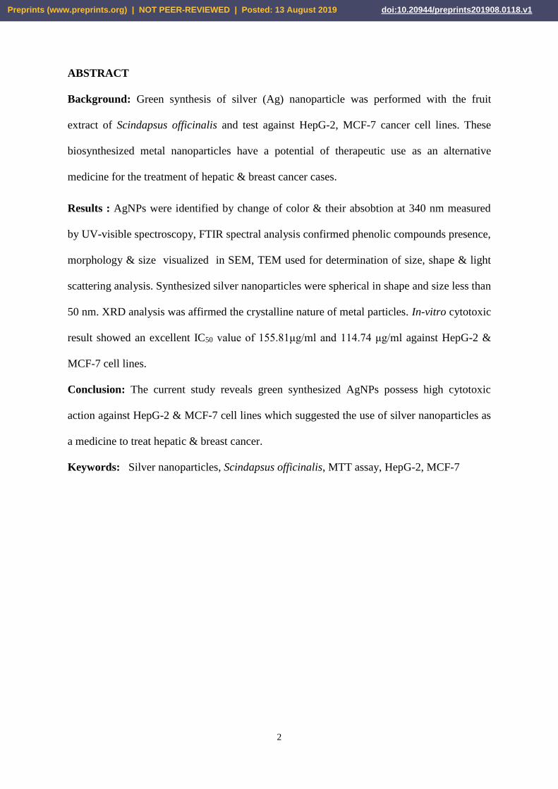
ABSTRACT
Background: Green synthesis of silver (Ag) nanoparticle was performed with the fruit
extract of Scindapsus officinalis and test against HepG-2, MCF-7 cancer cell lines. These
biosynthesized metal nanoparticles have a potential of therapeutic use as an alternative
medicine for the treatment of hepatic & breast cancer cases.
Results : AgNPs were identified by change of color & their absobtion at 340 nm measured
by UV-visible spectroscopy, FTIR spectral analysis confirmed phenolic compounds presence,
morphology & size visualized in SEM, TEM used for determination of size, shape & light
scattering analysis. Synthesized silver nanoparticles were spherical in shape and size less than
50 nm. XRD analysis was affirmed the crystalline nature of metal particles. In-vitro cytotoxic
result showed an excellent IC50 value of 155.81μg/ml and 114.74 μg/ml against HepG-2 &
MCF-7 cell lines.
Conclusion: The current study reveals green synthesized AgNPs possess high cytotoxic
action against HepG-2 & MCF-7 cell lines which suggested the use of silver nanoparticles as
a medicine to treat hepatic & breast cancer.
Keywords: Silver nanoparticles, Scindapsus officinalis, MTT assay, HepG-2, MCF-7
Preprints (www.preprints.org) | NOT PEER-REVIEWED | Posted: 13 August 2019 Preprints (www.preprints.org) | NOT PEER-REVIEWED | Posted: 13 August 2019 doi:10.20944/preprints201908.0118.v1

3
1. Background
Green synthesized Silver nanoparticles (AgNPs) is an integral part of
nanotechnology.(Kumar and Yadav, 2009)Research work on silver nanoparticles is set up
new trends in pharmaceutical field due to its wide therapeutic applications.(Mousavi et al.,
2018) Various papers reported application of silver nanoparticles including burn wounds
treatment by cream or ointments. Silver nanoparticles prepared by various ways such as
thermal decomposition, photochemical reductions in micelles, reduction in solutions,
chemicals, electrochemical, microwave, sonochemical methods.(Anandalakshmi et al., 2016)
Biological method reported recently for green synthesis of AgNPs using enzymes,
microorganisms & herbal plant extracts (Fig.1). Synthesis of AgNPs through biological
methods is without using any harmful chemicals & reagents so they are cost effective & eco-
friendly.(Abraham et al., 2017) Biological method has various application over traditional
methods (Physical &chemical methods) such as no requirement of heat, pressure &
temperature. Herbal plant extract arbitrated silver nanoparticles synthesis is widely used
recently because eco-friendly, safe & cost-effective.(Kajani et al., 2014)
Fig.1. Schematic of synthesis of silver nanoparticles using Scindapsus officinalis
Scindapsus Officinalis (Family – Araceae) commonly known as Gajpipli in hindi. S.
Officinalis is large, epiphytic, stout, perennial climber with adventitious roots. Fruits of S.
Preprints (www.preprints.org) | NOT PEER-REVIEWED | Posted: 13 August 2019 Preprints (www.preprints.org) | NOT PEER-REVIEWED | Posted: 13 August 2019 doi:10.20944/preprints201908.0118.v1
4
Officinalis mainly contains oil, Sterols, mixture of sugar and two glycosidic substance
Scindapsinidine-A, Scindapsinidine-B, rhamnose, fructose, glucose, xylose and polyphenols
Traditionally Gajpippli holds a reputed position in ayurvedic medicine system and its used in
chyawanprash as a active ingredient. It has been various reported ethanomedicinal uses like
antioxidant, anticancer, diarrhea, worm infestation, antipyretic.
Previous literature studies has reported about silver nanoparticles as anti-cancer agent & their
role as an anti-cancer agent could explore newer treatment therapy in the area of
pharmaceuticals with help treatment of cancer.(Barabadi et al., 2017) In present research
work, we reported first time green synthesis of AgNPs by reducing the Ag2+ (silver ion) of
silver nitrate by the aq. fruit extract of Scindapsus officinalis. Synthesized metal nanoparticles
particularly characterized by UV spectral analysis, FT-IR, SEM, TEM with mapping, EDAX,
XRD techniques. (Ferdous et al., 2013) Silver nanoparticles was identified by change of
color & absobtion peak confirmed by UV-visible spectroscopy, FT-IR spectra confirmed
latency of phenolic compounds, morphology & size visualized in SEM, TEM used for
determination of size, shape & light scattering analysis. (He et al., 2017) XRD used to affirm
crystalline nature of particles. (Supraja and Arumugam, 2015) Synthesized AgNPs from fruit
extracts of Scindapsus officinalis showed 50% inhibition of the cell viability of hepatic cell ,
Breast cancer cell lines. (Suseela, V; Lalitha, 2015) In-vitro result on cell lines showed an
exceptional cytotoxic action in term of IC50 value. (Sreekanth et al., 2016)
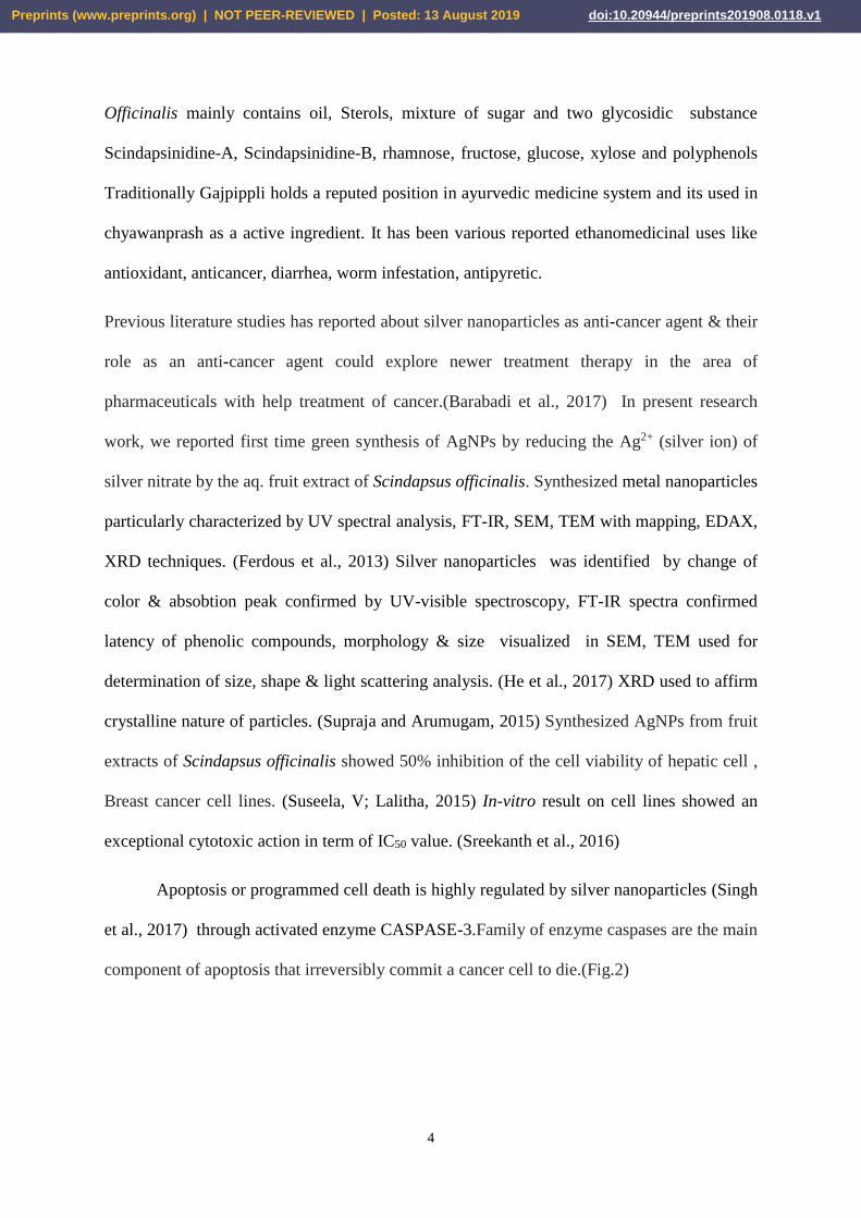
Apoptosis or programmed cell death is highly regulated by silver nanoparticles (Singh
et al., 2017) through activated enzyme CASPASE-3.Family of enzyme caspases are the main
component of apoptosis that irreversibly commit a cancer cell to die.(Fig.2)
Preprints (www.preprints.org) | NOT PEER-REVIEWED | Posted: 13 August 2019 Preprints (www.preprints.org) | NOT PEER-REVIEWED | Posted: 13 August 2019 doi:10.20944/preprints201908.0118.v1
5
Fig.2. Mechanism involved in CASPASE mediated apoptosis
2. Methods
2.1 Collection & authentication
Fresh fruits of Scindapsus officinalis (Shivhare et al., 2011) were collected from Gwalior,
M.P. The voucher specimen of Scindapsus officinalis authenticated by Botanical survey of
India, Allahabad (UP)-INDIA
2.2 Chemicals
Silver nitrate was procured from Sigma-Aldrich.
2.3 Preparation of plant extract
fruits were washed, dried & grind, 10 g powdered fruits was mixed with 100 ml double
distilled water & heated for 20 min. afterwards extract was get filtered.(Singh et al., 2009)
Preliminary phytochemical screening was performed to know about phytoconstituents present
in fruit extract.
2.4 Preparation of silver nitrate solution
Silver nitrate (1mM solution) 1.6 gm was dissolved in 1 liter double distilled water.(Medda
et al., 2015)
Preprints (www.preprints.org) | NOT PEER-REVIEWED | Posted: 13 August 2019 Preprints (www.preprints.org) | NOT PEER-REVIEWED | Posted: 13 August 2019 doi:10.20944/preprints201908.0118.v1

6
2.5 Synthesis of Ag-nanoparticles
10 % of Scindapsus officinalis plant extract was mixed with silver nitrate solution in 1:9
proportions &the mixture was kept for continuous stirring at room temperature for 48 hrs.
The resultant reddish brown color changes occurred in the solution due to formation of
reduced silver nanoparticles.(Xia et al., 2016) Reduced nano particles were collected after
centrifugation at 5000 rpm for 15 minutes.
2.6 Characterization of Silver nanoparticle
Ultraviolet–Visible spectroscopy
The reduction of Ag+ ions in Ag was confirmed by measuring the UV–visible spectrum.
(Mousavi et al., 2018) UV–visible spectral analysis was done by Perkin Elmer, Lamda
35.(Dasari and Anthony, 2017)
Fourier Transform Infrared spectroscopy
FT-IR is used to measure infrared absorption of the organic molecules found in the prepared
samples. A range of 800–4000 cm−1 using Shimadzu.(Mukundan et al., 2017) (He et al.,
2017)
Scanning Electron Microscope with Elemental Mapping
Synthesized Phytomolecules surface morphology confirmed by SEM. (Buttacavoli et al.,
2018) Characterization was carried out using ZEISS instruments.
Transmission Electron Microscope
Shape & size studied by TEM &it was confirmed by using Oxford instruments. (Bagherzade
et al., 2017)
Energy-dispersive X-ray spectroscopy
Elemental analysis (EDAX) confirmed by the Oxford instruments.(Khalil et al., 2014)
X-ray diffraction
X-ray diffraction of AgNPs carried out by using XPERT-PRO.(Ahmed et al., 2016)
Preprints (www.preprints.org) | NOT PEER-REVIEWED | Posted: 13 August 2019 Preprints (www.preprints.org) | NOT PEER-REVIEWED | Posted: 13 August 2019 doi:10.20944/preprints201908.0118.v1

7
Assessment of Cytotoxic activity on MCF-7 & HepG-2 cell lines by MTT assay method
Assay of anticancer effect of Scindapsus officinalis extract mediated synthesized AgNPs
(Kaur and Gupta, 2017) done by the help of MTT reduction (cell viability). (Devi et al.,
2012) (Bonigala et al., 2016) HepG-2 & MCF-7 cells were seeded in to separated plates &
each plate had 96-wells. HepG-2 & MCF-7 cells seeded at the density of 5 × 10³ cells/well.
Cells were allowed to grown & attach in 96-well plate for about 24 hrs. in 200 μl of
Dulbecco’s Modification of basal Medium Eagle (DMEM) with 10% Fetal bovine serum
(FBS) after the completion of 24 hrs. media were removed & replaced with the different
conc. of AgNPs ranging from 0.97 to 250 µg/ml . HepG-2 & MCF-7 cells were incubated for
48 hrs. Cells were incubated at 37°C for another 4 hrs. after the addition of 3-(4,5-
Dimethylthiazol-2-yl)-2,5-diphenyltetrazolium bromide (MTT) (10 ml, 5 mg/ ml). The
medium was then removed & 200 μl of Dimethyl sulfoxide (DMSO) added to each well
resultant a formazan product was formed. O.D of the formazan was read at 620 nm using
spectrophotometer (multi well). (Selvarani et al., 2015) Measurements were calculated & the
concentration required for a 50% inhibition of viability (K et al., 2018) was determined
graphically Standard Graph was plotted by taking conc. of the drug in X axis & relative cell
viability in Y axis.(Devi et al., 2012)
Mean OD
Cell viability (%) = x 100%
Control OD
3. Results
3.1 Phytochemical analysis
Glycosides, flavnoids tannins, Phenolic compounds, carbohydrate were present in fruit
extract. (Table 1)
Preprints (www.preprints.org) | NOT PEER-REVIEWED | Posted: 13 August 2019 Preprints (www.preprints.org) | NOT PEER-REVIEWED | Posted: 13 August 2019 doi:10.20944/preprints201908.0118.v1

8
Table 1: Phytochemical test
S. No. Chemical Test Aqueous extract
1. Alkaloids -
2. Glycosides +
3. Flavonoids +
4. Phytosterol -
5. Phenolic compounds and tannins +
6. Proteins -
7. Coumarins -
8. Saponins -
9. Carbohydrates +
3.2 Characterization of Ag-nanoparticles
Physical appearance
Silver nanoparticles synthesized by using 1 mM solution of silver nitrate and aq. fruits extract
of Scindapsus officinalis. However, after addition of silver nitrate, settled the reaction
mixture for continuous stirred at room temperature for about 48 hrs, the color of solution
turned light brown to dark brown in color shown in Fig.3.
Fig. 3. Biofabrication of silver nanoparticles by using Scindapsus officinalis aq. extract
Preprints (www.preprints.org) | NOT PEER-REVIEWED | Posted: 13 August 2019 Preprints (www.preprints.org) | NOT PEER-REVIEWED | Posted: 13 August 2019 doi:10.20944/preprints201908.0118.v1

9
UV–Visible spectral analysis
Light brown colored aq. extract was mixed with 1 Mm AgNO3 solution which changed in
dark brown color it’s because of S.P.R (Surface Plasmon Resonance) property of silver
(Supraja and Arumugam, 2015). The UV-visible observations reported that AgNPs had a
maximum absorbance at 340-380 nm. Synthesis of metal nanoparticles was observed at
different time intervals under UV-visible spectroscopy it shows synthesis of nanoparticles get
increased with time. Fruit extract reduced AgNO3 into Ag2+, the polyphenols present in
extract act as a reducing & capping agent for silver nanoparticles synthesis. (Fig.4)
Fig.4. UV-Visible spectra of AgNPs
FTIR spectral analysis
FTIR was used to characterize the surface & functional groups of AgNPs. FTIR spectra of
synthesized AgNPs showed marker absorption peaks at 504, 568, 1035, 1642, 2934 & 3476
cm-1, which confirmed that the plant molecules act as capping agents that were bound on
metal nanoparticle surface, peak at 3476 cm-1 was confirm for -OH stretching vibration, peak
at 2934 cm-1 confirmed the C-H stretching, confirmation of proteins by the amine or amide at
the region of 1606 cm-1, peak at 1642 cm-1 annex by AgNPs with the C=O functional groups,
peaks at 568 cm-1 represented C-H stretching of the aromatic & 504 cm-1 confirm O-H group
streching of a phenolic group. FTIR spectra exhibited that phytochemicals like phenolic
Preprints (www.preprints.org) | NOT PEER-REVIEWED | Posted: 13 August 2019 Preprints (www.preprints.org) | NOT PEER-REVIEWED | Posted: 13 August 2019 doi:10.20944/preprints201908.0118.v1

10
compounds, amino acids might protect the AgNPs from aggregation & thereby retain them
for long term stability. (Fig.5)
Fig.5. FTIR spectra of AgNPs
SEM imaging
Metal nanoparticles were agglomerated spheres with rough surface and with a diameter of
less than 50 nm affirmed by SEM analysis. It showed a spherical shape AgNPs were enclosed
by the different organic compounds. (Fig.6)
Fig.6. SEM images of AgNPs
Preprints (www.preprints.org) | NOT PEER-REVIEWED | Posted: 13 August 2019 Preprints (www.preprints.org) | NOT PEER-REVIEWED | Posted: 13 August 2019 doi:10.20944/preprints201908.0118.v1

11
Particle size from TEM analysis
AgNPs were spherical in shape & well dispersed while some other were irregular in shape &
less than 50 nm size. This feature explained that phytoconstituents present in aq. extract of
plant were effectively involved & affected the synthesis of silver nanoparticles. (Fig.7)
Fig.7. TEM images of AgNPs
Elemental analysis
Elemental mapping explain prepared nanoparticles exhibited maximum distribution of silver
as an element which is shown in image with red color, it confirmed that silver was the main
element present in sample refer to Fig.8.
Preprints (www.preprints.org) | NOT PEER-REVIEWED | Posted: 13 August 2019 Preprints (www.preprints.org) | NOT PEER-REVIEWED | Posted: 13 August 2019 doi:10.20944/preprints201908.0118.v1

12
Fig.8.Elemental mapping of AgNPs
EDAX Analysis
Confirmation of the elemental silver nanoparticles of Scindapsus officinalis was observed in
the graph obtained from EDAX analysis. Chemical composition of Ag was 54.04 wt%. This
result indicates the reduction of silver ions to elemental silver. The EDAX spectra affirmed
the presence of peak for elemental Ag at at 3 keV. Oxygen (O) & carbon (C) peaks might be
due existence of bio-organic compounds bound on the surface. (Fig. 9)
Fig.9. EDAX image
Preprints (www.preprints.org) | NOT PEER-REVIEWED | Posted: 13 August 2019 Preprints (www.preprints.org) | NOT PEER-REVIEWED | Posted: 13 August 2019 doi:10.20944/preprints201908.0118.v1

13
Degree of Crystallinity
XRD was carried out to identify the crystalline structure & chemical composition of a metal
nanoparticles therefore, presence of Ag (silver) in nanoparticles confirmed by diffraction
peaks. Synthesized silver nanoparticles crystal plane showed 2Q angles at the range of 38.68,
44.1, 64.11, 77.4 corresponding to 111, 200, 220 & 222 affirmed the formation face-centered
cubic silver crystal. (Fig.10)
Fig.10. XRD spectrum
3.3 Cytotoxicity analysis
Cytotoxicity action of the AgNPs were studied against the HepG-2 (Table 2 &3) & MCF-7
cell line by MTT assay (Table 4 &5). Cytotoxicity effect on cancerous cell was studied at
different concentrations (0.97 μg/mL,1.9 μg/mL,3.9 μg/mL,7.8 μg/mL,15.6 μg/mL,31.25
μg/mL, 62.5 μg/mL,125 μg/mL,250 μg/mL) and compared with normal control. IC50 of the
phytoconstituted AgNPs observed at conc. of 155.81 μg/mL against HepG-2 cell line. This
result shows that the minimum dose showed good cytotoxic activity. IC50 value of the
phytoconstituted AgNPs was confirmed at 114.74μg/mL against MCF-7 cells. The bar
diagram of efficacy of biosynthesized Silver nanoparticles against HepG-2 (Fig. 11) & MCF-
7 cells at different concentration (Fig. 12).
Preprints (www.preprints.org) | NOT PEER-REVIEWED | Posted: 13 August 2019 Preprints (www.preprints.org) | NOT PEER-REVIEWED | Posted: 13 August 2019 doi:10.20944/preprints201908.0118.v1

14
Table 2 Absorbance at different concentration in HepG-2 cell line.
Conc. μg/mL Normal Control
0.97
μg/mL
1.9
μg/mL
3.9
μg/mL
7.8
μg/mL
15.6
μg/mL
31.25
μg/mL
62.5
μg/mL
125
μg/mL
250
μg/mL
Absorbance
0.922 0.893 0.872 0.712 0.758 0.699 0.721 0.573 0.506 0.42
Absorbance
0.917 0.911 0.96 0.747 0.802 0.714 0.729 0.567 0.464 0.416
Table 3 Cell Viability (%) in HepG-2 cell line at different concentration.
Conc. μg/mL
Normal
Control
0.97
μg/mL
1.9
μg/mL
3.9
μg/mL
7.8
μg/mL
15.6
μg/mL
31.25
μg/mL
62.5
μg/mL
125
μg/mL
250
μg/mL
% viability 100 96.8546638 94.577007 77.223427 82.212581 75.813449 78.199566 62.147505 54.880694 45.553145
% viability 100 99.3456925 104.6892 81.461287 87.459106 77.862595 79.498364 61.832061 50.599782 45.365322
Fig.11. Bar diagram of efficacy of AgNPs
Preprints (www.preprints.org) | NOT PEER-REVIEWED | Posted: 13 August 2019 Preprints (www.preprints.org) | NOT PEER-REVIEWED | Posted: 13 August 2019 doi:10.20944/preprints201908.0118.v1

15
Table 4 Absorbance at different concentration in MCF-7 cell line.
Conc. μg/mL Normal Control
0.97
μg/mL
1.9
μg/mL
3.9
μg/mL
7.8
μg/mL
15.6
μg/mL
31.25
μg/mL
62.5
μg/mL
125
μg/mL
250
μg/mL
Absorbance 0.977 0.823 0.9 0.789 0.885 0.687 0.501 0.492 0.46 0.435
Absorbance
0.961 0.816 0.888 0.771 0.866 0.689 0.502 0.492 0.449 0.43
Table 5 Cell viability (%) in MCF-7 cell line at different concentration.
Conc. μg/mL
Normal
Control
0.97
μg/mL
1.9
μg/mL
3.9
μg/mL
7.8
μg/mL
15.6
μg/mL
31.25
μg/mL
62.5
μg/mL
125
μg/mL
250
μg/mL
% viability 100 84.2374616 92.118731 80.757421 90.583419 70.317298 51.279427 50.35824 47.082907 44.524053
% viability 100 84.9115505 92.403746 80.228928 90.114464 71.69615 52.237253 51.19667 46.722164 44.745057
‘
Fig.12. Bar diagram of efficacy of AgNPs
Preprints (www.preprints.org) | NOT PEER-REVIEWED | Posted: 13 August 2019 Preprints (www.preprints.org) | NOT PEER-REVIEWED | Posted: 13 August 2019 doi:10.20944/preprints201908.0118.v1
16
4. Discussion
In this research plan, we have used Scindapsus officinalis fruits extract contains polyphenols
which act as stabilizing and reducing agent for biofabrication of AgNPs. Biosynthesized
AgNPs was preliminarily confirmed by color change from light brown to dark brown in
mixture. (Fig.3.) Brown color arising due to the surface plasmon resonance (SPR)
phenomenon, which originate due to the interaction of EMF with surface oscillated free
electrons of AgNPs. SPR absorption band observed at 340-380 nm in UV-visible spectrum.
(Fig.4)
AgNPs biosynthesis, shape, size, surface texture, degree of crystalline and Ag percent
of presence is confirmed by FTIR, SEM, TEM, EDAX and XRD results. FTIR spectra
reveled that the surface capping functionalities of the prepared AgNPs. Phytochemicals like
phenolic compounds, amino acids might protect the AgNPs from aggregation & thereby
retain them for long term stability. (Fig.5) AgNPs were agglomerated spheres with rough
surface and a diameter of less than 50 nm reported by SEM images.(Fig.6) TEM confirmed
their spherical shape with some other AgNPs found to be irregular shape. (Fig.7) Elemental
analysis and EDAX reported the distribution pattern, percent (%) presence of Ag as an
element in biosynthesized metal nanoparticles. (Fig.8 & 9) XRD pattern of AgNPs with
distinctive peaks characteristic to the indexing planes showed the crystalline nature of the
formed AgNPs. (Fig.10)
Moreover, the size of the AgNPs obtained is in good agreement confirmed with the
SEM, TEM and UV absorption peak obtained at 340-380 nm, indicating small, spherical NPs
formation, as the shift in the SPR band of Ag, Au NPs is associated with their size. This
change in the properties of NPs with size is due to the quantum confinement effect. The
formation of small and spherical nanoparticles is useful in various therapeutic conditions.
Small and spherical AgNPs can easily cross the cellular membranes where as larger particles
found difficulty to cross.
Preprints (www.preprints.org) | NOT PEER-REVIEWED | Posted: 13 August 2019 Preprints (www.preprints.org) | NOT PEER-REVIEWED | Posted: 13 August 2019 doi:10.20944/preprints201908.0118.v1

17
AgNPs showed dose dependant cytotoxicity against the hepatic (HepG-2) & breast
cancer (MCF-7) cell lines. In-vitro cytotoxic result of biosynthesized AgNPs showed
excellent IC50 value of 155.81μg/ml against HepG-2 and 114.74 μg/ml against MCF-7 cancer
cell lines. (Fig.11 & 12) It is well known that the mechanism of cytotoxicity of AgNPs
involves the cellular uptake of nanoparticles via macropinocytosis and clathrin-dependent
endocytosis. Various studies have revealed that AgNPs works by triggering the intracellular
ROS by preventing the intracellular antioxidants. The immediately formed ROS then damage
the DNA which results in the cell death. It have showed that the highly reactive hydroxyl
radicals produced by silver nanoparticles damage the cellular components such as DNA
leading to cell death. From the earlier studies, it is concluded that the AgNPs coated with
plant bioconstituents induce oxidative stress leading to the apoptosis via caspase-mediated
and mitochondria-dependent pathways.
5. Conclusions
This research reports Green, facile, Simple, inexpensive synthesis of Silver nanoparticles
from Scindapsus officinalis in aqueous medium without using any hazardous chemicals. The
UV–Vis spectroscopy & FTIR affirmed the formation of silver nanoparticles. EDAX, XRD
affirms presence of silver (Ag). SEM & TEM image showed spherical shape with an average
particle size of less than 50 nm. The biosynthesized metal nanoparticles & Scindapsus
officinalis extract showed promising cytotoxic activity against human hepatic cancer cell line
(HepG-2) & breast cancer cell line (MCF-7). In conclusion the synthesized AgNPs using
Scindapsus officinalis extract possess high cytotoxic action against HepG-2 & MCF-7 cell
lines which suggested the potential therapeutic use of these silver nanoparticles as alternative
medicine for the treatment of hepatic & breast cancer.
Preprints (www.preprints.org) | NOT PEER-REVIEWED | Posted: 13 August 2019 Preprints (www.preprints.org) | NOT PEER-REVIEWED | Posted: 13 August 2019 doi:10.20944/preprints201908.0118.v1
18
Abbreviations
S.O: Scindapsus officinalis
Ag: Silver
AgNPs: Silver nanoparticles
AgNO: Silver nitrate
MTT: 3-(4,5-Dimethylthiazol-2-yl)-2,5-diphenyltetrazolium bromide
DMSO : Dimethyl sulfoxide
U.V: Ultra violet spectroscopy
FTIR: Fourier transform infrared spectroscopy
SEM: Scanning electron microscope
TEM: Transmission electron microscope
EDAX: Energy dispersive X-ray analysis
XRD: X-rays diffraction
HepG-2: Hepatic cancer cell line
MCF-7: Breast cancer cell line
OD: Optical density
IC50: Inhibitory concentration
FBS: Fetal bovine serum
DMEM: Dulbecco’s Modification of basal Medium Eagle
Aq.: Aqueous
Conc.: Concentration
N.C: Normal control
E.M.F: Electromagnetic field
R.O.S: Reactive oxygen species
%: Percent
Preprints (www.preprints.org) | NOT PEER-REVIEWED | Posted: 13 August 2019 Preprints (www.preprints.org) | NOT PEER-REVIEWED | Posted: 13 August 2019 doi:10.20944/preprints201908.0118.v1

19
Funding
This research work is not funded by any agency.
Conflict of interest
Authors have no conflict of interest
Author contributions
VK & PRT carried out study, data collection, analysis and interpretation. RM conducts
sample characterization. PP carried out In-vitro testing on cell lines. AV participated in the
design of the study and performed analysis. MP conceived of the study, and participated in its
design and coordination and helped to draft the manuscript. All authors read and approved
the final manuscript.
Acknowledgement
Authors are sincerely thankful to B.S.I, Allahabad (U.P) for authentication of plant, SAIF,
Punjab University (Punjab) for XRD, Patanjali Research Institute, Haridwar (U.K) for help
rendered in cytotoxic activity on cell lines , IISER, Kolkata for Providing characterization
facility (SEM, TEM, EDAX, Mapping) & Department of Pharmaceutical sciences, SHUATS,
Allahabad (U. P.) for providing research facilities.
Preprints (www.preprints.org) | NOT PEER-REVIEWED | Posted: 13 August 2019 Preprints (www.preprints.org) | NOT PEER-REVIEWED | Posted: 13 August 2019 doi:10.20944/preprints201908.0118.v1

20
References
Abraham, J., Saraf, S., Mustafa, V., Chaudhary, Y., Sivanangam, S., 2017. Synthesis and
evaluation of silver nanoparticles using Cymodocea rotundata against clinical pathogens
and human osteosarcoma cell line. J. Appl. Pharm. Sci. 7, 055–061.
https://doi.org/10.7324/JAPS.2017.70608
Ahmed, S., Saifullah, Ahmad, M., Swami, B.L., Ikram, S., 2016. Green synthesis of silver
nanoparticles using Azadirachta indica aqueous leaf extract. J. Radiat. Res. Appl. Sci. 9,
1–7. https://doi.org/10.1016/j.jrras.2015.06.006
Anandalakshmi, K., Venugobal, J., Ramasamy, V., 2016. Characterization of silver
nanoparticles by green synthesis method using Pedalium murex leaf extract and their
antibacterial activity. Appl. Nanosci. 6, 399–408. https://doi.org/10.1007/s13204-015-
0449-z
Bagherzade, G., Tavakoli, M.M., Namaei, M.H., 2017. Green synthesis of silver
nanoparticles using aqueous extract of saffron (Crocus sativus L.) wastages and its
antibacterial activity against six bacteria. Asian Pac. J. Trop. Biomed. 7, 227–233.
https://doi.org/10.1016/j.apjtb.2016.12.014
Barabadi, H., Ovais, M., Shinwari, Z.K., Saravanan, M., 2017. Anti-cancer green
bionanomaterials: Present status and future prospects. Green Chem. Lett. Rev. 10, 285–
314. https://doi.org/10.1080/17518253.2017.1385856
Bonigala, B., Aswani Kumar, Y. V, Vinay Viswanath, K., Joy Richardson, P., Mangamuri,
U.K., Poda, S., 2016. Anticancer activity of plant mediated silver nanoparticles on
selected cancer cell lines. J. Chem. Pharm. Res. 8, 376–381.
Buttacavoli, M., Albanese, N.N., Di Cara, G., Alduina, R., Faleri, C., Gallo, M., Pizzolanti,
Preprints (www.preprints.org) | NOT PEER-REVIEWED | Posted: 13 August 2019 Preprints (www.preprints.org) | NOT PEER-REVIEWED | Posted: 13 August 2019 doi:10.20944/preprints201908.0118.v1

21
G., Gallo, G., Feo, S., Baldi, F., Cancemi, P., 2018. Anticancer activity of biogenerated
silver nanoparticles: an integrated proteomic investigation. Oncotarget 9, 9685–9705.
https://doi.org/10.18632/oncotarget.23859
Dasari, P.R., Anthony, P., 2017. Synthesis, Characterization of Silver Nanoparticles using
Rosa damascena Hips Extract and 43, 13–19.
Devi, J.S., Bhimba, B. V., Ratnam, K., 2012. In vitro anticancer activity of silver
nanoparticles synthesized using the extract of Gelidiella Sp. Int. J. Pharm. Pharm. Sci. 4.
Ferdous, N., Hridi, S.U., Hannan, J.M.A., 2013. Antinociceptive and Hypoglycemic
Activities of Scindapsus officinalis ( Roxb .) Schott in Laboratory Animals 3, 561–576.
He, Y., Wei, F., Ma, Z., Zhang, H., Yang, Q., Yao, B., Huang, Z., Li, J., Zeng, C., Zhang, Q.,
2017. Green synthesis of silver nanoparticles using seed extract of: Alpinia katsumadai,
and their antioxidant, cytotoxicity, and antibacterial activities. RSC Adv. 7, 39842–
39851. https://doi.org/10.1039/c7ra05286c
K, K.K., B, D. kumar, Punathil, R.R., 2018. Green Synthesis of Silver Nanoparticles Using
Hydnocarpus pentandra Leaf Extract: In-vitro Cyto-Toxicity Studies Against MCF-7
Cell Line. J. Young Pharm. 10, 16–19. https://doi.org/10.5530/jyp.2018.10.5
Kajani, A.A., Bordbar, A.-K., Zarkesh Esfahani, S.H., Khosropour, A.R., Razmjou, A., 2014.
Green synthesis of anisotropic silver nanoparticles with potent anticancer activity using
Taxus baccata extract. RSC Adv. 4, 61394–61403.
https://doi.org/10.1039/C4RA08758E
Kaur, K., Gupta, R., 2017. Ethnobotanical and phytopharmacological review of Scindapsus
officinalis (“Gajapippali”). Asian Pac. J. Trop. Biomed. 7, 78–85.
https://doi.org/10.1016/j.apjtb.2016.10.010
Preprints (www.preprints.org) | NOT PEER-REVIEWED | Posted: 13 August 2019 Preprints (www.preprints.org) | NOT PEER-REVIEWED | Posted: 13 August 2019 doi:10.20944/preprints201908.0118.v1

22
Khalil, M.M.H., Ismail, E.H., El-Baghdady, K.Z., Mohamed, D., 2014. Green synthesis of
silver nanoparticles using olive leaf extract and its antibacterial activity. Arab. J. Chem.
7, 1131–1139. https://doi.org/10.1016/j.arabjc.2013.04.007
Kumar, V., Yadav, S.K., 2009. Plant-mediated synthesis of silver and gold nanoparticles and
their applications. J. Chem. Technol. Biotechnol. 84, 151–157.
https://doi.org/10.1002/jctb.2023
Medda, S., Hajra, A., Dey, U., Bose, P., Mondal, N.K., 2015. Biosynthesis of silver
nanoparticles from Aloe vera leaf extract and antifungal activity against Rhizopus sp.
and Aspergillus sp. Appl. Nanosci. 5, 875–880. https://doi.org/10.1007/s13204-014-
0387-1
Mousavi, B., Tafvizi, F., Zaker Bostanabad, S., 2018. Green synthesis of silver nanoparticles
using Artemisia turcomanica leaf extract and the study of anti-cancer effect and
apoptosis induction on gastric cancer cell line (AGS). Artif. Cells, Nanomedicine,
Biotechnol. 0, 1–12. https://doi.org/10.1080/21691401.2018.1430697
Mukundan, D., Mohankumar, R., Vasanthakumari, R., 2017. Comparative study of
synthesized silver and gold nanoparticles using leaves extract of Bauhinia tomentosa
Linn and their anticancer efficacy. Bull. Mater. Sci. 40, 335–344.
https://doi.org/10.1007/s12034-017-1376-2
Selvarani, S., Pv, M., Saranya, P., Abirami, M., 2015. Anti-Cancer Activity of Silver
Nanoparticle Synthesized from Stem Extract of ocimum Kilimandscharicum Against
Hep-G2 , Liver Cancer Cell Line 12, 1–7.
Shivhare, S., Malviya, K., Patidar, A., Shivhare-Malviya, K., 2011. Antioxidant and
anticancer evaluation of Scindapsus officinalis (Roxb.) Schott fruits. AYU (An Int. Q. J.
Preprints (www.preprints.org) | NOT PEER-REVIEWED | Posted: 13 August 2019 Preprints (www.preprints.org) | NOT PEER-REVIEWED | Posted: 13 August 2019 doi:10.20944/preprints201908.0118.v1
23
Res. Ayurveda) 32, 388. https://doi.org/10.4103/0974-8520.93921
Singh, J., Singh, T., Rawat, M., 2017. Green synthesis of silver nanoparticles via various
plant extracts for anti-cancer applications. Glob. J. nanomedicine 2, 2–5.
Singh, M., Velraj, M., College, V., 2009. In-vitro Evaluation of Scindapsus Officinalis (
ROXB .) Schott . Fruit for Antioxidant Potential 1, 83–86.
Sreekanth, T.V.M., Pandurangan, M., Kim, D.H., Lee, Y.R., 2016. Green Synthesis: In-vitro
Anticancer Activity of Silver Nanoparticles on Human Cervical Cancer Cells. J. Clust.
Sci. 27, 671–681. https://doi.org/10.1007/s10876-015-0964-9
Supraja, S., Arumugam, P., 2015. Antibacterial and Anticancer Activity of Silver
Nanoparticles Synthesized from Cynodon dactylon Leaf Extract 3, 629–631.
Suseela, V; Lalitha, G., 2015. Cytotoxic effect of green synthesized silver nanoparticles using
Indigofera longeracemosa on skin cancer SK MEL-28 cell lines. Int. J. Preclin. Pharm.
Res. 6, 118–125.
Xia, Q., Ma, Y., Wang, J., 2016. Biosynthesis of Silver Nanoparticles Using Taxus
yunnanensis Callus and Their Antibacterial Activity and Cytotoxicity in Human Cancer
Cells. Nanomaterials 6, 160. https://doi.org/10.3390/nano6090160
Preprints (www.preprints.org) | NOT PEER-REVIEWED | Posted: 13 August 2019 Preprints (www.preprints.org) | NOT PEER-REVIEWED | Posted: 13 August 2019 doi:10.20944/preprints201908.0118.v1