Grundlagen Und Praxis Der Anti-Aging Medizin Text
49
Praxis Dr. Davis Praxis Dr. Davis Was ist Altern? Was ist Altern? Alterungsprozess Alterungsprozess – Universelle biologische Ver Universelle biologische Verä nderungen nderungen Programm Programm Prozess des Alterns Prozess des Alterns – Ver Veränderungen, die stark durch nderungen, die stark durch Umwelteinfl Umwelteinflüsse, Lifestyle und Krankheiten sse, Lifestyle und Krankheiten beinflusst beinflusst werden werden Abn Abnützung und Verbrauch tzung und Verbrauch Programm-Theorien Stochastische-Theorien Dieser Punkt scheint offensichtlich zu sein. Wichtig sind für uns zunächst zwei zentrale Folgerungen, die sich daraus ergeben: 1. Veränderungen verlaufen interindividuell mit unterschiedlicher Geschwindigkeit. Daher die Unterscheidung zwischen „chronologischem“ (d.h. nach Lebensalter definiertem) und „funktionalem“ (d.h. nach tatsächlicher Leistungsfähigkeit definiertem) Alter. 2. Das Ziel individueller und gesellschaftlicher Bemühungen sollte darin bestehen, bestimmte irreversible Veränderungen möglichst weit hinauszuschieben. Wobei sich die Gewinne vor allem auf die weitere Differenzierung bereits ausgebildeter Fähigkeiten beziehen, die Verluste vor allem auf die Kapazität zum Erwerb neuer Fähigkeiten. Praxis Dr. Davis Praxis Dr. Davis Alterungsprozesse entstehen auf dem Alterungsprozesse entstehen auf dem Boden unzureichender Boden unzureichender Reparations Reparations- mechanismen mechanismen bei konstanter Sch bei konstanter Schä digung digung Stochastische Theorien Stochastische Theorien Error- Cata- strophe Freie Radikale Cross- linking Order to Disorder Wear und Tear Zellab- fallak- kumu- lation Lebens- rate Abnahme der Reparaturmechanismen Abnahme der Reparaturmechanismen Akkumulation zerst Akkumulation zerstö rerischer Verletzungen rerischer Verletzungen Glykosy lierung
-
Upload
swissestetix -
Category
Health & Medicine
-
view
3.040 -
download
6
description
Das sind Textbausteine zum Vortrag "Grundlagen Und Praxis Der Anti-Aging Medizin". Es werden Studien und Abstracts und sonstige Kommentare und Bemerkungen aufgefuehrt
Transcript of Grundlagen Und Praxis Der Anti-Aging Medizin Text

Praxis Dr. DavisPraxis Dr. Davis
Was ist Altern?Was ist Altern?
Alterungsprozess Alterungsprozess –– Universelle biologische VerUniverselle biologische Veräänderungennderungen
ProgrammProgramm
Prozess des AlternsProzess des Alterns–– VerVeräänderungen, die stark durch nderungen, die stark durch
UmwelteinflUmwelteinflüüsse, Lifestyle und Krankheiten sse, Lifestyle und Krankheiten beinflusstbeinflusst werdenwerden
AbnAbnüützung und Verbrauchtzung und Verbrauch
Programm-Theorien
Stochastische-Theorien
Dieser Punkt scheint offensichtlich zu sein. Wichtig sind für uns zunächst zwei zentrale Folgerungen, die sich daraus ergeben: 1. Veränderungen verlaufen interindividuell mit unterschiedlicher Geschwindigkeit. Daher die Unterscheidung zwischen „chronologischem“ (d.h. nach Lebensalter definiertem) und „funktionalem“ (d.h. nach tatsächlicher Leistungsfähigkeit definiertem) Alter.
2. Das Ziel individueller und gesellschaftlicher Bemühungen sollte darin bestehen, bestimmte irreversible Veränderungen möglichst weit hinauszuschieben.
Wobei sich die Gewinne vor allem auf die weitere Differenzierung bereits ausgebildeter Fähigkeiten beziehen, die Verluste vor allem auf die Kapazität zum Erwerb neuer Fähigkeiten.
Praxis Dr. DavisPraxis Dr. Davis
Alterungsprozesse entstehen auf dem Alterungsprozesse entstehen auf dem Boden unzureichender Boden unzureichender ReparationsReparations--mechanismenmechanismen bei konstanter Schbei konstanter Schäädigung digung
Stochastische Theorien Stochastische Theorien
Error-Cata-strophe
Freie Radikale
Cross-linking
Order to Disorder
Wearund Tear
Zellab-fallak-kumu-lation
Lebens-rate
Abnahme der ReparaturmechanismenAbnahme der ReparaturmechanismenAkkumulation zerstAkkumulation zerstöörerischer Verletzungenrerischer Verletzungen
Glykosylierung
© Dr. C
laren
ce P
. Dav
is 20
09

The "random damage" theories are based on the possibility that the balance between ongoing damage and repair is disrupted. The theories differ in the emphasis placed on increased damage (e.g., by free radicals, oxidation, or glycation) versus deficient repair, as well as in the mechanisms that might mediate each. However, all share the observation that cell and organ repair capacity declines with age.
Praxis Dr. DavisPraxis Dr. Davis
Theorie der freien Radikale Theorie der freien Radikale
Aggressive Sauerstoffradikale (OAggressive Sauerstoffradikale (O22--, OH, OH--, , HH22OO22) greifen die Zelle und/oder Zellwand ) greifen die Zelle und/oder Zellwand an und fan und füühren dort zur Bildung neuer hren dort zur Bildung neuer aggressiver Sauerstoffradikale aggressiver Sauerstoffradikale (Kettenreaktion)(Kettenreaktion)–– Antioxidantien kAntioxidantien köönnen die Lebensspanne von nnen die Lebensspanne von
WWüürmern und Mrmern und Määusen um bis zu 30% usen um bis zu 30% verlverläängernngern
Freie Radikale
Harman D; J Gerontol 1956Rose M.R.; Int J Org Evol 1984
Mit der Theorie der freien Radikale verbindet sich sowohl die Wear- and Tear Theorie, als auch die Theorie der Zellabfälle. Der überzeugendste Beweis für diese Theorie wurde an Longevity-Experimenten bei Rundwürmern und Mäusen erbracht.
Praxis Dr. DavisPraxis Dr. Davis
Die Die nichtenzymatischenichtenzymatische GlykosylierungGlykosylierungbestimmter Proteine im Kbestimmter Proteine im Köörper frper füührt zur hrt zur Bildung und AnhBildung und Anhääufung bleibender ufung bleibender QuerQuer--verbindungenverbindungen, die f, die füür den im Alter r den im Alter auftretenden Elastizitauftretenden Elastizitäätsts-- und und FunktionsFunktions--verlustverlust typisch sindtypisch sind–– Menschen mit hohen Blutzuckerspiegeln altern Menschen mit hohen Blutzuckerspiegeln altern
besonders schnell besonders schnell Komplikationen Komplikationen beim Diabetes mellitus kbeim Diabetes mellitus köönnen auch als Zeichen nnen auch als Zeichen einer vorzeitigen Organalterung interpretiert einer vorzeitigen Organalterung interpretiert werden.werden.
Glykosylierung
AGEAGE--TheorieTheorie((AdvancedAdvanced GlycosylationGlycosylation EndproductsEndproducts))
Kristal B; Gerontal Biol Sci 1992
© Dr. C
laren
ce P
. Dav
is 20
09

Nach dieser Theorie führt die nichtenzymatische Glykosylierung bestimmter Proteine im Körper zur Bildung und Anhäufung bleibender Querverbindungen, die für den im Alter auftretenden Elastizitäts- und Funktionsverlust typisch sind. Eine Schlüsselrolle spielt dabei der Blutzucker. Die Schritte, durch welche die Glukose die Proteine derart verändert, werden als Maillard- oder Bräunungsreaktion bezeichnet. Der Prozess beginnt damit, dass eine Aldehyd-Gruppe (-COH) der Glukose und eine Aminogruppe (-NH2) eines Proteins sich gegenseitig anziehen. Die Moleküle vereinigen sich zu einer so genannten Schiffschen Base, die instabil ist, sich aber schnell in eine stabilere, jedoch immer noch reversible Substanz umformen. Diese ist als Amadori-Produkt bekannt. Wenn ein Protein monate- oder jahrelang im Körper verweilt, verlieren einige seiner Amadori-Produkte Wasser und bilden neue Strukturen, die sich aus Glukose herleiten. Diese können sich dann mit verschiedenen Arten von Molekülen zu irreversiblen Strukturen verbinden, die als Endprodukte fortgeschrittener Glykosylierung (Advanced Glycosylation Endproducts) bezeichnet werden. Falls diese These zutrifft, so muss man davon ausgehen, dass Menschen mit sehr hohen Blutzuckerspiegeln besonders viel AGE-Produkte ausbilden und somit vorzeitig altern. In der Tat lassen sich viele der Komplikationen, die im Rahmen eines Diabetes mellitus auftreten, als Zeichen einer vorzeitigen Organalterung interpretieren. Hierzu gehört die Ausbildung einer Mikroangiopathie ebenso wie das Entstehen einer Niereninsuffizienz oder eines Kataraktes. Der Diabetiker wird somit zu einem Modellfall für die Untersuchung des Alterungsprozesses.
Praxis Dr. DavisPraxis Dr. Davis
AGEAGE--TheorieTheorie((AdvancedAdvanced GlycosylationGlycosylation EndproductsEndproducts))
Jacot L; J Ocul Pharmacol Ther 1998
FIGURE 3: Cross-sections of the iris (top 4 photos) and retina (bottom 4 pharos) from S-D control rats (Column A) or STZ-induced S-D diabetic rats of 4 wks duration (Column B). Light micrographs demonstrate increased immunoreactivity (blue chromogen) in the ocular tissue of STZ-induced diabetic rats relative to age matched control rats. Anti-Amadori antibody specificjty was demonstrated by preabsorption with a synthetic glycated peptide (ac-KOTAL) which prevented immunoreactivity (rows 2 and 4). Tissues ware counterstained with nuclear fast red. Eye tissue was paraffin-embedded and sectioned at 5-61Jm thick. Same magnification for all images (410x).
© Dr. C
laren
ce P
. Dav
is 20
09

Praxis Dr. DavisPraxis Dr. Davis
„„Tod entsteht dadurch, dass ein Tod entsteht dadurch, dass ein ausgeausge--laugterlaugter Organismus sich nicht immer von Organismus sich nicht immer von Neuem regenerieren kannNeuem regenerieren kann““–– Altern kAltern köönnte durch fortschreitende genetische nnte durch fortschreitende genetische
ZerstZerstöörung, die dann akkumuliert, rung, die dann akkumuliert, hervorhervor--gerufengerufen werdenwerden
WearWear and and TearTear--TheorieTheorie
Wearund Tear
August Weismann; 1882
Diese Theorie ist sehr alt und geht auf August Weismann zurück, der sie 1882 erstmals veröffentlichte. Ein einfaches Beispiel für diese Theorie sind die Zähne des Menschen, die, einmal verbraucht, nicht mehr zur Regeneration fähig sind. Komplexere Zusammenhänge werden auf molekularer Ebene vermutet, wo somatische Mutationen für den Alterungsprozess verantwortlich sein könnten. Diese Behauptung ist bis heute allerdings nicht bewiesen. Es steht aber fest, dass Fehler in der Genexpression durch Transkription oder translationale Störungen zu Veränderungen der Proteinsynthese führen und damit die Enzymmenge und –funktion, wie auch den Aufbau von Strukturproteinen erheblich negativ in Richtung Funktionsschwäche und Funktionsverlust entwickeln.
Praxis Dr. DavisPraxis Dr. Davis
Theorie der Theorie der ErrorError--CatastropheCatastrophe
Mit zunehmendem Alter treten immer mehr Mit zunehmendem Alter treten immer mehr Fehler in der AminosFehler in der Aminosääurensequenz der urensequenz der Proteine auf, was Proteine auf, was schliesslichschliesslich zum Zelltod zum Zelltod ffüührt.hrt.–– Bsp. Kataraktbildung durch AnhBsp. Kataraktbildung durch Anhääufung von ufung von
KrystallinproteinKrystallinprotein in der Linsein der Linse–– Biologische Altersbestimmung durch Messung Biologische Altersbestimmung durch Messung
von von dd--AspartatAspartat im Zahnschmelzim Zahnschmelz
Error-Cata-strophe
Orgel LE; Proc Natl Acad Sci USA 1963Orgel LE; Proc Natl Acad Sci USA 1970
© Dr. C
laren
ce P
. Dav
is 20
09
Diese Theorie ist breit getestet in einer Vielzahl von Spezies und Zelltypen. Dabei spielt die verminderte Kapazität alter Zellen, Proteine mit einer gestörten AS-Sequenz zu entfernen, eine wichtige Rolle, was schliesslich zum Funktionsausfall von zellulären Prozessen führen kann. Diese Theorie kann erweitert werden zur DNS-Zerstörungstheorie, die besagt, dass die Fähigkeit, verletzte DNS zu reparieren, mit zunehmendem Alter herabgesetzt ist.
Praxis Dr. DavisPraxis Dr. Davis
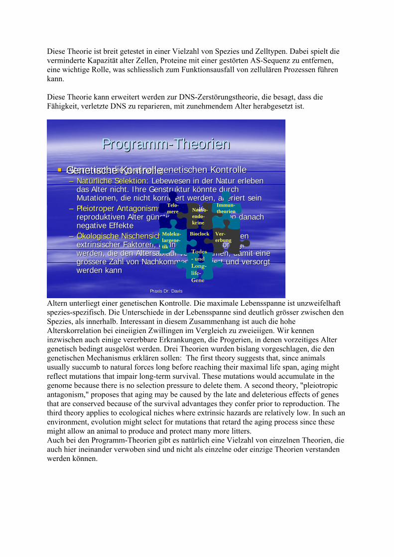
ProgrammProgramm--TheorienTheorien
Altern unterliegt einer genetischen KontrolleAltern unterliegt einer genetischen Kontrolle–– NatNatüürliche Selektionrliche Selektion: Lebewesen in der Natur erleben : Lebewesen in der Natur erleben
das Alter nicht. Ihre das Alter nicht. Ihre GenstrukturGenstruktur kköönnte durch nnte durch Mutationen, die nicht korrigiert werden, alteriert seinMutationen, die nicht korrigiert werden, alteriert sein
–– PleiotroperPleiotroper AntagonismusAntagonismus: Gene, die sich im : Gene, die sich im reproduktiven Alter greproduktiven Alter güünstig auswirken, haben danach nstig auswirken, haben danach negative Effektenegative Effekte
–– ÖÖkologische Nischensicherungkologische Nischensicherung: Durch das Fehlen : Durch das Fehlen extrinsischerextrinsischer Faktoren, kFaktoren, köönnten Gene bevorzugt nnten Gene bevorzugt werden, die den Altersablauf verlangsamen, damit eine werden, die den Altersablauf verlangsamen, damit eine grgröösseressere Zahl von Nachkommen generiert und versorgt Zahl von Nachkommen generiert und versorgt werden kannwerden kann
Ver-erbung
BioclockMoleku-largene-tik
Neuro-endo-krine
Immun-theorien
Telo-mere
Genetische KontrolleGenetische Kontrolle
Todes- und Long-life-Gene
Altern unterliegt einer genetischen Kontrolle. Die maximale Lebensspanne ist unzweifelhaft spezies-spezifisch. Die Unterschiede in der Lebensspanne sind deutlich grösser zwischen den Spezies, als innerhalb. Interessant in diesem Zusammenhang ist auch die hohe Alterskorrelation bei eineiigien Zwillingen im Vergleich zu zweieiigen. Wir kennen inzwischen auch einige vererbbare Erkrankungen, die Progerien, in denen vorzeitiges Alter genetisch bedingt ausgelöst werden. Drei Theorien wurden bislang vorgeschlagen, die den genetischen Mechanismus erklären sollen: The first theory suggests that, since animals usually succumb to natural forces long before reaching their maximal life span, aging might reflect mutations that impair long-term survival. These mutations would accumulate in the genome because there is no selection pressure to delete them. A second theory, "pleiotropic antagonism," proposes that aging may be caused by the late and deleterious effects of genes that are conserved because of the survival advantages they confer prior to reproduction. The third theory applies to ecological niches where extrinsic hazards are relatively low. In such an environment, evolution might select for mutations that retard the aging process since these might allow an animal to produce and protect many more litters. Auch bei den Programm-Theorien gibt es natürlich eine Vielzahl von einzelnen Theorien, die auch hier ineinander verwoben sind und nicht als einzelne oder einzige Theorien verstanden werden können.
© Dr. C
laren
ce P
. Dav
is 20
09

Praxis Dr. DavisPraxis Dr. Davis
TelomerenTelomeren--TheorieTheorie
Verlust der Telomere fVerlust der Telomere füührt zu hrt zu chromosomalerchromosomaler InstabilitInstabilitäät (Zelltod)t (Zelltod)–– KeimKeim-- und Tumorzellen besitzen Telomerase, und Tumorzellen besitzen Telomerase,
wodurch die Zellen unsterblich werdenwodurch die Zellen unsterblich werdenTelo-mere
Fossel M; JAMA 1998
Jede Zelle besitzt am Ende ihrer Chromosomen eine Häufung repetitiver DNS. Diese soll die Chromosomen daran hindern, mit anderen Chromosomen am Ende zu verkleben und dabei unter Umständen Segmente auszutauschen. Bei jeder Zellteilung kommt es zum Verlust von Basensequenzen, was schliesslich zum Verlust der chromosomalen Stabilität führt. Nach Leonard Hayflick können sich Zellen bis 50 mal teilen. Dies entspricht genau der Teilungszahl, die schliesslich zum Verlust der Telomeren führt (Hayflick Limit). Diese Tatsache wirkt sich auf das Therapiekonzept aus. Wir wissen, das HGH die Telomeraseaktivität intrazellulär verstärkt. Somit kann einer Zelle eine verlängerte Teilungsfähigkeit gegeben werden.
Praxis Dr. DavisPraxis Dr. Davis
Molekulargenetische Theorien Molekulargenetische Theorien
Altern ist ein biologisch aktiv gesteuerter Altern ist ein biologisch aktiv gesteuerter ProzessProzess–– Hemmung der Suppression von Genen, die Hemmung der Suppression von Genen, die
Prozesse verursachen, die mit dem Prozesse verursachen, die mit dem Alterungsprozess zu tun haben (Alterungsprozess zu tun haben (HayflickHayflick LimitLimit))
–– Zusammenhang mit der reproduktiven PhaseZusammenhang mit der reproduktiven PhaseFreischaltung Freischaltung supprimiertersupprimierter GeneGene
Moleku-largene-tik
Hayflick L; Gerontology 1985
© Dr. C
laren
ce P
. Dav
is 20
09

Altern wird durch gut zu belegende Veränderungen in der chemischen Zusammensetzung und der makroskopischen Struktur von Zellen, Organen und Organsystemen des Körpers, sowie seiner Funktionen begleitet. Dadurch kommt es zu einer erhöhten Anfälligkeit für Erkrankungen und zu einem erhöhten Sterberisiko. Dies Steuerung betrifft nicht nur komplex lebende Organismen, wie den Mensche, sonder auch Einzeller. Diese entziehen sich allerdings der Alterung durch Zellteilung. Interessant in diesem Zusammenhang ist die Tatsache, dass sich nach Erreichen der Hayflick Limit die Zelle nicht mehr zur Teilung anregen lässt. Gibt man allerdings Wachstumsfaktoren (z.B. IGF-1) zu einer Zellkultur, deren Teilungsrate die Hayflick Limit noch nicht erreicht hat, so kann man durchaus eine Verlängerte Lebensspanne erwarten.
Praxis Dr. DavisPraxis Dr. Davis
BioclockBioclock--TheorienTheorien
Zellen besitzen eine zeitlich abgestimmte Zellen besitzen eine zeitlich abgestimmte RegulationsmRegulationsmööglichkeit den eigenen glichkeit den eigenen Alterungsprozess zu beeinflussenAlterungsprozess zu beeinflussen–– Expression Expression wachstumsfwachstumsföördernderrdernder oder oder
hemmender Faktorenhemmender Faktorenclkclk--1 bei C. 1 bei C. eleganselegans Bioclock
Murakami S; Genetics 1996
Genetics. 1996 Jul;143(3):1207-18. Related Articles, Links A genetic pathway conferring life extension and resistance to UV stress in Caenorhabditis elegans. Murakami S, Johnson TE. Institute for Behavioral Genetics, University of Colorado at Boulder 80309-0447, USA. [email protected] A variety of mechanisms have been proposed to explain the extension of adult life span (Age) seen in several mutants in Caenorhabditis elegans (age-1: an altered aging rate; daf-2 and daf-23: activation of a dauer-specific longevity program; spe-26: reduced fertility; clk-1: an altered biological clock). Using an assay for ultraviolet (UV) resistance in young adult hermaphrodites (survival after UV irradiation), we observed that all these Age mutants show increased resistance to UV. Moreover, mutations in daf-16 suppressed the UV resistance as well as the increased longevity of all the Age mutants. In contrast to the multiple mechanisms initially proposed, these results suggest that a single, daf-16-dependent pathway, specifies both extended life span and increased UV resistance. The mutations in daf-16 did not alter the reduced fertility of spe-26 and interestingly a daf-16 mutant is more fertile than wild type. We
© Dr. C
laren
ce P
. Dav
is 20
09
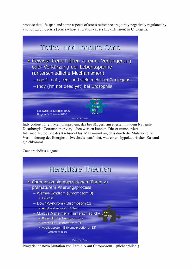
propose that life span and some aspects of stress resistance are jointly negatively regulated by a set of gerontogenes (genes whose alteration causes life extension) in C. elegans.
Praxis Dr. DavisPraxis Dr. Davis
TodesTodes-- und und LonglifeLonglife Gene Gene
Gewisse Gene fGewisse Gene füühren zu einer Verlhren zu einer Verläängerung ngerung oder Verkoder Verküürzung der Lebensspanne rzung der Lebensspanne (unterschiedliche Mechanismen)(unterschiedliche Mechanismen)–– ageage--1, 1, dafdaf--, , cedced-- und viele mehr bei C. und viele mehr bei C. eleganselegans–– IndyIndy ((ii‘‘mm notnot deaddead yetyet) bei ) bei DrosophilaDrosophila
Todes- und Long-life-Gene
Lakowski B; Science 1996Rogina B; Science 2000
Indy codiert für ein Membranprotein, das bei Säugern am ehesten mit dem Natrium-Dicarboxylat Cotransporter verglichen werden können. Dieser transportiert Intermediärprodukte des Krebs-Zyklus. Man nimmt an, dass durch die Mutation eine Verminderung des Energiestoffwechsels stattfindet, was einem hypokalorischen Zustand gleichkommt. Caenorhabditis elegans
Praxis Dr. DavisPraxis Dr. Davis
HereditHereditääre Theorien re Theorien
Chromosomale Aberrationen fChromosomale Aberrationen füühren zu hren zu prpräämaturemmaturem AlterungsprozessAlterungsprozess–– WernerWerner--Syndrom (Chromosom 8)Syndrom (Chromosom 8)
HelicaseHelicase
–– DownDown--Syndrom (Chromosom 21)Syndrom (Chromosom 21)AmyloidAmyloid--PrecursorPrecursor ProteinProtein
–– Morbus Alzheimer (4 unterschiedliche Gene)Morbus Alzheimer (4 unterschiedliche Gene)PresenilinPresenilin 1 (Chromosom 14)1 (Chromosom 14)PresenilinPresenilin 2 (Chromosom 1)2 (Chromosom 1)Apolipoprotein E (Apolipoprotein E (HomozygotieHomozygotie ffüür E4)r E4)
–– Chromosom 19Chromosom 19
Ver-erbung
Progerie: de novo Mutation von Lamin A auf Chromosom 1 (nicht erblich!)
© Dr. C
laren
ce P
. Dav
is 20
09
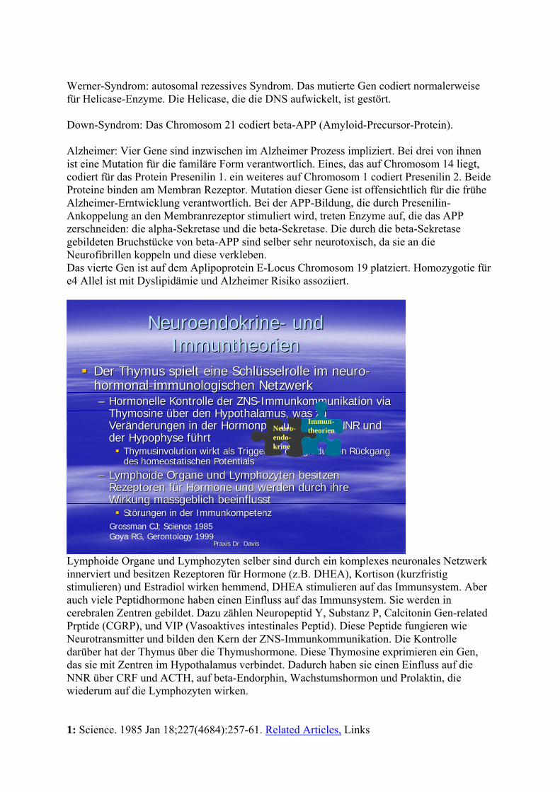
Werner-Syndrom: autosomal rezessives Syndrom. Das mutierte Gen codiert normalerweise für Helicase-Enzyme. Die Helicase, die die DNS aufwickelt, ist gestört. Down-Syndrom: Das Chromosom 21 codiert beta-APP (Amyloid-Precursor-Protein). Alzheimer: Vier Gene sind inzwischen im Alzheimer Prozess impliziert. Bei drei von ihnen ist eine Mutation für die familäre Form verantwortlich. Eines, das auf Chromosom 14 liegt, codiert für das Protein Presenilin 1. ein weiteres auf Chromosom 1 codiert Presenilin 2. Beide Proteine binden am Membran Rezeptor. Mutation dieser Gene ist offensichtlich für die frühe Alzheimer-Erntwicklung verantwortlich. Bei der APP-Bildung, die durch Presenilin-Ankoppelung an den Membranrezeptor stimuliert wird, treten Enzyme auf, die das APP zerschneiden: die alpha-Sekretase und die beta-Sekretase. Die durch die beta-Sekretase gebildeten Bruchstücke von beta-APP sind selber sehr neurotoxisch, da sie an die Neurofibrillen koppeln und diese verkleben. Das vierte Gen ist auf dem Aplipoprotein E-Locus Chromosom 19 platziert. Homozygotie für e4 Allel ist mit Dyslipidämie und Alzheimer Risiko assoziiert.
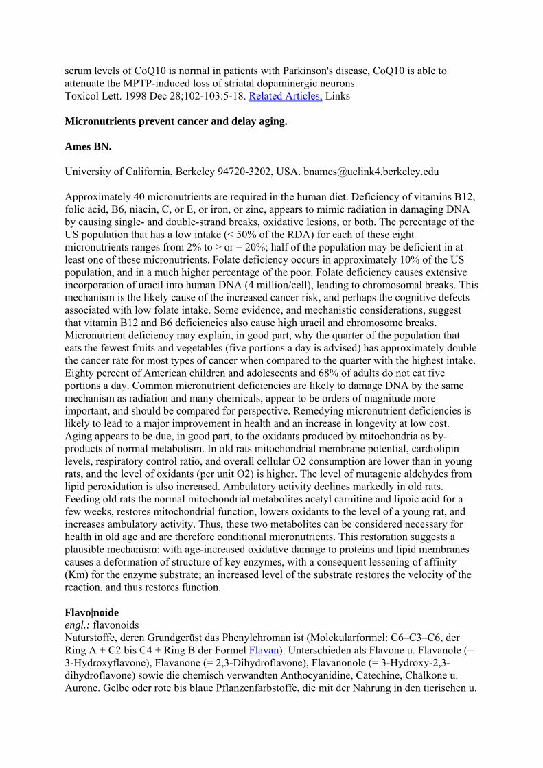
Praxis Dr. DavisPraxis Dr. Davis
Der Thymus spielt eine SchlDer Thymus spielt eine Schlüüsselrolle im neurosselrolle im neuro--hormonalhormonal--immunologischen Netzwerkimmunologischen Netzwerk–– Hormonelle Kontrolle der Hormonelle Kontrolle der ZNSZNS--ImmunkommunikationImmunkommunikation via via
ThymosineThymosine üüber den Hypothalamus, was zu ber den Hypothalamus, was zu VerVeräänderungen in der Hormonproduktion der NNR und nderungen in der Hormonproduktion der NNR und der Hypophyse fder Hypophyse füührthrt
ThymusinvolutionThymusinvolution wirkt als wirkt als TriggerTrigger ffüür den graduellen Rr den graduellen Rüückgang ckgang des des homeostatischenhomeostatischen PotentialsPotentials
–– LymphoideLymphoide Organe und Lymphozyten besitzen Organe und Lymphozyten besitzen Rezeptoren fRezeptoren füür Hormone und werden durch ihre r Hormone und werden durch ihre Wirkung Wirkung massgeblichmassgeblich beeinflusstbeeinflusst
StStöörungen in der Immunkompetenzrungen in der Immunkompetenz
NeuroendokrineNeuroendokrine-- und und ImmuntheorienImmuntheorien
Neuro-endo-krine
Immun-theorien
Grossman CJ; Science 1985Goya RG, Gerontology 1999
Lymphoide Organe und Lymphozyten selber sind durch ein komplexes neuronales Netzwerk innerviert und besitzen Rezeptoren für Hormone (z.B. DHEA), Kortison (kurzfristig stimulieren) und Estradiol wirken hemmend, DHEA stimulieren auf das Immunsystem. Aber auch viele Peptidhormone haben einen Einfluss auf das Immunsystem. Sie werden in cerebralen Zentren gebildet. Dazu zählen Neuropeptid Y, Substanz P, Calcitonin Gen-related Prptide (CGRP), und VIP (Vasoaktives intestinales Peptid). Diese Peptide fungieren wie Neurotransmitter und bilden den Kern der ZNS-Immunkommunikation. Die Kontrolle darüber hat der Thymus über die Thymushormone. Diese Thymosine exprimieren ein Gen, das sie mit Zentren im Hypothalamus verbindet. Dadurch haben sie einen Einfluss auf die NNR über CRF und ACTH, auf beta-Endorphin, Wachstumshormon und Prolaktin, die wiederum auf die Lymphozyten wirken. 1: Science. 1985 Jan 18;227(4684):257-61. Related Articles, Links
© Dr. C
laren
ce P
. Dav
is 20
09

Interactions between the gonadal steroids and the immune system. Grossman CJ. The immune system is regulated by the gonadal steroids estrogen, androgen, and progesterone, but the circulating levels of these steroids can also be affected by immune system function. Such interactions appear to be mediated through the hypothalamic-pituitary-gonadal-thymic axis and depend on pituitary luteinizing hormone released by thymic factors under the control of the gonadal steroids. PMID: 3871252 [PubMed - indexed for MEDLINE] Gerontology. 1999 May-Jun;45(3):174-8. Related Articles, Links Homeostasis, thymic hormones and aging. Goya RG, Bolognani F. INIBIOLP-Histology 'B', Faculty of Medicine, National University of La Plata, Argentina. [email protected] The thymic-pituitary axis constitutes a bidirectional circuit where the ascending feedback loop is effected by thymic factors of epithelial origin. The aim of the present article is, first, to introduce the idea of an immune-neuroendocrine homeostatic network in higher animals. Next, the relevance of the thymus in this network and the possible role of this gland in the neuroendocrine imbalances associated with aging are discussed. A number of studies are next reviewed which show that the endocrine thymus produces several bioactive molecules, generally called thymic hormones, which in addition to possessing immunoregulatory properties are also active on nervous and endocrine circuits. In particular, the reported activities of thymosin fraction five, thymosin alpha 1 and thymosin beta 4 on beta-endorphin, adrenocorticotropic hormone, glucocorticoids, luteinizing hormone-releasing hormone and luteinizing hormone secretion in different animal and cell models are reviewed. The known hypophysiotropic actions of other thymic hormones like thymulin, homeostatic thymus hormone and thymus factor are also summarized, and the impact of aging on pituitary responsiveness to thymic hormones is discussed. As a conclusion, it is proposed that in addition to its central role in the regulation of the immune function, the thymus gland may extend its influence to nonimmunologic components of the body, including the neuroendocrine system. The early onset of thymus involution might, therefore, act as a triggering event which would initiate the gradual decline in homeostatic potential that characterizes the aging process. Publication Types: Review Review, Tutorial PMID: 10202264 [PubMed - indexed for MEDLINE]
© Dr. C
laren
ce P
. Dav
is 20
09

Praxis Dr. DavisPraxis Dr. Davis
Probleme der DiagnoseProbleme der Diagnose
AntiAnti--Aging Aging heisstheisst in erster Linie Vorbeugung in erster Linie Vorbeugung und Verhund Verhüütung von altersbedingten tung von altersbedingten ErkrankungenErkrankungen–– Viele Krankheiten sind zum Zeitpunkt der Viele Krankheiten sind zum Zeitpunkt der
Diagnose noch nicht manifestDiagnose noch nicht manifest–– Parameter sind gesucht, die auf spParameter sind gesucht, die auf späätere tere
Erkrankung hindeuten kErkrankung hindeuten köönnennnen
Biomarker des Biomarker des AlternsAlterns–– Biomarker, die Biomarker, die
verschiedene verschiedene KKöörperfunktionen rperfunktionen erfassen, sollten erfassen, sollten RRüückschlckschlüüsse auf sse auf Alterungsprozesse Alterungsprozesse erlaubenerlauben
Biologisches Alter Chronologisches Alter
Schon vor 1975 wurden in verschiedenen Laboratorien überall in der Welt physiologische Marker, die mit Alterungsprozessen korrelierten, untersucht. Zum Beispiel der höchste hörbare Ton, der sehr steil von 18 kHz mit 20 Jahren auf 8 kHz mit 70 Jahren abfällt. Mehrere Wissenschaftler kamen zu der Auffassung, dass durch die Auswahl einer ausreichenden Anzahl solcher Biomarker, die verschiedene Funktionen des Körpers erfassen, es möglich sein müsste, Rückschlüsse auf Alterungsprozesse zu ziehen, unter Berücksichtigung des unterschiedlichen Gefälles einzelner Biomarker pro Zeiteinheit.
Praxis Dr. DavisPraxis Dr. Davis
Diagnostik: Diagnostik: HH--ScanScan
H-Scan
H-Scan
Der von Richard Hochschild entwickelte H-Scan. Das Gerät dokumentiert funktionelle Veränderungen und erlaubt so dem Arzt eine Therapiekontrolle.
© Dr. C
laren
ce P
. Dav
is 20
09

Praxis Dr. DavisPraxis Dr. Davis
Diagnostik: BiomarkerDiagnostik: Biomarker
Für jeden Biomarker wurden Normalwerte für die 977 Männer und 1485 Frauen im Alter von 35 bis 70 Jahren festgelegt, die an der Studie teilnahmen.
Praxis Dr. DavisPraxis Dr. Davis
NahrungsrestriktionNahrungsrestriktion–– Reduziert Reduziert metabolischemetabolische Rate Rate
/ oxidativen Stress/ oxidativen Stress–– Verbessert InsulinsensitivitVerbessert Insulinsensitivitäätt–– Reduziert Reduziert BlutlipideBlutlipide / /
BlutdruckBlutdruck
Mediterrane DiMediterrane Diäät t ((TrichopoulouTrichopoulou A; NEJM A; NEJM 2003)2003)
Viel FischViel FischViele ungesViele ungesäättigte FS / ttigte FS / wenig geswenig gesäättigte FSttigte FSViel GemViel GemüüseseMMäässigerssiger AlkoholkonsumAlkoholkonsum
LifestyleLifestyle--MassnahmenMassnahmen
Ernährung
Aufbau / ErhaltVermeidung
MuskeltrainingMuskeltraining–– Reduziert Reduziert SarkopenieSarkopenie
und damit Verbesserung und damit Verbesserung des des FrailtyFrailty IndexesIndexes
Bewegung (Bewegung („„lowlowimpactimpact““))–– Reduktion des Reduktion des
Frakturrisikos Frakturrisikos (Osteoporose?)(Osteoporose?)
GymnastikGymnastik–– KardiovaskulKardiovaskuläärere und und
antidiabetogeneantidiabetogeneBewegungBewegung
Jogging / Jogging / WalkingWalking
The concept of restricting calories to improve health was first introduced in the early 1900s, but Clive McCay (Cornell University) advanced the theory significantly when (in the 1930s) he found that calorie-restricted rats lived longer than those allowed to eat ad libitum. Though decades have passed since McCay's initial findings, the exact mechanisms whereby dietary restriction retards aging and extends life span are still not fully understood. Much of the emerging data suggest that McCay's calorie-restricted rodents lived longer and aged more slowly because they were more resistant to stress and their cells appeared protected against damaging agents (Van Remmen et al. 2001).
© Dr. C
laren
ce P
. Dav
is 20
09

The eight people in the BioSphere study averaged 1,800 calories per day during the first 6 months, increasing to 2,200 calories per day by the end of 2 years. In the first 6 months, body weight dropped an average of 15%, blood sugar dropped 20%, blood cholesterol dropped 38% and blood pressure dropped significantly (30% systolic and 27% diastolic); white blood cell count dropped 24% (Walford 1988, 1994; Best 1995). The results obtained from the BioSphere II experiment are consistent with information gathered from ongoing animal studies conducted at the National Institute on Aging and the University of Wisconsin (Madison). For example, monkeys (calorie restricted) weighed less and had less body fat. They exhibited lower body temperature, as well as lower fasting blood glucose and insulin levels; insulin sensitivity increased. Typically, the monkeys also had lower blood pressure, reduced triglyceride and cholesterol levels as well as increased levels of HDL2B (low levels of this HDL subfraction are associated with increased cardiovascular disease in humans) (Lane et al. 1999). The CRON theory has been most studied in lower species but the underlying principle has extended to include long-lived primates. To determine if calorie restriction has similar actions in higher species, the National Institute on Aging (NIA) initiated a study in 1987 to investigate the effects of a 30% caloric reduction in male and female rhesus macaques (Macaca mulatta). Comment: Rhesus monkeys living in captivity can live to be about 40 years old. At the genetic level, Rhesus macaques and humans are nearly the same, sharing a genome that is more than 90% identical (Devitt 1998). The NIA study demonstrated that calorie restriction decreases body weight and fat mass, improves glucoregulatory function, and decreases blood pressure, blood lipids, and body temperature. Juvenile males exhibited delayed skeletal and sexual maturation, and later were able to overcome an age-associated decline in both dehydroepiandrosterone (DHEA) and melatonin; bone mass was unaffected in both males and females. Eighty-one percent of the monkeys in the NIA study are still alive, suggesting that calorie restriction may have beneficial effects on morbidity and mortality statistics (Mattison et al. 2003). BACKGROUND: Adherence to a Mediterranean diet may improve longevity, but relevant data are limited. METHODS: We conducted a population-based, prospective investigation involving 22,043 adults in Greece who completed an extensive, validated, food-frequency questionnaire at base line. Adherence to the traditional Mediterranean diet was assessed by a 10-point Mediterranean-diet scale that incorporated the salient characteristics of this diet (range of scores, 0 to 9, with higher scores indicating greater adherence). We used proportional-hazards regression to assess the relation between adherence to the Mediterranean diet and total mortality, as well as mortality due to coronary heart disease and mortality due to cancer, with adjustment for age, sex, body-mass index, physical-activity level, and other potential confounders. RESULTS: During a median of 44 months of follow-up, there were 275 deaths. A higher degree of adherence to the Mediterranean diet was associated with a reduction in total mortality (adjusted hazard ratio for death associated with a two-point increment in the Mediterranean-diet score, 0.75 [95 percent confidence interval, 0.64 to 0.87]). An inverse association with greater adherence to this diet was evident for both death due to coronary heart disease (adjusted hazard ratio, 0.67 [95 percent confidence interval, 0.47 to 0.94]) and death due to cancer (adjusted hazard ratio, 0.76 [95 percent confidence interval, 0.59 to 0.98]). Associations between individual food groups contributing to the Mediterranean-diet score and total mortality were generally not significant. CONCLUSIONS: Greater adherence to the traditional Mediterranean diet is associated with a significant reduction in total mortality. Copyright 2003 Massachusetts Medical Society
© Dr. C
laren
ce P
. Dav
is 20
09

Frailty in the elderly: contributions of sarcopenia and visceral protein depletion. Vanitallie TB. Medical Service, St. Luke's Roosevelt Hospital Center, New York, NY, USA. In any given population of free-living individuals 65 years of age and older, a substantial proportion (in the range of 6% to 25%) suffers from many of the elements of the syndrome of frailty. Although the syndrome is complex and still lacks a standard definition, there is a growing consensus about the signs and symptoms as well as the pattern of biological correlates that characterize this disorder. Patients who are afflicted with frailty typically exhibit loss of muscle strength, fatigue easily, are physically inactive, and have a slow-and often unsteady-gait, with an increased risk (and fear) of falling. They are likely to have a poor appetite and to have undergone a recent, unintentional loss of weight. Frail individuals are more likely than the nonfrail to experience impaired cognition and depression. They die sooner. Frailty, of course, is frequently complicated by a variety of coexistent illnesses. Among the biological correlates of frailty are sarcopenia (now readily measurable by dual-energy x-ray absorptiometry [DXA]), osteopenia (with an increased susceptibility to fracture), and activation of the inflammatory and coagulation systems, with a rise in inflammatory cytokines and several markers of coagulopathy. Age-dependent changes in a number of hormones also appear to promote the development of frailty in the elderly, particularly via their effects on muscle mass and strength, bone density, and by contributing to activation of the catabolic cytokines. In particular, serum levels of growth hormone (GH) and insulin-like growth factor-1 (IGF-1) decline progressively during aging, and an association between reduction in the levels of these hormones and the involution of advancing age has been proposed. It is not yet known whether, in comparison with their nonfrail counterparts, frail individuals consistently manifest larger reductions in GH and IGF-1 (and other anabolic hormones). More research is needed before it will be known whether the benefits of administering GH to the frail elderly will outweigh the disadvantages. The poor appetite and weight loss that occur in many frail individuals are likely to be accompanied by a degree of visceral protein depletion (with its attendant morbidity), which can be estimated by making serial measurements of indicators of visceral protein status such as transthyretin (TTR), retinol-binding protein (RBP), and albumin. One characteristic of the frailty syndrome that distinguishes it from the effects of aging per se is the potential reversibility of many of its features. Progressive resistance training is feasible for many elderly individuals-even the oldest old-and, by increasing muscle mass and strength, can ameliorate or reverse important aspects of physical frailty. To the extent that visceral protein depletion has been caused by an inadequate intake of calories and protein, consumption of a more adequate diet can result in betterment of the frail patient's nutritional status, as determined by clinical improvement and favorable changes in TTR, RBP, and albumin. Influence of muscle strength, physical activity and weight on bone mass in a population-based sample of 1004 elderly women. Gerdhem P, Ringsberg KA, Akesson K, Obrant KJ. Department of Orthopaedics, Malmo University Hospital, SE-205 02 Malmo, Sweden. [email protected] High physical activity level has been associated with high bone mass and low fracture risk and is therefore recommended to reduce fractures in old age. The aim of this study was to
© Dr. C
laren
ce P
. Dav
is 20
09

estimate the effect of potentially modifiable variables, such as physical activity, muscle strength, muscle mass and weight, on bone mass in elderly women. The influence of isometric thigh muscle strength, self-estimated activity level, body composition and weight on bone mineral density (dual energy X-ray absorptiometry; DXA) in total body, hip and spine was investigated. Subjects were 1004 women, all 75 years old, taking part in the Malmo Osteoporosis Prospective Risk Assessment (OPRA) study. Physical activity and muscle strength accounted for 1-6% of the variability in bone mass, whereas weight, and its closely associated variables lean mass and fat mass, to a much greater extent explained the bone mass variability. We found current body weight to be the variable with the most substantial influence on the total variability in bone mass (15-32% depending on skeletal site) in a forward stepwise regression model. Our findings suggest that in elderly women, the major fracture-preventive effect of physical activity is unlikely to be mediated through increased bone mass. Retaining or even increasing body weight is likely to be beneficial to the skeleton, but an excess body weight increase may have negative effects on health. Nevertheless, training in elderly women may have advantages by improving balance, co-ordination and mobility and therefore decreasing the risk of fractures
Praxis Dr. DavisPraxis Dr. Davis
LifestyleLifestyle--MassnahmenMassnahmen
Vermeidung
ScreeningScreening--UntersuchungenUntersuchungen–– KoloskopieKoloskopie–– ProstatauntersuchungProstatauntersuchung–– GynGynääkologische kologische
UntersuchungUntersuchung–– KardiovaskulKardiovaskulääresres
ScreeningScreening
Vermeidung von Vermeidung von NoxenNoxen–– RaucherentwRaucherentwööhnunghnung–– Vermeidung von Vermeidung von
UmweltnoxenUmweltnoxen
Potential costs of flexible sigmoidoscopy-based colorectal cancer screening. Marshall JR, Fay D, Lance P. Arizona Cancer Center, Tucson, USA. BACKGROUND & AIMS: Increasing evidence shows that periodic screening by flexible sigmoidoscopy with appropriate referral of patients with adenomas to colonoscopy could substantially decrease colorectal cancer mortality rates. Estimates of the complete cost of such screening are needed. The aim of this study was to estimate the annual costs of periodic screening of Americans 50 years and older by flexible sigmoidoscopy with referral of subjects with adenomas to colonoscopy. METHODS: Cost analysis of flexible sigmoidoscopy, followed by colonoscopy as warranted, in U.S. population cohorts reaching age 50 each year from 1995 to 2010 was performed. Total yearly costs of repeat screening and surveillance examinations at American Cancer Society-recommended and other intervals were determined.
© Dr. C
laren
ce P
. Dav
is 20
09

RESULTS: With screening and surveillance intervals of 3 years, annual costs for the cohort of individuals turning 50 in 1995 would increase to $553 million by 2010. Annual costs for the entire population 50 years of age and older could increase by 2010 to nearly $20 billion. CONCLUSIONS: The cost of flexible sigmoidoscopy-based screening for colorectal cancer could vary as much as threefold depending on the protocol chosen. Prevention of colorectal cancer by once-only sigmoidoscopy. Atkin WS, Cuzick J, Northover JM, Whynes DK. Imperial Cancer Research Fund Colorectal Unit, St Mark's Hospital, London, UK. There is no national screening programme for colorectal cancer in the UK despite the fact that the annual death toll from this disease exceeds that of breast and cervical cancer. Faecal occult blood testing (FOBT) is under evaluation for screening, but screening by sigmoidoscopy is not considered viable. This situation contrasts with the USA where both annual FOBT and screening by flexible sigmoidoscopy every 3 to 5 years are recommended from 50 years old. We seek to demonstrate that most of the benefit from the US screening policy would accrue from a single flexible sigmoidoscopy examination at age 55 to 60 years with appropriate colonoscopic surveillance for the 3% to 5% found to have high-risk adenomas (> or = 1 cm or villous histology). If applied nationally, this screening regimen could prevent about 5500 colorectal cancer cases and 3500 deaths in the UK each year, thus saving 40,000 years of life. We estimate that there would be little net cost to the National Health Service because savings obtained from treating fewer patients would largely offset the cost of screening. We recommend that a randomised trial to evaluate screening by single flexible sigmoidoscopy should start without delay. Such a trial would involve about 120,000 participants, and 15 years of follow-up would be required to obtain a clear answer on mortality, although information on incidence reduction would be available sooner.
Praxis Dr. DavisPraxis Dr. Davis
Vitamin AVitamin AVitamin EVitamin EVitamin CVitamin CSelenSelen–– GPxGPx
Mangan / Kupfer / Mangan / Kupfer / ZinkZink–– SODSOD
GlutaminsGlutaminsääure / ure / CysteinCystein / / GlycinGlycin–– GlutathionGlutathion
CoQ10 / CoQ10 / αα--LiponsLiponsääureure / L/ L--CarnitinCarnitin–– Verminderung der Verminderung der
mitochondrialenmitochondrialen SchSchäädigungdigung–– Verbesserung des Verbesserung des
mitochondrialenmitochondrialen MetabolismusMetabolismusCarotinoideCarotinoideFlavonoideFlavonoide–– IsoflavoneIsoflavone
Verminderung der Verminderung der ZellproliferationZellproliferationin in vitrovitro Hemmung der Hemmung der TumorangiogeneseTumorangiogeneseHemmung der Hemmung der TcTc--AggregationAggregationE2E2--ModulationModulation
Ca. 30Ca. 30‘‘000 Weitere000 Weitere
MelatoninMelatonin–– HydroxylHydroxyl--
ScavengerScavenger–– FlavineFlavine / /
PorphyrinePorphyrineDHEADHEA–– Hemmung der Hemmung der
NADPHNADPH--OxidaseOxidase
PflanzenstoffeNahrungsergänzungen
VitamineSpurenelemente
Antioxidative TherapienAntioxidative Therapien
Hormone
PflanzenstoffeNahrungsergänzungen
VitamineSpurenelemente
Hormone
Bereits in den 50er Jahren postulierte Denam Harman die These, dass für den Altersprozess freie Radikale eine Schlüsselrolle spielen. Freie Radikale zeichnen sich dadurch aus, dass sie auf ihrer Elektronenhülle ein ungepaartes Elektron besitzen, was ihnen eine starke Reaktionsfähigkeit verleiht.
© Dr. C
laren
ce P
. Dav
is 20
09

Eine sich aus der Freie-Radikale-Theorie ableitende Therapieoption besteht in der hochdosierten Zufuhr so genannter Antioxidantien. An Fruchtfliegen konnten gezeigt werden, dass die Überexpression von Katalse und Superoxiddismutase das Lebensalter signifikant verlängern kann. Diese Experimente unterstützen die Theorie des Altern aufgrund von oxidativem Stress. Ein weiters Indiz für die Richtigkeit dieser Theorie liefern die Ergebnisse aus einigen Studien mit Nagetieren. Man konnte zeigen, dass sich die Lebenserwartung durch moderate kalorische Restriktion verlängern lässt. Dies ist darauf zurückzuführen, dass die effizientere Nutzung des Elektronen-Transport-Systems in den Mitochondrien weniger schädliche freie Sauerstoffradikale erzeugt. Ziele oxidativer Zerstörung sind Lipide, Proteine und DNS. Melatonin ist nicht autoxidabel, wie z.B. Vit. C. Es besitzt präoxidative Eigenschaften selektiv und ausnahmsweise für Hydroxylradikale und potente Photooxidantien (Flavine und Porphyrine). Melatonin ist der stärkste und effektivste Hydroxyl-Radikal-Scavenger, der wegen seiner lipophilen Eigenschaften „seitenspezifisch“ agiert (Phasengrenze Lipidmembranen-Zytoplasma). Im Gegensatz zu allen anderen Scavengern (Ascorbat, Übergangsmetalle, Riboflavin, usw.) kann Melatonin – einmal oxidiert – nicht rückreduziert oder regeneriert werden und nimmt deswegen nicht an Redoxcyclingsprozessen teil. Melatonin wird oxidiert und dieser oxidative Abbau führt nicht zu einer Wasserstoffperoxidformation. Einsatzgebiete von Melatonin: 1) Kontrolle des Schlaf-Wachrythmus und Verbesserung des Schlafs (auch bei ASPS, Advanced Sleep Phase Syndrome, und DSPS, Delayed Sleep Phase Syndrome). 2) Stimulation des Immunsystems (die Wirkung von Melatonin ist nicht direkt auf die Zellen des Immunsystems gerichtet, sondern wirkt mediatorisch über das endogene Opioidsystem auf die Antigenaktivierung der T-Zellen) 3) Jet Lag (0.2-1 mg Melatonin pro Stunde Zeitverschiebung während ca. 1 Woche) Die Mitochondrien-Theorie Der Einfluss der freien Radikale auf den Alterungsprozess gilt inzwischen als gut belegt. Freie Radikale entstehen durch schädigende Außeneinflüsse, in aller erster Linie aber als Abfallprodukte der körpereigenen Energiegewinnung. Zunehmend in den Fokus des Interesses geraten damit jene Zellorganellen, in denen sich die Energiegewinnung abspielt: Die Mitochondrien. Mitochondrien waren einst offensichtlich selbständige Kleinstlebewesen, die im Laufe der Evolution in die Zelle inkorporiert wurden und dort hauptsächlich für die Energiegewinnung genutzt werden. Dies erklärt die Tatsache, dass Mitochondrien als einzige Zellorganellen über eine eigene DNA verfügen [7]. Die Energiegewinnung durch die Mitochondrien erfolgt im Rahmen der so genannten Atmungskette. Diese besteht aus vier großen (Komplex I bis IV) und zwei kleinen (Ubichinon und Cytochrom C) Molekülkomplexen. Elektronen, die aus dem Abbau aufgenommener Nahrungsstoffe stammen, werden dort nach dem Stafettenprinzip weitergereicht. Am Komplex IV werden sie paarweise auf Sauerstoff übertragen, der sich mit Protonen (H+) zu Wasser vereint. Im Zuge der einzelnen Etappen pumpt das Mitochondrion Protonen auf die Außenseite der Innenmembranen. Das so entstehende elektrische und chemische Gefälle
© Dr. C
laren
ce P
. Dav
is 20
09

ermöglicht es der ATP-Synthetase - einem weiteren, quasi als Turbine dienendem Proteinkomplex - aus Adenosin-Diphosphat (ADP) energiereiches Adenosin-Triphosphat (ATP) herzustellen. ATP ist gleichsam die "körpereigene Universalwährung" 'für Energie. Frei Radikale entstehen in diesem Prozess, wenn Elektronen aus der Transportkette entweichen und von Sauerstoff eingefangen werden. Dafür scheint es eine besondere Schwachstelle zu geben, die im Komplex II im Umfeld des Ubichinons liegt. Hier werden besonders viele freie Radikale freigesetzt. Die Mitochondrien sind aber nicht nur die wichtigste Produktionsstelle für die Entstehung von freien Radikalen, sondern auch eine der massgeblichen und empfindlichsten Zielorte für deren zerstörerische Wirkung. Freie Radikale schädigen die Mitochondrienmembran, vor allem aber die mitochondriale DNS. Auf das Vorhandensein einer eigenen Mitochondrien-DNA haben wir bereits hingewiesen. Sie ist keineswegs nur ein evolutionsbiologisches Relikt, sondern kodiert für gut ein Dutzend Proteine, die für die Funktion dieser Organellen und damit für die Energiegewinnung von Bedeutung sind. Die Mitochondrien-DNS ist aber besonders anfällig für oxidative Schäden. Während die chromosomale Kern-DNS über eine ganze Reihe von Enzymsystemen verfügt, die in der Lage sind, oxidierte DNS-Fragmente herauszuschneiden und zu ersetzen, besitzt die evolutionsbiologisch ältere Mitochondrien-DNS keine solchen Reparaturmechanismen. Auch fehlen ihr Histone (basische Kernproteine), die das genetische Material abschirmen. Oxidative Schäden wirken sich an den Mitochondrien also besonders dramatisch aus. Es entsteht ein Teufelskreis: Geschädigte Mitochondrien produzieren zunehmend weniger ATP, gleichzeitig setzen sie vermehrt freie Radikale als Abfallprodukte frei, die ihrerseits die Mitochondrien weiter schädigen. Der Zelle steht damit zunehmend weniger Energie zur Verfügung, gleichzeitig kumulieren die durch freie Radikale induzierten Schäden – Gewebe und Organe altern. Altern beginnt demnach mit einer zunehmenden Funktionseinbusse der Mitochondrien. Im Sinne einer „mitochondrialen Medizin“ besteht ein weiterer Ansatz der Anti-Aging-Medizin daher in dem gezielten Schutz dieser Zellorganellen. Propagiert wird unter anderem die Supplementierung jenes Coenzyms, das sich im Rahmen der Atmungskette als besondere Schwachstelle erwiesen hat: Ubichinon. Ubichinon – auch als „Coenzym Q10 bekannt – ist das zentrale Bindeglied in der Atmungskette der Mitochondrien und der bisher wirksamste bekannte Radikalfänger in der Zellmembran. Da die körpereigene Ubichinon-Biosynthese mit steigendem Alter abnimmt, eine erhöhte Zufuhr über die Nahrung aber nur schwer zu bewerkstelligen ist, empfiehlt sich die Supplementierung der Substanz in Form von Mono- oder Kombinationspräparaten. ScientificWorldJournal. 2001 Jan 1;1(1 Suppl 3):81-2. Related Articles, Links Delaying aging with mitochondrial micronutrients and antioxidants. Atamna H, Ames BN, Liu J. MITOCHONDRIAL DECAY IN AGING. Mitochondria decay with age due to the oxidation of mtDNA, proteins, and lipids. Some of this decay can be reversed in old rats by feeding them normal mitochondrial metabolites at high levels. Aging appears to be in good part due to the oxidants produced as by-products of normal metabolism by mitochondria [1-5]. In old rats mitochondrial membrane potential, cardiolipin levels, respiratory control ratio, and overall cellular O2 consumption are lower than in young rats and the level of oxidants (per unit O2) and mutagenic aldehydes from lipid peroxidation is higher [3]. Ambulatory activity declines markedly in old rats. Spatial memory as measured by the Morris water maze, also declines with age. Feeding old rats the normal mitochondrial metabolites acetyl carnitine and lipoic acid for a few weeks, restores mitochondrial function, lowers oxidants to the level of a young
© Dr. C
laren
ce P
. Dav
is 20
09

rat, and increases ambulatory activity and spatial memory [6-11]. Thus, these two metabolites can be considered necessary for health in old age and are therefore "conditional micronutrients". This restoration suggests a plausible mechanism [11]: with age increased oxidative damage to proteins, such as acyl carnitine transferase (whose activity declines with age), and lipid membranes, particularly in mitochondria, causes a deformation of structure of key enzymes, with a consequent lessening of affinity (Km) for the enzyme substrate; an increased level of the substrate restores the velocity of the reaction, and thus restores function. Postgrad Med J. 1996 Jan;72(843):45-50. Related Articles, Links A randomised, double-blind, placebo-controlled trial of L-carnitine in suspected acute myocardial infarction. Singh RB, Niaz MA, Agarwal P, Beegum R, Rastogi SS, Sachan DS. Heart Research Laboratory, Medical Hospital and Research Centre, Moradabad, India. In a randomised, double-blind placebo-controlled trial, the effects of the administration of oral L-carnitine (2 g/day) for 28 days were compared in the management of 51 (carnitine group) and 50 (placebo group) patients with suspected acute myocardial infarction. At study entry, the extent of cardiac disease, cardiac enzymes and lipid peroxides were comparable between the groups, although both groups showed an increase in cardiac enzymes and lipid peroxides. At the end of the 28-day treatment period, the mean infarct size assessed by cardiac enzymes showed a significant reduction in the carnitine group compared to placebo. Electrocardiographic assessment of infarct size revealed that the QRS-score was significantly less in the carnitine group compared to placebo (7.4 +/- 1.2 vs 10.7 +/- 2.0), while serum aspartate transaminase and lipid peroxides showed significant reduction in the carnitine group. Lactate dehydrogenase measured on the sixth or seventh day following infarction showed a smaller rise in the carnitine group compared to placebo. Angina pectoris (17.6 vs 36.0%), New York Heart Association class III and IV heart failure plus left ventricular enlargement (23.4 vs 36.0%) and total arrhythmias (13.7 vs 28.0%) were significantly less in the carnitine group compared to placebo. Total cardiac events including cardiac deaths and nonfatal infarction were 15.6% in the carnitine group vs 26.0% in the placebo group. It is possible that L-carnitine supplementation in patients with suspected acute myocardial infarction may be protective against cardiac necrosis and complications during the first 28 days. Biogerontology. 2002;3(1-2):103-6. Related Articles, Links How to re-energise old mitochondria without shooting yourself in the foot. Driver C, Georgiou A. National Ageing Research Institute, Parkville, Australia. [email protected] In old humans and pathologies associated with mitochondrial mutations, deletions in mitochondrial DNA have been associated with failing function. Investigations have been reported where treatment with a number of micronutrients, such as coenzyme Q10, have been used to re-energise failing tissues. Bioenergy changes in ageing Drosophila have been observed which indicate similar changes in mitochondrial function in old age. Reserves of carbohydrate and fat fall and food intake rises. Biochemical changes include falling mitochondrial enzymes. Mitochondrial DNA contains increased amounts of sequences corresponding to deletions. Both coenzyme Q10 and nicotinamide in large doses successfully
© Dr. C
laren
ce P
. Dav
is 20
09

reversed bioenergy changes in aged Drosophila. However, only nicotinamide was able to reduce short term mortality and increase life span, whereas coenzyme Q10 increased mortality and reduced life span. Production of reactive oxygen species (ROS) was increased in coenzyme Q10 treated flies, whereas nicotinamide reduced ROS production. It is suggested that ROS production may account for these longevity differences. Large doses of two micronutrients have been successful in reversing the age-associated bioenergy deficit in Drosophila. This response is similar to clinical reports of re-energising tissues where mitochondrial damage has been observed. However, this work highlights a danger for some micronutrients, such as coenzyme Q10, that clinical efficacy may be limited by increased ROS production. Biol Signals Recept. 2001 May-Aug;10(3-4):224-53. Related Articles, Links Ubiquinone (coenzyme q10) and mitochondria in oxidative stress of parkinson's disease. Ebadi M, Govitrapong P, Sharma S, Muralikrishnan D, Shavali S, Pellett L, Schafer R, Albano C, Eken J. Department of Pharmacology, Physiology, and Therapeutics, University of North Dakota School of Medicine and Health Sciences, Grand Forks, N.Dak. 58203-2817, USA. [email protected] Parkinson's disease is the second most common neurodegenerative disorder after Alzheimer's disease affecting approximately1% of the population older than 50 years. There is a worldwide increase in disease prevalence due to the increasing age of human populations. A definitive neuropathological diagnosis of Parkinson's disease requires loss of dopaminergic neurons in the substantia nigra and related brain stem nuclei, and the presence of Lewy bodies in remaining nerve cells. The contribution of genetic factors to the pathogenesis of Parkinson's disease is increasingly being recognized. A point mutation which is sufficient to cause a rare autosomal dominant form of the disorder has been recently identified in the alpha-synuclein gene on chromosome 4 in the much more common sporadic, or 'idiopathic' form of Parkinson's disease, and a defect of complex I of the mitochondrial respiratory chain was confirmed at the biochemical level. Disease specificity of this defect has been demonstrated for the parkinsonian substantia nigra. These findings and the observation that the neurotoxin 1-methyl-4-phenyl-1,2,3, 6-tetrahydropyridine (MPTP), which causes a Parkinson-like syndrome in humans, acts via inhibition of complex I have triggered research interest in the mitochondrial genetics of Parkinson's disease. Oxidative phosphorylation consists of five protein-lipid enzyme complexes located in the mitochondrial inner membrane that contain flavins (FMN, FAD), quinoid compounds (coenzyme Q10, CoQ10) and transition metal compounds (iron-sulfur clusters, hemes, protein-bound copper). These enzymes are designated complex I (NADH:ubiquinone oxidoreductase, EC 1.6. 5.3), complex II (succinate:ubiquinone oxidoreductase, EC 1.3.5.1), complex III (ubiquinol:ferrocytochrome c oxidoreductase, EC 1.10.2.2), complex IV (ferrocytochrome c:oxygen oxidoreductase or cytochrome c oxidase, EC 1.9.3.1), and complex V (ATP synthase, EC 3.6.1.34). A defect in mitochondrial oxidative phosphorylation, in terms of a reduction in the activity of NADH CoQ reductase (complex I) has been reported in the striatum of patients with Parkinson's disease. The reduction in the activity of complex I is found in the substantia nigra, but not in other areas of the brain, such as globus pallidus or cerebral cortex. Therefore, the specificity of mitochondrial impairment may play a role in the degeneration of nigrostriatal dopaminergic neurons. This view is supported by the fact that MPTP generating 1-methyl-4-phenylpyridine (MPP(+)) destroys dopaminergic neurons in the substantia nigra. Although the
© Dr. C
laren
ce P
. Dav
is 20
09
serum levels of CoQ10 is normal in patients with Parkinson's disease, CoQ10 is able to attenuate the MPTP-induced loss of striatal dopaminergic neurons. Toxicol Lett. 1998 Dec 28;102-103:5-18. Related Articles, Links Micronutrients prevent cancer and delay aging. Ames BN. University of California, Berkeley 94720-3202, USA. [email protected] Approximately 40 micronutrients are required in the human diet. Deficiency of vitamins B12, folic acid, B6, niacin, C, or E, or iron, or zinc, appears to mimic radiation in damaging DNA by causing single- and double-strand breaks, oxidative lesions, or both. The percentage of the US population that has a low intake (< 50% of the RDA) for each of these eight micronutrients ranges from 2% to > or = 20%; half of the population may be deficient in at least one of these micronutrients. Folate deficiency occurs in approximately 10% of the US population, and in a much higher percentage of the poor. Folate deficiency causes extensive incorporation of uracil into human DNA (4 million/cell), leading to chromosomal breaks. This mechanism is the likely cause of the increased cancer risk, and perhaps the cognitive defects associated with low folate intake. Some evidence, and mechanistic considerations, suggest that vitamin B12 and B6 deficiencies also cause high uracil and chromosome breaks. Micronutrient deficiency may explain, in good part, why the quarter of the population that eats the fewest fruits and vegetables (five portions a day is advised) has approximately double the cancer rate for most types of cancer when compared to the quarter with the highest intake. Eighty percent of American children and adolescents and 68% of adults do not eat five portions a day. Common micronutrient deficiencies are likely to damage DNA by the same mechanism as radiation and many chemicals, appear to be orders of magnitude more important, and should be compared for perspective. Remedying micronutrient deficiencies is likely to lead to a major improvement in health and an increase in longevity at low cost. Aging appears to be due, in good part, to the oxidants produced by mitochondria as by-products of normal metabolism. In old rats mitochondrial membrane potential, cardiolipin levels, respiratory control ratio, and overall cellular O2 consumption are lower than in young rats, and the level of oxidants (per unit O2) is higher. The level of mutagenic aldehydes from lipid peroxidation is also increased. Ambulatory activity declines markedly in old rats. Feeding old rats the normal mitochondrial metabolites acetyl carnitine and lipoic acid for a few weeks, restores mitochondrial function, lowers oxidants to the level of a young rat, and increases ambulatory activity. Thus, these two metabolites can be considered necessary for health in old age and are therefore conditional micronutrients. This restoration suggests a plausible mechanism: with age-increased oxidative damage to proteins and lipid membranes causes a deformation of structure of key enzymes, with a consequent lessening of affinity (Km) for the enzyme substrate; an increased level of the substrate restores the velocity of the reaction, and thus restores function. Flavo|noide engl.: flavonoids Naturstoffe, deren Grundgerüst das Phenylchroman ist (Molekularformel: C6–C3–C6, der Ring A + C2 bis C4 + Ring B der Formel Flavan). Unterschieden als Flavone u. Flavanole (= 3-Hydroxyflavone), Flavanone (= 2,3-Dihydroflavone), Flavanonole (= 3-Hydroxy-2,3-dihydroflavone) sowie die chemisch verwandten Anthocyanidine, Catechine, Chalkone u. Aurone. Gelbe oder rote bis blaue Pflanzenfarbstoffe, die mit der Nahrung in den tierischen u.
© Dr. C
laren
ce P
. Dav
is 20
09

menschl. Körper gelangen, für den sie wichtig sind als Redoxsystem, Wasserstoff-Akzeptor, u. Schutzstoff gegen Autoxidation von Vitamin C u. Adrenalin. – s.a. Bioflavonoide. Bio|fla|vo|no|ide engl.: bioflavonoids die als Vitamin P bezeichneten pflanzlichen chemischen Verbindungen mit i.S. des Phenylchromans abgewandelter Struktur des – trizyklischen – Flavons, u. zwar als Glykoside u. Aglykone, z.B. Citrin, Rutin. Wirken synergistisch mit Vitamin C u. antihämorrhagisch, entzündungswidrig u. antiallergisch; besitzen auch östrogene Eigenschaften Die wichtigsten Vertreter der Isoflavone sind die Polyphenole Genistein und Daidzein. Isoflavone haben vielfältige positive Effekte. Sie vermindern die Zellproliferation, hemmen im Experiment die Tumorangioneogenese, blockieren die Thrombusbildung durch Hemmung der Plättchenaktivierung und -aggregation, wirken antioxidativ und schützen LDL-Cholesterin vor der Oxidation. Der Mensch besitzt Estrogen-Rezeptoren vom Typ αund b. Alpha – Rezeptoren sind vorwiegend in Zellen von Brustdrüse und Uterus, beta-Rezeptoren vor allem in Knochen und Blutgefässen zu finden. Ähnlich den körpereigenen Estrogenen, wenn auch zum Teil schwächer, binden Isoflavone an Estrogenrezeptoren, bevorzugt jedoch am Typ b. Damit wirken Isoflavone verstärkend auf die Wirkung der (z. Bsp. Im Klimakterium nur mehr in geringer Konzentration vorhandenen) körpereigenen Estrogene. Die schützende estrogenunterstützende Wirkung der Isoflavone am Kreislaufsystem und am Knochen, bei gleichzeitiger Inaktivität in Bezug auf hormonabhängige Tumore beruht auf die selektive Bindung an die beta-Estrogenrezeptoren. Einsatzmöglichkeiten Zur Vorbeugung und Behandlung klimakterischer Beschwerden Als Antioxidans Zur Krebsprävention Zur Prävention und Therapie der Osteoporose Zur Prävention kardiovaskulärer Erkrankungen In vitro ist Genistein ein potenter Inhibitor der Tyrosin-Kinase-Aktivität und ist dadurch imstande, die Aktivität von Wachstums-Faktoren zu blockieren. Darüber hinaus kann Genistein das Auftreten von Thrombose – das eng mit der Atherosklerose verbunden ist – einschränken, indem die Blutplättchen- und Thrombin-Aktivität gestört wird. Die Thrombus-Bildung beinhaltet die Ablagerung von Blutplättchen und Fibrin an der Stelle der Gefäßverletzung. Diese beginnt mit der Aktivierung von Blutplättchen und ihrer Ablagerung als eine Einzelschicht auf der exponierten Oberfläche und wird mit einer Anhäufung von Blutplättchen fortgesetzt. Thrombin wird an der Stelle der Verletzung gebildet mit der Folge einer weiteren Aktivierung und Anhäufung von Blutplättchen sowie der Produktion von Fibrin. Genistein verhindert die Aktivierung von Blutplättchen in vitro. Die Inhibierung der Tyrosin-Kinase-Aktivität durch Genistein verhindert auch die Thrombin induzierte Aktivierung und Anhäufung von Blutplättchen in vitro. J Nutr. 1995 Mar;125(3 Suppl):631S-638S. Related Articles, Links Thrombotic mechanisms in atherosclerosis: potential impact of soy proteins. Wilcox JN, Blumenthal BF. Department of Medicine, Emory University, Atlanta, GA 30322.
© Dr. C
laren
ce P
. Dav
is 20
09

Generally injuries that remove or disrupt the endothelial cells lining blood vessels stimulate formation of vascular lesions composed of smooth muscle cells. One of the first events after such endothelial cell disruption is the generation of thrombin at the site of injury. This leads to platelet activation and thrombus formation. Evidence suggests that thrombus formation may stimulate smooth muscle-cell proliferation through the action of any number of factors emanating from the thrombus including platelet- or macrophage-derived factors, platelet-derived growth factor, basic fibroblast growth factor or thrombin. Atherosclerotic plaques continue to grow for many years. The slow indolent process of nondenuding chemical injury of the endothelium and lesion formation may be accelerated periodically by thrombi forming on the lumenal surface at sites of small denuding injuries leading to progressive atherosclerotic disease. Genistein, an isoflavonoid derived from soy products, has been shown to inhibit thrombin formation and platelet activation in vitro in addition to its antigrowth factor activity. Should it have similar actions in vivo, this compound has the potential to affect the progression of atherosclerotic disease by modifying coagulation responses. To assess the potential of genistein as a therapeutic for vascular disease, additional studies will be required to establish its effect on experimental vascular lesion formation. Soy Estrogens Versus Estrogen Drugs Based upon records of dietary soy consumption in Japan, where breast cancer incidence is very low, daily soy isoflavone intake has been estimated to be about 50 mg a day. On the other hand, the typical Western diet provides only 1-5 mg a day of the soy isoflavones that may protect against several forms of cancer. At a conference in Brussels, Belgium (September 15 to 17, 1996), entitled "The Role of Soy in Medicine," numerous clinical studies were presented, showing that soy phytoestrogens in doses ranging from 40-160 mg a day produced rapid and significant reductions in menopausal symptoms. Other studies presented at this conference also reported that in countries where soy is a major constituent of the diet, women do not experience uncomfortable menopausal symptoms in the way Western women do (Ostman 1997). This data indicates that soy phyto-estrogens may be safe(r) and almost as effective as FDA-approved estrogen drugs. According to peer-reviewed scientific studies, soy isoflavones protect against menopausal disorders that are normally treated by FDA-approved estrogen drugs. Unlike these synthetic drugs, phytoestrogens from soy have been shown to: Prevent cancer at multiple sites Prevent gallstones Protect kidney function Stimulate bone formation Lower cholesterol levels Inhibit the oxidation of LDL cholesterol Inhibit the development or progression of atherosclerosis Unlike estrogen drugs, phytoestrogens have a balancing effect on the body. When estrogen levels are too low, their very mild estrogenic effect raises total estrogenic activity. When estrogen levels are too high, they compete with estrogen at cell membrane receptor sites, thus lowering total estrogenic activity. In a study by Cassidy et al. (1994), 27 women with a mean age of 56 years were studied in a double-blind, cross-over trial to assess whether supplementation with soy phytoestrogens could reduce the frequency of hot flashes. These women were given 80 mg daily of soy phytoestrogens or placebo for 2 months. The authors concluded that soy phytoestrogens demonstrated greater estrogenic hormonal activity and reduced hot flashes compared to placebo.
© Dr. C
laren
ce P
. Dav
is 20
09

However, not all studies support the clinically positive effects of phytoestrogens. Most studies are in agreement that soy intake at levels of greater than 50 mg/day will increase bone mass and will lower low-density cholesterol and triglycerides (Wagner et al. 1997; St. Germain et al. 2001), but will have clinically no estrogenic effect on the vaginal and uterine epithelium. Also, while there have been frequent positive anecdotal reports on the effect of soy on menopausal symptoms such as hot flashes, clinical studies have not wholly supported these benefits over a long period of time.
Praxis Dr. DavisPraxis Dr. Davis
Oxidiertes LDLOxidiertes LDL--CholesterinCholesterin
SupplementationSupplementation erherhööht ht TocopherolgehaltTocopherolgehalt in LDLin LDL--CholesterinCholesterin
Vitamin E Gehalt in LDLVitamin E Gehalt in LDL--Cholesterin steigert Cholesterin steigert oxidative Resistenzoxidative Resistenz–– oxLDLoxLDL korreliert mit der korreliert mit der
Schwere des akuten Schwere des akuten KoronarsyndromsKoronarsyndroms
–– Reduktion nicht fataler MIReduktion nicht fataler MI
0%
20%
40%
60%
80%
60 I.E. 200 I.E. 400 I.E. 800 I.E. 1200 I.E.
Oxidationsresistenz nach 8 Wochen Supplementation
Stephensen NG; Lancet 1996Ehara S; Circulation 2001Tsimikas S; Circulation 2001Jialal I; Arterioscler Thromb Vasc Biol. 1995
Elevated levels of oxidized low density lipoprotein show a positive relationship with the severity of acute coronary syndromes. Ehara S, Ueda M, Naruko T, Haze K, Itoh A, Otsuka M, Komatsu R, Matsuo T, Itabe H, Takano T, Tsukamoto Y, Yoshiyama M, Takeuchi K, Yoshikawa J, Becker AE. Department of Cardiology, Osaka City General Hospital, Osaka, Japan. BACKGROUND: There is accumulating data that acute coronary syndromes relate to recent onset activation of inflammation affecting atherosclerotic plaques. Increased blood levels of oxidized low density lipoprotein (ox-LDL) could play a role in these circumstances. METHODS AND RESULTS: Ox-LDL levels were measured in 135 patients with acute myocardial infarction (AMI; n=45), unstable angina pectoris (UAP; n=45), and stable angina pectoris (SAP; n=45) and in 46 control subjects using a sandwich ELISA method. In addition, 33 atherectomy specimens obtained from a different cohort of patients with SAP (n=10) and UAP (n=23) were studied immunohistochemically for ox-LDL. In AMI patients, ox-LDL levels were significantly higher than in patients with UAP (P<0.0005) or SAP (P<0.0001) or in controls (P<0.0001) (AMI, 1.95+/-1.42 ng/5 microgram LDL protein; UAP, 1.19+/-0.74 ng/5 microgram LDL protein; SAP, 0.89+/-0.48 ng/5 microgram LDL protein; control, 0.58+/-0.23 ng/5 microgram LDL protein). Serum levels of total, HDL, and LDL cholesterol did not differ among these patient groups. In the atherectomy specimens, the surface area containing ox-LDL-positive macrophages was significantly higher in patients with UAP than in those with SAP (P<0.0001). CONCLUSIONS: This study demonstrates that ox-LDL levels show a significant positive correlation with the severity of acute coronary syndromes and that
© Dr. C
laren
ce P
. Dav
is 20
09

the more severe lesions also contain a significantly higher percentage of ox-LDL-positive macrophages. These observations suggest that increased levels of ox-LDL relate to plaque instability in human coronary atherosclerotic lesions. Can J Cardiol. 1995 Oct;11 Suppl G:97G-103G. Related Articles, Links Effect of vitamin E, vitamin C and beta-carotene on LDL oxidation and atherosclerosis. Jialal I, Fuller CJ. Center for Human Nutrition, University of Texas Southwestern Medical Center, Dallas 75235-9052, USA. OBJECTIVE: The oxidative modification of low density lipoprotein (LDL) may be an early step in atherogenesis. Furthermore, evidence of oxidized LDL has been found in vivo. The most persuasive evidence shows that supplementation of some animal models with antioxidants slows atherosclerosis. The purpose of this review is to examine the roles that vitamin E, vitamin C and beta-carotene may play in reducing LDL oxidation. DATA SOURCES: English language articles published since 1980, particularly from groups active in this field of research. STUDY SELECTION: In vitro, animal, and human studies on antioxidants, LDL oxidation, and atherosclerosis were selected. DATA SYNTHESIS: Vitamin E has shown the most consistent effects with regard to LDL oxidation. Beta-carotene appears to have only a mild or no effect on oxidizability. Ascorbate, although it is not lipophilic, can also reduce LDL oxidative susceptibility. CONCLUSIONS: LDL oxidizability can be reduced by antioxidant nutrients. However, more research is needed to establish their utility in the prevention of coronary artery disease. Arterioscler Thromb Vasc Biol. 1995 Feb;15(2):190-8. Related Articles, Links The effect of alpha-tocopherol supplementation on LDL oxidation. A dose-response study. Jialal I, Fuller CJ, Huet BA. Center for Human Nutrition, UT Southwestern Medical Center, Dallas 75235-9052, USA. Because much data have accrued to support the concept that oxidatively modified LDL (Ox-LDL) can promote atherogenesis, the role of antioxidants in decreasing LDL oxidation has assumed great importance. High-dose alpha-tocopherol supplementation in humans decreases the susceptibility of LDL to oxidation. Hence, the aim of the present study was to ascertain the minimum dose of alpha-tocopherol that would decrease the susceptibility of LDL to oxidation. The effect of alpha-tocopherol in doses of 60, 200, 400, 800, and 1200 IU/d on copper-catalyzed LDL oxidation was tested in a randomized placebo-controlled study over 8 weeks. There were eight subjects in each group. Oxidation of LDL was monitored by measuring the formation of conjugated dienes and lipid peroxides by the thiobarbituric acid-reacting substances (TBARS) assay over an 8-hour time course at baseline and after 8 weeks of supplementation. Neither placebo nor any of the doses of alpha-tocopherol resulted in any side effects or exerted an adverse effect on the plasma lipoprotein profile. However, there was a dose-dependent increase in plasma and lipid-standardized alpha-tocopherol levels with increasing doses of alpha-tocopherol supplementation. LDL alpha-tocopherol appeared to follow a similar trend. When the time-course curves of LDL oxidation and the kinetics of LDL oxidation were examined, there was no significant effect at 8 weeks compared with
© Dr. C
laren
ce P
. Dav
is 20
09

baseline in the groups that received placebo or alpha-tocopherol 60 or 200 IU/d.(ABSTRACT TRUNCATED AT 250 WORDS) Lancet. 1996 Mar 23;347(9004):781-6. Related Articles, Links Comment in: ACP J Club. 1996 Jul-Aug;125(1):15. Lancet. 1996 Mar 23;347(9004):775. Lancet. 1996 Mar 23;347(9004):776-7. Lancet. 1997 Aug 30;350(9078):667-8. Lancet. 1999 Oct 30;354(9189):1554; author reply 1556-7. Lancet. 1999 Oct 30;354(9189):1555; author reply 1556-7. Randomised controlled trial of vitamin E in patients with coronary disease: Cambridge Heart Antioxidant Study (CHAOS) Stephens NG, Parsons A, Schofield PM, Kelly F, Cheeseman K, Mitchinson MJ. Department of Medicine, Cambridge University. BACKGROUND: Vitamin E (alpha-tocopherol) is thought to have a role in prevention of atherosclerosis, through inhibition of oxidation of low-density lipoprotein. Some epidemiological studies have shown an association between high dietary intake or high serum concentrations of alpha-tocopherol and lower rates of ischaemic heart disease. We tested the hypothesis that treatment with a high dose of alpha-tocopherol would reduce subsequent risk of myocardial infarction (MI) and cardiovascular death in patients with established ischaemic heart disease. METHODS: In this double-blind, placebo-controlled study with stratified randomisation, 2002 patients with angiographically proven coronary atherosclerosis were enrolled and followed up for a median of 510 days (range 3-981). 1035 patients were assigned alpha-tocopherol (capsules containing 800 IU daily for first 546 patients; 400 IU daily for remainder); 967 received identical placebo capsules. The primary endpoints were a combination of cardiovascular death and non-fatal MI as well as non-fatal MI alone. FINDINGS: Plasma alpha-tocopherol concentrations (measured in subsets of patients) rose in the actively treated group (from baseline mean 34.2 micromol/L to 51.1 micromol/L with 400 IU daily and 64.5 micromol/L with 800 IU daily) but did not change in the placebo group. Alpha-tocopherol treatment significantly reduced the risk of the primary trial endpoint of cardiovascular death and non-fatal MI (41 vs 64 events; relative risk 0.53 [95% Cl 0.34-0.83; p=0.005). The beneficial effects on this composite endpoint were due to a significant reduction in the risk of non-fatal MI (14 vs 41; 0.23 [0.11-0.47]; p=0.005); however, there was a non-significant excess of cardiovascular deaths in the alpha-tocopherol group (27 vs 23; 1.18 [0.62-2.27]; p=0.61). All-cause mortality was 36 of 1035 alpha-tocopherol-treated patients and 27 of 967 placebo recipients. INTERPRETATION: We conclude that in patients with angiographically proven symptomatic coronary atherosclerosis, alpha-tocopherol treatment substantially reduces the rate of non-fatal MI, with beneficial effects apparent after 1 year of treatment. The effect of alpha-tocopherol treatment on cardiovascular deaths requires further study.
© Dr. C
laren
ce P
. Dav
is 20
09

Praxis Dr. DavisPraxis Dr. Davis
DHEADHEACC--19 19 SteroidSteroid–– Bildung in der Bildung in der ZonaZona
reticularisreticularis der NNRder NNR–– Rasche Rasche SulfatisierungSulfatisierung zu zu
DHEAS (1:500)DHEAS (1:500)
Hormon mit der Hormon mit der grgröösstensstenVerbreitung im KVerbreitung im Köörperrper–– PeakPeak Produktion: 35Produktion: 35--
50mg/Tag50mg/Tag
SteroidpoolSteroidpool ffüür periphere r periphere KonversionKonversion–– AndrogeneAndrogene–– ÖÖstrogenestrogene
Steroidhormone haben als gemeinsames Kennzeichen ein cyclisches Ringsystem, das aus drei Sechs- (A-C) und einem Fünfring (D) aufgebaut ist DHEA ist eine Abkürzung für Dehydroepiandrosteron und ist ein Steroidhormon der Nebenniere. Hormone sind Stoffe, die in Drüsen oder Geweben gebildet werden und in anderen Zellen (Zielzellen) einen Stoffwechseleffekt haben. Hormone sind eine uneinheitliche Stoffgruppe. Es gibt Proteohormone wie z. B: Insulin, das von bestimmten Zellen der Bauchspeicheldrüsen produziert wird und den Blutzuckerspiegel senkt. Sie bestehen aus Ketten von Aminosäuren (Peptide) oder Aminosäurederivaten. Eine weitere Gruppe sind die Steroidhormone, die sich vom Cholesterin ableiten. Dazu gehören neben DHEA auch die Sexualhormone (Androgene, Östrogene) und Hormone wie Aldosteron (reguliert die Nierenfunktion) und Cortison. Steroidhormone können aufgrund ihres lipophilen Charakters (Fettlöslichkeit) problemlos durch die Zellmembranen gelangen und binden sich im Innern der Zellen an ein Rezeptorprotein. So erkärt sich auch, daß Steroid-Hormone eine so vielfältige Wirkung haben können. Der Hormon-Rezeptor-Komplex gelangt in den Zellkern und regt die Transcription (Ablesung der Gene) an, sodaß spezifische Proteine (Enzyme) gebildet werden können. DHEA ist das verbreiteste Hormon im Blut (15-20mg/Tag) und kommt in noch höheren Konzentrationen im Gehirn vor. DHEA wurde 1931 von A. Butenandt (deutscher Chemiker, Nobelpreisträger) im menschl. Urin entdeckt. Es wird in der Nebennierenrinde (Zona reticularis) hergestellt, durch die Leber in das Sulfat DHEAS (Entdeckung 1944) umgewandelt, zikuliert so relativ stabil hauptsächlich tagsüber im Blut und wird im Körper in die Sexualhormone Testosteron and Östrogen transformiert. Die sexuelle Wirkung beträgt ca. 10% von der des Testosterons. (Data f. Biochem Res. Oxford. Science Publ. 1984, S. 193) DHEA und DHEAS dienen als zirkulierender Steroidpool für die periphere Konversion zu Androgenen und Östrogenen. Eine DHEA-Gabe führt bei der Frau zu einer vermehrten Androgensynthese und bei Männern zu einer Zunahme der zirkulierenden Östrogene. Darüber hinaus besitzt DHEA eine neurosteroidale Wirkung: DHEA interagiert mit intrazerebralen Rezeptoren (unter anderem NMDA-, Sigma und GABAA-Rezeptoren) und DHEA kann im Gehirn selbst synthetisiert werden.
© Dr. C
laren
ce P
. Dav
is 20
09

Ca. 50% aller männlichen Sexualhormone beim erwachsenen Mann werden aus DHEA gebildet. Bei der Frau wird vermutet, dass vor der Menopause die Östrogene zu ca. 70%, nach der Menopause zu 100% aus DHEA synthetisiert werden. Da die im Körper produzierte Menge an DHEA mit fortschreitendem Alter abnimmt, vermindern sich folglich auch die sexualhormonabhängigen Stoffwechselfunktionen. Der grösste Teil des DHEA bei Mann und Frau wird in der NNR aus Cholesterin unter Stimulation von ACTH und der Mithilfe der P450-Oxidasen über Pregnenolon und 17-OH-Pregnenolon oder Progesteron und 17-OH-Porgesteron in den Mitochondrien und im endoplasmatischen Retikulum der NNR-Zellen gebildet. Im Alter führt die Stimulation mit ACTH zum starken Anstieg von 17-OH-Progesteron, die DHEAS Plasmaspiegel bleiben aber unverändert. Dies weist möglicherweise auf eine altersbedingte Desmolase-Aktivitätsminderung hin. Möglich ist auch ein fortschreitender Zellverlust oder Apoptose der Zona reticularis, während die Zona fasciculata und ihre Cortisolsynthese intakt bleiben. Die weitere Metabolisierung von DHEA verläuft über Androstendion und Testosteron zu Estradiol (Aromataseaktivität vorausgesetzt) beim Mann, während bei der Frau DHEA selber schwach androgen wirksam den Testosteronspiegel erhöht.
Praxis Dr. DavisPraxis Dr. Davis
DHEAS: VerlaufDHEAS: Verlauf
Alter [y]
[μg/dl ]
500
400
10 20 30 40 50 60 70 80
100
200
300
In den ersten Lebensjahren stellen die Nebennieren sehr wenig DHEA her. Um das sechste oder siebte Lebensjahr herum beginnen sie damit, größere Mengen auszuschütten. Die Produktion erreicht ihren Höhepunkt im Alter von etwa 25 Jahren, wo DHEA das im Blutkreislauf am häufigsten vorkommende Hormon ist. Ab etwa 30 Jahren geht die DHEA-Produktion stetig zurück, so daß der/die durchschnittliche 75jährige nur 20% der DHEA-Menge hat, die er/sie 50 Jahre vorher hatte. In allen Altersstufen tendieren Männer zu höheren DHEA-Werten als Frauen. Im Alter kommt es zu einer erheblichen Verminderung der DHEA-Produktion. So produziert ein 25-jähriger Mann noch täglich 35 mg DHEA, das dann zu DHEAS durch die Sulfatase konvertiert wird und in dieser Form im Plasma zirkuliert. Ein 80-jähriger Mann weist dagegen nur noch eine Produktion von maximal 7 mg /d auf. Auch sind die Plasmaspiegel
© Dr. C
laren
ce P
. Dav
is 20
09

dieses 80-jährigen um 80% vermindert. Um ca. 2% sinkt der Plasmaspiegel ab 25 Jahren jährlich ab. DHEA(S) zeigt ein besonderes (nur bei wenigen höheren Primaten vorkommendes) altersabhängiges Sekretionsprofil. Nach fetal sehr hoher Sekretion sinkt die Produktion im Säuglingsalter auf ein niedriges Niveau. Zwischen dem 5. und 7. Lebensjahr erfolgt ein erneuter Anstieg. Dieser Sekretionsanstieg wird als Adrenarche bezeichnet. Trotz intensiver Forschungsaktivitäten sind die diesem Hormonanstieg zugrundeliegenden Mechanismen bis heute nicht geklärt. Untersuchungen an Erwachsenen lassen einen deutlichen Ernährungseinfluß auf die DHEA(S)-Sekretion erkennen: bei experimentell induzierter Überernährung steigt die Nebennierenandrogen- Produktion, im Fasten sinkt sie. Bei Anorexia Nervosa ist die DHEA(S)-Sekretion trotz erhöhter Nebennierenaktivität ebenfalls reduziert.
Praxis Dr. DavisPraxis Dr. Davis
Biologische und physiologische Biologische und physiologische WirkungWirkung
DHEA(S) haben keinen eigenen RezeptorDHEA(S) haben keinen eigenen Rezeptor–– Wirken Wirken v.av.a. als . als ProandrogeneProandrogene ffüür r intrakrineintrakrine
HormonmetabolisierungHormonmetabolisierung
Neurosteroide (werden sowohl in der NNR als Neurosteroide (werden sowohl in der NNR als auch im Gehirn gebildet)auch im Gehirn gebildet)–– GABAGABA--A RezeptorA Rezeptor
Hemmende WirkungHemmende Wirkung
–– SigmarezeptorSigmarezeptorSteigerung der Steigerung der NoradrenalinausschNoradrenalinausschüüttungttung
–– NMDANMDA--sensitivesensitive GlutamatrezeptorGlutamatrezeptorVerstVerstäärkung des Carkung des Ca2+2+--EinstromsEinstroms
DHEA-Andockstellen sind der GABA-A Rezeptor (Ligandenbindung), der NMDA-sensitive Glutamatrezeptor und der Sigmarezeptor. Am GABA-A Rezeptor kommt es zur hemmenden Wirkung. Dabei ist die Wirkung von DHEA dem von Tibolon sehr ähnlich (antidepressiv, stimulierend). Über seine Ligandenbindung am Sigmarezeptor bewirkt DHEA eine Noradrenalinausschüttung in die Synapsen. Am NMDA-sensitiven Glutamatrezeptor verstärkt DHEA den Ca++-Ioneneinstrom. Dabei stimuliert DHEA die Konzentration und Merkfähigkeit. Im Hippocampusbereich, indem sich die meisten Glutamatrezeptoren befinden, wirkt DHEA protektiv der zerstörerischen Cortisonwirkung entgegen. © D
r. Clar
ence
P. D
avis
2009

Praxis Dr. DavisPraxis Dr. Davis
Biologische und physiologische Biologische und physiologische WirkungWirkung
Verbesserung der ImmunitVerbesserung der Immunitäätt–– Zunahme der TZunahme der T--ZellaktivitZellaktivitäätt–– Zunahme der Zunahme der NKCNKC--AktivitAktivitäätt
Stimulation der GH und IGFStimulation der GH und IGF--1 Bildung1 Bildung–– Steigerung der Muskelmasse und Abnahme der Steigerung der Muskelmasse und Abnahme der
Fettmasse beim MannFettmasse beim Mann
Hemmung der G6PDH und NADPHHemmung der G6PDH und NADPH–– Verminderung der KarzinogeneseVerminderung der Karzinogenese
Regulation der Regulation der BlutlipideBlutlipide–– Senkung des Cholesterins / Lipoprotein (a)Senkung des Cholesterins / Lipoprotein (a)
DHEA antagonisiert damit die Wirkung von Cortisol, das die Immunität supprimiert. Mech Ageing Dev. 2002 Apr 30;123(8):1101-6. Related Articles, Links Mechanism of action of anti-aging DHEA-S and the replacement of DHEA-S. Nawata H, Yanase T, Goto K, Okabe T, Ashida K. Department of Medicine and Bioregulatory Science, Graduate School of Medical Sciences, Kyushu University, Maidashi 3-1-1, Higashi-ku,Fukuoka 812-8582, Japan. [email protected] The plasma ACTH and cortisol levels do not change during aging. On the other hand, the plasma dehydroepiandrosterone sulfate (DHEA-S) changes remarkably during aging. Before puberty, the plasma DHEA-S level both in males and females is very low, however, it rapidly increases at puberty, and thereafter significantly decreases both linearly and age-dependently. Cytochrome P450c17 has two enzyme activities, 17-alpha-hydroxylase and 17,20-lyase. Cortisol is synthesized by 17-alpha-hydroxylase, and DHEA is synthesized by 17,20-lyase. The mechanism of dissociation of cortisol and DHEA synthesis in aging depends on another regulator of 17,20-lyase of cytochrome P450c17 such as cytochrome P450 reductase. We demonstrated significant decrease in cytochrome P450 reductase activity in bovine aged adrenal glands. We clarified the beneficial effects of DHEA as an anti-aging steroid based on both in vitro and in vivo experiments, such as the stimulatory effect of immune system, anti-diabetes mellitus, anti-atherosclerosis, anti-dementia (neurosteroid), anti-obesity and anti-osteoporosis. It is very important to identify the mechanism of action of DHEA. We clarified the conversion of DHEA to estrone by cytochrome P450 aromatase in primary cultured human osteoblasts. We indentified high affinity of DHEA binding with K(d)=6.6 nM in antigen and DHEA stimulated human T lymphocytes. We searched for the target genes that are specifically induced in activated T lymphocytes in the presence of DHEA by subtractive hybridization screening for differentially expressed transcripts. The double blind, randomized human replacement therapies utilizing DHEA are also reviewed. J Clin Endocrinol Metab. 1998 Jun;83(6):2012-7. Related Articles, Links
© Dr. C
laren
ce P
. Dav
is 20
09

Serum dehydroepiandrosterone (DHEA) and DHEA sulfate are negatively correlated with serum interleukin-6 (IL-6), and DHEA inhibits IL-6 secretion from mononuclear cells in man in vitro: possible link between endocrinosenescence and immunosenescence. Straub RH, Konecna L, Hrach S, Rothe G, Kreutz M, Scholmerich J, Falk W, Lang B. Department of Internal Medicine I, University Medical Center, Regensburg, Germany. [email protected] Interleukin-6 (IL-6) is one of the pathogenetic elements in inflammatory and age-related diseases such as rheumatoid arthritis, osteoporosis, atherosclerosis, and late-onset B cell neoplasia. In these diseases or during aging, the decrease in production of sex hormones such as dehydroepiandrosterone (DHEA) is thought to play an important role in IL-6-mediated pathogenetic effects in mice. In humans, we investigated the correlation of serum levels of DHEA, DHEA sulfate (DHEAS), or androstenedione (ASD) and IL-6, tumor necrosis factor-alpha, or IL-2 with age in 120 female and male healthy subjects (15-75 yr of age). Serum DHEA, DHEAS, and ASD levels significantly decreased with age (all P < 0.001), whereas serum IL-6 levels significantly increased with age (P < 0.001). DHEA/DHEAS and IL-6 (but not tumor necrosis factor-alpha or IL-2) were inversely correlated (all patients: r = -0.242/-0.312; P = 0.010/0.001). In female and male subjects, DHEA and ASD concentration dependently inhibited IL-6 production from peripheral blood mononuclear cells (P = 0.001). The concentration-response curve for DHEA was U shaped (maximal effective concentration, 1-5 x 10(-8) mol/L), which may be the optimal range for immunomodulation. In summary, the data indicate a functional link between DHEA or ASD and IL-6. It is concluded that the increase in IL-6 production during the process of aging might be due to diminished DHEA and ASD secretion. Immunosenescence may be directly related to endocrinosenescence, which, in turn, may be a significant cofactor for the manifestation of inflammatory and age-related diseases. J Endocrinol. 1996 Sep;150 Suppl:S209-20. Related Articles, Links Regulation of the immune response by dehydroepiandrosterone and its metabolites. Loria RM, Padgett DA, Huynh PN. Department of Microbiology, Virginia Commonwealth University, Medical College of Virginia, Richmond 23298-09678, USA. Dehydroepiandrosterone (5-androsten-3 beta-ol-17-one, DHEA) has been shown to protect mice from a variety of lethal infections. This includes, but is not limited to, infection with viruses (herpes virus type 2, coxsackie virus B4 (CB4)), bacteria (Enterococcus faecalis, Pseudomonas aeruginosa), and a parasite (Cryptosporidium parvum). We have previously reported that androstenediol (5-androstene-3 beta, 17 beta-diol, AED), derived from DHEA, is at least 100 x more effective in up-regulating systemic resistance against CB4 infection than its precursor. Furthermore, androstenetriol (5-androstene-3 beta,7 beta, 17 beta-triol, AET) which is formed by 7 beta hydroxylation of AED, was more effective against CB4 infection than its precursor, AED. Neither steroid, however, has shown any significant direct antiviral effects. The in vitro influences of DHEA, AED and AET on a mitogen-induced mixed splenocyte proliferation assay were determined. The results showed that DHEA suppressed the proliferation of concanavalin A (ConA)- or lipopolysaccharide-activated cultures in a dose-dependent manner. AED had little influence on the activation response. However, AET
© Dr. C
laren
ce P
. Dav
is 20
09

potentiated the response to both mitogens significantly above the control level. The regulation of interleukin (IL)-2 and IL-3 secretion from ConA-activated lymphocytes was analogous to these observations. These functions were depressed by DHEA, unaffected by AED, and potently increased by AET. Moreover, the classic immunosuppressive effects of hydrocortisone on ConA-induced lymphocyte proliferation, as well as IL-2 and IL-3 production, were unaffected by co-culture with DHEA and only minimally counteracted by AED. In contrast. AET significantly counteracted the effect of hydrocortisone when co-cultured together. These data show that while DHEA, AED and AET each function in a similar manner in vivo, in vitro their effects are dramatically different from one another with only AET potentiating the cellular response by increasing lymphocyte activation and counteracting the immunosuppressive activity of hydrocortisone. Clin Endocrinol (Oxf). 1998 Oct;49(4):421-32. Related Articles, Links The effect of six months treatment with a 100 mg daily dose of dehydroepiandrosterone (DHEA) on circulating sex steroids, body composition and muscle strength in age-advanced men and women. Morales AJ, Haubrich RH, Hwang JY, Asakura H, Yen SS. Department of Reproductive Medicine, School of Medicine, University of California San Diego, La Jolla, USA. OBJECTIVE: The biological role of the adrenal sex steroid precursors--DHEA and DHEA sulphate (DS) and their decline with ageing remains undefined. We observed previously that administration of a 50 daily dose of DHEA for 3 months to age-advanced men and women resulted in an elevation (10%) of serum levels of insulin-like growth factor-I (IGF-I) accompanied by improvement of self-reported physical and psychological well-being. These findings led us to assess the effect of a larger dose (100 mg) of DHEA for a longer duration (6 months) on circulating sex steroids, body composition (DEXA) and muscle strength (MedX). SUBJECTS AND DESIGN: Healthy non-obese age-advanced (50-65 yrs of age) men (n = 9) and women (n = 10) were randomized into a double-blind placebo-controlled cross-over trial. Sixteen subjects completed the one-year study of six months of placebo and six months of 100 mg oral DHEA daily. MEASUREMENTS: Fasting early morning blood samples were obtained. Serum DHEA, DS, sex steroids, IGF-I, IGFBP-1, IGFBP-3, growth hormone binding protein (GHBP) levels and lipid profiles as well as body composition (by DEXA) and muscle strength (by MedX testing) were measured at baseline and after each treatment. RESULTS: Basal serum levels of DHEA, DS, androsternedione (A), testosterone (T) and dihydrotestosterone (DHT) were at or below the lower range of young adult levels. In both sexes, a 100 mg daily dose of DHEA restored serum DHEA levels to those of young adults and serum DS to levels at or slightly above the young adult range. Serum cortisol levels were unaltered, consequently the DS/cortisol ratio was increased to pubertal (10:1) levels. In women, but not in men, serum A, T and DHT were increased to levels above gender-specific young adult ranges. Basal SHBG levels were in the normal range for men and elevated in women, of whom 7 of 8 were on oestrogen replacement therapy. While on DHEA, serum SHBG levels declined with a greater (P < 0.02) response in women (-40 +/- 8%; P = 0.002) than in men (-5 +/- 4%; P = 0.02). Relative to baseline, DHEA administration resulted in an elevation of serum IGF-I levels in men (16 +/- 6%, P = 0.04) and in women (31 +/- 12%, P = 0.02). Serum levels of IGFBP-1 and IGFBP-3 were unaltered but GHBP levels declined in women (28 +/- 6%; P = 0.02) not in men. In men, but not in women, fat body mass decreased 1.0 +/- 0.4 kg (6.1 +/- 2.6%, P = 0.02) and knee muscle strength 15.0 +/- 3.3% (P = 0.02) as well as lumbar back strength 13.9 +/- 5.4% (P = 0.01) increased. In women, but not in men,
© Dr. C
laren
ce P
. Dav
is 20
09

an increase in total body mass of 1.4 +/- 0.4 kg (2.1 +/- 0.7%; P = 0.02) was noted. Neither gender had changes in basal metabolic rate, bone mineral density, urinary pyridinoline cross-links, fasting insulin, glucose, cortisol levels or lipid profiles. No significant adverse effects were observed. CONCLUSIONS: A daily oral 100 mg dose of DHEA for 6 months resulted in elevation of circulating DHEA and DS concentrations and the DS/cortisol ratio. Biotransformation to potent androgens near and slightly above the range of their younger counterparts occurred in women with no detectable change in men. Given this hormonal milieu, an increase in serum IGF-I levels was observed in both genders but dimorphic responses were evident in fat body mass and muscle strength in favour of men. These differences in response to DHEA administration may reflect a gender specific response to DHEA and/or the presence of confounding factor(s) in women such as oestrogen replacement therapy. Lipids. 2000 Mar;35(3):325-31. Related Articles, Links The effects of dehydroepiandrosterone on carcinogenesis, obesity, the immune system, and aging. Williams JR. Department of Chemistry, Temple University, Philadelphia, Pennsylvania 19122, USA. [email protected] With the passage of the U.S. Dietary Supplement Health and Education Act of 1994, dehydroepiandrosterone (DHEA, 5-androsten-3beta-ol-17-one) has become widely available, and a large and growing market has developed for this "fountain of youth." DHEA has been shown to have significant beneficial effects in animals, which may lead to clinical uses in man. Historically, the U.S. Food and Drug Administration removed DHEA from the over-the-counter market in 1985 because there was no support for the health claims that were made for this product. Almost all of the biological data was on animals and there was a lack of demonstrated efficacy in humans. Recently there have been a number of small clinical trials in humans but the results have not been as positive as in the animal tests. This review will be restricted to the effects of DHEA on carcinogenesis, obesity, the immune system, and aging. Four hypotheses have been proposed to explain the underlying biochemical mechanism(s) by which DHEA exerts its beneficial properties. The first is based on the inhibitory effect of DHEA on mammalian glucose-6-phosphate dehydrogenase. This mechanism can explain the antiinitiation and antipromotion steps in some cases of carcinogenesis. The second biochemical mechanism involves the induction of peroxisomes and peroxisome-associated enzymes. The third explanation is that DHEA works in a similar fashion to the known anticarcinogenic action of food restriction. An antiglucocorticoid mechanism has also been suggested. A hypothesis for the increase followed by the decrease in the levels of DHEA with age is proposed. A number of new synthetic DHEA analogs have been synthesized and tested. They offer the best hope for the development of a clinically useful drug based on the properties of DHEA. Carcinogenesis. 1981;2(7):683-6. Related Articles, Links Dehydroepiandrosterone and 16 alpha-bromo-epiandrosterone: inhibitors of Epstein-Barr virus-induced transformation of human lymphocytes. Henderson E, Schwartz A, Pashko L, Abou-Gharbia M, Swern D. Dehydroepiandrosterone (DHEA), a major adrenal secretory product in men and women, is a
© Dr. C
laren
ce P
. Dav
is 20
09

potent inhibitor of mammalian glucose-6-phosphate dehydrogenase (G6PDH). Long-term treatment with this steroid has previously been found to suppress spontaneous breast cancer development in C3H mice. DHEA is now shown to inhibit Epstein-Barr virus (EBV)-induced morphologic transformation and stimulation of DNA synthesis in human lymphocytes. 16 alpha-Br-epiandrosterone, a DHEA analog that is about 60 times as potent as DHEA as an inhibitor of G6PDH, is much more effective as an inhibitor of EBV-induced transformation. Carcinogenesis. 1985 Mar;6(3):333-5. Related Articles, Links Dehydroepiandrosterone and 16 alpha-Br-epiandrosterone inhibit 12-O-tetradecanoylphorbol-13-acetate stimulation of superoxide radical production by human polymorphonuclear leukocytes. Whitcomb JM, Schwartz AG. Both dehydroepiandrosterone (DHEA) and the synthetic steroid 16 alpha-Br-epiandrosterone (Br-Epi), a more potent inhibitor of glucose-6-phosphate dehydrogenase (G6PDH) than DHEA, inhibit the 12-O-tetradecanoylphorbol-13-acetate (TPA) stimulation of superoxide anion (O2-) formation by human neutrophils. DHEA has previously been shown to inhibit the development of spontaneous breast cancer and chemically induced tumors of the lung and colon as well as TPA promoted skin tumors in the mouse. The inhibition of TPA stimulated O2- formation by DHEA may contribute to the cancer preventive activity of this steroid. Life Sci. 1986 Apr 21;38(16):1451-7. Related Articles, Links Inhibition of growth of HeLa and WI-38 cells by dehydroepiandrosterone and its reversal by ribo- and deoxyribonucleosides. Dworkin CR, Gorman SD, Pashko LL, Cristofalo VJ, Schwartz AG. Dehydroepiandrosterone (DHEA), an adrenal steroid of no known biological function, is a potent inhibitor of mammalian glucose-6-phosphate dehydrogenase (G6PDH). DHEA inhibited the growth of two stains of HeLa and WI-38 cells in culture. One of the HeLa strains, TCRC-2, was about 10x as sensitive to growth inhibition as the two other cell lines. The G6PDH activity in cell extracts of HeLa TCRC-2 was also much more sensitive to DHEA inhibition than the G6PDH activities of the other cell lines. The addition of a combination of four deoxyribonucleosides and four ribonucleosides to the culture medium overcame the DHEA-induced growth inhibition in the HeLa TCRC-2 line. J Endocrinol. 1996 Sep;150 Suppl:S43-50. Related Articles, Links Metabolic effects of 12-month percutaneous dehydroepiandrosterone replacement therapy in postmenopausal women. Diamond P, Cusan L, Gomez JL, Belanger A, Labrie F. Laboratory of Molecular Endocrinology, CHUL Research Center, Quebec, Canada. We have evaluated the effect of dehydroepiandrosterone (DHEA) replacement therapy in 60- to 70-year-old women (n = 15) who received a single daily percutaneous application of a 10% DHEA cream for 12 months. While anthropometric measurements showed no change in body weight, we observed a 9.8% decrease in subcutaneous skinfold thickness at 12 months (P < 0.05). This was confirmed by measurements of midthigh fat and muscle areas by computed
© Dr. C
laren
ce P
. Dav
is 20
09

tomography where a 3.8% decrease (P < 0.05) in femoral fat and a 3.5% increase (P < 0.05) in femoral muscular areas were observed at 12 months. There was no significant change in abdominal fat measurements but the waist-to-hip ratio was only 0.83 at the onset of treatment. These changes in body fat and muscular mass were associated with a 11% decrease (P < 0.05) in fasting plasma glucose and a 17% decrease (P < 0.05) in fasting insulin levels. Treatment with DHEA had no adverse effect on the lipid or lipoprotein profile. In fact, an overall trend towards a decrease in total cholesterol and its lipoprotein fractions was observed. Plasma triglycerides were not affected. Plasma high-density lipoprotein (HDL) cholesterol decreased by 8% but the ratio HDL/cholesterol was unchanged by DHEA treatment because of a parallel decrease in total cholesterol. The index of sebum secretion showed a 73% increase (P < 0.05) during the 12 months of DHEA therapy followed by a return to pretreatment values 3 months after cessation of therapy. At the same time, sex hormone-binding globulin levels decreased (P < 0.05) during treatment and returned to pretreatment values 3 months after the end of therapy. Serum gonadotropins were not changed by DHEA treatment. Although not significant, we observed a tendency towards an elevation in serum GH levels. Values of serum IGF-I remained unchanged while plasma IGF-binding protein-3 levels significantly decreased (P < 0.05) during treatment and returned to pretreatment values after cessation of DHEA therapy. The present data clearly indicate the beneficial effects of DHEA therapy in postmenopausal women through its transformation into androgens and/or estrogens in specific intracrine tissues without any significant side effects. J Clin Endocrinol Metab. 1990 Sep;71(3):696-704. Related Articles, Links The effects of oral dehydroepiandrosterone on endocrine-metabolic parameters in postmenopausal women. Mortola JF, Yen SS. Department of Reproductive Medicine, School of Medicine, University of California-San Diego, La Jolla 92093. To discern the pharmacological effects of dehydroepiandrosterone (DHEA) in older women with low endogenous DHEA and DHEA sulfate (DS), 1600 mg/day (in four divided doses) were administered orally to six postmenopausal women for 28 days in a double blind placebo-controlled cross-over study. Serum concentrations of androgens after the first 400-mg dose of DHEA increased rapidly and reached a maximum at 180-240 min, resulting in increases over baseline of 6-fold for DHEA (5.8 +/- 2.1 to 28.8 +/- 5.5 nmol/L), 12-fold for DS (3.0 +/- 1.6 to 28.2 +/- 4.6 mumol/L) and androstenedione (1.4 +/- 0.3 to 19.5 +/- 9.8 nmol/L), 2.5-fold for testosterone (0.7 +/- 0.1 to 2.2 +/- 0.6 nmol/L), and 15-fold for dihydrotestosterone (0.2 +/- 0.06 to 2.73 +/- 1.0 nmol/L), but estrone, estradiol, and sex hormone-binding globulin (SHBG) were unchanged. Assessment at weekly intervals revealed a further increase in all androgens which was maximal at 2 weeks and remained markedly elevated, although it declined somewhat by 4 weeks. The increments observed after 2 weeks of DHEA administration reached 15-fold for DHEA (71.9 +/- 14.2 nmol/L), 9-fold for testosterone (6.5 +/- 1.7 nmol/L), and 20-fold for DS (65.1 +/- 14.9 nmol/L), androstenedione (30.5 +/- 11.5 nmol/L), and dihyrotestosterone (3.8 +/- 1.5 nmol/L). Both estrone and estradiol showed a progressive increase to 2-fold the basal value at 4 weeks. Integrated SHBG and thyroid binding globulin levels decreased (P less than 0.05) during DHEA treatment. However, LH, FSH, body weight, and percent body fat, as measured by underwater weighing, were unaltered during the 4-week experiment. A marked decline of 11.3% (P less than 0.05) in serum cholesterol and 20.0% (P less than 0.05) in high density lipoprotein noted within the first week of DHEA administration persisted for the 28-day period and was accompanied by a
© Dr. C
laren
ce P
. Dav
is 20
09

nonsignificant downward trend in low density lipoprotein, very low density lipoprotein, and triglycerides. Peak insulin levels during the 3-h oral glucose tolerance test were significantly higher (P less than 0.05) after the 28 days of DHEA (1126 +/- 165 vs. 746 +/- 165 pmol/L) and were accompanied by a 50% increase in the integrated insulin response (P less than 0.01) without a significant change in fasting glucose insulin or glucose-6-phosphate dehydrogenase values.(ABSTRACT TRUNCATED AT 400 WORDS)
Praxis Dr. DavisPraxis Dr. Davis
Klinische Wirkung der Klinische Wirkung der DHEADHEA--SubstitutionSubstitution
Allgemeine Wirkung auf BefindlichkeitAllgemeine Wirkung auf Befindlichkeit–– Anstieg der physischen und psychischen BefindlichkeitAnstieg der physischen und psychischen Befindlichkeit–– Signifikanter Anstieg der sexuellen Signifikanter Anstieg der sexuellen AppetenzAppetenz bei Frauenbei Frauen–– Signifikante Reduktion depressiver Stimmungen sowie Signifikante Reduktion depressiver Stimmungen sowie
AngstgefAngstgefüühle bei Frauenhle bei Frauen–– Verbesserung der EDVerbesserung der ED
PsychogenPsychogenHypertonieHypertonie
Morales AJ; J Clin Endocrinol Metab 1995Arlt W; NEJM 1999Reiter WJ; Urol Res 2001
J Clin Endocrinol Metab. 1994 Jun;78(6):1360-7. Related Articles, Links Erratum in: J Clin Endocrinol Metab 1995 Sep;80(9):2799. Effects of replacement dose of dehydroepiandrosterone in men and women of advancing age. Morales AJ, Nolan JJ, Nelson JC, Yen SS. Department of Reproductive Medicine, University of California School of Medicine, La Jolla 92093-0802. Aging in humans is accompanied by a progressive decline in the secretion of the adrenal androgens dehydroepiandrosterone (DHEA) and DHEA sulfate (DS), paralleling that of the GH-insulin-like growth factor-I (GH-IGF-I) axis. Although the functional relationship of the decline of the GH-IGF-I system and catabolism is recognized, the biological role of DHEA in human aging remains undefined. To test the hypothesis that the decline in DHEA may contribute to the shift from anabolism to catabolism associated with aging, we studied the effect of a replacement dose of DHEA in 13 men and 17 women, 40-70 yr of age. A randomized placebo-controlled cross-over trial of nightly oral DHEA administration (50 mg) of 6-month duration was conducted. During each treatment period, concentrations of androgens, lipids, apolipoproteins, IGF-I, IGF-binding protein-1 (IGFBP-1), IGFBP-3, insulin sensitivity, percent body fat, libido, and sense of well-being were measured. A subgroup of
© Dr. C
laren
ce P
. Dav
is 20
09

men (n = 8) and women (n = 5) underwent 24-h sampling at 20-min intervals for GH determinations. DHEA and DS serum levels were restored to those found in young adults within 2 weeks of DHEA replacement and were sustained throughout the 3 months of the study. A 2-fold increase in serum levels of androgens (androstenedione, testosterone, and dihydrotestosterone) was observed in women, with only a small rise in androstenedione in men. There was no change in circulating levels of sex hormone-binding globulin, estrone, or estradiol in either gender. High density lipoprotein levels declined slightly in women, with no other lipid changes noted for either gender. Insulin sensitivity and percent body fat were unaltered. Although mean 24-h GH and IGFBP-3 levels were unchanged, serum IGF-I levels increased significantly, and IGFBP-1 decreased significantly for both genders, suggesting an increased bioavailability of IGF-I to target tissues. This was associated with a remarkable increase in perceived physical and psychological well-being for both men (67%) and women (84%) and no change in libido. In conclusion, restoring DHEA and DS to young adult levels in men and women of advancing age induced an increase in the bioavailability of IGF-I, as reflected by an increase in IGF-I and a decrease in IGFBP-1 levels. These observations together with improvement of physical and psychological well-being in both genders and the absence of side-effects constitute the first demonstration of novel effects of DHEA replacement in age-advanced men and women. N Engl J Med. 1999 Sep 30;341(14):1013-20. Related Articles, Links Comment in: N Engl J Med. 1999 Sep 30;341(14):1073-4. Dehydroepiandrosterone replacement in women with adrenal insufficiency. Arlt W, Callies F, van Vlijmen JC, Koehler I, Reincke M, Bidlingmaier M, Huebler D, Oettel M, Ernst M, Schulte HM, Allolio B. Department of Endocrinology, Medical University Hospital, Wuerzburg, Germany. [email protected] BACKGROUND: The physiologic role of dehydroepiandrosterone in humans is still unclear. Adrenal insufficiency leads to a deficiency of dehydroepiandrosterone; we therefore, investigated the effects of dehydroepiandrosterone replacement, in patients with adrenal insufficiency. METHODS: In a double-blind study, 24 women with adrenal insufficiency received in random order 50 mg of dehydroepiandrosterone orally each morning for four months and placebo daily for four months, with a one-month washout period. We measured serum steroid hormones, insulin-like growth factor I, lipids, and sex hormone-binding globulin, and we evaluated well-being and sexuality with the use of validated psychological questionnaires and visual-analogue scales, respectively. The women were assessed before treatment, after one and four months of treatment with dehydroepiandrosterone, after one and four months of placebo, and one month after the end of the second treatment period. RESULTS: Treatment with dehydroepiandrosterone raised the initially low serum concentrations of dehydroepiandrosterone, dehydroepiandrosterone sulfate, androstenedione, and testosterone into the normal range; serum concentrations of sex hormone-binding globulin, total cholesterol, and high-density lipoprotein cholesterol decreased significantly. Dehydroepiandrosterone significantly improved overall well-being as well as scores for depression and anxiety. For the global severity index, the mean (+/-SD) change from base line was -0.18+/-0.29 after four months of dehydroepiandrosterone therapy, as compared with 0.03+/-0.29 after four months of placebo (P=0.02). As compared with placebo, dehydroepiandrosterone significantly increased the frequency of sexual thoughts (P=0.006),
© Dr. C
laren
ce P
. Dav
is 20
09

sexual interest (P=0.002), and satisfaction with both mental and physical aspects of sexuality (P=0.009 and P=0.02, respectively). CONCLUSIONS: Dehydroepiandrosterone improves well-being and sexuality in women with adrenal insufficiency. Urol Res. 2001 Aug;29(4):278-81. Related Articles, Links Dehydroepiandrosterone in the treatment of erectile dysfunction in patients with different organic etiologies. Reiter WJ, Schatzl G, Mark I, Zeiner A, Pycha A, Marberger M. Department of Urology, University of Vienna, Austria. [email protected] In 1994 the Massachusetts Male Aging Study described an inverse correlation of the serum levels of dehydroepiandrosterone sulfate (DHEAS) and the incidence of erectile dysfunction (ED). The positive results of a pilot study in the treatment in patients with no organic etiology prompted a detailed investigation on the efficacy of DHEA therapy for ED in patients with different organic etiologies, in a prospective study. The inclusion criteria included ED, a normal physical condition, normal serum levels of testosterone, prolactin and PSA and a serum DHEAS level < 1.5 micromol/l. The study patients comprised 27 patients (group 1) with hypertension, 24 patients (group 2) with diabetes mellitus, six patients with neurological disorders (group 3) and 28 patients (group 4) with no organic etiology were treated with 50 mg DHEA p.o. for 6 months. We assessed efficacy by using the responses to question 3 (frequency of penetration) and question 4 (maintenance of erections after penetration) of the 15-question International Index of Erectile Function (IIEF). DHEA treatment was associated with statistically significantly higher mean scores compared to baseline values for question 3 and question 4 of the IIEF in groups 1 and 4 after a period of 24 weeks. The differences between the mean scores of groups 2 and 3 and the baseline values were not statistically significant. Our results suggest that oral DHEA-treatment may be of benefit to patients with ED who have hypertension or to patients with ED without organic etiology. There was no impact of DHEA therapy on patients with diabetes mellitus or with neurological disorders.
Praxis Dr. DavisPraxis Dr. Davis
Klinische Wirkung der Klinische Wirkung der DHEADHEA--SubstitutionSubstitution
Immunologische WirkungImmunologische Wirkung–– Stimulation der IL2Stimulation der IL2--Produktion sowie IL2Produktion sowie IL2--
Rezeptor ExpressionRezeptor Expression–– Senkung der IL6Senkung der IL6--ProduktionProduktion–– Steigerung der Steigerung der ZellzytotoxizitZellzytotoxizitäätt–– Reduktion der frReduktion der früühzeitigen Ohzeitigen O22--Aufnahme in Aufnahme in
MonozytenMonozytenYen SS; Ann N Y Acad Sci 1995Casson PR; Am J Obstet Gynecol 1993Meikle AW; J Steroid Biochem Mol Biol 1992Suzuki T: Clin Immunol Immunopathol 1991
© Dr. C
laren
ce P
. Dav
is 20
09

Ann N Y Acad Sci. 1995 Dec 29;774:128-42. Related Articles, Links Replacement of DHEA in aging men and women. Potential remedial effects. Yen SS, Morales AJ, Khorram O. Department of Reproductive Medicine, University of California, San Diego, La Jolla 92093, USA. DHEA in appropriate replacement doses appears to have remedial effects with respect to its ability to induce an anabolic growth factor, increase muscle strength and lean body mass, activate immune function, and enhance quality of life in aging men and women, with no significant adverse effects. Further studies are needed to confirm and extend our current results, particularly the gender differences. Clin Immunol Immunopathol. 1991 Nov;61(2 Pt 1):202-11. Related Articles, Links Dehydroepiandrosterone enhances IL2 production and cytotoxic effector function of human T cells. Suzuki T, Suzuki N, Daynes RA, Engleman EG. Department of Pathology, Stanford University School of Medicine, California 94305. Dehydroepiandrosterone (DHEA) is the most abundant adrenal steroid hormone in humans. Although it is well established that DHEA serves as an intermediate in sex steroid synthesis, recent studies in mice suggest that DHEA may also be a physiologic regulator of IL2 secretion. To explore the effect of DHEA on the human immune system, T lymphocytes from healthy adults were exposed to DHEA followed by stimulation with mitogens or antigen. Upon activation with a variety of stimuli, T cells pretreated with 10(-8) to 10(-11) M DHEA produced significantly greater amounts of IL2 and mediated more potent cytotoxicity than T cells activated in the absence of this steroid hormone. The peak effect of DHEA was observed at 10(-9) M, the concentration of hormone present in the blood of normal adults. In contrast to its effect on murine T cells, the IL2 enhancing effect of DHEA on human lymphocytes was limited to fresh CD4+ T cells and CD4+ clones; neither fresh CD8+ cells nor CD8+ clones were directly affected by DHEA treatment, although CD8+ cells stimulated in the presence of CD4+ cells and DHEA demonstrated enhanced cytotoxicity. The enhancing effect of DHEA was also detected at the level of IL2 mRNA, suggesting that DHEA may act as a transcriptional enhancer of the IL2 gene in CD4+ T cells. These results corroborate and extend earlier studies in mice and suggest a physiologic role for DHEA in regulating the human immune response. J Steroid Biochem Mol Biol. 1992 May;42(3-4):293-304. Related Articles, Links The presence of a dehydroepiandrosterone-specific receptor binding complex in murine T cells. Meikle AW, Dorchuck RW, Araneo BA, Stringham JD, Evans TG, Spruance SL, Daynes RA. Department of Internal Medicine, University of Utah School of Medicine, Salt Lake City 84132.
© Dr. C
laren
ce P
. Dav
is 20
09

We have investigated the ability of dehydroepiandrosterone (DHEA) to alter the production of interleukin-2 (IL-2) and to bind to a specific binding complex in antiCD3 epsilon activated T cells. Binding activity correlated with the presence of a specific DHEA binding complex in the cytosol and nuclei of DHEA-responsive T-cell hybridomas, as well as in CD4+ and CD8+ cells isolated from peripheral lymph nodes of normal mice. Scatchard analysis determined that intact lymphocytes and cytosolic fractions contained high affinity binding for [3H]DHEA (approx. 2.6 nM) with 1000-7000 binding sites existing per cell. Five of the T-cell hybridomas tested both responded to DHEA treatment with increased production of IL-2 and also contained specific high affinity [3H]DHEA binding. Four additional T-cell hybridomas were found to contain no specific [3H]DHEA binding and were also unresponsive to DHEA influences on IL-2 production. Sucrose density gradients demonstrated a 3-4s [3H]DHEA binding complex in high salt and a 7-8s binding complex in low salt. Specific binding was inhibited by preincubation of the cytosol fractions with either trypsin or chymotrypsin, or by heating to 60 degrees C for 1 h (less than 15% of control). [3H]DHEA binding was unaffected by preincubation of the cytosol fractions with ribonuclease, deoxyribonuclease, or phospholipase A. The DHEA-protein complexes bound to DNA-cellulose with the amount of binding being slightly increased by preincubation at 25 degrees C as compared to 4 degrees C. As expected, [3H]DHEA binding was inhibited by the addition of unlabeled DHEA, but was also modestly inhibited by dihydrotestosterone and cortisol. Binding of DHEA was unaffected by progesterone, dexamethasone, estradiol, androsterone, DHEAS, and beta-etiocholanolone at all concentrations tested. DHEA was incapable of inhibiting the binding of [3H]DHT to the androgen receptor or [3H]dexamethasone to the glucocorticoid receptor. Collectively, these findings suggest that murine T cells contain a specific DHEA receptor. We believe that DHEA is a steroid hormone that is directly involved in the regulation of IL-2 production by both normal and some T-cell hybridomas. Am J Obstet Gynecol. 1993 Dec;169(6):1536-9. Related Articles, Links Oral dehydroepiandrosterone in physiologic doses modulates immune function in postmenopausal women. Casson PR, Andersen RN, Herrod HG, Stentz FB, Straughn AB, Abraham GE, Buster JE. Department of Obstetrics and Gynecology, University of Tennessee, Memphis 38163. OBJECTIVE: This study tests the hypothesis that dehydroepiandrosterone or its metabolic products are immunomodulatory in postmenopausal women with relative adrenal androgen deficiency. STUDY DESIGN: A prospective, randomized, double-blind, crossover study of 11 subjects with 3-week treatment arms separated by a 2-week washout period was performed. Immunologic evaluation at the beginning and end of the treatment arms consisted of flow cytometry to delineate T-cell populations, in vitro T-cell mitogenic response and cytokine production, and natural killer cell cytotoxicity. Statistical analysis was based on a split-plot design with analysis of variance with repeated measures. RESULTS: Dehydroepiandrosterone supplementation decreased CD4+ (helper) T cells and increased CD8+/CD56+ (natural killer) cells. Although T-cell mitogenic and interleukin-6 responses were inhibited, natural killer cell cytotoxicity increased dramatically. CONCLUSIONS: These data provide the first in vivo evidence in human for an immunomodulatory effect of dehydroepiandrosterone. The salutary immune changes could account for clinical and experimental evidence of antioncogenic effects of this steroid. This study provides a strong rationale for further clinical studies on dehydroepiandrosterone supplementation in adrenal androgen-deficient states.
© Dr. C
laren
ce P
. Dav
is 20
09

Praxis Dr. DavisPraxis Dr. Davis
Klinische Wirkung der Klinische Wirkung der DHEADHEA--SubstitutionSubstitution
KardioprotektionKardioprotektion–– Ein DHEAS Spiegel < 140 Ein DHEAS Spiegel < 140 μμg/dl ist beim Mann mit g/dl ist beim Mann mit
einem 3.3 fachen Risiko feinem 3.3 fachen Risiko füür ein letales r ein letales kardiovaskulkardiovaskulääresresEreignis und einem 3.2 fachen Risiko fEreignis und einem 3.2 fachen Risiko füür einen letalen r einen letalen MI assoziiertMI assoziiert
–– Reduktion von Gesamtcholesterin, LDLReduktion von Gesamtcholesterin, LDL--Cholesterin und Cholesterin und Lipoprotein (a)Lipoprotein (a)
–– Verbesserung der InsulinsensitivitVerbesserung der Insulinsensitivitäät und Abfall von t und Abfall von HBA1cHBA1c
–– Hohe DHEA Spiegel verringern das Risiko einer Hohe DHEA Spiegel verringern das Risiko einer RestenoseRestenose nach nach GraftimplantationGraftimplantation
Barrett-Connor E; NEJM 1986LaCroix AZ; Circulation 1992Barrett-Connor E, Circulation 1995Nestler JE; J Clin Endocrinol Metab 1988Diamond P, J Endocrinol 1996Bates GW; Ann N Y Acad Sci 1995
N Engl J Med. 1986 Dec 11;315(24):1519-24. Related Articles, Links A prospective study of dehydroepiandrosterone sulfate, mortality, and cardiovascular disease. Barrett-Connor E, Khaw KT, Yen SS. It has been postulated that dehydroepiandrosterone (DHEA) and its sulfate ester, dehydroepiandrosterone sulfate (DHEAS), the major secretory products of the human adrenal gland, may be discriminators of life expectancy and aging. We examined the relation of base-line circulating DHEAS levels to subsequent 12-year mortality from any cause, from cardiovascular disease, and from ischemic heart disease in a population-based cohort of 242 men aged 50 to 79 years at the start of the study. Mean DHEAS levels decreased with age and were also significantly lower in men with a history of heart disease than in those without such a history. In men with no history of heart disease at base line, the age-adjusted relative risk associated with a DHEAS level below 140 micrograms per deciliter was 1.5 (P not significant) for death from any causes, 3.3 (P less than 0.05) for death from cardiovascular disease, and 3.2 (P less than 0.05) for death from ischemic heart disease. In multivariate analyses, an increase in DHEAS level of 100 micrograms per deciliter was associated with a 36 percent reduction in mortality from any causes (P less than 0.05) and a 48 percent reduction in mortality from cardiovascular disease (P less than 0.05), after adjustment for age, systolic blood pressure, serum cholesterol level, obesity, fasting plasma glucose level, cigarette smoking status, and personal history of heart disease. Our conclusions are limited by the single determination of DHEAS levels, but the data suggest that the DHEAS concentration is independently and inversely related to death from any cause and death from cardiovascular disease in men over age 50. Circulation. 1992 Nov;86(5):1529-35. Related Articles, Links Dehydroepiandrosterone sulfate, incidence of myocardial infarction, and extent of atherosclerosis in men.
© Dr. C
laren
ce P
. Dav
is 20
09

LaCroix AZ, Yano K, Reed DM. Department of Epidemiology, University of Washington, Seattle. BACKGROUND. Antiatherogenic effects of dehydroepiandrosterone (DHEA) and dehydroepiandrosterone sulfate (DHEAS) have been suspected for more than 30 years, yet the available evidence to support or refute such effects in humans is inconclusive. The hypothesis has not been adequately tested in large-scale epidemiological studies. METHODS AND RESULTS. The present study used a cohort of men initially free of clinically detectable coronary heart disease, stroke, and cancer to compare DHEAS levels measured in sera obtained in 1968-1971 between 238 cases who had definite coronary heart disease during the subsequent 18 years and 476 age-matched controls who survived the follow-up period and remained free of clinically detectable coronary heart disease. In a separate study, the relation of DHEAS levels to extent of atherosclerosis was examined among 82 cohort men who died during the follow-up period and had protocol autopsies. Age-adjusted DHEAS levels were lower among fatal cases of coronary heart disease than among controls (94.7 versus 106.9 micrograms/dl, respectively; p < 0.05). After adjustment for eight coronary risk factors, the odds ratio for fatal coronary heart disease comparing a 100-micrograms/dl difference in DHEAS level was 0.46 (95% confidence intervals, 0.19-1.07). In contrast, age-adjusted DHEAS levels did not significantly differ between nonfatal cases of myocardial infarction and controls (107.2 versus 106.9 micrograms/dl, respectively). Furthermore, DHEAS levels were not related to extent of atherosclerosis at autopsy. CONCLUSIONS. These findings do not support a role of DHEAS in the development of nonfatal myocardial infarction or the progression of atherosclerosis. The association of DHEAS with fatal coronary heart disease and possibly with death from all causes merits further investigation. These findings suggest continued skepticism that DHEAS has an important role in coronary disease etiology or prevention. Circulation. 1995 Mar 15;91(6):1757-60. Related Articles, Links Dehydroepiandrosterone sulfate does not predict cardiovascular death in postmenopausal women. The Rancho Bernardo Study. Barrett-Connor E, Goodman-Gruen D. Department of Family and Preventive Medicine, University of California, San Diego at La Jolla 92093-0628. BACKGROUND: High levels of dehydroepiandrosterone sulfate (DHEAS) appear to be associated with a reduced risk of fatal cardiovascular disease (CVD) in men. We examined the association between baseline DHEAS levels and the 19-year CVD and ischemic heart disease (IHD) mortality rates in 942 postmenopausal women free of known heart disease at baseline. METHODS AND RESULTS: The 199 CVD deaths and 102 IHD deaths were not related to baseline DHEAS levels. DHEAS was not related to body mass index, fasting plasma glucose, or family history of coronary heart disease, but significantly higher DHEAS levels were found in women who had elevated total or HDL cholesterol or blood pressure, were current smokers, or were nonusers of estrogen replacement therapy. After we adjusted for age, cholesterol, blood pressure, smoking, estrogen replacement therapy, obesity, fasting plasma glucose, and family history of heart disease, the relative risk of fatal CVD and IHD was 1.11 (95% confidence interval, 0.81 to 1.23) and 0.92 (95% confidence interval, 0.85 to 1.17), respectively, for a 50-microgram/dL decrease in DHEAS. CONCLUSIONS: Although
© Dr. C
laren
ce P
. Dav
is 20
09

higher DHEAS levels were associated with several major CVD risk factors, they were unrelated to the risk of fatal CVD in women. J Clin Endocrinol Metab. 1988 Jan;66(1):57-61. Related Articles, Links Dehydroepiandrosterone reduces serum low density lipoprotein levels and body fat but does not alter insulin sensitivity in normal men. Nestler JE, Barlascini CO, Clore JN, Blackard WG. Division of Endocrinology and Metabolism, Medical College of Virginia/Virginia Commonwealth University, Richmond 23298. To assess the effects of dehydroepiandrosterone (DHEA) on body fat mass, serum lipid levels, and tissue sensitivity to insulin, five normal men were given placebo and five normal men were given oral DHEA [1600 mg/day (554.7 mmol/day)] for 28 days in a randomized, double blind study. In the DHEA group serum DHEA-S levels rose 2.5- to 3.5-fold, and mean (+/- SEM) serum androstenedione rose from 4.3 +/- 0.6 to 8.6 +/- 1.2 nmol/L (P less than 0.004, by paired t test), but serum total testosterone, free testosterone, sex hormone-binding globulin, estradiol, and estrone levels did not change. In the DHEA group the mean percent body fat decreased by 31%, with no change in weight. This suggests that the reduction in fat mass was coupled with an increase in muscle mass. DHEA administration also resulted in a fall in mean serum total cholesterol concentration (4.82 +/- 0.21 vs. 4.48 +/- 0.29 nmol/L; P less than 0.05), which was due almost entirely to a fall of 7.5% in mean serum low density lipoprotein cholesterol (3.21 +/- 0.11 vs. 2.97 +/- 0.14 nmol/L; P less than 0.01). No changes in anthropometric parameters or serum lipid levels occurred in the placebo group. Tissue sensitivity to insulin, assessed by the hyperinsulinemic-euglycemic clamp technique, did not change in either the placebo or DHEA groups. These results suggest that in normal men DHEA administration reduces body fat, increases muscle mass, and reduces serum low density lipoprotein cholesterol levels. Tissue sensitivity to insulin was unaffected by short term DHEA administration. J Endocrinol. 1996 Sep;150 Suppl:S43-50. Related Articles, Links Metabolic effects of 12-month percutaneous dehydroepiandrosterone replacement therapy in postmenopausal women. Diamond P, Cusan L, Gomez JL, Belanger A, Labrie F. Laboratory of Molecular Endocrinology, CHUL Research Center, Quebec, Canada. We have evaluated the effect of dehydroepiandrosterone (DHEA) replacement therapy in 60- to 70-year-old women (n = 15) who received a single daily percutaneous application of a 10% DHEA cream for 12 months. While anthropometric measurements showed no change in body weight, we observed a 9.8% decrease in subcutaneous skinfold thickness at 12 months (P < 0.05). This was confirmed by measurements of midthigh fat and muscle areas by computed tomography where a 3.8% decrease (P < 0.05) in femoral fat and a 3.5% increase (P < 0.05) in femoral muscular areas were observed at 12 months. There was no significant change in abdominal fat measurements but the waist-to-hip ratio was only 0.83 at the onset of treatment. These changes in body fat and muscular mass were associated with a 11% decrease (P < 0.05) in fasting plasma glucose and a 17% decrease (P < 0.05) in fasting insulin levels. Treatment with DHEA had no adverse effect on the lipid or lipoprotein profile. In fact, an overall trend towards a decrease in total cholesterol and its lipoprotein fractions was observed. Plasma
© Dr. C
laren
ce P
. Dav
is 20
09

triglycerides were not affected. Plasma high-density lipoprotein (HDL) cholesterol decreased by 8% but the ratio HDL/cholesterol was unchanged by DHEA treatment because of a parallel decrease in total cholesterol. The index of sebum secretion showed a 73% increase (P < 0.05) during the 12 months of DHEA therapy followed by a return to pretreatment values 3 months after cessation of therapy. At the same time, sex hormone-binding globulin levels decreased (P < 0.05) during treatment and returned to pretreatment values 3 months after the end of therapy. Serum gonadotropins were not changed by DHEA treatment. Although not significant, we observed a tendency towards an elevation in serum GH levels. Values of serum IGF-I remained unchanged while plasma IGF-binding protein-3 levels significantly decreased (P < 0.05) during treatment and returned to pretreatment values after cessation of DHEA therapy. The present data clearly indicate the beneficial effects of DHEA therapy in postmenopausal women through its transformation into androgens and/or estrogens in specific intracrine tissues without any significant side effects. J Clin Endocrinol Metab. 1990 Sep;71(3):696-704. Related Articles, Links The effects of oral dehydroepiandrosterone on endocrine-metabolic parameters in postmenopausal women. Mortola JF, Yen SS. Department of Reproductive Medicine, School of Medicine, University of California-San Diego, La Jolla 92093. To discern the pharmacological effects of dehydroepiandrosterone (DHEA) in older women with low endogenous DHEA and DHEA sulfate (DS), 1600 mg/day (in four divided doses) were administered orally to six postmenopausal women for 28 days in a double blind placebo-controlled cross-over study. Serum concentrations of androgens after the first 400-mg dose of DHEA increased rapidly and reached a maximum at 180-240 min, resulting in increases over baseline of 6-fold for DHEA (5.8 +/- 2.1 to 28.8 +/- 5.5 nmol/L), 12-fold for DS (3.0 +/- 1.6 to 28.2 +/- 4.6 mumol/L) and androstenedione (1.4 +/- 0.3 to 19.5 +/- 9.8 nmol/L), 2.5-fold for testosterone (0.7 +/- 0.1 to 2.2 +/- 0.6 nmol/L), and 15-fold for dihydrotestosterone (0.2 +/- 0.06 to 2.73 +/- 1.0 nmol/L), but estrone, estradiol, and sex hormone-binding globulin (SHBG) were unchanged. Assessment at weekly intervals revealed a further increase in all androgens which was maximal at 2 weeks and remained markedly elevated, although it declined somewhat by 4 weeks. The increments observed after 2 weeks of DHEA administration reached 15-fold for DHEA (71.9 +/- 14.2 nmol/L), 9-fold for testosterone (6.5 +/- 1.7 nmol/L), and 20-fold for DS (65.1 +/- 14.9 nmol/L), androstenedione (30.5 +/- 11.5 nmol/L), and dihyrotestosterone (3.8 +/- 1.5 nmol/L). Both estrone and estradiol showed a progressive increase to 2-fold the basal value at 4 weeks. Integrated SHBG and thyroid binding globulin levels decreased (P less than 0.05) during DHEA treatment. However, LH, FSH, body weight, and percent body fat, as measured by underwater weighing, were unaltered during the 4-week experiment. A marked decline of 11.3% (P less than 0.05) in serum cholesterol and 20.0% (P less than 0.05) in high density lipoprotein noted within the first week of DHEA administration persisted for the 28-day period and was accompanied by a nonsignificant downward trend in low density lipoprotein, very low density lipoprotein, and triglycerides. Peak insulin levels during the 3-h oral glucose tolerance test were significantly higher (P less than 0.05) after the 28 days of DHEA (1126 +/- 165 vs. 746 +/- 165 pmol/L) and were accompanied by a 50% increase in the integrated insulin response (P less than 0.01) without a significant change in fasting glucose insulin or glucose-6-phosphate dehydrogenase values.(ABSTRACT TRUNCATED AT 400 WORDS)
© Dr. C
laren
ce P
. Dav
is 20
09

Ann N Y Acad Sci. 1995 Dec 29;774:291-3. Related Articles, Links Dehydroepiandrosterone attenuates study-induced declines in insulin sensitivity in postmenopausal women. Bates GW Jr, Egerman RS, Umstot ES, Buster JE, Casson PR. Department of Obstetrics and Gynecology, University of Tennessee, Memphis 38163, USA. Publication Types: Clinical Trial Randomized Controlled Trial Ann N Y Acad Sci. 1995 Dec 29;774:271-80. Related Articles, Links Dehydroepiandrosterone and coronary atherosclerosis. Herrington DM. Section of Cardiology, Bowman Gray School of Medicine, Winston-Salem, North Carolina 27157, USA. Tissue culture, animal model, and epidemiologic studies suggest that dehydroepiandrosterone (DEHA) may inhibit atherosclerosis through its potent antiproliferative effects. To examine the relation between DHEA and a direct measure of coronary atherosclerosis, plasma DHEA, and DHEA sulfate (DHEAS) levels were determined in 206 middle-aged patients undergoing coronary angiography. Plasma DHEAS levels were lower in subjects with at least one > or = 50% stenosis than in those with no stenosis > 50% (p = 0.05) and was inversely associated with the number of diseased coronary vessels and the extent of coronary atherosclerosis (p = 0.05 and 0.01, respectively). Cardiac allograft vasculopathy is dominated by abnormal cellular proliferation and, therefore, may be uniquely influenced by DHEA. To study this, 61 cardiac allograft recipients with at least one annual follow-up cardiac catheterization were studied. Plasma levels of total and free DHEA were inversely related to the development of accelerated coronary allograft vasculopathy (p = 0.005 and 0.003, respectively). Furthermore, the time to development of accelerated allograft vasculopathy was shorter in subjects with low levels of total and free DHEA (p = 0.062 and 0.046, respectively). These data suggest that low plasma levels of DHEA may facilitate, and high levels may retard, the development of coronary atherosclerosis and coronary allograft vasculopathy. © D
r. Clar
ence
P. D
avis
2009

Praxis Dr. DavisPraxis Dr. Davis
Klinische Wirkung der Klinische Wirkung der DHEADHEA--SubstitutionSubstitution
GGüünstige Effekte auf das nstige Effekte auf das KnochenKnochen--RemodellingRemodelling–– ErhErhööhung des hung des OsteokalzinsOsteokalzins und der knochenspezifischen und der knochenspezifischen
APAP–– Reduktion der Reduktion der urinurinäärenren NN--TelopeptideTelopeptide–– Umwandlung zu Androgenen und Umwandlung zu Androgenen und ÖÖstrogen strogen
MakrophagenhemmungMakrophagenhemmung durch Senkung der IL6durch Senkung der IL6--ProduktionProduktionPrPrääosteoblastenanregungosteoblastenanregung durch Erhdurch Erhööhung der ILhung der IL--2 Produktion2 Produktion
–– Stimulation der HG und IFGStimulation der HG und IFG--1 Bildung1 Bildung
Gordon CM; J Clin Endocrinol Metab 2002Labrie F; J Clin Endocrinol Metab 1997Cormier C; Joint Bone Spine 2001Martel C; J Endocrinol 1998
knochenspezifische alkalische Phosphatase (BALP = bone-specific alkaline phosphatase); sie wird von Osteoblasten als zweite Stufe deren Reifung gebildet und löst die Einlagerung von Kalziumphosphatkristallen aus, vermutlich durch Abbau von (lösungs-stabilisierendem) Pyrophosphat. Osteokalzin; dieses Vitamin-K-abhängige Protein bindet Kalzium, wird von reifen Osteoblasten gebildet und in die Knochenmatrix eingelagert (15% der neugebildeten Menge 'entweichen' in die Zirkulation und sind dort nachweisbar). Osteokalzin wird allerdings auch beim Abbau des Knochens wieder freigesetzt und gelangt (zu 70%) in den Kreislauf. J Clin Endocrinol Metab. 2002 Nov;87(11):4935-41. Related Articles, Links Effects of oral dehydroepiandrosterone on bone density in young women with anorexia nervosa: a randomized trial. Gordon CM, Grace E, Emans SJ, Feldman HA, Goodman E, Becker KA, Rosen CJ, Gundberg CM, LeBoff MS. Division of Adolescent/Young Adult Medicine, Children's Hospital, 300 Longwood Avenue, Boston, MA 02115, USA. [email protected] Young women with anorexia nervosa (AN) have subnormal levels of dehydroepiandrosterone (DHEA) and estrogen that may be mechanistically linked to the bone loss seen in this disease. The purpose of this study was to compare the effects of a 1-yr course of oral DHEA treatment vs. conventional hormonal replacement therapy (HRT) in young women with AN. Sixty-one young women were randomly assigned to receive oral DHEA (50 mg/d) or HRT (20 micro g ethinyl estradiol/0.1 mg levonorgestrel). Anthropometric, nutrition, and exercise data were acquired every 3 months, and bone mineral density (BMD) and body composition were measured by dual energy x-ray absorptiometry (DXA) every 6 months over 1 yr. Serum samples were obtained for measurements of hormones, proresorptive cytokines, and bone formation markers, and urine was collected for determinations of bone resorption markers at each visit. In initial analyses, total hip BMD increased significantly and similarly (+1.7%) in
© Dr. C
laren
ce P
. Dav
is 20
09

both groups. Hip BMD increases were positively correlated with increases in IGF-I (r = 0.44; P = 0.030) and the bone formation marker, bone-specific alkaline phosphatase increased significantly only in the DHEA treatment group (P = 0.003). However, both groups gained significant amounts of weight over the year of therapy, and after controlling for weight gain, no treatment effect was detectable. There was no significant change in lumbar BMD in either group. Both bone formation markers, bone-specific alkaline phosphatase and osteocalcin, increased transiently at 6-9 months in those subjects receiving DHEA compared with the estrogen-treated group (P < 0.05). Both DHEA and HRT significantly reduced levels of the bone resorption markers, urinary N-telopeptides (P < 0.05). There was a positive correlation between changes in IGF-I and changes in weight, body fat determined by DXA, and estradiol for both groups. In addition, patients receiving DHEA exhibited improvement on three validated psychological instruments (Eating Attitudes Test, Anorexia Nervosa Subtest, and Spielberger Anxiety Inventory). Both DHEA and HRT had similar effects on hip and spinal BMD. Over the year of treatment, maintenance of both hip and spinal BMD was seen, but there was no significant increase after accounting for weight gain. Compared with HRT, DHEA appeared to have anabolic effects, evidenced by the positive correlation between increases in hip DXA measurements and IGF-I and significant increases in bone formation markers. Both therapies significantly decreased bone resorption. Replicating results from studies of the elderly, DHEA resulted in improvements in specific psychological parameters in these young women. J Bone Miner Res. 1999 Jan;14(1):136-45. Related Articles, Links Changes in bone turnover markers and menstrual function after short-term oral DHEA in young women with anorexia nervosa. Gordon CM, Grace E, Emans SJ, Goodman E, Crawford MH, Leboff MS. Division of Adolescent/Young Adult Medicine, Children's Hospital, Harvard Medical School, Boston, Massachusetts 02115, USA. Bone loss is a serious consequence of anorexia nervosa (AN). Subnormal levels of serum dehydroepiandrosterone (DHEA) are seen in patients with AN and may be causally linked to their low bone density. We hypothesized that oral DHEA would decrease markers of bone resorption (urinary N-telopeptides [NTx]), and increase markers of bone formation (serum bone-specific alkaline phosphatase and osteocalcin [OC]). Fifteen young women (age 15-22 years) with AN were enrolled in a 3-month, randomized, double-blinded trial of 50, 100, or 200 mg of daily micronized DHEA. Blood and urinary levels of adrenal and gonadal steroids and bone turnover markers were measured. No adverse clinical side effects of DHEA were noted, and a 50 mg daily dose restored physiologic hormonal levels. At 3 months, NTx levels had decreased significantly in both the 50 mg (p = 0.018) and the 200 mg (p = 0.016) subgroups. OC levels simultaneously increased within treatment groups over time (p = 0.002). Eight out of 15 (53%) subjects had at least one menstrual cycle while on therapy. Short-term DHEA was well-tolerated and appears to normalize bone turnover in young women with AN. Resumption of menses in over half of subjects suggests that DHEA therapy may also lead to estradiol levels sufficient to stimulate the endometrium in this group of patients. J Clin Endocrinol Metab. 1997 Oct;82(10):3498-505. Related Articles, Links Effect of 12-month dehydroepiandrosterone replacement therapy on bone, vagina, and endometrium in postmenopausal women.
© Dr. C
laren
ce P
. Dav
is 20
09

Labrie F, Diamond P, Cusan L, Gomez JL, Belanger A, Candas B. Clinical Endocrine Research Unit, CHUL Research Center, Quebec, Canada. The effect of 12-month dehydroepiandrosterone (DHEA) replacement therapy has been evaluated in 14 60- to 70-yr-old women who received daily applications of a 10% DHEA cream. Vaginal epithelium maturation was stimulated by DHEA administration in 8 of 10 women who had a maturation value of zero at the onset of therapy, whereas a stimulatory effect was also seen in all three women who had an intermediate vaginal maturation index before therapy. The estrogenic effect of DHEA observed in the vagina was not observed in the endometrium, which remained atrophic in all women. Most interesting, the bone mineral density significantly increased at the hip from 0.744 +/- 0.021 to 0.759 +/- 0.025 g/cm2 after 12 months of treatment (P < 0.05). These changes in bone mineral density were associated with a significant 20.0% decrease (P < 0.01) in plasma bone alkaline phosphatase and a 28% decrease in the urinary hydroxyproline/creatinine ratio. A 2.1-fold increase over the control value (P < 0.01) in plasma osteocalcin was concomitantly observed. The present data describe for the first time a series of medically important beneficial effects of DHEA therapy in postmenopausal women through transformation of the precursor steroid DHEA into androgens and/or estrogens in specific peripheral intracrine tissues without significant adverse effects. The stimulatory effect on the vaginal epithelium in the absence of stimulation of the endometrium is of particular interest because it eliminates the need for progestin replacement therapy. On the other hand, the stimulatory effect on bone mineral density accompanied by an increase in serum osteocalcin, a marker of bone formation, suggests stimulation of bone formation by the androgenic action of DHEA, a finding of particular interest for both the prevention and treatment of osteoporosis. Joint Bone Spine. 2001 Dec;68(6):588-94. Related Articles, Links DHEA in bone and joint diseases. Cormier C, Souberbielle JC, Kahan A. Rheumatology Department A, Hopital Cochin, AP-HP, Paris, France. Serum concentrations of dehydroepiandrosterone (DHEA) and its sulfate ester (sDHEA) decrease dramatically with age in parallel with the appearance of age-related health problems, leading to the suggestion that DHEA replacement therapy may be beneficial in older patients. Daily oral doses that restore sDHEA levels to the values seen in young adults are well tolerated in the short-term (6 months to 1 year) and seem to have a positive although modest beneficial effect on bone in elderly women, particularly those with low pretreatment sDHEA levels. This effect seems ascribable to both decreased bone resorption and increased bone formation, which are probably related mainly to conversion of DHEA into active sex steroids, i.e., estradiol and testosterone; this explains why there is no effect on bone in men, whose testes continue to produce testosterone throughout life. Other mechanisms of action, including an increase in insulin growth factor-1, may be operative also. DHEA cannot be recommended as a treatment for osteoporosis at present given the absence of studies evaluating possible efficacy in reducing the fracture rate and possible long-term side effects. Osteokalzin; dieses Vitamin-K-abhängige Protein bindet Kalzium, wird von reifen Osteoblasten gebildet und in die Knochenmatrix eingelagert (15% der neugebildeten Menge
© Dr. C
laren
ce P
. Dav
is 20
09

'entweichen' in die Zirkulation und sind dort nachweisbar). Osteokalzin wird allerdings auch beim Abbau des Knochens wieder freigesetzt und gelangt (zu 70%) in den Kreislauf. J Endocrinol. 1998 Jun;157(3):433-42. Related Articles, Links Predominant androgenic component in the stimulatory effect of dehydroepiandrosterone on bone mineral density in the rat. Martel C, Sourla A, Pelletier G, Labrie C, Fournier M, Picard S, Li S, Stojanovic M, Labrie F. MRC Group in Molecular Endocrinology, CHUL Research Center, Quebec, Canada. In order to assess the relative roles of the androgenic and/or estrogenic components in the stimulatory effect of dehydroepiandrosterone (DHEA) on bone mineral content (BMC) and density (BMD), ovariectomized (OVX) female rats received DHEA administered alone or in combination with the antiandrogen flutamide (FLU) or the antiestrogen EM-800 for 12 months. We also evaluated, for comparison, the effect of estradiol (E2) and dihydrotestosterone (DHT) constantly released by Silastic implants as well as medroxyprogesterone acetate (MPA) released from poly(lactide-co-glycolide) microspheres. Femoral BMD was decreased by 11% 1 year after OVX, but treatment of OVX animals with DHEA increased BMD to a value 8% above that of intact animals. The administration of FLU reversed by 76% the stimulatory effect of DHEA on femoral BMD and completely prevented the stimulatory effect of DHEA on total body and lumbar spine BMD. Similar results were obtained for BMC. On the other hand, treatment with the antiestrogen EM-800 did not reduce the action of DHEA on BMD or BMC. At the doses used, MPA, E2 and DHT increased femoral BMD, but to a lesser degree than observed with DHEA. Bone histomorphometry measurements were also performed. While DHEA treatment partially reversed the marked inhibitory effect of OVX on the tibial trabecular bone volume, the administration of FLU inhibited by 51% (P < 0.01) the stimulatory effect of DHEA on this parameter. The addition of EM-800 to DHEA, on the other hand, increased trabecular bone volume to a value similar to that of intact controls. DHEA administration markedly increased trabecular number while causing a marked decrease in the intertrabecular area. The above stimulatory effect of DHEA on trabecular number was reversed by 54% (P < 0.01) by the administration of FLU, which also reversed by 29% the decrease in intertrabecular area caused by DHEA administration. On the other hand, the addition of EM-800, while further decreasing the intertrabecular space achieved by DHEA treatment, also led to a further increase in trabecular number to a value not significantly different from that of intact control animals, suggesting an additional effect of EM-800 over that achieved by DHEA. Treatment with DHEA caused a 4-fold stimulation of serum alkaline phosphatase, a marker of bone formation, while the urinary excretion of hydroxyproline, a marker of bone resorption, was decreased by DHEA treatment. Treatment with DHEA and DHEA + EM-800 decreased serum cholesterol levels by 22 and 65% respectively, while the other treatments had no significant effect on this parameter. The present data indicate that the potent stimulatory effect of DHEA on bone in the rat is mainly due to the local formation of androgens in bone cells and their intracrine action in osteoblasts.
© Dr. C
laren
ce P
. Dav
is 20
09